THE RAT AND ITS RELATION TO THE PUBLIC HEALTH. — INTRODUCTION.
9
By Walter Wyman,
Surgeon-General of the Public Health and Marine-Hospital Service.
The science of bacteriology has elucidated many facts with respect
to the causation of disease, and with this advance in knowledge, old
theories regarding the miasmatic and humoral origin of human ills
have been abandoned.
Epidemiological studies have likewise determined the methods of
transmission of many of the infectious and contagious diseases, thus
eliminating erroneous conceptions that they are attributable to
some mysterious condition of the atmosphere or soil, or to a visitation
of the wrath of the Almighty.
Both these sciences have contributed to our knowledge of the
relationship of living things, particularly with respect to their influence
upon each other in relation to health and disease. It is now
known, for instance, that mosquitoes are the pests of man, not only
because of their bites, but because they at times transmit malaria,
dengue, filariasis, and yellow fever. So, too, it is known that rodents
are the enemies of man, not only because of the toll exacted from
him, but because they are the principal agents in the propagation
and spread of bubonic plague.
Ancient writings abound in allusions to pestilences and their connection
with epizootics among rats and mice.
In the Book of Samuel there is reference to a pestilence having
relation to mice, and that it might be stayed the Philistines made
offerings of golden images of the mice that marred the land.
During the centuries that have intervened rats have migrated to
practically every quarter of the earth, causing untold losses on
account of their depredations. They have also, in all probability,
been the primary agents of transmission in the pandemics of plague
which have visited the earth. The fact that plague is due to a
specific microorganism, and that its presence in man is also associated
with epizootics in rats, has led to a more careful study of this
animal, particularly in relation to his habits, the diseases from which
he suffers, and the methods necessary to his control. Prior to the
10beginning of the present pandemic of plague which had its origin in
China, interest in the rat was almost wholly an economic and financial
one. Since that time evidence has been rapidly accumulating
which proves that this animal and his parasites are responsible for
the transmission of plague and that plague itself is essentially a
disease of the rat.
A knowledge of this animal on the part of the sanitarian therefore
becomes essential. During the enforcement of antiplague measures
in California, Hawaii, the Philippine Islands and elsewhere, observations
of great value have been made and their practical application
has resulted in better directed efforts for the elimination of the
disease.
In studies of plague and leprosy with the view to their diagnosis
and control, it is not enough now to isolate the microorganisms
responsible for these diseases, but the sanitarian must be able to
recognize the pathological conditions present in animals affected,
and to do so he must have practical knowledge of this subject in
order that he may differentiate between the various diseases from
which these animals suffer.
Opportunity for observation and study of the diseases of rats and
the methods necessary to their eradication has been afforded to the
officers of the Public Health and Marine-Hospital Service who are
constantly stationed on the outposts in the warfare against exotic
diseases. The results of these observations have been utilized by
officers of the service, and some of them have been published for
the benefit of others.
The rat has received much attention of late in other parts of the
world. In Denmark, for instance, a legalized warfare against rodents
has been begun, principally on account of their influence in the transmission
of trichinosis. In England there exists The Incorporated
Society for the Destruction of Vermin, and in other places rat
destruction is being agitated both from economic and public health
standpoints.
In view of the great importance of the rat in relation to the public
health, it has been thought advisable to collect and publish all
pertinent information on the subject, in order that public health officials
who should be on the lookout for the appearance of plague
among rodents might have available a reliable treatise on the subject.
Studies of rodents from a biologic and economic standpoint come
within the province of other departments of the public service, and
the cooperation of the Biological Survey and Bureau of Entomology
of the Department of Agriculture was therefore requested and
received.
The subjects dealt with in this publication have been prepared
by those having wide experience.
11In the chapter on natural history by Mr. David E. Lantz there is
given a classification of rats as well as the distribution of the genus
Mus in America. An interesting and important fact is mentioned
that the Biological Survey has no records of the presence of the
brown rat in Nevada, Utah, Wyoming, Idaho, and the greater part
of Montana. Mr. Lantz also describes the different species in
America, and refers to their habits as to breeding, feeding, migrations,
invasions, and ferocity. The facts presented by him emphasize
the great difficulty of ridding cities of these pests.
Passed Assistant Surgeon McCoy discusses plague infections in rats
and describes the methods of examination. He also describes the
gross lesions found in plague rats, gives the bacteriologic diagnosis
of rat plague and the cultural characteristics of the plague bacillus
on various media. He gives the methods of artificial infection of
rats with plague, and reviews the recent work of Ledingham in
relation to the histology of rat plague. Finally, he presents results
of his own investigations to show that the wild rat is not especially
susceptible to plague infection, and that a certain percentage of such
animals enjoy a natural immunity to plague.
Doctor Brinckerhoff discusses rat leprosy; states that it is very similar
to human leprosy, and that it is caused by a bacillus which
closely resembles the bacillus of Hansen. He describes the pathological
changes found, and expresses the hope that the disease will
receive further earnest study, in order that additional information
may throw light on the problems presented by leprosy in man.
Passed Assistant Surgeon Currie briefly outlines the bacterial diseases
of the rat, other than plague and leprosy. He mentions the
great utility that would follow the discovery of a rat destroying
bacterium, but states that it appears now more than probable that
few such natural diseases of rats exist.
In a chapter on organic diseases of the rat, Doctor McCoy summarizes
the results of his observations made during examinations of
these animals in the Federal laboratory of the service at San Francisco.
These observations are of interest, and will assist those
engaged in such work to further classify the pathological changes
noted as well as differentiate them from plague.
The ectoparasites of the rat are classified and described by Mr.
Nathan Banks, and he has presented in condensed form information
of much practical value upon the subject.
Dr. Ch. Wardell Stiles discusses the internal parasites of rats and
mice in relation to the diseases of man. He regards the rat as a permanent
reservoir for trichinosis, and states that this disease will
probably never be eradicated from man until rats and mice are
practically eradicated, and a national campaign directed against
trichinosis must take the rat into consideration.
12A compendium of animal parasites reported for rats and mice is
presented in a chapter by Ch. Wardell Stiles and Albert Hassall.
While, as the authors state, no list of this kind can ever lay claim to
being complete, it represents the present knowledge of the subject.
In a discussion of the flea and its relation to plague, Passed Assistant
Surgeon Fox summarizes the theories as to the transmission of this
disease. He also mentions the insects that have been suspected of
transmitting plague and presents accumulated evidence that fleas
actually convey the infection. He then gives the anatomy of the
mouth parts of the Ceratophyllus Fasciatus, the common rat flea of
North America. He also enumerates the fleas that have been found
on rats, and gives the results of identifications of 19,768 fleas in San
Francisco and Oakland, Cal. The plates accompanying this article,
and their description should be of great value to those engaged in
antiplague measures.
Surgeon Blue briefly discusses the subject of rodents in relation
to the transmission of bubonic plague. He discusses the theories
as to the cause of seasonal prevalence of this disease and presents
a table showing the number of rats examined during the different
months of the year, the number found infected, the average
temperature and rainfall for those months and the character of
the days, as to the number clear, partly cloudy, or cloudy. He refers
to plague infection in ground squirrels in California and warns against
the possibility that this animal may become responsible for the establishment
of a permanent focus of plague on the Pacific coast of the
United States, as the marmots are so concerned with regard to India.
The all-important subject of rodent extermination is considered
in detail, various phases of the subject being dealt with by different
authors.
Passed Assistant Surgeon Rucker discusses the destruction of these
animals by trapping, poisoning, cutting off of food supply, and destroying
of existing nests and at the same time preventing the making
of new ones. He describes the methods of use of the various
mineral poisons, but finally states that rodents must be builded out
of existence; in other words, habitations must be rendered rat proof.
Mr. Lantz, in discussing the natural enemies of the rat, mentions
the animals that destroy these pests. He concludes that on account
of this function bounties for the destruction of small animals
that prey on rodents can not be justified and that they should in the
future be protected in every way possible.
Passed Assistant Surgeon Creel discusses rat proofing as an antiplague
measure, and gives in detail the principles of construction necessary.
He concludes that rat proofing is the most valuable antiplague
measure, and that it should precede auxiliary measures such
as trapping and placing of poisons.
13Surgeon Rosenau discusses the bacterial viruses in relation to rat
destruction. As a result of his investigations in the hygienic
laboratory and the reports of investigations and practical use elsewhere,
he concludes that the bacterial viruses have signally failed
to accomplish the mission for which they were intended, and that
they are not entirely harmless to man, as has been stated.
Passed Assistant Surgeon Heiser briefly outlines the measures recommended
for the eradication of plague in cities by means of sectional
extermination of rats and general rat proofing. He gives results
following this method of procedure in Manila, and presents
charts showing how to deal with infected city districts.
Passed Assistant Surgeon Hobdy, in a chapter on the rat in relation
to shipping, refers to the voyage-making tendencies of the rodent,
its destructiveness aboard ship, and its power of adapting itself to
unusual conditions and surroundings. In one small lumber vessel
fumigated by Doctor Hobdy at the Angel Island quarantine station
there were collected 525 dead rats. Mention is also made of
another vessel on which were collected 1,700 rats after fumigation.
He discusses the methods by which it gains access to vessels, and outlines
the practices that should be observed to keep it off. He also
describes in some detail the measures to be adopted for its destruction
after it has gotten aboard ship, and mentions the different
methods of fumigation.
Mr. Lantz, in a third paper, discusses the rat as an economic factor,
and states in his paper that they do not serve any useful purpose.
On the other hand, they cause enormous loss through damage to
grain, merchandise, poultry and eggs, game and wild birds, fruit and
vegetables, and flowers and bulbs. They also cause damage by
setting fire to buildings and destroying furniture. He refers to various
estimates made of the losses in the United States from rats, and
they vary from $35,000,000 to $50,000,000 a year; but at the same
time he states that, with present information, any attempt to state
the amount of loss from rats would be largely guesswork.
Assistant Surgeon-General Kerr refers to the rat as a factor in international
sanitation, and briefly outlines the provisions contained in
international sanitary agreements for their eradication. He reviews
the efforts being made at the more important seaports to exterminate
rats, as well as the methods being employed to that end. The information
presented is, in part, compiled from consular reports received
through the Department of State. There are given, so far as obtainable,
copies of laws and ordinances enacted for the destruction of
rats and the different methods practiced in ports where plague has
prevailed, and the facts presented indicate that a more or less widespread
crusade against rats is being carried on. He expresses the
belief that it is too much to expect that the rat population can ever
14be exterminated from any city, but that it is not too much to expect
that ocean carriers can be freed from rodents and kept so, which
action would confine plague within continental boundaries.
Epidemiological studies made of plague since the adoption of the
International Sanitary Convention of Paris and the International
Sanitary Convention of Washington have proven that the rat and
its parasite, the flea, are the agents of transmission of the disease.
In other words, where rats go plague will go. I believe, therefore,
that in order to stop the further progress of plague, radical
measures should be adopted, and in a communication of February
26, 1909, addressed to the Secretary of State, I suggested the
advisability of submitting the question of a systematic destruction of
rodents aboard ship to an international sanitary conference, with the
view to the adoption of an international sanitary regulation on the
subject. The adoption of such a regulation would undoubtedly lessen
quarantine restrictions, prevent the destruction of cargo by rodents,
and obviate the danger of the further spread of plague.
Until ships are freed from rats, each country must take all necessary
precautions, consistent with international agreements, to destroy
rats; and the sanitary authorities of infected localities must, at great
expense, determine the extent of infection among rodents, with the
view to its elimination. This problem when it presents itself in a
community is of great magnitude, and those responsible for its
solution should be familiar with all its phases.
It is with the view to supplying the necessary information in one
treatise that this publication is issued. In its preparation the bureau
has had the cooperation of the Department of Agriculture and
acknowledgements are due, and here made to, the officers of that
department for their hearty cooperation in contributing some of the
chapters which follow.
NATURAL HISTORY OF THE RAT. — INTRODUCTION. — CLASSIFICATION OF RATS. — DISTRIBUTION OF THE GENUS MUS IN AMERICA. — HISTORY OF THE BROWN RAT. — GENERAL DESCRIPTION. — KEY TO THE SPECIES OF MUS IN AMERICA. — HABITS OF RATS. — BREEDING HABITS. — ABUNDANCE OF RATS. — MIGRATIONS AND INVASIONS. — FOOD OF RATS. — FEEDING HABITS. — FEROCITY OF RATS.
15
By David E. Lantz.
Assistant, U. S. Biological Survey, Department of Agriculture.
The extermination of rats has become one of the serious problems
of modern times. That such noxious animals should have flourished
so long is not creditable to our civilization. While no kind of
rat can be regarded as harmless, the various species differ greatly
in harmfulness. In comparison with the cosmopolitan species that
have reached our shores from the Old World, our native rats do
little damage. It is important, therefore, to be able to recognize
the introduced forms, to understand their habits, and to concentrate
efforts for their extirpation.
Rats and mice belong to the Rodentia, an order which comprises
more than a third of all living species of mammals. Also, it exceeds
any other mammalian order in the number of its individuals.
Rodents are mainly herbivorous mammals, mostly of small size,
having a furry, sometimes a spiny, integument, clawed digits, and
usually plantigrade feet. The most important distinguishing character
of the order is its dentition. This is marked by the absence
of canine teeth and the presence of strongly developed incisors
growing from permanent pulps. The incisors are never more than
two in the lower jaw and usually but two in the upper. They are
elongated, curved, chisel-like in shape, and continue to grow throughout
the life of the animal. Only the front of these teeth is covered
with enamel, a provision which keeps them sharp by the more rapid
wearing away of the softer dentine in the body of the tooth, as the
upper and lower pairs meet in gnawing. Between the incisors and
the cheek, or molar, teeth of rodents there is a wide, vacant space,
marking the entire absence of canines.
The most extensive family of rodents is the Muridæ, a name which
applies to rats and mice in the widest sense of those terms. It is
difficult to characterize the family, since its members differ widely.
However, most of them are rat-like in form and light and active in
16movements. None of the family have premolars; and, except in
a single genus (Hydromys), the number of molars is three. Oldfield
Thomas, the eminent English zoologist, includes in this family no
less than 77 genera, or almost half the total of 159 which he ascribes
to the whole order Rodentia.[A] He further subdivides the Muridæ
into a dozen subfamilies, of which the Murinæ and the Sigmodontinæ
are the most extensive. The name Cricetinæ is now generally used
instead of Sigmodontinæ, though not always with the same limitations.
The Murinæ comprise only Old World rats and mice, while the
Cricetinæ are, in the main, American forms. In the Murinæ the
cusps, or tubercles, of the unworn upper molars are arranged triserially,
or in three longitudinal rows; in the Cricetinæ they are arranged
biserially, one row on the outer and one on the inner margin (fig. 1).
The wearing away of these cusps leaves characteristic curved lines
of hard enamel surrounding areas of dentine. When the cusps are
in pairs the worn pattern looks somewhat like the Greek letter
sigma (Σ), whence the name sigmodont, often applied to native
American rats and mice.
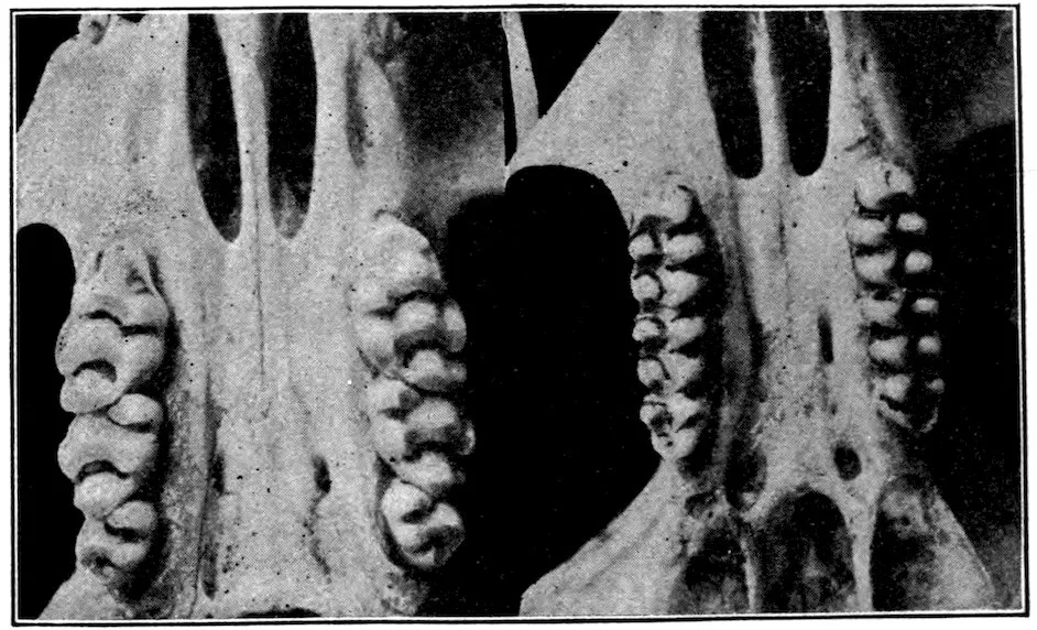
Fig. 1a. Fig. 1b.
Fig. 1a.—Upper molars of the brown rat (Mus): tubercles in three rows.
Fig. 1b.—Upper molars of the rice rat (Oryzomys): tubercles in two rows.
The Murinæ are the true rats and mice, typified by the genus Mus,
which contains by far the largest number of species. Trouessart,
in his Catalogus Mammalium, enumerates 260 species of Mus described
before 1905. Since that date a number of new forms have
been described.
The genus Mus is characterized by narrow, ungrooved incisors;
three small, rooted molars; soft fur mixed with hairs, sometimes with
spines; a rudimentary pollex having a short nail instead of a claw;
a long tail bearing rings of overlapping scales and often naked or
nearly so. The ears are rather large, the eyes bright and prominent,
17and the muzzle somewhat pointed. The members of the genus are
natives of the Old World, throughout which, with the exception of
Madagascar, they are quite generally distributed. Nearly seven-eighths
of the whole number of species are commonly called rats.
The distinction between rats and mice is arbitrary and based on
size. Exclusive of the tail, rats may be said to vary in length from
4½ to 10 inches or more, while mice measure from 2 to 4 inches.
With few exceptions, rats have six well-defined footpads (plantar
tubercles), the last on the hind foot being elongated in shape; the last
hind-foot pad of mice is usually circular (fig. 2).
Of the many species of Mus only three or four have developed
the ability to adapt themselves to such a variety of conditions as to
become cosmopolitan. Four have found lodgment in America: The
common house mouse (Mus musculus); the old English black rat
(Mus rattus); the Egyptian, or roof, rat (Mus alexandrinus); and
the brown rat (Mus norvegicus), known also as the gray rat, barn
rat, wharf rat, sewer rat, and Norway rat. The black rat and the
roof rat differ from each other chiefly in color. Indeed some zoologists
regard them as races of the same species, and the trinomial Mus
rattus alexandrinus for the roof rat is now in use among zoologists.
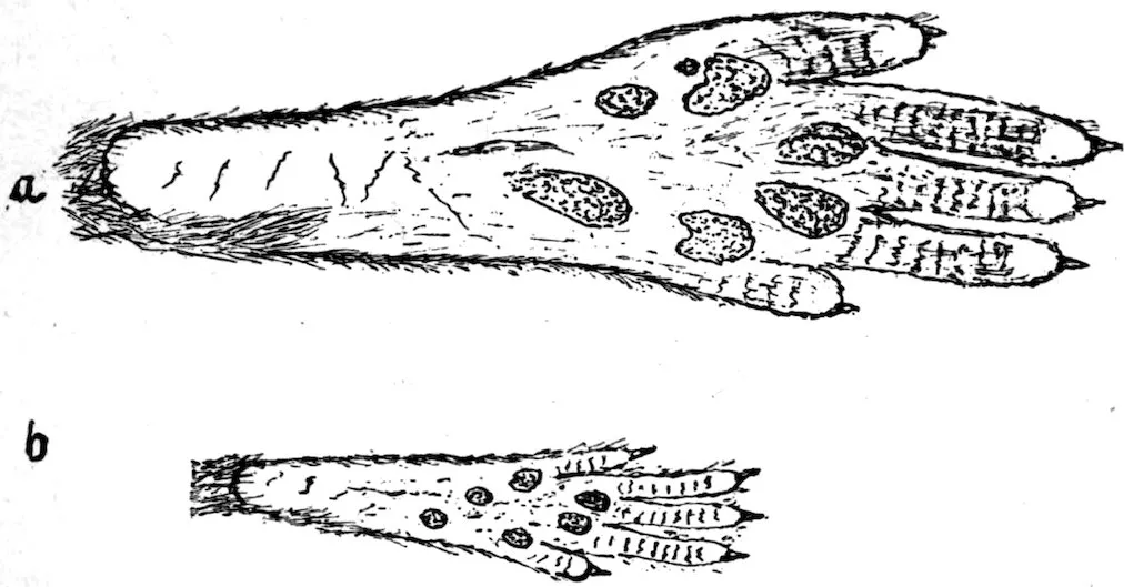
Fig. 2a.—Right hind foot of brown rat, showing long sixth foot pad.
Fig. 2b.—Right hind foot of house mouse, showing round sixth foot pad.
The common house mouse (M. musculus) found its way to America
soon after the first settlement by Europeans. It now inhabits all
settled parts of North and South America, as well as nearly the entire
Old World; but in very cold regions it does not always survive the
winters, and is therefore comparatively scarce or local. It almost
always reaches a new settlement sooner than the rat.
The black rat (M. rattus) has been known in Europe since the
twelfth century. It was carried to South and Middle America about
three and a half centuries ago (1554). The time of its arrival in the
18English colonies of North America is not known with certainty, but
it was well established in the settled parts by the beginning of the
eighteenth century. Soon after the arrival of the brown rat, the
black species began to decrease in numbers, and has become extinct
in most localities. At present it is not uncommon in some parts of
the South, and still occurs in scattered colonies in Canada and some
of the States east of the Mississippi, and also on some of the coastal
islands. It is occasional in many of our seaports, being apparently
brought from the Far East in merchandise. Except in a few ports
like San Francisco, where new arrivals are probably rather frequent,
these introduced individuals are often destroyed before they multiply.
The history of the black rat in Europe and its disappearance before
the brown rat is an exact parallel to its history here, and the animal
is now comparatively rare north of the Alps, except in the Channel
Islands.
The Biological Survey has specimens of the black rat from Massachusetts,
New Hampshire, Georgia, Florida, Alabama, California,
and Washington, and also from Mexico, Honduras, Nicaragua, and
Hawaii. There are authentic records of its recent occurrence in
Newfoundland, Quebec, Nova Scotia, New York, North Carolina,
Tennessee, West Virginia, and Mississippi. In parts of South and
Middle America it is abundant.
The roof or Alexandrian rat (M. alexandrinus) is similar to the black
rat in form and general habits, though not in color. Little is known
of its history, but it is thought to be a native of Egypt, where it is
still abundant. It has established itself in many parts of the world,
mainly in warm climates, and is common near the coast in the
southern parts of the United States.
The Biological Survey has specimens of the roof rat from North
Carolina, Georgia, Florida, Alabama, Mississippi, Texas, Arizona,
and California. In the last-named State it is abundant in the Sacramento
Valley. It is known also from Dismal Swamp, Virginia, and
from Cuba, the Bermudas, Trinidad, San Domingo, Costa Rica,
Nicaragua, Mexico, and Hawaii. Also, it inhabits many parts of
South America, where in places it is the dominant species.
The most destructive of the rat family is the brown rat (M. norvegicus).
In most parts of the United States it is the common rat
about houses and barns in the country and about markets, wharves,
and warehouses in cities. It is larger and more robust than either
the black or the roof rat, and differs from both in habits. It is more
of a burrower, and lives in excavations which it makes under buildings
and in loose soil along hedges and river banks. This habit,
combined with its greater strength and ferocity has enabled it to supplant
the other species in temperate latitudes; but in the warmer
parts of America and the Old World it has not been able to drive out
19the others. The house mouse everywhere holds its own against the
brown rat by its ability to escape into retreats too small for the rat
to follow.
The brown rat inhabits most of the thickly populated parts of
America. North of Panama it occurs generally except in the arid
interior, from the Isthmus to the Yukon Valley and southern Greenland.
In the Great Basin it is practically unknown, and in New Mexico
and Arizona it is confined chiefly to towns along the railroads.
The Biological Survey is without records of its presence in Nevada,
Utah, Wyoming, Idaho, and the greater part of Montana. The reason
for its absence in that region is not understood, but its ability to withstand
extreme cold is proved by the fact that it flourished in latitude
78° 37′ north on board Doctor Kane’s ship Advance, in the Second
Grinnell Expedition, during the two winters when that vessel was
icebound. It has also adapted itself to the continuous low temperatures
of cold-storage warehouses, in which it appears to breed freely.
We know little of the history of this species. Greek and Roman
writers make no mention of rats of any kind, but possibly knew the
animals and included them in their frequent references to mice.
Pallas, in 1778, described the brown rat under the name Mus decumanus,
and this was generally used until it was found that Erxleben
had called it M. norvegicus in 1777. Previously, the common name
Norway rat had often been used for this species.
The brown rat is generally supposed to be of Asiatic origin. Various
modern writers have asserted that it came originally from Persia
or India; but W. T. Blanford states that the species is at present
unknown in Persia, and that in India the black rat is the generally
distributed species, while the brown rat occurs only along the coast and
the navigable rivers.[B] This implies that the latter species is a comparatively
recent immigrant into India.
As regards the arrival of the brown rat in Europe, two facts are
known. The species reached England from some eastern port
about 1728 or 1729, and according to Pallas, a little earlier, 1727,
crossed the Russian frontier from Asia and soon spread over the
greater part of that country.[C] This statement, taken in connection
with that of Blanford, makes it highly probable that before this
migration the Asiatic home of the species was north, rather than
south, of the high mountains of northern India. This view, which has
been adopted by several naturalists, is further strengthened by the
fact that the animal flourishes better in temperate than in tropical
climates.
20Possibly earlier and unrecorded westward migrations of the brown
rat took place. A few years ago Professor Waile, the archeologist,
while making excavations at Cherchell on the coast of Algeria, dug
up the skull of a rat, which he stated was contemporary with the
Roman occupation of the country under the Cæsars. The skull
had but one molar, much worn, but the cranial bones were intact,
and French zoologists pronounced the remains as undoubtedly those
of the “surmulot,” or brown rat.[D] This shows that we have little
more than conjectures for the early history of this species.
The brown rat is said to have first appeared in Paris in 1750. It
was brought to the United States, probably from England, about
the beginning of the Revolution, 1775. According to Audubon, it
was unknown on the Pacific coast of the United States in 1851; but
Dr. J. S. Newberry thought it must have arrived at San Diego,
Monterey, and San Francisco at a much earlier date.[E] Doctor Cooper
recorded its arrival at Fort Steilacoom, Washington, as occurring
about 1855.
The brown rat differs considerably from the black rat and the roof
rat. It is larger, has a shorter head, a more obtuse muzzle, smaller
ears, and a relatively shorter and stouter tail. The general color is
grayish-brown above and whitish below. The over hairs of the upper
parts have black tips. The tail is usually shorter than the head and
body combined. The average measurements of adult specimens of
the brown rat in the Biological Survey collections are as follows:
Total length, 415 millimeters (16.3 inches); tail, 192 millimeters
(7.1 inches); hind foot, 43 millimeters (1.7 inches). This species
sometimes attains a total length of 19 to 20 inches, and has been
known to weigh 24 to 28 ounces and even more. The average
weight of an adult brown rat is considerably less than a pound.
The black rat is less robust than the brown rat. It has a longer
head, a sharper muzzle, and larger and broader ears (fig. 3). The
tail is longer than the head and body combined. The fur is of a
sooty, or plumbeous black, color, paler on the underparts. It is
much softer and denser than that of the brown rat, and the mixture
of very dark and lighter over hairs gives it a peculiar shining appearance.
The average measurements of 20 apparently adult specimens
in the collection of the Biological Survey are as follows: Total length,
379 millimeters (14.9 inches); tail, 207.4 millimeters (8.1 inches);
hind foot, 35.8 millimeters (1.4 inches).
21The roof rat in general resembles the black rat, except as to color
and texture of fur. Above it does not greatly differ in color from
the brown rat, but its underparts are usually more yellowish. The
fur is commonly shorter and harsher in texture than that of the
black rat, but this difference might not always be apparent in specimens
of the two forms from the same latitude. The average measurements
of 50 adult specimens of the roof rat in the collections
of the Biological Survey are as follows: Total length, 393.3 millimeters
(15.5 inches); tail, 212.8 millimeters (8.4 inches); hind foot,
36.2 millimeters (1.4 inches).
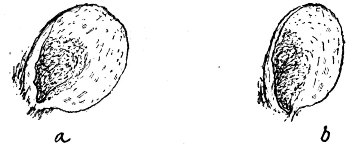
Fig. 3a, Fig. 3b.—Ears of brown rat and black rat, showing relative size.
Both albinism and melanism are frequent among rats, and pied
forms also are common. It has been claimed that all the white rats
(albinos) of the bird stores are Mus rattus, but albinism is by no
means confined to this species. Doctor Hatai found that all the
colonies of white rats maintained at the neurological laboratories of
Chicago University and the Wistar Institute of Anatomy, Philadelphia,
were of the M. norvegicus species.[F] The same is true of all
the albino rats in the collections of the National Museum and the
Biological Survey. These collections contain also several spotted
rats (gray and white) and sooty-black specimens indistinguishable
in color from M. rattus, all being undoubtedly of the M. norvegicus
species.
| Size small. Total length of adult less than 200 millimeters |
Mus musculus |
| Size large. Total length of adult exceeding 300 millimeters. |
|
| |
Ears moderate, when laid forward barely or not reaching eye; tail shorter than (rarely equal to) the length of head and body, darker above than below; color of body normally gray-brown above, white below; hind foot 38–46 millimeters |
Mus norvegicus |
| |
Ears larger, when laid forward reaching at least to middle of eye; tail longer than head and body, dusky all around; hind foot 33–37 millimeters. |
|
| |
|
Color grayish-brown above, white or yellowish white below. |
M. rattus alexandrinus |
| |
|
Color blue-black above, slaty below |
M. rattus |
22
Both climate and food supply affect the rate of multiplication of
most rodents. The rat probably increases more rapidly in a temperate
and equable climate than in one of great variability. Extremes
of heat and cold retard multiplication, decreasing both the
number of litters in a year and the number of young at a time. In
northern latitudes, apparently, more or less interruption of breeding
occurs in the winter months.
Where the country is well settled the food supply of rats is not
likely to be deficient; and when the animals have access to stores
of grain, the young mature very quickly and probably reproduce
earlier than when grain is absent.
The brown rat is more prolific than either the roof rat or the
black rat. The female brown rat has usually 12 mammæ—3 pairs
of pectoral and 3 pairs of inguinal—although these numbers are not
constant, one or more teats frequently being undeveloped. The
black rat and the roof rat have only 10 mammæ—2 pairs of pectoral
and 3 pairs of inguinal—with but little tendency to vary. Records
of actual observations on the number of young confirm the deductions
that might be drawn from the above facts. At Bombay,
India, during the recent investigations made by the India Plague
Commission, 12,000 rats were trapped and examined. The average
number of embryos found in pregnant brown rats was 8.1; the
highest number, 14. The average for the black rat was 5.2; the
largest number, 9.[G]
In temperate latitudes the average number of young produced by
the brown rat is undoubtedly greater. Instances of very large
litters observed in England are recorded in The Field (London). In
two instances 22 and 23 young, respectively, were found in a single
nest, though no evidence is offered that these were the progeny of a
single female; but in two other cases 17 and 19 embryos were found
in gravid females. A dealer in feedstuffs in Washington, D. C.,
relates that he found 19 young rats in a single nest in his store.
Within the past few months the writer has examined four pregnant
brown rats taken in traps. The numbers of embryos they contained
were 10, 11, 11, and 13, respectively. While we have not enough
data for definite conclusions, we may safely state that the average
litter for this latitude is not less than 10.
Frank T. Buckland, in Curiosities of Natural History, relates that a
white rat which he kept in captivity gave birth to 11 young when
only eight weeks old. As gestation in rats occupies three weeks,
this animal must have bred when only five weeks old.
23The number of times rats breed in a year is not definitely known,
and probably varies considerably with local conditions. Kolazy
makes the almost incredible statement that two female white rats,
kept by him in confinement and well fed, within thirteen months
gave birth to 26 litters of young, numbering 180 in all. One of them
produced young regularly at intervals of 25 days.[H]
The writer recently kept two young female brown rats with a male
in a large open cage for several months. One of the females gave
birth to young on April 15; the other on April 17. The number in
these litters was not observed, as some were devoured soon after
birth, and all within three days, presumably by the male rat. On
May 23 both females gave birth to young, 24 in number, all in one
nest.
The known facts concerning the breeding of the brown rat may be
briefly summarized as follows: The animals breed from three to five
times a year, each time bringing forth from 6 to 19 young. After a
gestation period of twenty-one days, the females give birth to their
young in nests built in underground burrows or under floors, stacks,
lumber, woodpiles, or other shelter. The young are blind and
naked when born, but grow rapidly, and young females are capable
of breeding when less than three months old.
Early spring and summer are the periods of greatest reproductive
activity among rats. Young, however, are to be found every month
of the year.
The above statements apply in the main also to the black and the
roof rat, but the number of young in a litter is somewhat smaller.
The newly born young of the black rat have not the bright pink
color of those of the brown and the roof rat, but are bluish, especially
on the upper parts. Black-and-white spotted rats are at first bluish-and-red
spotted, the red areas representing the white of the adults.
From the foregoing account of the breeding habits of rats, the
great difficulty of ridding cities or large areas of the animals may be
readily understood. Ordinarily, they breed more rapidly than they
are destroyed. Although few are seen in daytime, at night they
fairly swarm along river fronts and wharves, as well as in sewers,
stables, warehouses, markets, and other places where food is abundant.
Their real numbers may sometimes be discovered when any
such harbor is demolished.
An ordinary farm sometimes supports an astounding number of
rats. In 1901, an estate of 2,000 acres near Chichester, England, was
badly infested with the pests. They were systematically destroyed
24by traps, poisons, and ferrets, under the supervision of the proprietor.
In this way 31,981 were killed, while it was estimated that
tenants at the thrashing had destroyed fully 5,000 more. Even then
the property was by no means free from rats.[I]
During a plague of rats on the island of Jamaica in 1833, the number
of rats killed on a single plantation in a year was 38,000. The
injury to sugar cane on the island caused by the animals was at that
time estimated at half a million dollars a year.[J]
The report of the Indian Famine Commission presented to the
English Parliament in 1881 affords one of the best illustrations of
the number of rats that may infest a country. An extraordinary
number of the animals at that time inhabited the southern Deccan
and Mahratta districts of India. The autumn crop of 1878 and the
spring crop of 1879 were both below the average, and a large portion
of each was destroyed by rats. The resulting scarcity of food led to
the payment of rewards for the destruction of the pests, and over
12,000,000 were killed.[K]
Migrations of rats have often been recorded. The brown rat is
known in Europe quite generally as the migratory rat. The Germans
call it the Wanderratte. Pallas narrates that in the autumn of 1727
this species arrived from the east at Astrakhan, southeastern Russia,
in such great numbers and so suddenly that nothing could be done
to oppose them. They crossed the Volga in immense troops. The
cause of this general migration was attributed to an earthquake;
but since similar movements of the same species often occur without
earthquakes, it is probable that only the food supply of the animals
was involved in the migration which first brought the brown rat to
Europe.
A seasonal movement of rats from houses and barns to the open
fields takes place in spring when green and succulent plant food is
ready for them. The return movement takes place in the autumn.
This seasonal migration is noticeable even in large cities.
But more general movements of rats frequently occur. In 1903 a
multitude of migrating rats spread over several counties in western
Illinois. They were noticed especially in Rock Island and Mercer
counties. For several years previous no abnormal numbers of the
animals were seen, and their coming was remarkably sudden. An
eyewitness to the occurrence informed me that as he was returning
to his home one moonlit night he heard a general rustling in a
nearby field, and soon a great army of rats crossed the road in front
25of him, all moving in one direction. The host stretched away as far
as they could be seen in the dim light. These animals invaded the
farms and villages of the surrounding country and caused heavy
losses during the winter and summer of 1904. A local newspaper
stated that between March 20 and April 20, 1904, Mr. F. W. Montgomery,
of Preemption, Mercer County, killed 3,435 rats on his farm.
He caught most of them in traps.[L]
In 1877 a similar migration of rats into parts of Saline and
Lafayette counties, Mo., took place.[M] Also, one came under my
own observation in the Kansas River valley in 1904. This valley, for
the most part, was flooded by the great freshet of June, 1903, and
for about ten days was covered with several feet of water. Probably
most of the rats in the valley at the time perished in the flood. Yet
in the fall of 1903 much of the district was visited by hordes of rats,
which remained during the winter and had so increased by the following
spring that serious losses to grain and poultry resulted.
No doubt most of the so-called migrations of rodents, were all the
facts known, could be accounted for as instances of abnormal reproduction
or of failure of food supply in one place, compelling change of
habitat. In England a general movement of rats inland from the
coast occurs every October. This is known to be closely connected
with the closing of the herring season. During the fishing the
rodents swarm to the coast, attracted by the offal left in cleaning the
herring; and when this food fails, the animals troop back to the
farms and villages.
In South America plagues of rats are often periodical, occurring
in Parana, Brazil, at intervals of about thirty years and in Chile at
intervals of from fifteen to twenty-five years. It has been discovered
that these plagues in the cultivated lands follow the ripening
and decay of the dominant species of bamboo in each country.
The ripening of the seed furnishes for two or more years a favorite
food for rats in the forests, where the animals multiply greatly.
When this food fails, they are forced to the cultivated lands for
subsistence. In 1878 almost the whole crops of corn, rice, and mandioca
in the State of Parana were destroyed by rats, causing a serious
famine.[N]
An invasion of rats (Mus rattus) in the Bermuda Islands occurred
about the year 1615. Within two years they had increased so
alarmingly that none of the islands was free from them. The
rodents “devoured everything that came in their way—fruits, plants,
and even trees”—so that for a year or two the people were nearly
destitute of food. A law was passed requiring every man in the
26islands to keep 12 traps set. In spite of all efforts the animals
continued to increase, until finally they disappeared so suddenly
that they must have been victims of a pestilence.[O]
Instead of being strictly herbivorous, as might be inferred from
their dentition, rats are practically omnivorous.
The bill of fare of the rat includes grains and seeds of every kind,
flour, meal, and all food products made from them; fruits and garden
vegetables; mushrooms; bark of growing trees; bulbs, roots,
stems, leaves, and flowers of herbaceous plants; eggs, chicks, ducklings,
squabs, and young rabbits; milk, butter, and cheese; fresh
meat and carrion; mice, rats, fish, frogs, mollusks, and crustaceans.
This great variety of food explains the ease with which rats maintain
themselves in almost any environment.
Rats resemble squirrels in the manner of holding food while eating.
As soon as they have separated a small portion of food from a
larger mass, they sit up, arching the back and holding the morsel
in the paws and turning it as a squirrel does. After eating, they
brush the mouth and fore parts, including the whiskers (vibrissæ),
with the paws until all are clean. Rats drink much water, a habit
often taken advantage of in placing traps or poisons for them.
Rats generally feed after sunset, but in places where they are not
often disturbed they come out and feed in broad day and even in the
sunshine.
The roof rat and the black rat are more expert climbers than the
brown rat, which is larger and clumsier. In buildings, the brown
rat keeps mainly to the cellar and lower parts, where it commonly
lives in burrows. From these retreats it makes nightly excursions
to the upper parts of the house in search of food. The roof rat and
the black rat live in the walls or in the space between ceilings and
roofs. They nest in any of these places.
Rats readily climb trees to obtain fruit. In the Tropics the roof
rat and the black rat habitually nest in trees and spend much of
their time in these arboreal retreats, while the brown rat makes only
occasional excursions into the branches in search of food.
In the open, rats seem to have defective vision by daylight. They
move slowly and uncertainly. On the contrary, at the side of a room
and in contact with the wall they run with great celerity. This
fact suggests that the vibrissæ serve as feelers and that the sense
of touch in them is extremely delicate. The animals always prefer
narrow spaces as highways—another circumstance which may be
made use of in placing traps.
27
The ferocity of rats has been grossly exaggerated. The stories
of their attacks upon human beings, sleeping infants especially, have
but slight foundation. If attacked, nearly all rats defend themselves
with the teeth; and no doubt a horde of rats, if hungry, would
be formidable. Ordinarily the probability of being bitten by rats
is remote, and the bite is not poisonous.
The ferocity of rats is mainly exercised against members of their
own order. The brown rat is undoubtedly the most formidable
of the genus in America, and possibly in the world; yet when captured
it adapts itself readily to confinement, and in a few days
will take food and water whenever offered. The enmity of this
species toward other rats and mice is well known. It is supposed
to have destroyed the black rat over the greater part of Europe and
America, although it is possible that disease carried by the brown
rat was a factor in the disappearance of the other species. That
the black and the roof rat in tropical countries have not been displaced
by the brown rat is probably owing largely to their more
arboreal habits. It is not uncommon in the Far East to find two
species of rats living side by side in the same locality. An example
is M. imperator and M. rex living on one of the Solomon Islands.
The first is a burrowing species; the other arboreal. In 1877 two
native species of rats, M. macleari and M. nativitatis, were found
living together in amity on Christmas Island, in the Indian Ocean.[P]
About ten years ago the brown rat was accidentally introduced, and
it is now thought that both the native species are extinct.
When pressed by hunger rats become cannibals and destroy
their weaker fellows. However, when ordinary food is abundant,
cannibalism among rats is rare.
PLAGUE INFECTION IN RATS. — MODE OF EXAMINATION. — THE GROSS LESIONS OF NATURAL RAT PLAGUE—ACUTE PLAGUE. — SUBCUTANEOUS INJECTION. — THE BUBO. — THE GRANULAR LIVER. — THE SPLEEN. — PLEURAL EFFUSION. — CHRONIC PLAGUE. — RAT PLAGUE WITHOUT GROSS LESIONS. — MICROSCOPICAL EXAMINATION. — BACTERIOLOGICAL DIAGNOSIS OF RAT PLAGUE. — PEST-LIKE BACTERIA FOUND IN RATS. — ARTIFICIAL INFECTION OF RATS WITH PLAGUE. — MODES OF INFECTION. — LOCAL REACTION. — BUBO. — LIVER. — SPLEEN. — CHRONIC PLAGUE DUE TO ARTIFICIAL INOCULATION. — THE HISTOLOGY OF RAT PLAGUE. — NATURAL RAT PLAGUE. — IMMUNITY OF RATS. — REFERENCES. — RAT RECORD CARD. — PLAGUE RAT CARD.
29
By George W. McCoy,
Passed Assistant Surgeon United States Public Health and Marine-Hospital Service.
The rat is a known or a suspected factor in the transmission of
several diseases, yet at present, and perhaps for many years to
come, the most immediate and pressing question that concerns us
is its relation to the origin and spread of plague among human beings.
For this reason a discussion of the reaction of these animals to
natural and to artificial infection with B. pestis becomes of prime
importance.
Not only are rats believed to be more or less directly responsible
for cases of human plague in a community, but in addition, they
are believed to be the most frequent medium through which plague
is carried from one locality to another, for these animals are good
travelers, can live on a very meager ration, and can do without
water for a long time if food is available. We have found that on
a diet of dry grain alone a rat may live for over a month.
In this connection it may not be amiss to call attention to the
importance of the rat as an agent in conveying plague infection
to other rodents and especially to ground squirrels. There is every
reason for believing that the infection among the squirrels in California
was derived originally from rats. Wherry[16] states that
more rats than ground squirrels have been trapped in the squirrel
burrows in the vicinity of Berkeley, Cal. This shows how easy it
might be for rats to infect squirrels and vice versa.
The clinical manifestations of plague in rats are of little importance.
It is generally said that the plague-infected rat staggers
about with a drunken gait, loses fear of its natural enemies, and
is readily captured. Our experience with artificially infected rats
indicates that the animals show no marked manifestations of illness
until shortly before death when they become quiet, crouch in the
corner of the cage, and try to hide.
It is rather surprising to observe that comparatively few plague
rats are found dead. In the San Francisco campaign, while no
accurate figures are obtainable, certainly not more than 20 per cent
30of the infected rodents were found dead, the remainder being trapped.
This is probably due to the fact that the disease is one of several
days’ duration, from two to six most frequently, and during this
period there are more chances of catching the sick rodent in a trap
than there are of finding the body after death, unless the immediate
surroundings are known to harbor infected animals and an especially
careful search is made for cadavers in the places, often difficult of
access, where rats have their burrows and nests.
As plague is a disease that gives rise to such characteristic gross
pathological lesions in man and in laboratory animals, it is but
reasonable to expect that equally distinctive lesions would be found
in the rat, and this we find to be the case.
Skschivan[1], Kister and Schumacher[2], and other writers have
observed and recorded the gross lesions of plague in rats. It remained,
however, for the Indian Plague Commission[3], which had
the opportunity of examining an enormous number of plague rats
in Bombay and elsewhere in India, to crystallize our knowledge of
this subject and to point out its field of usefulness.
As to the comparative value of microscopical and macroscopical
methods of diagnosis, the Indian Plague Commission[3] states
that: “The results of tests carried out for the purpose of comparison
make it manifest that the naked eye is markedly superior to the
microscopical method as an aid in diagnosis, and as the result of
our experience we are prepared to make a diagnosis of plague on the
strength of the macroscopical appearances alone, even though the
other results of cutaneous inoculation and culture are negative and
the animal shows signs of putrefaction.”
Our experience with rat plague, though limited, leads us to the
same conclusion as that arrived at by the Indian Commission in
regard to the value of the gross lesions of plague in making the
diagnosis. To one who is acquainted with them, these lesions are
as characteristic as those of any infectious disease in man. It is
quite true that occasionally atypical cases are encountered where the
majority of the gross lesions are wanting, and in such cases it becomes
necessary to resort to the inoculation of animals or to cultural investigations
in order to make a diagnosis. Such cases are, however, if
anything, rarer than are atypical post-mortem findings in pneumonia
or in typhoid fever in man.
A brief description of the actual manner of examining rats for
plague infection will be given here.
The rats are immersed in any convenient solution for the purpose
of killing fleas and other ectoparasites that might be capable of carrying
infection from a plague-infected rat.
31The following plan of handling rats has been found satisfactory
in the federal laboratory at San Francisco. The rats are nailed to a
shingle by an attendant. Another attendant reads off the address
on the tag attached to the rat, puts a check number on the shingle,
and records the address from which the rat was taken and the check
number on the card shown on page 48. This card is arranged so as
to give the data as to the address from which the rat came, its size,
sex, and species. After being checked the rats are dissected and
finally, after examination by the medical officer, they are removed
from the shingle; any plague-infected rats are burned as soon as the
necessary investigation has been made. The dissection is made
by reflecting the skin from the whole front of the body and neck so
as to expose the cervical, axillary, and inguinal regions. The thoracic
and abdominal cavities are then opened with scissors.
In the inspection, careful search for buboes must be made in the
regions of the various peripheral lymph glands. The abdominal and
thoracic organs must be subjected to a careful scrutiny. It is needless
to say that this work should be done in a rat-proof, well-lighted
building that is provided with water, gas, and sewer connections.
The utmost care should be taken to avoid any undue risk of infection.
The wearing of rubber gloves is not necessary. Everyone who has
to handle infected animals must be sufficiently alive to the danger of
infection.
In the extensive work conducted by the Indian Plague Commission[3],
attendants were protected with Haffkine’s prophylactic. This is
undoubtedly a wise precaution and should be taken if possible.
For a worktable on which to dissect the rats we use in San Francisco
a table which slopes gently from the sides and ends toward the center,
where a drain pipe is attached which leads to a vessel containing a
disinfectant. The table is covered with sheet lead.
The layman of average intelligence readily learns to recognize the
gross lesions of rat plague and it is wise to train the laboratory attendants
to do this. Every rat should, however, be subjected to a
careful scrutiny by the physician responsible for the work. The great
majority of rats may be put aside after a cursory examination as entirely
beyond suspicion of infection. Probably 8 or 10 per cent of
them will require a very careful examination for the gross lesions of
plague. A card which we have found very useful for keeping records
of suspected and infected animals is shown on page 34. Probably all
of the species of the genus Mus are susceptible to plague infection. I
shall, however, confine myself to a consideration of plague in the rats
found the world over (M. norvegicus, M. rattus, M. alexandrinus).
In Bombay[18] it has been found that the epizootic among Mus
norvegicus appears first and is probably responsible for the diffusion
of plague among Mus rattus. It precedes the infection among Mus
32rattus by about ten days, and the opinion is expressed by the Indian
Plague Commission that the usual course of the infection is from the
Mus norvegicus to the Mus rattus, and as the latter rodent is a house
dweller in India it is the most frequent source of human infection.
In San Francisco the Mus rattus population is comparatively
small, contributing perhaps 2 per cent of the total rat population of the
city; but in the section of the city where the large warehouses are
found, especially those where oriental goods are stored, about 15 per
cent of the rats taken are Mus rattus. So far as concerns plague infection
about 5 per cent of the rat cases were in Mus rattus. It may
be of interest to note that the last infection found among rats in San
Francisco was among the Mus rattus in a large warehouse near the
water front. Two plague-infected rats were found in this building,
one October 21, 1908, and the other October 23, 1908. A large number
of mummified carcasses, all Mus rattus, were found in the building,
and it seems not unlikely that a somewhat extensive epizootic had
occurred among them. No previous case of rat plague had been
found in the city for eighty-five days, though about 25,000 rats had
been examined during that period, and none have been found in the
six months since, although over 30,000 rats have been examined.
Our records show that of 84 infected rats, 79 were Mus norvegicus,
and the remainder were Mus rattus. Some of the latter may have
been Mus alexandrinus, as the two species (Mus rattus and Mus alexandrinus)
were not clearly differentiated in the earlier examinations.
This is the sign which usually first attracts attention. White[4],
in discussing plague in rats, states that “the most noticeable post-mortem
appearance of the plague rat is the engorgement of the subcutaneous
blood vessels, together with a diffuse pink color of the subcutaneous
muscles, which have a peculiar dry, waxy translucency.”
It has been our experience frequently to have an attendant who is
dissecting rats remark that he had found an infected rat after the
first incision was made in reflecting the skin. The injection is dark
red, and upon close inspection one sees that the small vessels are
uniformly distended with blood. It is usually distributed over the
whole surface of the body, but on two occasions we have seen it confined
to the side of the body on which the primary bubo was found.
A bright pink injection is a rather common finding among rats in San
Francisco. It is not likely to be mistaken for the injection of plague
infection. Subcutaneous œdema, confined to the vicinity of the bubo,
is occasionally encountered.
In our experience in San Francisco an injection identical in appearance
with that found in plague infection was found only twice,
33and in each case there was associated with it a small discharging subcutaneous
abscess. There were no other lesions in either case and the
pus from these abscesses failed to produce plague in guinea pigs.
In a series of 61 consecutive plague rats in San Francisco, injection
was present fifty-two times, it was confined to the region of the
bubo twice, it was unilateral twice, and was general in distribution
forty-eight times. It was slight thirteen times, moderate fifteen
times, marked sixteen times, intense eight times.
This is the most reliable single sign of plague infection, and when
present in typical form is enough on which to base a diagnosis which
rarely proves erroneous.
The gland involved is usually surrounded by a more marked injection
than is present elsewhere, and an infiltration which at times is
hemorrhagic. This surrounding hemorrhage which was common in
the plague rats described by the Indian Plague Commission was met
with very rarely in San Francisco. The gland proper is usually caseous.
The contents may be shelled out very readily, though prior
to section the gland feels very firm. In the cases seen at the federal
laboratory in San Francisco, the contents of the buboes were recorded
as being hemorrhagic four times and as caseous twenty-nine times.
Pest-like bacilli were noted as present in 18 cases, in 6 of which the
“coccoid” form predominated. They were recorded as absent five
times.
Indolent enlargement of the lymph glands is very commonly encountered
in rats that are not infected with plague. Among old rats
probably 15 per cent will show this. Such glands, however, are tough,
elastic, and not surrounded by infiltration. They are not likely to be
mistaken for the plague buboes. In the leprosy-like disease of rats,
the glands may reach an enormous size.
Observers differ as to the location of the primary bubo. Skschivan[1]
states definitely the location of five primary buboes in plague rats
seen in Odessa in 1901. Two were in the axilla, two in the inguinal
region, and one in the neck. Kitasato[5] says: “To judge from the
experience of the past it can be suggested that in examining rats particular
attention should be paid to their submaxillary and cervical
glands and to the spleen. These organs in most cases show the evidence
of infection, if there be any.” From this it would appear that
he regarded the neck glands as the most frequent seat of the bubo.
It may be remarked here that his experience was derived from plague
rats seen in Asia.
We find a marked difference between the experience in San Francisco
and that in Bombay. This is demonstrated in the following
34table, which shows the location in percentage of single buboes in each
situation:
|
Neck. |
Axilla. |
Groin. |
Pelvis. |
|
Per cent. |
Per cent. |
Per cent. |
Per cent. |
| Indian Plague Commission, Bombay—2,923 rats[3] |
75 |
15 |
6 |
4 |
| Wherry, Walker, and Howell, San Francisco[6]—8 rats |
12 |
12 |
75 |
|
| Federal laboratory, San Francisco—32 rats |
|
22 |
72 |
6 |
The American figures are too small to be of much significance,
but one is struck with the fact that in Bombay three-fourths of the
buboes are in the neck, while in San Francisco three-fourths of all
found are in the inguinal region. We have records of only three
multiple buboes found in rats in San Francisco, and in no case was
either of the buboes in the neck; while in Bombay, to quote from
the report[3], “Of the rats with multiple buboes 54.5 per cent had
a bubo in the neck.” Striking as these figures are, we have collected
further evidence that the inguinal region is the commonest location
of the bubo in plague rats in this vicinity.
Passed Asst. Surg. J. D. Long, Public Health and Marine-Hospital
Service, who has had an extensive experience with rat plague in
Oakland, Cal., tells me that the majority of the buboes were found
in the groin, very few in the neck. Acting Assistant Surgeon
Wherry, Public Health and Marine-Hospital Service, informs me
that in a series of plague rats examined after the report made in
association with Walker and Howell[6], the cervical bubo was very
rarely encountered.
Particular care was taken to look for cervical buboes, as it seemed
rather inconsistent to find the other lesions so fully in accord with
those found in India, yet to have the location of the bubo to differ
so radically. We have not encountered a mesenteric bubo in our
work in San Francisco. The Indian Plague Commission found none
in over 5,000 naturally infected plague rats. As mesenteric buboes
are very commonly encountered in plague infection brought about
by feeding, they conclude that the absence of these buboes in naturally
infected rats is strong evidence that the infection does not
enter by the alimentary canal.
Two lesions of the liver are encountered in plague rats. The one
most frequently observed is spoken of by the Indian Plague Commission
as “fatty” change, though it is explained that this term
refers to the naked eye appearance as, microscopically, the lesion is
found to be due to a necrosis of the liver tissue. When this change
is present the organ is found to be rather yellowish in color and is
35studded with an enormous number of yellowish white granules which
are about the size of a pin head. This lesion, which was very common
in the San Francisco cases, is very readily recognized.
The other lesion is a marking of the organ with grayish white
spots; “they are typically of the size of a pin’s point, and give the
surface of the organ a stippled appearance as if dusted over with
gray pepper”[3] (p. 331). This appearance, which is less frequently
encountered than is the preceding one, is more difficult to recognize;
indeed the most careful scrutiny is necessary to avoid overlooking it.
Rats that have been fed with certain biological preparations used
to destroy rodents (Danysz’s virus and similar preparations) often
present lesions in the liver resembling those due to plague infection.
The granules are, however, larger and more distinct. In these cases
the spleen is enlarged and generally granular, but rarely dark and
friable as in plague infection.
The size of the spleen of healthy rats of the same weight varies so
greatly that often one can not be sure as to what constitutes an enlargement
of this organ.
In plague rats this organ is markedly enlarged, firm, friable,
rather dark in color, and occasionally presents small granules under
the capsule. As Skschivan[1] pointed out, these granules are not
encountered as often as are granules in the liver. At times the organ
presents a very distinctly mottled appearance. This latter appearance
is much more frequently seen in artificially inoculated rats than
in those found infected in nature. We have seen the organ distinctly
slate-colored on several occasions.
The last sign of rat plague is one of great importance when associated
with other suspicious lesions. The effusion is bilateral, and is
serous in character, usually clear, though it is occasionally blood stained.
Pleural effusion is rarely found in rats other than those
that are plague infected. The following table shows in percentage
the frequency of the various macroscopical lesions of acute natural
rat plague, as observed in Bombay and in San Francisco:
|
Subcutaneous injection. |
Bubo. |
Granular liver. |
Large dark spleen. |
Pleural effusion. |
|
Per cent. |
Per cent. |
Per cent. |
Per cent. |
Per cent. |
| Indian Plague Commission, Bombay—4,000 rats |
69 |
85 |
58 |
|
72 |
| Wherry, Walker, and Howell, San Francisco—88 rats |
59 |
14 |
14 |
68 |
71 |
| Federal laboratory, San Francisco—62 rats |
85 |
57 |
87 |
74 |
59 |
36It is recognized that the data from the San Francisco records is so
much smaller than that from the Indian report that perhaps no just
comparison is to be made. However, the figures are quite similar,
except for the small percentage of buboes and of liver lesions in the
work of Wherry, Walker, and Howell. The work of these observers
was done in the early part of the epizootic in San Francisco while
the other figures from that city are drawn from records later in the
campaign.
No single sign is pathognomonic, though only once have we been
deceived by what was regarded as a typical plague bubo. This was
in a rat that presented no other suspicious lesions and the inoculation
test resulted negatively.
It is a combination of two or more of the signs that is of moment.
The subcutaneous injection with a typical liver or these signs associated
with a typical spleen afford good grounds for a diagnosis.
A rat showing a typical liver associated with a pleural effusion will
usually prove to be plague infected, and if a large, dark, firm spleen
is also found a diagnosis may be considered as practically established.
As has been pointed out by several writers gross lesions of plague
may be distinguished even in rats that are badly decomposed.
No case of natural chronic plague has been encountered in San
Francisco. Only one case was found among the many hundreds of
plague rats examined by the Indian Plague Commission[3] (p. 457)
in Bombay. However, this commission encountered a considerable
number of cases among Mus rattus in the Punjab villages of Kasel
and Dhand. The lesions were purulent, or caseous foci. They
classify these cases as follows: Chronic plague of the visceral type,
which is further subdivided into splenic nodules and abscesses, and
mesenteric abscesses; chronic plague of the peripheral type in which
abscesses are situated in the regions of the peripheral lymph
glands.
Plague bacilli were either absent or very scanty upon microscopical
examination. They were, however, quite frequently recovered by
cultural methods, and in the great majority of the cases the organisms
were fully virulent. No evidence was forthcoming to show that
this chronic rat plague had anything to do with the recurrence of
acute plague among the rats.
We have diligently sought for chronic plague among the rats in
San Francisco, but, as we said above, without success, although a
considerable number of lesions that correspond perfectly to the
description of chronic plague have been submitted to the guinea-pig
inoculation test, but invariably with a negative result. An account
of the lesions of chronic plague as observed among inoculated rats
is given in another part of this paper.
37Pound[7] believes that recovery from plague in rats is shown by
the presence of pigmented lymphatic glands. Kister and Schumacher[2]
mention pigment deposits in the inguinal region, but remarked
that they are not characteristic of plague, a view which I believe is
correct, as we have frequently seen them in San Francisco among the
older rats, in which there was no reason to suspect previous plague
infection, and they have been almost uniformly absent in the case of
rats that have been experimentally infected with plague but have
recovered.
Plague infection may be present in a rat without bringing about
any recognizable gross lesions. For example: Dunbar and Kister[8]
mention a rat, which came from a ship on which plague rats had
been found, that had no lesions, and cultures were negative; but a
guinea pig cutaneously inoculated died of plague.
Among a considerable number of inoculated rats we have very
rarely, perhaps once or twice in a hundred cases, found nothing at the
post-mortem examination that would suggest plague infection, yet
cultures or inoculation of guinea pigs would demonstrate the presence
of B. pestis. Such cases are very infrequent, but it should be kept
in mind that they do occur. When a large number of rats are to be
examined it would be impracticable to inoculate a guinea pig from
each rat; and even if one did this the occasionally resistent guinea
pig would introduce a larger error than exists by placing dependence
upon the gross lesions for a diagnosis.
The exact weight to be given to the morphology of the organisms
found in smears from the organs of a rat suspected of being plague
infected is a matter of individual judgment. Smears from a bubo and
from the spleen may show no organisms at all, or none even remotely
resembling B. pestis, and yet by culture and inoculation methods we
may be able to demonstrate that the animal is plague infected.
Attention has been called to this point by several observers, and every
worker in this field has the experience sooner or later.
In other cases the smears will show such numbers of perfectly
typical bipolar bacilli and “involution” (coccoid) forms as to leave
scarcely any doubt as to the nature of the organism. But even here
cases that are not plague are encountered that will deceive even the
most experienced. We have been accustomed to put great dependence
on the “coccoid” forms of the organism, but late in the San
Francisco experience, smears from a splenic nodule that was not
regarded as due to plague showed perfectly typical “involution”
(“coccoid”) forms. Animal inoculations and cultures showed that
the tissues contained no plague bacilli.
38In addition to these two classes of cases we have a third, where
smears show a few typically shaped bacilli, or where a considerable
number of typical-looking bacilli are found along with many other
bacterial forms. There is no safe rule for reaching a conclusion in
these cases, and one must resort to culture or to inoculation methods,
or both. In any such case it is always a good plan to let the macroscopical
findings have more weight than the microscopical.
The bipolar appearance of B. pestis is so largely dependent upon
the technique of staining, fixing, length of time the stain is allowed to
act, and the length of the washing, that it should never be given
great weight. Here, as elsewhere in bacteriology, many errors are to
be avoided by not depending too much upon the morphology of the
organism under investigation.
While the gross lesions of rat plague are often sufficiently characteristic
to justify a positive diagnosis, and the gross lesions in conjunction
with the microscopical examination will in other cases enable
us to say definitely that a rat is plague infected, still a certain number
of cases occur in which it is necessary to resort to other methods, and
there are circumstances, such as the first case in a community, that
make a complete bacteriological confirmation of a diagnosis necessary.
This is not the proper place in which to discuss fully the bacteriology
of plague. However, a brief outline of what is necessary to
establish beyond question the existence of plague infection in an
animal will be given.
B. pestis may often be isolated in culture from the tissues (bubo,
liver, spleen, or heart’s blood) of an infected rat. Unless the tissues
are badly contaminated with other organisms, plate or stroke culture
will yield a growth of B. pestis in pure culture, or isolated pest-like
colonies may be transferred to other media.
It is unwise, however, to trust to cultural methods alone. In the
majority of doubtful cases it is advisable to inoculate guinea pigs or
white rats. The lesions of plague in these animals are quite characteristic,
and B. pestis may readily be recovered from their tissues if
cultures are made at once after death.
A pure culture of the organism under suspicion is obtained from
the naturally infected animal or from a laboratory animal inoculated
from the one under suspicion. This culture is studied in regard to its
morphology; first, on agar, where it grows as a short rod, or often in
the shape of a coccus; second, in broth, where it often grows in
streptococcus-like chains; third, on agar containing 3 per cent
sodium chloride, where most extraordinary alterations in morphology
occur, giving large balloon-shaped bodies, objects resembling gigantic
cocci and enormous trypanosome-shaped forms, the so-called “involution”
39forms. These involution forms must not be confused with
the so-called “involution” (coccoid) forms of the organism found in
smears from animal tissues.
We think it worth while to call special attention to the great diagnostic
value of involution forms developed when Bacillus pestis
is grown on salt agar. No other organism that we have had the
opportunity of working with gives forms that are at all likely to be
mistaken for those of Bacillus pestis, except B. mallei, and of course
the other points of difference would at once serve to distinguish the
latter organism.
B. pestis is Gram negative, though this point is of no great value
except to distinguish the “coccoid” forms from pus cocci.
The appearance and character of the culture should be as follows:
Agar.—Smooth, glistening, round whitish colonies which are
found to be sticky when touched with an inoculating needle.
Broth.—A scanty surface growth which falls, often in globular
masses, when the tube is gently agitated; and a fine flocculent precipitate.
Litmus milk.—Generally rendered slightly acid.
Glucose broth.—Rendered slightly acid. Gas is not formed.
Lactose broth.—Unchanged in reaction. Gas is not formed.
The other cultural reactions are of no material assistance in the
identification of the organism. Indeed, in routine work the appearance
of the growth on agar and in broth, together with the involution
forms on salt agar, are sufficient for identifying the organism.
The plague bacillus is a nonmotile organism, a point worth bearing
in mind.
A culture answering the above description when rubbed into the
shaven skin of a guinea pig or a white rat should cause the death of
either of these animals of plague within ten days, and an organism
must be isolated from their tissues after death corresponding to the
one inoculated.
If one wishes to be doubly certain, one may inoculate a series of
laboratory animals, giving to half of them a sufficient dose of antipest
serum. The protected animals should recover, or markedly
outlive the controls, which should die in the usual time.
As to the virulence of cultures of the bacillus from cases of rat
plague Klein[17] states “that B. pestis bred in the rat is of decidedly
less virulence than that bred in the human subject; moreover, the
former is liable, outside the animal body, to a much greater extent
to rapidly lose its virulence.” It is evident that in any given epidemic
it will be very difficult to say just which strain, rat or human,
one is dealing with.
In the case of the strains of B. pestis recovered from rats in San
Francisco we have seen nothing to justify such an opinion as Klein
40expresses. The cultures are all highly virulent and retain their
virulence under artificial cultivation.
The value of inoculation by the cutaneous method to demonstrate
the presence of plague infection in putrefying tissue is well known.
We have had one example in which the value of inoculation by this
method was proven in the case of a rat that was so badly decomposed
as not to admit of any opinion being formed as to whether the animal
was infected or not. A rat was brought from a warehouse where a
typical plague rat had been taken a few days previously. The
specimen was so badly decomposed that the abdominal organs could
not be distinguished with any degree of certainty. Smears from
tissue that was thought to represent spleen were negative so far as
pest-like organisms were concerned. A guinea pig vaccinated from
this splenic material died in seven days of typical plague, and a pure
culture of B. pestis was obtained from its organs.
Kolle and Martini [9] compare the cutaneous method of inoculation
to the use of an agar plate in separating plague bacilli from
other organisms, and so regularly does B. pestis penetrate the skin
and infect the animal, and so rarely do other organisms do this, that
it offers a certain and accurate method of “filtering out” B. pestis
from any badly decomposed tissue.
The technique of the cutaneous method of inoculation, or “vaccination”
as it is sometimes called, is very simple. An area about
an inch square is shaven on an animal’s belly, taking care to abrade
the epithelium slightly. The culture or suspected tissue is rubbed
on this shaven area with a platinum loop or a dressing forceps.
Guinea pigs when inoculated in this manner generally die before the
seventh day; white rats die a day or two earlier.
Kister [10] uses a drop of juice from an organ rich in bacilli for
agglutination experiments with antipest serum. This would appear
in many cases to be of very material assistance, and the objection
that it is difficult to form a uniform emulsion of the bacteria would
be avoided. The well-known tendency of B. pestis to grow in clumps
in culture is the main reason why agglutination reactions have not
been more extensively used in plague work.
Skschivan [1] makes use of Pfeiffer’s phenomenon in establishing
the identity of a given organism as B. pestis.
To assist in the early diagnosis of plague, Dunbar and Kister [8]
practiced intraperitoneal inoculation of laboratory animals and
used a parallel series of immunized animals. As is well known, intraperitoneal
inoculation with plague cultures or infected material leads
to the early death of the inoculated animal, and it is evident that
the survival of the immunized animal would afford considerable
evidence that the material used for inoculation contains B. pestis.
41
The somewhat general impression that there are a considerable
number of organisms that are readily mistaken for Bacillus pestis is
not justified, provided one gives attention to cultural and inoculation
investigations. It is quite true that there are a considerable number
of organisms which in smears from tissues are scarcely to be distinguished
morphologically from B. pestis. The similarity, however,
usually ends there. A few resemble plague somewhat closely in
cultural reactions, and especially B. pseudo-tuberculosis rodentium
(Pfeiffer) should be mentioned here; but these differ in pathogenicity.
For example, the above-named organism is not pathogenic for rats.
Neumans[11] reviews the subject of pest-like organisms pathogenic
for rats, and describes an organism belonging to this group
which he isolated from the body of a rat. His work clearly shows
that none of the organisms that have been described should cause
any serious difficulty in the hands of a careful investigator.
Kister and Schmidt[12] describe an organism closely resembling
B. pestis in many respects, and with which guinea pigs could be
successfully infected by the cutaneous method. This organism,
which was also pathogenic for rats and mice, belongs to the hemorrhagic
septicæmic group. It differed from B. pestis in that it gave
no involution forms when grown upon salt agar and was much more
rapidly fatal to laboratory animals.
Augeszky[13] observed an epidemic among gray rats in his laboratory
which was due to a pest-like organism belonging to the Friedlander
group. The animals died after a couple of days of illness. At
the post-mortem examination the spleen was found large, soft, and
congested. There was a hyperæmia of the intestines, lungs, and liver.
In the spleen were found many, and in the heart’s blood few, capsulated
bacilli, some of which resembled B. pestis. The cultural reactions
were in nowise similar to those of B. pestis. He found that inoculation
of rats with a pure culture of this organism sometimes killed
in as short a time as twenty-four hours, sometimes as late as two or
three weeks, and in some cases the lesions were not very unlike those
sometimes produced by B. pestis. However, this organism by its different
cultural reactions, and the fact that the capsule is usually easily
demonstrated, would probably never be a source of any confusion.
For laboratory purposes in general it is customary to use tame white
rats, and in plague work they are especially satisfactory, as they are
easily handled, rarely harbor fleas, are very susceptible to the infection,
and finally and most important, they frequently die a day or
two earlier than guinea pigs. At times it may be necessary to use
42wild rats on account of a failure in the supply of white rats, or for the
sake of economy. This may be done very satisfactorily, if one bears
in mind the fact that a considerable number of wild rats are more or
less immune to plague infection, especially when the infectious material
is introduced by Kölle’s (cutaneous) method. Therefore, it is
always advisable to use three or four wild rats where one white rat
would be sufficient. They should be kept in a container of such design
that there is no possibility of their escaping. The inoculation is
best conducted with the animal under the influence of ether.
Rats may be infected experimentally by the ingestion of contaminated
material, and by the application of virulent plague bacilli to a
mucous or a cutaneous surface, or by subcutaneous injection of the organism.
Practically we may confine our study to inoculation by the cutaneous
method, and to subcutaneous inoculation, when the material is
injected in the ordinary manner. A useful modification of the latter
method is to make a small pocket under the skin of the abdomen and
thrust the suspected material into this pocket. This avoids the necessity
of making an emulsion of infectious matter, such as the organs
of an animal. The time that elapses between the inoculation of a rat
with virulent culture of plague bacilli and its death varies somewhat
with the size of the dose and with the mode of inoculation. The following
table, compiled from work in San Francisco, shows the day of
death of a few white rats and a considerable number of wild rats
using the strain of B. pestis that was found in the recent epidemic
here. Some were inoculated by the cutaneous and some by the subcutaneous
method:
| Day of death. |
White rats. |
Wild rats. |
| Second |
|
3 |
| Third |
5 |
27 |
| Fourth |
7 |
41 |
| Fifth |
1 |
30 |
| Sixth |
1 |
9 |
| Seventh |
|
8 |
| Total |
14 |
118 |
The wild rats were all Mus norvegicus.
The lesions found, when an artificially inoculated rat is examined
after death, are in a general way similar to those found in naturally
infected rats with certain differences to be mentioned later.
In order to obtain accurate figures as to the frequency of the various
lesions in inoculated rats, I have compiled the data from the records
43of the federal laboratory in San Francisco of a considerable number of
wild rats that have been inoculated in the course of various investigations
and have died of acute plague. The rats were practically
all of the species Mus norvegicus.
Artificially inoculated (subcutaneously) plague rats.
|
Local reaction. |
Subcutaneous injection. |
Bubo. |
Granular liver. |
Enlarged dark spleen. |
Pleural effusion. |
| Present |
36 |
48 |
19 |
47 |
56 |
18 |
| Very extensive |
1 |
[Q]2 |
|
|
|
8 |
| Slight |
2 |
6 |
|
|
|
8 |
| Total present |
39 |
56 |
19 |
47 |
56 |
34 |
| Absent |
10 |
4 |
39 |
15 |
3 |
21 |
| Not recorded |
13 |
2 |
4 |
|
3 |
7 |
| Total |
62 |
62 |
62 |
62 |
62 |
62 |
All of the lesions aside from the local reaction were present and
well marked in six cases.
Artificially inoculated (cutaneously) plague rats.
|
Local reaction. |
Subcutaneous injection. |
Bubo. |
Granular liver. |
Enlarged dark spleen. |
Pleural effusion. |
| Present |
16 |
34 |
42 |
58 |
58 |
22 |
| Very extensive |
1 |
[R]15 |
|
|
|
6 |
| Slight |
6 |
13 |
|
|
|
|
| Total present |
23 |
62 |
42 |
58 |
58 |
28 |
| Absent |
37 |
6 |
26 |
11 |
8 |
37 |
| Not recorded |
9 |
1 |
1 |
|
3 |
4 |
| Total |
69 |
69 |
69 |
69 |
69 |
69 |
All of the lesions aside from the local reaction were present and
well marked in five cases.
The most striking difference between natural and artificial plague
in rats is the presence of a reaction at the site of inoculation in the majority
of cases where the organism is introduced subcutaneously, and
in about a third of the cases where the infectious material is rubbed
on the shaven skin (cutaneous inoculation). The local reaction may
exist only as a yellowish-brown crust, overlying a granulating surface,
and associated with a trifling thickening of the skin and subcutaneous
44tissue. It may appear as one or more firm papules 3 or 4
millimeters in diameter. The most frequent appearance is a brawny
œdematous and blood-stained reaction which extends over an area
perhaps an inch in diameter; at times purulent change may be well
advanced. Very rarely one finds so extensive an œdema as to cause
the lesion to somewhat resemble the widespread gelatinous reaction
seen so commonly in the guinea pig. On one or two occasions we
have seen an extensive slough at the site of inoculation.
It is very exceptional that one finds in cases of induced plague the
typical, firm, caseous bubo surrounded by an infiltrated area, as is
so commonly seen in natural infection in rats. The glands are sometimes
enlarged and injected without other changes. The commonest
lesion, however, is a markedly enlarged gland which upon close inspection
is seen to have a number of yellowish points just under the capsule.
These points are especially well seen when a section is made
through the gland. The gland may be squeezed out of the capsule
and it breaks down readily enough when pressure is made upon it;
but the uniform necrotic process that one sees so often in natural rat
plague is absent.
Granular lesions precisely like those found in natural infections
are very common. If the rat has died on the sixth day or later, the
ordinary lesions are apt to be replaced by necrotic foci that may be
as much as 2 millimeters in diameter.
This organ is found mottled more frequently than in natural plague
infection, and large granules are much more common.
The subcutaneous injection is rarely so well marked as it is in
natural infections.
Pleural effusion of the same nature as that found in natural plague
is common. Hemorrhagic foci are not rare in the lungs, and occasionally
the organs are partly consolidated.
Occasionally a rat that has been inoculated but has survived a
week or longer, will show, when killed, only an abscess at the site of
the injection. Stained smear preparations may show a large variety
of bacterial forms. We have not been able to demonstrate the
presence of B. pestis in these lesions, yet there is no doubt but that
the lesion is the result of the inoculation.
A lesion more frequently found is a caseous or a purulent lymphatic
gland. If the inoculated rat has been killed about ten days after
the inoculation, in some cases one or more of the peripheral lymph
45glands will be found to be surrounded by an infiltration, and the
gland itself will be purulent or less frequently caseous. Such lesions
are occasionally met with in rats in which there is no suspicion of
plague infection; but they are seen so frequently among rats that
have survived artificial inoculation with B. pestis, there is no doubt
but that in these cases they are the result of the inoculation. In
several such cases pest-like organisms have been demonstrated
in smears, and acute plague has been produced in guinea pigs
by inoculation with the pus found in these lesions. Not infrequently
in these cases the spleen will be found enlarged and looking very
much like the organ in acute plague, but cultures from this organ
in such cases have in my experience remained sterile.
In other cases the only lesions will be found in the spleen. The
organ is enlarged and contains a number of caseous nodules. These
nodules vary in number from four or five to thirty or forty and in
size from the head of a pin to a lesion 0.3 centimeter in diameter. In
a number of such cases the nature of the lesion has been demonstrated
by animal inoculation. For example, in a series of experiments
carried out to determine the susceptibility of San Francisco rats to
plague infection a large Mus rattus died on the eleventh day after
inoculation. The post-mortem examination showed nothing except
an enlarged spleen which contained about a dozen caseous nodules,
the largest of which was not over 2 millimeters in diameter. The
nodules were very firm and the capsule smooth, so that they were held
with difficulty with dressing forceps. Cultures from the liver and
the spleen remained sterile, but a piece of the spleen was placed
beneath the skin of a guinea pig. This animal died of acute plague,
and a pure culture of B. pestis was isolated from its liver. In some
of these cases the liver will show large, distinct, whitish caseous foci.
In another case a small Mus norvegicus was killed on the twelfth day
after a cutaneous inoculation from an artificially infected squirrel.
No lesion was found except in the spleen which was not materially
enlarged, but which presented two small whitish caseous granules
on the surface, neither being over 1 millimeter in diameter. A piece
of the spleen containing one of these granules was put under the skin
of the belly of a guinea pig. The guinea pig died on the fourth day
with the usual lesions of acute plague. Occasionally in these cases
of chronic plague punctate hemorrhages or even areas of consolidation
are found in the lungs.
The most recent and satisfactory work on this subject is that of
Ledingham [14], who has studied the lesions of both natural and
induced plague in rats. The following is a very brief abstract of his
work. The reader is referred to the original for a full study of the
subject.
46
Two groups of cases are distinguished, first, those in which a large
number of B. pestis are found in the liver and in the spleen. In the
spleen this is accompanied by hemorrhages and congestion of the
pulp sinuses and in the liver with congestion of the capillaries.
These are early cases.
In the second group, or the later cases, there are extensive reaction
changes in the tissues. In the spleen this leads at times to distinct
abscess formation, but more frequently to a walling off of the foci
of necrosis. In the liver more or less focal necrosis is found; sometimes
the areas of “necrosis” may be so extensive that little healthy
liver tissue remains. Bacilli are usually to be demonstrated in
these areas of necrosis. Giant cells of the Langhans type may be
found in the neighborhood of these foci.
The granular appearance of the liver is attributed to “hemorrhages
and the focal necroses, together with the fatty changes in the liver
cells. It must be understood, however, that a peculiar honeycomb-like
vacuolar degeneration of the liver cell protoplasm was far more
frequent than any actual, coarse, fatty infiltration. The granular
appearance of the spleen is due partly to endothelial catarrh and
partly to subcapsular changes.”
In experimental rat plague Ledingham found the lesions to resemble
those of the first group of cases referred to above. There is usually
marked bacteraemia; focal necroses of the liver are scanty.
In a chronic case, minute abscesses were found scattered through
the spleen. In the center of the abscesses were found clumps of
degenerated bacilli. The areas were walled off by epithelioid and
spindle cells and numerous giant cells of the tubercular type.
Contrary to the general impression the wild rat is not an animal
especially susceptible to plague infection. The Indian Plague Commission
[19] found that when rats are inoculated by the cutaneous
method from the spleen of infected rats 59 per cent are immune to
infection. A series of experiments conducted in the federal laboratory
in San Francisco showed that when inoculated with highly virulent
cultures of B. pestis there is an immunity which is, however, more
frequent among the large rats. When inoculated cutaneously with
tissue containing large numbers of B. pestis from plague infected
human beings, rats, or squirrels, about 15 per cent of small rats and
about 50 per cent of large ones were found to be immune. There is
no good reason for believing that this immunity of San Francisco
rats was due to a previous attack of the disease. Indeed, it was known
beyond a doubt that some of the immune rats had never had an
opportunity of becoming infected with plague in nature and thereby
47establishing an acquired immunity. We may mention here the
fact that has been observed by many workers, and which we have
amply confirmed, that rats are readily immunized by antiplague
serum.
The subject of the transfer of infection directly from rat to rat by
cutaneous or subcutaneous inoculation through a series of the animals
is one that is evidently intimately associated with the preceding
subject, as it is quite evident that an immune rat or several of them
might terminate a series without any actual diminution in the virulence
of the organism transferred. It is quite plain that the success
of such an experiment would depend largely upon the number of
rats used in each transfer. The Indian Plague Commission [19]
had no difficulty in carrying infection through twenty-six transfers,
using from six to fifty rats in each transfer.
Pound [7] in a series of eight experiments, was never able to convey
the infection successfully beyond the sixth rat, using but one rat for
each transfer. There was no apparent lessening of the virulence
of the organism and each series appears to have been terminated
abruptly by encountering an immune rat.
Baxter-Tyrie [15] says:
It is probable that under certain natural circumstances a reduction in the virulence
of the organism is effected and a comparative immunity is conferred on the rats.
The infection of immigrant rats is, however, severe, and their arrival is heralded
by a heavy mortality. In the same manner an infected rat imported into a fresh
locality produces a similar result. This attenuation of virulence is responsible for the
condition known as chronic rat plague.
Several experiments conducted in San Francisco to determine
this point have given results that I regard as showing merely the
presence of a considerable percentage of immunity among the rats.
It was observed that in each case certain of the rats died of acute
plague even in the last transfer. It was very evident that had certain
combinations of immune rats been encountered the experiment
might have terminated at any point. On the other hand, by being
especially fortunate in using nonimmune rats, the experiments might
have given a much higher percentage of cases of acute plague. Unfortunately
it was necessary to terminate these experiments in each
instance before they could be regarded as completed.
The reason for the natural subsidence of plague among rats in any
community is a point about which much more evidence must be
obtained before we can speak with any degree of certainty. It may
be due to the lack of susceptible material, possibly to a loss of virulence
of the organism; but it seems more probable that it is due to
a change in the number or relations of the ectoparasites of the rat.
Adequate measures of rat extermination, while they may never
bring about the ideal condition of a community that is free from
rats, are, as is shown by the recent experience in San Francisco, of the
utmost value in shortening the epizootic.
48
[Legend: O = Ordinary; W = White belly; R = Red; Go. = Gopher rat; S = Small; M = Medium; L = Large; M. R. = Mus rattus; M. N. = Mus norvegicus.]
| No. |
Date. |
District No. 6. |
Sex. |
Size. |
M. N. |
M. R. |
Pregnant. |
| M. |
F. |
S. |
M. |
L. |
O. |
W. |
R. |
Go. |
O. |
W. |
| 19 |
Dec. 10, 1908 |
401 Fillmore street |
1 |
|
|
1 |
|
|
|
1 |
|
|
|
|
| 20 |
do |
do |
|
1 |
|
|
1 |
1 |
|
|
|
|
|
[S]7 |
Date: June 20, 1908.
Species: M. norvegicus.
From District No. 6, sewer, Haight and Steiner Streets.
Condition: Badly injured by trap; thorax crushed.
Subcutaneous injection: General, marked.
Lymphatic glands, bubo or other lesions: Right inguinal bubo, caseous.
Liver: Typical whitish granules.
Spleen: Large, dark, firm.
Pleural effusion: Unable to say.
Purulent or caseous foci:
Diagnosis from gross lesions: Plague.
Diagnosis from smears: Plague (spleen and bubo).
Cultures: B. pestis recovered from liver culture.
Inoculation, guinea pig No. 50 A, +6.25.08.
Vaccination, guinea pig No. 50 B, +6.26.08.
Date suspicious:
Date positive: June 20, 1908.
Date negative:
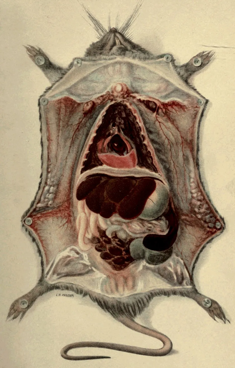
A HOEN & CO BALTIMORE.
NECROPSY APPEARANCE OF PLAGUE-INFECTED RAT
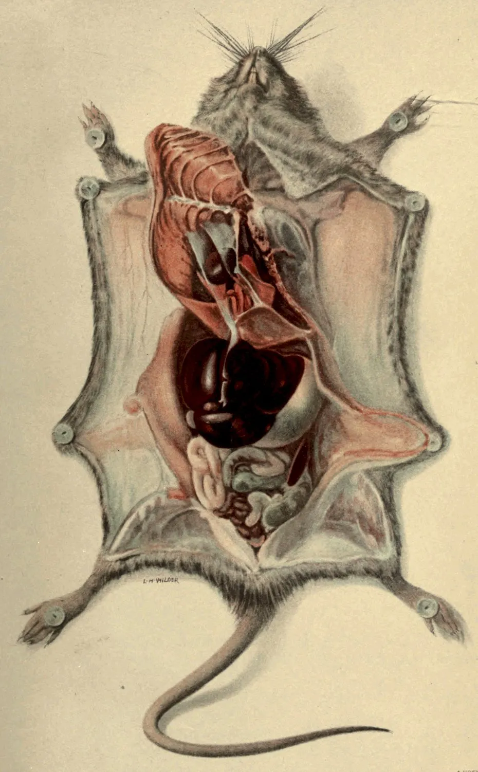
A HOEN & CO BALTIMORE
NECROPSY APPEARANCE OF NORMAL RAT
RAT LEPROSY. — INTRODUCTION. — REVIEW OF LITERATURE. — DESCRIPTION OF DISEASE. — ETIOLOGY. — SUMMARY.
49
By Walter R. Brinckerhoff, S. B., M. D.,
Assistant Director Leprosy Investigation Station, United States Public Health and
Marine-Hospital Service, Honolulu, Hawaii.
The leprosy-like disease of the rat is of great interest to leprologists
because of its close similarity to the disease leprosy in man.
Its practical importance to those engaged in the study of the human
disease is increased by the fact that it can be artificially propagated
under laboratory conditions from animal to animal and, still more
important, can be transferred from the species in which it occurs
naturally (Mus norvegicus) to a more tractable laboratory animal
(Mus albus). The brief description of the affection which follows
is intended to assist in its recognition and to stimulate the interest
of investigators in the disease, which presents problems replete with
interest to the study of pathology or bacteriology and of great
promise to those engaged in the investigation of human leprosy. It is
earnestly hoped that the investigation of this disease will be undertaken
in general medical research laboratories, as it is extremely
probable that certain of the most difficult problems presented by
leprosy in man can be studied in this disease of the rat, and if solved
there the information gained can be directly applied to the solution
of the analogous problems in the human disease.
The first publication on rat leprosy was made by Stefansky (1903),
who observed the disease in Odessa during an antiplague campaign
against rats.
Rabinowitch (1903) found the disease among rats in Berlin and
confirmed the work of Stefansky.
Dean (1903) discovered the disease independently in London, and
in a later publication (1905) reports success in transferring the disease
by artificial inoculation.
Tidswell (1906) reports a case of the disease in a rat caught in
Sydney, New South Wales, Australia.
50The English Plague Commission observed the disease in India in
1907 (Wherry).
Wherry (1908) and McCoy (1908) report upon the finding of the
disease in rats caught in San Francisco, Cal.
Mezincescu (1908) has studied the disease and attempted to determine
its relationship to known human lepra by complement fixation
tests.
Geographical occurrence.—It would be premature at present to
make didactic statements as to the geographical distribution of the
disease, for its discovery has usually depended upon antiplague
measures, which are not world-wide in their scope. In spite of
this it seems profitable to briefly review the known occurrence of
the disease in relation to that of human leprosy. When such a
comparison is made we note that the disease is present among the
rats of Berlin, a city which is practically free from human lepra.
On the other hand in Honolulu, which is an endemic focus of human
leprosy, in the examination of 16,000 rats, during an antiplague campaign,
no case of rat leprosy was encountered. In addition to the
scrutiny of the rats examined for plague in Honolulu, an attempt
was made to obtain leper rats by offering a reward for a rat, dead
or alive, infected with the disease. This offer was given wide publicity
in the Territory, but brought no results.
Occurrence of the disease.—The proportion of rats infected
with the disease in different localities varies greatly, as will be seen
in the following table:
Table 1.—Proportion of leper rats to the total rats examined.
| Place. |
Observer. |
Proportion. |
|
|
Per cent. |
| Odessa |
Stefansky [301] |
4–5.000 |
| Sydney |
Tidswell [305] |
.001 |
| San Francisco |
Wherry [308] |
.210 |
| Do |
McCoy [310] |
.160 |
| Honolulu |
Currie[T] |
.000 |
Rats in the late stage of the disease are easily recognized by the
presence of a patchy alopecia associated with cutaneous and subcutaneous
nodules, which may or may not be the site of open ulcers.
The diagnosis can be readily confirmed by a microscopic examination
of a smear from an ulcer or a nodule, which will show the specific
bacillus of the disease in enormous numbers.
51Stefansky[301] describes two clinical types of the disease, the one
localized particularly in the lymph nodes, the other in the skin and
muscles. The glandular type was the more common. Dean[304]
thinks that no line of demarcation can be drawn between these clinical
types.
Dean[304] and Wherry[307] both mention that attention was attracted
to the diseased animals by the fact that they were seen abroad during
daylight in an obviously sick condition.
The skin, in a well-developed case of the disease, presents a patchy
alopecia coincident with thickening and nodule formation, which
is situated in the subcutaneous tissue. The cut surface of the
nodules or thickenings is light yellow in color, is clean, dry, and
cheese-like. In the region of the nodules the skin is atrophic, and
ulcers often form on the prominent parts of the affected area. The
subcutaneous fat tissue is diminished in amount. Histologically
the process is seen to be practically confined to the subcutaneous
tissue and to consist essentially in the presence of cells rich in protoplasm,
with vesicular nuclei, whose cell body is more or less completely
filled with slender acid-fast bacilli. The subcutaneous fat
is replaced by such a tissue. All investigators who have studied
the disease agree in emphasizing the similarity of the histology of
the lesion to that in leprosy in man.
When the musculature is involved the muscle fibers atrophy and
the fibers are infiltrated with the specific bacilli. The affected
muscle is friable, and macroscopically grayish white in color.
The peripheral lymph nodes are commonly involved, though
McCoy[310] reports a case in which only the pelvic and mesenteric
nodes were diseased, and in the Tidswell case[305] the peripheral nodes
were not enlarged. The typically affected nodes are enlarged, sometimes
measuring as much as 3 centimeters in the greatest extent,
firm, and, on section, opaque pale yellow-white in color. In the experimental
disease the writer has frequently found the characteristic
bacilli of the disease in peripheral lymph nodes which were very
slightly enlarged and presented no macroscopic lesion. Dean[304]
has observed invasion of the submaxillary or salivary glands by
extension from infected cervical lymph nodes. Wherry[308] notes that
in his cases he did not find the submaxillary or cervical glands involved,
which fact he contrasts with two early cases in which the skin and
adjacent axillary or inguinal nodes were involved.
Microscopically the lymph nodes show large numbers of cells in
the sinuses similar to those in the skin lesions. Multinuclear giant
cells are frequently observed which may measure as much as 70 to
80 microns[304]. The protoplasm of the cells is loaded with the
specific bacilli of the disease. The lymph follicles, trabeculæ, and
capsule of the glands are also invaded by the bacilli.
52The internal organs are relatively slightly affected in the natural
disease. Small foci have been found in the liver by Dean[304] and
in the liver and spleen by McCoy[310]. Wherry[307] reports finding
the bacilli in smears from both the liver and spleen. The writer has
found microscopic lesions containing the characteristic bacilli in the
liver in a case of the experimental disease.
Lesions have been observed in the bone marrow by Dean[304], and
the same author states that the nerves are invaded by the bacilli of
the disease. McCoy[310] found the bacilli in the urinary bladder in
one case.
With a disease showing such a striking similarity to human leprosy,
attention has naturally been directed to the bacteriological examination
of the nasal mucus. Dean[304] and Wherry[307] have both
found the characteristic bacilli in the nasal mucus, while McCoy[310]
has failed to do so. The writer’s experience has been confined to
the experimental disease, and in his animals the nasal examinations
have been negative.
The accepted etiological factor in the disease is an acid-fast bacillus
3 to 5 microns in length and 0.5 micron wide. The bacilli resemble
very closely the lepra bacillus of man, but seem to have somewhat
greater power to hold carbol-fuchsin stain against mineral acids.
The bacilli often have rounded ends and may be curved. The
beaded appearance so often seen in lepra bacilli is common. The
bacilli show the same tendency to form bundles that is such a marked
characteristic of Bacillus lepræ. To one familiar with the microscopic
appearance of smears from the discharges and lesions of human
leprosy the picture presented by similar preparations from the disease
of the rat is most striking.
The organism does not grow on the usual culture media—Stefansky
[301], Rabinowitch[302], Dean[304], Tidswell[305]—or on certain special
media—Dean[304].
The organism is not pathogenic for the guinea pigs—Dean[304],
Tidswell[305]—rabbit, mouse, monkey—Dean[304]. The disease can
be transmitted to black and white rats—Dean[304], Wherry.
In the leprosy-like disease of rats we have an affection which
closely resembles, both in its etiological factor and in its pathology,
the disease leprosy in man. The fact that the disease is readily
propagated in a laboratory animal permits of its investigation in any
laboratory. It is earnestly hoped that the study of this disease will
be taken up by bacteriologists and pathologists, as in this way valuable
information may be gained which will be applicable to the problems
presented by leprosy in man.
BACTERIAL DISEASES OF THE RAT, OTHER THAN PLAGUE AND RAT LEPROSY. — DANYSZ’S BACILLUS OR BACILLUS TYPHI MURIUM OF LOEFFLER. — PNEUMONIA. — STAPHLOCOCCUS ABSCESSES. — BACILLUS PSEUDO-TUBERCULOSIS RODENTIUM (PFEIFFER). — TOYAMA’S BACILLUS. — INFECTIONS OF MICE (MUS MUSCULUS).
55
By Donald H. Currie,
Passed Assistant Surgeon, United States Public Health and Marine-Hospital Service.
So far as is known, the several species of rats that are found about
the habitations of man—Mus norvegicus, Mus rattus, Mus alexandrinus,
and Mus musculus—are naturally subject to but few bacterial
diseases as compared to some other animals. Interest in this
matter has only recently been aroused, owing to the rôle played by
the rat in the spread of bubonic plague. When we consider the
immense number of rats that have been examined in connection with
antiplague work by trained investigators in recent years, and that
to many investigators the thought must have come that the discovery
of some rat destroying bacterium would be of the greatest utility, it
appears more than probable that few such natural diseases exist.
Plague is the one natural bacterial disease that has demonstrated
its power to destroy these rodents in numbers sufficiently large to
attract general attention; scientific investigation has only been able
to add a few other bacterial diseases, and these are probably for the
most part rare ones, causing the death of a very small percentage of
the total rat population.
Of the “natural” diseases (i. e., spontaneous, in distinction to diseases
that can only be produced artificially, under laboratory conditions)
the following are the more important ones:
Rat plague and rat leprosy, which are made the subject of special
chapters in this publication, must be mentioned as the most important
diseases observed among rat populations.
These are probably identical organisms, differing only in their
degree of virulence, at least their pathogenicity alone distinguishes
them in the laboratory. They are both members of the paracolon
group. They produce a diffuse cloudiness in broth, ferment glucose
but not lactose or saccharose, do not liquefy gelatin nor coagulate milk.
56B. typhi murium (Loeffler) is fatal to mice (Mus musculus), but not
to rats. M. Danysz isolated a bacillus during an epidemic of field
mice which was indistinguishable from the above, except that its
virulence was capable of being raised to a point where it would
destroy a relatively large percentage of rats inoculated with it by feeding.
We see from this that, strictly speaking, it is not a natural disease
among rats, still there are cases where its virulence has for a
time remained high enough to infect a considerable per cent of rats
exposed to those that have sickened of it. Not only is this true in
cage experiments, but probably it sometimes occurs in nature after
the virus is once thoroughly introduced (an article by M. Danysz;
also experience of this service in plague in San Francisco, 1903 to
1905), and may therefore be grouped under the list of “natural”
infections. This bacillus is unfortunately of a very unstable nature,
in so far as its virulence is concerned; some cultures appearing to be
avirulent, while others cause an all but absolute mortality among
the rodents eating it.
The duration of the disease is variable and appears to depend
somewhat on the size of the dose received, as well as virulence of the
culture. We have seen death in thirty-six hours or less following
ingestion. On the other hand, it may occur in two weeks. Usually
it occurs in from six to twelve days. In a typical case when the animal
has lived ten or twelve days it is much emaciated, its tissues are dry,
and intestinal hemorrhages are sometimes met with. When the disease
is much prolonged a pustular eruption may be present over the
skin. The organism can often be isolated from the heart blood by
plating, such isolation alone affording means of diagnosis. The only
present interest this organism has is as a means of destroying the rat.
It was believed to be harmless to man, but more recently cases of
human illness have been reported that were believed to have been
caused by infection with this bacillus.
We have recently seen a case of lobar pneumonia in a rat in which
a diplococcus was present in pure culture. Possibly connected with
this is a condition of abscess of lung, which is not very uncommon.
The cavity is filled with a creamy or cheesy matter composed of
broken-down cells. Often these cavities break into the pleura.
Several morphological types of organisms are found, but from their
variation this laboratory has regarded them as secondary or accidental,
especially as we have failed to demonstrate that this material
was infectious.
These are rather common and may occur subcutaneously or in the
superficial muscles of any part of the body.
57
This organism that infects rats is of interest from its close resemblance
to the plague bacillus. It is difficult to distinguish the two
organisms by ordinary cultural or animal tests. The earlier writers
claimed that B. pseudo-tuberculosis rodentium could be differentiated
by its power of coagulating milk, but more recently this difference
has been found to be an inconstant one.
Toyama has described an organism which he states is pathogenic
for Mus rattus, field and house mice (Mus musculus), but not pathogenic
for Mus norvegicus.
It causes congestion of lungs, enlargement of lymph nodes, especially
in the neck, and enlargement of the spleen. It was isolated
from a natural epizootic among Mus rattus. It is a nonspore-bearing
bacillus, without capsule, stains without showing bipolarity, and grows
upon ordinary media.
Among other bacteria that have been described as causing diseases
in rats may be mentioned:
Von Schilling’s bacillus, allied to Danysz’s organism.
Bacillus “Eris,” a member of the colon group.
Bacillus muris, a member of the B. diphtheria group.
Of the bacteria that show virulence for rats under laboratory conditions,
but, so far as is known, cause no spontaneous outbreaks, the
following are the best-known examples:
Bacillus bovisepticus produces a fatal disease bacillus of swine
erysipelas (especially for albino rats), and the bacillus of tetanus.
Of the higher fungi (not strictly bacterial) we have:
Streptothrix maduræ produces local swellings when inoculated
artificially.
It has been stated that rats occasionally suffer from a disease
similar or identical to the affliction in man known as favus (Achorion
Schönleinii).
This species of Mus is very susceptible to a large number of bacterial
diseases when inoculated under laboratory conditions. The
following are some of the best-known examples:
B. murisepticus, Staphlococcus pyogenes, Streptococci, Diplococcus
pneumoniæ, B. pneumoniæ (Friedlander), Diplococcus of pleuro-pneumonia
of horses, B. Typhi murium, B. anthracis, B. of malignant
edema, B. tetani, B. mallei, B. diphtheriæ vitulorum, B. bovisepticus,
B. suisepticus, the bacillus of Mereshkowsky, and many others.
The last-named organism has been utilized to a limited extent for the
destruction of mice about dwellings.
ORGANIC DISEASES OF THE RAT, INCLUDING TUMORS. — CIRCULATORY APPARATUS. — PULMONARY APPARATUS. — DIGESTIVE TRACT. — CIRRHOSIS OF THE LIVER. — FATTY DEGENERATION OF THE LIVER. — HERNIÆ. — GENITO-URINARY TRACT. — NEPHRITIS. — ABSCESS OF THE KIDNEY. — ATROPHY OF A KIDNEY. — VESICAL CALCULI. — TUMORS. — TUMORS OF WILD RATS. — METASTASES. — HISTOLOGICAL STRUCTURE. — REFERENCES.
59
By George W. McCoy,
Passed Assistant Surgeon, United States Public Health and Marine-Hospital Service.
The lesions described here are those that have been found in the
routine examination of rats for plague infection in the federal laboratory
at San Francisco during the past year, in which time approximately
120,000 rats have been examined.
As the subject had no special bearing upon the plague investigations,
but little time was spent in examining and recording the
nature of organic lesions that were observed. Notes, however, were
made of many of the conditions which were encountered, and these
notes have been used as the basis of this paper.
It is well known that various lower animals are subject to some of
the so-called organic diseases from which man suffers, and not a
little experimental work has been done in endeavoring to establish in
animals certain of the lesions commonly found in human pathology.
Usefulness of wild rats for laboratory purposes.
We would call special attention to the fact that wild rats suffer
spontaneously from cirrhosis of the liver, fatty degeneration of the
liver, nephritis, and calculi of the urinary tract, and would, therefore,
probably furnish excellent subjects for the experimental investigation
of these diseases.
The objection may be made that the very fact that these animals
do suffer from these diseases spontaneously makes them unsuitable
for experimental purposes, as one could not be certain that any
lesions found were not spontaneously developed rather than that they
were due to the conditions imposed in an experiment. In reply to
this objection we would say that the most of these organic lesions
occur so rarely in rats in nature that one could almost ignore them.
The ease with which wild rats are obtained and the readiness with
which they adapt themselves to the conditions of life in captivity
are factors which should make them more extensively used for laboratory
purposes than is the case at present. We have described
(New York Medical Journal, Feb. 6, 1909) the methods that have
been found useful in keeping and handling these rodents. Without
60going into details here we may say that if rats of approximately the
same size are kept together in a cage there will be practically no
mortality from fighting. Of course, there should be no overcrowding.
Rats should be fed meat or cheese and plenty of green food such as
carrots or cabbage. In our experience in San Francisco it has been
found practicable to keep for a year one series of ten inoculated wild
rats without any loss. Judging from my experience I have no hesitancy
in saying that the natural mortality in the laboratory is higher
among both guinea pigs and white rats than it is among wild rats.
It is almost certain that some of the lesions described below are
due to animal parasites, or to bacteria, but no such causative agent
has been identified.
We have seen no lesion of the circulatory system with the exception
of a few cases of pericardial effusion. The most extreme example
was one in which the pericardial sac was dilated to such an extent
that it filled almost the entire cavity of the thorax. The fluid in the
sac was blood stained and there were a number of recent adhesions
between the visceral and the parietal surfaces of the pericardium.
Pleural effusion, as is stated in another place, is an important sign of
plague infection. A clear, watery effusion has been found in a few
cases in rats that were not plague infected.
One example has come under observation of a large Mus norvegicus
that had both pleural cavities almost entirely filled with a
milky fluid. The lungs were compressed and congested. Microscopical
examination for animal parasites and for bacteria was
negative.
A condition of consolidation of the lungs which closely resembles
the stage of gray hepatization in lobar pneumonia in man is seen
occasionally. The area may involve half of a lung. Upon microscopical
examination one finds the air spaces and the small bronchi
filled with leucocytes. There was no cavity formation in any of the
cases that have come under observation.
Two relatively common purulent conditions of the lungs are
encountered. In the first of these, large and more or less distinctly
loculated sacs are found, which are filled with yellow semifluid caseous
matter; in the second, the lesion is of much the same nature, but the
material in the sac has the consistency of tough, ropy mucus. Aside
from the main focus of this sort, numerous smaller areas of the same
nature are seen scattered through the otherwise normal parts of the
lungs. The extent of some of these purulent processes is remarkable.
We have seen cases in which the chest cavity was almost filled by
the lesions described.
61
It was a matter of surprise to find well-marked cases of hepatic
cirrhosis in rats, as this disease in man has been pretty generally
regarded as very largely due to intemperance in the use of alcoholic
beverages. Such an etiology hardly accounts for the condition in the
rat. The lesion is by no means rare; well-marked cases are encountered
probably as often as once in a thousand rats. We have never
seen it in a young rat, probably because the condition develops slowly
and the rat reaches adult life before the process is complete. The
organ is usually somewhat yellowish, very firm, often, but not
always, somewhat shrunken in size. The surface of the whole organ
is covered with small, rounded elevations; a typical “hobnail-liver”
in miniature.
Microscopically we find various degrees of increase of connective
tissue. In a well-marked case the capsule is much thickened, and
heavy bands of connective tissue run through the organ in every
direction. This increase of connective tissue is most marked in the
vicinity of the portal vein and its companion vessels. The microscope
will show that in some fields over half of the structure is made
up of fibrous tissue. The liver cells that remain appear to be normal.
The presence of animal parasites in the liver is frequently associated
with a considerable hypertrophy of the connective tissue of the
organ. In a majority of cases of hepatic cirrhosis, however, no
parasites are to be found. One case has come under observation in
which the surface of the liver was covered with a number of flattened,
wart-like elevations. Upon section nothing was to be found to
account for this except an enormous overgrowth of connective tissue.
A considerable number of cases of well-marked fatty degeneration
of the liver have been seen. At times the fatty change is so extensive
that the organ floats when placed in water. Microscopically the liver
cells are found to be extensively infiltrated with fat granules.
A few ventral herniæ have been observed. In the majority of the
cases the sac contained intestine only and this was easily reduced.
On two occasions other viscera have been found in the sac; the spleen
on one occasion and in another case along with several loops of
intestine which were easily reduced there was found the upper
extremity of the right division of the uterus which carried a cyst
about 1 centimeter in diameter. The cyst was partly adherent to
62the sac of the hernia. The other division of the uterus was dilated
and full of pus. The hernial sac is rarely situated in the median line.
One inguinal hernia has been seen.
Nephritis is a rather common condition in rats. Among the large
(old) ones it will be found probably once in every fifteen or twenty
examined. It has been found to be especially frequent in rats that
are suffering from the leprosy-like disease, as probably two-thirds of
those having that interesting infection will show marked evidence of
nephritis. The kidney is usually brownish or grayish, mottled, friable
and often shows cysts upon the surface and in the substance of the
organ. Some of these cysts may be as large as a pea, or indeed even
much larger. The capsule strips very readily.
Microscopically the lesions are found to be due partly to epithelial
and partly to interstitial change. There is a marked increase of
connective tissue rather irregularly distributed throughout the organ.
The epithelial cells show various degrees of degeneration; the nuclei
are stained very lightly, or not at all; granular change of the protoplasm
is well marked. Some tubules are encountered in which the
epithelial cells are entirely wanting.
Cyst formation is a conspicuous feature in many of the cases.
These cysts vary considerably in size, are often filled with granular
débris, and are more or less completely lined with epithelial cells
which are sometimes flattened. At times the epithelial lining is
entirely wanting. The glomeruli, on the whole, appear to be better
preserved than are the tubules. Occasionally areas are found in
which there is a very marked round cell infiltration between the
epithelial structure. One of the most marked cases of nephritis we
have observed was in a large female Mus alexandrinus, in which both
kidneys were almost entirely replaced by cystic formation, the largest
cyst being perhaps 3 centimeters in diameter by 4 centimeters in
length, and full of a clear, watery fluid. So extensive was the cystic
formation that only a few remnants of kidney tissue remained. Microscopical
examination showed a marked increase in the capsular and
interstitial connective tissue, a shrinking of the glomeruli, which were
surrounded by well-marked fibrous capsules, and extensive cyst formations.
The lining of some of these cysts was made up of epithelial
cells. Others were quite bare. This rat had, in addition, a large,
rough calculus in the urinary bladder.
A female Mus norvegicus had on one side of the neck a large cavity
full of caseous matter. In each kidney there were five or six circumscribed
collections of pus, the largest of which was about the size of
63a pea. Microscopical sections through these abscesses showed that
they were walled off from the kidney structure proper by beginning
connective tissue formation. The abscess cavity was filled with
polynuclear leucocytes, some of them very markedly disintegrated.
The epithelial structure of the kidney proper showed some parenchymatous
degeneration.
On one occasion we have seen a kidney represented by a very
small flattened mass of tissue, the nature of which was not quite clear
until microscopical examination showed a few fairly well-defined
glomeruli and a few cell groupings suggestive of tubules. Whether
the condition was congenital or acquired is not known. The other
kidney appeared to be normal in every respect. There was no evidence
of compensatory hypertrophy.
The bladder of rats very frequently contains very irregularly shaped,
rough, somewhat branching concretions. These concretions are
rather soft and tough and are dirty white in color.
In addition to these concretions we have seen several cases of well-marked
vesical calculi. In one case 21 smooth round stones which
completely filled the bladder were found. The total weight of the
stones was 3.8 grams. In another case 6 calculi were found, the total
weight of which was 7.8; the largest one weighing 5 grams. In a third
case 8 smooth, round stones weighing 1.7 grams were found, the largest
of which weighed 0.6 gram. The last two cases were female rats;
the sex of the first was not recorded.
In each of these cases the bladder showed to the naked eye very
marked evidence of inflammation. The mucous membrane was reddened,
villous, and covered with tenacious mucus. In one case in
which microscopical examination was made the mucous membrane
was found to be covered with pus cells, the surface layers of which
were undergoing degeneration.
Diseases of the genital tract in the human race analogous to those
mentioned below are so generally regarded as due for the most part
to infections from impure sexual relations that it was a distinct surprise
to find such lesions in rodents.
In the male abscesses are occasionally met with in connection with
the seminal vesicles. We have seen them varying in size from a
pea to a sac whose contents would have measured 3 or 4 cubic centimeters.
In the female purulent collections in the horns of the bifid
uterus are encountered, but they are rare. We have seen cases that
were anatomically exactly like the purulent lesions so commonly found
in the fallopian tubes of women. In one case one horn of the uterus
64was closed at both ends and distended by a thin, watery pus into a
large sausage-shaped mass about the size of an index finger. The
opposite horn of the uterus contained six fœtuses. A very curious
case was one in which four fœtuses, each one a little less than an inch
in length, were found lying in the midst of a large, yellowish, puttylike
mass that distended one horn of the uterus into a balloon-shaped
mass about 3 centimeters in diameter. The fœtuses were partly dried,
and had evidently been dead for a long time.
Tumors among rats and mice are not infrequent when these animals
are kept in captivity, and the tumors of mice especially have
been made the subject of very extensive experiments for the purpose
of determining the mode of transmission, the question of immunity,
and other subjects that might throw light upon malignant
growths in the human family. White or tame rats have been much
less used than mice. However, it is interesting to note that the
earliest observations on the successful transplantation of a malignant
growth from one animal to another was that of Hanau[101], who
reported a carcinoma of the external genitals of a white rat and he
succeeded in transplanting this tumor into other white rats.
I shall not make any attempt to review the enormous literature
on tumors in tame rats and mice, but shall merely mention some
of the more important points that have been learned in an experimental
way in regard to this subject. The histological nature of the
tumors found in white rats was of particular interest, as we wished
to compare them with the tumors that have come under observation
among the wild rats in San Francisco.
In addition to Hanau’s case of carcinoma cited above the following
tumors of white rats are mentioned. Herzog[102] observed a
cystic sarcoma of the neck of a white rat. Loeb[103] mentions three
tumors of white rats; an adenoma in the mammary gland, an adenocarcinoma
of the pancreas, and a carcinoma of the thyroid. Flexner
and Jobling[104] report a mixed cell sarcoma of the seminal vesicles of
a white rat. This tumor upon transplantation showed a marked
tendency to produce metastases. Gaylord and Clowes[105] report
cases of fibrocarcinoma of white rats arising apparently from infected
cages, and they present evidence that in certain breeding establishments
carcinoma is endemic among the white mice. Spontaneous
tumors are much more frequently met with in mice than in rats, and
a number of epidemics of malignant growths have been observed
among mice in captivity.
Tyzzer[106] found in a mouse a primary adenocarcinoma of the
lung and an adenoma of the kidney. Loeb[107] found that upon the
transplantation of a pure gland-like tumor (carcinoma) which originated
65in the submaxillary gland of a Japanese mouse both carcinoma
and spindle cell sarcoma were developed, and this observation, that
transplanted tumors may give rise to a different histological growth
from that which was transplanted, has been made by others. Tyzzer[108]
reports 20 spontaneous tumors in mice. Of these tumors 12
were papillary cyst-adenomas of the lung and were mostly very
minute, some of them microscopic; 2 were cyst-adenomas of the
kidney; 2 lymphosarcoma, 1 of the groin and 1 of the mediastinum,
and 4 were adenocarcinoma. These 20 spontaneous tumors occurred
in 16 mice, 4 of them having tumors of 2 different types. Ehrlich
and Apolant[109] record the occurrence in a white mouse of a mixed
tumor (carcinoma sarcomatodes). Saul[110] mentions spontaneous
papillary adenocarcinoma and teleangiectatic carcinoma both in the
mammary glands of mice.
Saul showed that by planting the common liver worm of the rat
(Cysticercus fasciolaris) subcutaneously in a mouse he was able to
develop a tumor which partook of the nature of a malignant (carcinomatous)
growth. It will be seen by an examination of the data
presented below relating to spontaneous tumors in wild rats that a
considerable number of them have been associated with the presence
of the parasite Saul used in his experiments. He also states[111]
that Borrel found worms or their remnants in malignant tumors of
mice.
When metastases occur in mouse tumors the most usual seat of the
secondary growths is in the lungs, thus Tyzzer[112] observed metastases
in 4 cases out of 73 mice inoculated with the Jensen tumor.
He demonstrated that the metastases took place by the blood vessels,
not by the lymphatic channels, although the tumors were of a carcinomatous
nature.
Simon[113], who reviews the subject of mouse tumors with special
reference to the subject of immunity, remarks that mouse carcinomata,
although found most frequently in old females, when transplanted
grows equally well in males, and better in young than in old
animals. It has been found by some observers that a rat or a mouse
unsuccessfully inoculated with a strain is thereafter immune, to even
the most virulent strain.
Haaland[114] and other writers have found a marked variation
in the susceptibility of different races of mice to mouse carcinoma.
Ehrlich[115] and his co-workers Apolant and Haaland have recorded
many experiments in transplantation of tumors of mice. They
have demonstrated that moderate heating of a mouse tumor lengthens
the incubation period, diminishes the number of successful
transplantations, and brings about certain changes in the histology
of the tumors reproduced.
66Gay[116] found a difference in the susceptibility of white rats
from different sources. In his work with carcinoma in rats he found
metastases regularly in the lungs and rarely in the lymph nodes.
He was able to raise the virulence of the tumor by transplantation
of the lung metastases. This increase of virulence was shown by
increase in the rapidity in growth, increase in metastases, and the
increase of the epithelial elements over the stroma.
Brooks[117] in considering the subject of tumors in animals concludes
that true neoplasms are very rare in wild animals living under
natural conditions. It should be stated, however, that Brooks
refers especially to higher mammals such as are found in zoological
collections.
A new growth is found approximately once in every thousand rats
examined in San Francisco. Ninety-two tumors have been examined
microscopically. Time has been available for the study of but one
or at the most two sections from each tumor and while in some cases
the diagnosis was easily made in others there was room for considerable
difference of opinion as to the nature of the growth. It
is obvious that it is hardly fair to expect to make a final diagnosis
in every case from one or two sections taken from one part of the
growth, and it is possible that further study will throw more light
upon the histological nature of some of them.
Location.—The largest number of the tumors have been found in
the subcutaneous tissue of either the thorax or of the abdomen,
and as the majority of these have been found in female rats we have
assumed that they were probably of mammary origin. The growths
were occasionally located directly under the nipple, but in such
cases the nipple was not retracted, and it was exceptional to find any
ulceration. The tumors were very rarely adherent to the surrounding
tissue. After the subcutaneous tissue tumors were found most
frequently in the liver. Histologically, the most of these growths
were sarcomas and the majority of them had a parasite, the Cysticercus
fasciolaris, in some part of the tumor. This parasite, as is
well known, is the larval stage of a tapeworm found in the cat.
These tumors of the liver were frequently associated with an enormous
number of secondary growths varying in size from a millet
seed to 1 centimeter in diameter scattered through the omentum,
mesentery, and other abdominal structures.
Several growths have been found in the kidney, mostly of an
epithelial nature, one being a particularly well-marked example of
a cystic papilloma. A few have been found in connection with other
parts of the genito-urinary tract. A large bloody tumor, which
upon microscopical examination was found to be an angiosarcoma,
67replaced a testicle. A large growth, apparently an endothelioma,
was found near the end of one horn of the bicornuate uterus.
Metastases have been found in a number of cases of sarcoma and
a smaller number of cases of the epithelial growths. Most frequently
the secondary tumors were in the liver, the mesentery or
the kidney.
Size.—In proportion to the size of the rat the tumors were quite
large, scarcely any under 1 centimeter in diameter having been
observed, and they varied from this to a growth several centimeters
in diameter.
The following tumors may be regarded as of the connective tissue
type:
LIPOMATA.
One typical lipoma has been found. It was located in the subcutaneous
tissue of the thorax and was similar in gross and microscopical
appearance to the tumors of the same nature in man.
FIBROMATA.
A considerable number of subcutaneous tumors have been typical
hard fibromas, others were fibromas in which there were a few cell
nests that led to the suspicion that perhaps a malignant change was
taking place in the tumor, or that a malignant growth was being
converted into one of a benign nature.
SARCOMATA.
Typical spindle cell sarcomas have been encountered a number of
times. A few round cell sarcomas were found in which there were
usually a number of giant cells, but hardly enough to justify one in
designating the growths as giant cell sarcomas. Several other
growths have been seen which gave the impression of being sarcomas
but left one in some doubt as to whether the tissue might not be of
the nature of a granuloma.
Many tumors of the epithelial type were encountered which may
be classed together.
ADENOMATA AND CARCINOMATA.
Several very typical adenomas and cystic adenomas have been
found. A few tumors were observed that presented the appearance
of carcinomas. A large number of growths were observed that
apparently stood between the adenoma and the carcinoma and there
68was room for legitimate difference of opinion about any one of these,
and in fact, different pathologists who have examined sections of
these tumors have expressed different opinions as to the nature of
the growths.
THE ECTOPARASITES OF THE RAT. — FLEAS—SIPHONAPTERA. — CTENOPSYLLIDÆ. — PULICIDÆ. — SARCOPSYLLIDÆ. — LICE—ANOPLURA. — MITES—ACARINA. — IXODIDÆ. — GAMASIDÆ. — CHEYLETIDÆ. — SARCOPTIDÆ. — DEMODECIDÆ.
69
By Nathan Banks,
Assistant Entomologist, Bureau of Entomology.
The ectoparasites of the rat fall naturally into three groups, the
fleas, the lice, and the mites. These three groups are widely separated
from each other, the mites belonging to the class Arachnida,
having four pairs of legs, no segmentation to the body, no antennæ,
and no compound eyes. The fleas and lice belong to the class
Insecta. The lice are near the order Hemiptera, sucking insects
without a complete metamorphosis, while the fleas are related to the
Diptera and pass through a complete metamorphosis. All of these
three groups, however, agree in one character—they are wingless.
The mites and lice have flattened or depressed bodies, while the fleas
have compressed bodies. All three groups have many other species
which infest various other animals. Few, if any, of these parasites
confine themselves to the rat, and all can walk or jump in the adult
condition, so that they can easily transfer their attentions from one
rat to another or to some other host. The majority of them are
known to occur on mice, and several of the fleas and mites will
readily attack man.
These wingless, compressed insects are known to all, but few have
taken the trouble to look at them with much care. The adult female
flea deposits her eggs among the hairs or fur of the host animal, but,
unlike the eggs of many parasites, these are not fastened to the hairs
and fall freely to the ground. These eggs are oval, whitish, and
smooth, and about one-half millimeter long. The larvæ escape from
the eggs in two to five days. They are enabled to break the eggshell
by a slender process on the top of the head which disappears after
the first molt. This larva is a slender, legless, cylindrical creature,
whitish or yellowish in color, with a head and 13 segments. There
are a few scattered hairs or bristles on the body and at the tip is a
pair of corneous processes. On the upper part of the head is a pair of
70short, slender appendages, the antennæ or feelers. At the front of
the head is a pair of biting jaws or mandibles. These larvæ feed on
almost any kind of refuse; some have been reared on the sweepings
from rooms. There is always some organic matter in this refuse,
and this is doubtless their nourishment. The larvæ in houses usually
crawl into cracks or under carpets and feed on the dust that
occurs in such places. Those that infest wild animals probably
feed on the refuse in the nests or retreats of these animals. They
remain in the larval stage from a week to ten days, sometimes two
weeks, molting the skin three times in this interval. Then they
spin flat, white, silken cocoons, in which they transform to the
pupal stage. Sometimes the cocoon is covered with particles of
dust. In from five to eight days the adult flea emerges from the
cocoon. The period of their transformation is affected by the temperature
and moisture. In warm, damp weather a generation may
develop in ten days or two weeks, but usually about eighteen days to
three weeks elapse from the egg to adult. Although some moisture
seems necessary to their development, an excess is apt to destroy
the larvæ.
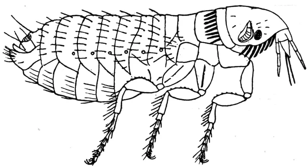
Fig. 6.—Flea, showing the various parts.
The leaping ability of adult fleas is familiar to all. No part of the
leg is particularly enlarged, so that the jump is made by the entire
leg as in the leaf-hopper insects, and not by the hind part of the leg
as in grasshoppers and flea-beetles. The size of fleas is not as variable
as in many insects. Most are about 2 to 3 millimeters long,
while the range is about 1.5 to 6 millimeters. The adult flea has a
hard, strongly chitinized body. The head is small, and on each side
bears a short jointed antenna, which may repose in a groove or
depression. Most species have a small, simple eye, but several
forms are normally without eyes. The sides of the head below the
antennæ are called the genæ. At the lower front end of the head is
the mouth and mouth parts. The latter consist of a pair of triangular
maxillæ with jointed maxillary palpi and a beak or proboscis
made up of one median and four lateral pieces. The outer pair of
lateral pieces is the labrum with the imperfectly jointed labial palpi.
71They serve as a sheath for the other organs, which are more slender.
The inner pair of pieces are considered to be the mandibles and the
median piece a labrum or hypopharynx. Others call this piece the
unpaired piercing organ, the lingua, or the syringostome. There are
other interpretations of the homologies of the mouth parts, but the
above is the most generally adopted one. The labrum and the
mandibles are roughened and constitute the piercing organs which
the flea inserts into the host to tap a blood vessel. On the lower
part of the head there is frequently a series or comb of stout spines.
Similar spines sometimes occur on the posterior border of the pronotum.
These series of spines are called “ctenidia,” and they are of
great value in classification. Behind the head are three segments,
or zoonites, each bearing a pair of legs. These together form the
thorax. The upper surface is called the notum (pronotum, mesonotum,
etc.). The sides are the pleura—sometimes “epimera” is used;
and the ventral part is the sternum. Each of the thoracic segments
has a spiracle, or a breathing pore, on each side. The first segment
of the thorax, called the “prothorax,” is shorter than the others,
and, as above stated, frequently has a row of ctenidia, or spines, on
its posterior border. The next segment is the mesothorax, and the
third the metathorax. The metathorax usually has some stout
bristles in rows on its pleura, which are enlarged and called “epiphyses,”
formerly called “squama aliforme.” The basal one or two
segments are sometimes partly covered by the epiphyses or the
metathorax. These segments consist of a dorsal plate, or tergite,
and a ventral plate, or sternite. Behind the thorax is the abdomen
of 9 apparent segments. Seven of these segments have a spiracle or
breathing pore on the sides. The last segment, or pygidium, bears
the genital organs; in the male certain processes called “claspers” at
each side of the genital opening. The anal aperture is at the end of
the ninth segment between the dorsal and ventral plates. The
claspers have a main curved part, and a slender backward projection
called the “manubrium,” and at the apex an articulated clawlike
process called “the movable finger.” At the tip of the abdomen
of the female there is a short median piece called the “style.”
The legs consist of five parts: The coxa, a large basal piece; the
trochanter, a minute piece at the end of the coxa; the femur, which
is usually slightly swollen in the middle; the tibia, which usually
has stout bristles or spines on its posterior side; and the tarsus, which
consists of five parts or joints. The basal joint is often the longest,
and the comparative lengths of these joints is expressed by a formula,
as 60–45–32–18–30. The last, or fifth, joint has been called the
“metatarsus,” but this name is better applied to the basal joint. At
the tip of the last tarsal joint is a pair of stout claws. The coxæ of
legs II and III show a longitudinal suture.
72Fleas as a rule prefer certain hosts, but are not as particular in
this regard as are many parasites. Those species which are best
known are found to attack several hosts, including man. This
catholicity of taste is what makes them dangerous parasites, the
possible transmitters not only of plague, but also of consumption,
leprosy, etc. The fleas are treated by various writers under other
names, such as Aphaniptera, and Suctoria. About 300 species are
described, and perhaps as many more will be gathered by collectors.
Formerly all fleas were kept in the genus Pulex; now they are arranged
in many genera, and these genera grouped into families. No
less than eight such families are recognized by some authorities on
this group. The species that occur on rats belong to three families,
which may be separated as follows:
| 1. |
Thoracic segments much shortened and constricted; labial palpi apparently not jointed; third joint of antennæ without subjoints; no ctenidia; abdomen of female becomes more or less swollen |
Sarcopsyllidæ |
| |
Thoracic segments not shortened nor constricted; labial palpi with joints; third joint of antennæ with several more or less distinct subjoints; ctenidia often present; abdomen of female never distinctly swollen |
2 |
| 2. |
Posterior tibial spines in pairs |
Pulicidæ |
| |
Posterior tibial spines mostly single and more numerous |
Ctenopsyllidæ |
To this family belongs the Ctenopsylla musculi Dugès.
This was formerly placed in the genus Typhlopsylla. The head
is rather acute in front and has four ctenidia each side; the eyes are
very small; the pronotal comb has 22 spines; each dorsal segment
of the body has two rows of hairs; the basal row of smaller hairs.
The proportions of joints in the hind tarsus are: 45–25–17–8–14.
Length 1.8 to 2.5 millimeters. This species is abundant on rats and
mice in Europe and other countries; recently it has been taken in
California and Florida on rats and mice.
This family includes the greater number of fleas. They have been
arranged in many genera, six of which have been taken from rats.
These are separable as follows:
| 1. |
Head without ctenidia; eyes distinct |
2 |
| |
Head and pronotum with ctenidia; last tarsal joint with four pairs of lateral spines |
5 |
| 2. |
Pronotum with ctenidia; female with one antepygidial bristle on each side |
Hoplopsyllus. |
| |
Pronotum without ctenidia |
3 |
| 3. |
Last tarsal joint with four pairs of lateral spines; female with one antepygidial bristle each side |
4 |
| |
Last tarsal joint with five pairs of lateral spines; female with two to five antepygidial bristles each side |
Ceratophyllus. |
| 734. |
Mesosternite very narrow, without internal rod-like incrassation from the insertion of coxa upward |
Pulex. |
| |
Mesosternite with a rod-like internal incrassation from the insertion of coxa upward |
Xenopsylla. |
| 5. |
Eyes rudimentary; female with two to five antepygidial bristles each side |
Neopsylla. |
| |
Eyes distinct; female with but one antepygidial bristle each side |
Ctenocephalus. |
Hoplopsyllus, one species, described as a Pulex.
Hoplopsyllus anomalus Baker.
The mandibles scarcely reach halfway down on the anterior coxæ;
upon each are two large spines; the pronotal comb has about nine
spines each side; and each abdominal segment has but a single row
of bristles. The hind femora have six to eight bristles on the side;
the proportions of the joints in the hind tarsus are: 26–16–8–5–13.
Color, dark reddish brown. Female, 2.5 millimeters; male, 1.5 millimeters.
Described from a spermophile from Colorado and recorded by
Doctor Fox and Professor Doane from Mus norvegicus from California.
Pulex.—Of this, the typical genus of the family, but one species has
been recorded from rats.
Pulex irritans Linn.
The mandibles reach about halfway down on the anterior coxæ;
the head is regularly rounded in front; there are no transverse rows
of bristles on the vertex, and but one row of bristles on each abdominal
tergite. The proportions of the joints in the hind tarsus are, 50–30–18–12–32.
Color, usually yellow brown. Male, 1.6 to 2 millimeters;
female, 2 to 3.5 millimeters.
This, the human flea, is quite cosmopolitan, but more abundant in
warm countries than elsewhere. It occurs on many domestic animals
and has frequently been taken from rats in California and elsewhere;
it also occurs on skunks.
Xenopsylla.—This genus includes the following species, formerly
placed in the genus Lœmopsylla.
Xenopsylla cheopis Rothschild.
The mandibles reach nearly to the end of the anterior coxæ; there
are no ctenidia on the head or pronotum; the eyes are distinct; each
abdominal tergite has but one row of bristles; the hind femur has a
row of about eight bristles; the proportions of the joints in the hind
tarsus are as follows: 46–30–16–10–20. Color, light brown. Male,
2.5 to 3.5 millimeters; female, 4 to 5.5 millimeters.
This is a true rat flea, but will readily bite man, and is the species
chiefly concerned in transmitting the bubonic plague. It is widely
distributed, especially in seaport towns.
Ceratophyllus.—Fleas of this genus are abundant on many kinds of
small mammals, especially rodents. There are a great many species
74and some are so closely related that it is not easy to identify them.
Of the eight species recorded from rats, four have been taken in this
country. It is not practicable to tabulate these eight species, but
the four that occur in our country may be arranged as follows:
| 1. |
Hind tarsal joint II with an apical spine much longer than joint III |
acutus. |
| |
Hind tarsal joint II with spines not longer than joint III |
2 |
| 2. |
Pronotal comb of about 26 spines |
niger. |
| |
Pronotal comb of about 18 or 20 spines |
fasciatus and londiniensis. |
Ceratophyllus niger Fox.
This species has the pronotal ctenidia of about 26 spines; there
are a few hairs on the inner surface of hind femur; apical spines of
second joint of hind tarsus not longer than third joint; three hairs
in front of the eye and three in front of these; movable finger of claspers
with five slender bristles on the outer edge. Color, very dark brown.
Length 3.5 millimeters.
Taken in California from Mus decumans and from man.
Ceratophyllus acutus Baker.
This species is readily known by having a spine at tip of the second
joint of hind tarsus longer than the third joint and reaching over
onto the fourth joint; the abdominal tergites have each two rows of
bristles; the male claspers are very large and long, sickle shaped.
Color, pale brown. Length, 3 to 3.5 millimeters.
It was described from a spermophile, but Doctor Fox has taken it
once from a rat in California.
Ceratophyllus fasciatus Bosc.
There are 18 or 20 spines in the pronotal comb; there are three
bristles in front of eye and in female two, and in male four in front
of these; there are three or four hairs on the inner surface of the hind
femur; the proportions of joints in the hind tarsus are 50–33–20–11–21.
The manubrium of the male claspers is very long and slender, and
some of the bristles on the movable finger are as long as the joint.
Length, male, 1.8 millimeters; female, 2.5 millimeters.
It has been recorded from California on rats, mice, skunks, and
man. It is also common in Europe and elsewhere on rats, mice, and
other small animals.
Ceratophyllus londiniensis Rothschild.
This is allied closely to C. fasciatus, and is best separated from that
species by the shape and armature of the genital parts; the manubrium
is not as long as in that species, and the bristles on the movable
finger are shorter; the third joint of the maxillary palpi is proportionally
longer than in C. fasciatus. There are three bristles in front
of the eyes and four or five in front of these; there are a few hairs
on the inner surface of the hind femur; the proportions of the joints
in the hind tarsus are 46–30–18–11–18.
75It has been recorded by Doctor Fox from Mus rattus in California,
and is known from rats and mice from several parts of Europe; the
C. italicus Tiraboschi is the same species.
The four other species of this genus found on rats and not yet
found in our country are closely related to C. fasciatus, and distinguished
chiefly by the shape of the male genitalia.
Ceratophyllus mustelæ Wagner.
This species has no series of hairs on the inner surface of the hind
femur; there are three bristles in front of the eye and six in front of
these; the pronotal comb has 18 or 20 spines; the proportions of the
joints in the hind tarsus are 47–37–20–13–20; the movable finger
of the male clasper has a long process below not seen in other forms.
Occurs (according to Rothschild) on rats in Europe.
Ceratophyllus pencilliger Grube.
This species also has no hairs on the inner surface of the hind
femora. The pronotal comb has 18 spines; there are three bristles in
front of the eye and four in front of these; the proportions of the
joints in the hind tarsus are 52–36–23–14–24; the outer corner of the
movable finger of the male clasper has two little rounded processes.
It was described from Siberia, but according to Rothschild occurs
on rats in Europe.
Ceratophyllus consimilis Wagner.
This species is very close to C. fasciatus, and has some fine hairs on
the inner surface of the hind femur; there are but two bristles in front
of the eye and in front of these a few finer hairs; the proportions of
the joints in the hind tarsus are 42–30–20–11–19; pronotal spines 18.
Occurs on rats in Russia.
Ceratophyllus lagomys Wagner.
This species also has a few fine hairs on the inner surface of the hind
femur; 18 spines in pronotal comb; there are three bristles in front of
eye and one in front of these; the proportions of the joints in the hind
tarsus are 53–32–20–11–22; the outer corner of the movable finger
of the male clasper has two little processes, similar to those on
C. pencilliger. Occurs on rats in Europe.
Ctenocephalus.—The common fleas on cats and dogs, as well as on
man, belong to two species long kept under one name (C. canis or
C. serraticeps), but lately shown by Rothschild to be distinct. Both
have a comb of 8 spines on the head and 16 spines in pronotal comb;
the proportions of joints in the hind tarsus are 40–24–15–10–24.
Both are occasionally taken on rats in this country. They may be
separated as follows:
| 1. |
In the female the head is fully twice as long as high (seen from side); the first spine of genal comb is two-thirds the length of the second; in male the manubrium of claspers is barely enlarged at tip; and with two rows of hairs on disc of movable finger |
C. felis Bouché. |
| 76 |
In the female the head is less than twice as long as high (seen from side); the first genal spine in the head comb is only about one-half the length of the second; in the male the manubrium of clasper is very distinctly enlarged at tip; but one row of hairs on the disc of the movable finger |
C. canis Curtis. |
Neopsylla.—One species of this genus has been described from the
brown rat in Europe.
Neopsylla bidentatiformis Wagner.
The eyes are very small; there are 4 pairs of lateral spines
beneath the last joint of the hind tarsus; the comb on head consists
of but 2 stout spines, below the middle of the antennæ; the pronotal
comb has 18 spines; the proportions of the joints in the hind
tarsus are 43–33–21–12–21.
Length: Male, 2 to 2.3 millimeters; female, 2.3 to 2.5 millimeters.
Not yet found in the United States; described from Russia.
The fleas of this family are commonly called “chigoes,” “jiggers,”
or sand fleas. The head is usually larger proportionally than in the
other fleas; there are no ctenidia on head or pronotum; the thoracic
segments are extremely short, and in the female the abdomen enlarges
with the development of the eggs. They do not hop about as other
fleas, but remain on the spot to which they have attached until they
die. Frequently the adjacent skin grows over them, forming a
swelling of considerable size.
Three species belonging to two genera have been recorded from rats.
| 1. |
Angle of head acutely produced; fifth tarsal joint of hind legs without heavy spines; few spines on the legs |
Sarcopsylla. |
| |
Angle of head not produced, obtuse; fifth tarsal joint with heavy lateral spines, and other spines on other parts of the legs |
Echidnophaga. |
Echidnophaga.—Two species of this genus are known from rats;
one, however (E. gallinacea), can hardly be called a normal parasite,
but rather of accidental occurrence. The genus has also been called
Argiopsylla and Xestopsylla.
| 1. |
Bristles at end of second joint of hind tarsus about as long as next three joints; palpi about one-half the length of mandibles |
E. rhynchopsylla. |
| |
Bristles at end of second joint of hind tarsus about as long as next two joints; palpi about two-thirds the length of the mandibles |
E. gallinacea. |
Echidnophaga rhynchopsylla Tiraboschi.
The body is about twice as long as broad and shining brown; there
is but one hair in front of eye, and four on each metathoracic pleuron;
mandibles larger than in E. gallinacea, and the spiracles are much
higher up on the sides than in that species. Length, 1.4 to 1.8 millimeters.
Taken from Mus rattus in Italy.
77Echidnophaga gallinacea Westwood.
This species has the body almost as broad as long, and of a red-brown
color; 1 bristle in front of eye and 6 on each metathoracic
pleuron; each abdominal tergite has on each side near the median
line a single hair; the spiracles are situated well down on the sides.
Length: Male, 0.8 to 1.2 millimeters; female, 1 to 1.8 millimeters.
This species is a fairly common pest of poultry and dogs in warm
countries, and is called the “chicken flea.” It has been taken from
rats in Italy.
Sarcopsylla.—This genus includes the S. penetrans, which attacks
the feet of various animals, including man, in the Tropics. This
species has not yet been recorded from rats, but an allied species is
described from Brazilian rats.
Sarcopsylla cæcata Enderlein.
Color, clear yellowish. Eyes rudimentary; lower anterior corner
of coxæ prolonged in a tooth; tarsal joints very short; claws long,
but little curved, and almost hair like. The body of a swollen
female is about 5 millimeters long.
Taken from Mus rattus in Brazil.
The insects known as Pediculi, or lice, are parasitic during their
entire life on various mammals, including man. They are flat,
rather elongate, wingless insects, with a small head and stout legs,
which end in a strong claw, opposable to a projection at the tip of
the penultimate joint. The simple antennæ, three to five jointed,
are inserted in a concavity on the side of the head. The mouth
parts are of a very peculiar nature, and not yet homologized with the
cibaria of other insects. There is a short beak or proboscis in front,
with recurved spines or hooks on its dorsal and lateral surfaces.
Through this beak extends a slender stylet, that is formed of three
parts; a ventral channeled piece, perhaps a labium; a dorsal piece,
consisting of two pieces fused together, perhaps the maxillæ; and a
median tube, possibly the hypopharynx. The stylet is used to
pierce the skin of the host, and the blood is sucked up through it.
There are no palpi. On each side of the head there is a small, simple
eye. The thorax shows only incompletely the division into the three
parts; there is a large spiracle above on each side. The abdomen
shows eight segments, six of them have a spiracle, or breathing pore,
on each side, the basal and apical segments being without them.
All of the segments bear a few simple hairs or bristles; the longest
are on the posterior segments. The legs are stout and prominent;
they consist of a broad coxa, a small trochanter, a longer femur, a
tibia with an apical process, and a tarsus of one joint and a very
large terminal claw. At the apex of the tibia, just within the projection,
is a sucking disc. This, the projection, and the claw form the
apparatus to hold fast to the hair of the host.
78Lice usually walk sideways, but do not travel much, and they keep
close to one host. The eggs are slightly elongate and fastened to
the hair of the host. They hatch in about ten to fifteen days, the
young coming out of the top of the egg. These young do not differ
much in structure from the adults, but are paler in color. They
molt their skin a few times, probably four, before they reach the
mature condition. The males are less numerous than the females,
and ordinarily smaller. There are several generations each year,
dependent doubtless on the temperature; but the life history is not
thoroughly known for any species. After sucking the blood the
abdomen of the louse becomes somewhat distended, very noticeably
so in some species.
The sucking habits of the lice render them dangerous parasites
and capable of transmitting a disease from one host to another. Fortunately
they do not readily change hosts so that they can not be
considered quite as dangerous as some more active parasites. However,
several species have already been shown to carry diseases in
laboratory experiments. Therefore it is probable that some of them
will be connected with the origin and diffusion of certain diseases of
animals.
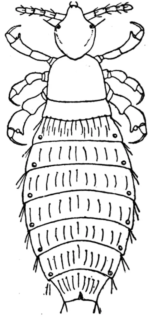
Fig. 7.—Louse (Polyplax spinulosa).
The Anoplura, or lice, have often been treated in connection with
the Mallophaga, or biting lice. This is doubtless because they frequently
occur on the same animal, and have a general resemblance
to them. However, they have no real affinity to these insects, and the
general opinion is that they are more or less related to the Hemiptera.
Sometimes they are treated as a group or section of the Hemiptera,
but also as a separate order, under various names as Siphunculata,
Lipognatha, Pseudorhynchota, and Ellipoptera.
There are about 50 or 60 known species which are arranged in 15
genera and 4 families. Four species belonging to three different
79genera have been recorded from rats; a number of others are known
from mice and other rodents, and some of these will probably yet be
taken upon rats.
These four forms are separated, as follows:
| 1. |
Eyes large and distinct; beak very short; thorax plainly longer than broad |
Pediculus capitis. |
| |
Eyes small, beak longer; thorax about as broad as long |
2 |
| 2. |
In male the pleura of abdominal segments 3 to 6 above and below have a prominent tooth-like projection; a tooth on the hind femur of female |
Hoplopleura acanthopus. |
| |
In male the pleura of abdominal segments without such projections; no tooth on hind femur of female |
3 |
| 3. |
Last joint of antennæ much more slender than those before; an acute tooth at sides of segments 4 to 7; head much narrower in front; antennæ two-thirds as long as head |
Polyplax miacantha. |
| |
Last joint of antennæ not much more slender than others; head quite broad in front; antennæ as long as head |
Polyplax spinulosus. |
Pediculus capitis De Geer.
It is pale grayish in color, with faint dark markings at the sides of
the thorax and abdomen; the last segment of the abdomen in female
is bilobed. The head is longer than broad and tapers in front.
Length, 2 millimeters. This is the head louse of man, and is said to
have been taken from rats, and is claimed to be able to transfer plague
from rats to man. Its occurrence on rats, however, appears to be
very uncommon.
Hopopleura acanthopus Burmeister.
In the male the pleura of the abdominal segments 3 to 6, which
reach up on the dorsum and over on the venter, have at their
inner ends a prominent projection, toothed in all except the third
on dorsum and the sixth on venter, which are spine-like. The head is
but little longer than broad, broad in front; and in the female there is
a recurved tooth on each hind femur. The last segment of the female
abdomen is bilobed behind. Length, 1.3 millimeters. It has been
taken from rats in Europe, but is more common on species of Microtus.
Polyplax spinulosus Burmeister.
The sides of the abdominal segments are acute, but the males do
not have the large tooth-like projection of Hoplopleura. The last segment
of the female is truncate; the head is about as broad in front
as behind, and the legs are very short and stout; the antennæ are as
long as head, and the last joint is but little smaller than the others.
Color, pale yellowish. Length, 1.4 millimeters.
This is the common rat louse, and is probably as widely distributed
as its host. Specimens have been taken in both the eastern
and western parts of the United States.
80Polyplax miacantha Speiser.
This differs from P. spinulosus in having a longer and narrower
anterior part of head, in that the last joint of the antennæ is more
slender, and the antennæ are only two-thirds as long as the head.
The abdominal segments 4 to 7 show an acute process at the sides.
Length, 1.5 to 1.75 millimeters.
Taken from rats in Abyssinia.
The mites (order Acarina, class Arachnida) are readily known
from the insects (fleas and lice) by having four pairs of legs, no antennæ,
and the abdomen does not show any segmentation, nor is there
usually any distinction between head and thorax. In some groups
there is a small head-like part, called the capitulum. The mouth
parts consist of a pair of mandibles (often styliform or needle-like), a
lip, and a pair of palpi. In some forms there is a central piece, called
the hypopharynx, and in other groups is a plate above the mouth
parts, known as the epistome. The body usually shows more or less
distinctly a division into two parts—the anterior, called the cephalothorax,
and the posterior the abdomen. However, in many mites it
is not possible to separate these parts, except that it is considered
that the legs are borne by the cephalothorax. In many forms
there is a small, simple eye each side on the cephalothorax, but
many other forms are blind. Some species have a tracheal system,
which opens in a pair of spiracles near the hind legs or near the
anterior end of the body; other species have no definite respiratory
system. The genital aperture is on the venter, usually between the
legs. The legs consist of the usual joints—coxa, trochanter, femur,
tibia, sometimes a metatarsus, and a tarsus. The tarsus terminates
in a pair of claws, sometimes three or only one, and often a sucker or
caroncle. Most mites are not parasitic; those species that are parasitic
are often free in one stage. The parasitic mites suck the blood
of their host, feed on the hair or dermal scales, or burrow in the skin.
Some predaceous species inhabit animals to hunt and eat the parasitic
mites that infest that animal.
The mites that occur parasitically on rats belong to four families:
Sarcoptidæ, Cheyletidæ, Ixodidæ, and Gamasidæ.
| 1. |
A distinct spiracle or breathing pore on each side of body near coxæ III or IV |
2 |
| |
No such spiracle or pore visible |
3 |
| 2. |
A small, distinct head part in front of the body; palpi three jointed; a granulate area around the spiracle; no sternal plate |
Ixodidæ. |
| |
No such head part; palpi five jointed; no granulate space around spiracle, but a long, chitinized piece reaching forward from it; a more or less distinct sternal plate |
Gamasidæ. |
| 3. |
All legs simple, unmodified, ending in a stalked sucker |
Sarcoptidæ. |
| |
Front legs short, enlarged, and modified for clasping; all legs end in one or two stout claws |
Cheyletidæ. |
81
The Ixodidæ, or ticks, are rather large, flat, leathery-skinned
mites, which suck the blood of various animals. In the male the
dorsum of the body is nearly covered by a corneous shield, while in
the female this shield occupies only the anterior part of the body.
In the female the body swells to enormous proportions as she engorges
herself on the blood of the host. At the posterior margin of the body
there are in many forms a series of lobes or festoons. There is no
species of tick that is commonly found on rats, but four species that
normally infest other animals have been taken from them.
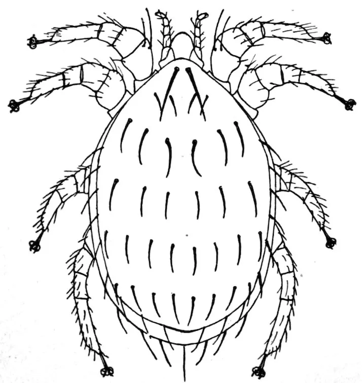
Fig. 8.—Mite (Lælaps echidninus).
| 1. |
On the venter is a groove in front of the anus and extending back each side; no festoons to posterior margin of body; palpi rather long |
Ixodes ricinus. |
| |
On the venter there is no groove in front of the anus, but usually one behind; festoons distinct in males and unengorged females |
2 |
| 2. |
Palpi very short with transverse ridges; shield of female narrowed behind eyes; stigmal plate nearly round |
Margaropus annulatus. |
| |
Palpi short, without transverse ridges; shield of female not narrowed behind eyes; stigmal plate comma shaped |
Rhipicephalus sanguineus. |
| |
Palpi elongate, without ridges; shield of female broad; stigmal plate oval, |
Hyalomma aegypticum. |
Ixodes ricinus Linné.
The shield of the female is elliptical, plainly longer than broad,
sides not suddenly narrowed behind, and there is no eye-spot at each
lateral corner. The coxa I has a long sharp spine.
This is a common European tick found on sheep, cattle, dogs, etc.,
and it has been taken a few times in this country. Neumann has
recorded its capture from Mus decumans.
82Margaropus annulatus Say.
The shield is plainly longer than broad, with a distinct eye-spot at
each lateral corner, and behind the eye the shield is suddenly narrowed;
the coxæ of the female are without spines, but the male has
2 on coxæ I. This is the common cattle tick of the United States, and
disseminates the Texas fever. Mr. Hunter has taken it once from a
rat in a barn at Dallas, Tex.
Rhipicephalus sanguineus Latreille.
The shield of the female is oval, and longer than broad, with an
eye-spot at each outer corner. Coxa I with 2 teeth; a smaller tooth
on each of the other coxæ. Stigmal plate long, comma shaped. In
the male there is a corneous plate each side of the anus, and on middle
of posterior margin a projection, or short tail.
This species is common in tropical countries, and Nuttall has
recorded specimens from the black rat in India.
Hyalomma aegypticum Linné.
The shield of the female is as broad as long, and the eye-spot is
slightly above each outer corner. Coxa I has 2 large teeth, and a
small tooth on each of the other coxæ. In the male there are 2 corneous
plates each side of anus, and behind is a pair of small tubercles.
This is a common tick in the warmer parts of the Old World; and
Nuttall has recorded young specimens on the black rat.
The Gamasid mites, although much smaller than the ticks, are
large enough to be seen by the naked eye. They are active, and
most are not parasitic, at least for part of their time. The palpi are
simple, of 5 joints; the mandibles are elongate, retractile, and usually
chelate at tip. There are no eyes. The dorsum and often the venter
shows one or more corneous shields or plates, frequently a number of
them; one or two on the dorsum, and on the venter one between the
coxæ, called the sternal plate; one behind this, the genital plate; one
behind the latter, the ventral plate; and one surrounding the anus,
the anal plate. Frequently some of these are absent or united to one
of the others.
The legs are slender, usually of 6 joints, with a long tarsus that
terminates in 2 claws, and often a sucker, or caroncle. The stigmata,
or spiracles, are lateral above and between coxæ II and IV, and
usually provided with a slender peritreme reaching forward toward
the head. Nearly all the Gamasidæ deposit eggs, and the young
often differ considerably from the adult in structure. There are
two, and perhaps sometimes three, nymphal stages. In one of these
nymphal stages the mite is apt to attach itself to an insect for the
purpose of being carried to a similar locality, where it may feed and
mature. The coprophagous and xylophagous insects are especially
concerned in the diffusion of these mites.
83There are, however, quite a number of species that are genuine
parasites of insects and other animals. Those occurring on rats
belong to two genera, Myonyssus and Lælaps. They can be separated
as follows:
| 1. |
Anal plate small, much smaller than the ventral plate |
Lælaps. |
| |
Anal plate large, larger than the ventral plate |
Myonyssus. |
Myonyssus.—This genus is made by Tiraboschi for one species:
Myonyssus decumani Tiraboschi.
Body oval; legs short and stout, all tarsi with a large caroncle
with two short claws; coxæ II have a large tooth on the anterior
border, none of coxæ with spines; sternal plate much broader than
long, with three bristles each side; ventro-genital plate much longer
than broad, broadest behind, bordered with bristles; anal plate very
large, nearly one and a half times as broad as long; three large spines
each side on venter. Length, 0.95 millimeter.
Found in Italy on Mus decumans.
Lælaps.—This genus embraces a large number of species, several
of which occur on small animals, such as the muskrat, ground hog,
and chipmunk. Three have been recorded from rats, one of these
from California. The dorsal plate is covered with hairs or bristles,
and there are usually stout bristles on the margins of the plates on
the venter. There is also a bristle, or a spine, at the tip of the anal
plate. The legs are short and stout, with a distinct caroncle, and
two claws.
| 1. |
Dorsum with numerous fine hairs; no stout spines on coxæ |
L. stabularis. |
| |
Dorsum with fewer, but stouter spine-like bristles; each coxa has a stout spine |
2 |
| 2. |
Body but little longer than broad; ventral plate longer than broad |
L. agilis. |
| |
Body much longer than broad; ventral plate about as broad as long |
L. echidninus. |
Lælaps echidninus Berlese.
Dorsum of body almost wholly covered with a shield, with rows
(six in front, eight behind) of stout, curved bristles, a longer pair
near front margin, and some around lateral and posterior margins.
Legs short and stout, tarsi about twice as long as preceding joint;
each coxa bears a stout spine near middle. Palpi very short; sternum
with three stout bristles or spines each side; ventral plate with four
stout bristles each side; anal plate with a stout apical bristle, and a
small one each side. Length, 1 millimeter.
Occurs commonly on rats in warm countries, and known from
California. It may possibly aid in the transmission of disease.
Lælaps agilis Koch.
Similar in many respects to L. echidninus but differ in the shorter
and proportionately broader body, barely longer than broad, and in
the weaker and shorter spines on dorsum and on the ventral plates;
there are also some small short spines on the general surface of the
venter. Length, 0.7 millimeter.
84Recorded from rats from Europe and Africa.
Lælaps stabularis Koch.
The body is of the same general shape as in L. echidninus, but the
dorsum is clothed with 12 to 18 rows of fine short hairs. The first
pair of legs is more slender than in the other species, and the hind
legs are also more elongate; the coxæ do not have the stout spines
seen in the other species, and the bristles on the sternal and ventral
plates are much less stout; the general surface of the venter has
many hairs, the anal plate has a short apical bristle. Length,
1.2 millimeters.
Taken on the brown rat in Italy; also found in manure.
This family consists of small, soft-bodied mites, that are parasitic
or predaceous in habits. The palpi are small, three or four jointed;
the mandibles are styliform and retractile; and the breathing spiracles
open near the mouth parts. The species that occur on rats
belong to the genus Myobia.
Myobia.—The body is elongate, fully twice as long as broad, tipped
by a pair of long, stout bristles. The first pair of legs is enlarged and
shortened, with a terminal hook to grasp hairs; the other legs are
short, simple, and far apart. The palpi and mouth parts are very
small, and the dorsum bears stout bristles. They are supposed to
feed on the exudations of the skin, but it would seem more probable,
from the nature of the mandibles, that they pierced the skin to secure
food. All are very small, not one-half a millimeter long. Of the
several species two have been recorded from rats.
| 1. |
Dorsum of female with spines all acute and sharp |
M. musculi. |
| |
Dorsum of female with some of the posterior spines flattened and rather scale like |
M. ensifera. |
Myobia musculi Schrank.
This occurs on various mice and moles, and once recorded from the
brown rat. It has been taken in this country on mice. It lives at
the base of the hairs.
Myobia ensifera Poppe.
This was described from the brown rat in Europe. The female is
separated from M. musculi by having about six of the posterior dorsal
spines flattened and scale like; in the male the six dorsal spines
are longer, and the small spines much smaller than in M. musculi.
These are the itch and scab mites. The body is soft, rounded, and
whitish in color. The legs are very short, of five joints, and end in
one or two claws, and often a pediceled sucker. The palpi are small
85and short, of three joints, but the basal is usually united to the
rostrum. There are no spiracles, and respiration is therefore through
the general surface of the skin. The sexes are often quite different
in structure. The females usually deposit eggs, the larvæ are hexopod,
and there are two nymphal stages. They are all parasites,
mostly on mammals and birds, and often burrow in the skin, causing
mange, or scabies.
Only one species has been taken on rats; this belongs to the genus
Notoedres.
Notoedres.—In this genus the third pair of legs of the male and
the third and fourth of the female have no sucker at the tip. The
anal opening is on the posterior part of the dorsum. The three
known species are parasitic on mammals—one on the cat, one on the
rabbit, and the third on rats.
Notoedres muris Mègnin.
This is a rounded mite, with finely striate skin, a small triangular
rostrum; in front the four anterior legs project a little beyond the
body, and each ends in a long pedicellate sucker; the third and
fourth pairs of legs are not visible from above, and each ends in a
long bristle. There are a few short hairs around the anal aperture
and about ten others in front of these. The species measures about
0.3 to 0.4 millimeter long.
It usually occurs about the ears and the genital organs of the host,
and has been taken from both the brown and black rat in Europe.
The Sarcoptes alepis Railliet and Lucet is the same species.
Besides the mites above described, a form of Demodex has been
recorded from rats, but the species is not given. These mites are
very tiny, with elongate body, the posterior part annulate, the front
part with eight very short legs. They inhabit the hair follicles of
various mammals. That on the rat may have been only an accidental
occurrence of some species normally on another animal.
THE INTERNAL PARASITES OF RATS AND MICE IN THEIR RELATION TO DISEASES OF MAN. — SUMMARY. — INTRODUCTION. — PROTOZOA. — Genus CHLAMYDOPHRYS [U] Cienkowski, 1876. — Genus LAMBLIA [W] R. Blanchard, 1888. — Species LAMBLIA DUODENALIS [X] (Davaine, 1875) Stiles, 1902, s. l. — Genus TRYPANOSOMA s. l. — Genus TRYPANOZOON Luehe, 1906. — CESTODA—TAPEWORMS. — CYSTICERCUS CELLULOSÆ—TÆNIA SOLIUM. — CYSTICERCUS PISIFORMIS—TÆNIA PISIFORMIS. — CYSTICERCUS FASCIOLARIS—TÆNIA TENIÆFORMIS. — HYMENOLEPIS MURINA [Y] (Dujardin, 1845) = HYMENOLEPIS NANA FRATERNA [Y] Stiles, 1906. — HYMENOLEPIS DIMINUTA [Z] (Rudolphi, 1819) R. Blanchard, 1891. — NEMATODA—TRUE ROUND WORMS. — Family TRICHINELLIDÆ. [AA] — Genus TRICHINELLA [AB] Railliet, 1895. — TRICHINELLA SPIRALIS (Owen, 1835) Railliet, 1895. — ACANTHOCEPHALA—THORN-HEADED WORMS. — Genus GIGANTORHYNCHUS Hamann, 1892. — The Moniliform Thorn-headed Worm—GIGANTORHYNCHUS MONILIFORMIS — ARACHNOIDEA. — Genus LINGUATULA Frœhlich, 1789.—Tongue worms.
87
By Ch. Wardell Stiles, Chief, and Charles G. Crane, B. S., Assistant, Division of Zoology, Hygienic Laboratory, United States Public Health and Marine-Hospital Service.
Rats and mice may harbor 11 species of internal parasites which come into consideration
as possible or established parasites of man. From this point of view, 7
of the parasites are of more academic interest than practical importance. The rat
may, however, be viewed as the practical, theoretical, and permanent reservoir for
one zooparasitic disease (trichinosis) of considerable importance, and of at least one,
perhaps two, other zooparasitic infections (“Lamblia duodenalis” and Hymenolepis
diminuta) of much less importance. Its possible future rôle in connection with sleeping
sickness should not be entirely ignored.
From the standpoint of internal zooparasitism, therefore, the present public health
interest in rats and mice centers in trichinosis. This disease will probably never be
eradicated from man until rats and mice are practically eradicated, and any rational
public health campaign directed against trichinosis must take the rat into serious
consideration.
The eradication of rats and mice would be a very substantial contribution toward
a reduction and eradication of trichinosis.
From the habits of rats, it is to be expected that they harbor
many species of parasites, and on account of their presence in our
houses the question naturally arises as to whether any of these parasites
are transmissible, either directly or indirectly, to man.
The species of internal parasites which come especially into consideration
in this connection are the following:
Protozoa: Chlamydophrys enchelys (p. 88), Lamblia duodenalis (p. 89), Trypanosoma
gambiense (p. 94).
Trematoda: None.
Cestoda: Cysticercus cellulosæ (p. 95), C. fasciolaris (p. 96), C. pisiformis (p. 95),
H. murina Duj. [= fraterna] (p. 96), Hymenolepis diminuta (p. 98).
Nematoda: Trichinella spiralis (p. 101).
Acanthocephala: Gigantorhynchus moniliformis (p. 108).
Arachnoidea: Linguatula denticulata (p. 110).
Of these 11 species, the trichina worm (sometimes called the flesh
worm) exceeds all the others combined, both in frequency and importance,
as a cause of disease in man.
88
Species CHLAMYDOPHRYS ENCHELYS[V] (Ehrenberg.)
A very peculiar organism has been described under the name of
Leydenia gemmipara Schaudinn, 1896. This was found in fluid,
obtained by puncture, from two ascites patients in Berlin, Germany.
More recently Schaudinn has concluded that Leydenia gemmipara
represents an abnormal condition of a protozoon known as Chlamydophrys.
The latter passes through the intestinal tract of various
animals (as man, mice, squirrels, rabbits, cattle), and thus is occasionally
found in fresh human stools. According to Schaudinn, if
pathological conditions in the colon cause an alkaline reaction of its
entire content, the usual shell formation in Chlamydophrys fails to
take place, the organisms then multiply in an atypical manner by
division and budding, and the result is the structure described as
Leydenia gemmipara.
Generic diagnosis.—Polymastigidæ: Body bilaterally symmetrical, pyriform, excavate
antero-ventrally to form a sucker; flagella directed posteriorly; 3 pairs inserted
on margin of the sucker, 1 pair at posterior end of body. Parasitic in intestine of
mammals.
Type species.—Lamblia duodenalis s. l. (“L. intestinalis” of man).
Flagellate protozoa belonging to this genus are reported as parasitic
in the intestinal canal of various species of mammals. At
present the forms in question are usually looked upon as belonging
to the species L. duodenalis. Evidence is, however, accumulating
(p. 92) to the effect that there are at least three distinct species of
Lamblia (“L. intestinalis” of man, L. muris of mice, and L. cuniculi (or
duodenalis?) of rabbits). Admitting that there may be three species,
the intertransmissibility of these forms from one host to another
remains to be investigated to some extent. It seems thus far definitely
proved that the form which occurs in man is transmissible to
mice, rabbits, and guinea pigs, hence mice still remain a source of
danger in respect to the infection in man. To exactly what extent
this fact is of academic interest or of practical significance is at
present sub judice.
89
Specific diagnosis.—Lamblia (p. 88): Body pyriform, 5 to 16μ (21μ Lambl) long,
4 to 12.5μ (8.6 to 11μ Lambl) broad; flagella 9 to 14μ long; anterior end bluntly
rounded, posterior end sharply pointed, dorsum convex, antero-ventrally concave,
venter flat to convex; antero-ventral concavity forms a sucker, the margins of which
project from the surface and are contractile. Four pairs of ventral posteriorly directed
flagella, arranged as follows: 1 pair insert on anterior margin of sucker; 2 pairs on posterior
margin of sucker, near median line; 1 pair on posterior extremity. Body
membrane (“cuticula”) very delicate, permitting some change of body form; protoplasm
finely granular; nucleus dumb-bell shaped, pre-equatorial. Vacuoles not
observed. Copulation sucker-to-sucker, followed by an encystation, in which stage
complicated nuclear changes occur; cysts 10 by 7μ.
Habitat.—Upper portion of small intestine of man (Homo); also of the common
house mouse (Mus musculus), the brown rat (M. decumanus), the black rat (M. rattus),
“Mus sylvestris” [=? M. decumanus], field mouse (Microtus arvalis), water mole
(Arvicola amphibius), rabbits, guinea pigs, domesticated cats, dogs, and sheep.
Geographical distribution.—Europe, Egypt, and United States.
This parasite is very common in animals in certain parts of
Europe, and cases of its presence in man have been reported by a
number of authors (Lambl, 1859; Grassi, 1881; Perroncito, 1888;
Moritz, 1891; Moritz & Holzl, 1892; Roos, 1893; Kruse & Pasquale,
1894; Piccardi, 1895; Sievers; Mueller; Frshezjesski & Ucke; Stiles,
1902; Braun, 1908; etc.). The indications are that it is more common
in man than is generally assumed.
Possibly man becomes infected through eating food (as bread,
etc.) which has been soiled by the excrements (containing the
encysted stage) of mice and rats. Grassi infected himself, Perroncito
infected mice and rabbits, and Stiles infected guinea pigs by
feeding to them human feces containing the encysted stage.
The parasite may be present in large numbers. Moritz estimated
a discharge of 18 milliards within twenty-four hours from one of
his patients. It has been observed in healthy persons and also in
cases of various diseases, but especially in children and in cases of
tuberculosis. It is an inhabitant chiefly of the duodenum and
jejunum, where it attaches itself (fig. 13) by means of the sucker to
the epithelial cells. It is rarer in the ileum. In case the stomach
is alkaline (carcinoma) the parasite may occur in this organ (Cohnheim,
Zabel). In P. Schmidt’s case the hydrochloric acid was 1
per cent. In case the intestinal peristalsis is normal the parasite
90becomes encysted in the colon, so that usually only the encysted
stage is found in the feces; but in case of increased peristalsis and
diarrhea the organisms have not time to encyst, so that the free
stages are observed in the stools. As the parasites become cool
motion decreases; when raised to high temperature, as 50° C.,
motion becomes slow, and the organisms die at 52° C. or below 0° C.
Fig. 9.—Lateral view of encysted Lamblia duodenalis.
Fig. 10.—Cyst from large intestine.
Fig. 11.—Ventral view of Lamblia.
Fig. 12.—Lateral view.
Fig. 13.—Epithelial cells of the villous coating of the small intestine infested with Lamblia. (After
Grassi & Schewiakoff, 1888, pl. 15, figs. 1, 2, 5, 11, 12.)
91
Fig. 14.—An epithelial cell with parasitic Lamblia. Greatly enlarged. (After Grassi & Schewiakoff, 1888, pl. 15, fig. 6.)
Fig. 15.—An individual in the act of joining an epithelial cell. (After Grassi & Schewiakoff, 1888, pl. 15, fig. 7.)
Pathogenicity.—Opinion differs as to the pathogenicity of this
organism. Perroncito (1902b) reports it as causing a fatal disease
in rabbits. Braun (1908) is inclined to consider it harmless. From
conversation with Doctor Hemmeter, we are persuaded that in his
case in a child in Baltimore the parasite was not without effect.
Possibly the question as to its pathogenicity is a relative one in
that light infections may produce no recognizable disturbance, while
heavy infections may produce recognizable effects. Doctor Hemmeter’s
original letter regarding his case contained the following notes:
Patient, male, white child, 3 years old, born in Maryland. Had recurrent attacks
of colitis all its life; three acute attacks within the last three weeks, accompanied by
fever, distended abdomen, sensitive, etc. Stools have always been like putty, containing
large amount of mucus, and some blood streaks; fever lasting three days, no
pronounced diarrhea, 2 to 3 passages per day; intervals between attacks variable,
stools at such times like putty, also with mucus.
92Later information from Doctor Hemmeter states that the disappearance
of the parasites from the stools coincided with improvement
in the child’s condition.
Clinical diagnosis.—The fresh unstained stools should be examined
microscopically; or the diluted stool may be stained with
methylene blue, by which nearly all objects become promptly
stained, except Lamblia, which remains grayish white (Roos, 1893)
for several hours.
Treatment.—Attempts to expel Lamblia have not always met
with marked success. Among the drugs used are male fern, sulphate
of quinine, naphthol, calomel, hydrochloric acid, and arsenic.
Grassi appears to have had success with calcined magnesia.
Fig. 16.—“Lamblia intestinalis” of man. (After Bensen, 1908, fig. 5.)
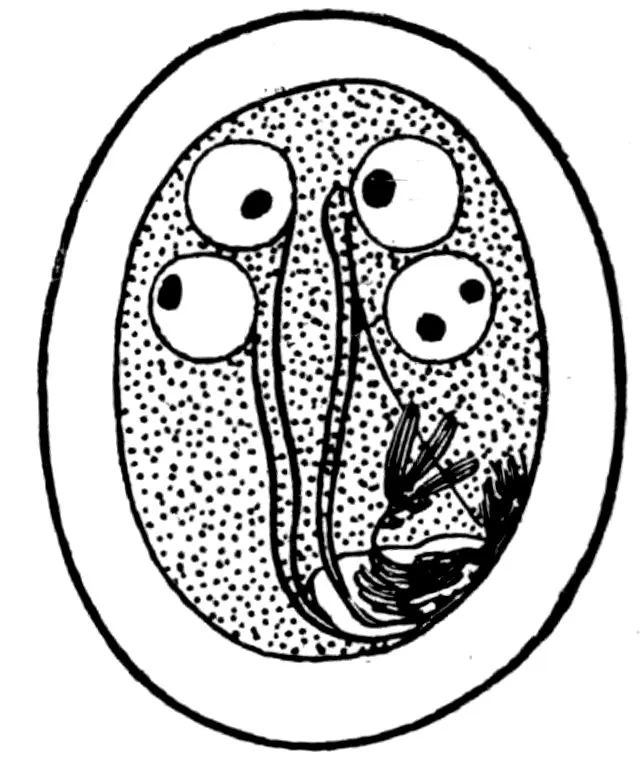
Fig. 17.—Copulation cyst of “Lamblia intestinalis” of man. (After Bensen, 1908, fig. 5.)
The division of Lamblia duodenalis INTO SEPARATE SPECIES.—Bensen
(1908) has recently divided Lamblia duodenalis s. l. into
three species: L. “intestinalis,” L. muris, and L. cuniculi. His
preliminary paper seems to offer fairly convincing data for the correctness
of his interpretation, but it may be well to await the publication
of his more complete paper, in which he promises fuller
details, before the species are definitely accepted. Several nomenclatural
points will come up for consideration in this connection.
Lamblia intestinalis, which Bensen accepts as name for the species
(fig. 16) occurring in man, can not be accepted, as this name is based
on Cercomonas intestinalis Lambl, 1859, which is invalidated by
93Cercomonas intestinalis (Ehrenberg, 1838) Perty, 1852, found in
frogs.
Lamblia muris (fig. 18) will probably stand, based on Dimorphus
muris Grassi, 1879.
Lamblia cuniculi.—There is some doubt as to the status of this
name. Davaine (1875a, 128–129) has described from rabbits a
protozoon, which he designated as Hexamita duodenalis and which
Railliet (1893a, 169) identifies as a synonym of Lamblia intestinalis
(Lambl). On basis of the principle that identifications are to be
accepted as correct until shown to be incorrect, Stiles has accepted
duodenalis as name (1902) for the form in question. The question
now arises as to the relation of cuniculi to duodenalis. If they are
accepted as identical, duodenalis will supplant cuniculi, and a new
name must be given to the form found in man. If duodenalis is
taken as identical with intestinalis Lambl, duodenalis remains as
name for the form in man, and cuniculi Bensen will stand for the
species in rabbits. If Railliet’s interpretation of synonymy be
shown to be incorrect by proving that duodenalis Davaine is not
to be considered in connection with either form, a new name must
be given to intestinalis Lambl.
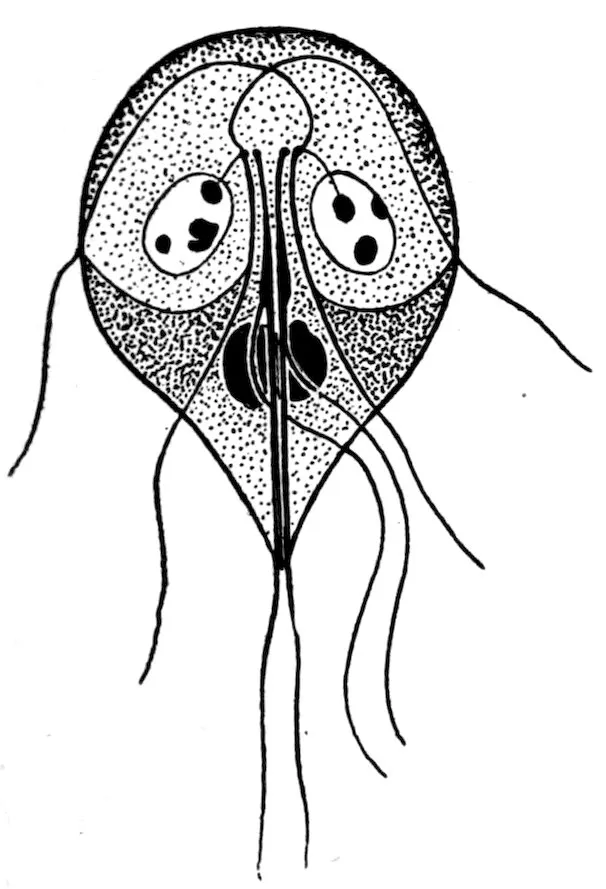
Fig. 18.—Lamblia muris of mice. (After Bensen, 1908, fig. 1.)
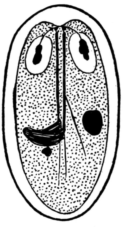
Fig. 19.—Autogametocyte of Lamblia muris. (After Bensen, 1908, fig. 6.)
An extensive group of parasitic protozoa, known as “trypanosomes,”
has recently been the basis of considerable literature. The
genus Trypanosoma was originally based upon a species (T. rotatorium)
found in frogs, and while most trypanosomes have been
94described as members of this genus several authors have separated
out certain forms into separate genera.
Luehe (1906) has recently placed the trypanosomes of mammals
in the
One of these species (Trypanozoon gambiense, usually known as
Trypanosoma gambiense) is the cause of “sleeping sickness” in man,
and has been transmitted in laboratory experiments to rats and mice.
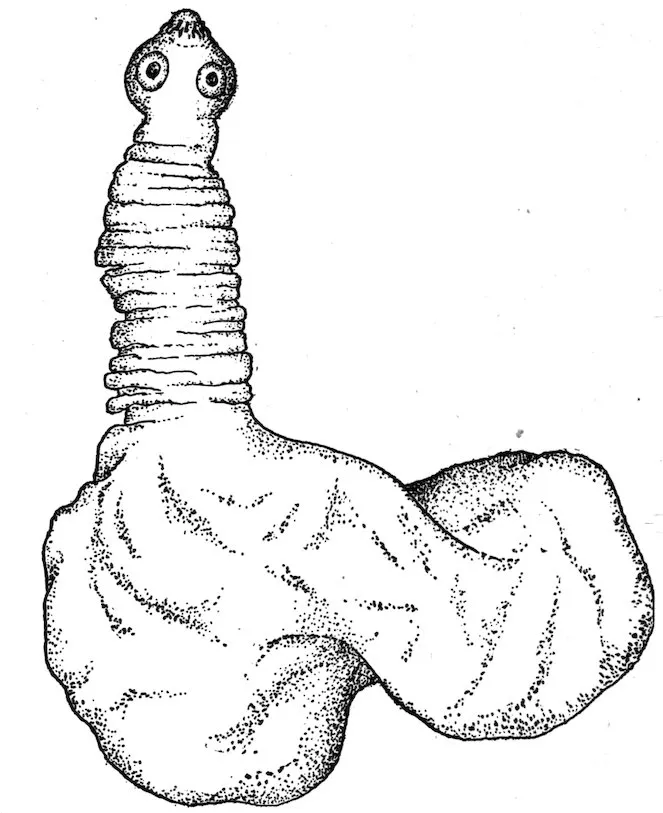
Fig. 20.—An isolated pork-measle bladder worm (Cysticercus cellulosæ), with extended head. Greatly enlarged. (After Stiles, 1898a, 90, fig. 76.)
Just what practical importance there may be in the ability of the
parasite to live in rats and mice remains to be seen, but theoretically
this biological factor may possibly become one of considerable magnitude.
At present the least conclusion to be drawn is that it adds
one more to the many arguments in favor of a world-wide destruction
of rats and mice.
Several trypanosomes, other than Trypanozoon gambiense, are
transmissible by experiment to rats and mice, while one species (Trypanozoon
lewisi) has rats for its normal host, and two other species
(Trypanozoon duttoni and Trypanosoma musculi Kendall, 1896) are
reported originally from the mouse.
95
Of the five cestodes mentioned as coming into consideration in the
subject under discussion, only one (Hymenolepis diminuta) need be
considered seriously.
The larval cestode known as Cysticercus cellulosæ (which causes
“measles” in swine) develops (when eaten by man) into a tapeworm
which is known as Tænia solium. This larva is also reported as
encysted in the peritoneum of Mus rattus.
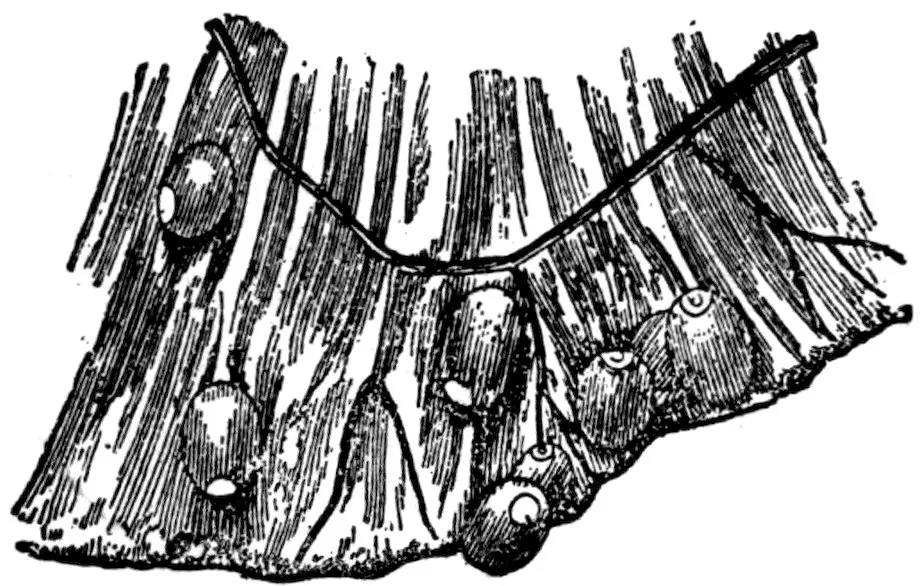
Fig. 21.—Portion of mesentery of rabbit infected with Cysticercus pisiformis. Natural size. (After Railliet, 1893a, 216, fig. 114.)
Even if it be granted that the specific determination of the specimen
in question as Cysticercus cellulosæ is correct, the occasional infection
of rats with this parasite would be of very little practical significance
in this country from a public health point of view, as we do not use
rats for food for man. Theoretically it is possible to conceive of
combinations of circumstances in which such infection in the rat
might under certain conditions eventually affect man, but the chances
are so remote as to be negligible, especially when compared with the
much greater questions which demand attention.
The larval stage of this parasite occurs in rabbits, the adult stage
in canines. Parona (1901) reports the occurrence of the larval stage
in Mus brasiliensis, and Vital has recorded the presence of the adult
stage in man.
In view of the fact that Galli-Valerio was unable to infect himself
experimentally with this species, the specific determination made by
Vital is open to some question. Even assuming that this tapeworm
may develop in man, the presence of the larval stage in rats is of
such little importance as to be negligible.
96
This encysted larval tapeworm is exceedingly common in the liver
of rats and mice, and when swallowed by cats it develops into an
adult tapeworm.

Fig. 22.—Larval stage of Tænia teniæformis. Natural size. (After Leuckart, 1880, 450, fig. 202.)
There are two possible points of view in connection with which this
parasite is of indirect interest in public health matters: (1) Occasionally
these encysted parasites are mistaken for lesions of tuberculosis;
(2) Krabbe (1880) relates that in Jutland there exists a folk custom
of eating chopped raw mice in case of retention of urine, and in this
connection the point has been raised that the possibility is not
excluded that such action might eventually give rise to infection of
man by the parasite in question. No case of such infection in man is
as yet established.
Under the name Tænia murina, Dujardin (1845) described for rats
a tapeworm which has been identified by a number of authors (including
Stiles) as identical with the dwarf tapeworm (Hymenolepis nana)
of man. If this identification be correct, the rats must be considered
as the great disseminators of this tapeworm. Serious doubts have
been raised, however, as to whether the tapeworm in man is not in
reality distinct from that of the rat, and the evidence in favor of such
conclusion is accumulating. Some slight differences between the two
forms have been noticed, but by some authors these differences have
97been considered insufficient to hold the two worms apart. Looss has
tried to infect rats with the dwarf tapeworm found in man in Egypt,
but his results have been negative. Here in Washington Stiles has
repeatedly attempted to infect rats with the dwarf tapeworm found
in man in the United States, but thus far no positive infection has
occurred in the rodents. In Italy, Grassi attempted to transmit the
rat form to six persons, and in one case he found tapeworms, but in
view of the frequency of H. nana in Italy the significance of this one
instance has been questioned; Grassi was not successful in trying to
infect rats with H. nana of man.
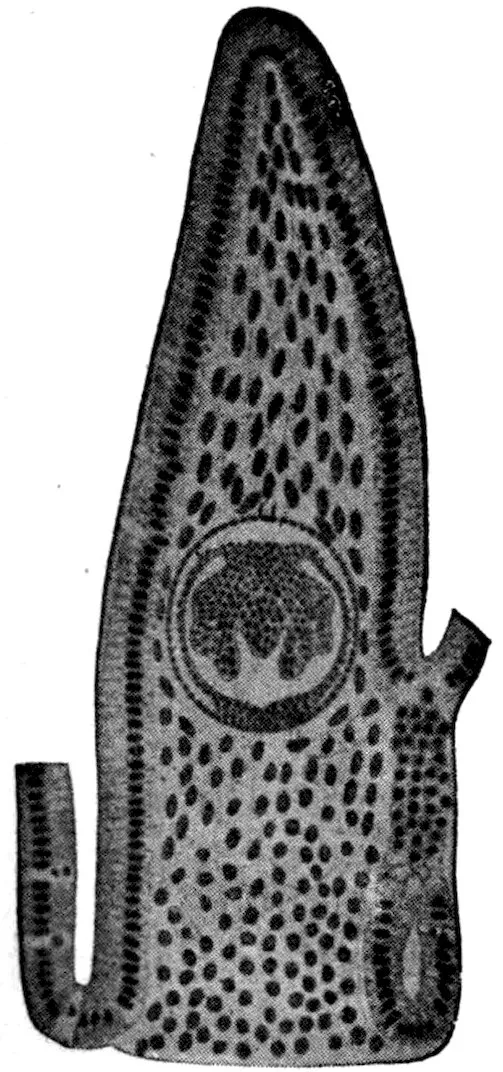
Fig. 23.—Longitudinal section of the intestinal villus of a rat, containing cystic stage of dwarf tapeworm. Enlarged. (After Grassi & Rovelli, 1892a, pl. 3, fig. 25.)
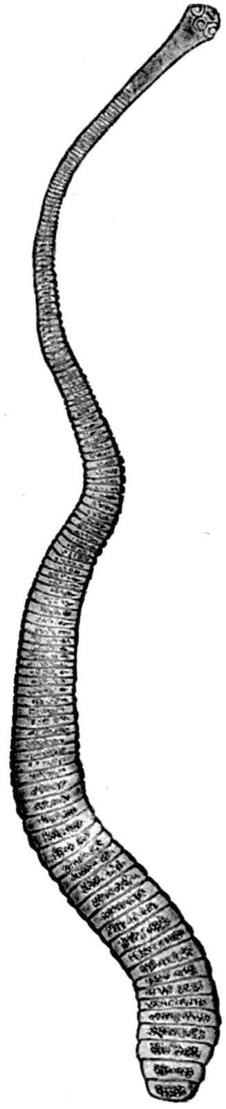
Fig. 24.—Adult dwarf tapeworm (Hymenolepis nana) of man. Enlarged. (After Leuckart, 1863, p. 393, fig. 112.)
Thus at present the evidence is to the effect that rats and mice are
not to be viewed as the source or reservoir for the dwarf tapeworm
(H. nana) of man.
98
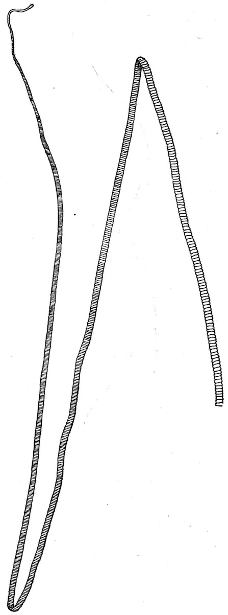
Fig. 25.—Strobila of Hymenolepis diminuta. Natural size. (After Grassi, 1881, pl. 11, fig. 1.)
Fig. 26.—Head and anterior portion of H. diminuta from the rat. Enlarged. (After Zschokke, 1889, pl. 1, fig. 21.)
Specific diagnosis.—Hymenolepis: Strobila 10 to 60 millimeters in length, 2.5 to 4
millimeters in maximum breadth; composed of 800 to 1,300 segments. Head small,
almost globular; 200 to 600μ in width; rostellum rudimentary, pyriform, only slightly
99protractile; hooks absent; suckers globular, near the apical portion of the head, 80
to 160μ in diameter. Neck usually short. Segments throughout strobila broader
than long. Genital pores on left margin, near the junction of the anterior and middle
thirds of each segment. Three testes in each segment; vas deferens dilates into a
prominent seminal vesicle before entering the cirrus pouch, within which also is a
vesicle. Gravid uterus occupies most of the proglottids; its cavity is subdivided
into a large number of incompletely separated compartments filled with eggs. Eggs
round or slightly oval; outer membrane 54 to 86μ in diameter, yellowish in color,
may be radially striated; inner membrane 24 by 20μ to 40 by 35μ in diameter, with
mammillate projection at each pole often not apparent; between outer and inner
membranes a prominent third layer of albuminous substance, often appearing as two
delicate smooth membranes, with intervening space filled by a granular coagulum;
embryonal hooks 11 to 16μ in length.
Fig. 27.—Male and female organs of H. diminuta: c. p., cirrus pouch; g. p., genital pore; ov., ovary; rec. sem., receptaculum seminis; s. g., shell gland; t., testiculæ; ut., uterus; vag., vagina; v. def., vas deferens; v. ef., vas efferens; ves. sem., vesicula seminalis; y. g., yolk gland. Enlarged. (After Zschokke, 1889, pl. 2, fig. 22.)
Fig. 28.—Gravid segment of H. diminuta. Enlarged. (After Grassi, 1881, pl. 11, fig. 15.)
Fig. 29.—Egg of H. diminuta from man. Greatly enlarged. (After Bizzozero, 1889a, pl. 4, fig. g″.)
Habitat.—Adults in small intestine of brown or Norway rat (Mus decumanus), black
rat (M. rattus), house mouse (M. musculus), Egyptian or roof rat (M. rattus alexandrinus),
wood or field mouse (M. sylvaticus), Rhipidomys pyrrhorhinus [according to
Linstow, 1878a, 23], and man (Homo sapiens).
100Development.—The larval stage (Cercocystis H. diminutæ) occurs in larval and
adult meal moths (Asopia farinalis); in young and adult earwigs (Anisolabis annulipes);
and in adult beetles (Acis spinosa and Scaurus striatus).
Geographic distribution.—Massachusetts, Pennsylvania, Nebraska, Iowa, District
of Columbia, Maryland, Brazil, Italy, Germany, France, Austria.
This parasite is certainly more common in man in this country than
has heretofore been assumed, but fortunately it seems to be one of
the most harmless and most easily expelled tapeworms occurring
in man.
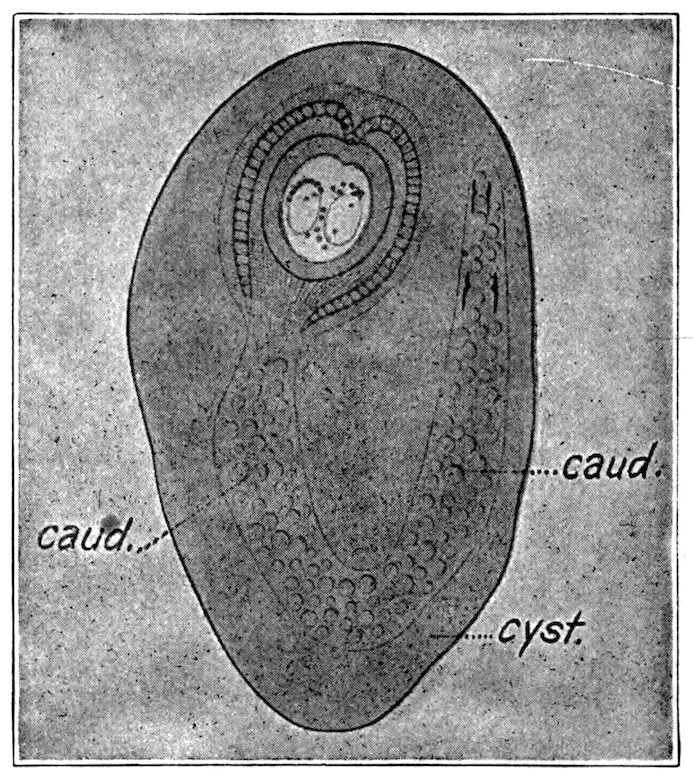
Fig. 30.—Encysted cystic stage of H. diminuta: caud., caudal appendage; cyst., adventitious capsule inclosing the cercocystis. Enlarged. (After Grassi & Rovelli, 1892a, pl. 4, fig. 1.)
From present evidence, the rats and mice are looked upon as the
regular hosts for this worm, and hence as the natural reservoir of
the infection. The intermediate host becomes infected from the
rodents and then transmits the infection to man.
It might be mentioned that as yet no extensive study has been
conducted in the United States to differentiate clearly the various
species of Hymenolepis found in our rats and mice. The possibility
is therefore not entirely excluded that some of our cases of Hymenolepis
diminuta may eventually be shown to be referable to other
species of the same genus.
101
Family diagnosis.—Nematoda: Elongate cylindrical worms; cephalic portion long
and very slender, caudal portion more or less swollen. Mouth rounded, without lips.
Esophagus relatively very long, composed of a single row of large cells, forming the
so-called “cell body” and supporting a narrow esophageal tube; anus terminal or
nearly so.
Male: With a single spicule or without spicule.
Female: With one ovary; vulva near caudal end of cell body, close to point where
body increases in diameter; oviparous or viviparous.
Eggs: Oviparous species, with thick shell, with opening at each pole, closed by a
transparent plug.
Type genus.—Trichinella Railliet, 1895.
This family furnishes two parasites to man: The whipworm (Trichuris
trichiura) of the colon, and the trichina or flesh worm (Trichinella
spiralis, see p. 101).
Generic diagnosis.—Trichinellidæ: Very minute worms, of nearly uniform diameter.
Adults in intestine of mammals, larvæ encysted in muscles.
Male: Without spicules, but with 2 conical appendages on the tail, at side of terminal
cloacal opening.
Female: Vulva about one-fifth the length from anterior end; viviparous.
Type species.—Trichinella spiralis (Owen, 1835) Railliet, 1895.
Specific diagnosis.—Trichinella: Body thread-like, visible to naked eye.
Male: Length, 1.4 to 1.6 millimeters; diameter, 40μ; distal of cloacal opening, 2
pairs of papillæ, the anterior pair hemispherical, posterior pair conical.
Female: Length, 3 to 4 millimeters; diameter, 60μ; anus terminal; vulva one-fifth
of length of body from the mouth; viviparous.
Habitat.—Adults in lumen and wall of small intestine, encysted larvæ in muscles
of various mammals, particularly in rats, mice, swine, and man.
Geographic distribution.—More or less cosmopolitan.
Source of infection.—From the life cycle of this parasite it is
clear that the permanent reservoir of infection must be some animal
with cannibalistic tendencies. Of the three most important hosts
(man, swine, and rats), the rats present ideal conditions in this
respect. It is true that there are some tribes of man which are
cannibalistic, but their distribution is restricted. Likewise swine
are in so far cannibalistic that they eat uncooked swine offal and
swill, but this is due to the shortsightedness of man rather than to
102the habits of the swine. Accordingly, neither man nor the hog
presents the proper theoretical conditions for the perpetuation of the
parasites and hence to serve as reservoir for the disease it causes.
Rats, on the contrary, are cannibalistic, and trichinosis is a common
disease among them. Hence they may be viewed as the natural
reservoir for the parasites and for the disease it causes; hence, also,
any well-directed public health campaign against trichinosis should
consider the eradication of rats.
Rats become infected by eating each other; by eating scraps of pork
found on the offal pile of slaughterhouses, or in swill; and by eating
scraps of human flesh in dissecting rooms of medical schools.
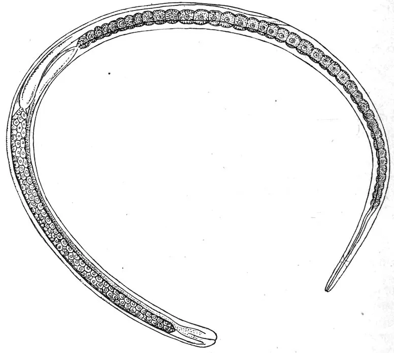
Fig. 31.—Female trichina from the intestine; 24 hours after infection. Enlarged. (After Leuckart, 1866a, pl. 1, fig. 1.)
Swine become infected by eating rats, and by eating scraps of pork
on the offal pile of slaughterhouses, or in swill.
Man becomes infected almost exclusively by eating pork and boar
meat. The rare infections which occur from eating other meat are
almost negligible.
Medical significance.—Trichiniasis or trichinosis refers to infection
with the trichina or flesh worm. Normally it occurs only in
mammals, chiefly carnivorous and omnivorous species, and it is
transmissible from any infected mammal to any other mammal susceptible
to it, in case the latter eats the uncooked flesh of the former.
103Symptoms.—In heavy infections there may be three more or less distinct
periods of the disease, corresponding to the three stages in the
life cycle of the parasite; but these stages are obscure in light or in
repeated infections. Profuse sweating may last during the entire
attack.
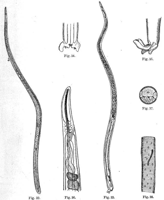
Fig. 32.—Gravid adult female trichina. Enlarged. (After Leuckart, 1866a, pl. 1, fig. 2.)
Fig. 33.—Adult male trichina from the intestine. Enlarged. (After Leuckart, 1866a, pl. 1, fig. 5.)
Fig. 34.—External genitalia of same. Enlarged. (After Leuckart, 1866a, pl. 1, fig. 7.)
Fig. 35.—The same with extruded cloaca. Enlarged. (After Leuckart, 1866a, pl. 1, fig. 8.)
Fig. 36.—Cephalic portion of a trichina showing central nervous system and anterior portion of intestinal canal. Greatly enlarged. (After Leuckart, 1866a, pl. 1, fig. 13.)
Fig. 37.—Transverse section of a female trichina. Greatly enlarged. (After Leuckart, 1866a, pl. 1, fig. 16.)
Fig. 38.—Young trichina embryo in a muscle fibre. Greatly enlarged. (After Leuckart, 1866a, pl. 2, fig. 1.)
Period of ingression: The adult parasites are in the intestine, hence
gastro-intestinal symptoms develop; irregular appetite, nausea, diarrhea
104or constipation, colicky pains; a temporary edema around eyes
about the eighth day; muscular pains begin.
Period of digression: This begins about the eighth to the fifteenth
day, sometimes later; young embryos are wandering to and attacking
the muscles, hence muscular symptoms (myositis) develop; painful
tension of muscles, especially biceps; members assume semiflexed
position; movements, chewing, swallowing, breathing, and speech
become difficult; eyes become fixed; fever.
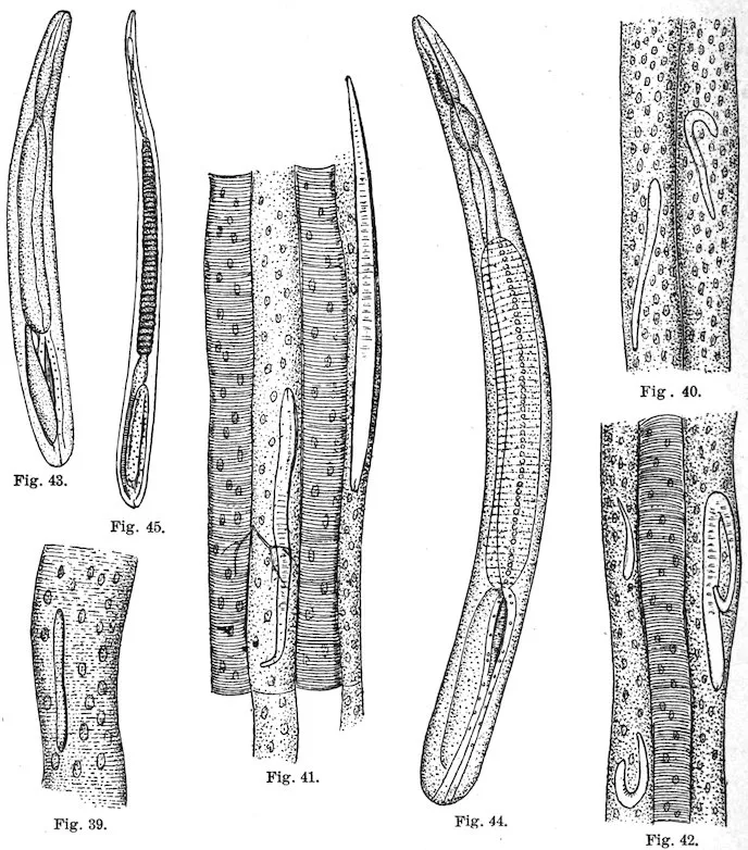
Figs. 39–42.—Later stages of same; the muscular structure is undergoing changes. (After Leuckart, 1866a, pl. 2, figs. 3, 6, 7, 8.)
Figs. 43–45.—Muscle trichinæ, 0.3 mm., 0.4 mm., and 0.6 mm. long. (After Leuckart, 1866a, pl. 2, figs. 10–12.)
Period of regression: The parasites become encysted in muscles.
All symptoms may increase, then gradually decrease; cachexia and
anemia resulting from malnutrition; pruritis, miliary cutaneous
eruptions; desquamation; about twenty-fourth day, a “second”
105edema develops, especially about the face; lungs may become edematous;
bronchial catarrh, pneumonia, or pleurisy may appear; gradual
recovery.
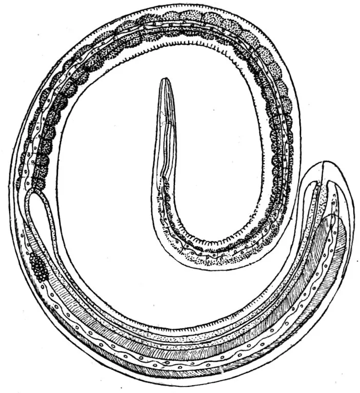
Fig. 46.—A female trichina from the muscle. Greatly enlarged. (After Leuckart, 1866a, pl. 1, fig. 12.)
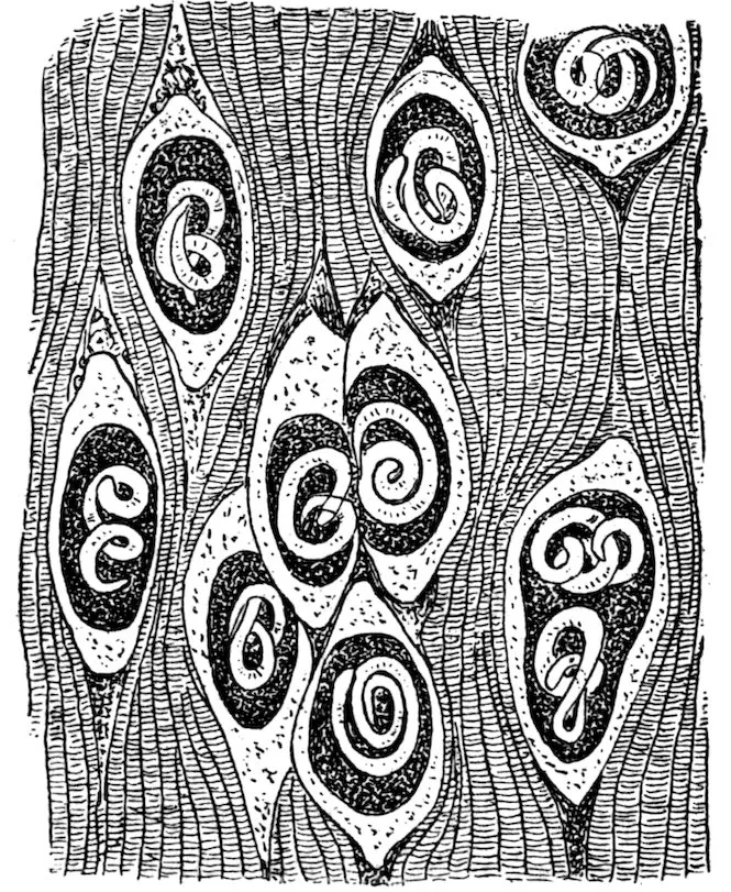
Fig. 47.—A piece of pork with encysted trichinæ. Enlarged. (After Braun, 1903, p. 251, fig. 195.)
Lethality.—The lethality varies from 0 to 100 per cent; it averaged
5.6 per cent in 14,820 cases collected from literature; it is dependent
upon amount of infection which remains in the body; low before
second and after seventh week, highest from fourth to sixth week.
Prognosis.—Better in cases having severe diarrhea in first stage.
106Complications and sequelæ.—Abortion, menstrual disturbances,
pneumonia, pleurisy, peritonitis.
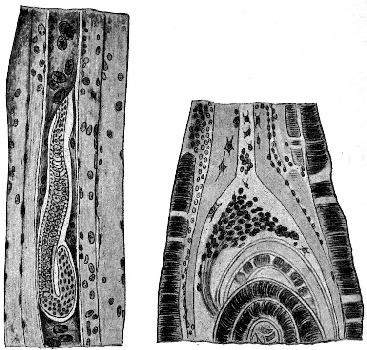
Fig. 48.—Section through a rat’s muscle; the infected muscle fiber has lost its striation, its nuclei are enlarged and increased in number. Greatly enlarged. (After Hertwig-Graham, see Braun, 1903, p. 284, fig. 212B.)
Fig. 49.—Portion of an isolated trichina cyst, at the pole of which connective tissue cells have wandered into the thickened sarcolemma. Greatly enlarged. (After Hertwig-Graham, see Braun, 1903, p. 284, fig. 212C.)
Clinical diagnosis.—Make microscopic examination:
(1) Of pork, if any has been left, to find encysted larvæ; if larvæ
are found, feed pork immediately to two or three guinea pigs or white
rats, to determine if the encysted larvæ are alive; kill one rat after
three days and examine intestinal content for adult; kill the second
rat after two weeks, the third rat after three weeks, and hunt for larvæ
in muscular portion of diaphragm. Even if live trichinæ are found
in intestine, an examination of the muscles may show that the worms
were too weak to reproduce, hence prognosis is favorable.
(2) Of patient’s blood, for increased proportion of eosinophiles.
(3) Of patient’s stools, for discharged adult worms, especially if
diarrhea is severe; dilute the fecal matter with warm water and pour
off whatever floats; place remainder in a shallow glass dish so that
it will not be over one-twelfth of an inch deep; move the dish gently
around over a dark background (such as dark paper), and hunt for
small hair-like objects; place these, if found, in a drop of water on a
107slide, cover with a cover slip, and examine under low power. Or, if
necessary, make a microscopic examination.
(4) Of small excised portion of patient’s deltoid, about three to
four weeks after infection, for encysted larvæ; cut a small piece
parallel to muscle fibers, tease this on a slide, add a drop of pure
water, or water and glycerine, cover with another slide, flatten
gently by pressure while examining under low power.
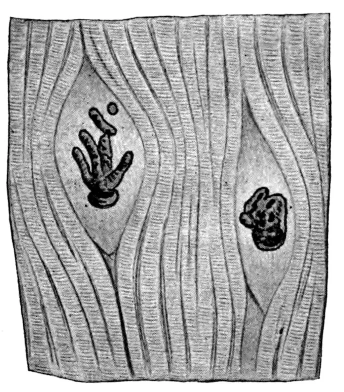
Fig. 50.—Calcified trichinæ in uncalcified cysts, from pork. Enlarged. (After Ostertag, see Braun, 1903, p. 285, fig. 213.)
Suspect trichinosis especially under following circumstances:
Several patients in same family or in same neighborhood, usually of
North German descent, show typhoid-like symptoms shortly after a
celebration (wedding, birthday party, etc.) at which pork was served.
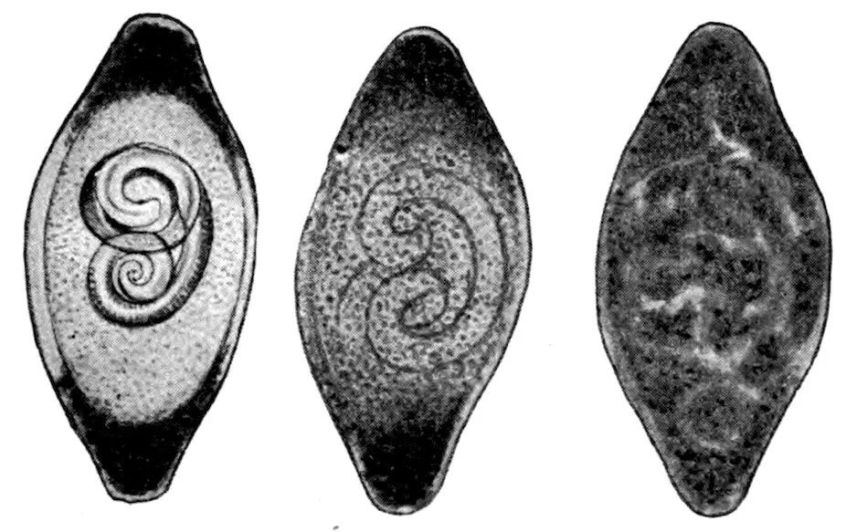
Fig. 51.—Three phases of calcification of trichinæ and their cysts, the changes starting from the poles of the cysts. Enlarged. (After Ostertag, see Braun, 1903, p. 285, fig. 214.)
Differential diagnosis.—Consider especially typhoid fever and
rheumatism.
Treatment.—Purge in early stage to carry away the adult worms
and thus eventually decrease the amount of muscular infection. No
treatment is known which can be relied upon to kill the larvæ in the
108muscles; benzine has been suggested. Stimulants may be given
to carry patients through until the larvæ encyst.
Prophylaxis.—Kill off rats and mice.—Educate public to eat pork
only when thoroughly cooked or thoroughly cured. A practical test
of cooking is the white color of the meat on being cut; if the cut
surface is reddish and serous, the pork is not sufficiently cooked to
kill trichinæ.
As a matter of practical experience, the microscopic inspection of
pork has not given the protection it is generally supposed to give.
Of 6,329 cases with 318 deaths reported for Germany during the years
1881–1898, 3,388 cases with 132 deaths are directly attributable to
faults in the inspection. This system directly increases the tendency
to eat raw pork, gives the public a false sense of security, and does
not give practical results commensurate with its expense.
Generic diagnosis.—Acanthocephala, Gigantorhynchidæ: Large worms with annulate
round to flat, tape-like body. Hooks with 2 roots and completely covered with transparent
chitin. Proboscis sheath a muscular apparatus, without cavity. Central
nervous system caudad of equator of proboscis sheath and eccentric. Lemnisci long,
cylindrical, with central canal.
Type species.—Gigantorhynchus echinodiscus (Diesing, 1851).
Specific diagnosis.—Gigantorhynchus (p. 108): Body attenuated anteriorly, with
fine transverse striæ or rings, or even constrictions which give the appearance of a
series of beads, except in the caudal fourth of body, which is nearly smooth and
cylindrical. Proboscis 425 to 450μ long, 176 to 190μ in diameter, armed with feeble,
very curved, 26μ long, hooks arranged more or less in quincunx and forming at most
15 transverse and about 12 longitudinal rows. Lemnisci more than a centimeter in
length, cylindrical, undulated posteriorly.
Male: Length 4 to 4.5 centimeters long; bursa campaniform.
Female: Length 7 to 8 centimeters (to 27 centimeters after Westrumb).
Eggs: Ellipsoidal, 85 by 45μ; external envelope thin, yellowish; middle envelope
very thick, colorless, homogeneous; inner envelope less thick, colorless, and quite
pliant. Embryo striated transversely in posterior two-thirds, and covered with
spines which increase in size toward anterior end of embryo, the anterior spines being
transformed into hooklets with prong and base.
Development: With beetles (Blaps mucronata) as intermediate host.
Habitat.—Small intestine of various small mammals; brown rat (Mus decumanus);
white rat (Mus norvegicus albus); M. fuscirostris; hamster (Cricetus frumentarius);
dormice (Myoxus quercinus or glis); field mole (Arvicola arvalis or agrestris?); Lemnus
arvalis; and Mustela putorius. It can also develop in man, as has been shown experimentally
by Grassi and Calandruccio (1888, 521–525).
Medical significance.—Grassi and Calandruccio report a doubtful
case of infection in a girl near Catania. Calandruccio infected
109himself experimentally by swallowing the young worms taken from
a Blaps. Twenty days later he was seized with severe pains which
increased on pressure; diarrhea followed, with ringing in the ears,
fatigue, and somnolence. Seventeen days later the characteristic
eggs were found in his stools, and twelve days later the symptoms
became so severe that he took 8 grams of extract of male fern; one
to two hours later he passed 53 of the parasites. For two days the
symptoms continued, on the second day fever developed, but all
symptoms disappeared on the third day.
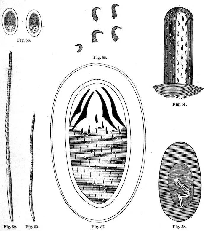
Fig. 52.—Gigantorhynchus moniliformis, female. ×2. (After Grassi & Calandruccio, 1888, p. 523, fig. 1.)
Fig. 53.—G. moniliformis, male. ×2. (After Grassi & Calandruccio, 1888, p. 523, fig. 2.)
Fig. 54.—Rostellum of G. moniliformis. Greatly enlarged. (After Grassi & Calandruccio, 1888,
p. 523, fig. 3.)
Fig. 55.—Hooks from same. Greatly enlarged. (After Grassi & Calandruccio, 1888, p. 523, fig. 4.)
Fig. 56.—Eggs of G. moniliformis, with embryo. Greatly enlarged. (After Grassi & Calandruccio,
1888, p. 523, fig. 5.)
Fig. 57.—Egg very greatly enlarged. (After Grassi & Calandruccio, 1888, p. 524, fig. 6.)
Fig. 58.—A young larva of G. moniliformis in a Blaps; the rostellum is invaginated and the larva is
surrounded by a thick inner jelly-like and thin outer cuticular covering. Enlarged. (After Grassi &
Calandruccio, 1888, p. 524, fig. 7.)
110
Species LINGUATULA SERRATA Frœhlich, 1789.
The larva of this parasite is found encysted in the entrails of
rabbits, cattle, and certain other animals, and it becomes mature in
the nasal cavities of canines.
Both the larva and the adult have been reported for man, and the
larva has been reported as occurring in Mus decumanus.
As canines are not fond of eating rats, the presence of the larval
tongue worm in the latter is of more academic interest than practical
importance, and although the theoretical possibility must be
admitted that a dog by eating rats might become infected with tongue
worms and eventually might transmit the infection to man, these
possibilities seem somewhat remote. Remote possibilities must also
be admitted to the effect that if a person ate a rat infected with
tongue worms this person might become infected.
THE FLEA AND ITS RELATION TO PLAGUE. — THEORIES AS TO TRANSMISSION OF PLAGUE. — INSECTS THAT HAVE BEEN SUSPECTED IN THE TRANSMISSION OF PLAGUE. — EXPERIMENTS PROVING THAT FLEAS CAN TRANSMIT PLAGUE. — THE BACILLUS IN THE FLEA. — HOW THE FLEA CLEARS ITSELF OF BACILLI. — REGIONAL DISTRIBUTION OF FLEAS ON RATS. — ANATOMY OF THE MOUTH PARTS OF THE CERATOPHYLLUS FASCIATUS. — OUTSIDE THE HEAD. — INSIDE THE HEAD. — DESCRIPTION OF FIGURE SHOWING MOUTH PARTS. — THE ACT OF BITING. — HOW THE FLEA INFECTS ITS HOST. — ENUMERATION OF FLEAS THAT HAVE BEEN FOUND ON RATS. — Family SARCOPSYLLIDÆ Taschenberg. — Genus ECHIDNOPHAGA Olliff. — Family PULICIDÆ Taschenberg. — Genus PULEX Linn. — Genus LŒMOPSYLLA Rothschild. — Genus CTENOCEPHALUS Kolenati. — Genus CTENOPSYLLUS Kolenati. — Genus NEOPSYLLA Wagner. — Genus HOPLOPSYLLUS Baker. — Genus HYSTRICHOPSYLLA Taschenberg. — Genus CTENOPTHALMUS Kolenati. — SYNOPSIS OF FLEAS COMMONLY FOUND ON RATS. — CERATOPHYLLUS FASCIATUS Bosc. — LŒMOPSYLLA CHEOPIS Rothschild. — CTENOPSYLLUS MUSCULI Dugés. — PULEX IRRITANS Linnæus. — CTENOCEPHALUS CANIS Curtis. — REFERENCES.
123
By Passed Assistant Surgeon Carroll Fox,
United States Public Health and Marine-Hospital Service.
1. Direct contagion from man to man.
2. Through slight abrasions of the skin, mucous membranes of
mouth, tonsils, nose, and conjunctiva receiving contaminated material.
3. Through the respiratory tract, from air contaminated with
dried infectious sputum or dejecta. (Possibly the cause of primary
pneumonic plague.)
4. Through the alimentary tract from food contaminated with
saliva or excretions from plague patients, or dejecta or the feet of
insects that have fed on plague material. In the case of rats, from
eating the carcasses of infected rats.
5. Infected clothes, soil, or houses.
6. Through the bites of insects, especially the flea.
It has been noticed for many years that an epidemic of plague in
man was associated with an epizootic of high mortality among rats,
but it was not until Yersin discovered the Bacillus pestis in 1894 that
the disease in man and rats was shown to be identical. The first
five theories are not satisfactory in explaining the epidemiology of
plague, and in 1897 Simond advanced the theory that plague was
carried by means of fleas. Hankin in 1898 also suggested an insect
as an intermediate host. This theory has been developed by Ashburton
Thompson, Gauthier and Raybaud, Liston, Verjbitski, and
others, and finally by the last Indian Plague Commission, whose
work makes a distinct advance in our knowledge of this subject.
The reader is referred to the work of this commission for a review
of the subject, which has been liberally used in the preparation of
this paper.[AD]
124
It is probable that all insects capable of sucking blood will take
the Bacillus pestis into their alimentary canal if they feed on a
septicæmic plague animal. Ogata suggested that not only the
flea but the mosquito also may be responsible for the transmission
of plague. Yersin, Hankin, and Nuttall have each demonstrated
the presence of Bacillus pestis in the dejecta of flies and ants; and
Nuttall and Verjbitski in the stomach and dejecta of the bedbug.
Hertzog found the bacilli in the Pediculus capitis taken from a child
which died of plague, and McCoy[201] has found the organism in lice,
Hæmatopinus columbianus, taken from a plague-infected squirrel.
The plague bacilli have been frequently demonstrated in rat fleas
taken from plague rats, and McCoy has shown its presence in the
flea (Ceratophyllus acutus) of the California ground squirrel (Citellus
beecheyi). The cockroach has also been thought to be instrumental
in spreading the infection by contaminating food. The presence
of bacilli in the stomach and dejecta of insects has not only been
proven microscopically but by animal inoculation as well.
Assuming that the relation between rat plague and human plague
has been proven without a doubt—that is, that an outbreak of human
plague is associated with an infection in rats, or, in other words, that
plague is primarily a disease of rats and secondarily a disease of
man—the theory that it is conveyed through an intermediate parasitic
host is the only one which will fulfill all the requirements, and
after a study of their habits we are able to exclude all of the parasites
but the flea as the active agent in its transmission.
Plague is rarely or never contracted either in rat or in man by
eating contaminated food. Therefore those insects like flies and
cockroaches, which are supposed to spread the infection by contaminating
food with their dejecta, need not be considered.
The habits of the domestic mosquitoes are such that while they
occasionally do bite animals they usually feed on the blood of man,
and are not known to feed where there is much hair, as there is on the
rat. This also applies to the bedbug. Verjbitski has shown experimentally
that bedbugs would not feed on rats until the animals were
shaved.
Pediculi are degenerate insects, their powers of locomotion being
limited. Their eggs are laid on and are attached to the hair of the
host. They are born, live, and die on the same host, and rarely
pass from one animal to another of a different species. It can not
be denied, however, that this parasite occasionally may be instrumental
in spreading plague from rat to rat. The Pediculus capitis,
if placed on a rat, will feed with avidity, but these insects are rarely
found upon rats in nature.
125We have no record of plague bacilli having been demonstrated in
mites commonly found on rats, but no doubt if search be made they
could be found after feeding on a septicæmic plague rat. These mites,
however, always confine themselves closely to their particular host
and are not known to bite man. The tiny itch mite (Notoedres alepis,
Railliet and Lucet) producing rat scabies has, according to Schumann,[202]
been known to cause a cutaneous lesion in man, but this mite
need not be considered from a plague standpoint.
The flea, on the other hand, lives but part of the time on its host,
its eggs developing in the nests or runs of the animal. Again, this
insect does not confine itself to one particular species of host only,
as frequently the flea of one animal is found on an animal of an entirely
different species. Unlike the lice, they are very active and can
readily move from place to place. Not only that, but it has been
frequently demonstrated that the fleas of rats and of other animals
would readily take to man, especially if their natural host was
scarce. That rat fleas will bite man has been demonstrated by
Gauthier and Raybaud, working with the Leomopsylla cheopis;
Tidswell, Lœmopsylla cheopis and Ceratophyllus fasciatus; Liston,
Lœmopsylla cheopis; Tiraboschi, Lœmopsylla cheopis; Indian Plague
Commission, Lœmopsylla cheopis; and McCoy and Mitzmain[203],
Lœmopsylla cheopis, Ceratophyllus fasciatus, and Ctenopsyllus musculi.
It has generally been considered that the Ctenopsyllus musculi,
above all others, would not bite man, but the last-named observers
showed that it would occasionally feed, although it would not live
long, in captivity. One of the fleas, a Ceratophyllus fasciatus, was
kept alive by Mitzmain for over four months on man’s blood alone.
By a series of experiments carried out in specially constructed
cages and go-downs where healthy rats in the absence of fleas were
brought in contact with plague-infected rats, the Indian Plague Commission
showed that the healthy rats would not contract the disease,
notwithstanding the fact that they were not only in intimate contact
with the sick rats, but also with the contaminated food and excreta
of the sick rats. They then showed that if fleas were introduced the
healthy rats would contract plague, the rate of progress of the epizootic
being in direct proportion to the number of fleas present. By
hanging cages containing healthy rats in cages holding infected rats,
but above the jumping distance of a flea, it was shown that the
healthy rats would remain well, while those in cages hung within 2
inches from the ground would contract plague. Thus they excluded
aerial infection. They also found that if fleas were excluded young
rats could suckle a plague-infected mother without contracting the
disease.
126Guinea pigs were allowed to run in houses where cases of human
and of rat plague were known to have occurred and where many
fleas were present. These rodents served as good traps for the fleas
and 29 per cent of them contracted plague.
Most of the experiments of the Indian Plague Commission were
done with the Indian rat flea, the Lœmopsylla cheopis, but they also
performed 27 experiments with the cat flea, Ctenocephalus felis, with
negative results; 35 experiments with the human flea, Pulex irritans,
3 of which were successful; and 2 experiments with the Ceratophyllus
fasciatus, the common rat flea of Europe and North America, both
of which were successful.
In San Francisco a few experiments under purely experimental
conditions have been carried on by McCoy to determine the ability
of the squirrel flea, the Ceratophyllus acutus, to transmit plague.
Fleas that had been previously fed on the blood of a septicæmic
plague-infected squirrel were then allowed to feed from test tubes on
healthy guinea pigs. While the feces of some of these fleas up to
four days, when inoculated into guinea pigs, were proven to be infective,
none of those guinea pigs on which the fleas were allowed to
feed contracted plague. It might be said, however, that in no case
were they seen to eject feces while feeding, the significance of which
will be apparent later.
The Indian Plague Commission found that the average capacity of
the rat flea’s stomach (Leomopsylla cheopis) was 0.5 cubic millimeter,
and that it might receive as many as 5,000 germs while imbibing blood
from a plague rat. They further found that the bacillus would multiply
in the stomach of a flea and that the percentage of fleas with
bacilli in the stomach varied with the season of the year. In the
epidemic season the percentage was greatest for the first four days,
and on one occasion the stomach was found filled with Bacillus pestis
on the twentieth day. In the nonepidemic season no plague bacilli
were found in the stomach after the seventh day. They also found
that in the epidemic season fleas might remain infective up to fifteen
days, while in the nonepidemic season but seven days, and in the
latter case the percentage of infection in animals was much less than
in the epidemic season. They showed that while one flea was occasionally
able to carry the infection this was not usual. It was found
that both the males and the females were capable of transmitting
the disease.
After a number of dissections they were unable to demonstrate the
presence of bacilli anywhere but in the stomach and rectum. At no
time was anything found in the body cavity or salivary glands and
127but rarely in the œsophagus, and then only when the flea was killed
immediately after feeding.
We have in San Francisco examined quite a number of serial sections
of plague-infected fleas with the same result as obtained by the
Indian Plague Commission. The bacilli are readily demonstrated,
sometimes in enormous numbers, in the gizzard, stomach, and in the
rectum, but at no time have they been found in the body cavity, the
salivary glands, or the ovary. In fact, as we are dealing with a
vegetable organism and not an animal organism, like the Plasmodium
malariæ, we could hardly expect to find any biologic change, except
simple multiplication, occurring in the intermediate host.
Some explanation is necessary as to why the bacilli eventually
disappear from the flea, although they seem to multiply during the
first few days. It is evident that the peristaltic action of the stomach
during the course of digestion forcing the blood at the proper time
into the rectum, finally to be ejected from the body, would in itself
cause many bacilli to be discharged, but naturally a few would remain
to multiply indefinitely. The bacteriacidal action of the blood is
soon lost after entering the flea’s stomach, but it has been shown by
proper staining that the leucocytes after the first feeding with healthy
blood contain numbers of Bacillus pestis, and it seems probable that
this phagocytic action is important in the cleansing process. It has
been shown that after successive feedings on the blood of noninfected
animals the power of phagocytosis is increased, and that successive
feedings on the fresh blood of animals that have been immunized
against plague still further assists and hastens the process. When
there is a frequent introduction of fresh normal or immunized blood
its bactericidal action is also instrumental in the cleansing process.
The location of the primary bubo in a case of plague, human or
rodent, depends upon the site of inoculation, for that group of glands
will first enlarge which has direct lymphatic connection with the area
through which the Bacillus pestis enters the animal organism. The
British Indian Plague Commission found that 72 per cent of their
naturally infected rats and 61 per cent of the rats experimentally
infected by fleas had cervical buboes, while in no instance in over
5,000 plague rats was a mesenteric bubo encountered. On the other
hand, where plague was induced through feeding healthy rats with
the carcasses of plague rats a mesenteric bubo was found in 74.5 per
cent of those infected and a cervical bubo in 36 per cent. In San
Francisco in naturally infected rats a primary mesenteric bubo has
never been seen, and a cervical bubo has been seen but once. These
128figures show conclusively that naturally infected rats are not infected
by feeding. It is curious, as has been pointed out by McCoy[204], that
such a large percentage of cervical buboes should be found in India,
while a cervical bubo has been seen but once in naturally infected
animals in San Francisco. Here the axillary and inguinal buboes are
the rule. The Indian Commission found that the commonest situation
to find fleas on guinea pigs was the head and neck. They
combed 53 guinea pigs to determine the regional distribution of fleas,
and found that 65.3 per cent were taken from the neck and head.
This would account for the preponderance of cervical buboes in
guinea pigs observed in their work, and inferentially for the preponderance
of cervical buboes found in naturally infected rats.
Thinking that the predominating rat flea in San Francisco, the
Ceratophyllus fasciatus, might be the carrier of the infection and that
it might prefer a different part of the body than the Lœmopsylla
cheopis, McCoy and Mitzmain carried on a series of investigations to
determine the regional distribution of fleas on the rat’s body, but
this has shown that while the Ctenopsyllus musculi seems to be generally
confined to the head and neck, the Ceratophyllus fasciatus and
Lœmopsylla cheopis are almost invariably taken from the body,
especially from the pelvic region.
The following description differs somewhat from that given by
Wagner[205] and the description found in the Journal of Hygiene, both
of which, however, refer to different species of Siphonaptera.
The mouth parts may be divided into those inside and those outside
of the head.
The epipharynx, or pricker, is a long, slender, hollow organ. Its
cavity is closed distally, and proximally connects with the hoemocoel.
It is made up of a dorsal and a ventral portion. Its dorsal portion
ends just within the head. Its ventral portion is grooved and is
continuous with the posterior wall of the aspiratory pharynx. Its
distal extremity is slightly expanded, forming a stylet for piercing,
while the little papillæ seen along the anterior surface in many species
are absent in this one. Laterally there is a membranous expansion
which interlocks with a similar expansion on the mandibles, forming
a tube, through which the blood is sucked.
The mandibles are two in number, articulating just within the
head, so that they are capable, of independent movement. They are
serrated at their distal extremities. Above, within the head, the
anterior portion of the mandibles ends just behind the beginning of
the hypopharynx, to which it is connected, becoming practically continuous
with that organ. The posterior portion is attached to its
129basal element. Each mandible contains a groove, forming practically
a closed canal, which becomes continuous with the exit duct of
the salivary pump.
The rostrum (labial palpi) forms a protection and guide to the
mandibles and epipharynx. Its first portion is unpaired and articulates
within the head, with its basal element. At the apex of its
first portion it bifurcates, forming a paired organ, which is divided
into a varying number of pseudojoints, depending on the species of
the flea. As it is a chitinous structure, these pseudojoints, areas in
which there is little chitin, enable it to double up as the mandibles
and epipharynx are inserted into the skin. At the apex of the rostrum
are some tactile hairs.
The maxillæ are triangular chitinous plates situated on either side
of that portion of the head where the biting organs emerge. These
structures serve to protect the origin of the epipharynx and mandibles,
rest upon the cutaneous surface in the act of biting, thereby steadying
the head and serving as a fulcrum when the flea withdraws its biting
apparatus when through feeding. The maxillæ have their palpi,
which are four jointed, paired organs coming out at the anterior lower
angle of the head. Their function is sensory.
The hypopharynx is a chitinous plate forming part of the floor of
the aspiratory canal. To its under surface are attached the muscles
which operate the salivary pump. Its lower portion is connected to
the mandibles, while its upper portion is connected to the posterior
portion of the floor of the aspiratory pharynx by a membranous
ligament.
The aspiratory pharynx extends from the connection of the hypopharynx
with the mandibles to the œsophageal commissure. In a
general way it first passes upward and then turns, passing backward.
Its roof is formed by the continuation of the ventral surface of the
epipharynx, while its floor is formed by the hypopharynx below and
above by the chitinous layer which is continuous with the œsophagus.
The anterior end of this particular portion curves strongly downward,
where it is attached to the upper portion of the hypopharynx by a
membranous ligament. In a general way it may be divided into a
vertical and longitudinal portion. The longitudinal portion expands
laterally, so that its capacity is greatly increased when dilated. Into
the floor of this longitudinal portion empties the vertical part of the
aspiratory pharynx, and at the junction of the two there seems to be
a valvular arrangement, preventing blood from escaping after it has
entered the upper part of the aspiratory canal. The œsophagus
starts at the œsophageal commissure and ends in the gizzard. It is
130not expanded as in some insects, forming a gullet, but is practically
the same diameter throughout its entire extent. It is lined with
chitin, surrounded by a delicate basement membrane.
The gizzard is a mushroom-shaped organ, opening into the stomach
and receiving the contents of the œsophagus and the aspiratory
pharynx. From its anterior concave inner surface project a number
of finger-like processes that arise from a basement membrane. They
are lined with chitin, and each one near its base contains an elongated
nucleus. These processes reach to the center of the gizzard and in a
general way point towards the opening into the stomach. The gizzard
is surrounded by circular bands of muscle fibers. Its function
is not entirely understood. Wagner[205] has pointed out that these
processes may act as whips to defibrinate the blood. It is more
probable that their action is mainly valvular, preventing regurgitation
of blood from the stomach.
The stomach of a flea is large and is capable of great distention.
It is composed of a layer of secretory cells, resting on a basement membrane,
the organ being surrounded by muscle fibers passing in
different directions. The epithelial surface is thrown into little
projections like villi. As absorption occurs in the stomach, these
villi, or projections of the epithelial cells, may serve to increase the
absorptive surface as well as serving a glandular function. At the
anterior end of the stomach are the cecal glands.
The intestine is short, receives the excretion from the Malpighian
tubules, and ends in the rectum. In the rectum may be seen the
so-called “rectal glands.” All of the alimentary canal, with the exception
of the stomach, is lined with chitin. The stomach and the
rectum are capable of peristaltic movement.
The salivary glands, four in number, two on each side of the anterior
part of the stomach, are simple acinous glands, lined with a single layer
of secreting cells. The lumen of the glands is large and acts as a
reservoir for the salivary secretion. The ducts from these glands
unite to form a single duct which passes beneath the subœsophagal
ganglion and empties into the salivary pump. This duct is lined on
its inner surface by a spiral arrangement of chitin, giving it a very
characteristic appearance.
- 1.
- Epipharynx.
- 2.
- Mandibles.
- 3.
- Rostrum, paired portion.
- 4.
- Rostrum, unpaired portion.
- 5.
- Maxilla.
- 6.
- Maxillary palpus.
- 7.
- Salivary grooves.
- 8.
- Basal element of rostrum.
- 9.
- Basal element of mandibles.
- 10.
- Salivary pump.
- 11.
- Salivary duct.
- 12.
- Vertical portion, aspiratory canal.
- 13.
- Longitudinal portion, aspiratory canal.
- 14.
- Œsophagus.
- 15.
- Œsophageal ganglia.
- 16.
- Muscles operating aspiratory canal.
- 17.
- Hypopharynx.
- 18.
- Muscles operating salivary pump.
- 19.
- Ligament connecting hypopharynx with floor of aspiratory canal.
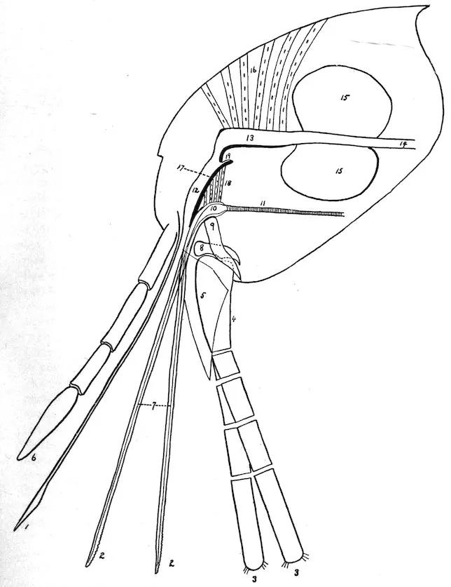
Plate I.
MOUTH PARTS OF CERATOPHYLLUS FASCIATUS.
131
The epipharynx, or pricker, makes an opening into the skin, through
which the mandibles are inserted. These organs, by means of their
serrations and independent movement, then enlarge the opening as
with a saw, permitting them, with the epipharynx, to pass deeper
and deeper until the points of the maxilla rest upon the cutaneous
surface. The labial palpi serve as a protective case when the organs
are not in action. When in action they serve as a guide to the
piercing organs, but are not inserted into the skin. They double up
like a bow, on each side, the bend of the bow becoming greater and
greater as the biting apparatus passes deeper and deeper. Mitzmain[206]
has pointed out that the spring-like action of this bow may assist
the flea to withdraw the mandibles and epipharynx.
During the process of penetration the salivary pump receives saliva
from the salivary glands and pumps it down, through the channel
in the mandibles, into the wound. It will be seen that the hypopharynx,
being attached above by a membranous ligament and connected
intimately with the mandibles below, moves downward with
these organs as they pass through the skin. At the same time the
muscles attached to its under surface and the salivary pump contract,
enlarging the lumen of the pump. When the mandibles are retracted
the salivary pump collapses, thereby forcing the saliva out with the
movement upward of the mandibles. At the proper time the
muscles operating the aspiratory pharynx contract, drawing the
canal open and aspirating blood through the canal made by the
approximation of the epipharynx and mandibles and into the
aspiratory pharynx. When full, the muscles relax from before
backward and the pharynx, by means of the elastic reaction of its
chitinous lining, contracts and forces the blood backward through the
gizzard and into the stomach. It has already been pointed out that
the finger-like processes in the gizzard probably act as valves to
prevent regurgitation from the stomach.
132
The exact method by which the flea can transmit plague from
animal to animal has, in our opinion, never been satisfactorily
explained. There have been several explanations offered: First,
that the rat may eat the flea. Miller[207] has found that the
Hepatazoon perniciosum is transmitted from rat to rat through the
rat eating the mite, Lelaps echidninus, which acts as the intermediate
host. We know, however, that when a rat is fed on plague material
a mesenteric bubo is the rule, while in naturally infected rats a
mesenteric bubo is a rare condition. This, then, negatives the possibility
of plague being contracted through eating the flea.
Another explanation is that the infection comes from the saliva
injected at the time of biting. We have already stated that after
repeated examinations, both by dissecting out the salivary glands
and by serial sections of the entire flea, plague bacilli have never
been demonstrated in these glands or anywhere outside of the
alimentary tract.
Another explanation has been advanced, that the bacillus is introduced
by the contaminated mandibles. It is not possible to exclude
this as a means of infection, although the Indian Plague Commission
made numerous investigations and was unable to demonstrate the
bacillus on the mandibles.
The possibility of infection taking place by regurgitation from the
stomach has also been considered. As the stomach is guarded by
the finger-like processes in the gizzard which seem to act as competent
valves, and as the movement of the blood aspirated by reason of
the mechanism already explained is in a backward direction, it
would seem improbable that there is any regurgitation from the
stomach.
The most plausible explanation that has been advanced has been
based on an observation that blood-sucking insects at the time of
biting frequently eject a drop of blood from the rectum. We know
that the rectum may contain numerous plague bacilli, and it is supposed
that this blood ejected in the vicinity of the bite is either
brought in contact with the slight wound by the feet or mandibles
of the flea itself or is rubbed in as a result of scratching. Verjbitski
has shown that an emulsion of the feces of fleas or any plague material
when placed upon the bitten part before the expiration of twenty-four
hours is sufficient to give the animal plague. After twenty-four
hours the animals did not develop plague, it being supposed that
the slight wound in the skin made by the biting apparatus had healed.
It is probable that this ejection of blood is purely accidental and does
not necessarily occur at the time of biting, but it is likely that the
insect had just previously had a full meal, which had been digested
and passed into the rectum. In the many biting experiments done
133by McCoy and Mitzmain they report never having seen this ejection
of rectal contents taking place. It might also be stated that where
they used plague-infected fleas none of the animals developed plague
after being bitten.
Various writers have reported the following fleas taken off rats:
Genus DERMATOPHILUS.
1. Dermatophilus cæcata Enderlein.—Seventeen specimens (females)
were found by Doctor Enderlein on the skin behind the ears
of a specimen of Mus rattus from Saopaulo, Brazil.
2. Echidnophaga gallinacea Westwood.—Tiraboschi has found this
flea on the Mus rattus in Italy.
3. Echidnophaga rhynchopsylla Tiraboschi.—This flea has been
taken in Italy from Mus rattus and Mus alexandrinus. It has been
described by Rothschild under the name of Echidnophaga murina.
Genus CERATOPHYLLUS Curtis.
4. Ceratophyllus fasciatus Bosc.—This is the common rat flea of
Europe and the United States. It has also been found in Cape
Town, Australia, and is occasionally found on rats in India.
5. Ceratophyllus londiniensis Rothschild.—This flea has been taken
off mice in England; off rats in Italy (Ceratophyllus italicus Tiraboschi)
and has been found once on Mus rattus in San Francisco,
Cal.
6. Ceratophyllus acutus Baker.—This is the common flea of the
California ground squirrel; and has been taken off Mus norvegicus in
San Francisco, Cal.
7. Ceratophyllus anisus Rothschild.—This flea has been described
by Rothschild from Yokohama, Japan, taken off Felis sp. One
specimen was found in San Francisco, Cal., taken off Mus norvegicus.
8. Ceratophyllus niger Fox.—This flea is commonly found in San
Francisco, Cal., in chicken yards and sparrows’ nests and has also
been found on rats, Mus norvegicus, and on man.
9. Ceratophyllus consimilis Wagner.
10. Ceratophyllus lagomys Wagner.
11. Ceratophyllus mustelæ Wagner.
12. Ceratophyllus penicilliger Grube.
These fleas have been taken off Mus norvegicus in Europe.
134
13. Pulex irritans Linn.—This flea is widely distributed throughout
the world, and while essentially the human flea has been found
on many different species of animals and has frequently been encountered
on rats. A very large number of specimens have been
taken off rats in San Francisco, Cal.
14. Lœmopsylla cheopis Rothschild.—This is the common rat flea
in tropical and subtropical countries. It has also been found in
seaports of the temperate zone, where it has been brought by ship
rats. Ninety-eight per cent of the rat fleas in India are of this
species. It has been found in Australia, where it was described by
Tidswell under the name of Pulex pallidus. In the Philippine Islands,
where it was described by Hertzog as the Pulex philippinensis. It
has been found in Brazil, where it was described by Baker as Pulex
brasiliensis, and Tiraboschi has found it in Italy, where it has been
described as the Pulex murinus. This flea has been frequently
found on man in India.
15. Ctenocephalus canis Curtis.—This is the common dog flea
found in many parts of the world and is frequently taken off rats.
16. Ctenocephalus felis Bouché.—This is the common cat flea and
is also a widely distributed species. Frequently taken off rats.
17. Ctenopsyllus musculi Dugés.—In England this flea is commonly
found on the domestic mouse. It has a wide distribution and has
been found on rats and mice in Europe, South Africa, India, Australia,
Mexico, and other places, and has been taken off Mus norvegicus,
Mus rattus, and Mus musculus in San Francisco, Cal.
18. Neopsylla bidentatiformis Wagner.—This flea has been taken
off Mus norvegicus in the Crimea.
19. Hoplopsyllus anomalus Baker.—This is one of the common
groundsquirrel fleas of California and has been found on Mus norvegicus
in San Francisco and Palo Alto, Cal. That these squirrel fleas
are occasionally found on rats is interesting from the fact that
plague has been demonstrated both in rats and the ground squirrel
in California.
135
20. Hystrichopsylla tripectinata Tiraboschi.—Reported by Tiraboschi
from Mus musculus in Italy.
21. Ctenopthalmus agyrtes Heller.—Taken off Mus Norvegicus in
England.
The results of the identification of 19,768 fleas in San Francisco and Oakland, Cal.
| SAN FRANCISCO, 1908. |
|---|
| Host: Mus norvegicus. |
|---|
| Month. |
C. fasciatus. |
L. cheopis. |
P. irritans. |
Cten. musculi. |
Cten. felis, Cten. canis. |
| Male. |
Female. |
Male. |
Female. |
Male. |
Female. |
Male. |
Female. |
Male. |
Female. |
| April to July 31 |
1,343 |
2,510 |
485 |
837 |
31 |
76 |
78 |
211 |
16 |
31 |
| August |
489 |
883 |
145 |
228 |
156 |
206 |
27 |
90 |
17 |
22 |
| September |
543 |
1,180 |
655 |
930 |
339 |
387 |
33 |
109 |
46 |
119 |
| October |
254 |
435 |
509 |
652 |
59 |
64 |
9 |
45 |
6 |
18 |
| November |
129 |
252 |
256 |
288 |
52 |
69 |
20 |
54 |
6 |
6 |
| |
2,758 |
5,260 |
2,050 |
2,935 |
637 |
802 |
167 |
509 |
91 |
196 |
| Host: Mus rattus. |
|---|
| |
23 |
43 |
3 |
3 |
0 |
0 |
17 |
16 |
1 |
0 |
| |
4 |
7 |
1 |
0 |
9 |
16 |
3 |
3 |
0 |
0 |
| |
27 |
50 |
4 |
3 |
9 |
16 |
20 |
19 |
1 |
0 |
| Host: Mus musculus. |
|---|
| |
4 |
10 |
1 |
0 |
0 |
0 |
2 |
10 |
0 |
0 |
| |
11 |
10 |
1 |
6 |
4 |
4 |
0 |
3 |
1 |
1 |
| |
15 |
20 |
2 |
6 |
4 |
4 |
2 |
13 |
1 |
1 |
| OAKLAND, CAL., 1909. |
|---|
| Host: Mus norvegicus. |
|---|
| February |
135 |
304 |
166 |
178 |
1 |
1 |
229 |
506 |
1 |
3 |
| March |
253 |
456 |
167 |
215 |
2 |
5 |
125 |
243 |
1 |
2 |
| April |
227 |
479 |
105 |
129 |
0 |
1 |
62 |
143 |
0 |
1 |
| |
615 |
1,239 |
438 |
522 |
3 |
7 |
416 |
892 |
2 |
6 |
| Host: Mus alexandrinus. |
|---|
| April |
1 |
5 |
0 |
0 |
0 |
0 |
0 |
0 |
0 |
0 |
136This does not include a few other specimens of different species
taken from Mus rattus and Mus norvegicus, which have been included
under the heading of “Enumeration of fleas which have been found
on rats.”
| A. WITHOUT A COMB OF SPINES ON THE PROTHORAX OR THE HEAD. |
| 1. |
Two bristles on the gena, an ocular bristle placed below the eye, an oral bristle placed just above root of maxilla. Mesothorax not divided by an internal incrassation. Claspers in male forming prominent hump, claw like and covered by hairy flap. An irregular row of about 10 teeth on inner side of hind coxa |
Pulex irritans. |
| 2. |
Two bristles on gena, an ocular placed in front of and just above middle of eye, an oral bristle placed just above root of maxilla. Mesothorax divided by internal incrassation, claspers not forming prominent hump, not claw like, not covered by hairy flap. A regular row of about six teeth on inner side of hind coxa, |
Lœmopsylla cheopis. |
| AA. WITH A COMB OF SPINES ON PROTHORAX BUT NOT ON HEAD. |
| 3. |
Three bristles on lower genal row, upper genal row represented by three or four small bristles running along anterior margin of antennal groove. Eye present, about five hairs on second joint of antenna, not as long as third joint. Maxillary palpi not as long as labial palpi. Labial palpi reach to apex of fore coxa. Spines on posterior tibia in pairs of about five groups. Head of male flattened on top, |
Ceratophyllus fasciatus. |
| AAA. WITH A COMB OF SPINES ON THE PROTHORAX AND ON THE HEAD. |
| 4. |
Eye present, seven spines on lower margin of gena. Spines on posterior border of tibia in pairs |
Ctenocephalus canis or felis. |
| 5. |
Eye absent, four spines on hind margin of gena. Spines on posterior tibia single and in a close set row |
Ctenopsyllus musculi. |
| Description of Plate II. |
|---|
| Fig. 1. Clasping organs of male. |
P |
Process. |
| F |
Finger. |
| M |
Manubrium. |
| IX St |
Ninth Sternite. |
| Fig. 2. Head of female. |
|
|
| Fig. 3. Terminal abdominal segments, female. |
8 T |
Eighth Tergite. |
| 8 St |
Eighth Sternite. |
| 10 T |
Tenth Tergite. |
| 10 St |
Tenth Sternite. |
| Sp |
Spermatheca. |
| Sty |
Stylet. |
| Fig. 4. Hind tibia. |
|
|
Head.—Evenly and gently rounded in the female, flattened on top
in the male. Frontal notch distinct. Eye present, placed low down
in head. Gena acutely pointed posteriorly. Maxilla triangular.
Maxillary palpi not as long as the labial palpi. Labial palpi reach
to apex of anterior coxa, 5-jointed. Antennal groove in the male
reaches to top of head, in the female to within one-third. There are
3 bristles on the lower genal row, the middle of which is the smallest,
while the upper genal row is represented by 3 small bristles, extending
along the edge of the antennal groove. In the male the lowermost
bristle is frequently paired. There are several fine hairs above the
eye. The occiput contains the normal row of apical bristles, the
lowest of which is the largest. There is one bristle back of the middle
of the antennal groove and a number of fine hairs along the posterior
margin of the antennal groove. The antenna is 3-jointed, the first
joint contains a row of about 5 very short fine hairs, while the second
joint contains about 5 not as long as the third joint.
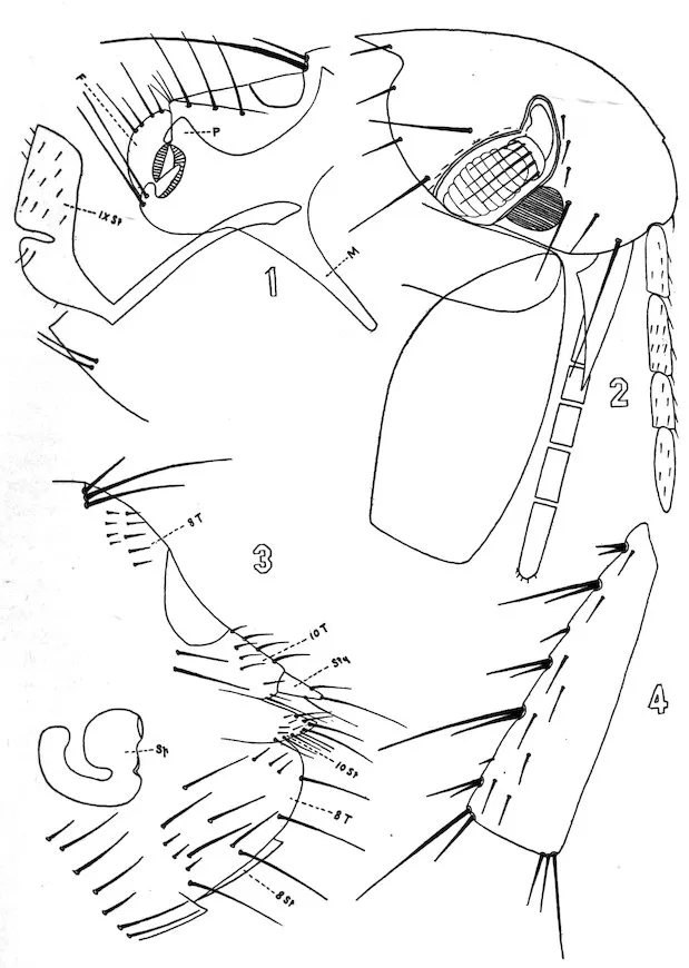
Plate II.
CERATOPHVLLUS FASCIATUS, BOSC.
137Thorax.—The pronotum has one row of about 10 bristles, and a
ctenidium composed of about 16 or 18 spines. The mesonotum has
a posterior row of about 10 long bristles and there is an anterior row
of more numerous smaller ones. The metanotum has also a posterior
row of about 10 large bristles, with an anterior row of more numerous
smaller ones, while still anterior to this there are 5 or 6 still smaller
bristles. The metathorax contains 8 or 10 bristles which are small
anteriorly, larger posteriorly. On the sternum of the metathorax
there are 2 large bristles, while the episternum has 3 smaller ones.
On the epimerum are 2 bristles placed anteriorly and 3 or 4 posteriorly,
one of which is on the apical margin.
Abdomen.—The first stigma is nearly in line with those of the other
abdominal segments. There are two rows of bristles on the abdominal
tergites, a posterior of about 12 or 14 and an anterior of smaller, less
numerous bristles. The antipygidial bristles in the female are 3 in
number on each side, of which the middle is the longest, and the inner
one the smallest. The male has but 2 antipygidial bristles on each
side. The sternites from the third to the sixth have a single row of
about 10 bristles, while the seventh has about 12. The metanotum
has 2 teeth on each side, as have the first and second abdominal
tergites. The third and fourth abdominal tergites have 1 tooth on
each side.
Legs.—The fore coxæ are normally clothed. The fore femur has
on the outer side 11 or 12 fine bristles irregularly disposed, while on
the mid femur there is a row of about 3 to 5 bristles on the inner surface.
The hind coxa has no patch of spines on the inner side, while
on the inner surface of the hind femur there is a row of about 5 to 7
bristles. The spines on the posterior tibia are in pairs of six groups,
while on the outer surface there is a row of about 7 bristles. None
of the apical bristles of the tarsi are as long as the next succeeding
joint. The fifth tarsal joints on all the legs have 5 lateral spines.
138Length of joints of tarsi:
| Mid tarsi (♂) |
8 |
7 |
4½ |
3 |
7 |
| Hind tarsi |
18 |
11 |
7 |
4 |
8 |
| Mid tarsi (♀) |
8 |
7 |
5 |
3½ |
7 |
| Hind tarsi |
21 |
13 |
8 |
5 |
8 |
Modified segments.—(♂). The manubrium of the claspers is straight
and narrow, while the process extends upward as a short, blunt cone,
where at the tip there are several fine hairs. The lower margin is
evenly and gently rounded. The finger is short, extending but a
little above the process. It is concave on its anterior surface and
convex on its posterior, and from the posterior margin there are 2
large and 2 small bristles alternating. Two long heavy bristles arise
from the process below the insertion of the finger. The ninth sternite
is broad, with a deep sinus in its posterior border. Its lateral
surface contains numerous fine hairs, these hairs being somewhat
larger just beneath the sinus. Along the dorsal border of the tenth
sternite there are 3 heavy bristles in line. At the tip of the tenth
tergite there is one heavy bristle. Besides these heavy bristles in
this segment there are numerous fine hairs.
(♀) The eighth tergite contains just anterior to the sensory plate
about 12 small hairs while just beneath the sensory plate there are 2
long bristles. Lower down there is a patch of about 6 bristles and
on the apical margin 4 to 6. The stylet is short, cylindrical, slightly
larger at the base than at the tip, where there is a long bristle. On
the under surface arises a fine hair. Substylar flap (tenth sternite)
has along its margin numerous hairs.
| Description of Plate III. |
|---|
| Fig. 1. Clasping organs of male. |
P |
Process. |
| M |
Manubrium. |
| F |
Finger. |
| IX St |
Ninth Sternite. |
| Fig. 2. Head of female. |
|
|
| Fig. 3. Terminal abdominal segments, female. |
8 T |
Eighth Tergite. |
| 8 St |
Eighth Sternite. |
| 10 T |
Tenth Tergite. |
| 10 St |
Tenth Sternite. |
| Sp |
Spermatheca. |
| Fig. 4. Hind coxa inner surface. |
|
|
Head.—Abruptly rounded. Flattened on top in ♂. Eye present.
No ctenidia on head. Antennal groove in the ♀ reaches to within
one-third of the top of the head. In ♂ reaches to top of head.
Gena obtusely pointed posteriorly. Maxilla triangular. Maxillary
palpi are not as long as labial palpi. Labial palpi reach to apex of
fore coxæ, 4-jointed. Anterior edge of antennal groove overlapped
by chitinous flap. On posterior edge of antennal groove are a
number of small bristles, these being most distinct in the male. The
first antennal joint in the male contains 4 or 5 hairs at its outer edge,
while transversely there is a row of several fine hairs. The second
joint has a row of fine hairs not as long as the third joint. Divisions
marking separations of third joint most pronounced on dorsal edge.
Two bristles on gena. The oral bristle placed low down just above
the base of the maxilla; the ocular bristle in front and just above
the middle of the eye. Six bristles on the posterior margin of the
occiput on each side with 2 back of the antennal groove.
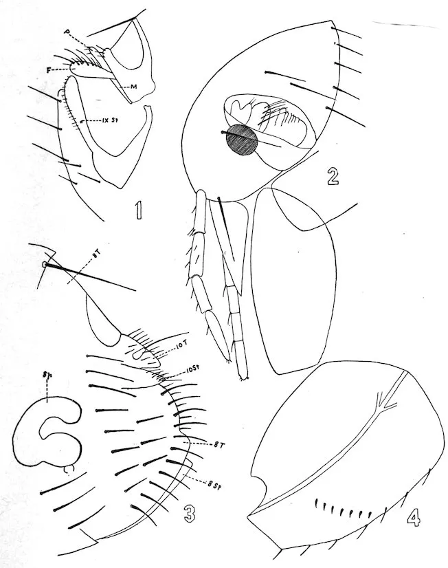
Plate III.
LŒMOPSYLLA CHEOPIS, ROTHSCHILD.
139Thorax.—The pronotum is without a ctenidial comb, and has one
row of about 14 bristles. The mesonotum, the broadest of the three
thoracic nota, also has a single row of about 12 bristles. The metanotum
has a single row of about the same number. The mesosternite
contains about 5 bristles. The pleura of the metathorax is normally
divided. The sternum contains 2 bristles, 1 anterior and 1
posterior. The episternum contains 1 bristle, and the epimerum
contains 2 rows of bristles, an anterior row of 7 and an apical row of
the same number.
Abdomen.—The first abdominal tergite contains 2 rows of bristles,
an anterior and a posterior of about 6 bristles each, while the next 6
contains but a single row of about 14 bristles each, the lowest placed
just below the stigma. From the seventh tergite springs a single
antipygidial bristle. The sternites contain a single row of 8 or 10
bristles.
Legs.—The fore coxa is normally clothed. The fore femur has on
its outer surface about 8 fine bristles. The mid femur has a single
row of about 6 bristles, while the hind femur has a row of the same
number. The hind coxa has on its inner surface a regular row of
about 6 teeth. The hind tibia has on its posterior border 5 groups
of spines in pairs, while on its outer surface there are about 8 small
bristles in a row. The apical bristle on the second tarsal joint of
the hind leg reaches to about the middle of the fifth tarsal article.
The fifth tarsal article on all of the legs has 4 lateral spines and a
subapical pair of hairs.
Modified segments.—(♂) The manubrium of the claspers is short
and narrow. There are two free processes, the upper one, the finger,
being broadest and wider at the tip than at the base, its upper
border being more convex than the lower border and containing a
number of bristles. The ninth sternite is club-shaped, is nearly
straight on its dorsal margin, and the ventral margin contains a row
of fine bristles from base to apex.
(♀) No bristles in front of the sensory plate. Along its apical
margin externally there is a row of about 12 long bristles, and internally
140a row of less numerous, shorter bristles. Laterally there is a
more or less regular row of about 8 bristles, and between this row
and the apical row 3 or 4 more.
| Description of Plate IV. |
|---|
| Fig. 1. Clasping organs, male. |
10 T |
Tenth Tergite. |
| 10 St |
Tenth Sternite. |
| P |
Process. |
| F |
Finger. |
| M |
Manubrium. |
| IX St |
Ninth Sternite. |
| Fig. 2. Head of female. |
|
|
| Fig. 3. Last tarsal joint of hind leg. |
|
|
| Fig. 4. Terminal abdominal segments, female. |
8 T |
Eighth Tergite. |
| 8 St |
Eighth Sternite. |
| 10 T |
Tenth Tergite. |
| 10 St |
Tenth Sternite. |
| Sp |
Spermatheca. |
| Fig. 5. Hind tibia. |
|
|
Head.—The frons is prominent anteriorly, giving the head somewhat
the shape of a fez. There are 4 spines on the posterior border of the
gena. The antennal groove reaches to the top of the head. The maxillary
palpi are shorter than the labial palpi, which reach to about two-thirds
of the fore coxa and are 5-jointed. Maxilla triangular. Eyes
absent. At the most prominent part of the frons anteriorly there
are two short thick spines, while below these, running along the
anterior margin, there are 5 bristles. Above there is an oblique row
of 4 bristles, with 1 more placed near the top of the antennal
groove. Between this oblique row and lower bristles there are numerous
fine hairs. On the occiput there is a subapical row of about
7 bristles on each side, while in front of this are 3 oblique rows of
bristles, the first containing 3, the second 4, and the third 5. On
the posterior margin of the antennal groove there are several small
hairs. On the first joint of the antenna there are about 3 hairs, while
on the second joint there are 4 or 5, the longest somewhat longer
than the third joint.
Thorax.—The pronotum has an anterior row of about 10 bristles,
and a ctenidium of about 24 spines. The mesonotum contains about
4 rows of bristles, more or less regularly disposed, each row consisting
of about 8 or 9 bristles. The metanotum has 2 rows of bristles, a
posterior row of about 10 bristles, and an anterior of the same number,
while there are several smaller bristles in front of this. The
mesothorax contains about 10 bristles. The episternum of the
metathorax has 2 bristles, and on the sternum there is 1 large one.
The epimerum has 2 rows of 4 bristles each, with 1 large one at
the apical margin.
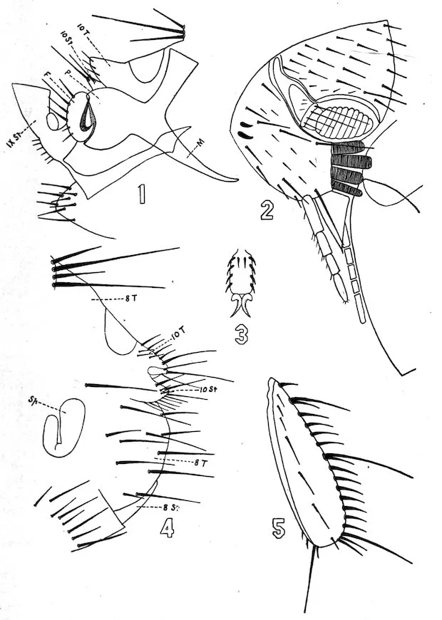
Plate IV.
CTENOPSYLLUS MUSCULI, DUGES.
141Abdomen.—The first abdominal tergite has 2 rows of 10 bristles
each, the posterior being comprised of the larger bristles. The next
6 tergites have 2 rows of bristles each, a posterior of large bristles,
about 12 in number, and an anterior of smaller bristles, also 12 in
number. On the apical edge of the metanotum there are 2 small
teeth on each side. The first abdominal tergite contains 3 such teeth
while the second and third have 1 each on each side. At the apex
of the seventh tergite in the female there are 4 antipygidial bristles,
sometimes 5. The male has but 3 antipygidial bristles. The abdominal
sternites from the third to the sixth have a single row of 6
bristles. The seventh has a row of about 16 bristles.
Legs.—The fore coxa has about 32 large bristles more or less regularly
disposed in 6 oblique rows. The hind coxa is without teeth on
the inner surface. The mid femur is without bristles on its lateral
surfaces. The hind femur is also without a row of bristles on its
lateral surfaces. The spines on the posterior border of the tibia are
single and in a close set row. The apical spines of the second tarsal
joint of the hind legs are shorter than the third joint. The last
tarsal joint on all the legs contains 4 lateral spines and a subbasal
pair situated between the first lateral pair.
Modified segments.—(♀) Just beneath the pygidium is 1 long
bristle. On the eighth tergite there is a patch of hairs, 6 of which
are on the apical margin and about 5 or 7 anterior to these. The
stylet is short, almost as wide at the base as at the tip, where there
is a long hair. Posteriorly to this bristle there springs another one
from the under surface.
(♂) Manubrium of the claspers is narrow, curved at the tip. The
finger reaches to the level of the process, has a stout pedicle, is flat
on its anterior border, and is decidedly convex on its posterior border,
where there are 4 bristles. The shape of the ninth sternite is shown
in the figure.
| Description of Plate V. |
|---|
| Fig. 1. Clasping organs of male |
P |
Process. |
| F |
Finger. |
| M |
Manubrium. |
| IX St |
Ninth Sternite. |
| Fig. 2. Head of female. |
|
|
| Fig. 3. Terminal abdominal segments, females |
8 T |
Eighth Tergite. |
| 8 St |
Eighth Sternite. |
| 10 T |
Tenth Tergite. |
| 10 St |
Tenth Sternite. |
| Sty |
Stylet. |
| Sp |
Spermatheca. |
| Fig. 4. Hind coxa, inner surface. |
|
|
142
Head.—Evenly and abruptly rounded in both sexes. Frontal
notch absent. Eye large. Maxillary palpi longer than the labial
palpi. Labial palpi reach to about half the length of the anterior
coxa and are 4-jointed. The mandibles are broad and markedly
serrate. Maxillæ triangular. Antennal groove short and wide,
closed behind, thickened on edges, and reaches to top of head in both
sexes by chitinous thickening. Second joint contains 8 or 9 fine hairs,
shorter than the third joint. Division of the third joint only to be
seen on dorsal surface. Two bristles on the gena, one placed low
down just above the maxilla, the other below the eye. From the
lower margin of the gena occasionally may be seen a small tooth.
One bristle on the occiput near the posterior lower angle. A few fine
hairs on the posterior edge of the antennal groove.
Thorax.—The thoracic nota each contain a single row of about 10
or 12 bristles. There is no ctenidium on the pronotum. The mesosternite
is narrow and is not divided by an internal incrassation. The
episternum of the metathorax is large and contains about 2 or 3
bristles and is not quite separated from the sternum anteriorly. The
epimerum has an anterior row of about 7 or 8 bristles and an apical
row of about 6.
Abdomen.—Each of the abdominal tergites, with the exception of
the first, has a single row of 8 or 10 bristles. The first has 2 rows of
about 4 each. The sternites from third to seventh have a single row
of about 6 bristles. There is one short antipygidial bristle on each
side.
Legs.—The hind coxa has on its inner surface posteriorly a number
of fine hairs, while anteriorly there are 10 or 12 teeth in an irregular
line. The hind femur has on its inner surface a row of about 8 or 9
bristles. The spines on the posterior tibia are in pairs, and there are
about 7 bristles in a line on its outer lateral surface. The apical
bristle of the second tarsal joint of the hind leg reaches to about the
middle of the fifth joint. The last tarsal joints of all the legs contain
4 lateral spines and a subapical pair, and between the third and last
lateral spine there is a hair.
Modified segments.—(♀) The eighth tergite has no bristles above
the pygidium but has numerous short stout bristles laterally and on
and close to the apical margin. The stylet is short and stout and
has at its tip a long hair. The tenth sternite and tergite contain
numerous fine hairs, those on the sternite confined to the apical edge.
(♂) The male claspers are quite characteristic. The manubrium
is large and curved and points ventrally. The claspers have two
processes, the lower of which, with the finger, form together a kind
of claw which is covered by the other process forming a flap, quite
hairy on its upper margin. The ninth sternite is described very well
by Rothschild[208] as “boomerang” shaped. The eighth tergite has
a small manubrium.
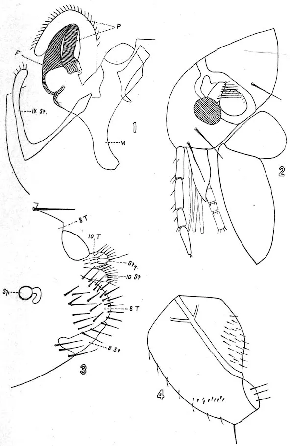
Plate V.
PULEX IRRITANS, LINNÆUS.
| 143 |
| Description of Plate VI. |
|---|
| Fig. 1. Clasping organs of male |
F |
Finger. |
| M |
Manubrium. |
| IX St |
Ninth Sternite. |
| Fig. 2. Head of male. |
|
|
| Fig. 3. Terminal abdominal segments, female |
8 T |
Eighth Tergite. |
| 8 St |
Eighth Sternite. |
| Fig. 4. Hind coxa and femur, inner surface. |
|
|
Head.—Strongly and evenly rounded in both sexes. Eye large.
Maxilla triangular. Maxillary palpi about as long as labial palpi.
Labial palpi reach to two-thirds of anterior coxæ, 4-jointed. Seven
spines along the lower margin of the gena. The posterior angle of
the gena ends in a small tooth. Occasionally this may be absent.
Antennal groove in the female reaches to within one-third of the top
of head and is prolonged upwards by a chitinous thickening and in
the male reaches almost to top of head. Two bristles on the gena,
one placed well toward the anterior lower angle and the other in front
of the eye. Usual number of bristles on posterior margin of the head,
with 2 large ones back of the antennal groove. About 8 hairs on
the second joint of the antenna nearly as long as the third joint.
Thorax.—A row of about 10 bristles on the pronotum, with a
ctenidium of about 14 to 16 spines. Two rows of bristles on the
mesonotum, a posterior of about 12, another of more numerous
smaller bristles placed well anteriorly. The metanotum contains a
single row of about 10 or 12 bristles. The episternum of the metathorax
has 3 or 4 stout bristles, while the epimerum contains an anterior
row of about 10 bristles and a posterior row of about 9.
Abdomen.—The first abdominal tergite contains 2 rows of about
4 bristles each, while the other tergites to the seventh contain a
single row of from 12 to 16 bristles. The stigmata are large. There
is a single antipygidial bristle on each side. The sternites from third
to seventh have a single row of 4 bristles each.
Legs.—The hind coxa has on its inner side a patch of from 6 to 12
spines, while the hind femur has a row of 10 or 12 bristles on its inner
surface. The spines on the posterior border of the hind tibia, with
the exception of the apical, are in pairs, while in the apical group are
about 3 stout bristles. The apical spine of the second joint of the
hind leg reaches to nearly the middle of the fifth joint. On the fifth
144joint of all the legs there are 4 lateral spines and a subapical pair, and
between the third and fourth lateral spines there is a hair.
Modified segments.—(♀) The eighth tergite has no hairs back of
the stigma. The apical margin is rounded at the apex and contains
8 or 10 bristles. The stylet is short and wide and contains at its tip
a long and a short bristle.
(♂) The manubrium is short and narrow. The movable finger of
the clasper is short, thick, swollen at its middle, bluntly rounded at
its extremity, and contains on its upper border numerous hairs and
a few on its lower border.
Rothschild[209] has pointed out certain differences between the
Ctenocephalus canis and Ctenocephalus felis. The differences are that
in the female of the felis the head is longer and more pointed. This
difference is not so pronounced in the male. Also certain differences
in the shape of the claspers and the number of bristles in the episternum
and epimerum of the metathorax and the hind femur, those in
the C. canis being more numerous. Also that group of bristles on
the posterior border of the hind tibia between the fifth pair and the
apical bristles consists of two in the Ctenocephalus canis, while there is
but a single bristle with a small hair in the Ctenocephalus felis.
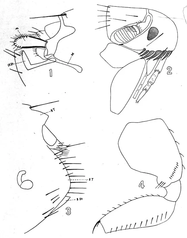
Plate VI.
CTENOCEPHALUS CANIS, CURTIS.
RODENTS IN RELATION TO THE TRANSMISSION OF BUBONIC PLAGUE. — EPIDEMIOLOGICAL OBSERVATIONS IN SAN FRANCISCO. — THEORIES AS TO THE CAUSE OF SEASONAL PREVALENCE. — THE OCCURRENCE OF PLAGUE IN THE MARMOT OF ASIA AND THE GROUND SQUIRREL OF CALIFORNIA. — PLAGUE INFECTION IN GROUND SQUIRRELS. [AF] — THE NATURAL HABITAT OF PLAGUE. — REFERENCES.
145
By Surgeon Rupert Blue,
United States Public Health and Marine-Hospital Service.
Man has associated the rat with bubonic plague since the dawn of
history. The monuments and coins of the earliest times yield abundant
evidence of this association. Æsculapius, the god of the healing
art, is represented by the Greeks with a rat at his feet. An early
scriptural reference may be found in the first Book of Samuel in the
fifth and sixth chapters. The historian records therein the occurrence
of a fatal epidemic of “emerods” in the land of the Philistines
coincident with an invasion of “mice.”
The inhabitants of southern China in recent times have learned to
look upon the finding of sick and dead rats in their homes as a harbinger
of evil, in fact, as a forerunner of that dread scourge—“wan-yick,”
or plague. In the villages and cities of the Kwantung and
Kwangsi provinces, as recorded by medical missionaries, epizootic
plague almost invariably precedes an outbreak among human beings.
So well is this fact known to the common people that many seek
safety in flight, feeling assured that in a short time “yang-tzu” or
“wan-yick” will claim a harvest of victims among those who remain.
Doctor Mahē, sanitary officer for the port of Constantinople, in
1889, called attention to the fact that epidemics of plague were always
announced by a great mortality among rats and mice. In 1894
Yersin reported the fatal epizootic among rats then prevailing in
Canton and Hongkong coincident with the outbreak of plague among
the Chinese. Recent researches have confirmed these observations
and a great deal has been added to the literature of plague, especially
in relation to its mode of transmission. Indeed, it should be said
that wherever the disease has prevailed in recent years the relation
of rats to its spread has been observed, and that since the discovery
of the specific bacillus by Yersin and Kitasato, in 1894, bacteriological
investigations have shown that there is no difference morphologically
or culturally between the bacilli of human and rat plague.
146Moreover, the gross and microscopic lesions in the lymph nodes are
practically the same, and the B. pestis recovered in both fulfills the
postulates of Koch.
Nothing was definitely known, however, of the mode of transmission
of the disease from rat to rat or from rat to man until the completion
of the experimental work of the Indian Plague Commission.
Simond, Ogata, Thompson, and Koch each expressed the belief that
the infection was transferred by the rat flea. Nuttall (1897) and
Simond (1898) demonstrated the presence of B. pestis in the bodies
of bugs (Cimex) and fleas which had been taken from plague-sick
rats, and the latter observer, in the same year, succeeded in transmitting
the disease from rat to rat without contact.
The work of the Indian Plague Commission was undertaken (1905)
with a view to establishing the exact relationship between epizootics
among rats and epidemics among men, and included both field and
laboratory observations. The experiments of Gauthier and Raybaud
(1903) and of Simond were repeated on a larger scale and greatly
improved in that all rats and fleas used were first identified as to
species. The findings of the commission may be briefly summarized
as follows: That fleas and bugs taken from plague-sick rats contain
B. pestis, and that some of them remain alive in the bodies of the
insects from five to sixteen days; that plague is conveyed by the bites
of fleas which have previously fed on the blood of animals suffering
with the disease; that rat fleas bite man; that under experimental
conditions the infection is not transferred from rat to rat in the
absence of fleas.
A careful study of the findings of the workers in India justifies
the assumption that plague is a disease of the rodent primarily and
accidentally, and secondarily a disease of man. An analysis of the
epidemiological facts collected in San Francisco leads to the same conclusion.
As a result our practice with regard to suppressive measures
and quarantine procedure has undergone a radical change in the
last decade. If the infection is flea-borne from rat to man in the
majority of cases, then the extermination of the rat should be the
first principle upon which to base a campaign. In the former contribution
on the subject (1907) I stated that “if we destroy the host
there is no longer danger of infecting the parasite.” This basic
principle has been recognized and successfully applied in two campaigns
against plague in San Francisco. First in the outbreak in
Chinatown in 1903–4, and again in the larger epidemic of 1907.
The outbreak of 1907 began May 27, a little over a year after the
great fire and earthquake, but no cases were discovered between that
time and mid-August when the disease began to appear in various
parts of the city. The source of infection was, in all probability, a
recrudescence from a focus which was not destroyed in the campaign
147of 1903–4. There occurred 160 cases with 77 deaths, the last case
appearing January 30, 1908. The following table shows the incidence
of human plague:
| Year. |
Cases. |
Deaths. |
| 1907. |
|
|
| May |
1 |
1 |
| August |
13 |
6 |
| September |
56 |
25 |
| October |
34 |
25 |
| November |
41 |
12 |
| December |
13 |
7 |
| 1908. |
|
|
| January |
2 |
1 |
| Total |
160 |
77 |
Abundant epidemiological data associating the ratAE with plague
have been collected in San Francisco. For the purpose of illustration
a detailed reference to a few cases will be made. Two small boys
(October, 1907) while playing in an unused cellar found the body of a
dead rat. The corpse was buried with unusual funeral honors.
In forty-eight hours both were ill with bubonic plague. A laborer
finding a sick rat on the wharf picked it up with the naked hand and
threw it into the bay. He was seized three days later with plague.
Doctor C. and family lived in a second-story flat over a grocery store
in the residence section. Being annoyed for some days by a foul
odor the doctor caused the wainscoting around the plumbing to be
removed. One or two rat cadavers were found in the hollow wall.
In two or three days the two members of the family who used the room
sickened, one dying on the fifth day of cervical bubonic plague. It
is probable that infected rat fleas were set free by the removal of
the wainscoting.
Dead rats were frequently found in or near houses where plague
had occurred. Immediately upon the discovery of a case of plague
trained men were sent into the neighborhood and a thorough search
made for rats. This work consisted in the removal of defective
wooden floors and walls of insanitary buildings and other harboring
places. Extensive rat catacombs were frequently found in these
operations. In the yard of a house in which 4 cases had occurred
20 cadavers were found under the board covering. In the walls of
a Chinese restaurant 87 dead rats were uncovered.
148Very little can be said of the relation of mice (M. musculus) to the
epidemic. While many thousands were trapped, only a few hundred
were examined microscopically and in these no infection was found.
They are nonmigratory in habit and for this reason are not considered
of much importance from an epizoological standpoint.
Transmission from man to man was observed in but a small percentage
of cases, 3 per cent to be exact. In these the probability
of transference by fleas (P. irritans) or by bugs (Cimex) must be admitted.
When more than one case occurred in a house a common
source of infection was indicated, such cases occurring simultaneously
or within from forty-eight to seventy-two hours after the first.
Deratization was the measure mainly relied upon. After an infected
house was rat proofed, and the harboring places in the block destroyed,
no further cases occurred.
The course of epizootic plague was not interrupted at any time
by climatic conditions, there being as many cases in proportion to
the rat population in the winter of 1908 as there were at the height
of the epidemic. The last case of human plague occurred January
30, 1908, but the infection remained active among rats for eight
months longer, or until October 21, 1908. (See following table.)
| Month. |
Number examined. |
Number infected. |
Per cent. |
Average temperature. |
Rainfall in inches. |
Character of days. |
| 1907. |
|
|
|
°F. |
|
{Clear, 13. |
| September |
1,002 |
27 |
2.69 |
60.6 |
0.11 |
{Part cloudy, 15. |
| |
|
|
|
|
|
{Cloudy, 2. |
| |
|
|
|
|
|
{Clear, 10. |
| October |
2,679 |
23 |
.86 |
60.6 |
1.36 |
{Part cloudy, 10. |
| |
|
|
|
|
|
{Cloudy, 11. |
| |
|
|
|
|
|
{Clear, 14. |
| November |
3,954 |
36 |
.88 |
57.8 |
.04 |
{Part cloudy, 13. |
| |
|
|
|
|
|
{Cloudy, 3. |
| |
|
|
|
|
|
{Clear, 6. |
| December |
4,308 |
48 |
1.11 |
52.4 |
3.66 |
{Part cloudy, 11. |
| |
|
|
|
|
|
{Cloudy, 14. |
| 1908. |
|
|
|
|
|
{Clear, 5. |
| January |
6,622 |
70 |
1.05 |
50.8 |
4.88 |
{Part cloudy, 11. |
| |
|
|
|
|
|
{Cloudy, 15. |
| |
|
|
|
|
|
{Clear, 11. |
| February |
11,700 |
45 |
.38 |
51.0 |
5.39 |
{Part cloudy, 12. |
| |
|
|
|
|
|
{Cloudy, 6. |
| |
|
|
|
|
|
{Clear, 20. |
| March |
19,263 |
52 |
.26 |
54.8 |
.90 |
{Part cloudy, 10. |
| |
|
|
|
|
|
{Cloudy, 1. |
| |
|
|
|
|
|
{Clear, 17. |
| April |
15,524 |
34 |
.21 |
56.3 |
.22 |
{Part cloudy, 10. |
| |
|
|
|
|
|
{Cloudy, 3. |
| |
|
|
|
|
|
{Clear, 17. |
| May |
11,311 |
20 |
.13 |
55.4 |
.76 |
{Part cloudy, 12. |
| |
|
|
|
|
|
{Cloudy, 2. |
| 149_ |
| |
|
|
|
|
|
{Clear, 16. |
| June |
13,624 |
4 |
0.02 |
55.3 |
0.01 |
{Part cloudy, 9. |
| |
|
|
|
|
|
{Cloudy, 5. |
| |
|
|
|
|
|
{Clear, 11. |
| July |
11,204 |
2 |
.017 |
57.4 |
.02 |
{Part cloudy, 17. |
| |
|
|
|
|
|
{Cloudy, 3. |
| |
|
|
|
|
|
{Clear, 11. |
| August |
10,988 |
0 |
.0 |
57.3 |
.01 |
{Part cloudy, 10. |
| |
|
|
|
|
|
{Cloudy, 10. |
| |
|
|
|
|
|
{Clear, 16. |
| September |
15,902 |
0 |
.0 |
59.3 |
.29 |
{Part cloudy, 9. |
| |
|
|
|
|
|
{Cloudy, 5. |
| |
|
|
|
|
|
{Clear, 16. |
| October |
10,178 |
2 |
.019 |
58.8 |
.061 |
{Part cloudy, 7. |
| |
|
|
|
|
|
{Cloudy, 8. |
The rats examined for September, 1907, were very largely collected
from the badly infected districts; the remaining months give a truer
picture of the extent of the epizootic in the entire rat population.
The marked seasonal prevalence of plague in man in San Francisco
may be given as additional proof of the association of the rat with
its spread. In the cold, rainy season, from December to April, the
epidemic ceases while the epizootic is apparently not influenced.
The anomaly is accounted for when we remember that the rat and
its parasites are very susceptible to cold and rain. It is then that
the animal seeks a warm, comfortable place from which it does not
venture until driven thence by dire necessity. In other words, the association
of the rat with man is not so intimate in winter, while the reverse
is true of the relation of rat with rat. The rains, while interrupting
the overground migrations and domiciliary visits of rats, drive them to
overcrowded burrows and harboring places. Another factor should
be mentioned in this connection. Human fleas (P. irritans), and
probably rat fleas also, are markedly reduced in numbers at that
season of the year. We must conclude, therefore, that the seasonal
prevalence of plague in man is due to the effect of climatic conditions
upon the habits of rats and the life history of the insect carriers of
the bacilli.
An examination of the foregoing should convince everyone that
all former theories as to the prolonged viability of B. pestis in
contaminated soil or in polluted streams, and of the periodical
spread of the infection therefrom, are no longer tenable. It may
also be stated that insanitary conditions, except in so far as they
furnish food and shelter to rats and other vermin, play no important
150rôle in the continuance of plague. This general revision has also
eliminated overcrowding as an important factor. In the absence
pneumonic cases, and of suctorial insects, this bête noire of the sanitarian
may be disregarded.
Rudenko (1900) first pointed out the possibility of contagion by
the “Tarbagan,” a species of the arctomyinæ found in Siberia. He
observed a connection in 1894 between this rodent and an outbreak
of plague in a Cossack family of Soktuewsk. According to Beliatsky
and Zabolotny, each having been an observer in the same field, the
natives of Siberia and Mongolia often acquire plague in this manner.
Le Dantec and other writers have called attention to the probable
susceptibility of the marmot (Arctomys bobac), a hibernating rodent of
India and China. The marmot of Thibet, in the opinion of this writer,
is the natural animal host and purveyor of the virus. The literature
of the subject presents no bacteriological evidence, however, of such
a relationship, and plague in the arctomyinæ of Asia is merely an
hypothesis. There is positive evidence though of the susceptibility
of the tree squirrel (Sciurinæ) to plague infection. Dr. Alice Corthorn
(1898) reported the finding of a plague-infected squirrel in
one of the outbreaks in the Bombay Presidency.
The demonstration of natural plague in the California ground
squirrel (Otospermophilus beecheyi) is perhaps the most important
observation of the antiplague work of the service in 1908. The
existence of a plague epizootic in Contra Costa County was suspected
as early as the summer of 1903, and efforts were made at that time to
collect sick and dead rodents for bacteriological examination. In
August (1903) two fatal cases of human infection occurred in widely
separated sections of the county. The investigation which followed
failed to connect either with a previous case of human plague, but
showed an association with ground squirrels. These deaths occurred
during a fatal epizootic among ground squirrels and suggested a connection
which unfortunately was not confirmed.
None of the circumstances were forgotten, however, and in the
second campaign, begun in September, 1907, in San Francisco,
inspectors were detailed to examine all persons dying in the area
under suspicion. No plague was reported that autumn and winter.
Fatal cases occurred and were reported by the inspectors in July, 1908,
as follows: A boy (J. F.) died July 15, near Concord, and a young
151woman (M. P.) died July 28, on a ranch 10 miles from Martinez.
The two were not associated. An investigation was ordered at once
and a force of trappers was hurried to the scene with instructions to
collect squirrels from the ranches in the vicinity. The first plague-infected
squirrel was found August 5 on the ranch where the boy had
died July 15. Of 425 squirrels collected from August 1 to October
12, 4 showed the gross and microscopic lesions of natural plague.
A lad (F. M.) sickened August 5, 1908, in Los Angeles, Cal., after
being bitten by a sick ground squirrel. A polyadenitis, which afterwards
proved to be plague, developed in a few days. A dead squirrel
was found nearby and pathological specimens taken from it were sent
to the United States Plague Laboratory in San Francisco. McCoy
recovered B. pestis from the tissue of the animal. This was the
only case of plague reported in Los Angeles. In order to complete
the list of those who contracted plague in the country, two other cases
should be mentioned. F. S., a pregnant woman, died of bubosepticæmic
plague near Concord, Cal., February 29, 1904. The
B. pestis was recovered in pure culture from the axillary glands. In
April, 1906, a school boy of east Oakland developed a multiple plague
adenitis. Investigation showed that he had shot and handled ground
squirrels in the country four or five days before his illness.
The location of the natural habitat of plague has concerned sanitarians
for many years. Not a few have settled upon India as the
endemic center, while others associate China with the epidemics
which have devastated Europe from remote times. Le Dantec, a
recent writer, suggests the “lofty mountains” between India, Thibet,
and China as the exact location, and selects the rodent (marmot) of
that region as the natural enzootic host.
A panzootic leaves in its wake enzootics of plague in various countries
which persist until the rodents upon which they thrive are either
exterminated or rendered immune. At varying intervals epidemics
spring from them and finally cease with the exhaustion or destruction
of the enzootic foci. Plague disappears in time from these temporary
abodes and retires to its original habitat in India or China.
Of serious import in this connection is the fact that all the conditions
necessary for the establishment of a permanent focus of plague
exist on the Pacific coast of the United States. The broad valleys
and lofty mountains of this region are rich in the arctomyinæ, there
being no less than 12 species in California alone. In the high Sierras
the marmot (Marmota flaviventer),[AG] a species of the natural enzootic
host of Le Dantec, is found in great numbers. The ground squirrel
infests the valleys and foothills in an unbroken chain from Oregon
152to the Mexican border. Once planted in this ideal soil, infection
may never be uprooted or its growth and extension controlled.
Small outbreaks will occur here and there, and periodical visitations
of greater magnitude may be expected in cities where a combination
of epidemiological factors is permitted.
The facts as set forth in this paper have caused grave apprehension
in the minds of those who have been at all conversant with the conditions
in the transbay counties since 1903. At that time the writer
recognized the probability of the establishment of a permanent focus
of plague in that locality, and subsequent discoveries have proven
the correctness of the assumption. This changes the aspect of the
problem from that of a local infection to one of national importance.
Once established in such a rural community, plague is dislodged with
difficulty and only after a campaign covering a considerable length of
time. Being a national problem it can be best solved by the Federal
Government.
The Croonian Lectures on Plague, W. J. Simpson; Journal of
Hygiene, Volume VI, No. 4; Volume VII, No. 6; Volume VIII, No. 2;
Plague among the Ground Squirrels of California, W. B. Wherry,
Journal Infectious Diseases, Volume V, No. 5; California Mammals,
Frank Stephens.
RODENT EXTERMINATION. — TRAPPING. — CAGE TRAPS. — SNAP TRAPS. — BARREL TRAPS. — POISONING. — PLASTER FLOUR. — PHOSPHORUS PASTE. — ARSENIC PASTE. — BARIUM CARBONATE. — STRYCHNINE. — CARBON BISULPHIDE. — NATURAL ENEMIES. — DOGS. — CATS. — CUTTING OFF THE RAT’S FOOD SUPPLY. — BUILDING THE RAT OUT OF EXISTENCE.
153
By Passed Asst. Surg. William Colby Rucker,
United States Public Health and Marine-Hospital Service.
It should be remembered that rodents are extremely wily creatures
and that any campaign against them is a contest between the wit of
man on the one hand and acute animal instinct on the other. The
rat, by his constant association with man, has become extremely
wary, and is frightened by anything in the least out of the ordinary.
They will eat the bread on which poison is spread so carefully that
they will leave behind the poison and take practically all the bread;
they will open traps by pressing down the pan, and they have been
known to repeat this operation several times within an hour, entering
the trap, eating the bait, and then liberating themselves. At other
times they will enter the trap and stand on the pan with their hind
legs, eat the cheese, then carefully turn around and back out. This,
of course, is not possible with snap traps, but they have been known
to spring them by causing pieces of wood to fall upon them, after
which the bait would be eaten. Rats are found wherever food exists
in abundance or where they can find suitable breeding and nesting
places.
Rodent extermination is a problem, with difficulties arising from
the animal’s highly developed regard for self-preservation. In main,
the rat requires two conditions for life. He needs plentiful food and
places suitable for nesting and breeding. Eliminate either of these
elements and you drive away your rats. Yet the problem remains
far more difficult than shown in the simple terms of the above equation.
The fabulous speed at which rats multiply will baffle all but
the most determined and efficient efforts to exterminate them. Under
normal conditions each female bears 3 litters a year and each litter
produces 10 young. Under conditions ideally favorable, it has been
computed that 1 pair of rats will in five years, providing all can live
so long, increase to 940,369,969,152. Such a result is, of course, impossible
in nature, for it means that every rat born of the original
pair survive five years; that every litter of 10 contains 5 males and 5
females; and that the ideally favorable conditions persist. On the
other hand, rodent existence is an unending struggle in which an enormous
154percentage succumbs; the ratio of half males and half females
does not hold; and ordinary conditions of life are hardly even favorable.
Nevertheless, the above proves emphatically that no rat eradication
can be effective unless the breeding is curtailed. Any campaign
against rodents must aim (a) to slaughter the greatest possible number
of those already living and (b) to prevent the possibility of further
breeding.
The existing rats are best attacked by trapping, by poisoning, and
by their natural enemies. Traps and poisons alone have been found
insufficient to keep pace with the rat’s speed of multiplication. The
surest of the rat’s enemies are his natural ones, and once they have
been loosed upon him his chance of escape is reduced. The cat, dog,
skunk, and other rodent foes, given a fair chance, quickly drive out
rats. But these animals do not eradicate the pest. The rats will
probably migrate to some other shelter, returning when their natural
enemies have quieted down. Absolute extermination is reached only
when conditions make the continuation of species impossible for the
rat.
The size and frequency of rodent litters decreases proportionately
with every cutting off of food supplies. Separate the rat from his
pabulum and he will not breed so freely nor so often as when he is
well fed. Destroy rat habitations and make it impossible for them
to find new nesting places, and breeding will virtually cease, since the
unsheltered progeny can no longer survive, and since the starving
parent rats are driven to cannibalism in the struggle for existence.
Campaigns against rodents must cover five directions: (1) Trapping,
(2) poisoning, (3) exposing them to natural enemies, (4) cutting
off food supply, and (5) destroying existing nests at the same time
that the making of new ones is prevented.
Parenthetically, it may be noted that while these principles apply
equally to the extermination of rats in cities and in country districts,
their application must vary according to the place.
The kind of traps to be used varies with the rodent to be captured
and the locality which it infests.
The large 19-inch French cage trap gives good results where rats
are plentiful. It should be made of stiff, heavy wire and well reenforced,
as a large, strong rat will force his head between the wires in
a weak trap and thus escape. Before setting, the lever on the trap
should be tested to see that it works properly. The trap should be
placed on a hard surface, with the rear end a little higher than the
entrance, so that the trap will close promptly. When setting the
155trap in the open it should be fastened to a board on which about an
inch of soft dirt has been spread. Place the trap where the rat usually
goes for food or in a runway and disturb the surroundings as little
as possible. It is sometimes well to place the trap near where there
is dripping water, as the rats come there to drink. If the trap is
set in hay or straw or wood it should be covered (with the exception
of the entrance) with this material. When this is not possible it
should be covered with a piece of sacking or placed in a dark corner
or beneath the floors. When setting the traps in the sewer a dry
place should be chosen.
The rat is more or less of an epicure, therefore the bait should be
changed at frequent intervals. Also he should be given food which
he is not in the habit of getting. For example: In a meat market
vegetables are the best bait, while in a location where vegetables are
plentiful fresh liver and fish heads, or a little grain, are best. The
following may be suggested as good bait to be used: Fish, fish heads,
raw meat, cheese, smoked fish, fresh liver, cooked corn beef, fried
bacon, pine nuts, apples, carrots, and corn. When trapping in chicken
yards a small chick or duckling is remarkably good. When a large
number of rats are caught in one trap, search for the female and leave
her alive in the trap, as she may call in the young or the males. The
bait should be fastened to the inner side of the top of the trap with
a piece of fine wire so that the first rat in can not force the bait underneath
the pan and thus prevent the entrance of other rats. A few
grains of barley should be scattered near the entrance of the trap
and a small piece of cheese or meat fastened to the pan with a piece
of wire. It is often well to touch the pan with a feather which has
been dipped in oil of anise or oil of rhodium. Before leaving the
trap it should be smoked with a piece of burning newspaper to kill
the smell of the human hands or the rats which have been in it.
Do not handle the trap after burning it out. When trapping in a
neighborhood where rats are known to exist the traps should not be
moved for three or four days unless they have rats in them, as it is
well for the rats to become accustomed to seeing them and thus
careless about entering. It is not wise to kill rats where they are
caught, as the squealing may frighten the other rats away.
Snap or spring traps are best for use in houses and stores, with
the exception of fish and meat markets. Snap traps are best for use
in runways, beams, and shelves. It is sometimes well to disguise the
trap by covering its floor with a little sawdust or dirt. They should
be first tested to see that they work properly and that the staples
are secure. New traps should be smoked or stained to render them
an inconspicuous color.
156The bait should consist of some firm material, such as fried bacon
or tough meat, and should be tied on so that the rat will be obliged
to pull on it and thus spring the trap. The trap should be placed in
a corner or close to the wall on a flat, hard surface, so that the rat
can not spring it with his tail or by walking on it.
In warehouses and granaries large numbers of rats may frequently
be trapped by using a barrel or garbage can having a metal top
which is carefully balanced. Large pieces of strong cheese are
placed in the middle of the cover and a plank laid from the floor to
the edge of the barrel. The rat runs up the plank onto the smooth
metallic lid which tips and the rat is precipitated into the barrel.
In cities trapping is one of the most effective of the three methods
to slaughter rodents. The rat highways are easily discovered and
in them traps capture great numbers of the unwary. In the country
one can not so readily determine the rat highway. This difficulty
diminishes the effectiveness of trapping. To make up for what is
thus lost shooting has been resorted to with good results. In Honolulu,
where a vigorous campaign against rodents is being waged, a
very large proportion of the captured rats (Mus rattus and M. alexandrinus)
have been shot from trees. In Contra Costa County, Cal.,
where ground squirrels are being exterminated, it has been found that
rodents possess an instinctive suspicion of traps and that during the
summer months shooting is not only the most practical but also
about the only effective means of attacking them. Shotguns are
the weapons to use. A rifle requires the hunter to be a better shot
than is ordinarily obtainable for such work, and, furthermore, the
danger from its longer range and from ricocheting bullets menaces
cattle and farm hands who may be working in the vicinity. As to
the shot and the powder charge for shells hunters differ. It is a
different problem for every shotgun, depending upon the gun’s
caliber and choke. The principle is to put the greatest number of
the largest shot the gun will carry into the rodent body. Thus in
10 and 12 gauge guns shoot No. 8 shot and in 16-gauge guns shoot
No. 9 shot, but this varies with each individual gun. The use of
soft lead or chilled shot seems a matter for personal preference.
The charge must be as much as the gun will carry. In the country
smokeless powder becomes a necessity during summer months since
black powder is liable to ignite the dry grass and stubble.
Plaster flour is prepared by mixing one part dry plaster of Paris with
two parts flour or meal. When this is taken in sufficient quantity
157by the rodent it produces death by the formation of enteroliths,
death occurring in from four to eight days. This is a poison of
uncertain value and is recommended chiefly on account of its cheapness
and small danger to children and domestic animals. It can
not be used in wet weather, and judging from the small number of
rats found dead with plaster casts in the alimentary canal it is not
believed to be very efficacious.
Phosphorus paste is prepared by mixing crude phosphorus in the
proportion of one-half to 10 per cent in a suitable base. The latter
may consist of cheese, sugar, and oil of anise mixed together and
heated to the consistency of sirup, the phosphorus being added after
the fire has been withdrawn and the mixture begun to cool. Other
bases are cheese, corn meal, and oil of rhodium; cheese, ground fish,
or meat and oil of valerian, glucose, and a small quantity of flour.
Glucose makes an exceptionally good base, as when properly mixed
the poison thus prepared is noninflammable even when heated.
The liability to spontaneous combustion of phosphorus mixtures
eliminates their use in hay, grain, or other warehouses or places
where there is danger of fire or the invalidation of insurance. It
should not be forgotten that phosphorus deteriorates very rapidly,
especially when it is exposed to the sun.
This consists of arsenious acid combined with a base of cheese,
meal, or macerated fish. It may be placed on raisins or prunes and
is to be recommended on account of its stability, the ease with which
it is handled, and the absence of danger from fire. It should, however,
be distributed with great care, every precaution being used to
place it where it is inaccessible to children and domestic animals.
This has not proven an effective poison owing to the fact that it
is easily decomposed by the vegetable acids, especially lactic and
oleic acid found in cheese and oil. The poisonous effect is not
greatly altered by this change. A disagreeable metallic taste is produced
and the rats will not take it.
Strychnine is prepared as a poison by soaking wheat over night
in water and subsequently pouring off the excess fluid and placing
the wheat in a caldron containing hot glucose and strychnia sulphate,
the latter in the proportion of one-tenth of 1 per cent. After
carefully stirring so that each grain is thoroughly coated it is dried
158in shallow iron pans over a slow fire with constant agitation of the
grain, or by exposure on sheets or canvas to the rays of the sun.
This mixture may be made much more efficient by the addition of
cyanide of potassium in the proportion one-half of 1 per cent. Poisoned
grain has not been found efficient in the destruction of rats, as
its bitter taste causes them to eat little or none of it. It is, however,
particularly efficient in poisoning squirrels, as it is taken readily by
them. The chief objections to its use are its cost, difficulties of
preparation, and liability to its being taken by chickens.
Carbon bisulphide is not a poison so much as an asphyxiant. As
the name indicates, it is a two-to-one mixture of sulphur and carbon.
The resulting liquid preparation should be kept in air-tight
cans, since it evaporates quickly. The principle of its efficacy against
rodents is that the fumes are heavier than air and, sinking into a rat
or squirrel hole, drive out the necessary oxygen. To use bisulphide
saturate a small pad of some absorbent cotton, jute, wool, or flannel
material with the liquid, thrust this into the rodents’ burrow, and
carefully stop all apertures through which the fumes might escape.
Animal life of every sort in that burrow is quickly asphyxiated. In
buildings the use of carbon bisulphide is greatly hampered by the
difficulty to confine the gas by stopping all cracks and other openings.
Also the odors of decomposition in animals so killed stand against
its use anywhere but in the country.
Against rats and squirrels in country places carbon bisulphide
has proved one of the best of all weapons. Where it kills, it kills
whole families at a time, not one by one, as must ever be the case
with other poisons and with traps or shotguns. Not only does it
kill the rodent but it also destroys the rodent’s fleas and vermin,
which is most important. A dead infected rat is still a menace,
since its fleas may inoculate other rats and human beings with the
infection. Destroy the fleas and that greatest danger is removed.
The recent campaigns against rodents in the United States have been
waged because rats and squirrels were infected with bubonic plague;
hence the added value of carbon bisulphide. Unfortunately,
though this asphyxiant proved so effective in the work against
squirrels in Contra Costa County, Cal., it proved to be well-nigh
useless during the summer season when dry heat checks the adobe
and makes the ground generally porous. Nevertheless, the value
of carbon bisulphide, especially for sanitary purposes, can not be
easily overestimated for work in the country during the fall, winter,
and spring seasons.
Fumigants in general are effective. They possess no marked or
peculiar advantages as special weapons against rodents. Their use
159is limited chiefly to warehouses, elevators, and ship holds. They
are deadly to rats in the same way that they are fatal to every sort
of life. Many difficulties and some dangers stand in the way of
their use. It is safe to advise that no one unacquainted with their
action should ever employ fumigants.
The war upon rats carried on in San Francisco has proved the great
value of cats and dogs as natural enemies of the rat. That city now
has a law requiring all structures of 800 or less square feet and outside
certain limits to be raised high enough above the ground to allow
access to cats and dogs. (All other buildings in the city must be rat
proof.) An index to the worth of rodent foes in their extermination
points from what happened during the great London plague. At
that time the disease was supposed to be air carried; any furry material
might hold and spread infection. The magistrates decreed that
all cats and dogs should be killed to prevent plague from lodging
and traveling in their hair. Rats were thus free to live and breed unmolested,
and live and breed they did until the plague killed them off;
then and then only did the disease cease its ravages among human
beings.
Slight training will make excellent ratters of Fox, Irish, or Scotch
terriers. Fox terriers have proved especially valuable as retrievers
when shooting squirrels, which in one case out of five will escape to
their burrows unless sharply retrieved.
Cats are little less valuable than dogs against rats, while they are
useless against squirrels. The ordinary cat is too well fed to attack
large rats and goes, almost solely, after mice.
All other animals naturally preying upon rodents class with wild
life—weazels, ferrets, badgers, skunks, and minks. These can be used
only in country places, where, however, their raiding of chicken coops
tends to counterbalance their value as ratters. The skunk alone is
an infrequent slayer of fowl, whereas he harvests innumerable farm
pests from worms to crickets. Yet an insurmountable prejudice
against skunks stands in the way of realizing his full usefulness against
rodents. In addition, various hawks and most owls kill off rats.
Since rats come out chiefly in the nighttime, owls have the better
chance to be serviceable in their destruction. The efficiency of these
birds in rural districts quite equals that of a dog against rats, while
besides dogs we have not as yet found a safe natural foe of ground
squirrels.
160But all such attacks upon rats fail of absolute eradication. One
must make it impossible for them to find sustenance and must destroy
not only all existing rat houses, but also all chance of their digging or
finding new ones.
This is important not alone for its effect in a campaign upon rodents,
but equally because it necessitates sanitary care and cleanliness in
handling foodstuffs intended for humans and garbage coming therefrom.
Abattoirs and places where cattle and hogs are fattened
perhaps furnish the greatest number of rats. Stables, food-supply
stores, groceries, meat, fish and vegetable markets, restaurants,
bakeries, and the various places where food is prepared for human
consumption are usually infested. In each of the places the barriers
vary according to the nature of the premises. Rat-proof receptacles
for the foodstuffs must be installed wherever practical. In
San Francisco an ordinance requires every stable to have metal lined
feed bins. Markets and places where eatables are constantly being
shifted about must be properly screened against rats. Screening
should be of heavy wire and sufficient fineness, not larger than halfinch
mesh. In all places the food has to be raised such a height
above floorings as to be beyond the rat’s reach. This applies also
to corn and grain cribs in the country. Yet, no matter how carefully
the bulk of the food may be kept from rats, negligence in handling
it, in spilling or scattering small amounts upon floors or the ground,
will nullify every precaution.
No less painstaking must be the disposition of garbage. Ordinance
now requires that all premises in San Francisco be provided with
“sanitary garbage cans.” Preferably these should be of zinc or
galvanized iron and fitted with tight covers. Under no circumstances
should the cover be allowed to remain off its can. Garbage is to be
placed in a can without delay and care must prevent the dropping
of it upon the ground. Rats once served communities as scavengers;
wherever the scavenger work is laxly done, rats are welcomed.
Finally, garbage must never be allowed to accumulate and should be
removed daily, not less often than every other day.
Special conditions, closely related to the next topic, are encountered
in large warehouses and grain sheds. Places where large quantities
of food may be stored for a length of time should be constructed of
reenforced concrete to be rat proof. Then, again, where vessels
are changing cargoes, rat-proof compounds should be erected for the
temporary storage of freights. The water fronts of seaports are
invariably rat ridden; and in San Francisco a compound for freight
held in transit was found invaluable. No effort can be spared in
161keeping rats from their food if their extermination is to be accomplished.
With regard to ground squirrels, the use of poisoned wheat very
properly enters here. With the changing season ground squirrels
change their habitat and food. During early spring these rodents
come down from their winter dwellings in the hills and seek burrows
in meadow lands and cultivated spots. Months have passed since
the squirrel tasted wheat; his fickle appetite betrays him. From the
first spring months until harvest time one can kill thousands of ground
squirrels by tempting them with poisoned wheat. But so soon as
harvest time comes, they seek new growing green stuffs to eat and
thereafter, on through winter, poisoned wheat is ineffective.
Most certain of all methods to get rid of rodents is to allow them no
place in which to live. San Francisco effects this result through its
ordinance, which requires small houses outside certain city limits to
be raised so high from the ground that dogs and cats can drive out
rats from under them, and which requires all other buildings in the
city to be rat proofed with cement or concrete. The latter contemplates
foundation walls of concrete or brick sunk at least 1 foot
to 18 inches to 2 feet above that surface; the whole ground area
inclosed by their foundation walls must be covered with concrete
at least 1½ inches in thickness. Thus the entrance of burrowing
rodents is prevented. Even where buildings stand upon rock or
hardpan, these requirements should be enforced. Rock may crack,
gradual weather decay may cause crevices to be found in it unseen
crannies may be found by rodents, and once the rat lodges in rock
his nest is virtually unassailable. With hardpan it is even worse,
for the rats can burrow in it, with some difficulty, truly, but when
nests are impossible elsewhere, necessity will drive rats to find shelter
in hardpan, which will protect them quite as well as rock. In the
main, these two points of building rats out of existence, though modified,
will apply to any structure.
Yet any negligence will overthrow these safeguards. The principle
is to allow no opening within which rodents may nest. Plank sidewalks
and back yards will continue the rat nuisance even though
buildings are amply protected. Carelessness in throwing old boxes
into basements or piling old lumber or refuse within reach will supply
shelter for rats despite concreted ground area. The precaution must
be constant and consistent.
Another important phase is to cut off the rodent migrations.
Prevent rats from moving from place to place. Their time-honored
highway is through sewers. Modern sewers afford no protection,
162inviting rodents to live in them as formerly. Scarcely less important,
access should be stopped. Catch-basin feeding sewers should be
constructed so that rats can not slip through into the mains, or having
once gotten in, they can not escape, and hence must drown. Farms
are frequently protected against rodent migrations by tin or zinc
sheeting sunk into the ground about a foot and a half and standing
about the same height above the surface along the fence line.
Finally, as the progress of rodents from place to place within communities
must be hindered, so must they be stopped from entering
new communities. Railroads and seagoing vessels carry great
numbers of rats in freight. The rat-proof compounds above described
serve well enough so far as railroads are concerned. With vessels
it is a different matter, and one demanding special attention, since
the rodent is only too likely to import infection from foreign harbors.
All hawsers thrown out to make boats fast should be provided with
traps to catch any rat seeking to land along the hawser. San
Francisco, about to possess a rat-proof water front, is now building
concrete wharves to prevent the landing of rodents. Every port
should be safeguarded by stone, concrete, or iron wharves and piers.
As a further protection, all ships should have permanent devices, as is
now proposed for naval construction. Levy’s system of metal
conduits built into vessels promises much in the present world-wide
war upon rodents. Rodents must be “built out of existence,”
and to eradicate rats for all time we must erect wide systems of
municipal fortifications.
NATURAL ENEMIES OF THE RAT. — INTRODUCTION. — ANIMALS THAT DESTROY RATS. — HAWKS. — OWLS. — NATIVE WILD MAMMALS. — DOMESTIC ANIMALS. — OTHER ANIMALS. — BOUNTIES ON PREDATORY ANIMALS.
163
By David E. Lantz,
Assistant Biologist, United States Department of Agriculture.
Undoubtedly the great increase of rodent pests throughout North
America is in large part due to a general scarcity of the animals that
habitually prey upon them. Since the early settlement of the country
persistent warfare has been made on birds and mammals of prey
on the plea that they are enemies of poultry, game, and insectivorous
birds. Efforts to destroy the predaceous birds and mammals have
been greatly stimulated by the payment of municipal, county, and
state bounties, and the destruction has gone on until in many sections
these animals have nearly disappeared.
The effect of killing off the natural enemies of rodents has been to
disturb natural conditions. Rodents multiply so rapidly that they
derive an undue advantage in the struggle for existence when their
natural enemies are destroyed. The result is noticeable in the
increased depredations of rats, field mice, rabbits, and other pests.
The destruction of carnivorous wild mammals and birds by the
farmer, hunter, or game preserver is often due to misapprehension.
Because one kind of hawk preys on the farmer’s poultry is not sufficient
reason for exterminating all hawks. Nor does the fact that
occasionally an owl or a skunk destroys a chicken or a game bird
justify warfare on all owls and skunks. It is the occasional individual
and not the species that offends.
The usefulness of the natural enemies of the rat must not be overlooked
in plans for its repression. Among the more important are
the larger hawks and owls, skunks, foxes, coyotes, weasels, minks,
and a few other wild mammals; as well as cats, dogs, and ferrets
among domestic animals, and snakes and alligators among reptiles.
Most of the larger hawks destroy rats. Feeding only in the daytime,
they seldom find their quarry near houses and barns, where rats
do not venture out until after sunset. Besides, owing to persecution
164by farmers, hawks generally keep away from farm buildings. In the
open fields, however, where rats feed in early morning and late afternoon,
hawks find many of the rodents.
The species of hawks that more commonly feed on rats are the
buzzard hawks, including the red-tailed (Buteo borealis and sub-species),
the red-shouldered (B. lineatus), the broad-winged (B. platypterus),
and the Swainson (B. swainsoni); the rough-legged hawks
(Archibuteo), two species; and, to a less extent, the marsh harrier
(Circus hudsonius), and a few other species.
The writer has several times found the remains of rats about the
nest of the red-tailed hawk. Of the 562 stomachs of this species
examined by Dr. A. K. Fisher, of the Biological Survey, less than 10
per cent contained poultry or game, while more than 70 per cent contained
injurious rodents.[AH] Most of the other species of buzzard
hawks made a better showing even than this, especially the Swainson
hawk, which had fed entirely on harmful rodents and insects. The
stomachs of rough-legged hawks examined nearly all contained harmful
rodents and none of them contained remains of birds of any sort.
A few months ago, while walking on the Potomac flats near Washington,
the writer met some boys who had just shot a red-tailed hawk.
Its crop was greatly distended, and later examination showed that
the bird had recently eaten an enormous brown rat. Although
the shooting was contrary to law, when it was reported to the nearest
policeman, his comment was, “Oh, a hawk! Why, it’s a good thing
to shoot a hawk.” The incident illustrates the general popular
prejudice against all hawks.
Because they hunt by twilight and at night, owls are more efficient
than hawks in destroying rats. All American owls, except the more
diminutive species, prey on the common rat. Even the little screech
owl (Otus asio) feeds on young rats.
Of all our species, the barn owl (Aluco pratincola) is preeminent as
a destroyer of rats. It lives commonly about farm buildings, sometimes
even making its nest and rearing its young in the pigeon loft
without molesting the pigeons. In such surroundings its opportunities
for securing rats are excellent, and no other wild bird is so useful
on the farm. The late Henry Newman once stated that every barn
owl is worth £5 a year to the British nation, and the value of the bird
to the American farmer is not less.
Owls, hawks, and other birds of prey that swallow their quarry
whole or in large pieces do not digest the bones, fur, and feathers.
They eject these indigestible parts in the form of large pellets, in which
the fur or feathers surround the bones. The contents of these casts
165are an excellent index of the food of owls. Dr. A. K. Fisher, of the
Biological Survey, has examined 987 pellets of a pair of barn owls that
live in a tower of the Smithsonian building in Washington, and in
them found the skulls of no fewer than 192 rats (Mus norvegicus),
together with those of 554 common mice and 1,508 field mice (Microtus
pennsylvanicus).
Dr. John I. Northrop found a nest of the barn owl on Andros Island,
Bahamas. It contained two young owls and the remains of a black
rat (Mus rattus). The ground about the nest was covered with
pellets which contained remains of the black rat and no other species.[AI]
The great horned owl (Bubo virginianus) is the largest of our resident
owls. It feeds mainly on rodents, though occasionally it takes
a fowl found roosting in an exposed situation, as on a fence or in a
tree. While it occasionally destroys game birds, the rats it captures
would probably destroy ten times as much game as the owl. Charles
Dury, of Ohio, in 1886 published a letter from O. E. Niles in which it
was stated that he counted 113 dead rats at one time under a nest of
this bird.[AJ]
The snowy owl (Nyctea nyctea) is a rather rare winter visitor in the
northern United States. It usually arrives when the ground is
covered with snow and ordinary food is scarce. Near barns, outbuildings,
and stacks it finds its chief subsistence in the common rat;
and, if undisturbed, will stay for several weeks in the same locality,
destroying many of the pests. Unfortunately, mounted specimens
of this beautiful owl are so much in demand that the majority of them
fall a prey to the specimen hunter and the taxidermist. The destruction
of this bird should be prohibited under heavy penalties.
The barred owl (Strix varia), the long-eared owl (Asio wilsonianus),
and the short-eared owl (Asio flammeus) all destroy some rats; but
as they do not generally nest or live in the vicinity of farm buildings,
the rodents they capture are taken chiefly from the fields. Occasionally
a short-eared or a long-eared owl makes its winter home in a
group of evergreens near the farm buildings, and does excellent
service in clearing the premises of rats and mice. Evergreens are
desirable about a country place, if for no other reason than that they
attract owls.
The practice of indiscriminately destroying hawks and owls should
be discouraged. Game preservers especially should realize that the
birds of prey they kill would, if allowed to live, destroy rats, which in
the course of a year do many times as much harm to game as the
supposed offenders do. Besides, the birds would destroy also large
numbers of mice and injurious insects.
166The farmer and the poultry grower may easily learn to recognize
the few harmful species of hawks, and should confine their warfare to
these. The practice of setting pole traps for hawks and owls is
exceedingly reprehensible, as it results chiefly in the destruction of
our beneficial owls when they come about the premises at night in
search of rats. Furthermore, the beneficial hawks and owls should
have legal protection. The larger hawks, nearly all of which are
beneficial, are slow of wing and much more likely to be shot than the
swifter and more harmful falcons.
Not many species of wild carnivorous mammals live where the
common rat is abundant. Coyotes, foxes, and a few others occasionally
find a rat in the fields, but for the most part they depend for
food on native wild rodents and other animals. Chief among the
mammals that do good work in destroying rats are skunks, minks,
and weasels.
SKUNKS.
Skunks are excellent ratters, and when they take up their abode
on the premises of the farmer, they speedily destroy or drive away all
rats and mice. This statement applies equally to the large skunks
(Mephitis) and the little spotted skunks (Spilogale). Unfortunately,
skunks are seldom allowed to tenant the premises without being
molested by either dogs or men. When undisturbed, they are inoffensive,
and will stay about the farm buildings or stacks until rats
and mice are no longer to be had for food.
Skunks usually hunt by night, and hence poultry properly housed
is safe from them. The larger skunks do not climb, and can capture
only fowls that roost on the ground. Indeed, so few skunks ever
learn to kill poultry that there is no good reason for warfare on the
skunk family. Besides destroying mice and rats, the animals are
invaluable as consumers of noxious insects, especially cutworms, army
worms, white grubs, May beetles, grasshoppers, crickets, and sphinx
moths.
WEASELS.
Weasels are good ratters and mousers. Several of our species
come about buildings, and often perform excellent service in destroying
rats and mice. They are more likely than the skunk to attack
poultry, for they can enter the poultry house through smaller openings.
At times weasels seem to kill for the mere love of killing, and
while occasionally this trait makes them formidable in the poultry
house, it also renders them more efficient as destroyers of rodents. A
small weasel can follow a rat into all its retreats, and will soon clear
a stackyard or shed of all rodents.
167Our largest weasel, the black-footed ferret (Putorius nigripes),
occasionally deserts its wild haunts on the plains and comes about
buildings in search of rats and mice. In 1905, while the writer was
at Hays, Kans., one of these ferrets took up its quarters under a
board sidewalk in the business part of the village. The squealing of
the rats it killed was often heard.
As regards the destruction of poultry by weasels, the same care
necessary to exclude the rat from a poultry house will keep out the
weasel also. When so excluded, a weasel will do no harm about the
premises, but may be depended upon to drive out or kill all the rats
and mice.
MINKS.
Minks are excellent ratters, but as enemies of poultry are worse
than weasels. They destroy fish also. The great demand for mink
fur causes close trapping of these animals, and in the future they are
not likely to influence greatly the numbers of rodent pests.
Among the enemies of rodents often employed as aids to rat
destruction are the dog, cat, and ferret.
DOGS.
The value of dogs as ratters can not be appreciated by those who
have had no experience with trained animals. The ordinary cur
and the larger breeds of dogs seldom make useful ratters. Small
Irish, Scotch, and fox terriers, when well trained, are superior to
most other breeds as ratters, and under favorable circumstances
may be depended on to keep premises free from rodents. Much, too,
may be done by the farmer or householder to increase the effectiveness
of his dogs by removing obstructions to their work. Corncribs
and outbuildings, when of wood, should not have floors close to the
ground, but should have ample room below to permit dogs to move
about freely.
With a little preliminary training, most terriers learn to hunt rats
independently, and they thus become doubly useful on farms and in
warehouses.
CATS.
When the black rat was the dominant species in Europe and
America, cats were the chief dependence of the householder against
rats; but comparatively few cats will venture to capture a full-grown
brown rat. Then, too, the ordinary house cat is too well fed and
consequently too lazy to be an efficient ratter.
Occasionally, however, one meets with rat-killing cats whose work
in destroying the brown rat has decided value. These cats are
168rarely of the fine breeds, but generally of the common “tabby”
variety, kept in barns or warehouses, fed on milk, and left to forage
for their own meat. Managed in this way, cats are far less objectionable
on sanitary grounds than when kept in the house as pets. In
the country, on the other hand, barn cats are far more likely than
the house-kept ones to run at large and prey upon birds and young
poultry. Aside from the rat itself, we have no more serious enemy
of birds and game than half-wild cats, many of which have been
abandoned in fields and woods by the thoughtless. All things
considered, cats do not rank high as destroyers of the common
brown rat.
FERRETS.
Tame ferrets, like weasels, are inveterate foes of rats, and can
follow them into their retreats. Under favorable circumstances
ferrets are useful aids to the rat catcher, but their value is often
greatly overestimated. They require experienced handling and the
additional services of a well-trained dog or two to do effective work.
Dogs and ferrets must be thoroughly accustomed to each other.
A noisy or excitable dog is useless in ferreting. The ferret should be
used only to drive out the rats, which are then killed by the dogs.
If an unmuzzled ferret is sent into rat retreats under floors, it is apt to
lie up after killing a rat and sucking its blood. Sometimes the ferret
will remain for hours in a rat burrow or escape by unguarded exits
and be lost.
Such experiences often discourage the amateur ferreter. Besides,
ferrets are subject to diseases and require the greatest of care as to
their food. For these reasons the use of ferrets to destroy rats,
except in the hands of the experienced, is generally expensive and
disappointing.
MONGOOSE.
The various species of mongoose (Herpestes and Mongos) are
destroyers of rats, and their importation into this country has often
been urged. Many years ago they were introduced into Jamaica and
Hawaii to save the sugar plantations from ravages by rats. The
mongoose has, however, proved very destructive to native birds and
poultry in the islands, and its introduction is now generally regretted.
Its importation into the United States is prohibited by law.
ALLIGATORS.
In the South the alligator is said to destroy many rats along levees
and banks of streams, and its protection has been urged on this
account.
169
SNAKES.
Our larger snakes are beneficial in destroying rats, mice, prairie
squirrels, and pocket gophers. As most of the food of snakes is
obtained remote from human abodes, only a small percentage consists
of rats.
Whatever may be said in favor of bounties on the larger beasts
of prey, those on hawks, owls, and the smaller fur-bearing animals
can not be justified. Payments of this sort should cease, and laws
should be enacted to protect species which careful investigations
have shown to be mainly beneficial.
A few States still pay bounties for the destruction of foxes, weasels,
skunks, minks, and raccoons. All of these, except the southern
weasels, have valuable fur, and hence should be protected as a source
of wealth. In addition they do far more good by destroying rats,
mice, and other field pests than harm to game and poultry.
The payment of bounties on hawks of any kind is open to the
objection that officials hardly ever discriminate between the harmful
and the useful kinds, even when the statutes do so. Since the beneficial
kinds are the more easily captured, public money is often paid
out to reward what really injures the community. The bounty on
owls is still more reprehensible, since owls are a more decided check
to rodent increase.
The natural enemies of the rat exercise a steady, cumulative effect
in restricting the numbers of the pest. That the effect is not greater is
largely our own fault, since instead of protecting the birds and
mammals that prey on the rat, we destroy them, sometimes even
offering bounties on their heads. In future our aim should be to
increase their numbers and to protect them in every way possible.
RAT PROOFING AS AN ANTIPLAGUE MEASURE. — RAT PROOFING OF PRIMARY IMPORTANCE. — RAT PROOFING IS EXPENSIVE. — METHODS OF RAT PROOFING. — RAT-PROOFING ORDINANCES SHOULD BE SPECIFIC. — CHOICE OF ARCHITECTURE AND BUILDING MATERIALS.
171
By Richard H. Creel,
Passed Assistant Surgeon, United States Public Health and Marine-Hospital Service.
To appreciate the great importance and absolute necessity of rat
proofing as an antiplague measure, it is only necessary to consider
the results that have followed its use as compared with other measures
that have been relied on in recent years in combating this disease.
These were, briefly, disinfection, evacuation, or destruction of buildings
in infected areas, preventive inoculations, and destruction of
rats either by poison or by trapping.
Plague was formerly believed to be communicable by aerial transmission
and through the agency of fomites. Sanitarians have,
therefore, put great faith in disinfection procedures, but the results
have never been satisfactory, and it is only necessary to consider
the method of transmission of plague to perceive the fatuity of bactericidal
measures. Measures intended for the destruction of fleas
are also of relatively small value. It is well worth while to destroy
all fleas possible, but if those infesting the rat population escape, the
efforts will have had little effect in preventing the spread of plague.
It is only those fleas that infest rats and their habitats that are of
importance in relation to the transmission of disease, and it is only
by rat proofing that their destruction in rat burrows and runs can be
accomplished.
Rat-proofing of individual buildings is of no recent date, but new
emphasis was laid on rat-proofing as a separate and distinct antiplague
measure by Passed Assistant Surgeon Mark J. White in an
article written in the fall of 1907.[AK]
Disinfecting procedures must be regarded as of minor importance
in plague prevention, except in pneumonic cases where its use is
imperative. It is not intended to depreciate the value of disinfection,
but rather to estimate its exact value as an antipest measure.
Time and money should not be wasted nor a feeling of false security
engendered by using an ineffective measure when others, as rat proofing,
172of much greater value are at hand. As an example of this
might be cited an outbreak of plague in one of the refugee camps
during the recent epidemic of the disease in San Francisco. This
camp covered several blocks and housed between two and three
thousand people. The camp grounds throughout and the houses
were disinfected and disinfected well. At the same time, every
effort was made to poison and trap rats. Notwithstanding these
precautions, cases continued to occur, but when the houses were
elevated there followed an immediate cessation of plague cases in
the camp.
Another case of infected premises proved equally refractory to
disinfection. The place was a large two-story frame dwelling located
in the center of the city and in a good neighborhood. The yard
was planked, as was also a part of the basement, the latter being used
as a storeroom. On November 1 there occurred in this dwelling a
fatal case of human plague, and plague rats were found at the same
time. The place was disinfected in the usual manner and thorough
measures were taken to trap and poison rats with apparent subsidence
of infection, but on January 22 a plague rat was trapped, followed
by another on January 31, after which the occupant of the
building vacated it in great alarm. All planking was then removed
from the yard and basement and concrete substituted by the owner,
the place thereby being rendered thoroughly rat proof, and no plague
rats were subsequently taken from that dwelling or in its immediate
neighborhood.
In 1902 the plague outbreak was almost wholly confined to the
Chinese colony. Chinatown was made the battle ground, and among
other measures rat proofing was enforced, with the result that after the
fire it was by far the most sanitary district in the city of San Francisco
from a structural point of view. The buildings when erected
were made rat proof from cellar to garret. The Chinese had had
their lesson, and to their credit it must be stated that they responded
with a greater show of intelligence than did some of the residents in
surrounding districts.
Adjacent to the Chinese colony is the Latin quarter. In the
rebuilding of this section, no attention was paid to rat proofing;
consequently many of the buildings consisted of small shacks set on
the ground or abutting some insanitary stable, and were therefore
ideal rat harbors. On account of these conditions the natural results
followed. Chinatown, on the other hand, which had contributed
in the previous epidemic almost the entire number of plague cases
during the epidemic of 1907, did not furnish more than two or three
of the plague cases reported; that is, less than 2 per cent of the total
cases reported, while the Italian colony, including North Beach district,
probably furnished over 50 per cent of the total.
173The evacuation or burning of buildings can hardly be called a successful
measure any more than a retreat can be styled a victory;
moreover, there can be no question from an economic standpoint
as to the value of rat proofing over abandonment except in a few
isolated cases of dilapidated insanitary property.
Schemes and plans for demurization, total or partial, have been
as numerous and varied as they have been unsuccessful. Traps and
poisons have been the agencies of destruction, but until some highly
communicable epizootic peculiar to rodents shall have been discovered,
absolute eradication of the rat can be considered as nothing
less than impossible.
A recognized authority on plague, Major Morehead, of the Indian
Medical Service, states that “rat destruction is of doubtful value,”
referring, of course, to trapping and poisoning when those measures
are used solely without auxiliary measures. He agrees with Japanese
authorities in their arguments that as rat populations decrease,
the breeding rate among survivors increases, due, obviously, in part
at least, to increased food supply and harboring facilities. Such a
result is assured where rat proofing is not accomplished at the same
time. This latter procedure, by destroying rat harborages and cutting
off food supplies bring about conditions unfavorable to breeding.
The total eradication of rats in a locality is not absolutely necessary,
however, to the eradication of plague. If the rat population
is kept within fairly low limits, rat centers destroyed, and such rat
population as does exist well scattered and not congested, it is ventured
that rat plague will disappear from a locality. Plague among
rats in San Francisco ceased to appear when the number of rodents
was reduced some 50 per cent, but such reduction was accomplished
only after six months of ceaseless endeavor, which included also the
rat proofing of the bakeries, stables, and markets in the city.
It is a logical supposition that close contact is just as essential
for the propagation of plague among rats as it is for the spread of
certain communicable diseases among human beings, the increase
of cases being in direct proportion to the density of population and
closeness of contact.
Without the general enforcement of rat proofing antiplague measures
are bound to be more or less temporary and decidedly unsatisfactory.
This subject is of immense importance to the public, both
from a sanitary and a commercial standpoint, but the latter aspect
of the question is more apt to prove of interest to most communities.
The measures necessary to render buildings rat proof are the
same, however, whether they be instituted for sanitary or for commercial
reasons. Rat proofing will, therefore, be considered entirely
174from a sanitary standpoint; but it can be understood that granaries,
bakeries, butcher shops, packing houses, dwellings, and other places,
if rat proofed for sanitary reasons, are just as much protected from
depredation of the rat as though the work had been performed for
commercial reasons alone.
Rat proofing has a twofold objective. It serves as a protection
to the inmates of a building, and excludes rodents from sources of
food supplies and harboring places. While rat proofing should be
enforced as a general measure in all plague-infected localities, it is
imperatively demanded in premises whereon have occurred cases of
human or rodent plague.
Plague-infected localities or places that contain food must be
rendered impervious to rats in order to insure the success of other
preventive measures. Rats can be trapped or poisoned only when
other food supply is excluded. A rat will enter a trap for food or
will eat poisoned preparations not because of their greater attractiveness,
but because of their greater availability. It therefore follows
that rat proofing of food supplies is a prerequisite to success in rat
eradication. The food depots requiring attention in the order of
importance are stables, meat markets, bakeries, restaurants, groceries,
warehouses, and private dwellings.
It is logical to suppose that the most common mode of infection
is by reason of plague rats dying in the walls, roofs, or floorings of
human habitations. As soon as the rat’s body is cold the fleas
abandon it for another rat, some domestic animal, or human being.
The risk to human inmates in such infected houses, therefore, is
evident.
That rat proofing is a valuable measure is shown by the reports
of the British Plague Commission where are mentioned the results
following the use of rat-proof “go-downs” and those not so constructed.
Additional evidence is presented by the fact that appalling
epidemics of plague have ravaged India and the China coast, whereas
in the Philippine Islands but few people die of the disease. It would
appear that the comparatively few cases in the Philippine Islands
were due to the fact that most Philippine dwellings are rat proof
by reason of being elevated from the ground and the fact that the
walls are thin and offer no refuge whatever to rats.
The almost insuperable obstacle that will usually confront the
sanitary authorities in such work will be either the financial inability
of the unfortunate community or the sordid unwillingness to make
any expenditure that does not promise personal gain.
When the influence of the mosquito in the transmission of yellow
fever was proven, recourse was had to mosquito proofing of both
175the sick and the well as a preventive measure. Rat proofing in
plague is just as rational and necessary, but the financial expenditure
contemplated thereby has been of such proportion as to cause the
majority of sanitary authorities in different parts of the world to
dismiss the idea as impossible.
To properly rat proof a city undeniably requires enormous expenditures,
but no antiplague campaign was ever waged without an
immense outlay of both money and labor. If allowed to progress
unchecked, however, plague, either through ravages of the population
or through commercial interference, is ruinous. To fight plague,
therefore, is the only alternative, and a costly campaign should be
anticipated and prepared for in advance. To merely put out traps
and poisons without the preliminary rat proofing required can be
productive of little good and no permanency. Such a plan of campaign
may be attractive because of its relative cheapness, but any
city or country that relies wholly on such measures is practicing
false economy and deferring the day of reckoning.
It becomes evident, therefore, that rat proofing is of the greatest
value as an antiplague measure, and that practical results to be
expected are much greater than with any other method.
As has already been stated, the individual premises on which plague
either among rodents or human beings has occurred demand first
and immediate attention. The work should be extended as rapidly
as possible from the point of infection so as to include the entire
block and neighboring blocks.
While the chief energies should be centered on plague-infected
foci, similar work should be carried on simultaneously throughout
the city.
If plague occurs in the grounds of dwellings the following course
should be pursued: All planked-over areas, including sidewalks, that
might possibly shelter a rat should be removed, leaving either bare
ground or, at the option of the owner, gravel or concrete used, the
gravel being preferable. Small sheds should be elevated, or their
ground floors concreted. Wood sheds should probably be left without
flooring, wood kindling or other contents being piled on elevated
platforms provided for the purpose. Stables on the premises should
be treated as indicated in a subsequent paragraph relating to these
structures.
The garbage depository must be given most careful attention. It
should be a metal receptacle, preferably a galvanized can, water-tight
to prevent seepage which would attract rats, and there should be a
closely fitting lid. A can 2 feet in height without cover will not be
proof against the incursion of rats.
The rat proofing of chicken yards is a difficult task as most chickens
in private families are fed on table scraps, thereby attracting and
176supporting a fair quota of rats. The entire inclosure should be
protected by wire fencing 6 feet high and of a mesh not larger than a
half inch. Ordinary poultry netting is inadequate, the mesh being
too large. The edge of the yard should be of concrete construction,
the concrete extending 1 foot upward and 2 feet inward. If, on
any subsequent inspection, rats have been found to have burrowed
into the inclosure, the entire area should be concreted, sand or earth
being allowed as a top dressing.
It would seem sufficient to confine these specifications to a feeding
pen, but in practice this will not suffice, as a mere pretext of such a
place would be built, and the housewife would continue to throw
scraps into the unprotected yard.
The dwelling house itself should receive the most careful attention.
If it is a small frame structure the cheapest and most effective means
of rendering it free from rats is by elevating it, the minimum height
being 1½ feet, measured from the most dependent joist. At the
same time, all underpinning should be freed of rubbish or other
material. It is not sufficient to raise the structure a few inches so
as to permit the entrance of cats and other enemies of the rat. Such
height and exposure must be secured as to deprive all rodents of
cover.
If the house is of more substantial structure, and always if it has
a cellar or basement, concrete or some other rat-proof material should
be adopted. If sound foundation walls of stone or brick exist, then
only the addition of a concrete floor is necessary. The stopping up
of rat holes in any substance pervious to rats is at best a poor
expedient.
The grounds must be rendered rat proof by piling all loose materials
at such an elevation as will preclude rat harborage. All rubbish
should be burned or otherwise destroyed. All basement windows
should be properly protected against the ingress of rats, and if the
Mus rattus be present, even second and third story windows should
not be considered too high to afford them entrance.
All loose materials on the premises should be properly piled, even
though they are in a rat-proof cellar. It is not probable that the
Mus decumanus would remain or breed in any place where it could
not burrow; but no encouragement should be offered to any rodent
let in by carelessly left open doors. There have been cases where
the black rat has lived, increased, and overrun a house which was
structurally rat proof, but in which there was allowed easy access
through open windows and doors, and great piles of loose materials
and dunnage furnish harborage.
Stables are of twofold importance because they provide a source
of food supply for rats and furnish harborage. All grain must be
kept in a metal lined box or granary. A small stable is sufficiently
177rat proof if it has an elevation of 2 feet with clear underpinning, provided
the floor is rendered impervious to falling grain. Barns of
larger extent are best made rat proof by concrete flooring tight on
the ground, and the area walls should be of concrete 1 foot high or
of galvanized iron of standard thickness. The ingredients of concrete
should be specified as to quality and quantity.
The windows of stables should be screened, especially if black rats
be present. To render a large livery stable rat proof, however, is
hardly practicable, owing to doors being open almost continuously,
but rat proofing even in such buildings will destroy rat harborage
and limit rat invasions to an occasional migratory rodent. With
concrete flooring and protected feed pens it should be an easy task
to keep such a building free from rats.
Finally, manure pens should be rat proof or the manure thrown
into the corner of the rat-proof stable, provided there is frequent
cartage.
Any law or ordinance providing for rat proofing should specifically
state the minimum thickness of concrete and cement. Concrete 4
inches deep with one-half inch dressing of cement or 1-inch asphalt
answers very well. Area walls if of concrete should be 6 inches
thick, and the floors should have sufficient slant to allow drainage.
Any expedient as galvanized iron sunk into the ground and made
flush with the flooring will not prove of practical value, as it allows
rat-harboring space to exist beneath the flooring, and sooner or later
rats will gain access thereto by burrowing under the iron gratings or
through the wooden flooring.
Meat markets should have concrete floors with cement or asphalt
dressing, the floors to be close on the ground and surrounded by
properly constructed foundation walls of stone or brick in cement.
Water-tight metal cans should be provided for all scraps, and especially
for the sawdust with its admixture of fine pieces of meat.
Bakeries and restaurant kitchens should be treated in the same
way as meat markets. Packing houses, slaughter pens, warehouses,
and food depots in general should be concreted.
The water front demands the greatest attention. The piers and
wharves should be of concrete or steel construction. The shipping
should be shored off from the dock, and all lines properly rat guarded.
When not in use, gang planks should be lifted. Notwithstanding
these precautions, rats will be imported from time to time, and the
only practical way to prevent their getting ashore will be the systematic
and routine fumigation of ships by the quarantine authorities.
The rat proofing of sewers is open to argument. Of all places in a
city the sewer is certainly the one where rats can die with the least
178danger to the human population. For this reason the sewer should
be the last structure in a municipality to be made rat proof. The
movements and migrations of rats should be controlled to the extent
of making corner catch basins their sole means of entrance and exit
to sewers. The small and large iron-pipe mains require no attention
in this respect, but where mains are constructed of brick, and especially
where they are old and in bad repair, they should be repaired,
all rat runs leading from the sewer walls being stopped up and all
blind sewers being closed. By this means there will be prevented
the breeding of rats in these areas.
The rat proofing of catch basins by any method that would not
also block the entrance to the basin seems hardly possible. Properly
trapped basins, however, will be found almost as effective and just
as desirable.
To attain efficient rat proofing requires necessary laws or ordinances
and public sentiment favoring their enforcement. Dead-letter laws
that form no small part of many city statutes attest the fact that
favorable public opinion is almost indispensable to their enforcement.
However, well-drafted laws, clear and specific in requirement and
impartially and consistently enforced, inevitably lessen and destroy
opposition.
In the foregoing are contained general principles necessary to rat
proofing in the case of an outbreak of plague. Due allowance will
have to be made, however, for local conditions, and special considerations
as they arise, as no unvarying plan will be practical of application
in every instance.
Cities and countries have from time to time wholly revolutionized
their type of buildings and constructive materials for either commercial
or æsthetic reasons. It is suggested that ports having trade
relations with countries where plague prevails should bear in mind
the advisability of taking advantage of this fact and revise their
building laws with the view to rendering all new buildings rat proof.
Concrete has been advocated for purposes of rat proofing because
of its durability and relative cheapness. Concrete flooring or side
walls can be made more durable, however, by embedding therein
steel netting of 1 or 2 inch mesh. By this method the cost of construction
will probably be reduced, as a thinner layer of concrete
would be sufficient, and the metal would be protected, thereby adding
stability. Such construction at the same time would be doubly rat
proof.
THE INEFFICIENCY OF BACTERIAL VIRUSES IN THE EXTERMINATION OF RATS. — INTRODUCTION. — DESCRIPTION OF THE ORGANISM. — EXPERIMENTS UPON RAT VIRUS IN THE HYGIENIC LABORATORY. — AZOA. — RATITE. — DANYSZ VIRUS. — TRANSATLANTIC RATIN. — EXPERIMENTS WITH MICRO-ORGANISMS FOR DESTROYING RATS, CONDUCTED BY THE UNITED STATES BIOLOGICAL SURVEY. [AV] — RATIN. — AZOA. — DANYSZ BACILLUS. — EXPERIMENTS DURING THE SAN FRANCISCO PLAGUE OUTBREAK. [AW] — RATIN NO. 1. — DANYSZ VIRUS. — RATITE. — OPINIONS OF OTHERS. — PATHOGENICITY FOR MAN. — REFERENCES TO THE LITERATURE. — RÉSUMÉ.
179
By M. J. Rosenau,
Surgeon, U. S. Public Health and Marine-Hospital Service, Washington, D. C., now Professor of Preventive Medicine, Harvard University Medical School.
Rats are notoriously resistant to bacterial infections. About the
only exception is the plague bacillus. Plague among rats occurs
both in endemic and epidemic form. When we recall that this
virulent disease, which spreads readily from rat to rat, neither eradicates
these rodents nor, as a rule, makes any appreciable inroads on
the number of rats, we can hardly expect that an artificially induced
bacterial disease would be successful. No other known bacterial infection
has such a virulence for rats as the plague bacillus has maintained.
Epizootics of bacterial nature, therefore, can not be classed among
the natural enemies of the rat. Despite this fact persistent efforts
have been made to create artificial epizootics to combat these dangerous
and destructive rodents, but with little success.
The bacterial viruses that have been used for the destruction of
rats and mice belong to the colon-typhoid group[AL] and excite enteritis
of different characters and a septicemia.
In 1889 Loeffler discovered and described a bacillus which he called
the bacillus of mouse typhoid (B. typhi murium). He isolated this
organism from a spontaneous epidemic which occurred among white
mice in the Hygienic Institute at Griefswald.[AM] He determined that
this bacillus not only caused the death of his mice in the laboratory,
but also that the infection was taken into the system of the mouse
by ingestion. He found the cultures to be especially virulent for
field mice (Arvicola arvalis). Loeffler gives a complete description
180of the bacillus which, from a biologic standpoint, is the parent stock
of almost all subsequent work along this line. The bacillus used by
Danysz and other workers is either identical with or very closely
allied to Loeffler’s bacillus of mouse typhoid.
In 1892 Loeffler[AN] personally undertook a campaign against the
field mice in Thessaly and reported satisfactory results. The depredations
carried on by the mice were checked within eight to nine days.
The English commission[AO] threw doubt upon the Thessaly operations
and concluded that the bacillus as a means of destruction of mice has
no value. They found the method to be expensive, affecting only
one species of mice; further, that the epidemic-like spread of the
disease in the fields was not sufficiently investigated, and that the
infected material retains its virulence only for eight days and does
not permit of being used in continued bad weather.
Loeffler’s optimistic report, however, stimulated many similar
trials with varying success. Practically all these efforts were directed
against mice, until 1900, when Danysz took up the subject from the
standpoint of the rat and plague.
Danysz found that Loeffler’s Bacillus typhi murium proved to be
pathogenic for ordinary mice (Mus musculus) and for field or harvest
mice (Mus arvicolis), but not for rats.
The culture isolated by Laser[AP] in 1892 was pathogenic for field
mice (Mus agrarius); this organism killed 70 of the 76 mice which
were used as experiment animals at the Hygienic Institute at
Königsberg.
Mereshkowsky[AQ] in June, 1893, isolated an organism belonging to
this group from a ground squirrel known as the Zisel (Spermophilus
musicus). This culture killed domestic and field mice when placed
in their food, but was not pathogenic for rats.
The Japanese investigator Issatchenko,[AR] in 1898, briefly described
a bacillus obtained by him from gray [white?] rats, which proved
virulent for rats and mice.
Each of these various bacilli is of such variable virulence that it
could not be used for the destruction of all species of these rodents.
181Danysz[AS] therefore conceived the notion that it would be of great
interest first to extend the field of action of one of these organisms by
increasing its virulence so that it would attack other species of rodents,
and then maintain this increased virulence at its highest point.
In 1900 Danysz isolated a bacillus from a spontaneous epidemic
among harvest mice. This organism was a cocco-bacillus presenting
the general characteristics of the colon-typhoid group and resembling
the bacillus of Loeffler—B. typhi murium. From the first this bacillus
showed a slight pathogenicity for gray rats (M. decumanus). Out of
10 animals fed with a culture of this microbe 2 or 3 would die; several
others would sicken and recover; others appeared completely refractory.
The fact that a certain number of the rats fed with these
cultures always succumbed led to the hope that it would be possible
to increase the virulence of this particular microbe by the generally
accepted methods—that is to say, by a certain number of passages
from rat to rat.
Danysz first tried to increase the virulence of the organism by this
means, but he found that successive passages from rat to rat, whether
by feeding or by subcutaneous injection, ended by enfeebling rather
than increasing the virulence of the microbe. He found that it was
rarely possible to go beyond 10 to 12 passages. Sometimes the series
was stopped at the fifth passage, or even sooner, by the survival of all
the animals undergoing experiment. The result was exactly the
same if, instead of alternating each passage through the animal by a
culture in bouillon or agar, the bodies of animals dead of a preceding
passage were fed to others.
It was therefore plain that in the evolution of an epidemic caused
by this microbe it was necessary to take account of the indisputable
diminution of the virulence of the microbe, as well as the natural
resistance of the survivors.
Passage of cultures in collodion sacs inclosed in the peritoneal
cavities of rats was tried, both in interrupted series and by alternating
each sac culture with a culture in bouillon or on agar, but the end was
invariably a notable diminution of virulence when administered by
the digestive tract.
Finally, after long and painstaking procedures, Danysz obtained a
very virulent culture that, contained in flasks and kept from the
influence of light and air, preserved its virulence for several months.
Planted on agar it preserved its virulence without appreciable diminution
for two months under laboratory conditions. In bouillon, in
flasks, or in tubes stoppered with cotton it altered very rapidly.
182
A culture of Danysz’s virus obtained from the Pasteur Institute had
the following characteristics:
The organism is a cocco-bacillus showing distinct motility. Stains
well by the ordinary stains, but does not stain by Gram’s method.
It grows well at ordinary room temperature, also in the incubator,
and on all the ordinary media. In bouillon it produces a uniform
cloudiness in twenty-four hours. A slight scum forms after several
days’ growth, which falls to the bottom when shaken. In Dunham’s
solution it grows well, but produces no indol in twenty-four hours’
growth.
It does not coagulate milk.
It grows the whole length of the stab in gelatin, forming small
whitish colonies in the deeper portions of the tube. It does not grow
over the entire surface of the gelatin tube; does not liquefy gelatin.
It grows under anærobic conditions.
It ferments glucose bouillon, but not lactose bouillon. In glucose
bouillon it produces 1-CO2, 5-H. It also produces H2S.
From a general biological standpoint it is plain that this bacillus
belongs to the para-typhoid group, and is very similar to the bacillus
of hog cholera as well as the Bacillus icteroides, also B. enteritidis, so
far as its morphological and cultural characteristics are concerned.
In the following table the fermentations produced by various members
of this group upon certain carbohydrates are shown:
| Organism. |
Lactose. |
Saccharose. |
Maltose. |
Mannete. |
Glucose. |
Levulose. |
| B. typhosus |
− |
− |
− |
− |
− |
− |
| B. dysenteriæ Shiga |
− |
− |
− |
− |
− |
[AT] |
| B. dysenteriæ Kruse |
− |
− |
− |
− |
− |
[AT] |
| B. para-typhosus A |
− |
− |
+ |
+ |
+ |
+ |
| B. para-typhosus B |
− |
− |
+ |
+ |
+ |
+ |
| B. para colon |
− |
− |
+ |
+ |
+ |
+ |
| B. acidi lactici |
+ |
− |
+ |
+ |
+ |
+ |
| B. hog cholera |
− |
− |
+ |
+ |
+ |
+ |
| B. typhi murium Danysz |
− |
− |
+ |
+ |
+ |
+ |
| B. icteroides |
− |
− |
+ |
+ |
+ |
+ |
| B. enteritidis |
− |
− |
+ |
+ |
+ |
+ |
| B. coli communis |
+ |
+ |
+ |
+ |
+ |
+ |
183
In 1901 I made an investigation of this pathogenic microbe (B. typhi
murium) applied to the destruction of rats under laboratory conditions.[AU]
One hundred and fifteen rats were fed with the cultures in
various ways during the course of these experiments with the virus.
Of these, 46 died—less than half.
Most of the rats used were the gray rat (M. decumanus or norvegicus)
and the tame white rat. A few (8) of the wild black or house
rats (M. rattus) were used.
The virus is in reality pathogenic for these three kinds of rats when
ingested. No special difference was noted in its effects upon the
various species.
As the work progressed it soon became evident to me that the
result depended largely upon the amount of the culture ingested.
By starving rats for a day or two and then giving them all they could
be induced to eat and drink of the cultures, a very positive result
was obtained. In one instance of 27 rats so fed all died within a
week. If the rats are given a small amount the effect is uncertain—only
a few die. In one instance I fed 70 rats with 4 agar tubes and
only 7 died. Feeding them a second time with very large quantities
9 more died. The survivors were then fed with all they could be
induced to eat every day for a week without effect.
It seems plain, therefore, that a large primary dose proves fatal,
and a small dose is not only uncertain, but produces an immunity.
This is a very important factor, for it is likely that in the wild state
rats would often partake of an amount too small to cause death.
Such rats may then subsequently eat large amounts of the culture
with impunity.
It would seem then that, after all, the virus is not so different from
the laying of a chemical poison, depending as it does for its effect
upon the amount ingested. A chemical poison, however, does not
possess the disadvantage of producing an immunity. Another disadvantage
possessed by the virus is the rapid deterioration in virulence
which occurs when it is exposed to the action of air and light,
or when it becomes dry, as is very apt to happen when laid out for
rats in the wild state.
Since these early experiments, tests have been made of various rat
viruses in the Hygienic Laboratory, and the results are given in the
following pages:
184
Series 1. Single feeding of azoa in oatmeal. Rats starved twenty-four
hours before feeding. Three out of eight animals died in four,
five, and seven days, respectively. Micro-organisms resembling the
predominant one of azoa could not be isolated from their hearts’
blood. The organs of these dead rats were fed to fresh rats with the
result that one of the three died. It must be mentioned that the
mortality among our fresh rats was nearly as high as that in the
experimental animals from a disease probably due to infection with
an animal parasite.
Series 2. Daily feedings with azoa in oatmeal. Five white rats
fed with the mixture, a constant supply being kept in the cage.
These animals were picked rats freshly obtained from the dealer.
One rat died after seven days. It was heavily infested with lice.
The azoa organism could not be found in the blood. The rest remained
well after twenty-five days.
Series 3. Black tame mice, daily feedings. One of the five died
after seven days. The rest remained well after fourteen days.
These experiments indicate that azoa is not pathogenic for white
rats and black tame mice to a degree rendering it applicable for
vermin extermination on a practical scale, provided that its action
is no more pathogenic for the wild than for the tame species.
The manufacturers recommend a single feeding of this substance
rather than a continued exhibition of the virus.
Series 1. Five white rats starved twenty-four hours and then fed
with a mixture of ratite and oatmeal. Subsequent daily feedings
with plain oatmeal. Picked animals fresh from the dealer. One
animal died after twelve days; too much putrefied for further examination;
had been heavily infested with lice. The remaining rats
are well after twenty-five days.
Series 2. Five black tame mice fed as above. Three were found
dead after eighteen days and the other two after nineteen days.
Further experiments were not made, as putrefaction was too far
advanced when they were found. The room in which they were
kept had been unusually cold just before their death owing to a
sudden and unexpected drop in the external temperature.
Ratite does not appear to be very pathogenic for white rats. All
the five mice fed with ratite died in eighteen to nineteen days (much
longer than the advertised incubation period of the infection), but
their death could be reasonably attributed to unusual cold. This
part of the test is therefore invalidated except that the animals
lived considerably longer than would be expected from the literature
185furnished by the manufacturers, which says that the effects of laying
the virus will be apparent in eight to ten days.
Twelve tubes of Danysz virus were sent to the laboratory for examination
April 7 by the Independent Chemical Company, agent for
Danysz Virus Company (Limited). The label stated “Keep in a cool
place and at above temperature; use before May 15, 1909.”
The virus was kept at 15° C. until April 13, when it was turned
over to Passed Asst. Surg. W. H. Frost, who made the following
tests:
One tube opened. Cultures made on two agar tubes were found
to correspond in cultural characteristics with the bacillus of Danysz
virus as generally described. The remainder of the tube prepared
according to directions in accompanying circular, using stale dry
bread 2 ounces and suspension of the culture in normal salt solution.
Series 1. Approximately equal parts, then fed to six white rats
in individual cages, they having not been fed for twenty-four hours
previously. Rats all ate greater part of infected food.
Five rats of series 1 died within five to seven days and were
partly eaten before being removed from the cage. The pathological
changes in all cases were chiefly enlargement and congestion of
spleen and liver, and in some cases inflammation of small intestine.
In each case an organism was obtained, usually in pure culture,
from one or more organs, corresponding culturally and morphologically
with cultures taken from original tube.
Series 2. April 14: Rats all ate greater part of infected food.
Transferred to large cage containing nine other white rats. Nine
other white rats exposed to infection by being placed in a large
cage with the rats of series 1. Four of the nine rats of this series
died in four to seven days after eating infected rats.
Pathological changes and results of cultures from internal organs
the same as with series 1. Autopsy and cultures impossible in one
case, where body was almost completely devoured.
Series 3. May 5: Three of the six surviving rats (one from series
1, two from series 2) were placed in individual cages, deprived of
food for twenty-four hours, then fed each with one-third agar tube
cultures of Danysz virus (the same used in the original feeding). All
three ate practically all the virus given.
All three of these rats remain alive and well after two months.
Summary.—Series 1. Six rats each fed one-twelfth to one-sixteenth
agar culture Danysz virus. Five died within five to seven days.
Series 2. Nine rats exposed to infection by being placed in cage
with series 1. Four died within eight to twelve days after death
of first rat of series 1.
186Series 3. Three of the surviving rats (1 from series 1 and 2 from
series 2) fed each with one-third agar tube of original Danysz virus
as used in series 1. None died.
A can of this substance labeled “Transatlantic ratin” was furnished
by the American agents representing the Bacteriological Laboratory,
Copenhagen, Denmark. The can bore the date January 26, 1909,
and was stated to be “effective for six months from date of production.”
On April 13, 1909, this sample was given to Passed Asst. Surg.
W. H. Frost for examination, and he obtained the following results:
April 13, 1909: Can of ratin opened with aseptic precautions. Contents
mixed with about equal bulk of clean fresh lard.
A portion of this about equal to one tablespoonful fed to each of
six white rats previously deprived of food for twenty-four hours.
All the rats ate some of the bait at once. Feeding at 2 p. m.
April 14: Five rats very sick, having convulsions; partially paralyzed.
One dead.
April 15: Three more rats found dead. Remaining 2 recovering.
The pathological change in all the above cases consisted chiefly of
intense congestion of intestines, both large and small.
Cultures from the original case of ratin, on agar, bouillon, and in
fermentation tubes, negative except staphylococcus in one tube.
Cultures taken from heart’s blood and other organs of the 4 dead
rats all negative, except in one case a growth of a staphylococcus
resembling S. pyogenes citreus.
April 20: Two rats fed on half agar slant culture of the staphylococcus
obtained from heart’s blood of rat No. 1. Result of feeding
negative after several weeks.
Note. The absence of a colon-like organism in this virus and the rapid death of the
animals with convulsions suggested a chemical poison, which it is believed this can
contained.—M. J. R.
The Biological Survey, Department of Agriculture, in cooperation
with the Bureau of Animal Industry, has experimented with “ratin.”
The material (ratin No. 2—labeled “Transatlantic ratin”) was furnished
by the American agents in New York. Although the agent
claimed that this was a bacterial preparation and that “it would kill
187for six generations,” it proved to be a glucoside poison (probably
squills) and contained no bacteria of any kind. A number of experiments
were made with it, and it proved to be an effective rat poison.
In some instances the animals died within two hours after eating it,
and in two experiments all the animals fed died within twelve hours.
In other experiments, however, a considerable percentage of the
affected rats recovered, and subsequent attempts to kill them with
ratin No. 2 failed. Some were immune to its effects and others too
wise to eat it a second time. More than a hundred rats were used in
the experiments; but the main object—to test the communicability
of the disease caused by ratin bacteria in healthy rats—failed, of
course, since, as above stated, the preparation experimented with
contained no bacteria, but was merely a vegetable poison. Before
its character was fully determined, 15 rats killed in the experiments
were eaten by 5 healthy rats; the latter were unaffected.
It should be noted that the labels on the tins containing transatlantic
ratin were misleading. The user was warned to open the
packages in dim light and to allow no moisture to come in contact
with the contents, as the bacteria were very sensitive to light and
moisture. The contents of the can were to be used at once. As a
matter of fact the contents of one can were exposed to severe drying
in heat and sunlight for four days and then soaked in water for two
days. Afterwards the preparation was fed to different rats for a
further period of four days, and its virulence was retained to the last.
The transatlantic ratin is in a solid medium, apparently bread
and molasses. Its keeping qualities are excellent, and it is an effective
poison for rats, but far too expensive for extensive use. A can
costing $1.50 is enough for only 15 baits.
Its harmfulness to domestic animals was not fully tested. Dogs
and cats refused to eat it and vomited it when it was forced upon them.
Several animals, including a dog, were killed by injections of the
poison in concentrated form.
A shipment of ratin No. 1 (the solid bacterial ratin, said to retain its
virulence for two months) was received June 4, 1909. This preparation
was dated May 8 and should have been still virulent. The contents
of a can mixed with milk was fed to 8 adult rats on June 7. All
of the baits were eaten, but no result followed. Cultures of the
bacteria showed strong growths of new colonies.
On July 6 the contents of another can were fed to 1 adult and 16
young rats. One of the young was found dead on the morning of
July 14. Cultures were made from the dead rat, but the bacillus
was not recovered. Up to July 28 none of the other rats have been
affected.
188
Several trials of azoa for the destruction of rats have come under
the observation of members of the Biological Survey. Experiments
made in the building occupied by the Interstate Commerce Commission
were at first promising, but from a second invoice of the
virus no results were obtained. In the buildings of the National
Zoological Park 72 bottles of azoa were used, but the results were
for the most part negative. In a store in south Washington where
this preparation had been used the stench of dead rats was very
strong, showing a measure of success.
Some three years ago the Biological Survey, assisted by the Bureau
of Animal Industry, tested the efficiency of Danysz virus. In the
laboratory from 10 to 50 per cent of rats fed on the virus died. In
the field, however, results obtained were unsatisfactory. Only 1 dead
rat was found from which the bacillus was recovered. Experiments
with field mice gave better results. All the mice fed in confinement
died, and field experiments resulted in many dead mice from which
the bacillus was recovered.
Several proprietary biological products sold as rat exterminators
were made the subject of seven experiments on wild San Francisco
rats, for the purpose of ascertaining whether they were efficient
for the purpose for which they were sold. Seventy-six rats were
used in these experiments. About 10 per cent died within a month
and there was considerable doubt as to whether all of the deaths
that occurred were due to the agent used.
The following is a brief statement of the work done with these
agents. In each case the rats used were wild Mus norvegicus, caught
in San Francisco.
Made by the Bakteriologik Laboratorium, Copenhagen, marked:
“Effective two months from April 28, 1908.” The preparation
comes ready for use in the form of a moist, mealy mass. On May 28,
about 6 ounces of the material was fed to 12 rats. They all remained
well until thirty days after feeding them when the experiment was
regarded as terminated.
189
The Danysz Virus Company (Limited), of London, furnished a
preparation in the form of a culture on a slant of solid medium, said
to be gelatine. The tube was marked: “To be used before June 1,
1908.” The contents of the tube, mixed with bread, according to
directions, was fed to 6 rats. On the twenty-first day but 4 rats
remained, 2 having died and been devoured by their companions.
The 4 that remained were chloroformed, as the cage was needed for
other purposes. Post-mortem examination showed them to be
entirely normal.
Furnished by the Pasteur Vaccine Company, Chicago. This preparation
is in the form of a culture in a liquid medium, presumably
broth. The bottle was dated April 10, 1908, and the label stated
that it should be used within twenty days from date of preparation.
On April 29, 1908, 9 rats were fed with about 6 ounces of the preparation,
mixed according to directions. The rats all remained alive and
well, and when chloroformed on June 1, 1908, presented no abnormality
on post-mortem examination. In another experiment the
contents of a bottle of ratite was fed to 6 medium-sized Mus norvegicus.
None of the animals died from the effects of the agent, and
when they were killed on the fifty-fifth day after the feeding were
found to present no lesions.
The remaining work was done with rat virus, sometimes called
“Mouratus.” It is made by the same concern that makes the ratite.
The rat virus comes in the form of a culture on a solid medium.
The contents of three tubes was fed to 6 rats on May 26, 1908. Three
of these rats died within thirty days. Only one was secured for
examination before it had been mutilated beyond the possibility
of making a satisfactory examination. This rat had a large yellow
liver and a very large, dark, firm spleen. These appearances were
probably due to the agent used and it is not unlikely that these 3
rats died from its effect. It will be observed that a very large dose
was given. On another occasion four tubes of Mouratus were used
for feeding 6 Mus norvegicus. One of the rats died on the fifteenth
day, showing at autopsy an enlarged granular spleen and a granular
liver. The other rats were alive and well at the end of thirty-four
days when the experiment was discontinued.
Subcultures on broth were made from this preparation on three
occasions, always well within the time limit on the label. The cultures
were incubated in the dark, at room temperature, for forty
hours on each occasion. Liberal amounts of the subculture were
fed to a total of 31 rats. At the end of thirty days, it was found that
190only 2 of these rats had died. The others were alive and apparently
well.
One objection to these agents which I have not seen stated is the
following: The lesions caused by at least some of these members of
the paracolon group may readily be mistaken for the lesions of
plague, or it will perhaps be more accurate to say they give rise to
lesions that create in one’s mind a suspicion of plague infection,
and I have had to put many a rat to the guinea-pig test in order to
make certain that a Danysz infection was not associated with the
infection of plague, or that a Danysz rat was not a plague rat. Of
course, this is of no consequence except in a community where antiplague
measures are being taken, and an observer of limited experience
who did not put a rat to a pretty rigid test would probably call
some plague infected when in reality such is not the case.
In addition to the data set forth in this report, I have on several
occasions fed the tissue of rats dead of Danysz infection to other
rats, but have never succeeded in reproducing the disease. In other
words, I have had no success whatever in raising the virulence by
passage through animals.
Kitasato,[AX] 1906, states that the typhoid bacillus of the rat, which
has been effectively used for killing field mice, has been found useless
for house rats (Mus rattus) and therefore they no longer employ it.
Melvin,[AY] 1908, reports that recently several new rat viruses were
investigated in the Bureau of Animal Industry, with the result that
the experiments clearly demonstrated the ineffectiveness and unreliability
of the preparations tested.
Räbiger and Schwinning,[AZ] 1906, tested the culture discovered
by G. Neumann and prepared by the joint stock company “Ratin”
at Copenhagen by applying it to rat destruction. Of house rats 90
per cent died; black rats 42.9 per cent; while horses, dogs, goats,
sheep, fowls, and pigeons suffered no harm. Of seven experiments
practically carried out, six showed very good results; in one favorable
results were absent, which agrees with the experiments made in
Denmark. There it was likewise found that in individual locally
limited places the rats were able absolutely to withstand the infection
of ratin.
191Räbiger,[BA] 1905, states that experiments with Loeffler’s mouse
typhus bacillus and the bacillus of Danysz virus have been carried
to the conclusion that these bacterial preparations must be characterized
as practically worthless.
In 1903 Neumann discovered in Denmark a rat-killing bacillus,
which has been placed on the market by a society under scientific
control under the name of “ratin.” Feeding experiments with this
bacillus were tried under conditions as nearly natural as possible
upon white mice, gray house mice, long-tailed field mice, and gray
rats. The experiment animals were fed with cubes of white bread
impregnated with virulent cultures. White mice show the least
power of resistance, since they die within six days; house mice died
in six to nine days; the greater part of the rats died from the sixth
to the sixteenth day after feeding; a small percentage lived. The
long-tailed field mice, which are shown to be insusceptible to Loeffler’s
bacillus, also remained perfectly healthy after repeated feedings with
bread infected with ratin.
Brooks,[BB] 1908, reports the results of tests made with azoa on rats
and mice, both in captivity and at large, but without any apparent
discomfort to the animals. One of the tests is described as follows:
A supply of the azoa was obtained direct from the laboratories of the manufacturers.
On July 27, 1907, while the material was yet fresh, three young Norway rats were caught
and kept confined in a large wire rat trap. Beginning with the date given, and for a
period of forty days thereafter, azoa was fed to the rats at intervals of a few days until
ten 75-cent bottles had been consumed. The rats ate the cracked grain with which the
virus was mixed very readily, and other food was denied them each time the azoa
was given until every particle was eaten. At the end of the forty days the rats were
still apparently in a healthy condition, and were removed from the trap and killed
with a club.
Thompson,[BC] 1906, states that three laboratory attempts have been
made to destroy rats with imported strains of Danysz rat virus without
success. Danysz having arrived at Sydney to study a similar
method of destroying rabbits, the opportunity was taken of making
a further attempt under his supervision with virus which had been
imported and subsequently increased to the requisite degree of virulence,
and had been placed at Thompson’s disposition. The grounds
of the Gladesville Asylum, a large institute for the insane, were chosen
for the tests, which were conducted by Dr. R. J. Millard.
192Millard summarized his result by stating that they can not be considered
a satisfactory demonstration of the efficacy claimed for the
Danysz virus. The results indicate a rapid loss of virulence, which
must be obviated if this virus is to be of utility for rat destruction.
Again, in 1907, during the seventh outbreak of plague in Sydney,
Thompson[BD] had Millard test the preparations known as azoa and ratin.
The laboratory results with these preparations were similar to those
made by other investigators. Experiments made upon the ship
Hartfield with azoa produced no considerable epizootic. The fatality
among such rats as were infected was small. The practical tests with
azoa upon several areas along the harbor front also resulted in disappointment.
The tests made with ratin upon the bark Quilpe produced no
epizootic among the rats, and of the rats caught none of them showed
infection; and the field experiment at Gladesville also resulted negatively
so far as dead or sick rats were concerned. Nevertheless, there
was apparently considerable diminution in the rat population of this
area.
Foster,[BE] 1908, reports unfavorably upon the results of tests made
of some of these rat viruses. Laboratories were opened for the use of
different parties who wished to make tests. The tests were conducted
under their own supervision. The rats which were not fed on anything
but grain died as freely as those that had been fed on azoa.
So far as this preparation is concerned Foster states that it is absolutely
useless to depend upon it.
Several reports are found in print in which the rat virus was laid
out in certain localities and shortly afterwards the rats disappeared—at
least no more were noticed. Such observations are apt to be misleading,
for rats are migratory. They come and go, especially when
disturbed. Further, it is doubtful, as far as plague is concerned,
whether it is desirable to drive the rats away, for they may thus scatter
the infection.
S. S. Mereshkowsky and E. Sarin[BF] have recently studied ratin II,
put out by a Copenhagen firm—“Bakteriologisches Laboratorium
Ratin.” The label upon the can of ratin II states that it is a bacterial
culture, which produces in rats an infectious and fatal disease,
killing them in two to eight days. The samples used by the authors
were obtained as needed from the St. Petersburg representative of
the firm. Feeding experiments carried out with gray rats (Mus decumanus)
193showed that the rapidity and severity of the symptoms
was proportional to the amount ingested. No positive results were
obtained from the bacteriological examination of the bodies.
The ratin itself was sometimes found to be sterile, sometimes found
to contain several varieties of bacteria and fungi, but no one variety
was constantly present.
The potency of the ratin was not altered by exposure to 100° C. for
one hour or 120° C. for five minutes. It was destroyed, however, by
burning to an ash.
Identical poisonous results were obtained upon rats by feeding
them with “Scilla maritina cum bulbo rubro.”
Microscopical examination disclosed a small portion of a lamella,
identified as belonging to the Liliaciæ, to which family squill belongs.
The authors conclude that ratin II is not a bacterial culture, but a
poison rendered more dangerous to persons and domestic animals by
the misleading statements of its makers.
Loeffler[BG] rather took it for granted at first that his Bacillus typhi
murium was harmless for man. In order to remove the fears of the
peasants in his campaign against the field mice in Thessaly he fed
pieces of bread impregnated with the cultures to chickens, pigeons,
dogs, hogs, horses, asses, sheep, and goats. No ill effects resulted.
Further, some of the men who were distributing the prepared virus
ate pieces of the infected bread in the presence of all and, it appears,
suffered no ill effects.
Up to this time Loeffler had made no human experiments, but
thought it improbable that his bacillus was harmful to man. He
considered this view confirmed by the fact that he and his companions
and still more so the peasants, handled large quantities of the virus
without thorough disinfection of their hands and suffered no untoward
effects.
Since that time, however, several mishaps have occurred. Instances
of serious sickness and even death have been attributed to
infection with the bacterial virus used for the destruction of rats.
Further, there is practically no difference between the Bacillus
typhi murium and the para-typhoid bacillus which is the well-known
cause of meat poisoning, and the Bacillus enteridion of Jarbues,
which is associated with intestinal disorders.
It is true that persons have purposely partaken of the rat virus
to prove that it is harmless to man; but it must be remembered that
persons have partaken of cultures of cholera, typhoid, and other
194bacteria without apparent injury to themselves. The flora and
condition of the gastro-intestinal tract, the amount and virulence of
the infection, and other conditions (“Y” and “Z” of Pettenkofer)
play an important rôle in the production of these diseases.
The following references from the literature give the instances in
which the B. typhi murium, or similar rat viruses, have been held
responsible for the disease in man:
Trommsdorff[BH] carefully studied 13 suspected cases near Munich in
early May, 1903. Nine of these came into direct contact with the
virus, three ate and associated with these, and the remaining one
only smelled of the virus. One died from vomiting and severe
diarrhea. The illness, which set in usually two days after contact
with the virus, was for the most part simple diarrhea of two to seven
days’ duration (two to eight stools daily); in only three or four
cases was there vomiting. The one fatal case seemed due to a confusing
chain of circumstances, gross dietetic and alcoholic excesses
in a weak, emaciated, presumably phthisical man whose three
brothers had died of phthisis. One man, case No. 2 in the table,
known to have eaten three pieces of infected bread, suffered only
with a mild diarrhea.
In all cases errors of diet could be proven, and diarrhea was not
uncommon at that season. The same physician attended during
this period ten other cases of similar diarrhea in the vicinity having
nothing to do with rat virus. The stools, however, did not have
the same pathogenicity for mice, guinea pigs, or rabbits.
Trommsdorff specially points out the fact that the bacillus of
mouse typhoid can multiply vigorously in the human intestine. It
demands greater caution in the application of the cultures and more
careful supervision over their use.
Finally, attention is invited to the fact that, contrary to the usual
custom, the cultures of rat virus here used had been grown on milk,
which might account for the increased virulence.
195The following table gives a brief summary of ten cases with the
results of the agglutination tests:
| Agglutination tests with serum of recovered cases, May 17, 1908. |
|---|
| Case. |
How infected; symptoms. |
Strains. |
Serum dilutions. |
| ¹⁄₂₀ |
¹⁄₄₀ |
¹⁄₆₀ |
¹⁄₁₀₀ |
¹⁄₂₀₀ |
| 1 B. K. |
(From this case stool 2.) Laid rat poison 5–2,5–5; some days later diarrhea; later vomiting; convalescence and recovery May 10. |
A |
+ |
+ |
+ |
± |
0 |
| B |
+ |
+ |
+ |
± |
0 |
| C |
+ |
+ |
+ |
± |
0 |
| D |
+ |
+ |
+ |
+ |
± |
| 2 K. E. |
Ate three pieces of infected bread May 2; mild diarrhea several days. |
A |
+ |
0 |
0 |
0 |
0 |
| B |
+ |
± |
0 |
0 |
0 |
| C |
0 |
0 |
0 |
0 |
0 |
| D |
+ |
± |
0 |
0 |
0 |
| 3 H. B. |
Brought the virus April 28; perhaps touched it; next day diarrhea and vomiting; recovery after several days. |
A |
+ |
0 |
|
|
|
| B |
+ |
0 |
|
|
|
| C |
+ |
0 |
|
|
|
| D |
− |
− |
− |
− |
− |
| 4 G. S. |
Father of man that died; laid virus; two days later mild diarrhea for one or two days. |
A |
+ |
+ |
+ |
+ |
0 |
| B |
+ |
+ |
+ |
+ |
0 |
| C |
+ |
+ |
+ |
0 |
0 |
| D |
+ |
+ |
+ |
+ |
± |
| 5 J. K. |
On April 27 laid virus; 2 days later diarrhea for several days. |
A |
± |
0 |
0 |
0 |
0 |
| B |
± |
0 |
0 |
0 |
0 |
| C |
± |
0 |
0 |
0 |
0 |
| D |
− |
0 |
0 |
0 |
0 |
| 6 J. N. |
Laid virus April 27; two days later had diarrhea for several days. |
A |
0 |
0 |
0 |
0 |
|
| B |
+ |
+ |
+ |
± |
|
| C |
0 |
0 |
0 |
0 |
|
| D |
+ |
+ |
+ |
± |
|
| 7 G. I. |
“Held” the virus April 28. Next day diarrhea for four days. |
A |
+ |
+ |
+ |
+ |
± |
| B |
+ |
+ |
+ |
+ |
± |
| C |
+ |
+ |
+ |
+ |
± |
| D |
− |
− |
− |
− |
− |
| 8 H. R. |
Ate and associated with persons who handled virus; diarrhea several days. |
A |
+ |
+ |
0 |
|
|
| B |
+ |
− |
0 |
|
|
| C |
+ |
± |
0 |
|
|
| D |
− |
− |
− |
|
|
| 9 K. S. |
Associations as case 8; headache several days and loss of appetite. |
A |
+ |
+ |
+ |
+ |
|
| B |
+ |
+ |
+ |
+ |
|
| C |
+ |
± |
0 |
|
|
| D |
− |
− |
− |
− |
− |
| 10 |
Associations as case 8; diarrhea for eight days; vomiting several days. |
A |
0 |
0 |
0 |
0 |
|
| B |
+ |
+ |
+ |
± |
|
| C |
0 |
0 |
0 |
0 |
|
| D |
+ |
+ |
+ |
± |
|
| A = Strain from Loeffler. |
| B = Strain from market virus. |
| C = Strain from stool 1 (fatal case). |
| D = Strain from stool 2 (case 1 of table). |
| − = No test made. |
| 0 = Negative. |
| ± = Slight. |
| Controls = Serum of five normal persons tested as above; gave in no case agglutination higher than 1 : 20. |
196The attending physician, noting that most of his patients had
come into recent contact with rat virus (B. typhi murium) and suspecting
that to be responsible, sent specimens of stools to the Hygienic
Institute at Munich, where they were carefully examined with reference
to this subject.
Organisms identical with Loeffler’s B. typhi murium were isolated
from the two stools examined and these cultures were compared
with and conformed with a culture of B. typhi murium obtained
from Loeffler and also a culture from the virus on the local market.
Two guinea pigs injected with cultures from the two stools gave,
after the second injection, serum which agglutinated all the above
organisms 1:200. A mouse typhoid serum obtained from Loeffler
agglutinated all the above strains distinctly in dilutions 1:640 and
slightly in 1:1280.
In conclusion, the author considers three possibilities: 1. The
mouse typhoid bacillus was the cause of the illness. 2. The bacillus
was accidentally present, having no part in the production of the
symptoms. 3. The bacillus was able to multiply only in case pre-existing
intestinal trouble; then, however, causing the inflammation.
The case of Mayer, who became infected during the course of some
laboratory experiments, is particularly instructive.
During an epidemic of mouse typhoid among his laboratory mice,
evidently spread from some inoculated mice by ants, Mayer[BI] who
had personally handled the infected mice and their cages, became
sick July 15, just seven days after the first appearance of the ants
and after the observed rise in virulence of the mouse typhoid among
the mice. His clinical history is as follows:
July 15: Weakness, epigastric pain, obstipation, temperature 37.7,
pulse 90.
July 16: Slight diarrhea, increase of pain in region of trans-colon,
temperature 38.3, pulse 98.
July 17: Diarrhea continued, pains increased—severe, chill in
evening, temperature 39.1, pulse 102.
July 18: Obstipation, symptoms worse, chill again in evening,
temperature 39.4, pulse 104.
July 19: Symptoms better, stools from purgative, evening temperature
36.9, pulse 68.
July 20: Left bed. Temperature and pulse normal, but weakness
and slight epigastric pains continued till August 7.
197Patient’s serum agglutinated as follows:
|
Typhoid. |
Paratyphoid. |
Mouse-typhoid. |
| July 23 |
1–50+ |
1–50+ |
1–250+ |
| August 7 |
1–50+ |
1–50+ |
[BJ]1–100+ |
| August 16 |
1–50+ |
1–50+ |
1–50 + |
The bacillus of mouse typhoid was isolated from the patient’s
stools July 21 and 23. Negative results thereafter. Examinations
continued a month.
Same organism isolated from urine July 21. Negative thereafter.
The author concludes that the B. typhi murium is able to cause in
man a rather severe acute illness of short duration.
Shibayama[BK] gives the following report of outbreaks of human
infection that have come to his knowledge in Japan, where mouse-typhoid
virus has been used in considerable quantities.
Outbreak 1. In April, 1905, in a village of the Province of Saitama,
30 people became ill and 2 died with severe gastro-intestinal symptoms.
Outbreak investigated by Dr. H. Sezuki, district medical
officer, formerly of the Tokio Institute for Infectious Diseases.
It was found that all the 30 people had partaken of a dish of cooked
vegetables served at a meeting of the town council, and that for
application of sauce to these vegetables (after cooking) a wooden
vessel had been used which two days before had been used for mixing
mouse-typhoid virus with meal, without subsequent cleansing or
sterilization.
The symptoms came on within twelve to forty-eight hours thereafter
(usually twenty hours), chill or chilly sensations, rise of temperature
to 38° or 39° C., or even to 40° C.; face flushed; pulse accelerated;
great weakness; thirst, nausea, colicky pains in abdomen followed
by severe diarrhea and vomiting. In general, the fever and
diarrhea lasted two or three days; but malaise, anorexia, weakness,
and mucous stools persisted for several days. The more severe
cases showed choleraic symptoms of collapse. Two persons died
in spite of medical treatment—a 6-year old boy and a man of 43, on
the second and third day, respectively.
From the intestinal contents of these two cases, from the stools of
several other cases, and from the remnants of the dish of vegetables
in the wooden bowl an organism was isolated, which was demonstrated
to be identical with the bacillus of mouse typhoid.
198These results were confirmed at the Tokio Institute for Infectious
Diseases by Shibayama, by biological and immunizing tests.
Outbreak 2. On December 7, 1905, a peasant of a village in the
Province of Miyaki brought home some mouse virus mixed with meal
in cakes. This being mistaken for “mochi” was eaten about 2 p. m.
the next day by two little girls, 3 and 8 years old, respectively, and
their grandfather, 61 years old.
The man and the 8-year-old girl became sick at 9 p. m. the same
day and the other child at about 3 p. m. on December 9. The
symptoms in all cases were those of severe gastro-enteritis, as described
under outbreak 1.
The man died December 12, the 8-year-old child died on the 10th;
the 3-year-old child recovered after several days’ illness. These
three alone ate of the virus and no other persons in the house became
sick.
No bacteriological examination was practicable.
Outbreak 3.—In a village of the Province of Iskawa, on April 22,
1906, a lot of rat poison was prepared by mixing agar cultures of the
mouse-typhoid bacillus with meal and water in a large wooden bowl.
On April 24 there was a festival in the village at which about 170
persons were served with 240 pounds of rice, which, after being cooked,
was kneaded into cakes in a wooden bowl. About 80 pounds of this
rice was so kneaded in the bowl previously used for preparing the
rat poison. Twenty to twenty-four hours later 120 people who had
eaten of the rice became ill with the already described symptoms of
gastro-enteritis, of mild type among the strong but severe among
the children and old people. Eighty-nine cases came under medical
treatment. There were no deaths, but a number of cases were confined
to bed for a week or more; mild cases recovered in one to three
days.
No bacteriological examination was made, but the physicians and
town officials were unanimously of the opinion that the rat virus
was the cause of the outbreak.
Outbreak 4.—A peasant of the province of Niigata brought home
on May 14, 1906, some rat virus (cultures of mouse-typhoid bacilli
mixed with meal) which he laid away. Two of his grandchildren—a
boy of 5 and a girl of 7—together with the 4-year-old daughter of
a neighbor, found and ate the rat virus. The next day all three
children became ill with severe gastro-enteritis, of which the 4-year-old
child died on the third day. The others recovered after several
days of medical treatment.
Outbreak 5.—On May 16, 1906, a peasant in the province of Jamagata
brought home some rat virus (6 c. c. cultures of mouse-typhoid
bacillus mixed with meal), which was accidentally mixed with the
feed given to a healthy horse next morning. The same evening the
199horse showed loss of appetite and appearance of sickness. Within
two days he developed a severe enteritis, of which he died on the
seventh day. The body was buried, but was dug up in the night by
a laborer who cut off the hind quarters, took them home, and distributed
the meat among friends and neighbors.
Within three days 34 persons who had eaten of this meat became
ill with symptoms of severe gastro-enteritis. A 72-year-old man
died after five days; the others recovered in three to eleven days.
This outbreak was investigated by Dr. H. Segawa, a medical
officer of the province and former member of the institute at Tokyo,
who isolated from the remains of the horseflesh by plate cultures
and animal inoculations, an organism identical with the bacillus of
mouse typhoid. A culture was sent to Shibayama, who carefully
verified it (details not given).
Shibayama concludes: In all cases the close relationship between
the bacillus of mouse typhoid and the illness was established; and he
thinks this organism must be accepted as the direct cause of the
outbreaks.
Referring to Loeffler’s uniformly negative human experiments, he
calls attention to known cases where men have taken virulent cultures
of typhoid, diphtheria, etc., without infection. According to
many bacteriological investigations, B. typhi murium is identical with
the bacillus of enteritis. If it is proven that the latter is a cause of
acute gastro-enteritis then the conclusion is likewise justified that
the B. typhi murium is frequently pathogenic for man, causing an
acute gastro-enteritis.
Fleischanderl[BL] a reports six cases of illness—three severe and three
mild—occurring in his practice in the latter part of April, 1908, presenting
the following symptoms: Onset with rapidly increasing body
pains, followed in a few hours by diarrhea, rise of temperature, and
general prostration; in the next two or three days aggravation of the
symptoms, fever (39° to 40° C.), copious diarrhea, vomiting (in one
case), severe body pains, vertigo, and considerable prostration.
Symptoms abated quickly in a few days, leaving considerable prostration,
convalescence requiring two weeks in one case. In the less
severe cases there was no fever, and the other symptoms were generally
milder.
The simultaneous appearance of these and other similar rumored
mild cases among the neighbors (about 20 in all) pointed to a common
cause. It was found that three of the six cases were in people who
had handled mouse-typhoid cultures the day before their illness,
taking no precautions to avoid infection.
200The other three occurred in a family which, on the day before the
onset of the illness, had drunk raw milk obtained from a house where
the rat virus had been used shortly before, and only three members
of the family who drank the milk became ill.
In order to prove the etiology of these cases Fleischanderl, who
had never suffered any intestinal troubles, had had nothing to do
with any case of typhoid fever for a year, and was in excellent health,
took a culture of the mouse-typhoid bacillus as used in the neighborhood,
rubbed a glass rod over the surface, washed it off in a glass of
water, and drank this before breakfast on the morning of May 3.
In twenty-two hours he experienced mild, increasing body pains,
followed within a few hours by diarrhea, and a few hours later by
slight chill, rise of temperature to 38.2, pulse 106, severe pains in
body, and feeling of great weakness.
May 4, 9 p. m.: Temperature 39.2° C., pulse 120. Height of symptoms.
May 5: Temperature 38.2° to 38.5° C., pulse 106 to 120. Other
symptoms continued.
May 6: Temperature and pulse normal. All symptoms disappeared
except weakness, which lasted two days.
Bacteriological investigations conducted by Herbert Berger in the
K. K. Serotherapeutischen Institut and by Doctor Reichel, assistant
in the Hygienic Institute of the University of Vienna, follow:
From the stools of one of the patients infected from milk an organism
was isolated which, injected into mice (1 c. c. emulsion of forty-eight-hour
culture), killed them in two to five hours. Mice infected
by eating these dead mice died in thirty to forty-eight hours.
Control mice inoculated similarly with a culture of the market
mouse-typhoid virus died in twenty to thirty hours, while the mice
infected through eating these died after three to four days.
The following strains were used for cultural agglutination tests:
A. From stools of patient infected from milk.
B1. Market virus used in injecting mice.
B2. Market virus taken by author.
C25. From stools of author twenty-five hours after infection.
C55. From stools of author fifty-five hours after infection.
LL. Stock culture of Loeffler’s mouse-typhoid bacillus.
LP. Stock culture of para-typhoid bacillus.
All organisms (A-C55) were demonstrated as motile bacilli, not
liquefying gelatine, not forming lactic acid, and forming gas from
dextrose.
The serum of a rabbit after two injections of LL agglutinated LL
and LP in dilution of 1:1280, did not agglutinate A, B1, B2, C25,
and C55.
201Serum of rabbit after one injection of B2 agglutinated in 1:320
dilution A, B1, B2, C25, C55, and LL; did not agglutinate LP.
Serum of rabbit after one injection of C25 gave exactly similar
results.
Doctor Reichel considers it proven that the organisms A to C55
are undoubtedly identical with Loeffler’s bacillus of mouse typhoid,
and distinct from para-typhoid bacilli. The author considers it proven
that this bacillus was the sole cause of the cases of enteritis observed.
Recently Mallory and Ordway[BM], in a paper read before the American
Association of Pathologists and Bacteriologists held in Boston,
reported that lesions analogous to the early stages of typhoid lesions
may be produced in rats by the use of Danysz virus.
In view of these facts the statements of some of the advertising
matter of certain rat viruses call for revision.
Loeffler,[BN] 1889, gives an account of two spontaneous outbreaks
among the mice kept at the Hygienic Institute at Griefswald. It was
from these animals that he obtained and described the original
B. typhi murium. He determined that the infection was by ingestion
and that the organism was especially virulent for field mice. He
described the organism in detail and also the lesions.
Laser,[BO] 1892, reports that on the morning of February 6, 1892,
70 of the 76 field mice (Mus agrarius) used as experiment animals in
the Hygienic Institut at Königsberg were found dead. A small
bacillus twice as long as broad, displaying a very lively specific motility,
was isolated from the spleen. It was tested upon animals and
all the results compared with Eisenberg’s tables and found to be
closely allied to the bacillus of ferret plague (Ebert-Schummelbusch),
to the bacillus of American swine plague (Billings), and to that of
French swine plague (Chantamesse and Cornil).
Mereshkowsky,[BP] 1893, isolated an organism at the Royal Bacteriological
Institute at St. Petersburg from a stock of Zisel (Spermophilus
musicus) among which a spontaneous epizootic had occurred.
The author found this culture to be virulent for domestic and field
mice.
202Zupnik,[BQ] 1897, states that Joseph, of the Agricultural Institute of
Breslau, in 1882 originated the use of favus fungus for the destruction
of mice. Zupnik tested B. typhi murium and Danysz virus upon
mice. No experiments with rats.
Issatschenko,[BR] in 1898, described briefly a bacillus obtained by him
from gray rats. Recent investigation showed this bacillus to be very
virulent for rats and mice, but harmless for the different species of
domestic animals. Four hundred and forty-three experiments were
made upon rats with pure cultures of the bacillus combined with
dough and fed to the rats. He gives a table showing that the mortality
occurred in 431 rats at an average of ten and one-half days.
The greatest mortality occurred during the first fifteen days (84.2 per
cent), with the greatest number on the seventh day (20.1 per cent).
Danysz,[BS] 1900, isolated a cocco-bacillus during an outbreak of
spontaneous disease amongst field mice which presented the general
characteristics of the colon bacillus and to this extent resembled
Loeffler’s bacillus (B. typhi murium), and which from the beginning
exhibited some pathogenicity for gray rats (M. decumanus). Of ten
such rats fed upon a culture of this organism, two or three died, while
others that had fallen sick recovered and the same remained well.
This small mortality offered some hope that it would be possible to
increase the virulence of the bacillus by ordinary methods; that is,
passing it from rat to rat. It was found, however, that the opposite
was true; the virulence was always weakened by this process regardless
of the method of administration. Thus in every series the first
culture killed the animals in seven to twelve days; occasionally after
one or two passages five to seven days; but subsequent passages
decreased the virulence so that none died.
The general result is that it is difficult to maintain the virulence of
the cocco-bacillus of the rat or to increase it when it is found to be
small. It can only be effected by constantly making a large number
of experiments and frequently testing the virulence of the culture.
Danysz succeeded in keeping up a supply of cultures of sufficient
strength for eight years. In 60 per cent of the operations where this
culture has been used it has been successful in causing the absolute
disappearance of the rats. In 15 per cent the result was entirely
negative, and in the remaining 25 per cent there was a large diminution.
203Oettinger,[BT] in 1903, increased the virulence of the Danysz bacillus
by growth in an egg rendered alkaline after the method previously
introduced by Wiener.
Pfreimbtner,[BU] 1904, sees the reason for a partial failure in the application
of Loeffler’s bacillus to the destruction of field mice. In the
use of a solid medium (agar-agar), upon which the bacterial cultures
only grow upon the surface, too few bacteria are transferred to the
pieces of bread, and consequently too few virulent bacteria are consumed
by the mice. An active infection depends not upon the existence
of virulent bacteria, but rather upon the entrance of a definite
number of virulent bacilli.
The author used skimmed milk as a nutrient medium and describes
a series of 9 experiments and gives an estimate of the cost of the
process with the use of milk instead of agar. The advantage of the
use of milk is in the greater certainty of the results and cheapness as
compared with other methods. The bread cubes impregnated with
skimmed milk were well taken by the mice and desiccation of the
cultures, which not infrequently occurs in the use of more solid media,
is excluded. Notwithstanding the excessive thinning, the liquid
still contains many virulent bacilli. The washing out of bacilli after
rain is slightly less possible as in the use of the more solid media.
The action of light, where the bread cubes contain many bacilli, is
insignificant, and hence the carrying out of the process in the daytime
is made possible. Milk is easily obtained and the thinning
and application less bothersome. Finally the author expresses himself
against the view that the B. typhi murium causes serious diseases
in man.
Teichert,[BV] 1905, speaks of Loeffler’s mice typhus bacillus and
Pfreimbtner’s method of growing it upon sterilized skimmed milk. A
number of experiments carried out at the bacteriological laboratory
of the Vreschen experiment station shows the utility of Loeffler’s
bacillus for the destruction of house and field mice. The long-tailed
field mouse was, on the other hand, not harmed by it.
Bahr,[BW] 1905, gives a complete and satisfactory summary of the
literature upon the subject of the destruction of rats and mice with
bacteria, including original work of his own.
204Zielander,[BX] 1908, obtained fairly good results with Danysz virus
in the laboratory, also with ratin. No fixed tests were recorded.
Rats are notoriously resistant to bacterial infection. Even plague
usually fails markedly to diminish their prevalence. An epizootic of
bacterial nature, therefore, can not be classed with the natural enemies
of the rat. We are not surprised, then, to learn that the bacterial
viruses have signally failed to accomplish their mission.
These bacterial viruses belong to the colon-typhoid group of organisms.
They are either identical with or closely related to the original
bacillus of mouse typhoid discovered by Loeffler, or the para-typhoid
bacillus type B, which is frequently the cause of meat poisoning, or
the Bacillus enteritidis of Gærtner, which has been associated with
gastro-intestinal disorders.
The claim that these rat viruses are harmless to man needs revision,
in view of the instances of sickness and death reported by various
observers. The pathogenicity for man depends upon the virulence of
the culture, the amount ingested, the nature of the medium in which
it grows, and many other factors.
Danysz virus is pathogenic for rats under laboratory conditions,
but has feeble powers of propagating itself from rat to rat. It rapidly
loses its virulence, especially when exposed to light and air. The
result depends largely upon the amount ingested. The other viruses
have proven even less satisfactory.
Under natural conditions these rat viruses may be likened to a
chemical poison, with the great disadvantage that they rapidly lose
their virulence and are comparatively expensive. They also have the
further disadvantage that chemical poisons do not possess of rendering
animals immune by the ingestion of amounts that are insufficient to
kill or by the ingestion of cultures that have lost their virulence.
PLAGUE ERADICATION IN CITIES BY SECTIONAL EXTERMINATION OF RATS AND GENERAL RAT PROOFING. — CONCLUSIONS.
205
By Victor G. Heiser,
Passed assistant surgeon, U. S. Public Health and Marine-Hospital Service, chief quarantine officer and director of health for the Philippine Islands.
The health officials of the city of Manila, P. I., for the five-year
period from 1900 to 1905 made most valiant efforts to destroy the rats
of the city; approximately $15,000 were paid in rat bounties and
$325,000 in salaries and wages and other expenses for rat catching,
but at the end of that time the rats were apparently as plentiful as
before and the plague was still present. The experience in Tokio
and Osaka had been practically the same. Professor Kitasato
expressed the opinion that a given city could only have up to a certain
number anyhow, because further increase was limited by the amount
of available food, and when the limit had been reached the rats
commenced to eat one another, which prevented more than a certain
number ever being present, and that the increase by breeding was
about as rapid as any method of destruction which had yet been tried.
The following plan was then tried, and the plague among human
beings soon disappeared, there being no cases since April, 1906; and
it has been eradicated among rats each time that it has made its
appearance.
A list of the places at which plague-infected rats were found was
made. Each was regarded as a center of infection. Radiating lines,
usually five in number, were prolonged from this center, evenly spaced
like the spokes of a wheel. Rats were caught along these lines and
examined. Plague rats were seldom found more than a few blocks
away. The furthermost points at which infected rats were found
were then connected with a line, as is roughly shown in the diagram
on page 206 (Fig. 59.)
The space inclosed by the dotted line was regarded as the section of
infection. The entire rat-catching force, which had heretofore been
employed throughout the city, was then concentrated along the border
of the infected section; that is, along the dotted line. They then
commenced to move toward the center, catching the rats as
206they closed in. Behind them thorough rat proofing was carried out.
One section after another was treated in this way until they had all
been wiped out. Once weekly thereafter rats were caught in the previously
infected sections and at other places which were insanitary and
which had been infected in years gone by. This was continued for
one year.
The city was then divided as is shown in the diagram facing this
page, and rats are caught once weekly at each point at which the
lines intersect and sent to the laboratory for examination.
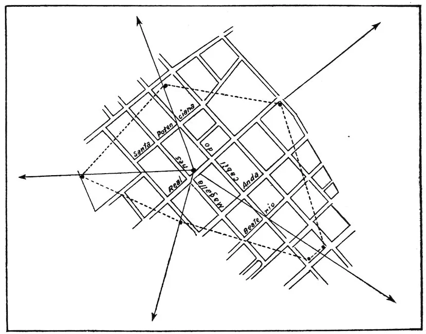
Fig. 59.—Isolated plague-infested center, Manila, P. I.
In addition, sanitary inspectors are instructed to bring in dead rats
which have evidently died of disease, and more detailed rat catchings
are made along the water front.
It is understood of course that rat proofing of the entire city
should be thoroughly carried out and constantly maintained.
1. Since the above system was adopted plague has disappeared in
the city of Manila; among human beings in 1906; among rats in 1907,
and it has not reappeared since.
2. That the cost is only a small fraction of that of general rat
extermination.
3. That the plan is thoroughly practical for any kind of a city.
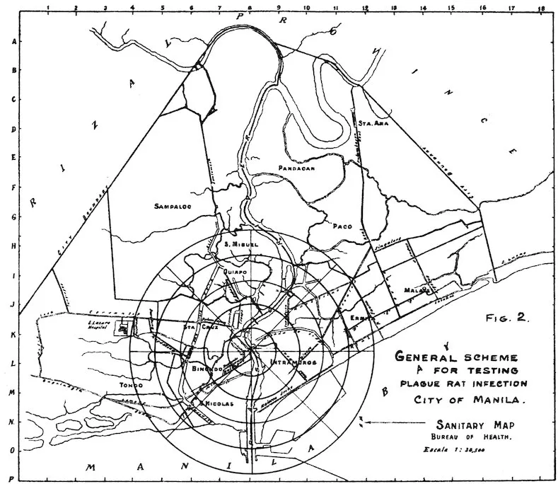
Fig. 60.—General Scheme for testing plague rat infection, City of Manila. Sanitary Map Bureau of Health. Escala 1:38,500
THE RAT IN RELATION TO SHIPPING. — ADAPTABILITY OF THE RAT TO HIS SURROUNDINGS. — DAMAGE TO CARGO. — FUMIGATION. — SUMMARY.
207
By William C. Hobdy,
Passed Assistant Surgeon, Public Health and Marine-Hospital Service.
Since men first went down to the sea in ships the rat’s voyage-making
tendencies have been known, and their fecundity is as well
established as their fondness for travel. The record does not state
that there were more than a pair on the ark at the beginning of her
voyage, but the chances are better than even that her skipper began
that voyage with more rats than his manifest showed; but whether
he did or not, we can be sure he had more at the end of the voyage
than at the beginning. Whether or not succeeding generations inherited
from their forbears on the ark this well-known wanderlust is
undetermined, but it is a fact that the intimacy and companionship
established and begun then have been persistently maintained by the
rat ever since. His travels have been coextensive with man’s, until
to-day there is not a port on earth where the rat is not present. Any
exception to this statement simply proves the rule. The rat is cute;
he knows when he is well off and his absence from a port does not
prove that he has not been there, but that he has been too intelligent
to follow man ashore. In establishing this shipboard intimacy there
has been no “by your leave” courtesy on his part either; he goes
without consent—against orders, even—and man’s ingenuity has as
yet discovered no effective means of keeping him off. This is not
surprising when the rat’s ability as a rope walker is considered. I
have seen a rat gallop with all appearance of enjoyment along an
inclined electric cable from a church steeple on one side of a street
into the second story of a hotel on the other. Others have been seen
traveling along the telephone wires from house to house, and on shipboard
they frequently have runways on small pipes along which they
scurry in perfect security. When a ship is fended off 6 feet from the
dock and her gang plank is lifted or guarded she is still freely accessible,
because all her mooring lines are only so many highways along
which rats can and do pass with ease and in perfect safety.
This fondness for ships and sea travel is shared by the various
species of the rat family, but the Mus norvegicus has earned the reputation
208of being the greatest traveler of them all. He almost invariably
predominated among those killed by fumigation on shipboard.
That he finds life on shipboard easy and the conditions satisfactory is
proved by the numbers that are destroyed from time to time by
fumigation. While in charge of the outgoing quarantine work in
San Francisco the chief engineer of a small lumber carrier called to
book his vessel for fumigation. The vessel was small, only 260 tons,
and carried nothing but lumber and her own ship’s stores, but the
chief declared she was overrun with rats, and to prove it showed
where they had eaten the patches from his shoes. He declared they
robbed him of his sandwich when he came off watch, and requested
me to give her a thorough fumigation. This was done. The next
morning the agent of the vessel phoned to ask how I measured rats,
stating that on this vessel they had collected “a barrelful and seven.”
Three hundred and ten on a little vessel of only 260 tons burthen.
On another vessel after one fumigation 100 were collected immediately
after fumigation, but a few days later, when the vessel was
undergoing extensive repairs, 425 others were found—a total of 525
on one small vessel. These numbers are small, however, when compared
with the results obtained on others, i. e., on grain-carrying
vessels. For instance, a vessel was fumigated some years since in
Bombay where 1,300 were destroyed at one time, and the Minnehaha,
a new vessel only nine months in commission, on fumigation in London,
England, in May, 1901, yielded a bag of 1,700 rats.
In addition to his qualities as a sailor and tight-rope walker, the rat
has the power of adapting himself to most unusual conditions and
surroundings. At the beginning of the outgoing work in San Francisco
it was urged that rats either could not or would not live on any
part of tank ships engaged exclusively in carrying oil, owing to the
fumes and vapors that permeated the entire vessel. This statement
was unquestionably correct for those compartments in which the oil
itself was stored or carried. It was not true, however, for the superstructure
of these vessels, for on one of the oil carriers 60 rats were
found after one fumigation, and of the thirty or more vessels of this
class that were regularly fumigated in San Francisco, although the
odors of oil or gasoline were quite strong in the living compartments,
not one was found that did not harbor rats. Still more remarkable,
as illustrating the rat’s adaptability, was the fact that from the large
refrigerating plants which some vessels carried and in which fumigation
had not been practiced for a long time rats were obtained that
had grown a fur an inch and a half long to protect themselves from
the cold.
209
That rats on shipboard in any such numbers as mentioned above
must do much damage to cargo can not be doubted.
Inquiry as to the extent of this damage showed that there were no
data on the subject. That such damage was common and considerable,
however, was revealed by the fact, elicited by these inquiries,
that nearly every steamship company on both the Atlantic and
Pacific took precautions both to keep rats from getting on board and
to destroy them after they did. One example will show what damage
may occur. The British steamer Gadsby, on a voyage from India to
Antwerp, covering a period of twenty-nine days, had 44,000 out of
46,000 bags of wheat cut by rats, with an estimated damage of $2,200.
The constant and almost universal presence, then, of rats on shipboard
can not be doubted, and if it could the results of fumigation,
wherever practiced with SO2, would serve to settle the question, for
they are found under all conditions, even on the most unlikely vessels.
How do these rodents gain access to a vessel? It has been the
custom to assume that they came on board from the docks over the
side when this was possible, and when it was not, as when the vessel
was fended off or stood too high out of the water, that they made use
of the gangways, mooring lines, hawsers, etc., as avenues of communication.
It is still the practice, therefore, in enforcing antirat precautions,
to compel the ship to fend off 6 feet from the dock, to wear
fat funnels on all lines, and to raise the gangway from the dock at
night. Just a word as to these precautions. The most practical
fender is a floating one made of heavy timbers either bolted together
into a solid frame, with the necessary cross braces bolted in, or made
up of logs or spars chained together. They should be long enough
to distribute the pressure of the vessel as the tide moves over a number
of piers or piles so that the weight does not bear, through the
medium of these fenders, on just one or two of the wharf foundations.
Such a fender will stay in position, will do no damage to the vessel,
and no matter how great the amplitude of the tide may be, will always
remain below the ship’s gunwale and can not therefore be utilized by
rats as a means to get on board. Large vessels require at least two.
Small ones need but one, and it was found in San Francisco, in the
case of those vessels changing their mooring several times daily, that
this one could be carried from wharf to wharf by the vessel without
trouble or delay simply by lashing it edgewise outside on top of the
guard.
Funnels should be of heavy galvanized iron, circular in shape, and
not less than 36 inches in diameter. The spout of the funnel should be
3 inches in diameter and should be at least 18 inches long. The flange
of the funnel should be soldered to this 18-inch pipe at its middle so
that the spout projects 9 inches out of the funnel and 9 inches into it.
210When the two halves of the funnel are brought together this spout or
tube is occupied by the line or hawser to be protected, and by lashing
this tube to the hawser the funnel is held in position and prevented
from lying down. Such a funnel should be put on every line from the
vessel to the dock, and when the tube does not fit the line the latter
should be parceled before the tube is lashed to it.
These, together with raising the gang plank from the dock at night,
make up the precautions ordinarily taken to prevent the rats from
getting on ships. As stated above, they are based on the assumption
that these are the common avenues of entrance. That these
precautions do much good can not be doubted, but in the writer’s
opinion they do not entirely cover the case, for there remains one
other road of ingress, one of the important, if not the most important,
which these precautions do not and can not block and
through which rats constantly get on board, and that is through the
medium of the cargo itself. There is at present nothing to prevent
access in this manner to a vessel and the route is so easy that there
can be no doubt that whole families of rats are carried on board in
this way. In fact some articles of cargo offer inviting harbors and
homes to rats, particularly when these articles have been stored for
a time in rat-infested warehouses. Among such articles of cargo
may be mentioned crockery or china packed in hay or straw or excelsior
and loosely crated; various articles of furniture packed in excelsior,
wrapped in gunny, and loosely crated; wheat, corn, oats,
peanuts, or barley when shipped in bags; and matting in hollow rolls
when sewed up in gunny. Any of these articles could easily become
the home of even an entire rat family after having been stored for a
time prior to shipment in a rat-infested warehouse. As a matter of
fact, the last plague rat discovered in San Francisco was found in a
bag of peanuts on the third floor of a warehouse.
That rats are thus carried on board is absolutely certain in my
opinion. In the recent antiplague campaign at San Francisco there
were ample opportunities for observations along this line, and in no
other way can the presence of rats in troublesome numbers on board
certain vessels be explained. These vessels were new, were freed
from rats by careful and repeated fumigation, and between these acts
touched at no wharves save in Honolulu and San Francisco, where
constant antirat precautions were observed. And yet on their second
trip (about five months after the fumigation had been discontinued)
they were badly rat-infested. Of course, by no means had all these
rats been carried on board in cargo, but the original patriarchs of the
colony had, after which, as is probably the case in all rat-infested ships,
their natural prolific characteristics did the rest.
In the same way, too, rats are carried from ship to shore and the
truth of Kitasatso’s aphorism that “wherever ships go, plague will go,”
211at once becomes apparent, and any regulations to prevent the introduction
of such vermin and the plague which they may carry to be
effective must include the inspection of cargo to insure its freedom
from rats, this inspection to be made just before it goes on board.
The relation these rodents bear to plague and the part they play
in its transmission have been thoroughly discussed and set forth in
another article in this work. The work of Ashburton Thompson in
Australia and of the British Medical Commission in India was a
scientific demonstration that plague was primarily a rat disease
transmitted by fleas, while McCoy in the United States has gone
further probably than anyone else in demonstrating the manner in
which the flea does this work. The importance of this relation is
emphasized and the difficulty of ridding a port of pest infection is
explained by a fact, first observed so far as I know by Klein, of London,
that rats suffer from a chronic form of pest, not fatal, at least for a
long time, and during the course of which the rat may exhibit practically
his normal activity. This fact then, that plague is primarily a
rat disease and that it may occur in the rat in a chronic form, shows
how great the danger may be from their presence on shipboard,
explains why it is that where ships go plague will go, and emphasizes
the importance of destroying them on shipboard apart from the damage
and loss which their presence entails.
So important is this and so preeminent is the rôle played by the
rat in plague transmission and propagation that I believe regulations
should demand that all ships be disinfected at least three times, and
better still, four times, a year, for the destruction of rats. If this
precaution were taken, and if to it were added an inspection of cargo
at the port of embarkation to insure its freedom from rats, I believe
the disinfection of cargo could be entirely dispensed with. It is
infectious only in so far as it harbors rats, and if these are not present
disinfection, in my opinion, does as little good in preventing plague as
dipping ballast did in preventing yellow fever.
Once the rat has gained access to a vessel, what is the best method
of getting rid of him?
There are several methods, all of which are effective if properly
used, and all of which depend on sulphur dioxide as the destructive
agent. The following are mentioned: Pot and pan, sulphur furnace,
Clayton system, and Marot system. A choice of one of these methods
will be determined by the cost, the rapidity of fumigation desired,
and the condition of the vessel, whether empty or loaded. No matter
which method is selected, to be effective the sulphur dioxide must be
simultaneously delivered to or generated in every compartment on
the vessel.
212For an empty vessel nothing is so satisfactory as the pot and pan
method of generating the gas. It has the following advantages; is
more rapid than any other, is cheaper, is more effective, and is equally
applicable to the largest and the smallest vessel afloat. With a sufficient
number of pots and pans 3,500 pounds of sulphur can be
burned just as quickly as 100. Ten pounds of sulphur in each of
350 pots will be consumed just as quickly as will 10 pounds in any one
of that number, namely, in less than five hours, a fact which was
demonstrated over and over in the outgoing work in San Francisco.
At the beginning of this work it was thought a 2½ per cent gas with
three hours’ exposure would be sufficient. Practice proved, however,
that this was not effective and the strength of gas was increased
to 3 per cent and the exposure to five hours which a test, extending
over twelve months and embracing over 3,000 vessels, proved to be
ample.
The best pot in which to burn sulphur is 6 inches deep, has a flare
of 6 inches—that is, the diameter at the top exceeds the diameter at
the bottom by that much, is from 16 inches to 24 inches in diameter
at the top and has four hemispherical legs about the size of half a
billiard ball. These pots when in use are set in a galvanized iron
tub. These tubs contain a little water, and are of a diameter 6 inches
greater than the top of the pot. The hemispherical legs of these pots
will not punch holes into the tub. The pots are filled with sulphur,
which is hollowed out into a little crater at the top, into which crater
from 4 to 6 ounces of alcohol are poured and when all are ready a
lighted match is dropped into each little crater and the compartment
is closed.
In actual practice it was found that an exposure of five hours to a
3 per cent gas would not destroy all the rats in absolutely every case.
Some ships afford better hiding places than others, and on these an
occasional rat would escape. It was the custom, however, to fumigate
all vessels every thirty days and after the third fumigation, on
vessels that did not carry general cargo, no more rats were obtained,
though the fumigations were continued for a number of months.
On those vessels that carried miscellaneous general cargoes a few
rats were found after almost every fumigation. These vessels
touched no wharf from the time they left San Francisco until their
return, except for a short time in Honolulu, where adequate precautions
were observed, and it is difficult to understand how these
rats got on board if they were not carried on in cargo.
For vessels with cargo in their holds the pot and pan method is
dangerous owing to the possibility of fire. For these vessels one of
the other methods of generating the sulphur gas must be used. This
involves the use of an expensive plant consisting of a furnace, cooling
chamber, blower, or fan, and a system of mains and delivery pipes
213by means of which the gas is delivered to the various holds and
compartments of the vessel. To be at all effective the gas must be
4½ per cent strength, with at least twenty-four hours exposure. The
one recommendation of such a system is its freedom from danger by
fire. It is too slow; the pipes, even where 6 inches in diameter, are
liable to clogging with sublimed sulphur, an inevitable result if the
fans are driven too rapidly, and it is not possible to do more than
one or at most two ships at a time. The inadequacy of such a
method when compared to the work done in San Francisco where we
averaged over nine vessels every day for almost fourteen months is
at once apparent. Many ships now carry their own disinfecting
plants, by means of which not only is sulphur dioxide generated and
pumped into a compartment, but at the same time also the air of
this space is sucked out. This principle is excellent, but in its application
the machines used are wholly inadequate, having a very limited
sulphur capacity per hour and equipped with delivery pipes in
many instances only 2 or 3 inches in diameter. It would be a matter
of days to disinfect some of these ships with the machines they carry.
In San Francisco we again and again used pots and pans to fumigate
these vessels, including the very compartments in which their own
machines sat doing nothing.
The Marot system of generating the gas from compressed liquid
sulphur dioxide has in this country been found too expensive to
apply to vessels. Probably no system will effectually destroy all the
rats on a cargo-laden vessel.
To summarize then:
1. The rat is found on all vessels, sometimes in enormous numbers,
and is able to adapt himself to all sorts of conditions. He either
gets on board himself or is carried on in cargo. Owing to his seagoing
tendency, his distribution is world-wide.
2. On shipboard, to live he must do damage to either cargo or
stores, or both.
3. Plague is primarily a rat disease; it may exist in the rat in a
chronic form. Hence where ships go plague will go sooner or later.
4. To prevent the ingress of rats and the consequent spread of
plague, ships should observe antirat precautions, and cargo inspection
should be included in these.
5. At stated intervals, three or, better still, four times a year, all
vessels should be fumigated for the destruction of rats.
6. On empty vessels this can best be done by generating sulphur
by the pot and pan method.
7. On laden vessels some special apparatus must be used to generate
the gas. A longer exposure is required, at least twenty-four
hours, and the gas should be 4½ per cent strength instead of 3 percent.
It is extremely difficult by any method to kill all the rats on
a cargo-laden vessel.
THE RAT AS AN ECONOMIC FACTOR. — INTRODUCTION. — UTILITY OF THE RAT. — DESTRUCTIVENESS OF THE RAT. — DAMAGE TO GRAINS. — MERCHANDISE IN STORES AND WAREHOUSES. — MERCHANDISE IN TRANSIT. — POULTRY AND EGGS. — GAME AND WILD BIRDS. — FRUITS AND VEGETABLES. — FLOWERS AND BULBS. — FIRES. — BUILDINGS AND FURNITURE. — MISCELLANEOUS. — AMOUNT OF LOSSES CAUSED BY RATS. — INDIRECT LOSSES.
215
By David E. Lantz,
Assistant Biologist, United States Department of Agriculture.
The world has rightly learned to dread rats as disseminators of
disease, and recent efforts to rid cities of the pests have resulted
chiefly from sanitary considerations. Yet the material losses due to
depredations of rats are now, and always have been, a sufficient argument
for their destruction. The requirements of sanitation and
public health are slowly bringing to pass what economic interests
failed to accomplish, namely, a general recognition of the fact that
the rat is a standing menace to prosperity. To point out some of
the many ways in which rats inflict injury and the extent to which
they drain the resources of the people is the object of the present
chapter.
Do rats serve any useful purpose? With very slight reservation,
the question may be answered in the negative. There have been
times and places in which the rat’s work as a scavenger accomplished
good, but modern methods of garbage disposal are superseding the
feeding it to rats.
It was Robert Southey, the poet, who, nearly a century ago,
humorously suggested as the first three steps to eradicate rats—first,
introducing them as a table delicacy; second, utilizing the skins; and
third, inoculating them with a contagious disease.[BY] The last of these
plans is now receiving considerable attention from bacteriologists, but
the others, for obvious reasons, have been neglected.
It is true that under exceptional circumstances the rat has been a
source of human food. The principal instances on record were during
the siege of Paris in 1870, and during the siege of the French garrison
at Malta, 1798–1800, when food was so scarce that rat carcasses brought
high prices. Another was on board the ship Advance during an
216arctic winter, when Doctor Kane attributed his entire immunity from
scurvy to his diet of fresh rats, of which none of the other members
of the party would partake.[BZ]
The statement is often made in newspapers, and even in encyclopedias,
that in Europe, and especially in France, rat skins are extensively
used in the manufacture of gloves. The late Frank T. Buckland,
about a half century ago, made diligent inquiry in London, and
through friends in Paris and other places on the Continent, but found
no confirmation of such statement. He concluded that either rat
skins were not used for making gloves or the manufacturers were
unwilling to acknowledge such a use.[CA] Personally, the writer has
been unable to learn of any demand or market for rat skins at the
present time. They are not strong, and the fur is of inferior quality.
The occasional finding of one or more rat skins in the fur lining of
coats is probably to be explained by the fact that they are sometimes
included in lots of small muskrat skins (“kitts”) and overlooked by
the buyer.
Rats inflict injury in a surprising number of ways, and before an
attempt is made to consider the magnitude of the losses due to these
animals a statement of the nature of their depredations should be
made.
Cultivated grains are the favorite food of rats. The animals begin
their depredations by digging up the newly-sown seed. They eat
the tender sprouts when they first appear, and continue destroying
the plants until the crop matures. They then attack the grain itself,
and after harvest take toll from shock, stack, mow, crib, granary,
elevator, mill, and warehouse. When rats are abundant their
depredations amount to an appreciable percentage of the entire yield
of grain, and in exceptional cases whole crops have been ruined.
INDIAN CORN.
Probably this crop suffers greater injury from rats than any other
in the United States. To some extent the animals dig up newly
planted corn, but their injury to the maturing grain is far greater.
They are especially fond of corn in the milk stage, and often climb the
upright stalks and strip the cobs bare. In this way sometimes whole
fields are destroyed.
Corn in the shock is often attacked by rats, especially in parts of
fields adjacent to hedges, drains, or embankments that afford shelter
217for the animals. A pair of rats often make a corn shock their home,
and soon destroy both grain and fodder.
Corn in cribs is often damaged by rats. Many cribs are built close
to the ground, and rats take up their abode under the floor. They
soon gnaw through the wooden barrier and have free access to the
grain. They shell the corn and eat the soft part of the kernels, wasting
much more than they eat. They carry the grain into underground
burrows and bring up moist soil from below, which in contact
with the grain makes it moldy and unfit for market or for feeding to
stock. A number of farmers have reported the loss by rat depredations
of from a fifth to a half of the contents of a large corn crib during
a single winter.
An Iowa farmer, writing to an agricultural journal, relates the
following experience:
We had about 2,000 bushels of corn in three cribs to which rats ran, and they ate and
destroyed about one-fourth of the corn. Much of it was too dirty to put through the
grinder until it had been cleaned an ear at a time. All the time we were poisoning
and trapping the rats. We killed as high as 300 rats in two days and could hardly miss
them. They destroyed more than enough corn to pay taxes on 400 acres of land.[CB]
Throughout the United States, but especially in the West and
South, corn is often stored for months in rail or other open pens, to
which rats have free access. Often the loss in a single season
would pay for the construction of rat-proof cribs, or at least for wire
netting, that would fully protect the crop.
SMALL GRAINS.
Much has been written about the rat as a house and barn pest,
but its depredations in the fields have usually been overlooked. In
some localities the common rat, as well as the house mouse, swarms
in the fields, especially in summer, and subsists entirely upon the
farmers’ crops.
Stacked grain is peculiarly exposed to rat depredations. In the
United States, although the cost of protection is small, rats are
seldom fenced away from stacks, and, if threshing is delayed, serious
loss results. Often, at the removal of a stack, large numbers of rats
are discovered, which have been living at the expense of the farmer.
As early as 1832 a farmer in Frederick County, Md., with the help
of men and dogs, killed 217 large brown rats from one stack of rye.[CC]
In England instances are on record of the killing of over a thousand
rats from one stack of wheat.
The destruction of feed by rats is a serious loss not only on the
farm but also in city and village. The feed bin or barrel is often
left uncovered and rats swarm to the banquet thus exposed. Small
218feeders suffer greater proportional losses, for managers of larger
barns recognize the enormous drain and usually provide rat-proof
bins, if not rat-proof stables. When rats have access to a stable
they take a good share of the feed directly from the mangers, but the
loss is seldom noticed.
Rats are exceedingly fond of malt, and in malt houses and breweries
constant watchfulness is needed to prevent losses. Mills, elevators,
and warehouses in which grain and feed stuffs are stored are
subject to constant invasion by rats and mice.
A full-grown rat consumes about 2 ounces of grain daily. A half-grown
rat eats nearly as much as an adult. Fed on grain, therefore,
a rat eats from 45 to 50 pounds a year. The cost depends
somewhat on the kind of grain. If wheat, the value is 60 to 75 cents;
if oatmeal, about $1.80 to $2. Several feeders of horses in Washington,
D. C., estimated the cost of keeping each rat on their premises
at $1 a year. Even though half the grain eaten is waste, the direct
loss from this source to feeders is enormous.
The loss from depredations of rats on miscellaneous merchandise
in stores, markets, and warehouses, is second only to the losses on
grains. Not only are food materials of every kind subject to attack,
but the destruction of dry goods, clothing, books, leather goods,
and so on is equally serious. Merchandise other than foodstuffs is
usually destroyed for making nests, but books and pamphlets,
especially the newly bound, and some other articles, furnish food
in the glue, paste, oils, or paraffin used in their manufacture. Some
kinds of leather have a peculiar attraction for rats, while others are
never touched. Shoes are seldom gnawed unless they have cloth
uppers or are made of kid. New harnesses are not often attacked,
except collars, which contain straw, and cruppers, which are stuffed
with flaxseed. Old harness leather is salty from the perspiration of
horses, and rats and mice gnaw it for this reason. Kid gloves and
other articles made of similar leather are often destroyed by rats.
Lace curtains, silk handkerchiefs, linens, carpets, mattings, and
other dry goods in stores are often attacked by rats. Some of the
stuffs contain starch, which serves as food, but most of them furnish
nesting materials only. A slight injury makes these articles unsalable;
this is especially true of white goods, which are easily ruined
by soiling. Nearly all large dry goods and department stores suffer
heavy losses from rats. Grocers, druggists, confectioners, and other
merchants also have similar experiences, and to the direct losses
must be added the sums expended in fighting the pests.
219
Merchandise billed for shipment often lies for days in stations and
warehouses or on wharves, where depredations of rats and mice
cause heavy losses to shippers and consignees. Similar losses occur
on boats carrying merchandise from port to port.
Fruits and vegetables in transit on steamboats are often destroyed
or damaged by rats. Tomatoes, cucumbers, sweet potatoes, bananas,
oranges, grape fruit, peanuts, and similar produce shipped by water
from the South, especially in winter, reach northern markets with a
large percentage of loss.
In view of the practicability of destroying rats on ships by fumigation,
and the ease with which rat-proof compartments for stowing
produce can be constructed, it would seem that losses of this nature
should be entirely prevented.
Aside from disease, the greatest enemy of poultry is the rat. The
loss from rats varies with their abundance and the care taken to exclude
them from the poultry yard. The magnitude of the damage
is not generally known, because much of it is blamed on other animals,
particularly minks, skunks, and weasels. Much of the injury
occurs at night, and the actual culprit is seldom detected. Farmers
have heard that minks, skunks, and weasels prey upon poultry.
What more natural than to conclude that one of these animals is
doing the mischief, especially if one has been seen about the premises?
Rats often prey upon small chicks, capturing them in the nests at
night or even about the coops in the daytime. The writer has
known rats to take nearly all the chicks on a large poultry ranch, and
over a large section of country to destroy nearly half of a season’s
hatching. Young ducks, turkeys, and pigeons are equally liable to
attack, and when rats are numerous, are safe only in rat-proof yards.
A writer in a western agricultural journal states that in 1904 rats
robbed him of an entire summer’s hatching of three or four hundred
chicks.[CD] A correspondent of another newspaper says, “Rats destroyed
enough grain and poultry on this place in one season to pay
our taxes for three years.”[CE] When it is remembered that the poultry
and eggs marketed each year in the United States have a farm value
of over $600,000,000, it will be seen that a small percentage of loss
represents an enormous sum.
The destruction of eggs by rats is great, not only on the farms
where they are produced, but also in the markets. Commission
men and grocers complain of depredations upon packed eggs. The
220animals break and eat a few eggs at the top of a case and the broken
yolks run down and soil the eggs below. Then, too, rats carry away
unbroken eggs, displaying much ingenuity in getting them over
obstacles, as up or down a stairway.
A commission merchant in Washington, D. C., states that he once
stored 100 dozen eggs in a wooden tub in his warehouse and left
them for nearly two weeks. He then found that rats had gnawed a
hole through the tub, just under the cover, and had carried away
71½ dozen, leaving neither pieces of shell nor stains to show that any
had been broken.
Besides their destruction of eggs and young fowls, rats eat much
of the food put out for poultry. They are destructive also to tame
pigeons and their eggs, but particularly to young squabs. They
climb the wire netting and gain entrance to the cages through the
same openings by which the pigeons come and go. Fanciers are
often put to great trouble to protect their pigeons from rats, and
because of these pests some of them have abandoned the business.
The rat is the most serious pest in European game preserves. A
writer in Chambers’s Journal says:
In a closely preserved country at the end of an average year the game suffers more
from the outlying rats of the lordship than from the foxes and mustelines together.
The solitary rats, whether males or females, are the curse of a game country. They
are most difficult to detect, for in a majority of cases their special work is supposed to
be done by hedgehog, weasels, or stoat.[CF]
The propagation of game birds is becoming a promising industry
in the United States. The difficulties of the business are not yet
fully known, but the rat is an enemy with which the raiser of game
will have to contend. The animal has already proved itself a foe in
American pheasantries.
Our wild native game birds are less subject to rat depredations
than birds kept in confinement. The nests of ruffed grouse are in
woodlands; those of the prairie hen and related species are on plains
remote from the haunts of rats. The quail, however, often makes
its nest within the summer range of rats, which destroy many of its
eggs.
Rats are said often to destroy the nests of wild ducks, woodcock,
and other marsh birds. Terns have been entirely driven from their
nesting grounds in this way. In England the common tern was
extirpated from the Thames marshes; and on Loggerhead Key,
Tortugas Islands, off the Florida coast, rats recently nearly exterminated
a colony of least terns by destroying the eggs.
221The nests of many ground-nesting and other song birds are robbed
by rats. Crows, jays, snakes, and skunks are blamed for most of
the destruction and the actual offender seldom suspected. While
the other animals named do part of the mischief, the rat is a more
serious foe of song and game birds than any of these.
A well-known form of damage by rats is the destruction of fruits
and vegetables in cellars and pits. Apparently no garden vegetable
or common fruit is exempt from attack. But the rat does not confine
its depredations to stored fruits and vegetables. It attacks ripe
tomatoes, melons, cantaloupes, squashes, pumpkins, sweet corn, and
many other vegetables in the field; and often the depredations are
attributed to rabbits or other animals, which may or may not be
concerned in the mischief.
Rats are fond of the small fruits, eating not only the fallen but
climbing vines and canes to obtain the ripe grapes or berries. They
eat also apples, pears, cherries, and other fruits. The brown rat,
while not so expert as the black or the roof rat, readily climbs trees
and obtains fruit even at the extremities of the branches.
Among tropical fruits injured by rats are oranges, bananas, figs,
dates, cocoanuts, and especially the pods of cacao (Theobroma cacao),
from which chocolate is manufactured. H. N. Riddey, writing of
his experiences on the island of Fernando do Noronha, South America,
mentions the destructiveness of rats in this penal colony. They
climb the cocoanut palms and papaw trees to devour the fruit, and
do mischief in melon patches. To lessen the evil, each convict was
required to bring in a certain number of dead rats, and battues were
held monthly to satisfy the requirement. Sometimes the number
killed in a single hunt reached 20,000.[CG]
Fruits and vegetables grown under glass are subject to injury by
rats. The animals usually find entrance to greenhouses by way of
openings for pipes or drains.
Rats attack seeds, bulbs, and the leaves, stems, and flowers of
growing plants, whether in the greenhouse, propagating pits, or elsewhere.
Of flowering bulbs, the tulip suffers most from rats. Hyacinths
also are eaten; but, probably because they are slightly poisonous,
narcissus and daffodil bulbs escape injury. Rats eat pinks,
carnations, and roses, cutting the stems off clean. They denude
geraniums of both flowers and leaves. They attack the choicest
blooms of chrysanthemums and carnations in markets, stores, and
exhibition rooms, causing heavy losses.
222
Rats and mice cause many fires. Several specific instances have
been reported by the fire department of the city of Washington
within the past two or three years. It is likely that some of these
fires are caused by rats gnawing matches. The animals are fond of
paraffin, which is often used to protect match heads. They carry
the matches to their nests, which are composed of paper and other
combustible materials, and the conditions for a conflagration are
ready. Since the heads of matches contain from 14 to 17 per cent
of phosphorus, actual gnawing is not required to ignite them, but
heat or friction from any cause may suffice.
Fires in mills or warehouses have sometimes been traced to the
spontaneous ignition of oily or fatty rags and waste carried under
floors by rats. Cotton and woolen mills are said to be peculiarly
subject to fires of this kind.
Sometimes rats cause fires by gnawing through the lead pipes
leading to the gas meter. Workmen or others, searching for the
leak, accidentally ignite the gas. Phillips’s warehouse, London, was
twice badly damaged by fires originating in this way, and in several
instances the sleeping inmates of houses have been in danger of
asphyxiation by gas freed in this manner.
The most common way in which rats and mice cause fires is by
the destruction of the covering of electric light wires under floors or
in partition walls. A considerable percentage of the enormous fire
losses in the United States is caused by defective insulation of wires.
After wires are once in position rats are the chief agents in impairing
the insulation. These animals do much mischief also by gnawing
off the coverings of telephone wires. In the case of electric light
wires the covering is probably used by the rats for nesting material,
but frequently the paraffin in the insulation is the object of attack.
Rats seem to be able to gnaw through almost any common material
except stone, hard brick, cement, glass, and iron; neither wood nor
mortar suffice to keep them out of bins or rooms. They sometimes
gnaw through walls or doors in a single night. In the same way they
enter chests, wardrobes, bookcases, closets, barrels, and boxes.
Almost every old dwelling bears evidence of its present or former
occupancy by rats. Often the depreciation of houses and furniture
is largely due to marks left upon them by rats—marks that paint and
varnish can not hide.
Damage to dwellings by rats is a large item. The decay of sills and
floors is hastened by contact with moist soil brought up from rat
burrows. Ceilings, wall decorations, and floor coverings are flooded
223by leaks in lead pipes or wooden tanks gnawed by rats. Bricked
areas and even foundations are undermined and ruined by rats. All
this is real waste and a constant drain on the resources of the country.
A few instances of miscellaneous damage by rats may be mentioned
to show the great variety of mischief chargeable to the animals.
A Washington, D. C., merchant reported that at one time rats in
his store destroyed 50 dozen brooms worth $2.50 a dozen. In another
store, in a single night, they broke $500 worth of fine chinaware on
shelves and tables. A dealer in harness reported the loss of $400
worth of collars in one season. The manager of a restaurant complained
of an average loss of $30 a month in table linen ruined by rats
and mice. A hotel reported the destruction of $75 worth of linen in
a month.
At Mobile, Ala., in March, 1908, lost jewelry worth $400 was recovered
from a rat’s nest in the home of Señor Viada.
In London rats at one time killed all but 11 out of an aviary of
366 birds.
At Hamburg, Germany, Carl Hagenbeck once had to kill three
young African elephants because rats had gnawed their feet, inflicting
incurable wounds.
Rats often gnaw the hoofs of horses until they bleed. They kill
young lambs and pigs, and have been known to gnaw holes in the
bodies of very fat swine, causing death.
Like the muskrat, the brown rat often burrows into embankments
and dams, causing serious breaks.
The rat is one of the greatest enemies with which the sugar planter
has to contend, destroying acres of growing cane.
In rice plantations rats not only break down and destroy the growing
crops, but burrow into the dikes and flood the fields at the wrong
season.
On the London docks and on shipboard ivory is often badly damaged
by rats. They select for attack the greenest tusks, which are
the more valuable.
Mail sacks and other kinds of bagging are greatly injured by rats.
The consequent loss and necessary outlay for repairs are a large item
in post-office expenditures and in mills and other places where bagging
is used.
About the year 1616 rats caused a two years’ famine in the
Bermudas. In the southern Deccan and Mahratta districts of India
rats ate a large part of the scant crops of 1878 and 1879, and were
regarded as in a great measure responsible for the severe famine which
followed.[CH] A writer in Chambers’s Journal stated that the Dutch
224abandoned the Isle of France (Mauritius) in 1610 because of the great
abundance of rats.[CI]
The damage done by a single rat varies greatly with the circumstances.
We have already stated that the cost of feeding a rat on
grain varies from 60 cents to $2 a year. Assuming that much of
the rat’s food is waste, each rat on a farm will cause a loss of over 50
cents a year. In cities the damage by a single rat probably averages
more than in the country. Hotel managers and restaurant keepers
state that $5 a year is a low estimate of the loss inflicted by a rat.
In making an estimate it should be remembered that the rat is to be
charged not only with the food it actually consumes but also with
what it destroys or pollutes and renders unfit for use.
If an accurate census of the rats in the United States were possible,
and if the minimum average loss caused by a rat were known, an
estimate of the total annual losses from their depredations could be
made. It was on such a minimum basis that the Incorporated
Society for the Destruction of Vermin arrived at their total estimate
of £15,000,000 ($73,000,000) as the yearly losses from rats in Great
Britain and Ireland. Three propositions were assumed: first, that
in cities and villages the number of rats equals the population; second,
that in the country there is at least one rat for every acre of cultivated
land; third, that each rat in the kingdom inflicts a damage of at least
a farthing per day. Circulars asking whether these assumptions are
excessive were distributed throughout the country. From 90 to 99
per cent of the replies fully indorsed each of the assumptions.
It can readily be seen that the English basis of estimate would not
apply to farm conditions in the United States, where the area in the
twelve leading crops alone is over 250,000,000 acres. On a basis of
a rat per acre and damage of a farthing per day the annual loss on
this area would be $450,000,000, a sum much too great for serious
consideration. However, in the more thickly populated parts of the
country an estimate of one rat per acre would not be excessive; and
it is probable that in most of our cities there are quite as many
rats as people. Yet it would be unsafe, owing to our vast territory
and varying conditions, to make these assumptions the basis for
conclusions.
Over a year ago the writer made an attempt to investigate actual
conditions, and thus arrive at an estimate of the total damage by rats
in the cities of Washington and Baltimore. From the data obtained
the direct annual damage in the two cities was calculated at $400,000
and $700,000, respectively. The Census Bureau in 1906 estimated
225the population of these cities at 308,000 and 554,000, respectively.
If the estimates of damage were correct, the average loss for each
person is $1.27 a year; and, on the same basis, the 28,000,000 of urban
population in the United States (census of 1900) sustains an annual
direct injury of $35,000,000 from rats. This is considerably lower
than on the English assumption, which would make the losses in our
cities over $50,000,000.
Denmark (population 2,500,000) has an estimated rat bill of about
$3,000,000 a year, or $1.20 a person. Germany (population
56,000,000) is said to sustain a loss from rats of 200,000,000 marks
($47,640,000) a year, or about 85 cents for each person. The per
capita estimate for the United Kingdom is about double that made for
Germany. In France the loss from rats and mice for a single year
(1904) was placed at $38,500,000, or a little over a dollar for each of
its 38,000,000 inhabitants. These estimates are supposed to include
only direct losses, but they vary enough to show that no common
basis can be assumed for all countries. With present information,
therefore, any attempt to state the amount of loss from rats in the
United States would be largely guesswork. Considering the population
and wealth of the country, however, and the vast area over which
rats are abundant, it is not unreasonable to conclude that in the
United States the losses from rats amount to much more than in any
of the other countries named.
To the direct losses caused by rats must be added the cost of fighting
the animals and of protecting property from them. In our larger
cities a number of so-called expert rat catchers are to be found, who
operate with dogs, ferrets, traps, poisons, or other means, and who
have an extensive clientage among merchants, hotel managers, and
others. These pay the rat catcher a yearly or monthly stipend to
keep their premises free of rats and mice. Some of the large establishments
pay $200 to $600 yearly for such service. While the agreements
are seldom kept in full, the clients are usually satisfied that
results warrant the expense. Even when no contractor is employed,
merchants are at expense for traps, poisons, the keep of cats or dogs,
and other means of fighting rats. The same is true in less degree of
nearly every householder.
The cost of protecting property from rats is no small item. It
increases the expense of nearly all building, but it greatly reduces
direct losses from the animals. The economy of rat-proof construction
is everywhere manifest, in city or country, and the necessity for
it can not be too strongly emphasized.
226Depreciation of property and loss of rents and custom are items not
generally thought of in connection with rat damage. A few years
ago the writer knew of almost an entire block of city houses that
remained untenanted for several months, because infested by rats.
As the houses were otherwise desirable, the loss of rent to the owners
was probably nearly $2,000. The presence of rats in markets, hotels,
and restaurants inevitably results in loss of custom, besides the direct
damage by the rodents.
From every point of view the keeping of rats is exceedingly expensive,
and the sooner our population can be made to realize the enormous
drain upon our resources caused by these rodents the sooner
can concerted measures for their destruction be made effective.
THE RAT IN RELATION TO INTERNATIONAL SANITATION. — INQUIRY INTO THE CRUSADE AGAINST RATS THROUGHOUT THE WORLD. — RAT EXTERMINATION IN UNITED STATES PORTS. — RAT EXTERMINATION IN CHINESE CITIES. — RAT EXTERMINATION IN MADRAS, BOMBAY, AND CALCUTTA, INDIA. — RAT EXTERMINATION IN YOKOHAMA AND NAGASAKI, JAPAN. — RAT EXTERMINATION IN LOURENÇO MARQUEZ, EAST AFRICA, AND MADAGASCAR. — RAT EXTERMINATION IN CAPE TOWN, SOUTH AFRICA. — RAT EXTERMINATION IN ALEXANDRIA AND CAIRO, EGYPT. — EXTERMINATION OF RATS IN THE PORT OF CONSTANTINOPLE. — RAT EXTERMINATION IN RUSSIAN PORTS. — DESTRUCTION OF RATS IN TRIESTE, AUSTRIA. — DESTRUCTION OF RATS IN GENOA, ITALY. — RAT DESTRUCTION IN BARCELONA, SPAIN. — RAT DESTRUCTION AT FRENCH PORTS. — DESTRUCTION OF RATS IN GERMAN PORTS. — MEASURES AGAINST RATS AT THE PORT OF ROTTERDAM. — DESTRUCTION OF RATS AT ANTWERP, BELGIUM. — DESTRUCTION OF RATS IN DENMARK. — CIRCULAR TO THE LOCAL AUTHORITIES. — DESTRUCTION OF RATS IN SWEDISH PORTS. — DESTRUCTION OF RATS IN ENGLISH PORTS. — MEASURES AGAINST RATS AT AUSTRALIAN PORTS. — MEASURES AGAINST RATS IN SOUTH AMERICAN PORTS. — MEASURES AGAINST RATS IN WEST INDIAN PORTS. — DESTRUCTION OF RATS IN PANAMA. — MEASURES AGAINST RATS IN VANCOUVER. — NECESSITY OF CONCERTED ACTION OF NATIONS.
227
By Asst. Surg.-Gen., John W. Kerr,
Public Health and Marine-Hospital Service.
Rats, like man, had their origin in Asia, from which continent
they have finally become disseminated throughout the world. They,
too, like man, are great travelers, and therefore prey on the commerce
of the ships in which they are carried. For this reason, and
on account of the fact that they are subject to plague and may
transmit the disease from one country to another, these animals have
an influence on international policy, and their control aboard ships
is an international problem.
It has been estimated that there are as many rats as there are
human beings, and that each rat causes each day a loss by the destruction
of material of at least half a cent.
Assuming that the rat population aboard ships is as great as the
human population—and my experience gained during the fumigation
of ships to kill rats convinces me that on the whole it is greater—some
idea may be had of the enormous migrations of rats and the
toll they exact for food from international commerce. Some idea
can also be had of the danger of rats in transmitting plague when
it is remembered that 51 countries have been infected with the
disease since the present pandemic began in Canton, China, in 1894,
and when it is known that at least 146 ships have had plague infection
on board during that time.
During the International Sanitary Conference of Paris in 1903
the influence of the rat in transmitting plague was borne in mind,
and the international sanitary agreement, which was signed ad
referendum December 3, 1903, requires the destruction of rats aboard
plague-infected ships, recommends it on ships suspected of being
plague infected, and permits it on ships indemne from plague. The
ship is considered indemne from plague which, although coming
from an infected port, has had neither death nor case on board
either before departure, during the voyage, or at the moment of
arrival.
228The International Sanitary Convention signed at Washington,
October 14, 1905, follows the text of the Paris convention, with
respect to plague, consequently embodying similar requirements and
recommendations as follows:
Art. XX. Classification of ships.—A ship is considered as infected which has plague,
cholera, or yellow fever on board, or which has presented one or more cases of plague
or cholera within seven days, or a case of yellow fever at any time during the voyage.
A ship is considered as suspected on board of which there have been a case or cases
of plague or cholera at the time of departure or during the voyage, but no new case
within seven days; also such ships as have lain in such proximity to the infected
shore as to render them liable to the access of mosquitoes.
The ship is considered indemne which, although coming from an infected port,
has had neither death nor case of plague, cholera, or yellow fever on board, either
before departure, during the voyage, or at the time of arrival, and which in the case
of yellow fever has not lain in such proximity to the shore as to render it liable, in
the opinion of the sanitary authorities, to the access of mosquitoes.
Art. XXI. Ships infected with plague are to be subjected to the following regulations:
1. Medical visit (inspection).
2. The sick are to be immediately disembarked and isolated.
3. Other persons should also be disembarked, if possible, and subjected to an observation,[CJ]
which should not exceed five days, dating from the day of arrival.
4. Soiled linen, personal effects in use, the belongings of crew[CK] and passengers
which, in the opinion of the sanitary authorities are considered as infected, should be
disinfected.
5. The parts of the ship which have been inhabited by those stricken with plague,
and such others as, in the opinion of the sanitary authorities, are considered as infected,
should be disinfected.
6. The destruction of rats on shipboard should be effected before or after the discharge
of cargo, as rapidly as possible, and in all cases with a maximum delay of
forty-eight hours, care being taken to avoid damage of merchandise, the vessel, and
its machinery.
For ships in ballast, this operation should be performed immediately before taking
on cargo.
Art. XXII. Ships suspected of plague are to be subjected to the measures which
are indicated in Nos. 1, 4, and 5 of Article XXI.
Further, the crew and passengers may be subjected to observation, which should
not exceed five days, dating from the arrival of the ship. During the same time
the disembarkment of the crew may be forbidden, except for reasons of duty.
The destruction of rats on shipboard is recommended. This destruction is to be
effected before or after the discharge of cargo, as quickly as possible, and in all cases
with a maximum delay of forty-eight hours, taking care to avoid damage to merchandise,
ships, and their machinery.
For ships in ballast this operation should be done, if done at all, as early as possible,
and in all cases before taking on cargo.
Art. XXIII. Ships indemne from plague are to be admitted to free pratique
immediately, whatever may be the nature of their bill of health.
229The only regulation which the sanitary authorities at a port of arrival may prescribe
for them consists of the following measures:
1. Medical visit (inspection).
2. Disinfection of soiled linen, articles of wearing apparel, and the other personal
effects of the crew and passengers, but only in exceptional cases when the sanitary
authorities have special reason to believe them infected.
3. Without demanding it as a general rule, the sanitary authorities may subject
ships coming from an infected port to a process for the destruction of the rats on board
before or after the discharge of cargo. This operation should be done as soon as possible,
and in all cases should not last more than twenty-four hours, care being taken to avoid
damaging merchandise, ships, and their machinery, and without interfering with
the passing of passengers and crew between the ship and the shore. For ships in ballast
this procedure, if practiced, should be put in operation as soon as possible, and in all
cases before taking on cargo.
When a ship coming from an infected port has been subjected to a process for the
destruction of rats, this process should only be repeated if the ship has touched meanwhile
at an infected port, and has been alongside a quay in such port, or if the presence
of sick or dead rats on board is proven.
The crew and passengers may be subjected to a surveillance, which should not
exceed five days, to be computed from the date when the ship sailed from the infected
port. The landing of the crew may also, during the same time, be forbidden except
for reasons of duty.
Competent authority at the port of arrival may always demand, under oath, a
certificate of the ship’s physician, or in default of a physician, of the captain, setting
forth that there has not been a case of plague on board since departure, and that
no marked mortality among the rats has been observed.
Art. XXIV. When upon an indemne ship rats have been recognized as pest stricken
as a result of bacteriological examination, or when a marked mortality has been established
among these rodents, the following measures should be applied:
1. Ships with plague-stricken rats:
(a) Medical visit (inspection).
(b) Rats should be destroyed before or after the discharge of cargo, as rapidly as
possible, and in all cases with a delay not to exceed forty-eight hours; the deterioration
of merchandise, vessels, and machinery to be avoided. Upon ships in ballast, this
operation should be performed as soon as possible, and in all cases before taking on
cargo.
(c) Such parts of the ship and such articles as the local sanitary authority regards
as infected, shall be disinfected.
(d) Passengers and crew may be submitted to observation, the duration of which
should not exceed five days, dating from the day of arrival, except in special cases,
where the sanitary authority may prolong the observation to a maximum of ten days.
2. Ships where a marked mortality among rats is observed:
(a) Medical visit (inspection).
(b) An examination of rats, with a view to determining the existence of plague,
should be made as quickly as possible.
(c) If the destruction of rats is judged necessary, it shall be accomplished under
the conditions indicated above in the case of ships with plague-stricken rats.
(d) Until all suspicion may be eliminated, the passengers and crew may be submitted
to observation, the duration of which should not exceed five days, counting
from the date of arrival, except in special cases, when the sanitary authority may
prolong the observation to a maximum of ten days.
Art. XXV. The sanitary authorities of the port must deliver to the captain, the
owner, or his agent, whenever a demand for it is made, a certificate setting forth
that the measures for the destruction of rats have been efficacious and indicating the
reasons why these measures have been applied.
230To be in conformity with these agreements regarding the destruction
of rats, quarantine authorities may demand the fumigation
of infected and suspected ships; suspected ships within the meaning
of the treaties being those on board of which there have been a case
or cases of plague at the time of departure or during the voyage,
but no new case within seven days. In the case of indemne ships,
on the other hand, without demanding it as a general rule, the sanitary
authorities may subject them to fumigation to kill rats. When upon
such ships rats have been recognized as pest stricken, as a result
of bacteriological examination, or when a marked mortality has been
established among these rodents, fumigation is prescribed.
While the classification of ships and the limitations placed on
quarantine procedures in relation thereto, as contained in existing
international sanitary agreements, is more apparent than real, there
appears to be a lack of exactness with respect to the destruction of
rats necessary for the prevention of the importation of plague from
one country to another.
Since the adoption of the international sanitary agreement of
Paris in 1903, some of the unproven convictions of that time regarding
the rôle of rats and fleas in the transmission of plague have been proven
to be positive facts. Many epidemiological observations and exact
scientific experiments have demonstrated particularly the importance
of the rat in the propagation of plague and the rôle of the flea as the
carrier of infection from rat to rat and from rats to man.
At the same time there has been a growing conviction that other
agencies, such as passengers, baggage, and merchandise play a very
minor rôle in the dissemination of plague. It is of interest, therefore,
to review the efforts being made at the more important seaports
to exterminate rats, as well as the methods being employed
to that end.
The first law in modern times aiming at the destruction of rats
appears to have been enacted in Barbados in 1745.[CL] According
to Boelter this act was incorporated into a new act which was
passed in 1748. The same author states that the next legislative
body to enact a law against rats was in Antigua in 1880. In the
same article he refers to the rat ordinance of Hongkong, adopted
in 1902.
Private measures against rats have undoubtedly been practiced
from time immemorial. The action of Denmark, however, in passing
a law for the systematic destruction of rats and providing the organization
231for that purpose is perhaps the most notable advance taken
on this subject. A copy of the Danish law appears elsewhere in this
publication.
That the citizens of other countries are equally alive to the importance
of rat extermination is shown by the fact that in England there
exists an incorporated society for the destruction of vermin, which
society in October, 1908, began the publication of a journal which
would supply trustworthy information upon the subject.
It is recognized that a detailed review of past efforts against
rodents would be unprofitable, but on account of the enormous
trade relations between important seaports it is thought that a
review of the measures taken therein against rodents would be of
value.
Prior to the adoption of the International Sanitary Conventions of
Paris and Washington, the Federal Government had made provision
in its quarantine regulations for the prevention of the spread of plague
on ships through rodents.
The articles contained in the United States Quarantine Regulations,
issued April 1, 1903, and bearing on the subject, are as follows:
14. At ports or places where plague prevails every precaution must be taken to prevent
the vessel becoming infected through the agency of rats, ants, flies, fleas, or other
animals. At such ports or places the vessel should not lie at a dock, or tie to the shore,
or anchor near any place where such animals may gain access to the vessel. In case
cables are led to the shore they should be freshly tarred and provided with inverted
cones or such other devices as may prevent rats and other animals passing to the ship.
The introduction of vermin on board the vessel from lighters and all other sources should
be guarded against. In such ports sulphur fumigation should be resorted to in the
holds when empty and from time to time during loading in order to destroy vermin.
45 (b) Free ventilation and rigorous cleanliness should be maintained in all portions
of the ship during the voyage and measures taken to destroy rats, mice, fleas,
flies, roaches, mosquitoes, and other vermin.
45 (h) In the case of plague, special measures must be taken to destroy rats, mice,
fleas, flies, ants, and other vermin on board.
128. Vessels infected with plague, or suspected of such infection, should be anchored
at a sufficient distance from the shore or other vessels to prevent the escape of rats by
swimming.
133. Special precautions must be taken against rats, mice, ants, flies, fleas, and other
animals, on account of the danger of the infection of the disease being spread through
their agency.
134. As soon as practicable there shall be a preliminary disinfection with sulphur
dioxide for the purpose of killing rats and vermin before further disinfecting processes
are applied to the vessel and her cargo. The killing of any escaping rats shall be provided
for by a water guard in small boats, and no person with abrasions or open sores
should be employed in the handling of the vessel or her cargo.
135. The vessel shall be submitted to a simultaneous disinfection in all parts with
sulphur dioxide to insure the destruction of rats and vermin. The rats shall be subsequently
gathered and burned, due precautions being taken not to touch them with
232the bare hands, and the places where found disinfected with a germicidal solution;
and the quarantine officer shall assure himself that the vessel is free of rats and vermin
before granting free pratique.
Additional regulations prescribe the method of disinfection of
vessels for plague, and elsewhere in this publication is given a detailed
description of the measures taken aboard ships at Angel Island, one
of the national quarantine stations.
Elsewhere is also given an account of the measures taken to eradicate
plague from certain cities on the Pacific coast, among the measures
being the systematic destruction of rodents and practical rat
proofing.
In a letter dated November 21, 1908, requests were made to the
Department of State for reports from certain of the more important
foreign seaports as to systematic measures being practiced for the
destruction of rats. As a result much valuable information has
been received, and acknowledgments are due and here made to the
consular officers furnishing it. The data received was abstracted
and classified according to countries as follows:
Although the present pandemic of plague had its origin in Canton,
China, in 1894, and the disease has been endemic there practically
ever since, Consul-General Bergholz states that the provincial government
of Kwangtung has made no efforts to exterminate rats.
In Amoy the local authorities have never taken measures to
encourage the extermination of rats, and in the absence of assistance
from the local authorities but little can be done toward effective
eradication.
An outbreak of plague in Shanghai in December, 1908, was attributed
to the introduction of rats by ships from plague-infected ports.[CM]
A plan of campaign for such an emergency had previously been formulated
and was put in operation. It included collection and laboratory
examinations of rats and organization of rat parties to destroy
rats and render houses rat proof.
In Tientsin official efforts made to exterminate rats are on lines to
suit the convenience of the particular health official. The consulate
at that port states that generally on the appearance of plague, the
officials pay about one-half cent for each rat brought, and as the
epidemic becomes severe, as much as 2½ cents gold.
In Hongkong, the question of rats in relation to plague has been
of perennial interest. While on duty in the American consulate at
that port, my attention was forcibly called to the influence of rodents
in the transmission of plague. In 1900 it had been the practice
233to encourage the destruction of rats in the city, and a reward of 2
cents Mexican money was offered for each rat brought to the health
department. A certain number of these rats were examined from
day to day from different districts.
In August, 1901, arrangements were made by which the health
department collected and examined a specified number of rats
each day to try to determine in some degree the relative prevalence
of plague among these animals. This practice was continued for
several months, with the result that the mortality among rodents
from the disease was shown to have rapidly decreased until in November
practically no plague-infected rats were found.
In a discussion of the subject, furnished February 2, 1909, by Dr.
W. W. Pierce, medical officer of health, through Consul-General A. P.
Wilder, it is stated that in 1902 the fee for rats was raised to 5 cents,
and a special staff of coolies was engaged to destroy rats. The abuses
on account of these bounties were so great, however, that it was
found necessary to discontinue it in 1903, but the method of trapping
rats was continued.
While the total number of rats taken in 1903 was 101,047, Doctor
Pierce stated in effect that on account of the prejudice against disinfection,
it was practically impossible to secure the addresses where
the infected rats were found. The services of the staff were continued,
however, until 1908, when they were abolished because it was
thought, as stated by Doctor Pierce, that results were not commensurate
with the cost, and many complaints were heard. The plan of
furnishing traps to all persons who applied was then introduced, and
in addition, structural methods which had gone on for years were
continued.
The traps were distributed through district committees consisting
of the more educated natives, who were informed that with their
assistance it would be possible to avoid abuses which had been practiced
by the official rat-catching staff.
Doctor Pierce stated that it had been impossible under other systems
to secure the addresses where infected rats were found, and in
order to overcome this prejudice, several hundred receptacles were
placed in different parts of the city whereby the rats could be collected.
These tins were visited from day to day, and by this means
it was possible to locate infected districts.
Doctor Pierce stated that the use of ordinary disinfectants in
plague-infected houses had been discontinued, and that a 2 per cent
mixture in water of a kerosene emulsion made by stirring warm
tank oil, 85 parts added gradually to 15 parts of hot, strong solution
of “sunlight” soap, was used. This solution was found to instantly
kill fleas and bugs, and it has been used systematically.
234For ordinary cleansing a 1 per cent solution is used, but Doctor
Pierce stated that on the recurrence of plague special gangs would
be employed to apply a 2 per cent solution in infected localities.
Beginning with January 1, 1909, Doctor Pierce stated that a rat
poison known as “Punjaub rat exterminator” had been laid down,
and that the intensive destruction of rats by this means was under
consideration.
In summarizing the measures taken against rats, the American
consul-general stated that four methods were in use, namely, rat
proofing, trapping, poisoning, and use of cats. The use of bacterial
viruses as poisons has thus far been unsatisfactory, and the use of
chemical poisons, such as phosphorus, prepared, and known as
“Punjaub rat exterminator,” has only given moderate results.
The keeping of cats is encouraged, and the consul states that some
hundreds of these animals have been imported from Macao and
Canton by the colony and distributed.
Finally, the consul states that the use of “rat funnels” has long
been compelled whenever large vessels lie at the wharves.
In Sinyang the work of exterminating rats is in charge of a physician
who has 12 assistants, which number, however, may be increased
in case of emergency. The means employed are trapping, offering
rewards, and catching by means of cats. In his report, the consul
states that the best results have been secured by trapping. For
ridding houses of rats, some use is made of fumigation. It is stated
that there is not much danger of rats invading cargo boats, but that in
the country rats are present in enormous numbers, and that the
problem of their destruction has thus far baffled all attempts at
solution. Bounties at the rate of 15 cents per hundred are paid, and
in one year the Government expended for this purpose $32,500.
In Madras, Consul N. B. Stewart states there is no legal enactment in
effect requiring the destruction of rats, but the Government has done
everything in its power to make the public understand the value of
such a measure as a preventive against plague. In 1907 a reward of
one-half anna, equal to 1 cent, was offered by the city for each large
rat killed, and one-fourth anna for each small one. Bounties of
one-fourth anna and one-twelfth anna each for the respective varieties
of rats were still being continued at the time report was made.
In Bombay, where plague has been endemic since 1896, destruction
of rats and examination of all dead rats and evacuation of
rat-infected areas are included among the measures taken by the
health department in connection with plague operations. In his
report Consul E. H. Dennison states that the present antirat campaign
in Bombay includes the distribution of poisons, the trapping of rats,
235and the reward of 1 cent for each live rat, and a half cent for each dead
rat, delivered to the health department. He also states that there
are no by-laws or acts under which the destruction of rats can be
enforced in private houses.
On account of religious opposition, and to teach the people, educational
posters signed by the health officer were displayed, containing
advice what to do to prevent plague, which in part reads as follows:
1. Beware of rats in your dwelling houses.
2. To harbor rats is to court plague.
3. Rat infection and consequent rat mortality are the indications of the impending
plague visitation among human beings.
4. Allow the health department staff to place rat traps in and about your houses to
catch rats and take them back.
5. Freedom from plague is in the removal of rats.
6. Inform the health department when dead rats are found in or near your houses.
7. Clear your houses of all materials likely to harbor rats.
The same placard also contains information regarding disinfection
to kill fleas, evacuation of infected areas, and inoculations to produce
immunity.
In a special report dated January 23 the consul described a new
experiment in rat destruction; that is, an intensive effort in the
Kamatipura district. The district was divided into divisions and
subdivisions in which simultaneously there were distributed for use
on a certain day 19,642 poison baits and 2,670 traps. It was stated
that as a result hundreds of rats were collected the following morning.
No reference is made to measures taken for the destruction of
rats aboard ships in the harbor.
In Calcutta, it is reported by Consul-General W. H. Michael that
various methods are resorted to for the extermination of plague, rats,
the use of poison being the most general. Rewards have been offered
for live and dead rats, and about 11,135 dead rats were produced, but
it was found that a considerable portion of these was picked up by
conservancy coolies, and the reward for dead rats was therefore discontinued.
Of live rats, only 9,447 were produced. After December
1, 1908, the reward was raised to 4 cents, with the result that 49,396
live rats were captured. It is stated, however, that this did not seem
to have any appreciable effect upon the rat population.
In the Kanagawa Ken, in which Yokohama is located, the following
ordinances relating to the extermination of rats have been
enacted:
Kanagawa Ken Ordinance No. 64 of 1902 (issued October 8).
Finders of dead rats within the prefecture shall immediately report and deliver
same to the nearest police, village, or other authorities in charge. Upon receipt
thereof, the police or other authorities shall without delay transmit the same to the
police headquarters (Yokohama).
236Kanagawa Ken Ordinance No. 45 of 1903 (issued June 26).
Finders of a dead rat or capturers of a live rat shall report and deliver the same within
twelve hours to the police, mayor, medical inspector or officer, antiplague committee,
or officer in charge of the Hygiene Guild.
Violation of the foregoing shall be punishable either by a fine or detention in jail.
Kanagawa Ken Ordinance No. 63 of 1907 (issued June 8).
Persons who plan or are engaged in the importation of rats from without into the
city of Yokohama, or persons who plan to breed rats in the said city, shall be punished
by detention in jail or by a fine.
Extract from Kanagawa Ken Ordinance No. 14 of 1907, relating to supervision over storage
and warehouses (issued March 6).
Article 1. The term “storage and warehouses,” hereinafter mentioned, shall mean
and include any godown, storage or storehouses, or warehouses, used by storage or
warehouse companies, individuals, forwarding agents or express companies, wholesale
dealers, works and factories, wherein cotton, cereals, or grains, flour, peanuts,
beancake, and other pressed oil cakes, cocoons, feathers, leather, old or waste cotton,
old straw, bags, etc., are stored and kept.
Art. 8, sec. 1. Owners of a storage or warehouse shall exercise at least four times a
year a rigid and thorough method of house cleaning and rat destruction in the storage
or warehouse. The date and time of cleaning shall be reported to the police, to whose
satisfaction and approval the cleaning must be carried out.
Art. 8, sec. 2. Owners of a storage or warehouse shall furnish and always keep at
each doorway a suitable rat trap, and shall always endeavor to exterminate rats or
pursue such method of extermination as may be directed by the police.
In forwarding the above ordinances, Vice-Consul Babbit stated that
vessels engaged in foreign trade, when deemed advisable by the harbor
authorities, are required to endeavor to exterminate the rats on
board by sulphur fumigation or other effective methods, as are also
coastwise vessels.
To encourage the extermination of rats, the mayor of Yokohama
was authorized by the city council to pay bounties, the rate being 3
sen (1½ cents) each. The vice-consul states that in addition to this
purchase price a ticket is given for a lottery, and for each 60,000
rats 156 prizes, amounting to a total of 1,000 yens are given by the
city.
In a table giving the number of rats purchased and sent to the
hygiene bureau for bacteriological examination, it is seen that for
the year ending December 31, 1908, 447,981 were received.
In Nagasaki the municipal council passed an ordinance January 6,
1906, as follows:
1. Rats to be purchased are of two kinds—house and field rats—and they must be
either caught or found dead within this city.
2. A bounty of 3 sen (1½ cents) shall be paid for each rat with a ticket, to be cashed on
presentation.
3. Such tickets, to be cashed, must be presented at the city office within thirty days
from the date of issue.
2374. Rats shall be purchased at the city office, police stations, branch police stations,
police boxes, or by city officials, who go around for such purpose, by giving a ticket
to be cashed upon presentation, provided that distinction must be made between a
rat caught and one found dead.
In transmitting a copy of this ordinance Consul G. H. Scidmore
stated that it was issued in accordance with the infectious diseases
law of Japan, No. 36, of March, 1897, which provides that cities,
towns, and villages shall make all necessary arrangements relating
to the extermination of rats as may be ordered by the prefectural
governors.
He also stated that when the above ordinance was decreed the
mayor of Nagasaki issued detailed instructions regarding its enforcement.
The ordinance was enforced from the time of its enactment
until July 7, 1908, when the city ceased paying bounties; from January
13, 1908, to July 7, 1908, 980.33 yen ($488.20) having been expended,
and 30,767 rats having been destroyed.
In Lourenço Marquez, Consul W. S. Hollis states that a disinfecting
barge is maintained, and requisitioned from time to time by vessels,
by which means considerable numbers of rats are destroyed.
He also states that the last efforts to destroy rats on shore were
made during the plague outbreak in November and December, 1907.
In a circular accompanying Provincial Decree No. 754 of the governor-general,
the following relates to the destruction of rats:
1. For the destruction of rats we advise the public to make use of poison paste.
2. These may be obtained by requisition on the municipal chamber and from police
stations.
3. The paste furnished shall be divided into portions and distributed in different
parts of the dwellings.
In Provincial Decree No. 737, issued December 11, 1907, by the
governor-general, it is required that grain and forage warehouses and
stables be provided with cement floors, and that ventilators be provided
with wire screens sufficiently fine to prevent the access of rats,
and that interior doors and salient angles be provided with metallic
points to prevent the climbing and entrance of rats.[CN]
In Provincial Decree No. 48, issued by the governor-general January
30, 1908, it is proposed among other things to establish a permanent
service for the capture and bacteriological examination of
rodents in the city and its suburbs.
In transmitting the publication containing copies of these decrees
the consul stated that by the end of February, 1908, the campaign
against rats was relaxed, and since then nothing had been done to
continue the work of extermination.
238In Tamatave, Madagascar, and other ports of that island no efforts
were being made to exterminate rats, and the American consul
reported that there were no municipal or colonial laws or regulations
directing such action.
In a report dated January 20, 1909, Dr. A. J. Gregory, medical
officer of health for the colony, states that at present no persons are
solely employed on rat catching, but the sanitary staff is required to
take all possible measures to reduce the rodent population. By the
use of bird lime a very large number of rats have from time to time
been destroyed. Doctor Gregory also refers to experiments made to
determine the value of tar and funnels placed on ropes to prevent the
access of rats to ships. The experiments were made to simulate
actual conditions that would prevail at ships lying at docks. It was
found that thickly coating a rope with fresh tar had not the slightest
deterrent effect on rats passing along. Funnels of a less diameter
than 20 inches were equally unsuccessful, and it was thought the
experiments proved the fallacy of trusting to tarred ropes or to disks
of a workable diameter being able to prevent rats from migrating in
either direction between shipping and shore.
In Alexandria, Consul D. R. Burch stated that measures for the
extermination of rats were practiced; that the cost of disinfection
was defrayed by the municipality, which also supplied rat traps and
poison.
In Cairo rat destruction was being practiced, but it was stated that
the results could not be described as encouraging.
Consul-General E. H. Ozmun, at Constantinople, states that while no
special measures have been taken to exterminate rats in that city, the
sanitary administration of the Ottoman Empire has provided measures
for the destruction of rats and mice on all vessels arriving from places
contaminated with plague, and he has furnished the following copy
of instructions concerning vessels which have or have not undergone
disinfection in view of destroying rats and mice on board:
Article 1. Vessels coming from places contaminated with plague and which have
not been disinfected either in the port of departure or in an intermediary port during
voyage, for the destruction of rats and mice on board, according to the regulations of
the superior council of health, shall undergo their disinfection in the lazaretto while
finishing their quarantine.
Art. 2. Vessels coming from places contaminated with plague provided with a certificate
stating that the aforementioned disinfection has been undergone may, after
their admission, work in the port but without landing on the quay.
239Art. 3. Vessels proceeding from an uncontaminated Ottoman or foreign port and
which are provided with the certificate mentioned in article 2 shall be free to moor
at the quay if it is proved that the vessel has been disinfected within a period of forty
days; if not, the vessel will operate in the port or at anchor.
Vessels in a similar case not provided with this certificate but which can prove by
the journal of the vessel that they have not sailed within a period of four months to a
contaminated port shall be authorized to moor at the quay.
Art. 4. Vessels mooring at the quay must be at a distance of from 1 to 2 meters
(39⅓ inches to 78¾ inches) maximum. During night they must draw up the gangways
and ladders, and must leave no towline suspended without protecting it with
funnels, brush wood, etc.
The vessels working in the harbor must also protect their towlines in the same
manner.
It is prohibited for lighters and boats to remain attached to these vessels during the
night outside of the time for working.
Art. 5. The above-mentioned vessels, mooring at the quay and on the way to an
Ottoman port, shall be required after having finished the loading and discharging of
cargo, to pass through the disinfection prescribed by article 1 if their certificate of disinfection
mentioned by article 3 is found to be out of date, and also as long as the
city of Constantinople shall be considered as contaminated.
Art. 6. Vessels coming from uncontaminated quarters, although not under any
restraint, are free to go to any lazaretto in the Empire and ask to be disinfected according
to article 1; the latter will work without delay so as to prevent loss of time as much
as possible.
Art. 7. The expenses of disinfection are to be paid by the vessels disinfected.
Art. 8. Captains, doctors, or any officers of vessels are expected to furnish the sanitary
authorities with all information asked for relating to the presence of rats and mice
on board the vessel.
In Vladivostok, according to Consul Lester Maynard, the only efforts
to exterminate rats were made by the commissary department of the
army. Poisons, which had for their active principle caustic lime, were
distributed but were not entirely satisfactory, as the baits were not
sufficiently tempting food.
The keeping of cats had been recommended as the best method
of exterminating rodents, and it had been suggested that skunks,
weasels, and similar animals should not be killed, as they are the
best destroyers of rats and mice.
In Riga and Libau there were no laws and regulations prescribing
a systematic extermination of rats. The consul reported that only
in case of plague did the sanitary authorities order a thorough
destruction of rats not only on ships but also in warehouses and
private dwellings. The steamship companies, however, were said to
employ rat poison on their vessels, and, in addition, the ships were
thoroughly disinfected by means of sulphur fumes several times a
year.
In Odessa it was reported by Consul J. H. Grout that the public health
officers of the port had been fully alive to the importance of exterminating
rats in order to prevent plague. In 1901 a systematic
240extermination of rats on board vessels had been inaugurated, and in
1902 this practice was extended to include all vessels leaving the
port. The agent used in this process was the burning of sulphur in
specially designed iron containers. In 1902, 2,054 rats were killed in
346 vessels. In 1903, 1,038 rats were killed in 68 vessels. In 1904,
17,074 were killed in 168 vessels. In 1905, 512 rats were killed in
166 vessels. In 1906, 553 rats were killed in 188 vessels. In 1907,
1,887 rats were killed in 135 vessels; and in 1908, 1,138 rats were
killed in 97 vessels.
In St. Petersburg and vicinity no consistent effort, according to
the consul, had been made to exterminate rats, but at Cronstadt the
port authorities had experimented with ratin.
All vessels arriving at Trieste from plague-infected countries on
board of which rats appeared in abnormal numbers were disinfected
with sulphur in accordance with rules of the Paris convention of 1903.
Consul G. M. Hotschick stated that it was a rule, whether rats were
numerous or not, to disinfect every vessel every six months so as to
exterminate rats on board. An exception was made in the case of the
Austrian Lloyd steamships plying between Trieste and the Far East,
these vessels being disinfected by the Clayton apparatus. All
attempts to destroy rats along the port shore had proven fruitless,
according to the consul, but in custom warehouses cats were kept,
thus limiting the number of rats.
The rules applying to Trieste extended to all the ports of Austria.
The methods employed at Genoa for the extermination of rats
found on ships were those prescribed by the ministry of the interior at
Rome in accordance with the Sanitary Convention of Paris.
As stated by Consul-General J. A. Smith, the regulations place all
ships arriving from plague-infected ports into three categories, as follows:
Infected, suspected, and noninfected. On ships coming under
the first two headings all rats must be destroyed previous to the ship
being allowed to pass quarantine. Noninfected ships were subject to
the same regulations only in case of an unusual mortality among rats
aboard, or in case of an excessive number of them being found on board
on arrival, which, in the opinion of the port medical officer, required
their destruction. Sulphur was used as the agent of destruction, the
gas being generated in a special apparatus. This apparatus had
been installed in the ports of Naples, Genoa, Messina, Brindisi,
Venice, and Asinara.
Further regulations of the ministry provided for the means to be
employed in preventing rats from reaching shore.
241
In Barcelona Vice-Consul-General Wm. Dawson, Jr., reported that
the officials in charge of public health measures attached no really great
importance to the destruction of rats as an effective means of preventing
the spread of plague. Several attempts, however, had been made
to kill rats, which invade Barcelona to an enormous extent. In his
report the vice-consul further stated that bacterial cultures known
as Tifus ratoso, and supposed to have given excellent results in
Formosa, had been tried without appreciable results on wild rats.
He further stated that wheat boiled in a 5 per cent solution of corrosive
sublimate, dried in the air, and spread in sewers and other
places, had proved the most effective means of killing a few thousand
of them, but this practice had not been carried out to any great
extent nor for any length of time.
In Marseille the following was the practice as reported by the
director of the maritime health service at that port and forwarded
by Consul-General H. L. Washington:
The obligatory destruction of rats in all French ports is enforced by virtue of a
decree dated May 4, 1906. This applies: (1) To all vessels arriving from a port
regarded as contaminated by plague or having only touched at such port. (2) To all
vessels having received in transshipment—that is to say, from ship to ship—merchandise
originating in a country deemed contaminated by plague. The destruction
of these animals is carried out exclusively by means of apparatus whose efficacy
has been recognized by the Superior Council of Hygiene of France.
The devices employed at Marseille are:
(1) The “Marot,” adopted June 19, 1905. This apparatus utilizes liquid sulphurous
anhydrite, which is slackened, diluted in the air, and subjected or not to
the action of the electric spark. The gas is introduced into the vessel by means of
a ventilator at a rate of from 25 to 30 meters per minute (82.02 to 98.42 feet).
(2) The “Gauthier-Deglos,” adopted February 18, 1907. This method requires
the combustion in an oven of a mixture of sulphur and coal dust. A ventilator
withdraws air from the vessel and causes it to pass over the mixture in combustion;
the gas thus produced is cooled and then introduced into the vessel. A third device,
known as the “Clayton,” in use in some of the French ports, also operates from
time to time in Marseille on such vessels as are provided with it, but it does not exist
in the port itself. The principle of this device is based upon the combustion of sulphur,
its transformation into sulphurous sulphuric gas, the cooling of the gases
leaving the oven, aspiration of the exterior air or the air in the holds of vessels,
and introduction into the holds by means of a powerful ventilator.
With all three systems, the ships’ holds are opened only after they have been in
operation for three hours.
In Bordeaux, according to the consul, contracts had been entered
into between the Government and a private individual for the
extermination of rats on all ships coming from plague-infected ports,
the apparatus employed being that in use at Marseille.
242In Havre the extermination of rats on vessels from plague-infected
ports was reported by Consul A. Gaulin as being systematically carried
out in accordance with a ministerial decree of May 4, 1906, which
is as follows:
Article 1.—The destruction of rats, or “deratization,” effected exclusively by
means of apparatus the efficiency of which has been recognized by the Superior Council
of Public Hygiene of France, is obligatory for admission into French ports:
1. Of every ship coming from or having called at a port considered as being contaminated
with plague.
2. Of every vessel having taken in transshipment—that is to say, from one vessel to
the other—more than 50 tons of merchandise coming directly from a country considered
as being contaminated by the plague.
The above provisions are applicable to vessels having already discharged a part of
their cargo in one or several foreign ports.
Art. 2.—May be exempt from deratization:
1. Vessels which only land passengers in French ports without docking and which
sojourn only several hours.
2. Vessels making a call of less than twelve hours and discharging less than 500 tons
of merchandise, on condition that the surveillance of discharging be accomplished
during the day exclusively, the ship being moored away from the quays, and the
hawsers provided with rat guards.
3. Steamships which shall not have called at any port considered as being contaminated
by the plague for sixty days since their departure from the last contaminated
port, and on board of which there shall have been observed nothing of a suspicious
sanitary nature.
4. Vessels which, having called at a port considered as being contaminated by the
plague, will prove that they neither berthed alongside the quay or landing stages, nor
embarked merchandise.
5. Vessels which have undergone the process of deratization in a foreign port subsequent
to their departure from the last port considered as being contaminated. It
must be proven, in this case, that nothing of a suspicious sanitary nature has taken
place on board during the voyage, and that the deratization has been effected with
the same apparatus and the same guarantees as in France. The captain of the vessel
shall deliver as proof to the sanitary authorities a certificate mentioning the apparatus
employed, the conditions under which the operation was effected, the verifications
made, etc., and a certificate viséed by a French consular officer.
6. Vessels whose status is that indicated in paragraph 2 of article 1, on condition
that the merchandise has been transshipped from a vessel which has been deratized
under the conditions prescribed in the preceding paragraph, and if such merchandise
is accompanied by a certificate of deratization provided for in said paragraph.
Art. 3.—Shall be considered as merchandise, for the application of the present
decree, all products embarked, figuring or not figuring on the manifest, the only
exception being coal embarked for the needs of the ship without touching the quay.
Art. 4.—Deratization may be effected during the voyage by any French ship
having a surgeon, and one of the machines prescribed by article 1.
The sanitary official at the port of arrival shall determine, upon examining the documents
presented and the proofs furnished, the conditions under which the operation
has been effected, and he may exact a total or partial renewal of the same.
The same provisions are applicable to foreign vessels, by virtue of reciprocity, on
the twofold condition that the sanitary officials of the one (nation) enjoy the same standing
as French sanitary officials, and that the apparatus used are the same as those
mentioned in article 1.
Art. 5.—In ports, the deratization is effected before the unloading of the ship.
243The operation comprises the holds, bunkers, storerooms, crew’s quarters, emigrants’
quarters or compartments for third and fourth class passengers, and, in general, all
interior compartments of the ship.
The officers’ cabins, and those of first and second class passengers, as well as the
dining rooms, and saloons which are provided for them, are not subjected to deratization
except in cases where the sanitary official judges it necessary—notably when the
ship is suspected of being or is infected by plague, when it has been observed that the
malady exists among the rats on board, or when there has been a death from unusual
causes.
Art. 6.—The apparatus to be employed for the deratization, by virtue of article 1,
are placed at the disposal of the owners or agents, according to the conditions approved
by the sanitary authority.
Ports possessing one of these machines are alone open to vessels coming from countries
considered as being contaminated by plague.
The operations are effected under the permanent supervision of the sanitary authority
and with the least possible delay.
Art. 7.—The expenses of deratization are borne by the owners, in conformity with
the provisions of article 94 (last paragraph) of the decree of January 4, 1896. No
sanitary tax is due, in consequence, for the operation.
Art. 8.—The expenses considered in article 7 are based on the gross tonnage of
the ship, if the deratization comprises all its parts, and on the cubic capacity of the
parts deratized, if the operation is partial. The cubic capacity is determined by and
from the plans of the ship, without allowing for the space actually occupied by merchandise.
Art. 9.—A certificate setting forth the conditions under which the operation has
been effected is delivered to the captain or owners by the sanitary authority.
Art. 10.—Ships which are not necessarily subject to the requirements of deratization
may, upon their request, be subjected to this operation upon their departure,
as on their arrival, either with full or empty holds, and obtain, in consequence, the
delivery of the certificate mentioned in article 9. Every facility should be accorded
them for this purpose.
Art. 11.—Violations of the provisions of the present decree are punishable by the
penalties set forth in article 14 of the law of March 3, 1822, independently of the
measures taken for the isolation or other measures to which ships are subjected by
reason of their origin or the sanitary condition on board at the time of arrival.
Art. 12.—Are annulled, the decree of September 21, 1903, and the provisions of
the decree of September 23, 1900, which would be at variance with the second paragraph
of article 6 above cited.
Art. 13.—The minister of the interior is charged with the execution of the present
decree, which shall be published in the Official Journal, inserted in the Bulletin of
Laws, and posted in the ports.
In Hamburg, according to Consul-General R. P. Skinner, stationed
at that port, persistent efforts were being made to exterminate rats
not only on board ship but in the city itself, and he reported the
following method of procedure:
Upon the arrival of every vessel an inspecting officer employed by the board of
health boards the same to inquire whether, during the voyage, rats have died in
exceptionally large numbers. While in port the vessel is visited almost daily by inspectors,
who search for dead rats, particularly in the holds. On vessels from such
ports whence plague-infected rats have been brought to Hamburg repeatedly an
244officer of the board of health is posted on board constantly to watch the discharging
of the cargo. All dead rats found are immediately delivered to the Hygienic Institute;
and if the latter’s bacteriological examinations give reason to suspect plague, the discharging
is immediately discontinued and communication with the shore interrupted.
The vessel’s crew and discharging gangs are placed under medical observation for a
period of five days, the cargo compartments are treated with generator gas, so as to
exterminate all rats, and, after the quarantine has been discontinued and the cargo
discharged, all compartments are carefully disinfected. For the purpose of treating
infected ships by means of generator gas the Hamburg government owns a special
disinfection ship, called the Desinfektor, which will be described later.
In order to exterminate rats on ships frequenting the port of Hamburg the master
of every vessel arriving here receives the following instructions from a representative
of the health officer of the port:
He shall cause rat poison to be laid and fumigate holds by means of sulphur and
charcoal as soon as the cargo has been discharged, not less than 10 kilos (22 pounds)
of sulphur and 20 kilos (44 pounds) of charcoal to be used for a room of 1,000 cubic
meters (1,308 cubic yards). Such rooms must be kept closed at least ten hours. On
ships arriving from ports infected with plague, rat poison is laid, free of charge, immediately
upon arrival, by an employee of the municipal disinfection establishment, at
all places within reach. On all other ships the laying of rat poison is done by private
persons whose charges are payable by the vessel’s master or owner.
The disinfectors employed by the State of Hamburg use principally a rat poison
called “Rattengiftspeise,” consisting chiefly of phosphorus and squills. Private
rat killers may choose any other material, but from time to time samples of such poisons
as are laid out on ships are taken by order of the harbor surgeon, and rats kept in the
public laboratories are fed with them, to enable a control as to the effectiveness of the
poison.
Under special circumstances the harbor surgeon is authorized to waive the requirement
of fumigation.
Killed rats are not permitted to be thrown overboard, but must be delivered to the
nearest police station, which causes their cremation.
On river barges rat poison is laid by official disinfectors every three months.
Finally there is rat poison laid, from time to time, in warehouses, cargo sheds, and
trade establishments in the harbor, partly by official disinfectors and partly by private
rat killers, which is regularly controlled by the harbor surgeon, a special officer of his
department being engaged for such purpose.
The ship called the Desinfektor, owned by the government of Hamburg for the
purpose of disinfecting ships arriving from infected ports and for the extermination
of plague infected rats, is a steamer equipped with a generator gas apparatus and other
disinfecting facilities. The method of using generator gas has been chosen for reasons
which are described in a booklet issued some time ago by the local board of health
on this subject.
In respect to the extermination of rats in Hamburg, aside from the
system adopted for ships and in the harbor, the consul-general states
that efforts to this end are being successfully carried out by official
disinfectors by order of the board of health, and he refers to the procedures
as follows:
If it becomes known to the board of health that in any locality or group of buildings
there are rats in large quantities, rat poison is immediately laid. For such purpose
the above-mentioned Jungclaussen’s preparation is almost exclusively used. The
several local citizens’ associations (Bürgervereine), at the meetings of which all topics
of interest are discussed, contribute largely to the bringing to the knowledge of the
245proper authorities of all matters a remedy of which is, in public interest, considered
necessary, among them rat and mice nuisances in the several districts of the city. Of
late the board of health has also begun to lay rat poison in houses in the old parts of
the city, employing the house-to-house method, and rat poison is laid, from time to
time in sewers and other underground canals where rats usually congregate in large
numbers.
As the laying of rat poison at or in the vicinity of places where domestic animals are
kept is dangerous to the latter, the Hamburg board of health has only shortly ago
caused a small gas generator to be constructed, similar to that on the Desinfektor. The
apparatus can easily be removed from one place to another, and is chiefly to be used
on yards or unimproved lots, public parks, zoölogical gardens, etc., where rats live
under the ground. In fumigating such a rat nest, all holes leading out of it are
closed with earth, except two. The hose of the gas apparatus is introduced into one
of the holes and gas insufflated. The majority of rats in the hole are dead before being
able to reach the fresh air. Those succeeding in doing so, by getting out of the one
open hole, are so dizzy that they can easily be killed with a club. Only a few experiments
have so far been made with this apparatus, but the same promises good success.
In Bremen, according to the consul, all disinfection of vessels and
their cargoes was done with sulphur dioxide by means of a Clayton
apparatus, and vessels equipped with this apparatus, and those having
physicians aboard, had the advantage of being able to start disinfection
twenty-four hours before arrival at port, this process having
been recognized as sufficient compliance with the quarantine laws of
Germany.
In the report from Consul-General S. Listoe it was stated that the
extermination of rats had not been officially undertaken by the
authorities of Rotterdam, either in the port or aboard incoming ships.
The question had been investigated, however, and the harbor master
had strongly advised the installation of a fumigating machine, which
would be installed on one of the numerous river police boats. Ship
owners had, for their own protection, caused their ships to be occasionally
fumigated, and two of the well-known lines had fitted out
some of their steamers with fumigating apparatus.
Consul-General H. W. Diedrich stated that no official action had
been taken in the port of Antwerp for the extermination of rats,
but that every vessel entering the port had to pass the sanitary station
at Doel, and there was authority to hold up any suspected vessel
and to insist on fumigation for the destruction of rats.
As a result of the agitation started in 1898, the following law was
passed and signed by the King of Denmark on March 22, 1907:
1. When an association constituted for the purpose of effecting the systematic destruction
of rats has proved to the satisfaction of the minister of the interior that it is
in a position to expend on the furtherance of its objects, within a period of three
246years, a sum of not less than 10,000 kroner per annum, it shall become incumbent
upon each local authority to make suitable arrangements at the expense of the local
funds, and commencing with a date to be made known hereafter by the minister of
the interior, for the reception and the destruction of all rats killed within the district
of such authority and delivered up to such authority.
For each rat delivered up each local authority shall pay a premium, for the payment
of which an annual grant shall be made out of the local funds, which shall be not less
than three kroner per each hundred inhabitants within the district of each local
authority, according to the then last general census.
The State shall make for a period of three years an annual grant of 30,000 kroner, of
which one-third may be expended on scientific experiments with preparations for the
extermination of rats, under the control of, and in consultation with, the Royal Veterinary
and Agricultural College, while the remainder shall be expended on purchasing
preparations for the extermination of rats, which shall be either employed on or in
public lands or buildings, or out of which remainder grants may be made to associations
toward the purchase of such preparations, in a manner to be defined hereafter
by the minister of the interior.
2. Each local authority shall fix the amount of the premium (sec. 1) which shall
not, however, be more than 10 oere or less than 5 oere.
Instructions for the collection and destruction of the rats killed will be issued by
the minister of the interior.
3. The association cited in section 1 shall submit for the sanction by the minister
of the interior at the beginning of each year a plan showing the proposed expenditure,
and at the end of each year an account of the money expended by it, together with
statistics obtained by it showing the expenditure on premiums made by each local
authority.
4. Where the proprietor or occupier of a messuage has participated in the grant to
be made by the State (sec. 1), he shall not deliver up, or cause to be delivered up,
for the purpose of obtaining a premium or premiums, rats killed within the said messuage,
until the expiration of one month from the employment of such preparation
for which such grant has been made. Any person acting in contravention of this
section shall be liable to a penalty of 100 to 500 kroner.
5. Any person who preserves or breeds rats or imports rats from abroad, in order to
obtain premiums or enable another person to obtain them, shall be liable to a penalty
of 100 to 500 kroner, unless he is liable to a higher penalty under the common law. A
person who shall deliver up rats knowing them to have been preserved, bred, or imported
for the purpose of obtaining a premium shall be liable to the same penalties.
All proceedings under this act shall be taken in a public police court, the fines to
go to the special funds provided by this act, or where such fund does not exist, to the
public funds of such local authority.
Any person delivering up rats to any other local authority than to that within the
district of which they have been caught shall be liable to a penalty not exceeding
100 kroner.
This act shall come into operation on a date to be fixed hereafter by the minister
of the interior and remain in operation for three years.
In the session of the Riksdag immediately preceding the expiration of this law a
vote shall be taken for the renewal or revision of this law.
The Government is authorized by royal rescript to make such alterations in the
operations of this law within the Faroe Islands as may be considered most suitable,
having regard to the special conditions obtaining within those islands.
Following the enactment of this law, there was issued by the
ministry of the interior May 1, 1907, a circular to the local authorities
on the subject.
247
Whereas the Association for the Authorized Extermination of Rats has proved
to the satisfaction of the ministry of the interior that it is in a position to expend on
the furtherance of its objects not less than 10,000 kroner within a period of three years,
it is hereby requested, in pursuance of act No. 59, of the 22d March, 1907 (see public
notice dated this day), and commencing with the 1st day of July of this year each
local authority shall at its own expense take all such measures as may be necessary
for the reception and destruction of all rats killed within the district of such authority
and delivered up to it. For the purpose of paying a premium for each rat delivered
up each local authority shall out of the common funds make an annual grant which
shall be not less than 3 kroner for each hundred inhabitants, according to the then
last general census, should the amount required for the payment of premiums make
such grant necessary. It shall be left to each local authority to decide whether further
grants shall be made toward this purpose. The premium to be paid for each rat delivered
up shall not be more than 10 oere or less than 5 oere, and shall be fixed by
each local authority which shall give due and sufficient notice both of the date fixed
for the commencement of the operations of the law and the premium to be paid.
As far as possible, a uniform rate of payment by premium shall be fixed by local
authorities within the same county. Rats may not be delivered up to any local
authority but to that within the district of which they have been caught; any person
acting in contravention (par. 5 of the aforementioned law) shall be liable to a penalty
not exceeding 100 kroner.
The chairman of the councils of the various local authorities are hereby requested
to take steps for the discussion and carrying out of the provisions of this law.
If the grant made by any local authority for the purposes of this law should prove
insufficient for the payment of premiums on all rats delivered up, such authority
may apply to the Association for the Authorized Extermination of Rats, Colbjornsengade
14, Copenhagen B, for a subsidy, this association having undertaken to organize
the obtaining of voluntary subscriptions for the carrying out of the purposes of this act.
The said association is further prepared, at the request of local authorities, to render
expert assistance in commencing and carrying through operations under this act.
For the collection and destruction of rats killed the ministry of the interior issues
the following instructions:
A.—THE LARGER TOWNS.
Collecting depots.—The local authorities shall provide a sufficient number of places
suitable for collecting depots. Such depots must not be within any place where
food or clothing is made or offered for sale. Fire brigade stations are considered most
suitable for collecting depots.
The collecting may suitably be done in the manner that for each depot a number
of receptacles are provided, made of galvanized iron and furnished with a tight-fitting
lid. Into these receptacles the rats are to be thrown after their tails have been cut off.
The tails are to be kept in a separate tin box. All receptacles and boxes are to be
collected daily and to be replaced by empty receptacles. The full receptacles are
to be taken to the place where the destruction of the rats is effected.
Further advice on the purchase of such receptacles and the apparatus for cutting
off their tails will be given by the Association for the Authorized Extermination of
Rats at the request of a local authority.
The destruction may be effected either by cremating the dead rats—for instance,
at the municipal gas works—or by burying the carcasses in the open, at a sufficient
distance from the town, unless this course is prohibited by local sanitary considerations.
It is recommended that the local authority act in this manner always with
the local health committee.
248B.—THE SMALLER TOWNS.
Instead of opening a fixed depot, it would appear more suitable in the smaller
towns to provide a collecting cart. Any horse-drawn vehicle would serve the purpose
as long at it is furnished with a fixed apparatus for cutting off the tails and a
receptacle of galvanized iron for receiving the carcasses of the rats. The vehicle should
also be fitted with a bell, to announce the arrival and presence of the collecting cart.
The destruction of the carcasses is to be effected in the manner described under A.
C.—THE VILLAGES.
The authorities in the villages shall appoint a suitable person to receive the rats
delivered up, for which work he shall be paid an adequate remuneration. Such
persons must be supplied with an apparatus for cutting off the tails of rats handed
in. After the tails have been cut off, the rats may be buried in a suitable place
without delay. It is most undesirable that any person engaged in the carrying of
milk or other foodstuffs be asked to convey rats to the persons appointed to receive
them. Villages in close proximity to towns are advised to make arrangements for
the cremation of the rats at the municipal gas works.
In the case of villages whose buildings approximate those of a town it is recommended
that the regulations given for towns are adopted.
Respecting the payment of the premiums it is recommended that the person in
charge of a collecting depot or otherwise appointed to receive rats is supplied with a
fixed amount of petty cash, out of which he pays the premium for each rat delivered
up. The tails cut from the rats serve as a receipt for the payment made, so that the
total amount of tails will be a discharge for the total amount of petty cash received
and paid out in premiums.
In order to prevent abuse it is particularly requested that the local authorities
take care that the rat tails are destroyed in an efficient manner as soon as they have
served the purpose of control and checking.
For the purpose of keeping satisfactory accounts the Association for the Authorized
Extermination of Rats has on sale specially arranged account books which are recommended
by the ministry of the interior.
As in accordance with paragraph 3 of the law of 22d March, 1907, it is the duty
of the Association for the Authorized Extermination of Rats to submit to the ministry
of the interior a report on the money expended in the whole of the kingdom on such
premiums, the local authorities are hereby desired to make a quarterly return to the
aforementioned association on the number of rats killed within the district of each
authority in each month of the quarter covered by such return and on the money
paid out for premiums. Forms for such returns will be supplied by the association.
Any associations which in accordance with terms of paragraph 1 of the law of 22d
March, 1907, desire to participate in the grant made by the State for the purpose of
purchasing preparations for the extermination of rats (ratin, etc.) must send a request
to that effect to the minister of the interior, together with a statement showing the
approximate cost of the proposed campaign and the amount at the disposal of the
association for that purpose. As this law is essentially of an experimental character,
the requests of all such associations will be treated as preferential, which show that
the proposed extermination may be easily and successfully effected (as, for instance,
on small islands).
A number of copies of this circular will be forwarded to each local authority.
Consul W. H. Robertson at Gothenburg, Sweden, quoted the city
physician to the effect that “upon the appearance of plague in Great
Britain the city council decided in April and November, 1901, to
249make an appropriation of $2,680 for an attempt to reduce the number
of rats within the community.” These attempts were continued
during the period May, 1901, to September, 1902, 2.68 cents being
paid for each rat caught. Any unusual mortality among rats on
board a vessel coming from a plague-infected port was being dealt
with in accordance with a royal proclamation of June 16, 1905.
In Malmo, according to the consular agent, the authorities during
the past seven years had given a premium for every rat killed during
the first five years, 2.68 cents for each rat, but during the past two
years only half of that amount.
The Local Government Board has issued regulations for the prevention
of plague and certain other diseases. One of these regulations
is as follows:
The master of a ship which by reason of plague is an infected ship, or a suspected
ship, or which has come from, or has during the voyage called at, a port infected
with plague, or in which there are rats infected with plague, or in which there is or
has been during the voyage an unusual mortality among rats, shall, under the direction
and to the satisfaction of the medical officer of health, take all such precautions
or employ all such means for effectually stopping the access of rats from the ship to
the shore as in the opinion of the medical officer of health are measures reasonably
necessary for the prevention of danger arising to public health from the ship.
In accordance with this regulation, notice was given in a circular
issued by the medical officer of health of the port of London of the
precautions necessary for stopping the access of rats from ship to
shore in that port. These precautions were outlined as follows:
1. All ropes and mooring tackle for securing the vessel either to the shore or mooring
buoys shall be fitted with metal brushes, funnels, or other effective guards, the portions
of such ropes and mooring tackle leading from the vessel to a distance from the vessel’s
side of at least 4 feet shall be coated each night with fresh tar. Ropes may, if
desired, be protected by a covering of canvas or yarns before tarring.
2. When not engaged in discharging cargo, one gangway only shall be permitted to
afford means of communication between the ship and the shore.
3. The end of the gangway near the ship shall be whitened for a length of 10 feet, and
the watchman shall keep the gangway pulled inboard after sunset, or it shall be
guarded in some approved manner.
4. When alongside the quay, the ports on the side of the vessel nearest the quay shall
be kept closed after sunset.
5. All empty cases and barrels, especially those from the storerooms, shall be examined
before being landed, to insure that no rats are contained therein.
6. It is recommended that all possible means be adopted for catching and destroying
rats, both on the voyage and during the stay of the vessel in port. Any rats so caught
shall be killed, then placed in a bucket of strong disinfecting solution, and afterwards
burnt in the ship’s furnace.
7. No rats, alive or dead, are to be removed from the ship without my permission in
writing.
In London, the practice of destroying rats on the docks had been
systematically carried out by the dock companies at their own expense
250and under the supervision of the port sanitary authority. Vice-consul
Richard Westacott reported that the destruction of rats on
vessels was provided for by regulation whenever the medical officer
of health was satisfied that such precaution against the introduction
of the spread of plague was necessary.
In Liverpool, Consul J. L. Griffiths stated that earnest endeavors were
made to capture rats by professional rat catchers. On infected or suspected
ships, special precautions were taken to prevent the escape to
the shore of rodents. On noninfected or nonsuspected ships the
medical officer of health might also require the destruction of rats,
and in this case the expense was borne by the sanitary authority.
It is evident, therefore, that the precautions taken are in accordance
with the provisions of the International Sanitary Convention of Paris.
It was the practice, at the time the consul sent his dispatch, to maintain
strict surveillance over vessels likely to develop plague aboard
until after the period of incubation had been passed.
In Southampton, according to Consul A. W. Swalm, a competent
man was employed by the dock authorities whose sole duty was to
wage war on rats. In addition, the night watchmen on all vessels were
required to perform the additional duty of trapping rats. The usual
precautions to prevent the passage of rats from ship to shore were
also observed.
The following are the regulations issued under the quarantine
act of 1908 by the commonwealth of Australia relating to the ingress
to and egress from vessels of rats and mice; the destruction of rats,
mice, and other vermin; and precautions against the introduction of
vermin from plague-infested places.
136. (1) The master or owner of every vessel shall—
(a) Effectively obstruct—against the migration of rats—by means of stout wire
netting, all pipes, ports, cabin scuttles, and other openings or holes in the side of the
vessel next to the wharf, and also when cargo is being discharged into lighters, in the
side next to the lighters, and keep them so obstructed while the vessel is alongside
the wharf or lighters;
(b) Prevent any organic refuse, galley scraps and waste from being discharged into
the waters or on the wharfs of any port.
(2) The master or owner of any vessel arriving in any port in Australia from any place
proclaimed infected with plague, or as a place from or through which plague may be
brought or carried, under section 12 of the quarantine act, 1908, shall—
(a) Produce to the quarantine officer a certificate showing that an efficient fumigation
of such vessel while empty had been carried out prior to departure. Such certificate,
in the case of an oversea vessel, must (if the port of departure be within the British
dominions) be signed by the health officer of the port; or, when such port is a foreign
port, by the British consul. In the case of an interstate vessel the certificate must be
signed by a quarantine officer. In the absence of such certificate the quarantine
officer may require the cargo to be discharged in the stream. Efficient fumigation in
this regulation shall mean fumigation as specified in regulation 137 (2) b;
251(b) Suspend or cause to be suspended over the side of the vessel against the wharf,
or against any lighter alongside, electric or other effective lights, distributed so as to
afford from sunset to sunrise thorough illumination fore and aft along the whole side of
the vessel.
137. (1) The owner or master of any vessel shall—
(a) Keep all foodstuffs and food refuse in rat-proof and mouse-proof receptacles;
(b) Thoroughly flush out and afterwards empty the bilges before berthing at any port;
(c) Keep on board the vessel a dog or a cat—or both—efficient for rat and mouse
killing, and give it or them constant access to those parts of the vessel where rats or
mice may harbor;
(d) Set and keep set in sufficient numbers and in suitable places metal break-back
traps or other effective traps for rats and mice; and
(2) When so ordered by a quarantine officer, shall—
(a) Lay on the vessel poison baits effective for rats and mice;
(b) Submit the holds and other such parts of the vessel as the quarantine officer
directs to sulphur fumigation in accordance with this regulation, or to some other
method of fumigation approved by the director of quarantine. Sulphur fumigation
shall be effected by passing sulphur fumes into the vessel under pressure, and at the
same time exhausting the air in the parts of the vessel under fumigation, and shall be
continued until all parts of the vessel under fumigation are filled with a gaseous mixture
of a strength of not less than 3 per cent of sulphur oxides, and are kept so filled for at
least eight hours.
The fumigation shall, if the quarantine officer so orders, be carried out in the stream
or away from a wharf.
(c) Clean, wash, or spray all portions of the vessel likely to harbor or afford shelter
to vermin, with an approved insecticidal solution effective for the killing of fleas, lice,
bugs, and other vermin; and
(d) Flush, cleanse, disinfect, or empty all lavatories, water tanks, or any closed-in
space on board the vessel, and cause to be produced for disinfection any articles
desired by the quarantine officer.
In Sydney, it was stated by the president of the department of
public health that steady, systematic poisoning and trapping of rats
were done all the year round, and that this had been the case for the
past eight years. The experience there had been that mineral poisons
were found to answer best, and that organic viruses had been found
to be not practically successful.
In Melbourne, rat destruction was carried on by the board of public
health of Victoria and by the local health authorities under the Victorian
health act of 1890. As stated in the report of Consul J. M. Jewell, the
board of public health restricts its operations to shipping wharves, to
shores, and banks of the River Yarra upon which Melbourne is situated.
Since 1900, the board had had a staff of men continually engaged in
distributing poison baits. In order to prevent the passage of rats to
and from vessels, certain specific berthing restrictions were in force.
In addition, fumigation of vessels was practiced under the supervision
of the board’s officers. The board had continually urged the various
municipal authorities to maintain the crusade against rats and render
dwellings rat proof.
The consul also stated that the local municipal councils paid a
bonus for every rat, and that the fee was then 4 cents.
252In Adelaide, it was stated there were no compulsory regulations for
the destruction of rats, but shipping companies had cooperated with
the local sanitary authorities to keep down these rodents on the
wharves by means of poison and traps, the poison being supplied
gratis by the board of health.
In Fremantle, and other seaports of Western Australia, according
to the consul-general, men were engaged in baiting and trapping
rats, these precautions being maintained throughout the year.
In Buenos Aires it was stated by the chief of the asistencia publica
that a regular staff of 150 men was employed in the destruction of
rats and fumigation of houses. A map of the city showing houses
that had been found to contain rats was marked. In addition, a
pesthouse was maintained in which live rats were watched, and
developments of pest noted.
In Montevideo Consul F. W. Goding stated that there were no organized
efforts for the destruction of rats, but that vessels were fumigated at
stated intervals under the direction of the sanitary authorities. He
also stated that the Government had required portions of the sea wall
to be covered with cement in order to prevent rats obtaining a lodging
there.
In Callao, Peru, provision was made for the fumigation of vessels
from infected ports, and it is stated by Consul-General S. M. Taylor
that the Government had required steamship companies to install
fumigating apparatus on board their passenger vessels.
It is stated by the consul that there was a new municipal law in
Callao calling for stone or brick 2 feet below and 2 feet above ground
on all walls and foundations for new buildings, and concrete floors
in all establishments where provisions are sold.
In Iquique, Chile, it was reported by the consul that the director
of the municipal laboratory disinfected houses infected with plague,
and sent a corps of men to poison and trap rats which might be therein.
From Habana it was reported that no action had been taken by
the sanitary authorities toward exterminating vermin, except the
promulgation of a circular letter calling attention to the presence of
the plague in neighboring countries, and requesting citizens to free
their premises of rats. The same statement was also said to apply
to other Cuban seaports.
In Kingston, Jamaica, on account of the appearance of the plague
in Venezuela, the government took precautionary measures with the
253view of exterminating rats. These steps, as reported by Vice-Consul
W. H. Orritt, were as follows:
A. Lectures were delivered in various centers of the island showing
how rats are the distributers of plague and the necessity of destroying
them.
B. Virus was imported, and live rats were inoculated and set free
in every seaport in the island.
C. Bamboo pots with poison glued to their bottoms were distributed
to householders and placed in the haunts of rodents.
In Santo Domingo bounties for rats were authorized May 19, 1908,
by the city council. In addition, rat virus had been used in considerable
quantity.
In Cristobal, Canal Zone, Colon, and Bocas del Toro, it was stated by
Consul J. A. Kellogg that the sanitary department of the Isthmian
Canal Commission had for some time been exterminating rats by
traps and poisons.
In La Boca, Canal Zone, Consul-General Arnold Shanklin stated
that the sanitary department of the Isthmian Canal Commission and
the Public Health and Marine-Hospital Service had in charge and
had most effectually carried on the extermination of rats, and that
this crusade had also been extended to the old docks and wharves in
the city of Panama.
It was stated by Consul-General George M. West, December
17, 1908, that the city of Vancouver was paying a bounty of 50
cents per hundred for all rats caught. The following regulations
for the docking or mooring of vessels arriving from plague-infected
ports became effective April 8, 1908:
1. All vessels arriving at British Columbian ports from ports infected or suspected
of being infected with bubonic plague shall conform to the following regulations:
(a) Vessels shall be moored or docked at a distance not less than 6 feet from wharf
or land.
(b) Ropes or chains connecting a vessel with wharf or land shall be protected by
funnels of size and shape satisfactory to local and provincial boards of health.
(c) All gangways shall be lifted when not in use. Gangways when in use shall be
guarded against the exit of rats by a person specially detailed for this purpose.
(d) All vessels changing route to solely British Columbian ports shall give satisfactory
evidence of disinfection and extermination of vermin to provincial board of health.
2. Every owner, agent, or captain of any vessel, and every other person violating
or instructing, authorizing, ordering, permitting, or otherwise suffering any person
to violate any of the foregoing regulations, shall be liable, upon summary conviction
before any two justices of the peace, for every such offense to a fine not exceeding
$100, with or without costs, or to imprisonment, with or without hard labor, for a
term not exceeding six months, or to both fine and imprisonment, in the discretion
of the convicting magistrates.
Dated at Victoria, 8th April, 1908.
254In addition, the mayor and council of the city enacted a by law
November 11, 1907, one provision of which made it unlawful for any
boat entering the port of Vancouver to be connected with any
wharf in the city by a gangway which was not guarded by some
person there for the purpose of preventing rats from leaving such
import by such gangway.
It appears from the foregoing data that a more or less widespread
crusade against rats is being carried on in the different ports of the
world, and that the extent and persistence of these measures, with
few exceptions, depend upon whether the particular port has been
directly threatened with an invasion of plague. It is necessary to
state here that the above data are not presented as a complete
epitome of measures taken throughout the world, but refer to the
ports from which consular reports were received.
The fact that within fifteen years plague has spread to no less than
52 countries indicates that the measures taken against rats have not
been wholly efficient.
It is too much to expect that the rat population can ever be exterminated
from any country, but by the adoption of systematic
measures, such as are in force in Denmark, the rat population should
be markedly reduced, and the occurrence of plague among rodents
quickly detected. It is not too much to expect, however, that ocean
carriers could be freed from rodents and kept so, and this action would
confine plague within continental boundaries.
When the existing sanitary conventions were adopted several years
since, the importance of the subject was just beginning to be recognized,
but now that the rat has been proven beyond all doubt to be
the greatest factor in the transmission of plague from one country
to another it would appear that the conventions in question should
be amended, and the Surgeon-General of the Public Health and
Marine-Hospital Service, in a communication of February 26, 1909,
addressed to the Secretary of State, suggested the advisability of
submitting the question of the systematic destruction of rodents
aboard ships to an international sanitary conference with the view
to the adoption of an international sanitary regulation on the subject.
It must be apparent that such a regulation would lessen quarantine
restrictions, prevent the destruction of cargo by rodents, and in large
measure obviate the danger of the further spread of plague.
| Page |
Changed from |
Changed to |
| 57 |
similar or identical to the affection in man known as favus |
similar or identical to the affliction in man known as favus |
| 57 |
This specie of Mus is very susceptible to a large number of bacterial |
This species of Mus is very susceptible to a large number of bacterial |
| 59 |
Rabinowitch, L., ’03. Ueber ein Hauterkrankung der Ratten. Cent. f. Bact., |
Rabinowitch, L., ’03. Ueber eine Hauterkrankung der Ratten. Cent. f. Bact., |
| 144 |
Universitat zu St. Petersburg. |
Universität zu St. Petersburg. |
- Typos fixed; non-standard spelling and dialect retained.
- Used letters for footnotes and numbers for endnotes. Renumbered all to avoid
duplicates.