IN the last chapter we talked about the body fluids and saw that they
can be subdivided into the tissue fluids, which surround the cells, and
the blood, which is inclosed in a system of pipes and which carries the
materials to and from the tissue fluids. We now have to take up this
matter of transporting the material in more detail. The first step will
be to see how materials that are in the blood get from it to the tissue
fluids, and how materials that are in the tissue fluids get from them
into the blood. Unless these interchanges can take place freely, there
is no way in which the blood can serve as a conveyer system. In order to
see how the interchanges are carried on, we shall have to look first at
some features of the construction of the system of pipes through which
the blood flows. As we all know, the large blood vessels are either
arteries or veins, the arteries being blood vessels which are carrying
blood away from the heart, and the veins vessels that are carrying blood
toward the heart. If we start with an artery and trace it through the
body, we find that it is continually giving off branches which in turn
give off smaller branches, until finally the subdivisions are so small
that we cannot trace them any further with the naked eye. In the days
before the microscope was discovered, there was a great deal of question
as to how these finest branches ended. At first no one suspected that
there was connection between the fine subdivisions of the arteries and
the fine subdivisions of the veins through which the blood could pass.
About the beginning of the seventeenth century William Harvey became
convinced that there must be fine vessels leading across from the
smallest arteries to the smallest veins and that the blood must pass
through these. He came to this conclusion without ever having seen these
small vessels, since at that time there was no microscope by which such
tiny structures could be seen. Before Harvey’s time it was supposed that
the blood ebbed and flowed in the arteries and in the veins. He showed
that the blood flows in one direction constantly, leaving the heart by
way of the arteries and coming back into it by the veins. Harvey was,
therefore, the discoverer of the circulation, one of the most important
discoveries in physiology. After the microscope was perfected the tiny
tubes connecting the finest arteries with the finest veins were made
out. They were found to be very small in diameter and to have very thin
and delicate walls. They were also found to be extremely numerous. The
finest subdivisions of the arteries that can be seen with the naked eye
are scattered very thickly through all the tissues of the body which
have a blood supply, and they go on subdividing microscopically, so that
the finest vessels are scattered more and more thickly through the mass
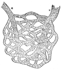
of living substance. The very finest of all, which are the tubes
connecting the smallest arteries with the smallest veins, are called
capillaries, from a Latin word meaning a hair, to indicate their very
small size. They are so close together in most parts of the body that it
would be difficult to thrust a

 Photo, Paul Thompson
THE TEST FOR BLOOD PRESSURE
Photo, Paul Thompson
THE TEST FOR BLOOD PRESSURE
 Photo, Paul Thompson
PART OF PROCESS OF URINALYSIS INDICAN TEST
Photo, Paul Thompson
PART OF PROCESS OF URINALYSIS INDICAN TEST
 A NETWORK OF CAPILLARIES
The artery a and vein v (highly magnified). (From “The Human
Mechanism,” Hough and Sedgwick)
pin in anywhere for any distance without striking against one or more of
them. The capillaries are spread so thickly that there are not many
places in the body where living cells are more than a very small
fraction of an inch from one of them. The cells do not, however, lie
right against the capillaries, but are separated from them by tiny
spaces filled with tissue fluid. In order for material to get from the
blood to any living cell, then, it must pass through the wall of the
capillary into the fluid which fills the tissue space and from that in
turn to the cell itself. The wall of the capillaries is so delicate that
if the blood flowing through any capillary contains more of any
substance than is present in the tissue fluid surrounding that
capillary, some of it will pass out through the wall and into the
tissue fluid, just about as freely as though there were no wall there at
all. The arrangement can be illustrated by a familiar example; if a drop
of ink is allowed to fall into a glass of water, it will color only a
small part of the water at first, but quickly spreads out until each
part of the water is as deeply colored as any other part. If the glass
of water were to be divided in half by a very delicate membrane, and the
ink dropped in on one side, it would spread out in the same way, passing
through the membrane in so doing, until again all the water in the glass
was equally colored. Of course, the quickness with which the ink could
pass through the membrane would depend on how delicate the membrane was.
We could imagine membranes which would not let any ink at all through,
and every degree from that up to membranes so delicate as to offer no
obstruction at all to the passage of the ink. The walls of the
capillaries rank as membranes of such delicacy as apparently to offer
almost no obstruction to the passage of materials through them. They
hold back the red corpuscles and the platelets fairly well, so that they
do not pass out of the blood and into the tissue spaces, unless the
capillaries are actually injured. The colorless corpuscles are able to
make their way through the capillary walls and so also do nearly all the
substances that are dissolved in the blood. It is an interesting fact
that the blood proteins do not pass freely through the capillary walls,
although the digestion products of food proteins, do. It will be
remembered that in the chapter on Body Fluids the importance of the
sticky quality of the blood proteins was spoken of. It is now believed
that it is because of this gelatinous nature that the blood proteins
are not able to pass out through the capillary walls, and this is
supposed to be important in the proper working of the circulation. In
fact there is a condition of greatly lowered vitality to which the name
“shock” is applied, in which the blood proteins escape through the
capillary walls to so great an extent as to interfere with the proper
working of the body. It has been found possible to prevent this in a
very large measure by the simple expedient of injecting some substance
like mucilage directly into the blood stream. We are to think of the
capillary walls, then, as allowing materials to pass freely through them
in either direction, from the blood into the tissue spaces or from the
tissue spaces into the blood, with the exception of the red corpuscles,
platelets, and the blood proteins, and as thus keeping the tissue fluids
supplied with whatever materials the blood contains or taking from the
tissue fluids the waste products of cell metabolism, which the cells are
pouring out. With this arrangement clearly in mind, all that remains for
the understanding of the conveyer system is to see how the blood is kept
in motion and distributed among the various organs of the body, and then
to consider where the blood in turn gets its supplies of materials which
it can pass on to the tissue fluids, or how it gets rid of the
substances which the tissue fluids have passed on to it from the cells.
At the beginning of the chapter we said something about the arteries and
veins and their branching into smaller and smaller subdivisions with the
final connecting link between the smallest arteries and the smallest
veins in the form of capillaries. We are now to consider in detail the
movement of the blood through these tubes, and to do that it will be
necessary to speak again of this arrangement. In describing the
circulation we usually begin with the heart. The heart itself will be
taken up presently. First let us trace the blood vessels from the heart
through the body and back to it again. The large main artery leading
from the heart is known as the aorta. This springs from the upper side
of the heart, bends over in an arch, and passes down through the chest
into the abdomen.
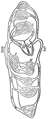 THE HEART AND BLOOD VESSELS DIAGRAMMATICALLY REPRESENTED
L, lung; M, intestine; P, liver; dotted lines lymphatics.
(Martin’s “Human Body”)
Branches are given off from the aorta all along its length. The very
first of these come off before the aorta gets away from the heart, and
are the arteries by which the tissues of the heart itself are supplied
with blood. A little farther on are large arteries, one for the left arm
with a large branch running up the left side of the neck, another for
the right arm with a large branch running up the right side of the
neck. Each of these in turn gives off branches all along to provide for
the tissues of the arms, neck, and head. It is worth while noting that
the arteries running up the neck to the head are large in proportion to
the size of the head itself; this is because the brain, as the most
important organ in the body, requires and receives a disproportionately
large blood supply. Besides the brain the head contains numerous muscles
and also the salivary and tear glands, all of which carry on active
metabolism and therefore require abundant blood supply. The main
branches of the aorta to the head and arms are given off from the arch;
as the aorta passes down through the chest it gives off small branches
to the muscles of the chest wall, and then passes into the abdomen. Here
are located two of the three important arrangements for renewing the
blood; namely, the digestive organs and the organs of excretion
(kidneys). Large branches from the aorta pass to the digestive organs
and others to the kidneys; smaller branches lead to the muscles of the
abdominal wall, and also to the various secreting glands that are
located in the abdomen. At the lower end of the abdomen the aorta
divides, giving one large branch for each leg. As we have already seen,
if we follow any of these subdivisions through its finer and finer
branchings, we shall finally be able, with the aid of a microscope, to
trace it to capillaries, where the interchanges between blood and tissue
fluid occur, and beyond the capillaries to tiny veins which unite with
other tiny veins from other capillaries into larger veins. These again
continue to come together into main veins corresponding in every part of
the body with the main arteries. All these veins finally unite into two,
one for the lower part of the body, called the inferior vena cava,
and one for the upper part of the body, called the superior vena cava.
These two come together just at the entrance to the heart. One special
feature of the blood supply to the digestive organs may as well be
mentioned here; it is that the blood which flows through the capillaries
of the stomach and intestines is all reassembled into a vein known as
the portal vein, which instead of passing directly into the inferior
vena cava goes first to the liver, where the vein breaks up into another
set of capillaries, the liver capillaries, beyond which is another vein
which leads into the inferior vena cava. The result of this arrangement
is that all blood passing into the capillaries of the stomach or
intestines is obliged to pass again through the capillaries of the liver
before going on into the main stream of the circulation. This is an
important feature of the renewal of the food supplies of the blood.
We have now traced the blood from the heart through the body back to the
heart again, and have seen how in its course some of it will pass
through such active tissues as muscles or brain or glands, so that the
interchanges can go on by which the fluids in these active tissues can
take up needed materials and give off wastes. Also a part of it flows
through the digestive organs, where food materials can be taken up, and
another part flows into the kidneys, where wastes can be gotten rid of.
This leaves us to consider only the passage of the blood through the
lungs, where the supply of oxygen is to be taken up and the gaseous
waste product, carbon dioxide, is to be disposed of.
The most urgent requirement of the body is the requirement for oxygen.
There is under ordinary circumstances at all times some surplus of food
materials stored in the cells, so that even though the renewal of their
surrounding fluids from the blood should stop, they could keep on going
for a time on the material that is stored within them; but there is no
such storage of oxygen. The cells in the body lead almost a
hand-to-mouth existence so far as their oxygen supply is concerned. They
are constantly withdrawing oxygen from the tissue fluids surrounding
them, and these fluids are just as constantly withdrawing it from the
blood; therefore any failure of the blood to be properly supplied with
oxygen results very promptly in a condition of oxygen hunger in the
cells. This means prompt cutting off of metabolism, since metabolism is
a matter of oxidizing fuel, and oxidation cannot go on unless the oxygen
is provided. This urgent need for oxygen is met in the body by having
the arrangement for supplying it to the blood much more perfect than the
other renewal arrangements. We saw a moment ago that only part of the
whole blood stream passes through the digestive organs at any given time
and only part of the stream passes through the kidneys. The whole
stream, on the other hand, passes through the capillaries of the lungs.
This is brought about by having an arrangement whereby the combined venæ
cavæ after entering the heart communicate with an outlet in the form of
an artery leading to the lungs. This artery, which is called the
pulmonary artery, breaks up into capillaries in the lungs, which
reunite into the pulmonary vein which comes back to the heart again.
It is from the pulmonary vein that connection is made with the aorta,
starting the blood on its course through the body again. We see then
that the blood passes through the heart twice in each complete round,
once as it comes in from the body at large on its way to the lungs, and
again as it comes in from the lungs on its way to the body at large.
We have spoken of the heart thus far as a single organ; it is actually
two hearts side by side, and these would work just as well if they were
at a distance instead of being built into one organ. There has probably
been more misunderstanding of the heart by people in general than of any
of the other parts of the body. This is because from the earliest times
the heart has been looked upon as the seat of the affections, and so
powers and properties have been attributed to it to which it is not at
all entitled. As we tried to make perfectly clear in a former chapter
all intelligence and all feelings are located in the brain; the heart
cannot possibly take any more active part in these than can the stomach,
liver, kidneys or any of the other parts of the body which are concerned
with the maintaining of the tissues in good working order. Probably no
one really knows how it came about originally that the heart was endowed
with these peculiar gifts. It is true that in time of strong emotion
there are changes in the activity of the heart which we can perceive.
These occur because the heart is under the same kind of nervous control
as are the smooth muscles and glands, and shares with them in the
disturbances which accompany emotion; but the real seat of these
emotions is, of course, the brain. As a matter of fact the heart is
nothing but a muscular pump whose sole function is to keep the blood in
motion. From what has already been said it is clear that the heart
cannot relax its activity for more than an instant without disastrous
results; the pressing need of the tissues for oxygen requires that the
blood be kept moving. If there were any other way in which the needs of
the cells could be supplied except through the movement of the blood,
the heart could be dispensed with perfectly well. We have emphasized
this about the heart because it is much easier to understand its working
if one thinks of it simply as a pumping organ than if one is attributing
to it mystic functions connected with our higher emotions.
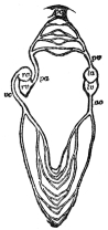 DIAGRAM SHOWING THE RELATION OF THE
TWO HALVES OF THE
HEART
ra and rv, right auricle and ventricle; la and lv, left auricle
and ventricle; ao, aorta; vc, venæ cavæ; pa, pulmonary artery;
pc, pulmonary capillaries; pv, pulmonary vein. (Martin’s “Human
Body.”)
We showed a moment ago that the heart is really a double pump. The
relation of the two halves is shown in the diagram. One of the two
pumps, that on the right side of the heart, receives the blood from the
body at large and pumps it out into the pulmonary artery and through
the capillaries of the lungs; the pump on the left side of the heart
receives the blood from the lungs through the pulmonary vein and pumps
it out into the aorta and so through all the other capillaries of the
body. Since the circuit of the body is much more extensive than the
circuit of the lungs, the work of pumping is correspondingly greater,
and we find the left part of the heart a much more powerful pump than
the right. The heart operates as a reciprocating pump, by which we
mean that it alternately fills and empties. In this respect it is like
ordinary pumps except those of the rotary variety. Any reciprocating
pump must have a chamber which will fill and which can then be emptied
forcibly. In order that it shall not empty itself back through the pipe
from which it filled there must be a valve in the intake pipe which
shall close as the pump is being emptied. If, as is the case in the
heart, it is emptying itself into a system which permits backflow, there
must be another valve in the outlet pipe to prevent the fluid that has
been expelled from running back in. Each of the heart pumps consists,
then, of a chamber, which alternately fills and empties itself, and two
valves, one on the intake and one on the outflow side. In ordinary pumps
the forcible emptying is performed by a piston which moves through the
pump chamber expelling the liquid ahead of it and then has to draw back,
making room for the chamber to fill again. In the heart the forcible
emptying is accomplished by muscular action. The wall of the heart
consists of a great many muscle fibers so arranged that when they
contract they pull the walls of the heart together, making the cavity
smaller, or even obliterating it completely. The contraction of these
fibers makes up what we are familiar with as the beat of the heart. The
frequency with which they contract varies a good deal in different
individuals. The average is about seventy-two a minute; but it may be as
slow as forty-eight or fifty, or may run up to one hundred and forty or
one hundred and fifty a minute. Whatever the rate, in every case there
is an alternation of contraction and relaxation; during the relaxation
the cavity is filling with blood through the intake valve, the outflow
valve being closed, so that no blood that has once been pumped out can
rush back in again. By the contraction of the muscles the heart is
emptied, the outflow valve being open, and the intake valve being closed
to prevent an escape of blood backward into the veins through which it
flowed in. The part of the heart that carries on this active pumping
work is known as the ventricle; that on the right side, which receives
the blood from the body and pumps it to the lungs, is the right
ventricle; and that on the left side, which receives the blood from the
lungs and pumps it to the body, is the left ventricle.
In addition to the ventricles, which are the active pumps, each side of
the heart has an additional chamber known as the auricle, whose
purpose is to serve as a reservoir into which blood can flow during the
time that the ventricles are emptying themselves. If it were not for the
auricles, the movement of blood into the heart would have to stop with
every beat, because while the ventricles are contracting the intake
valves are closed and there would be no place to which blood could flow,
but since each side has its auricle, the flow of blood goes on during
the beat of the ventricles, the auricles filling up. The intake valve,
in order to operate properly, should be located between the auricle and
ventricle, and this is where it is. The vein opens directly into the
auricle without any valve between; the auricle opens into the ventricle
with the intake valve at the point of junction. The intake valves are
given rather formidable names; they are sometimes spoken of as the
auriculo-ventricular valves; that on the right side of the heart
between the right auricle and the right ventricle is composed of three
flaps of membrane, and has therefore been named the tricuspid valve.
The intake valve on the left side of the heart, which is composed of but
two flaps, is known as the mitral valve. As soon as the beat of the
ventricles is over and the ventricular muscle relaxes, the blood which
has accumulated in the auricles presses the intake valve open and blood
begins to flow through it directly into the ventricle. Both the intake
and outflow valves are composed of stout but thin sheets of membrane, so
that very little pressure is required to operate them. The weight of the
blood that is accumulated in the auricles during the beat of the
ventricle is more than sufficient to force the valve open and allow the
blood to flow on through into the ventricle. In a heart that is beating
seventy-two times a minute, there cannot be much time occupied either in
filling or emptying. As a matter of fact both these intervals are
measured in tenths of seconds. If we take a heart that is beating at the
average rate of seventy-two times a minute the whole of a single beat
amounts to eight-tenths of a second. The beat of the ventricle takes
about three-tenths of a second or three-eighths of the whole time; the
period of relaxation of the ventricle, during which it is filling with
blood through the open intake valve, is about five-tenths of a second or
five-eighths of the whole time. The movement of blood is rapid enough
so that this five-tenths of a second allows the ventricle to fill; in
fact much less time than this is required, for in a heart that is
beating at twice the average rate, the ventricle still fills with blood
between beats.
A word remains to be said about the beat of the auricle. During most of
the period when the ventricle is relaxed the auricle is also quiet and
blood is pouring directly through it from the veins into the ventricle;
but just an instant before the ventricular beat begins, one-tenth of a
second to be exact, the auricle contracts, emptying what blood it
contains into the nearly filled ventricle; thus, when the ventricle
beats, which it does immediately, closing the intake valve at the same
time, the auricle is empty and so is able to accommodate the inflow of
blood from the vein during the three-tenths of a second that the intake
valve is shut. Both sides of the heart work exactly together, the two
auricles beating simultaneously, and the two ventricles. Of course it is
necessary that this be so, for if they did not keep pace exactly, one
with the other, there would be a piling up of the blood either in the
lungs or in the veins leading from the body to the heart, and the
efficiency of the circulation would be seriously impaired.
We can get a good deal of information about the way our hearts are
behaving simply by holding the hand against the chest directly over the
heart or by pressing the ear against the chest of some one else and
listening to the heart’s action. The physician makes use of a
stethoscope, which is merely an apparatus for conducting clearly the
sounds which the heart makes, so that it is not necessary to apply the
ear to the chest. When one listens thus to the heart he finds that with
every beat there are two distinct sounds: the first is a rather dull
sound which comes just at the beginning of the beat of the ventricle,
the second is a sharp sound occurring just at the end of the ventricular
beat. As we saw in Chapter IX, sound is always the result of vibrations,
and a great deal of study has been devoted to an attempt to find out
where the vibrations come from that cause the heart sounds. It is now
generally believed that the first sound is partly the result of
vibrations set up in the contracting heart muscle and partly due to
vibrations from the sharp closing of the intake valves. The second sound
is known to be wholly due to the sudden closing of the outflow valves.
The sounds are chiefly of importance in that they enable the physician
to determine whether the valves are holding tight or whether there is a
leakage of blood through them. In case the intake valve leaks, there
will be a backward jet of blood from the ventricle into the auricle with
every beat of the heart. This will cause a sort of hissing or murmur
which can be heard with the stethoscope in connection with the first
sound. If the outflow valve is the one that leaks, blood will squirt
back into the ventricle from the aorta, while the ventricle is relaxing.
The murmur in this case will come just after the second sound. The
skillful physician by comparing the loudness of the murmur when the
stethoscope is pressed at different points on the chest and back can
determine whether the leaky valves are on the right side or the left
side. Thus an accurate diagnosis of imperfect valves can be obtained. Of
course the heart will not work well if its valves are not tight any more
than will an ordinary pump, so that persons suffering from this trouble
cannot have as good a circulation as those whose valves have nothing
the matter with them. It is true that in most cases of imperfect valve
action there is a compensation in the form of an increase in the size
and strength of the heart muscle, so that the circulation is maintained
in spite of poor valve action by harder work on the part of the heart.
It is evident that in a case of this kind exceptional strains on the
heart are more dangerous than if the heart is normal to begin with, so
that persons with faulty valve action must avoid physical strains, such
as sharp running after street cars or trains, which would be borne with
impunity by ordinary individuals. Since faulty valves are a frequent
result of acute rheumatism, which in turn comes from pus pockets, and
since no way is known to cure a defective valve, once the trouble has
developed, it is evident that prevention is of the utmost importance.
Physical efficiency is very seriously hampered by poor heart action.
One feature of the heart action with which we are all perfectly familiar
is that both the rate and the vigor of the heartbeat vary greatly from
time to time. When one is lying quietly, the heartbeat is at its lowest
point. It becomes more rapid as one sits up, still more rapid upon
standing, increasing still more with the taking of any form of muscular
exercise, and in vigorous muscular exercise attains its greatest
rapidity and force. The rate in this latter case may be fully double
that of the quiet standing position, and, as the vigorous thumping tells
us, the force is also very much increased. As we saw in Chapter VII the
heart muscle works automatically, contracting and relaxing without being
stimulated through the nervous system. The variations in rate and
force, however, are the result of nervous action. The heart muscle, as
we have already seen, is under the same sort of nervous control as the
smooth muscles and glands. It has passing to it two sets of nerves, one
to slow it down, the other to speed it up. Both these sets of nerves
arise from centers in the brain stem, and both these centers appear to
be discharging continuously. So it works out that the heart muscle is
under the constant influence of two opposing sets of nerves, and its
actual rate and vigor depend upon the balance between them. This has the
effect of making the heart extremely responsive to nervous influences.
The slightest relaxation on the part of the nerves whose function is to
cause slowing will lead to a prompt increase of rate, since the nerves
that tend to cause increase are active all the time. Various things may
bring about changes in the nervous balance governing the heart; chief of
these are muscular activity and emotional disturbance. Practically all
the changes in the heart action that we observe from moment to moment
can be explained as being due to one or the other of these causes. There
are, however, two additional points to be noted briefly; the first is
that after muscular exercise the heart slows down very gradually, not
returning to its ordinary resting rate for a half hour to an hour after
the exercise is over, depending on how long the exercise was kept up.
The explanation of this long-continued rapid beat is found in the great
outpouring of waste products as the result of the exercise. We have
already learned that the functional metabolism of muscular work involves
the oxidation of a large amount of energy-yielding material and
therefore brings about the production of large amounts of oxidation
products. Their presence in the blood serves as a stimulus to
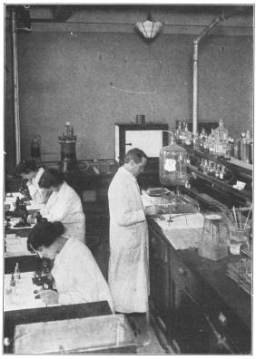 THE MICROSCOPE IS NECESSARY TO UNDERSTAND THE STRUCTURE
OF THE BODY
THE MICROSCOPE IS NECESSARY TO UNDERSTAND THE STRUCTURE
OF THE BODY
In this laboratory the workers are examining blood smears
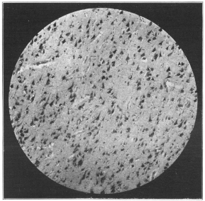 Photo, Cornell University Medical School
MICROPHOTOGRAPH OF BRAIN TISSUE
Photo, Cornell University Medical School
MICROPHOTOGRAPH OF BRAIN TISSUE
the nerve center in the brain stem, which acts to quicken the heart, and
this keeps the rapid beat going until these products are gradually
gotten rid of from the body. Another somewhat similar case is the
prolonged rapid heartbeat following a violent emotion. The explanation
of this we saw a couple of chapters ago in the outpouring of adrenalin
that accompanies emotion. One property of adrenalin, as already noted,
is to quicken the heart; so, as long as any adrenalin remains in the
blood stream, the heartbeat will be faster than normal.
In the above paragraphs we have tried to make clear that the blood is
kept in motion through the body by the work of the heart, and that the
heart’s activity varies in accordance with the needs of the body; in
muscular exercise there is a great increase in metabolism, which means a
greatly increased demand both for food supplies and for oxygen. To meet
this increased demand it is necessary that the blood circulate more
abundantly, and in the automatic speeding up of the heart through the
nervous system we have the means by which this is done. In the case of
strong emotion, as already emphasized, the bodily reactions are such as
put the body into the best possible condition for meeting the emergency.
Evidently a quickened heartbeat, by insuring abundant supplies of oxygen
and of foodstuffs, contributes to this end. The slowing of the
heartbeat, when one lies down, is evidently helpful in enabling the
heart itself to recover from any strains that may have been put upon it.
The heart is a muscle, and like any other muscle carries on its
functional metabolism, which means that it is oxidizing fuel materials
and producing waste products. Since it is absolutely necessary that the
heart go on beating regularly year in and year out for perhaps eighty
or a hundred years, any relief from activity that it can get by slowing
down during sleep is evidently an advantage. It has been calculated that
the heart muscle really enjoys an “eight-hour day,” by which is meant
that on the average the functional metabolism of contraction is going on
in heart muscle only about one-third of the time, eight hours out of
each twenty-four. During the active waking time the metabolism takes a
larger percentage than that, but during sleep enough less to even up.
The heart empties itself into the large arteries; the left heart into
the aorta, the right heart into the pulmonary artery. Both these
arteries, as well as their subdivisions, are highly elastic. The very
best quality of rubber tubing is not superior to our arteries as samples
of elastic tubes. The blood, as we have already seen, is quite sticky,
and the capillaries through which it must pass in its course around the
body are microscopically tiny. The heart pumps the blood out of itself
at the rate of four or five quarts a minute or more, according to
whether it is working moderately or at high speed. To force this amount
of the sticky blood through the tiny capillaries evidently requires very
considerable force. As a matter of fact, the force is sufficient so that
if it were applied to working a fountain it would force a jet to the
height of nearly eight feet. Evidently pumping blood into elastic
arteries with this force and against the resistance offered by the tiny
capillaries causes the arteries themselves to be not only filled but
overfilled, so that their walls are greatly stretched. This fact, that
our arteries are elastic and are kept on the stretch by the pressure of
the blood within them, is of the very greatest importance to the proper
flow of blood and this in turn is so important to our well-being that
some of our most serious chronic diseases are traceable to the loss of
elasticity on the part of the arteries.
We must remember that once every second, or oftener, the heart is
shooting a jet of blood into the large artery which is already stretched
with blood and which can empty itself only through the tiny capillaries
at the tips of its finest subdivisions. On account of the inertia of the
blood stream, room is made for this additional jet of blood by
stretching the arteries near the heart more than they were stretched
before. The result is that there is an inequality in the amount to which
the arteries are stretched, those near the heart being stretched more
than those farther along. As quickly as possible this inequality of
stretch will be equalized by a spreading of the additional tension out
over all the arteries in the form of a wave. This wave makes up what we
know as the pulse. It can be felt in any artery that is near enough to
the surface so that the finger tips can press upon it. The radial artery
at the wrist is the one commonly used by physicians for feeling it.
There is a large artery in the neck in which the pulse can also be felt
readily, and if one takes the pulse of another person in the neck with
one hand and in the wrist with the other he can easily satisfy himself
that the pulse in the neck always comes an instant earlier than that in
the wrist. This is simply because the pulse spreads from the heart as a
wave, and the distance to the neck is not so great as that to the wrist.
By the time the finest subdivisions have been reached, the tension is
equalized throughout the arterial system, and there is no more pulse.
The advantage of this is that the blood flows through the capillaries
in a steady stream and not in a series of jerks. This, in fact, is the
chief, but not the only, benefit we derive from having elastic arteries.
Since the heart operates as an intermittent pump, it is evident that
unless the arteries were elastic and so could take up the shock, the
blood would have to pass through the capillaries in a series of jerks,
exactly corresponding with the beats of the heart. There is abundant
proof, which we shall return to in a moment, that to have the blood move
through the capillaries in this jerky fashion would be disastrous.
Before taking that up, however, we wish to show that by having elastic
arteries the actual work of the heart is less than it would be if the
arteries were stiff. The reason is really very simple. As was stated a
few pages back, the heart is actually emptying itself only during
three-eighths of every beat. If the arteries were stiff tubes, and
therefore not able to take up any of the blood within themselves,
exactly as much would have to pass out through the capillaries during
this three-eighths of the beat as was pumped in by the heart. In other
words, if the heart were pumping five quarts a minute, five quarts would
have to pass through the capillaries in three-eighths of the minute
instead of having the whole minute in which to do it. Since the arteries
are actually elastic, they are able, by stretching a little more, to
make room for part of the blood and so spread the time of its passage
through the capillaries out over the whole time instead of confining it
just to the period when the ventricle is contracting. Evidently it would
take more work to pump five quarts of blood through the capillaries in
three-eighths of a minute than in a whole minute.
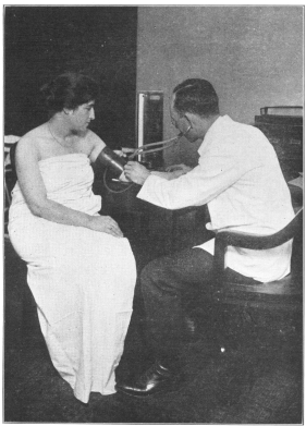
We measure the work of the heart by what we call blood pressure, about
which we are hearing so much these days, so that it will be well to
explain as clearly as possible just what is meant by it. The blood
pressure really means the pressure of the blood within the large
arteries. It could be measured with any ordinary pressure gauge, if it
were not for the fact that we cannot very well cut into our bodies to
apply gauges to the arteries. For this reason it has been necessary to
invent means of finding out what the blood pressure is from the outside.
The way it is done is to put a band around the arm, press this band down
upon the arm until it squeezes the large arm artery shut, and then, by
means of a suitable gauge, find out how much pressure was required. Of
course, it is necessary to be able to tell when the artery has actually
been squeezed shut, so that the determination of blood pressure in human
beings is the work of an expert. Furthermore, blood pressure, as should
be clear from what has already been said, varies with every heartbeat.
It is at its maximum the instant the heart finishes emptying itself into
the artery, and falls off steadily, reaching a minimum just before the
next beat comes. The more elastic the arteries, the less difference
there will be between the maximum and the minimum blood pressure. The
reason for this will be clear when we think that if the arteries were
entirely rigid there would have to be a very high pressure during the
time the heart was actually beating to force the blood out through the
capillaries, but that between beats the pressure would fall off to zero.
The more elastic the arteries are, the more nearly do they exert a
steady pressure on the blood within them, and so the less will be the
difference between the maximum and the minimum pressure. When the
physician determines blood pressure, he really determines both the
maximum and the minimum pressure, in order that he may be able to judge
whether or not the arteries are as elastic as they should be. High blood
pressure just by itself might not mean much more than that the heart was
beating more rapidly than it should, but a high maximum pressure and a
low minimum pressure means nonelastic arteries. This in turn means that
the blood is forced through the capillaries in jets rather than in a
steady stream, and we may judge of the importance of having a steady
flow through the capillaries when we recall the well-known medical
proverb that “a man is as old as his arteries.” It is an actual fact
that the chronological age of an individual need not have much to do
with his physical age. If his arteries continue elastic over a long
period of years, he will be physiologically youthful, while if his
arteries become rigid he will be physiologically aged, no matter how few
his actual years upon earth may have been. Unfortunately we do not know
as much as we would like to about the causes of loss of elasticity in
the arteries. It does appear, however, that self-indulgence of various
kinds is apt to lead to loss of elasticity. For example, even the
moderate use of alcohol is now generally recognized by the medical
profession as a cause of impairment of elasticity in the arteries. It is
probable that intemperance in the use of various foods and drugs leads
also to this same condition.
We have just seen that the heart is obliged to maintain high blood
pressure in order to force the blood through the tiny capillaries. It
will be clear that the actual amount of pressure will depend in part
upon how much blood is forced through in a minute and in part upon the
extent to which the capillaries offer resistance. It is a familiar law
of friction that the smaller the tube the greater will be the resistance
it will offer to the passage of liquid through it, so that if the
capillaries change in size their resistance to the flow of blood through
them is bound to vary. The walls of the capillaries are very sparsely
provided with muscle fibers, but the very finest subdivisions of the
arteries, which are really no larger in diameter than the capillaries,
have much more smooth muscle in their walls. These muscles, as we have
already seen, can by their contraction or relaxation make the tiny
vessels smaller or larger. We have examples of this in the flushing and
pallor of the skin. What we wish to do now is to show how the flow of
blood through different parts of the body is controlled by changes in
the caliber of these tiny tubes. The muscles in the vessel walls have
the double nervous control commonly found in smooth muscles, and both
sets of nerves trace back to centers in the brain stem. One of these
centers causes the muscles to contract and the vessels to become
smaller; the effect of this is, of course, to increase the resistance to
the passage of blood through them. The nervous center which brings this
about is called vasomotor, or, more properly, the vasoconstrictor
center. Vasoconstriction means literally causing contraction of the
vessels, which is exactly what this center does. Not all the blood
vessels in the body are acted upon through the vasoconstrictor center.
Those of the skin and of the abdominal organs are under its control, but
those of the skeletal muscles are not. The result of activity of this
center, then, is to make it more difficult for blood to flow through the
skin and through the abdominal organs, but the ease of flow of blood
through the muscles is not affected. Of course, it will follow
automatically that the blood stream will be diverted in a large part
from the former regions into the latter. The skin and abdomen together
make up so large a part of the whole body that marked constriction of
the blood vessels in these two regions is bound to cause a considerable
increase in blood pressure.
A fairly high blood pressure is necessary for bodily well-being, because
only thereby is the brain assured of sufficient nourishment. To see why
this is so, we have only to remember that the brain is unfavorably
situated for receiving ample supplies of blood. It is at the top of the
body, so that the influence of gravity has to be overcome in forcing
blood up to it. Also it is completely inclosed in the bony skull, which
it in turn fills so completely that there is almost no room for the
accommodation of extra blood in it. In all other parts of the body a
rise in blood pressure stretches the arteries and so leads to there
being actually more blood within them, but there is no room for the
arteries in the brain to stretch, so that the total quantity of blood in
the brain cannot vary greatly from time to time. The only way in which
an increased blood supply can be obtained is by causing the blood to
flow more rapidly. This is precisely what happens every time the blood
pressure in the body rises. Whenever the vasoconstrictor center sends
nervous discharges into the blood vessels of the skin and abdominal
organs, causing them to contract, there is a diversion of blood from
them directly into the skeletal muscles and also a rise in blood
pressure due to the increase in resistance to the circulation, which
causes blood to flow more rapidly through the brain. The net result,
then, is improvement in the blood supply to the skeletal muscles and to
the brain. So important is the blood supply to the brain that the
vasoconstrictor center discharges actively throughout the waking part of
the day. If for any cause the activity of this center diminishes, there
will be an increased circulation in the skin and in the abdominal
organs, the blood pressure will fall, the circulation through the brain
will decrease, and along with it there will be a decrease in brain
function. After this passes a certain point unconsciousness will result.
This is what happens when one faints. For some reason or other the
vasoconstrictor center becomes inactive, and the series of events just
described is set in motion. Fainting ordinarily cures itself
automatically, because when consciousness is lost, the individual falls
over; this brings his head down on the level with the rest of his body,
makes it easier for the blood to flow through it, and so in a moment or
two consciousness will be regained. It is a mistake to try to scramble
to one’s feet immediately, because until the vasoconstrictor center
recovers its ordinary activity, raising of the head above the level of
the rest of the body is bound to result in its failure to receive
sufficient blood, and so faintness will come on again. It has long been
a practice to dash cold water in the face of a fainting person. The
physiological value of this is in the sudden stimulation of the sensory
nerves in the face by which is set up a stream of nervous discharges
which will play upon the vasomotor center, and arouse it again to
activity. Almost any vigorous sensory stimulation may have the same
effect.
During sleep the vasoconstrictor center is usually not very active; the
cause of sleep is not completely understood, but one of the most
satisfactory theories regarding it is that during the period of waking
the vasoconstrictor center becomes gradually more and more fatigued, and
so requires more and more stimulation to keep it active. This
stimulation may come in part through the ordinary channels of the sense
organs and in part from the higher brain centers, as when one keeps
awake by an effort of the will. Upon going to bed sensory stimulation is
cut off to a very large extent, also the will to remain awake is no
longer present. Under these circumstances the fatigued vasoconstrictor
center is under a minimum of stimulation and tends, therefore, to lessen
its activity. The result is that the blood pressure falls and presently
the circulation through the brain drops below the level of consciousness
and the individual is asleep. During the period of sleep the fatigued
center recuperates, so that it becomes more susceptible to sensory
stimulations and, in course of time, is aroused by such stimuli as
accompany the returning day to sufficient activity to restore the brain
to consciousness. Of course it will be perceived that there are many
things about sleep which are not satisfactorily explained on this
theory; in fact no one at the present time pretends that we can account
for it completely on this basis of changes of circulation through the
brain. It is believed, however, that they have a good deal to do with it
and it is certainly true that in healthy individuals the course of sleep
follows very closely the activity of the vasoconstrictor center. Undue
wakefulness in persons not suffering from disease can nearly always be
explained on the basis of excessive activity on the part of this
center. The activity may be the result of chemical stimulation, as when
persons are kept awake by drinking coffee or strong tea in the evening,
or by the persistence of adrenalin in the blood following a period of
great excitement. Mental activity, itself, tends to keep the
vasoconstrictor center whipped up, so that one who allows his mind to
work actively during the time when he should be asleep is very apt to
find sleep refusing to come when it is desired. The center is often
stimulated from the digestive tract; gastric irritation, even though not
acute enough to be recognized as indigestion, may cause wakefulness by
arousing nervous disturbances which play upon the vasoconstrictor
center. Flatulence, namely the presence of large volumes of gas in the
digestive tract, frequently acts as a mechanical source of irritation by
which wakefulness is induced. Evidently the factors favoring healthy
sleep are the inducing of fatigue, preferably by physical exercise, the
avoidance of chemical irritation or of excitement in the hours just
before going to bed, and finally the adoption of dietary habits which
shall insure good digestion. If an individual in whom all these
precautions are combined still continues chronically wakeful, the
trouble is sufficiently deep-seated to call for competent medical
attention.
Besides the vasoconstrictor center about which we have just been talking
there is in the brain stem a center which relaxes the tension of the
muscles in the walls of the blood vessels. This is known as the
vasodilator center, and it acts in opposition to the vasoconstrictor.
In most parts of the body it does not appear to have any very great
importance for the simple reason that the blood is under such high
pressure that any relaxation of effort on the part of the
vasoconstrictor center leads at once to a forcing open of the blood
vessels. There are a few regions, however, in which the action of the
vasodilator center is of real importance; one of these is in the
skeletal muscles. These, as should be perfectly clear by this time, are
the seats of our most active functional metabolism. When the muscles are
active, great amounts of oxygen and food are being withdrawn from the
blood and large amounts of waste material, including carbon dioxide,
poured out into it. Only a very rapid circulation will take care of this
situation. At the same time the skeletal muscles are the most compact of
our living tissues. The cells are crowded together in making up the very
strong muscular machine by which our movements are performed. As the
muscles contract, they squeeze hard on the blood vessels passing through
them. In view of this situation it is very important that the blood
vessels be opened as widely as possible during muscular activity, and so
we find that the vasodilator center acts to improve the circulation
through the muscles, while they are active. In time of special
emergency, as we saw a moment ago, the vasoconstrictor center is at the
same time engaged in cutting down the blood supply to the skin and to
the abdominal organs, thus insuring to the muscles the maximum possible
nourishment through the blood stream.
Besides the skeletal muscles there is active functional metabolism in
the various secreting glands; a feature of their activity is that it
must be very great at certain times, but falls to little or nothing at
others; thus during the actual taking and digesting of food the various
glands which secrete digestive juices are exceedingly active, but in
the intervals they may be doing little or no work. They require a very
copious blood supply when they are functioning, but need very little
between times. The blood vessels flowing through all these glands are
under the influence of the vasodilator center, so that they are opened
as widely as possible while the glands are active. They, therefore,
receive much more blood in proportion to their size than they would if
it were not for the action of this center. Between times their blood
supply falls off to that which suffices for inactive tissues generally.
In time of emergency the action of the vasoconstrictor center upon the
vessels through these glands is such as to cut off the blood supply to
them almost completely. This is well illustrated in the dry mouth of the
frightened man, showing that the salivary glands have suspended
activity, a suspension due largely, if not wholly, to the cutting down
of the blood supply through them.
In the section just completed we have tried to give some idea of the way
in which the circulating blood provides the various tissues with the
materials they require and is adjusted automatically to meet variations
in demand from different tissues. Just one more point needs to be noted
in completing the account of the conveyer system. The tissue fluids
which serve as the connecting links between the circulating blood and
the individual cells are necessarily full of waste products, because
these come directly from the cells to the tissue fluids and afterward
pass on from them to the blood. The result is that the cells are
constantly bathed in a solution of their own waste products. There is
only one way in which relief from this condition can be obtained and
that is by moving the used tissue fluid away bodily and letting it be
replaced by fresh fluid. As a matter of fact, this happens; there is an
oozing of fluid through the walls of the capillaries from the blood into
the tissue spaces; this of course pushes the fluid already in those
spaces on ahead of it. If there were no place to which the fluid could
go, the result would be a swelling, as the tissue became more and more
filled with fluid. This is avoided ordinarily by a drainage system
whereby the tissue fluids are carried off as fast as more fluid comes in
from the blood, but when liquid is poured out faster than it will drain
off, as from a bruise or wrench, we do get a swelling.
The drainage system consists of very delicate vessels known as the
lymphatics, which come together into larger and larger vessels, just
as the veins do, and finally empty into the vena cava just at the point
where it enters the heart. There is no back pressure of blood here, so
that the movement of fluid through the lymphatics is not hindered. There
is no pump for forcing the lymph to move along; the very gradual motion,
which is all that is necessary to keep the fluid from accumulating in
the tissue spaces, is brought about by pressure upon the lymphatics
resulting from the bodily movements. The lymphatic vessels are like the
veins in having valves here and there along them. Whenever by any bodily
movement either a lymphatic vessel or a vein is squeezed, the valves
insure that the liquid shall be passed along in the direction toward the
heart and never in the reverse direction. In the case of the veins this
action is not absolutely necessary, since the heart itself is able to
maintain the circulation, although it does help, particularly in
bringing back the blood from the extremities. In the lymphatics this is
the only way by which movement of fluid is brought about. The result is
that when the body is perfectly quiet, there is very little movement of
fluid through the lymphatics; muscular activity, on the other hand,
leads to rather active movement through these vessels. The fact is well
illustrated in ourselves. One who sits for a long time humped over a
desk finds himself feeling very dull; to obtain relief he stirs about,
stretches, and yawns. The dullness was merely the result of the
stagnation of fluid in his tissues, causing the cells to be more or less
poisoned by their own waste products. By making active movements, these
stagnating fluids were forced along to be replaced by fresh liquid
direct from the blood and the beneficial effect is felt immediately.
Massage properly applied has very much the same effect, although it is
doubtful whether as good results can ever be obtained thus as by actual
vigorous exercise.
At various places along the lymphatics are little spongelike lumps of
tissue known as lymph nodes; the particular spongy substance of which
they are composed is called adenoid tissue. This adenoid tissue acts
as a filter for the fluid passing through it. Any foreign particles,
living or nonliving, that get into the stream are caught in the lymph
nodes and held there more or less permanently. Most of the nonliving
particles that get into our tissue fluids are from the dust that we
inhale, which works its way through the mucous membrane of the
respiratory passages and so into the body fluids. The lymphatics that
drain the lungs carry along these dust particles and they lodge in lymph
nodes at the base of the neck. Persons who have lived in dusty regions
or have pursued a dusty livelihood, such as coal heaving, will have by
the end of their lives lymph nodes which are literally black with dirt.
The tonsils are lymph nodes at the base of the tongue. Unfortunately
they are so near the surface of the throat that they frequently become
infected from the throat itself, and so become the seats of pus pockets,
as already noted. Closely related to the tonsils are the masses of
adenoid tissue at the back of the throat which frequently grow to an
undue size in children, and are then known as adenoids. The harm done by
adenoids is chiefly mechanical; they may block the Eustachian tubes, and
so cause deafness, as already mentioned in Chapter IX, or they may
interfere with the free movement of the air through the nasal passages.
Children in whom this condition exists are mouth breathers. Adenoids,
like tonsils, are subject to infection, and so may give trouble by
becoming the sites of poison formation. Adenoids represent always an
overgrowth and for that reason may be removed without any possibility of
hampering the proper working of the body. Experience has shown that
children whose development appears to be hindered by the presence of
adenoids are almost invariably benefited by having them removed. The
tonsils are normal parts of the bodily structure and as such undoubtedly
have a regular work to do, but here again experience has shown that harm
from persistent pus pockets is so much greater than harm from loss of
function following their removal as to justify taking them out, whenever
pus pockets develop in them. There are enough lymph nodes in the region
about the throat, so that if tonsils or adenoids are removed any foreign
matters that get into the body fluids will still be filtered out.
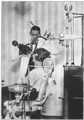 Copyright, Keystone View Co.
X-RAYING THE TEETH AND JAW. HEALTH OFTEN DEPENDS ON THEIR GOOD
CONDITION
Copyright, Keystone View Co.
X-RAYING THE TEETH AND JAW. HEALTH OFTEN DEPENDS ON THEIR GOOD
CONDITION
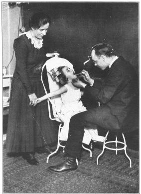 Photo, Paul Thompson
A THROAT EXAMINATION—IMPORTANT IN DETERMINING PHYSICAL FITNESS
Photo, Paul Thompson
A THROAT EXAMINATION—IMPORTANT IN DETERMINING PHYSICAL FITNESS
One more function of the lymph nodes must be mentioned; this is their
property in preventing the spread of cancer cells. We now know that
so-called secondary cancers are the result of the spread of cancer cells
from the original seat of the cancer to other parts of the body, and
that this spread is much hindered by the ability of the lymph nodes to
catch the cancer cells and hold them. Unfortunately sooner or later some
of the cells will escape beyond the lymph nodes and so spread the
malignant growth throughout the body, but until this happens the cancer
is confined to the region where it started, and it is during this period
that complete cure by surgical means is possible. It is because of the
imminent danger of the escape of cancer cells beyond the restraining
lymph nodes that relief by surgery should be sought at the very earliest
possible moment. Delay, whether due to carelessness or any other cause,
is as certainly fatal in the case of cancer as in any other disease for
which a cure is known.