FELLOW AND LECTURER OF TRINITY COLLEGE,
CAMBRIDGE.
VOL. IV.
PLATES.
London:
MACMILLAN AND CO.
1885
[The Right of Translation is reserved.]
LIST OF PLATES, ILLUSTRATING THE ORIGINAL
MEMOIRS IN VOL. I. |
|---|
| Plate |
PAGE |
| 1. |
Development of the layers of the blastoderm |
29 |
| " |
Disappearance of primitive groove |
41 |
| 2. |
Development of blood-vessels |
47 |
| 3. 4. |
Preliminary account of development of Elasmobranch Fishes |
60 |
| 5. |
Comparison of early stages of Vertebrates |
112 |
| 6. |
Development of Elasmobranch Fishes. ch. II. |
222 |
| 7. |
" " "
" III. |
246 |
| 8. 9. |
" " "
" IV. |
286 |
| 10. |
" " "
" V. |
298 |
| 11. 12. |
" " "
" VI. |
315 |
| 13. |
" " "
" VII. |
361 |
| 14. |
" " "
" VIII. |
378 |
| 15. 16. 17. |
" " "
" IX. |
397 |
| 18. |
" " "
" X. |
446 |
| 19. |
" " "
" XI. |
460 |
| 20. 21. |
" " "
" XII. |
479 |
| 22. 23. |
Development of spinal nerves in Elasmobranchii |
168 |
| 24. 25. 26. |
Structure and development of Vertebrate ovary |
549 |
| 27. 28. |
Head-kidney in embryo Chick |
618 |
| 29. |
Early development of Lacertilia |
644 |
| 30. 31. 32. |
Development of Araneina |
668 |
| 33. |
Development of paired fins of Elasmobranchii |
714 |
| 34. 35. 36. 37. 38. 39. 40. 41. 42. |
Structure and development of Lepidosteus |
738 |
| 43. 44. 45. |
Germinal layers of the Chick |
854 |
| 46. 47. 48. 49. 50. 51. 52. 53. |
Anatomy and development of Peripatus Capensis |
871 |
EXPLANATION OF PLATE 1, Figs. 1-5 and
9-12. (II. p. 29.)
Fig. 1. Section through an unincubated blastoderm,
shewing the upper layer, composed of a single row of columnar cells, and
the lower layer, composed of several rows of rounded cells in which no
nucleus is visible. Some of the formative cells,
at the bottom of
the segmentation cavity, are seen at (b).
Fig. 2. Section through the periphery of an eight hours'
blastoderm, shewing the epiblast (p), the hypoblast (h), and
the mesoblast commencing to be formed (c), partly by lower-layer
cells enclosed between the epiblast and hypoblast, and partly by formative
cells. Formative cells at the bottom of the segmentation cavity are seen at
b. At s is one of the side folds parallel to the primitive
groove.
Fig. 3. Portion of the hypoblast of a thirteen hours'
blastoderm, treated with silver nitrate, shewing the great variation in the
size of the cells at this period. An hour-glass shaped nucleus is seen at
a.
Fig. 4. Periphery of a twenty-three hours' blastoderm,
shewing cell for cell the junction between the hypoblast (h) and
white-yolk spheres (w).
Fig. 5. Junction between the white-yolk spheres and the
hypoblast cells at the passage from the area pellucida to the area opaca.
The specimen was treated with silver nitrate to bring out the shape of the
cells. The line of junction between the opaque and pellucid areas passes
diagonally.
Fig. 9. Section through the primitive streak of an eight
hours' blastoderm. The specimen shews the mesoblast very much thickened in
the immediate neighbourhood of the primitive streak, but hardly formed at
all on each side of the streak. It also shews the primitive groove just
beginning to be formed (pr), and the
fusion between the epiblast and the mesoblast under the primitive groove.
The hypoblast is completely formed in the central part of the blastoderm.
At f is seen one of the side folds parallel to the primitive groove.
Its depth has been increased by the action of the chromic acid.
Fig. 10. Hypoblast cells from the hinder end of a
thirty-six hours' embryo, treated with silver nitrate, shewing the
regularity and elongated shape of the cells over the embryo and the smaller
cells on each side.
Fig. 11. Epiblast cells from an unincubated blastoderm,
treated with silver nitrate, shewing the regular hexagonal shape of the
cells and the small spherules they contain.
Fig. 12. Portion of the epiblast of a thirty-six hours'
embryo, treated with silver nitrate, shewing the small rounded cells
frequently found at the meeting-points of several larger cells which are
characteristic of the upper layer.
EXPLANATION OF PLATE 1, Figs. 6-8 and
13-19. (III. p. 41.)
Figs. 6 and 7 are sections through an embryo rather
earlier than the one drawn in fig. 8. Fig. 6 passes through the just
commencing medullary groove (md), which
appears in fresh specimens, as in fig. 8, merely as an opaque streak coming
from the end of the primitive groove. The notochord is hardly
differentiated, but the complete separation of mesoblast and
hypoblast under the primitive groove is clearly shewn. Fig. 7 passes
through the anterior end of the primitive groove (pr), and shews the fusion between the mesoblast and
epiblast, which is always to be found under the primitive groove.
Fig. 8 is a view from above of a twenty hours'
blastoderm, seen as a transparent object. Primitive groove (pr). Medullary groove (md), which passes off from the anterior end of the
primitive groove, and is produced by the thickening of the mesoblast. Head
fold (pf).
Figs. 13-17 are sections through the blastoderm, drawn
in fig. 18 through the lines 1, 2, 3, 4, 5 respectively.
The first section (fig. 13) passes through the true
medullary groove (mc); the two medullary
folds (A, A) are seen on each side with the thickened
mesoblast, and the mesoblast cells are beginning to form the notochord
(nc) under the medullary groove. There
is no adherence between the mesoblast cells and the epiblast under the
medullary groove.
The second (fig. 14) section passes through the
medullary groove where it has become wider. Medullary folds, A,
A; notochord, ch.
In the third section (fig. 15) the notochord (ch) is broader, and the epiblast is raised in
the centre, while the medullary folds are seen far apart at A.
In section fig. 16 the medullary folds (A) are
still to be seen enclosing the anterior end of the primitive groove (pr). Where the primitive groove appears there
is a fusion of the epiblast and mesoblast, and no appearance of the
notochord.
In the last section, fig. 17, no trace is to be seen of
the medullary folds.
Figs. 18 and 19 are magnified views of two hardened
blastoderms. Fig. 18 is twenty-three hours old; fig. 19 twenty-five hours.
They both shew how the medullary canal arises entirely independently of the
primitive groove and in front of it, and also how the primitive groove gets
pushed backwards by the growth of the medullary groove. pv, Protovertebræ; other references as above. Fig. 18
is the blastoderm from which sections figs. 13-17 were cut.
EXPLANATION OF PLATE 2. (IV. p. 47.)
Fig. 1 is taken from the anterior part of the pellucid
area of a thirty hours' chick, with four protovertebræ. At n is a
nucleus with two nucleoli.
Figs. 2 and 3 are taken from the posterior end of the
pellucid area of a chick with eight protovertebræ. In fig. 3 the nuclei are
seen to have considerably increased in number at the points of starting of
the protoplasmic processes. At n is seen a nucleus with two
nucleoli.
Fig. 4 is taken from the anterior part of the pellucid
area of an embryo of thirty-six hours. It shews the narrow processes
characteristic of the anterior part of the pellucid area, and the fewer
nuclei. Small spaces, which have the appearance of vacuoles, are shewn at
v.
Fig. 5 is taken from the posterior part of the pellucid
area of a thirty-six hours' embryo. It shews the nuclei, with somewhat
irregular nucleoli, which have begun to acquire the red colour of
blood-corpuscles; the protoplasmic processes containing the nuclei; the
nuclei in the protoplasm surrounding the corpuscles, as shewn at a,
a´.
Fig. 6 shews fully formed blood-vessels, in part filled
with blood-corpuscles and in part empty. The walls of the capillaries,
formed of cells, spindle-shaped in section, are shewn, and also the
secondary investment of Klein at k, and at b is seen a narrow
protoplasmic process filled with blood-corpuscles.
Fig. 7 is taken from the anterior part of the pellucid
area of a thirty-six hours' embryo. It shews a collection of nuclei which
are beginning to become blood-corpuscles.
Figs. 1-5 are drawn with an 1/8 object-glass. Fig. 6 is
on a much smaller scale. Fig. 7 is intermediate.
Fig. 8. A transverse section through the dorsal
region of a forty-five hours' embryo; ao, aorta with a few blood-corpuscles. v,
Blood-vessels, all of them being formed in the splanchnopleure, and all of
them provided with the secondary investment of Klein; pe, pellucid area; op, opaque area.
Fig. 9. Small portion of a section through the
opaque area of a thirty-five hours' embryo, showing protoplasmic processes,
with nuclei passing from the somatopleure to the splanchnopleure.
Fig. 10. Section through the heart of a
thirty-four hours' embryo. a. Alimentary canal; hb, hind brain; nc, notochord; e, epiblast; so, mesoblast of the somatopleure; sp, mesoblast of the splanchnopleure; hy, hypoblast; hz,
cavity of the heart.
Fig. 11. Section through the same embryo as
fig. 10, and passing through the orifice of the omphalomeseraic vein. of. Omphalomeseraic vein; other references as
above.
These two sections shew that the heart is entirely
formed from the mesoblast of the splanchnopleure, and that it is
formed by the splitting of that part of the mesoblast which has turned to
assume its normal direction after being folded in to form the muscular wall
of the alimentary canal. In fig. 11 the cavities so formed on each side
have not yet united, but in fig. 10 they have united. When the folding
becomes more complete the cavities (of,
of) in fig. 11 will unite, and in this
way the origin of the omphalomeseraic veins will be carried further
backwards. In the section immediately behind section 11 the mesoblast had
become thickened, but had not split.
EXPLANATION OF PLATES 3 AND 4. (V. p. 60.)
Complete List of Reference Letters.
al. Alimentary canal.
ao. Dorsal aorta. auv. Auditory vesicle. bd. Formative cell probably derived from the yolk.
cav. Cardinal vein. ch. Notochord. ch´. Thickening of hypoblast to form the notochord.
Eb. Line indicating the edge of the
blastoderm. ep. Epiblast. ep´.
Epidermis. er. Embryonic rim. es. Embryonic swelling. gl. Glosso-pharyngeal nerve. h. Head. ht. Heart. hy. Hypoblast. ll.
Lower layer cells. ly. Line of
separation between the blastoderm and the yolk. m. Mesoblast. mc. Medullary canal. mg. Medullary groove. mp. Muscle-plate. mp´. Early formed mass of muscles. n. Peculiar
nuclei formed in the yolk. n´. Similar nuclei in the cells of the
blastoderm. na. Cells which help to
close in the alimentary canal, and which are derived from the yolk. ny. Network of lines present in the food-yolk.
ol. Olfactory pit. op. Eye. ov.
Oviduct. pn. Pineal gland. pov. Projection which becomes the ovary. pp. Pleuro-peritoneal cavity. pp´. Remains of pleuro-peritoneal cavity in the head.
prv. Protovertebræ. pwd. Primary points of involution from the
pleuro-peritoneal cavity by the coalescence of which the Wolffian duct is
formed. sg. Segmentation cavity. so. Somatopleure. sos. Stalk connecting embryo with yolk-sac. sp. Splanchnopleure. spn. Spinal nerve. sur. Suprarenal body. ts. Caudal lobes. v. Blood-vessel. vg. Vagus nerve. V. Fifth nerve. VII. Seventh nerve. vc, 1, 2, 3, &c.
1st, 2nd and 3rd &c. visceral clefts.
vp. Vertebral plates. wd. Wolffian duct. x. Peculiar body underlying
the notochord derived from the hypoblast. yk. Yolk spherules.
All the figures were drawn with the Camera Lucida.
Plate 3.
Fig. 1. Section parallel with the long axis of the
embryo through a blastoderm, in which the floor of the segmentation cavity
(sg) is not yet completely lined by
cells. The roof of the segmentation cavity is broken. (Magnified 60 diam.) The section is intended chiefly to
illustrate the distribution of nuclei (n) in the yolk under the
blastoderm. One of the chief points to be noticed in their distribution is
the fact that they form almost a complete layer under the floor of the
segmentation cavity. This probably indicates that the cells whose nuclei
they become take some share in forming the layer of cells which
subsequently (vide fig. 4) forms the floor of the
cavity.
Fig. 2. Small portion of blastoderm and subjacent
yolk of an embryo at the time of the first appearance of the medullary
groove. (Magnified 300 diam.)
The specimen is taken from a portion of the blastoderm
which will form part of the embryo. It shews two large nuclei of the yolk
(n) and the network in the yolk between them; this network is seen
to be closer around the nuclei than in the intervening space. The specimen
further shews that there are no areas representing cells around the
nuclei.
Fig. 3. Section parallel with the long axis of the
embryo through a blastoderm, in which the floor of the segmentation cavity
is not yet covered by a complete layer of cells. (Magnified 60 diam.)
It illustrates (1) the characters of the epiblast, (2)
the embryonic swelling (es), (3) the
segmentation cavity (sg). It should have
been drawn upon the same scale as fig. 4; the line above it represents its
true length upon this scale.
Fig. 4. Longitudinal section through a blastoderm at
the time of the first appearance of the embryonic rim, and before the
formation of the medullary groove. (Magnified 45 diam.)
It illustrates (1) the embryonic rim, (2) the continuity
of epiblast and hypoblast at edge of this, (3) the continual
differentiation of the lower layer cells, to form, on the one hand, the
hypoblast, which is continuous with the epiblast, and on the other the
mesoblast, between this and the epiblast; (4) the segmentation cavity,
whose floor of cells is now completed.
N.B. The cells at the
embryonic end of the blastoderm have been made rather too large.
Fig. 5. Surface view of the blastoderm shortly after
the appearance of the medullary groove. To shew the relation of the embryo
to the blastoderm.
Fig. 6a and b. Two transverse sections
of the same embryo, shortly after the appearance of the medullary groove.
(Magnified 96 diam.)
a. In the region of the groove. It shews (1) the
two masses of mesoblast on each side, and the deficiency of the mesoblast
underneath the medullary groove; (2) the commencement of the closing in of
the alimentary canal below, chiefly from cells (na) derived from the yolk.
b. Section in the region of the head where the
medullary groove is deficient, other points as above.
Fig. 7a and b. Two transverse
sections of an embryo about the age or rather younger than that represented
in fig. 5. (Magnified 96 diam.)
a. Section nearer the tail; it shews the
thickening of the hypoblast to form the notochord (ch´).
In b the thickening has become completely
separated from the hypoblast as the notochord. In a the epiblast and
hypoblast are continuous at the edge of the section, owing to the section
passing through the embryonic rim.
Fig. 8. Surface view of a spatula-shaped embryo. The
figure shews (1) the flattened head (h) where the medullary groove
is deficient, (2) the caudal lobes, with a groove between them; it also
shews that at this point, the medullary groove has become roofed over and
converted into a canal.
Fig. 8a. Transverse section of fig. 8, passing
through the line a. (Magnified 90 diam.) The section shews (1) the absence of the medullary
groove in the head and the medullary folds turning down at this time
instead of upwards; (2) the presence of the pleuro-peritoneal cavity in the
head (pp); (3) the completely closed
alimentary canal (al).
Fig. 8b. Transverse section of fig. 8, through
the line b. (Magnified 90 diam.) It
shews (1) the neural canal completely formed; (2) the vertebral plates of
mesoblast not yet split up into somatopleure and splanchnopleure.
Fig. 9. Side view of an embryo of the Torpedo, seen
as a transparent object a little older than the embryo represented in fig.
8. (Magnified 20 diam.) The internal anatomy
has hardly altered, with the exception of the medullary folds having closed
over above the head and the whole embryo having become more folded off from
the germ.
The two caudal lobes, and the very marked groove between
them, are seen at ts. The front end of
the notochord became indistinct, and I could not see its exact termination.
The epithelium of the alimentary canal (al) is seen closely underlying the notochord and
becoming continuous with the epiblast at the hind end of the notochord.
The first visceral cleft (1vc) and eye (op)
are just commencing to be formed, and the cranial flexure has just
appeared.
Fig. 10. Section through the dorsal region of an
embryo somewhat older than the one represented in fig. 9. (Magnified 96
diam.)
It shews (1) the formation by a pinching off from the
top of the alimentary canal of a peculiar body which underlies the
notochord (x); (2) the primitive extension of the pleuro-peritoneal
cavity up to the top of the vertebral plates.
Plate 4.
Fig. 11a, b, and c. Three sections
closely following each other from an embryo in which three visceral clefts
are present; a is the most anterior of the three. (Magnified 96
diam.) In all of these the muscle-plates are shewn at mp. They have become separated from the lateral
plates in b and c, but are still continuous with them in
a. The early formed mass of muscles is also shewn in all the figures
(mp´).
The figures further shew (1) the formation of the spinal
nerves (spn) as small bodies of cells
closely applied to the upper and outer edge of the neural canal.
(2) The commencing formation of the cells which form the
axial skeleton from the inner (splanchnopleuric) layer of the muscle-plate.
Sections b and c are given more especially to shew the mode
of formation of the oviduct (ov).
In b it is seen as a solid
knob (ov), arising from the point
where the somatopleure and splanchnopleure unite, and
in c (the section behind b) as a solid rod (ov) closely applied to the epiblast, which has
grown backwards from the knob seen in b.
N.B. In all three sections
only one side is completed.
Fig. 12a and b. Two transverse sections
of an embryo just before the appearance of the external gills. (Magnified
96 diam.)
In a there is seen to be an involution on each
side (pwd), while b is a section
from the space between two involutions from the pleuro-peritoneal cavity,
so that the Wolffian duct (at first solid) (wd) is not connected as in a with the
pleuro-peritoneal cavity. The further points shewn in the sections are—
(1) The commencing formation of the spiral valve
(al).
(2) The suprarenal body (sur).
(3) The oviduct (ov), which has acquired a lumen.
(4) The increase in length of the muscle-plates,
the spinal nerves, &c.
Fig. 13. Section through the dorsal region of an
embryo in which the external gills are of considerable length. (Magnified
40 diam.) The chief points to be noticed:
(1) The formation of the Wolffian body by
outgrowths from the Wolffian duct (wd).
(2) One of the still continuing connections
(primitive involutions) between the Wolffian duct and the pleuro-peritoneal
cavity (pwd).
(3) The oviduct largely increased in size (ov).
N.B. On the left side
the oviduct has been accidentally made too small.
(4) The growth downwards of the muscle-plate to
form the muscles of the abdomen.
(5) The formation of an outgrowth on each side of
the mesentery (pov), which will become
the ovary.
(6) The spiral valve (al).
Fig. 14. Transparent view of the head of an embryo
shortly before the appearance of the external gills. (Magnified 20 diam.) The chief points to be noticed are—
(1) The relation of the cranial nerves to the
visceral clefts and the manner in which the glosso-pharyngeal (gl) and vagus (vg)
are united.
(2) The remnants of the pleuro-peritoneal cavity
in the head (pp).
(3) The eye (op). The stalk, as well as the bulb of the eye, are
supposed to be in focus, so that the whole eye has a somewhat peculiar
appearance.
EXPLANATION OF PLATE 5. (VI. p. 112.)
Complete List of References.
al. Cavity of
alimentary canal. bl. Blastoderm. ch. Notochord. ep. Epiblast. em.
Embryo. f. Formative cells. hy.
Hypoblast. ll. Lower layer cells.
m. Mesoblast. n. Nuclei of yolk of Selachian egg. nc. Neural canal. sg. Segmentation cavity. x. Point where
epiblast and hypoblast are continuous at the mouth of the alimentary
involution. This point is always situated at the tail end of the embryo.
yk. Yolk.
Epiblast is coloured blue, mesoblast red, and hypoblast
yellow. The lower layer cells before their separation into hypoblast and
mesoblast are also coloured green.
A I, A
II, A III. Diagrammatic sections of Amphioxus in its early
stages (founded upon Kowalevsky's observations).
B I, B
II, B III. Diagrammatic longitudinal sections of an
hypothetical animal, intermediate between Amphioxus and Batrachians, in its
early stages.
C I, C
II, C III. Diagrammatic longitudinal sections of Bombinator
igneus in its early stages (founded upon Götte's observations). in C III the neural
canal is completed, which was not the case in B III. The epiblast in
C III has
been diagrammatically represented as a single layer.
D I, D
II, D III. Diagrammatic longitudinal sections of an animal,
intermediate between Batrachians and Selachians, in its early stages.
E I, E
II, E III. Diagrammatic longitudinal sections of a Selachian
in its early stages.
E´. Surface view of the
yolk of a Selachian's egg to shew the manner in which it is enclosed by the
Blastoderm. The yolk is represented yellow and the Blastoderm blue.
F I, F
II, F III. Diagrammatic longitudinal sections of a Bird in
its early stages.
EXPLANATION OF PLATE 6. (X. p. 222.)
Fig. 1. Section through the germinal disc of a ripe
ovarian ovum of the Skate. gv. germinal
vesicle.
Fig. 2. Surface-view of a germinal disc with two
furrows.
Figs. 3, 4, 5. Surface-views of three germinal discs in
different stages of segmentation.
Fig. 6. Section through the germinal disc
represented in fig 3. n. nucleus; x. edge of germinal disc.
The engraver has not accurately copied my original drawings in respect to
the structure of the segmentation furrows.
Figs. 6a and 6b. Two furrows of the same
germinal disc more highly magnified.
Fig. 6c. A nucleus from the same germinal disc
highly magnified.
Fig. 7. Section through a germinal disc of the same age
as that represented in fig. 4. n. nucleus; nx. modified nucleus; nx´. modified nucleus of the yolk; f.
furrow appearing in the yolk around the germinal disc.
Figs. 7a, 7b, 7c. Three segments
with modified nuclei from the same germinal disc.
Fig. 8. Section through a somewhat older germinal disc.
ep. epiblast; n´. nuclei of
yolk.
Figs. 8a, 8b, 8c. Modified nuclei
from the yolk from the same germinal disc.
Fig. 8d. Segment in the act of division from the
same germinal disc.
Fig. 9. Section through a germinal disc in which the
segmentation is completed. It shews the larger collection of cells at the
embryonic end of the germinal disc than at the non-embryonic. ep. epiblast.
EXPLANATION OF PLATE 7. (X. p. 246.)
Complete List of Reference
Letters.
c. Cells formed in the yolk around the nuclei of
the yolk. ep. Epiblast. er. Embryonic ring. es. Embryo swelling. hy. Hypoblast. ll.
Lower layer cells. ly. Line separating
the yolk from the blastoderm. m. Mesoblast. mg. Medullary groove. n´. Nuclei of yolk.
na. Cells to form ventral wall of
alimentary canal which have been derived from the yolk. nal. Cells formed around the nuclei of the yolk which
have entered the hypoblast. sc.
Segmentation cavity. vp. Combined
lateral and vertebral plate of mesoblast.
Fig. 1. Longitudinal section of a blastoderm at the
first appearance of the segmentation cavity.
Fig. 2. Longitudinal section through a blastoderm after
the layer of cells has disappeared from the floor of the segmentation
cavity. bd. Large cell resting on the
yolk, probably remaining over from the later periods of segmentation.
Magnified 60 diameters. (Hardened in chromic acid.)
The section is intended to illustrate the fact that the
nuclei form a layer in the yolk under the floor of the segmentation cavity.
The roof of the segmentation cavity is broken.
Fig. 2a. Portion of same blastoderm highly
magnified, to shew the characters of the nuclei of the yolk n´ and
the nuclei in the cells of the blastoderm.
Fig. 2b. Large knobbed nucleus from the same
blastoderm, very highly magnified.
Fig. 2c. Nucleus of yolk from the same
blastoderm.
Fig. 3. Longitudinal section of blastoderm of same stage
as fig. 2. (Hardened in chromic acid.)
Fig. 4. Longitudinal section of blastoderm slightly
older than fig. 2. Magnified 45 diameters. (Hardened in osmic acid.)
It illustrates (1) the characters of the epiblast; (2)
the embryonic swelling; (3) the segmentation cavity.
Fig. 5. Longitudinal section through a blastoderm at the
time of the first appearance of the embryonic rim, and before the formation
of the medullary groove. Magnified 45 diameters.
Fig. 5a. Section through the periphery of the
embryonic rim of the blastoderm of which fig. 5 represents a section.
Fig. 6. Section through the embryonic rim of a
blastoderm somewhat younger than that represented on Pl. 8, fig. B.
Fig. 7. Section through the most projecting portion of
the embryonic rim of a blastoderm of the same age as that represented on
Pl. 8, fig. B. The section is drawn on a very
considerably smaller scale than that on fig. 5. It is intended to
illustrate the growth of the embryonic rim and the disappearance of the
segmentation cavity.
Fig. 7a. Section through peripheral portion of
the embryonic rim of the same blastoderm, highly magnified. It specially
illustrates the formation of a cell (c) around a nucleus in the
yolk. The nuclei of the blastoderm have been inaccurately rendered by the
artist.
Figs. 8a, 8b, 8c.
Three sections of the same embryo. Inserted mainly to illustrate the
formation of the mesoblast as two independent lateral masses of cells; only
half of each section is represented. 8a is the most posterior of the
three sections. In it the mesoblast forms a large mass on each side,
imperfectly separated from the hypoblast. In 8b, from the anterior
part of the embryo, the main mass of mesoblast is far smaller, and only
forms a cap to the hypoblast at the highest point of the medullary fold. In
8c a cap of mesoblast is present, similar to that in 8b,
though much smaller. The sections of these embryos were somewhat oblique,
and it has unfortunately happened that while in 8a one side is
represented, in 8b and 8c the other side is figured, had it
not been for this the sections 8b and 8c would have been
considerably longer than 8a.
Fig. 9. Longitudinal section of an embryo belonging to a
slightly later stage than B.
This section passes through one of the medullary folds.
It illustrates the continuity of the hypoblast with the remaining lower
layer cells of the blastoderm.
Figs. 10a, 10b, 10c. Three sections
of the same embryo belonging to a stage slightly later than B, Pl. 8. The space between the mesoblast and the hypoblast has
been made considerably too great in the figures of the three sections.
10a. The most posterior of the three sections. It
shews the posterior flatness of the medullary groove and the two isolated
vertebral plates.
10b. This section is taken from the anterior part
of the same embryo and shews the deep medullary groove and the commencing
formation of the ventral wall of the alimentary canal from the nuclei of
the yolk.
10c shews the disappearance of the medullary
groove and the thinning out of the mesoblast plates in the region of the
head.
Fig. 11. Small portion of the blastoderm and the
subjacent yolk of an embryo at the time of the first appearance of the
medullary groove × 300. It shews two large nuclei of the yolk (n)
and the protoplasmic network in the yolk between them; the network is seen
to be closer round the nuclei than in the intervening space. There are no
areas representing cells around the nuclei.
Fig. 12. Nucleus of the yolk in connection with the
protoplasmic network hardened in osmic acid.
Fig. 13. Portion of posterior end of a blastoderm of
stage B, shewing the formation of cells around the nuclei of the yolk.
Fig. 14. Section through part of a young Scyllium egg,
about 1/15th of an inch in diameter.
nl. Protoplasmic
network in yolk. zp. Zona pellucida.
ch. Structureless chorion. fep. Follicular epithelium. x. Structureless
membrane external to this.
EXPLANATION OF PLATES 8 AND 9. (X. p. 286.)
Complete List of Reference
Letters.
a. Arteries of yolk sac (red). al. Alimentary cavity. alv. Alimentary vesicle at the posterior end of the
alimentary canal. an. Point where anus
will appear. auv. Auditory vesicle.
bl. Blastoderm. ch. Notochord. es. Embryo-swelling. h. Head. ht. Heart. m. Mouth. mg. Medullary groove. mp. Muscle-plate or protovertebra. op. Eye. sc.
Segmentation cavity. sos. Somatic stalk.
ts. Tail-swelling. v. Veins of
yolk sac (blue). vc. Visceral cleft.
I. vc. 1st visceral cleft. x. Portion of blastoderm outside
the arterial circle in which no blood-vessels are present. yk. Yolk.
Plate 8.
Fig. A. Surface view of blastoderm of Pristiurus
hardened in chromic acid.
Fig. B. Surface view of fresh blastoderm of
Pristiurus.
Figs. C, D, E, and F. Pristiurus embryos hardened in
chromic acid.
Fig. G. Torpedo embryo viewed as a transparent
object.
Figs. H, I. Pristiurus embryos viewed as transparent
objects.
Fig. K. Pristiurus embryo hardened in chromic acid.
The remainder of the figures are representations of
embryos of Scyllium canicula hardened in chromic acid. In every case, with
the exception of the figures marked P and Q, two representations of the
same embryo are given; one from the side and one from the under
surface.
Plate 9.
Fig. 1. Yolk of a Pristiurus egg with blastoderm and
embryo. About two-thirds of the yolk have been enveloped by the blastoderm.
The embryo is still situated at the edge of the blastoderm, but at the end
of a bay in the outline of this. The thickened edge of the blastoderm is
indicated by a darker shading. Two arteries have appeared.
Fig. 2. Yolk of an older Pristiurus egg. The yolk has
become all but enveloped by the blastoderm, and the embryo ceases to lie at
the edge of the blastoderm, owing to the coalescence of the two sides of
the bay which existed in the earlier stage. The circulation is now largely
developed. It consists of an external arterial ring, and an internal venous
ring, the latter having been developed in the thickened edge of the
blastoderm. Outside the arterial ring no vessels are developed.
Fig. 3. The yolk has now become completely enveloped by
the blastoderm. The arterial ring has increased in size. The venous ring
has vanished, owing to the complete enclosure of the yolk by the
blastoderm. The point where it existed is still indicated (y) by the
brush-like termination of the main venous trunk in a number of small
branches.
Fig. 4. Diagrammatic projection of the vascular system
of the yolk sac of a somewhat older embryo.
The arterial ring has grown much larger and the portion
of the yolk where no vessels exist is very small (x). The brush-like
termination of the venous trunk is still to be noticed.
The two main trunks (arterial and venous) in reality are
in close contact as in fig. 5, and enter the somatic stalk close
together.
The letter a which points to the venous (blue)
trunk should be v and not a.
Fig. 5. Circulation of the yolk sac of a still older
embryo, in which the arterial circle has ceased to exist, owing to the
space outside it having become smaller and smaller and finally
vanished.
EXPLANATION OF PLATE 10. (X. p. 298.)
Complete List of Reference
Letters.
al. Alimentary canal.
ch. Chorda dorsalis or notochord.
ch´. Ridge of hypoblast, which will
become separated off as the notochord. ep. Epiblast. hy.
Hypoblast. lp. Coalesced lateral and
vertebral plate of mesoblast. mg.
Medullary groove. n. Nucleus of yolk. na. Cells formed around the nuclei of the yolk to
enter into the ventral wall of the alimentary canal. nc. Neural or medullary canal. pv. Protovertebra. so. Somatopleure. sp. Splanchnopleure. ts. Mesoblast of tail-swelling. yk. Yolk-spherules.
Figs. 1a, 1b, 1c. Three sections
from the same embryo belonging to a stage intermediate between B and C, of
which fig. 1a is the most anterior. (× 96 diameters.)
The sections illustrate (1) The different characters of
the medullary groove in the different regions of the embryo. (2) The
structure of the coalesced lateral and vertebral plates. (3) The mode of
formation of the notochord as a thickening of the hypoblast (ch´), which eventually becomes separated from
the hypoblast as an elliptical rod (1a, ch).
Fig. 2. Section through the anterior part of an embryo
belonging to stage C. The section is mainly intended to illustrate the
formation of the ventral wall of the alimentary canal from cells formed
around the nuclei of the yolk. It also shews the shallowness of the
medullary groove in the anterior part of the body.
Figs. 2a, 2b, 2c. Three sections
from the same embryo as fig. 2. Fig. 2a is the most anterior of the
three sections and is taken through a point shortly in front of fig. 2. The
figures illustrate the general features of an embryo of stage C, more
especially the complete closing of the alimentary canal in front and the
triangular section which it there presents.
Fig. 3. Section through the posterior part of an embryo
belonging to stage D. (× 86 diameters.)
It shews the general features of the layers during the
stage, more especially the differentiation of somatic and splanchnic layers
of the mesoblast.
Figs. 3a, 3b, 3c, 3d,
3e, 3f. Sections of the same embryo as fig. 3 (× 60
diameters). Fig. 3 belongs to part of the embryo intermediate between figs.
3e and 3f.
The sections shew the features of various parts of the
embryo. Figs. 3a, 3b and 3c belong to the head, and
special attention should be paid to the presence of a cavity in the
mesoblast in 3b and to the ventral curvature of the medullary
folds.
Fig. 3d belongs to the neck, fig. 3e to
the back, and fig. 3f to the tail.
Fig. 4. Section through the region of the tail at the
commencement of stage F. (× 60 diameters.)
The section shews the character of the tail-swellings
and the commencing closure of the medullary groove.
Fig. 5. Transverse section through the
anterior part of the head of an embryo belonging to stage F (× 60
diameters). It shews (1) the ventral curvature of the medullary folds next
the head. (2) The absence of mesoblast in the anterior part of the head.
hy points to the extreme front end of
the alimentary canal.
Fig. 6. Section through the head of an embryo at a stage
intermediate between F and G. (× 86 diameters.)
It shews the manner in which the medullary folds of the
head unite to form the medullary canal.
Fig. 7. Longitudinal and vertical section through the
tail of an embryo belonging to stage G.
It shews the direct communication which exists between
the neural and alimentary canals.
The section is not quite parallel to the long axis of
the embryo, so that the protovertebræ are cut through in its anterior part,
and the neural canal passes out of the section anteriorly.
Fig. 8. Network of nuclei from the yolk of an embryo
belonging to stage H.
EXPLANATION OF PLATES 11 AND 12.
Complete List of Reference
Letters.
al. Alimentary
tract. an. Point where anus will be
formed. ao. Dorsal aorta. ar. Rudiment of anterior root of spinal nerve.
b. Anterior fin. c. Connective-tissue cells. cav. Cardinal vein. ch. Notochord. df. Dorsal fin. ep. Epiblast. ge.
Germinal epithelium. ht. Heart. l.
Liver. mp. Muscle-plate. mp´. Early formed band of muscles from the
splanchnic layer of the muscle-plates. nc. Neural canal. p. Protoplasm from
yolk in the alimentary tract. pc.
Pericardial cavity. po. Primitive ovum.
pp. body-cavity. pr. Rudiment of posterior root of spinal nerve.
sd. Segmental duct. sh. Cuticular sheath of notochord. so. Somatic layer of mesoblast. sp. Splanchnic layer of mesoblast. spc. Spinal cord. sp.v. Spiral valve. sr. Interrenal body. st. Segmental tube. sv. Sinus venosus. ua. Umbilical artery. um. Umbilical cord. uv. Umbilical vein. V. Splanchnic vein.
v. Blood-vessel. vc. Visceral
cleft. vr. Vertebral rudiment. W.
White matter of spinal cord. x. Subnotochordal rod (except in fig.
14a). y. Passage connecting the neural and alimentary
canals.
Plate 11.
Fig. 1. Section from the caudal region of a Pristiurus
embryo belonging to stage H. Zeiss C, ocul. 1.
Osmic acid specimen.
It shews (1) the constriction of the subnotochordal rod
(x) from the summit of the alimentary canal. (2) The formation of
the body-cavity in the muscle-plate and the ventral thickening of the
parietal plate.
Fig. 1a. Portion of alimentary wall of the same
embryo, shewing the formation of the subnotochord rod (x).
Fig. 2. Section through the caudal vesicle of a
Pristiurus embryo belonging to stage H. Zeiss C, ocul. 1.
It shews the bilobed condition of the alimentary vesicle
and the fusion of the mesoblast and hypoblast at the caudal vesicle.
Fig. 3a. Sections from the caudal region of a
Pristiurus embryo belonging to stage H. Zeiss C, ocul. 1. Picric acid specimen.
It shews the communication which exists posteriorly
between the neural and alimentary canals, and also by comparison with
3b it exhibits the dilatation undergone by the alimentary canal in
the caudal vesicle.
Fig. 3b. Section from the caudal region of an
embryo slightly younger than 3a. Zeiss C, ocul. 1. Osmic acid specimen.
Fig. 4. Section from the cardiac region of
a Pristiurus embryo belonging to stage H. Zeiss C, ocul. 1. Osmic acid specimen.
It shews the formation of the heart (ht) as a cavity between the splanchnopleure and the
wall of the throat.
Fig. 5. Section from the posterior dorsal region of a
Scyllium embryo, belonging to stage H. Zeiss C, ocul. 1. Osmic acid specimen.
It shews the general features of an embryo of stage H,
more especially the relations of the body-cavity in the parietal and
vertebral portions of the lateral plate, and the early-formed band of
muscle (mp´) in the splanchnic layer of
the vertebral plate.
Fig. 6. Section from the œsophageal region of
Scyllium embryo belonging to stage I. Zeiss C, ocul. 1. Chromic acid specimen.
It shews the formation of the rudiments of the posterior
nerve-roots (pr) and of the vertebral
rudiments (Vr).
Fig. 7. Section of a Torpedo embryo belonging to
stage slightly later than I. Zeiss C, ocul. 1,
reduced 1/3. Osmic acid specimen.
It shews (1) the formation of the anterior and posterior
nerve-roots. (2) The solid knob from which the segmental duct (sd) originates.
Fig. 8. Section from the dorsal region of a Scyllium
embryo belonging to a stage intermediate between I and K. Zeiss C, ocul. 1. Chromic acid specimen.
It illustrates the structure of the primitive ova,
segmental tubes, notochord, etc.
Fig. 8a. Section from the caudal region of an
embryo of the same age as 8. Zeiss A, ocul.
1.
It shews (1) the solid œsophagus. (2) The narrow
passage connecting the pericardial (pc)
and body cavities (pp).
Fig. 9. Section of a Pristiurus embryo belonging to
stage K. Zeiss A, ocul. 1. Osmic acid
specimen.
It shews the formation of the liver (l), the
structure of the anterior fins (b), and the anterior opening of the
segmental duct into the body-cavity (sd).
Figs. 9a, 9b, 9c, 9d. Four
sections through the anterior region of the same embryo as 9. Osmic acid
specimens.
The sections shew (1) the atrophy of the post-anal
section of the alimentary tract (9b, 9c, 9d). (2) The
existence of the segmental tubes behind the anus (9b, 9c,
9d). With reference to these it deserves to be noted that the
segmental tubes behind the anus are quite disconnected, as is proved by the
fact that a tube is absent on one side in 9c but reappears in
9d. (3) The downward prolongation of the segmental duct to join the
posterior or cloacal extremity of the alimentary tract (9b).
Plate 12.
Fig. 10. Longitudinal and horizontal section of a
Scyllium embryo of stage H. Zeiss C, ocul. 1.
Reduced by ⅓. Picric acid specimen.
It shews (1) the structure of the notochord; (2) the
appearance of the early formed band of muscles (mp´) in the splanchnic layer of the
protovertebra.
Fig. 11. Longitudinal and horizontal sections of an
embryo belonging to stage I. Zeiss C, ocul. 1.
Chromic acid specimen. It illustrates the same points as the previous
section, but in addition shews the formation of the rudiments of the
vertebral bodies (Vr) which are seen to
have the same segmentation as the muscle-plates.
Fig. 12.[1]
Longitudinal and horizontal section of an embryo belonging to the stage
intermediate between I and K. Zeiss C, ocul. 1.
Osmic acid specimen illustrating the same points as the previous
section.
Fig. 13. Longitudinal and horizontal section of an
embryo belonging to stage K. Zeiss C, ocul. 1,
and illustrating same points as previous section.
Figs. 14a, 14b, 14c, 14d.
Figures taken from preparations of an embryo of an age intermediate between
I and K, and illustrating the structure of the primitive ova. Figs.
14a and 14b are portions of transverse sections. Zeiss C,
ocul. 3 reduced 1/3. Figs. 14c and
14d are individual ova, shewing the lobate form of nucleus. Zeiss F,
ocul. 2.
Fig. 15. Osmic acid preparation of primitive ova
belonging to stage K. Zeiss immersion No. 2,
ocul. 1. The protoplasm of the ova is seen to
be nearly filled with bodies resembling yolk-spherules: and one ovum is
apparently undergoing division.
Fig. 15a. Picric acid preparation shewing a
primitive ovum partially filled with bodies resembling yolk-spherules.
Fig. 16. Horizontal and longitudinal section of Scyllium
embryo belonging to stage K. Zeiss A, ocul. 1.
Picric acid preparation. The connective-tissue cells are omitted.
The section shews that there is one segmental tube to
each vertebral segment.
Fig. 17. Portion of a Scyllium embryo belonging to stage
K, viewed as a transparent object.
It shews the segmental duct and the segmental
involutions—two of which are seen to belong to segments behind the
end of the alimentary tract.
Fig. 18. Vertical longitudinal section of a Scyllium
embryo belonging to stage K. Zeiss A, ocul. 1.
Hardened in a mixture of osmic and chromic acid. It shews
(1) the commissures connecting together the posterior
roots of the spinal nerves;
(2) the junction of the anterior and posterior
roots;
(3) the relations of the segmental ducts to the
segmental involutions and the alternation of calibre in the segmental
tube;
(4) the germinal epithelium lining the body-cavity.
EXPLANATION OF PLATE 13. (X. p. 361.)
Complete List of Reference
Letters.
al. Alimentary tract.
ao. Aorta. c. Connective tissue.
cav. Cardinal vein. ch. Notochord. ep.
Epiblast. ha. Hæmal arch. l.
Liver. ll. Lateral line. mc. Mucous canal of the head. mel. Membrana elastica externa. mp. Muscle-plate. mp´. Muscles of muscle-plate. na. Neural arch. nl. Nervus lateralis. rp. Rib process. sd. Segmental duct. sh. Sheath of notochord. spc. Spinal cord. spg. Spinal ganglion. syg. Sympathetic ganglion. um. Ductus choledochus. v. Blood-vessel.
var. Vertebral arch. vb. Vertebral body. vcau. Caudal vein. vin. Intestinal branch of the vagus. vop. Ramus ophthalmicus of the fifth nerve. x.
Subnotochordal rod.
Fig. 1. Section through the anterior part of an
embryo of Scyllium canicula during stage L.
c. Peculiar large cells which are found at the
dorsal part of the spinal cord. Sympathetic ganglion shewn at syg. Zeiss A, ocul.
1.
Fig. 2. Section through the lateral line at the time
of its first formation.
The cells marked nl
were not sufficiently distinct to make it quite certain that they really
formed part of the lateral nerve. Zeiss B, ocul. 2.
Figs. 3a, 3b, 3c, 3d.
Four sections of the lateral line from an embryo belonging to stage L.
3a is the most anterior. In 3a the lateral nerve (nl) is seen to lie in the mesoblast at some
little distance from the lateral line. In 3b and 3c it lies
in immediate contact with and partly enclosed by the modified epiblast
cells of the lateral line. In 3d, the hindermost section, the
lateral line is much larger than in the other sections, but no trace is
present of the lateral nerve. The sections were taken from the following
slides of my series of the embryo (the series commencing at the tail end)
3d (46), 3c (64), 3b (84), 3a (93). The figures
all drawn on the same scale, but 3 a is not from the same side of
the body as the other sections.
Fig. 4. Section through lateral line of an embryo of
stage P at the point where it is acquiring an opening to the exterior. The
peculiar modified cells of its innermost part deserve to be noticed. Zeiss
D, ocul. 2.
Fig. 5. Mucous canals of the head with branches of
the ramus ophthalmicus growing towards them. Stage O. Zeiss A, ocul. 2.
Fig. 6. Mucous canals of head with branches of the
ramus ophthalmicus growing towards them. Stage between O and P. Zeiss a a,
ocul. 2.
Fig. 7. Junction of a nerve and mucous canal. Stage
P. Zeiss D, ocul. 2.
Fig. 8. Longitudinal and horizontal section through
the muscle-plates and adjoining structures at a stage intermediate between
L and M. The section is intended to shew the gradual conversion of the
cells of the somatic layer of muscle-plates into muscles.
Fig. 9. Longitudinal section through the notochord
and adjoining parts to shew the first appearance of the cartilaginous
notochordal sheath which forms the vertebral centra. Stage N.
Fig. 10. Transverse section through the tail of an
embryo of stage P to shew the coexistence of the rib-process and hæmal
arches in the first few sections behind the point where the latter appear.
Zeiss C, ocul. 1.
Fig. 11. Transverse section through the centre of a
caudal vertebra of an embryo somewhat older than Q. It shews (1) the
similarity between the arch-tissue and the hyaline tissue of the outer
layer of the vertebral centrum, and (2) the separation of the two by the
membrana elastica externa[2] (mel). It shews also the differentiation of three
layers in the vertebral centrum: vide p. 374.
EXPLANATION OF PLATE 14. (X. p. 378.)
This Plate illustrates the Formation of the
Spinal Nerves.
Complete List of Reference
Letters.
ar. Anterior root of a
spinal nerve. ch. Notochord. com. Commissure connecting the posterior roots of the
spinal nerves. i. Mesoblastic investment of spinal cord. mp. Muscle-plate. n. Spinal nerve.
nc. Neural canal. pr. Posterior root of a spinal nerve. spg. Ganglion on posterior root of spinal
nerve. v.r. Vertebral rudiment.
w. White matter of spinal cord. y. Point where the spinal
cord became segmented off from the superjacent epiblast.
Figs. 1, 2, and 3. Three sections of a Pristiurus embryo
belonging to stage I. Fig. 1 passes through the heart, fig. 2 through the
anterior part of the dorsal region, fig. 3 through a point slightly behind
this. (Zeiss CC, ocul. 2.) In fig. 3 there is visible a slight proliferation
of cells from the dorsal summit of the neural canal. In fig. 2 this
proliferation definitely constitutes two club-shaped masses of cells (pr)—the rudiments of the posterior
nerve-roots,—both attached to the dorsal summit of the spinal cord.
In fig. 1 the rudiments of the posterior roots are of considerable
length.
Fig. 4. Section through the dorsal region of a Torpedo
embryo slightly older than stage I, with three visceral clefts. (Zeiss
CC, ocul. 2.) The
section shews the formation of a pair of dorsal nerve-rudiments (pr) and a ventral nerve-rudiment (ar). The latter is shewn in its youngest condition,
and is not distinctly cellular.
Fig. 5. Section through the dorsal region of a Torpedo
embryo slightly younger than stage K. (Zeiss CC,
ocul. 2.) The connective-tissue cells are
omitted. The rudiment of the ganglion (spg) on the posterior root has appeared, and the
junction of posterior root with the cord is difficult to detect. The
anterior root forms an elongated cellular structure.
Fig. 6. Section through the dorsal region of a
Pristiurus embryo of stage K. (Zeiss CC, ocul. 2.) The section especially illustrates the
attachment of the posterior root to the spinal cord.
Fig. 7. Section through the same embryo as fig. 6.
(Zeiss CC, ocul. 1.)
The section contains an anterior root, which takes its origin at a point
opposite the interval between two posterior roots.
Fig. 8. A series of posterior roots with their central
ends united by a dorsal commissure, from a longitudinal and vertical
section of a Scyllium embryo belonging to a stage intermediate between L
and M. The embryo was hardened in a mixture of osmic and chromic acids.
Fig. 9. The central end of a posterior nerve-root from
the same embryo, with the commissure springing out from it on either
side.
EXPLANATION OF PLATES 15, 16, 17. (X. p. 397.)
Plate 15. (The Head during
stages G—K.)
Complete List of Reference
Letters.
1aa, 2aa, etc. 1st, 2d, etc. aortic arch. acv. Anterior cardinal vein. al. Alimentary canal. ao. Aorta. au. Thickening of epiblast to form the auditory
pit. aun. Auditory nerve. aup. Auditory pit. auv. Auditory vesicle. b. Wall of brain. bb. Base of brain. cb. Cerebellum. cer. Cerebrum. Ch.
Choroid slit. ch. Notochord. com. Commissure connecting roots of vagus
nerve. 1, 2, 3 etc. eg. External gills.
ep. External epiblast. fb. Fore-brain. gl. Glossopharyngeal nerve. hb. Hind-brain. ht.
Heart. hy. Hyaloid membrane. In. Infundibulum. l. Lens. M.
Mouth involution. m. Mesoblast at the base of the brain. mb. Mid-brain. mn.
v. Mandibular branch of fifth. ol. Olfactory pit. op. Eye. opn.
Optic nerve. opv. Optic vesicle.
opth V. Ophthalmic branch of fifth.
p. Posterior root of spinal nerve. pn. Pineal gland. 1, 2 etc. pp. First, second, etc. section of body-cavity in the
head. pt. Pituitary body. so. Somatopleure. sp. Splanchnopleure. spc. Spinal cord. Th. Thyroid body. v. Blood-vessel. iv. v. Fourth ventricle. v. Fifth nerve. Vc. Visceral cleft. Vg. Vagus. vii. Seventh or
facial nerve.
Fig. 1. Head of a Pristiurus embryo of stage K viewed
as a transparent object.
The points which deserve special attention are: (1) The
sections of the body-cavity in the head (pp): the first or premandibular section being
situated close to the eye, the second in the mandibular arch. Above this
one the fifth nerve bifurcates. The third at the summit of the hyoid
arch.
The cranial nerves and the general appearance of the
brain are well shewn in the figure.
The notochord cannot be traced in the
living embryo so far forward as it is represented. It has been inserted
according to the position which it is seen to occupy in sections.
Fig. 2. Head of an embryo of Scyllium canicula
somewhat later than stage K, viewed as a transparent object.
The figure shews the condition of the brain; the
branches of the fifth and seventh nerves (v. vii.); the rudiments of the semicircular canals; and
the commencing appearance of the external gills as buds on both walls of
2nd, 3rd, and 4th clefts. The external gills have not appeared on the first
cleft or spiracle.
Fig. 3. Section through the head of a Pristiurus
embryo during stage G. It shews (1) the fifth nerve (v.) arising as an
outgrowth from the dorsal summit of the brain. (2) The optic vesicles not
yet constricted off from the fore-brain.
Figs. 4a and 4b. Two sections through
the head of a Pristiurus embryo of stage I. They shew (1) the appearance of
the seventh nerve. (2) The portion of the body-cavity belonging to the
first and second visceral arches. (3) The commencing thickening of epiblast
to form the auditory involution.
In 4b, the posterior of the two sections, no
trace of an auditory nerve is to be seen.
Figs. 5a and 5b. Two sections through
the head of a Torpedo embryo with 3 visceral clefts. Zeiss A, ocul. 1.
5a shews the formation of the thin roof of the
fourth ventricle by a divarication of the two lateral halves of the
brain.
Both sections shew the commencing formation of the
thyroid body (th) at the base of the
mandibular arch.
They also illustrate the formation of the visceral
clefts by an outgrowth from the alimentary tract without any corresponding
ingrowth of the external epiblast.
Fig. 6. Section through the hind-brain of a somewhat
older Torpedo embryo. Zeiss A, ocul. 1.
The section shews (1) the attachment of a branch of the
vagus to the walls of the hind-brain. (2) The peculiar form of the
hind-brain.
Fig. 7. Transverse section through the head of a
Pristiurus embryo belonging to a stage intermediate between I and K,
passing through both the fore-brain and the hind-brain. Zeiss A, ocul. 1.
The section illustrates (1) the formation of the
pituitary body (pt) from the mouth
involution (m), and proves that, although the wall of the throat
(al) is in contact with the mouth
involution, there is by this stage no communication between the two. (2)
The eye. (3) The sections of the body-cavity in the head (1pp, 2pp). (4) The
fifth nerve (v.) and the seventh nerve (vii).
Fig. 8. Transverse section through the brain of a
rather older embryo than fig. 7. It shews the ventral junction of the
anterior sections of the body-cavity in the head (1pp).
Figs. 9a and 9b. Two longitudinal
sections through the brain of a Pristiurus embryo belonging to a stage
intermediate between I and K. Zeiss A, ocul.
1.
9a is taken through the median line, but is
reconstructed from two sections. It shews (1) The divisions of the
brain—The cerebrum and thalamencephalon in the fore-brain; the
mid-brain; the commencing cerebellum in the hind-brain. (2) The relation of
the mouth involution to the infundibulum. (3) The termination of the
notochord.
9b is a section to one side of the
same brain. It shews (1) The divisions of the brain. (2) The point of
outgrowth of the optic nerves (opn). (3)
The sections of the body-cavity in the head and the bifurcation of the
optic nerve over the second of these.
Fig. 10. Longitudinal section through the head of a
Pristiurus embryo somewhat younger than fig. 9. Zeiss a, ocul. 4. It shews the relation of the nerves and the
junction of the fifth, seventh, and auditory nerves with the brain.
Fig. 11. Longitudinal section through the fore-brain
of a Pristiurus embryo of stage K, slightly to one side of the middle line.
It shews the deep constriction separating the thalamencephalon from the
cerebral hemispheres.
Fig. 12. Longitudinal section through the base of the
brain of an embryo of a stage intermediate between I and K.
It shews (1) the condition of the end of the notochord;
(2) the relation of the mouth involution to the infundibulum.
Fig. 13a. Longitudinal and horizontal section
through part of the head of a Pristiurus embryo rather older than K. Zeiss
A, ocul. 1.
The figure contains the eye cut through in the plane of
the choroid slit. Thus the optic nerve (opn) and choroid slit (ch) are both exhibited. Through the latter is seen
passing mesoblast accompanied by a blood-vessel (v). Op represents part of the optic vesicle to one side
of the choroid slit.
No mesoblast can be seen passing round the outside of
the optic cup; and the only mesoblast which enters the optic cup passes
through the choroid slit.
Fig. 13b. Transverse section through the last
arch but one of the same embryo as 13a. Zeiss A, ocul. 1.
The figure shews (1) The mode of formation of a visceral
cleft without any involution of the external skin. (2) The head-cavity in
the arch and its situation in relation to the aortic arch.
Fig. 14. Surface view of the nasal pit of an embryo
of same age as fig. 13, considerably magnified. The specimen was prepared
by removing the nasal pit, flattening it out and mounting in glycerine
after treatment with chromic acid. It shews the primitive arrangement of
the Schneiderian folds. One side has been injured.
Figs. 15a and 15b. Two longitudinal and
vertical sections through the head of a Pristiurus embryo belonging to
stage K. Zeiss a, ocul. 3.
15a is the most superficial section of the two.
It shews the constitution of the seventh and fifth nerves, and of the
intestinal branch of the vagus. The anterior branch of the seventh nerve
deserves a special notice.
15b mainly illustrates the dorsal commissure of
the vagus nerve (com) continuous with
the dorsal commissures of the posterior root of the spinal nerves.
Fig. 16. Two longitudinal and vertical sections of
the head of a Pristiurus embryo belonging to the end of stage K. Zeiss a,
ocul. 1.
16a passes through the median line of the brain
and shews the infundibulum, notochord and pituitary body, etc.
The pituitary body still opens into the mouth, though the septum between
the mouth and the throat is broken through.
16b is a more superficial section shewing the
head-cavities pp 1, 2, 3, and the lower
vagus commissure.
Plate 16. (X. p. 397.)
Complete List of Reference
Letters.
auv. Auditory
vesicle. cb. Cerebellum. cer. Cerebral hemispheres. ch. Notochord. cin. Internal carotid. ft. Fasciculi teretes. in. Infundibulum. lv. Lateral ventricle. mb. Mid-brain, or optic lobes. md. Medulla oblongata. mn. Mandible. ol.
Olfactory pit. oll. Olfactory lobe.
op. Eye. opn. Optic nerve. opth. Optic thalamus. pc. Posterior commissure. pcl. Posterior clinoid. pn. Pineal gland. pt. Pituitary body. rt. Restiform tracts. tv. Tela vasculosa of the roof of the fourth
ventricle. iv. v. Fourth ventricle. vii. Seventh nerve. x. Rudiment of septum which
will grow backwards and divide the unpaired cerebral rudiment into the two
hemispheres.
Figs. 1a, 1b, 1c. Longitudinal
sections of the brain of a Scyllium embryo belonging to stage L. Zeiss a,
ocul. 1.
1a is taken slightly to one side of the middle
line, and shews the general features of the brain, and more especially the
infundibulum (in) and pituitary body
(pt).
1b is through the median line of the pineal
gland.
1c is through the median line of the base of the
brain, and shews the notochord (ch) and
pituitary body (pt); the latter still
communicating with the mouth. It also shews the wide opening of the
infundibulum in the middle line into the base of the brain.
Fig. 2. Section through the unpaired cerebral
rudiment during stage O, to shew the origin of the olfactory lobe and the
olfactory nerve. The latter is seen to divide into numerous branches, one
of which passes into each Schneiderian fold. At its origin are numerous
ganglion cells represented by dots. Zeiss a, ocul. 2.
Fig. 3. Horizontal section through the three lobes of
the brain during stage O. Zeiss a, ocul. 2.
The figure shews (1) the very slight indications which
have appeared by this stage of an ingrowth to divide the cerebral rudiment
into two lobes (x): (2) the optic thalami united by a posterior
commissure, and on one side joining the base of the mid-brain, and behind
them the pineal gland: (3) the thin posterior wall of the cerebral rudiment
with folds projecting into the cerebral cavity.
Figs. 4a, 4b, 4c. Views from the
side, from above, and from below, of a brain of Scyllium canicula during
stage P. In the view from the side the eye (op) has not been removed.
The bilobed appearance both of the mid-brain and
cerebellum should be noticed.
Fig. 5. Longitudinal section of a brain of Scyllium
canicula during stage P. Zeiss a, ocul. 2.
There should be noticed (1) the increase in the flexure
of the brain accompanying a rectification of the cranial axis; (2) the
elongated pineal gland, and (3) the structure of the optic thalamus.
Figs. 6a, 6b, 6c. Views from the
side, from above, and from below, of a brain of Scyllium stellare during a
slightly later stage than Q.
Figs. 7a and 7b. Two
longitudinal sections through the brain of a Scyllium embryo during stage
Q. Zeiss a, ocul. 2.
7a cuts the hind part of the brain nearly through
the middle line; while 7b cuts the cerebral hemispheres and pineal
gland through the middle.
In 7a the infundibulum (1), cerebellum (2), the
passage of the restiform tracts (rt)
into the cerebellum (3), and the rudiments of the tela vasculosa (4) are
shewn. In 7b the septum between the two lobes of the cerebral
hemispheres (1), the pineal gland (2), and the relations of the optic
thalami (3) are shewn.
Figs. 8a, 8b, 8c, 8d.
Four transverse sections of the brain of an embryo slightly older than Q.
Zeiss a, ocul. 1.
8a passes through the cerebral hemispheres at
their junction with the olfactory lobes. On the right side is seen the
olfactory nerve coming off from the olfactory lobe. At the dorsal side of
the hemispheres is seen the pineal gland (pn).
8b passes through the mid-brain now slightly
bilobed, and the opening into the infundibulum (in). At the base of the section are seen the optic
nerves and their chiasma.
8c passes through the opening from the ventricle
of the mid-brain into that of the cerebellum. Below the optic lobes is seen
the infundibulum with the rudiments of the sacci vasculosi.
8d passes through the front end of the medulla,
and shews the roots of the seventh pair of nerves, and the overlapping of
the medulla by the cerebellum.
Plate 17. (X. p. 397.)
Complete List of Reference
Letters.
vii. a. Anterior
branch of seventh nerve. ar. Anterior
root of spinal nerve. auv. Auditory
vesicle. cer. Cerebrum. ch. Notochord. ch.
Epithelial layer of choroid membrane. gl. Glossopharyngeal nerve. vii.hy. Hyoid branch of seventh nerve. hym. Hyaloid membrane. ll. Lateral line. v.
mn. Ramus mandibularis of fifth nerve. vii. mn. Mandibular (spiracular) branch of seventh
nerve. v. mx. Ramus maxillæ superioris
of fifth nerve. nl. Nervus lateralis.
ol. Olfactory pit. op. Eye. v. opth.
Ramus ophthalmicus of fifth nerve. pch.
Parachordal cartilage. pfal. Processus
falciformis. pp. Head cavity. pr. Posterior root of spinal nerve. rt. Retina. sp. Spiracle. v. Fifth nerve. vii. Seventh nerve. vc.
Visceral cleft. vg. Vagus nerve. vg.br. Branchial branch of vagus. vgcom. Commissure uniting the roots of the vagus, and
continuous with commissure uniting the posterior roots of the spinal
nerves. vgr. Roots of vagus nerves in
the brain. vgin. Intestinal branch of
vagus. vh. Vitreous humour.
Fig. 1. Diagram of cranial nerves at stage L.
A description of the part of this referring to the vagus
and glossopharyngeal nerves is given at p. 426. It should be noticed that there are only five
strands indicated as springing from the spinal cord to form the vagus and
glossopharyngeal nerves. It is however probable that there are even from
the first a greater number of strands than this.
Fig. 2. Section through the hinder part of
the medulla oblongata, stage between K and L. Zeiss A, ocul. 2.
It shews (1) the vagus commissure with branches on one
side from the medulla: (2) the intestinal branch of the vagus giving off a
nerve to the lateral line.
Fig. 3. Longitudinal and vertical section through the
head of a Scyllium embryo of stage L. Zeiss a, ocul. 2.
It shews the course of the anterior branch of the
seventh nerve (vii.); especially with relation to the ophthalmic branch of
the fifth nerve (v. oth).
Figs. 4a and 4b. Two horizontal and
longitudinal sections through the head of a Scyllium embryo belonging to
stage O. Zeiss a, ocul. 2.
4a is the most dorsal of the two sections, and
shews the course of the anterior branch of the seventh nerve above the
eye.
4b is a slightly more ventral section, and shews
the course of the fifth nerve.
Fig. 5. Longitudinal and horizontal section through
the hind-brain at stage O, shewing the roots of the vagus and
glossopharyngeal nerves in the brain. Zeiss B, ocul. 2.
There appears to be one root in the brain for the
glossopharyngeal, and at least six for the vagus. The fibres from the roots
divide in many cases into two bundles before leaving the brain. Swellings
of the brain towards the interior of the fourth ventricle are in connection
with the first five roots of the vagus, and the glossopharyngeal root; and
a swelling is also intercalated between the first vagus root and the
glossopharyngeal root.
Fig. 6. Horizontal section through a part of the
choroid slit at stage P. Zeiss B, ocul. 2.
The figure shews (1) the rudimentary processus
falciformis (pfal) giving origin to the vitreous humour; and (2) the
hyaloid membrane (hym) which is seen to
adhere to the retina, and not to the vitreous humour or processus
falciformis.
EXPLANATION OF PLATE 18. (X. p. 446.)
Complete List of Reference
Letters.
Nervous System.
ar. Anterior root of
spinal nerve. nc. Neural canal. pr. Posterior root of spinal nerve. spn. Spinal nerve. syg. Sympathetic ganglion.
Alimentary Canal.
al. Alimentary canal.
alv. Caudal vesicle of the postanal gut.
clal. Cloacal section of alimentary
canal. du. Duodenum. hpd. Ductus choledochus. pan. pancreas. sœs. Solid œsophagus. spv. Intestine with rudiment of spiral valve. umc. Umbilical canal.
General.
ao. Dorsal aorta.
aur. Auricle of heart. cav. Cardinal vein. ch. Notochord. eppp. Epithelial lining of the body-cavity. ir. Interrenal body. me. Mesentery. mp.
Muscle-plate. mpl. Muscle-plate sending
a prolongation into the limb. po.
Primitive ovum. pp. Body-cavity. sd. Segmental duct. st. Segmental tube. ts. Tail swelling. vcau. Caudal vein. x. Subnotochordal
rod.
Fig. 1. Transverse section through the anterior
abdominal region of an embryo of a stage between K and L. Zeiss B, ocul. 2. Reduced one-third.
The section illustrates the junction of a sympathetic
ganglion with a spinal nerve and the sprouting of the muscle-plates into
the limbs (mpl).
Fig. 2. Transverse section through the abdominal
region of an embryo belonging to stage L. Zeiss B, ocul. 2. Reduced one-third.
The section illustrates the junction of a sympathetic
ganglion with a spinal nerve, and also the commencing formation of a branch
from the aorta (still solid) which will pass through the sympathetic
ganglion, and forms the first sign of the conversion of part of a
sympathetic ganglion into one of the suprarenal bodies.
Fig. 3. Longitudinal and vertical section of an
embryo of a stage between L and M, shewing the successive junctions of the
spinal nerves and sympathetic ganglia.
Fig. 4. Section through the solid œsophagus
during stage L. Zeiss A, ocul. 1. The section
is taken through the region of the heart, so that the cavity of the auricle
(aur) lies immediately below the
œsophagus.
Fig. 5. Optical section of the tail of an embryo
between stages I and K, shewing the junction between the neural and
alimentary canals.
Fig. 6. Four sections through the caudal region of an
embryo belonging to stage K, shewing the condition of the postanal section
of the alimentary tract. Zeiss A, ocul. 2. An
explanation of these figures is given on p. 449.
Fig. 7. Section through the interrenal body of a
Scyllium embryo belonging to stage Q. Zeiss C, ocul. 2.
Fig. 8. Portion of a section of the interrenal body
of an adult Scyllium. Zeiss C, ocul. 2.
EXPLANATION OF PLATE 19. (X. p. 460)
Complete List of Reference
Letters.
Nervous System.
n. Nerve. spn.
Spinal nerve. syg. Sympathetic
ganglion.
Alimentary Canal.
cl. Cloaca. incl. Cloacal involution. œep. Œsophageal epithelium. pan. Pancreas. th.
Thyroid body.
General.
abp. Abdominal pocket
(pore). aur. Auricle. cav. Cardinal vein. cauv. Caudal vein. ly. Lymphoid tissue. mm. Muscles. od.
Oviduct. pc. Pericardium. pp. body-cavity. sr. Suprarenal body. u. Ureter. vao. Ventral aorta (anterior continuation of bulbus
arteriosus). ven. Ventricle. wd. Wolffian duct.
Figs. 1a, 1b, 1c. Three sections
through the cloacal region of an embryo belonging to stage O. 1a is
the anterior of the three sections. Zeiss A, ocul. 2. Reduced one-third.
1a shews the cloacal involution at its deepest
part abutting on the cloacal section of the alimentary tract.
1b is a section through a point somewhat behind
this close to the opening of the Wolffian ducts into the cloaca.
1c shews the opening to the exterior in the
posterior part of the cloaca, and also the rudiments of the two abdominal
pockets (abp).
Fig. 2. Section through the cloacal region of an
embryo belonging to stage P. Zeiss A, ocul.
2.
The figure shews the solid anterior extremity of the
cloacal involution.
Fig. 3. Longitudinal vertical section through the
thyroid body in a stage between O and P. Zeiss a a, ocul. 1.
The figure shews the solid thyroid body (th) connected in front with throat, and terminating
below the bulbus arteriosus.
Fig. 4. Pancreas (pan) and adjoining part of the alimentary tract in
longitudinal section, from an embryo between stages L and M. Zeiss A, ocul. 2.
Fig. 5. Portion of liver network of stage L. Zeiss C,
ocul. 2. The section is intended to illustrate
the fact that the tubules or cylinders of which the liver is composed are
hollow and not solid. Between the liver tubules are seen blood spaces with
distinct walls, and blood corpuscles in their interior.
Fig. 6. Section through part of one of the suprarenal
bodies of an adult Scyllium hardened in chromic acid. Zeiss C, ocul. 2. The section shews the columnar cells forming
the cortex and the more polygonal cells of the medulla.
Fig. 7. Transverse section through the anterior
suprarenal body of an adult Scyllium. Zeiss B, ocul. 2. Reduced one-third. The tissue of the suprarenal
body has not been filled in, but only the sympathetic ganglion cells which
are seen to be irregularly scattered through the substance of the body. The
entrance of the nerve (n) is shewn, and indications are given of the
distribution of the nerve-fibres.
Fig. 8. Section through the sympathetic ganglion of a
Scyllium embryo between stages M and N, shewing the connecting trunk
between the suprarenal body and the spinal nerve (spn), and the appearance of an indication in the
ganglion of a portion more directly connected with the nerve. Zeiss D,
ocul. 2.
Fig. 9. Section through one of the anterior
sympathetic ganglia of an embryo of stage Q, shewing its division into a
true ganglionic portion (syg), and a
suprarenal body (sr). Zeiss C, ocul. 2.
EXPLANATION OF PLATES 20 AND 21. (X. p. 479.)
Complete List of Reference
Letters.
amg. Accessory
Malpighian body. cav. Cardinal vein.
ge. Germinal epithelium. k. True
kidney. l.c. Longitudinal canal of the
Wolffian body connected with vasa efferentia. mg. Malpighian body. nt. Network and central canal at the base of the
testis. o. External aperture of urinal cloaca. od. Oviduct or Müllerian duct of the female. od´. Müllerian duct of the male. ou. Openings of ureters in Wolffian duct in the
female (fig. 3). pmg. Primary Malpighian
body. px. Growth from vesicle at the end
of a segmental tube to join the collecting tube of the preceding segment.
rst. Rudimentary segmental tube. ru. Ureter commencing to be formed. sb. Seminal bladder. sd. Segmental duct. st. Segmental tube. sto. Opening of segmental tube into body-cavity.
sur. Suprarenal body. t. Testis.
u. Ureters. ve. Vas efferens.
wb. Wolffian body. wd. Wolffian duct.
Plate 20.
Fig. 1. Diagrammatic representation of excretory
organs on one side of a male Scyllium canicula, natural size.
Fig. 2. Diagrammatic representation of the kidney
proper on one side of a female Scyllium canicula, natural size, shewing the
ducts of the kidney and the dilated portion of the Wolffian duct.
Fig. 3. Opening of the ureters into the Wolffian duct
of a female Scyllium canicula. The figure represents the Wolffian ducts
(wd) with ventral portion removed so as
to expose their inner surface, and shews the junction of the two W. ducts
to form the common urinal cloaca, the single external opening of this
(o), and openings of ureters into one Wolffian duct (ou).
Fig. 4. Anterior extremity of Wolffian body of a
young male Scyllium canicula shewing the vasa efferentia and their
connection with the kidneys and the testis. The vasa efferentia and
longitudinal canal are coloured to render them distinct. They are intended
to be continuous with the uncoloured coils of the Wolffian body, though
this connection has not been very successfully rendered by the artist.
Fig. 5. Part of the Wolffian body of a nearly ripe
male embryo of Scyllium canicula as a transparent object. Zeiss a a, ocul. 3. The figure shews two segmental tubes opening
into the body-cavity and connected with a primary Malpighian body, and
also, by a fibrous connection, with a secondary Malpighian body of the
preceding segment. It also shews one segmental tube (rst) imperfectly connected with the accessory
Malpighian body of the preceding segment of the kidney. The coils of the
kidney are represented somewhat diagrammatically.
Fig. 6. Vasa efferentia of a male embryo of Scyllium
canicula eight centimetres in length. Zeiss a a, ocul. 2.
There are seen to be at the least six and possibly seven
distinct vasa going to as many segments of the Wolffian body and connected
with a longitudinal canal in the base of the testis. They were probably
also connected with a longitudinal canal in the Wolffian body, but this
could not be clearly made out.
Fig. 7. The anterior four vasa efferentia
of a nearly ripe embryo. Connected with the foremost one is seen a body
which looks like the remnant of a segmental tube and its opening
(rst?).
Fig. 8. Testis and anterior part of Wolffian body of
an embryo of Squatina vulgaris.
The figure is intended to illustrate the arrangement of
the vasa efferentia. There are five of these connected with a longitudinal
canal in the base of the testis, and with another longitudinal canal in the
Wolffian body. From the second longitudinal canal there pass off four ducts
to as many Malpighian bodies. Through the Malpighian bodies these ducts are
continuous with the several coils of the Wolffian body, and so eventually
with the Wolffian duct. Close to the hindermost vas efferens is seen a body
which resembles a rudimentary segmental tube (rst?).
Plate 21.
Figs. 1A, 1B, 1C, 1D. Four sections from a female
Scyllium canicula of a stage between M and N through the part where the
segmental duct becomes split into Wolffian duct and oviduct. Zeiss B, ocul. 2. 1A is the foremost section.
The sections shew that the oviduct arises as a
thickening on the under surface of the segmental duct into which at the
utmost a very narrow prolongation of the lumen of the segmental duct is
carried. The small size of the lumen of the Wolffian duct in the foremost
section is due to the section passing through nearly its anterior blind
extremity.
Fig. 2. Section close to the junction of the Wolffian
duct and oviduct in a female embryo of Scyllium canicula belonging to stage
N. Zeiss B, ocul. 2.
The section represented shews that in some instances the
formation of the oviduct and Wolffian duct is accompanied by a division of
the lumen of the segmental duct into two not very unequal parts.
Figs. 3A, 3B, 3C. Three sections illustrating the
formation of a ureter in a female embryo belonging to stage N. Zeiss B,
ocul. 2.
3A is the foremost section.
The figures shew that the lumen of the developing ureter
is enclosed in front by an independent wall (fig. 3A), but that further
back the lumen is partly shut in by the subjacent Wolffian duct, while
behind no lumen is present, but the ureter ends as a solid knob of cells
without an opening into the Wolffian duct.
Fig. 4. Section through the ureters of the same
embryo as fig. 3, but nearer the cloaca. Zeiss B, ocul. 2.
The figure shews the appearance of a transverse section
through the wall of cells above the Wolffian duct formed by the overlapping
ureters, the lumens of which appear as perforations in it. It should be
compared with fig. 9A, which represents a longitudinal section through a
similar wall of cells.
Fig. 5. Section through the ureters, the Wolffian
duct and the oviduct of a female embryo of Scy. canicula belonging to stage
P. Zeiss B, ocul. 2.
Fig. 6. Section of part of the Wolffian body of a
male embryo of Scyllium canicula belonging to stage O. Zeiss B, ocul. 2.
The section illustrates (1) the formation
of a Malpighian body (mg) from the
dilatation at the end of a segmental tube, (2) the appearance of the
rudiment of the Müllerian duct in the male (od´).
Figs. 7a, 7b. Two longitudinal and
vertical sections through part of the kidney of an embryo between stages L
and M. Zeiss B, ocul. 2.
7a illustrates the parts of a single segment of
the Wolffian body at this stage, vide p. 491. The segmental tube and opening are not in the
plane of the section, but the dilated vesicle is shewn into which the
segmental tube opens.
7b is taken from the region of the kidney proper.
To the right is seen the opening of a segmental tube into the body-cavity,
and in the segment to the left the commencing formation of a ureter, vide p. 502.
Fig. 8. Longitudinal and vertical section through the
posterior part of the kidney proper of an embryo of Scyllium canicula at a
stage between N and O. Zeiss A, ocul. 2.
The section shews the nearly completed ureters,
developing Malpighian bodies, &c.
Fig. 9. Longitudinal and vertical section through the
anterior part of the kidney proper of the same embryo as fig. 8. Zeiss A,
ocul. 2.
The figure illustrates the mode of growth of the
developing ureters.
9A. More highly magnified portion of the same section as
fig. 9.
Compare with transverse section fig. 4.
Fig. 10. Longitudinal and vertical section through
part of the Wolffian body of an embryo of Scyllium canicula at a stage
between O and P.
The section contains two examples of the budding out of
the vesicle of a segmental tube to form a Malpighian body in its own
segment and to unite with the tubulus of the preceding segment close to its
opening into the Wolffian duct.
EXPLANATION OF THE PLATES 22 AND 23[3]. (VIII. p. 168.)
Plate 22.
Fig. A. Section through the dorsal region of an embryo
of Scyllium stellare, with the rudiments of two visceral clefts. The
section illustrates the general features at a period anterior to the
appearance of the posterior nerve-roots.
nc. neural canal.
mp. muscle-plate. ch. notochord. x. subnotochordal rod. ao. rudiment of dorsal aorta. so. somatopleure. sp. splanchnopleure. al. alimentary tract. All the parts of the section
except the spinal cord are drawn somewhat diagrammatically.
Figs. B I, B II, B
III. Three sections of a
Pristiurus-embryo. B I is through the heart, B II through the anterior
part of the dorsal region, and B III through a point slightly behind this.
Drawn with a camera. (Zeiss CC ocul. 2.)
In B III there is visible a slight
proliferation of cells from the dorsal summit of the neural canal.
In B II this proliferation definitely
constitutes two club-shaped masses of cells (pr), both attached to the dorsal summit of the neural
canal. The masses are the rudiments of the posterior nerve-roots.
In B I the rudiments of the posterior roots
are of considerable length.
pr.
rudiment of posterior roots. nc. neural
canal. mp. muscle-plate. ch. notochord. x. subnotochordal rod. ao. dorsal aorta. so. somatopleure. sp. splanchnopleure. al. alimentary canal. ht. heart.
Fig. C. Section from a Pristiurus-embryo,
slightly older than B. Camera. (Zeiss CC ocul. 2.) The embryo from which this figure was taken
was slightly distorted in the process of removal from the blastoderm.
vr. rudiment of
vertebral body. Other reference letters as in previous figures.
Fig. D a. Section through
the dorsal region of a Torpedo-embryo with three visceral clefts.
(Zeiss CC ocul. 2.)
The section shews the formation of the dorsal nerve-rudiments (pr) and of a ventral anterior nerve-rudiment (ar), which at this early stage is not
distinctly cellular.
ar. rudiment of an
anterior nerve-root. y. cells left behind on the separation of the
external skin from the spinal cord. c. connective-tissue cells
springing from the summit of the muscle-plates. Other reference letters as
above.
Fig. D b. Section from
dorsal region of a Torpedo-embryo somewhat older than D a. Camera. (Zeiss CC ocul. 2.) The posterior nerve-rudiment is
considerably longer than in fig. Da, and its pedicle of attachment to the
spinal cord is thinner. The anterior nerve-rudiment, of which only the edge
is present in the section, is distinctly cellular.
m. mesoblast growing up from vertebral rudiment.
sd. segmental duct.
Fig. D c. Section from a
still older Torpedo-embryo. Camera. (Zeiss CC ocul. 2.) The
connective-tissue cells are omitted. The rudiment of the ganglion
(g) on the posterior root has appeared. The rudiment of the
posterior nerve is much longer than before, and its junction with the
spinal cord is difficult to detect. The anterior root is now an elongated
cellular structure.
g. ganglion.
Fig. D d. Longitudinal and
vertical section through a Torpedo-embryo of the same age as D c.
The section shews the commissures (x) uniting the
posterior roots.
Fig. E a. Section of a
Pristiurus-embryo belonging to the second stage. Camera. (Zeiss
CC ocul. 2.) The
section shews the constriction of the pedicle which attaches the posterior
nerve-rudiments to the spinal cord.
pr. rudiment of
posterior nerve-root. nc. neural canal.
mp. muscle-plate. vr. vertebral rudiment. sd. segmental duct. ch. notochord. so.
somatopleure. sp. splanchnopleure. ao. aorta. al. alimentary canal.
Fig. E b. Section of a
Pristiurus-embryo slightly older than Ea. Camera. (Zeiss CC ocul. 2.) The section shews the formation of the
anterior nerve-root (ar).
ar. rudiment of the
anterior nerve-root.
Fig. F. Section of a Pristiurus-embryo with
the rudiments of five visceral clefts. Camera. (Zeiss CC ocul. 2.)
The rudiment of the posterior root is seen surrounded by
connective-tissue, from which it cannot easily be distinguished. The artist
has not been very successful in rendering this figure.
Figs. G1, G2,
G3. Three longitudinal and horizontal sections of an embryo somewhat
older than F. The embryo from which these sections were taken was hardened
in osmic acid, but the sections have been represented without tinting.
G1 is most dorsal of the three sections.
Camera. (Zeiss CC ocul. 1.)
nc. neural canal.
sp.c. spinal cord. pr. rudiment of posterior root. ar. rudiment of anterior root. mp. muscle-plate. c. connective-tissue
cells. ch. notochord.
Plate 23.
Fig. H I. Section through the dorsal region of a
Pristiurus-embryo in which the rudimentary external gills are
present as very small knobs. Camera. (Zeiss CC
ocul. 2.)
The section shews the commencing differentiation of the
posterior nerve-rudiment into root (pr),
ganglion (sp.g), and nerve (n),
and also the attachment of the nerve-root to the spinal cord (x).
The variations in the size and shape of the cells in the different parts of
the nerve-rudiment are completely lost in the figure.
pr. posterior
nerve-root. sp.g. ganglion of posterior
root. n. nerve of posterior root. x. attachment of posterior
root to spinal cord. w. white matter of spinal cord. i.
mesoblastic investment to the spinal cord.
Fig. H II. Section through the same embryo as H I. (Zeiss CC ocul. 1.)
The section contains an anterior root, which takes its
origin at a point opposite the interval between two posterior roots.
The white matter has not been very satisfactorily
represented by the artist.
Figs. I I, I II. Two
sections of a Pristiurus-embryo somewhat older than H. Camera.
(Zeiss CC ocul.
1.)
The connective-tissue cells are omitted.
Figs. I a, I b, I c. Three
isolated cells from the ganglion of one of the posterior roots of the same
embryo.
Figs. K I, K II. Two
horizontal longitudinal sections through an embryo in which the external
gills have just appeared. K I is the most dorsal of the two sections.
Camera. (Zeiss CC ocul. 1.)
The sections shew the relative positions of the anterior
and posterior roots at different levels.
pr. posterior
nerve-rudiment. ar. anterior
nerve-rudiment. sp.c. spinal cord. n.c. neural canal. mp. muscle-plate. mp´. first-formed muscles.
Fig. L. Longitudinal and vertical section through the
trunk of a Scyllium-embryo after the external gills have attained
their full development. Camera. (Zeiss CC ocul. 1.)
The embryo was hardened in a mixture of chromic acid and
osmic acid.
The section shews the commissures which dorsally unite
the posterior roots, and also the junction of the anterior and posterior
roots. The commissures are unfortunately not represented in the figure with
great accuracy; their outlines are in nature perfectly regular, and not, as
in the figure, notched at the junctions of the cells composing them. Their
cells are apparently more or less completely fused, and certainly not
nearly so clearly marked as in the figure. The commissures stain very
deeply with the mixture of osmic and chromic acid, and form one of the most
conspicuous features in successful longitudinal sections of embryos so
hardened. In sections hardened with chromic acid only they cannot be seen
with the same facility.
sp.c. spinal cord.
gr. grey matter. w. white matter.
ar. anterior root. pr. posterior root. x. commissure uniting the
posterior roots.
Figs. M I, M II. Two
sections through the head of the same embryo as fig. B. M I, the foremost of the
two, passes through the anterior part of the thickening of epiblast, which
becomes involuted as the auditory vesicle. It contains the rudiment of the
seventh nerve, VII. Camera. (Zeiss CC ocul. 2.)
VII. rudiment of seventh
nerve. au. thickening of external
epiblast, which becomes involuted as the auditory vesicle. n.c. neural canal. ch. notochord. pp. body-cavity in the head. so. somatopleure. sp. splanchnopleure. al. throat exhibiting an outgrowth to form the
first visceral cleft.
EXPLANATION OF THE PLATES 24, 25, 26. (XII. p. 549.)
Plate 24.
List of Reference
Letters.
dn. Modified nucleus
of primitive ovum. do. Permanent ovum in
the act of being formed. dv. Developing
blood-vessels. dyk. Developing yolk.
ep. Non-ovarian epithelium of ovarian
ridge. fe. Follicular epithelium. gv. Germinal vesicle. lstr. Lymphatic region of stroma. nn. Nests of nuclei of ovarian region. o.
Permanent ovum. ovr. Ovarian portion of
ovarian ridge. po. Primitive ovum. pse. Pseudo-epithelium of ovarian ridge. str. Stroma ingrowths into ovarian epithelium.
v. Blood-vessel. vstr. Vascular
region of stroma adjoining ovarian ridge. vt. Vitelline membrane. x. Modified
nucleus. yk. Yolk. zn. Zona radiata.
Fig. 1. Transverse section of the ovarian ridge of an
embryo of Scy. canicula, belonging to
stage P, shewing the ovarian region with thickened epithelium and numerous
primitive ova. Zeiss C, ocul. 2. Picric
acid.
Fig. 2. Transverse section of the ovarian ridge of an
embryo of Scyllium canicula, considerably older than stage Q. Zeiss
C, ocul. 2. Picric acid. Several nests,
some with distinct ova, and others with the ova fused together, are present
in the section (n.n.), and several
examples of modified nuclei in still distinct ova are also represented. One
of these is marked x. The stroma of the ovarian ridge is
exceptionally scanty.
Fig. 3. Transverse section through part of the ovarian
ridge, including the ovarian region of an almost ripe embryo of Scyllium
canicula. Zeiss C, ocul. 2. Picric
acid. Nuclear nests (n.n.),
developing ova (d.o.), and ova
(o.), with completely formed follicular epithelium, are now present.
The ovarian region is still well separated from the subjacent stroma, and
does not appear to contain any cells except those of the original germinal
epithelium.
Fig. 4. Section through ovarian ridge of the same embryo
as fig. 3, to illustrate the relation of the stroma (str.) and ovarian region. Zeiss a a, ocul. 2. Picric acid.
Fig. 5. Section through the ovarian ridge of an embryo
of Scyllium canicula, 10 cm. long,
in which the ovary was slightly less advanced than in fig. 3. To illustrate
the relation of the ovarian epithelium to the subjacent vascular stroma.
Zeiss A, ocul. 2. Osmic acid. y.
points to a small separated portion of the germinal epithelium.
Fig. 6. Section through the ovarian ridge of an embryo
of Scyllium canicula, slightly older than fig. 5. To illustrate the
relation of the ovarian epithelium to the subjacent vascular stroma. Zeiss A,
ocul. 2.
Osmic acid.
Fig. 7. More highly magnified portion of the same ovary as fig. 6.
To illustrate the same points. Zeiss C, ocul. 2. Osmic acid.
Fig. 8. Section through the ovarian region
(close to one extremity, where it is very small) from a young female of
Scy. canicula. Zeiss C, ocul. 2. Picric acid. It shews the vascular
ingrowths amongst the original epithelial cells of the ovarian region.
Fig. 9. Section through the ovarian region of the same
embryo as fig. 8, at its point of maximum development. Zeiss A, ocul. 2. Picric acid.
Fig. 10. Section through superficial part of the ovary
of an embryo, shewing the pseudo-epithelium; the cells of which are
provided with tails prolonged into the general tissue of the ovary. At
f.e. is seen a surface view of the
follicular epithelium of an ovum. Zeiss C, ocul. 2. Picric acid.
Fig. 11. Section through part of an ovary of Scyllium
canicula of stage Q, with three primitive ova, the most superficial one
containing a modified nucleus.
Fig. 12. Section through part of an ovary of an example
of Scyllium canicula, 8 cm. long.
The section passes through a nest of ova with modified nuclei, in which the
outlines of the individual ova are quite distinct. Zeiss E, ocul. 2. Picric acid.
Fig. 13. Section through part of ovary of the same
embryo as in fig. 5. The section passes through a nest of nuclei, with at
the least two developing ova, and also through one already formed permanent
ovum. Zeiss E, ocul. 2. Osmic acid.
Figs. 14, 15, 16, 17, 18 [Figs. 17 and 18 are on Pl. 25]. Sections through parts of the ovary of the
same embryo as fig. 3, with nests of nuclei and a permanent ova in the act
of formation. Fig. 14 is drawn with Zeiss D D, ocul. 2. Figs. 15, 16, 17, with Zeiss E, ocul. 2. Picric acid.
Plate 25.
List of Reference Letters.
do. Permanent ovum in
the act of being formed. dyk. Developing
yolk. fe. Follicular epithelium. fe´. Secondary follicular epithelium. gv. Germinal vesicle. nn. Nests of nuclei of ovarian region. o.
Permanent ovum. pse. Pseudo-epithelium.
str. Stroma ingrowths into ovarian
epithelium. vt. Vitelline membrane.
x. Modified nucleus. yk. Yolk
(vitellus). zn. Zona radiata.
[Figs. 17 and 18. Vide
description of Plate 24.].
Fig. 19. Two nuclei from a nest which appear to be in
the act of division. From ovary of the same embryo as fig. 3.
Fig. 20. Section through part of an ovary of the same
embryo as fig. 6, containing a nest of nuclei. Zeiss F, ocul. 2. Osmic acid.
Fig. 21. Ovum from the ovary of a half-grown female,
containing isolated deeply stained patches of developing yolk granules.
Zeiss B, ocul. 2. Picric acid.
Fig. 22. Section through a small part of the ovum of an
immature female of Scyllium canicula, to shew the constitution of
the yolk, the follicular epithelium, and the egg membranes. Zeiss E, ocul. 2. Chromic acid.
Fig. 23. Section through part of the periphery of a
nearly ripe ovum of Scy. canicula.
Zeiss C, ocul. 2. It shews the remnant of the
vitelline membrane (v.t.) separating the
columnar but delicate cells of the follicular epithelium (f.e.) from the yolk (yk.). In the yolk are seen yolk-spherules in a
protoplasmic network. The transverse markings in the yolk-spherules have
been made oblique by the artist.
Fig. 24. Fully formed ovum containing a
second nucleus (x), probably about to be employed as pabulum; from
the same ovary as fig. 5. The follicular epithelium is much thicker on the
side adjoining the stroma than on the upper side of the ovum. Zeiss F,
ocul. 2. Osmic acid.
Fig. 25. A. Ovum from the same ovary as fig. 21,
containing in the yolk three peculiar bodies, similar in appearance to the
two small bodies in the germinal vesicle. B. Germinal vesicle of a large
ovum from the same ovary, containing a body of a strikingly similar
appearance to those in the body of the ovum in A. Zeiss E, ocul. 2. Picric acid.
Fig. 26. Section of the ovary of a young female of
Scyllium stellare 16½ centimetres in length. The ovary is
exceptional, on account of the large size of the stroma ingrowths into the
epithelium. Zeiss C, ocul. 2. Osmic
acid.
Fig. 27. Ovum of Scyllium canicula, 5 mm. in diameter, treated with osmic acid. The
figure illustrates the development of the yolk and a peculiar mode of
proliferation of the germinal spots. Zeiss A, ocul. 2.
Fig. 28. Small part of the follicular epithelium and egg
membranes of a somewhat larger ovum of Scyllium canicula than fig.
22. Zeiss D D, ocul. 2.
Fig. 29. The same parts as in fig. 28, from a still
larger ovum. Zeiss D D, ocul. 2.
Fig. 30. Ovum of Raja with follicular epithelium. Zeiss
C, ocul. 2.
Fig. 31. Small portion of a larger ovum of Raja than
fig. 30. Zeiss D D, ocul. 2.
Fig. 32. Follicular epithelium, &c., from an ovum of Raja still larger than fig. 31.
Zeiss D D, ocul. 2.
Fig. 33. Surface view of follicular epithelium from an
ovum of Raja of about the same age as fig. 33.
Fig. 34. Vertical section through the superficial part
of an ovary of an adult Raja to shew the relation of the pseudo-epithelium
to the subjacent stroma. Zeiss D D, ocul.
2.
Plate 26.
Complete List of Reference
Letters.
do. Developing ovum.
fc. Cells which will form the follicular
epithelium, fe. Follicular epithelium.
ge. Germinal epithelium. mg. Malpighian body. n. Nest of cells of the
germinal epithelium. nd. Nuclei in the
act of dividing. o. Permanent ovum. ov. Ovary. po.
Primitive ovum. t. Tubuliferous tissue, derived from Malpighian
bodies.
Fig. 35. Transverse section through the ovary of an
embryo rabbit of eighteen days, hardened in osmic acid. The colours
employed are intended to render clear the distinction between the germinal
epithelium (ge.) and the tubuliferous
tissue (t.), which has grown in from the Wolffian body, and which
gives rise in the male to parts of the tubuli seminiferi. Zeiss A, ocul. 2.
Fig. 35A. Transverse section through a small
part of the ovary of an embryo from the same female as fig. 35, hardened in
picric acid, shewing the relation of the germinal epithelium to the
subjacent tissue. Zeiss D D, ocul. 2.
Fig. 35B. Longitudinal section through part of the
Wolffian body and the anterior end of the ovary of an eighteen days'
embryo, to shew the derivation of tubuliferous tissue (t.) from the
Malpighian bodies, close to the anterior extremity of the ovary. Zeiss A,
ocul. 1.
Fig. 36. Transverse section through the ovary of an
embryo rabbit of twenty-two days, hardened in osmic acid. It is coloured in
the same manner as fig. 35. Zeiss A, ocul.
2.
Fig. 36A. Transverse
section through a small part of the ovary of an embryo, from the same
female as fig. 36, hardened in picric acid, shewing the relation of the
germinal epithelium to the stroma of the ovary. Zeiss D D, ocul. 2.
Figs. 37 and 37A. The
same parts of an ovary of a twenty-eight days' embryo as figs. 36 and
36A of a twenty-two days' embryo.
Fig. 38. Ovary of a rabbit five days after birth,
coloured in the same manner as figs. 35, 36 and 37, but represented on a
somewhat smaller scale. Picric acid.
Fig. 38A. Vertical
section through a small part of the surface of the same ovary as fig. 38.
Zeiss D D, ocul. 2.
Fig. 38B. Small portion
of the deeper layer of the germinal epithelium of the same ovary as fig.
38. The figure shews the commencing differentiation of the cells of the
germinal epithelium into true ova and follicle cells. Zeiss D D, ocul. 2.
Fig. 39A. Section
through a small part of the middle region of the germinal epithelium of a
rabbit seven days after birth. Zeiss D D, ocul.
2.
Fig. 39B. Section
through a small part of the innermost layer of the germinal epithelium of a
rabbit seven days after birth, shewing the formation of Graafian follicles.
Zeiss D D, ocul. 2.
Figs. 40A and 40B. Small portions of the middle region of the
germinal epithelium of a rabbit four weeks after birth. Zeiss D D, ocul. 2.
Fig. 41. Graafian follicle with two ova, about to divide
into two follicles, from a rabbit six weeks after birth. Zeiss D D, ocul. 2.
DESCRIPTION OF PLATES 27 AND 28. (XIII. p. 618.)
Complete List of Reference
Letters.
ao. Aorta. cv. Cardinal vein. gl. Glomerulus. gr1. First groove of head-kidney. gr2. Second groove of head-kidney.
gr3. Third groove of
head-kidney. ge. Germinal epithelium.
mrb. Malpighian body. me. Mesentery. md.
Müllerian duct. r1. First ridge of head-kidney.
r2. Second ridge of head-kidney. r3.
Third ridge of head-kidney. Wd. Wolffian
duct. x. Fold in germinal epithelium.
Plate 27.
Series A. Sections through
the head-kidney at our second stage. Zeiss 2, ocul. 3 (reduced one-third). The second and third grooves
are represented with the ridge connecting them, and the rod of cells
running backwards for a short distance.
No. 1. Section through the
second groove.
No. 2. Section through the
ridge connecting the second and third grooves.
No. 3. Section passing
through the same ridge at a point nearer the third groove.
Nos. 4, 5, 6. Sections
through the third groove.
No. 7. Section through the
point where the third groove passes into the solid rod of cells.
No. 8. Section through the
rod when quite separated from the germinal epithelium.
No. 9. Section very near
the termination of the rod.
No. 10.
Last section in which any trace of the rod is seen.
Series B. Sections passing
through the head-kidney at our third stage. Zeiss C, ocul. 2. Our figures are representations of the following
sections of the series, section 1 being the first which passes through the
anterior groove of the head-kidney.
| No. | 1 |
Section | 3. |
| " |
2 |
" |
4. |
| " |
3 |
" |
5. |
| " |
4 |
" |
6. |
| " |
5 |
" |
8. |
| " |
6 |
" |
10. |
| " |
7 |
" |
11. |
| " |
8 |
" |
13. |
| " |
9 |
" |
15. |
| " |
10 |
" |
16. |
| " |
11 |
" |
17. |
| " |
12 |
" |
18. |
| " |
13 |
" |
19. |
| " |
14 |
" |
20. |
The Müllerian duct extends through eleven more
sections.
The first groove (gr1.) extends to No. 3.
The second groove (gr2.) extends from No. 4 to No. 7.
The third groove (gr3.) extends from No. 11 to No. 13.
The first ridge (r1.) extends from
No. 2 to No.
5.
The second ridge (r2.) extends from
No. 8 to No.
11.
The third ridge (r3.) extends from
No. 13 backwards through twelve sections, when
it terminates by a pointed extremity.
Fig. C. Section through the
ridge connecting the second and third grooves of the head-kidney of an
embryo slightly younger than that from which Series B was taken. Zeiss C,
ocul. 3 (reduced one-third).
The fold of the germinal epithelium, which gives rise to
a deep groove (x.) external to the head-kidney is well marked.
Series G. Sections through
the rod of cells constituting the termination of the Müllerian duct at a
stage in which the head-kidney is still present. Zeiss C, ocul. 2.
Plate 28.
Series D. Sections chosen at
intervals from a complete series traversing the peritoneal opening of the
Müllerian duct, the remnant of the head-kidney, and the termination of the
Müllerian duct. Zeiss C, ocul. 3 (reduced
one-third).
Nos. 1 and 2. Sections
through the persistent anterior opening of the head-kidney (abdominal
opening of Müllerian duct). The approach of the Wolffian duct to the groove
may be seen by a comparison of these two figures. In the sections in front
of these (not figured) the two are much more widely separated than in No. 1.
No. 3. Section through the
Müllerian duct, just posterior to the persistent opening.
Nos. 4 and 5. Remains of
the ridges, which at an earlier stage connected the first and second
grooves, are seen passing from the Müllerian duct to the peritoneal
epithelium.
No. 6. Rudiment of the
second groove (gr2.) of the
head-kidney.
Between 6 and 7 is a considerable interval.
No. 7. All traces of this
groove (gr2.) have vanished,
and the Müllerian duct is quite disconnected from the epithelium.
No. 8.
Rudiment of the third groove (gr3.).
No. 9. Müllerian duct quite
free in the space between the peritoneal epithelium and the Wolffian duct,
in which condition it extends until near its termination. Between Nos. 9 and 10 is an interval of eight sections.
No. 10. The penultimate
section, in which the Müllerian duct is seen. A lumen cannot be clearly
made out.
No. 11. The last section in
which any trace of the Müllerian duct is visible. No line of demarcation
can be seen separating the solid end of the Müllerian duct from the ventral
wall of the Wolffian duct.
Figs. E. and F. Sections
through the glomerulus of the head-kidney from an embryo prior to the
appearance of the head-kidney. Zeiss B, ocul.
2. A comparison of the two figures shows the variation in the thickness of
the stalk of the glomerulus. E. Section anterior to the foremost Malpighian
body. F. Section through both the glomerulus of the head-kidney and that of
a Malpighian body. The two are seen to be connected.
Series H. Consecutive
sections through the hind end of the Müllerian duct, from an embryo in
which the head-kidney was only represented by a rudiment. (The embryo was,
perhaps, very slightly older than that from which Series D was taken.)
Zeiss C, ocul. 3 (reduced one-third).
No. 1. Müllerian duct is
without a lumen, and quite distinct from the Wolffian wall.
No. 2. The solid end of the
Müllerian duct is no longer distinct from the internal wall of the Wolffian
duct.
No. 3. All trace of the
Müllerian duct has vanished.
Series I. Sections through
the hinder end of the Müllerian duct from an embryo of about the middle of
the sixth day. Zeiss C, ocul. 2 (reduced
one-third).
No. 1. The Müllerian duct
is distinct and small.
No. 2. Is posterior by
twelve sections to No. 1. The Müllerian duct
is dilated, and its cells are vacuolated.
No. 3. Penultimate section,
in which the Müllerian duct is visible; it is separated by three sections
from No. 2.
No. 4. Last section in
which any trace of the Müllerian duct is visible; the lumen, which was
visible in the previous section, is now absent.
No. 5. No trace of
Müllerian duct. Nos. 3, 4, and 5 are
consecutive sections.
Fig. K. Section through the
hind end of the abdominal opening of the Müllerian duct of a chick of 123
hours. Zeiss C, ocul. 2 (reduced one-third).
It illustrates the peculiar cord connecting the Müllerian and Wolffian
ducts.
DESCRIPTION OF PLATE 29. (XIV. p. 644.)
Complete List of Reference
Letters.
am. Amnion. ch. Notochord. ch´. Notochordal thickening of hypoblast. ep. Epiblast. hy. Hypoblast. m.g. Medullary groove. me.p. Mesoblastic plate. ne. Neurenteric canal (blastopore). pr. Primitive streak.
Series A. Sections through an
embryo shortly after the formation of the medullary groove. × 120[4].
Fig. 1. Section through the trunk of the embryo.
Figs. 2-5. Sections through the neurenteric canal.
Fig. B. Surface view of a somewhat older embryo than
that from which Series A is taken. × 30.
Series B. Sections through
the embryo represented in Fig. B. × 120.
Fig. 1. Section through the trunk of the embryo.
Figs. 2, 3. Sections through the hind end of the
medullary groove.
Fig. 4. Section through the neurenteric canal.
Fig. 5. Section through the primitive streak.
Fig. C. Surface view of a somewhat older embryo than
that represented in Fig. B. × 30.
EXPLANATION OF PLATES 30, 31, AND 32. (XVII. p. 668.)
Plate 30.
Complete List of Reference
Letters.
ch. Cheliceræ. ch.g. Ganglion of cheliceræ. c.l. Caudal lobe. p.c. Primitive cumulus. pd. Pedipalpi. pr.l. Præoral lobe. pp1. pp2. etc. Provisional appendages.
sp. Spinnerets. st. Stomodæum.
I-IV. Ambulatory appendages. 1-16. Postoral
segments.
Fig. 1. Ovum, with primitive cumulus and streak
proceeding from it.
Fig. 2. Somewhat later stage, in which the primitive
cumulus is still visible. Near the opposite end of the blastoderm is a
white area, which is probably the rudiment of the procephalic lobe.
Fig. 3a and 3b. View of an embryo from the
ventral surface and from the side when six segments have become
established.
Fig. 4. View of an embryo, ideally unrolled, when the
first rudiments of the appendages become visible.
Fig. 5. Embryo ideally unrolled at the stage when all
the appendages have become established.
Fig. 6. Somewhat older stage, when the limbs begin to be
jointed. Viewed from the side.
Fig. 7. Later stage, viewed from the side.
Fig. 7a. Same embryo as fig. 7, ideally
unrolled.
Figs. 8a and 8b. View from the ventral
surface and from the side of an embryo, after the ventral flexure has
considerably advanced.
Fig. 9. Somewhat older embryo, viewed from the ventral
surface.
Plates 31 and 32.
Complete List of Reference
Letters.
ao. Aorta. ab.g. Abdominal nerve cord. ch. Cheliceræ. ch.g. Ganglion of cheliceræ. ep. Epiblast. hs.
Hemispherical lobe of supra-œsophageal ganglion. ht. Heart. l.l.
Lower lip. m. Muscles. me.
Mesoblast. mes. Mesenteron. mp.g. Malpighian tube. ms. Mesoblastic somite. œ. Œsophagus. p.c. Pericardium. pd. Pedipalpi. pd.g. Ganglion of pedipalpi. pr. Proctodæum (rectum). pr.c. Primitive cumulus. s. Septum in
abdomen. so. Somatopleure. sp. Splanchnopleure. st. Stomodæum. su.
Suctorial apparatus. su.g.
Supra-œsophageal ganglion. th.g.
Thoracic ganglion. v.g. Ventral nerve
cord. y.c. Cells derived from yolk.
yk. Yolk. y.n. Nuclei of yolk cells.
Ig—IVg. Ganglia of ambulatory limbs. 1—16.
Postoral segments.
Fig. 10. Section through an ovum, slightly younger than
fig. 1. Shewing the primitive cumulus and the columnar character of the
cells of one half of the blastoderm.
Fig. 11. Section through an embryo of the same age as
fig. 2. Shewing the median thickening of the blastoderm.
Fig. 12. Transverse section through the ventral plate of
a somewhat older embryo. Shewing the division of the ventral plate into
epiblast and mesoblast.
Fig. 13. Section through the ventral plate of an embryo
of the same age as fig. 3, shewing the division of the mesoblast of the
ventral plate into two mesoblastic bands.
Fig. 14. Transverse section through an embryo of the
same age as fig. 5, passing through an abdominal segment above and a
thoracic segment below.
Fig. 15. Longitudinal section slightly to one side of
the middle line through an embryo of the same age.
Fig. 16. Transverse section through the ventral plate in
the thoracic region of an embryo of the same age as fig. 7.
Fig. 17. Transverse section through the procephalic
lobes of an embryo of the same age. gr.
Section of hemicircular groove in procephalic lobe.
Fig. 18. Transverse section through the thoracic region
of an embryo of the same age as fig. 8.
Fig. 19. Section through the procephalic lobes of an
embryo of the same age.
Fig. 20a, b, c, d, e.
Five sections through an embryo of the same age as fig. 9. a and
b are sections through the procephalic lobes, c through the
front part of the thorax. d cuts transversely the posterior parts of
the thorax, and longitudinally and horizontally the ventral surface of the
abdomen. e cuts the posterior part of the abdomen longitudinally and
horizontally, and shews the commencement of the mesenteron.
Fig. 21. Longitudinal and vertical section of an embryo
of the same age. The section passes somewhat to one side of the middle
line, and shews the structure of the nervous system.
Fig. 22. Transverse section through the dorsal part of
the abdomen of an embryo of the same stage as fig. 9.
EXPLANATION OF PLATE 33. (XX. p. 714.)
Fig. 1. Transverse section through the pelvic fin of
an embryo of Scyllium belonging to stage P[5], magnified 50 diameters. bp. basipterygium. br. fin ray. m. muscle. hf. horny fibres supporting the peripheral part of
the fin.
Fig. 2. Pelvic fin of a very young female embryo of
Scyllium stellare, magnified 16 diameters. bp. basipterygium. pu. pubic process of pelvic girdle (cut across
below). il. iliac process of pelvic
girdle. fo. foramen.
Fig. 3. Pelvic fin of a young male embryo of Scyllium
stellare, magnified 16 diameters. bp. basipterygium. mo. process of basipterygium continued into clasper.
il. iliac process of pelvic girdle.
pu. pubic section of pelvic girdle.
Fig. 4. Transverse section through the ventral part of
the trunk of an embryo Scyllium of stage P, in the region of the
pectoral fins, to shew how the fins are attached to the body, magnified 18
diameters. br. cartilaginous fin-ray.
bp. basipterygium. m. muscle of
fin. mp. muscle-plate.
Fig. 5. Transverse section through the ventral part of
the trunk of an embryo Scyllium of stage P, in the region of the
pelvic fin, on the same scale as fig. 4. bp. basipterygium. br. cartilaginous fin-rays. m. muscle of the
fins. mp. muscle-plate.
Fig. 6. Pectoral fin of an embryo of Scyllium
canicula, of a stage between O and P, in longitudinal and horizontal
section (the skeleton of the fin was still in the condition of embryonic
cartilage), magnified 36 diameters. bp.
basipterygium (eventual metapterygium). fr. cartilaginous fin-rays. pg. pectoral girdle in transverse section. fo. foramen in pectoral girdle. pe. epithelium of peritoneal cavity.
Fig. 7. Transverse section through the pectoral fin of a
Scyllium embryo of stage P, magnified 50 diameters. bp. basipterygium. br. cartilaginous fin-ray. m. muscle. hf. horny fibres.
Fig. 8. Pectoral fin of an embryo of Scyllium
stellare, magnified 16 diameters. mp. metapterygium (basipterygium of earlier stage).
me.p. rudiment of future pro- and
mesopterygium. sc. cut surface of a
scapular process. cr. coracoid process.
fr. foramen. hf. horny fibres.
Fig. 9. Skeleton of the pectoral fin and part of
pectoral girdle of a nearly ripe embryo of Scyllium stellare,
magnified 10 diameters. mp.
metapterygium. mes. mesopterygium. pp. propterygium. cr. coracoid process.
EXPLANATION OF PLATES 34-42. (XXII. p. 738.)
List of Reference
Letters.
a. Anus. ab.
Air-bladder. ab´. Aperture of
air-bladder into throat. ac. Anterior
commissure. af. Anal fin. al. Alimentary canal. ao. Aorta. ar. Artery. au. Auditory pit. b. Brain. bc. Body-cavity. bd. Bile duct. bd´. Aperture of bile duct into duodenum. bl. Coalesced portion of segmental ducts,
forming urinogenital bladder. bra.
Branchial arches. brc. Branchial clefts.
c. Pyloric caæca. c´. Apertures
of caæca into duodenum. cb. Cerebellum.
cdv. Cardinal vein. ce. Cerebrum: in figs. 47A and B, anterior lobe of
cerebrum. ce´. Posterior lobe of
cerebrum. cf. Caudal fin. cn. Centrum. ch.
Choroidal fissure. crv. Circular vein of
vascular membrane of eye. csh. Cuticular
sheath of notochord. cv. Caudal vein.
d. Duodenum. dc. Dorsal cartilage
of neural arch. df. Dermal fin-rays.
dl. Dorsal lobe of caudal fin. dlf. Dorsal fin. e. Eye. ed. Epidermis. ep.
Epiblast. fb. Fore-brain. fe. Pyriform bodies surrounding the zona radiata of
the ovum, probably the remains of epithelial cells. gb. Gall-bladder. gd. Genital duct. gl. Glomerulus. gr.
Genital ridge. h. Heart. ha.
Hæmal arch. hb. Hind-brain. hc. Head-cavity. hpd. Hepatic duct. hm. Hyomandibular cleft. hop. Operculum. hy. Hypoblast; in fig. 10, hyoid arch. hyl. Hyaloid membrane. ic. Intercalated cartilaginous elements of the neural
arches. in. Infundibulum. ir. Iris. is.
Interspinous cartilage or bones. iv.
subintestinal vein. ivr. Intervertebral
ring of cartilage. k. Kidney. l. Lens. lc. Longitudinal canal, formed by union of the vasa
efferentia. lin. Lobi inferiores. ll. Ligamentum longitudinale superius. lr. Liver. lt. Lateral line. ly. Lymphatic body in front of kidney. m.
Mouth. mb. Mid-brain. mc. Medullary cord. mel. Membrana elastica externa. mes. Mesorchium. mn. Mandible. md.
and mo. Medulla oblongata. ms. Mesoblast. na.
Neural arch. na´. Dorsal element of
neural arch. nc. Notochord. nve. Network formed by vasa efferentia on inner face
of testis. od. Oviduct. od´. Aperture of oviduct into bladder. ol. Nasal pit or aperture. olf. Olfactory lobe. op. Optic vesicle. opch. Optic chiasma. opl. Optic lobes. opth. Optic thalami. orep. Oral epithelium. ov. Ovary. p. Pancreas. pc. Pericardium. pcf. Pectoral fin. pch. Pigmented layer of choroid. pf. Peritoneal funnel of segmental tube of
mesonephros. pfp. Peritoneal funnel
leading into pronephric chamber. pg.
Pectoral girdle. plf. Pelvic fin. pn. Pineal gland. po. Primitive germinal cells. pr. Mesoblastic somite. prc. Pronephric chamber. prn. Pronephros. prn´. Opening of pronephros into pronephric chamber.
pt. Pituitary body. py. Pyloric valve. pz. Parietal zone of blastoderm. r. Rostrum.
rb. Rib. rc. Rectum. s. Spleen. sc. Seminal vessels passing from the longitudinal
canal into the kidney. sd. Suctorial
disc. sg. Segmental or archinephric
duct. sgt. Segmental tubules. sh. Granular outer portion of the sheath of
the notochord in the vertebral regions. smx. Superior maxillary process. snc. subnotochordal rod. so. Somatic mesoblast. sp. Splanchnic mesoblast. spn. Spinal nerve. spv. Spiral valve. st. Stomach. st.
Seminal tubes of the testis. sup.
Suctorial papillæ. t. Testis. th.
Thalamencephalon. thl. Lobes of the roof
of the thalamencephalon. tr. Trabeculæ.
ug. Urinogenital aperture. v.
Ventricle. ve. Vasa efferentia. vh. Vitreous humour. vl. Ventral lobe of the caudal fin. vmt. Ventral mesentery. vn. Vein. vs.
Blood-vessel. vsh. Vascular sheath
between the hyaloid membrane and the vitreous humour. vth. Vesicle of the thalamencephalon. x.
Groove in epiblast, probably formed in process of hardening. y.
Yolk. z. Commissure in front of pineal gland. zr. Outer striated portion of investing membrane
(zona radiata) of ovum. zr´. Inner
non-striated portion of investing membrane of ovum. I. Olfactory nerve. II.
Optic nerve. III. Oculomotor nerve. V. Trigeminal nerve. VIII. Facial and
auditory nerves.
Plate 34.
Figs. 1-4. Different stages in the segmentation of
the ovum.
Fig. 1. Ovum with a single vertical furrow, from
above.
Fig. 2. Ovum with two vertical furrows, from
above.
Fig. 3. Side view of an ovum with a completely
formed blastodermic disc.
Fig. 4. The same ovum as fig. 3, from below,
shewing four vertical furrows nearly meeting at the vegetative pole.
Figs. 5-10. External views of embryos up to time of
hatching.
Fig. 5. Embryo, 3.5 millims. long, third day after impregnation.
Fig. 6. Embryo on the fifth day after
impregnation.
Fig. 7. Posterior part of same embryo as fig. 6,
shewing tail swelling.
Fig. 8. Embryo on the sixth day after
impregnation.
Fig. 9. Embryo on the seventh day after
impregnation.
Fig. 10. Embryo on the eleventh day after
impregnation (shortly before hatching).
Fig. 11. Head of embryo about the same age as fig.
10, ventral aspect.
Fig. 12. Side view of a larva about 11 millims. in length, shortly after hatching.
Fig. 13. Head of a larva about the same age as fig.
12, ventral aspect.
Fig. 14. Side view of a larva about 15
millims. long, five days after
hatching.
Fig. 15. Head of a larva 23 millims. in length.
Fig. 16. Tail of a larva 11 centims. in length.
Fig. 17. Transverse section through the egg-membranes
of a just-laid ovum.
We are indebted to Professor W. K. Parker for figs. 12,
14 and 15.
Plate 35.
Figs. 18-22. Transverse sections of embryo on the
third day after impregnation.
Fig. 18. Through head, shewing the medullary
keel.
Fig. 19. Through anterior part of trunk.
Fig. 20. Through same region as fig. 19, shewing
a groove (x) in the epiblast, probably artificially formed in the
process of hardening.
Fig. 21. Through anterior part of tail region,
shewing partial fusion of layers.
Fig. 22. Through posterior part of tail region,
shewing more complete fusion of layers than fig. 21.
Figs. 23-25. Transverse sections of an embryo on the
fifth day after impregnation.
Fig. 23. Through fore-brain and optic
vesicles.
Fig. 24. Through hind-brain and auditory
pits.
Fig. 25. Through anterior part of trunk.
Figs. 26-27. Transverse[TN 1]
sections of the head of an embryo on the sixth day after impregnation.
Fig. 26. Through fore-brain and optic
vesicles.
Fig. 27. Through hind-brain and auditory
pits.
Plate 36.
Figs. 28-29. Transverse sections of the trunk of an
embryo on the sixth day after impregnation.
Fig. 28. Through anterior part of trunk (from a
slightly older embryo than the other sections of this stage).
Fig. 29. Slightly posterior to fig. 28, shewing
formation of segmental duct as a fold of the somatic mesoblast.
Fig. 30. Longitudinal horizontal section of embryo on
the sixth day after impregnation, passing through the mesoblastic somites,
notochord, and medullary canal.
Figs. 31-34. Transverse sections through an embryo on
the seventh day after impregnation.
Fig. 31. Through anterior part of trunk.
Fig. 32. Through the trunk somewhat behind fig.
31.
Fig. 33. Through tail region.
Fig. 34. Further back than fig. 33, shewing
constriction of tail from the yolk.
Figs. 35-37. Transverse sections through an
embryo on the eighth day after impregnation.
Fig. 35. Through fore-brain and optic
vesicles.
Fig. 36. Through hind-brain, shewing closed
auditory pits, &c.
Fig. 37. Through anterior part of trunk.
Fig. 38. Section through tail of an embryo on the ninth
day after impregnation.
Plate 37.
Fig. 39. Section through the olfactory involution and
part of fore-brain of a larva on the ninth day after impregnation, shewing
olfactory nerve.
Fig. 40. Section through the anterior part of the
head of the same larva, shewing pituitary involution.
Figs. 41-43. Transverse sections through an embryo on
the eleventh day after impregnation.
Fig. 41. Through fore-part of head, shewing the
pituitary body still connected with the oral epithelium.
Fig. 42. Slightly further back than fig. 41,
shewing the pituitary body constricted off from the oral epithelium.
Fig. 43. Slightly posterior to fig. 42, to shew
olfactory involution, eye, and hyomandibular cleft.
Fig. 44. Longitudinal section of the head of an
embryo of 15 millims. in length, a few
days after hatching, shewing the structure of the brain.
Fig. 45. Longitudinal section of the head of an
embryo, about five weeks after hatching, 26 millims. in length, shewing the structure of the
brain. In the front part of the brain the section passes slightly to one
side of the median line.
Figs. 46A to 46G. Transverse sections through the
brain of an embryo 25 millims. in length, about a month after hatching.
Fig. 46A. Through anterior lobes of cerebrum.
Fig. 46B. Through posterior lobes of
cerebrum.
Fig. 46C. Through thalamencephalon.
Fig. 46D. Through optic thalami and optic
chiasma.
Fig. 46E. Through optic lobes and
infundibulum.
Fig. 46F. Through optic lobes and cerebellum.
Fig. 46G. Through optic lobes and cerebellum,
slightly in front of fig. 46F.
Plate 38.
Figs. 47A, B, C. Figures of adult brain.
Fig. 47A. From the side.
Fig. 47B. From above.
Fig. 47C. From below.
Fig. 48. Longitudinal vertical section through the
eye of an embryo, about a week after hatching, shewing the vascular
membrane surrounding the vitreous humour.
Fig. 49. Diagram shewing the arrangement of
the vessels in the vascular membrane of the vitreous humour of adult
eye.
Fig. 50. Capillaries of the same vascular
membrane.
Fig. 51. Transverse section through anterior part of
trunk of an embryo on the ninth day after impregnation, shewing the
pronephros and pronephric chamber.
Fig. 52. Transverse section through the region of the
stomach of an embryo 15 millims. in
length, shortly after hatching, to shew the glomerulus and peritoneal
funnel of pronephros.
Fig. 53. Transverse section through posterior part of
the body of an embryo, about a month after hatching, shewing the structure
of the mesonephros, the spiral valve, &c.
Plate 39.
Figs. 54, 55, 56, and 57 are a series of transverse
sections through the genital ridge and mesonephros of one side from a larva
of 11 centims.
Fig. 54. Section of the lymphatic organ which
lies in front of the mesonephros.
Fig. 55. Section near the anterior end of the
mesonephros, where the genital sack is completely formed.
Fig. 56. Section somewhat further back, shewing
the mode of formation of the genital sack.
Fig. 57. Section posterior to the above, the
formation of the genital sack not having commenced, and the genital ridge
with primitive germinal cells projecting freely into the body-cavity.
Fig. 58A. View of the testis, mesorchium, and duct of
the kidney of the left side of an adult male example of Lepidosteus,
60 centims. in length, shewing the vasa
efferentia and the longitudinal canal at the base of the mesorchium. The
kidney ducts have been cut open posteriorly to shew the structure of the
interior.
Fig. 58B. Inner aspect of the posterior lobe of the
testis from the same example, to shew the vasa efferentia forming a network
on the face of the testis.
Figs. 59A and B. Two sections shewing the structure
and relations of the efferent ducts of the testis in the same example.
Fig. 59A. Section through the inner aspect of a
portion of the testis and mesorchium, to shew the network of the vasa
efferentia (nve) becoming continuous
with the seminal tubes (st). The
granular matter nearly filling the vasa efferentia and the seminal tubes
represent the spermatozoa.
Fig. 59B. Section through part of the kidney and
its duct and the longitudinal canal (lc)
at the base of the mesorchium. Canals (sc) are seen passing off from the latter, which enter
the kidney and join the uriniferous tubuli. Some of the latter (as well as
the seminal tubes) are seen to be filled with granular matter, which we
believe to be the remains of spermatozoa.
Fig. 60. Diagram of the urinogenital organs
of the left side of an adult female example of Lepidosteus 100 centims. in length. This figure shews the
oviduct (od) continuous with the
investment of the ovary, opening at od´
into the dilated part of the kidney duct (segmental duct). It also shews
the segmental duct and the junction of the latter with its fellow of the
right side to form the so-called bladder, this part being represented as
cut open. The kidney (k) and lymphatic organ (ly) in front of it are also shewn.
Plate 40.
Fig. 61. Transverse section through the developing
pancreas (p) of a larva 11 millims.
in length.
Fig. 62. Longitudinal section through portions of the
stomach, liver, and duodenum of an embryo about a month after hatching, to
shew the relations of the pancreas (p) to the surrounding parts.
Fig. 63. External view of portions of the liver,
stomach, duodenum, &c., of a young
Fish, 11 centims. in length, to shew the
pancreas (p).
Fig. 64. Transverse section through the anterior part
of the trunk of an embryo, about a month after hatching, shewing the
connection of the air-bladder with the throat (ab´).
Fig. 65. Transverse section through the same embryo
as fig. 64 further back, shewing the posterior part of the air-bladder
(ab).
Fig. 66. Viscera of an adult female, 100 centims. in length, shewing the alimentary canal
with its appended glands in natural position, and the air-bladder with its
aperture into the throat (ab´). The
proximal part of the duodenum and the terminal part of the intestine are
represented as cut open, the former to shew the pyloric valve and the
apertures of the pyloric cæca and bile duct, and the latter to shew the
spiral valve.
This figure was drawn for us by Professor A. C.
Haddon.
Plate 41.
Fig. 67. Transverse section through the tail of an
advanced larva, shewing the neural and hæmal processes, the independently
developed interneural and interhæmal elements (is), and the commencing dermal fin-rays (df).
Fig. 68. Side view of the tail of a larva, 21 minims.
in length, dissected so as to shew the structure of the skeleton.
Fig. 69. Longitudinal horizontal section through the
vertebral column of a larva, 5.5 centims.
in length, on the level of the hæmal arches, shewing the intervertebral
rings of cartilage continuous with the arches, the vertebral constriction
of the notochord, &c.
Figs. 70 and 71. Transverse sections through the
vertebral column of a larva of 5.5 centims. The red represents bone, and the blue
cartilage.
Fig. 70. Through the vertebral region, shewing
the neural and hæmal arches, the notochordal sheath, &c.
Fig. 71. Through the intervertebral region,
shewing the intervertebral cartilage.
Figs. 72 and 73. Transverse sections
through the trunk of a larva of 5.5 centims. to shew the structure of the ribs and
hæmal arches.
Fig. 72. Through the anterior part of the
trunk.
Fig. 73. Through the posterior part of the
trunk.
Plate 42.
Figs. 74-76. Transverse sections through the trunk of
the same larva as figs. 72 and 73.
Fig. 74. Through the posterior part of the trunk
(rather further back than fig. 73).
Fig. 75. Through the anterior part of the
tail.
Fig. 76. Rather further back than fig. 75.
Fig. 77. Longitudinal horizontal section through the
vertebral column of a larva of 11 centims., passing through the level of the hæmal
arches, and shewing the intervertebral constriction of the notochord, the
ossification of the cartilage, &c.
Fig. 78. Transverse section through a vertebral
region of the vertebral column of a larva 11 centims. in length.
Fig. 79. Transverse section through an intervertebral
region of the same larva as fig. 78.
Fig. 80. Side view of two trunk vertebræ of an adult
Lepidosteus.
Fig. 81. Front view of a trunk vertebra of adult.
In figures 80 and 81 the red does not represent bone as
in the other figures, but simply the ligamentum longitudinale superius.
EXPLANATION OF PLATES 43-45. (XXIV. p. 854.)
N. B. The series of sections are in all cases
numbered from before backwards.
List of Reference Letters.
a.p. Area pellucida.
ep. Epiblast. ch. Notochord. gr.
Germinal wall. hy. Hypoblast. m.
Mesoblast. o.p. Area opaca. pr.g. Primitive groove. pvs. Primitive streak. yk. Yolk of germinal wall.
Plate 43.
Series A, 1 and 2. Sections
through the blastoderm before the appearance of primitive streak.
1. Section through anterior part of area pellucida in
front of embryonic shield. The hypoblast here forms an imperfect layer. The
figure represents about half the section. 2. Section through same
blastoderm, in the region of the embryonic shield. Between the epiblast and
hypoblast are a number of undifferentiated cells. The figure represents
considerably more than half the section.
Series B, 1, 2 and 3.
Sections through a blastoderm with a very young primitive streak.
1. Section through the anterior part of the area
pellucida in front of the primitive streak. 2. Section through about the
middle of the primitive streak. 3. Section through the posterior part of
the primitive streak.
Series C, 1 and 2. Sections
through a blastoderm with a young primitive streak.
1. Section through the front end of the primitive
streak. 2. Section through the primitive streak, somewhat behind 1. Both
figures shew very clearly the difference in character between the cells of
the epiblastic mesoblast of the primitive streak, and the more granular
cells of the mesoblast derived from the hypoblast.
Fig. D. Longitudinal section
through the axial line of the primitive streak, and the part of the
blastoderm in front of it, of an embryo duck with a well-developed
primitive streak.
Plate 44.
Series E, 1, 2, 3 and 4.
Sections through blastoderm with a primitive streak, towards the end of the
first stage.
1. Section through the anterior part of the area
pellucida. 2. Section a little way behind 1 shewing a forward growth of
mesoblast from the primitive streak. 3. Section through primitive streak.
4. Section through posterior part of primitive streak, shewing the great
widening of primitive streak behind.
Series F, 1 and 2.
Sections through a blastoderm with primitive groove.
1. Section shewing a deep pit in front of primitive
streak, probably an early indication of the neurenteric canal. 2. Section
immediately following 1.
Fig. G. Section through
blastoderm with well-developed primitive streak, shewing an exceptionally
deep slit-like primitive groove.
Series H, 1 and 2. Sections
through a blastoderm with a fully-developed primitive streak.
1. Section through the anterior part of area pellucida,
shewing the cubical granular hypoblast cells in this region. 2. Section
slightly behind 1, shewing the primitive hypoblast cells differentiated
into stellate cells, which can hardly be resolved in the middle line into
hypoblast and mesoblast.
Plate 45.
Series I, 1, 2, 3, 4 and 5.
Sections through blastoderm somewhat older than Series H.
1. Section through area pellucida well in front of
primitive streak. 2. Section through area pellucida just in front of
primitive streak. 3. Section through the front end of primitive streak. 4.
Section slightly behind 3. 5. Section slightly behind 4.
Series K, 1, 2, 3, 4 and 5.
Sections through a blastoderm in which the first traces of notochord and
medullary groove have made their appearance. Rather more than half the
section is represented in each figure, but the right half is represented in
1 and 3, and the left in 2 and 4.
1. Section through notochord immediately behind the head
fold. 2. Section shewing medullary groove a little behind 1. 3. Section
just in front of the primitive streak. 4 and 5. Sections through the front
end of the primitive streak.
Fig. L. Surface view of
blastoderm with a very young primitive streak.
EXPLANATION OF PLATES 46-53[6]. (XXV. p. 871.)
Complete List of Reference
Letters.
A. Anus. a.
Dorso-lateral horn of white matter in brain. a.g. Accessory gland of male (modified accessory leg
gland). at. Antenna. at.n. Antennary nerve. b. Ventro-lateral horn
of white matter of brain. b.c.
Body-cavity. bl. Blastopore. C. Cutis. c. Postero-dorsal lobe of white
matter of brain. c.g.
Supra-œsophageal ganglia. cl.
Claw. c.m. Circular layer of muscles.
co. Commissures between the ventral
nerve-cords. co.2. Second commissure
between the ventral nerve-cords. co1. 2. Mass of cells developed on second
commissure. cor. Cornea. c.s.d. Common duct for the two salivary glands. cu. Cuticle. d. Ventral protuberance of
brain. d.l.m. Dorsal longitudinal muscle
of pharynx. d.n. Median dorsal nerve to
integument from supra-œsophageal ganglia. d.o. Muscular bands passing from the ventro-lateral
wall of the pharynx at the region of its opening into the buccal cavity.
E. Eye. E. Central lobe of white matter of brain. e.n. Nerves passing outwards from the ventral
cords. ep. Epidermis. ep.c. Epidermis cells. F.1, F.2,
&c. First and second pair of
feet, &c. f. Small accessory
glandular tubes of the male generative apparatus. F.g. Ganglionic enlargement on ventral
nerve-cord, from which a pair of nerves to foot pass off. f.gl. Accessory foot-gland. F.n. Nerves to feet. g.co. Commissures between the ventral nerve-cords
containing ganglion cells. g.o.
Generative orifice. H. Heart. h.
Cells in lateral division of body-cavity. hy. Hypoblast. i.j. Inner jaw. j. Jaw. j.n. Nerves to jaws. L. Lips. l. Lens. l.b.c. Lateral compartment of body-cavity. le. Jaw lever (cuticular prolongation of inner
jaw lying in a backwardly projecting diverticulum of the buccal cavity).
l.m. Bands of longitudinal muscles.
M. Buccal cavity. M1. Median backward diverticulum of
mouth or common salivary duct which receives the salivary ducts. me. Mesenteron. mes. Mesoblastic somite. m.l. Muscles of jaw lever. m.s. Sheets of muscle passing round the side walls of
pharynx to dorsal body-wall. od.
Oviduct. œ. Œsophagus. œs.co. Œsophageal commissures.
o.f.g. Orifice of duct of foot-gland.
o.j. Outer jaw. op. Optic ganglion. op.n. Optic nerve. or.g. Ganglionic enlargements for oral papillæ. or.n. Nerves to oral papillæ. or.p. Oral papillæ. o.s. Orifice of duct of segmental organ. ov. Ovary. P. Pads
on ventral side of foot. p. Common duct into which the vasa
deferentia open. p.c. Posterior lobe of
brain. p.d.c. Posterior commissure
passing dorsal to rectum. p.f. Internal
opening of nephridium into body-cavity. ph. Pharynx. pi.
Pigment in outer ends of epidermic cells. pi.r. Retinal pigment. p.n. Nerves to feet. p.p. Primary papilla. pr. Prostate. R. Rectum. re.
Retinal rods. R.
m. Muscle of claw. s. Vesicle of nephridium. s1. Part of 4th or 5th nephridium
which corresponds to vesicle of other nephridia. s.c.1. Region No. 1 of
coiled tube of nephridium. s.c.2. Region
No. 2 of ditto. s.c.3. Region No. 3 of
ditto. s.c.4. Region No. 4 of ditto. s.d.
Salivary duct. s.g. Salivary gland.
sl.d. Reservoir of slime gland. sl.g. Tubules of slime gland. s.o.1, 2, 3, &c. Nephridia of 1st, 2nd, &c., feet. s.o.f.
Terminal portion of nephridium. s.p.
Secondary papilla. st. Stomach. st.e. Epithelium of stomach. sy. Sympathetic nerve running in muscles of tongue
and pharynx. sy´. Origin of pharyngeal
sympathetic nerves. T. Tongue. t.
Teeth on tongue. te. Testis. tr. Tracheæ. tr.c. Cells found along the course of the tracheæ.
tr.o. Tracheal stigma. tr.p. Tracheal pit. ut. Uterus. v.c.
Ventral nerve cord. v.d. Vas deferens.
v.g. Imperfect ganglia of ventral
cord.
Plate 46.
Fig. 1. Peripatus capensis, × 4; viewed from the
dorsal surface. (From a drawing by Miss Balfour.)
Plate 47.
Fig. 2. A left leg of Peripatus capensis, viewed
from the ventral surface; × 30. (From a drawing by Miss Balfour.)
Fig. 3. A right leg of Peripatus capensis, viewed
from the front side. (From a drawing by Miss Balfour.)
Fig. 4. The last left (17th) leg of a male Peripatus
capensis, viewed from the ventral side to shew the papilla at the apex
of which the accessory gland of the male, or enlarged crural gland, opens
to the exterior. (From a drawing by Miss Balfour.) Prof. Balfour left a
rough drawing (not reproduced) shewing the papilla, to which is appended
the following note. Figure shewing the accessory genital gland of male,
which opens on the last pair of legs by a papilla on the ventral side. The
papilla has got a slit-like aperture at its extremity.
Fig. 5. Ventral view of head and oral region of
Peripatus capensis. (From a drawing by Miss Balfour.)
Plate 48.
Figs. 6 and 7 are from one drawing.
Fig. 6. Peripatus capensis dissected so as to
shew the alimentary canal, slime glands, and salivary glands; × 3. (From a
drawing by Miss Balfour.)
Fig. 7. The anterior end of Fig. 6 enlarged; × 6. (From
a drawing by Miss Balfour.) The dissection is viewed from the ventral side,
and the lips, L., have been cut through in
the middle line behind and pulled outwards, so as to expose the jaws,
j., which have been turned outwards, and the tongue, T., bearing a median row of chitinous teeth,
which branches behind into two. The junction of the salivary ducts, s.d., and the opening of the median duct so
formed into the buccal cavity is also shewn. The muscular pharynx,
extending back into the space between the 1st and 2nd pairs of legs, is
followed by a short tubular œsophagus. The latter opens into the
large stomach with plicated walls, extending almost to the hind end of the
animal. The stomach at its point of junction with the rectum presents an
S-shaped ventro-dorsal curve.
A. Anus.
at. Antenna. F.1, F.2. First
and second feet. j. Jaws. L. Lips.
œ. Œsophagus. or.p. Oral papilla. ph. Pharynx. R.
Rectum. s.d. Salivary duct. s.g. Salivary gland. sl.d. Slime reservoir. sl.g. Portion of tubules of slime gland. st. Stomach. T.
Tongue in roof of mouth.
Fig. 8. Peripatus capensis, × 4; male. (From a
drawing by Miss Balfour.) Dissected so as to shew the nervous system, slime
glands, ducts of the latter passing into the oral papilla, accessory glands
opening on the last pair of legs (enlarged crural glands), and segmental
organs, viewed from dorsal surface. The first three pairs of segmental
organs consist only of the vesicle and duct leading to the exterior. The
fourth and fifth pairs are larger than the succeeding, and open externally
to the crural glands. The ventral nerve-cords unite behind dorsal to the
rectum.
A. Anus. a.g. Accessory generative gland, or enlarged crural
gland of the 17th leg. at. Antenna.
c.g. Supra-œsophageal ganglia with
eyes. co. Commissures between the
ventral nerve-cords. d.n. Large median
nerve to dorsal integument from hinder part of brain. F.1, 2, &c.
Feet. g.o. Generative orifice.
œ. Œsophagus. œs.co. Œsophageal commissures. or.p. Oral papilla. p.d.c. Posterior dorsal commissure between the
ventral nerve-cords. ph. Pharynx. p.n. Nerves to feet, one pair from each
ganglionic enlargement. sl.d. Reservoir
of slime gland. sl.g. Tubules of slime
gland. s.o.1, 2, 3, &c. Segmental organs. v.c. Ventral nerve-cords. v.g. Imperfect ganglia of ventral cords.
Figs. 9 and 10. Left jaw of Peripatus capensis
(male), shewing reserve jaws. (From a drawing by Miss Balfour.)
Fig. 9. Inner jaw.
Fig. 10. Outer jaw.
Plate 49.
Figs. 11-16. A series of six transverse sections through
the head of Peripatus capensis.
Fig. 11. The section is taken immediately behind the
junction of the supra-œsophageal ganglia, c.g., and passes through the buccal cavity, M., and jaws, o.j. and i.j.
Fig. 12. The section is taken through the hinder part of
the buccal cavity at the level of the opening of the mouth into the pharynx
and behind the jaws. The cuticular rod-like continuation (le.) of the inner jaw lying in a backwardly directed
pit of the buccal cavity is shewn; on the right hand side the section
passes through the opening of this pit.
Fig. 13. The section passes through the front part of
the pharynx, and shews the opening into the latter of the median backward
diverticulum of the mouth (M1),
which receives the salivary ducts. It also shews the commencement of the
ventral nerve-cords, and the backwardly projecting lobes of the brain.
Fig. 14. The section passes through the anterior part of
the pharynx at the level of the second commissure (co.2), between the ventral nerve-trunks, and shews
the mass of cells developed on this commissure, which is in contact with
the epithelium of the backward continuation of the buccal cavity (M1).
Fig. 15. Section through the point of
junction of the salivary ducts with the median oral diverticulum.
Fig. 16. Section behind the pharynx through the
œsophagus.
b.c. Body-cavity.
C. Cutis. c.b.c. Central compartment of body-cavity. c.g. Supra-œsophageal ganglia. c.m. Layer of circular muscles. co. Commissure between ventral nerve-cords. co. 2. Second commissure between the ventral
nerve-cords. co1. 2. Mass of
cells developed on second commissure (probably sensory). c.s.d. Common duct for the two salivary glands. d.l.m. Dorsal longitudinal muscles of pharynx.
d.o. Muscles serving to dilate the
opening of the pharynx. Ep. Epidermis.
e.n. Nerve passing outwards from ventral
nerve-cord. H. Heart. i.j. Inner jaw. j.p. Jaw papillæ. L. Lips of buccal cavity.
l.b.c. Lateral compartment of
body-cavity. le. Rod-like cuticular
continuation of inner jaw, lying in a pit of the buccal cavity. l.m. Bands of longitudinal muscles. M. Buccal cavity. M1. Median backward continuation of
buccal cavity. m.l. Muscles of jaw
lever. m.s. Muscular sheets passing from
side walls of pharynx to dorsal body-wall. œ. Œsophagus. œs.co. Œsophageal commissures. o.j. Outer jaw. ph. Pharynx. s.d.
Salivary duct. s.g. Salivary gland.
sl.d. Reservoir of slime gland. sy. Sympathetic nerves running in muscles of
tongue or pharynx. sy1.
Origin of sympathetic nerves to pharynx. T. Tongue. v.c. Ventral nerve-cords.
Figs. 17, 18. Two longitudinal horizontal sections
through the head of Peripatus capensis. Fig. 17 is the most ventral.
They are both taken ventral to the cerebral ganglia. In Fig. 17 dorsal
tracheal pits are shewn with tracheæ passing off from them. (Zeiss a a,
Hartnack's camera.) C. Cutis. c.s.d. Common salivary duct. ep. Epidermis. i.j. Inner jaw. M.
Buccal cavity. M1. Median
backward diverticulum of mouth. o.j.
Outer jaw. s.d. Salivary ducts. T. Tongue. t. Teeth on tongue. tr. Tracheæ. tr.p. Tracheal pits.
Plate 50.
Fig. 19. "A, B, C, D, E, F, G." Seven transverse sections
illustrating the structure of the supra-œsophageal ganglia. (Zeiss A,
Hartnack's camera.) a. Dorso-lateral horn of white matter. b.
Ventro-lateral horn of white matter. c. Postero-dorsal lobe of white
matter. d. Ventral protuberance of brain. e. Central lobe of
white matter. o.p. Optic ganglion.
A. Section through
anterior portions of ganglia close to the origin of the antennary nerve.
B. Section a little in front of the point
where the two ganglia unite. C. Section
close to anterior junction of two ganglia. D. Section through origin of optic nerve on the right
side. E. Section shewing origin of the
optic nerve on the left side. F. Section
through the dorso-median lobe of white matter. G. Section near the termination of the dorsal tongue
of ganglion cells.
Plate 51.
Fig. 20. Portion of a transverse section through the
hinder part of Peripatus capensis (male). The section passes through
a leg, and shews the opening of the segmental organ (o.s.), and of a crural gland, o.f.g., and the forward continuation of the
enlarged crural gland of the 17th leg (f.gl.). (Zeiss a a, Hartnack's camera.) a.g. accessory gland of male (modified crural gland
of last leg). C. Cutis. cl. Claw. cu.
Cuticle. ep. Epidermis. f.gl. Crural gland. h. Cells in lateral
compartment of body-cavity. o.f.g.
Orifice of accessory foot gland. o.s.
Opening of segmental organ. P. Three
spinous pads on ventral surface of foot. pr. Prostate. R.M. Retractor muscle of claw. s. Vesicle of
nephridium. s.c.i. Region No. 1 of coiled part of nephridium. sl.g. Tubule of slime gland. s.o.t. Terminal portion of nephridium. st. Stomach. st.e.
Epithelium of stomach. v.c. Ventral
nerve-cord. v.d. Vas deferens.
Fig. 21. Longitudinal vertical section through the
supra-œsophageal ganglion and œsophageal commissures of
Peripatus capensis. (Zeiss a a, Hartnack.)
at. Antenna. e. Central lobe of white matter.
j. Part of jaw. s.g. Salivary
gland.
Fig. 22: drawn by Miss Balfour. Brain and anterior part
of the ventral nerve-cords of Peripatus capensis enlarged and viewed
from the ventral surface. The paired appendages (d) of the ventral
surface of the brain are seen, and the pair of sympathetic nerves (sy1) arising from the ventral
surface of the hinder part.
From the commencement of the œsophageal
commissures (œs.co.) pass off on
each side a pair of nerves to the jaws (j.n.).
The three anterior commissures between the ventral
nerve-cords are placed close together; immediately behind them the
nerve-cords are swollen, to form the ganglionic enlargements from which
pass off to the oral papillæ a pair of large nerves on each side (or.n.).
Behind this the cords present a series of enlargements,
one pair for each pair of feet, from which a pair of large nerves pass off
on each side to the feet (p.n). at.n. Antennary nerves. co. Commissures between ventral cords. d.
Ventral appendages of brain. E. Eye. e.n. Nerves passing outwards from ventral
cord. F.g. Ganglionic enlargements from
which nerves to feet pass off. j.n.
Nerves to jaws. or.g. Ganglionic
enlargement from which nerves to oral papillæ pass off. or.n. Nerves to oral papillæ. p.c. Posterior lobe of brain. p.n. Nerves to feet. s.y. Sympathetic nerves.
Fig. 23. Longitudinal horizontal section through the
head of Peripatus capensis, shewing the structure of the brain, the
antennary and optic nerves, &c. (Zeiss
a a, Hartnack's camera.)
at.
Antenna. at.n. Antennary nerve. cor. Cornea. e. Central mass of white
matter. l. Lens. op.n. Optic
nerve. ph. Pharynx. p.p. Primary papilla covered with secondary papillæ
and terminating in a long spine. sy.
Pharyngeal sympathetic nerves.
Fig. 24. Eye of Peripatus capensis, as shewn
in a longitudinal horizontal section through the head. The figure is so far
diagrammatic that the lens is represented as filling up the whole space
between the rods and the cornea. In the actual section there is a
considerable space between the parts, but this space is probably
artificial, being in part caused by the shrinkage of the lens and in part
by the action of the razor. (Zeiss C,
Hartnack's camera.)
(It appears that the ganglionic region of the eye
is covered by a thin capsule, which is omitted in the figure.)
cor. Cornea. l. Lens. op. Optic ganglion. op.n. Optic nerve. pi.r. Pigment. Re.
rods. s.p. Secondary papillæ.
Fig. 25. Longitudinal horizontal section
through the dorsal skin, shewing the peculiar arrangement of the circular
muscular fibres. (Zeiss A, Hartnack's
camera.)
Plate 52.
Fig. 26. Portion of ventral cord of Peripatus
capensis enlarged, shewing two ganglionic enlargements and the origin
of the nerves and commissures. (From a drawing by Miss Balfour.)
co. Commissures. E.n. Nerves passing out from ventral
cords. F.n. Nerves to feet. g.co. Commissures between the ventral cords
containing ganglion cells. v.g.
Ganglionic enlargements.
Fig. 27. Segmental organ from the 5th pair of legs of
Peripatus capensis. This nephridium resembles those of the 4th legs,
and differs from all the others in its large size and in the absence of any
dilatation giving rise to a collecting vesicle on its external portion
(enlarged). The terminal portion has the same histological characters as in
the case of the hinder segmental organs. (From a drawing by Miss
Balfour.)
Fig. 28. Segmental organ or nephridium from the 9th pair
of legs of Peripatus capensis, shewing the external opening, the
vesicle, the coiled portion and the terminal portion with internal opening
(enlarged). (From a drawing by Miss Balfour.)
o.s. External opening
of segmental organ. p.f. Internal
opening of nephridium into the body-cavity (lateral compartment). s.
Vesicle of segmental organ. s1. Portion of segmental
organ of 4th and 5th legs, corresponding to vesicle of the other nephridia.
s.c.1. First or external portion of
coiled tube of nephridium, lined by columnar epithelium with small nuclei;
the cells project for very different distances, giving the inner boundary
of this region a ragged appearance. s.c.2. Region No. 2 of
coiled tube of nephridium, lined by small closely-packed columnar cells.
s.c.3. Region No. 3 of coiled tube of segmental organ, lined by large
flat cells with large disc-shaped nuclei. s.c.4. Region No. 4 of
coiled tube of nephridium; this region is very short and lined by small
columnar cells. s.o.t. Terminal portion
of nephridium.
Fig. 29. Portion of nephridium of the hindermost leg
of Peripatus capensis, seen in longitudinal and vertical section.
The figure is given to shew the peritoneal funnel of the nephridium.
Portions of the collecting sack (s.) and other parts are also
represented. (Zeiss B, Hartnack's
camera.)
p.f. Peritoneal
funnel. s. Vesicle. s.c.1, s.c.2,
s.c.3. Portions of coiled tube.
Fig. 30. Section through a tracheal pit and diverging
bundles of tracheal tubes
taken transversely to the long axis of the
body. (Zeiss E, oc. 2.) (From a rough drawing by Prof. Balfour.)
tr. Tracheæ,
shewing rudimentary spiral fibre. tr.c.
Cells resembling those lining the tracheal pits, which occur at intervals
along the course of the tracheæ. tr.s.
Tracheal stigma. tr.p. Tracheal pit.
Fig. 31. Sense organs and nerves attached from
antenna of Peripatus capensis (Zeiss, immersion 2, oc. 2.)
(From a rough drawing by Prof. Balfour.) The
figure shews the arrangement of the epidermis cells round the base of the
spine. The spine is seen to be continuous with the inner layer of the
cuticle.
Fig. 32. Section through the skin of
Peripatus capensis; it shews the secondary papillæ covered with
minute spinous tubercles and the relation of the epidermis to them. (The
cuticle in the process of cutting has been torn away from the subjacent
cells.) The cells of the epidermis are provided with large oval nuclei, and
there is a deposit of pigment in the outer ends of the cells. The granules
in the protoplasm of the inner ends of the cells are arranged in lines, so
as to give a streaked appearance. (Zeiss E, oc. 2.) (From a rough
drawing by Prof. Balfour.)
c. Dermis. cu.
Cuticle. ep.c. Epidermis cells. pi. Deposit of pigment in outer ends of
epidermis cells. s.p. Secondary
papillæ.
Fig. 33. Female generative organs of Peripatus
capensis, × 5. (From a rough drawing by Prof. Balfour.) The following
note was appended to this drawing: Ovary rather to dorsal side, lying in
a central compartment of body-cavity and attached to one of the
longitudinal septa, dividing this from the lateral compartment between the
penultimate pair of legs and that next in front. The oviducts cross before
opening to the exterior, the right oviduct passing under the rectum and the
left over it. They meet by opening into a common vestibule, which in its
turn opens below the anus. On each side of it are a pair of short papillæ
(aborted feet?).
F. 16, 17. Last two
pairs of legs. od. Oviduct. ov. Ovary. ut.
Uterus. v.c. Nerve-cord.
Plate 53.
Figs. 34-39. Five young embryos of Peripatus
capensis; ventral view. All, excepting Fig. 37, from drawings by Miss
Balfour. In figures 34 to 38a denotes what is probably the anterior
extremity.
Fig. 34, Stage A. Youngest embryo found, with slightly
elongated blastopore.
Fig. 35, Stage B. Embryo with three mesoblastic somites
and elongated blastopore. The external boundaries of the somites are not
distinct.
Fig. 36, Stage C. Embryo with five somites. The
blastopore is closing in its middle portion.
Fig. 37, Stage D. The blastopore has completely closed
in its middle portion, and given rise to two openings, the future mouth and
anus. (From a rough drawing left by Professor Balfour.) (Zeiss A, Camera
Oberhaus. on level of stage.)
The following note was appended to this drawing in his
handwriting: Young larva of Peripatus capensis. I could not tell
for certain which was the anterior end. Length, 1.34 mm.
Fig. 38, Stage E. Embryo with about thirteen mesoblastic
somites in which the flexure of the hind part of the body has commenced.
The remains of the original blastopore are present as the mouth, placed
between the second pair of mesoblastic somites, and the anus placed on the
concavity of the commencing flexure of the hind part of the body.
Fig. 39. Side view of same embryo.
Figs. 40-42. Drawings by Professor Balfour of three
transverse sections through the embryo from which fig. 36 was taken. (Zeiss
c, Camera.) Figs. 40 and 42 pass through the region of the blastopore.
bl. Blastopore. ep. Epiblast. hy. Hypoblast. me.
Mesenteron. mes. Mesoblastic somite.
Fig. 43. Male generative organs of Peripatus
capensis, viewed from the dorsal surface. (From a drawing by Miss
Balfour.)
a.g. Enlarged crural
glands of last pair of legs. F.16, 17.
Last pairs of legs. f. Small accessory glandular tubes. p.
Common duct into which vasa deferentia open. p.r. Prostate. te.
Testes. v.c. Nerve-cord. v.d. Vas deferens.
Transcriber's Note: Click or double-click each plate to enlarge image.
(This feature may not work with some ebook readers.)
PLATE 1
PLATE 2
PLATE 3
PLATE 4
PLATE 5
PLATE 6
PLATE 7
PLATE 8
PLATE 9
PLATE 10
PLATE 11
PLATE 12
PLATE 13
PLATE 14
PLATE 15
PLATE 16
PLATE 17
PLATE 18
PLATE 19
PLATE 20
PLATE 21
PLATE 22
PLATE 23
PLATE 24
PLATE 25
PLATE 26
PLATE 27
PLATE 28
PLATE 29
PLATE 30
PLATE 31
PLATE 32
PLATE 33
PLATE 34
PLATE 35
PLATE 36
PLATE 37
PLATE 38
PLATE 39
PLATE 40
PLATE 41
PLATE 42
PLATE 43
PLATE 44
PLATE 45
PLATE 46
PLATE 47
PLATE 48
PLATE 49
PLATE 50
PLATE 51
PLATE 52
PLATE 53
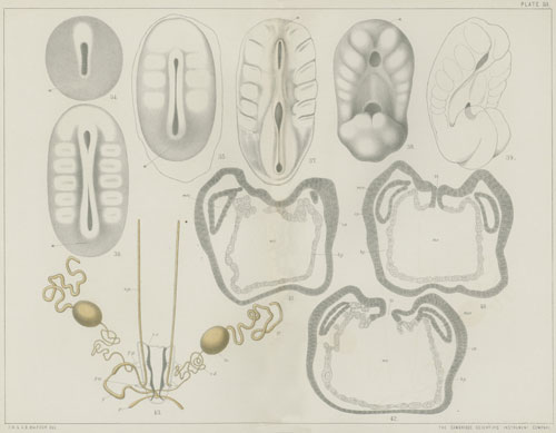
TRANSCRIBER'S NOTE:
Raised dots in numbers were converted to decimals.
Use of periods and commas in the abbreviations within and referring to
figures and plates is inconsistent. Often, punctuation marks do not match
the illustrations to which they refer. Periods were retained; commas were
added to separate figure numbers from abbreviations within the figure.
Spacing within the abbreviations was standardized.
Footnotes were renumbered sequentially, and were moved to the end of the
plate explanation.
Changes for consistency within the text of the book:
body cavity to body-cavity
body wall to body-wall
choroid-slit to choroid slit
develope(s) to develop(s)
Elasmobranchs to Elasmobranchii
inter-renal to interrenal
re-agent(s) to reagent(s)
omphalo-meseraic to omphalomeseraic
pleuroperitoneal to pleuro-peritoneal
proto-vertebra(æ) to protovertebra(æ)
re-appear to reappear
side-fold to side fold
sub-intestinal to subintestinal
sub-notochordal to subnotochordal
urino-genital to urinogenital
Spelling change:


