Introduction.
Our knowledge of the development of the organs in most of the
Invertebrate groups is so meagre that it would not be profitable to
attempt to treat systematically the organogeny of the whole animal
kingdom.
For this reason the plan adopted in this section of the work has been
to treat somewhat fully the organogeny of the Chordata, which is
comparatively well known; and merely to indicate a few salient facts
with reference to the organogeny of other groups. In the case of the
nervous system, and of some other organs which especially lend
themselves to this treatment, such as the organs of special sense and
the excretory system, a wider view of the subject has been taken; and
certain general principles underlying the development of other organs
have also been noticed.
The classification of the organs is a matter of some difficulty.
Considering the character of this treatise it seemed desirable to
arrange the organs according to the layers from which they are
developed. The compound nature of many organs, e.g. the eye and ear,
renders it, however, impossible to carry out consistently such a mode
of treatment. I have accordingly adopted a rough classification of the
organs according to the layers, dropping the principle where
convenient, as, for instance, in the case of the stomodæum and
proctodæum.
The organs which may be regarded as mainly derived from
the epiblast
are (1) the skin; (2) the nervous system; (3) the organs of special
sense.
Those from the mesoblast are (1) the general connective tissue and
skeleton; (2) the vascular system and body cavity; (3) the muscular
system; (4) the urinogenital system.
Those from the hypoblast are the alimentary tract and its derivates;
with which the stomodæum and proctodæum and their respective derivates
are also dealt with.
Bibliography.
General works dealing with the development of the organs of the
Chordata.
(291) K. E. von Baer. Ueber Entwicklungsgeschichte d. Thiere.
Königsberg, 1828-1837.
(292) F. M. Balfour. A Monograph on the development of Elasmobranch
Fishes. London, 1878.
(293) Th. C. W. Bischoff. Entwicklungsgesch. d. Säugethiere U. d.
Menschen. Leipzig, 1842.
(294) C. Gegenbaur. Grundriss d. vergleichenden Anatomie. Leipzig,
1878. Vide also English translation, Elements of Comp. Anatomy.
London, 1878.
(295) M. Foster and F. M. Balfour. The Elements of Embryology. Part
I. London, 1874.
(296) Alex. Götte. Entwicklungsgeschichte d. Unke. Leipzig, 1875.
(297) W. His. Untersuch. üb. d. erste Anlage d. Wirbelthierleibes.
Leipzig, 1868.
(298) A. Kölliker. Entwicklungsgeschichte d. Menschen u. der höheren
Thiere. Leipzig, 1879.
(299) H. Rathke. Abhandlungen ü. Bildung und Entwicklungsgeschichte
d. Menschen u. d. Thiere. Leipzig, 1838.
(300) H. Rathke. Entwicklungs. d. Natter. Königsberg, 1839.
(301) H. Rathke. Entwicklungs. d. Wirbelthiere. Leipzig, 1861.
(302) R. Remak. Untersuchungen üb. d. Entwicklung d. Wirbelthiere.
Berlin, 1850-1855.
(303) S. L. Schenk. Lehrbuch d. vergleich. Embryologie d.
Wirbelthiere. Wien, 1874.
In many of the Cœlenterata the outermost layer of the blastoderm is
converted as a whole into the skin or ectoderm. The cells composing it
become no doubt in part differentiated into muscular elements and in
part into nervous elements, &c.; but still it may remain through life
as a simple external membrane. This membrane contains in itself
indefinite potentialities for developing into various organs, and in
all the true Triploblastica these potentialities are more or less
realized. The embryonic epiblast ceases in fact, in the higher forms,
to become converted as a whole into the epidermis, but first gives
rise to parts of the nervous system, organs of special sense, and
other parts.
After the formation of these parts the remnant of the epiblast gives
rise to the epidermis, and often unites more or less intimately with a
subjacent layer of mesoblast, known as the dermis, to form with it the
skin.
Various differentiations may arise in the epidermis forming protective
or skeletal structures, terminal sense organs, or glands. The
structure of the epidermis itself varies greatly, and for Vertebrates
its general modifications have been already sufficiently dealt with in
chapter XII. Of its special differentiations those of a protective or
skeletal nature and those of a glandular nature may be considered in
this place.
Protective epidermal structures. These structures constitute a general
cuticle or an exoskeleton of scales, hairs, feathers, nails, hoofs,
&c. They may be entirely formed from
the epidermis either as (1) a
cuticular deposit, or as (2) a chitinization, a cornification, or
calcification of its constituent cells. These two processes run into
each other, and are in many cases not easily distinguished. The
protective structures of the epidermis may be divided into two groups
according as they are formed on the outer or the inner side of the
epidermis. Dermal skeletal structures are in many cases added to them.
Amongst the Invertebrata the most widely distributed type of
exoskeleton is a cuticle formed on the outer surface of the epidermis,
which reaches its highest development in the Arthropoda. In the same
class with this cuticle must be placed the molluscan and brachiopod
shells, which are developed as cuticular plates on special regions of
the epidermis. They differ, however, from the more usual form of
cuticle in their slighter adhesion to the subjacent epidermis, and in
their more complicated structure. The test of Ascidians is an abnormal
form of exoskeleton belonging to this type. It is originally formed
(Hertwig and Semper) as a cuticle on the surface of the epidermis; but
subsequently epidermic cells migrate into it, and it then constitutes
a tissue similar to connective tissue, but differing from ordinary
epidermic cuticles in that the cells which deposit it do so over their
whole surface, instead of one surface, as is usually the case with
epithelial cells.
In the Vertebrata the two types of exoskeleton mentioned above are
both found, but that developed on the inner surface of the epidermis
is always associated with a dermal skeleton, and that on the outer
side frequently so. The type of exoskeleton developed on the inner
side of the general epidermis is confined to the Pisces, where it
appears as the scales; but a primitive form of these structures
persists as the teeth in the Amphibia and Amniota. The type developed
on the outer side of the epidermis is almost entirely[149]
confined
to the Amphibia and Amniota, where it appears as scales, feathers,
hairs, claws, nails, &c. For the histological details as to the
formation of these various organs I must refer the reader to treatises
on histology, confining my attention here to the general embryological
processes which take place in their development.
The most primitive form of the first type of dermal structures is that
of the placoid scales of Elasmobranchii[150].
These consist, when
fully formed, of a plate bearing a spinous projection. They are
constituted of an outer enamel layer on the projecting part, developed
as a cuticular deposit of the epidermis (epiblast), and an underlying
basis of dentine (the lower part of which may be osseous) with a
vascular pulp in its axis. The development (fig. 235) is as follows
(Hertwig, No. 306). A papilla of the dermis makes its appearance, the
outer layer of which gradually calcifies to form the dentine and
osseous tissue. This papilla is covered by the columnar mucous layer
of the epidermis (e), from which it is separated by a basement
membrane, itself a product of the epidermis. This membrane gradually
thickens and calcifies, and so gives rise to the enamel cap (o). The
spinous point gradually forces its way through the epidermis, so as to
project freely at the surface.
The scales of other forms of fishes are to be derived from those of
Elasmobranchii. The great dermal plates of many fishes have been
formed by the concrescence of groups of such scales. The dentine in
many cases partially or completely atrophies, leaving the major part
of the scale formed of osseous tissue; such plates often become parts
of the internal skeleton.
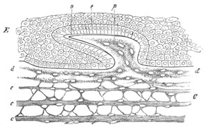
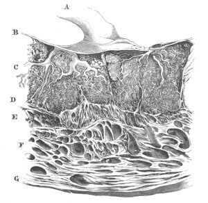
Fig. 235. Vertical section through the skin of an embryonic Shark,
to shew a developing placoid scale. (From Gegenbaur; after O.
Hertwig.)
E. epidermis; C. layers of dermis; d. uppermost layer of
dermis; p. papilla of dermis; e. mucous layer of epidermis; o.
enamel layer.
The teeth, as will be more particularly described in the section on
the alimentary tract, are formed by a modification of the same process
as the placoid scales, in which a ridge of the epithelium grows
inwards to meet a connective tissue papilla, so that the development
of the teeth takes place entirely below the superficial layer of
epidermis.
In most Teleostei the enamel and dentine layers have disappeared, and
the scales are entirely formed of a peculiar calcified tissue
developed in the dermis.
The cuticle covering the scales of Reptiles is the simplest type of
protective structure formed on the outer surface of the epidermis. The
scales consist of papillæ of the dermis and epidermis; and are covered
by a thickened portion of a two-layered cuticle, formed over the whole
surface of the body from a cornification of the superficial part of
the epidermis. Dermal osseous plates may be formed in connection with
these scales, but are never of course united with the superficial
cuticle.
Feathers are probably special modifications of such scales. They arise
from an induration of the epidermis of papillæ containing a vascular
core. The provisional down, usually present at the time of hatching,
is formed by the cornification of longitudinal ridges of the mucous
layer of the epidermis of the papillæ; each cornified ridge giving
rise to a barb of the feather. The horny layer of the epidermis forms
a provisional sheath for the developing feather below. When the barbs
are fully formed this sheath is thrown off, the vascular core dries
up, and the barbs become free except at their base.
Without entering into the somewhat complicated details of the
formation of the permanent feathers, it may be mentioned that the
calamus or quill is formed by a cornification in the form of a tube of
both layers of the epidermis at the base of the papilla. The quill is
open at both ends, and to it is attached the vexillum or plume of the
feather. In a typical feather this is formed at the apex of the
papilla from ridge-like thickenings of the mucous layer of the
epidermis, arranged in the form of a longitudinal axis, continuous
with the cornified mucous layer of the quill, and from lateral ridges.
These subsequently become converted into the axis and barbs of the
plume. The external epidermic layer becomes converted into a
provisional horny sheath for the true feather beneath.
On the completion of the plume of the feather the external sheath is
thrown off, leaving it quite free, and the vascular core belonging to
it shrivels up. The papilla in which the feather is formed becomes at
a very early period secondarily enveloped in a pit or follicle which
gradually deepens as the development of the feather is continued.
Hairs (Kölliker, No. 298) are formed in solid processes of the mucous
layer of the epidermis, which project into
the subjacent dermis. The
hair itself arises from a cornification of the cells of the axis of
one of the above processes; and is invested by a sheath similarly
formed from the more superficial epidermic cells. A small papilla of
the dermis grows into the inner end of the epidermic process when the
hair is first formed. The first trace of the hair appears close to
this papilla, but soon increases in length, and when the end of the
hair projects from the surface, the original solid process of the
epidermis becomes converted into an open pit, the lumen of which is
filled by the root of the hair. Hairs differ in their mode of
formation from scales in a manner analogous to that in which the teeth
differ from ordinary placoid scales; i.e. they are formed in
inwardly directed projections of the epidermis instead of upon free
papillæ at the surface.
Nails (Kölliker, No. 298) are developed on special regions of the
epidermis, known as the primitive nail beds. They are formed by the
cornification of a layer of cells which makes its appearance between
the horny and mucous layers of the epidermis. The distal border of the
nail soon becomes free, and the further growth is effected by
additions to the under side and attached extremity of the nail.
Although the nail at first arises in the interior of the epidermis,
yet its position on the outer side of the mucous layer clearly
indicates with which group of epidermic structures it should be
classified.
Dermal skeletal structures. We have seen that in the Chordata skeletal
structures, which were primitively formed of both an epidermic and
dermic element, may lose the former element and be entirely developed
in the dermis. Amongst the Invertebrata there are certain dermal
skeletal structures which are evolved wholly independently of the
epidermis. The most important of these structures are the skeletal
plates of the Echinodermata.
Glands. The secretory part of the various glandular structures
belonging to the skin is invariably formed from the epidermis. In
Mammalia it appears that these glands are always formed as solid
ingrowths of the mucous layer (Kölliker, No. 298). The ends of these
ingrowths dilate to form the true glandular part of the organs, while
the stalks connecting the glandular portions with the surface form the
ducts. In the case of the sweat-glands the lumen of the duct becomes
first established. Its formation is inaugurated by the appearance of
the cuticle, and appears first at the inner end of the duct and thence
extends outwards (Ranvier, No. 311). In the sebaceous glands the first
secretion is formed by a fatty modification of the whole of the
central cells of the gland.
The muscular layer of the secreting part of the sweat-glands is
formed, according to Ranvier (No. 311), from a modification of the
deeper layer of the epidermic cells.
The Mammary Glands arise in essentially the same manner as the other
glands of the skin[151].
The glands of each side are formed as a solid
bud of the mucous layer of the epidermis. From this bud processes
sprout out, each of which gives rise to one of the numerous glands of
which the whole organ is formed. Two very distinct types in the
relation of the ducts of the glands to the nipple are found
(Gegenbaur, No. 313).
Bibliography of Epidermis.
General.
(304) T. H. Huxley. “Tegumentary organs.” Todd’s Cyclopædia of Anat.
and Physiol.
(305) P. Z. Unna. “Histol. u. Entwick. d. Oberhaut.” Archiv f. mikr.
Anat. Vol. XV. 1876. Vide also Kölliker (No. 298).
Scales of the Pisces.
(306) O. Hertwig. “Ueber Bau u. Entwicklung d. Placoidschuppen u. d.
Zähne d. Selachier.” Jenaische Zeitschrift, Vol. VIII. 1874.
(307) O. Hertwig. “Ueber d. Hautskelet d. Fische.” Morphol.
Jahrbuch, Vol. II. 1876. (Siluroiden u. Acipenseridæ.)
(308) O. Hertwig. “Ueber d. Hautskelet d. Fische (Lepidosteus u.
Polypterus).” Morph. Jahrbuch, Vol. V. 1879.
Feathers.
(309) Th. Studer. Die Entwick. d. Federn. Inaug. Diss. Bern, 1873.
(310) Th. Studer. “Beiträge z. Entwick. d. Feder.” Zeit. f. wiss.
Zool., Vol. XXX. 1878.
Sweat-glands.
(311) M. S. Ranvier. “Sur la structure des glandes sudoripares.”
Comptes Rendus, Dec. 29, 1879.
Mammary glands.
(312) C. Creighton. “On the development of the Mamma and the Mammary
function.” Jour. of Anat. and Phys., Vol. XI. 1877.
(313) C. Gegenbaur. “Bemerkungen üb. d. Milchdrüsen-Papillen d.
Säugethiere.” Jenaische Zeit., Vol. VII. 1873.
(314) M. Huss. “Beitr. z. Entwick. d. Milchdrüsen b. Menschen u. b.
Wiederkäuern.” Jenaische Zeit., Vol. VII. 1873.
(315) C. Langer. “Ueber d. Bau u. d. Entwicklung d. Milchdrüsen.”
Denk. d. k. Akad. Wiss. Wien, Vol. III. 1851.
Origin of the Nervous System.
One of the most important recent embryological discoveries is the fact
that the central nervous system, in all the Metazoa in which it is
fully established, is (with a few doubtful exceptions) derived from
the primitive epiblast[152].
As we have already seen that the epiblast
represents to a large extent the primitive epidermis, the fact of the
nervous system being derived from the epiblast implies that the
functions of the central nervous system, which were originally taken
by the whole skin, became gradually concentrated in a special part of
the skin which was step by step removed from the surface, and has
finally become in the higher types a well-defined organ imbedded in
the subdermal tissues.
Before considering in detail the comparative development of the
nervous system, it will be convenient shortly to review the present
state of our knowledge on the general process of its evolution.
This process may be studied either embryologically, or by a comparison
of the various stages in its evolution preserved in living forms. Both
the methods have led to important results.
The embryological evidence shews that the ganglion-cells of the
central part of the nervous system are originally derived from the
simple undifferentiated epithelial cells of the surface of the body,
while the central nervous system itself has arisen from the
concentration of such cells in special tracts. In the Chordata at any
rate the nerves arise as outgrowths of the central organ.
Another important fact shewn by embryology is that the central nervous
system, and percipient portions of the organs of special sense,
especially of optic organs, are often formed from the same part of the
primitive epidermis. Thus the retina of the Vertebrate eye is formed
from the two lateral lobes of the primitive fore-brain.
The same is true for the compound eyes of some Crustacea. The
supraœsophageal ganglia of these animals are formed in the embryo
from two thickened patches of the epiblast of the procephalic lobes.
These thickened patches become gradually detached from the surface,
remaining covered by a layer of epidermis. They then constitute the
supraœsophageal ganglia; but they form not only the ganglia, but also
the retinulæ of the eye—the parts in fact which correspond to the
rods and cones in our own retina. The accessory parts of these organs
of special sense, viz. the crystalline lens of the Vertebrate eye, and
the corneal lenses and crystalline cones of the Crustacean eye, are
independently formed from the epiblast after the separation of the
part which becomes the central nervous system.
In the Acraspedote Medusæ the rudimentary central nervous system has
the form of isolated rings, composed of sense-cells prolonged into
nervous fibres, surrounding the stalks of tentacle-like organs, at the
ends of which are placed the sense-organs.
This close connection between certain organs of special sense and
ganglia is probably to be explained by supposing that the two sets of
structures actually originated pari passu.
We may picture the process as being somewhat as follows:—
It is probable that in simple ancestral organisms the whole body was
sensitive to light, but that with the appearance of pigment-cells in
certain parts of the body, the sensitiveness to light became
localised to the areas where the pigment-cells were present. Since,
however, it was necessary that stimuli received by such organs
should be communicated to other parts
of the body, some of the
epidermic cells in the neighbourhood of the pigment-spots, which
were at first only sensitive in the same manner as other cells of
the epidermis, became gradually differentiated into special
nerve-cells. As to the details of this differentiation embryology
does not as yet throw any great light; but from the study of
comparative anatomy there are grounds for thinking that it was
somewhat as follows:—Cells placed on the surface sent protoplasmic
processes of a nervous nature inwards, which came into connection
with nervous processes from similar cells placed in other parts of
the body. The cells with such processes then became removed from the
surface, forming a deeper layer of the epidermis below the sensitive
cells of the organ of vision. With the latter cells they remained
connected by protoplasmic filaments, and thus they came to form a
thickening of the epidermis underneath the organ of vision, the
cells of which received their stimuli from those of the organ of
vision, and transmitted the stimuli so received to other parts of
the body. Such a thickening would obviously be the rudiment of a
central nervous system, and is in fact very similar to the
rudimentary ganglia of the Acraspeda mentioned above. It is easy to
see by what steps it might become larger and more important, and
might gradually travel inwards, remaining connected with the
sense-organ at the surface by protoplasmic filaments, which would
then constitute nerves. The rudimentary eye would at first merely
consist of cells sensitive to light, and of ganglion-cells connected
with them; while at a later period optical structures, constituting
a lens capable of throwing an image of external objects upon it,
would be developed, and so convert the whole structure into a true
organ of vision. It has thus come about that, in the development of
the individual, the retina is often first formed in connection with
the central nervous system, while the lenses of the eye are
independently evolved from the epidermis at a later period.
A series of forms of the Cœlenterata and Platyelminthes affords us
examples of various stages in the differentiation of a central nervous
system[153].
In sea-anemones (Hertwigs, No. 321) there are, for instance, no organs
of special sense, and no definite central nervous system. There are,
however, scattered throughout the skin, and also throughout the lining
of the digestive tract, a number of specially modified epithelial
cells, which are no doubt delicate organs of sense. They are provided
at their free extremity with a long hair, and are prolonged on their
inner side into fine processes which penetrate into the deeper part of
the epithelial layer of the skin or digestive wall. They eventually
join a fine network of protoplasmic fibres which forms a special layer
immediately within the epithelium. The fibres of this network are no
doubt essentially nervous. In addition to fibres there are,
moreover,
present in the network cells of the same character as the multipolar
ganglion-cells in the nervous system of Vertebrates, and some of these
cells are characterised by sending a process into the superjacent
epithelium. Such cells are obviously intermediate between
neuro-epithelial cells and ganglion-cells; and it is probable that the
nerve-cells are, in fact, sense-cells which have travelled inwards and
lost their epithelial character.
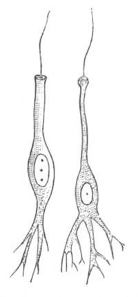
Fig. 236. Neuro-epithelial sense-cells of Aurelia aurita. (From
Lankester; after Schäfer.)
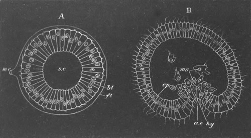
In the Craspedote Medusæ (Hertwigs, No. 320) the differentiation of
the nervous system is carried somewhat further. There is here a
definite double ring, placed at the insertion of the velum, and
usually connected with sense-organs. The two parts of the ring belong
respectively to the epithelial layers on the upper and lower surfaces
of the velum, and are not separated from these layers; they are formed
of fine nerve-fibres and ganglion-cells. The epithelium above the
nerve rings contains sense-cells (fig. 237) with a stiff hair at their
free extremity, and a nervous prolongation at the opposite end, which
joins the nerve-fibres of the ring. Between such cells and true
ganglion-cells an intermediate type of cell has been found (fig. 237
B) which sends a process upwards amongst the epithelial cells, but
does not reach the surface. Such cells, as the Hertwigs have pointed
out, are clearly sense-cells partially transformed into
ganglion-cells.
A still higher type of nervous system has been met with amongst some
primitive Nemertines (Hubrecht, No. 323), consisting of a pair of
large cephalic ganglia, and two well-developed lateral ganglionic
cords placed close beneath the epidermis. These cords, instead of
giving off definite nerves, as in animals with a fully differentiated
nervous system, are connected with a continuous subdermal nervous
plexus.
The features of the embryology and the anatomy of the nervous system,
to which attention has just been called, point to the following
general conclusions as to the evolution of the nervous system.
(1) The nervous system of the higher Metazoa appears to have been
evolved in the course of a long series of generations from a
differentiation of some of the superficial epithelial cells of the
body, though it is possible that some parts of the system may have
been formed by a differentiation of the alimentary epithelium.
(2) An early feature in the differentiation consisted in the growth of
a series of delicate processes of the inner ends of
certain epithelial
cells, which became at the same time especially differentiated as
sense-cells (figs. 236 and 237).
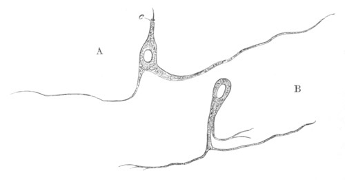
Fig. 237. Isolated cells belonging to the upper nerve-ring of Carmarina
hastata. (After O. and R. Hertwig.)
A. Neuro-epithelial sense-cell. c. sense-hair.
B. Transitional cell between a neuro-epithelial cell and a
ganglion-cell.
(3) These processes gave rise to a subepithelial nervous
plexus, in which ganglion-cells, formed from sense-cells which
travelled inwards and lost their epithelial character (fig. 237 B),
soon formed an important part.
(4) Local differentiations of the nervous network, which was
no doubt distributed over the whole body, took place partly in
the formation of organs of special sense, and partly in other
ways, and such differentiations gave rise to a central nervous
system. The central nervous system was at first continuous
with the epidermis, but became separated from it and travelled
inwards.
(5) Nerves, such as we find them in the higher types,
originated from special differentiations of the nervous network,
radiating from the parts of the central nervous system.
The following points amongst others are still very obscure:—
(1) The steps by which the protoplasmic processes from the primitive
epidermic cells became united together so as to form a network
of nerve-fibres, placing the various parts of the body in
nervous communication.
(2) The process by which nerves became connected with muscles, so
that a stimulus received by a nerve-cell could be communicated
to and cause a contraction in a muscle.
It is probable, as stated in the above summary, that the nervous
network
took its origin from processes of the sense-cells. The
processes of the different cells probably first met and then fused
together, and, becoming more arborescent, finally gave rise to a
complicated network.
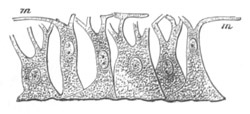
Fig. 238. Myo-epithelial cells of Hydra. (From Gegenbaur; after
Kleinenberg.)
m. contractile fibres; processes of cells.
The primitive relations between the nervous network and the muscular
system are matters of pure speculation. The primitive muscular cells
consist of epithelial cells with muscular processes (fig. 238), but
the branches of the nervous network have not been traced into
connection with the muscles in any Cœlenterata except Ctenophora. In
the higher types a continuity between nerves and muscles in the form
of motorial end plates has been widely observed. Even in the case of
the Cœlenterata it is quite clear from Romanes’ experiments that
stimuli received by the nerves are capable of being transmitted to the
muscles, and that there must therefore be some connection between
nerves and muscles. How did this connection originate?
Epithelial cells with muscular processes (fig. 238) were discovered by
Kleinenberg (No. 324) in Hydra before epithelial cells with nervous
processes were known, and Kleinenberg pointed out that Hydra shewed
the possibility of nervous and muscular tissues existing without a
central nervous system, and suggested that the epithelial part of the
myoepithelial cells was a sense-organ, and that the connecting part
between this and the contractile processes was a rudimentary nerve. He
further supposed that in the subsequent evolution of these elements
the epithelial part of the cell became a ganglion-cell, while the part
connecting this with the muscular tail became prolonged so as to form
a true nerve. The discovery of neuro-epithelial cells existing side by
side with myoepithelial cells demonstrates that this theory must in
part be abandoned, and that some other explanation must be given of
the continuity between nerves and muscles. The hypothetical
explanation which most obviously suggests itself is that of fusion.
It seems quite possible that many of the epithelial cells of the
epidermis and walls of the alimentary tract were originally provided
with processes, the protoplasm of which, like that of the Protozoa,
carried on the functions of nerves and muscles at the same time, and
that these processes united amongst themselves into a network. Such
cells would be very similar to Kleinenberg’s neuro-muscular cells. By
a subsequent differentiation some of the cells forming this network
may have become specially contractile, the epithelial parts of the
cells ceasing to have a nervous function, and other cells may have
lost their contractility and become solely nervous. In this way we
should get neuro-epithelial cells and myoepithelial cells both
differentiated from the primitive network, and the connection between
the two would also be explained. This hypothesis fits in moreover very
well with the condition of the neuro-muscular system as we find it in
the Cœlenterata.
Bibliography.
Origin of the Nervous System.
(316) F. M. Balfour. “Address to the Department of Anat. and Physiol.
of the British Association.” 1880.
(317) C. Claus. “Studien üb. Polypen u. Quallen d. Adria. 1.
Acalephen, Discomedusen,” Denk. d. math.-naturwiss. Classe d. k.
Akad. Wiss. Wien, Vol. XXXVIII. 1877.
(318) Th. Eimer. Zoologische Studien a. Capri. 1. Ueber Beroë ovatus.
Ein Beitrag z. Anat. d. Rippenquallen. Leipzig, 1873.
(319) V. Hensen. “Zur Entwicklung d. Nervensystems.” Virchow’s
Archiv, Vol. XXX. 1864.
(320) O. and R. Hertwig. Das Nervensystem u. d. Sinnesorgane d.
Medusen. Leipzig, 1878.
(321) O. and R. Hertwig. “Die Actinien anat. u. histol. mit besond.
Berücksichtigung d. Nervenmuskelsystem untersucht.” Jenaische Zeit.,
Vol. XIII. 1879.
(322) R. Hertwig. “Ueb. d. Bau d. Ctenophoren.” Jenaische
Zeitschrift, Vol. XIV. 1880.
(323) A. W. Hubrecht. “The Peripheral Nervous System in Palæo- and
Schizonemertini, one of the layers of the body-wall.” Quart. J. of
Micr. Science, Vol. XX. 1880.
(324) N. Kleinenberg. Hydra, eine
anatomisch-entwicklungsgeschichtliche Untersuchung. Leipzig, 1872.
(325) A. Kowalevsky. “Embryologische Studien an Würmern u.
Arthropoden.” Mém. Acad. Pétersbourg, Series VII., Vol. XVI. 1871.
(326) E. A. Schäfer. “Observations on the nervous system of Aurelia
aurita.” Phil. Trans. 1878.
Nervous system of the Invertebrata. Our knowledge of the development
of the central nervous system is still very imperfect in the case of
many Invertebrate groups. In the Echinodermata and some of the
Chætopoda it is never detached from the epidermis, and in such cases
its origin is clear without embryological evidence.
In the majority of groups the central nervous system may be reduced to
the type of a pair of cephalic ganglia, continued posteriorly into two
cords provided with nerve-cells, which may coalesce ventrally or be
more or less widely separated, and be unsegmented or segmented.
Various additional visceral ganglia may be added, and in different
instances parts of the system may be much reduced, or peculiarly
modified. The nervous system of the Platyelminthes (when present), of
the
Rotifera, Brachiopoda, Polyzoa (?), the Mollusca, the Chætopoda,
the Discophora, the Gephyrea, the Tracheata, and the Crustacea, the
various small Arthropodan phyla (Pœcilopoda, Pycnognida, Tardigrada,
&c.), the Chætognatha (?), and the Myzostomea, probably belongs to
this type.
The nervous system of the Echinodermata cannot be reduced to this
form; nor in the present state of our knowledge can that of the
Nematelminthes or Enteropneusta.
It is only in the case of members of the former set of groups that any
adequate observations have yet been made on the development of the
nervous system, and even in the case of these groups observations
which have any claim to completeness are confined to certain members
of the Chætopoda, the Arthropoda and the Mollusca. An account of
imperfect observations on other forms, where such have been made, will
be found in the systematic part of this work.
Chætopoda. We are indebted to Kleinenberg (No. 329) for the most
detailed account which we have of the development of the central
nervous system in the Chætopoda.
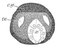
Fig. 239. Section through the head of a young embryo of Lumbricus
trapezoides. (After Kleinenberg.)
c.g. cephalic ganglion; cc. cephalic portion of the body cavity;
x. œsophagus.
The supraœsophageal ganglion with the œsophageal commissure
developes independently of the ventral cord. It arises as an unpaired
thickening of the epiblast, close to the dorsal side of the œsophagus
at the front end of the head (fig. 239), which becomes separated from
the epiblast, and extends obliquely backwards and downwards in a
somewhat arched form; its lower extremities being somewhat swollen.
The inner portion of this curved rudiment becomes converted into
commissural nerve-fibres, while the cells of the outer and upper
portion assume the characters of ganglion-cells. The commissural
fibres are continued downwards to meet the ventral chord, but their
junction with the latter structure is not effected till late in
embryonic life.
The ventral cord is formed by the coalescence of a pair of linear
cords, the development of which takes place from before backwards, so
that when their anterior part is well developed their posterior part
is hardly differentiated. These cords arise, one on
each side of a
ventral ciliated furrow, first as a single row of epiblast cells, and
subsequently as several rows (fig. 240, Vg). While still united to
the external epiblast, they extend themselves below the cells lining
the ventral furrow, and unite into a single nervous band, which
however exhibits its double origin by its bilobed section. Before the
two cords unite, the groove between them becomes somewhat deep, but
subsequently shallows out and disappears. The nervous band, before
separating from the epiblast, exhibits, in correspondence with the
mesoblastic segments, alternate swellings and constrictions. The
former become the ganglia, and the latter the connecting trunks.
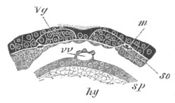
Fig. 240. Section through part of the ventral wall of the trunk of
an embryo of Lumbricus trapezoides. (After Kleinenberg.)
m. longitudinal muscles; so. somatic mesoblast; sp. splanchnic
mesoblast; hy. hypoblast; Vg. ventral nerve-cord; vv. ventral
vessel.
As soon as the cord becomes free from the epiblast, it becomes
surrounded by a sheath, formed of somatic mesoblast. In each of the
ganglionic enlargements there next appears on the dorsal surface a
pair of areas of punctiform material, the substance of which soon
differentiates itself into nerve-fibres. These areas, by uniting from
side to side, give rise to the transverse commissures, and also by a
linear coalescence to the longitudinal commissures of the cord. The
cellular parts of the band surrounding them become converted into a
ganglionic covering of the cord.
In each ganglion the cells of this ganglionic investment penetrate as
a median septum into the cord. A fissure is next formed, dividing this
septum into two; it is subsequently continued for the whole length of
the cord.
Arthropoda. In the Tracheata and the Crustacea the development of the
ventral cord is in the main similar to that in the Chætopods, while
that of the supraœsophageal ganglia is as a rule somewhat more
complicated. No such clear evidence of an independent development of
these two parts, as in the case of the Chætopods, has as yet been
produced.
The most primitive type of nervous system amongst the
Tracheata is
that of Peripatus, where it consists of large supraœsophageal
ganglia, continuous with a pair of widely separated but large ventral
cords united posteriorly above the anus. These cords have an
investment of ganglion-cells for their whole length, and are
imperfectly divided into ganglia corresponding in number with the
feet.
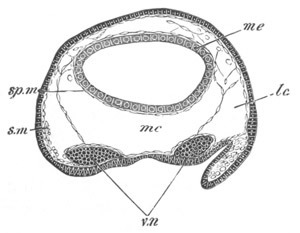
Fig. 241. Section through the trunk of an embryo of
Peripatus. The embryo from which the section is taken was
somewhat younger than that of fig. 242.
sp.m. splanchnic mesoblast; s.m. somatic mesoblast; mc. median
section of body cavity; lc. lateral section of body cavity; v.n.
ventral nerve cord; me. mesenteron.
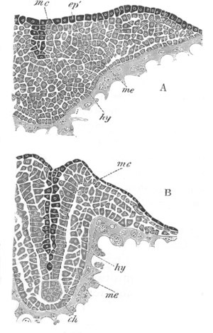
The ventral cords are formed as two separate epiblastic ridges (fig.
241, v.n), continued in front into a pair of thickenings of the
procephalic lobes, which are at first independent of each other, and
from which a large part of the supraœsophageal ganglia takes its
origin. After the latter have become separated from the epiblast an
invagination of the epiblast covering them grows into each lobe (fig.
242), and becoming constricted from the superficial epiblast, which
remains as the epidermis, forms a not unimportant part of the
permanent supraœsophageal ganglia.
In the Arachnida the mode of development of the nervous system is
essentially the same, and the reader will find a detailed account of
it for Spiders in Vol. II. pp. 447-451. The ventral cords are here
formed as independent and at first widely separated strands (fig. 243,
vn), which for a long time remain far apart; they are subsequently
divided into ganglia and become united by transverse commissures.
The supraœsophageal ganglia are formed as two independent
thickenings
of the procephalic lobes (fig. 244), which eventually separate from
the superficial skin. There is formed however in each of them a
semicircular groove (fig. 244, gr) lined by the superficial
epiblast, which becomes detached from the skin, and is involuted to
form part of the ganglia.
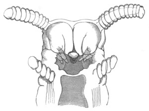
Fig. 242. Head of an embryo Peripatus. (From Moseley.)
The figure shews the jaws (mandibles), and close to them epiblastic
involutions, which grow into the supraœsophageal ganglia. The
antennæ, oral cavity, and oral papillæ are also shewn.
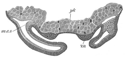
Fig. 243. Transverse section through the Ventral plate of Agelena
labyrinthica.
The ventral cords have begun to be formed as thickenings of the
epiblast, and the limbs are established.
me.s. mesoblastic somite; vn. ventral nerve-cord; yk. yolk.
A similar mode of formation of both the ventral cords and the
supraœsophageal ganglia obtains in Insects (fig. 245). The ventral
cords are however much less widely separated than in Spiders, and
early unite in the median line. In the supraœsophageal ganglia the
invaginated epiblast has in Lepidoptera (Hatschek) the form of a pit
on the dorsal border of the antennæ.
Hatschek states that there takes place an invagination of a median
part of the skin between the two ventral cords, for the details of
which I must refer the reader to Vol. II. p. 410. He has made more or
less similar statements for the earthworm, but his observations in
both instances are open to serious doubt.
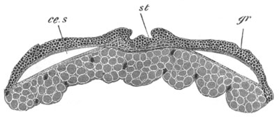
Fig. 244. Section through the procephalic lobes of an embryo of
Agelena labyrinthica.
st. stomadæum; gr. section through semicircular groove in
procephalic lobe; ce.s. cephalic section of body cavity.
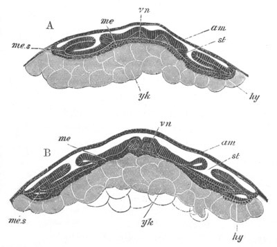
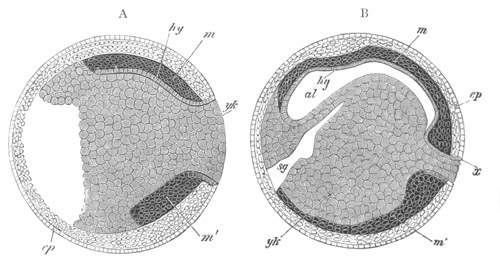
Fig. 245. Two transverse sections through the embryo of Hydrophilus. (After Kowalevsky.)
A. Transverse section through an embryo in the region of one of the
stigmata.
B. Transverse section through an older embryo.
vn. ventral nerve-cord; am. amnion and serous membrane; me.
mesoblast; me.s. somatic mesoblast; hy. hypoblast (?); yk.
yolk-cells (true hypoblast); st. stigma of trachea.
Full details as to the development of the nervous system in the
Crustacea are still wanting; a fairly complete account of
what is
known on the subject is given in Vol. II. pp. 521-2. It appears that
the ventral cord may either arise as an unpaired thickening of the
epiblast (Isopoda), marked however by a shallow median furrow, or from
two cords which eventually coalesce[154].
It is not certain how far
the supraœsophageal ganglia are usually in the first instance
continuous with the ventral cord. In Astacus, the early stages of
which have been elaborately investigated by Reichenbach (No. 331),
they are stated to be so; the supraœsophageal ganglia are moreover
described by this author as having a somewhat complicated origin. Five
elements enter into their composition. There is first formed a pair of
pits on the procephalic lobes, which become very deep during the
Nauplius stage, and are continuous with a pair of epiblastic ridges
which pass round the mouth, and join the ventral cords just described.
The walls of the pits are believed to form a part of the embryonic
ganglia which gives rise to the retina as well as to the optic
ganglia. The ridges form the remainder of the ganglia and the
œsophageal commissures; while the fifth element is supplied by a
median invagination in front of the mouth, which appears at a much
later date than the other parts.
In the Isopoda supraœsophageal ganglia are stated to arise as
thickenings of the procephalic lobes, which become eventually detached
from the epidermis.
The ventral cord is at first unsegmented, but soon becomes partially
divided by a series of constrictions into a number of ganglia,
corresponding with the segments. The development of the commissural
and ganglionic portions takes place much as in the Chætopoda.
The Gephyrea approach closely the types so far dealt with, but the
ventral cord in the Inermia is formed as an unpaired thickening of the
epiblast. In Echiurus, as has been shewn by Hatschek in an interesting
paper on the larva of this species, published since the appearance of
the first volume, there is a pair of ventral cords[155].
In
correspondence with a general segmentation of the body, which is
subsequently lost, these cords become
segmented. The two cords unite
in the median line, and Hatschek, in accordance with his general view
on this subject, states that their junction is effected by means of a
median cord of invaginated epiblast. The segmentation of the cords
subsequently becomes lost. The supraœsophageal ganglia arise as an
unpaired median thickening of the procephalic lobe. No traces of
segmentation in the ventral cord have been observed by Spengel in
Bonellia, and the supraœsophageal ganglion is formed in this genus as
an unpaired band.
In all the groups above considered the nervous system clearly presents
the same type of development with various modifications.
It is formed of two parts, viz. (1) the supraœsophageal ganglia, and
(2) the ventral cord.
In the simpler forms, Chætopoda and Gephyrea, the supraœsophageal
ganglia are usually stated to be formed as an unpaired thickening at
the apex of the præoral lobe, which in most cases becomes subsequently
bilobed.
In the Arthropoda the unpaired præoral lobe of the Chætopoda is
replaced by the so-called procephalic lobes, which are themselves
bilobed; and the supraœsophageal ganglia are formed of two
independent halves; further complications in development are also
generally found.
There is not as yet sufficient evidence to decide whether the
supraœsophageal ganglia were primitively developed continuously with,
or independently of, the ventral cords.
The ventral cord appears in the embryo as two independent unsegmented
strands, although in a few cases (some Crustacea and Gephyrea) these
cords, by an abbreviation in development, arise as an unpaired median
thickening of the epiblast.
The form of nervous system of the Chætopoda, Arthropoda, and Gephyrea
is clearly therefore to be derived, as was first pointed out by
Gegenbaur, from a more or less similar type to that now found in the
Nemertines; and as suggested in the chapter on larval forms (vide p.
378) may perhaps be derived from the elongation of a circular ring, of
which the anterior end has become developed into the supraœsophageal
ganglia, the lateral parts into the two lateral strands, while the
posterior part persists in some forms in the junction of the ventral
cords above the anus (Enopla and Peripatus).
Mollusca. While study of the anatomy of the nervous system of the
Mollusca, especially of certain primitive genera (Chiton, Haliotis,
Fissurella, &c.) leaves little doubt that it is formed on the same
type as that of the groups just spoken of, the development, so far as
our imperfect knowledge enables us to make definite statements on the
subject, is somewhat abnormal[156].
In the Gasteropoda and Pteropoda the supraœsophageal ganglia appear
most probably to be developed either as paired thickenings of the
epiblast of the velar area, or as invaginated pits of the velar area,
which become detached from the surface, and then become solid
(Hyaleacea and Limax). In either case the supraœsophageal ganglia
appear to be developed quite independently of the pedal ganglia. The
latter, as might be anticipated, are earlier in their development and
more constant than the various visceral ganglia; and, if the views
above expressed are correct, are homologous with the ventral cord of
the Chætopods and Arthropods. Their actual development is very
imperfectly known.
The most precise statements on the subject, viz. those of Bobretzky
and Fol, would lead us to suppose that they arise in the mesoblast,
but it seems more probable that they are formed as thickenings of the
sides of the foot.
In the Cephalopods all the ganglia are stated to be differentiated in
the mesoblast (Lankester, Bobretzky).
Hatschek[157]
has recently given a detailed description of the
development of the supraœsophageal and pedal ganglia of Teredo. He
finds that the former ganglia arise as an unpaired thickening of the
epiblast in the centre of the velar area, and the latter as an
unpaired thickening of the epiblast of the ventral side of the body
between the mouth and the anus. The two ganglia would thus seem to be
disconnected with each other in their development.
(327) F. M. Balfour. “Notes on the development of the Araneina.”
Quart. J. of Micr. Science, Vol. XX. 1880.
(328) B. Hatschek. “Beitr. z. Entwicklung d. Lepidopteren.” Jenaische
Zeitschrift, Vol. XI. 1877.
(329) N. Kleinenberg. “The development of the Earthworm, Lumbricus
Trapezoides.” Quart. J. of Micr. Science, Vol. XIX. 1879.
(330) A. Kowalevsky. “Embryologische Studien an Würmern u.
Arthropoden.” Mém. Acad. Pétersbourg, Series VIII., Vol. XVI. 1871.
(331) H. Reichenbach. “Die Embryonalanlage u. erste Entwick. d.
Flusskrebses.” Zeit. f. wiss. Zool., Vol. XXIX. 1877.
The Central Nervous System of the Vertebrata[158].
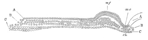
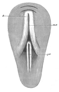
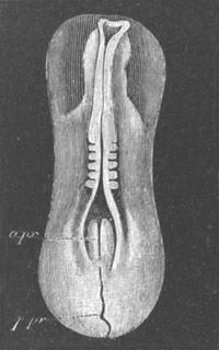
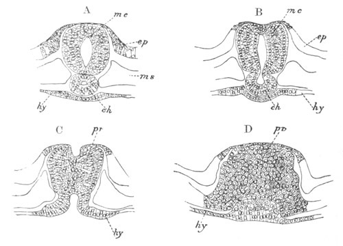
The formation of the cerebrospinal axis of the Chordata from the
medullary plate has already been treated at length (pp. 301-304).
Before entering into the consideration of the morphological value of
the various parts of this cord, it will be convenient to describe the
more important features of its ontogeny. For this purpose the two
parts into which the nervous axis becomes at an early period divided,
viz. the spinal cord and the brain, may be dealt with separately.
The Spinal Cord, shortly after the closure of the medullary canal,
has, in all the true Vertebrata, the form of an oval tube; the walls
of which are of a fairly uniform thickness, and are composed of
several rows of elongated cells. This cord, as development proceeds,
usually becomes vertically prolonged in transverse section, and the
central canal which it contains also becomes vertically elongated. The
variations in shape of the spinal canal are very great at different
periods and in different parts of the body, and an attempt to
chronicle them would appear, in the present state of our knowledge, to
be quite valueless[159].
fig. 117, in which the spinal cord of the
chick of the third day is shewn in transverse section, illustrates the
character of the cord at the stage just described. Up to this time the
walls of the spinal canal have exhibited an uniform structure. A
series of changes now however takes place, which results in the
differentiation (1) of the epithelium of the central canal, (2) of the
grey matter of the cord, and (3) of the external coating of white
matter.
The relative time at which each of these parts becomes developed is
not constant in the different forms.
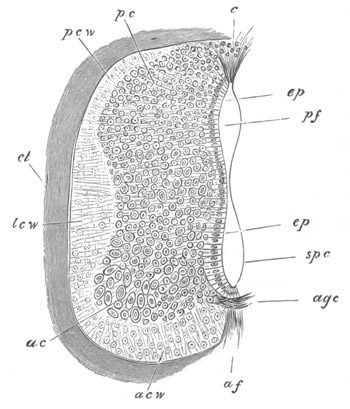
Fig. 246. Section through the spinal cord of a seven days’ Chick.
pcw. dorsal white column; lcw. lateral white column; acw.
ventral white column; c. dorsal tissue filling up the part where
the dorsal fissure will be formed; pc. dorsal grey cornu; ac.
anterior grey cornu; ep. epithelial cells; agc. anterior
commissure; pf. dorsal part of spinal canal; spc. ventral part
of spinal canal; af. anterior fissure.
The white matter is apparently the result of a differentiation of the
outermost parts of the superficial cells of the cord into
longitudinal
nerve-fibres, which remain for a long period without a medullary
sheath. These fibres appear in transverse sections as small dots. The
white matter forms a transparent investment of the grey matter and
would seem to contain neither nuclei nor cells[160].
The white matter
may from the first form only two masses, one on each side, forming a
layer on the ventral and lateral parts of the spinal cord but not
extending to the dorsal surface (Elasmobranchii, fig. 185, W); or it
may form four patches, viz. an anterior and a posterior white column
on each side, which lie on a level with the origin of the anterior and
posterior nerve-roots (the Fowl, Human embryo, etc.). In whichever of
these forms the white matter appears, it is always, at first, a layer
of extreme tenuity, which rapidly increases
in thickness in the
subsequent stages, and extends so as gradually to cover the whole cord
(fig. 246).
The anterior white commissure is formed very shortly after the first
appearance of the white matter. The grey matter and the central
epithelium are formed by a differentiation of the main mass of the
spinal cord. The outer cells lose their epithelial-like arrangement,
and, becoming prolonged into fibres, give rise to the grey matter,
while the innermost cells retain their primitive arrangement, and
constitute the epithelium of the canal. The process of formation of
the grey matter would appear to proceed from without inwards, so that
some of the cells, which have, on the formation of the grey matter, an
epithelial-like arrangement, subsequently become converted into true
nerve-cells.
As has already been mentioned, the central epithelium of the nervous
system probably corresponds with the so-called epidermic layer of the
epiblast.
The grey matter soon becomes prolonged dorsally and ventrally into the
posterior and anterior horns. Its fibres may especially be traced in
two directions:—(1) round the anterior end of the spinal canal,
immediately outside its epithelium and so to the grey matter on the
opposite side, forming in this way an anterior grey commissure,
through which a decussation of the fibres from the opposite sides is
effected: (2) dorsalwards along the outside of the lateral walls of
the canal.
There is at this period no trace of the ventral or dorsal fissure, and
the shape of the central canal is not very different to what it was at
an earlier period. This condition of the spinal cord is especially
instructive, as it is very nearly that which is permanent in
Amphioxus.
The next event of importance is the formation of the ventral or
anterior fissure. This owes its origin to a downgrowth of the anterior
horns of the cord on each side of the middle line. The two downgrowths
enclose between them a somewhat linear space—the anterior
fissure—which increases in depth in the succeeding stages (fig. 246,
af).
The dorsal or posterior fissure is formed at a later period than the
anterior, and accompanies the atrophy of the dorsal section of the
embryonically large canal of the spinal cord.
The exact mode of its formation appears to me to be still involved in
some obscurity.
In the Elements of Embryology the development of the posterior
fissure was described in the following way:
“On the seventh day the most important event is the formation of the
posterior fissure.
“This is brought about by the absorption of the roof of the
posterior of the two parts into which the neural canal has become
divided.
“Between the posterior horns of the cord, the epithelium forming the
roof of the, so to speak, posterior canal is along the middle line
covered neither by grey nor by white matter, and on the seventh day
is partially absorbed, thus transforming the canal into a
wedge-shaped fissure, whose mouth however is seen in section to be
partially closed by a triangular clump of elongated cells (fig. 246,
c). Below this mass of cells the fissure is open. It is separated
from the ‘true spinal canal’ by a very narrow space along which the
side walls have coalesced. In the lumbar and sacral regions the two
still communicate.
“We thus find, as was first pointed out by Lockhart Clarke, that the
anterior and posterior fissures of the spinal cord are,
morphologically speaking, entirely different. The anterior fissure
is merely the space left between two lateral downward growths of the
cord, while the posterior fissure is part of the original neural
canal separated from the rest of the cavity (which goes to form the
true spinal canal) by a median coalescence of the side walls.”
I confess that I have some doubts as to the complete accuracy of the
above statement.
Kölliker gives a full account of the gradual atrophy of the central
canal; but I do not fully understand his statements with reference to
the formation of the posterior fissure, which in fact appears to be
only incidentally mentioned. It would seem from his account that a
shallow and somewhat wide dorsal fissure is formed to start with, in
the human embryo, by two projections of the posterior white horns. On
the atrophy of the central canal this furrow becomes narrowed, but
Kölliker does not definitely state how it becomes deepened so as to
give rise to the permanent dorsal fissure.
It seems to me probable, though further investigations on the point
are still required, that the dorsal fissure is a direct result of the
atrophy of the dorsal part of the central canal of the spinal cord.
The walls of the canal coalesce dorsally, and the coalescence
gradually extends ventralwards, so as finally to reduce the central
canal to a minute tube, formed of the ventral part of the original
canal. The epithelial wall formed by the coalesced walls on the dorsal
side of the canal is gradually absorbed.
The epithelium of the central canal, at the period when its
atrophy
commences, is not covered dorsally either by grey or white matter, so
that, with the gradual reduction of the dorsal part of the canal, and
the absorption of the epithelial wall formed by the fusion of its two
sides, a fissure between the two halves of the spinal cord becomes
formed. This fissure is the posterior or dorsal fissure. In the
process of its formation the white matter of the dorsal horns becomes
prolonged so as to line its walls; and shortly after its formation the
dorsal grey commissure makes its appearance, which is not improbably
derived from part of the epithelium of the original central canal.
Development of the Brain.
The brain is formed from the anterior portion of the medullary plate.
When the medullary plate first becomes differentiated it is not
possible to distinguish between the region of the brain and that of
the spinal cord. The brain region is however usually very early
indicated by a widening of the medullary plate, but does not become
sharply marked off from the region of the spinal cord. In many
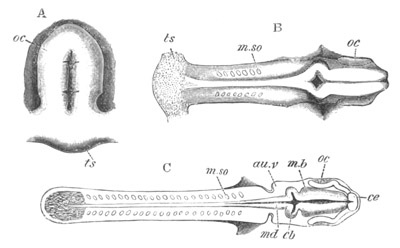
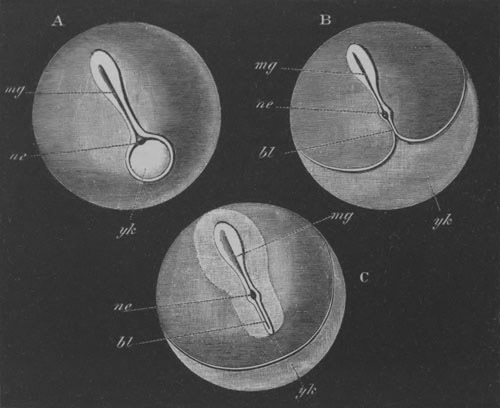
Ichthyopsida (Elasmobranchii (fig. 28, C) and Amphibia (fig. 77, A))
the anterior dilatation gives to the medullary plate, before its sides
meet to form a canal, a spatula-like form; which is either not present
or less marked in Reptilia, Aves and Mammalia.
The length of the brain as compared to the spinal cord is always very
great in the embryo, and in the earliest developmental periods the
disproportion in the size of the brain is specially marked, owing to
the full number of the somites of the trunk not having been formed. In
Elasmobranchii the brain is about one-third of the whole length of the
embryo at the stage immediately following the closure of the medullary
canal.
The first differentiation of the brain into distinct parts is a very
early occurrence, and may take place before (Mammalia) or during the
closure of the medullary folds. The brain first becomes divided into
two successive lobes or vesicles by a single transverse constriction,
and subsequently the posterior of these again becomes divided into
two, so that three lobes
are formed—known as the fore- the mid- and
the hind-brain; of these the hind-brain is usually the longest. In
some instances a bilobed stage can hardly be recognised. This
primitive division of the brain is shewn in many of the figures
already given. The reader may perhaps best refer to fig. 108. On the
closure of the medullary groove the lumen of the medullary canal is
continued uninterruptedly through the brain, but dilates considerably
in each of the cerebral vesicles.
The anterior lobe of the brain becomes converted into the cerebral
hemispheres, the thalamencephalon, the primary optic vesicles, and the
parts connected with them. The middle lobe becomes the optic lobes
(corpora bigemina or corpora quadrigemina in Mammalia) and the crura
cerebri; while the posterior lobe becomes converted into the
cerebellum and medulla oblongata.
Before describing in detail the changes by which the primary vesicles
of the brain become converted into the above parts, it will be
convenient to say a few words about the general development of the
brain.
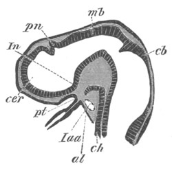
Fig. 247. Longitudinal section through the brain of a young
Pristiurus embryo.
cer. commencement of the cerebral hemisphere; pn. pineal gland;
In. infundibulum; pt. ingrowth from mouth to form the pituitary
body; mb. mid-brain; cb. cerebellum; ch. notochord; al.
alimentary tract; Iaa. artery of mandibular arch.
The most striking peculiarity with reference to the general
development of the brain is a curvature which appears in its axis,
known as the cranial flexure. The flexure takes place through the
mid-brain, and causes the fore-brain to be gradually bent downwards so
that the axis of its floor forms, first, a right angle with that of
the hinder part of the brain, and subsequently, as a rule, an acute
angle.
During these changes the brain, in most Amniota at any rate, becomes
in the first instance retort-shaped, the cerebral vesicle forming the
swollen part of the retort, but subsequently the retort-shape is lost
owing to the great development of the vesicle of the mid-brain, which
forms the termination of the long axis of the embryo. Figs. 29, 76,
and 118, are representative figures of embryos of various vertebrate
forms at a period when the mid-brain forms the termination of the long
axis of the body.
It is generally stated that the cranial flexure is at its maximum at
the stage represented in these figures, and there can be no doubt that
viewed from the exterior the cranial flexure ceases to be so marked a
feature, and finally disappears as the embryo gradually grows older;
but though the mid-brain ceases to form the termination of the long
axis of the embryo, the flexure of the brain becomes in many forms
absolutely more marked; while in other forms, though stated to
diminish, it does not entirely vanish.
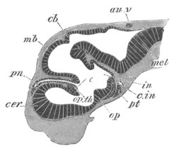
Fig. 248. Longitudinal section through the brain of Scyllium
canicula at an advanced stage of development.
cer. cerebral hemisphere; pn. pineal gland; op.th. optic
thalamus, connected with its fellow by a commissure (the middle
commissure). In front of it is seen a fold of the roof of the
fore-brain, which is connected with the choroid plexus of the third
ventricle; op. optic chiasma; pt. pituitary body; in.
infundibulum; cb. cerebellum; au.v. passage leading from the
auditory vesicle to the exterior; mel. medulla oblongata; c.in.
internal carotid artery.
The general nature of the changes which take place will perhaps best
be understood by a comparison of figs. 247 and 248 representing
longitudinal sections at two stages through the brain of an embryo
Elasmobranch. The actual cranial flexure, i.e. flexure of the floor
of the brain, is obviously greater in the older of the two brains,
though viewed from the exterior the axis of this brain appears to be
quite straight. In the younger stage, fig. 247, the mid-brain (mb)
forms the end of the long axis of the body, while in the older one the
cerebral hemispheres (cer) have grown very greatly, especially
forwards and dorsalwards. They have thus come to lie in front of the
mid-brain, and to form the end of the long axis of the body, and have
at the same time compressed the originally large thalamencephalon
against the mid-brain. The same general features may be seen in fig.
250 representing a longitudinal section of the brain of an embryo
fowl, and fig. 255 representing a longitudinal section of the brain of
a Mammal.
The infundibulum or perhaps rather the point of origin of the optic
nerves is to be regarded as the anterior termination of the axis of
the base of the brain.
The cranial flexure is least marked in Cyclostomata (fig. 253),
Teleostei, Ganoidei, and Amphibia, while it is very pronounced in
Elasmobranchii, Reptilia, Aves, and Mammalia. In Teleostei, and still
more in Cyclostomata, it permanently remains slight, owing to the
small development of the cerebral hemispheres.
In addition to the cranial flexures, two other flexures make their
appearance in the base of the brain. A posterior at the junction of
the brain and spinal cord, and an anterior at the boundary between the
cerebellum and medulla oblongata, just at the point where the pons
Varolii is formed in Mammalia. The anterior of these is the most
marked and constant; it is shewn in fig. 250. It arises considerably
later than the main cranial flexure, and since it is turned the
opposite way it assists to a considerable extent in causing the
apparent straightening of the cranial axis.
Histogenetic changes[161].
The walls of the brain are at first very
thin and, like those of the spinal cord, are formed of a number of
ranges of spindle-shaped cells. The processes of each of these cells
are stated to be continued through the whole thickness of the wall. In
the floor of the hind- and mid-brain a superficial layer of delicate
nerve-fibres is formed at an early period. This layer appears in the
first instance on the floor and sides of the hind-brain, and very
slightly, if at all, later on the floor and the sides of the
mid-brain. The cells internal to the nerve-fibres become
differentiated into an innermost epithelial layer lining the cavities
of the ventricles, and an outer layer of grey matter.
The similarity of the primitive arrangement and histological character
of the parts of the brain behind the cerebral hemispheres to that of
the spinal cord is very conclusively shewn by the examination of any
good series of sections. In both brain and spinal cord the white
matter forms a cap on the ventral and lateral parts considerably
before it extends to the dorsal surface. In the medulla the white
matter does not eventually extend to the roof owing to the peculiar
degeneration which that part undergoes.
In the case of the fore-brain the earliest histological changes,
except possibly in Mammals, take place on the same general plan as
those of the remainder of the central nervous system[162];
but though
the general plan is the same, yet the early histological distinction
between the fore-brain, and the mid- and hind-brain is more marked
than the distinction between the latter and the spinal cord.
On the floor and sides of the thalamencephalon, and apparently the
whole of the hemispheres of the lower types, there is formed, somewhat
later than in the remainder of the brain, a very delicate layer of
white matter. The inner part of the wall, which still remains
comparatively thin, is not at first clearly divided into an epithelial
and nervous layer. This distinction soon however becomes more or less
apparent, though it is not so marked as in most other parts of the
brain; and it appears that in the subsequent growth the greater part
of the original epithelial layer becomes converted into nervous
tissue.
In Mammals the same plan of differentiation would seem to be followed,
though somewhat less obviously than in the lower types. The walls of
the hemispheres become first divided (Kölliker) into a superficial
thinner layer of rounded elements, and a deeper and thicker epithelial
layer, and between these the fibres of the crura cerebri soon
interpose themselves. At a slightly later period a thin superficial
layer of white matter, homologous with that of the remainder of the
brain, becomes established.
The inner layer, together with the fibres from the crura cerebri,
gives rise to the major part of the white matter of the hemispheres
and to the epithelium lining the lateral ventricles.
The outer layer of rounded cells becomes divided into (1) a
superficial part with comparatively few cells, which, together with
its coating of white matter, forms the cortical part of the grey
matter, and (2) a deeper layer with numerous cells which forms the
main mass of the grey matter of the hemispheres.
The development of the several parts of the brain will now be
described.
The hind-brain. The hind-brain is at first an elongated, funnel-shaped
tube, the walls of which are of a nearly uniform thickness, though the
roof and floor are somewhat thinner than the sides. It forms a direct
continuation of the spinal cord, into which it passes without any
sharp line of demarcation. The ventricle it contains is known as the
fourth ventricle.
The sides become in the chick marked by a series of transverse
constrictions, dividing it into lobes, which are somewhat indefinite
in number. The first of these remains permanent, and its roof gives
rise to the cerebellum. It is uncertain whether the other
constrictions have any morphological significance. More or less
similar constrictions are present in Teleostei. In Elasmobranchii the
medulla presents on its inner face at a late period a series of lobes
corresponding with the roots of the vagus and glossopharyngeal nerves,
and it is possible that the earlier constrictions may potentially
correspond to so many nerve-roots.
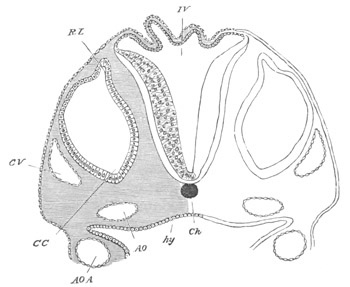
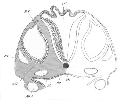
Fig. 249. Section through the hind-brain of a Chick at the end
of the third day of incubation.
IV. Fourth ventricle. The section shews the very thin roof and
thicker sides of the ventricle. Ch. Notochord; CV. Anterior
cardinal vein; CC. Involuted auditory vesicle; CC points to the
end which will form the cochlear canal; RL. Recessus labyrinthi
(remains of passage connecting the vesicle with the exterior); hy.
Hypoblast lining the alimentary canal; AO., AOA. Aorta, and
aortic arch.
Throughout the Vertebrata an anterior lobe of the hind-brain becomes
very early marked off, so that the primitive hind-brain becomes
divided into two regions which may be
conveniently spoken of as the
cerebellum (figs. 247 and 248, cb) and medulla oblongata. The floor
of these regions is quite continuous and is also prolonged without any
break into the floor of the mid-brain.
The posterior section of the hind-brain, which forms the medulla,
undergoes changes of a somewhat complicated character. In the first
place its roof becomes in front very much extended and thinned out. At
the raphe, where the two lateral halves of the brain originally
united, a separation, as it were, takes place, and the two sides of
the brain become pushed apart, remaining united by only a very thin
layer of nervous matter, consisting of a single row of flattened cells
(fig. 249). As a result of this peculiar growth in the brain, the
roots of the nerves of the two sides, which were originally in contact
at the dorsal summit of the brain, become carried away from one
another, and appear to arise at the sides of the brain.
The thin roof of the fourth ventricle is triangular, or, in Mammalia,
somewhat rhomboidal in shape. The apex of the triangle is directed
backwards.
At a later period the blood-vessels of the pia mater form a rich
plexus over the anterior part of the thin roof of the medulla, which
becomes at the same time somewhat folded. The whole structure is known
as the tela vasculosa, or choroid plexus of the fourth ventricle (fig.
250, chd 4). The floor of the whole hind-brain becomes thickened,
and there very soon appears on its outer surface a layer of
non-medullated nerve-fibres, similar to those which first appear on
the spinal cord. They are continuous with a similar layer of fibres on
the floor of the mid-brain, where they constitute the crura cerebri.
On the ventral floor of the medulla is a shallow continuation of the
anterior fissure of the spinal cord.
In Elasmobranchii and many Teleostei the restiform tracts are well
developed, and are anteriorly continued into the cerebellum, of which
they form the peduncles. Near their junction with the cerebellum they
form prominent bodies, which are regarded by Miklucho-Maclay as
representing the true cerebellum of Elasmobranchii.
In Elasmobranchii a dorsal pair of ridges projects into the cavity of
the fourth ventricle, corresponding apparently with the fasciculi
teretes of the Mammalia.
In Mammalia there develop, subsequently to the longitudinal fibres
already spoken of, first the olivary bodies of the ventral side of the
medulla, and at a still later period the pyramids. The fasciculi
teretes in the cavity of the fourth ventricle are developed shortly
before the pyramids.
When the hind-brain becomes divided into two regions the roof of the
anterior part does not become thinned out like that of the posterior,
but on the contrary, becomes somewhat thickened and forms a band-like
structure roofing over the anterior part of the fourth ventricle (fig.
247 and fig. 253, cb).
This is a rudiment of the cerebellum, and in all Craniate Vertebrates
it at first presents this simple structure and insignificant size. In
Cyclostomata, Amphibia and many Reptilia this condition is permanent.
In Elasmobranchii, on the other hand, the cerebellum assumes in the
course of development a greater and greater prominence (fig. 248,
cb), and eventually overlaps both the optic lobes in front and the
medulla behind. In the later embryonic stages it exhibits in
surface-views the appearance of a median constriction, and the portion
of the ventricle contained in it is prolonged into two lateral
outgrowths.
Miklucho-Maclay, from his observations on the brains of adult
Elasmobranchii, was led to regard what is here called the cerebellum
as identical with the mid-brain, and the true mid-brain as part of the
thalamencephalon. Miklucho-Maclay was no doubt misled by the large
size of the cerebellum, but, as we have seen, this body does not begin
to be conspicuous till late in embryonic life.
The mid-brain and thalamencephalon (according to the ordinary
interpretations) have in the embryo of Elasmobranchs exactly the same
relations as in the embryos of other Vertebrates; so that the
embryological evidence appears to me to be conclusive against
Miklucho-Maclay’s view.
In Birds the cerebellum attains a very considerable development (fig.
250, cbl), consisting of a folded central lobe with an arbor vitæ,
into which the fourth ventricle is prolonged. There are two small
lateral lobes, apparently equivalent to the flocculi. Anteriorly the
cerebellum is connected with the roof of the mid-brain by a delicate
membrane, the velum medullæ anterius, or valve of Vieussens (fig. 250,
vma). The pons Varolii of Mammalia is represented by a small number
of transverse fibres on the floor of the hind-brain immediately below
the cerebellum.
In Mammalia the cerebellum attains a still greater development.
The
median lobe or vermiform process is first developed. In the higher
Mammalia the lateral parts forming the hemispheres of the cerebellum
become formed as swellings at the sides at a considerably later
period, and are hardly developed in the Monotremata and Marsupialia.
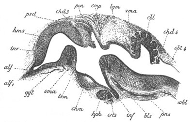
Fig. 250. Longitudinal section through the brain of a Chick of ten
days. (After Mihalkovics.)
hms. cerebral hemispheres; alf. olfactory lobe; alf1.
olfactory nerve; ggt. corpus striatum; oma. anterior commissure;
chd3. choroid plexus of the third ventricle; pin. pineal
gland; cmp. posterior commissure; trm. lamina terminalis; chm.
optic chiasma; inf. infundibulum; hph. pituitary body; bgm.
commissure of Sylvius (roof of iter a tertio ad quartum
ventriculum); vma. velum medullæ anterius (valve of Vieussens);
cbl. cerebellum; chd4. choroid plexus of the fourth
ventricle; obl4. roof of fourth ventricle; obl. medulla
oblongata; pns. commissural part of medulla; inv. sheath of
brain; bls. basilar artery; crts. internal carotid.
The cerebellum is connected with the roof of the mid-brain in front
and with the choroid plexus of the fourth ventricle behind by delicate
membranous structures, known as the velum medullæ anterius (valve of
Vieussens) and the velum medullæ posterius.
The pons Varolii is formed on the ventral side of the floor of the
cerebellar region as a bundle of transverse fibres at about the same
time as the olivary bodies.
The mid-brain. The changes undergone by the mid-brain are simpler than
those of any other part of the brain. We have already seen that the
mid-brain, on the appearance of the cranial flexure, forms an
unpaired vesicle with a vaulted roof and curved floor, at the front
end of the long axis of the body (fig. 118, MB). It is at this
period in most Vertebrates relatively much larger than in the adult;
and it is only in the Teleostei that it more or less retains in the
adult its embryonic proportions.
The cavity of the mid-brain, greatly reduced in size in the higher
forms, is known as the iter a tertio ad quartum ventriculum, or
aqueductus Sylvii.
The roof of the mid-brain is sharply constricted off from the
divisions of the brain in front of and behind it, but these
constrictions do not extend to the floor.
In some Vertebrates the region of the mid-brain is stated to undergo
hardly any further development. In the Axolotl it remains according to
Stieda[163]
as a simple tube with nearly uniformly thick walls. In the
majority of forms it undergoes, however, a more complicated
development.
In Elasmobranchs the sides become thickened to form the optic lobes,
which are soon separated by a median longitudinal groove. The floor
becomes thickened to form the crura cerebri. The primitive simple
median cavity becomes imperfectly divided into a median portion below,
and two lateral diverticula in the optic lobes.
In Teleostei the changes, resulting in the formation of (1) a pair of
longitudinal ridges projecting from the roof into the cavity of the
iter, constituting the fornix of Gottsche, and (2) of the two
swellings on the floor, forming the tori semicirculares, are more
complicated, but have not been satisfactorily worked out. In
Bombinator and the Anura generally the changes are of the same nature
as those in Elasmobranchii, except that the prolongations of the
ventricle into the optic lobes are still further constricted off from
the median portion, which forms the true iter.
In Reptilia and Aves the development of the mid-brain takes place on
the same type as in Elasmobranchii and the Anura. In Birds the optic
lobes are pushed very much aside, and the roof of the iter is greatly
thinned out. In Mammalia the sides of the mid-brain give rise to two
pairs of prominences—the corpora quadrigemina—instead of the two
optic lobes of other Vertebrata. The prominences, which do not contain
prolongations of the iter, become first visible on the appearance of
an oblique transverse furrow, while the anterior pair alone are
separated by a longitudinal furrow. In the later stages of development
the longitudinal furrow is continued so as to bisect the posterior
pair.
The floor, which is bounded posteriorly by the pons Varolii, becomes
the crura cerebri. The corpora geniculata interna also belong to this
division of the brain.
Fore-brain. In its earliest condition the fore-brain forms a single
vesicle without a trace of separate divisions, but very early it buds
off the optic vesicles, whose history is described with that of the
eye.
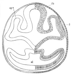
Fig. 251. Section through the front part of the head of a Lepidosteus embryo on the
seventh day after impregnation.
al. alimentary tract; fb. thalamencephalon; l. lens of eye;
op.v. optic vesicle. The mesoblast is not represented.
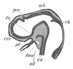
Fig. 252. Longitudinal section through the brain of a young
Pristiurus embryo.
cer. commencement of cerebral hemisphere; pn. pineal gland;
In. infundibulum; pt. ingrowth of mouth to form the pituitary
body; mb. mid-brain; cb. cerebellum; ch. notochord; al.
alimentary tract; Iaa. artery of mandibular arch.
The optic vesicles become gradually constricted off from the
fore-brain in a direction obliquely backwards and downwards. They
remain, however, attached to it at the anterior extremity of the base
of the fore-brain (fig. 251, op.v.). While the above changes are
taking place in the optic vesicles the anterior part of the fore-brain
becomes prolonged, and at the same time somewhat dilated. At first
there is no sharp boundary between the primitive fore-brain and its
anterior prolongation, but there shortly appears a constriction which
passes from above obliquely forwards and downwards. This constriction
is shallow at first, but soon becomes much deeper, leaving however the
cavities of the two divisions of the fore-brain united ventrally by a
somewhat wide canal (fig. 252).
Of these two divisions the posterior becomes the thalamencephalon,
while the anterior and larger division (cer) forms the rudiment of
the cerebral hemispheres and olfactory lobes. For a considerable
period this rudiment remains perfectly simple, and exhibits no signs,
either externally or internally, of a longitudinal constriction
dividing it into two lobes.
From the above description it may be concluded that the
rudiment of
the cerebral hemispheres is contained in the original fore-brain. In
spite however of their great importance in all the Craniata, it is
probable that the hemispheres were either not present as distinct
structures, or only imperfectly separated from the thalamencephalon,
in the primitive vertebrate stock.
The thalamencephalon. The thalamencephalon varies so slightly in
structure throughout the Vertebrate series that a general description
will suffice for all the types.
It forms at first a simple vesicle, the walls of which are of a nearly
uniform thickness and formed of the usual spindle-shaped cells.
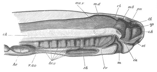
Fig. 253. Diagrammatic vertical section through the head of a
larva of Petromyzon.
The larva had been hatched three days, and was 4.8 mm. in length.
The optic and auditory vesicles are supposed to be seen through the
tissues.
c.h. cerebral hemisphere; th. optic thalamus; in.
infundibulum; pn. pineal gland; mb. mid-brain; cb. cerebellum;
md. medulla oblongata; au.v. auditory vesicle; op. optic
vesicle; ol. olfactory pit; m. mouth; br.c. branchial pouches;
th. thyroid involution; v.ao. ventral aorta; ht. ventricle of
heart; ch. notochord.
The cavity it contains is known as the third ventricle. Anteriorly it
opens widely into the cerebral rudiment, and posteriorly into the
ventricle of the mid-brain. The opening into the cerebral rudiment
becomes the foramen of Munro.
For convenience of description I shall divide it into three regions,
viz. (1) the floor, (2) the sides, and (3) the roof.
The floor becomes divided into two parts, an anterior part, giving
origin to the optic nerves, in which is formed the optic chiasma; and
a posterior part, which becomes produced into
an at first
inconspicuous prominence—the rudiment of the infundibulum (fig. 252,
In). This comes in contact with an involution from the mouth, which
gives rise to the pituitary body (fig. 252, pt), the development of
which will be dealt with separately.
In the later stages of development the infundibulum becomes gradually
prolonged, and forms an elongated diverticulum of the third ventricle,
the apex of which is in contact with the pituitary body (figs. 252,
254, in, and figs. 250 and 255, inf).
Along the sides of the infundibulum run the commissural fibres
connecting the floor of the mid-brain with the cerebrum.
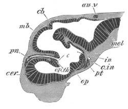
Fig. 254. Longitudinal section through the brain of Scyllium canicula at an advanced
stage of development.
cer. cerebral hemisphere; pn. pineal gland; op. th. optic
thalamus, connected with its fellow by a commissure (the middle
commissure). In front of it is seen a fold of the roof of the
fore-brain, which is the choroid plexus of the third ventricle;
op. optic chiasma; pt. pituitary body; in. infundibulum; cb.
cerebellum; au.v. passage leading from the auditory vesicle to the
exterior; mel. medulla oblongata; c.in. internal carotid
artery.
In its later stages the infundibular region presents considerable
variations in the different vertebrate types. In Fishes it generally
remains very large, and permanently forms a marked diverticulum of the
floor of the thalamencephalon. In Elasmobranchii the distal end
becomes divided into three lobes—a median and two lateral. The
lateral lobes appear to become the sacci vasculosi of the adult.
In Teleostei peculiar bodies known as the lobi inferiores (hypoaria)
make their appearance at the sides of the infundibulum. They appear to
correspond in position with the tuber cinereum of Mammalia[164].
In
Birds, Reptiles, and Amphibia the lower part of the embryonic
infundibulum becomes atrophied and reduced to a mere finger-like
process—the processus infundibuli.
In Mammalia the posterior part of the primitive infundibulum becomes
the corpus albicans, which is double in Man and the higher Apes; the
ventral part of the posterior wall forms the tuber cinereum.
Laterally, at the junction of the optic thalami and infundibulum,
there are placed the fibres of the crura cerebri, which are probably
derived from the walls of the infundibulum. A special process grows
out from the base of the infundibulum, which undergoes peculiar
changes, and becomes intimately united with the pituitary body; in
which connection it will be more fully described.
The sides of the thalamencephalon become very early thickened to form
the optic thalami, which constitute the most important section of the
thalamencephalon. They are separated, in Mammalia at all events, on
their inner aspect from the infundibular region by a somewhat S-shaped
groove, known as the sulcus of Munro, which ends in the foramen of
Munro. They also become in Mammalia secondarily united by a transverse
commissure, the grey or middle commissure, which passes across the
cavity of the third ventricle. This commissure is probably homologous
with, and derived from, a commissural band in the roof of the
thalamencephalon, placed immediately in front of the pineal gland
which is well developed in Elasmobranchii (fig. 254).
The roof undergoes more complicated changes. It becomes divided, on
the appearance of the pineal gland as a small papilliform outgrowth
(the development of which is dealt with separately), into two
regions—a longer anterior in front of the pineal gland and a shorter
posterior. The anterior region becomes at an early period excessively
thin, and at a later period, when the roof of the thalamencephalon is
shortened by the approach of the cerebral hemispheres to the
mid-brain, it becomes (vide figs. 250 and 255, chd 3, and 254)
considerably folded, while at the same time a vascular plexus is
formed in the pia mater above it. On the accomplishment of these
changes it is known as the tela choroidea of the third ventricle.
In the roof of the third ventricle behind the pineal gland there
appear in Elasmobranchii, the Sauropsida and Mammalia transverse
commissural fibres, forming a structure known as the posterior
commissure, which connects together the two optic thalami.
The most remarkable organ in the roof of the thalamencephalon is the
pineal gland, which is developed in most Vertebrates as a simple
papilliform outgrowth of the roof, and is at first composed of cells
similar to those of the other parts of the central nervous system
(figs. 250, 252, 254 and 255, pn or pin). In the lower Vertebrata
it is directed forwards, but in Mammalia, and to some extent in Aves,
it is directed backwards.
In Amphibia it is described by Götte (No. 296) as being a product of
the point where the roof of the brain remains latest attached to the
external skin.
The figure which Götte gives to prove this does not appear to me fully
to bear out his conclusion; which if true is very important. Although
I directed my attention specially to this point, I could find no
indication in Elasmobranchii of a process similar to that described by
Götte, and his observations have not as yet been confirmed for other
Vertebrates. Götte compares the pineal gland to the long-persisting
pore which leads into the cavity of the brain in the embryo of
Amphioxus, and we might add the Ascidians, and, should his facts be
confirmed, the conclusion he draws from them would appear to be well
founded.
The later stages in the development of the pineal gland in different
Vertebrates have not in all cases been fully worked out[165].
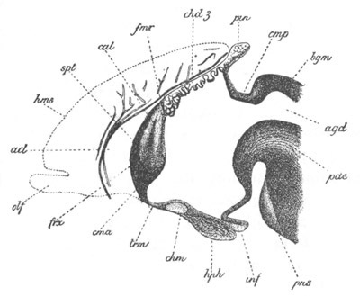
Fig. 255. Longitudinal vertical section through the anterior part
of the brain of an embryo rabbit of four centimetres. (After
Mihalkovics.)
The section passes through the median line so that the cerebral
hemispheres are not cut; their position is however indicated in
outline.
spt. septum lucidum formed by the coalescence of the inner walls
of part of the cerebral hemispheres; cna. anterior commissure;
frx. vertical pillars of the fornix; cal. genu of corpus
callosum; trm. lamina terminalis; hms. cerebral hemispheres;
olf. olfactory lobes; acl. artery of corpus callosum; fmr.
position of foramen of Munro; chd3. choroid plexus of third
ventricle; pin. pineal gland; cmp. posterior commissure; bgm.
lamina uniting the lobes of the mid-brain; chm. optic chiasma;
hph. pituitary body; inf. infundibulum; pns. pons Varolii;
pde. cerebral peduncles; agd. iter.
In Elasmobranchii the pineal gland becomes in time very long, and
extends far forwards over the roof of the cerebral
hemispheres (fig.
254 pn). Its distal extremity dilates somewhat, and in the adult the
whole organ forms (Ehlers, No. 337) an elongated tube, enlarged at its
free extremity, and opening at its base into the brain. The enlarged
extremity may either be lodged in a cavity in the cartilage of the
cranium (Acanthias), or be placed outside the cranium (Raja).
In Petromyzon its form is very different. It arises (fig. 253 pn) as
a sack-like diverticulum of the thalamencephalon extending at first
both backwards and forwards. In the Ammocœte the walls of this sack
are deeply infolded.
The embryonic form of the pineal gland in Amphibia is very much like
that which remains permanent in Elasmobranchii; the stalk connecting
the enlarged terminal portion with the brain soon however becomes
solid and very thin except at its proximal extremity. The enlarged
portion also becomes solid, and is placed in the adult externally to
the skull, where it forms a mass originally described by Stieda as the
cerebral gland.
In Birds the primitive outgrowth to form the pineal gland becomes,
according to Mihalkovics, deeply indented by vascular connective
tissue ingrowths, so that it assumes a dendritic structure (fig. 250
pin).
The proximal extremity attached to the roof of the thalamencephalon
forms a special section, known as the infra-pineal process. The
central lumen of the free part of the gland finally atrophies, but the
branches still remain hollow. The infra-pineal process becomes reduced
to a narrow stalk, connecting the branched portion of the body with
the brain. The branched terminal portion and the stalk obviously
correspond with the vesicle and distal part of the stalk of the types
already described. In Mammalia the development of the pineal gland is,
according to Mihalkovics, generally similar to that of Birds. The
original outgrowth becomes branched, but the follicles or lobes to
which the branching gives rise eventually become solid (fig. 255
pin). An infra-pineal process is developed comparatively late, and
is not sharply separated from the roof of the brain.
No satisfactory suggestions have yet been offered as to the nature of
the pineal gland, unless the view of Götte be regarded as such. It
appears to possess in all forms an epithelial structure, but, except
at the base of the stalk (infra-pineal process) in
Mammalia, in the
wall of which there are nerve-fibres, no nervous structures are
present in it in the adult state.
The pituitary body. Although the pituitary body is not properly a
nervous structure, yet from its intimate connection with the brain it
will be convenient to describe its development here. The pituitary
body is in fact an organ derived from the epiblast of the stomodæum.
This fact has been demonstrated for Mammalia, Aves, Amphibia and
Elasmobranchii, and may be accepted as holding good for all the
Craniata[166].
The epiblast in the angle formed by the cranial flexure
becomes involuted to form the cavity of the mouth. This cavity is
bordered on its posterior surface by the front wall of the alimentary
tract, and on its anterior by the base of the fore-brain. Its
uppermost end does not at first become markedly constricted off from
the remainder, but is nevertheless the rudiment of the pituitary body.
Fig. 256 represents a transverse section through the head of an
Elasmobranch embryo, in which, owing to the cranial flexure, the fore
part of the head is cut longitudinally and horizontally, and the
section passes through both the fore-brain (fb) and the hind-brain.
Close to the base of the fore-brain are seen the mouth (m), and the
pituitary involution from this (pt). In contact with the pituitary
involution is the blind anterior termination of the throat (al)
which a little way back opens to the exterior by the first visceral
cleft (1 v.c.). This figure alone suffices to demonstrate the
correctness of the above account of the pituitary body; but its truth
is still further confirmed by fig. 252; in which the mouth involution
(pt) is in contact with, but still separated from, the front end of
the alimentary tract. Very shortly after the septum between the mouth
and throat becomes pierced, and the two are placed in communication,
the pituitary involution becomes very partially constricted off from
the mouth involution, though still in direct communication with it. In
later stages the pituitary involution becomes longer
and is dilated
terminally; while the passage connecting it with the mouth becomes
narrower and narrower, and is finally reduced to a solid cord, which
in its turn disappears.
Before the connection between the pituitary vesicle and the mouth is
obliterated the cartilaginous cranium becomes developed, and it may
then be seen that the infundibulum projects through the pituitary
space to come into close juxtaposition with the pituitary body.
After the pituitary vesicle has lost its connection with the mouth it
lies just in front of the infundibulum (figs. 250 and 255 hph and
fig. 254 pt); and soon becomes surrounded by vascular mesoblast,
which grows in and divides it into a number of branching tubes. In
many forms the cavity of the vesicle completely disappears, and the
branches become for the most part solid [Cyclostomata and some
Mammalia (the rabbit), Elasmobranchii, Teleostei and Amphibia]. In
Reptilia, Aves and most Mammalia the lumen of the organ is more or
less retained (W. Müller, No. 344).
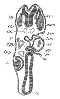
Fig. 256. Transverse section through the front part of the head of a young
Pristiurus embryo.
The section, owing to the cranial flexure, cuts both the fore- and
the hind-brain. It shews the premandibular and mandibular head
cavities 1pp and 2pp, etc. The section is moreover somewhat
oblique from side to side.
fb. fore-brain; l. lens of eye; m. mouth; pt. upper end of
mouth, forming pituitary involution; 1ao. mandibular aortic arch;
1pp. and 2pp. first and second head cavities; 1vc. first
visceral cleft; V. fifth nerve; aun. auditory nerve; VII.
seventh nerve; aa. roots of dorsal aorta; acv. anterior cardinal
vein; ch. notochord.
Although in the majority of the Vertebrata there is a close connection
between the pituitary body and the infundibulum, there is no actual
fusion between the two. In Mammalia the case is different. The part of
the infundibulum which lies at the hinder end of the pituitary body is
at first a simple finger-like process of the brain (fig. 255 inf),
but its end becomes swollen, and the lumen in this part becomes
obliterated. Its cells, originally similar to those of the other parts
of the nervous system and even (Kölliker) containing differentiated
nerve-fibres, partly atrophy, and partly assume an indifferent form,
while at the same time
there grow in amongst them numerous vascular
and connective-tissue elements. The process of the infundibulum thus
metamorphosed becomes inseparably connected with the true pituitary
body, of which it is usually described as the posterior lobe. The part
of the infundibulum which undergoes this change is very probably
homologous with the saccus vasculosus of Fishes.
The true nature of the pituitary body has not yet been made out. It is
clearly a rudimentary organ in existing craniate Vertebrates, and its
development indicates that when functional it was probably a sense
organ opening into the mouth, its blind end reaching to the base of
the brain. No similar organ has as yet been found in Amphioxus, but it
seems possible perhaps to identify it with the peculiar ciliated sack
placed at the opening of the pharynx in the Tunicata, the development
of which was described at p. 18. If the suggestion is correct, the
division of the body into lobes in existing Vertebrata must be
regarded as a step towards a retrogressive metamorphosis.
Another possible view is to regard the pituitary body as a glandular
structure which originally opened into the mouth in the lower
Chordata, but which has in all existing forms ceased to be functional.
The intimate relation of the organ to the brain appears to me opposed
to this view of its nature, while on the other hand its permanent
structure is more easily explained on this view than on that
previously stated. In the Ascidians a glandular organ has been
described by Lacaze Duthiers[167]
in juxtaposition to the ciliated
sack, and it is possible that this organ as well as the ciliated sack
may be related to the pituitary body. In view of this possibility
further investigations ought to be carried out in order to determine
whether the whole pituitary body is derived from the oral involution,
or whether there may not be a nervous part and a glandular part of the
organ.
The Cerebral Hemispheres. It will be convenient to treat separately
the development of the cerebral hemispheres proper, and that of the
olfactory lobes.
Although the cerebral hemispheres vary more than any other part of the
brain, they are nevertheless developed from the unpaired cerebral
rudiment in a nearly similar manner throughout the series of
Vertebrata.
In the cerebral rudiment two parts may be distinguished, viz. the
floor and the roof. The former gives rise to the ganglia at the base
of the hemispheres—corpora striata, etc.—the latter to the
hemispheres proper.
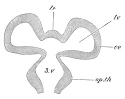
Fig. 257. Diagrammatic longitudinal horizontal section through the
fore-brain.
3.v. third ventricle; lv. lateral ventricle; lt. lamina
terminalis; ce. cerebral hemisphere; op.th. optic thalamus.
The first change which takes place consists in the roof growing out
into two lobes, between which a shallow median constriction makes its
appearance (fig. 257). The two lobes thus formed are the rudiments of
the two hemispheres. The cavity of each of them opens by a widish
aperture into the vestibule at the base of the cerebral rudiment,
which again opens directly into the cavity of the third ventricle (3
v). The Y-shaped aperture thus formed, which leads from the cerebral
hemispheres into the third ventricle, is the foramen of Munro. The
cavity (lv) in each of the rudimentary hemispheres is a lateral
ventricle. The part of the cerebrum which lies between the two
hemispheres, and passes forwards from the roof of the third ventricle
round the end of the brain to the optic chiasma, is the rudiment of
the lamina terminalis (figs. 257 lt and 255 trm). Up to this point
the development of the cerebrum is similar in all Vertebrata, but in
some forms it practically does not proceed much further.
In Elasmobranchii, although the cerebrum reaches a considerable size
(fig. 254 cer), and grows some way backwards over the
thalamencephalon, yet it is not in many forms divided into two
distinct lobes, but its paired nature is only marked by a shallow
constriction on the surface. The lamina terminalis in the later stages
of development grows backwards as a thick median septum which
completely separates the two lateral ventricles[168]
(fig. 263).
There are, it may be mentioned, considerable variations in
the
structure of the cerebrum in Elasmobranchii into which it is not
however within the scope of this work to enter.
In the Teleostei the vesicles of the cerebral hemispheres appear at
first to have a wide lumen, but it subsequently becomes almost or
quite obliterated, and the cerebral rudiment forms a small bilobed
nearly solid body. In Petromyzon (fig. 253 ch) the cerebral rudiment
is at first an unpaired anterior vesicle, which subsequently becomes
bilobed in the normal manner. The walls of the hemispheres become much
thickened, but the lateral ventricles persist.
In all the higher Vertebrates the division of the cerebral rudiment
into two distinct hemispheres is quite complete, and with the
deepening of the furrow between the two hemispheres the lamina
terminalis is carried backwards till it forms a thin layer bounding
the third ventricle anteriorly, while the lateral ventricles open
directly into the third ventricle.
In Amphibians the two hemispheres become united together immediately
in front of the lamina terminalis by commissural fibres, forming the
anterior commissure. They also send out anteriorly two solid
prolongations, usually spoken of as the olfactory lobes, which
subsequently fuse together.
In all Reptilia and Aves there is formed an anterior commissure, and
in the higher members of the group, especially Aves (fig. 250), the
hemispheres may obtain a considerable development. Their outer walls
are much thickened, while their inner walls become very thin; and a
well-developed ganglionic mass, equivalent to the corpus striatum, is
formed at their base.
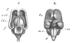
Fig. 258. Brain of a three months’ human embryo: natural
size. (From Kölliker.)
1. From above with the dorsal part of hemispheres and mid-brain
removed; 2. From below. f. anterior part of cut wall of the
hemisphere; f´. cornu ammonis; tho. optic thalamus; cst.
corpus striatum; to. optic tract; cm. corpora mammillaria; p.
pons Varolii.
The cerebral hemispheres undergo in Mammalia the most complicated
development. The primitive unpaired cerebral rudiment becomes, as in
lower Vertebrates, bilobed, and at the same time divided by the
ingrowth of a septum of connective tissue into two distinct
hemispheres (figs. 260 and 261 f and 258 1). From this septum is
formed the falx cerebri and other parts.
The hemispheres contain at first very large cavities, communicating by
a wide foramen of Munro with the third ventricle (fig. 260). They grow
rapidly in size, and extend, especially backwards, and gradually
cover the thalamencephalon and the
mid-brain (fig. 258 1, f). The
foramen of Munro becomes very much narrowed and reduced to a mere
slit.
The walls are originally nearly uniformly thick, but the floor becomes
thickened on each side, and gives rise to the corpus striatum (figs.
260 and 261 st). The corpus striatum projects upwards into each
lateral ventricle, giving to it a somewhat semilunar form, the two
horns of which constitute the permanent anterior and descending cornua
of the lateral ventricles (fig. 262 st).
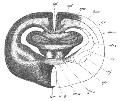
Fig. 259. Transverse section through the brain of a rabbit of five
centimetres. (After Mihalkovics.)
The section passes through nearly the posterior border of the septum
lucidum, immediately in front of the foramen of Munro.
hms. cerebral hemispheres; cal. corpus callosum; amm. cornu
ammonis (hippocampus major); cms. superior commissure of the
cornua ammonis; spt. septum lucidum; frx 2. vertical fibres of
the fornix; cma. anterior commissure; trm. lamina terminalis;
str. corpus striatum; ltf. nucleus lenticularis of corpus
striatum; vtr 1. lateral ventricle; vtr 3. third ventricle;
ipl. slit between cerebral hemispheres.
With the further growth of the hemisphere the corpus
striatum loses
its primitive relations to the descending cornu. The reduction in size
of the foramen of Munro above mentioned is, to a large extent, caused
by the growth of the corpora striata.
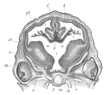
Fig. 260. Transverse section through the brain of a sheep’s embryo
of 2.7 cm. in length. (From Kölliker.)
The section passes through the level of the foramen of Munro.
st. corpus striatum; m. foramen of Munro; t. third ventricle;
pl. choroid plexus of lateral ventricle; f. falx cerebri; th.
anterior part of optic thalamus; ch. optic chiasma; o. optic
nerve; c. fibres of the cerebral peduncles; h. cornu ammonis;
p. pharynx; sa. presphenoid bone; a. orbitosphenoid bone; s.
points to part of the roof of the brain at the junction between the
roof of the third ventricle and the lamina terminalis; l. lateral
ventricle.
The corpora striata are united at their posterior border with the
optic thalami. In the later stages of development the area of contact
between these two pairs of ganglia increases to an immense extent
(fig. 261), and the boundary between them becomes somewhat obscure, so
that the sharp distinction which exists in the embryo between the
thalamencephalon and cerebral hemispheres becomes lost. This change is
usually (Mihalkovics, Kölliker) attributed to a fusion between the
corpora striata and optic thalami, but it has recently been attributed
by Schwalbe (No. 349), with more probability, to a growth of the
original surface of contact, and an accompanying change in the
relations of the parts.
The outer wall of the hemispheres gradually thickens, while the inner
wall becomes thinner. In the latter, two curved folds, projecting
towards the interior of the lateral ventricle, become formed. These
folds extend from the foramen of Munro along nearly the whole of what
afterwards becomes the descending cornu of the lateral ventricle.
The upper fold becomes the hippocampus major (cornu ammonis) (figs.
259 amm, 260 and 261 h, and 262 am). When the rudiment of the
descending cornu has become transformed into a simple process of the
lateral ventricle the hippocampus major forms a prominence upon its
floor.
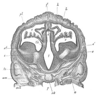
.
Fig. 261. Transverse section through the brain of a sheep’s embryo
of 2.7 cm. in length. (From Kölliker.)
The section is taken a short distance behind the section represented
in fig. 260, and passes through the posterior part of the
hemispheres and the third ventricle.
st. corpus striatum; th. optic thalamus; to. optic tract; t.
third ventricle; d. roof of third ventricle; c. fibres of
cerebral peduncles; c´. divergence of these fibres into the walls
of the hemispheres; e. lateral ventricle with choroid plexus pl; h
cornu ammonis; f. primitive falx; am. alisphenoid; a.
orbitosphenoid; sa. presphenoid; p. pharynx; mk. Meckel’s
cartilage.
The wall of the lower fold becomes very thin, and a vascular plexus,
derived from the connective-tissue septum between the hemispheres, and
similar to that of the roof of the third ventricle,
is formed outside
it. It constitutes a fold projecting far into the cavity of the
lateral ventricle, and together with the vascular connective tissue in
it gives rise to the choroid plexus of the lateral ventricle (figs.
260 and 261 pl).
It is clear from the above description that a marginal fissure leading
into the cavity of the lateral ventricle does not exist in the sense
often implied in works on human anatomy, in that the epithelium
covering the choroid plexus, which forms the true wall of the brain,
is a continuous membrane. The epithelium of the choroid plexus of
the lateral ventricle is quite independent of that of the choroid
plexus of the third ventricle, though at the foramen of Munro the roof
of the third ventricle is of course continuous with the inner wall of
the lateral ventricle (fig. 260 s). The vascular elements of the
two plexuses form however a continuous structure.
The most characteristic parts of the Mammalian cerebrum are the
commissures connecting the two hemispheres. These commissures are (1)
the anterior commissure, (2) the fornix, and (3) the corpus callosum,
the two latter being peculiar to Mammalia.
By the fusion of the inner walls of the hemispheres in front of the
lamina terminalis a solid septum is formed, known as the septum
lucidum, continuous behind with the lamina terminalis, and below with
the corpora striata (figs. 255 and 259 spt). It is by a series of
differentiations within this septum that the above commissures
originate. In Man there is a closed cavity left in the septum known as
the fifth ventricle, which has however no communication with the true
ventricles of the brain.
In the septum lucidum there become first formed, below, the transverse
fibres of the anterior commissure (fig. 255 and fig. 259 cma), and
in the upper part the vertical fibres of the fornix (fig. 255 and fig.
259 frx 2). The vertical fibres meet above the foramen of Munro, and
thence diverge backwards, as the posterior pillars, to lose themselves
in the cornu ammonis (fig. 259 amm). Ventrally they are continued,
as the descending or anterior pillars of the fornix, into the corpus
albicans, and thence into the optic thalami.
The corpus callosum is not formed till after the anterior commissure
and fornix. It arises in the upper part of the region
(septum lucidum)
formed by the fusion of the lateral walls of the hemispheres (figs.
255 and 259 cal), and at first only its curved anterior portion—the
genu or rostrum—is developed. This portion is alone found in
Monotremes and Marsupials. The posterior portion, which is present in
all the Monodelphia, is gradually formed as the hemispheres are
prolonged further backwards.
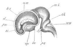
Fig. 262. Lateral view of the brain
of a calf embryo of 5 cm. (After Mihalkovics.)
The outer wall of the hemisphere is removed, so as to give a view of
the interior of the left lateral ventricle.
hs. cut wall of hemisphere; st. corpus striatum; am.
hippocampus major (cornu ammonis); d. choroid plexus of lateral
ventricle; fm. foramen of Munro; op. optic tract; in.
infundibulum; mb. mid-brain; ch. cerebellum; IV.V. roof of
fourth ventricle; ps. pons Varolii, close to which is the fifth
nerve with Gasserian ganglion.
Primitively the Mammalian cerebrum, like that of the lower Vertebrata,
is quite smooth. In many of the Mammalia, Monotremata, Insectivora,
etc., this condition is nearly retained through life, while in the
majority of Mammalia a more or less complicated system of fissures is
developed on the surface. The most important, and first formed, of
these is the Sylvian fissure. It arises at the time when the
hemispheres, owing to their growth in front of and behind the corpora
striata, have assumed a somewhat bean-shaped form. At the root of the
hemispheres—the hilus of the bean—there is formed a shallow
depression, which constitutes the first trace of the Sylvian fissure.
The part of the brain lying in this fissure is known as the island of
Reil.
The olfactory lobes. The olfactory lobes, or rhinencephala, are
secondary outgrowths of the cerebral hemispheres, and contain
prolongations of the lateral ventricles, but may however be solid in
the adult state. According to Marshall they develop in Birds and
Elasmobranchs and presumably other forms later than the olfactory
nerves, so that the olfactory region of the hemispheres is indicated
before the appearance of the olfactory lobes.
In most Vertebrates the olfactory lobes arise at a fairly early
stage
of development from the under and anterior part of the hemispheres
(fig. 250 olf). In Elasmobranchs they arise, not from the base, but
from the lateral parts of the brain (fig. 263), and become
subsequently divided into a bulbous portion and a stalk. They vary
considerably in their structure in the adult.
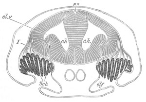
Fig. 263. Section through the brain and olfactory organ of an
embryo of Scyllium. (Modified from figures by Marshall and myself.)
ch. cerebral hemispheres; ol.v. olfactory vesicle; olf.
olfactory pit; Sch. Schneiderian folds; I. olfactory nerve. The
reference line has been accidentally taken through the nerve to the
brain; pn. anterior prolongation of pineal gland.
In Amphibia the solid anterior prolongations of the cerebral
hemispheres already spoken of are usually regarded as the olfactory
lobes, but according to Götte, whose view appears to me well founded,
small papillæ, situated at the base of these prolongations, from which
olfactory nerves spring, and which contain a process of the lateral
ventricle, should properly be regarded as the olfactory lobes. These
papillæ arise prior to the solid anterior prolongations of the
hemispheres.
In Birds the olfactory lobes are small. In the chick they arise
(Marshall) on the seventh day of incubation.
General conclusions as to the Central Nervous System.
It has been shewn above that both the brain and spinal cord are
primitively composed of a uniform wall of epithelial cells, and that
the first differentiation results in the formation of an external
layer of white matter, a middle layer of grey matter (ganglion cells),
and an inner epithelial layer. This primitive
histological
arrangement, which in many parts of the brain at any rate, is only to
be observed in the early developmental stages, has a simple
phylogenetic explanation.
As has been already explained in an earlier part of this chapter the
central nervous system was originally a differentiated part of the
superficial epidermis.
This differentiation (as may be concluded from the character of the
nervous system in the Cœlenterata and Echinodermata) consisted in the
conversion of the inner ends of the epithelial cells into
nerve-fibres; that is to say, that the first differentiation resulted
in the formation of a layer of white matter on the inner side of the
epidermis. The next stage was the separation of a deeper layer of the
epidermis as a layer of ganglion cells from the superficial epithelial
layer, i.e. the formation of a middle layer of ganglion cells and an
outer epithelial layer. Thus, phylogenetically, the same three layers
as those which first make their appearance in the ontogeny of the
vertebrate nervous system became successively differentiated, and in
both cases they are clearly placed in the same positions, because the
central canal of the vertebrate nervous system, as formed by an
involution, is at the true outer surface, and the external part of the
cord is at the true inner surface.
It is probable that a very sharp distinction between the white and
grey matter is a feature acquired in the higher Vertebrata, since in
Amphioxus there is no such sharp separation; though the nerve-fibres
are mainly situated externally and the nerve-cells internally.
As already stated in Chapter XII. the primitive division of the
nervous axis was probably not into brain and spinal cord, but into (1)
a fore-brain, representing the ganglion of the præoral lobe, and (2)
the posterior part of the nervous axis, consisting of the mid- and
hind-brains and the spinal cord. This view of the division of the
central nervous system fits in fairly satisfactorily with the facts of
development. The fore-brain is, histologically, more distinct from the
posterior part of the nervous system than the posterior parts are from
each other; the front end of the notochord forms the boundary between
these two parts of the central nervous system (vide fig. 253),
ending as it does at the front termination of the floor of the
mid-brain, and finally,
the nerves of the fore-brain have a different
character to those of the mid- and hind-brain.
This primitive division of the central nervous system is lost in all
the true Vertebrata, and in its place there is a secondary
division—corresponding with the secondary vertebrate head—into a
brain and spinal cord. The brain, as it is established in these forms,
is again divided into a fore-brain, a mid-brain and a hind-brain. The
fore-brain is, as we have already seen, the original ganglion of the
præoral lobe. The mid-brain appears to be the lobe, or ganglion, of
the third pair of nerves (first pair of segmental nerves), while the
hind-brain is a more complex structure, each section of which (perhaps
indicated by the constrictions which often appear at an early stage of
development) giving rise to a pair of segmental nerves is, roughly
speaking, homologous with the whole mid-brain.
The type of differentiation of each of the primitively simple vesicles
forming the fore-, the mid- and the hind-brains is very uniform
throughout the Vertebrate series, but it is highly instructive to
notice the great variations in the relative importance of the parts of
the brain in the different types. This is especially striking in the
case of the fore-brain, where the cerebral hemispheres, which on
embryological grounds we may conclude to have been hardly
differentiated as distinct parts of the fore-brain in the most
primitive types now extinct, gradually become more and more prominent,
till in the highest Mammalia they constitute a more important section
of the brain than the whole of the remaining parts put together.
The little that is known with reference to the significance of the
more or less corresponding outgrowths of the floor and roof of the
thalamencephalon, constituting the infundibulum and pineal gland, has
already been mentioned in connection with the development of these
parts.
Bibliography.
(332) C. J. Carus. Versuch einer Darstellung d. Nervensystems, etc.
Leipzig, 1814.
(333) J. L. Clark. “Researches on the development of the spinal cord
in Man, Mammalia and Birds.” Phil. Trans., 1862.
(334) E. Dursy. “Beiträge zur Entwicklungsgeschichte des
Hirnanhanges.” Centralblatt f. d. med. Wissenschaften, 1868. Nr. 8.
(335) E. Dursy. Zur Entwicklungsgeschichte des Kopfes des Menschen
und der höheren Wirbelthiere. Tübingen, 1869.
(336) A. Ecker. “Zur Entwicklungsgeschichte der Furchen und Windungen
der Grosshirn-Hemisphären im Foetus des Menschen.” Archiv f.
Anthropologie, v. Ecker und Lindenschmidt. Vol. III. 1868.
(337) E. Ehlers. “Die Epiphyse am Gehirn d. Plagiostomen.” Zeit. f.
wiss. Zool. Vol. XXX., suppl. 1878.
(338) P. Flechsig. Die Leitungsbahnen im Gehirn und Rückenmark des
Menschen. Auf Grund entwicklungsgeschichtlicher Untersuchungen.
Leipzig, 1876.
(339) V. Hensen. “Zur Entwicklung des Nervensystems.” Virchow’s
Archiv, Bd. XXX. 1864.
(340) L. Löwe. “Beiträge z. Anat. u. z. Entwick. d. Nervensystems d.
Säugethiere u. d. Menschen.” Berlin, 1880.
(341) L. Löwe. “Beiträge z. vergleich. Morphogenesis d. centralen
Nervensystems d. Wirbelthiere.” Mittheil. a. d. embryol. Instit.
Wien, Vol. II. 1880.
(342) A. M. Marshall. “The Morphology of the Vertebrate Olfactory
organ.” Quart. J. of Micr. Science, Vol. XIX. 1879.
(343) V. v. Mihalkovics. Entwicklungsgeschichte d. Gehirns. Leipzig,
1877.
(344) W. Müller. “Ueber Entwicklung und Bau der Hypophysis und des
Processus infundibuli cerebri.” Jenaische Zeitschrift. Bd. VI. 1871.
(345) H. Rahl-Rückhard. “Die gegenseitigen Verhältnisse d. Chorda,
Hypophysis etc. bei Haifischembryonen, nebst Bemerkungen üb. d.
Deutung d. einzelnen Theile d. Fischgehirns.” Morphol. Jahrbuch,
Vol. VI. 1880.
(346) H. Rathke. “Ueber die Entstehung der glandula pituitaria.”
Müller’s Archiv f. Anat. und Physiol., Bd. V. 1838.
(347) C. B. Reichert. Der Bau des menschlichen Gehirns. Leipzig,
1859 u. 1861.
(348) F. Schmidt. “Beiträge zur Entwicklungsgeschichte des Gehirns.”
Zeitschrift f. wiss. Zoologie, 1862. Bd. XI.
(349) G. Schwalbe. “Beitrag z. Entwick. d. Zwischenhirns.” Sitz. d.
Jenaischen Gesell. f. Med. u. Naturwiss. Jan. 23, 1880.
(350) Fried. Tiedemann. Anatomie und Bildungsgeschichte des Gehirns
im Foetus des Menschen. Nürnberg, 1816.
The development of the Cranial and Spinal Nerves[169].
All the nerves are outgrowths of the central nervous system, but the
differences in development between the cranial and spinal nerves are
sufficiently great to make it convenient to treat them separately.
Spinal nerves. The posterior roots of the spinal nerves, as well as
certain of the cranial nerves, arise in the same manner, and from the
same structure, and are formed considerably before the anterior roots.
Elasmobranch fishes may be taken as the type to illustrate the mode of
formation of the spinal nerves.
The whole of the nerves in question arise as outgrowths of a median
ridge of cells, which makes its appearance on the dorsal side of the
spinal cord (fig. 264 A, pr). This ridge has been called by Marshall
the neural crest. At each point, where a pair of nerves will be
formed, two pear-shaped outgrowths project from it, one on each side;
and apply themselves closely to the walls of the spinal cord (fig. 264 B,
pr). These outgrowths are the rudiments of the posterior nerves.
While still remaining attached to the dorsal summit of the neural cord
they grow to a considerable size (fig. 264 B, pr).
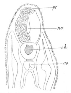
Fig. 264 A. Transverse section through a pristiurus embryo shewing
the proliferation of cells to form the neural crest.
pr. neural crest; nc. neural canal; ch. notochord; ao.
aorta.
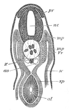
Fig. 264 B. Transverse section through the trunk of an embryo
slightly older than fig. 28 E.
nc. neural canal; pr. posterior root of spinal nerve; x.
subnotochordal rod; ao. aorta; sc. somatic mesoblast; sp.
splanchnic mesoblast; mp. muscle-plate; mp´. portion of
muscle-plate converted into muscle; Vv. portion of the vertebral
plate which will give rise to the vertebral bodies; al. alimentary
tract.
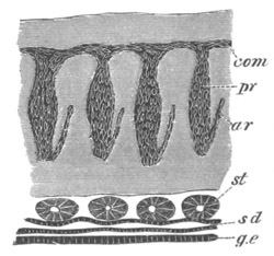
Fig. 265. Vertical longitudinal section through part of the trunk
of a young Scyllium embryo.
com. commissure uniting the dorsal ends of the posterior
nerve-roots; pr. ganglia of posterior roots; ar. anterior roots;
st. segmental tubes; sd. segmental duct; g.e. epithelium
lining the body cavity in the region of the future germinal ridge.
The attachment to the dorsal summit is not permanent, but
before
describing the further fate of the nerve-rudiments it is necessary to
say a few words as to the neural crest. At the period when the nerves
have begun to shift their attachment to the spinal cord, there makes
its appearance, in Elasmobranchii, a longitudinal commissure
connecting the dorsal ends of all the spinal nerves (figs. 265, 266
com), as well as those of the vagus and glossopharyngeal nerves.
This commissure has as yet only been found in a complete form in
Elasmobranchii;
but it is nevertheless to be regarded as a very
important morphological structure.
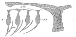
Fig. 266. Spinal Nerves of Scyllium in longitudinal section to shew
the commissure connecting them.
A. Section through a series of nerves.
B. Highly magnified view of the dorsal part of a single nerve, and
of the commissure connected with it.
com. commissure; sp.g. ganglion of posterior root; ar.
anterior root.
It is probable, though the point has not yet been definitely made out,
that this commissure is derived from the neural crest, which appears
therefore to separate into two cords, one connected with each set of
dorsal roots.
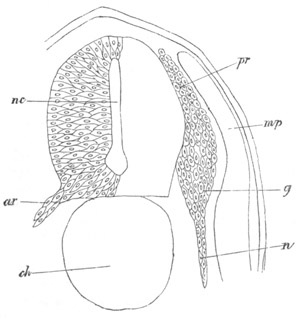
Fig. 267. Section through the dorsal part of the trunk of a
Torpedo embryo.
pr. posterior root of spinal nerve; g. spinal ganglion; n.
nerve; ar. anterior root of spinal nerve; ch. notochord; nc.
neural canal; mp. muscle-plate.
Returning to the original attachment of the nerve-rudiments to the
medullary wall, it has been already stated that this attachment is not
permanent. It becomes, in fact, at about the time of the appearance of
the above commissure, either extremely delicate or absolutely
interrupted.
The nerve-rudiment now becomes divided into three parts (figs. 267 and
268), (1) a proximal rounded portion, to which is attached the
longitudinal commissure (pr); (2) an enlarged portion, forming the
rudiment of a ganglion (g and sp g); (3) a distal portion, forming
the commencement of the nerve (n). The proximal portion may very
soon be observed to be united with the side of the spinal cord at a
very considerable distance from its original point of attachment.
Moreover the proximal portion of the nerve is attached, not by its
extremity, but by its side, to the spinal cord (fig. 268 x). The
dorsal extremities of the posterior roots are therefore free.
This attachment of the posterior nerve-root to the spinal cord is, on
account of its small size, very difficult to observe. In favourable
specimens there may however be seen a distinct cellular prominence
from the spinal cord, which becomes continuous with a small prominence
on the lateral border of the nerve-root near its proximal extremity.
The proximal extremity of the nerve is composed of cells, which, by
their small size and circular form, are easily distinguished from
those which form the succeeding or ganglionic portion of the nerve.
This part has a swollen configuration, and is composed of large
elongated cells with oval nuclei. The remainder of the rudiment forms
the commencement of the true nerve. This also is, at first, composed
of elongated cells[170].
It is extremely difficult to decide whether the permanent attachment
of the posterior nerve-roots to the spinal cord is entirely a new
formation, or merely due to the shifting of the original point of
attachment. I am inclined to adopt the former view, which is also held
by Marshall and His, but may refer to fig. 269, shewing the roots
after they have become attached to the side, as distinct evidence in
favour of the view that the attachment simply becomes shifted, a
process which might perhaps be explained by a growth of the dorsal
part of the spinal cord. The change of position in the case of some of
the cranial nerves is, however, so great that I do not think that it
is possible to account for it without admitting the formation of a new
attachment.
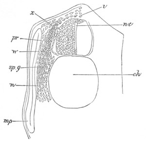
Fig. 268. Section through the dorsal region of a Pristiurus embryo.
pr. posterior root; sp.g. spinal ganglion; n. nerve; x.
attachment of ganglion to spinal cord; nc. neural canal; mp.
muscle-plate; ch. notochord; i. investment of spinal cord.
The anterior roots of the spinal nerves appear somewhat later than the
posterior roots, but while the latter are still quite small. Each of
them (fig. 269 ar) arises as a small but distinct conical outgrowth
from a ventral corner of the spinal cord, before the latter has
acquired its covering of white matter. From the very first the
rudiments of the anterior roots have a somewhat fibrous appearance and
an indistinct form of peripheral termination, while the protoplasm of
which they are composed becomes attenuated towards its end. They
differ from the posterior roots in never shifting their point of
attachment to the spinal cord, in not being united with each other by
a commissure, and in never developing a ganglion.
The anterior roots grow rapidly, and soon form elongated cords of
spindle-shaped cells with wide attachments to the spinal cord (fig.
267). At first they pass obliquely and nearly horizontally outwards,
but, before reaching the muscle-plates, they take a bend downwards.
One feature of some interest with reference to the anterior roots is
the fact that they arise not vertically below, but alternately with
the posterior roots: a condition which persists in the adult. They are
at first quite separate from the posterior roots; but about the stage
represented in fig. 267 a junction is effected between each posterior
root and the corresponding anterior root. The anterior root joins the
posterior at some little distance below its ganglion (figs. 265 and
266).
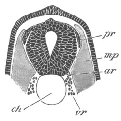
Fig. 269. Transverse section through the dorsal region of a young
Torpedo embryo to shew the origin of the anterior and posterior roots
of the spinal nerves.
pr. posterior root of spinal nerve; ar. anterior root of spinal
nerve; mp. muscle-plate; ch. notochord; vr. mesoblast cells
which will form the vertebral bodies.
Although I have made some efforts to determine the eventual fate of
the commissure uniting the dorsal roots, I have not hitherto met with
success. It grows thinner and thinner, becoming at the same time
composed of fibrous protoplasm with imbedded nuclei, and finally
ceases to be recognisable. I can only conclude that it gradually
atrophies, and ultimately vanishes.
After the junction of the posterior and anterior roots the compound
nerve extends downwards, and may easily be traced for a considerable
distance. A special dorsal branch is given off from the ganglion on
the posterior root (fig. 275 dn). According to Löwe the fibres of
the anterior and posterior roots can easily be distinguished in the
higher types by their structure and behaviour towards colouring
reagents, and can be separately traced in the compound nerve.
So far as has been made out, the development of the spinal nerves of
other Vertebrates agrees in the main with that in Elasmobranchii, but
no dorsal commissure has yet been discovered, except in the case of
the first two or three spinal nerves of the Chick.
In the Chick (Marshall, No. 353) the posterior roots, during their
early stages, closely resemble those in Elasmobranchii, though their
relatively smaller size makes them difficult to observe. They at first
extend more or
less horizontally outwards above the muscle-plates (as
a few of the nerves also do to some extent in Elasmobranchii), but
subsequently lie close to the sides of the neural canal. They are
shewn in this position in fig. 116 sp.g. There does not appear to be
a continuous crest connecting the roots of the posterior nerves. The
later stages of the development are precisely like those in
Elasmobranchii.
The anterior roots have not been so satisfactorily investigated as the
posterior, but they grow out, possibly by several roots for each
nerve, from the ventral corners of the spinal cord, and subsequently
become attached to the posterior nerves.
I have observed the development of the posterior roots in Lepidosteus,
in which they appear as projections from the dorsal angles of the
spinal cord, extending laterally outwards and, at first, having their
extremities placed dorsally to the muscle-plates.
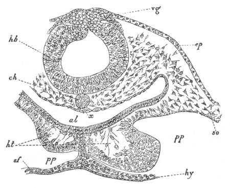
Fig. 270. Transverse section through the posterior part of the
head of an embryo chick of thirty hours.
hb. hind-brain; vg. vagus nerve; ep. epiblast; ch.
notochord; x. thickening of hypoblast (possibly a rudiment of the
subnotochordal rod); al. throat; ht. heart; pp. body cavity;
so. somatic mesoblast; sf. splanchnic mesoblast; hy.
hypoblast.
The cranial nerves[171].
The earliest stages in the development of the
cranial nerves have been most satisfactorily studied, especially by
Marshall (No. 354), in the Chick, while the later stages have been
more fully worked out in Elasmobranchii, where, moreover, they present
a very primitive arrangement.
In the Chick certain of the cranial
nerves arise before the complete closure of the neural groove. These
nerves are formed as paired outgrowths of a continuous band composed
of two laminæ, connecting the dorsal end of the incompletely closed
medullary canal with the external epiblast. This mode of development
will best be understood by an examination of fig. 270, where the two
roots of the vagus nerve (vg) are shewn growing out from the neural
band. Shortly after this stage the neural band, becoming separated
from the epiblast, constitutes a crest attached to the roof of the
brain, while its two laminæ become fused. The relation of the cranial
nerves to the brain then becomes exactly the same as that of the
posterior roots of the spinal nerves to the spinal cord.
It does not appear possible to decide whether the mode of development
of the cranial nerves in the Chick, or that of the posterior roots of
the spinal nerves, is the more primitive. The difference in
development between the two sets of nerves probably depends upon the
relative time of the closure of the neural canal. The neural crest
clearly belongs to the brain, from the fact of its remaining connected
with the latter when the medullary tube separates from the external
epiblast.
It is not known whether the cranial nerves originate before the
closure of the neural canal in other forms besides the Chick.
The neural crest of the brain is continuous with that of the spinal
cord, and on its separation from the central nervous axis forms on
each side a commissure, uniting the posterior cranial nerves with the
spinal nerves, and continuous with the commissure connecting together
the latter nerves.
Anteriorly, the neural crest extends as far as the roof of the
mid-brain[172].
The pairs of nerves which undoubtedly grow out from it
are the third pair (Marshall), the fifth, the seventh and auditory (as
a single root), the glossopharyngeal, and the various elements of the
vagus (as separate roots in Elasmobranchii, but as a single root in
Aves). Marshall holds that the olfactory
nerve probably also
originates from this crest. It will however be convenient to deal
separately with this nerve, after treating of the other nerves which
undoubtedly arise from the neural crest.
The cranial nerves just enumerated present in their further
development many points of similarity; and the glossopharyngeal nerve,
as it develops in Elasmobranchii, may perhaps be taken as typical.
This nerve is connected by a commissure with those behind, but this
fact may for the moment be left out of consideration. Springing at
first from the dorsal line of the hind-brain immediately behind the
level of the auditory capsule, it apparently loses this primitive
attachment and acquires a secondary attachment about halfway down the
side of the hind-brain. The primitive undifferentiated rudiment soon
becomes divided, exactly like a true posterior root of a spinal nerve,
into a root, a ganglion and a nerve. The main branch of the nerve
passes ventralwards, and supplies the first branchial arch (fig. 271
gl). Shortly afterwards it sends forwards a smaller branch, which
passes to the hyoid arch in front; so that the nerve forks over the
hyobranchial cleft. A typical cranial nerve appears therefore, except
as concerns its relations to the clefts, to develop precisely like the
posterior root of the spinal nerve.
Most of the cranial nerves of the above group, in correlation with the
highly differentiated character of the head, acquire secondary
differentiations, and render necessary a brief description of what is
known with reference to their individual development.
The Glossopharyngeal and Vagus Nerves. Behind the ear there are
formed, in Scyllium, a series of five nerves which pass down to
respectively the first, second, third, fourth and fifth branchial
arches.
For each arch there is thus one nerve, whose course lies close to the
posterior margin of the preceding cleft; a second anterior branch,
forking over the cleft and passing to the arch in front, being
developed later. These nerves are connected with the brain by roots at
first attached to the dorsal summit, but eventually situated about
halfway down the sides. The foremost of them is the glossopharyngeal.
The next four are, as has been shewn by Gegenbaur[173],
equivalent to
four independent nerves, but form together a compound nerve, which we
may briefly call the vagus.
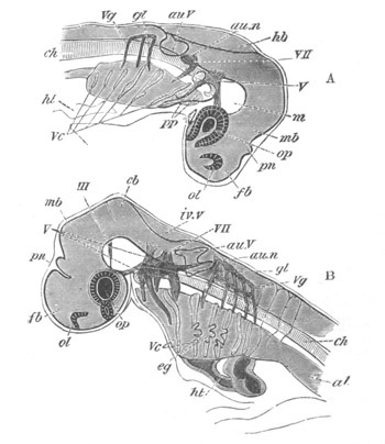
Fig. 271. Views of the head of Elasmobranch embryos at two stages
as transparent objects.
A. Pristiurus embryo of the same stage as fig. 28 F.
B. Somewhat older Scyllium embryo.
III. third nerve; V. fifth nerve; VII. seventh nerve; au.n.
auditory nerve; gl. glossopharyngeal nerve; Vg. vagus nerve;
fb. fore-brain; pn. pineal gland; mb. mid-brain; hb.
hind-brain; iv.v. fourth ventricle; cb. cerebellum; ol.
olfactory pit; op. eye; au.V. auditory vesicle; m. mesoblast
at base of brain; ch. notochord; ht. heart; Vc. visceral
clefts; eg. external gills; pp. sections of body cavity in the
head.
This compound nerve together with the glossopharyngeal soon attains a
very complicated structure, and presents several remarkable features.
There are present five branches (fig. 271 B), viz. the glossopharyngeal
(gl) and four branches of the vagus, the latter probably arising by
a considerably greater number of strands from the brain[174].
All the
strands from the brain are united together by a thin commissure (fig.
271 B, vg), continuous with the commissure of the posterior roots of
the spinal nerves, and from this commissure the five branches are
continued obliquely ventralwards and backwards, and each of them
dilates into a ganglionic swelling. They all become again united
together by a second thick commissure, which is continued backwards as
the intestinal branch of the vagus nerve. The nerves, however, are
continued ventralwards each to its respective arch.
From the lower
commissure springs the lateral nerve, at a point whose relations to
the branches of the vagus I have not certainly determined.
With reference to the dorsal commissure, which is almost certainly
derived from the original neural crest, it is to be noted that there
is a longish stretch of it between the last branch of the vagus and
the first spinal nerve, which is probably the remains of a part of the
commissure which connected the posterior branches of the vagus, at a
stage in the evolution of the Vertebrata, when the posterior visceral
clefts were still present. These branches of the vagus are probably
partially preserved in the ramifications of the intestinal stem of the
vagus (Gegenbaur). The origin of the ventral commissure, continued as
the intestinal branch of the vagus, has not been embryologically
worked out.
The lateral nerve may very probably be a dorsal sensory branch of the
vagus, whose extension into the posterior part of the trunk has been
due to the gradual backward elongation of the lateral line[175],
causing the nerve supplying it to elongate at the same time (vide
Section on lateral line).
In the Chick the common rudiment for the vagus and glossopharyngeal
nerves (Marshall), which has already been spoken of, subsequently
divides into two parts, an anterior forming the glossopharyngeal
nerve, and a posterior forming the vagus nerve.
The seventh and auditory nerves. As shewn by Marshall’s and my own
observations there is a common rudiment for the seventh and auditory
nerves. This rudiment divides almost at once into two branches. The
anterior of these pursues a straight course to the hyoid arch (fig.
271 A, VII.) and forms the rudiment of the facial nerve; the second
of the two (fig. 271 A, au.n), which is the rudiment of the auditory
nerve, develops a ganglionic enlargement and, turning backwards,
closely hugs the ventral wall of the auditory involution (fig. 272).
The seventh or facial nerve soon becomes more complicated. It early
develops, like the glossopharyngeal and vagus nerves, a branch, which
forks over the cleft in front (spiracle), and supplies the mandibular
arch (fig. 271 B). This branch forms the præspiracular nerve of the
adult, and is homologous with the chorda tympani of Mammalia. Besides
however giving rise to this typical branch it gives origin, at a very
early period, to two other rather remarkable branches; one of these,
arising from its dorsal anterior border, passes forwards to the front
part of the head, immediately dorsal to the ophthalmic branch of the
fifth to be described directly. This nerve is the portio major or
superficialis of the nerve usually known as the ramus ophthalmicus
superficialis in the adult[176].
The other branch of the seventh is the palatine branch—superficial
petrosal of Mammalia—the course of which has been more fully
investigated by Marshall than by myself. He has shewn that it arises
"just below the root of the ophthalmic branch,” and “runs downwards
and forwards, lying parallel and immediately superficial to the
maxillary branch of the fifth nerve.” This branch of the seventh nerve
appears to bear the same sort of relation to the superior maxillary
branch of the fifth nerve, that the ophthalmic branch of the seventh
does to the ophthalmic branch of the fifth.
Both the root of the seventh and its main branches are gangliated.
The auditory nerve is probably to be regarded as a specially
differentiated part of a dorsal branch of the seventh, while the
ophthalmic branch may not improbably be a dorsal branch comparable to
a dorsal branch of one of the spinal nerves.
The fifth nerve. Shortly after its development the root of the fifth
nerve shifts so as to be attached about halfway down the side of the
brain. A large ganglion becomes developed close to the root, which
forms the rudiment of the Gasserian ganglion. The main branch of the
nerve grows into the mandibular arch (fig. 271 A, V), maintaining
towards it similar relations to those of the posterior nerves to their
respective arches.
Two other branches very soon become developed, which were not properly
distinguished in my original account. The dorsal one takes a course
parallel to the ophthalmic branch of the seventh nerve, and forms,
according to the nomenclature already adopted, the portio profunda of
the ophthalmicus superficialis of the adult.
The second nerve (fig. 271 A) passes forwards, above the mandibular
head cavity, and is directed straight towards the eye, near which it
meets and unites with the third nerve, where the ciliary ganglion is
developed (Marshall). This branch is usually called the ophthalmic
branch of the fifth nerve, but Marshall rightly prefers to call it the
communicating branch between the fifth and third nerves[177].
Later than these two branches there is developed a third branch,
passing to the front of the mouth, and forming the superior maxillary
branch of the adult (fig. 271 B).
Of the branches of the fifth nerve the main mandibular branch is
obviously comparable to the main branch of the posterior nerves. The
superficial ophthalmic branch is clearly equivalent to the ophthalmic
branch of the seventh. The superior maxillary is usually held to be
equivalent to that branch of the posterior nerves which forms the
anterior limb of the fork over a cleft. The similarity between the
course of this nerve and that of the palatine branch of the seventh,
resembling as it does the similar course of the ophthalmic branches of
the two nerves, suggests that it may perhaps really be the homologue
of the palatine branch of the seventh, there
being no homologue of the
typical anterior branch of the other cranial nerves.
The third nerve. Our knowledge of the development of the third nerve
is entirely due to Marshall. He has shewn that in the Chick there is
developed from the neural crest, on the roof of the mid-brain, an
outgrowth on each side, very similar to the rudiment of the posterior
nerves. This outgrowth, the presence of which I can confirm, he
believes to be the third nerve, but although he is probably right in
this view, it must be borne in mind that there is no direct evidence
on the point, the fate of the outgrowth in question not having been
satisfactorily followed.
At a very considerably later period a nerve may be found springing
from the floor of the mid-brain, which is undoubtedly the third
nerve, and which Marshall supposes to be the above rudiment, which has
shifted its position. It is shewn in Scyllium in fig. 271 B, III. A
few intermediate stages between this and the earliest condition of the
nerve have been imperfectly traced by Marshall.
The nerve at the stage represented in fig. 271 B arises from a
ganglionic root, and “runs as a long slender stem almost horizontally
backwards, then turns slightly outwards to reach the interval between
the dorsal ends of the first and second head cavities, where it
expands into a small ganglion.” This ganglion, as first suggested by
Schwalbe (No. 357), and subsequently proved embryologically by
Marshall, is the ciliary ganglion. From the ciliary ganglion two
branches arise; one branch continuing the main stem of the nerve, and
obviously homologous with the main branch of the other nerves, and the
other passing directly forwards “along the top of the first head
cavity, then along the inner side of the eye, and finally terminating
at the anterior extremity of the head, just dorsal of the olfactory
pit.”
The partial separation, in many forms, of the ciliary ganglion from
the stem of the third nerve has led to the erroneous view (disproved
by the researches of Marshall and Schwalbe) that the ciliary ganglion
belongs to the fifth nerve. The connecting branch of the fifth nerve
often becomes directly continuous with the anterior branch of the
third nerve, and the two together probably constitute the nerve known
as the ramus ophthalmicus profundus (Marshall). Further embryological
investigations will be required to shew whether this nerve is
homologous with the nasal branch of the fifth nerve in Mammalia.
Relations of the nerves to the head-cavities. The cranial nerves,
whose development has just been given, bear certain very definite
relations to the mesoblastic structures in the head, of the nature of
somites, which are known as the head-cavities. Each cranial nerve is
typically placed immediately behind the head-cavity of its somite.
Thus the main branch of the fifth nerve lies in contact with the
posterior wall of the mandibular cavity, as shewn in section in fig.
272 V. 2pp and in surface view in fig. 271; the main branch of the
seventh nerve occupies a similar position in relation to the hyoid
cavity; and, as Marshall has recently shewn, the main branch of the
third nerve adjoins the posterior border of the front
cavity,
described by me as the premandibular cavity. Owing to the early
conversion of the walls of the posterior head-cavities into muscles,
their relations to the nerves are not quite so clear as in the case of
the anterior cavities, though, as far as is known, they are precisely
the same.
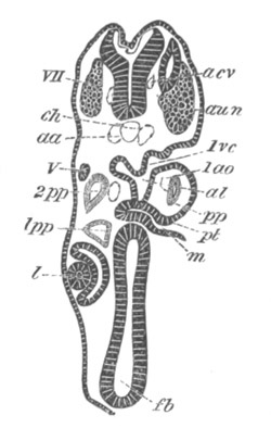
Fig. 272. Transverse section through the front part of the head of
a young Pristiurus embryo.
The section, owing to the cranial flexure, cuts both the fore- and
the hind-brain. It shews the præmandibular and mandibular
head-cavities 1pp and 2pp, etc.
fb. fore-brain; l. lens of eye; m. mouth; pt. upper end of
mouth, forming pituitary involution; 1ao. mandibular aortic arch;
1pp. and 2pp. first and second head-cavities; 1vc. first
visceral cleft; V. fifth nerve; aun. ganglion of auditory nerve;
VII. seventh nerve; aa. dorsal aorta; acv. anterior cardinal
vein; ch. notochord.
Anterior nerve-roots in the brain.
During my investigations on the development of the cranial nerves I
was unable to find any roots comparable with the anterior roots of the
spinal nerves, and propounded an hypothesis (suggested by the absence
of anterior spinal roots in Amphioxus[178])
that the head and trunk
had become differentiated from each other at a stage when mixed motor
and sensory posterior roots were the only roots present, and I
supposed the cranial and spinal nerves to have been independently
evolved from a common ground form, the resulting types of nerves being
so different that no roots strictly comparable with the anterior roots
of spinal nerves were to be found in the cranial nerves.
The views put forward by me on this subject, though accepted by
Schwalbe (No. 357), have in other quarters not met with much favour.
Wiedersheim holds that it is impossible to believe that the cranial
nerves are simpler than the spinal nerves. Such simplicity, which is
clearly not found, I have never asserted to exist; I have only stated
that the cranial nerves, in acquiring the complicated character they
have in the adult, do not develop anterior roots comparable with those
of the spinal nerves. Marshall also strongly objects to my views, and
has made some observations for the purpose of testing them, leading to
some very interesting results, which I proceed to state, and I will
then explain my opinion concerning them.
The most important observation of Marshall on this subject concerns
the sixth nerve. In both the Chick and Scyllium he has detected a
nerve (the first development of which has unfortunately not been made
out) arising by a series of roots from the base of the hind-brain. By
tracing this nerve to the external rectus muscle of the eye he has
satisfactorily identified
it as the sixth nerve. “Neither in the
nerve nor in its roots are there any ganglion cells." This nerve he
finds to be placed vertically below the roots of the seventh nerve;
and it is not visible till much later than the cranial nerves above
described.
In addition to this nerve Marshall has found, both in the third nerve
and in the fifth nerve, a series of non-gangliated roots, which arise
in a manner not yet satisfactorily elucidated, considerably later
than, and in front of, the main roots. These roots join the gangliated
roots on the proximal side of the ganglion or in the ganglion[179];
and Marshall believes them to be homologous with the anterior roots of
spinal nerves, while he holds the sixth nerve to be an anterior root
of the seventh nerve.
In addition to these nerves Marshall holds certain ventral roots,
which occur in Elasmobranchs close to the boundary of the spinal cord
and medulla, and which probably form the hypoglossal nerve of higher
types, to be anterior roots of the vagus. It is very difficult to
prove anything definitely about these nerves, but, for reasons stated
in my work on Elasmobranch Fishes, I am inclined to regard them as
anterior roots of one or more spinal nerves.
Before attempting to decide how far Marshall’s views about the
so-called anterior roots of the seventh, the fifth and the third
nerves are well founded it will conduce to clearness to state the
characters and relations of the two roots of spinal nerves.
The posterior root is (1) always purely sensory; (2) it always
develops a ganglion. The anterior root is (1) always purely motor; (2)
it always joins the posterior root below the ganglion, except in
Petromyzon (though not in Myxine) where the two roots are stated to be
independent.
How far do Marshall’s anterior and posterior roots of the cranial
nerves exhibit these respective peculiarities?
With reference to the sixth and seventh nerves he states “we must
regard the sixth nerve as having the same relation to the seventh that
the anterior root of a spinal nerve has to the posterior root.” On
this I would remark (1) that the posterior root of this nerve is a
mixed sensory and motor nerve and therefore differs in a very
fundamental point from that of a spinal nerve; (2) the sixth nerve
though resembling the anterior root of a spinal nerve in being motor
and without a ganglion, differs from the nearly universal arrangement
of spinal nerves in not uniting with the seventh.
With reference to the fifth nerve it is to be observed that it is by
no means certain that the whole of the motor fibres are supplied by
the so-called anterior roots, and that these roots differ again in the
most marked manner from the anterior roots of spinal nerves in joining
the main root of the nerve above (nearer the brain), and not as in a
spinal nerve below the
ganglion. The gangliated root of the third
nerve is purely motor[180],
and its so-called anterior roots again
differ from the anterior roots of spinal nerves, in the same manner as
those of the fifth nerve.
With reference to the glossopharyngeal and vagus nerves I would merely
remark that no anterior root has even been suggested for the
glossopharyngeal nerve and that the posterior roots of both these
nerves contain a mixture of sensory and motor fibres.
In view of these facts, my original hypothesis appears to me to be
confirmed by Marshall’s observations.
The fact of all the posterior roots of the above cranial nerves
(except the third which may be purely motor) being mixed motor and
sensory roots appears to me to demonstrate that the starting-point of
their differentiation was a mixed nerve with a single dorsal root; and
that they did not therefore become differentiated from nerves built on
the same type as the spinal nerves with dorsal sensory and ventral
motor roots. The presence of such non-gangliated roots as those of the
third and fifth nerves is not a difficulty to this view. Considering
that the cranial nerves are more highly differentiated than the spinal
nerves, and have more complicated functions to perform, it would be
surprising if there had not been developed nonganglionated roots
analogous to, but not of course homologous with, the anterior roots
of the spinal nerves[181].
As to the sixth nerve further embryological investigations are
requisite before its true position in the series can be determined;
but it appears to me very probable that it is a product of the
differentiation of the seventh nerve.
The fourth nerve. No embryological investigations have been made with
reference to the fourth nerve. It is possible that it is a segmental
nerve comparable with the third nerve, and that the only remnant still
left of the segment to which it belongs is the superior oblique muscle
of the eye. If this is the case there must have been two præmandibular
segments, viz. that belonging to the third nerve, and that belonging
to the fourth nerve. Against this view of the fourth nerve is the
fact, urged with great force by Marshall, that the superior oblique
muscle is in front of the other eye muscles, and that the fourth nerve
therefore crosses the third nerve to reach its destination.
The Olfactory nerve. It was shewn in my monograph on Elasmobranch
Fishes that the olfactory nerve grew out from the brain in the
same
manner as other nerves; and Marshall (No. 355), to whom we are
indebted for the greater part of our knowledge on the development of
this nerve, has proved that it arises prior to the differentiation of
the olfactory lobes.
The earliest stages in the development of the nerve have not been made
out. Marshall, as already stated, finds that in the Chick the neural
crest is continued in front of the optic vesicles, and holds that this
fact is strong a priori evidence in favour of the nerve growing out
from it. As mentioned above, note on p. 456, I cannot without further
evidence accept Marshall’s statements on this point. In any case
Marshall has not yet been able again to find an olfactory nerve till
long after the disappearance of the neural crest. The olfactory nerve
at the next stage observed forms an outgrowth of fusiform cells
springing on either side from near the summit of the fore-brain; and
at fifty hours it ends close to a slight thickening of the epiblast
forming the first rudiment of the olfactory pit, with the walls of
which it soon becomes united.
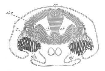
Fig. 273. Section through the brain and olfactory organ of an
embryo of Scyllium. (Modified from figures by Marshall and myself.)
c.h. cerebral hemispheres; ol.v. olfactory vesicle; olf.
olfactory pit; Sch. Schneiderian folds; I. olfactory nerve. The
reference line has been accidentally taken through the nerve to the
brain; pn. pineal gland.
The growth of the cerebral hemispheres causes its point of insertion
in the brain to be relatively shifted; and on the development of the
olfactory lobes (vide pp. 444, 445) it arises from them (fig. 273).
In Elasmobranchs there is a large development of ganglion cells near
its root. From Marshall’s figures these appear also to be present in
the Chick, but they do not seem to have been found in other forms. In
both Teleostei and Amphibia the olfactory nerves are at first
extremely short.
Marshall holds that the olfactory nerve is a segmental nerve
equivalent to the third, fifth, seventh etc. nerves. It has been
already stated that in my opinion the origin of the olfactory nerves
from the fore-brain, which I hold to be the ganglion of the præoral
lobe, negatives this view. The mere fact
of these nerves originating
as an outgrowth from the central nervous system is no argument in
favour of Marshall’s view of their nature; and even if Marshall’s
opinion that they arise from the neural crest should turn out to be
well founded, this fact would not prove their segmental nature,
because their origin from this crest would, as indicated in the next
paragraph, merely seem to imply that they primitively arose from the
lateral borders of the nerve-plate from which the cerebrospinal tube
has been formed.
Situation of the dorsal roots of the cranial and spinal nerves. The
probable explanation of the origin of nerves from the neural crest has
already been briefly given (p. 316). It is that the neural crest
represents the original lateral borders of the nervous plate, and
that, in the mechanical folding of the nervous plate to form the
cerebrospinal canal, its two lateral borders have become approximated
in the median dorsal line to form the neural crest. The subsequent
shifting of the nerves I am unable to explain, and the meaning of the
transient longitudinal commissure connecting the nerves is also
unknown. The folding of the neural plate must have extended to the
region of the origin of the olfactory nerves, so that, as just stated,
there would be no special probability of the olfactory nerves
belonging to the same category as the other dorsal nerves from the
fact of their springing from the neural crest.
Bibliography of the Peripheral Nervous System.
(351) F. M. Balfour. “On the development of the spinal nerves in
Elasmobranch Fishes.” Philosophical Transactions, Vol. CLXVI. 1876;
vide also, A monograph on the development of Elasmobranch Fishes.
London, 1878, pp. 191-216.
(352) W. His. “Ueb. d. Anfänge d. peripherischen Nervensystems.”
Archiv f. Anat. u. Physiol., 1879.
(353) A. M. Marshall. “On the early stages of development of the
nerves in Birds.” Journal of Anat. and Phys., Vol. XI. 1877.
(354) A. M. Marshall. “The development of the cranial nerves in the
Chick.” Quart. J. of Micr. Science, Vol. XVIII. 1878.
(355) A. M. Marshall. “The morphology of the vertebrate olfactory
organ.” Quart. J. of Micr. Science, Vol. XIX. 1879.
(356) A. M. Marshall. “On the head-cavities and associated nerves in
Elasmobranchs.” Quart. J. of Micr. Science, Vol. XXI. 1881.
(357) C. Schwalbe. “Das Ganglion oculomotorii.” Jenaische
Zeitschrift, Vol. XIII. 1879.
Sympathetic nervous system.
The discovery that the spinal and cranial nerves together with their
ganglia were formed from the epiblast was shortly afterwards extended
to the sympathetic nervous system, which has now been shewn to arise
in connection with the spinal and
cranial nerves. The earliest
observations on this subject were those contained in my Monograph on
Elasmobranch Fishes (p. 173), while Schenk and Birdsell (No. 361)
have since arrived at the same result for Aves and Mammalia.
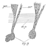
Fig. 274. Longitudinal vertical section through part of the body
wall of an Elasmobranch embryo shewing part of two spinal nerves and
the sympathetic ganglia belonging to them.
ar. anterior root; pr. posterior root; sy.g. sympathetic
ganglion; mp. part of muscle-plate.
In my account of the development of these ganglia, it is stated that
they were first met with as small masses situated at the ends of short
branches of the spinal nerves (fig. 275 sy.g). More recent
investigations have shewn me that the sympathetic ganglia are at first
simply swellings on the main branches of the spinal nerves some way
below the ganglia. Their situation may be understood from fig. 274,
sy.g, which belongs however to a somewhat later stage. Subsequently
the sympathetic ganglia become removed from the main stem of their
respective nerves, remaining however connected with those stems by a
short branch (fig. 275, sy.g). I have been unable to find a
longitudinal commissure connecting them in their early stages; and I
presume that they are at first independent, and become subsequently
united into a continuous cord on each side.
The observations of Schenk and Birdsell on the Mammalia seem to
indicate that the main parts of the sympathetic system arise in
continuity with the posterior spinal ganglia: they also shew that in
the neck and other parts the sympathetic cords arise as a continuous
ganglionic chain. The observations on the topographical features of
the development of the sympathetic system in higher types are however
as yet very imperfect.
The later history of the sympathetic ganglia is intimately bound up
with that of the so-called suprarenal bodies, which are dealt with in
another chapter.
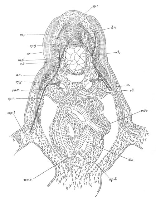
Fig. 275. Transverse section through the anterior part of the trunk
of an embryo of Scyllium slightly older than fig. 29 B.
The section is diagrammatic in the fact that the anterior
nerve-roots have been inserted for their whole length; whereas they
join the spinal cord halfway between two posterior roots.
sp.c. spinal cord; sp.g. ganglion of posterior root; ar.
anterior root; d.n. dorsally directed nerve springing from
posterior root; mp. muscle plate; mp´. part of muscle plate
already converted into muscles; mp.l. part of muscle plate which
gives rise to the muscles of the limbs; nl. nervus lateralis;
ao. aorta; ch. notochord; sy.g. sympathetic ganglion; ca.v.
cardinal vein; sp.n. spinal nerve; sd. segmental (archinephric)
duct; st. segmental tube; du. duodenum; pan. pancreas; hp.d.
point of junction of hepatic duct with duodenum; umc. umbilical
canal.
Bibliography of the Sympathetic Nervous System.
(360) F. M. Balfour. Monograph on the development of Elasmobranch
Fishes. London, 1878, p. 173.
(361) S. L. Schenk and W. R. Birdsell. “Ueb. d. Lehre von d.
Entwicklung d. Ganglien d. Sympatheticus.” Mittheil. a. d.
embryologischen Instit. Wien. Heft III. 1879.
In the lowest forms of animal life the whole surface is sensitive to
light, and organs of vision have no doubt arisen in the first instance
from limited areas becoming especially sensitive to light in
conjunction with a deposit of pigment. Lens-like structures, formed
either as a thickening of the cuticle, or as a mass of cells, were
subsequently formed; but their function was not, in the first
instance, to throw an image of external objects on the perceptive part
of the eye, but to concentrate the light on it. From such a simple
form of visual organ it is easy to pass by a series of steps to an eye
capable of true vision.
There are but few groups of the Metazoa which are not provided with
optic organs of greater or less complexity.
In a large number of instances these organs are placed on the anterior
part of the head, and are innervated from the anterior ganglia. It is
possible that many of the eyes so situated may be modifications of a
common prototype. In other instances organs of vision are situated in
different regions of the body, and it is clear that such eyes have
been independently evolved in each instance.
The percipient elements of the eye would invariably appear to be
cells, one end of each of which is continuous with a nerve, while the
other terminates in a cuticular structure, or indurated part of the
cell forming what is known as the rod or cone.
The presence of such percipient elements in various eyes is therefore
no proof of genetic relationship between these eyes, but merely of
similarity of function.
Embryological data as to the development of the eye do not
exist
except in the case of the Arthropoda, Mollusca and Chordata. From such
data as there are, combined with study of the adult structure of the
eye, it can be shewn that two types of development are found. In one
of these the percipient elements are formed from the central nervous
system, in the other from the epidermis. The former may be called
cerebral eyes. It is probable however that this distinction is not, in
all cases at any rate, so fundamental as might be supposed; but that
in both instances the eye may have taken its origin from the
epidermis. In the eyes in which the retina is continuous with the
central nervous system, these two organs were probably evolved
simultaneously as differentiations of the epidermis, and continue to
develop together in the ontogenetic growth of the eye.
Some of the eyes in which the retina is formed from the epidermis have
also probably arisen simultaneously with part of the central nervous
system, while in other instances they have arisen as later formations
subsequently to the complete establishment of a central nervous
system.
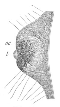
Fig. 276. Eye Of Lizzia Koellikeri. (From Lankester; after
Hertwig.)
l. lens; oc. perceptive part of eye.
Cœlenterata. The actual evolution of the eye is best shewn in the
Hydrozoa. The simplest types are those found in Oceania and
Lizzia[182].
In Lizzia the eye is placed at the base of a tentacle and
consists of (fig. 276) a lens (l) and a percipient bulb (oc). The
lens is a simple thickening of the cuticle, while the percipient part
of the eye is formed of three kinds of elements:—(1) pigment cells;
(2) sense cells, forming the true retinal elements, and consisting of
a central swelling with the nucleus, a peripheral process representing
a hardly differentiated rod, and a central process continuous with (3)
ganglion cells at the base of the eye. In this eye there is present a
commencing differentiation of a ganglion as well as of a retina.
The eye of Oceania is simpler than that of Lizzia in the absence of a
lens. Claus has shewn that in
Charybdea amongst the Acraspeda a more
highly differentiated eye is present, with a lens formed of cells like
the vertebrate eye.
Mollusca. In a large number of the odontophorous Mollusca eyes,
innervated by the supraœsophageal ganglia, are present on the dorsal
side of the head. These eyes exhibit very various degrees of
complexity, but are shewn both by their structure and development to
be modifications of a common prototype.
The simplest type of eye is that found in the Nautilus, and although
the possibility of this eye being degenerated must be borne in mind,
it is at the same time very interesting to note (Hensen) that it
retains permanently the early embryonic structure of the eyes of the
other groups.
It has (fig. 277 A) the form of a vesicle, with a small opening in the
outer wall, placing the cavity of the vesicle in free communication
with the exterior. The cells lining the posterior face of the vesicle
form a retina (R); and are continuous with the fibres of the optic
nerve (N.op). We have no knowledge of the development of this eye.
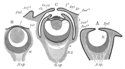
Fig. 277. Three diagrammatic sections of the eyes of Mollusca.
(After Grenacher.)
A. Nautilus. B. Gasteropod (Limax or Helix). C. Dibranchiate
Cephalopod.
Pal. eyelid; Co. cornea; Co.ep. epithelium of ciliary body;
Ir. iris; Int, Int1 ... Int4. different parts of the
integument; l. lens; l1. outer segment of lens; R. retina;
N.op. optic nerve; G.op. optic ganglion; x. inner layer of
retina; N.S. nervous stratum of retina.
In the Gasteropods the eye (fig. 277 B) has the form of a closed
vesicle: the cells lining the inner side form the retina, while the
outer wall of the vesicle constitutes the cornea. A
cuticular lens is
placed in the cavity, on the side adjoining the cornea. This eye
originates from the ectoderm, within the velar area, and close to the
supraœsophageal ganglia, usually at the base of the tentacles.
According to Rabl (Vol. II. No. 268) it is formed as an invagination,
the opening of which soon closes; while according to Bobretzky (Vol.
II. No. 242) and Fol it arises as a thickening of the epiblast, which
becoming detached takes the form of a vesicle. It is quite possible
that both types of development may occur, the second being no doubt
abbreviated. The vesicle, however formed, soon acquires a covering of
pigment, except for a small area of its outer wall, where the lens
becomes formed as a small body projecting into the lumen of the
vesicle. The lens seems to commence as a cuticular deposit, and to
grow by the addition of concentric layers. The inner wall of the
vesicle gives rise to the retina.
The most highly differentiated molluscan eye is that of the
Dibranchiate Cephalopoda, which is in fact more highly organized than
any other invertebrate eye.
A brief description of its adult structure[183]
will perhaps render
more clear my account of the development. The most important features
of the eye are shewn in fig. 277 C. The outermost layer of the optic
bulb forms a kind of capsule, which may be called the sclerotic.
Posteriorly the sclerotic abuts on the cartilaginous orbit, which
encloses the optic ganglion (G. op); and in front it becomes
transparent and forms the cornea Co, which may be either completely
closed, or (as represented in the diagram) perforated by a larger or
smaller opening. Behind the cornea is a chamber known as the anterior
optic chamber. This chamber is continued back on each side round a
great part of the circumference of the eye, and separates the
sclerotic from a layer internal to it.
In the anterior optic chamber there are placed (1) the anterior part
of the lens (l1) and (2) the folds of the iris (Ir). The whole
chamber, except the part formed by the lens, is lined by the epidermis
(Int1 and Int2). Bounding the inner side of the anterior optic
chamber is a layer which is called the choroid (Int1) which is
continued anteriorly into the fold of the iris (Ir). The most
superficial layer of the choroid is the epithelium already mentioned,
next comes a layer of obliquely placed plates known as the argentea
externa, then a layer of muscles, and finally the argentea interna.
The argentea interna abuts on a cartilaginous capsule, which
completely invests the inner part of the eye.
The lens is a nearly spherical body composed of concentric lamellæ of
a structureless material. It is formed of a small outer (l1) and
large inner
(l) segment, the two being separated by a thin membrane.
It is supported by a peculiar projection of the wall of the optic cup,
known as the ciliary body (Co.ep), inserted at the base of the iris,
and mainly formed of a continuation of the retina. This body is
however muscular, and presents a series of folds on its outer and
inner surfaces, which are especially developed on the latter.
The membrane dividing the lens into two parts is continuous with the
ciliary body. Within the lens is the inner optic chamber, bounded in
front by the lens and the ciliary body, and behind by the retina.
The retina is formed of two main divisions, an anterior division
adjoining the inner optic chamber, and a posterior division (N.S)
adjoining the cartilage of the choroid. The two layers are separated
by a membrane. Passing from within outwards the following layers in
the retina may be distinguished:
Anterior division of retina.
(1) Homogeneous membrane.
(2) Layer of rods.
(3) Layer of granules imbedded in pigment.
Posterior layer of retina.
(4) Cellular layer.
(5) Connective tissue layer
(6) Layer of nerve-fibres.
At the side of the optic ganglion is a peculiar body, known as the
white body (not shewn in the figure), which has the histological
characters of glandular tissue.
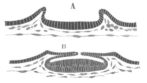
Fig. 278. Two sections through the developing eye of a Cephalopod
to shew the formation of the optic cup. (After Lankester.)
The first satisfactory account of the development of the eye is due to
Lankester (No. 365). The more important features in it were also
independently worked out by Grenacher (No. 363), and are beautifully
illustrated in Bobretzky’s paper (No. 362). The eye first appears as
an oval pit of the epiblast, the edge of which is formed by a
projecting rim (fig. 278 A). The epiblast layer lining the floor of
the pit soon becomes considerably thickened. By the growth inwards of
the rim the mouth of the pit
is gradually narrowed (fig. 278 B),
resembling at this stage the eye of Nautilus, and finally closed.
There is thus formed a flattened sack, lined by epiblast, which may be
called the primary optic vesicle. Its cavity eventually forms the
inner optic chamber. The anterior wall of the sack is lined by a much
less columnar layer than the posterior, the former giving rise to the
epithelium on the inner side of the ciliary processes, the latter to
the retina.
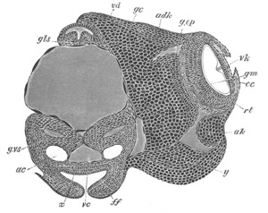
Fig. 279. Transverse section through the head of an advanced
embryo of Loligo. (After Bobretzky.)
gls. salivary gland; g.vs. visceral ganglion; gc. cerebral
ganglion; g.op. optic ganglion; adk. optic cartilage; ak. and
y. lateral cartilage or (?) white body; rt. retina; gm.
limiting membrane of retina; vk. ciliary region of eye; cc.
iris; ac. auditory sack (the epithelium lining the auditory sacks
is not represented); vc. vena cava; ff. folds of funnel; x.
epithelium of funnel.
The cavity of the sack rapidly enlarges, and assumes a spherical form.
At the same time a layer of mesoblast grows in between the walls of
the sack and the external epiblast. Two new structures soon arise
nearly simultaneously (fig. 279),—which become in the adult eye the
iris (cc) and the posterior segment of the lens. The iris is formed
as a circular fold of the skin in front of the optic vesicle. It
consists both of epiblast and mesoblast, and gives rise to a pit lined
by epiblast. The posterior segment of the lens arises as a
structureless rod-like body, which is shewn in fig. 279 depending from
the inner side
of the anterior wall of the optic vesicle. Its exact
mode of origin is somewhat obscure. The following is Lankester’s
account of it[184]:
“It is formed entirely within the primitive optic
chamber, and at first depends as a short cylindrical rod from the
middle point of the anterior wall of that chamber, that is to say,
from the point at which the chamber finally closed up. It grows
subsequently by the deposition of concentric layers of a horny
material round this cone. No cells appear to be immediately concerned
in effecting the deposition, and it must be looked upon as an organic
concretion, formed from the liquid contained in the primitive optic
chamber.”
The lens would thus appear to be a cuticular structure. It gradually
assumes a nearly spherical form; and is then composed of
concentrically arranged layers (fig. 280, hl).
While the lens is being formed, the ciliary epithelium of the optic
vesicle becomes divided into two layers, an outer layer of large cells
and an inner of small cells. Both layers are at first continuous
across the anterior wall of the optic chamber in front of the lens,
but soon become confined to the sides (fig. 280 A, cc and gz). The
inner layer is stated by Lankester to give rise to the muscles present
in the adult. The mesoblast cells also disappear from the region in
front of the lens, and the outer epithelium is converted into a kind
of cuticular membrane. By these changes the original layers of cells
in front of the lens become reduced to mere membranes,—a change which
appears to be preparatory to the appearance of the anterior segment of
the lens. The formation of the latter has not been fully followed out
by any investigator except Bobretzky. His figures would seem to
indicate that it is formed as a cuticular deposit in front of the
membrane already spoken of (fig. 280 B, vl). The two segments of the
lens appear at any rate to be separated by a membrane continuous with
the ciliary region of the optic vesicle.
Grenacher believes that the front part of the lens is formed in a
pocket-like depression of the epiblastic layer covering the outer side
of the optic cup; and Lankester thinks that the lens “pushes its way
through the median anterior area of the primitive optic chamber, and
projects into the second or anterior optic chamber where the iridian
folds lie closely upon it.”
While the lens is attaining its complete development there appears a
fresh fold round the circumference of the eye, which gradually grows
inwards so as to form a chamber outside the parts already present.
This chamber is the anterior optic chamber of the adult. In most
Cephalopods (fig. 277 C) the edges of the fold do not quite meet, but
leave a larger or smaller aperture leading into the chamber containing
the iris, outer segment of the lens, etc. In some forms however they
meet and coalesce, and so shut off this chamber from communication
with the exterior. The edge of the fold constitutes the cornea while
the remainder of it gives rise to the sclerotic.
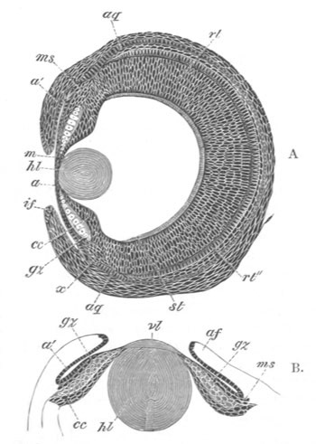
Fig. 280. Sections through the developing eye of Loligo
at two stages. (After Bobretzky.)
hl. inner segment of lens; vl. outer segment of lens; a and
a´. epithelium lining the anterior optic chamber; gz. large
epiblast cells of ciliary body; cc. small epiblast cells of
ciliary body; ms. layer of mesoblast between the two epiblastic
layers of the ciliary body; af. and if. fold of iris; rt.
retina; rt´´. inner layer of retina; st. rods; aq. cartilage of
the choroid.
The retina is at first a thick layer of numerous rows of oval
cells
(fig. 279). When the inner segment of the lens is far advanced towards
its complete formation pigment becomes deposited in the anterior part
of the retina, and a layer of rods grows out from the surface turned
towards the cavity of the optic vesicle (fig. 280 A, st). At a
slightly later stage the retina becomes divided into two layers
(Bobretzky), a thicker anterior layer, and a thinner posterior layer
(fig. 280, rt and rt´´). The former is composed of two strata, (1)
the rods and (2) a stratum with numerous rows of nuclei which becomes
in the adult the granular layer with its pigment. The posterior layer
gives rise to the cellular part of the posterior division of the
retina, while layers of connective tissue around it give rise to the
connective tissue of this portion of the retina (layer 6 in the scheme
on p. 474). The nervous layer is derived from the optic ganglion which
attaches itself to the inner side of the connective tissue layer.
The greater part of the choroid is formed from the mesoblast adjoining
the retina, but the epithelium covering its outer wall is of
epiblastic origin.
It is difficult to decide from development whether the Molluscan eyes,
so far dealt with, originated in the first instance pari passu with
the supraœsophageal ganglia or independently at a later period. On
purely à priori ground I should be inclined to adopt the former
alternative.
In addition to the above eyes there occur amongst Mollusca highly
complicated eyes, of a very different kind, in two widely separated
groups, viz. certain species of a genus of slug (Onchidium), and
certain Lamellibranchiata. These eyes, though they have no doubt been
evolved independently of each other, present certain remarkable points
of agreement. In both of them the rods of the retina are turned away
from the surface, and the nerve-fibres are placed, as in the
Vertebrate eye, on the side of the retina which faces outwards.
The peculiar eyes of Onchidium, investigated by Semper[185],
are
scattered on the dorsal surface, there being normal eyes in the usual
situation on the head. The eyes on the dorsal surface are formed of a
cornea, a lens composed of 1-7 cells, and a retina surrounded by
pigment; which is perforated in the centre by an optic nerve, the
retinal elements being in the inverted position above mentioned.
The development of these eyes has been somewhat imperfectly studied in
the adult, in which they continue to be formed anew. They arise by a
differentiation of the epidermis at the end of a papilla. At first a
few glandular cells appear in the epidermis in the situation where an
eye is about to be formed. Then, by a further process of growth, an
irregular mass of epidermic cells becomes developed, which pushes the
glandular cells to one side, and constitutes the rudiment of the eye.
This mass, becoming surrounded by pigment, unites with the optic
nerve, and its cells then differentiate themselves, in situ, into
the various elements of the eye. No explanation is offered by Semper
of the inverted position of the rods, nor is any suggested by his
account of the development. As pointed out by Semper these eyes are no
doubt modifications of the sensory epithelium of the papillæ.
The eyes of Pecten and Spondylus[186]
are placed on short stalks at
the edge of the mantle, and are probably modifications of the
tentacular processes of the mantle edge. They are provided with a
cornea, a cellular lens, a vitreous chamber, and a retina. The retinal
elements are inverted, and the optic nerve passes in at the side, but
occupies, in reference to its ramifications, the same relative
situation as the optic nerve in the Vertebrate eye. The development
has unfortunately not yet been studied.
Our knowledge of the structure or still more of the development of the
organ of vision of the Platyelminthes, Rotifera, and Echinodermata is
too scanty to be of any general interest.
Chætopoda. Amongst the Chætopoda the cephalic eyes of Alciope (fig.
281) have been adequately investigated as to their anatomy by Greeff.
These are provided with a large cuticular lens (l), separated from
the retina by a wide cavity containing the vitreous humour. The retina
is formed of a single row of cells, with rods at their free
extremities, continuous at their opposite ends with nerve-fibres. The
development of this eye has not been worked out. Eyes not situated
on the head are found in Polyophthalmus, and have probably been
evolved from the more indifferent type of sense-organ found by Eisig
in the allied Capitellidæ.
Chætognatha[187].
The paired cephalic eyes of Sagitta are spherical
bodies imbedded in the epidermis. They are formed of a central mass of
pigment with three lenses partially imbedded in it. The outer covering
of the eye is the retina, which is mainly composed of rod-bearing
cells; the rods being placed in contact with the outer surface of each
of the lenses. In the presence of three lenses the eye of Sagitta
approaches in some respects the eye of the Arthropoda.
Arthropodan eye. A satisfactory elucidation of the phylogeny of
Arthropodan eyes has not yet been given.
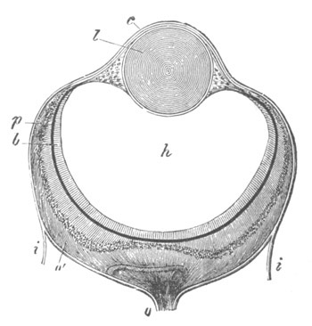
Fig. 281. Eye of an Alciopid (Neophanta Celox). (From Gegenbaur;
after Greef.)
i. cuticle; c. continuation of cuticle in front of eye; l.
lens; h. vitreous humour; o. optic nerve; o´. expansion of the
optic nerve; b. layer of rods; p. pigment layer.
All the types of eyes found in the group (with exception of
that of
Peripatus)[188]
present marked features of similarity, but I am
inclined to view this similarity as due rather to the character of the
exoskeleton modifying in a more or less similar way all the forms of
visual organs, than to the descent of all these eyes from a common
prototype. In none of these eyes is there present a chamber filled
with fluid between the lens and the retina, but the space in question
is filled with cells. This character sharply distinguishes them from
such eyes as those of Alciope (fig. 281). The types of eyes which are
found in the Arthropoda are briefly the following:
(1) Simple eyes. In all simple eyes the corneal lens is formed by a
thickening of the cuticle. Such eyes are confined to the Tracheata.
There are three types of simple eyes. (a) A type in which the
retinal cells are placed immediately behind the lens, found
(Lowne) in
the larvæ of some Diptera (Eristalis), and also in some Chilognatha.
(b) A type of simple eye found in some Chilopoda, and in some Insect
larvæ (Dytiscus, etc.) (fig. 282), the parts of which are entirely
derived from the epidermis. There is present a lens (l) formed as a
thickening of the cuticle, a so-called vitreous humour (gl) formed
of modified hypodermis cells, and a retina (r) derived from the same
source. The outer ends of the retinal cells terminate in rods, and
their inner ends are continuous with nerve-fibres.
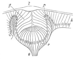
Fig. 282. Section through the simple eye of a young Dytiscus
larva. (From Gegenbaur; after Grenacher.)
l. corneal lens; g. vitreous humour; r. retina; o. optic
nerve; h. hypodermis.
(c) A type of simple eye found in the Arachnida, and apparently some
Chilopoda, and forming the simple eyes of most Insects, which differs
from type (a) in the cells of the retina forming a distinct layer
beneath the hypodermis; the latter only obviously giving rise to the
vitreous humour.
The development of the simple eyes has not yet been studied.
The simple eyes so far described are always placed on the head, and
are usually rather numerous.
(2) Compound eyes. Compound eyes are almost always present in the
Crustacea, and are usually found in adult Insects. In both groups they
are paired, though in the Crustacea a median much simplified compound
eye may either take the place of the paired eyes in the Nauplius larva
and lower forms, or be present together with them during a period in
the development of higher forms.
The typical compound eye is formed (fig. 283) of a series of corneal
lenses (c) developed from the cuticle; below which are placed bodies
known as the crystalline cones, one to each corneal lens; and below
the crystalline cones are placed bodies known as the retinulæ (r)
constituting the percipient elements of the eye, each of them being
formed of an axial rod, the rhabdom, and a number of cells surrounding
it.
The crystalline cones are formed from the coalescence of cuticular
deposits in several cells, the nuclei of which usually remain as
Semper’s nuclei. These cells are probably simple hypodermis cells, but
in some forms, e.g. Phronima, there may be a continuous layer of
hypodermis cells between them and the cuticle. In various Insect eyes
the cells which usually give rise to a crystalline cone may remain
distinct, and such eyes have been called by Grenacher aconous eyes,
while eyes with incompletely formed crystalline cones are called by
him pseudoconous eyes.
The rhabdom of the retinulæ is, like the crystalline cone, developed
by the coalescence of a series of parts, which are primitively
separate rods placed each in its own cell: this condition of the
retinulæ is permanently retained in the eyes of the Tipulidæ.
The development of the compound eye has so far only been
satisfactorily studied in some Crustacea by Bobretzky (No. 367); by
whom it has been worked out in Palæmon and Astacus, but more fully in
the latter, to which the following account refers:
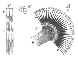
Fig. 283. Diagrammatic representations of parts of a compound
Arthropod eye. (From Gegenbaur.)
A. Section through the eye.
B. Corneal facets.
C. Two segments of
the eye.
c. corneal (cuticular) lenses; r. retinulæ with rhabdoms; n.
optic nerve; g. ganglionic swelling of optic nerve.
The eye of Astacus takes its origin from two distinct parts, (1) the
external epidermis of the procephalic lobes which will be spoken of as
the epidermic layer of the eye, (2) a portion of the supraœsophageal
ganglia, which will be spoken of as the neural layer of the eye. The
mesoblast is moreover the source of some of the pigment between the
two above layers. The epidermic layer gives rise to the corneal
lenses, the crystalline cones, and the pigment around the latter. The
neural layer on the other hand seems to give rise to the retinulæ with
their rhabdoms, and to the optic ganglion.
After the separation of the supraœsophageal ganglia from the
superficial epiblast, the cells of the epidermis in the region of the
future eye become columnar, and so form the above-mentioned epidermic
layer of the eye. This layer soon becomes two or three cells deep. At
the same time the most superficial part of the adjoining
supraœsophageal ganglion becomes partially constricted off from the
remainder as the neural layer of the eye, but is separated by a small
space from the thickened patch of epidermis.
Into this space some
mesoblast cells penetrate at a slightly later period. Both the
epidermic and neural layers next become divided into two strata. The
outer stratum of the epidermic layer gives rise to the crystalline
cones and Semper’s nuclei; each crystalline cone being formed from
four coalesced rods, developed as cuticular differentiations of four
cells, the nuclei of which may be seen in the embryo on its outer
side. The lower ends of the cones pass through the inner stratum of
the epidermic disc, the cells of which become pigmented, and
constitute the pigment cells surrounding the lower part of the
crystalline cones in the adult. The outer end of each of the
crystalline cones is surrounded by four cells, believed by Bobretzky
to be identical with Semper’s nuclei[189].
These cells give rise in a
later stage (not worked out in Astacus) to the cuticular corneal
lenses.
Of the two strata of the neural layer the outer is several cells deep,
while the inner is formed of elongated rod-like cells. Unfortunately
however the fate of the two neural layers has not been worked out,
though there can be but little doubt that the retinulæ originate from
the outer layer.
The mesoblast which grows in between the neural and epidermic layers
becomes a pigment layer, and probably also forms the perforated
membrane between the crystalline cones and the retinulæ.
The above observations of Bobretzky would appear to indicate that the
paired compound eyes of Crustacea belong to the type of cerebral eyes.
How far this is also the case with the compound eyes of Insects is
uncertain, in that it is quite possible that the latter eyes may have
had an independent origin.
The relation between the paired and median eye of the Crustacea is
also uncertain.
In the genus Euphausia amongst the Schizopods there is present a
series of eyes placed on the sides of some of the thoracic legs and on
the sides of the abdomen. The structure of these eyes, though not as
yet satisfactorily made out, would appear to be very different from
that of other Arthropodan visual organs.
The Eye of the Vertebrata. In view of the various structures which
unite to form it, the eye is undoubtedly the most complicated organ of
the Vertebrata; and though its mode of development is fairly constant
throughout the group, it will be convenient shortly to describe what
may be regarded as its typical development, and then to proceed to a
comparative view of the origin of its various parts, and to enter into
greater detail with reference to some of them. At the end of the
section
there is an account of the accessory structures connected with
the eye.
The formation of the eye commences with the appearance of a pair of
hollow outgrowths from the anterior cerebral vesicle or
thalamencephalon, which arise in many instances, even before the
closure of the medullary canal. These outgrowths, known as the optic
vesicles, at first open freely into the cavity of the anterior
cerebral vesicle. From this they soon however become partially
constricted, and form vesicles (fig. 284, a), united to the base of
the brain by comparatively narrow hollow stalks, the rudiments of the
optic nerves. The constriction to which the stalk or optic nerve is
due takes place obliquely downwards and backwards, so that the optic
nerves open into the base of the front part of the thalamencephalon
(fig. 284, b).
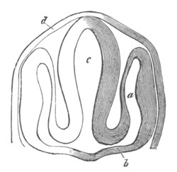
Fig. 284. Section through the head of an embryo Teleostean, to
shew the formation of the optic vesicles, etc. (From Gegenbaur;
after Schenk.)
c. fore-brain; a. optic vesicle; b. stalk of optic vesicle;
d. epidermis.
After the establishment of the optic nerves, there take place (1) the
formation of the lens, and (2) the formation of the optic cup from the
walls of the primary optic vesicle.
The external or superficial epiblast which covers, and is in most
forms in immediate contact with, the most projecting portion of the
optic vesicle, becomes thickened. This thickened portion is then
driven inwards in the form of a shallow open pit with thick walls
(fig. 285 A, o), carrying before it the front wall (r) of the
optic vesicle. To such an extent does this involution of the
superficial epiblast take place, that the front wall of the optic
vesicle is pushed close up to the hind wall, and the cavity of the
vesicle becomes almost obliterated (fig. 285 B).
The bulb of the optic vesicle is thus converted into a cup with double
walls, containing in its cavity the portion of involuted epiblast.
This cup, in order to distinguish its cavity from that of the original
optic vesicle, is generally called the secondary optic vesicle. We
may, for the sake of brevity, speak of it as the optic cup; in
reality it never is a vesicle, since it
always remains widely open in
front. Of its double walls the inner or anterior (fig. 285 B, r) is
formed from the front portion, the outer or posterior (fig. 285 B,
u) from the hind portion of the wall of the primary optic vesicle.
The inner or anterior (r), which very speedily becomes thicker than
the other, is converted into the retina: in the outer or posterior
(u), which remains thin, pigment is eventually deposited, and it
ultimately becomes the tesselated pigment-layer of the choroid.
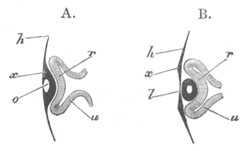
Fig. 285. Diagrammatic sections illustrating the formation of the
eye. (After Remak.)
In A the thin superficial epiblast h is seen to be thickened at
x, in front of the optic vesicle, and involuted so as to form a
pit o, the mouth of which has already begun to close in.
Accompanying this involution, which forms the rudiment of the lens,
the optic vesicle is doubled in, its front portion r being pushed
against the back portion u, and the original cavity of the vesicle
thus reduced in size. The stalk of the vesicle is shewn as still
broad.
In B the optic vesicle is still further doubled in so as to form a
cup with a posterior wall u and an anterior wall r. In the
hollow of this cup lies the lens l, now completely detached from
the superficial epiblast xh.
By the closure of its mouth the pit of the involuted epiblast becomes
a completely closed sac with thick walls and a small central cavity
(fig. 285 B, l). At the same time it breaks away from the external
epiblast, which forms a continuous layer in front of it, all traces of
the original opening being lost. There is thus left lying in the cup
of the secondary optic vesicle, an isolated elliptical mass of
epiblast. This is the rudiment of the lens. The small cavity within it
speedily becomes still less by the thickening of the walls, especially
of the hinder one.
At its first appearance the lens is in immediate contact with the
anterior wall of the secondary optic vesicle (fig. 285 B). In a short
time however, the lens is seen to lie in the mouth of the cup (fig.
288 D), a space (vh) (which is occupied by the vitreous humour)
making its appearance between the lens and anterior wall of the
vesicle.
In order to understand how this space is developed, the position of
the optic vesicle and the relations of its stalk must be borne in
mind.
The vesicle lies at the side of the head, and its stalk is directed
downwards, inwards and backwards. The stalk in fact
slants away from
the vesicle. Hence, when the involution of the lens takes place, the
direction in which the front wall of the vesicle is pushed in is not
in a line with the axis of the stalk, as for simplicity’s sake has
been represented in the diagram (fig. 285), but forms an obtuse angle
with that axis, after the manner of fig. 286, where s´ represents
the cavity of the stalk leading away from the almost obliterated
cavity of the primary vesicle.
Fig. 286 represents the early stage at which the lens fills the whole
cup of the secondary vesicle. The subsequent condition is brought
about through the rapid growth of the walls of the cup. This growth
however does not take place equally in all parts of the cup. The walls
of the cup rise up all round except that point of the circumference of
the cup which adjoins the stalk. While elsewhere the walls increase
rapidly in height, carrying so to speak the lens with them, at this
spot, which in the natural position of the eye is on its under
surface, there is no growth: the wall is here imperfect, and a gap is
left. Through this gap, which afterwards receives the name of the
choroidal fissure, a way is open from the mesoblastic tissue
surrounding the optic vesicle and stalk into the interior of the
cavity of the cup.
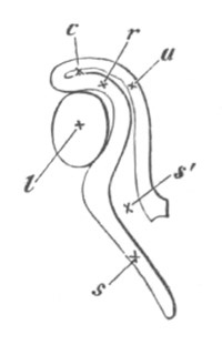
Fig. 286. Diagrammatic section of the eye and the optic nerve at
an early stage. (From Lieberkühn.)
To shew the lens l occupying the whole hollow of the optic cup,
the inclination of the stalk s to the optic cup, and the
continuity of the cavity of the stalk s´ with that of the primary
vesicle c; r. anterior, u. posterior wall of the optic cup.
From the manner of its formation the gap or fissure is evidently in a
line with the axis of the optic stalk, and in order to be seen must be
looked for on the under surface of the optic vesicle. In this position
it is readily recognised in the embryo seen as a transparent object
(fig. 118, chs).
Bearing in mind these relations of the gap to the optic stalk, the
reader will understand how sections of the optic vesicle at this stage
present very different appearances according to the plane in which the
sections are taken.
When the head is viewed from underneath as a transparent
object the
eye presents very much the appearance represented in the diagram (fig.
287).
A section of such an eye taken along the line y, perpendicular to
the plane of the paper, would give a figure corresponding to that of
fig. 288 D. The lens, the cavity and double walls of the secondary
vesicle, the remains of the primary cavity, would all be represented
(the superficial epiblast of the head would also be shewn); but there
would be nothing seen of either the stalk or the fissure. If on the
other hand the section were taken in a plane parallel to the plane of
the paper, at some distance above the level of the stalk, some such
figure would be obtained as that shewn in fig. 288 E. Here the fissure
f is obvious, and the communication of the cavity vh of the
secondary vesicle with the outside of the eye evident; the section of
course would not go through the superficial epiblast. Lastly, a
section, taken perpendicular to the plane of the paper along the line
z, i.e. through the fissure itself, would present the appearances
of fig. 288 F, where the wall of the vesicle is entirely wanting in
the region of the fissure marked by the position of the letter f.
The external epiblast has been omitted in this figure.
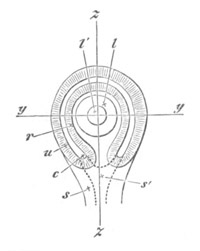
Fig. 287. Diagrammatic representation of the eye of the Chick of
about the third day as seen when the head is viewed from underneath as
a transparent object.
l. the lens; l´. the cavity of the lens, lying in the hollow of
the optic cup; r. the anterior, u. the posterior wall of the
optic cup; c. the cavity of the primary optic vesicle, now nearly
obliterated. By inadvertence u has been drawn in some places
thicker than r, it should have been thinner throughout. s. the
stalk of the optic cup with s´ its cavity, at a lower level than
the cup itself and therefore out of focus; the dotted line indicates
the continuity of the cavity of the stalk with that of the primary
vesicle.
The line z z, through which the section shewn in fig. 288 F is
supposed to be taken, passes through the choroidal fissure.
With reference to the above description, taken with very slight
alterations from the Elements of Embryology, Pt. 1., two points
require to be noticed. Firstly it is extremely doubtful whether the
invagination of the secondary optic vesicle is to be viewed as an
actual mechanical result of the ingrowth of the lens. Secondly it
seems probable that the choroid fissure is not simply due to an
inequality in the growth of the walls of the secondary optic cup, but
is partly due to a doubling up of the primary vesicle from the side
along the line of the fissure, at the same time that the lens is being
thrust in in front. In Mammalia, the doubling up involves the optic
stalk, which becomes flattened (whereby its original cavity is
obliterated) and then folded in on itself, so as to embrace a new
central cavity continuous with the cavity of the vitreous humour. And
in other forms a partial phenomenon of the same kind is usually
observable, as is more particularly described in the sequel.
Before describing the development of the cornea, aqueous humour, etc.
we may consider the further growth of the parts, whose first
development has just been described, commencing with the optic cup.
During the above changes the mesoblast surrounding the optic cup
assumes the character of a distinct investment, whereby the outline of
the eyeball is definitely formed. The internal portions of this
investment, nearest to the retina, become the choroid (i.e. the
chorio-capillaris, and the lamina fusca; the pigment epithelium,
as we have seen, being derived from the epiblastic optic cup), and
pigment is subsequently deposited in it. The remaining external
portion of the investment forms the sclerotic.
The complete differentiation of these two coats of the eye does not
however take place till a late period.
The cavity of the original optic vesicle was left as a nearly
obliterated space between the two walls of the optic cup. By the end
of the third day the obliteration is complete, and the two walls are
in immediate contact.
The inner or anterior wall is, from the first, thicker than the outer
or posterior; and over the greater part of the cup this contrast
increases with the growth of the eye, the anterior wall becoming
markedly thicker and undergoing changes of which we shall have to
speak directly (fig. 289).
In the front portion however, along, so to speak, the lip of the cup,
anterior to a line which afterwards becomes the ora serrata, both
layers cease to take part in the increased thickening, accompanied by
peculiar histological changes, which the rest of the cup is
undergoing. Thus a hind portion or true retina is marked off from a
front portion.
The front portion, accompanied by the mesoblast which immediately
overlies it, is behind the lens thrown into folds, the
ciliary ridges;
while further forward it bends in between the lens and the cornea to
form the iris. The original wide opening of the optic cup is thus
narrowed to a smaller orifice, the pupil; and the lens, which before
lay in the open mouth of the cup, is now inclosed in its cavity. While
in the hind portion of the cup or retina proper no deposit of black
pigment takes place in the layer formed out of the inner or anterior
wall of the vesicle; in the front portion forming the region of the
iris, pigment is largely deposited throughout both layers, though
first of all in the outer one, so that eventually this portion seems
to become nothing more than a forward prolongation of the pigment
epithelium of the choroid.
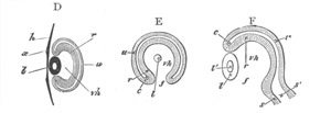
Fig. 288.
D. Diagrammatic section taken perpendicular to the plane of the
paper, along the line yy, fig. 287. The stalk is not seen, the
section falling quite out of its region. vh. hollow of optic cup
filled with vitreous humour; other letters as in fig. 285 B. (After
Remak.)
E. Section taken parallel to the plane of the paper through fig.
287, so far behind the front surface of the eye as to shave off a
small portion of the posterior surface of the lens l, but not so
far behind as to be carried at all through the stalk. Letters as
before; f. the choroidal fissure.
F. Section along the line zz, perpendicular to the plane of the
paper, to shew the choroidal fissure f, and the continuity of the
cavity of the optic stalk with that of the primary optic vesicle.
Had this section been taken a little to one side of the line zz,
the wall of the optic cup would have extended up to the lens below
as well as above. Letters as before. The external epiblast is
omitted in this section.
Thus, while the hind moiety of the optic cup becomes the retina
proper, including the choroid-pigment in which the rods and cones are
imbedded, the front moiety is converted into the ciliary portion of
the retina, covering the ciliary processes, and into the uvea of the
iris; the bodies of the ciliary processes and the substance of the
iris, their vessels, muscles, connective tissue and ramified pigment,
being derived from the mesoblastic choroid. The margin of the pupil
marks the extreme lip of the optic
vesicle, where the outer or
posterior wall turns round to join the inner or anterior.
The ciliary muscle and the ligamentum pectinatum are both derived from
the mesoblast between the cornea and the iris.
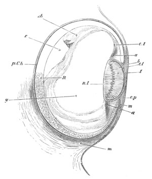
Fig. 289. Section of the eye of Chick at the fourth day.
e.p. superficial epiblast of the side of the head; R. true
retina: anterior wall of the optic cup; p.Ch. pigment-epithelium
of the choroid: posterior wall of the optic cup. b is placed at
the extreme lip of the optic cup at what will become the margin of
the iris. l. the lens. The hind wall, the nuclei of whose
elongated cells are shewn at nl, now forms nearly the whole mass
of the lens, the front wall being reduced to a layer of flattened
cells el. m. the mesoblast surrounding the optic cup and about
to form the choroid and sclerotic. It is seen to pass forward
between the lip of the optic cup and the superficial epiblast.
Filling up a large part of the hollow of the optic cup is seen a
hyaline mass, the rudiment of the hyaloid membrane, and of the
coagulum of the vitreous humour, y. In the neighbourhood of the
lens it seems to be continuous as at cl with the tissue a, which
appears to be the rudiment of the capsule of the lens and suspensory
ligament.
The Retina. At first the two walls of the optic cup do not greatly
differ in thickness. On the third day the outer or posterior becomes
much thinner than the inner or anterior, and by the middle of the
fourth day is reduced to a single layer of flattened
cells (fig. 289,
p.Ch). At about the 80th hour its cells commence to receive a
deposit of pigment, and eventually form the so-called pigmentary
epithelium of the choroid; from them no part of the true retina (or no
other part of the retina, if the pigment-layer in question be supposed
to belong more truly to the retina than to the choroid) is derived.
On the fourth day, the inner (anterior) wall of the optic cup (fig.
289, R) has a perfectly uniform structure, being composed of
elongated somewhat spindle-shaped cells, with distinct nuclei. On its
external (posterior) surface a distinct cuticular membrane, the
membrana limitans externa, early appears.
As the wall increases in thickness, its cells multiply rapidly, so
that it soon becomes several cells thick: each cell being however
probably continued through the whole thickness of the layer. The wall
at this stage corresponds closely in its structure with the brain, of
which it may properly be looked upon as part. According to the usual
view, which is not however fully supported by the development, the
retina becomes divided in the subsequent growth into (1) an outer
part, corresponding morphologically to the epithelial lining of the
cerebrospinal canal, composed of what may be called the visual cells
of the eye, i.e. the cells forming the outer granular (nuclear)
layer and the rods and cones attached to them; and (2) an inner
portion consisting of the inner granular (nuclear) layer, the inner
molecular layer, the ganglionic layer and the layer of nerve-fibres
corresponding morphologically to the walls of the brain. According to
Löwe, however, only the outer limbs of the rods and cones, which he
holds to be metamorphosed cells, correspond to the epithelial layer of
the brain.
The actual development of the retina is not thoroughly understood.
According to the usual statements (Kölliker, No. 298, p. 693) the
layer of ganglion cells and the inner molecular layer are first
differentiated, while the remaining cells give rise to the rest of the
retina proper, and are bounded externally by the membrana limitans
externa. On the inner side of the ganglionic layer the stratum of
nerve-fibres is also very early established. The rods and cones are
formed as prolongations (Kölliker, Babuchin), or cuticularizations
(Schultze, W. Müller) of the cells which eventually form the outer
granular layer. The layer of cells external to the molecular layer is
not divided till comparatively late into the inner and outer granular
(nuclear) layers, and the interposed outer molecular layer.
Löwe’s account of the development of the retina in the Rabbit is in
many points different from the above. He finds that three stages in
the differentiation of the layers of the retina may be distinguished.
In the first stage, in an embryo of four or five millimetres, the
following layers are present, commencing at the outer side, adjoining
the external wall of the secondary optic cup.
(1) A membrane, which does not however, as usually believed, become
the membrana limitans externa.
(2) A layer of clear elements, derived from metamorphosed cells,
constituting the outer limbs of the rods and cones.
(3) A layer of dark rounded elements.
(4) An indistinctly striated layer, the future layer of nerve-fibres.
The third of these layers gives rise to all the eventual strata of the
retina proper, except the outer limbs of the rods and cones.
In the next stage, when the embryo has reached a length of 2 cm., this
layer becomes divided into three strata: viz. an outer and inner layer
of dark elements and a middle one of clearer elements. The two inner
of these layers become respectively the inner molecular layer and the
layer of ganglion cells, while the outer layer gives rise to the parts
of the retina external to the inner molecular layer.
In the newly born animal the outer darker layer of the previous stage
has become considerably subdivided. Its outermost part forms a stratum
of darkly coloured elements, which develop into the inner limbs of the
rods and cones. It is bounded internally by a membrane—the true
membrana elastica externa. The part of the layer within this is soon
divided into the outer and inner granular layers, separated from each
other by the delicate outer molecular layer. Thus, shortly after
birth, all the layers of the retina are established in the Rabbit. It
is important to notice that, according to Löwe’s views, the outer and
inner limbs of the rods and cones are metamorphosed cells. The outer
limbs at first form a continuous layer, in which separate elements
cannot be recognised.
At a very early period there appears a membrane on the side of the
retina adjoining the vitreous humour. This membrane is the hyaloid
membrane. The investigations of Kessler and myself lead to the
conclusion that it may be formed at a time when there is no trace of
mesoblastic structures in the cavity of the vitreous humour, and that
it is therefore necessarily developed as a cuticular deposit of the
cells of the optic cup. Lieberkühn, Arnold, Löwe, and other authors
regard it however as a mesoblastic product; and Kölliker believes that
a primitive membrane is developed from the cells of the optic cup, and
that a true hyaloid membrane is developed much later as a product of
the mesoblast.
For fuller information on this subject the reader is referred to the
authors quoted above.
The optic nerve. The optic nerves are derived, as we have said, from
the at first hollow stalks of the optic vesicles. Their
cavities
gradually become obliterated by a thickening of the walls, the
obliteration proceeding from the retinal end inwards towards the
brain. While the proximal ends of the optic stalks are still hollow
the rudiments of the optic chiasma are formed from fibres at the roots
of the stalks, the fibres of the one stalk growing over into the
attachment of the other. The decussation of the fibres would appear to
be complete. The fibres arise in the remainder of the nerves somewhat
later. At first the optic nerve is equally continuous with both walls
of the optic cup; as must of necessity be the case, since the interval
which primarily exists between the two walls is continuous with the
cavity of the stalk. When the cavity within the optic nerve vanishes,
and the fibres of the optic nerve appear, all connection is ruptured
between the outer wall of the optic cup and the optic nerve, and the
optic nerve simply perforates the outer wall, and becomes continuous
with the inner one.
There does not appear to me any ground for doubting (as has been done
by His and Kölliker) that the fibres of the optic nerve are derived
from a differentiation of the epithelial cells of which the nerve is
at first formed.
Choroid Fissure. With reference to the choroid fissure we may state
that its behaviour varies somewhat in the different types. It becomes
for the greater part of its extent closed, though its proximal end is
always perforated by the optic nerve, and in many forms by a
mesoblastic process also.
The lens when first formed is an oval vesicle with a small central
cavity, the front and hind walls being of nearly equal thickness, and
each consisting of a single layer of elongated columnar cells. In the
subsequent stages the mode of growth of the hind wall is of precisely
an opposite character to that of the front wall. The hind wall becomes
much thicker, and tends to obliterate the central cavity by becoming
convex on its front surface. At the same time its cells, still
remaining as a single layer, become elongated and fibre-like. The
front wall on the contrary becomes thinner and thinner and its cells
flattened.
These modes of growth continue until, as shewn in fig. 289, the hind
wall l is in absolute contact with the front wall el, and the
cavity thus becomes entirely obliterated. The cells of the hind wall
have by this time become veritable fibres, which, when
seen in
section, appear to be arranged nearly parallel to the optic axis,
their nuclei nl being seen in a row along their middle. The front
wall, somewhat thickened at either side where it becomes continuous
with the hind wall, is now a single layer of flattened cells
separating the hind wall of the lens, or as we may now say the lens
itself, from the front limb of the lens-capsule; of the latter it
becomes the epithelium.
The subsequent changes undergone consist chiefly in the continued
elongation and multiplication of the lens-fibres, with the partial
disappearance of their nuclei.
During their multiplication they become arranged in the manner
characteristic of the adult lens of the various forms. The
lens-capsule, as was originally stated by Kölliker, appears to be
formed as a cuticular membrane deposited by the epithelial cells of
the lens.
The views of Lieberkühn, Arnold, Löwe and others, according to which
the lens-capsule is a mesoblastic structure, do not appear to be well
founded. The contrary view, held by Kölliker, Kessler, etc., is
supported mainly by the fact that at the time when the lens-capsule
first appears there are no mesoblast cells to give rise to it. It
should however be stated that W. Müller has actually found cellular
elements in what he believes to be the lens-capsule of the Ammocœte
lens. Considering the degraded character of the Ammocœte eye,
evidence derived from its structure must be accepted with caution.
The vitreous humour. The vitreous humour is derived (except in
Cyclostomata) from a vascular ingrowth, which differs considerably in
different types, through the choroid slit. Its real nature is very
much disputed. According to Kessler’s view, it is of the nature of a
fluid transudation, but the occasional presence in it of ordinary
embryonic mesoblast cells, in addition to more numerous
blood-corpuscles, gives it a claim to be regarded as intercellular
substance. The number of cells in it is however at best extremely
small and in many cases there is no trace of them. In Mammals there
appear to be some mesoblast cells invaginated with the lens, which are
not improbably employed in the formation of the vessels of the
so-called membrana capsulo-pupillaris. In the Ammocœte the vitreous
humour originates from a distinct mesoblastic ingrowth, though the
cells which give rise to it subsequently disappear.
The development of the zonula of Zinn in Mammalia, which ought to
throw some light on the nature of the vitreous humour, has not been
fully investigated. According to Lieberkühn (No. 373, p. 43), this
structure appears in half-grown embryos of the sheep and calf.
He says “At the point where the ciliary processes and the ciliary part
of the retina are entirely removed, one sees in the meridian bundles
of fine fibres, which correspond to the valleys between the ciliary
processes and fill them; also between these bundles there extend, as a
thin layer, similar finely striated masses, and these would have been
on the top of the ciliary processes.” He further states that these
fibres may be traced to the anterior and posterior limb of the
lens-capsule, and that amongst them are numerous cells. Kölliker
confirms Lieberkühn’s statements. There can be little doubt that the
fibres of the zonula are of the nature of connective tissue: they are
stated to be elastic. By Löwe they are believed to be developed out of
the substance of the vitreous humour, but this does not appear to me
to follow from the observations hitherto made. It seems quite possible
that they arise from mesoblast cells which have grown into the cavity
of the vitreous humour, solely in connection with their production.
The integral parts of the eye in front of the lens are the cornea, the
aqueous humour, and the iris. The development of the latter has
already been described, and there remain to be dealt with the cornea,
and the cavity containing the aqueous humour.
The cornea. The cornea is formed by the coalescence of two structures,
viz. the epithelium of the cornea and the cornea proper. The former is
directly derived from the external epiblast, which covers the eye
after the invagination of the lens. The latter is formed in a somewhat
remarkable manner, first clearly made out by Kessler.
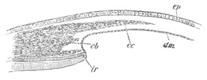
Fig. 290. Section through the eye of a Fowl on the eighth day
of development, to shew the iris and cornea in the process of
formation. (After Kessler.)
ep. epiblastic epithelium of cornea; cc. corneal corpuscles
growing into the structureless matrix of the cornea; dm.
Descemet’s membrane; ir. iris; cb. mesoblast of the iris (this
reference letter points a little too high).
The space between the layers dm. and ep. is filled with the
structureless matrix of the cornea.
When the lens is completely separated from the epidermis its outer
wall is directly in contact with the external epiblast (future corneal
epithelium). At its edge there is a small ring-shaped space bounded by
the outer skin, the lens and the edge of the optic cup. In the chick,
which we may take as typical, there appears at about the time when the
cavity of the lens is completely obliterated a structureless layer
external to the above ring-like space and immediately adjoining the
inner face of the epiblast. This layer, which forms the commencement
of the cornea proper, at first only forms a ring at the border of the
lens, thickest at its outer edge, and gradually thinning off to
nothing towards the centre. It soon however becomes broader, and
finally forms a continuous stratum of considerable thickness,
interposed between the external skin and the lens. As soon as this
stratum has reached a certain thickness, a layer of flattened cells
grows in along its inner side from the mesoblast surrounding the optic
cup (fig. 290, dm). This layer is the epithelioid layer of the
membrane of Descemet. After it[190]
has become completely established,
the mesoblast around the edge of the cornea becomes divided into two
strata; an inner one (fig. 290, cb) destined to form the mesoblastic
tissue of the iris already described, and an outer one (fig. 290,
cc) adjoining the epidermis. The outer stratum gives rise to the
corneal corpuscles, which are the only constituents of the cornea not
yet developed. The corneal corpuscles make their way through the
structureless corneal layer, and divide it into two strata, one
adjoining the epiblast, and the other adjoining the inner epithelium.
The two strata become gradually thinner as the corpuscles invade a
larger and larger portion of their substance, and finally the
outermost portion of them alone remains as the membrana elastica
anterior and posterior (Descemet’s membrane) of the cornea. The
corneal
corpuscles, which have grown in from the sides, thus form a
layer which becomes continually thicker, and gives rise to the main
substance of the cornea. Whether the increase in the thickness of the
layer is due to the immigration of fresh corpuscles, or to the
division of those already there, is not clear. After the cellular
elements have made their way into the cornea, the latter becomes
continuous at its edge with the mesoblast which forms the sclerotic.
The derivation of the original structureless layer of the cornea is
still uncertain. Kessler derives it from the epiblast, but it appears
to me more probable that Kölliker is right in regarding it as derived
from the mesoblast. The grounds for this view are, (1) the fact of its
growth inwards from the border of the mesoblast round the edge of the
eye, (2) the peculiar relations between it and the corneal corpuscles
at a later period. This view would receive still further support if a
layer of mesoblast between the lens and the epiblast were really
present as believed by Lieberkühn. It must however be admitted that
the objections to Kessler’s view of its epiblastic nature are rather
a priori than founded on definite observation.
The observations of Kessler, which have been mainly followed in the
above account, are strongly opposed by Lieberkühn (No. 374) and Arnold
(No. 370), and are not entirely accepted by Kölliker. It is especially
on the development of these parts in Mammalia (to be spoken of in the
sequel) that the above authors found their objections. I have had
through Kessler’s kindness an opportunity of looking through some of
his beautiful preparations, and have no hesitation in generally
accepting his conclusions, though as mentioned above I cannot agree
with all his interpretations.
The aqueous humour. The cavity for the aqueous humour has its origin
in the ring-shaped space round the front of the lens, which, as
already mentioned, is bounded by the external skin, the edge of the
optic cup, and the lens. By the formation of the cornea this space is
shut off from the external skin, and on the appearance of the
epithelioid layer of Descemet’s membrane a continuous cavity is
developed between the cornea and the lens. This cavity enlarges and
receives its final form on the full development of the iris.
Comparative view of the development of the Vertebrate Eye.
The organ of vision, when not secondarily aborted, contains in all
Vertebrata the essential parts above described. The most interesting
cases of partial degeneration are those of Myxine and the Ammocœte.
The development of such aborted eyes has as yet been studied only in
the
Ammocœte[191],
in which it resembles in most important features
that of other Vertebrata.
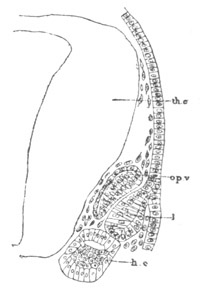
Fig. 291. Horizontal section through the head of a just hatched
larva of Petromyzon shewing the development of the lens of the
eye.
th.c. thalamencephalon; op.v. optic vesicle; l. lens of eye;
h.c. head cavity.
Eye of Ammocœtes. The optic vesicle arises as an outgrowth of the
fore-brain, but the secondary optic cup is remarkable in the young
larva for its small size (fig. 291, opv). The thicker outer wall
gives rise to the retina, and the thinner inner wall to the choroid
pigment. The lens is formed as an invagination of the single-layered
epidermis (fig. 291, l). As development proceeds the parts of the
eye gradually enlarge, and the mesoblast around the hinder and dorsal
part of the optic cup becomes pigmented. There is at first no cavity
for the vitreous humour, but eventually the growth of the optic cup
gives rise to a space, into which a cellular process of mesoblast
grows at a slight notch in the ventral edge of the optic cup (W.
Müller, No. 377). This notch is the only rudiment of the choroid
fissure of other types. The mesoblastic process is probably the
homologue of the processus falciformis and pecten, and appears to give
rise to the vitreous humour; for a long time it retains its connection
with the surrounding mesoblast. Its cells eventually disappear, and it
never contains any vascular structures.
The lens for a long time remains as an oval vesicle with a central
cavity. In a later stage, when the Ammocœte is fully developed,
the secondary optic cup forms a deep pit (fig. 292, r); in the
mouth of which is placed the lens (l). The two walls of the
retina have now the normal vertebrate structure, though the pigment is
as yet imperfectly present in the choroid layer. The lens has the
embryonic forms of higher types (cf. fig. 289), consisting of an inner
thicker segment, the true lens, and an outer layer forming the
epithelium of the lens capsule. The edge of the optic cup, which forms
the rudiment of the epiblast of the iris, is imperfectly separated
from the remainder of the optic cup; and a mesoblastic element of the
iris, distinct from Descemet’s membrane (dm), can hardly be
spoken of.
There is no cavity for the aqueous humour in front of the lens; and
there is no cornea as distinct from the epidermis and subepidermic
tissues. The elements in front of the lens are (1) the epidermis
(ep); (2) the dermis (dc); (3) the subdermal connective tissue
(sdc) which passes without any sharp line of demarcation into the
dermis; (4) a thick membrane, continuous with the mesoblastic part of
the choroid, which appears to represent Descemet’s membrane. The
subdermal connective tissue is continued as an
investment round the
whole eye; and there is no differentiated sclerotic and only an
imperfect choroid.
In a still later stage a distinct mesoblastic element for the iris is
formed. When the Ammocœte is becoming a Lamprey, the eye approaches
the surface; an anterior chamber is established; and the eye differs
from that of the higher types mainly in the fact that the cornea is
hardly distinguished from the remainder of the skin, and that a
sclerotic is very imperfectly represented.
Optic vesicles. The development of the primitive optic vesicles, so
far as is known, is very constant throughout the Vertebrata. In
Teleostei and Lepidosteus alone is there an important deviation from
the ordinary type, dependent however upon the mode of formation of the
medullary keel, the optic vesicles arising while the medullary keel is
still solid, and being at first also solid. They subsequently acquire
a lumen and undergo the ordinary changes.
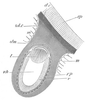
Fig. 292. Eye of an Ammocœtes lying beneath the skin.
ep. epidermis; d.c. dermal connective tissue continuous with the
subdermal connective tissue (s.d.c), which is also shaded. There
is no definite boundary to this tissue where it surrounds the eye.
m. muscles; dm. membrane of Descemet; l. lens; v.h. vitreous
humour; r. retina; rp. retinal pigment.
The lens. In the majority of groups, viz. Elasmobranchii, Reptilia,
Aves, and Mammalia, the lens is formed by an open invagination of the
epiblast, but in Amphibia, Teleostei and Lepidosteus, where the
nervous layer of the skin is early established, this layer alone takes
part in the formation of the lens (fig. 293, l). The lens is however
formed even in these types as a hollow body by an invagination; but
its opening remains permanently shut off from communication with the
exterior by the epidermic
layer of the epiblast. Götte describes the
lens as formed by a solid thickening of the nervous layer in
Bombinator. This is probably a mistake.
The cornea. The mode of formation of the cornea already described
appears to be characteristic of most Vertebrata except the Ammocœte.
It has been found by Kessler in Aves, Reptilia and Amphibia, and
probably also occurs in Pisces. In Mammals it is not however so easy
to establish. There are at first no mesoblast cells between the lens
and the epiblast (fig. 295) but in many Mammals (vide Kessler, No.
372, pp. 91-94) a layer of rounded mesoblast cells, which forms
Descemet’s membrane, grows in between the two, at a time when it is
not easy to recognise a corneal lamina, as distinct from a simple
coagulum.
After the formation of this layer the mesoblast cells grow into the
corneal lamina from the sides, and becoming flattened arrange
themselves in rows between the laminæ of the cornea. The cornea
continues to increase in thickness by the addition of laminæ on the
side adjoining the epiblast.
We have already seen that in the Lamprey the cornea is nothing else
but the slightly modified and more transparent epidermis and dermis.
The optic nerve and the choroid fissure. It will be convenient to
consider together the above structures, and with them the vascular and
other processes which pass into the cavity of the optic cup through
the choroid fissure. These parts present on the whole a greater amount
of variation than any other parts of the eye.
I commence with the Fowl which is both a very convenient general type
for comparison, and also that in which these structures have been most
fully worked out.
During the third day of incubation there passes in through the choroid
slit a vascular loop, which no doubt supplies the transuded material
for the growth of the vitreous humour. Up to the fifth day this
vascular loop is the only structure passing through the choroid slit.
On this day however a new structure appears, which remains permanently
through life, and is known as the pecten. It consists of a lamellar
process of the mesoblast cells round the eye, passing through the
choroid slit near the optic nerve, and enveloping part of the afferent
branch of the vascular loop above mentioned. The proximal part of the
free edge of the pecten is somewhat swollen, and sections through this
part have a club-shaped form. On the sixth day the choroid slit
becomes rapidly closed, so that at the end of the sixth day it is
reduced to a mere seam. There are however two parts of this seam where
the edges of the optic cup have not coalesced. The proximal of these
adjoins the optic nerve, and permits the passage of the pecten and at
a later period of the optic nerve; and the second or distal one is
placed near the ciliary edge of the slit, and is traversed by the
efferent branch of the above-mentioned vascular loop. This vessel soon
atrophies, and with it the distal opening in the choroid slit
completely vanishes. In some varieties of domestic Fowl (Lieberkühn)
the opening however persists. The seam which marks the original site
of the choroid slit is at first
conspicuous by the absence of pigment,
and at a later period by the deep colour of its pigment. Finally, a
little after the ninth day, no trace of it is to be seen.
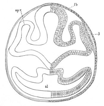
Fig. 293. Section through the front part of the head of a Lepidosteus
embryo on the seventh day after impregnation.
al. alimentary tract; fb. thalamencephalon; l. lens of eye;
op.v. optic vesicle. The mesoblast is not represented.
Up to the eighth day the pecten remains as a simple lamina; by the
tenth or twelfth day it begins to be folded or rather puckered, and by
the seventeenth or eighteenth day it is richly pigmented and the
puckerings have become nearly as numerous as in the adult, there being
in all seventeen or eighteen. The pecten is almost entirely composed
of vascular coils, which are supported by a sparse pigmented
connective tissue; and in the adult the pecten is still extremely
vascular. The original artery which became enveloped at the formation
of the pecten continues, when the latter becomes vascular, to supply
it with blood. The vein is practically a fresh development after the
atrophy of the distal portion of the primitive vascular loop of the
vitreous humour.
There are no true retinal blood-vessels.
In the formation of the optic cup the extreme peripheral part of the
optic nerve, which is in immediate proximity with the artery of the
pecten, becomes folded. The permanent opening in the choroid fissure
for the pecten is intimately related to the entrance of the optic
nerve into the eyeball; the fibres of the optic nerve passing in at
the inner border of the pecten, coursing along its sides to its outer
border, and radiating from it as from a centre to all parts of the
retina.
In the Lizard the choroid slit closes considerably earlier than in the
Fowl. The vascular loop in the vitreous humour is however more
developed. The pecten long remains without vessels, and does not in
fact become at all
vascular till after the very late disappearance of
the distal part of the vascular loop of the vitreous humour.
The arrangement of the ingrowth through the choroid slit in
Elasmobranchii (Scyllium) has been partially worked out, and so far as
is at present known the agreement between the Avian and Elasmobranch
type is fairly close.
At the time when the cavity between the lens and the secondary optic
cup is just commencing to be formed, a process of mesoblast
accompanied by a vascular loop passes into the vitreous humour,
through the choroid slit, close to the optic nerve. The vessel in this
process is no doubt equivalent to the vascular loop in the Avian eye,
but I have not made out that it projects beyond the mesoblastic
process accompanying it. As the cavity of the vitreous humour enlarges
and the choroid slit elongates, the process through it takes the form
of a lamina with a somewhat swollen border, and projects for some
distance into the cavity of the vitreous humour.
At a later stage, after the outer layer of the optic cup has become
pigmented, the distal part of the choroid slit adjoining the border of
the lens closes up; but along the line where it was present the walls
of the optic cup remain very thin and are thrown into three folds, two
lateral and one median, projecting into the cavity of the vitreous
humour. The median fold is in contact with the lens, and the vascular
mesoblast surrounding the eye projects into the space between the two
laminæ of which it is formed. In passing from the region of the lens
to that of the optic nerve the lateral folds of the optic cup
disappear, and the median fold forms a considerable projection into
the cavity of the vitreous humour. It consists of a core of mesoblast
covered by a delicate layer derived from both strata of the optic cup.
Still nearer the optic nerve the choroid slit is no longer closed, and
the mesoblast, which in the neighbourhood of the lens only extended
into the folds of the wall of the optic cup, now projects freely into
the cavity of the vitreous humour, and forms the lamina already
described. It is not very vascular, but close to the optic nerve there
passes into it a considerable artery.
In the young animal the choroid slit is no longer perforated by a
mesoblastic lamina. At its inner end it remains open to allow of the
passage of the optic nerve. The line of the slit can easily be traced
along the lower side of the retina; and close to the lens the retinal
wall continues, as in the embryo, to be raised into a projecting fold.
Traces of these structures are visible even in the fully grown
examples of Scyllium.
As has been pointed out by Bergmeister the mesoblastic lamina
projecting into the vitreous humour resembles the pecten at an early
stage of development, and is without doubt homologous with it. The
artery which supplies it is certainly equivalent to the artery of the
pecten.
There can be no doubt that the mesoblastic lamina projecting into the
vitreous humour is equivalent to the processus falciformis of
Teleostei, and it seems probable that the whole of it, including the
free part as well as that covered by epiblast, ought to be spoken of
under this title. The optic nerve
in Elasmobranchii is not included in
the folding to which the secondary optic vesicle owes its origin, and
would seem to perforate the walls of the optic cup only at the distal
end of the processus falciformis.
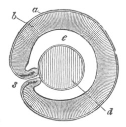
Fig. 294. Horizontal section through the eye of a Teleostean
embryo. (From Gegenbaur; after Schenk.)
s. choroid fissure, with two folds forming part of the processus
falciformis; a. choroid layer of optic cup; b. retinal layer of
optic cup; c. cavity of vitreous humour; d. lens.
In Teleostei there is at first a vascular loop like that in Birds,
passing through the choroid fissure. This has been noticed by Kessler
in the Pike, and by Schenk in the Trout. At a later period a
mesoblastic ingrowth with a blood-vessel makes its way in many forms
into the cavity of the vitreous humour, accompanied by two folds in
the walls of the free edges of the choroid fissure (fig. 294). These
structures, which constitute the processus falciformis, clearly
resemble very closely the mesoblastic process and folds of the optic
cup in Elasmobranchii. The processus falciformis comes in contact
with, and perhaps becomes attached to the wall of the lens; and
persists through life.
In Triton there is no vascular ingrowth through the choroid fissure,
but a few mesoblastic cells pass in which represent the vascular
ingrowth of other types. The optic nerve perforates the proximal
extremity of the original choroid slit.
The absence of an embryonic blood-vessel does not however hold good
for all Amphibia, as there is present in the embryo Alytes
(Lieberkühn) an artery, which breaks up into a capillary system on the
retinal border of the vitreous humour.
In the Ammocœte the choroid slit is merely represented by a slight
notch on the ventral edge of the optic cup, and the mesoblastic
process which passes through the choroid slit in most types is
represented by a large cellular process, from which the vitreous
humour would appear to be derived.
Mammalia differ from all the types already described in the immense
fœtal development of the blood-vessels of the vitreous humour. There
are however some points in connection with the development of these
vessels which are still uncertain. The most important of these points
concerns the presence of a prolongation of the mesoblast around the
eye into the cavity of the vitreous humour. It is maintained by
Lieberkühn, Arnold, Kölliker, etc., that in the invagination of the
lens a thin layer of mesoblast is carried before it; and is thus
transported into the cavity of the vitreous humour. This is denied by
Kessler, but the layer is so clearly figured by the above
embryologists, that the existence of it in some Mammalia (the Rabbit,
etc.) must I think be accepted.
In the folding in of the optic vesicle, which accompanies the
formation of the lens, the optic nerve becomes included, and on the
development of the cavity of the vitreous humour an artery, running in
the fold of the optic
nerve, passes through the choroid slit into the
cavity of the vitreous humour (fig. 295, acr). The sides of the
optic nerve subsequently bend over, and completely envelope this
artery, which at a later period gives off branches to the retina, and
becomes known as the arteria centralis retinæ. It is homologous with
the arterial limb of the vascular loop projecting into the vitreous
humour in Birds, Lizards, Teleostei, etc.
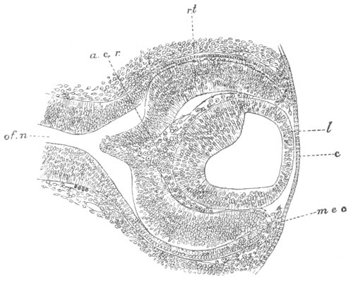
Fig. 295. Section through the eye of a Rabbit embryo of
about twelve days.
c. epithelium of cornea; l. lens; mec. mesoblast growing in
from the side to form the cornea; rt. retina; a.c.r. arteria
centralis retinæ; of.n. optic nerve.
The figure shews (1) the absence at this stage of mesoblast between
the lens and the epiblast: the interval between the two has however
been made too great; (2) the arteria centralis retinæ forming the
vascular capsule of the lens and continuous with vascular structures
round the edges of the optic cup.
Before becoming enveloped in the optic nerve this artery is continued
through the vitreous humour (fig. 295), and when it comes in close
proximity to the lens it divides into a number of radiating branches,
which pass round the edge of the lens, and form a vascular sheath
which is prolonged so as to cover the anterior wall of the lens. In
front of the lens they anastomose with vessels, coming from the iris,
many of which are venous (fig. 295)—and the whole of the blood from
the arteria centralis is carried away by these veins. The vascular
sheath surrounding the lens receives the name of the membrana
capsulo-pupillaris. The posterior part of it appears (Kessler, No.
372) to be formed of vessels without the addition of any other
structures and is either formed simply by branches of the arteria
centralis, or out of
the mesoblast cells involuted with the lens. The
anterior part of the vascular sheath is however inclosed in a very
delicate membrane, the membrana pupillaris, continuous at the sides
with the epithelium of Descemet’s membrane. On the formation of the
iris this membrane lies superficially to it, and forms a kind of
continuation of the mesoblast of the iris over the front of the lens.
The origin of this membrane is much disputed. By Kessler, whose
statements have been in the main followed, it is believed to appear
comparatively late as an ingrowth of the stroma of the iris; while
Kölliker believes it to be derived from a mesoblastic ingrowth between
the front wall of the lens and the epiblast. According to Kölliker
this ingrowth subsequently becomes split into two laminæ, one of which
forms the cornea, and the other the anterior part of the vascular
sheath of the lens with its membrana pupillaris. Between the two
appears the aqueous humour.
The membrana capsulo-pupillaris is simply a provisional embryonic
structure, subserving the nutrition of the lens. The time of its
disappearance varies somewhat for the different Mammalia in which this
point has been investigated. In the human embryo it lasts from the
second to the seventh month and sometimes longer. As a rule it is
completely absorbed at the time of birth. The absorption of the
anterior part commences in the centre and proceeds outwards.
In addition to the vessels of the vascular capsule round the lens,
there arise from the arteria centralis retinæ, just after its exit
from the optic nerve, in many forms (Dog, Cat, Calf, Sheep, Rabbit,
Man) provisional vascular branches which extend themselves in the
posterior part of the vitreous humour. Near the ciliary end of the
vitreous humour they anastomose with the vessels of the membrana
capsulo-pupillaris.
In Mammals the choroid slit closes very early, and is not perforated
by any structure homologous with the pecten. The only part of the slit
which remains open is that perforated by the optic nerve; and in the
centre of the latter is situated the arteria centralis retinæ as
explained above. From this artery there grow out the vessels to supply
the retina, which have however nothing to do with the provisional
vessels of the vitreous humour just described (Kessler). On the
atrophy of the provisional vessels the whole of the blood of the
arteria centralis passes into the retina.
It is interesting to notice (Kessler, No. 372, p. 78) that there seems
to be a blood-vessel supplying the vitreous humour in the embryos of
nearly all vertebrate types, which is homologous throughout the
Vertebrata. This vessel often exhibits a persisting and a provisional
part. The latter in Mammalia is the membrana capsulo-pupillaris and
other vessels of the vitreous humour; in Birds and Lizards it is the
part of the original vascular loop, not included in the pecten, and in
Osseous Fishes that part (?) not involved in the processus
falciformis. The permanent part is formed by the retinal vessels of
Mammalia, by the vessels of the pecten in Birds and Lizards, and by
those of the processus falciformis in Fishes.
The Iris and Ciliary processes. The walls of the edge of the optic cup
become very much thinner than those of the true retinal part. In many
Vertebrates (Mammalia, Aves, Reptilia, Elasmobranchii, etc.) the
thinner part, together with the mesoblast covering it, becomes divided
into two regions, viz. that of the iris, and that of the ciliary
processes. In the Newt and Lamprey this differentiation does not take
place, but the part in question simply becomes the iris.
Accessory Organs connected with the Eye.
Eyelids. The most important accessory structures connected with the
eye are the eyelids. They are developed as simple folds of the
integument with a mesoblastic prolongation between their two laminæ.
They may be three in number, viz. an upper and lower, and a lateral
one—the nictitating membrane—springing from the inner or anterior
border of the eye. Their inner face is lined by a prolongation of
conjunctiva, which is the modified epiblast covering the cornea and
part of the sclerotic.
In Teleostei and Ganoidei eyelids are either not present or at most
very rudimentary. In Elasmobranchii they are better developed, and the
nictitating membrane is frequently present. The latter is also usually
found in Amphibia. In the Sauropsida all three eyelids are usually
present, but in Mammalia the nictitating membrane is rudimentary.
In many Mammalia the two eyelids meet together during a period of
embryonic life, and unite in front of the eye. A similar arrangement
is permanent through life in Ophidia and some Lacertilia; and there is
a chamber formed between the coalesced eyelids and the surface of the
cornea, into which the lacrymal ducts open.
Lacrymal glands. Lacrymal glands are found in the Sauropsida and
Mammalia. They arise (Remak, Kölliker) as solid ingrowths of the
conjunctival epithelium. They appear in the chick on the eighth day.
Lacrymal duct. The lacrymal duct first appears in Amphibia, and is
present in all the higher Vertebrates. Its mode of development in the
Amphibia, Lacertilia and Aves has recently been very thoroughly worked
out by Born (Nos. 380 and 381).
In Amphibia he finds that the lacrymal duct arises as a solid ridge of
the mucous layer of the epidermis, continued from the external opening
of the nasal cavity backwards towards the eye. It usually appears at
about the time when the nasal capsule is beginning to be chondrified.
As this ridge is gradually prolonged backwards towards the eye its
anterior end becomes separated from the epidermis, and grows inwards
in the mesoblast to become continuous with the posterior part of the
nasal sack. The posterior end which joins the eye becomes divided into
the two collecting branches of the adult. Finally the whole structure
becomes separated from the skin except at the external opening, and
develops a lumen.
In Lacertilia the lacrymal duct arises very much in the same manner as
in Amphibia, though its subsequent growth is somewhat different. It
appears as an internal ridge of the epithelium, at the junction of the
superior maxillary process and the fold which gives rise to the lower
eyelid. A solid process of this ridge makes its way through the
mesoblast on the upper border of the maxillary process till it meets
the wall of the nasal cavity, with the epithelium of which it becomes
continuous. At a subsequent stage a second solid growth from the upper
part of the epithelial ridge makes its way through the lower eyelid,
and unites with the inner epithelium of the eyelid; and at a still
later date a third growth from the lower part of the structure forms a
second junction with the epithelium of the eyelid. The two latter
outgrowths form the two upper branches of the duct. The ridge now
loses its connection with the external skin, and, becoming hollow,
forms the lacrymal duct. It opens at two points on the inner surface
of the eyelid, and terminates at its opposite extremity by opening
into the nasal cavity. It is remarkable, as pointed out by Born, that
the original epithelial ridge gives rise directly to a comparatively
small part of the whole duct.
In the Fowl the lacrymal duct is formed as a solid ridge of the
epidermis, extending along the line of the so-called lacrymal groove
from the eye to the nasal pit (fig. 120). At the end of the sixth day
it begins to be separated from the epidermis, remaining however united
with it on the inner side of the lower eyelid. After its separation
from the epidermis it forms a solid cord, the lower end of which
unites with the wall of the nasal cavity. The cord so formed gives
rise to the whole of the duct proper and to the lower branch of the
collecting tube. The upper branch of the collecting tube is formed as
an outgrowth from this cord. A lumen begins to be formed on the
twelfth day of incubation, and first appears at the nasal end. It
arises by the formation of a space between the cells of the cord, and
not by an absorption of the central cells.
In Mammalia Kölliker states that he has been unable to observe
anything similar to that described by Born in the Sauropsida and
Amphibia, and holds to the old view, originally put forward by Coste,
that the duct is formed by the closure of a groove leading from the
eye to the nose between the outer nasal process and the superior
maxillary process. The upper extremity of the duct dilates to form a
sack, from which two branches pass off to open on the lacrymal
papillæ. In view of Born’s discoveries Kölliker’s statements must be
received with some caution.
The Eye of the Tunicata.
The unpaired eye of the larva of simple Ascidians is situated somewhat
to the right side of the posterior part of the dorsal wall of the
anterior cephalic vesicle (fig. 296, O). It consists of a refractive
portion, turned towards the cavity of the vesicle of
the brain, and a
retinal portion forming part of the wall of the brain. The refractive
parts consist of a convex-concave meniscus in front, and a spherical
lens behind, adjoining the concave side of the meniscus. The posterior
part of this lens is imbedded in a layer of pigment. The retina is
formed of columnar cells, with their inner ends imbedded in the
pigment which encloses the posterior part of the lens. The retinal
part of the eye arises in the first instance as a prominence of the
wall of the cerebral vesicle: its cells become very columnar and
pigmented at their inner extremities (fig. 8, V, a). The lens is
developed at a later period, after the larva has become hatched, but
the mode of its formation has not been made out.
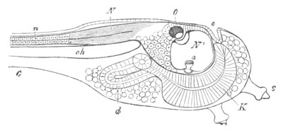
Fig. 296. Larva of Ascidia mentula. (From Gegenbaur; after Kupffer.)
Only the anterior part of the tail is represented.
N´. anterior swelling of neural tube; N. anterior swelling of
spinal portion of neural tube; n. hinder part of neural tube;
ch. notochord; K. branchial region of alimentary tract; d.
œsophageal and gastric region of alimentary tract; O. eye; a.
otolith; o. mouth; s. papilla for attachment.
General considerations on the Eye of the Chordata.
There can be but little doubt that the eye of the Tunicata belongs to
the same phylum as that of the true Vertebrata, different as the two
eyes are. The same may also be said with reference to the degenerate
and very rudimentary eye of Amphioxus.
The peculiarity of the eye of all the Chordata consists in the retina
being developed from part of the wall of the brain. How is this
remarkable feature of the eye of the Chordata to be explained?
Lankester, interpreting the eye in the light of the Tunicata, has made
the interesting suggestion[192]
“that the original Vertebrate must
have been a transparent animal, and had an eye or pair of eyes inside
the brain, like that of the Ascidian Tadpole.”
This explanation may possibly be correct, but another explanation
appears to me possible, and I am inclined to think that the vertebrate
eyes have not been derived from eyes like those of Ascidians, but that
the latter is a degenerate form of vertebrate eye.
The fact of the retina being derived from the fore-brain may perhaps
be explained in the same way as has already been attempted in the case
of the retina of the Crustacea; i.e. by supposing that the eye was
evolved simultaneously with the fore part of the brain.
The peculiar processes which occur in the formation of the optic
vesicle are more difficult to elucidate; and I can only suggest that
the development of a primary optic vesicle, and its conversion into an
optic cup, is due to the retinal part of the eye having been involved
in the infolding which gave rise to the canal of the central nervous
system. The position of the rods and cones on the posterior side of
the retina is satisfactorily explained by this hypothesis, because, as
may be easily seen from figure 285, the posterior face of the retina
is the original external surface of the epidermis, which is infolded
in the formation of the brain; so that the rods and cones are, as
might be anticipated, situated on what is morphologically the external
surface of the epiblast of the retina.
The difficulty of this view arises in attempting to make out how the
eye can have continued to be employed during the gradual change of
position which the retina must have undergone in being infolded with
the brain in the manner suggested. If however the successive steps in
this infolding were sufficiently small, it seems to me not impossible
that the eye might have continued to be used throughout the whole
period of change, and a transparency of the tissues, such as Lankester
suggests, may have assisted in rendering this possible.
The difficulty of the eye continuing to be in use when undergoing
striking changes in form is also involved in Lankester’s view, in that
if, as I suppose, he starts from the eye of the Ascidian Tadpole with
its lenses turned towards the cavity of the brain; it is necessary
for him to admit that a fresh lens and other optical parts of the eye
became developed on the opposite side of the eye to the original
lens; and it is difficult to understand such a change, unless we can
believe that the refractive media on the two sides were in operation
simultaneously. It may be noted that the same difficulty is involved
in supposing, as I have done, that the eye of the Ascidian Tadpole was
developed from that of a Vertebrate. I should however be inclined to
suggest that the eye had in this case ceased for a period to be
employed; and that it has been re-developed again in some of the
larval forms. Its characters in the Tunicata are by no means constant.
Accessory eyes in the Vertebrata.
In addition to the paired eyes of the Vertebrata certain organs are
found in the skin of a few Teleostei living in very deep water, which,
though clearly not organs of true vision, yet present characters which
indicate that
they may be used in the perception of light. The most
important of such organs are those found in Chauliodus, Stomias, etc.,
the significance of which was first pointed out by Leuckart, while the
details of their structure have been recently worked out by
Leydig[193]
and Ussow. They are distributed not only in the skin, but
are also present in the mouth and respiratory cavity, a fact which
appears to indicate that their main function must be something else
than the perception of light. It has been suggested that they have the
function of producing phosphorescence.
Another organ, probably of the same nature, is found on the head of
Scopelus.
The organs in Chauliodus are spherical or nearly spherical bodies
invested in a special tunic. The larger of them, which alone can have
any relation to vision, are covered with pigment except on their outer
surface. The interior is filled with two masses, named by Leuckart the
lens and vitreous humour. According to Leydig each of them is cellular
and receives a nerve, the ultimate destination of which has not
however been made out. According to Ussow the anterior mass is
structureless, but serves to support a lens, placed in the centre of
the eye, and formed of a series of crystalline cones prolonged into
fibres, which in the posterior part of the eye diverge and terminate
by uniting with the processes of multipolar cells, placed near the
pigmented sheath. These cells, together with the fibres of the
crystalline cones which pass to them, are held by Ussow to constitute
a retina.
Eye of the Mollusca.
(362) N. Bobretzky. “Observations on the development of the
Cephalopoda” (Russian). Nachrichten d. kaiserlichen Gesell. d.
Freunde der Naturwiss. Anthropolog. Ethnogr. bei d. Universität
Moskau.
(363) H. Grenacher. “Zur Entwicklungsgeschichte d. Cephalopoden.”
Zeit. f. wiss. Zool., Bd. XXIV. 1874.
(364) V. Hensen. “Ueber d. Auge einiger Cephalopoden.” Zeit. f. wiss.
Zool., Vol. XV. 1865.
(365) E. R. Lankester. “Observations on the development of the
Cephalopoda.” Quart. J. of Micr. Science, Vol. XV. 1875.
(366) C. Semper. Ueber Sehorgane von Typus d. Wirbelthieraugen.
Wiesbaden, 1877.
Eye of the Arthropoda.
(367) N. Bobretzky. Development of Astacus and Palaemon. Kiew, 1873.
(368) A. Dohrn. “Untersuchungen üb. Bau u. Entwicklung d. Arthropoden.
Palinurus und Scyllarus.” Zeit. f. wiss. Zool., Bd. XX. 1870, p. 264
et seq.
(369) E. Claparède. “Morphologie d. zusammengesetzten Auges bei den
Arthropoden.” Zeit. f. wiss. Zool., Bd. X. 1860.
(370) H. Grenacher. Untersuchungen üb. d. Sehorgane d. Arthropoden.
Göttingen, 1879.
Vertebrate Eye.
(371) J. Arnold. Beiträge zur Entwicklungsgeschichte des Auges.
Heidelberg, 1874.
(372) Babuchin. “Beiträge zur Entwicklungsgeschichte des Auges.”
Würzburger naturwissenschaftliche Zeitschrift, Bd. 8.
(373) L. Kessler. Zur Entwicklung d. Auges d. Wirbelthiere. Leipzig,
1877.
(374) N. Lieberkühn. Ueber das Auge des Wirbelthierembryo. Cassel,
1872.
(375) N. Lieberkühn. “Beiträge z. Anat. d. embryonalen Auges.” Archiv
f. Anat. und Phys., 1879.
(376) L. Löwe. “Beiträge zur Anatomie des Auges” and “Die Histogenese
der Retina.” Archiv f. mikr. Anat., Vol. XV. 1878.
(377) V. Mihalkowics. “Untersuchungen über den Kamm des Vogelauges.”
Archiv f. mikr. Anat., Vol. IX. 1873.
(378) W. Müller. “Ueber die Stammesentwickelung des Schorgans der
Wirbelthiere.” Festgabe Carl Ludwig. Leipzig, 1874.
(379) S. L. Schenk. “Zur Entwickelungsgeschichte des Auges der
Fische.” Wiener Sitzungsberichte, Bd. LV. 1867.
Accessory organs of the Vertebrate Eye.
(380) G. Born. “Die Nasenhöhlen u. d. Thränennasengang d. Amphibien.”
Morphologisches Jahrbuch, Bd. II. 1876.
(381) G. Born. “Die Nasenhöhlen u. d. Thränennasengang d. amnioten
Wirbelthiere. I. Lacertilia. II. Aves.” Morphologisches Jahrbuch,
Bd. V. 1879.
Eye of the Tunicata.
(382) A. Kowalevsky. “Weitere Studien üb. d. Entwicklung d. einfachen
Ascidien.” Archiv f. mikr. Anat., Vol. VII. 1871.
(383) C. Kupffer. “Zur Entwicklung d. einfachen Ascidien.” Archiv f.
mikr. Anat., Vol. VII. 1872.
Auditory Organs.
A great variety of organs, very widely distributed amongst aquatic
forms, and also found, though less universally, in land forms, are
usually classed together as auditory organs.
In the case of all aquatic forms, or of forms which have directly
inherited their auditory organs from aquatic forms, these organs are
built upon a common type; although in the majority of instances the
auditory organs of the several groups have no genetic relations. All
the organs have their origin in specialized portions of the epidermis.
Some of the cells of a special region become provided at their free
extremities with peculiar hairs, known as auditory hairs; while in
other cells concretions, known as otoliths, are formed, which appear
often to be sufficiently free to be acted upon by vibrations of the
surrounding medium, and to be so placed as to be able in their turn to
transmit their vibrations to the cells with auditory hairs[194].
The
auditory regions of the epidermis are usually shut off from the
surface in special sacks.
The actual function of these organs is no doubt correctly described,
in the majority of instances, as being auditory; but it appears to me
very possible that in some cases their function may be to enable the
animals provided with them to detect the presence of other animals in
their neighbourhood, through the
undulatory movements in the water,
caused by the swimming of the latter.
Auditory organs with the above characters, sometimes freely open to
the external medium, but more often closed, are found in various
Cœlenterata, Vermes and Crustacea, and universally or all but
universally in the Mollusca and Vertebrata.
In many terrestrial Insects a different type of auditory organ has
been met with, consisting of a portion of the integument modified to
form a tympanum or drum, and supported at its edge by a chitinous
ring. The vibrations set up in the membranous tympanum stimulate
terminal nerve organs at the ends of chitinous processes, placed in a
cavity bounded externally by the tympanic membrane.
The tympanum of Amphibia and Amniota is an accessory organ added, in
terrestrial Vertebrata, to an organ of hearing primitively adapted to
an aquatic mode of life; and it is interesting to notice the presence
of a more or less similar membrane in the two great groups of
terrestrial forms, i.e. terrestrial Vertebrata and Insecta.
Nothing is known with reference to the mode of development or
evolution of the tympanic type of auditory organ found in Insects,
and, except in the case of Vertebrates, but little is known with
reference to the development of what may be called the vesicular type
of auditory organ found in aquatic forms. Some very interesting facts
with reference to the evolution of such organs have however been
brought to light by the brothers Hertwig in their investigations on
the Cœlenterata; and I propose to commence my account of the
development of the auditory organs in the animal kingdom by a short
statement of the results of their researches.
Cœlenterata. Three distinct types of auditory organ have been
recognised in the Medusæ; two of them resulting from the
differentiation of a tentacle-like organ, and one from ectoderm cells
on the under surface of the velum. We may commence with the latter as
the simplest. It is found in the Medusæ known as the Vesiculata. The
least differentiated form of this organ, so far discovered, is present
in Mitrotrocha, Tiaropsis and other genera. It has the form of an open
pit; and a series of such organs are situated along the attached edge
of the velum with their apertures directed downwards. The majority of
the cells lining the outer, i.e. peripheral side of the pit, contain
an otolith, while a row of the cells on the inner, i.e. central
side, are modified as auditory cells. The auditory cells are somewhat
strap-shaped, their inner ends being continuous with the fibres of the
lower nerve-ring, and their free ends being provided with bent
auditory hairs, which lie in contact with the convex surfaces of the
cells containing the otoliths.
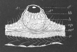
Fig. 297. Auditory vesicle of Phialidium after treatment with
dilute osmic acid. (From Lankester; after O. and R. Hertwig.)
d1. epithelium of the upper surface of the velum; d2.
epithelium of the under surface of the velum; r. circular canal at
the edge of the velum; nr1. upper nerve-ring; h. auditory
cells; hh. auditory hairs; np. nervous cushion formed of a
prolongation of the lower nerve-ring. Close to the nerve-ring is
seen a cell, shewn as black, containing an otolith.
By the conversion of such open pits into closed sacks a more
complicated type of auditory organ, which is present in many of the
Vesiculata, viz. Æquorea, Octorchis, Phialidium, &c., is produced. A
closed vesicle of this type is shewn in fig. 297. Such organs form
projections on the upper surface of the velum. They are covered by a
layer of the epithelium (d1) of the upper surface of the velum, but
the lining of the vesicle (d2) is derived from what was originally
part of the epithelium of the lower surface of the velum, homologous
with that lining the open pits in the type already described. The
general arrangement of the cells lining such vesicles is the same as
that of the cells lining the open pits.
A second type of auditory organ, found in the Trachymedusæ, appears in
its simplest condition as a modified tentacle.
It is formed of a basal
portion, covered by auditory cells with long stiff auditory hairs,
supporting at its apex a club-shaped body, attached to it by a
delicate stalk. An endodermal axis is continued through the whole
structure, and in one or more of the endoderm cells of the club-shaped
body otoliths are always present. The tails of the auditory cells are
directly continued into the upper nerve-ring.
In more complicated forms of this organ the tentacle becomes enclosed
in a kind of cup, by a wall-like upgrowth of the surrounding parts
(fig. 298); and in some forms, e.g. Geryonia, by the closure of the
cup, the whole structure takes the form of a completely closed
vesicle, in the cavity of which the original tentacle forms an
otolith-bearing projection.
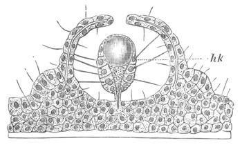
Fig. 298. Auditory organ of Rhopalonema. (From Lankester; after O.
and R. Hertwig.)
The organ consists of a modified tentacle (hk) with auditory cells
and concretions, partially enclosed in a cup.
The auditory organs found in the Acraspedote Medusæ approach in many
respects to the type of organ found in the Trachymedusæ. They consist
of tentacular organs placed in grooves on the under surface of the
disc. They have a swollen extremity, and are provided with an
endodermal axis for half the length of which there is a diverticulum
of the gastrovascular canal system. The terminal portion of the
endoderm is solid, and contains calcareous concretions. The ectodermal
cells at the base of these organs have the form of auditory cells.
Mollusca. Auditory vesicles are found in almost all Mollusca on the
ventral side of the body in close juxtaposition to the pedal ganglia.
Except possibly in some Cephalopods, these
vesicles are closed. They
are provided with free otoliths, supported by the cilia of the walls
of the sack, but in addition some of the cells of the sack are
provided with stiff auditory hairs.
In many forms these sacks have been observed to originate by an
invagination of the epiblast of the foot (Paludina, Nassa,
Heteropoda, Limax, Clio, Cephalopoda and Lamellibranchiata). In
other instances (some Pteropods, Lymnæus, &c.) they appear, by a
secondary modification in the development, to originate by a
differentiation of a solid mass of epiblast.
According to Fol the otocysts in Gasteropods are formed by cells of
the wall of the auditory sacks; and the same appears to hold good for
Cephalopoda (Grenacher)[195]
shewing that free otoliths have in these
instances originated from otoliths originally placed in cells.
Crustacea. In the decapodous Crustacea organs, which have been
experimentally proved to be true organs of hearing, are usually
present on the basal joint of the anterior antennæ. They may have
(Hensen, No. 384) the form either of closed or of open sacks, lined by
an invagination of the epidermis. They are provided with chitinous
auditory hairs and free otoliths. In the case of the open sacks the
otoliths appear to be simply stones transported into the interior of
the sacks, but in the closed sacks the otoliths, though free, are no
doubt developed within the sacks.
The Schizopods, which, as mentioned in the last chapter, are
remarkable as containing a genus (Euphausia) with abnormally situated
eyes, distinguish themselves again with reference to their auditory
organs, in that another genus (Mysis) is characterized by the presence
of a pair of auditory sacks in the inner plates of the tail. These
sacks have curved auditory hairs supporting an otolith at their
extremity.
The development of the auditory organs in the Crustacea has not been
investigated.
The Vertebrata. The Cephalochorda are without organs of hearing, and
the auditory organ of the Urochorda is constructed on a special type
of its own. The primitive auditory organs of the true Vertebrata have
the same fundamental characters as those of the majority of aquatic
invertebrate forms. They consist of a vesicle, formed by the
invagination of a patch of epiblast, and usually shut off from the
exterior, but occasionally (Elasmobranchii)
remaining open. The walls
of this vesicle are always much complicated and otoliths of various
forms are present in its cavity. To this vesicle accessory structures,
derived from the walls of the hyomandibular cleft, are added in the
majority of terrestrial Vertebrata.
The development of the true auditory vesicle will be considered
separately from that of the accessory structures derived from the
hyomandibular cleft.
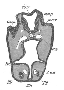
Fig. 299. Section through the head of an Elasmobranch embryo, at
the level of the auditory involution.
aup. auditory pit; aun. ganglion of auditory nerve; iv.v. roof
of fourth ventricle; a.c.v. anterior cardinal vein; aa. aorta;
I.aa. aortic trunk of mandibular arch; pp. head cavity of
mandibular arch; Ivc. alimentary pouch which will form the first
visceral cleft; Th. rudiment of thyroid body.
In all Vertebrata the development of the auditory vesicle commences
with the formation of a thickened patch of epiblast, at the side of
the hind-brain, on the level of the second visceral cleft. This patch
soon becomes invaginated in the form of a pit (fig. 299, aup), to
the inner side of which the ganglion of the auditory nerve (aun),
which as shewn in a previous chapter is primitively a branch of the
seventh nerve, closely applies itself.
In those Vertebrata (viz. Teleostei, Lepidosteus and Amphibia) in
which the epiblast is early divided into a nervous and epidermic
stratum, the auditory pit arises as an invagination of the nervous
stratum only, and the mouth of the auditory pit is always closed (fig.
300) by the epidermic stratum of the skin. Since the opening of the
pit is retained through life in Elasmobranchii the closed form of pit
in the above forms is clearly secondary.
In Teleostei the auditory pit arises as a solid invagination of the
epiblast.
The mouth of the auditory vesicle gradually narrows, and in most forms
soon becomes closed, though in Elasmobranchii it remains permanently
open. In any case the vesicle is gradually removed from the surface,
remaining connected with it by an elongated duct, either opening on
the dorsal aspect of the head (Elasmobranchii), or ending blindly
close beneath the skin.
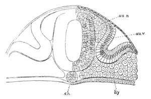
Fig. 300. Section through the head of a Lepidosteus embryo on
the sixth day after impregnation.
au.v. auditory vesicle; au.n. auditory nerve; ch. notochord;
hy. hypoblast.
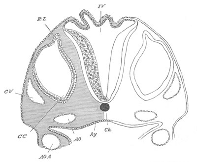
Fig. 301. Section through the hind-brain of a Chick at the end
of the third day of incubation.
IV. fourth ventricle. The section shews the very thin roof and
thicker sides of the ventricle. Ch. notochord; CV. anterior
cardinal vein; CC. involuted auditory vesicle (CC points to the
end which will form the cochlear canal); RL. recessus labyrinthi
(remains of passage connecting the vesicle with the exterior); hy.
hypoblast lining the alimentary canal; AO., AO.A. aorta, and
aortic arch.
In all Vertebrata the auditory vesicle undergoes further
changes of a
complicated kind. In the Cyclostomata these changes are less
complicated than in other forms, though whether this is due to
degeneration, or to the retention of a primitive state of the auditory
organ, is not known. In the Lamprey the auditory vesicle is formed in
the usual way by an invagination
of the epiblast, which soon becomes
vesicular, and for a considerable period retains a simple character.
As pointed out by Max Schultze, a number of otoliths appears in the
vesicle during larval life, and, although such otoliths are stated by
J. Müller to be absent both in the full-grown Ammocœte and in the
adult, they have since been found by Ketel (No. 387). The formation of
the two semicircular canals has not been investigated.
In all the higher Vertebrates the changes of the auditory sacks are
more complicated. The ventral end of the sack is produced into a short
process (fig. 301, CC); while at the dorsal end there is the
canal-like prolongation of the lumen of the sack (RL), derived from
the duct which primitively opened to the exterior, and which in most
cases persists as a blind diverticulum of the auditory sack, known as
the recessus labyrinthi or aqueductus vestibuli. The parts thus
indicated give rise to the whole of the membranous labyrinth of the
ear. The main body of the vesicle becomes the utriculus and
semicircular canals, while the ventral process forms the sacculus
hemisphericus and cochlear canal.
The growth of these parts has been most fully studied in Mammalia,
where they reach their greatest complexity, and it will be convenient
to describe their development in this group, pointing out how they
present, during some of the stages in their growth, a form permanently
retained in lower types.
The auditory vesicle in Mammalia is at first nearly spherical, and is
imbedded in the mesoblast at the side of the hind-brain. It soon
becomes triangular in section, with the apex of the triangle pointing
inwards and downwards. This apex gradually elongates to form the
rudiment of the cochlear canal and sacculus hemisphericus (fig. 302,
CC). At the same time the recessus labyrinthi (R.L) becomes
distinctly marked, and the outer wall of the main body of the vesicle
grows out into two protuberances, which form the rudiments of the
vertical semicircular canals (V.B). In the lower forms (fig. 305)
the cochlear process of the vestibule hardly reaches a higher stage of
development than that found at this stage in Mammalia.
The parts of the auditory labyrinth thus established soon increase in
distinctness (fig. 303); the cochlear canal (CC) becomes longer and
curved; its inner and concave surface being
lined by a thick layer of
columnar epiblast. The recessus labyrinthi also increases in length,
and just below the point where the bulgings to form the vertical
semicircular canals are situated, there is formed a fresh protuberance
for the horizontal semicircular canal. At the same time the central
parts of the walls of the flat bulgings of the vertical canals grow
together, obliterating this part of the lumen, but leaving a canal
round the periphery; and, on the absorption of their central parts,
each of the original simple bulgings of the wall of the vesicle
becomes converted into a true semicircular canal, opening at its two
extremities into the auditory vesicle. The vertical canals are first
established and then the horizontal canal.
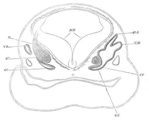
Fig. 302. Transverse section of the head of a fœtal Sheep (16 mm. in
length) in the region of the hind-brain. (After Böttcher.)
HB. the hind-brain.
The section is somewhat oblique, hence while on the right side the
connections of the recessus vestibuli R.L., and of the commencing
vertical semicircular canal V.B., and of the ductus cochlearis
CC., with the cavity of the primary otic vesicle are seen; on the
left side, only the extreme end of the ductus cochlearis CC, and
of the semicircular canal V.B. are shewn.
Lying close to the inner side of the otic vesicle is seen the
cochlear ganglion GC; on the left side the auditory nerve G and
its connection N with the hind-brain are also shewn.
Below the otic vesicle on either side lies the jugular vein.
Shortly after the formation of the rudiment of the horizontal
semicircular canal a slight protuberance becomes apparent on the inner
commencement of the cochlear canal. A constriction arises on each side
of the protuberance, converting it into a prominent hemispherical
projection, the sacculus hemisphericus (fig. 304, S.R).
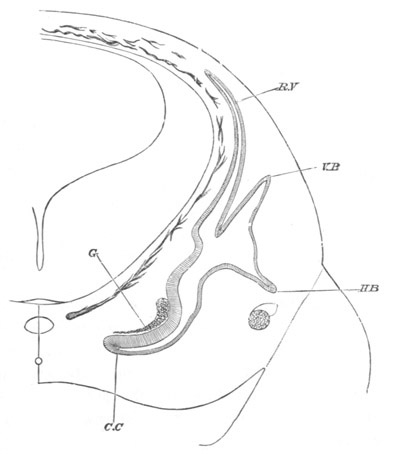
Fig. 303. Section of the head of a fœtal Sheep 20 mm. in length.
(After Böttcher.)
R.V. recessus labyrinthi; V.B. vertical semicircular canal;
H.B. horizontal semicircular canal; C.C. cochlear canal; G.
cochlear ganglion.
The constrictions are so deep that the sacculus is only connected with
the cochlear canal on the one hand, and with the general cavity of the
auditory vesicle on the other, by, in each case, a narrow though short
canal.
The former of these canals (fig. 304, b) is known as the canalis
reuniens. At this stage we may call the remaining cavity of the
original otic vesicle, into which all the above parts open, the
utriculus.
Soon after the formation of the sacculus hemisphericus, the
cochlear
canal and the semicircular canals become invested with cartilage. The
recessus labyrinthi remains however still enclosed in undifferentiated
mesoblast.
Between the cartilage and the parts which it surrounds there remains a
certain amount of indifferent connective tissue, which is more
abundant around the cochlear canal than around the semicircular
canals.
As soon as they have acquired a distinct connective-tissue coat, the
semicircular canals begin to be dilated at one of their terminations
to form the ampullæ. At about the same time a constriction appears
opposite the mouth of the recessus labyrinthi, which causes its
opening to be divided into two branches—one towards the utriculus and
the other towards the sacculus hemisphericus; and the relations of the
parts become so altered that communication between the sacculus and
utriculus can only take place through the mouth of the recessus
labyrinthi (fig. 305).
When the cochlear canal has come to consist of two and a half coils,
the thickened epithelium which lines the lower surface of the canal
forms a double ridge from which the organ of Corti is subsequently
developed. Above the ridge there appears a delicate cuticular
membrane, the membrane of Corti or membrana tectoria.
The epithelial walls of the utricle, the recessus labyrinthi, the
semicircular canals, and the cochlear canal constitute together the
highly complicated product of the original auditory vesicle. The whole
structure forms a closed cavity, the various parts of which are in
free communication. In the adult the fluid present in this cavity is
known as the endolymph.
In the mesoblast lying between these parts and the cartilage, which at
this period envelopes them, lymphatic spaces become established, which
are partially developed in the Sauropsida, but become in Mammals very
important structures.
They consist in Mammals partly of a space surrounding the utricle and
semicircular canals, and partly of two very definite channels, which
largely embrace between them the cochlear canal. The latter channels
form the scala vestibuli on the upper side of the cochlear canal and
the scala tympani on the lower. The scala vestibuli is in free
communication with the lymphatic cavity surrounding the vestibule, and
opens at the apex of the cochlea
into the scala tympani. The latter
ends blindly at the fenestra rotunda.
The fluid contained in the two scalæ, and in the remaining lymphatic
cavities of the auditory labyrinth, is known as perilymph.
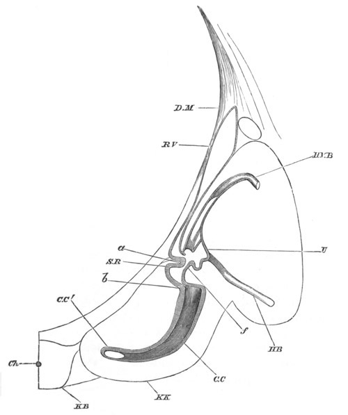
Fig. 304. Section through the internal ear of an embryonic Sheep
28 mm. in length. (After Böttcher.)
D.M. dura mater; R.V. recessus labyrinthi; H.V.B. posterior
vertical semicircular canal; U. utriculus; H.B. horizontal
semicircular canal; b. canalis reuniens; a. constriction by
means of which the sacculus hemisphericus S.R. is formed; f.
narrowed opening between sacculus hemisphericus and utriculus;
C.C. cochlea; C.C´. lumen of cochlea; K.K. cartilaginous
capsule of cochlea; K.B. basilar plate; Ch. notochord.
The cavities just spoken of are formed by an absorption of
parts of
the embryonic mucous tissue between the perichondrium and the walls of
the membranous labyrinth.
The scala vestibuli is formed before the scala tympani, and both scalæ
begin to be developed at the basal end of the cochlea: the cavity of
each is continually being carried forwards towards the apex of the
cochlear canal by a progressive absorption of the mesoblast. At first
both scalæ are somewhat narrow, but they soon increase in size and
distinctness.
The cochlear canal, which is often known as the scala media of the
cochlea, becomes compressed on the formation of the scalæ so as to be
triangular in section, with the base of the triangle outwards. This
base is only separated from the surrounding cartilage by a narrow
strip of firm mesoblast, which becomes the stria vascularis, etc. At
the angle opposite the base the canal is joined to the cartilage by a
narrow isthmus of firm material, which contains nerves and vessels.
This isthmus subsequently forms the lamina spiralis, separating the
scala vestibuli from the scala tympani.
The scala vestibuli lies on the upper border of the cochlear canal,
and is separated from it by a very thin layer of mesoblast, bordered
on the cochlear aspect by flat epiblast cells. This membrane is called
the membrane of Reissner. The scala tympani is separated from the
cochlear canal by a thicker sheet of mesoblast, called the basilar
membrane, which supports the organ of Corti and the epithelium
adjoining it. The upper extremity of the cochlear canal ends in a
blind extremity called the cupola, to which the two scalæ do not for
some time extend. This condition is permanent in Birds, where the
cupola is represented by a structure known as the lagena (fig. 305,
II. L). Subsequently the two scalæ join at the extremity of the
cochlear canal; the point of the cupola still however remains in
contact with the bone, which has now replaced the cartilage, but at a
still later period the scala vestibuli, growing further round,
separates the cupola from the adjoining osseous tissue.
The ossification around the internal ear is at first confined to the
cartilage, but afterwards extends into the thick periosteum between
the cartilage and the internal ear, and thus eventually makes its way
into the lamina spiralis, etc.
The organ of Corti. In Mammalia there is formed from the
epithelium of
the cochlear canal a very remarkable organ known as the organ of
Corti, the development of which is of sufficient importance to merit a
brief description. A short account of this organ in the adult state
may facilitate the understanding of its development.
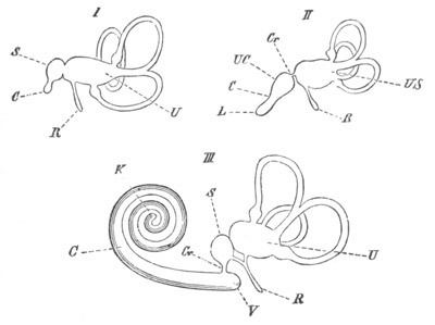
Fig. 305. Diagrams of the Membranous labyrinth. (From Gegenbaur.)
I. Fish. II. Bird. III. Mammal.
U. utriculus; S. sacculus; US. utriculus and sacculus; Cr.
canalis reuniens; R. recessus labyrinthi; UC. commencement of
cochlea; C. cochlear canal; L. lagena; K. cupola at apex of
cochlear canal; V. cæecal sack of the vestibulum of the cochlear
canal.
The cochlear canal is bounded by three walls, the outer one being the
osseous wall of the cochlea. The membrane of Reissner bounds it
towards the scala vestibuli, and the basilar membrane towards the
scala tympani. This membrane stretches from the margin of the lamina
spiralis to the ligamentum spirale; the latter being merely an
expanded portion of the connective tissue lining the osseous cochlea.
The lamina spiralis is produced into two lips, called respectively the
labium tympanicum and labium vestibulare; it is to the former and
longer of these that the basilar membrane is attached. At the margin
of the junction of the labium tympanicum with the basilar membrane the
former is perforated for the passage of the nervous fibres, and this
region is called the habenula perforata.
The labium vestibulare, so called from its position, is shorter than
the labium tympanicum and is raised above into numerous blunt teeth.
Partly springing out from the labium vestibulare, and passing from
near the inner attachment of the membrane of Reissner towards the
outer wall of the cochlea, is an elastic membrane, the membrana
tectoria. Resting on the basilar membrane is the organ of Corti.
Considering for the moment that a transverse section of the cochlear
canal only one cell deep is being dealt with, the organ of Corti will
be found to consist of a central part composed of two peculiarly
shaped rods widely separated below, but in contact above. These are
the rods or fibres of Corti. On their outer side, i.e. on the side
towards the osseous wall of the canal, is a reticulate membrane which
passes from the inner rod of Corti towards the osseous wall of the
canal. With their upper extremities fixed in that membrane, and their
lower resting on the basilar membrane are three (four in man) cells
with auditory hairs known as the outer ‘hair cells,’ which alternate
with three other cells known as Deiters’ cells. Between these and the
outer attachment of the basilar membrane is a series of cells
gradually diminishing in height in passing outwards. On the inner side
of the rods of Corti is one hair cell, and then a number of peculiarly
modified cells which fill up the space between the two lips of the
lamina spiralis.
It will not be necessary to say much in reference to the development
of the labium tympanicum and the labium vestibulare.
The labium vestibulare is formed by a growth of the connective tissue
which fuses with and passes up between the epithelial cells. The
epithelial cells which line its upper (vestibular) border become
modified, and remain as its teeth.
The labium tympanicum is formed by the coalescence of the connective
tissue layer separating the scala tympani from the cochlear canal with
part of the connective tissue of the lamina spiralis. At first these
two layers are separate, and the nerve fibres to the organ of Corti
pass between them. Subsequently however they coalesce, and the region
where they are penetrated by the nervous fibres becomes the habenula
perforata.
The organ of Corti itself is derived from the epiblast cells lining
the cochlear canal, and consists in the first instance of two
epithelial ridges or projections. The larger of them forms the cells
on the inner side of the organ of Corti, and the smaller the rods of
Corti together with the inner and outer hair cells and Deiters’ cells.
At first both these ridges are composed of simple elongated epithelial
cells one row deep. The smaller ridge is the first to shew any change.
The cells adjoining the larger ridge acquire auditory hairs at their
free extremities, and form the row of inner hair cells; the next row
of cells acquires a broad attachment to the basilar membrane, and
gives origin to the inner and outer rods of Corti.
Outside the latter come several rows of cells adhering together so as
to form a compact mass which is quadrilateral in section. This mass is
composed of three upper cells with nuclei at the same level, which
form the outer hair cells, each of them ending above in auditory
hairs, and three lower cells which form the cells of Deiters. Beyond
this the cells gradually pass into ordinary cubical epithelial cells.
As just mentioned, the cells of the second row, resting with their
broad bases on the basilar membrane, give rise to the rods of Corti.
The breadth of the bases of these cells rapidly increases, and
important changes take place in the structure of the cells themselves.
The nucleus of each cell divides; so that there come to be two nuclei
or sometimes three which lie close together near the base of the cell.
Outside the nuclei on each side a fibrous cuticular band appears. The
two bands pass from the base of the cell to its apex, and there meet
though widely separated below. The remaining contents of the cell,
between the two fibrous bands, become granular, and are soon to a
great extent absorbed; leaving at first a round, and then a triangular
space between the two fibres. The two nuclei, surrounded by a small
amount of granular matter, come to lie, each at one of the angles
between the fibrous bands and the basilar membrane.
The two fibrous bands become, by changes which need not be described
in detail, converted into the rods of Corti—each of their upper ends
growing outwards into the processes which the adult rods possess.
Each pair of rods of Corti is thus (Böttcher) to be considered as the
product of one cell; and the nuclei embedded in the granular mass
between them are merely the remains of the two nuclei formed by the
division of the original nucleus of that cell[196].
The larger ridge
is for the most part not permanent, and from being the most
conspicuous part of the organ of Corti comes to be far less important
than the smaller ridge. Its cells undergo a partial degeneration; so
that the epithelium in the hollow between the two lips of the lamina
spiralis, which is derived from the larger ridge, comes to be composed
of a single row of short and broad cells. In the immediate
neighbourhood however of the inner hair cell, one or two of the cells
derived from the larger ridge are very much elongated.
The membrana reticularis is a cuticular structure derived from the
parts to which it is attached.
Accessory structures connected with the organ of hearing in
Terrestrial Vertebrata.
In all the Amphibia, Sauropsida and Mammalia, except the Urodela and a
few Anura and Reptilia, the first visceral or hyomandibular cleft
enters into intimate relations with the organs of hearing, and from it
and the adjoining parts are formed the tympanic cavity, the Eustachian
tube, the tympanic membrane and the meatus auditorius externus. The
tympanic membrane serves to receive from the air the sound vibrations,
which are communicated to fluids contained in the true auditory
labyrinth by one ossicle or by a chain of auditory ossicles.
The addition to the organ of hearing of a tympanic membrane to receive
aerial sound vibrations is an interesting case of the
adaptation of a
structure, originally required for hearing in water, to serve for
hearing in air; and as already pointed out, the similarity of this
membrane to the tympanic membrane of some Insects is also striking.
There is much that is obscure with reference to the actual development
of the above parts of the ear, which has moreover only been carefully
studied in Birds and Mammals.
The Eustachian tube and tympanic cavity seem to be derived from the
inner part of the first visceral or hyomandibular cleft, the external
opening of which becomes soon obliterated. Kölliker holds that the
tympanic cavity is simply a dorsally and posteriorly directed
outgrowth of the median part of the inner section of this cleft; while
Moldenhauer (No. 392) holds, if I understand him rightly, that it is
formed as an outgrowth of a cavity called by him the sulcus
tubo-tympanicus, derived from the inner aperture of the first visceral
cleft together with the groove of the pharynx into which it opens; and
Moldenhauer is of opinion that the greater part of the original cleft
atrophies.
The meatus auditorius externus is formed at the region of a shallow
depression where the closure of the first visceral cleft takes place.
It is in part formed by the tissue surrounding this depression growing
up in the form of a wall, and Moldenhauer believes that this is the
whole process. Kölliker states however that the blind end of the
meatus becomes actually pushed in towards the tympanic cavity.
The tympanic membrane is derived from the tissue which separates the
meatus auditorius externus from the tympanic cavity. This tissue is
obviously constituted of an hypoblastic epithelium on its inner
aspect, an epiblastic epithelium on its outer aspect, and a layer of
mesoblast between them, and these three layers give rise to the three
layers of which this membrane is formed in the adult. During the
greater part of fœtal life it is relatively very thick, and presents
a structure bearing but little resemblance to that in the adult.
A proliferation of the connective tissue-cells in the vicinity of the
tympanic cavity causes in Mammalia the complete or nearly complete
obliteration of the cavity during fœtal life.
The tympanic cavity is bounded on its inner aspect by the osseous
investment of the internal ear, but at one point, known
as the
fenestra ovalis, the bone is deficient in the Amphibia, Sauropsida and
Mammalia, and its place is taken by a membrane; while in Mammalia and
Sauropsida a second opening, the fenestra rotunda, is also present.
These two fenestræ appear early, but whether they are formed by an
absorption of the cartilage, or by the nonchondrification of a small
area, is not certainly known. The upper of the two, or fenestra
ovalis, contains the base of a bone, known in the Sauropsida and
Amphibia as the columella. The main part of the columella is formed of
a stalk which is held by Parker to be derived from part of the
skeleton of the visceral arches, but its nature is discussed in
connection with the skeleton, while the base, forming the stapes,
appears to be derived from the wall of the periotic cartilage.
In all Amphibia and Sauropsida with a tympanic cavity, the stalk of
the columella extends to the tympanic membrane; its outer end becoming
imbedded in this membrane, and serving to transmit the vibrations of
the membrane to the fluid in the internal ear. In Mammalia there is a
stapes not directly attached to the tympanic membrane by a stalk, and
two additional auditory ossicles, derived from parts of the skeleton
of the visceral arches, are placed between the stapes and the tympanic
membrane. These ossicles are known as the malleus and incus, and the
chain of the three ossicles replaces physiologically the single
ossicle of the lower forms.
These ossicles are at first imbedded in the connective tissue in the
neighbourhood of the tympanic cavity, but on the full development of
this cavity, become apparently placed within it; though really
enveloped in the mucous membrane lining it.
The fenestra ovalis is in immediate contiguity with the walls of the
utricle, while the fenestra rotunda adjoins the scala tympani.
Hunt (No. 391) holds, from his investigations on the embryology of the
pig, that “the Eustachian tube is an involution of the pharyngeal
mucous membrane;” and that “the meatus is an involution of the
integument” while “the drum is formed by the Eustachian tube
overlapping the extremity of the meatus.” Urbantschitsch also holds
that the first visceral cleft has nothing to do with the formation of
the tympanic cavity and Eustachian tube, and that these parts are
derived from lateral outgrowths of the oral cavity.
The evolution of the accessory parts of the ear would be very
difficult to explain on Darwinian principles if the views of Hunt and
Urbantschitsch were correct; and the accepted doctrine, originally
proposed by Huschke (No. 389), according to which these structures
have originated by a ‘change of function’ of the parts of the first
visceral cleft, may fairly be held till more conclusive evidence has
been brought against it than has yet been done.
Fig. 306. Larva of Ascidia mentula. (From Gegenbaur; after
Kupffer.) Only the anterior part of the tail is represented.
N´. anterior swelling of neural tube; N. anterior swelling of
spinal portion of neural tube; n. hinder part of neural tube;
ch. notochord; K. branchial region of alimentary tract; d.
œsophageal and gastric region of alimentary tract; O. eye; a.
otolith; o. mouth; s. papilla for attachment.
Tunicata. The auditory organ of the Tunicata (fig. 306) is placed on
the under surface of the anterior vesicle of the brain. It consists of
two parts (1) a prominence of the cells of the floor of the brain
forming a crista acustica, and (2) an otolith projecting into the
cavity of the brain, and attached to the crista by delicate hairs.
The crista acustica is formed of very delicate cylindrical cells, and
in its most projecting part is placed a vesicle with clear contents.
The otolith is an oval body with its dorsal half pigmented, and its
ventral half clear and highly refractive. It is balanced on the
highest point of the crista.
The crista acustica would seem to be developed from the cells of the
lower part of the front vesicle of the brain. The otolith however is
developed from a single cell on the dorsal and right side of the
brain. This cell commences to project into the cavity of the brain and
its free end becomes pigmented. It gradually grows inwards till it
forms a spherical prominence in the cavity of the brain, to the wall
of which it is attached by a
stalk. At the same time it travels round
the right side of the vesicle of the brain (in a way not fully
explained) till it reaches the summit of the crista, which has become
in the meantime established.
The auditory organ of the simple Ascidians can hardly be brought into
relation with that of the other Chordata, and has most probably been
evolved within the Tunicate phylum.
Bibliography.
Invertebrata.
(384) V. Hensen. “Studien üb. d. Gehörorgan d. Decapoden.” Zeit. f.
wiss. Zool., Vol. XIII. 1863.
(385) O. and R. Hertwig. Das Nervensystem u. d. Sinnesorgane d.
Medusen. Leipzig, 1878.
Vertebrata.
(386) A. Boettcher. “Bau u. Entwicklung d. Schnecke.” Denkschriften
d. kaiserl. Leop. Carol. Akad. d. Wissenschaft., Vol. XXXV.
(387) C. Hasse. Die vergleich. Morphologie u. Histologie d. häutigen
Gehörorgane d. Wirbelthiere. Leipzig, 1873.
(388) V. Hensen. “Zur Morphologie d. Schnecke.” Zeit. f. wiss.
Zool., Vol. XIII. 1863.
(389) E. Huschke. “Ueb. d. erste Bildungsgeschichte d. Auges u. Ohres
beim bebrüteten Küchlein.” Isis von Oken, 1831, and Meckel’s
Archiv, Vol. VI.
(390) Reissner. De Auris internæ formatione. Inaug. Diss. Dorpat,
1851.
Accessory parts of Vertebrate Ear.
(391) David Hunt. “A comparative sketch of the development of the ear
and eye in the Pig.” Transactions of the International Otological
Congress, 1876.
(392) W. Moldenhauer. “Zur Entwick. d. mittleren u. äusseren Ohres.”
Morphol. Jahrbuch, Vol. III. 1877.
(393) V. Urbantschitsch. “Ueb. d. erste Anlage d. Mittelohres u. d.
Trommelfelles.” Mittheil. a. d. embryol. Instit. Wien, Heft I. 1877.
Olfactory organ.
Amongst the Invertebrata numerous sense organs have been described
under the title of olfactory organs. In aquatic animals they often
have the form of ciliated pits or grooves, while in the Insects and
Crustacea delicate hairs and other structures present on the antennæ
are usually believed to be organs of smell. Our knowledge of all these
organs is however so vague that it
would not be profitable to deal
with them more fully in this place. Amongst the Chordata there are
usually well developed olfactory organs.
Amongst the Urochorda (Tunicata) it is still uncertain what organs (if
any) deserve this appellation. The organ on the dorsal side of the
opening of the respiratory pharynx may very possibly have an olfactory
function, but it is certainly not homologous with the olfactory pits
of the true Vertebrata, and as mentioned above (pp. 436 and 437), may
perhaps be homologous with the pituitary body.
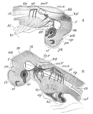
Fig. 307. Views of the head of Elasmobranch embryos at two stages
as transparent objects.
A. Pristiurus embryo of the same stage as fig. 28 F.
B. Somewhat older Scyllium embryo.
III. third nerve; V. fifth nerve; VII. seventh nerve; au.n.
auditory nerve; gl. glossopharyngeal nerve; Vg. vagus nerve; fb.
fore-brain; pn. pineal gland; mb. mid-brain; hb. hind-brain;
iv.v. fourth ventricle; cb. cerebellum; ol. olfactory pit; op.
eye; au.V. auditory vesicle; m. mesoblast at base of brain; ch.
notochord; ht. heart; Vc. visceral clefts; eg. external gills;
pp. sections of body cavity in the head.
In the Cephalochorda (Amphioxus) there is a shallow ciliated pit,
discovered by Kölliker, which is situated on the left side of the
head, and is closely connected with a special process of the
front end
of the brain. It is most probably the homologue of the olfactory pits
of the true Vertebrata.
In the true Vertebrata the olfactory organ has usually the form of a
pair of pits, though in the Cyclostomata the organ is unpaired.
In all the Vertebrata with two olfactory pits these organs are formed
from a pair of thickened patches of the epiblast, on the under side of
the fore-brain, immediately in front of the mouth (fig. 307, ol).
Each thickened patch of epiblast soon becomes involuted as a pit (fig.
308, N), the lining cells of which become the olfactory or
Schneiderian epithelium. The surface of this epithelium is usually
much increased by various foldings, which in the Elasmobranchii arise
very early, and are bilaterally symmetrical, diverging on each side
like the barbs of a feather from the median line. They subsequently
become very pronounced (fig. 309), serving greatly to increase the
surface of the olfactory epithelium. At a very early stage the
olfactory nerve attaches itself to the olfactory epithelium.
In Petromyzon the olfactory organ arises as an unpaired thickening
of the epiblast, which in the just hatched larva forms a shallow pit,
on the ventral side of the head, immediately in front of the mouth.
This pit rapidly deepens, and soon extends itself backwards nearly as
far as the infundibulum (fig. 310, ol). By the development of the
upper lip the opening of the olfactory pit is gradually carried to the
dorsal surface of the head, and becomes at the same time narrowed and
ciliated (fig. 47, ol). The whole organ forms an elongated sack, and
in later stages becomes nearly divided by a median fold into two
halves.
It is probable that the unpaired condition of the olfactory organ in
the Lamprey has arisen from the fusion of two pits into one; there is
however no evidence of this in the early development; but the division
of the sack into two halves by a median fold may be regarded as an
indication of such a paired character in the later stages.
In Myxine the olfactory organ communicates with the mouth through the
palate, but the meaning of this communication, which does not appear
to be of the same nature as the communication between the olfactory
pits and the mouth by the posterior nares in the higher types, is not
known.
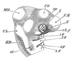
Fig. 308. Side view of the head of an embryo Chick of the third
day as an opaque object. (Chromic acid preparation.)
C.H. cerebral hemispheres; F.B. vesicle of third ventricle;
M.B. mid-brain; Cb. cerebellum; H.B. medulla oblongata; N.
nasal pit; ot. auditory vesicle in the stage of a pit with the
opening not yet closed up; op. optic vesicle, with l. lens and
ch.f. choroidal fissure.
1 F. The first visceral fold; above it is seen the superior
maxillary process.
2, 3, 4 F. Second, third and fourth visceral folds, with the
visceral clefts between them.
The opening of the olfactory pit does not retain its embryonic
characters. In Elasmobranchii and Chimæra it becomes enclosed by a
wall of integument, often deficient on the side of the mouth, so that
there is formed a groove leading from the nasal pit towards the angle
of the mouth. This groove is
usually constricted in the middle, and
the original single opening of the nasal sack thus becomes nearly
divided into two. In Teleostei and Ganoids the division of the nasal
opening into two parts becomes complete, but the ventral opening is
generally carried off some distance from the mouth, and placed, by the
growth of the snout, on the upper surface of the head (figs. 54 and
68). In all these instances it is probable that the dorsal opening of
the external nares, and the ventral opening with the posterior nares
of higher types. Thus the posterior nares would in fact seem to be
represented in all Fishes by a ventral part of the opening of the
original nasal pit which either adjoins the border of the mouth (many
Elasmobranchii) or is quite separate from the mouth (Teleostei and
Ganoidei). In the Dipnoi, Amphibia and all the higher types the oral
region becomes extended so as to enclose the posterior nares, and then
each nasal pit acquires two openings; viz. one outside the mouth, the
external nares, and one within the mouth, the internal or posterior
nares. In the Dipnoi the two nasal openings are very similar to those
in Ganoidei and Teleostei, but both are placed on the under surface of
the head, the inner one being within the mouth, and the external one
is so close to the outer border of the upper lip that it also has been
considered by some anatomists to lie within the mouth.
In all the higher types the nasal pits have originally only a single
opening, and the ontogenetic process by which the posterior nasal
opening is formed has been studied in the Amniota and Amphibia.
Amongst the Amniota we may take the Chick as representing the process
in a very simple form. The general history of the process was first
made out by Kölliker.
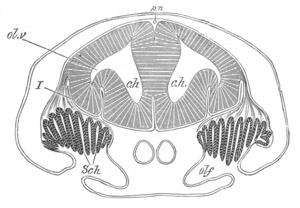
Fig. 309. Section through the brain and olfactory organ of an
embryo of Scyllium. (Modified from figures by Marshall and myself.)
c.h. cerebral hemispheres; ol.v. olfactory vesicle; olf.
olfactory pit; Sch. Schneiderian folds; I. olfactory nerve. The
reference line has been accidentally taken through the nerve to the
brain.
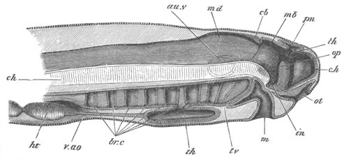
Fig. 310. Diagrammatic vertical section through the head of a
larva of Petromyzon.
The larva had been hatched three days, and was 4.8 mm. in length.
The optic and auditory vesicles are supposed to be seen through the
tissues.
c.h. cerebral hemisphere; th. optic thalamus; in.
infundibulum; pn. pineal gland; mb. mid-brain; cb. cerebellum;
md. medulla oblongata; au.v. auditory vesicle; op. optic
vesicle; ol. olfactory pit; m. mouth; br.c. branchial pouches;
th. thyroid involution; v.ao. ventral aorta; ht. ventricle of
heart; ch. notochord.
The opening of the nasal pit becomes surrounded by a ridge except on
its oral side. The deficiency of this ridge on the side of the mouth
gives rise to a kind of shallow groove leading from the nasal pit to
the mouth. The ridge enveloping the opening of the nasal pit next
becomes prolonged along the sides of this groove, especially on its
inner one; and at the same time the superior maxillary process grows
forwards so as to bound the lower
part of its outer side. The inner
and outer ridges, together with the superior maxillary process,
enclose a deep groove, connecting the original opening of the nasal
pit with the mouth. The process just described is illustrated by fig.
311 A, and it may be seen that the ridge on the inner side of the
groove forms the edge of the frontonasal process (k).
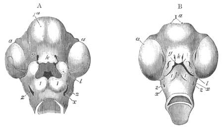
Fig. 311. Head of a Chick from below on the sixth and seventh days
of incubation. (From Huxley.)
Ia. cerebral vesicles; a. eye, in which the remains of the
choroid slit can still be seen in A; g. nasal pits; k.
frontonasal process; l. superior maxillary process; 1. inferior
maxillary process or first visceral arch; 2. second visceral arch;
x. first visceral cleft.
In A the cavity of the mouth is seen enclosed by the frontonasal
process, the superior maxillary processes and the first pair of
visceral arches. At the back of it is seen the opening leading into
the throat. The nasal grooves leading from the nasal pits to the
mouth are already closed over.
In B the external opening of the mouth has become much constricted,
but it is still enclosed by the frontonasal process and superior
maxillary processes above, and by the inferior maxillary processes
(first pair of visceral arches) below.
The superior maxillary processes have united with the frontonasal
process, along nearly the whole length of the latter.
On the sixth day (Born, 394) the sides of this groove unite together
in the middle, and convert it into a canal open at both ends—the
ventral openings of the canals of the two sides being placed just
within the border of the mouth, and forming the posterior nares; while
the external openings form the anterior nares. The upper part of the
canal, together with the original nasal pit, is alone lined by
olfactory epithelium; the remaining epithelium of the nasal cavity
being indifferent epiblastic epithelium.
Further changes subsequently
take place in connection with the posterior nares, but these are
described in the section dealing with the mouth.
In Mammalia the general formation of the anterior and posterior nares
is the same as in Birds; but, as shewn by Dursy and Kölliker, an
outgrowth from the inner side of the canal between the two openings
arises at an early period; and becoming separate from the posterior
nares and provided with a special opening into the mouth, forms the
organ of Jacobson. The general relations of this organ when fully
formed are shewn in fig. 312.
In Lacertilia the formation of the posterior nares differs in some
particulars from that in Birds (Born). A groove is formed leading from
the primitive nasal pit to the mouth, bordered on its inner side by
the swollen edge of the frontonasal process, and on its outer by an
outer-nasal process; while the superior maxillary process does not
assist in bounding it. On the inner side of the narrowest part of this
groove there is formed a large lateral diverticulum, which is lined by
a continuation of the Schneiderian epithelium, and forms the rudiment
of Jacobson’s organ. The nasal groove continues to grow in length, but
soon becomes converted into a canal by the junction of the outer-nasal
process with the frontonasal process. This canal is open at both ends:
at its dorsal end is placed the original opening of the nasal pit, and
its ventral opening is situated within the cavity of the mouth. The
latter forms the primitive posterior nares. The superior maxillary
process soon grows inwards on the under side of the posterior part of
the nasal passage, and assists in forming its under wall. This
ingrowth of the superior maxillary process is the rudiment of the hard
palate.
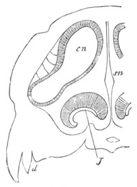
Fig. 312. Section through the nasal cavity and Jacobson’s
organ. (From Gegenbaur.)
sn. septum nasi; cn. nasal cavity; J. Jacobson’s organ; d.
edge of upper jaw.
On the conversion of the nasal groove into a closed passage, the
opening of Jacobson’s organ into the groove becomes concealed; and at
a later period Jacobson’s organ becomes completely shut off from the
nasal cavity, and opens into the mouth at the front end of an
elongated groove leading back to the posterior nares.
In Amphibia the posterior nares are formed in a manner very different
from that of the Amniota. At an early stage a shallow groove is formed
leading from the nasal pit to the mouth; but this groove instead
of
forming the posterior nares soon vanishes, and by the growth of the
front of the head the nasal pits are carried farther away from the
mouth.
The actual posterior nares are formed by a perforation in the palate,
opening into the blind end of the original nasal pit.
Considering that the various stages in the formation of the posterior
nares of the Amniota are so many repetitions of the adult states of
lower forms, it may probably be assumed that the mode of formation of
the posterior nares in Amphibia is secondary, as compared with that in
the Amniota.
A diverticulum of the front part of the nasal cavity of the Anura is
probably to be regarded as a rudimentary form of Jacobson’s organ.
Bibliography.
(394) G. Born. “Die Nasenhöhlen u. d. Thränennasengang d. amnioten
Wirbelthiere.” Parts I. and II. Morphiologisches Jahrbuch, Bd. V.,
1879.
(395) A. Kölliker. “Ueber die Jacobson’schen Organe des Menschen.”
Festschrift f. Rienecker, 1877.
(396) A. M. Marshall. “Morphology of the Vertebrate Olfactory Organ.”
Quart. Journ. of Micr. Science, Vol. XIX., 1879.
Sense organs of the lateral line.
Although I do not propose dealing with the general development of
various sense organs of the skin, there is one set of organs, viz.
that of the lateral line, which, both from its wide extension amongst
the Ichthyopsida and from the similarity of some of its parts to
certain organs found amongst the Chætopoda[197],
has a great
morphological importance.
The organs of the lateral line consist as a rule of canals, partly
situated in the head, and partly in the trunk. These canals open at
intervals on the surface, and their walls contain a series of
nerve-endings. The branches of the canal in the head are innervated
for the most part by the fifth pair, and those of the trunk by the
nervus lateralis of the vagus nerve. There is typically but a single
canal in the trunk, the openings and nerve-endings of which are
segmentally arranged.
Two types of development of these organs have been found. One of these
is characteristic of Teleostei; the other of Elasmobranchii.
In just hatched Teleostei, Schulze (No. 402) found that instead of the
normal canals there was present a series of sense bulbs, projecting
freely on the surface and partly composed of cells with stiff hairs.
In most
cases each bulb is enclosed in a delicate tube open at its
free extremity; while the bulbs correspond in number with the
myotomes. In some Teleostei (Gobius, Esox, etc.) such sense organs
persist through life; in most forms however each organ becomes covered
by a pair of lobes of the adjacent tissue, one formed above and the
other below it. The two lobes of each pair then unite and form a tube
open at both ends. The linear series of tubes so formed is the
commencement of the adult canal; while the primitive sense bulbs form
the sensory organs of the tubes. The adjacent tubes partially unite
into a continuous canal, but at their points of apposition pores are
left, which place the canal in communication with the exterior.
Besides these parts, I have found that there is present in the just
hatched Salmon a linear streak of modified epidermis on the level of
the lateral nerve, and from the analogy of the process described below
for Elasmobranchii it appears to me probable that these streaks play
some part in the formation of the canal of the lateral line.
In Elasmobranchii (Scyllium) the lateral line is formed as a linear
thickening of the mucous layer of the epidermis. This thickening is at
first very short, but gradually grows backwards, its hinder end
forming a kind of enlarged growing point. The lateral nerve is formed
shortly after the lateral line, and by the time that the lateral line
has reached the level of the anus the lateral nerve has grown back for
about two-thirds of that distance. The lateral nerve would seem to be
formed as a branch of the vagus, but is at first half enclosed in the
modified cells of the lateral line (fig. 275, nl)[198],
though it
soon assumes a deeper position.
A permanent stage, more or less corresponding to the stage just
described in Elasmobranchii, is retained in Chimæra, and Echinorhinus
spinosus, where the lateral line has the form of an open groove
(Solger, No. 404).
The epidermic thickening, which forms the lateral line, is converted
into a canal, not as in Teleostei by the folding over of the sides,
but by the formation of a cavity between the mucous and epidermic
layers of the epiblast, and the subsequent enclosure of this cavity by
the modified cells of the mucous layer of the epiblast which
constitute the lateral line. The cavity first appears at the hind end
of the organ, and thence extends forwards.
After its conversion into a canal the lateral line gradually recedes
from the surface; remaining however connected with the epidermis at a
series of points corresponding with the segments, and at these points
perforations are eventually formed to constitute the segmental
apertures of the system.
The manner in which the lumen of the canal is formed in Elasmobranchs
bears the same relation to the ordinary process of conversion of a
groove into a canal that the formation of the auditory involution
in
Amphibia does to the same process in Birds. In both Elasmobranchii and
Amphibia the mucous layer of the epiblast behaves exactly as does the
whole epiblast in the other types, but is shut off from the surface by
the passive epidermic layer of the epiblast.
The mucous canals of the head and the ampullæ are formed from the
mucous layer of the epidermis in a manner very similar to the lateral
line; but the nerves to them arise as simple branches of the fifth and
seventh nerves, which unite with them at a series of points, but do
not follow their course like the lateral nerve.
It is clear that the canal of the lateral line is secondary, as
compared with the open groove of Chimæra or the segmentally arranged
sense bulbs of young Teleostei; and it is also clear that the
phylogenetic mode of formation of the canal consisted in the closure
of a primitively open groove. The abbreviation of this process in
Elasmobranchii was probably acquired after the appearance of food-yolk
in the egg, and the consequent disappearance of a free larval stage.
While the above points are fairly obvious it does not seem easy to
decide à priori whether a continuous sense groove or isolated sense
bulbs were the primitive structures from which the canals of the
lateral line took their origin. It is equally easy to picture the
evolution of the canal of the lateral line either from (1) a
continuous unsegmented sense line, certain points of which became
segmentally differentiated into special sense bulbs, while the whole
subsequently formed a groove and then a canal; or from (2) a series of
isolated sense bulbs, for each of which a protective groove was
developed; and from the linear fusion of which a continuous canal
became formed.
From the presence however of a linear streak of modified epidermis in
larval Teleostei, as well as in Elasmobranchii, it appears to me more
probable that a linear sense streak was the primitive structure from
which all the modifications of the lateral line took their origin, and
that the segmentally arranged sense bulbs of Teleostei are secondary
differentiations of this primitive structure.
The, at first sight remarkable, distribution of the vagus nerve to the
lateral line is probably to be explained in connection with the
evolution of this organ. As is indicated both by its innervation from
the vagus, as also from the region where it first becomes developed,
the lateral line was probably originally restricted to the anterior
part of the body. As it became prolonged backwards it naturally
carried with it the vagus nerve, and thus a sensory branch of this
nerve has come to innervate a region which is far beyond the limits of
its original distribution.
Bibliography.
(397) F. M. Balfour. A Monograph on the development of Elasmobranch
Fishes, pp. 141-146. London, 1878.
(398) H. Eisig. “Die Segmentalorgane d. Capitelliden.” Mittheil. a.
d. zool. Station zu Neapel, Vol. I. 1879.
(399) A. Götte. Entwicklungsgeschichte d. Unke. Leipzig, 1875.
(400) Fr. Leydig. Lehrbuch d. Histologie des Menschen u. d. Thiere.
Hamm. 1857.
(401) Fr. Leydig. Neue Beiträge z. anat. Kenntniss d. Hautdecke u.
Hautsinnesorgane d. Fische. Halle, 1879.
(402) F. E. Schulze. “Ueb. d. Sinnesorgane d. Seitenlinie bei Fischen
und Amphibien.” Archiv f. mikr. Anat., Vol. VI. 1870.
(403) C. Semper. “Das Urogenitalsystem d. Selachier.” Arbeit. a. d.
zool.-zoot. Instit. Würzburg, Vol. II.
(404) B. Solger. “Neue Untersuchungen zur Anat. d. Seitenorgane d.
Fische.” Archiv f. mikr. Anat., Vol. XVII. and XVIII. 1879 and 1880.
Introduction.
Amongst the products of that part of the mesoblast which constitutes
the connective tissue of the body special prominence must be given to
the skeleton of the Vertebrata, from its importance in relation to
numerous phylogenetic and morphological problems.
The development of the skeleton is however so large a subject that it
cannot be satisfactorily dealt with except in a special treatise
devoted to it; and the following description must be regarded as a
mere sketch, from which detail has been as far as possible excluded.
In the lowest Chordata the sole structure present, which deserves to
be called a skeleton, is the notochord. Although the notochord often
persists as an important organ in the true Vertebrata, yet there are
always added to it various skeletal structures developed in the
mesoblast. Before entering into a systematic description of these, it
will be convenient to say a few words as to the general characters of
the skeleton.
Two elements, distinct both in their genesis and structure, are to be
recognized in the skeleton. The one, forming the true primitive
internal skeleton or endoskeleton, is imbedded within the muscles and
is originally formed in cartilage. In many instances it retains a
cartilaginous consistency through life, but in the majority of cases
it becomes gradually ossified, and
converted into true bone. Bones so
formed are known as cartilage bones.
The other element is originally formed by the fusion of the ossified
bases of the dermal placoid scales already described in Chapter XIV.,
or by the fusion of the ossified bases of teeth situated in the mucous
membrane of the mouth. In both instances the plates of bone so formed
may lose the teeth or spines with which they were in the first
instance covered, either by absorption in the individual, or
phylogenetically by their gradually ceasing to be developed. The
plates of bone, which originated by the above process, become in
higher types directly developed in the connective tissue beneath the
skin; and gradually acquire a deeper situation, and are finally so
intimately interlocked with parts of the true internal skeleton, that
the two sets of elements can only be distinguished by the fact of the
one set ossifying in cartilage and the other in membrane.
It seems probable that in the Reptilia, and possibly the extinct
Amphibia, dermal bones have originated in the skin without the
intervention of superjacent spinous structures.
In cases where a membrane bone, as the dermal ossifications are
usually called, overlies a part of the cartilage, it may set up
ossification in the latter, and the cartilage bone and membrane bone
may become so intimately fused as to be quite inseparable. It seems
probable that in cases of this kind the compound bone may in the
course of further evolution entirely lose either its cartilaginous
element or its membranous element; so that cases occasionally occur
where the development of a bone ceases to be an absolutely safe guide
to its evolution.
As to the processes which take place in the ossification of cartilage
there is still much to be made out. Two processes are often
distinguished, viz. (1) a process known as ectostosis, in which the
ossification takes place in the perichondrium, and either simply
surrounds or gradually replaces the cartilage, and (2) a process known
as endostosis, where the ossification actually takes place between the
cartilage cells. It seems probable however (Gegenbaur, Vrolik) that
there is no sharp line to be drawn between these two processes; but
that the ossification almost always starts from the perichondrium. In
the higher types, as a rule, the vessels of the perichondrium extend
into
the cartilage, and the ossification takes place around these
vessels within the cartilage; but in the lower types (Pisces,
Amphibia) ossification is often entirely confined to the
perichondrium; and the cartilage is simply absorbed.
The regions where ossification first sets in are known as centres of
ossification; and from these centres the ossification spreads
outwards. There may be one or more centres for a bone.
The actual causes which in the first instance gave rise to particular
centres of ossification, or to the ossification of particular parts of
the cartilage, are but little understood; nor have we as yet any
satisfactory criterion for determining the value to be attached to the
number and position of centres of ossification. In some instances such
centres appear to have an important morphological significance, and in
other instances they would seem to be determined by the size of the
cartilage about to be ossified.
There is no doubt that the membrane bones and cartilage bones can as a
rule be easily distinguished by their mode of development; but it is
by no means certain that this is always the case. It is necessarily
very difficult to establish the homology between bones, which develop
in one type from membrane and in another type from cartilage; but
there are without doubt certain instances in which the homology
between two bones would be unhesitatingly admitted were it not for the
difference in their development. The most difficult cases of this kind
are connected with the shoulder-girdle.
The possible sources of confusion in the development of bones are
obviously two. (1) A cartilage bone by origin may directly ossify in
membrane, without the previous development of cartilage, and (2) a
membrane bone may in the first instance be formed in cartilage.
The occurrence of the first of these is much more easy to admit than
that of the second; and there can be little doubt that it sometimes
takes place. In a large number of cases it would moreover cause no
serious difficulty to the morphologist.
Bibliography of the origin of the Skeleton.
(405) C. Gegenbaur. “Ueb. primäre u. secundäre Knochenbildung mit
besonderer Beziehung auf d. Lehre von dem Primordialcranium.”
Jenaische Zeitschrift, Vol. III. 1867.
(406) O. Hertwig. “Ueber Bau u. Entwicklung d. Placoidschuppen u. d.
Zähne d. Selachier.” Jenaische Zeitschrift, Vol. VIII. 1874.
(407) O. Hertwig. “Ueb. d. Zahnsystem d. Amphibien u. seine Bedeutung
f. d. Genese d. Skelets d. Mundhöhle.” Archiv f. mikr. Anat., Vol.
XI. Supplementheft, 1874.
(408) O. Hertwig. “Ueber d. Hautskelet d. Fische.” Morphol.
Jahrbuch, Vol. II. 1876. (Siluroiden u. Acipenseriden.)
(409) O. Hertwig. “Ueber d. Hautskelet d. Fische (Lepidosteus u.
Polypterus).” Morph. Jahrbuch, Vol. V. 1879.
(410) A. Kölliker. “Allgemeine Betrachtungen üb. die Entstehung d.
knöchernen Schädels d. Wirbelthiere.” Berichte v. d. königl. zoot.
Anstalt z. Würzburg, 1849.
(411) Fr. Leydig. “Histologische Bemerkungen üb. d. Polypterus
bichir.” Zeit. f. wiss. Zool., Vol. V. 1858.
(412) H. Müller. “Ueber d. Entwick. d. Knochensubstanz nebst
Bemerkungen, etc.” Zeit. f. wiss. Zool., Vol. IX. 1859.
(413) Williamson. “On the structure and development of the Scales and
Bones of Fishes.” Phil. Trans., 1851.
(414) Vrolik. “Studien üb. d. Verknöcherung u. die Knochen d. Schädels
d. Teleostier.” Niederländisches Archiv f. Zoologie, Vol. I.
Notochord and Vertebral column.
The primitive axial skeleton of the Chordata consists of the notochord
and its sheath. It persists as such in the adult in Amphioxus, and
constitutes, in embryos of all Vertebrata, for a considerable period
of their early embryonic life, the sole representative of the axial
skeleton.
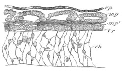
Fig. 313. Horizontal section through the trunk of an embryo of
Scyllium considerably younger than F in fig. 28.
The section is taken at the level of the notochord, and shews the
separation of the cells to form the vertebral bodies from the
muscle-plates.
ch. notochord; ep. epiblast; Vr. rudiment of vertebral body;
mp. muscle-plate; mp´. portion of muscle-plate already
differentiated into longitudinal muscles.
The Notochord. The early formation of the notochord has already been
described in detail (pp. 292-300). It is developed, in most if not all
cases, as an axial differentiation of the hypoblast, and forms at
first a solid cord of cells, without a sheath, placed between the
nervous system and the dorsal wall of the alimentary tract, and
extending from the base of the front of the mid-brain to the end of
the tail. The section in the region of the brain will be dealt with by
itself. That
in the trunk forms the basis round which the vertebral
column is moulded.
The early histological changes in the cells of the notochord are
approximately the same in all the Craniata. There is formed by the
superficial cells of the notochord a delicate sheath, which soon
thickens, and becomes a well-defined structure. Vacuoles (one or more
to each cell) are formed in the cells of the notochord, which enlarge
till the whole notochord becomes almost entirely formed of large
vacuoles separated by membranous septa which form a complete
sponge-like reticulum (fig. 313). In the Ichthyopsida most of the
protoplasm with the nuclei is carried to the periphery, where it forms
a special nucleated layer sometimes divided into definite
epithelial-like cells (fig. 314), while in the meshes of the reticulum
a few nuclei surrounded by a little protoplasm still remain. In the
Amniotic Vertebrata, probably owing to the early atrophy of the
notochord, the distribution of the nuclei in the spaces of the
mesh-work remains fairly uniform.
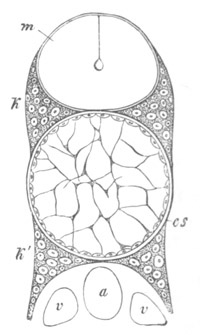
Fig. 314. Section through the spinal column of a young
Salmon. (From Gegenbaur.)
cs. sheath of notochord; k. neural arch; k´. hæmal arch; m.
spinal cord; a. dorsal aorta; v. cardinal veins.
In the early stages of development the spaces in the notochordal
sponge-work, each containing a nucleus and protoplasm, probably
represent cells. In the types in which the notochord persists in the
adult the mesh-work becomes highly complicated, and then forms a
peculiar reticulum filled with gelatinous material, the spaces in
which do not indicate the outlines of definite cells (figs. 315 and
318).
Around the sheath of the notochord there is formed in the
Cyclostomata, Ganoidei, Elasmobranchii and Teleostei an elastic
membrane usually known as the membrana elastica externa.
In most Vertebrates the notochord and its sheath either atrophy
completely or become a relatively unimportant part of the axial
skeleton; but in the Cyclostomata (fig. 315) and in the Selachioidean
Ganoids (Acipenser, etc.) they persist as the sole representative of
the true vertebral axis. The sheath becomes very much thickened; and
on the membrana elastica covering
it the vertebral arches directly
rest. In Elasmobranchii the sheath of the notochord undergoes a more
complicated series of changes, which result first of all in the
formation of a definite unsegmented cartilaginous tube[199]
round
the notochord, and subsequently (in most forms) in the formation of
true vertebral bodies.
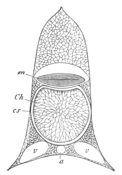
Fig. 315. Section through the vertebral column of
Ammocœtes. (From Gegenbaur.)
Ch. notochord; cs. notochordal sheath; m. spinal cord; a.
aorta; v. cardinal veins.
Between the membrana elastica externa and the sheath of the notochord
a layer of cells becomes interposed (fig. 316, n), which lie in a
matrix not sharply separated from the sheath of the notochord. The
cells which form this layer appear to be derived from a special
investment of the notochord, and to have penetrated through the
membrana elastica externa to reach their final situation. The layer
with these cells soon increases in thickness, and forms a continuous
unsegmented tube of fibrous tissue with flattened concentrically
arranged nuclei (fig. 317, Vb). Externally is placed the membrana
elastica externa (mel), while within is the cuticular sheath of the
notochord. This tube is the cartilaginous tube spoken of above and is
known as the cartilaginous sheath of the notochord.
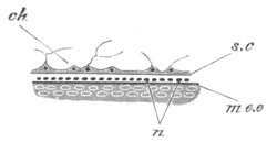
Fig. 316. Longitudinal section through a small part of the
notochord and adjoining parts of a Scyllium embryo, at the time of the
first formation of the cartilaginous sheath.
ch. notochord; sc. sheath of notochord; n. nuclei of
cartilaginous sheath; me.e. membrana elastica externa.
The exact origin of the cartilaginous tube just described is a
question of fundamental importance with reference to the origin of the
vertebral column and the homologies of its constituent parts; but is
by no means easy to settle. In the account of the subject in my memoir
on Elasmobranch Fishes I held with Gegenbaur that it arose from
a
layer of cells outside the sheath of the notochord, on the exterior
of which the membrana elastica externa was subsequently formed. To
this view Götte (No. 419) also gave his adhesion. Schneider has since
(No. 429) stated that this is not the case, but that, as described
above, the membrana elastica externa is formed before the layer of
cartilage. I have since worked over this subject again, and am on the
whole inclined to adopt Schneider’s correction.
It follows from the above description that the cartilaginous tube in
question is an essential part of the sheath of the notochord, and that
it is to some extent homologous with the notochordal sheath of the
Sturgeon and the Lamprey, and not an entirely new formation.
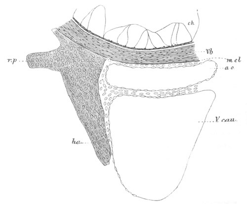
Fig. 317. Transverse section through the ventral part of the
notochord and adjoining structures of an advanced Scyllium embryo
at the root of the tail.
Vb. cartilaginous sheath of the notochord; ha. hæmal arch; vp.
process to which the rib is articulated; mel. membrana elastica
externa; ch. notochord; ao. aorta; V.cau. caudal vein.
This sheath forms the basis of the centra of the future vertebræ. In a
few adult forms, i.e. Chimæra and the Dipnoi, it retains its
primitive condition, except that in Chimæra there are present delicate
ossified rings more numerous than the arches; while in the Notidani,
Læmargi and Echinorhini the
indications of vertebræ are imperfectly
marked out. The further history of this sheath in the forms in which
true vertebræ are formed can only be dealt with in connection with the
formation of the vertebral arches.
In Teleostei there is present, as in Elasmobranchii, an elastica
externa, and an inner notochordal sheath. The elastica externa
contains, according to Götte, cells. These cells, if present, are
however very difficult to make out, but in any case the so-called
elastica externa appears to correspond with the cartilaginous sheath
of Elasmobranchii together with its enveloping elastica, since
ossification, when it sets in, occurs in this layer. The sheath within
becomes unusually thick.
In the Amphibia and in the Amniota no membrane is present which can be
identified with the membrana elastica externa of the Elasmobranchii,
Teleostei, etc. In Amphibia (Götte) there is formed round the
notochord a cellular sheath, which has very much the relations of the
cartilaginous tube around the notochord of Elasmobranchii, and is
developed in the same way from the perichordal connective tissue
cells. It is only necessary to suppose that the membrana elastica
externa has ceased to be developed (which in view of its extreme
delicacy and unimportant function in Elasmobranchii is not difficult
to do) and this cellular sheath would then obviously be homologous
with the cartilaginous tube in question. In the Amniota an external
sheath of the notochord cannot be traced as a distinct structure, but
the connective tissue surrounding the notochord and spinal cord is
simply differentiated into the vertebral bodies and vertebral arches.
Vertebral arches and Vertebral bodies.
Cyclostomata. The Cyclostomata are the most primitive forms in which
true vertebral arches are present. Their ontogeny in this group has
not been satisfactorily worked out. It is however noticeable in
connection with them that they form for the most part isolated pieces
of cartilage, the segmental arrangement of which is only imperfect.
Elasmobranchii. In the Elasmobranchii the cells forming the vertebral
arches are derived from the splanchnic layer of the mesoblastic
somites. They have at first the same segmentation
as the somites (fig.
313, Vr), but this segmentation is soon lost, and there is formed
round the notochord a continuous sheath of embryonic connective tissue
cells, which gives rise to the arches of the vertebræ, the tissue
forming the dura mater, the perichondrium, and the general investing
connective tissue.
The changes which next follow result in what has been known since
Remak as the secondary segmentation of the vertebral column. This
segmentation, which occurs in all Vertebrata with true vertebræ, is
essentially the segmentation of the continuous investment of the
notochord and spinal cord into vertebral bodies and vertebral arches.
It does not however follow the lines of the segmentation of the
muscle-plates, but is so effected that the centres of the vertebral
bodies are opposite the septa between the muscle-plates.
The explanation of this character in the segmentation is not difficult
to find. The primary segmentation of the body is that of the
muscle-plates, which were present in the primitive forms in which
vertebræ had not appeared. As soon however as the notochordal sheath
was required to be strong as well as flexible, it necessarily became
divided into a series of segments.
The condition under which the lateral muscles can best cause the
flexure of the vertebral column is clearly that each myotome shall be
capable of acting on two vertebræ; and this condition can only be
fulfilled when the myotomes are opposite the intervals between the
vertebræ. For this reason, when the vertebræ became formed, their
centres were opposite not the middle of the myotomes but the
intermuscular septa.
These considerations fully explain the characters of the secondary
segmentation of the vertebral column. On the other hand the primary
segmentation (fig. 313) of the vertebral rudiments is clearly a
remnant of a condition when no vertebral bodies were present; and has
no greater morphological significance than the fact that the cells of
the vertebræ were derived from the segmented muscle-plates, and then
became fused into a continuous sheath around the notochord and nervous
axis; till finally they became in still higher forms differentiated
into vertebræ and their arches.
During the stage represented in fig. 28 g, and somewhat before the
cartilaginous sheath of the notochord is formed, there appear four
special concentrations of the mesoblastic tissue adjoining the
notochord, two of them dorsal (neural) and two of them ventral
(hæmal). They are not segmented, and form four ridges, seated on the
sides of the notochord. They are united
with each other by a delicate
layer of tissue, and constitute the substance in which the neural and
hæmal arches subsequently become differentiated.
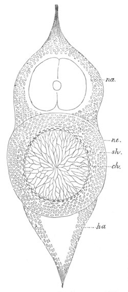
Fig. 318. Section through the vertebral column of an advanced
embryo of Scyllium in the region of the tail.
na. neural arch; ha. hæmal arch; ch. notochord; sh. inner
sheath of notochord; ne. membrana elastic externa.
At about the time when the first traces of the cartilaginous sheath of
the notochord arise, differentiations take place in the neural and
hæmal ridges. In the neural ridge two sets of arches are formed for
each myotome, and resting on the cartilaginous sheath of the notochord
in the region which will afterwards form the centrum of a vertebra,
and constituting a true neural arch; and a second separate from the
cartilaginous sheath, forming an intercalated piece[200].
Both of them
soon become hyaline cartilage.
There is a considerable portion of the original tissue of the neural
ridge, especially in the immediate neighbourhood of the notochord,
which is not employed in the formation of the neural arches. This
tissue has a fibrous character and becomes converted into the
perichondrium and other parts.
The hæmal arches are formed from the hæmal ridge in precisely the same
way as the neural arches, but interhæmal intercalated pieces are often
present. In the region of the tail the hæmal arches are continued into
ventral processes which meet below, enclosing the aorta and caudal
veins.
Since primitively the postanal gut was placed between the aorta and
the caudal vein, the hæmal arches potentially invest a caudal section
of the body cavity. In the trunk region they do not meet ventrally,
but give support to the ribs. The structures just described are shewn
in section in fig. 318, in which the neural (na) and hæmal (ha)
arches are shewn resting upon the cartilaginous sheath of the
notochord.
While these changes are being effected in the arches the cartilaginous
sheath of the notochord undergoes important differentiations. In the
vertebral regions opposite the origin of the neural and hæmal arches
(fig. 318) its outer part becomes hyaline cartilage, while the inner
parts adjoining the notochord undergo a somewhat different
development, the notochord in this part becomes at the same time
somewhat constricted. In the intervertebral regions the
cartilaginous sheath of the notochord becomes more definitely fibrous,
while the notochord is in no way constricted. A diagrammatic
longitudinal section through the vertebral column, while these changes
are being effected, is shewn in fig. 320 B.
These processes are soon carried further. The notochord within the
vertebral body becomes gradually constricted, especially in the median
plane, till it is here reduced to a fibrous band, which gradually
enlarges in either direction till it reaches its maximum thickness in
the median plane of the intervertebral region. The hyaline cartilage
of the vertebral region forms a vertebral body in which calcification
may to some extent take place. The cartilage of the base of the arches
gradually spreads over it, and on the absorption of the membrana
elastica externa, which usually takes place long before the adult
state is reached, the arch tissue becomes indistinguishably fused with
that of the vertebral bodies, so that the latter are compound
structures, partly formed of the primitive cartilaginous sheath, and
partly of the tissue of the bases of the neural and hæmal arches.
Owing to the beaded structure of the notochord the vertebral bodies
take of necessity a biconcave hourglass-shaped form.
The intervertebral regions of the primitive sheath of the notochord
form fibrous intervertebral ligaments enclosing the unconstricted
intervertebral sections of the notochord.
A peculiar fact may here be noticed with reference to the formation of
the vertebral bodies in the tail of Scyllium, Raja, and possibly other
forms, viz. that there are double as many vertebral bodies as there
are myotomes and spinal nerves. This is not due to a secondary
segmentation of the vertebræ but, as I have satisfied myself by a
study of the development, takes place when the vertebral bodies first
become differentiated. The possibility of such a relation of parts is
probably to be explained by the fact that the segmentation of the
vertebral column arose subsequently to that of the nerves and
myotomes.
Ganoidei. In Acipenser and other cartilaginous Ganoids the hæmal and
neural arches are formed as in Elasmobranchii, and rest upon the outer
sheath of the notochord. Since however the sheath of the notochord is
never differentiated into distinct vertebræ, this primitive condition
is retained through life.
Teleostei. In Teleostei the formation of the vertebral arches and
bodies takes place in a manner, which can be reduced, except in
certain minor points, to the same type as that of Elasmobranchii.
There are early formed (fig. 314 k and k´) neural and hæmal arches
resting upon the outer sheath of the notochord. The latter structure,
which, as mentioned on p. 549, corresponds to the cartilaginous sheath
of the notochord of Elasmobranchii, soon becomes divided into
vertebral and intervertebral regions. In the former ossification
directly sets in without the sheath acquiring the character of hyaline
cartilage (Götte, 419). The latter forms the fibrous intervertebral
ligaments. The notochord exhibits vertebral constrictions.
The ossified outer sheath of the notochord forms but a small part of
the permanent vertebræ. The remainder is derived partly from an
ossification of the connective tissue surrounding the sheath, and
partly from the bases of the arches, which do not spread round the
primitive vertebral bodies as in Elasmobranchii. The ossifications in
the tissue surrounding the sheath usually (fig. 319) take the form of
a cross, while the bases of the arches (k and k´) remain as four
cartilaginous radii between the limbs of the osseous cross. In some
instances the bases of the arches also become ossified, and are then
with difficulty distinguishable from the other parts of the secondary
vertebral body. The parts of the arches outside the vertebral bodies
are for the most part ossified (fig. 319). In correlation with the
vertebral constrictions of the notochord the vertebral bodies are
biconcave.
Amphibia. Of the forms of Amphibia so far studied embryologically the
Salamandridæ present the most primitive type of formation of the
vertebral column.
It has already been stated that in Amphibia there is present
around
the notochord a cellular sheath, equivalent to the cartilaginous
sheath of Elasmobranchii. In the tissue on the dorsal side of this
sheath a series of cartilaginous processes becomes formed. These
processes are the commencing neural arches; and they rest on the
cellular sheath of the notochord opposite the middle of the vertebral
regions.
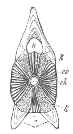
Fig. 319. Vertical section through the middle of a vertebra of
Esox lucius (Pike). (From Gegenbaur.)
ch. notochord; cs. notochordal sheath; k. and k´.
cartilaginous tissue of the neural and hæmal arches; h. osseous
hæmal process; n. spinal canal.
A superficial osseous layer becomes very early formed in each
vertebral region of the cellular sheath; while in each of the
intervertebral regions, which are considerably shorter than the
vertebral, there is developed a ring-like cartilaginous thickening of
the sheath, which projects inwards so as to constrict the notochord.
At a period before this thickening has attained considerable
dimensions the notochord becomes sufficiently constricted in the
centre of each vertebral region to give a biconcave form to the
vertebræ for a very short period of fœtal life.
The stage with biconcave vertebræ is retained through life in the
Perennibranchiata and Gymnophiona.
The chief peculiarity which distinguishes the later history of their
vertebral column from that of fishes consists in the immense
development of the intervertebral thickenings just mentioned, which
increase to such an extent as to reduce the notochord, where it passes
through them, to a mere band; while the cartilage of which they are
composed becomes differentiated into two regions, one belonging to the
vertebra in front, the other to that behind, the hinder one being
convex, and the anterior concave. The two parts are not however
absolutely separated from each other.
By these changes each vertebra comes to be composed of (1) a thin
osseous somewhat hourglass-shaped cylinder with a dilated portion of
the notochord in its centre, and (2 and 3) of two
halves of two
intervertebral cartilages, viz. an anterior convex half and a
posterior concave half. The vertebræ thus come to be opisthocœlous. A
longitudinal section through the vertebral column at this stage is
diagrammatically shewn in fig. 320 C.
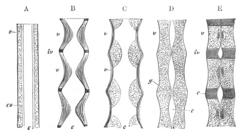
Fig. 320. Diagram representing the mode of development of the
vertebræ in the different types. (From Gegenbaur.)
A. Ideal type in which distinct vertebræ are not established.
B. Type of Pisces with vertebral constrictions of the notochord.
C. Amphibian type, with intervertebral constrictions of the
notochord by the intervertebral parts of the cellular sheath.
D. Intervertebral constriction of the notochord as effected in
Reptilia and Aves.
E. Vertebral constriction of the notochord as effected in Mammalia,
the intervertebral parts of the cartilaginous sheath being
converted into intervertebral ligaments.
c. notochord; cs. cuticular sheath of notochord; s.
cartilaginous sheath; v. vertebral regions; iv. intervertebral
regions; g. intervertebral joints.
To the centre of each of these vertebræ the neural arches, the origin
of which was described above, become in the meantime firmly attached;
and grow obliquely upwards and backwards, so as to meet and unite
above the spinal cord. The transverse processes of the vertebræ would
seem (Fick) to be developed independently of the arches, though they
very soon fuse with them. According to Götte the transverse processes
are double in the trunk, there being two pairs, one vertically above
the other for each vertebra. The pair on each side eventually fuse
together.
In the tail hæmal arches are formed, which are similar in their mode
of development to the neural arches.
The unconstricted portion of the notochord, which persists in each
vertebra, becomes in part converted into cartilage.
Anura. In the Anura the process of formation of the vertebral column
is essentially the same as that in the Salamandridæ. Two types may
however be observed. One of these occurs in the majority of the Anura,
and mainly differs from that in Salamandra in (1) the earlier fusion
of the arches with the cellular sheath of the notochord; (2) the more
rapid growth of the intervertebral thickenings of the cellular sheath,
which results in the early and complete obliteration of the
intervertebral parts of the notochord; (3) the complete division of
these intervertebral thickenings into anterior and posterior portions,
which unite with and form the articular surfaces of two contiguous
vertebræ. The vertebræ are moreover procœlous instead of being
opisthocœlous.
The unconstricted vertebral sections of the notochord always persist
till the ossification of the vertebræ has taken place. In some forms
they remain through life (Rana), while in other cases they eventually
either wholly or partially disappear.
The second type of vertebral development is found in Bombinator,
Pseudis, Pipa, and Pelobates. In these genera the formation of the
vertebra takes place almost entirely on the dorsal side of the
notochord; so that the latter forms a band on the ventral side of the
vertebral column. In other respects the history of the vertebral
column is the same in the two cases; the vertebral unconstricted parts
of the notochord appear however to become in part converted into
cartilage. The type of formation of the vertebral column in these
genera has been distinguished as epichordal in contradistinction to
the more normal or perichordal type.
Amniota. In the Amniota all trace of a distinction between a cellular
notochord sheath and an arch tissue is lost, and the two are developed
together as a continuous whole forming an unsegmented tube round the
notochord, with a neural ridge which does not at first nearly invest
the neural cord. This tube becomes differentiated, in the manner
already described for other types, into (1) vertebral regions with
true arches, and (2) intervertebral regions.
Reptilia. In Reptilia (Gegenbaur, No. 416) a cartilaginous tube is
formed round the notochord, which is continuous with the cartilaginous
neural arches. The latter are placed in the vertebral regions, and in
these regions ossification very early sets in, while the notochord
remains relatively unconstricted. In the intervertebral regions the
cartilage becomes thickened, as in Amphibia, and gradually constricts
the notochord. The cartilage in each of the intervertebral regions
soon becomes divided into two parts which form the articular faces of
two contiguous vertebræ.
The general character of the vertebral column on the completion of
these changes is shewn in fig. 320 D. The later changes are relatively
unimportant. The constricted intervertebral sections of the notochord
rapidly disappear, while the vertebral sections become partially
converted into cartilage, and only cease to be distinguishable at a
considerably later period.
The ossification extends from the bodies of the vertebræ into the
arches and into the articular surfaces, so that the whole vertebræ
eventually become ossified.
The Ascalabotæ (Geckos) present an exceptional type of vertebral
column which has many of the characters of a developmental stage in
other Lizards. The body of the vertebra is formed of a slightly
hourglass-shaped osseous tube, united with adjoining vertebræ by a
short intervertebral cartilage. There is a persistent and continuous
notochord which, owing to the small development of the intervertebral
cartilages, is narrower in the vertebral than in the intervertebral
regions.
Aves. In Birds the cellular tube formed round the notochord is far
thicker than in the Reptilia. It is continuous in the regions of the
future vertebræ with neural arches, which do not at first nearly
enclose the spinal cord.
On about the fifth day, in the case of the chick, it becomes
differentiated into vertebral regions opposite the attachments of the
neural arches, and intervertebral regions between them; the two sets
of regions being only distinguished by their histological characters.
Very shortly afterwards each intervertebral region becomes segmented
into two parts, which respectively attach themselves to the contiguous
vertebral regions. A part of each intervertebral region, immediately
adjoining the notochord, does not however undergo this division, and
afterwards gives rise to the ligamentum suspensorium.
The notochord during these changes at first remains indifferent, but
subsequently, on about the seventh day in the chick, a slight
constriction of each vertebral region takes place; so that the
vertebræ have temporarily, as they have also in Amphibia, a biconcave
form which repeats the permanent condition of most fishes. By the
ninth and tenth days, however, this condition has completely
disappeared, and in all the intervertebral portions the notochord has
become distinctly constricted, and at the same time in each vertebral
portion there
have also appeared two constrictions of the notochord
giving rise to a central and to two terminal enlargements.
On the twelfth day the ossification of the cartilaginous centra
commences.
The first vertebra to ossify is the second or third cervical, and the
ossification gradually extends to those behind. It does not commence
in the arches till somewhat later than in the bodies. For each arch
there are two centres of ossification, one on each side.
The notochord persists for the greater part of fœtal life and even
into post-fœtal life. The larger vertebral portions are often the
first completely to vanish. They would seem in many cases at any rate
(Gegenbaur) to be converted into cartilage, and so form an integral
part of the permanent vertebræ. Rudiments of the intervertebral
portions of the notochord may long be detected in the ligamenta
suspensoria.
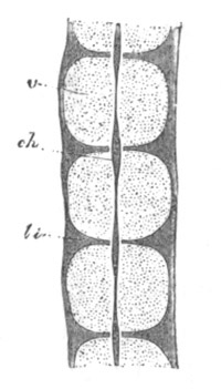
Fig. 321. Longitudinal section through the vertebral column of an
eight weeks’ human embryo in the thoracic region. (From
Kölliker.)
v. cartilaginous vertebral body; li. intervertebral ligament;
ch. notochord.
Schwarck (No. 420) states that in both the intervertebral and the
vertebral regions, though less conspicuously in the former, the
cartilage is divided into two layers, an inner and an outer. He
holds that the inner layer corresponds to the cartilaginous
notochordal sheath of the lower types, and the outer to the arch
tissue. Ossification (Gegenbaur) of the centra appears in a special
inner layer of cartilage, which is probably the same as the inner
layer of the earlier stage, though this point has not been definitely
established.
Mammalia. The early development of the perichordal cartilaginous tube
and rudimentary neural arches is almost the same in Mammals as in
Birds. The differentiation into vertebral and intervertebral regions
is the same in both groups; but instead of becoming divided as in
Reptilia and Birds into two segments attached to two adjoining
vertebræ, the intervertebral regions become in Mammals wholly
converted into the intervertebral ligaments (fig. 322 li). There
are three centres of ossifications for each vertebra, two in the arch
and one in the centrum.
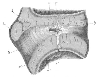
Fig. 322. Longitudinal section through the intervertebral ligament
and adjacent parts of two vertebræ from the thoracic region of an
advanced embryo of a Sheep. (From Kölliker.)
la. ligamentum longitudinale anterius; lp. ligamentum long.
posterius; li. ligamentum intervertebrale; k, k´. epiphysis of
vertebra; w. and w´. anterior and posterior vertebræ; c.
intervertebral dilatation of notochord; c´. and c´´. vertebral
dilatation of notochord.
The fate of the notochord is in important respects different from that
in Birds. It is first constricted in the centre of the vertebræ
(figs. 320 E and 321) and disappears there shortly after the
ossification; while in the intervertebral regions it remains
relatively unconstricted (figs. 320 E, 321 and 322 c) and after
undergoing certain histological changes remains through life as part
of the nucleus pulposus in the axis of the invertebral ligaments[201].
There is also a slight swelling of the notochord near the two
extremities of each vertebra (fig. 322 c´ and c´´). In the
persistent vertebral constriction of the notochord Mammals retain a
more primitive and piscine mode of formation of the vertebral column
than the majority either of the Reptilia or Amphibia.
Bibliography of Notochord and Vertebral column.
(415) Cartier. “Beiträge zur Entwicklungsgeschichte der Wirbelsäule.”
Zeitschrift für wiss. Zool., Bd. XXV. Suppl. 1875.
(416) C. Gegenbaur. Untersuchungen zur vergleichenden Anatomie der
Wirbelsäule der Amphibien und Reptilien. Leipzig, 1862.
(417) C. Gegenbaur. “Ueber die Entwickelung der Wirbelsäule des
Lepidosteus mit vergleichend anatomischen Bemerkungen.” Jenaische
Zeitschrift, Bd. III. 1863.
(418) C. Gegenbaur. “Ueb. d. Skeletgewebe d. Cyclostomen.” Jenaische
Zeitschrift, Vol. V. 1870.
(419) Al. Götte. “Beiträge zur vergleich. Morphol. des Skeletsystems
d. Wirbelthiere.” II. “Die Wirbelsäule u. ihre Anhänge.” Archiv f.
mikr. Anat., Vol. XV. 1878 (Cyclostomen, Ganoiden, Plagiostomen,
Chimaera), and Vol. XVI. 1879 (Teleostier).
(420) Hasse und Schwarck. “Studien zur vergleichenden Anatomie der
Wirbelsäule u. s. w.” Hasse, Anatomische Studien, 1872.
(421) C. Hasse. Das natürliche System d. Elasmobranchier auf
Grundlage d. Bau. u. d. Entwick. ihrer Wirbelsäule. Jena, 1879.
(422) A. Kölliker. “Ueber die Beziehungen der Chorda dorsalis zur
Bildung der Wirbel der Selachier und einiger anderen Fische.”
Verhandlungen der physical. medicin. Gesellschaft in Würzburg, Bd.
X.
(423) A. Kölliker. “Weitere Beobachtungen über die Wirbel der
Selachier insbesondere über die Wirbel der Lamnoidei.” Abhandlungen
der senkenbergischen naturforschenden Gesellschaft in Frankfurt, Bd.
V.
(424) H. Leboucq. “Recherches s. l. mode de disparition de la corde
dorsale chez les vertébrés supérieurs.” Archives de Biologie, Vol.
I. 1880.
(425) Fr. Leydig. Anatomisch-histologische Untersuchungen über Fische
und Reptilien. Berlin, 1853.
(426) Aug. Müller. “Beobachtungen zur vergleichenden Anatomie der
Wirbelsäule.” Müller’s Archiv. 1853.
(427) J. Müller. “Vergleichende Anatomie der Myxinoiden u. der
Cyklostomen mit durchbohrtem Gaumen, I. Osteologie und Myologie.”
Abhandlungen der königlichen Akademie der Wissenschaften zu Berlin.
1834.
(428) W. Müller. “Beobachtungen des pathologischen Instituts zu Jena,
I. Ueber den Bau der Chorda dorsalis.” Jenaische Zeitschrift, Bd.
VI. 1871.
(429) A. Schneider. Beiträge z. vergleich. Anat. u. Entwick. d.
Wirbelthiere. Berlin, 1879.
Ribs and Sternum.
Ribs. Embryological evidence on the development of the ribs, though
somewhat inadequate, indicates that they arise as cartilaginous bars
in the connective tissue of the intermuscular septa, and that they are
placed, in Elasmobranchii and
Amphibia, on the level of division
between the dorso-lateral and ventro-lateral divisions of the
muscle-plates. This does not appear to hold true for either Ganoidei
or Teleostei. In Teleostei they are entirely below the muscles along
the lines of the intermuscular septa, and this is partially true for
Ganoidei, though not wholly so in Lepidosteus. They may be attached
either to the hæmal (Pisces) or neural (Amphibia and Amniota) arches.
The connective tissue from which they are formed is continuous with
the processes of the vertebræ to which they are attached; but the
conversion of the tissue into cartilage takes place more or less
independently of that of the arches, although in many cases the
cartilage of the two becomes continuous, the separation of the ribs
being then effected by a subsequent process of segmentation (Fick, No.
431). It is possible that the ribs of Pisces may not be homologous
with those of Amphibia and the Amniota, but till the reverse can be
proved it is more convenient to assume that the ribs are homologous
structures throughout the vertebrate series.
In Elasmobranchii the ribs are relatively of less importance in the
adult than in the embryo. By a careful examination of their early
development, I have satisfied myself that the differentiation of the
ribs is independent of that of the hæmal processes to which they are
attached, although the differentiation proceeds in such a manner that,
when both are converted into cartilage, they are quite continuous.
Subsequently the ribs become segmented off from the hæmal processes.
At the junction of the tail and trunk, where the hæmal processes
commence to be ventrally prolonged, eventually to unite in the region
of the tail below the caudal vein, the ribs are attached to short
processes which spring from the sides of the hæmal arches (fig. 317).
The ventral hæmal arches of these fishes are therefore clearly in no
part formed by the ribs.
In Ganoidei and Teleostei there is very great difficulty in
determining the homologies of the ribs.
In the cartilaginous Ganoidei there are well developed rib-like
structures, which might be regarded as homologous with Elasmobranch
ribs, and indeed probably are so; but at the same time their relations
are in some respects very different from those of Elasmobranch ribs in
the caudal region. In Ganoids the ribs, in approaching the tail,
become shorter and then fuse with the ends of the hæmal processes, and
finally in the caudal region form together with the hæmal arches a
closed hæmal canal which superficially resembles that in
Elasmobranchii.
In Lepidosteus and Amia, especially the former, the same phenomenon is
still more marked; and in Lepidosteus it is easy, in passing
backwards,
to trace the ribs bending ventralwards, and uniting
ventrally in the caudal region to form, with the hæmal processes, a
complete hæmal canal.
It might have been anticipated that the Teleostean Ganoids would
resemble the Teleostei, but, from an examination of adult Teleostei,
it would seem to be clear that the relations of the parts are the same
as in Elasmobranchii, i.e. that the ribs have no share in forming
the hæmal canal in the tail. Aug. Müller and Götte have however
brought embryological evidence (though not of a conclusive character),
to shew that in the embryo the ribs really fuse with the hæmal
processes in the tail, and so assist, as in the Ganoids, in forming
the hæmal canal. Götte moreover holds that the ribs in Elasmobranchii
are not homologous with those of Teleostei and Ganoids; but that the
hæmal arches in the tail are homologous in the three groups.
Without necessarily following Götte in these views it is worth
pointing out that the undoubtedly close affinity between the bony
Ganoids and the Teleostei is in favour of the view on the hæmal arches
of Teleostei at which he has arrived on embryological grounds.
In Amphibia the formation of the ribs from the connective tissue of
the intermuscular septa, their secondary attachment to the transverse
processes of the neural arches, and their subsequent separation was
first clearly established by Fick (No. 431), whose statements have
since been confirmed by Hasse, Born, &c., and in part by Götte, who
holds however that, though converted into cartilage independently of
the transverse processes, they are formed in membrane as outgrowths of
these processes.
In the Amniota the ribs are also independently established (Hasse and
Born), though they subsequently become united to the transverse
processes and to the bodies of the vertebræ, or to the transverse
processes only. This junction is however stated by the majority of
authorities, never to be effected by the fusion of the cartilage of
the two parts, but always by fibrous tissue; though Hoffmann (No. 435)
takes a different view on this subject, holding that the ribs are at
first continuous with the intervertebral regions of the primitive
cartilaginous tube surrounding the notochord.
Sternum. In dealing with the development of the sternum it will be
convenient to leave out of consideration the interclavicle or
episternum which is, properly speaking, only part of the
shoulder-girdle and to confine my statements to the sternum proper.
This structure is found in all the Amniota except the Ophidia,
Chelonia, and some of the Amphisbænæ.
From the older researches of Rathke, and from the newer ones of Götte,
etc., it appears that the sternum is always formed from the fusion of
the ventral extremities of a certain number of ribs. The extremities
of the ribs unite with each other from
before backwards, and thus give
rise to two cartilaginous bands. These bands become segmented off from
the ribs with which they are at first continuous, and subsequently
fuse in the median ventral line to form an unpaired sternum. The
Mammalian presternum (manubrium sterni) and xiphosternum have the same
origin as the main body of the sternum (Ruge, No. 438).
In the Amphibia there is no structure which admits from its mode of
development of a complete comparison with the sternum of the Amniota;
and it must for this reason be considered doubtful whether the median
structure placed behind the coracoids in the Anura, which is usually
known as the sternum, is really homologous with the sternum of the
Amniota[202].
The remaining Ichthyopsida are undoubtedly not provided with a sternum.
Bibliography of Ribs and Sternum.
(430) C. Claus. “Beiträge z. vergleich. Osteol. d. Vertebraten. I.
Rippen u. unteres Bogensystem.” Sitz. d. kaiserl. Akad. Wiss. Wien,
Vol. LXXIV. 1876.
(431) A. E. Fick. “Zur Entwicklungsgeschichte d. Rippen und
Querfortsätze.” Archiv f. Anat. und Physiol. 1879.
(432) C. Gegenbaur. “Zur Entwick. d. Wirbelsäule des Lepidosteus mit
vergleich. anat. Bemerk.” Jenaische Zeit., Vol. III. 1867.
(433) A. Götte. “Beiträge z. vergleich. Morphol. d. Skeletsystems d.
Wirbelthiere Brustbein u. Schultergürtel.” Archiv f. mikr. Anat.,
Vol. XIV. 1877.
(434) C. Hasse u. G. Born. “Bemerkungen üb. d. Morphologie d. Rippen.”
Zoologischer Anzeiger, 1879.
(435) C. K. Hoffmann. “Beiträge z. vergl. Anat. d. Wirbelthiere.”
Niederländ. Archiv Zool., Vol. IV. 1878.
(436) W. K. Parker. “A monograph on the structure and development of
the shoulder-girdle and sternum.” Ray Soc. 1867.
(437) H. Rathke. Ueb. d. Bau u. d. Entwicklung d. Brustbeins d.
Saurier. 1853.
(438) G. Ruge. “Untersuch. üb. Entwick. am Brustbeine d. Menschen.”
Morphol. Jahrbuch., Vol. VI. 1880.
Three distinct sets of elements may enter into the composition of the
skull. These are (1) the cranium proper, composed of true endoskeletal
elements originally formed in cartilage, to which are usually added
exoskeletal osseous elements, formed in the manner already described
p. 542, and known in the higher types as membrane bones. (2) The
visceral arches formed primitively as cartilaginous bars, but in the
higher types largely supplemented or even replaced by exoskeletal
elements. (3) The labial cartilages.
These parts present themselves in the most various forms, and their
study constitutes one of the most important departments of vertebrate
morphology, and one which has always been a favourite subject of study
with anatomists. At the end of the last century and during the first
half of the present century the morphology of the skull was handled
from the point of view of the adult anatomy by Goethe, Oken, Cuvier,
Owen, and many other anatomists, while Dugés and, nearer to our own
time, Rathke, laid the foundation of an embryological study of its
morphology. A new era in the study of the skull was inaugurated by
Huxley in his Croonian lecture in 1858, and in his lectures on
Comparative Anatomy subsequently delivered before the Royal College of
Surgeons. In these lectures Huxley disproved the then widely accepted
view that the skull was composed of four vertebræ; and laid the
foundation of a more satisfactory method of dealing with the
homologies of its constituent parts. Since then the knowledge of the
development of the skull has made great progress. In this country a
number
of very interesting memoirs have been published on the subject
by Parker, which together constitute a most striking contribution to
our knowledge of the ontogeny of the skull in a series of types; and
in Germany Gegenbaur’s monograph on the cephalic skeleton of
Elasmobranchii has greatly promoted a scientific appreciation of the
nature of the skull.
In the present chapter only the most important features in the
development of the skull will be touched on.
It will be convenient to describe, in the first instance, the
development of the cartilaginous elements of the skull.
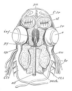
Fig. 323. head of embryo Dogfish, second stage; basal view of
cranium from above, the contents having been removed. (From Parker.)
ol. olfactory sacs; au. auditory capsule; nc. notochord; py.
pituitary body; pa.ch. parachordal cartilage; tr. trabecula;
inf. infundibulum; C.tr. cornua trabeculæ; pn. prenasal
element; sp. spiracular cleft; br. external branchiæ; Cl. 2,
4. visceral clefts.
The Cranium. The brain is at first enveloped in a continuous layer of
mesoblast known as the membranous cranium, into the base of which the
anterior part of the notochord is prolonged for some distance. The
primitive cartilaginous cranium is formed by a differentiation within
the membranous cranium, and is always composed of the following parts
(fig. 323):
(1) A pair of cartilaginous plates on each side of the cephalic
section of the notochord, known as the parachordals (pa.ch). These
plates together with the notochord (nc) enclosed between them form a
floor for the hind- and mid-brain. The continuous plate, formed by
them and the notochord, is known as the basilar plate.
(2) A pair of bars forming the floor for the fore-brain, known as the
trabeculæ (tr). These bars are continued forward from the
parachordals. They meet behind and embrace the front end of the
notochord; and after separating for some distance bend in again in
such a way
as to enclose a space—the pituitary space. In front of
this space they remain in contact and generally unite. They extend
forwards into the nasal region (pn).
(3) The cartilaginous capsules of the sense organs. Of these the
auditory (au) and olfactory capsules (ol) unite more or less
intimately with the cranial walls; while the optic capsules, forming
the usually cartilaginous sclerotics, remain distinct.
The parachordals and notochord. The first of these sets of elements,
viz. the parachordals and notochord, forming together the basilar
plate, is always an unsegmented continuation of the axial tissue of
the vertebral column. It forms the floor for that section of the brain
which belongs to the primitive postoral part of the head (vide p.
314), and its extension is roughly that of the basioccipital of the
adult skull. Its mode of development is almost identical with that of
the vertebral column, except that the notochord, even in many forms
where it persists in the vertebral column, disappears in the basilar
plate; though in a certain number of cases remnants of it are found in
the adult state.
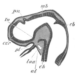
Fig. 324. Longitudinal section through the brain of a young
Pristiurus embryo.
cer. commencement of the cerebral hemisphere; pn. pineal gland;
In. infundibulum; pt. ingrowth from mouth to form the pituitary
body; mb. mid-brain; cb. cerebellum; ch. notochord; al.
alimentary tract; Iaa. artery of mandibular arch.
It will be convenient to say a few words here with reference to the
notochord in the head. It always extends along the floor of the mid-
and hind-brains, but ends immediately behind the infundibulum. The
limits of its anterior extension are clearly shewn in fig. 43. The
front end of the notochord often becomes more or less ventrally flexed
in correspondence with the cranial flexure; its anterior end being in
some instances (Elasmobranchii) almost bent backwards (fig. 324).
Kölliker has shewn that in the Rabbit[203],
and I believe that a more
or less similar phenomenon may also be observed in Birds, the anterior
end of the notochord is united to the hypoblast of the throat in
immediate contiguity with the opening of the pituitary body; but it is
not clear whether this is to be looked upon as the remnant of a
primitive attachment of the notochord to the hypoblast, or as a
secondary attachment.
Before the parachordals are formed the anterior end of the notochord
has usually undergone a partial atrophy; and its front end often
becomes somewhat dorsally flexed. Within the basilar plate it often
exhibits two or more dilatations, which have been regarded by Parker
and Kölliker as indicative of a segmentation of this plate; but they
hardly appear to me to be capable of this interpretation.
In Elasmobranchs where, as shewn above, a very primitive type of
development of the vertebral column is retained, we find that the
basilar plate is at first formed of (1) the notochord invested by its
cartilaginous sheath, and (2) of lateral masses of cartilage, the
parachordals, homologous with the arch tissue of the vertebral column.
This development probably indicates that the basilar plate contains in
itself the same elements as those from which the neural arches and the
centra of the vertebral column are formed; but that it never passes
beyond the unsegmented stage at first characteristic of the vertebral
column. The hinder end of each parachordal forms a condyle
articulating with the first vertebra; so that in the cartilaginous
skull there are always two occipital condyles. The basilar plate
always grows up behind (fig. 326, so), and gives rise to a complete
cartilaginous ring enveloping the medulla oblongata, in the same
manner that the neural arches envelope the spinal cord. This ring
forms an occipital cartilaginous ring; in front of it the basilar
plate becomes laterally continuous with the periotic cartilaginous
capsules, and the occipital ring above usually spreads forward to form
a roof for the part of the brain between these capsules. In the higher
Vertebrates the periotic cartilages may be developed continuously with
the basilar plate (fig. 325).
The trabeculæ. The trabeculæ, so far as their mere anatomical
relations are concerned, play the same part in forming the floor for
the front cerebral vesicle as the parachordals for the mid- and
hind-brains. They differ however from the parachordals in one
important feature, viz. that, except at their hinder end (fig. 323),
they do not embrace between them the notochord.
The notochord constitutes, as we have seen, the primitive axial
skeleton of the body, and its absence in the greater part of the
region of the trabeculæ would probably seem to indicate, as
pointed
out by Gegenbaur, that these parts, in spite of their similarity to
the parachordals, have not the same morphological significance.
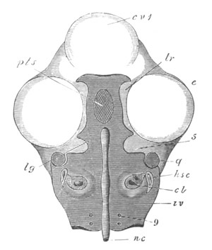
Fig. 325. View From above of the investing mass and of the trabeculæ
of a chick on the fourth day of incubation. (After Parker.)
In order to shew this, the whole of the upper portion of the head
has been sliced away. The cartilaginous portions of the skull are
marked with the dark horizontal shading.
cv 1. cerebral vesicle (sliced off); e. eye; nc. notochord;
iv. investing mass; 9. foramen for the exit of the ninth nerve;
cl. cochlea; hsc. horizontal semicircular canal; q. quadrate;
5. notch for the passage of the fifth nerve; lg. expanded anterior
end of the investing mass; pts. pituitary space; tr. trabeculæ.
The reference line tr. has been accidentally made to end a little
short of the cartilage.
The nature of the trabeculæ has been much disputed by morphologists.
The view that they cannot be regarded as the anterior section of the
vertebral axis is supported by the consideration that the forward
limit of the primitive skeletal axis, as marked by the notochord,
coincides exactly with the distinction we have found it necessary to
recognise, on entirely independent grounds, between the fore-brain,
and the remainder of the nervous axis. But while this distinction
between the parachordals and the trabeculæ must I think be admitted, I
see no reason against supposing that the trabeculæ may be plates
developed to support the floor of the fore-brain, for the same
physiological reasons that the parachordals have become formed at the
sides of the notochord to support the floor of the hind-brain. By some
anatomists the trabeculæ have been held to be a pair of branchial
bars; but this view has now been generally given up. They have also
been regarded as equivalent to a complete pair of neural arches
enveloping the front end of the brain. The primitive extension of the
base of the fore-brain through the pituitary
space is an argument, not
without force, which has been appealed to in support of this view.
In the majority of the lower forms the trabeculæ arise quite
independently of the parachordals, though the two sets of elements
soon unite; while in Birds (fig. 325) and Mammals the parachordals and
trabeculæ are formed as a continuous whole. The junction between the
trabeculæ and parachordals becomes marked by a cartilaginous ridge
known as the posterior clinoid.
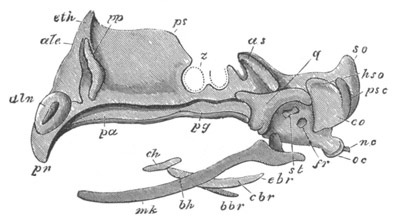
Fig. 326. Side view of the cartilaginous cranium of a Fowl on the
seventh day of incubation. (After Parker.)
pn. prenasal cartilage; aln. alinasal cartilage; ale.
aliethmoid; immediately below this is the aliseptal cartilage.
eth. ethmoid; pp. pars plana; ps. presphenoid or interorbital;
pa. palatine; pg. pterygoid; z. optic nerve; as.
alisphenoid; q. quadrate; st. stapes; fr. fenestra rotunda;
hso. horizontal semicircular canal; psc. posterior vertical
semicircular canal: both the anterior and the posterior semicircular
canals are seen shining through the cartilage. so. supraoccipital;
eo. exoccipital; oc. occipital condyle; nc. notochord; mk.
Meckel’s cartilage; ch. ceratohyal; bh. basihyal; cbr. and
ebr. ceratobranchial; bbr. basibranchial.
The trabeculæ are usually somewhat lyre-shaped, meeting in front and
behind, and leaving a large pituitary space between their middle parts
(figs. 323 and 325). Into this space there primitively projects the
whole base of the fore-brain, but the space itself gradually becomes
narrowed, till it usually contains only the pituitary body. The
carotid arteries always pass through it in the embryo; but in the
higher forms it ceases to be perforated in the adult. The trabeculæ
soon unite together both in front and behind and form a complete plate
underneath the fore-brain, and extending into the nasal region[204].
A special
vertical growth of this plate in the region of the orbit forms
the interorbital plate of Teleostei, Lacertilia and Aves (fig. 326,
ps), on the upper surface of which the front part of the brain
rests. The trabecular floor of the brain does not long remain simple.
Its sides grow vertically upwards, forming a lateral wall for the
brain, in which in the higher types two regions may be distinguished,
viz. an alisphenoidal region (fig. 326, as) behind, growing out from
what is known as the basisphenoidal region of the primitive trabeculæ,
and an orbitosphenoidal region in front growing out from the
presphenoidal region of the trabeculæ. These plates form at first a
continuous lateral wall of the cranium. At the front end of the brain
they are continued inwards, and more or less completely separate the
true cranial cavity from the nasal region in front. The region of the
cartilage forming the anterior boundary of the cranial cavity is known
as the lateral ethmoid region, and it is always perforated for the
passage of the olfactory nerves.
The cartilaginous walls which grow up from the trabecular floor of the
cranium generally extend upwards so as to form a roof, though almost
always an imperfect roof, for the cranial cavity. In the higher types,
in Mammals more especially, this roof can hardly be said to be formed
at all. The region of the trabeculæ in front of the brain is the
ethmoid region. The basal part of this region forms an internasal
plate, from which an internasal septum may grow up (fig. 326). To its
sides the olfactory capsules are attached, and there are usually
lateral outgrowths in front forming the trabecular cornua, while from
the posterior part of the ethmoidal plate, forming the anterior
boundary of the cranial cavity, there often grows out a prefrontal or
lateral ethmoidal process.
These and other processes growing out from the trabeculæ have
occasionally been regarded as rudimentary præoral branchial arches. I
have already stated it as my view that the existence of branchial
arches in this region is highly improbable, and I may add that the
development of these structures as outgrowths of the skull is in
itself to my mind a nearly conclusive argument against their being
branchial arches, in that true branchial arches hardly ever or perhaps
never arise in this way.
The sense capsules. The most important of these is the auditory
capsule, which, as we have seen, fuses intimately with
the lateral
walls of the skull. In front there is usually a cleft separating it
from the alisphenoid region of the skull, through which the third
division of the fifth nerve passes out. This cleft becomes narrowed to
a small foramen (fig. 327, V). The sclerotic cartilage is always
free, but profoundly modifies the region of the cranium near which it
is placed. The nasal investment forms in Elasmobranchs (fig. 327,
Na) a capsule open below, and continuous with the ethmoid region of
the trabeculæ. In most types however it becomes more closely united
with the ethmoid region and the accessory parts belonging to it.
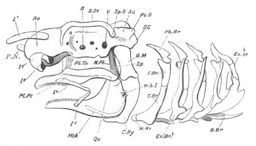
Fig. 327. Skull of adult Dogfish, side view. (From Parker.)
O.C. occipital condyle; Au. periotic capsule; Pt.O. pterotic
ridge; Sp.O. sphenotic process; S.Or. supraorbital ridge; Na.
nasal capsule; P.N. prenasal cartilage; II. optic foramen; V.
trigeminal foramen; Pl.Pt., Qu. pterygo-quadrate arcade; M.Pt.
metapterygoid ligament (including a small cartilage); Pl.Tr.
ethmo-palatine or palato-trabecular ligament; Mck. lower jaw;
Sp. spiracle; H.M. hyomandibular; C.Hy. ceratohyal; m.h.l.
mandibulohyoid ligament; Ph.Br. pharyngobranchial; E.Br.
epibranchial; C.br. ceratobranchial; H.Br. hypobranchial;
B.Br. basibranchial; Ex.Br. extra-branchial; l1, 2, 3,
4, 5. labial cartilages; the dotted lines within Mck.
indicate the basihyal.
The cartilaginous cranium, the development of which has been thus
briefly traced, persists in the adult without even the addition of
membrane bones in the Cyclostomata, Elasmobranchii (fig. 327) and
Holocephali. In the Selachioid Ganoids it is also found in the adult,
but is covered over by membrane bones. In all other types it is
invariably present in the embryo, but becomes in the adult more or
less replaced by osseous tissue.
Branchial skeleton.
The most primitive type of branchial skeleton in any existing form
would appear to be that of the Petromyzonidæ, which is developed in a
superficial subdermal tissue, and consists of a series of bars united
by transverse pieces, so as to form a basketwork. It is known as an
extra-branchial system, and an early stage of its development in the
Lamprey is shewn in fig. 47. In the higher forms this system is
replaced by a series of bars, known as the branchial bars, so situated
as to afford support to the successive branchial pouches. Outside
these bars there may be present in some primitive forms
(Elasmobranchii) cartilaginous elements, which are supposed to be
remnants of the extra-branchial system (fig. 327, Ex.Br); while a
series of membrane bones is also usually added to them, which will be
dealt with in a separate section. The branchial bars are developed as
simple cartilaginous rods in the deeper parts of the mesoblast which
constitutes the primitive branchial arches.
The position of the branchial bars in relation to the somatopleure and
splanchnopleure can be determined from their relation to the so-called
head cavities. These cavities atrophy before the formation of the
cartilaginous branchial bars, but it will be observed (fig. 328), that
the artery of each arch (aa) is placed on the inner side of the head
cavity (pp). The cartilaginous bar arises at a later period on the
inner side of the artery, and therefore on the inner side of the
section of the body cavity primitively present in the arches.
An anterior arch, known as the mandibular arch, placed in front of the
hyomandibular cleft, and a second arch, known as the hyoid arch,
placed in front of the hyobranchial cleft, are developed in all types.
The succeeding arches are known as the true branchial arches, and are
only fully developed in the Ichthyopsida.
In some Sharks (Notidani) seven branchial arches may be present (not
including the hyoid and mandibular). In other Ichthyopsida five are
usually present, in the embryo at any rate, while in the Amniota there
are usually two or three post-hyoid membranous arches, in the interior
of which a cartilaginous bar is usually formed. The general form of
these bars at an early
stage of development is shewn in the dogfish
(Scyllium) in fig. 329.
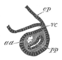
Fig. 328. Horizontal section through the penultimate visceral arch
of an embryo of Pristiurus.
ep. epiblast; vc. pouch of hypoblast which will form the walls
of a visceral cleft; pp. segment of body-cavity in visceral arch;
aa. aortic arch.
The simple condition of these bars in the embryo renders it highly
probable that forms existed at one time with a simple branchial
skeleton of this kind: at the present day however such forms no longer
exist. The first arch has in all cases changed its function and has
become converted into a supporting skeleton for the mouth; the hyoid
arch, though retaining in some forms its branchial function, has in
most acquired additional functions and has undergone in consequence
various peculiar modifications. The true branchial arches retain their
branchial functions in Pisces and some Amphibia, but are secondarily
modified and largely aborted in the abranchiate forms. Since the
changes undergone by the true branchial bars are far less complicated
than those of the hyoid and mandibular bars it will be convenient to
treat of them in the first instance.
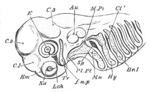
Fig. 329. Head of embryo Dogfish, 11 lines long. (From Parker.)
Tr. trabecula; Pl.Pt. pterygo-quadrate; M.Pt. metapterygoid
region; Mn. mandibular cartilage; Hy. hyoid arch; Br. 1. first
branchial arch; Sp. mandibulohyoid cleft; Cl1. hyobranchial
cleft; Lch. groove below the eye; Na. olfactory rudiment; E.
eyeball; Au. auditory mass; C 1, 2, 3. cerebral vesicles; Hm.
hemispheres; f.n.p. nasofrontal process.
These bars are, as already mentioned, most numerous in certain very
primitive forms (seven in Notidanus), while as we ascend the series
there is a gradual tendency for the posterior of them to disappear.
This tendency is the result of a gradual atrophy of the posterior
branchial pouches, which commenced at
a stage in the evolution of the
Chordata long prior to the appearance of cartilaginous or osseous
branchial bars, and reaches its climax in the Amniota.
In a fully developed branchial bar the primitively simple rod of
cartilage becomes divided into a series of segments, usually four,
articulated so as to be more or less mobile: and either remaining
cartilaginous or becoming partially or wholly ossified. Each bar (fig.
327) forms a somewhat curved structure, embracing the pharynx. The
dorsal and somewhat horizontally placed segment is known as the
pharyngobranchial (Ph.Br), the next two as the epibranchial (E.Br)
and ceratobranchial (C.Br), and the ventral segment as the
hypobranchial (H.Br). There is also typically present a basal
unpaired segment, uniting the bars of the two sides, known as the
basibranchial (B.Br). The arches often bear cartilaginous rays which
support the gill lamellæ.
In Teleostei dental plates are usually developed as an exoskeletal
covering on parts of the branchial arches.
In the Amphibia four or three branchial arches are present in the
embryo. These parts are more or less completely retained in the
Perennibranchiata and Caducibranchiata, but in the Myctodera and Anura
they become largely reduced, and entirely connected with the hyoid.
In the Anura they never reach any considerable development, and are
soon reduced to a plate (fig. 330)—the coalesced basihyal and
basibranchial plate—the posterior processes of which represent the
remnants of the branchial arches.
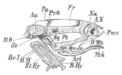
Fig. 330. Young Frog, with tail just absorbed; side view of
skull. (From Parker.)
Au. auditory capsule; in front of it is the cranial side wall;
A.N. external nostril; St. stapes; Mck. Meckelian cartilage;
B.Hy. basihyobranchial plate; St.Hy. stylohyal or ceratohyal;
Br. 1. first branchial arch.
Bones: E.O. exoccipital; Pr.O. prootic; Pa. parietal; Fr.
frontal; Na. nasal; Pmx. premaxillary; Mx. maxillary; Pt.
pterygoid; Sq. squamosal; Qu.Ju. quadratojugal; Art.
articular; D. dentary.
According to Parker the posterior process of this plate in the adult
is a remnant of the fourth branchial bar; the next one is the third
branchial bar, while the anterior lamina behind the hyoid is stated by
him (though this is somewhat doubtful) to be a remnant of the first
two bars.
In the Amniota, the branchial arches become still more
degenerated, in
correlation with the total disappearance of a branchial respiration at
all periods of life. Their remnants become more or less important
parts of the hyoid bone, and are solely employed in support of the
tongue. Their basal portions are best preserved, forming parts of the
body of the hyoid. The posterior (thyroid) cornua of the hyoid are
remnants of the true arches. Of these there are two in the Chelonia
and Lacertilia, and one in the Aves and Mammalia. In Aves the cornu
formed from the first branchial arch (fig. 331, cbr) is always
larger than that of the true hyoid arch (ch).
Mandibular and Hyoid arches. The adaptations of both the mandibular
and hyoid bars, to functions entirely distinct from those which they
primitively served, are most remarkable; and the adaptations of the
two bars are in many cases so intimately bound together, that it is
not possible to treat them separately.
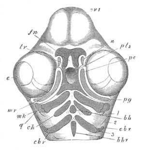
Fig. 331. View from below of the branchial skeleton of the skull
of a Fowl on the fourth day of incubation. (After Parker.)
cv 1. cerebral vesicles; e. eye; fn. frontonasal process; n.
nasal pit; tr. trabeculæ; pts. pituitary space; mr. superior
maxillary process; pg. pterygoid; pa. palatine; q. quadrate;
mk. Meckel’s cartilage; ch. ceratohyal; bh. basihyal; cbr.
ceratobranchial; ebr. proximal portion of the cartilage in the
third visceral (first branchial) arch; bbr. basibranchial; 1. first
visceral cleft; 2. second visceral cleft; 3. third visceral arch.
The most important change of function is undoubtedly that of the
mandibular arch, which becomes entirely converted into a skeleton for
the jaws. It may be noted as a peculiarity of the
mandibular arch that
it is never provided with an unpaired basal element.
The simplest forms of metamorphosis are those undergone by
Elasmobranchii, of which the Dogfish (Scyllium) and Skate (Raja) have
been studied (Parker, No. 456). In some of these forms, e.g. the
Skate, part of the mandibular bar is still related to the
hyomandibular cleft (the spiracle).
Elasmobranchii. In Scyllium the hyoid and mandibular arches are at
first very similar to those which follow. Soon however each of them
sends an anteriorly directed dorsal process (fig. 329). The regions
which may be distinguished owing to the growth of these processes have
received names from ossifications in them which are found in other
types. The anterior process of the mandibular arch is known as the
pterygo-quadrate bar (Pl.Pt); the dorsal end of the primitive bar
from which it starts (M.Pt) is known as the metapterygoid process;
while the ventral end of the bar forms the Meckelian cartilage. The
upper end of the hyoid arch is known as the hyomandibular.
In a somewhat later stage changes take place which cause these parts
practically to assume the adult form (fig. 327). The mandibular arch
becomes segmented at its bend into (1) a pterygo-quadrate bar
(Pl.Pt) which grows forwards in front of the mouth and forms an
upper jaw, and (2) a Meckelian cartilage (Mck) which is placed
behind the mouth, and forms a lower jaw. The two jaws are articulated
together, and the cartilages of the two sides composing them meet each
other distally.
At the articulation of the Meckelian cartilage with the quadrate part
of the pterygo-quadrate is situated a ligament (M.Pt), which takes
the place of the metapterygoid process of the previous stage, and
passes up on the anterior side of the spiracle, to be attached to the
cranium in the front part of the auditory region. This ligament, which
is supplemented by a second ligament, the ethmo-palatine ligament,
passing from the pterygo-quadrate bar to the antorbital region of the
skull, is not the most important support of the jaw. The main support
is, on the contrary, given by the hyoid arch; the hyomandibular
segment of which (H.M) as well as the adjoining segment (ceratohyoid
C.Hy) are firmly attached by ligament to the mandibular
arch. The
hyomandibular is articulated with the cranium beneath the pterotic
ridge (Pt.O).
In the type just described, the hyoid and mandibular arches undergo
less modification than in almost any other case. The hyoid arch has
altered its form, but retains its respiratory function. It has however
acquired the secondary function of supporting the mandibular arch. The
mandibular arch is divided into two elements, which form respectively
the upper and lower jaws. It is not directly articulated with the
skull, and its mode of support by the hyoid arch has been called by
Huxley (No. 445) hyostylic.
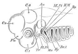
Fig. 333. Head of embryo Skate, 11⁄3 in. long. (From
Parker.)
Tr. trabecula; Pl.Pt. pterygo-quadrate bar; Mn. mandibular
bar; M.Pt. metapterygoid cartilage; H.M. hyomandibular; Hy.
remainder of hyoid arch; Br. 1. first branchial arch; Sp.
mandibulohyoid cleft or spiracle; Pn. pineal gland; Au. auditory
vesicle; C. 1, C. 2, and C. 3. vesicles of the brain.
The development of the hyoid and mandibular arches in the Skate is
characterised by a few important features (fig. 333). The anterior
element of the hyoid arch, which forms the hyomandibular (H.M),
becomes entirely separate from the posterior part of the arch, and
only serves to support the jaws. The posterior part of the arch (Hy)
carries on the respiratory functions of the hyoid, and is closely
connected with the first branchial arch. The upper or metapterygoid
element of the mandibular arch (M.Pt) has a considerable
development, and, becoming separated from the remainder of the arch,
forms a mass of cartilage with one or two branchial rays, in the front
wall of the spiracle, and constitutes a section of the mandibular arch
still retaining traces of its primitive function in supporting the
wall of a branchial pouch.
Although the development of other Elasmobranch types is not known, it
is necessary to call attention to the mode of support of the
mandibular arch in certain forms, notably Notidanus, Hexanchus and
Cestracion, where the pterygo-quadrate region of the mandibular arch
is directly articulated to the
cranium between the optic and
trigeminal foramina. In the two former genera the metapterygoid region
of the arch is moreover continuous with the pterygo-quadrate, and
articulates with the postorbital process of the auditory region of the
skull. In spite of these attachments the mandibular arch continues to
be partially supported by the hyomandibular. The skulls in which the
mandibular arch has this double form of support have been called by
Huxley amphistylic.
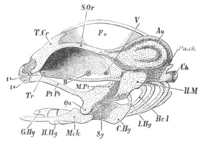
Fig. 334. Cranial skeleton of a Salmon fry, second week after
hatching; membrane bones, eyeballs, and nasal sacs removed. (From
Parker.)
T.Cr. tegmen cranii; S.Or. supraorbital band; Fo. superior
fontanelle; Au. auditory capsule; Pa.ch. parachordal cartilage;
Ch. notochord; Tr. trabecula; above the trabecula, the
interorbital septum is seen, passing into the cranial wall above and
reaching the supraorbital band; H. optic foramen; V. trigeminal
foramen; l1, l2. labial cartilages; Pl.Pt. palatopterygoid
bar; M. Pt. metapterygoid tract; Qu. quadrate region; Mck.
Meckelian cartilage; H.M. hyomandibular cartilage; Sy.
symplectic tract; I.Hy. interhyal; C.Hy. ceratohyal; H.Hy.
hypohyal; G.Hy. glossohyal; Br.1. first branchial arch.
Considering the in many respects primitive characters of the forms
with amphistylic skulls it seems not improbable that they preserve the
original mode of support of the mandibular arch; from which
differentiations in two directions have taken place, viz.
differentiations in the direction of a complete support of the
mandibular arch by the hyoid, which is characteristic of most
Elasmobranchii and, as will be shewn below, of Ganoidei and Teleostei;
and differentiations towards a direct articulation or attachment of
the mandibular arch to the cranium, without the
intervention of the
hyoid. The latter mode of attachment is called by Huxley autostylic.
It is found in Holocephala, Dipnoi, Amphibia and the Amniota.
Teleostei. In addition to that of Elasmobranchii, the skull of the
Salmon is the only hyostylic skull in which, by the admirable
investigation of Parker (No. 451), the ontogeny of the hyoid and
mandibular bars has been satisfactorily worked out. Apart from the
presence of a series of membrane bones, the development of these bars
agrees on the whole with the types already described.
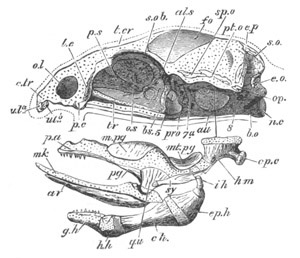
Fig. 335. Young Salmon of the first summer, about 2 inches long;
side view of skull, excluding branchial arches. (From Parker.)
The palato-mandibular and hyoid tracts are detached from their
proper situations, a line indicating the position where the
hyomandibular is articulated beneath the pterotic ridge.
ol. olfactory fossa; c.tr. trabecular cornu; ula. ulb.
upper labial cartilages; p.s. presphenoid tract; t.cr. tegmen
cranii; s.o.b. supraorbital band; fo. superior fontanelle;
n.c. notochord; b.o. basilar cartilage; tr. trabecula; p.c.
condyle for palatine cartilage; 5. trigeminal foramen; 7a. facial
foramen; 8. foramen for glossopharyngeal and vagus nerves; mk.
Meckelian cartilage; op.c. opercular condyle.
Bones: e.o. exoccipital; s.o. supraoccipital; e.p. epiotic;
pt.o. pterotic; sp.o. sphenotic; op. opisthotic; pro.
prootic; b.s. basisphenoid; al.s. alisphenoid; o.s.
orbitosphenoid; l.e. ectethmoid or lateral ethmoid; pa.
palatine; pg. pterygoid; m.pg. mesopterygoid; mt.pg.
metapterygoid; qu. quadrate; ar. articular; h.m.
hyomandibular; sy. symplectic; i.h. interhyal; ep.h.
epiceratohyal; c.h. ceratohyal; h.h. hypohyal; g.h. glosso- or
basihyal.
The hyoid arch, though largely ossified, undergoes a process of
development very similar to that in Raja. It is formed as a simple
cartilaginous bar, which soon becomes segmented longitudinally
into an
anterior and a posterior part (fig. 334). The former constitutes the
hyomandibular (H.M), while the latter, becoming more and more
separated from the hyomandibular, constitutes the hyoid arch proper;
owing to the disappearance of the hyobranchial cleft, it loses its
primitive function, and serves on the one hand to support the
operculum covering the gills, and on the other to support the tongue.
It becomes segmented into a series of parts which are ossified (fig.
335) as the epiceratohyal (ep.h) above, then a large ceratohyal
(ch), followed by a hypohyal (hh), while the median ventral
element forms the basi- or glossohyal (g.h).
The hyomandibular itself is articulated with the skull below the
pterotic process (fig. 334, H.M). Its upper element ossifies as the
hyomandibular (fig. 335, h.m.), while its lower part (fig. 334,
Sy), which is firmly connected with the mandibular arch, ossifies as
the symplectic (fig. 335, sy). A connecting element between the two
parts of the hyoid bar forms an interhyal (ih).
There are more important differences in the development of the
mandibular arch in Elasmobranchii and the Salmon than in that of the
hyoid arch, in that, instead of the whole arcade of the upper jaw
being formed from the mandibular arch, a fresh element, in the form of
an independently developed bar of cartilage, completes the upper
arcade in front; but even with this bar the two halves of the upper
branch of the arch do not meet anteriorly, but are separated by the
ends of the trabeculæ.
The anterior bar of the upper arcade is known as the palatine; but it
appears to me as yet uncertain how far it is to be regarded as an
element, primitively belonging to the upper arcade of the mandibular
arch, which has become secondarily independent in its development; or
as an entirely distinct structure which has no counterpart in the
Elasmobranch upper jaw. The latter view is adopted by Parker and
Bridge, and a cartilage attached to the hinder wall of the nasal
capsule of many Elasmobranchii is identified by them with the palatine
rod of the Teleostei.
The arch itself is at first very similar to the succeeding arches; its
dorsal extremity soon however becomes broadened, and provided with an
anteriorly directed process. This part (fig. 334, M.Pt and Qu) is
then segmented from the lower region,
and forms what may be called the
pterygo-quadrate cartilage, though not completely homologous with the
similarly named cartilage in Elasmobranchs; while the lower region
forms the Meckelian cartilage (Mck), which has already grown
inwards, so as to meet its fellow ventrally below the mouth. The whole
arch becomes at the same time widely separated from the axial parts of
the skull.
Nearly simultaneously with the first differentiation of the mandibular
arch, a bar of cartilage—the palatine bar already spoken of—is
formed on each side, below the eye, in front of the mouth. The dilated
anterior extremity of this bar soon comes in contact with an anterior
process of the trabeculæ, known as the ethmo-palatine process.
In a later stage the pterygoid end of the pterygo-quadrate cartilage
unites with the distal end of the palatine bar (fig. 334, Pl.Pt),
and there is then formed a continuous cartilaginous arcade for the
upper jaw, which is strikingly similar to the cartilaginous upper jaw
of Elasmobranchii.
A large dorsal process of the primitive pterygo-quadrate now forms a
large metapterygoid tract (M.Pt); while the whole arch becomes
firmly bound to the hyomandibular (H.M).
In the later stages the parts formed in cartilage become ossified
(fig. 335). The palatine is first ossified, the pterygoid region of
the pterygo-quadrate is next ossified as a dorsal mesopterygoid
(m.pg) and a ventral pterygoid proper (pg). The quadrate region,
articulating with the Meckelian cartilage, becomes ossified as a
distinct quadrate (qu), while the dorsal region becomes also
ossified as a metapterygoid (mt.pg).
In the Meckelian cartilage a superficial ossification of the ventral
edge and inner surface forms an articulare (ar); but the greater
part of the cartilage persists through life.
Some of the above ossifications, at any rate those of the palatine and
pterygoid, seem to be started by dental osseous plates adjoining the
cartilage. They will be spoken of further in the section dealing with
the membrane bones.
Amphibia. The development of the autostylic piscine skulls
has unfortunately not yet been studied; and the most primitive
autostylic types whose development we are acquainted with are
those of the Amphibia; on which a large amount of light has
been shed by the researches of Huxley and Parker.
The modifications of the hyoid arch are comparatively simple
and uniform. It forms a rod of cartilage, which soon articulates
in front with the quadrate element of the mandibular arch, and
is subsequently attached by ligaments both to the quadrate and
to the cranium. In those Amphibia in which external gills and
gill clefts are lost, it fuses with the basal element of the hyoid
(fig. 330), which, together with the basal portions of the following
arches, forms a continuous cartilaginous plate. On the completion
of these changes the paired parts of the hyoid arch have
the form of two elongated rods, known as the anterior cornua of
the hyoid, which attach the basihyal plate to the cranium behind
the auditory capsule.
It is still uncertain whether there is any distinct element corresponding
to the hyomandibular of fishes.
Parker holds that the columella auris of the Anura is the homologue of
the hyomandibular. The columella develops comparatively late and
independently of the remainder of the hyoid arch, but the similarity
between its relations to the nerves and those of the hyomandibular is
put forward by Parker as an argument in favour of his view. The early
ligamentous connection between the quadrate and the upper end of the
primitive hyoid is however an argument in favour of regarding the
upper end of the primitive hyoid as the hyomandibular element, not
separated from the remainder of the arch.
The history of the mandibular arch is more complicated than that of
the hyoid. The part of it which corresponds with the upper jaw of
Elasmobranchii exhibits most striking variations in development; so
striking indeed as to suggest that the secondary modifications it has
undergone are sufficiently considerable to render great caution
necessary in drawing morphological conclusions from the processes
which are in some instances observable. A more satisfactory judgment
on this point will be possible after the publication of a memoir with
which Parker is now engaged on the skulls of the different Anura.
The membrane bones applying themselves to the sides of the mandibular
arch are relatively far more important than in the lower types. This
is especially the case with the upper jaw where the maxillary and
premaxillary bones functionally replace the primitive cartilaginous
jaw; while membranous pterygoids
and palatines apply themselves to,
and largely take the place of, the cartilaginous palatine and
pterygoid bars.
Two types worked out by Parker, viz. the Axolotl and the common Frog,
may be selected to illustrate the development of the mandibular arch.
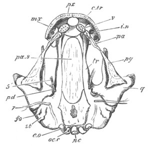
Fig. 336. Young Axolotl, 2¼ inches long; under view of skull,
dissected, the lower jaw and gill arches having been removed.
(From Parker.)
nc. notochord; oc.c. occipital condyle; f.o. fenestra ovalis;
st. stapes; tr. trabecular cartilage; i.n. internal nares;
c.tr. cornu trabeculæ; pd. pedicle of quadrate; q. quadrate;
pg. outline of pterygoid cartilage; 5´. orbito-nasal nerve; 7.
facial nerve.
Bones: pa.s. parasphenoid; e.o. exoccipital; v. vomer; px.
premaxillary; mx. maxillary; pa. palatine; pg. pterygoid.
In the Axolotl, which may be taken as the type for the Urodela, the
mandibular arch is constituted at a very early stage of (1) an
enlarged dorsal element, corresponding with the pterygo-quadrate of
the lower types, but usually known as the quadrate; and (2) a ventral
or Meckelian element. The Meckelian bar very early acquires its
investing bones, while the dorsal part of the quadrate becomes divided
into two characteristic processes, viz. an anterior dorsal process
which grows towards and soon permanently fuses with the trabecular
crest, and a posterior process known as the otic process, which
applies itself to the outer side of the auditory region. The anterior
of these processes, as pointed out by Huxley, is probably homologous
with the anterior process of the pterygo-quadrate bar in Notidanus,
which articulates with the trabecular region of the cranium, while the
otic process is homologous with the metapterygoid
process. Hardly any
trace is present of an anterior process to form a pterygoid bar, but
dentigerous plates forming a dermal palatopterygoid bar have already
appeared.
At a somewhat later stage a fresh process, called by Huxley the
pedicle, grows out from the quadrate, and articulates with the ventral
side of the auditory region (fig. 336, pd). Shortly afterwards a rod
of cartilage grows forward from the quadrate under the membranous
pterygoid (pg), which corresponds with the cartilaginous pterygoid
bar of other types (fig. 336), and an independent palatine bar,
arising even before the pterygoid process, is formed immediately
dorsal to the dentigerous palatine plate (pa), and is attached to
the trabecula. These two bars eventually meet, but never become firmly
united to the more important membrane bones placed superficially to
them.
The mandibular arch in the Frog stands, so far as development is
concerned, in striking contrast to the mandibular arch of the Axolotl,
in spite of the obvious similarity in the arrangement of the adult
parts in the two types.
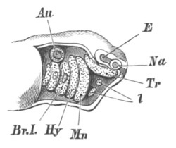
Fig. 337. Embryo Frog, just before
hatching; side view of head,
with skin removed. (From Parker.)
Na. olfactory sack; E. involution for eyeball; Au. auditory
sack; Tr. trabecula; Mn. mandibular; Hy. hyoid; Br.I. first
branchial arch; the gill-buds are seen on the first two branchial
arches; l. labial cartilages.
In the earliest stage it forms a simple bar in the membranous
mandibular arch, parallel to and very similar to the hyoid bar behind
(fig. 337, Mn). In the next stage observed, that is to say in
Tadpoles of four, five, to six lines long, an astonishing
transformation has taken place. The mandibular arch (fig. 338) is
turned directly forwards parallel to the trabecula, to which it is
attached in front (p.pg) and behind (pd). The proximal part of the
arch thus forms a subocular bar, and the space between it and the
trabecula a subocular fenestra. In front of the anterior attachment it
is continued forwards for a short distance, and to the free end of
this projecting part is articulated a small Meckelian cartilage
directed upwards (mk). The Meckelian cartilage is at this stage
placed in front of the nasal sacks, in the lower lip of the suctorial
mouth. The greater part of the arch, parallel with the trabeculæ, is
equivalent to what has been called in the Axolotl the quadrate, while
its anterior attachment to the trabeculæ is the rudiment of the
palatopterygoid cartilage. The posterior attachment is known as the
pedicle.
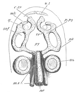
Fig. 338. Tadpole of Common Toad, one-third of an inch long;
cranial and mandibular cartilages seen from above; the parachordal
cartilages are not yet definite. (From Parker.)
nc. notochord; ms. muscular segments; au. auditory capsule;
py. region of pituitary body; tr. trabecula; c.tr. cornu
trabeculæ; p.pg. palatopterygoid bar; pd. pedicle; q. quadrate
condyle; mk. Meckelian piece of mandibular arch; so.f. subocular
fenestra; u.l. upper labial cartilage. The dotted circle within
the quadrate region indicates the position of the internal nostril.
The condition of the mandibular arch during this and the next stage
(fig. 339) is very perplexing. Its structure appears adapted in some
way to support the suctorial mouth of the Tadpole.
Reasons have been offered in a previous part of this volume for
supposing that the suctorial mouth of the Tadpole is probably not
simply a structure secondarily acquired by this larva, but is an organ
inherited from an ancestor provided through life with a suctorial
mouth.
The question thus arises, is the peculiar modification of the
mandibular arch of the Tadpole an inherited or an acquired
feature?
If the first alternative is accepted we should have to admit that the
mandibular arch became first of all modified in connection with the
suctorial mouth, before it was converted into the jaws of the
Gnathostomata; and that the peculiar history of this arch in the
Tadpole is a more or less true record of its phylogenetic development.
In favour of this
view is the striking similarity which Huxley has
pointed out between the oral skeleton of the Lamprey and that of the
Tadpole; and certain peculiarities of the mandibular arch of Chimæra
and the Dipnoi can perhaps best be explained on the supposition that
the oral skeleton of these forms has arisen in a manner somewhat
similar to that in the Frog; though with reference to this point
further developmental data are much required.
On the other hand the above suppositions would necessitate our
admitting that a great abbreviation has occurred in the development of
the mandibular arch of the otherwise more primitive Urodela; and that
the simple mode of growth of the jaws in Elasmobranchii, from the
primitive mandibular arch, is phylogenetically a much abbreviated and
modified process, instead of being, as usually supposed, a true record
of ancestral history.
If the view is accepted that the characters of the mandibular arch of
the Tadpole are secondary, it will be necessary to admit that the
adaptation of the mandibular arch to the suctorial mouth took place
after the suctorial mouth had come to be merely a larval organ.
In view of our imperfect knowledge of the development of most Piscine
skulls I would refrain from expressing a decided opinion in favour of
either of these alternatives.
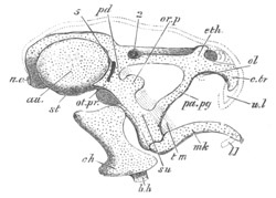
Fig. 339. Tadpole with tail beginning to shrink; side view of skull
without the branchial arches. (From Parker.)
n.c. notochord; au. auditory capsule; between it and eth. the
low cranial side wall is seen; eth. ethmoidal region; st.
stapes; 5. trigeminal foramen; 2. optic foramen; ol. olfactory
capsules, both seen owing to slight tilting of the skull; c.tr.
cornu trabeculæ; u.l. upper labial, in outline; su. suspensorium
(quadrate); pd. its pedicle; ot.pr. its otic process; or.p.
its orbitar process; t.m. temporal muscle, indicated by dotted
lines passing beneath the orbitar process; pa.pg. palatopterygoid
bar; mk. Meckelian cartilage; l.l. lower labial, in outline;
c.h. ceratohyal; b.h. basihyal. The upper outline of the head is
shewn by dotted lines.
As the tail of the Tadpole gradually disappears, and the metamorphosis
into the Frog becomes accomplished, the mandibular arch undergoes
important changes (fig. 339): the
palatopterygoid attachment (pa.pg)
of the quadrate subocular bar becomes gradually elongated; and, as it
is so, the front end of the subocular bar (su) rotates outwards and
backwards, and soon forms a very considerable angle with the
trabeculæ. The Meckelian cartilage (mk) at its free end becomes at
the same time considerably elongated. These processes of growth
continue till (fig. 330) the palatopterygoid bar (Pt) forms a
subocular bar, and is considerably longer than the original subocular
region of the quadrate; while the Meckelian cartilage (Mck) has
assumed its permanent position on the hinder border of the no longer
suctorial mouth, and has grown forwards so as nearly to meet its
fellow in the median line.
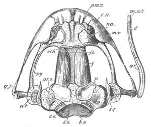
Fig. 340. Young Frog, near end of first summer; upper view of
skull, with left mandible removed, and the right extended outwards.
(From Parker.)
b.o. basioccipital tract; s.o. supraoccipital tract; fo.
frontal fontanelle; e.n. external nostril; internal to it,
internasal plate; a.t. tympanic annulus.
Bones: e.o. exoccipital; pr.o. prootic, partly overlapped by
p. parietal; f. frontal; eth. rudiment of sphenethmoid; na.
nasal; pmx. premaxillary; mx. maxillary; pg. pterygoid, partly
ensheathing the reduced cartilage; q.j. quadratojugal; sq.
squamosal; ar. articular; d. dentary; m.mk. mento-Meckelian.
The metapterygoid region of the quadrate gives rise to a posterior and
dorsal process (fig. 339, ot.pr), the end of which is constricted
off as the tympanic annulus (fig. 340, a.t); while the proximal part
of the process remains as the otic (metapterygoid) process,
articulating with the auditory cartilage.
The pedicle (pd) retains its original attachment to the skull.
The palatopterygoid soon becomes segmented into a transversely placed
palatine, and a longitudinally placed pterygoid (fig. 340). With the
exception of a few ossifications, which present no features of special
interest, the parts of the mandibular arch have now reached their
final condition, which is not very different from that in the Axolotl.
Sauropsida. In the Sauropsida the modifications of the hyoid and
mandibular arches are fairly uniform.
The lower part of the hyoid arch, including the basihyoid, unites with
the remnants of the arches behind to form the hyoid bone, to which it
contributes the anterior cornu and anterior part of the body.
The columella is believed by Huxley and Parker to represent, as in the
Anura, the independently developed dorsal (hyomandibular) element of
the hyoid, together with the stapes with which it has become
united[205].
The membranous mandibular arch gives off in the embryos of all the
Sauropsida an obvious bud to form the superior maxillary process, and
the formation of this bud appears to represent the growth forwards of
the pterygoid process in Elasmobranchii, which is indeed accompanied
by the formation of a similar bud; but the skeletal rod, which appears
in the axis of this bud, is as a rule independent of that in the true
arch (fig. 331, pa, pg). The former is the pterygo-palatine bar;
the latter the Meckelian and quadrate cartilages.
The pterygo-palatine bar is usually if not always ossified directly,
without the intervention of cartilage.
Born has recently shewn that Parker was mistaken in supposing that the
palatopterygoid bone is cartilaginous in Birds. In the Turtle a short
cartilaginous pterygoid process of the quadrate would seem to be
present (Parker, No. 458).
The quadrate and Meckelian cartilages are either from the first
separate, or very early become so.
The quadrate cartilage ossifies as the quadrate bone, and supplies the
permanent articulation for the lower jaw. Its upper end exhibits a
tendency to divide into two processes, corresponding with the pedicle
and otic processes of the Amphibia. The Meckelian cartilage becomes
soon covered by investing bones, and its proximal end ossifies as the
articulare. The remainder of the cartilage usually disappears.
Mammalia. The most extraordinary metamorphosis of the hyoid and
mandibular arches occurs in the Mammalia, and has been in part known
since the publication of the memoir of Reichert (No. 461).
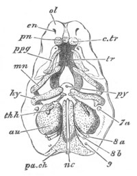
Fig. 341. Embryo Pig, two-thirds of an inch long; elements of the
skull seen somewhat diagrammatically from below. (From Parker.)
pa. ch. parachordal cartilage; nc. notochord; au. auditory
capsule; py. pituitary body; tr. trabeculæ; c.tr. trabecular
cornu; pn. prenasal cartilage; e.n. external nasal opening;
ol. nasal capsule; p.pg. palatopterygoid tract enclosed in the
maxillopalatine process; mn. mandibular arch; hy. hyoid arch;
th.h. first branchial arch; 7a. facial nerve; 8a.
glossopharyngeal; 8b. vagus; 9. hypoglossal.
Both the hyoid and mandibular arches develop at first more completely
than in any of the other types above Fishes; and are articulated to
each other above, while the pterygo-palatine bar is quite distinct.
The main features of the subsequent development are undisputed, with
the exception of that of the upper end of the hyoid, which is still
controverted. The following is Parker’s (No. 452) account for the Pig,
which confirms in the main the view originally put forward by Huxley
(No. 445).
The mandibular and hyoid arches are at first very similar
(fig. 341
mn and hy), their dorsal ends being somewhat incurved, and
articulating together.
In a somewhat later stage (fig. 342) the upper end of the mandibular
bar (mb), without becoming segmented from the ventral part, becomes
distinctly swollen, and clearly corresponds to the quadrate region of
other types. The ventral part of the bar constitutes the Meckelian
cartilage (mk).
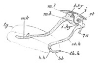
Fig. 342. Embryo Pig, an inch and a third long; side view of
mandibular and hyoid arches. The main hyoid arch is seen as displaced
backwards after segmentation from the incus. (From Parker.)
tg. tongue; mk. Meckelian cartilage; ml. body of malleus;
mb. manubrium or handle of the malleus; t.ty. tegmen tympani;
i. incus; st. stapes; i.hy. interhyal ligament; st.h.
stylohyal cartilage; h.h. hypohyal; b.h. basibranchial; th.h.
rudiment of first branchial arch; 7a. facial nerve.
The hyoid arch has in the meantime become segmented into two parts, an
upper part (i), which eventually becomes one of the small bones of
the ear—the incus—and a lower part which remains permanently as the
anterior cornu of the hyoid (st.h). The two parts continue to be
connected by a ligament.
The incus is articulated with the quadrate end of the mandibular arch,
and its rounded head comes in contact with the stapes (fig. 342, st)
which is segmented from the fenestra ovalis. The main arch of the
hyoid becomes divided into a hypohyal (h.h) below and a stylohyal
(st.h) above, and also becomes articulated with the basal element of
the arch behind (bh).
In the course of further development the Meckelian part of the
mandibular arch becomes enveloped in a superficial ossification
forming the dentary. Its upper end, adjoining the quadrate region,
becomes calcified and then absorbed, and its lower, with the exception
of the extreme point, is ossified and subsequently incorporated in the
dentary.
The quadrate region remains relatively stationary in growth
as
compared with the adjacent parts of the skull, and finally ossifies to
form the malleus bone of the ear. The processus gracilis of the
malleus is the primitive continuation into Meckel’s cartilage.
The malleus and incus are at first embedded in the connective tissue
adjoining the tympanic cavity (hyomandibular cleft, vide p. 528);
and externally to them a bone known as the tympanic bone becomes
developed so that they become placed between the tympanic bone and the
periotic capsule. In late fœtal life they become transported
completely within the tympanic cavity, though covered by a reflection
of the tympanic mucous membrane.
The dorsal end of the part of the hyoid separated from the incus
becomes ossified as the tympano-hyal, and is anchylosed with the
adjacent parts of the periotic capsule. The middle part of the bar
just outside the skull forms the stylohyal (styloid process in Man)
which is attached by ligament to the anterior cornu of the hyoid
(ceratohyal).
While the account of the formation of the malleus, incus, and stapes
just given is that usually accepted in this country, a somewhat
different view of the development of these parts has as a rule been
adopted in Germany. Reichert (No. 461) held that both the malleus and
the incus were derived from the mandibular bar; and this view has been
confirmed by Günther, Kölliker and other observers, and has recently
been adopted by Salensky (No. 462) after a careful research especially
directed towards this point. Reichert also held that the stapes was
derived from the hyoid bar; but, though his observations on this point
have been very widely accepted, they have not met with such universal
recognition as his views on the origin of the malleus and incus.
Salensky has recently arrived at a view, which is in accord with that
of Parker, in so far as the independence of the stapes of both the
hyoid and mandibular arches is concerned. Salensky however holds that
it is formed from a mass of mesoblast surrounding the artery of the
mandibular arch, and that the form of the stapes is due to its
perforation by the mandibular artery. A product of this artery
permanently perforates the stapes in a few Mammalia, though in the
majority it atrophies.
In view of the different accounts of the origin of the incus the exact
nature of this bone must still be considered as an open question, but
should Reichert’s view be confirmed the identification of the incus
with the columella of the Amphibia and Sauropsida must be abandoned.
Membrane bones and ossifications of the cranium.
The membrane bones of the skull may be divided into two classes, viz.
(1) those derived from dermal osseous plates, which as explained above
(p. 542) are primitively formed by the coalescence of the osseous
plates of scales; and (2) those formed by the coalescence of the
osseous plates of teeth lining the oral cavity. Some of the bones
sheathing the edge of the mouth have been formed partly by the one
process and partly by the other.
In the Fishes there are found all grades of transition between simple
dermal scutes, and true subdermal osseous plates forming an integral
part of the internal skeleton. Dermal scutes are best represented in
Acipenser and some Siluroid Fishes.
Where the membrane bones still retain the character of dermal plates,
those on the dorsal surface of the cranium are usually arranged in a
series of longitudinal rows, continuing in the region of the head the
rows of dermal scutes of the trunk; while the remaining cranial scutes
are connected with the visceral arches. The dermal bones on the dorsal
surface of the head are very different in number, size, and
arrangement in different types of Fishes; but owing to their linear
disposition it is usually possible to find a certain number both of
the paired and unpaired bones which have a similar situation in the
different forms. These usually receive the same names, but both from
general considerations as to their origin, as well as from a
comparison of different species, it appears to me probable that there
is no real homology between these bones in different species, but only
a kind of general correspondence[206].
It is not in fact till we get to the types above the Fishes that we
can find a series of homologous dorsal membrane bones covering the
roof of the skull. In these types three paired sets of such bones are
usually present, viz. from behind forwards the parietals, frontals and
nasals, the latter bounding the posterior surface of the external
nasal opening. Even in the higher
types these bones are liable to vary
very greatly from the usual arrangement.
Besides these bones there is usually present in the higher forms a
lacrymal bone on the anterior margin of the orbit derived from one of
a series of periorbital membrane bones frequently found in Fishes.
Various supraorbital and postorbital bones, etc. are also frequently
found in Lacertilia, etc. which are not impossibly phylogenetically
independent of the membrane bones inherited from Fishes; and may have
been evolved as bony scutes in the subdermal tissue of the papillæ of
the sauropsidan scales.
The visceral arches of Fishes, especially of the Teleostei, are
usually provided with a series of membrane bones. In the true
branchial arches these take the form of dentigerous plates; but no
such plates are found in the Amphibia or Amniota.
The opercular flap attached to the hyoid arch is usually supported by
a series of membrane bones, which attain their highest development in
the Teleostei. One of these bones, the præopercular, is very constant
and is primitively attached along the outer edge of the hyomandibular.
It seems to be retained in Amphibia as a membrane bone, overlapping
the attachment of the quadrate and known as the squamosal; though it
is not impossible that this bone may be derived from a superficial
membrane bone, widely distributed in Teleostei and Ganoids, which is
known as the supra-temporal. In Dipnoi the bone which appears to be
clearly homologous with the squamosal would seem from its position to
belong to the series of dorsal plates, and therefore to be the
supra-temporal; but it is regarded by Huxley (No. 446) as the
præopercular[207].
In the Amniota the squamosal forms an integral part of the osseous
roof of the skull; but in the Sauropsida it continues, as in Amphibia,
to be closely related to the quadrate.
A larger series of persistent membrane bones are related to the
mandibular, and its palato-quadrate process.
Overlying the palato-quadrate process are two rows of bones,
one row
lying at the edge of the mouth, on the outer side of the
pterygo-palatine process, and the other set on the roof of the mouth
superficial to the pterygo-palatine process.
The outer row is formed of the præmaxilla, maxilla, jugal, and very
often quadratojugal. Of these bones the maxilla and præmaxilla, as is
more especially demonstrated by their ontogeny in the Urodela, are
partly derived from dentigerous plates and partly from membrane plates
outside the mouth; while the jugal, and quadratojugal when present,
are entirely extra-oral. In the Amphibia and Amniota the præmaxillæ
and maxillæ are the most important bones in the facial region, and are
quite independent of any cartilaginous substratum.
The second row of bones is clearly constituted in the Dipnoi and
Amphibia by the vomer in front, then the palatine, and finally the
pterygoid behind. Of these bones the vomer is never related to a
cartilaginous tract below, while the palatines and pterygoids usually
are so. The position and growth of the three bones in many Urodela
(Axolotl) are especially striking (Hertwig. No. 442). In the Axolotl
they form a continuous series, the vomer and palatine being covered by
teeth, but the pterygoid being without teeth. The vomer and palatine
originate from the united osseous plates of the bases of the teeth,
while the pterygoid is in the first instance continuous with the
palatine.
In Teleostei, Amia, etc., there are dentigerous plates forming a
palatine and pterygoid, which in position, at any rate, closely
correspond with the similarly named bones in Amphibia; and there is
also a dentigerous vomer which may fairly be considered as equivalent
to that in Amphibia.
In the Amniota the three bones found in Amphibia are always present,
but with a few exceptions amongst the Lacertilia and Ophidia, are no
longer dentigerous. The cartilaginous bars, which in the lower types
are placed below the palatine and pterygoid membrane bones, are
usually imperfectly or not at all developed.
On Meckel’s cartilage important membrane bones are almost always
grafted. On the outside and distal part of the cartilage a dentary is
usually developed, which may envelope and replace the cartilage to a
larger or smaller extent. Its oral edge
is usually dentigerous. The
splenial membrane bone is the most important bone on the inner side of
Meckel’s cartilage, but other elements known as the coronoid and
angular may also be added. In Mammalia the dentary is the only element
present (vide p. 590).
On the roof of the mouth a median bone, the parasphenoid, is very
widely present in the Amphibia and Fishes, except the Elasmobranchii
and Cyclostomata, and has no doubt the same phylogenetic origin as the
vomer and membranous palatines and pterygoids.
It is less important in the Sauropsida, and becomes indistinguishably
fused with the sphenoid in the adult, while in Mammalia it is no
longer found.
Ossification of the Cartilaginous Cranium. In certain Fishes the
cartilaginous cranium remains quite unossified, while completely
enveloped in dermal bones. Such for instance is its condition in the
Selachioid Ganoids. In most instances, however, the investment of the
cartilaginous cranium by membrane bones is accompanied by a more or
less complete ossification of the cartilage itself.
In the Dipnoi this occurs to the smallest extent, the only
ossifications occurring in the lateral parts of the occipital region,
and forming the exoccipitals.
In Teleostei and bony Ganoids, a considerably greater number of
ossifications occur in the cartilage.
In the region of the occipital cartilaginous ring there appears a
basioccipital and supraoccipital and two exoccipitals. The
basioccipital is the only bone on the floor of the skull ossifying
that part into which the notochord is primitively continued[208].
In the region of the periotic cartilage a large number of bones may
appear. In front there is the prootic, which often meets the
exoccipital behind; behind there is above and in close connection with
the supraoccipital the epiotic, and below in close connection with the
exoccipital the opisthotic. On the dorsal side of the cartilage there
is a projecting ridge composed mainly of a bone known as the pterotic,
sometimes erroneously
called the squamosal, and continued in front by
the sphenotic. The pterotic, or the cartilaginous region corresponding
to it, always supplies the articular surface for the hyomandibular.
In the floor of the skull, in the region of the pituitary body, there
is formed a basisphenoid; while in the lateral parts of the wall of
this part of the cranium, there is a bone known as the alisphenoid.
In front, parts of the lateral walls of the cranium ossify as the
orbitosphenoids.
In view of the very imperfect ossification of the cartilaginous
cranium of the Dipnoi, and of the fact that there is certainly no
direct genetic connection between the Teleostei on the one hand, and
the Amphibia and Amniota on the other, it is very difficult to believe
that most of the ossifications of the cranium in the Amphibia and
Amniota have more than a general correspondence with those in the
Teleostei.
In the Amphibia the ossifications in the cartilage are comparatively
few. In the occipital region there is a lateral ossification on each
side of the exoccipital, the basioccipital region being unossified,
and the supraoccipital at the utmost indurated by a calcareous
deposit.
The periotic capsule is ossified by a prootic centre, which meets the
exoccipital behind.
The front part of the cartilaginous cranium is ossified by a complete
ring of bone—the sphenethmoid bone—which embraces part of the
ethmoid region, and of the orbitosphenoid and presphenoid regions.
In the Amphibia the cartilaginous cranium, with its centres of
ossification, is easily separable from the membranous investing bones.
In the Amniota the cartilaginous cranium, whose development in the
embryo has already been described, becomes in the adult much more
largely ossified, and the bones which replace the primitive cartilage
unite with the membrane bones to form a continuous bony cranium.
The centres of ossification become again much more numerous. In the
occipital segment analogous centres to those of Teleostei are again
found; and it is probable that the exoccipitals are homologous
throughout the series, the supraoccipital and basioccipital
bones of
the higher types being merely identical in position with the similarly
named bones in Fishes.
In the periotic there are usually three centres of ossification, first
recognised by Huxley. These are the prootic, the epiotic and
opisthotic, the situations of which have already been defined. Of
these the prootic is the most constant.
In Reptiles, the prootic and opisthotic frequently remain distinct
even in the adult.
In Birds, the epiotic and opisthotic are early united with the supra-
and exoccipital; and at a later period the prootic is also
indistinguishably fused with the adjacent parts.
In Mammals the three ossifications fuse into a continuous whole—the
periotic bone—which may be partially united with the adjacent parts.
In the pituitary region of the base of the cranium a pair of osseous
centres or in the higher types a single centre (Parker[209]) gives
rise to the basisphenoid bone, and in front of this another basal or
pair of basal ossifications forms the presphenoid, while laterally to
these two centres there are formed centres of ossification in the
alisphenoid and orbitosphenoid regions, which may be extremely reduced
in various Sauropsida, leaving the side walls of the skull almost
entirely formed of membrane or cartilage.
In the ethmoid region there may arise a median ossification forming
the mesethmoid and lateral ossifications forming the lateral ethmoids
or prefrontals; which may assist in forming the front wall of the
brain-case, or be situated quite externally to the brain-case and be
only related to the olfactory capsules.
The labial cartilages. In most Fishes a series of skeletal structures,
known as the labial cartilages, are developed at the front and sides
of the mouth, and in connection with the olfactory capsules; and these
cartilages still persist in connection with the olfactory capsules,
though in a reduced form, in the higher types. They are more developed
in the Cyclostomata than in any other Vertebrate type.
The meaning of these cartilages is very obscure; but, from their being
in part employed to support the lips and horny teeth of the
Cyclostomata and the Tadpole, I should be inclined to regard them as
remnants of a primitive skeleton supporting the suctorial mouth, with
which, on the grounds already stated (p. 317), I believe the ancestors
of the present Vertebrata to have been provided.
Bibliography.
(439) A. Dugès. “Recherches sur l'Ostéologie et la myologie des
Batraciens à leur différents âges.” Paris, Mém. savans étrang. 1835,
and An. Sci. Nat. Vol. I. 1834.
(440) C. Gegenbaur. Untersuchungen z. vergleich. Anat. d.
Wirbelthiere, III. Heft. Das Kopfskelet d. Selachier. Leipzig,
1872.
(441) Günther. Beob. üb. die Entwick. d. Gehörorgans. Leipzig, 1842.
(442) O. Hertwig. “Ueb. d. Zahnsystem d. Amphibien u. seine Bedeutung
f. d. Genese d. Skelets d. Mundhöhle.” Archiv f. mikr. Anat., Vol.
XI. 1874, suppl.
(443) T. H. Huxley. “On the theory of the vertebrate skull.” Proc.
Royal Soc., Vol. ix. 1858.
(444) T. H. Huxley. The Elements of Comparative Anatomy. London,
1869.
(445) T. H. Huxley. “On the Malleus and Incus.” Proc. Zool. Soc.,
1869.
(446) T. H. Huxley. “On Ceratodus Forsteri.” Proc. Zool. Soc., 1876.
(447) T. H. Huxley. “The nature of the craniofacial apparatus of
Petromyzon.” Journ. of Anat. and Phys., Vol. X. 1876.
(448) T. H. Huxley. The Anatomy of Vertebrated Animals. London,
1871.
(449) W. K. Parker. “On the structure and development of the skull of
the Common Fowl (Gallus Domesticus).” Phil. Trans., 1869.
(450) W. K. Parker. “On the structure and development of the skull of
the Common Frog (Rana temporaria).” Phil. Trans., 1871.
(451) W. K. Parker. “On the structure and development of the skull in
the Salmon (Salmo salar).” Bakerian Lecture, Phil. Trans., 1873.
(452) W. K. Parker. “On the structure and development of the skull in
the Pig (Sus scrofa).” Phil. Trans., 1874.
(453) W. K. Parker. “On the structure and development of the skull in
the Batrachia.” Part II. Phil. Trans., 1876.
(454) W. K. Parker. “On the structure and development of the skull in
the Urodelous Amphibia.” Part III. Phil. Trans., 1877.
(455) W. K. Parker. “On the structure and development of the skull in
the Common Snake (Tropidonotus natrix).” Phil. Trans., 1878.
(456) W. K. Parker. “On the structure and development of the skull in
Sharks and Skates.” Trans. Zoolog. Soc., 1878. Vol. X. pt. IV.
(457) W. K. Parker. “On the structure and development of the skull in
the Lacertilia.” Pt. I. Lacerta agilis, L. viridis and Zootoca
vivipara. Phil. Trans., 1879.
(458) W. K. Parker. “The development of the Green Turtle.” The
Zoology of the Voyage of H. M. S. Challenger. Vol. I. pt. V.
(459) W. K. Parker. “The structure and development of the skull in the
Batrachia.” Pt. III. Phil. Trans., 1880.
(460) W. K. Parker and G. T. Bettany. The Morphology of the Skull.
London, 1877.
(460*) H. Rathke. Entwick. d. Natter. Königsberg, 1839.
(461) C. B. Reichert. “Ueber die Visceralbogen d. Wirbelthiere.”
Müller’s Archiv, 1837.
(462) W. Salensky. “Beiträge z. Entwick. d. knorpeligen
Gehörknöchelchen.” Morphol. Jahrbuch, Vol. VI. 1880.
Vide also Kölliker (No. 298), especially for the human and mammalian
skull; Götte (No. 296).
The Pectoral girdle.
Pisces. Amongst Fishes the pectoral girdle presents itself in its
simplest form in Elasmobranchii, where it consists of a bent band of
cartilage on each side of the body, of somewhat variable form, meeting
and generally uniting with its fellow ventrally. Its anterior border
is in close proximity with the last visceral arch, and a transverse
ridge on its outer and posterior border, forming the articular surface
for the skeleton of the limb, divides it into a dorsal part, which may
be called the scapula, and a ventral part which may be called the
coracoid.
In all the remaining groups of Fishes there is added to the
cartilaginous band, which may wholly or partially ossify, an osseous
support composed of a series of membrane bones.
In the types with such membrane bones the cartilaginous parts do not
continue to meet ventrally, except in the Dipnoi where there is a
ventral piece of cartilage, distinct from that bearing the
articulation of the limb. The cartilage is moreover produced into two
ventral processes, an anterior and a posterior, below the articulation
of the limb; which may be called, in accordance with Gegenbaur’s
nomenclature, the præcoracoid and coracoid. Of these the præcoracoid
is far the most
prominent, and in the majority of cases the coracoid
can hardly be recognised. The coracoid process is however well
developed in the Selachioid Ganoids, and the Siluroid Teleostei. In
Teleostei the scapular region often ossifies in two parts, the smaller
of which is named by Parker præcoracoid, though it is quite distinct
from Gegenbaur’s præcoracoid. The membrane bones, as they present
themselves in their most primitive state in Acipenser and the
Siluroids, are dermal scutes embracing the anterior edge of the
cartilaginous girdle. In Acipenser there are three scutes on each
side. A dorsal scute known as the supra-clavicle, connected above with
the skull by the post-temporal; a middle piece or clavicle, and a
ventral or infra-clavicle (interclavicle), which meets its fellow
below.
In most Fishes the primitive dermal scutes have become subdermal
membrane bones, and the infra-clavicle is usually not distinct, but
the two clavicles form the most important part of the membranous
elements of the girdle. Additional membrane bones (post-clavicles) are
often present behind the main row.
The development of these parts in Fishes has been but little studied.
In Scyllium, amongst the Elasmobranchii, I find that each half of the
pectoral girdle develops as a vertical bar of cartilage at the front
border of the rudimentary fin, and externally to the muscle plates.
Before the tissue forming the pectoral girdle has acquired the
character of true cartilage, the bars of the two sides meet ventrally
by a differentiation in situ of the mesoblastic cells, so that, when
the girdle is converted into cartilage, it forms an undivided arc,
girthing the ventral side of the body. There is developed in
continuity with the posterior border of this arc on the level of the
fin a horizontal bar of cartilage, which is continued backwards along
the insertion of the fin, and, as will be shewn in the sequel, becomes
the metapterygium of the adult (figs. 344, bp and 348, mp). With
this bar the remaining skeletal elements of the fin are also
continuous.
The foramina of the pectoral girdle are not in the first instance
formed by absorption, but by the non-development of the cartilage in
the region of pre-existing nerves and vessels.
The development of these parts in Teleostei has been recently
investigated by ’Swirski (No. 472) who finds in the Pike (Esox) that
the cartilaginous pectoral girdle is at first continuous with the
skeleton of the fin. It forms a rod with a dorsal scapular and ventral
coracoid process. An independent mass of cartilage gives rise to a
præcoracoid, which unites with the main mass, forming a triradiate bar
like that of Acipenser or the Siluroids. The coracoid process becomes
in the course of development gradually reduced.
’Swirski concludes that the so-called præcoracoid bar is to some
extent a secondary element, and that the coracoid bar corresponds to
the whole of the ventral part of the girdle of Elasmobranchii, but his
investigations do not appear to me to be as complete as is desirable.
Amphibia and Amniota. The pectoral girdle contains a more or less
constant series of elements throughout the Amphibia and Amniota; and
the differences in structure between the shoulder girdle of these
groups and that of Fishes are so great that it is only possible to
make certain general statements respecting the homologies of the parts
in the two sets of types.
The generally accepted view, founded on the researches of Parker,
Huxley, and Gegenbaur, is to the effect that there is a primitively
cartilaginous coraco-scapular plate, homologous with that in Fishes,
and that the membrane bones in Fishes are represented by the clavicle
and interclavicle in the Sauropsida and Mammalia, which are however
usually admitted to be absent in Amphibia. These views have recently
been challenged by Götte (No. 466) and Hoffmann (No. 467), on the
ground of a series of careful embryological observations; and until
the whole subject has been worked over by other observers it does not
seem possible to decide satisfactorily between the conflicting views.
It is on all hands admitted that the scapulo-coracoid elements of the
shoulder girdle are formed as a pair of cartilaginous plates, one on
each side of the body. The dorsal half of each plate becomes the
scapula, which may subsequently become divided into a supra-scapula
and scapula proper; while the ventral half forms the coracoid, which
is not always separated from the scapula, and is usually divided into
a coracoid proper, a præcoracoid, and an epicoracoid. By the
conversion of parts of the primitive cartilaginous plates into
membranous tissue various fenestræ may be formed in the cartilage, and
the bars
bounding these fenestræ both in the scapula and coracoid
regions have received special names; the anterior bar of the coracoid
region, forming the præcoracoid, being especially important. At the
boundary between the scapula and the coracoid, on the hinder border of
the plate, is placed the glenoid articular cavity to carry the head of
the humerus.
The grounds of difference between Götte and Hoffmann and other
anatomists concern especially the clavicle and interclavicle. The
clavicle is usually regarded as a membrane bone which may become to
some extent cartilaginous. By the above anatomists, and by Rathke
also, it is held to be at first united with the coraco-scapular plate,
of which it forms the anterior limb, free ventrally, but united
dorsally with the main part of the plate; and Götte and Hoffmann hold
that it is essentially a cartilage bone, which however in the
majority of the Reptilia ossifies directly without passing through the
condition of cartilage.
The interclavicle (episternum) is held by Götte to be developed from a
paired formation at the free ventral ends of the clavicles, but he
holds views which are in many respects original as to its homologies
in Mammalia and Amphibia. Even if Götte’s facts are admitted, it does
not appear to me necessarily to follow that his deductions are
correct. The most important of these is to the effect that the dermal
clavicle of Pisces has no homologue in the higher types. Granting that
the clavicle in these groups is in its first stage continuous with the
coraco-scapular plate, and that it may become in some forms
cartilaginous before ossifying, yet it seems to me all the same quite
possible that it is genetically derived from the clavicle of Pisces,
but that it has to a great extent lost even in development its
primitive characters, though these characters are still partially
indicated in the fact that it usually ossifies very early and
partially at least as a membrane bone[210].
In treating the development of the pectoral girdle systematically it
will be convenient to begin with the Amniota, which may be considered
to fix the nomenclature of the elements of the shoulder girdle.
Lacertilia. The shoulder girdle is formed as two membranous plates,
from the dorsal part of the anterior border of each of which a bar
projects (Rathke, Götte), which is free at its ventral end. This bar,
which is usually (Gegenbaur, Parker) held to be independent of the
remaining part of the shoulder girdle, gives rise to the clavicle and
interclavicle. The scapulo-coracoid plate soon becomes cartilaginous,
while at the same time the clavicular bar ossifies directly from the
membranous state. The ventral ends of the two clavicular bars enlarge
to form two longitudinally placed plates, which unite together and
ossify as the interclavicle.
Parker gives a very different account of the interclavicle in Anguis.
He states that it is formed of two pairs of bones ‘strapped on to the
antero-inferior part of the præsternum,’ which subsequently unite into
one.
Chelonia. The shoulder girdle of the Chelonia is formed (Rathke) of a
triradiate cartilage on each side, with one dorsal and two ventral
limbs. It is admitted on all hands that the dorsal limb is the
scapular element, and the posterior ventral limb the coracoid; but,
while the anterior ventral limb is usually held to be the præcoracoid,
Götte and Hoffmann maintain that, in spite of its being formed of
cartilage, it is homologous with the anterior bar of the primitive
shoulder-plates of Lacertilia, and therefore the homologue of the
clavicle.
Parker and Huxley (doubtfully) hold that the three anterior elements
of the ventral plastron (entoplastron and epiplastra) are homologous
with the interclavicle and clavicles, but considering that these
plates appear to belong to a secondary system of dermal ossifications
peculiar to the Chelonia, this homology does not appear to me probable.
Aves. There are very great differences of view as to the development
of the pectoral arch of Aves.
About the presence in typical forms of the coraco-scapular plate and
two independent clavicular bars all authors are agreed. With reference
to the clavicle and interclavicle Parker (No. 468) finds that the
scapular end of the clavicle attaches itself to and ossifies a mass of
cartilage, which he regards as the mesoscapula, while the
interclavicle is formed of a mass of tissue between the ends of the
clavicles where they meet ventrally, which becomes the dilated plate
at their junction.
Gegenbaur holds that the two primitive clavicular bars are simply
clavicles, without any element of the scapula; and states that the
clavicles are not entirely ossified from membrane, but that a delicate
band of cartilage precedes the osseous bars. He finds no
interclavicle.
Götte and Rathke both state that the clavicle is at first continuous
with the coraco-scapular plate, but becomes early separated, and
ossifies entirely as a membrane bone. Götte further states that the
interclavicles are formed as outgrowths of the median ends of the
clavicles, which extend themselves at an early period of development
along the inner edges of the two halves of the sternum. They soon
separate from the clavicles, which subsequently meet to form the
furculum; while the interclavicular rudiments give rise, on the
junction of the two halves of the sternum, to its keel, and to the
ligament
connecting the furculum with the sternum. The observations of
Götte, which tend to shew the keel of the sternum is really an
interclavicle, appear to me of great importance.
A præcoracoid, partially separated from the coracoid by a space, is
present in Struthio. It is formed by a fenestration of a primitively
continuous cartilaginous coracoid plate (Hoffmann). In Dromæus and
Casuarius clavicles are present (fused with the scapula in the adult
Dromæus), though absent in other Ratitæ (Parker, etc.).
Mammalia. The coracoid element of the coraco-scapular plate is much
reduced in Mammalia, forming at most a simple process (except in the
Ornithodelphia) which ossifies however separately[211].
With reference to the clavicles the same divergencies of opinion met
with in other types are found here also.
The clavicle is stated by Rathke to be at first continuous with the
coraco-scapular plate. It is however soon separated, and ossifies very
early, in the human embryo before any other bone. Gegenbaur however
shewed that the human clavicle is provided with a central axis of
cartilage, and this observation has been confirmed by Kölliker, and
extended to other Mammalia by Götte. The mode of ossification is
nevertheless in many respects intermediate between that of a true
cartilage bone and a membrane bone. The ends of the clavicles remain
for some time, or even permanently, cartilaginous, and have been
interpreted by Parker, it appears to me on hardly sufficient grounds,
as parts of the mesoscapula and præcoracoid. Parker’s so-called
mesoscapula may ossify separately. The homologies of the episternum
are much disputed. Götte, who has worked out the development of the
parts more fully than any other anatomist, finds that paired
interclavicular elements grow out backwards from the ventral ends of
the clavicles, and uniting together form a somewhat T-shaped
interclavicle overlying the front end of the sternum. This condition
is permanent in the Ornithodelphia, except that the anterior part of
the sternum undergoes atrophy. But in the higher forms the
interclavicle becomes almost at once divided into three parts, of
which the two lateral remain distinct, while the median element fuses
with the subjacent part of the sternum and constitutes with it the
presternum (manubrium sterni). If Götte’s facts are to be trusted, and
they have been to a large extent confirmed by Hoffmann, his homologies
appear to be satisfactorily established. As mentioned on p. 563 Ruge
(No. 438) holds that Götte is mistaken as to the origin of the
presternum.
Gegenbaur admits the lateral elements as parts of the interclavicle,
while Parker holds that they are not parts of an interclavicle but are
homologous with the omosternum of the Frog, which is however held by
Götte to be a true interclavicle.
Amphibia. In Amphibia the two halves of the shoulder girdle are each
formed as a continuous plate, the ventral or coracoid part of which is
forked, and is composed of a larger posterior and a smaller anterior
bar-like process, united dorsally. In the Urodela the two remain
permanently free at their ventral ends, but in the Anura they become
united, and the space between them then forms a fenestra. The anterior
process is usually (Gegenbaur, Parker) regarded as the præcoracoid,
but Götte has pointed out that in its mode of development it strongly
resembles the clavicle of the higher forms, and behaves quite
differently to the so-called præcoracoid of Lizards. It is however to
be noticed that it differs from the clavicle in the fact that it is
never segmented off from the coraco-scapular plate, a condition which
has its only parallel in the equally doubtful case of the Chelonia.
Parker holds that there is no clavicle present in the Amphibia, while
Gegenbaur maintains that an ossification which appears in many of the
Anura (though not in the Urodela) in the perichondrium on the anterior
border of the cartilaginous bar above mentioned is the representative
of the clavicle. Götte’s observations on the ossification of this bone
throw doubt upon this view of Gegenbaur; while the fact that the
cartilaginous bar may be completely enclosed by the bone in question
renders Gegenbaur’s view, that there is present both a clavicle and
præcoracoid, highly improbable.
No interclavicle is present in Urodela, but in this group and in a
number of the Anura, a process grows out from the end of each of the
bars (præcoracoids) which Götte holds to be the clavicles. The two
processes unite in the median line, and give rise in front to the
anterior unpaired element of the shoulder girdle (omosternum of
Parker). They sometimes overlap the epicoracoids behind, and fusing
with them bind them together in the median line. Parker who has
described the paired origin of the so-called omosternum, holds that it
is not homologous with the interclavicle, but compares it with his
omosternum in Mammals.
Bibliography.
(463) Bruch. “Ueber die Entwicklung der Clavicula und die Farbe des
Blutes.” Zeit. f. wiss. Zool., iv. 1853.
(464) A. Dugès. “Recherches sur l'ostéologie et la myologie des
Batraciens à leurs différens âges.” Mémoires des savants étrang.
Académie royale des sciences de l'institut de France, Vol. VI. 1835.
(465) C. Gegenbaur. Untersuchungen zur vergleichenden Anatomie der
Wirbelthiere, 2 Heft. Schultergürtel der Wirbelthiere. Brustflosse
der Fische. Leipzig, 1865.
(466) A. Götte. “Beiträge z. vergleich. Morphol. d. Skeletsystems d.
Wirbelthiere, Brustbein u. Schultergürtel.” Archiv f. mikr. Anat.
Vol. XIV. 1877.
(467) C. K. Hoffmann. “Beiträge z. vergleichenden Anatomie d.
Wirbelthiere.” Niederländisches Archiv f. Zool., Vol. V. 1879.
(468) W. K. Parker. “A Monograph on the Structure and Development of
the Shoulder-girdle and Sternum in the Vertebrata.” Ray Society,
1868.
(469) H. Rathke. Ueber die Entwicklung der Schildkröten.
Braunschweig, 1848.
(470) H. Rathke. Ueber den Bau und die Entwicklung des Brustbeins der
Saurier, 1853.
(471) A. Sabatier. Comparaison des ceintures et des membres
antérieurs et postérieurs d. la Série d. Vertébrés. Montpellier,
1880.
(472) Georg ’Swirski. Untersuch. üb. d. Entwick. d. Schultergürtels
u. d. Skelets d. Brustflosse d. Hechts. Inaug. Diss. Dorpat, 1880.
Pelvic girdle.
Pisces. The pelvic girdle of Fishes is formed of a cartilaginous band,
to the outer and posterior side of which the basal element of the
pelvic fin is usually articulated. This articulation divides it into a
dorsal iliac, and ventral pubic section. The iliac section never
articulates with the vertebral column.
In Elasmobranchii the two girdles unite ventrally, but the iliac
section is only slightly developed. In Chimæra there is a well
developed iliac process, but the pubic parts of the girdle are only
united by connective tissue.
In the cartilaginous Ganoids the pelvic girdle is hardly to be
separated from the skeleton of the fin. It is not united with its
fellow, and is represented by a plate with slightly developed pubic
and iliac processes.
In the Dipnoi there is a simple median cartilage, articulated with the
limb, but not provided with an iliac process. In bony Ganoids and
Teleostei there is on each side a bone meeting its fellow in the
ventral line, which is usually held to be the rudiment of the pelvic
girdle; while Davidoff attempts to shew that it is the basal element
of the fin, and that, except in Polypterus, a true pelvic girdle is
absent in these types.
From my own observations I find that the mode of development of the
pelvic girdle in Scyllium is very similar to that of the pectoral
girdle. There is a bar on each side, continuous on its posterior
border with the basal element of the fin (figs. 345 and 347). This bar
meets and unites with its fellow ventrally before becoming converted
into true cartilage, and though the iliac process (il) is never very
considerable, yet it is better developed in the embryo than in the
adult, and is at first directed nearly horizontally forwards.
Amphibia and Amniota. The primitive cartilaginous pelvic
girdle of the
higher types exhibits the same division as that of Pisces into a
dorsal and a ventral section, which meet to form the articular cavity
for the femur, known as the acetabulum. The dorsal section is always
single, and is attached by means of rudimentary ribs to the sacral
region of the vertebral column, and sometimes to vertebræ of the
adjoining lumbar or caudal regions. It always ossifies as the ilium.
The ventral section is usually formed of two more or less separated
parts, an anterior which ossifies as the pubis, and a posterior which
ossifies as the ischium. The space between them is known as the
obturator foramen. In the Amphibia the two parts are not separated,
and resemble in this respect the pelvic girdle of Fishes. They
generally meet the corresponding elements of the opposite side
ventrally, and form a symphysis with them. The symphysis pubis, and
symphysis ischii may be continuous (Mammalia, Amphibia).
The observations on the development of the pelvic girdle in the
Amphibia and Amniota are nearly as scanty as on those of Fishes.
Amphibia. In the Amphibia (Bunge, No. 473) the two halves of the
pelvic girdle are formed as independent masses of cartilage, which
subsequently unite in the ventral line.
In the Urodelous Amphibia (Triton) each mass is a simple plate of
cartilage divided into a dorsal and ventral section by the acetabulum.
The ventral parts, which are not divided into two regions, unite in a
symphysis comparatively late.
The dorsal section ossifies as the ilium. The ventral usually contains
a single ossification in its posterior part which forms the ischium;
while the anterior part, which may be considered as representing the
pubis, usually remains cartilaginous; though Huxley (No. 475) states
that it has a separate centre of ossification in Salamander, which
however does not appear to be always present (Bunge). There is a small
obturator foramen between the ischium and pubis, which gives passage
to the obturator nerve. It is formed by the part of the tissue where
the nerve is placed not becoming converted into cartilage.
There is a peculiar cartilage in the ventral median line in front of
the pubis, which is developed independently of and much later than the
true parts of the pelvic girdle. It may be called the præpubic
cartilage.
Reptilia. In Lacertilia the pelvic girdle is formed as a somewhat
triradiate mass of cartilage on each side, with a dorsal (iliac)
process, and two ventral (pubic and ischiad) processes. The acetabulum
is placed on the outer side at the junction of the three processes,
each of which may
be considered to have a share in forming it. The
distal ends of the pubis and ischium are close together when first
formed, but subsequently separate. Each of them unites at a late stage
with the corresponding process of the opposite side in a ventral
symphysis. A centre of ossification appears in each of the three
processes of the primitive cartilage.
Aves. In Birds the parts of the pelvic girdle no longer develop as a
continuous cartilage (Bunge). Either the pubis may be distinct, or, as
in the Duck, all the elements. The ilium early exhibits a short
anterior process, but the pubis and ischium are at first placed with
their long axes at right angles to that of the ilium, but gradually
become rotated so as to lie parallel with it, their distal ends
pointing backwards, and not uniting ventrally excepting in one or two
Struthious forms.
Mammalia. In Mammalia the pelvic girdle is formed in cartilage as in
the lower forms, but in Man at any rate the pubic part of the
cartilage is formed independently of the remainder (Rosenberg). There
are the usual three centres of ossification, which unite eventually
into a single bone—the innominate bone. The pubis and ischium of each
side unite with each other ventrally, so as completely to enclose the
obturator foramen.
Huxley holds that the so-called marsupial bones of Monotremes and
Marsupials, which as shewn by Gegenbaur (No. 474) are performed in
cartilage, are homologous with the præpubis of the Urodela; but
considering the great gap between the Urodela and Mammalia this
homology can only be regarded as tentative. He further holds that the
anterior prolongations of the cartilaginous ventral ends of the pubis
of Crocodilia are also structures of the same nature.
Bibliography.
(473) A. Bunge. Untersuch. z. Entwick. d. Beckengürtels d. Amphibien,
Reptilien u. Vögel. Inaug. Diss. Dorpat, 1880.
(474) C. Gegenbaur. “Ueber d. Ausschluss des Schambeins von d. Pfanne
d. Hüftgelenkes.” Morph. Jahrbuch, Vol. II. 1876.
(475) Th. H. Huxley. “The characters of the Pelvis in Mammalia, etc.”
Proc. of Roy. Soc., Vol. XXVIII. 1879.
(476) A. Sabatier. Comparaison des ceintures et des membres
antérieurs et postérieurs dans la Série d. Vertébrés. Montpellier,
1880.
Comparison of Pectoral and Pelvic girdles.
Throughout the Vertebrata a more or less complete serial homology may
be observed between the pectoral and pelvic girdles.
In the cartilaginous Fishes each girdle consists of a continuous band,
a dorsal and ventral part being indicated by the articulation of the
fin; the former being relatively undeveloped in the pelvic
girdle,
while in the pectoral it may articulate with the vertebral column. In
the case of the pectoral girdle secondary membrane bones become added
to the primitive cartilage in most Fishes, which are not developed in
the case of the pelvic girdle.
In the Amphibia and Amniota the ventral section of each girdle becomes
divided into an anterior and a posterior part, the former constituting
the præcoracoid and pubis, and the latter the coracoid and ischium;
these parts are however very imperfectly differentiated in the pelvic
girdle of the Urodela. The ventral portions of the pelvic girdle
usually unite below in a symphysis. They also meet each other
ventrally in the case of the pectoral girdle in Amphibia, but in most
other types are separated by the sternum, which has no homologue in
the pelvic region, unless the præpubic cartilage is to be regarded as
such. The dorsal or scapular section of the pectoral girdle remains
free; but that of the pelvic girdle acquires a firm articulation with
the vertebral column.
If the clavicle of the higher types is derived from the membrane bones
of the pectoral girdle of Fishes, it has no homologue in the pelvic
girdle; but if, as Götte and Hoffmann suppose, it is a part of the
primitive cartilaginous girdle, the ordinary view as to the serial
homologies of the ventral sections of the two girdles in the higher
types will need to be reconsidered.
Limbs.
It will be convenient to describe in this place not only the
development of the skeleton of the limbs but also that of the limbs
themselves. The limbs of Fishes are moreover so different from those
of the Amphibia and Amniota that the development of the two types of
limb may advantageously be treated separately.
In Fishes the first rudiments of the limbs appear as slight
longitudinal ridge-like thickenings of the epiblast, which closely
resemble the first rudiments of the unpaired fins.
These ridges are two in number on each side, an anterior immediately
behind the last visceral fold, and a posterior on the level of the
cloaca. In most Fishes they are in no way connected, but in some
Elasmobranch embryos, more especially in Torpedo, they are connected
together at their first development
by a line of columnar epiblast
cells[212].
This connecting line of columnar epiblast is a very
transitory structure, and after its disappearance the rudimentary fins
become more prominent, consisting (fig. 343, b) of a projecting
ridge both of epiblast and mesoblast, at the outer edge of which is a
fold of epiblast only, which soon reaches considerable dimensions. At
a later stage the mesoblast penetrates into this fold and the fin
becomes a simple ridge of mesoblast, covered by epiblast. The pectoral
fins are usually considerably ahead of the pelvic fins in development.
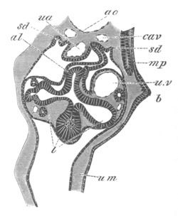
Fig. 343. Section through the ventral part of the trunk of a young
embryo of scyllium at the level of the umbilical cord.
b. pectoral fin; ao. dorsal aorta; cav. cardinal vein; ua.
vitelline artery; u.v. vitelline vein; al. duodenum; l. liver;
sd. opening of segmented duct into the body cavity; mp. muscle
plate; um. umbilical canal.
For the remaining history it is necessary to confine ourselves to
Scyllium as the only type which has been adequately studied.
The direction of the original ridge which connects the two fins of
each side is nearly though not quite longitudinal, sloping somewhat
obliquely downwards. It thus comes about that the attachment of each
pair of limbs is somewhat on a slant, and that the pelvic pair nearly
meet each other in the median ventral line a little way behind the
anus.
The elongated ridge, forming the rudiment of each fin, gradually
projects more and more, and so becomes broader in proportion to its
length, but at the same time its actual attachment to the side of the
body becomes shortened from behind forwards, so that what was
originally the attached border becomes in part converted into the
posterior border. This process is much more completely carried out
in the case of the pectoral fins than in that of the pelvic, and the
changes of form undergone by the pectoral fin in its development may
be gathered from figs. 344 and 348.
Before proceeding to the development of the skeleton of the fin it may
be pointed out that the connection of the two rudimentary fins by a
continuous epithelial line suggests the hypothesis that they are the
remnants of two continuous lateral fins[213].
Shortly after the view that the paired fins were remnants of
continuous lateral fins had been put forward in my memoir on
Elasmobranch Fishes, two very interesting papers were published by
Thacker (No. 489) and Mivart (No. 484) advocating this view on the
entirely independent grounds of the adult structure of the skeleton of
the paired fins in comparison with that of the unpaired fins[214].
The development of the skeleton has unfortunately not been as yet very
fully studied. I have however made some investigations on this subject
on Scyllium, and ’Swirski has also made some on the Pike.
In Scyllium the development of both the pectoral and pelvic fins is
very similar.
In both fins the skeleton in its earliest stage consists of a bar
springing from the posterior side of the pectoral or pelvic girdle,
and running backwards parallel to the long axis of the body. The outer
side of this bar is continued into a plate which
extends into the fin,
and which becomes very early segmented into a series of parallel rays
at right angles to the longitudinal bar.
In other words, the primitive skeleton of both the fins consists of a
longitudinal bar running along the base of the fin, and giving off at
right angles series of rays which pass into the fin. The longitudinal
bar, which may be called the basipterygium, is moreover continuous in
front with the pectoral or pelvic girdle as the case may be.
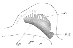
Fig. 344. Pectoral fin of a young embryo of Scyllium in longitudinal
and horizontal section.
The skeleton of the fin was still in the condition of embryonic
cartilage.
b.p. basipterygium (eventual metapterygium); fr. fin rays;
p.g. pectoral girdle in transverse section; f. foramen in
pectoral girdle; pc. wall of peritoneal cavity.
The primitive skeleton of the pectoral fin is shewn in longitudinal
section in fig. 344, and that of the pelvic fin at a slightly later
stage in fig. 345.
A transverse section shewing the basipterygium (mpt) of the pectoral
fin, and the plate passing from it into the fin, is shewn in fig. 346.
Before proceeding to describe the later history of the two fins it may
be well to point out that their embryonic structure completely
supports the view which has been arrived at from the consideration of
the soft parts of the fin.
My observations shew that the embryonic skeleton of the paired fin
consists of a series of parallel rays similar to those of the unpaired
fins. These rays support the soft part of the fin which has the form
of a longitudinal ridge, and are continuous at their base with a
longitudinal bar, which may very probably
be due to secondary
development. As pointed out by Mivart, a longitudinal bar is also
occasionally formed to support the cartilaginous rays of unpaired
fins. The longitudinal bar of the paired fins is believed by both
Thacker and Mivart to be due to the coalescence of the bases of
primitively independent rays, of which they believe the fin to have
been originally composed. This view is probable enough in itself, but
there is no trace in the embryo of the bar in question being formed by
the coalescence of rays, though the fact of its being perfectly
continuous with the bases of the rays is somewhat in favour of this
view[215].
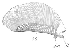
Fig. 345. Pelvic fin of a very young female embryo of Scyllium
stellare.
bb. basipterygium; pu. pubic process of pelvic girdle; il.
iliac process of pelvic girdle.
A point may be noticed here which may perhaps appear to be a
difficulty, viz. that to a considerable extent in the pectoral, and to
some extent in the pelvic fin the embryonic cartilage from which the
fin-rays are developed is at first a continuous lamina, which
subsequently segments into rays. I am however inclined to regard this
merely as a result of the mode of conversion of the indifferent
mesoblast into cartilage; and in any case no conclusion adverse to the
above view can be drawn from it, since I find that the rays of the
unpaired fin are similarly segmented from a continuous lamina. In all
cases the segmentation of the rays is to a large extent completed
before the tissue in question is sufficiently differentiated to be
called cartilage by an histologist.
Thacker and Mivart both hold that the pectoral and pelvic girdles have
been evolved by ventral and dorsal growths of the anterior end of the
longitudinal bar supporting the fin-rays.
There is, so far as I see, no theoretical objection to be taken to
this view, and the fact of the pectoral and pelvic girdles originating
continuously, and long remaining united with the
longitudinal bars of
their respective fins is in favour of rather than against this view.
The same may be said of the fact that the first part of each girdle to
be formed is that in the neighbourhood of the longitudinal bar
(basipterygium) of the fin, the dorsal and ventral prolongations being
subsequent growths.
The later development of the skeleton of the two fins is more
conveniently treated separately.
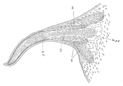
Fig. 346. Transverse section through the pectoral fin of a young
embryo of Scyllium stellare.
mpt. basipterygial bar (metapterygium); fr. fin ray; m.
muscles; hf. horny fibres.
The pelvic fin. The changes in the pelvic fin are comparatively
slight. The fin remains through life as a nearly horizontal lateral
projection of the body, and the longitudinal bar—the basipterygium—at
its base always remains as such. It is for a considerable period
attached to the pelvic girdle, but eventually becomes segmented from
it. Of the fin rays the anterior remains directly articulated with the
pelvic girdle on the separation of the basipterygium (fig. 347), and
the remaining rays finally become segmented from the basipterygium,
though they remain articulated with it. They also become to some
extent transversely segmented. The posterior end of the basipterygial
bar also becomes segmented off as the terminal ray.
The pelvic fin thus retains in all essential points its primitive
arrangement.
The pectoral fin. The earliest stage of the pectoral fin differs from
that of the pelvic fin only in minor points. There is the same
longitudinal or basipterygial bar to which the fin-rays are attached,
whose position at the base of the fin is clearly seen in the
transverse section (fig. 346, mpt). In front the bar is continuous
with the pectoral girdle (figs. 344 and 348).
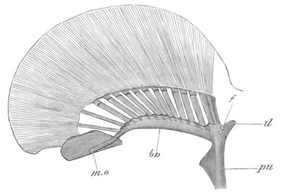
Fig. 347. Pelvic fin of a young male embryo of Scyllium stellare.
bp. basipterygium; m.o. process of basipterygium continued into
clasper; il. iliac process of pectoral girdle; pu. pubis.
Fig. 348. Pectoral fin of an embryo of
Scyllium stellare.
mp. metapterygium (basipterygium of earlier stage); me.p.
rudiment of future pro- and mesopterygium; sc. cut surface of
scapular process; cr. coracoid process; fr. foramen; f. horny
fibres.
The changes which take place in the course of the further development
are however very much more considerable in the case of the pectoral
than in that of the pelvic fin.
By the process spoken of above, by which the attachment of the
pectoral
fin to the body wall becomes shortened from behind forwards,
the basipterygial bar is gradually rotated outwards, its anterior end
remaining attached to the pectoral girdle. In this way this bar comes
to form the posterior border of the skeleton of the fin (figs. 348 and
349, mp), constituting what Gegenbaur called the metapterygium, and
eventually becomes segmented off from the pectoral girdle, simply
articulating with its hinder edge.
The plate of cartilage, which is continued outwards from the
basipterygium, or as we may now call it, the metapterygium, into the
fin, is not nearly so completely divided up into fin-rays as in the
case of the pelvic fin, and this is especially the case with the basal
part of the plate. This basal part becomes in fact at first only
divided into two parts (fig. 348) a small anterior part at the front
end (me.p), and a larger posterior along the base of the remainder
of the fin. The anterior part directly joins the pectoral girdle at
its base, resembling in this respect the anterior fin-ray of the
pelvic girdle. It constitutes the rudiment of the mesopterygium and
propterygium of Gegenbaur. It bears four fin-rays at its extremity,
the anterior not being well marked. The remaining fin-rays are borne
by the edge of the plate continuous with the metapterygium.
The further changes in the cartilages of the limb are not important,
and are easily understood by reference to fig. 349 representing the
limb of a nearly full-grown embryo. The front end of the anterior
basal cartilage becomes segmented off as a propterygium, bearing a
single fin-ray, leaving the remainder of the cartilage as a
mesopterygium. The remainder of the now considerably segmented
fin-rays are borne by the metapterygium.
The mode of development of the pectoral fin demonstrates that, as
supposed by Mivart, the metapterygium is the homologue of the basal
cartilage of the pelvic fin.
From the mode of development of the fins of Scyllium conclusions may
be drawn adverse to the views recently put forward on the structure of
the fin by Gegenbaur and Huxley, both of whom consider the primitive
type of fin to be most nearly retained in Ceratodus, and to consist of
a central multisegmented axis with numerous rays. Gegenbaur derives
the Elasmobranch pectoral fin from a form which he calls the
archipterygium, nearly like that of Ceratodus, with a median axis and
two
rows of rays; but holds that in addition to the rays attached to
the median axis, which are alone found in Ceratodus, there were other
rays directly articulated to the shoulder-girdle. He considers that in
the Elasmobranch fin the majority of the lateral rays on the posterior
(median or inner according to his view of the position of the limb)
side have become aborted, and that the central axis is represented by
the metapterygium; while the pro- and mesopterygium and their rays
are, he believes, derived from those rays of the archipterygium which
originally articulated directly with the shoulder-girdle.
Gegenbaur’s view appears to me to be absolutely negatived by the facts
of development of the pectoral fin in Scyllium; not so much because
the pectoral fin in this form is necessarily to be regarded as
primitive, but because what Gegenbaur holds to be the primitive axis
of the biserial fin is demonstrated to be really the base, and it is
only in the adult that it is conceivable that a second set of lateral
rays could have existed on the posterior side of the metapterygium. If
Gegenbaur’s view were correct we should expect to find in the embryo,
if anywhere, traces of the second set of lateral rays; but the fact is
that, as may easily be seen by an inspection of figs. 344 and 346,
such a second set of lateral rays could not possibly have existed in a
type of fin like that found in the embryo[216].
With this view of
Gegenbaur’s it appears to me that the theory held by this anatomist to
the effect that the limbs are modified gill arches also falls; in that
his method of deriving the limbs from gill arches ceases to be
admissible, while it is not easy to see how a limb, formed on the type
of the embryonic limb of Elasmobranchs, could be derived from a
visceral arch with its branchial rays[217].
Fig. 349. Skeleton of the pectoral fin and part of pectoral girdle
of a nearly ripe embryo of Scyllium stellare.
m.p. metapterygium; me.p. mesopterygium; pp. propterygium;
cr. coracoid process.
Gegenbaur’s older view
that the Elasmobranch fin retains a primitive
uniserial type appears to me to be nearer the truth than his more
recent view on this subject; though I hold that the fundamental point
established by the development of these parts in Scyllium is that the
posterior border of the adult Elasmobranch fin is the primitive base
line, i.e. the line of attachment of the fin to the side of the
body.
Huxley holds that the mesopterygium is the proximal piece of the axial
skeleton of the limb of Ceratodus, and derives the Elasmobranch fin
from that of Ceratodus by the shortening of its axis and the
coalescence of some of its elements. The secondary character of the
mesopterygium, and its total absence in the embryo Scyllium, appears
to me as conclusive against Huxley’s view, as the character of the
embryonic fin is against that of Gegenbaur; and I should be much more
inclined to hold that the fin of Ceratodus has been derived from a fin
like that of the Elasmobranchii by a series of steps similar to those
which Huxley supposes to have led to the establishment of the
Elasmobranch fin, but in exactly the reverse order.
With reference to the development of the pectoral fin in the Teleostei
there are some observations of ’Swirski (No. 488) which unfortunately
do not throw very much light upon the nature of the limb.
’Swirski finds that in the Pike the skeleton of the limb is formed of
a plate of cartilage, continuous with the pectoral girdle; which soon
becomes divided into a proximal and a distal portion. The former is
subsequently segmented into five basal rays, and the latter into
twelve parts, the number of which subsequently becomes reduced.
These investigations might be regarded as tending to shew that the
basipterygium of Elasmobranchii is not represented in Teleostei, owing
to the fin rays not having united into a continuous basal bar, but the
observations are not sufficiently complete to admit of this conclusion
being founded upon them with any certainty.
The cheiropterygium.
Observations on the early development of the pentadactyloid limbs of
the higher Vertebrata are comparatively scanty.
The limbs arise as simple outgrowths of the sides of the body, formed
both of epiblast and mesoblast. In the Amniota, at all events, they
are processes of a special longitudinal ridge known as the Wolffian
ridge. In the Amniota they also bear at their extremity a thickened
cap of epiblast, which may be compared with the epiblastic fold at the
apex of the Elasmobranch fin.
Both limbs have at first a precisely similar position, both being
directed backwards and being parallel to the surface of the body.
In the Urodela (Götte) the ulnar and fibular sides are primitively
dorsal, and the radial and tibial ventral: in Mammalia however
Kölliker states that the radial and tibial edges are from the first
anterior.
The exact changes of position undergone by the limbs in the course of
development are not fully understood. To suit a terrestrial mode of
life the flexures of the two limbs become gradually more and more
opposite, till in Mammalia the corresponding joints of the two limbs
are turned in completely opposite directions.
Within the mesoblast of the limbs a continuous blastema becomes
formed, which constitutes the first trace of the skeleton of the limb.
The corresponding elements of the two limbs, viz. the humerus and
femur, radius and tibia, ulna and fibula, carpal and tarsal bones,
metacarpals and metatarsals, and digits, become differentiated within
this, by the conversion of definite regions into cartilage, which may
either be completely distinct or be at first united. These
cartilaginous elements subsequently ossify.
The later development of the parts, more especially of the carpus and
tarsus, has been made the subject of considerable study; and important
results have been thereby obtained as to the homology of the various
carpal and tarsal bones throughout the Vertebrata; but this subject is
too special to be treated of here. The early development, including
the succession of the growth of the different parts, and the extent of
continuity primitively obtaining between them, has on the other hand
been but little investigated; recently however the development of the
limbs in the Urodela has been worked out in this way by two
anatomists, Götte (No. 482) and Strasser (No. 487), and their results,
though not on all points in complete harmony, are of considerable
interest, more especially in their bearing on the derivation of the
pentadactyloid limb from the piscine fin. Till however further
investigations of the same nature have been made upon other types, the
conclusions to be drawn from Götte and Strasser’s observations must be
regarded as somewhat provisional, the actual interpretation of various
ontological processes being very uncertain.
The forms investigated are Triton and Salamandra. We may remind the
reader that the hand of the Urodela has four digits, and the foot
five, the fifth digit being absent in the hand[218].
In Triton the
proximal row of carpal bones consists (using Gegenbaur’s nomenclature)
of (1) a radiale, and (2 and 3) an intermedium and ulnare, partially
united. The distal row is formed of four carpals, of which the first
often does not support the first
metacarpal; while the second
articulates with both the first and second metacarpals. In the foot
the proximal row of tarsals consists of a tibiale, an intermedium and
a fibulare. The distal row is formed of four tarsals, the first, like
that in the hand, often not articulating with the first metatarsal,
the second supporting the first and second metatarsals; and the fourth
the fourth and fifth metatarsals.
The mode of development of the hand and foot is almost the same. The
most remarkable feature of development is the order of succession of
the digits. The two anterior (radial or tibial) are formed in the
first instance, and then the third, fourth and fifth in succession.
As to the actual development of the skeleton Strasser, whose
observations were made by means of sections, has arrived at the
following results.
The humerus with the radius and ulna, and the corresponding parts in
the hind limb, are the first parts to be differentiated in the
continuous plate of tissue from which the skeleton of the limb is
formed. Somewhat later a cartilaginous centre appears at the base of
the first and second fingers (which have already appeared as
prominences at the end of the limb) in the situation of the permanent
second carpal of the distal row of carpals; and the process of
chondrification spreads from this centre into the fingers and into the
remainder of the carpus. In this way a continuous carpal plate of
cartilage is established, which is on the one hand continuous with the
cartilage of the two metacarpals, and on the other with the radius and
ulna.
In the cartilage of the carpus two special columns may be noticed, the
one on the radial side, most advanced in development, being continuous
with the radius; the other less developed column on the side of the
ulna being continuous both with the ulna and with the radius. The ulna
and radius are not united with the humerus.
In the further growth the third and fourth digits, and in the foot the
fifth digit also, gradually sprout out in succession from the ulnar
side of the continuous carpal plate. The carpal plate itself becomes
segmented from the radius and ulna, and divided up into the carpal
bones.
The original radial column is divided into three elements, a proximal
the radiale, a middle element the first carpal, and a distal the
second carpal already spoken of. The first carpal is thus situated
between the basal cartilage of the second digit and the radiale, and
would therefore appear to be the representative of a primitive middle
row of carpal bones, of which the centrale is also another
representative.
The centrale and intermedium are the middle and proximal products of
the segmentation of the ulnar column of the primitive carpus, the
distal second carpal being common both to this column and to the
radial column.
The ulnar or fibular side of the carpus or tarsus becomes divided into
a proximal element—the ulnare or fibulare—the ulnare remaining
partially united with the intermedium. There are also formed from this
plate two carpals to articulate with digits 3 and 4; while in the foot
the corresponding elements articulate respectively with the third
digit, and with the fourth and fifth digits.
Götte, whose observations were made in a somewhat different method to
those of Strasser, is at variance with him on several points. He finds
that the primitive skeleton of the limb consists of a basal portion,
the humerus, continued into a radial and an ulnar ray, which are
respectively prolonged into the two first digits. The two rays next
coalesce at the base of the fingers to form the carpus, and thus the
division of the limb into the brachium, antebrachium and manus is
effected.
The ulna, which is primitively prolonged into the second digit, is
subsequently separated from it and is prolonged into the third; from
the side of the part of the carpus connecting the ulna with the third
digit the fourth digit is eventually budded out, and in the foot the
fourth and fifth digits arise from the corresponding region. Each of
the three columns connected respectively with the first, second, and
third digits becomes divided into three successive carpal bones, so
that Götte holds the skeleton of the hand or foot to be formed of a
proximal, a middle, and a distal row of carpal bones each containing
potentially three elements. The proximal row is formed of the radiale,
intermedium and ulnare; the middle row of carpal 1, the centrale and
carpal 4, and the distal of carpal 2 (consisting according to Götte of
two coalesced elements) and carpal 3.
The derivation of the cheiropterygium from the ichthyopterygium. All
anatomists are agreed that the limbs of the higher Vertebrata are
derived from those of Fishes, but the gulf between the two types of
limbs is so great that there is room for a very great diversity of
opinion as to the mode of evolution of the cheiropterygium. The most
important speculations on the subject are those of Gegenbaur and
Huxley.
Gegenbaur holds that the cheiropterygium is derived from a uniserial
piscine limb, and that it consists of a primitive stem, to which a
series of lateral rays are attached on one (the radial) side; while
Huxley holds that the cheiropterygium is derived from a biserial
piscine limb by the “lengthening of the axial skeleton, accompanied by
the removal of its distal elements further away from the
shoulder-girdle and by a diminution in the number of the rays.”
Neither of these theories is founded upon ontology, and the only
ontological evidence we have which bears on this question is that
above recorded with reference to the development of the Urodele limb.
Without holding that this evidence can be considered as in any way
conclusive, its tendency would appear to me to be in favour of
regarding the cheiropterygium as derived from a uniserial type of fin.
The humerus or femur would appear to be the basipterygial bars
(metapterygium), which have become directed outwards instead of
retaining their original position parallel to the length of the body
at the base of the fin. The anterior (proximal) fin-rays and the pro-
and mesopterygium must be supposed to have become aborted, while the
radius or ulna, and tibia or fibula are two posterior fin-rays
(probably each representing several coalesced rays like the pro- and
mesopterygium) which support at their distal extremities more numerous
fin-rays consisting of the rows of carpal and tarsal bones.
This view of the cheiropterygium corresponds in some respects with
that put forward by Götte as a result of his investigations on the
development of the Urodele limbs, though in other respects it is very
different. A difficulty of this view is the fact that it involves our
supposing that the radial edge of the limb corresponds with the
metapterygial edge of the piscine fin. The difficulties of this
position have been clearly pointed out by Huxley, but the fact that in
the primitive position of the Urodele limbs the radius is ventral and
the ulna dorsal shews that this difficulty is not insuperable, in that
it is easy to conceive the radial border of the fin to have become
rotated from its primitive Elasmobranch position into the vertical
position it occupies in the embryos of the Urodela, and then to have
been further rotated from this position into that which it occupies in
the adult Urodela and in all higher forms.
Bibliography of the Limbs.
(477) M. v. Davidoff. “Beiträge z. vergleich. Anat. d. hinteren
Gliedmaassen d. Fische I.” Morphol. Jahrbuch, Vol. V. 1879.
(478) C. Gegenbaur. Untersuchungen z. vergleich. Anat. d.
Wirbelthiere. Leipzig, 1864-5. Erstes Heft. Carpus u. Tarsus. Zweites
Heft. Brustflosse d. Fische.
(479) C. Gegenbaur. “Ueb. d. Skelet d. Gliedmaassen d. Wirbelthiere im
Allgemeinen u. d. Hintergliedmaassen d. Selachier insbesondere.”
Jenaische Zeitschrift, Vol. V. 1870.
(480) C. Gegenbaur. “Ueb. d. Archipterygium.” Jenaische Zeitschrift,
Vol. VII. 1873.
(481) C. Gegenbaur. “Zur Morphologie d. Gliedmaassen d. Wirbelthiere.”
Morphologisches Jahrbuch, Vol. II. 1876.
(482) A. Götte. Ueb. Entwick. u. Regeneration d. Gliedmaassenskelets
d. Molche. Leipzig, 1879.
(483) T. H. Huxley. “On Ceratodus Forsteri, with some observations on
the classification of Fishes.” Proc. Zool. Soc. 1876.
(484) St George Mivart. “On the Fins of Elasmobranchii.” Zoological
Trans., Vol. X.
(485) A. Rosenberg. “Ueb. d. Entwick. d. Extremitäten-Skelets bei
einigen d. Reduction ihrer Gliedmaassen charakterisirten
Wirbelthieren.” Zeit. f. wiss. Zool., Vol. XXIII. 1873.
(486) E. Rosenberg. “Ueb. d. Entwick. d. Wirbelsäule u. d. centrale
carpi d. Menschen.” Morphologisches Jahrbuch, Vol. I. 1875.
(487) H. Strasser. “Z. Entwick. d. Extremitätenknorpel bei Salamandern
u. Tritonen.” Morphologisches Jahrbuch, Vol. V. 1879.
(488) G. ’Swirski. Untersuch. üb. d. Entwick. d. Schultergürtels u.
d. Skelets d. Brustflosse d. Hechts. Inaug. Diss. Dorpat, 1880.
(489) J. K. Thacker. “Median and paired fins. A contribution to the
history of the Vertebrate limbs.” Trans. of the Connecticut Acad.,
Vol. III. 1877.
(490) J. K. Thacker. “Ventral fins of Ganoids.” Trans. of the
Connecticut Acad., Vol. IV. 1877.
The Body cavity.
In the Cœlenterata no body cavity as distinct from the alimentary
cavity is present; but in the remaining Invertebrata the body cavity
may (1) take the form of a wide space separating the wall of the gut
from the body wall, or (2) may be present in a more or less reduced
form as a number of serous spaces, or (3) only be represented by
irregular channels between the muscular and connective-tissue cells
filling up the interior of the body. The body cavity, in whatever form
it presents itself, is probably filled with fluid, and the fluid in it
may contain special cellular elements. A well developed body cavity
may coexist with an independent system of serous spaces, as in the
Vertebrata and the Echinodermata; the perihæmal section of the body
cavity of the latter probably representing the system of serous
spaces.
In several of the types with a well developed body cavity it has been
established that this cavity originates in the embryo from a pair of
alimentary diverticula, and the cavities resulting from the formation
of these diverticula may remain distinct, the adjacent walls of the
two cavities fusing to form a dorsal and a ventral mesentery.
It is fairly certain that some groups, e.g. the Tracheata, with
imperfectly developed body cavities are descended from ancestors which
were provided with well developed body cavities, but how far this is
universally the case cannot as yet be definitely decided, and for
additional information on this subject the
reader is referred to pp. 355-360 and to the literature there referred to.
Fig. 350. Longitudinal section through an embryo of Agelina
labyrinthica.
The section is taken slightly to one side of the middle line so as
to shew the relation of the mesoblastic somites to the limbs. In the
interior are seen the yolk segments and their nuclei.
1-16. the segments; pr.l. procephalic lobe; do. dorsal
integument.
In the Chætopoda and the Tracheata the body cavity arises as a series
of paired compartments in the somites of mesoblast (fig. 350) which
have at first a very restricted extension on the ventral side of the
body, but eventually extend dorsalwards and ventralwards till each
cavity is a half circle investing the alimentary tract; on the dorsal
side the walls separating the two half cavities usually remain as the
dorsal mesentery, while ventrally they are in most instances absorbed.
The transverse walls, separating the successive compartments of the
body cavity, generally become more or less perforated.
Chordata. In the Chordata the primitive body cavity is either directly
formed from a pair of alimentary diverticula (Cephalochorda) (fig. 3)
or as a pair of spaces in the mesoblastic plates of the two sides of
the body (fig. 20).
As already explained (pp. 294-300) the walls of the dorsal sections of
the primitive body cavity soon become separated from those of the
ventral, and becoming segmented constitute the muscle plates, while
the cavity within them becomes
obliterated: they are dealt with in a
separate chapter. The ventral part of the primitive cavity alone
constitutes the permanent body cavity.
The primitive body cavity in the lower Vertebrata is at first
continued forwards into the region of the head, but on the formation
of the visceral clefts the cephalic section of the body cavity becomes
divided into a series of separate compartments. Subsequently these
sections of the body cavity become obliterated; and, since their walls
give rise to muscles, they may probably be looked upon as equivalent
to the dorsal sections of the body cavity in the trunk, and will be
treated of in connection with the muscular system.
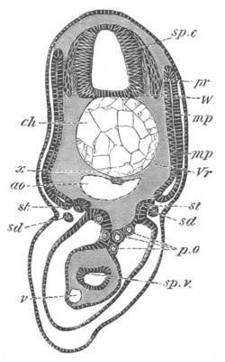
Fig. 351. Section through the trunk of a Scyllium embryo slightly
younger than 28 f.
sp.c. spinal canal; W. white matter of spinal cord; pr.
posterior nerve-roots; ch. notochord; x. subnotochordal rod;
ao. aorta; mp. muscle-plate; mp´. inner layer of muscle-plate
already converted into muscles; Vr. rudiment of vertebral body;
st. segmental tube; sd. segmental duct; sp.v. spiral valve;
v. subintestinal vein; p.o. primitive generative cells.
As a result of its mode of origin the body cavity in the trunk is at
first divided into two lateral halves; and part of the mesoblast
lining it soon becomes distinguished as a special layer of epithelium,
known as the peritoneal epithelium, of which the part bounding the
outer wall forms the somatic layer, and that bounding the inner wall
the splanchnic layer. Between the two splanchnic layers is placed the
gut. On the ventral side, in the region of the permanent gut, the two
halves of the body cavity soon coalesce, the septum between them
becoming absorbed, and the splanchnic layers of epithelium of the two
sides uniting at the ventral side of the gut, and the somatic layers
at the median ventral line of the body wall (fig. 351).
In the lower Vertebrata the body cavity is originally present even in
the postanal region of the trunk, but usually atrophies early,
frequently before the two halves coalesce.
On the dorsal side of the gut the
two halves of the body cavity never
coalesce, but eventually the splanchnic layers of epithelium of the
two sides, together with a thin layer of interposed mesoblast, form a
delicate membrane, known as the mesentery, which suspends the gut from
the dorsal wall of the body (figs. 119 and 351). On the dorsal side
the epithelium lining of the body cavity is usually more columnar than
elsewhere (fig. 351), and its cells partly form a covering for the
generative organs, and partly give rise to the primitive germinal
cells. This part of the epithelium is often known as the germinal
epithelium.
Over the greater part of the body cavity the lining epithelium becomes
in the adult intimately united with a layer of the subjacent
connective tissue, and constitutes with it a special lining membrane
for the body cavity, known as the peritoneal membrane.
Abdominal pores. In the Cyclostomata, the majority of the
Elasmobranchii, the Ganoidei, a few Teleostei, the Dipnoi, and some
Sauropsida (Chelonia and Crocodilia) the body cavity is in
communication with the exterior by a pair of pores, known as abdominal
pores, the external openings of which are usually situated in the
cloaca[219].
The ontogeny of these pores has as yet been but very slightly
investigated. In the Lamprey they are formed as apertures leading from
the body cavity into the excretory section of the primitive cloaca.
This section would appear from Scott’s (No. 87) observations to be
derived from part of the hypoblastic cloacal section of the alimentary
tract.
In all other cases they are formed in a region which appears to belong
to the epiblastic region of the cloaca; and from my observations on
Elasmobranchs it may be certainly concluded that they are formed there
in this group. They may appear as perforations (1) at the apices of
papilliform prolongations of the body cavity, or (2) at the ends of
cloacal pits directed from the exterior towards the body cavity, or
(3) as simple slit-like openings.
Considering the difference in development between the abdominal pores
of most types, and those of the Cyclostomata, it is open to doubt
whether these two types of pores are strictly homologous.
In the Cyclostomata they serve for the passage outwards of the
generative products, and they also have this function in some of the
few Teleostei in which they are found; and Gegenbaur and Bridge hold
that the primitive mode of exit of the generative products, prior to
the development of the Müllerian ducts, was probably by means of these
pores. I have elsewhere
suggested that the abdominal pores are perhaps
remnants of the openings of segmental tubes; there does not however
appear to be any definite evidence in favour of this view, and it is
more probable that they may have arisen as simple perforations of the
body wall.
Pericardial cavity, pleural cavities, and diaphragm. In all Vertebrata
the heart is at first placed in the body cavity (fig. 353 A), but the
part of the body cavity containing it afterwards becomes separated as
a distinct cavity known as the pericardial cavity. In Elasmobranchii,
Acipenser, etc. a passage is however left between the pericardial
cavity and the body cavity; and in the Lamprey a separation between
the two cavities does not occur during the Ammocœte stage.
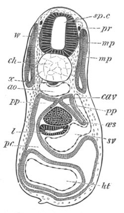
Fig. 352. Section through the trunk of a Scyllium embryo slightly
younger than 28 F.
The figure shews the separation of the body cavity from the
pericardial cavity by a horizontal septum in which runs the ductus
Cuvieri; on the left side is seen the narrow passage which remains
connecting the two cavities.
sp.c. spinal canal; w. white matter of spinal cord; pr.
commissure connecting the posterior nerve-roots; ch. notochord;
x. subnotochordal rod; ao. aorta; sv. sinus venosus; cav.
cardinal vein; ht. heart; pp. body cavity; pc. pericardial
cavity; œs. solid œsophagus; l. liver; mp. muscle-plate.
In Elasmobranchii the pericardial cavity becomes established as a
distinct space in front of the body cavity in the following way. When
the two ductus Cuvieri, leading transversely from the sinus venosus to
the cardinal veins, become developed, a horizontal septum, shewn on
the right side in fig. 352, is formed to support them, stretching
across from the splanchnic to the somatic side of the body cavity, and
dividing the body cavity (fig. 352) in this part into (1) a dorsal
section formed of a right and left division constituting the true body
cavity (pp), and (2) a ventral part the pericardial cavity (pc).
The septum is at first of a very small longitudinal extent, so that
both in front and behind it (fig. 352 on the left side) the dorsal and
ventral sections of the body cavity are in free communication. The
septum soon however becomes prolonged, and ceasing to be quite
horizontal, is directed obliquely upwards and forwards till it meets
the dorsal wall of the body.
Anteriorly all communication is thus early
shut off between the body cavity and the pericardial cavity, but the
two cavities still open freely into each other behind.
The front part of the body cavity, lying dorsal to the pericardial
cavity, becomes gradually narrowed, and is wholly obliterated long
before the close of embryonic life, so that in adult Elasmobranch
Fishes there is no section of the body cavity dorsal to the
pericardial cavity. The septum dividing the body cavity from the
pericardial cavity is prolonged backwards, till it meets the ventral
wall of the body at the point where the liver is attached by its
ventral mesentery (falciform ligament). In this way the pericardial
cavity becomes completely shut off from the body cavity, except, it
would seem, for the narrow communications found in the adult. The
origin of these communications has not however been satisfactorily
worked out.
The septum between the pericardial cavity and the body cavity is
attached on its dorsal aspect to the liver. It is at first nearly
horizontal, but gradually assumes a more vertical position, and then,
owing to the obliteration of the primitive anterior part of the body
cavity, appears to mark the front boundary of the body cavity. The
above description of the mode of formation of the pericardial cavity,
and the explanation of its relations to the body cavity, probably
holds true for Fishes generally.
In the higher types the earlier changes are precisely the same as
those in Elasmobranch Fishes. The heart is at first placed within the
body cavity attached to the ventral wall of the gut by a mesocardium
(fig. 353 A). A horizontal septum is then formed, in which the ductus
Cuvieri are placed, dividing the body cavity for a short distance into
a dorsal (p.p) and ventral (p.c) section (fig. 353 B). In Birds
and Mammals, and probably also in Reptilia, the ventral and dorsal
parts of the body cavity are at first in free communication both in
front of and behind this septum. This is shewn for the Chick in fig.
353 A and B, which are sections of the same chick, A being a little in
front of B. The septum is soon continued forwards so as completely to
separate the ventral pericardial and the dorsal body cavity in front,
the pericardial cavity extending at this period considerably further
forwards than the body cavity.
Since the horizontal septum, by its mode of origin, is
necessarily
attached to the ventral side of the gut, the dorsal part of the
primitive body space is divided into two halves by a median vertical
septum formed of the gut and its mesentery (fig. 353 B). Posteriorly
the horizontal septum grows in a slightly ventral direction along the
under surface of the liver (fig. 354), till it meets the abdominal
wall of the body at the insertion of the falciform ligament, and thus
completely shuts off the pericardial cavity from the body cavity. The
horizontal septum forms, as is obvious from the above description, the
dorsal wall of the pericardial cavity[220].
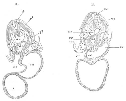
Fig. 353. Transverse sections through a Chick embryo with twenty-one
mesoblastic somites to shew the formation of the pericardial
cavity, A. being the anterior section.
p.p. body cavity; p.c. pericardial cavity; al. alimentary
cavity; au. auricle; v. ventricle; s.v. sinus venosus; d.c.
ductus Cuvieri; ao. aorta; mp. muscle-plate; mc. medullary
cord.
With the complete separation of the pericardial cavity from the body
cavity, the first period in the development of these parts is
completed, and the relations of the body cavity to the
pericardial
cavity become precisely those found in the embryos of Elasmobranchii.
The later changes are however very different. Whereas in Fishes the
right and left sections of the body cavity dorsal to the pericardial
cavity soon atrophy, in the higher types, in correlation with the
relatively backward situation of the heart, they rapidly become
larger, and receive the lungs which soon sprout out from the throat.
The diverticula which form the lungs grow out into the splanchnic
mesoblast, in front of the body cavity; but as they grow, they extend
into the two anterior compartments of the body cavity, each attached
by its mesentery to the mesentery of the gut (fig. 354, lg). They
soon moreover extend beyond the region of the pericardium into the
undivided body cavity behind. This holds not only for the embryos of
the Amphibia and Sauropsida, but also for those of Mammalia.
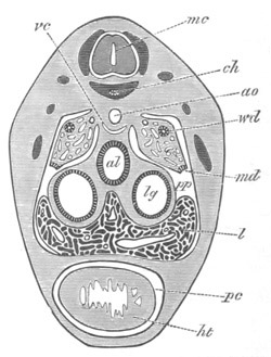
Fig. 354. Section through the cardiac region of an embryo of
Lacerta Muralis of 9 mm. to shew the mode of formation of the
pericardial cavity.
ht. heart; pc. pericardial cavity; al. alimentary tract; lg.
lung; l. liver; pp. body cavity; md. open end of Müllerian
duct; wd. Wolffian duct; vc. vena cava inferior; ao. aorta;
ch. notochord; mc. medullary cord.
To understand the further changes in the pericardial cavity it is
necessary to bear in mind its relations to the adjoining parts. It
lies at this period completely ventral to the two anterior
prolongations of the body cavity containing the lungs (fig. 354). Its
dorsal wall is attached to the gut, and is continuous with the
mesentery of the gut passing to the dorsal abdominal wall, forming the
posterior mediastinum of human anatomy.
The changes which next ensue consist essentially in the enlargement of
the sections of the body cavity dorsal to the pericardial cavity. This
enlargement takes place partly by the elongation of the posterior
mediastinum, but still more by the two divisions of the body cavity
which contain the lungs extending themselves ventrally round the
outside of the pericardial
cavity. This process is illustrated by fig.
355, taken from an embryo Rabbit. The two dorsal sections of the body
cavity (pl.p) finally extend so as completely to envelope the
pericardial cavity (pc), remaining however separated from each other
below by a lamina extending from the ventral wall of the pericardial
cavity to the body wall, which forms the anterior mediastinum of human
anatomy.
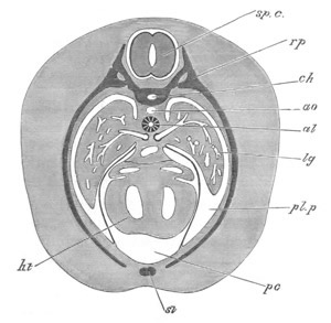
Fig. 355. Section through an advanced embryo of a Rabbit to shew
how the pericardial cavity becomes surrounded by the pleural
cavities.
ht. heart; pc. pericardial cavity; pl.p pleural cavity; lg.
lung; al. alimentary tract; ao. dorsal aorta; ch. notochord;
rp. rib; st. sternum; sp.c. spinal cord.
By these changes the pericardial cavity is converted into a closed
bag, completely surrounded at its sides by the two lateral halves of
the body cavity, which were primitively placed dorsally to it. These
two sections of the body cavity, which in Amphibia and Sauropsida
remain in free communication with the undivided peritoneal cavity
behind, may, from the fact of their containing the lungs, be called
the pleural cavities.
In Mammalia a further change takes place, in that, by the formation of
a vertical partition across the body cavity, known as the diaphragm,
the pleural cavities, containing the lungs,
become isolated from the
remainder of the body or peritoneal cavity. As shewn by their
development the so-called pleuræ or pleural sacks are simply the
peritoneal linings of the anterior divisions of the body cavity, shut
off from the remainder of the body cavity by the diaphragm.
The exact mode of formation of the diaphragm is not fully made out;
the account of it recently given by Cadiat (No. 491) not being in my
opinion completely satisfactory.
Bibliography.
(491) M. Cadiat. “Du développement de la partie céphalothoracique de
l'embryon, de la formation du diaphragme, des pleures, du péricarde,
du pharynx et de l'œsophage.” Journal de l'Anatomie et de la
Physiologie, Vol. XIV. 1878.
Vascular System.
The actual observations bearing on the origin of the vascular system,
using the term to include the lymphatic system, are very scanty. It
seems probable, mainly it must be admitted on à priori grounds, that
vascular and lymphatic systems have originated from the conversion of
indefinite spaces, primitively situated in the general connective
tissue, into definite channels. It is quite certain that vascular
systems have arisen independently in many types; a very striking case
of the kind being the development in certain parasitic Copepoda of a
closed system of vessels with a red non-corpusculated blood (E. van
Beneden, Heider), not found in any other Crustacea. Parts of vascular
systems appear to have arisen in some cases by a canalization of
cells.
The blood systems may either be closed or communicate with the body
cavity. In cases where the primitive body cavity is atrophied or
partially broken up into separate compartments (Insecta, Mollusca,
Discophora, etc.) a free communication between the vascular system and
the body cavity is usually present; but in these cases the
communication is no doubt secondary. On the whole it would seem
probable that the vascular system has in most instances arisen
independently of the body cavity, at least in types where the body
cavity is
present in a well-developed condition. As pointed out by the
Hertwigs, a vascular system is always absent where there is not a
considerable development of connective tissue.
As to the ontogeny of the vascular channels there is still much to be
made out both in Vertebrates and Invertebrates.
The smaller channels often rise by a canalization of cells. This
process has been satisfactorily studied by Lankester in the
Leech[221],
and may easily be observed in the blastoderm of the Chick
or in the epiploon of a newly born Rabbit (Schäfer, Ranvier). In
either case the vessels arise from a network of cells, the superficial
protoplasm and part of the nuclei giving rise to the walls, and the
blood-corpuscles being derived either from nucleated masses set free
within the vessels (the Chick) or from blood-corpuscles directly
differentiated in the axes of the cells (Mammals).
Larger vessels would seem to be formed from solid cords of cells, the
central cells becoming converted into the corpuscles, and the
peripheral cells constituting the walls. This mode of formation has
been observed by myself in the case of the Spider’s heart, and by
other observers in other Invertebrata. In the Vertebrata a more or
less similar mode of formation appears to hold good for the larger
vessels, but further investigations are still required on this
subject. Götte finds that in the Frog the larger vessels are formed as
longitudinal spaces, and that the walls are derived from the
indifferent cells bounding these spaces, which become flattened and
united into a continuous layer.
The early formation of vessels in the Vertebrata takes place in the
splanchnic mesoblast; but this appears due to the fact that the
circulation is at first mainly confined to the vitelline region, which
is covered by splanchnic mesoblast.
The Heart.
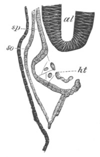
Fig. 356. Section through the developing heart of an embryo of an
Elasmobranch (Pristiurus).
al. alimentary tract; sp. splanchnic mesoblast; so. somatic
mesoblast; ht. heart.
The heart is essentially formed as a tubular cavity in the splanchnic
mesoblast, on the ventral side of the throat, immediately behind the
region of the visceral clefts. The walls of this cavity are formed of
two layers, an outer thicker layer, which has at first only the form
of a half tube, being incomplete on its dorsal side; and an inner
lamina formed of delicate flattened cells. The latter is the
epithelioid lining of the heart, and the cavity it contains the true
cavity of the heart. The outer layer gives rise to the muscular wall
and peritoneal covering of the heart. Though at first it has only the
form of a half tube (fig.
356), it soon becomes folded in on the
dorsal side so as to form for the heart a complete muscular wall. Its
two sides, after thus meeting to complete the tube of the heart,
remain at first continuous with the splanchnic mesoblast surrounding
the throat, and form a provisional mesentery—the mesocardium—which
attaches the heart to the ventral wall of the throat. The superficial
stratum of the wall of the heart differentiates itself as the
peritoneal covering. The inner epithelioid tube takes its origin at
the time when the general cavity of the heart is being formed by the
separation of the splanchnic mesoblast from the hypoblast. During this
process (fig. 357) a layer of mesoblast remains close to the
hypoblast, but connected with the main mass
of the mesoblast by
protoplasmic processes. A second layer next becomes split from the
splanchnic mesoblast, connected with the first layer by the
above-mentioned protoplasmic processes. These two layers form together
the epithelioid lining of the heart; between them is the cavity of the
heart, which soon loses the protoplasmic trabeculæ which at first
traverse it. The cavity of the heart may thus be described as being
formed by a hollowing out of the splanchnic mesoblast, and resembles
in its mode of origin that of other large vascular trunks.
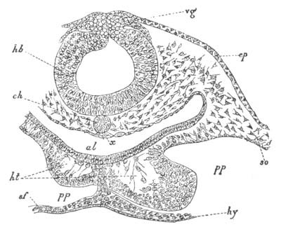
Fig. 357. Transverse section through the posterior part of the
head of an embryo Chick of thirty hours.
hb. hind-brain; vg. vagus nerve; ep. epiblast; ch.
notochord; x. thickening of hypoblast (possibly a rudiment of the
subnotochordal rod); al. throat; ht. heart; pp. body cavity;
so. somatic mesoblast; sf. splanchnic mesoblast; hy.
hypoblast.
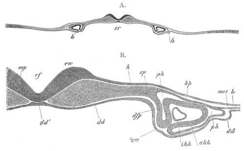
Fig. 358. Transverse section through the head of a Rabbit of the
same age as fig. 144 B. (From Kölliker.)
B is a more highly magnified representation of part of A.
rf. medullary groove; mp. medullary plate; rw. medullary fold;
h. epiblast; dd. hypoblast; dd´. notochordal thickening of
hypoblast; sp. undivided mesoblast; hp. somatic mesoblast;
dfp. splanchnic mesoblast; ph. pericardial section of body
cavity; ahh. muscular wall of heart; ihh. epithelioid layer of
heart; mes. lateral undivided mesoblast; sw. part of the
hypoblast which will form the ventral wall of the pharynx.
The above description applies only to the development of the heart in
those types in which it is formed at a period after the throat has
become a closed tube (Elasmobranchii, Amphibia, Cyclostomata, Ganoids
(?)). In a number of other cases, in which the heart is formed before
the conversion of the throat into a closed tube, of which the most
notable is that of Mammals (Hensen, Götte, Kölliker), the heart arises
as two independent
tubes (fig. 358), which eventually coalesce into an
unpaired structure.
In Mammals the two tubes out of which the heart is formed appear at
the sides of the cephalic plates, opposite the region of the mid- and
hind-brain (fig. 358). They arise at a time when the lateral folds
which form the ventral wall of the throat are only just becoming
visible. Each half of the heart originates in the same way as the
whole heart in Elasmobranchii, etc.; and the layer of the splanchnic
mesoblast, which forms the muscular wall for each part (ahh), has at
first the form of a half tube open below to the hypoblast.
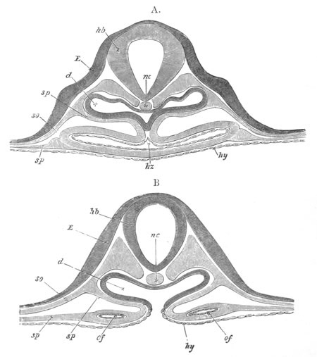
Fig. 359. Two diagrammatic sections through the region of the
hind-brain of an embryo Chick of about 36 hours illustrating the
formation of the heart.
hb. hind-brain; nc. notochord; E. epiblast; so.
somatopleure; sp. splanchnopleure; d. alimentary tract; hy.
hypoblast; hz. heart; of. vitelline veins.
On the formation of the lateral folds of the splanchnic walls, the two
halves of the heart become carried inwards and downwards, and
eventually
meet on the ventral side of the throat. For a short time
they here remain distinct, but soon coalesce into a single tube.
In Birds, as in Mammals, the heart makes its appearance as two tubes,
but arises at a period when the formation of the throat is very much
more advanced than in the case of Mammals. The heart arises
immediately behind the point up to which the ventral wall of the
throat is established and thus has at first a Lambda-shaped form. At
the apex of the Lambda, which forms the anterior end of the heart, the
two halves are in contact (fig. 357), though they have not coalesced;
while behind they diverge to be continued as the vitelline veins. As
the folding in of the throat is continued backwards the two limbs of
the heart are brought together and soon coalesce from before backwards
into a single structure. Fig. 359 A and B shews the heart during this
process. The two halves have coalesced anteriorly (A) but are still
widely separated behind (B). In Teleostei the heart is formed as in
Birds and Mammals by the coalescence of two tubes, and it arises
before the formation of the throat.
The fact that the heart arises in so many instances as a double tube
might lead to the supposition that the ancestral Vertebrate had two
tubes in the place of the present unpaired heart.
The following considerations appear to me to prove that this
conclusion cannot be accepted. If the folding in of the
splanchnopleure to form the throat were deferred relatively to the
formation of the heart, it is clear that a modification in the
development of the heart would occur, in that the two halves of the
heart would necessarily be formed widely apart, and only eventually
united on the folding in of the wall of the throat. It is therefore
possible to explain the double formation of the heart without having
recourse to the above hypothesis of an ancestral Vertebrate with two
hearts. If the explanation just suggested is the true one the heart
should only be formed as two tubes when it arises prior to the
formation of the throat, and as a single tube when formed after the
formation of the throat. Since this is invariably found to be so, it
may be safely concluded that the formation of the heart as two
cavities is a secondary mode of development, which has been brought
about by variations in the period of the closing in of the wall of the
throat.
The heart arises continuously with the sinus venosus, which in the
Amniotic Vertebrata is directly continued into the vitelline veins.
Though at first it ends blindly in front, it is very soon connected
with the foremost aortic arches.
The simple tubular heart, connected as above described, grows more
rapidly than the chamber in which it is contained, and is soon doubled
upon itself, acquiring in this way an S-shaped curvature, the
posterior portion being placed dorsally, and the anterior ventrally. A
constriction soon appears between the dorsal and ventral portions.
The dorsal section becomes partially divided off behind from the sinus
venosus, and constitutes the relatively thin-walled auricular section
of the heart; while the ventral portion, after becoming distinct
anteriorly from a portion continued forwards from it to the origin of
the branchial arteries, which may be called the truncus arteriosus,
acquires very thick spongy muscular walls, and becomes the ventricular
division of the heart.
The further changes in the heart are but slight in the case of the
Pisces. A pair of simple membranous valves becomes established at the
auriculo-ventricular orifice, and further changes take place in the
truncus arteriosus. This part becomes divided in Elasmobranchii,
Ganoidei, and Dipnoi into a posterior section, called the conus
arteriosus, provided with a series of transverse rows of valves, and
an anterior section, called the bulbus arteriosus, not provided with
valves, and leading into the branchial arteries. In most Teleostei
(except Butirinus and a few other forms) the conus arteriosus is all
but obliterated, and the anterior row of its valves alone preserved;
and the bulbus is very much enlarged[222].
In the Dipnoi important changes in the heart are effected, as compared
with other Fishes, by the development of true lungs. Both the
auricular and ventricular chamber may be imperfectly divided into two,
and in the conus a partial longitudinal septum is developed in
connection with a longitudinal row of valves[223].
In Amphibia the heart is in many respects similar to that of the
Dipnoi. Its curvature is rather that of a screw than of a simple S.
The truncus arteriosus lies to the left, and is continued into the
ventricle which lies ventrally and more to the right, and this again
into the dorsally placed auricular section.
After the heart has reached the piscine stage, the auricular section
(Bombinator) becomes prolonged into a right and left auricular
appendage. A septum next grows from the roof of the auricular portion
of the heart
obliquely backwards and towards the left, and divides it
in two chambers; the right one of which remains continuous with the
sinus venosus, while the left one is completely shut off from the
sinus, though it soon enters into communication with the newly
established pulmonary veins. The truncus arteriosus[224]
is divided
into a posterior conus arteriosus (pylangium) and an anterior
bulbus (synangium). The former is provided with a proximal row of
valves at its ventricular end, and a distal row at its anterior end
near the bulbus. It is also provided with a longitudinal septum, which
is no doubt homologous with the septum in the conus arteriosus of the
Dipnoi. The bulbus is well developed in many Urodela, but hardly
exists in the Anura.
In the Amniota further changes take place in the heart, resulting in
the abortion of the distal rows of valves of the conus
arteriosus[225],
and in the splitting up of the whole truncus
arteriosus into three vessels in Reptilia, and two in Birds and
Mammals, each opening into the ventricular section of the heart, and
provided with a special set of valves at its commencement. In Birds
and Mammals the ventricle becomes moreover completely divided into two
chambers, each communicating with one of the divisions of the
primitive truncus, known in the higher types as the systemic and
pulmonary aortæ. The character of the development of the heart in the
Amniota will be best understood from a description of what takes place
in the Chick.
In Birds the originally straight heart (fig. 109) soon becomes doubled
up upon itself. The ventricular portion becomes placed on the ventral
and right side, while the auricular section is dorsal and to the left.
The two parts are separated from each other by a slight constriction
known as the canalis auricularis. Anteriorly the ventricular cavity is
continued into the truncus, and the venous or auricular portion of the
heart is similarly connected behind with the sinus venosus. The
auricular appendages grow out from the auricle at a very early period.
The general appearance of the heart, as seen from the ventral side on
the fourth day, is shewn in fig. 360. Although the external divisions
of the heart are well marked even before this stage, it is not till
the end of the third day that the internal partitions become apparent;
and, contrary to what might have been anticipated from the evolution
of these parts in the lower types, the ventricular septum is the first
to be established.
It commences on the third day as a crescentic ridge or fold springing
from the convex or ventral side of the rounded ventricular portion of
the heart, and on the fourth day grows rapidly across the ventricular
cavity towards the concave or dorsal side. It thus forms an incomplete
longitudinal partition, extending from the canalis auricularis to the
commencement of the truncus arteriosus, and dividing the twisted
ventricular tube into two somewhat curved canals, one more to the left
and above, the other to the right and below. These communicate with
each other, above the free edge of the partition, along its whole
length.
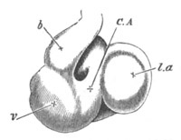
Fig. 360. Heart of a Chick on the fourth day of incubation viewed
from the ventral surface.
l.a. left auricular appendage; C.A. canalis auricularis; v.
ventricle; b. truncus arteriosus.
Externally the ventricular portion as yet shews no division into two
parts.
By the fifth day the venous end of the heart, though still lying
somewhat to the left and above, is placed as far forwards as the
arterial end, the whole organ appearing to be drawn together. The
ventricular septum is complete.
The apex of the ventricles becomes more and more pointed. In the
auricular portion a small longitudinal fold appears as the rudiment of
the auricular septum, while in the canalis auricularis, which is now
at its greatest length, there is also to be seen a commencement of the
valvular structures tending to separate the cavity of the auricles
from those of the ventricles.
About the 106th hour, a septum begins to make its appearance in the
truncus arteriosus in the form of a longitudinal fold, which according
to Tonge (No. 495) starts at the end of the truncus furthest removed
from the heart. It takes origin from the wall of the truncus between
the fourth and fifth pairs of arches, and grows downwards in such a
manner as to divide the truncus into two channels, one of which leads
from the heart to the third and fourth pairs of arches, and the other
to the fifth pair. Its course downwards is not straight but spiral,
and thus the two channels into which it divides the truncus arteriosus
wind spirally the one round the other.
At the time when the septum is first formed, the opening of the
truncus arteriosus into the ventricles is narrow or slit-like,
apparently in order to prevent the flow of the blood back into the
heart. Soon after the appearance of the septum, however, semilunar
valves (Tonge, No. 495) are developed from the wall of that portion of
the truncus which lies between the free edge of the septum and the
cavity of the ventricles[226].
The ventral and the dorsal pairs of valves are the first to appear:
the former as two small solid prominences separated from each other by
a narrow groove; the latter as a single ridge, in the centre of which
is a prominence indicating the point where the ridge will subsequently
become divided into two. The outer valves appear opposite each other,
at a considerably later period.
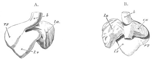
Fig. 361. Two views of the heart of a Chick upon the fifth day
of incubation.
A. from the ventral, B. from the dorsal side.
l.a. left auricular appendage; r.a. right auricular appendage;
r.v. right ventricle; l.v. left ventricle; b. truncus
arteriosus.
As the septum grows downwards towards the heart, it finally reaches
the position of these valves. One of its edges then passes between the
two ventral valves, and the other unites with the prominence on the
dorsal valve-ridge. At the same time the growth of all the parts
causes the valves to appear to approach the heart, and thus to be
placed quite at the top of the ventricular cavities. The free edge of
the septum of the truncus now fuses with the ventricular septum, and
thus the division of the truncus into two separate channels, each
provided with three valves, and each communicating with a separate
side of the heart, is complete; the position of the valves not being
very different from that in the adult heart.
That division of the truncus which opens into the fifth pair of arches
is the one which communicates with the right ventricle, while that
which opens into the third and fourth pairs communicates with the left
ventricle. The former becomes the pulmonary artery, the latter the
commencement of the systemic aorta.
The external constriction actually dividing the truncus into two
vessels does not begin to appear till the septum has extended some way
back towards the heart.
The semilunar valves become pocketed at a period considerably later
than their first formation (from the 147th to the 165th hour) in the
order of their appearance.
At the end of the sixth day, and even on the fifth day (figs. 361 and
362), the appearance of the heart itself, without reference to the
vessels which come from it, is not very dissimilar from that of the
adult. The original
protuberance to the right now forms the apex of
the ventricles, and the two auricular appendages are placed at the
anterior extremity of the heart. The most noticeable difference (in
the ventral view) is the still externally undivided condition of the
truncus arteriosus.
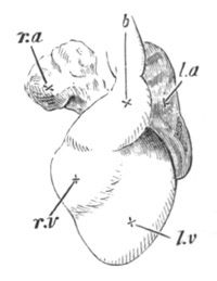
Fig. 362. Heart of a Chick upon the sixth day of incubation, from
the ventral surface.
l.a. left auricular appendage; r.a. right auricular appendage;
r.v. right ventricle; l.v. left ventricle; b. truncus
arteriosus.
The subsequent changes which the heart undergoes are concerned more
with its internal structure than with its external shape. Indeed,
during the next three days, viz. the eighth, ninth, and tenth, the
external form of the heart remains nearly unaltered.
In the auricular portion, however, the septum which commenced on the
fifth day becomes now more conspicuous. It is placed vertically, and
arises from the ventral wall; commencing at the canalis auricularis
and proceeding towards the opening into the sinus venosus.
This latter structure gradually becomes reduced so as to become a
special appendage of the right auricle. The inferior vena cava enters
the sinus obliquely from the right, so that its blood has a tendency
to flow towards the left auricle of the heart, which is at this time
the larger of the two.
The valves between the ventricles and auricles are now well developed,
and it is about this time that the division of the truncus arteriosus
into the aorta and pulmonary artery becomes visible from the exterior.
By the eleventh to the thirteenth day the right auricle has become as
large as the left, and the auricular septum much more complete, though
there is still a small opening, the foramen ovale, by which the two
cavities communicate with each other.
The most important feature in which the development of the Reptilian
heart differs from that of Birds is the division of the truncus into
three vessels, instead of two. The three vessels remain bound up in a
common sheath, and appear externally as a single trunk. The vessel not
represented in Birds is that which is continued into the left aortic
arch.
In Mammals the early stages in the development of the heart present no
important points of difference from those of Aves. The septa in the
truncus, in the ventricular, and in the auricular cavities are formed,
so far as is known, in the same way and at the same relative periods
in both groups. In the embryo Man, the Rabbit, and other Mammals the
division of the ventricles is made apparent externally by a deep
cleft, which, though evanescent in these forms, is permanent in the
Dugong.
The attachment of the auriculo-ventricular valves to the wall of the
ventricle, and the similar attachment of the left auriculo-ventricular
valves in Birds, have been especially studied by Gegenbaur and Bernays
(No. 492),
and deserve to be noticed. In the primitive state the
ventricular walls have throughout a spongy character; and the
auriculo-ventricular valves are simple membranous projections like the
auriculo-ventricular valves of Fishes. Soon however the spongy
muscular tissue of both the ventricular and auricular walls, which at
first pass uninterruptedly the one into the other, grows into the
bases of the valves, which thus become in the main muscular
projections of the walls of the heart. As the wall of the ventricle
thickens, the muscular trabeculæ, connected at one end with the
valves, remain at the other end united with the ventricular wall, and
form special bands passing between the two. The valves on the other
hand lose their muscular attachment to the auricular walls. This is
the condition permanent in Ornithorhynchus. In higher Mammalia the
ends of the muscular bands inserted into the valves become fibrous,
from the development of intermuscular connective tissue, and the
atrophy of the muscular elements. The fibrous parts now form the
chordæ tendineæ, and the muscular the musculi papillares.
The sinus venosus in Mammals becomes completely merged into the right
auricle, and the systemic division of the truncus arteriosus is
apparently not homologous with that in Birds.
In the embryos of all the Craniata the heart is situated very far
forwards in the region of the head. This position is retained in
Pisces. In Amphibia the heart is moved further back, while in all the
Amniota it gradually shifts its position first of all into the region
of the neck and finally passes completely within the thoracic cavity.
The steps in the change of position may be gathered from figs. 109,
111, and 118.
Bibliography of the Heart.
(492) A. C. Bernays. “Entwicklungsgeschichte d.
Atrioventricularklappen.” Morphol. Jahrbuch, Vol. II. 1876.
(493) E. Gasser. “Ueber d. Entstehung d. Herzens beim Hühn.” Archiv
f. mikr. Anat., Vol. XIV.
(494) A. Thomson. “On the development of the vascular system of the
fœtus of Vertebrated Animals.” Edinb. New Phil. Journal, Vol. IX.
1830 and 1831.
(495) M. Tonge. “Observations on the development of the semilunar
valves of the aorta and pulmonary artery of the heart of the Chick.”
Phil. Trans. CLIX. 1869.
Vide also Von Baer (291), Rathke (300), Hensen (182), Kölliker
(298), Götte (296), and Balfour (292).
Arterial System.
In the embryos of Vertebrata the arterial system consists of a forward
continuation of the truncus arteriosus, on the ventral
side of the
throat (figs. 363, abr, and 364, a), which, with a few exceptions
to be noticed below, divides into as many branches on each side as
there are visceral arches. These branches, after traversing the
visceral arches, unite on the dorsal side of the throat into a common
trunk on each side. This trunk (figs. 363 and 364) after giving off
one (or more) vessels to the head (c´ and c) turns backwards, and
bends in towards the middle line, close to its fellow, immediately
below the notochord (figs. 21 and 116) and runs backwards in this
situation towards the end of the tail. The two parallel trunks below
the notochord fuse very early into a single trunk, the dorsal aorta
(figs. 363, ad, and 364, a´´). There is given off from each
collecting trunk from the visceral arches, or from the commencement of
the dorsal aorta, a subclavian artery to each of the anterior limbs;
from near the anterior end of the dorsal aorta a vitelline artery (or
before the dorsal aortæ have united a pair of arteries fig. 125, R
of A and L of A) to the yolk-sack, which subsequently becomes the
main visceral artery[227];
and from the dorsal aorta opposite the hind
limbs one (or two) arteries on each side—the iliac arteries—to the
hind limbs; from these arteries the allantoic arteries are given off
in the higher types, which remain as the hypogastric arteries after
the disappearance of the allantois.
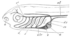
Fig. 363. Diagrammatic view of the head of an embryo Teleostean,
with the primitive vascular trunks. (From Gegenbaur.)
a. auricle; v. ventricle; abr. branchial artery; c´.
carotid; ad. dorsal aorta; s. branchial clefts; sv. sinus
venosus; dc. ductus Cuvieri; n. nasal pit.
The primitive arrangement of the arterial trunks is with a few
modifications retained in Fishes. With the development of the gills
the vessels to the arches become divided into two parts connected by a
capillary system in the gill folds, viz. into the
branchial arteries
bringing the blood to the gills from the truncus arteriosus, and the
branchial veins transporting it to the dorsal aorta. The branchial
vessels to those arches which do not bear gills, either wholly or
partially atrophy; thus in Elasmobranchii the mandibular trunk, which
is fully developed in the embryo (fig. 193, 1av), atrophies, except
for a small remnant bringing blood to the rudimentary gill of the
spiracle from the branchial vein of the hyoid arch. In Ganoids the
mandibular artery atrophies, but the hyoid is usually preserved. In
Teleostei both mandibular[228]
and hyoid arteries are absent in the
adult, except that there is usually left a rudiment of the hyoid,
supplying the pseudobranch, which is similar to the rudiment of the
mandibular artery in Elasmobranchii. In Dipnoi the mandibular artery
atrophies, but the hyoid is sometimes preserved (Protopterus), and
sometimes lost.
In Fishes provided with a well developed air-bladder this organ
receives arteries, which arise sometimes from the dorsal aorta,
sometimes from the cæliac arteries, and sometimes from the dorsal
section of the last (fourth) branchial trunk. The latter origin is
found in Polypterus and Amia, and seems to have been inherited by the
Dipnoi where the air-bladder forms a true lung.
The pulmonary artery of all the air-breathing Vertebrata is derived
from the pulmonary artery of the Dipnoi.
In all the types above Fishes considerable changes are effected in the
primitive arrangement of the arteries in the visceral arches.
In Amphibia the piscine condition is most nearly retained[229].
The
mandibular artery is never developed, and the hyoid artery is
imperfect, being only connected with the cephalic vessels and never
directly joining the dorsal aorta. It is moreover developed later than
the arteries of the true branchial arches behind. The subclavian
arteries spring from the common trunks which unite to form the dorsal
aorta.
In the Urodela there are developed, in addition to the hyoid,
four
branchial arteries. The three foremost of these at first supply gills,
and in the Perennibranchiate forms continue to do so through life. The
fourth does not supply a gill, and very early gives off, as in the
Dipnoi, a pulmonary branch.
The hyoid artery soon sends forward a lingual artery from its ventral
end, and is at first continued to the carotid which grows forward from
the dorsal part of the first branchial vessel.
In the Caducibranchiata, where the gills atrophy, the following
changes take place. The remnant of the hyoid is continued entirely
into the lingual artery. The first branchial is mainly continued into
the carotid and other cephalic branches, but a narrow remnant of the
trunk, which originally connected it with the dorsal aorta, remains,
forming what is known as a ductus Botalli. A rete mirabile on its
course is the remnant of the original gill.
The second and third branchial arches are continued as simple trunks
into the dorsal aorta, and the blood from the fourth arch mainly
passes to the lungs, but a narrow ductus Botalli still connects this
arch with the dorsal aorta.
In the Anura the same number of arches is present in the embryo as in
the Urodela, all four branchial arteries supplying branchiæ, but the
arrangement of the two posterior trunks is different from that in the
Urodela. The third arch becomes at a very early period continued into
a pulmonary vessel, a relatively narrow branch connecting it with the
second arch. The fourth arch joins the pulmonary branch of the third.
At the metamorphosis the hyoid artery loses its connection with the
carotid, and the only part of it which persists is the root of the
lingual artery. The first branchial artery ceases to join the dorsal
aorta, and forms the root of the carotid: the so-called carotid gland
placed on its course is the remnant of the gill supplied by it before
the metamorphosis.
The second artery forms a root of the dorsal aorta. The third, as in
all the Amniota, now supplies the lungs, and also sends off a
cutaneous branch. The fourth disappears. The connection of the
pulmonary artery with both the third and fourth branchial arches in
the embryo appears to me clearly to indicate that this artery was
primitively derived from the fourth arch as in the Urodela, and that
its permanent connection
with the third arch in the Anura and in all
the Amniota is secondary.
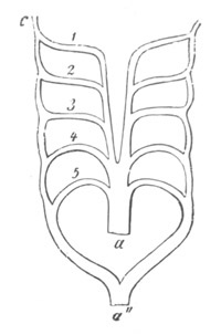
Fig. 364. Diagram of the arrangement of the arterial arches in an
embryo of one of the Amniota. (From Gegenbaur; after
Rathke.)
a. ventral aorta; a´´. dorsal aorta; 1, 2, 3, 4, 5. arterial
arches; c. carotid artery.
In the Amniota the metamorphosis of the arteries is in all cases very
similar. Five arches, viz. the mandibular, hyoid, and three branchial
arches are always developed (fig. 364), but, owing to the absence of
branchiæ, never function as branchial arteries. Of these the main
parts of the first two, connecting the truncus arteriosus with the
collecting trunk into which the arterial arches fall, always
disappear, usually before the complete development of the arteries in
the posterior arches.
The anterior part of the collecting trunk into which these vessels
fall is not obliterated when they disappear, but is on the contrary
continued forwards as a vessel supplying the brain, homologous with
that found in Fishes. It constitutes the internal carotid. Similarly
the anterior part of the trunk from which the mandibular and hyoid
arteries sprang is continued forwards as a small vessel[230],
which at
first passes to the oral region and constitutes in Reptiles the
lingual artery, homologous with the lingual artery of the Amphibia;
but in Birds and Mammals becomes more important, and is then known as
the external carotid (fig. 125). By these changes the roots of the
external and internal carotids spring respectively from the ventral
and dorsal ends of the primitive third artery, i.e. the artery of
the first branchial arch (fig. 365, c and c´); and thus this
arterial arch persists in all types as the common carotid,
and the
basal part of the internal carotid. The trunk connecting the third
arterial arch with the system of the dorsal aorta persists in some
Reptiles (Lacertilia, fig. 366 A) as a ductus Botalli, but is lost in
the remaining Reptiles and in Birds and Mammals (fig. 366 B, C, D). It
disappears earliest in Mammals (fig. 365 C), later in Birds (fig. 365
B), and still later in the majority of Reptiles.
The fourth arch always continues to give rise, as in the Anura, to the
system of the dorsal aorta.
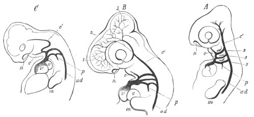
Fig. 365. Development of the great arterial trunks in the embryos
of A. a Lizard; B. the common Fowl; C. the Pig. (From Gegenbaur; after
Rathke.)
The first two arches have disappeared in all three. In A and B the
last three are still complete, but in C the last two are alone
complete.
p. pulmonary artery springing from the fifth arch, but still
connected with the system of the dorsal aorta by a ductus Botalli;
c. external carotid; c´. internal carotid; ad. dorsal aorta;
a. auricle; v. ventricle; n. nasal pit; m. rudiment of
fore-limb.
In all Reptiles it persists on both sides (fig. 366 A and B), but with
the division of the truncus arteriosus into three vessels one of
these, i.e. that opening furthest to the left side of the ventricle
(e and d), is continuous with the right fourth arch, and also
with the common carotid arteries (c); while a second springing from
the right side of the ventricle is continuous with the left fourth
arch (h and f). The right and left divisions of the fourth arch
meet however on the dorsal side of the œsophagus to give origin to
the dorsal aorta (g).
In Birds (fig. 366 C) the left fourth arch (h) loses its
connection with the dorsal aorta, though the ventral part remains as
the root of the left subclavian. The truncus arteriosus is moreover
only divided into two parts, one of which is continuous with all the
systemic arteries. Thus it comes about that in Birds the right fourth
arch (e) alone gives rise to the dorsal aorta.
In Mammals (fig. 366 D) the truncus arteriosus is only divided into
two, but the left fourth arch (e), instead of the right, is that
continuous with the dorsal aorta, and the right fourth arch (i) is
only continued into the right vertebral and right subclavian arteries.
The fifth arch always gives origin to the pulmonary artery (fig. 365,
p) and is continuous with one of the divisions of the truncus
arteriosus. In Lizards (fig. 366 A, i), Chelonians and Birds (fig.
366 C, i) and probably in Crocodilia, the right and left pulmonary
arteries spring respectively from the right and left fifth arches, and
during the greater part of embryonic life the parts of the fifth
arches between the origins of the pulmonary arteries and the system of
the dorsal aorta are preserved as ductus Botalli. These ductus Botalli
persist for life in the Chelonia. In Ophidia (fig. 366 B, h) and
Mammalia (fig. 366 D, m) only one of the fifth arches gives origin
to the two pulmonary arteries, viz. that on the right side in Ophidia,
and the left in Mammalia.
The ductus Botalli of the fifth arch (known in Man as the ductus
arteriosus) of the side on which the pulmonary arteries are formed,
may remain (e.g. in Man) as a solid cord connecting the common stern
of the pulmonary aorta with the systemic aorta.
The main history of the arterial arches in the Amniota has been
sufficiently dealt with, and the diagram, fig. 366, copied from
Rathke, shews at a glance the character of the metamorphosis these
arches undergo in the different types. It merely remains for me to say
a few words about the subclavian and vertebral arteries.
The subclavian arteries in Fishes usually spring from the trunks
connecting the branchial veins with the dorsal aorta. This origin,
which is also found in Amphibia, is typically found in the embryos of
the Amniota. In the Lizards this origin persists through life, but
both subclavians spring from the right
side. In most other types the
origin of the subclavians is carried upwards, so that they usually
spring from a trunk common to them and the carotids (arteria anonyma)
(Birds and some Mammals); or the left one, as in Man and some other
Mammals, arises from the systemic aorta just beyond the carotids.
Various further modifications in the origin of the subclavians of the
same general nature are found in Mammalia,
but they need not be
specified in detail. The vertebral arteries usually arise in close
connection with the subclavians, but in Birds they arise from the
common carotids.
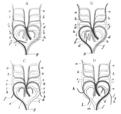
Fig. 366. Diagrams illustrating the metamorphosis of the arterial
arches in a Lizard A, a Snake B, a Bird C and a Mammal D. (From
Mivart; after Rathke.)
A. a. internal carotid; b. external carotid; c. common
carotid; d. ductus Botalli between the third and fourth arches;
e. right aortic trunk; f. subclavian; g. dorsal aorta; h.
left aortic trunk; i. pulmonary artery; k. rudiment of ductus
Botalli between the pulmonary artery and the system of the dorsal
aorta.
B. a. internal carotid; b. external carotid; c. common
carotid; d. right aortic trunk; e. vertebral artery; f. left
aortic trunk of dorsal aorta; h. pulmonary artery; i. ductus
Botalli of pulmonary artery.
C. a. internal carotid; b. external carotid; c. common
carotid; d. systemic aorta; e. fourth arch of right side (root
of dorsal aorta); f. right subclavian; g. dorsal aorta; h.
left subclavian (fourth arch of left side); i. pulmonary artery;
k. and l. right and left ductus Botalli of pulmonary arteries.
D. a. internal carotid; b. external carotid; c. common carotid;
d. systemic aorta; e. fourth arch of left side (root of dorsal
aorta); f. dorsal aorta; g. left vertebral artery; h. left
subclavian artery; i. right subclavian (fourth arch of right side);
k. right vertebral; l. continuation of right subclavian; m.
pulmonary artery; n. ductus Botalli of pulmonary artery.
Bibliography of the Arterial System.
(496) H. Rathke. “Ueb. d. Entwick. d. Arterien w. bei d. Säugethiere
von d. Bogen d. Aorta ausgehen.” Müller’s Archiv, 1843.
(497) H. Rathke. “Untersuchungen üb. d. Aortenwurzeln d. Saurier.”
Denkschriften d. k. Akad. Wien, Vol. XIII. 1857.
Vide also His (No. 232) and general works on Vertebrate Embryology.
The Venous System.
The venous system, as it is found in the embryos of Fishes, consists
in its earliest condition of a single large trunk, which traverses the
splanchnic mesoblast investing the part of the alimentary tract behind
the heart. This trunk is directly continuous in front with the heart,
and underlies the alimentary canal through both its præanal and
postanal sections. It is shown in section in fig. 367, v, and may be
called the subintestinal vein. This vein has been found in the embryos
of Teleostei, Ganoidei, Elasmobranchii and Cyclostomata, and runs
parallel to the dorsal aorta above, into which it is sometimes
continued behind (Teleostei, Ganoidei, etc.).
In Elasmobranch embryos the subintestinal vein terminates, as may be
gathered from sections (fig. 368, v.cau), shortly before the end of
the tail. The same series of sections also shews that at the cloaca,
where the gut enlarges and comes in contact with the skin, this vein
bifurcates, the two branches uniting into a single vein both in front
of and behind the cloaca.
In most Fishes the anterior part of this vein atrophies, the caudal
section alone remaining, but the anterior section of it persists in
the fold of the intestine in Petromyzon, and also remains in the
spiral valve of some Elasmobranchii. In Amphioxus, moreover, it forms,
as in the embryos of higher types, the main venous trunk, though even
here it is usually broken up into two or three parallel vessels.
It no doubt represents one of the primitive longitudinal trunks of the
vermiform ancestors of the Chordata. The heart and the branchial
artery constitute a specially modified anterior continuation of this
vein. The
dilated portal sinus of Myxine is probably also part of it;
and if this is really rhythmically contractile[231]
the fact would be
interesting as shewing that this quality, which is now localised in
the heart, was once probably common to the subintestinal vessel for
its whole length.
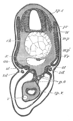
Fig. 367. Section through the trunk of a Scyllium embryo slightly
younger than 28 F.
sp.c. spinal canal; W. white matter of spinal cord; pr.
posterior nerve-roots; ch. notochord; x. subnotochordal rod;
ao. aorta; mp. muscle plate; mp´. inner layer of muscle-plate
already converted into muscles; Vr. rudiment of vertebral body;
st. segmental tube; sd. segmental duct; sp.v. spiral valve;
v. subintestinal vein; p.o. primitive generative cells.
On the development of the cardinal veins (to be described below)
considerable changes are effected in the subintestinal vein. Its
postanal section, which is known in the adult as the caudal vein,
unites with the cardinal veins. On this junction being effected
retrogressive changes take place in the præanal section of the
original subintestinal vessel. It breaks up in front into a number of
smaller vessels, the most important of which is a special vein, which
lies in the fold of the spiral valve, and which is more conspicuous in
some Elasmobranchii than in Scyllium, in which the development of the
vessel has been mainly studied. The lesser of the two branches
connecting it round the cloaca with the caudal vein first vanishes,
and then the larger; and the two posterior cardinals are left as the
sole forward continuations of the caudal vein. The latter then becomes
prolonged forwards, so that the two cardinals open into it some little
distance in front of the hind end of the kidneys. By these changes,
and by the disappearance of the postanal section of the gut, the
caudal vein is made to appear as a supraintestinal and not, as it
really is, a subintestinal vessel.
From the subintestinal vein there is given off a branch which supplies
the yolk-sack. This leaves the subintestinal vein close
to the liver.
The liver, on its development, embraces the subintestinal vein, which
then breaks up into a capillary system in the liver, the main part of
its blood coming at this period from the yolk-sack.
The portal system is thus established from the subintestinal vein; but
is eventually joined by the various visceral, and sometimes by the
genital, veins as they become successively developed.
The blood from the liver is brought back to the sinus venosus by veins
known as the hepatic veins, which, like the hepatic capillary system,
are derivatives of the subintestinal vessel.
There join the portal system in Myxinoids and many Teleostei a number
of veins from the anterior abdominal walls, representing a
commencement of the anterior abdominal or epigastric vein of higher
types[232].
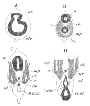
Fig. 368. Four sections through the postanal part of the tail
of an embryo of the same age as fig. 28 F.
A. is the posterior section.
nc. neural canal; al. postanal gut; alv. caudal vesicle of
postanal gut; x. subnotochordal rod; mp. muscle-plate; ch.
notochord; cl.al. cloaca; ao. aorta; v.cau. caudal vein.
In the higher Vertebrates the original subintestinal vessel never
attains a full development, even in the embryo. It is represented by
(1) the ductus
venosus, which, like the true subintestinal vein, gives
origin (in the Amniota) to the vitelline veins to the yolk-sack, and
(2) by the caudal vein. Whether the partial atrophy of the
subintestinal vessel was primitively caused by the development of the
cardinal veins, or for some other reason, it is at any rate a fact
that in all existing Fishes the cardinal veins form the main venous
channels of the trunk.
Their later development than the subintestinal vessel as well as their
absence in Amphioxus, probably indicate that they became evolved, at
any rate in their present form, within the Vertebrate phylum.
The embryonic condition of the venous system, with a single large
subintestinal vein is, as has been stated, always modified by the
development of a paired system of vessels, known as the cardinal
veins, which bring to the heart the greater part of the blood from the
trunk.
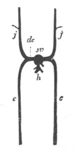
Fig. 369. Diagram of the paired venous system of a Fish.
(From Gegenbaur.)
j. jugular vein (anterior cardinal vein); c. posterior cardinal
vein; h. hepatic veins; sv. sinus venosus; dc. ductus
Cuvieri.
The cardinal veins appear in Fishes as four paired longitudinal trunks
(figs. 363 and 369), two anterior (j) and two posterior (c). They
unite into two transverse trunks on either side, known as the ductus
Cuvieri (dc), which fall into the sinus venosus, passing from the
body wall to the sinus by a lateral mesentery of the heart already
spoken of (p. 627, fig. 352). The anterior pair, known as the anterior
cardinal or jugular veins, bring to the heart the blood from the head
and neck. They are placed one on each side above the level of the
branchial arches (fig. 299, a.cv). The posterior cardinal veins lie
immediately dorsal to the mesonephros (Wolffian body), and are mainly
supplied by the blood from this organ and from the walls of the body
(fig. 275, c.a.v). In many forms (Cyclostomata, Elasmobranchii and
many Teleostei) they unite posteriorly with the caudal veins in the
manner already described, and in a large number of instances the
connecting branch between the two systems, in its passage through the
mesonephros, breaks up into a capillary network, and so gives rise to
a renal portal system.
The vein from the anterior pair of fins (subclavian) usually unites
with the anterior jugular vein.
The venous system of the Amphibia and Amniota always differs from that
of Fishes in the presence of a new vessel, the vena cava inferior,
which replaces the posterior cardinal veins; the latter only being
present, in their piscine form, during embryonic life. It further
differs from that of all Fishes, except the Dipnoi, in the presence of
pulmonary veins bringing back the blood directly from the lungs.
In the embryos of all the higher forms the general characters of the
venous system are at first the same as in Fishes, but with the
development of the vena cava inferior the front sections of the
posterior cardinal veins atrophy, and the ductus Cuvieri, remaining
solely connected with the anterior cardinals and their derivatives,
constitute the superior venæ cavæ. The inferior cava receives the
hepatic veins.
Apart from the non-development of the subintestinal vein the visceral
section of the venous system is very similar to that in Fishes.
The further changes in the venous system must be dealt with separately
for each group.
Amphibia. In Amphibia (Götte, No. 296) the anterior and posterior
cardinal veins arise as in Pisces. From the former the internal
jugular vein arises as a branch; the external jugular constituting the
main stem. The subclavian with its large cutaneous branch also springs
from the system of the anterior cardinal. The common trunk formed by
the junction of these three veins falls into the ductus Cuvieri.
The posterior cardinal veins occupy the same position as in Pisces,
and unite behind with the caudal veins, which Götte has shewn to be
originally situated below the postanal gut. The iliac veins unite with
the posterior cardinal veins, where the latter fall into the caudal
vein. The original piscine condition of the veins is not long
retained. It is first of all disturbed by the development of the
anterior part of the important unpaired venous trunk which forms in
the adult the vena cava inferior. This is developed independently, but
unites behind with the right posterior cardinal. From this point
backwards the two cardinal veins coalesce for some distance, to give
rise to the posterior section of the vena cava inferior, situated
between the kidneys[233].
The anterior sections of the cardinal veins
subsequently atrophy. The posterior part of the cardinal veins, from
their junction with the vena cava inferior to the caudal veins, forms
a rhomboidal figure. The iliac vein joins the outer angle of this
figure, and is thus in direct communication with the inferior vena
cava, but it is also connected with a longitudinal
vessel on the outer
border of the kidneys, which receives transverse vertebral veins and
transmits their blood to the kidneys, thus forming a renal portal
system. The anterior limbs of the rhomboid formed by the cardinal
veins soon atrophy, so that the blood from the hind limbs can only
pass to the inferior vena cava through the renal portal system. The
posterior parts of the two cardinal veins (uniting in the Urodela
directly with the unpaired caudal vein) still persist. The iliac veins
also become directly connected with a new vein, the anterior abdominal
vein, which has meanwhile become developed. Thus the iliac veins
become united with the system of the vena cava inferior through the
vena renalis advehens on the outer border of the kidney, and with the
anterior abdominal veins by the epigastric veins.
The visceral venous system begins with the development of two
vitelline veins, which at first join the sinus venosus directly. They
soon become enveloped in the liver, where they break up into a
capillary system, which is also joined by the other veins from the
viscera. The hepatic system has in fact the same relations as in
Fishes. Into this system the anterior abdominal vein also pours itself
in the adult. This vein is originally formed of two vessels, which at
first fall directly into the sinus venosus, uniting close to their
opening into the sinus with a vein from the truncus arteriosus. They
become prolonged backwards, and after receiving the epigastric veins
above mentioned from the iliac veins, and also veins from the
allantoic bladder, unite behind into a single vessel. Anteriorly the
right vein atrophies and the left continues forward the unpaired
posterior section.
A secondary connection becomes established between the anterior
abdominal vein and the portal system; so that the blood originally
transported by the former vein to the heart becomes diverted so as to
fall into the liver. A remnant of the primitive connection is still
retained in the adult in the form of a small vein, the so-called vena
bulbi posterior, which brings the blood from the walls of the truncus
arteriosus directly into the anterior abdominal vein.
The pulmonary veins grow directly from the heart to the lungs.
For our knowledge of the development of the venous system of the
Amniota we are mainly indebted to Rathke.
Reptilia. As an example of the Reptilia the Snake may be selected, its
venous system having been fully worked out by Rathke in his important
memoir on its development (No. 300).
The anterior (external jugular) and posterior cardinal veins are
formed in the embryo as in all other types (fig. 370, vj and vc);
and the anterior cardinal, after giving rise to the anterior vertebral
and to the cephalic veins, persists with but slight modifications in
the adult; while the two ductus Cuvieri constitute the superior venæ
cavæ.
The two posterior cardinals unite behind with the caudal veins. They
are placed in the usual situation on the dorsal and outer border of
the kidneys.
With the development of the vena cava inferior, to be described below,
the blood from the kidneys becomes mainly transported by this vessel
to the heart; and the section of the posterior cardinals opening into
the ductus Cuvieri gradually atrophies, their posterior parts
remaining however on the outer border of the kidneys as the venæ
renales advehentes[234].
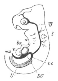
Fig. 370. Anterior portion of the venous system of an embryonic
Snake. (From Gegenbaur; after Rathke.)
vc. posterior cardinal vein; vj. jugular vein; DC. ductus
Cuvieri; vu. allantoic vein; v. ventricle; ba. truncus
arteriosus; a. visceral clefts; l. auditory vesicle.
While the front part of the posterior cardinal veins is undergoing
atrophy, the intercostal veins, which originally poured their blood
into the posterior cardinal veins, become also connected with two
longitudinal veins—the posterior vertebral veins—which are
homologous with the azygos and hemiazygos veins of Man; and bear the
same relation to the anterior vertebral veins that the anterior and
posterior cardinals do to each other.
These veins are at first connected by transverse anastomoses with the
posterior cardinals, but, on the disappearance of the front part of
the latter, the whole of the blood from the intercostal veins falls
into the posterior vertebral veins. They are united in front with the
anterior vertebral veins, and the common trunk of the two veins on
each side falls into the jugular vein.
The posterior vertebral veins are at first symmetrical, but after
becoming connected by transverse anastomoses, the right becomes the
more important of the two.
The vena cava inferior, though considerably later in its development
than the cardinals, arises fairly early. It constitutes in front an
unpaired trunk, at first very small, opening into the right allantoic
vein, close to the heart. Posteriorly it is continuous with two veins
placed on the inner border of the kidneys[235].
The vena cava inferior passes through the dorsal part of the liver,
and in doing so receives the hepatic veins.
The portal system is at first constituted by the vitelline vein, which
is directly continuous with the venous end of the heart, and at first
receives the two ductus Cuvieri, but at a later period unites with the
left ductus.
It soon receives a mesenteric vein bringing the blood
from the viscera, which is small at first but rapidly increases in
importance.
The common trunk of the vitelline and mesenteric veins, which may be
called the portal vein, becomes early enveloped by the liver, and
gives off branches to this organ, the blood from which passes by the
hepatic veins to the vena cava inferior. As the branches in the liver
become more important, less and less blood is directly transported to
the heart, and finally the part of the original vitelline vein in
front of the liver is absorbed, and the whole of the blood from the
portal system passes from the liver into the vena cava inferior.
The last section of the venous system to be dealt with is that of the
anterior abdominal vein. There are originally, as in the Anura, two
veins belonging to this system, which owing to the precocious
development of the bladder to form the allantois, constitute the
allantoic veins (fig. 370, vu).
These veins, running along the anterior abdominal wall, are formed
somewhat later than the vitelline vein, and fall into the two ductus
Cuvieri. They unite with two epigastric veins (homologous with those
in the Anura), which connect them with the system of the posterior
cardinal veins. The left of the two eventually atrophies, so that
there is formed an unpaired allantoic vein. This vein at first
receives the vena cava inferior close to the heart, but eventually the
junction of the two takes place in the region of the liver, and
finally the anterior abdominal vein (as it comes to be after the
atrophy of the allantois) joins the portal system and breaks up into
capillaries in the liver[236].
In Lizards the iliac veins join the posterior cardinals, and so pour
part of their blood into the kidneys; they also become connected by
the epigastric veins with the system of the anterior abdominal or
allantoic vein. The subclavian veins join the system of the superior
venæ cavæ.
The venous system of Birds and Mammals differs in two important points
from that of Reptilia and Amphibia. Firstly the anterior abdominal
vein is only a fœtal vessel, forming during fœtal life the allantoic
vein; and secondly a direct connection is established between the vena
cava inferior and the veins of the hind limbs and posterior parts of
the cardinal veins, so that there is no renal portal system.
Aves. The Chick may be taken to illustrate the development of the
venous system in Birds.
On the third day, nearly the whole of the venous blood from the body
of the embryo is carried back to the heart by two main venous trunks,
the anterior (fig. 125, S.Ca.V) and posterior (V.Ca) cardinal
veins, joining on each side to form the short transverse ductus
Cuvieri (DC), both of which unite with the sinus venosus close to
the heart. As the head and neck continue to enlarge, and the wings
become developed, the single anterior
cardinal or jugular vein (fig.
371, J), of each side, is joined by two new veins: the vertebral
vein, bringing back blood from the head and neck, and the subclavian
vein from the wing (W).
On the third day the posterior cardinal veins are the only veins which
return the blood from the hinder part of the body of the embryo.
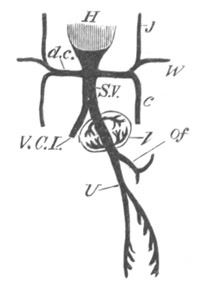
Fig. 371. Diagram of the venous circulation in the Chick at the
commencement of the fifth day.
H. heart; d.c. ductus Cuvieri. Into the ductus Cuvieri of each
side fall J. the jugular vein, W. the vein from the wing, and
c. the inferior cardinal vein; S.V. sinus venosus; Of.
vitelline vein; U. allantoic vein, which at this stage gives off
branches to the body-walls; V.C.I. inferior vena cava; l. liver.
About the fourth or fifth day, however, the vena cava inferior (fig.
371, V.C.I.) makes its appearance. This, starting from the sinus
venosus not far from the heart, is on the fifth day a short trunk
running backward in the middle line below the aorta, and speedily
losing itself in the tissues of the Wolffian bodies. When the true
kidneys are formed it also receives blood from them, and thenceforward
enlarging rapidly becomes the channel by which the greater part of the
blood from the hinder part of the body finds its way to the heart. In
proportion as the vena cava inferior increases in size, the posterior
cardinal veins diminish.
The blood originally coming to them from the posterior part of the
spinal cord and trunk is transported into two posterior vertebral
veins, similar to those in Reptilia, which are however placed dorsally
to the heads of the ribs, and join the anterior vertebral veins. With
their appearance the anterior parts of the posterior cardinals
disappear. The blood from the hind limbs becomes transported directly
through the kidney into the vena cava inferior, without forming a
renal portal system[237].
On the third day the course of the vessels from the yolk-sack is very
simple. The two vitelline veins, of which the right is already the
smaller, form the ductus venosus, from which, as it passes through the
liver on its way to the heart, are given off the two sets of venæ
advehentes and venæ revehentes (fig. 371).
With the appearance of the allantois on the fourth day, a new feature
is introduced. From the ductus venosus there is given off a vein which
quickly divides into two branches. These, running along the ventral
walls of the body from which they receive some amount of blood, pass
to the allantois. They are the allantoic veins (fig. 371, U)
homologous with the anterior abdominal vein of the lower types. They
unite in front to form a single vein, which becomes, by reason of the
rapid growth of the allantois, very long. The right branch soon
diminishes in size and finally disappears. Meanwhile the left on
reaching the allantois bifurcates; and, its two
branches becoming
large and conspicuous, there still appear to be two main allantoic
veins. At its first appearance the allantoic vein seems to be but a
small branch of the vitelline, but as the allantois grows rapidly, and
the yolk-sack dwindles, this state of things is reversed, and the less
conspicuous vitelline appears as a branch of the larger allantoic
vein.
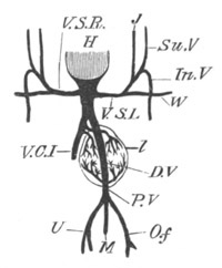
Fig. 372. Diagram of the venous circulation in the Chick during
the later days of incubation.
H. heart; V.S.R. right vena cava superior; V.S.L. left vena
cava superior. The two venæ cavæ superiores are the original ‘ductus
Cuvieri,’ they open into the sinus venosus. J. jugular vein;
Su.V. anterior vertebral vein; In.V. inferior vertebral vein;
W. subclavian; V.C.I. vena cava inferior; D.V. ductus venosus;
P.V. portal vein; M. mesenteric vein bringing blood from the
intestines into the portal vein; O.f. vitelline vein; U.
allantoic vein. The three last mentioned veins unite together to
form the portal vein; l. liver.
On the third day the blood returning from the walls of the intestine
is insignificant in amount. As however the intestine becomes more and
more developed, it acquires a distinct venous system, and its blood is
returned by veins which form a trunk, the mesenteric vein (fig. 372,
M) falling into the vitelline vein at its junction with the
allantoic vein.
These three great veins, in fact, form a large common trunk, which
enters at once into the liver, and which we may now call the portal
vein (fig. 372, P.V). This, at its entrance into the liver, partly
breaks up into the venæ advehentes, and partly continues as the
ductus venosus (D.V) straight through the liver, emerging from which
it joins the vena cava inferior. Before the establishment of the vena
cava inferior, the venæ revehentes, carrying back the blood which
circulates through the hepatic capillaries, join the ductus venosus
close to its exit from the liver. By the time however that the vena
cava has become a large and important vessel it is found that the venæ
revehentes, or as we may now call them the hepatic veins, have
shifted their embouchment, and now fall directly into that vein, the
ductus venosus making a separate junction rather higher up (fig. 372).
This state of things continues with but slight changes till near the
end of incubation, when the chick begins to breathe the air in the
air-chamber of the shell, and respiration is no longer carried on by
the allantois. Blood then ceases to flow along the allantoic vessels;
they become obliterated. The vitelline vein, which as the yolk becomes
gradually absorbed proportionately diminishes in size and importance,
comes to appear as a mere branch of the portal vein. The ductus
venosus becomes obliterated; and hence the whole of the blood coming
through the portal vein flows into the substance of the liver, and so
by the hepatic veins into the vena cava.
Although the allantoic (anterior abdominal) vein is obliterated in the
adult, there is nevertheless established an anastomosis between the
portal system and the veins bringing the blood from the limbs to the
vena cava
inferior, in that the caudal vein and posterior pelvic veins
open into a vessel, known as the coccygeo-mesenteric vein, which joins
the portal vein; while at the same time the posterior pelvic veins are
connected with the common iliac veins by a vessel which unites with
them close to their junction with the coccygeo-mesenteric vein.
Mammalia. In Mammals the same venous trunks are developed in the
embryo as in other types (fig. 373 A). The anterior cardinals or
external jugulars form the primitive veins of the anterior part of the
body, and the internal jugulars and anterior vertebrals are
subsequently formed. The subclavians (fig. 373 A, s), developed on
the formation of the anterior limbs, also pour their blood into these
primitive trunks. In the lower Mammalia (Monotremata, Marsupialia,
Insectivora, some Rodentia, etc.), the two ductus Cuvieri remain as
the two superior venæ cavæ, but more usually an anastomosis arises
between the right and left innominate veins, and eventually the whole
of the blood of the left superior cava is carried to the right side,
and there is left only a single superior cava (fig. 373 B and C). A
small rudiment of the left superior cava remains however as the sinus
coronarius and receives the coronary vein from the heart (figs. 373 C,
cor and 374, cs).
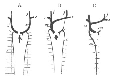
Fig. 373. Diagram of the development of the paired venous system of
Mammals (Man). (From Gegenbaur.)
j. jugular vein; cs. vena cava superior; s. subclavian veins;
c. posterior cardinal vein; v. vertebral vein; az. azygos
vein; cor. coronary vein.
A. Stage in which the cardinal veins have already disappeared. Their
position is indicated by dotted lines.
B. Later stage when the blood from the left jugular vein is carried
into the right to form the single vena cava superior; a remnant
of the left superior cava being however still left.
C. Stage after the left vertebral vein has disappeared; the right
vertebral remaining as the azygos vein. The coronary vein remains
as the last remnant of the left superior vena cava.
The posterior cardinal veins form at first the only veins receiving
the
blood from the posterior part of the trunk and kidneys; and on the
development of the hind limbs receive the blood from them also.
As in the types already described an unpaired vena cava inferior
becomes eventually developed, and gradually carries off a larger and
larger portion of the blood originally returned by the posterior
cardinals. It unites with the common stem of the allantoic and
vitelline veins in front of the liver.
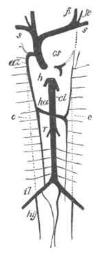
Fig. 374. Diagram of the chief venous trunks of Man. (From
Gegenbaur.)
cs. vena cava superior; s. subclavian vein; ji. internal
jugular; je. external jugular; az. azygos vein; ha. hemiazygos
vein; c. dotted line shewing previous position of cardinal veins;
ci. vena cava inferior; r. renal veins; il. iliac; hy.
hypogastric veins; h. hepatic veins.
The dotted lines shew the position of embryonic vessels aborted in
the adult.
At a later period a pair of trunks is established bringing the blood
from the posterior part of the cardinal veins and the crural veins
directly into the vena cava inferior (fig. 374, il). These vessels,
whose development has not been adequately investigated, form the
common iliac veins, while the posterior ends of the cardinal veins
which join them become the hypogastric veins (fig. 374, hy). Owing
to the development of the common iliac veins there is no renal portal
system like that of the Reptilia and Amphibia.
Posterior vertebral veins, similar to those of Reptilia and Birds, are
established in connection with the intercostal and lumbar veins, and
unite anteriorly with the front part of the posterior cardinal veins
(fig. 373 A)[238].
On the formation of the posterior vertebral veins, and as the inferior
vena cava becomes more important, the middle part of the posterior
cardinals becomes completely aborted (fig. 374, c), the anterior and
posterior parts still persisting, the former as the continuations of
the posterior vertebrals into the anterior vena cava (az), the
latter as the hypogastric veins (hy).
Though in a few Mammalia both the posterior vertebrals persist, a
transverse connection is usually established between them, and the one
(the right) becoming the more important constitutes the azygos vein
(fig. 374, az), the persisting part of the left forming the
hemiazygos vein (ha).
The remainder of the venous system is formed in the embryo of the
vitelline and allantoic veins, the former being eventually joined by
the mesenteric vein so as to constitute the portal vein.
The vitelline vein is the first part of this system established, and
divides near the heart into two veins bringing back the blood from the
yolk-sack (umbilical vesicle). The right vein soon however aborts.
The allantoic (anterior abdominal) veins are originally paired. They
are developed very early, and at first course along the still widely
open somatic walls of the body, and fall into the single vitelline
trunk in front. The right allantoic vein disappears before long, and
the common trunk formed by the junction of the vitelline and allantoic
veins becomes considerably elongated. This trunk is soon enveloped by
the liver.
The succeeding changes have been somewhat differently described by
Kölliker and Rathke. According to the former the common trunk of the
allantoic and vitelline veins in its passage through the liver gives
off branches to the liver, and also receives branches from this organ
near its anterior exit. The main trunk is however never completely
aborted, as in the embryos of other types, but remains as the ductus
venosus Arantii.
With the development of the placenta the allantoic vein becomes the
main source of the ductus venosus, and the vitelline or portal vein,
as it may perhaps be now conveniently called, ceases to join it
directly, but falls into one of its branches in the liver.
The vena cava inferior joins the continuation of the ductus venosus in
front of the liver, and, as it becomes more important, it receives
directly the hepatic veins which originally brought back blood into
the ductus venosus. The ductus venosus becomes moreover merely a small
branch of the vena cava.
At the close of fœtal life the allantoic vein becomes obliterated up
to its place of entrance into the liver; the ductus venosus becomes a
solid cord—the so-called round ligament—and the whole of the venous
blood is brought to the liver by the portal vein[239].
Owing to the allantoic (anterior abdominal) vein having merely a
fœtal existence an anastomosis between the iliac veins and the portal
system by means of the anterior abdominal vein is not established.
Bibliography of the Venous System.
(498) J. Marshall. “On the development of the great anterior veins.”
Phil. Trans., 1859.
(499) H. Rathke. “Ueb. d. Bildung d. Pfortader u. d. Lebervenen b.
Säugethieren.” Meckel’s Archiv, 1830.
(500) H. Rathke. “Ueb. d. Bau u. d. Entwick. d. Venensystems d.
Wirbelthiere.” Bericht. üb. d. naturh. Seminar. d. Univ. Königsberg,
1838.
Vide also Von Baer (No. 291), Götte (No. 296), Kölliker (No. 298),
and Rathke (Nos. 299, 300, and 301).
Lymphatic System.
The lymphatic system arises from spaces in the general parenchyma of
the body, independent in their origin of the true body cavity, though
communicating both with this cavity and with the vascular system.
In all the true Vertebrata certain parts of the system form definite
trunks communicating with the venous system; and in the higher types
the walls of the main lymphatic trunks become quite distinct.
But little is known with reference to the ontogeny of the lymphatic
vessels, but they originate late in larval life, and have at first the
form of simple intercellular spaces.
The lymphatic glands appear to originate from lymphatic plexuses, the
cells of which produce lymph corpuscles. It is only in Birds and
Mammals, and especially in the latter, that the lymphatic glands form
definite structures.
The Spleen. The spleen, from its structure, must be classed with the
lymphatic glands, though it has definite relations to the vascular
system. It is developed in the mesoblast of the mesogastrium, usually
about the same time and in close connection with the pancreas.
According to Müller and Peremeschko the mass of mesoblast which forms
the spleen becomes early separated by a groove on the one side from
the pancreas and on the other from the mesentery. Some of its cells
become elongated, and send out processes which uniting with like
processes from other cells form the trabecular system. From the
remainder of the tissue are derived the cells of the spleen pulp,
which frequently contain more than one nucleus. Especial accumulations
of these cells take place at a later period to form the so-called
Malpighian corpuscles of the spleen.
Bibliography of Spleen.
(501) W. Müller. “The Spleen.” Stricker’s Histology.
(502) Peremeschko. “Ueb. d. Entwick. d. Milz.” Sitz. d. Wien. Akad.
Wiss., Vol. LVI. 1867.
Suprarenal bodies.
In Elasmobranch Fishes two distinct sets of structures are found, both
of which have been called suprarenal bodies. As shewn in the sequel
both of these structures probably unite in the higher types to form
the suprarenal bodies.
One of them consists of a series of paired bodies, situated on the
branches of the dorsal aorta, segmentally arranged, and forming a
chain extending from close behind the heart to the hinder end of the
body cavity. Each body is formed of a series of lobes, and exhibits a
well-marked distinction into a cortical layer of columnar cells, and a
medullary substance formed of irregular polygonal cells. As first
shewn by Leydig, they are
closely connected with the sympathetic
ganglia, and usually contain numerous ganglion cells distributed
amongst the proper cells of the body.
The second body consists of an unpaired column of cells placed between
the dorsal aorta and unpaired caudal vein, and bounded on each side by
the posterior parts of the kidney. I propose to call it the interrenal
body. In front it overlaps the paired suprarenal bodies, but does not
unite with them. It is formed of a series of well-marked lobules, etc.
In the fresh state Leydig (No. 506) finds that “fat molecules form the
chief mass of the body, and one finds freely imbedded in them clear
vesicular nuclei.” As may easily be made out from hardened specimens
it is invested by a tunica propria, which gives off septa dividing it
into well-marked areas filled with polygonal cells. These cells
constitute the true parenchyma of the body. By the ordinary methods of
hardening, the oil globules, with which they are filled in the fresh
state, completely disappear.
The paired suprarenal bodies (Balfour, No. 292, pp. 242-244) are
developed from the sympathetic ganglia. These ganglia, shewn in an
early stage in fig. 380, sy.g, become gradually divided into a
ganglionic part and a glandular part. The former constitutes the
sympathetic ganglia of the adult; the latter the true paired
suprarenal bodies. The interrenal body is however developed (Balfour,
No. 292, pp. 245-247) from indifferent mesoblast cells between the two
kidneys, in the same situation as in the adult.
The development of the suprarenal bodies in the Amniota has been most
fully studied by Braun (No. 503) in the Reptilia.
In Lacertilia they consist of a pair of elongated yellowish bodies,
placed between the vena renalis revehens and the generative glands.
They are formed of two constituents, viz. (1) masses of brown cells
placed on the dorsal side of the organ, which stain deeply with
chromic acid, like certain of the cells of the suprarenals of
Mammalia, and (2) irregular cords, in part provided with a lumen,
filled with fat-like globules[240],
amongst which are nuclei. On
treatment with chromic acid the fat globules disappear, and the cords
break up into bodies resembling columnar cells.
The dorsal masses of brown cells are developed from the sympathetic
ganglia in the same way as the paired suprarenal bodies of the
Elasmobranchii, while the cords filled with fat-like globules are
formed of indifferent mesoblast cells as a thickening in the lateral
walls of the inferior vena cava, and the cardinal veins continuous
with it. The observations of Brunn (No. 504) on the Chick, and
Kölliker (No. 298, pp. 953-955) on the Mammal, add but little to those
of Braun. They shew that the greater part of the gland (the cortical
substance) in these two types is derived from the mesoblast, and that
the glands are closely connected with sympathetic ganglia; while
Kölliker also states that the posterior part of the organ is unpaired
in the embryo rabbit of 16 or 17 days.
The structure and development of what I have called the interrenal
body
in Elasmobranchii so closely correspond with that of the
mesoblastic part of the suprarenal bodies of the Reptilia, that I have
very little hesitation in regarding them as homologous[241];
while the
paired bodies in Elasmobranchii, derived from the sympathetic ganglia,
clearly correspond with the part of the suprarenals of Reptilia having
a similar origin; although the anterior parts of the paired suprarenal
bodies of Fishes have clearly become aborted in the higher types.
In Elasmobranch Fishes we thus have (1) a series of paired bodies,
derived from the sympathetic ganglia, and (2) an unpaired body of
mesoblastic origin. In the Amniota these bodies unite to form the
compound suprarenal bodies, the two constituents of which remain,
however, distinct in their development. The mesoblastic constituent
appears to form the cortical part of the adult suprarenal body, and
the nervous constituent the medullary part.
Bibliography of the Suprarenal bodies.
(503) M. Braun. “Bau u. Entwick. d. Nebennieren bei Reptilien.”
Arbeit. a. d. zool.-zoot. Institut Würzburg, Vol. V. 1879.
(504) A. v. Brunn. “Ein Beitrag z. Kenntniss d. feinern Baues u. d.
Entwick. d. Nebennieren.” Archiv f. mikr. Anat., Vol. VIII. 1872.
(505) Fr. Leydig. Untersuch. üb. Fische u. Reptilien. Berlin, 1853.
(506) Fr. Leydig. Rochen u. Haie. Leipzig, 1852.
Vide also F. M. Balfour (No. 292), Kölliker (No. 298), Remak (No.
302), etc.
In all the Cœlenterata, except the Ctenophora, the contractile
elements of the body wall consist of filiform processes of ectodermal
or entodermal epithelial cells (figs. 375 and 376 B). The elements
provided with these processes, which were first discovered by
Kleinenberg, are known as myoepithelial cells. Their contractile parts
may either be striated (fig. 376) or non-striated (fig. 375). In some
instances the epithelial part of the cell may nearly abort, its
nucleus alone remaining (fig. 376 A); and in this way a layer of
muscles lying completely below the surface may be established.
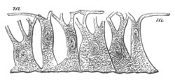
Fig. 375. Myo-epithelial cells of Hydra. (From Gegenbaur;
after Kleinenberg.)
m. contractile fibres.
There is embryological evidence of the derivation of the voluntary
muscular system of a large number of types from myoepithelial cells of
this kind. The more important of these groups are the Chætopoda, the
Gephyrea, the Chætognatha, the Nematoda, and the Vertebrata[242].
While there is clear evidence that the muscular system of a large
number of types is composed of cells which had their origin in
myoepithelial cells, the mode of evolution of the
muscular system of
other types is still very obscure. The muscles may arise in the embryo
from amœboid or indifferent cells, and the Hertwigs[243]
hold that in
many of these instances the muscles have also phylogenetically taken
their origin from indifferent connective-tissue cells. The subject is
however beset with very serious difficulties, and to discuss it here
would carry me too far into the region of pure histology.
The voluntary muscular system of the Chordata.
The muscular fibres. The muscular elements of the Chordata undoubtedly
belong to the myoepithelial type. The embryonic muscle-cells are at
first simple epithelial cells, but soon become spindle-shaped: part of
their protoplasm becomes differentiated into longitudinally placed
striated muscular fibrils, while part, enclosing the nucleus, remains
indifferent, and constitutes the epithelial element of the cells. The
muscular fibrils are either placed at one side of the epithelial part
of the cell, or in other instances (the Lamprey, the Newt, the
Sturgeon, the Rabbit) surround it. The latter arrangement is shewn for
the Sturgeon in fig. 57.
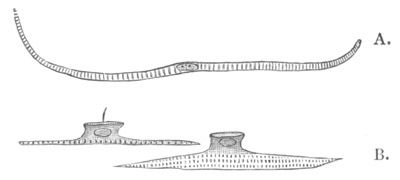
Fig. 376. Muscle-cells of Lizzia Köllikeri. (From Lankester; after
O. and R. Hertwig.)
A. Muscle-cell from the circular fibres of the subumbrella.
B. Myoepithelial cells from the base of a tentacle.
The number of the fibrils of each cell gradually increases, and the
protoplasm diminishes, so that eventually only the nucleus, or nuclei
resulting from its division, are left. The products of each cell
probably give rise, in conjunction with a further division of the
nucleus, to a primitive bundle, which,
except in Amphioxus,
Petromyzon, etc., is surrounded by a special investment of sarcolemma.
The voluntary muscular system. For the purposes of description the
muscular system of the Vertebrata may conveniently be divided into two
sections, viz. that of the head and that of the trunk. The main part,
if not the whole, of the muscular system of the trunk is derived from
certain structures, known as the muscle-plates, which take their
origin from part of the primitive mesoblastic somites.
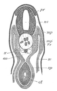
Fig. 377. Transverse section through the trunk of an embryo
slightly older than fig. 28 E.
nc. neural canal; pr. posterior root of spinal nerve; x.
subnotochordal rod; ao. aorta; sc. somatic mesoblast; sp.
splanchnic mesoblast; mp. muscle-plate; mp´. portion of
muscle-plate converted into muscle; Vr. portion of the vertebral
plate which will give rise to the vertebral bodies; al. alimentary
tract.
It has already been stated (pp. 292-296) that the mesoblastic somites
are derived from the dorsal segmented part of the primitive
mesoblastic plates. Since the history of these bodies is presented in
its simplest form in Elasmobranchii it will be convenient to commence
with this group. Each somite is composed of two layers—a somatic and
a splanchnic—both formed of a single row of columnar cells. Between
these two layers is a cavity, which is at first directly continuous
with the general body cavity, of which indeed it merely forms a
specialised part (fig. 377). Before long the cavity becomes however
completely constricted off from the permanent body cavity.
Very early (fig. 377) the inner or splanchnic wall of the somites
loses its simple constitution, owing to the middle part of it
undergoing peculiar changes. The meaning of the changes is at once
shewn by longitudinal horizontal sections, which prove (fig. 378) that
the cells in this situation (mp´) have become extended in a
longitudinal direction, and, in fact, form typical spindle-shaped
embryonic muscle-cells, each with a large nucleus. Every muscle-cell
extends for the whole length of a somite. The inner layer of each
somite, immediately within the muscle-band just described, begins to
proliferate, and produce
a mass of cells, placed between the muscles
and the notochord (Vr). These cells form the commencing vertebral
bodies, and have at first (fig. 378) the same segmentation as the
somites from which they sprang.
After the separation of the vertebral bodies from the somites the
remaining parts of the somites may be called muscle-plates; since they
become directly converted into the whole voluntary muscular system of
the trunk (fig. 379, mp).
According to the statements of Bambeke and Götte, the Amphibians
present some noticeable peculiarities in the development of their
muscular system, in that such distinct muscle-plates as those of other
vertebrate types are not developed. Each side-plate of mesoblast is
divided into a somatic and a splanchnic layer, continuous throughout
the vertebral and parietal portions of the plate. The vertebral
portions (somites) of the plates soon become separated from the
parietal, and form independent masses of cells constituted of two
layers, which were originally continuous with the somatic and
splanchnic layers of the parietal plates (fig. 79). The outer or
somatic layer of the vertebral plates is formed of a single row of
cells, but the inner or splanchnic layer is made up of a kernel of
cells on the side of the somatic layer and an inner layer. The kernel
of the splanchnic layer and the outer or somatic layer together
correspond to a muscle-plate of other Vertebrata, and exhibit a
similar segmentation.
Osseous Fishes are stated to agree with Amphibians in the development
of their somites and muscular systems[244],
but further observations
on this point are required.
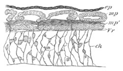
Fig. 378. Horizontal section through the trunk of an embryo of
Scyllium considerably younger than 28 F.
ch. notochord; ep. epiblast; Vr. rudiment of vertebral body;
mp. muscle-plate; mp´. portion of muscle-plate already
differentiated into longitudinal muscles.
In Birds the horizontal splitting of the mesoblast extends at first to
the dorsal summit of the mesoblastic plates, but after the isolation
of the somites the split between the somatic and splanchnic layers
becomes to a large extent obliterated, though in the anterior somites
it appears in part to persist. The somites on the second day, as seen
in a transverse section (fig. 115, P.v.), are somewhat quadrilateral
in form but broader than they are deep.
Each at that time consists of a somewhat thick cortex of radiating
rather granular columnar cells, enclosing a small kernel of spherical
cells. They are not, as may be seen in the above figure, completely
separated from the ventral (or lateral as they are at this period)
parts of the mesoblastic plate, and the dorsal and outer layer of the
cortex of the somites is continuous with the somatic layer of
mesoblast, the remainder of the cortex, with the central kernel, being
continuous with the splanchnic layer. Towards the end of the second
and beginning of the third day the upper and outer layer of the
cortex, together probably with some of the central cells of the
kernel, becomes separated off as a muscle-plate (fig. 116). The
muscle-plate when formed (fig. 117) is found to consist of two layers,
an inner and an outer, which enclose between them an almost
obliterated central cavity; and no sooner is the muscle-plate formed
than the middle portion of the inner layer becomes converted into
longitudinal muscles. The avian muscle-plates have, in fact, precisely
the same constitution as those of Elasmobranchii. The central space is
clearly a remnant of the vertebral portion of the body cavity,
which, though it wholly or partially disappears in a previous stage,
reappears again on the formation of the muscle-plate.
The remainder of the somite, after the formation of the muscle-plate,
is of very considerable bulk; the cells of the cortex belonging to it
lose their distinctive characters, and the major part of it becomes
the vertebral rudiment.
In Mammalia the history appears to be generally the same as in
Elasmobranchii. The split which gives rise to the body cavity is
continued to the dorsal summit of the mesoblastic plates, and the
dorsal portions of the plates with their contained cavities become
divided into somites, and are then separated off from the ventral. The
later development of the somites has not been worked out with the
requisite care, but it would seem that they form somewhat cubical
bodies in which all trace of the primitive slit is lost. The further
development resembles that in Birds.
The first changes of the mesoblastic somites and the formation of the
muscle-plates do not, according to existing statements, take place on
quite the same type throughout the Vertebrata, yet the comparison
which has been instituted between Elasmobranchs and other Vertebrates
appears to prove that there are important common features in their
development, which may be regarded as primitive, and as having been
inherited from the ancestors of Vertebrates. These features are (1)
the extension of the body cavity into the vertebral plates, and
subsequent enclosure of this cavity between the two layers of the
muscle-plates; (2) the primitive division of the vertebral plate into
an outer (somatic) and an inner (splanchnic) layer, and the formation
of a large part of the voluntary muscular system out of the inner
layer, which in all cases is converted into muscles earlier than the
outer layer.
The conversion of the muscle-plates into muscles. It will be
convenient to commence this subject with a description of the changes
which take place in such a simple type as that of the Elasmobranchii.
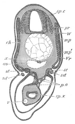
Fig. 379. Section through the trunk of a Scyllium embryo slightly
younger than 28 F.
sp.c. spinal canal; W. white matter of spinal cord; pr.
posterior nerve-roots; ch. notochord; x. subnotochordal rod;
ao. aorta; mp. muscle-plate; mp´. inner layer of muscle-plate
already converted into muscles; Vr. rudiment of vertebral body;
st. segmental tube; sd. segmental duct; sp.v. spiral valve;
v. subintestinal vein; p.o. primitive generative cells.
At the time when the muscle-plates have become independent structures
they form flat two-layered oblong bodies enclosing a slit-like central
cavity (fig. 379, mp). The outer or somatic wall is formed of simple
epithelial-like cells. The inner or splanchnic wall has however a
somewhat complicated structure. It is composed dorsally and ventrally
of a columnar epithelium, but in its middle portion of the
muscle-cells previously spoken of. Between these and the central
cavity of the plates the epithelium forming the remainder of the layer
commences to insert itself; so that between the first-formed muscle
and the cavity of the muscle-plate there appears a thin layer of
cells, not however continuous throughout.
When first formed the muscle-plates, as viewed from the exterior, have
nearly straight edges; soon however they become bent in the middle, so
that the edges have an obtusely angular form, the apex of the angle
being directed forwards. They are so arranged that the anterior edge
of the one plate fits into the posterior edge of the one in front. In
the lines of junction between the plates layers of connective-tissue
cells appear, which form the commencements of the intermuscular septa.
The growth of the plates is very rapid, and their upper ends
soon
extend to the summit of the neural canal, and their lower ones nearly
meet in the median ventral line. The original band of muscles, whose
growth at first is very slow, now increases with great rapidity, and
forms the nucleus of the whole voluntary muscular system (fig. 380,
mp´). It extends upwards and downwards by the continuous conversion
of fresh cells of the splanchnic layer into muscle-cells. At the same
time it grows rapidly in thickness by the addition of fresh
spindle-shaped muscle-cells from the somatic layer as well as by the
division of the already existing cells.
Thus both layers of the muscle plate are concerned in forming the
great longitudinal lateral muscles, though the splanchnic layer is
converted into muscles very much sooner than the somatic[245].
Each muscle-plate is at first a continuous structure, extending from
the dorsal to the ventral surface, but after a time it becomes divided
by a layer of connective tissue, which becomes developed nearly on a
level with the lateral line, into a dorso-lateral and a ventro-lateral
section. The ends of the muscle-plates continue for a long time to be
formed of undifferentiated columnar cells. The complicated outlines of
the intermuscular septa become gradually established during the later
stages of development, causing the well-known appearances of the
muscles in transverse sections, which require no special notice here.
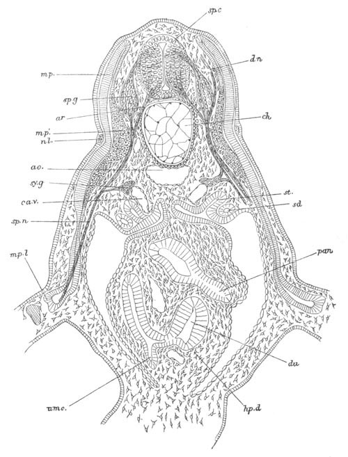
Fig. 380. Transverse section through the anterior part of the trunk
of an embryo of Scyllium slightly older than fig. 29 B.
The section is diagrammatic in so far that the anterior nerve-roots
have been inserted for the whole length; whereas they join the
spinal cord halfway between two posterior roots.
sp.c. spinal cord; sp.g. ganglion of posterior root; ar.
anterior root; dn. dorsally directed nerve springing from
posterior root; mp. muscle-plate; mp´. part of muscle-plate
already converted into muscles; m.pl. part of muscle-plate which
gives rise to the muscles of the limbs; nl. nervus lateralis;
ao. aorta; ch. notochord; sy.g. sympathetic ganglion; ca.v.
cardinal vein; sp.n. spinal nerve; sd. segmental (archinephric)
duct; st. segmental tube; du. duodenum; pan. pancreas; hp.d.
point of junction of hepatic duct with duodenum; umc. umbilical
canal.
The muscles of the limbs. The limb muscles are formed in
Elasmobranchii, coincidently with the cartilaginous skeleton, as two
bands of longitudinal fibres on the dorsal and ventral surfaces of the
limbs (fig. 346). The cells, from which these muscles originate, are
derived from the muscle-plates. When the ends of the muscle-plates
reach the level of the limbs they bend outwards and enter the tissue
of the limbs (fig. 380). Small portions of several muscle-plates
(m.pl) come in this way to be situated within the limbs, and are
very soon segmented off from the remainder of the muscle-plates. The
portions of the muscle-plates thus introduced soon lose their original
distinctness.
There can however be but little doubt that they supply
the tissue for the muscles of the limbs. The muscle-plates themselves,
after giving off buds to the limbs, grow downwards, and soon cease to
shew any trace of having given off these buds.
In addition to the longitudinal muscles of the trunk just described,
which are generally characteristic of Fishes, there is found in
Amphioxus a peculiar transverse abdominal muscle, extending from the
mouth to the abdominal pore, the origin of which has not been made
out.
It has already been shewn that in all the higher Vertebrata
muscle-plates appear, which closely resemble those in Elasmobranchii;
so that all the higher Vertebrata pass through, with reference to
their muscular system, a fish-like stage. The middle portion of the
inner layers of their muscle-plates becomes, as in Elasmobranchii,
converted into muscles at a very early period, and the outer layer for
a long time remains formed of indifferent cells. That these
muscle-plates give rise to the main muscular system of the trunk, at
any rate to the episkeletal muscles of Huxley, is practically certain,
but the details of the process have not been made out.
In the Perennibranchiata the fish-like arrangement of muscles is
retained through life in the tail and in the dorso-lateral parts of
the trunk. In the tail of the Amniotic Vertebrata the primitive
arrangement is also more or less retained, and the same holds good for
the dorso-lateral trunk muscles of the Lacertilia. In the other
Amniota and the Anura the dorso-lateral muscles have become divided up
into a series of separate muscles, which are arranged in two main
layers. It is probable that the intercostal muscles belong to the same
group as the dorso-lateral muscles.
The abdominal muscles of the trunk, even in the lowest Amphibia,
exhibit a division into several layers. The recti abdominis are the
least altered part of this system, and usually retain indications of
the primitive intermuscular septa, which in many Amphibia and
Lacertilia are also to some extent preserved in the other abdominal
muscles.
In the Amniotic Vertebrates there is formed underneath the vertebral
column and the transverse processes a system of muscles, forming part
of the hyposkeletal system of Huxley, and called by Gegenbaur the
subvertebral muscles. The development of this system has not been
worked out, but on the whole I am inclined to believe that it is
derived from the muscle-plates. Kölliker, Huxley and other
embryologists believe however that these muscles are independent of
the muscle-plates in their origin.
Whether the muscle of the diaphragm is to be placed in the same
category as the hyposkeletal muscles has not been made out.
It is probable that the cutaneous muscles of the trunk are derived
from the cells given off from the muscle-plates. Kölliker however
believes that they have an independent origin.
The limb-muscles, both extrinsic and intrinsic, as may be concluded
from their development in Elasmobranchii, are derived from the
muscle-plates. Kleinenberg found in Lacertilia a growth of the
muscle-plates into the limbs, and in Amphibia Götte finds that the
outer layer of the muscle-plates gives rise to the muscles of the
limbs.
In the higher Vertebrata on the other hand the entrance of the
muscle-plates into the limbs has not been made out (Kölliker). It
seems therefore probable that by an embryological modification, of
which instances are so frequent, the cells which give rise to the
muscles of the limbs in the higher Vertebrata can no longer be traced
into a direct connection with the muscle-plates.
The Somites and muscular system of the head.
The extension of the somites to the anterior end of the body in
Amphioxus clearly proves that somites, similar to those of the trunk,
were originally present in a region, which in the higher Vertebrata
has become differentiated into the head. In the adult condition no
true Vertebrate exhibits indications of such somites, but in the
embryos of several of the lower Vertebrata structures have been found,
which are probably equivalent to the somites of the trunk: they have
been frequently alluded to in the previous chapters of this volume.
These structures have been most fully worked out in Elasmobranchii.
The mesoblast in Elasmobranch embryos becomes first split into somatic
and splanchnic layers in the region of the head; and between these
layers there are formed two cavities, one on each side, which end in
front opposite the blind anterior extremity of the alimentary canal;
and are continuous behind with the general body-cavity (fig. 20 A,
vp). I propose calling them the head-cavities. The cavities of the
two sides have no communication with each other.
Coincidently with the formation of an outgrowth from the throat to
form the first visceral cleft, the head-cavity on each side becomes
divided into a section in front of the cleft and a section behind the
cleft; and at a later period it becomes, owing to the formation of a
second cleft, divided into three sections:
(1) a section in front of
the first or hyomandibular cleft; (2) a section in the hyoid arch
between the hyomandibular cleft and the hyobranchial or first
branchial cleft; (3) a section behind the first branchial cleft.
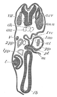
Fig. 381. Transverse section through the front part of the head of
a young Pristiurus embryo.
The section, owing to the cranial flexure, cuts both the fore- and
the hind-brain. It shews the premandibular and mandibular
head-cavities 1pp and 2pp, etc. The section is moreover somewhat
oblique from side to side.
fb. fore-brain; l. lens of eye; m. mouth; pt. upper end of
mouth, forming pituitary involution; 1ao. mandibular aortic arch;
1pp. and 2pp. first and second head-cavities; 1vc. first
visceral cleft; V. fifth nerve; aun. auditory nerve; VII.
seventh nerve; aa. dorsal aorta; acv. anterior cardinal vein;
ch. notochord.
The front section of the head-cavity grows forward, and soon becomes
divided, without the intervention of a visceral cleft, into an
anterior and posterior division. The anterior lies close to the eye,
and in front of the commencing mouth involution. The posterior part
lies completely within the mandibular arch.
As the rudiments of the successive visceral clefts are formed, the
posterior part of the head-cavity becomes divided into successive
sections, there being one section for each arch. Thus the whole
head-cavity becomes on each side divided into (1) a premandibular
section; (2) a mandibular section (vide fig. 29 A, pp); (3) a
hyoid section; (4) sections in each of the branchial arches.
The first of these divisions forms a space of a considerable size,
with epithelial walls of somewhat short columnar cells (fig. 381,
1pp). It is situated close to the eye, and presents a rounded or
sometimes a triangular figure in section. The two halves of the cavity
are prolonged ventralwards, and meet below the base of the fore-brain.
The connection between them appears to last for a considerable time.
These two cavities are the only parts of the body-cavity within the
head which unite ventrally. The section of the head-cavity just
described is so similar to the remaining sections that it must be
considered as serially homologous with them.
The next division of the head-cavity, which from its position
may be
called the mandibular cavity, presents a spatulate shape, being
dilated dorsally, and produced ventrally into a long thin process
parallel to the hyomandibular gill-cleft (fig. 20, pp). Like the
previous space it is lined by a short columnar epithelium.
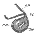
Fig. 382. Horizontal section through the penultimate visceral arch
of an embryo of Pristiurus.
ep. epiblast; vc. pouch of hypoblast which will form the walls
of a visceral cleft; pp. segment of body-cavity in visceral arch;
aa. aortic arch.
The mandibular aortic arch is situated close to its inner side (fig.
381, 2pp). After becoming separated from the lower part (Marshall),
the upper part of the cavity atrophies about the time of the
appearance of the external gills. Its lower part also becomes much
narrowed, but its walls of columnar cells persist. The outer or
somatic wall becomes very thin indeed, the splanchnic wall, on the
other hand, thickens and forms a layer of several rows of elongated
cells. In each of the remaining arches there is a segment of the
original body-cavity fundamentally similar to that in the mandibular
arch (fig. 382). A dorsal dilated portion appears, however, to be
present in the third or hyoid section alone (fig. 20), and even there
disappears very soon, after being segmented off from the lower part
(Marshall). The cavities in the posterior parts of the head become
much reduced like those in its anterior part, though at rather a later
period.
It has been shewn that the divisions of the body-cavity in the head,
with the exception of the anterior, early become atrophied, not so
however their walls. The cells forming the walls both of the dorsal
and ventral sections of these cavities become elongated, and finally
become converted into muscles. Their exact history has not been
followed in its details, but they almost unquestionably become the
musculus constrictor superficialis and musculus interbranchialis[246];
and probably also musculus levator mandibuli and other muscles of the
front part of the head.
The anterior cavity close to the eye remains unaltered much longer
than the remaining cavities.
Its further history is very interesting. In my original account of
this cavity (No. 292, p. 208) I stated my belief that its walls gave
rise to the eye-muscles, and the history of this process has been to
some extent worked out by Marshall in his important memoir (No. 509).
Marshall finds that the ventral portion of this cavity, where its two
halves meet, becomes separated from the remainder. The eventual fate
of this part has not however been followed. Each dorsal section
acquires a cup-like form, investing the posterior and inner surface of
the eye. The cells of its outer wall subsequently give rise to three
sets of muscles. The middle of these, partly also derived from the
inner walls of the cup, becomes the rectus internus of the eye, the
dorsal set forms the rectus superior, and the ventral the rectus
inferior. The obliquus inferior appears also to be in part developed
from the walls of this cavity.
Marshall brings evidence to shew that the rectus externus (as might be
anticipated from its nerve supply) has no connection with the walls of
the premandibular head-cavity, and finds that it arises close to the
position originally occupied by the second and third cavities.
Marshall has not satisfactorily made out the mode of development of
the obliquus superior.
The walls of the cavities, whose history has just been recorded, have
definite relations with the cranial nerves, an account of which has
already been given at p. 461.
Head-cavities, in the main similar to those of Elasmobranchii, have
been found in the embryo of Petromyzon (fig. 45, hc), the Newt
(Osborn and Scott), and various Reptilia (Parker).
Bibliography.
(507) G. M. Humphry. “Muscles in Vertebrate Animals.” Journ. of Anat.
and Phys., Vol. VI. 1872.
(508) J. Müller. “Vergleichende Anatomie d. Myxinoiden. Part I.
Osteologie u. Myologie.” Akad. Wiss., Berlin, 1834.
(509) A. M. Marshall. “On the head cavities and associated nerves of
Elasmobranchs.” Quart. J. of Micr. Science, Vol. XXI. 1881.
(510) A. Schneider. “Anat. u. Entwick. d. Muskelsystems d.
Wirbelthiere.” Sitz. d. Oberhessischen Gesellschaft, 1873.
(511) A. Schneider. Beiträge z. vergleich. Anat. u. Entwick. d.
Wirbelthiere. Berlin, 1879.
Vide also Götte (No. 296), Kölliker (No. 298), Balfour (No. 292),
Huxley, etc.
Excretory organs consist of coiled or branched and often ciliated
tubes, with an excretory pore opening on the outer surface of the
body, and as a rule an internal ciliated orifice placed in the
body-cavity. In forms provided with a true vascular system, there is a
special development of capillaries around the glandular part of the
excretory organs. In many instances the glandular cells of the organs
are filled with concretions of uric acid or some similar product of
nitrogenous waste.
There is a very great morphological and physiological similarity
between almost all the forms of excretory organ found in the animal
kingdom, but although there is not a little to be said for holding all
these organs to be derived from some common prototype, the attempt to
establish definite homologies between them is beset with very great
difficulties.
Platyelminthes. Throughout the whole of the Platyelminthes these
organs are constructed on a well-defined type, and in the Rotifera
excretory organs of a similar form to those of the Platyelminthes are
also present.
These organs (Fraipont, No. 513) are more or less distinctly paired,
and consist of a system of wide canals, often united into a network,
which open on the one hand into a pair of large tubes leading to the
exterior, and on the other into fine canals which terminate by
ciliated openings, either in spaces between the connective-tissue
cells (Platyelminthes), or in the body-cavity (Rotifera). The fine
canals open directly into the larger ones, without first uniting into
canals of an intermediate size.
The two large tubes open to the exterior, either by means of a median
posteriorly placed contractile vesicle, or by a pair of vesicles,
which have a ventral and anterior position. The former type is
characteristic of the majority of the Trematoda, Cestoda, and
Rotifera, and the latter of the Nemertea and some Trematoda. In the
Turbellaria the position of the external openings of the system is
variable, and in a few Cestoda (Wagner) there are lateral openings on
each of the successive proglottides, in addition to the terminal
openings. The mode of development of these organs is unfortunately not
known.
Mollusca. In the Mollusca there are usually present two independent
pairs of excretory organs—one found in a certain number of forms
during early larval life only[247],
and the other always present in
the adult.
The larval excretory organ has been found in the pulmonate Gasteropoda
(Gegenbaur, Fol[248],
Rabl), in Teredo (Hatschek), and possibly also
in Paludina. It is placed in the anterior region of the body, and
opens ventrally on each side, a short way behind the velum. It is
purely a larval organ, disappearing before the close of the veliger
stage. In the aquatic Pulmonata, where it is best developed, it
consists on each side of a V-shaped tube, with a dorsally-placed apex,
containing an enlargement of the lumen. There is a ciliated cephalic
limb, lined by cells with concretions, and terminating by an internal
opening near the eye, and a non-ciliated pedal limb opening to the
exterior[249].
Two irreconcilable views are held as to the development of this
system. Rabl (Vol. ii. No. 268) and Hatschek hold that it is developed
in the mesoblast; and Rabl states that in Planorbis it is formed from
the anterior mesoblast cells of the mesoblastic bands. A special
mesoblast cell on each side elongates into two processes, the
commencing limbs of the future organ. A lumen is developed in this
cell, which is continued into each limb, while
the continuations of
the two limbs are formed by perforated mesoblast cells.
According to Fol these organs originate in aquatic Pulmonata as a pair
of invaginations of the epiblast, slightly behind the mouth. Each
invagination grows in a dorsal direction, and after a time suddenly
bends on itself, and grows ventralwards and forwards. It thus acquires
its V-shaped form.
In the terrestrial Pulmonata the provisional excretory organs are,
according to Fol, formed as epiblastic invaginations, in the same way
as those in the aquatic Pulmonata, but have the form of simple
non-ciliated sacks, without internal openings.
The permanent renal organ of the Mollusca consists typically of a pair
of tubes, although in the majority of the Gasteropoda one of the two
tubes is not developed. It is placed considerably behind the
provisional renal organ.
Each tube, in its most typical form, opens by a ciliated funnel into
the pericardial cavity, and has its external opening at the side of
the foot. The pericardial funnel leads into a glandular section of the
organ, the lining cells of which are filled with concretions. This
section is followed by a ciliated section, from which a narrow duct
leads to the exterior.
As to the development of this organ the same divergence of opinion
exists as in the case of the provisional renal organ.
Rabl’s careful observations on Planorbis (Vol. II. No. 268) tend to
shew that it is developed from a mass of mesoblast cells, near the end
of the intestine. The mass becomes hollow, and, attaching itself to
the epiblast on the left side of the anus, acquires an opening to the
exterior. Its internal opening is not established till after the
formation of the heart. Fol gives an equally precise account, but
states that the first rudiment of the organ arises as a solid mass of
epiblast cells. Lankester finds that this organ is developed as a
paired invagination of the epiblast in Pisidium, and Bobretzky also
derives it from the epiblast in marine Prosobranchiata. In Cephalopoda
on the other hand Bobretzky’s observations (I conclude this from his
figures) indicate that the excretory sacks of the renal organs are
derived from the mesoblast.
Polyzoa. Simple excretory organs, consisting of a pair of ciliated
canals, opening between the mouth and the anus, have
been found by
Hatschek and Joliet in the Entoproctous Polyzoa, and are developed,
according to Hatschek, by whom they were first found in the larva,
from the mesoblast.
Brachiopoda. One or rarely two (Rhynchonella) pairs of canals, with
both peritoneal and external openings, are found in the Brachiopoda.
They undoubtedly serve as genital ducts, but from their structure are
clearly of the same nature as the excretory organs of the Chætopoda
described below. Their development has not been worked out.
Chætopoda. Two forms of excretory organ have been met with in the
Chætopoda. The one form is universally or nearly universally present
in the adult, and typically consists of a pair of coiled tubes
repeated in every segment. Each tube has an internal opening, placed
as a rule in the segment in front of that in which the greater part of
the organ and the external opening are situated.
There are great variations in the structure of these organs, which
cannot be dealt with here. It may be noted however that the internal
opening may be absent, and that there may be several internal
openings for each organ (Polynoe). In the Capitellidæ moreover
several pairs of excretory tubes have been shewn by Eisig (No. 512) to
be present in each of the posterior segments.
The second form of excretory organ has as yet only been found in the
larva of Polygordius, and will be more conveniently dealt with in
connection with the development of the excretory system of this form.
There is still considerable doubt as to the mode of formation of the
excretory tubes of the Chætopoda. Kowalevsky (No. 277), from his
observations on the Oligochæta, holds that they develop as outgrowths
of the epithelial layer covering the posterior side of the
dissepiments, and secondarily become connected with the epidermis.
Hatschek finds that in Criodrilus they arise from a continuous linear
thickening of the somatic mesoblast, immediately beneath the
epidermis, and dorsal to the ventral band of longitudinal muscles.
They break up into S-shaped cords, the anterior end of each of which
is situated in front of a dissepiment, and is formed at first of a
single large cell, while the posterior part is
continued into the
segment behind. The cords are covered by a peritoneal lining, which
still envelopes them, when in the succeeding stage they are carried
into the body-cavity. They subsequently become hollow, and their
hinder ends acquire openings to the exterior. The formation of their
internal openings has not been followed.
Kleinenberg is inclined to believe that the excretory tubes take their
origin from the epiblast, but states that he has not satisfactorily
worked out their development.
The observations of Eisig (No. 512) on the Capitellidæ support
Kowalevsky’s view that the excretory tubes originate from the lining
of the peritoneal cavity.
Hatschek (No. 514) has given a very interesting account of the
development of the excretory system in Polygordius.
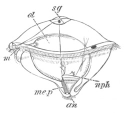
Fig. 383. Polygordius larva. (After Hatschek.)
m. mouth; sg. supraœsophageal ganglion; nph. nephridion;
me.p. mesoblastic band; an. anus; ol. stomach.
The excretory system begins to be formed, while the larva is still in
the trochospere stage (fig. 383, nph), and consists of a provisional
excretory organ, which is placed in front of the future segmented part
of the body, and occupies a position very similar to that of the
provisional excretory organ found in some Molluscan larvæ (vide p.
681).
Hatschek, with some shew of reason, holds that the provisional
excretory organs of Polygordius are homologous with those of the
Mollusca.
In its earliest stage the provisional excretory organ of Polygordius
consists of a pair of simple ciliated tubes, each with an anterior
funnel-like opening situated in the midst of the mesoblast cells, and
a posterior external opening. The latter is placed immediately in
front of what afterwards becomes the segmented region of the embryo.
While the larva is still unsegmented, a second internal opening is
formed for each tube (fig. 383, nph) and the two openings so formed
may eventually become divided into five (fig. 384 A), all
communicating by a single pore with the exterior.
When the posterior region of the embryo becomes segmented,
paired
excretory organs are formed in each of the posterior segments, but the
account of their development, as given by Hatschek, is so remarkable
that I do not think it can be definitely accepted without further
confirmation.
From the point of junction of the two main branches of the larval
kidney there grows backwards (fig. 384 B), to the hind end of the
first segment, a very delicate tube, only indicated by its ciliated
lumen, its walls not being differentiated. Near the front end of this
tube a funnel, leading into the larval body cavity of the head, is
formed, and subsequently the posterior end of the tube acquires an
external opening, and the tube distinct walls. The communication with
the provisional excretory organ is then lost, and thus the excretory
tube of the first segment is established.
The excretory tubes in the second and succeeding segments are formed
in the same way as in the first, i.e. by the continuation of the
lumen of the hind end of the excretory tube from the preceding
segment, and the subsequent separation of this part as a separate
tube.
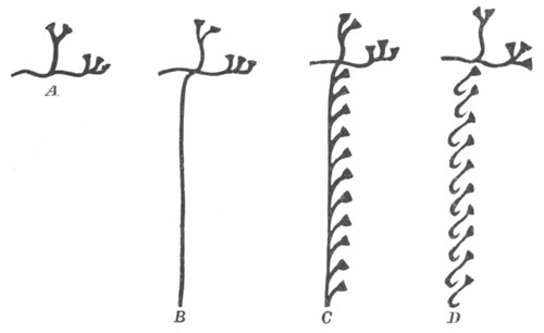
Fig. 384. Diagram illustrating the development of the excretory
system of Polygordius. (After Hatschek.)
The tube may be continued with a sinuous course through several
segments without a distinct wall. The external and internal openings
of the permanent excretory tubes are thus secondarily acquired. The
internal openings communicate with the permanent body-cavity. The
development of the permanent
excretory tubes is diagrammatically
represented in fig. 384 C and D.
The provisional excretory organ atrophies during larval life.
If Hatschek’s account of the development of the excretory system of
Polygordius is correct, it is clear that important secondary
modifications must have taken place in it, because his description
implies that there sprouts from the anterior excretory organ, while it
has its own external opening, a posterior duct, which does not
communicate either with the exterior or with the body-cavity! Such a
duct could have no function. It is intelligible either (1) that the
anterior excretory organ should lead into a longitudinal duct, opening
posteriorly; that then a series of secondary openings into the
body-cavity should attach themselves to this, that for each internal
opening an external should subsequently arise, and the whole break up
into separate tubes; or (2) that behind an anterior provisional
excretory organ a series of secondary independent segmental tubes
should be formed. But from Hatschek’s account neither of these modes
of evolution can be deduced.
Gephyrea. The Gephyrea may have three forms of excretory organs, two
of which are found in the adult, and one, similar in position and
sometimes also in structure, to the provisional excretory organ of
Polygordius, has so far only been found in the larvæ of Echiurus and
Bonellia.
In all the Gephyrea the so-called ‘brown tubes’ are apparently
homologous with the segmented excretory tubes of Chætopods. Their main
function appears to be the transportation of the generative products
to the exterior. There is but a single highly modified tube in
Bonellia, forming the oviduct and uterus; a pair of tubes in the
Gephyrea inermia, and two or three pairs in most Gephyrea armata,
except Bonellia. Their development has not been studied.
In the Gephyrea armata there is always present a pair of posteriorly
placed excretory organs, opening in the adult into the anal extremity
of the alimentary tract, and provided with numerous ciliated
peritoneal funnels. These organs were stated by Spengel to arise in
Bonellia as outgrowths of the gut; but in Echiurus Hatschek (No.
515) finds that they are developed from the somatic mesoblast of the
terminal part of the trunk. They soon become hollow, and after
attaching themselves to the epiblast on each side of the anus, acquire
external openings. They are not at first provided with peritoneal
funnels, but these parts of the organs become developed from a ring of
cells at
their inner extremities; and there is at first but a single
funnel for each vesicle. The mode of increase of the funnels has not
been observed, nor has it been made out how the organs themselves
become attached to the hindgut.
The provisional excretory organ of Echiurus is developed at an early
larval stage, and is functional during the whole of larval life. It at
first forms a ciliated tube on each side, placed in front of that part
of the larva which becomes the trunk of the adult. It opens to the
exterior by a fine pore on the ventral side, immediately in front of
one of the mesoblastic bands, and appears to be formed of perforated
cells. It terminates internally in a slight swelling, which represents
the normal internal ciliated funnel. The primitively simple excretory
organ becomes eventually highly complex by the formation of numerous
branches, each ending in a slightly swollen extremity. These branches,
in the later larval stages, actually form a network, and the inner end
of each main branch divides into a bunch of fine tubes. The whole
organ resembles in many respects the excretory organ of the
Platyelminthes.
In the larva of Bonellia Spengel has described a pair of provisional
excretory tubes, opening near the anterior end of the body, which are
probably homologous with the provisional excretory organs of Echiurus
(vide Vol. II., fig. 162 C, se).
Discophora. As in many of the types already spoken of, permanent and
provisional excretory organs may be present in the Discophora. The
former are usually segmentally arranged, and resemble in many respects
the excretory tubes of the Chætopoda. They may either be provided with
a peritoneal funnel (Nephelis, Clepsine) or have no internal opening
(Hirudo).
Bourne[250]
has shewn that the cells surrounding the main duct in the
medicinal Leech are perforated by a very remarkable network of
ductules, and the structure of these organs in the Leech is so
peculiar that it is permissible to state with due reserve their
homology with the excretory organs of the Chætopoda.
The excretory tubes of Clepsine are held by Whitman to be developed in
the mesoblast.
There are found in the embryos of Nephelis and Hirudo certain
remarkable provisional excretory organs the origin and history of
which are not yet fully made out. In Nephelis they appear as one
(according to Robin), or (according to Bütschli) as two successive
pairs of convoluted tubes on the dorsal side of the embryo, which are
stated by the latter author to develop from the scattered mesoblast
cells underneath the skin. At their fullest development they extend,
according to Robin, from close to the head to near the ventral sucker.
Each of them is U-shaped, with the open end of the U forwards, each
limb of the U being formed by two tubes united in front. No external
opening has been clearly made out. Fürbringer is inclined from his own
researches to believe that they open laterally. They contain a clear
fluid.
In Hirudo, Leuckart has described three similar pairs of organs, the
structure of which he has fully elucidated. They are situated in the
posterior part of the body, and each of them commences with an
enlargement, from which a convoluted tube is continued for some
distance backwards; the tube then turns forwards again, and after
bending again upon itself opens to the exterior. The anterior part is
broken up into a kind of labyrinthic network.
The provisional excretory organs of the Leeches cannot be identified
with the anterior provisional organs of Polygordius and Echiurus.
Arthropoda. Amongst the Arthropoda Peripatus is the only form with
excretory organs of the type of the segmental excretory organs of the
Chætopoda[251].
These organs are placed at the bases of the feet, in the lateral
divisions of the body-cavity, shut off from the main median division
of the body-cavity by longitudinal septa of transverse muscles.
Each fully developed organ consists of three parts:
(1) A dilated vesicle opening externally at the base of a foot. (2) A
coiled glandular tube connected with this, and subdivided again into
several minor divisions. (3) A short terminal portion opening at one
extremity into the coiled tube
and at the other, as I believe, into
the body cavity. This section becomes very conspicuous, in stained
preparations, by the intensity with which the nuclei of its walls
absorb the colouring matter.
In the majority of the Tracheata the excretory organs have the form of
the so-called Malpighian tubes, which always (vide Vol. II.)
originate as a pair of outgrowths of the epiblastic proctodæum. From
their mode of development they admit of comparison with the anal
vesicles of the Gephyrea, though in the present state of our knowledge
this comparison must be regarded as somewhat hypothetical.
The antennary and shell-glands of the Crustacea, and possibly also the
so-called dorsal organ of various Crustacean larvæ appear to be
excretory, and the two former have been regarded by Claus and Grobben
as belonging to the same system as the segmental excretory tubes of
the Chætopoda.
Nematoda. Paired excretory tubes, running for the whole length of the
body in the so-called lateral line, and opening in front by a common
ventral pore, are present in the Nematoda. They do not appear to
communicate with the body cavity, and their development has not been
studied.
Very little is known with reference either to the structure or
development of excretory organs in the Echinodermata and the other
Invertebrate types of which no mention has been so far made in this
Chapter.
Excretory organs and generative ducts of the Craniata.
Although it would be convenient to separate, if possible, the history
of the excretory organs from that of the generative ducts, yet these
parts are so closely related in the Vertebrata, in some cases the same
duct having at once a generative and a urinary function, that it is
not possible to do so.
The excretory organs of the Vertebrata consist of three distinct
glandular bodies and of their ducts. These are (1) a small glandular
body, usually with one or more ciliated funnels opening into the body
cavity, near the opening of which there projects into the body cavity
a vascular glomerulus. It is situated very far forwards, and is
usually known as the head-kidney,
though it may perhaps be more
suitably called, adopting Lankester’s nomenclature, the pronephros.
Its duct, which forms the basis for the generative and urinary ducts,
will be called the segmental duct.
(2) The Wolffian body, which may be also called the mesonephros. It
consists of a series of, at first, segmentally (with a few exceptions)
arranged glandular canals (segmental tubes) primitively opening at
one extremity by funnel-shaped apertures into the body cavity, and at
the other into the segmental duct. This duct becomes in many forms
divided longitudinally into two parts, one of which then remains
attached to the segmental tubes and forms the Wolffian or mesonephric
duct, while the other is known as the Müllerian duct.
(3) The kidney proper or metanephros. This organ is only found in a
completely differentiated form in the amniotic Vertebrata. Its duct is
an outgrowth from the Wolffian duct.
The above parts do not coexist in full activity in any living adult
member of the Vertebrata, though all of them are found together in
certain embryos. They are so intimately connected that they cannot be
satisfactorily dealt with separately.
Elasmobranchii. The excretory system of the Elasmobranchii is by no
means the most primitive known, but at the same time it forms a
convenient starting point for studying the modifications of the system
in other groups. The most remarkable peculiarity it presents is the
absence of a pronephros. The development of the Elasmobranch excretory
system has been mainly studied by Semper and myself.
The first trace of the system makes its appearance as a knob of
mesoblast, springing from the intermediate cell-mass near the level of
the hind end of the heart (fig. 385 A, pd). This knob is the
rudiment of the abdominal opening of the segmental duct, and from it
there grows backwards to the level of the anus a solid column of
cells, which constitutes the rudiment of the segmental duct itself
(fig. 385 B, pd). The knob projects towards the epiblast, and the
column connected with it lies between the mesoblast and epiblast. The
knob and column do not long remain solid, but the former acquires an
opening into the body cavity (fig. 421, sd) continuous with a lumen,
which
makes its appearance in the column (fig. 386, sd). The knob
forms the only structure which can be regarded as a rudiment of the
pronephros.
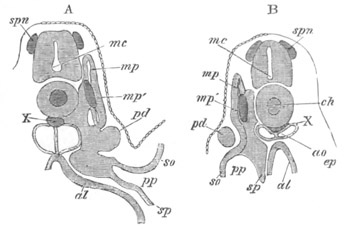
Fig. 385. Two sections of a Pristiurus embryo with three visceral
clefts.
The sections illustrate the development of the segmental duct (pd)
or primitive duct of the pronephros. In A (the anterior of the two
sections) this appears as a solid knob (pd) projecting towards the
epiblast. In B is seen a section of the column which has grown
backwards from the knob in A.
spn. rudiment of a spinal nerve; mc. medullary canal; ch.
notochord; X. subnotochordal rod; mp. muscle-plate; mp´.
specially developed portion of muscle-plate; ao. dorsal aorta;
pd. segmental duct; so. somatopleure; sp. splanchnopleure;
pp. body cavity; ep. epiblast; al. alimentary canal.
While the lumen is gradually being formed, the segmental tubes of the
mesonephros become established. They appear to arise as
differentiations of the parts of the primitive lateral plates of
mesoblast, placed between the dorsal end of the body cavity and the
muscle-plate (fig. 386, st)[252],
which are usually known as the
intermediate cell-masses.
The lumen of the segmental tubes, though at first very small, soon
becomes of a considerable size. It appears to be established in the
position of the section of the body cavity in the intermediate
cell-mass, which at first unites the part of the body cavity in the
muscle-plates with the permanent body cavity. The lumen of each tube
opens at its lower end into the dorsal part of the body cavity (fig.
386, st), and each tube curls obliquely
backwards round the inner
and dorsal side of the segmental duct, near which it at first ends
blindly.
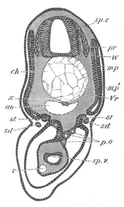
Fig. 386. Section through the trunk of a Scyllium embryo slightly
younger than 28 F.
sp.c. spinal canal; W. white matter of spinal cord; pr.
posterior nerve-roots; ch. notochord; x. subnotochordal rod;
ao. aorta; mp. muscle-plate; mp´. inner layer of muscle-plate
already converted into muscles; Vr. rudiment of vertebral body;
st. segmental tube; sd. segmental duct; sp.v. spiral valve;
v. subintestinal vein; p.o. primitive generative cells.
One segmental tube makes its appearance for each somite (fig. 265),
commencing with that immediately behind the abdominal opening of the
segmental duct, the last tube being situated a few segments behind the
anus. Soon after their formation the blind ends of the segmental tubes
come in contact with, and open into the segmental duct, and each of
them becomes divided into four parts. These are (1) a section carrying
the peritoneal opening, known as the peritoneal funnel, (2) a dilated
vesicle into which this opens, (3) a coiled tubulus proceeding from
(2), and terminating in (4) a wider portion opening into the segmental
duct. At the same time, or shortly before this, each segmental duct
unites with and opens into one of the horns of the cloaca, and also
retires from its primitive position between the epiblast and
mesoblast, and assumes a position close to the epithelium lining the
body cavity (fig. 380, sd). The general features of the excretory
organs at this period are diagrammatically represented in the woodcut
(fig. 387). In this fig. pd is the segmental duct and o its
abdominal opening; s.t points to the segmental tubes, the finer
details of whose structure are not represented in the diagram. The
mesonephros thus forms at this period an elongated gland composed of a
series of isolated coiled tubes, one extremity of each of which opens
into the body cavity, and the other into the segmental duct, which
forms the only duct of the system, and communicates at its front end
with the body cavity, and behind with the cloaca.
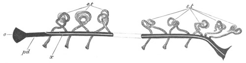
Fig. 387. Diagram of the primitive condition of the kidney in an
Elasmobranch embryo.
pd. segmental duct. It opens at o into the body cavity and at
its other extremity into the cloaca; x. line along which the
division appears which separates the segmental duct into the
Wolffian duct above and the Müllerian duct below; s.t. segmental
tubes. They open at one end into the body cavity, and at the other
into the segmental duct.
The next important change concerns the segmental duct, which becomes
longitudinally split into two complete ducts in the female, and one
complete duct and parts of a second duct in the male. The manner in
which this takes place is diagrammatically represented in fig. 387 by
the clear line x, and in transverse section in figs. 388 and 389.
The resulting ducts are (1) the Wolffian duct or mesonephric duct
(wd), dorsally, which remains continuous with the excretory tubules
of the mesonephros, and ventrally (2) the oviduct or Müllerian duct in
the female, and the rudiments of this duct in the male. In the female
the formation of these ducts takes place (fig. 389) by a nearly solid
rod of cells being gradually split off from the ventral side of all
but the foremost part of the original segmental duct. This nearly
solid cord is the Müllerian duct (od). A very small portion of the
lumen of the original segmental duct is perhaps continued into it, but
in any case it very soon acquires a wide lumen (fig. 389 A). The
anterior part of the segmental duct is not divided, but remains
continuous with the Müllerian duct, of which its anterior pore forms
the permanent peritoneal opening[253]
(fig. 387). The remainder of the
segmental duct (after the loss of its anterior section, and the part
split off from its ventral side) forms the Wolffian duct. The process
of formation of these ducts in the male differs from that in the
female chiefly
in the fact of the anterior undivided part of the
segmental duct, which forms the front end of the Müllerian duct, being
shorter, and in the column of cells with which it is continuous being
from the first incomplete.
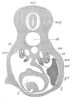
Fig. 388. Diagrammatic representation of a transverse section of a
Scyllium embryo illustrating the formation of the Wolffian and
Müllerian ducts by the longitudinal splitting of the segmental
duct.
mc. medullary canal; mp. muscle-plate; ch. notochord; ao.
aorta; cav. cardinal vein; st. segmental tube. On the left side
the section passes through the opening of a segmental tube into the
body cavity. On the right this opening is represented by dotted
lines, and the opening of the segmental tube into the Wolffian duct
has been cut through; w.d. Wolffian duct; m.d. Müllerian duct.
The section is taken through the point where the segmental duct and
Wolffian duct have just become separate; gr. the germinal ridge
with the thickened germinal epithelium; l. liver; i. intestine
with spiral valve.
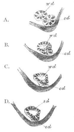
Fig. 389. Four sections through the anterior part of the segmental
duct of a female embryo of Scyllium canicula.
The figure shews how the segmental duct becomes split into the
Wolffian or mesonephric duct above, and Müllerian duct or oviduct
below.
wd. Wolffian or mesonephric duct; od. Müllerian duct or oviduct;
sd. segmental duct.
The segmental tubes of the mesonephros undergo further important
changes. The vesicle at the termination of each peritoneal funnel
sends a bud forwards towards the preceding tubulus, which joins the
fourth section of it close to the opening
into the Wolffian duct (fig.
390, px). The remainder of the vesicle becomes converted into a
Malpighian body (mg). By the first of these changes a tube is
established connecting each pair of segments of the mesonephros, and
though this tube is in part aborted (or only represented by a fibrous
band) in the anterior part of the excretory organs in the adult, and
most probably in the hinder part, yet it seems almost certain that the
secondary and tertiary Malpighian bodies of the majority of segments
are developed from its persisting blind end. Each of these secondary
and tertiary Malpighian bodies is connected with a convoluted tubulus
(fig. 391, a.mg), which is also developed from the tube connecting
each pair of segmental tubes, and therefore falls into the primary
tubulus close to its junction with the
segmental duct. Owing to the
formation of the accessory tubuli the segments of the mesonephros
acquire a compound character.
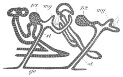
Fig. 390. Longitudinal vertical section through part of the
mesonephros of an embryo of Scyllium.
The figure contains two examples of the budding of the vesicle of a
segmental tube (which forms a Malpighian body in its own segment) to
unite with the tubulus in the preceding segment close to its opening
into the Wolffian (mesonephric) duct.
ge. epithelium of body-cavity; st. peritoneal funnel of
segmental tube with its peritoneal opening; mg. Malpighian body;
px. bud from Malpighian body uniting with preceding segment.
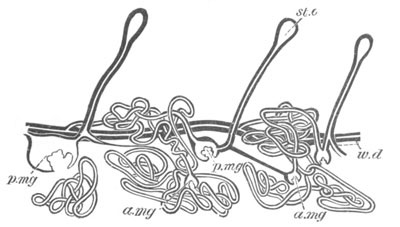
Fig. 391. Three segments of the anterior part of the mesonephros of a
nearly ripe embryo of Scyllium canicula as a transparent object.
The figure shews a fibrous band passing from the primary to the
secondary Malpighian bodies in two segments, which is the remains of
the outgrowth from the primary Malpighian body.
st.o. peritoneal funnel; p.mg. primary Malpighian body; a.mg.
accessory Malpighian body; w.d. mesonephric (Wolffian) duct.
The third section of each tubulus becomes by continuous growth,
especially in the hinder segments, very bulky and convoluted.
The general character of a slightly developed segment of the
mesonephros at its full growth may be gathered from fig. 391. It
commences with (1) a peritoneal opening, somewhat oval in form
(st.o) and leading directly into (2) a narrow tube, the segmental
tube, which takes a more or less oblique course backwards, and,
passing superficially to the Wolffian duct (w.d), opens into (3) a
Malpighian body (p.mg) at the anterior extremity of an isolated coil
of glandular tubuli. This coil forms the third section of each
segment, and starts from the Malpighian body. It consists of a
considerable number of rather definite convolutions, and after uniting
with tubuli from one, two, or more (according to the size of the
segment) accessory Malpighian bodies (a.mg) smaller than the one
into which the segmental tube falls, eventually opens by (4) a
narrowish collecting tube into the Wolffian duct at the posterior end
of the segment. Each segment is probably completely isolated from the
adjoining segments, and never has more than one peritoneal funnel and
one communication with the Wolffian duct.
Up to this time there has been no distinction between the anterior and
posterior tubuli of the mesonephros, which alike open into the
Wolffian duct. The collecting tubes of a considerable number of the
hindermost tubuli (ten or eleven in Scyllium canicula), either in some
species elongate, overlap, while at the same time their openings
travel backward so that they eventually open by apertures (not usually
so numerous as the separate tubes), on nearly the same level, into the
hindermost section of the Wolffian duct in the female, or into the
urinogenital cloaca, formed by the coalesced terminal parts of the
Wolffian ducts, in the male; or in other species become modified, by a
peculiar process of splitting from the Wolffian duct, so as to pour
their secretion into a single duct on each side, which opens in a
position corresponding with the numerous ducts of the other species
(fig. 392). In both cases the modified posterior kidney-segments are
probably equivalent to the permanent
kidney or metanephros of the
amniotic Vertebrates, and for this reason the numerous collecting
tubes or single collecting tube, as the case may be, will be spoken of
as ureters. The anterior tubuli of the primitive excretory organ
retain their early relation to the Wolffian duct, and form the
permanent Wolffian body or mesonephros.
The originally separate terminal extremities of the Wolffian ducts
always coalesce, and form a urinal cloaca, opening by a single
aperture, situated at the extremity of the median papilla behind the
anus. Some of the peritoneal openings of the segmental tubes in
Scyllium, or in other cases all the openings, become obliterated.
In the male the anterior segmental tubes undergo remarkable
modifications, and become connected with the testes. Branches appear
to grow from the first three or four or more of them (though probably
not from their peritoneal openings), which pass to the base of the
testis, and there uniting into a longitudinal canal, form a network,
and receive the secretion of the testicular ampullæ (fig. 393, nt).
These ducts, the vasa efferentia, carry the semen to the Wolffian
body, but before opening into the tubuli of this body they unite into
a canal known as the longitudinal canal of the Wolffian body
(l.c), from which pass off ducts equal in number to the vasa
efferentia, each of which normally ends in a Malpighian corpuscle.
From the Malpighian corpuscles so connected there spring the
convoluted tubuli, forming the generative segments of the Wolffian
body, along which the semen is conveyed to the Wolffian duct (v.d).
The Wolffian duct itself becomes much contorted and acts as vas
deferens.
Figs. 392 and 393 are diagrammatic representations of the chief
constituents of the adult urinogenital organs in the two sexes. In the
adult female (fig. 392), there are present the following parts:
(1) The oviduct or Müllerian duct (m.d) split off from the segmental
duct of the kidneys. Each oviduct opens at its anterior extremity into
the body cavity, and behind the two oviducts have independent
communications with the general cloaca.
(2) The mesonephric ducts (w.d), the other product
of the segmental
ducts of the kidneys. They end in front by becoming continuous with
the tubulus of the anterior persisting segment of the mesonephros on
each side, and unite behind to open by a common papilla into the
cloaca. The mesonephric duct receives the secretion of the anterior
tubuli of the primitive mesonephros.
(3) The ureter which carries off the secretion of the kidney proper or
metanephros. It is represented in my diagram in its most rare and
differentiated condition as a single duct connected with the posterior
segmental tubes.
(4) The segmental tubes (s.t) some of which retain their
original
openings into the body cavity, and others are without them. They are
divided into two groups, an anterior forming the mesonephros or
Wolffian body, which pours its secretion into the Wolffian duct; and a
posterior group forming a gland which is probably equivalent to the
kidney proper of amniotic Craniata, and is connected with the ureter.
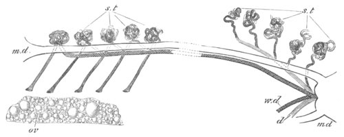
Fig. 392. Diagram of the arrangement of the urinogenital organs
in an adult female Elasmobranch.
m.d. Müllerian duct; w.d. Wolffian duct; s.t. segmental tubes;
five of them are represented with openings into the body cavity, the
posterior segmental tubes form the mesonephros; ov. ovary.
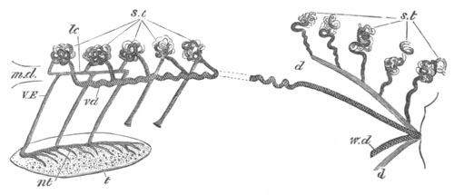
Fig. 393. Diagram of the arrangement of the urinogenital organs
in an adult male Elasmobranch.
m.d. rudiment of Müllerian duct; w.d. Wolffian duct, marked vd
in front and serving as vas deferens; s.t. segmental tubes; two of
them are represented with openings into the body cavity; d.
ureter; t. testis; nt. canal at the base of the testis; VE.
vasa efferentia; lc. longitudinal canal of the Wolffian body.
In the male the following parts are present (fig. 393):
(1) The Müllerian duct (m.d), consisting of a small rudiment
attached to the liver, representing the foremost end of the oviduct of
the female.
(2) The mesonephric duct (w.d) which precisely corresponds to the
mesonephric duct of the female, but, in addition to serving as the
duct of the Wolffian body, also acts as a vas deferens (vd). In the
adult male its foremost part has a very tortuous course.
(3) The ureter (d), which has the same fundamental constitution as
in the female.
(4) The segmental tubes (s.t). The posterior tubes have the same
arrangement in both sexes, but in the male modifications take place in
connection with the anterior tubes to fit them to act as transporters
of the semen.
Connected with the anterior tubes there are present (1) the vasa
efferentia (VE), united on the one hand with (2) the central canal
in the base of the testis (nt), and on the other with the
longitudinal canal of the Wolffian body (lc). From the latter are
seen passing off the successive tubuli of the anterior segments of the
Wolffian body, in connection with which Malpighian bodies are
typically present, though not represented in my diagram.
Apart from the absence of the pronephros the points which deserve
notice in the Elasmobranch excretory system are (1) The splitting of
the segmental duct into Wolffian (mesonephric) and Müllerian ducts.
(2) The connection of the former with the mesonephros, and of the
latter with the abdominal opening of the segmental duct which
represents the pronephros of other types. (3) The fact that the
Müllerian duct serves as oviduct, and the Wolffian duct as vas
deferens. (4) The differentiation of a posterior section of the
mesonephros into a special gland foreshadowing the metanephros of the
Amniota.
Cyclostomata. The development of the excretory system amongst the
Cyclostomata has only been studied in Petromyzon (Müller, Fürbringer,
and Scott).
The first part of the system developed is the segmental duct. It
appears in the embryo of about 14 days (Scott) as a solid cord of
cells, differentiated from the somatic mesoblast near the dorsal end
of the body cavity. This cord is at first placed immediately below the
epiblast, and grows backwards by a continuous process of
differentiation of fresh mesoblast cells. It soon acquires a lumen,
and joins the cloacal section of the alimentary tract before the close
of fœtal life. Before this communication is established, the front
end of the duct sends a process towards the body cavity, the blind end
of which acquires a ciliated opening into the latter. A series of
about four or five successively formed outgrowths from the duct, one
behind the other, give rise to as many ciliated funnels opening into
the body cavity, and each communicating by a more or less elongated
tube with the segmental duct. These funnels, which have a metameric
arrangement, constitute the pronephros, the whole of which is situated
in the pericardial region of the body cavity.
On the inner side of the peritoneal openings of each pronephros there
is formed a vascular glomerulus, projecting into the body cavity, and
covered by peritoneal epithelium. For a considerable period the
pronephros constitutes the sole functional part of the excretory
system.
A mesonephros is formed (Fürbringer) relatively late in larval life,
as a segmentally arranged series of solid cords, derived from the
peritoneal epithelium. These cords constitute the rudiments of the
segmental tubes. They are present for a considerable portion of the
body cavity, extending backwards from a point shortly behind the
pronephros. They soon separate from the peritoneal epithelium, become
hollowed out into canals, and join the segmental duct. At their blind
extremity (that originally connected with the peritoneal epithelium) a
Malpighian body is formed.
The pronephros is only a provisional excretory organ, the atrophy of
which commences during larval life, and is nearly completed when the
Ammocœte has reached 180 mm. in length.
Further changes take place in
connection with the excretory system on the conversion of the
Ammocœte into the adult.
The segmental ducts in the adult fall into a common urinogenital
cloaca, which opens on a papilla behind the anus. This cloaca also
communicates by two apertures (abdominal pores) with the body cavity.
The generative products are carried into the cloaca by these pores; so
that their transportation outwards is not performed by any part of the
primitive urinary system. The urinogenital cloaca is formed by the
separation of the portion of the primitive cloaca containing the
openings of the segmental ducts from that connected with the
alimentary tract.
The mesonephros of the Ammocœte undergoes at the metamorphosis
complete atrophy, and is physiologically replaced by a posterior
series of segmental tubes, opening into the hindermost portion of the
segmental duct (Schneider).
In Myxine the excretory system consists (1) of a highly developed
pronephros with a bunch of ciliated peritoneal funnels opening into
the pericardial section of the body cavity. The coiled and branched
tubes of which the pronephros is composed open on the ventral side of
the anterior portion of the segmental duct, which in old individuals
is cut off from the posterior section of the duct. On the dorsal side
of the portion of the segmental duct belonging to the pronephros there
are present a small number of diverticula, terminating in glomeruli:
they are probably to be regarded as anterior segmental tubes. (2) Of a
mesonephros, which commences a considerable distance behind the
pronephros, and is formed of straight extremely simple segmental tubes
opening into the segmental duct (fig. 385).
The excretory system of Myxine clearly retains the characters of the
system as it exists in the larva of Petromyzon.
Teleostei. In most Teleostei the pronephros and mesonephros coexist
through life, and their products are carried off by a duct, the nature
of which is somewhat doubtful, but which is probably homologous with
the mesonephric duct of other types.
The system commences in the embryo (Rosenberg, Oellacher, Götte,
Fürbringer) with the formation of a groove-like fold of the somatic
layer of peritoneal epithelium, which becomes gradually constricted
into a canal; the process of constriction commencing in the middle and
extending in both directions. The canal does not however close
anteriorly, but remains open to the body cavity, thus giving rise to a
funnel equivalent to the pronephric funnels of Petromyzon and Myxine.
On the inner side of this
funnel there is formed a glomerulus,
projecting into the body cavity; and at the same time that this is
being formed the anterior end of the canal becomes elongated and
convoluted. The above structures constitute a pronephros, while the
posterior part of the primitive canal forms the segmental duct.
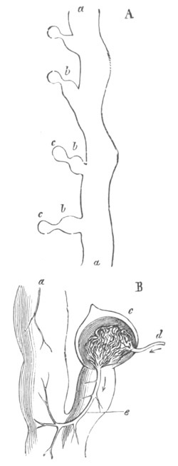
Fig. 394. Portions of the mesonephros of Myxine. (From
Gegenbaur; after J. Müller.)
a. segmental duct; b. segmental tube; c. glomerulus; d.
afferent, e. efferent artery.
B represents a portion of A highly magnified.
The portion of the body cavity with the glomerulus and peritoneal
funnel of the pronephros (fig. 395, po) soon becomes completely
isolated from the remainder, so as to form a closed cavity (gl). The
development of the mesonephros does not take place till long after
that of the pronephros. The segmental tubes which form it are stated
by Fürbringer to arise from solid ingrowths of peritoneal epithelium,
developed successively from before backwards, but Sedgwick informs me
that they arise as differentiations of the mesoblastic cells near
the peritoneal epithelium. They soon become hollow, and unite with the
segmental duct. Malpighian bodies are developed on their median
portions. They grow very greatly in length, and become much
convoluted, but the details of this process have not been followed
out.
The foremost segmental tubes are situated close behind the pronephros,
while the hindermost are in many cases developed in the postanal
continuations of the body cavity. The pronephros appears to form the
swollen cephalic portion of the kidney of the adult, and the
mesonephros the remainder; the so-called caudal portion, where
present, being derived (?) from the postanal segmental tubes.
In some cases the cephalic portion of the kidneys is absent
in the
adult, which probably implies the atrophy of the pronephros; in other
instances the cephalic portion of the kidneys is the only part
developed. Its relation to the embryonic pronephros requires however
further elucidation.
In the adult the ducts in the lower part of the kidneys lie as a rule
on their outer borders, and almost invariably open into a urinary
bladder, which usually opens in its turn on the urinogenital papilla
immediately behind the genital pore, but in a few instances there is a
common urinogenital pore.
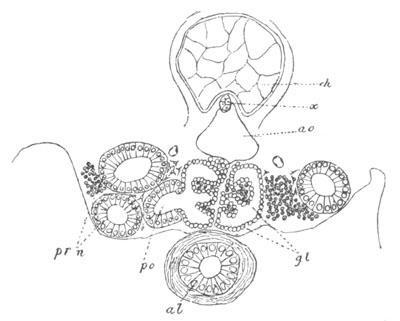
Fig. 395. Section through the pronephros of a Trout and adjacent
parts ten days before hatching.
pr.n. pronephros; po. opening of pronephros into the isolated
portion of the body cavity containing the glomerulus; gl.
glomerulus; ao. aorta; ch. notochord; x. subnotochordal rod;
al. alimentary tract.
In most Osseous Fish there are true generative ducts continuous with
the investment of the generative organs. It appears to me most
probable, from the analogy of Lepidosteus, to be described in the next
section, that these ducts are split off from the primitive segmental
duct, and correspond with the Müllerian ducts of Elasmobranchii, etc.;
though on this point we have at present no positive embryological
evidence (vide general considerations at the end of the Chapter). In
the female Salmon and the male and female Eel the generative products
are carried to the exterior by abdominal pores. It is possible that
this may represent a primitive condition, though it
is more probably a
case of degeneration, as is indicated by the presence of ducts in the
male Salmon and in forms nearly allied to the Salmonidæ.
The coexistence of abdominal pores and generative ducts in Mormyrus
appears to me to demonstrate that the generative ducts in Teleostei
cannot be derived from the coalescence of the investment of the
generative organs with the abdominal pores.
Ganoidei. The true excretory gland of the adult Ganoidei resembles on
the whole that of Teleostei, consisting of an elongated band on each
side—the mesonephros—an anterior dilatation of which probably
represents the pronephros.
There is in both sexes a Müllerian duct, provided, except in
Lepidosteus, with an abdominal funnel, which is however situated
relatively very far back in the abdominal cavity. The Müllerian ducts
appear to serve as generative canals in both sexes. In Lepidosteus
they are continuous with the investment of the generative glands, and
thus a relation between the generative ducts and glands, very similar
to that in Teleostei, is brought about.
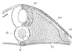
Fig. 396. Section through the trunk of a Lepidosteus embryo on the
sixth day after impregnation.
mc. medullary cord; ms. mesoblast; sg. segmental duct; ch.
notochord; x. subnotochordal rod; hy. hypoblast.
Posteriorly the Müllerian ducts and the ducts of the mesonephros
remain united. The common duct so formed on each side is clearly the
primitive segmental duct. It receives the secretion of a certain
number of the posterior mesonephric tubules, and usually unites with
its fellow to form a kind of bladder, opening by a single pore into
the cloaca, behind the anus. The duct which receives the secretion of
the anterior mesonephric tubules is the true mesonephric or Wolffian
duct.
The development of the excretory system, which has been partially
worked out in Acipenser and Lepidosteus[254],
is on the whole very
similar to that in the Teleostei. The first portion of the system to
be formed is the segmental duct. In Lepidosteus this duct is formed as
a groove-like invagination of the somatic peritoneal epithelium,
precisely as in Teleostei, and shortly afterwards forms a duct lying
between the mesoblast and the epiblast (fig. 396, sg). In Acipenser
(Salensky) however it is formed as a solid ridge of the somatic
mesoblast, as in Petromyzon and Elasmobranchii (fig. 397, Wg).
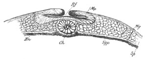
Fig. 397. Transverse section through the anterior part of an Acipenser
embryo. (After Salensky.)
Rf. medullary groove; Mp. medullary plate; Wg. segmental duct;
Ch. notochord; En. hypoblast; Sgp. mesoblastic somite; Sp.
parietal part of mesoblastic plate.
In both forms the ducts unite behind with the cloaca, and a pronephros
of the Teleostean type appears to be developed. This gland is provided
with but one[255]
peritoneal opening, which together with the
glomerulus belonging to it becomes encapsuled in a special section of
the body cavity. The opening of the pronephros of Acipenser into this
cavity is shewn in fig. 398, pr.n. At this early stage of Acipenser
(larva of 5 mm.) I could find no glomerulus.
The mesonephros is formed some distance behind, and some time after
the pronephros, both in Acipenser and Lepidosteus, so that in the
larvæ of both these genera the pronephros is for a considerable period
the only excretory organ. In Lepidosteus especially the development of
the mesonephros occurs very late.
The development of the mesonephros has not been worked out in
Lepidosteus, but in Acipenser the anterior segmental tubes become
first established as (I believe) solid cords of cells, attached at one
extremity to the peritoneal epithelium on each
side of the insertion
of the mesentery, and extending upwards and outwards round the
segmental duct[256].
The posterior segmental tubes arise later than
the anterior, and (as far as can be determined from the sections in my
possession) they are formed independently of the peritoneal
epithelium, on the dorsal side of the segmental duct.
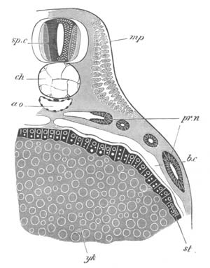
Fig. 398. Transverse section through the region of the stomach of a
larva of Acipenser 5 mm. in length.
st. epithelium of stomach; yk. yolk; ch. notochord, below
which is a subnotochordal rod; pr.n. pronephros; ao. aorta;
mp. muscle-plate formed of large cells, the outer parts of which
are differentiated into contractile fibres; sp.c. spinal cord;
b.c. body cavity.
In later stages (larvæ of 7-10 mm.) the anterior segmental tubes
gradually lose their attachment to the peritoneal epithelium. The
extremity near the peritoneal epithelium forms a Malpighian body, and
the other end unites with the segmental duct. At a still later stage
wide peritoneal funnels are established, for at any rate a
considerable number of the tubes, leading from the body cavity to the
Malpighian bodies. These
funnels have been noticed by Fürbringer,
Salensky and myself, but their mode of development has not, so far as
I know, been made out. The funnels appear to be no longer present in
the adult. The development of the Müllerian ducts has not been worked
out.
Dipnoi. The excretory system of the Dipnoi is only known in the adult,
but though in some respects intermediate in character between that of
the Ganoidei and Amphibia, it resembles that of the Ganoidei in the
important feature of the Müllerian ducts serving as genital ducts in
both sexes.
Amphibia. In Amphibia (Götte, Fürbringer) the development of the
excretory system commences, as in Teleostei, by the formation of the
segmental duct from a groove formed by a fold of the somatic layer of
the peritoneal epithelium, near the dorsal border of the body cavity
(fig. 399, u). The anterior end of the groove is placed immediately
behind the branchial region. Its posterior part soon becomes converted
into a canal by a constriction which commences a short way from the
front end of the groove, and thence extends backwards. This canal at
first ends blindly close to the cloaca, into which however it soon
opens.
The anterior open part of the groove in front of the constriction
(fig. 399, u) becomes differentiated into a longitudinal duct, which
remains in open communication with the body cavity by two (many
Urodela) three (many Anura) or four (Cœciliidæ) canals. This
constitutes the dorsal part of the pronephros. The ventral part of the
gland is formed from the section of the duct immediately behind the
longitudinal canal. This part grows in length, and, assuming an
S-shaped curvature, becomes placed on the ventral side of the first
formed part of the pronephros. By continuous growth in a limited space
the convolutions of the canal of the pronephros become more numerous,
and the complexity of the gland is further increased by the outgrowth
of blindly ending diverticula.
At the root of the mesentery, opposite the peritoneal openings of the
pronephros, a longitudinal fold, lined by peritoneal epithelium, and
attached by a narrow band of tissue, makes its appearance. It soon
becomes highly vascular, and constitutes a glomerulus homologous with
that in Petromyzon and Teleostei.
The section of the body cavity which contains the openings of the
pronephros and the glomerulus, becomes dilated, and then temporarily
shut off from the remainder. At a later period it forms a special
though not completely isolated compartment. For a long time the
pronephros and its duct form the only excretory organs of larval
Amphibia. Eventually however the formation of the mesonephros
commences, and is followed by the atrophy of the pronephros. The
mesonephros is composed, as in other types, of a series of segmental
tubes, but these, except in Cœciliidæ, no longer correspond in number
with the myotomes, but are in all instances more numerous. Moreover,
in the posterior part of the mesonephros in the Urodeles, and through
the whole length of the gland in other types, secondary and tertiary
segmental tubes are formed in addition to the primary tubes.
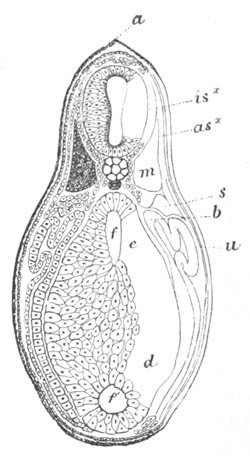
Fig. 399. Transverse section through a very young tadpole of
Bombinator at the level of the anterior end of the yolk-sack.
(After Götte.)
a. fold of epiblast continuous with the dorsal fin; isx. neural
cord; m. lateral muscle; asx. outer layer of muscle-plate; s.
lateral plate of mesoblast; b. mesentery; u. open end of the
segmental duct, which forms the pronephros; f. alimentary tract;
f´. ventral diverticulum which becomes the liver; e. junction of
yolk cells and hypoblast cells; d. yolk cells.
The development of the mesonephros commences in Salamandra
(Fürbringer) with the formation of a series of solid cords, which in
the anterior myotomes spring from the peritoneal epithelium on the
inner side of the segmental duct, but posteriorly arise independently
of this epithelium in the adjoining mesoblast. Sedgwick informs me
that in the Frog the segmental tubes are throughout developed in the
mesoblast, independently of the peritoneal epithelium. These cords
next become detached from the peritoneal epithelium (in so far as they
are primitively united to it), and after first assuming a vesicular
form, grow out into coiled tubes, with a median limb the blind end of
which assists in forming a Malpighian body, and a lateral limb which
comes in contact with and opens into the segmental duct, and an
intermediate portion connecting the two. At the junction of the median
with the intermediate portion, and therefore at the neck of the
Malpighian body, a canal grows out in a ventral direction, which meets
the
peritoneal epithelium, and then develops a funnel-shaped opening
into the body cavity, which subsequently becomes ciliated. In this way
the peritoneal funnels which are present in the adult are established.
The median and lateral sections of the segmental tubes become highly
convoluted, and the separate tubes soon come into such close proximity
that their primitive distinctness is lost.
The first fully developed segmental tube is formed in Salamandra
maculata in about the sixth myotome behind the pronephros. But in the
region between the two structures rudimentary segmental tubes are
developed.
The number of primary segmental tubes in the separate myotomes of
Salamandra is as follows:
In the 6th myotome (i.e. the first with a true
segmental tube): 1-2 segmental tubes
In the 7th-10th myotome: 2-3 segmental tubes
In the 11th myotome: 3-4 segmental tubes
In the 12th myotome: 3-4 4-5 segmental tubes
In the 13th myotome: 4-5 segmental tubes
In the 13th-16th myotome: 5-6 segmental tubes
It thus appears that the segmental tubes are not only more numerous
than the myotomes, but that the number in each myotome increases from
before backwards. In the case of Salamandra there are formed in the
region of the posterior (10-16) myotomes secondary, tertiary, etc.
segmental tubes out of independent solid cords, which arise in the
mesoblast dorsally to the tubes already established.
The secondary segmental tubes appear to develop out of these cords
exactly in the same way as the primary ones, except that they do not
join the segmental duct directly, but unite with the primary segmental
tubes shortly before the junction of the latter with the segmental
duct. In this way compound segmental tubes are established with a
common collecting tube, but with numerous Malpighian bodies and
ciliated peritoneal openings. The difference in the mode of origin of
these compound tubes and of those in Elasmobranchii is very striking.
The later stages in the development of the segmental tubes have not
been studied in the other Amphibian types.
In Cœciliidæ the earliest stages are not known, but the tubes present
in the adult (Spengel) a truly segmental arrangement, and in the young
each of them is single, and provided with only a single peritoneal
funnel. In the adult however many of the segmental organs become
compound, and may have as many as twenty funnels, etc. Both simple and
compound segmental tubes occur in all parts of the mesonephros, and
are arranged in no definite order.
In the Anura (Spengel) all the segmental tubes are compound, and an
enormous number of peritoneal funnels are present on the ventral
surface, but it has not yet been definitely determined into what part
of the segmental tubes they open.
Before dealing with the further changes of the Wolffian body it is
necessary to return to the segmental duct, which, at the time when the
pronephros is undergoing atrophy, becomes split into a dorsal Wolffian
and ventral Müllerian duct. The process in Salamandra (Fürbringer) has
much the same character as in Elasmobranchii, the Müllerian duct being
formed by the gradual separation, from before backwards, of a solid
row of cells from the ventral side of the segmental duct, the
remainder of the duct constituting the Wolffian duct. During the
formation of the Müllerian duct its anterior part becomes hollow, and
attaching itself in front to the peritoneal epithelium acquires an
opening into the body cavity. The process of hollowing is continued
backwards pari passu with the splitting of the segmental duct. In
the female the process is continued till the Müllerian duct opens,
close to the Wolffian duct, into the cloaca. In the male the duct
usually ends blindly. It is important to notice that the abdominal
opening of the Müllerian duct in the Amphibia (Salamandra) is a
formation independent of the pronephros, and placed slightly behind
it; and that the undivided anterior part of the segmental duct (with
the pronephros) is not, as in Elasmobranchii, united with the
Müllerian duct, but remains connected with the Wolffian duct.
The development of the Müllerian duct has not been satisfactorily
studied in other forms besides Salamandra. In Cœciliidæ its abdominal
opening is on a level with the anterior end of the Wolffian body. In
other forms it is usually placed very far forwards, close to the root
of the lungs (except in Proteus and Batrachoseps, where it is placed
somewhat further back), and some distance in front of the Wolffian
body.
The Müllerian duct is always well developed in the female, and serves
as oviduct. In the male it does not (except possibly in Alytes) assist
in the transportation of the genital products, and is always more or
less rudimentary, and in Anura may be completely absent.
After the formation of the Müllerian duct, the Wolffian duct remains
as the excretory channel for the Wolffian body, and, till the atrophy
of the pronephros, for this gland also. Its anterior section, in front
of the Wolffian body, undergoes a more or less complete atrophy.
The further changes of the excretory system concern (1) the junction
in the male of the anterior part of the Wolffian body with the testis;
(2) certain changes in the collecting tubes of the
posterior part of
the mesonephros. The first of these processes results in the division
of the Wolffian body into a sexual and a non-sexual part, and in
Salamandra and other Urodeles the division corresponds with the
distribution of the simple and compound segmental tubes.
Since the development of the canals connecting the testes with the
sexual part of the Wolffian body has not been in all points
satisfactorily elucidated, it will be convenient to commence with a
description of the adult arrangement of the parts (fig. 400 B). In
most instances a non-segmental system of canals—the vasa efferentia
(ve)—coming from the testis, fall into a canal known as the
longitudinal canal of the Wolffian body, from which there pass off
transverse canals, which fall into, and are equal in number to, the
primary Malpighian bodies of the sexual part of the gland. The
spermatozoa, brought to the Malpighian bodies, are thence transported
along the segmental tubes to the Wolffian duct, and so to the
exterior. The system of canals connecting the testis with the
Malpighian bodies is known as the testicular network. The number of
segmental tubes connected with the testis varies very greatly. In
Siredon there are as many as from 30-32 (Spengel).
The longitudinal canal of the Wolffian body is in rare instances
(Spelerpes, etc.) absent, where the sexual part of the Wolffian body
is slightly developed. In the Urodela the testes are united with the
anterior part of the Wolffian body. In the Cœciliidæ the junction
takes place in an homologous part of the Wolffian body, but, owing to
the development of the anterior segmental tubes, which are rudimentary
in the Urodela, it is situated some way behind the front end. Amongst
the Anura the connection of the testis with the tubules of the
Wolffian body is subject to considerable variations. In Bufo cinereus
the normal Urodele type is preserved, and in Bombinator the same
arrangement is found in a rudimentary condition, in that there are
transverse trunks from the longitudinal canal of the Wolffian body,
which end blindly, while the semen is carried into the Wolffian duct
by canals in front of the Wolffian body. In Alytes and Discoglossus
the semen is carried away by a similar direct continuation of the
longitudinal canal in front of the Wolffian body, but there are no
rudimentary transverse canals passing into the Wolffian body, as in
Bombinator. In Rana the transverse ducts which pass off from the
longitudinal canal of the Wolffian body, after dilating to form (?)
rudimentary Malpighian bodies, enter directly into the collecting
tubes near their opening into the Wolffian duct.
In most Urodeles the peritoneal openings connected with the primary
generative Malpighian bodies atrophy, but in Spelerpes they persist.
In the Cœciliidæ they also remain in the adult state.
With reference to the development of these parts little is known
except that the testicular network grows out from the primary
Malpighian bodies, and becomes united with the testis. Embryological
evidence, as well as the fact of the persistence of the peritoneal
funnels of the generative region in the adults of some forms, proves
that the testicular network is not developed from the peritoneal
funnels.
Rudiments of the testicular network are found in the female Cœciliidæ
and in the females of many Urodela (Salamandra, Triton). These
rudiments may in their fullest development consist of a longitudinal
canal and of transverse canals passing from this to the Malpighian
bodies, together with some branches passing into the mesovarium.
Amongst the Urodela the collecting tubes of the hinder non-sexual part
of the Wolffian body, which probably represents a rudimentary
metanephros, undergo in the male sex a change similar to that which
they usually undergo in Elasmobranchii. Their points of junction with
the Wolffian duct are carried back to the hindermost end of the duct
(fig. 400 B), and the collecting tubes themselves unite together into
one or more short ducts (ureters) before joining the Wolffian duct.
In Batrachoseps only the first collecting tube becomes split off in
this way; and it forms a single elongated ureter which receives all
the collecting tubes of the posterior segmental tubes. In the female
and in the male of Proteus, Menobranchus, and Siren the collecting
tubes retain their primitive transverse course and open laterally into
the Wolffian duct. In rare cases (Ellipsoglossus, Spengel) the
ureters open directly into the cloaca.
The urinary bladder of the Amphibia is an outgrowth of the ventral
wall of the cloacal section of the alimentary tract, and is homologous
with the allantois of the amniotic Vertebrata.
The subjoined diagram (fig. 400) of the urogenital system of Triton
illustrates the more important points of the preceding description.
In the female (A) the following parts are present:
(1) The Müllerian duct or oviduct (od) derived from the splitting
of the segmental duct.
(2) The Wolffian duct (sug) constituting the portion of the
segmental duct left after the formation of the Müllerian duct.
(3) The mesonephros (r), divided into an anterior sexual part
connected with a rudimentary testicular network, and a posterior
part. The collecting tubes from both parts fall transversely
into the Wolffian duct.
(4) The ovary (ov).
(5) The rudimentary testicular network.
In the male (B) the following parts are present:
(1) The functionless though fairly developed Müllerian duct (m).
(2) The Wolffian duct (sug).
(3) The mesonephros (r) divided into a true sexual part, through
the segmental tubes of which the semen passes, and a non-sexual
part. The collecting tubes of the latter do not enter the
Wolffian duct directly, but bend obliquely backwards and only
fall into it close to its cloacal aperture, after uniting to
form one or two primary tubes (ureters).
(4) The testicular network (ve) consisting of (1) transverse ducts
from the testes, falling into (2) the longitudinal canal of the
Wolffian body, from which (3) transverse canals are again given
off to the Malpighian bodies.
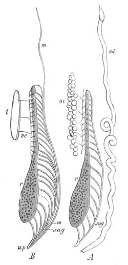
Fig. 400. Diagram of the urinogenital system of Triton. (From
Gegenbaur; after Spengel.)
A. Female. B. Male.
r. mesonephros, on the surface of which numerous peritoneal
funnels are visible; sug. mesonephric or Wolffian duct; od.
oviduct (Müllerian duct); m. Müllerian duct of male; ve. vasa
efferentia of testis; t. testis; ov. ovary; up. urinogenital
pore.
Amniota. The amniotic Vertebrata agree, so far as is known, very
closely amongst themselves in the formation of the urinogenital
system.
The most characteristic feature of the system is the full development
of a metanephros, which constitutes the functional kidney on the
atrophy of the mesonephros or Wolffian body, which is a purely
embryonic organ. The first part of the system to develop is a duct,
which is usually spoken of as the Wolffian duct, but which is really
the homologue of the segmental
duct. It apparently develops in all the
Amniota nearly on the Elasmobranch type, as a solid rod, primarily
derived from the somatic mesoblast of the intermediate cell mass (fig.
401 W.d)[257].
The first trace of it is visible in an embryo Chick with eight
somites, as a ridge projecting from the intermediate cell mass towards
the epiblast in the region of the seventh somite. In the course of
further development it continues to constitute such a ridge as far as
the eleventh somite (Sedgwick), but from this point it grows backwards
in the space between the epiblast and mesoblast. In an embryo with
fourteen somites a small lumen has appeared in its middle part and in
front it is connected with rudimentary Wolffian tubules, which develop
in continuity with it (Sedgwick). In the succeeding stages the lumen
of the duct gradually extends backwards and forwards, and the duct
itself also passes inwards relatively to the epiblast (fig. 402). Its
hind-end elongates till it comes into connection with, and opens into,
the cloacal section of the hindgut[258].
It might have been anticipated that, as in the lower types, the
anterior end of the segmental duct would either open into the body
cavity, or come into connection with a pronephros. Neither of these
occurrences takes place, though in some types (the Fowl) a structure,
which is probably the rudiment of a pronephros, is developed; it does
not however appear till a later stage, and is then unconnected with
the segmental duct. The next part of the system to appear is the
mesonephros or Wolffian body.
This is formed in all Amniota as a series of segmental tubes, which in
Lacertilia (Braun) correspond with the myotomes, but in Birds and
Mammalia are more numerous.
In Reptilia (Braun, No. 542), the mesonephric tubes develop as
segmentally-arranged masses on the inner side of the Wolffian duct,
and appear to be at first united with the peritoneal epithelium. Each
mass soon becomes an oval vesicle, probably opening for a very short
period into the
peritoneal cavity by a peritoneal funnel. The vesicles
become very early detached from the peritoneal epithelium, and lateral
outgrowths from them give rise to the main parts of the segmental
tubes, which soon unite with the segmental duct.
In Birds the development of the segmental tubes is more
complicated[259].
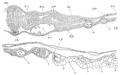
Fig. 401. Transverse section through the dorsal region of an
embryo Chick of 45 hours.
M.c. medullary canal; P.v. mesoblastic somite; W.d. Wolffian
duct which is in contact with the intermediate cell mass; So.
somatopleure; S.p. splanchnopleure; p.p. pleuroperitoneal
cavity; ch. notochord; op. boundary of area opaca; v.
blood-vessel.
The tubules of the Wolffian body are derived from the intermediate
cell mass, shewn in fig. 401, between the upper end of the body cavity
and the muscle-plate. In the Chick the mode of development of this
mass into the segmental tubules is different in the regions in front
of and behind about the sixteenth segment. In front of about the
sixteenth segment the intermediate cell mass becomes detached from the
peritoneal epithelium at certain points, remaining attached to it at
other points, there being several such to each segment. The parts of
the intermediate cell mass attached to the peritoneal epithelium
become converted into S-shaped cords (fig. 402, st) which soon unite
with the segmental duct (wd). Into the commencement of each of these
cords the lumen of the body cavity is for a short distance prolonged,
so that this part constitutes a rudimentary peritoneal funnel.
In the
Duck the attachment of the intermediate cell mass to the peritoneal
epithelium is prolonged further back than in the Chick.
In the foremost segmental tubes, which never reach a very complete
development, the peritoneal funnels widen considerably, while at the
same time they acquire a distinct lumen. The section of the tube
adjoining the wide peritoneal funnel becomes partially invaginated by
the formation of a glomerulus, and this glomerulus soon grows to such
an extent as to project through the peritoneal funnel, the neck of
which it completely fills, into the body cavity (fig. 403, gl).
There is thus formed a series of free peritoneal glomeruli belonging
to the anterior Wolffian tubuli[260].
These tubuli become however
early aborted.
In the case of the remaining tubules developed from the S-shaped cords
the attachment to the peritoneal epithelium is very soon lost. The
cords acquire a lumen, and open into the segmental duct. Their blind
extremities constitute the rudiments of Malpighian bodies.
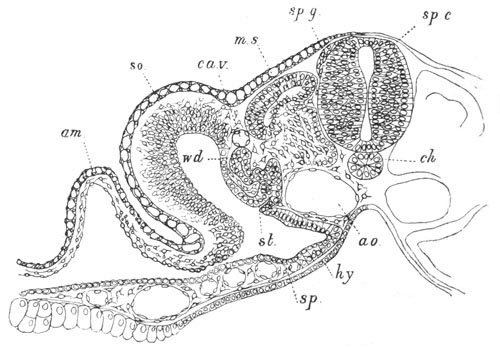
Fig. 402. Transverse section through the trunk of a Duck embryo with
about twenty-four mesoblastic somites.
am. amnion; so. somatopleure; sp. splanchnopleure; wd.
Wolffian duct; st. segmental tube; ca.v. cardinal vein; m.s.
muscle-plate; sp.g. spinal ganglion; sp.c. spinal cord; ch.
notochord; ao. aorta; hy. hypoblast.
In the posterior part of the Wolffian body of the Chick the
intermediate cell mass becomes very early detached from the peritoneal
epithelium, and at a considerably later period breaks up into oval
vesicles similar to those of the Reptilia, which form the rudiments of
the segmental tubes.
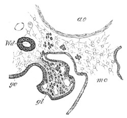
Fig. 403. Section through the external
glomerulus of one of the anterior segmental tubes of an embryo Chick
of about 100 h.
gl. glomerulus; ge. peritoneal epithelium; Wd. Wolffian duct;
ao. aorta; me. mesentery. The segmental tube, and the connection
between the external and internal parts of the glomerulus are not
shewn in this figure.
Secondary and tertiary segmental tubules are formed in the Chick, on
the dorsal side of the primary tubules, as direct differentiations of
the mesoblast. They open independently into the Wolffian duct.
In Mammalia the segmental tubules (Egli) are formed as solid masses in
the same situation as in Birds and Reptiles. It is not known whether
they are united with the peritoneal epithelium. They soon become oval
vesicles, which develop into complete tubules in the manner already
indicated.
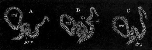
Fig. 404. Sections shewing two of the peritoneal invaginations which
give rise to the anterior part of the Müllerian duct (pronephros).
(After Balfour and Sedgwick.)
A is the 11th section of the series.
B is the 15th section of the series.
C is the 18th section of the series.
gr2. second groove; gr3. third groove; r2. second ridge; wd.
Wolffian duct.
After the establishment of the Wolffian body there is formed in both
sexes in all the Amniota a duct, which in the female becomes the
oviduct, but which is functionless and disappears more or less
completely in the male. This duct, in spite of certain peculiarities
in its development, is without doubt homologous with the Müllerian
duct of
the Ichthyopsida. In connection with its anterior extremity
certain structures have been found in the Fowl, which are probably, on
grounds to be hereafter stated, homologous with the pronephros
(Balfour and Sedgwick).
The pronephros, as I shall call it, consists of a slightly convoluted
longitudinal canal with three or more peritoneal openings. In the
earliest condition, it consists of three successive open involutions
of the peritoneal epithelium, connected together by more or less
well-defined ridge-like thickenings of the epithelium. It takes its
origin from the layer of thickened peritoneal epithelium situated near
the dorsal angle of the body cavity, and is situated some considerable
distance behind the front end of the Wolffian duct.
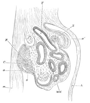
Fig. 405. Section of the Wolffian body developing pronephros and
genital gland of the fourth day. (After Waldeyer.) Magnified 160 times.
m. mesentery; L. somatopleure; a´. portion of the germinal
epithelium from which the involution (z) to form the pronephros
(anterior part of Müllerian duct) takes place; a. thickened
portion of the germinal epithelium in which the primitive germinal
cells C and o are lying; E. modified mesoblast which will form
the stroma of the ovary; WK. Wolffian body; y. Wolffian duct.
In a slightly later stage the ridges connecting the grooves become
partially constricted off from the peritoneal epithelium,
and develop
a lumen. The condition of the structure at this stage is illustrated
by fig. 404, representing three transverse sections through two
grooves, and through the ridge connecting them.
The pronephros may in fact now be described as a slightly convoluted
duct, opening into the body cavity by three groove-like apertures, and
continuous behind with the rudiment of the true Müllerian duct.
The stage just described is that of the fullest development of the
pronephros. In it, as in all the previous stages, there appear to be
only three main openings into the body cavity; but in some sections
there are indications of the possible presence of one or two
additional rudimentary grooves.
In an embryo not very much older than the one last described the
pronephros atrophies as such, its two posterior openings vanishing,
and its anterior opening remaining as the permanent opening of the
Müllerian duct.
The pronephros is an extremely transitory structure, and its
development and atrophy are completed between the 90th and 120th hours
of incubation.
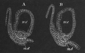
Fig. 406. Two sections shewing the junction of the terminal solid
portion of the Müllerian duct with the Wolffian duct. (After Balfour
and Sedgwick.)
In A the terminal portion of the duct is quite distinct; in B it has
united with the walls of the Wolffian duct.
md. Müllerian duct; Wd. Wolffian duct.
The position of the pronephros in relation to the Wolffian body is
shewn in fig. 405, which probably passes through a region between two
of the peritoneal openings. As long as the pronephros persists, the
Müllerian duct consists merely of a very
small rudiment, continuous
with the hindermost of the three peritoneal openings, and its solid
extremity appears to unite with the walls of the Wolffian duct.
After the atrophy of the pronephros, the Müllerian duct commences to
grow rapidly, and for the first part of its course it appears to be
split off as a solid rod from the outer or ventral wall of the
Wolffian duct (fig. 406). Into this rod the lumen, present in its
front part, subsequently extends. Its mode of development in front is
thus precisely similar to that of the Müllerian duct in Elasmobranchii
and Amphibia.
This mode of development only occurs however in the anterior part of
the duct. In the posterior part of its course its growing point lies
in a bay formed by the outer walls of the Wolffian duct, but does not
become definitely attached to that duct. It seems however possible
that, although not actually split off from the walls of the Wolffian
duct, it may grow backwards from cells derived from that duct.
The Müllerian duct finally reaches the cloaca though it does not in
the female for a long time open into it, and in the male never does
so.
The mode of growth of the Müllerian duct in the posterior part of its
course will best be understood from the following description quoted
from the paper by Sedgwick and myself.
“A few sections before its termination the Müllerian duct appears as
a well-defined oval duct lying in contact with the wall of the
Wolffian duct on the one hand and the germinal epithelium on the
other. Gradually, however, as we pass backwards, the Müllerian duct
dilates; the external wall of the Wolffian duct adjoining it becomes
greatly thickened and pushed in in its middle part, so as almost to
touch the opposite wall of the duct, and so form a bay in which the
Müllerian duct lies. As soon as the Müllerian duct has come to lie
in this bay its walls lose their previous distinctness of outline,
and the cells composing them assume a curious vacuolated appearance.
No well-defined line of separation can any longer be traced between
the walls of the Wolffian duct and those of the Müllerian, but
between the two is a narrow clear space traversed by an irregular
network of fibres, in some of the meshes of which nuclei are
present.
“The Müllerian duct may be traced in this condition for a
considerable number of sections, the peculiar features above
described becoming more and more marked as its termination is
approached. It continues to dilate and attains a maximum size in the
section or so before it disappears. A lumen may be observed in it up
to its very end, but is usually irregular in outline and frequently
traversed by strands of protoplasm. The Müllerian
duct finally
terminates quite suddenly, and in the section immediately behind its
termination the Wolffian duct assumes its normal appearance, and the
part of its outer wall on the level of the Müllerian duct comes into
contact with the germinal epithelium.”
Before describing the development of the Müllerian duct in other
Amniotic types it will be well to say a few words as to the
identifications above adopted. The identification of the duct, usually
called the Wolffian duct, with the segmental duct (exclusive of the
pronephros) appears to be morphologically justified for the following
reasons: (1) that it gives rise to part of the Müllerian duct as well
as to the duct of the Wolffian body; behaving in this respect
precisely as does the segmental duct of Elasmobranchii and Amphibia.
(2) That it serves as the duct for the Wolffian body, before the
Müllerian duct originates from it. (3) That it develops in a manner
strikingly similar to that of the segmental duct of various lower
forms.
With reference to the pronephros it is obvious that the organ
identified as such is in many respects similar to the pronephros of
the Amphibia. Both consist of a somewhat convoluted longitudinal
canal, with a certain number of peritoneal openings.
The main difficulties in the homology are:
(1) the fact that the pronephros in the Bird is not united with the
segmental duct;
(2) the fact that it is situated behind the front end of the
Wolffian body.
It is to be remembered in connection with the first of these
difficulties that in the formation of the Müllerian duct in
Elasmobranchii the anterior undivided extremity of the primitive
segmental duct, with the peritoneal opening, which probably represents
the pronephros, is attached to the Müllerian duct, and not to the
Wolffian duct; though in Amphibia the reverse is the case. To explain
the discontinuity of the pronephros with the segmental duct it is only
necessary to suppose that the segmental duct and pronephros, which in
the Ichthyopsida develop as a single formation, develop in the Bird as
two independent structures—a far from extravagant supposition,
considering that the pronephros in the Bird is undoubtedly quite
functionless.
With reference to the posterior position of the pronephros it is only
necessary to remark that a change in position might easily take place
after the acquirement of an independent development, and that the
shifting is probably correlated with a shifting of the abdominal
opening of the Müllerian duct.
The pronephros has only been observed in Birds, and is very possibly
not developed in other Amniota. The Müllerian duct is also usually
stated to develop as a groove of the peritoneal epithelium, shewn in
the Lizard in fig. 354, md., which is continued backward as a
primitively solid rod in the space between
the Wolffian duct and
peritoneal epithelium, without becoming attached to the Wolffian duct.
On the formation of the Müllerian duct, the duct of the mesonephros
becomes the true mesonephric or Wolffian duct.
After these changes have taken place a new organ of great importance
makes its appearance. This organ is the permanent kidney, or
metanephros.
Metanephros. The mode of development of the metanephros has as yet
only been satisfactorily elucidated in the Chick (Sedgwick, No. 549).
The ureter and the collecting tubes of the kidney are developed from a
dorsal outgrowth of the hinder part of the Wolffian duct. The
outgrowth from the Wolffian duct grows forwards, and extends along the
outer side of a mass of mesoblastic tissue which lies mainly behind,
but somewhat overlaps the dorsal aspect of the Wolffian body.
This mass of mesoblastic cells may be called the metanephric blastema.
Sedgwick, of the accuracy of whose account I have satisfied myself,
has shewn that in the Chick it is derived from the intermediate cell
mass of the region of about the thirty-first to the thirty-fourth
somite. It is at first continuous with, and indistinguishable in
structure from, the portion of the intermediate cell mass of the
region immediately in front of it, which breaks up into Wolffian
tubules. The metanephric blastema remains however quite passive during
the formation of the Wolffian tubules in the adjoining blastema; and
on the formation of the ureter breaks off from the Wolffian body in
front, and, growing forwards and dorsalwards, places itself on the
inner side of the ureter in the position just described.
In the subsequent development of the kidney collecting tubes grow out
from the ureter, and become continuous with masses of cells of the
metanephric blastema, which then differentiate themselves into the
kidney tubules.
The process just described appears to me to prove that the kidney of
the Amniota is a specially differentiated posterior section of the
primitive mesonephros.
According to the view of Remak and Kölliker the outgrowths from the
ureter give rise to the whole of the tubuli uriniferi and the capsules
of the Malpighian bodies, the mesoblast around them forming
blood-vessels, etc. On the other hand some observers (Kupffer,
Bornhaupt, Braun) maintain, in
accordance with the account given
above, that the outgrowths of the ureter form only the collecting
tubes, and that the secreting tubuli, etc. are formed in situ in the
adjacent mesoblast.
Braun (No. 542) has arrived at the conclusion that in the Lacertilia
the tissue, out of which the tubuli of the metanephros are formed, is
derived from irregular solid ingrowths of the peritoneal epithelium,
in a region behind the Wolffian body, but in a position corresponding
to that in which the segmental tubes take their origin. These
ingrowths, after separating from the peritoneal epithelium, unite
together to form a cord into which the ureter sends the lateral
outgrowths already described. These outgrowths unite with secreting
tubuli and Malpighian bodies, formed in situ. In Lacertilia the
blastema of the kidney extends into a postanal region. Braun’s account
of the origin of the metanephric blastema does not appear to me to be
satisfactorily demonstrated.
The ureter does not long remain attached to the Wolffian duct, but its
opening is gradually carried back, till (in the Chick between the 6th
and 8th day) it opens independently into the cloaca.
Of the further changes in the excretory system the most important is
the atrophy of the greater part of the Wolffian body, and the
conversion of the Wolffian duct in the male sex into the vas deferens,
as in Amphibia and the Elasmobranchii.
The mode of connection of the testis with the Wolffian duct is very
remarkable, but may be derived from the primitive arrangement
characteristic of Elasmobranchii and Amphibia.
In the structures connecting the testis with the Wolffian body two
parts have to be distinguished, (1) that equivalent to the testicular
network of the lower types, (2) that derived from the segmental tubes.
The former is probably to be found in peculiar outgrowths from the
Malpighian bodies at the base of the testes.
These were first discovered by Braun in Reptilia, and consist in this
group of a series of outgrowths from the primary (?) Malpighian bodies
along the base of the testis: they unite to form an interrupted cord
in the substance of the testis, from which the testicular tubuli (with
the exception of the seminiferous cells) are subsequently
differentiated. These outgrowths, with the exception of the first two
or three, become detached from the Malpighian bodies. Outgrowths
similar to those in the male are found in the female, but subsequently
atrophy.
Outgrowths homologous with those found by Braun have
been detected by
myself (No. 555) in Mammals. It is not certain to what parts of the
testicular tubuli they give rise, but they probably form at any rate
the vasa recta and rete vasculosum.
In Mammals they also occur in the female, and give rise to cords of
tissue in the ovary, which may persist through life.
The comparison of the tubuli, formed out of these structures, with the
Elasmobranch and Amphibian testicular network is justified in that
both originate as outgrowths from the primary Malpighian bodies, and
thence extend into the testis, and come into connection with the true
seminiferous stroma.
As in the lower types the semen is transported from the testicular
network to the Wolffian duct by parts of the glandular tubes of the
Wolffian body. In the case of Reptilia the anterior two or three
segmental tubes in the region of the testis probably have this
function. In the case of Mammalia the vasa efferentia, i.e. the coni
vasculosi, appear, according to the usually accepted view, to be of
this nature, though Banks and other investigators believe that they
are independently developed structures. Further investigations on this
point are required. In Birds a connection between the Wolffian body
and the testis appears to be established as in the other types. The
Wolffian duct itself becomes, in the males of all Amniota, the vas
deferens and the convoluted canal of the epididymis—the latter
structure (except the head) being entirely derived from the Wolffian
duct.
In the female the Wolffian duct atrophies more or less completely.
In Snakes (Braun) the posterior part remains as a functionless canal,
commencing at the ovary, and opening into the cloaca. In the Gecko
(Braun) it remains as a small canal joining the ureter; in Blindworms
a considerable part of the canal is left, and in Lacerta (Braun) only
interrupted portions.
In Mammalia the middle part of the duct, known as Gaertner’s canal,
persists in the females of some monkeys, of the pig and of many
ruminants.
The Wolffian body atrophies nearly completely in both sexes; though,
as described above, part of it opposite the testis persists as the
head of the epididymis. The posterior part of the gland from the level
of the testis may be called the sexual part of the gland, the anterior
part forming the non-sexual part.
The latter, i.e. the anterior
part, is first absorbed; and in some Reptilia the posterior part,
extending from the region of the genital glands to the permanent
kidney, persists till into the second year.
Various remnants of the Wolffian body are found in the adults of both
sexes in different types. The most constant of them is perhaps the
part in the female equivalent to the head of the epididymis and to
parts also of the coiled tube of the epididymis, which may be called,
with Waldeyer, the epoophoron[261].
This is found in Reptiles, Birds
and Mammals; though in a very rudimentary form in the first-named
group. Remnants of the anterior non-sexual part of the Wolffian bodies
have been called by Waldeyer parepididymis in the male, and
paroophoron in the female. Such remnants are not (Braun) found in
Reptilia, but are stated to be found in both male and female Birds, as
a small organ consisting of blindly ending tubes with yellow pigment.
In some male Mammals (including Man) a parepididymis is found on the
upper side of the testis. It is usually known as the organ of
Giraldes.
The Müllerian duct forms, as has been stated, the oviduct in the
female. The two ducts originally open independently into the cloaca,
but in the Mammalia a subsequent modification of this arrangement
occurs, which is dealt with in a separate section. In Birds the right
oviduct atrophies, a vestige being sometimes left. In the male the
Müllerian ducts atrophy more or less completely.
In most Reptiles and in Birds the atrophy of the Müllerian ducts is
complete in the male, but in Lacerta and Anguis a rudiment of the
anterior part has been detected by Leydig as a convoluted canal. In
the Rabbit (Kölliker)[262]
and probably other Mammals the whole of the
ducts probably disappears, but in some Mammals, e.g. Man, the lower
fused ends of the Müllerian ducts give rise to a pocket opening into
the urethra, known as the uterus masculinus; and in other cases,
e.g. the Beaver and the Ass, the rudiments are more considerable,
and may be continued into horns homologous with the horns of the
uterus (Weber).
The hydatid of Morgani in the male is supposed (Waldeyer) to represent
the abdominal opening of the Fallopian tube in the female, and
therefore to be a remnant of the Müllerian duct.
Changes in the lower parts of the urinogenital ducts in the Amniota.
The genital cord. In the Monodelphia the lower part of the Wolffian
ducts becomes enveloped in both sexes in a special
cord of tissue,
known as the genital cord (fig. 407, gc), within the lower part of
which the Müllerian ducts are also enclosed. In the male the Müllerian
ducts in this cord atrophy, except at their distal end where they
unite to form the uterus masculinus. The Wolffian ducts, after
becoming the vasa deferentia, remain for some time enclosed in the
common cord, but afterwards separate from each other. The seminal
vesicles are outgrowths of the vasa deferentia.
In the female the Wolffian ducts within the genital cord atrophy,
though rudiments of them are for a long time visible or even
permanently persistent. The lower parts of the Müllerian ducts unite
to form the vagina and body of the uterus. The junction commences in
the middle and extends forwards and backwards; the stage with a median
junction being retained permanently in Marsupials.
The urinogenital sinus and external generative organs. In all the
Amniota, there open at first into the common cloaca the alimentary
canal dorsally, the allantois ventrally, and the Wolffian and
Müllerian ducts and ureters laterally. In Reptilia and Aves the
embryonic condition is retained. In both groups the allantois serves
as an embryonic urinary bladder, but while it atrophies in Aves, its
stalk dilates to form a permanent urinary bladder in Reptilia. In
Mammalia the dorsal part of the cloaca with the alimentary tract
becomes first of all partially constricted off from the ventral, which
then forms a urinogenital sinus (fig. 407, ug). In the course of
development the urinogenital sinus becomes, in all Mammalia but the
Ornithodelphia, completely separated from the intestinal cloaca, and
the two parts obtain separate external openings. The ureters (fig.
407, 3) open higher up than the other ducts into the stalk of the
allantois which dilates to form the bladder (4). The stalk connecting
the bladder with the ventral wall of the body constitutes the urachus,
and loses its lumen before the close of embryonic life. The part of
the stalk of the allantois below the openings of the ureters narrows
to form the urethra, which opens together with the Wolffian and
Müllerian ducts into the urinogenital cloaca.
In front of the urinogenital cloaca there is formed a genital
prominence (fig. 407, cp), with a groove continued from the
urinogenital opening; and on each side a genital fold (ls). In the
male the sides of the groove on the prominence coalesce together,
embracing between them the opening of the urinogenital cloaca; and the
prominence itself gives rise to the penis, along which the common
urinogenital passage is continued. The two genital folds unite from
behind forwards to form the scrotum.
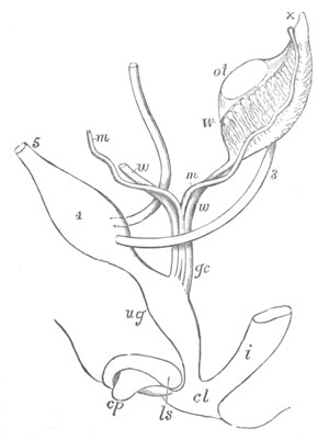
Fig. 407. Diagram of the urinogenital organs of a Mammal at an
early stage. (After Allen Thomson; from Quain’s Anatomy.)
The parts are seen chiefly in profile, but the Müllerian and
Wolffian ducts are seen from the front.
3. ureter; 4. urinary bladder; 5. urachus; ot. genital ridge
(ovary or testis); W. left Wolffian body; x. part at apex from
which coni vasculosi are afterwards developed; w. Wolffian duct;
m. Müllerian duct; gc. genital cord consisting of Wolffian and
Müllerian ducts bound up in a common sheath; i. rectum; ug.
urinogenital sinus; cp. elevation which becomes the clitoris or
penis; ls. ridge from which the labia majora or scrotum are
developed.
In the female the groove on the genital prominence gradually
disappears, and the prominence remains as the clitoris, which is
therefore the homologue of the penis: the two genital folds form the
labia majora. The urethra and vagina open independently into the
common urinogenital sinus.
General conclusions and Summary.
Pronephros. Sedgwick has pointed out that the pronephros is always
present in types with a larval development, and either absent or
imperfectly developed in those types which undergo the greater part of
their development within the egg. Thus it is practically absent in the
embryos of Elasmobranchii and the Amniota, but present in the larvæ of
all other forms.
This coincidence, on the principles already laid down in a previous
chapter on larval forms, affords a strong presumption that the
pronephros is an ancestral organ; and, coupled with the fact that it
is the first part of the excretory system to be developed, and often
the sole excretory organ for a considerable period, points to the
conclusion that the pronephros and its duct—the segmental duct—are
the most primitive parts of the Vertebrate excretory system. This
conclusion coincides with that arrived at by Gegenbaur and Fürbringer.
The duct of the pronephros is always developed prior to the gland, and
there are two types according to which its development may take place.
It may either be formed by the closing in of a continuous groove of
the somatic peritoneal epithelium (Amphibia, Teleostei, Lepidosteus),
or as a solid knob or rod of cells derived from the somatic mesoblast,
which grows backwards between the epiblast and the mesoblast
(Petromyzon, Elasmobranchii, and the Amniota).
It is quite certain that the second of these processes is not a true
record of the evolution of the duct, and though it is more possible
that the process observable in Amphibia and the Teleostei may afford
some indications of the manner in which the duct was established, this
cannot be regarded as by any means certain.
The mode of development of the pronephros itself is apparently partly
dependent on that of its duct. In Petromyzon, where the duct does not
at first communicate with the body cavity, the pronephros is formed as
a series of outgrowths from the duct, which meet the peritoneal
epithelium and open into the body cavity; but in other instances it is
derived from the anterior open end of the groove which gives rise to
the segmental duct. The open end of this groove may either remain
single
(Teleostei, Ganoidei) or be divided into two, three or more
apertures (Amphibia). The main part of the gland in either case is
formed by convolutions of the tube connected with the peritoneal
funnel or funnels. The peritoneal funnels of the pronephros appear to
be segmentally arranged.
The pronephros is distinguished from the mesonephros by developmental
as well as structural features. The most important of the former is
the fact that the glandular tubules of which it is formed are always
outgrowths of the segmental duct; while in the mesonephros they are
always or almost always[263]
formed independently of the duct.
The chief structural peculiarity of the pronephros is the absence from
it of Malpighian bodies with the same relations as those in the meso-
and metanephros; unless the structures found in Myxine are to be
regarded as such. Functionally the place of such Malpighian bodies is
taken by the vascular peritoneal ridge spoken of in the previous pages
as the glomerulus.
That this body is really related functionally to the pronephros
appears to be indicated (1) by its constant occurrence with the
pronephros and its position opposite the peritoneal openings of this
body; (2) by its atrophy at the same time as the pronephros; (3) by
its enclosure together with the pronephridian stoma in a special
compartment of the body-cavity in Teleostei and Ganoids, and its
partial enclosure in such a compartment in Amphibia.
The pronephros atrophies more or less completely in most types, though
it probably persists for life in the Teleostei and Ganoids, and in
some members of the former group it perhaps forms the sole adult organ
of excretion.
The cause of its atrophy may perhaps be related to the fact that it is
situated in the pericardial region of the body-cavity, the dorsal part
of which is aborted on the formation of a closed pericardium; and its
preservation in Teleostei and Ganoids may on this view be due to the
fact that in these types its peritoneal funnel and its glomerulus are
early isolated in a special cavity.
Mesonephros. The mesonephros is in all instances composed of a series
of tubules (segmental tubes) which are developed independently of the
segmental duct. Each tubule is
typically formed of (1) a peritoneal
funnel opening into (2) a Malpighian body, from which there proceeds
(3) a coiled glandular tube, finally opening by (4) a collecting tube
into the segmental duct, which constitutes the primitive duct for
the mesonephros as well as for the pronephros.
The development of the mesonephridian tubules is subject to
considerable variations.
(1) They may be formed as differentiations of the intermediate cell
mass, and be from the first provided with a lumen, opening into the
body-cavity, and directly derived from the section of the body-cavity
present in the intermediate cell mass; the peritoneal funnels often
persisting for life (Elasmobranchii).
(2) They may be formed as solid cords either attached to or
independent of the peritoneal epithelium, which after first becoming
independent of the peritoneal epithelium subsequently send downwards a
process, which unites with it and forms a peritoneal funnel, which may
or may not persist (Acipenser, Amphibia).
(3) They may be formed as in the last case, but acquire no secondary
connection with the peritoneal epithelium (Teleostei, Amniota). In
connection with the original attachment to the peritoneal epithelium,
a true peritoneal funnel may however be developed (Aves, Lacertilia).
Physiological considerations appear to shew that of these three
methods of development the first is the most primitive. The
development of the tubes as solid cords can hardly be primary.
A question which has to be answered in reference to the segmental
tubes is that of the homology of the secondarily developed peritoneal
openings of Amphibia, with the primary openings of the Elasmobranchii.
It is on the one hand difficult to understand why, if the openings are
homologous in the two types, the original peritoneal attachment should
be obliterated in Amphibia, only to be shortly afterwards reacquired.
On the other hand it is still more difficult to understand what
physiological gain there could be, on the assumption of the
non-homology of the openings, in the replacement of the primary
opening by a secondary opening exactly similar to it. Considering the
great variations in development which occur in undoubtedly homologous
parts I incline to the view that the openings in the two types are
homologous.
In the majority of the lower Vertebrata the mesonephric tubes have at
first a segmental arrangement, and this is no doubt the primitive
condition. The coexistence of two, three, or more of them in a single
segment in Amphibia, Aves and Mammalia has recently been shewn, by an
interesting discovery of Eisig, to have a parallel amongst Chætopods,
in the coexistence of several segmental organs in a single segment in
some of the Capitellidæ.
In connection with the segmental features of the mesonephros it is
perhaps worth recalling the fact that in Elasmobranchii as well as
other types there are traces of segmental tubes in some of the
postanal segments. In the case of all the segmental tubes a Malpighian
body becomes established close to the extremity of the tube adjoining
the peritoneal opening, or in an homologous position in tubes without
such an opening. The opposite extremity of the tube always becomes
attached to the segmental duct.
In many of the segments of the mesonephros, especially in the hinder
ones, secondary and tertiary tubes become developed in certain types,
which join the collecting canals of the primary tubes, and are
provided, like the primary tubes, with Malpighian bodies at their
blind extremities.
There can it appears to me be little or no doubt that the secondary
tubes in the different types are homodynamous if not homologous. Under
these circumstances it is surprising to find in what different ways
they take their origin. In Elasmobranchii a bud sprouts out from the
Malpighian body of one segment, and joins the collecting tube of the
preceding segment, and subsequently, becoming detached from the
Malpighian body from which it sprouted, forms a fresh secondary
Malpighian body at its blind extremity. Thus the secondary tubes of
one segment are formed as buds from the segment behind. In Amphibia
(Salamandra) and Aves the secondary tubes develop independently in the
mesoblast. These great differences in development are important in
reference to the homology of the metanephros or permanent kidney,
which is discussed below.
Before leaving the mesonephros it may be worth while putting forward
some hypothetical suggestions as to its origin and relation to the
pronephros,
leaving however the difficult questions as to the homology
of the segmental tubes with the segmental organs of Chætopods for
subsequent discussion.
It is a peculiarity in the development of the segmental tubes that
they at first end blindly, though they subsequently grow till they
meet the segmental duct with which they unite directly, without the
latter sending out any offshoot to meet them[264].
It is difficult to
believe that peritoneal infundibula ending blindly and unprovided with
some external orifice can have had an excretory function, and we are
therefore rather driven to suppose that the peritoneal infundibula
which become the segmental tubes were either from the first provided
each with an orifice opening to the exterior, or were united with the
segmental duct. If they were from the first provided with external
openings we may suppose that they became secondarily attached to the
duct of the pronephros (segmental duct), and then lost their external
openings, no trace of these structures being left, even in the
ontogeny of the system. It would appear to me more probable that the
pronephros, with its duct opening into the cloaca, was the only
excretory organ of the unsegmented ancestors of the Chordata, and
that, on the elongation of the trunk and its subsequent segmentation,
a series of metameric segmental tubes became evolved opening into the
segmental duct, each tube being in a sort of way serially homologous
with the primitive pronephros. With the segmentation of the trunk the
latter structure itself may have acquired the more or less definite
metameric arrangement of its parts.
Another possible view is that the segmental tubes may be modified
derivatives of posterior lateral branches of the pronephros, which may
at first have extended for the whole length of the body-cavity. If
there is any truth in this hypothesis it is necessary to suppose that,
when the unsegmented ancestor of the Chordata became segmented, the
posterior branches of the primitive excretory organ became segmentally
arranged, and that, in accordance with the change thus gradually
introduced in them, the time of their development became deferred, so
as to accord to a certain extent with the time of formation of the
segments to which they belonged. The change in their mode of
development which would be thereby introduced is certainly not greater
than that which has taken place in the case of segmental tubes, which,
having originally developed on the Elasmobranch type, have come to
develop as they do in the posterior part of the mesonephros of
Salamandra, Birds, etc.
Genital ducts. So far the origin and development of the excretory
organs have been considered without reference to the modifications
introduced by the excretory passages coming to serve as generative
ducts. Such an unmodified state of the
excretory organs is perhaps
found permanently in Cyclostomata[265]
and transitorily in the embryos
of most forms.
At first the generative products seem to have been discharged freely
into the body-cavity, and transported to the exterior by the abdominal
pores (vide p. 626).
The secondary relations of the excretory ducts to the generative
organs seem to have been introduced by an opening connected with the
pronephridian extremity of the segmental duct having acquired the
function of admitting the generative products into it, and of carrying
them outwards; so that primitively the segmental duct must have
served as efferent duct both for the generative products and the
pronephric secretion (just as the Wolffian duct still does for the
testicular products and secretion of the Wolffian body in
Elasmobranchii and Amphibia).
The opening by which the generative products entered the segmental
duct can hardly have been specially developed for this purpose, but
must almost certainly have been one of the peritoneal openings of the
pronephros. As a consequence (by a process of natural selection) of
the segmental duct having both a generative and a urinary function, a
further differentiation took place, by which that duct became split
into two—a ventral Müllerian duct and a dorsal Wolffian duct.
The Müllerian duct was probably continuous with one or more of the
abdominal openings of the pronephros which served as generative pores.
At first the segmental duct was probably split longitudinally into two
equal portions, and this mode of splitting is exceptionally retained
in some Elasmobranchii; but the generative function of the Müllerian
duct gradually impressed itself more and more upon the embryonic
development, so that, in the course of time, the Müllerian duct
developed less and less at the expense of the Wolffian duct. This
process appears partly to have taken place in Elasmobranchii, and
still more in Amphibia, the Amphibia offering in this respect a less
primitive condition than the Elasmobranchii; while in Aves it has been
carried even further, and it seems possible that in some Amniota the
Müllerian and segmental
ducts may actually develop independently, as
they do exceptionally in individual specimens of Salamandra
(Fürbringer). The abdominal opening no doubt also became specialised.
At first it is quite possible that more than one pronephric abdominal
funnel may have served for the entrance of the generative products;
this function being, no doubt, eventually restricted to one of them.
Three different types of development of the abdominal opening of the
Müllerian duct have been observed.
In Amphibia (Salamandra) the permanent opening of the Müllerian duct
is formed independently, some way behind the pronephros.
In Elasmobranchii the original opening of the segmental duct forms the
permanent opening of the Müllerian duct, and no true pronephros
appears to be formed.
In Birds the anterior of the three openings of the rudimentary
pronephros remains as the permanent opening of the Müllerian duct.
These three modes of development very probably represent
specialisations of the primitive state along three different lines. In
Amphibia the specialisation of the opening appears to have gone so far
that it no longer has any relation to the pronephros. It was probably
originally one of the posterior openings of this gland.
In Elasmobranchii, on the other hand, the functional opening is formed
at a period when we should expect the pronephros to develop. This
state is very possibly the result of a differentiation by which the
pronephros gradually ceased to become developed, but one of its
peritoneal openings remained as the abdominal aperture of the
Müllerian duct. Aves, finally, appear to have become differentiated
along a third line; since in their ancestors the anterior (?) pore of
the head-kidney appears to have become specialised as the permanent
opening of the Müllerian duct.
The Müllerian duct is usually formed in a more or less complete manner
in both sexes. In Ganoids, where the separation between it and the
Wolffian duct is not completed to the cloaca, and in the Dipnoi, it
probably serves to carry off the generative products of both sexes. In
other cases however only the female
products pass out by it, and the
partial or complete formation of the Müllerian duct in the male in
these cases needs to be explained. This may be done either by
supposing the Ganoid arrangement to have been the primitive one in the
ancestors of the other forms, or, by supposing characters acquired
primitively by the female to have become inherited by both sexes.
It is a question whether the nature of the generative ducts of
Teleostei can be explained by comparison with those of Ganoids. The
fact that the Müllerian ducts of the Teleostean Ganoid Lepidosteus
attach themselves to the generative organs, and thus acquire a
resemblance to the generative ducts of Teleostei, affords a powerful
argument in favour of the view that the generative ducts of both sexes
in the Teleostei are modified Müllerian ducts. Embryology can however
alone definitely settle this question.
In the Elasmobranchii, Amphibia, and Amniota the male products are
carried off by the Wolffian duct, and they are transported to this
duct, not by open peritoneal funnels of the mesonephros, but by a
network of ducts which sprout either from a certain number of the
Malpighian bodies opposite the testis (Amphibia, Amniota), or from the
stalks connecting the Malpighian bodies with the open funnels
(Elasmobranchii). After traversing this network the semen passes
(except in certain Anura) through a variable number of the segmental
tubes directly to the Wolffian duct. The extent of the connection of
the testis with the Wolffian body is subject to great variations, but
it is usually more or less in the anterior region. Rudiments of the
testicular network have in many cases become inherited by the female.
The origin of the connection between the testis and Wolffian body is
still very obscure. It would be easy to understand how the testicular
products, after falling into the body-cavity, might be taken up by the
open extremities of some of the peritoneal funnels, and how such open
funnels might have groove-like prolongations along the mesorchium,
which might eventually be converted into ducts. Ontogeny does not
however altogether favour this view of the origin of the testicular
network. It seems to me nevertheless the most probable view which has
yet been put forward.
The mode of transportation of the semen by means of the mesonephric
tubules is so peculiar as to render it highly improbable that it was
twice acquired, it becomes therefore necessary to suppose that the
Amphibia and
Amniota inherited this mode of transportation of the
semen from the same ancestors as the Elasmobranchii. It is remarkable
therefore that in the Ganoidei and Dipnoi this arrangement is not
found.
Either (1) the arrangement (found in the Ganoidei and Dipnoi) of the
Müllerian duct serving for both sexes is the primitive arrangement,
and the Elasmobranch is secondary, or (2) the Ganoid arrangement is a
secondary condition, which has originated at a stage in the evolution
of the Vertebrata when some of the segmental tubes had begun to serve
as the efferent ducts of the testis, and has resulted in consequence
of a degeneration of the latter structures. Although the second
alternative is the more easy to reconcile with the affinities of the
Ganoid and Elasmobranch types, as indicated by the other features of
their organization, I am still inclined to accept the former; and
consider that the incomplete splitting of the segmental duct in
Ganoidei is a strong argument in favour of this view.
Metanephros. With the employment of the Wolffian duct to transport the
semen there seems to be correlated (1) a tendency of the posterior
segmental tubes to have a duct of their own, in which the seminal and
urinary fluids cannot become mixed, and (2) a tendency on the part of
the anterior segmental tubes to lose their excretory function. The
posterior segmental tubes, when connected in this way with a more or
less specialised duct, have been regarded in the preceding pages as
constituting a metanephros.
This differentiation is hardly marked in the Anura, but is well
developed in the Urodela and in the Elasmobranchii; and in the latter
group has become inherited by both sexes. In the Amniota it
culminates, according to the view independently arrived at by Semper
and myself, (1) in the formation of a completely distinct metanephros
in both sexes, formed however, as shewn by Sedgwick, from the same
blastema as the Wolffian body, and (2) in the atrophy in the adult of
the whole Wolffian body, except the part uniting the testis and the
Wolffian duct.
The homology between the posterior metanephridian section of the
Wolffian body, in Elasmobranchii and Urodela, and the kidney of the
Amniota, is only in my opinion a general one, i.e. in both cases a
common cause, viz. the Wolffian duct acting as vas deferens, has
resulted in a more or less similar differentiation of parts.
Fürbringer has urged against Semper’s and my view that no satisfactory
proof of it has yet been offered. This proof has however, since
Fürbringer wrote his paper, been supplied by Sedgwick’s observations.
The development of the kidney in the Amniota is no doubt a direct as
opposed to a phylogenetic development; and the substitution of a
direct for
a phylogenetic development has most probably been rendered
possible by the fact that the anterior part of the mesonephros
continued all the while to be unaffected and to remain as the main
excretory organ during fœtal life.
The most serious difficulty urged by Fürbringer against the homology
is the fact that the ureter of the metanephros develops on a type of
its own, which is quite distinct from the mode of development of the
ureters of the metanephros of the Ichthyopsidan forms. It is however
quite possible, though far from certain, that the ureter of Amniota
may be a special formation confined to that group, and this fact would
in no wise militate against the homology I have been attempting to
establish.
Comparison of the Excretory organs of the Chordata and Invertebrata.
The structural characters and development of the various forms of
excretory organs described in the preceding pages do not appear to me
to be sufficiently distinctive to render it possible to establish
homologies between these organs on a satisfactory basis, except in
closely related groups.
The excretory organs of the Platyelminthes are in many respects
similar to the provisional excretory organ of the trochosphere of
Polygordius and the Gephyrea on the one hand, and to the Vertebrate
pronephros on the other; and the Platyelminth excretory organ with an
anterior opening might be regarded as having given origin to the
trochosphere organ, while that with a posterior opening may have
done so for the Vertebrate pronephros[266].
Hatschek has compared the provisional trochosphere excretory organ of
Polygordius to the Vertebrate pronephros, and the posterior Chætopod
segmental tubes to the mesonephric tubes; the latter homology having
been already suggested independently by both Semper and myself. With
reference to the comparison of the pronephros with the provisional
excretory organ of Polygordius there are two serious difficulties:
(1) The pronephric (segmental) duct opens directly into the cloaca,
while the duct of the provisional trochosphere excretory organ opens
anteriorly, and directly to the exterior.
(2) The pronephros is situated within the segmented region of the
trunk, and has a more or less distinct metameric arrangement of its
parts; while the provisional trochosphere organ is placed in front
of the segmented region of the trunk, and is in no way segmented.
The comparison of the mesonephric tubules with the segmental excretory
organs of the Chætopoda, though not impossible, cannot be
satisfactorily admitted till some light has been thrown upon the loss
of the supposed external openings of the tubes, and the origin of
their secondary connection with the segmental duct.
Confining our attention to the Invertebrata it appears to me fairly
clear that Hatschek is justified in holding the provisional
trochosphere excretory organs of Polygordius, Echiurus and the
Mollusca to be homologous. The atrophy of all these larval organs may
perhaps be due to the presence of a well-developed trunk region in the
adult (absent in the larva), in which excretory organs, probably
serially homologous with those present in the anterior part of the
larva, became developed. The excretory organs in the trunk were
probably more conveniently situated than those in the head, and the
atrophy of the latter in the adult state was therefore brought about,
while the trunk organs became sufficiently enlarged to serve as the
sole excretory organs.
Bibliography of the Excretory Organs.
Invertebrata.
(512) H. Eisig. “Die Segmentalorgane d. Capitelliden.” Mitth. a. d.
zool. Stat. z. Neapel, Vol. I. 1879.
(513) J. Fraipont. “Recherches s. l'appareil excréteur des Trematodes
et d. Cestoïdes.” Archives de Biologie, Vol. I. 1880.
(514) B. Hatschek. “Studien üb. Entwick. d. Anneliden.” Arbeit. a. d.
zool. Instit. Wien, Vol. I. 1878.
(515) B. Hatschek. “Ueber Entwick. von Echiurus,” etc. Arbeit. a. d.
zool. Instit. Wien, Vol. III. 1880.
Excretory Organs of Vertebrata.
General.
(516) F. M. Balfour. “On the origin and history of the urinogenital
organs of Vertebrates.” Journal of Anat. and Phys., Vol. X. 1876.
(517) Max. Fürbringer[267].
“Zur vergleichenden Anat. u. Entwick. d.
Excretionsorgane d. Vertebraten.” Morphol. Jahrbuch, Vol. IV. 1878.
(518) H. Meckel. Zur Morphol. d. Harn-u. Geschlechtswerkz. d.
Wirbelthiere, etc. Halle, 1848.
(519) Joh. Müller. Bildungsgeschichte d. Genitalien, etc.
Düsseldorf, 1830.
(520) H. Rathke. “Beobachtungen u. Betrachtungen ü. d. Entwicklung d.
Geschlechtswerkzeuge bei den Wirbelthieren.” N. Schriften d. naturf.
Gesell. in Dantzig, Bd. I. 1825.
(521) C. Semper[267].
“Das Urogenitalsystem d. Plagiostomen u. seine
Bedeutung f. d. übrigen Wirbelthiere.” Arb. a. d. zool.-zoot.
Instit. Würzburg, Vol. II. 1875.
(522) W. Waldeyer[267].
Eierstock u. Ei. Leipzig, 1870.
Elasmobranchii.
(523) A. Schultz. “Zur Entwick. d. Selachiereies.” Archiv f. mikr.
Anat., Vol. XI. 1875.
Vide also Semper (No. 521) and Balfour (No. 292).
Cyclostomata.
(524) J. Müller. “Untersuchungen ü. d. Eingeweide d. Fische.” Abh. d.
k. Ak. Wiss. Berlin, 1845.
(525) W. Müller. “Ueber d. Persistenz d. Urniere b. Myxine glutinosa.”
Jenaische Zeitschrift, Vol. VII. 1873.
(526) W. Müller. “Ueber d. Urogenitalsystem d. Amphioxus u. d.
Cyclostomen.” Jenaische Zeitschrift, Vol. IX. 1875.
(527) A. Schneider. Beiträge z. vergleich. Anat. u. Entwick. d.
Wirbelthiere. Berlin, 1879.
(528) W. B. Scott. “Beiträge z. Entwick. d. Petromyzonten.” Morphol.
Jahrbuch, Vol. VII. 1881.
Teleostei.
(529) J. Hyrtl. “Das uropoetische System d. Knochenfische.” Denkschr.
d. k. k. Akad. Wiss. Wien, Vol. II. 1850.
(530) A. Rosenberg. Untersuchungen üb. die Entwicklung d.
Teleostierniere. Dorpat, 1867.
Vide also Oellacher (No. 72).
Amphibia.
(531) F. H. Bidder. Vergleichend-anatomische u. histologische
Untersuchungen ü. die männlichen Geschlechts- und Harnwerkzeuge d.
nackten Amphibien. Dorpat, 1846.
(532) C. L. Duvernoy. “Fragments s. les Organes genito-urinaires des
Reptiles,” etc. Mém. Acad. Sciences. Paris. Vol. XI. 1851, pp.
17-95.
(533) M. Fürbringer. Zur Entwicklung d. Amphibienniere. Heidelberg,
1877.
(534) F. Leydig. Anatomie d. Amphibien u. Reptilien. Berlin, 1853.
(535) F. Leydig. Lehrbuch d. Histologie. Hamm, 1857.
(536) F. Meyer. “Anat. d. Urogenitalsystems d. Selachier u.
Amphibien.” Sitz. d. naturfor. Gesellsch. Leipzig, 1875.
(537) J. W. Spengel. “Das Urogenitalsystem d. Amphibien.” Arb. a. d.
zool.-zoot. Instit. Würzburg. Vol. III. 1876.
(538) Von Wittich. “Harn- u. Geschlechtswerkzeuge d. Amphibien.”
Zeit. f. wiss. Zool., Vol. IV.
Vide also Götte (No. 296).
Amniota.
(539) F. M. Balfour and A. Sedgwick. “On the existence of a
head-kidney in the embryo Chick,” etc. Quart. J. of Micr. Science,
Vol. XIX. 1878.
(540) Banks. On the Wolffian bodies of the fœtus and their remains
in the adult. Edinburgh, 1864.
(541) Th. Bornhaupt. Untersuchungen üb. die Entwicklung d.
Urogenitalsystems beim Hühnchen. Inaug. Diss. Riga, 1867.
(542) Max Braun. “Das Urogenitalsystem d. einheimischen Reptilien.”
Arbeiten a. d. zool.-zoot. Instit. Würzburg. Vol. IV. 1877.
(543) J. Dansky u. J. Kostenitsch. “Ueb. d. Entwick. d. Keimblätter u.
d. Wolff’schen Ganges im Hühnerei.” Mém. Acad. Imp. Pétersbourg,
VII. Series, Vol. XXVII. 1880.
(544) Th. Egli. Beiträge zur Anat. und Entwick. d.
Geschlechtsorgane. Inaug. Diss. Zürich, 1876.
(545) E. Gasser. Beiträge zur Entwicklungsgeschichte d. Allantois,
der Müller’schen Gänge u. des Afters. Frankfurt, 1874.
(546) E. Gasser. “Beob. üb. d. Entstehung d. Wolff’schen Ganges bei
Embryonen von Hühnern u. Gänsen.” Arch. für mikr. Anat., Vol. XIV.
1877.
(547) E. Gasser. “Beiträge z. Entwicklung d. Urogenitalsystems d.
Hühnerembryonen.” Sitz. d. Gesell. zur Beförderung d. gesam.
Naturwiss. Marburg, 1879.
(548) C. Kupffer. “Untersuchung über die Entwicklung des Harn- und
Geschlechtssystems.” Archiv für mikr. Anat., Vol. II. 1866.
(549) A. Sedgwick. “Development of the kidney in its relation to the
Wolffian body in the Chick.” Quart. J. of Micros. Science, Vol. XX.
1880.
(550) A. Sedgwick. “On the development of the structure known as the
glomerulus of the head-kidney in the Chick.” Quart. J. of Micros.
Science, Vol. XX. 1880.
(551) A. Sedgwick. “Early development of the Wolffian duct and
anterior Wolffian tubules in the Chick; with some remarks on the
vertebrate excretory system.” Quart. J. of Micros. Science, Vol.
XXI. 1881.
(552) M. Watson. “The homology of the sexual organs, illustrated by
comparative anatomy and pathology.” Journal of Anat. and Phys., Vol.
XIV. 1879.
(553) E. H. Weber. Zusätze z. Lehre von Baue u. d. Verrichtungen d.
Geschlechtsorgane. Leipzig, 1846.
Vide also Remak (No. 302), Foster and Balfour (No. 295), His (No.
297), Kölliker (No. 298).
Generative organs.
The structure and growth of the ovum and spermatozoon were given in
the first chapter of this work, but their derivation from the germinal
layers was not touched on, and it is this subject with which we are
here concerned. If there are any structures whose identity throughout
the Metazoa is not open to doubt these structures are the ovum and
spermatozoon; and the constancy of their relations to the germinal
layers would seem to be a crucial test as to whether the latter have
the morphological importance usually attributed to them.
The very fragmentary state of our knowledge of the origin of the
generative cells has however prevented this test being so far very
generally applied.
Porifera. In the Porifera the researches of Schulze have clearly
demonstrated that both the ova and the spermatozoa take their origin
from indifferent cells of the general parenchyma, which may be called
mesoblastic. The primitive germinal cells of the two sexes are not
distinguishable; but a germinal cell by enlarging and becoming
spherical gives rise to an ovum; and by subdivision forms a
sperm-morula, from the constituent cells of which the spermatozoa are
directly developed.
Cœlenterata. The greatest confusion prevails as to the germinal layer
from which the male and female products are derived in the
Cœlenterata[268].
The following apparent modes of origin of these products have been
observed.
(1) The generative products of both sexes originate in the ectoderm
(epiblast): Hydra, Cordylophora, Tubularia, all (?) free Gonophores of
Hydromedusæ, the Siphonophora, and probably the Ctenophora.
(2) The generative products of both sexes originate in the entoderm
(hypoblast): Plumularia and Sertularella, amongst the Hydroids, and
the whole of the Acraspeda and Actinozoa.
(3) The male cells are formed in the ectoderm, and the female in the
entoderm: Gonothyræa, Campanularia, Hydractinia, Clava.
In view of the somewhat surprising results to which the researches on
the origin of the genital products amongst the Cœlenterata have led,
it would seem to be necessary either to hold that there is no definite
homology between the germinal layers in the different forms of
Cœlenterata, or to offer some satisfactory explanation of the
behaviour of the genital products, which would not involve the
acceptance of the first alternative.
Though it can hardly be said that such an explanation has yet been
offered, some observations of Kleinenberg (No. 557) undoubtedly point
to such an explanation being possible.
Kleinenberg has shewn that in Eudendrium the ova migrate freely from
the ectoderm into the endoderm, and vice versa; but he has given
strong grounds for thinking that they originate in the ectoderm. He
has further shewn that the migration in this type is by no means an
isolated phenomenon.
Since it is usually only possible to recognise generative elements
after they have advanced considerably in development, the mere
position of a generative cell, when first observed, can afford, after
what Kleinenberg has shewn, no absolute proof of its origin. Thus it
is quite possible that there is really only one type of origin for the
generative cells in the Cœlenterata.
Kleinenberg has given reasons for thinking that the migration of the
ova into the entoderm may have a nutritive object. If this be so, and
there are numerous facts which shew that the position of generative
cells is often largely influenced by their nutritive requirements, it
seems not impossible
that the endodermal position of the generative
organs in the Actinozoa and acraspedote Medusæ may have arisen by a
continuously earlier migration of the generative cells from the
ectoderm into the endoderm; and that the migration may now take place
at so early a period of the development, that we should be justified
in formally holding the generative products to be endodermal in
origin.
We might perhaps, on this view, formulate the origin of the generative
products in the Cœlenterata in the following way:—
Both ova and spermatozoa primitively originated in the ectoderm, but
in order to secure a more complete nutrition the cells which give rise
to them exhibit in certain groups a tendency to migrate into the
endoderm. This migration, which may concern the generative cells of
one or of both the sexes, takes place in some cases after the
generative cells have become recognisable as such, and very probably
in other cases at so early a period that it is impossible to
distinguish the generative cells from indifferent embryonic cells.
Very little is known with reference to the origin of the generative
cells in the triploblastic Invertebrata.
Chætopoda and Gephyrea. In the Chætopoda and Gephyrea, the germinal
cells are always developed in the adult from the epithelial lining of
the body cavity; so that their origin from the mesoblast seems fairly
established.
If we are justified in holding the body cavity of these forms to be a
derivative of the primitive archenteron (vide pp. 356 and 357) the
generative cells may fairly be held to originate from a layer which
corresponds to the endoderm of the Cœlenterata[269].
Chætognatha. In Sagitta the history of the generative cells, which was
first worked out by Kowalevsky and Bütschli, has been recently treated
with great detail by O. Hertwig[270].
The generative cells appear during the gastrula stage, as two large
cells with conspicuous nuclei, which are placed in the hypoblast
lining the archenteron, at the pole opposite the blastopore. These
cells soon divide, and at the same time pass out of the hypoblast, and
enter the archenteric cavity (fig. 408 A, ge). The division into
four cells, which is not satisfactorily represented in my diagram,
takes place in such a way that two
cells are placed nearer the median
line, and two externally. The two inner cells form the eventual
testes, and the outer the ovaries, one half of each primitive cell
thus forming an ovary, and the other a testis.
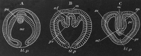
Fig. 408. Three stages in the development of Sagitta. (A and C
after Bütschli, and B after Kowalevsky.)
The three embryos are represented in the same positions.
A. Represents the gastrula stage.
B. Represents a succeeding stage, in which the primitive archenteron
is commencing to be divided into three.
C. Represents a later stage, in which the mouth involution (m) has
become continuous with the alimentary tract, and the blastopore is
closed.
m. mouth; al. alimentary canal; ae. archenteron; bl.p.
blastopore; pv. perivisceral cavity; sp. splanchnic mesoblast;
so. somatic mesoblast; ge. generative organs.
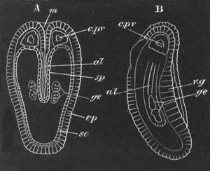
Fig. 409. Two views of a late embryo of Sagitta. A, from the dorsal
surface. B, from the side. (After Bütschli.)
m. mouth; al. alimentary canal; v.g. ventral ganglion
(thickening of epiblast); ep. epiblast; c.pv. cephalic section
of body cavity; so. somatopleure; sp. splanchnopleure; ge.
generative organs.
When the archenteric cavity is divided into a median alimentary tract,
and two lateral sections forming the body cavity, the generative
organs are placed in the common vestibule into which both the body
cavity and alimentary cavity at first open (fig. 408).
The generative organs long retain their character as simple cells.
Eventually (fig. 409) the two ovaries travel forwards, and apply
themselves to the body walls, while the two testes also become
separated by a backward prolongation of the median alimentary tract.
On the formation of the transverse septum dividing the tail from the
body, the ovarian cells lie immediately in front of this septum, and
the testicular cells in the region behind it.
Polyzoa. In Pedicellina amongst the entoproctous Polyzoa Hatschek
finds that the generative organs originate from a pair of specially
large mesoblast cells, situated in the space between the stomach and
the floor of the vestibule. The two cells undergo changes, which have
an obvious resemblance to those of the generative cells of the
Chætognatha. They become surrounded by an investment of mesoblast
cells, and divide so as to form two masses. Each of these masses at a
later period separates into an anterior and a posterior part. The
former becomes the ovary, the latter the testis.
Nematoda. In the Nematoda the generative organs are derived from the
division of a single cell which would appear to be mesoblastic[271].
Insecta. The generative cells have been observed at a very early
embryonic stage in several insect forms (Vol. II. p. 404), but the
observations so far recorded with reference to them do not enable us
to determine with certainty from which of the germinal layers they are
derived.
Crustacea. In Moina, one of the Cladocera, Grobben[272]
has shewn that
the generative organs are derived from a single cell, which becomes
differentiated during the segmentation. This cell, which is in close
contiguity with the cells from which both the mesoblast and hypoblast
originate, subsequently divides;
but at the gastrula stage, and after
the mesoblast has become formed, the cells it gives rise to are
enclosed in the epiblast, and do not migrate inwards till a later
stage. The products of the division of the generative cell
subsequently divide into two masses. It is not possible to assign the
generative cell of Moina to a definite germinal layer. Grobben,
however, thinks that it originates from the division of a cell, the
remainder of which gives rise to the hypoblast.
Chordata. In the Vertebrata, the primitive generative cells (often
known as primitive ova) are early distinguishable, being imbedded
amongst the cells of two linear streaks of peritoneal epithelium,
placed on the dorsal side of the body cavity, one on each side of the
mesentery (figs. 405 C and 410, po). They appear to be derived from
the epithelial cells amongst which they lie; and are characterized by
containing a large granular nucleus, surrounded by a considerable body
of protoplasm. The peritoneal epithelium in which they are placed is
known as the germinal epithelium.
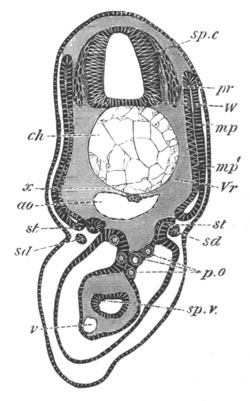
Fig. 410. Section through the trunk of a Scyllium embryo slightly
younger than 28 F.
sp.c. spinal cord; W. white matter of spinal cord; pr.
posterior nerve-roots; ch. notochord; x. subnotochordal rod;
ao. aorta; mp. muscle-plate; mp´. inner layer of muscle-plate
already converted into muscles; Vr. rudiment of vertebral body;
st. segmental tube; sd. segmental duct; sp.v. spiral valve;
v. subintestinal vein; p.o. primitive generative cells.
It is at first impossible to distinguish the germinal cells which will
become ova from those which will become spermatozoa.
The former however remain within the peritoneal epithelium (fig. 411),
and become converted into ova in a manner more particularly described
in Vol. II. pp. 54-59.
The history of the primitive germinal cells in the male has not been
so adequately worked out as in the female.
The fullest history of them is that given by Semper (No. 559) for the
Elasmobranchii, the general accuracy of which I can fully support;
though with reference to certain stages in the history further
researches are still required[273].
In Elasmobranchii the male germinal cells, instead of remaining in the
germinal epithelium, migrate into the adjacent stroma, accompanied I
believe by some of the indifferent epithelial cells. Here they
increase in number, and give rise to masses of variable form, composed
partly of true germinal cells, and partly of smaller cells with deeply
staining nuclei, which are, I believe, derived from the germinal
epithelium.
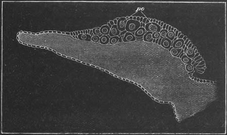
Transverse section through the ovary of a young embryo of
scyllium canicula, to shew the primitive germinal cells (po) lying
in the germinal epithelium on the outer side of the ovarian ridge.
These masses next break up into ampullæ, mainly formed of germinal
cells, and each provided with a central lumen; and these ampullæ
attach themselves to tubes derived from the smaller cells, which are
in their turn continuous with the testicular network. The spermatozoa
are developed from the cells forming the walls of the primitive
ampullæ; but the process of their formation does not concern us in
this chapter.
In the Reptilia Braun has traced the passage of the primitive germinal
cells into the testicular tubes, and I am able to confirm his
observations on this point: he has not however traced their further
history.
In Mammalia the evidence of the origin of the spermatospores from the
germinal epithelium is not quite complete, but there can be but little
doubt of its occurrence[274].
In Amphioxus Langerhans has shewn that the ova and spermatozoa are
derived from similar germinal cells, which may be compared to the
germinal epithelium of the Vertebrata. These cells are however
segmentally arranged as separate masses (vide Vol. II. p. 54).
Bibliography.
(554) G. Balbiani. Leçons s. la génération des Vertébrés. Paris,
1879.
(555) F. M. Balfour. “On the structure and development of the
Vertebrate ovary.” Quart. J. of Micr. Science, Vol. XVIII.
(556) E. van Beneden. “De la distinction originelle du tecticule et de
l'ovaire, etc.” Bull. Ac. roy. belgique, Vol. XXXVII. 1874.
(557) N. Kleinenberg. “Ueb. d. Entstehung d. Eier b. Eudendrium.”
Zeit. f. wiss. Zool., Vol. XXXV. 1881.
(558) H. Ludwig. “Ueb. d. Eibildung im Theirreiche.” Arbeit. a. d.
zool.-zoot. Instit. Würzburg, Vol. I. 1874.
(559) C. Semper. “Das Urogenitalsystem d. Plagiostomen, etc.” Arbeit.
a. d. zool.-zoot. Instit. Würzburg, Vol. II. 1875.
(560) A. Weismann. “Zur Frage nach dem Ursprung d. Geschlechtszellen
bei den Hydroiden.” Zool. Anzeiger, No. 55, 1880.
Vide also O. and R. Hertwig (No. 271), Kölliker (No. 298), etc.
Genital ducts.
The development and evolution of the generative ducts is as yet very
incompletely worked out, but even in the light of our present
knowledge a comparative review of this subject brings to light
features of considerable interest, and displays a fruitful field for
future research.
In the Cœlenterata there are no generative ducts.
In the Hydromedusæ and Siphonophora the generative products are
liberated by being dehisced directly into the surrounding medium;
while in the Acraspeda, the Actinozoa and the Ctenophora, they are
dehisced into parts of the gastrovascular system, and carried to the
exterior through the mouth.
The arrangement in the latter forms indicates the origin of
the
methods of transportation of the genital products to the exterior in
many of the higher types.
It has been already pointed out that the body cavity in a very large
number of forms is probably derived from parts of a gastrovascular
system like that of the Actinozoa.
When the part of the gastrovascular system into which the generative
products were dehisced became, on giving rise to the body cavity, shut
off from the exterior, it would be essential that some mode of
transportation outwards of the generative products should be
constituted.
In some instances simple pores (probably already existing at the time
of the establishment of a closed body cavity) become the generative
ducts. Such seems probably to have been the case in the Chætognatha
(Sagitta) and in the primitive Chordata.
In the latter forms the generative products are sometimes dehisced
into the peritoneal cavity, and thence transported by the abdominal
pores to the exterior (Cyclostomata and some Teleostei, vide p.
626). In Amphioxus they pass by dehiscence into the atrial cavity, and
thence through the gill slits and by the mouth, or by the abdominal
pore (?) to the exterior. The arrangement in Amphioxus and the
Teleostei is probably secondary, as possibly also is that in the
Cyclostomata; so that the primitive mode of exit of the generative
products in the Chordata is still uncertain. It is highly improbable
that the generative ducts of the Tunicata are primitive structures.
A better established and more frequent mode of exit of the generative
products when dehisced into the body cavity is by means of the
excretory organs. The generative products pass from the body cavity
into the open peritoneal funnels of such organs, and thence through
their ducts to the exterior. This mode of exit of the generative
products is characteristic of the Chætopoda, the Gephyrea, the
Brachiopoda and the Vertebrata, and probably also of the Mollusca. It
is moreover quite possible that it occurs in the Polyzoa, some of the
Arthropoda, the Platyelminthes and some other types.
The simple segmental excretory organs of the Polychæta, the Gephyrea
and the Brachiopoda serve as generative canals, and in many instances
they exhibit no modification, or but a very slight one, in connection
with their secondary generative
function; while in other instances,
e.g. Bonellia, such modification is very considerable.
The generative ducts of the Oligochæta are probably derived from
excretory organs. In the Terricola ordinary excretory organs are
present in the generative segments in addition to the generative
ducts, while in the Limicola generative ducts alone are present in the
adult, but before their development excretory organs of the usual type
are found, which undergo atrophy on the appearance of the generative
ducts (Vedjovsky).
From the analogy of the splitting of the segmental duct of the
Vertebrata into the Müllerian and Wolffian ducts, as a result of a
combined generative and excretory function (vide p. 728), it seems
probable that in the generative segments of the Oligochæta the
excretory organs had at first both an excretory and a generative
function, and that, as a secondary result of this double function,
each of them has become split into two parts, a generative and an
excretory. The generative part has undergone in all forms great
modifications. The excretory parts remain unmodified in the Earthworms
(Terricola), but completely abort on the development of the generative
ducts in the Limicola. An explanation may probably be given of the
peculiar arrangements of the generative ducts in Saccocirrus amongst
the Polychæta (vide Marion and Bobretzky), analogous to that just
offered for the Oligochæta.
The very interesting modifications produced in the excretory organs of
the Vertebrata by their serving as generative ducts were fully
described in the last chapter; and with reference to this part of our
subject it is only necessary to call attention to the case of
Lepidosteus and the Teleostei.
In Lepidosteus the Müllerian duct appears to have become attached to
the generative organs, so that the generative products, instead of
falling directly into the body cavity and thence entering the open end
of a peritoneal funnel of the excretory organs, pass directly into the
Müllerian duct without entering the body cavity. In most Teleostei the
modification is more complete, in that the generative ducts in the
adult have no obvious connection with the excretory organs.
The transportation of the male products to the exterior in all the
higher Vertebrata, without passing into the body cavity, is in
principle similar to the arrangement in Lepidosteus.
The above instances of the peritoneal funnels of an excretory organ
becoming continuous with the generative glands, render it highly
probable that there may be similar instances amongst the Invertebrata.
As has been already pointed out by Gegenbaur there are many features
in the structure of the genital ducts in the more primitive Mollusca,
which point to their having been derived from the excretory organs. In
several Lamellibranchiata[275]
(Spondylus, Lima, Pecten) the
generative ducts open into the excretory organs (organ of Bojanus), so
that the generative products have to pass through the excretory organ
on their way to the exterior. In other Lamellibranchiata the genital
and excretory organs open on a common papilla, and in the remaining
types they are placed close together.
In the Cephalopoda again the peculiar relations of the generative
organs to their ducts point to the latter having primitively had a
different, probably an excretory, function. The glands are not
continuous with the ducts, but are placed in special capsules from
which the ducts proceed. The genital products are dehisced into these
capsules and thence pass into the ducts.
In the Gasteropoda the genital gland is directly continuous with its
duct, and the latter, especially in the Pulmonata and Opisthobranchiata,
assumes such a complicated form that its origin from the excretory
organ would hardly have been suspected. The fact however that its
opening is placed near that of the excretory organ points to its being
homologous with the generative ducts of the more primitive types.
In the Discophora, where the generative ducts are continuous with the
glands, the structure both of the generative glands and ducts points
to the latter having originated from excretory organs.
It seems, as already mentioned, very possible that there are other
types in which the generative ducts are derived from the excretory
organs. In the Arthropoda for instance the generative ducts, where
provided with anteriorly placed openings, as in the Crustacea,
Arachnida and the Chilognathous Myriapoda, the Pœcilopoda, etc., may
possibly be of this nature, but the data for deciding this point are
so scanty that it is not at present possible to do more than frame
conjectures.
The ontogeny of the generative ducts of the Nematoda and
the Insecta
appears to point to their having originated independently of the
excretory organs.
In the Nematoda the generative organs of both sexes originate from a
single cell (Schneider, Vol. I. No. 390).
This cell elongates and its nuclei multiply. After assuming a somewhat
columnar form, it divides into (1) a superficial investing layer, and
(2) an axial portion.
In the female the superficial layer is only developed distinctly in
the median part of the column. In the course of the further
development the two ends of the column become the blind ends of the
ovary, and the axial tissue they contain forms the germinal tissue of
nucleated protoplasm. The superficial layer gives rise to the
epithelium of the uterus and oviduct. The germinal tissue, which is
originally continuous, is interrupted in the middle part (where the
superficial layer gives rise to the uterus and oviduct), and is
confined to the two blind extremities of the tube.
In the male the superficial layer, which gives rise to the epithelium
of the vas deferens, is only formed at the hinder end of the original
column. In other respects the development takes place as in the
female.
In the Insecta again the evidence, though somewhat conflicting,
indicates that the generative ducts arise very much as in Nematodes,
from the same primitive mass as the generative organs. In both of
these types it would seem probable that the generative organs were
primitively placed in the body cavity, and attached to the epidermis,
through a pore in which their products passed out; and that, acquiring
a tubular form, the peripheral part of the gland gave rise to a duct,
the remainder constituting the true generative gland. It is quite
possible that the generative ducts of such forms as the Platyelminthes
may have had a similar origin to those in Insecta and Nematoda, but
from the analogy of the Mollusca there is nearly as much to be said
for regarding them as modified excretory organs.
In the Echinodermata nothing is unfortunately known as to the ontogeny
of the generative organs and ducts. The structure of these organs in
the adult would however seem to indicate that the most primitive type
of echinoderm generative organ consists of a blind sack, projecting
into the body cavity, and opening by
a pore to the exterior. The sack
is lined by an epithelium, continuous with the epidermis, the cells of
which give rise to the ova or spermatozoa. The duct of these organs is
obviously hardly differentiated from the gland; and the whole
structure might easily be derived from the type of generative organ
characteristic of the Hydromedusæ, where the generative cells are
developed from special areas of the ectoderm, and, when ripe, pass
directly into the surrounding medium.
If this suggestion is correct we may suppose that the generative ducts
of the Echinodermata have a different origin to those of the majority
of[276]
the remaining triploblastica.
Their ducts have been evolved in forms in which the generative
products continued to be liberated directly to the exterior, as in the
Hydromedusæ; while those of other types have been evolved in forms in
which the generative products were first transported, as in the
Actinozoa, into the gastrovascular canals[277].
The alimentary canal in the Chordata is always formed of three
sections, analogous to those so universally present in the
Invertebrata. These sections are (1) the mesenteron lined by
hypoblast; (2) the stomodæum or mouth lined by epiblast, and (3) the
proctodæum or anal section lined like the stomodæum by epiblast.
Mesenteron.
The early development of the epithelial wall of the mesenteron has
already been described (Chapter XI.). It forms at first a simple
hypoblastic tube extending from near the front end of the body, where
it terminates blindly, to the hinder extremity where it is united with
the neural tube by the neurenteric canal (fig. 420, ne). It often
remains for a long time widely open in the middle towards the
yolk-sack.
It has already been shewn that from the dorsal wall of the mesenteron
the notochord is separated off nearly at the same time as the lateral
plates of mesoblast (pp. 292-300).
The subnotochordal rod. At a period slightly subsequent to the
formation of the notochord, and before any important differentiations
in the mesenteron have become apparent, a remarkable rod-like body,
which was first discovered by Götte, becomes split off from the dorsal
wall of the alimentary tract in all the Ichthyopsida. This body, which
has a purely provisional existence, is known as the subnotochordal
rod.
It develops in Elasmobranch embryos in two sections, one situated in
the head, and the other in the trunk.
The section in the trunk is the first to appear. The wall of the
alimentary canal becomes thickened along the median dorsal line (fig.
412, x), or else produced into a ridge into which there penetrates a
narrow prolongation of the lumen of the alimentary canal. In either
case the cells at the extreme summit become gradually constricted off
as a rod, which lies immediately dorsal to the alimentary tract, and
ventral to the notochord (fig. 413, x).
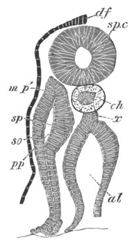
Fig. 412. Transverse section through the tail region of a
Pristiurus embryo of the same age as fig. 28 E.
df. dorsal fin; sp.c. spinal cord; pp. body cavity; sp.
splanchnic layer of mesoblast; so. somatic layer of mesoblast;
mp´. portion of splanchnic mesoblast commencing to be
differentiated into muscles; ch. notochord; x. subnotochordal
rod arising as an outgrowth of the dorsal wall of the alimentary
tract; al. alimentary tract.
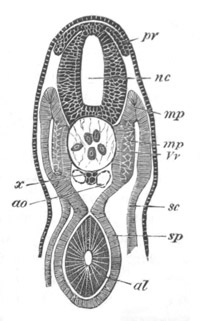
Fig. 413. Transverse section through the trunk of an embryo
slightly older than fig. 28 E.
nc. neural canal; pr. posterior root of spinal nerve; x.
subnotochordal rod; ao. aorta; sc. somatic mesoblast; sp.
splanchnic mesoblast; mp. muscle-plate; mp´. portion of
muscle-plate converted into muscle; Vv. portion of the vertebral
plate which will give rise to the vertebral bodies; al. alimentary
tract.
In the hindermost part of the body its mode of formation differs
somewhat from that above described. In this part the alimentary wall
is very thick, and undergoes no special growth prior to the formation
of the subnotochordal rod; on the contrary, a small linear portion of
the wall becomes scooped out along the median dorsal line, and
eventually separates from the remainder as the rod in question. In the
trunk the splitting off of the rod takes place from before backwards,
so that the anterior part of it is formed before the posterior.
The section of the subnotochordal rod in the head would appear to
develop in the same way as that in the trunk, and the splitting off
from the throat proceeds from before backwards.
On the formation of the dorsal aorta, the subnotochordal rod becomes
separated from the wall of the gut and the aorta interposed between
the two (fig. 367, x).
When the subnotochordal rod attains its fullest development it
terminates anteriorly some way in front of the auditory vesicle,
though a little behind the end of the notochord; posteriorly it
extends very nearly to the extremity of the tail and is almost
co-extensive with the postanal section of the alimentary tract,
though it does not reach quite so far back as the caudal vesicle (fig.
424, b x). Very shortly after it has attained its maximum size it
begins to atrophy in front. We may therefore conclude that its
atrophy, like its development, takes place from before backwards.
During the later embryonic stages not a trace of it is to be seen. It
has also been met with in Acipenser, Lepidosteus, the Teleostei,
Petromyzon, and the Amphibia, in all of which it appears to develop in
fundamentally the same way as in Elasmobranchii. In Acipenser it
appears to persist in the adult as the subvertebral ligament (Bridge,
Salensky). It has not yet been found in a fully developed form in any
amniotic Vertebrate, though a thickening of the hypoblast, which may
perhaps be a rudiment of it, has been found by Marshall and myself in
the Chick (fig. 110, x).
Eisig has instituted an interesting comparison between it and an organ
which he has found in a family of Chætopods, the Capitellidæ. In these
forms there is a tube underlying the alimentary tract for nearly its
whole length, and opening into it in front, and probably behind. A
remnant of such a tube might easily form a rudiment like the
subnotochordal rod of the Ichthyopsida, and as Eisig points out the
prolongation into the latter during its formation of the lumen of the
alimentary tract distinctly favours such a view of its original
nature. We can however hardly suppose that there is any direct genetic
connection between Eisig’s organ in the Capitellidæ and the
subnotochordal rod of the Chordata.
Splanchnic mesoblast and mesentery. The mesenteron consists at first
of a simple hypoblastic tube, which however becomes enveloped by a
layer of splanchnic mesoblast. This layer, which is not at first
continued over the dorsal side of the mesenteron, gradually grows in,
and interposes itself between the hypoblast of the mesenteron, and the
organs above. At the same time it becomes differentiated into two
layers, viz. an outer epithelioid layer which gives rise to part of
the peritoneal epithelium, and an inner layer of undifferentiated
cells which in time becomes converted into the connective tissue and
muscular walls of the mesenteron. The connective tissue layers become
first formed, while of the muscular layers the circular is the first
to make its appearance.
Coincidently with their differentiation the connective tissue stratum
of the peritoneum becomes established.
The Mesentery. Prior to the splanchnic mesoblast growing round the
alimentary tube above, the attachment of the latter structure to the
dorsal wall of the body is very wide. On the completion of this
investment the layer of mesoblast suspending the alimentary tract
becomes thinner, and at the same time the alimentary canal appears to
be drawn downwards and away from the vertebral column.
In what may be regarded as the thoracic division of the general
pleuroperitoneal space, along that part of the alimentary canal which
will form the œsophagus, this withdrawal is very slight, but it is
very marked in the abdominal region. In the latter the at first
straight digestive canal comes to be suspended from the body above by
a narrow flattened band of mesoblastic tissue. This flattened band is
the mesentery, shewn commencing in fig. 117, and much more advanced
in fig. 119, M. It is covered on either side by a layer of flat
cells, which form part of the general peritoneal epithelioid lining,
while its interior is composed of indifferent tissue.
The primitive simplicity in the arrangement of the mesentery is
usually afterwards replaced by a more complicated disposition, owing
to the subsequent elongation and consequent convolution of the
intestine and stomach.
The layer of peritoneal epithelium on the ventral side of the stomach
is continued over the liver, and after embracing the liver, becomes
attached to the ventral abdominal wall (fig. 380). Thus in the region
of the liver the body cavity is divided into two halves by a membrane,
the two sides of which are covered by the peritoneal epithelium, and
which encloses the stomach dorsally and the liver ventrally. The part
of the membrane between the stomach and liver is narrow, and
constitutes a kind of mesentery suspending the liver from the stomach:
it is known to human anatomists as the lesser omentum.
The part of the membrane connecting the liver with the anterior
abdominal wall constitutes the falciform or suspensory ligament of the
liver. It arises by a secondary fusion, and is not a remnant of a
primitive ventral mesentery (vide pp. 624 and 625).
The mesentery of the stomach, or mesogastrium, enlarges in Mammalia to
form a peculiar sack known as the greater omentum.
The mesenteron exhibits very early a trifold division. An anterior
portion, extending as far as the stomach, becomes separated off as the
respiratory division. On the formation of the anal invagination the
portion of the mesenteron behind the anus becomes marked off as the
postanal division, and between the postanal section and the
respiratory division is a middle portion forming an intestinal and
cloacal division.
The respiratory division of the mesenteron.
This section of the alimentary canal is distinguished by the fact that
its walls send out a series of paired diverticula, which meet the
skin, and after a perforation has been effected at the regions of
contact, form the branchial or visceral clefts.
In Amphioxus the respiratory region extends close up to the opening of
the hepatic diverticulum, and therefore to a position corresponding
with the commencement of the intestine in higher types. In the
craniate Vertebrata the number of visceral clefts has become reduced,
but from the extension of the visceral clefts in Amphioxus, combined
with the fact that in the higher Vertebrata the vagus nerve, which is
essentially the nerve of the branchial pouches, supplies in addition
the walls of the œsophagus and stomach, it may reasonably be
concluded, as has been pointed out by Gegenbaur, that the true
respiratory region primitively included the region which in the higher
types forms the œsophagus and stomach.
In Ascidians the respiratory sack is homologous with the respiratory
tract of Amphioxus.
The details of the development of the branchial clefts in the
different groups of Vertebrata have already been described in the
systematic part of this work.
In all the Ichthyopsida the walls of a certain number of clefts become
folded; and in the mesoblast within these folds a rich capillary
network, receiving its blood from the branchial arteries, becomes
established. These folds constitute the true internal gills.
In addition to internal gills external branchial processes covered
by epiblast are placed on certain of the visceral arches in the larva
of Polypterus, Protopterus and many Amphibia. The external gills have
probably no genetic connection with the internal gills.
The so-called external gills of the embryos of Elasmobranchii are
merely internal gills prolonged outwards through the gill clefts.
The posterior part of the primitive respiratory division of the
mesenteron becomes, in all the higher Vertebrata, the œsophagus and
stomach. With reference to the development of these parts the only
point worth especially noting is the fact that in Elasmobranchii and
Teleostei their lumen, though present in very young embryos, becomes
at a later stage completely filled up, and thus the alimentary tract
in the regions of the œsophagus and stomach becomes a solid cord of
cells (fig. 23 A, œs): as already suggested (p. 61) it seems not
impossible that this feature may be connected with the fact that the
œsophageal region of the throat was at one time perforated by gill
clefts.
In addition to the gills two important organs, viz. the thyroid body
and the lungs, take their origin from the respiratory region of the
alimentary tract.
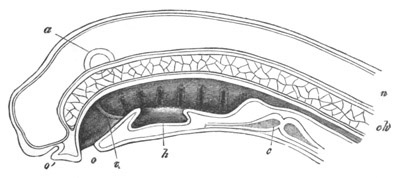
Fig. 414. Diagrammatic vertical section of a just-hatched larva
of Petromyzon. (From Gegenbaur; after Calberla.)
o. mouth; o´. olfactory pit; v. septum between stomodæum and
mesenteron; h. thyroid involution; n. spinal cord; ch.
notochord; c. heart; a. auditory vesicle.
Thyroid body. In the Ascidians the origin of a groove-like
diverticulum of the ventral wall of the branchial sack, bounded by two
lateral folds, and known as the endostyle or hypopharyngeal groove,
has already been described (p. 18). This groove remains permanently
open to the pharyngeal sack,
and would seem to serve as a glandular
organ secreting mucus. As was first pointed out by W. Müller there is
present in Amphioxus a very similar and probably homologous organ,
known as the hypopharyngeal groove.
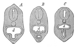
Fig. 415. Diagrammatic transverse sections through the branchial
region of young larvæ of Petromyzon. (From Gegenbaur; after
Calberla.)
d. branchial region of throat.
In the higher Vertebrata this organ never retains its primitive
condition in the adult state. In the larva of Petromyzon there is,
however, present a ventral groove-like diverticulum of the throat,
extending from about the second to the fourth visceral cleft. This
organ is shewn in longitudinal section in fig. 414, h, and in
transverse section in fig. 415, and has been identified by W. Müller
(Nos. 565 and 566) with the hypopharyngeal groove of Amphioxus and
Ascidians. It does not, however, long retain its primitive condition,
but its opening becomes gradually reduced to a pore, placed between
the third and fourth of the permanent clefts (fig. 416, th). This
opening is retained throughout the Ammocœte condition, but the organ
becomes highly complicated, with paired anterior and posterior horns
and a median spiral portion. In the adult the connection with the
pharynx is obliterated, and the organ is partly absorbed and partly
divided up into a series of glandular follicles, and eventually forms
the thyroid body.
From the consideration of the above facts W. Müller was led to the
conclusion that the thyroid body of the Craniata was derived from the
endostyle or hypopharyngeal groove. In all the higher Vertebrata the
thyroid body arises as a diverticulum of the ventral wall of the
throat in the region either of the mandibular or hyoid arches (fig.
417, Th), which after being segmented off becomes divided up into
follicles.
In Elasmobranch embryos it appears fairly early as a diverticulum from
the ventral surface of the throat in the region of the mandibular
arch, extending from the border of the mouth to the point where the
ventral aorta divides into the two aortic branches of the mandibular
arch (fig. 417, Th).
Somewhat later it becomes in Scyllium and
Torpedo solid, though still retaining its attachment to the wall of
the œsophagus. It continues to grow in length, and becomes divided up
into a number of solid branched lobules separated by connective tissue
septa. Eventually its connection with the throat becomes lost, and the
lobules develop a lumen. In Acanthias the lumen of the gland is
retained (W. Müller) till after its detachment from the throat. It
preserves its embryonic position through life. In Amphibia it
originates, as in Elasmobranchii, from the region of the mandibular
arch; but when first visible it forms a double epithelial wall
connecting the throat with the nervous layer of the epidermis. It
subsequently becomes detached from the epidermis, and then has the
usual form of a diverticulum from the throat. In most Amphibians it
becomes divided into two lobes, and so forms a paired body. The
peculiar connection between the thyroid diverticulum and the epidermis
in Amphibia has been noted by Götte in Bombinator, and by Scott and
Osborn in Triton. It is not very easy to see what meaning this
connection can have.
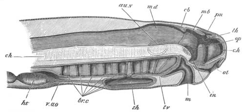
Fig. 416. Diagrammatic vertical section through the head of a
larva of Petromyzon.
The larva had been hatched three days, and was 4.8 mm. in length.
The optic and auditory vesicles are supposed to be seen through the
tissues. The letter tv pointing to the base of the velum is where
Scott believes the hyomandibular cleft to be situated.
c.h. cerebral hemisphere; th. optic thalamus; in.
infundibulum; pn. pineal gland; mb. mid-brain; cb. cerebellum;
md. medulla oblongata; au.v. auditory vesicle; op. optic
vesicle; ol. olfactory pit; m. mouth; br.c. branchial pouches;
th. thyroid involution; v.ao. ventral aorta; ht. ventricle of
heart; ch. notochord.
In the Fowl (W. Müller) the thyroid body arises at the end of the
second or beginning of the third day as an outgrowth from the
hypoblast of the throat, opposite the point of origin of the anterior
arterial arch. This outgrowth becomes by the fourth day a solid mass
of cells, and by the fifth ceases to be connected with the epithelium
of the throat, becoming at the same time bilobed. By the seventh day
it has travelled somewhat backwards, and the two lobes have completely
separated from each other. By
the ninth day the whole is invested by a
capsule of connective tissue, which sends in septa dividing it into a
number of lobes or solid masses of cells, and by the sixteenth day it
is a paired body composed of a number of hollow branched follicles,
each with a ‘membrana propria,’ and separated from each other by septa
of connective tissue. It finally travels back to the point of origin
of the carotids.
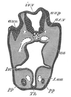
Fig. 417. Section through the head of an Elasmobranch embryo, at
the level of the auditory involution.
Th. rudiment of thyroid body; aup. auditory pit; aun. ganglion
of auditory nerve; iv.v. roof of fourth ventricle; a.c.v.
anterior cardinal vein; aa. aorta; I.aa. aortic trunk of
mandibular arch; pp. head cavity of mandibular arch; Ivc.
alimentary pouch which will form the first visceral cleft.
Amongst Mammalia the thyroid arises in the Rabbit (Kölliker) and Man
(His) as a hollow diverticulum of the throat at the bifurcation of the
foremost pair of aortic arches. It soon however becomes solid, and is
eventually detached from the throat and comes to lie on the ventral
side of the larynx or windpipe. The changes it undergoes are in the
main similar to those in the lower Vertebrata. It becomes partially
constricted into two lobes, which remain however united by an
isthmus[278].
The fact that the thyroid sometimes arises in the region
of the first and sometimes in that of the second cleft is probably to
be explained by its rudimentary character.
The Thymus gland. The thymus gland may conveniently be dealt with
here, although its origin is nearly as obscure as its function. It has
usually been held to be connected with the lymphatic system. Kölliker
was the first to shew that this view was probably erroneous, and he
attempted to prove that it was derived in the Rabbit from the walls of
one of the visceral clefts, mainly on the ground of its presenting in
the embryo an epithelial character.
Stieda (No. 569) has recently verified Kölliker’s statements. He finds
that in the Pig and the Sheep the thymus arises as a paired outgrowth
from the epithelial remnants of a pair of visceral clefts. Its two
lobes may at first be either hollow (Sheep) or solid (Pig), but
eventually become solid, and unite in the median line. Stieda and His
hold that in the adult gland, the so-called corpuscles of Hassall are
the remnants of the embryonic epithelial part of the gland, and that
the lymphatic part of it is of mesoblastic origin; but Kölliker
believes the lymphatic cells to be direct products of the embryonic
epithelial cells.
The posterior visceral clefts in the course of their atrophy give rise
to various more or less conspicuous bodies of a pseudo-glandular
nature, which have been chiefly studied by Remak[279].
Swimming bladder and lungs. A swimming bladder is present in all
Ganoids and in the vast majority of Teleostei. Its development however
is only imperfectly known.
In the Salmon and Carp it arises, as was first shewn by Von Baer, as
an outgrowth of the alimentary tract, shortly in front of the liver.
In these forms it is at first placed on the dorsal side and slightly
to the right, and grows backwards on the dorsal side of the gut,
between the two folds of the mesentery.
The absence of a pneumatic duct in the Physoclisti would appear to be
due to a post-larval atrophy.
In Lepidosteus the air-bladder appears to arise, as in the Teleostei,
as an invagination of the dorsal wall of the œsophagus.
In advanced embryos of Galeus, Mustelus and Acanthias, Miklucho-Maclay
detected a small diverticulum opening on the dorsal side of the
œsophagus, which he regards as a rudiment of a swimming bladder. This
interpretation must however be regarded as somewhat doubtful.
The lungs. The lungs originate in a nearly identical way in all the
Vertebrate forms in which their development has been observed. They
are essentially buds or processes of the ventral wall of the primitive
œsophagus.
At a point immediately behind the region of the visceral clefts the
cavity of the alimentary canal becomes compressed laterally, and at
the same time constricted in the middle, so that its transverse
section (fig. 418 1) is somewhat hourglass-shaped, and shews an upper
or dorsal chamber d, joining on to a lower or ventral chamber l by
a short narrow neck.
The hinder end of the lower tube enlarges (fig. 418 2), and then
becomes partially divided into two lobes (fig. 418 3). All these parts
at first freely communicate, but the two lobes, partly by their own
growth, and partly by a process of constriction, soon become isolated
posteriorly; while in front they open into the lower chamber of the
œsophagus (fig. 422).
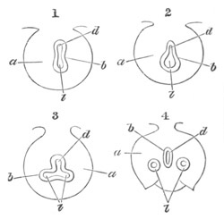
Fig. 418. Four diagrams illustrating the formation of the
Lungs. (After Götte.)
a. mesoblast; b. hypoblast; d. cavity of digestive canal; l.
cavity of the pulmonary diverticulum.
In (1) the digestive canal has commenced to be constricted into an
upper and lower canal; the former the true alimentary canal, the
latter the pulmonary tube; the two tubes communicate with each
other in the centre.
In (2) the lower (pulmonary) tube has become expanded.
In (3) the expanded portion of the tube has become constricted into
two tubes, still communicating with each other and with the
digestive canal.
In (4) these are completely separated from each other and from the
digestive canal, and the mesoblast has also begun to exhibit
externally changes corresponding to the internal changes which
have been going on.
By a continuation forwards of the process of constriction the lower
chamber of the œsophagus, carrying with it the two lobes above
mentioned, becomes gradually transformed into an independent tube,
opening in front by a narrow slit-like aperture into the œsophagus.
The single tube in front is the rudiment of the trachea and larynx,
while the two diverticula behind become (fig. 419, lg) the bronchial
tubes and lungs.
While the above changes are taking place in the hypoblastic walls of
the alimentary tract, the splanchnic mesoblast surrounding these
structures becomes very much thickened; but otherwise bears no marks
of the internal changes which are going on, so that the above
formation of the lungs and trachea cannot be seen from the surface. As
the paired diverticula of the lungs grow backwards, the mesoblast
around them takes however the form of two lobes, into which they
gradually bore their way.
There do not seem to be any essential differences in the mode of
formation of the above structures in the types so far observed, viz.
Amphibia, Aves and Mammalia. Writers differ as to whether the lungs
first arise as
paired diverticula, or as a single diverticulum; and as
to whether the rudiments of the lungs are established before those of
the trachea. If the above account is correct it would appear that any
of these positions might be maintained. Phylogenetically interpreted
the ontogeny of the lungs appears however to imply that this organ was
first an unpaired structure and has become secondarily paired, and
that the trachea was relatively late in appearing.
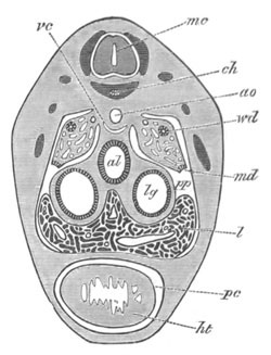
Fig. 419. Section through the cardiac region of an embryo of
Lacerta Muralis of 9 mm. to shew the mode of formation of the
pericardial cavity.
ht. heart; pc. pericardial cavity; al. alimentary tract; lg.
lung; l. liver; pp. body cavity; md. open end of Müllerian
duct; wd. Wolffian duct; vc. vena cava inferior; ao. aorta;
ch. notochord; mc. medullary cord.
The further development of the lungs is at first, in the higher types
at any rate, essentially similar to that of a racemose gland. From
each primitive diverticulum numerous branches are given off. In Aves
and Mammalia (fig. 355) they are mainly confined to the dorsal and
lateral parts. These branches penetrate into the surrounding mesoblast
and continue to give rise to secondary and tertiary branches. In the
mesoblast around them numerous capillaries make their appearance, and
the further growth of the bronchial tubes is supposed by Boll to be
due to the mutual interaction of the hitherto passive mesoblast and of
the hypoblast.
The further changes in the lungs vary somewhat in the different forms.
The air sacks are the most characteristic structures of the avian
lung. They are essentially the dilated ends of the primitive
diverticula or of their main branches.
In Mammalia (Kölliker, No. 298) the ends of the bronchial tubes become
dilated into vesicles, which may be called the primary air-cells. At
first, owing to their development at the ends of the bronchial
branches, these are confined to the surface of the lungs. At a later
period the primary air-cells divide each into two or three parts, and
give rise to secondary air-cells, while at the same time the smallest
bronchial tubes, which continue all the while to divide, give rise at
all points to fresh air-cells. Finally the bronchial tubes cease to
become more branched, and the air-cells belonging to each minute lobe
come in their further growth to open into a common chamber.
Before the
lungs assume their function the embryonic air-cells undergo a
considerable dilatation.
The trachea and larynx. The development of the trachea and larynx
does not require any detailed description. The larynx is formed as a
simple dilatation of the trachea. The cartilaginous structures of the
larynx are of the same nature as those of the trachea.
It follows from the above account that the whole pulmonary structure
is the result of the growth by budding of a system of branched
hypoblastic tubes in the midst of a mass of mesoblastic tissue, the
hypoblastic elements giving rise to the epithelium of the tubes, and
the mesoblast providing the elastic, muscular, cartilaginous,
vascular, and other connective tissues of the tracheal and bronchial
walls.
There can be no doubt that the lungs and air-bladder are homologous
structures, and the very interesting memoir of Eisig on the
air-bladder of the Chætopoda[280]
shews it to be highly probable that
they are the divergent modifications of a primitive organ, which
served as a reservoir for gas secreted in the alimentary tract, the
gas in question being probably employed for respiration when, for any
reason, ordinary respiration by the gills was insufficient.
Such an organ might easily become either purely respiratory, receiving
its air from the exterior, and so form a true lung; or mainly
hydrostatic, forming an air-bladder, as in Ganoidei and Teleostei.
It is probable that in the Elasmobranchii the air-bladder has become
aborted, and the organ discovered by Micklucho-Maclay may perhaps be a
last remnant of it.
The middle division of the mesenteron. The middle division of the
mesenteron, forming the intestinal and cloacal region, is primitively
a straight tube, the intestinal region of which in most Vertebrate
embryos is open below to the yolk-sack.
Cloaca. In the Elasmobranchii, the embryos of which probably retain a
very primitive condition of the mesenteron, this region is not at
first sharply separated from the postanal section behind. Opposite the
point where the anus will eventually
appear a dilatation of the
mesenteron arises, which comes in contact with the external skin (fig.
28 E, an). This dilatation becomes the hypoblastic section of the
cloaca. It communicates behind with the postanal gut (fig. 424 D), and
in front with the intestine; and may be defined as the dilated
portion of the alimentary tract which receives the genital and urinary
ducts and opens externally by the proctodæum.
In Acipenser and Amphibia the cloacal region is indicated as a ventral
diverticulum of the mesenteron even before the closure of the
blastopore. It is shewn in the Amphibia at an early stage in fig. 73,
and at a later period, when in contact with the skin at the point
where the anal invagination is about to appear, in fig. 420.
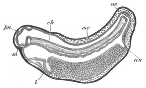
Fig. 420. Longitudinal section through an advanced embryo of
Bombinator. (After Götte.)
m. mouth; an. anus; l. liver; ne. neurenteric canal; mc.
medullary canal; ch. notochord; pn. pineal gland.
In the Sauropsida and Mammalia the cloaca appears as a dilatation of
the mesenteron, which receives the opening of the allantois almost as
soon as the posterior part of the mesenteron is established.
The eventual changes which it undergoes have been already dealt with
in connection with the urinogenital organs.
Intestine. The region in front of the cloaca forms the intestine. In
certain Vertebrata it nearly retains its primitive character as a
straight tube; and in these types its anterior part is characterised
by the presence of a peculiar fold, which in a highly specialised
condition is known as the spiral valve. This structure appears in its
simplest form in Ammocœtes. It
there consists of a fold in the wall
of the intestine, giving to the lumen of this canal a semilunar form
in section, and taking a half spiral.
In Elasmobranchii a similar fold to that in Ammocœtes first makes its
appearance in the embryo. This fold is from the first not quite
straight, but winds in a long spiral round the intestine. In the
course of development it becomes converted into a strong ridge
projecting into the lumen of the intestine (fig. 388, l). The spiral
it makes becomes much closer, and it thus acquires the form of the
adult spiral valve. A spiral valve is also found in Chimæra and
Ganoids. No rudiment of such an organ is found in the Teleostei, the
Amphibia, or the higher Vertebrata.
The presence of this peculiar organ appears to be a very primitive
Vertebrate character. The intestine of Ascidians exhibits exactly the
same peculiarity as that of Ammocœtes, and we may probably conclude
from embryology that the ancestral Chordata were provided with a
straight intestine having a fold projecting into its lumen, to
increase the area of the intestinal epithelium.
In all forms in which there is not a spiral valve, with the exception
of a few Teleostei, the intestine becomes considerably longer than the
cavity which contains it, and therefore necessarily more or less
convoluted.
The posterior part usually becomes considerably enlarged to form the
rectum or in Mammalia the large intestine.
In Elasmobranchii there is a peculiar gland opening into the dorsal
side of the rectum, and in many other forms there is a cæcum at the
commencement of the rectum or of the large intestine.
In Teleostei, the Sturgeon and Lepidosteus there opens into the front
end of the intestine a number of cæcal pouches known as the pancreatic
cæca. In the adult Sturgeon these pouches unite to form a compact
gland, but in the embryo they arise as a series of isolated outgrowths
of the duodenum.
Connected with the anterior portion of the middle region of the
alimentary canal, which may be called the duodenum, are two very
important and constant glandular organs, the liver and the pancreas.
The liver. The liver is the earliest formed and largest glandular
organ in the embryo.
It appears in its simplest form in Amphioxus as a single unbranched
diverticulum of the alimentary tract, immediately behind the
respiratory region, which is directed forwards and placed on the left
side of the body.
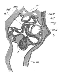
Fig. 421. Section through the ventral part of the trunk of a young
embryo of Scyllium at the level of the umbilical cord.
b. pectoral fin; ao. dorsal aorta; cav. cardinal vein; ua.
vitelline artery; uv. vitelline vein united with subintestinal
vein; al. duodenum; l. liver; sd. opening of segmental duct
into the body-cavity; mp. muscle-plate; um. umbilical canal.
In all true Vertebrata the gland has a much more complicated
structure. It arises as a ventral outgrowth of the duodenum (fig. 420,
l). This outgrowth may be at first single, and then grow out into
two lobes, as in Elasmobranchii (fig. 421) and Amphibia, or have from
the first the form of two somewhat unequal diverticula, as in Birds
(fig. 422), or again as in the Rabbit (Kölliker) one diverticulum may
be first formed, and a second one appear somewhat later. The hepatic
diverticula, whatever may be their primitive form, grow into a special
thickening of the splanchnic mesoblast.
From the primitive diverticula there are soon given off a number of
hollow buds (fig. 421) which rapidly increase in length and number,
and form the so-called hepatic cylinders. They soon anastomose and
unite together, and so constitute an irregular network. Coincidently
with the formation of the hepatic network the united vitelline and
visceral vein or veins (u.v), in their passage through the liver,
give off numerous branches, and gradually break up into a plexus of
channels which form a secondary network amongst the hepatic cylinders.
In Amphibia these channels are stated by Götte to be lacunar, but in
Elasmobranchii, and probably Vertebrata generally, they are from the
first provided with distinct though delicate walls.
It is still doubtful whether the hepatic cylinders are as a rule
hollow or solid. In Elasmobranchii they are at first provided with a
large lumen, which though it becomes gradually smaller never entirely
vanishes. The same seems to hold good for Amphibia and some Mammalia.
In Aves the lumen of the cylinders is even from the first much more
difficult to see, and the cylinders are stated by Remak to be solid,
and he has been followed in this matter by Kölliker. In the Rabbit
also Kölliker finds the cylinders to be solid.
The embryonic hepatic network gives rise to the parenchyma of the
adult liver, with which in its general arrangement it closely agrees.
The blood-channels are at first very large, and have a very irregular
arrangement; and it is not till comparatively late that the hepatic
lobules with their characteristic vascular structures become
established.
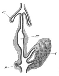
Fig. 422. Diagram of the digestive tract of a Chick upon the
fourth day. (After Götte.)
The black line indicates the hypoblast. The shaded part around it is
the splanchnic mesoblast.
lg. lung; st. stomach; p. pancreas; l. liver.
The biliary ducts are formed either from some of the primitive hepatic
cylinders, or, as would seem to be the case in Elasmobranchii and
Birds (fig. 422), from the larger diverticula of the two primitive
outgrowths.
The gall-bladder is so inconstant, and the arrangement of the ducts
opening into the intestine so variable, that no general statements can
be made about them. In Elasmobranchii the primitive median
diverticulum (fig. 421) gives rise to the ductus choledochus. Its
anterior end dilates to form a gall-bladder.
In the Rabbit a ductus choledochus is formed by a diverticulum from
the intestine at the point of insertion of the two primitive lobes.
The gall-bladder arises as a diverticulum of the right primitive lobe.
The liver is relatively very large during embryonic life and has, no
doubt, important functions in connection with the circulation.
The pancreas. So far as is known the development of the pancreas takes
place on a very constant type throughout the series of craniate
Vertebrata, though absent in some of the Teleostean fishes and
Cyclostomata, and very much reduced in most Teleostei and in
Petromyzon.
It arises nearly at the same time as the liver in the form of a hollow
outgrowth from the dorsal side of the intestine nearly opposite but
slightly behind the hepatic outgrowth (fig. 422, p). It soon
assumes, in Elasmobranchii and Mammalia, somewhat the form of an
inverted funnel, and from the expanded dorsal part of the funnel there
grow out numerous hollow diverticula into the passive splanchnic
mesoblast.
As the ductules grow longer and become branched, vascular processes
grow in between them, and the whole forms a compact glandular body in
the mesentery on the dorsal side of the alimentary tract. The
funnel-shaped receptacle loses its original form, and elongating,
assumes the character of a duct.
From the above mode of development it is clear that the glandular
cells of the pancreas are derived from the hypoblast.
Into the origin of the varying arrangements of the pancreatic ducts it
is not possible to enter in detail. In some cases, e.g. the Rabbit
(Kölliker), the two lobes and ducts arise from a division of the
primitive gland and duct. In other cases, e.g. the Bird, a second
diverticulum springs from the alimentary tract. In a large number of
instances the primitive condition with a single duct is retained.
Postanal section of the mesenteron. In the embryos of all the Chordata
there is a section of the mesenteron placed behind the anus. This
section invariably atrophies at a comparatively early period of
embryonic life; but it is much better developed in the lower forms
than in the higher. At its posterior extremity it is primitively
continuous with the neural tube (fig. 420), as was first shewn by
Kowalevsky.
The canal connecting the neural and alimentary canals has already been
described as the neurenteric canal, and represents the remains of the
blastopore.
In the Tunicata the section of the mesenteron, which in all
probability corresponds to the postanal gut of the Vertebrata, is that
immediately
following the dilated portion which gives rise to the
branchial cavity and permanent intestine. It has already been shewn
that from the dorsal and lateral portions of this section of the
primitive alimentary tract the notochord and muscles of the Ascidian
tadpole are derived. The remaining part of its walls forms a solid
cord of cells (fig. 423, al´), which either atrophies, or, according
to Kowalevsky, gives rise to blood-vessels.
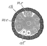
Fig. 423. Transverse optical section of the tail of an embryo of
Phallusia mammillata. (After Kowalevsky.)
The section is from an embryo of the same age as fig. 8 IV.
ch. notochord; n.c. neural canal; me. mesoblast; al´.
hypoblast of tail.
In Amphioxus the postanal gut, though distinctly developed, is not
very long, and atrophies at a comparatively early period.
In Elasmobranchii this section of the alimentary tract is very well
developed, and persists for a considerable period of embryonic life.
The following is a history of its development in the genus Scyllium.
Shortly after the stage when the anus has become marked out by the
alimentary tract sending down a papilliform process towards the skin,
the postanal gut begins to develop a terminal dilatation or vesicle,
connected with the remainder of the canal by a narrower stalk.
The walls both of the vesicle and stalk are formed of a fairly
columnar epithelium. The vesicle communicates in front by a narrow
passage with the neural canal, and behind is continued into two horns
corresponding with the two caudal swellings previously spoken of (p.
55). Where the canal is continued into these two horns, its walls lose
their distinctness of outline, and become continuous with the adjacent
mesoblast.
In the succeeding stages, as the tail grows longer and longer, the
postanal section of the alimentary tract grows with it, without
however undergoing alteration in any of its essential characters. At
the period of the maximum development, it has a length of about 1⁄3 of
that of the whole alimentary tract.
Its features at a stage shortly before the external gills have become
prominent are illustrated by a series of transverse sections through
the tail (fig. 424). The four sections have been selected for
illustration out of a fairly-complete series of about one hundred and
twenty.
Posteriorly (A) there is present a terminal vesicle (alv) .25 mm. in
diameter, which communicates dorsally by a narrow opening with the
neural canal (nc); to this is attached a stalk in the form of a
tube, also lined by columnar epithelium, and extending through about
thirty sections (B al). Its average diameter is about .084 mm., and
its walls are very thick. Overlying its front end is the
subnotochordal rod (x), but this does not extend as far back as the
terminal vesicle.
The thick-walled stalk of the vesicle is connected with the cloacal
section
of the alimentary tract by a very narrow thin-walled tube (C
al). This for the most part has a fairly uniform calibre, and a
diameter of not more than .035 mm. Its walls are formed of flattened
epithelial cells. At a point not far from the cloaca it becomes
smaller, and its diameter falls to .03 mm. In front of this point it
rapidly dilates again, and, after becoming fairly wide, opens on the
dorsal side of the cloacal section of the alimentary canal just behind
the anus (D al).
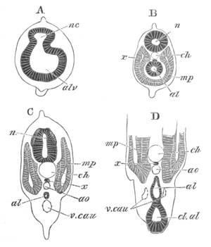
Fig. 424. Four sections through the postanal part of the tail
of an embryo of the same age as fig. 28 F.
A. is the posterior section.
nc. neural canal; al. postanal gut; alv. caudal vesicle of
postanal gut; x. subnotochordal rod; mp. muscle-plate; ch.
notochord; cl.al. cloaca; ao. aorta; v.cau. caudal vein.
Very shortly after the stage to which the above figures belong, at a
point a little behind the anus, where the postanal section of the
canal was thinnest in the previous stage, it becomes solid, and a
rupture here occurs in it at a slightly later period.
The atrophy of this part of the alimentary tract having once commenced
proceeds rapidly. The posterior part first becomes reduced to a small
rudiment near the end of the tail. There is no longer a terminal
vesicle, nor a neurenteric canal. The portion of the postanal section
of the alimentary tract, just behind the cloaca, is for a short time
represented by a small rudiment of the dilated part which at an
earlier period opened into the cloaca.
In Teleostei the vesicle at the end of the tail, discovered by
Kupffer,
(fig. 34, hyv) is probably the equivalent of the vesicle at
the end of the postanal gut in Elasmobranchii.
In Petromyzon and in Amphibia there is a well-developed postanal gut
connected with a neurenteric canal which gradually atrophies. It is
shewn in the embryo of Bombinator in fig. 420.
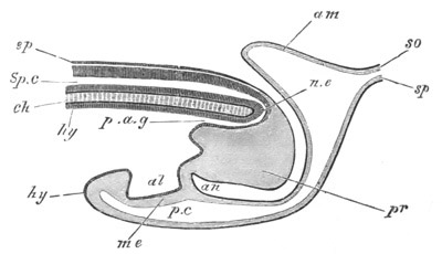
Fig. 425. Diagrammatic longitudinal section through the posterior
end of an embryo Bird at the time of the formation of the Allantois.
ep. epiblast; Sp.c. spinal canal; ch. notochord; n.e.
neurenteric canal; hy. hypoblast; p.a.g. postanal gut; pr.
remains of primitive streak folded in on the ventral side; al.
allantois; me. splanchnic mesoblast; an. point where anus will
be formed; p.c. perivisceral cavity; am. amnion; so.
somatopleure; sp. splanchnopleure.
Amongst the amniotic Vertebrata the postanal gut is less developed
than in the Ichthyopsida. A neurenteric canal is present for a short
period in various Birds (Gasser, etc.) and in the Lizard, but
disappears very early. There is however, as has been pointed out by
Kölliker, a well-marked postanal gut continued as a narrow tube from
behind the cloaca into the tail both in the Bird (fig. 425, p.a.g.)
and Mammals (the Rabbit), but especially in the latter. It atrophies
early as in lower forms.
The morphological significance of the postanal gut and of the
neurenteric canal has already been spoken of in Chapter XII., p. 323.
The Stomodæum.
The anterior section of the permanent alimentary tract is formed by an
invagination of epiblast, constituting a more or less considerable
pit, with its inner wall in contact with the blind anterior extremity
of the alimentary tract.
In Ascidians this pit is placed on the dorsal surface (fig. 9, o),
and becomes the permanent oral cavity of these forms. In the larva of
Amphioxus it is stated to be formed unsymmetrically
(vide p. 5), but
further observations on its development are required.
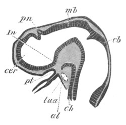
Fig. 426. Longitudinal section through the brain of a young
Pristiurus embryo.
cer. unpaired rudiment of the cerebral hemispheres; pn. pineal
gland; In. infundibulum; pt. ingrowth from mouth to form the
pituitary body; mb. mid-brain; cb. cerebellum; ch. notochord;
al. alimentary tract; Iaa. artery of mandibular arch.
In the true Vertebrata it is always formed on the ventral surface of
the head, immediately behind the level of the fore-brain (fig. 426),
and is deeper in Petromyzon (fig. 416, m) than in any other known
form.
From the primary buccal cavity or stomodæum there grows out the
pituitary pit (fig. 426, pt), the development of which has already
been described (p. 435).
The wall separating the stomodæum from the mesenteron always becomes
perforated, usually at an early stage of development, and though in
Petromyzon the boundary between the two cavities remains indicated by
the velum, yet in the higher Vertebrata all trace of this boundary is
lost, and the original limits of the primitive buccal cavity become
obliterated; while a secondary buccal cavity, partly lined by
hypoblast and partly by epiblast, becomes established.
This cavity, apart from the organs which belong to it, presents
important variations in structure. In most Pisces it retains a fairly
simple character, but in the Dipnoi its outer boundary becomes
extended so as to enclose the ventral opening of the nasal sack, which
thenceforward constitutes the posterior nares.
In Amphibia and Amniota the posterior nares also open well within the
boundary of the buccal cavity.
In the Amniota further important changes take place.
In the first place a plate grows inwards from each of the superior
maxillary processes (fig. 427, p), and the two plates, meeting in
the middle line, form a horizontal septum dividing the front part of
the primitive buccal cavity into a dorsal respiratory section (n),
containing the opening of the posterior nares, and a ventral cavity,
forming the permanent mouth. The
two divisions thus formed open into a
common cavity behind. The horizontal septum, on the development within
it of an osseous plate, constitutes the hard palate.
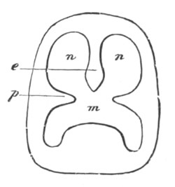
Fig. 427. Diagram shewing the division of the primitive buccal
cavity into the respiratory section above and the true mouth
below. (From Gegenbaur.)
p. palatine plate of superior maxillary process; m. permanent
mouth; n. posterior part of nasal passage; e. internasal
septum.
An internasal septum (fig. 427, e) may more or less completely
divide the dorsal cavity into two canals, continuous respectively with
the two nasal cavities.
In Mammalia a posterior prolongation of the palate, in which an
osseous plate is not formed, constitutes the soft palate.
The second change in the Amniota, which also takes place in some
Amphibia, is caused by the section of the mesenteron into which the
branchial pouches open, becoming, on the atrophy of these structures,
converted into the posterior part of the buccal cavity.
The organs derived from the buccal cavity are the tongue, the various
salivary glands, and the teeth; but the latter alone will engage our
attention here.
The teeth. The teeth are to be regarded as a special product of the
oral mucous membrane. It has been shewn by Gegenbaur and Hertwig that
in their mode of development they essentially resemble the placoid
scales of Elasmobranchii, and that the latter structures extend in
Elasmobranchii for a certain distance into the cavity of the mouth.
As pointed out by Gegenbaur, the teeth are therefore to be regarded as
more or less specialised placoid scales, whose presence in the mouth
is to be explained by the fact that the latter structure is lined by
an invagination of the epidermis; The most important developmental
point of difference between teeth and placoid scales consists in the
fact, that in the case of the former there is a special ingrowth of
epiblast to meet a connective tissue papilla which is not found in the
latter.
Although the teeth are to be regarded as primitively epiblastic
structures, they are nevertheless found in Teleostei and Ganoidei on
the hyoid
and branchial arches; and very possibly the teeth on some
other parts of the mouth are developed in a true hypoblastic region.
The teeth are formed from two distinct organs, viz. an epithelial cap
and a connective tissue papilla.
The general mode of development, as has been more especially shewn by
the extended researches of Tomes, is practically the same for all
Vertebrata, and it will be convenient to describe it as it takes place
in Mammalia.
Along the line where the teeth are about to develop, there is formed
an epithelial ridge projecting into the subjacent connective tissue,
and derived from the innermost columnar layer of the oral epithelium.
At the points where a tooth is about to be formed this ridge undergoes
special changes. It becomes in the first place somewhat thickened by
the development of a number of rounded cells in its interior; so that
it becomes constituted of (1) an external layer of columnar cells, and
(2) a central core of rounded cells; both of an epithelial nature. In
the second place the organ gradually assumes a dome-shaped form (fig.
428, e), and covers over a papilla of the subepithelial connective
tissue (p) which has in the meantime been developed.
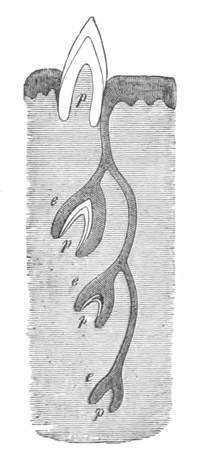
Fig. 428. Diagram shewing the development of the teeth. (From
Gegenbaur.)
p. dental papilla; e. enamel organ.
From the above epithelial structure, which may be called the enamel
organ, and from the papilla it covers, which may be spoken of as the
dental papilla, the whole tooth is developed. After these parts have
become established there is formed round the rudiment of each tooth a
special connective tissue capsule; known as the dental capsule.
Before the dental capsule has become definitely formed the enamel
organ and the dental papilla undergo important changes. The rounded
epithelial cells forming the core of the enamel organ undergo a
peculiar transformation into a tissue closely resembling ordinary
embryonic connective tissue, while at the same time the epithelium
adjoining the dental papilla and covering the inner surface of the
enamel organ, acquires a somewhat different structure to the
epithelium on the outer side of the organ. Its cells become very
markedly columnar, and form a very regular cylindrical epithelium.
This layer alone is concerned in forming the enamel. The cells of the
outer epithelial layer of the enamel organ become somewhat flattened,
and the surface of the layer is raised into a series of short papillæ
which project into the highly vascular tissue of the dental sheath.
Between
the epithelium of the enamel organ and the adjoining
connective tissue there is everywhere present a delicate membrane
known as the membrana præformativa.
The dental papilla is formed of a highly vascular core and a
non-vascular superficial layer adjoining the inner epithelium of the
enamel organ. The cells of the superficial layer are arranged so as
almost to resemble an epithelium.
The first formation of the hard structures of the tooth commences at
the apex of the dental papilla. A calcification of the outermost layer
of the papilla sets in, and results in the formation of a thin layer
of dentine. Nearly simultaneously a thin layer of enamel is deposited
over this, from the inner epithelial layer of the enamel organ (fig.
428). Both enamel and dentine continue to be deposited till the crown
of the tooth has reached its final form, and in the course of this
process the enamel organ is reduced to a thin layer, and the whole of
the outer layer of the dental papilla is transformed into
dentine—while the inner portion remains as the pulp.
The root of the tooth is formed later than the crown, but the enamel
organ is not prolonged over this part, so that it is only formed of
dentine.
By the formation of the root the crown of the tooth becomes pushed
outwards, and breaking through its sack projects freely on the
surface.
The part of the sack which surrounds the root of the tooth gives rise
to the cement, and becomes itself converted into the periosteum of the
dental alveolus.
The general development of the enamel organs and dental papillæ is
shewn in the diagram (fig. 428). From the epithelial ridge three
enamel organs are represented as being developed. Such an arrangement
may occur when teeth are successively replaced. The lowest and
youngest enamel organ (e) has assumed a cap-like form enveloping a
dental papilla, but no calcification has yet taken place.
In the next stage a cap of dentine has become formed, while in the
still older tooth this has become covered by a layer of enamel. As may
be gathered from this diagram, the primitive epithelial ridge from
which the enamel organ is formed is not necessarily absorbed on the
formation of a tooth, but is capable of giving rise to fresh enamel
organs. When the enamel organ has reached a certain stage of
development, its connection with the epithelial ridge is ruptured
(fig. 428).
The arrangement represented in fig. 428, in which successive enamel
organs are formed from the same epithelial ridge, is found in most
Vertebrata except the Teleostei. In the Teleostei, however (Tomes), a
fresh enamel organ grows inwards from the epithelium for each
successively formed tooth.
The Proctodæum.
In all Vertebrata the cloacal section of the alimentary tract which
receives the urinogenital ducts is placed in communication
with the
exterior by means of an epiblastic invagination, constituting a
proctodæum.
This invagination is not usually very deep, and in most instances the
boundary wall between it and the hypoblastic cloaca is not perforated
till considerably after the perforation of the stomodæum; in
Petromyzon, however, its perforation is effected before the mouth and
pharynx are placed in communication.
The mode of formation of the proctodæum, which is in general extremely
simple, is illustrated by fig. 420 an.
In most forms the original boundary between the epiblast of the
proctodæum and the hypoblast of the primitive cloaca becomes
obliterated after the two have become placed in free communication.
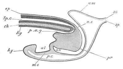
Fig. 429. Diagrammatic longitudinal section through the posterior
end of an embryo Bird at the time of the formation of the Allantois.
ep. epiblast; Sp.c. spinal canal; ch. notochord; n.e.
neurenteric canal; hy. hypoblast; p.a.g. postanal gut; pr.
remains of primitive streak folded in on the ventral side; al.
allantois; me. mesoblast; an. point where anus will be formed;
p.c. perivisceral cavity; am. amnion; so. somatopleure; sp.
splanchnopleure.
In Birds the formation of the proctodæum is somewhat more complicated
than in other types, owing to the outgrowth from it of the bursa
Fabricii.
The proctodæum first appears when the folding off of the tail end of
the embryo commences (fig. 429, an) and is placed near the front
(originally the apparent hind) end of the primitive streak. Its
position marks out the front border of the postanal section of the
gut.
The bursa Fabricii first appears on the seventh day (in the chick), as
a dorsal outgrowth of the proctodæum. The actual perforation of the
septum between the proctodæum and the cloacal section of the
alimentary tract is not effected till about the fifteenth day of
fœtal life, and the approximation
of the epithelial layers of the two
organs, preparatory to their absorption, is partly effected by the
tunneling of the mesoblastic tissue between them by numerous spaces.
The hypoblastic section of the cloaca of birds, which receives the
openings of the urinogenital ducts, is permanently marked off by a
fold from the epiblastic section or true proctodæum, with which the
bursa Fabricii communicates.
Bibliography.
Alimentary Canal and its appendages.
(561) B. Afanassiew. “Ueber Bau u. Entwicklung d. Thymus d. Säugeth.”
Archiv f. mikr. Anat. Bd. XIV. 1877.
(562) Fr. Boll. Das Princip d. Wachsthums. Berlin, 1876.
(563) E. Gasser. “Die Entstehung d. Cloakenöffnung bei
Hühnerembryonen.” Archiv f. Anat. u. Physiol., Anat. Abth. 1880.
(564) A. Götte. Beiträge zur Entwicklungsgeschichte d. Darmkanals im
Hühnchen. 1867.
(565) W. Müller. “Ueber die Entwickelung der Schilddrüse.” Jenaische
Zeitschrift, Vol. VI. 1871.
(566) W. Müller. “Die Hypobranchialrinne d. Tunicaten.” Jenaische
Zeitschrift, Vol. VII. 1872.
(567) S. L. Schenk. “Die Bauchspeicheldrüse d. Embryo.”
Anatomischphysiologische Untersuchungen. 1872.
(568) E. Selenka. “Beitrag zur Entwicklungsgeschichte d. Luftsäcke d.
Huhns.” Zeit. f. wiss. Zool. 1866.
(569) L. Stieda. Untersuch. üb. d. Entwick. d. Glandula Thymus,
Glandula thyroidea, u. Glandula carotica. Leipzig, 1881.
(570) C. Fr. Wolff. “De formatione intestinorum.” Nov. Comment. Akad.
Petrop. 1766.
(571) H. Wölfler. Ueb. d. Entwick. u. d. Bau d. Schilddrüse. Berlin,
1880.
Vide also Kölliker (298), Götte (296), His (232 and 297), Foster and
Balfour (295), Balfour (292), Remak (302), Schenk (303), etc.
Teeth.
(572) T. H. Huxley. “On the enamel and dentine of teeth.” Quart. J.
of Micros. Science, Vol. III. 1855.
(573) R. Owen. Odontography. London, 1840-1845.
(574) Ch. S. Tomes. Manual of dental anatomy, human and comparative.
London, 1876.
(575) Ch. S. Tomes. “On the development of teeth.” Quart. J. of
Micros. Science, Vol. XVI. 1876.
(576) W. Waldeyer. “Structure and development of teeth.” Stricker’s
Histology. 1870.
Vide also Kölliker (298), Gegenbaur (294), Hertwig (306), etc.
- Abdominal muscles, 675
- Abdominal pore, 626, 749
- Acipenser, development of, 102;
- affinities of, 118;
- comparison of gastrula of, 279;
- pericardial cavity of, 627;
- Actinotrocha, 373
- Air-bladder of Teleostei, 77;
- Lepidosteus, 117;
- blood supply of, 645;
- general account of, 763;
- homologies of, 766;
- Alciope, eye of, 480
- Alisphenoid region of skull, 569
- Alimentary canal and appendages, development of, 754
- Alimentary tract of Ascidia, 18;
- Molgula, 22;
- Pyrosoma, 24;
- Salpa, 31;
- Elasmobranchii, 52;
- Teleostei, 75;
- Petromyzon, 93, 97;
- Acipenser, 110;
- Amphibia, 129, 136;
- Chick, 167;
- respiratory region of, 754;
- temporary closure of oesophageal region of, 759;
- Allantois, development of in Chick, 191, 198;
- blood-vessels of in Chick, 193;
- Lacerta, 205, 209;
- early development of in Rabbit, 229;
- of Guinea-pig, 264;
- origin of, 309.
- _See also_ ‘Placenta’ and ‘Bladder’
- Alternation of generations in Ascidians, origin of, 35;
- in Botryllus, 35;
- Pyrosoma, 36;
- Salpa, 36;
- Doliolum, 36;
- Alytes, branchial chamber of, 136;
- yolk-sack of, 139;
- branchiæ, 141;
- Müllerian duct of, 710;
- Amblystoma, ovum of, 120;
- Amia, ribs of, 561
- Ammocoetes, 95;
- metamorphosis of, 97;
- eye of, 498;
- Amnion, early development of in Chick, 185;
- Amphibia, development of, 120;
- viviparous, 121;
- gastrula of, 277;
- suctorial mouth of, 317;
- cerebellum of, 426;
- infundibulum of, 431;
- pineal gland of, 433;
- cerebrum of, 439;
- olfactory lobes of, 444;
- nares of, 553;
- notochord and its sheath, 548;
- vertebral column of, 554;
- ribs of, 561;
- branchial arches of, 574;
- mandibular and hyoid arches of, 582;
- columella of, 582;
- pectoral girdle of, 605;
- pelvic girdle of, 607;
- limbs of, 619;
- heart of, 638;
- arterial system of, 645;
- venous system of, 655;
- excretory system of, 707;
- vasa efferentia of, 711;
- liver of, 769;
- postanal gut of, 774;
- stomodæum of, 778;
- Amphiblastula larva of Porifera, 344
- Amphioxus, development of, 1;
- gastrula of, 275;
- formation of mesoblast of, 292;
- development of notochord of, 293;
- head of, 314;
- spinal nerves of, 461;
- olfactory organ of, 462;
- venous system of, 651;
- transverse abdominal muscle of, 673;
- generative cells of, 748;
- liver of, 769;
- postanal gut of, 772;
- stomodæum of, 777;
- Amphistylic skulls, 578
- Angular bone, 594
- Anterior abdominal vein, 653
- Anura, development of, 121;
- epiblast of, 125;
- mesoblast of, 128;
- notochord of, 128;
- hypoblast of, 129;
- general growth of embryo of, 131;
- larva of, 134;
- vertebral column of, 556;
- mandibular arch of, 584;
- Anus of Amphioxus, 7;
- Ascidia, 18;
- Pyrosoma, 28;
- Salpa, 31;
- Elasmobranchii, 57;
- Amphibia, 130, 132;
- Chick, 167;
- primitive, 324;
- Appendicularia, development of, 34
- Aqueductus vestibuli, 519
- Aqueous humour, 497
- Arachnida, nervous system of, 409;
- Area, embryonic, of Rabbit, 218;
- epiblast of, 219;
- origin of embryo from, 228;
- area opaca of Chick, 150;
- epiblast, hypoblast, and mesoblast of, 159;
- area pellucida of Chick, 150;
- of Lacerta, 202 area vasculosa of Chick, 194;
- mesoblast of, 160;
- of Lizard, 209;
- Rabbit, 228, 229;
- Arteria centralis retinæ, 503
- Arterial system of Petromyzon, 97;
- constitution of in embryo, 643;
- of Fishes, 644;
- of Amphibia, 645;
- of Amniota, 647;
- Arthropoda, head of, 313;
- nervous system of, 409;
- eye of, 480;
- excretory organs of, 688;
- Articular bone of Teleostei, 581;
- Ascidia, development of, 9
- Ascidians. _See_ ‘Tunicata’
- Ascidiozooids, 25
- Atrial cavity of Amphioxus, 7;
- Ascidia, 18;
- Pyrosoma, 24;
- Atrial pore of Amphioxus,7;
- Ascidia, 20;
- Pyrosoma, 28;
- Salpa, 32;
- Auditory capsules, ossifications in, 595, 596
- Auditory involution of Elasmobranchii, 57;
- Teleostei, 73;
- Petromyzon, 89, 92;
- Acipenser, 106;
- Lepidosteus, 114;
- Amphibia, 127;
- Chick, 170;
- Auditory nerve, development of, 459
- Auditory organs, of Ascidia, 15;
- of Salpa, 31;
- of Ammocoetes, 98;
- Ganoidei, 108, 114;
- of Amphibia, 127;
- of Aves, 170;
- general development of, 512;
- of aquatic forms, 512;
- of land forms, 513;
- of Coelenterata, 513;
- of Mollusca, 515;
- of Crustacea, 516;
- of Vertebrata, 517;
- of Cyclostomata, 89, 92, 518;
- of Teleostei, Lepidosteus and Amphibia, 518;
- of Mammalia, 519;
- accessory structures of, 527;
- of Tunicata, 528;
- Auriculo-ventricular valves, 642
- Autostylic skulls, 579
- Aves, development of, 145;
- cerebellum of, 426;
- mid-brain of, 427;
- infundibulum of, 431;
- pineal gland of, 434;
- pituitary body of, 436;
- cerebrum of, 439;
- olfactory lobes of, 444;
- spinal nerves of, 449;
- cranial nerves of, 455;
- vagus of, 458;
- glossopharyngeal of, 458;
- vertebral column of, 557;
- ossification of vertebral column of, 558;
- branchial arches of, 572, 573;
- pectoral girdle of, 603;
- pelvic girdle of, 608;
- heart of, 637;
- arterial system of, 647;
- venous system of, 658;
- muscle-plates of, 670;
- excretory organs of, 714;
- mesonephros of, 715;
- pronephros of, 718;
- Müllerian duct of, 718, 720;
- nature of pronephros of, 721;
- connection of Müllerian duct with Wolffian in, 720;
- kidney of, 722;
- lungs of, 764;
- liver of, 769;
- postanal gut of, 774;
- Axolotl, 142, 143;
- ovum of, 120;
- mid-brain of, 427;
- mandibular arch of, 583;
- Basilar membrane, 524
- Basilar plate, 565
- Basipterygium, 612
- Basisphenoid region of skull, 569
- Bilateral symmetry, origin of, 373-376
- Bile duct, 770
- Bladder, Amphibia, 131;
- Blastodermic vesicle, of Rabbit, first development of, 217;
- of 7th day, 222;
- Guinea-pig, 263;
- meaning of, 291;
- Blastoderm of Pyrosoma, 24;
- Elasmobranchii, 41;
- Chick, 150;
- Lacerta 202;
- Blastopore, of Amphioxus, 2;
- of Ascidia, 11;
- Elasmobranchii, 42, 54, 62;
- Petromyzon, 87;
- Acipenser, 104;
- Amphibia, 125, 130;
- Chick, 153;
- Rabbit, 216;
- true Mammalian, 226;
- comparative history of closure of, 284, 288;
- summary of fate of, 340;
- relation of to primitive anus, 324;
- Blood-vessels, development of, 633
- Body cavity, of Ascidia, 21;
- Molgula, 21;
- Salpa, 31;
- Elasmobranchii, 47;
- of Teleostei, 75;
- Petromyzon, 94;
- Chick, 169;
- development of in Chordata, 325;
- views on origin of, 356-360, 377;
- of Invertebrata, 623;
- of Chordata, 624;
- of head, 676;
- Bombinator, branchial chamber of, 136;
- vertebral column of, 556;
- Bonellia, excretory organs of, 687
- Bones, origin of cartilage bones, 542;
- origin of membrane bones, 543;
- development of, 543;
- homologies of membrane bones, 542;
- homologies of cartilage bones, 545;
- Brachiopoda, excretory organs of, 683;
- generative ducts of, 749;
- Brain, of Ascidia, 11, 15;
- Elasmobranchii, 56, 59, 60;
- Teleostei, 77;
- Petromyzon, 89, 92;
- Acipenser, 105;
- Lepidosteus, 113;
- early development of in Chick, 170;
- flexure of in Chick, 175;
- later development of in Chick, 176;
- Rabbit, 229;
- general account of development of, 419;
- flexure of, 420;
- histogeny of, 422;
- Branchial arches, præoral, 570;
- disappearance of posterior, 573;
- dental plates of in Teleostei, 574;
- relation of to head cavities, 571;
- _See_ ‘Visceral arches’
- Branchial chamber of Amphibia, 136
- Branchial clefts, of Amphioxus, 7;
- of Ascidia, 18, 20;
- Molgula, 23;
- Salpa, 32;
- of Elasmobranchii, 57, 59-61;
- Teleostei, 77;
- Petromyzon, 91, 96;
- Acipenser, 105;
- Lepidosteus, 114, 116;
- Amphibia, 132, 133;
- Chick, 178;
- Rabbit, 231;
- præoral, 312, 318;
- of Invertebrata, 326;
- origin of, 326;
- Branchial rays, 574
- Branchial skeleton, development of, 572, 592;
- of Petromyzon, 96, 312, 571;
- of Ichthyopsida, 572;
- dental plates of in Teleostei, 574;
- relation of to head cavities, 572;
- Branchiæ, external of Elasmobranchii, 61, 62;
- Brood-pouch, of Salpa, 29;
- Teleostei, 68;
- Amphibia, 121;
- Brown tubes of Gephyrea, 686
- Bulbus arteriosus, of Fishes, 638;
- Bursa Fabricii, 167, 779
- Canalis auricularis, 639
- Canalis reuniens, 521
- Capitellidæ, excretory organs of, 683
- Carcharias, placenta of, 66
- Cardinal vein, 652
- Carnivora, placenta of, 250
- Carpus, development of, 620
- Cartilage bones of skull, 595;
- Cat, placenta of, 250
- Caudal swellings of Elasmobranchii, 46, 55;
- Cephalic plate of Elasmobranchii, 55
- Cephalochorda, development of, 1
- Cephalopoda, eyes of, 473-477
- Cerebellum, Petromyzon, 93;
- Chick, 176;
- general account of development of, 424, 425;
- Cerebrum of Petromyzon, 93, 97;
- Chick, 175;
- general development of, 429, 438;
- transverse fissure of, 443;
- Cestoda, excretory organs of, 681
- Cetacea, placenta, 255
- Chætognatha, nervous system of, 349;
- eye of, 479;
- generative organs of, 743;
- generative ducts of, 749;
- Chætopoda, head of, 313;
- eyes of, 479;
- excretory organs of, 683;
- generative organs of, 743;
- generative ducts of, 749;
- Charybdæa, eye of, 472
- Cheiroptera, placenta of, 244
- Cheiropterygium, 618;
- relation of to ichthyopterygium, 621;
- Chelonia, development of, 210;
- pectoral girdle of, 603;
- arterial system of, 649;
- Chick, development of, 145;
- general growth of embryo of, 170;
- rotation of embryo of, 173;
- foetal membranes of, 185;
- epiblast of, 150, 166;
- optic nerve and choroid fissure of, 500;
- Chilognatha, eye of, 481
- Chilopoda, eye of, 481
- Chimæra, lateral line of, 539;
- vertebral column of, 548;
- nares of, 533;
- Chiromantis, oviposition of, 121
- Chorda tympani, development of, 460
- Chordata, ancestor of, 311;
- branchial system of, 312;
- evidence from Ammocoetes, 312;
- head of, 312;
- mouth of, 318;
- table of phylogeny of, 327;
- Chorion, 237;
- Choroid coat, Ammocoetes, 99;
- Choroid fissure, of Vertebrate eye, 486, 493;
- of Ammocoetes, 498;
- comparative development of, 500;
- of Chick, 501;
- of Lizards, 501;
- of Elasmobranchii, 502;
- of Teleostei, 503;
- Amphibia, 503;
- Mammals, 503, 504;
- Choroid gland, 320
- Choroid pigment, 489
- Choroid plexus, of fourth ventricle, 425;
- of third ventricle, 432;
- of lateral ventricle, 442;
- Ciliated sack of Ascidia, 18;
- Ciliary ganglion, 461
- Ciliary muscle, 490
- Ciliary processes, 488;
- comparative development of, 506;
- Clavicle, 600
- Clitoris, development of, 727
- Clinoid ridge, 569
- Cloaca, 766
- Coccygeo-mesenteric vein, 661
- Cochlear canal, 519
- Coecilia, development of, 143;
- pronephros of, 707;
- mesonephros of, 709;
- Müllerian duct of, 710;
- Coelenterata, larvæ of, 367;
- eyes of, 471;
- auditory organs of, 513;
- generative organs of, 741;
- Columella auris, 529;
- of Amphibia, 582;
- of Sauropsida, 588;
- Commissures, of spinal cord, 417;
- Coni vasculosi, 724
- Conus arteriosus, of Fishes, 638;
- Coracoid bone, 599
- Cornea, of Ammocoetes, 99;
- general development of, 495;
- corpuscles of, 496;
- comparative development of, 499;
- of Mammals, 499;
- Coronoid bone, 595
- Corpora geniculata interna, 428
- Corpora quadrigemina, 428
- Corpora striata, development of, 437
- Corpus callosum, development of, 443
- Corti, organ of, 522;
- structure of, 525;
- fibres of, 525;
- development of, 526;
- Cranial flexure, of Elasmobranchii, 58, 60;
- of Teleostei, 77;
- Petromyzon, 93, 94;
- of Amphibia, 131, 132;
- Chick, 174;
- Rabbit, 231;
- characters of, 321;
- significance of, 322;
- Cranial nerves, development of, 455;
- relation of to head cavities, 461;
- anterior roots of, 462-464;
- view on position of roots of, 466;
- Crocodilia, arterial system of, 649
- Crura cerebri, 429
- Crustacea, nervous system of, 411;
- eye of, 481;
- auditory organs of, 515;
- generative cells of, 745;
- generative ducts of, 751;
- Cupola, 524
- Cutaneous muscles, 676
- Cyathozooid, 25
- Cyclostomata, auditory organs of, 517;
- olfactory organ of, 532;
- notochord and vertebral column of, 546, 549;
- abdominal pores of, 626;
- segmental duct of, 700;
- pronephros of, 700;
- mesonephros of, 700;
- generative ducts of, 733, 749;
- venous system of, 651;
- excretory organs of, 700;
- Cystignathus, oviposition of, 122
- Dactylethra, branchial chamber of, 136;
- branchiæ of, 136;
- tadpole of, 140;
- Decidua reflexa, of Rat, 242;
- of Insectivora, 243;
- of Man, 245;
- Deiter’s cells, 526
- Dental papilla, 777
- Dental capsule, 777
- Dentary bone, 595
- Dentine, 780
- Descemet’s membrane, 496
- Diaphragm, 631;
- Dipnoi, nares of, 534;
- vertebral column of, 548;
- membrane bones of skull of, 592;
- heart of, 638;
- arterial system of, 645;
- excretory system of, 707;
- stomodæum of, 777;
- Diptera, eye of, 481
- Discophora, excretory organs of, 687
- Dog, placenta of, 248
- Dohrn, on relations of Cyclostomata, 84;
- on ancestor of Chordata, 311, 319;
- Doliolum, development of, 28
- Ductus arteriosus, 649
- Ductus Botalli, 648
- Ductus Cuvieri, 654
- Ductus venosus Arantii, 663
- Dugong, heart of, 642
- Dysticus, eye of, 481
- Ear, _see_ ‘Auditory organ’
- Echinodermata, secondary symmetry of larva of, 380;
- excretory organs of, 689;
- generative ducts of, 752;
- Echinorhinus, lateral line of, 539;
- vertebral column of, 548;
- Echiurus, excretory organs of, 686
- Ectostosis, 543
- Edentata, placenta of, 248, 250, 256
- Eel, generative ducts of, 703
- Egg-shell of Elasmobranchii, 40;
- Elasmobranchii, development of, 40;
- viviparous, 40;
- general features of development of, 55;
- gastrula of, 281;
- development of mesoblast of, 294;
- notochord of, 294;
- meaning of formation of mesoblast of, 295;
- restiform tracts of, 425;
- optic lobes of, 427;
- cerebellum of, 425;
- pineal gland of, 432;
- pituitary body of, 435;
- cerebrum of, 438;
- olfactory lobes of, 444;
- spinal nerves, 449;
- cranial nerves of, 457;
- sympathetic nervous system of, 466;
- nares of, 533;
- lateral line of, 539;
- vertebral column of, 549;
- ribs of, 560;
- parachordals of, 567;
- mandibular and hyoid arches of, 576;
- pectoral girdle of, 600;
- pelvic girdle of, 607;
- limbs of, 609;
- pericardial cavity of, 627;
- arterial system of, 644;
- venous system of, 651;
- muscle-plates of, 668;
- excretory organs of, 690;
- constitution of excretory organs in adult of, 697;
- spermatozoa of, 747;
- swimming-bladder of, 763;
- intestines of, 767;
- liver of, 769;
- postanal gut of, 772;
- Elæoblast of Pyrosoma, 28;
- Elephant, placenta of, 249
- Embolic formation of gastrula, 333
- Enamel organ, 777
- Endolymph of ear, 522
- Endostosis, 543
- Endostyle of Ascidia, 18, 759;
- Epiblast, of Elasmobranchii, 47;
- Teleostei, 71, 75;
- Petromyzon, 86;
- Lepidosteus, 112;
- Amphibia, 122, 125;
- Chick, 149, 166;
- Lacerta, 203;
- Rabbit, 216, 219;
- origin of in Rabbit, 221;
- comparative account of development of, 300;
- Epibolic formation of gastrula, 334
- Epichordal formation of vertebral column, 556
- Epicrium glutinosum, 143
- Epidermis, in Coelenterata, 393;
- protective structures of, 394;
- Epididymis, 724
- Epigastric vein, 653
- Episkeletal muscles, 676
- Episternum, 602
- Epoophoron, 725
- Ethmoid bone, 597
- Ethmoid region of skull, 570
- ethmo-palatine ligament of Elasmobranchs, 576
- Euphausia, eye of, 483
- Eustachian tube, of Amphibia, 135;
- Chick, 180;
- Rabbit, 232;
- general development of, 528;
- Excretory organs, general constitution of, 680;
- of Platyelminthes, 680;
- of Mollusca, 681;
- of Polyzoa, 682;
- of Brachiopoda, 683;
- of Chætopoda, 683;
- of Gephyrea, 686;
- of Discophora, 687;
- of Arthropoda, 688;
- of Nematoda, 689;
- of Echinodermata, 689;
- constitution of in Craniata, 689;
- of Elasmobranchii, 690;
- constitution of in adult Elasmobranch, 697;
- of Petromyzon, 700;
- of Myxine, 701;
- of Teleostei,701;
- of Ganoidei, 704;
- of Dipnoi, 707;
- of Amphibia, 707;
- of Amniota, 713;
- comparison of Vertebrate and Invertebrate, 737;
- Excretory system, of Elasmobranchii, 49;
- Teleostei, 78;
- Petromyzon, 95, 98;
- Acipenser, 99;
- Amphibia, 133;
- Exoccipital bone, 595
- Exoskeleton, dermal, 393-395;
- External generative organs, 726
- Extra-branchial skeleton, 572
- Eye, of Ascidia, 16;
- Salpa, 31;
- Elasmobranchii, 56, 57, 58;
- Teleostei, 73;
- Petromyzon, 92, 98;
- Aves, 170;
- Rabbit, 229;
- general development of, 470;
- evolution of, 470, 471;
- simple, 480;
- compound, 481;
- aconous, 482;
- pseudoconous, 482;
- of Invertebrata, 471;
- of Vertebrata, 483;
- comparative development of Vertebrate, 497;
- of Ammocoetes, 497;
- of Tunicata, 507;
- of Chordata, general views on, 508;
- accessory eyes of Fishes, 509;
- muscles of, 677;
- Eyelids, development of, 506
- Falciform ligament, 757
- Falx cerebri, 439
- Fasciculi teretes, of Elasmobranchii, 426
- Feathers, development of, 396
- Fenestra rotunda and ovalis, 529
- Fertilization, of Amphioxus, 2;
- of Urochorda, 9;
- Salpa, 29;
- Elasmobranchii, 46;
- of Teleostei, 68;
- Petromyzon, 84;
- Amphibia, 120;
- Chick, 145;
- Reptilia, 202;
- meaning of, 331;
- Fifth nerve, development of, 460
- Fifth ventricle, 443
- Fins, of Elasmobranchii, 62;
- Teleostei, 78;
- Petromyzon, 94, 95;
- Acipenser, 109;
- Lepidosteus, 118;
- relation of paired to unpaired, 611, 612;
- development of pelvic, 614;
- development of pectoral, 615;
- views on nature of paired fins, 616;
- Fissures of spinal cord, 417
- Foetal development, 360;
- secondary variations in, 361;
- Foot, 618
- Foramen of Munro, 430, 438
- Foramen ovale, 642
- Fore-brain, of Elasmobranchii, 55, 59, 60;
- Petromyzon, 93;
- general development of, 428;
- Formative cells, of Chick, 154
- Fornix, development of, 443
- Fornix of Gottsche, 428
- Fourth nerve, 464
- Frontals, 592
- Frontonasal process of Chick, 179
- Gaertner’s canals, 724
- Gall-bladder, 770
- Ganoidei, development of, 102;
- relations of, 118;
- nares of, 534;
- notochord of, 546;
- vertebral column of, 546, 553;
- ribs of, 561;
- pelvic girdle of, 606;
- arterial$ system of, 645;
- excretory organs of, 704;
- generative ducts of, 734;
- Gastropoda, eye of, 472
- Gastrula, of Amphioxus, 2;
- of Ascidia, 10;
- Elasmobranchii, 43, 44;
- Petromyzon, 86;
- Acipenser, 103;
- Amphibia, 123;
- comparative development of, in Invertebrata, 275;
- comparison of Mammalian, 291;
- phylogenetic meaning of, 333;
- ontogeny of (general), 333;
- phylogeny of, 338-343;
- secondary types of, 341;
- Geckos, vertebral column of, 557
- Generative cells, development of, 741;
- origin of in Coelenterata, 741;
- of Invertebrata, 743;
- of Vertebrata, 746;
- Generative ducts, of Teleostei, 704, 735;
- of Ganoids, 704;
- of Cyclostomata, 733;
- origin of, 733;
- of Lepidosteus, 735, 750;
- development and evolution of, 748;
- of Coelenterata, 748;
- of Sagitta, 749;
- of Tunicata, 749;
- Chætopoda, Gephyrea, etc., 749;
- of Mollusca, 751;
- of Discophora, 751;
- of Echinodermata, 752;
- Generative system of Elasmobranchii, 51
- Gephyrea, nervous system of, 412;
- excretory organs of, 686;
- generative cells of, 743;
- generative ducts of, 749;
- Germinal disc, of Elasmobranchii, 40;
- Teleostei, 68;
- Chick, 147;
- Germinal epithelium, 746
- Germinal layers, summary of organs derived from, in Vertebrata, 304;
- historical account of views of, 332;
- homologies of in the Metazoa, 345;
- Germinal wall of Chick, 152, 159;
- structure and changes of, 160;
- Geryonia, auditory organ of, 515
- Gill of Salpa, 31
- Giraldes, organ of, 725
- Glands, epidermic, development of, 397
- Glomerulus, external, of Chick, 716
- Glossopharyngeal nerve, development of, 456, 457
- Grey matter of spinal cord, 417;
- Growth in length of Vertebrate embryo, 306
- Guinea-pig, primitive streak of, 223;
- notochord of, 226;
- placenta of, 242;
- development of, 262;
- Gymnophiona, _see_ ‘Coecilia’
- Habenula perforata, 525
- Hairs, development of, 396
- Halichærus, placenta of, 250
- Hand, 619
- Head, comparative account of, 313;
- Head cavities, of Elasmobranchii, 50;
- Petromyzon, 90, 96;
- Amphibia, 127;
- general development of, 676;
- Head-fold of Chick, 157, 167
- Head kidney, _see_ ‘Pronephros’
- Heart, of Pyrosoma, 25;
- Elasmobranchii, 50, 58;
- Petromyzon, 94, 97;
- Acipenser, 106;
- Chick, 170;
- first appearance of in Rabbit, 230;
- general development of, 633;
- of Fishes, 635, 637;
- of Mammalia, 638;
- of Birds, 637, 639;
- meaning of development of, 637;
- of Amphibia, 638;
- of Amniota, 639;
- change of position of, 643;
- Hind-brain, Elasmobranchii, 55, 59, 60;
- Petromyzon, 93;
- general account of, 424;
- Hippocampus major, development of, 442
- Hirudo, development of blood-vessels of, 633;
- excretory organs of, 688;
- Horse, placenta of, 253
- Hyaloid membrane, 492
- Hylodes, oviposition of, 121;
- Hyobranchial cleft, 572
- Hyoid arch, of Chick, 179;
- general account of, 572, 575;
- modifications of, 573, 577;
- of Elasmobranchii, 576;
- of Teleostei, 577;
- of Amphibia, 582;
- of Sauropsida, 588;
- of Mammalia, 589;
- Hyomandibular bar of Elasmobranchii, 576, 577;786.png
- of Teleostei, 579;
- of Amphibia, 582;
- Hyomandibular cleft, of Petromyzon, 91;
- Chick, 179;
- general account of, 572;
- Hyostylic skulls, 582
- Hypoblast of Elasmobranchii, 51;
- Teleostei, 71, 75;
- Petromyzon, 86;
- Acipenser, 104;
- Lepidosteus, 113;
- Amphibia, 122, 129;
- Chick, 151, 167;
- Lacerta, 203;
- Rabbit, 215, 216, 219;
- origin of in Rabbit, 220;
- Hyposkeletal muscles, 675
- Hyrax, placenta of, 249
- Incus, 529, 590
- Infraclavicle, 600
- Infundibulum of Petromyzon, 92;
- Chick, 175;
- general development of, 430;
- Insectivora, placenta of, 243
- Insects, nervous system of, 410;
- eye of, 481;
- generative organs of, 745;
- generative ducts of, 751;
- Intercalated pieces of vertebral column, 551
- Interclavicle, homologies of, 602
- Intermediate cell-mass of Chick, 183
- Intermuscular septa, 672
- Interorbital septum, 570
- Interrenal bodies, 665
- Iris, 489;
- comparative development of, 506;
- Iris of Ammocoetes, 98
- Island of Reil, 444
- Jacobson’s organ, 537
- Jugal bone, 594
- Kidney, _see_ ‘Metanephros’
- Labia majora, development of, 727
- Labial cartilages, 597
- Labium tympanicum, 525;
- Lacertilia, general development of, 202;
- nares of, 537;
- pectoral girdle of, 603;
- pelvic girdle of, 607;
- arterial system of, 649;
- Lacrymal bone, 593
- Lacrymal duct, 506
- Lacrymal glands, 506
- Læmargus, vertebral column of, 548
- Lagena, 524
- Lamina spiralis, 524
- Lamina terminalis, 438
- Larva of Amphioxus, 2;
- of Ascidia, 15-21;
- Teleostei, 81;
- Petromyzon, 89, 95;
- Lepidosteus, 117, 318;
- Amphibia, 134, 142;
- types of, in the Invertebrata, 363;
- Larvæ, nature, origin, and affinities of, 360-386;
- secondary variations of less likely to be retained, 362;
- ancestral history more fully recorded in, 362;
- secondary variations in development of, 363;
- ontogenetic record of secondary variations in, 361;
- of freshwater and land animals, 362;
- types of, 362;
- phosphorescence of, 364;
- of Coelenterata, 367;
- table of, 365;
- of Invertebrata, 367 et seq.
- Larynx, 766
- Lateral line sense organs, 538;
- comparison of, with invertebrate, 538;
- development of, in Teleostei, 538;
- development of, in Elasmobranchii, 539;
- Lateral ventricle, 438;
- anterior cornu of, 440;
- descending cornu of, 440;
- choroid plexus of, 443;
- Layers, formation of, in Elasmobranchii, 41, 56;
- Teleostei, 71;
- Petromyzon, 85;
- Acipenser, 103;
- Lepidosteus, 111;
- Amphibia, 121;
- Chick, 150, 152;
- Lacerta, 202;
- Rabbit, 215-227;
- comparison of Mammalia with lower forms, 226, 289;
- comparison of formation of in Vertebrata, 275;
- origin and homologies of, in the Metazoa, 331;
- Leech, _see_ ‘Hirudo’
- Lemuridæ, placenta, 256
- Lens, of Elasmobranchii, 57, 58;
- Petromyzon, 94, 99;
- Acipenser, 106;
- Lepidosteus, 115;
- Amphibia, 127;
- Chick, 177;
- of Vertebrate eyes, 485;
- general account of, 493;
- capsule of, 493;
- comparative development of, 499;
- of Amphibia, Teleostei, Lepidosteus, 499;
- Lepidosteus, development of, 111;
- larva of, 117;
- relations of, 119;
- spinal nerves of, 455;
- ribs of, 561;
- generative ducts of, 704, 735;
- swimming-bladder of, 763;
- Ligamentum pectinatum, 490
- Ligamentum suspensorium, 557, 558
- Ligamentum vesicæ medium, 239
- Limbs, of Elasmobranchii, 59;
- Teleostei, 80;
- first appearance of in Chick, 184;
- Rabbit, 232;
- muscles of, 673;
- of Fishes, 609;
- relation of, to unpaired fins of Fishes, 611, 612;
- of Amphibia, 618;
- Liver of Teleostei, 78;
- Petromyzon, 95, 96;
- Acipenser, 110;
- Amphibia, 130;
- general account of, 769;
- Lizard, development of, 202;
- general growth of embryo of, 208;
- Müllerian duct of, 721;
- Lizzia, eye of, 471
- Lobi inferiores, 431
- Lungs of Amphibia, 137;
- development of, 763;
- homology of, 766;
- Lymphatic system, 664
- Malleus, 529, 591;
- Malpighian bodies, development of accessory in Elasmobranchs, 695
- Mammalia, development of, 214;
- comparison of gastrula of, 291;
- cerebellum of, 427;
- infundibulum of, 431;
- pineal gland of, 434;
- pituitary body of, 436;
- cerebrum of, 439;
- spinal nerves of, 449;
- sympathetic of, 466;
- vertebral column of, 558;
- branchial arches of, 573, 574;
- mandibular and hyoid arches of, 589;
- pectoral girdle of, 604;
- pelvic girdle of, 608;
- heart of, 636;
- arterial system of, 647;
- venous system of, 661;
- muscle-plates of, 671;
- mesonephros of, 714;
- testicular network of, 724;
- urinogenital sinus of, 727;
- spermatozoa of, 747;
- lungs of, 765;
- intestines of, 768;
- liver of, 769;
- postanal gut of, 774;
- stomodæum of, 775;
- Mammary gland, development of, 398
- Man, placenta of, 244;
- general account of development of, 265;
- characters of embryo of, 270;
- Mandibular arch of Elasmobranchii, 62, 576;
- Petromyzon, 91;
- Acipenser, 106, 116;
- Chick, 179;
- general account of, 572, 575;
- modification of to form jaws, 573, 575;
- of Teleostei, 580;
- of Amphibia, 582;
- Sauropsida, 588;
- Mammalia, 589;
- Mandibular bar, evolution of, 311, 321
- Manis, placenta of, 256
- Marsupial bones, 608
- Marsupialia, foetal membranes of, 240;
- cerebellum of, 426;
- corpus callosum of, 443;
- uterus of, 726;
- Maxilla, 594
- Meatus auditorius externus, of Chick, 181;
- Meckelian cartilage, of Elasmobranchii, 576;
- of Teleostei, 581;
- of Amphibia, 584, 585;
- of Sauropsida, 588;
- of Mammalia, 590;
- Mediastinum anterior and posterior, 630
- Medulla oblongata, of Chick, 176;
- general development of, 425;
- Medullary plate of Amphioxus, 4, 5;
- of Ascidia, 11;
- Elasmobranchii, 44, 47, 55;
- Teleostei, 72;
- Petromyzon, 88;
- Acipenser, 104;
- Lepidosteus, 111;
- Amphibia, 126, 127, 131;
- Chick, 159;
- Lacerta, 204;
- Rabbit, 223, 227, 228;
- primitive bilobed character of, 303, 317;
- Medusæ, auditory organs of, 513
- Membrana capsulo-pupillaris, 494, 504, 507
- Membrana elastica externa, 546
- Membrana limitans of retina, 491
- Membrana tectoria, 522, 525
- Membrane bones, of Amphibia, 582;
- of Sauropsida, 588;
- of Mammalia, 590;
- of mandibular arch, 593;
- of pectoral girdle, 599, 602;
- origin of, 592;
- homologies of, 593;
- Membranous labyrinth, development of in Man, 519
- Menobranchus, branchial arches of, 142
- Mesenteron of Elasmobranchii, 43;
- Mesentery, 626, 756
- Mesoblast, of Amphioxus, 6;
- Ascidia, 17, 20;
- Pyrosoma, 24;
- Salpa, 30;
- Elasmobranchii, 44, 47;
- Teleostei, 75;
- Petromyzon, 86;
- Acipenser, 105;
- Lepidosteus, 113;
- Amphibia, 125, 128, 129;
- of Chick, 154, 167;
- double origin of in Chick, 154, 158, 159;
- origin of from lips of blastopore in Chick, 158;
- of area vasculosa of Chick, 160;
- Lacerta, 203;
- origin of in Rabbit, 218, 223;
- of area vasculosa in Rabbit, 227;
- comparative account of formation of, 292;
- discussion of development of in Vertebrata, 297;
- meaning of development of in Amniota, 298;
- phylogenetic origin of, 346;
- summary of ontogeny of, 349-352;
- views on ontogeny of, 352-360;
- Mesoblastic somites, of Amphioxus, 6;
- Elasmobranchii, 48, 55;
- Petromyzon, 88;
- Acipenser, 105;
- Lepidosteus, 114;
- Amphibia, 129, 131;
- Chick, 161, 180;
- Rabbit, 228;
- development of in Chordata, 325;
- meaning of development of, 331;
- of head, 676
- Mesogastrium, 758
- Mesonephros, of Teleostei, 78, 702;
- Petromyzon, 95, 98, 700;
- Acipenser, 110, 705;
- Amphibia, 134, 708;
- Chick, 184, 714;
- general account of, 690;
- development of in Elasmobranchs 691;
- of Cyclostomata, 700;
- Ganoidei, 705;
- sexual and non-sexual part of in Amphibia, 710;
- of Amniota, 713, 724;
- summary and general conclusions as to, 729;
- relation of to pronephros, 731;
- Mesopterygium, 616
- Metagenesis of Ascidians, 34
- Metamorphosis of Amphibia, 137, 140
- Metanephros, 690;
- development of in Elasmobranchii, 697;
- of Amphibia, 712;
- of Amniota, 713;
- of Chick, 722;
- of Lacertilia, 723;
- phylogeny of, 736;
- Metapterygium, 616
- Metapterygoid, of Elasmobranchii, 576;
- Metazoa, evolution of, 339, 342;
- Mid-brain, of Elasmobranchii, 55, 58, 59;
- Petromyzon, 92;
- general account of development of, 427;
- Moina, generative organs of, 745
- Molgula, development of, 22
- Mollusca, nervous system of, 414;
- eyes of, 472;
- auditory organs of, 515;
- excretory organs of, 681;
- Monotremata, foetal membranes of, 240;
- cerebellum of, 426;
- corpus callosum of, 443;
- cerebrum of, 443;
- urinogenital sinus of, 726;
- Mormyrus, generative ducts of, 704
- Mouth, of Amphioxus, 7;
- of Ascidia, 18;
- Pyrosoma, 27;
- Salpa, 31;
- Elasmobranchii, 57, 60, 61, 62;
- Petromyzon, 92, 94, 95, 99;
- Acipenser, 107;
- Lepidosteus, 118;
- Amphibia, 129, 132, 134;
- Rabbit, 231;
- origin of, 317;
- Mouth, suctorial, of Petromyzon, 99;
- Müllerian duct, 690;
- of Elasmobranchs, 693;
- of Ganoids, 704;
- of Amphibia, 710;
- of Aves, 717, 720;
- opening of into cloaca, 727;
- origin of, 733;
- summary of development of, 733;
- relation of to pronephros, 733;
- Muscle-plates, of Amphioxus, 6;
- Elasmobranchii, 49, 668;
- Teleostei, 670;
- Petromyzon, 94;
- Chick, 183, 670;
- general development of, 669;
- of Amphibia, 670;
- Aves, 670;
- of Mammalia, 671;
- origin of muscles from, 672;
- Muscles, of Ascidia, 11, 17;
- development of from muscle-plates, 672;
- of limbs, 673;
- of head, 676;
- of branchial arches, 678;
- of eye, 678;
- Muscular fibres, epithelial origin of, 667
- Muscular system, development of, 667;
- Mustelus, placenta of, 66
- Myoepithelial cells, 667
- Mysis, auditory organ of, 517
- Myxine, ovum of, 100;
- olfactory organ of, 533;
- portal sinus of, 652;
- excretory system of, 701;
- Nails, development of, 397
- Nares, of Acipenser, 108;
- of Ichthyopsida, 534;
- development of in Chick, 535;
- development of in Lacertilia, 537;
- development of in Amphibia, 537;
- Nasal bones, 592
- Nasal pits, Acipenser, 108;
- Chick, 176;
- general development of, 531;
- Nematoda, excretory organs of, 689;
- generative organs of, 745;
- generative ducts of, 752;
- Nemertines, nervous system of, 311;
- excretory organs of, 681;
- Nerve cord, origin of ventral, 378
- Nerves, spinal, 449;
- Nervous system, central,
- general account of development of in Vertebrata, 415;
- conclusions as to, 445;
- sympathetic, 466;
- Nervous system, of Amphioxus, 4;
- Ascidia, 15, 16;
- Molgula, 22;
- Pyrosoma, 24, 25;
- Salpa, 30, 31;
- Elasmobranchii, 44;
- Teleostei, 77;
- Petromyzon, 89, 93;
- Acipenser, 105;
- Amphibia, 126;
- comparative account of formation of central, 301;
- of Sagitta, 349;
- origin of in Coelenterata, 349;
- of præoral lobe, 377, 380;
- evolution of, 400-405;
- development of in Invertebrates, 406;
- of Arthropoda, 408;
- of Gephyrea, 412;
- Mollusca, 414;
- Neural canal, of Ascidia, 10;
- Teleostei, 72;
- Petromyzon, 88;
- Acipenser, 105;
- Lepidosteus, 114;
- Amphibia, 126, 131;
- Chick, 166, 171;
- Lacerta, 208;
- closure of in Frog and Amphioxus, 279;
- closure of in Elasmobranchii, 284;
- phylogenetic origin of, 316;
- Neural crest, 449, 456, 457
- Neurenteric canal, of Amphioxus, 4, 5;
- Ascidia, 10;
- Elasmobranchii, 54;
- Petromyzon, 88;
- Acipenser, 105;
- Lepidosteus, 113;
- Aves, 162;
- Lacerta, 203, 206;
- general account of, 323;
- meaning of, 323;
- Newt, ovum of, 120;
- development of, 125;
- general growth of, 141;
- Notidanus, vertebral column of, 548;
- branchial arches of, 572;
- Notochord of Amphioxus, 6;
- Ascidia, 11, 17;
- Elasmobranchii, 51;
- Teleostei, 74;
- Petromyzon 86, 94;
- Acipenser, 104;
- Lepidosteus, 113;
- Amphibia, 128, 129;
- Chick, 157;
- canal of, in Chick, 163;
- Lacerta, 204, 205;
- Guinea-pig, 226;
- comparative account of formation of, 292, 325;
- sheath of, 545;
- later histological changes in, 546;
- cartilaginous sheath of, 547;
- in head, 566;
- absence of in region of trabeculæ, 567;
- Notodelphys, brood-pouch of, 121;
- Nototrema, brood-pouch of, 121
- Nucleus pulposus, 559
- Oceania, eye of, 471
- Occipital bone, 595
- OEsophagus, solid, of Elasmobranchii, 61, 759;
- Olfactory capsules, 571
- Olfactory lobes, development of, 444
- Olfactory nerves, Ammocoetes, 99;
- general development of, 464;
- Olfactory organ, of aquatic forms, 531;
- Insects and Crustacea, 531;
- of Tunicata, 532;
- of Amphioxus, 532;
- of Vertebrata, 533;
- Petromyzon, 533;
- of Myxine, 533;
- Olfactory sacks, of Elasmobranchii, 60;
- Oligochæta, excretory organs of, 683
- Olivary bodies, 426
- Omentum, lesser and greater, 757
- Onchidium, eye of, 478
- Opercular bones, 593
- Operculum, of Teleostei, 77;
- Ophidia, development of, 210;
- arterial system of, 649;
- venous system of, 656;
- Optic chiasma, 430, 493
- Optic cup, retinal part of, 488;
- Optic lobes, 428
- Optic nerve, development of, 492;
- comparative development of, 500;
- Optic thalami, development of, 431
- Optic vesicle, of Elasmobranchii, 57-59;
- Teleostei, 74, 499;
- Petromyzon, 89, 92;
- Acipenser, 106;
- Lepidosteus, 115;
- Chick, 170;
- Rabbit, 229;
- general development of, 429;
- formation of secondary, 487;
- obliteration of cavity of, 488;
- comparative development of, 499;
- of Lepidosteus and Teleostei, 499.
- _See also_ ‘Eye’
- Ora serrata, 488
- Orbitosphenoid region of skull, 570
- Organs, classification of, 391;
- derivation of from germinal layers, 392;
- Orycteropus, placenta of, 249
- Otic process of Axolotl, 583;
- Otoliths, 512
- Oviposition, of Amphioxus, 1;
- Elasmobranchii, 40;
- Teleostei, 68;
- Petromyzon, 84;
- Amphibia, 121;
- Reptilia, 202;
- Ovum, of Amphioxus, 1;
- Pyrosoma, 23;
- Elasmobranchii, 40;
- Teleostei, 68;
- Petromyzon, 83;
- Myxine, 100;
- Acipenser, 102;
- Lepidosteus, 111;
- Amphibia, 120;
- Chick, 146;
- Reptilia, 202;
- Mammalia, 214;
- of Porifera, 741;
- migration of in Coelenterata, 742;
- Vertebrata, 746;
- Palatine bone, of Teleostei, 580;
- Pancreas, Acipenser, 110;
- general development of, 770;
- Pancreatic cæca, of Teleostei, etc. 768
- Papillæ, oral, of Acipenser, 108;
- Parachordals, 565, 566
- Parasphenoid bone, 594
- Parepididymis, 725
- Parietal bones, 592
- Paroophoron, 725
- Parovarium, 725
- Pectoral girdle, 599;
- of Elasmobranchs, 600;
- of Teleostei, 600;
- of Amphibia and Amniota, 601;
- comparison of with pelvic, 608;
- Pecten, eye of, 479
- Pecten, of Ammocoetes, 498;
- of Chick, 501;
- Lizard, 501;
- Elasmobranchs, 501;
- Pedicle, of Axolotl, 484;
- Pelobates, branchial apertures of, 136;
- vertebral column of, 556;
- Pelodytes, branchial chamber of, 135
- Pelvic girdle, 606;
- of Fishes, 606;
- Amphibia and Amniota, 607;
- of Lacertilia, 607;
- of Mammalia, 608;
- comparison with pectoral, 608;
- Penis, development of, 727
- Peribranchial cavity, of Amphioxus, 7;
- of Ascidia, 18;
- Pyrosoma, 24;
- Pericardial cavity, of Pyrosoma, 26;
- Elasmobranchii, 49;
- Petromyzon, 94;
- general account of, 626;
- of Fishes, 627;
- of Amphibia, Sauropsida and Mammalia, 628;
- Perichordal formation of vertebral column, 556
- Perilymph of ear, 523
- Periotic capsules, ossifications in, 595, 596
- Peripatus, nervous system of, 409;
- eye of 480;
- excretory organs of, 688;
- Peritoneal membrane, 626
- Petromyzon, development of, 83;
- affinities of, 83, 84;
- general development of, 87;
- hatching of, 89;
- comparison of gastrula of, 280;
- branchial skeleton of, 312, 572;
- cerebellum of, 425;
- pineal gland of, 434;
- pituitary body of, 436;
- cerebrum of, 439;
- auditory organ of, 517;
- olfactory organ of, 533;
- comparison of oral skeleton of with Tadpole, 586;
- pericardial cavity of, 627;
- abdominal pores of, 626;
- venous system of, 651;
- excretory organs of, 700;
- segmental duct of, 700;
- pronephros of, 700;
- mesonephros of, 700;
- thyroid body of, 760;
- postanal gut of, 774;
- stomodæum of, 775;
- Phosphorescence of larvæ, 364
- Phylogeny, of the Chordata, 327;
- Pig, placenta of, 251;
- mandibular and hyoid arches of, 589;
- Pineal gland, of Petromyzon, 93;
- Chick, 175;
- general development of, 432;
- nature of, 432, 434;
- Pipa, brood-pouch of, 121;
- metamorphosis of, 139;
- yolk-sack of, 140;
- vertebral column of, 556;
- Pituitary body, of Rabbit, 231;
- general development of, 435;
- meaning of, 436;
- Placenta, of Salpa, 29;
- Elasmobranchii, 66;
- of Mammalia, 232;
- villi of, 235;
- deciduate and non-deciduate, 239;
- comparative account of, 239-259;
- characters of primitive type of, 240;
- zonary, 248;
- non-deciduate, 250;
- histology of, 257;
- evolution of, 259;
- Placoid scales, 395
- Planorbis, excretory organs of, 681
- Planula, structure of, 367
- Pleural cavities, 631
- Pleuronectidæ, development of, 80
- Pneumatocoela, characters of, 327
- Polygordius, excretory organs of, 684
- Polyophthalmus, eye of, 479
- Polypedates, brood-pouch of, 121
- Polyzoa, excretory organs of, 682;
- generative cells of, 745;
- generative ducts of, 751;
- Pons Varolii, 426, 427
- Pori abdominales, Ammocoetes, 99
- Porifera, ancestral form of, 345;
- development of generative cells of, 741;
- Portal vein, 653
- Postanal gut of Elasmobranchii, 58, 59, 60;
- Teleostei, 75;
- Chick, 169;
- general account of, 323, 772;
- Præmaxilla, 594
- Præopercular bone, 593
- Præoral lobe, ganglion of, 377, 380
- Prefrontals, 597
- Presphenoid region of skull, 570
- Primitive groove of Chick, 155
- Primitive streak, of Chick, 152, 161;
- meaning of, 153;
- origin of mesoblast form in Chick, 154;
- continuity of hypoblast with epiblast at anterior end of, in Chick, 156;
- comparison of with blastopore, 165;
- fate of, in Chick, 165;
- of Lacerta, 203;
- of Rabbit, 221;
- of Guinea-pig, 223;
- fusion of layers at, in Rabbit, 224;
- comparison of with blastopore of lower forms, 226, 287;
- of Mammalia, 290;
- Processus falciformis of Ammocoetes, 498;
- of Elasmobranch, 502;
- of Teleostei, 503;
- Proctodæum, 778
- Pronephros, of Teleostei, 78, 701;
- Petromyzon, 95, 99, 700;
- Acipenser, 106, 110;
- Amphibia, 134, 707;
- general account of, 689;
- of Cyclostomata, 700;
- of Myxine, 701;
- Ganoidei, 705;
- of Amniota, 714;
- of Chick, 718;
- summary of and general conclusions as to, 728;
- relation of, to mesonephros, 731;
- cause of atrophy of, 729;
- Prootic, 596, 597
- Propterygium, 616
- Proteus, branchial arches of, 142
- Protochordata, characters of, 327
- Proto-ganoidei, characters of, 328
- Proto-gnathostomata, characters of, 328
- Proto-pentadactyloidei, characters of, 329
- Protovertebrata, characters of, 328
- Pseudis, Tadpole of, 139;
- vertebral column of, 556;
- Pseudophryne, yolk-sack of, 140;
- Pterygoid bone, of Teleostei, 581;
- Pterygoquadrate bar, of Elasmobranchii, 576;
- of Teleostei, 581;
- Axolotl, 584;
- Frog, 584;
- of Sauropsida, 588;
- of Mammalia, 589;
- Pulmonary artery, origin of, 645;
- of Amphibia, 645;
- of Amniota, 649;
- Pulmonary vein, 655
- Pupil, 489
- Pyrosoma, development of, 23
- Quadrate bone of Teleostei, 581;
- Quadratojugal bone, 594
- Rabbit, development of, 214;
- general growth of embryo of, 227;
- placenta of, 248;
- Radiate symmetry, passage from to bilateral symmetry, 373-376
- Raja, caudal vertebræ of, 553
- Rat, placenta of, 242
- Recessus labyrinthi, 519
- Reissner’s membrane, 524
- Reptilia, development of, 202;
- viviparous, 202;
- cerebellum of, 426;
- infundibulum of, 431;
- pituitary body of, 436;
- cerebrum of, 439;
- vertebral column of, 556;
- arterial system of, 648;
- venous system of, 656;
- mesonephros of, 713;
- testicular network of, 723;
- spermatozoa of, 747;
- Restiform tracts of Elasmobranchii and Teleostei, 425
- Retina, histogenesis of, 490
- Retinulæ, 482
- Rhabdom, 482
- Rhinoderma, brood-pouch of, 121;
- Ribs, development of, 560
- Rosenmüller’s organ, 725
- Rotifera, excretory organs of, 680
- Round ligament of liver, 663
- Ruminantia, placenta of, 253
- Sacci vasculosi, 437
- Sacculus hemisphericus, 519;
- Sagitta. _See_ ‘Chætognatha’
- Salpa, sexual development of, 29;
- asexual development of, 33;
- Salamandra, larva of, 142;
- vertebral column of, 553;
- limbs of, 619;
- mesonephros of, 708;
- Müllerian duct of, 710;
- Salmonidæ, hypoblast of, 71;
- generative ducts of, 704;
- Sauropsida, gastrula of, 286;
- meaning of primitive streak of, 288;
- blastopore of, 289;
- mandibular and hyoid arches of, 588;
- pectoral girdle of, 601;
- Scala, vestibuli, 522;
- Scales, general development of, 396;
- development of placoid scales, 395;
- Scapula, 599
- Sclerotic, 488
- Scrotum, development of, 727
- Scyllium, caudal vertebræ of, 553;
- mandibular and hyoid arches of, 578;
- pectoral girdle of, 600;
- limbs of, 610;
- pelvic fin of, 614;
- pectoral fin of, 615;
- Segmental duct, 690;
- development of in Elasmobranchs, 690;
- of Cyclostomata, 700;
- of Teleostei, 701;
- of Ganoidei, 704, 705;
- of Amphibia, 707;
- of Amniota, 713;
- Segmental organs, 682
- Segmental tubes, 690;
- development of in Elasmobranchs, 691;
- rudimentary anterior in Elasmobranchs, 693;
- development of secondary, 731;
- Segmentation cavity, of Elasmobranchii, 42-44;
- Segmentation, meaning of, 331
- Segmentation of ovum, in Amphioxus, 2;
- Ascidia, 9;
- Molgula, 22;
- Pyrosoma, 23;
- Salpa, 30;
- Elasmobranchii, 40;
- Telostei, 69;
- Petromyzon, 84;
- Acipenser, 102;
- Lepidosteus, 111;
- Amphibia, 122, 124;
- Newt, 125;
- Chick, 146;
- Lizard, 202;
- Rabbit, 214;
- Semicircular canals, 519
- Sense organs, comparative account of development of, 304
- Septum lucidum, 443
- Serous membrane, Lacerta, 209;
- Seventh nerve, development of, 459
- Shell-gland of Crustacea, 689
- Shield, embryonic, of Chick, 151;
- Simiadæ, placenta of, 247
- Sinus rhomboidalis, of Chick, 162
- Sinus venosus, 637
- Sirenia, placenta of, 255
- Sixth nerve, 463
- Skate, mandibular and hyoid arches of, 577
- Skeleton, elements of found in Vertebrata, 542
- Skull, general development of, 564;
- historical account of, 564;
- development of cartilaginous, 566;
- cartilaginous walls of, 570;
- composition of primitive cartilaginous cranium, 565;
- Somatopleure, of Chick, 170
- Spelerpes, branchial arches of, 142
- Spermatozoa, of Porifera, 741;
- Sphenoid bone, 595
- Sphenodon, hyoid arch of, 588
- Spinal cord, general account of, 415;
- white matter of, 415;
- central canal of, 417, 418;
- commissures of, 417;
- grey matter of, 417;
- fissures of, 418;
- Spinal nerves, posterior roots of, 449;
- Spiracle, of Elasmobranchii, 62;
- Acipenser, 105;
- Amphibia, 136;
- Spiral valve. _See_ ‘Valve’
- Spleen, 664
- Splenial bone, 595
- Squamosal bone, 593
- Stapes, 529;
- Sternum, development of, 562
- Stolon of Doliolum, 29;
- Stomodæum, 774
- Stria vascularis, 524
- Styloid process, 591
- Subintestinal vein, 651;
- Syngnathus, brood-pouch of, 68
- Subnotochordal rod, of Elasmobranchii, 54;
- Petromyzon, 94;
- Acipenser, 110;
- Lepidosteus, 115;
- general account of, 754;
- comparison of with siphon of Chætopods, 756;
- Subzonal membrane, 237;
- Sulcus of Munro, 432
- Supraclavicle, 600
- Suprarenal bodies, 664
- Supra-temporal bone, 593
- Swimming bladder, _see_ Air bladder
- Sylvian aqueduct, 428
- Sylvian fissure, 444
- Sympathetic ganglia, development of, 467
- Tadpole, 134, 139, 140;
- phylogenetic meaning of, 137;
- metamorphosis of, 137;
- meaning of suctorial mouth of, 585;
- Tail of Teleostei, 80;
- Acipenser, 109;
- Lepidosteus, 109;
- Amphibia, 132;
- Tarsus, development of, 620
- Teeth, horny provisional, of Amphibia, 136;
- general development of, 776;
- origin of, 777;
- Teleostei, development of, 68;
- viviparous, 68;
- comparison of formation of layers in, 286;
- restiform tracts of, 425;
- mid-brain of, 425;
- infundibulum of, 431;
- cerebrum of, 439;
- nares of, 534;
- lateral line of, 538;
- notochord and membrana elastica of, 549;
- vertebral column of, 553;
- ribs of, 561;
- hyoid and mandibular arches of, 579;
- pectoral girdle of, 601;
- pelvic girdle of, 606;
- limbs of, 618;
- heart of, 637;
- arterial system of, 645;
- muscle-plates of, 670;
- excretory organs of, 701;
- generative ducts of, 704, 735, 749;
- swimming bladder of, 763;
- postanal gut of, 774;
- Teredo, nervous system of, 414
- Test of Ascidia, 14;
- Testicular network, of Elasmobranchs, 697;
- of Amphibia, 712;
- Reptilia, 723;
- of Mammals, 724;
- Testis of Vertebrata, 746
- Testis, connection of with Wolffian body, in Elasmobranchii, 697;
- in Amphibia, 710;
- in Amniota, 723;
- origin of, 735;
- Thalamencephalon of Chick, 175;
- general development of, 430;
- Third nerve, development of, 461
- Thymus gland, 762
- Thyroid gland, Petromyzon, 92;
- general account of, 759;
- nature of, 760;
- development of in Vertebrata, 761;
- Tooth. _See_ ‘Teeth’
- Tori semicirculares, 428
- Tornaria, 372
- Trabeculæ, 565, 567;
- Trachea, 766
- Trematoda, excretory organs of, 681
- Triton alpestris, sexual larva of, 143
- Triton, development of limbs of, 619;
- urinogenital organs of, 712;
- Truncus arteriosus, 638;
- of Amphibia, 638;
- of Birds, 639;
- Tunicata, development of mesoblast of, 293;
- test of, 394;
- eye of, 507;
- auditory organ of, 530;
- olfactory organ of, 532;
- generative duct of, 749;
- intestine of 767;
- postanal gut of, 771;
- stomodæum of, 775;
- Turbellaria, excretory organs of, 681
- Tympanic annulus of Frog, 587
- Tympanic cavity, of Amphibia, 135;
- Chick, 180;
- Rabbit, 232;
- general development of, 528;
- of Mammals, 591;
- Tympanic membrane, of Chick, 180;
- general development of, 528;
- Tympanohyal, 591
- Umbilical canal of Elasmobranchii, 54, 57, 58, 59
- Umbilical cord, 238;
- Ungulata, placenta of, 250
- Urachus, 239, 726
- Ureters, of Elasmobranchii, 696;
- Urethra, 727
- Urinary bladder of Amphibia, 712;
- Urinogenital organs, _see_ Excretory organs
- Urinogenital sinus of Petromyzon, 700;
- of Sauropsida, 726;
- of Mammalia, 727;
- Urochorda, development of, 9
- Uterus, development of, 726;
- Uterus masculinus, 726
- Utriculus, 519
- Uvea of iris, 489
- Vagus nerve, development of, 456, 457;
- intestinal branch of, 458;
- branch of to lateral line, 459;
- Valve, spiral, of Petromyzon, 97;
- Acipenser, 110;
- general account of, 767;
- Valves, semilunar, 641;
- auriculo-ventricular, 642;
- Vasa efferentia, of Elasmobranchs, 697;
- of Amphibia, 711;
- general origin of, 724;
- Vascular system, of Amphioxus, 8;
- Petromyzon, 97;
- Lepidosteus, 116;
- general development of, 632;
- Vas deferens, of Elasmobranchii, 697;
- Vein, subintestinal of Petromyzon, 97;
- Acipenser, 110;
- Lepidosteus, 116;
- Velum of Petromyzon, 91
- Vena cava inferior, development of, 655
- Venous system of Petromyzon, 97;
- general development of, 651;
- of Fishes, 651;
- of Amphibia and Amniota, 655;
- of Reptilia, 656;
- of Ophidia, 656;
- of Aves, 658;
- of Mammalia, 661;
- Ventricle, fourth, of Chick, 176;
- Ventricle, lateral, 438, 440;
- Ventricle, third, of Chick, 175
- Vertebral bodies, of Chick, 183
- Vertebral column, development of, 545, 549;
- epichordal and perichordal development of in Amphibia, 556;
- Vespertilionidæ, early development of, 217
- Vieussens, valve of, 426
- Villi, placental, of zona radiata, 235;
- subzonal membrane, 235;
- chorion, 237;
- Man, 246;
- comparative account of, 257;
- of young human ovum, 265, 269;
- Visceral arches, Amphioxus, 7;
- Elasmobranchii, 57-60;
- Teleostei, 77;
- Acipenser, 106;
- Lepidosteus, 116;
- Amphibia, 133;
- Chick, 177;
- Rabbit, 231;
- præoral, 570;
- relation of to head cavities, 572;
- disappearance of posterior, 573;
- dental plates of in Teleostei, 574;
- Visual organs, evolution of, 470
- Vitelline arteries of Chick, 195
- Vitelline veins of Chick, 195
- Vitreous humour, of Ammocoetes, 98;
- general development of, 494;
- blood-vessels of in Mammals 503;
- mesoblastic ingrowth in Mammals, 503;
- Vomer, 594
- White matter, of spinal cord, 415;
- Wolffian body, _see_ ‘Mesonephros’
- Wolffian duct, first appearance of in Chick, 183;
- general account of, 690;
- of Elasmobranchs, 693;
- of Ganoids, 704;
- of Amphibia, 710;
- of Amniota, 713;
- atrophy of in Amniota, 724;
- Wolffian ridge, 185
- Yolk blastopore, of Elasmobranchii, 64
- Yolk, folding off of embryo from, in Elasmobranchii, 55;
- Yolk nuclei, of Elasmobranchii, 41, 53;
- Yolk, of Elasmobranchii, 40;
- Teleostei, 68;
- Petromyzon, 96;
- Acipenser, 109;
- Amphibia, 122, 129;
- Chick, 146;
- influence of on formation of layers, 278;
- influence of on early development, 341, 342;
- Yolk-sack, Amphibia, 131, 140, 141;
- Yolk-sack, development of in Rabbit, 227;
- of Mammalia reduced, 227;
- circulation of in Rabbit, 233;
- enclosure of in Sauropsida, 289;
- Yolk-sack, enclosure of, Petromyzon, 86
- Yolk-sack, Lepidosteus, 118
- Yolk-sack of Chick, enclosure of, 160;
- stalk of, 174;
- general account of, 193;
- circulation of, 195;
- later history of, 198;
- Yolk-sack of Elasmobranchii, enclosure of, 62, 283;
- Yolk-sack of Lacerta, 209;
- Yolk-sack, Teleostei, 75, 81;
- enclosure of, 75;
- circulation of, 81;
- Zona radiata, villi of, 237
- Zonula of Zinn, 495
Cephalopoda.
(1) A. Kowalevsky. “Entwicklungsgeschichte des Amphioxus lanceolatus.”
Mém. Acad. Impér. des Sciences de St Pétersbourg, Series VII. Tom.
XI. 1867.
(2) A. Kowalevsky. “Weitere Studien über die Entwicklungsgeschichte
des Amphioxus lanceolatus.” Archiv f. mikr. Anat., Vol. XIII. 1877.
(3) Leuckart u. Pagenstecher. “Untersuchungen über niedere Seethiere.”
Müller’s Archiv, 1858.
(4) Max Schultze. “Beobachtung junger Exemplare von Amphioxus.” Zeit.
f. wiss. Zool., Bd. III. 1851.
(5) A. M. Marshall. “On the mode of Oviposition of Amphioxus.” Jour.
of Anat. and Phys., Vol. X. 1876.
Urochorda.
(6) P. J. van Beneden. “Recherches s. l'Embryogénie, l'Anat. et la
Physiol. des Ascidies simples.” Mém. Acad. Roy. de Belgique,
Tom. XX.
(7) W. K. Brooks. “On the development of Salpa.” Bull. of the Museum
of Comp. Anat. at Harvard College, Cambridge, Mass.
(8) H. Fol. Etudes sur les Appendiculaires du détroit de Messine.
Genève et Bâle, 1872.
(9) Ganin. “Neue Thatsachen a. d. Entwicklungsgeschichte d. Ascidien.”
Zeit. f. wiss. Zool., Vol. XX. 1870.
(10) C. Gegenbaur. “Ueber den Entwicklungscyclus von Doliolum nebst
Bemerkungen über die Larven dieser Thiere.” Zeit. f. wiss. Zool.,
Bd. VII. 1856.
(11) A. Giard. “Etudes critiques des travaux d'embryogénie relatifs à
la parenté des Vertebrés et des Tuniciers.” Archiv Zool.
expériment., Vol. I. 1872.
(12) A. Giard. “Recherches sur les Synascidies.” Archiv Zool.
expér., Vol. I. 1872.
(13) O. Hertwig. “Untersuchungen üb. d. Bau u. d. Entwicklung des
Cellulose-Mantels d. Tunicaten.” Jenaische Zeitschrift, Bd. VII.
1873.
(14) Th. H. Huxley. “Remarks upon Appendicularia and Doliolum.” Phil.
Trans., 1851.
(15) Th. H. Huxley. “Observations on the anatomy and physiology of
Salpa and Pyrosoma.” Phil. Trans., 1851.
(16) Th. H. Huxley. “Anatomy and development of Pyrosoma.” Linnean
Trans., 1860, Vol. XXIII.
(17) Keferstein u. Ehlers. Zoologische Beiträge, 1861. Doliolum.
(18) A. Kowalevsky. “Entwicklungsgeschichte d. einfachen Ascidien.”
Mém. Acad. Pétersbourg, VII. série, T. X. 1866.
(19) A. Kowalevsky. “Beitrag z. Entwick. d. Tunicaten.” Nachrichten
d. königl. Gesell. zu Göttingen. 1868.
(20) A. Kowalevsky. “Weitere Studien üb. d. Entwicklung d. einfachen
Ascidien.” Archiv f. mikr. Anat., Vol. VII. 1871.
(21) A. Kowalevsky. “Ueber Knospung d. Ascidien.” Archiv f. mikr.
Anat., Vol. X. 1874.
(22) A. Kowalevsky. “Ueber die Entwicklungsgeschichte d. Pyrosoma.”
Archiv f. mikr. Anat., Vol. XI. 1875.
(23) A. Krohn. “Ueber die Gattung Doliolum u. ihre Arten.” Archiv f.
Naturgeschichte, Bd. XVIII. 1852.
(24) A. Krohn. “Ueber die Entwicklung d. Ascidien.” Müller’s Archiv,
1852.
(25) A. Krohn. “Ueber die Fortpflanzungsverhältnisse d. Botrylliden.”
Archiv f. Naturgeschichte, Vol. XXXV. 1869.
(26) A. Krohn. “Ueber die früheste Bildung d. Botryllenstöcke.”
Archiv f. Naturgeschichte, Vol. XXXV. 1869.
(27) C. Kupffer. “Die Stammverwandschaft zwischen Ascidien u.
Wirbelthieren.” Archiv f. mikr. Anat., Vol. VI. 1870.
(28) C. Kupffer. “Zur Entwicklung d. einfachen Ascidien.” Archiv f.
mikr. Anat., Vol. VIII. 1872.
(29) H. Lacaze Duthiers. “Recherches sur l'organisation et
l'Embryogénie des Ascidies (Molgula tubulosa).” Comptes rendus, May
30, 1870, p. 1154.
(30) H. Lacaze Duthiers. “Les Ascidies simples des Côtes de France”
(Development of Molgula). Archiv Zool. expér., Vol. III. 1874.
(31) R. Leuckart. “Salpa u. Verwandte.” Zoologische Untersuchungen,
Heft II.
(32) E. Metschnikoff. “Observations sur le développement de quelques
animaux (Botryllus and Simple Ascidians).” Bull. d. l'Acad.
Pétersbourg, Vol. XIII. 1869.
(33) H. Milne-Edwards. “Observations s. l. Ascidies composées des
côtes de la Manche.” Mémoires d. l'Institut, T. XVIII. 1842.
(34) W. Salensky. “Ueber d. embryonale Entwicklungsgeschichte der
Salpen.” Zeit. f. wiss. Zool., B. XXVII. 1877.
(35) W. Salensky. “Ueber die Knospung d. Salpen.” Morphol. Jahrbuch,
Bd. III. 1877.
(36) W. Salensky. “Ueber die Entwicklung d. Hoden u. über den
Generationswechsel d. Salpen.” Zeit. f. wiss. Zool., Bd. XXX. Suppl.
1878.
(37) C. Semper. “Ueber die Entstehung d. geschichteten
Cellulose-Epidermis d. Ascidien.” Arbeit. a. d. zool.-zoot. Instit.
Würzburg, Vol. II. 1875.
(38) Fr. Todaro. Sopra lo sviluppo e l'anatomia delle Salpe. Roma,
1875.
(39) Fr. Todaro. “Sui primi fenomeni dello sviluppo delle Salpe.”
Reale Accademia dei Lincei, Vol. IV. 1880.
Elasmobranchii.
(40) F. M. Balfour. “A preliminary account of the development of the
Elasmobranch Fishes.” Quart. J. of Micr. Science, Vol. XIV. 1876.
(41) F. M. Balfour. “A Monograph on the development of Elasmobranch
Fishes.” London, 1878. Reprinted from the Journal of Anat. and
Physiol. for 1876, 1877, and 1878.
(42) Z. Gerbe. Recherches sur la segmentation de la cicatrule et la
formation des produits adventifs de l'œuf des Plagiostomes et
particulièrement des Raies. Vide also Journal de l'Anatomie et de
la Physiologie, 1872.
(43) W. His. “Ueb. d. Bildung v. Haifischenembryonen.” Zeit. für
Anat. u. Entwick., Vol. II. 1877.
(44) A. Kowalevsky. “Development of Acanthias vulgaris and Mustelus
lævis.” (Russian.) Transactions of the Kiew Society of
Naturalists, Vol. I. 1870.
(45) R. Leuckart. “Ueber die allmählige Bildung d. Körpergestalt bei
d. Rochen.” Zeit. f. wiss. Zool., Bd. II., p. 258.
(46) Fr. Leydig. Rochen u. Haie. Leipzig, 1852.
(47) A. W. Malm. “Bidrag till kännedom om utvecklingen af Rajæ.”
Kongl. vetenskaps akademiens förhandlingar. Stockholm, 1876.
(48) Joh. Müller. Glatter Haie des Aristoteles und über die
Verschiedenheiten unter den Haifischen und Rochen in der Entwicklung
des Eies. Berlin, 1840.
(49) S. L. Schenk. “Die Eier von Raja quadrimaculata innerhalb der
Eileiter.” Sitz. der k. Akad. Wien, Vol. LXXIII. 1873.
(50) Alex. Schultz. “Zur Entwicklungsgeschichte des Selachiereies.”
Archiv für micro. Anat., Vol. XI.? 1875.
(51) Alex. Schultz. “Beitrag zur Entwicklungsgeschichte d.
Knorpelfische.” Archiv für micro. Anat., Vol. XIII. 1877.
(52) C. Semper. “Die Stammesverwandschaft d. Wirbelthiere u.
Wirbellosen.” Arbeit. a. d. zool.-zoot. Instit. Würzburg, Vol. II.
1875.
(53) C. Semper. “Das Urogenitalsystem d. Plagiostomen, etc.” Arbeit.
a. d. zool.-zoot. Instit. Würzburg, Vol. II. 1875.
(54) Wyman. “Observations on the Development of Raja batis.” Memoirs
of the American Academy of Arts and Sciences, Vol. IX. 1864.
Teleostei.
(55) Al. Agassiz. “On the young Stages of some Osseous Fishes. I.
Development of the Tail.” Proceedings of the American Academy of Arts
and Sciences, Vol. XIII. Presented Oct. 11, 1877.
(56) Al. Agassiz. “II. Development of the Flounders.” Proceedings of
the American Acad. of Arts and Sciences, Vol. XIV. Presented June,
1878.
(57) K. E. v. Baer. Untersuchungen über die Entwicklungsgeschichte
der Fische. Leipzig, 1835.
(58) Ch. van Bambeke. “Premiers effets de la fécondation sur les œufs
de Poissons: sur l'origine et la signification du feuillet muqueux ou
glandulaire chez les Poissons Osseux.” Comptes Rendus des Séances de
l'Académie des Sciences, Tome LXXIV. 1872.
(59) Ch. van Bambeke. “Recherches sur l'Embryologie des Poissons
Osseux.” Mém. couronnés et Mém. de savants étrangers de l'Académie
roy. Belgique, Vol. XL. 1875.
(60) E. v. Beneden. “A contribution to the history of the Embryonic
development of the Teleosteans.” Quart. J. of Micr. Sci., Vol.
XVIII. 1878.
(61) E. Calberla. “Zur Entwicklung des Medullarrohres u. d. Chorda
dorsalis d. Teleostier u. d. Petromyzonten.” Morphologisches
Jahrbuch, Vol. III. 1877.
(62) A. Götte. “Beiträge zur Entwicklungsgeschichte der Wirbelthiere.”
Archiv f. mikr. Anat., Vol. IX. 1873.
(63) A. Götte. “Ueber d. Entwicklung d. Central-Nervensystems der
Teleostier.” Archiv f. mikr. Anat., Vol. XV. 1878.
(64) A. Götte. “Entwick. d. Teleostierkeime.” Zoologischer Anzeiger,
No. 3. 1878.
(65) W. His.. “Untersuchungen über die Entwicklung von Knochenfischen,
etc.” Zeit. f. Anat. u. Entwicklungsgeschichte, Vol. I. 1876.
(66) W. His. “Untersuchungen über die Bildung des Knochenfischembryo
(Salmen).” Archiv f. Anat. u. Physiol., 1878.
(67) E. Klein. “Observations on the early Development of the Common
Trout.” Quart. J. of Micr. Science, Vol. XVI. 1876.
(68) C. Kupffer. “Beobachtungen über die Entwicklung der
Knochenfische.” Archiv f. mikr. Anat., Bd. IV. 1868.
(69) C. Kupffer. Ueber Laichen u. Entwicklung des Ostsee-Herings.
Berlin, 1878.
(70) M. Lereboullet. “Recherches sur le développement du brochet de la
perche et de l'écrevisse.” Annales des Sciences Nat., Vol. I.,
Series IV. 1854.
(71) M. Lereboullet. “Recherches d'Embryologie comparée sur le
développement de la Truite.” An. Sci. Nat., quatrième série, Vol.
XVI. 1861.
(72) T. Oellacher. “Beiträge zur Entwicklungsgeschichte der
Knochenfische nach Beobachtungen am Bachforellenei.” Zeit. f. wiss.
Zool., Vol. XXII., 1872, and Vol. XXIII., 1873.
(72*) H. Rathke. Abh. z. Bildung u. Entwick. d. Menschen u. Thiere.
Leipzig, 1832-3. Part II. Blennius.
(73) Reineck. “Ueber die Schichtung des Forellenkeims.” Archiv f.
mikr. Anat., Bd. V. 1869.
(74) S. Stricker. “Untersuchungen über die Entwicklung der
Bachforelle.” Sitzungsberichte der Wiener k. Akad. d. Wiss., 1865.
Vol. LI. Abth. 2.
(75) Carl Vogt. “Embryologie des Salmones.” Histoire Naturelle des
Poissons de l'Europe Centrale. L. Agassiz. 1842.
(76) C. Weil. “Beiträge zur Kenntniss der Knochenfische.”
Sitzungsber. der Wiener kais. Akad. der Wiss., Bd. LXVI. 1872.
Cyclostomata.
(77) E. Calberla. “Der Befruchtungsvorgang beim Petromyzon Planeri.”
Zeit. f. wiss. Zool., Vol. XXX. 1877.
(78) E. Calberla. “Ueb. d. Entwicklung d. Medullarrohres u. d. Chorda
dorsalis d. Teleostier u. d. Petromyzonten.” Morpholog. Jahrbuch,
Vol. III. 1877.
(79) C. Kupffer u. B. Benecke. Der Vorgang d. Befruchtung am Ei d.
Neunaugen. Königsberg, 1878.
(80) Aug. Müller. “Ueber die Entwicklung d. Neunaugen.” Müller’s
Archiv, 1856.
(81) Aug. Müller. Beobachtungen üb. d. Befruchtungserscheinungen im
Ei d. Neunaugen. Königsberg, 1864.
(82) W. Müller. “Das Urogenitalsystem d. Amphioxus u. d. Cyclostomen.”
Jenaische Zeitschrift, Vol. IX. 1875.
(83) Ph. Owsjannikoff. “Die Entwick. von d. Flussneunaugen.” Vorläuf.
Mittheilung. Mélanges Biologiques tirés du Bulletin de l'Acad. Imp.
St Pétersbourg, Vol. VII. 1870.
(84) Ph. Owsjannikoff. On the development of Petromyzon fluviatilis
(Russian).
(85) Anton Schneider. Beiträge z. vergleich. Anat. u. Entwick. d.
Wirbelthiere. Quarto. Berlin, 1879.
(86) M. S. Schultze. “Die Entwickl. v. Petromyzon Planeri.” Gekrönte
Preisschrift. Haarlem, 1856.
(87) W. B. Scott. “Vorläufige Mittheilung üb. d.
Entwicklungsgeschichte d. Petromyzonten.” Zoologischer Anzeiger,
Nos. 63 and 64. III. Jahrg. 1880.
Ganoidei.
Acipenserdæ
(88) Knock. “Die Beschr. d. Reise z. Wolga Behufs d.
Sterlettbefruchtung.” Bull. Soc. Nat. Moscow, 1871.
(89) A. Kowalevsky, Ph. Owsjannikoff, and N. Wagner. “Die Entwick. d.
Störe.” Vorläuf. Mittheilung. Mélanges Biologiques tirés du Bulletin
d. l'Acad. Imp. St Pétersbourg, Vol. VII. 1870.
(90) W. Salensky. “Development of the Sterlet (Acipenser ruthenus).” 2
Parts. Proceedings of the Society of Naturalists in the imperial
University of Kasan. 1878 and 9 (Russian). Part I., abstracted in
Hoffmann and Schwalbe’s Jahresbericht for 1878.
(91) W. Salensky. “Zur Embryologie d. Ganoiden (Acipenser).”
Zoologischer Anzeiger, Vol. I., Nos. 11, 12, 13.
Lepidosteidæ
(92) Al. Agassiz. “The development of Lepidosteus.” Proc. Amer. Acad.
of Arts and Sciences, Vol. XIII. 1878.
Amphibia.
(93) Ch. van Bambeke. “Recherches sur le développement du Pélobate
brun.” Mémoires couronnés, etc. de l'Acad. roy. de Belgique, 1868.
(94) Ch. van Bambeke. “Recherches sur l'embryologie des Batraciens.”
Bulletin de l'Acad. roy. de Belgique, 1875.
(95) Ch. van Bambeke. “Nouvelles recherches sur l'embryologie des
Batraciens.” Archives de Biologie, Vol. I. 1880.
(96) K. E. von Baer. “Die Metamorphose des Eies der Batrachier.”
Müller’s Archiv, 1834.
(97) B. Benecke. “Ueber die Entwicklung des Erdsalamanders.”
Zoologischer Anzeiger, 1880.
(98) S. F. Clarke. “Development of Amblystoma punctatum,” Part I.,
External. Studies from the Biological Laboratory of the Johns Hopkins
University, No. II. 1880.
(99) H. Cramer. “Bemerkungen üb. d. Zellenleben in d. Entwick. d.
Froscheies.” Müller’s Archiv, 1848.
(100) A. Ecker. Icones Physiolog. 1851-1859.
(101) A. Götte. Die Entwicklungsgeschichte der Unke. Leipzig, 1875.
(102) C. K. Hoffmann. “Amphibia.” Klassen u. Ordnungen d.
Thierreichs, 1873-1879.
(103) T. H. Huxley. Article “Amphibia” in the Encyclopædia
Britannica.
(104) A. Moquin-Tandon. “Développement des Batraciens anures.”
Annales des Sciences Naturelles, III. 1875.
(105) G. Newport. “On the impregnation of the Ovum in Amphibia” (three
memoirs). Phil. Trans. 1851, 1853, and 1854.
(106) W. K. Parker. “On the structure and development of the Skull of
the common Frog.” Phil. Trans., CLXI. 1871.
(107) W. K. Parker. “On the structure and development of the Skull of
the Batrachia.” Phil. Trans., Vol. CXLVI., Part 2. 1876.
(108) W. C. H. Peters. “Ueber die Entwicklung der Coecilien und
besonders von Cœcilia compressicauda.” Berlin Monatsbericht, p. 40,
1874.
(109) W. C. H. Peters. “Ueber die Entwicklung der Coecilien.” Berl.
Monatsbericht, p. 483, 1875.
(110) J. L. Prevost and J. B. Dumas. “Deuxième Mém. s. l. génération.
Développement de l'œuf d. Batraciens.” Ann. Sci. Nat. II. 1824.
(111) R. Remak. Untersuchungen über die Entwicklung der
Wirbelthiere, 1850-1858.
(112) M. Rusconi. Développement de la grenouille commune depuis le
moment de sa naissance jusqu'à son état parfait, 1826.
(113) M. Rusconi. Histoire naturelle, développement et métamorphose
de la Salamandre terrestre, 1854.
(114) W. B. Scott and H. F. Osborn. “On the early development of the
common Newt.” Quart. J. of Micr. Science, Vol. XXIX. 1879.
(115) S. Stricker. “Entwicklungsgeschichte von Bufo cinereus.” Sitzb.
der kaiserl. Acad. zu Wien, 1860.
(116) S. Stricker. “Untersuchungen über die ersten Anlagen in
Batrachier-Eiern.” Zeitschrift f. wiss. Zoologie, Bd. XI. 1861.
Aves.
(117) K. E. von Baer. Ueb. Entwicklungsgeschichte d. Thiere.
Königsberg, 1828-1837.
(118) F. M. Balfour. “The development and growth of the layers of the
Blastoderm,” and “On the disappearance of the Primitive Groove in the
Embryo Chick.” Quart. J. of Micros. Science, Vol. XIII. 1873.
(119) M. Braun. “Die Entwicklung d. Wellenpapagei’s.” Part I. Arbeit.
d. zool.-zoot. Instit. Würzburg. Vol. V. 1879.
(120) M. Braun. “Aus d. Entwick. d. Papageien; I. Rückenmark; II.
Entwicklung d. Mesoderms; III. Die Verbindungen zwischen Rückenmark u.
Darm bei Vögeln.” Verh. d. phys.-med. Ges. zu Würzburg. N. F. Bd.
XIV. and XV. 1879 and 1880.
(121) J. Disse. “Die Entwicklung des mittleren Keimblattes im
Hühnerei.” Archiv für mikr. Anat., Vol. XV. 1878.
(122) J. Disse. “Die Entstehung d. Blutes u. d. ersten Gefässe im
Hühnerei.” Archiv f. mikr. Anat., Vol. XVI. 1879.
(123) Fr. Durante. “Sulla struttura della macula germinativa delle
uova di Gallina.” Ricerche nel Laboratorio di Anatomia della R.
Università di Roma.
(124) E. Dursy. Der Primitivstreif des Hühnchens. 1867.
(125) M. Duval. “Etude sur la ligne primitive de l'embryon de Poulet.”
Annales des Sciences Naturelles, Vol. VII. 1879.
(126) M. Foster and F. M. Balfour. Elements of Embryology. Part I.
London, 1874.
(127) Gasser. “Der Primitivstreifen bei Vogelembryonen.” Schriften
d. Gesell. zur Beförd. d. gesammten Naturwiss. zu Marburg, Vol. II.
Supplement I. 1879.
(128) A. Götte. “Beiträge zur Entwicklungsgeschichte d. Wirbelthiere.
II. Die Bildung d. Keimblatter u. d. Blutes im Hühnerei.” Archiv für
mikr. Anat., Vol. X. 1874.
(129) V. Hensen. “Embryol. Mitth.” Archiv f. mikr. Anat., Vol. III.
1867.
(130) W. His. Untersuch. üb. d. erste Anlage d. Wirbelthierleibes.
Leipzig, 1868.
(131) W. His. Unsere Körperform und das physiol. Problem ihrer
Entstehung. Leipzig, 1875.
(132) W. His. “Der Keimwall des Hühnereies u. d. Entstehung d.
parablastischen Zellen.” Zeit. f. Anat. u. Entwicklungsgeschichte.
Bd. I. 1876.
(133) W. His. “Neue Untersuchungen üb. die Bildung des Hühnerembryo
I.” Archiv f. Anat. u. Phys. 1877.
(134) E. Klein. “Das mittlere Keimblatt in seiner Bezieh. z. Entwick.
d. ers. Blutgefässe und Blutkörp. im Hühnerembryo.” Sitzungsber.
Wien. Akad., Vol. LXIII. 1871.
(135) A. Kölliker. Entwicklungsgeschichte d. Menschen u. d. höheren
Thiere. Leipzig, 1879.
(136) C. Kupffer. “Die Entsteh. d. Allantois u. d. Gastrula d.
Wirbelth.” Zoolog. Anzeiger, Vol. II. 1879, pp. 520, 593, 612.
(137) C. Kupffer and B. Benecke. “Photogramme z. Ontogenie d. Vögel.”
Nov. Act. d. k. Leop.-Carol.-Deutschen Akad. d. Naturforscher, Vol.
XLI. 1879.
(138) J. Oellacher. “Untersuchungen über die Furchung u.
Blatterbildung im Hühnerei.” Stricker’s Studien. 1870.
(139) C. H. Pander. Beiträge z. Entwick. d. Hühnchens im Eie.
Würzburg, 1817.
(140) A. Rauber. “Ueber die Embryonalanlage des Hühnchens.”
Centralblatt für d. medic. Wissenschaften. 1874-75.
(141) A. Rauber. Ueber die Stellung des Hühnchens im
Entwicklungsplan. 1876.
(142) A. Rauber. “Primitivrinne und Urmund. Beiträge zur
Entwicklungsgeschichte des Hühnchens.” Morphol. Jahrbuch, B. II.
1876.
(143) A. Rauber. Primitivstreifen und Neurula der Wirbelthiere in
normaler und pathologischer Beziehung. 1877.
(144) R. Remak. Untersuch. üb. d. Entwicklung d. Wirbelthiere.
Berlin, 1850-55.
(145) S. L. Schenk. “Beiträge z. Lehre v. d. Organanlage im
motorischen Keimblatt.” Sitz. Wien. Akad., Vol. LVII. 1860.
(146) S. L. Schenk. “Beiträge z. Lehre v. Amnion.” Archiv f. mikr.
Anat., Vol. VII. 1871.
(147) S. L. Schenk. Lehrbuch d. vergleich. Embryol. d. Wirbelthiere.
Wien, 1874.
(148) S. Stricker. “Mittheil. üb. d. selbstständigen Bewegungen
embryonaler Zellen.” Sitz. Wien. Akad., Vol. XLIX. 1864.
(149) S. Stricker. “Beiträge zur Kenntniss des Hühnereies.” Wiener
Sitzungsber., Vol. LIV. 1866.
(150) H. Virchow. Ueber d. Epithel d. Dottersackes im Hühnerei.
Inaug. Diss. Berlin, 1875.
(151) (151) W. Waldeyer. “Ueber die Keimblätter und den Primitivstreifen bei
der Entwicklung des Hühnerembryo.” Zeitschrift für rationelle
Medicin. 1869.
(152) C. F. Wolff. Theoria generationis. Halæ, 1759.
(153) C. F. Wolff. Ueb. d. Bildung d. Darmcanals im bebrüteten
Hühnchen. Halle, 1812.
Reptilia.
(154) C. Kupffer and Benecke. Die erste Entwicklung am Ei d.
Reptilien. Königsberg, 1878.
(155) C. Kupffer. “Die Entstehung d. Allantois u. d. Gastrula d.
Wirbelthiere.” Zoologischer Anzeiger, Vol. II. 1879, pp. 520, 593,
612.
Lacertilia.
(156) F. M. Balfour. “On the early Development of the Lacertilia,
together with some observations, etc.” Quart. J. of Micr. Science,
Vol. XIX. 1879.
(157) Emmert u. Hochstetter. “Untersuchung üb. d. Entwick. d.
Eidechsen in ihren Eiern.” Reil’s Archiv, Vol. X. 1811.
(158) M. Lereboullet. “Développement de la Truite, du Lézard et du
Limnée. II. Embryologie du Lézard.” An. Sci. Nat., Ser. IV., Vol.
XXVII. 1862.
(159) W. K. Parker. “Structure and Devel. of the Skull in Lacertilia.”
Phil. Trans., Vol. 170, p. 2. 1879.
(160) H. Strahl. “Ueb. d. Canalis myeloentericus d. Eidechse.”
Schrift. d. Gesell. z. Beför. d. gesam. Naturwiss. Marburg. July 23,
1880.
Ophidia.
(161) H. Dutrochet. “Recherches s. l. enveloppes du fœtus.” Phil.
Trans., Paris, Vol. VIII. 1816.
(162) W. K. Parker. “On the skull of the common Snake.” Phil.
Trans., Vol. 169, Part II. 1878.
(163) H. Rathke. Entwick. d. Natter. Königsberg, 1839.
Chelonia.
(164) "L. Agassiz. Contributions to the Natural History of the United
States, Vol. II. 1857. Embryology of the Turtle.
(165) W. K. Parker. “On the development of the skull and nerves in the
green Turtle.” Proc. of the Roy. Soc., Vol. XXVIII. 1879. Vide
also Nature, April 14, 1879, and Challenger Reports, Vol. I. 1880.
(166) H. Rathke. Ueb. d. Entwicklung d. Schildkröten. Braunschweig,
1848.
Crocodilia.
(167) H. Rathke. Ueber die Entwicklung d. Krokodile. Braunschweig,
1866.
Mammalia
(168) K. E. von Baer. Ueb. Entwicklungsgeschichte d. Thiere.
Königsberg, 1828-1837.
(169) Barry. “Researches on Embryology.” First Series. Philosophical
Transactions, 1838, Part II. Second Series, Ibid. 1839, Part II.
Third Series, Ibid. 1840.
(170) Ed. van Beneden. La maturation de l'œuf, la fécondation et les
premières phases du développement embryonaire d. Mammifères.
Bruxelles, 1875.
(171) Ed. van Beneden. “Recherches sur l'embryologie des Mammifères.”
Archives de Biologie, Vol. I. 1880.
(172) Ed. v. Beneden and Ch. Julin. “Observations sur la maturation
etc. de l'œuf chez les Cheiroptères.” Archives de Biologie, Vol. I.
1880.
(173) Th. L. W. Bischoff. Entwicklungsgeschichte d. Säugethiere u.
des Menschen. Leipzig, 1842.
(174) Th. L. W. Bischoff. Entwicklungsgeschichte des Kanincheneies.
Braunschweig, 1842.
(175) Th. L. W. Bischoff. Entwicklungsgeschichte des Hundeeies.
Braunschweig, 1845.
(176) Th. L. W. Bischoff. Entwicklungsgeschichte des
Meerschweinchens. Giessen, 1852.
(177) Th. L. W. Bischoff. Entwicklungsgeschichte des Rehes. Giessen,
1854.
(178) Th. L. W. Bischoff. “Neue Beobachtungen z. Entwicklungsgesch.
des Meerschweinchens.” Abh. d. bayr. Akad., Cl. II. Vol. X. 1866.
(179) Th. L. W. Bischoff. Historisch-kritische Bemerkungen z. d.
neuesten Mittheilungen üb. d. erste Entwick. d. Säugethiereier.
München, 1877.
(180) M. Coste. Embryogénie comparée. Paris, 1837.
(181) E. Haeckel. Anthropogenie, Entwicklungsgeschichte des
Menschen. Leipzig, 1874.
(182) V. Hensen. “Beobachtungen üb. d. Befrucht. u. Entwick. d.
Kaninchens u. Meerschweinchens.” Zeit. f. Anat. u. Entwick., Vol. I.
1876.
(183) A. Kölliker. Entwicklungsgeschichte d. Menschen u. d. höheren
Thiere. Leipzig, 1879.
(184) A. Kölliker. “Die Entwick. d. Keimblätter des Kaninchens.”
Zoologischer Anzeiger, Nos. 61, 62, Vol. III. 1880.
(185) N. Lieberkühn. Ueber d. Keimblätter d. Säugethiere.
Doctor-Jubelfeier d. Herrn. H. Nasse. Marburg, 1879.
(186) N. Lieberkühn. “Z. Lehre von d. Keimblättern d. Säugethiere.”
Sitz. d. Gesell. z. Beförd d. gesam. Naturwiss. Marburg, No. 3.
1880.
(187) Rauber. “Die erste Entwicklung d. Kaninchens.” Sitzungsber. d.
naturfor. Gesell. z. Leipzig. 1875.
(188) C. B. Reichert. “Entwicklung des Meerschweinchens.” Abh. der.
Berl. Akad. 1862.
(189) E. A. Schäfer. “Description of a Mammalian ovum in an early
condition of development.” Proc. Roy. Soc., No. 168. 1876.
(190) E. A. Schäfer. “A contribution to the history of development of
the guinea-pig.” Journal of Anat. and Phys., Vol. X. and XI. 1876
and 1877.
Fœtal Membranes and Placenta of Mammalia.
(191) John Anderson. Anatomical and Zoological Researches in Western
Yunnan. London, 1878.
(192) K. E. von Baer. Untersuchungen über die Gefässverbindung
zwischen Mutter und Frucht, 1828.
(193) C. G. Carus. Tabulae anatomiam comparativam illustrantes.
1831, 1840.
(194) H. C. Chapman. “The placenta and generative apparatus of the
Elephant.” Journ. Acad. Nat. Sc., Philadelphia. Vol. VIII. 1880.
(195) C. Creighton. “On the formation of the placenta in the
guinea-pig.” Journal of Anat. and Phys., Vol. XII. 1878.
(196) Ecker. Icones Physiologicae. 1852-1859.
(197) G. B. Ercolani. The utricular glands of the uterus, etc.,
translated from the Italian under the direction of H. O. Marcy.
Boston, 1880. Contains translations of memoirs published in the Mem.
dell'Accad. d. Scienze d. Bologna, and additional matter written
specially for the translation.
(198) G. B. Ercolani. Nuove ricerche sulla placenta nei pesci
cartilaginosi e nei mammiferi. Bologna, 1880.
(199) Eschricht. De organis quae respirationi et nutritioni fœtus
Mammalium inserviunt. Hafniae, 1837.
(200) A. H. Garrod and W. Turner. “The gravid uterus and placenta of
Hyomoschus aquaticus.” Proc. Zool. Soc., London, 1878.
(201) P. Harting. Het ei en de placenta van Halicore Dugong. Inaug.
diss. Utrecht. “On the ovum and placenta of the Dugong.” Abstract by
Prof. Turner. Journal of Anat. and Phys., Vol. XIII.
(202) (202) Th. H. Huxley. The Elements of Comparative Anatomy. London,
1864.
(203) A. Kölliker. “Ueber die Placenta der Gattung Tragulus.” Verh.
der Würzb. phys.-med. Gesellschaft, Bd. X.
(204) C. D. Meigs. “On the reproduction of the Opossum (Didelphis
Virginiana).” Amer. Phil. Soc. Trans., Vol. X. 1853.
(205) H. Milne-Edwards. “Sur la Classification Naturelle.” Ann.
Sciences Nat., Sér. 3, Vol. I. 1844.
(206) Alf. Milne-Edwards. “Recherches sur la famille des Chevrotains.”
Ann. des Sciences Nat., Séries V., Vol. II. 1864.
(207) Alf. Milne-Edwards. “Observations sur quelques points de
l'Embryologie des Lemuriens, etc.” Ann. Sci. Nat., Sér. V., Vol. XV.
1872.
(208) Alf. Milne-Edwards. “Sur la conformation du placenta chez le
Tamandua.” Ann. des Sci. Nat., XV. 1872.
(209) Alf. Milne-Edwards. “Recherches s. l. enveloppes fœtales du
Tatou à neuf bandes.” Ann. Sci. Nat., Sér. VI., Vol. VIII. 1878.
(210) R. Owen. “On the generation of Marsupial animals, with a
description of the impregnated uterus of the Kangaroo.” Phil.
Trans., 1834.
(211) R. Owen. “Description of the membranes of the uterine fœtus of
the Kangaroo.” Mag. Nat. Hist., Vol. I. 1837.
(212) R. Owen. “On the existence of an Allantois in a fœtal Kangaroo
(Macropus major).” Zool. Soc. Proc., v. 1837.
(213) R. Owen. “Description of the fœtal membranes and placenta of
the Elephant.” Phil. Trans., 1857.
(214) R. Owen. On the Anatomy of Vertebrates, Vol. III. London,
1868.
(215) G. Rolleston. “Placental structure of the Tenrec, etc.”
Transactions of the Zoological Society, Vol. V. 1866.
(216) W. Turner. “Observations on the structure of the human
placenta.” Journal of Anat. and Phys., Vol. VII. 1868.
(217) W. Turner. “On the placentation of the Cetacea.” Trans. Roy.
Soc. Edinb., Vol. XXVI. 1872.
(218) W. Turner. “On the placentation of Sloths (Cholœpus
Hoffmanni).” Trans. of R. Society of Edinburgh, Vol. XXVII. 1875.
(219) W. Turner. “On the placentation of Seals (Halichœrus gryphus).”
Trans. of R. Society of Edinburgh, Vol. XXVII. 1875.
(220) W. Turner. “On the placentation of the Cape Ant-eater
(Orycteropus capensis).” Journal of Anat. and Phys., Vol. X. 1876.
(221) W. Turner. Lectures on the Anatomy of the Placenta. First
Series. Edinburgh, 1876.
(222) W. Turner. “Some general observations on the placenta, with
special reference to the theory of Evolution.” Journal of Anat. and
Phys., Vol. XI. 1877.
(223) W. Turner. “On the placentation of the Lemurs.” Phil. Trans.,
Vol. 166, p. 2. 1877.
(224) W. Turner. “On the placentation of Apes.” Phil. Trans., 1878.
(225) W. Turner. “The cotyledonary and diffused placenta of the
Mexican deer (Cervus Americanus).” Journal of Anat. and Phys., Vol.
XIII. 1879.
Human Embryo.
(226) Fried. Ahlfeld. “Beschreibung eines sehr kleinen menschlichen
Eies.” Archiv f. Gynaekologie, Bd. XIII. 1878.
(227) Herm. Beigel und Ludwig Loewe. “Beschreibung eines menschlichen
Eichens aus der zweiten bis dritten Woche der Schwangerschaft.”
Archiv f. Gynaekologie, Bd. XII. 1877.
(228) K. Breus. “Ueber ein menschliches Ei aus der zweiten Woche der
Gravidität.” Wiener medicinische Wochenschrift, 1877.
(229) M. Coste. Histoire générale et particulière du développement
des corps organisés, 1847-59.
(230) A. Ecker. Icones Physiologicae. Leipzig, 1851-1859.
(231) V. Hensen. “Beitrag z. Morphologie d. Körperform u. d. Gehirns
d. menschlichen Embryos.” Archiv f. Anat. u. Phys., 1877.
(232) W. His. Anatomie menschlicher Embryonen, Part I. Embryonen d.
ersten Monats. Leipzig, 1880.
(233) J. Kollmann. “Die menschlichen Eier von 6 MM. Grösse.” Archiv
f. Anat. und Phys., 1879.
(234) W. Krause. “Ueber d. Allantois d. Menschen.” Archiv f. Anat.
und Phys., 1875.
(235) W. Krause. “Ueber zwei frühzeitige menschliche Embryonen.”
Zeit. f. wiss. Zool., Vol. XXXV. 1880.
(236) L. Loewe. “Im Sachen der Eihäute jüngster menschlicher Eier.”
Archiv für Gynaekologie, Bd. XIV. 1879.
(237) C. B. Reichert. “Beschreibung einer frühzeitigen menschlichen
Frucht im bläschenformigen Bildungszustande (sackformiger Keim von
Baer) nebst vergleichenden Untersuchungen über die bläschenformigen
Früchte der Säugethiere und des Menschen.” Abhandlungen der königl.
Akad. d. Wiss. zu Berlin, 1873.
(238) Allen Thomson. “Contributions to the history of the structure of
the human ovum and embryo before the third week after conception; with
a description of some early ova.” Edinburgh Med. Surg. Journal, Vol.
LII. 1839.
Comparison of the formation of the germinal layers
in the Vertebrata.
(239) F. M. Balfour. “A comparison of the early stages in the
development of Vertebrates.” Quart. J. of Micr. Science, Vol. XV.
1875.
(240) F. M. Balfour. “A monograph on the development of Elasmobranch
Fishes.” London, 1878.
(241) F. M. Balfour. “On the early development of the Lacertilia
together with some observations, etc.” Quart. J. of Micr. Science,
Vol. XIX. 1879.
(242) A. Götte. Die Entwicklungsgeschichte d. Unke. Leipzig, 1875.
(243) W. His. “Ueb. d. Bildung d. Haifischembryonen.” Zeit. f. Anat.
u. Entwick., Vol. II.
1877. Cf. also His’ papers on Teleostei, Nos.
65 and 66.
(244) A. Kowalevsky. “Entwick. d. Amphioxus lanceolatus.” Mém. Acad.
des Sciences St Pétersbourg,
Ser. VII.
Tom. XI. 1867.
(245) A. Kowalevsky. “Weitere Studien üb. d. Entwick. d. Amphioxus
lanceolatus.” Archiv f. mikr. Anat., Vol. XIII. 1877.
(246) C. Kupffer. “Die Entstehung d. Allantois u. d. Gastrula d.
Wirbelthiere.” Zool. Anzeiger, Vol. II. 1879, pp. 520, 593, 612.
(247) R. Remak. Untersuchungen üb. d. Entwicklung d. Wirbelthiere,
1850-1858.
(248) A. Rauber. Primitivstreifen u. Neurula d. Wirbelthiere.
Leipzig, 1877.
Phylogeny of the Chordata.
(249) F. M. Balfour. A Monograph on the development of Elasmobranch
Fishes. London, 1878.
(250) A. Dohrn. Der Ursprung d. Wirbelthiere und d. Princip. d.
Functionswechsel. Leipzig, 1875.
(251) E. Haeckel. Schöpfungsgeschichte. Leipzig. Vide also
Translation. The History of Creation. King and Co., London, 1876.
(252) E. Haeckel. Anthropogenie. Leipzig. Vide also Translation.
Anthropogeny. Kegan Paul and Co., London, 1878.
(253) A. Kowalevsky. “Entwicklungsgeschichte d. Amphioxus
lanceolatus.” Mém. Acad. d. Scien. St Pétersbourg, Ser. VII. Tom.
XI. 1867, and Archiv f. mikr. Anat., Vol. XIII. 1877.
(254) A. Kowalevsky. “Weitere Stud. üb. d. Entwick. d. einfachen
Ascidien.” Archiv f. mikr. Anat., Vol. VII. 1871.
(255) C. Semper. “Die Stammesverwandschaft d. Wirbelthiere u.
Wirbellosen.” Arbeit. a. d. zool.-zoot. Instit. Würzburg, Vol. II.
1875.
(256) C. Semper. “Die Verwandschaftbeziehungen d. gegliederten
Thiere.” Arbeit. a. d. zool.-zoot. Instit. Würzburg, Vol. III.
1876-1877.
General works on Embryology.
(257) Allen Thomson. British Association Address, 1877.
(258) A. Agassiz. “Embryology of the Ctenophoræ.” Mem. Amer. Acad. of
Arts and Sciences, Vol. X. 1874.
(259) K. E. von Baer. Ueb. Entwicklungsgeschichte d. Thiere.
Königsberg, 1828-1837.
(260) F. M. Balfour. “A Comparison of the Early Stages in the
Development of Vertebrates.” Quart. Journ. of Micr. Sci., Vol. XV.
1875.
(261) C. Claus. Die Typenlehre u. E. Haeckel’s sg. Gastræa-theorie.
Wien, 1874.
(262) C. Claus. Grundzüge d. Zoologie. Marburg und Leipzig, 1879.
(263) A. Dohrn. Der Ursprung d. Wirbelthiere u. d. Princip des
Functionswechsels. Leipzig, 1875.
(264) C. Gegenbaur. Grundriss d. vergleichenden Anatomie. Leipzig,
1878. Vide also Translation. Elements of Comparative Anatomy.
Macmillan & Co., 1878.
(265) A. Götte. Entwicklungsgeschichte d. Unke. Leipzig, 1874.
(266) E. Haeckel. Studien z. Gastræa-theorie, Jena, 1897; and also
Jenaische Zeitschrift, Vols. VIII. and IX. 1874-5.
(267) E. Haeckel. Schöpfungsgeschichte. Leipzig. Vide also
Translation, The History of Creation. King & Co., London, 1878.
(268) E. Haeckel. Anthropogenie. Leipzig. Vide also Translation,
Anthropogeny. Kegan Paul & Co., London, 1878.
(269) B. Hatschek. “Studien üb. Entwicklungsgeschichte d. Anneliden.”
Arbeit. a. d. zool. Instit. d. Univer. Wien. 1878.
(270) O. and R. Hertwig. “Die Actinien.” Jenaische Zeitschrift,
Vols. XIII. and XIV. 1879.
(271) O. and R. Hertwig. Die Cœlomtheorie.
Jena, 1881.
(272) O. Hertwig. Die Chætognathen. Jena, 1880.
(273) R. Hertwig. Ueb. d. Bau d. Ctenophoren. Jena, 1880.
(274) T. H. Huxley. The Anatomy of Invertebrated Animals. Churchill,
1877.
(274*) T. H. Huxley. “On the Classification of the Animal Kingdom.”
Quart. J. of Micr. Science, Vol. XV. 1875.
(275) N. Kleinenberg. Hydra, eine
anatomisch-entwicklungsgeschichtliche Untersuchung. Leipzig, 1872.
(276) A. Kölliker. Entwicklungsgeschichte d. Menschen u. d. höh.
Thiere. Leipzig, 1879.
(277) A. Kowalevsky. “Embryologische Studien an Würmern u.
Arthropoden.” Mém. Acad. Pétersbourg, Series VII. Vol. XVI. 1871.
(278) E. R. Lankester. “On the Germinal Layers of the Embryo as the
Basis of the Genealogical Classification of Animals.” Ann. and Mag.
of Nat. Hist. 1873.
(279) E. R. Lankester. “Notes on Embryology and Classification.”
Quart. Journ. of Micr. Sci., Vol. XVII. 1877.
(280) E. Metschnikoff. “Zur Entwicklungsgeschichte d. Kalkschwämme.”
Zeit. f. wiss. Zool., Vol. XXIV. 1874.
(281) E. Metschnikoff. “Spongiologische Studien.” Zeit. f. wiss.
Zool., Vol. XXXII. 1879.
(282) A. S. P. Packard. Life Histories of Animals, including Man, or
Outlines of Comparative Embryology. Holt and Co., New York, 1876.
(283) C. Rabl. “Ueb. d. Entwick. d. Malermuschel.” Jenaische
Zeitsch., Vol. X. 1876.
(284) C. Rabl. “Ueb. d. Entwicklung. d. Tellerschnecke (Planorbis).”
Morph. Jahrbuch, Vol. V. 1879.
(285) H. Rathke. Abhandlungen z. Bildung und Entwicklungsgesch. d.
Menschen u. d. Thiere. Leipzig, 1833.
(286) H. Rathke. Ueber die Bildung u. Entwicklungs. d. Flusskrebses.
Leipzig, 1829.
(287) R. Remak. Untersuch. üb. d. Entwick. d. Wirbelthiere. Berlin,
1855.
(288) Salensky. “Bemerkungen üb. Haeckels Gastræa-theorie.” Archiv.
f. Naturgeschichte, 1874.
(289) E. Schäfer. “Some Teachings of Development.” Quart. Journ. of
Micr. Science, Vol. XX. 1880.
(290) C. Semper. “Die Verwandtschaftbeziehungen d. gegliederten
Thiere.” Arbeiten a. d. zool.-zoot. Instit. Würzburg, Vol. III.
1876-7.
General works dealing with the development of the organs of the
Chordata.
(291) K. E. von Baer. Ueber Entwicklungsgeschichte d. Thiere.
Königsberg, 1828-1837.
(292) F. M. Balfour. A Monograph on the development of Elasmobranch
Fishes. London, 1878.
(293) Th. C. W. Bischoff. Entwicklungsgesch. d. Säugethiere U. d.
Menschen. Leipzig, 1842.
(294) C. Gegenbaur. Grundriss d. vergleichenden Anatomie. Leipzig,
1878. Vide also English translation, Elements of Comp. Anatomy.
London, 1878.
(295) M. Foster and F. M. Balfour. The Elements of Embryology. Part
I. London, 1874.
(296) Alex. Götte. Entwicklungsgeschichte d. Unke. Leipzig, 1875.
(297) W. His. Untersuch. üb. d. erste Anlage d. Wirbelthierleibes.
Leipzig, 1868.
(298) A. Kölliker. Entwicklungsgeschichte d. Menschen u. der höheren
Thiere. Leipzig, 1879.
(299) H. Rathke. Abhandlungen ü. Bildung und Entwicklungsgeschichte
d. Menschen u. d. Thiere. Leipzig, 1838.
(300) H. Rathke. Entwicklungs. d. Natter. Königsberg, 1839.
(301) H. Rathke. Entwicklungs. d. Wirbelthiere. Leipzig, 1861.
(302) R. Remak. Untersuchungen üb. d. Entwicklung d. Wirbelthiere.
Berlin, 1850-1855.
(303) S. L. Schenk. Lehrbuch d. vergleich. Embryologie d.
Wirbelthiere. Wien, 1874.
Epidermis and its derivatives.
General.
(304) T. H. Huxley. “Tegumentary organs.” Todd’s Cyclopædia of Anat.
and Physiol.
(305) P. Z. Unna. “Histol. u. Entwick. d. Oberhaut.” Archiv f. mikr.
Anat. Vol. XV. 1876. Vide also Kölliker (No. 298).
Scales of the Pisces.
(306) O. Hertwig. “Ueber Bau u. Entwicklung d. Placoidschuppen u. d.
Zähne d. Selachier.” Jenaische Zeitschrift, Vol. VIII. 1874.
(307) O. Hertwig. “Ueber d. Hautskelet d. Fische.” Morphol.
Jahrbuch, Vol. II. 1876. (Siluroiden u. Acipenseridæ.)
(308) O. Hertwig. “Ueber d. Hautskelet d. Fische (Lepidosteus u.
Polypterus).” Morph. Jahrbuch, Vol. V. 1879.
Feathers.
(309) Th. Studer. Die Entwick. d. Federn. Inaug. Diss. Bern, 1873.
(310) Th. Studer. “Beiträge z. Entwick. d. Feder.” Zeit. f. wiss.
Zool., Vol. XXX. 1878.
Sweat-glands.
(311) M. S. Ranvier. “Sur la structure des glandes sudoripares.”
Comptes Rendus, Dec. 29, 1879.
Mammary glands.
(312) C. Creighton. “On the development of the Mamma and the Mammary
function.” Jour. of Anat. and Phys., Vol. XI. 1877.
(313) C. Gegenbaur. “Bemerkungen üb. d. Milchdrüsen-Papillen d.
Säugethiere.” Jenaische Zeit., Vol. VII. 1873.
(314) M. Huss. “Beitr. z. Entwick. d. Milchdrüsen b. Menschen u. b.
Wiederkäuern.” Jenaische Zeit., Vol. VII. 1873.
(315) C. Langer. “Ueber d. Bau u. d. Entwicklung d. Milchdrüsen.”
Denk. d. k. Akad. Wiss. Wien, Vol. III. 1851.
The Nervous System.
Evolution of the Nervous System.
(316) F. M. Balfour. “Address to the Department of Anat. and Physiol.
of the British Association.” 1880.
(317) C. Claus. “Studien üb. Polypen u. Quallen d. Adria. 1.
Acalephen, Discomedusen,” Denk. d. math.-naturwiss. Classe d. k.
Akad. Wiss. Wien, Vol. XXXVIII. 1877.
(318) Th. Eimer. Zoologische Studien a. Capri. 1. Ueber Beroë ovatus.
Ein Beitrag z. Anat. d. Rippenquallen. Leipzig, 1873.
(319) V. Hensen. “Zur Entwicklung d. Nervensystems.” Virchow’s
Archiv, Vol. XXX. 1864.
(320) O. and R. Hertwig. Das Nervensystem u. d. Sinnesorgane d.
Medusen. Leipzig, 1878.
(321) O. and R. Hertwig. “Die Actinien anat. u. histol. mit besond.
Berücksichtigung d. Nervenmuskelsystem untersucht.” Jenaische Zeit.,
Vol. XIII. 1879.
(322) R. Hertwig. “Ueb. d. Bau d. Ctenophoren.” Jenaische
Zeitschrift, Vol. XIV. 1880.
(323) A. W. Hubrecht. “The Peripheral Nervous System in Palæo- and
Schizonemertini, one of the layers of the body-wall.” Quart. J. of
Micr. Science, Vol. XX. 1880.
(324) N. Kleinenberg. Hydra, eine
anatomisch-entwicklungsgeschichtliche Untersuchung. Leipzig, 1872.
(325) A. Kowalevsky. “Embryologische Studien an Würmern u.
Arthropoden.” Mém. Acad. Pétersbourg, Series VII., Vol. XVI. 1871.
(326) E. A. Schäfer. “Observations on the nervous system of Aurelia
aurita.” Phil. Trans. 1878.
Nervous System of the Invertebrata.
(327) F. M. Balfour. “Notes on the development of the Araneina.”
Quart. J. of Micr. Science, Vol. XX. 1880.
(328) B. Hatschek. “Beitr. z. Entwicklung d. Lepidopteren.” Jenaische
Zeitschrift, Vol. XI. 1877.
(329) N. Kleinenberg. “The development of the Earthworm, Lumbricus
Trapezoides.” Quart. J. of Micr. Science, Vol. XIX. 1879.
(330) A. Kowalevsky. “Embryologische Studien an Würmern u.
Arthropoden.” Mém. Acad. Pétersbourg, Series VIII., Vol. XVI. 1871.
(331) H. Reichenbach. “Die Embryonalanlage u. erste Entwick. d.
Flusskrebses.” Zeit. f. wiss. Zool., Vol. XXIX. 1877.
Central Nervous System of the Vertebrata.
(332) C. J. Carus. Versuch einer Darstellung d. Nervensystems, etc.
Leipzig, 1814.
(333) J. L. Clark. “Researches on the development of the spinal cord
in Man, Mammalia and Birds.” Phil. Trans., 1862.
(334) E. Dursy. “Beiträge zur Entwicklungsgeschichte des
Hirnanhanges.” Centralblatt f. d. med. Wissenschaften, 1868. Nr. 8.
(335) E. Dursy. Zur Entwicklungsgeschichte des Kopfes des Menschen
und der höheren Wirbelthiere. Tübingen, 1869.
(336) A. Ecker. “Zur Entwicklungsgeschichte der Furchen und Windungen
der Grosshirn-Hemisphären im Foetus des Menschen.” Archiv f.
Anthropologie, v. Ecker und Lindenschmidt. Vol. III. 1868.
(337) E. Ehlers. “Die Epiphyse am Gehirn d. Plagiostomen.” Zeit. f.
wiss. Zool. Vol. XXX., suppl. 1878.
(338) P. Flechsig. Die Leitungsbahnen im Gehirn und Rückenmark des
Menschen. Auf Grund entwicklungsgeschichtlicher Untersuchungen.
Leipzig, 1876.
(339) V. Hensen. “Zur Entwicklung des Nervensystems.” Virchow’s
Archiv, Bd. XXX. 1864.
(340) L. Löwe. “Beiträge z. Anat. u. z. Entwick. d. Nervensystems d.
Säugethiere u. d. Menschen.” Berlin, 1880.
(341) L. Löwe. “Beiträge z. vergleich. Morphogenesis d. centralen
Nervensystems d. Wirbelthiere.” Mittheil. a. d. embryol. Instit.
Wien, Vol. II. 1880.
(342) A. M. Marshall. “The Morphology of the Vertebrate Olfactory
organ.” Quart. J. of Micr. Science, Vol. XIX. 1879.
(343) V. v. Mihalkovics. Entwicklungsgeschichte d. Gehirns. Leipzig,
1877.
(344) W. Müller. “Ueber Entwicklung und Bau der Hypophysis und des
Processus infundibuli cerebri.” Jenaische Zeitschrift. Bd. VI. 1871.
(345) H. Rahl-Rückhard. “Die gegenseitigen Verhältnisse d. Chorda,
Hypophysis etc. bei Haifischembryonen, nebst Bemerkungen üb. d.
Deutung d. einzelnen Theile d. Fischgehirns.” Morphol. Jahrbuch,
Vol. VI. 1880.
(346) H. Rathke. “Ueber die Entstehung der glandula pituitaria.”
Müller’s Archiv f. Anat. und Physiol., Bd. V. 1838.
(347) C. B. Reichert. Der Bau des menschlichen Gehirns. Leipzig,
1859 u. 1861.
(348) F. Schmidt. “Beiträge zur Entwicklungsgeschichte des Gehirns.”
Zeitschrift f. wiss. Zoologie, 1862. Bd. XI.
(349) G. Schwalbe. “Beitrag z. Entwick. d. Zwischenhirns.” Sitz. d.
Jenaischen Gesell. f. Med. u. Naturwiss. Jan. 23, 1880.
(350) Fried. Tiedemann. Anatomie und Bildungsgeschichte des Gehirns
im Foetus des Menschen. Nürnberg, 1816.
Peripheral Nervous System of the Vertebrata.
(351) F. M. Balfour. “On the development of the spinal nerves in
Elasmobranch Fishes.” Philosophical Transactions, Vol. CLXVI. 1876;
vide also, A monograph on the development of Elasmobranch Fishes.
London, 1878, pp. 191-216.
(352) W. His. “Ueb. d. Anfänge d. peripherischen Nervensystems.”
Archiv f. Anat. u. Physiol., 1879.
(353) A. M. Marshall. “On the early stages of development of the
nerves in Birds.” Journal of Anat. and Phys., Vol. XI. 1877.
(354) A. M. Marshall. “The development of the cranial nerves in the
Chick.” Quart. J. of Micr. Science, Vol. XVIII. 1878.
(355) A. M. Marshall. “The morphology of the vertebrate olfactory
organ.” Quart. J. of Micr. Science, Vol. XIX. 1879.
(356) A. M. Marshall. “On the head-cavities and associated nerves in
Elasmobranchs.” Quart. J. of Micr. Science, Vol. XXI. 1881.
(357) C. Schwalbe. “Das Ganglion oculomotorii.” Jenaische
Zeitschrift, Vol. XIII. 1879.
Sympathetic Nervous System.
(360) F. M. Balfour. Monograph on the development of Elasmobranch
Fishes. London, 1878, p. 173.
(361) S. L. Schenk and W. R. Birdsell. “Ueb. d. Lehre von d.
Entwicklung d. Ganglien d. Sympatheticus.” Mittheil. a. d.
embryologischen Instit. Wien. Heft III. 1879.
The Eye.
Eye of the Mollusca.
(362) N. Bobretzky. “Observations on the development of the
Cephalopoda” (Russian). Nachrichten d. kaiserlichen Gesell. d.
Freunde der Naturwiss. Anthropolog. Ethnogr. bei d. Universität
Moskau.
(363) H. Grenacher. “Zur Entwicklungsgeschichte d. Cephalopoden.”
Zeit. f. wiss. Zool., Bd. XXIV. 1874.
(364) V. Hensen. “Ueber d. Auge einiger Cephalopoden.” Zeit. f. wiss.
Zool., Vol. XV. 1865.
(365) E. R. Lankester. “Observations on the development of the
Cephalopoda.” Quart. J. of Micr. Science, Vol. XV. 1875.
(366) C. Semper. Ueber Sehorgane von Typus d. Wirbelthieraugen.
Wiesbaden, 1877.
Eye of the Arthropoda.
(367) N. Bobretzky. Development of Astacus and Palaemon. Kiew, 1873.
(368) A. Dohrn. “Untersuchungen üb. Bau u. Entwicklung d. Arthropoden.
Palinurus und Scyllarus.” Zeit. f. wiss. Zool., Bd. XX. 1870, p. 264
et seq.
(369) E. Claparède. “Morphologie d. zusammengesetzten Auges bei den
Arthropoden.” Zeit. f. wiss. Zool., Bd. X. 1860.
(370) H. Grenacher. Untersuchungen üb. d. Sehorgane d. Arthropoden.
Göttingen, 1879.
Vertebrate Eye.
(371) J. Arnold. Beiträge zur Entwicklungsgeschichte des Auges.
Heidelberg, 1874.
(372) Babuchin. “Beiträge zur Entwicklungsgeschichte des Auges.”
Würzburger naturwissenschaftliche Zeitschrift, Bd. 8.
(373) L. Kessler. Zur Entwicklung d. Auges d. Wirbelthiere. Leipzig,
1877.
(374) N. Lieberkühn. Ueber das Auge des Wirbelthierembryo. Cassel,
1872.
(375) N. Lieberkühn. “Beiträge z. Anat. d. embryonalen Auges.” Archiv
f. Anat. und Phys., 1879.
(376) L. Löwe. “Beiträge zur Anatomie des Auges” and “Die Histogenese
der Retina.” Archiv f. mikr. Anat., Vol. XV. 1878.
(377) V. Mihalkowics. “Untersuchungen über den Kamm des Vogelauges.”
Archiv f. mikr. Anat., Vol. IX. 1873.
(378) W. Müller. “Ueber die Stammesentwickelung des Schorgans der
Wirbelthiere.” Festgabe Carl Ludwig. Leipzig, 1874.
(379) S. L. Schenk. “Zur Entwickelungsgeschichte des Auges der
Fische.” Wiener Sitzungsberichte, Bd. LV. 1867.
Accessory organs of the Vertebrate Eye.
(380) G. Born. “Die Nasenhöhlen u. d. Thränennasengang d. Amphibien.”
Morphologisches Jahrbuch, Bd. II. 1876.
(381) G. Born. “Die Nasenhöhlen u. d. Thränennasengang d. amnioten
Wirbelthiere. I. Lacertilia. II. Aves.” Morphologisches Jahrbuch,
Bd. V. 1879.
Eye of the Tunicata.
(382) A. Kowalevsky. “Weitere Studien üb. d. Entwicklung d. einfachen
Ascidien.” Archiv f. mikr. Anat., Vol. VII. 1871.
(383) C. Kupffer. “Zur Entwicklung d. einfachen Ascidien.” Archiv f.
mikr. Anat., Vol. VII. 1872.
Auditory Organs.
Auditory organs of the Invertebrata.
(384) V. Hensen. “Studien üb. d. Gehörorgan d. Decapoden.” Zeit. f.
wiss. Zool., Vol. XIII. 1863.
(385) O. and R. Hertwig. Das Nervensystem u. d. Sinnesorgane d.
Medusen. Leipzig, 1878.
Auditory organs of the Vertebrata.
(386) A. Boettcher. “Bau u. Entwicklung d. Schnecke.” Denkschriften
d. kaiserl. Leop. Carol. Akad. d. Wissenschaft., Vol. XXXV.
(387) C. Hasse. Die vergleich. Morphologie u. Histologie d. häutigen
Gehörorgane d. Wirbelthiere. Leipzig, 1873.
(388) V. Hensen. “Zur Morphologie d. Schnecke.” Zeit. f. wiss.
Zool., Vol. XIII. 1863.
(389) E. Huschke. “Ueb. d. erste Bildungsgeschichte d. Auges u. Ohres
beim bebrüteten Küchlein.” Isis von Oken, 1831, and Meckel’s
Archiv, Vol. VI.
(390) Reissner. De Auris internæ formatione. Inaug. Diss. Dorpat,
1851.
Accessory parts of Vertebrate Ear.
(391) David Hunt. “A comparative sketch of the development of the ear
and eye in the Pig.” Transactions of the International Otological
Congress, 1876.
(392) W. Moldenhauer. “Zur Entwick. d. mittleren u. äusseren Ohres.”
Morphol. Jahrbuch, Vol. III. 1877.
(393) V. Urbantschitsch. “Ueb. d. erste Anlage d. Mittelohres u. d.
Trommelfelles.” Mittheil. a. d. embryol. Instit. Wien, Heft I. 1877.
Olfactory Organ.
(394) G. Born. “Die Nasenhöhlen u. d. Thränennasengang d. amnioten
Wirbelthiere.” Parts I. and II. Morphiologisches Jahrbuch, Bd. V.,
1879.
(395) A. Kölliker. “Ueber die Jacobson’schen Organe des Menschen.”
Festschrift f. Rienecker, 1877.
(396) A. M. Marshall. “Morphology of the Vertebrate Olfactory Organ.”
Quart. Journ. of Micr. Science, Vol. XIX., 1879.
Sense-organs of the lateral line.
(397) F. M. Balfour. A Monograph on the development of Elasmobranch
Fishes, pp. 141-146. London, 1878.
(398) H. Eisig. “Die Segmentalorgane d. Capitelliden.” Mittheil. a.
d. zool. Station zu Neapel, Vol. I. 1879.
(399) A. Götte. Entwicklungsgeschichte d. Unke. Leipzig, 1875.
(400) Fr. Leydig. Lehrbuch d. Histologie des Menschen u. d. Thiere.
Hamm. 1857.
(401) Fr. Leydig. Neue Beiträge z. anat. Kenntniss d. Hautdecke u.
Hautsinnesorgane d. Fische. Halle, 1879.
(402) F. E. Schulze. “Ueb. d. Sinnesorgane d. Seitenlinie bei Fischen
und Amphibien.” Archiv f. mikr. Anat., Vol. VI. 1870.
(403) C. Semper. “Das Urogenitalsystem d. Selachier.” Arbeit. a. d.
zool.-zoot. Instit. Würzburg, Vol. II.
(404) B. Solger. “Neue Untersuchungen zur Anat. d. Seitenorgane d.
Fische.” Archiv f. mikr. Anat., Vol. XVII. and XVIII. 1879 and 1880.
Origin of the Skeleton.
(405) C. Gegenbaur. “Ueb. primäre u. secundäre Knochenbildung mit
besonderer Beziehung auf d. Lehre von dem Primordialcranium.”
Jenaische Zeitschrift, Vol. III. 1867.
(406) O. Hertwig. “Ueber Bau u. Entwicklung d. Placoidschuppen u. d.
Zähne d. Selachier.” Jenaische Zeitschrift, Vol. VIII. 1874.
(407) O. Hertwig. “Ueb. d. Zahnsystem d. Amphibien u. seine Bedeutung
f. d. Genese d. Skelets d. Mundhöhle.” Archiv f. mikr. Anat., Vol.
XI. Supplementheft, 1874.
(408) O. Hertwig. “Ueber d. Hautskelet d. Fische.” Morphol.
Jahrbuch, Vol. II. 1876. (Siluroiden u. Acipenseriden.)
(409) O. Hertwig. “Ueber d. Hautskelet d. Fische (Lepidosteus u.
Polypterus).” Morph. Jahrbuch, Vol. V. 1879.
(410) A. Kölliker. “Allgemeine Betrachtungen üb. die Entstehung d.
knöchernen Schädels d. Wirbelthiere.” Berichte v. d. königl. zoot.
Anstalt z. Würzburg, 1849.
(411) Fr. Leydig. “Histologische Bemerkungen üb. d. Polypterus
bichir.” Zeit. f. wiss. Zool., Vol. V. 1858.
(412) H. Müller. “Ueber d. Entwick. d. Knochensubstanz nebst
Bemerkungen, etc.” Zeit. f. wiss. Zool., Vol. IX. 1859.
(413) Williamson. “On the structure and development of the Scales and
Bones of Fishes.” Phil. Trans., 1851.
(414) Vrolik. “Studien üb. d. Verknöcherung u. die Knochen d. Schädels
d. Teleostier.” Niederländisches Archiv f. Zoologie, Vol. I.
Notochord and Vertebral Column.
(415) Cartier. “Beiträge zur Entwicklungsgeschichte der Wirbelsäule.”
Zeitschrift für wiss. Zool., Bd. XXV. Suppl. 1875.
(416) C. Gegenbaur. Untersuchungen zur vergleichenden Anatomie der
Wirbelsäule der Amphibien und Reptilien. Leipzig, 1862.
(417) C. Gegenbaur. “Ueber die Entwickelung der Wirbelsäule des
Lepidosteus mit vergleichend anatomischen Bemerkungen.” Jenaische
Zeitschrift, Bd. III. 1863.
(418) C. Gegenbaur. “Ueb. d. Skeletgewebe d. Cyclostomen.” Jenaische
Zeitschrift, Vol. V. 1870.
(419) Al. Götte. “Beiträge zur vergleich. Morphol. des Skeletsystems
d. Wirbelthiere.” II. “Die Wirbelsäule u. ihre Anhänge.” Archiv f.
mikr. Anat., Vol. XV. 1878 (Cyclostomen, Ganoiden, Plagiostomen,
Chimaera), and Vol. XVI. 1879 (Teleostier).
(420) Hasse und Schwarck. “Studien zur vergleichenden Anatomie der
Wirbelsäule u. s. w.” Hasse, Anatomische Studien, 1872.
(421) C. Hasse. Das natürliche System d. Elasmobranchier auf
Grundlage d. Bau. u. d. Entwick. ihrer Wirbelsäule. Jena, 1879.
(422) A. Kölliker. “Ueber die Beziehungen der Chorda dorsalis zur
Bildung der Wirbel der Selachier und einiger anderen Fische.”
Verhandlungen der physical. medicin. Gesellschaft in Würzburg, Bd.
X.
(423) A. Kölliker. “Weitere Beobachtungen über die Wirbel der
Selachier insbesondere über die Wirbel der Lamnoidei.” Abhandlungen
der senkenbergischen naturforschenden Gesellschaft in Frankfurt, Bd.
V.
(424) H. Leboucq. “Recherches s. l. mode de disparition de la corde
dorsale chez les vertébrés supérieurs.” Archives de Biologie, Vol.
I. 1880.
(425) Fr. Leydig. Anatomisch-histologische Untersuchungen über Fische
und Reptilien. Berlin, 1853.
(426) Aug. Müller. “Beobachtungen zur vergleichenden Anatomie der
Wirbelsäule.” Müller’s Archiv. 1853.
(427) J. Müller. “Vergleichende Anatomie der Myxinoiden u. der
Cyklostomen mit durchbohrtem Gaumen, I. Osteologie und Myologie.”
Abhandlungen der königlichen Akademie der Wissenschaften zu Berlin.
1834.
(428) W. Müller. “Beobachtungen des pathologischen Instituts zu Jena,
I. Ueber den Bau der Chorda dorsalis.” Jenaische Zeitschrift, Bd.
VI. 1871.
(429) A. Schneider. Beiträge z. vergleich. Anat. u. Entwick. d.
Wirbelthiere. Berlin, 1879.
Ribs and Sternum.
(430) C. Claus. “Beiträge z. vergleich. Osteol. d. Vertebraten. I.
Rippen u. unteres Bogensystem.” Sitz. d. kaiserl. Akad. Wiss. Wien,
Vol. LXXIV. 1876.
(431) A. E. Fick. “Zur Entwicklungsgeschichte d. Rippen und
Querfortsätze.” Archiv f. Anat. und Physiol. 1879.
(432) C. Gegenbaur. “Zur Entwick. d. Wirbelsäule des Lepidosteus mit
vergleich. anat. Bemerk.” Jenaische Zeit., Vol. III. 1867.
(433) A. Götte. “Beiträge z. vergleich. Morphol. d. Skeletsystems d.
Wirbelthiere Brustbein u. Schultergürtel.” Archiv f. mikr. Anat.,
Vol. XIV. 1877.
(434) C. Hasse u. G. Born. “Bemerkungen üb. d. Morphologie d. Rippen.”
Zoologischer Anzeiger, 1879.
(435) C. K. Hoffmann. “Beiträge z. vergl. Anat. d. Wirbelthiere.”
Niederländ. Archiv Zool., Vol. IV. 1878.
(436) W. K. Parker. “A monograph on the structure and development of
the shoulder-girdle and sternum.” Ray Soc. 1867.
(437) H. Rathke. Ueb. d. Bau u. d. Entwicklung d. Brustbeins d.
Saurier. 1853.
(438) G. Ruge. “Untersuch. üb. Entwick. am Brustbeine d. Menschen.”
Morphol. Jahrbuch., Vol. VI. 1880.
The Skull.
(439) A. Dugès. “Recherches sur l'Ostéologie et la myologie des
Batraciens à leur différents âges.” Paris, Mém. savans étrang. 1835,
and An. Sci. Nat. Vol. I. 1834.
(440) C. Gegenbaur. Untersuchungen z. vergleich. Anat. d.
Wirbelthiere, III. Heft. Das Kopfskelet d. Selachier. Leipzig,
1872.
(441) Günther. Beob. üb. die Entwick. d. Gehörorgans. Leipzig, 1842.
(442) O. Hertwig. “Ueb. d. Zahnsystem d. Amphibien u. seine Bedeutung
f. d. Genese d. Skelets d. Mundhöhle.” Archiv f. mikr. Anat., Vol.
XI. 1874, suppl.
(443) T. H. Huxley. “On the theory of the vertebrate skull.” Proc.
Royal Soc., Vol. ix. 1858.
(444) T. H. Huxley. The Elements of Comparative Anatomy. London,
1869.
(445) T. H. Huxley. “On the Malleus and Incus.” Proc. Zool. Soc.,
1869.
(446) T. H. Huxley. “On Ceratodus Forsteri.” Proc. Zool. Soc., 1876.
(447) T. H. Huxley. “The nature of the craniofacial apparatus of
Petromyzon.” Journ. of Anat. and Phys., Vol. X. 1876.
(448) T. H. Huxley. The Anatomy of Vertebrated Animals. London,
1871.
(449) W. K. Parker. “On the structure and development of the skull of
the Common Fowl (Gallus Domesticus).” Phil. Trans., 1869.
(450) W. K. Parker. “On the structure and development of the skull of
the Common Frog (Rana temporaria).” Phil. Trans., 1871.
(451) W. K. Parker. “On the structure and development of the skull in
the Salmon (Salmo salar).” Bakerian Lecture, Phil. Trans., 1873.
(452) W. K. Parker. “On the structure and development of the skull in
the Pig (Sus scrofa).” Phil. Trans., 1874.
(453) W. K. Parker. “On the structure and development of the skull in
the Batrachia.” Part II. Phil. Trans., 1876.
(454) W. K. Parker. “On the structure and development of the skull in
the Urodelous Amphibia.” Part III. Phil. Trans., 1877.
(455) W. K. Parker. “On the structure and development of the skull in
the Common Snake (Tropidonotus natrix).” Phil. Trans., 1878.
(456) W. K. Parker. “On the structure and development of the skull in
Sharks and Skates.” Trans. Zoolog. Soc., 1878. Vol. X. pt. IV.
(457) W. K. Parker. “On the structure and development of the skull in
the Lacertilia.” Pt. I. Lacerta agilis, L. viridis and Zootoca
vivipara. Phil. Trans., 1879.
(458) W. K. Parker. “The development of the Green Turtle.” The
Zoology of the Voyage of H. M. S. Challenger. Vol. I. pt. V.
(459) W. K. Parker. “The structure and development of the skull in the
Batrachia.” Pt. III. Phil. Trans., 1880.
(460) W. K. Parker and G. T. Bettany. The Morphology of the Skull.
London, 1877.
(460*) H. Rathke. Entwick. d. Natter. Königsberg, 1839.
(461) C. B. Reichert. “Ueber die Visceralbogen d. Wirbelthiere.”
Müller’s Archiv, 1837.
(462) W. Salensky. “Beiträge z. Entwick. d. knorpeligen
Gehörknöchelchen.” Morphol. Jahrbuch, Vol. VI. 1880.
Vide also Kölliker (No. 298), especially for the human and mammalian
skull; Götte (No. 296).
The Pectoral Girdle.
(463) Bruch. “Ueber die Entwicklung der Clavicula und die Farbe des
Blutes.” Zeit. f. wiss. Zool., iv. 1853.
(464) A. Dugès. “Recherches sur l'ostéologie et la myologie des
Batraciens à leurs différens âges.” Mémoires des savants étrang.
Académie royale des sciences de l'institut de France, Vol. VI. 1835.
(465) C. Gegenbaur. Untersuchungen zur vergleichenden Anatomie der
Wirbelthiere, 2 Heft. Schultergürtel der Wirbelthiere. Brustflosse
der Fische. Leipzig, 1865.
(466) A. Götte. “Beiträge z. vergleich. Morphol. d. Skeletsystems d.
Wirbelthiere, Brustbein u. Schultergürtel.” Archiv f. mikr. Anat.
Vol. XIV. 1877.
(467) C. K. Hoffmann. “Beiträge z. vergleichenden Anatomie d.
Wirbelthiere.” Niederländisches Archiv f. Zool., Vol. V. 1879.
(468) W. K. Parker. “A Monograph on the Structure and Development of
the Shoulder-girdle and Sternum in the Vertebrata.” Ray Society,
1868.
(469) H. Rathke. Ueber die Entwicklung der Schildkröten.
Braunschweig, 1848.
(470) H. Rathke. Ueber den Bau und die Entwicklung des Brustbeins der
Saurier, 1853.
(471) A. Sabatier. Comparaison des ceintures et des membres
antérieurs et postérieurs d. la Série d. Vertébrés. Montpellier,
1880.
(472) Georg ’Swirski. Untersuch. üb. d. Entwick. d. Schultergürtels
u. d. Skelets d. Brustflosse d. Hechts. Inaug. Diss. Dorpat, 1880.
The Pelvic Girdle.
(473) A. Bunge. Untersuch. z. Entwick. d. Beckengürtels d. Amphibien,
Reptilien u. Vögel. Inaug. Diss. Dorpat, 1880.
(474) C. Gegenbaur. “Ueber d. Ausschluss des Schambeins von d. Pfanne
d. Hüftgelenkes.” Morph. Jahrbuch, Vol. II. 1876.
(475) Th. H. Huxley. “The characters of the Pelvis in Mammalia, etc.”
Proc. of Roy. Soc., Vol. XXVIII. 1879.
(476) A. Sabatier. Comparaison des ceintures et des membres
antérieurs et postérieurs dans la Série d. Vertébrés. Montpellier,
1880.
Skeleton of the Limbs.
(477) M. v. Davidoff. “Beiträge z. vergleich. Anat. d. hinteren
Gliedmaassen d. Fische I.” Morphol. Jahrbuch, Vol. V. 1879.
(478) C. Gegenbaur. Untersuchungen z. vergleich. Anat. d.
Wirbelthiere. Leipzig, 1864-5. Erstes Heft. Carpus u. Tarsus. Zweites
Heft. Brustflosse d. Fische.
(479) C. Gegenbaur. “Ueb. d. Skelet d. Gliedmaassen d. Wirbelthiere im
Allgemeinen u. d. Hintergliedmaassen d. Selachier insbesondere.”
Jenaische Zeitschrift, Vol. V. 1870.
(480) C. Gegenbaur. “Ueb. d. Archipterygium.” Jenaische Zeitschrift,
Vol. VII. 1873.
(481) C. Gegenbaur. “Zur Morphologie d. Gliedmaassen d. Wirbelthiere.”
Morphologisches Jahrbuch, Vol. II. 1876.
(482) A. Götte. Ueb. Entwick. u. Regeneration d. Gliedmaassenskelets
d. Molche. Leipzig, 1879.
(483) T. H. Huxley. “On Ceratodus Forsteri, with some observations on
the classification of Fishes.” Proc. Zool. Soc. 1876.
(484) St George Mivart. “On the Fins of Elasmobranchii.” Zoological
Trans., Vol. X.
(485) A. Rosenberg. “Ueb. d. Entwick. d. Extremitäten-Skelets bei
einigen d. Reduction ihrer Gliedmaassen charakterisirten
Wirbelthieren.” Zeit. f. wiss. Zool., Vol. XXIII. 1873.
(486) E. Rosenberg. “Ueb. d. Entwick. d. Wirbelsäule u. d. centrale
carpi d. Menschen.” Morphologisches Jahrbuch, Vol. I. 1875.
(487) H. Strasser. “Z. Entwick. d. Extremitätenknorpel bei Salamandern
u. Tritonen.” Morphologisches Jahrbuch, Vol. V. 1879.
(488) G. ’Swirski. Untersuch. üb. d. Entwick. d. Schultergürtels u.
d. Skelets d. Brustflosse d. Hechts. Inaug. Diss. Dorpat, 1880.
(489) J. K. Thacker. “Median and paired fins. A contribution to the
history of the Vertebrate limbs.” Trans. of the Connecticut Acad.,
Vol. III. 1877.
(490) J. K. Thacker. “Ventral fins of Ganoids.” Trans. of the
Connecticut Acad., Vol. IV. 1877.
Pleural and pericardial cavities.
(491) M. Cadiat. “Du développement de la partie céphalothoracique de
l'embryon, de la formation du diaphragme, des pleures, du péricarde,
du pharynx et de l'œsophage.” Journal de l'Anatomie et de la
Physiologie, Vol. XIV. 1878.
Vascular System.
The Heart.
(492) A. C. Bernays. “Entwicklungsgeschichte d.
Atrioventricularklappen.” Morphol. Jahrbuch, Vol. II. 1876.
(493) E. Gasser. “Ueber d. Entstehung d. Herzens beim Hühn.” Archiv
f. mikr. Anat., Vol. XIV.
(494) A. Thomson. “On the development of the vascular system of the
fœtus of Vertebrated Animals.” Edinb. New Phil. Journal, Vol. IX.
1830 and 1831.
(495) M. Tonge. “Observations on the development of the semilunar
valves of the aorta and pulmonary artery of the heart of the Chick.”
Phil. Trans. CLIX. 1869.
Vide also Von Baer (291), Rathke (300), Hensen (182), Kölliker
(298), Götte (296), and Balfour (292).
The Arterial System.
(496) H. Rathke. “Ueb. d. Entwick. d. Arterien w. bei d. Säugethiere
von d. Bogen d. Aorta ausgehen.” Müller’s Archiv, 1843.
(497) H. Rathke. “Untersuchungen üb. d. Aortenwurzeln d. Saurier.”
Denkschriften d. k. Akad. Wien, Vol. XIII. 1857.
Vide also His (No. 232) and general works on Vertebrate Embryology.
The Venous System.
(498) J. Marshall. “On the development of the great anterior veins.”
Phil. Trans., 1859.
(499) H. Rathke. “Ueb. d. Bildung d. Pfortader u. d. Lebervenen b.
Säugethieren.” Meckel’s Archiv, 1830.
(500) H. Rathke. “Ueb. d. Bau u. d. Entwick. d. Venensystems d.
Wirbelthiere.” Bericht. üb. d. naturh. Seminar. d. Univ. Königsberg,
1838.
Vide also Von Baer (No. 291), Götte (No. 296), Kölliker (No. 298),
and Rathke (Nos. 299, 300, and 301).
The Spleen.
(501) W. Müller. “The Spleen.” Stricker’s Histology.
(502) Peremeschko. “Ueb. d. Entwick. d. Milz.” Sitz. d. Wien. Akad.
Wiss., Vol. LVI. 1867.
The Suprarenal bodies.
(503) M. Braun. “Bau u. Entwick. d. Nebennieren bei Reptilien.”
Arbeit. a. d. zool.-zoot. Institut Würzburg, Vol. V. 1879.
(504) A. v. Brunn. “Ein Beitrag z. Kenntniss d. feinern Baues u. d.
Entwick. d. Nebennieren.” Archiv f. mikr. Anat., Vol. VIII. 1872.
(505) Fr. Leydig. Untersuch. üb. Fische u. Reptilien. Berlin, 1853.
(506) Fr. Leydig. Rochen u. Haie. Leipzig, 1852.
Vide also F. M. Balfour (No. 292), Kölliker (No. 298), Remak (No.
302), etc.
The Muscular System of the Vertebrata.
(507) G. M. Humphry. “Muscles in Vertebrate Animals.” Journ. of Anat.
and Phys., Vol. VI. 1872.
(508) J. Müller. “Vergleichende Anatomie d. Myxinoiden. Part I.
Osteologie u. Myologie.” Akad. Wiss., Berlin, 1834.
(509) A. M. Marshall. “On the head cavities and associated nerves of
Elasmobranchs.” Quart. J. of Micr. Science, Vol. XXI. 1881.
(510) A. Schneider. “Anat. u. Entwick. d. Muskelsystems d.
Wirbelthiere.” Sitz. d. Oberhessischen Gesellschaft, 1873.
(511) A. Schneider. Beiträge z. vergleich. Anat. u. Entwick. d.
Wirbelthiere. Berlin, 1879.
Vide also Götte (No. 296), Kölliker (No. 298), Balfour (No. 292),
Huxley, etc.
Excretory Organs.
invertebrata.
(512) H. Eisig. “Die Segmentalorgane d. Capitelliden.” Mitth. a. d.
zool. Stat. z. Neapel, Vol. I. 1879.
(513) J. Fraipont. “Recherches s. l'appareil excréteur des Trematodes
et d. Cestoïdes.” Archives de Biologie, Vol. I. 1880.
(514) B. Hatschek. “Studien üb. Entwick. d. Anneliden.” Arbeit. a. d.
zool. Instit. Wien, Vol. I. 1878.
(515) B. Hatschek. “Ueber Entwick. von Echiurus,” etc. Arbeit. a. d.
zool. Instit. Wien, Vol. III. 1880.
vertebrata.
General.
(516) F. M. Balfour. “On the origin and history of the urinogenital
organs of Vertebrates.” Journal of Anat. and Phys., Vol. X. 1876.
(517) Max. Fürbringer[281].
“Zur vergleichenden Anat. u. Entwick. d.
Excretionsorgane d. Vertebraten.” Morphol. Jahrbuch, Vol. IV. 1878.
(518) H. Meckel. Zur Morphol. d. Harn-u. Geschlechtswerkz. d.
Wirbelthiere, etc. Halle, 1848.
(519) Joh. Müller. Bildungsgeschichte d. Genitalien, etc.
Düsseldorf, 1830.
(520) H. Rathke. “Beobachtungen u. Betrachtungen ü. d. Entwicklung d.
Geschlechtswerkzeuge bei den Wirbelthieren.” N. Schriften d. naturf.
Gesell. in Dantzig, Bd. I. 1825.
(521) C. Semper[281].
“Das Urogenitalsystem d. Plagiostomen u. seine
Bedeutung f. d. übrigen Wirbelthiere.” Arb. a. d. zool.-zoot.
Instit. Würzburg, Vol. II. 1875.
(522) W. Waldeyer[281].
Eierstock u. Ei. Leipzig, 1870.
Elasmobranchii.
(523) A. Schultz. “Zur Entwick. d. Selachiereies.” Archiv f. mikr.
Anat., Vol. XI. 1875.
Vide also Semper (No. 521) and Balfour (No. 292).
Cyclostomata.
(524) J. Müller. “Untersuchungen ü. d. Eingeweide d. Fische.” Abh. d.
k. Ak. Wiss. Berlin, 1845.
(525) W. Müller. “Ueber d. Persistenz d. Urniere b. Myxine glutinosa.”
Jenaische Zeitschrift, Vol. VII. 1873.
(526) W. Müller. “Ueber d. Urogenitalsystem d. Amphioxus u. d.
Cyclostomen.” Jenaische Zeitschrift, Vol. IX. 1875.
(527) A. Schneider. Beiträge z. vergleich. Anat. u. Entwick. d.
Wirbelthiere. Berlin, 1879.
(528) W. B. Scott. “Beiträge z. Entwick. d. Petromyzonten.” Morphol.
Jahrbuch, Vol. VII. 1881.
Teleostei.
(529) J. Hyrtl. “Das uropoetische System d. Knochenfische.” Denkschr.
d. k. k. Akad. Wiss. Wien, Vol. II. 1850.
(530) A. Rosenberg. Untersuchungen üb. die Entwicklung d.
Teleostierniere. Dorpat, 1867.
Vide also Oellacher (No. 72).
Amphibia.
(531) F. H. Bidder. Vergleichend-anatomische u. histologische
Untersuchungen ü. die männlichen Geschlechts- und Harnwerkzeuge d.
nackten Amphibien. Dorpat, 1846.
(532) C. L. Duvernoy. “Fragments s. les Organes genito-urinaires des
Reptiles,” etc. Mém. Acad. Sciences. Paris. Vol. XI. 1851, pp.
17-95.
(533) M. Fürbringer. Zur Entwicklung d. Amphibienniere. Heidelberg,
1877.
(534) F. Leydig. Anatomie d. Amphibien u. Reptilien. Berlin, 1853.
(535) F. Leydig. Lehrbuch d. Histologie. Hamm, 1857.
(536) F. Meyer. “Anat. d. Urogenitalsystems d. Selachier u.
Amphibien.” Sitz. d. naturfor. Gesellsch. Leipzig, 1875.
(537) J. W. Spengel. “Das Urogenitalsystem d. Amphibien.” Arb. a. d.
zool.-zoot. Instit. Würzburg. Vol. III. 1876.
(538) Von Wittich. “Harn- u. Geschlechtswerkzeuge d. Amphibien.”
Zeit. f. wiss. Zool., Vol. IV.
Vide also Götte (No. 296).
Amniota.
(539) F. M. Balfour and A. Sedgwick. “On the existence of a
head-kidney in the embryo Chick,” etc. Quart. J. of Micr. Science,
Vol. XIX. 1878.
(540) Banks. On the Wolffian bodies of the fœtus and their remains
in the adult. Edinburgh, 1864.
(541) Th. Bornhaupt. Untersuchungen üb. die Entwicklung d.
Urogenitalsystems beim Hühnchen. Inaug. Diss. Riga, 1867.
(542) Max Braun. “Das Urogenitalsystem d. einheimischen Reptilien.”
Arbeiten a. d. zool.-zoot. Instit. Würzburg. Vol. IV. 1877.
(543) J. Dansky u. J. Kostenitsch. “Ueb. d. Entwick. d. Keimblätter u.
d. Wolff’schen Ganges im Hühnerei.” Mém. Acad. Imp. Pétersbourg,
VII. Series, Vol. XXVII. 1880.
(544) Th. Egli. Beiträge zur Anat. und Entwick. d.
Geschlechtsorgane. Inaug. Diss. Zürich, 1876.
(545) E. Gasser. Beiträge zur Entwicklungsgeschichte d. Allantois,
der Müller’schen Gänge u. des Afters. Frankfurt, 1874.
(546) E. Gasser. “Beob. üb. d. Entstehung d. Wolff’schen Ganges bei
Embryonen von Hühnern u. Gänsen.” Arch. für mikr. Anat., Vol. XIV.
1877.
(547) E. Gasser. “Beiträge z. Entwicklung d. Urogenitalsystems d.
Hühnerembryonen.” Sitz. d. Gesell. zur Beförderung d. gesam.
Naturwiss. Marburg, 1879.
(548) C. Kupffer. “Untersuchung über die Entwicklung des Harn- und
Geschlechtssystems.” Archiv für mikr. Anat., Vol. II. 1866.
(549) A. Sedgwick. “Development of the kidney in its relation to the
Wolffian body in the Chick.” Quart. J. of Micros. Science, Vol. XX.
1880.
(550) A. Sedgwick. “On the development of the structure known as the
glomerulus of the head-kidney in the Chick.” Quart. J. of Micros.
Science, Vol. XX. 1880.
(551) A. Sedgwick. “Early development of the Wolffian duct and
anterior Wolffian tubules in the Chick; with some remarks on the
vertebrate excretory system.” Quart. J. of Micros. Science, Vol.
XXI. 1881.
(552) M. Watson. “The homology of the sexual organs, illustrated by
comparative anatomy and pathology.” Journal of Anat. and Phys., Vol.
XIV. 1879.
(553) E. H. Weber. Zusätze z. Lehre von Baue u. d. Verrichtungen d.
Geschlechtsorgane. Leipzig, 1846.
Vide also Remak (No. 302), Foster and Balfour (No. 295), His (No.
297), Kölliker (No. 298).
Generative Organs.
(554) G. Balbiani. Leçons s. la génération des Vertébrés. Paris,
1879.
(555) F. M. Balfour. “On the structure and development of the
Vertebrate ovary.” Quart. J. of Micr. Science, Vol. XVIII.
(556) E. van Beneden. “De la distinction originelle du tecticule et de
l'ovaire, etc.” Bull. Ac. roy. belgique, Vol. XXXVII. 1874.
(557) N. Kleinenberg. “Ueb. d. Entstehung d. Eier b. Eudendrium.”
Zeit. f. wiss. Zool., Vol. XXXV. 1881.
(558) H. Ludwig. “Ueb. d. Eibildung im Theirreiche.” Arbeit. a. d.
zool.-zoot. Instit. Würzburg, Vol. I. 1874.
(559) C. Semper. “Das Urogenitalsystem d. Plagiostomen, etc.” Arbeit.
a. d. zool.-zoot. Instit. Würzburg, Vol. II. 1875.
(560) A. Weismann. “Zur Frage nach dem Ursprung d. Geschlechtszellen
bei den Hydroiden.” Zool. Anzeiger, No. 55, 1880.
Vide also O. and R. Hertwig (No. 271), Kölliker (No. 298), etc.
Alimentary Canal and its Appendages.
(561) B. Afanassiew. “Ueber Bau u. Entwicklung d. Thymus d. Säugeth.”
Archiv f. mikr. Anat. Bd. XIV. 1877.
(562) Fr. Boll. Das Princip d. Wachsthums. Berlin, 1876.
(563) E. Gasser. “Die Entstehung d. Cloakenöffnung bei
Hühnerembryonen.” Archiv f. Anat. u. Physiol., Anat. Abth. 1880.
(564) A. Götte. Beiträge zur Entwicklungsgeschichte d. Darmkanals im
Hühnchen. 1867.
(565) W. Müller. “Ueber die Entwickelung der Schilddrüse.” Jenaische
Zeitschrift, Vol. VI. 1871.
(566) W. Müller. “Die Hypobranchialrinne d. Tunicaten.” Jenaische
Zeitschrift, Vol. VII. 1872.
(567) S. L. Schenk. “Die Bauchspeicheldrüse d. Embryo.”
Anatomischphysiologische Untersuchungen. 1872.
(568) E. Selenka. “Beitrag zur Entwicklungsgeschichte d. Luftsäcke d.
Huhns.” Zeit. f. wiss. Zool. 1866.
(569) L. Stieda. Untersuch. üb. d. Entwick. d. Glandula Thymus,
Glandula thyroidea, u. Glandula carotica. Leipzig, 1881.
(570) C. Fr. Wolff. “De formatione intestinorum.” Nov. Comment. Akad.
Petrop. 1766.
(571) H. Wölfler. Ueb. d. Entwick. u. d. Bau d. Schilddrüse. Berlin,
1880.
Vide also Kölliker (298), Götte (296), His (232 and 297), Foster and
Balfour (295),
Balfour (292), Remak (302), Schenk (303), etc.
Teeth.
(572) T. H. Huxley. “On the enamel and dentine of teeth.” Quart. J.
of Micros. Science, Vol. III. 1855.
(573) R. Owen. Odontography. London, 1840-1845.
(574) Ch. S. Tomes. Manual of dental anatomy, human and comparative.
London, 1876.
(575) Ch. S. Tomes. “On the development of teeth.” Quart. J. of
Micros. Science, Vol. XVI. 1876.
(576) W. Waldeyer. “Structure and development of teeth.” Stricker’s
Histology. 1870.
Vide also Kölliker (298), Gegenbaur (294), Hertwig (306), etc.
CAMBRIDGE: PRINTED BY C. J. CLAY, M.A. & SON, AT THE UNIVERSITY PRESS.
Footnotes were renumbered sequentially and moved to the end of the
chapter in which the related anchor occurs. In the bibliography for
Chapter 23, entries (517), (521), and (522) have the same footnote
anchor, [267]; in the final bibliography, these appear as duplicate
footnote anchors [281].
Section headers in Chapter 24 were changed from small caps to italics,
for consistency with formatting in the remaining chapters.
Punctuation, hyphenation, and use of small caps were standardized;
missing letters were added. Authors names in the bibliography sections
were changed from gesperrt to bold.
The layout of several tables was adjusted to accommodate display on
ebook readers and handheld devices. An alpha jump table was added at
the beginning of the index for the convenience of users.
Changes to text:
Chapter 6 - ‘mesoblastis’ to ‘mesoblast is’
... mesoblast is divided ...
Chapter 10 - ‘vescicle’ to ‘vesicle’
... blastodermic vesicle ...
Chapter 12 - added italic markup to ‘e.g.’
Chapter 15 - changed reference from (359) to (357)
...This ganglion, as first suggested by Schwalbe (No. 357)
Chapter 16 - removed duplicate ‘in’ from
... being thrust in in front ...
Chapter 22 - ‘contrictor’ to ‘constrictor’
... musculus constrictor superficialis ...
Chapter 23 - removed duplicate ‘in’ from
... and pushed in in its middle ...
Footnote 163 - added final ‘s’ to ‘Nervensystems’
Fig. 100 - added ‘yolk’ as definition to abbreviation ‘yk.’ in caption
Fig. 191 - ‘Mc’ to ‘Me’ in reference to ventral surface
Fig. 390 - ‘Malphigian’ to ‘Malpighian’ in 2nd paragraph of caption
Fig. 416 - removed italics from ‘thyroid involution’ for consistency
Bibliography item (139) - ‘Hünchens’ to ‘Hühnchens’
Bibliography item (168) - ‘Jhiere’ to ‘Thiere’
Bibliography item (466) - ‘Brustbien’ to ‘Brustbein’
Anomalies noted, but left unchanged:
Periods after abbreviations of Mr., Dr., St., etc. were omitted.
Many illustrations are slightly out of focus in the original.
Abbreviations in the captions usually conclude with a period, which
is missing in most illustrations, and is usually missing in the text
discussing the illustration; occasionally, periods are used in the
abbreviation in the illustration, but not in the caption.
Fig. 73 - reference to ‘medullary canal’ in text is shown as ‘neural
canal’ in the figure.
Fig. 113 - the abbreviation for neural canal is identified in the
illustration as ‘m.c.’
Fig. 324 - ‘Iaa’ in caption doesn't match ‘laa’ in illustration.
Fig. 148 - abbr ‘a.’ is used for both amnion and urachus in the caption
and illustration.
There is no Figure 332 in the original.
Bibliography header is missing between Footnote 157 and reference (327).
Bibliography item (208) does not have a series or volume number
identified in the original.
There are no bibliography entries numbered (358) or (359).