FELLOW AND LECTURER OF TRINITY COLLEGE,
CAMBRIDGE.
VOL. I.
SEPARATE MEMOIRS.
London:
MACMILLAN AND CO.
1885
[The Right of Translation is reserved.]
Upon the death of Francis Maitland Balfour, a desire very
naturally arose among his friends and admirers to provide some memorial of
him. And, at a public meeting held at Cambridge in October 1882, the
Vice-Chancellor presiding, and many distinguished men of science being
present, it was decided to establish a 'Balfour Fund' the proceeds of which
should be applied: firstly to maintain a studentship, the holder of which
should devote himself to original research in Biology, especially in Animal
Morphology, and secondly, 'by occasional grants of money, to further in
other ways original research in the same subject'. The sum of £8446 was
subsequently raised; this was, under certain conditions, entrusted to and
accepted by the University of Cambridge; and the first 'Balfour student'
was appointed in October 1883.
The publication of Balfour's works in a collected form was not proposed
as an object on which part of the fund should be expended, since his family
had expressed their wish to take upon themselves the charge of arranging
for a memorial edition of their brother's scientific writings. That
edition, with no more delay than circumstances have rendered necessary, is
now laid before the public. It comprises four volumes.
The first volume contains, in chronological order, all Balfour's
scattered original papers, including those published by him in conjunction
with his pupils, as well as the Monograph on the Elasmobranch Fishes. The
last memoir in the volume, that on the Anatomy and Development of Peripatus
Capensis, was published after his death, from his notes and drawings, with
additions by Prof. Moseley and Mr Adam Sedgwick, who prepared the
manuscript for publication. To the volume is prefixed an introductory
biographical notice.
The second and third volumes are the two volumes of the Comparative
Embryology reprinted from the original edition without alteration, save the
correction of obvious misprints and omissions.
The fourth volume contains the plates illustrating the memoirs contained
in Vol. 1. We believe that we are consulting
the convenience of readers in adopting this plan, rather than in
distributing the plates among the memoirs to which they belong. To assist
the reader the explanations of these plates have been given twice: at the
end of the memoir to which they belong (in the case of the Monograph on
Elasmobranch Fishes at the end of each separate chapter), and in the volume
of plates.
All the figures of these plates had to be redrawn on the stone, and our
best thanks are due to the Cambridge Scientific Instrument Company for the
pains which they have taken in executing this work. We are also indebted to
the Committee of Publication of the Zoological Society for the gift of
electrotypes of the woodcuts illustrating memoir no. XX. of Vol. 1.
Several photographs of Balfour, taken at different times of his life,
the last shortly before his death, are in the possession of his relatives
and friends; but these, in the opinion of many, leave much to be
desired.
There is also a portrait of him in oils painted since his death by Mr
John Collier, A.R.A., and Herr Hildebrand of Florence has executed a
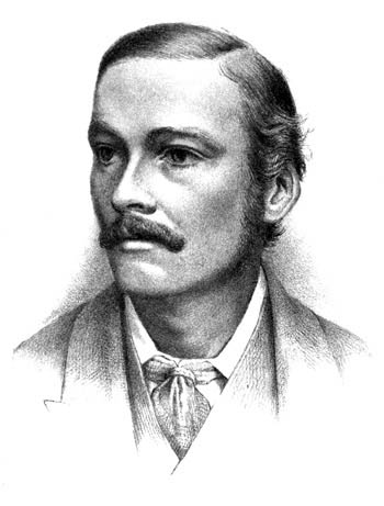
posthumous bust in bronze[1]. The portrait which forms the
frontispiece of Vol. 1. has been drawn on
stone by Mr E. Wilson of the Cambridge Scientific Instrument Company, after
the latest photograph. Should it fail, in the eyes of those who knew
Balfour well, to have reproduced with complete success his features and
expression, we would venture to ask them to bear in mind the acknowledged
difficulties of posthumous portraiture.
| TABLE OF CONTENTS. |
|---|
| PAGE |
| Preface |
i |
| Introduction |
1 |
| 1872 |
| I. | On some points in the
Geology of the East Lothian Coast. By G. W. and F. M.
Balfour | 25 |
| 1873 |
| II. | The development and growth
of the layers of the blastoderm. With Plate 1 | 29 |
| III. | On the disappearance of
the Primitive Groove in the Embryo Chick. With Plate 1 | 41 |
| IV. | The development of the
blood-vessels of the Chick. With Plate 2 | 47 |
| 1874 |
| V. | A preliminary account of the
development of the Elasmobranch Fishes. With Plates 3 and 4 | 60 |
| 1875 |
| VI. | A comparison of the early
stages in the development of Vertebrates. With Plate 5 | 112 |
| VII. | On the origin and history
of the urinogenital organs of Vertebrates | 135 |
| VIII. | On the development of the
spinal nerves in Elasmobranch Fishes. With Plates 22 and 23 | 168 |
| 1876
|
| IX. | On the spinal nerves of
Amphioxus | 197 |
| 1876-78 |
| X. | A Monograph on the
development of Elasmobranch Fishes. With Plates 6-21 | 203 |
| 1878 |
| XI. | On the phenomena
accompanying the maturation and impregnation of the ovum | 521 |
| XII. | On the structure and
development of the vertebrate ovary. With Plates 24, 25, 26 | 549 |
| 1879 |
| XIII. | On the existence of a
Head-kidney in the Embryo Chick, and on certain points in the development
of the Müllerian duct. By F. M. Balfour and A. Sedgwick. With Plates 27 and 28 | 618 |
| XIV. | On the early development
of the Lacertilia, together with some observations on the nature and
relations of the primitive Streak. With Plate 29 | 644 |
| XV. | On certain points in the
Anatomy of Peripatus Capensis | 657 |
| XVI. | On the morphology and
systematic position of the Spongida | 661 |
| 1880 |
| XVII. | Notes on the development
of the Araneina. With Plates 30, 31, 32 | 668 |
| XVIII. | On the spinal nerves of
Amphioxus | 696 |
| XIX. | Address to the Department
of Anatomy and Physiology of the British Association for the Advancement of
Science | 698 |
| 1881 |
| XX. | On the development of the
skeleton of the paired fins of Elasmobranchii, considered in relation to
its bearings on the nature of the limbs of the Vertebrata. With Plate
33 | 714 |
| XXI. | On the evolution of the
Placenta, and on the possibility of employing the characters of the
Placenta in the classification of the Mammalia | 734 |
| 1882
|
| XXII. | On the structure and
development of Lepidosteus. By F. M. Balfour and
W. N. Parker. With Plates 34-42 | 738 |
| XXIII. | On the nature of the
organ in Adult Teleosteans and Ganoids which is usually regarded as the
Head-kidney or Pronephros | 848 |
| XXIV. | A renewed study of the
germinal layers of the Chick. By F. M. Balfour and
F. Deighton. With Plates 43, 44, 45 | 854 |
| | Posthumous,
1883 |
| XXV. | The Anatomy and
Development of Peripatus Capensis. Edited by H. N.
Moseley and A. Sedgwick. With Plates
46-53 | 871 |
Francis Maitland Balfour, the sixth child and third son of
James Maitland Balfour of Whittinghame, East Lothian, and Lady Blanche,
daughter of the second Marquis of Salisbury, was born at Edinburgh, during
a temporary stay of his parents there, on the 10th November, 1851. He can
hardly be said to have known his father, who died of consumption in 1856,
at the early age of thirty-six, and who spent the greater part of the last
two years of his life at Madeira, separated from the younger children who
remained at home. He fancied at one time that he had inherited his father's
constitution; and this idea seems to have spurred him on to achieve early
what he had to do. But, though there was a period soon after he went to
College, during which he seemed delicate, and the state of his health
caused considerable anxiety to his friends, he eventually became fairly
robust, and that in spite of labours which greatly taxed his strength.
The early years of his life were spent chiefly at Whittinghame under the
loving care of his mother. She made it a point to attempt to cultivate in
all her children some taste for natural science, especially for natural
history, and in this she was greatly helped by the boys' tutor, Mr J. W.
Kitto. They were encouraged to make collections and to form a museum, and
the fossils found in the gravel spread in front of the house served as the
nucleus of a geological series. Frank soon became greatly interested in
these things, and indeed they may be said to have formed the beginnings of
his scientific career. At all events there was thus awakened in him a love
for geology, which science continued to be his favorite study all through
his
boyhood, and interested him to the last. He was most assiduous in searching
for fossils in the gravel and elsewhere, and so great was his love for his
collections that while as yet quite a little boy the most delightful
birthday present he could think of was a box with trays and divisions to
hold his fossils and specimens. His mother, thinking that his fondness for
fossils was a passing fancy and that he might soon regret the purchase of
the box, purposely delayed the present. But he remained constant to his
wish and in time received his box. He must at this time have been about
seven or eight years old. In the children's museum, which has been
preserved, there are specimens labelled with his childish round-hand, such
as a piece of stone with the label marks of some shels;
and his
sister Alice, who was at that time his chief companion, remembers
discussing with him one day after the nursery dinner, when he was about
nine years old, whether it were better to be a geologist or a naturalist,
he deciding for the former on the ground that it was better to do one thing
thoroughly than to attempt many branches of science and do them
imperfectly.
Besides fossils, he collected not only butterflies, as do most boys at
some time or other, but also birds; and he with his sister Alice, being
instructed in the art of preparing and preserving skins, succeeded in
making a very considerable collection. He thus acquired before long not
only a very large but a very exact knowledge of British birds.
In the more ordinary work of the school-room he was somewhat backward.
This may have been partly due to the great difficulty he had in learning to
write, for he was not only left-handed but, in his early years, singularly
inapt in acquiring particular muscular movements, learning to dance being a
great trouble to him. Probably however the chief reason was that he failed
to find any interest in the ordinary school studies. He fancied that the
family thought him stupid, but this does not appear to have been the
case.
In character he was at this time quick tempered, sometimes even violent,
and the energy which he shewed in after life even thus early manifested
itself as perseverance, which, when he was crossed, often took on the form
of obstinacy, causing at times no little trouble to his nurses and tutors.
But he was at the same time warm-hearted and affectionate; full
of strong impulses, he disliked heartily and loved much, and in his
affections was wonderfully unselfish, wholly forgetting himself in his
thought for others, and ready to do things which he disliked to please
those whom he loved. Though, as we have said, somewhat clumsy, he was
nevertheless active and courageous; in learning to ride he shewed no signs
of fear, and boldly put his pony to every jump which was practicable.
In 1861 he was sent to the Rev. C. G.
Chittenden's preparatory school at Hoddesden in Hertfordshire, and here the
qualities which had been already visible at home became still more obvious.
He found difficulty not only in writing but also in spelling, and in the
ordinary school-work he took but little interest and made but little
progress.
In 1865 he was moved to Harrow and placed in the house of the Rev. F. Rendall. Here, as at Hoddesden, he did not
show any great ability in the ordinary school studies, though as he grew
older his progress became more marked. But happily he found at Harrow an
opportunity for cultivating that love of scientific studies which was
yearly growing stronger in him. Under the care of one of the Masters, Mr G.
Griffith, the boys at Harrow were even then taught the elements of natural
science. The lessons were at that time, so to speak, extra-academical,
carried on out of school hours; nevertheless, many of the boys worked at
them with diligence and even enthusiasm, and among these Balfour became
conspicuous, not only by his zeal but by his ability. Griffith was soon
able to recognize the power of his new pupil, and thus early began to see
that the pale, earnest, somewhat clumsy-handed lad, though he gave no
promise of being a scholar in the narrower sense of the word, had in him
the makings of a man of science. Griffith chiefly confined his teaching to
elementary physics and chemistry with some little geology, but he also
encouraged natural history studies and began the formation of a museum of
comparative anatomy. Balfour soon began to be very zealous in dissecting
animals, and was especially delighted when the Rev. A. C. Eaton, the well-known entomologist, on a visit
to Harrow, initiated Griffith's pupils in the art of dissecting under
water. The dissection of a caterpillar in this way was probably an epoch in
Balfour's life. Up to that time his rough examination of such bodies had
revealed to him nothing more than what in school-boy language he spoke of
as squash;
but when under Eaton's deft hands the intricate organs of
the larval Arthropod floated out under water and displayed themselves as a
labyrinth of threads and sheets of silvery whiteness a new world of
observation opened itself up to Balfour, and we may probably date from this
the beginning of his exact morphological knowledge.
While thus learning the art of observing, he was at the same time
developing his power of thinking. He was by nature fond of argument, and
defended with earnestness any opinions which he had been led to adopt. He
was very active in the Harrow Scientific Society, reading papers, taking
part in the discussions, and exhibiting specimens. He gained in 1867 a
prize for an essay on coal, and when, in 1868, Mr Leaf offered a prize (a
microscope) for the best account of some locality visited by the writer
during the Easter Holidays,
two essays sent in, one by Balfour, the
other by his close friend, Mr Arthur Evans, since well known for his
researches in Illyria, were found to be of such unusual merit that Prof.
Huxley was specially requested to adjudicate between them. He judged them
to be of equal merit, and a prize was given to each. The subject of
Balfour's essay was The Geology and Natural History of East Lothian.
When biological subjects were discussed at the Scientific Society, Balfour
appears to have spoken as a most uncompromising opponent of the views of Mr
Charles Darwin, little thinking that in after life his chief work would be
to develop and illustrate the doctrine of evolution.
The years at Harrow passed quickly away, Balfour making fair, but
perhaps not more than fair, progress in the ordinary school learning. In
due course however he reached the upper sixth form, and in his last year,
became a monitor. At the same time his exact scientific knowledge was
rapidly increasing. Geology still continued to be his favorite study, and
in this he made no mean progress. During his last years at Harrow he and
his brother Gerald worked out together some views concerning the geology of
their native county. These views they ultimately embodied in a paper, which
was published in their joint names in the Geological Magazine
for 1872, under the title of Some Points in the Geology of the East
Lothian Coast,
and which was in itself a work of considerable promise.
Geology however was beginning to find a rival in natural history. Much of
his holiday time was now spent in dredging for marine animals along the
coast off Dunbar. Each specimen thus obtained was carefully determined and
exact records were kept of the various 'finds,' so that the dredgings
(which were zealously continued after he had left Harrow and gone to
Cambridge) really constituted a serious study of the fauna of this part of
the coast. They also enabled him to make a not inconsiderable collection of
shells, in the arrangement of which he was assisted by his sister Evelyn,
of crustacea and of other animals.
Both to the masters and to his schoolfellows he became known as a boy of
great force of character. Among the latter his scrupulous and unwavering
conscientiousness made him less popular perhaps than might have been
expected from his bright kindly manner and his unselfish warmheartedness.
In the incidents of school life a too strict conscience is often an
inconvenience, and the sternness and energy with which Balfour denounced
acts of meanness and falsehood were thought by some to be unnecessarily
great. He thus came to be feared rather than liked by many, and
comparatively few grew to be sufficiently intimate with him to appreciate
the warmth of his affections and the charm of his playful moments.
At the Easter of 1870 he passed the entrance examination at Trinity
College, Cambridge, and entered into residence in the following October.
His college tutor was Mr J. Prior, but he was from the first assisted and
guided in his studies by his friend, Mr Marlborough Pryor, an old Harrow
boy, who in the same October had been, on account of his distinction in
Natural Science, elected a Fellow of the College, in accordance with
certain new regulations which then came into action for the first time, and
which provided that every three years one of the College Fellowships should
be awarded for excellence in some branch or branches of Natural Science, as
distinguished from mathematics, pure or mixed. During the whole of that
year and part of the next Mr Marlborough Pryor remained in residence, and
his influence in wisely directing Balfour's studies had a most beneficial
effect on the latter's progress.
During his first term Balfour was occupied in preparation for the Previous
Examination; and this he successfully passed at Christmas. After that he
devoted himself entirely to Natural Science, attending lectures on several
branches. During the Lent term he was a very diligent hearer of the
lectures on Physiology which I was then giving as Trinity Prælector, having
been appointed to that post in the same October that Balfour came into
residence. At this time he was not very strong, and I remember very well
noticing among my scanty audience, a pale retiring student, whose mind
seemed at times divided between a desire to hear the lecture and a feeling
that his frequent coughing was growing an annoyance to myself and the
class. This delicate-looking student, I soon learnt, was named Balfour, and
when the Rev. Coutts Trotter, Mr Pryor and
myself came to examine the candidates for the Natural Science Scholarships
which were awarded at Easter, we had no difficulty in giving the first
place to him. In point of knowledge, and especially in the thoughtfulness
and exactitude displayed in his papers and work, he was very clearly ahead
of his competitors.
During the succeeding Easter term and the following winter he appears to
have studied physics, chemistry, geology and comparative anatomy, both
under Mr Marlborough Pryor and by means of lectures. He also continued to
attend my lectures, but though I gradually got to know him more and more we
did not become intimate until the Lent term of 1872. He had been very much
interested in some lectures on embryology which I had given, and, since
Marlborough Pryor had left or was about to leave Cambridge, he soon began
to consult me a good deal about his studies. He commenced practical
histological and embryological work under me, and I remember very vividly
that one day when we were making a little excursion in search of nests and
eggs of the stickleback in order that he might study the embryology of
fishes, he definitely asked my opinion as to whether he might take up a
scientific career with a fair chance of success. I had by this time formed
a very high opinion of his abilities, and learning then for the first time
that he had an income independent of his own exertions, my answer was very
decidedly a positive one. Soon after, feeling more and more impressed with his
power and increasingly satisfied both with his progress in biological
studies and his sound general knowledge of other sciences, anxious also, it
may be, at the same time that as much original inquiry as possible should
be carried on at Cambridge in my department, I either suggested to him or
acquiesced in his own suggestion that he should at once set to work on some
distinct research; and as far as I remember the task which I first proposed
to him was an investigation of the layers of the blastoderm in the chick.
It must have been about the same time that I proposed to him to join me in
preparing for publication a small work on Embryology, the materials for
this I had ready to hand in a rough form as lectures which I had previously
given. To this proposal he enthusiastically assented, and while the lighter
task of writing what was to be written fell to me, he undertook to work
over as far as was possible the many undetermined points and unsatisfactory
statements across which we were continually coming.
During his two years at College his health had improved; though still
hardly robust and always in danger of overworking himself, he obviously
grew stronger. He rejoiced exceedingly in his work, never tiring of it, and
was also making his worth felt among his fellow students, and especially
perhaps among those of his own college whose studies did not lie in the
same direction as his own. At this time he must have been altogether happy,
but a sorrow now came upon him. His mother, to whom he was passionately
attached, and to whose judicious care in his early days not only the right
development of his strong character but even his scientific leanings were
due, had for some time past been failing in health, though her condition
caused no immediate alarm. In May 1872, however, she died quite suddenly
from unsuspected heart disease. Her loss was a great blow to him, and for
some time afterward I feared his health would give way; but he bore his
grief quietly and manfully and threw himself with even increased vigour
into his work.
During the academic session of 1872-3, he continued steadily at work at
his investigations, and soon began to make rapid progress. At the beginning
he had complained to me about what he considered his natural clumsiness,
and expressed a fear that he should never be able to make
satisfactory microscopic sections; as to his being able to make drawings of
his dissections and microscopical preparations, he looked upon that at
first as wholly impossible. I need hardly say that in time he acquired
great skill in the details of microscopical technique, and that
his drawings, if wanting in so-called artistic finish, were always
singularly true and instructive. While thus struggling with the details
which I could teach him, he soon began to manifest qualities which no
teacher could give him. I remember calling his attention to Dursy's paper
on the Primitive Streak, and suggesting that he should work the matter
over, since if such a structure really existed, it must, most probably,
have great morphological significance. I am free to confess that I myself
rather doubted the matter, and a weaker student might have been influenced
by my preconceptions. Balfour, however, thus early had the power of seeing
what existed and of refusing to see what did not exist. He was soon able to
convince me that Dursy's streak was a reality, and the complete working out
of its significance occupied his thoughts to the end of his days.
The results of these early studies were made known in three papers which
appeared in the Quarterly Journal of Microscopical Science for
July 1873, and will be found in the beginning of this volume. The summer
and autumn of that year were spent partly in a visit to Finland, in company
with his friend and old school-fellow Mr Arthur Evans, and partly in formal
preparation for the approaching Tripos examination. Into this preparation
Balfour threw himself with characteristic energy, and fully justified my
having encouraged his spending so much of the preceding time in original
research, not only by the rapidity with which he accumulated the stock of
knowledge of various kinds necessary for the examination but also by the
manner in which he acquitted himself at the trial itself. At that time the
position of the candidates in the Natural Sciences Tripos was determined by
the total number of marks, and Balfour was placed second, the first place
being gained by H. Newell Martin of Christ's College, now Professor at
Baltimore, U.S.A. In the examination, in which I took part, Balfour did not
write much, and he had not yet learnt the art of putting his statements in
the best possible form; he won his position chiefly by the firm thought
and clear insight which was present in almost all his answers.
The examination was over in the early days of Dec. 1873 and Balfour was now free to devote
himself wholly to his original work. Happily, the University had not long
before secured the use of two of the tables at the then recently founded
Stazione Zoologica at Naples. And upon the nomination of the University,
Balfour, about Christmas, started for Naples in company with his friend Mr
A. G. Dew-Smith, also of Trinity College. The latter was about to carry on
some physiological observations; Balfour had set himself to work out as
completely as he could the embryology of Elasmobranch fishes, about which
little was at that time known, but which, from the striking characters of
the adult animals could not help proving of interest and importance.
From his arrival there at Christmas 1873 until he left in June 1874, he
worked assiduously, and with such success, that as the result of the
half-year's work he had made a whole series of observations of the greatest
importance. Of these perhaps the most striking were those on the
development of the urogenital organs, on the neurenteric canal, on the
development of the spinal nerves, on the formation of the layers and on the
phenomena of segmentation, including a history of the behaviour of nuclei
in cell division. He returned home laden with facts and views both novel
and destined to influence largely the progress of embryology.
In August of the same year he attended the meeting of the British
Association for the Advancement of Science at Belfast; and the account he
then gave of his researches formed one of the most important incidents at
the Biological Section on that occasion.
In the September of that year the triennial fellowship for Natural
Science was to be awarded at Trinity College, and Balfour naturally was a
candidate. The election was, according to the regulations, to be determined
partly by the result of an examination in various branches of science, and
partly by such evidence of ability and promise as might be afforded by
original work, published or in manuscript. He spent the remainder of the
autumn in preparation for this examination. But when the examination was
concluded it was found that in his written answers he had not been very
successful; he had not even acquitted himself so well as in the Tripos of
the year before, and had the election been determined by the results of the
examination alone, the examiners would have been led to choose the
gentleman who was Balfour's only competitor. The original work however
which Balfour sent in, including a preliminary account of the discoveries
made at Naples, was obviously of so high a merit and was spoken of in such
enthusiastic terms by the External Referee Prof. Huxley, that the examiners
did not hesitate for a moment to neglect altogether the formal written
answers (and indeed the papers of questions were only introduced as a
safeguard, or as a resource in case evidence of original power should be
wanted) and unanimously recommended him for election. Accordingly he was
elected Fellow in the early days of October.
Almost immediately after, the little book on Embryology appeared, on
which he and I had been at work, he doing his share even while his hands
and mind were full of the Elasmobranch inquiry. The title-page was kept
back some little time in order that his name might appear on it with the
addition of Fellow of Trinity, a title of which he was then, and indeed
always continued to be, proud. He also published in the October number of
the Quarterly Journal of Microscopical Science a preliminary
account of his Elasmobranch researches.
He and his friends thought that after these almost incessant labours,
and the excitement necessarily contingent upon the fellowship election, he
needed rest and change. Accordingly on the 17th of October he started with
his friend Marlborough Pryor on a voyage to the west coast of South
America. They travelled thither by the Isthmus of Panama, visited Peru and
Chili, and returned home along the usual route by the Horn; reaching
England some time in Feb. 1875.
Refreshed by this holiday, he now felt anxious to complete as far as
possible his Elasmobranch work, and very soon after his return home, in
fact in March, made his way again to Naples, where he remained till the hot
weather set in in May. On his return to Cambridge, he still
continued working on the Elasmobranchii, receiving material partly from
Naples, partly from the Brighton Aquarium, the then director of which, Mr
Henry Lee, spared no pains to provide him both with embryo and adult
fishes. While at Naples, he communicated to the Philosophical Society at
Cambridge a remarkable paper on The Early Stages of Vertebrates,
which was published in full in the Quarterly Journal of Microscopical
Science, July, 1875; he also sent me a paper on The Development
of the Spinal Nerves
, which I communicated to the Royal Society, and
which was subsequently published in the Philosophical
Transactions of 1876. He further wrote in the course of the summer
and published in the Journal of Anatomy and Physiology in
October, 1875, a detailed account of his Observations and Views on the
Development of the Urogenital Organs.
Some time in August of the same year he started in company with Mr
Arthur Evans and Mr J. F. Bullar for a second trip to Finland, the
travellers on this occasion making their way into regions very seldom
visited, and having to subsist largely on the preserved provisions which
they carried with them, and on the produce of their rods and guns. From a
rough diary which Balfour kept during this trip it would appear that while
enjoying heartily the fun of the rough travelling, he occupied himself
continually with observations on the geology and physical phenomena of the
country, as well as on the manners, antiquities, and even language of the
people. It was one of his characteristic traits, a mark of the truly
scientific bent of his mind, of his having, as Dohrn soon after Balfour's
first arrival at Naples said, 'a real scientific head,' that every thing
around him wherever he was, incited him to careful exact observation, and
stimulated him to thought.
In the early part of the Long Vacation of the same year he had made his
first essay in lecturing, having given a short course on Embryology in a
room at the New Museums, which I then occupied as a laboratory. Though he
afterwards learnt to lecture with great clearness he was not by nature a
fluent speaker, and on this occasion he was exceedingly nervous. But those
who listened to him soon forgot these small defects as they began to
perceive the knowledge and power which lay in their new teacher.
Encouraged by the result of this experiment, he threw himself, in spite
of the heavy work which the Elasmobranch investigation was entailing, with
great zeal into an arrangement which Prof. Newton, Mr J. W. Clark and
myself had in course of the summer brought about, that he and Mr A. Milnes
Marshall, since Professor at Owens College, Manchester, should between them
give a course on Animal Morphology, with practical instruction, Prof.
Newton giving up a room in the New Museums for the purpose.
In the following October (1875) upon Balfour's return from Finland,
these lectures were accordingly begun and carried on by the two lecturers
during the Michaelmas and Lent Terms. The number of students attending this
first course, conducted on a novel plan, was, as might be expected, small,
but the Lent Term did not come to an end before an enthusiasm for
morphological studies had been kindled in the members of the class.
The ensuing Easter term (1876) was spent by Balfour at Naples, in order
that he might carry on towards completion his Elasmobranch work. He had by
this time determined to write as complete a monograph as he could of the
development of these fishes, proposing to publish it in instalments in the
Journal of Anatomy and Physiology, and subsequently to gather
together the several papers into one volume. The first of these papers,
dealing with the ovum, appeared in Jan. 1876;
most of the numbers of the Journal during that and the succeeding year
contained further portions; but the complete monograph did not leave the
publisher's hands until 1878.
He returned to England with his pupil and friend Mr J. F. Bullar some
time in the summer; on their way home they passed through Switzerland, and
it was during the few days which he then spent in sight of the snow-clad
hills that the beginnings of a desire for that Alpine climbing, which was
destined to be so disastrous, seem to have been kindled in him.
In October, 1876, he resumed the lectures on Morphology, taking the
whole course himself, his colleague, Mr Marshall, having meanwhile left
Cambridge. Indeed, from this time onward, he may be said to have made these
lectures, in a certain sense, the chief business of his life. He lectured
all three terms, devoting the Michaelmas and Lent terms to a systematic
course of Animal Morphology, and the Easter term to a more elementary
course of Embryology. These lectures were given under the auspices of Prof.
Newton; but Balfour's position was before long confirmed by his being made
a Lecturer of Trinity College, the lectures which he gave at the New
Museums, and which were open to all students of the University, being
accepted in a liberal spirit by the College as equivalent to College
Lectures. He very soon found it desirable to divide the morphological
course into an elementary and an advanced course, and to increase the
number of his lectures from three to four a week. Each lecture was followed
by practical work, the students dissecting and examining microscopically,
an animal or some animals chosen as types to illustrate the subject-matter
of the lecture; and although Balfour had the assistance at first of one[2], and ultimately of several demonstrators, he himself put
his hand to the plough, and after the lecture always spent some time in the
laboratory among his pupils. Had Balfour been only an ordinary man, the
zeal and energy which he threw into his lectures, and into the supervision
of the practical work, added to the almost brotherly interest which he took
in the individual development of every one of the pupils who shewed any
love whatever for the subject, would have made him a most successful
teacher. But his talents and powers were such as could not be hid even from
beginners. His extensive and exact knowledge, the clearness with which in
spite of, or shall I not rather say, by help of a certain want of fluency,
he explained difficult and abstruse matters, the trenchant way in which he
lay bare specious fallacies, and the presence in almost his every word of
that power which belongs only to the man who has thought out for himself
everything which he says, these things aroused and indeed could hardly fail
to arouse in his hearers feelings which, except in the case of the very
dullest, grew to be those of enthusiasm. His class, at first slowly, but
afterwards more rapidly, increased in numbers, and, what is of more
importance, grew in quality. The room allotted to him soon became far too
small and steps were taken to provide for him, for myself, whose wants were
also urgent, and for the biological studies generally, adequate
accommodation; but it was not until Oct. 1877
that we were able to take possession of the new quarters.
Even this new accommodation soon became insufficient, and in the spring
of 1882 a new morphological laboratory was commenced in accordance with
plans suggested by himself. He was to have occupied them in the October
term, 1883, but did not live to see them finished.
As might have been expected from his own career, he regarded the mere
teaching of what is known as a very small part of his duties as Lecturer;
and as soon as any of his pupils became sufficiently advanced, he urged or
rather led them to undertake original investigations; and he had the
satisfaction before his death of seeing the researches of his pupils (such
as those by Messrs. Bullar, Sedgwick, Mitzikuri, Haddon, Scott, Osborne,
Caldwell, Heape, Weldon, Parker, Deighton and others) carried to a
successful end. In each of these inquiries he himself took part, sometimes
a large part, generally suggesting the problem to be solved, indicating the
methods, and keeping a close watch over the whole progress of the study.
Hence in many cases the published account bears his name as well as that of
the pupil.
In the year 1878 his Monograph on Elasmobranch Fishes was
published as a complete volume, and in the same year he received the honour
of being elected a Fellow of the Royal Society, a distinction which
now-a-days does not often fall to one so young. No sooner was the Monograph
completed than in spite of the labours which his lectures entailed, he set
himself to the great task of writing a complete treatise on
Comparative Embryology. This not only laid upon him the heavy
burden of gathering together the observations of others, enormous in number
and continually increasing, scattered through many journals and books, and
recorded in many different languages, as well as of putting them in orderly
array, and of winnowing out the grain from the chaff (though his
critical spirit found some relief in the latter task), but also caused him
much labour, inasmuch as at almost every turn new problems suggested
themselves, and demanded inquiry before he could bring his mind to writing
about them. This desire to see his way straight before him, pursued him
from page to page, and while it has resulted in giving the book an almost
priceless value, made the writing of it a work of vast labour. Many of the
ideas thus originated served as the bases of inquiries worked out by
himself or his pupils, and published in the form of separate papers, but
still more perhaps never appeared either in the book or elsewhere and were
carried with him undeveloped and unrecorded to the grave.
The preparation of this work occupied the best part of his time for the
next three years, the first volume appearing in 1880, the second in
1881.
In the autumn of 1880, he attended the Meeting at Swansea of the British
Association for the Advancement of Science, having been appointed
Vice-President of the Biological Section with charge of the Department of
Anatomy and Physiology. At the Meetings of the Association, especially of
late years, much, perhaps too much, is expected in the direction of
explaining the new results of science in a manner interesting to the
unlearned. Popular expositions were never very congenial to Balfour, his
mind was too much occupied with the anxiety of problems yet to be solved;
he was therefore not wholly at his ease, in his position on this occasion.
Yet his introductory address, though not of a nature to interest a large
mixed audience, was a luminous, brief exposition of the modern development
and aims of embryological investigation.
During these years of travail with the Comparative
Embryology the amount of work which he got through was a marvel to
his friends, for besides his lectures, and the researches, and the writing
of the book, new labours were demanded of him by the University for which
he was already doing so much. Men at Cambridge, and indeed elsewhere as
well, soon began to find out that the same clear insight which was solving
biological problems could be used to settle knotty questions of policy
and business. Moreover he united in a remarkable manner, the power of
boldly and firmly asserting and maintaining his own views, with a frank
courteousness which went far to disarm opponents. Accordingly he found
himself before long a member of various Syndicates, and indeed a very great
deal of his time was thus occupied, especially with the Museums and Library
Syndicates, in both of which he took the liveliest interest. Besides these
University duties his time and energy were also at the service of his
College. In the preparation of the New Statutes, with which about this time
the College was much occupied, the Junior Fellows of the College took a
conspicuous share, and among these Junior Fellows Balfour was perhaps the
most active; indeed he was their leader, and he threw himself into the
investigation of the bearings and probable results of this and that
proposed new statute with as much zeal as if he were attacking some
morphological problem.
While he was in the midst of these various labours, his friends often
feared for his strength, for though gradually improving in health after his
first year at Cambridge, he was not robust, and from time to time he seemed
on the point of breaking down. Still, hard as he was working, he was in
reality wisely careful of himself, and as he grew older, paid more and more
attention to his health, daily taking exercise in the form either of
bicycle rides or of lawn-tennis. Moreover he continued to spend some part
of his vacations in travel. Combining business with pleasure, he made
frequent visits to Germany and France, and especially to Naples. The
Christmas of 1876-7 he spent in Greece, that of 1878-9 at Ragusa, where his
old school-fellow and friend Mr Arthur Evans was at that time residing, and
the appointment of his friend Kleinenberg to a Professorship at Messina led
to a journey there. Early in the long vacation of 1880, he went with his
sister, Mrs H. Sidgwick, and her husband to Switzerland, and was joined
there for a short time by his friend and pupil Adam Sedgwick. During this
visit he took his first lessons in Alpine climbing, making several
excursions, some of them difficult and dangerous; and the love of
mountaineering laid so firm a hold upon him, that he returned to
Switzerland later on in the autumn of the same year, in company with his
brother Gerald, and spent some weeks near Zermatt in systematic climbing,
ascending, among other mountains, the Matterhorn and the Weisshorn. In the
following summer, 1881, he and his brother Gerald again visited the Alps,
dividing their time between the Chamonix district and the Bernese Oberland.
On this occasion some of the excursions which they made were of extreme
difficulty, and such as needed not only great presence of mind and bodily
endurance, but also skilful and ready use of the limbs. As a climber indeed
Balfour soon shewed himself fearless, indefatigable, and expert in all
necessary movements as well as full of resources and expedients in the face
of difficulties, so much so that he almost at once took rank among the
foremost of distinguished mountaineers. In spite of his apparent clumsiness
in some matters, he had even as a lad proved himself to be a bold and
surefooted climber. Moreover he had been perhaps in a measure prepared for
the difficulties of Alpine climbing by his experience in deer-stalking.
This sport he had keenly and successfully pursued for many years at his
brother's place in Rosshire. When however about the year 1877, the question
of physiological experiments on animals became largely discussed in public,
he felt that to continue the pursuit of this or any other sport involving,
for the sake of mere pleasure, the pain and death of animals, was
inconsistent with the position which he had warmly taken up, as an advocate
of the right to experiment on animals; and he accordingly from that time
onward wholly gave it up.
His fame as an investigator and teacher, and as a man of brilliant and
powerful parts, was now being widely spread. Pupils came to him, not only
from various parts of England, but from America, Australia and Japan. At
the York Meeting of the British Association for the Advancement of Science,
in August, 1881, he was chosen as one of the General Secretaries. In April,
1881, the honorary degree of LL.D. was conferred upon him by the University
of Glasgow, and in November of the same year the Royal Society gave him one
of the Royal Medals in recognition of his embryological discoveries, and at
the same time placed him on its Council.
At Cambridge he was chosen, in the autumn of 1880, President of the
Philosophical Society, and in the December of that year a brilliant
company were gathered together at the Annual Dinner to do honour to their
new young President. Otherwise nothing as yet had been done for him in his
own University in the way of recognition of his abilities and services; and
he still remained a Lecturer of Trinity College, giving lectures in a
University building. An effort had been made by some of his friends to urge
the University to take some step in this direction; but it was thought at
that time impossible to do anything. In 1881 a great loss fell upon the
sister University of Oxford in the death of Prof. George Rolleston; and
soon after very vigorous efforts were made to induce Balfour to become a
candidate for the vacant chair. The prospect was in many ways a tempting
one, and Balfour seeing no very clear way in the future for him at his own
University, was at times inclined to offer himself, but eventually he
decided to remain at Cambridge. Hardly had this temptation if we may so
call it been overcome when a still greater one presented itself. Through
the lamented death of Sir Wyville Thomson in the winter of 1881-2, the
chair of Natural History at Edinburgh, perhaps the richest and most
conspicuous biological chair in the United Kingdom, became vacant. The post
was in many ways one which Balfour would have liked to hold. The teaching
duties were it is true laborious, but they had in the past been compressed
into a short time, occupying only the summer session and leaving the rest
of the year free, and it seemed probable that this arrangement might be
continued with him. The large emolument would also have been grateful to
him inasmuch as he would have felt able to devote the whole of it to
scientific ends; and the nearness to Whittinghame, his native place and
brother's home, added to the attractions; but what tempted him most was the
position which it would have given him, and the opportunities it would have
afforded, with the rich marine Fauna of the north-eastern coast close at
hand, to develop a large school of Animal Morphology. The existing
Professors at Edinburgh were most desirous that he should join them, and
made every effort to induce him to come. On the part of the Crown, in whose
hands the appointment lay, not only were distinct offers made to him, but
he was repeatedly pressed to accept the post. Nor was it until after a
considerable struggle that he finally refused, his love for his own
University in the end overcoming the many inducements to leave; he elected
to stay where he was, trusting to the future opening up for him some
suitable position. In this decision he was undoubtedly influenced by the
consideration that Cambridge, besides being the centre of his old
friendships, had become as it were a second home for his own family. By the
appointment of Lord Rayleigh to the chair of Experimental Physics his
sister Lady Rayleigh had become a resident, his sister Mrs Sidgwick had
lived there now for some years, and his brother Gerald generally spent the
summer there; their presence made Cambridge doubly dear to him.
At the close of the Michaelmas term, with feelings of relief at having
completed his Comparative Embryology, the preparation of the
second volume of which had led to almost incessant labour during the
preceding year, he started to spend the Christmas vacation with his friend
Kleinenberg at Messina. Stopping at Naples on his way thither he found his
pupil Caldwell, who had been sent to occupy the University table at the
Stazione Zoologica, lying ill at Capri, with what proved to be typhoid
fever. The patient was alone, without any friend to tend him, and his
mother who had been sent for had not yet arrived. Accordingly Balfour (with
the kindness all forgetful of himself which was his mark all his life
through) stayed on his journey to nurse the sick man until the mother came.
He then went on to Messina, and there seemed to be in good health, amusing
himself with the ascent of Etna. Yet in January, soon after his return
home, he complained of being unwell, and in due time distinct symptoms of
typhoid fever made their appearance. The attack at first promised to be
severe, but happily the crisis was soon safely passed and the convalescence
was satisfactory.
While yet on his sick bed, a last attempt was made to induce him to
accept the Edinburgh offer, and for the last time he refused. These
repeated offers, and the fact that the dangers of his grave illness had led
the University vividly to realize how much they would lose if Balfour were
taken away from them, encouraged his friends to make a renewed effort to
gain for him some adequate position in the University. This time the
attempt was successful, and the authorities took a step, unusual but
approved of by the whole body of resident members of the University; they
instituted a new Professorship of Animal Morphology, to be held by Balfour
during his life or as long as he should desire, but to terminate at his
death or resignation unless it should be otherwise desirable. Accordingly
in May, 1882, he was admitted into the Professoriate as Professor of Animal
Morphology.
During his illness his lectures had been carried on by his Demonstrator,
Mr Adam Sedgwick, who continued to take his place during the remainder of
that Lent Term and during the ensuing Easter Term. The spring Balfour spent
partly in the Channel Islands with his sister Alice, partly in London with
his eldest brother, but in the course of the Easter Term returned to
Cambridge and resumed his work though not his lectures. His recovery to
health was steady and satisfactory, the only drawback being a swelling over
the shin-bone of one leg, due to a blow on the rocks at Sark; otherwise he
was rapidly becoming strong. He himself felt convinced that a visit to the
Alps, with some mountaineering of not too difficult a kind, would complete
his restoration to health. In this view many of his friends coincided; for
the experience of former years had shewn them what a wonderfully beneficial
effect the Alpine air and exercise had upon his health. He used to go away
pale, thin and haggard, to return bronzed, clear, firm and almost stout;
nor was there anything in his condition which seemed to forbid his
climbing, provided that he was cautious at the outset. Accordingly, early
in June he left Cambridge for Switzerland, having long ago, during his
illness in fact, engaged his old guide, Johann Petrus, whom he had first
met in 1880, and who had always accompanied him in his expeditions
since.
His first walking was in the Chamonix district; and here he very soon
found his strength and elasticity come back to him. Crossing over from
Montanvert to Courmayeur, by the Col du Géant, he was attracted by the peak
called the Aiguille Blanche de Peuteret, a virgin peak, the ascent of which
had been before attempted but not accomplished. Consulting with Petrus he
determined to try it, feeling that the fortnight, which by this time
he had spent in climbing, had brought back to him his old vigour, and that
his illness was already a thing of the past.
There is no reason to believe that he regarded the expedition as one of
unusual peril; and an incident which at the time of his death was thought
by some to indicate this was in reality nothing more than a proof of his
kindly foresight. The guide Petrus was burdened by a debt on his land
amounting to about £150. In the previous year Balfour and his brother had
come to know of this debt; and, seeing that no Alpine ascent is free from
danger, that on any expedition some accident might carry them off, had
conceived the idea of making some provision for Petrus' family in case he
might meet with sudden death in their service. This suggestion of the
previous year Balfour carried out on this occasion, and sent home to his
brother Gerald a cheque of £150 for this purpose. But the cheque was sent
from Montanvert before he had even conceived the idea of ascending the
Aiguille Blanche. It was not a provision for any specially dangerous
ascent, and must be regarded as a measure prompted not by a sense of coming
peril but rather by the donor's generous care for his servant.
On Tuesday afternoon, July 18, he and Petrus, with a porter to carry
provisions and firing to their sleeping-place on the rocks, set out from
Courmayeur, the porter returning the same night. They expected to get back
to Courmayeur some time on the Thursday, but the day passed without their
appearing. This did not cause any great anxiety because it was supposed
that they might have found it more convenient to pass over to the Chamonix
side than to return to Courmayeur. When on Friday however telegrams
dispatched to Chamonix and Montanvert brought answers that nothing had been
seen of them, it became evident that some accident had happened, and an
exploring party set out for the hills. It was not until early on the Sunday
morning that this search party found the bodies, both partly covered with
snow, lying on the Glacier de Fresney, below the impassable icefall which
separates the upper basin of the glacier from the lower portion, and at the
foot of a couloir which descends by the side of the
icefall. Their tracks were visible on the snow at the top of the couloir. Balfour's neck was broken, and his skull fractured
in
three places; Petrus' body was also fractured in many places. The exact
manner of their death will never be known, but there can be no doubt that,
in Balfour's case at all events, it was instantaneous, and those competent
to form a judgment are of opinion that they were killed by a sudden fall
through a comparatively small height, slipping on the rocks as they were
descending by the side of the ice-fall, and not precipitated from the top
of the couloir. There is moreover indirect evidence which
renders it probable that in the fatal fall Petrus slipped first and carried
Balfour with him. Whether they had reached the summit of the Aiguille and
were returning home after a successful ascent or whether they were making
their way back disheartened and wearied with failure, is not and perhaps
never will be known. Since the provisions at the sleeping-place were
untouched, the deaths probably took place on Wednesday the 19th. The
bringing down the bodies proved to be a task of extreme difficulty, and it
was not till Wednesday the 26th that the remains reached Courmayeur, where
M. Bertolini, the master of the hotel, and indeed everyone, not least the
officers of a small body of Italian troops stationed there, shewed the
greatest kindness and sympathy to Balfour's brothers, Gerald and Eustace,
who hastened to the spot as soon as the news of the terrible disaster was
telegraphed home. Mr Walter Leaf also and Mr Conway, friends of Balfour,
the former a very old one, who had made their way to Courmayeur from other
parts of Switzerland as soon as they heard of the accident, rendered great
assistance. The body was embalmed, brought to England, and buried at
Whittinghame on Saturday, Aug. 5, the Fellows
of Trinity College holding a service in the College Chapel at the same
time.
In person he was tall, being fully six feet in height, well built though
somewhat spare. A broad forehead overhanging deeply set dark brown eyes
whose light shining from beneath strongly marked eye-brows told all the
changes of his moods, slightly prominent cheek-bones, a pale skin, at times
inclined to be even sallow, dark brown hair, allowed to grow on the face
only as a small moustache, and slight whiskers, made up a countenance which
bespoke at once strength of character and delicacy of constitution. It was
an open countenance, hiding nothing, giving sign at once, both when his
body was weary or weak, and when his mind was gladdened, angered or
annoyed.
The record of some of his thoughts and work, all that he had given to
the world will be found in the following pages. But who can tell the ideas
which had passed into his quick brain, but which as yet were known only to
himself, of which he had given no sign up to that sad day on which he made
the fatal climb? And who can say whither he might not have reached had he
lived, and his bright young life ripened as years went on? This is not the
place to attempt any judgment of his work: that may be left to other times,
and to other hands; but it may be fitting to place here on record a letter
which shews how much the greatest naturalist of this age appreciated his
younger brother. Among Balfour's papers was found a letter from Charles
Darwin, acknowledging the receipt of Vol. II.
of the Comparative Embryology in the following words:
"July 6, 1881.
Down, Beckenham, Kent.
My Dear Balfour,
I thank you heartily for the present of your grand
book, and I congratulate you on its completion. Although I read almost all
of Vol. I, I do not feel that I am worthy of
your present, unless indeed the fullest conviction that it is a memorable
work makes me worthy to receive it.
* * * * *
Once again accept my thanks, for I am proud to
receive a book from you, who, I know, will some day be the chief of the
English Biologists.
Believe me,
Yours sincerely,
Charles Darwin."
The loss of him was a manifold loss. He is mourned, and will long be
mourned, for many reasons. Some miss only the brilliant investigator;
others feel that their powerful and sympathetic teacher is gone; some look
back on his memory and grieve for the charming companion whose
kindly courtesy and bright wit made the hours fly swiftly and pleasantly
along; and to yet others is left an aching void when they remember that
they can never again lean on the friend whose judgment seemed never to fail
and whose warm-hearted affection was a constant help. And to some he was
all of these. At the news of his death the same lines came to the lips of
all of us, so fittingly did Milton's words seem to speak our loss and grief—
"For Lycidas is dead, dead ere his prime,
Young Lycidas, and hath not left his
peer."
M. FOSTER.
By G. W. and F. M.
Balfour, Trinity College, Cambridge.
The interesting relation between the Porphyrite of Whitberry
Point, at the mouth of the Tyne, near Dunbar, and the adjacent sedimentary
rocks, was first noticed, we believe, by Professor Geikie, who speaks of it
in the Memoirs of the Geological Survey of East Lothian, pages
40 and 31, and again in the new edition of Jukes's Geology,
p. 269. The volcanic mass which forms the point consists of a dark
felspathic base with numerous crystals of augite: it is circular in form,
and is exposed for two-thirds of its circumference in a vertical precipice
facing the sea, about twenty feet in height.
The rock is traversed by numerous joints running both in a horizontal
and in a vertical direction. The latter are by far the most conspicuous,
and give the face of the cliff, when seen from a distance, a well-marked
columnar appearance, though the columns themselves are not very distinct or
regular. They are quadrangular in form, and are evidently produced by the
intersection at right-angles of the two series of vertical joints.
It is clear that the face of the precipice has been gradually receding
in proportion as it yielded to the action of the waves; and that at a
former period the volcanic rock extended considerably further than at
present over the beds which are seen to dip beneath it. These latter
consist of hard fine-grained calcareous sandstones belonging to the Lower
Carboniferous formation. Their colour varies from red to white, and their
prevailing dip is in a N.W. direction, with an average inclination of
12-20°. If the volcanic mass is a true intrusive rock, we should naturally
expect the strata which surround it to dip away in all directions,
the amount of their inclination diminishing in proportion to their
distance from it. We find, however, that the case is precisely the reverse:
as the beds approach the base of the cliff, they dip towards it
from every side at perpetually increasing angles, until at the point of
junction the inclination amounts in places to as much as 55 degrees. The
exact amount of dip in the various positions will be seen on referring to
the accompanying map.
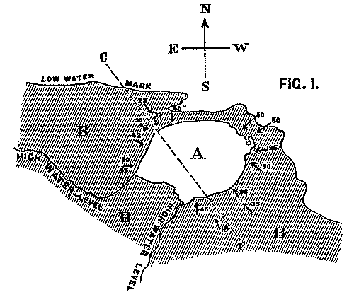
Fig. 1. Map of Strata at Whitberry
Point. Scale, 6 in. to the mile.
A. Lava sheet. B. Sandstone Beds, dipping
from every side towards the lava. CC.
Line of Section along which Fig. 2 is supposed to be drawn.
We conceive that the phenomenon is to be explained by supposing the
orifice through which the lava rose and overflowed the surface of the
sedimentary strata to have been very much smaller in area than the extent
of igneous rock at present visible; and that the pressure of the erupted
mass on the soft beds beneath, aided perhaps by the abstraction of matter
from below, caused them to incline towards the central point at a gradually
increasing angle. The diagram, fig. 2, will serve further to illustrate
this hypothesis. A is the neck or orifice by which the melted matter
is supposed to ascend. C shews the sheet of lava after it has
overspread the surface of the sandstone beds B, so as to cause them
to assume their present inclination. The dotted lines represent the
hypothetical extension of the igneous mass and sandstones previous to the
denudation which they have suffered from the action of the waves.
Professor Geikie, in his admirable treatise on the Geology of the
county[4], adopts a view on this subject which is somewhat
different from that which is suggested in this paper. He considers that the
whole mass is an intrusive neck of rock with perpendicular sides; and that
it once filled up an orifice through the surrounding sedimentary strata, of
which it is now the only remnant.
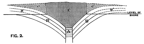
Fig. 2. Vertical Section through CC. Diagram (Fig. 1).
A. Orifice by which the lava ascended. B.
Sandstone Beds. B´. Hypothetical extension of ditto. C.
Sheet of lava spread over the sandstones B. C´. Hypothetical
extension of ditto.
He admits that the inclination of the sandstone beds towards the igneous
mass in the centre is a phenomenon that is somewhat difficult to explain,
and suggests that a subsequent contraction of the column may have tended to
produce such a result. To use his own words: In the case of a solid
column of felstone or basalt, the contraction of the melted mass on cooling
may have had some effect in dragging down the sides of the orifice[5].
But, apart from other objections, it is scarcely conceivable that this
result should have been produced by the contraction of the column.
In his recent edition of Jukes's Manual of Geology
(p. 269), in which he also refers to this instance, he states that in
other cases of necks
it is found to be an almost invariable rule,
that strata are bent down so as to dip into the neck all round its
margin.
We are not aware to what other instances Prof. Geikie may
allude; but on referring to his Memoir on the Geology of East
Lothian, we find that he states in the cases of 'North Berwick Law'
and 'Traprain' (which he compares with the igneous mass at Whitberry
Point), that the beds at the base of these two necks, where exposed, dip
away from them, and that at a high angle.
In support of the hypothesis which we have put forward, the following
arguments may be urged:
(1) That in one place at least the sedimentary strata are seen to be
actually dipping beneath the superincumbent basalt; and that the impression
produced by the general relation of the two rocks is, that they do so
everywhere.
(2) Since the columns into which the lava is split are vertical, the
cooling surface must have been horizontal: the mass must, therefore, have
formed a sheet, and not a dyke; for, in the latter case, the cooling
surfaces would have been vertical.
(3) It is difficult to conceive, on the supposition that the volcanic
rock is a neck with perpendicular sides, that the marine denudation should
have uniformly proceeded only so far as to lay bare the junction between
the two formations. We should have expected that in many places the igneous
rock itself would have been cut down to the general level, whereas the only
signs of such an effect are shown in a few narrow inlets where the rock was
manifestly softer than in the surrounding parts.
The last objection is greatly confirmed by the overhanging cliffs and
numerous blocks of porphyrite which lie scattered on the beach, as if to
attest the former extension of that ancient sheet of which these blocks now
form but a small remnant. Indeed, the existence of such remains appears
sufficient of itself to condemn any hypothesis which presumes the present
face of the cliff to have formed the original boundary of the mass.
It may be fairly objected to our theory, as Prof. Geikie himself has
suggested, that the high angle at which the strata dip is difficult to
account for. But, in fact, this steep inclination constitutes the very
difficulty which any hypothesis on the subject must be framed to explain;
and it is a difficulty which is not more easily solved by Prof. Geikie's
theory than by our own.
With Plate 1, figs. 1-5 and 9-12.
The following paper deals with the changes which take place
in the cells of the blastoderm of the hen's egg during the first thirty or
forty hours of incubation. The subject is one which has, as a general rule,
not been much followed up by embryologists, but is nevertheless of the
greatest interest, both in reference to embryology itself, and to the
growth and changes of protoplasm exhibited in simple embryonic cells. I am
far from having exhausted the subject in this paper, and in some cases I
shall be able merely to state facts, without being able to give any
explanation of their meaning.
My method of investigation has been the examination of sections and
surface views. For hardening the blastoderm I have employed, as usual,
chromic acid, and also gold chloride. It is, however, difficult to make
sections of blastoderms hardened by this latter reagent, and the sections
when made are not in all cases satisfactory. For surface views I have
chiefly used silver nitrate, which brings out the outlines of the cells in
a manner which leaves nothing to be desired as to clearness. If the
outlines only of the cells are to be examined, a very short immersion (half
a minute) of the blastoderm in a half per cent. solution of silver nitrate
is sufficient, but if the immersion lasts for a longer period the nuclei
will be brought out also. For studying the latter, however, I have found it
better to employ gold chloride or carmine in conjunction with the silver
nitrate.
My observations begin with the blastoderm of a freshly laid egg. The
appearances presented by sections of this have been accurately described by
Peremeschko, Ueber die Bildung der Keimblätter im
Hühnerei,
Sitzungsberichte der K. Akademie der
Wissenschaften in Wien, 1868. Oellacher, Untersuchung
über die Furchung und Blatterbildung im Hühnerei,
Studien aus dem Institut für Experim. Pathologie in Wien, 1870
(pp. 54-74), and Dr Klein, lxiii. Bande der Sitz. der K. Acadamie der Wiss. in
Wien, 1871.
The unincubated blastoderm (Pl. 1, fig. 1)
consists of two layers. The upper layer is composed of a single row of
columnar cells. Occasionally, however, the layer may be two cells thick.
The cells are filled with highly refracting spherules of a very small size,
and similar in appearance to the finest white yolk spherules, and each cell
also contains a distinct oval nucleus. This membrane rests with its extreme
edge on the white yolk, its central portion covering in the segmentation
cavity. From the very first it is a distinct coherent membrane, and
exhibits with silver nitrate a beautiful hexagonal mosaic of the outlines
(Pl. 1, fig. 6) of the cells. The diameter of
the cells when viewed from above is from 1/2000 - 1/3000 of an inch. The
under layer is very different from this: it is composed of cells which are
slightly, if at all, united, and which vary in size and appearance, and in
which a nucleus can rarely be seen. The cells of which it is composed fill
up irregularly the segmentation cavity, though a distinct space is even at
this time occasionally to be found at the bottom of it. Later, when the
blastoderm has spread and the white yolk floor has been used as food, a
considerable space filled with fluid may generally be found.
The shape of the floor of the cavity varies considerably, but it is
usually raised in the middle and depressed near the circumference. In this
case the under layer is perhaps only two cells deep at the centre and three
or four cells deep near the circumference.
The cells of which this layer is composed vary a good deal in size; the
larger cells being, however, more numerous in the lower layers. In
addition, there are usually a few very large cells quite at the bottom of
the cavity, occasionally separated from the other cells by fluid. They were
called formative cells (Bildungselemente)
by Peremeschko (loc. cit.); and, according to Oellacher's
observations (loc. cit.), some of them, at any rate, fall
to the bottom of the segmentation cavity during the later stages of
segmentation. They do not differ from the general lower layer cells except
in size, and even pass into them by insensible gradations. All the cells of
the lower layer are granular, and are filled with highly refracting
spherules precisely similar to the smaller white yolk spherules which line
the bottom of the segmentation cavity.
The size of the ordinary cells of the lower layer varies from 1/2000 -
1/1000 of an inch. The largest of the formative cells come up to 1/300 of
an inch. It will be seen from this description that, morphologically
speaking, we cannot attach much importance to the formative cells. The fact
that they broke off from the blastoderm, towards the end of the
segmentation—even if we accept it as a normal occurrence, rather than
the result of manipulation—is not of much importance, and, except in
size, it is impossible to distinguish these cells from other cells of the
lower layer of the blastoderm.
Physiologically, however, as will be afterwards shewn, they are of
considerable importance.
The changes which the blastoderm undergoes during the first three or
four hours of incubation are not very noticeable. At about the sixth or
eighth hour, or in some cases considerably earlier, changes begin to take
place very rapidly. These changes result in the formation of a hypoblast
and mesoblast, the upper layer of cells remaining comparatively unaltered
as the epiblast.
To form the hypoblast a certain number of the cells of the lower layer
begin to undergo remarkable changes. From being spherical and, as far as
can be seen, non-nucleated, they become (vide fig.
2, h) flattened and nucleated, still remaining granular, but with
fewer spherules.
Here, then, is a direct change, of which all the stages can be followed,
of a cell of one kind into a cell of a totally different character. The new
cell is not formed by a destruction of the old one, but directly from it by
a process of metamorphosis. These hypoblast cells are formed first at the
centre and later at the circumference, so that from the first the cells at
the circumference are less flattened and more granular than the cells at
the centre. A number of cells of the original lower layer are enclosed
between this layer and the epiblast; and, in addition to these,
the formative cells (as has been shewn by Peremeschko, Oellacher, and
Klein, whose observations I can confirm) begin to travel towards the
circumference, and to pass in between the epiblast and hypoblast.
Both the formative cells, and the lower layer cells enclosed between the
hypoblast and epiblast, contribute towards the mesoblast, but the mode in
which the mesoblast is formed is very different from that in which the
hypoblast originates.
It is in this difference of formation that the true distinction between
the mesoblast and hypoblast is to be looked for, rather than in the
original difference of the cells from which they are derived.
The cells of the mesoblast are formed by a process which seems to be a
kind of free cell formation. The whole of the interior of each of the
formative cells, and of the other cells which are enclosed between the
epiblast and the hypoblast, become converted into new cells. These are the
cells of the mesoblast. I have not been able perfectly to satisfy myself as
to the exact manner in which this takes place, but I am inclined to think
that some or all of the spherules which are contained in the original cells
develop into nuclei for the new cells, the protoplasm of the new cells
being formed from that of the original cells.
The stages of formation of the mesoblast cells are shewn in the section
(Pl. 1, fig. 2), taken from the periphery of a
blastoderm of eight hours.
The first formation of the mesoblast cells takes place in the centre of
the blastoderm, and the mass of cells so formed produces the opaque line
known as the primitive streak. This is shown in Pl. 1, fig. 9.
One statement I have made in the above description in reference to the
origin of the mesoblast cells, viz. that they
are only partly derived from the formative cells at the bottom of the
segmentation cavity, is to a certain extent opposed to the statements of
the three investigators above mentioned. They state that the mesoblast is
entirely derived from the formative cells. It is not a point to which I
attach much importance, considering that I can detect no difference between
these cells and any other cells of the original lower layer except that of
size; and even this difference is probably to be explained by their
proximity to the white yolk, whose spherules they absorb. But my reason for
thinking it probable that these cells alone do not form the mesoblast are:
1st. That the mesoblast and hypoblast are formed nearly synchronously, and
except at the centre a fairly even sprinkling of lower layer cells is from
the first to be distinguished between the epiblast and hypoblast. 2nd. That
if some of the lower layer cells are not converted into mesoblast, it is
difficult to see what becomes of them, since they appear to be too numerous
to be converted into the hypoblast alone. 3rd. That the chief formation of
mesoblast at first takes place in the centre, while if the formative cells
alone took part in its formation, it would be natural to expect that it
would begin to be formed at the periphery.
Oellacher himself has shewn (Zeitschrift für
wissenschaftliche Zoologie, 1873, Beiträge zur Entwick. Gesch.
der Knochenfische
) that in osseous fishes the cells which break
away from the blastoderm take no share in the formation of the mesoblast,
so that we can derive no argument from the formation of the mesoblast in
these animals, for believing that in the chick it is derived only from the
formative cells.
In the later stages, however, from the twelfth to the twenty-fifth hour,
the growth of the mesoblast depends almost entirely on these cells, and
Peremeschko's discovery of the fact is of great value.
Waldeyer (Henle und v. Pfeufer's
Zeitschrift, xxxiv. Band, für 1869) has given a different
account of the origin of the layers. There is no doubt, however, in
opposition to his statements and drawings, that from the very first the
hypoblast is distinct from the mesoblast, which is, indeed, most
conspicuously shewn in good sections; and his drawings of the derivation of
the mesoblast from the epiblast are not very correct.
The changes which have been described are also clearly shewn by means of
silver nitrate. Whereas, at first this reagent brought out no outline
markings of cells in the lower layer, by the eighth to the twelfth hour the
markings (Pl. 1, fig. 3) are very plain, and
shew that the hypoblast is a distinct coherent membrane.
In section, the cells of the hypoblast appear generally very thin and
spindle shaped, but the outlines brought out by the silver nitrate shew
that they are much expanded horizontally, but very irregular as to size,
varying even within a small area from 1/4000 - 1/400 of an inch in the
longest diameter.
At about the twelfth hour they are uniformly smaller a short way from
each extremity of its longer axis than over the rest of the blastoderm.
It is, perhaps, fair to conclude from this that growth is most rapid at
these parts.
At this time the hypoblast, both in sections and from a surface view
after treatment with silver nitrate, appears to end abruptly against the
white yolk. The surface view also shews that its cells are still filled
with highly refractive globules, making it difficult to see the nucleus. In
some cases I thought that I could (fig. 3, a) make out that it was
hour-glass shaped, and some cells certainly contain two nuclei. Some of the
cells (fig. 3, b) shew re-entrant curves, which prove that they have
undergone division.
The cells of the epiblast, up to the thirteenth hour, have chiefly
undergone change in becoming smaller.
In surface views they are about 1/4000 of an inch in diameter over the
centre of the pellucid area, and increase to 1/2000 of an inch over the
opaque area.
In the centre of the pellucid area the form of the epiblast cells is
more elongated vertically and over the opaque area more flattened than was
the case with the original upper layer cells. In the centre the epiblast is
two or three cells deep.
Before going on to the further changes of the blastodermic cells it will
be well to say a few words in reference to the origin of the mesoblast.
From the description given above it will be clear that in the chick the
mesoblast has an independent origin; it can be said neither to originate
from the epiblast nor from the hypoblast. It is formed coincidently with
the latter out of apparently similar segmentation cells. The hypoblast, as
has been long known, shews in the chick no trace of its primitive method of
formation by involution, neither does the mesoblast shew any signs of its
primitive mode of formation. In so excessively highly differentiated a type
as birds we could hardly expect to find, and certainly do not find, any
traces of the primitive origin of the mesoblast, either from the epiblast or
hypoblast, or from both. In the chick the mesoblast cells are formed
directly from the ultimate products of segmentation. From having a
secondary origin in most invertebrates the mesoblast comes to have, in the
chick, a primary origin from the segmentation spheres, precisely as we find
to be the case with the nervous layer in osseous fishes. It is true we
cannot tell which segmentation-cells will form the mesoblast, and which the
hypoblast; but the mesoblast and hypoblast are formed at the same time, and
both of them directly from segmentation spheres.
The process of formation of the mesoblast in Loligo, as observed by Mr
Ray Lankester (Annals and Magazine of Natural History,
February, 1873), is still more modified. Here the mesoblast arises
independently of the blastoderm, and by a process of free cell-formation in
the yolk round the edge of the blastoderm. If Oellacher's observations in
reference to the origin of formative cells are correct, then the modes of
origin of the mesoblast in Loligo and the chick would have nothing in
common; but if the formative cells are in reality derived from the white
yolk, and also are alone concerned in the formation of the mesoblast, then
the modes of formation of the mesoblast in the chick would be substantially
the same as that observed by Mr Ray Lankester in Loligo.
No very important changes take place in the actual forms of the cells
during the next few hours. A kind of fusion takes place between the
epiblast and the mesoblast along the line of the primitive streak forming
the axis-string of His; but the line of junction between the layers is
almost always more or less visible in sections. In any case it does not
appear that there is any derivation of mesoblast cells from the epiblast;
and since the fusion only takes place in the region of the primitive
groove, and not in front, where the medullary groove arises (see succeeding
paper), it cannot be considered of any importance in reference to the
possible origin of the Wolffian duct, &c., from the epiblast (as mooted by Waldeyer, Eierstock und Ei, Leipzig, 1870). The primitive groove, as
can be seen in sections, begins to appear very early, generally before the
twelfth hour. The epiblast spreads rapidly over the white yolk, and the
area pellucida also increases in size.
From the mesoblast forming at first only a small mass of cells, which lies
below the primitive streak, it soon comes to be the most important layer of
the blastoderm. Its growth is effected by means of the formative cells.
These cells are generally not very numerous in an unincubated blastoderm,
but rapidly increase in numbers, probably by division; at the same time
they travel round the edge of, and in some cases through, the hypoblast,
and then become converted in the manner described into mesoblast cells.
They act as carriers of food from the white yolk to the mesoblast till,
after the formation of the vascular area, they are no longer necessary. The
numerous cases in which two nucleoli and even two nuclei can be seen in one
cell prove that the mesoblast cells also increase by division.
The growth of the hypoblast takes place in a very different way. It
occurs by a direct conversion, cell for cell, of the white yolk spheres
into hypoblast cells. This interpretation of the appearances, which I will
describe presently, was first suggested to me by Dr Foster, from an
examination of some of my specimens of about thirty-six hours, prepared
with silver nitrate. Where there is no folding at the junction between the
pellucid and opaque areas, there seems to be a perfect continuity in the
silver markings and a gradual transition in the cells, from what would be
undoubtedly called white yolk spheres, to as undoubted hypoblast cells
(vide Pl. 1, fig. 5). In
passing from the opaque to the pellucid areas the number of white yolk
spherules in each cell becomes less, but it is not till some way into the
pellucid area that they quite cease to be present. I at first thought that
this was merely due to the hypoblast cells feeding on the white yolk
sphericles, but the perfect continuity of the cells, and the perfect
gradation in passing from the white yolk cells to the hypoblast, proves
that the other interpretation is the correct one, viz. that the white yolk spheres become directly converted
into the hypoblast cells. This is well shewn in sections (vide Pl. 1, fig. 4) taken from
embryos of all ages from the fifteenth to the thirty-sixth hour and
onwards. But it is, perhaps, most easily seen in embryos of about twenty
hours. In such an embryo there is a most perfect gradation: the cells of
the hypoblast become, as they approach the edge of the pellucid area,
broader, and are more and more filled with white yolk sphericles, till at
the line of junction it is quite impossible to say whether a particular
cell is a white-yolk cell (sphere) or a hypoblast cell. The white-yolk
cells near the line of junction can frequently be seen to possess nuclei.
At first the hypoblast appears to end abruptly against the white yolk; this
state of things, however, soon ends, and there supervenes a complete and
unbroken continuity between the hypoblast and the white yolk.
Of the mode of increase of the epiblast I have but little to say. The
cells undoubtedly increase entirely by division, and the new material is
most probably derived directly from the white yolk.
Up to the sixth hour the cells of the upper layer retain their early
regular hexagonal pattern, but by the twelfth hour they have generally
entirely lost this, and are irregularly shaped and very angular. The cells
over the centre of the pellucid area remain the smallest up to the
twenty-fifth hour or later, while those over the rest of the pellucid area
are uniformly larger.
In the hypoblast the cells under the primitive groove, and on each side
as far as the fold which marks off the exterior limit of the protovertebræ
are at the eighteenth hour considerably smaller than any other cells of
this layer.
In all the embryos between the eighteenth and twenty-third hour which I
have examined for the purpose, I have found that at about two-thirds of the
distance from the anterior end of the pellucid area, and just external to
the side fold, there is a small space on each side in which the cells are
considerably larger than anywhere else in the hypoblast. These larger
cells, moreover, contain a greater number of highly refractive spherules
than any other cells. It is not easy to understand why growth should have
been less rapid here than elsewhere, as the position does not seem to
correspond to any feature in the embryo. In some specimens the hypoblast
cells at the extreme edge of the pellucid area are smaller than the cells
immediately internal to them. At about the twenty-third hour these cells
begin rapidly to lose the refractive spherules they contained in the
earlier stages of incubation, and come to consist of a
nucleus surrounded simply by granular protoplasm.
At about this period of incubation the formative cells are especially
numerous at the periphery of the blastoderm, and, no doubt, become
converted into the mass of mesoblast which is found at about the
twenty-fifth hour in the region of the vascular area. Some of them are
lobate, and appear as if they were undergoing division. At this time also
the greatest number of formative cells are to be found at the bottom of the
now large segmentation cavity.
In embryos of from thirty to forty hours the cells of the hypoblast
have, over the central portion of the pellucid area, entirely lost their
highly refractive spherules, and in the fresh state are composed of the
most transparent protoplasm. When treated with reagents they are found to
contain an oval nucleus with one or sometimes two nucleoli, imbedded in a
considerable mass of protoplasm. The protoplasm appears slightly granular
and generally contains one or two small vacuoles. I have already spoken of
the gradation of the hypoblast at the edge of the blastoderm into white
yolk. I have, therefore, only to mention the variations in the size of its
cells in different parts of the pellucid area. The points where the cells
are smallest seem generally to coincide with the points of maximum growth.
Over the embryo the cells are more regular than elsewhere. They are
elongated and arranged transversely to the long axis of the embryo. They
are somewhat hexagonal in shape, and not unlike the longer pieces in the
dental plate of a Myliobatis (Pl. 1, fig. 10).
This regularity, however, is much more marked in some specimens than in
others. These cells are about 1/4000th of an inch in breadth, and 1/1000th
in length. On each side of the embryo immediately external to the
protovertebræ the cells are frequently about the same size as those over
the embryo itself. In the neck, however, and near the end of the sinus
rhomboidalis, they are considerably smaller, about 1/4000th inch each way.
The reason of this small size is not very clear, but probably shews that
the greatest growth is taking place at these two points. The cells, again,
are very small at the head fold, but are very much larger in front of
this—larger, in fact, than any other cells of the hypoblast. Outside
the embryo they gradually increase in size towards the
edge of the pellucid area. Here they are about 1/1000th of an inch in
diameter, irregular in shape and rather angular.
The outlines of the cells of the epiblast at this time are easily
distinguished from the cells of the hypoblast by being more elongated and
angular; they are further distinguished by the presence of numerous small
oval cells, frequently at the meeting point of several cells, at other
times at points along the lines of junction of two cells (Pl. 1, fig. 12). These small cells look very like the
smaller stomata of endothelial membranes, but are shewn to be cells by
possessing a nucleus. There is considerable variation in size in the cells
in different parts of the epiblast. Between the front lobes of the brain
the cells are very small, 1/4000th inch, rising to 1/2000th on each side.
They are about the latter size over the greater part of the embryo. But
over the sinus rhomboidalis they fall again to from 1/3000th to 1/4000th
inch. This is probably to be explained by the growth of the medullary fold
at this point, which pushes back the primitive groove. At the sides of the
head the cells are larger than anywhere else in the epiblast, being here
about 1/1000th inch in diameter. I at present see no explanation of this
fact. At the periphery of the pellucid area and over the vascular area the
cells are 1/1500th to 1/2000th inch in diameter, but at the periphery of
the opaque area they are smaller again, being about the 1/3000th of an
inch. This smaller size at the periphery of the area opaca is remarkable,
since in the earlier stages the most peripheral epiblast cells were the
largest. It, perhaps, implies that more rapid growth is at this time taking
place in that part of the epiblast which is spreading over the yolk
sac.
EXPLANATION OF PLATE 1, Figs. 1-5 and
9-12.
Fig. 1. Section through an unincubated blastoderm,
shewing the upper layer, composed of a single row of columnar cells, and
the lower layer, composed of several rows of rounded cells in which no
nucleus is visible. Some of the formative cells,
at the bottom of
the segmentation cavity, are seen at (b).
Fig. 2. Section through the periphery of an eight hours'
blastoderm, shewing the epiblast (p), the hypoblast (h), and
the mesoblast commencing to be formed (c), partly by lower-layer
cells enclosed between the epiblast and hypoblast, and partly by formative
cells. Formative cells at the bottom of the segmentation cavity are seen at
b. At s is one of the side folds parallel to the primitive
groove.
Fig. 3. Portion of the hypoblast of a thirteen hours'
blastoderm, treated with silver nitrate, shewing the great variation in the
size of the cells at this period. An hour-glass shaped nucleus is seen at
a.
Fig. 4. Periphery of a twenty-three hours' blastoderm,
shewing cell for cell the junction between the hypoblast (h) and
white-yolk spheres (w).
Fig. 5. Junction between the white-yolk spheres and the
hypoblast cells at the passage from the area pellucida to the area opaca.
The specimen was treated with silver nitrate to bring out the shape of the
cells. The line of junction between the opaque and pellucid areas passes
diagonally.
Fig. 9. Section through the primitive streak of an eight
hours' blastoderm. The specimen shews the mesoblast very much thickened in
the immediate neighbourhood of the primitive streak, but hardly formed at
all on each side of the streak. It also shews the primitive groove just
beginning to be formed (pr), and the
fusion between the epiblast and the mesoblast under the primitive groove.
The hypoblast is completely formed in the central part of the blastoderm.
At f is seen one of the side folds parallel to the primitive groove.
Its depth has been increased by the action of the chromic acid.
Fig. 10. Hypoblast cells from the hinder end of a
thirty-six hours' embryo, treated with silver nitrate, shewing the
regularity and elongated shape of the cells over the embryo and the smaller
cells on each side.
Fig. 11. Epiblast cells from an unincubated blastoderm,
treated with silver nitrate, shewing the regular hexagonal shape of the
cells and the small spherules they contain.
Fig. 12. Portion of the epiblast of a thirty-six hours'
embryo, treated with silver nitrate, shewing the small rounded cells
frequently found at the meeting-points of several larger cells which are
characteristic of the upper layer.
With Plate 1, Figs. 6-8 and 13-19.
The investigations of Dursy (Der
Primitivstreif des Hühnchens, von Dr E. Dursy. Lahr, 1866) on
the primitive groove, shewing that it is a temporary structure, and not
connected with the development of the neural canal, have in this country
either been ignored or rejected. They are, nevertheless, perfectly
accurate; and had Dursy made use of sections to support his statements I do
not think they would so long have been denied. In Germany, it is true,
Waldeyer has accepted them with a few modifications, but I have never seen
them even alluded to in any English work. The observations which I have
made corroborating Dr Dursy may, perhaps, under these circumstances be
worth recording.
After about twelve hours of incubation the pellucid area of a hen's egg
has become somewhat oval, with its longer axis at right angles to the long
axis of the egg. Rather towards the hinder (narrower) end of this an opaque
streak has appeared, with a somewhat lighter line in the centre. A section
made at the time shews that the opaque streak is due partly to a thickening
of the epiblast, but more especially to a large collection of the rounded
mesoblast cells, which along this opaque line form a thick mass between the
epiblast and the hypoblast. The mesoblast cells are in contact with both
hypoblast and epiblast, and appear to be fused with the latter. The line of
junction between them can, however, almost always be made out.
Soon after the formation of this primitive streak a groove is formed
along its central line by a pushing inwards of the epiblast. The
epiblast is not thinner where it lines the groove, but the mass of
mesoblast below the groove is considerably thinner than at its two sides.
This it is which produces the peculiar appearance of the primitive groove
when the blastoderm is viewed by transmitted light as a transparent line in
the middle of an opaque one.
This groove, as I said above, is placed at right angles to the long axis
of the egg, and nearer the hind end, that is, the narrower end of the
pellucid area. It was called the primitive groove
by the early
embryologists, and they supposed that the neural canal arose from the
closure of its edges above. It is always easy to distinguish this groove,
in transverse sections, by several well-marked characters. In the first
place, the epiblast and mesoblast always appear more or less fused together
underneath it; in the second place, the epiblast does not become thinner
where it lines the groove; and in the third place, the mesoblast beneath it
never shews any signs of being differentiated into any organ.
As Dursy has pointed out, there is frequently to be seen in fresh
specimens, examined as transparent objects, a narrow opaque line running
down the centre of this groove. I do not know what this line is caused by,
as there does not appear to be any structural feature visible in sections
to which it can correspond.
From the twelfth to the sixteenth hour the primitive groove grows
rapidly, and by the sixteenth hour is both absolutely and considerably
longer than it was at the twelfth hour, and also proportionately longer as
compared with the length of the pellucid area.
There is a greater interval between its end and that of the pellucid
area in front than behind.
At about the sixteenth hour, or a little later, a thickening of the
mesoblast takes place in front of the primitive groove, forming an opaque
streak, which in fresh specimens looks like a continuation from the
anterior extremity of the primitive groove (vide
Pl. 1, fig. 8). From hardened specimens,
however, it is easy to see that the connection of this streak with the
primitive groove is only an apparent one. Again, it is generally possible
to see that in the central line of this streak there is a narrow groove. I
do not feel certain that there is no period when this groove may not be
present, but its very early appearance has not been recognized either by
Dursy or by Waldeyer. Moreover, both these authors, as also His, seem to
have mistaken the opaque streak spoken of above for the notochord. This,
however, is not the case, and the notochord does not make its appearance
till somewhat later. The mistake is of very minor importance, and probably
arose in Dursy's case from his not sufficiently making use of sections. At
about the time the streak in front of the primitive groove makes its
appearance a semicircular fold begins to be formed near the anterior
extremity of the pellucid area, against which the opaque streak, or as it
had, perhaps, better be called, the medullary streak,
ends
abruptly.
This fold is the head fold, and the groove along the medullary streak is
the medullary groove, which subsequently forms the cavity of the medullary
or neural canal.
Everything which I have described above can without difficulty be made
out from the examination of fresh and hardened specimens under the simple
microscope; but sections bring out still more clearly these points, and
also shew other features which could not have been brought to light without
their aid. In Pl. 1, figs. 6 and 7, two
sections of an embryo of about eighteen hours are shewn. The first of these
passes through the medullary groove, and the second of them through the
extreme anterior end of the primitive groove. The points of difference in
the two sections are very obvious.
From fig. 6 it is clear that a groove has already been formed in the
medullary streak, a fact which was not obvious in the fresh specimen. In
the second place the mesoblast is thickened both under the groove and also
more especially in the medullary folds at the sides of the groove; but
shews hardly a sign of the differentiation of the notochord. So that it is
clear that the medullary streak is not the notochord, as was thought to be
the case by the authors above mentioned. In the third place there is no
adhesion between the epiblast and the mesoblast. In all the sections I have
cut through the medullary groove I have found this feature to be constant;
while (for instance, as in Pl. 1, figs. 7, 9,
17) all sections through the primitive groove shew most clearly an
adhesion between the epiblast and mesoblast. This fact is both strongly
confirmatory of the separate origins of the medullary and primitive
grooves, and is also important in itself, as leaving no loophole for
supposing that in the region of embryo there is any separation of the cells
from the epiblast to form the mesoblast.
By this time the primitive groove has attained its maximum growth, and
from this time begins both absolutely to become smaller, and also gradually
to be pushed more and more backwards by the growth of the medullary
groove.
The specimen figured in Pl. 1, fig. 18,
magnified about ten diameters, shews the appearance presented by an embryo
of twenty-three hours. The medullary groove (mc) has become much wider and deeper than it was in
the earlier stage; the medullary folds (A) are also broader and more
conspicuous. The medullary groove widens very much posteriorly, and also
the medullary folds separate far apart to enclose the anterior end of the
primitive groove (pr).
All this can easily be seen with a simple microscope, but the sections
taken from the specimen figured also fully bear out the interpretations
given above, and at the same time shew that the notochord has at this age
begun to appear. The sections marked 13-17 pass respectively through the
lines with corresponding numbers in fig. 18. Section 1 (fig. 13) passes
through the middle of the medullary canal.
In it the following points are to be noted. (1) That the epiblast
becomes very much thinner where it lines the medullary canal (mc), a feature never found in the epiblast lining the
primitive groove. (2) That the mesoblast is very much thickened to form the
medullary folds at A, A, while there is no adherence between
it and the epiblast, below the primitive groove. (3) The notochord (ch) has begun to be formed, though its
separation from the rest of the mesoblast is not as yet very distinct[8].
In fig. 14 the medullary groove has become wider and the medullary folds
broader, the notochord has also become more expanded: the other features
are the same as in section 1. In the third section (fig. 15) the notochord
is still more expanded; the bottom of the now much expanded
medullary groove has become raised to form the ridge which separates the
medullary from the primitive groove. The medullary folds are also flatter
and broader than in the previous section. Section 4 (fig. 16) passes
through the anterior end of the primitive groove. Here the notochord is no
longer visible, and the adherence between the mesoblast and epiblast below
the primitive groove comes out in marked contrast with the entire
separation of the two layers in the previous sections.
The medullary folds (A) are still visible outside the raised
edges of the primitive groove, and are as distinctly as possible separate
and independent formations, having no connection with the folds of the
primitive groove. In the last section (fig. 17), which is taken some way
behind section 4, no trace of the medullary folds is any longer to be seen,
and the primitive groove has become deeper. This series of sections, taken
in conjunction with the specimen figured in fig. 18, must remove all
possible doubt as to the total and entire independence of the primitive and
medullary grooves. They arise in different parts of the blastoderm; the one
reaches its maximum growth before the other has commenced to be formed; and
finally, they are distinguished by almost every possible feature by which
two such grooves could be distinguished.
Soon after the formation of the notochord, the protovertebræ begin to be
formed along the sides of the medullary groove (Pl. 1, fig. 19, pv). Each new protovertebra (of those which are
formed from before backwards) arises just in front of the anterior end of
the primitive groove. As growth continues, the primitive groove becomes
pushed further and further back, and becomes less and less conspicuous,
till at about thirty-six hours only a very small and curved remnant is to
be seen behind the sinus rhomboidalis; but even up to the forty-ninth Dursy
has been able to distinguish it at the hinder end of the embryo.
The primitive groove in the chick is, then, a structure which appears
very early, and soon disappears without entering directly into the
formation of any part of the future animal, and without, so far as I can
see, any function whatever. It is clear, therefore, that the primitive
groove must be the rudiment of some ancestral feature; but whether it is a
rudiment of some structure which is to be found in reptiles,
or whether of some earlier form, I am unable to decide. It is just possible
that it is the last trace of that involution of the epiblast by which the
hypoblast is formed in most of the lower animals. The fact that it is
formed in the hinder part of the pellucid area perhaps tells slightly in
favour of this hypothesis, since the point of involution of the epiblast
not unfrequently corresponds with the position of the anus.
EXPLANATION OF PLATE 1, Figs. 6-8 and
13-19.
Figs. 6 and 7 are sections through an embryo rather
earlier than the one drawn in fig. 8. Fig. 6 passes through the just
commencing medullary groove (md), which
appears in fresh specimens, as in fig. 8, merely as an opaque streak coming
from the end of the primitive groove. The notochord is hardly
differentiated, but the complete separation of mesoblast and
hypoblast under the primitive groove is clearly shewn. Fig. 7 passes
through the anterior end of the primitive groove (pr), and shews the fusion between the mesoblast and
epiblast, which is always to be found under the primitive groove.
Fig. 8 is a view from above of a twenty hours'
blastoderm, seen as a transparent object. Primitive groove (pr). Medullary groove (md), which passes off from the anterior end of the
primitive groove, and is produced by the thickening of the mesoblast. Head
fold (pf).
Figs. 13-17 are sections through the blastoderm, drawn
in fig. 18 through the lines 1, 2, 3, 4, 5 respectively.
The first section (fig. 13) passes through the true
medullary groove (mc); the two medullary
folds (A, A) are seen on each side with the thickened
mesoblast, and the mesoblast cells are beginning to form the notochord
(nc) under the medullary groove. There
is no adherence between the mesoblast cells and the epiblast under the
medullary groove.
The second (fig. 14) section passes through the
medullary groove where it has become wider. Medullary folds, A,
A; notochord, ch.
In the third section (fig. 15) the notochord (ch) is broader, and the epiblast is raised in
the centre, while the medullary folds are seen far apart at A.
In section fig. 16 the medullary folds (A) are
still to be seen enclosing the anterior end of the primitive groove (pr). Where the primitive groove appears there
is a fusion of the epiblast and mesoblast, and no appearance of the
notochord.
In the last section, fig. 17, no trace is to be seen of
the medullary folds.
Figs. 18 and 19 are magnified views of two hardened
blastoderms. Fig. 18 is twenty-three hours old; fig. 19 twenty-five hours.
They both shew how the medullary canal arises entirely independently of the
primitive groove and in front of it, and also how the primitive groove gets
pushed backwards by the growth of the medullary groove. pv, Protovertebræ; other references as above. Fig. 18
is the blastoderm from which sections figs. 13-17 were cut.
With Plate 2.
The development of the first blood-vessels of the yolk-sac of
the chick has been investigated by a large number of observers, but with
very discordant results. A good historical résumé of the
subject will be found in a paper of Dr Klein (liii. Band der K. Akad. der Wissensch.
Wien), its last investigator.
The subject is an important one in reference to the homologies of the
blood-vascular system of the vertebrata. As I shall shew in the sequel (and
on this point my observations agree with those of Dr Klein), the
blood-vessels of the chick do not arise as spaces or channels between the
cells of the mesoblast; on the contrary, they arise as a network formed by
the united processes of mesoblast-cells, and it is through these processes,
and not in the spaces between them, that the blood flows. It is, perhaps,
doubtful whether a system of vessels arising in this way can be considered
homologous with any vascular system which takes its origin from channels
hollowed out in between the cells of the mesoblast.
My own researches chiefly refer to the development of the blood-vessels
in the pellucid area. I have worked but very slightly at their development
in the vascular area; but, as far as my observations go, they tend to prove
that the mode of their origin is the same, both for the pellucid and the
vascular area.
The method which I have principally pursued has been to examine the
blastoderm from the under surface. It is very difficult to obtain exact
notions of the mode of development of the blood-vessels by
means of sections, though these come in as a valuable confirmation of the
other method.
For the purpose of examination I have employed (1) fresh specimens; (2)
specimens treated with spirit, and then mounted in glycerine; (3) specimens
treated with chloride of gold for about half a minute, and then mounted in
glycerine; and (4) specimens treated with osmic acid.
All these methods bring out the same appearances with varying clearness;
but the successful preparations made by means of the gold chloride are the
best, and bring out the appearances with the greatest distinctness.
The first traces of the blood-vessels which I have been able to
distinguish in the pellucid area are to be seen at about the thirtieth hour
or slightly earlier, at about the time when there are four to five
protovertebræ on each side.
Fig. 1 shews the appearance at this time. Immediately above the
hypoblast there are certain cells whose protoplasm sends out numerous
processes. These processes vary considerably in thickness and size, and
quickly come in contact with similar processes from other cells, and unite
with them.
I have convinced myself, by the use of the hot stage, that these
processes continually undergo alteration, sometimes uniting with other
processes, sometimes becoming either more elongated and narrower or broader
and shorter. In this way a network of somewhat granular protoplasm is
formed with nuclei at the points from which the processes start.
From the first a difference may be observed in the character of this
network in different parts of the pellucid area. In the anterior part the
processes are less numerous and thicker, the nuclei fewer, and the meshes
larger; while in the posterior part the processes are generally very
numerous, and at first thin, the meshes small, and the nuclei more
frequent. As soon as this network commences to be formed the nuclei begin
to divide. I have watched this take place with the hot stage. It begins by
the elongation of the nucleus and division of the nucleolus, the parts of
which soon come to occupy the two ends of the nucleus. The nucleus becomes
still longer and then narrows in the centre and divides. By this means the
nuclei become much more numerous, and are found in almost all the larger
processes. Whether they are carried out into the processes by the movement
of the surrounding protoplasm, or whether they move through the protoplasm,
I have been unable to determine; the former view, however, seems to be the
most probable.
It is possible that some nuclei arise spontaneously in the protoplasm,
but I am much more inclined to think that they are all formed by the
division of pre-existing nuclei—a view favoured by the number of
nuclei which are seen to possess two nucleoli. Coincidently with the
formation of the new nuclei the protoplasm of the processes, as well as
that surrounding the nuclei at the starting-points of the processes, begins
to increase in quantity.
At these points the nuclei also increase more rapidly than elsewhere,
but at first the resulting nuclei seem to be all of the same kind.
In the anterior part of the pellucid area (fig. 4) the increase in the
number of nuclei and in the amount of protoplasm at the starting-points of
the protoplasm is not very great, but in the posterior part the increase in
the amount of the protoplasm at these points is very marked, and
coincidently the increase in number of the nuclei is also great. This is
shewn in figs. 2 and 3. These are both taken from the tail end of an embryo
of about thirty-three hours, with seven or eight protovertebræ. Fig. 3
shews the processes beginning to increase in thickness, and also the
protoplasm at the starting-points increasing in quantity; at the same time
the nuclei at these points are beginning to become more numerous. Fig. 3 is
taken from a slightly higher level, i.e. slightly nearer the
epiblast. In it the protoplasm is seen to have increased still more in
quantity, and to be filled with nuclei. These nuclei have begun to be
slightly coloured, and one of them is seen to possess two nucleoli.
Very soon after this a change in the nuclei begins to be observed, more
especially in the hinder part of the embryo. While before this time they
were generally elongated, some of them now become more nearly circular. In
addition to this, they begin to have a yellowish tinge, and the nuclei,
when treated with gold (for in the fresh condition it is not easy to see
them distinctly), have a more jagged and irregular appearance than the
nucleoli of the other nuclei.
This change takes place especially at the starting-points of the
processes, so that the appearance presented (fig. 5) is that of spherical
masses of yellowish nuclei connected with other similar spherical masses by
protoplasmic processes, in which nuclei of the original type are seen
imbedded. These masses are surrounded by a thin layer of protoplasm, at the
edge of which a normal nucleus may here and there be detected, as at fig.
5, a and a´, the latter possessing two nucleoli. Some of
these processes are still very delicate, and it is exceedingly probable
that they undergo further changes of position before the final capillary
system is formed.
These differentiated nuclei are the first stage in the formation of the
blood-corpuscles. From their mode of formation it is clear that the
blood-corpuscles of the Sauropsida are to be looked upon as nuclei
containing nucleoli, rather than as cells containing nuclei; indeed, they
seem to be merely ordinary nuclei with red colouring matter.
This would make them truly instead of only functionally homologous with
the red corpuscles of the Mammalia, and would well agree with the fact that
the red corpuscles of Mammalia, in their embryonic condition, possess what
have previously been called nuclei, but which might perhaps more properly
be called nucleoli.
In the anterior part of the blastoderm the processes, as I have stated,
are longer and thinner, and the spaces enclosed between them are larger.
This is clearly brought out in Pl. 2, fig. 4.
But, besides these large spaces, there are other smaller spaces, such as
that at v. It is, on account of the transparency of the protoplasm,
very difficult to decide whether these are vacuoles or simply spaces
enclosed by the processes, but I am inclined to think that they are merely
spaces. The difficulty of exactly determining this point is increased by
the presence of numerous white-yolk spherules in the hypoblast above, which
considerably obscure the view. At about the same time that the
blood-corpuscles appear in the posterior end of the pellucid area, or
frequently a little later, they begin to be formed in the anterior part
also. The masses of them are, however, far smaller and far fewer than in
the posterior part of the embryo. It is at the tail end of the pellucid
area that the chief formation of blood-corpuscles takes place.
The part of the pellucid area intermediate in position between the
anterior and posterior ends of the embryo is likewise intermediate as
regards the number of corpuscles formed and the size of the spaces between
the processes; the spaces being here larger than at the posterior
extremity, but smaller than the spaces in front. Close to the sides of the
embryo the spaces are, however, smaller than in any other part of the
pellucid area. It is, however, in this part that the first formation of
blood-corpuscles takes place, and that the first complete capillaries are
formed.
We have then somewhat round protoplasmic masses filled with
blood-corpuscles and connected by means of processes, a few of which may
begin to contain blood-corpuscles, but the majority of which only contain
ordinary nuclei. The next changes to be noticed take place in the nuclei
which were not converted into blood-corpuscles, but which were to be seen
in the protoplasm surrounding the corpuscles. They become more numerous and
smaller, and, uniting with the protoplasm in which they were imbedded,
become converted into flat cells (spindle-shaped in section), and in a
short time form an entire investment for the masses of blood-corpuscles.
The same change also occurs in the protoplasmic processes which connect the
masses of corpuscles. In the case of those processes which contain no
corpuscles the greater part of their protoplasm seems to be converted into
the protoplasm of the spindle-shaped cells. The nuclei arrange themselves
so as completely to surround the exterior of the protoplasmic processes. In
this way each process becomes converted into a hollow tube, completely
closed in by cells formed from the investment of the original nuclei by the
protoplasm which previously formed the solid processes. The remainder of
the protoplasm probably becomes fluid, and afterwards forms the plasma in
which the corpuscles float. While these changes are taking place the
formation of the blood-corpuscles does not stand still, and by the time a
system of vessels, enclosed by cellular walls, is formed out of the
protoplasmic network, a large number of the connecting processes in this
network have become filled with blood-corpuscles. The appearances presented
by the network at a slightly later stage than this is shewn in Pl. 2, fig. 6, but in this figure all the processes
are seen to be filled with blood-corpuscles.
This investment of the masses of corpuscles by a cellular wall occurs
much earlier in some specimens than in others, both in relation to the time
of incubation and to the completion of the network. It is generally
completed in some parts by the time there are eight or nine protovertebræ,
and is almost always formed over a great part of the pellucid area by the
thirty-sixth hour. The formation of the corpuscles, as was pointed out
above, occurs earliest in the central part of the hour-glass shaped
pellucid area, and latest in its anterior part. In the hinder part of the
pellucid area the processes, as well as their enlarged starting-points,
become entirely filled with corpuscles; this, however, is by no means the
case in its anterior part. Here, although the corpuscles are undoubtedly
developed in parts as shewn in fig. 7, yet a large number of the processes
are entirely without them. Their development, moreover, is in many cases
very much later. When the development has reached the stage described, very
little is required to complete the capillary system. There are always, of
course, a certain number of the processes which end blindly, and others are
late in their development, and are not by this time opened; but, as a
general rule, when the cellular investment is formed for the masses of
corpuscles, there is completed an open network of tubes with cellular
walls, which are more or less filled with corpuscles. These become quickly
driven into the opaque area in which at that time more corpuscles may
almost always be seen than in the pellucid area.
By the formation of a network of this kind it is clear that there must
result spaces enclosed between the walls of the capillaries; these spaces
have under the microscope somewhat the appearance of being vesicles
enclosed by walls formed of spindle-shaped cells. In reality they are only
spaces enclosed at the sides, and, as a general rule, not above and below.
They have been mistaken by some observers for vesicles in which the
corpuscles were supposed to be developed, and to escape by the rupture of
the walls into the capillary spaces between. This mistake has been clearly
pointed out by Klein (loc. cit.).
At the time when these spaces are formed, and especially in the hinder
two-thirds of the pellucid area, and in the layer of blood-vessels
immediately above the hypoblast, a formation takes place which forms in
appearance a secondary investment of the capillaries. Dr Klein was the
first to give a correct account of this formation. It results from the
cells of the mesoblast in the meshes of the capillary system. Certain of
these cells become flattened, and send out fine protoplasmic processes.
They arrange themselves so as completely to enclose the spaces between the
capillaries, forming in this way vesicles.
Where seen on section (vide fig. 6) at the edge
of the vesicles these cells lining the vesicles appear spindle-shaped, and
look like a secondary investment of the capillaries. This investment is
most noticeable in the hinder two-thirds of the pellucid area; but, though
less conspicuous, there is a similar formation in its anterior third, where
there would seem to be only veins present. Dr Klein (loc.
cit., fig. 12) has also drawn this investment in the anterior third of
the pellucid area. He has stated that the vessels in the mesoblast between
the splanchnopleure and the somatopleure, and which are enclosed by
prolongations from the former, do not possess this secondary investment; he
has also stated that the same is true for the sinus terminalis; but I am
rather doubtful whether the generalisation will hold, that veins and
arteries can from the first be distinguished by the latter possessing this
investment. I am also rather doubtful whether the spaces enclosed by the
protoplasmic threads between the splanchnopleure and somatopleure are the
centres of vessels at all, since I have never seen any blood-corpuscles in
them.
It is not easy to learn from sections much about the first stages in the
formation of the capillaries, and it is impossible to distinguish between a
completely-formed vessel and a mere spherical space. The fine protoplasmic
processes which connect the masses of corpuscles can rarely be seen in
sections, except when they pass vertically, as they do occasionally (vide Pl. 2, fig. 9) in the
opaque area, joining the somatopleure and the splanchnopleure. Dr
Klein considers these latter processes to be the walls of the vessels, but
they appear rather to be the processes which will eventually become new
capillaries.
From sections, however, it is easy to see that the appearances of the
capillaries in the vascular area are similar to the appearances in the
pellucid area, from which it is fair to conclude that their mode of
formation is the same in both. It is also easy to see that the first
formation of vessels occurs in the splanchnopleure, and that even up to the
forty-fifth hour but few or no vessels are found in the somatopleure. The
mesoblast of the somatopleure is continued into the opaque area as a single
layer of spindle-shaped cells.
Sections clearly shew in the case of most of the vessels that the
secondary investment of Klein is present, even in the case of those vessels
which lie immediately under the somatopleure.
In reference to the origin of particular vessels I have not much to say.
Dr Klein's account of the origin of the sinus terminalis is quite correct.
It arises by a number of the masses of blood-corpuscles, similar to those
described above, becoming connected together by protoplasmic processes. The
whole is subsequently converted into a continuous vessel in the usual
way.
From the first the sinus terminalis possesses cellular walls, as is
clear from its mode of origin. I am inclined to think that Klein is right
in saying that the aortæ arise in a similar manner, but I have not worked
out their mode of origin very fully.
It will be seen from the account given above that, in reference to the
first stages in the development of the blood-vessels, my observations
differ very considerably from those of Dr Klein; as to the later stages,
however, we are in tolerable agreement. We are in agreement, moreover, as
to the fundamental fact that the blood-vessels are formed by a number of
cells becoming connected, and by a series of changes converted into a
network of vessels, and that they are not in the first instance merely
channels between the cells of the mesoblast.
By the forty-fifth hour colourless corpuscles are to be found in the
blood whose exact origin I could not determine; probably they come from the
walls of the capillaries.
In the vessels themselves the coloured corpuscles undergo increase by
division, as has already been shewn by Remak. Corpuscles in the various
stages of division may easily be found. They do not appear to show very
active amœboid movements in the vessels, though their movements are
sometimes very active when removed from the body.
To recapitulate—some of the cells of the mesoblast of the
splanchnopleure send out processes, these processes unite with the
processes from other cells, and in this way a network is formed. The nuclei
of the original cells divide, and at the points from which the processes
start their division is especially rapid. Some of them acquire especially
at these points a red colour, and so become converted into
blood-corpuscles; the others, together with part of the protoplasm in which
they are imbedded, become converted into an endothelium both for the
processes and the masses of corpuscles; the remaining protoplasm becomes
fluid, and thus the original network of the cells becomes converted into a
network of hollow vessels, filled with fluid, in which corpuscles
float.
In reference to the development of the heart, my observations are not
quite complete. It is, however, easy to prove from sections (vide figs. 10 and 11, Pl. 2)
that the cavity of the heart is produced by a splitting or absorption of
central cells of the thickened mesoblast of the splanchnopleure, while its
muscular walls are formed from the remaining cells of this thickened
portion. It is produced in the following way:—When the hypoblast is
folded in to form the alimentary canal the mesoblast of the splanchnopleure
follows it closely, and where the splanchnopleure turns round to assume its
normal direction (fig. 11) its mesoblast becomes thickened. This thickened
mass of mesoblast is, as can easily be seen from figs. 10 and 11, Pl. 2, entirely distinct from the mesoblast which
forms the outside walls of the alimentary canal. At the point where this
thickening occurs an absorption takes place to form the cavity of the
heart. The method in which the cavity is formed can easily be seen from
figs. 10 and 11. It is in fig. 11 shewn as it takes place in the mesoblast
on each side, the folds of the splanchnopleure not having united in the
middle line; and hence a pair of cavities are formed, one on each side. It
is, however, probable that, in the very first formation of the heart, the
cavity is single, being formed after the two ends of the folded mesoblast
have united (vide hz, fig. 10). In some cases the two folds of the
mesoblast appear not at first to become completely joined in the middle
line; in this case the cavity of the heart is still complete from side to
side, but the mesoblast-cells which form its muscular walls are deficient
above. By the process of absorption, as I said, a cavity is produced in the
thickened part of the mesoblast of the splanchnopleure, a cavity which is
single in front, but becomes divided further behind, where the folds of the
mesoblast have not united, into two cavities, to form the origin of the
omphalomeseraic veins. As the folding proceeds backwards the starting-point
of the omphalomeseraic veins is also pushed backwards, and the cavities
which were before separated become joined together. From its first
formation the heart is lined internally by an endothelium; this is formed
of flattened cells, spindle-shaped in section. The exact manner of the
origin of this lining I have not been able to determine; it is, however,
probable that some of the central mesoblast-cells are directly converted
into the cells of the endothelium.
I have obtained no evidence enabling me to determine whether Dr Klein is
correct in stating that the cells of the mesoblast in the interior of the
heart become converted partly into blood-corpuscles and partly into a
cellular lining forming the endothelium of the heart, in the same way that
the blood-vessels in the rest of the blastoderm are formed. But I should be
inclined to think that it is very probable—certainly more probable
than that the cavity of the heart is formed by a process of splitting
taking place. Where I have used the word absorption
in speaking of the
formation of the cavity of the heart, I must be understood as implying that
certain of the interior cells become converted into the endothelium, while
others either form the plasma or become blood-corpuscles.
The originally double formation of the hinder part of the heart probably
explains Dr Afanassiev's statement (Bullétin de l'Académ. Impériale de St Pétersb., tom. xiii, pp. 321-335), that he finds the endothelium of the
heart originally dividing its interior into two halves; for when the
partition of the mesoblast which separated at first the two halves of
the heart became absorbed, the endothelium lining of each of the originally
separate vessels would remain complete, dividing the cavity of the heart
into two parts. The partition in the central line is, however, soon
absorbed.
The account given above chiefly differs from that of Remak by not
supposing that the mesoblast-cells which form the heart are in any way
split off from the wall of the alimentary canal.
There can be no doubt that His is wrong in supposing that the heart
originates from the mesoblast of the splanchnopleure and somatopleure
uniting to form its walls, thus leaving a cavity between them in the
centre. The heart is undoubtedly formed out of the mesoblast of the
splanchnopleure only.
Afanassiev's observations are nearer to the truth, but there are some
points in which he has misinterpreted his sections.
Sections Pl. 2, figs. 10 and 11, explain
what I have just said about the origin of the heart. Immediately around the
notochord the mesoblast is not split, but a very little way outside it is
seen to be split into two parts so and
sp; the former of these follows the
epiblast, and together with it forms the somatopleure, which has hardly
begun to be folded at the line where the sections are taken. The latter
(sp) forms with the hypoblast (hy) the splanchnopleure, and thus has become
folded in to form the walls of the alimentary canal (d). In fig. 11
the folds have not united in the central line, but in fig. 10 they have so
united. In fig. 11, where the mesoblast, still following the hypoblast,
turns back to assume its normal direction, it is seen to be thickened and
to have become split, so that a cavity (of) (of the omphalomeseraic vein) is formed in it on
each side, lined by endothelium.
In the section immediately behind section fig. 11 the mesoblast was
thickened, but had not become split.
In fig. 10 the hypoblast folds are seen to have united in the centre, so
as to form a completely closed digestive canal (d); the folds of the
mesoblast have also united, so that there is only a single cavity in the
heart (hz), lined, as was the case with
the omphalomeseraic veins, by endothelium.
In conclusion, I have to thank Dr Foster for his assistance and
suggestions throughout the investigations which have formed the subject
of these three short papers, and which were well carried on in the
apartments used by him as a Physiological Laboratory.
EXPLANATION OF PLATE 2.
Fig. 1 is taken from the anterior part of the pellucid
area of a thirty hours' chick, with four protovertebræ. At n is a
nucleus with two nucleoli.
Figs. 2 and 3 are taken from the posterior end of the
pellucid area of a chick with eight protovertebræ. In fig. 3 the nuclei are
seen to have considerably increased in number at the points of starting of
the protoplasmic processes. At n is seen a nucleus with two
nucleoli.
Fig. 4 is taken from the anterior part of the pellucid
area of an embryo of thirty-six hours. It shews the narrow processes
characteristic of the anterior part of the pellucid area, and the fewer
nuclei. Small spaces, which have the appearance of vacuoles, are shewn at
v.
Fig. 5 is taken from the posterior part of the pellucid
area of a thirty-six hours' embryo. It shews the nuclei, with somewhat
irregular nucleoli, which have begun to acquire the red colour of
blood-corpuscles; the protoplasmic processes containing the nuclei; the
nuclei in the protoplasm surrounding the corpuscles, as shewn at a,
a´.
Fig. 6 shews fully formed blood-vessels, in part filled
with blood-corpuscles and in part empty. The walls of the capillaries,
formed of cells, spindle-shaped in section, are shewn, and also the
secondary investment of Klein at k, and at b is seen a narrow
protoplasmic process filled with blood-corpuscles.
Fig. 7 is taken from the anterior part of the pellucid
area of a thirty-six hours' embryo. It shews a collection of nuclei which
are beginning to become blood-corpuscles.
Figs. 1-5 are drawn with an 1/8 object-glass. Fig. 6 is
on a much smaller scale. Fig. 7 is intermediate.
Fig. 8.—A transverse section through the dorsal
region of a forty-five hours' embryo; ao, aorta with a few blood-corpuscles. v,
Blood-vessels, all of them being formed in the splanchnopleure, and all of
them provided with the secondary investment of Klein; pe, pellucid area; op, opaque area.
Fig. 9.—Small portion of a section through the
opaque area of a thirty-five hours' embryo, showing protoplasmic processes,
with nuclei passing from the somatopleure to the splanchnopleure.
Fig. 10.—Section through the heart of a
thirty-four hours' embryo. a. Alimentary canal; hb, hind brain; nc, notochord; e, epiblast; so, mesoblast of the somatopleure; sp, mesoblast of the splanchnopleure; hy, hypoblast; hz,
cavity of the heart.
Fig. 11.—Section through the same embryo as
fig. 10, and passing through the orifice of the omphalomeseraic vein. of, Omphalomeseraic vein; other references as
above.
These two sections shew that the heart is entirely
formed from the mesoblast of the splanchnopleure, and that it is
formed by the splitting of that part of the mesoblast which has turned to
assume its normal direction after being folded in to form the muscular wall
of the alimentary canal. In fig. 11 the cavities so formed on each side
have not yet united, but in fig. 10 they have united. When the folding
becomes more complete the cavities (of,
of) in fig. 11 will unite, and in this
way the origin of the omphalomeseraic veins will be carried further
backwards. In the section immediately behind section 11 the mesoblast had
become thickened, but had not split.
With Plates 3 and 4.
During the spring of the present year I was studying at the
Zoological Station, founded by Dr Dohrn at Naples, and entirely through its
agency was supplied with several hundred eggs of various species of
Dog-fish (Selachii)—a far larger number than any naturalist has
previously had an opportunity of studying. The majority of the eggs
belonged to an oviparous species of Mustelus, but in addition to
these I had a considerable number of eggs of two or three species of
Scyllium, and some of the Torpedo. Moreover, since my return to
England, Professor Huxley has most liberally given me several embryos of
Scyllium stellare in a more advanced condition than I ever had at
Naples, which have enabled me to fill up some lacunæ in my
observations.
On many points my investigations are not yet finished, but I have
already made out a number of facts which I venture to believe will add to
our knowledge of vertebrate embryology; and since it is probable that some
time will elapse before I am able to give a complete account of my
investigations, I have thought it worth while preparing a preliminary paper
in which I have briefly, but I hope in an intelligible manner, described
some of the more interesting points in the development of the
Elasmobranchii. The first-named species (Mustelus sp.?) was alone
used for the early stages, for the later ones I have also employed the
other species, whose eggs I have had; but as far as I have seen at
present, the differences between the various species in early embryonic
life are of no importance.
Without further preface I will pass on to my investigations.
The Egg-shell.
In the eggs of all the species of Dog-fishes which I have examined the
yolk lies nearest that end of the quadrilateral shell which has the
shortest pair of strings for attachment. This is probably due to the shape
of the cavity of the shell, and is certainly not due to the presence of any
structures similar to chalazæ.
The Yolk.
The yolk is not enclosed in any membrane comparable to the vitelline
membrane of Birds, but lies freely in a viscid albumen which fills up the
egg-capsule. It possesses considerable consistency, so that it can be
removed into a basin, in spite of the absence of a vitelline membrane,
without falling to pieces. This consistency is not merely a property of the
yolk-sphere as a whole, but is shared by every individual part of it.
With the exception of some finely granular matter around the blastoderm,
the yolk consists of rather small, elliptical, highly refracting bodies,
whose shape is very characteristic and renders them easily recognizable. A
number of striæ like those of muscle are generally visible on most of the
spherules, which give them the appearance of being in the act of breaking
up into a series of discs; but whether these striæ are normal, or produced
by the action of water I have not determined.
Position of the Blastoderm.
The blastoderm is always situated, immediately after impregnation, near
the pole of the yolk which lies close to the end of the egg-capsule. Its
position varies a little in the different species and is not quite constant
in different eggs of the same species. But this general situation is quite
invariable. It is of about the same proportional size as the blastoderm of
a bird.
Segmentation.
In a fresh specimen, in which segmentation has only just commenced, the
blastoderm or germinal disc appears as a circular disc, distinctly
marked off by a dark line from the rest of the yolk. This line, as is
proved by sections, is the indication of a very shallow groove. The
appearance of sharpness of distinction between the germ and the yolk is
further intensified by their marked difference of colour, the germ itself
being usually of a darker shade than the remainder of the yolk; while
around its edge, and apparently sharply separated from it by the groove
before mentioned, is a ring of a different shade which graduates at its
outer border into the normal shade of the yolk.
These appearances are proved by transverse sections to be deceptive.
There is no sharp line either at the sides or below separating the
blastoderm from the yolk. In the passage between the fine granular matter
of the germ to the coarser yolk-spheres every intermediate size of granule
is present; and, though the space between the two is rather narrow, in no
sense of the word can there be said to be any break or line between
them.
This gradual passage stands in marked contrast with what we shall find
to be the case at the close of the segmentation. In the youngest egg which
I had, the germinal disc was already divided into four segments by two
furrows at right angles. These furrows, however, did not reach its edge;
and from my sections I have found that they were not cut off below by any
horizontal furrow. So that the four segments were continuous below with the
remainder of the germ without a break.
In the next youngest specimen which I had, there were already present
eighteen segments, somewhat irregular in size, but which might roughly be
divided into an outer ring of larger spheres, separated, as it were, by a
circular furrow from an inner series of smaller segments. The furrows in
this case reached quite to the edge of the germinal disc.
The remarks I made in reference to the earlier specimen about the
separation of the germ from the yolk apply in every particular to the
present one. The external limit of the blastoderm was not defined by a true
furrow, and the segmentation furrows still ended below without meeting any
horizontal furrows, so that the blastoderm was not yet separated by any
line from the remainder of the yolk, and the segments of which it was
composed were still only circumscribed upon five sides. In this
particular the segmentation in these animals differs materially from that
in the Bird, where the horizontal furrows appear very early.
In each segment a nucleus was generally to be seen in sections. I will,
however, reserve my remarks upon the nature of the nuclei till I discuss
the nuclei of the blastoderm as a whole.
For some little time the peripheral segments continue larger than the
more central ones, but this difference of size becomes less and less
marked, and before the segments have become too small to be seen with the
simple microscope, their size appears to be uniform over the whole surface
of the blastoderm.
In the blastoderms somewhat older than the one last described the
segments have already become completely separate masses, and each of them
already possesses a distinct nucleus. They form a layer one or two segments
deep. The limits of the blastoderm are not, however, defined by the already
completed segments, but outside these new segments continue to be formed
around nuclei which appear in the yolk. At this stage there is, therefore,
no line of demarcation between the germ and the yolk, but the yolk is being
bored into, so to speak, by a continuous process of fresh segmentation.
The further segmentation of the already existing spheres, and the
formation of new ones from the yolk below and to the sides, continues till
the central cells acquire their final size, the peripheral ones being still
large, and undefined towards the yolk. These also soon reach the final
size, and the blastoderm then becomes rounded off towards the yolk and
sharply separated from it.
The Nuclei of the Yolk.
Intimately connected with the segmentation is the appearance and history
of a number of nuclei which arise in the yolk surrounding the
blastoderm.
When the horizontal furrows appear which first separate the blastoderm
from the yolk, the separation does not occur along the line of passage from
the fine to the coarse yolk, but in the former at some distance from this
line.
The blastoderm thus rests upon a mass of finely granular material, from
which, however, it is sharply separated. At this time there appear in
this finely granular material a number of nuclei of a rather peculiar
character.
They vary immensely in size—from that of an ordinary nucleus to a
size greater than the largest blastoderm-cell.
In Pl. 3, fig. 1, n, is shewn their
distribution in this finely granular matter and their variation in size.
But whatever may be their size, they always possess the same characteristic
structure. This is shewn in Pl. 3, figs. 1 and
2, n.
They are rather irregular in shape, with a tendency when small to be
roundish, and are divided by a number of lines into distinct areas, in each
of which a nucleolus is to be seen. The lines dividing them into these
areas have a tendency (in the smaller specimens) to radiate from the
centre, as shewn in Pl. 3, fig. 1.
These nuclei colour red with hematoxylin and carmine and brown with
osmic acid, while the nucleoli or granules contained in the areas also
colour very intensely with all the three above-named reagents.
With such a peculiar structure, in favourable specimens these nuclei are
very easily recognised, and their distribution can be determined without
difficulty. They are not present alone in the finely granular yolk, but
also in the coarsely granular yolk adjoining it. They form very often a
special row, sometimes still more markedly than in Pl. 3, fig. 1, along the floor of the segmentation cavity.
They are not, however, found alone in the yolk. All the blastoderm-cells in
the earlier stages possess precisely similar nuclei! From the appearance of
the first nucleus in a segmentation-sphere till a comparatively late period
in development, every nucleus which can be distinctly seen is found to be
of this character. In Pl. 3, fig. 2, this is
very distinctly shewn.
(1) We have, then, nuclei of this very peculiar character scattered
through the sub-germinal granular matter, and also universally present in
the cells of the blastoderm. (2) These nuclei are distributed in a special
manner under the floor of the segmentation cavity on which new cells are
continually appearing. Putting these two facts together, there would be the
strongest presumption that these nuclei do actually become the nuclei of
cells which enter the blastoderm, and such is actually the case. In
my account of the segmentation I have, indeed, already mentioned this, and
I will return to it, but before doing so will enter more fully into the
distribution of these nuclei in the yolk.
They appear in small numbers around the blastoderm at the close of
segmentation, and round each one of them there may at this time be seen in
osmic acid specimens, and with high powers, a fine network similar to but
finer than that represented in Pl. 3, fig. 2.
This network cannot, as a general rule, be traced far into the yolk, but in
some exceptionally thin specimens it may be seen in any part of the fine
granular yolk around the blastoderm, the meshes of the network being,
however, considerably coarser between than around the nuclei. This network
may be seen in the fine granular material around the germ till the latest
period of which I have yet cut sections of the blastoderm. In the later
specimens, indeed, it is very much more distinctly seen than in the
earlier, owing to the fact that in parts of the blastoderm, especially
under the embryo, the yolk-granules have disappeared partly or entirely,
leaving only this fine network with the nuclei in it.
A specimen of this kind is represented in Pl. 3, fig. 2, where the meshes of the network are seen to
be finer immediately around the nuclei, and coarser in the intervals. The
specimen further shows in the clearest manner that this network is not
divided into areas, each representing a cell and each containing a nucleus.
I do not know to what extent this network extends into the yolk. I have
never yet seen the limits of it, though it is very common to see the
coarsest yolk-granules lying in its meshes. Some of these are shewn in
Pl. 3, fig. 2, yk.
This network of lines[11] (probably bubbles) is
characteristic of many cells, especially ova. We are, therefore, forced to
believe that the fine granular and probably coarser granular yolk of this
meroblastic egg consists of an active organized basis with passive
yolk-spheres imbedded in it. The organized basis is especially concentrated
at the germinal pole of the egg, but becomes less and less in quantity, as
compared with the yolk-spheres, the further we depart from this.
Admitting, as I think it is necessary to do, the organized condition of
the whole yolk-sphere, there are two possible views as to its nature. We
may either take the view that it is one gigantic cell, the ovum, which has
grown at the expense of the other cells of the egg-follicle, and that these
cells in becoming absorbed have completely lost their individuality; or we
may look upon the true formative yolk (as far as we can separate it from
the remainder of the food-yolk) as the remains of one cell (the primitive
ovum), and the remainder of the yolk as a body formed from the coalescence
of the other cells of the egg-follicle, which is adherent to, but has not
coalesced with, the primitive ovum, the cells in this case not having
completely lost their individuality; and to these cells, the nuclei, I have
found, must be supposed to belong.
The former view I think, for many reasons, the most probable. The share
of these nuclei in the segmentation, and the presence of similar nuclei in
the cells of the germ, both support it, and are at the same time
difficulties in the way of the other view. Leaving this question which
cannot be discussed fully in a preliminary paper like the present one, I
will pass on to another important question, viz.:
How do these nuclei originate? Are they formed by the division of the
pre-existing nuclei, or by an independent formation? It must be admitted
that many specimens are strongly in favour of the view that they increase
by division. In the first place, they are often seen two together;
examples of this will be seen in Pl. 3, fig. 1.
In the second place, I have found several specimens in which five or six
appear close together, which look very much as if there had been an actual
division into six nuclei. It is, however, possible in this case that the
nuclei are really connected below and only appear separate, owing to the
crenate form of the mass. Against this may be put the fact that the
division of a nucleus is by no means so common as has been sometimes
supposed, that in segmentation it has very rarely been observed that the
nucleus of a sphere first divides[12], and
that then segmentation takes place, but segmentation generally occurs and
then a new nucleus arises in each of the newly formed spheres. Such nuclei
as I have described are rare; they have, however, been observed in the egg
of a Nephelis (one of the Leeches), and have in that case been said
to divide. Dr Kleinenberg, however, by following a single egg through the
whole course of its development, has satisfied himself that this is not the
case, and that, further, these nuclei in Nephelis never form the nuclei of
newly developing cells.
I must leave it an open question, and indeed one which can hardly be
solved from sections, whether these nuclei arise freely or increase by
division, but I am inclined to believe that both processes may possibly
take place. In any case their division does not appear to determine the
segmentation or segregation of the protoplasm around them.
As was mentioned in my account of the segmentation, these nuclei first
appear during that process, and become the nuclei of the freshly formed
segmentation spheres. At the close of segmentation a few of them are still
to be seen around the blastoderm, but they are not very numerous.
From this period they rapidly increase in number, up to the commencement
of the formation of the embryo as a body distinct from the germ. Though
before this period they probably become the nuclei of veritable cells which
enter the germ, it is not till this period, when the growth of the
blastoderm becomes very rapid and it commences to spread over the yolk,
that these new cells are formed in large numbers. I have many specimens of
this age which shew the formation of these new cells with great clearness.
This is most distinctly to be seen immediately below the embryo, where the
yolk-spherules are few in number. At the opposite end of the blastoderm I
believe that more of these cells are formed, but, owing to the presence of
numerous yolk-spherules, it is much more difficult to make certain of
this.
As to the final destination of these cells, my observations are not yet
completed. Probably a large number of them are concerned in the formation
of the vascular system, but I will give reasons later on for believing that
some of them are concerned in the formation of the walls of the digestive
canal and of other parts.
I will conclude my account of these nuclei by briefly summarizing the
points I have arrived at in reference to them.
A portion, or more probably the whole, of the yolk of the
Dog-fish consists of organized material, in which nuclei appear
and increase either by division or by a process of
independent formation, and a great number of these subsequently
become the nuclei of cells formed around them, frequently at a distance
from the germ, which then travel up and enter it.
The formation of cells in the yolk, apart from the general process of
segmentation, has been recognised by many observers. Kupffer (Archiv. für Micr. Anat., Bd. IV. 1868) and
Owsjannikow (Entwicklung der Coregonus,
Bulletin der Akad. St Petersburgh, Vol. XIX.) in osseous fishes[13], Ray
Lankester (Annals and Mag. of Nat. Hist. Vol. IX. 1873, p. 81) in Cephalopoda, Götte (Archiv. für Micr. Anat. Vol.
X.) in the chick, have all described a new
formation of cells from the so-called food-yolk. The organized nature of
the whole or part of this, previous to the formation of the cells from it,
has not, however, as a rule, been distinctly recognised. In the majority of
cases, as, for instance, in Loligo, the nucleus is not the first thing to
be formed, but a plastide is first formed, in which a nucleus subsequently
makes its appearance.
Formation of the Layers.
Leaving these nuclei, I will now pass on to the formation of the
layers.
At the close of segmentation the surface of the blastoderm is composed
of cells of a uniform size, which, however, are too small to be seen by the
aid of the simple microscope.
The cells of this uppermost layer are somewhat columnar, and can be
distinguished from the remainder of the cells of the blastoderm as a
separate layer. This layer forms the epiblast; and the Dog-fish agree with
Birds, Batrachians, and Osseous fish in the very early differentiation of
it.
The remainder of the cells of the blastoderm form a mass, many cells
deep, in which it is impossible as yet or till a very considerably later
period to distinguish two layers. They may be called the lower layer
cells. Some of them near the edge of this mass are still considerably
larger than the rest, but they are, as a whole, of a fairly uniform size.
Their nuclei are of the same character as the nuclei in the yolk.
There is one point to be noticed in the shape of the blastoderm as a
whole. It is unsymmetrical, and a much larger number of its cells are found
collected at one end than at the other. This absence of symmetry is found
in all sections which are cut parallel to the long axis of the egg-capsule.
The thicker end is the region where the embryo will subsequently
appear.
This very early appearance of distinction in the blastoderm between the
end at which the embryo will appear, and the non-embryonic end is
important, especially as it shews the affinity of the modes of development
of Osseous fishes and the Elasmobranchii. Oellacher (Zeitschrift für Wiss. Zoologie, Vol. XXXIII. 1873) has shewn, and, though differing from
him on many other points, on this point Götte (Arch. für
Micr. Anat. Vol. IX. 1873) agrees
with him, that a similar absence of symmetry by which the embryonic end of
the germ is marked off, occurs almost immediately after the end of
segmentation in Osseous fishes. In the early stages of development there
are a number of remarkable points of agreement between the Osseous
fish and the Dog-fish, combined with a number of equally remarkable points
of difference. Some of these I shall point out as I proceed with my
description.
The embryonic end of the germ is always the one which points towards the
pole of the yolk farthest removed from the egg-capsule.
The germ grows, but not very rapidly, and without otherwise undergoing
any very appreciable change, for some time.
The growth at these early periods appears to be particularly slow,
especially when compared with the rapid manner in which some of the later
stages of the development are passed through.
The next important change which occurs is the formation of the so-called
segmentation cavity.
This forms a very marked feature throughout the early stages. It
appears, however, to have somewhat different relations to the blastoderm
than the homologous structure in other vertebrates. In its earliest stage
which I have observed, it appears as a small cavity in the centre of the
lower layer cells. This grows rapidly, and its roof becomes composed of
epiblast and only a thin lining of lower layer
cells, while
its floor is formed by the yolk (Pl. 3, fig. 3,
sg). In the next and third stage (Pl. 3, fig. 4, sg)
its floor is formed by a thin layer of cells, its roof remaining as before.
It has, however, become a less conspicuous formation than it was; and in
the last (fourth) stage in which it can be distinguished it is very
inconspicuous, and almost filled up by cells.
What I have called the second stage corresponds to a period in which no
trace of the embryo is to be seen. In the third stage the embryonic end of
the blastoderm projects outwards to form a structure which I shall speak of
as the embryonic rim,
and in the fourth and last stage a distinct
medullary groove is formed. For a considerable period during the second
stage the segmentation cavity remains of about the same size; during the
third stage it begins to be encroached upon, and becomes smaller both
absolutely, and relatively to the increased size of the germ.
The segmentation cavity of the Dog-fish most nearly agrees with that of
Osseous fishes in its mode of formation and relation to the embryo.
Dog-fish resemble Osseous fish in the fact that their embryos are
entirely formed from a portion of the germ which does not form part of the
roof of the segmentation cavity, so that the cells forming the roof of the
segmentation cavity take no share at any time in the formation of
their embryos. They further agree with Osseous fish (always supposing that
the descriptions of Oellacher, loc. cit., and Götte, Archiv. für Micr. Anat. Bd. IX. are correct) in
the floor of the segmentation cavity being formed at one period by yolk.
Together with these points of similarity there are some important
differences.
(1) The segmentation cavity in the Osseous fish from the first arises as
a cavity between the yolk and the blastoderm, and its floor is never at any
period covered with cells. In the Dog-fish, as we have said above, both in
the earlier and later periods the floor is covered with cells.
(2) The roof in the Dog-fish is invariably formed by the
epiblast and a row of flattened lower layer cells.
According to both Götte and Oellacher the roof of the segmentation
cavity in Osseous fishes is in the earlier stages formed alone of
the two layers which correspond with the single layer forming the epiblast
in the Dog-fish. In Osseous fishes it is very difficult to distinguish the
various layers, owing to the similarity of their component cells. In
Dog-fish this is very easy, owing to the great distinctness of the
epiblast, and it appears to me, on this account, very probable that the two
above-named observers may be in error as to the constitution of its roof in
the Osseous fish. With both the Bird and the Frog the segmentation cavity
of the Dog-fish has some points of agreement, and some points of
difference, but it would take me too far from my present subject to discuss
them.
When the segmentation cavity is first formed, no great changes have
taken place in the cells forming the blastoderm. The upper layer—the
epiblast—is composed of a single layer of columnar cells,
and the remainder of the cells of blastoderm, forming the lower
layer, are of a fairly uniform size, and polygonal from mutual pressure.
The whole edge of the blastoderm is thickened, but this thickening is
especially marked at its embryonic end.
This thickened edge of the blastoderm is still more conspicuous in the
next and second stage (Pl. 3, fig. 3).
In the second stage the chief points of progress, in addition to the
increased thickness of the edge of the blastoderm, are—
(1) The increased thickness and distinctness of the epiblast, caused by
its cells becoming more columnar, though it remains as a one-cell-thick
layer.
(2) The disappearance of the cells from the floor of the segmentation
cavity.
The lower layer cells have undergone no important changes, and the
blastoderm has increased very little if at all in size.
From Pl. 3, fig. 3, it is seen that there is
a far larger collection of cells at the embryonic than at the opposite
end.
Passing over some rather unimportant stages, I will come to the next
important one.
The general features of this (the third) stage in a surface view
are—
(1) The increase in size of the blastoderm.
(2) The diminution in size of the segmentation cavity, both relatively
and absolutely.
(3) The appearance of a portion of the blastoderm projecting beyond the
rest over the yolk. This projecting rim extends for nearly half the
circumference of the yolk, but is most marked at the point where the embryo
will shortly appear. I will call it the embryonic rim.
These points are still better seen from sections than from surface
views, and will be gathered at once from an inspection of Pl. 3, fig. 4.
The epiblast has become still more columnar, and is markedly thicker in
the region where the embryo will appear. But its most remarkable feature is
that at the outer edge of the embryonic rim
(er) it turns round and becomes continuous with the
lower layer cells. This feature is most important, and involves some
peculiar modifications in the development. I will, however,
reserve a discussion of its meaning till the next stage.
The only other important feature of this stage is the appearance of a
layer of cells on the floor of the segmentation cavity.
Does this layer come from an ingrowth from the thickened edge of the
blastoderm, or does it arise from the formation of new cells in the
yolk?
It is almost impossible to answer this question with certainty. The
following facts, however, make me believe that the newly formed cells do
play an important part in the formation of this layer.
(1) The presence at an earlier date of almost a row of nuclei under the
floor of the segmentation cavity (Pl. 3, fig.
1).
(2) The presence on the floor of the cavity of such large cells as those
represented in fig. 1, bd, cells which
are very different, as far as the size and granules are concerned, from the
remainder of the cells of the blastoderm.
On the other hand, from this as well as other sections, I have satisfied
myself that there is a distinct ingrowth of cells from the embryonic
swelling. It is therefore most probable that both these processes, viz. a fresh formation and an ingrowth, have a share
in the formation of the layer of cells on the floor of the segmentation
cavity.
In the next stage we find the embryo rising up as a distinct body from
the blastoderm, and I shall in future speak of the body, which now becomes
distinct as the embryo. It corresponds with what Kupffer (loc.
cit.) in his paper on the Osseous Fishes
has called the
embryonic keel.
This starting-point for speaking of the embryo as a
distinct body is purely arbitrary and one merely of convenience. If I
wished to fix more correctly upon a period which could be spoken of as
marking the commencing formation of the embryo, I should select the time
when structures first appear to mark out the portion of the germ from which
the embryo becomes formed; this period would be in the Elasmobranchii, as
in the Osseous fish, at the termination of segmentation, when the want of
symmetry between the embryonic end of the germ and the opposite end first
appears.
I described in the last stage the appearance of the embryonic rim.
It is in the middle point of this, where it projects most, that the
formation of the embryo takes place. There appear two parallel folds
extending from the edge of the blastoderm towards the centre, and cut off
at their central end by another transverse fold. These three folds raise
up, between them, a flat broadish ridge, the embryo
(Pl. 3, fig. 5). The head end of the embryo is the end
nearest the centre of the blastoderm, the tail end being the one formed by
its (the blastoderm's) edge.
Almost from its first appearance this ridge acquires a shallow
groove—the medullary groove (Pl. 3, fig.
5, mg)—along its middle line,
where the epiblast and hypoblast are in absolute contact (vide fig. 6a, 7a, 7b, &c.) and where the mesoblast (which is already
formed by this stage) is totally absent. This groove ends abruptly a little
before the front end of the embryo, and is deepest in the middle and wide
and shallow behind.
On each side of it is a plate of mesoblast equivalent to the combined
vertebral and lateral plates of the Chick. These, though they cannot be
considered as entirely the cause of the medullary groove, may perhaps help
to make it deeper. In the parts of the germ outside the embryo the
mesoblast is again totally absent, or, more correctly, we might say that
outside the embryo the lower layer cells do not become
differentiated into hypoblast and mesoblast, and remain continuous only
with the lower of the two layers into which the lower layer cells
become differentiated in the body of embryo. This state of things is not
really very different from what we find in the Chick. Here outside the
embryo (i.e. in the opaque area) there is a layer of cells in which
no differentiation into hypoblast and mesoblast takes place, but the layer
remains continuous rather with the hypoblast than the mesoblast.
There is one peculiarity in the formation of the mesoblast which I wish
to call attention to, i.e. its formation as two lateral masses, one
on each side of the middle line, but not continuous across this line (vide figs. 6a and 6b, and 7a and
7b). Whether this remarkable condition is the most primitive,
i.e. whether, when in the stage before this the mesoblast is first
formed, it is only on each side of the middle line that the differentiation
of the lower layer cells into hypoblast and mesoblast takes place, I do not
certainly know, but it is undoubtedly a very early condition of the
mesoblast. The condition of the mesoblast as two plates, one on each side
of the neural canal, is precisely similar to its embryonic condition in
many of the Vermes, e.g. Euaxes and Lumbricus. In these there
are two plates of mesoblast, one on each side of the nervous cord, which
are known as the Germinal streaks (Keimstreifen) (vide Kowalevsky Würmern u. Arthropoden
;
Mém. de l'Acad. Imp. St Pétersbourg, 1871).
From longitudinal sections I have found that the segmentation cavity has
ceased by this stage to have any distinct existence, but that the whole
space between the epiblast and the yolk is filled up with a mass of
elongated cells, which probably are solely concerned in the formation of
the vascular system. The thickened posterior edge of the blastoderm is
still visible.
At the embryonic end of the blastoderm, as I pointed out in an earlier
stage, the epiblast and the lower layer cells are perfectly continuous.
Where they join the epiblast, the lower layer cells become
distinctly divided, and this division commenced even in the earlier stage,
into two layers; a lower one, more directly continuous with the epiblast,
consisting of cells somewhat resembling the epiblast-cells, and an upper
one of more flattened cells (Pl. 3, fig. 4,
m). The first of these forms the hypoblast, and the latter the
mesoblast. They are indicated by hy and
m in the figures. The hypoblast, as I said before, remains
continuous with the whole of the rest of lower layer cells of the
blastoderm (vide fig. 7b). This division into
hypoblast and mesoblast commences at the earlier stage, but becomes much
more marked during this one.
In describing the formation of the hypoblast and mesoblast in this way I
have assumed that they are formed out of the large mass of lower layer
cells which underlie the epiblast at the embryonic end of the blastoderm.
But there is another and, in some ways, rather a tempting view, viz. to suppose that the epiblast, where it
becomes continuous with the hypoblast, in reality becomes involuted, and
that from this involuted epiblast are formed the whole mesoblast and
hypoblast.
In this case we would be compelled to suppose that the mass of lower
layer cells which forms the embryonic swelling is used as food for the
growth of the involuted epiblast, or else employed solely in the growth
over the yolk of the non-embryonic portion of the blastoderm; but the
latter possibility does not seem compatible with my sections.
I do not believe that it is possible, from the examination of sections
alone, to decide which of these two views (viz. whether the epiblast is involuted, or whether it
becomes merely continuous with the lower layer cells) is the true one. The
question must be decided from other considerations.
The following ones have induced me to take the view that there is no
involution, but that the mesoblast and hypoblast are formed from the lower
layer cells.
(1) That it would be rather surprising to find the mass of lower layer
cells which forms the embryo swelling
playing no part in the
formation of embryo.
(2) That the view that it is the lower layer cells from which the
hypoblast and mesoblast are derived agrees with the mode of formation of
these two layers in the Bird, and also in the Frog; since although, in the
latter animal, there is an involution, this is not of the epiblast, but of
the larger cells of the lower pole of the yolk, which in part correspond
with what I have called the lower layer cells in the Dog-fish.
If the view be accepted that it is from the lower layer cells that the
hypoblast and mesoblast are formed, it becomes necessary to explain what
the continuity of the hypoblast with the epiblast means.
The explanation of this is, I believe, the keystone to the whole
position. The vertebrates may be divided as to their early development into
two classes, viz. those with holoblastic
ova, in which the digestive canal is formed by an involution
with the presence of an anus of Rusconi.
This class includes Amphioxus,
the Lamprey,
the
Sturgeon,
and Batrachians.
The second class are those with meroblastic ova and no anus of
Rusconi, and with an alimentary canal formed by the infolding of the
sheet of hypoblast, the digestive canal remaining in communication with the
food-yolk for the greater part of embryonic life by an umbilical canal.
This class includes the Elasmobranchii,
Osseous fish,
Reptiles,
and Aves.
The mode of formation of the alimentary canal in the first class is
clearly the more primitive; and it is equally clear that its mode of
formation in the second class is an adaptation due to the presence of the
large quantity of food-yolk.
In the Dog-fish I believe that we can see, to a certain extent, how the
change from the one to the other of these modes of development of the
alimentary canal took place.
In all the members of the first class, viz.
Amphioxus,
the Lamprey,
the Sturgeon,
and the
Batrachians,
the epiblast becomes continuous with the hypoblast at
the so-called anus of Rusconi,
and the alimentary canal, potentially
in all and actually in the Sturgeon (vide
Kowalevsky, Owsjannikow, and Wagner, Bulletin der Acad. d.
St Petersbourg, Vol. XIV. 1870, "Entwicklung der Störe"), communicates freely at its
extreme hind end with the neural canal. The same is the case in the
Dog-fish. In these, when the folding in to form the alimentary canal on the
one hand, and the neural on the other, takes place, the two foldings unite
at the corner, where the epiblast and hypoblast are in continuity, and
place the two tubes, the neural and alimentary, in free communication with
each other[14].
There is, however, nothing corresponding with the anus of
Rusconi,
which merely indicates the position of the involution of the
digestive canal, and subsequently completely closes up, though it nearly
coincides in position with the true anus in the Batrachians, &c.
This remarkable point of similarity between the Dog-fish's development
and the normal mode of development in the first class (the holoblastic) of
vertebrates, renders it quite clear that the continuity of the epiblast and
hypoblast in the Dogfish is really the remnant of a more primitive
condition, when the alimentary canal was formed by an involution. Besides
the continuity between neural and alimentary canals, we have other remnants
of the primitive involution. Amongst these the most marked is the formation
of the embryonic rim, which is nothing less than the commencement of an
involution. Its form is due to the flattened, sheet-like condition of the
germ. In the mode in which the alimentary canal is closed in front I shall
shew there are indications of the primitive mode of formation of the
alimentary canal; and in certain peculiarities of the anus, which I shall
speak of later, we have indications of the primitive anus of Rusconi; and
finally, in the general growth of the epiblast (small cells of the upper
pole of the Batrachian egg) over the yolk (lower pole of the Batrachian
egg), we have an example of the manner in which the primitive involution,
to form the alimentary canal, invariably disappears when the quantity of
yolk in an egg becomes very great.
I believe that in the Dog-fish we have before our eyes one of the steps
by which a direct mode of formation comes to be substituted for an
indirect one by involution. We find, in fact, in the Dog-fish,
that the cells from which are derived the mesoblast and hypoblast come to
occupy their final position in the primitive arrangement of the cells
during segmentation, and not by a subsequent and secondary involution.
This change in the mode of formation of the alimentary canal is clearly
a result of change of mechanical conditions from the presence of the large
food-yolk.
Excellent parallels to it will be found amongst the Mollusca. In this
class the presence or absence of food-yolk produces not very dissimilar
changes to those which are produced amongst vertebrates from the same
cause.
The continuity of the hypoblast and epiblast at the embryonic rim is a
remnant which, having no meaning or function, except in reference to the
earlier mode of development, is likely to become lost, and in Birds no
trace of it is any longer to be found.
I will not in the present preliminary paper attempt hypothetically to
trace the steps by which the involution gradually disappeared, though I
do not think it would be very difficult to do so. Nor will I attempt to
discuss the question whether the condition with a large amount of food-yolk
(as seems more probable) was twice acquired—once by the
Elasmobranchii and Osseous fishes, and once by Reptiles and Birds—or
whether only once, the Reptiles and Birds being lineal descendants of the
Dog-fish.
In reference to the former point, however, I may mention that the
Batrachians and Lampreys are to a certain extent intermediate in condition
between the Amphioxus and the Dog-fishes, since in them the yolk
becomes divided during segmentation into lower layer cells and epiblast,
but a modified involution is still retained, while the Dog-fish may be
looked upon as intermediate between Birds and Batrachians, the continuity
at the hind end between the epiblast and hypoblast being retained by them,
though not the involution.
It may be convenient here to call attention to some of the similarities
and some of the differences which I have not yet spoken of between the
development of Osseous fish and the Dog-fish in the early stages. The
points of similarity are—(1) The swollen edge of the blastoderm. (2)
The embryo-swelling. (3) The embryo-keel. (4) The spreading of the
blastoderm over the yolk-sac from a point corresponding with the position
of the embryo, and not with the centre of the germ. The growth is almost
nothing at that point, and most rapid at the opposite pole of the
blastoderm, being less and less rapid along points of the circumference in
proportion to their proximity to the embryonic swelling. (5) The medullary
groove.
In external appearance the early embryos of Dog-fish and Teleostei are
very similar; some of my drawings could almost be substituted for those
given by Oellacher. This similarity is especially marked at the first
appearance of the medullary groove. In the Dog-fish the medullary groove
becomes converted into the medullary canal in the same way as in Birds and
all other vertebrates, except Osseous fishes, where it comes to nothing,
and is, in fact, a rudimentary structure. But in spite of Oellacher's
assertions to the contrary, I am convinced from the similarity of its
position and appearance to the true medullary groove in the Dog-fish, that
the groove which appears in Osseous fishes is the true medullary
groove; although Oellacher and Kuppfer appear to have conclusively proved
that it does not become converted into the medullary canal. The chief
difference between the Dog-fish and Osseous fish, in addition to the point
of difference about the medullary groove, is that the epiblast is in the
Dog-fish a single layer, and not divided into nervous and epidermic layers
as in Osseous fish, and this difference is the more important, since,
throughout the whole period of development till after the commencement of
the formation of the neural canal, the epiblast remains in Dog-fish as a
one-cell-deep layer of cells, and thus the possibility is excluded of any
concealed division into a neural and epidermic layer, as has been supposed
to be the case by Stricker and others in Birds.
Development of the Embryo.
After the embryo has become definitely established, for some time it
grows rapidly in length, without externally undergoing other important
changes, with the exception of the appearance of two swellings, one on each
side of its tail.
These swellings, which I will call the Caudal lobes (figs. 8
and 9, ts), are also found in Osseous
fishes, and have been called by Oellacher the Embryonal saum. They
are caused by a thickening of mesoblast on each side of the hind end of the
embryo, at the edge of the embryonic rim, and form a very conspicuous
feature throughout the early stages of the development of the Dog-fish, and
are still more marked in the Torpedo (Pl. 3,
fig. 9). Although from the surface the other changes which are visible are
very insignificant, sections shew that the notochord is commencing
to be formed.
I pointed out that beneath the medullary groove the epiblast and
hypoblast were not separated by any interposed mesoblast. Along the line
(where the mesoblast is deficient) which forms the long axis of the embryo,
a rod-like thickening of the hypoblast appears (Pl. 3, figs. 7a and 7b, ch and ch´), first
at the head end of the embryo, and gradually extends backwards. This is the
rudiment of the notochord; it remains attached for some time to the
hypoblast, and becomes separated from it first at the head end of the
embryo, and the separation is then carried backwards. This thickening of
the hypoblast projects up and comes in contact with the epiblast, and in
the later stages with bad (especially chromic-acid) specimens the line of
separation between the epiblast and the thickening may become a little
obscured, and might possibly lead to the supposition that a structure
similar to that which has been called the axis
cord
was present. In all my best (osmic-acid) specimens the line of
junction is quite clear; and any one who is aware how easily two separate
masses of cells may be made indistinguishably to fuse together from simple
pressure will not be surprised to find the occasional obscurity of the line
of junction between the epiblast and hypoblast. In the earlier stage of the
thickening there is never in the osmic-acid preparations any appearance of
fusion except in very badly prepared ones. Its mode of formation will be
quite clear without further description from an inspection of Pl. 3, figs. 7a and 7b, ch and ch´. Both
are taken from one embryo. In fig. 7b, the most anterior of the two,
the notochord has become quite separated from the hypoblast. In fig.
7a, ch, there is only a very
marked thickening of hypoblast, which reaches up to the epiblast, but the
thickening is still attached to the hypoblast. Had I had space to insert a
drawing of a third section of the same embryo there would only have been a
slight thickening of the hypoblast. In the earlier stage it will be seen,
by referring to figs. 6a and 6b, that there is no sign of a
thickening of the hypoblast. My numerous sections (all made from embryos
hardened in osmic acid) shewing these points are so clear that I do not
think there can be any doubt whatever of the notochord being formed as a
thickening of the hypoblast. Two interpretations of this seem possible.
I mentioned that the mesoblast appeared to be primitively formed as two
independent sheets, split off, so to speak, from the hypoblast,
one on each side of the middle line of the embryo. If we looked upon the
notochord as a third median sheet of mesoblast, split off from the
hypoblast somewhat later than the other two, we should avoid having to
admit its hypoblastic origin.
Professor Huxley, to whom I have shewn my specimens, strongly advocates
this view.
The other possibility is that the notochord is primitively a true
hypoblastic structure which has only by adaptation become an
apparently mesoblastic one in the higher vertebrates. In favour of
this view are the following considerations:
(1) That this is the undoubtedly natural interpretation of the sections.
(2) That the notochord becomes separated from the hypoblast after the
latter has acquired its typical structure, and differs in that respect from
the two lateral sheets of mesoblast, which are formed coincidently with the
hypoblast by a homogeneous mass of cells becoming differentiated into two
distinct layers. (3) That the first mode of looking at the matter really
proves too much, since it is clear that by the same method of reasoning we
could prove the mesoblastic origin of any organ derived from the hypoblast
and budded off into the mesoblast. We would merely have to assert that it
was really a mass of mesoblast budded off from the hypoblast rather later
than the remainder of the mesoblast. Still, it must be admitted that the
first view I have suggested is a possible, not to say a probable one,
though the mode of arguing by which it can be upheld may be rather
dangerous if generally applied. We ought not, however, for that reason
necessarily to reject it in the present case. As Mr Ray Lankester pointed
out to me, if we accept the hypoblastic origin of the notochord, we should
find a partial parallel to it in the endostyle of Tunicates, and it is
perhaps interesting to note in reference to it that the notochord is the
only unsegmented portion of the axial skeleton.
Whether the strong à priori difficulties of the
hypoblastic origin of the notochord are sufficient to counterbalance the
natural interpretation of my sections, cannot, I think, be decided from the
single case of the Dog-fish. It is to be hoped that more complete
investigations of the Lamprey, &c., may
throw further light upon the question.
Whichever view of the primitive origin of the notochord is the true one,
its apparent origin is very instructive as illustrating the possible way in
which an organ might come to change the layer to which it primarily
belonged.
If the notochord is a true mesoblastic structure, it is easy to be seen
how, by becoming separated from the hypoblast a little later than is the
case with the Dog-fish, its mesoblastic origin would become
lost; while if, on the other hand, it is primitively a hypoblastic
structure, we see from higher vertebrates how, by becoming separated from
the hypoblast rather earlier than in the Dog-fish, viz. at the same time as the rest of the mesoblast, its
primitive derivation from the hypoblast has become concealed.
The view seemingly held by many embryologists of the present day, that
an organ, when it was primitively derived from one layer, can never be
apparently formed in another layer, appears to me both unreasonable on à priori grounds, and also unsupported by facts.
I see no reason for doubting that the embryo in the earliest periods of
development is as subject to the laws of natural selection as is the animal
at any other period. Indeed, there appear to me grounds for the thinking
that it is more so. The remarkable differences in allied species as to the
amount of food-yolk, which always entail corresponding alterations in the
development—the different modes of segmentation in allied species,
such as are found in the Amphipoda and Isopoda—the suppression of
many stages in freshwater species, which are retained in the allied marine
species—are all instances of modifications due to natural selection
affecting the earliest stages of development. If such points as these can
be affected by natural selection I see no reason why the arrangement of
individual cells (or rather primitive elements) should not also be
modified; why, in fact, a mass of cells which was originally derived from
one layer, but in the course of development became budded off from that
layer and entered another layer, should not by a series of small steps
cease ever to be attached to the original layer, but from the first moment
it can be distinguished should be found as a separate mass in the second
layer.
The change of layers will, of course, only take place where some economy
is effected by it. The variations in the mode of development of the nervous
system may probably be explained in this way.
If we admit that organs can undergo changes, as to the primitive layer
from which they are derived, important consequences must follow.
It will, for instance, by no means be sufficient evidence of two organs
not being homologous that they are not developed from the same layer. It
renders the task of tracing out the homologies from development much more
difficult than if the ordinary view of the invariable correspondence of the
three layers throughout the animal kingdom be accepted. Although I do not
believe that this correspondence is invariable or exact, I think that we
both find and should expect to find that it is, roughly speaking, fairly
so.
Thus, the muscles, internal skeleton, and connective tissue are always
placed in the adult between the skin (epidermis) and the epithelium of the
alimentary canal.
We should therefore expect to find them, and, as a matter of fact, we
always do find them, developed from a middle layer when this is
present.
The upper layer must always and does always form the epidermis, and
similarly the lower layer or hypoblast must form a part of the epithelium
of the alimentary canal. A full discussion of this question would, however,
lead me too far away from my present subject.
The only other point of interest which I can touch on in this stage is
the commencing closure of the alimentary canal in the region of the head.
This is shewn in Pl. 3, figs. 6a,
6b, 7b, n.a. From these
figures it can be seen that the closing does not take place as much by an
infolding as by an ingrowth from the side walls of the alimentary canal
towards the middle line. In this abnormal mode of closing of the alimentary
canal we have again, I believe, an intermediate stage between the mode of
formation of the alimentary canal in the Frog and the typical folding in
which occurs in Birds. There is, however, another point in reference to it
which is still more interesting. The cells to form the ingrowth from the
bottom (ventral) wall of the alimentary canal are derived by a continuous
fresh formation from the yolk, being formed around the nuclei spoken of
above (vide p. 63 et
seq.). All my sections shew this with more or less clearness, especially
those a little later than fig. 6b, in which the lower wall of the
alimentary canal is nearly completed. This is the more interesting since,
from the mode of formation of the alimentary canal in the Batrachians,
&c., we might expect that the cells
from the yolk would take a share in its formation in the Dog-fish. I
have not as yet made out for certain the share which is taken by these
freshly formed cells of the yolk in the formation of any other organ.
By the completion of its lower wall in the way described, the throat
early becomes a closed tube, its closing taking place before any other
important changes are visible in the embryo from surface views.
A considerable increase in length is attained before other changes than
an increase in depth of the medullary groove and a more complete folding
off of the embryo from the blastoderm take place. The first important
change is the formation of the protovertebræ.
These are formed by the lateral plates of mesoblast, which I said were
equivalent at once to the vertebral and lateral plates in the Bird,
becoming split by transverse divisions into cubical masses.
At the time when this occurs, and, indeed, up till a considerably later
period, the mesoblast is not split into somatopleure and splanchnopleure,
and it is not divided into vertebral and lateral plates. The transverse
lines of division of the protovertebræ do not, however, extend to the outer
edge of the undivided lateral plates.
The differences between this mode of formation of the protovertebræ and
that occurring in Birds are too obvious to require pointing out. I will
speak of them more fully when I have given the whole history of the
protovertebræ of the Dog-fish.
I will only now say that I have had in the early stages to investigate
the formation of the protovertebræ entirely by means of sections, the
objects being too opaque to be otherwise studied.
The next change of any importance is the commencement of the formation
of the head. The region of the head first becomes distinguishable by the
flattening out of the germ at its front end.
The flattened-out portion of the germ grows rapidly, and forms a
spatula-like termination to the embryo (Pl. 3,
fig. 8).
In the region of the head the medullary groove is at first totally
absent (vide section, Pl. 3, fig. 8a).
Indeed, as can be seen from fig. 8b, the laminæ dorsales, so far
from bending up at this stage, actually bend down in the opposite
direction.
I am at present quite unable even to form a guess what this peculiar
feature of the brain means. It, no doubt, has some meaning in reference to
the vertebrate ancestry if we could only discover it. The peculiar
spatula-like flattened condition of the head is also (vide loc. ant. cit.) apparently found in the
Sturgeons; it must therefore almost undoubtedly be looked upon as not
merely an accidental peculiarity.
While these changes have been taking place in the head not less
important changes have occurred in the remainder of the body. In the first
place the two caudal lobes have increased in size, and have become, as it
were, pushed in together, leaving a groove between them (fig. 8, ts). They are very conspicuous objects, and,
together with the spatula-like head, give the whole embryo an almost
comical appearance. The medullary canal has by this time become completely
closed in the region of the tail (figs. 8 and 8b).
It is still widely open in the region of the back, and, though more
nearly closed again in the neck, is, as I have said, flattened out to
nothing in the head.
The groove[15] between the two caudal lobes must
not be confused (as may easily be done) with the medullary groove, which by
the time the former groove has become conspicuous is a completely closed
canal.
The vertebral plates are not divided (vide fig.
7) into a somatopleuric and splanchnopleuric layer by this stage, except in
the region of the head (vide fig. 8b, pp), where there is a distinct space between
the two layers, which is undoubtedly homologous with the pleuro-peritoneal
cavity of the hinder portion of the body.
It is probably the same cavity which Oellacher (loc.
cit.) calls in Osseous fishes the pericardial cavity. In the
Dog-fish, at least, it has no connection with the pericardium. Of its
subsequent history I shall say a few words when I come to speak of the
later stages.
The embryo does not take more than twenty-four hours in passing from this
stage, when the head is a flat plate, to the stage when the whole neural
canal (including the region of the head) is closed in. The other changes,
in addition to the closing in of the neural canal, are therefore somewhat
insignificant. The folding off of the embryo from the germ has, however,
progressed considerably, and a portion of the hind gut is closed in below.
This is accomplished, not by a tail-fold, as in Birds, but by two lateral
folds, which cause the sides of the body to meet and coalesce below. At the
extreme hind end, where the epiblast is continuous with the hypoblast, the
lateral folds turn round, so to speak, and become continuous with the
medullary folds, so that when the various folds meet each other an
uninterrupted canal is found passing round from the neural into the
alimentary canal, and placing these two in communication at the tail end of
the body. Since I have already mentioned this, and spoken of its
significance, I will not dwell on it further here.
The cranial flexure commences coincidently with the closing in of the
neural canal in the region of the brain, and the division into fore, mid,
and hind brain becomes visible at the same time as or even before the
closing of the canal occurs. The embryo has now become more or less
transparent, and protovertebræ, of which about twenty are present, can
now be seen in the fresh specimens. The heart, however, is not yet
formed.
Up to this period, a period at which the embryo becomes very similar in
external appearance to any other vertebrate embryo, I have followed in my
description a chronological order. I shall now cease to do so, since it
would be too long for a preliminary notice of this kind, but shall confine
myself to the history of a few organs whose development is either more
important or more peculiar than that of the others.
The Protovertebræ.
I have thought it worth while to give a short history of the development
of the protovertebræ, firstly, because it is very easy to follow this in
the Dog-fish, and, secondly, because I believe that the
Dog-fish have more nearly retained the primitive condition of the
protovertebræ than any other vertebrate whose embryology has hitherto been
described with sufficient detail.
I intend to describe, at the same time, the development of the spinal
nerves.
I left each lateral mass of mesoblast in my last stage as a plate which
had not yet become split into a somatic and a splanchnic sheet (Pl. 3, fig. 8a, vp), but which had become cut by transverse lines
(not, indeed, extending to the outer limit of the sheet, but as yet not cut
off by longitudinal lines of cleavage) into segments, which I called
protovertebræ.
This sheet of mesoblast is fairly thick at its proximal (upper) end, but
thins off laterally to a sheet two cells deep, and its cells are so
arranged as to foreshadow its subsequent splitting into somatic and
splanchnic sheets. Its upper (proximal) end is at this stage level with the
bottom of the neural canal, but soon begins to grow upwards, and at the
same time the splitting into somatopleure and splanchnopleure commences
(Pl. 3, fig. 10, so and sp).
The separation between the two sheets is first visible in its uppermost
part, and thence extends outwards. By this means each of the protovertebræ
becomes divided into two sheets, which are only connected at their upper
ends and outside the region of the body. I speak of the whole lateral sheet
as being composed of protovertebræ, because at this time no separation into
vertebral and lateral plates can be seen; but I may anticipate matters by
saying that only the upper portion of the sheet from the level of the top
of the digestive canal, becomes subsequently the true protovertebræ. From
this it is clear that the pleuro-peritoneal cavity extends primitively
quite up to the top of the protovertebræ; and that thus a portion of a
sheet of mesoblast, at first perfectly continuous with the splanchnic sheet
from which is derived the muscular wall of the alimentary canal, is
converted into a part of the voluntary muscular system of the body, having
no connection whatever with the involuntary muscular system of the
digestive tract.
The pleuro-peritoneal cavity is first distinctly formed at a time when
only two visceral clefts are present. Before the appearance of a third
visceral cleft in a part of the innermost layer of each protovertebræ
(which may be called the splanchnic layer, from its being continuous with
the mesoblast of the splanchnopleure), opposite the bottom of the neural
tube, some of the cells commence to become distinguishable from the rest,
and to form a separate mass. This mass becomes much more distinct a little
later, its cells being characterised by being spindle-shaped, and having an
elongated nucleus which becomes deeply stained by reagents (Pl. 4, fig. 11, mp´).
Coincidently with its appearance the young Dog-fish commences spontaneously
to move rapidly from side to side with a kind of serpentine motion, so
that, even if I had not traced the development of this differentiated mass
of cells till it becomes a band of muscles close to the notochord, I should
have had little doubt of its muscular nature. It is indicated in figs. 11,
12, 13, by the letters mp´. Its early
appearance is most probably to be looked upon as an adaptation consequent
upon the respiratory requirements of the young Dog-fish necessitating
movements within the egg.
Shortly after this date, at a period when three visceral clefts are
present, I have detected the first traces of the spinal nerves.
At this time they appear in sections as small elliptical masses of
cells, entirely independent of the protovertebræ, and closely applied to
the upper and outer corners of the involuted epiblast of the neural canal
(Pl. 4, fig. 11, spn). These bodies are far removed from any
mesoblastic structures, and at the same time the cells composing them are
not similar to the cells composing the walls of the neural canal,
and are not attached to these, though lying in contact with them. I have
not, therefore, sufficient evidence at present to enable me to say with any
certainty where the spinal nerves are derived from in the Dog-fish. They
may be derived from the involuted epiblast of the neural canal, and,
indeed, this is the most natural interpretation of their position.
On the other hand, it is possible that they are formed from wandering
cells of the mesoblast—a possibility which, with our present
knowledge of wandering cells, must not be thrown aside as altogether
improbable.
In any case, it is clear that the condition in the Bird, where the spinal
nerves are derived from tissue of the protovertebræ, is not the primitive
one. Of this, however, I will speak again when I have concluded my account
of the development of the protovertebræ.
About the same time that the first rudiments of the nerves appear, the
division of the mesoblast of the sides of the body into a vertebral and a
lateral portion occurs. This division first appears in the region where the
oviduct (Müller's duct) is formed (Pl. 4, fig.
11, ov).
At this part opposite the level of the dorsal aorta the two sheets,
viz. the splanchnic and the somatic, unite
together, and thus each lateral sheet of mesoblast becomes divided into an
upper portion (fig. 11, mp), split up by
transverse partitions into protovertebræ, and a lower portion not so split,
but consisting of an outer layer, the true somatopleure, and an inner
layer, the true splanchnopleure. These two divisions of the primitive plate
are thus separated by the line at which a fusion between the mesoblast of
the somatopleure and splanchnopleure takes place. The mass of cells
resulting from the fusion at this point corresponds with the intermediate
cell-mass of Birds (vide Waldeyer, Eierstock und Ei).
At the same time, in the upper of these two sheets (the protovertebræ),
the splanchnic layer sends a growth of cells inwards towards the notochord
and the neural canal. This growth is the commencement of the large quantity
of mesoblastic tissue around the notochord, which is in part converted into
the axial skeleton, and in part into the connective tissue adjoining
this.
This mass of cells is at first quite continuous with the splanchnic
layer of the protovertebræ, and I see no reason for supposing that it is
not derived from the growth of the cells of this layer. The ingrowth to
form it first appears a little after the formation of the dorsal aorta;
but, as far as I have been able to see, its cells have no connection with
the walls of the aorta.
What I have said as to the development of the skeleton-forming layer
will be quite clear from figs. 11 and 12a; and from these it will
also be clear, especially from fig. 11a, that the outermost layer
of this mass of cells, which was the primitive splanchnic layer of the
protovertebræ, still retains its epithelial character, and so can easily be
distinguished from those cells which will form the skeleton. In the next
stage which I have figured (fig. 12a), this outer portion of the
splanchnic layer is completely separated from the skeleton-forming cells,
and at the same time, having united below as well as above with the outer
(somatic) layer of the two layers of which the protovertebræ are formed,
the two together form an independent mass (fig. 12, mp), similar in appearance and in every way
homologous with the muscle-plate of Birds.
On the inner side of this, which we may now call the muscle-plate, is
seen the bundle of earlier-developed muscles (fig. 12, mp´) which I spoke of before.
The section represented in fig. 12 is from a very considerably later
embryo than that represented in fig. 11, so that the skeleton-forming
cells, few in number in the earlier section, have become very numerous in
the later one, and have grown up above the neural canal, and also below the
notochord, between the digestive canal and the aorta. They have, moreover,
changed their character; they were round before, now they have become
stellate. As to their further history, it need only be said that the layer
of them immediately around the notochord and neural canal forms the
cartilaginous centra and arches of the vertebræ, and that the remaining
portion of them, which becomes much more insignificant in size as compared
with the muscles, forms the connective tissue of the skeleton and of the
parts around and between the muscles.
A muscle-plate itself is at this stage (shewn in fig. 12) composed of an
inner and an outer layer of columnar cells (splanchnic and somatic) united
at the upper and lower ends of the plate, and on the inner of the two lies
the more developed mass of muscles before spoken of (mp´).
Each of these plates now grows both upwards and downwards; and at the
same time connective-tissue cells appear between the plates and epidermis;
but from where they come I do not know for certain; very probably they are
derived from the somatic layer of the muscle-plate.
While the muscle-plates continue to grow both upwards and downwards,
the cells of which they are composed commence to become elongated and soon
acquire an unmistakably muscular character (Pl.
4, fig. 13, mp).
Before this has occurred the inner mass of muscles has also undergone
further development and become a large and conspicuous band of muscles
close to the notochord (fig. 13, mp´).
At the same time that the muscle-plates acquire the true histological
character of muscle, septa of connective tissue grow in and divide them
into a number of distinct segments which subsequently form separate bands
of muscle. I will not say more in reference to the development of the
muscular system than that the whole of the muscles of the body (apart from
the limbs, the origin of whose muscular system I have not yet investigated)
are derived from the muscle-plates which grow upwards above the neural
canal and downwards to the ventral surface of the body.
During the time the muscle-plates have been undergoing these changes the
nerve masses have also undergone developmental changes.
They become more elongated and fibrous, their main attachment to the
neural tube being still at its posterior (dorsal) surface, near which they
first appeared. Later still they become applied closely to the sides of the
neural tube and send fibres to it below as well as above. Below (ventral
to) the neural tube a ganglion appears, forming only a slight swelling, but
containing a number of characteristic nerve-cells. The ganglion is
apparently formed just below the junction of the anterior and posterior
roots, though probably the fibres of the two roots do not mix till below
it.
The main points which deserve notice in the development of the
protovertebræ are—
(1) That at the time when the mesoblast becomes split horizontally into
somatopleure and splanchnopleure the vertebral and lateral plates are one,
and the splitting extends to the very top of the vertebral or muscle-plate,
so that the future muscle-plates are divided into a splanchnic and somatic
layer, the space between which is at first continuous with the
pleuro-peritoneal cavity.
(2) That the following parts are respectively formed by the vertebral and
lateral plates:
(a) Vertebral plate. From the splanchnic layer of this, or from
cells which appear close to and continuous with it, the skeleton, and
connective tissue of the upper part of the body, are derived.
The remainder of the plate, consisting of a splanchnic and somatic
layer, is entirely converted into the muscles of the trunk, all of which
are derived from it.
(b) Between the vertebral plate and the lateral plate is a mass
of cells where, as I mentioned above, the mesoblast of the somatopleure and
splanchnopleure fuse together. This mass of cells is the equivalent of the
intermediate cell mass of Birds (vide
Waldeyer, Eierstock und Ei).
From it are derived the Wolffian bodies and duct, the oviduct, the
ovaries and the testis, and the connective tissue of the parts adjoining
these.
(c) The lateral plate. From the somatic layer of this is derived
the connective tissue of the ventral half of the body; the mesoblast of the
limbs, including probably the muscles, and certainly the skeleton. From its
splanchnic layer are derived the muscles and connective tissue of the
alimentary canal.
(3) The spinal nerves are developed independently of the protovertebræ,
so that the protovertebræ of the Elasmobranchii do not appear to be of such
a complicated structure as the protovertebræ of Birds.
The Digestive Canal.
I do not intend to enter into the whole history of the digestive canal,
but to confine myself to one or two points of interest connected with it.
These fall under two heads:
(1) The history of the portion of the digestive canal between the anus
and the end of the tail where the digestive canal opens into the neural
canal.
(2) Certain less well-known organs derived from the digestive canal.
The anus is a rather late formation, but its position becomes very early
marked out by the hypoblast of the digestive canal approaching at that
point close to the surface, whilst receding to some little distance from it
on either side. The portion of the digestive tract I propose at present
dealing with is that between this point, which I will call, for the sake of
brevity, the anus and the hind end of the body. This portion of the canal
is at first very short; it is elliptical in section, and of rather a larger
bore than the remainder of the canal. Its diameter becomes, however,
slightly less as it approaches the tail, dilating again somewhat at its
extreme end. It is lined by a markedly columnar epithelium. Though at first
very short, its length increases with the growth of the tail, but at the
same time its calibre continually becomes smaller as compared with the
remainder[TN1] of the alimentary canal.
It commences to become smaller, first of all, near, though not quite, at
its extreme hind end, and thus becomes of a conical shape; the base of the
cone being just behind the anus, while the apex of the cone is situated a
short distance from the hind end of the embryo. The extreme hind end,
however, at the same time does not diminish in size, and becomes relatively
(if not also absolutely) much larger in diameter than it was at first, as
compared with the remainder of the digestive canal. It becomes, in fact, a
vesicle or vesicular dilatation at the end of a conical canal.
Just before the appearance of the external gills this part of the
digestive canal commences to atrophy. It begins to do so close to the
terminal vesicle, which, however, still remains as or more conspicuous than
it was before. The lumen of the canal becomes smaller and smaller, and
finally it becomes a solid string of cells, and these also soon become
indistinguishable and not a trace of the canal is left.
Almost the whole of it has disappeared before the vesicle begins to
atrophy, but very shortly after all trace of the rest of the canal has
vanished the terminal vesicle also vanishes. This occurs just about the
time or shortly after the appearance of the external gills—there
being slight differences probably in this respect in the different
species.
In this history there are two points of especial interest:
(1) The terminal vesicle.
(2) The disappearance of a large and well-developed portion of the
alimentary canal.
The interest in the terminal vesicle lies in the possibility of its
being some rudimentary structure.
In Osseous fishes Kupffer has described the very early appearance of a
vesicle near the tail end, which he doubtfully speaks of as the
allantois.
The figure he gives of it in his earlier paper (Archiv. für Micro. Anat. Vol.
II. pl. xxiv,
fig. 2) bears a very strong resemblance to my figures of this vesicle at
the time when the hind end of the alimentary canal is commencing to
disappear; and I feel fairly confident that it is the same structure as I
have found in the Dog-fish: but until the relations of the Kupffer's
vesicle to the alimentary canal are known, any comparison between it and
the terminal vesicle in the Dog-fish must be to a certain extent
guess-work.
I have, however, been quite unsuccessful in finding any other vesicular
structure which can possibly correspond to the so-called allantoic vesicle
of Osseous fish.
The disappearance of a large portion of the alimentary canal behind the
anus is very peculiar. In order, however, to understand the whole
difficulties of the case I shall be obliged to speak of the relations of
the anus of the Dog-fish to the anus of Rusconi in the Lamprey, &c.
In those vertebrates whose alimentary canal is formed by an involution,
the anus of Rusconi represents the opening of this involution, and
therefore the point where the alimentary canal primitively communicates
with the exterior. When, however, the anus of Rusconi
becomes
closed, the wall of the alimentary canal still remains at that
point in close juxtaposition to the surface, and the new and final anus is
formed at or close to that point. In the Dog-fish, although the anus of
Rusconi is not present, still, during the closing of the alimentary canal,
the point which would correspond with this becomes marked out by the
alimentary canal there approaching the surface, and it is at this point
that the involution to form the true anus subsequently appears.
The anus in the Dog-fish has thus, more than a mere secondary
significance. It corresponds with the point of closing of the
primitive involution. If it was not for this peculiarity of the vertebrate
anus we would naturally suppose, from the disappearance of a considerable
portion of the alimentary canal lying behind its present termination, that
in the adult the alimentary canal once extended much farther back than at
present, and that the anus we now find was only a secondary anus, and not
the primitive one. It is perhaps possible that this hinder portion of the
alimentary canal is a result of the combined growth of the tail and the
persisting continuity (at the end of the body) of the epiblast with the
hypoblast.
Whichever view is correct, it may be well to mention, in order to shew
that the difficulty about the anus of Rusconi is no mere visionary one,
that Götte (Untersuchung über die Entwicklung der Bombinator
igneus,
Archiv. für Micro. Anat., vol. V. 1869) has also
described the disappearance of the hind portion of the alimentary canal in
Batrachians, a rudiment (according to him) remaining in the shape of a
lymphatic trunk.
It is, perhaps, possible that we have a further remnant of this hind
portion
of the alimentary canal amongst the higher vertebrates in the
allantois.
Organs developed from the Digestive Canal.
In reference to the development of the liver, pancreas, &c., as far as my observations have at present gone, the
Dog-fish presents no features of peculiar interest. The liver is developed
as in the Bird, and independently of the yolk.
There are, however, two organs derived from the hypoblast which deserve
more attention. Immediately under the notochord, and in contact with it
(vide Pl. 3, fig. 10;
Pl. 4, figs. 11 and 12, x), a small
roundish (in section) mass of cells is to be seen in most of the
sections.
Its mode of development is shewn in fig. 10, x. That section
shows a mass of cells becoming pinched off from the top of the alimentary
canal. By this process of pinching off from the alimentary canal a small
rod-like body close under the notochord is formed. It persists till after
the appearance of the external gills, but later than that I have not
hitherto succeeded in finding any trace of it.
It was first seen by Götte (loc. cit.) in the Batrachians,
and he gave a correct account of its development, and added that it became
the thoracic duct.
I have not myself worked out the later stages in the development of this
body with sufficient care to be in a position to judge of the correctness
of Götte's statements as to its final fate. If it is true that it becomes
the thoracic duct it is very remarkable, and ought to throw some light upon
the homologies of the lymphatic system.
Some time before the appearance of the external gills another mass of
cells becomes, I believe, constricted off from the part of the alimentary
canal in the neighbourhood of the anus, and forms a solid rod composed at
first of dark granular cells lying between the Wolffian ducts. I have not
followed out its development quite completely, but I have very little doubt
that it is really constricted off from a portion of the alimentary canal
chiefly in front of the point where the anus appears, but also, I believe,
from a small portion behind this.
Though the cells of which it is composed are at first columnar and
granular (fig. 12, su, r), they
soon begin to become altered, and in the latter stage of its development
the body forms a conspicuous rounded mass of cells with clear protoplasm,
and each provided with a large nucleus. Later still it becomes divided into
a number of separate areas of cells by septa of connective tissue, in which
(the septa) capillaries are also present. Since I have not followed it to
its condition in the adult, I cannot make any definite statements as to the
fate of this body; but I think that it possibly becomes the so-called
suprarenal organ, which in the Dog-fish forms a yellowish elongated body
lying between the two kidneys.
The development of the Wolffian Duct and Body and
of the Oviduct.
The development of the Wolffian duct and the Oviduct in the various
classes of vertebrates is at present involved in some obscurity, owing to
the very different accounts given by different observers.
The manner of development of these parts in the Dog-fish is different from
anything that previous investigators have met with in other classes, but I
believe that it gives a clearer insight into the true constitution of these
parts than vertebrate embryology has hitherto supplied, and at the same
time renders easier the task of understanding the differences in the modes
of development in the different classes.
I shall commence with a simple description of the observed facts, and
then give my view as to their meaning. At about the time of the appearance
of the third visceral cleft, and a short way behind the point up to which
the alimentary canal is closed in front, the splanchnopleure and
somatopleure fuse together opposite the level of the dorsal aorta.
From the mass of cells formed by this fusion a solid knob rises up
towards the epiblast (Pl. 4, fig. 11b,
ov), and from this knob a solid rod of
cells grows backwards towards the tail (fig. 11c, ov) very closely applied to the epiblast. This
description will be rendered clear by referring to figs. 11b and
c. Fig. 11b is a section at the level of the knob, and fig.
11c is a section of the same embryo a short way behind this point.
So closely does the rod of cells apply itself to the epiblast that it might
very easily be supposed to be derived from it. Such, indeed, was at first
my view till I cut a section passing through the knob. In order, however,
to avoid all possibility of mistake I made sections of a large number of
embryos of about the age at which this appears, and invariably
found the large knob in front, and from it the solid string growing
backwards.
This string is the commencement of the Oviduct or Müller's
duct, which in the Dog-fish as in the Batrachians is the first portion
of the genito-urinary system to appear, and is in the Dog-fish undoubtedly
at first solid. All my specimens have been hardened with osmic acid, and
with specimens hardened with this reagent it is quite easy to detect even
the very smallest hole in a mass of cells.
As a solid string or rod of cells the Oviduct remains for some time; it
grows, indeed, rapidly in length, the extreme hind end of the rod being
very small and the front end continuing to remain attached to the knob. The
knob, however, travels inwards and approaches nearer and nearer to the true
pleuro-peritoneal cavity, always remaining attached to the
intermediate cell mass.
At about the time when five visceral clefts are present the Oviduct
first begins to get a lumen and to open at its front end into the
pleuro-peritoneal cavity. The cells of the rod are first of all arranged in
an irregular manner, but gradually become columnar and acquire a radiating
arrangement around a central point. At this point, where the ends of all
the cells meet, a very small hole appears, which gradually grows larger and
becomes the cavity of the duct (fig. 12, ov). The hole first makes its appearance at the
anterior end of the duct, and then gradually extends backwards, so that the
hind end is still without a lumen, when the lumen of the front end is of a
considerable size.
At the front knob the same alteration in the cells takes place as in the
rest of the duct, but the cells become deficient on the side adjoining the
pleuro-peritoneal cavity, so that an opening is formed into the
pleuro-peritoneal cavity, which soon becomes of a considerable size. Soon
after its first formation, indeed, the opening becomes so large that it may
be met in from two to three consecutive sections if these are very
thin.
Thus is formed the lumen of the Oviduct. The duct still, at this age,
ends behind without having become attached to the cloaca, so that at this
time the Oviduct is a canal closed behind, but communicating in front by a
large opening with the pleuro-peritoneal cavity.
It has during this time been travelling downwards, and is now much
nearer the pleuro-peritoneal cavity than the epiblast.
It may be well to point out that the mode of development which I have
described is really not very different from an involution, and must, in
fact, be only looked upon as a modification of an involution. Many examples
from all classes in the animal kingdom could be selected to exemplify how
an involution may become simply a solid thickening. In the Osseous fish
nearly all the organs which are usually formed by an involution have
undergone this change in their mode of development. I shall attempt to give
reasons later on for the solid form having been acquired in this particular
case of the Oviduct.
At about the time when a lumen appears in the Oviduct the first
traces of the Wolffian duct become visible.
At intervals along the whole length, between the front and hind ends of
the Oviduct, involutions arise from the pleuro-peritoneal cavity (fig.
12a, pwd) on the inside (nearer
the middle line) of the Oviduct. The upper ends of these numerous
involutions unite together and form a string of cells, at first solid, but
very soon acquiring a lumen, and becoming a duct which communicates (as it
clearly must from its mode of formation), at numerous points with the
pleuro-peritoneal cavity. It is very probable that there is one involution
to each segment of the body between the front and hind ends of the Oviduct.
This duct is the Wolffian duct, which thus, together with the Oviduct, is
formed before the appearance of the external gills.
For a considerable period the front end of the Oviduct does not undergo
important changes; the hind end, however, comes into connection with the
extreme end of the alimentary canal. The two Oviducts do not open together
into the cloaca, though, as my sections prove, their openings are very
close together. The whole Oviduct, as might be expected, shares in the
general growth, and its lumen becomes in both sexes very considerably
greater than it was before.
It is difficult to define the period at which I find these changes
accomplished without giving drawings of the whole embryo. The stage is one
considerably after the external gills have appeared, but before the period
at which the growth of the olfactory bulbs renders the head of an elongated
shape.
During the same period the Wolffian duct has undergone most important
changes. It has commenced to bud off diverticula, which subsequently become
the tubules of the Wolffian body (vide fig. 13,
wd). I am fairly satisfied that the
tubules are really budded off, and are not formed independently in the
mesoblast. The Dog-fish agrees so far with Birds, where I have also no
doubt the tubules of the Wolffian body are formed as diverticula from the
Wolffian duct.
The Wolffian ducts have also become much longer than the Oviduct, and
are now found behind the anus, though they do not extend as far forward as
does the Oviduct.
They have further acquired a communication with the Oviduct, in
the form of a narrow duct passing from each of them into an Oviduct a short
way before the latter opens into the cloacal dilatation of the alimentary
canal.
The canals formed by the primitive involution leading from the
pleuro-peritoneal cavity into the Wolffian duct have become much more
elongated, and at the same time narrower. One of these is shewn in fig. 13,
pwd.
Any doubt which could possibly be entertained as to the true character
of the ducts whose development I have described is entirely removed by the
development of the tubules of the Wolffian body. In the still later stage
than this further proofs are furnished involving the function of the
Oviduct. At the period when the olfactory lobes have become so developed as
to render the head of the typical elongated shape of the adult, I find that
the males and females can be distinguished by the presence in the former of
the clasping appendages[16]. I find at this stage that
in the female the front ends of the Oviducts have approached the middle
line, dilated considerably, and commenced to exhibit at their front ends
the peculiarities of the adult. In the male they are much less conspicuous,
though still present.
At the same time the tubules of the Wolffian body become much more
numerous, the Malpighian tufts appear, and the ducts cease almost, if not
entirely, to communicate with the pleuro-peritoneal cavity. I have not made
out anything very definitely as to the development of the Malpighian tufts,
but I am inclined to believe that they arise independently in the mesoblast
of the intermediate cell mass.
The facts which I have made out in reference to the development of the
Wolffian duct, especially of its arising as a series of
involutions from the pleuro-peritoneal cavity, will be found, I
believe, of the greatest importance in understanding the true constitution
of the Wolffian body. To this I will return directly, but first wish to
clear the ground by insisting upon one preliminary point.
From their development the Oviduct and Wolffian body appear to stand to
each other in the relation of the Wolffian duct being the
equivalent to a series, so to speak, of Oviducts.
I pointed out before that the mode of development of the Oviduct could
only be considered as a modification of a simple involution from the
pleuro-peritoneal cavity. Its development, both in the Birds and in the
Batrachians as an involution, still more conclusively proves the truth of
this view.
The explanation of its first appearing as a solid rod of cells which
keeps close to the epiblast is, I am inclined to think, the following.
Since the Oviduct had to grow a long way backwards from its primitive point
of involution, it was clearly advantageous for it not to bore its way
through the mesoblast of the intermediate cell mass, but to pass between
this and the epiblast. This modification having been adopted, was followed
by the knob forming the origin of the duct coming to be placed at the
outside of the intermediate cell mass rather than close to the
pleuro-peritoneal cavity, a change which necessitated the mode of
development by an involution being dropped and the solid mode of
development substituted for it, a lumen being only subsequently
acquired.
In support of the modification in the development being due to this
cause is the fact that in Birds a similar modification has taken place with
the Wolffian duct. The Wolffian duct there arises differently from its mode
of development in all the lower vertebrates as a solid rod close to the
epiblast[17], instead of as an involution.
If the above explanation about the Oviduct be correct, then it is clear
that similar causes have produced a similar modification in development
(only with a different organ) in Birds; while, at the same time, the
primitive mode of origin of the Oviduct (Müller's duct) has been retained
by them.
The Oviduct, then, may be considered as arising by an involution from
the pleuro-peritoneal cavity.
The Wolffian duct arises by a series of such involutions, all of which
are behind (nearer the tail) the involution to form the Oviduct.
The natural interpretation of these facts is that in the place
of the Oviduct and Wolffian body there were primitively a series of similar
bodies (probably corresponding in number with the vertebral segments), each
arising by an involution from the pleuro-peritoneal cavity; and that the
first of these subsequently became modified to carry eggs, while the rest
coalesced to form the Wolffian duct.
If we admit that the Wolffian duct is formed by the coalescence of a
series of similar organs, we shall only have to extend the suggestion of
Gegenbaur as to the homology of the Wolffian body in order to see its true
nature. Gegenbaur looks upon the whole urinogenital system as homologous
with a pair of segmental organs. Accepting its homology with the segmental
organs, its development in Elasmobranchii proves that it is not one pair,
but a series of pairs of segmental organs with which the urinogenital
system is homologous. The first of these have become modified so as to form
the Oviducts, and the remainder have coalesced to form the Wolffian
ducts.
The part of a segmental organ which opens to the exterior appears to be
lost in the case of all but the last one, where this part is still
retained, and serves as the external opening for all.
Whether the external opening of the first segmental organ (Oviduct) is
retained or not is doubtful. Supposing it has been lost, we must look upon
the external opening for the Wolffian body as serving also for the Oviduct.
In the case of all other vertebrates whose development has been
investigated (but the Elasmobranchii), the Wolffian duct arises by a single
involution, or, what is equivalent to it, the other involutions having
disappeared. This even appears to be the case in the Marsipobranchii. In
the adult Lamprey the Wolffian duct terminates at its anterior end by a
large ciliated opening into the pleuro-peritoneal cavity. It will, perhaps,
be found, when the development of the Marsipobranchii is more carefully
studied, that there are primitively a number of such openings[18]. The Oviduct, when present, arises in other vertebrates
as a single involution, strongly supporting the view that its mode of
formation in the Dog-fish is fundamentally merely an involution.
The duct of the testes is, I have little doubt, derived from the
anterior part of the Wolffian body; if so, it must be looked upon as not
precisely equivalent to the Oviduct, but rather to a series of coalesced
organs, each equivalent to the Oviduct. The Oviduct is in the
Elasmobranchii, as in other vertebrates, primitively developed in both
sexes. In the male, however, it atrophies. I found it still visible in the
male Torpedos, though much smaller than in the females near the close of
intra-uterine life.
Whether or not these theoretical considerations as to the nature of the
Wolffian body and Oviduct are correct, I believe that the facts I have
brought to light in reference to the development of these parts in the
Dog-fish will be found of service to every one who is anxious to discover
the true relations of these parts.
Before leaving the subject I will say one or[TN2] two words
about the development of the Ovary. In both sexes the germinal epithelium
(fig. 13) becomes thickened below the Oviduct, and in both sexes a knob (in
section but really a ridge) comes to project into the pleuro-peritoneal
cavity on each side of the mesentery (fig. 13, pov). In both sexes, but especially the females, the
epithelium on the upper surface of this ridge becomes very much thickened,
whilst subsequently it elsewhere atrophies. In the females, however, the
thickened epithelium on the knob grows more and more conspicuous, and
develops a number of especially large cells with large nuclei, precisely
similar to Waldeyer's (loc. cit.) primitive ova
of
the Bird. In the male the epithelium on the ridge, though containing
primitive ova, is not as conspicuous as in the female. Though I have not
worked out the matter further than this at present, I still have no doubt
that these projecting ridges become the Ovaries.
The Head.
The study of the development of the parts of the head, on account of the
crowding of organs which occurs there, always presents greater difficulties
to the investigator than that of the remainder of the body. My observations
upon it are correspondingly incomplete. I have, however, made out a few
points connected with it in reference to some less well-known organs, which
I have thought it worth while calling attention to in this preliminary
account.
The continuation of the Pleuro-peritoneal Cavity
into the Head.
In the earlier part of this paper (p. 86) I
called attention to the extension of the separation between somatopleure
and splanchnopleure into the head, forming a space continuous with the
pleuro-peritoneal cavity (Pl. 3, fig.
8a, pp); this becomes more marked
in the next stage, and, indeed, the pleuro-peritoneal cavity is present for
a considerable time in the head before it becomes visible elsewhere. At the
time of the appearance of the second visceral cleft it has become for the
most part atrophied, but there persist two separated portions of it in
front of the first cleft, and also remnants of it less well marked between
and behind the two clefts. The visceral clefts necessarily divide it into
separate parts.
The two portions in front of the first visceral cleft remain very
conspicuous till the appearance of the external gills, and above the hinder
one of the two the fifth nerve bifurcates.
These two are shewn as they appear in a surface view in fig. 14, pp. They are in reality somewhat flattened
spaces, lined by a mesoblastic epithelium; the epithelium on the inner
surface of the space corresponding to the splanchnopleure, and that on the
outer to the somatopleure.
I have not followed the history of these later than the time of the
appearance of the external gills.
The presence of the pleuro-peritoneal cavity in the head is interesting,
as shewing the fundamental similarity between the head and the remainder of
the body.
The Pituitary Body.
All my sections seem to prove that it is a portion of the epiblastic
involution to form the mouth which is pinched off to form the pituitary
body, and not a portion of the hypoblast of the throat. Since Götte (Archiv. für Micr. Anat. Bd. IX.) has also found
that the same is the case with the Batrachians and Mammalia, I have little
doubt it will be found to be universally the case amongst vertebrates.
Probably the observations which lead to the supposition that it was the
throat which was pinched off to form the pituitary body were made after the
opening between the mouth and throat was completed, when it would naturally
be impossible to tell whether the pinching off was from the epiblast of the
mouth involution or the hypoblast of the throat.
The Cranial Nerves.
The cranial nerves in their early condition are so clearly visible that
I have thought it worth while giving a figure of them, and calling
attention to some points about their embryonic peculiarities.
From my figure (14) it will be seen that there is behind the auditory
vesicle a nervous tract, from which four nerves descend, and that each of
these nerves is distributed to the front portion of a visceral arch. When
the next and last arch (in this species) is developed, a branch from this
nervous mass will also pass down to it. That each of these is of an equal
morphological value can hardly be doubted.
The nerve to the third arch becomes the glosso-pharyngeal (fig. 14,
gl), the nerves to the other arches
become the branchial branches of the vagus nerve (fig. 14, vg). Thus the study of their development strongly
supports Gegenbaur's view of the nature of the vagus and glosso-pharyngeal,
viz. that the vagus is a compound nerve, each
component part of it which goes to an arch being equivalent to one nerve,
such as the glosso-pharyngeal.
Of the nerves in front of the auditory sac the posterior is the seventh
nerve (fig. 14, VII). Its mode of distribution to the second arch
leaves hardly a doubt that it is equivalent to one such nerve as those
distributed to the posterior arches. Subsequently it acquires another
branch, passing forwards towards the arch in front.
The most anterior nerve is the fifth (fig. 14, V), of which two branches are at this stage developed.
The natural interpretation of its present condition is, that it is
equivalent to two nerves, but the absence of relation in its branches to
any visceral clefts renders it more difficult to determine the morphology
of the fifth nerve than of the other nerves. The front branch of the two is
the ophthalmic branch of the adult, and the hind branch the inferior
maxillary branch. The latter branch subsequently gives off low down,
i.e. near its distal extremity, another branch, the superior
maxillary branch.
In its embryonic condition this latter branch does not appear like a
third branch of the fifth, equivalent to the seventh or the
glosso-pharyngeal nerves, but rather resembles the branch of the seventh
nerve which passes to the arch in front, which also is present in all the
other cranial nerves.
Modes of Preparation.
Before concluding I will say one or two words as to my modes of
preparation.
I have used picric and chromic acids, both applied in the usual way; but
for the early stages I have found osmic acid by far the most useful
reagent. I placed the object to be hardened, in osmic acid (half per cent.)
for two hours and a half, and then for twenty four in absolute alcohol.
I then embedded and cut sections of it in the usual way, without
staining further.
I found it advantageous to cut sections of these embryos immediately
after hardening, since if kept for long in the absolute alcohol the osmic
acid specimens are apt to become brittle.
LIST OF WORKS REFERRED TO.
Gegenbaur. Anat. der Wirbelthiere, III Heft, Leipzig,
1873.
A. Götte. Archiv. für Micr. Anat., Vol.
X. 1873. Der Keim der
Forelleneies,
Archiv. Für. Micr. Anat., Vol. IX. 1873. Untersuchung über die
Entwicklung der Bombinator igneus,
Archiv. für Micr.
Anat., Vol. V. 1869. Kurze Mittheilungen aus
der Entwicklungsgeschichte der Unke,
Archiv. für Micr.
Anat., Vol. IX. 1873.
Kupffer. Archiv.
für Micr. Anat., Vol. II. 1866,
p. 473. Ibid. Vol. IV. 1868,
p. 209.
Kowalevsky. Entwicklungsgeschichte der Holothurien,
Mémoires de l'Acad. Impér. des Sciences de St
Petersbourg, vii ser. Vol. XI. 1867.
Kowalevsky,
Owsjannikow, und Wagner. Entwicklung der Störe,
Bulletin der K. Acad. St Petersbourg, Vol. XIV. 1873.
Kowalevsky. Embryologische Studien an Würmern und Arthropoden,
Mémoirs de l'Acad.
Impér. des Sciences de St
Petersbourg, Vol. XIV. 1871.
E. Ray Lankester. Annals
and Mag. of Nat. History,
Vol. XI. 1873, p. 81.
W. Müller. Ueber die Persistenz der Urniere bei Myxine Glutinosa,
Jenaische Zeitschrift, Vol. VII. 1873.
Oellacher. Zeitschrift für Wiss. Zoologie, Vol. XXIII. 1873.
Owsjannikow. Entwicklung der Coregonus,
Bul. der K. Akad. St
Petersbourg, Vol. XIX.
Romiti. Archiv.
für Micr. Anat., Vol. IX. 1873.
Waldeyer. Eierstock u. Eie.
EXPLANATION OF PLATES 3 AND 4.
COMPLETE LIST OF REFERENCE LETTERS.
al. Alimentary canal.
ao. Dorsal aorta. auv. Auditory vesicle. bd. Formative cell probably derived from the yolk.
cav. Cardinal vein. ch. Notochord. ch´. Thickening of hypoblast to form the notochord.
Eb. Line indicating the edge of the
blastoderm. ep. Epiblast. ep´.
Epidermis. er. Embryonic rim. es. Embryonic swelling. gl. Glosso-pharyngeal nerve. h. Head. ht. Heart. hy. Hypoblast. ll.
Lower layer cells. ly. Line of
separation between the blastoderm and the yolk. m. Mesoblast. mc. Medullary canal. mg. Medullary groove. mp. Muscle-plate. mp´. Early formed mass of muscles. n. Peculiar
nuclei formed in the yolk. n´. Similar nuclei in the cells of the
blastoderm. na. Cells which help to
close in the alimentary canal, and which are derived from the yolk. ny. Network of lines present in the food-yolk.
ol. Olfactory pit. op. Eye. ov.
Oviduct. pn. Pineal gland. pov. Projection which becomes the ovary. pp. Pleuro-peritoneal cavity. pp´. Remains of pleuro-peritoneal cavity in the head.
prv. Protovertebræ. pwd. Primary points of involution from the
pleuro-peritoneal cavity by the coalescence of which the Wolffian duct is
formed. sg. Segmentation cavity. so. Somatopleure. sos. Stalk connecting embryo with yolk-sac. sp. Splanchnopleure. spn. Spinal nerve. sur. Suprarenal body. ts. Caudal lobes. v. Blood-vessel. vg. Vagus nerve. V. Fifth nerve. VII. Seventh nerve. vc, 1, 2, 3, &c.
1st, 2nd and 3rd &c. visceral clefts.
vp. Vertebral plates. wd. Wolffian duct. x. Peculiar body underlying
the notochord derived from the hypoblast. yk. Yolk spherules.
All the figures were drawn with the Camera Lucida.
Plate 3.
Fig. 1. Section parallel with the long axis of the
embryo through a blastoderm, in which the floor of the segmentation cavity
(sg) is not yet completely lined by
cells. The roof of the segmentation cavity is broken. (Magnified 60 diam.) The section is intended chiefly to
illustrate the distribution of nuclei (n) in the yolk under the
blastoderm. One of the chief points to be noticed in their distribution is
the fact that they form almost a complete layer under the floor of the
segmentation cavity. This probably indicates that the cells whose nuclei
they become take some share in forming the layer of cells which
subsequently (vide fig. 4) forms the floor of the
cavity.
Fig. 2. Small portion of blastoderm and subjacent
yolk of an embryo at the time of the first appearance of the medullary
groove. (Magnified 300 diam.)
The specimen is taken from a portion of the blastoderm
which will form part of the embryo. It shews two large nuclei of the yolk
(n) and the network in the yolk between them; this network is seen
to be closer around the nuclei than in the intervening space. The specimen
further shews that there are no areas representing cells around the
nuclei.
Fig. 3. Section parallel with the long axis of the
embryo through a blastoderm, in which the floor of the segmentation cavity
is not yet covered by a complete layer of cells. (Magnified 60 diam.)
It illustrates (1) the characters of the epiblast, (2)
the embryonic swelling (es), (3) the
segmentation cavity (sg). It should have
been drawn upon the same scale as fig. 4; the line above it represents its
true length upon this scale.
Fig. 4. Longitudinal section through a blastoderm at
the time of the first appearance of the embryonic rim, and before the
formation of the medullary groove. (Magnified 45 diam.)
It illustrates (1) the embryonic rim, (2) the continuity
of epiblast and hypoblast at edge of this, (3) the continual
differentiation of the lower layer cells, to form, on the one hand, the
hypoblast, which is continuous with the epiblast, and on the other the
mesoblast, between this and the epiblast; (4) the segmentation cavity,
whose floor of cells is now completed.
N.B. The cells at the
embryonic end of the blastoderm have been made rather too large.
Fig. 5. Surface view of the blastoderm shortly after
the appearance of the medullary groove. To shew the relation of the embryo
to the blastoderm.
Fig. 6a and b. Two transverse sections
of the same embryo, shortly after the appearance of the medullary groove.
(Magnified 96 diam.)
a. In the region of the groove. It shews (1) the
two masses of mesoblast on each side, and the deficiency of the mesoblast
underneath the medullary groove; (2) the commencement of the closing in of
the alimentary canal below, chiefly from cells (na) derived from the yolk.
b. Section in the region of the head where the
medullary groove is deficient, other points as above.
Fig. 7a and b. Two transverse
sections of an embryo about the age or rather younger than that represented
in fig. 5. (Magnified 96 diam.)
a. Section nearer the tail; it shews the
thickening of the hypoblast to form the notochord (ch´).
In b the thickening has become completely
separated from the hypoblast as the notochord. In a the epiblast and
hypoblast are continuous at the edge of the section, owing to the section
passing through the embryonic rim.
Fig. 8. Surface view of a spatula-shaped embryo. The
figure shews (1) the flattened head (h) where the medullary groove
is deficient, (2) the caudal lobes, with a groove between them; it also
shews that at this point, the medullary groove has become roofed over and
converted into a canal.
Fig. 8a. Transverse section of fig. 8, passing
through the line a. (Magnified 90 diam.) The section shews (1) the absence of the medullary
groove in the head and the medullary folds turning down at this time
instead of upwards; (2) the presence of the pleuro-peritoneal cavity in the
head (pp); (3) the completely closed
alimentary canal (al).
Fig. 8b. Transverse section of fig. 8, through
the line b. (Magnified 90 diam.) It
shews (1) the neural canal completely formed; (2) the vertebral plates of
mesoblast not yet split up into somatopleure and splanchnopleure.
Fig. 9. Side view of an embryo of the Torpedo, seen
as a transparent object a little older than the embryo represented in fig.
8. (Magnified 20 diam.) The internal anatomy
has hardly altered, with the exception of the medullary folds having closed
over above the head and the whole embryo having become more folded off from
the germ.
The two caudal lobes, and the very marked groove between
them, are seen at ts. The front end of
the notochord became indistinct, and I could not see its exact termination.
The epithelium of the alimentary canal (al) is seen closely underlying the notochord and
becoming continuous with the epiblast at the hind end of the notochord.
The first visceral cleft (1vc) and eye (op)
are just commencing to be formed, and the cranial flexure has just
appeared.
Fig. 10. Section through the dorsal region of an
embryo somewhat older than the one represented in fig. 9. (Magnified 96
diam.)
It shews (1) the formation by a pinching off from the
top of the alimentary canal of a peculiar body which underlies the
notochord (x); (2) the primitive extension of the pleuro-peritoneal
cavity up to the top of the vertebral plates.
Plate 4.
Fig. 11a, b, and c. Three sections
closely following each other from an embryo in which three visceral clefts
are present; a is the most anterior of the three. (Magnified 96
diam.) In all of these the muscle-plates are shewn at mp. They have become separated from the lateral
plates in b and c, but are still continuous with them in
a. The early formed mass of muscles is also shewn in all the figures
(mp´).
The figures further shew (1) the formation of the spinal
nerves (spn) as small bodies of cells
closely applied to the upper and outer edge of the neural canal.
(2) The commencing formation of the cells which form the
axial skeleton from the inner (splanchnopleuric) layer of the muscle-plate.
Sections b and c are given more especially to shew the mode
of formation of the oviduct (ov).
In b it is seen as a solid
knob (ov), arising from the point
where the somatopleure and splanchnopleure[TN3] unite, and
in c (the section behind b) as a solid rod (ov) closely applied to the epiblast, which has
grown backwards from the knob seen in b.
N.B. In all three sections
only one side is completed.
Fig. 12a and b. Two transverse sections
of an embryo just before the appearance of the external gills. (Magnified
96 diam.)
In a there is seen to be an involution on each
side (pwd), while b is a section
from the space between two involutions from the pleuro-peritoneal cavity,
so that the Wolffian duct (at first solid) (wd) is not connected as in a with the
pleuro-peritoneal cavity. The further points shewn in the sections are—
(1) The commencing formation of the spiral valve
(al).
(2) The suprarenal body (sur).
(3) The oviduct (ov), which has acquired a lumen.
(4) The increase in length of the muscle-plates,
the spinal nerves, &c.
Fig. 13. Section through the dorsal region of an
embryo in which the external gills are of considerable length. (Magnified
40 diam.) The chief points to be noticed:
(1) The formation of the Wolffian body by
outgrowths from the Wolffian duct (wd).
(2) One of the still continuing connections
(primitive involutions) between the Wolffian duct and the pleuro-peritoneal
cavity (pwd).
(3) The oviduct largely increased in size (ov).
N.B. On the left side
the oviduct has been accidentally made too small.
(4) The growth downwards of the muscle-plate to
form the muscles of the abdomen.
(5) The formation of an outgrowth on each side of
the mesentery (pov), which will become
the ovary.
(6) The spiral valve (al).
Fig. 14. Transparent view of the head of an embryo
shortly before the appearance of the external gills. (Magnified 20 diam.) The chief points to be noticed are—
(1) The relation of the cranial nerves to the
visceral clefts and the manner in which the glosso-pharyngeal (gl) and vagus (vg)
are united.
(2) The remnants of the pleuro-peritoneal cavity
in the head (pp).
(3) The eye (op). The stalk, as well as the bulb of the eye, are
supposed to be in focus, so that the whole eye has a somewhat peculiar
appearance.
With Plate 5.
If the genealogical relationships of animals are to be mainly
or largely determined on embryological evidence, it becomes a matter of
great importance to know how far evidence of this kind is trustworthy.
The dependence to be placed on it has been generally assumed to be
nearly complete. Yet there appears to be no à priori
reason why natural selection should not act during the embryonic as well as
the adult period of life; and there is no question that during their
embryonic existence animals are more susceptible to external forces than
after they have become full grown: indeed, an immense mass of evidence
could be brought to shew that these forces do act upon embryos, and produce
in them great alterations tending to obscure the genealogical inferences to
be gathered from their developmental histories. Even the time-honoured
layers form to this no exception. In Elasmobranchii, for instance,
we find the notochord derived from the hypoblast and the spinal ganglia
derived from the involuted epiblast of the neural canal, whilst in the
higher vertebrates both of these organs are formed in the mesoblast. Such
instances are leading embryologists to recognise the fact that the
so-called layers are not quite constant and must not be absolutely depended
upon in the determination of homologies. But though it is necessary to
recognise the fact that great changes do occur in animals during their
embryonic life, it is not necessary to conclude that all embryological
evidence is thereby vitiated; but rather it becomes incumbent on us to
attempt to determine which embryological features are ancestral and which
secondary. For this purpose it is requisite to ascertain what
are the general characters of secondary features and how they are produced.
Many vertebrates have in the first stages of their development a number of
secondary characters which are due to the presence of food material in the
ovum; the present essay is mainly an attempt to indicate how those
secondary characters arose and to trace their gradual development. At the
same time certain important ancestral characters of the early phases of the
development of vertebrates, especially with reference to the formation of
the hypoblast and mesoblast, are pointed out and their meaning
discussed.
There are three orders of vertebrates of which no mention has been made,
viz., the Mammals, the
Osseous fishes, and the Reptiles. The first of these have
been passed over because the accounts of their development are not
sufficiently satisfactory, though as far as can be gathered from Bischoff's
account of the dog and rabbit there would be no difficulty in shewing their
relations with other vertebrates.
We also require further investigations on Osseous fishes, but it seems
probable that they develop in nearly the same manner as the
Elasmobranchii.
With reference to Reptiles we have no satisfactory investigations.
* * * * *
Amphioxus is the vertebrate whose mode of development in its earliest
stages is simplest, and the modes of development of other vertebrates are
to be looked upon as modifications of this due to the presence of food
material in their ova. It is not necessary to conclude from this that
Amphioxus was the ancestor of our present vertebrates, but merely that the
earliest stages of development of this vertebrate ancestor were similar to
those of Amphioxus.
The ovum of Amphioxus contains very little food material and its
segmentation is quite uniform. The result of segmentation is a vesicle
whose wall is formed of a single layer of cells. These are all of the same
character, and the cavity of the vesicle called the segmentation cavity is
of considerable size. A section of the embryo, as we may now call the ovum,
is represented in Plate 5, fig. A I.
The first change which occurs is the pushing in of one half of
the wall of the vesicle towards the opposite half. At the same time by the
narrowing of its mouth the hollow hemisphere so formed becomes again a
vesicle[20].
Owing to its mode of formation the wall of this secondary vesicle is
composed of two layers which are only separated by a narrow space, the
remnant of the segmentation cavity.
Two of the stages in the formation of the secondary vesicle by this
process of involution are shewn in Plate X, fig. A II , and A III. In the second of
these the general growth has been very considerable, rendering the whole
animal much larger than before. The cavity of this vesicle, A III , is that of the
commencing alimentary canal whose final form is due to changes of shape
undergone by this primitive cavity. The inner wall of the vesicle becomes
converted into the wall of the alimentary canal or hypoblast, and also into
part or the whole of the mesoblast.
During the involution the cells which are being involuted undergo a
change of form, and before the completion of the process have acquired a
completely different character to the cells forming the external wall of
the secondary vesicle or epiblast. This change of character in the cells is
already well marked in fig. A II. It is of great importance, since we shall find
that some of the departures from this simple mode of development, which
characterise other vertebrates, are in part due to the distinction between
the hypoblast and epiblast cells appearing during segmentation, and not
subsequently as in Amphioxus during the involution of the hypoblast.
Kowalevsky (Entwicklungsgeschichte des Amphioxus)
originally believed that the narrow mouth of the vesicle (according to Mr
Lankester's terminology blastopore) became the anus of the adult.
He has since, and certainly correctly, given up this view. The opening of
the involution becomes closed up and the adult anus is no doubt formed as
in all other vertebrates by a pushing in from the exterior, though it
probably corresponds in position very closely with the point of closing up
of the original involution.
The mode of formation of the mesoblast is not certainly known
in Amphioxus; we shall find, however, that for all other vertebrates it
arises from the cells which are homologous with the involuted cells of this
animal.
Since food material is a term which will be very often employed, it will
be well to explain exactly the sense in which it will be used. It will be
used only with reference to those passive highly refractive particles which
are found embedded in most ova.
In some eggs, of which the hen's egg may be taken as a familiar example,
the yolk-spherules or food material form the larger portion of the ovum,
and a distinction is frequently made between the germinal disc and the
yolk.
This distinction is, however, apt to lead to a misconception of the true
nature of the egg. There are strong grounds for believing that the
so-called yolk, equally with the germinal disc, is composed of an active
protoplasmic basis endowed with the power of growth, in which passive
yolk-spherules are embedded; but that the part ordinarily called the yolk
contains such a preponderating amount of yolk-spherules that the active
basis escapes detection, and does not exhibit the same power of growth as
the germinal disc.
With the exception of mammals, whose development requires to be more
completely investigated, Amphioxus is as far as we know the only vertebrate
whose ovum does not contain a large amount of food material.
In none of these (vertebrate) yolk-containing ova is the food material
distributed uniformly. It is always concentrated much more at one pole than
at the other, and the pole at which it is most concentrated may be
conveniently called the lower pole of the egg.
In eggs in which the distribution of food material is not uniform
segmentation does not take place with equal rapidity through all parts of
the egg, but its rapidity is, roughly speaking, inversely proportional to
the quantity of food material.
When the quantity of food material in a part of the egg becomes very
great, segmentation does not occur at all; and even in those cases where
the quantity of food yolk is not too great to prevent segmentation the
resulting segmentation spheres are much larger than where the
yolk-granules are more sparsely scattered.
The Frog is the vertebrate whose development comes nearest to that of
Amphioxus, as far as the points we are at present considering are
concerned. But it will perhaps facilitate the understanding of their
relations shortly to explain the diagrammatic sections which I have given
of an animal supposed to be intermediate in its development between the
Frog and Amphioxus. Plate 5, fig. B I, represents a longitudinal section of this
hypothetical egg at the close of segmentation. The lower pole, coloured
yellow, represents the part containing more yolk material, and the upper
pole, coloured blue, that with less yolk. Owing to the presence of this
yolk the lower pole even at the close of segmentation is composed of cells
of a different character to those of the upper pole. In this respect this
egg can already be distinguished from that of Amphioxus, in which no such
difference between the two poles is apparent at the corresponding period
(Plate 5, fig. A
I).
The segmentation cavity in this ovum is not quite so large
proportionately as in Amphioxus, and the encroachment upon it is due to the
larger bulk of the lower pole of the egg. In fig. B II the involution of
the lower pole has already commenced; this involution is (1) not quite
symmetrical, and (2) on the ventral side (the left side) the epiblast cells
forming the upper part of the egg are growing round the cells of the lower
pole of the egg or lower layer cells. Both of these peculiarities are
founded upon what happens in the Frog and the Selachian, but it is to be
noticed that the change from the lower layer cells being involuted towards
the epiblast cells, to the epiblast cells growing round the lower layer
cells, is a necessary consequence of the increased bulk of the latter.
In this involution not only are the cells of the lower pole pushed on,
but also some of those of the upper or yellow portion; so that in this as
in all other cases the true distinction between the epiblast and hypoblast
does not appear till the involution to form the latter is completed. In the
next stage, B III, the involution has become nearly completed and the
opening to the exterior or blastopore quite constricted.
The segmentation cavity has been entirely obliterated, as would
have been found to be the case with Amphioxus had the stage a little older
than that on Plate 5, A
III, been represented. The cavity marked (al), as was the case with Amphioxus, is that of the
alimentary canal.
The similarities between the mode of formation of the hypoblast and
alimentary canal in this animal and in Amphioxus are so striking and the
differences between the two cases so slight that no further elucidation is
required. One or two points need to be spoken of in order to illustrate
what occurs in the Frog. When the involution to form the alimentary canal
occurs, certain of the lower layer cells (marked hy) become distinguished from the remainder of the
lower layer cells as a separate layer and form the hypoblast which lines
the alimentary canal. It is to be noticed that the cells which form the
ventral epithelium of the alimentary canal are not so soon to be
distinguished from the other lower layer cells as those which form its
dorsal epithelium. This is probably a consequence of the more active
growth, indicated by the asymmetry of the involution, on the dorsal side,
and is a fact with important bearings in the ova with more food material.
The cells marked m and coloured red also become distinguished as a
separate layer from the remainder of the hypoblast and form the mesoblast.
The remainder of the lower layer cells form a mass equivalent to the
yolk-sac of many vertebrates, and are not converted directly into the
tissues of the animal.
Another point to be noticed is the different relation of epiblast cells
to the hypoblast cells at the upper and lower side of the mouth of the
involution. Above it, on its dorsal side, the epiblast and hypoblast are
continuous with one another. On its ventral side they are primitively not
so continuous. This is due to the epiblast, as was before mentioned,
growing round the lower layer cells on the ventral side, vide B
II, and merely remaining continuous with them on the dorsal.
The importance of these two points will appear when we come to speak of
other vertebrates.
The next animal whose development it is necessary to speak of is the
Frog, and its differences from the mode of development are quite easy to
follow and interpret. Segmentation is again not uniform, and results in the
formation of an upper layer of smaller cells and a lower one of larger; in
the centre is a segmentation cavity. The stage at the close of segmentation
is represented in C
I. From the diagram it is apparent that the lower layer cells
occupy a larger bulk than they did in the previous animal (Plate 5, B I), and tend to
encroach still more upon the segmentation cavity, otherwise the differences
between the two are unimportant. There are, however, two points to be
noted. In the first place, although the cells of the upper pole are
distinguished in the diagrams from the lower by their colour, it is not
possible at this stage to say what will become epiblast and what hypoblast.
In the second place the cells of the upper pole or epiblast consist of two
layers—an outer called the epidermic layer and an inner called the
nervous. In the previous cases the epiblast consisted of a single layer of
cells. The presence of these two layers is due to a distinction which,
arising in most other vertebrates late, in the Frog arises early. In most
other vertebrates in the later stages of development the epiblast consists
of an outer layer of passive and an inner of active cells. In the Frog and
other Batrachians these two layers become distinguished at the commencement
of development.
In the next stage (C
II) we find that the involution to form the alimentary canal
has commenced (al), but that it is of a
very different character to the involution in the previous case. It
consists in the growing inwards of a number of cells from the point
x (C I)
towards the segmentation cavity. The cells which grow in this way are
partly the blue cells and partly the smaller yellow ones. At first this
involuted layer of cells is only separated by a slit from the remainder of
the lower layer cells; but by the stage represented in C II this has
widened into an elongated cavity (al).
In its formation this involution pushes backwards the segmentation cavity,
which finally disappears in the stage C III. The point x remains practically
stationary, but by the general growth of the epiblast, mesoblast and
hypoblast, becomes further removed from the segmentation cavity in C II than in C I. On the opposite
side of the embryo to that at which the involution occurs the epiblast
cells as before, grow round the lower layer cells. The commencement of this
is already apparent in C
I, and in C
II the process is nearly completed, though there is
still a small mass of yolk filling up the blastopore. The features of this
involution are in the main exaggerations of what was supposed to occur in
the previous animal. The asymmetry of the involution is so great that it is
completely one-sided and results, in the first instance, in a mere slit;
and the whole process of enclosing the yolk by epiblast is effected by the
epiblast cells on the side of the egg opposite to the involution.
The true mesoblast and hypoblast are formed precisely as in the previous
case. The involuted cells become separated into two layers, one forming the
dorsal epithelium of the alimentary canal, and a layer between this and the
epiblast forming the mesoblast. There is also a layer of mesoblast
accompanying the epiblast which encloses the yolk, which is derived from
the smaller yellow cells at y (C I). The edge of this mesoblast, m´,
forms a thickened ridge, a feature which persists in other vertebrates.
It is a point of some importance for understanding the relation between
the mode of formation of the alimentary canal in the Frog and other
vertebrates to notice that on the ventral surface the cells which are to
form the epithelium of the alimentary canal become distinguished as such
very much later than do those to form its dorsal epithelium, and are
derived not from the involuted cells but from the primitive large
yolk-cells. It is indeed probable that only a very small portion of
epithelium of the ventral wall of the mid-gut is in the end derived from
these larger yolk-cells. The remainder of the yolk-cells (C III, and C II, yk) form the yolk mass and do not become directly
formed into the tissues of the animal.
In the last stage I have represented for the frog, C III , there are several
features to be noticed.
The direct connection at their hind-ends between the cavities of the
neural and alimentary canals is the most important of these. This is a
result of the previous continuity of the epiblast and hypoblast at the
point x, and is a feature almost certainly found in Amphioxus, but
which I will speak of more fully in my account of the Selachian's
development. The opening of the blastopore called the anus of Rusconi is
now quite narrowed, it does not become the anus of the adult. It may be
noticed that at the front end of the embryo the primitive dorsal
epithelium of the alimentary canal is growing in such a way as to form the
epithelium both of the dorsal and ventral surfaces of the fore-gut.
In spite of various features rendering the development of the Frog more
difficult of comprehension than that of most other vertebrates, it is easy
to see that the step between it and Amphioxus is not a very great one, and
will very likely be bridged over at some future time, when our knowledge of
the development of other forms becomes greater.
From the Frog to the Selachian is a considerable step, but I have again
hypothetically sketched a type intermediate between them whose development
agrees in some important points with that of Pelobates fuscus as
described by Bambeke. The points of agreement, though not obvious at first
sight, I shall point out in the course of my description.
The first stage (D
I), at the close of segmentation, deserves careful attention.
The segmentation cavity by the increase of the food yolk is very much
diminished in size, and, what is still more important, has as it were sunk
down so as to be completely within the lower layer cells. The roof
of the segmentation cavity is thus formed of epiblast and lower layer
cells, a feature which Bambeke finds in Pelobates fuscus and which
is certainly found in the Selachians. In the Frog we found that the
segmentation cavity began to be encroached on by the lower layer cells, and
from this it is only a small step to find these cells creeping still
further up and forming the roof of the cavity. In the lower layer cells
themselves we find an important new feature, viz. that during segmentation they become divided in two
distinct parts—one of these where the segments owing to the presence
of much food yolk are very large, and the other where the segments are much
smaller.
The separation between these two is rather sharp. Even this separation
was foreshadowed in the Frog's egg, in which a number of lower layer cells
were much smaller and more active at the two sides of the segmentation
cavity than elsewhere. The segmentation cavity at first lies completely
within the region of the small spheres. The larger cells serve almost
entirely as food yolk. The epiblast, as is normal with vertebrates,
consists of a single layer of columnar cells.
In the next stage (D II) the formation of the alimentary canal (al) has commenced, but it is to be observed
that there is in this case no true involution.
As an accompaniment to the encroachment upon the segmentation cavity,
which was a feature of the last stage, the cells to form the walls of the
alimentary canal have come to occupy their final position during
segmentation and without the intermediation of an involution, and traces
only of the involution, are to be found in (1) a split in the lower layer
cells which passes along the line separating the small and the large lower
layer cells; and (2) in the epiblast becoming continuous with the hypoblast
on the dorsal side of the mouth of this split. It is even possible that at
this point a few cells (though certainly only a very small number) of those
marked blue in D
I become involuted. This point in this, as in all other
cases, is the tail end of the embryo. The other features of this stage are
as follows:—(1) The segmentation cavity has become smaller and less
conspicuous than it was. (2) The epiblast cells have begun to grow round
the yolk even in a more conspicuous manner than they did in the Frog, and
are accompanied by a layer of mesoblast cells which again becomes thickened
at its edge. The mesoblast cells in the region of the body are formed in
the same way as before, viz. by the separation
of a layer to form the epithelium of the alimentary canal, the other cells
remaining as mesoblast; and as in the Frog, or in a more conspicuous
manner, we find that the dorsal surface only of the alimentary cavity has a
wall formed of a distinct layer of cells, but on the ventral side
the cavity is at first closed in by the large spheres of the yolk only. The
formation of the alimentary canal by a split and not by an involution is
exactly what Bambeke finds in Pelobates.
The next stage, D
III, is about an equivalent age to C III in the Frog. It
exhibits the same connection between the neural and the alimentary canals
as was found there.
The alimentary canal is beginning to become closed in below, and this
occurs near the two ends earlier than in the middle. The cells to form the
ventral wall are derived from the large yolk-cells. The non-formation of
the ventral wall of the alimentary canal so soon in the middle as at the
ends is an early trace of the umbilical canal found in Birds and
Selachians, by which the alimentary tract is placed in communication with
the yolk-sac. The segmentation cavity has by this stage completely
vanished, and the epiblast with its accompanying mesoblast has spread
completely round the yolk material so as to form the ventral wall of the
body.
Though in some points this manner of development may seem to differ from
that of the Frog, there is really a fundamental agreement between the two,
and between this mode of development and that of the Selachians we shall
find the agreement to be very close.
After segmentation we find that the egg of a Selachian consists of two
parts—one of these called the germinal disc or blastoderm, and the
other the yolk. The former of these corresponds with the epiblast and the
part of the lower pole composed of smaller segments in the last-described
egg, and the latter to the larger segments of the lower pole. This latter
division, owing to the quantity of yolk which it contains, has not
undergone segmentation, but its homology with the larger segments of the
previous eggs is proved (1) by its containing a number of nuclei (E I, n),
which become the nuclei of true cells and enter the blastoderm, and (2) by
the presence in it of a number of lines forming a network similar to that
of many cells. The segmentation cavity, as before, lies completely within
the lower layer cells.
The next stage, E
II, is almost precisely similar to the second stage of the
last egg. As there, the primitive involution is merely represented by a
split separating the yolk and the germinal disc, and on the dorsal side
alone is there a true cellular wall for this split, and at the dorsal mouth
of the split the alimentary epithelium becomes continuous with the
epiblast.
The segmentation cavity has become diminished, and round the yolk the
epiblast, accompanied by a layer of mesoblast, is commencing to grow. In
this growth all parts of the blastoderm take a share except that part where
the epiblast and hypoblast are continuous. This manner of growth is
precisely what occurs in the Frog, though there it is not so easily made
out; and not all the investigators who have studied the Frog have
understood the exact meaning of the appearances they have seen and
drawn. This similarity of relation of the epiblast to the yolk in the two
cases is a further confirmation of the identity of the Selachian's yolk
with the large yolk-spheres of the previous eggs.
The next stage, E
III, is in many ways identical with the corresponding stage
in the last-described egg, and in the same way as in that case the neural
and alimentary canals are placed in communication with each other.
The mode in which this occurs will be easily gathered from a comparison
of E II and
E III. It is
the same for the Selachians and Batrachians. The neural canal (nc) is by the stage figured E III , completely formed
in the way so well known in the Bird, and between the roof of the canal and
the external epiblast a layer of mesoblast has already grown in. The floor
of the neural canal is the same layer marked ep in E II , and therefore remains continuous with the
hypoblast at x; and when by a simultaneous process the roof of the
neural canal and the ventral wall of the alimentary become formed by the
folding over of one continuous layer (the epiblast and hypoblast continuous
at the point x), the two canals, viz.
the neural and alimentary, are necessarily placed in communication at their
hind-ends, as is seen in the diagram.
There are several important points of difference between E III and D III. In the first place,
owing to the larger size of the yolk mass in E III , the epiblast,
accompanied by mesoblast, has not proceeded nearly so far round it as in
the previous case. It is also worth notice that at the right as well as at
the left end of the germinal disc the epiblast is commencing to grow round
the yolk. The yolk has, however, become surrounded to a much smaller extent
on the right hand than on the left. Since, in the earlier stage, the
epiblast became continuous with the hypoblast at x, it is not from
sections obvious how this occurs. I have therefore appended a diagram to
explain it (E´). The blastoderm rests like a disc on the yolk and grows
over it on all sides, except at the point where the epiblast and hypoblast
are continuous (x). This point becomes as it were left in a bay.
Next the two sides of the bay coalesce, the bay becomes obliterated, and
the effect produced is exactly as if the blastoderm had grown round the
yolk at the point x (corresponding with the tail of the embryo)
as well as everywhere else. It thus comes about that the final point where
the various parts of the blastoderm meet and completely enclose the yolk
mass does not correspond with the anus of Rusconi of the Frog, but is at
some little distance from the hind-end of the embryo. In other words, the
position of the blastopore in the Selachian is not the same as in the
Frog.
Another point deserving attention is the formation of the ventral wall
of the alimentary canal. This takes place in two ways—partly by a
folding-in at the sides and end, and partly from cells formed around the
nuclei (n) in the yolk. From these a large portion of the ventral
wall of the mid-gut is formed.
The folding-in of the sheet of hypoblast to assist in the closing-in of
the ventral wall of the alimentary canal is a consequence of the flattened
form of the original alimentary slit which is far too wide to form the
cavity of the final canal. In the Bird whose development must next be
considered this folding-in is a still more prominent feature in the
formation of the alimentary canal. As in the last case, the alimentary
canal is widely open in the middle to the yolk at the time when its two
ends are closed below and shut off from it; still later this opening
becomes very narrow and forms the duct of the so-called umbilical cord
which places the yolk-sac in communication with the alimentary canal. As
the young animal becomes larger the yolk-sac ceases to communicate directly
with the alimentary canal, and is carried about by it for some time as an
appendage and only at a later period shrivels up.
The mesoblast is formed in a somewhat different way in the Sharks than
in other vertebrates. It becomes split off from the hypoblast, not in the
form of a single sheet as in other vertebrates, but as two lateral sheets,
one on each side of the middle line and separated from one another by a
considerable interval; whilst the notochord is derived not as in other
vertebrates from the mesoblast, but from the hypoblast (vide F. M. Balfour, Development of Selachians[21],
Journal of Microscopical Science,
Oct., 1874).
Between the Selachians and the Aves there is a considerable
gulf, which it is more difficult satisfactorily to bridge over than in the
previous cases; owing to this I have not attempted to give any intermediate
stage between them.
The first stage of the Bird (F I) is very similar in many respects to the
corresponding stage in the Selachian. The segmentation cavity is, however,
a less well-defined formation, and it may even be doubted whether a true
segmentation cavity, homologous with the segmentation cavity in the
previously described eggs, is present. On the floor of the cavity which is
formed by the yolk are a few larger cells known as formative cells which,
according to Götte's observations, are derived from the yolk, in a somewhat
similar manner to the cells which were formed around the nuclei in the
Selachian egg, and which helped to form the ventral wall of the alimentary
canal. Another point to be noticed is that the segmentation cavity occupies
a central position, and not one to the side as in the Selachian.
The yolk is proportionately quite as large as in the Selachian's egg,
but, as in that case, there can be little or no doubt of its being
homologous with the largest of the segmentation spheres of the previous
eggs. It does not undergo segmentation. The epiblast is composed of
columnar cells, and extends a short way beyond the edge of the lower layer
cells.
In the next stage the more important departures from the previous type
of development become visible.
The epiblast spreads uniformly over the yolk-sac and not on the one side
only as in the former eggs.
This is due to the embryo (indicated in F II by a thickening of the cells) lying in
the centre and not at the edge of the blastoderm. A necessary consequence
of this is, that the epiblast does not, as in the previous cases, become
continuous with the hypoblast at the tail end of the embryo. This
continuity, being of no functional importance, could easily be dispensed
with, and the central position of the embryo may perhaps be explained by
supposing the process, by which in the Selachian egg the blastopore ceases
to correspond in position with the opening of the alimentary slit or anus
of Rusconi (vide E´), to occur quite early during
segmentation instead of at a late period of development. For the
possibility of such a change in the date of formation, the early appearance
of the nervous and epidermic layers in the Frog affords a parallel.
The epiblast in its growth round the yolk is only partially accompanied
by mesoblast, which, however, is thickened at its extreme edge as in the
Frog. Owing to the epiblast not becoming continuous with the hypoblast at
the tail end of the embryo, the alimentary slit is not open to the
exterior. The hypoblast is formed by some of the lower layer cells becoming
distinguished as a separate layer; the remainder of the lower layer cells
become the mesoblast.
The formation of the mesoblast and hypoblast out of the lower layer
cells has been accepted for the Bird by most observers, but has been
disputed by several, and recently by Kölliker. These have supposed that the
mesoblast is derived from the epiblast. I feel convinced that these
observers are in the wrong, and that the mesoblast is genuinely derived
from the lower layer cells.
The greater portion of the alimentary cavity consists of the original
segmentation cavity (vide diagrams). This feature of
the segmentation cavity of Birds sharply distinguishes it from any
segmentation cavity of other eggs, and renders it very doubtful whether the
similarly named cavities of the Bird and of other vertebrates are
homologous. On the floor of the cavity are still to be seen some of the
formative cells, but observers have not hitherto found that they take any
share in forming the ventral wall of the alimentary canal.
The features of the next stage are the necessary consequences of those
of the last.
The ventral wall of the alimentary canal is entirely formed by a
folding-in of the sheet of hypoblast.
The more rapid folding-in at the head still indicates the previous more
vigorous growth there, otherwise there is very little difference between
the forms of the fold at the head and tail. The alimentary canal does not
of course, at this or any period, communicate with the neural tube, since
the epiblast and hypoblast are never continuous. The other features, such
as the growth of the epiblast round the yolk-sac, are merely continuations
of what took place in the last stage.
In the development of a yolk-sac as a distinct appendage, and
its absorption within the body, at a later period, the bird fundamentally
resembles the Dog-fish.
Although there are some difficulties in deriving the type of development
exhibited by the Bird directly from that of the Selachian, it is not very
difficult to do so directly from Amphioxus. Were the alimentary involution
to remain symmetrical as in Amphioxus, and the yolk-containing part of the
egg to assume the proportions it does in the Bird, we should obtain a mode
of development which would not be very dissimilar to that of the Bird. The
epiblast would necessarily overgrow the yolk uniformly on all sides and not
in the unsymmetrical fashion of the Selachian egg. A confirmation of this
view might perhaps be sought for in the complete difference between the
types of circulation of the yolk-sac in Birds and Selachians; but this is
not so important as might at first sight appear, since it is not from the
Selachian egg but from some Batrachian that it would be necessary to derive
the Reptiles' and Birds' eggs.
If this view of the Bird's egg be correct, we are compelled to suppose
that the line of ancestors of Birds and Reptiles did not include amongst
them the Selachians and the Batrachians, or at any rate Selachians and
Batrachians which develop on the type we now find.
The careful investigation of the development of some Reptiles might very
probably throw light upon this important point. In the meantime it is
better to assume that the type of development of Birds is to be derived
from that of the Frog and Selachians.
Summary.—If the views expressed in this paper are correct,
all the modes of development found in the higher vertebrates are to be
looked upon as modifications of that of Amphioxus. It is, however, rather
an interesting question whether it is possible to suppose that the original
type was not that of Amphioxus, but of some other animal, say, for
instance, that of the Frog, and that this varied in two
directions,—on the one hand towards Amphioxus, in the reverse
direction to the course of variation presupposed in the text; and on the
other hand in the direction towards the Selachians as before.
The answer to this question must in my opinion be in the
negative. It is quite easy to conceive the food material of the Frog's egg
completely vanishing, but although this would entail simplifications of
development and possibly even make segmentation uniform, there would, as
far as I can see, be no cause why the essential features of difference
between the Frog's mode of development and that of Amphioxus should change.
The asymmetrical and slit-like form of involution on the one side and the
growth of the epiblast over the mesoblast on the other side, both
characteristics of the present Frog's egg, would still be features in the
development of the simplified egg.
In the Mammal's egg we probably have an example of a Reptile's egg
simplified by the disappearance of the food material; and when we know more
of Mammalian embryology it will be very interesting to trace out the exact
manner in which this simplification has affected the development. It is
also probable that the eggs of Osseous fish are fundamentally simplified
Selachian eggs; in which case we already know that the diminution of food
material has affected but very slightly the fundamental features of
development.
One common feature which appears prominently in reviewing the embryology
of vertebrates as a whole is the derivation of the mesoblast from the
hypoblast; in other words, we find that it is from the layer corresponding
to that which becomes involuted in Amphioxus so as to line the alimentary
cavity that the mesoblast is split off.
That neither the hypoblast or mesoblast can in any sense be said to be
derived from the epiblast is perfectly clear. When the egg of Amphioxus is
in the blastosphere stage we cannot speak of either an epiblast or
hypoblast. It is not till the involution or what is equivalent has
occurred, converting the single-walled vesicle into a double-walled one,
that we can speak of these two layers. It might seem scarcely necessary to
insist upon this point, so clear is it without explanation, were it not
that certain embryologists have made a confusion about it.
The derivation of the mesoblast from the hypoblast is the more
interesting, since it is not confined to the vertebrates, but has a very
wide extension amongst the invertebrates. In the cases (whose importance
has been recently insisted upon by Professor Huxley), of the Asteroids, the
Echinoids, Sagitta, and others, in which the body-cavity arises as
an outgrowth of the alimentary canal and the somatopleure and
splanchnopleure are formed from that outgrowth, it is clear without further
remark that the mesoblast is derived from the hypoblast. For the
Echinoderms in which the water-vascular system and muscular system arise as
a solid outgrowth of the wall of the alimentary canal there can also be no
question as to the derivation of the mesoblast from the hypoblast.
Amongst other worms, in addition to Sagitta, the investigations of
Kowalevsky seem to shew that in Lumbricus the mesoblast is derived from the
hypoblast.
Amongst Crustaceans, Bobretsky's[22]
observations on Oniscus (Zeitschrift für wiss.
Zoologie, 1874) lead to the same conclusion.
In Insects Kowalevsky's observations lead to the conclusion that
mesoblast and hypoblast arise from a common mass of cells; Ulianin's
observations bring out the same result for the abnormal Poduridæ, and
Metschnikoff's observations shew that this also holds for Myriapods.
In Molluscs the point is not so clear.
In Tunicates, even if we are not to include them amongst vertebrates[23], the derivation of mesoblast from hypoblast is without
doubt.
Without going further into details it is quite clear that the derivation
of the mesoblast from the hypoblast is very general amongst
invertebrates.
It will hardly be disputed that primitively the muscular system of the
body-wall could not have been derived from the layer of cells which lines
the alimentary canal. We see indeed in Hydra and the Hydrozoa that in its
primitive differentiation, as could have been anticipated beforehand, the
muscular system of the body is derived from the epiblast cells. What, then,
is the explanation of the widespread derivation of the mesoblast, including
the muscular system of the body, from the hypoblast?
The explanation of it may, I think, possibly be found, and at
all events the suggestion seems to me sufficiently plausible to be worth
making, in the fact that in many cases, and probably this applies to the
ancestors of the vertebrates, the body-cavity was primitively a part of the
alimentary.
Mr Lankester, who has already entered into this line of speculation,
even suggests (Q. J.
of Micr. Science, April, 1875) that this applies to all
higher animals. It might then be supposed that the muscular system of part
of the alimentary canal took the place of the primitive muscular system of
the body; so that the whole muscular system of higher animals would be
primitively part of the muscular system of the digestive tract.
I put this forward merely as a suggestion, in the truth of which I feel
no confidence, but which may perhaps induce embryologists to turn their
attention to the point. If we accept it for the moment, the supplanting of
the body muscular system by that of the digestive tract may hypothetically
be supposed to have occurred in the following way.
When the diverticulum or rather paired diverticula were given off from
the alimentary canal they would naturally become attached to the body-wall,
and any contractions of their intrinsic muscles would tend to cause
movements in the body-wall. So far there is no difficulty, but there is a
physiological difficulty in explaining how it can have happened that this
secondary muscular system can have supplanted the original muscular system
of the body.
The following suggestions may lessen this difficulty, though perhaps
they hardly remove it completely. If we suppose that the animal in which
these diverticula appeared had a hard test and was not locomotive, the
intrinsic muscular system of the body would naturally completely atrophy.
But since the muscular system of the diverticula from the stomach would be
required to keep up the movement of the nutritive fluid, it would not
atrophy, and were the test subsequently to become soft and the animal
locomotive, would naturally form the muscular system of the body. Or even
were the animal locomotive in which the diverticula appeared, it is
conceivable that the two systems might at first coexist together; that
either (1) subsequently owing to the greater convenience of early
development, the two systems might acquire a development from the same mass
of cells and those the cells of the inner or hypoblast layer, so that the
derivation of the body muscles from the hypoblast would only be apparent
and not real, or (2) owing to their being better nourished as they would
necessarily be, and to their possibly easier adaptability to some new form
of movement of the animal, the muscle-cells of the alimentary canal might
become developed exclusively whilst the original muscular system
atrophied.
I only hold this view provisionally till some better explanation is
given of the cases of Sagitta and the Echinoderms, as well as of the nearly
universal derivation of the mesoblast from the hypoblast. The cases of this
kind may be due to some merely embryonic changes and have no meaning in
reference to the adult condition, but I think that we have no right to
assume this till some explanation of the embryonic can be suggested.
For vertebrates, I have shewn that in Selachians the body-cavity at
first extends quite to the top of what becomes the muscle plate, so that
the line or space separating the two layers of the muscle plate (vide Balfour, 'Development of Elasmobranch Fishes[24],' Quart. Journ. of Micro. Science for Oct., 1874. Plate XV, fig. 11a, 11b,
12a, mp.) is a portion of the
original body-cavity. If this is a primitive condition, which is by no
means certain, we have a condition which we might expect, in which both the
inner and the outer wall of the primitive body-cavity assists in forming
the muscular system of the body.
It is very possible that the formation of the mesoblast as two masses,
one on each side of the middle line as occurs in Selachians, and which as I
pointed out in the paper quoted above also takes place in some worms, is a
remnant of the primitive formation of the body-cavity as paired outgrowth
of the alimentary canal. This would also explain the fact that in
Selachians the body-cavity consists at first of two separate portions, one
on each side of the alimentary canal, which only subsequently become
united below and converted into a single cavity (vide
loc. cit.[25], Plate XIV, fig. 8b,
pp).
In the Echinoderms we find instances where the body-cavity and
water-vascular system arise as an outgrowth from the alimentary canal,
which subsequently becomes constricted off from the latter (Asteroids and
Echinoids), together with other instances (Ophiura, Synapta) where the
water-vascular system and body-cavity are only secondarily formed in a
solid mass of mesoblast originally split off from the walls of the
alimentary canal.
These instances shew us how easily a change of this kind may take place,
and remove the difficulty of understanding why in vertebrates the
body-cavity never communicates with the alimentary.
The last point which I wish to call attention to is the blastopore or
anus of Rusconi.
This is the primitive opening by which the alimentary canal communicates
with the exterior, or, in other words, the opening of the alimentary
involution. It is a distinctly marked structure in Amphioxus and the
Batrachians, and is also found in a less well-marked form in the
Selachians; in Birds no trace of it is any longer to be seen. In all those
vertebrates in which it is present, it closes up and does not become the
anus of the adult. The final anus nevertheless corresponds very closely in
position with the anus of Rusconi. Mr Lankester has shewn (Quart. Journ. of Micro.
Science for April, 1875) that in invertebrates as well as
vertebrates the blastopore almost invariably closes up. It nevertheless
corresponds as a rule very nearly in position either with the mouth or with
the anus.
If this opening is viewed, as is generally done, as really being the
mouth in some cases and the anus in others, it becomes very difficult to
believe that the blastopore can in all cases represent the same structure.
In a single branch of the animal kingdom it sometimes forms the mouth and
sometimes the anus: thus for instance in Lumbricus it is the mouth
(according to Kowalevsky), in Palæmon (Bobretzky) the anus. Is it credible
that the mouth and anus have become changed, the one for the other?
If, on the other hand, we accept the view that the blastopore never
becomes either the one or the other of these openings, it is, I think,
possible to account for its corresponding in position with the mouth in
some cases or the anus in others.
That it would soon come to correspond either with the mouth or anus
(probably with the earliest formed of these in the embryo), wherever it was
primitively situated, follows from the great simplification which would be
effected by its doing so. This simplification consists in the greater
facility with which the fresh opening of either mouth or anus could be made
where the epiblast and hypoblast were in continuity than elsewhere. Even a
change of correspondence from the position of the mouth to that of the anus
or vice versa could occur. The mode in which this might happen is
exemplified by the case of the Selachians. I pointed out in the course of
this paper how the final point of envelopment of the yolk became altered in
Selachians so as to cease to correspond with the anus of Rusconi; in other
words, how the position of the blastopore became changed. In such a case,
if the yolk material again became diminished, the blastopore would
correspond in position with neither mouth nor anus, and the causes which
made it correspond in position with the anus before, would again operate,
and make it correspond in position perhaps with the mouth. Thus the
blastopore might absolutely cease to correspond in position with the anus
and come to correspond in position with the mouth.
It is hardly possible to help believing that the blastopore primitively
represented a mouth. It may perhaps have lost this function owing to an
increase of food yolk in the ovum preventing its being possible for the
blastopore to develop directly into a mouth, and necessitating the
formation of a fresh mouth. If such were the case, there would be no reason
why the blastopore should ever again serve functionally as a mouth in the
descendants of the animal which developed this fresh mouth.
EXPLANATION OF PLATE 5.
COMPLETE LIST OF REFERENCES.
al. Cavity of
alimentary canal. bl. Blastoderm. ch. Notochord. ep. Epiblast. em.
Embryo. f. Formative cells. hy.
Hypoblast. ll. Lower layer cells.
m. Mesoblast. n. Nuclei of yolk of Selachian egg. nc. Neural canal. sg. Segmentation cavity. x. Point where
epiblast and hypoblast are continuous at the mouth of the alimentary
involution. This point is always situated at the tail end of the embryo.
yk. Yolk.
Epiblast is coloured blue, mesoblast red, and hypoblast
yellow. The lower layer cells before their separation into hypoblast and
mesoblast are also coloured green.
A I, A
II, A III. Diagrammatic sections of Amphioxus in its early
stages (founded upon Kowalevsky's observations).
B I, B
II, B III. Diagrammatic longitudinal sections of an
hypothetical animal, intermediate between Amphioxus and Batrachians, in its
early stages.
C I, C
II, C III. Diagrammatic longitudinal sections of Bombinator
igneus in its early stages (founded upon Götte's observations). in C III the neural
canal is completed, which was not the case in B III. The epiblast in
C III has
been diagrammatically represented as a single layer.
D I, D
II, D III. Diagrammatic longitudinal sections of an animal,
intermediate between Batrachians and Selachians, in its early stages.
E I, E
II, E III. Diagrammatic longitudinal sections of a Selachian
in its early stages.
E´. Surface view of the
yolk of a Selachian's egg to shew the manner in which it is enclosed by the
Blastoderm. The yolk is represented yellow and the Blastoderm blue.
F I, F
II, F III. Diagrammatic longitudinal sections of a Bird in
its early stages.
Recent discoveries[27] as to
the mode of development and anatomy of the urinogenital system of
Selachians, Amphibians, and Cyclostome fishes, have greatly increased our
knowledge of this system of organs, and have rendered more possible a
comparison of the types on which it is formed in the various orders of
vertebrates.
The following paper is an attempt to give a consecutive history
of the origin of this system of organs in vertebrates and of the changes
which it has undergone in the different orders.
For this purpose I have not made use of my own observations alone, but
have had recourse to all the Memoirs with which I am acquainted, and to
which I have access. I have commenced my account with the Selachians, both
because my own investigations have been directed almost entirely to them,
and because their urinogenital organs are, to my mind, the most convenient
for comparison both with the more complicated and with the simpler
types.
On many points the views put forward in this paper will be found to
differ from those which I expressed in my paper (loc.
cit.) which give an account of my original[28]
discovery of the segmental organs of Selachians, but the differences, with
the exception of one important error as to the origin of the Wolffian duct,
are rather fresh developments of my previous views from the consideration
of fresh facts, than radical changes in them.
* * * * *
In Selachian embryos an intermediate cell-mass, or middle plate of
mesoblast is formed, as in birds, from a partial fusion of the somatic and
splanchnic layers of the mesoblast at the outer border of the
protovertebræ. From this cell-mass the whole of the urinogenital system is
developed.
At about the time when three visceral clefts have appeared, there arises
from the intermediate cell-mass, opposite the fifth protovertebra, a solid
knob, from which a column of cells grows backwards to opposite the position
of the future anus (Fig. 1. pd.).
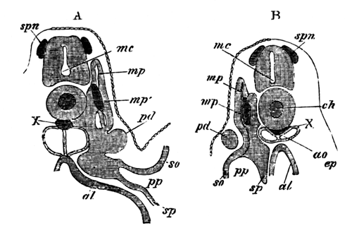
Fig. 1. Two sections of a Pristiurus
Embryo with three visceral clefts.
The sections are to shew the development of the
segmental duct (pd) or primitive duct
of the kidneys. In A (the anterior of the two sections) this
appears as a solid knob projecting towards the epiblast. In B is
seen a section of the column which has grown backwards from the knob in
A.
spn. rudiment of a
spinal nerve; mc. medullary canal;
ch. notochord; X. string of cells
below the notochord; mp. muscle-plate;
mp´. specially developed portion of
muscle-plate; ao. dorsal aorta; pd. segmental duct; so. somatopleura; sp. splanchnopleura; pp. pleuro-peritoneal or body-cavity; ep. epiblast; al.
alimentary canal.
This knob projects outwards toward the epiblast, and the column lies at
first between the mesoblast and epiblast. The knob and column do not long
remain solid. The knob becoming hollow acquires a wide opening into the
pleuro-peritoneal or body-cavity, and the column a lumen; so that by the
time that five visceral clefts have appeared, the two together form a
duct closed behind, but communicating in front by a wide opening with the
pleuro-peritoneal cavity.
Before these changes are accomplished, a series of solid[29] outgrowths of elements of the 'intermediate cell-mass'
appear at the uppermost corner of the body-cavity. These soon become hollow
and appear as involutions from the body-cavity, curling round the inner and
dorsal side of the previously formed duct.
One involution of this kind makes its appearance for each protovertebra,
and the first belongs to the protovertebra immediately behind the anterior
end of the duct whose development has just been described. In Pristiurus
there are in all 29 of these at this period. The last two or three arise
from that portion of the body-cavity, which at this stage still exists
behind the anus. The first-formed duct and the subsequent involutions are
the rudiments of the whole of the urinary system. The duct is the
primitive duct of the kidney[30]; I shall call it in future
the segmental duct; and the involutions are the
commencements of the segmental tubes which constitute the body of the
kidney. I shall call them in future segmental
tubes.
Soon after their formation the segmental tubes become convoluted, and
their blind ends become connected with the segmental duct of the kidney. At
the same time, or rather before this, the blind posterior termination of
each of the segmental ducts of the kidneys unites with and opens into one
of the horns of the cloaca. At this period the condition of affairs is
represented in Fig. 2.
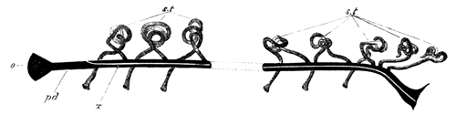
Fig. 2. Diagram of the primitive
condition of the Kidney in a Selachian Embryo.
pd. segmental duct.
It opens at o into the body-cavity and at its other extremity into
the cloaca; x. line along which the division appears which
separates the segmental duct into the Wolffian duct above and the
Müllerian duct below; st. segmental
tubes. They open at one end into the body-cavity, and at the other into
the segmental duct.
There is at pd, the segmental duct of
the kidneys, opening in front (o) into the body-cavity, and behind
into the cloaca, and there are a series of convoluted segmental tubes
(st), each opening at one end into the
body-cavity, and at the other into the duct (pd).
The next important change which occurs is the longitudinal division of
the segmental duct of the kidneys into Müller's duct, or the oviduct, and
the duct of the Wolffian bodies or Leydig's duct. The splitting[31] is effected by the growth of a wall of cells which
divides the duct into two parts (fig. 3, wd. and md.). It
takes place in such a way that the front end of the segmental duct,
anterior to the entrance of the first segmental tube, together with the
ventral half of the rest of the duct, is split off from its dorsal half as
an independent duct (vide fig. 2, x).
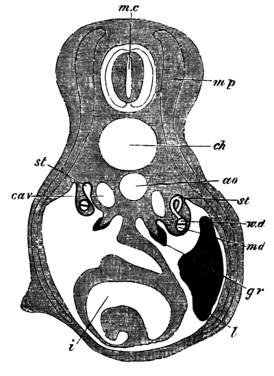
Fig. 3. Transverse section of a
Selachian Embryo illustrating the formation of the Wolffian and Müllerian
ducts by the longitudinal splitting of the segmental duct.
mc. medullary canal;
mp. muscle-plate; ch. notochord; ao. aorta; cav. cardinal vein; st. segmental tube. On the one side the section
passes through the opening of a segmental tube into the body-cavity. On
the other this opening is represented by dotted lines, and the opening of
the segmental tube into the Wolffian duct has been cut through; wd. Wolffian duct; md. Müllerian duct. The Müllerian duct and the
Wolffian duct together constitute the primitive segmental duct; gr. The germinal ridge with the thickened
germinal epithelium; l. liver; i. intestine with spiral
valve.
The dorsal portion also forms an independent duct, and into it the
segmental tubes continue to open. Such at least is the method of splitting
for the female—for the male the splitting is according to Professor
Semper, of a more partial character, and consists for the most part in the
front end of the duct only being separated off from the rest. The result of
these changes is the formation in both sexes of a fresh duct which carries
off the excretions of the segmental involutions, and which I shall call the
Wolffian duct—while in the female there is formed another complete
and independent duct, which I shall call the Müllerian duct, or oviduct,
and in the male portions only of such a duct.
The next change which takes place is the formation of another duct from
the hinder portion of the Wolffian duct, which receives the secretion of
the posterior segmental tubes. This secondary duct unites with the primary
or Wolffian duct near its termination, and the primary ducts of the two
sides unite together to open to the exterior by a common papilla.
Slight modifications of the posterior terminations of these ducts are
found in different genera of Selachians (vide
Semper, Centralblatt für Med. Wiss. 1874, No. 59), but they are of no fundamental
importance.
These constitute the main changes undergone by the segmental duct of the
kidneys and the ducts derived from it; but the segmental tubes also undergo
important changes. In the majority of Selachians their openings into the
body-cavity, or, at any rate, the openings of a large number of them,
persist through life; but the investigations of Dr Meyer[32] render it very probable that the small portion of each
segmental tube adjoining the opening becomes separated from the rest and
becomes converted into a sort of lymph organ, so that the openings of the
segmental tubes in the adult merely lead into lymph organs and not into the
gland of the kidneys.
These constitute the whole changes undergone in the female, but in the
male the open ends of a varying number (according to the species) of the
segmental tubes become connected with the testis and, uniting with the
testicular follicles, serve to carry away the seminal fluid[33]. The spermatozoa have therefore to pass through a
glandular portion of the kidneys before they enter the Wolffian duct, by
which they are finally carried away to the exterior.
In the adult female, then, there are the following parts of the
urinogenital system (fig. 4):
(1) The oviduct, or Müller's duct (fig. 4, md.), split off from the segmental duct of the
kidneys. Each oviduct opens at its upper end into the body-cavity, and
behind the two oviducts have independent communications with the cloaca.
The oviducts serve simply to carry to the exterior the ova, and have no
communication with the glandular portion of the kidneys.
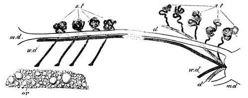
Fig. 4. Diagram of the arrangement of
the Urinogenital Organs in an adult Female Selachian.
md. Müllerian duct;
wd. Wolffian duct; st. segmental tubes; d. duct of the posterior
segmental tubes; ov. ovary.
(2) The Wolffian ducts (fig. 4, wd.)
or the remainder of the segmental ducts of the kidneys. Each Wolffian duct
ends blindly in front, and the two unite behind to open by a common papilla
into the cloaca.
This duct receives the secretion of the whole anterior end of the
kidneys[34], that is to say, of all the anterior segmental
tubes.
(3) The secondary duct (fig. 4, d.) belonging to the lower
portion of the kidneys opening into the former duct near its
termination.
(4) The segmental tubes (fig. 4, st)
from whose convolutions and outgrowths the kidney is formed. They may be
divided into two parts, according to the duct by which their secretion
is carried off.
In the male the following parts are present:
(1) The Müllerian duct (fig. 5, md.),
consisting of a small remnant, attached to the liver, which represents the
foremost end of the oviduct of the female.
(2) The Wolffian duct (fig. 5, wd),
which precisely corresponds to the Wolffian duct of the female, except
that, in addition to functioning as the duct of the anterior part of the
kidneys, it also serves to carry away the semen. In the female it is
straight, but has in the adult male a very tortuous course (vide fig. 5).
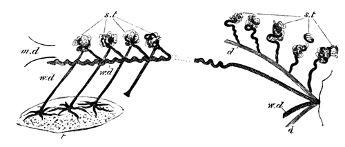
Fig. 5. Diagram of the arrangement of
the Urinogenital Organs in an adult male selachian.
md. rudiment of Müllerian duct; wd. Wolffian duct, which also serves as vas
deferens; st. segmental tubes. The ends
of three of those which in the female open into the body-cavity, have in
the male united with the testicular follicles, and serve to carry away the
products of the testis; d. duct of the posterior segmental tubes;
t. testis.
(3) the duct (fig. 5, d.) of the posterior portion of the
kidneys, which has the same relations as in the female.
(4) The segmental tubes (fig. 5, st.). These have the same relations as in the female,
except that the most anterior two, three or more, unite with the testicular
follicles, and carry away the semen into the Wolffian duct.
* * * * *
The mode of arrangement and the development of these parts suggest a
number of considerations.
In the first place it is important to notice that the segmental tubes
develop primitively as completely independent organs[35], one of which appears in each segment. If embryology is
in any way a repetition of ancestral history, it necessarily follows that
these tubes were primitively independent of each other. Ancestral history,
as recorded in development, is often, it is true, abridged; but it is clear
that though abridgement might prevent a series of primitively separate
organs from appearing as such, yet it would hardly be possible for a
primitively compound organ, which always retained this condition, to appear
during development as a series of separate ones. These considerations
appear to me to prove that the segmented ancestors of vertebrates possessed
a series of independent and segmental excretory organs.
Both Professor Semper and myself, on discovering these organs, were led
to compare them and state our belief in their identity with the so-called
segmental organs of Annelids.
This view has since been fairly generally accepted. The segmental organs
of annelids agree with those of vertebrates in opening at one end into the
body-cavity, but differ in the fact that each also communicates with the
exterior by an independent opening, and that they are never connected with
each other.
On the hypothesis of the identity of the vertebrate segmental tubes with
the annelid segmental organs, it becomes essential to explain how the
external openings of the former may have become lost.
This brings us at once to the origin of the segmental duct of the
kidneys, by which the secretion of all the segmental tubes was carried to
the exterior, and it appears to me that a right understanding of the
vertebrate urinogenital system depends greatly upon a correct view of the
origin of this duct. I would venture to repeat the suggestion which I made
in my original paper (loc. cit.) that this duct is to be
looked upon as the most anterior of the segmental tubes which persist in
vertebrates. In favour of this view are the following
anatomical and embryological facts. (1) It develops in nearly the same
manner as the other segmental tubes, viz. in
Selachians as a solid outgrowth from the intermediate cell-mass, which
subsequently becomes hollowed so as to open into the body-cavity: and in
Amphibians and Osseous and Cyclostome fishes as a direct involution from
the body-cavity. (2) In Amphibians, Cyclostomes and Osseous fishes its
upper end develops a glandular portion, by becoming convoluted in a manner
similar to the other segmental tubes. This glandular portion is often
called either the head-kidney or the primitive kidney. It is only an
embryonic structure, but is important as demonstrating the true nature of
the primitive duct of the kidneys.
We may suppose that some of the segmental tubes first united, possibly
in pairs, and that then by a continuation of this process the whole of them
coalesced into a common gland. One external opening sufficed to carry off
the entire secretion of the gland, and the other openings therefore
atrophied.
This history is represented in the development of the dog-fish in an
abbreviated form, by the elongation of the first segmental tube (segmental
duct of the kidney) and its junction with each of the posterior segmental
tubes. Professor Semper looks upon the primitive duct of the kidneys as a
duct which arose independently, and was not derived from metamorphosis of
the segmental organs. Against this view I would on the one hand urge the
consideration, that it is far easier to conceive of the transformation by
change of function (comp. Dohrn, Functionswechsel, Leipzig, 1875) of a segmental organ into
a segmental duct, than to understand the physiological cause which should
lead, in the presence of so many already formed ducts, to the appearance of
a totally new one. By its very nature a duct is a structure which can
hardly arise de novo. We must even suppose that the segmental organs of
Annelids were themselves transformations of still simpler structures. On
the other hand I would point to the development in this very duct amongst
Amphibians and Osseous fishes of a glandular portion similar to that of a
segmental tube, as an à posteriori proof of its
being a metamorphosed segmental tube. The development in insects of a
longitudinal tracheal duct by the coalescence of a series of
transverse tracheal tubes affords a parallel to the formation of a duct
from the coalescence of a series of segmental tubes.
Though it must be admitted that the loss of the external openings of the
segmental organs requires further working out, yet the difficulties
involved in their disappearance are not so great as to render it improbable
that the vertebrate segmental organs are descended from typical annelidan
ones.
The primitive vertebrate condition, then, is probably that of an early
stage of Selachian development while there is as yet a segmental
duct,—the original foremost segmental tube opening in front into the
body-cavity and behind into the cloaca; with which duct all the segmental
tubes communicate. Vide Fig. 2.
The next condition is to be looked upon as an indirect result of the
segmental duct serving as well for the products of the generative organs as
the secretions of the segmental tubes.
As a consequence of this, the segmental duct became split into a ventral
portion, which served alone for the ova, and a dorsal portion which
received the secretion of the segmental tubes. The lower portion, which we
have called the oviduct, in some cases may also have received the semen as
well as the ova. This is very possibly the case with Ceratodus (vide Günther, Trans. of Royal Society, 1871), and the
majority of Ganoids (Hyrtl, Denkschriften Wien,
Vol. VIII.). In the majority of other cases the oviduct
exists in the male in a completely rudimentary form; and the semen is
carried away by the same duct as the urine.
In Selachians the transportation of the semen from the testis to the
Wolffian duct is effected by the junction of the open ends of two or three
or more segmental tubes with the testicular follicles, and the modes in
which this junction is effected in the higher vertebrates seem to be
derivatives from this. If the views here expressed are correct it is by a
complete change of function that the oviduct has come to perform its
present office. And in the bird and higher vertebrates no trace, or only
the very slightest (vide p. 165) of the primitive urinary function is retained
during embryonic or adult life.
The last feature in the anatomy of the Selachians which requires
notice is the division of the kidney into two portions, an anterior and
posterior. The anatomical similarity between this arrangement and that of
higher vertebrates (birds, &c.) is very
striking. The anterior one precisely corresponds, anatomically, to the
Wolffian body, and the posterior one to the true permanent
kidney of higher vertebrates: and when we find that in the
Selachians the duct for the anterior serves also for the semen as does the
Wolffian duct of higher vertebrates, this similarity seems almost to amount
to identity. A discussion of the differences in development in the two
cases will come conveniently with the account of the bird; but there appear
to me the strongest grounds for looking upon the kidneys of Selachians as
equivalent to both the Wolffian bodies and the true kidneys of the higher
vertebrates.
The condition of the urinogenital organs in Selachians is by no means
the most primitive found amongst vertebrates.
The organs of both Cyclostomous and Osseous fishes, as well as those of
Ganoids, are all more primitive; and in the majority of points the
Amphibians exhibit a decidedly less differentiated condition of these
organs than do the Selachians.
In Cyclostomous fishes the condition of the urinary system is very
simple. In Myxine (vide Joh.
Müller Myxinoid fishes, and Wilhelm
Müller, Jenaische Zeitschrift, 1875, Das Urogenitalsystem des
Amphioxus u. d. Cyclostomen) there is a pair of ducts which
communicate posteriorly by a common opening with the abdominal pore. From
these ducts spring a series of transverse tubules, each terminating in a
Malpighian corpuscle. These together constitute the mass of the kidneys.
About opposite the gall-bladder the duct of the kidney (the segmental duct)
narrows very much, and after a short course ends in a largish glandular
mass (the head-kidney), which communicates with the pericardial cavity by a
number of openings.
In Petromyzon the anatomy of the kidneys is fundamentally the same as in
Myxine. They consist of the two segmental ducts, and a number of fine
branches passing off from these, which become convoluted but do not form
Malpighian tufts. The head-kidney is absent in the adult.
W. Müller (loc. cit.) has given a short but interesting
account of the development of the urinary system of Petromyzon. He finds
that the segmental ducts develop first of all as simple involutions from
the body-cavity. The anterior end of each then develops a glandular portion
which comes to communicate by a number of openings with the body-cavity.
Subsequently to the development of this glandular portion the remainder of
the kidneys appears in the posterior portion of the body-cavity; and before
the close of embryonic life the anterior glandular portion atrophies.
The comparison of this system with that of a Selachian is very simple.
The first developed duct is the segmental duct of a Selachian, and the
glandular portion developed at its anterior extremity, which is permanent
in Myxine but embryonic in Petromyzon, is, as W. Müller has rightly
recognized, equivalent to the head-kidney of Amphibians, which remains
undeveloped in Selachians. It is, according to my previously stated view,
the glandular portion of the first segmental organ or the segmental duct.
The series of orifices by which this communicates with the body-cavity are
due to the division of the primary opening of the segmental duct. This is
shewn both by the facts of their development in Petromyzon given by Müller,
as well as by the occurrence of a similar division of the primary orifice
in Amphibians, which is mentioned later in this paper. In a note in my
original paper (loc. cit.) I stated that these openings
were equivalent to the segmental involutions of Selachians. This is
erroneous, and was due to my not having understood the description given in
a preliminary paper of Müller (Jenaische
Zeitschrift, 1873). The large development of this glandular mass in
the Cyclostome and Osseous fishes and in embryo Amphibians, implies that it
must at one time have been important. Its earlier development than the
remainder of the kidneys is probably a result of the specialized function
of the first segmental organ.
The remainder of the kidney in Cyclostomes is equivalent to the kidney
of Selachians. Its development from segmental involutions has not been
recognized. If these segmental involutions are really absent it may perhaps
imply that the simplicity of the Cyclostome kidneys, like that of so many
other of their organs, is a result of degeneration rather than a primitive
condition.
In Osseous fishes the segmental duct of the kidneys develops,
as the observations of Rosenberg[36] (Teleostierniere,
Inaug. Disser.
Dorpat, 1867) and Oellacher (Zeitschrift für
Wiss. Zool. 1873) clearly prove, by an involution from the
body-cavity. This involution grows backwards in the form of a duct and
opens into the cloaca. The upper end of this duct (the most anterior
segmental tube) becomes convoluted, and forms a glandular body, which has
no representative in the urinary apparatus of Selachians, but whose
importance, as indicating the origin of the segmental duct of the kidneys,
I have already insisted upon.
The rest of the kidney becomes developed at a later period, probably in
the same way as in Selachians; but this, as far as I know, has not been
made out.
The segmental duct of the kidneys forms the duct for this new gland, as
in embryo Selachians (Fig. 2), but, unlike what happens in Selachians,
undergoes no further changes, with the exception of a varying amount of
retrogressive metamorphosis of its anterior end. The kidneys of Osseous
fish usually extend from just behind the head to opposite the anus, or even
further back than this. They consist for the most part of a broader
anterior portion, an abdominal portion reaching from this to the anus, and,
as in those cases in which the kidneys extend further back than the anus,
of a caudal portion.
The two ducts (segmental ducts of the kidneys) lie, as a rule, in the
lower part of the kidneys on their outer borders, and open almost
invariably into a urinary bladder. In some cases they unite before opening
into the bladder, but generally have independent openings.
This bladder, which is simply a dilatation of the united lower ends of
the primitive kidney-ducts, and has no further importance, is almost
invariably present, but in many cases lies unsymmetrically either to the
right or the left. It opens to the exterior by a very minute opening in the
genito-urinary papilla, immediately behind the genital pore. There are,
however, a few cases in which the generative and urinary organs have a
common opening. For further details vide Hyrtl,
Denk. der k. Akad. Wien, Vol. II.
It is possible that the generative ducts of Osseous fishes are derived
from a splitting from the primitive duct of the kidney, but this is
discussed later in the paper.
In Osseous fishes we probably have an embryonic condition of the
Selachian kidneys retained permanently through life.
* * * * *
In the majority of Ganoids the division of the segmental duct of the
kidney into two would seem to occur, and the ventral duct of the two
(Müllerian duct), which opens at its upper end into the body-cavity, is
said to serve as an excretory duct for both male and female organs.
The following are the more important facts which are known about the
generative and urinary ducts of Ganoids.
In Spatularia (vide Hyrtl,
Geschlechts u. Harnwerkzeuge bei den Ganoiden, Denkschriften der k.
Akad. Wien, Vol. VIII.) the following
parts are found in the female.
(1) The ovaries stretching along the whole length of the abdominal
cavity.
(2) The kidneys, which are separate and also extend along the greater
part of the abdominal cavity.
(3) The ureters lying on the outer borders of the kidneys. Each ureter
dilates at its lower end into an elongated wide tube, which continues to
receive the ducts from the kidneys. The two ureters unite before
terminating and open behind the anus.
(4) The two oviducts (Müllerian ducts). These open widely into the
abdominal cavity, at about two-thirds of the distance from the anterior
extremity of the body-cavity. Each opens by a narrow pore into the dilated
ureter of its side.
In the male the same parts are found as in the female, but Hyrtl found
that the Müllerian duct of the left side at its entrance into the ureter
became split into two horns, one of which ended blindly. On the right side
the opening of the Müllerian duct was normal.
In the Sturgeon (vide J. Müller,
Bau u. Grenzen d. Ganoiden, Berlin Akad. 1844; Leydig,
Fischen u. Reptilien, and Hyrtl,
Ganoiden) the same parts are found as in
Spatularia.
The kidneys extend along the whole length of the body-cavity;
and the ureter, which does not reach the whole length of the kidneys, is a
thin-walled wide duct lying on the outer side. On laying it open the
numerous apertures of the tubules for the kidney are exposed. The Müllerian
duct, which opens in both sexes into the abdominal cavity, ends, according
to Leydig, in the cases of some males, blindly behind without opening into
the ureter, and Müller makes the same statement for both sexes. It was open
on both sides in a female specimen I examined[37], and
Hyrtl found it invariably so in both sexes in all the specimens he
examined.
Both Rathke and Stannius (I have been unable to refer to the original
papers) believed that the semen was carried off by transverse ducts
directly into the ureter, and most other observers have left undecided the
mechanism of the transportation of the semen to the exterior. If we suppose
that the ducts Rathke saw really exist they might perhaps be supposed to
enter not directly into the ureter, but into the kidney, and be in fact
homologous with the vasa efferentia of the Selachians. The frequent blind
posterior termination of the Müllerian duct is in favour of the view that
these ducts of Rathke are really present.
In Polypterus (vide Hyrtl,
Ganoiden) there is, as in other Ganoids, a pair of
Müllerian ducts. They unite at their lower ends. The ureters are also much
narrower than in previously described Ganoids and, after coalescing, open
into the united oviducts. The urinogenital canal, formed by coalescence of
the Müllerian ducts and ureters, has an opening to the exterior immediately
behind the anus.
In Amia (vide Hyrtl) there is a pair of Müllerian
ducts which, as well as the ureters, open into a dilated vesicle. This
vesicle appears as a continuation of the Müllerian ducts, but receives a
number of the efferent ductules of the kidneys. There is a single
genito-urinary pore behind the anus.
In Ceratodus (Günther,
Phil. Trans. 1871) the kidneys are small and confined
to the posterior extremity of the abdomen. The generative organs extend
however along the greater part of the length of the
abdominal cavity. In both male and female there is a long Müllerian duct,
and the ducts of the two sides unite and open by a common pore into a
urinogenital cloaca which communicates with the exterior by the same
opening as the alimentary canal. In both sexes the Müllerian duct has a
wide opening near the anterior extremity of the body-cavity. The ureters
coalesce and open together into the urinogenital cloaca dorsal to the
Müllerian ducts. It is not absolutely certain that the semen is transported
to the exterior by the Müllerian duct of the male, which is perhaps merely
a rudiment as in Amphibia. Dr Günther failed however to find any other
means by which it could be carried away.
The genital ducts of Lepidosteus differ in important particulars from
those of the other Ganoids (vide Müller, loc. cit. and Hyrtl, loc. cit.).
In both sexes the genital ducts are continuous with the investments of
the genital organs.
In the female the dilated posterior extremities of the ureters
completely invest for some distance the generative ducts, whose extremities
are divided into several processes, and end in a different way on the two
sides. A similar division and asymmetry of the ducts is mentioned by Hyrtl
as occurring in the male of Spatularia, and it seems not impossible that on
the hypothesis of the genital ducts being segmental tubes these divisions
may be remnants of primitive glandular convolutions. The ureters in both
sexes dilate as in other Ganoids at their posterior extremities, and unite
with one another. The unpaired urinogenital opening is situated behind the
anus. In the male the dilated portion of the ureters is divided into a
series of partitions which are not present in the female.
Till the embryology of the secretory system of Ganoids has been worked
out, the homologies of their generative ducts are necessarily a matter of
conjecture. It is even possible that what I have called the Müllerian duct
in the male is functionless, as with Amphibians, but that, owing to the
true ducts of the testis having been overlooked, it has been supposed to
function as the vas deferens. Günther's (loc. cit.)
injection experiments on Ceratodus militate against this view, but I do not
think they can be considered as conclusive as long as the
mechanism for the transportation of the semen to the exterior has not been
completely made out. Analogy would certainly lead us to expect the ureter
to serve in Ganoids as the vas deferens.
The position of the generative ducts might in some cases lead to the
supposition that they are not Müllerian ducts, or, in other words, the most
anterior pair of segmental organs but a pair of the posterior segmental
tubes.
What are the true homologies of the generative ducts of Lepidosteus,
which are continuous with the generative glands, is somewhat doubtful. It
is very probable that they may represent the similarly functioning ducts of
other Ganoids, but that they have undergone further changes as to their
anterior extremities.
It is, on the other hand, possible that their generative ducts are the
same structures as those ducts of Osseous fishes, which are continuous with
the generative organs. These latter ducts are perhaps related to the
abdominal pores, and had best be considered in connection with these; but a
completely satisfactory answer to the questions which arise in reference to
them can only be given by a study of their development.
In the Cyclostomes the generative products pass out by an abdominal
pore, which communicates with the peritoneal cavity by two short tubes[38], and which also receives the ducts of the kidneys.
Gegenbaur suggests that these are to be looked upon as Müllerian ducts,
and as therefore developed from the segmental ducts of the kidneys. Another
possible view is that they are the primitive external openings of a pair of
segmental organs. In Selachians there are usually stated to be a pair of
abdominal pores. In Scyllium I have only been able to find, on each side, a
large deep pocket opening to the exterior, but closed below towards the
peritoneal cavity, so that in it there seem to be no abdominal pores[39]. In the Greenland Shark (Læmargus Borealis)
Professor Turner (Journal of Anat. and Phys. Vol.
VIII.) failed
to find either oviduct or vas deferens, but found a pair of large open
abdominal pores, which he believes serve to carry away the generative
products of both sexes. Whether the so-called abdominal pores of Selachians
usually end blindly as in Scyllium, or, as is commonly stated, open into
the body-cavity, there can be no question that they are homologous with
true abdominal powers.
The blind pockets of Scyllium appear very much like the remains of
primitive involutions from the exterior, which might easily be supposed to
have formed the external opening of a pair of segmental organs, and this is
probably the true meaning of abdominal pores. The presence of abdominal
pores in all Ganoids in addition to true genital ducts and of these pockets
or abdominal pores in Selachians, which are almost certainly homologous
with the abdominal pores of Ganoids and Cyclostomes, and also occur in
addition to true Müllerian ducts, speak strongly against the view that the
abdominal pores have any relation to Müllerian ducts. Probably therefore
the abdominal pores of the Cyclostomous fishes (which seem to be of the
same character as other abdominal pores) are not to be looked on as
rudimentary Müllerian ducts.
We next come to the question which I reserved while speaking of the
kidneys of Osseous fishes, as to the meaning of their genital ducts.
In the female Salmon and the male and female Eel, the generative
products are carried to the exterior by abdominal pores, and there are no
true generative ducts. In the case of most other Osseous fish there are
true generative ducts which are continuous with the investment of the
generative organs[40] and have generally,
though not always, an opening or openings independent of the ureter close
behind the rectum, but no abdominal pores are present. It seems, therefore,
that in Osseous fish the generative ducts are complementary to abdominal
pores, which might lead to the view that the generative ducts were formed
by a coalescence of the investment of the generative glands with the short
duct of abdominal pore.
Against this view there are, however, the following facts:
(1) In the cases of the salmon and the eel it is perfectly true that the
abdominal pore exactly corresponds with the opening of the genital duct in
other Osseous fishes, but the absence of genital ducts in these cases must
rather be viewed, as Vogt and Pappenheim (loc. cit.) have
already insisted, as a case of degeneration than of a primitive condition.
The presence of genital ducts in the near allies of the Salmonidæ, and even
in the male salmon, are conclusive proofs of this. If we admit that the
presence of an abdominal pore in Salmonidæ is merely a result of
degeneration, it obviously cannot be used as an argument for the
complementary nature of abdominal pores and generative ducts.
(2) Hyrtl (Denkschriften der k. Akad.
Wien, Vol. 1) states that in
Mormyrus oxyrynchus there is a pair of abdominal pores in addition to true
generative ducts. If his statements are correct, we have a strong argument
against the generative ducts of Osseous fishes being related to abdominal
pores. For though this is the solitary instance of the presence of both a
genital opening and abdominal pores known to me in Osseous fishes, yet we
have no right to assume that the abdominal pores of Mormyrus are not
equivalent to those of Ganoids and Selachians. It must be admitted, with
Gegenbaur, that embryology alone can elucidate the meaning of the genital
ducts of Osseous fishes.
In Lepidosteus, as was before mentioned, the generative ducts, though
continuous with the investment of the generative bodies, unite with the
ureters, and in this differ from the generative ducts of Osseous fishes.
The relation, indeed, of the generative ducts of Lepidosteus to the
urinary ducts is very similar to that existing in other Ganoid fishes; and
this, coupled with the fact that Lepidosteus possesses a pair of abdominal
pores on each side of the anus[41], makes
it most probable that its generative ducts are true Müllerian ducts.
* * * * *
In the Amphibians the urinary system is again more primitive than in the
Selachians.
The segmental duct of the kidneys is formed[42] by an
elongated fold arising from the outer wall of the body-cavity, in the same
position as in Selachians. This fold becomes constricted into a canal,
closed except at its anterior end, which remains open to the body-cavity.
This anterior end dilates, and grows out into two horns, and at the same
time its opening into the body-cavity becomes partly constricted, and so
divided into three separate orifices, one for each horn and a central one
between the two. The horns become convoluted, blood channels appearing
between their convolutions, and a special coil of vessels is formed arising
from the aorta and projecting into the body-cavity near the openings of the
convolutions. These formations together constitute the glandular portion[43] of the original anterior segmental tube or segmental
duct of the kidneys. I have already pointed out the similarity which this
organ exhibits to the head-kidneys of Cyclostome fishes in its mode of
formation, especially with reference to the division of the primitive
opening. The lower end of the segmental duct unites with a horn of the
cloaca.
After the formation of the gland just described the remainder of the
kidney is formed.
This arises in the same way as in Selachians. A series of
involutions from the body-cavity are developed; these soon form convoluted
tubes, which become branched and interlaced with one another, and also
unite with the primitive duct of the kidneys. Owing to the branching and
interlacing of the primitive segmental tubes, the kidney is not divided
into distinct segments in the same way as with the Selachians. The mode of
development of these segmental tubes was discovered by Götte. Their
openings are ciliated, and, as Spengel (loc. cit.) and
Meyer (loc. cit.) have independently discovered, persist
in most adult Amphibians. As both these investigators have pointed out, the
segmental openings are in the adult kidneys of most Amphibians far more
numerous than the vertebral segments to which they appertain. This is due
to secondary changes, and is not to be looked upon as the primitive state
of things. At this stage the Amphibian kidneys are nearly in the same
condition as the Selachian, in the stage represented in Fig. 2. In both
there is the segmental duct of the kidneys, which is open in front,
communicates with the cloaca behind, and receives the whole secretion from
the kidneys. The parallelism between the two is closely adhered to in the
subsequent modifications of the Amphibian kidney, but the changes are not
completed so far in Amphibians as in Selachians. The segmental duct of the
Amphibian kidney becomes, as in Selachians, split into a Müllerian duct or
oviduct, and a Wolffian duct or duct for the kidney.
The following points about this are noteworthy:
(1) The separation of the two ducts is never completed, so that they are
united together behind, and for a short distance, blend and form a common
duct; the ducts of the two sides so formed also unite before opening to the
exterior.
(2) The separation of the two ducts does not occur in the form of a
simple splitting, as in Selachians. But the efferent ductules from the
kidney gradually alter their points of entrance into the primitive duct.
Their points of entrance become carried backwards further and further, and
since this process affects the anterior ducts proportionally more than the
posterior, the efferent ducts finally all meet and form a common duct which
unites with the Müllerian duct near its posterior extremity. This
process is not always carried out with equal completeness. In the tailless
Amphibians, however, the process is generally[44]
completed, and the ureters (Wolffian ducts) are of considerable length.
Bufo cinereus, in the male of which the Müllerian ducts are very
conspicuous, serves as an excellent example of this.
In the Salamander (Salamandra maculosa), Figs. 6 and 7, the process is
carried out with greater completeness in the female than in the male, and
this is the general rule in Amphibians. In the male Proteus, the embryonic
condition would seem to be retained almost in its completeness so that the
ducts of the kidney open directly and separately into the still persisting
primitive duct of the kidney. The upper end of the duct nevertheless
extends some distance beyond the end of the kidney and opens into the
abdominal cavity. In the female Proteus, on the other hand, the separation
into a Müllerian duct and a ureter is quite complete. The Newt (Triton)
also serves as an excellent example of the formation of distinct Müllerian
and Wolffian ducts being much more complete in the female than the male. In
the female Newt all the tubules from the kidney open into a duct of some
length which unites with the Müllerian duct near its termination, but in
the male the anterior segmental tubes, including those which, as will be
afterwards seen, serve as vasa efferentia of the testis, enter the
Müllerian duct directly, while the posterior unite as in the female into a
common duct before joining the Müllerian duct. For further details as to
the variations exhibited in the Amphibians, the reader is referred to
Leydig, Anat. Untersuchung, Fischen u. Reptilien.
Ditto, Lehrbuch der Histologie, Menschen u.
Thiere. Von Wittich, Siebold u. Kölliker,
Zeitschrift, Vol. IV. p. 125.
The different conditions of completeness of the Wolffian ducts
observable amongst the Amphibians are instructive in reference to the
manner of development of the Wolffian duct in Selachians. The mode
of division in the Selachians of the segmental duct of the kidney into a
Müllerian and Wolffian duct is probably to be looked upon as an
embryonic abbreviation of the process by which these two ducts are formed
in Amphibians. The fact that this separation into Müllerian and Wolffian
ducts proceeds further in the females of most Amphibians than in the males,
strikingly shews that it is the oviductal function of the Müllerian duct
which is the indirect cause of its separation from the Wolffian duct. The
Müllerian duct formed in the way described persists almost invariably in
both sexes, and in the male sometimes functions as a sperm reservoir;
e.g. Bufo cinereus. In the embryo it carries at its upper end the
glandular mass described above (Kopfniere), but this generally atrophies,
though remnants of it persist in the males of some species (e.g.
Salamandra). Its anterior end opens, in most cases by a single opening,
into the perivisceral cavity in both sexes, and is usually ciliated. As the
female reaches maturity, the oviduct dilates very much; but it remains thin
and inconspicuous in the male.
The only other developmental change of importance is the connection of
the testes with the kidneys. This probably occurs in the same manner as in
Selachians, viz. from the junction of the open
ends of the segmental tubes with the follicles of the testes. In any case
the vessels which carry off the semen constitute part of the kidney, and
the efferent duct of the testis is also that of the kidney. The vasa
efferentia from the testis either pass through one or two nearly isolated
anterior portions of the kidney (Proteus, Triton) or else no such special
portion of the kidney becomes separated from the rest, and the vasa
efferentia enter the general body of the kidney.
* * * * *
In the male Amphibian, then, the urinogenital system consists of the
following parts (Fig. 6):
(1) Rudimentary Müllerian ducts, opening anteriorly into the
body-cavity, which sometimes carry aborted Kopfnieren.
(2) The partially or completely formed Wolffian ducts (ureters) which
also serve as the ducts for the testes.
(3) The kidneys, parts of which also serve as the vasa efferentia, and
whose secretion, together with the testicular products, is carried off by
the Wolffian ducts.
(4) The united lower parts of Wolffian and Müllerian ducts
which are really the lower unsplit part of the segmental ducts of the
kidneys.
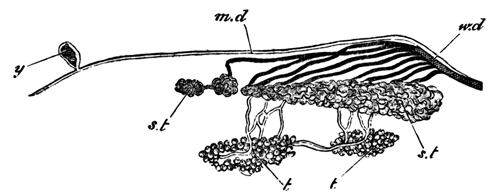
Fig. 6. Diagram of the Urinogenital
Organs of a Male Salamander.
(Copied from Leydig's Histologie des Menschen u.
der Thiere.)
md. Müller's duct
(rudimentary); y. remnant of the secretory portion of the segmental
duct Kopfniere; Wd. Wolffian duct; a
less complete structure in the male than in the female; st. segmental tubes or kidney. The openings of these
into the body-cavity are not inserted in the figure; t. testis. Its
efferent ducts form part of the kidney.
In the female, there are (Fig. 7)
(1) The Müllerian ducts which function as the oviducts.
(2) The Wolffian ducts.
(3) The kidneys.
(4) The united Müllerian and Wolffian ducts as in the male.
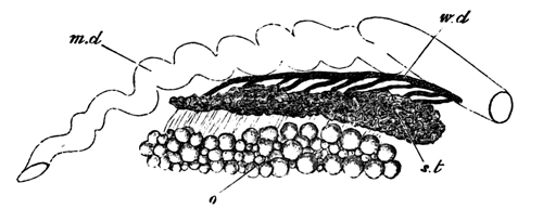
Fig. 7. Diagram of the Urinogenital
Organs of a Female Salamander.
(Copied from Leydig's Histologie des Menschen u.
der Thiere.)
Md. Müller's duct or
oviduct; Wd. Wolffian duct or the duct
of the kidneys; st. segmental tubes or
kidney. The openings of these into the body-cavity are not inserted in the
figure; o. ovary.
The urinogenital organs of the adult Amphibians agree in almost all
essential particulars with those of Selachians. The ova are carried off
in both by a specialized oviduct. The Wolffian duct, or ureter, is found
both in Selachians and Amphibians, and the relations of the testis to it
are the same in both, the vasa efferentia of the testes having in both the
same anatomical peculiarities.
The following points are the main ones in which Selachians and
Amphibians differ as to the anatomy of the urinogenital organs; and in all
but one of these, the organs of the Amphibian exhibit a less differentiated
condition than do those of the Selachian.
(1) A glandular portion (Kopfniere) belonging to the first segmental
organ (segmental duct of the kidneys) is found in all embryo Amphibians,
but usually disappears, or only leaves a remnant in the adult. It has not
yet been found in any Selachian.
(2) The division of the primitive duct of the kidney into the Müllerian
duct and the Wolffian duct is not completed so far in Amphibians as
Selachians, and in the former the two ducts are confluent at their lower
ends.
(3) The permanent kidney exhibits in Amphibians no distinction into two
glands (foreshadowing the Wolffian bodies and true kidneys of higher
vertebrates), as it does in the Selachians.
(4) The Müllerian duct persists in its entirety in male Amphibians, but
only its upper end remains in male Selachians.
(5) The openings of the segmental tubes into the body-cavity correspond
in number with the vertebral segments in most Selachians, but are far more
numerous than these in Amphibians. This is the chief point in which the
Amphibian kidney is more differentiated than the Selachian.
* * * * *
The modifications in development which the urinogenital system has
suffered in higher vertebrates (Sauropsida and Mammalia) are very
considerable; nevertheless it appears to me to be possible with fair
certainty to trace out the relationship of its various parts in them to
those found in the Ichthyopsida. The development of urinogenital organs has
been far more fully worked out for the bird than for any other member of
the amniotic vertebrates; but, as far as we know, there are no
essential variations except in the later periods of development throughout
the division. These later variations, concerning for the most part the
external apertures of the various ducts, are so well known and have been so
fully described as to require no notice here. The development of these
parts in the bird will therefore serve as the most convenient basis for
comparison.
In the bird the development of these parts begins by the appearance of a
column of cells on the upper surface of the intermediate cell-mass (Fig. 8,
W.d). As in Selachians, the intermediate cell-mass is a group of
cells between the outer edge of the protovertebræ and the upper end of the
body-cavity. The column of cells thus formed is the commencement of the
duct of the Wolffian body. Its development is strikingly similar to that of
the segmental duct of the kidney in Selachians. I shall attempt when I have
given an account of the development of the Müllerian duct to speak of the
relations between the Selachian duct and that of the bird.
Romiti (Archiv f. Micr. Anat. Vol. X.) has recently
stated that the Wolffian duct develops as an involution from the
body-cavity. The fact that the specimens drawn by Romiti to support this
view are too old to determine such a point, and the inspection of a number
of specimens made by my friend Mr Adam Sedgwick of Trinity College, who, at
my request, has been examining the urinogenital organs of the fowl, have
led me to the conclusion that Romiti is in error in differing from his
predecessors as to the development of the Wolffian duct. The solid string
of cells to form the Wolffian duct lies at first close to the epiblast,
but, by the alteration in shape which the protovertebræ undergo and the
general growth of cells around it, becomes gradually carried downwards till
it lies close to the germinal epithelium which lines the body-cavity. While
undergoing this change of position it also acquires a lumen, but ends
blindly both in front and behind. Towards the end of the fourth day the
Wolffian duct opens into a horn of the cloaca. The cells adjoining its
inner border commence, as it passes down on the third day, to undergo
histological changes, which, by the fourth day, result in the formation of
a series of ducts and Malpighian tufts which form the mass of the Wolffian
body[45].
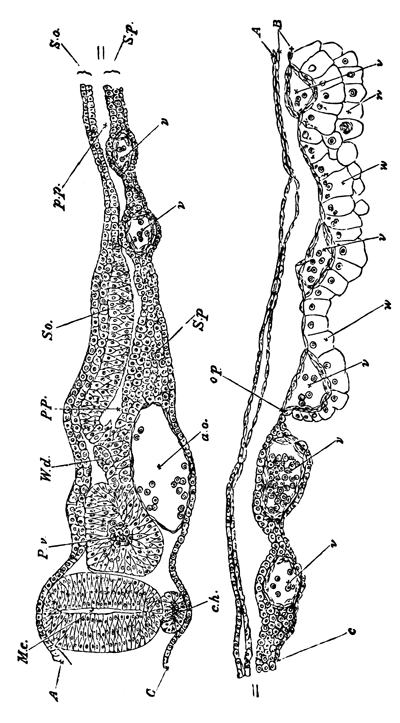
Fig. 8. Transverse section through the
Dorsal region of an Embryo Fowl of 45 h. To shew
the mode of Formation of the Wolffian Duct.
A. epiblast; B. mesoblast; C.
hypoblast; M.c. medullary canal; Pv. Protovertebræ; W.d. Wolffian duct; so. Somatopleure; Sp. Splanchnopleure; pp. pleuro-peritoneal cavity; ch. notochord; ao.
dorsal aorta; v. blood-vessels.
The Müllerian duct arises in the form of an involution, whether
at first solid or hollow, of the germinal epithelium, and, as I am
satisfied, quite independently of the Wolffian duct. It is important to
notice that its posterior end soon unites with the Wolffian duct, from
which however it not long after becomes separated and opens independently
into the cloaca. The upper end remains permanently open to the body-cavity,
and is situated nearly opposite the extreme front end of the Wolffian
body.
Between the 80th and 100th hour of incubation the ducts of the permanent
kidneys begin to make their appearance. Near its posterior extremity each
Wolffian duct becomes expanded, and from the dorsal side of this portion a
diverticulum is constricted off, the blind end of which points forwards.
This is the duct of the permanent kidneys, and around its end the kidneys
are found. It is usually stated that the tubules of the permanent kidneys
arise as outgrowths from the duct, but this requires to be worked over
again.
The condition of the urinogenital system in birds immediately after the
formation of the permanent kidneys is strikingly similar to its permanent
condition in adult Selachians. There is the Müllerian duct in both opening
in front into the body-cavity and behind into the cloaca. In both the
kidneys consist of two parts—an anterior and posterior—which
have been called respectively Wolffian bodies and permanent kidneys in
birds and Leydig's glands and the kidneys in Selachians.
The duct of the permanent kidney, which at first opens into that of the
Wolffian body, subsequently becomes further split off from the Wolffian
duct, and opens independently into the cloaca.
The subsequent changes of these parts are different in the two
sexes.
In the female the Müllerian ducts[46]
persist and become the oviducts. Their anterior ends remain open to the
body-cavity. The changes in their lower ends in the various orders of
Sauropsida and Mammalia are too well known to require repetition here. The
Wolffian body and duct atrophy: there are left however in many cases slight
remnants of the anterior extremity of the body forming the parovarium of
the bird, and also frequently remnants of the posterior portion of the
gland as well as of the duct. The permanent kidney and its duct remain
unaltered.
In the male the Müllerian duct becomes almost completely obliterated.
The Wolffian duct persists and forms the vas deferens, and the anterior
so-called sexual portion of the Wolffian body also persists in an altered
form. Its tubules unite with the seminiferous tubules, and also form the
epididymis. Unimportant remnants of the posterior part of the Wolffian body
also persist, but are without function. In both sexes the so-called
permanent kidneys form the sole portion of the primitive uriniferous system
which persists in the adult.
In considering the relations between the modes of development of the
urinogenital organs of the bird and of the Selachians, the first important
point to notice is, that whereas in the Selachians the segmental duct of
the kidneys is first developed and subsequently becomes split into the
Müllerian and Wolffian ducts; in the bird these two ducts develop
independently. This difference in development would be accurately described
by saying that in birds the segmental duct of the kidneys develops as in
Selachians, but that the Müllerian duct develops independently of it.
Since in Selachians the Wolffian duct is equivalent to the segmental
duct of the kidneys with the Müllerian removed from it, when in birds the
Müllerian duct develops independently of the segmental kidney duct, the
latter becomes the same as the Wolffian duct.
The second mode of stating the difference in development in the
two cases represents the embryological facts of the bird far better than
the other method.
It explains why the Wolffian duct appears earlier than the Müllerian and
not at the same time, as one might expect according to the other way of
stating the case. If the Wolffian duct is equivalent to the segmental duct
of Selachians, it must necessarily be the first duct to develop; and not
improbably the development of the Müllerian duct would in birds be expected
to occur at the time corresponding to that at which the primitive duct in
Selachians became split into two ducts.
It probably also explains the similarity in the mode of development of
the Wolffian duct in birds and the primitive duct of the kidneys in
Selachians.
This way of stating the case is also in accordance with theoretical
conclusions. As the egg-bearing function of the Müllerian duct became more
and more confirmed we might expect that the adult condition would impress
itself more and more upon the embryonic development, till finally the
Müllerian duct ceased to be at any period connected with the kidneys, and
the history of its origin ceased to be traceable in its development. This
seems to have actually occurred in the higher vertebrates, so that the only
persisting connection between the Müllerian duct and the urinary system is
the brief but important junction of the two at their lower ends on the
sixth or seventh day. This junction justly surprised Waldeyer (Eierstock u. Ei, p. 129), but receives a complete and
satisfactory explanation on the hypothesis given above.
The original development of the segmental tubes is in the bird solely
retained in the tubules of the Wolffian body arising independently of the
Wolffian duct, and I have hitherto failed to find that there is a distinct
division of the Wolffian bodies into segments corresponding with the
vertebral segments.
I have compared the permanent kidneys to the lower portion of the
kidneys of Selachians. The identity of the anatomical condition of the
adult Selachian and embryonic bird which has been already pointed out
speaks strongly in favour of this view; and when we further consider that
the duct of the permanent kidneys is developed in nearly the same way as
the supposed homologous duct in Selachians, the suggested identity gains
further support. The only difficulty is the fact that in Selachians the
tubules of the part of the kidneys under comparison develop as segmental
involutions in point of time anteriorly to their duct, while in birds they
develop in a manner not hitherto certainly made out but apparently in point
of time posteriorly to their duct. But when the immense modifications in
development which the whole of the gland of the excretory organ has
undergone in the bird are considered, I do not think that the fact I have
mentioned can be brought forward as a serious difficulty.[TN5]
The further points of comparison between the Selachian and the bird are
very simple. The Müllerian duct in its later stages behaves in the higher
vertebrates precisely as in the lower. It becomes in fact the oviduct in
the female and atrophies in the male. The behaviour of the Wolffian duct is
also exactly that of the duct which I have called the Wolffian duct in
Ichthyopsida, and in the tubules of the Wolffian body uniting with the
tubuli seminiferi we have represented the junction of the segmental tubes
with the testis in Selachians and Amphibians. It is probably this junction
of two independent organs which led Waldeyer to the erroneous view that the
tubuli seminiferi were developed from the tubules of the Wolffian body.
With the bird I conclude the history of the origin of the urinogenital
system of vertebrates. I have attempted, and I hope succeeded, in tracing
out by the aid of comparative anatomy and embryology the steps by which a
series of independent and simple segmental organs like those of Annelids
have become converted into the complicated series of glands and ducts which
constitute the urinogenital system of the higher vertebrates. There are no
doubt some points which require further elucidation amongst the Ganoid and
Osseous fishes. The most important points which appear to me still to need
further research, both embryological and anatomical, are the abdominal
pores of fishes, the generative ducts of Ganoids, especially Lepidosteus,
and the generative ducts of Osseous fishes.
The only further point which requires discussion is the
embryonic layer from which these organs are derived.
I have shewn beyond a doubt (loc. cit.) that in
Selachians these organs are formed from the mesoblast. The unanimous
testimony of all the recent investigators of Amphibians leads to the same
conclusion. In birds, on the other hand, various investigators have
attempted to prove that these organs are derived from the epiblast. The
proof they give is the following: the epiblast and mesoblast appear fused
in the region of the axis cord. From this some investigators have been led
to the conclusion that the whole of the mesoblast is derived from the upper
of the two primitive embryonic layers. To these it may be replied that,
even granting their view to be correct, it is no proof of the derivation of
the urinogenital organs from the epiblast, since it is not till the
complete formation of the three layers that any one of them can be said to
exist. Others look upon the fusion of the two layers as a proof of the
passage of cells from the epiblast into the mesoblast. An assumption in
itself, which however is followed by the further assumption that it is from
these epiblast cells that the urinogenital system is derived! Whatever may
have been the primitive origin of the system, its mesoblastic origin in
vertebrates cannot in my opinion be denied.
Kowalewsky (Embryo. Stud. an Vermen u.
Arthropoda, Mem. Akad. St Petersbourg, 1871) finds that the
segmental tubes of Annelids develop from the mesoblast. We must therefore
look upon the mesoblastic origin of the excretory system as having an
antiquity greater even than that of vertebrates.
With Plates 22 and 23.
In the course of an inquiry into the development of
Elasmobranch Fishes, my attention has recently been specially directed to
the first appearance and early stages of the spinal nerves, and I have been
led to results which differ so materially from those of former
investigators, that I venture at once to lay them before the Society. I
have employed in my investigations embryos of Scyllium canicula,
Scyllium stellare, Pristiurus, and Torpedo. The
embryos of the latter animal, especially those hardened in osmic acid, have
proved by far the most favourable for my purpose, though, as will be seen
from the sequel, I have been able to confirm the majority of my conclusions
on embryos of all the above-mentioned genera.
A great part of my work was done at the Zoological Station founded by Dr
Dohrn at Naples; and I have to thank both Dr Dohrn and Dr Eisig for the
uniformly obliging manner in which they have met my requirements for
investigation. I have more recently been able to fill up a number of lacunæ
in my observations by the study of embryos bred in the Brighton Aquarium;
for these I am indebted to the liberality of Mr Lee and the directors of
that institution.
The first appearance of the Spinal Nerves in
Pristiurus.
In a Pristiurus-embryo, at the time when two visceral clefts
become visible from the exterior (though there are as yet no
openings from without into the throat), a transverse section through the
dorsal region exhibits the following features (Pl. 22, fig. A):—
The external epiblast is formed of a single row of flattened elongated
cells. Vertically above the neural canal the cells of this layer are more
columnar, and form the rudiment of the primitively continuous dorsal
fin.
The neural canal (nc) is elliptical
in section, and its walls are composed of oval cells two or three deep. The
wall at the two sides is slightly thicker than at the ventral and dorsal
ends, and the cells at the two ends are also smaller than elsewhere. A
typical cell from the side walls of the canal is about 1/1900 inch in its
longest diameter. The outlines of the cells are for the most part
distinctly marked in the specimens hardened in either chromic or picric
acid, but more difficult to see in those prepared with osmic acid; their
protoplasm is clear, and in the interior of each is an oval nucleus very
large in proportion to the size of its cell. The long diameter of a typical
nucleus is about 1/3000 inch, or about two-thirds of that of the cell.
The nuclei are granular, and very often contain several especially large
and deeply stained granules; in other cases only one such is present, which
may then be called a nucleolus.
In sections there may be seen round the exterior of the neural tube a
distinct hyaline membrane: this becomes stained of a brown colour with
osmic acid, and purple or red with hæmatoxylin or carmine respectively.
Whether it is to be looked upon as a distinct membrane differentiated from
the outermost portion of the protoplasm of the cells, or as a layer of
albumen coagulated by the reagents applied, I am unable to decide for
certain. It makes its appearance at a very early period, long before that
now being considered; and similar membranes are present around other organs
as well as the neural tube. The membrane is at this stage perfectly
continuous round the whole exterior of the neural tube as well on the
dorsal surface as on the ventral.
The section figured, whose features I am describing, belongs to the
middle of the dorsal region. Anteriorly to this point the spinal cord
becomes more elliptical in section, and the spinal canal more lanceolate;
posteriorly, on the other hand, the spinal canal and tube
become more nearly circular in section. Immediately beneath the neural tube
is situated the notochord (ch). It
exhibits at this stage a central area rich in protoplasm, and a peripheral
layer very poor in protoplasm; externally it is invested by a distinct
cuticular membrane.
Beneath the notochord is a peculiar rod of cells, constricted from the
top of the alimentary canal[48]. On each side and below
this are the two aortæ, just commencing to be formed, and ventral to these
is the alimentary canal.
On each side of the body two muscle-plates are situated; their upper
ends reach about one-third of the way up the sides of the neural tube. The
two layers which together constitute the muscle-plates are at this stage
perfectly continuous with the somatic and splanchnic layers of the
mesoblast, and the space between the two layers is continuous with the
body-cavity. In addition to the muscle-plates and their ventral
continuations, there are no other mesoblast-cells to be seen. The absence
of all mesoblastic cells dorsal to the superior extremities of the muscles
is deserving of special notice.
Very shortly after this period and, as a rule, before a third visceral
cleft has become visible, the first traces of the spinal nerves make their
appearance.
First Stage.—The spinal nerves do not appear at the same
time along the whole length of the spinal canal, but are formed first of
all in the neck and subsequently at successive points posterior to
this.
Their mode of formation will be most easily understood by referring to
Pl. 22, figs. B I, B II,
B III, which are
representations of three sections taken from the same embryo. B I is from the
region of the heart; B II belongs to a part of the body posterior to
this, and B III to a still posterior region.
In most points the sections scarcely differ from Pl. 22, fig. A, which, indeed, might very well be a
posterior section of the embryo to which these three sections belong.
The chief point, in addition to the formation of the spinal nerves,
which shews the greater age of the embryo from which the sections were
taken is the complete formation of the aortæ.
The upper ends of the muscle-plates have grown no further round
the neural canal than in fig. A, and no scattered mesoblastic
connective-tissue cells are visible.
In fig. A the dorsal surface of the neural canal was as completely
rounded off as the ventral surface; but in fig. B III this has ceased
to be the case. The cells at the dorsal surface of the neural canal have
become rounder and smaller and begun to proliferate, and the uniform
outline of the neural canal has here become broken (fig. B III, pr). The peculiar membrane completely surrounding the
canal in fig. A now terminates just below the point where the proliferation
of cells is taking place.
The prominence of cells which springs in this way from the top of the
neural canal is the commencing rudiment of a pair of spinal nerves. In fig.
B II, a
section anterior to fig. B III, this formation has advanced much further
(fig. B II, pr).
From the extreme top of the neural canal there have now grown out two
club-shaped masses of cells, one on each side; they are perfectly
continuous with the cells which form the extreme top of the neural canal,
and necessarily also are in contact with each other dorsally. Each grows
outwards in contact with the walls of the neural canal; but, except at the
point where they take their origin, they are not continuous with its walls,
and are perfectly well separated by a sharp line from them.
In fig. B I,
though the club-shaped processes still retain their attachment to the
summit of the neural canal, they have become much longer and more
conspicuous.
Specimens hardened in both chromic acid (Pl.
22, fig. C) and picric acid give similar appearances as to the formation of
these bodies.
In those hardened in osmic acid, though the mutual relations of the
masses of cells are very clear, yet it is difficult to distinguish the
outlines of the individual cells.
In the chromic acid specimens (fig. C) the cells of these rudiments
appear rounded, and each of them contains a large nucleus.
I have been unable to prepare longitudinal sections of this stage,
either horizontal or vertical, to shew satisfactorily the extreme summit of
the spinal cord; but I would call attention to the fact that
the cells forming the proximal portion of the outgrowth are seen in every
transverse section at this stage, and therefore exist the whole way along,
whereas the distal portion is seen only in every third or fourth
section, according to the thickness of the sections. It may be concluded
from this that there appears a continuous outgrowth from the spinal canal,
from which discontinuous processes grow out.
In specimens of a very much later period (Pl. 23, fig. I) the proximal portions of the outgrowth are
unquestionably continuous with each other, though their actual junctions
with the spinal cord are very limited in extent. The fact of this
continuity at a later period is strongly in favour of the view that the
posterior branches of the spinal nerves arise from the first as a
continuous outgrowth of the spinal cord, from which a series of distal
processes take their origin. I have, however, failed to demonstrate this
point absolutely. The processes, which we may call the nerve-rudiments,
are, as appears from the later stages, equal in number to the
muscle-plates.
It may be pointed out, as must have been gathered from the description
above, that the nerve-rudiments have at this stage but one point of
attachment to the spinal cord, and that this one corresponds with the
dorsal or posterior root of the adult nerve.
The rudiments are, in fact, those of the posterior root only.
The next or second stage in the formation of these structures to which I
would call attention occurs at about the time when three to five visceral
clefts are present. The disappearance from the notochord in the anterior
extremity of the body of a special central area rich in protoplasm serves
as an excellent guide to the commencement of this epoch.
Its investigation is beset with far greater difficulties than the
previous one. This is owing partly to the fact that a number of
connective-tissue cells, which are only with great difficulty to be
distinguished from the cells which compose the spinal nerves, make their
appearance around the latter, and partly to the fact that the attachment of
the spinal nerves to the neural canal becomes much smaller, and therefore
more difficult to study.
Fortunately, however, in Torpedo these peculiar features are not
present to nearly the same extent as in Pristiurus and
Scyllium.
The connective-tissue cells, though they appear earlier in
Torpedo than in the two other genera, are much less densely packed,
and the large attachment of the nerves to the neural canal is retained for
a longer period.
Under these circumstances I consider it better, before proceeding with
this stage, to give a description of the occurrences in Torpedo, and
after that to return to the history of the nerves in the genera
Pristiurus and Scyllium.
The development of the Spinal Nerves in
Torpedo.
The youngest Torpedo-embryo in which I have found traces of the
spinal nerves belongs to the earliest part of what I called the second
stage.
The segmental duct[49] is just appearing, but the cells
of the notochord have not become completely vacuolated. The rudiments of
the spinal nerves extend half of the way towards the ventral side of the
spinal cord; they grow out in a most distinct manner from the dorsal
surface of the spinal cord (Pl. 22, fig. D a, pr); but the nerve-rudiments of the two
sides are no longer continuous with each other at the dorsal median line,
as in the earlier Pristiurus-embryos. The cells forming the proximal
portion of the rudiment have the same elongated form as the cells of the
spinal cord, but the remaining cells are more circular.
From the summit of the muscle-plates (mp) an outgrowth of connective tissue has made its
appearance (c), which eventually fills up the space between the
dorsal surface of the cord and the external epiblast. There is not the
slightest difficulty in distinguishing the connective-tissue cells from the
nerve-rudiment. I believe that in this embryo the origin of the nerves from
the neural canal was a continuous one, though naturally the peripheral ends
of the nerve-rudiments were separate from each other.
The most interesting feature of the stage is the commencing formation of
the anterior roots. Each of these arises (Pl.
22, fig. D a, ar) as a small but
distinct outgrowth from the epiblast of the spinal cord, near the ventral
corner of which it appears as a conical projection. Even from the very
first it has an indistinct form of termination and a fibrous appearance,
while the protoplasm of which it is composed becomes very attenuated
towards its termination.
The points of origin of the anterior roots from the spinal cord are
separated from each other by considerable intervals. In this fact, and also
in the nerves of the two sides never being united with each other in the
ventral median line, the anterior roots exhibit a marked contrast to the
posterior.
There exists, then, in Torpedo-embryos by the end of this stage
distinct rudiments of both the anterior and posterior roots of the spinal
nerves. These rudiments are at first quite independent of and disconnected
with each other, and both take their rise as outgrowths of the epiblast of
the neural canal.
The next Torpedo-embryo (Pl. 22, fig.
D b), though taken from the same female, is
somewhat older than the one last described. The cells of the notochord are
considerably vacuolated; but the segmental duct is still without a lumen.
The posterior nerve-rudiments are elongated, pear-shaped bodies of
considerable size, and, growing in a ventral direction, have reached a
point nearly opposite the base of the neural canal. They still remain
attached to the top of the neural canal, though the connexion has in each
case become a pedicle so narrow that it can only be observed with great
difficulty.
It is fairly certain that by this stage each posterior nerve-rudiment
has its own separate and independent junction with the spinal cord; their
dorsal extremities are nevertheless probably connected with each other by a
continuous commissure.
The cells composing the rudiments are still round, and have, in fact,
undergone no important modifications since the last stage.
The important feature of the section figured (fig. D b), and one which it shares with the other sections of the
same embryo, is the appearance of connective-tissue cells around the
nerve-rudiment. These cells arise from two sources; one of these is
supplied by the vertebral rudiments, which at the end of the last
stage (Pl. 22, fig. C, vr) become split off from the inner layer of the
muscle-plates. The vertebral rudiments have in fact commenced to grow up on
each side of the neural canal, in order to form the mass of cells out of
which the neural arches are subsequently developed.
The dorsal extremities of the muscle-plates form the second source of
these connective-tissue cells. These latter cells lie dorsal and external
to the nerve-rudiments.
The presence of this connective tissue, in addition to the
nerve-rudiments, removes the possibility of erroneous interpretations in
the previous stages of the Pristiurus-embryo.
It might be urged that the two masses which I have called
nerve-rudiments are nothing else than mesoblastic connective tissue
commencing to develop around the neural canal, and that the appearance of
attachment to the neural canal which they present is due to bad preparation
or imperfect observation. The sections of both this and the last
Torpedo-embryo which I have been describing clearly prove that this
is not the case. We have, in fact, in the same sections the developing
connective tissue as well as the nerve-rudiments, and at a time when the
latter still retains its primitive attachment to the neural canal. The
anterior root (fig. D b, ar) is still a
distinct conical prominence, but somewhat larger than in the previously
described embryo; it is composed of several cells, and the cells of the
spinal cord in its neighbourhood converge towards its point of origin.
In a Torpedo-embryo (Pl. 22, fig.
D c) somewhat older than the one last described,
though again derived from the oviduct of the same female, both the anterior
and the posterior rudiments have made considerable steps in
development.
In sections taken from the hinder part of the body I found that the
posterior rudiments nearly agreed in size with those in fig. D b.
It is, however, still less easy than there to trace the junction of the
posterior rudiments with the spinal cord, and the upper ends of the
rudiments of the two sides do not nearly meet.
In a considerable series of sections I failed to find any case in which
I could be absolutely certain that a junction between the nerve and the
spinal cord was effected; and it is possible that in course of the change
of position which this junction undergoes there may be for a short period a
break of continuity between the nerve and the cord. This, however, I do not
think probable. But if it takes place at all, it takes place before the
nerve becomes functionally active, and so cannot be looked upon as
possessing any physiological significance.
The rudiment of the posterior nerve in the hinder portion of the body is
still approximately homogeneous, and no distinction of parts can be found
in it.
In the same region of the body the anterior rudiment retains nearly the
same condition as in the previous stage, though it has somewhat increased
in size.
In the sections taken from the anterior part of the same embryo the
posterior rudiment has both grown in size and also commenced to undergo
histological changes by which it has become divided into a root, a
ganglion, and a nerve.
The root (fig. D c, pr) consists of
small round cells which lie close to the spinal cord, and ends dorsally in
a rounded extremity.
The ganglion (g) consists of larger and more elongated cells, and
forms an oval mass enclosed on the outside by the downward continuation of
the root, having its inner side nearly in contact with the spinal cord.
From its ventral end is continued the nerve, which is of considerable
length, and has a course approximately parallel to that of the
muscle-plate. It forms a continuation of the root rather than of the
ganglion.
Further details in reference to the histology of the nerve-rudiment at
this stage are given later in this paper, in the description of
Pristiurus-embryos, of which I have a more complete series of
sections than of the Torpedo-embryos.
When compared with the nerve-rudiment in the posterior part of the same
embryo, the nerve-rudiment last described is, in the first place,
considerably larger, and has secondly undergone changes, so that it is
possible to recognize in it parts which can be histologically distinguished
as nerve and ganglion.
The developmental changes which have taken place in the anterior
root are not less important than those in the posterior. The anterior root
now forms a very conspicuous cellular prominence growing out from the
ventral corner of the spinal cord (fig. D c,
ar). It has a straight course from the spinal cord to the
muscle-plate, and there shews a tendency to turn downwards at an open
angle: this, however, is not represented in the specimen figured. The cells
of which it is composed each contain a large oval nucleus, and are not
unlike the cells which form the posterior rudiment. The anterior and
posterior nerves are still quite unconnected with each other; and in those
sections in which the anterior root is present the posterior root of the
same side is either completely absent or only a small part is to be seen.
The cells of the spinal cord exhibit a slight tendency to converge towards
the origin of the anterior nerve-root.
In the spinal cord itself the epithelium of the central canal is
commencing to become distinguished from the grey matter, but no trace of
the white matter is visible.
I have succeeded in making longitudinal vertical sections of this stage,
which prove that the ends of the posterior roots adjoining the junction
with the cord are all connected with each other (Pl. 22, fig. D d).
If the figure representing a transverse section of the embryo (fig.
D c) be examined, or better still the figure of
a section of the slightly older Scyllium-embryo (Pl. 23, fig. HI or II), the posterior root will be seen to end
dorsally in a rounded extremity, and the junction with the spinal cord to
be effected, not by the extremity of the nerve, but by a part of it at some
little distance from this.
It is from these upper ends of the rudiments beyond the junction with
the spinal cord that I believe the commissures to spring which connect
together the posterior roots.
My sections shewing this for the stage under consideration are not quite
as satisfactory as is desirable; nevertheless they are sufficiently good to
remove all doubt as to the presence of these commissures.
A figure of one of these sections is represented (Pl. 22, fig. D d). In this figure
pr points to the posterior roots and
x to the commissures uniting them.
In a stage somewhat subsequent to this I have succeeded in
making longitudinal sections, which exhibit these junctions with a
clearness which leaves nothing to be desired.
It is there effected (Pl. 23, fig. L) in
each case by a protoplasmic commissure with imbedded nuclei[50]. Near its dorsal extremity each posterior root dilates,
and from the dilated portion is given off on each side the commissure
uniting it with the adjoining roots.
Considering the clearness of this formation in this embryo, as well as
in the embryo belonging to the stage under description, there cannot be
much doubt that at the first formation of the posterior rudiments a
continuous outgrowth arises from the spinal cord, and that only at a later
period do the junctions of the roots with the cord become separated and
distinct for each nerve.
I now return to the more complete series of Pristiurus-embryos,
the development of whose spinal nerves I have been able to observe.
Second Stage of the Spinal Nerves in
Pristiurus.
In the youngest of these (Pl. 22, fig. E)
the notochord has undergone but very slight changes, but the segmental duct
has made its appearance, and is as much developed as in the
Torpedo-embryo from which fig. D b was
taken.
(The embryo from which fig. E a was derived
had three visceral clefts.)
There have not as yet appeared any connective-tissue cells dorsal to the
top of the muscle-plates, so that the posterior nerve-rudiments are still
quite free and distinct.
The cells composing them are smaller than the cells of the neural canal;
they are round and nucleated; and, indeed, in their histological
constitution the nerve-rudiments exhibit no important deviations from the
previous stage, and they have hardly increased in size. In their mode of
attachment to the neural tube an important change has, however, already
commenced to be visible.
In the previous stage the two nerve-rudiments met above the summit
of the spinal cord and were broadly attached to it there; now their points
of attachment have glided a short distance down the sides of the spinal
cord[51].
The two nerve-rudiments have therefore ceased to meet above the summit
of the canal; and in addition to this they appear in section to narrow very
much before becoming united with its walls, so that their junctions with
these appear in a transverse section to be effected by at most one or two
cells, and are, comparatively speaking, very difficult to observe.
In an embryo but slightly older than that represented in Fig. E a the first rudiment of the anterior root becomes
visible. This appears, precisely as in Torpedo, in the form of a
small projection from the ventral corner of the spinal cord (fig. E b, ar).
The second step in this stage (Pl. 22, fig.
F) is comparable, as far as the connective-tissue is concerned, with the
section of Torpedo (Pl. 22, fig. D d). The notochord (the histological details of
whose structure are not inserted in this figure) is rather more developed,
and the segmental duct, as was the case with the corresponding
Torpedo-embryo, has become hollow at its anterior extremity.
The embryo from which the section was taken possessed five visceral
clefts, but no trace of external gills.
In the section represented, though from a posterior part of the body,
the dorsal nerve-rudiments have become considerably larger than in the last
embryo; they now extend beyond the base of the neural canal. They are
surrounded to a great extent by mesoblastic tissue, which, as in the case
of the Torpedo, takes its origin from two sources, (1) from the
commencing vertebral bodies, (2) from the summits of the muscle-plates.
It is in many cases very difficult, especially with chromic-acid
specimens, to determine with certainty the limits of the rudiments of the
posterior root.
In the best specimens a distinct bordering line can be seen,
and it is, as a rule, possible to state the characters by which the cells
of the nerve-rudiments and vertebral bodies differ. The more important of
these are the following:—(1) The cells of the nerve-rudiment are
distinctly smaller than those of the vertebral rudiment; (2) the cells of
the nerve-rudiment are elongated, and have their long axis arranged
parallel to the long axis of the nerve-rudiment, while the cells
surrounding them are much more nearly circular.
The cells of the nerve-rudiment measure about 1/1600 × 1/4500 to 1/1600
× 1/3200 inch, those of the vertebral rudiment 1/1600 × 1/1900 inch. The
greater difficulty experienced in distinguishing the nerve-rudiment from
the connective-tissue in Pristiurus than in Torpedo arises
from the fact that the connective-tissue is much looser and less condensed
in the latter than in the former.
The connective-tissue cells which have grown out from the muscle-plates
form a continuous arch over the dorsal surface of the neural tube (vide Pl. 22, fig. F): and in
some specimens it is difficult to see whether the arch is formed by the
rudiment of the posterior root or by connective-tissue. It is, however,
quite easy with the best specimens to satisfy one's self that it is from
the connective-tissue, and not the nerve-rudiment, that the dorsal
investment of the neural canal is derived.
As in the previous case, the upper ends of each pair of posterior
nerve-rudiments are quite separate from one another, and appear in sections
to be united by a very narrow root to the walls of the neural canal at the
position indicated in fig. F[52].
The cells forming the nerve-rudiments have undergone slight
modifications; they are for the most part more distinctly elongated than in
the earlier stage, and appear slightly smaller in comparison with the cells
of the neural canal.
They possess as yet no distinctive characters of nerve-cells. They stain
more deeply with osmic acid than the cells around them, but with
hæmatoxylin there is but a very slight difference in intensity between
their colouring and that of the neighbouring connective-tissue cells.
The anterior roots have grown considerably in length, but their
observation is involved in the same difficulties with chromic-acid
specimens as that of the posterior rudiments.
There is a further difficulty in observing the anterior roots, which
arises from the commencing formation of white matter in the cord. This is
present in all the anterior sections of the embryo from which fig. F is
taken. When the white matter is formed the cells constituting the junction
of the anterior nerve-root with the spinal cord undergo the same changes as
the cells which are being converted into the white matter of the cord, and
become converted into nerve-fibres; these do not stain with hæmatoxylin,
and thus an apparent space is left between the nerve-root and the spinal
cord. This space by careful examination may be seen to be filled up with
fibres. In osmic acid sections, although even in these the white matter is
stained less deeply than the other tissues, it is a matter of comparative
ease to observe the junction between the anterior nerve root and the spinal
cord.
I have been successful in preparing satisfactory longitudinal sections
of embryos somewhat older than that shewn in fig. F, and they bring to
light several important points in reference to the development of the
spinal nerves. Three of these sections are represented in Pl. 22, figs. G1, G2, and G3.
The sections are approximately horizontal and longitudinal. G1 is the
most dorsal of the three; it is not quite horizontal though nearly
longitudinal. The section passes exactly through the point of attachment of
the posterior roots to the walls of the neural canal.
The posterior rudiments appear as slight prominences of rounded cells
projecting from the wall of the neural canal. From transverse sections the
attachment of the nerves to the wall of the neural canal is proved to be
very narrow, and from these sections it appears to be of some length in the
direction of the long axis of the embryo. A combination of the sections
taken in the two directions leads to the conclusion that the nerves at this
stage thin out like a wedge before joining the spinal cord.
The independent junctions of the posterior rudiments with the spinal
cord at this stage are very clearly shewn, though the rudiments are
probably united with each other just dorsal to their junction with the
spinal cord.
The nerves correspond in number with the muscle-plates, and
each arises from the spinal cord, nearly opposite the middle line of the
corresponding muscle-plates (figs. G1 and G2).
Each nerve-rudiment is surrounded by connective-tissue cells, and is
separated from its neighbours by a considerable interval.
At its origin each nerve-rudiment lies opposite the median portion of a
muscle-plate (figs. G1 and G2); but, owing to the muscle-plate acquiring an oblique
direction, at the level of the dorsal surface of the notochord it appears
in horizontal sections more nearly opposite the interval between two
muscle-plates (figs. G2 and G3).
In horizontal sections I find masses of cells which make their
appearance on a level with the ventral surface of the spinal cord. I
believe I have in some sections successfully traced these into the spinal
cord, and I have little doubt that they are the anterior roots of the
spinal nerves; they are opposite the median line of the muscle-plates, and
do not appear to join the posterior roots (vide fig.
G3, ar).
At the end of this period or second stage the main characters of the
spinal nerves in Pristiurus are the following:—
(1) The posterior nerve-rudiments form somewhat wedge-shaped masses of
tissue attached dorsally to the spinal cord.
(2) The cells of which they are composed are typical undifferentiated
embryonic cells, which can hardly be distinguished from the
connective-tissue cells around them.
(3) The nerves of each pair no longer meet above the summit of the
spinal canal, but are independently attached to its sides.
(4) Their dorsal extremities are probably united by commissures.
(5) The anterior roots have appeared; they form small conical
projections from the ventral corner of the spinal cord, but have no
connexion with the posterior rudiments.
The Third Stage of the Spinal Nerves in
Pristiurus.
With the third stage the first distinct histological
differentiations of the nerve-rudiments commence. Owing to the changes
both in the nerves themselves and in the connective-tissue around them,
which becomes less compact and its cells stellate, the difficulty of
distinguishing the nerves from the surrounding cells vanishes; and the
difficulties of investigation in the later stages are confined to the modes
of attachment of the nerves to the neural canal, and the histological
changes which take place in the rudiments themselves.
The stage may be considered to commence at the period when the external
gills first make their appearance as small buds from the walls of the
visceral clefts. Already, in the earliest rudiments of the posterior root
of this period now figured, a number of distinct parts are visible (Pl. 23, fig. H I).
Surrounding nearly the whole structure there is present a delicate
investment similar to that which I mentioned as surrounding the neural
canal and other organs; it is quite structureless, but becomes coloured
with all staining reagents. I must again leave open the question whether it
is to be looked upon as a layer of coagulated protoplasm or as a more
definite structure. This investment completely surrounds the proximal
portion of the posterior root, but vanishes near its distal extremity.
The nerve-rudiment itself may be divided into three distinct
portions:—(1) the proximal portion, in which is situated the pedicle
of attachment to the wall of the neural canal; (2) an enlarged portion,
which may conveniently, from its future fate, be called the ganglion; (3) a
distal portion beyond this. The proximal portion presents a fairly uniform
diameter, and ends dorsally in a rounded expansion; it is attached
remarkably enough, not by its extremity, but by its side, to the spinal
cord. The dorsal extremities of the posterior nerves are therefore free; as
was before mentioned, they probably serve as the starting-point of the
longitudinal commissures between the posterior roots.
The spinal cord at this stage is still made up of fairly uniform cells,
which do not differ in any important particulars from the cells which
composed it during the last stage. The outer portion of the most peripheral
layer of cells has already begun to be converted into the white matter.
The delicate investment spoken of before still surrounds the whole
spinal cord, except at the points of junction of the cord with the
nerve-rudiments. Externally to this investment, and separated from it for
the most part by a considerable interval, a mesoblastic sheath (Pl. 23, fig. H I, i) for the spinal cord is beginning
to be formed.
The attachment of the nerve-rudiments to the spinal cord, on account of
its smallness, is [TN6] still very difficult to observe. In
many specimens where the nerve is visible a small prominence may be seen
rising up from the spinal cord at a point corresponding to x (Pl. 23, fig. H I). It is, however, rare to see this prominence
and the nerve continuous with each other: as a rule they are separated by a
slight space, and frequently one of the cells of the mesoblastic investment
of the spinal cord is interposed between the two. In some especially
favourable specimens, similar to the one figured, there can be seen a
distinct cellular prominence (fig. H I, x) from the spinal cord, which
becomes continuous with a small prominence on the lateral border of the
nerve-rudiment near its free extremity. The absence of a junction between
the two in a majority of sections is only what might be expected,
considering how minute the junction is.
Owing to the presence of the commissure connecting the posterior roots,
some part of a nerve is present in every section.
The proximal extremity of the nerve-rudiment itself is composed of
cells, which, by their smaller size and a more circular form, are easily
distinguished from cells forming the ganglionic portion of the nerve.
The ganglionic portion of the nerve, by its externally swollen
configuration, is at once recognizable in all the sections in which the
nerve is complete. The delicate investment before mentioned is continuous
around it. The cells forming it are larger and more elongated than the
cells forming the upper portion of the nerve-rudiment: each of them
possesses a large and distinct nucleus.
The remainder of the nerve rudiment forms the commencement of the true
nerve. It can in this stage be traced only for a very small distance, and
gradually fades away, in such a manner that its absolute termination is
very difficult to observe.
The connective-tissue cells which surround the nerve-rudiment are far
looser than in the last stage, and are commencing to throw out processes
and become branched.
The anterior root-nerve has grown very considerable since the last
stage. It projects from the same region of the cord as before, but on
approaching the muscle-plate takes a sudden bend downwards (fig. H II, ar).
I have failed to prove that the anterior and posterior roots are at this
stage united.
Fourth Stage.
In an embryo but slightly more advanced than the one last described,
important steps have been made in the development of the nerve-rudiment.
The spinal cord itself now possesses a covering of white matter; this is
thickest at the ventral portion of the cord, and extends to the region of
the posterior root of the spinal nerve.
The junction of the posterior root with the spinal cord is easier to
observe than in the last stage.
It is still effected by means of unaltered cells, though the cells which
form the projection from the cord to the nerve are commencing to undergo
changes similar to those of the cells which are being converted into white
matter.
In the rudiment of the posterior root itself there are still three
distinct parts, though their arrangement has undergone some alteration and
their distinctness has become more marked (Pl.
23, fig. I I).
The root of the nerve (fig. I I, pr) consists, as before, of nearly
circular cells, each containing a nucleus, very large in proportion to the
size of the cell. The cells have a diameter of about 1/3000 of an inch.
This mass forms not only the junction between the ganglion and the spinal
canal, but is also continued into a layer investing the outer side of the
ganglion and continuous with the nerve beyond the ganglion.
The cells which compose the ganglion (fig. I I, sp.g) are easily distinguished
from those of the root. Each cell is elongated with an oval nucleus, large
in proportion to the cell; and its protoplasm appears to be continued into
an angular, not to say fibrous process, sometimes at one and more rarely at
both ends. The processes of the cells are at this stage very difficult to
observe: figs. I a, I b, I c represent three
cells provided with them and placed in the positions they occupied in the
ganglion.
The relatively very small amount of protoplasm in comparison to the
nucleus is fairly represented in these figures, though not in the drawing
of the ganglion as a whole. In the centre of each nucleus is a
nucleolus.
Fig. I b, in which the process points towards
the root of the nerve, I regard as a commencing nerve-fibre: its more
elongated shape seems to imply this. In the next stage special bundles of
nerve-fibres become very conspicuous in the ganglion. The long diameter of
an average ganglion-cell is about 1/1600 of an inch. The whole ganglion
forms an oval mass, well separated both from the nerve-root and the nerve,
and is not markedly continuous with either. On its outer side lies the
downward process of the nerve-root before mentioned.
The nerve itself is still, as in the last case, composed of cells which
are larger and more elongated than either the cells of the root or the
ganglion.
The condition of the anterior root at this stage is hardly altered from
what it was; it is composed of very small cells, which with hæmatoxylin
stain more deeply than any other cell of the section. A figure of it is
given in I II.
Horizontal longitudinal sections of this stage are both easy to make and
very instructive. On Pl. 23, fig. K I is represented a
horizontal section through a plane near the dorsal surface of the spinal
cord: each posterior root is seen in this section to lie nearly opposite
the anterior extremity of a muscle-plate.
In a more ventral plane (fig. K II) this relation is altered, and the posterior
roots lie opposite the hinder parts of the muscle-plates.
The nerves themselves are invested by the hyaline membrane spoken of
above; and surrounding this again there is present a delicate mesoblastic
investment of spindle-shaped cells.
Longitudinal sections also throw light upon the constitution of the
anterior nerve roots (vide fig. K II, ar). In the
two segments on the left-hand side in this figure the anterior roots
are cut through as they are proceeding, in a more or less horizontal
course, from the spinal cord to the muscle-plates.
Where the section (which is not quite horizontal) passes through the
plane of the notochord, as on the right-hand side, the anterior roots are
cut transversely. Each root, in fact, changes its direction, and takes a
downward course.
The anterior roots are situated nearly opposite the middle of the
muscle-plates: their section is much smaller than that of the posterior
roots, and with hæmatoxylin they stain more deeply than any of the other
cells in the preparation.
The anterior roots, so far as I have been able to observe, do not at
this stage unite with the posterior; but on this point I do not speak with
any confidence.
The period now arrived at forms a convenient break in the development of
the spinal nerves; and I hope to treat the remainder of the subject,
especially the changes in the ganglion, the development of the
ganglion-cells, and of the nerve-fibres, in a subsequent paper.
I will only add that, not long after the stage last described, the
posterior root unites with the anterior root at a considerable distance
below the cord: this is shewn in Pl. 23, fig.
L. Still later the portion of the root between the ganglion and the spinal
cord becomes converted into nerve-fibres, and the ganglion becomes still
further removed from the cord, while at the same time it appears distinctly
divided into two parts.
As regards the development of the cranial nerves, I have made a few
observations, which, though confessedly incomplete, I would desire to
mention here, because, imperfect as they are, they seem to shew that in
Elasmobranch Fishes the cranial nerves resemble the spinal nerves in
arising as outgrowths from the central nervous system.
I have given a figure of the development of a posterior root of a
cranial nerve in fig. M I. The section is taken from the same embryo as
figs. B I, B II, and B III.
It passes through the anterior portion of a thickening of the external
epiblast, which eventually becomes involuted as the auditory vesicle.
The posterior root of a nerve (VII) is seen
growing out from the summit of the hind brain in precisely the same manner
that the posterior roots of the spinal nerves grow out from the
spinal cord: it is the rudiment of the seventh or facial nerve. The section
behind this (fig. M II), still in the region of the ear, has no
trace of a nerve, and thus serves to shew the early discontinuity of the
posterior nerve-rudiments which arise from the brain.
I have as yet failed to detect any cranial anterior roots like those of
the spinal nerves[53]. The similarity in development
between the cranial and spinal nerves is especially interesting, as forming
an important addition to the evidence which at present exists that the
cranial nerves are only to be looked on as spinal nerves, especially
modified in connexion with the changes which the anterior extremity of the
body has undergone in existing vertebrates.
* * * * *
My results may be summarized as follows:—
Along the extreme dorsal summit of the spinal cord there arises on each
side a continuous outgrowth.
From each outgrowth processes corresponding in number to the
muscle-plates grow downwards. These are the posterior nerve-rudiments.
The outgrowths, at first attached to the spinal cord throughout their
whole length, soon cease to be so, and remain in connexion with it in
certain spots only, which form the junctions of the posterior roots with
the spinal cord.
The original outgrowth on each side remains as a bridge, uniting
together the dorsal extremities of all the posterior rudiments. The points
of junction of the posterior roots with the spinal cord are at first
situated at the extreme dorsal summit of the latter, but eventually travel
down, and are finally placed on the sides of the cord.
After these events the posterior nerve-rudiments grow rapidly in size,
and become differentiated into a root (by which they are attached to the
spinal canal), a ganglion, and a nerve.
The anterior roots, like the posterior, are outgrowths from the spinal
cord; but the outgrowths to form them are from the first
discontinuous, and the points from which they originally spring remain as
those by which they are permanently attached to the spinal cord, and do
not, as in the case of the posterior roots, undergo a change of position.
The anterior roots arise, not vertically below, but opposite the intervals
between the posterior roots.
The anterior roots are at first quite separate from the posterior roots;
but soon after the differentiation of the posterior rudiment into a root,
ganglion, and nerve, a junction is effected between each posterior nerve
and the corresponding anterior root. The junction is from the first at some
little distance from the ganglion.
* * * * *
Investigators have hitherto described the spinal nerves as formed from
part of the mesoblast of the protovertebræ. His alone, so far as I know,
takes a different view.
His's[54] observations lead him to the
conclusion that the posterior roots are developed as ingrowths from the
external epiblast into the space between the protovertebræ and the neural
canal. These subsequently become constricted off, unite with the neural
canal and form spinal nerves.
These statements, which have not been since confirmed, diverge nearly to
the same extent from my own results as does the ordinary account of the
development of these parts.
Hensen (Virchow's Archiv, Vol. XXXI. 1864) also looks upon the spinal nerves as
developed from the epiblast, but not as a direct result of his own
observations[55].
Without attempting, for the present at least, to explain this
divergence, I venture to think that the facts which I have just described
have distinct bearings upon one or two important problems.
One point of general anatomy upon which they throw considerable light is
the primitive origin of nerves.
So long as it was admitted that the spinal and cerebral nerves
developed in the embryo independently of the central nervous system, their
mode of origin always presented to my mind considerable difficulties.
It never appeared clear how it was possible for a state of things to
have arisen in which the central nervous system, as well as the peripheral
terminations of nerves, whether motor or sensory, were formed independently
of each other, while between them a third structure was developed which,
growing in both directions (towards the centre and towards the periphery),
ultimately brought the two into connexion.
That such a condition could be a primitive[TN7] one seemed
scarcely possible.
Still more remarkable did it appear, on the supposition that the
primitive mode of formation of these parts was represented in the
developmental history of vertebrates, that we should find similar
structural elements in the central and in the peripheral nervous
systems.
The central nervous system arises from the epiblast, and yet contains
precisely similar nerve-cells and nerve-fibres to the peripheral nervous
system, which, if derived, as is usually stated, from the mesoblast, was
necessarily supposed to have a completely different origin from the central
nervous system.
Both of these difficulties are to a great extent removed by the facts of
the development of these parts in Elasmobranchii.
If it be admitted that the spinal roots develop as outgrowths from the
central nervous system in Elasmobranch Fishes, the question arises, how far
can it be supposed to be possible that in other vertebrates the spinal
roots and ganglia develop independently of the spinal cord, and only
subsequently become united with it.
I have already insisted that this cannot be the primary condition; and
though I am of opinion that the origin of the nerves in higher vertebrates
ought to be worked over again, yet I do not think it impossible that, by a
secondary adaptation, the nerve-roots might develop in the mesoblast[56].
The presence of longitudinal commissures connecting the central
ends of all the posterior roots is very peculiar. The commissures may
possibly be looked on as outlying portions of the cord, rather than as
parts of the nerves.
I have not up to this time followed their history beyond a somewhat
early period in embryonic life, and am therefore unacquainted with their
fate in the adult.
As far as I am aware, no trace of similar structures has been met with
in other vertebrates.
The commissures have a very strong resemblance to those by which in
Elasmobranch Fishes the glossopharyngeal nerve and the branches of the
pneumogastric are united in an early embryonic stage[57].
I think it not impossible that the commissures in the two cases
represent the same structures. If this is the case, it would seem that the
junction of a number of nerves to form the pneumogastric is not a secondary
state, but the remnant of a primary one, in which all the spinal nerves
were united, as they embryonically are in Elasmobranchii.
One point brought out in my investigations appears to me to have
bearings upon the origin of the central canal of the Vertebrate nervous
system, and in consequence upon the origin of the Vertebrate group
itself.
The point I allude to is the posterior nerve-rudiments making their
first appearance at the extreme dorsal summit of the spinal
cord.
The transverse section of the ventral nervous cord of an ordinary
segmented worm consists of two symmetrical halves placed side by side.
If by a mechanical folding the two lateral halves of the nervous cord
became bent towards each other, while into the groove formed between the
two the external skin became pushed, we should have an approximation to the
Vertebrate spinal cord. Such a folding might take place to give extra
rigidity to the body in the absence of a vertebral column.
If this folding were then completed in such a way that the groove, lined
by external skin and situated between the two lateral columns
of the nervous system, became converted into a canal, above and below which
the two columns of the nervous system united, we should have in the
transformed nervous cord an organ strongly resembling the spinal cord of
Vertebrates.
This resemblance would even extend beyond mere external form. Let the
ventral nervous cord of the common earthworm, Lumbricus agricola, be
used for comparison[58], a transverse section of which is
represented by Leydig[59] and Claparède. In this we find
that on the ventral surface (the Annelidan ventral surface) of the nervous
cord the ganglion-cells (grey matter) (k) are situated, and on the
dorsal side the nerve-fibres or white matter (h). If the folding
that I have supposed were to take place, the grey and white matters would
have very nearly the relative situations which they have in the Vertebrate
spinal cord.
The grey matter would be situated in the interior and surround the
epithelium of the central canal, and the white matter would nearly surround
the grey and form the anterior white commissure. The nerves would then
arise, not from the sides of the nervous cord as in existing Vertebrates,
but from its extreme ventral summit.
One of the most striking features which I have brought to light with
reference to the development of the posterior roots, is the fact of their
growing out from the extreme dorsal summit of the neural canal—a
position analogous to the ventral summit of the Annelidan nervous cord.
Thus the posterior roots of the nerves in Elasmobranchii arise in the exact
manner which might have been anticipated were the spinal cord due to such a
folding as I have suggested. The argument from the nerves becomes the
stronger, from the great peculiarity in the position of the outgrowth, a
feature which would be most perplexing without some such explanation as I
have proposed. The central epithelium of the neural canal according to this
view represents the external skin; and its ciliation is to be explained as
a remnant of the ciliation of the external skin now found amongst many of
the lower Annelids.
I have, however, employed the comparison of the Vertebrate and
Annelidan nervous cords, not so much to prove a genetic relation between
the two as to shew the à priori possibility of the
formation of a spinal canal and the à posteriori
evidence we have of the Vertebrate spinal canal having been formed in the
way indicated.
I have not made use of what is really the strongest argument for my
view, viz. that the embryonic mode of
formation of the spinal canal, by a folding in of the external epiblast, is
the very method by which I have supposed the spinal canal to have been
formed in the ancestors of Vertebrates.
My object has been to suggest a meaning for the peculiar primitive
position of the posterior roots, rather than to attempt to explain in full
the origin of the spinal canal.
EXPLANATION OF THE PLATES[60].
Plate 22.
Fig. A. Section through the dorsal region of an embryo
of Scyllium stellare, with the rudiments of two visceral clefts. The
section illustrates the general features at a period anterior to the
appearance of the posterior nerve-roots.
nc. neural canal.
mp. muscle-plate. ch. notochord. x. subnotochordal rod. ao. rudiment of dorsal aorta. so. somatopleure. sp. splanchnopleure. al. alimentary tract. All the parts of the section
except the spinal cord are drawn somewhat diagrammatically.
Figs. B I, B II, B
III. Three sections of a
Pristiurus-embryo. B I is through the heart, B II through the anterior
part of the dorsal region, and B III through a point slightly behind this.
Drawn with a camera. (Zeiss CC ocul. 2.)
In B III there is visible a slight
proliferation of cells from the dorsal summit of the neural canal.
In B II this proliferation definitely
constitutes two club-shaped masses of cells (pr), both attached to the dorsal summit of the neural
canal. The masses are the rudiments of the posterior nerve-roots.
In B I the rudiments of the posterior roots
are of considerable length.
pr.
rudiment of posterior roots. nc. neural
canal. mp. muscle-plate. ch. notochord. x. subnotochordal rod. ao. dorsal aorta. so. somatopleure. sp. splanchnopleure. al. alimentary canal. ht. heart.
Fig. C. Section from a Pristiurus-embryo,
slightly older than B. Camera. (Zeiss CC ocul. 2.) The embryo from which this figure was taken
was slightly distorted in the process of removal from the blastoderm.
vr. rudiment of
vertebral body. Other reference letters as in previous figures.
Fig. D a. Section through
the dorsal region of a Torpedo-embryo with three visceral clefts.
(Zeiss CC ocul. 2.)
The section shews the formation of the dorsal nerve-rudiments (pr) and of a ventral anterior nerve-rudiment (ar), which at this early stage is not
distinctly cellular.
ar. rudiment of an
anterior nerve-root. y. cells left behind on the separation of the
external skin from the spinal cord. c. connective-tissue cells
springing from the summit of the muscle-plates. Other reference letters as
above.
Fig. D b. Section from
dorsal region of a Torpedo-embryo somewhat older than D a. Camera. (Zeiss CC ocul. 2.) The posterior nerve-rudiment is
considerably longer than in fig. Da, and its pedicle of attachment to the
spinal cord is thinner. The anterior nerve-rudiment, of which only the edge
is present in the section, is distinctly cellular.
m. mesoblast growing up from vertebral rudiment.
sd. segmental duct.
Fig. D c. Section from a
still older Torpedo-embryo. Camera. (Zeiss CC ocul. 2.) The
connective-tissue cells are omitted. The rudiment of the ganglion
(g) on the posterior root has appeared. The rudiment of the
posterior nerve is much longer than before, and its junction with the
spinal cord is difficult to detect. The anterior root is now an elongated
cellular structure.
g. ganglion.
Fig. D d. Longitudinal and
vertical section through a Torpedo-embryo of the same age as D c.
The section shews the commissures (x) uniting the
posterior roots.
Fig. E a. Section of a
Pristiurus-embryo belonging to the second stage. Camera. (Zeiss
CC ocul. 2.) The
section shews the constriction of the pedicle which attaches the posterior
nerve-rudiments to the spinal cord.
pr. rudiment of
posterior nerve-root. nc. neural canal.
mp. muscle-plate. vr. vertebral rudiment. sd. segmental duct. ch. notochord. so.
somatopleure. sp. splanchnopleure. ao. aorta. al. alimentary canal.
Fig. E b. Section of a
Pristiurus-embryo slightly older than Ea. Camera. (Zeiss CC ocul. 2.) The section shews the formation of the
anterior nerve-root (ar).
ar. rudiment of the
anterior nerve-root.
Fig. F. Section of a Pristiurus-embryo with
the rudiments of five visceral clefts. Camera. (Zeiss CC ocul. 2.)
The rudiment of the posterior root is seen surrounded by
connective-tissue, from which it cannot easily be distinguished. The artist
has not been very successful in rendering this figure.
Figs. G1, G2,
G3. Three longitudinal and horizontal sections of an embryo somewhat
older than F. The embryo from which these sections were taken was hardened
in osmic acid, but the sections have been represented without tinting.
G1 is most dorsal of the three sections.
Camera. (Zeiss CC ocul. 1.)
nc. neural canal.
sp.c. spinal cord. pr. rudiment of posterior root. ar. rudiment of anterior root. mp. muscle-plate. c. connective-tissue
cells. ch. notochord.
Plate 23.
Fig. H I. Section through the dorsal region of a
Pristiurus-embryo in which the rudimentary external gills are
present as very small knobs. Camera. (Zeiss CC
ocul. 2.)
The section shews the commencing differentiation of the
posterior nerve-rudiment into root (pr),
ganglion (sp.g), and nerve (n),
and also the attachment of the nerve-root to the spinal cord (x).
The variations in the size and shape of the cells in the different parts of
the nerve-rudiment are completely lost in the figure.
pr. posterior
nerve-root. sp.g. ganglion of posterior
root. n. nerve of posterior root. x. attachment of posterior
root to spinal cord. w. white matter of spinal cord. i.
mesoblastic investment to the spinal cord.
Fig. H II. Section through the same embryo as H I. (Zeiss CC ocul. 1.)
The section contains an anterior root, which takes its
origin at a point opposite the interval between two posterior roots.
The white matter has not been very satisfactorily
represented by the artist.
Figs. I I, I II. Two
sections of a Pristiurus-embryo somewhat older than H. Camera.
(Zeiss CC ocul.
1.)
The connective-tissue cells are omitted.
Figs. I a, I b, I c. Three
isolated cells from the ganglion of one of the posterior roots of the same
embryo.
Figs. K I, K II. Two
horizontal longitudinal sections through an embryo in which the external
gills have just appeared. K I is the most dorsal of the two sections.
Camera. (Zeiss CC ocul. 1.)
The sections shew the relative positions of the anterior
and posterior roots at different levels.
pr. posterior
nerve-rudiment. ar. anterior
nerve-rudiment. sp.c. spinal cord. n.c. neural canal. mp. muscle-plate. mp´. first-formed muscles.
Fig. L. Longitudinal and vertical section through the
trunk of a Scyllium-embryo after the external gills have attained
their full development. Camera. (Zeiss CC ocul. 1.)
The embryo was hardened in a mixture of chromic acid and
osmic acid.
The section shews the commissures which dorsally unite
the posterior roots, and also the junction of the anterior and posterior
roots. The commissures are unfortunately not represented in the figure with
great accuracy; their outlines are in nature perfectly regular, and not, as
in the figure, notched at the junctions of the cells composing them. Their
cells are apparently more or less completely fused, and certainly not
nearly so clearly marked as in the figure. The commissures stain very
deeply with the mixture of osmic and chromic acid, and form one of the most
conspicuous features in successful longitudinal sections of embryos so
hardened. In sections hardened with chromic acid only they cannot be seen
with the same facility.
sp.c. spinal cord.
gr. grey matter. w. white matter.
ar. anterior root. pr. posterior root. x. commissure uniting the
posterior roots.
Figs. M I, M II. Two
sections through the head of the same embryo as fig. B. M I, the foremost of the
two, passes through the anterior part of the thickening of epiblast, which
becomes involuted as the auditory vesicle. It contains the rudiment of the
seventh nerve, VII. Camera. (Zeiss CC ocul. 2.)
VII. rudiment of seventh
nerve. au. thickening of external
epiblast, which becomes involuted as the auditory vesicle. n.c. neural canal. ch. notochord. pp. body-cavity in the head. so. somatopleure. sp. splanchnopleure. al. throat exhibiting an outgrowth to form the
first visceral cleft.
During a short visit to Naples in January last, I was
enabled, through the kindness of Dr Dohrn, to make some observations on the
spinal nerves of Amphioxus. These were commenced solely with the view of
confirming the statements of Stieda on the anatomy of the spinal nerves,
which, if correct, appeared to me to be of interest in connection with the
observations I had made that, in Elasmobranchii, the anterior and posterior
roots arise alternately and not in the same vertical plane. I have been led
to conclusions on many points entirely opposed to those of Stieda, but,
before recording these, I shall proceed briefly to state his results, and
to examine how far they have been corroborated by subsequent observers.
Stieda[62], from an examination of sections
and isolated spinal cords, has been led to the conclusion that, in
Amphioxus, the nerves of the opposite sides arise alternately, except in
the most anterior part of the body, where they arise opposite each other.
He also states that the nerves of the same side issue alternately from the
dorsal and ventral corners of the spinal cord. He regards two of these
roots (dorsal and ventral) on the same side as together equivalent to a
single spinal nerve of higher vertebrates formed by the coalescence of a
dorsal and ventral root.
Langerhans[63] apparently agrees with Stieda as
to the facts about the alternation of dorsal and ventral roots, but differs
from him as to the conclusions to be drawn from those facts. He does not,
for two reasons, believe that two nerves of Amphioxus can be equivalent to
a single nerve in higher vertebrates: (1) Because he finds no connecting
branch between two succeeding nerves, and no trace of an anastomosis. (2)
Because he finds that each nerve in Amphioxus supplies a complete myotome,
and he considers it inadmissible to regard the nerves, which in Amphioxus
together supply two myotomes, as equivalent to those which in
higher vertebrates supply a single myotome only.
Although the agreement as to facts between Langerhans and Stieda is
apparently a complete one, yet a critical examination of the statements of
these two authors proves that their results, on one important point at
least, are absolutely contradictory. Stieda, Pl. III. fig. 19, represents a longitudinal and
horizontal section through the spinal cord which exhibits the nerves
arising alternately on the two sides, and represents each myotome supplied
by one nerve. In his explanation of the figure he expressly states
that the nerves of one plane only (i.e. only those with dorsal or
only those with ventral roots) are represented; so that if all the nerves
which issue from the spinal cord had been represented double the number
figured must have been present. But since each myotome is supplied by
one nerve in the figure, if all the nerves present were
represented, each myotome would be supplied by two nerves.
Since Langerhans most emphatically states that only one nerve
is present for each myotome, it necessarily follows that he or
Stieda has made an important error; and it is not too much to say that this
error is more than sufficient to counterbalance the value of Langerhans'
evidence as a confirmation of Stieda's statements.
I commenced my investigations by completely isolating the nervous system
of Amphioxus by maceration in nitric acid according to the method
recommended by Langerhans[64]. On examining specimens so
obtained it appeared that, for the greater length of the cord, the nerves
arose alternately on the two sides, as was first stated by
Owsjannikow, and subsequently by Stieda and Langerhans; but to my surprise
not a trace could be seen of a difference of level in the origin of the
nerves of the same side.
The more carefully the specimens were examined from all points of view,
the more certainly was the conclusion forced upon me, that nerves issuing
from the ventral corner of the spinal cord, as described by Stieda, had no
existence.
Not satisfied by this examination, I also tested the point by means of
sections. I carefully made transverse sections of a successfully hardened
Amphioxus, through the whole length of the body. There was no difficulty in
seeing the dorsal roots in every third section or so, but not a trace of a
ventral root was to be seen. There can, I think, be no doubt, that, had
ventral roots been present, they must, in some cases at least, have been
visible in my sections.
In dealing with questions of this kind it is no doubt difficult to prove
a negative; but, since the two methods of investigation employed by me both
lead to the same result, I am able to state with considerable confidence
that my observations lend no support to the view that the alternate spinal
nerves of Amphioxus have their roots attached to the ventral corner of the
spinal cord.
How a mistake on this point arose it is not easy to say. All who have
worked with Amphioxus must be aware how difficult it is to conserve the
animal in a satisfactory state for making sections. The spinal cord,
especially, is apt to be distorted in shape, and one of its ventral corners
is frequently produced into a horn-like projection terminating in close
contact with the sheath. In such cases the connective tissue fibres of the
sheath frequently present the appearance of a nerve-like prolongation of
the cord; and for such they might be mistaken if the sections were examined
in a superficial manner. It is not, however, easy to believe that, with
well conserved specimens, a mistake could be made on this point by so
careful and able an investigator as Stieda, especially considering that the
histological structure of the spinal nerves is very different from that of
the fibrous prolongations of the sheath of the spinal cord.
It only remains for me to suppose that the specimens which
Stieda had at his disposal, were so shrunk as to render the origin of the
nerves very difficult to determine.
The arrangement of the nerves of Amphioxus, according to my own
observations, is as follows.
The anterior end of the central nervous system presents on its left and
dorsal side a small pointed projection, into which is prolonged a
diverticulum from the dilated anterior ventricle of the brain. This may
perhaps be called the olfactory nerve, though clearly of a different
character to the other nerves. It was first accurately described by
Langerhans[65].
Vertically below the olfactory nerve there arise two nerves, which issue
at the same level from the ventral side of the anterior extremity of the
central nervous system. These form the first pair of nerves, and are the
only pair which arise from the ventral portion of the cerebro-spinal cord.
The two nerves, which form the second pair, arise also opposite each other
but from the dorsal side of the cord. The first and second pair of nerves
have both been accurately drawn and described by Langerhans: they, together
with the olfactory nerve, can easily be seen in nervous systems which have
been isolated by maceration.
In the case of the third pair of nerves, the nerve on the right-hand
side is situated not quite opposite but slightly behind that on the left.
The right nerve of the fourth pair is situated still more behind the left,
and, in the case of the fifth pair, the nerve to the right is situated so
far behind the left nerve that it occupies a position half-way between the
left nerves of the fifth and sixth pairs. In all succeeding nerves the same
arrangement holds good, so that they exactly alternate on two sides.
Such is the arrangement carefully determined by me from one specimen. It
is possible that it may not be absolutely constant, but the following
general statement almost certainly holds good.
All the nerves of Amphioxus, except the first pair, have their roots
inserted in the dorsal part of the cord. In the case of the first two pairs
the nerves of the two sides arise opposite each other; in the next few
pairs, the nerves on the right-hand side gradually shift backwards: the
remaining nerves spring alternately from the two sides of the cord.
For each myotome there is a single nerve, which enters, as in the case
of other fishes, the intermuscular septum. This point may easily be
determined by means of longitudinal sections, or less easily from an
examination of macerated specimens. I agree with Langerhans in denying the
existence of ganglia on the roots of the nerves.
Published 1878.
The present Monograph is a reprint of a series of papers
published in the Journal of Anatomy and Physiology during the
years 1876, 1877 and 1878. The successive parts were struck off as they
appeared, so that the earlier pages of the work were in print fully two
years ago. I trust the reader will find in this fact a sufficient excuse
for a certain want of coherence, which is I fear observable, as well as for
the omission of references to several recent publications. The first and
second chapters would not have appeared in their present form had I been
acquainted, at the time of writing them, with the researches which have
since been published, on the behaviour of the germinal vesicle and on the
division of nuclei. I may also call attention to the valuable papers of
Prof. His[66] on the
formation of the layers in Elasmobranchii, and of Prof. Kowalevsky[67] on the development of
Amphioxus, to both of which I would certainly have referred, had it been
possible for me to do so.
Professor His deals mainly with the subjects
treated of in Chapter III., and gives a description very similar to my own
of the early stages of development. His interpretations of the observed
changes are, however, very different from those at which I have arrived.
Although this is not the place for a discussion of Prof. His's views, I may perhaps state that, in spite of the
arguments he has brought forward in support of his position, I am still
inclined to maintain the accuracy of my original account. The very striking
paper on Amphioxus by Kowalevsky (the substance of
which I understand to have been published in Russia at an earlier period)
contains a confirmation of the views expressed in chapter VI. on the
development of the mesoblast, and must be regarded as affording a
conclusive demonstration, that in the case of Vertebrata the mesoblast has
primitively the form of a pair of diverticula from the walls of the
archenteron.
* * * * *
The present Memoir, while differing essentially in scope and object from
the two important treatises by Professors His[68] and Götte[69],
which have recently appeared in Germany, has this much in common with them,
that it deals monographically with the development of a single type: but
here the resemblance ends. Both of these authors seek to establish, by a
careful investigation of the development of a single species, the general
plan of development of Vertebrates in general, if not of the whole animal
kingdom. Both reject the theory of descent, as propounded by Mr Darwin, and
offer completely fresh explanations of the phenomena of Embryology.
Accepting, as I do, the principle of natural selection, I have had before
me, in writing the Monograph, no such ambitious aim as the establishment of
a completely new system of Morphology. My object will have been fully
attained if I have succeeded in adding a few stones to the edifice, the
foundations of which were laid by Mr Darwin in his
work on the Origin of Species.
I may perhaps call attention to one or two special points in this work
which seem to give promise of further results. The chapter on the
Development of the Spinal and Cranial Nerves contains a modification of the
previously accepted views on this subject, which may perhaps lead to a more
satisfactory conception of the origin of nerves than has before been
possible, and a more accurate account of the origin of the muscle-plates
and vertebral column. The attempt to employ the embryological relations of
the cephalic prolongations of the body-cavity, and of the cranial nerves,
in the solution of the difficult problems of the Morphology of the head,
may prove of use in the line of study so successfully cultivated by our
great English Anatomist, Professor Huxley. Lastly,
I venture to hope that my conclusions in reference to the relations of the
sympathetic system and the suprarenal body, and to the development of the
mesoblast, the notochord, the limbs, the heart, the venous system, and the
excretory organs, are not unworthy of the attention of Morphologists.
* * * * *
The masterly manner in which the systematic position of Elasmobranchii is
discussed by Professor Gegenbaur, in the
introduction to his Monograph on the Cranial Skeleton of the group,
relieves me from the necessity of entering upon this complicated question.
It is sufficient for my purpose that the Elasmobranch Fishes be regarded as
forming one of the most primitive groups among Vertebrates, a view which
finds ample confirmation in the importance of the results to which Prof.
Gegenbaur and his pupils have been led in this
branch of their investigations.
* * * * *
Though I trust that the necessary references to previous contributions
in the same department of enquiry have not been omitted, the 'literature of
the subject' will nevertheless be found to occupy a far smaller share of
space than is usual in works of a similar character. This is an intentional
protest on my part against, what appears to me, the unreasonable amount of
space so frequently occupied in this way. The pages devoted to the
'previous literature' only weary the reader, who is not wise enough to skip
them, and involve a great and useless expenditure of time on the part of
any writer, who is capable of something better than the compilation of
abstracts.
* * * * *
In conclusion, my best thanks are due to Drs Dohrn and Eisig for the uniformly
kind manner in which they have forwarded my researches both at the
Zoological Station in Naples, and after my return to England; and also to
Mr Henry Lee and to the Manager and Directors of
the Brighton Aquarium, who have always been ready to respond to my numerous
demands on their liberality.
To my friend and former teacher Dr Michael
Foster I tender my sincerest thanks for the never-failing advice and
assistance which he has given throughout the whole course of the work.
| TABLE OF CONTENTS. |
|---|
| CHAPTER I. |
| THE RIPE OVARIAN OVUM, pp. 213-221. |
| Structure of ripe ovum. Atrophy of germinal vesicle.
The extrusion of its membrane and absorption of its contents. Oellacher's
observations on the germinal vesicle. Götte's observations. Kleinenberg's
observations. General conclusions on the fate of the germinal vesicle.
Germinal disc. |
| CHAPTER II. |
| THE SEGMENTATION, pp. 222-245. |
| Appearance of impregnated germinal disc. Stage with
two furrows. Stage with twenty-one segments. Structure of the sides of the
furrows. Later stages of segmentation. Spindle-shaped nuclei. Their
presence outside the blastoderm. Knobbed nuclei. Division of nuclei.
Conclusion of segmentation. Nuclei of the yolk. Asymmetry of the segmented
blastoderm. Comparison of Elasmobranch segmentation with that of other
meroblastic ova. Literature of Elasmobranch segmentation. |
| CHAPTER III. |
| FORMATION OF THE LAYERS, pp. 246-285. |
| Division of blastoderm into two layers. Formation of
segmentation cavity. Disappearance of cells from floor of segmentation
cavity. Nuclei of yolk and of blastoderm. Formation of embryonic rim.
Appearance of a layer of cells on the floor of the segmentation cavity.
Formation of mesoblast. Formation of medullary groove. Disappearance of
segmentation cavity. Comparison of segmentation cavity of Elasmobranchii
with that of other types. Alimentary cavity. Formation of mesoblast in two
lateral plates. Protoplasmic network of yolk. Summary. Nature of
meroblastic ova. Comparison of Elasmobranch development with that of other
types. Its relation to the Gastrula. Haeckel's views on vertebrate
Gastrula. Their untenable nature. Comparison of primitive streak with
blastopore. Literature. |
| CHAPTER IV. |
| GENERAL FEATURES OF THE ELASMOBRANCH EMBRYO
AT SUCCESSIVE STAGES, pp. 286-297. |
| Description of Stages A-Q.
Enclosure of yolk by blastoderm. Relation of the anus of Rusconi to the
blastopore. |
| CHAPTER V. |
| STAGES B-G, pp. 298-314. |
| General features of the
epiblast.—Original uniform constitution. Separation into lateral
and central portions. The medullary groove.—Its conversion
into the medullary canal. The mesoblast.—Its division into
somatic and splanchnic layers. Formation of protovertebræ. The lateral
plates. The caudal swellings. The formation of the body-cavity in the head.
The alimentary canal.—Its primitive constitution. The anus of
Rusconi. Floor formed by yolk. Formation of cellular floor from cells
formed around nuclei of the yolk. Communication behind of neural and
alimentary canals. Its discovery by Kowalevsky. Its occurrence in other
instances. General features of the hypoblast. The
notochord.—Its formation as a median thickening of the hypoblast.
Possible interpretations to be put on this. Its occurrence in other
instances. |
| CHAPTER VI. |
| DEVELOPMENT OF THE TRUNK DURING STAGES G TO
K, pp. 315-360.
|
| Order of treatment. External
epiblast.—Characters of epiblast. Its late division into horny
and epidermic layers. Comparison of with Amphibian epiblast. The
unpaired fins. The paired fins.—Their formation as lateral
ridges of epiblast. Hypothesis that the limbs are remnants of continuous
lateral fins. Mesoblast.—Constitution of lateral plates of
mesoblast. Their splanchnic and somatic layers. Body-cavity constituting
space between them. Their division into lateral and vertebral plates.
Continuation of body-cavity into vertebral plates. Protovertebræ. Division
into muscle-plates and vertebral bodies. Development of muscle-plates.
Disappearance of segmentation in tissue to form vertebral bodies.
Body-cavity and parietal plates. Primitive independent halves of
body-cavity. Their ventral fusion. Separation of anterior part of
body-cavity as pericardial cavity. Communication of pericardial and
peritoneal cavities. Somatopleure and splanchnopleure. Résumé. General considerations on development of mesoblast.
Probability of lateral plates of mesoblast in Elasmobranchii representing
alimentary diverticula. Meaning of secondary segmentation of vertebral
column. The urinogenital system.—Development of segmental duct
and segmental tubes as solid bodies. Formation of a lumen in them, and
their opening into body-cavity. Comparison of segmental duct and segmental
tubes. Primitive ova. Their position. Their structure. The
notochord.—The formation of its sheath. The changes in its
cells. |
| CHAPTER VII. |
| GENERAL DEVELOPMENT OF THE TRUNK FROM STAGE
K TO THE CLOSE OF EMBRYONIC LIFE, pp. 361-377. |
| External epiblast.—Division into separate
layers. Placoid scales. Formation of their enamel. Lateral
line.—Previous investigations. Distinctness of lateral line and
lateral nerve. Lateral nerve a branch of vagus. Lateral line a thickening
of epiblast. Its greater width behind. Its conversion into a canal by its
cells assuming a tubular arrangement. The formation of its segmental
apertures. Mucous canals of the head. Their nerve-supply. Reasons for
dissenting from Semper's and Götte's view of lateral nerve.
Muscle-plates.—Their growth. Conversion of both layers into
muscles. Division into dorso-lateral and ventro-lateral sections.
Derivation of limb-muscles from muscle-plates. Vertebral column and
notochord.—Previous investigations. Formation of arches.
Formation of cartilaginous sheath of notochord and membrana elastica
externa. Differentiation of neural arches. Differentiation of hæmal arches.
Segmentation of cartilaginous sheath of notochord. Vertebral and
intervertebral regions. Notochord. |
| CHAPTER VIII. |
| DEVELOPMENT OF THE SPINAL NERVES AND OF THE
SYMPATHETIC NERVOUS SYSTEM, pp. 378-396. |
| The spinal nerves.—Formation of posterior
roots. Later formation of anterior roots. Development of commissure uniting
posterior roots. Subsequent development of posterior roots. Their change in
position. Development of ganglion. Further changes in anterior roots.
Junction of anterior and posterior roots. Summary. General
considerations.—Origin of nerves. Hypothesis explaining
peripheral growth. Hensen's views. Later investigations. Götte. Calberla.
Relations between Annelidan and Vertebrate nervous systems. Spinal canal.
Dr Dohrn's views. Their difficulties. Hypothesis of dorsal coalescence of
lateral nerve cords. Sympathetic nervous system.—Development
of sympathetic ganglia on branches of spinal nerves. Formation of
sympathetic commissure. |
| CHAPTER IX. |
| DEVELOPMENT OF THE ORGANS IN THE HEAD, pp. 397-445. |
| Development of the Brain,
pp. 397-407.
General history. Fore-brain.—Optic vesicles. Infundibulum.
Pineal gland. Olfactory lobes. Lateral ventricles. Mid-brain.
Hind-brain.—Cerebellum. Medulla.—Previous
investigations. Huxley. Miklucho-Maclay. Wilder. Organs of
sense, pp. 407-412. Olfactory organ.—Olfactory pit.
Schneiderian folds. Eye. General development. Hyaloid membrane. Lens
capsule. Processus falciformis. Auditory organs.—Auditory pit.
Semicircular canals. Mouth involution and Pituitary body, pp. 412-414. Outgrowth of pituitary involution.
Separation of pituitary sack. Junction with infundibulum. Development of cranial nerves, pp. 414-428. Early development
of 5th, 7th, 8th, 9th and 10th cranial nerves. Distribution of the nerves
in the adult. The fifth nerve.—Its division into ophthalmic
and mandibular branches. Later formation of superior maxillary branch.
Seventh and auditory nerves.—Separation of single rudiment
into seventh and auditory. Forking of seventh nerve over hyomandibular
cleft. Formation of anterior branch to form ramus ophthalmicus[TN8] superficialis of adult. General view of morphology of
branches of seventh nerve. Glossopharyngeal and vagus
nerves.—General distribution at stage L. Their connection by a
commissure. Junction of the commissure with commissure connecting posterior
roots of spinal nerves. Absence of anterior roots. Hypoglossal nerve. Mesoblast of Head, pp. 429-432. Body-cavity
and myotomes of head.—Continuation of body-cavity into head. Its
division into segments. Development of muscles from their walls. General
mesoblast of head. Notochord in Head, P. 433. Hypoblast
of the Head, pp. 433-434. The formation of the gill-slits. Layer from which
gills are derived. Segmentation of the Head, pp. 434-440. Indication
of segmentation afforded by (1) cranial nerves, (2) visceral clefts, (3)
head-cavities. Comparison of results obtained. |
| CHAPTER X. |
| THE ALIMENTARY CANAL, pp. 446-459. |
| The solid œsophagus.—Œsophagus
originally hollow. Becomes solid during Stage K. The postanal section of
the alimentary tract.—Continuity of neural and alimentary canals.
Its discovery by Kowalevsky. The postanal section of gut. Its history in
Scyllium. Its disappearance. The cloaca and anus.—The
formation of the cloaca. Its junction with segmental ducts. Abdominal
pockets. Anus. The thyroid body.—Its formation in region of
mandibular arch. It becomes solid. Previous investigations. The
pancreas.—Arises as diverticulum from dorsal side of duodenum.
Its further growth. Formation of duct. The liver.—Arises as
ventral diverticulum of duodenum. Hepatic cylinders. Comparison with other
types. The subnotochordal rod.—Its separation from dorsal wall
of alimentary canal. The section of it in the trunk. In the head. Its
disappearance. Views as to its meaning. |
| CHAPTER XI. |
| THE VASCULAR SYSTEM AND VASCULAR GLANDS,
pp. 460-478. |
| The heart.—Its development. Comparison
with other types. Meaning of double formation of heart. The general
circulation. The venous system. The primitive condition of. Comparison
of, with Amphioxus and Annelids. The cardinal veins. Relations of caudal
vein. The circulation of the yolk-sack.—Previous observations.
Various stages. Difference of type in amniotic Vertebrates. The vascular
glands.—Suprarenal and interrenal bodies. Previous
investigations. The suprarenal bodies.—Their structure in the
adult. Their development from the sympathetic ganglia. The interrenal
body.—Its structure in the adult. Its independence of suprarenal
bodies. Its development. |
| CHAPTER XII. |
| THE ORGANS OF EXCRETION, pp. 479-520. |
| Previous investigations. Excretory organs and
genital ducts in adult. In male.—Kidney and Wolffian body.
Wolffian duct. Ureters. Cloaca. Seminal bladders. Rudimentary oviduct.
In female.—Wolffian duct. Ureters. Cloaca.—Segmental
openings. Glandular tubuli of kidney. Malpighian bodies. Accessory
Malpighian bodies. Relations of to segmental tubes. Vasa efferentia.
Comparison of Scyllium with other Elasmobranchii. Development of
segmental tubes. Their junction with segmental duct. Their division
into four segments. Formation of Malpighian bodies. Connection between
successive segments. Morphological interest of. Development of Müllerian
and Wolffian ducts. In female—General account. Formation
of oviduct as nearly solid cord. Hymen. In male—Rudimentary
Müllerian duct.—Comparison of development of Müllerian duct in Birds
and Elasmobranchii. Own researches. Urinal cloaca. Formation of Wolffian
body and kidney proper.—General account. Details of formation of
ureters. Vasa efferentia.—Views of Semper and Spengel.
Difficulties of Semper's views. Unsatisfactory result of own researches.
General homologies. Résumé. Postscript. |
The ripe ovum is nearly spherical, and, after the removal of
its capsule, is found to be unprovided with any form of protecting
membrane.
My investigations on the histology of the ripe ovarian ovum have been
made with the ova of the Gray Skate (Raja batis) only, and owing to
a deficiency of material are somewhat imperfect.
The bulk of the ovum is composed of yolk spherules, imbedded in a
protoplasmic matrix. Dr Alexander Schultz[70], who
has studied with great care the constitution of the yolk, finds, near the
centre of the ovum, a kernel of small yolk spherules, which is succeeded by
a zone of spherules which gradually increase in size as they approach the
surface. But, near the surface, he finds a layer in which they again
diminish in size and exhibit numerous transitional forms on the way to
molecular yolk granules. These Dr Schultz regards as in a retrogressive
condition.
Another interesting feature about the yolk is the presence in it of a
protoplasmic network. Dr Schultz has completely confirmed, and on some
points enlarged, my previous observations on this subject[71]. Dr Schultz's confirmation is the more important, since
he appears to be unacquainted with my previous investigations. In my paper
(loc. cit.), after giving a description of the network I
make the following statement as to its distribution.
A specimen of this kind is represented
in Plate 14, fig. 2, ny, where the meshes
of the network are seen to be finer immediately around the nuclei, and
coarser in the intervals. The specimen further shews, in the clearest
manner, that this network is not divided into areas, each representing a
cell and each containing a nucleus. I do not know to what extent this
network extends into the yolk. I have never yet seen the limits of it,
though it is very common to see the coarsest yolk-granules lying in its
meshes. Some of these are shewn in Plate 14, fig. 2, y.k.
[This edition, p. 65.]
Dr Schultz, by employing special methods of hardening and cutting
sections of the whole egg, has been able to shew that this network extends,
in the form of fine radial lines, from the centre to the circumference; and
he rightly states, that it exhibits no cell-like structures. I have
detected this network extending throughout the whole yolk in young eggs,
but have failed to see it with the distinctness which Dr Schultz attributes
to it in the ripe ovum. Since it is my intention to enter fully both into
the structure and meaning of this network in my account of a later stage, I
say no more about it here.
At one pole of the ripe ovum a slight examination demonstrates the
presence of a small circular spot, sharply distinguished from the remainder
of the yolk by its lighter colour. Around this spot is an area which is
also of a lighter colour than the yolk, and the outer border of which
gradually shades into the normal tint of the yolk. If a section be made
through this part (vide Pl. 6, fig. 1) the circular spot will be found to be the
germinal vesicle, and the area around it a disc of yolk containing smaller
spherules than the surrounding parts. The germinal vesicle possessed the
same structure in both the ripe eggs examined by me; and, in both, it was
situated quite on the external surface of the yolk.
In one of my specimens it was flat above, but convex below; in the other
and, on the whole, the better preserved of the two, it had the somewhat
quadrangular but rather irregular section represented in Pl. 6, fig. 1. It consisted of a thickish membrane and its
primitive contents. The membrane surrounded the upper part of the contents
and exhibited numerous folds and creases (vide fig.
1). As it extended downwards it became thinner, and completely disappeared
at some little distance from the lower end of the contents. These,
therefore, rested below on the yolk. At its circumference the membrane of
the disc was produced into a kind of fold, forming a rim which
rested on the surface of the yolk.
In neither of my specimens is the cavity in the upper part of the
membrane filled by the contents; and the upper part of the membrane is so
folded and creased that sections through almost any portion of it pass
through the folds. The regularity of the surface of the yolk is not broken
by the germinal vesicle, and the yolk around exhibits not the slightest
signs of displacement. In the germinal vesicle figured the contents are
somewhat irregular in shape; but in my other specimen they form a regular
mass concave above and convex below. In both cases they rest on the yolk,
and the floor of the yolk is exactly moulded to suit the surface of the
contents of the germinal vesicle. The contents have a granular aspect, but
differ in constitution from the surrounding yolk. Each germinal vesicle
measured about one-fiftieth of an inch in diameter.
It does not appear to me possible to suppose that the peculiar
appearances which I have drawn and described are to be looked upon as
artificial products either of the chromic acid, in which the ova were
hardened, or of the instrument with which sections of them were made. It is
hardly conceivable that chromic acid could cause a rupture of the membrane
and the ejection of the contents of the vesicle. At the same time the
uniformity of the appearances in the different sections, the regularity of
the whole outline of the egg, and the absence of any signs of disturbance
in the yolk, render it impossible to believe that the structures described
are due to faults of manipulation during or before the cutting of the
sections.
We can only therefore conclude that they represent the real state of the
germinal vesicle at this period. No doubt they alone do not supply a
sufficient basis for any firm conclusions as to the fate of the germinal
vesicle. Still, if they cannot sustain, they unquestionably support certain
views. The natural interpretation of them is that the membrane of the
germinal vesicle is in the act of commencing to atrophy, preparatory to
being extruded from the egg, while the contents of the germinal vesicle are
about to be absorbed.
In favour of the extrusion of the membrane rather than its absorption
are the following features:
(1) The thickness of its upper surface. (2) The extension of
its edge over the yolk. (3) Its position external to the yolk.
In favour of the view that the contents will be left behind and absorbed
when the membrane is pushed out, are the following features of my
sections:
(1) The rupture of the membrane of the germinal vesicle on its lower
surface. (2) The position of the contents almost completely below the
membrane of the vesicle and surrounded by yolk.
In connection with this subject, Oellacher's valuable observations upon
the behaviour of the germinal vesicle in Osseous Fishes and in Birds at
once suggest themselves[72]. Oellacher sums up his
results upon the behaviour of the germinal vesicle in Osseous Fishes in the
following way (p. 12):
The germinal vesicle of the Trout's egg, at a
period when the egg is very nearly ripe, lies near the surface of the
germinal disc which is aggregated together in a hollow of the yolk....
After this a hole appears in the membrane of the germinal vesicle, which
opens into the space between the egg-membrane and the germinal disc. The
hole widens more and more, and the membrane frees itself little by little
from the contents of the germinal vesicle, which remain behind in the form
of a ball on the floor of the cavity formed in this way. The cavity becomes
flatter and flatter and the contents are pushed up further and further from
the germinal disc. When the hollow, in which lie the contents of the
original germinal vesicle, completely vanishes, the covering membrane
becomes inverted ... and the membrane is spread out on the convex surface
of the germinal disc as a circular, investing structure. It is clear that
by the removal of the membrane the contents of the germinal vesicle become
lost.
These very definite statements of Oellacher tell strongly against my
interpretation of the appearance presented by the germinal vesicle of the
ripe Skate's egg. Oellacher's account is so precise, and his drawings so
fully bear out his interpretations, that it is very difficult to see where
any error can have crept in.
On the other hand, with the exception of those which Oellacher has made,
there cannot be said to be any satisfactory observations demonstrating the
extrusion of the germinal vesicle from the ovum. Oellacher has observed
this definitely for the Trout, but his observations upon the same point in
the Bird would quite as well bear the interpretation that the membrane
alone became pushed out, as that this occurred to the germinal vesicle,
contents and all.
While, then, there are on the one hand Oellacher's observations
on a single animal, hitherto unconfirmed, there are on the other very
definite observations tending to shew that the germinal vesicle has in many
cases an altogether different fate. Götte[73], not
to mention other observers before him, has in the case of Batrachian's eggs
traced out with great precision the gradual atrophy of the germinal
vesicle, and its final absorption into the matter of the ovum.
Götte distinguishes three stages in the degeneration of the germinal
vesicle of Bombinator's egg. In the first stage the germinal vesicle has
begun to travel up towards the surface of the egg. It retains nearly its
primitive condition, but its contents have become more opaque and have
partly withdrawn themselves from the thin membrane. The germinal spots are
still circular, but in some cases have increased in size. The most
important feature of this stage is the smaller size of the germinal vesicle
than that of the cavity of the yolk in which it lies, a condition which
appears to demonstrate the commencing atrophy of the vesicle.
In the next stage the cavity containing the germinal vesicle has
vanished without leaving a trace. The germinal vesicle itself has assumed a
lens-like form, and its borders are irregular and pressed in here and there
by yolk. Of the membrane of the germinal vesicle, and of the germinal
spots, only scanty remnants are to be seen, many of which lie in the
immediately adjoining yolk.
In the last stage no further trace of a distinct germinal vesicle is
present. In its place is a mass of very finely granular matter, which is
without a distinct border and graduates into the surrounding yolk and is to
be looked on as a remnant of the germinal vesicle.
This careful investigation of Götte proves beyond a doubt that in
Batrachians neither the membrane, nor the contents of the germinal vesicle,
are extruded from the egg.
In Mammalia, Van Beneden[74] finds that the germinal
vesicle becomes invisible, though he does not consider that it absolutely
ceases to exist. He has not traced the steps of the process with the same
care as Götte, but it is difficult to believe that an extrusion of the
vesicle in the way described by Oellacher would have escaped his
notice.
Passing from Vertebrates to Invertebrates, we find that almost every
careful investigator has observed the disappearance, apparent or otherwise,
of the germinal vesicle, but that very few have watched with care the steps
of the process.
The so-called Richtungskörper has been supposed
to be the extruded remnant of the germinal vesicle. This view has been
especially adopted and supported by Oellacher (loc. cit.),
and Flemming[75].
The latter author regards the constant presence of this body, and the
facility with which it can be stained, as proofs of its connection with the
germinal vesicle, which has, however, according to his observations,
disappeared before the appearance of the Richtungskörper.
Kleinenberg[76], to whom we are indebted for the
most precise observations we possess on the disappearance of the germinal
vesicle, gives the following account of it, pp. 41 and 42.
"We left the germinal vesicle as a vesicle with a
distinct doubly contoured membrane, and equally distributed granular
contents, in which the germinal spot had appeared.... The germinal vesicle
reaches 0.06 mm. in diameter, and at the
same time its contents undergo a separation. The greater part withdraws
itself from the membrane and collects as a dense mass around the germinal
spot, while closely adjoining the membrane there remains only a very thin
but unbroken lining of the plasmoid material. The intermediate space is
filled with a clear fluid, but the layer which lines the membrane retains
its connection with the mass around the germinal vesicle by means of
numerous fine threads which traverse the space filled with fluid.... At
about the time when the formation of the pseudocells in the egg is
completed the germinal spot undergoes a retrogressive metamorphosis, it
loses its circular outline and it now appears as if coagulated; then it
breaks up into small fragments, and I am fairly confident that these become
dissolved. The germinal vesicle ... becomes, on the egg assuming a
spherical form, drawn into an eccentric position towards the pole of the
egg directed outwards, where it lies close to the surface and only covered
by a very thin layer of plasma. In this situation its degeneration now
begins, and ends in its complete disappearance. The granular contents
become more and more fluid; at the same time part of them pass out through
the membrane. This, which so far was firmly stretched, next collapses to a
somewhat egg-like sac, whose wall is thickened and in places folded.
The inner mass which up to this time has
remained compact now breaks up into separate highly refractive bodies, of
spherical or angular form and of very different sizes; between them, here
and there, are scattered drops of a fluid fat.... I am very much inclined
to regard the solid bodies in question as fat or as that peculiar
modification of albuminoid bodies which we recognise as the certain
forerunner of the formation of fat in so many pathologically altered
tissues; and therefore to refer the disappearance of the germinal vesicle
to a fatty degeneration. On one occasion I believe that I observed an
opening in the membrane at this stage; if this is a normal condition it
would be possible to believe that its solid contents passed out and were
taken up in the surrounding plasma. What becomes of the membrane I am
unable to say; in any case the germinal vesicle has vanished to the very
last trace before impregnation occurs.
Kleinenberg clearly finds that the germinal vesicle disappears
completely before the appearance of the Richtungskörper, in which he states a pseudocell or yolk-sphere
is usually found.
The connection between the Richtungskörper and
the germinal vesicle is not a result of strict observation, and there can
be no question that the evidence in the case of invertebrates tends to
prove that the germinal vesicle in no case disappears owing to its
extrusion from the egg, but that if part of it is extruded from the egg as
Richtungskörper this occurs when its constituents
can no longer be distinguished from the remainder of the yolk. This is
clearly the case in Hydra, where, as stated above, one of the pseudocells
or yolk-spheres is usually found imbedded in the Richtungskörper.
My observations on the Skate tend to shew that, in its case, the
membrane of the germinal vesicle is extruded from the egg, though they do
not certainly prove this. That conclusion is however supported by the
observations of Schenk[77]. He found in the impregnated, but
not yet segmented, germinal disc a cavity which, as he suggests, might well
have been occupied by the germinal vesicle. It is not unreasonable to
suppose that the membrane, being composed of formed matter and able only to
take a passive share in vital functions, could, without thereby influencing
the constitution of the ovum, be ejected.
If we suppose, and this is not contradicted by observation, that the
Richtungskörper is either only the metamorphosed
membrane of the germinal vesicle with parts of the yolk, or part of the
yolk alone, and assume that in Oellacher's observations only the membrane
and not the contents were extruded from the egg, it would be possible to
frame a consistent account of the behaviour of the germinal vesicle
throughout the animal kingdom, which may be stated in the following
way.
The germinal vesicle usually before, but sometimes immediately after
impregnation undergoes atrophy and its contents become
indistinguishable from the remainder of the egg. In those cases in which
its membrane is very thick and resistent, e.g. Osseous and
Elasmobranch Fishes, Birds, etc., this may be incapable of complete
resorption, and be extruded bodily from the egg. In the case of most ova,
it is completely absorbed, though at a subsequent period it may be extruded
from the egg as the Richtungskörper. In all cases the contents of the
germinal vesicle remain in the ovum.
In some cases the germinal vesicle is stated to persist and to undergo
division during the process of segmentation; but the observations on this
point stand in need of confirmation.
My investigations shew that the germinal vesicle atrophies in the Skate
before impregnation, and in this respect accord with very many recent
observations. Of these the following may be mentioned.
(1) Oellacher (Bird, Osseous Fish). (2) Götte (Bombinator igneus). (3)
Kupffer (Ascidia canina). (4) Strasburger (Phallusia mamillata). (5)
Kleinenberg (Hydra). (6) Metschnikoff (Geryonia, Polyzenia leucostyla,
Epibulia aurantiaca, and other Hydrozoa).
This list is sufficient to shew that the disappearance of the germinal
vesicle before impregnation is very common, and I am unacquainted with any
observations tending to shew that its disappearance is due to
impregnation.
In some cases, e.g. Asterocanthion[78], the
germinal vesicle vanishes after the spermatozoa have begun to surround the
egg; but I do not know that its disappearance in these cases has been shewn
to be due to impregnation. To do so it would be necessary to prove that in
ripe eggs let loose from the ovary, but not fertilized, the germinal
vesicle did not undergo the same changes as in the case of fertilized eggs;
and this, as far as I know, has not been done. After the
disappearance of the germinal vesicle, and before the first act of
division, a fresh nucleus frequently appears [—vide—Auerbach
(Ascaris nigrovenosa), Fol (Geryonia), Kupffer (Ascidia canina),
Strasburger (Phallusia mamillata), Flemming (Anodon), Götte (Bombinator
igneus)], which is generally stated to vanish before the appearance of the
first furrow; but in some cases (Kupffer and Götte, and as studied with
especial care, Strasburger) it is stated to divide. Upon the second
nucleus, or upon its relation to the germinal vesicle, I have no
observations; but it appears to me of great importance to determine whether
this fresh nucleus arises absolutely de novo, or is formed out of the
matter of the germinal vesicle.
The germinal vesicle is situated in a bed of finely divided
yolk-particles. These graduate insensibly into the coarser yolk-spherules
around them, though the band of passage between the coarse and the finer
yolk-particles is rather narrow. The mass of fine yolk-granules may be
called the germinal disc. It is not to be looked upon as diverging in any
essential particular from the remainder of the yolk, for the difference
between the two is one of degree only. It contains in fact a larger bulk of
active protoplasm, as compared with yolk-granules, than does the remainder
of the ovum. The existence of this agreement in kind has been already
strongly insisted on in my preliminary paper; and Schultz (loc. cit.) has arrived at an entirely similar conclusion,
from his own independent observations.
One interesting feature about the germinal disc at this period is its
size.
My observations upon it have been made with the eggs of the Skate (Raja)
alone; but I think that it is not probable that its size in the Skate is
greater than in Scyllium or Pristiurus. If its size is the same in all
these genera, then the germinal disc of the unimpregnated ovum is very much
greater than that portion of the ovum which undergoes segmentation, and
which is usually spoken of as the germinal disc in impregnated ova.
I have no further observation on the ripe ovarian ovum; and my next
observations concern an ovum in which two furrows have already
appeared.
I have not been fortunate enough to obtain an absolutely
complete series of eggs during segmentation.
In the cases of Pristiurus and Scyllium only have I had any considerable
number of eggs in this condition, though one or two eggs of Raja in which
the process was not completed have come into my hands.
In the youngest impregnated Pristiurus eggs, which I have obtained, the
germinal disc was already divided into four segments.
The external appearance of the blastoderm, which remains nearly constant
during segmentation, has been already well described by Leydig[79].
The yolk has a pale greenish tinge which, on exposure to the air,
acquires a yellower hue. The true germinal disc appears as a circular spot
of a bright orange colour, and is, according to Leydig's measurements,
1½m. in diameter. Its colour renders it very conspicuous, a feature
which is further increased by its being surrounded by a narrow dark line
(Pl. 6, fig. 2), the indication of a shallow
groove. Surrounding this line is a concentric space which is lighter in
colour than the remainder of the yolk, but whose outer border passes by
insensible gradations into the yolk. As was mentioned in my preliminary
paper (loc. cit.), and as Leydig (loc.
cit.) had before noticed, the germinal disc is always situated at the
pole of the yolk which is near the rounded end of the Pristiurus egg. It
occupies a corresponding position in the eggs of both species of Scyllium
(stellare and canicula) near the narrower end of the egg to which the
shorter pair of strings is attached. The germinal disc in the youngest egg
examined, exhibited two furrows which crossed each other at right angles in
the centre of the disc, but neither of which reached its edge. These
furrows accordingly divided the disc into four segments, completely
separated from each other at the centre of the disc, but united near its
circumference.
I made sections, though not very satisfactorily, of this germinal disc.
The sections shewed that the disc was composed of a protoplasmic basis, in
which were imbedded innumerable minute spherical yolk-globules so closely
packed as to constitute nearly the whole mass of the germinal disc.
In passing from the coarsest yolk-spheres to the fine spherules of the
germinal disc, three bands of different-sized yolk-particles have to be
traversed. These bands graduate into one another and are without sharp
lines of demarcation. The outer of the three is composed of the
largest-sized yolk-spherules which constitute the greater part of the ovum.
The middle band forms a concentric layer around the germinal disc, and is
composed of yolk-spheres considerably smaller than those outside it. Where
it cuts the surface it forms the zone of lighter colour immediately
surrounding the germinal disc. The innermost band is formed by the germinal
disc itself and is composed of spherules of the smallest size. These
features are shewn in Pl. 6, fig. 6, which is
the section of a germinal disc with twenty-one segments; in it however the
outermost band of spherules is not present.
From this description it is clear, as has already been mentioned in the
description of the ripe unimpregnated ovum, that the germinal disc is not
to be looked upon as a body entirely distinct from the remainder of the
ovum, but merely as a part of the ovum in which the protoplasm is more
concentrated and the yolk-spherules smaller than elsewhere. Sections shew
that the furrows visible on the surface end below, as indeed they do on the
surface, before they reach the external limit of the finely granular matter
of the germinal disc. There are therefore at this stage no distinct
segments: the otherwise intact germinal disc is merely grooved by two
furrows.
I failed to observe any nuclei in the germinal disc just described, but
it by no means follows that they were not present.
In the next youngest of the eggs[80]
examined the germinal disc was already divided into twenty-one segments.
When viewed from the surface (Pl. 6, fig. 3),
the segments appeared divided into two distinct groups—an inner group
of eleven smaller segments, and an outer group of segments surrounding the
former. The segments of both the inner and the outer group were very
irregular in shape and varied considerably in size. The amount of
irregularity is far from constant and many germinal discs are more regular
than the one figured.
In this case the situation of the germinal disc and its relations to the
yolk were precisely the same as in the earlier stage.
In sections of this germinal disc (Pl. 6,
fig. 6), the groove which separates it from the yolk is well marked on one
side, but hardly visible at the other extremity of the section.
Passing from the external features of this stage to those which are
displayed by sections, the striking point to be noticed is the persisting
continuity of the segments, marked out on the surface, with the floor of
the germinal disc.
The furrows which are visible on the surface merely form a pattern, but
do not isolate a series of distinct segments. They do not even extend to
the limit of the finely granular matter of the germinal disc.
The section represented, Pl. 6, fig. 6,
bears out the statements about the segments as seen on the surface. There
are three smaller segments in the middle of the section, and two larger at
the two ends. These latter are continuous with the coarser yolk-spheres
surrounding the germinal disc and are not separated from them by a
segmentation furrow.
In a slightly older embryo than the one figured I met with a few
completely isolated segments at the surface. These segments were formed by
the apparent bifurcation of furrows as they neared the surface of the
germinal disc. The segments thus produced are triangular in form. They
probably owe their origin to the meeting of two oblique furrows. The
last-formed of these furrows apparently ceases to be prolonged after
meeting the first-formed furrow. I have not in any case observed an example
of two furrows crossing one another at this stage.
The furrows themselves for the most part are by no means simple slits
with parallel sides. They exhibit a beaded structure, shewn imperfectly in
Pl. 6, fig. 6, but better in Pl. 6, fig. 6a, which is executed on a larger scale.
They present intervals of dilatations where the protoplasms of the segments
on the two sides of the furrow are widely separated, alternating with
intervals where the protoplasms of the two segments are almost in contact
and are only separated from one another by a very narrow space.
A closer study of the germinal disc at this period shews that the
cavities which cause the beaded structure of the furrows are not only
present along the lines of the furrows but are also found scattered
generally through the germinal disc, though far more thickly in the
neighbourhood of the furrows. Their appearance is that of vacuoles, and
with these they are probably to be compared. There can be little question
that in the living germinal disc they are filled with fluid. In some cases,
they are collected in very large numbers in the region of a furrow. Such a
case as this is shewn in Pl. 6, fig. 6b.
In numerous other cases they occur, roughly speaking, alternately on each
side of a furrow. Some furrows, though not many, are entirely destitute of
these structures. The character of their distribution renders it impossible
to overlook the fact that these vacuole-like bodies have important
relations with the formation of the segmentation furrows.
Lining the two sides of the segmentation furrows there is present in
sections a layer which stains deeply with colouring reagents; and the
surface of the blastoderm is stained in the same manner. In neither case is
it permissible to suppose that any membrane-like structure is present. In
many cases a similar very delicate, but deeply-stained line, invests the
vacuolar cavities, but the fluid filling these remains quite unstained.
When distinct segments are formed, each of these is surrounded by a
similarly stained line.
The yolk-spherules are so numerous, and render even the thinnest section
so opaque, that I have failed to make satisfactory observations on the
behaviour of the nucleus. I find nuclei in many of
the segments, though it is very difficult even to see them, and only in
very favourable specimens can their structure be studied. In some cases,
two of them lie one on each side of a furrow; and in one case at the
extreme end of a furrow I could see two peculiar aggregations of
yolk-spherules united by a band through which the furrow, had it been
continued, would have passed. The connection (if any exists) between this
appearance and the formation of the fresh nuclei in the segments, I have
been unable to elucidate.
The peculiar appearances attending the formation of fresh nuclei in
connection with cell-division, which have recently been described by so
many observers, have hitherto escaped my observation at this stage of the
segmentation, though I shall describe them in a later stage. A nucleus of
this stage is shewn on Pl. 6, fig. 6c.
It is lobate in form and is divided by lines into areas in each of which a
deeply-stained granule is situated.
The succeeding stages of segmentation present from the surface no fresh
features of great interest. The somewhat irregular (Pl. 6, figs. 4 and 5) circular line, which divides the
peripheral larger from the central smaller segments, remains for a long
time conspicuous. It appears to be the representative of the horizontal
furrow which, in the Batrachian ovum, separates the smaller pigmented
spheres from the larger spheres of the lower pole of the egg.
As the segments become smaller and smaller, the distinction between the
peripheral and the central segments becomes less and less marked; but it
has not disappeared by the time that the segments become too small to be
seen with the simple lens. When the spheres become smaller than in the
germinal disc represented on Pl. 6, fig. 5, the
features of segmentation can be more easily and more satisfactorily studied
by means of sections.
To the features presented in sections, both of the latter and of the
earlier blastoderms, I now return. A section of one of the earlier germinal
discs, of about the age of the one represented on Pl. 6, fig. 4, is shewn in Pl.
6, fig. 7.
It is clear at a glance that we are now dealing with true segments
completely circumscribed on all sides. The peripheral segments are, as a
rule, larger than the more central ones, though in this respect there is
considerable irregularity. The segments are becoming smaller by repeated
division; but, in addition to this mode of increase, there is now going on
outside the germinal disc a segmentation of the yolk, by which fresh
segments are being formed from the yolk and added to those which already
exist in the germinal disc. One or two such segments are seen in the act of
being formed (Pl. 6, fig. 7, f); and it
is to be noticed that the furrows which will eventually mark out the
segments, do so at first in a partial manner only, and do not circumscribe
the whole circumference of the segment in the act of being formed. These
fresh furrows are thus repetitions on a small scale of the earliest
segmentation furrows.
It deserves to be noticed that the portion of the germinal disc which
has already undergone segmentation, is still surrounded by a broad band of
small-sized yolk-spherules. It appears to me probable that owing to changes
taking place in the spherules of the yolk, which result in the formation of
fresh spherules of a small size, this band undergoes a continuous
renovation.
The uppermost row of segmentation spheres is now commencing to be
distinguished from the remainder as a separate layer which becomes
progressively more distinct as segmentation proceeds.
The largest segments in this section measure about the 1/100th of an
inch in diameter, and the smallest about 1/300th of an inch.
The nuclei at this stage present points of rather a special interest. In
the first place, though visible in many, and certainly present in all the
segments[81], they are not confined to these:
they are also to be seen, in small numbers, in the band of fine spherules
which surrounds the already segmented part of the germinal disc. Those
found outside the germinal disc are not confined to the spots where fresh
segments are appearing, but are also to be seen in places where
there are no traces of fresh segments.
This fact, especially when taken in connection with the formation of
fresh segments outside the germinal disc and with other facts which I shall
mention hereafter, is of great morphological interest as bearing upon the
nature and homologies of the food-yolk. It also throws light upon the
behaviour and mode of increase of the nuclei. All the nuclei, both those of
the segments and those of the yolk, have the peculiar structure I described
in the last stage.
In specimens of this stage I have been able to observe certain points
which have an important bearing upon the behaviour of the nucleus during
cell-division.
Three figures, illustrating the behaviour of the nucleus, as I have seen
it in sections of blastoderms hardened in chromic acid, are shewn in Pl. 6, figs. 7a, 7b and 7c.
In the place of the nucleus is to be seen a sharply defined figure (Fig.
7a) stained in the same way as the nucleus or more deeply. It has
the shape of two cones placed base to base. From the apex of each cone
there diverge towards the base a series of excessively fine striæ. At the
junction between the two cones is an irregular linear series of small
deeply stained granules which form an apparent break between the two. The
line of this break is continued very indistinctly beyond the edge of the
figure on each side.
From the apex of each cone there diverge outwards into the protoplasm of
the cell a series of indistinct markings. They are rendered obscure by the
presence of yolk-spherules, which completely surround the body just
described, but which are not arranged with any reference to these markings.
These latter striæ, diverging from the apex of the cone, are more
distinctly seen when the apex points to the observer (Fig. 7b), than
when a side of the cone is in view.
The striæ diverging outwards from the apices of the cones must be
carefully distinguished from the striæ of the cones themselves. The cones
are bodies quite as distinctly differentiated from the protoplasm of the
cell as nuclei, while the striæ which diverge from their apices are merely
structures in the general protoplasm of the cell.
In some cells, which contain these bodies, no trace of a
commencing line of division is visible. In other cases (Fig. 7c),
such a line of division does appear and passes through the junction of the
two cones. In one case of this kind I fancied I could see (and have
represented) a coloured circular body in each cone. I do not feel any
confidence that these two bodies are constantly present; and even where
visible they are very indistinct.
Instead of an ordinary nucleus a very indistinctly marked vesicular body
sometimes appears in a segment; but whether it is to be looked on as a
nucleus not satisfactorily stained, or as a nucleus in the act of being
formed, I cannot decide.
With reference to the situation of the cone-like bodies I have described
I have made an observation which appears to me to be of some interest. I
find that bodies of this kind are found in the yolk completely
outside the germinal disc. I have made this observation, in at least
two cases which admitted of no doubt (vide Fig. 7,
nx´).
We have therefore the remarkable fact, that whatever connection these
bodies may have with cell-division, they can occur in cases where this is
altogether out of the question and where an increase in the number of
nuclei can be their only product.
These are the main facts which I have been able to determine with
reference to the nuclei of this stage; but it will conduce to clearness if
I now finish what I have to say upon this subject.
At a still later stage of segmentation the same peculiar bodies are to
be seen as during the stage just described, but they are rarer; and, in
addition to them, other bodies are to be seen of a character intermediate
between ordinary nuclei and the former bodies.
Three such are represented in Pl. 6, figs.
8a, 8b, 8c. In all of these there can be traced out
the two cones, which are however very irregular. The striation of the cones
is still present, but is not nearly so clear as it was in the earlier
stage.
In addition to this, there are numerous deeply stained granules
scattered about the two figures which resemble exactly the granules of
typical nuclei.
All these bodies occupy the place of an ordinary nucleus, they
stain like an ordinary nucleus and are as sharply defined as an ordinary
nucleus.
There is present around some of these, especially those situated in the
yolk, the network of lines of the yolk described by me in a preliminary
paper[82], and I feel satisfied that there is in some
cases an actual connection between the network and the nuclei. This network
I shall describe more fully hereafter.
Further points about these figures and the nuclei of this stage I should
like to have been able to observe more completely than I have done, but
they are so small that with the highest powers I possess (Zeiss, Immersion
No. 2 = 1/15 in.) their complete and
satisfactory investigation is not possible.
Most of the true nuclei of the cells of the germinal disc are regularly
rounded; those however of the yolk are frequently irregular in shape and
often provided with knob-like processes. The gradations are so complete
between typical nuclei and bodies like that shewn (Pl. 6, fig. 8c) that it is impossible to refuse the
name of nucleus to the latter.
In many cases two nuclei are present in one cell.
In later stages knob-like nuclei of various sizes are scattered in very
great numbers in the yolk around the blastoderm (vide Pl. 7). In some cases it
appears to me that several of these are in close juxtaposition, as if they
had been produced by the division of one primitive nucleus. I do not feel
absolutely confident that this is the case, owing to the fact that in the
investigation of a knobbed body there is great difficulty in ascertaining
that the knobs, which appear separate in one plane, are not in reality
united in another.
I have, in spite of careful search, hitherto failed to find amongst
these later nuclei cone-like figures, similar to those I found in the yolk
during segmentation. This is the more remarkable since in the early stages
of segmentation, when very few nuclei are present in the yolk, the
cone-like figures are not uncommon; whereas, in the latter stages of
development when the nuclei of the yolk are very common and obviously
increasing rapidly, such figures are not to be met with.
In no case have I been able to see a distinct membrane round
any of the nuclei.
I have hitherto attempted to describe the appearances bearing on the
behaviour of the nuclei in as objective a manner as possible.
My observations are not as complete as could be desired; but, taken in
conjunction with those of other investigators, they appear to me to point
towards certain definite conclusions with reference to the behaviour of the
nucleus in cell-division.
The most important of these conclusions may be stated as follows. In the
act of cell-division the nuclei of the resulting cells are formed from the
nucleus of the primitive cell.
This may occur:—
(1) By the complete solution of the old nucleus within the protoplasm of
the mother cell and the subsequent reaggregation of its matter to form the
nuclei of the freshly formed daughter cells,
(2) By the simple division of the nucleus,
(3) Or by a process intermediate between these two where part of the old
nucleus passes into the general protoplasm and part remains always
distinguishable and divides; the fresh nucleus being in this case formed
from the divided parts as well as from the dissolved parts of the old
nucleus.
Included in this third process it is permissible to suppose that we may
have a series of all possible gradations between the extreme processes 1
and 2. If it be admitted, and the evidence we have is certainly in favour
of it, that in some cases, both in animal and vegetable cells, the nucleus
itself divides during cell division, and in others the nucleus completely
vanishes during the cell-division, it is more reasonable to suspect the
existence of some connection between the two processes, than to suppose
that they are entirely different in kind. Such a connection is given by the
hypothesis I have just proposed.
The evidence for this view, derived both from my own observations and
those of other investigators, may be put as follows.
The absolute division of the nucleus has been stated to occur in animal
cells, but the number of instances where the evidence is quite
conclusive are not very numerous. Recently F. E. Schultze[83] appears to have observed it in the case of an
Amœba in an altogether satisfactory manner. The instance is quoted by
Flemming[84]. Schultze saw the nucleus assume
a dumb-bell shape, divide, and the two halves collect themselves together.
The whole process occupied a minute and a half and was shortly followed by
the division of the Amœba, which occupied eight minutes. Amongst
vegetable cells the division of the nucleus seems to be still rarer than
with animal cells. Sachs[85] admits the division of the
nucleus in the case of the parenchyma cells of certain Dicotyledons
(Sambucus, Helianthus, Lysimachia, Polygonum, Silene) on the authority of
Hanstein.
The division of the nucleus during cell-division, though seemingly not
very common, must therefore be considered as a thoroughly well
authenticated occurrence.
The frequent disappearance of the nucleus during cell-division is now so
thoroughly recognised, both for animal and vegetable cells, as to require
no further mention.
In many cases the partial or complete disappearance of the nucleus is
accompanied by the formation of two peculiar star-like figures. Appearances
of the kind have been described by Fol[86],
Flemming[87], Auerbach[88]
and possibly also Oellacher[89] as well as other
observers.
These figures[90] are possibly due to the streaming
out of the protoplasm of the nucleus into that of the cell[91]. The appearance of striation may on this hypothesis be
explained as due to the presence of granules in the protoplasm. When the
streaming out of the protoplasm of a nucleus into that of a cell takes
place, any large granule which cannot be moved by the stream will leave
behind it a slack area where there is no movement of the fluid. Any
granules which are carried into this area will remain there, and by the
continuation of a process of this kind a row of granules may be formed, and
a series of such rows would produce an appearance of striation. In many
cases, e.g. Anodon, vide Flemming[92], even the larger yolk-spherules are arranged in this
fashion.
On the supposition that the striation of these figures is due to the
outflow from the nucleus, the appearances presented in Elasmobranchii admit
of the following explanation.
The central body consisting of two cones (figs. 7a, 7c) is
almost without question the remnant of the primitive nucleus. This is shewn
by its occupying the same position as the primitive nucleus, staining in
the same way, and by there being a series of insensible gradations between
it and a typical nucleus. The contents must be supposed to be streaming out
from the two apices of the cones, as appears from the striæ in the body
converging on each side towards the apex, and then diverging again from it.
In my specimens the yolk-spherules are not arranged with any reference to
the radiating striation.
It is very likely that in the cases of the disappearance of the nucleus,
its protoplasm streams out in two directions, towards the two parts of the
cell which will eventually become separated from each other; and probably,
after the division, the matter of the old nucleus is again collected to
form two fresh nuclei.
In some cases of cell-division a remnant of the old nucleus is stated to
be visible after the fresh nuclei have appeared. These cases, of which I
have not seen full accounts, are perhaps analogous to what occasionally
happens with the germinal vesicle of an ovum. The whole of the
contents of the germinal vesicle become at its disappearance mingled with
the protoplasm of the ovum, but the resistant membrane remains and is
eventually ejected from the egg, vide p. 215 et seq. If the remnant of the old nucleus
in the cases described is nothing more than its membrane, no difficulty is
offered to the view that the constituents of the old nucleus may help to
form the new ones.
In many cases the total bulk of the new nuclei is greater than that of
the old one; in such instances part of the protoplasm of the cell
necessarily has a share in forming the new nuclei.
Although, in instances where the nucleus vanishes, an absolute
demonstration of the formation of the fresh nuclei from the matter of the
old one is not possible; yet, if cases of the division of the old nucleus
to form the new ones be admitted to exist, the derivation in the first
process of the fresh nuclei from the old ones must be postulated in order
to maintain a continuity between the two processes of formation; and, as I
have attempted to shew, all the circumstantial evidence is in favour of
it.
Admitting the existence of the two extreme processes of nuclear
formation, I wish to shew that my results in Elasmobranchii tend to
demonstrate the existence of intermediate steps between them. The first
figures I described of two opposed cones, appear to me almost certainly to
represent nuclei in the act of dissolution; but though a portion of the
nucleus may stream out into the yolk, I think it impossible that the whole
of it does[93].
I described these bodies in two states. An earlier one, in which the two
cones were separated by an irregular row of deeply stained granules; and a
later one in which a furrow had already appeared dividing the cones as well
as the cell. In neither of these conditions could I see any signs of the
body vanishing completely. It was as clearly defined and as deeply stained
as an ordinary nucleus, and in its later condition the signs of the
streaming out of material from its pointed extremities were less marked
than in the earlier stage.
All these facts, to my mind, point to the view that these
cone-like bodies do not disappear, but form the basis for the new nuclei.
Possibly the body visible in each cone in the later stage, was the
commencement of this new nucleus. Götte[94] has
figured structures somewhat similar to these bodies, but I hardly
understand either his figure or his account sufficiently clearly to be able
to pronounce upon the identity of the two. In case they are identical,
Götte gives a very different explanation of them from my own[95].
A second of my results, which points to a series of intermediate steps
between division and solution of the nucleus, is the distribution in time
of the peculiar cone-like bodies. These are present in fair abundance at an
early period of segmentation, when there are but few nuclei either in the
blastoderm or the yolk. But at later periods, when there are both more
nuclei, especially in the yolk, and they are also increasing in numbers
more rapidly than before, no bodies of this kind are to be seen. This fact
becomes the more striking from the lobate appearance of the later nuclei of
the yolk, an appearance which exactly suits the hypothesis of the rapid
budding off of fresh nuclei.
The observations of R. Hertwig[96] on the
gemmation of Podophrya gemmipara, support my interpretation of the
knobbed condition of the nuclei. Hertwig finds (p. 47) that
The horse-shoe shaped nucleus grows out into numerous
anastomosing projections. Over the free ends of the projections little
knobs appear on the surface of the body, into which the lengthening ends of
the processes of the nucleus grow up. Here they bend themselves into a
horse-shoe form. The newly-formed nucleus then separates from the original
nucleus, and afterwards the bud containing it from the body.
From the peculiar arrangement of the net-work of lines of the yolk
around these knobbed nuclei, it is reasonable to conclude that interchange
of material between the protoplasm of the yolk and the
nuclei is still taking place, even during the later periods.
These facts about the distribution in time of the cone-like bodies
afford a strong presumptive evidence of a change in the manner of nuclear
increase.
The last argument I propose urging on this head is derived from the
bodies (Pl. 6, fig. 8a, b,
c) which I have described as intermediate between the true cone-like
bodies and typical nuclei. They appear to afford evidence of less and less
of the matter of the nucleus streaming out into the yolk and of a large
proportion of it becoming divided.
The conclusion to be derived from all these facts is that for
Elasmobranchii in the earlier stages of segmentation, and during the
formation of fresh segments, a partial solution of the old nucleus takes
place, but all its constituents serve for the reconstruction of the fresh
nuclei.
In later periods of development a still smaller part of the nucleus
becomes dissolved, and the rest divides; but the two fresh nuclei are still
derived from the two sources. After the close of segmentation the fresh
nuclei are formed by a simple division of the older ones.
The appearance of the cone-like bodies in the yolk outside the germinal
disc is a point of some interest. It demonstrates in a conclusive manner
that whatever influence (if any) the nucleus may have in ordinary cases of
cell division, yet it may undergo changes of a precisely similar character
to those which it experiences during cell division, without exerting any
influence on the surrounding protoplasm[97]. If
the lobate nuclei are also nuclei undergoing division, we have in the egg
of an Elasmobranch examples of all the known forms of nuclear increase
unaccompanied by cell division.
The next stage in the segmentation does not present so many features of
interest as the last one. The segments are now so small, as to
be barely visible from the surface with a simple lens. A section of an
embryo of this stage is represented in Pl. 6,
fig. 8. The section, which is drawn on the same scale as the section
belonging to the last stage, serves to shew the relative size of the
segments in the two cases.
The epiblast is now more distinct than it was. The segments composing it
are markedly smaller than the remainder of the cells of the germinal disc,
but possess nuclei of an absolutely larger size than do the other cells.
They are irregular in shape, with a slight tendency to be columnar. An
average segment of this layer measures about 1/700 inch.
The cells of the lower layer are more polygonal than those of the
epiblast, and are decidedly larger. An average specimen of the larger cells
of the lower layer measures about 1/400 in. in
diameter, and is therefore considerably smaller than one of the smallest
cells of the last stage. The formation of fresh segments from the yolk
still continues with fair rapidity, but nearly comes to an end shortly
after this.
Of the nuclei of the lower layer cells, there is not much to add to what
has already been said. Not infrequently two nuclei may be observed in a
single cell.
The nuclei in the yolk which surrounds the germinal disc are more
numerous than in the earlier periods, and are now to be met with in fair
numbers in every section (fig. 8, n´).
These are the main features which characterise the present stage, they
are in all essential points similar to those of the last stage, and the two
germinal discs hardly differ except in the size of the segments of which
they are composed.
In the last stage which I consider as belonging to the segmentation, the
cells of the whole blastoderm have become smaller (Pl. 6, fig. 9).
The epiblast (ep) now consists of a
very marked layer of columnar cells. It is, as far as I have been able to
observe, never more than one cell deep. The cells of the lower layer are of
an approximately uniform size, though a few of those at the circumference
of the blastoderm considerably exceed the remainder in the bulk.
There are two fresh features of importance in germinal discs of this
age.
Instead of being but indistinctly separated from the
surrounding yolk, the blastoderm has now very clearly defined limits.
This is an especially marked feature of preparations made with osmic
acid. In these there may frequently be seen a deeply stained doubly
contoured line, which forms the limit of the yolk, where it surrounds the
germinal disc. Lines of this kind are often to be seen on the surface of
the yolk, or even of the blastoderm, but are probably to be regarded as
products of reagents, rather than as organised structures. The outline of
the germinal disc is well rounded, though it is occasionally broken, from
the presence of a larger cell in the act of being formed from the yolk.
It is not probable that any great importance is to be attached to the
comparative distinctness of the outline of the germinal disc at this stage,
which is in a great measure due to a cessation in the formation of fresh
cells in the surrounding yolk, and in part to the small and comparatively
uniform size of the cells of the germinal disc.
The formation of fresh cells from the yolk nearly comes to an end during
this period, but it still continues on a small scale.
The number of the nuclei around the germinal disc has increased.
Another feature of interest which first becomes apparent during this
stage is the asymmetry of the germinal disc. If a section were made through
the germinal disc, as it lay in situ in the egg capsule,
parallel or nearly so to the long axis of the capsule, one end of the
section would be found to be much thicker than the other. There would in
fact be a far larger collection of cells at one extremity of the germinal
disc than at the other. The end at which this collection of cells is formed
points towards the end of the egg capsule opposite to that near which the
yolk is situated. This collection of cells is the first trace of the
embryo; and with its appearance the segmentation may be supposed to
terminate.
The section I have represented, though not quite parallel to the long
axis of the egg, is sufficiently nearly so to shew the greater mass of
cells at the embryonic end of the germinal disc.
This very early appearance of a distinction in the germinal
disc between the extremity at which the embryo appears and the
non-embryonic part of the disc, besides its inherent interest, has a
further importance from the fact that in Osseous Fishes a similar
occurrence takes place. Oellacher[98] and
Götte[99] both agree as to the very early period at which
a thickening of one extremity of the blastoderm in Osseous Fishes is
formed, which serves to indicate the position at which the embryo will
appear. There are many details of development in which Osseous Fish and
Elasmobranchii agree, which, although if taken individually are without any
great importance, yet serve to shew how long even insignificant features in
development may be retained.
* * * * *
The segmentation of the Elasmobranch egg presents in most of its
features great regularity, and exhibits in its mode of occurrence the
closest resemblance to that in other meroblastic vertebrate ova.
There is, nevertheless, one point with reference to which a slight
irregularity may be observed. In almost all eggs segmentation commences by,
what for convenience may be called, a vertical furrow which is followed by
a second vertical furrow at right angles to the first. The third furrow
however is a horizontal one, and cuts the other two at right angles. This
method of segmentation must be looked on as the normal one, in almost all
the important groups of the animal kingdom, both for the so-called
holoblastic and meroblastic eggs, and the gradations intermediate between
the two. The Frog amongst vertebrates exhibits a most typical instance of
this form of segmentation.
In Elasmobranchii the first two furrows are formed in a perfectly normal
manner, but though I have not observed the actual formation of the next
furrow, yet from the later stages, which I have observed, I conclude that
it is parallel to one of the first formed furrows; and it is fairly certain
that, not till a considerably later period, is a furrow homologous with the
horizontal furrow of the Batrachian egg formed. This furrow appears to be
represented in the Elasmobranch segmentation by the irregular
circumscription of a body of central smaller spheres from a ring of
peripheral larger ones (vide Pl. 6, figs. 3, 4 and 5).
In the Bird the representative of the horizontal furrow appears
relatively much earlier. It is formed when there are eight segments marked
out on the surface of the germinal disc[100].
From Oellacher's[101] account of the segmentation in
the fowl[102] it seems certain, as might be
anticipated, that this furrow is nearly parallel to the surface of the
disc, so that it cuts the earlier formed vertical furrows and causes the
segments of the germinal disc to be completely circumscribed below as well
as at the surface. In the Elasmobranch egg this is not the case; so that,
even after the smaller central segments have become separated from the
outer ring of larger ones, none of the segments of the disc are completely
circumscribed, and only appear to be so in surface views (vide Pl. 6, fig. 6).
Segmentation in the Elasmobranch egg differs in the following particulars
from that in the Bird's egg:
(1) The equivalent of the horizontal furrow of the Batrachian egg
appears much later than in the Bird.
(2) When it has appeared it travels inwards much more slowly.
As a result of these differences, the segments of the germinal disc of
the Birds' eggs are much earlier circumscribed on all sides than those of
the Elasmobranch egg.
As might be expected, the segmentation of the Elasmobranch egg resembles
in many points that of Osseous Fishes (vide
Oellacher[103] and Klein[104]). It may be noticed, that with Osseous as with
Elasmobranch Fishes, the furrow corresponding with the horizontal furrow of
the Amphibian's egg does not appear at as early a period as is normal. The
third furrow of an Osseous Fish egg is parallel to one of the first formed
pair.
In Oellacher's[105] figures, Pl. 23, figs. 19-21, peculiar beadings of the sides of the
earlier formed furrows are distinctly shewn. No mention of these is made in
the text, but they are unquestionably similar to those I have described in
the Elasmobranch furrows. In the case of Elasmobranchii I pointed out that
not only were the sides of the furrow beaded, but that there appeared in
the protoplasm, close to the furrows, peculiar vacuole-like cavities,
precisely similar to the cavities which were the cause of the beadings of
the furrows.
The presence of these seems to shew that the molecular cohesion of the
protoplasm becomes, as compared with other parts, much diminished in the
region where a furrow is about to appear, so that before the protoplasm
finally gives way along a particular line to form a furrow, its cohesion is
broken at numerous points in this region, and thus a series of vacuole-like
spaces is formed.
If this is the true explanation of the formation of these spaces, their
presence gives considerable support to the views of Dr Kleinenberg upon the
causes of segmentation, so clearly and precisely stated in his monograph
upon Hydra; and is opposed to any view which regards the forces which come
into play during segmentation as resident in the nucleus.
I have not observed the peculiar threads of protoplasm which Oellacher[106] describes as crossing the commencing segmentation
furrows. I have also failed to discover any signs of a concentration of the
yolk-spherules, round one or two centres, in the segmentation spheres,
similar to that observed by Oellacher in the segmenting eggs of Osseous
Fish. The appearances observed by him are probably connected with the
behaviour of the nucleus during segmentation, and are related to the
curious bodies I have already described.
With reference to the nuclei which Oellacher[107] has
described as occurring in the eggs of Osseous Fish during segmentation,
there can, I think, be little doubt that they are identical with the
peculiar nuclei in the Elasmobranch eggs.
He[108] says:
In an unsegmented germ there occurred at a certain
point in the section ... a small aggregation of round bodies. I do not feel
satisfied whether these aggregations represent one or more nuclei.
Fig. 29 shews such aggregation; by focusing
at its optical section eleven unequally large rounded bodies measuring from
0.004 - 0.009 mm. may be distinguished.
They lay as if in a multilocular gap in the germ mass, which however they
did not quite fill. In each of these bodies there appeared another but far
smaller body. These aggregations were distinguished from the germ by an
especially beautiful intense violet gold chloride colouration of their
elements. The smaller elements contained in the larger were still more
intensely coloured than the larger.
He further states that these aggregations equal the segments in number,
and that the small bodies within the elements are not always to be seen
with the same distinctness.
Oellacher's description as well as his figures of these bodies leaves no
doubt in my mind that they are exactly similar bodies to those which I have
already spoken of as nuclei, and the characteristic features of which I
have shortly mentioned, and shall describe more fully at a later stage. A
moderately full description of them is to be found in my preliminary
paper[109].
Their division into a series of separate areas each with a
deeply-stained body, as well as the staining of the whole of them, exactly
corresponds to what I have found. That each is a single nucleus is quite
certain, though their knobbed form might occasionally lead to the view of
their being divided. This knobbed condition, observed by Oellacher as well
as myself, certainly supports the view, that they are in the act of budding
off fresh nuclei. Oellacher conceives, that the areas into which these
nuclei are divided represent a series of separate bodies—this
according to my observations is not the case. Nuclei of the same form have
already been described in Nephelis, and are probably not very rare. They
pass by insensible gradations into ordinary nuclei with numerous
granules.
One marked feature of the segmentation of the Elasmobranch egg is the
continuous advance of the process of segmentation into the yolk and the
assimilation of this into the germ by the direct formation of fresh
segments out of it. Into the significance of this feature I intend to enter
fully hereafter; but it is interesting to notice that Oellacher's
descriptions point to a similar feature in the segmentation of Osseous
Fish. This however consists chiefly in the formation of fresh segments
from the lower parts of the germinal disc which in Osseous Fish is more
distinctly marked off from the food-yolk than in Elasmobranchii.
I conclude my description of the segmentation by a short account of what
other investigators have written about its features in these fishes. One of
the earliest descriptions of this process was given by Leydig[110]. To his description of the germinal disc, I have
already done full justice.
In the first stage of segmentation which he observed 20-30 segments were
already visible on the surface. In each of these he recognized a nucleus
but no nucleolus.
He rightly states that the segments have no membrane, and describes the
yolk-spherules which fill them.
The next investigator is Gerbe[111]. I
have unfortunately been unable to refer to this elaborate paper, but I
gather from an abstract that M. Gerbe has given a careful description of
the external features of segmentation.
Schenk[112] has also made important
investigations on the subject. He considers that the ovum is invested with
a very delicate membrane. This membrane I have failed to find a trace of,
and agree with Leydig[113] in denying its existence.
Schenk further found that after impregnation, but before segmentation, the
germinal disc divided itself into two layers, an upper and a lower. Between
the two a cavity made its appearance which Schenk looks upon as the
segmentation cavity. Segmentation commences in the upper of the two layers,
but Schenk does not give a precise account of the fate of the lower. I have
had no opportunity of investigating the impregnated ovum before the
commencement of segmentation, but my observations upon the early stages of
this process render it clear that no division of the germinal disc exists
subsequently to the commencement of segmentation, and
that the cavity discovered by Schenk can have no connection whatever with
the segmentation cavity. I am indeed inclined to look upon this cavity as
an artificial product. I have myself met with somewhat similar appearances,
after the completion of segmentation, which were caused by the
non-penetration of my hardening reagent beyond a certain point.
Without attempting absolutely to explain the appearances described by
Professor Schenk, I think that his observations ought to be repeated,
either by himself or some other competent observer.
Several further facts are recorded by Professor Schenk in his
interesting paper. He states that immediately after impregnation, the
germinal disc presents towards the yolk a strongly convex surface, and that
at a later period, but still before the commencement of segmentation, this
becomes flattened out. He has further detected amœboid movements in
the disc at the same period. As to the changes of the germinal disc during
segmentation, his paper contains no facts of importance.
Next in point of time to the paper of Schenk, is my own preliminary
account of the development of the Elasmobranch Fishes[114]. In this a large number of the facts here described in
full are briefly alluded to.
The last author who has investigated the segmentation in Elasmobranchii,
is Dr Alexander Schultz[115]. He merely states that he
has observed the segmentation, and confirms Professor Schenk's statements
about the amœboid movements of the germinal disc.
EXPLANATION OF PLATE 6.
Fig. 1. Section through the germinal disc of a ripe
ovarian ovum of the Skate. gv. germinal
vesicle.
Fig. 2. Surface-view of a germinal disc with two
furrows.
Figs. 3, 4, 5. Surface-views of three germinal discs in
different stages of segmentation.
Fig. 6. Section through the germinal disc
represented in fig 3. n. nucleus; x. edge of germinal disc.
The engraver has not accurately copied my original drawings in respect to
the structure of the segmentation furrows.
Figs. 6a and 6b. Two furrows of the same
germinal disc more highly magnified.
Fig. 6c. A nucleus from the same germinal disc
highly magnified.
Fig. 7. Section through a germinal disc of the same age
as that represented in fig. 4. n. nucleus; nx. modified nucleus; nx´. modified nucleus of the yolk; f.
furrow appearing in the yolk around the germinal disc.
Figs. 7a, 7b, 7c. Three segments
with modified nuclei from the same germinal disc.
Fig. 8. Section through a somewhat older germinal disc.
ep. epiblast; n´. nuclei of
yolk.
Figs. 8a, 8b, 8c. Modified nuclei
from the yolk from the same germinal disc.
Fig. 8d. Segment in the act of division from the
same germinal disc.
Fig. 9. Section through a germinal disc in which the
segmentation is completed. It shews the larger collection of cells at the
embryonic end of the germinal disc than at the non-embryonic. ep. epiblast.
In the last chapter the blastoderm was left as a solid
lens-shaped mass of cells, thicker at one end than at the other, its
uppermost row of cells forming a distinct layer. There very soon appears in
it a cavity, the well-known segmentation cavity, or cavity of von Baer,
which arises as a small space in the midst of the blastoderm, near its
non-embryonic end (Pl. 7, fig. 1.).
This condition of the segmentation cavity, though already[116] described, has nevertheless been met with in one case
only. The circumstance of my having so rarely met with this condition is
the more striking because I have cut sections of a considerable number of
blastoderms in the hope of encountering specimens similar to the one
figured, and it can only be explained on one of the two following
hypotheses. Either the stage is very transitory, and has therefore escaped
my notice except in the one instance; or else the cavity present in this
instance is not the true segmentation cavity, but merely some abnormal
structure. That this latter explanation is a possible one, appears from the
fact that such cavities do at times occur in other parts of the blastoderm.
Dr Schultz[117] does not mention having found
any stage of this kind.
The position of the cavity in question, and its general appearance,
incline me to the view that it is the segmentation cavity[118]. If this is the true view of its nature the fact
should be noted that at first its floor is formed by the lower layer
cells and not by the yolk, and that its roof is constituted by both the
lower layer cells and the epiblast cells. The relations of the floor
undergo considerable modifications in the course of development.
The other features of the blastoderm at this stage are very much those
of the previous stage.
The embryonic swelling is very conspicuous. The cells of the blastoderm
are still disposed in two layers: an upper one of slightly columnar cells
one deep, which constitutes the epiblast, and a lower one consisting of the
remaining cells of the blastoderm.
An average cell of the lower layer has a diameter of about 1/900 inch,
but the cells at the periphery of the layer are in some cases considerably
larger than the more central ones. All the cells of the blastoderm are
still completely filled with yolk spherules. In the yolk outside the
peculiar nuclei, before spoken of, are present in considerable numbers.
They seem to have been mistaken by Dr Schultz[119] for
cells: there can however be no question that they are true nuclei.
In the next stage the relations of the segmentation cavity undergo
important modifications.
The cells which form its floor disappear almost completely from that
position, and the floor becomes formed by the yolk.
The stage, during which the yolk serves as the floor of the segmentation
cavity, extends over a considerable period of time, but during it I have
been unable to detect any important change in the constitution of the
blastoderm. It no doubt gradually extends over the yolk, but even this
growth is not nearly so rapid as in the succeeding stage. Although
therefore the stage I proceed to describe is of long continuance, a
blastoderm at the beginning of it exhibits, both in its external and in its
internal features, no important deviations from one at the end of it.
Viewed from the surface (Pl. 8, fig. A) the
blastoderm at this stage appears slightly oval, but the departure from the
circular form is not very considerable. The long axis of the oval
corresponds with what eventually becomes the long axis of the embryo. From
the yolk the blastoderm is still well distinguished by its darker colour;
and it is surrounded by a concentric ring of light-coloured yolk, the outer
border of which shades insensibly into the normal yolk.
At the embryonic portion of the blastoderm is a slight swelling, clearly
shewn in Plate 8, fig. A, which can easily be detected in fresh and in
hardened embryos. This swelling is to be looked upon as a local
exaggeration of a slightly raised rim present around the whole
circumference of the blastoderm. The roof of the segmentation cavity (fig.
A, s.c.) forms a second swelling; and in
the fresh embryo this region appears of a darker colour than other parts of
the blastoderm.
It is difficult to determine the exact shape of the blastoderm, on
account of the traction exercised upon it in opening the egg; and no
reliance can be placed on the forms assumed by hardened blastoderms. This
remark also applies to the sections of blastoderms of this stage. There can
be no doubt that the minor individual variations exhibited by almost every
specimen are produced in the course of manipulations while the objects are
fresh. These variations may affect even the relative length of a particular
region and certainly the curvature of it. The roof of the segmentation
cavity is especially apt to be raised into a dome-like form.
The main internal feature of this stage is the disappearance of the
layer of cells which, during the first stage, formed the floor of the
segmentation cavity. This disappearance is nevertheless not absolute, and
it is doubtful whether there is any period in which the floor of the cavity
is quite without cells.
Dr Schultz supposes[120] that the entire
segmentation cavity is, in the living animal, filled with a number of loose
cells. Though it is not in my power absolutely to deny this, the point
being one which cannot be satisfactorily investigated in sections, yet no
evidence has come under my notice which would lead to the conclusion that
more cells are present in the segmentation cavity than are represented on
Pl. 13, fig. 1, of my preliminary
paper[121], an illustration which is
repeated on Pl. 7, fig. 2.
The number of cells on the floor of the cavity differs considerably in
different cases, but these cases come under the category of individual
variations, and are not to be looked upon as indications of different
states of development.
In many cases especially large cells are to be seen on the floor of the
cavity (Pl. 7, fig. 2, bd). In my preliminary paper[122]
the view was expressed that these are probably cells formed around the
nuclei of the yolk. This view I am inclined to abandon, and to substitute
for it the suggestion made by Dr Schultz, that they are remnants of the
larger segmentation cells which were to be seen in the previous stages.
Plate 7, figs. 2, 3, 4 (all sections of this stage) shew the different
appearances presented by the floor of the segmentation cavity. In only one
of these sections are there any large number of cells upon the floor; and
in no case have cells been observed imbedded in the yolk forming this
floor, as described by Dr Schultz[123],
but in all cases the cells simply rested upon it.
Passing from the segmentation cavity to the blastoderm itself, the first
feature to be noticed is the more decided differentiation of the epiblast.
This now forms a distinct layer composed of a single row of columnar cells.
These are slightly more columnar in the region of the embryonic swelling
than elsewhere, and become less elongated at the edge of the blastoderm. In
my specimens this layer was never more than one cell deep, but Dr Schultz[124] states that, in the Elasmobranch embryos investigated
by him, the epiblast was composed of more than a single row of cells.
Each epiblast cell is filled with yolk-spherules and contains a nucleus.
Very frequently the nuclei in the layer are arranged in a regular row
(vide Pl. 7, fig. 3). In
the later blastoderms of this stage there is a tendency in the cells to
assume a wedge-like form with their thin ends pointing alternately in
opposite directions. This arrangement is, however, by no means strictly
adhered to, and the regularity of it is exaggerated in Plate 7, fig. 4.
The nuclei of the epiblast cells have the same characters as those of
the lower layer cells to be presently described, but their intimate
structure can only be successfully studied in certain exceptionally
favourable sections. In most cases the yolk-spherules around them render
the finer details invisible.
There is at this stage no such obvious continuity as in the succeeding
stage between the epiblast and the lower layer cells; and this statement
holds good more especially with the best conserved specimens which have
been hardened in osmic acid (Pl. 7, fig. 4). In
these it is very easy to see that the epiblast simply thins out at the edge
of the blastoderm without exhibiting the slightest tendency to become
continuous with the lower layer cells[125].
The lower layer cells form a mass rather than a layer, and constitute
the whole of the blastoderm not included in the epiblast. The shape of this
mass in a longitudinal section may be gathered from an examination of Plate
7, figs. 3 and 4.
It presents an especially thick portion forming the bulk of the
embryonic swelling, and frequently contains one or two cavities, which from
their constancy I regard as normal and not as artificial products.
In addition to the mass forming the embryonic swelling there is seen in
sections another mass of lower layer cells at the opposite extremity of the
blastoderm, connected with the former by a bridge of cells, which
constitutes the roof of the segmentation cavity. The lower layer cells may
thus be divided into three distinct parts:
(1) The embryo swelling.
(2) The thick rim of cells round the edge of the remainder of the
blastoderm.
(3) The cells which form the roof of the segmentation cavity.
These three parts form a continuous whole, but in addition to these
there exist the previously mentioned cells, which rest on the floor of the
segmentation cavity.
With the exception of these latter, the lower layer is composed of cells
having a fairly uniform size, and exhibits no trace of a division into two
layers.
The cells are for the most part irregularly polygonal from mutual
pressure; and in their shape and arrangement, exhibit a marked contrast to
the epiblast cells. A few of the lower layer cells, highly magnified, are
represented in Pl. 7, fig. 2a. An
average cell measures about 1/800 to 1/900 of an inch, but some of the
larger ones on the floor attain to the 1/475 of an inch.
Owing to my having had the good fortune to prepare some especially
favourable specimens of this stage, it has been possible for me to make
accurate observations both upon the nuclei of the cells of the blastoderm,
and upon the nuclei of the yolk.
The nuclei of the blastoderm cells, both of the epiblast and lower
layer, have a uniform structure. Those of the lower layer cells are about
1/1600 of an inch in diameter. Roughly speaking each consists of a
spherical mass of clear protoplasm refracting more highly than the
protoplasm of its cell. The nucleus appears in sections to be divided by
deeply stained lines into a number of separate areas, and in each of these
a deeply stained granule is placed. In some cases two or more of such
granules may be seen in a single area. The whole of the nucleus stains with
the colouring reagents more deeply than the protoplasm of the cells; but
this is especially the case with the granules and lines.
Though usually spherical the nuclei not infrequently have a somewhat
lobate form.
Very similar to these nuclei are the nuclei of the yolk.
One of the most important differences between the two is that
of size. The majority of the nuclei present in the yolk are as large or
larger than an ordinary blastoderm cell; while many of them reach a size
very much greater than this. The examples I have measured varied from 1/500
to 1/250 of an inch in diameter.
Though they are divided, like the nuclei of the blastoderm, with more or
less distinctness into separate areas by a network of lines, their greater
size frequently causes them to present an aspect somewhat different from
the nuclei of the blastoderm. They are moreover much less regular in
outline than these, and very many of them have lobate projections (Pl. 7, figs. 2a and 2c and 3), which
vary from simple knobs to projections of such a size as to cause the
nucleus to present an appearance of commencing constriction into halves.
When there are several such projections the nucleus acquires a peculiar
knobbed figure. With bodies of this form it becomes in many cases a matter
of great difficulty to decide whether or no a particular series of knobs,
which appear separate in one plane, are united in a lower plane, whether,
in fact, there is present a single knobbed nucleus or a number of nuclei in
close apposition. A nucleus in this condition is represented in Pl. 7, fig. 2b.
The existence of a protoplasmic network in the yolk has already been
mentioned. This in favourable cases may be observed to be in special
connection with the nuclei just described. Its meshes are finer in the
vicinity of the nuclei, and its fibres in some cases almost appear to start
from them (Pl. 7, fig. 12). For reasons which I
am unable to explain the nuclei of the yolk and the surrounding meshwork
present appearances which differ greatly according to the reagent employed.
In most specimens hardened in osmic acid the protoplasm of the nuclei is
apparently prolonged in the surrounding meshwork (Pl. 7, fig. 12). In other specimens hardened in osmic acid
(Pl. 7, fig. 11), and in all hardened in
chromic acid (Pl. 7, fig. 2a and
2c), the appearances are far clearer than in the previous case, and
the protoplasmic meshwork merely surrounds the nuclei, without shewing any
signs of becoming continuous with them.
There is also around each nucleus a narrow space in which the spherules
of the yolk are either much smaller than elsewhere or completely absent,
vide Pl. 7, fig.
2b.
It has not been possible for me to satisfy myself as to the
exact meaning of the lines dividing these nuclei into a number of distinct
areas. My observations leave the question open as to whether they are to be
looked upon as lines of division, or as protoplasmic lines such as have
been described in nuclei by Flemming[126],
Hertwig[127] and Van Beneden[128]. The latter view appears to me to be the more probable
one.
Such are the chief structural features presented by these nuclei, which
are present during the whole of the earlier periods of development and
retain throughout the same appearance. There can be little doubt that their
knobbed condition implies that they are undergoing a rapid division. The
arguments for this view I have already insisted on, and, in spite of the
observations of Dr Kleinenberg shewing that similar nuclei of Nephelis do
not undergo division, the case for their doing so in the Elasmobranch eggs
is to my mind a very strong one.
During this stage the distribution of these nuclei in the yolk becomes
somewhat altered from that in the earlier stages. Although the nuclei are
still scattered generally throughout the finer yolk-matter around the
blastoderm, yet they are especially aggregated at one or two points. In the
first place a special collection of them may be noticed immediately below
the floor of the segmentation cavity. They here form a distinct row or even
layer. If the presence of this layer is coupled with the fact that at this
period cells are beginning to appear on the floor of the segmentation
cavity, a strong argument is obtained for the supposition that around these
nuclei cells are being produced, which pass into the blastoderm to form the
floor. Of the actual formation of cells at this period I have not
been able to obtain any satisfactory example, so that it remains a matter
of deduction rather than of direct observation.
Another special aggregation of nuclei is generally present at the
periphery of the blastoderm, and the same amount of doubt hangs over the
fate of these as over that of the previously mentioned nuclei.
The next stage is the most important in the whole history of
the formation of the layers. Not only does it serve to shew, that the
process by which the layers are formed in Elasmobranchii can easily be
derived from a simple gastrula type like that of Amphioxus, but it also
serves as the key by which other meroblastic types of development may be
explained. At the very commencement of this stage the embryonic swelling
becomes more conspicuously visible than it was. It now projects above the
level of the yolk in the form of a rim. At one point, which eventually
forms the termination of the axis of the embryo, this projection is at its
greatest; while on either side of this it gradually diminishes and finally
vanishes. This projection I propose calling, as in my preliminary paper[129], the embryonic rim.
The segmentation cavity can still be seen from the surface, and a marked
increase in the size of the blastoderm may be noticed. During the stage
last described, the growth was but very slight; hence the rather sudden and
rapid growth which now takes place becomes striking.
Longitudinal sections at this stage, as at the earlier stages, are the
most instructive. Such a section on the same scale as Pl. 7, fig. 4, is represented in Pl. 7, fig. 5. It passes parallel to the long axis of the
embryo, through the point of greatest development of the embryonic
ring.
The three fresh features of the most striking kind are (1) the complete
envelopment of the segmentation cavity within the lower layer cells, (2)
the formation of the embryonic rim, (3) the increase in distance between
the posterior end of the blastoderm and the segmentation cavity. The
segmentation cavity has by no means relatively increased in size. The roof
has precisely its earlier constitution, being composed of an internal
lining of lower layer cells and an external one of epiblast. The thin
lining of lower layer cells is, in the course of mounting the sections,
very apt to fall off; but I am absolutely satisfied that it is never
absent.
The floor of the cavity has undergone an important change, being now
formed by a layer of cells instead of by the yolk. A precisely similar
but more partial change in the constitution of the floor takes place in
Osseous Fishes[130].
The mode in which the floor is formed is a question of some importance.
The nuclei, which during the last stage formed a row beneath it, probably,
as previously pointed out, take some share in its formation. An additional
argument to those already brought forward in favour of this view may be
derived from the fact that during this stage such a row of nuclei is no
longer present.
This argument may be stated as follows:
Before the floor of cells for the segmentation cavity is formed a number
of nuclei are present in a suitable situation to supply the cells for the
floor; as soon as the floor of cells makes its appearance these nuclei are
no longer to be seen. From this it may be concluded that their
disappearance arises from their having become the nuclei of the cells which
form the floor.
It appears to me most probable that there is a growth inwards from the
whole peripheral wall of the cavity, and that this ingrowth, as well as the
cells derived from the yolk, assist in forming the floor of the cavity. In
Osseous Fish there appears to be no doubt that the floor is largely formed
by an ingrowth of this kind.
A great increase is observable in the distance between the posterior end
of the segmentation cavity and the edge of the blastoderm. This is due to
the rapid growth of the latter combined with the stationary condition of
the former. The growth of the blastoderm at this period is not uniform, but
is more rapid in the non-embryonic than in the embryonic parts.
The main features of the epiblast remain the same as during the last
stages. It is still composed of a very distinct layer one cell deep. Over
the segmentation cavity, and over the whole embryonic end of the
blastoderm, the cells are very thin, columnar, and, roughly speaking,
wedge-shaped with the thin ends pointing alternately in different
directions. For this reason, the nuclei form two rows; but both the rows
are situated near the upper surface of the layer (vide Pl. 7, fig. 5). Towards the
posterior end of the blastoderm the cells are flatter and broader; and the
layer terminates at the non-embryonic end of the blastoderm without
exhibiting the slightest tendency to become continuous with the lower layer
cells. At the embryonic end of the blastoderm the relations of the epiblast
and lower layer cells are very different. At this part, throughout the
whole extent of the embryonic rim, the epiblast is reflected and becomes
continuous with the lower layer cells.
The lower layer cells form, for the most part, a uniform stratum in
which no distinction into mesoblast and hypoblast is to be seen.
Both the lower layer cells and the epiblast cells are still filled with
yolk-spherules.
The structures at the embryonic rim, and the changes which are there
taking place, unquestionably form the chief features of interest at this
stage.
The general relations of these parts are very fairly shewn in Pl. 7, fig. 5, which represents a section passing
through the median line of the embryonic region. They are however more
accurately represented in Pl. 7, fig.
5a, taken from the same embryo, but in a lateral part of the
embryonic rim; or in Pl. 7, fig. 6, from a
slightly older embryo. In all of these figures the epiblast cells are
reflected at the edge of the embryonic rim, and become perfectly continuous
with the hypoblast cells. A few of the cells, immediately beyond the line
of this reflection, precisely resemble in character the typical epiblast
cells; but the remainder exhibit a gradual transition into typical lower
layer cells. Adjoining these transitional cells, or partly enclosed in the
corner formed between them and the epiblast, are a few unaltered lower
layer cells (m), which at this stage are not distinctly separated
from the transitional cells. The transitional cells form the commencement
of the hypoblast (hy); and the cells
(m) between them and the epiblast form the commencement of the
mesoblast. The gradual conversion of lower layer cells into columnar
hypoblast cells, is a very clear and observable phenomenon in the best
specimens. Where the embryonic rim projects most, a larger number of cells
have assumed a columnar form. Where it projects less clearly, a smaller
number have done so. But in all cases there may be observed a series of
gradations between the columnar cells and the typical rounded
lower layer cells[131].
In the last described embryo, although the embryonic rim had attained to
a considerable development, no trace of the medullary groove had made its
appearance. In an embryo in the next stage of which I propose describing
sections, this structure has become visible.
A surface view of a blastoderm of this age, with the embryo, is
represented on Pl. 8, fig. B; and I shall, for
the sake of convenience, in future speak of embryos of this age as
belonging to period B.
The blastoderm is nearly circular. The embryonic rim is represented by a
darker shading at the edge. At one point in this rim may be seen the
embryo, consisting of a somewhat raised area with an axial groove (mg). The head end of the embryo is that which
points towards the centre of the blastoderm, and its free peripheral
extremity is at the edge of the blastoderm.
A longitudinal section of an embryo of the same age as the one figured[132] is represented on Pl. 7,
fig. 7. The general growth has been very considerable, though as before
explained, it is mainly confined to that part of the blastoderm where the
embryonic rim is absent.
A fresh feature of great importance is the complete disappearance of the
segmentation cavity, the place which was previously occupied by it being
now filled up by an irregular network of cells. There can be little
question that the obliteration of the segmentation cavity is in part due to
the entrance into the blastoderm of fresh cells formed around the nuclei of
the yolk. The formation of these is now taking place with great rapidity
and can be very easily followed.
Since the segmentation cavity ceases to play any further part in the
history of the blastoderm, it will be well shortly to review the main
points in its history.
Its earliest appearance is involved in some obscurity, though
it probably arises as a simple cavity in the midst of the lower layer cells
(Pl. 7, fig. 1). In its second phase the floor
ceases to be formed of lower layer cells, and the place of these is taken
by the yolk, on which however a few scattered cells still remain (Pl. 7, figs. 2, 3, 4). During the third period of its
history, a distinct cellular floor is again formed for it, so that it comes
a second time into the same relations with the blastoderm as at its
earliest appearance. The floor of cells which it receives is in part due to
a growth inwards from the periphery of the blastoderm, and in part to the
formation of fresh cells from the yolk. Coincidently with the commencing
differentiation of hypoblast and mesoblast the segmentation cavity grows
smaller and vanishes.
One of the most important features of the segmentation cavity in the
Elasmobranchii which I have studied, is the fact that throughout its whole
existence its roof is formed of lower layer cells. There is not
the smallest question that the segmentation cavity of these fishes is the
homologue of that of Amphioxus, Batrachians, etc., yet in the case of all
of these animals, the roof of the segmentation cavity is formed of epiblast
only. How comes it then to be formed of lower layer cells in
Elasmobranchii?
To this question an answer was attempted in my paper, Upon the Early
Stages of the Development of Vertebrates[133].
It was there pointed out, that as the food
material in the ovum increases, the bulk of the lower layer cells
necessarily also increases; since these, as far as the blastoderm is
concerned, are the chief recipients of food material. This causes the lower
layer cells to encroach upon the segmentation cavity, and to close it in
not only on the sides, but also above; from the same cause it results that
the lower layer cells assume, from the first, a position around the spot
where the future alimentary cavity will be formed, and that this cavity
becomes formed by a simple split in the midst of the lower layer cells, and
not by an involution.
All the most recent observations[134] on
Osseous Fishes tend to shew that in them, the roof of the
segmentation cavity is formed alone of epiblast; but on account of the
great difficulty which is experienced in distinguishing the layers in the
blastoderms of these animals, I still hesitate to accept as conclusive the
testimony on this point.
In the formation a second time of a cellular floor for the segmentation
cavity in the third stage, the Elasmobranch embryo seems to resemble that
of the Osseous Fish[135]. Upon this feature great stress
is laid both by Dr Götte[136] and Prof. Haeckel[137]: but I am unable to agree with the interpretation of
it offered by them. Both Dr Götte and Prof. Haeckel regard the formation of
this floor as part of an involution to which the lower layer cells owe
their origin, and consider the involution an equivalent to the alimentary
involution of Batrachians, Amphioxus, &c. To this question I hope to return, but it may be
pointed out that my observations prove that this view can only be true in a
very modified sense; since the invagination by which hypoblast and
alimentary canal are formed in Amphioxus is represented in Elasmobranchii by
a structure quite separate from the ingrowth of cells to form the floor of
the segmentation cavity.
The eventual obliteration of the segmentation cavity by cells
derived from the yolk is to be regarded as an inherited remnant of the
involution by which this obliteration was primitively effected. The passage
upwards of cells from the yolk, may possibly be a real survival of the
tendency of the hypoblast cells to grow inwards during the process of
involution.
The last feature of the segmentation cavity which deserves notice is its
excentric position. It is from the first situated in much closer proximity
to the non-embryonic than to the embryonic end of the blastoderm. This
peculiarity in position is also characteristic of the segmentation cavity
of Osseous Fishes, as is shewn by the concordant observations of
Oellacher[138] and Götte[139]. Its meaning becomes at once intelligible by referring
to the diagrams in my paper[140] on
the Early Stages in the Development of Vertebrates. It in fact arises from
the asymmetrical character of the primitive alimentary involution in
all anamniotic vertebrates with the exception of Amphioxus.
Leaving the segmentation cavity I pass on to the other features of my
sections.
There is still to be seen a considerable aggregation of cells at the
non-embryonic end of the blastoderm. The position of this, and its
relations with the portion of the blastoderm which at an earlier period
contained the segmentation cavity, indicate that the growth of the
blastoderm is not confined to its edge, but that it proceeds at all points
causing the peripheral parts to glide over the yolk.
The main features of the cells of this blastoderm are the same as they
were in the one last described. In the non-embryonic region the epiblast
has thinned out, and is composed of a single row of cells, which, in the
succeeding stages, become much flattened.
The lower layer cells over the greater part of their extent, have not
undergone any histological changes of importance. Amongst them may
frequently be seen a few exceptionally large cells, which without doubt
have been derived directly from the yolk.
The embryonic rim is now a far more considerable structure than it was.
Vide Pl. 7, fig. 7. Its
elongation is mainly effected by the continuous conversion of rounded lower
layer cells into columnar hypoblast cells at its central or anterior
extremity.
This conversion of the lower layer cells into hypoblast cells is still
easy to follow, and in every section cells intermediate between the two are
to be seen. The nature of the changes which are taking place requires for
its elucidation transverse as well as longitudinal sections. Transverse
sections of a slightly older embryo than B are represented on Pl. 7, fig. 8a, 8b and 8c.
Of these sections a is the most peripheral or posterior, and
c the most central or anterior. By a combination of transverse and
longitudinal sections, and by an inspection of a surface view, it is
rendered clear that, though the embryonic rim is a far more considerable
structure in the region of the embryo than elsewhere (compare fig. 6 and
fig. 7 and 7a), yet that this gain in size is not produced by an
outgrowth of the embryo beyond the rest of the germ, but by the conversion
of the lower layer cells into hypoblast having been carried far further
towards the centre of the germ in the axial line than in the lateral
regions of the rim.
The most anterior of the series of transverse sections (Pl. 7, fig. 8c) I have represented, is especially
instructive with reference to this point. Though the embryonic rim is cut
through at the sides of the section, yet in these parts the rim consists of
hardly more than a continuity between epiblast and lower layer cells, and
the lower layer cells shew no trace of a division into mesoblast and
hypoblast. In the axis of the embryo, however, the columnar hypoblast is
quite distinct; and on it a small cap of mesoblast is seen on each side of
the medullary groove. Had the embryonic rim resulted from a projecting
growth of the blastoderm, such a condition could not have existed. It might
have been possible to find the hypoblast formed at the sides of the section
and not at the centre; but the reverse, as in these sections, could not
have occurred. Indeed it is scarcely necessary to have recourse to sections
to prove that the growth of the embryonic rim is towards the centre of the
blastoderm. The inspection of a surface view of a blastoderm at this period
demonstrates it beyond a doubt (Pl. 8, fig. B).
The embryo, close to which the embryonic rim is alone largely developed,
does not project outwards beyond the edge of the germ, but inwards towards
its centre.
The space between the embryonic rim and the yolk (Pl. 7, fig. 7, al.) is the
alimentary cavity. The roof of this is therefore primitively formed of
hypoblast and the floor of yolk. The external opening of this space at the
edge of the blastoderm is the exact morphological homologue of the anus of
Rusconi, or blastopore of Amphioxus, the Amphibians, &c. The importance of the mode of growth in the
embryonic rim depends upon the homology of the cavity between it and the
yolk, with the alimentary cavity of Amphioxus and Amphibians. Since this
homology exists, the direction of the growth of this cavity ought to be, as
it in fact is, the same as in Amphioxus, etc., viz. towards the centre of the germ and original position
of the segmentation cavity. Thus though a true invagination is not present
as in the other cases, yet this is represented in Elasmobranchii by the
continuous conversion of lower layer cells into hypoblast along a line
leading towards the centre of the blastoderm.
In the parts of the rim adjoining the embryo, the lower layer cells, on
becoming continuous with the epiblast cells, assume a columnar form. At the
sides of the rim this is not strictly the case, and the lower layer cells
retain their rounded form, though quite continuous with the epiblast cells.
One curious feature of the layer of epiblast in these lateral parts of the
rim is the great thickness it acquires before being reflected and becoming
continuous with the hypoblast (Pl. 7, fig.
8c). In the vicinity of the point of reflection there is often a
rather large formation of cells around the nuclei of the yolk. The cells
formed here no doubt pass into the blastoderm, and become converted into
columnar hypoblast cells. In some cases the formation of these cells is
very rapid, and they produce quite a projection on the under side of the
hypoblast. Such a case is represented in Pl. 7,
fig. 8b, n.al. The cells
constituting this mass eventually become converted into the lateral and
ventral walls of the alimentary canal.
The formation of the mesoblast has progressed rapidly. While many of the
lower layer cells become columnar and form the hypoblast, others, between
these and the epiblast, remain spherical. The latter do not at once become
separated as a layer distinct from the hypoblast, and, at first, are only
to be distinguished from them through their different character, vide Plate 7, figs. 6 and 7. They nevertheless constitute
the commencing mesoblast.
Thus much of the mode of formation of the mesoblast can be easily made
out in longitudinal sections, but transverse sections throw still further
light upon it.
From these it may at once be seen that the mesoblast is not formed in
one continuous sheet, but as two lateral masses, one on each side of the
axial line of the embryo[141]. In my
preliminary account[142] it was stated that this was a
condition of the mesoblast at a very early period, and that it was probably
its condition from the beginning. Sections are now in my possession which
satisfy me that, from the very first, the mesoblast arises as two distinct
lateral masses, one on each side of the axial line.
In the embryo from which the sections Pl. 7,
fig. 8a, 8b, 8c were taken, the mesoblast had, in most
parts, not yet become separated from the hypoblast. It still formed with
this a continuous layer, though the mesoblast cells were distinguishable by
their shape from the hypoblast. In only one section (b) was any part
of the mesoblast quite separated from the hypoblast.
In the hindermost part of the embryo the mesoblast is at its maximum,
and forms, on each side, a continuous sheet extending from the median line
to the periphery (fig. 8a). The rounder form of the mesoblast cells
renders the line of junction between the layer constituted by them and the
hypoblast fairly distinct; but towards the periphery, where the hypoblast
cells have the same rounded form as the mesoblast, the fusion between the
two layers is nearly complete.
In an anterior section the mesoblast is only present as a cap on both
sides of the medullary groove, and as a mass of cells at the periphery of
the section (fig. 8b); but no continuous layer of it is present. In
the foremost of the three sections (fig. 8c) the mesoblast can
scarcely be said to have become in any way separated from the hypoblast
except at the summit of the medullary folds (m).
From these and similar sections it may be certainly concluded, that the
mesoblast becomes first separated from the hypoblast as a distinct layer in
the posterior region of the embryo, and only at a later period in the
region of the head.
In an embryo but slightly more developed than B, the formation of the
layer is quite completed in the region of the embryo. To this embryo I now
pass on.
In the non-embryonic parts of the blastoderm no fresh features of
interest have appeared. It still consists of two layers. The epiblast is
composed of flattened cells, and the lower layer of a network of more
rounded cells, elongated in a lateral direction. The
growth of the blastoderm has continued to be very rapid.
In the region of the embryo (Pl. 7, fig. 9)
more important changes have occurred. The epiblast still remains as a
single row of columnar cells. The hypoblast is no longer fused with the
mesoblast, and forms a distinct dorsal wall for the alimentary cavity.
Though along the axis of the embryo the hypoblast is composed of a single
row of columnar cells, yet in the lateral part of the embryo its cells are
less columnar and are one or two deep.
Owing to the manner in which the mesoblast became split off from the
hypoblast, a continuity is maintained between the hypoblast and the lower
layer cells of the blastoderm (Pl. 7, fig. 9),
while the two plates of mesoblast are isolated and disconnected from any
other masses of cells.
The alimentary cavity is best studied in transverse sections. (Vide
Pl. 7, fig. 10a, 10b and
10c, three sections from the same embryo.) It is closed in above and
at the sides by the hypoblast, and below by the yolk. In its anterior part
a floor is commencing to be formed by a growth of cells from the walls of
the two sides. The cells for this growth are formed around the nuclei of
the yolk; a feature which recalls the fact that in Amphibians the ventral
wall of the alimentary cavity is similarly formed in part from the
so-called yolk cells.
We left the mesoblast as two masses not completely separated from the
hypoblast. During this stage the separation between the two becomes
complete, and there are formed two great lateral plates of mesoblast cells,
one on each side of the medullary groove. Each of these corresponds to a
united vertebral and lateral plate of the higher Vertebrates. The plates
are thickest in the middle and posterior regions (Pl. 7, fig. 10a and 10b), but thin out and
almost vanish in the region of the head. The longitudinal section of this
stage represented in Pl. 7, fig. 9, passes
through one of the lateral masses of mesoblast cells, and shews very
distinctly its complete independence of all the other cells in the
blastoderm.
From what has been stated with reference to the development of the
mesoblast, it is clear that in Elasmobranchii this layer is derived from the
same mass of cells as the hypoblast, and receives none
of its elements from the epiblast. In connection with its development, as
two independent lateral masses, I may observe, as I have previously done[143], that in this respect it bears a close resemblance to
mesoblast in Euaxes, as described by Kowalevsky[144].
This resemblance is of some interest, as bearing on a probable Annelid
origin of Vertebrata. Kowalevsky has also shewn[145]
that the mesoblast in Ascidians is similarly formed as two independent
masses, one on each side of the middle line.
It ought, however, to be pointed out that a similar bilateral origin of
the mesoblast had been recently met with in Lymnæus by Carl Rabl[146]. A fact which somewhat diminishes the genealogical
value of this feature in the mesoblast in Elasmobranchii.
During the course of this stage the spherules of food-yolk immediately
beneath the embryo are used up very rapidly. As a result of this the
protoplasmic network, so often spoken of, comes very plainly into view.
Considerable areas may sometimes be seen without any yolk-spherule
whatever.
On Pl. 7, fig. 7a, and figs. 11 and
12, I have attempted to reproduce the various appearances presented by this
network: and these figures give a better idea of it than any description.
My observations tend to shew that it extends through the whole yolk, and
serves to hold it together. It has not been possible for me to satisfy
myself that it had any definite limits, but on the other hand, in many
parts all my efforts to demonstrate its presence have failed. When the
yolk-spherules are very thickly packed, it is difficult to make out for
certain whether it is present or absent, and I have not succeeded in
removing the yolk-spherules from the network in cases of this kind. In
medium-sized ovarian eggs this network is very easily seen, and extends
through the whole yolk. Part of such an egg is shewn in Pl. 7, fig. 14. In full-sized ovarian eggs,
according to Schultz[147], it forms, as was mentioned in
the first chapter, radiating striæ, extending from the centre to the
periphery of the egg. When examined with the highest powers, the lines of
this network appear to be composed of immeasurably small granules arranged
in a linear direction. These granules are more distinct in chromic acid
specimens than in those hardened in osmic acid, but are to be seen in both.
There can be little doubt that these granules are imbedded in a thread or
thin layer of protoplasm.
I have already (p. 252) touched upon the relation of this network to the
nuclei of the yolk[148].
During the stages which have just been described specially favourable
views are frequently to be obtained of the formation of cells in the yolk
and their entrance into the blastoderm. Two representations of these are
given, in Pl. 7, fig. 7a, and fig. 13.
In both of these distinctly circumscribed cells are to be seen in the yolk
(c), and in all cases are situated near to the typical nuclei of the
yolk. The cells in the yolk have such a relation to the surrounding parts,
that it is quite certain that their presence is not due to artificial
manipulation, and in some cases it is even difficult to decide whether or
no a cell area is circumscribed round a nucleus (Pl. 7, fig. 13). Although it would be possible for cells in
the living state to pass from the blastoderm into the yolk, yet the view
that they have done so in the cases under consideration has not much to
recommend it, if the following facts be taken into consideration. (1) That
the cells in the yolk are frequently larger than those in the blastoderm.
(2) That there are present a very large number of nuclei in the yolk which
precisely resemble the nuclei of the cells under discussion. (3) That in
some cases (Pl. 7, fig. 13) cells are seen
indistinctly circumscribed as if in the act of being formed.
Between the blastoderm and the yolk may frequently be seen a
membrane-like structure, which becomes stained with hæmatoxylin, osmic acid
etc. It appears to be a layer of coagulated albumen and not a distinct
membrane.
Summary.
At the close of segmentation, the blastoderm forms a somewhat
lens-shaped disc, thicker at one end than at the other; the thicker end
being termed the embryonic end.
It is divided into two layers—an upper one, the epiblast, formed
by a single row of columnar cells; and a lower one, consisting of the
remaining cells of the blastoderm.
A cavity next appears in the lower layer cells, near the non-embryonic
end of the blastoderm, but the cells soon disappear from the floor of this
cavity which then comes to be constituted by yolk alone.
The epiblast in the next stage is reflected for a small arc at the
embryonic end of the blastoderm, and becomes continuous with the lower
layer cells; at the same time some of the lower layer cells of the
embryonic end of the blastoderm assume a columnar form, and constitute the
commencing hypoblast. The portion of the blastoderm, where epiblast and
hypoblast are continuous, forms a projecting structure which I have called
the embryonic rim. This rim increases rapidly by growing inwards more and
more towards the centre of the blastoderm, through the continuous
conversion of lower layer cells into columnar hypoblast.
While the embryonic rim is being formed, the segmentation cavity
undergoes important changes. In the first place, it receives a floor of
lower layer cells, partly from an ingrowth from the two sides, and partly
from the formation of cells around the nuclei of the yolk.
Shortly after the floor of cells has appeared, the whole
segmentation cavity becomes obliterated.
When the embryonic rim has attained to some importance, the position of
the embryo becomes marked out by the appearance of the medullary groove at
its most projecting part. The embryo extends from the edge of the
blastoderm inwards towards the centre.
At about the time of the formation of the medullary groove, the
mesoblast becomes definitely constituted. It arises as two independent
plates, one on each side of the medullary groove, and is entirely derived
from lower layer cells.
The two plates of mesoblast are at first unconnected with any other
cells of the blastoderm, and, on their formation, the hypoblast remains in
connection with all the remaining lower layer cells. Between the embryonic
rim and the yolk is a cavity,—the primitive alimentary cavity. Its
roof is formed of hypoblast, and its floor of yolk. Its external opening is
homologous with the anus of Rusconi, of Amphioxus and the Amphibians. The
ventral wall of the alimentary cavity is eventually derived from cells
formed in the yolk around the nuclei which are there present.
* * * * *
Since the important researches of Gegenbaur[149]
upon the meroblastic vertebrate eggs, it has been generally admitted that
the ovum of every vertebrate, however complicated may be its apparent
constitution, is nevertheless to be regarded as a simple cell. This view
is, indeed, opposed by His[150] and to a very modified
extent by Waldeyer[151], and has recently been attacked
from an entirely new standpoint by Götte[152];
but, to my mind, the objections of these authors do not upset the well
founded conclusions of previous observations.
As soon as the fact is recognised that both meroblastic and
holoblastic eggs have the same fundamental constitution, the admission
follows, naturally, though not necessarily, that the eggs belonging to
these two classes differ solely in degree, not only as regards their
constitution, but also as regards the manner in which they become
respectively converted into the embryo. As might have been anticipated,
this view has gained a wide acceptance.
Amongst the observations, which have given a strong objective support to
this view, may be mentioned those of Professor Lankester upon the
development of Cephalopoda[153], and of Dr Götte[154] upon the development of the Hen's egg. In Loligo
Professor Lankester shewed that there appeared, in the part of the egg
usually considered as food-yolk, a number of bodies, which eventually
developed a nucleus and became cells, and that these cells entered into the
blastoderm. These observations demonstrate that in the eggs of Loligo the
so-called food-yolk is merely equivalent to a part of the egg which in
other cases undergoes segmentation.
The observations of Dr Götte have a similar bearing. He made out that in
the eggs of the Hen no sharp line is to be found separating the germinal
disc from the yolk, and that, independently of the normal segmentation, a
number of cells are derived from that part of the egg hitherto regarded as
exclusively food-yolk. This view of the nature of the food-yolk was also
advanced in my preliminary account of the development of Elasmobranchii[155], and it is now my intention to put forward the
positive evidence in favour of this view, which is supplied from a
knowledge of the phenomena of the development of the Elasmobranch ovum; and
then to discuss how far the facts of the growth of the blastoderm in
Elasmobranchii accord with the view that their large food-yolk is exactly
equivalent to part of the ovum, which in Amphibians undergoes segmentation,
rather than some fresh addition, which has no equivalent in the Amphibian
or other holoblastic ovum.
Taking for granted that the ripe ovum is a single cell, the question
arises whether in the case of meroblastic ova the cell is not constituted
of two parts completely separated from one another.
Is the meroblastic ovum, before or after impregnation, composed of a
germinal disc in which all the protoplasm of the cell is
aggregated, and of a food-yolk in which no protoplasm is present?
or is the protoplasm present throughout, being simply more
concentrated at the germinal pole than elsewhere? If the former
alternative is accepted, we must suppose that the mass of food-yolk is a
something added which is not present in holoblastic ova. If the latter
alternative is accepted, it may then be maintained that holoblastic and
meroblastic ova are constituted in the same way and differ only in the
proportions of their constituents.
My own observations in conjunction with the specially interesting
observations of Dr Schultz[156] justify the view which
regards the protoplasm as present throughout the whole ovum, and not
confined to the germinal disc. Our observations shew that a fine
protoplasmic network, with ramifications extending throughout the whole
yolk, is present both before and after impregnation.
The presence of this network is, in itself, only sufficient to prove
that the yolk may be equivalent to part of a holoblastic ovum; to
demonstrate that it is so requires something more, and this link in the
chain of evidence is supplied by the nuclei of the yolk, which have been so
often referred to.
These nuclei arise independently in the yolk, and become the nuclei of
cells which enter the germ and the bodies of which are derived from the
protoplasm of the yolk. Not only so, but the cells formed around these
nuclei play the same part in the development of Elasmobranchii as do the
largest so-called yolk cells in the development of Amphibians. Like the
homologous cells in Amphibians, they mainly serve to form the ventral wall
of the alimentary canal and the blood-corpuscles. The identity in the fate
of the so-called yolk cells of Amphibians with the cells derived from the
yolk in Elasmobranchii, must be considered as a proof of the homology of the
yolk cells in the first case with the yolk in the second; the difference
between the yolk in the two cases arising from the fact that in the
Elasmobranch ovum the yolk-spherules bear a larger proportion to the
protoplasm than they do in the Amphibian ovum. As I have suggested
elsewhere[157], the segmentation or
non-segmentation of a particular part of the ovum depends solely upon the
proportion borne by the protoplasm to the yolk particles; so that, when the
latter exceed the former in a certain fixed proportion, segmentation is no
longer possible; and, as this limit is approached, segmentation becomes
slower, and the resulting segments larger and larger.
The question how far the facts in the developmental history of the
various vertebrate blastoderms accord with the view of the nature of the
yolk just propounded is one of considerable interest. An answer to it has
already been attempted from a general point of view in my paper[158] entitled 'The Comparison of the early stages of
development in Vertebrates'; but the subject may be conveniently treated
here in a special manner for Elasmobranch embryos.
In the woodcut, fig. 1, A, B, C[159], are represented three diagrammatic longitudinal
sections of an Elasmobranch embryo. A nearly corresponds with the
longitudinal section represented on Pl. 7, fig.
4, and B with Pl. 7, fig. 7. In Pl. 7, fig. 7, the segmentation cavity has however
completely disappeared, while it is still represented as present in the
diagram of the same period. If these diagrams, or better still, the
woodcuts fig. 2 A, B, C (which only differ from those
of the Elasmobranch fish in the smaller amount of food-yolk), be compared
with the corresponding ones of Bombinator, fig. 3, A, B,
C, they will be found to be in fundamental agreement with them.
First let fig. 1, A, or fig. 2, A, or Pl. 7, fig. 4, be compared with fig. 3, A. In all
there is present a segmentation cavity situated not centrally but near the
surface of the egg. The roof of the cavity is thin in all, being composed
in the Amphibian of epiblast alone, and in the Elasmobranch of
epiblast and lower layer cells. The floor of the cavity is, in
all, formed of so-called yolk (vide Pl. 7, fig. 4), which in all forms the main mass of the egg.
In the Amphibian the yolk is segmented, and, though it is not segmented in
the Elasmobranch, it contains in compensation the nuclei so often
mentioned. In all, the sides of the segmentation cavity are formed by lower
layer cells. In the Amphibian the sides are enclosed by smaller cells (in
the diagram) which correspond exactly in function and position with the
lower layer cells of the Elasmobranch blastoderm.
Fig. 1.
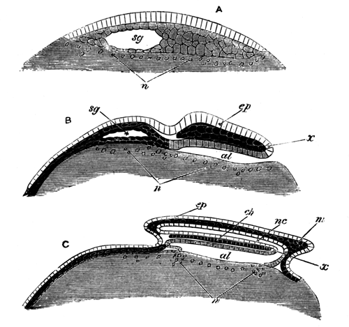
Diagrammatic longitudinal sections of an Elasmobranch
embryo.
Epiblast without shading. Mesoblast black
with clear outlines to the cells. Lower layer cells and
hypoblast with simple shading.
ep. epiblast.
m. mesoblast. al. alimentary
cavity. sg. segmentation cavity. nc. neural canal. ch. notochord. x. point where epiblast
and hypoblast become continuous at the posterior end of the embryo.
n. nuclei of yolk.
A. Section of young blastoderm, with
segmentation cavity in the middle of the lower layer cells.
B. Older blastoderm with embryo in which
hypoblast and mesoblast are distinctly formed, and in which the alimentary
slit has appeared. The segmentation cavity is still represented as being
present, though by this stage it has in reality disappeared.
C. Older blastoderm with embryo in which neural
canal has become formed, and is continuous posteriorly with alimentary
canal. The notochord, though shaded like mesoblast, belongs properly to
the hypoblast.
Fig. 2.
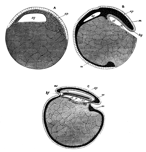
Diagrammatic longitudinal sections of embryo, which
develops in the same manner as the Elasmobranch embryo, but in which the
ovum contains far less food-yolk than is the case with the Elasmobranch
ovum.
Epiblast without shading. Mesoblast black
with clear outlines to the cells. Lower layer cells and
hypoblast with simple shading.
ep. epiblast.
m. mesoblast. hy. hypoblast.
sg. segmentation cavity. al. alimentary cavity. nc. neural canal. hf. head fold. n. nuclei of the yolk.
The stages A, B and C are the same
as in figure .[TN9]
Fig. 3.
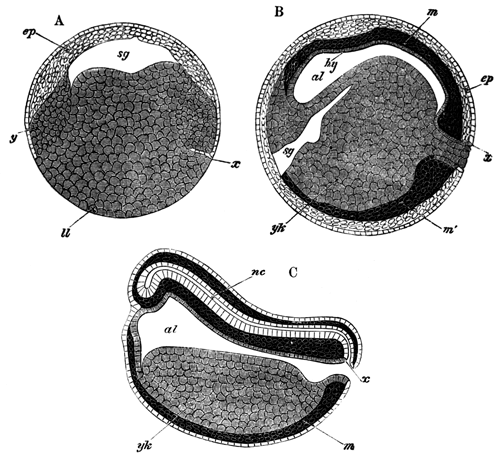
Diagrammatic longitudinal sections of Bombinator
igneus. Reproduced with modifications from Götte.
Epiblast without shading. Mesoblast black
with clear outlines to the cells. Lower layer cells and
hypoblast with simple shading.
ep. epiblast. l.l. lower layer cells. y. smaller
lower layer cells at the sides of the segmentation cavity. m.
mesoblast. hy. hypoblast. al. alimentary cavity. sg. segmentation cavity. nc. neural cavity. yk. yolk-cells.
A is the youngest stage in which the alimentary
involution has not yet appeared. x is the point from which the
involution will start to form the dorsal wall of the alimentary tract. The
line on each side of the segmentation cavity, which separates the smaller
lower layer cells from the epiblast cells, is not present in Götte's
original figure. The two shadings employed in the diagram render it
necessary to have some line, but at this stage it is in reality not
possible to assert which cells belong to the epiblast and which to the
lower layer.
B. In this stage the alimentary cavity has
become formed, but the segmentation cavity is not yet obliterated.
x. point where epiblast and hypoblast become
continuous.
C. The neural canal is already formed, and
communicates posteriorly with the alimentary.
x. point where epiblast and hypoblast become
continuous.
The relation of the yolk to the blastoderm in the Elasmobranch
embryo at this stage of development very well suits the view of its
homology with the large cells of the Amphibian ovum. The only essential
difference between the two ova arises from the roof of the segmentation
cavity being in the Elasmobranch embryo formed of lower layer cells, which
are absent in the Amphibian embryo. This difference no doubt depends upon
the greater quantity of yolk particles present in the Elasmobranch ovum.
These increase the bulk of the lower layer cells, which are thus compelled
to creep up the sides of the segmentation cavity till they close it in
above.
In the next stage for the Elasmobranch, fig. 1 and 2 B and Pl. 7, fig. 7, and for the Amphibian, fig. 3,
B, the agreement between the two types is again very close. In both
for a small portion (x) of the edge of the blastoderm the epiblast
and hypoblast become continuous, while at all other parts the epiblast,
accompanied by lower layer cells, grows round the yolk or round the large
cells which correspond to it. The yolk cells of the Amphibian ovum form a
comparatively small mass, and are therefore rapidly enveloped; while in the
case of the Elasmobranch ovum, owing to the greater mass of the yolk, the
same process occupies a long period. In both ova the portion of the
blastoderm, where epiblast and hypoblast become continuous, forms the
dorsal lip of an opening—the anus of Rusconi—which leads into
the alimentary cavity. This cavity has the same relation in both ova. It is
lined dorsally by lower layer cells, and ventrally by yolk or what
corresponds with yolk; the ventral epithelium of the alimentary canal being
in both cases eventually supplied by the yolk cells.
As in the earlier stage, so in the present one, the anatomical relations
of the yolk to the blastoderm in the one case (Elasmobranch) are nearly
identical with those of the yolk cells to the blastoderm in the other
(Amphibian). The main features in which the two embryos differ, during the
stage under consideration, arise from the same cause as the solitary point
of difference during the preceding stage.
In Amphibians, the alimentary cavity is formed coincidently
with a true ingrowth of cells from the point where epiblast and hypoblast
become continuous, and from this ingrowth the dorsal wall of the alimentary
cavity is formed. The same ingrowth causes the obliteration of the
segmentation cavity.
In the Elasmobranchii, owing to the larger bulk of the lower layer cells
caused by the food-yolk, these have been compelled to arrange themselves in
their final position during segmentation, and no room is left for a true
invagination; but instead of this there is formed a simple split between
the blastoderm and the yolk. The homology of this with the primitive
invagination is nevertheless proved by the survival of a number of features
belonging to the ancestral condition in which a true invagination was
present. Amongst the more important of these are the following:—(1)
The continuity of epiblast and hypoblast at the dorsal lip of the anus of
Rusconi. (2) The continuous conversion of indifferent lower layer cells
into hypoblast, which gradually extends backwards towards the segmentation
cavity, and exactly represents the course of the invagination whereby in
Amphibians the dorsal wall of the alimentary cavity is formed. (3) The
obliteration of the segmentation cavity during the period when the
pseudo-invagination is occurring.
The asymmetry of the gastrula or pseudo-gastrula in Cyclostomes,
Amphibians, Elasmobranchii and, I believe, Osseous Fishes, is to be
explained by the form of the vertebrate body. In Amphioxus, where the small
amount of food-yolk present is distributed uniformly, there is no reason
why the invagination and resulting gastrula should not be symmetrical. In
other vertebrates, where more food-yolk is present, the shape and structure
of the body render it necessary for the food-yolk to be stored away on the
ventral side of the alimentary canal. This, combined with the unsymmetrical
position of the anus, which primitively corresponds in position with the
blastopore or anus of Rusconi, causes the asymmetry of the gastrula
invagination, since it is not possible for the part of the ovum which will
become the ventral wall of the alimentary canal, and which is loaded with
food-yolk, to be invaginated in the same fashion as the dorsal wall. From
the asymmetry, so caused, follow a large number of features in vertebrate
development, which have been worked out in some detail
in my paper already quoted[160].
Prof. Haeckel, in a paper recently published[161],
appears to imply that because I do not find absolute invagination in
Elasmobranchii, I therefore look upon Elasmobranchii as militating against
his Gastræa theory. I cannot help thinking that Prof. Haeckel must have
somewhat misunderstood my meaning. The importance of the Gastræa theory has
always appeared to me to consist not in the fact that an actual ingrowth of
certain cells occurs—an ingrowth which might have many different
meanings[162]—but in the fact that the
types of early development of all animals can be easily derived from that
of the typical gastrula. I am perfectly in accordance with Professor
Haeckel in regarding the type of Elasmobranch development to be a simple
derivative from that of the gastrula, although believing it to be without
any true ingrowth or invagination of cells.
Professor Haeckel[163] in the paper just referred to
published his view upon the mutual relationships of the various vertebrate
blastoderms. In this paper, which appeared but shortly after my own[164] on the same subject, he has put forward views which
differ from mine in several important details. Some of these bear upon the
nature of food-yolk; and it appears to me that Professor Haeckel's scheme
of development is incompatible with the view that the food-yolk in
meroblastic eggs is the homologue of part of the hypoblast of the
holoblastic eggs.
The following is Professor Haeckel's own statement of the scheme or
type, which he regards as characteristic of meroblastic eggs, pp. 98 and 99.
Jetzt folgt der höchst wichtige und
interessante Vorgang, den ich als Einstülpung der Blastula auffasse und der
zur Bildung der Gastrula führt (Fig. 63, 64)[165]. Es
schlägt sich nämlich der verdickte Saum der Keimscheibe, der Randwulst
oder das Properistom, nach innen um und eine dünne Zellenschicht
wächst als directe Fortsetzung desselben, wie ein immer enger werdendes
Diaphragma, in die Keimhöhle hinein. Diese Zellenschicht ist das
entstehende Entoderm (Fig. 64 i, 74 i). Die Zellen, welche
dieselbe zusammensetzen und aus dem innern Theile des Randwulstes
hervorwachsen, sind viel grösser aber flacher als die Zellen der
Keimhöhlendecke und zeigen ein dunkleres grobkörniges Protoplasma. Auf dem
Boden der Keimhöhle, d. h. also auf der Eiweisskugel des Nahrungsdotters,
liegen sie unmittelbar auf und rücken hier durch centripetale Wanderung
gegen dessen Mitte vor, bis sie dieselbe zuletzt erreichen und nunmehr eine
zusammenhängende einschichtige Zellenlage auf dem ganzen Keimhöhlenboden
bilden. Diese ist die erste vollständige Anlage des Darmblatts, Entoderms
oder Hypoblasts
, und von nun an können wir, im Gegensatz dazu den
gesammten übrigen Theil des Blastoderms, nämlich die mehrschichtige Wand
der Keimhöhlendecke als Hautblatt, Exoderm oder Epiblast
bezeichnen. Der
verdickte Randwulst (Fig. 64 w, 74 w), in welchem beide
primäre Keimblätter in einander übergehen, besteht in seinem oberen und
äusseren Theile aus Exodermzellen, in seinem unteren und inneren Theile aus
Entodermzellen.
In diesem Stadium entspricht unser
Fischkeim einer Amphiblastula, welche mitten in der Invagination begriffen
ist, und bei welcher die entstehende Urdarmhöhle eine grosse Dotterkugel
aufgenommen hat. Die Invagination wird nunmehr dadurch vervollständigt und
die Gastrulabildung dadurch abgeschlossen, dass die Keimhöhle verschwindet.
Das wachsende Entoderm, dem die Dotterkugel innig anhängt, wölbt sich in
die letztere hinein und nähert sich so dem Exoderm. Die klare Flüssigkeit
in der Keimhöhle wird resorbirt und schliesslich legt sich die obere
convexe Fläche des Entoderms an die untere concave des Exoderms eng an: die
Gastrula des discoblastischen Eies oder die Discogastrula
ist fertig (Fig. 65, 76;
Meridiandurchschnitt Fig. 66, 75).
Die Discogastrula unsers Knochenfisches in
diesem Stadium der vollen Ausbildung stellt nunmehr eine kreisrunde Kappe
dar, welche wie ein gefüttertes Mützchen fast die ganze obere Hemisphäre
der hyalinen Dotterkugel eng anliegend bedeckt (Fig. 65). Der Ueberzug des
Mützchens entspricht dem Exoderm (e), sein Futter dem Entoderm
(i). Ersteres besteht aus drei Schichten von kleineren Zellen,
letzteres aus einer einzigen Schicht von grösseren Zellen. Die
Exodermzellen (Fig. 77) messen 0.006 - 0.009 Mm., und haben ein klares, sehr feinkörniges
Protoplasma. Die Entodermzellen (Fig. 78) messen 0.02 - 0.03 Mm. und ihr Protoplasma ist mehr grobkörnig und trüber.
Letztere bilden auch den grössten Theil des Randwulstes, den wir nunmehr
als Urmundrand der Gastrula, als
Properistoma
oder auch als Rusconi'schen After
bezeichnen können. Der letztere
umfasst die Dotterkugel, welche die ganze Urdarmhöhle ausfüllt und weit aus
der dadurch verstopften Urmund-Oeffnung vorragt.
My objections to the view so lucidly explained in the passage just
quoted, fall under two heads.
(1) That the facts of development of the meroblastic eggs of
vertebrates, are not in accordance with the views here advanced.
(2) That even if these views be accepted as representing the actual
facts of development, the explanation offered of these facts would not be
satisfactory.
* * * * *
Professor Haeckel's views are absolutely incompatible with the facts of
Elasmobranch development, if my investigations are correct.
The grounds of the incompatibility may be summed up under the following
heads:
(1) In Elasmobranchii the hypoblast cells occupy, even before the close
of segmentation, the position which, on Professor Haeckel's view, they
ought only eventually to take up after being involuted from the whole
periphery of the blastoderm.
(2) There is no sign at any period of an invagination of the periphery
of the blastoderm, and the only structure (the embryonic rim) which could
be mistaken for such an invagination is confined to a very limited arc.
(3) The growth of cells to form the floor of the segmentation cavity,
which ought to be part of this general invagination from the periphery, is
mainly due to a formation of cells from the yolk.
It is this ingrowth of cells for the floor of the segmentation cavity
which, I am inclined to think, Professor Haeckel has mistaken for a general
invagination in the Osseous Fish he has investigated.
(4) Professor Haeckel fails to give an account of the asymmetry of the
blastoderm; an asymmetry which is unquestionably also present in the
blastoderm of most Osseous Fishes, though not noticed by Professor Haeckel
in the investigations recorded in his paper.
The facts of development of Osseous Fishes, upon which Professor Haeckel
rests his views, are too much disputed, for their discussion in this
place to be profitable[166]. The eggs of Osseous
Fishes appear to me unsatisfactory objects for the study of this question,
partly on account of all the cells of the blastoderm being so much alike,
that it is a very difficult matter to distinguish between the various
layers, and, partly, because there can be little question that the eggs of
existing Osseous Fishes are very much modified, through having lost a great
part of the food-yolk possessed by the eggs of their ancestors[167]. This disappearance of the food-yolk must, without
doubt, have produced important changes in development, which would be
especially marked in a pelagic egg, like that investigated by Professor
Haeckel.
The Avian egg has been a still more disputed object than even the egg of
the Osseous Fishes. The results of my own investigations on this subject do
not accord with those of Dr Götte, or the views of Professor Haeckel[168].
Apart from disputed points of development, it appears to me that a
comparative account of the development of the meroblastic
vertebrate ova ought to take into consideration the essential differences
which exist between the Avian and Piscian blastoderms, in that the embryo
is situated in the centre of the blastoderm in the first case and at the
edge in the second[169].
This difference entails important modifications in development, and must
necessarily affect the particular points under discussion. As a result of
the different positions of the embryo in the two cases, there is present in
Elasmobranchii and Osseous Fishes a true anus of Rusconi, or primitive
opening into the alimentary canal, which is absent in Birds. Yet in neither
Elasmobranchii[170] nor Osseous Fishes does the
anus of Rusconi correspond in position with the point where the final
closing in of the yolk takes place, but in them this point corresponds
rather with the blastopore of Birds[171].
Owing also to the respective situations of the embryo in the
blastoderm, the alimentary and neural canals communicate posteriorly in
Elasmobranchii and Osseous Fishes, but not in Birds. Of all these
points Professor Haeckel makes no mention.
The support of his views which Prof. Haeckel attempts to gain from
Götte's researches in Mammalia is completely cut away by the recent
discoveries of Van Beneden[172] and Hensen[173].
It thus appears that Professor Haeckel's views but ill accord with the
facts of vertebrate development; but even if they were to do so completely
it would not in my opinion be easy to give a rational explanation of
them.
Professor Haeckel states that no sharp and fast line can be drawn
between the types of 'unequal' and 'discoidal' segmentation[174]. In the cases of unequal segmentation he admits, as is
certainly the case, that the larger yolk cells (hypoblast) are simply
enclosed by a growth of the epiblast around them; which is to be looked on
as a modification of the typical gastrula invagination, necessitated by the
large size of the yolk cells (vide Professor
Haeckel's paper, Taf. II. fig. 30). In these
instances there is no commencement of an ingrowth in the manner
supposed for meroblastic ova.
When the food-yolk becomes more bulky, and the hypoblast does not
completely segment, it is not easy to understand why an ingrowth, which had
no existence in the former case, should occur; nor where it is to come
from. Such an ingrowth as is supposed to exist by Professor Haeckel would,
in fact, break the continuity of development between meroblastic and
holoblastic ova, and thus destroy one of the most important results of the
Gastræa theory.
It is quite easy to suppose, as I have done, that in the cases of
discoidal segmentation, the hypoblast (including the yolk) becomes enclosed
by the epiblast in precisely the same manner as in the cases of unequal
segmentation.
But even if Professor Haeckel supposes that in the unsegmented food-yolk
a fresh element is added to the ovum, it remains quite
unintelligible to me how an ingrowth of cells from a circumferential line,
to form a layer which had no previous existence, can be equivalent to, or
derived from, the invagination of a layer, which exists before the process
of invagination begins, and which remains continuous throughout it.
If Professor Haeckel's views should eventually turn out to be in
accordance with the facts of vertebrate development, it will, in my
opinion, be very difficult to reduce them into conformity with the Gastræa
theory.
Although some space has been devoted to an attempt to refute the views
of Professor Haeckel on this question, I wish it to be clearly understood
that my disagreement from his opinions concerns matters of detail only, and
that I quite accept the Gastræa theory in its general bearings.
* * * * *
Observations upon the formation of the layers in Elasmobranchii have
hitherto been very few in number. Those published in my preliminary account
of these fishes are, I believe, the earliest[175].
Since then there has been published a short notice on the subject by Dr
Alex. Schultz[176]. His observations in the main
accord with my own. He apparently speaks of the nuclei of the yolk as
cells, and also of the epiblast being more than one cell deep. In Torpedo
alone, amongst the genera investigated by me, is the layer of epiblast, at
about the age of the last described embryo, composed of more than a single
row of cells.
EXPLANATION OF PLATE 7.
Complete List of Reference
Letters.
c. Cells formed in the yolk around the nuclei of
the yolk. ep. Epiblast. er. Embryonic ring. es. Embryo swelling. hy. Hypoblast. ll.
Lower layer cells. ly. Line separating
the yolk from the blastoderm. m. Mesoblast. mg. Medullary groove. n´. Nuclei of yolk.
na. Cells to form ventral wall of
alimentary canal which have been derived from the yolk. nal. Cells formed around the nuclei of the yolk which
have entered the hypoblast. sc.
Segmentation cavity. vp. Combined
lateral and vertebral plate of mesoblast.
Fig. 1. Longitudinal section of a blastoderm at the
first appearance of the segmentation cavity.
Fig. 2. Longitudinal section through a blastoderm after
the layer of cells has disappeared from the floor of the segmentation
cavity. bd. Large cell resting on the
yolk, probably remaining over from the later periods of segmentation.
Magnified 60 diameters. (Hardened in chromic acid.)
The section is intended to illustrate the fact that the
nuclei form a layer in the yolk under the floor of the segmentation cavity.
The roof of the segmentation cavity is broken.
Fig. 2a. Portion of same blastoderm highly
magnified, to shew the characters of the nuclei of the yolk n´ and
the nuclei in the cells of the blastoderm.
Fig. 2b. Large knobbed nucleus from the same
blastoderm, very highly magnified.
Fig. 2c. Nucleus of yolk from the same
blastoderm.
Fig. 3. Longitudinal section of blastoderm of same stage
as fig. 2. (Hardened in chromic acid.)
Fig. 4. Longitudinal section of blastoderm slightly
older than fig. 2. Magnified 45 diameters. (Hardened in osmic acid.)
It illustrates (1) the characters of the epiblast; (2)
the embryonic swelling; (3) the segmentation cavity.
Fig. 5. Longitudinal section through a blastoderm at the
time of the first appearance of the embryonic rim, and before the formation
of the medullary groove. Magnified 45 diameters.
Fig. 5a. Section through the periphery of the
embryonic rim of the blastoderm of which fig. 5 represents a section.
Fig. 6. Section through the embryonic rim of a
blastoderm somewhat younger than that represented on Pl. 8, fig. B.
Fig. 7. Section through the most projecting portion of
the embryonic rim of a blastoderm of the same age as that represented on
Pl. 8, fig. B. The section is drawn on a very
considerably smaller scale than that on fig. 5. It is intended to
illustrate the growth of the embryonic rim and the disappearance of the
segmentation cavity.
Fig. 7a. Section through peripheral portion of
the embryonic rim of the same blastoderm, highly magnified. It specially
illustrates the formation of a cell (c) around a nucleus in the
yolk. The nuclei of the blastoderm have been inaccurately rendered by the
artist.
Figs. 8a, 8b, 8c.
Three sections of the same embryo. Inserted mainly to illustrate the
formation of the mesoblast as two independent lateral masses of cells; only
half of each section is represented. 8a is the most posterior of the
three sections. In it the mesoblast forms a large mass on each side,
imperfectly separated from the hypoblast. In 8b, from the anterior
part of the embryo, the main mass of mesoblast is far smaller, and only
forms a cap to the hypoblast at the highest point of the medullary fold. In
8c a cap of mesoblast is present, similar to that in 8b,
though much smaller. The sections of these embryos were somewhat oblique,
and it has unfortunately happened that while in 8a one side is
represented, in 8b and 8c the other side is figured, had it
not been for this the sections 8b and 8c would have been
considerably longer than 8a.
Fig. 9. Longitudinal section of an embryo belonging to a
slightly later stage than B.
This section passes through one of the medullary folds.
It illustrates the continuity of the hypoblast with the remaining lower
layer cells of the blastoderm.
Figs. 10a, 10b, 10c. Three sections
of the same embryo belonging to a stage slightly later than B, Pl. 8. The space between the mesoblast and the hypoblast has
been made considerably too great in the figures of the three sections.
10a. The most posterior of the three sections. It
shews the posterior flatness of the medullary groove and the two isolated
vertebral plates.
10b. This section is taken from the anterior part
of the same embryo and shews the deep medullary groove and the commencing
formation of the ventral wall of the alimentary canal from the nuclei of
the yolk.
10c shews the disappearance of the medullary
groove and the thinning out of the mesoblast plates in the region of the
head.
Fig. 11. Small portion of the blastoderm and the
subjacent yolk of an embryo at the time of the first appearance of the
medullary groove × 300. It shews two large nuclei of the yolk (n)
and the protoplasmic network in the yolk between them; the network is seen
to be closer round the nuclei than in the intervening space. There are no
areas representing cells around the nuclei.
Fig. 12. Nucleus of the yolk in connection with the
protoplasmic network hardened in osmic acid.
Fig. 13. Portion of posterior end of a blastoderm of
stage B, shewing the formation of cells around the nuclei of the yolk.
Fig. 14. Section through part of a young Scyllium egg,
about 1/15th of an inch in diameter.
nl. Protoplasmic
network in yolk. zp. Zona pellucida.
ch. Structureless chorion. fep. Follicular epithelium. x. Structureless
membrane external to this.
No complete series of figures, representing the various
stages in development of an Elasmobranch Embryo, has hitherto been
published. With the view of supplying this deficiency Plate 8 has been
inserted. The embryos represented in this Plate form a fairly complete
series, but do not all belong to a single species. Figs. A, B, C, D, E, F,
H, I represent embryos of Pristiurus; G being an embryo of Torpedo. The
remaining figures, excepting K, which is a Pristiurus embryo, are embryos
of Scyllium canicula. The embryos A-I were very accurately drawn from
nature by my sister, Miss A. B. Balfour. Unfortunately the exceptional
beauty and clearness of the originals is all but lost in the lithographs.
To facilitate future description, letters will be employed in the remainder
of these pages to signify that an embryo being described is of the same age
as the embryo on this Plate to which the letter used refers. Thus an embryo
of the same age as L will be spoken of hereafter as belonging to stage
L.
A.
This figure represents a hardened blastoderm at a stage when the
embryo-swelling (e.s.) has become
obvious, but before the appearance of the medullary groove. The position of
the segmentation cavity is indicated by a slight swelling of the blastoderm
(s.c). The shape of the blastoderm, in
hardened specimens, is not to be relied upon, owing to the traction which
the blastoderm undergoes during the process of removing the yolk from the
egg-shell.
B.
B is the view of a fresh blastoderm. The projecting part of this,
already mentioned as the 'embryonic rim', is indicated by the shading. At
the middle of the embryonic rim is to be seen the rudiment of the embryo
(m.g.). It consists of an area of the
blastoderm, circumscribed on its two sides and at one end, by a slight
fold, and whose other end forms part of the edge of the blastoderm. The end
of the embryo which points towards the centre of the blastoderm is
the head end, and that which forms part of the edge of the
blastoderm is the tail end. To retain the nomenclature usually adopted in
treating of the development of the Bird, the fold at the anterior end of
the embryo may be called the head fold, and those at the sides the
side folds. There is in Elasmobranchii no tail fold, owing to the
position of the embryo at the periphery of the blastoderm, and it is by the
meeting of the three above-mentioned folds only, that the embryo becomes
pinched off from the remainder of the blastoderm. Along the median line of
the embryo is a shallow groove (m.g.),
the well-known medullary groove of vertebrate embryology. It flattens out
both anteriorly and posteriorly, and is deepest in the middle part of its
course.
C.
This embryo resembles in most of its features the embryo last described.
It is, however, considerably larger, and the head fold and side folds have
become more pronounced structures. The medullary groove is far deeper than
in the earlier stage, and widens out anteriorly. This anterior widening is
the first indication of a distinction between the brain and the remainder
of the central nervous system, a distinction which arises long before the
closure of the medullary canal.
D.
This embryo is far larger than the one last described, but the increase
in length does not cause it to project beyond the edge of the blastoderm,
but has been due to a growth inwards towards the centre of the blastoderm.
The head is now indicated by an anterior enlargement, and the embryo also
widens out posteriorly. The posterior widening (t.s.) is formed by a pair of rounded prominences, one
on each side of the middle line. These are very conspicuous organs during
the earlier stages of development, and consist of two large aggregations of
mesoblast cells. In accordance with the nomenclature adopted
in my preliminary paper[177], they may be called
'tail-swellings'. Between the cephalic enlargements and the tail-swellings
is situated the rudimentary trunk of the embryo. It is more completely
pinched off from the blastoderm than in the last described embryo. The
medullary groove is of a fairly uniform size throughout the trunk of the
embryo, but flattens out and vanishes completely in the region of the head.
The blastoderm in Pristiurus and Scyllium grows very rapidly, and has by
this stage attained a very considerable size; but in Torpedo its growth is
very slow.
E and F.
These two embryos may be considered together, for, although they differ
in appearance, yet they are of an almost identical age; and the differences
between the two are purely external. E appears to be a little abnormal in
not having the cephalic region so distinctly marked off from the trunk as
is usual. The head is proportionally larger than in the last stage, and the
tail-swellings remain as conspicuous as before. The folding off from the
blastoderm has progressed rapidly, and the head and tail are quite
separated from it. The medullary groove has become closed posteriorly in
both embryos, but the closing has extended further forwards in F than in E.
In F the medullary folds have not only united posteriorly, but have very
nearly effected a fresh junction in the region of the neck. At this point a
second junction of the two medullary folds is in fact actually effected
before the posterior closing has extended forwards so far. The later
junction in the region of the neck corresponds in position with the point,
where in the Bird the medullary folds first unite. No trace of a medullary
groove is to be met with in the head, which simply consists of a wide
flattened plate. Between the two tail-swellings surface views present the
appearance of a groove, but this appearance is deceptive, since in sections
no groove, or at most a very slight one, is perceptible.
G.
During the preceding stages growth in the embryo is very slow, and
considerable intervals of time elapse before any perceptible changes
are effected. This state of things now becomes altered, and the future
changes succeed each other with far greater rapidity. One of the most
important of these, and one which first presents itself during this stage,
is the disappearance of the yolk-spherules from the embryonic cells, and
the consequently increased transparency of the embryo. As a result of this,
a number of organs, which in the earlier stages were only to be
investigated by means of sections, now become visible in the living
embryo.
The tail-swellings (t.s.) are still
conspicuous objects at the posterior extremity of the embryo. The folding
off of the embryo from the yolk has progressed to such an extent that it is
now quite possible to place the embryo on its side and examine it from that
point of view.
The embryo may be said to be attached to the yolk by a distinct stalk or
cord, which in the succeeding stages gradually narrows and elongates, and
is known as the umbilical cord (so.s.).
The medullary canal has now become completely closed, even in the region of
the brain, where during the last stage no trace of a medullary groove had
appeared. Slight constrictions, not perceptible in views of the embryo as a
transparent object, mark off three vesicles in the brain. These vesicles
are known as the fore, mid, and hind brain. From the fore-brain there is an
outgrowth on each side, the first rudiment of the optic vesicle (op.).
The mesoblast on each side of the body is divided into a series of
segments, known as protovertebræ or muscle-plates, the first of which lies
a little behind the head. The mesoblast of the tail has not as yet
undergone this segmentation. There are present in all seventeen segments.
These first appeared at a much earlier date, but were not visible owing to
the opacity of the embryo.
Another structure which became developed in even a younger embryo than C
is now for the first time visible in the living embryo. This is the
notochord: it extends from almost the extreme posterior to the anterior end
of the embryo. It lies between the ventral wall of the spinal canal and the
dorsal wall of the intestine; and round its posterior end these two walls
become continuous with each other (vide fig.).
Anteriorly the termination of the notochord cannot be
seen, it can only be traced into a mass of mesoblast at the base of the
brain, which there separates the epiblast from the hypoblast. The
alimentary canal (al.) is completely
closed anteriorly and posteriorly, though still widely open to the yolk-sac
in the middle part of its course. In the region of the head it exhibits on
each side a slight bulging outwards, the rudiment of the first visceral
cleft. This is represented in the figure by two lines (I v.c.). The
visceral clefts at this stage consist of a pair of simple diverticula from
the alimentary canal, and there is no communication between the throat and
the exterior.
H.
The present embryo is far larger than the last, but it has not been
possible to represent this increase in size in the drawings. Accompanying
this increase in size, the folding off of the embryo from the yolk has
considerably progressed, and the stalk which unites the embryo with the
yolk is proportionately narrower and longer than before.
The brain is now very distinctly divided into the three lobes, whose
rudiments appeared during the last stage. From the foremost of these, the
optic vesicles now present themselves as well-marked lateral outgrowths,
towards which there appears a growing in, or involution, from the external
skin (op.) to form the lens. The opening
of this involution is represented by the dark spot in the centre.
A fresh organ of sense, the auditory sac, now for the first time becomes
visible as a shallow pit in the external skin on each side of the
hind-brain (au.v.). The epiblast which is
involuted to form this pit becomes much thickened, and thereby the opacity,
indicated in the figure, is produced.
The muscle-plates have greatly increased in number by the formation of
fresh segments in the tail. Thirty-eight of them were present in the embryo
figured. The mesoblast at the base of the brain has increased in quantity,
and there is still a certain mass of unsegmented mesoblast which forms the
tail-swellings. The first rudiment of the heart becomes visible during this
stage as a cavity between the mesoblast of the splanchnopleure and the
hypoblast (ht.).
The fore and hind guts are now longer than they were. A slight
pushing in from the exterior to form the mouth has appeared (m.),
and an indication of the future position of the anus is afforded by a
slight diverticulum of the hind gut towards the exterior some little
distance from the posterior end of the embryo (an.). The portion of the alimentary canal behind this
point, though at this stage large, and even dilated into a vesicle at its
posterior end (al.v.), becomes eventually
completely atrophied. In the region of the throat the rudiment of a second
visceral cleft has appeared behind the first; neither of them are as yet
open to the exterior. The number of visceral clefts present in any given
Pristiurus embryo affords a very easy and simple way of determining its
age.
I.
A great increase in size is again to be noticed in the embryo, but, as
in the case of the last embryo, it has not been possible to represent this
in the figure. The stalk connecting the embryo with the yolk has become
narrower and more elongated, and the tail region of the embryo
proportionately far longer than in the last stage. During this stage the
first spontaneous movements of the embryo take place, and consist in
somewhat rapid excursions of the embryo from side to side, produced by a
serpentine motion of the body.
The cranial flexure, which commenced in stage G, has now become very
evident, and the mid-brain[178] begins to project in the
same manner as in the embryo fowl on the third day, and will soon form the
anterior termination of the long axis of the embryo. The fore-brain has
increased in size and distinctness, and the anterior part of it may now be
looked on as the unpaired rudiment of the cerebral hemispheres.
Further growths have taken place in the organs of sense, especially in
the eye, in which the involution for the lens has made considerable
progress. The number of the muscle-plates has again increased, but there is
still a region of unsegmented mesoblast in the tail. The thickened
portions of mesoblast which caused the tail-swellings are still to be seen
and would seem to act as the reserve from which is drawn the matter for the
rapid growth of the tail, which occurs soon after this. The mass of the
mesoblast at the base of the brain has again increased. No fresh features
of interest are to be seen in the notochord. The heart is now much more
conspicuous than before, and its commencing flexure is very apparent. It
now beats actively. The hind gut especially is much longer than in the last
specimen; and the point where the anus will appear is very easily detected
by the bulging out of the gut towards the external skin at that point
(an.). The alimentary vesicle, first
observable during the last stage, is now a more conspicuous organ (al.v.). Three visceral clefts, none of which
are as yet open to the exterior, may now be seen.
K.
The figures G, H, I are representations of living and transparent
embryos, but the remainder of the figures are drawings of opaque embryos
which were hardened in chromic acid.
The stalk connecting the embryo with the yolk is now, comparatively
speaking, quite narrow, and is of sufficient length to permit the embryo to
execute considerable movements.
The tail has grown immensely, but is still dilated terminally. This
terminal dilatation is mainly due to the alimentary vesicle, but the tract
of gut connecting this with the gut in front of the anus is now a solid rod
of cells and very soon becomes completely atrophied.
The two pairs of limbs have appeared as elongated ridges of epiblast.
The anterior pair is situated just at the front end of the umbilical stalk;
and the posterior pair, which is the more conspicuous of the two, is
situated some little distance behind the stalk.
The cranial flexure has greatly increased, and the angle between the
long axis of the front part of the head and of the body is less than a
right angle. The conspicuous mid-brain forms the anterior termination of
the long axis of the body. The thin roof of the fourth ventricle may in the
figure be noticed behind the mid-brain. The auditory sac is nearly closed
and its opening is not shewn in the figure. In the eye the lens is
completely formed.
Owing to the opacity of the embryo, the muscle-plates are only
indistinctly indicated, and no other features of the mesoblast are to be
seen.
The mouth is now a deep pit, whose borders are almost completely formed
by the thickening in front of the first visceral cleft, which may be called
the first visceral arch or mandibular arch.
Four visceral clefts are now visible, all of which are open to the
exterior, but in a transparent embryo one more, not open to the exterior,
would have been visible behind the last of these.
L.
This embryo is considerably older than the one last described, but
growth is not quite so rapid as might be gathered from the fact that L is
nearly twice as long as K, since the two embryos belong to different
genera; and the Scyllium embryos, of which L is an example, are larger than
Pristiurus embryos. The umbilical stalk is now quite a narrow elongated
structure, whose subsequent external changes are very unimportant, and
consist for the most part merely in an increase in its length.
The tail has again grown greatly in length, and its terminal dilatation
together with the alimentary vesicle contained in it, have both completely
vanished. A dorsal and ventral fin are now clearly visible; they are
continuous throughout their whole length. The limbs have grown and are more
easily seen than in the previous stage.
Great changes have been effected in the head, resulting in a diminution
of the cranial flexure. This diminution is nevertheless apparent rather
than real, and is chiefly due to the rapid growth of the rudiment of the
cerebral hemispheres. The three main divisions of the brain may still be
clearly seen from the surface. Posteriorly is situated the hind-brain, now
consisting of the medulla oblongata and cerebellum. At the anterior part of
the medulla is to be seen the thin roof of the fourth ventricle, and
anteriorly to this again the roof becomes thickened to form the rudiment of
the cerebellum. In front of the hind-brain lies the mid-brain, the roof of
which is formed by the optic lobes, which are still situated at
the front end of the long axis of the embryo.
Beyond the mid-brain is placed the fore-brain, whose growth is rapidly
rendering the cranial flexure imperceptible.
The rudiments of the nasal sacs are now clearly visible as a pair of
small pits. The pits are widely open to the exterior, and are situated one
on each side, near the front end of the cerebral hemispheres. Five visceral
clefts are open to the exterior, and in them the external gills have
commenced to appear (L´).
The first cleft is no longer similar to the rest, but has commenced to
be metamorphosed into the spiracle.
Accompanying the change in position of the first cleft, the mandibular
arch has begun to bend round and enclose the front as well as the side of
the mouth. By this change in the mandibular arch the mouth becomes narrowed
in an antero-posterior direction.
M.
Of this embryo the head alone has been represented. Two views of it are
given, one (M) from the side and the other (M´) from the under surface. The
growth of the front part of the head has considerably diminished the
prominence of the cranial flexure. The full complement of visceral clefts
is now present—six in all. But the first has already atrophied
considerably, and may easily be recognized as the spiracle. In Scyllium,
there are present at no period more than six visceral clefts. The first
visceral arch on each side has become bent still further round, to form the
front border of the mouth. The opening of the mouth has in consequence
become still more narrowed in an antero-posterior direction. The width of
the mouth in this direction, serves for the present and for some of the
subsequent stages as a very convenient indication of age.
N.
The limbs, or paired fins, have now acquired the general features and
form which they possess in the adult.
The unpaired fins have now also become divided in a manner not only
characteristic of the Elasmobranchii but even of the genus Scyllium.
There is a tail fin, an anal fin and two dorsal fins, both the
latter being situated behind the posterior paired fins.
In the head may be noticed a continuation of the rapid growth of the
anterior part.
The mouth has become far more narrow and slit-like; and with many other
of the organs of the period commences to approach the form of the
adult.
The present and the three preceding stages shew the gradual changes by
which the first visceral arch becomes converted into the rudiments of the
upper and of the lower jaw. The fact of the conversion was first made known
through the investigations of Messrs Parker and Gegenbaur.
O.
In this stage the embryo is very rapidly approaching the form of the
adult.
This is especially noticeable in the fins, which project in a manner
quite characteristic of the adult fish. The mouth is slit-like, and the
openings of the nasal sacs no longer retain their primitive circular
outline. The external gills project from all the gill-slits including the
spiracle.
P.
The head is rapidly elongating by the growth of the snout, and the
divisions of the brain can no longer be seen with distinctness from the
exterior, and, with the exception of the head and of the external gills,
the embryo almost completely resembles the adult.
Q.
The snout has grown to such an extent, that the head has nearly acquired
its adult shape. In the form of its mouth the embryo now quite resembles
the adult fish.
* * * * *
This part of the subject may be conveniently supplemented by a short
description of the manner in which the blastoderm encloses the yolk. It has
been already mentioned that the growth of the blastoderm is not uniform.
The part of it in the immediate neighbourhood of the embryo remains
comparatively stationary, while the growth elsewhere is very rapid. From
this it results that that part of the edge of the blastoderm where the
embryo is attached forms a bay in the otherwise regular outline of the edge
of the blastoderm. By the time that one-half of the yolk is enclosed the
bay is a very conspicuous feature (Pl. 9, fig.
1). In this figure bl. points to the
blastoderm, and yk. to the part of the
yolk not yet enclosed by the blastoderm.
Shortly subsequent to this the bay becomes obliterated by its two sides
coming together and coalescing, and the embryo ceases to lie at the edge of
the yolk.
This stage is represented on Pl. 9, fig. 2.
In this figure there is only a small patch of yolk not yet enclosed (yk), which is situated at some little distance
behind the embryo. Throughout all this period the edge of the blastoderm
has remained thickened, a feature which persists till the complete
investment of the yolk, which takes place shortly after the stage last
figured. In this thickened edge a circular vein arises, which brings back
the blood from the yolk-sac to the embryo. The opening in the blastoderm
(Pl. 9, fig. 2, yk.), exposing the portion of the yolk not yet
enclosed, may be conveniently called the blastopore, according to Professor
Lankester's nomenclature.
The interesting feature which characterizes the blastopore in
Elasmobranchii is the fact of its not corresponding in position with the
opening of the anus of Rusconi. We thus have in Elasmobranchii two
structures, each of which corresponds in part with the single structure in
Amphioxus which may be called either blastopore or anus of Rusconi, which
yet do not in Elasmobranchii coincide in position. It is the blastopore of
Elasmobranchii which has undergone a change of position, owing to the
unequal growth of the blastoderm; while the anus of Rusconi retains its
normal situation. In Osseous Fishes the blastopore undergoes a similar
change of position. The possibility of a change in position of this
structure is peculiarly interesting, in that it possibly serves to explain
how the blastopore of different animals corresponds in different cases with
the anus or the mouth, and has not always a fixed situation[179].
EXPLANATION OF PLATES 8 and 9.
Complete List of Reference
Letters.
a. Arteries of yolk sac (red). al. Alimentary cavity. alv. Alimentary vesicle at the posterior end of the
alimentary canal. an. Point where anus
will appear. auv. Auditory vesicle.
bl. Blastoderm. ch. Notochord. es. Embryo-swelling. h. Head. ht. Heart. m. Mouth. mg. Medullary groove. mp. Muscle-plate or protovertebra. op. Eye. sc.
Segmentation cavity. sos. Somatic stalk.
ts. Tail-swelling. v. Veins of
yolk sac (blue). vc. Visceral cleft.
I. vc. 1st visceral cleft. x. Portion of blastoderm outside
the arterial circle in which no blood-vessels are present. yk. Yolk.
Plate 8.
Fig. A. Surface view of blastoderm of Pristiurus
hardened in chromic acid.
Fig. B. Surface view of fresh blastoderm of
Pristiurus.
Figs. C, D, E, and F. Pristiurus embryos hardened in
chromic acid.
Fig. G. Torpedo embryo viewed as a transparent
object.
Figs. H, I. Pristiurus embryos viewed as transparent
objects.
Fig. K. Pristiurus embryo hardened in chromic acid.
The remainder of the figures are representations of
embryos of Scyllium canicula hardened in chromic acid. In every case, with
the exception of the figures marked P and Q, two representations of the
same embryo are given; one from the side and one from the under
surface.
Plate 9.
Fig. 1. Yolk of a Pristiurus egg with blastoderm and
embryo. About two-thirds of the yolk have been enveloped by the blastoderm.
The embryo is still situated at the edge of the blastoderm, but at the end
of a bay in the outline of this. The thickened edge of the blastoderm is
indicated by a darker shading. Two arteries have appeared.
Fig. 2. Yolk of an older Pristiurus egg. The yolk has
become all but enveloped by the blastoderm, and the embryo ceases to lie at
the edge of the blastoderm, owing to the coalescence of the two sides of
the bay which existed in the earlier stage. The circulation is now largely
developed. It consists of an external arterial ring, and an internal venous
ring, the latter having been developed in the thickened edge of the
blastoderm. Outside the arterial ring no vessels are developed.
Fig. 3. The yolk has now become completely enveloped by
the blastoderm. The arterial ring has increased in size. The venous ring
has vanished, owing to the complete enclosure of the yolk by the
blastoderm. The point where it existed is still indicated (y) by the
brush-like termination of the main venous trunk in a number of small
branches.
Fig. 4. Diagrammatic projection of the vascular system
of the yolk sac of a somewhat older embryo.
The arterial ring has grown much larger and the portion
of the yolk where no vessels exist is very small (x). The brush-like
termination of the venous trunk is still to be noticed.
The two main trunks (arterial and venous) in reality are
in close contact as in fig. 5, and enter the somatic stalk close
together.
The letter a which points to the venous (blue)
trunk should be v and not a.
Fig. 5. Circulation of the yolk sac of a still older
embryo, in which the arterial circle has ceased to exist, owing to the
space outside it having become smaller and smaller and finally
vanished.
The present chapter deals with the history of the development
of the Elasmobranch embryo from the period when the medullary groove first
arises till that in which it becomes completely closed, and converted into
the medullary canal. The majority of the observations recorded were made on
Pristiurus embryos, a few on embryos of Torpedo. Where nothing is said to
the contrary the statements made apply to the embryos of Pristiurus
only.
The general external features for this period have already been given in
sufficient detail in the last chapter; and I proceed at once to describe
consecutively the history of the three layers.
General Features of the Epiblast.
At the commencement of this period, during the stage intermediate
between B and C, the epiblast is composed of a single layer of cells.
(Pl. 10, fig. 1.)
These are very much elongated in the region of the embryo, but flattened
in other parts of the blastoderm. Throughout they contain numerous
yolk-spherules.
In a Torpedo embryo of this age (as determined by the condition of the
notochord) the epiblast presents a very different structure. It is composed
of small spindle-shaped cells several rows deep. The nuclei of these are
very large in proportion to the cells containing them, and the
yolk-spherules are far less numerous than in the cells of corresponding
Pristiurus embryos.
During stage C the condition of the epiblast does not undergo any
important change, with the exception of the layer becoming much
thickened, and its cells two or three deep in the anterior parts of the
embryo. (Pl. 10, fig. 2.)
In the succeeding stages that part of the epiblast, which will form the
spinal cord, gradually becomes two or three cells deep. This change is
effected by a decrease in the length of the cells as compared with the
thickness of the layer. In the earlier stages the cells are wedge-shaped
with an alternate arrangement, so that a decrement in the length of the
cells at once causes the epiblast to be composed of two rows of
interlocking cells.
The lateral parts of the epiblast which form the epidermis of the embryo
are modified in quite a different manner to the nervous parts of the layer,
becoming very much diminished in thickness and composed of a single row of
flattened cells. (Pl. 10, fig. 3.)
Till the end of stage F, the epiblast cells and indeed all the cells of
the blastoderm retain their yolk-spherules, but the epiblast begins to lose
them and consequently to become transparent in stage G.
Medullary Groove.
During stage B the medullary groove is shallow posteriorly, deeper in
the middle part, and flattened out again at the extreme anterior end of the
embryo. (Pl. 7, fig. 10a, b,
c.)
A similar condition obtains in the stage between B and C, but the canal
has now in part become deeper. Anteriorly no trace of it is to be seen. In
stage C it exhibits the same general features. (Pl. 10, fig. 2a, 2b, 2c.)
By stage D we find important modifications of the canal.
It is still shallow behind and deep in the dorsal region, Pl. 10, figs. 3d, 3e, 3f; but the
anterior flattened area in the last stage has grown into a round flat plate
which may be called the cephalic plate, Pl. 8,
D and Pl. 10, figs. 3a, 3b,
3c. This plate becomes converted into the brain. Its size and form
give it a peculiar appearance, but the most remarkable feature about it is
the ventral curvature of its edges. Its edges do not, as might be expected,
bend dorsalwards towards each other, but become sharply bent in a ventral
direction. This feature is for the first time apparent at
this stage, but becomes more conspicuous during the succeeding ones, and
attains its maximum in stage F (Pl. 10, fig.
5), in which it might almost be supposed that the edges of the cephalic
plate were about to grow downwards and meet on the ventral side of the
embryo.
In the stages subsequent to D the posterior part of the canal deepens
much more rapidly than the rest (vide Pl. 10, fig. 4, taken from the posterior end of an embryo
but slightly younger than F), and the medullary folds unite and convert the
posterior end of the medullary groove into a closed canal (Pl. 8, fig. F), while the groove is still widely open
elsewhere[180]. The medullary canal does not
end blindly behind, but simply forms a tube not closed at either extremity.
The importance of this fact will appear later.
In a stage but slightly subsequent to F nearly the whole of the
medullary canal becomes formed. This occurs in the usual way by the
junction and coalescence of the medullary folds. In the course of the
closing of the medullary groove the edges of the cephalic plate lose their
ventral curvature and become bent up in the normal manner (vide Pl. 10, fig. 6, a section
taken through the posterior part of the cephalic plate), and the enlarged
plate merely serves to enclose a dilated cephalic portion of the medullary
canal. The closing of the medullary canal takes place earlier in the head
and neck than in the back. The anterior end of the canal becomes closed and
does not remain open like the posterior end.
Elasmobranch embryos resemble those of the Sturgeon (Acipenser) and the
Amphibians in the possession of a spatula-like cephalic expansion: but so
far as I am aware a ventral flexure in the medullary plates of the head has
not been observed in other groups.
The medullary canal in Elasmobranchii is formed precisely on the type so
well recognised for all groups of vertebrates with the exception of the
Osseous Fishes. The only feature in any respect peculiar to these fishes is
the closing of their medullary canal first commencing behind, and then at a
second point in the cervical region. In those vertebrates in which
the medullary folds do not unite at approximately the same time throughout
their length, they appear usually to do so first in the region of the
neck.
Mesoblast.
The separation from the hypoblast of two lateral masses of mesoblast has
already been described. Till the close of stage C the mesoblast retains its
primitive bilateral condition unaltered. Throughout the whole length of the
embryo, with the exception of the extreme front part, there are present two
plates of rounded mesoblast cells, one on each side of the medullary
groove. These plates are in very close contact with the hypoblast, and also
follow with fair accuracy the outline of the epiblast. This relation of the
mesoblast plates to the epiblast must not however be supposed to indicate
that the medullary groove is due to growth in the mesoblast: a view which
is absolutely negatived by the manner of formation of the medullary groove
in the head. Anteriorly the mesoblast plates thin out and completely
vanish.
In stage D, the plates of mesoblast in the trunk undergo important
changes. The cells composing them become arranged in two layers (Pl. 10, fig. 3), a splanchnic layer adjoining the
hypoblast (sp), and a somatic layer
adjoining the epiblast[181] (so). Although these two layers are distinctly formed,
they do not become separated at this stage in the region of the trunk, and
in the trunk no true body-cavity is formed.
By stage D the plates of mesoblast have ceased to be quite isolated, and
are connected with the lower layer cells of the general blastoderm.
Moreover the lower layer cells outside the embryo now exhibit distinct
traces of a separation into two layers, one continuous with the hypoblast,
the other with the mesoblast. Both layers are composed of very flattened
cells, and the mesoblast layer is often more than one cell deep, and
sometimes exhibits a mesh-like arrangement of its elements.
Coincidentally with the appearance of a differentiation into a
somatic and splanchnic layer the mesoblast plates become partially split by
a series of transverse lines of division into protovertebræ. Only the
proximal regions of the plates become split in this way, while their
peripheral parts remain quite intact. As a result of this each plate
becomes divided into a proximal portion adjoining the medullary canal,
which is divided into protovertebræ, and may be called the
vertebral plate, and a peripheral portion not so divided, which
may be called the lateral plate. These two parts are at this stage
quite continuous with each other; and, as will be seen in the sequel, the
body-cavity originally extends uninterruptedly to the summit of the
vertebral plates.
By stage D at the least ten protovertebræ have appeared.
In Torpedo the mesoblast commences to be divided into two layers much
earlier than in Pristiurus; and even before stage C this division is more
or less clearly marked.
In the head and tail the condition of the mesoblast is by no means the
same as in the body.
In the tail the plates of mesoblast become considerably thickened and
give rise to two projections, one on each side, which have already been
alluded to as caudal or tail-swellings; vide Pl. 8, figs. D, F, and Pl. 10, fig. 3f and fig. 4, ts.
These masses of mesoblast are neither divided into protovertebræ, nor do
they exhibit any trace of a commencing differentiation into somatopleure
and splanchnopleure.
In the head, so far as I have yet been able to observe, the mesoblastic
plates do not at this stage become divided into protovertebræ. The
other changes exhibited in the cephalic region are of interest, mainly from
the fact that here appears a cavity in the mesoblast directly continuous
with the body-cavity (when that cavity becomes formed), but which appears
at a very much earlier date than the body-cavity. This cavity can only be
looked on in the light of a direct continuation of the body or peritoneal
cavity into the head. Theoretical considerations with reference to it I
propose reserving till I have described the changes which it undergoes in
the subsequent periods.
Pl. 10, figs. 3a, 3b and
3c exhibit very well the condition of the mesoblast in the head at
this period. In fig. 3c, a section taken through the
back part of the head, the mesoblast plates have nearly the same form as in
the sections immediately behind. The ventral continuation of the mesoblast
formed by the lateral plate has, however, become much thinner, and the
dorsal or vertebral portion has acquired a more triangular form than in the
sections through the trunk (figs. 3d and 3e).
In the section (fig. 3b) in front of this the ventral portion of
the plate is no longer present, and only that part exists which corresponds
with the vertebral division of the primitive plate of mesoblast.
In this a distinct cavity, forming part of the body-cavity, has
appeared.
In a still anterior section (fig. 3a) no cavity is any longer
present in the mesoblast; whilst in sections taken from the foremost part
of the head no mesoblast is to be seen (vide Pl. 10, fig. 5, taken from the front part of the head
of the embryo represented in Pl. 8, fig.
F).
A continuation of the body-cavity into the head has already been
described by Oellacher[182] for the Trout: but he
believes that the cavity in this part is solely related to the formation of
the pericardial space.
The condition of the mesoblast undergoes no important change till the
end of the period treated of in this chapter. The masses of mesoblast which
form the tail-swellings become more conspicuous (Pl. 10, fig. 4); and indeed their convexity is so great that
the space between them has the appearance of a median groove, even after
the closure of the neural canal in the caudal region.
In embryos of stage G, which may be considered to belong to the close of
this period, eighteen protovertebræ are present both in Pristiurus and
Torpedo embryos.
The Alimentary Canal.
The alimentary canal at the commencement of this period (stage B) forms
a space between the embryo and the yolk, ending blindly in front, but
opening posteriorly by a widish slit-like aperture, which corresponds to
the anus of Rusconi (Pl. 7, fig. 7).
The cavity anteriorly has a more or less definite form, having
lateral walls, as well as a roof and floor (Pl.
7, figs. 10b and 10c). Posteriorly it is not nearly so
definitely enclosed (Pl. 7, fig. 10a).
The ventral wall of the cavity is formed by yolk. But even in stage B there
are beginnings of a cellular ventral wall derived from an ingrowth of cells
from the two sides.
By stage C considerable progress has been made in the formation of the
alimentary canal. Posteriorly it is as flattened and indefinite as during
stage B (Pl. 10, figs. 2b and
2c). But in the anterior part of the embryo the cavity becomes much
deeper and narrower, and a floor of cells begins to be formed for it (Pl. 10, fig. 2); and, finally, in front, it forms a
definite space completely closed in on all sides by cells (Pl. 10, fig. 2a). Two distinct processes are
concerned in effecting these changes in the condition of the alimentary
cavity. One of these is a process of folding off the embryo from the
blastoderm. The other is a simple growth of cells independent of any
folding. To the first of these processes the depth and narrowness of the
alimentary cavity is due; the second is concerned in forming its ventral
wall. The combination of the two processes produces the peculiar triangular
section which characterises the anterior closed end of the alimentary
cavity at this stage. The process of the folding off of the embryo from the
blastoderm resembles exactly the similar process in the embryo bird. The
fold by which the constricting off of the embryo is effected is a perfectly
continuous one, but may be conveniently spoken of as composed of a head
fold and two lateral folds.
Of far greater interest than the nature of these folds is the formation
of the ventral wall of the alimentary canal. This, as has been said, is
effected by a growth of cells from the two sides to the middle line (Pl. 10, fig. 2). The cells for this are however not
derived from pre-existing hypoblast cells, but are formed spontaneously
around nuclei of the yolk. This fact can be determined in a large number of
sections, and is fairly well shewn in Pl. 10,
fig. 2, na. The cells are formed in the
yolk, as has been already mentioned, by a simple aggregation of protoplasm
around pre-existing nuclei.
The cells being described are in most cases formed close to the
pre-existing hypoblast cells, but often require to undergo a
considerable change of position before attaining their final situation in
the wall of the alimentary canal.
I have already alluded to this feature in the formation of the ventral
wall of the alimentary cavity. Its interest, as bearing on the homology of
the yolk, is considerable, owing to the fact that the so-called yolk-cells
of Amphibians play a similar part in supplying the ventral epithelium of
the alimentary cavity, as do the cells derived from the yolk in
Elasmobranchii.
The fact of this feature being common to the yolk-cells of Amphibians
and the yolk of Elasmobranchii, supplies a strong argument in favour of the
homology of the yolk-cells in the one case with the yolk in the other[183].
The history of the alimentary canal during the remainder of
this period may be told briefly.
The folding off and closing of the alimentary canal in the anterior part
of the body proceeds rapidly, and by stage D not only is a considerable
tract of alimentary canal formed, but a great part of the head is
completely folded off from the yolk (Pl. 10,
fig. 3a). By stage F a still greater part is folded off. The
posterior part of the alimentary canal retains for a long period its
primitive condition. It is not until stage F that it begins to be folded
off behind. After the folding has once commenced it proceeds with great
rapidity, and before stage G the hinder part of the alimentary canal
becomes completely closed in.
The folding in of the gut is produced by two lateral folds, and the gut
is not closed posteriorly.
It may be remembered that the neural canal also remained open behind.
Thus both the neural and alimentary canals are open behind; and, since both
of them extend to the posterior end of the body, they meet there, their
walls coalesce, and a direct communication from the neural to the
alimentary canal is instituted. The process may be described in another way
by saying that the medullary folds are continuous round the end of the tail
with the lateral walls of the alimentary canal; so that, when the medullary
folds unite to form a canal, this canal becomes continuous with the
alimentary canal, which is closed in at the same time. In whatever way this
arrangement is produced, the result of it is that it becomes possible to
pass in a continuously closed passage along the neural canal round the end
of the tail and into the alimentary canal. A longitudinal section shewing
this feature is represented on Pl. 10, fig.
7.
This communication between the neural and alimentary canals, which is
coupled, as will be seen in the sequel, with the atrophy of a posterior
segment of the alimentary canal, is a feature of great interest which ought
to throw considerable light upon the meaning of the neural canal. So far as
I know, no suggestion as to the origin of it has yet been made. It is by no
means confined to Elasmobranchii, but is present in all the vertebrates
whose embryos are situated at the centre and not at the periphery of the
blastoderm. It has been described by Goette[184] in
Amphibians and by Kowalevsky, Owsjannikow and Wagner[185]
in the Sturgeon (Acipenser). The same arrangement is also stated by
Kowalevsky[186] to exist in Osseous Fishes and
Amphioxus. The same investigator has shewn that the alimentary and neural
canals communicate in larval Ascidians, and we may feel almost sure that
they do so in the Marsipobranchii.
The Reptilia, Aves, and Mammalia have usually been distinguished from
other vertebrates by the possession of a well-developed allantois and
amnion. I think that we may further say that the lower vertebrates, Pisces
and Amphibia, are to be distinguished from the three above-mentioned groups
of higher vertebrates, by the positive embryonic character that their
neural and alimentary canals at first communicate posteriorly. The presence
or absence of this arrangement depends on the different positions of the
embryo in the blastoderm. In Reptiles, Birds and Mammals, the embryo
occupies a central position in the blastoderm, and not, as in Pisces and
Amphibia, a peripheral one at its edge. We can, in fact, only compare the
blastoderm of the Bird and the Elasmobranch, by supposing that in the
blastoderm of the Bird there has occurred an abbreviation of the processes,
by which the embryo Elasmobranch is eventually placed in the centre of the
blastoderm: as a result of this abbreviation the embryo Bird occupies
from the first a central position in the blastoderm[187].
The peculiar relations of the blastoderm and embryo, and the resulting
relations of the neural and alimentary canal, appear to me to be features
of quite as great an importance for classification as the presence or
absence of an amnion and allantois.
General Features of the Hypoblast.
There are but few points to be noticed with reference to the histology
of the hypoblast cells. The cells of the dorsal wall of the alimentary
cavity are columnar and form a single row. Those derived from the yolk to
form the ventral wall are at first roundish, but subsequently assume a more
columnar form.
The Notochord.
One of the most interesting features in the Elasmobranch development is
the formation of the notochord from the hypoblast. All the steps in the
process by which this takes place can be followed with great ease and
certainty.
Up to stage B the hypoblast is in contact with the epiblast immediately
below the medullary groove, but exhibits no trace of a thickening or any
other formation at that point.
Between stage B and C the notochord first arises.
In the hindermost sections of this stage the hypoblast retains a
perfectly normal structure and uniform thickness throughout. In the next
few sections (Pl. 10, fig. 1c, ch´) a slight thickening is to be observed in
the hypoblast, immediately below the medullary canal. The layer, which
elsewhere is composed of a single row of cells, here becomes two cells
deep, but no sign of a division into two layers exhibited.
In the next few sections the thickening of the hypoblast becomes much
more pronounced; we have, in fact, a ridge projecting from the hypoblast
towards the epiblast (Pl. 10, fig. 1b,
ch´).
This ridge is pressed firmly against the epiblast, and causes in it a
slight indentation. The hypoblast in the region of the ridge is formed of
two layers of cells, the ridge being entirely due to the uppermost of the
two.
In sections in front of this a cylindrical rod, which can at once be
recognised as the notochord and is continuous with the ridge just
described, begins to be split off from the hypoblast. It is difficult to
say at what point the separation of this rod from the hypoblast is
completed, since all intermediate gradations between complete separation
and complete attachment are to be seen.
Where the separation first appears, a fairly thick bridge of hypoblast
is left connecting the two lateral halves of the layer, but anteriorly this
bridge becomes excessively delicate and thin (Pl. 10, fig. 1a), and in some cases is barely visible
except with high powers.
From the series of sections represented, it is clear that the
notochord commences to be separated from the hypoblast anteriorly, and that
the separation gradually extends backwards.
The posterior extremity of the notochord remains for a long time
attached to the hypoblast; and it is not till the end of the period treated
of in this chapter that it becomes completely free.
A sheath is formed around the notochord, very soon after its formation,
at a stage intermediate between stages C and D. This sheath is very
delicate, though it stains with both osmic acid and hæmatoxylin. I conclude
from its subsequent history, that it is to be regarded as a product of the
cells of the notochord, but at the same time it should be stated that it
precisely resembles membrane-like structures, which I have already
described as being probably artificial.
Towards the end of this period the cells of the notochord become very
much flattened vertically, and cause the well-known stratified appearance
which characterises the notochord in longitudinal sections. In transverse
sections the outlines of the cells of the notochord appear rounded.
Throughout this period the notochord cells are filled with
yolk-spherules, and near its close small vacuoles make their appearance in
them.
An account of the development of the notochord, substantially similar to
that I have just given, appeared in my preliminary paper[188] on the development of the Elasmobranch fishes.
To the remarks which were there made, I have little to add. There are
two possible views, which can be held with reference to the development of
the notochord from the hypoblast.
We may suppose that this is the primitive mode of development of the
notochord, or we may suppose that the separation of the notochord from the
hypoblast is due to a secondary process.
If the latter view is accepted, it will be necessary to maintain that
the mesoblast becomes separated from the hypoblast as three separate
masses, two lateral, and one median, and that the latter becomes separated
much later than the two former.
We have, I think, no right to assume the truth of this view without
further proof. The general admission of assumptions of this kind is apt to
lead to an injurious form of speculation, in which every fact
presenting a difficulty in the way of some general theory is explained away
by an arbitrary assumption, while all the facts in favour of it are taken
for granted. It is however clear that no theory can ever be fairly tested
so long as logic of this kind is permitted. If, in the present instance,
the view is adopted that the notochord has in reality a mesoblastic origin,
it will be possible to apply the same view to every other organ derived
from the hypoblast, and to say that it is really mesoblastic, but has
become separated at rather a late period from the hypoblast.
If, however, we provisionally reject this explanation, and accept the
other alternative, that the notochord is derived from the hypoblast, we
must be prepared to adopt one of two views with reference to the
development of the notochord in other vertebrates. We must either suppose
that the current statements as to the development of the notochord in other
vertebrates are inaccurate, or that the notochord has only become
secondarily mesoblastic.
The second of these alternatives is open to the same objections as the
view that the notochord has only apparently a hypoblastic source in
Elasmobranchii, and, provisionally at least, the first of them ought to be
accepted. The reasons for accepting this alternative fall under two heads.
In the first place, the existing accounts and figures of the development of
the notochord exhibit in almost all cases a deficiency of clearness and
precision. The exact stage necessary to complete the series never appears.
It cannot, therefore, at present be said that the existing observations on
the development of the notochord afford a strong presumption against its
hypoblastic origin.
In the second place, the remarkable investigations of Hensen[189], on the development of the notochord in Mammalia,
render it very probable that, in this group, the notochord is developed
from the hypoblast.
Hensen finds that in Mammalia, as in Elasmobranchii, the mesoblast forms
two independent lateral masses, one on each side of the medullary
canal.
After the commencing formation of the protovertebræ the hypoblast
becomes considerably thickened beneath the medullary groove; and, though
he has not followed out all the steps of the process by which this
thickening is converted into the notochord, yet his observations go very
far towards proving that it does become the notochord.
Against the observations of Hensen, there ought, however, to be
mentioned those of Lieberkühn[190]. He
believes that the two lateral masses of mesoblast, described by Hensen (in
an earlier paper than the one quoted), are in reality united by a delicate
layer of cells, and that the notochord is formed from a thickening of
these.
Lieberkühn gives no further statements or figures, and it is clear that,
even if there is present the delicate layer of mesoblast, which he fancies
he has detected, yet this cannot in any way invalidate such a section as
that represented on Pl. X. fig. 40, of Hensen's
paper.
In this figure of Hensen's, the hypoblast cells become distinctly more
columnar, and the whole layer much thicker immediately below the medullary
canal than elsewhere, and this independently of any possible layer of
mesoblast.
It appears to me reasonable to conclude that Lieberkühn's statements do
not seriously weaken the certainty of Hensen's results.
In addition to the observations of Hensen's on Mammalia, those of
Kowalevsky and Kuppfer on Ascidians may fairly be pointed to as favouring
the hypoblastic origin of the notochord.
It is not too much to say that at the present moment the balance of
evidence is in favour of regarding the notochord as a hypoblastic
organ.
This conclusion is, no doubt, rather startling, and difficult to
understand. The only feature of the notochord in its favour is the fact of
its being unsegmented[191].
Should it eventually turn out that the notochord is developed in most
vertebrates from the mesoblast, and only exceptionally from the hypoblast,
the further question will have to be settled as to whether it is
primitively a hypoblastic or a mesoblastic organ; but, from whatever layer
it has its source, an excellent example will be afforded of an organ
changing from the layer in which it was originally developed into another
distinct layer.
EXPLANATION OF PLATE 10.
Complete List of Reference
Letters.
al. Alimentary canal.
ch. Chorda dorsalis or notochord.
ch´. Ridge of hypoblast, which will
become separated off as the notochord. ep. Epiblast. hy.
Hypoblast. lp. Coalesced lateral and
vertebral plate of mesoblast. mg.
Medullary groove. n. Nucleus of yolk. na. Cells formed around the nuclei of the yolk to
enter into the ventral wall of the alimentary canal. nc. Neural or medullary canal. pv. Protovertebra. so. Somatopleure. sp. Splanchnopleure. ts. Mesoblast of tail-swelling. yk. Yolk-spherules.
Figs. 1a, 1b, 1c. Three sections
from the same embryo belonging to a stage intermediate between B and C, of
which fig. 1a is the most anterior. × 96 diameters.
The sections illustrate (1) The different characters of
the medullary groove in the different regions of the embryo. (2) The
structure of the coalesced lateral and vertebral plates. (3) The mode of
formation of the notochord as a thickening of the hypoblast (ch´), which eventually becomes separated from
the hypoblast as an elliptical rod (1a, ch).
Fig. 2. Section through the anterior part of an embryo
belonging to stage C. The section is mainly intended to illustrate the
formation of the ventral wall of the alimentary canal from cells formed
around the nuclei of the yolk. It also shews the shallowness of the
medullary groove in the anterior part of the body.
Figs. 2a, 2b, 2c. Three sections
from the same embryo as fig. 2. Fig. 2a is the most anterior of the
three sections and is taken through a point shortly in front of fig. 2. The
figures illustrate the general features of an embryo of stage C, more
especially the complete closing of the alimentary canal in front and the
triangular section which it there presents.
Fig. 3. Section through the posterior part of an embryo
belonging to stage D. × 86 diameters.
It shews the general features of the layers during the
stage, more especially the differentiation of somatic and splanchnic layers
of the mesoblast.
Figs. 3a, 3b, 3c, 3d,
3e, 3f. Sections of the same embryo as fig. 3 (× 60
diameters). Fig. 3 belongs to part of the embryo intermediate between figs.
3e and 3f.
The sections shew the features of various parts of the
embryo. Figs. 3a, 3b and 3c belong to the head, and
special attention should be paid to the presence of a cavity in the
mesoblast in 3b and to the ventral curvature of the medullary
folds.
Fig. 3d belongs to the neck, fig. 3e to
the back, and fig. 3f to the tail.
Fig. 4. Section through the region of the tail at the
commencement of stage F. × 60 diameters.
The section shews the character of the tail-swellings
and the commencing closure of the medullary groove.
Fig. 5. Transverse section through the
anterior part of the head of an embryo belonging to stage F (× 60
diameters). It shews (1) the ventral curvature of the medullary folds next
the head. (2) The absence of mesoblast in the anterior part of the head.
hy points to the extreme front end of
the alimentary canal.
Fig. 6. Section through the head of an embryo at a stage
intermediate between F and G. × 86 diameters.
It shews the manner in which the medullary folds of the
head unite to form the medullary canal.
Fig. 7. Longitudinal and vertical section through the
tail of an embryo belonging to stage G.
It shews the direct communication which exists between
the neural and alimentary canals.
The section is not quite parallel to the long axis of
the embryo, so that the protovertebræ are cut through in its anterior part,
and the neural canal passes out of the section anteriorly.
Fig. 8. Network of nuclei from the yolk of an embryo
belonging to stage H.
By the stage when the external gills have become conspicuous
objects, the rudiments of the greater number of the important organs of the
body are definitely established.
Owing to this fact the first appearance of the external gills forms a
very convenient break in the Elasmobranch development; and in the present
chapter the history is carried on to the period of this occurrence.
While the last chapter dealt for the most part with the formation of the
main organic systems from the three embryonic layers, the present one has
for its subject the gradual differentiation of these systems into
individual organs. In treating of the development of the separate organs a
divergence from the plan of the last chapter becomes necessary, and the
following arrangement has been substituted for it. First of all an account
is given of the development of the external epiblast, which is followed by
a description of the organs derived from the mesoblast and of the
notochord.
External Epiblast.
During stages G to I the epiblast[192] is
formed of a single layer of flattened cells; and in this, as in the earlier
stages, it deserves to be especially noticed that the epiblast is never
more than one cell deep, and is therefore incapable of presenting
any differentiation into nervous and epidermic layers. (Pl. 11, figs. 1-5.)
The cells which compose it are flattened and polygonal in
outline, but more or less spindle-shaped in section. They present a strong
contrast to the remaining embryonic cells of the body in possessing a
considerable quantity of clear protoplasm, which in most other cells is
almost entirely absent. Their granular nucleus is rounded or oval, and
typically contains a single nucleolus. Frequently, however, two nucleoli
are present, and when this is the case an area free from granules is to be
seen around each nucleolus, and a dark line, which could probably be
resolved into granules by the use of a sufficiently high magnifying power,
divides the nucleus into two halves. These appearances probably indicate
that nuclei, in which two nucleoli are present, are about to divide.
The epiblast cells vary in diameter from .022 to .026 Mm. and their nuclei from .014 to .018 Mm. They present a fairly uniform character over the
greater part of the body. In Torpedo they present nearly the same
characters as in Pristiurus and Scyllium, but are somewhat more columnar.
(Pl. 11, fig. 7.)
Along the summit of the back from the end of the tail to the level of
the anus, or slightly beyond this, epiblast cells form a fold—the
rudiment of the embryonically undivided dorsal fin—and the cells
forming this, unlike the general epiblast cells, are markedly columnar;
they nevertheless, here as elsewhere, form but a single layer. (Pl. 11, fig. 3 and 5, df.) Although at this stage the dorsal fin is not
continued as a fold anteriorly to the level of the anus, yet a columnar
thickening or ridge of epiblast, extending along the median dorsal line
nearly to the level of the heart, forms a true morphological prolongation
of the fin.
On the ventral side of the tail is present a rudiment of the ventral
unpaired fin, which stops short of the level of the anus, but, though less
prominent, is otherwise quite similar to the dorsal fin and continuous with
it round the end of the tail. At this stage the mesoblast has no share in
forming either fin.
In many sections of the tail there may be seen on each side two folds of
skin, which are very regular, and strongly simulate the rudimentary fins
just described. The cells composing them are, however, not columnar, and
the folds themselves are merely artificial products due to shrinking.
At a stage slightly younger than K an important change takes
place in the epiblast.
From being composed of a single layer of cells it becomes two cells
deep. The two layers appear first of all anteriorly, and subsequently in
the remaining parts of the body. At first, both layers are formed of
flattened cells (Pl. 11, figs. 8, 9); but at a
stage slightly subsequent to that dealt with in the present chapter, the
cells of the inner of the two layers become columnar, and thus are
established the two strata always present in the epidermis of adult
vertebrates, viz. an outer layer of flattened
cells and an inner one of columnar cells[193].
The history of the epiblast in Elasmobranchii is interesting, from the
light which it throws upon the meaning of the nervous and epidermic layers
into which the epiblast of Amphibians and some other Vertebrates is
divided. The Amphibians and Elasmobranchii present the strongest contrast in
the development of their epiblast, and it is worth while shortly to review
and compare the history of the layer in the two groups.
In Amphibians the epiblast is from the first divided into an outer
stratum formed of a single row of flattened cells, and an inner stratum
composed of several rows of more rounded cells. These two strata were
called by Stricker the nervous and epidermic layers, and these names have
been very generally adopted.
Both strata have a share in forming the general epiblast, and though
eventually they partially fuse together, there can be but little doubt that
the horny layer of the adult epiblast, where such can be distinguished[194], is derived from the epidermic layer of the embryo,
and the mucous layer of the epiblast from the embryonic nervous layer. Both
layers of the epiblast assist in the formation of the cerebro-spinal
nervous system, and there also at first fuse together[195], though the epidermic layer probably separates itself
again, as the central epithelium of the spinal canal. The lens and auditory
sac are derived exclusively from the nervous layer of the epidermis,
while this layer also has the greater share in forming the olfactory
sac.
In Elasmobranchii the epiblast is at first uniformly composed of a single
row of cells. The part of the layer which will form the central nervous
system next becomes two or three cells deep, but presents no distinction
into two layers; the remaining portions of the layer remain, as before, one
cell deep. Although the epiblast at first presents this simple structure,
it eventually, as we have seen, becomes divided throughout into two layers,
homologous with the two layers which arise so early in Amphibians. The
outer one of the two forms the horny layer of the epidermis and the central
epithelium of the neural canal. The inner one, the mucous layer of the
epidermis and the nervous part of the brain and spinal cord. Both layers
apparently enter into the formation of the organs of sense.
While there is no great difficulty in determining the equivalent parts
of the epidermis in Elasmobranchii and Amphibians, it still remains an open
question in which of these groups the epiblast retains its primitive
condition.
Though it is not easy to bring conclusive proofs on the one side or the
other, the balance of argument appears to me to be decidedly in favour of
regarding the condition of the epiblast in Elasmobranchii, and most other
Vertebrates, as the primitive one, and its condition in Amphibians as a
secondary one, due to the throwing back of the differentiation of their
epiblast into two layers to a very early period in their development.
In favour of this view are the following points: (1) That a
primitive division of the epiblast into two layers is unknown in
the animal kingdom, except amongst Amphibians and (?) Osseous Fish. (2)
That it appears more likely for a particular feature of development to be
thrown back to an earlier period, than for such an important feature as a
distinction between two primary layers to be absolutely lost during an
early period of development, and then to reappear again in later
stages.
The fact of the epiblast of the neural canal being divided, like the
remainder of the layer, into nervous and epidermic parts, cannot, I think,
be used as an argument in favour of the opposite view to that here
maintained.
It seems probable that the central canal of the nervous system
arose as an involution from the exterior, and therefore that the epidermis
lining it is in reality merely a part of the external epidermis, and as
such is naturally separated from the true nervous structures adjacent to
it[196].
Leaving the general features of the external skin, I pass to the special
organs derived from it during the stage just anterior to K.
The unpaired Fins. The unpaired fins have grown considerably, and
the epiblast composing them becomes, like the remainder of the layer,
divided into two strata, both however composed of more or less columnar
cells. The ventral fin has now become more prominent than the dorsal fin;
but the latter extends forward as a fold quite to the anterior part of the
body.
The paired Fins. Along each side of the body there appears during
this stage a thickened line of epiblast, which from the first exhibits two
special developments: one of these just in front of the anus, and a second
and better marked one opposite the front end of the segmental duct. These
two special thickenings are the rudiments of the paired fins, which thus
arise as special developments of a continuous ridge on each side, precisely
like the ridges of epiblast which form the rudiments of the unpaired
fins.
Similar thickenings to those in Elasmobranchii are found at the ends of
the limbs in the embryos of both Birds and Mammals, in the form of caps of
columnar epiblast[197].
The ridge, of which the limbs are special developments, is situated on a
level slightly ventral to that of the dorsal aorta, and extends from just
behind the head to the level of the anus. It is not noticeable in surface
views, but appears in sections as a portion of the epiblast where the cells
are more columnar than elsewhere; precisely resembling in this respect the
forward continuation of the dorsal fin. At the present stage the posterior
thickenings of this ridge which form the abdominal fins are so slight as to
be barely visible, and their real nature can only be detected by a careful
comparison between sections of this and the succeeding stages. The
rudiments of the anterior pair of limbs are more
visible than those of the posterior, though the passage between them and
the remainder of the ridges is most gradual. Thus at first the rudiments of
both the limbs are nothing more than slight thickenings of the epiblast,
where its cells are more columnar than elsewhere. During stage K the
rudiments of both pairs of limbs, but especially of the anterior pair, grow
considerably, while at the same time the thickened ridge of epiblast which
connects them together rapidly disappears. The thoracic limbs develop into
an elongated projecting fold of epiblast, in every way like the folds
forming the unpaired fins; while at the same time the cells of the
subjacent mesoblast become closely packed, and form a slight projection, at
the summit of which the fold of the epiblast is situated (Pl. 11, fig. 9). The maximum projection of the thoracic fin
is slightly in advance of the front end of the segmental duct. The
abdominal fins do not, during stage K, develop quite so fast as the
thoracic, and at its close are merely elongated areas where the epiblast is
much thickened, and below which the mesoblast is slightly condensed. In the
succeeding stages they develop into projecting folds of skin, precisely as
do the thoracic fins.
The features of the development of the limbs just described, are
especially well shewn in Torpedo; in the embryos of which the passage from
the general linear thickening of epiblast into the but slightly better
marked thickening of the thoracic fin is very gradual, and the fact of the
limb being nothing else than a special development of the linear lateral
thickening is proved in a most conclusive manner.
If the account just given of the development of the limbs is an accurate
record of what really takes place, it is not possible to deny that some
light is thrown by it upon the first origin of the vertebrate limbs. The
facts can only bear one interpretation, viz.: that the limbs are the remnants of continuous lateral
fins.
The unpaired dorsal fin develops as a continuous thickening, which then
grows up into a projecting fold of columnar cells. The greater part of this
eventually atrophies, but three separate lobes are left which form the two
dorsal fins and the upper lobe of the caudal fin.
The development of the limbs is almost identically similar to that
of the dorsal fins. There appears a lateral linear thickening of epiblast,
which however does not, like the similar thickening of the fins, grow into
a distinct fold. Its development becomes confined to two special points, at
each of which is formed a continuous elongated fold of columnar cells
precisely like the fold of skin forming the dorsal fins. These two folds
form the paired fins. If it be taken into consideration that the continuous
lateral fin, of which the rudiment appears in Elasmobranchii, does not exist
in any adult Vertebrate, and also that a continuous dorsal fin exists in
many Fishes, the small differences in development between the paired fins
and the dorsal fins will be seen to be exactly those which might have been
anticipated beforehand. Whereas the continuous dorsal fin, which often
persists in adult fishes, attains a considerable development before
vanishing, the originally continuous lateral one has only a very ephemeral
existence.
While the facts of development strongly favour a view which would regard
the limbs as remnants of a primitively continuous lateral fin, there is
nothing in the structure of the limbs of adult Fishes which is opposed to
this view. Externally they closely resemble the unpaired fins, and both
their position and nervous supply appear clearly to indicate that they do
not belong to one special segment of the body. They appear rather to be
connected with a varying number of segments; a fact which would receive a
simple explanation on the hypothesis here adopted[198].
My researches throw no light on the nature of the skeletal parts of the
limb, but the suggestion which has been made by Günther[199] with reference to the limb of Ceratodus (the most
primitive known), that it is a modification of a series of parallel rays,
would very well suit the view here proposed.
Dr Dohrn[200] in speaking of the limbs,
points out the difficulties in the way of supposing that they can have
originated de novo, and not by the modification of some
pre-existing organ, and suggests that the limbs are modified gill-arches; a
view similar to which has been hinted at by Professor Gegenbaur[201].
Dr Dohrn has not as yet given the grounds for his determination, so that
any judgment on his views is premature.
None of my observations on Elasmobranchii lends any support to these
views; but perhaps, while regarding the limbs as the remains of a
continuous fin, it might be permissible to suppose that the pelvic and
thoracic girdles are altered remnants of the skeletal parts of some of the
gill-arches which have vanished in existing Vertebrates.
The absence of limbs in the Marsipobranchii and Amphioxus, for reasons
already insisted upon by Dr Dohrn[202],
cannot be used as an argument against limbs having existed in still more
primitive Vertebrates.
Though it does not seem probable that a dorsal and ventral fin can have
existed contemporaneously with lateral fins (at least not as continuous
fins), yet, judging from such forms as the Rays, there is no reason why
small balancing dorsal and caudal fins should not have co-existed with
fully developed lateral fins.
Mesoblast. G-K.
The mesoblast in stage F forms two independent lateral plates, each with
a splanchnic and somatic layer, and divided, as before explained, into a
vertebral portion and a parietal portion. At their peripheral edge these
plates are continuous with the general mesoblastic tissue of the
non-embryonic part of the blastoderm; except in the free parts of the
embryo, where they are necessarily separated from the mesoblast of the
yolk-sac, and form completely independent lateral masses of cells.
During the stages G and H, the two layers of which the mesoblast is
composed cease to be in contact, and leave between them a space which
constitutes the commencement of the body-cavity (Pl. 10, fig. 1). From the very first this cavity is more or
less clearly divided into two distinct parts; one of them in the
vertebral portion of the plates of mesoblast, the other in the parietal.
The cavity in the parietal part of the plates alone becomes the true
body-cavity. It extends uninterruptedly through the anterior parts of the
embryo, but does not appear in the caudal region, being there indicated
only by the presence of two layers in the mesoblast plates. Though fairly
wide below, it narrows dorsally before becoming continuous with the cavity
in the vertebral plates. The line of junction of the vertebral and parietal
plates is a little ventral to the dorsal summit of the alimentary canal
(Pl. 10, fig. 5). Owing to the fact that the
vertebral plates are split up into a series of segments (protovertebræ),
the section of the body-cavity they enclose is necessarily also divided
into a series of segments, one for each protovertebra.
Thus the whole body-cavity consists of a continuous parietal space which
communicates by a series of apertures with a number of separate cavities
enclosed in the protovertebræ. The cavity in each of the protovertebræ is
formed of a narrowed dorsal and a dilated ventral segment, the latter on
the level of the dorsal aorta (Pl. 11, fig. 5).
Cavities are present in all the vertebral plates with the exception of a
few far back in the tail; and exist in part of the caudal region posterior
to that in which a cavity in the parietal plate is present.
Protovertebræ. Each protovertebra[203] or
vertebral segment of the mesoblast plate forms a flattened rectangular
body, ventrally continuous with the parietal plate of mesoblast. During
stage G the dorsal edge of the protovertebræ is throughout on about a level
with the ventral third of the spinal cord. Each vertebral plate is composed
of two layers, a somatic and a splanchnic, and encloses the
already-mentioned section of the body-cavity. The cells of both layers of
the plate are columnar, and each consists of a very large nucleus, invested
by a delicate layer of protoplasm.
Before the end of stage H the inner or splanchnic wall of the
protovertebra loses its simple constitution, owing to the middle part of
it, opposite the dorsal two-thirds of the notochord, undergoing peculiar
changes. These changes are indicated in transverse sections (Pl. 11, figs. 5 and 6, mp´), by the cells in the part we are speaking of
acquiring a peculiar angular appearance, and becoming one or two deep; and
the meaning of the changes is at once shewn by longitudinal horizontal
sections. These prove (Pl. 12, fig. 10) that
the cells in this situation have become elongated in a longitudinal
direction, and, in fact, form typical spindle-shaped embryonic
muscle-cells, each with a large nucleus. Every muscle-cell extends for the
whole length of a protovertebra, and in the present stage, or at any rate
in stage I, acquires a peculiar granulation, which clearly foreshadows
transverse striation (Pl. 12, figs. 11-13).
Thus by stage H a small portion of the splanchnopleure which forms the
inner layer of each protovertebra, becomes differentiated into a distinct
band of longitudinal striated muscles; these almost at once become
functional, and produce the peculiar serpentine movements of the embryo,
spoken of in a previous chapter, p. 291.
It may be well to say at once that these muscles form but a very small
part of the muscles which eventually appear; which latter are developed at
a very much later period from the remaining cells of the protovertebræ. The
band developed at this stage appears to be a special formation, which has
arisen through the action of natural selection, to enable the embryo to
meet its respiratory requirements, by continually moving about, and so
subjecting its body to fresh oxydizing influences; and as such affords an
interesting example of an important structure acquired during and for
embryonic life.
Though the cavities in the protovertebræ are at first perfectly
continuous with the general body-cavity, of which indeed they merely form a
specialized part, yet by the close of stage H they begin to be constricted
off from the general body-cavity, and this process is continued rapidly,
and completed shortly after stage I, and considerably before the
commencement of stage K (Pl. 11, figs. 6 and
8). While this is taking place, part of the splanchnic layer of each
protovertebra, immediately below the muscle-band just described, begins to
proliferate, and produce a number of cells, which at once grow in between
the muscles and the notochord. These cells are very easily seen
both in transverse and longitudinal sections, and form the commencing
vertebral bodies (Pl. 11, fig. 6, and Pl. 12, figs. 10 and 11, Vr).
At first the vertebral bodies have the same segmentation as the
protovertebræ from which they sprang; that is to say, they form masses of
embryonic cells separated from each other by narrow slits, continuous with
the slits separating the protovertebræ. They have therefore at their first
appearance a segmentation completely different from that which they
eventually acquire (Pl. 12, fig. 11).
After the separation of the vertebral bodies from the protovertebræ, the
remaining parts of the protovertebræ may be called muscle-plates; since
they become directly converted into the whole voluntary muscular system of
the trunk. At the time when the cavity of the muscle-plates has become
completely separate from the body-cavity, the muscle-plates themselves are
oblong structures, with two walls enclosing the cavity just mentioned, in
which the original ventral dilatation is still visible. The outer or
somatic wall of the plates retains its previous simple constitution. The
splanchnic wall has however a somewhat complicated structure. It is
composed dorsally and ventrally of a columnar epithelium, but in its middle
portion of the muscle-cells previously spoken of. Between these and the
central cavity of the plates the epithelium forming the remainder of the
layer commences to insert itself; so that between the first-formed muscle
and the cavity of the muscle-plate there appears a thin layer of cells, not
however continuous throughout.
At the end of the period K the muscle-plates have extended dorsally
two-thirds of the way up the sides of the spinal cord, and ventrally to the
level of the segmental duct. Their edges are not straight, but are bent
into an angular form, with the apex pointing forwards. Vide Pl. 12, fig. 17, mp.
Before the end of the period a number of connective-tissue cells make
their appearance, and extend upwards from the dorsal summit of the
muscle-plates around the top of the spinal cord. These cells are at first
rounded, but become typical branched connective-tissue cells before the
close of the period (Pl. 11, figs. 7 and
8).
Between stages I and K the bodies of the vertebræ rapidly increase
in size and send prolongations downwards and inwards to meet below the
notochord.
These soon become indistinguishably fused with other cells which appear
in the area between the alimentary cavity and the notochord, but probably
serve alone to form the vertebral bodies, while the cells adjoining them
form the basis for the connective tissue of the kidneys, &c.
The vertebral bodies also send prolongations dorsalwards between the
sides of the spinal cord and the muscle-plates. These grow round till they
meet above the spinal and enclose the dorsal nerve-roots. They soon however
become fused with the dorsal prolongations from the muscle-plates, at least
so far as my methods of investigation enable me to determine; but it
appears to me probable that they in reality remain distinct, and become
converted into the neural arches, while the connective-tissue cells from
the muscle-plates form the adjoining subcutaneous and inter-muscular
connective tissue.
All the cells of the vertebral rudiments become stellate and form
typical embryonic connective-tissue. The rudiments however still retain
their primitive segmentation, corresponding with that of the muscle-plates,
and do not during this period acquire their secondary segmentation. Their
segmentation is however less clear than it was at an earlier period, and in
the dorsal part of the vertebral rudiments is mainly indicated by the
dorsal nerve-roots, which always pass out in the interval between two
vertebral rudiments. Vide Pl. 12, fig. 12, pr.
Intermediate Cell-mass. At about the period when the
muscle-plates become completely free, a fusion takes place between the
somatopleure and splanchnopleure immediately above the dorsal extremity of
the true body-cavity (Pl. 11, fig. 6). The
cells in the immediate neighbourhood of this fusion form a special mass,
which we may call the intermediate cell-mass—a name originally used
by Waldeyer for the homologous cells in the Chick. Out of it are developed
the urinogenital organs and the adjoining tissues. At first it forms little
more than a columnar epithelium, but by the close of the period is divided
into (1) An epithelium on the free surface; from this are derived the
glandular parts of the kidneys and functional parts of the genital glands;
and (2) a subjacent stroma which forms the basis for the
connective-tissue and vascular parts of these organs.
To the history of these parts a special section is devoted; and I now
pass to the description of the mesoblast which lines the body-cavity and
forms the connective tissue of the body-wall, and the muscular and
connective tissue of the wall of the alimentary canal.
Body-cavity and Parietal Plates. By the close of stage H, as has
been already mentioned, a cavity is formed between the somatopleure and
splanchnopleure in the anterior part of the trunk, which rapidly widens
during the succeeding stages. Anteriorly, it invests the heart, which
arises during stage G, as a simple space between the ventral wall of the
throat and the splanchnopleure (Pl. 11, fig.
4). Posteriorly it ends blindly.
This cavity forms in the region of the heart the rudiment of the
pericardial cavity. The remainder of the cavity forms the true
body-cavity.
Immediately behind the heart the alimentary canal is still open to the
yolk-sac, and here naturally the two lateral halves of the body-cavity are
separated from each other. In the tail of the embryo no body-cavity has
appeared by stage I, although the parietal plates of mesoblast are
distinctly divided into somatic and splanchnic layers. In the caudal region
the lateral plates of mesoblast of the two sides do not unite ventrally,
but are, on the contrary, quite disconnected. Their ventral edge is
moreover much swollen (Pl. 11, fig. 1). At the
caudal swelling the mesoblast plates cease to be distinctly divided into
somatopleure and splanchnopleure, and more or less fuse with the hypoblast
of the caudal vesicle (Pl. 11, fig. 2).
Between stages I and K the body-cavity extends backwards behind the
point where the anus is about to appear, though it never reaches quite to
the extreme end of the tail. The backward extension of the body-cavity, as
is primitively the case everywhere, is formed of two independent lateral
halves (Pl. 11, fig. 9a). Anteriorly,
opposite the hind end of the small intestine, these two lateral halves
unite ventrally to form a single cavity in which hangs the small intestine
(Pl. 11, fig. 8) suspended by a very short
mesentery.
The most important change which takes place in the body-cavity
during this period is the formation of a septum which separates off a
pericardial cavity from the true body-cavity.
Immediately in front of the liver the splanchnic and somatic walls of
the body come into very close contact, and I believe unite over the greater
part of their extent. The septum so formed divides the original body-cavity
into an anterior section or pericardial cavity, and a posterior section or
true body-cavity. There is left, however, on each side dorsally a rather
narrow passage which serves to unite the pericardial cavity in front with
the true body-cavity behind.
In Pl. 11, fig. 8a, there is seen on
one side a section through this passage, while on the other side the
passage is seen to be connected with the pericardial cavity.
It is not possible from transverse sections to determine for certain
whether the septum spoken of is complete. An examination of longitudinal
horizontal sections from an embryo belonging to the close of the stage K
has however satisfied me that this septum, by that stage at any rate, is
fully formed.
The two lateral passages spoken of above probably unite in the adult to
form the passage connecting the pericardial with the peritoneal cavity,
which, though provided with but a single orifice into the pericardial
cavity, divides into two limbs before opening into the peritoneal
cavity.
The body-cavity undergoes no further changes of importance till the
close of the period.
Somatopleure and Splanchnopleure. Both the somatic and splanchnic
walls of the body-cavity during stage I exhibit a simple uniform character
throughout their whole extent. They are formed of columnar cells where they
line the dorsal part of the body-cavity, but ventrally of more rounded and
irregular cells (Pl. 11, fig. 5).
In them may occasionally be seen aggregations of very peculiar and large
cells with numerous highly refracting spherules; the cells forming these
are not unlike the primitive ova to be described subsequently, but
are probably large cells derived from the yolk.
It is during the stage intermediate between I and K that the first
changes become visible which indicate a distinction between an
epithelium (endothelium) lining the body-cavity and the connective tissue
adjoining this.
There are at first but very few connective-tissue cells between the
epithelium of the somatic layer of the mesoblast and the epiblast, but a
connection between them is established by peculiar protoplasmic processes
which pass from the one to the other (Pl. 11,
fig. 8). Towards the end of stage K, however, there appears between the two
a network of mesoblastic cells connecting them together. In the rudimentary
outgrowth to form the limbs the mesoblast cells of the somatic layer are
crowded in an especially dense manner.
From the first the connective-tissue cells around the hypoblastic
epithelium of the alimentary tract are fairly numerous (Pl. 11, fig. 8), and by the close of this period become
concentrically arranged round the intestinal epithelium, though not divided
into distinct layers. A special aggregation of them is present in the
hollow of the rudimentary spiral valve.
Behind the anal region the two layers of the mesoblast (somatic and
splanchnic) completely fuse during stage K, and form a mass of stellate
cells in which no distinction into two layers can be detected (Pl. 11, figs. 9c, 9d).
The alimentary canal, which at first lies close below the aorta, becomes
during this period gradually carried further and further from this,
remaining however attached to the roof of the body-cavity by a thin layer
of the mesoblast of the splanchnopleure formed of an epithelium on each
side, and a few interposed connective-tissue cells. This is the mesentery,
which by the close of stage K is of considerable length in the region of
the stomach, though shorter elsewhere.
* * * * *
The above account of the protovertebræ and body-cavity applies solely to
the genera Pristiurus and Scyllium. The changes of these parts in Torpedo
only differ from those of Pristiurus in unimportant though fairly
noticeable details. Without entering into any full description of these it
may be pointed out that both the true body-cavity and its continuations
into the protovertebræ appear later in Torpedo than in Pristiurus and
Scyllium. In some cases even the muscle-plates become definitely separated
and independent before the true body-cavity has appeared. As a result
of this the primitive continuity of the body-cavity and cavity of the
muscle-plates becomes to a certain extent masked, though its presence may
easily be detected by the obvious continuity which at first exists between
the somatic and splanchnic layers of mesoblast and the two layers of the
muscle-plate. In the muscle-plate itself the chief point to be noticed is
the fact that the earlier formed bands of muscles (mp´) arise very much later, and are less
conspicuous, in Torpedo than in the genera first described. They are
however present and functional.
The anatomical relations of the body-cavity itself are precisely the
same in Torpedo as in Pristiurus and Scyllium, and the pericardial cavity
becomes separated from the peritoneal in the same way in all the genera;
the two lateral canals connecting the two cavities being also present in
all the three genera. The two independent parietal plates of mesoblast of
the posterior parts of the body have ventrally a swollen edge, as in
Pristiurus, and in this a cavity appears which forms a posterior
continuation of the true body-cavity.
Resumé. The primitive independent mesoblast plates of
the two sides of the body become divided into two layers, a somatic and a
splanchnic (Hautfaserblatt and Darmfaserblatt). At the same time in the
dorsal part of the mesoblast plate a series of transverse splits appear
which mark out the limits of the protovertebræ and serve to distinguish a
dorsal or vertebral part of the plate from a ventral or parietal part.
Between the somatic and splanchnic layers of the mesoblast plate a
cavity arises which is continued quite to the summit of the vertebral part
of the plate. This is the primitive body-cavity; and at first the cavity is
divided into two lateral and independent halves.
The next change which takes place is the complete separation of the
vertebral portion of the plate from the parietal; thereby the upper
segmented part of the body-cavity becomes isolated and separated from the
lower and unsegmented part. In connection with this change in the
constitution of the body-cavity there are formed a series of rectangular
plates, each composed of two layers, a somatic and a splanchnic, between
which is the cavity originally continuous with the body-cavity. The
splanchnic layer of the plates buds off cells to form the rudiments of
the vertebral bodies which are originally segmented in the same planes as
the protovertebræ. The plates themselves remain as the muscle-plates and
develop a special layer of muscle (mp´)
in their splanchnic layer.
In the meantime the parietal plates of the two sides unite ventrally
throughout the intestinal and cardiac regions of the body, and the two
primitively isolated cavities contained in them coalesce. Posteriorly
however the plates do not unite ventrally, and their contained cavities
remain distinct.
At first the pericardial cavity is quite continuous with the
body-cavity; but by the close of the period included in the present chapter
it becomes separated from the body-cavity by a septum in front of the
liver, which is however pierced dorsally by two narrow channels.
The parts derived from the two layers of the mesoblast (not including
special organs or the vascular system) are as follow:—
From the somatic layer are formed
(1) A considerable part of the voluntary muscular system
of the body.
(2) The dermis.
(3) A large part of the intermuscular connective
tissue.
(4) Part of the peritoneal epithelium.
From the splanchnic layer are formed
(1) A great part of the voluntary muscular system.
(2) Part of the intermuscular connective tissue (?).
(3) The axial skeleton.
(4) The muscular and connective-tissue wall of the
alimentary tract.
(5) A great part of the peritoneal epithelium.
General Considerations. In the history which has just been given
of the development of the mesoblast, there are several points which appear
to me to throw light upon the primitive origin of that layer. Before
entering into these it is however necessary to institute a comparison
between the history of the mesoblast in Elasmobranchii and in other
Vertebrates, in order to distinguish as far as possible the primitive and
the secondary characters present in the various groups.
Though the Mammals are to be looked on as the most
differentiated group amongst the Vertebrates, yet in their embryonic
history they retain many very primitive features, and, as has been recently
shewn by Hensen[204], present numerous remarkable
approximations to the Elasmobranchii. We find accordingly[205] that the primitive lateral plates of mesoblast undergo
nearly the same changes in these two groups. In Mammals there is at first a
continuous cavity extending through both the parietal and vertebral
portions of each plate, and dividing the plates into a somatic and a
splanchnic layer: this cavity is the primitive body-cavity. The vertebral
portion of each plate with its contained cavity then becomes divided off
from the parietal. The later development of these parts is not accurately
known, but it seems that the outer portion of each vertebral plate,
composed of two layers (somatic and splanchnic) enclosing between them a
remnant of the primitive body-cavity, becomes separated off as a
muscle-plate. The remainder forms a vertebral rudiment, &c. Thus the extension of the body-cavity into the
vertebral portion of the mesoblast, and the constriction of the vertebral
portion of the cavity from the remainder, are as distinctive features of
Mammals as they are of the Elasmobranchii.
In Birds[206] the horizontal splitting of the
mesoblast into somatic and splanchnic layers appears, as in Mammals, to
extend at first to the summit of the protovertebræ, but these bodies become
so early separated from the parietal plates that this fact has usually been
overlooked or denied; but even on the second day of incubation the outer
layer of the protovertebræ is continuous with the somatic layer of the
lateral plates, and the inner layer and kernel of the protovertebræ with
the splanchnic layer of the lateral plates[207].
After the isolation of the protovertebræ the primitive position of the
split which separated their somatic and splanchnic layers becomes obscured,
but when on the third day the muscle-plates are formed they are found to
be constituted of two layers, an inner and an outer, which enclose
between them a central cavity. This remarkable fact, which has not
received much attention, though noticeable in most figures, receives a
simple explanation as a surviving rudiment on Darwinian principles. The
central cavity of the muscle-plate is, in fact, a remnant of the vertebral
extension of the body-cavity, and is the same cavity as that found in the
muscle-plates of Elasmobranchii. The two layers of the muscle-plate also
correspond with the two layers present in Elasmobranchii, the one belonging
to the somatic, the other to the splanchnic layer of mesoblast. The
remainder of the protovertebræ internal to the muscle-plates is very large
in Birds, and is the equivalent of that portion of the protovertebræ which
in Elasmobranchii is split off to form the vertebral bodies[208] (Pl. 11, figs. 6, 7, 8,
Vr). Thus, though the history of the development of the mesoblast is
not precisely the same for Birds as for Elasmobranchii, yet the differences
between the two groups are of such a character as to prove in a striking
manner that the Avian development is a derivation from a more primary form,
like that of the Elasmobranchii.
According to the statements of Bambeke and Götte, the Amphibians present
rather remarkable peculiarities in the development of their muscular
system. Each side-plate of mesoblast is divided into a somatic and a
splanchnic layer, continuous throughout the vertebral and parietal portions
of the plate. The vertebral portions (protovertebræ) of the plates soon
become separated from the parietal, and form an independent mass of cells
constituted of two layers, which were originally continuous with the
somatic and splanchnic layers of the parietal plates. The outer or somatic
layer of the vertebral plates is formed of a single row of cells, but the
inner or splanchnic layer is made up of a central kernel of cells and an
inner single layer. This central kernel is the first portion of the
vertebral body to undergo any change, and it becomes converted into
the main dorso-lateral muscles of the body, which apparently correspond
with the muscles derived from the whole muscle-plate of the Elasmobranchii.
From the inner layer of the splanchnic division there are next formed the
main internal ventral muscles, rectus abdominis, &c., as well as the chief connective-tissue elements of
the parts surrounding the spinal cord. The outer layer of the vertebral
plates forms the dermis and subcutaneous connective tissue, as well as some
of the superficial muscles of the trunk and the muscles of the limbs.
Dr Götte appears to think that the vertebral plates in Amphibians
present a perfectly normal development very similar to that of other
Vertebrates. The divergences between Amphibians and other Vertebrates
appear, however, to myself, to be very great, and although the very careful
account given by Dr Götte is probably to be relied on, yet some further
explanation than he has offered of the development of these parts amongst
the Amphibians would seem to be required.
A primary stage in which the two layers of the vertebral plates are
continuous with the somatic and splanchnic layers of the body-wall is
equally characteristic of Amphibians, Elasmobranchii and Mammals. In the
subsequent development, however, a great difference between the types
becomes apparent, for whereas in Elasmobranchii both layers of the vertebral
plates combine to form the muscle-plates, out of which the great
dorso-lateral muscles are formed, in Amphibians what appear to be the
equivalent muscles are derived from a few of the cells (the kernel) of the
inner layer of the vertebral plates only. The cells which form the lateral
muscles in Amphibians might be thought to correspond in position with the
cells which become, in Elasmobranchii, converted into the special early
formed band of muscles (m.p´.), rather
than, as their development seems to indicate, with the whole Elasmobranch
muscle-plates[209].
Osseous Fishes are stated to agree with Amphibians in the
development of their protovertebræ and muscular system[210], but further observations on this point are
required.
Though the development of the general muscular system and muscle-plates
does not, according to existing statements, take place on quite the same
type throughout the Vertebrate subkingdom, yet the comparison which has
been instituted between Elasmobranchii and other Vertebrates appears to
prove that there are one or two common features in their development, which
may be regarded as primitive, and as having been inherited from the
ancestors of Vertebrates. These features are (1) The extension of the
body-cavity into the vertebral plates, and subsequent enclosure of this
cavity between the two layers of the muscle-plates; (2) The primitive
division of the vertebral plate into a somatic and a splanchnic layer, and
the formation of a large part of the voluntary muscular system out of the
splanchnic layer.
* * * * *
The ultimate derivation of the mesoblast forms one of the numerous
burning questions of modern embryology, and there are advocates to be found
for almost every one of the possible views the question admits of.
All who accept the doctrine of descent are agreed that primitively only
two embryonic layers were present—the epiblast and the
hypoblast—and that the mesoblast subsequently appeared as a distinct
layer, after a certain complexity of organization had been attained.
The general agreement stops, however, at this point, and the greatest
divergence of opinion exists with reference to all further questions which
bear on the development of the mesoblast. There appear to be four
possibilities as to the origin of this layer.
It may be derived:
(1) entirely from the epiblast,
(2) partly from the epiblast, and partly from the
hypoblast,
(3) entirely from the hypoblast,
(4) or may have no fixed origin.
The fourth of these possibilities may for the present be dismissed,
since it can be only maintained should it turn out that all the other views
are erroneous. The first possibility is supported by the case of the
Cœlenterata, and we might almost say by that of this group only[211].
Amongst the Cœlenterata the mesoblast, when present, is
unquestionably a derivative of the epiblast, and when, as is frequently the
case, a distinct mesoblast is not present, the muscle-cells form a
specialized part of the epidermic cells.
The condition of the mesoblast in these lowly organized animals is
exactly what might à priori have been anticipated, but the
absence throughout the group of a true body-cavity, or specially developed
muscular system of the alimentary tract, prevents the possibility of
generalizing for other groups, from the condition of the mesoblast in this
one.
In those animals in which a body-cavity and muscular alimentary tract
are present, it would certainly appear reasonable to expect the mesoblast
to be derived from both the primitive layers: the voluntary muscular system
from epiblast, and the splanchnic system from the hypoblast. This view has
been taken and strongly advocated by so distinguished an embryologist as
Professor Haeckel, and it must be admitted, that on à
priori grounds there is much to recommend it; there are, however, so
far as I am aware of, comparatively few observed facts in its favour.
Professor Haeckel's own objective arguments in support of his view are
as follows:
(1) From the fact that some investigators derive the mesoblast
with absolute confidence from the hypoblast, while others do so with equal
confidence from the epiblast, he concludes that it is really derived from
both these layers.
(2) A second argument is founded on the supposed derivation of the
mesoblast in Amphioxus from both epiblast and hypoblast. Kowalevsky's
account (on which apparently Prof. Haeckel's[212]
statements are based) appears to me, however, too vague, and his
observations too imperfect, for much confidence to be placed in his
statements on this head. It does not indeed appear to me that the formation
of the layers in Amphioxus, till better known, can be used as an argument
for any special view about this question.
(3) Professor Haeckel's own observations on the development of Osseous
fish form a third argument in support of his views. These observations do
not, however, accord with those of the majority of investigators, and not
having been made by means of sections, require further confirmation before
they can be definitely accepted.
(4) A fourth argument rests on the fact that the various embryonic
layers fuse together to form the primitive streak or axis-cord in higher
vertebrates. This he thinks proves that the mesoblast is derived from both
the primitive layers. The primitive streak has, however, according to my
views, quite another significance to that attributed to it by Professor
Haeckel[213]; but in any case Professor
Kölliker's researches, and on this point my own observations accord with
his, appear to me to prove that the fusion which there takes place is only
capable of being used as an argument in favour of an epiblastic origin of
the mesoblast, and not of its derivation from both epiblast and
hypoblast.
The objective arguments in favour of Professor Haeckel's views are not
very conclusive, and he himself does not deny that the mesoblast as a rule
apparently arises as a single and undivided mass from one of the two
primary layers, and only subsequently becomes split into somatic and
splanchnic strata. This original fusion and subsequent splitting of the
mesoblast is explained by him as a secondary condition, a possibility which
cannot by any means be thrown on one side. It seems therefore worth while
examining how far the history of the somatic and splanchnic layers of the
mesoblast in Elasmobranchii and other Vertebrates accords with the
supposition that they were primitively split off from the epiblast and the
hypoblast respectively.
It is well to consider first of all what parts of the mesoblast of the
body might be expected to be derived from the somatic and splanchnic layers
on this view of their origin[214].
From the somatic layer of the mesoblast there would no doubt be formed
the whole of the voluntary muscular system of the body, the dermis, the
subcutaneous connective tissue, and the connective tissue between the
muscles. It is probable also, though this point is less certain, that the
skeleton would be derived from the somatic layer. From the splanchnic layer
would be formed the connective tissue and muscular layers of the alimentary
tract, and possibly also the vascular system.
Turning to the actual development of these parts, the discrepancy
between theory and fact becomes very remarkable. From the somatic layer of
the mesoblast, part of the voluntary muscular system and the dermis is no
doubt derived, but the splanchnic layer supplies the material, not only for
the muscular wall of the digestive canal and the vascular system, but also
for the whole of the axial skeleton and a great part of the voluntary
muscular system of the body, including the first-formed muscles.
Though remarkable, it is nevertheless not inconceivable, that the skeleton
might be derived from the splanchnic mesoblast, but it is very
difficult to understand how there could be formed from it a part of the
voluntary muscular system of the body indistinguishably fused with part of
the muscular system derived from the somatopleure. No fact in my
investigations comes out more clearly than that a great part of the
voluntary muscular system is formed from the splanchnic layer of the
mesoblast, yet this fact presents a most serious difficulty to the view
that the somatic and splanchnic layers of the mesoblast in Vertebrates are
respectively derived from the epiblast and hypoblast.
In spite, therefore, of general à priori considerations
of a very convincing kind which tell in favour of the double origin of the
mesoblast, this view is supported by so few objective facts, and there
exists so powerful an array of facts against it, that at present, at least,
it seems impossible to maintain it. The full strength of the facts against
it will appear more fully in a review of the present state of our knowledge
as to the development of the mesoblast in the different groups.
To this I now pass.
In a paper on the Early stages of Development in Vertebrates[215]
a short resumé was given of the
development of the mesoblast throughout the animal kingdom, which it may be
worth while repeating here with a few additions. So far as we know at
present, the mesoblast is derived from the hypoblast in the following
groups:
Echinoderms (Hensen, Agassiz, Metschnikoff, Selenka, Götte), Nematodes
(Bütschli), Sagitta (Kowalevsky, Bütschli), Lumbricus and probably other
Annelids (Kowalevsky), Brachiopoda (Kowalevsky), Crustaceans (Bobretzky),
Insects (Kowalevsky, Ulianin, Dohrn), Myriapods (Metschnikoff), Tunicates
(Kowalevsky, Kuppfer), Petromyzon (Owsjanikoff), Osseous fishes (Oellacher,
Götte, Kowalevsky), Elasmobranchii (Self), Amphibians (Remak, Stricker,
Götte).
The list includes members from the greater number of the groups of the
animal kingdom; the most striking omissions being the Cœlenterates,
Mollusks, and the Amniotic Vertebrates. The absence of the
Cœlenterates has been already explained and my grounds for regarding
the Amniotic Vertebrates as apparent rather than real exceptions have
also been pointed out. The Mollusks, however, remain as a large group, in
which we as yet know very little as to the formation of the mesoblast.
Dr Rabl[216], who seems recently to have
studied the development of Lymnæus by means of sections, gives some figures
shewing the origin of the mesoblast; they are, however, too diagrammatic to
be of much service in settling the present question, and the memoirs of
Professor Lankester[217] and Dr Fol[218] are equally inconclusive for this purpose, for, though
they contain figures of elongated and branched mesoblast cells passing from
the epiblast to the hypoblast, no satisfactory representations are given of
the origin of these cells. I have myself observed in embryos of Turbo or
Trochus similar elongated cells to those figured by Lankester and Fol, but
was unable clearly to determine whence they arose. The most accurate
observations which we have on this question are those of Professor
Bobretzky[219]. In Nassa he finds that the
three embryonic layers are all established during segmentation. The
outermost and smallest cells form the epiblast, somewhat larger cells
adjoining these the mesoblast, and the large yolk-cells the hypoblast.
These observations do not, however, demonstrate from which of the primary
layers the mesoblast is derived.
The evidence at present existing is clearly in favour of the mesoblast
being, in almost all groups of animals, developed from the hypoblast, but
strong as this evidence is, it has not its full weight unless the actual
manner in which the mesoblast is in many groups derived from the hypoblast,
is taken into consideration. The most important of these are the
Echinoderms, Brachiopods and Sagitta.
In the Echinoderms the mesoblast is in part formed by cells budded off
from the hypoblast, the remainder, however, arises as one or more
diverticula of the alimentary tract. From the separate cells first
budded off there are formed the cutis, part of the connective tissue and
the calcareous skeleton[220]. The diverticula from the
alimentary cavity form the water-vascular system and the somatic and
splanchnic layers of mesoblast. The cavity of the diverticula after the
separation of the water-vascular system, forms the body-cavity. The outer
lining layer of the cavity forms the somatic layer of mesoblast and the
voluntary muscles; the inner lining layer the splanchnic mesoblast which
unites with the epithelium of the alimentary tract. Though this
fundamental arrangement would seem to be universal amongst Echinoderms,
considerable variations of it are exhibited in different groups.
There is one outgrowth from the alimentary tract in Synapta;
two in Echinoids, Asteroids and Ophiura; three in
Comatula, and four (?) in Amphiura. The cavity of the outgrowth usually
forms the body-cavity, but sometimes in Ophiura and Amphiura (Metschnikoff)
the outgrowths are from the first or soon become solid, and only
secondarily acquire a cavity, which is however homologous with the
body-cavity of the other groups.
In Sagitta[221] the formation of the mesoblast
and the alimentary tract takes place in nearly the same fashion as in the
Echinoderms. The simple invaginate alimentary cavity becomes divided into
three lobes, a central and two lateral. The two lateral lobes are gradually
more and more constricted off from the central one, and become eventually
quite separated from it; their cavities remain independent, and form in
the adult the body-cavity, divided by a mesentery into two distinct
lateral sections. The inner layer of each of the two lateral lobes
forms the mesoblast of the splanchnopleure, the outer layer the mesoblast
of the somatopleure. The central division of the primitive gastræa
cavity remains as the alimentary tract of the adult.
The remarkable observations of Kowalevsky[222] on
the development of the Brachiopoda have brought to light the unexpected
fact that in two genera at least (Argiope and Terebratula) the mesoblast
and body-cavity develop as paired constrictions from the alimentary
tract in a manner almost identically the same as in Sagitta.
It thus appears that, so far as can be determined from the facts at our
disposal, the mesoblast in almost all cases is derived from the hypoblast,
and in three widely separated groups it arises as a pair of diverticula
from the alimentary tract, each diverticulum containing a cavity which
eventually becomes the body-cavity. I have elsewhere suggested[223] that the origin of the mesoblast from alimentary
diverticula is to be regarded as primitive for all higher animals, and that
the more general cases in which the mesoblast becomes split off, as an
undivided layer, from the hypoblast, are in reality derivates from this.
The chief obstacle in the way of this view arises from the difficulty of
understanding how the whole voluntary muscular system can have been derived
at first from the alimentary tract. That part of a voluntary system of
muscles might be derived from the contractile diverticula of the alimentary
canal attached to the body-wall is not difficult to understand, but it is
not easy to believe that the secondary system so formed could completely
replace the primitive muscular system, derived, as it must have been, from
the epiblast. In my paper above quoted will be found various speculative
suggestions for removing this difficulty, which I do not repeat here. If it
be granted, however, that in Sagitta, Brachiopods, and Echinoderms we have
genuine examples of the formation of the whole mesoblast from alimentary
diverticula, it is easy to see how the formation of the mesoblast in
Vertebrates may be a secondary derivate from an origin of this nature.
An attempt has been already made to shew that the mesoblast in
Elasmobranchii is formed in a very primitive fashion, and for this reason
the Elasmobranchii appear to be especially adapted for determining whether
any signs are exhibited of a derivation of the mesoblast as paired
diverticula of the alimentary tract. There are, it appears to me, several
such features. In the first place, the mesoblast is split off from the
hypoblast not as a single mass but as a pair of distinct masses, comparable
with the paired diverticula already alluded to. Secondly, the
body-cavity when it appears in the mesoblast plates, does not arise as
a single cavity, but as a pair of cavities, one for each plate of
mesoblast, and these cavities remain permanently distinct in some
parts of the body, and nowhere unite till a comparatively late period.
Thirdly, the primitive body-cavity of the embryo is not confined to the
region in which a body-cavity exists in the adult, but extends to the
summit of the muscle-plates, at first separating parts which become
completely fused in the adult to form the great lateral muscles of the
body. It is difficult to understand how the body-cavity could have such an
extension as this, on the supposition that it represents a primitive split
in the mesoblast between the wall of the gut and the body-wall; but its
extension to this part is quite intelligible, on the supposition that it
represents the cavities of two diverticula of the alimentary tract, from
whose muscular walls the voluntary muscular system has been derived.
Lastly, I would point out that the derivation of part of the muscular
system from what appears as the splanchnopleure is quite intelligible on
the assumed hypothesis, but, as far as I see, on no other.
Such are the main features presented by the mesoblast in Elasmobranchii,
which favour the view of its having originally formed the walls of the
alimentary diverticula. Against this view of its nature are the facts (1)
of the mesoblast plates being at first solid, and (2), as a consequence of
this, of the body-cavity never communicating with the alimentary canal.
These points, in view of our knowledge of embryological modifications,
cannot be regarded as great difficulties to my view. We have many examples
of organs, which, though in most cases arising as involutions, yet appear
in other cases as solid ingrowths. Such examples are afforded by the optic
vesicle, auditory vesicle, and probably also by the central nervous system,
of Osseous Fish. In most Vertebrates these organs are formed as hollow
involutions from the exterior; in Osseous Fish, however, as solid
involutions, in which a cavity secondarily appears.
The segmental duct of Elasmobranchii or the Wolffian duct (segmental
duct) of Birds are cases of a similar kind, being organs which must
originally have been formed as hollow involutions, but which now arise as
solid bodies.
Only one more instance of this kind need be cited, taken from
the Echinoderms.
The body-cavity and the mesoblast investing it arise in the case of most
Echinoderms as hollow involutions of the alimentary tract, but in some
exceptional groups, Ophiura and Amphiura, are stated to be solid at first
and only subsequently to become hollow. Should the accuracy of
Metschnikoff's account of this point be confirmed, an almost exact parallel
to what has been supposed by me to have occurred with the mesoblast in
Elasmobranchii, and other groups, will be supplied.
The tendency of our present knowledge appears to be in favour of
regarding the body-cavity in Vertebrates as having been primitively the
cavity of alimentary diverticula, and the mesoblast as having formed the
walls of the diverticula.
This view, to say the least of it, suits the facts which we know far
better than any other theory which has been proposed, and though no doubt
the à priori difficulties in its way are very great, yet
it appears to me to be sufficiently strongly supported to deserve the
attention of investigators. In the meantime, however, our knowledge of
invertebrate embryology is so new and imperfect that no certainty on a
question like that which has just been discussed can be obtained; and any
generalizations made at present are not unlikely to be upset by the
discovery of fresh facts.
The only other point in connection with the mesoblast which I would call
attention to is the formation of the vertebral bodies.
My observations confirm those of Remak and Gegenbaur, shewing that there
is a primary segmentation of the vertebral bodies corresponding to that of
the muscle-plates, followed by a secondary segmentation in which the
central lines of the vertebral bodies are opposite the partitions between
the muscle-plates.
The explanation of these changes is not difficult to find. The primary
segmentation of the body is that of the muscle-plates, which must have been
present at a time when the vertebral bodies had no existence. As soon
however as the notochordal sheath was required to be strong as well as
flexible, it necessarily became divided into a series of segments.
The conditions under which the lateral muscles can cause the flexure
of the vertebral column are clearly that each muscle-segment shall be
capable of acting on two vertebræ; and this condition can only be fulfilled
when the muscle-segments are opposite the intervals between the vertebræ.
Owing to this necessity, when the vertebral segments became formed, their
centres corresponded, not with the centres of the muscle-plates, but with
the inter-muscular septa.
These considerations fully explain the secondary segmentation of the
vertebræ by which they become opposite the inter-muscular septa. On the
other hand, the primary segmentation is clearly a remnant of the time when
no vertebral bodies were present, and has no greater morphological
significance than the fact that the cells to form the unsegmented
investment of the notochord were derived from the segmented muscle-plates,
and only secondarily became fused into a continuous tube.
The Urinogenital System.
The first traces of the urinary system become visible at about the time
of the appearance of the third visceral cleft. At about this period the
somatopleure and splanchnopleure become more or less fused together at the
level of the dorsal aorta, and thus, as has been already mentioned, each of
the original plates of mesoblast becomes divided into a vertebral plate and
lateral plate (Pl. 11, fig. 6). The mass of
cells resulting from this fusion corresponds with Waldeyer's intermediate
cell-mass in the Fowl.
At about the level of the fifth protovertebra the first trace of the
urinary system appears.
From the intermediate cell-mass a solid knob grows outwards towards the
epiblast (woodcut, fig. 4, pd). This
knob consists at first of 20-30 cells, which agree in character with the
neighbouring cells of the intermediate cell-mass, and are at this period
rounded. It is mainly, if not entirely, derived from the somatic layer of
the mesoblast.
From this knob there grows backwards a solid rod of cells which keeps in
very close contact with the epiblast, and rapidly diminishes in size
towards its posterior extremity. Its hindermost part consists in section of
at most one or two cells. It keeps so close to the epiblast that it might
be supposed to be derived from that layer were it not for the
sections shewing its origin from the knob above mentioned. We have in this
rod the commencement of what I have elsewhere[224]
called the segmental duct.
Fig. 4. Two sections of a Pristiurus
Embryo with three visceral clefts.
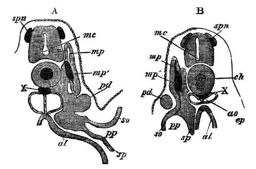
The sections are to shew the development of the
segmental duct (pd) or primitive duct
of the kidneys. In A (the anterior of the two sections) this
appears as a solid knob projecting towards the epiblast. In B is
seen a section of the column which has grown backwards from the knob in
A.
spn. rudiment of a
spinal nerve; mc. medullary canal;
ch. notochord; X. string of cells
below the notochord; mp. muscle-plate;
mp´. specially developed portion of
muscle-plate; ao. dorsal aorta; pd. segmental duct; so. somatopleura; sp. splanchnopleura; pp. pleuro-peritoneal or body-cavity; ep. epiblast; al.
alimentary canal.
My observations shew that the segmental duct is developed in the way
just described in both Pristiurus and Torpedo. Its origin in Pristiurus is
shewn in the adjoining woodcut, and in Torpedo in Pl. 11, fig. 7, sd.
At a stage somewhat older than I, the condition of the segmental duct
has not very materially altered. It has increased considerably in length,
and the knob at its front end is both absolutely smaller, and also consists
of fewer cells than before (Pl. 11, fig. 7,
sd). These cells have become more
columnar, and have begun to arrange themselves radially; thus indicating
the early appearance of the lumen of the duct. The cells forming the front
part of the rod, as well as those of the knob, commence to exhibit a
columnar character, but in the hinder part of the rod the cells are
still rounded. In no part of it has a lumen appeared.
At this period also the knob, partly owing to the commencing separation
of the muscle-plate from the remainder of the mesoblast, begins to pass
inwards and approach the pleuro-peritoneal cavity.
At the same stage the first not very distinct traces of the remainder of
the urinary system become developed. These appear in the form of solid
outgrowths from the intermediate cell-mass just at the most dorsal part of
the body-cavity.
The outgrowths correspond in numbers with the vertebral segments, and
are at first quite disconnected with the segmental duct. At this stage they
are only distinctly visible in the first few segments behind the front end
of the segmental duct. A full description of them will come more
conveniently in the next stage.
By a stage somewhat earlier than K important changes have taken place in
the urinary system.
The segmental duct has acquired a lumen in its anterior portion, which
opens at its front end into the body-cavity. (Pl. 11, fig. 9, sd.) The
lumen is formed by the columnar cells spoken of in the last stage,
acquiring a radiating arrangement round a central point, at which a small
hole appears. After the lumen has once become formed, it rapidly increases
in size.
The duct has also grown considerably in length, but its hind extremity
is still as thin, and lies as close to the epiblast, as at first. The
segmental involutions which commenced to be formed in the last stage, have
now appeared for every vertebral segment along the whole length of the
segmental duct, and even for two or three segments behind this.
They are simple independent outgrowths arising from the outer and
uppermost angle of the body-cavity, and are at first almost without a trace
of a lumen; though their cells are arranged as two layers. They grow in
such a way as to encircle the oviduct on its inner and upper side (Pl. 11, fig. 8 and Pl.
12, fig. 14b, st). When the
hindermost ones are formed, a slight trace of a lumen is perhaps visible in
the front ones. At a stage slightly subsequent to this, in Scyllium
canicula, I noticed 29 of them; the first of them arising in the
segment immediately behind the front end of the oviduct (Pl. 12, fig. 17, st), and
two of them being formed in segments just posterior to the hinder extremity
of the oviduct.
Pl. 12, figs. 16 and 18 represent two
longitudinal sections shewing the segmental nature of the involutions and
their relation to the segmental duct.
Many of the points which have been mentioned can be seen by referring to
Pl. 11 and 12. Anteriorly the segmental duct
opens into the pleuro-peritoneal cavity. In the sections behind this there
may be seen the segmental duct with a distinct lumen, and also a pair of
segmental involutions (Pl. 12, fig.
14a). In the still posterior sections the segmental duct would be
quite without a lumen, and would closely adjoin the epiblast.
It seems not out of place to point out that the modes of the development
of the segmental duct and of the segmental involutions are strikingly
similar. Both arise as solid involutions, from homologous parts of the
mesoblast. The segmental duct arises in the vertebral segment immediately
in front of that in which the first segmental involution appears; so
that the segmental duct appears to be equivalent to a single segmental
involution.
The next stage corresponds with the first appearance of the external
gills. The segmental duct now communicates by a wide opening with the
body-cavity (Pl. 11, fig. 9, sd). It possesses a lumen along its whole length up
to the extreme hind end (Pl. 11, fig.
9a). It is, however, at this hinder extremity that the most
important change has taken place. This end has grown downwards towards that
part of the alimentary canal which still lies behind the anus. This
downgrowth is beginning to shew distinct traces of a lumen, and will appear
in the next stage as one of the horns by which the segmental ducts
communicate with the cloaca (Pl. 11, fig.
9b). All the anterior segmental involutions have now acquired a
lumen. But this is still absent in the posterior ones (Pl. 11, fig. 9a).
Owing to the disappearance of the body-cavity in the region behind the
anus, the primitive involutions there remain as simple masses of cells
still disconnected with the segmental duct (Pl.
11, figs. 9b, 9c and 9d).
Primitive Ova. The true generative products make their
first appearance as the primitive ova between stages I and K.
In the sections of one of my embryos of this stage they are especially
well shewn, and the following description is taken from those displayed in
that embryo.
They are confined to the region which extends posteriorly nearly to the
end of the small intestine and anteriorly to the abdominal opening of the
segmental duct.
Their situation in this region is peculiar. There is no trace of a
distinct genital ridge, but the ova mainly lie in the dorsal portion of the
mesentery, and therefore in a part of the mesoblast which distinctly
belongs to the splanchnopleure (Pl. 12, fig.
14a). Some are situated external to the segmental involutions; and
others again, though this is not common, in a part of the mesoblast which
distinctly belongs to the body-wall (Pl. 12,
fig. 14b).
The portion of mesentery, in which the primitive ova are most densely
aggregated, corresponds to the future position of the genital ridge, but
the other positions occupied by ova are quite outside this. Some ova are in
fact situated on the outside of the segmental duct and segmented tubes, and
must therefore effect a considerable migration before reaching their final
positions in the genital ridge on the inner side of the segmental duct
(Pl. 12, fig. 14b).
The condition of the tissue in which the ova appear may at once be
gathered from an examination of the figures given. It consists of an
irregular epithelium of cells partly belonging to the somatopleure and
partly to the splanchnopleure, but passing uninterruptedly from one layer
to the other. The cells which compose it are irregular in shape, but
frequently columnar (Pl. 12, figs. 14a
and 14b).
They are formed of a nucleus which stains deeply, invested by a very
delicate layer of protoplasm. At the junction of somatopleure and
splanchnopleure they are more rounded than elsewhere. Very few loose
connective-tissue cells are present. The cells just described vary from
.008 Mm. to .01 Mm. in diameter.
The primitive ova are situated amongst them and stand out with
extraordinary clearness, to which justice is hardly done in my figures.
The normal full-sized ova exhibit the following structure. They
consist of a mass of somewhat granular protoplasm of irregular, but more or
less rounded, form. Their size varies from .016 - .036 Mm. In their interior a nucleus is present, which
varies from .012 - .016 Mm., but its size
as a rule bears no relation to the size
of the containing cell.
This is illustrated by the subjoined list of measurements.
Size of Primitive ova
in degrees of micrometer
scale with F. ocul 2. |
Size of nucleus of Primitive
ova in degrees of
micrometer
scale with F. ocul 2. |
| 10 | 8 |
| 13 | 8 |
| 13 | 8 |
| 14 | 7 |
| 15 | 7 |
| 13 | 7½ |
| 11 | 8 |
| 16 | 5½ |
| 12 | 7 |
| 10 | 7 |
| 15 | 6 |
| 13 | 6 |
| 12 | 7 |
The numbers given refer to degrees on my micrometer scale.
Since it is the ratio alone which it is necessary to call attention to,
the numbers are not reduced to decimals of a millimeter. Each degree of my
scale is equal, however, with the object glass employed, to .002 Mm.
This series brings out the result I have just mentioned with great
clearness.
In one case we find a cell has three times the diameter of the nucleus
16 : 5½; in another case 10 : 8, the nucleus has only a slightly
smaller diameter than the cell. The irrationality of the ratio is fairly
shewn in some of my figures, though none of the largest cells with very
small nuclei have been represented.
The nuclei are granular, and stain fairly well with hæmatoxylin. They
usually contain a single deeply stained nucleolus, but in many cases,
especially where large (and this independently of the size of the
cell), they contain two nucleoli (Pl. 12, figs.
14c and 14d), and are at times so lobed as to give an
apparent indication of commencing division.
A multi-nucleolar condition of the nuclei, like that figured by Götte[225], does not appear till near the close of embryonic
life, and is then found equally in the large ova and in those not larger
than the ova which exist at this early date.
As regards the relation of the primitive ova to each other and the
neighbouring cells, there are a few points which deserve attention. In the
first place, the ova are, as a rule, collected in masses at particular
points, and not distributed uniformly (fig. 14a). The masses in some
cases appear as if they had resulted from the division of one primitive
ovum, but can hardly be adduced as instances of a commencing coalescence;
since if the ova thus aggregated were to coalesce, an ovum would be
produced of a very much greater size than any which is found during the
early stages. Though at this stage no indication is present of such a
coalescence of cells to form ova as is believed to take place by Götte,
still the origin of the primitive ova is not quite clear. One would
naturally expect to find a great number of cells intermediate between
primitive ova and ordinary columnar cells. Cells which may be intermediate
are no doubt found, but not nearly so frequently as might have been
anticipated. One or two cells are shewn in Pl.
12, fig. 14a, x, which are perhaps of an intermediate
character; but in most sections it is not possible to satisfy oneself that
any such intermediate cells are present.
In one case what appeared to be an intermediate cell was measured, and
presented a diameter of .012 Mm. while its
nucleus was .008 Mm. Apart from certain
features of the nucleus, which at this stage are hardly very marked, the
easiest method of distinguishing a primitive ovum from an adjacent cell is
the presence of a large quantity of protoplasm around the nucleus. The
nucleus of one of the smallest primitive ova is not larger than the nucleus
of an ordinary cell (being about .008 Mm.
in both). It is perhaps the similarity in the size of the nuclei which
renders it difficult at first to distinguish developing primitive ova from
ordinary cells. Except with the very thinnest sections a small extra
quantity of protoplasm around a nucleus might easily escape detection, and
the developing cell might only become visible when it had attained to the
size of a small typical primitive ovum.
It deserves to be noticed that the nuclei even of some of the largest
primitive ova scarcely exceed the surrounding nuclei in size. This appears
to me to be an argument of some weight in shewing that the great size of
primitive ova is not due to the fact of their having been formed by a
coalescence of different cells (in which case the nucleus would have
increased in the same proportion as the cell); but to an increase by a
normal method of growth in the protoplasm around the nucleus.
It appears to me to be a point of great importance certainly to
determine whether the primitive ova arise by a metamorphosis of adjoining
cells, or may not be introduced from elsewhere. In some of the lower
animals, e.g. Hydrozoa, there is no question that the ova are
derived from the epiblast; we might therefore expect to find that they had
the same origin in Vertebrates. Further than this, ova are frequently
capable in a young state of executing amœboid movements, and
accordingly of migrating from one layer to another. In the Elasmobranchii
the primitive ova exhibit in a hardened state an irregular form which might
appear to indicate that they possess a power of altering their shape, a
view which is further supported by some of them being at the present stage
situated in a position very different from that which they eventually
occupy, and which they can only reach by migration. If it could be shewn
that there were no intermediate stages between the primitive ova and the
adjoining cells (their migratory powers being admitted) a strong
presumption would be offered in favour of their having migrated from
elsewhere to their present position. In view of this possibility I have
made some special investigations, which have however led to no very
satisfactory results. There are to be seen in the stages immediately
preceding the present one, numerous cells in a corresponding position to
that of the primitive ova, which might very well be intermediate between
the primitive ova and ordinary cells, but which offer no sufficiently well
marked features for a certain determination of their true nature.
In the particular embryo whose primitive ova have been
described these bodies were more conspicuous than in the majority of cases,
but at the same time they presented no special or peculiar characters.
In a somewhat older embryo of Scyllium the cells amongst which the
primitive ova lay had become very distinctly differentiated as an
epithelium (the germinal epithelium of Waldeyer) well separated by what
might almost be called a basement membrane from the adjoining
connective-tissue cells. Hardly any indication of a germinal ridge had
appeared, but the ova were more definitely confined than in previous
embryos to the restricted area which eventually forms this. The ova on the
average were somewhat smaller than in the previous cases.
In several embryos intermediate in age between the embryo whose
primitive ova were described at the commencement of this section and the
embryo last described, the primitive ova presented some peculiarities,
about the meaning of which I am not quite clear, but which may perhaps
throw some light on the origin of these bodies.
Instead of the protoplasm around the nucleus being clear or slightly
granular, as in the cases just described, it was filled in the most typical
instances with numerous highly refracting bodies resembling yolk-spherules.
In osmic acid specimens (Pl. 12, fig. 15) these
stain very darkly, and it is then as a rule very difficult to see the
nucleus; in specimens hardened in picric acid and stained with hæmatoxylin
these bodies are stained of a deep purple colour, but the nucleus can in
most cases be distinctly seen. In addition to the instances in which the
protoplasm of the ova is quite filled with these bodies, there are others
in which they only occupy a small area adjoining the nucleus (Pl. 12, fig. 15a), and finally some in which only one
or two of these bodies are present. The protoplasm of the primitive ova
appears in fact to present a series of gradations between a state in which
it is completely filled with highly refracting spherules and one in which
these are completely absent.
This state of things naturally leads to the view that the primitive ova,
when they are first formed, are filled with these spherules, which are
probably yolk-spherules, but that they gradually lose them
in the course of development. Against this interpretation is the fact that
the primitive ova in the younger embryo first described are completely
without these bodies; this embryo however unquestionably presented an
abnormally early development of the ova; and I am satisfied that embryos
present considerable variations in this respect.
If the primitive ova are in reality in the first instance filled with
yolk-spherules, the question arises as to whether, considering that they
are the only mesoblast cells filled at this period with yolk-spherules, we
must not suppose that they have migrated from some peripheral part of the
blastoderm into their present position. To this question I can give no
satisfactory answer. Against a view which would regard the spherules in the
protoplasm as bodies which appear subsequently to the first formation of
the ova, is the fact that hitherto no instances in which these spherules
were present have been met with in the late stages of development; and they
seem therefore to be confined to the first stages.
Notochord.
The changes undergone by the notochord during this period present
considerable differences according to the genus examined. One type of
development is characteristic of Scyllium and Pristiurus; a second type, of
Torpedo.
My observations being far more complete for Scyllium and Pristiurus than
for Torpedo, it is to the two former genera only that the following account
applies, unless the contrary is expressly stated. Only the development of
the parts of the notochord in the trunk are here dealt with; the cephalic
section of the notochord is treated of in a subsequent section.
During stage G the notochord is composed of flattened cells arranged
vertically, rendering the histological characters of the notochord
difficult to determine in transverse sections. In longitudinal sections,
however, the form and arrangement of the cells can be recognised with great
ease. At the beginning of stage G each cell is composed of a nucleus
invested by granular protoplasm frequently vacuolated and containing in
suspension numerous yolk-spherules. It is difficult to determine whether
there is only one vacuole for each cell, or whether in some cases there may
not be more than one.
Round the exterior of the notochord there is present a distinct though
delicate cuticular sheath.
The vacuoles are at first small, but during stage G rapidly increase in
size, while at the same time the yolk-spherules completely vanish from the
notochord.
As a result of the rapid growth of the vacuoles, the nuclei, surrounded
in each case by a small amount of protoplasm, become pushed to the centre
of the notochord, the remainder of the protoplasm being carried to the
edge. The notochord thus becomes composed during stages H and I (Pl. 11, fig. 4-6) of a central area mainly formed of
nuclei with a small quantity of protoplasm around them, and of a thin
peripheral layer of protoplasm without nuclei, the widish space between the
two being filled with clear fluid. The exterior of the cells is indurated,
so that they may be said to be invested by a membrane[226]; the cells themselves have a flattened form, and each
extends from the edge to the centre of the notochord, the long axis of each
being rather greater than half the diameter of the cord.
The nuclei of the notochord are elliptical vesicles, consisting of a
membrane filled with granular contents, amongst which is situated a
distinct nucleolus. They stain deeply with hæmatoxylin. Their long diameter
in Scyllium is about 0.02 Mm.
The diameter of the whole notochord in Pristiurus during stage I is
about 0.1 Mm. in the region of the back,
and about 0.08 Mm. near the posterior end
of the body.
Owing to the form of its constituent cells, the notochord presents in
transverse sections a dark central area surrounded by a lighter peripheral
one, but its true structure cannot be unravelled without the assistance of
longitudinal sections. In these (Pl. 12, fig.
10) the nuclei form an irregular double row in the centre of the cord.
Their outlines are very clear, but those of the individual cells cannot for
certain be made out. It is, however, easy to see that the cells have a
flattened and wedge-shaped form, with the narrow ends overlapping and
interlocking at the centre of the notochord.
By the close of stage I the cuticular sheath of the notochord
has greatly increased in thickness.
During the period intermediate between stages I and K the notochord
undergoes considerable transformations. Its cells cease to be flattened,
and become irregularly polygonal, and appear but slightly more compressed
in longitudinal sections than in transverse ones. The vacuolation of the
cells proceeds rapidly, and there is left in each cell only a very thin
layer of protoplasm around the nucleus. Each cell, as in the earlier
stages, is bounded by a membrane-like wall.
Accompanying these general changes special alterations take place in the
distribution of the nuclei and the protoplasm. The nuclei, accompanied by
protoplasm, gradually leave the centre and migrate towards the periphery of
the notochord. At the same time the protoplasm of the cells forms a special
layer in contact with the investing sheath.
The changes by which this takes place can easily be followed in
longitudinal sections. In Pl. 12, fig. 11 the
migration of the nuclei has commenced. They are still, however, more or
less aggregated at the centre, and very little protoplasm is present at the
edges of the notochord. The cells, though more or less irregularly
polygonal, are still somewhat flattened. In Pl.
12, fig. 12 the notochord has made a further progress. The nuclei now
mainly lie at the side of the notochord, where they exist in a somewhat
shrivelled state, though still invested by a layer of protoplasm.
A large portion of the protoplasm of the cord forms an almost continuous
layer in close contact with the sheath, which is more distinctly visible in
some cases than in others.
While the changes above described are taking place the notochord
increases in size. At the age of fig. 11 it is in the anterior part of the
body of Pristiurus about 0.11 Mm. At the
age of fig. 12 it is in the same species 0.12 Mm., while in Scyllium stellare it reaches about 0.17
Mm.
During stage K (Pl. 11, fig. 8) the
vacuolation of the cells of the notochord becomes even more complete than
during the earlier stages, and in the central cells hardly any protoplasm
is present, though a starved nucleus surrounded by a little protoplasm may
be found in an occasional corner.
The whole notochord becomes very delicate, and can with great
difficulty be conserved whole in transverse sections.
The layer of protoplasm which appeared during the last stage on the
inner side of the cuticular membrane of the notochord becomes during the
present stage a far thicker and more definite structure. It forms a
continuous layer with irregular prominences on its inner surface; and
contains numerous nuclei. The layer sometimes presents in transverse
sections hardly any indication of a division into a number of separate
cells, but in longitudinal sections this is generally very obvious. The
cells are directed very obliquely forwards, and consist of an oblong
nucleus invested by protoplasm. The layer formed by them is very delicate
and very easily destroyed. In one example its thickness varied from .004 to
.006 Mm., in another it reached .012 Mm. The thickness of the cuticular membrane is
about .002 Mm. or rather less.
The diameter of a notochord in the anterior part of the body of a
Pristiurus embryo of this stage is about 0.21 Mm. Round the exterior of the notochord the mesoblast
cells are commencing to arrange themselves as a special sheath.
In Torpedo the notochord at first presents the same structure as in
Pristiurus, i.e. it forms a cylindrical rod of flattened cells.
The vacuolation of these cells does not however commence till a
relatively very much later period than in Pristiurus, and also presents a
very different character (Pl. 11, fig. 7).
The vacuoles are smaller, more numerous, and more rounded than in the
other genera, and there can be no question that in many cases there is more
than one vacuole in a cell. The most striking point in which the notochord
of Torpedo differs from that of Pristiurus consists in the fact that in
Torpedo there is never any aggregation of the nuclei at the centre of the
cord, but the nuclei are always distributed uniformly through it. As the
vacuolation proceeds the differences between Torpedo and the other genera
become less and less marked. The vacuoles become angular in form, and the
cells of the cord cease to be flattened, and become polygonal.
At my final stage for Torpedo (slightly younger than K) the only
important feature distinguishing the notochord from that of
Pristiurus, is the absence of any signs of nuclei or protoplasm passing to
the periphery. Around the exterior of the cord there is early found in
Torpedo a special investment of mesoblastic cells.
EXPLANATION OF PLATES 11 AND 12.
Complete List of Reference
Letters.
al. Alimentary
tract. an. Point where anus will be
formed. ao. Dorsal aorta. ar. Rudiment of anterior root of spinal nerve.
b. Anterior fin. c. Connective-tissue cells. cav. Cardinal vein. ch. Notochord. df. Dorsal fin. ep. Epiblast. ge.
Germinal epithelium. ht. Heart. l.
Liver. mp. Muscle-plate. mp´. Early formed band of muscles from the
splanchnic layer of the muscle-plates. nc. Neural canal. p. Protoplasm from
yolk in the alimentary tract. pc.
Pericardial cavity. po. Primitive ovum.
pp. body-cavity. pr. Rudiment of posterior root of spinal nerve.
sd. Segmental duct. sh. Cuticular sheath of notochord. so. Somatic layer of mesoblast. sp. Splanchnic layer of mesoblast. spc. Spinal cord. sp.v. Spiral valve. sr. Interrenal body. st. Segmental tube. sv. Sinus venosus. ua. Umbilical artery. um. Umbilical cord. uv. Umbilical vein. V. Splanchnic vein.
v. Blood-vessel. vc. Visceral
cleft. vr. Vertebral rudiment. W.
White matter of spinal cord. x. Subnotochordal rod (except in fig.
14a). y. Passage connecting the neural and alimentary
canals.
Plate 11.
Fig. 1. Section from the caudal region of a Pristiurus
embryo belonging to stage H. Zeiss C, ocul. 1.
Osmic acid specimen.
It shews (1) the constriction of the subnotochordal rod
(x) from the summit of the alimentary canal. (2) The formation of
the body-cavity in the muscle-plate and the ventral thickening of the
parietal plate.
Fig. 1a. Portion of alimentary wall of the same
embryo, shewing the formation of the subnotochord rod (x).
Fig. 2. Section through the caudal vesicle of a
Pristiurus embryo belonging to stage H. Zeiss C, ocul. 1.
It shews the bilobed condition of the alimentary vesicle
and the fusion of the mesoblast and hypoblast at the caudal vesicle.
Fig. 3a. Sections from the caudal region of a
Pristiurus embryo belonging to stage H. Zeiss C, ocul. 1. Picric acid specimen.
It shews the communication which exists posteriorly
between the neural and alimentary canals, and also by comparison with
3b it exhibits the dilatation undergone by the alimentary canal in
the caudal vesicle.
Fig. 3b. Section from the caudal region of an
embryo slightly younger than 3a. Zeiss C, ocul. 1. Osmic acid specimen.
Fig. 4. Section from the cardiac region of
a Pristiurus embryo belonging to stage H. Zeiss C, ocul. 1. Osmic acid specimen.
It shews the formation of the heart (ht) as a cavity between the splanchnopleure and the
wall of the throat.
Fig. 5. Section from the posterior dorsal region of a
Scyllium embryo, belonging to stage H. Zeiss C, ocul. 1. Osmic acid specimen.
It shews the general features of an embryo of stage H,
more especially the relations of the body-cavity in the parietal and
vertebral portions of the lateral plate, and the early-formed band of
muscle (mp´) in the splanchnic layer of
the vertebral plate.
Fig. 6. Section from the œsophageal region of
Scyllium embryo belonging to stage I. Zeiss C, ocul. 1. Chromic acid specimen.
It shews the formation of the rudiments of the posterior
nerve-roots (pr) and of the vertebral
rudiments (Vr).
Fig. 7. Section of a Torpedo embryo belonging to
stage slightly later than I. Zeiss C, ocul. 1,
reduced 1/3. Osmic acid specimen.
It shews (1) the formation of the anterior and posterior
nerve-roots. (2) The solid knob from which the segmental duct (sd) originates.
Fig. 8. Section from the dorsal region of a Scyllium
embryo belonging to a stage intermediate between I and K. Zeiss C, ocul. 1. Chromic acid specimen.
It illustrates the structure of the primitive ova,
segmental tubes, notochord, etc.
Fig. 8a. Section from the caudal region of an
embryo of the same age as 8. Zeiss A, ocul.
1.
It shews (1) the solid œsophagus. (2) The narrow
passage connecting the pericardial (pc)
and body cavities (pp).
Fig. 9. Section of a Pristiurus embryo belonging to
stage K. Zeiss A, ocul. 1. Osmic acid
specimen.
It shews the formation of the liver (l), the
structure of the anterior fins (b), and the anterior opening of the
segmental duct into the body-cavity (sd).
Figs. 9a, 9b, 9c, 9d. Four
sections through the anterior region of the same embryo as 9. Osmic acid
specimens.
The sections shew (1) the atrophy of the post-anal
section of the alimentary tract (9b, 9c, 9d). (2) The
existence of the segmental tubes behind the anus (9b, 9c,
9d). With reference to these it deserves to be noted that the
segmental tubes behind the anus are quite disconnected, as is proved by the
fact that a tube is absent on one side in 9c but reappears in
9d. (3) The downward prolongation of the segmental duct to join the
posterior or cloacal extremity of the alimentary tract (9b).
Plate 12.
Fig. 10. Longitudinal and horizontal section of a
Scyllium embryo of stage H. Zeiss C, ocul. 1.
Reduced by 1/3. Picric acid specimen.
It shews (1) the structure of the notochord; (2) the
appearance of the early formed band of muscles (mp´) in the splanchnic layer of the
protovertebra.
Fig. 11. Longitudinal and horizontal sections of an
embryo belonging to stage I. Zeiss C, ocul. 1.
Chromic acid specimen. It illustrates the same points as the previous
section, but in addition shews the formation of the rudiments of the
vertebral bodies (Vr) which are seen to
have the same segmentation as the muscle-plates.
Fig. 12.[227]
Longitudinal and horizontal section of an embryo belonging to the stage
intermediate between I and K. Zeiss C, ocul. 1.
Osmic acid specimen illustrating the same points as the previous
section.
Fig. 13. Longitudinal and horizontal section of an
embryo belonging to stage K. Zeiss C, ocul. 1,
and illustrating same points as previous section.
Figs. 14a, 14b, 14c, 14d.
Figures taken from preparations of an embryo of an age intermediate between
I and K, and illustrating the structure of the primitive ova. Figs.
14a and 14b are portions of transverse sections. Zeiss C,
ocul. 3 reduced 1/3. Figs. 14c and
14d are individual ova, shewing the lobate form of nucleus. Zeiss F,
ocul. 2.
Fig. 15. Osmic acid preparation of primitive ova
belonging to stage K. Zeiss immersion No. 2,
ocul. 1. The protoplasm of the ova is seen to
be nearly filled with bodies resembling yolk-spherules: and one ovum is
apparently undergoing division.
Fig. 15a. Picric acid preparation shewing a
primitive ovum partially filled with bodies resembling yolk-spherules.
Fig. 16. Horizontal and longitudinal section of Scyllium
embryo belonging to stage K. Zeiss A, ocul. 1.
Picric acid preparation. The connective-tissue cells are omitted.
The section shews that there is one segmental tube to
each vertebral segment.
Fig. 17. Portion of a Scyllium embryo belonging to stage
K, viewed as a transparent object.
It shews the segmental duct and the segmental
involutions—two of which are seen to belong to segments behind the
end of the alimentary tract.
Fig. 18. Vertical longitudinal section of a Scyllium
embryo belonging to stage K. Zeiss A, ocul. 1.
Hardened in a mixture of osmic and chromic acid. It shews
(1) the commissures connecting together the posterior
roots of the spinal nerves;
(2) the junction of the anterior and posterior
roots;
(3) the relations of the segmental ducts to the
segmental involutions and the alternation of calibre in the segmental
tube;
(4) the germinal epithelium lining the body-cavity.
External Epiblast.
The change already alluded to in the previous chapter (p. 317) by which the external epiblast or epidermis
becomes divided into two layers, is completed before the close of stage
L.
In the tail region at this stage three distinct strata may be recognized
in the epidermis. (1) An outer stratum of flattened horny cells, which fuse
together to form an almost continuous membrane. (2) A middle stratum of
irregular partly rounded and partly flattened cells. (3) An internal
stratum of columnar cells, bounded towards the mesoblast by a distinct
basement membrane (Pl. 13, fig. 8),
unquestionably pertaining to the epiblast. This layer is especially
thickened in the terminal parts of the paired fins (Pl. 13, fig. 1). The two former of these strata together
constitute the epidermic layer of the skin, and the latter the mucous
layer.
In the anterior parts of the body during stage L the skin only presents
two distinct strata, viz. an inner somewhat
irregular layer of rounded cells, the mucous layer, and an outer layer of
flattened cells (Pl. 13, fig. 8).
The remaining history of the external epiblast, consisting as it does of
a record of the gradual increase in thickness of the epidermic strata, and
a topographical description of its variations in structure and thickness in
different parts, is of no special interest and need not detain us here.
In the late embryonic periods subsequent to stage Q the layers of the
skin cease to be so distinct as at an earlier period, partly owing to the
innermost layer becoming less columnar, and partly to the presence of a
large number of mucous cells, which have by that stage made their
appearance.
I have followed with some care the development of the placoid scales,
but my observations so completely accord with those of Dr O. Hertwig[228], that it is not necessary to record them. The
so-called enamel layer is a simple product of the thickening and
calcification of the basement membrane, and since this membrane is derived
from the mucous layer of the epidermis, the enamel is clearly to be viewed
as an epidermic product. There is no indication of a gradual conversion of
the bases of the columnar cells forming the mucous layer of the epidermis
into enamel prisms, as is frequently stated to occur in the formation of
the enamel of the teeth in higher Vertebrates.
Lateral line.
The lateral line and the nervous structures appended to it have been
recently studied from an embryological point of view by Götte[229] in Amphibians and by Semper[230] in
Elasmobranchii.
The most important morphological result which these two distinguished
investigators believe themselves to have arrived at is the direct
derivation of the lateral nerve from the ectoderm. On this point there is a
complete accord between them, and Semper especially explains that it is
extremely easy to establish the fact.
As will appear from the sequel, I have not been so fortunate as Semper
in elucidating the origin of the lateral nerve, and my observations bear an
interpretation not in the least in accordance with the views of my
predecessors, though not perhaps quite conclusive against them.
It must be premised that two distinct structures have to be dealt with,
viz. the lateral line formed of
modified epidermis, and the lateral nerve whose origin is in
question.
The lateral line is the first of the two to make its appearance, at a
stage slightly subsequent to K, in the form of a linear thickening
of the inner row of cells of the external epiblast, on each side, at the
level of the notochord.
This thickening, in my youngest embryo in which it is found, has but a
very small longitudinal extension, being present through about 10 thin
sections in the last part of the head and first part of the trunk. The
thickening, though short, is very broad, measuring about 0.28 Mm. in transverse section, and presents no signs of a
commencing differentiation of nervous structures. The large intestinal
branch of the vagus can be seen in all the anterior sections in close
proximity to this line, and appears to me to give off to it posteriorly a
small special branch which can be traced through a few sections, vide Pl. 13, fig. 2, n.l. But this branch is not sufficiently well
marked to enable me to be certain of its real character. In any case the
posterior part of the lateral line is absolutely without any adjoining
nervous structures or traces of such.
The rudiment of the epidermic part of the lateral line is formed of
specially elongated cells of the mucous layer of the epiblast, but around
the bases of these certain rounder cells of a somewhat curious appearance
are intercalated.
There is between this and my next youngest embryo an unfortunately large
gap with reference to the lateral line, although in almost every other
respect the two embryos might be regarded as belonging to the same stage.
The lateral line in the older embryo extends from the hind part of the head
to a point well behind the anus, and is accompanied by a nerve for at least
two-thirds of its length.
In the foremost section in which it appears the intestinal branch of the
vagus is situated not far from it, and may be seen at intervals giving
off branches to it. There is no sign that these are otherwise than
perfectly normal branches of the vagus. Near the level of the last visceral
cleft the intestinal branch of the vagus gives off a fair-sized branch,
which from the first occupies a position close to the lateral line though
well within the mesoblast (Pl. 13, fig.
3a, n.l). This branch is the
lateral nerve, and though somewhat larger, is otherwise much like the nerve
I fancied I could see originating from the intestinal branch of the vagus
during the previous stage.
It rapidly thins out posteriorly and also approaches closer and
closer to the lateral line. At the front end of the trunk it is quite in
contact with it, and a short way behind this region the cells of the
lateral line arrange themselves in a gable-like form, in the angle of which
the nerve is situated (Pl. 13, figs. 3b,
and 3c). In this position the nerve though small is still very
distinct in all good sections, and is formed of a rod of protoplasm, with
scattered nuclei, in which I could not detect a distinct indication of
cell-areas. The hinder part of the nerve becomes continually smaller and
smaller, without however presenting any indication of becoming fused with
the epiblast, and eventually ceases to be visible some considerable
distance in front of the posterior end of the lateral line.
The lateral line itself presents some points of not inconsiderable
interest. In the first place, it is very narrow anteriorly and throughout
the greater part of its length, but widens out at its hinder end, and is
widest of all at its termination, which is perfectly abrupt. The following
measurements of it were taken from an embryo belonging to stage L, which
though not quite my second youngest embryo is only slightly older. At its
hinder end it was 0.17 Mm. broad. At a
point not far from this it was 0.09 Mm.
broad, and anteriorly it was 0.05 Mm.
broad. These measurements clearly shew that the lateral line is broadest at
what may be called its growing-point, a fact which explains its
extraordinary breadth in the anterior part of the body at my first stage,
viz. 0.28 Mm., a breadth which strangely contrasts with the
breadth, viz. 0.05 Mm., which it has in the same part of the body at the
present stage.
It still continues to form a linear area of modified epidermis, and has
no segmental characters. Anteriorly it is formed by the cells of the mucous
layer becoming more columnar (Pl. 13 fig.
3a). In its middle region the cells of the mucous layer in it are
still simply elongated, but, as has been said above, have a gable-like
arrangement, so as partially to enclose the nerve (Pl. 13, fig. 3b). Nearer the hind end of the trunk a
space appears in it between its columnar cells and the flattened cells of
the outermost layer of the skin (Pl. 13, fig.
3c), and this space becomes posteriorly invested by a very definite
layer of cells. The space (Pl. 13, fig.
3d) or lumen has a slit-like section, and is not formed by the
closing in of an originally open groove, but by the formation of a
cavity in the midst of the cells of the lateral line. Its walls are formed
by a layer of columnar cells on the inner side, and flattened cells on the
outer side, both layers however appearing to be derived from the mucous
layer of the epidermis. The outer layer of cells attains its greatest
thickness dorsally.
During stages M, N, O, the lateral nerve gradually passes inwards into
the connective tissue between the dorso-lateral and the ventro-lateral
muscles, and becomes even before the close of stage N completely isolated
from the lateral line.
The growth of the lateral line itself remains for some time almost
stationary; anteriorly the cells retain the gable-like arrangement which
characterised them at an earlier period, but cease to enclose the nerve;
posteriorly the line retains its original more complicated constitution as
a closed canal. In stage O the cells of the anterior part of the line, as
well as those of the posterior, commence to assume a tubular arrangement,
and the lateral line takes the form of a canal. The tubular form is due to
a hollowing out of the lateral line itself and a rearrangement of its
cells. As the lateral line becomes converted into a canal it recedes from
the surface.
In stage P the first indication of segmental apertures to the exterior
make their appearance, vide Pl. 13, fig. 4. The lateral line forms a canal situated
completely below the skin, but at intervals (corresponding with segments)
sends upwards and outwards prolongations towards the exterior. These
prolongations do not during stage P acquire external openings. As is shewn
in my figure, a special area of the inner border of the canal of the
lateral line becomes distinguished by its structure from the remainder.
No account of the lateral line would be complete without some allusion
to the similar sensory structures which have such a wide distribution on
the heads of Elasmobranchii; and this is especially important in the present
instance, owing to the light thrown by a study of their development on the
origin of the nerves which supply the sense-organs of this class. The
so-called mucous canals of the head originate in the same way as does the
lateral line; they are products of the mucous layer of the epidermis. They
eventually form either canals with numerous openings to the
exterior, or isolated tubes with terminal ampulliform dilatations.
I have not definitely determined whether the canal-system of the head
arises in connection with the lateral line, or only eventually becomes so
connected. The important point to be noticed is, that at first no nervous
structures are to be seen in connection with it. In stage O nerves for the
mucous canals make their appearance as delicate branches of the main stems.
These nerve-stems are very much ramified, and their branches have, in a
large number of instances, an obvious tendency towards a particular
sense-organ (Pl. 13, figs. 5 and 6).
I have not during stage O been able to detect a case of direct
continuity between the two. This is, however, established in the succeeding
stage P, in the case of the canals, and the facility with which it may be
observed would probably render the embryo Elasmobranch a very favourable
object for studying the connection between nerves and terminal
sense-organs. The nerve (Pl. 13, fig. 7)
dilates somewhat before uniting with the sense-organ, and the protoplasm of
the nerve and the sense-organ become completely fused. The basement
membrane of the skin is not continuous across their point of junction, and
appears to unite with a delicate membrane-like structure, which invests the
termination of the nerve. The ampullæ would seem to receive their nervous
supply somewhat later than the canals, and the terminal swellings of the
nerves supplying them are larger than in the case of the canals, and the
connection between the ampullæ and the nerves not so clear. In the case of
the head, there can for Elasmobranchii be hardly a question that the nerves
which supply the mucous canals grow centrifugally from the original cranial
nerve-stems, and do not originate in a peripheral manner from the
integument.
This is an important point to make certain of in settling any doubtful
features in the nervous supply of the lateral line. Professor Semper[231], with whom as dealing with Elasmobranchii we are more
directly concerned, makes the following statement: At the time when at
the front end the lateral nerve has already completely separated itself
from the ectoderm, and is situated amongst the muscles, it still lies in
the middle of the body close to the ectoderm, and at the hind end of the
body is not yet completely segmented off (abgegliedert) from the ectoderm.
Although the last sentence
of this quotation may seem to be opposed to my statements, yet it appears
to me probable that Professor Semper has merely seen the lateral nerve
partially enclosed in the ectoderm. This position of the nerve no doubt
affords a presumption, but only a presumption, in favour of a
direct origin of the lateral nerve from the ectoderm; but against this
interpretation of it are the following facts:
(1) That the front part of the lateral line is undoubtedly supplied by
branches which arise in the ordinary way from the intestinal branch of the
vagus; and we should not expect to find part of the lateral line supplied
by nerves which originate in one way, and the remainder supplied by a nerve
having a completely different and abnormal mode of origin.
(2) The growth of the lateral line is quite independent of that of the
lateral nerve: the latter arises subsequently to the lateral line, and, so
far as is shewn by the inconclusive observation of my earliest stage, as an
offshoot from the intestinal branch of the vagus; and though it grows along
at first in close contact with the lateral line, yet it never presents, so
far as I have seen, any indubitable indication of becoming split off from
this, or of fusing with it.
(3) The fact that the cranial representatives of the lateral line are
supplied with nerves which originate in the normal way[232], affords a strong argument in favour of the lateral
line receiving an ordinary nerve-supply.
Considering all these facts, I am led to the conclusion that the
lateral nerve in Elasmobranchii arises as a branch of the vagus, and not as
a direct product of the external epiblast.
An interesting feature about the lateral line and the similar cephalic
structures, is the fact of these being the only sense-organs in
Elasmobranchii which originate entirely from the mucous layer of the
epiblast. This, coupled with the well-known facts about the Amphibian
epiblast, and the fact that the mucous canals are the only sense-organs
which originate subsequently to the distinct differentiation of the
epiblast into mucous and horny layers, goes far to prove[233] that the mucous layer is to be regarded as the active
layer of the epiblast, and that after this has become differentiated, an
organ formed from the epiblast is always a product of it.
Muscle-plates.
The muscle-plates at the close of stage K were flattened angular bodies
with the apex directed forwards, their ventral edge being opposite the
segmental duct, and their dorsal edge on a level with the middle of the
spinal cord. They were composed of two layers, formed for the most part of
columnar cells, but a small part of their splanchnic layer opposite the
notochord had already become differentiated into longitudinal muscles.
During stage L the growth of these plates is very rapid, and their upper
ends extend to the summit of the neural canal, and their lower ones nearly
meet in the median ventral line. The original band of muscles (Pl. 11, fig. 8, m.p´),
whose growth was so slow during stages I and K, now increases with great
rapidity, and forms the nucleus of the whole voluntary muscular system. It
extends upwards and downwards by the continuous conversion of fresh cells
of the splanchnic layer into muscle-cells. At the same time it grows
rapidly in thickness, but it requires some little patience and care to
unravel the details of this growth; and it will be necessary to enter on a
slight digression as to the relations of the muscle-plates to the
surrounding connective tissue.
As the muscle-plates grow dorsalwards and ventralwards their ends dive
into the general connective tissue, whose origin has already been described
(Pl. 13, fig. 1). At the same time the
connective-tissue cells, which by this process become situated between the
ends of the muscle-plates and the skin, grow upwards and downwards, and
gradually form a complete layer separating the muscle-plates from the skin.
The cells forming the ends of the muscle-plates retain
unaltered their primitive undifferentiated character, and the separation
between them and the surrounding connective-tissue cells is very marked.
This however ceases to be the case in the parts of the muscle-plates on a
level with the notochord and lower part of the medullary canal; the
thinnest sections and most careful examination are needed to elucidate the
changes taking place in this region. The cells which form the somatic layer
of the muscle-plates then begin to elongate and become converted into
muscle-cells, at the same time that they are increasing in number to meet
the rapid demands upon them. One result of these changes is the loss of the
original clearness in the external boundary between the muscle-plates and
the adjoining connective-tissue cells, which is only in exceptional cases
to be seen so distinctly as it may be in Pl.
13, figs. 1 and 8. Longitudinal horizontal sections are the most
instructive for studying the growth of the muscles, but transverse sections
are also needed. The interpretation of the transverse ones is however
rendered difficult, both by rapid alterations in the thickness of the
connective-tissue layer between the skin and the muscle-plates (shewn in
Pl. 13, fig. 8), and by the angular shape of
the muscle-plates themselves.
A careful study of both longitudinal and transverse sections has enabled
me to satisfy myself of the fact that the cells of the somatic layer of the
protovertebræ, equally with the cells of the splanchnic layer, are
converted into muscle-cells, and some of these are represented in the act
of undergoing this conversion in Pl. 13, fig.
8; but the difficulty of distinguishing the outline of the somatic layer of
the muscle-plates, at the time its cells become converted into
muscle-cells, renders it very difficult to determine whether any cells of
this layer join the surrounding connective tissue. General considerations
certainly lead me to think that they do not; but my observations do not
definitely settle the point.
From these facts it is clear, as was briefly stated in the last chapter,
that both layers of the muscle-plate are concerned in forming the great
lateral muscle, though the splanchnic layer is converted into muscles very
much sooner than the somatic[234].
The remainder of the history of the muscle-plates presents no
points of special interest.
Till the close of stage L, the muscle-plates are not distinctly divided
into dorsal and ventral segments, but this division, which is so
characteristic of the adult, commences to manifest itself during stage M,
and is quite completed in the succeeding stage. It is effected by the
appearance, nearly opposite the lateral line, of a layer of connective
tissue which divides the muscles on each side into a dorso-lateral and
ventro-lateral section. Even during stage O the ends of the muscle-plates
are formed of undifferentiated columnar cells. The peculiar outlines of the
intermuscular septa gradually appear during the later stages of
development, causing the well-known appearances of the muscles in
transverse sections, but require no special notice here.
With reference to the histological features of the development of the
muscle-fibres, I have not pushed my investigations very far. The primitive
cells present the ordinary division, well known since Remak, into a
striated portion and a non-striated portion, and in the latter a nucleus is
to be seen which soon undergoes division and gives rise to several nuclei
in the non-striated part, while the striated part of each cell becomes
divided up into a number of fibrillæ. I have not however determined what
exact relation the original cells hold to the eventual primitive bundles,
or anything with reference to the development of the sarcolemma.
The Muscles of the Limbs.—These are formed during stage O
coincidently with the cartilaginous skeleton, in the form of two bands of
longitudinal fibres on the dorsal and ventral surfaces of the limbs. Dr
Kleinenberg first called my attention to the fact that he had proved the
limb-muscles in Lacerta to be derived from the muscle-plates. This I
at first believed did not hold good for Elasmobranchii, but have since
determined that it does so. Between stages K and L the muscle-plates grow
downwards as far as the limbs and then turn outwards and grow into them
(Pl. 18, fig. 1). Small portions of several
muscle-plates come in this way to be situated in the limbs, and are very
soon segmented off from the remainder of the muscle-plates. The portions of
muscle-plates thus introduced into the limbs soon lose their original
distinctness, and can no longer be recognized in stage L. There can however
be but little doubt that they supply the tissue for the muscles of the
limbs. The muscle-plates themselves after giving off these buds to the
limbs grow downwards, and by stage L cease to shew any trace of what has
occurred (Pl. 13, fig. 1). This fact, coupled
with the late development of the muscles of the limbs (stage O), caused me
to fall into my original error.
The Vertebral Column and Notochord.
In the previous chapter (p. 325) an account
was given of the origin of the tissue destined to form the vertebral
bodies; it merely remains to describe the changes undergone by this in
becoming converted into the permanent vertebræ.
This subject has already been dealt with by a considerable number of
anatomists, and my investigations coincide in the main with the results of
my predecessors. Especially the researches of Gegenbaur[235] may be singled out as containing the pith of the whole
subject, and my results, while agreeing in all but minor points with his,
do not supplement them to any very great extent. I cannot do more than
confirm Götte's[236] account of the development of
the hæmal arches, and may add that Cartier[237] has
given a good account of the later development of the centra. Under the
circumstances it has not appeared to me to be worth while recording with
great detail my investigations; but I hope to be able to give a somewhat
more complete history of the whole subject than has appeared in any single
previous memoir.
At their first appearance the cells destined to form the permanent
vertebræ present the same segmentation as the muscle-plates. This
segmentation soon disappears, and between stages K and L the tissue of the
vertebral column forms a continuous investment of the notochord which
cannot be distinguished from the adjoining connective tissue. Immediately
surrounding the notochord a layer formed of a single row of cells may be
observed, which is not however very distinctly marked[238].
During the stage L there appear four special concentrations of
mesoblastic tissue adjoining the notochord, two of them dorsal and two of
them ventral. They are not segmented, and form four ridges seated on the
sides of the notochord. They are united with each other by a delicate layer
of tissue, and constitute the rudiments of the neural and hæmal arches. In
longitudinal sections of stage L special concentrated wedge-shaped masses
of tissue are to be seen between the muscle-plates, which must not be
confused with these rudiments. Immediately around the notochord the
delicate investment of cells previously mentioned, is still present.
The rudiments of the arches increase in size and distinctness in the
succeeding stages, and by stage N have unquestionably assumed the
constitution of embryonic cartilage. In the meantime there has appeared
surrounding the sheath of the notochord a well-marked layer of tissue which
stains deeply with hæmatoxylin, and with the highest power may be observed
to contain flattened nuclei. It is barely thicker than the adjoining
sheath, but is nevertheless the rudiment of the vertebral bodies. Pl. 13, fig. 9, vb. Whence does this layer arise? To this question I
cannot give a quite satisfactory answer. It is natural to conclude that it
is derived from the previously existing mesoblastic investment of the
notochord, but in the case of the vertebral column I have not been able to
prove this. Observations on the base of the brain afford fairly conclusive
evidence that the homologous tissue present there has this origin.
Gegenbaur apparently answers the question of the origin of this layer in
the way suggested above, and gives a figure in support of his conclusion
(Pl. XXII. fig. 3)[239].
The layer of tissue which forms the vertebral bodies rapidly
increases in thickness, and very soon, at a somewhat earlier period than
represented in Gegenbaur's Pl. XXII. fig. 4, a
distinct membrane (Kölliker's Membrana Elastica Externa) may easily be
recognized surrounding it and separating it from the adjoining tissue of
the arches. Gegenbaur's figure gives an excellent representation of the
appearance of this layer at the period under consideration. It is formed of
a homogeneous basis containing elongated concentrically arranged nuclei,
and constitutes a uniform unsegmented investment for the notochord (vide Pl. 13, fig. 10).
The neural and hæmal arches now either cease altogether to be united
with each other by a layer of embryonic cartilage, or else the layer
uniting them is so delicate that it cannot be recognized as true cartilage.
They have moreover by stage P undergone a series of important changes. The
tissue of the neural arches does not any longer form a continuous sheet,
but is divided into (1) a series of arches encircling the spinal cord, and
(2) a basal portion resting on the cartilaginous sheath of the notochord.
There are two arches to each muscle-plate, one continuous with the basal
portion of the arch-tissue and forming the true arch, which springs
opposite the centre of a vertebral body, and the second not so continuous,
which forms what is usually known as the intercalated piece. Between every
pair of true arches the two roots of a single spinal nerve pass out. The
anterior root passes out in front of an intercalated piece and the
posterior behind it[240].
The basal portion of the arch-tissue likewise undergoes differentiation
into a vertebral part continuous with the true arch and formed of hyaline
cartilage, and an intervertebral segment formed of a more fibrous
tissue.
The hæmal arches, like the neural arches, become divided into a layer of
tissue adjoining the cartilaginous sheath of the notochord, and processes
springing out from this opposite the centres of the
vertebræ. These processes throughout the region of the trunk in front of
the anus pass into the space between the dorsal and ventral muscles, and
are to be regarded as rudiments of ribs. The tissue with which they are
continuous, which is exactly equivalent to the tissue from which the neural
arches originate, is not truly a part of the rib. In the tail, behind the
anus and kidneys, the cardinal veins fuse to form an unpaired caudal vein
below the aorta, and in this part a fresh series of processes originates on
each side from the hæmal tissue adjoining the cartilaginous sheath of the
notochord, and eventually, by the junction of the processes of the two
sides, a canal which contains the aorta and caudal vein is formed below the
notochord. These processes for a few segments coexist with small ribs
(vide Pl. 13, fig. 10),
a fact which shews (1) that they cannot be regarded as modified ribs, and
(2) that the tissue from which they spring is to be viewed as a kind of
general basis for all the hæmal processes which may arise, and is not
specially connected with any one set of processes.
While these changes (all of which are effected during stage P) are
taking place in the arches, the tissue of the vertebral bodies or
cartilaginous investment of the notochord, though much thicker than before,
still remains as a continuous tube whose wall exhibits no segmental
differentiations.
It is in stage Q that these differentiations first appear in the
vertebral regions opposite the origin of the neural arches. The outermost
part of the cartilage at these points becomes hyaline and almost
undistinguishable in structure from the tissue of the arches[241]. These patches of hyaline cartilage grow larger and
cause the vertebral parts of the column to constrict the notochord, whilst
the intervertebral parts remain more passive, but become composed of cells
with very little intercellular substance. Coincidently also with these
changes, part of the layer internal to the hyaline cartilage becomes
modified to form a somewhat peculiar tissue, the intercellular substance of
which does not stain, and in which calcification eventually arises (Pl. 13, fig. 11). The innermost layer adjoining the
notochord retains its primitive fibrous character, and is distinguishable
as a separate layer through both the vertebral and the intervertebral
regions. As a result of these changes a transverse section through the
centre of the vertebral regions now exhibits three successive rings (vide Pl. 13, fig. 11), an
external ring of hyaline cartilage invested by the membrana elastica
externa
(m.el), followed by a ring
of calcifying cartilage, and internal to this a ring of fibrous cartilage,
which adjoins the now slightly constricted notochord. A transverse section
of an intervertebral region shews only a thick outer and thin inner ring of
fibrous cartilage, the latter in contact with the sheath of the
unconstricted notochord.
The constriction of the notochord proceeds till in the centre of the
vertebræ it merely forms a fibrous band. The tissue internal to the
calcifying cartilage then becomes hyaline, so that there is formed in the
centre of each vertebral body a ring of hyaline cartilage immediately
surrounding the fibrous band which connects the two unconstricted segments
of the notochord. The intervertebral tissue becomes more and more fibrous.
In Cartier's paper before quoted there is a figure (fig. 3) which
represents the appearance presented by a longitudinal section of the
vertebral column at this stage.
The relation of the vertebral bodies to the arches requires a short
notice. The vertebral hyaline cartilage becomes almost precisely similar to
the tissue of the arches, and the result is, that were it not for the
"membrana elastica externa" it would be hardly possible to distinguish the
limits of the two tissues. This membrane however persists till the hyaline
cartilage has become a very thick layer (Pl.
13, fig. 11), but I have failed to detect it in the adult, so that I cannot
there clearly distinguish the arches from the body of the vertebræ. From a
comparison however of the adult with the embryo, it is clear that the
arches at most form but a small part of what is usually spoken of as the
body of the vertebræ.
The changes in the notochord itself during the stages subsequent to K
are not of great importance. The central part retains for some time its
previous structure, being formed of large vacuolated cells with an
occasional triangular patch of protoplasm containing the starved nucleus
and invested by indurated layers of protoplasm. These indurated layers are
all fused, and are probably rightly regarded by Gegenbaur and Götte
as representing a sparse intercellular matter. The external protoplasmic
layer of the notochord ceases shortly after stage K to exhibit any traces
of a division into separate cells, but forms a continuous layer with
irregular prominences and numerous nuclei (Pl.
13, fig. 9). In the stages subsequent to P further changes take place in
the notochord: the remains of the cells become more scanty and the
intercellular tissue assumes a radiating arrangement, giving to sections of
the notochord the appearance of a number of lines radiating from the centre
to the periphery (Pl. 13, fig. 11).
The sheath of the notochord at first grows in thickness, and during
stage L there is no difficulty in seeing in it the fine radial markings
already noticed by Müller[242] and Gegenbaur[243], and regarded by them as indicating pores. Closely
investing the sheath of the notochord there is to be seen a distinct
membrane, which, though as a rule closely adherent to the sheath, in some
examples separates itself from it. It is perhaps the membrane identified by
W. Müller[244] (though not by Gegenbaur) as
Kölliker's membrana elastica interna
. After the formation of the
cartilaginous investment of the notochord, this membrane becomes more
difficult to see than in the earlier stage, though I still fancy that I
have been able to detect it. The sheath of notochord also appears to me to
become thinner, and its radial striation is certainly less easy to detect[245].
EXPLANATION OF PLATE 13.
Complete List of Reference
Letters.
al. Alimentary tract.
ao. Aorta. c. Connective tissue.
cav. Cardinal vein. ch. Notochord. ep.
Epiblast. ha. Hæmal arch. l.
Liver. ll. Lateral line. mc. Mucous canal of the head. mel. Membrana elastica externa. mp. Muscle-plate. mp´. Muscles of muscle-plate. na. Neural arch. nl. Nervus lateralis. rp. Rib process. sd. Segmental duct. sh. Sheath of notochord. spc. Spinal cord. spg. Spinal ganglion. syg. Sympathetic ganglion. um. Ductus choledochus. v. Blood-vessel.
var. Vertebral arch. vb. Vertebral body. vcau. Caudal vein. vin. Intestinal branch of the vagus. vop. Ramus ophthalmicus of the fifth nerve. x.
Subnotochordal rod.
Fig. 1. Section through the anterior part of an
embryo of Scyllium canicula during stage L.
c. Peculiar large cells which are found at the
dorsal part of the spinal cord. Sympathetic ganglion shewn at syg. Zeiss A, ocul.
1.
Fig. 2. Section through the lateral line at the time
of its first formation.
The cells marked nl
were not sufficiently distinct to make it quite certain that they really
formed part of the lateral nerve. Zeiss B, ocul. 2.
Figs. 3a, 3b, 3c, 3d.
Four sections of the lateral line from an embryo belonging to stage L.
3a is the most anterior. In 3a the lateral nerve (nl) is seen to lie in the mesoblast at some
little distance from the lateral line. In 3b and 3c it lies
in immediate contact with and partly enclosed by the modified epiblast
cells of the lateral line. In 3d, the hindermost section, the
lateral line is much larger than in the other sections, but no trace is
present of the lateral nerve. The sections were taken from the following
slides of my series of the embryo (the series commencing at the tail end)
3d (46), 3c (64), 3b (84), 3a (93). The figures
all drawn on the same scale, but 3 a is not from the same side of
the body as the other sections.
Fig. 4. Section through lateral line of an embryo of
stage P at the point where it is acquiring an opening to the exterior. The
peculiar modified cells of its innermost part deserve to be noticed. Zeiss
D, ocul. 2.
Fig. 5. Mucous canals of the head with branches of
the ramus ophthalmicus growing towards them. Stage O. Zeiss A, ocul. 2.
Fig. 6. Mucous canals of head with branches of the
ramus ophthalmicus growing towards them. Stage between O and P. Zeiss a a,
ocul. 2.
Fig. 7. Junction of a nerve and mucous canal. Stage
P. Zeiss D, ocul. 2.
Fig. 8. Longitudinal and horizontal section through
the muscle-plates and adjoining structures at a stage intermediate between
L and M. The section is intended to shew the gradual conversion of the
cells of the somatic layer of muscle-plates into muscles.
Fig. 9. Longitudinal section through the notochord
and adjoining parts to shew the first appearance of the cartilaginous
notochordal sheath which forms the vertebral centra. Stage N.
Fig. 10. Transverse section through the tail of an
embryo of stage P to shew the coexistence of the rib-process and hæmal
arches in the first few sections behind the point where the latter appear.
Zeiss C, ocul. 1.
Fig. 11. Transverse section through the centre of a
caudal vertebra of an embryo somewhat older than Q. It shews (1) the
similarity between the arch-tissue and the hyaline tissue of the outer
layer of the vertebral centrum, and (2) the separation of the two by the
membrana elastica externa[246] (mel). It shews also the differentiation of three
layers in the vertebral centrum: vide p. 374.
The spinal nerves.
The development of the spinal nerves has been already treated by me at
considerable length in a paper read before the Royal Society in December,
1875[247], and I have but little fresh matter to add to
the facts narrated in that paper. The succeeding account, though fairly
complete, is much less full than the previous one in the
Philosophical Transactions, but a number of morphological
considerations bearing on this subject are discussed.
The rudiments of the posterior roots make their appearance considerably
before those of the anterior roots. They arise during stage I, as
outgrowths from the spinal cord, at a time when the muscle-plates do not
extend beyond a third of the way up the sides of the spinal cord, and in a
part where no scattered mesoblast-cells are present. They are formed first
in the anterior part of the body and successively in the posterior parts,
in the following way. At a point where a spinal nerve is about to arise,
the cells of the dorsal part of the cord begin to proliferate, and the
uniform outline of the cord becomes broken (Pl.
14, fig. 3). There is formed in this way a small prominence of cells
springing from the summit of the spinal cord, and constituting a rudiment
of a pair of posterior roots. In sections anterior to the point where a
nerve is about to appear, the nerve-rudiments are always very distinctly
formed. Such a section is shewn in Pl. 14, fig.
2, and the rudiments may there be seen as two club-shaped
masses of cells, which have grown outwards and downwards from the extreme
dorsal summit of the neural canal and in contact with its walls. The
rudiments of the two sides meet at their point of origin at the dorsal
median line, and are dorsally perfectly continuous with the walls of the
canal.
It is a remarkable fact that rudiments of posterior roots are to be seen
in every section. This may be interpreted as meaning that the rudiments are
in very close contact with each other, but more probably means, as I hope
to shew in the sequel, that there arises from the spinal cord a continuous
outgrowth from which discontinuous processes (the rudiments of posterior
roots) grow out.
After their first formation these rudiments grow rapidly ventralwards in
close contact with the spinal cord (vide Pl. 14, fig. 1, and Pl.
11, figs. 6 and 7), but soon meet with and become partially enclosed in the
mesoblastic tissue (Pl. 11, fig. 7). The
similarity of the mesoblast and nerve-tissue in Scyllium and Pristiurus
embryos hardened in picric or chromic acid, render the nerves in these
genera, at the stage when they first become enveloped in mesoblast,
difficult objects to observe; but no similar difficulty is encountered in
the case of Torpedo embryos.
While the rudiments of the posterior roots are still quite short, those
of the anterior roots make their first appearance. Each of these (Pl. 14, fig. 4, a.r.) arises as a very small but distinct conical
outgrowth from a ventral corner of the spinal cord. From the very first the
rudiments of the anterior roots have an indistinct form of peripheral
termination and somewhat fibrous appearance, while the protoplasm of which
they are composed becomes attenuated towards its end. The points of origin
of the anterior roots from the spinal cord are separated by considerable
intervals. In this fact, and also in the fact of the nerves of the two
sides never being united with each other in the median line, the anterior
roots exhibit a marked contrast to the posterior. There are thus
constituted, before the close of stage I, the rudiments of both the
anterior and posterior roots of the spinal nerves. The rudiments of both of
these take their origin from the involuted epiblast of the neural canal,
and the two roots of each spinal nerve are at first quite unconnected
with each other. It is scarcely necessary to state that the pairs of roots
correspond in number with the muscle-plates.
It is not my intention to enter with any detail into the subsequent
changes of the rudiments whose origin has been described, but a few points
especially connected with their early development are sufficiently
important to call for attention.
One feature of the posterior roots at their first formation is the fact
that they appear as processes of a continuous outgrowth of the spinal cord.
This state of affairs is not of long continuance, and before the close of
stage I each posterior root has a separate junction with the spinal cord.
What then becomes of the originally continuous outgrowth? It has not been
possible for me to trace the fate of this step by step; but the discovery
that at a slightly later period (stage K) there is present a continuous
commissure independent of the spinal cord connecting the dorsal and central
extremities of all the spinal nerves, renders it very probable that the
original continuous outgrowth becomes converted into this commissure. Like
all the other nervous structures, this commissure is far more easily seen
in embryos hardened in a mixture of osmic and chromic acids or osmic acid,
than in those hardened in picric acid. Its existence must be regarded as
one of the most remarkable results of my researches upon the Elasmobranch
nervous system. At stage K it is fairly thick, though it becomes much
thinner at a slightly later period. Its condition during stage K is shewn
in Pl. 12, fig. 18, com. What it has been possible for me to make out of
its eventual fate is mentioned subsequently[248].
A second feature of the earliest condition of the posterior roots is
their attachment to the extreme dorsal summit of the spinal cord—a
point of attachment very different from that which they eventually acquire.
Before the commencement of stage K this state of things has become altered;
and the posterior roots spring from the spinal cord in the position normal
for Vertebrates.
This apparent migration caused me at first great perplexity, and I do
not feel quite satisfied that I have yet got completely to the bottom of
its meaning. The explanation which appears to me most probable has
suggested itself in the course of some observations on the development of
the thin roof of the fourth ventricle. A growth of cells appears to take
place in the median dorsal line of the roof of the spinal cord. This growth
tends to divaricate the two lateral parts of the cord, which are originally
contiguous in the dorsal line, and causes therefore the posterior roots,
which at first spring from the dorsal summit, to assume an apparent
attachment to the side of the cord at some little distance from the summit.
If this is the true explanation of the change of position which takes
place, it must be regarded as due rather to peculiar growths in the spinal
cord, than to any alteration in the absolute attachment of the nerves.
By stage K the rudiment of the posterior root has become greatly
elongated, and exhibits a division into three distinct portions (Pl. 14, fig. 6):
(1) A proximal portion, in which is situated the pedicle of attachment
to the wall of the neural canal.
(2) An enlarged portion, which may conveniently from its future fate be
called the spinal ganglion.
(3) A distal portion beyond this.
The proximal portion presents a fairly uniform diameter, and ends
dorsally in a rounded expansion; it is attached, remarkably enough, not
by its extremity, but by its side, to the spinal cord. The dorsal
extremities of the posterior roots are therefore free. It seems almost
certain that the free dorsal extremities of these roots serve as the
starting points for the dorsal commissure before mentioned, which connects
the roots together. The attachment of the posterior nerve-root to the
spinal cord is, on account of its small size, very difficult to observe. In
favourable specimens there may however be seen a distinct cellular
prominence from the spinal cord, which becomes continuous with a small
prominence on the lateral border of the nerve-root near its distal
extremity. The proximal extremity of the rudiment is composed of cells,
which, by their small size and circular form, are easily distinguished from
those which form the succeeding or ganglionic portion of the nerve. This
succeeding part has a swollen configuration, and is composed of large
elongated cells with oval nuclei. The remainder of the rudiment forms the
commencement of the true nerve.
The anterior root, which, at the close of stage I, formed a small and
inconspicuous prominence from the spinal cord, grows rapidly during the
succeeding stages, and soon forms an elongated cellular structure with a
wide attachment to the spinal cord (Pl. 14,
fig. 5). At first it passes obliquely and nearly horizontally outwards,
but, before reaching the muscle-plate of its side, takes a bend downwards
(Pl. 14, fig. 7).
I have not definitely made out when the anterior and posterior roots
unite, but this may easily be seen to take place before the close of stage
K (Pl. 12, fig. 18).
One feature of some interest with reference to the anterior roots, is
the fact that they arise not vertically below, but alternately with the
dorsal roots, a condition which persists in the adult.
Although I have made some efforts to determine the eventual fate of the
commissure uniting the dorsal roots, these have not hitherto been crowned
with success. It grows thinner and thinner, becoming at the same time
composed of fibrous protoplasm with imbedded nuclei (Pl. 14, figs. 8 and 9). By stage M it is so small as to be
quite indistinguishable in transverse sections; and I have failed in stage
P to recognize it at all. I can only conclude that it gradually atrophies,
and finally vanishes without leaving a trace. Both its appearance and
history are very remarkable, and deserve the careful attention of future
investigators.
There can be little doubt that it is some sort of remnant of an
ancestral structure in the nervous system; and it would appear to indicate
that the central nervous system must originally have been formed of a
median and two lateral strands. At the same time I very much doubt whether
it can be brought into relation with the three rows of ganglion-cells (a
median and two lateral) which are so frequently present on the ventral side
of annelidan nerve-cords.
My results may be summarised as follows:—Along the extreme
dorsal summit of the spinal cord there arises on each side a continuous
outgrowth. From each outgrowth processes corresponding in number to the
muscle-plates grow downwards. These are the rudiments of the posterior
nerve-roots. The outgrowths, though at first attached to the spinal cord
throughout their whole length, soon cease to be so, and remain in
connection with it at certain points only, which form the primitive
junctions of the posterior roots with the spinal cord. The original
outgrowth on each side remains as a bridge, uniting together the dorsal
extremities of all the posterior roots. The posterior roots, though
primitively attached to the dorsal summit of the spinal cord, eventually
come to arise from its sides. The original homogeneous rudiments before the
close of stage K become differentiated into a root, a ganglion, and a
nerve.
The anterior roots, like the posterior, are outgrowths from the spinal
cord, but are united independently with it, and the points from which they
spring originally, remain as those by which they are permanently attached.
The anterior roots arise, not vertically below, but in the intervals
between the posterior roots. They are at first quite separate from the
posterior roots; but before the close of stage K a junction is effected
between each posterior root and the corresponding anterior root. The
anterior root joins the posterior at some little distance below its
ganglion.
* * * * *
The results here arrived at are nearly in direct opposition to those of
the majority of investigators, though in accordance, at least so far as the
posterior roots are concerned, with the beautiful observations of Hensen
'on the Development of Mammalia[249].'
Mr Marshall[250] has more recently published a
paper on the development of the nerves in Birds, in which he shews in a
most striking manner that the observations recorded here for Elasmobranchii
hold good for the posterior roots of Birds. The similarity between his
figures and my own is very noticeable. A further discussion of the
literature would be quite unprofitable, and I proceed at once to certain
considerations suggested by the above observations.
General considerations. One point of general anatomy
upon which my observations throw considerable light, is the primitive
origin of nerves. So long as it was admitted that the spinal and
cerebral nerves developed in the embryo independently of the central
nervous system, their mode of origin always presented to my mind
considerable difficulties. It never appeared clear how it was possible for
a state of things to have arisen in which the central nervous system as
well as the peripheral terminations of nerves, whether motor or sensory,
were formed independently of each other; while between them a third
structure was developed, which, growing out either towards the centre or
towards the periphery, ultimately brought the two into connection. That
such a condition could be a primitive one seemed scarcely possible.
Still more remarkable did it appear, on the supposition that the
primitive mode of formation of these parts was represented in the
developmental history of Vertebrates, that we should find similar
structural elements in the central and in the peripheral nervous systems.
The central nervous system arises from the epiblast, and yet contains
precisely similar nerve-cells and nerve-fibres to the peripheral nervous
system, which, when derived from the mesoblast, was necessarily supposed to
have an origin completely different from that of the central nervous
system. Both of these difficulties are to a great extent removed by the
facts of the development of these parts in Elasmobranchii.
It is possible to suppose that in their primitive differentiation
contractile and sensory systems may, as in Hydra[251],
have been developed from the protoplasm of even the same cell. As the
sensory and motor systems became more complicated, the sensory portion of a
cell would become separated by an increasing interval from the muscular
part of a cell, and the two parts of a cell would only be connected by a
long protoplasmic process. When such a condition as that was reached, the
sensory portion of the cell would be called a ganglion-cell or terminal
sensory organ, the connecting process a nerve, and the contractile portion
of the cell a muscle-cell. When these organs were in this condition, it
might not impossibly happen for the general developmental growth which
tended to separate the ganglion-cell and the muscle-cell to be so
rapid as to render it impossible for the growth of the connecting nerve to
keep pace with it, and that thus the process connecting the ganglion-cell
and the muscle-cell might become ruptured. Nevertheless the tendency of the
process to grow from the ganglion-cell to the muscle-cell, would remain,
and when the rapid developmental growth had ceased, the two would become
united again by the growth of the process which had previously been
ruptured. It will be seen that this hypothesis, which I have considered
only with reference to a single nerve and muscle-cell, might be extended so
as to apply to a complicated central nervous system and peripheral nerves
and muscles, and also could apply equally as well to the sensory as to the
motor terminations of a nerve. In the case of the sensory termination, we
should only have to suppose that the centre nervous cell became more and
more separated by the general growth from the recipient terminal sensory
cell, and that during the general growth the connection between the two was
mechanically ruptured but restored again on the termination of the more
rapid growth.
As the descendants of the animal in which the rupture occurred became
progressively more complicated, the two terminal cells must have become
widely separated at a continually earlier period, till finally they may
have been separated at a period of development when they were
indistinguishable from the surrounding embryonic cells; and since the
rupture would also occur at this period, the primitive junction between the
nerve-centre and termination would escape detection. The object of this
hypothesis is to explain the facts, so far as they are known, of the
development of the nervous system in Vertebrates.
In Vertebrates we certainly appear to have an outgrowth from the nervous
system, which eventually becomes united with the muscle or sensory terminal
organs. The ingenious hypothetical scheme of development of the nerves
given by Hensen[252] would be far preferable to the
one suggested if it could be brought into conformity with the facts. There
is, however, at present no evidence for Hensen's view, as he himself
admits, but considering how little we know of the finer details of the
development of nerves, it seems not impossible that such evidence
may be eventually forthcoming. The evidence from my own observation is, so
far as it goes, against it. At a time anterior to the outgrowth of the
spinal nerves, I have shewn[253]
that the spinal cord is completely invested by a delicate hyaline membrane.
It is difficult to believe that this is pierced by a number of fine
processes, which completely escape detection, but which must, nevertheless,
be present on the hypothesis of Hensen.
The facts of the development of nerves in Vertebrates are unquestionably
still involved in considerable doubt. It may, I think, be considered as
certain, that in Elasmobranchii the roots of the spinal and cranial nerves
are outgrowths of the central nervous system. How the final terminations of
the nerves are formed is, however, far from being settled. Götte[254], whose account of the development of the spinal
ganglia is completely in accordance with the ordinary views, yet states[255] that the growth of the nerve fibres themselves is a
centrifugal one from the ganglia. My own investigations prove that the
ganglia have a centrifugal development, and also appear to demonstrate that
the nerves themselves near the ganglion have a similar manner of growth.
Moreover, the account given in the preceding chapter of the manner in which
the nerves become connected with the mucous canals of the head, goes far to
prove that the whole growth of the nerves is a centrifugal one. The
combination of all these converging observations tells strongly in favour
of this view.
On the other hand, Calberla[256]
believes that in the tails of larval Amphibians he has seen
connective-tissue cells unite with nerve-processes, and become converted
into nerves, but he admits that he cannot definitely prove that the
axis-cylinder has not a centrifugal growth, while the connective-tissue
cells merely become converted into the sheath of the nerve. If Calberla's
view be adopted, that the nerves are developed directly out of a chain of
originally indifferent cells, each cell of the chain being converted in
turn into a section of the nerve, an altogether different origin of nerves
from that I have just suggested would seem to be indicated.
The obvious difficulty, already alluded to, of understanding
how it is, according to the generally accepted mode of development of the
spinal nerves, that precisely similar nerve-cells and nerves should arise
in structures which have such different origins as the central nervous
system and the spinal nerves, is completely removed if my statements on the
development of the nerves in Elasmobranch represent the truth.
One point brought out in my investigations appears to me to have
bearings upon the origin of the central canal of the vertebrate nervous
system, and in consequence upon the origin of the vertebrate nervous system
itself. This point is, that the posterior nerve-rudiments make their first
appearance at the extreme dorsal summit of the spinal cord. The transverse
section of the ventral nervous cord of an ordinary segmented Annelid
consists of two symmetrical halves placed side by side. If by a mechanical
folding the two lateral halves of the nervous cord became bent towards each
other, while into the groove between the two the external skin became
pushed, we should have an approximation to the vertebrate nervous system.
Such a folding as this might take place to give extra rigidity to the body
in the absence of a vertebral column.
If this folding were then completed in such a way that the groove, lined
by external skin and situated between the two lateral columns of the
nervous system, became converted into a canal, above and below which the
two columns of the nervous system united, we should have in the transformed
nervous cord an organ strongly resembling the spinal cord of
Vertebrates.
It is well known that the nerve-cells are always situated on the ventral
side of the abdominal nerve-cord of Annelids, either as a continuous layer,
or in the form of two, or more usually, three bands. The dorsal side of the
cord is composed of nerve-fibres or white matter. If the folding I have
supposed were to take place in the Annelid nervous cord, the grey and white
matters would have very nearly the same relative situations as they have in
the Vertebrate spinal cord. The grey matter would be situated in the
interior and line the central canal, and the white matter would nearly
surround the grey. The nerves would then arise, not from the sides of the
nervous cord as in existing Annelids, but from its extreme ventral summit.
One of the most striking features which I have brought to light
with reference to the development of the posterior roots, is the fact of
their growing out from the extreme dorsal summit of the neural canal, a
position analogous to the ventral summit of the Annelidan nervous cord.
Thus the posterior roots of the nerves in Elasmobranchii[257] arise, in the exact manner which might have been
anticipated, were the spinal canal due to such a folding as I have
suggested.
The argument from the position of the outgrowth of nerves becomes the
more striking from its great peculiarity, and forms a feature which would
be most perplexing without some such explanation as I have proposed. The
central epithelium of the neural canal, according to this view, represents
the external skin, and its ciliation in certain cases may, perhaps, be
explained as a remnant of the ciliation of the external skin still found
amongst many of the lower Annelids.
I have employed the comparison of the Vertebrate and Annelidan nervous
cords, not so much to prove a genetic relation between the two, as to shew
the à priori possibility of the formation of a spinal
cord, and the à posteriori evidence we have of the
vertebrate canal having been formed in the way indicated. I have not made
use of what is really my strongest argument, viz. that the embryological mode of formation of the spinal
canal by a folding in of the external epiblast is the very method by which
I supposed the spinal canal to have been formed in the ancestors of
Vertebrates. My object has been to suggest a meaning for the peculiar
primitive position of the posterior roots, rather than to attempt to
explain in full the origin of the spinal canal.
Although the homologies between the Vertebrate and the Annelidan nervous
systems are not necessarily involved in the questions which arise with
reference to the formation of the spinal canal, they have nevertheless
considerable bearings on it.
Two views have recently been put forward on this subject.
Professor Gegenbaur[258] looks upon the central nervous
system of Vertebrates as equivalent to the superior œsophageal
ganglia of Annelids and Arthropods only, while Professors Leydig[259] and Semper[260] and
Dr Dohrn[261] compare it with the whole
Annelidan nervous system.
The first of these two views is only possible on the supposition that
Vertebrates are descended from unsegmented ancestors, and even then
presents considerable difficulties. If the ancestors of Vertebrates were
segmented animals, and several of the recent researches tend to shew that
they were, they must almost certainly have possessed a nervous cord like
that of existing Annelids. If such were the case, it is almost
inconceivable that the greater portion of the nervous system which forms
the ventral cord can have become lost, and the system reduced to the
superior œsophageal ganglia. Dr Dohrn[262],
who has speculated very profoundly on this matter, has attempted to explain
and remove some of the difficulties which arise in comparing the nervous
systems of Vertebrates and Annelids. He supposes that the segmented
Annelids, from which Vertebrates are descended, were swimming animals. He
further supposes that their alimentary canal was pierced by a number of
gill-slits, and that the anterior amongst these served for the introduction
of nutriment into the alimentary canal, in fact as supplementary mouths as
well as for respiration. Eventually the old mouth and throat atrophied, and
one pair of coalesced gill-slits came to serve as the sole mouth. Thus it
came about that on the disappearance of that portion of the alimentary
canal, which penetrated the œsophageal nervous ring, the latter
structure ceased to be visible as such, and no part of the alimentary
canal was any longer enclosed by a commissure of the central nervous
system. With the change of mouth Dr Dohrn also supposes that there took
place a change, which would for a swimming animal be one of no great
difficulty, of the ventral for the dorsal surface. This general explanation
of Dr Dohrn's, apart from the considerable difficulty of the fresh mouth,
appears to me to be fairly satisfactory. Dr Dohrn has not however in my
opinion satisfactorily dealt with the questions of detail which arise in
connection with this comparison. One of the most important points for his
theory is to settle the position where the nervous system was formerly
pierced by the œsophagus. This position he fixes in the fourth
ventricle, and supports his hypothesis by the thinness of the roof of the
spinal canal in this place, and the absence (?) of nervous structures in
it.
It appears to me that this thinness cannot be used as an argument. In
the first place, if the hypothesis I have suggested as to the formation of
the spinal canal be accepted, the formation of the canal must be supposed
to have occurred in point of time either after or before the loss of the
primitive mouth. If, on the one hand, the spinal canal made its appearance
before the atrophy of the primitive mouth, the folding to form it must
necessarily have ceased behind the mouth; and, on the supposition of the
œsophageal ring having been situated in the region of the fourth
ventricle, a continuation of the spinal canal could not be present in front
of this part. If, on the other hand, the cerebro-spinal canal appeared
after the disappearance of the primitive mouth, its roof must necessarily
also be a formation subsequent to the atrophy of the mouth, and varieties
of structure in it can have no bearing upon the previous position of the
mouth.
But apart from speculations upon the origin of the spinal cord, there
are strong arguments against Dr Dohrn's view about the fourth ventricle. In
the first place, were the fourth ventricle to be the part of the nervous
system which previously formed the œsophageal commissures, we should
expect to find the opening in the nervous system at this point to be
visible at an early period of development, and at a later period to cease
to be so. The reverse is however the case. In early embryonic life the roof
of the fourth ventricle is indistinguishable from other parts of the
nervous system, and only thins out at a later period. Further than this,
any explanation of the thin roof of the fourth ventricle ought also to
elucidate the nearly similar structure in the sinus rhomboidalis, and
cannot be considered satisfactory unless it does so.
The peculiarities of the cerebro-spinal canal in the region of the brain
appear to me to present considerable difficulties in the way of comparing
the central nervous system of Vertebrates and segmented Annelids. The
manner in which the cerebro-spinal canal is prolonged into the optic
vesicles, the cerebral and the optic lobes is certainly opposed both to an
intelligible explanation of the spinal canal itself, and also to a
comparison of the two nervous systems under consideration.
Its continuation into the cerebral hemispheres and into the optic lobes
(mid-brain) may perhaps be looked upon as due to peculiar secondary growths
of those two ganglia, but it is very difficult to understand its
continuation into the optic vesicles.
If it be granted that the spinal canal has arisen from a folding in of
the external skin, then the present inner surface of the optic vesicle must
also have been its original outer surface, and it follows as a necessary
consequence that the present position of the rods and cones behind and not
in front of the nervous structures of the retina was not the primitive one.
The rods and cones arise, as is well known, from the inner surface of the
outer portion of the optic vesicle, and must, according to the above view,
be supposed originally to have been situated on the external surface, and
have only come to occupy their present position during the folding in,
which resulted in the spinal canal. On à priori grounds we
should certainly expect the rods and cones to have resulted from the
differentiation of a layer of cells external to the conducting nervous
structures. The position of the rods and cones posterior to these suggests
therefore that some peculiar infolding has occurred, and may be used as an
argument to prove that the medullary groove is no mere embryonic structure,
but the embryonic repetition of an ancestral change. The supposition of
such a change of position in the rods and cones necessarily implies that
the folding in to form the spinal canal must have been a very slow one. It
must have given time to the refracting media of the eye gradually to travel
round, so as still to maintain their primitive position, while in
successive generations a rudimentary spinal furrow carrying with it the
retina became gradually converted into a canal[263].
If Dr Dohrn's comparison of the vertebrate nervous system with that of
segmented Annelids be accepted, the following two points must in my opinion
be admitted:—
(1) That the formation of the cerebro-spinal canal was subsequent to the
loss of the old mouth.
(2) That the position of the old mouth is still unknown.
The well-known view of looking at the pituitary and pineal growths as
the remnants of the primitive œsophagus, has no doubt some features
to recommend it. Nearly conclusive against it is the fact that the
pituitary involution is not, as used to be supposed, a growth towards the
infundibulum of the hypoblast of the œsophagus, but of the epiblast
of the mouth. It is almost inconceivable that an involution from the
present mouth can have assisted in forming part of the old
œsophagus.
There is a view not involving the difficulty of the œsophageal
ring, fresh mouth[264], and of the change of the
ventral to the dorsal surface, which, though so far unsupported
by any firm basis of observed facts, nevertheless appears to me worth
suggesting. It assumes that Vertebrates are descended not through
the present line of segmented Vermes, but through some other line which has
now, so far as is known, completely vanished. This line must be supposed to
have originated from the same unsegmented Vermes as the present
segmented Annelids. They therefore acquired fundamentally similar segmental
and other Annelidan organs.
The difference between the two branches of the Vermes lay in the nervous
system. The unsegmented ancestors of the present Annelids seem to
have had a pair of super-œsophageal ganglia, from which two main
nervous stems extended backwards, one on each side of the body. Such a
nervous system in fact as is possessed by existing Nemertines or
Turbellarians[265]. As the Vermes became segmented
and formed the Annelids, these side nerves seem to have developed ganglia,
corresponding in number with the segments, and finally, approximating on
the ventral surface, to have formed the ventral cord[266].
The other branch of Vermes which I suppose to have been the ancestors of
Vertebrates started from the same stock as existing Annelids, but I
conceive the lateral nerve-cords, instead of approximating ventrally, to
have done so dorsally, and thus a dorsal cord to have become formed
analogous to the ventral cord of living Annelids, only without an
œsophageal nerve-ring[267].
It appears to me, (if the difficulties of comparing the
Annelidan ventral cord with the spinal cord of Vertebrates are found to be
insurmountable), that this hypothesis would involve far fewer
improbabilities than one which supposes the whole central nervous system of
Vertebrates to be homologous with the super-œsophageal ganglia. The
mode of formation of a nervous system presupposed in my hypothesis, well
accords with what we know of the formation of the ventral cord in existing
Annelids.
The supposition of the existence of another branch of segmented Vermes
is not a very great difficulty. Even at the present day we have possibly
more than one branch of Vermes which have independently acquired
segmentation. viz.: the Chætopodous Annelids
and the Hirudinea. If the latter is an isolated branch, it is especially
interesting from having independently developed a series of segmental
organs like those of Chætopodous Annelids, which we must suppose the
ancestors of Vertebrates also to have done if they too form an independent
branch.
In addition to the difficulty of imagining a fresh line of segmented
Vermes, there is another difficulty to my view, viz.: the fact that in almost all Vermes, the blood flows
forwards in the dorsal vessel, and backwards in the ventral vessel. This
condition of the circulation very well suits the view of a change of the
dorsal for the ventral surfaces, but is opposed to these surfaces being the
same for Vertebrates and Vermes. I cannot however regard this point as a
very serious difficulty to my view, considering how undefined is the
circulation in the unsegmented groups of the Vermes.
Sympathetic nervous system.
Between stages K and L there may be seen short branches from the spinal
nerves, which take a course towards the median line of the body, and
terminate in small irregular cellular masses immediately dorsal to the
cardinal veins (Pl. 18, fig. 1, sy.g.). These form the first traces that have
come under my notice of the sympathetic nervous system. In the youngest of
my embryos in which I have detected these it has not been possible for me
either definitely to determine the antero-posterior limits of the system,
or to make certain whether the terminal masses of cells which form the
ganglia are connected by a longitudinal commissure. In a stage slightly
younger than L the ganglia are much more definite, the anterior one is
situated in the cardiac region close to the end of the intestinal branch of
the vagus, and the last of them quite at the posterior end of the abdominal
cavity. The anterior ganglia are the largest; the commissural cord, if
developed, is still very indistinct. In stage L the commissural cord
becomes definite, though not very easy to see even in longitudinal
sections, and the ganglia become so considerable as not to be easily
overlooked. They are represented in Pl. 13,
fig. 1, sy.g. and in Pl. 18, fig. 2, in the normal position immediately above the
cardinal veins. The branches connecting them with the trunks of the spinal
nerves may still be seen without difficulty. In later stages these branches
cannot so easily be made out in sections, but the ganglia themselves
continue as fairly conspicuous objects. The segmental arrangement of the
ganglia is shewn in Pl. 18, fig. 3, a
longitudinal and vertical section of an embryo between stages L and M with
the junctions of the sympathetic ganglia and spinal nerves. The ganglia
occupy the intervals between the successive segments of the kidneys.
The sympathetic system only came under my notice at a comparatively late
period in my investigations, and the above facts do not in all points clear
up its development[268]. My observations seem to point
to the sympathetic system arising as an off-shoot from the cerebrospinal
system. Intestinal branches would seem to be developed on the main nerve
stems of this in the thoracic and abdominal regions, each of these then
develops a ganglion, and the ganglia become connected by a longitudinal
commissure. On this view a typical spinal nerve has the following parts:
two roots, a dorsal and ventral, the dorsal one ganglionated, and three
main branches, (1) a ramus dorsalis, (2) a ramus ventralis, and (3) a ramus
intestinalis. This scheme may be advantageously compared with that of a
typical cranial nerve according to Gegenbaur. It may be noted that it
brings the sympathetic nervous system into accord with the other parts
of the nervous system as a product of the epiblast, and derived from
outgrowths from the neural axis. It is clear, however, that my
investigations, though they may naturally be interpreted in this way, do
not definitely exclude a completely different method of development for the
sympathetic system.
EXPLANATION OF PLATE 14.
This Plate illustrates the Formation of the
Spinal Nerves.
Complete List of Reference
Letters.
ar. Anterior root of a
spinal nerve. ch. Notochord. com. Commissure connecting the posterior roots of the
spinal nerves. i. Mesoblastic investment of spinal cord. mp. Muscle-plate. n. Spinal nerve.
nc. Neural canal. pr. Posterior root of a spinal nerve. spg. Ganglion on posterior root of spinal
nerve. v.r. Vertebral rudiment.
w. White matter of spinal cord. y. Point where the spinal
cord became segmented off from the superjacent epiblast.
Figs. 1, 2, and 3. Three sections of a Pristiurus embryo
belonging to stage I. Fig. 1 passes through the heart, fig. 2 through the
anterior part of the dorsal region, fig. 3 through a point slightly behind
this. (Zeiss CC, ocul. 2.) In fig. 3 there is visible a slight proliferation
of cells from the dorsal summit of the neural canal. In fig. 2 this
proliferation definitely constitutes two club-shaped masses of cells (pr)—the rudiments of the posterior
nerve-roots,—both attached to the dorsal summit of the spinal cord.
In fig. 1 the rudiments of the posterior roots are of considerable
length.
Fig. 4. Section through the dorsal region of a Torpedo
embryo slightly older than stage I, with three visceral clefts. (Zeiss
CC, ocul. 2.) The
section shews the formation of a pair of dorsal nerve-rudiments (pr) and a ventral nerve-rudiment (ar). The latter is shewn in its youngest condition,
and is not distinctly cellular.
Fig. 5. Section through the dorsal region of a Torpedo
embryo slightly younger than stage K. (Zeiss CC,
ocul. 2.) The connective-tissue cells are
omitted. The rudiment of the ganglion (spg) on the posterior root has appeared, and the
junction of posterior root with the cord is difficult to detect. The
anterior root forms an elongated cellular structure.
Fig. 6. Section through the dorsal region of a
Pristiurus embryo of stage K. (Zeiss CC, ocul. 2.) The section especially illustrates the
attachment of the posterior root to the spinal cord.
Fig. 7. Section through the same embryo as fig. 6.
(Zeiss CC, ocul. 1.)
The section contains an anterior root, which takes its origin at a point
opposite the interval between two posterior roots.
Fig. 8. A series of posterior roots with their central
ends united by a dorsal commissure, from a longitudinal and vertical
section of a Scyllium embryo belonging to a stage intermediate between L
and M. The embryo was hardened in a mixture of osmic and chromic acids.
Fig. 9. The central end of a posterior nerve-root from
the same embryo, with the commissure springing out from it on either
side.
The Development of the Brain.
General History. In stage G the brain presents a very simple
constitution (Pl. 8, fig. G), and is in fact
little more than a dilated termination to the cerebro-spinal axis. Its
length is nearly one-third that of the whole body, being proportionately
very much greater than in the adult.
It is divided by very slight constrictions into three lobes, the
posterior of which is considerably the largest. These are known as the
fore-brain, the mid-brain, and the hind-brain. The anterior part of the
brain is bent slightly downwards about an axis passing through the
mid-brain. The walls of the brain, composed of several rows of elongated
columnar cells, have a fairly uniform thickness, and even the roof of the
hind-brain is as thick as any other part. Towards the end of stage G the
section of the hind-brain becomes somewhat triangular with the apex of the
triangle directed downwards.
In Pristiurus during stage H no very important changes take place in the
constitution of the brain. In Scyllium, however, indications appear in the
hind-brain of its future division into a cerebellum and medulla oblongata.
The cavity of the anterior part dilates and becomes rounded, while that of
the posterior part assumes in section an hour-glass shape, owing to an
increase in the thickness of the lateral parts of the walls. At the same
time the place of the original thick roof is taken by a very thin layer,
which is formed not so much through a change in the character and
arrangements of the cells composing the roof, as by a divarication
of the two sides of the hind-brain, and the simultaneous introduction of a
fresh structure in the form of a thin sheet of cells connecting dorsally
the diverging lateral halves of this part of the brain. By stage I, the
hind-brain in Pristiurus also acquires an hour-glass shaped section, but
the roof has hardly begun to thin out (Pl. 15,
figs. 4a and 4b).
During stages I and K the cranial flexure becomes more and more
pronounced, and causes the mid-brain definitely to form the termination of
the long axis of the embryo (Pl. 15, figs. 1,
2, etc.), and before the close of stage K a thin coating of white matter
has appeared on the exterior of the whole brain, but no other histological
changes of interest have occurred.
During stage L an apparent rectification of the cranial flexure
commences, and is completed by stage Q. The changes involved in this
process may be advantageously studied by comparing the longitudinal
sections of the brain during stages L, P, and Q, represented in Pl. 16, figs. 1a, 5 and 7a.
It will be seen, first of all, that so far from the flexure of the brain
itself being diminished, it is increased, and in P (fig. 5) the angle in
the floor of the mid-brain becomes very acute indeed; in other words, the
anterior part of the brain has been bent upon the posterior through nearly
two right angles, and the infundibulum, or primitive front end of the
brain, now points nearly directly backwards. At the same time the cerebral
hemispheres have grown directly forwards, and if figures 1a and 5 in
Pl. 16 be compared it will be seen that in the
older brain of the two the cerebral hemispheres have assumed a position
which might be looked on as the result of their having been pushed
dorsalwards and forwards against the mid-brain, and having in the process
pressed in and nearly obliterated the original thalamencephalon. The
thalamencephalon in fig. 1a, belonging to stage L, is relatively
large, but in fig. 5, belonging to stage P, it only occupies a very small
space between the front wall of the mid-brain and the hind wall of the
cerebral hemispheres. It is therefore in part by the change in position of
the cerebral hemispheres that the angle between the trabeculæ and
parachordals becomes increased, i.e. their flexure
diminished, while at the same time the flexure of the brain itself
is increased. More important perhaps in the apparent rectification
of the cranial flexure than any of the previously
mentioned points, is the appearance of a bend in the hind-brain which tends
to correct the original cranial flexure. The gradual growth of this fresh
flexure can be studied in the longitudinal sections which have been
represented. It is at its maximum in stage Q. This short preliminary sketch
of the development of the brain as a whole will serve as an introduction to
the history of the individual divisions of the brain.
Fore-brain. In its earliest condition the fore-brain forms a
single vesicle without a trace of separate divisions, but buds off very
early the optic vesicles, whose history is described with that of the eye
(Pl. 15, fig. 3, op.v). Between stages I and K the posterior part of
the fore-brain sends outwards a papilliform process towards the exterior,
which forms the rudiment of the pineal gland (Pl. 15, fig. 1, pn).
Immediately in front of the rudiment a constriction appears, causing a
division of the fore-brain into a large anterior and a small posterior
portion. This constriction is shallow at first, but towards the close of
stage K becomes much deeper (Pl. 15, fig. 2 and
fig. 16a), leaving however the two cavities of the two divisions of
the fore-brain united ventrally by a somewhat wide canal.
The posterior of the two divisions of the fore-brain forms the
thalamencephalon. Its anterior wall adjoining the cerebral rudiment becomes
excessively thin (Pl. 15, fig. 11); and its
base till the close of stage K is in close contact with the mouth
involution, and presents but a very inconspicuous prominence which marks
the eventual position of the infundibulum (Pl.
15, figs. 9a, 12, 16a, in). The anterior and larger division of the
fore-brain forms the rudiment of the cerebral hemispheres and olfactory
lobes. Up to stage K this rudiment remains perfectly simple, and exhibits
no signs, either externally or internally, of a longitudinal constriction
into two lobes. From the canal uniting the two divisions of the fore-brain
(which eventually forms part of the thalamencephalon) there spring the
hollow optic nerves. A slight ventral constriction separating the cerebral
rudiment from that part of the brain where these are attached appears even
before the close of stage K (Pl. 15, fig. 11,
op.n).
During stage L the infundibulum becomes much produced, and forms a wide
sack in contact with the pituitary body, and its cavity
communicates with that of the third ventricle by an elongated slit-like
aperture. This may be seen by comparing Pl. 16,
figs. 1a and 1c. In fig. 1c taken along the middle
line, there is present a long opening into the infundibulum (in), which is shewn to be very narrow by being no
longer present in fig. 1a representing a section slightly to one
side of the middle line. During the same stage the pineal gland grows into
a sack-like body, springing from the roof of the thalamencephalon, fig.
1b, pn. This latter (the
thalamencephalon) is now dorsally separated from the cerebral rudiment by a
deep constriction, and also ventrally by a less well marked constriction.
At its side also a deep constriction is being formed in it, immediately
behind the pineal gland. The cerebral rudiment is still quite unpaired and
exhibits no sign of becoming constricted into two lobes.
During the next two stages the changes in the fore-brain are of no great
importance, and I pass at once to stage O. The infundibulum is now nearly
in the same condition as during stage L, though (as is well shewn in the
figure of a longitudinal section of the next stage) it points more directly
backwards than before. The remaining parts of the thalamencephalon have
however undergone considerable changes. The more important of these are
illustrated by a section of stage O, Pl. 16,
fig. 3, transverse to the long axis of the embryo, and therefore, owing to
the cranial flexure, cutting the thalamencephalon longitudinally and
horizontally; and for stage P in a longitudinal and vertical section
through the brain (Pl. 16, fig. 5). In the
first place the roof of the thalamencephalon has become very much shortened
by the approximation of the cerebral rudiment to the mid-brain. The pineal
sack has also become greatly elongated, and its somewhat dilated extremity
is situated between the cerebral rudiment and the external skin. It opens
into the hind end of the third ventricle, and its posterior wall is
continuous with the front wall of the mid-brain. The sides of the
thalamencephalon have become much thickened, and form distinct optic
thalami (op.) united by a very well
marked posterior commissure (pc.). The
anterior wall of the thalamencephalon as well as its roof are very thin.
The optic nerves have become by stage O quite solid except at their roots,
into which the ventricles of the fore-brain are for a short distance
prolonged. This solidification is arrived at, so far as I have determined,
without the intervention of a fold. The nerves are fibrous, and a
commencement of the chiasma is certainly present. From the chiasma there
appears to pass out on each side a band of fibres, which runs near the
outer surface of the brain to the base of the optic lobes (mid-brain), and
here the fibres of the two sides again cross.
By stage O important changes are perceptible in the cerebral rudiment.
In the first place there has appeared a slight fold at its anterior
extremity (Pl. 16, fig. 3, x), destined
to form a vertical septum dividing it into two hemispheres, and secondly,
lateral outgrowths (vide Pl. 16, fig. 2, ol.l), to
form the olfactory lobes. Its thin posterior wall presents on each side a
fold which projects into the central cavity. From the peripheral end of
each olfactory lobe a nerve similar in its histological constitution to any
other cranial nerve makes its appearance (Pl.
16, fig. 2); this divides into a number of branches, one of which passes
into the connective tissue between the two layers of epithelium in each
Schneiderian fold. On the root of this nerve there is a large development
of ganglionic cells. I have not definitely observed its origin, but have no
reason to doubt that it is a direct outgrowth from the olfactory lobe,
exactly similar in its mode of development to any other nerve of
the body.
The cerebral rudiment undergoes great changes during stage P. In
addition to a great increase in the thickness of its walls, the fold which
appeared in the last stage has grown backwards, and now divides it in front
into two lobes, the rudiments of the cerebral hemispheres. The greater and
posterior section is still however quite undivided, and the cavities of the
lobes (lateral ventricles) though separated in front are still quite
continuous behind. At the same time, the olfactory lobes, each containing a
prolongation of the ventricle, have become much more pronounced (vide Pl. 16, figs. 4a
and 4c, ol.l). The root of the
olfactory nerve is now very thick, and the ganglion cells it contains are
directly prolonged into the ganglionic portion of the olfactory bulb; in
consequence of which it becomes rather difficult to fix on the exact line
of demarcation between the bulb and the nerve.
Stage Q is the latest period in which I have investigated the
development of the brain. Its structure is represented for this stage in
general view in Pl. 16, figs. 6a,
6b, 6c, in longitudinal section in Pl. 16, figs. 7a, 7b, and in transverse
section Pl. 16, figs. 8a-d. The
transverse sections are taken from a somewhat older embryo than the
longitudinal. In the thalamencephalon there is no fresh point of great
importance to be noticed. The pineal gland remains as before, and has
become, if anything, longer than it was, and extends further forwards over
the summit of the cerebrum. It is situated, as might be expected, in the
connective tissue within the cranial cavity (fig. 8a, pn), and does not extend outside the skull, as it
appears to do, according to Götte's investigations, in Amphibians. Götte[269] compares the pineal gland with the long persisting
pore which leads into the cavity of the brain in the embryo of Amphioxus,
and we might add the Ascidians, and calls it ein
Umbildungsprodukt einer letzten Verbindung des Hirns mit der Oberhaut.
This suggestion appears to me a very good one, though no facts have come
under my notice which confirm it. The sacci vasculosi are perhaps indicated
at this stage in the two lateral divisions of the trilobed ventricle of the
infundibulum (fig. 8c).
The lateral ventricles (fig. 8a) are now quite separated by a
median partition, and a slight external constriction marks the lobes of the
two hemispheres; these, however, are still united by nervous structures for
the greater part of their extent. The olfactory lobes are formed of a
distinct bulb and stalk (fig. 8a, ol.l), and contain, as before, prolongations of the
lateral ventricles. The so-called optic chiasma is very distinct (fig.
8b, op.n), but the fibres from
the optic nerves appear to me simply to cross and not to intermingle.
The mid-brain. The mid-brain is at first fairly marked off from
both the fore and hind brains, but less conspicuously from the latter than
from the former. Its roof becomes progressively thinner and its sides
thicker up to stage P, its cavity remaining quite simple. The thinness of
the roof gives it, in isolated brains of stage P, a bilobed appearance
(vide Pl. 16, fig.
4b, mb, in which the distinctness
of this character is by no means exaggerated): During stage Q it becomes
really bilobed through the formation in its roof of a shallow
median furrow (Pl. 16, fig. 8b). Its
cavity exhibits at the same time the indication of a division into a
central and two lateral parts.
The hind-brain. The hind-brain has at first a fairly uniform
structure, but by the close of stage I, the anterior part becomes
distinguished from the remainder by the fact, that its roof does not become
thin as does that of the posterior part. This anterior, and at first
very insignificant portion, forms the rudiment of the cerebellum. Its
cavity is quite simple and is continued uninterruptedly into that of the
remainder of the hind-brain. The cerebellum assumes in the course of
development a greater and greater prominence, and eventually at the close
of stage Q overlaps both the optic lobes in front and the medulla behind
(Pl. 16, fig. 7a). It exhibits in
surface-views of the hardened brain of stages P and Q the appearance of a
median constriction, and the portion of the ventricle contained in it is
prolonged into two lateral outgrowths (Pl. 16,
figs. 8c and 8d, cb).
The posterior section of the hind-brain which forms the medulla
undergoes changes of a somewhat complicated character. In the first place
its roof becomes in front very much extended and thinned out. At the raphe,
where the two lateral halves of the brain originally united, a separation,
as it were, takes place, and the two sides of the brain become pushed
apart, remaining united by only a very thin layer of nervous matter (Pl. 15, fig. 6, iv.v.). As a result of this peculiar growth in the
brain, the roots of the nerves of the two sides which were originally in
contact at the dorsal summit of the brain become carried away from one
another, and appear to rise at the sides of the brain (Pl. 15, figs. 6 and 7). Other changes also take place in the
walls of the brain. Each lateral wall presents two projections towards the
interior (Pl. 15, fig. 5a). The ventral
of these vanish, and the dorsal approximate so as nearly to divide the
cavity of the hind-brain, or fourth ventricle, into a large dorsal and a
small ventral channel (Pl. 15, fig. 6), and
this latter becomes completely obliterated in the later stages. The dorsal
pair, while approximating, also become more prominent, and stretch into the
dorsal moiety of the fourth ventricle (Pl. 15,
fig. 6). They are still very prominent at stage Q (Pl. 16, fig. 8d, ft), and correspond in position with the fasciculi
teretes of human anatomy. Part of the root of the seventh nerve originates
from them. They project freely in front into the cavity of the fourth
ventricle (Pl. 16, fig. 7a, ft).
By stage Q restiform tracts are indistinctly marked off from the
remainder of the brain, and are anteriorly continued into the cerebellum,
of which they form the peduncles. Near their junction with the cerebellum
they form prominent bodies (Pl. 16, fig.
7a, rt), which are regarded by
Miklucho-Maclay[270] as representing the true
cerebellum.
By stage O the medulla presents posteriorly, projecting into its cavity,
a series of lobes which correspond with the main roots (not the branches)
of the vagus and glosso-pharyngeal nerves (Pl.
17, fig. 5). There appear to me to be present seven or eight projections:
their number cannot however be quite certainly determined. The first of
them belongs to the root of the glosso-pharyngeal, the next one is
interposed between the glosso-pharyngeal and the first root of the vagus,
and is without any corresponding nerve-root. The next five correspond to
the five main roots of the vagus. For each projection to which a nerve
pertains there is a special nucleus of nervous matter, from which the root
springs. These nuclei do not stain like the remainder of the walls of the
medulla, and stand out accordingly very conspicuously in stained
sections.
The coating of white matter which appeared at the end of stage K, on the
exterior of each lateral half of the hind-brain, extends from a point just
dorsal to the attachment of the nerve-roots to the ventral edge of the
medulla, and is specially connected with the tissue of the upper of the two
already described projections into the fourth ventricle.
A rudiment of the tela vasculosa makes its appearance during stage Q,
and is represented by the folds in the wall of the fourth ventricle in my
figure of that stage (Pl. 16, fig. 7a,
tv).
* * * * *
The development of the brain in Elasmobranchii has already been worked
out by Professor Huxley, and a brief but in many respects very complete
account of it is given in his recent paper on Ceratodus[271]. He says, pp. 30 and
31, "The development of the cerebral hemispheres in Plagiostome Fishes
differs from the process by which they arise in the higher Vertebrata. In a
very early stage, when the first and second visceral clefts of the embryo
Scyllium are provided with only a few short branchial filaments, the
anterior cerebral vesicle is already distinctly divided into the
thalamencephalon (from which the large infundibulum proceeds below, and the
small tubular peduncle of the pineal gland above, while the optic nerve
leaves its sides) and a large single oval vesicle of the hemispheres. On
the ventral face of the integument covering these are two oval depressions,
the rudimentary olfactory sacs.
As development proceeds the vesicle of the
hemispheres becomes divided by the ingrowth of a median longitudinal
septum, and the olfactory lobes grow out from the posterior lateral regions
of each ventricle thus formed, and eventually rise on to the dorsal faces
of the hemispheres, instead of, as in most Vertebrata, remaining on their
ventral sides. I may remark, that I cannot accept the views of
Miklucho-Maclay, whose proposal to alter the nomenclature of the parts of
the Elasmobranch's brain, appears to me to be based upon a
misinterpretation of the facts of development.
The last sentence of the paragraph brings me to the one part on which it
is necessary to say a few words, viz. the
views of Miklucho-Maclay. His views have not received any general
acceptance, but the facts narrated in the preceding pages shew, beyond a
doubt, that he has 'misinterpreted' the facts of development, and that the
ordinary view of the homology of the parts is the correct one. A comparison
of the figures I have given of the embryo brain with similar figures of the
brain of higher Vertebrates shews this point conclusively. Miklucho-Maclay
has been misled by the large size of the cerebellum, but, as we have seen,
this body does not begin to be conspicuous till late in embryonic life.
Amongst the features of the embryonic brain of Elasmobranchii, the long
persisting unpaired condition of the cerebral hemisphere, upon which so
much stress has already been laid by Professor Huxley, appears to me to be
one of great importance, and may not improbably be
regarded as a real ancestral feature. Some observations have recently been
published by Professor B. G. Wilder[272]
upon this point, and upon the homologies and development of the olfactory
lobes. Fairly good figures are given to illustrate the development of the
cerebral hemispheres, but the conclusions arrived at are in part opposed to
my own results. Professor Wilder says: The true hemispheres are the
lateral masses, more or less completely fused in the middle line, and
sometimes developing at the plane of union a bundle of longitudinal
commissural fibres. The hemispheres retain their typical condition as
anterior protrusions of the anterior vesicle; but they lie mesiad of the
olfactory lobes, and in Mustelus at least seem to be formed after
them.
The italics are my own. From what has been said above, it is
clear that the statement italicised, for Scyllium at least, completely
reverses the order of development. Still more divergent from my conclusions
are Professor Wilder's statements on the olfactory lobes. He says: The
true olfactory lobe, or rhinencephalon, seems, therefore, to embrace only
the hollow base of the crus, more or less thickened, and more or less
distinguishable from the main mass as a hollow process. The olfactory bulb,
with the more or less elongated crus of many Plagiostomes, seems to be
developed independently, or in connection with the olfactory sack, as are
the general nerves;
and again, But the young and adult brains since
examined shew that the ventricle (i.e. the ventricle of the
olfactory lobe) ends as a rounded cul-de-sac before reaching the
lobe.
The majority of the statements contained in the above quotations are not
borne out by my observations. Even the few preparations of which I have
given figures, appear to me to prove that (1) the olfactory lobes (crura
and bulbs) are direct outgrowths from the cerebral rudiment, and develop
quite independently of the olfactory sack; (2) that the ventricle of the
cerebral rudiment does not stop short at the base of the crus; (3) that
from the bulb a nerve grows out which has a centrifugal growth like other
nerves of the body, and places the central olfactory lobe in communication
with the peripheral olfactory sack. In some other Vertebrates this nerve
seems hardly to be developed, but it is easily intelligible, that if in the
ordinary course of growth the olfactory sack became approximated to the
olfactory lobe, the nerve which grew out from the latter to the sack might
become so short as to escape detection.
Organs of Sense.
The olfactory organ. The olfactory pit is the latest formed of
the three organs of special sense. It appears during a stage intermediate
between I and K, as a pair of slight thickenings of the
external epiblast, in the normal vertebrate position on the under side of
the fore-brain immediately in front of the mouth (Pl. 15, figs. 1 and 2, ol).
The epiblast cells which form this thickening are very columnar, but
present no special peculiarities. Each thickened patch of skin soon becomes
involuted as a shallow pit, which remains in this condition till the close
of the stage K. The epithelium very early becomes raised into a
series of folds (Schneiderian folds). These are bilaterally symmetrical,
and diverge like the barbs of a feather from a median line (Pl. 15, fig. 14). The nasal pits at the close of stage K are
still separated by a considerable interval from the walls of the brain, and
no rudiment of an olfactory lobe arises till a later period; but a
description of the development of this as an integral part of the brain has
already been given, p. 401.
Eye. The eye does not present in its early development any very
special features of interest. The optic vesicles arise as hollow outgrowths
from the base of the fore-brain (Pl. 15, fig.
3, op.v), from which they soon become
partially constricted, and form vesicles united to the base of the brain by
comparatively narrow hollow stalks, the rudiments of the optic nerves. The
constriction to which the stalk or optic nerve is due takes place from
above and backwards, so that the optic nerves open into the base of the
front part of the thalamencephalon (Pl. 15,
fig. 13a, op.n). After the
establishment of the optic nerves, there take place the formation of the
lens and the pushing in of the anterior wall of the optic vesicle towards
the posterior.
The lens arises in the usual vertebrate fashion. The epiblast
in front of the optic vesicle becomes very much thickened, and then
involuted as a shallow pit, which eventually deepens and narrows. The walls
of the pit are soon constricted off as a nearly spherical mass of cells
enclosing a very small central cavity, in some cases indeed so small as to
be barely recognizable (Pl. 15, fig. 7,
l). The pushing in of the anterior wall of the optic vesicle towards
the posterior takes place in quite the normal manner; but, as has been
already noticed by Götte[273] and others, is not a
simple mechanical result of the formation of the lens, as is shewn by the
fact that the vesicle assumes a flattened form even before the appearance
of the lens. The whole exterior of the optic cup becomes invested by
mesoblast, but no mesoblastic cells grow in between the lens and the
adjoining wall of the optic cup.
Round the exterior of the lens, and around the exterior and interior of
the optic cup, there appear membrane-like structures, similar to those
already described round the spinal cord and other organs. These
membrane-like structures appear with a varying distinctness, but at the
close of stage K stand out with such remarkable clearness as to
leave no doubt that they are not artificial products (Pl. 15, fig. 13a)[274].
They form the rudiments of the hyaloid membrane and lens capsule. Similar,
though less well marked membranes, may often be seen lining the central
cavity of the lens and the space between the two walls of the optic cup.
The optic cup is at first very shallow, but owing to the rapid growth of
the free edge of its walls soon becomes fairly deep. The growth extends to
the whole circumference of the walls except the point of entrance of the
optic nerve (Pl. 15, fig. 13a), where no
growth takes place; here accordingly a gap is left in the walls which forms
the well-known choroid slit. While this double walled cup is increasing in
size, the wall lining the cavity of the cup becomes thick, and the outer
wall very thin (fig. 13a). No further differentiations arise before
the close of stage K.
The lens is carried outwards with the growth of the optic cup, leaving
the cavity of the cup quite empty. It also grows in size, and its central
cavity becomes larger. Still later its anterior wall becomes very
thin, and its posterior wall thick, and doubly convex (fig. 13a).
Its changes, however, so exactly correspond to those already known in other
Vertebrates, that a detailed description of them would be superfluous.
No mesoblast passes into the optic cup round its edge, but a
process of mesoblast, accompanied by a blood-vessel, passes into the space
between the lens and the wall of the optic cup through the choroid slit
(fig. 13a, ch). This process of
tissue is very easily seen, and swells out on entering the optic cup into a
mushroom-like expansion. It forms the processus falciformis, and from it is
derived the vitreous humour.
About the development of the parts of the eye, subsequently to stage
K, I shall not say much. The iris appears during stage O, as an
ingrowing fold of both layers of the optic cup with a layer of mesoblast on
its outer surface, which tends to close over the front of the lens. Both
the epiblast layers comprising the iris are somewhat atrophied, and the
outer one is strongly pigmented. At stage O the mesoblast first also grows
in between the external skin and the lens to form the rudiment of the
mesoblastic structures of the eye in front of the lens. The layer, when
first formed, is of a great tenuity.
The points in my observations, to which I attach the greatest
importance, are the formation of the lens capsule and the hyaloid membrane;
with the development of these may be treated also that of the vitreous
humour and rudimentary processus falciformis. The development of
these parts in Elasmobranchii has recently been dealt with by Dr
Bergmeister[275], and his observations with
reference to the vitreous humour and processus falciformis, the discovery
of which in embryo Elasmobranchii is due to him, are very complete. I
cannot, however, accept his view that the hyaloid membrane is a mesoblastic
product. Through the choroid slit there grows, as has been said, a process
of mesoblast, the processus falciformis, which on entering the optic cup
dilates, and therefore appears mushroom-shaped in section. At the earliest
stage (K) a blood-vessel appeared in connection with it, but no vascular
structure came under my notice in the later stages. The structure of this
process during stage P is shewn in Pl. 17, fig.
6, p.fal.; it is there seen to be
composed of mesoblast-cells with fibrous prolongations. The cells, as has
been noticed by Bergmeister, form a special border round its dilated
extremity. This process is formed much earlier than the vitreous humour,
which is first seen in stage O. In hardened specimens this latter appears
either as a gelatinous mass with a meshwork of fibres or (as shewn in Pl. 17, fig. 6) with elongated fibres proceeding from
the end of the processus falciformis. These fibres are probably a product
of the hardening reagent, but perhaps represent some preformed structure in
the vitreous humour. I have failed to detect in it any cellular elements.
It is more or less firmly attached to the hyaloid membrane.
On each side of the processus falciformis in stage P a slight fold of
the optic cup is to be seen, but folds so large as those represented by
Bergmeister have never come under my notice, though this may be due to my
not having cut sections of such late embryos as he has. The hyaloid
membrane appears long before the vitreous humour as a delicate basement
membrane round the inner surface of the optic cup (Pl. 15, fig. 13a), which is perfectly continuous with
a similar membrane round the outer surface. In the course of development
the hyaloid membrane becomes thicker than the membrane outside the optic
cup, with which however it remains continuous. This is very clear in my
sections of stage M. By stage O the membrane outside the cup has ceased to
be distinguishable, but the hyaloid membrane may nevertheless be traced to
the very edge of the cup round the developing iris; but does not unite with
the lens capsule. It can also be traced quite to the junction of the two
layers of the optic cup at the side of the choroid slit (Pl. 17, fig. 6, hy.m).
When the vitreous humour becomes artificially separated from the retina,
the hyaloid membrane sometimes remains attached to the former, but at other
times retains in preference its attachment to the retina. My observations
do not throw any light upon the junction of the hyaloid membrane and lens
capsule to form the suspensory ligament, nor have I ever seen (as described
by Bergmeister) the hyaloid membrane extending across the free end of the
processus falciformis and separating the latter from the vitreous humour.
This however probably appears at a period subsequent to the latest one
investigated by me. The lens capsule arises at about the
same period as the hyaloid membrane, and is a product of the cells of the
lens. It can be very distinctly seen in all the stages subsequent to its
first formation. The proof of its being a product of the epiblastic lens,
and not of the mesoblast, lies mainly in the fact of there being no
mesoblast at hand to give rise to it at the time of its formation, vide Pl. 15, fig. 13a.
If the above observations are correct, it is clear that the hyaloid
membrane and lens capsule are respectively products of the retina and lens;
so that it becomes necessary to go back to the older views of Kölliker and
others in preference to the more modern ones of Lieberkühn and Arnold. It
would take me too far from my subject to discuss the arguments used by the
later investigators to maintain their view that the hyaloid membrane and
lens capsule are mesoblastic products; but it will suffice to say that the
continuity of the hyaloid membrane over the pecten in birds is no
conclusive argument against its retinal origin, considering the great
amount of apparently independent growth which membranes, when once formed,
are capable of exhibiting.
Bergmeister's and my own observations on the vitreous humour clearly
prove that this is derived from an ingrowth through the choroid slit. On
the other hand, the researches of Lieberkühn and Arnold on the Mammalian
Eye appear to demonstrate that a layer of mesoblast becomes in Mammalia
involuted with the lens, and from this the vitreous humour (including the
membrana capsulo-pupillaris) is said to be in part formed.
Lieberkühn states that in Birds the vitreous humour is formed in a similar
fashion. I cannot, however, accept his results on this point. It appears,
therefore, that, so far as is known, all groups of Vertebrata, with the
exception of Mammalia, conform to the Elasmobranch type. The differences
between the types of Mammalia and remaining Vertebrata are, however, not so
great as might at first sight appear. They are merely dependent on slight
differences in the manner in which the mesoblast enters the optic cup. In
the one case it grows in round one specialized part of the edge of the cup,
i.e. the choroid slit; in the other, round the whole edge, including
the choroid slit. Perhaps the mode of formation of the vitreous humour in
Mammalia may be correlated with the early closing of the choroid slit.
Auditory Organ. With reference to the development of the
organ of hearing I have very little to say. Opposite the interval between
the seventh and the glosso-pharyngeal nerves the external epiblast becomes
thickened, and eventually involuted as a vesicle which remains however in
communication with the exterior by a narrow duct. Towards the close of
stage K the auditory sack presents three protuberances—one pointing
forwards, a second backwards, and a third outwards. These are respectively
the rudiments of the anterior and posterior vertical and external
horizontal semicircular canals. These rudiments are easily visible from the
exterior (Pl. 15, fig. 2).
* * * * *
As has been already pointed out, the epiblast of Elasmobranchii during
the early periods of development exhibits no division into an epidermic and
a nervous layer, and in accordance with its primitive undifferentiated
condition, those portions of the organs of sense which are at this time
directly derived from the external integument are formed indiscriminately
from the whole, and not from an inner or so-called nervous part of it only.
In the Amphibians the auditory sack and lens are derived from the nervous
division of the epiblast only, while the same division of the layer plays
the major part in forming the olfactory organ. It is also stated that in
Birds and Mammals the part of the epiblast corresponding to the nervous
layer is alone concerned in the formation of the lens, though this does not
appear to be the case with the olfactory or auditory organs in these groups
of Vertebrates.
Mouth involution and Pituitary body.
The development of the mouth involution and the pituitary body is
closely related to that of the brain, and may conveniently be dealt with
here. The epiblast in the angle formed by the cranial flexure becomes
involuted as a hollow process situated in close proximity to the base of
the brain. This hollow process is the mouth involution, and it is bordered
on its posterior surface by the front wall of the alimentary tract, and on
its anterior by the base of the fore-brain.
The uppermost end of this does not till near the close of stage
K become markedly constricted off from the remainder, but is nevertheless
the rudiment of the pituitary body. Pl. 15,
figs. 9a and 12, m shew in a most conclusive manner the
correctness of the above account, and demonstrate that it is from the mouth
involution, and not, as has usually been stated, from the alimentary canal,
that the pituitary body is derived.
This fact was mentioned in my preliminary account of Elasmobranch
development[276]; and has also been shewn to be
the case in Amphibians by Götte[277];
and in Birds by Mihalkowics[278].
The fact is of considerable importance with reference to speculations as to
the meaning of this body.
Plate 15, fig. 7 represents a transverse section through the head during
a stage between I and K; but, owing to the cranial flexure, it cuts the
fore part of the head longitudinally and horizontally, and passes through
both the fore-brain (fb) and the
hind-brain (iv.v.). Close to the base of
the fore-brain are seen the mouth (m), and the pituitary involution
from this (pt.). In contact with the
pituitary involution is the blind anterior termination of the throat, which
a little way back opens to the exterior by the first visceral cleft (I. v.c.).
This figure alone suffices to demonstrate the correctness of the above
account of the pituitary body; but the truth of this is still further
confirmed by other figures on the same plate (figs. 9a and 12,
m); in which the mouth involution is in contact with, but still
separated from, the front end of the alimentary tract. By the close of
stage K, the septum between the mouth and throat becomes pierced, and the
two are placed in communication. This condition is shewn in Pl. 15, fig. 16a, and Pl.
16, figs. 1a, 1c, pt. In
these figures the pituitary involution has become very partially
constricted off from the mouth involution, though still in direct
communication with it. In later stages the pituitary involution becomes
longer and dilated terminally, while the passage connecting it with the
mouth becomes narrower and narrower, and is finally reduced to a
solid cord, which in its turn disappears. The remaining vesicle then
becomes divided into lobes, and connects itself closely with the
infundibulum (Pl. 16, figs. 5 and 6 pt). The later stages for Elasmobranchii are
fully described by W. Müller in his important memoir on the Comparative
Anatomy and development of this organ[279].
Development of the Cranial Nerves.
The present section deals with the whole development (so far as I have
succeeded in elucidating it) of the cranial nerves (excluding the optic and
olfactory nerves and the nerves of the eye-muscles) from their first
appearance to their attainment of the adult condition. My description
commences with the first development of the nerves, to this succeeds a
short description of the nerves in the adult Scyllium, and the section is
completed by an account of the gradual steps by which the adult condition
is attained.
Early Development of the Cranial Nerves.—Before
the close of stage H the more important of the cranial nerves make their
appearance. The fifth and the seventh are the first to be formed. The fifth
arises by stage G (Pl. 15, fig. 3, V), near the anterior end of the hind-brain, as an
outgrowth from the extreme dorsal summit of the brain, in identically the
same way as the dorsal root of a spinal nerve.
The roots of the two sides sprout out from the summit of the brain, in
contact with each other, and grow ventralwards, one on each side of the
brain, in close contact with its walls. I have failed to detect more than
one root for the two embryonic branches of the fifth (ophthalmic and
mandibular), and no trace of an anterior or ventral root has been met
with in any of my sections.
The seventh nerve is formed nearly simultaneously with or shortly after
the fifth, and some little distance behind and independently of it,
opposite the anterior end of the thickening of the epiblast to form the
auditory involution. It arises precisely like the fifth,
from the extreme dorsal summit of the neural axis (Pl. 15, fig. 4a, VII). So far as I have been able to determine,
the auditory nerve and the seventh proper possess only a single root common
to the two. There is no anterior root for the seventh any more than for the
fifth.
Behind the auditory involution, at a stage subsequent to that in which
the fifth and seventh nerves appear, there arise a series of roots from the
dorsal summit of the hind-brain, which form the rudiments of the
glosso-pharyngeal and vagus nerves. These roots are formed towards the
close of stage H, but are still quite short at the beginning of stage I.
Their manner of development resembles that of the previously described
cranial nerves. The central ends of the roots of the opposite sides are at
first in contact with each other, and there is nothing to distinguish the
roots of the glosso-pharyngeal and of the vagus nerves from the dorsal
roots of spinal nerves. Like the dorsal roots of the spinal nerves, they
appear as a series of ventral prolongations of a continuous outgrowth from
the brain, which outgrowth is moreover continuous with that for the spinal
nerves[280]. The outgrowth of the vagus and
glosso-pharyngeal nerves is not continuous with that of the seventh nerve.
This is shewn by Pl. 15, figs. 4a and
4b. The outgrowth of the seventh nerve though present in 4a
is completely absent in 4b which represents a section just behind
4a.
Thus, by the end of stage I, there have appeared the rudiments of the
5th, 7th, 8th, 9th and 10th cranial nerves, all of which spring from the
hind-brain. These nerves all develop precisely as do the posterior roots of
the spinal nerves, and it is a remarkable fact that hitherto I have
failed to find a trace in the brain of a root of any cranial nerve arising
from the ventral corner of the brain as do the anterior roots of the spinal
nerves[281].
It is admittedly difficult to prove a negative, and it may
still turn out that there are anterior roots of the brain similar to those
of the spinal cord; in the mean time, however, the balance of evidence is
in favour of there being none such. This at first sight appears a somewhat
startling conclusion, but a little consideration shews that it is not
seriously opposed to the facts which we know. In the first place it has
been shewn by myself[282] that in Amphioxus (whose
vertebrate nature I cannot doubt) only dorsal nerve-roots are present. Yet
the nerves of Amphioxus are clearly mixed motor and sensory nerves, and it
appears to me far more probable that Amphioxus represents a phase of
development in which the nerves had not acquired two roots, rather than one
in which the anterior root has been lost. In other words, the condition of
the nerves in Amphioxus appears to me to point to the conclusion that
primitively the cranio-spinal nerves of vertebrates were nerves of mixed
function with one root only, and that root a dorsal one; and that the
present anterior or ventral root is a secondary acquisition. This
conclusion is further supported by the fact that the posterior roots
develop in point of time before the anterior roots. If it be admitted that
the vertebrate nerves primitively had only a single root, then the
retention of that condition in the brain implies that this became
differentiated from the remainder of the nervous system at a very early
period before the acquirement of anterior nerve-roots, and that these
eventually become developed only in the case of spinal nerves, and not in
the case of the already highly modified cranial nerves.
* * * * *
Subsequent Changes of the Nerves. To simplify my description of
the subsequent growth of the cranial nerves, I have inserted a short
description of their distribution in the adult. This is taken from
a dissection of Scyllium stellare, which like other species has some
individualities of its own not found in the other Elasmobranchii. For points
not touched on in this description I must refer the reader to the more
detailed accounts of my predecessors, amongst whom may specially be
mentioned Stannius[283] for Carcharias, Spinax, Raja,
Chimæra, &c.; Gegenbaur[284] for Hexanchus; Jackson and Clarke[285] for Echinorhinus.
The ordinary nomenclature has been employed for the branches of the
fifth and seventh nerves, though embryological data to be adduced in the
sequel throw serious doubts upon it. Since I am without observations on the
origin of the nerves to the muscles of the eyes, all account of these is
omitted.
The fifth nerve arises from the brain by three
roots[286]: (1) an anterior more or less
ventral root; (2) a root slightly behind, but close to the former[287], formed by the coalescence of two distinct strands,
one arising from a dorsal part of the medulla, and a second and larger from
the ventral; (3) a dorsal and posterior root, in its origin quite distinct
and well separated from the other two, and situated slightly behind the
dorsal strand of the second root. This root a little way from its
attachment becomes enclosed for a short distance in the same sheath as the
dorsal part of the second root, and a slight mixture of fibres seems to
occur, but the majority of its fibres have no connection with those of the
second root. The first and second roots of the fifth appear to me partially
to unite, but before their junction the ramus ophthalmicus profundus is
given off from the first of them.
The fifth nerve, according to the usual nomenclature,
has three main divisions. The first of these is the ophthalmic. It is
formed by the coalescence of two entirely independent branches of the
fifth, which unite on leaving the orbit. The dorsalmost of these, or ramus
ophthalmicus superficialis, originates from the third and posterior of the
roots of the fifth, nearly the whole of which appears to enter into its
formation. This root is situated on the dorsal part of the lobi
trigemini,
at a point posterior to that of the other roots of the
fifth or even of the seventh nerve. The branch itself enters the orbit
by a separate foramen, and, keeping on the dorsal side of it, reenters the
cartilage at its anterior wall, and is there joined by the ramus
ophthalmicus profundus. This latter nerve arises from the anterior
root of the fifth, separately pierces the wall of the orbit, and takes a
course slightly ventral to the superior ophthalmic nerve, but does not (as
is usual with Elasmobranchii) run below the superior rectus and superior
oblique muscles of the eye. The nerve formed by the coalescence of the
superficial and deep ophthalmic branches courses a short way below the
surface, and supplies the mucous canals of the front of the snout. It is a
purely sensory nerve. Strong grounds will be adduced in the sequel for
regarding the ramus ophthalmicus superficialis, though not the
ophthalmicus profundus, as in reality a branch of the seventh, and
not of the fifth nerve.
The second division of the fifth nerve is the
superior maxillary, which appears to me to arise from both the first and
second roots of the fifth, though mainly from the first. It divides once
into two main branches. The first of these—the buccal nerve of
Stannius—after passing forwards along the base of the orbit takes its
course obliquely across the palatine arch and behind and below the nasal
sack, supplying by the way numerous mucous canals, and dividing at last
into two branches, one of these passing directly forwards on the ventral
surface of the snout, and the second keeping along the front border of the
mouth. The second division of the superior maxillary nerve (superior
maxillary of Stannius), after giving off a small branch, which passes
backwards in company with a branch from the inferior maxillary nerve to the
levator maxillæ superioris, itself keeps close to the buccal nerve, and
eventually divides into numerous fine twigs to the mucous canals of the
skin at the posterior region of the upper jaw. It anastomoses with the
buccal nerve. The inferior maxillary nerve arises mainly from the second
root of the fifth. After sending a small branch to the levator maxillæ
superioris, it passes outwards along the line separating the musculus
adductor mandibulæ from the musculus levator labii superioris, and after
giving branches to these muscles takes a course forward along the border of
the lower jaw. It appears to be a mixed motor and sensory nerve.
The seventh or facial nerve arises by a root close
to, but behind and below the second root of the fifth, and is intimately
fused with this. It divides almost at once into a small anterior branch and
large posterior.
The anterior branch is the palatine nerve. It gives
off at first one or two very small twigs, which pursue a course towards the
spiracle, and probably represent the spiracular nerves of other
Elasmobranchii. Immediately after giving off these branches it divides into
two stems, a posterior smaller and an anterior larger one. The former
eventually takes a course which tends towards the angle of the jaw, and is
distributed to the mucous membrane of the roof of the mouth, while the
larger one bends forwards and supplies the mucous membrane at the edge of
the upper jaw. The main stem of the seventh, after giving off a branch to
the dorsal section of the musculus constrictor superficialis, passes
outwards to the junction of the upper and lower jaws, where it divides into
two branches, an anterior superficial branch, which runs immediately below
the skin on the surface of the lower jaw, and a second branch, which takes
a deep course along the posterior border of the lower jaw, between it and
the hyoid, and sends a series of branches backwards to the ventral section
of the musculus constrictor superficialis. The main stem of the facial is
mixed motor and sensory. I have not noticed a dorsal branch, similar to
that described by Jackson and Clarke.
The auditory nerve arises immediately behind the
seventh, but requires no special notice here. A short way behind the
auditory is situated the root of the glossopharyngeal nerve. This nerve
takes an oblique course backwards through the skull, and gives off in its
passage a very small dorsal branch, which passes upwards and backwards
through the cartilage towards the roof of the skull. At the point where the
main stem leaves the cartilage it divides into two branches, an anterior
smaller branch to the hinder border of the hyoid arch, and a posterior and
larger one to anterior border of the first branchial arch. It forks, in
fact, over the first visceral cleft.
The vagus arises by a great number of distinct
strands from the sides of the medulla. In the example dissected there were
twelve in all. The anterior three of these were the largest; the middle one
having the most ventral origin. The next four were very small and in pairs,
and were separated by a considerable interval from the next four, also very
small, and these again by a marked interval from the hindermost strand.
The common stem formed by the junction of these gives
off immediately on leaving the skull a branch which forks on the second
branchial cleft; a second for the third cleft is next given off; the main
stem then divides into a dorsal branch—the lateral nerve—and a
ventral one—the branchio-intestinal nerve—which, after giving
off the branches for the two last branchial clefts, supplies the heart and
intestinal tract. The lateral nerve passes back towards the posterior end
of the body, internal to the lateral line, and between the dorso-lateral
and ventro-lateral muscles. It gives off at its origin a fine nerve, which
has a course nearly parallel to its own. The main stem of the vagus, at a
short distance from its central end, receives a nerve which springs from
the ventral side of the medulla, on about a level with the most posterior
of the true roots of the vagus. This small nerve corresponds with the
ventral or anterior roots of the vagus described by Gegenbaur, Jackson, and
Clarke (though in the species investigated by the latter authors these
roots did not join the vagus, but the anterior spinal nerves). Similar
roots are also mentioned by Stannius, who found two of them in the
Elasmobranchii dissected by him; it is possible that a second may be present
in Scyllium, but have been overlooked by me, or perhaps may have been
exceptionally absent in the example dissected.
The Fifth Nerve. The thinning of the roof of the brain, in the
manner already described, produces a great change in the apparent position
of the roots of all the nerves. The central ends of the rudiments of the
two sides are, as has been mentioned, at first in contact dorsally but,
when by the growth of the roof of the brain its two lateral halves become
pushed apart, the nerves also shift their position and become widely
separated. The roots of the fifth nerve are so influenced by these changes
that they spring from the brain about half way up its sides, and a little
ventral to the border of its thin roof. While this change has been taking
place in the point of attachment of the fifth nerve, it has not remained in
other respects in a stationary condition.
During stage H it already exhibits two distinct branches known as the
mandibular and ophthalmic. These branches first lie outside a section of
the body-cavity which exists in the front part of the head. The ophthalmic
branch of the fifth being situated near the anterior end of this, and the
mandibular near the posterior end.
In stage I the body-cavity in this part becomes divided into two parts
one behind the other, the posterior being situated in the mandibular arch.
The bifurcation of the nerve then takes place over the summit of the
posterior of the two divisions of the body-cavity, Pl. 15, figs. 9b, V, and 10, V, &c., and at first both branches keep close to
the sides of this.
The anterior or ophthalmic branch of the fifth soon leaves the walls of
the cavity just spoken of and tends towards the eye, and there comes in
close contact with the most anterior section of the body-cavity which
exists in the head. These relations it retains unchanged till the close of
stage K. Between stages I and K it may easily be seen from the surface;
but, before the close of stage K, the increased density of the tissues
renders it invisible in the living embryo.
The posterior branch of the fifth extends downwards into the mandibular
arch in close contact with the posterior and outer wall of the body space
already alluded to. At first no branches from it can be seen, but I have
detected by the close of stage K, by an examination of the living embryo, a
branch springing from it a short way from its central extremity, and
passing forwards, Pl. 15, fig. 2, V This branch I take to be the rudiment of the
superior maxillary division of the fifth nerve. It is shewn in section,
Pl. 15, fig. 15a, V.
In the stages after K the anatomy of the nerves becomes increasingly
difficult to follow, and accordingly I must plead indulgence for the
imperfections in my observations on all the nerves subsequently to this
date. In the fifth I find up to stage O a single ophthalmic branch (Pl. 17, fig. 4b, V. op.th.),
which passes forwards slightly dorsal to the eye and parallel and ventral
to a branch of the seventh, which will be described when I come to that
nerve. I have been unable to observe that this branch divides into
a ramus superficialis and ramus profundus, and subsequently to stage O I
have no observations on it.
By stage O the fifth may be observed to have two very distinct roots,
and a large ganglionic mass is developed close to their junction (Gasserian
ganglion), Pl. 17, fig. 4a. But in
addition to this ganglionic enlargement, all of the branches have special
ganglia of their own, Pl. 17, fig.
4b.
Summary. The fifth nerve has almost from the beginning two
branches, the ophthalmic (probably the inferior ophthalmic of the adult)
and the inferior maxillary. The superior maxillary nerve arises later than
the other two as a branch from the inferior, originating comparatively far
from its root. There is at first but a single root for the whole nerve,
which subsequently becomes divided into two. Ganglionic swellings are
developed on the common stem and main branches of the nerve.
A general view of the nerve is shewn in the diagram in Pl. 17, fig. 1.
* * * * *
Seventh and Auditory Nerves. There appears in my earliest
sections a single large rudiment in the position of the seventh and
auditory nerves; but in longitudinal sections of an embryo somewhat older
than stage I, in which the auditory organ forms a fairly deep pit, still
widely open to the exterior, there are to be seen immediately in front of
the ear the rudiments of two nerves, which come into contact where they
join the brain and have their roots still closely connected at the end of
stage K (Pl. 15, figs. 10 and 15a and
15b). The anterior of these pursues a straight course to the hyoid
arch (Pl. 15, fig. 10, VII), the second of the
two (Pl. 15, fig. 10, au.n.), which is clearly the rudiment of the auditory
nerve, develops a ganglionic enlargement and, turning backward, closely
hugs the ventral wall of the auditory involution.
The observation just recorded appears to lead to the following
conclusions with reference to the development of the auditory nerve. A
single rudiment arises from the brain for the auditory and seventh nerves.
This rudiment subsequently becomes split into two parts, an anterior
to form the seventh nerve, and a posterior to form the auditory nerve. The
ganglionic part of the auditory nerve is derived from the primitive
outgrowths from the brain, and not from the auditory involution. I do not
feel perfectly confident that an independent origin of the auditory nerve
might not have escaped my notice; but, admitting the correctness of the
view which attributes to the seventh and auditory a common origin, it
follows that the auditory nerve primitively arose in connection with the
seventh, of which it may either, as Gegenbaur believes, be a distinct
part—the ramus dorsalis—or else may possibly have formed part
of a commissure, homologous with that uniting the dorsal roots of the
spinal nerves, connecting the seventh with the glossopharyngeal nerve. In
either case it must be supposed secondarily to have become separate and
independent in consequence of the development of the organ of hearing.
My sections of embryos of stage K and the subsequent stages do not bring
to light many new facts with reference to the auditory nerve: they
demonstrate however that its ganglionic part increases greatly in size, and
in stage O there is a distinct root for the auditory nerve in contact with
that for the seventh.
The history of the seventh nerve in its later stages presents points of
great interest. Near the close of stage K there may be observed, in the
living embryos and in sections, two branches of the seventh in addition to
the original trunk to the hyoid arch, both arising from its anterior side;
one passes straight forwards close to the external skin, but is at first
only traceable a short way in front of the fifth, and a second passes
downwards into the mandibular arch in such a fashion, that the seventh
nerve forks over the hyomandibular cleft (vide Pl. 15, fig. 2, VII.; 15a, VII.). My sections shew
both these branches with great clearness. A third branch has also come
under my notice, whose course leads me to suppose that it supplies the roof
of the palate.
In the later stages my attention has been specially directed to the very
remarkable anterior branch of the seventh. This may, in stages L to O, be
traced passing on a level with the root of the fifth nerve above the eye,
and apparently terminating in branches to the skin in front of the eye
(Pl. 17, figs. 3, VII.; 4a, VII, a). It
courses close beneath the skin (though this does not appear in the sections
represented on account of their obliqueness), and runs parallel and dorsal
to the ophthalmic branch of the fifth nerve, and may easily be seen in this
position in longitudinal sections belonging to stage O; but its changes
after this stage have hitherto baffled me, and its final fate is therefore,
to a certain extent, a matter of speculation.
The two other branches of the seventh, viz., the hyoid or main branch and mandibular branch,
retain their primitive arrangement till the close of stage O.
The fate of the remarkable anterior branch of the seventh nerve is one
of the most interesting points which has started up in the course of my
investigations on the development of the cranial nerves, and it is a matter
of very great regret to me that I have not been able to clear up for
certain its later history.
Its primitive distribution leads to the supposition that it becomes the
nerve known in the adult as the ramus ophthalmicus[TN10] superficialis of the fifth nerve, and this
is the view which I admit myself to be inclined to adopt. There are several
points in the anatomy of this nerve in the adult which tell in favour of
accepting this view with reference to it. In the first place, the ramus
ophthalmicus superficialis rises from the brain (vide description above, p. 417),
quite independently of the ramus ophthalmicus profundus, and not in very
close connection with the other branches of the fifth, and also
considerably behind these, quite as far back indeed as the ventral root of
the seventh. There is therefore nothing in the position of its root opposed
to its being regarded as a branch of the seventh nerve. Secondly, its
distribution, which might at first sight be regarded as peculiar, presents
no very strange features if it is looked on as a ramus dorsalis of the
seventh, whose apparent anterior instead of dorsal course is due to the
cranial flexure. If, however, the distribution of the ramus ophthalmicus
superficialis is used as an argument against my view, a satisfactory reply
is to be found in the fact that a branch of the seventh nerve certainly has
the distribution in question in the embryo, and that there is no
reason why it should not retain it in the adult.
Finally, the junction of the two rami ophthalmici, most
remarkable if they are branches of a single nerve, would present nothing
astonishing when they are regarded as branches of two separate nerves.
If this view be adopted, certain modifications of the more generally
accepted views of the morphology of the cranial nerves will be
necessitated; but this subject is treated of at the end of this
section.
Some doubt hangs over the fate of the other branches of the seventh
nerve, but their destination is not so obscure as that of the anterior
branch. The branch to the roof of the mouth can be at once identified as
the 'palatine nerve', and it only remains to speak of the mandibular
branch.
It may be noticed first of all with reference to this branch, that the
seventh behaves precisely like the less modified succeeding cranial nerves.
It forks in fact over a visceral cleft (the hyomandibular) the two sides of
which it supplies; the branch at the anterior side of the cleft is the
later developed and smaller of the two. There cannot be much doubt that the
mandibular branch must be identified with the spiracular nerve
(præ-spiracular branch Jackson and Clarke) of the adult, and if the chorda
tympani of Mammals is correctly regarded as the mandibular branch of the
seventh nerve, then the spiracular nerve must represent it. Jackson and
Clarke[288] take a different view of the
homology of the chorda tympani, and regard it as equivalent to the ramus
mandibularis internus (one of the two branches into which the seventh
eventually divides), because this nerve takes its course over the ligament
connecting the mandible with the hyoid. This view I cannot accept so long
as it is admitted that the chorda tympani is the branch of a cranial nerve
supplying the anterior side of a cleft. The ramus mandibularis internus,
instead of forming with the main branch of the seventh a fork over the
spiracle, passes to its destination completely behind and below the
spiracle, and therefore fails to fulfil the conditions requisite for
regarding it as a branch to the anterior wall of a visceral cleft. It is
indeed clear that the ramus mandibularis internus cannot be identified with
the embryonic mandibular branch of the seventh (which passes above the
spiracle or hyomandibular cleft) when there is present in the adult another
nerve (the spiracular nerve), which exactly corresponds in distribution
with the embryonic nerve in question. My view accords precisely with that
already expressed by Gegenbaur in his masterly paper on the nerves of
Hexanchus, in which he distinctly states that he looks upon the spiracular
nerve as the homologue of an anterior branchial branch of a division of the
vagus. In the adult the spiracular nerve is sometimes represented by one or
two branches of the palatine, e.g. Scyllium, but at other times
arises independently from the main stem of the seventh[289]. The only difficulty in my identification of the
embryonic mandibular branch with the adult spiracular nerve, is the
extremely small size of the latter in the adult, compared with the size of
mandibular in the embryo; but it is hardly surprising to find an atrophy of
the spiracular nerve accompanying an atrophy of the spiracle itself. The
palatine appears to me to have been rightly regarded by Jackson and Clarke
as the great superficial petrosal of Mammals.
On the common root of the branches of the seventh nerve, as well as on
its hyoid branch, ganglionic enlargements are present at an early period of
development.
The Glossopharyngeal and Vagus Nerves. Behind the ear there are
formed a series of five nerves which pass down to respectively the first,
second, third, fourth and fifth visceral arches.
For each arch there is thus one nerve, whose course lies close to the
posterior margin of the preceding cleft, a second anterior branch being
developed later. These nerves are connected with the brain (as I have
determined by transverse sections) by roots at first attached to the dorsal
summit, but eventually situated about half-way down the sides (Pl. 15, fig. 6) nearly opposite the level of the process
which divides the ventricle of the hind-brain into a dorsal and a ventral
moiety. The foremost of these nerves is the glossopharyngeal. The next four
are, as has been shewn by Gegenbaur[290],
equivalent to four independent nerves, but form, together with the
glossopharyngeal, a compound nerve, which we may briefly call the
vagus.
This compound nerve by stage K attains a very complicated
structure, and presents several remarkable and unexpected features. Since
it has not been possible for me completely to elucidate the origin of all
its various parts, it will conduce to clearness if I give an account of its
structure during stage K or L, and then return to what facts I can mention
with reference to its development. Its structure during these stages is
represented on the diagram, Pl. 17, fig. 1.
There are present five branches, viz. the
glossopharyngeal and four branches of the vagus, arising probably by a
considerably greater number of strands from the brain[291]. All the strands from the brain are united together by
a thin commissure, Vg.com., continuous
with the commissure of the posterior roots of the spinal nerves, and from
this commissure the five branches are continued obliquely ventralwards and
backwards, and each of them dilates into a ganglionic swelling. They all
become again united together by a second thick commissure, which is
continued backwards as the intestinal branch of the vagus nerve Vg.in. The nerves, however, are continued
ventralwards each to its respective arch. From the hinder part of the
intestinal nerve springs the lateral nerve n.l., at a point whose relations to the branches of
the vagus I have not certainly determined.
The whole nerve-complex formed by the glossopharyngeal and the vagus
nerves cannot of course be shewn in any single section. The various roots
are shewn in Pl. 17, fig. 5. The dorsal
commissure is represented in longitudinal section in Pl. 15, fig. 15b, com., and in transverse section in Pl. 17, fig. 2, Vg.com.
The lower commissure continued as the intestinal nerve is shewn in Pl. 15, fig. 15a, Vg., and as seen in the living embryo in Pl. 15, figs. 1 and 2. The ganglia are seen in Pl. 15, fig. 6, Vg. The
junction of the vagus and glossopharyngeal nerves is shewn in Pl. 15, fig. 10. My observations have not taught me much
with reference to the origin of the two commissures, viz. the dorsal one and the one which forms the intestinal
branch of the vagus. Very possibly they originate as a single commissure
which becomes longitudinally segmented. It deserves to be noticed that the
dorsal commissure has a long stretch, from the last branch of
the vagus to the first spinal nerve, during which it is not connected with
the root of any nerve; vide fig. 15b, com. This space probably contained originally
the now lost branches of the vagus. In many transverse sections where the
dorsal commissure might certainly be expected to be present it cannot be
seen, but this is perhaps due to its easily falling out of the sections. I
have not been able to prove that the commissure is continued forwards into
the auditory nerve.
The relation of the branches of the vagus and glossopharyngeal to the
branchial clefts requires no special remark. It is fundamentally the same
in the embryo as in the adult. The branches at the posterior side of the
clefts are the first to appear, those at the anterior side of the clefts
being formed subsequently to stage K.
One of the most interesting points with reference to the vagus is the
number of separate strands from the brain which unite to form it. The
questions connected with these have been worked out in a masterly manner,
both from an anatomical and a theoretical standpoint, by Professor
Gegenbaur[292]. It has not been possible for
me to determine the exact number of these in my embryos, nor have I been
able to shew whether they are as numerous at the earliest appearance of the
vagus as at a later embryonic period. The strands are connected (Pl. 17, fig. 5) with separate ganglionic centres in
the brain, though in several instances more than one strand is connected
with a single centre. In an embryo between stage O and P more than a dozen
strands are present. In an adult Scyllium I counted twelve separate
strands, but their number has been shewn by Gegenbaur to be very variable.
It is possible that they are remnants of the roots of the numerous primary
branches of the vagus which have now vanished; and this perhaps is the
explanation of their variability, since in the case of all organs which are
on the way to disappear variability is a precursor of disappearance.
A second interesting point is the presence of the two connecting
commissures spoken of above. It was not till comparatively late in my
investigations that I detected the dorsal one. This has clearly the same
characters as the dorsal commissure already described as
connecting the roots of all the spinal nerves, and is indeed a direct
prolongation of this. It becomes gradually thinner and thinner, and finally
ceases to be observable by about the close of stage L. It is of importance
as shewing the similarity of the branches of the vagus to the dorsal roots
of the spinal nerves. The ventral of the two commissures persists in the
adult as the common stem from which all the branches of the vagus
successively originate, and is itself continued backwards as the intestinal
branch of the vagus. The glossopharyngeal nerve alone becomes eventually
separated from the succeeding branches. Stannius and Gegenbaur have, as was
mentioned above, detected in adult Elasmobranchii roots which join the
vagus, and which resemble the anterior or ventral roots of spinal nerves;
and I have myself described one such root in the adult Scyllium. I have
searched for these in my embryos, but without obtaining conclusive results.
In the earliest stages I can find no trace of them, but I have detected in
stage L one anterior root on debatable border-land, which may conceivably
be the root in question, but which I should naturally have put down for the
root of a spinal nerve. Are the roots in question to be regarded as proper
roots of the vagus, or as ventral roots of spinal nerves whose dorsal roots
have been lost? The latter view appears to me the most probable one, partly
from the embryological evidence furnished by my researches, which is
clearly opposed to the existence of anterior roots in the brain, and partly
from the condition of these roots in Echinorhinus, in which they join the
succeeding spinal nerves and not the vagus[293].
The similar relations of the apparently homologous branch or branches in
many Osseous Fish may also be used as an argument for my view.
If, as seems probable, the roots in question become the hypoglossal
nerve, this nerve must be regarded as formed from the anterior roots of one
or more spinal nerves. Without embryological evidence it does not however
seem possible to decide whether the hypoglossal nerve contains elements
only of anterior roots or of both anterior and posterior roots.
Mesoblast of the Head.
Body-Cavity and Myotomes of the Head.—During stage F the
appearance of a cavity on each side in the mesoblast of the head was
described. (Vide Pl.
10, figs. 3b and 6, pp.) These
cavities end in front opposite the blind anterior extremity of the
alimentary canal; behind they are continuous with the general body-cavity.
I propose calling them the head-cavities. The cavities of the two
sides have no communication with each other.
Coincidently with the formation of an outgrowth from the throat to form
the first visceral cleft, the head-cavity on each side becomes divided into
a section in front of the cleft and a section behind the cleft (vide Pl. 15, figs. 4a
and 4b, pp.); and during stage H
it becomes, owing to the formation of a second cleft, divided into three
sections: (1) a section in front of the first or hyomandibular cleft; (2) a
section in the hyoid arch between the hyomandibular cleft and the
hyobranchial or first branchial cleft; (3) a section behind the first
branchial cleft.
The section in front of the hyomandibular cleft stands in a peculiar
relation to the two branches of the fifth nerve. The ophthalmic branch of
the fifth lies close to the outer side of its anterior part, the mandibular
branch close to the outer side of its posterior part. During stage I this
front section of the head-cavity grows forward, and becomes divided,
without the intervention of a visceral cleft, into an anterior and
posterior division. The anterior lies close to the eye, and in front of the
commencing mouth involution, and is connected with the ophthalmic branch of
the fifth nerve. The posterior part lies completely within the mandibular
arch, and is closely connected with the mandibular division of the fifth
nerve.
As the rudiments of the successive visceral clefts are formed, the
posterior part of the head-cavity becomes divided into successive sections,
there being one section for each arch. Thus the whole head-cavity becomes
on each side divided into (1) a premandibular section; (2) a mandibular
section; (3) a hyoid section; (4) sections in the branchial arches.
The first of these divisions forms a space of a considerable size, with
epithelial walls of somewhat short columnar cells. It is situated close
to the eye, and presents a rounded or sometimes triangular figure in
sections (Pl. 15, figs. 7, 9b and
16b, 1pp.). The ophthalmic branch
of the fifth nerve passes close to its superior and outer wall.
Between stages I and K the anterior cavities of the two sides are
prolonged ventralwards and meet below the base of the fore-brain (Pl. 15, fig. 8, 1pp.). The connection between the two cavities appears
to last for a considerable time, and still persists at the close of stage
L. The anterior or premandibular pair of cavities are the only parts of the
body-cavity within the head which unite ventrally. In the trunk, however,
the primitively independent lateral halves of the body-cavity always unite
in this way. The section of the head-cavity just described is so similar to
the remaining posterior sections that it must be considered as equivalent
to them.
The next division of the head-cavity, which from its position may be
called the mandibular cavity, presents during the stages I and K a
spatulate shape. It forms a flattened cavity, dilated dorsally, and
produced ventrally into a long thin process parallel to the hyomandibular
gill-cleft, Pl. 15, fig. 1pp. and fig. 7, 9b and 15a, 2pp. Like the previous space it is lined by a
short columnar epithelium.
The fifth nerve, as has already been mentioned, bifurcates over its
dorsal summit, and the mandibular branch of that nerve passes down on its
posterior and outer side. The mandibular aortic arch is situated close to
its inner side, Pl. 15, fig. 7. Towards the
close of this period the upper part of the cavity atrophies. Its lower part
also becomes much narrowed, but its walls of columnar cells persist and lie
close to one another. The outer or somatic wall becomes very thin indeed,
the splanchnic wall, on the other hand, thickens and forms a layer of
several rows of elongated cells. This thicker wall is on its inner side
separated from the surrounding tissue by a small space lined by a
membrane-like structure. In each of the remaining arches there is a segment
of the original body-cavity fundamentally similar to that in the mandibular
arch. A dorsal dilated portion appears, however, to be present in the third
or hyoid section alone, and even there disappears by the close of stage K.
The cavities in the posterior parts of the head become much reduced
like those in its anterior part, though at rather a later period. Their
walls however persist, and become more columnar. In Pl. 15, fig. 13b, pp., is represented the cavity in the last arch but
one, at a period when the cavity in the mandibular arch has become greatly
reduced. It occupies the same position on the outer side of the aortic
trunk of its arch as does the cavity in the mandibular arch (Pl. 15, fig. 7, 2pp). In
Torpedo embryos the head-cavity is much smaller, and atrophies earlier than
in the embryos of Pristiurus and Scyllium.
It has been shewn that, with the exception of the most anterior, the
divisions of the body-cavity in the head become atrophied, not so
however their walls. The cells forming these become elongated, and by
stage N become distinctly developed into muscles. Their exact history I
have not followed in its details, but they almost unquestionably become the
musculus constrictor superficialis and musculus interbranchialis[294]; and probably also musculus levator mandibuli and
other muscles of the front part of the head.
The most anterior cavity close to the eye remains unaltered much longer
than the remaining cavities, and its two halves are still in communication
at the close of stage L. I have not yet succeeded in tracing the subsequent
fate of its walls, but think it probable that they develop into the
muscles of the eye. The morphological importance of the sections of
the body-cavity in the head cannot be over-estimated, and the fact that the
walls become developed into the muscular system of the head renders it
almost certain that we must regard them as equivalent to the
muscle-plates of the body, which originally contain, equally with those of
the head, sections of the body-cavity. If this determination is
correct, there can be no doubt that they ought to serve as valuable guides
to the number of segments which have coalesced to form the head. This point
is, however, discussed in a subsequent section.
General mesoblast of the head.—In stage G no mesoblast is
present in the head, except that which forms the walls of the
head-cavity.
During stage H a few cells of undifferentiated connective tissue
appear around the stalk of the optic vesicle, and in the space between the
front end of the alimentary tract and the base of the brain in the angle of
the cranial flexure. They are probably budded off from the walls of the
head-cavities. Their number rapidly increases, and they soon form an
investment surrounding all the organs of the head, and arrange themselves
as a layer, between the walls of the roof of the fore and mid-brain and the
external skin. At the close of stage K they are still undifferentiated and
embryonic, each consisting of a large nucleus surrounded by a very delicate
layer of protoplasm produced into numerous thread-like processes. They form
a regular meshwork, the spaces of which are filled up by an intercellular
fluid.
I have not worked out the development of the cranial and visceral
skeleton; but this has been made the subject of an investigation by Mr
Parker, who is more competent to deal with it than any other living
anatomist. His results were in part made known in his lectures before the
Royal College of Surgeons[295], and will be published in
full in the Transactions of the Zoological Society.
All my efforts have hitherto failed to demonstrate any segmentation in
the mesoblast of the head, other than that indicated by the sections of the
body-cavity before-mentioned; but since these, as above stated, must be
regarded as equivalent to muscle-plates, any further segmentation of
mesoblast could not be anticipated. To this statement the posterior part of
the head forms an apparent exception. Not far behind the auditory
involution there are visible at the end of period K a few longitudinal
muscles, forming about three or four muscle-plates, the ventral part of
which is wanting. I have not the means of deciding whether they properly
belong to the head, or may not really be a part of the trunk system of
muscles which has, to a certain extent, overlapped the back part of the
head, but am inclined to accept the latter view. These cranial
muscle-plates are shewn in Pl. 15, fig.
15b, and in Pl. 17, fig. 2.
Notochord in the Head.
The notochord during stage G is situated for its whole length close
under the brain, and terminates opposite the base of the mid-brain. As the
cranial flexure becomes greater and mesoblast is collected in the angle
formed by this, the termination of the notochord recedes from the base of
the brain, but remains in close contact with the front end of the
alimentary canal. At the same time its terminal part becomes very much
thinner than the remainder, ends in a point, and exhibits signs of a
retrogressive metamorphosis. It also becomes bent upon itself in a ventral
direction through an angle of 180°; vide Pl. 15, figs. 9a and 16a. In some cases
this curvature is even more marked than is represented in these
figures.
The bending of the end of the notochord is not directly caused by the
cranial flexure, as is proved by the fact that the end of the notochord
becomes bent through a far greater angle than does the brain. During the
stages subsequent to K the ventral flexure of the notochord disappears, and
its terminal part acquires by stage O a distinct dorsal curvature.
Hypoblast of the Head.
The only feature of the alimentary tract in the head which presents any
special interest is the formation of the gill-slits and of the thyroid
body. In the present section the development of the former alone is dealt
with; the latter body will be treated in the section devoted to the general
development of the alimentary tract.
The gill-slits arise as outgrowths of the lining of the throat towards
the external skin. In the gill-slits of Torpedo I have observed a very
slight ingrowth of the external skin towards the hypoblastic outgrowth in
one single case. In all other cases observed by me, the outgrowth from the
throat meets the passive external skin, coalesces with it, and then, by the
dissolution of the wall separating the lumen of the throat from the
exterior, a free communication from the throat outwards is effected; vide Pl. 15, figs. 5a
and b, and 13b. Thus it happens that the walls
lining the clefts are entirely formed of hypoblast. The clefts are formed
successively[296], the anterior appearing first,
and it is not till after the rudiments of three have appeared, that any of
them become open to the exterior.
In stage K, four if not five are open to the exterior, and the rudiments
of six, the full number, have appeared[297].
Towards the close of stage K there arise, from the walls of the 2nd, 3rd
and 4th clefts, very small knob-like processes, the rudiments of the
external gills. These outgrowths are formed both by the lining of the
gill-cleft and by the adjoining mesoblast[298].
From the mode of development of the gill-clefts, it appears that their
walls are lined externally by hypoblast, and therefore that the external
gills are processes of the walls of the alimentary tract, i.e. are
covered by an hypoblastic, and not an epiblastic layer. It should be
remembered, however, that after the gill-slits become open, the point where
the hypoblast joins the epiblast ceases to be determinable, so that some
doubt hangs over the above statement.
The identification of the layer to which the gills belong is not without
interest. If the external gills have an epiblastic origin, they may be
reasonably regarded[299] as homologous with the external
gills of Annelids; but, if derived from the hypoblast, this view becomes,
to say the least, very much less probable.
Segmentation of the Head.
The nature of the vertebrate head and its relation to the trunk forms
some of the oldest questions of Philosophical Morphology.
The answers of the older anatomists to these questions are of a
contradictory character, but within the last few years it has been more or
less generally accepted that the head is, in part at least, merely a
modified portion of the trunk, and composed, like that, of a series
of homodynamous segments[300]. While the researches of
Huxley, Parker, Gegenbaur, Götte, and other anatomists, have demonstrated
in an approximately conclusive manner that the head is composed of a series
of segments, great divergence of opinion still exists both as to the number
of these segments, and as to the modifications which they have undergone,
especially in the anterior part of the head. The questions involved are
amongst the most difficult in the whole range of morphology, and the
investigations recorded in the preceding pages do not, I am very well
aware, go far towards definitely solving them. At the same time my
observations on the nerves and on the head-cavities appear to me to throw a
somewhat new light upon these questions, and it has therefore appeared to
me worth while shortly to state the results to which a consideration of
these organs points. There are three sets of organs, whose development has
been worked out, each of which presents more or less markedly a segmental
arrangement:—(1) The cranial nerves; (2) the visceral clefts; (3) the
divisions of the head-cavity.
The first and second of these have often been employed in the solution
of the present problem, while the third, so far as is known, exists only in
the embryos of Elasmobranchii.
The development of the cranial nerves has recently been studied with
great care by Dr Götte, and his investigations have led him to adopt very
definite views on the segments of head. The arrangement of the cranial
nerves in the adult has frequently been used in morphological
investigations about the skull, but there are to my mind strong grounds
against regarding it as affording a safe basis for speculation. The most
important of these depends on the fact that nerves are liable to the
greatest modification on any changes taking place in the organs they
supply. On this account it is a matter of great difficulty, amounting in
many cases to actual impossibility, to determine the morphological
significance of the different nerve-branches, or the nature of the fusions
and separations which have taken place at the roots of the nerves. It is,
in fact, only in those parts of the head which have,
relatively speaking, undergone but slight modifications, and which require
no special elucidation from the nerves, that these sufficiently retain in
the adult their primitive form to serve as trustworthy morphological
guides.
I propose to examine separately the light thrown on the segmentation of
the head by the development of (1) the nerves, (2) the visceral clefts, (3)
the head-cavities; and then to compare the three sets of results so
obtained.
The post-auditory nerves present no difficulties; they are all organized
in the same fashion, and, as was first pointed out by Gegenbaur, form five
separate nerves, each indicating a segment. A comparison of the
post-auditory nerves of Scyllium and other typical Elasmobranchii with those
of Hexanchus and Heptanchus proves, however, that other segments were
originally present behind those now found in the more typical forms. And
the presence in Scyllium of numerous (twelve) strands from the brain to
form the vagus, as well as the fact that a large section of the commissure
connecting the vagus roots with the posterior roots of the spinal nerves is
not connected with the brain, appear to me to shew that all traces of the
lost nerves have not yet vanished.
Passing forwards from the post-auditory nerves, we come to the seventh
and auditory nerves. The embryological evidence brought forward in this
paper is against regarding these nerves as representing two segments.
Although it must be granted that my evidence is not conclusive against an
independent formation of these two nerves, yet it certainly tells in favour
of their originating from a common rudiment, and Marshall's results on the
origin of the two nerves in Birds (published in the Journal of
Anatomy and Physiology, Vol. XI. Part 3)
support, I have reason to believe, the same conclusion. Even were it
eventually to be proved that the auditory nerve originated independently of
the seventh, the general relations of this nerve, embryological and
otherwise, are such that, provisionally at least, it could not be regarded
as belonging to the same category as the facial or glossopharyngeal nerves,
and it has therefore no place in a discussion on the segmentation of the
head.
The seventh nerve of the embryo (Pl. 17,
fig. 1, VII) is formed by the
junction of three conspicuous branches, (1) an anterior dorsal branch which
takes a more or less horizontal course above the eye (VII. a); (2) a main
branch to the hyoid arch (VII. hy); (3) a
smaller branch to the posterior edge of the mandibular arch (VII. mn). The first of these branches can clearly be
nothing else but the typical ramus dorsalis,
of which however the
auditory may perhaps be a specialized part. The fact that this branch
pursues an anterior and not a directly dorsal course is probably to be
explained as a consequence of the cranial flexure. The two other branches
of the seventh nerve are the same as those present in all the posterior
nerves, viz. the branches to the two sides of
a branchial cleft, in the present instance the spiracle; the seventh nerve
being clearly the nerve of the hyoid arch.
The fifth nerve presents in the arrangement of its branches a similarity
to the seventh nerve so striking that it cannot be overlooked. This
similarity is at once obvious from an inspection of the diagram of the
nerves on Pl. 17, fig. 1, V, or from an examination of the sections representing
these nerves (Pl. 17, figs. 3 and 4). It
divides like the seventh nerve into three main branches: (1) an anterior
and dorsal branch (r. ophthalmicus profundus), whose course lies
parallel to but ventral to that of the dorsal branch of the seventh nerve;
(2) a main branch to the mandibular arch (r. maxillæ inferioris);
and (3) an anterior branch to the palatine arcade (r. maxillæ
superioris). I was at first inclined to regard the anterior branch of the
fifth (ophthalmic) as representing a separate nerve, and was supported in
this view by its relation to the most anterior of the head-cavities; but
the unexpected discovery of an exactly similar branch in the
seventh nerve has induced me to modify this view, and I am now constrained
to view the fifth as a single nerve, whose branches exactly correspond with
those of the seventh. The anterior branch of the fifth is, like the
corresponding branch of the seventh, the ramus dorsalis, and the
two other branches are the equivalent of the branches of the seventh, which
fork over the spiracle, though in the case of the fifth nerve no distinct
cleft is present unless we regard the mouth as such. Embryology thus
appears to teach us that the fifth nerve is a single nerve supplying the
mandibular arch, and not, as has been usually thought, a complex
nerve resulting from the coalescence of two or three distinct nerves. My
observations do not embrace the origin or history of the third, fourth, and
sixth nerves, but it is hardly possible to help suspecting that in these we
have the nerve of one or more segments in front of that supplied by the
fifth nerve; a view which well accords with the most recent morphological
speculations of Professor Huxley[301].
From this enumeration of the nerves the optic nerve is excluded for
obvious reasons, and although it has been shewn above that the olfactory
nerve develops like the other nerves as an outgrowth from the brain, yet
its very late appearance and peculiar relations are, at least for the
present, to my mind sufficient grounds for excluding it from the category
of segmental cranial nerves.
The nerves then give us indications of seven cranial segments, or, if
the nerves to the eye-muscles be included, of at the least eight
segments, but to these must be added a number of segments now lost, but
which once existed behind the last of those at present remaining.
The branchial clefts have been regarded as guides to segmentation by
Gegenbaur, Huxley, Semper, etc., and this view cannot I think be
controverted. In Scyllium there are six clefts which give indications of
seven segments, viz., the segments of the
mandibular arch, hyoid arch, and of the five branchial arches. If,
following the views of Dr Dohrn[302], we
regard the mouth as representing a cleft, we shall have seven clefts and
eight segments; and it is possible, as pointed out in Dr Dohrn's very
suggestive pamphlet, that remnants of a still greater number of præoral
clefts may still be in existence. Whatever may be the value of these
speculations, such forms as Hexanchus and Heptanchus and Amphioxus make it
all but certain that the ancestors of Vertebrates had a number of clefts
behind those now developed.
The last group of organs to be dealt with for our present question is
that of the Head-Cavities.
The walls of the spaces formed by the cephalic prolongations of the
body-cavity develop into muscles and resemble the muscle-plates of the
trunk, and with these they must be identified, as has been already stated.
As equivalent to the muscle-plates, they clearly are capable of serving as
very valuable guides for determining the segmentation of the head. There
are then a pair of these in front of the mandibular arch, a pair in the
mandibular arch, and a pair in each succeeding arch. In all there are eight
pairs of these cavities representing eight segments, the first of them
præoral. As was mentioned above, each of the sections of the head-cavity
(except perhaps the first) stands in a definite relation to the nerve and
artery of the arch in which it is situated.
The comparative results of these three independent methods of
determining the segmentation of the head are in the subjoined table
represented in a form in which they can be compared:—
Table of the Cephalic Segments as determined by the
Nerves, Visceral Arches, and Head-Cavities.
| Segments |
Nerves |
Visceral Arches |
Head-Cavities or
Cranial Muscle-Plates |
|---|
| Præoral 1 |
3rd and 4th and ? 6th nerves
(perhaps
representing
more than one segment) |
(?) |
1st head cavity
(in my figures
1pp.) |
| Postoral 2 |
5th nerve |
Mandibular |
2nd head-cavity
(in my figures
2pp.) |
| ——3 |
7th nerve | Hyoid |
3rd head-cavity |
| ——4 | Glossopharyngeal nerve |
1st branchial arch |
4th head-cavity |
| ——5 |
1st branch of vagus |
2nd branchial arch |
5th head-cavity |
| ——6 |
2nd branch of vagus |
3rd branchial arch |
6th head-cavity |
| ——7 |
3rd branch of vagus |
4th branchial arch |
7th head-cavity |
| ——8 |
4th branch of vagus |
5th branchial arch |
8th head-cavity |
In the above table the first column denotes the segments of the head as
indicated by a comparison of the three sets of organs employed. The second
column denotes the segments as obtained by an examination of the nerves;
the third column is for the visceral arches (which lead to the same results
as, but are more convenient for our table than, the visceral clefts), and
the fourth column is for the head-cavities. It may be noticed that from the
second segment backwards the three sets of organs lead to the same results.
The head-cavities indicate one segment in front of the mouth, and now that
the ophthalmic branch of the fifth has been dethroned from its position as
a separate nerve, the eye-nerves, or one of them, may probably be regarded
as belonging to this segment. If the suggestion made above (p. 431), that the walls of the first cavity become the
eye-muscles, be correct, the eye-nerves would perhaps after all be the most
suitable nerves to regard as belonging to the segment of the first
head-cavity.
EXPLANATION OF PLATES 15, 16, 17.
Plate 15. (The Head during
stages G—K.)
Complete List of Reference
Letters.
1aa, 2aa, etc. 1st, 2d, etc. aortic arch. acv. Anterior cardinal vein. al. Alimentary canal. ao. Aorta. au. Thickening of epiblast to form the auditory
pit. aun. Auditory nerve. aup. Auditory pit. auv. Auditory vesicle. b. Wall of brain. bb. Base of brain. cb. Cerebellum. cer. Cerebrum. Ch.
Choroid slit. ch. Notochord. com. Commissure connecting roots of vagus
nerve. 1, 2, 3 etc. eg. External gills.
ep. External epiblast. fb. Fore-brain. gl. Glossopharyngeal nerve. hb. Hind-brain. ht.
Heart. hy. Hyaloid membrane. In. Infundibulum. l. Lens. M.
Mouth involution. m. Mesoblast at the base of the brain. mb. Mid-brain. mn.
v. Mandibular branch of fifth. ol. Olfactory pit. op. Eye. opn.
Optic nerve. opv. Optic vesicle.
opth V. Ophthalmic branch of fifth.
p. Posterior root of spinal nerve. pn. Pineal gland. 1, 2 etc. pp. First, second, etc. section of body-cavity in the
head. pt. Pituitary body. so. Somatopleure. sp. Splanchnopleure. spc. Spinal cord. Th. Thyroid body. v. Blood-vessel. iv. v. Fourth ventricle. v. Fifth nerve. Vc. Visceral cleft. Vg. Vagus. vii. Seventh or
facial nerve.
Fig. 1. Head of a Pristiurus embryo of stage K viewed
as a transparent object.
The points which deserve special attention are: (1) The
sections of the body-cavity in the head (pp): the first or premandibular section being
situated close to the eye, the second in the mandibular arch. Above this
one the fifth nerve bifurcates. The third at the summit of the hyoid
arch.
The cranial nerves and the general appearance of the
brain are well shewn in the figure.
The notochord cannot be traced in the
living embryo so far forward as it is represented. It has been inserted
according to the position which it is seen to occupy in sections.
Fig. 2. Head of an embryo of Scyllium canicula
somewhat later than stage K, viewed as a transparent object.
The figure shews the condition of the brain; the
branches of the fifth and seventh nerves (v. vii.); the rudiments of the semicircular canals; and
the commencing appearance of the external gills as buds on both walls of
2nd, 3rd, and 4th clefts. The external gills have not appeared on the first
cleft or spiracle.
Fig. 3. Section through the head of a Pristiurus
embryo during stage G. It shews (1) the fifth nerve (v.) arising as an
outgrowth from the dorsal summit of the brain. (2) The optic vesicles not
yet constricted off from the fore-brain.
Figs. 4a and 4b. Two sections through
the head of a Pristiurus embryo of stage I. They shew (1) the appearance of
the seventh nerve. (2) The portion of the body-cavity belonging to the
first and second visceral arches. (3) The commencing thickening of epiblast
to form the auditory involution.
In 4b, the posterior of the two sections, no
trace of an auditory nerve is to be seen.
Figs. 5a and 5b. Two sections through
the head of a Torpedo embryo with 3 visceral clefts. Zeiss A, ocul. 1.
5a shews the formation of the thin roof of the
fourth ventricle by a divarication of the two lateral halves of the
brain.
Both sections shew the commencing formation of the
thyroid body (th) at the base of the
mandibular arch.
They also illustrate the formation of the visceral
clefts by an outgrowth from the alimentary tract without any corresponding
ingrowth of the external epiblast.
Fig. 6. Section through the hind-brain of a somewhat
older Torpedo embryo. Zeiss A, ocul. 1.
The section shews (1) the attachment of a branch of the
vagus to the walls of the hind-brain. (2) The peculiar form of the
hind-brain.
Fig. 7. Transverse section through the head of a
Pristiurus embryo belonging to a stage intermediate between I and K,
passing through both the fore-brain and the hind-brain. Zeiss A, ocul. 1.
The section illustrates (1) the formation of the
pituitary body (pt) from the mouth
involution (m), and proves that, although the wall of the throat
(al) is in contact with the mouth
involution, there is by this stage no communication between the two. (2)
The eye. (3) The sections of the body-cavity in the head (1pp, 2pp). (4) The
fifth nerve (v.) and the seventh nerve (vii).
Fig. 8. Transverse section through the brain of a
rather older embryo than fig. 7. It shews the ventral junction of the
anterior sections of the body-cavity in the head (1pp).
Figs. 9a and 9b. Two longitudinal
sections through the brain of a Pristiurus embryo belonging to a stage
intermediate between I and K. Zeiss A, ocul.
1.
9a is taken through the median line, but is
reconstructed from two sections. It shews (1) The divisions of the
brain—The cerebrum and thalamencephalon in the fore-brain; the
mid-brain; the commencing cerebellum in the hind-brain. (2) The relation of
the mouth involution to the infundibulum. (3) The termination of the
notochord.
9b is a section to one side of the
same brain. It shews (1) The divisions of the brain. (2) The point of
outgrowth of the optic nerves (opn). (3)
The sections of the body-cavity in the head and the bifurcation of the
optic nerve over the second of these.
Fig. 10. Longitudinal section through the head of a
Pristiurus embryo somewhat younger than fig. 9. Zeiss a, ocul. 4. It shews the relation of the nerves and the
junction of the fifth, seventh, and auditory nerves with the brain.
Fig. 11. Longitudinal section through the fore-brain
of a Pristiurus embryo of stage K, slightly to one side of the middle line.
It shews the deep constriction separating the thalamencephalon from the
cerebral hemispheres.
Fig. 12. Longitudinal section through the base of the
brain of an embryo of a stage intermediate between I and K.
It shews (1) the condition of the end of the notochord;
(2) the relation of the mouth involution to the infundibulum.
Fig. 13a. Longitudinal and horizontal section
through part of the head of a Pristiurus embryo rather older than K. Zeiss
A, ocul. 1.
The figure contains the eye cut through in the plane of
the choroid slit. Thus the optic nerve (opn) and choroid slit (ch) are both exhibited. Through the latter is seen
passing mesoblast accompanied by a blood-vessel (v). Op represents part of the optic vesicle to one side
of the choroid slit.
No mesoblast can be seen passing round the outside of
the optic cup; and the only mesoblast which enters the optic cup passes
through the choroid slit.
Fig. 13b. Transverse section through the last
arch but one of the same embryo as 13a. Zeiss A, ocul. 1.
The figure shews (1) The mode of formation of a visceral
cleft without any involution of the external skin. (2) The head-cavity in
the arch and its situation in relation to the aortic arch.
Fig. 14. Surface view of the nasal pit of an embryo
of same age as fig. 13, considerably magnified. The specimen was prepared
by removing the nasal pit, flattening it out and mounting in glycerine
after treatment with chromic acid. It shews the primitive arrangement of
the Schneiderian folds. One side has been injured.
Figs. 15a and 15b. Two longitudinal and
vertical sections through the head of a Pristiurus embryo belonging to
stage K. Zeiss a, ocul. 3.
15a is the most superficial section of the two.
It shews the constitution of the seventh and fifth nerves, and of the
intestinal branch of the vagus. The anterior branch of the seventh nerve
deserves a special notice.
15b mainly illustrates the dorsal commissure of
the vagus nerve (com) continuous with
the dorsal commissures of the posterior root of the spinal nerves.
Fig. 16. Two longitudinal and vertical sections of
the head of a Pristiurus embryo belonging to the end of stage K. Zeiss a,
ocul. 1.
16a passes through the median line of the brain
and shews the infundibulum, notochord and pituitary body, etc.
The pituitary body still opens into the mouth, though the septum between
the mouth and the throat is broken through.
16b is a more superficial section shewing the
head-cavities pp 1, 2, 3, and the lower
vagus commissure.
Plate 16.
Complete List of Reference
Letters.
auv. Auditory
vesicle. cb. Cerebellum. cer. Cerebral hemispheres. ch. Notochord. cin. Internal carotid. ft. Fasciculi teretes. in. Infundibulum. lv. Lateral ventricle. mb. Mid-brain, or optic lobes. md. Medulla oblongata. mn. Mandible. ol.
Olfactory pit. oll. Olfactory lobe.
op. Eye. opn. Optic nerve. opth. Optic thalamus. pc. Posterior commissure. pcl. Posterior clinoid. pn. Pineal gland. pt. Pituitary body. rt. Restiform tracts. tv. Tela vasculosa of the roof of the fourth
ventricle. iv. v. Fourth ventricle. vii. Seventh nerve. x. Rudiment of septum which
will grow backwards and divide the unpaired cerebral rudiment into the two
hemispheres.
Figs. 1a, 1b, 1c. Longitudinal
sections of the brain of a Scyllium embryo belonging to stage L. Zeiss a,
ocul. 1.
1a is taken slightly to one side of the middle
line, and shews the general features of the brain, and more especially the
infundibulum (in) and pituitary body
(pt).
1b is through the median line of the pineal
gland.
1c is through the median line of the base of the
brain, and shews the notochord (ch) and
pituitary body (pt); the latter still
communicating with the mouth. It also shews the wide opening of the
infundibulum in the middle line into the base of the brain.
Fig. 2. Section through the unpaired cerebral
rudiment during stage O, to shew the origin of the olfactory lobe and the
olfactory nerve. The latter is seen to divide into numerous branches, one
of which passes into each Schneiderian fold. At its origin are numerous
ganglion cells represented by dots. Zeiss a, ocul. 2.
Fig. 3. Horizontal section through the three lobes of
the brain during stage O. Zeiss a, ocul. 2.
The figure shews (1) the very slight indications which
have appeared by this stage of an ingrowth to divide the cerebral rudiment
into two lobes (x): (2) the optic thalami united by a posterior
commissure, and on one side joining the base of the mid-brain, and behind
them the pineal gland: (3) the thin posterior wall of the cerebral rudiment
with folds projecting into the cerebral cavity.
Figs. 4a, 4b, 4c. Views from the
side, from above, and from below, of a brain of Scyllium canicula during
stage P. In the view from the side the eye (op) has not been removed.
The bilobed appearance both of the mid-brain and
cerebellum should be noticed.
Fig. 5. Longitudinal section of a brain of Scyllium
canicula during stage P. Zeiss a, ocul. 2.
There should be noticed (1) the increase in the flexure
of the brain accompanying a rectification of the cranial axis; (2) the
elongated pineal gland, and (3) the structure of the optic thalamus.
Figs. 6a, 6b, 6c. Views from the
side, from above, and from below, of a brain of Scyllium stellare during a
slightly later stage than Q.
Figs. 7a and 7b. Two
longitudinal sections through the brain of a Scyllium embryo during stage
Q. Zeiss a, ocul. 2.
7a cuts the hind part of the brain nearly through
the middle line; while 7b cuts the cerebral hemispheres and pineal
gland through the middle.
In 7a the infundibulum (1), cerebellum (2), the
passage of the restiform tracts (rt)
into the cerebellum (3), and the rudiments of the tela vasculosa (4) are
shewn. In 7b the septum between the two lobes of the cerebral
hemispheres (1), the pineal gland (2), and the relations of the optic
thalami (3) are shewn.
Figs. 8a, 8b, 8c, 8d.
Four transverse sections of the brain of an embryo slightly older than Q.
Zeiss a, ocul. 1.
8a passes through the cerebral hemispheres at
their junction with the olfactory lobes. On the right side is seen the
olfactory nerve coming off from the olfactory lobe. At the dorsal side of
the hemispheres is seen the pineal gland (pn).
8b passes through the mid-brain now slightly
bilobed, and the opening into the infundibulum (in). At the base of the section are seen the optic
nerves and their chiasma.
8c passes through the opening from the ventricle
of the mid-brain into that of the cerebellum. Below the optic lobes is seen
the infundibulum with the rudiments of the sacci vasculosi.
8d passes through the front end of the medulla,
and shews the roots of the seventh pair of nerves, and the overlapping of
the medulla by the cerebellum.
Plate 17.
Complete List of Reference
Letters.
vii. a. Anterior
branch of seventh nerve. ar. Anterior
root of spinal nerve. auv. Auditory
vesicle. cer. Cerebrum. ch. Notochord. ch.
Epithelial layer of choroid membrane. gl. Glossopharyngeal nerve. vii.hy. Hyoid branch of seventh nerve. hym. Hyaloid membrane. ll. Lateral line. v.
mn. Ramus mandibularis of fifth nerve. vii. mn. Mandibular (spiracular) branch of seventh
nerve. v. mx. Ramus maxillæ superioris
of fifth nerve. nl. Nervus lateralis.
ol. Olfactory pit. op. Eye. v. opth.
Ramus ophthalmicus of fifth nerve. pch.
Parachordal cartilage. pfal. Processus
falciformis. pp. Head cavity. pr. Posterior root of spinal nerve. rt. Retina. sp. Spiracle. v. Fifth nerve. vii. Seventh nerve. vc.
Visceral cleft. vg. Vagus nerve. vg.br. Branchial branch of vagus. vgcom. Commissure uniting the roots of the vagus, and
continuous with commissure uniting the posterior roots of the spinal
nerves. vgr. Roots of vagus nerves in
the brain. vgin. Intestinal branch of
vagus. vh. Vitreous humour.
Fig. 1. Diagram of cranial nerves at stage L.
A description of the part of this referring to the vagus
and glossopharyngeal nerves is given at p. 426. It should be noticed that there are only five
strands indicated as springing from the spinal cord to form the vagus and
glossopharyngeal nerves. It is however probable that there are even from
the first a greater number of strands than this.
Fig. 2. Section through the hinder part of
the medulla oblongata, stage between K and L. Zeiss A, ocul. 2.
It shews (1) the vagus commissure with branches on one
side from the medulla: (2) the intestinal branch of the vagus giving off a
nerve to the lateral line.
Fig. 3. Longitudinal and vertical section through the
head of a Scyllium embryo of stage L. Zeiss a, ocul. 2.
It shews the course of the anterior branch of the
seventh nerve (vii.); especially with relation to the ophthalmic branch of
the fifth nerve (v. oth).
Figs. 4a and 4b. Two horizontal and
longitudinal sections through the head of a Scyllium embryo belonging to
stage O. Zeiss a, ocul. 2.
4a is the most dorsal of the two sections, and
shews the course of the anterior branch of the seventh nerve above the
eye.
4b is a slightly more ventral section, and shews
the course of the fifth nerve.
Fig. 5. Longitudinal and horizontal section through
the hind-brain at stage O, shewing the roots of the vagus and
glossopharyngeal nerves in the brain. Zeiss B, ocul. 2.
There appears to be one root in the brain for the
glossopharyngeal, and at least six for the vagus. The fibres from the roots
divide in many cases into two bundles before leaving the brain. Swellings
of the brain towards the interior of the fourth ventricle are in connection
with the first five roots of the vagus, and the glossopharyngeal root; and
a swelling is also intercalated between the first vagus root and the
glossopharyngeal root.
Fig. 6. Horizontal section through a part of the
choroid slit at stage P. Zeiss B, ocul. 2.
The figure shews (1) the rudimentary processus
falciformis (pfal) giving origin to the vitreous humour; and (2) the
hyaloid membrane (hym) which is seen to
adhere to the retina, and not to the vitreous humour or processus
falciformis.
The present Chapter completes the history of the primitive
alimentary canal, whose formation has already been described. In order to
economise space, no attempt has been made to give a full account of the
alimentary canal and its appendages, but only those points have been dealt
with which present any features of special interest.
The development of the following organs is described in order.
(1) The solid œsophagus.
(2) The postanal section of the alimentary tract.
(3) The cloaca and anus.
(4) The thyroid body.
(5) The pancreas.
(6) The liver.
(7) The subnotochordal rod.
The solid œsophagus.
A curious point which has turned up in the course of my investigations
is the fact that for a considerable period of embryonic life a part of the
œsophagus remains quite solid and without a lumen. The part of the
œsophagus to undergo this peculiar change is that which overlies the
heart, and extends from the front end of the stomach to the branchial
region. At first, this part of the œsophagus has the form of a tube
with a well-developed lumen like the remainder of the alimentary tract,
but at a stage slightly younger than K its lumen becomes smaller, and
finally vanishes, and the original tube is replaced by a solid rod of
uniform and somewhat polygonal cells. A section of it in this condition is
represented in Pl. 11, fig. 8a.
At a slightly later stage its outermost cells become more columnar than
the remainder, and between stages K and L it loses its cylindrical form and
becomes much more flattened. By stage L the external layer of columnar
cells is more definitely established, and the central rounded cells are no
longer so numerous (Pl. 18, fig. 4,
sœs).
In the succeeding stages the solid part of the œsophagus
immediately adjoining the stomach is carried farther back relatively to the
heart and overlies the front end of the liver. A lumen is not however
formed in it by the close of stage Q, and beyond that period I have not
carried my investigations, and cannot therefore state the exact period at
which the lumen reappears. The limits of the solid part of the
œsophagus are very satisfactorily shewn in longitudinal and vertical
sections.
The solidification of the œsophagus belongs to a class of
embryological phenomena which are curious rather than interesting, and are
mainly worth recording from the possibility of their turning out to have
some unsuspected morphological bearings.
Up to stage Q there are no signs of a rudimentary air-bladder.
The postanal section of the alimentary tract.
An account has already been given (p. 307)
of the posterior continuity of the neural and alimentary canals, and it was
there stated that Kowalevsky was the discoverer of this peculiar
arrangement. Since that account was published, Kowalevsky has given further
details of his investigations on this point, and more especially describes
the later history of the hindermost section of the alimentary tract. He
says[303]:
The two germinal layers, epiblast and hypoblast, are
continuous with each other at the border of the germinal disc. The
primitive groove or furrow appears at the border of the
germinal disc and is continued from the upper to the lower side. By the
closing of the groove there is formed the medullary canal above, while the
part of the groove on the under surface directed below is chiefly converted
into the hind end of the alimentary tract. The connection of the two tubes
in Acanthias persists till the formation of the anus, and the part of the
nervous tube which lies under the chorda passes gradually upwards to the
dorsal side of the chorda, and persists there for a long time in the form
of a large thin-walled vesicle.
The last part of the description beginning at The connection of
does not hold good for any of the genera which I have had an opportunity of
investigating, as will appear from the sequel.
In a previous section[304] the history of the
alimentary tract was completed up to stage G.
In stage H the point where the anus will (at a very much later period)
appear, becomes marked out by the alimentary tract sending down a
papilliform process towards the skin. This is shewn in Pl. 8, figs. H and I, an.
That part of the alimentary tract which is situated behind this point
may, for convenience, be called the postanal section. During stage
H the postanal section begins to develop a terminal dilatation or vesicle,
connected with the remainder of the canal by a narrower stalk. The relation
in diameter between the vesicle and the stalk may be gathered by a
comparison of figs. 3a and 3b, Pl. 11. The diameter of the vesicle represented in section
in Pl. 11, fig. 3, is 0.328 Mm.
The walls both of the vesicle and stalk are formed of a fairly columnar
epithelium. The vesicle communicates in front by a narrow passage (Pl. 11, fig. 3a) with the neural canal, and
behind is continued into two horns (Pl. 11,
fig. 2, al.) corresponding with the two
caudal swellings spoken of above (p. 288).
Where the canal is continued into these two horns, its walls lose their
distinctness of outline, and become continuous with the adjacent
mesoblast.
In the succeeding stages up to K the tail grows longer and longer, and
with it grows the postanal section of the alimentary tract, without however
altering in any of its essential characters.
Its features at stage K are illustrated by an optical section
of the tail of an embryo (Pl. 18, fig. 5) and
by a series of transverse sections through the tail of another embryo in
Pl. 18, figs. 6a, 6b, 6c,
6d. In the optical section there is seen a terminal vesicle
(alv.) opening into the neural canal, and
connected with the remainder of the alimentary tract. The terminal vesicle
causes the end of the tail to be dilated, as is shewn in Pl. 8, fig. K. The length of the postanal section
extending from the abdominal paired fins to the end of the tail (equal to
rather less than one-third of the whole length of the embryo), may be
gathered from the same figure.
The most accurate method of studying this part of the alimentary canal
is by means of transverse sections. Four sections have been selected for
illustration (Pl. 18, figs. 6a,
6b, 6c, and 6d) out of a fairly-complete series of
about one hundred and twenty.
Posteriorly (fig. 6a) there is present a terminal vesicle .25
Mm. in diameter, and therefore rather
smaller than in the earlier stage, whose walls are formed of columnar
epithelium, and which communicates dorsally by a narrow opening with the
neural canal; to this is attached a stalk in the form of a tube, also lined
by columnar epithelium, and extending through about thirty sections (Pl. 18, fig. 6b). Its average diameter is
about .084 Mm. Overlying its front end is
the subnotochordal rod (fig. 6b, x.), but this does not
extend as far back as the terminal vesicle.
The thick-walled stalk of the vesicle is connected with the cloacal
section of the alimentary tract by a very narrow thin-walled tube (Pl. 18, 6c, al.). This for the most part has a fairly uniform
calibre, and a diameter of not more than .035 Mm. Its walls are formed of a flattened epithelium. At
a point not far from the cloaca it becomes smaller, and its diameter falls
to .03 Mm. In front of this point it
rapidly dilates again, and, after becoming fairly wide, opens on the dorsal
side of the cloacal section of the alimentary canal just behind the anus
(fig. 6d).
Near the close of stage K at a point shortly behind the anus, where the
postanal section of the canal was thinnest in the early part of the stage,
the alimentary canal becomes solid (Pl. 11, fig. 9d), and a rupture here occurs in it at
a slightly later period.
In stage L the posterior part of the postanal section of the canal is
represented by a small rudiment near the end of the tail. The rudiment no
longer has a terminal vesicle, nor does it communicate with the neural
canal. It was visible in one series for about 40 sections, and was
continued forwards by a few granular cells, lying between the aorta and the
caudal vein. The portion of the postanal section of the alimentary tract
just behind the cloaca, was in the same embryo represented by a still
smaller rudiment of the dilated part which at an earlier period opened into
the cloaca.
Later than stage L no trace of the postanal section of the alimentary
canal has come under my notice, and I conclude that it vanishes without
becoming converted into any organ in the adult. Since my preliminary
account of the development of Elasmobranch Fishes was written, no fresh
light appears to have been thrown on the question of the postanal section
of the alimentary canal being represented in higher Vertebrata by the
allantois.
The cloaca and anus.
Elasmobranchii agree closely with other Vertebrates in the formation of
the cloaca and anus, and in the relations of the cloaca to the urinogenital
ducts.
The point where the anus, or more precisely the external opening of the
cloaca, will be formed, becomes very early marked out by the approximation
of the wall of the alimentary tract and external skin. This is shewn for
stages H and I in Pl. 8 an.
Between stages I and K the alimentary canal on either side of this
point, which we may for brevity speak of as the anus, is far removed from
the external skin, but at the anus itself the lining of the alimentary
canal and the skin are in absolute contact. There is, however, no
involution from the exterior, but, on the contrary, the position of the
anus is marked by a distinct prominence. Opposite the anus the alimentary
canal dilates and forms the cloaca.
During stage K, just in front of the prominence of the anus, a
groove is formed between two downgrowths of the body-wall. This is shewn in
Pl. 11, fig. 9a. During the same stage
the segmental ducts grow downwards to the cloaca, and open into it in the
succeeding stage (Pl. 11, fig. 9b). Up
to stage K the cloaca is connected with the præanal section of the
alimentary canal in front, and the postanal section behind; the latter,
however, by stage L, as has been stated above, atrophies, with the
exception of a very small rudiment. In stage L the posterior part of the
cloaca is on a level with the hind end of the kidneys, and is situated
behind the posterior horns of the body-cavity, which are continued
backwards to about the point where the segmental ducts open into the
cloaca, and though very small at their termination rapidly increase in size
anteriorly.
Nothing very worthy of note takes place in connection with the cloaca
till stage O. By this stage we have three important structures developed.
(1) An involution from the exterior to form the mouth of the cloaca or
anus. (2) A perforation leading into the cloaca at the hind end of this.
(3) The rudiments of the abdominal pockets. All of these structures are
shewn in Pl. 19, figs. 1a, 1b,
1c.
The mouth of the cloaca is formed by an involution of the skin, which is
deepest in front and becomes very shallow behind (Pl. 19, figs. 1a, 1b). At first only the
mucous layer of the skin takes part in it, but when the involution forms a
true groove, both layers of the skin serve to line it. At its posterior
part, where it is shallowest, there is present, at stage O, a slit-like
longitudinal perforation, leading into the posterior part of the cloaca
(Pl. 19, fig. 1c) and forming its
external opening. Elsewhere the wall of the cloaca and cloacal groove are
merely in contact but do not communicate. On each side of the external
opening of the cloaca there is present an involution (Pl. 19, fig. 1c, ab.p.) of the skin, which resembles the median
cloacal involution, and forms the rudiment of an abdominal pocket. These
two rudiments must not be confused with two similar ones, which are present
in all the three sections represented, and mark out the line which
separates the limbs from the trunk. These latter are not present in the
succeeding stages. The abdominal pockets are only found in sections through
the opening into the cloaca, and are only visible in the
hindermost of my three sections.
All the structures of the adult cloaca appear to be already constituted
by stage O, and the subsequent changes, so far as I have investigated them,
may be dealt with in very few words. The perforation of the cloacal
involution is carried slowly forwards, so that the opening into the cloaca,
though retaining its slit-like character, becomes continuously longer; by
stage Q its size is very considerable. The cloacal involution, relatively
to the cloaca, recedes backwards. In stage O its anterior end is situated
some distance in front of the opening of the segmental duct into the
cloaca; by stage P the front end of the cloacal involution is nearly
opposite this opening, and by stage Q is situated behind it.
As I have shewn elsewhere[305],
the so-called abdominal pores of Scyllium are simple pockets open to the
exterior, but without any communication with the body-cavity. By stage Q
they are considerably deeper than in stage O, and retain their original
position near the hind end of the opening into the cloaca. The opening of
the urinogenital ducts into the cloaca will be described in the section
devoted to the urinogenital system.
In Elasmobranchii, as in other Vertebrata, that part of the cloaca which
receives the urinogenital ducts, is in reality the hindermost section of
the gut and not the involution of epiblast which eventually meets this.
Thus the urinogenital ducts at first open into the alimentary canal and not
to the exterior. This fact is certainly surprising, and its meaning is not
quite clear to me.
The very late appearance of the anus may be noticed as a point in which
Elasmobranchii agree with other Vertebrata, notably the Fowl[306]. The abdominal pockets, as might be anticipated from
their structure in the adult, are simple involutions of the epiblast.
The thyroid body.
The earliest trace of the thyroid body has come under my notice in a
Torpedo embryo slightly older than I. In this embryo it appeared
as a diverticulum from the ventral surface of the throat in the region of
the mandibular arch, and extended from the border of the mouth to
the point where the ventral aorta divided into the two aortic branches of
the mandibular arch. In front it bounded a groove (Pl. 15, fig. 5a, Th.), directly continuous with the narrow posterior
pointed end of the mouth and open to the throat, while behind it became a
solid rod attached to the ventral wall of the œsophagus (Pl. 15, fig. 5b, Th.). In a Scyllium embryo belonging to the early
part of stage K, the thyroid gland presented the same arrangement as in the
Torpedo embryo just described, with the exception that no solid posterior
section of it was present.
Towards the close of stage K the thyroid body begins to elongate and
become solid, though it still retains its attachment to the wall of the
œsophagus. The solidification is effected by the columnar cells which
line the groove elongating and meeting in the centre. As soon as the lumen
is by these means obliterated, small cells make their appearance in the
interior of the body, probably budded off from the original columnar
cells.
The gland continues to grow in length, and by stage L assumes a long
sack-like form with a layer of columnar cells bounding it externally, and a
core of rounded cells filling up its interior. Anteriorly it is still
attached to the throat, and its posterior extremity lies immediately below
the end of the ventral aorta. The cells of the gland contain numerous
yellowish concretionary pigment bodies, which are also present in the later
stages.
Up to stage P the thyroid gland retains its original position. Its form
and situation are shewn in Pl. 19, fig. 3,
th., in longitudinal and vertical
section for a stage between O and P. The external layer of columnar cells
has now vanished, and the gland is divided up by the ingrowth of
connective-tissue septa into a number of areas or lobules—the
rudiments of the future follicles. These lobules are perfectly solid
without any trace of a lumen. A capillary network following the septa is
present.
By stage Q the rudimentary follicles are more distinctly marked, but
still without a lumen, and a connective-tissue sheath indistinctly
separated from the surrounding tissue has been formed. My sections do not
shew a junction between the gland and the epithelium
of the throat; but the two are so close together, that I am inclined to
think that such a junction still exists. It is certainly present up to
stage P.
Dr Müller[307], in his exhaustive memoir on
the thyroid body, gives an account of its condition in two Acanthias
embryos. In his earliest embryo (which, judging from the size, is perhaps
about the same age as my latest) the thyroid body is disconnected from the
throat, yet contains a lumen, and is not divided up into lobules. It is
clear from this account, that there must be considerable differences of
detail in the development of the thyroid body in Acanthias and
Scyllium.
In the Bird Dr Müller's figures shew that the thyroid body develops in
the region of the hyoid arch, whereas, in Elasmobranchii, it develops in the
region of the mandibular arch. Dr Götte's[308]
account of this body in Bombinator accords very completely with my own,
both with reference to the region in which it develops, and its mode of
development.
The pancreas.
The pancreas arises towards the close of stage K as a somewhat rounded
hollow outgrowth from the dorsal side of that part of the gut which from
its homologies may be called the duodenum. In the region where the pancreas
is being formed the appearances presented in a series of transverse
sections are somewhat complicated (Pl. 18, fig.
1), owing to the several parts of the gut and its appendages which may
appear in a single section, but I have detected no trace of other than a
single outgrowth to form the pancreas.
By stage L the original outgrowth from the gut has become elongated
longitudinally, but transversely compressed: at the same time its opening
into the duodenum has become somewhat narrowed.
Owing to these changes the pancreas presents in longitudinal and
vertical section a funnel-shaped appearance (Pl. 19, fig. 4). From the expanded dorsal part of the
funnel, especially from its anterior end, numerous small tubular
diverticula grow out into the mesoblast. The apex of the funnel
leads into the duodenum. From this arrangement it results that at this
period the original outgrowth from the duodenum serves as a receptacle into
which each ductule of the embryonic gland opens separately. I have not
followed in detail the further growth of the gland. It is, however, easy to
note that while the ductules grow longer and become branched, vascular
processes grow in between them, and the whole forms a compact glandular
body in the mesentery on the dorsal side of the alimentary tract, and
nearly on a level with the front end of the spiral valve. The funnel-shaped
receptacle loses its original form, and elongating, assumes the character
of a duct.
From the above account it follows that the glandular part of the
pancreas, and not merely its duct, is derived from the original hypoblastic
outgrowth from the gut. This point is extremely clear in my preparations,
and does not, in spite of Schenk's observations to the contrary[309], appear to me seriously open to doubt.
The liver.
The liver arises during stage I as a ventral outgrowth from the duodenum
immediately in front of the opening of the umbilical canal (duct of the
yolk-sack) into the intestine. Almost as soon as it is formed this
outgrowth develops two lateral diverticula opening into a median canal.
The two diverticula are the rudimentary lobes of the liver, and the
median duct is the rudiment of the common bile-duct (ductus choledochus)
and gall-bladder (Pl. 11, fig. 9).
By stage K the hepatic diverticula have begun to bud out a number of
small hollow knobs. These rapidly increase in length and number, and form
the so-called hepatic cylinders. They anastomose and unite together, so
that by stage L there is constructed a regular network. As the cylinders
increase in length their lumen becomes very small, but appears never to
vanish (Pl. 19, fig. 5).
The mode of formation of the liver parenchyma by hollow and not solid
outgrowths agrees with the suggestion made in the Elements
of Embryology, p. 133, and also with the results of Götte on
the Amphibian liver. Schenk has thrown doubts upon the hypoblastic nature
of the secreting tissue of the liver, but it does not appear to me, from my
own investigations, that this point is open to question.
Coincidently with the formation of the hepatic network, the umbilical
vein (Pl. 11, fig. 9, u.v.) which unites with the subintestinal or
splanchnic vein (Pl. 11, fig. 8, V.)
breaks up into a series of channels, which form a second network in the
spaces of the hepatic network. These vascular channels of the liver appear
to me to have from the first distinct walls of delicate spindle-shaped
cells, and I have failed to find a stage similar to that described by Götte
for Amphibians in which the blood-channels are simply lacunar spaces in the
hepatic parenchyma.
The changes of the median duct of the liver are of rather a passive
nature. By stage O its anterior end has dilated into a distinct
gall-bladder, whose duct receives in succession the hepatic ducts, and so
forms the ductus choledochus. The ductus choledochus opens on the ventral
side of the intestine immediately in front of the commencement of the
spiral valve.
It may be noted that the liver and pancreas are corresponding ventral
and dorsal appendages of the part of the alimentary tract immediately in
front of its junction with the yolk-sack.
The subnotochordal rod.
The existence of this remarkable body in Vertebrata was first made known
by Dr Götte[310], who not only demonstrated its
existence, but also gave a correct account of its development. Its presence
in Elasmobranchii and mode of development were mentioned by myself in my
preliminary account of the development of these fishes[311], and it has been independently observed and described
by Professor Semper[312]. No plausible suggestion as to
its function has hitherto been made, and it is therefore a matter of some
difficulty to settle with what group of organs it ought
to be treated. In the presence of this difficulty it seemed best to deal
with it in this chapter, since it is unquestionably developed from the wall
of the alimentary canal.
At its full growth this body forms a rod underlying the notochord, and
has nearly the same longitudinal extension as this. It is indicated in most
of my sections by the letter x. We may distinguish two sections of
it, the one situated in the head, the other in the trunk. The junction
between the two occurs at the hind border of the visceral clefts.
The section in the trunk is the first to develop. It arises during stage
H in the manner illustrated in Pl. 11, figs. 1
and 1a. The wall of the alimentary canal becomes thickened (Pl. 11, fig. 1) along the median dorsal line, or else
produced into a ridge into which there penetrates a narrow prolongation of
the lumen of the alimentary canal. In either case the cells at the extreme
summit of the thickening become gradually constricted off as a rod, which
lies immediately dorsal to the alimentary tract, and ventral to the
notochord. The shape of the rod varies in the different regions of the
body, but it is always more or less elliptical in section. Owing to its
small size and soft structure it is easily distorted in the process of
preparing sections.
In the hindermost part of the body its mode of formation differs
somewhat from that above described. In this part the alimentary wall is
very thick and undergoes no special growth prior to the formation of the
subnotochordal rod; on the contrary, a small linear portion of the wall
becomes scooped out along the median dorsal line, and eventually separates
from the remainder as the rod in question. In the trunk the splitting off
of the rod takes place from before backwards, so that the anterior part of
it is formed before the posterior.
The section of the subnotochordal rod in the head would appear from my
observations on Pristiurus to develop in the same way as in the trunk, and
the splitting off from the throat proceeds from before backwards (Pl. 15, fig. 4a, x).
In Torpedo, this rod develops very much later in the head than in the
trunk; and indeed my conclusion that it develops in the head at all is only
based on grounds of analogy, since in my oldest Torpedo
embryo (just younger than K) there is no trace of it present. In a Torpedo
embryo of stage I the subnotochordal rod of the trunk terminated anteriorly
by uniting with the wall of the throat. The junction was effected by a
narrow pedicle, so that the rod appeared mushroom-shaped in section, the
stalk representing the pedicle of attachment.
On the formation of the dorsal aorta, the subnotochordal rod becomes
separated from the wall of the gut and the aorta interposed between the
two.
The subnotochordal rod attains its fullest development during stage K.
Anteriorly it terminates at a point well in front of the ear, though a
little behind the end of the notochord; posteriorly it extends very nearly
to the extremity of the tail and is almost co-extensive with the postanal
section of the alimentary tract, though it does not quite reach so far back
as the caudal vesicle (Pl. 18, fig. 6b,
x). In stage L it is still fairly large in the tail, though it has
begun to atrophy anteriorly. We may therefore conclude that its atrophy,
like its development, takes place from before backwards. In the succeeding
stages I have failed to find any trace of it, and conclude, as does
Professor Semper, that it disappears completely.
Götte[313] is of opinion that the
subnotochordal rod is converted into the dorsal lymphatic trunk, and
regards it as the anterior continuation of the postanal gut, which he
believes to be also converted into a lymphatic trunk. My observations
afford no support to these views, and the fact already mentioned, that the
subnotochordal rod is nearly co-extensive with the postanal section of the
gut, renders it improbable that both these structures are connected with
the lymphatic system.
EXPLANATION OF PLATE 18.
Complete List of Reference
Letters.
Nervous System.
ar. Anterior root of
spinal nerve. nc. Neural canal. pr. Posterior root of spinal nerve. spn. Spinal nerve. syg. Sympathetic ganglion.
Alimentary Canal.
al. Alimentary canal.
alv. Caudal vesicle of the postanal gut.
clal. Cloacal section of alimentary
canal. du. Duodenum. hpd. Ductus choledochus. pan. pancreas. sœs. Solid œsophagus. spv. Intestine with rudiment of spiral valve. umc. Umbilical canal.
General.
ao. Dorsal aorta.
aur. Auricle of heart. cav. Cardinal vein. ch. Notochord. eppp. Epithelial lining of the body-cavity. ir. Interrenal body. me. Mesentery. mp.
Muscle-plate. mpl. Muscle-plate sending
a prolongation into the limb. po.
Primitive ovum. pp. Body-cavity. sd. Segmental duct. st. Segmental tube. ts. Tail swelling. vcau. Caudal vein. x. Subnotochordal
rod.
Fig. 1. Transverse section through the anterior
abdominal region of an embryo of a stage between K and L. Zeiss B, ocul. 2. Reduced one-third.
The section illustrates the junction of a sympathetic
ganglion with a spinal nerve and the sprouting of the muscle-plates into
the limbs (mpl).
Fig. 2. Transverse section through the abdominal
region of an embryo belonging to stage L. Zeiss B, ocul. 2. Reduced one-third.
The section illustrates the junction of a sympathetic
ganglion with a spinal nerve, and also the commencing formation of a branch
from the aorta (still solid) which will pass through the sympathetic
ganglion, and forms the first sign of the conversion of part of a
sympathetic ganglion into one of the suprarenal bodies.
Fig. 3. Longitudinal and vertical section of an
embryo of a stage between L and M, shewing the successive junctions of the
spinal nerves and sympathetic ganglia.
Fig. 4. Section through the solid œsophagus
during stage L. Zeiss A, ocul. 1. The section
is taken through the region of the heart, so that the cavity of the auricle
(aur) lies immediately below the
œsophagus.
Fig. 5. Optical section of the tail of an embryo
between stages I and K, shewing the junction between the neural and
alimentary canals.
Fig. 6. Four sections through the caudal region of an
embryo belonging to stage K, shewing the condition of the postanal section
of the alimentary tract. Zeiss A, ocul. 2. An
explanation of these figures is given on p. 449.
Fig. 7. Section through the interrenal body of a
Scyllium embryo belonging to stage Q. Zeiss C, ocul. 2.
Fig. 8. Portion of a section of the interrenal body
of an adult Scyllium. Zeiss C, ocul. 2.
The present chapter deals with the early development of the
heart, the development of the general circulatory system, especially the
venous part of it, and the circulation of the yolk-sack. It also contains
an account of two bodies which I shall call the suprarenal and interrenal
bodies, which are generally described as vascular glands.
The heart.
The first trace of the heart becomes apparent during stage G, as a
cavity between the splanchnic mesoblast and the wall of the gut immediately
behind the region of the visceral clefts (Pl.
11, fig. 4, ht.).
The body-cavity in the region of the heart is at first double, owing to
the two divisions of it not having coalesced; but even in the earliest
condition of the heart the layers of splanchnic mesoblast of the two sides
have united so as to form a complete wall below. The cavity of the heart is
circumscribed by a more or less complete epithelioid (endothelial) layer of
flattened cells, connected with the splanchnic wall of the heart by
protoplasmic processes. The origin of this lining layer I could not
certainly determine, but its connection with the splanchnic mesoblast
suggests that it is probably a derivative of this[314]. In front the cavity of the heart is bounded by
the approximation of the splanchnic mesoblast to the wall of the throat,
and behind by the stalk connecting the alimentary canal with the
yolk-sack.
As development proceeds the ventral wall of the heart becomes bent
inwards on each side on a level with the wall of the gut (Plate 11, fig.
4), and eventually becomes so folded in as to form for the heart a complete
muscular wall of splanchnic mesoblast. The growth inwards of the mesoblast
to form the dorsal wall of the heart does not, as might be expected, begin
in front and proceed backwards, but commences behind and is gradually
carried forwards.
From the above account it is clear that I have failed to find in
Elasmobranchii any traces of two distinct cavities coalescing to form the
heart, such as have been recently described in Mammals and Birds; and this,
as well as the other features of the formation of the heart in
Elasmobranchii, are in very close accordance with the careful description
given by Götte[315] of the formation of the heart
in Bombinator. The divergence which appears to be indicated in the
formation of so important an organ as the heart between Pisces and
Amphibians on the one hand, and Aves and Mammalia on the other, is
certainly startling, and demands a careful scrutiny. The most complete
observations on the double formation of the heart in Mammalia have been
made by Hensen, Götte and Kölliker. These observations lead to the
conclusion (1) that the heart arises as two independent splits between the
splanchnic mesoblast and the hypoblast, each with an epithelioid
(endothelial) lining. (2) That the heart is first formed at a period
when the folding in of the splanchnopleure to form the throat has
not commenced, and when therefore it would be impossible for it to be
formed as a single tube.
In Birds almost every investigator since von Baer has detected more or
less clearly the coalescence of two halves to form the unpaired heart[316]. Most investigators have however believed that there
was from the first an unpaired anterior section of the heart, and that only
the posterior part was formed by the coalescence of two lateral halves.
Professor Darlste His, and more recently Kölliker, have stated that there
is no such unpaired anterior section of the heart. My own recent
observations confirm their conclusions as to the double formation of the
heart, though I find that the heart has from the first a Λ-shaped
form. At the apex of the Λ the two limbs are only separated by a
median partition and are not continuous with the aortic arches, which do
not arise till a later period[317]. In
the Bird the heart arises just behind the completed throat, and a
double formation of the heart appears, in fact, in all instances to be
most distinctly correlated with the non-closure of the throat, a
non-closure which it must be noted would render it impossible for the heart
to arise otherwise than as a double cavity.
In the instances in which the heart arises as a double cavity it is
formed before the complete closure of the throat, and in those in which
it arises as a single cavity it is formed subsequently to the complete
formation of the throat. There is thus a double coincidence which
renders the conclusion almost certain, that the formation of the heart
as two cavities is a secondary change which has been brought about by
variations in the period of the closing in of the wall of the
throat.
If the closing in of the throat were deferred and yet the primitive time
of formation of the heart retained, it is clear that such a condition as
may be observed in Birds and Mammals must occur, and that the two halves of
the heart must be formed widely apart, and only eventually united on the
folding in of the wall of the throat. We may then safely
conclude that the double formation of the heart has no morphological
significance, and does not, as might at first sight be supposed, imply that
the ancestral Vertebrate had two tubes in the place of the present unpaired
heart. I have spoken of this point at considerable length, on account of
the morphological importance which has been attached to the double
formation of the heart. But the views above enunciated are not expressed
for the first time. In the Elements of Embryology we say,
p. 64, The exact mode of development (of the heart) appears
according to our present knowledge to be very different in different cases;
and it seems probable that the differences are in fact the result of
variations in the mode of formation and time of closure of the alimentary
canal.
Götte again in his great work[318]
appears to maintain similar views, though I do not perfectly understand all
his statements. In my review of Kölliker's Embryology[319] this point is still more distinctly enunciated in the
following passage: The primitive wide separation and complete
independence of the two halves of the heart is certainly surprising; but we
are inclined, provisionally at least, to regard it as a secondary condition
due to the late period at which the closing of the throat takes place in
Mammals.
The general circulation.
The chief points of interest in connection with the general circulation
centre round the venous system. The arterial arches present no
peculiarities: the dorsal aorta, as in all other Vertebrates, is at first
double (Pl. 11, fig. 6, ao), and, generally speaking, the arrangement of the
arteries accords with what is already known in other forms. The evolution
of the venous system deserves more attention.
The cardinal veins are comparatively late developments. There is at
first one single primitive vein continuous in front with the heart and
underlying the alimentary canal through its præanal and postanal sections.
This vein is shewn in section in Pl. 11, fig.
8, V. It may be called either the subintestinal or
splanchnic vein. At the cloaca, where the gut enlarges and comes in contact
with the skin, this vein is compelled to bifurcate (Pl. 18, fig. 6,d, v.cau.), and usually the two branches into which it
divides are unequal in size. The two branches meet again behind the cloaca
and take their course ventral to the postanal section of the gut, and
terminate close to the end of the tail, Pl. 18,
fig. 6,c, v.cau. In
the tail they form what is usually known as the caudal vein. The venous
system of Scyllium or Pristiurus, during the early parts of stage K,
presents the simple constitution just described.
Before proceeding to describe the subsequent changes which take place in
it, it appears to me worth pointing out the remarkable resemblance which
the vascular system of an Elasmobranch presents at this stage to that of an
ordinary Annelid and Amphioxus. It consists, as does the circulatory
system, in Annelids, of a neural vessel (the aorta) and an intestinal
vessel, the blood flowing backwards in the latter and forwards in the
former. The two in Elasmobranchii communicate posteriorly by a capillary
system, and in front by the arterial arches, connected like the similar
vessels in Annelids with the branchiæ. Striking as is this resemblance,
there is a still closer resemblance between the circulation of the Scyllium
embryo at stage K and that of Amphioxus. The two systems are in fact
identical except in very small details. The subintestinal vessel, absent or
only represented by the caudal vein and in part by the ductus venosus in
higher Vertebrates and adult Fish, forms the main and only posterior venous
trunk of Amphioxus and the embryo Scyllium. The only noteworthy point of
difference between Amphioxus and the embryo Scyllium is the presence of a
portal circulation in the former, absent at this stage in the latter; but
even this is acquired in Scyllium before the close of stage K, and does not
therefore represent a real difference between the two types.
The cardinal veins make their appearance before the close of stage K,
and very soon unite behind with the unpaired section of the caudal vein
(Pl. 11, fig. 9b, p.cav. and v.). On this junction being
effected retrogressive changes take place in the original subintestinal
vessel. It breaks up in front into a number of smaller vessels; the lesser
of the two branches connecting it round the cloaca with the caudal vein
first vanishes (Pl. 11, fig. 9a,
v), and then the larger; and the two cardinals are left as the sole
forward continuations of the caudal vein. This latter then becomes
prolonged forwards, and the two posterior cardinals open into it some
little distance in front of the hind end of the kidneys. By these changes
and by the disappearance of the postanal section of the gut the caudal vein
is made to appear as a superintestinal and not a subintestinal vessel, and
as the direct posterior continuation of the cardinal veins. Embryology
proves however that the caudal vein is a true subintestinal vessel[320], and that its connection with the cardinals is
entirely secondary.
The invariably late appearance of the cardinal veins in the embryo and
their absence in Amphioxus leads me to regard them as additions to the
circulatory system which appeared in the Vertebrata themselves, and were
not inherited from their ancestors. It would no doubt be easy to point to
vessels in existing Annelids which might be regarded as their equivalent,
but to do so would be in my opinion to follow an entirely false
morphological scent.
The circulation of the yolk-sack.
The observations recorded on this subject are so far as I am acquainted
with them very imperfect, and in most cases the arteries and veins appear
to have been transposed.
Professor Wyman[321], however, gives a short
description of the circulation in Raja Batis, in which he rightly
identifies the arteries, though he regards the arterial ring which
surrounds the vascular area as equivalent to the venous sinus terminalis of
the Bird.
The general features of the circulation are clearly portrayed in the
somewhat diagrammatic figures on Pl. 9, in
which the arteries are represented red, and the veins blue[322].
I shall follow the figures on this plate in my
descriptions.
Fig. 1 represents my earliest stage of the circulation of the yolk-sack.
At this stage there is visible a single aortic trunk passing forwards from
the embryo and dividing into two branches. No venous trunk could be
detected with the simple microscope, but probably venous channels were
present in the thickened edge of the blastoderm.
In fig. 2 the circulation was greatly advanced[323]. The blastoderm has now nearly completely enveloped
the yolk, and there remains only a small circular space (yk) not enclosed by it. The arterial trunk is present
as before, and divides in front of the embryo into two branches which turn
backwards and nearly form a complete ring round the embryo. In general
appearance it resembles the sinus terminalis of the area vasculosa of the
Bird, but in reality bears quite a different relation to the circulation.
It gives off branches only on its inner side.
A venous system of returning vessels is now fully developed, and its
relations are very remarkable. There is a main venous ring round the
thickened edge of the blastoderm, which is connected with the embryo by a
single stem which runs along the seam where the edges of the blastoderm
have coalesced. Since the venous trunks are only developed behind the
embryo, it is only the posterior part of the arterial ring which gives off
branches.
The succeeding stage, fig. 3, is also one of considerable interest. The
arterial ring has greatly extended, and now embraces nearly half the yolk,
and sends off trunks on its inner side along its whole circumference.
More important changes have taken place in the venous system. The
blastoderm has now completely enveloped the yolk, and as a result of this,
the venous ring no longer exists, but at the point where it vanished there
may be observed a number of smaller veins diverging in a brush-like fashion
from the termination of the unpaired trunk which originally connected the
venous ring with the heart. This point is indicated in the figure by the
letter y. The brush-like divergence of the veins is a still
more marked feature in a blastoderm of a succeeding stage (fig. 4).
The circulation in the succeeding stage (fig. 4) (projected in my
figure) only differs in details from that of the previous stage. The
arterial ring has become much larger, and the portion of the yolk not
embraced (x) by it is quite small. Instead of all the branches from
the ring being of nearly equal size, two of them are especially developed.
The venous system has undergone no important changes.
In fig. 5 the circulation is represented at a still later stage. The
arterial ring has come to embrace the whole yolk, and as a result of this,
has in its turn vanished as did the venous ring before it. At this stage of
the circulation there is present a single arterial and a single venous
trunk. The arterial trunk is a branch of the dorsal aorta, and the venous
trunk originally falls into the heart together with the subintestinal or
splanchnic vein, but on the formation of the liver enters this and breaks
up into capillaries in it. The venous trunk leaves the body on the right
side, and the arterial on the left.
The most interesting point to be noticed in connection with the
yolk-sack circulation of Scyllium is the fact of its being formed on a
completely different type to that of the Amniotic Vertebrates.
The Vascular Glands.
There are in Scyllium two structures which have gone under the name of
the suprarenal body. The one of these is an unpaired rod-like body lying
between the dorsal aorta and the caudal vein in the region of the posterior
end of the kidneys. This body I propose to call the interrenal
body. The other is formed by a series of paired bodies situated dorsal
to the cardinal veins on branches of the aorta, and arranged segmentally.
These bodies I shall call the suprarenal bodies. I propose
treating the literature of these bodies together, since they have usually
been dealt with in this way, and indeed regarded as parts of the same
system. As I hope to shew in the sequel, the origin of these bodies is very
different. The interrenal body appears to be developed from the
mesoblast; while my researches on the suprarenal bodies confirm the
brilliant investigations of Leydig, shewing that they are formed out of the
sympathetic ganglia.
The most important investigations on these bodies have been made by
Leydig[324]. In his first researches, Rochen u. Haie, pp. 71, 72, he
gives an account of the position and histology of what is probably my
interrenal body[325].
The position and relations of the interrenal body vary somewhat
according to Leydig in different cases. He makes the following statement
about its histology. Fat molecules form the chief mass of the body,
which causes its white, or ochre-yellow colour, and one finds freely
embedded in them clear vesicular nuclei.
He then proceeds to state that
this structure is totally dissimilar to that of the Mammalian suprarenal
body, and gives it as his opinion that it is not the same body as this. In
his later researches[326] he abandons this opinion, and
adopts the view that the interrenal body is part of the same system as the
suprarenal bodies to be subsequently spoken of. Leydig describes the
suprarenal bodies as paired bodies segmentally arranged along the ventral
side of the spinal column situated on the successive arteriæ axillares, and
in close connection with one or more sympathetic ganglia. He finds them
formed of lobes, consisting of closed vesicles full of nuclei and cells.
Numerous nerve-fibres are also described as present. With reference to the
real meaning of these bodies he expresses a distinct view. He says[327], As the pituitary body is an integral part of the
brain, so are the suprarenal bodies part of the sympathetic system.
He
re-affirms with still greater emphasis the same view in his Fische u. Reptilien. Though these views have not obtained
much acceptance, and the accuracy of the histological data on which
they are grounded has been questioned, yet I hope to shew in the sequel not
only that Leydig's statements are in the main true, but that development
proves his conclusions to have been well founded.
Stannius alludes[328] to both these bodies, and
though he does not contribute much to Leydig's previous statements, yet he
accepts Leydig's position with reference to the relation of the sympathetic
and suprarenal bodies[329].
The general text-books of Histology, Kölliker's work, and Eberth's
article in Stricker's Histology, do not give much information
on this subject; but Eberth, without apparently having examined the point,
questions the accuracy of Leydig's statements with reference to the
anatomical relations of the sympathetic ganglia and suprarenal bodies.
The last author who has dealt with this subject is Professor Semper[330]. He records observations both on the anatomy and
development of these organs. His anatomical observations are in the main
confirmatory of those of Leydig, but he shews still more clearly than did
Leydig the segmental arrangement of the suprarenal bodies. He definitely
regards the interrenal and suprarenal bodies as parts of the same system,
and states that in many forms they are continuous (p. 228):
Hier freilich gehen sie bei manchen
Formen...in einen Körper über, welcher zwischen den Enden d. beiden Nieren
liegend dicht an der einfachen Caudalvene sitzt.
With reference to their development he says: They arise then also
completely independently of the kidneys, as isolated segmentally arranged
groups of mesoderm cells between the convolutions of the segmental organs;
only anteriorly do they stretch beyond them, and extend quite up to the
pericardium.
To Semper's statements I shall return, but now pass on to my own
observations. The paired suprarenal bodies are dealt with first.
The suprarenal bodies.
My observations on these bodies in the adult Scyllium have only been
made with specimens hardened in chromic acid, and there are many points
which deserve a fuller investigation than I have been able to give
them.
The general position and relations of the suprarenal bodies have been
fully given by Leydig and Semper, and I have nothing to add to their
statements. They are situated on branches of the aorta, segmentally
arranged, and extend on each side of the vertebral column from close behind
the heart to the posterior part of the body-cavity. The anterior pair are
the largest, and are formed apparently from the fusion of two bodies[331]. When these bodies are examined microscopically, their
connection with the sympathetic ganglia becomes at once obvious. Bound up
in the same sheath as the anterior one is an especially large ganglion
already alluded to by Leydig, and sympathetic ganglia are more or less
distinctly developed in connection with all the others. There is however
considerable irregularity in the development and general arrangement of the
sympathetic ganglia, which are broken up into a number of small ganglionic
swellings, on some of which an occasional extra suprarenal body is at times
developed. As a rule it may be stated that there is a much smaller
ganglionic development in connection with the posterior suprarenal bodies
than with the anterior.
The different suprarenal bodies exhibit variations in structure mainly
dependent on the ganglion cells and nerves in them, and their typical
structure is best exhibited in a posterior one, in which there is a
comparatively small development of nervous elements.
A portion of a section through one of these is represented on Pl. 19, fig. 6, and presents the following features.
Externally there is present a fibrous capsule, which sends in the septa,
imperfectly dividing up the body into a series of alveoli or lobes.
Penetrating and following the septa there is a rich capillary network. The
parenchyma of the body itself exhibits a well-marked distinction in the
majority of instances into a cortical and medullary substance. The cortical
substance is formed of rather irregular columnar cells, for the most part
one row deep, arranged round the periphery of the body. Its cells measure
on about an average .03 Mm. in their
longest diameter. The medullary substance is more or less distinctly
divided into alveoli, and is formed of irregularly polygonal cells; and
though it is difficult to give an estimate of their size on account of
their irregularity, .021 Mm. may be taken
as probably about the diameter of an average cell. The character of the
cortical and medullary cells is nearly the same, and the cells of the two
strata appear rather to differ in shape than in any other essential point.
The protoplasm of both has a markedly yellow tinge, giving to the
suprarenal bodies a yellowish brown colour. The nuclei are small compared
to the size of the cells, being about .009 Mm. in both cortical and medullary cells. In the
anterior suprarenal body there is a less marked distinction between the
cortical and the medullary layers, and a less pronounced yellow coloration
of the whole, than in the posterior bodies. The suprarenal bodies are often
partially or completely surrounded by a lymphoid tissue, which is alluded
to in the account of their development.
The most interesting features of my sections of the anterior bodies are
the relations they bring to light between the sympathetic ganglia and the
suprarenal bodies. In the case of one of the posterior suprarenal bodies, a
small ganglion is generally found attached to both ends of the body, and
invested in the same sheath; in addition to this a certain number of
ganglion cells (very conspicuous by their size and other characters) are to
be found scattered through the body. In the anterior suprarenal bodies the
development of ganglion cells is very much greater. If a section is taken
through the region where the large sympathetic ganglion (already mentioned)
is attached to the body, one half of the section is composed mainly of
sympathetic ganglion cells and nerve fibres, and the other of suprarenal
tissue, but the former spread in considerable numbers into the latter. A
transverse section through the suprarenal body in front of, or behind this
point, is still more instructive. One of these is represented in Pl. 19, fig. 7. The suprarenal tissue is not
inserted, but fills up the whole space within the outline of the body. At
one point a nerve (n) is seen to enter. In connection with this are
a number of ganglion cells, the exact distribution of which has been
reproduced. They are scattered irregularly throughout the suprarenal body,
but are more concentrated at the smaller than at the large end. It is this
small end which, in succeeding sections, is entirely replaced by a
sympathetic ganglion. Wavy fibres (which I take to be nervous) are
distributed through the suprarenal body in a manner which, roughly
speaking, is proportional to the number of ganglion cells. At the large end
of the body, where there are few nerve cells, the typical suprarenal
structure is more or less retained. Where the nerve fibres are more
numerous at the small end of the section, they give to the tissue a
somewhat peculiar appearance, though the individual suprarenal cells retain
their normal structure. In a section of this kind the ganglion and nerves
are clearly so intimately united with the suprarenal body as not to be
separable from it.
The question naturally arises as to whether there are cells of an
intermediate character between the ganglion cells and the cells of the
suprarenal body. I have not clearly detected any such, but my observations
are of too limited a character to settle the point in an adverse sense.
The embryological part of my researches on these bodies is in reality an
investigation of later development of the sympathetic ganglia. The earliest
stages in the development of these have already been given[332], and I take them up here as they appear during stage
L, and shall confine my description to the changes they undergo in the
anterior part of the trunk. They form during stage L irregular masses of
cells with very conspicuous branches connecting them with the spinal nerves
(Pl. 18, fig. 3). There may be noticed at
intervals solid rods of cells passing from the bodies to the aorta, Pl. 18, fig. 2. These rods are the rudiments of the
aortic branches to which the suprarenal bodies are eventually attached.
In a stage between M and N the trunks connecting these bodies with the
spinal nerves are much smaller and less easy to see than during stage L. In
some cases moreover the nerves appear to attach themselves more definitely
to a central and inner part of the ganglia than to the whole of them. This
is shewn in Pl. 19, fig. 8, and I regard it as
the first trace of a division of the primitive ganglia into a suprarenal
part and a ganglionic part. The branches from the aorta have now a definite
lumen, and take a course through the centre of these bodies, as do the
aortic branches in the adult.
By stage O these bodies have acquired a distinct mesoblastic investment,
which penetrates into their interior, and divides it, especially in the
case of the anterior bodies, into a number of distinct alveoli. These
alveoli are far more distinct in some parts of the bodies than in others.
The nerve-trunks uniting the bodies with the spinal nerves are (at least in
specimens hardened in picric and chromic acids) very difficult to see, and
I have failed to detect that they are connected with special parts of the
bodies, or that the separate alveoli differ much as to the nature of their
constituent cells. The aortic branches to the bodies are larger than in the
previous stage, and the bodies themselves fairly vascular.
By stage Q (Pl. 19, fig. 9) two distinct
varieties of cells are present in these bodies. One of these is large,
angular, and strikingly resembles the ganglion cells of the spinal nerves
at the same period. This variety is found in separate lobules or alveoli on
the inner border of the bodies. I take them to be true ganglion cells,
though I have not seen them in my sections especially connected with the
nerves. The cells of the second variety are also aggregated in special
lobules, and are very markedly smaller than the ganglionic cells. They
form, I imagine, the cells of the true suprarenal tissue. At this and the
earlier stage lymphoid tissue, like that surrounding the suprarenal bodies
in the adult, is found adjacent to these bodies.
Stage Q forms my last embryonic stage, and it may perhaps be asked on
what grounds I regard these bodies as suprarenal bodies at all and not as
simple sympathetic ganglia.
My determination mainly rests on three grounds: (1) That a branch from
the aorta penetrates these bodies and maintains exactly the same relations
to them that the same branches of the aorta do in the adult to the true
suprarenal bodies. (2) That the bodies are highly vascular. (3) That in my
last stage they become divided into a ganglionic and a
non-ganglionic part, with the same relations as the ganglia and suprarenal
tissue in the adult. These grounds appear to me to afford ample
justification for my determinations, and the evidence adduced above appears
to me to render it almost certain that the suprarenal tissue is a product
of the primitive ganglion and not introduced from the mesoblast without,
though it is not to be denied that a more complete investigation of this
point than it has been possible for me to make would be very desirable.
Professor Semper states that he only made a very slight embryological
investigation of these bodies, and probably has only carefully studied
their later stages. He has accordingly overlooked the branches connecting
them with the spinal nerves, and has not therefore detected the fact that
they develop as parts of the sympathetic nervous system. I feel sure that
if he re-examines his sections of younger embryos he will not fail to
discover the nerve-branches described by me. His descriptions apart from
this point accord fairly well with my own. The credit of the discovery that
these bodies are really derivatives of the sympathetic nervous system is
entirely Leydig's: my observations do no more than confirm his remarkable
observations and well-founded conclusions.
Interrenal body.
My investigations on the interrenal body in the adult are even less
complete than those on the suprarenal bodies. I find the body forming a
small rod elliptical in section in the posterior region of the kidney
between the dorsal aorta and unpaired caudal vein. Some little distance
behind its front end (and probably not at its thickest point) it measured
in one example, of which I have sections, a little less than a millimetre
in its longest diameter. Anteriorly it overlaps the suprarenal bodies, and
I failed to find any connection between them and it. On this point my
observations do not accord with those of Professor Semper. I have however
only been able to examine hardened specimens.
It is, vide Pl. 18,
fig. 8, invested by a fairly thick tunica propria, which sends in septa,
dividing it into rather well-marked lobules or alveoli.
These are filled with polygonal cells, which form the true parenchyma of
the body. These cells are in my hardened specimens not conspicuous by the
number of oil-globules they contain, as might have been expected from
Leydig's description[333]. They are rather granular in
appearance, and are mainly peculiar from the somewhat large size of the
nucleus. The diameter of an average cell is about .015 Mm., and that of the nucleus about .01 to .012. The
nuclei are remarkably granular. The septa of the body are provided with a
fairly rich capillary network.
At the first glance there is some resemblance in structure between the
tissues of the suprarenal and interrenal bodies, but on a closer inspection
this resemblance resolves itself into both bodies being divided up into
lobules by connective-tissue septa. There is in the interrenal body no
distinction between cortical and medullary layers as in the suprarenal. The
cells of the two bodies have very different characters, as is demonstrated
by a comparison of the relative diameters of the nuclei and the cells. The
cells of the suprarenal bodies are considerably larger than those of the
interrenal (.021 to .03 as compared to .015), yet the nuclei of the larger
cells of the former body do not equal in size those of the smaller cells of
the latter (.009 as compared to .01).
My observations both on the coarser anatomy and on the histology of the
interrenal body in the adult point to its being in no way connected with
the suprarenal bodies, and are thus in accordance with the earlier and not
the later views of Leydig.
The embryology of this body (under the title of suprarenal body) was
first described in my preliminary account of the development of the
Elasmobranch Fishes[334]. A short account of its
embryonic structure was given, and I stated that although I had not fully
proved the point, yet I believed it to be derived from the wall of the
alimentary canal. As will be shewn in the sequel this belief was
ill-founded, and the organ in question is derived from the mesoblast.
Allusion has also been made to it by Professor Semper,
who figures it at an early stage of development, and implies that it arises
in the mesoblast and in connection with the suprarenal body. It appears at
stage K as a rod-like aggregate of mesoblast cells, rather more closely
packed than their neighbours, between the two kidneys near their hinder
ends (Plate 11, fig. 9a, su). The
posterior and best marked part of it does not extend further forwards than
the front end of the large intestine, and reaches backwards nearly as far
as the hinder end of the kidneys. This part of the body lies between the
caudal vein and dorsal aorta.
At about the point where the unpaired caudal vein divides into the two
cardinals, the interrenal body becomes less well marked off from the
surrounding tissue, though it may be traced forward for a considerable
distance in the region of the small intestine. It retains up to stage Q its
original extension, but the anterior part becomes quite definite though
still of a smaller calibre than the posterior. In one of my examples of
stage O the two divisions were separated by a small interval, and not as in
other cases continuous. I have not determined whether this was an
accidental peculiarity or a general feature. I have never seen any signs of
the interrenal body becoming continuous with the suprarenal bodies, though,
as in the adult, the two bodies overlap for a considerable distance.
The histology of the interrenal body in the embryonic periods is very
simple. At first it is formed of cells differing from those around in being
more circular and more closely packed. By stage L its cells have acquired a
character of their own. They are still spherical or oval, but have more
protoplasm than before, and their nucleus becomes very granular. At the
same time the whole body becomes invested by a tunic of spindle-shaped
mesoblast cells. By stage O it begins to be divided into a number of
separate areas or lobes by septa formed of nucleated fibres. These become
more distinct in the succeeding stages up to Q (Pl. 18, fig. 7), and in them a fair number of capillaries
are formed.
From the above description it is clear that embryology lends no more
countenance than does anatomy to the view that the interrenal bodies belong
to the same system as the suprarenal, and it becomes a question with which
(if of either) of these two bodies the suprarenal bodies of the higher
Vertebrata are homologous. This question I shall not attempt to answer in a
definite way. My own decided belief is that the suprarenal bodies of
Scyllium are homologous with the suprarenal bodies of Mammalia, and a good
many points both in their structure and position might be urged in favour
of this view. In the mean time, however, it appears to me better to wait
before expressing a definite opinion till the embryonic development of the
suprarenal bodies has been worked out in the higher Vertebrata.
EXPLANATION OF PLATE 19.
Complete List of Reference
Letters.
Nervous System.
n. Nerve. spn.
Spinal nerve. syg. Sympathetic
ganglion.
Alimentary Canal.
cl. Cloaca. incl. Cloacal involution. œep. Œsophageal epithelium. pan. Pancreas. th.
Thyroid body.
General.
abp. Abdominal pocket
(pore). aur. Auricle. cav. Cardinal vein. cauv. Caudal vein. ly. Lymphoid tissue. mm. Muscles. od.
Oviduct. pc. Pericardium. pp. body-cavity. sr. Suprarenal body. u. Ureter. vao. Ventral aorta (anterior continuation of bulbus
arteriosus). ven. Ventricle. wd. Wolffian duct.
Figs. 1a, 1b, 1c. Three sections
through the cloacal region of an embryo belonging to stage O. 1a is
the anterior of the three sections. Zeiss A, ocul. 2. Reduced one-third.
1a shews the cloacal involution at its deepest
part abutting on the cloacal section of the alimentary tract.
1b is a section through a point somewhat behind
this close to the opening of the Wolffian ducts into the cloaca.
1c shews the opening to the exterior in the
posterior part of the cloaca, and also the rudiments of the two abdominal
pockets (abp).
Fig. 2. Section through the cloacal region of an
embryo belonging to stage P. Zeiss A, ocul.
2.
The figure shews the solid anterior extremity of the
cloacal involution.
Fig. 3. Longitudinal vertical section through the
thyroid body in a stage between O and P. Zeiss a a, ocul. 1.
The figure shews the solid thyroid body (th) connected in front with throat, and terminating
below the bulbus arteriosus.
Fig. 4. Pancreas (pan) and adjoining part of the alimentary tract in
longitudinal section, from an embryo between stages L and M. Zeiss A, ocul. 2.
Fig. 5. Portion of liver network of stage L. Zeiss C,
ocul. 2. The section is intended to illustrate
the fact that the tubules or cylinders of which the liver is composed are
hollow and not solid. Between the liver tubules are seen blood spaces with
distinct walls, and blood corpuscles in their interior.
Fig. 6. Section through part of one of the suprarenal
bodies of an adult Scyllium hardened in chromic acid. Zeiss C, ocul. 2. The section shews the columnar cells forming
the cortex and the more polygonal cells of the medulla.
Fig. 7. Transverse section through the anterior
suprarenal body of an adult Scyllium. Zeiss B, ocul. 2. Reduced one-third. The tissue of the suprarenal
body has not been filled in, but only the sympathetic ganglion cells which
are seen to be irregularly scattered through the substance of the body. The
entrance of the nerve (n) is shewn, and indications are given of the
distribution of the nerve-fibres.
Fig. 8. Section through the sympathetic ganglion of a
Scyllium embryo between stages M and N, shewing the connecting trunk
between the suprarenal body and the spinal nerve (spn), and the appearance of an indication in the
ganglion of a portion more directly connected with the nerve. Zeiss D,
ocul. 2.
Fig. 9. Section through one of the anterior
sympathetic ganglia of an embryo of stage Q, shewing its division into a
true ganglionic portion (syg), and a
suprarenal body (sr). Zeiss C, ocul. 2.
The earliest stages in the development of the excretory
system have already been described in a previous chapter[335] of this memoir, and up to the present time no
investigator, with the exception of Dr Alex. Schultz[336], has gone over the same ground. Dr Schultz'
descriptions are somewhat brief, but differ from my own mainly in stating
that the segmental duct arises from an involution instead of as a solid
knob. This discrepancy is, I believe, due to Dr Schultz drawing his
conclusions as to the development of the segmental duct from its appearance
at a comparatively late stage. He appears to have been unacquainted with my
earlier descriptions.
The adult anatomy and later stages in the development of the excretory
organs form the subject of the present chapter, and stand in marked
contrast to the earlier stages in that they have been dealt with in a
magnificent monograph[337] by Professor Semper,
whose investigations have converted this previously almost unknown field of
vertebrate embryology into one of the most fully explored parts of the
whole subject. Reference is frequently made to this monograph in the
succeeding pages, but my references, numerous as they are, give no adequate
idea of the completeness and thoroughness of Professor Semper's
investigations. In Professor Semper's monograph are embodied the results of
a considerable number of preliminary papers published by him in his Arbeiten and in the Centralblatt.
The excretory organs of Elasmobranchii have also formed the subject of
some investigations by Dr Meyer[338] and
by myself[339]. Their older literature is
fully given by Professor Semper. In addition to the above-cited works,
there is one other paper by Dr Spengel[340] on
the Urinogenital System of Amphibians, to which reference will frequently
be made in the sequel, and which, though only indirectly connected with the
subject of this chapter, deserves special mention both on account of the
accuracy of the investigations of which it forms the record, and of the
novel light which it throws on many of the problems of the constitution of
the urinogenital system of Vertebrates.
Excretory organs and genital ducts in the adult.
The kidneys of Scyllium canicula are paired bodies in contact along the
median line. They are situated on the dorsal wall of the abdominal cavity,
and extend from close to the diaphragm to a point a short way behind the
anus. Externally, each appears as a single gland, but by the arrangement of
its ducts may be divided into two distinct parts, an anterior and a
posterior. The former will be spoken of as the Wolffian body, and the
latter as the kidney, from their respective homology with the glands so
named in higher Vertebrates. The grounds for these determinations have
already been fully dealt with both by Semper[341] and
by myself.
Externally both the Wolffian body and the kidney are more or less
clearly divided into segments, and though the breadth of both glands as
viewed from the ventral surface is fairly uniform, yet the hinder part of
the kidney is very much thicker and bulkier than the anterior part and than
the whole of the Wolffian body. In both sexes the Wolffian body is rather
longer than the kidney proper. Thus in a male example, 33 centimetres
long, the two glands together measured 8¼ centimetres and the kidney
proper only 3½. In the male the Wolffian bodies extend somewhat
further forwards than in the female. Leaving the finer details of the
glands for subsequent treatment, I pass at once to their ducts. These
differ slightly in the two sexes, so that it will be more convenient to
take the male and female separately.
A partly diagrammatic representation of the kidney and Wolffian body of
the male is given on Pl. 20, fig. 1. The
secretion of the Wolffian body is carried off by a duct, the Wolffian
duct (w.d.), which lies on the ventral
surface of the gland, and receives a separate ductule from each segment
(Pl. 20, fig. 5). The main function of the
Wolffian duct in the male is, however, that of a vas deferens. The
testicular products are brought to it through the coils of the anterior
segments of the Wolffian body by a number of vasa efferentia, the
arrangement of which is treated of on pp. 487, 488. The section of the Wolffian duct which overlies the
Wolffian body is much contorted, and in adult individuals at the generative
period enormously so. The duct often presents one or two contortions beyond
the hind end of the Wolffian body, but in the normal condition takes a
straight course from this point to the unpaired urinogenital cloaca, into
which it falls independently of its fellow of the opposite side. It
receives no feeders from the kidney proper.
The excretion of the kidney proper is carried off not by a single duct,
but by a series of more or less independent ducts, which, in accordance
with Prof. Semper's nomenclature, will be spoken of as ureters.
These are very minute, and their investigation requires some care. I have
reason, from my examinations of this and other species of Elasmobranchii, to
believe that they are, moreover, subject to considerable variations, and
the following description applies to a definite individual. Nine or
possibly ten distinct ureters, whose arrangement is diagrammatically
represented in fig. 1, Pl. 20, were present on
each side. It will be noticed that, whereas the five hindermost are
distinct till close to their openings into the urinogenital cloaca, the
four anterior ones appear to unite at once into a single duct, but are
probably only bound up in a common sheath. The ureters fall into the common
urinogenital cloaca, immediately behind the opening of the
Wolffian duct (so far as could be determined), by four apertures on each
side. In a section made through the part of the wall of the cloaca
containing the openings of the ureters of both sides, there were present on
the left side (where the section passed nearer to the surface than on the
right) four small openings posteriorly, viz.
the openings of the ureters and one larger one anteriorly, viz. the opening of the Wolffian duct. On the other side of
the section where the level was rather deeper, there were five distinct
ducts cut through, one of which was almost on the point of dividing into
two. This second section proves that, in this instance at least, the two
ureters did not unite till just before opening into the urinogenital
cloaca. The same section also appeared to shew that one of the ureters fell
not into the cloaca but into the Wolffian duct.
As stated above both the Wolffian duct and the ureters fall into an
unpaired urinogenital cloaca. This cloaca communicates at one end with the
general cloaca by a single aperture situated at the point of a somewhat
conspicuous papilla, just behind the anus (Pl.
20, fig. 1, o), and on the other it opens freely into a pair of
bladders, situated in close contact with each other, on the ventral side of
the kidney (Pl. 20, fig. 1, sb). To these bladders Professor Semper has given the
name uterus masculinus, from having supposed them to correspond with
the lower part of the oviducts of the female. This homology he now admits
to be erroneous, and it will accordingly be better to drop the name uterus
masculinus, for which may be substituted seminal bladder—a
name which suits their function, since they are usually filled with semen
at the generation season. The seminal bladders communicate with the
urinogenital cloaca by wide openings, and it is on the borders of these
openings that the mouths of the Wolffian duct and ureters must be looked
for. My embryological investigations, though they have not been specially
directed to this point, seem to shew that the seminal bladders do not arise
during embryonic life, and are still absent in very young individuals. It
seems probable that both the bladders and the urinogenital cloaca are
products of the lower extremities of the Wolffian duct. The only other duct
requiring any notice in the male is the rudimentary oviduct. As was first
shewn by Semper, rudiments of the upper extremities of the oviducts, with
their abdominal openings, are to be found in the male in the same position
as in the female, on the front surface of the liver.
In the female the same ducts are present as in the male, viz. the Wolffian duct and the ureters. The part of the
Wolffian duct which receives the secretion of the Wolffian body is not
contorted, but is otherwise similar to the homologous part of the Wolffian
duct in the male. The Wolffian ducts of the two sides fall independently
into an unpaired urinal cloaca, but their lower ends, instead of remaining
simple as in the male, become dilated into urinary bladders. Vide Pl. 20, fig. 2. There were
nine ureters in the example dissected, whose arrangement did not differ
greatly from that in the male—the hinder ones remaining distinct from
each other, but a certain amount of fusion, the extent of which could not
be quite certainly ascertained, taking place between the anterior ones. The
arrangement of the openings of these ducts is not quite the same as in the
male. A somewhat magnified representation of it is given in Pl. 20, fig. 3, o.u. The
two Wolffian ducts meet at so acute an angle that their hindermost
extremities are only separated by a septum. In the region of this septum on
the inner walls of the two Wolffian ducts were situated the openings of the
ureters, of which there were five on each side arranged linearly. In a
second example, also adult, I found four distinct openings on each side
similarly arranged to those in the specimen described. Professor Semper
states that all the ureters in the female unite into a single duct
before opening into the Wolffian duct. It will certainly surprise me to
find such great variations in different individuals of this species as is
implied by the discrepancy between Professor Semper's description and my
own.
The main difference between the ureters in the male and female consists
in their falling into the urinogenital cloaca in the former and into the
Wolffian duct in the latter. Since, however, the urinogenital cloaca is a
derivative of the Wolffian duct, this difference between the two sexes is
not a very important one. The urinary cloaca opens, in the female, into the
general cloaca by a median papilla of somewhat smaller dimensions than the
corresponding papilla in the male. Seminal bladders are absent
in the female, though possibly represented by the bladder-like dilatations
of the Wolffian duct. The oviducts, whose anatomy is too well known to need
description, open independently into the general cloaca.
Since the publication of Professor Semper's researches on the
urinogenital system of Elasmobranch fishes, it has been well known that, in
most adult Elasmobranchii, there are present a series of funnel-shaped
openings, leading from the perivisceral cavity, by the intermediation of a
short canal, into the glandular tubuli of the kidney. These openings are
called by Professor Semper, Segmentaltrichter, and by Dr
Spengel, in his valuable work on the urogenital system of Amphibia,
Nephrostomen. In the present work the openings will be spoken
of as segmental openings, and the tubes connected with them as segmental
tubes. Of these openings there are a considerable number in the adults of
both sexes of Scy. canicula, situated along the inner border of each
kidney. The majority of them belong to the Wolffian body, though absent in
the extreme anterior part of this. In very young examples a few certainly
belong to the region of the kidney proper. Where present, there is one for
each segment[342]. It is not easy to make certain
of their exact number. In one male I counted thirteen. In the female it is
more difficult than in the male to make this out with certainty, but in one
young example, which had left the egg but a short time, there appeared to
be at least fourteen present. According to Semper there are thirteen
funnels in both sexes—a number which fairly well agrees with my own
results. In the male, rudiments of segmental tubes are present in all the
anterior segments of the Wolffian body behind the vasa efferentia, but it
is not till about the tenth segment that the first complete one is present.
In the female a somewhat smaller number of the anterior segments, six or
seven, are without segmental tubes, or only possess them in a rudimentary
condition.
A typical segment of the Wolffian body or kidney, in the sense in which
this term has been used above, consists of a number of factors, each of
which will be considered in detail with reference to its variations. On
Pl. 20, fig. 5, is represented a
portion of the Wolffian body with three complete segments and part of a
fourth. If one of these be selected, it will be seen to commence with (1) a
segmental opening, somewhat oval in form (st.o) and leading directly into (2) a narrow tube,
the segmental tube, which takes a more or less oblique course backwards,
and, passing superficially to the Wolffian duct (w.d), opens into (3) a Malpighian body (p.mg) at the anterior extremity of an isolated coil
of glandular tubuli. This coil forms the fourth section of each segment,
and starts from the Malpighian body. It consists of a considerable number
of rather definite convolutions, and after uniting with tubuli from one or
two (according to size of the segment) accessory Malpighian bodies (a.mg), smaller than the one into which the
segmental tube falls, eventually opens by a (5) narrowish tube into the
Wolffian duct at the posterior end of the segment. Each segment is
completely isolated (except for certain rudimentary structures to be
alluded to shortly) from the adjoining ones, and never has more than
one segmental tube and one communication with the Wolffian duct.
The number and general arrangement of the segmental tubes have already
been spoken of. Their openings into the body-cavity are, in Scyllium, very
small, much more so than in the majority of Elasmobranchii. The general
appearance of a segmental tube and its opening is somewhat that of a spoon,
in which the handle represents the segmental tube, and the bowl the
segmental opening. Usually amongst Elasmobranchii the openings and tubes are
ciliated, but I have not determined whether this is the case in Scy.
canicula, and Semper does not speak definitely on this point. From the
segmental openings proceed the segmental tubes, which in the front segments
have nearly a transverse direction, but in the posterior ones are directed
more and more obliquely backwards. This statement applies to both sexes,
but the obliquity is greater in the female than in the male.
As has been said, each segmental tube normally opens into a Malpighian
body, from which again there proceeds the tubulus, the convolutions of
which form the main mass of each segment. This feature can be easily seen
in the case of the Malpighian bodies of the anterior part of the Wolffian
gland in young examples, and sometimes fairly well in old
ones, of either sex[343]. There is generally in each
segment a second Malpighian body, which forms the commencement of a tubulus
joining that from the primary Malpighian body, and, where the segments are
larger, there are three, and possibly in the hinder segments of the
Wolffian gland and segments of the kidney proper, more than three
Malpighian bodies.
The accessory Malpighian bodies, or at any rate one of them, appear to
have curious relations to the segmental tubes. The necks of some of the
anterior segmental tubes (Pl. 20, fig. 5) close
to their openings into the primary Malpighian bodies are provided with a
small knob of cells which points towards the preceding segment and is
usually connected with it by a fibrous band. This knob is most conspicuous
in the male, and in very young animals or almost ripe embryos. In several
instances in a ripe male embryo it appeared to me to have a lumen, and to
be continued directly forwards into the accessory Malpighian body of the
preceding segment. One such case is figured in the middle segment on Pl. 20, fig. 5. In this embryo segmental tubes were
present in the segments immediately succeeding those connected with the
vasa efferentia, and at the same time these segments contained ordinary and
accessory Malpighian bodies. The segmental tubes of these segments were
not, however, connected with the Malpighian body of their proper segment,
but instead, turned forwards and entered the segment in front of that to
which they properly belonged. I failed to trace them quite definitely to
the accessory Malpighian body of the preceding segment, but, in one
instance at least, there appeared to me to be present a fibrous connection,
which is shewn in the figure already referred to, Pl. 20, fig. 5, r.st. In
any case it can hardly be doubted that this peculiarity of the foremost
segmental tubes is related to what would seem to be the normal arrangement
in the next few succeeding segments, where each segmental tube is connected
with a Malpighian body in its own segment, and more or less distinctly with
an accessory Malpighian body in the preceding segment.
In the male the anterior segmental tubes, which even in the
embryo exhibit signs of atrophy, become in the adult completely aborted (as
has been already shewn by Semper), and remain as irregular tubes closed at
both ends, which for the most part do not extend beyond the Wolffian duct
(Pl. 20, fig. 4, r.st). In the adult, the first two or three segments
with these aborted tubes contain only accessory Malpighian bodies; the
remaining segments, with aborted segmental tubes, both secondary and
primary Malpighian bodies. In neither case are the Malpighian bodies
connected with the aborted tubes.
The Malpighian bodies in Scyllium present no special peculiarities. The
outer layer of their capsule is for the most part formed of flattened
cells; but, between the opening of the segmental tube and the efferent
tubulus of the kidney, their cells become columnar. Vide Pl. 20, fig. 5. The convoluted
tubuli continuous with them are, I believe, ciliated in their proximal
section, but I have not made careful investigations with reference to their
finer structure. Each segment is connected with the Wolffian duct by a
single tube at the hinder end of the segment. In the kidney proper, these
tubes become greatly prolonged, and form the ureters.
It has already been stated that the semen is carried by vasa efferentia
from the testes to the anterior segments of the Wolffian body, and thence
through the coils of the Wolffian body to the Wolffian duct. The nature of
the vasa will be discussed in the embryological section of this chapter: I
shall here confine myself to a simple description of their anatomical
relations. The consideration of their connections naturally falls under
three heads: (1) the vasa efferentia passing from the testes to the
Wolffian body, (2) the mode in which these are connected with the Wolffian
body, and (3) with the testis.
In Pl. 20, fig. 4, drawn for me from nature
by my friend Mr Haddon, are shewn the vasa efferentia and their junctions
both with the testes and the kidney. This figure illustrates better than
any description the anatomy of the various parts. Behind there are two
simple vasa efferentia (v.e.) and in
front a complicated network of vasa, which might be regarded as formed of
either two or four main vessels. It will be shewn in the sequel that it is
really formed of four distinct vessels. Professor Semper
states that there is but a single vas efferens in Scyllium canicula, a
statement which appears to me unquestionably erroneous. All the vasa
efferentia fall into a longitudinal duct (l.c), which is connected in succession with the
several segments of the Wolffian body (one for each vas efferens) which
appertain to the testis. The hind end of the longitudinal duct is simple,
and ends blindly close to its junction with the last vas efferens; but in
front, where the vasa efferentia are complicated, the longitudinal duct
also has a complicated constitution, and forms a network rather than a
simple tube. It typically sends off a duct to join the coils of the
Wolffian body between each pair of vasa efferentia, and is usually swollen
where this duct parts from it. A duct similar to this has been described by
Semper as Nierenrandcanal in several Elasmobranchii, but
its existence is expressly denied in the case of Scyllium! It is usually
found in Amphibia, as we know from Bidder and Spengel's researches. Spengel
calls it Längscanal des Hoden; the vessels from it into
the kidney he calls vasa efferentia, and the vessels to it, which
I speak of as vasa efferentia, he calls Quercanale.
The exact mode of junction of the separate vasa efferentia with the
testis is difficult to make out on account of the opacity of the basal
portion of the testis. My figure shews that there is a network of tubes
(formed of four main tubes connected by transverse branches) which is a
continuation of the anterior vasa efferentia, and joined by the two
posterior ones. These tubes receive the tubuli coming from the testicular
ampullæ. The whole network may be called, with Semper, the testicular
network. While its general relations are represented in my figure, the
opacity of the testes was too great to allow of all the details being with
certainty filled in.
The kidneys of Scyllium stellare, as might be expected, closely resemble
those of Scy. canicula. The ducts of the kidney proper, have, in the former
species, a larger number of distinct openings into the urinogenital cloaca.
In two male examples I counted seven distinct ureters, though it is not
impossible that there may have been one or two more present. In one of my
examples the ureters had seven distinct openings into the cloaca, in the
other five openings. In a female I counted eleven ureters opening into the
Wolffian duct by seven distinct openings. In the remaining
parts of the excretory organs the two species of Scyllium resemble each
other very closely.
As may be gathered from Prof. Semper's monograph, the excretory organs
of Scyllium canicula are fairly typical for Elasmobranchii generally. The
division into kidney and Wolffian body is universal. The segmental openings
may be more numerous and larger, e.g. Acanthias and Squatina, or
absent in the adult, e.g. Mustelus and Raja. Bladder-like swellings
of the Wolffian duct in the female appear to be exceptional, and seminal
bladders are not always present. The variations in the ureters and their
openings are considerable, and in some cases all the ureters are stated to
fall into a single duct, which may be spoken of as the ureter par excellence[344], with the same relations
to the kidneys as the Wolffian duct bears to the Wolffian body. In some
cases Malpighian corpuscles are completely absent in the Wolffian body,
e.g. Raja.
The vasa efferentia of the testes in Scyllium are very typical, but
there are some forms in which they are more numerous as well as others in
which they are less so. Perhaps the vasa efferentia are seen in their most
typical form in Centrina as described and figured (Pl. XXI) by Professor Semper, or in Squatina
vulgaris, as I find it, and have represented it on Pl. 20, fig. 8. From my figure, representing the anterior
part of the Wolffian body of a nearly ripe embryo, it will be seen that
there are five vasa efferentia (v.e)
connected on the one hand with a longitudinal canal at the base of the
testes (n.t) and on the other with a
longitudinal canal in the Wolffian body. Connected with the second
longitudinal canal are four Malpighian bodies, three of them stalked and
one sessile; from which again proceed tubes forming the commencements of
the coils of the anterior segments of the Wolffian body. These Malpighian
bodies are clearly my primary Malpighian bodies, but there are in Squatina,
even in the generative segments, secondary Malpighian bodies. What Semper
has described for Centrina and one or two other genera, closely correspond
with what is present in Squatina.
Development of the Segmental
Tubes.
On p. 345, et seq. an account was
given of the first formation of the segmental tubes and the segmental duct,
and the history of these bodies was carried on till nearly the period at
which it is taken up in the exhaustive Memoir of Professor Semper. Though
the succeeding narration traverses to a great extent the same ground as
Semper's Memoir, yet many points are treated somewhat differently, and
others are dealt with which do not find a place in the latter. In the
majority of instances, attention is called to points on which my results
either agree with, or are opposed to, those of Professor Semper.
From previous statements it has been rendered clear that at
first the excretory organs of Elasmobranchii exhibit no division into
Wolffian body or kidney proper. Since this distinction is merely a question
of the ducts, and does not concern the glandular tubuli, no allusion is
made to its appearance in the present section, which deals only with the
glandular part of the kidneys and not with their ducts.
Up to the close of stage K the urinogenital organs consist of a
segmental duct opening in front into the body-cavity, and terminating
blindly behind in close contact with the cloaca, and of a series of
segmental tubes, each opening into the body-cavity on the inner side of the
segmental duct, but ending blindly at their opposite extremities. It is
with these latter that we have at present to deal. They are from the first
directed obliquely backwards, and coil close round the inner and dorsal
sides of the segmental duct. Where they are in contact (close to their
openings into the body-cavity) with the segmental duct, the lumen of the
latter diminishes and so comes to exhibit regular alternations of size.
This is shewn in Pl. 12, fig. 18, s.d. At the points where the segmental duct has a
larger lumen, it eventually unites with the segmental tubes.
The segmental tubes rapidly undergo a series of changes, the character
of which may be investigated, either by piecing together transverse
sections, or more easily from longitudinal and vertical sections. They
acquire a Λ-shaped form with an anterior limb opening into the
body-cavity and posterior limb, resting on a dilated portion of
the segmental duct. The next important change which they undergo consists
in a junction being effected between their posterior limbs and the
segmental duct. In the anterior part of the body these junctions appear
before the commencement of stage L. A segmental tube at this stage is shewn
in longitudinal section on Pl. 21, fig.
7a, and in transverse section on Pl. 18,
fig. 2. In the former the actual openings into the body-cavity are not
visible. In the transverse section only one limb of the Λ is met
with on either side of the section; the limb opening into the body-cavity
is seen on the left side, and that opening into the segmental duct on the
right side. This becomes quite intelligible from a comparison with the
longitudinal section, which demonstrates that it is clearly not possible to
see more than a single limb of the Λ in any transverse section.
After the formation of their junctions with the segmental duct, other
changes soon take place in the segmental tubes. By the close of stage L
four distinct divisions may be noticed in each tube. Firstly, there is the
opening into the body-cavity, with a somewhat narrow stalk, to which the
name segmental tube will be strictly confined in the future, while the
whole products of the original segmental tube will be spoken of as a
segment of the kidney. This narrow stalk opens into a vesicle (Pl. 18, fig. 2, and 21, fig. 6), which forms the second
division. From the vesicle proceeds a narrower section forming the third
division, which during stage L remains very short, though in later stages
it grows with great rapidity. It leads into the fourth division, which
constitutes the posterior limb of the Λ, and has the form of a
dilated tube with a narrow opening into the segmental duct.
The subsequent changes of each segment do not for the most part call for
much attention. They consist mainly in the elongation of the third
division, and its conversion into a coiled tubulus, which then constitutes
the main mass of each segment of the kidney. There are, however, two points
of some interest, viz. (1) the formation of
the Malpighian bodies, and (2) the establishment of the connection between
each segmental tube and the tubulus of the preceding segment which was
alluded to in the description on p. 486. The
development of the Malpighian body is intimately linked with
that of the secondary connection between two segments. They are both
products of the metamorphosis of the vesicle which forms the termination of
the segmental tube proper.
At about stage O this vesicle grows out in two directions (Pl. 21, fig. 10), viz. towards
the segment in front (p.x) and
posteriorly into the segment of which it properly forms a part (mg). That portion which grows backward remains
continuous with the third division of its proper segment, and becomes
converted into a Malpighian body. It assumes (Pl. 21, figs. 6 and 10) a hemispherical form, while near one
edge of it is the opening from a segmental tube, and near the other the
opening leading into a tubulus of the kidney. The two-walled hemisphere
soon grows into a nearly closed sphere, with a central cavity into which
projects a vascular tuft. For this tuft the thickened inner wall of cells
forms a lining, and at the same time the outer wall becomes thinner, and
formed of flattened cells, except in the interval between the openings of
the segmental tube and kidney tubulus, where its cells remain columnar.
The above account of the formation of the Malpighian bodies agrees very
well with the description which Pye[345] has
given of the formation of these bodies in the embryonic Mammalian kidney.
My statements also agree with those of Semper, in attributing the formation
of the Malpighian body to a metamorphosis of part of the vesicle at the end
of the segmental tube. Semper does not however enter into full details on
this subject.
The elucidation of the history of the second outgrowth from the original
vesicle towards the preceding segment is fraught with considerable
difficulties, which might no doubt be overcome by a patient investigation
of ample material, but which I have not succeeded in fully
accomplishing.
The points which I believe myself to have determined are illustrated by
fig. 10, Pl. 21, a longitudinal vertical
section through a portion of the kidney between stages O and P. In this
figure parts of three segments of the kidney are represented. In the
hindermost of the three—the one to the right—there is a
complete segmental tube (s.t) which
opens at its upper extremity into an irregular vesicle, prolonged
behind into a body which is obviously a developing Malpighian
body, m.g, and in front into a
wide tube cut obliquely in the section and ending apparently blindly (p.x). In the preceding segment there is also a
segmental tube (s.t) whose opening into
the body-cavity passes out of the plane of the section, but which is again
connected with a vesicle dilating behind into a Malpighian body (m.g) and in front into the irregular tube
(p.x), as in the succeeding segment,
but this tube is now connected (and this could be still more
completely seen in the segment in front of this) with a vesicle which
opens into the thick-walled collecting tube (fourth division) of the
preceding segment close to the opening of the latter into the Wolffian
duct. The fact that the anterior prolongation of the vesicle ends blindly
in the hinder-most segment is due of course to its terminal part passing
out of the plane of the section. Thus we have established between
stages O and P a connection between each segmental tube and the collecting
tube of the segment in front of that to which it properly belongs; and it
further appears that in consequence of this each segment of the kidney
contains two distinct coils of tubuli which only unite close to their
common opening into the Wolffian duct!
This remarkable connection is not without morphological interest, but I
am unfortunately only able to give in a fragmentary manner its further
history. During the greater part of embryonic life a large amount of
interstitial tissue is present in the embryonic kidneys, and renders them
too opaque to be advantageously studied as a whole; and I have also, so
far, failed to prepare longitudinal sections suitable for the study of this
connection. It thus results that the next stage I have satisfactorily
investigated is that of a nearly ripe embryo already spoken of in
connection with the adult, and represented on Pl. 20, fig. 5. This figure shews that each segmental tube,
while distinctly connected with the Malpighian body of its own segment,
also sends out a branch towards the secondary Malpighian body of the
preceding segment. This branch in most cases appeared to be rudimentary,
and in the adult is certainly not represented by more than a fibrous band,
but I fancy that I have been able to trace it (though not with
the distinctness I could desire) in surface views of the embryonic kidney
of stage Q. The condition of the Wolffian body represented on Pl. 20, fig. 5 renders it probable that the accessory
Malpighian body in each segment is developed in connection with the
anterior growth from the original vesicle at the end of the segmental tube
of the succeeding segment. How the third or fourth accessory
Malpighian bodies, when present, take their origin I have not made out. It
is, however, fairly certain that they form the commencement of two
additional coils which unite, like the coil connected with the first
accessory Malpighian body, with the collecting tube of the primitive coil
close to its opening into the Wolffian duct or ureter.
The connection above described between two successive kidney segments
appears to have escaped Professor Semper's notice, though I fancy that the
peculiar vesicle he describes, loc. cit. p. 303, as
connected with the end of each segmental tube, is in some way related to
it. It seems possible that the secondary connection between the segmental
tube and the preceding segment may explain a peculiar observation of Dr
Spengel[346] on the kidney of the tailless
Amphibians. He finds that, in this group, the segmental tubes do not open
into Malpighian bodies, but into the fourth division of the kidney tube. Is
it not just possible that in this case the primitive attachment of the
segmental tubes may have become lost, and a secondary attachment,
equivalent to that above described, though without the development of a
secondary Malpighian body, have been developed? In my embryos the secondary
coil of the segmental tubes opens, as in the Anura, into the fourth section
of a kidney tubulus.
Development of the Müllerian and Wolffian
ducts.
The formation of the Müllerian and Wolffian ducts out of the original
segmental duct has been dealt with in a masterly manner by Professor
Semper, but though I give my entire assent to his general conclusions, yet
there are a few points on which I differ from him. These are for the
most part of a secondary importance; but they have a certain bearing on the
homology between the Müllerian duct of higher Vertebrates and that of
Elasmobranchii. The following account refers to Scy. canicula, but so far as
my observations go, the changes in Scy. stellare are nearly identical in
character.
I propose treating the development of these ducts in the two sexes
separately, and begin with the female.
Shortly before stage N a horizontal split arises in the segmental duct[347], commencing some little distance from its anterior
extremity, and extending backwards. This split divides the duct into a
dorsal section and a ventral one. The dorsal section forms the Wolffian
duct, and receives the openings of the segmental tubes, and the ventral one
forms the Müllerian duct or oviduct, and is continuous with the unsplit
anterior part of the primitive segmental duct, which opens into the
body-cavity. The nature of the splitting may be gathered from the woodcut,
fig. 6, p. 511, where x represents the
line along which the segmental duct is divided. The splitting of the
primitive duct extends slowly backwards, and thus there is for a
considerable period a single duct behind, which bifurcates in front. A
series of transverse sections through the point of bifurcation always
exhibits the following features. Anteriorly two separate ducts are present,
next two ducts in close juxtaposition, and immediately behind this a single
duct. A series of sections through the junction of two ducts is represented
on Plate 21, figs. 1A, 1B, 1C, 1D.
In my youngest example, in which the splitting had commenced, there were
two separate ducts for only 14 sections, and in a slightly older one for
about 18. In the second of these embryos the part of the segmental duct
anterior to the front end of the Wolffian duct, which is converted directly
into the oviduct, extended through 48 sections. In the space included in
these 48 sections at least five, and I believe six, segmental tubes with
openings into the body-cavity were present. These segmental tubes did not
however unite with the oviduct, or at best, but one or two rudimentary
junctions were visible, and the evidence of my earlier embryos appears to
shew that the segmental tubes in front of the Wolffian duct never
become in the female united with the segmental duct. The anterior end of
the Wolffian duct is very much smaller than the oviduct adjoining it, and
as the reverse holds good in the male, an easy method is afforded of
distinguishing the two sexes even at the earliest period of the formation
of the Wolffian duct.
Hitherto merely the general features of the development of the oviduct
and Wolffian duct have been alluded to, but a careful inspection of any
good series of sections, shewing the junction of these two ducts, brings to
light some features worth noticing in the formation of the oviduct. It
might have been anticipated that, where the two ducts unite behind as the
segmental duct, their lumens would have nearly the same diameter, but
normally this appears to be far from the case.
To illustrate the formation of the oviduct I have represented a series
of sections through a junction in an embryo in which the splitting into two
ducts had only just commenced (Pl. 21, fig. 1),
but I have found that the features of this series of sections are exactly
reproduced in other series in which the splitting has extended as far back
as the end of the small intestine. In the series represented (Pl. 21) 1A is the foremost section, and 1D the hindermost.
In 1A the oviduct (od) is as large or
slightly larger than the Wolffian duct (w.d), and in the section in front of this (which I
have not represented) was considerably the larger of the two ducts. In 1B
the oviduct has become markedly smaller, but there is no indication of its
lumen becoming united with that of the Wolffian duct—the two ducts,
though in contact, are distinctly separate. In 1C the walls of the two
ducts have fused, and the oviduct appears merely as a ridge on the under
surface of the Wolffian duct, and its lumen, though extremely minute,
shews no sign of becoming one with that of the Wolffian duct.
Finally, in 1D the oviduct can merely be recognised as a thickening on the
under side of the segmental duct, as we must now call the single duct, but
a slight bulging downwards of the lumen of the segmental duct appears to
indicate that the lumens of the two ducts may perhaps have actually united.
But of this I could not be by any means certain, and it seems quite
possible that the lumen of the oviduct never does open into that of the
segmental duct.
The above series of sections goes far to prove that the
posterior part of the oviduct is developed as a nearly solid ridge split
off from the under side of the segmental duct, into which at the utmost a
very small portion of the lumen of the latter is continued. One instance
has however occurred amongst my sections which probably indicates that the
lumen of the segmental duct may sometimes, in the course of the formation
of the oviduct and Wolffian duct, become divided into two parts, of which
that for the oviduct, though considerably smaller than that for the
Wolffian duct, is not so markedly so as in normal cases (Pl. 21, fig. 2).
Professor Semper states that the lumen of the part of the oviduct split
off from the hindermost end of the segmental duct becomes continuously
smaller, till at last close to the cloaca it is split off as a solid rod of
cells without a lumen, and thus it comes about that the oviduct, when
formed, ends blindly, and does not open into the cloaca till the period of
sexual maturity. My own sections do not include a series shewing the
formation of a terminal part of the oviduct, but Semper's statements accord
precisely with what might probably take place if my account of the earlier
stages in the development of the oviduct is correct. The presence of a
hymen in young female Elasmobranchii was first made known by Putmann and
Garman[348], and subsequently discovered
independently by Semper[349].
The Wolffian duct appears to receive its first segmental tube at its
anterior extremity.
In the male the changes of the original segmental duct have a somewhat
different character to those in the female, although there is a fundamental
agreement between the two sexes. As in the female, a horizontal split makes
its appearance a short way behind the front end of the segmental duct, and
divides this into a dorsal Wolffian duct and a ventral Müllerian duct, the
latter continuous with the anterior section of the segmental duct, which
carries the abdominal opening. The differences in development between the
two sexes are, in spite of a general similarity, very obvious. In
the first place, the ventral portion split off from the segmental duct,
instead of being as in the female larger in front than the Wolffian duct,
is very much smaller; while behind it does not form a continuous duct, but
in some parts a lumen is present, and in others again absent (Pl. 21, fig. 6). It does not even form an unbroken cord, but
is divided in disconnected portions. Those parts with a lumen do not appear
to open into the Wolffian duct.
The process of splitting extends gradually backwards, so that there is a
much longer rudimentary Müllerian duct by stage O than by stage N. By stage
P the posterior portions of the Müllerian ducts have vanished. The anterior
parts remain, as has been already stated, till adult life. A second
difference between the male and female depends on the fact that, in the
male, the splitting of the segmental duct into Müllerian duct and Wolffian
duct never extends beyond the hinder extremity of the small intestine. A
third and rather important point of difference consists in the splitting
commencing far nearer the front end of the segmental duct in the male than
in the female. In the female it was shewn that about 48 sections intervened
between the front end of the segmental duct and the point where this became
split, and that this region included five or six segmental tubes. In the
male the homologous space only occupies about 7 to 12 sections, and does
not contain the rudiment of more than a single segmental tube. Although
my sections have not an absolutely uniform thickness, yet the above figures
suffice to shew in a conclusive manner that the splitting of the segmental
duct commences far further forwards in the male than in the female. This
difference accounts for two facts which were mentioned in connection with
the excretory organs of the adult, viz. (1)
the greater length of the Wolffian body in the male than in the female, and
(2) the fact that although a nearly similar number of segmental tubes
persist in the adults of both sexes, yet that in the male there are five or
six more segments in front of the first fully developed segmental opening
than in the female.
The above description of the formation of the Müllerian duct in the male
agrees very closely with that of Professor Semper for Acanthias. For
Scyllium however he denies, as it appears to me erroneously, the
existence of the posterior rudimentary parts of the Müllerian duct. He
further asserts that the portions of the Müllerian duct with a lumen open
into the Wolffian duct. The most important difference, however, between
Professor Semper's and my own description consists in his having failed to
note that the splitting of the segmental duct commences much further
forwards in the male than in the female.
I have attempted to shew that the oviduct in the female, with the
exception of the front extremity, is formed as a nearly solid cord split
off from the ventral surface of the segmental duct, and not by a simple
splitting of the segmental duct into two equal parts. If I am right on this
point, it appears to me far easier to understand the relationship between
the oviduct or Müllerian duct of Elasmobranchii and the Müllerian duct of
Birds, than if Professor Semper's account of the development of the oviduct
is the correct one. Both Professor Semper and myself have stated our belief
in the homology of the ducts in the two cases, but we have treated their
relationship in a very different way. Professor Semper[350] finds himself compelled to reject, on theoretical
grounds, the testimony of recent observers on the development of the
Müllerian duct in Birds, and to assert that it is formed out of the
Wolffian duct, or, according to my nomenclature, 'the segmental duct.' In
my account[351], the ordinary statements with
reference to the development of the Müllerian duct in Birds are accepted;
but it is suggested that the independent development of the Müllerian duct
may be explained by the function of this duct in the adult having, as it
were, more and more impressed itself upon the embryonic development, till
finally all connection, even during embryonic life, between the oviduct and
the segmental duct (Wolffian duct) became lost.
Since finding what a small portion of the segmental duct became
converted into the Müllerian duct in Elasmobranchii, I have reexamined the
development of the Müllerian duct in the Fowl, in the hope of finding that
its posterior part might develop nearly in the same manner as in
Elasmobranchii, at the expense of a thickening of cells on the outer surface
of the Wolffian duct. I have satisfied myself, in conjunction
with Mr Sedgwick, that this is not the case, and that the general account
is in the main true; but at the same time we have obtained evidence which
tends to shew that the cells which form the Müllerian duct are in part
derived from the walls of the Wolffian duct. We propose giving a full
account of our observations on this point, so that I refrain from
mentioning further details here. It may however be well to point out that,
apart from observations on the actual development of the Müllerian duct in
the Bird, the fact of its abdominal opening being situated some way behind
the front end of the Wolffian duct, is of itself a sufficient proof that it
cannot be the metamorphosed front extremity of the Wolffian (= segmental)
duct, in the same way that the abdominal opening of the Müllerian duct is
the front extremity of the segmental duct in Elasmobranchii.
Although the evidence I can produce in the case of the Fowl of a direct
participation of the Wolffian duct in the formation of the Müllerian is not
of an absolutely conclusive kind, yet I am inclined to think that the
complete independence of the two ducts, if eventually established as a
fact, would not of itself be sufficient (as Semper is inclined to think) to
disprove the identity of the Müllerian duct in Birds and Elasmobranchii.
We have, no doubt, almost no knowledge of the magnitude of the changes
which can take place in the mode of development of the same organ in
different types, yet this would have to be placed at a very low figure
indeed in order to exclude the possibility of a change from the mode of
development of the Müllerian duct in Elasmobranchii to that in Birds. We
have, it appears to me, in the smallness of the portion of the segmental
duct which goes to form the Müllerian duct in Elasmobranchii, evidence that
a change has already appeared in this group in the direction of a
development of the Müllerian duct independent of the segmental duct, and
therefore of the Wolffian duct; and it has been in view of this
consideration, that I have devoted so much attention to the apparently
unimportant point of how much of the segmental duct was concerned in the
formation of the Müllerian duct. An analogous change, in a somewhat
different direction, would seem to be taking place in the development of
the rudimentary Müllerian duct in the male Elasmobranchii.
It is, perhaps, just worth pointing out, that the blindness of
the oviduct of female Elasmobranchii, and its mode of development from an
imperfect splitting of the segmental duct, may probably be brought into
connection with the blindness of the extremity of the Müllerian duct or
oviduct which so often occurs in both sexes of Sturgeons (Accipenser).
I may, perhaps, at this point, be permitted to say a few words about my
original account of the development of the Wolffian duct This account was
incorrect, and based upon a false interpretation of an imperfect series of
sections, and I took the opportunity, in a general account of the
urinogenital system of Vertebrates, to point out my mistake[352]. Professor Semper has, however, subsequently done me
the honour to discuss, at considerable length, my original errors, and to
attempt to explain them. Since it appears to me improbable that the
continuation of such a discussion can be of much general interest, it will
suffice to say now, that both Professor Semper's and my own original
statements on the development of the Wolffian duct were erroneous; but that
both of us have now recognised our mistakes; and that the first
morphologically correct account of the development was given by him.
* * * * *
With reference to the formation of the urinal cloaca there is not much
to say. The originally widely separated openings of the two Wolffian ducts
gradually approximate in both sexes. By stage O (Pl. 19, fig. 1b) they are in close contact, and the
lower ends of the two ducts actually coalesce at a somewhat later period,
and open by a single aperture into the common cloaca. The papilla on which
this is situated begins to make its appearance considerably before the
actual fusion of the lower extremities of the two ducts.
Formation of Wolffian Body and Kidney
proper.
Between stages L and M the hindermost ten or eleven segments of the
primitive undivided excretory organ commence to undergo changes which
result in their separation from the anterior segments
as a distinct gland, which was spoken of in the description of the adult as
the kidney proper, while the unaltered preceding segments of the kidney
were spoken of as the Wolffian body.
It will be remembered that each segment of the embryonic kidney consists
of four divisions, the last or fourth of which opens into the Wolffian
duct. The changes which take place in the hindermost ten or eleven
segments, and cause them to become distinguished as the kidney proper,
concern alone the fourth division of each segment, which becomes prolonged
backwards, and its opening into the Wolffian duct proportionately shifted.
These changes affect the foremost segments of the kidney much more than the
hindermost, so that the fourth division in the foremost segments becomes
very much longer than in the hindermost, and at last all the prolongations
of the kidney segments come to open nearly on the same level, close to the
cloacal termination of the Wolffian duct (Pl.
21, fig. 8). The prolongations of the fourth division of the
kidney-segments have already (p. 481) been
spoken of in the description of the adult as ureters, and this name will be
employed for them in the present section.
The exact manner in which the changes, that have been briefly related,
take place is rather curious, and very difficult to unravel without the aid
of longitudinal sections. First of all, the junction between each segment
of the kidney and the Wolffian duct becomes so elongated as to occupy the
whole interval between the junctions of the two neighbouring segments. The
original opening of each tube into the Wolffian duct is situated at the
anterior end of this elongated attachment, the remaining part of the
attachment being formed solely of a ridge of cells on the dorsal side of
the Wolffian duct. The general character of this growth will be understood
by comparing figs. 7a and 7b, Pl.
21—two longitudinal vertical sections through part of the kidneys.
Fig. 7 a shews the normal junction of a segmental tube with the
Wolffian duct in the Wolffian body, while in figure 7b (r.u) is shewn the modified junction in the region of
the kidney proper in the same embryo. The latter of these figures (fig.
7b) appears to me to prove that the elongation of the attachments
between the segmental tubes and Wolffian duct takes place entirely
at the expense of the former. Owing to the length of this attachment,
every transverse section through the kidney proper at this stage either
presents a solid ridge of cells closely adhering to the dorsal side of the
Wolffian duct, or else passes through one of the openings into the Wolffian
duct.
During stage M the original openings of the segmental tubes into the
Wolffian duct appear to me to become obliterated, and at the same time the
lumen of each ureter is prolonged into the ridge of cells on the dorsal
wall of the duct.
Both of these changes are illustrated in my figures. The fact of the
obliteration of the original opening into the Wolffian duct is shewn in
longitudinal section in Pl. 21, fig. 9,
u, but more conclusively in the series of transverse sections
represented on Pl. 21, figs. 3A, 3B, 3C. In the
hindermost of these (3C) is seen the solid terminal point of a ureter,
while the same ureter possesses a lumen in the two previous sections, but
exhibits no signs of opening into the Wolffian duct. Sections may however
be met with which appear to shew that in some instances the ureters still
continue to open into the Wolffian duct, but these I find to be rare and
inconclusive, and am inclined to regard them as abnormalities. The
prolongation of the lumen of the ureters takes place in a somewhat peculiar
fashion. The lumen is not, as might be expected, completely
circumscribed by the wall of the ureter, but only dorsally and to the
sides. Ventrally it is closed in by the dorsal wall of the Wolffian
duct. In other words, each ureter is at first an incomplete tube. This
peculiarity is clearly shewn in the middle figure of the series on Pl. 21, fig. 3B.
During stages M and N the ureters elongate considerably, and, since the
foremost ones grow the most rapidly, they soon come to overlap those
behind. As each ureter grows in length it remains an incomplete tube, and
its lumen, though proportionately prolonged, continues to present the same
general relations as at first. It is circumscribed by its proper walls only
dorsally and laterally; its floor being formed in the case of the front
ureter by the Wolffian duct, and in the case of each succeeding ureter by
the dorsal wall of the ureter in front. This is most easily seen in
longitudinal sections, and is represented on Pl. 21, fig. 9, or on a larger scale in fig. 9A. In the
latter figure it is especially clear that while the wall on the dorsal side
of the lumen of each ureter is continuous with the dorsal wall of the
tubulus of its own segment, the wall on the ventral side is continuous with
the dorsal wall of the ureter of the preceding segment. This feature in the
ureters explains the appearance of transverse sections in which the ureters
are not separate from each other, but form together a kind of ridge on the
dorsal side of the Wolffian duct, in which there are a series of
perforations representing the separate lumens of the ureters (Pl. 21, fig. 4). The peculiarities in the appearance of the
dorsal wall of the Wolffian duct in fig. 9A, and the difference between the
cells composing it and those of the ventral wall, become intelligible on
comparing this figure with the representation of transverse section in
figs. 3B and 3C, and especially in fig. 4. Most of the ureters continue to
end blindly at the close of stage N, and appear to have solid posterior
terminations like that of the Müllerian duct in Birds.
By stage O all the ureters have become prolonged up to the cloacal end
of the Wolffian duct, so that the anterior one has a length equal to that
of the whole kidney proper. For the most part they acquire independent
openings into the end section of the Wolffian duct, though some of them
unite together before reaching this. The general appearance of the
hindermost of them between stages N and O is shewn in longitudinal and
vertical section in Pl. 21, fig. 8,
u.
They next commence to develop into complete and independent tubes by
their side walls growing inwards and meeting below so as to completely
enclose their lumen. This is seen already to have occurred in most of the
posterior ureters in Pl. 21, fig. 8.
Before stage P the ureters cease to be united into a continuous ridge,
and each becomes separated from its neighbours by a layer of indifferent
tissue: by this stage, in fact, the ureters have practically attained very
nearly their adult condition. The general features of a typical section
through them are shewn on Pl. 21, fig. 5. The
figure represents the section of a female embryo, not far from the cloaca.
Below is the oviduct (od). Above this
again is the Wolffian duct (w.d), and
still dorsal to this are four ureters (u). In female
embryos more than four ureters are not usually to be seen in a single
section. This is probably owing to the persistence, in some instances, of
the intimate connection between the ureters found at an earlier stage of
development, and results in a single ureter coming to serve as the
collecting duct for several segments. A section through a male embryo of
stage P would mainly differ from that through a female in the absence of
the oviduct, and in the presence of probably six[353],
instead of four, ureters.
The exact amount of fusion which takes place between the ureters, and
the exact number of the ureters, cannot easily be determined from sections,
but the study of sections is chiefly of value in shewing the general nature
of the changes which take place in the process of attaining the adult
condition.
It may be noticed, as a consequence of the above account, that the
formation of the ureters takes place by a growth of the original segmental
tubes, and not by a splitting off of parts of the wall of the Wolffian
duct.
The formation of ureters in Scyllium, which has been only very cursorily
alluded to by Professor Semper, appears to differ very considerably from
that in Acanthias as narrated by him.
The Vasa Efferentia.
A comparison of the results of Professor Semper on Elasmobranchii, and Dr
Spengel on Amphibians, suggests several interesting questions with
reference to the development of the vasa efferentia, and the longitudinal
canal of the Wolffian body.
Professor Semper was the first to describe the adult anatomy and
development of vasa efferentia in Elasmobranchii, and the following extracts
will fully illustrate his views with reference to them.
In[354] dem frühesten Stadium finden
sich wie früher angegeben ungefahr 34 Trichter in der Leibeshöhle, von
diesen gehen die 27 hintersten in die persistirenden Segmentaltrichter
über, von denen 4 beim erwachsenen Thiere auf dem Mesorchium stehen.
Die übrigen 7 schliessen sich vollständig ab zu den erwähnten länglichen
und später mannigfach auswachsenden varicösen Trichterblasen; von diesen
sind es wiederum 3-4 welche untereinander in der Längsrichtung verwachsen
und dadurch den in der Basis der Hodenfalte verlaufenden Centralcanal des
Hodens bilden. Ehe aber diese Verwachsung zu einem mehr oder minder
geschlängelten Centralcanal vollständig wird, hat sich einmal das Lumen der
Trichterblasen fast vollständig geschlossen und ausserdem von ihnen aus
durch Verwachsung und Knospung die erste Anlage des rete vasculosum Halleri
gebildet (Taf. XX. Figs. 1, 2c). Es erstreckt sich nämlich
mehr oder minder weit in die Genitalfalte hinein ein unregelmässiges von
kleinen Zellen begränztes Canalnetz welches zweifellos mit dem noch nicht
ganz vollständigen Centralcanale des Hodens (Taf. XX. Fig. 2c) in
Verbindung steht. Von diesem letzteren aus gehen in regelmässigen Abständen
die Segmentalgänge (Taf. XX. Fig. 2 sg.)
gegen die Niere hin; da sie meist stark geneigt oder selbst geschlängelt
(bei 6ctm langen Embryonen)
gegen die Niere zu verlaufen, wo sie sich an die primären
Malpighi'schen Körperchen und deren Bildungsblasen ansetzen, so
kann ein verticaler Querschnitt auch nie einen solchen nun zum vas efferens
gewordenen Segmentalgang seiner ganzen Länge nach treffen. Gegen die
Trichterfurche zu aber steht namentlich am hinteren Theile der Genitalfalte
der Centralcanal häufig noch durch einen kurzen Zellstrang mit dem
Keimepithel der Trichterfurche in Verbindung; mitunter findet sich hier
sogar noch eine kleine Höhlung, Rest des ursprünglich hier vorhandenen
weiten Trichters
(Taf. XX. Fig. 3c).
And again: Dieser[355]
Gegensatz in der Umbildung der Segmentalgänge an der Hodenbasis scheint nun
mit einem anderen Hand in Hand zu gehen. Es bildet sich nämlich am
Innenrande der Niere durch Sprossung und Verwachsung der Segmentalgänge vor
ihrer Insertion an das primäre Malpighi'sche Körperchen ein Canal
beim Männchen aus, den ich als Nierenrandcanal oben bezeichnet
habe. Ich habe denselben bei Acanthias Centrina (Taf. XXI. Fig. 13) und Mustelus
(Taf. XV.
Fig. 8) gefunden. Bei Centrina ist er ziemlich lang und vereinigt
mindestens 7 Segmentalgänge, aber von diesen letzteren stehen nur 5 mit dem
Hodennetz in Verbindung. Dort nun wo diese letzteren sich an
den Nierenrandcanal ansetzen (Taf. XXI. Fig. 13 sg.1-sg.5) findet sich jedesmal ein
typisch ausgebildetes Malpighi'sches Körperchen, mit dem aber nun
nicht mehr wie ursprünglich nur 2 Canäle verbunden sind (Taf. XXI. Fig. 14) sondern 3.
Einer dieser letzteren ist derjenige Ast des Nierenrandcanals welcher die
Verbindung mit dem nächst folgenden Segmentalgang zu besorgen hat. An den
Stellen aber wo sich an den Nierenrandcanal die hinteren blind gegen den
Hoden hin endenden Segmentalgänge ansetzen fehlen diese
Malpighi'schen Körperchen (Taf. XXI. Fig. 13 sg7) vollständig. Auch bei Mustelus (Taf.
XV. Figs. 8,
10) findet genau dasselbe Verhältniss statt; da aber hier nur 2 (oder 3)
Segmentalgänge zu vasa efferentia umgewandelt werden, so stehen hier am
kurzen Randcanal der Niere auch nur 2 oder 3 Malpighi'sche
Körperchen. Diese aber sind typisch ausgebildet
(Taf. XV. Fig. 10).
From these two extracts it is clear that Semper regards both the vasa
efferentia, and central canal of the testis network, as well as the
longitudinal canal of the Wolffian body, as products of the anterior
segmental tubes.
The appearance of these various parts in the fully grown embryos or
adults of such genera as Acanthias and Squatina strongly favours this view,
but Semper appears to have worked out the development of these structures
somewhat partially and by means of sections, a method not, in Scyllium at
least, very suitable for this particular investigation. I myself at first
unhesitatingly accepted Semper's views, and it was not till after the study
of the paper of Dr Spengel on the Amphibian kidney that I came to have my
doubts as to their accuracy. The arrangement of the parts in most
Amphibians is strikingly similar to that in Elasmobranchii. From the testis
come transverse canals corresponding with my vasa efferentia; these fall
into a longitudinal canal of the kidneys, from which again, as in Squatina
(Pl. 20, fig. 8), Mustelus and Centrina, canals
(the vasa efferentia of Spengel) pass off to Malpighian bodies. So far
there is no difficulty, but Dr Spengel has made the extremely important
discovery, that in young Amphibians each Malpighian body in the region of
the generative ducts, in addition to receiving the vasa efferentia, is
connected with a fully developed segmental tube opening into
the body-cavity. In Amphibians, therefore, it is improbable that the vasa
efferentia are products of the open extremities of the segmental tubes,
considering that these latter are found in their unaltered condition at the
same time as the vasa efferentia. When it is borne in mind how strikingly
similar in most respects is the arrangement of the testicular ducts in
Amphibia and Elasmobranchii, it will not easily be credited that they
develop in entirely different methods. Since then we find in Amphibians
fully developed segmental tubes in the same segments as the vasa
efferentia, it is difficult to believe that in Elasmobranchii the same vasa
efferentia have been developed out of the segmental tubes by the
obliteration of their openings.
I set myself to the solution of the origin of the vasa efferentia by
means of surface views, after the parts had been made transparent in
creosote, but I have met with great difficulties, and so far my researches
have only been partially successful. From what I have been able to see of
Squatina and Acanthias, I am inclined to think that the embryos of either
of these genera would form far more suitable objects for this research than
Scyllium. I have had a few embryos of Squatina which were unfortunately too
old for my purpose.
Very early the vasa efferentia are fully formed, and their arrangement
in an embryo eight centimetres long is shewn in Pl. 20, fig. 6, v.e. It
is there seen that there are six if not seven vasa efferentia connected
with a longitudinal canal along the base of the testes (Semper's central
canal of the testis), and passing down like the segmental tubes to spaces
between the successive segments of the Wolffian body. They were probably
connected by a longitudinal canal in the Wolffian body, but this could not
be clearly seen. In the segment immediately behind the last vas efferens
was a fully developed segmental tube. This embryo clearly throws no light
on the question at issue except that on the whole it supports Semper's
views. I further failed to make out anything from an examination of still
younger embryos.
In a somewhat older embryo there was connected with the anterior vas
efferens a peculiar structure represented on Pl. 20, fig. 7, r.st?
which strangely resembled the opening of an ordinary segmental tube, but as
I could not find it in the younger embryo, this suggestion as to its
nature, is, at the best, extremely hazardous. If, however, this body
really is the remnant of a segmental opening, it would be reasonable to
conclude that the vasa efferentia are buds from the segmental tubes as
opposed to their openings; a mode of origin which is not incompatible with
the discoveries of Dr Spengel. I have noticed a remnant, somewhat similar
to that in the Scyllium embryo, close to the hindermost vas efferens in an
embryo Squatina (Pl. 20, fig. 8, r.st?).
With reference to the development of the longitudinal canal of the
Wolffian body, I am without observations, but it appears to me to be
probably a further development of the outgrowths of the vesicles of each
segmental tube, which were described in connection with the development of
the segmental tubes, p. 492. Were an anterior
outgrowth of one vesicle to meet and coalesce with the posterior outgrowth
of the preceding vesicle, a longitudinal canal such as actually exists
would be the result. The central canal of the base of the testes and the
network connected with it in the adult (Pl. 20,
fig. 4), appear to be derivatives of the vasa efferentia.
I am thus compelled to leave open the question of the real nature of the
vasa efferentia, but am inclined to regard them as outgrowths from the
anterior segmental tubes, though not from their open terminations.
* * * * *
My views upon the homologies of the various parts of the urinogenital
system, the development of which has been described in the present chapter,
have already been expressed in a paper on Urinogenital organs of
Vertebrates[356]. Although Kölliker's[357] discovery of the segmental tubes in Aves, and the
researches of Spengel[358], Gasser[359], Ewart[360] and
others, have rendered necessary a few corrections in my facts, I still
adhere in their entirety to the views expressed in that paper, and feel it
unnecessary to repeat them in this place. I conclude the
chapter with a résumé of the development of the urinogenital organs in
Elasmobranchii from their first appearance to their permanent condition.
* * * * *
Résumé.—The first trace of the urinary system
makes its appearance as a knob springing from the intermediate cell-mass
opposite the fifth protovertebra (woodcut, fig. 5A, p.d). This knob is the rudiment of the abdominal
opening of the segmental duct, and from it there grows backwards to the
level of the anus a solid column of cells, which constitutes the rudiment
of the segmental duct itself (woodcut, fig. 5B, p.d). The knob projects towards the epiblast, and the
column connected with it lies between the mesoblast and epiblast. The knob
and column do not long remain solid, but the former acquires an opening
into the body-cavity continuous with a lumen, which makes its appearance in
the latter.
Fig. 5.
Two Sections of a Pristiurus
Embryo with three visceral clefts.
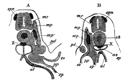
The sections illustrate the development of the
segmental duct (pd) or primitive duct
of the kidneys. In A (the anterior of the two sections) this
appears as a solid knob (pd) projecting
towards the epiblast. In B is seen a section of the column which
has grown backwards from the knob in A.
spn. rudiment of a
spinal nerve; mc. medullary canal;
ch. notochord; X. string of cells
below the notochord; mp. muscle-plate;
mp´. specially developed portion of
muscle-plate; ao. dorsal aorta; pd. segmental duct; so. somatopleure; sp. splanchnopleure; pp. pleuro-peritoneal or body-cavity; ep. epiblast; al.
alimentary canal.
While the lumen is gradually pushing its way backwards along the solid
rudiment of the segmental duct, the first traces of the segmental
tubes, or proper excretory organs, make their appearance in the form of
solid outgrowths of the intermediate cell-mass, which soon become hollow
and open into the body-cavity. Their blind ends curl obliquely backwards
round the inner and dorsal side of the segmental duct. One segmental tube
makes its appearance for each protovertebra, commencing with that
immediately behind the abdominal opening of the segmental duct, the last
tube being situated a short way behind the anus. Soon after their formation
the blind ends of the segmental tubes open into the segmental duct, and
each of them becomes divided into four parts. These are (woodcut 7) (1) a
section carrying the abdominal opening or segmental tube proper, (2) a
dilated vesicle into which this opens, (3) a coiled tubulus proceeding from
(2) and terminating in (4), a wider portion opening into the segmental
duct. At the same time, or shortly before this, each segmental duct unites
with and opens into one of the horns of the cloaca, and also retires from
its primitive position between the epiblast and mesoblast, and assumes a
position close to the epithelium lining the body-cavity. The general
features of the excretory organs at this period are diagrammatically
represented on the woodcut, fig. 6. In this fig. p.d is the segmental duct and o its abdominal
opening. s.t points to the segmental
tubes, the finer details of whose structure are not represented in the
diagram. The kidneys thus form at this period an unbroken gland composed of
a series of isolated coiled tubes, one extremity of each of which opens
into the body-cavity, and the other into the segmental duct, which forms
the only duct of the kidney, and communicates at one end with the
body-cavity, and at the other with the cloaca.
Fig. 6.
Diagram of the primitive
condition of the Kidney in an Elasmobranch Embryo.
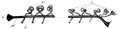
pd. segmental duct. It opens at o into
the body-cavity and at its other extremity into the cloaca; x. line
along which the division appears which separates the segmental duct into
the Wolffian duct above and the Müllerian duct below; st. segmental tubes. They open at one end into the
body-cavity, and at the other into the segmental duct.
The next important change concerns the segmental duct, which becomes
longitudinally split into two complete ducts in the female, and one
complete duct and parts of a second in the male. The manner in which this
takes place is diagrammatically represented in woodcut 6 by the clear line
x, and in transverse section in woodcut 7. The resulting ducts are
the (1) Wolffian duct dorsally, which remains continuous with the excretory
tubules of the kidney, and ventrally (2) the oviduct or Müllerian duct in
the female, and the rudiments of this duct in the male. In the female the
formation of these ducts takes place by a nearly solid rod of cells, being
gradually split off from the ventral side of all but the foremost part of
the original segmental duct, with the short undivided anterior part of
which duct it is continuous in front. Into it a very small portion of the
lumen of the original segmental duct is perhaps continued (Pl. 21, fig. 1A, etc.). The remainder of the segmental duct
(after the loss of its anterior section and the part split off from its
ventral side) forms the Wolffian duct. The process of formation of the
ducts in the male chiefly differs from that in the female in the fact of
the anterior undivided part of the segmental duct, which forms the front
end of the Müllerian duct, being shorter, and in the column of cells with
which it is continuous being from the first incomplete.
Fig. 7.
Diagrammatic representation of a
transverse section of a Scyllium Embryo
illustrating the formation of
the Wolffian and Müllerian ducts by
the longitudinal splitting of the
segmental duct.
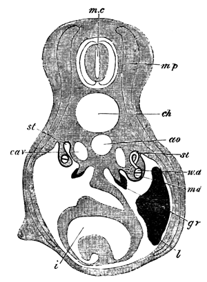
mc. medullary canal;
mp. muscle-plate; ch. notochord; ao. aorta; cav.
cardinal vein; st. segmental tube. On
the one side the section passes through the opening of a segmental tube
into the body-cavity. On the other this opening is represented by dotted
lines, and the opening of the segmental tube into the Wolffian duct has
been cut through; w.d. Wolffian duct;
m.d. Müllerian duct. The section is
taken through the point where the segmental duct and Wolffian duct have
just become separate; gr. The germinal
ridge with the thickened germinal epithelium; l. liver; i.
intestine with spiral valve.
The tubuli of the primitive excretory organ undergo further important
changes. The vesicle at the termination of each segmental tube grows
forwards towards the preceding tubulus, and joins the fourth section of it
close to the opening into the Wolffian duct (Pl. 21, fig. 10). The remainder of the vesicle becomes
converted into a Malpighian body. By the first of these changes a
connection is established between the successive segments of the kidney,
and though this connection is certainly lost (or only represented by
fibrous bands) in the anterior part of the excretory organs in the adult,
and very probably in the hinder part, yet it seems most probable that
traces of it are to be found in the presence of the secondary Malpighian
bodies of the majority of segments, which are most likely developed from
it.
Up to this time there has been no distinction between the anterior and
posterior tubuli of the primitive excretory organ which alike open into the
Wolffian duct. The terminal division of the tubuli of a considerable number
of the hindermost of these (ten or eleven in Scyllium canicula), either in
some species elongate, overlap, and eventually open by apertures (not
usually so numerous as the separate tubes), on nearly the same level, into
the hindermost section of the Wolffian duct in the female, or into the
urinogenital cloaca, formed by the coalesced terminal parts of the
Wolffian ducts, in the male; or in other species become modified in such a
manner as to pour their secretion into a single duct on each side, which
opens in a position corresponding with the numerous ducts of the other type
(woodcut, fig. 8). It seems that both in Amphibians and Elasmobranchii the
type with a single duct, or approximations to it, are more often found in
the females than in the males. The subject requires however to be more
worked out in Elasmobranchii[361]. In
both groups the modified posterior kidney-segments are probably equivalent
to the permanent kidney of the amniotic Vertebrates, and for this reason
the numerous ducts of the first group or single duct of the second were
spoken of as ureters. The anterior tubuli of the primitive excretory organ
retain their early relation to the Wolffian duct, and form the Wolffian
body.
The originally separate terminal extremities of the Wolffian ducts
always coalesce, and form a urinal cloaca, opening by a single aperture
situated at the extremity of a median papilla behind the anus. Some of the
abdominal openings of the segmental tubes in Scyllium, or in other cases
all the openings, become obliterated.
In the male the anterior segmental tubes undergo remarkable
modifications. There appear to grow from the first three or four or more of
them (though the point is still somewhat obscure) branches, which pass to
the base of the testis and there unite into a longitudinal canal, form a
network, and receive the secretion of the testicular ampullæ (woodcut 9,
nt). These ducts, the vasa efferentia,
carry the semen to the Wolffian body, but before opening into the tubuli of
this they unite into the longitudinal canal of the Wolffian body (l.c), from which pass off ducts equal in number
to the vasa efferentia, each of which normally ends in a Malpighian body.
From the Malpighian body so connected start the convoluted tubuli of what
may be called the generative segments of the Wolffian body along which the
semen is conveyed to the Wolffian duct (v.d). The Wolffian duct itself becomes much contorted
and acts as vas deferens.
Fig. 8.
Diagram of the arrangement of
the Urinogenital Organs in an adult
Female Elasmobranch.
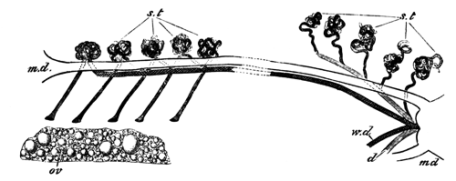
m.d. Müllerian duct;
w.d. Wolffian duct; s.t. glandular tubuli; five of them are represented
with openings into the body-cavity; d. duct of the posterior
segmental tubes; ov. ovary.
In the woodcuts, figs. 8 and 9, are diagrammatically represented the
chief constituents of the adult urinogenital organs in the two sexes. In
the adult female, fig. 8, there are present the following parts:
(1) The oviduct or Müllerian duct (m.d) split off from the segmental duct of the
kidneys. Each oviduct opens at its anterior extremity into the body-cavity,
and behind the two oviducts have independent communications with the
general cloaca.
(2) The Wolffian ducts (w.d), the
other product of the segmental ducts of the kidneys. They end in front by
becoming continuous with the tubulus of the anterior segment of the
Wolffian body on each side, and unite behind to open by a common papilla
into the cloaca. The Wolffian duct receives the secretion of the anterior
part of the primitive kidney which forms the Wolffian body.
(3) The ureter (d) which carries off the secretion of the kidney
proper. It is represented in my diagram in its most rare and differentiated
condition as a single duct.
(4) The glandular tubuli (s.t), some
of which retain their original openings into the body-cavity, and others
are without them. They are divided into two groups, an anterior forming
the Wolffian body, which pour their secretion into the Wolffian duct, and a
posterior group forming the kidney proper, which are connected with the
ureter.
Fig. 9.
Diagram of the arrangement of
the Urinogenital Organs in an adult
Male Elasmobranch.
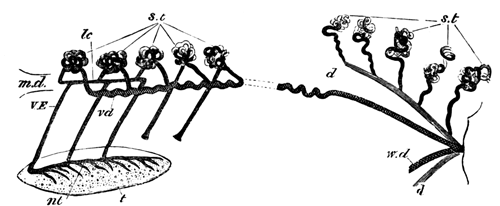
m.d. rudiment of
Müllerian duct; w.d. Wolffian duct,
marked vd in front and serving as vas
deferens; st. glandular tubuli; two of
them are represented with openings into the body-cavity; d. ureter;
t. testis; nt. central canal at
the base of the testis; VE. vasa
efferentia; lc. longitudinal canal of
the Wolffian body.
In the male the following parts are present (woodcut 9):
(1) The Müllerian duct (md),
consisting of a small rudiment attached to the liver representing the
foremost end of the oviduct of the female.
(2) The Wolffian duct (w.d) which
precisely corresponds to the Wolffian duct of the female, but, in addition
to functioning as the duct of the Wolffian body, also acts as a vas
deferens (vd). In the adult male its
foremost part has a very tortuous course.
(3) The ureter (d), which has the same fundamental constitution
as in the female.
(4) The segmental tubes (st). The
posterior of these have the same arrangement in both sexes, but in the male
modifications take place in connection with the anterior ones to fit them
to act as transporters of the testicular products.
Connected with the anterior ones there are present (1) the vasa
efferentia (VE), united on the one hand with (2) the
central canal in the base of the testis (nt), and on the other with the longitudinal
canal of the Wolffian body (l.c). From
the latter are seen passing off the successive tubuli of the anterior
segments of the Wolffian body in connection with which Malpighian bodies
are typically present, though not represented in my diagram.
Postscript.
It was my original intention to have given an account of the development
of the generative organs. In the course, however, of my work a number of
novel and unexpected points turned up, which have considerably protracted
my investigations, and it has appeared to me better no longer to delay the
appearance of this monograph, but to publish elsewhere my results on the
generative organs. In chapter VI. p. 349 et seq. the early stages of the generative
organs are described, but in contemplation of the completion of the account
no allusion was made to their literature, and more especially to Professor
Semper's important contributions. I may perhaps say that I have been able
to confirm the most important result to which he and other anatomists have
nearly simultaneously arrived with respect to Vertebrates, viz. that the primitive ova give rise to both the male
and female generative products.
EXPLANATION OF PLATES 20 AND 21.
Complete List of Reference
Letters.
amg. Accessory
Malpighian body. cav. Cardinal vein.
ge. Germinal epithelium. k. True
kidney. l.c. Longitudinal canal of the
Wolffian body connected with vasa efferentia. mg. Malpighian body. nt. Network and central canal at the base of the
testis. o. External aperture of urinal cloaca. od. Oviduct or Müllerian duct of the female. od´. Müllerian duct of the male. ou. Openings of ureters in Wolffian duct in the
female (fig. 3). pmg. Primary Malpighian
body. px. Growth from vesicle at the end
of a segmental tube to join the collecting tube of the preceding segment.
rst. Rudimentary segmental tube. ru. Ureter commencing to be formed. sb. Seminal bladder. sd. Segmental duct. st. Segmental tube. sto. Opening of segmental tube into body-cavity.
sur. Suprarenal body. t. Testis.
u. Ureters. ve. Vas efferens.
wb. Wolffian body. wd. Wolffian duct.
Plate 20.
Fig. 1. Diagrammatic representation of excretory
organs on one side of a male Scyllium canicula, natural size.
Fig. 2. Diagrammatic representation of the kidney
proper on one side of a female Scyllium canicula, natural size, shewing the
ducts of the kidney and the dilated portion of the Wolffian duct.
Fig. 3. Opening of the ureters into the Wolffian duct
of a female Scyllium canicula. The figure represents the Wolffian ducts
(wd) with ventral portion removed so as
to expose their inner surface, and shews the junction of the two W. ducts
to form the common urinal cloaca, the single external opening of this
(o), and openings of ureters into one Wolffian duct (ou).
Fig. 4. Anterior extremity of Wolffian body of a
young male Scyllium canicula shewing the vasa efferentia and their
connection with the kidneys and the testis. The vasa efferentia and
longitudinal canal are coloured to render them distinct. They are intended
to be continuous with the uncoloured coils of the Wolffian body, though
this connection has not been very successfully rendered by the artist.
Fig. 5. Part of the Wolffian body of a nearly ripe
male embryo of Scyllium canicula as a transparent object. Zeiss a a, ocul. 3. The figure shews two segmental tubes opening
into the body-cavity and connected with a primary Malpighian body, and
also, by a fibrous connection, with a secondary Malpighian body of the
preceding segment. It also shews one segmental tube (rst) imperfectly connected with the accessory
Malpighian body of the preceding segment of the kidney. The coils of the
kidney are represented somewhat diagrammatically.
Fig. 6. Vasa efferentia of a male embryo of Scyllium
canicula eight centimetres in length. Zeiss a a, ocul. 2.
There are seen to be at the least six and possibly seven
distinct vasa going to as many segments of the Wolffian body and connected
with a longitudinal canal in the base of the testis. They were probably
also connected with a longitudinal canal in the Wolffian body, but this
could not be clearly made out.
Fig. 7. The anterior four vasa efferentia
of a nearly ripe embryo. Connected with the foremost one is seen a body
which looks like the remnant of a segmental tube and its opening
(rst?).
Fig. 8. Testis and anterior part of Wolffian body of
an embryo of Squatina vulgaris.
The figure is intended to illustrate the arrangement of
the vasa efferentia. There are five of these connected with a longitudinal
canal in the base of the testis, and with another longitudinal canal in the
Wolffian body. From the second longitudinal canal there pass off four ducts
to as many Malpighian bodies. Through the Malpighian bodies these ducts are
continuous with the several coils of the Wolffian body, and so eventually
with the Wolffian duct. Close to the hindermost vas efferens is seen a body
which resembles a rudimentary segmental tube (rst?).
Plate 21.
Figs. 1A, 1B, 1C, 1D. Four sections from a female
Scyllium canicula of a stage between M and N through the part where the
segmental duct becomes split into Wolffian duct and oviduct. Zeiss B, ocul. 2. 1A is the foremost section.
The sections shew that the oviduct arises as a
thickening on the under surface of the segmental duct into which at the
utmost a very narrow prolongation of the lumen of the segmental duct is
carried. The small size of the lumen of the Wolffian duct in the foremost
section is due to the section passing through nearly its anterior blind
extremity.
Fig. 2. Section close to the junction of the Wolffian
duct and oviduct in a female embryo of Scyllium canicula belonging to stage
N. Zeiss B, ocul. 2.
The section represented shews that in some instances the
formation of the oviduct and Wolffian duct is accompanied by a division of
the lumen of the segmental duct into two not very unequal parts.
Figs. 3A, 3B, 3C. Three sections illustrating the
formation of a ureter in a female embryo belonging to stage N. Zeiss B,
ocul. 2.
3A is the foremost section.
The figures shew that the lumen of the developing ureter
is enclosed in front by an independent wall (fig. 3A), but that further
back the lumen is partly shut in by the subjacent Wolffian duct, while
behind no lumen is present, but the ureter ends as a solid knob of cells
without an opening into the Wolffian duct.
Fig. 4. Section through the ureters of the same
embryo as fig. 3, but nearer the cloaca. Zeiss B, ocul. 2.
The figure shews the appearance of a transverse section
through the wall of cells above the Wolffian duct formed by the overlapping
ureters, the lumens of which appear as perforations in it. It should be
compared with fig. 9A, which represents a longitudinal section through a
similar wall of cells.
Fig. 5. Section through the ureters, the Wolffian
duct and the oviduct of a female embryo of Scy. canicula belonging to stage
P. Zeiss B, ocul. 2.
Fig. 6. Section of part of the Wolffian body of a
male embryo of Scyllium canicula belonging to stage O. Zeiss B, ocul. 2.
The section illustrates (1) the formation
of a Malpighian body (mg) from the
dilatation at the end of a segmental tube, (2) the appearance of the
rudiment of the Müllerian duct in the male (od´).
Figs. 7a, 7b. Two longitudinal and
vertical sections through part of the kidney of an embryo between stages L
and M. Zeiss B, ocul. 2.
7a illustrates the parts of a single segment of
the Wolffian body at this stage, vide p. 491. The segmental tube and opening are not in the
plane of the section, but the dilated vesicle is shewn into which the
segmental tube opens.
7b is taken from the region of the kidney proper.
To the right is seen the opening of a segmental tube into the body-cavity,
and in the segment to the left the commencing formation of a ureter, vide p. 502.
Fig. 8. Longitudinal and vertical section through the
posterior part of the kidney proper of an embryo of Scyllium canicula at a
stage between N and O. Zeiss A, ocul. 2.
The section shews the nearly completed ureters,
developing Malpighian bodies, &c.
Fig. 9. Longitudinal and vertical section through the
anterior part of the kidney proper of the same embryo as fig. 8. Zeiss A,
ocul. 2.
The figure illustrates the mode of growth of the
developing ureters.
9A. More highly magnified portion of the same section as
fig. 9.
Compare with transverse section fig. 4.
Fig. 10. Longitudinal and vertical section through
part of the Wolffian body of an embryo of Scyllium canicula at a stage
between O and P.
The section contains two examples of the budding out of
the vesicle of a segmental tube to form a Malpighian body in its own
segment and to unite with the tubulus of the preceding segment close to its
opening into the Wolffian duct.
The brilliant discoveries of Strasburger and Auerbach have
caused the attention of a large number of biologists to be turned to the
phenomena accompanying the division of nuclei and the maturation and
impregnation of the ovum. The results of the recent investigations on the
first of these points formed the subject of an article by Mr Priestley in
the sixteenth volume of this Journal, and the object of the present article
is to give some account of what has so far been made out with reference to
the second of them. The matters to be treated of naturally fall under two
heads: (1) the changes attending the ripening of the ovum, which are
independent of impregnation; (2) the changes which are directly
due to impregnation.
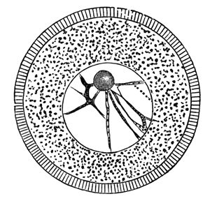
Fig. 1.—Unripe ovum of
Toxopneustes lividus (copied from Hertwig).
Every ovum as it approaches maturity is found to be composed (Fig. 1) of
(1) a protoplasmic body or vitellus usually containing yolk-spherules in
suspension; (2) of a germinal vesicle or nucleus, containing (3) one
or more germinal spots or nucleoli. It is with the germinal vesicle and its
contents that we are especially concerned. This body at its full
development has a more or less spherical shape, and is enveloped by a
distinct membrane. Its contents are for the most part fluid, but may be
more or less granular. Their most characteristic component is, however, a
protoplasmic network which stretches from the germinal spot to the
investing membrane, but is especially concentrated round the former (Fig.
1). The germinal spot forms a nearly homogeneous body, with frequently one
or more vacuoles. It occupies an often excentric position within the
germinal vesicle, and is usually rendered very conspicuous by its high
refrangibility. In many instances it has been shewn to be capable of
amœboid movements (Auerbach, and Os. Hertwig), and is moreover more
solid and more strongly tinged by colouring reagents than the remaining
constituents of the germinal vesicle. These peculiarities have caused the
matter of which it is composed to be distinguished by Auerbach and Hertwig
as nuclear substance.
In many instances there is only one germinal spot, or one main spot, and
two or three accessory smaller spots. In other cases, e.g. Osseous
Fish, there are a large number of nearly equal germinal spots. The eggs
which have been most investigated with reference to the changes of germinal
vesicle are those with a single germinal spot, and it is with these that I
shall have more especially to deal in the sequel.
The germinal vesicle occupies in the first instance a central position
in the ovum, but at maturity is almost always found in close proximity to
the surface. Its change of position in a large number of instances is
accomplished during the growth of the ovum in the ovary, but in other cases
does not take place till the ovum has been laid.
The questions which many investigators have recently set themselves to
answer are the two following:—(1) What becomes of the germinal
vesicle when the ovum is ready to be impregnated? (2) Is any part of it
present in the ovum at the commencement of segmentation? According to their
answers to these questions the older embryologists roughly fall into two
groups: (1) By one set the germinal vesicle is stated to completely
disappear and not to be genetically connected with the subsequent nuclei of
the embryo. (2) According to the other set it remains in the ovum and by
successive divisions forms the parent nucleus of all the nuclei in the body
of the embryo. Though the second of these views has been supported by
several very distinguished names the first view was without doubt the one
most generally entertained, and Haeckel (though from his own observations
he was originally a supporter of the second view) has even enunciated the
theory that there exists an anuclear stage, after the disappearance of the
germinal vesicle, which he regards as an embryonic repetition of the monad
condition of the Protozoa.
While the supporters of the first view agree as to the disappearance of
the germinal vesicle they differ considerably as to the manner of this
occurrence. Some are of opinion that the vesicle simply vanishes, its
contents being absorbed in the ovum; others that it is ejected from the
ovum and appears as the polar cell or body, or Richtungskörper of the Germans—a small body which is
often found situated in the space between the ovum and its membrane, and
derives its name from retaining a constant position in relation to the
ovum, and thus serving as a guide in determining the similar parts of the
embryo through the different stages. The researches of Oellacher (15)[363] in this direction deserve special mention, as having
in a sense formed the foundation of the modern views upon this subject. By
a series of careful observations upon the egg of the trout and subsequently
of the bird, he demonstrated that the germinal vesicle of the ovum, while
still in the ovary, underwent partial degeneration and eventually became
ejected. His observations were made to a great extent by means of sections,
and the general accuracy of his results is fairly certain, but the nature
of the eggs he worked on, as well as other causes, prevented his obtaining
so deep an insight into the phenomena accompanying the ejection of the
germinal vesicle as has since been possible. Lovén, Flemming (6), and
others have been led by their investigations to adopt views similar in the
main to Oellacher's. As a rule, however, it is held by believers in the
disappearance of the germinal vesicle that it becomes simply absorbed, and
many very accurate accounts, so far as they go, have been
given of the gradual atrophy of the germinal vesicle. The description of
Kleinenberg (14) for Hydra, and Götte for Bombinator, may perhaps be
selected as especially complete in this respect; in both instances the
germinal vesicle commences to atrophy at a relatively early period.
Coming to the more modern period the researches of five workers, viz. Bütschli, E. van Beneden, Fol, Hertwig, and
Strasburger have especially thrown light upon this difficult subject. It is
now hardly open to doubt that while part of the germinal vesicle is
concerned in the formation of the polar cell or cells, when such are
present, and is therefore ejected from the ovum, part also remains in the
ovum and forms a nuclear body which will be spoken of as the female
pronucleus, the fate of which is recorded in the second part of this
paper. The researches of Bütschli and van Beneden have been especially
instrumental in demonstrating the relation between the polar bodies and the
germinal vesicle, and those of Hertwig and Fol, in shewing that part of the
germinal vesicle remained in the ovum. It must not, however, be supposed
that the results of these authors are fully substantiated, or that all the
questions connected with these phenomena are settled. The statements we
have are in many points opposed and contradictory, and there is much that
is still very obscure.
In the sequel an account is first given of the researches of the
above-named authors, followed by a statement of those results which appear
to me the most probable.
The researches of van Beneden (3 and 4) were made on the ovum of the
rabbit and of Asterias, and from his observations on both these widely
separated forms he has been led to conclude that the germinal vesicle is
either ejected or absorbed, but that it has in no case a genetic connection
with the first segmentation sphere. He gives the following description of
the changes in the rabbit's ovum. The germinal vesicle is enclosed by a
membrane, and contains one main germinal spot, and a few accessory ones,
together with a granular material which he calls nucleoplasma,
which affects, as is usual in nuclei, a reticular arrangement. The
remaining space in the vesicle is filled by a clear fluid. As the ovum
approaches maturity the germinal vesicle assumes an
excentric position, and fuses with the peripheral layer of the egg to
constitute the cicatricular lens. The germinal spot next travels
to the surface of the cicatricular lens and forms the nuclear
disc: at the same time the membrane of the germinal vesicle vanishes
though it probably unites with the nuclear disc. The nucleoplasma then
collects into a definite mass and forms the nucleoplasmic body. Finally the
nuclear disc assumes an ellipsoidal form and becomes the nuclear body.
Nothing is now left of the original germinal vesicle but the nuclear body
and the nucleoplasmic body both still situated within the ovum. In the next
stage no trace of the germinal vesicle can be detected in the ovum, but
outside it, close to the point where the modified remnants of the vesicle
were previously situated, there is present a polar body which is composed
of two parts, one of which stains deeply and resembles the nuclear body,
and the other does not stain but is similar to the nucleoplasmic body. Van
Beneden concludes that the polar bodies are the two ejected products of the
germinal vesicle. In the case of Asterias, van Beneden has not observed the
mode of formation of the polar bodies, and mainly gives an account of the
atrophy of the germinal vesicle, but adds very little to what was already
known to us from Kleinenberg's (14) earlier observations. He describes with
precision the breaking up of the germinal spot into fragments and its
eventual disappearance.
Though there are reasons for doubting the accuracy of all the above
details on the ovum of the rabbit, nevertheless, the observations of van
Beneden taken as a whole afford strong grounds for concluding that the
formation of the polar cells is connected with the disappearance, partial
or otherwise, of the germinal vesicle. A very similar account of the
apparent disappearance of the germinal vesicle is given by Greeff (19) who
states that the apparent disappearance of the germinal spot precedes that
of the vesicle.
The observations of Bütschli are of still greater importance in this
direction. He has studied with a view to elucidating the fate of the
germinal vesicle, the eggs of Nephelis, Lymnæus, Cucullanus, and other
Nematodes; and Rotifers. In all of these, with the exception of Rotifers,
he finds polar bodies, and in this respect his
observations are of value as tending to shew the widespread existence of
these structures. Negative results with reference to the presence of the
polar bodies have, it may be remarked, only a very secondary value.
Bütschli has made the very important discovery that in perfectly ripe eggs
of Nephelis, Lymnæus and Cucullanus and allied genera a spindle,
similar to that of ordinary nuclei in the act of division, appears close to
the surface of the egg. This spindle he regards as the metamorphosed
germinal vesicle, and has demonstrated that it takes part in the formation
of the polar cells. He states that the whole spindle is ejected from the
egg, and that after swelling up and forming a somewhat spherical mass it
divides into three parts.
In the Nematodes generally, Bütschli has been unable to find the spindle
modification of the germinal vesicle, but he states that the germinal
vesicle undergoes degeneration, its outline becoming indistinct and the
germinal spot vanishing. The position of the germinal vesicle continues to
be marked by a clear space which gradually approaches the surface of the
egg. When it is in contact with the surface a small spherical body, the
remnant of the germinal vesicle, comes into view, and eventually becomes
ejected. The clear space subsequently disappears. This description of
Bütschli resembles in some respects that given by van Beneden of the
changes in the rabbit's ovum, and not impossibly refers to a nearly
identical series of phenomena. The discovery by Bütschli of the spindle and
its relation to the polar body has been of very great value.
The publications of van Beneden, and more especially those of Bütschli,
taken by themselves lead to the conclusion that the whole germinal vesicle
is either ejected or absorbed. Nearly simultaneously with their
publications there appeared, however, a paper by Oscar Hertwig (11) on the
eggs of one of the common sea urchins (Toxopneustes lividus), in
which he attempted to shew that part of the germinal vesicle, at any rate,
was concerned in the formation of the first segmentation nucleus. He
believed (though he has himself now recognised that he was in error on the
point) that no polar cell was formed in Toxopneustes, and that the whole
germinal vesicle was absorbed, with the exception of the germinal spot
which remained in the egg as the female pronucleus.
The following is the summary which he gives of his results,
pp. 357-8.
At the time when the egg is mature the germinal
vesicle undergoes a retrogressive metamorphosis and becomes carried towards
the surface of the egg by the contraction of the protoplasm. Its membrane
becomes dissolved and its contents disintegrated and finally absorbed by
the yolk. The germinal spot appears, however, to remain unaltered and to
continue in the yolk and to become the permanent nucleus of the ripe ovum
capable of impregnation.
After the publication of Bütschli's monograph, O. Hertwig (12) continued
his researches on the ova of Leeches (Hæmopis and Nephelis),
and not only added very largely to our knowledge of the history of the
germinal vesicle, but was able to make a very important rectification in
Bütschli's conclusions. The following is a summary of his
results:—The germinal vesicle, as in other cases, undergoes a form of
degeneration, though retaining its central position; and the germinal spot
breaks up into fragments. The stages in which this occurs are followed by
one when, on a superficial examination, the ovum appears to be absolutely
without a nucleus; but there can be demonstrated by means of reagents in
the position previously occupied by the germinal vesicle a spindle nucleus
with the usual suns at its poles, which Hertwig believes to be a product of
the fragments of the germinal spot. This spindle travels towards the
periphery of the ovum and then forms the spindle observed by Bütschli. At
the point where one of the apices of the spindle lies close to the surface
a small protuberance arises which is destined to form the first polar cell.
As the protuberance becomes more prominent one half of the spindle passes
into it. The spindle then divides in the normal manner for nuclei, one half
remaining in the protuberance, the other in the ovum, and finally the
protuberance becomes a rounded body united to the egg by a narrow stalk. It
is clear that if, as there is every reason to think, the above description
is correct, the polar cell is formed by a simple process of cell-division
and not, as Bütschli believed, by the forcible ejection of the spindle.
The portion of the spindle in the polar cell becomes a mass of granules,
and that in the ovum becomes converted without the occurrence of
the usual nuclear stage into a fresh spindle. A second polar cell is formed
in the same manner as the first one, and the first one subsequently divides
into two. The portion of the spindle which remains in the egg after the
formation of the second polar cell reconstitutes itself into a
nucleus—the female pronucleus—and travelling towards the centre
of the egg undergoes a fate which will be spoken of in the second part of
this paper.
The most obscure part of Hertwig's work is that which concerns the
formation of the spindle on the atrophy of the germinal vesicle, and his
latest paper, though it gives further details on this head, does not appear
to me to clear up the mystery. Though Hertwig demonstrates clearly enough
that this spindle is a product of the metamorphoses of the germinal
vesicle, he does not appear to prove the thesis which he maintains, that it
is the metamorphosed germinal spot.
Fol, to whom we are indebted in his paper on the development of Geryonia
(7) for the best of the earlier descriptions of the phenomena which attend
the maturation of the egg, and later for valuable contributions somewhat
similar to those of Bütschli with reference to the development of the
Pteropod egg (8), has recently given us a very interesting account of what
takes place in the ripe egg of Asterias glacialis (9). In reference
to the formation of the polar cells, his results accord closely with those
of Hertwig, but he differs considerably from this author with reference to
the preceding changes in the germinal vesicle. He believes that the
germinal spot atrophies more or less completely, but that in any case its
constituents remain behind in the egg, though he will not definitely assert
that it takes no share in the formation of the spindle at the expense of
which both the polar cells and the female pronucleus are formed. The
spindle with its terminal suns arises, according to him, from the contents
of the germinal vesicle, loses its spindle character, travels to the
surface, and reacquiring a spindle character is concerned in the formation
of the polar cells in the way described by Hertwig.
Giard (10) gives a somewhat different account of the behaviour of the
germinal vesicle in Psammechinus miliaris. At maturity the contents
of the germinal vesicle and spot mix together and form
an amœboid mass, which, assuming a spindle form, divides into two
parts, one of which travels towards the centre of the egg and forms the
female pronucleus, the other remains at the surface and gives origin to two
polar cells, both of which are formed after the egg is laid. What Giard
regards as the female pronucleus is perhaps the lower of the two bodies
which take the place of the original germinal vesicle as described by Fol.
Vide the account of Fol's observations on
p. 531.
Strasburger, from observations on Phallusia, accepts in the main
Hertwig's conclusion with reference to the formation of the polar bodies,
but does not share Hertwig's view that either the polar bodies or female
pronucleus are formed at the expense of the germinal spot alone. He has
further shewn that the so-called canal-cell of conifers is formed in the
same manner as the polar cells, and states his belief that an equivalent of
the polar cells is widely distributed in the vegetable subkingdom.
This sketch of the results of recent researches will, it is hoped,
suffice to bring into prominence the more important steps by which the
problems of this department of embryology have been solved. The present
aspects of the question may perhaps be most conveniently displayed by
following the history of a single ovum. For this purpose the eggs of
Asterias glacialis, which have recently formed the subject of a
series of beautiful researches by Fol (9), may conveniently be
selected.
The ripe ovum (Fig. 2), when detached from the ovary, is formed of a
granular vitellus without a vitelline membrane, but enveloped in a
mucilaginous coat. It contains an excentrically situated germinal vesicle
and germinal spot. In the former is present the usual protoplasmic
reticulum. As soon as the ovum reaches the sea water the germinal vesicle
commences to undergo a peculiar metamorphosis. It exhibits frequent changes
of form, its membrane becomes gradually absorbed and its outline indented
and indistinct, and finally its contents become to a certain extent
confounded with the vitellus (Fig. 3).
The germinal spot at the same time loses its clearness of outline and
gradually disappears from view.
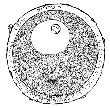
Fig. 2.—Ripe ovum of
Asterias glacialis enveloped in a mucilaginous envelope, and containing an
excentric germinal vesicle and germinal spot (copied from Fol).
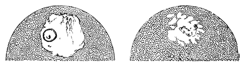
Fig. 3.—Two successive
stages in the gradual metamorphosis of the germinal vesicle and spot of
the ovum of Asterias glacialis immediately after it is laid (copied from
Fol).
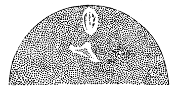
Fig. 4.—Ovum of Asterias
glacialis, shewing the clear spaces in the place of the germinal vesicle.
Fresh preparation (copied from Fol).
At a slightly later stage in the place of the original germinal vesicle
there may be observed in the fresh ovum two clear spaces (fig. 4), one
ovoid and nearer the surface, and the second more irregular in form and
situated rather deeper in the vitellus. By treatment with reagents the
first clear space is found to be formed of a spindle with two terminal suns
on the lower side of which is a somewhat irregular body (Fig. 5). The
second clear space by the same treatment is shewn to contain a round body.
Fol concludes that the spindle is formed out of part of the germinal
vesicle and not of the germinal spot, while he sees in the round body
present in the lower of the two clear spaces the metamorphosed germinal
spot. He will not, however, assert that no fragment of the germinal spot
enters into the formation of the spindle. It may be observed that Fol is
here obliged to fill up (so far at least as his present preliminary account
enables me to determine) a lacuna in his observations in a hypothetical
manner, and O. Hertwig's (13) most recent observations on the ovum of the
same or an allied species of Asterias tend to throw some doubt upon Fol's
interpretations.
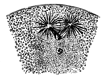
Fig. 5.—Ovum of Asterias
glacialis, at the same stage as Fig. 4, treated with picric acid (copied
from Fol).
The following is Hertwig's account of the changes in the germinal
vesicle. A quarter of an hour after the egg is laid the protoplasm on the
side of the germinal vesicle towards the surface of the egg develops a
prominence which presses inwards the wall of the vesicle. At the same time
the germinal spot develops a large vacuole, in the interior of which is a
body consisting of nuclear substance, and formed of a firmer and more
refractive material than the remainder of the germinal spot. In the
above-mentioned prominence towards the germinal vesicle, first one sun is
formed by radial striæ of protoplasm, and then a second makes its
appearance, while in the living ovum the germinal spot appears to have
vanished, the outline of the germinal vesicle to have become indistinct,
and its contents to have mingled with the surrounding protoplasm. Treatment
with reagents demonstrates that in the process of disappearance of the
germinal spot the nuclear mass in the vacuole forms a rod-like body, the
free end of which is situated between the two suns which occupy the
prominence of the germinal vesicle. At a slightly later period granules may
be seen at the end of the rod and finally the rod itself vanishes. After
these changes there may be demonstrated by the aid of reagents a spindle
between the two suns, which Hertwig believes to grow in size as the last
remnants of the germinal spot gradually vanish, and he maintains, as before
mentioned, that the spindle is formed at the expense of the germinal spot.
Without following Hertwig so far as this[364] it
may be permitted to suggest that his observations tend to shew that the
body noticed by Fol in the median line, on the inner side of his spindle,
is in reality a remnant of the germinal spot and not, as Fol supposes, part
of the germinal vesicle. Considering how conflicting is the evidence before
us it seems necessary to leave open for the present the question as to what
parts of the germinal vesicle are concerned in forming the first
spindle.
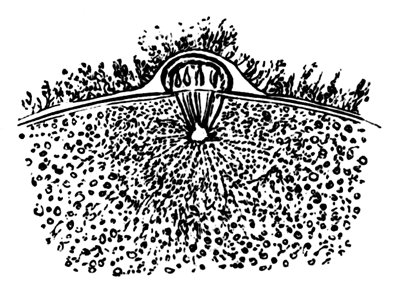
Fig. 6.—Portion of the
ovum of Asterias glacialis, shewing the spindle formed from the
metamorphosed germinal vesicle projecting into a protoplasmic prominence
of the surface of the egg. Picric acid preparation (copied from Fol).
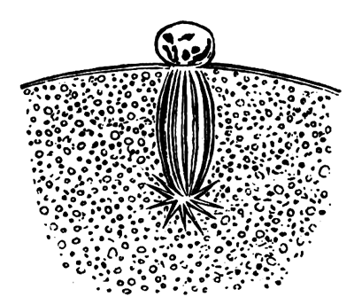
Fig. 7.—Portion of the
ovum of Asterias glacialis at the moment of the detachment of the first
polar body and the withdrawal of the remaining part of the spindle within
the ovum. Picric acid preparation (copied from Fol).
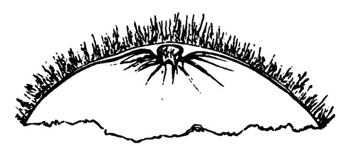
Fig. 8.—Portion of the
ovum of Asterias glacialis, with the first polar body as it appears when
living (copied from Fol).
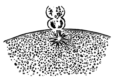
Fig. 9.—Portion of the
ovum of Asterias glacialis immediately after the formation of the second
polar body. Picric acid preparation (copied from Fol).
The spindle, however it be formed, has up to this time been situated
with its axis parallel to the surface of the egg, but not long after the
stage last described a spindle is found with one end projecting into a
protoplasmic prominence which makes its appearance on the surface of the
egg (Fig. 6). Hertwig believes that the spindle simply travels towards the
surface, and while doing so changes the direction of its axis. Fol finds,
however, that this is not the case, but that between the two conditions of
the spindle an intermediate one is found in which a spindle can no longer
be seen in the egg, but its place is taken by a compact rounded body. He
has not been able to arrive at a conclusion as to what meaning is to be
attached to this occurrence. In any case the spindle which projects into
the prominence on the surface of the egg divides it into two parts, one in
the prominence and one in the egg (Fig. 7). The prominence itself with the
enclosed portion of the spindle becomes partially constricted off from the
egg as the first polar body (Fig. 8). The part of the spindle which remains
in the egg becomes directly converted into a second spindle by the
elongation of its fibres without passing through a typical nuclear
condition. A second polar cell next becomes formed in the same manner as
the first (Fig. 9), and the portion of the spindle remaining in the
egg becomes converted into two or three clear vesicles (Fig. 10) which soon
unite to form a single nucleus, the female pronucleus (Fig. 11). The two
polar cells appear to be situated between two membranes, the outer of which
is very delicate and only distinct where it covers the polar cells, while
the inner one is thicker and becomes, after impregnation, more distinct and
then forms what Fol speaks of as the vitelline membrane. It is clear, as
Hertwig has pointed out, that the polar bodies originate by a regular cell
division and have the value of cells.
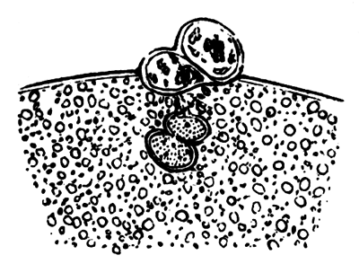
Fig. 10.—Portion of the
ovum of Asterias glacialis after the formation of the second polar cell,
shewing the part of the spindle remaining in the ovum becoming converted
into two clear vesicles. Picric acid preparation (copied from Fol).
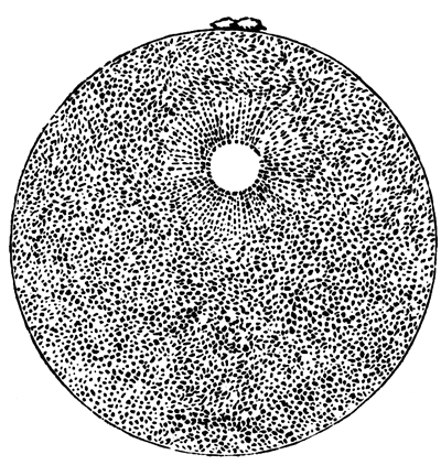
Fig. 11.—Ovum of Asterias
glacialis with the two polar bodies and the female pronucleus surrounded
by radial striæ, as seen in the living egg (copied from Fol).
General conclusions.
Considering how few ova have been adequately investigated with reference
to the behaviour of the germinal vesicle any general conclusions which may
at present be formed are to be regarded as provisional, and I trust that
this will be borne in mind by the reader in perusing the following
paragraphs.
There is abundant evidence that at the time of maturation of the egg the
germinal vesicle undergoes peculiar changes, which are, in part at least,
of a retrogressive character. These changes may begin considerably before
the egg has reached the period of maturity, or may not take place till
after it has been laid. They consist in appearance of irregularity and
obscurity in the outline of the germinal vesicle, the absorption of its
membrane, the partial absorption of its contents in the yolk, and the
breaking up and disappearance of the germinal spot. The exact fate of the
single germinal spot, or the numerous spots where they are present, is
still obscure; and the observations of Oellacher on the trout, and to a
certain extent my own on the skate, tend to shew that the membrane of the
germinal vesicle may in some cases be ejected from the egg, but this
conclusion cannot be accepted without further confirmation.
The retrogressive metamorphosis of the germinal vesicle is followed in a
large number of instances by the conversion of what remains into a striated
spindle similar in character to a nucleus previous to division. This
spindle travels to the surface and undergoes division to form the polar
cell or cells in the manner above described. The part which remains in the
egg forms eventually the female pronucleus.
The germinal vesicle has up to the present time only been observed to
undergo the above series of changes in a certain number of instances,
which, however, include examples from several divisions of the
Cœlenterata, the Echinodermata, and the Mollusca, and also some of
the Vermes (Nematodes, Hirudinea, Sagitta). It is very possible, not to say
probable, that it is universal in the animal kingdom, but the present state
of our knowledge does not justify us in saying so. It may be that in the
case of the rabbit, and many Nematodes as described by van Beneden and by
Bütschli, we have instances of a different mode of formation of the polar
cells.
The case of Amphibians, as described by Bambeke (2) and Hertwig (12)
cannot so far be brought into conformity with our type, though observations
are so difficult to make with such opaque eggs that not much reliance can
be placed upon the existing statements. In both of these types of possible
exceptions it is fairly clear that, whatever may be the case with reference
to the formation of the polar cells, part of the germinal vesicle remains
behind as the female pronucleus.
There are a large number of types, including the whole of the Rotifera[365] and Arthropoda, with a few doubtful exceptions, in
which the polar cells cannot as yet be said to have been satisfactorily
observed.
Whatever may be the eventual result of more extended investigation, it
is clear that the formation of polar cells according to our type is a very
constant occurrence. Its importance is also very greatly increased by the
discovery by Strasburger of the existence of an analogous process amongst
plants. Two questions about it obviously present themselves for solution:
(1) What are the conditions of its occurrence with reference to
impregnation? (2) What meaning has it in the development of the ovum or the
embryo?
The answer to the first of these questions is not difficult to find. The
formation of the polar bodies is independent of impregnation, and is the
final act of the normal growth of the ovum. In a few types the polar cells
are formed while the ovum is still in the ovary, as, for instance, in some
species of Echini, Hydra, &c., but,
according to our present knowledge, far more usually after the ovum has
been laid. In some of the instances the budding off of the polar cells
precedes, and in others follows impregnation; but there is no evidence to
shew that in the later cases the process is influenced by the contact with
the male element. In Asterias, as has been shewn by O. Hertwig, the
formation of the polar cells may indifferently either precede or follow
impregnation—a fact which affords a clear demonstration of the
independence of the two occurrences.
To the second of the two questions it does not unfortunately seem
possible at present to give an answer which can be regarded as
satisfactory.
The retrogressive changes in the membrane of the germinal vesicle which
usher in the formation of the polar bodies may very probably be viewed as a
prelude to a renewed activity of the contents of the vesicle; and are
perhaps rendered the more necessary from the thickness of the membrane
which results from a protracted period of passive growth. This suggestion
does not, however, help us to explain the formation of polar cells by a
process identical with cell division. The ejection of part of the germinal
vesicle in the formation of the polar cells may probably be paralleled by
the ejection of part or the whole of the original nucleus which, if we may
trust the beautiful researches of Bütschli, takes place during conjugation
in Infusoria as a preliminary to the formation of a fresh nucleus. This
comparison is due to Bütschli, and according to it the formation of the
polar bodies would have to be regarded as assisting, in some as yet unknown
way, the process of regeneration of the germinal vesicle. Views analogous
to this are held by Strasburger and Hertwig, who regard the formation of
the polar bodies in the light of a process of excretion or removal of
useless material. Such hypotheses do not unfortunately carry us very
far.
I would suggest that in the formation of the polar cells part of the
constituents of the germinal vesicle which are requisite for its functions
as a complete and independent nucleus are removed to make room for the
supply of the necessary parts to it again by the spermatic nucleus (vide p. 541). More light on
this, as on other points, may probably be thrown by further investigations
on parthenogenesis and the presence or absence of a polar cell in eggs
which develop parthenogenetically. Curiously enough the two groups in which
parthenogenesis most frequently occurs in the ordinary course of
development (Arthropoda and Rotifera) are also those in which
polar cells, with the possible exception mentioned above, of the
parthenogenetic eggs of Lacenularia, are stated to be absent. This curious
coincidence, should it be confirmed, may perhaps be
explained on the hypothesis, I have just suggested, viz. that a more or less essential part of the nucleus
is removed in the formation of the polar cells; so that in cases, e.g.
Arthropoda and Rotifera, where polar cells are not formed, and an essential
part of the nucleus not therefore removed, parthenogenesis can much more
easily occur than when polar cells are formed.
That the part removed in the formation of the polar cells is not
absolutely essential, seems at first sight to follow from the fact of
parthenogenesis being possible in instances where impregnation is the
normal occurrence. The genuineness of all the observations on this head is
too long a subject to enter into here[366],
but after admitting, as we probably must, that there are genuine cases of
parthenogenesis, it cannot be taken for granted without more extended
observation that the occurrence of development in these rare instances may
not be due to the polar cells not having been formed as usual, and that
when the polar cells are formed the development without impregnation is
less possible.
The remarkable observations of Professor Greeff (19) on the
parthenogenetic development of the eggs of Asterias rubens tell,
however, very strongly against this explanation. Greeff has found that
under normal circumstances the eggs of this species of starfish will
develop without impregnation in simple sea water. The development is quite
regular and normal though much slower than in the case of impregnated eggs.
It is not definitely stated that polar cells are formed, but there can be
no doubt that this is implied. Professor Greeff's account is so precise and
circumstantial that it is not easy to believe that any error can have crept
in; but neither Hertwig nor Fol have been able to repeat his experiments,
and we may be permitted to wait for further confirmation before absolutely
accepting them.
It is possible that the removal of part of the protoplasm of
the egg in the formation of the polar cells may be a secondary process due
to an attractive influence of the nucleus on the cell protoplasm, such as
is ordinarily observed in cell division.
Impregnation of the Ovum.
A far greater amount of certainty appears to me to have been attained as
to the effects of impregnation than as to the changes of the germinal
vesicle which precede this, and there appears, moreover, to be a greater
uniformity in the series of resulting phenomena. For convenience I propose
to reverse the order hitherto adopted and to reserve the history of the
literature and my discussion of disputed points till after my general
account. Fol's paper on Asterias glacialis, is again my source of
information. The part of the germinal vesicle which remains in the egg,
after the formation of the second polar cell, becomes converted into a
number of small vesicles (Fig. 10), which aggregate themselves into a
single clear nucleus which gradually travels toward the centre of the egg
and around which as a centre the protoplasm becomes radiately striated
(Fig. 11). This nucleus is known as the female pronucleus[367]. In Asterias glacialis the most favourable
period for fecundation is about an hour after the formation of the female
pronucleus. If at this time the spermatozoa are allowed to come in contact
with the egg, their heads soon become enveloped in the investing
mucilaginous coat. A prominence, pointing towards the nearest spermatozoon,
now arises from the superficial layer of protoplasm of the egg and grows
till it comes in contact with the spermatozoon (Figs. 12 and 13), Under
normal circumstances the spermatozoon, which meets the prominence, is the
only one concerned in the fertilisation, and it makes its way into the egg
by passing through the prominence. The tail of the spermatozoa, no longer
motile, remains visible for some time after the head has bored its way in,
but its place is soon taken by a pale conical body which is, however,
probably in part a product of the metamorphosis of the tail itself (Fig.
14). This body vanishes in its turn.
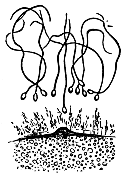
Fig. 12.
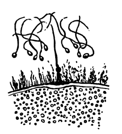
Fig. 13.
Figs. 12 and 13.—Small
portion of the ovum of Asterias glacialis. The spermatozoa are shewn
enveloped in the mucilaginous coat. In Fig. 12 a prominence is rising from
the surface of the egg towards the nearest spermatozoon; and in Fig. 13
the spermatozoon and prominence have met. From living ovum (copied from
Fol).
At the moment of contact between the spermatozoon and the egg the
outermost layer of the protoplasm of the latter raises itself as distinct
membrane, which separates from the egg and prevents the entrance of any
more spermatozoa. At the point where the spermatozoon entered a crater-like
opening is left in the membrane (Fig. 14).
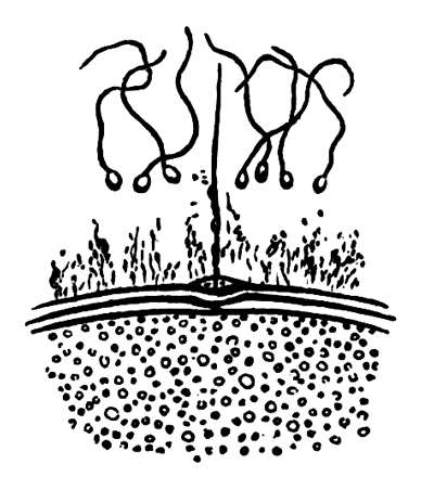
Fig. 14.—Portion of the
ovum of Asterias glacialis after the entrance of a spermatozoon into the
ovum. It shows the prominence of the ovum through which the spermatozoon
has entered. A vitelline membrane with a crater-like opening has become
distinctly formed. From living ovum (copied from Fol).
The head of the spermatozoon when in the egg forms a nucleus for which
the name male pronucleus may be conveniently adopted. It grows in
size by absorbing, it is said, material from the ovum, though this may be
doubted, and around it is formed a clear space free from yolk-spherules.
Shortly after its formation the protoplasm in its neighbourhood assumes
a radiate arrangement (Fig. 15). At whatever point of the egg the
spermatozoon may have entered, it gradually travels towards the female
pronucleus. This latter, around which the protoplasm no longer has a radial
arrangement, remains motionless till it comes in contact with the rays of
the male pronucleus, after which its condition of repose is exchanged for
one of activity, and it rapidly approaches the male pronucleus, and
eventually fuses with it (Fig. 16).
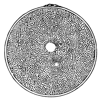
Fig. 15.—Ovum of Asterias
glacialis, with male and female pronucleus and a radial striation of the
protoplasm around the former. From living ovum (copied from Fol).
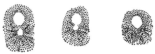
Fig. 16.—Three successive
stages in the coalescence of the male and female pronucleus in Asterias
glacialis. From the living ovum (copied from Fol).
The product of this fusion forms the first segmentation nucleus (Fig.
17), which soon, however, divides into the two nuclei of the two first
segmentation spheres. While the two pronuclei are approaching one another
the protoplasm of the egg exhibits amœboid movements.
Of the earlier observations on this subject there need perhaps only be
cited one of E. van Beneden, on the rabbit's ovum, shewing the
presence of two nuclei before the commencement of segmentation. Bütschli
was the earliest to state from observations on Rhabditis dolichura
that the first segmentation nucleus arose from the fusion of two nuclei,
and this was subsequently shewn with greater detail for Ascaris
nigrovenosa, by Auerbach (1). Neither of these authors gave at first
the correct interpretation of their results. At a later period Bütschli (5)
arrived at the conclusion that in a large number of instances
(Lymnæus, Nephelis, Cucullanus, &c.), the nucleus in question was formed by the fusion
of two or more nuclei, and Strasburger at first made a similar statement
for Phallusia, though he has since withdrawn it. Though Bütschli's
statements depend, as it seems, upon a false interpretation of appearances,
he nevertheless arrived at a correct view with reference to what occurs in
impregnation. Van Beneden (3) described in the rabbit the formation of the
original segmentation nucleus from two nuclei, one peripheral and the other
central, and he gave it as his hypothetical view that the peripheral
nucleus was derived from the spermatic element. It was reserved for Oscar
Hertwig (11) to describe in Echinus lividus the entrance of a
spermatozoon into the egg and the formation from it of the male
pronucleus.
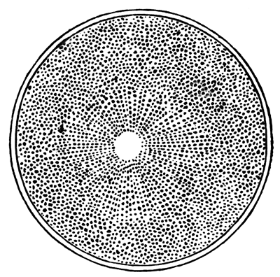
Fig. 17.—Ovum of Asterias
glacialis, after the coalescence of the male and female pronucleus (copied
from Fol).
Though there is a general agreement between the most recent observers,
Hertwig, Fol, Selenka, Strasburger, &c., as to the main facts connected with the entrance of
one spermatozoon into the egg, the formation of the male
pronucleus, and its fusion with the female pronucleus, there still exist
differences of detail in the different descriptions which partly, no doubt,
depend upon the difficulties of observation, but partly also upon the
observations not having all been made upon the same species. Hertwig does
not enter into details with reference to the actual entrance of the
spermatozoon into the egg, but in his latest paper points out that
considerable differences may be observed in occurrences which succeed
impregnation, according to the relative period at which this takes place.
When, in Asterias, the impregnation is effected about an hour after the egg
is laid and previously to the formation of the polar cells, the male
pronucleus appears at first to exert but little influence on the
protoplasm, but after the formation of the second polar cell, the radial
striæ around it become very marked, and the pronucleus rapidly grows in
size. When it finally unites with the female pronucleus it is equal in size
to the latter. In the case when the impregnation is deferred for four hours
the male pronucleus never becomes so large as the female pronucleus. With
reference to the effect of the time at which impregnation takes place,
Asterias would seem to serve as a type. Thus in Hirudinea,
Mollusca, and Nematodes impregnation normally takes place
before the formation of the polar bodies is completed, and the male
pronucleus is accordingly as large as the female. In Echinus, on the
other hand, where the polar bodies are formed in the ovary, the male
pronucleus is always small.
Selenka, who has investigated the formation of the male pronucleus in
Toxopneustes variegatus, differs in certain points from Fol. He
finds that usually, though not always, a single spermatozoon enters the
egg, and that though the entrance may be effected at any part of the
surface, it generally occurs at the point marked by a small prominence
where the polar cell was formed. The spermatozoon first makes its way
through the mucous envelope of the egg, within which it swims about, and
then bores with its head into the polar prominence. The head of the
spermatozoon on entering the egg becomes enveloped by the superficial
protoplasm, and travels inward with its envelope, while the tail remains
outside. As Fol has described, a delicate membrane becomes formed shortly
after the entrance of the spermatozoon. The head continues to make
its way by means of rapid oscillations, till it has traversed about one
eighth of the diameter of the egg, and then suddenly becomes still. The
tail in the meantime vanishes, while the neck swells up and forms the male
pronucleus. The junction of the male and female pronucleus is described by
Fol and Selenka in nearly the same manner.
Giard gives an account of impregnation which is not easily brought into
harmony with that of the other investigators. His observations were made on
Psammechinus miliaris. At one point is situated a polar body and
usually at the pole opposite to it a corresponding prominence. The
spermatozoa on gaining access to the egg attach themselves to it and give
it a rotatory movement, but according to Giard none of them penetrate the
vitelline membrane which, though formed at an earlier period, now retires
from the surface of the egg.
Giard believes that the prominence opposite the polar cells serves for
the entrance of the spermatic material, which probably passes in by a
process of diffusion. Thus, though he regards the male pronucleus as a
product of impregnation, he does not believe it to be the head of a
spermatozoon.
Both Hertwig and Fol have made observations on the result of the
entrance into the egg of several spermatozoa. Fol finds that when the
impregnation has been too long delayed the vitelline membrane is formed
with comparative slowness and several spermatozoa are thus enabled to
penetrate. Each spermatozoon forms a separate pronucleus with a surrounding
sun; and several male pronuclei usually fuse with the female pronucleus.
Each male pronucleus appears to exercise a repulsive influence on other
male pronuclei, but to be attracted by the female pronucleus. When there
are several male pronuclei the segmentation is irregular and the resulting
larva a monstrosity. These statements of Fol and Hertwig are at first sight
in contradiction with the more recent results of Selenka. In
Toxopneustes variegatus Selenka finds that though impregnation is
usually effected by a single spermatozoon yet that several may be concerned
in the act. The development continues, however, to be normal if three or
even four spermatozoa enter the egg almost simultaneously. Under such
circumstances each spermatozoon forms a separate pronucleus and sun.
It may be noticed that, while the observations of Fol and
Hertwig were admittedly made upon eggs in which the impregnation was
delayed till they no longer displayed their pristine activity, Selenka's
were made upon quite fresh eggs; and it seems not impossible that the
pathological symptoms in the embryos reared by the two former authors may
have been due to the imperfection of the egg and not to the entrance of
more than one spermatozoon. This, of course, is merely a suggestion which
requires to be tested by fresh observations. We have not as yet a
sufficient body of observations to enable us to decide whether impregnation
is usually effected by a single spermatozoon, though in spite of certain
conflicting evidence the balance would seem to incline towards the side of
a single spermatozoon[368].
The discovery of Hertwig as to the formation of the male pronucleus
throws a flood of light upon impregnation.
The act of impregnation is seen essentially to consist in the fusion of
a male and female nucleus; not only does this appear in the actual fusion
of the two pronuclei, but it is brought into still greater prominence by
the fact that the female pronucleus is a product of the nucleus of a
primitive ovum, and the male pronucleus is the metamorphosed head
of the spermatozoon which is itself developed from the nucleus of a
spermatic cell[369]. The spermatic cells originate
from cells (in the case of Vertebrates at least) identical with the
primitive ova, so that the fusion which takes place is the fusion of
morphologically similar parts in the two sexes.
It must not, however, be forgotten, as Strasburger has pointed out, that
part of the protoplasm of the generative cells of the two sexes also fuse,
viz. the tail of the spermatozoon with the
protoplasm of the egg. But there is no evidence that the former is of
importance for the act of impregnation. The fact that impregnation mainly
consists in the union of two nuclei gives an importance to the nucleus
which would probably not have been accorded to it on other grounds.
Hertwig's discovery is in no way opposed to Mr Darwin's theory
of pangenesis and other similar theories, but does not afford any definite
proof of their accuracy, nor does it in the meantime supply any explanation
of the origin of two sexes or of the reasons for an embryo becoming male or
female.
Summary.
In what may probably be regarded as a normal case the following series
of events accompanies the maturation and impregnation of an egg:—
(1) Transportation of the germinal vesicle to the surface of the
egg.
(2) Absorption of the membrane of the germinal vesicle and metamorphosis
of the germinal spot.
(3) Assumption of a spindle character by the remains of germinal
vesicle, these remains being probably largely formed from the germinal
spot.
(4) Entrance of one end of the spindle into a protoplasmic prominence at
the surface of the egg.
(5) Division of the spindle into two halves, one remaining in the egg,
the other in the prominence. The prominence becomes at the same time nearly
constricted off from the egg as a polar cell.
(6) Formation of a second polar cell in same manner as first, part of
the spindle still remaining in the egg.
(7) Conversion of the part of the spindle remaining in the egg after the
formation of the second polar cell into a nucleus—the female
pronucleus.
(8) Transportation of the female pronucleus towards the centre of the
egg.
(9) Entrance of one spermatozoon into the egg.
(10) Conversion of the head of the spermatozoon into a nucleus—the
male pronucleus.
(11) Appearance of radial striæ round the male pronucleus which
gradually travels towards the female pronucleus.
(12) Fusion of male and female pronuclei to form the first segmentation
nucleus.
List of important recent Publications on
the Maturation and Impregnation of the Ovum.
1. Auerbach. Organologische
Studien, Heft 2.
2. Bambeke. Recherches s. Embryologie des Batraciens.
Bull. de
l'Acad. royale de Belgique, 2me sér., t. LXI. 1876.
3. E. Van Beneden. La Maturation de l'Œuf des Mammifères.
Bull. de
l'Acad. royale de Belgique, 2me sér., t. XL, no. 12, 1875.
4. E. Van Beneden. Contributions à l'Histoire de la Vésicule Germinative, &c.
Bull. de l'Acad. royale de
Belgique, 2me sér., t. XLI,
no. 1, 1876.
5. Bütschli. Eizelle, Zelltheilung, und Conjugation der Infusorien.
6. Flemming. Studien in d. Entwicklungsgeschichte der Najaden.
Sitz. d. k. Akad. Wien, B. LXXI. 1875.
7. Fol. Die
erste Entwicklung des Geryonideneies.
Jenaische
Zeitschrift, Vol. VII.
8. Fol. Sur
le Développement des Pteropodes.
Archives de Zoologie Expérimentale
et Générale, Vols. IV and V.
9. Fol. Sur
le Commencement de l'Hénogénie.
Archives des Sciences Physiques
et Naturelles. Genève, 1877.
10. Giard. Note
sur les premiers phénomènes du développement de l'Oursin. 1877.
11. Hertwig, Oscar. Beit. z. Kenntniss d. Bildung, &c., d. thier. Eies.
Morphologisches
Jahrbuch, Bd.I.
12. Hertwig, Oscar. Ibid. Morphologisches Jahrbuch, Bd.III, Heft. 1.
13. Hertwig, Oscar. Weitere Beiträge, &c.
Morphologisches Jahrbuch, Bd. III, Heft 3.
14. Kleinenberg.
Hydra. Leipzig, 1872.
15. Oellacher, J. Beiträge zur Geschichte des Keimbläschens im Wirbelthiereie.
Archiv f. micr. Anat., Bd.VIII.
16. Selenka. Befruchtung u. Theilung des Eies von Toxopneustes
variegatus (Vorläufige Mittheilung). Erlangen, 1877.
17. Strasburger. Ueber Zellbildung u. Zelltheilung. Jena, 1876.
18. Strasburger. Ueber Befruchtung u. Zelltheilung. Jena, 1878.
19. R. Greeff. Ueb. d. Bau u. d. Entwicklung d. Echinodermen.
Sitzun. der Gesellschaft z. Beförderung d. gesammten Naturwiss. z.
Marburg, No. 5. 1876.
Postscript.—Two important
memoirs have appeared since this paper was in type. One of these by
Hertwig, Morphologisches Jahrbuch, Bd. IV, contains a full account with illustrations of
what was briefly narrated in his previous paper (13); the other by
Calberla, Der Befruchtungsvorgang beim Ei von Petromyzon
Planeri,
Zeit. für wiss. Zool., Bd. XXX, shews that the superficial layer of the egg is
formed by a coating of protoplasm free from yolk-spheres, which at one part
is continued inwards as a column, and contains the germinal vesicle. The
surface of this column is in contact with a micropyle in the egg-membrane.
Impregnation is effected by the entrance of the head of a single
spermatozoon (the tail remaining outside) through the micropyle, and then
along the column of clear protoplasm to the female pronucleus.
(With Plates 24, 25, 26.)
The present paper records observations on the ovaries of but
two types, viz., Mammalia and Elasmobranchii.
The main points dealt with are three:—1. The relation of the germinal
epithelium to the stroma. 2. The connection between primitive ova
in Waldeyer's sense and the permanent ova. 3. The homologies of the egg
membranes.
The second of these points seems to call for special attention after
Semper's discovery that the primitive ova ought really to be regarded as
primitive sexual cells, in that they give rise to the generative
elements of both sexes.
The Development of the Elasmobranch
Ovary.
The development of the Elasmobranch ovary has recently formed the
subject of three investigations. The earliest of them, by H. Ludwig, is
contained in his important work, on the 'Formation of the Ovum in the
Animal Kingdom[371].' Ludwig arrives at the
conclusion that the ovum and the follicular epithelium are both derived
from the germinal epithelium, and enters into some detail as to their
formation. Schultz[372], without apparently being
acquainted with Ludwig's observations, has come to very similar results for
Torpedo.
Semper[373], in his elaborate memoir on the
urogenital system of Elasmobranchii, has added very greatly to our knowledge
on this subject. In a general way he confirms Ludwig's statements, though
he shews that the formation of the ova is somewhat more complicated than
Ludwig had imagined. He more especially lays stress on the existence of
nests of ova (Ureiernester[TN11]),
derived from the division of a single primitive ovum, and of certain
peculiarly modified nuclei, which he compares to spindle nuclei in the act
of division.
My own results agree with those of previous investigators, in
attributing to the germinal epithelium the origin both of the follicular
epithelium and ova, but include a number of points which I believe to be
new, and, perhaps, of some little interest; they differ, moreover, in many
important particulars, both as to the structure and development of the
ovary, from the accounts of my predecessors.
The history of the female generative organs may conveniently be treated
under two heads, viz. (1) the history of the
ovarian ridge itself, and (2) the history of the ova situated in it. I
propose dealing in the first place with the ovarian ridge.
The Ovarian ridge in Scyllium.—At the stage spoken of in my
monograph on Elasmobranch Fishes as stage L, the ovarian ridge has a very
small development, and its maximum height is about 0.1 mm. It exhibits in section a somewhat rounded form, and
is slightly constricted along the line of attachment. It presents two
surfaces, which are respectively outer and inner, and is formed of a layer
of somewhat thickened germinal epithelium separated by a basement membrane
from a central core of stroma. The epithelium is far thicker on the outer
surface than on the inner, and the primitive ova are entirely confined to
the former. The cells of the germinal epithelium are irregularly scattered
around the primitive ova, and have not the definite arrangement usually
characteristic of epithelial cells. Each of them has a large nucleus, with
a deeply staining small nucleolus, and a very scanty protoplasm. In stage N
the ovarian ridge has a pointed edge and narrower attachment than in stage
L. Its greatest height is about 0.17 mm.
There is more stroma, and the basement membrane is more distinct than
before; in other respects no changes worth recording have
taken place. By stage P a distinction is observable between the right and
left ovarian ridges; the right one has, in fact, grown more rapidly than
the left, and the difference in size between the two ridges becomes more
and more conspicuous during the succeeding stages, till the left one ceases
to grow any larger, though it remains for a great part of life as a small
rudiment.
The right ovarian ridge, which will henceforth alone engage our
attention, has grown very considerably. Its height is now about 0.4 mm. It has in section (vide Pl. 24, fig. 1) a triangular
form with constricted base, and is covered by a flat epithelium, except for
an area on the outer surface, in length co-extensive with the ovarian
ridge, and with a maximum breadth of about 0.25 mm. This area will be spoken of as the ovarian area or
region, since the primitive ova are confined to it. The epithelium covering
it has a maximum thickness of about 0.05 mm., and thins off rather rapidly on both borders, to
become continuous with the general epithelium of the ovarian ridge. Its
cells have the same character as before, and are several layers deep.
Scattered irregularly amongst them are the primitive ova. The germinal
epithelium in the ovarian region is separated by a basement membrane from
the adjacent stroma.
In succeeding stages, till the embryo reaches a length of 7 centimetres,
no very important changes take place. The ovarian region grows somewhat in
breadth, though in this respect different embryos vary considerably. In two
embryos of nearly the same age, the breadth of the ovarian epithelium was
0.3 mm. in the one and 0.35 mm. in the other. In the former of these embryos, the
thickness of the epithelium was slightly greater than in the latter, viz. 0.09 mm. as
compared with 0.08. In both the epithelium was sharply separated from the
subjacent stroma. There were relatively more epithelial cells in proportion
to primitive ova than at the earlier date, and the individual cells
exhibited great variations in shape, some being oval, some angular, others
very elongated, and many of them applied to part of an ovum and
accommodating themselves to its shape. In some of the more elongated cells
very deeply stained nuclei were present, which (in a favourable light and
with high powers) exhibited the spindle modification of Strasburger with
great clearness, and must therefore be regarded as undergoing
division. The ovarian region is at this stage bounded on each side by a
groove.
In an embryo of seven centimetres (Pl. 24,
fig. 2) the breadth of the ovarian epithelium was 0.5, but its height only
0.06 mm. It was still sharply separated
from the subjacent stroma, though a membrane could only be demonstrated in
certain parts. The amount of stroma in the ovarian ridge varies greatly in
different individuals, and no reliance can be placed on its amount as a
test of the age of the embryo. In the base of the ovarian ridge the cells
were closely packed, elsewhere they were still embryonic.
My next stage (Pl. 24, fig. 3, and fig. 4),
shortly before the time of the hatching of the embryo, exhibits in many
respects an advance on the previous one. It is the stage during which a
follicular covering derived from the germinal epithelium is first
distinctly formed round the ova, in a manner which will be more
particularly spoken of in the section devoted to the development of the
ovum itself. The breadth of the ovarian region is 0.56 mm., and its greatest height close to the central
border, 0.12 mm.—a great advance on the previous stage, mainly,
however, due to the larger size of the ova.
The ovarian epithelium is still in part separated from the subjacent
stroma by a membrane close to its dorsal and ventral borders, but elsewhere
the separation is not so distinct, it being occasionally difficult within a
cell or so to be sure of the boundary of the epithelium. The want of a
clear line between the stroma and the epithelium is rendered more obvious
by the fact that the surface of the latter is somewhat irregular, owing to
projections formed by specially large ova, into the bays between which are
processes of the stroma. In an ovary about this stage, hardened in osmic
acid, the epithelium stains very differently from the subjacent stroma, and
the line of separation between the two is quite sharp. A figure of the
whole ovarian ridge, shewing the relation between the two parts, is
represented on Pl. 24, fig. 5.
The layer of stroma in immediate contact with the epithelium is very
different from the remainder, and appears to be destined to accompany the
vascular growths into the epithelium, which will appear in the
next stage. The protoplasm of the cells composing it forms a loose
reticulum with a fair number of oval or rounded nuclei, with their long
axis for the most part parallel to the lower surface of the epithelium. It
contains, even at this stage, fully developed vascular channels.
The remainder of the stroma of the ovarian ridge has now acquired a
definite structure, which remains constant through life, and is eminently
characteristic of the genital ridge of both sexes. The bulk of it (Pl. 24, fig. 3, str) consists of closely packed polygonal cells, of
about 0.014 mm. with large nuclei of about
0.009. These cells appear to be supported by a delicate reticulum. The
whole tissue is highly vascular, with the numerous capillaries; the nuclei
in the walls of which stand out in some preparations with great
clearness.
In the next oldest ovary, of which I have sections, the breadth of the
ovarian epithelium is 0.7 mm. and its
thickness 0.096. The ovary of this age was preserved in osmic acid, which
is the most favourable reagent, so far as I have seen, for observing the
relation of the stroma and epithelium. On Pl.
24, fig. 6, is represented a transverse section through the whole breadth
of the ovary, slightly magnified to shew the general relations of the
parts, and on Pl. 24, fig. 7, a small portion
of a section more highly magnified. The inner surface of the ovarian
epithelium is more irregular than in the previous stage, and it may be
observed that the subjacent stroma is growing in amongst the ova. From the
relation of the two tissues it is fairly clear that the growth which is
taking place is a definite growth of the stroma into the epithelium, and
not a mutual intergrowth of the two tissues. The ingrowths of the stroma
are, moreover, directed towards individual ova, around which, outside the
follicular epithelium, they form a special vascular investment in the
succeeding stages. They are formed of a reticular tissue with comparatively
few nuclei.
By the next stage, in my series of ovaries of Scy. canicula, important changes have taken place in
the constitution of ovarian epithelium. Fig. 8, Pl. 24, represents a portion of the ovarian epithelium, on
the same scale as figs. 1, 2, 3, &c.,
and fig. 9 a section through the whole ovarian ridge slightly magnified.
Its breadth is now 1.3 mm., and its
thickness 0.3 mm. The ova have grown
very greatly, and it appears to me to be mainly owing to their growth that
the greater thickness of the epithelium is due, as well as the irregularity
of its inner surface (vide fig. 9).
The general relation of the epithelium to the surrounding parts is much
the same as in the earlier stage, but two new features have
appeared—(1) The outermost cells of the ovarian region have more or
less clearly arranged themselves as a kind of epithelial covering for the
organ; and (2) the stroma ingrowths of the previous stage have become
definitely vascular, and have penetrated through all parts of the
epithelium.
The external layer of epithelium is by no means a very marked structure,
the character of its cells varies greatly in different regions, and it is
very imperfectly separated from the subjacent layer. I shall speak of it
for convenience as pseudo-epithelium.
The greater part of the germinal epithelium forms anastomosing columns,
separated by very thin tracts of stroma. The columns are, in the majority
of instances, continuous with the pseudo-epithelium at the surface, and
contain ova in all stages of development. Many of the cells composing them
naturally form the follicular epithelium for the separate ova; but the
majority have no such relation. They have in many instances assumed an
appearance somewhat different from that which they presented in the last
stage, mainly owing to the individual nuclei being more widely separated. A
careful examination with a high power shews that this is owing to an
increase in the amount of protoplasm of the individual cells, and it may be
noted that a similar increase in the size of the bodies of the cells has
taken place in the pseudo-epithelium and in the follicular epithelium of
the individual ova.
The stroma ingrowths form the most important feature of the stage. In
most instances they are very thin and delicate, and might easily be
overlooked, especially as many of the cells in them are hardly to be
distinguished, taken separately, from those of the germinal epithelium.
These features render the investigation of the exact relation of the stroma
and epithelium a matter of some difficulty. I have, however, been greatly
assisted by the investigation of the ovary of a young example of
Scyllium stellare, 16½ centimètres in length, a section of
which is represented in Pl. 25, fig. 26. In
this ovary, although no other abnormalities were observable, the stroma
ingrowths were exceptionally wide; indeed, quite without a parallel in my
series of ovaries in this respect. The stroma most clearly divides up the
epithelium of the ovary into separate masses, or more probably anastomosing
columns, the equivalents of the egg-tubes of Pflüger. These columns are
formed of normal cells of the germinal epithelium, which enclose ovarian
nests and ova in all stages of development. A comparison of the section I
have represented, with those from previous stages, appears to me to
demonstrate that the relation of the epithelium and stroma has been caused
by an ingrowth or penetration of the stroma into the epithelium, and not by
a mutual intergrowth of the two tissues. Although the ovary, of which fig.
26 represents a section was from Scy.
stellare, and the previous ovaries have been from Scy. canicula, yet the thickness of the epithelium
may still be appealed to in confirmation of this view. In the previous
stage the thickness was about 0.096 mm.,
in the present one it is about 0.16 mm., a
difference of thickness which can be easily accounted for by the growth of
the individual ova and the additional tracts of stroma. A pseudo-epithelium
is more or less clearly formed, but it is continuous with the columns of
epithelium. In the stroma many isolated cells are present, which appear to
me, from a careful comparison of a series of sections, to belong to the
germinal epithelium.
The thickness of the follicular epithelium on the inner side of the
larger ova deserves to be noted. Its meaning is discussed on
p. 567.
Quite a different interpretation to that which I have given has been put
by Ludwig and Semper upon the parts of the ovary at this stage. My
pseudo-epithelium is regarded by them as forming, together with the
follicular epithelium of the ova, the sole remnant of the original
germinal epithelium; and the masses of cells below the pseudo-epithelium,
which I have attempted to shew are derived from the original germinal
epithelium, are regarded as parts of the ingrowths of the adjacent
stroma.
Ludwig has assumed this interpretation without having had an opportunity
of working out the development of the parts, but Semper attempts to
bring forward embryological proofs in support of this position.
If the series of ovaries which I have represented be examined, it will
not, I think, be denied that the general appearances are very much in
favour of my view. The thickened patch of ovarian epithelium can apparently
be traced through the whole series of sections, and no indications of its
sudden reduction to the thin pseudo-epithelium are apparent. The most
careful examination that I have been able to make brings to light nothing
tending to shew that the general appearances are delusive. The important
difference between us refers to our views of the nature of the tissue
subjacent to the pseudo-epithelium. If my results be accepted, it is
clear that the whole ovarian region is an epithelium interpenetrated by
connective tissue ingrowths, so that the region below the pseudo-epithelium
is a kind of honeycomb or trabecular net-work of germinal epithelium,
developing ova of all stages and sizes, and composed of cells capable of
forming follicular epithelium for developing ova. Ludwig figures what he
regards as the formation of the follicular epithelium round primitive ova
during their passage into the stroma. It is quite clear to me, that his
figures of the later stages, 33 and 34, represent fully formed permanent
ova surrounded by a follicular epithelium, and that their situation in
contact with the pseudo-epithelium is, so to speak, an accident, and it is
quite possible that his figures 31 and 32 also represent fully formed ova;
but I have little hesitation in asserting that he has not understood the
mode of formation of the follicular epithelium, and that, though his
statement that it is derived from the germinal epithelium is quite correct,
his account of the process is completely misleading. The same criticism
does not exactly apply to Semper's statements. Semper has really observed
the formation of the follicular epithelium round young ova; but,
nevertheless, he appears to me to give an entirely wrong account of the
relation of the stroma to the germinal epithelium. The extent of the
difference between Semper's and my view may perhaps best be shewn by a
quotation from Semper, loc. cit., 465:—In females
the nests of primitive ova sink in groups into the stroma. In these groups
one cell enlarges till it becomes the ovum, the neighbouring cells increase
and arrange themselves around the ova as follicle cells.
Although the histological changes which take place in the succeeding
stages are not inconsiderable, they do not involve any fundamental change
in the constitution of the ovarian region, and may be described with
greater brevity than has been so far possible.
In a half-grown female, with an ovarian region of 3mm. in breadth, and
0.8mm. in thickness, the stroma of the ovarian region has assumed a far
more formed aspect than before. It consists (Pl. 24, fig. 10) of a basis in most parts fibrous, but in
some nearly homogeneous, with a fair number of scattered cells. Immediately
below the pseudo-epithelium, there is an imperfectly developed fibrous
layer, forming a kind of tunic, in which are imbedded the relatively
reduced epithelial trabeculæ of the previous stages. They appear in
sections as columns, either continuous with or independent of the
pseudo-epithelium, formed of normal cells of the germinal epithelium, nests
of ova, and permanent ova in various stages of development. Below this
there comes a layer of larger ova which are very closely packed. A not
inconsiderable number of the larger ova have, however, a superficial
situation, and lie in immediate contact with the pseudo-epithelium. Some of
the younger ova, enclosed amongst epithelial cells continuous with the
pseudo-epithelium, are very similar to those figured by Ludwig. It is
scarcely necessary to insist that this fact does not afford any argument in
favour of his interpretations. The ovarian region is honeycombed by large
vascular channels with distinct walls, and other channels which are perhaps
lymphatic.
The surface of the ovarian region is somewhat irregular and especially
marked by deep oblique transverse furrows. It is covered by a distinct,
though still irregular pseudo-epithelium, which is fairly columnar in the
furrows but flattened along the ridges. The cells of the pseudo-epithelium
have one peculiarity very unlike that of ordinary epithelial cells. Their
inner extremities (vide fig. 10) are prolonged into
fibrous processes which enter the subjacent tissue, and bending nearly
parallel to the surface of the ovary, assist in forming the tunic spoken of
above. This peculiarity of the pseudo-epithelial cells seems to
indicate that they do not essentially differ from cells which have the
character of undoubted connective tissue cells, and renders it possible
that the greater part of the tunic, which has apparently the structure of
ordinary connective tissue, is in reality derived from the original
germinal epithelium, a view which tallies with the fact that in some
instances the cells of the tunic appear as if about to assist in forming
the follicular epithelium of some of the developing ova. In Raja, the
similarity of the pseudo-epithelium to the subjacent tissue is very much
more marked than in Scyllium. The pseudo-epithelium appears merely as the
superficial layer of the ovarian tunic somewhat modified by its position on
the surface. It is formed of columnar cells with vertically arranged fibres
which pass into the subjacent layers, and chiefly differ from the ordinary
fibres in that they still form parts of the cell-protoplasm enclosing the
nucleus. In Pl. 25, fig. 34, an attempt is made
to represent the relations of the pseudo-epithelium to the subjacent tissue
in Raja. Ludwig's figures of the pseudo-epithelium of the ovary, in the
regular form of its constituent cells, and its sharp separation by a
basement membrane from the tissue below, are quite unlike anything which I
have met with in my sections either of Raja or Scyllium.
Close to the dorsal border of the ovary the epithelial cells of the
non-ovarian region have very conspicuous tails, extending into a more or
less homogeneous substance below, which constitutes a peculiar form of
tunic for this part of the ovarian ridge.
In the full-grown female the stroma of the ovarian region is denser and
has a more fibrous aspect than in the younger animal. Below the
pseudo-epithelium it is arranged in two or three more or less definite
layers, in which the fibres run at right angles. It forms a definite
ovarian tunic. The pseudo-epithelium is much more distinct, and the tails
of its cells, so conspicuous in previous stages, can no longer be made
out.
Formation of the permanent ova and the follicular
epithelium.—In my monograph on the development of Elasmobranch
Fishes an account was given of the earliest stages in the development of
the primitive ova, and I now take up their development from the
point at which it was left off in that work. From their first formation
till the stage spoken of in my monograph as P, their size remains fairly
constant. The larger examples have a diameter of about 0.035 mm., and the medium-sized examples of about 0.03 mm. The larger nuclei have a diameter of about
0.16 mm., but their variations in size are
considerable. If the above figures be compared with those on page 350 of my
monograph on Elasmobranch Fishes, it will be seen that the size of the
primitive ova during these stages is not greater than it was at the period
of their very first appearance.
The ova (Pl. 24, fig. 1) are usually
aggregated in masses, which might have resulted from division of a single
ovum. The outlines of the individual ova are always distinct.
Their protoplasm is clear, and their nuclei, which are somewhat passive
towards staining reagents, are granular, with one to three nucleoli. I have
noticed, up to stage P, the occasional presence of highly refractive
spherules in the protoplasm of the primitive ova already described in my
monograph (pp. 353, 354, Pl. 12, fig. 15). They seem to occur up to a later period
than I at first imagined. Their want of constancy probably indicates that
they have no special importance. Professor Semper has described similar
appearances in the male primitive ova of a later period.
As to the distribution of the primitive ova in the germinal epithelium,
Professor Semper's statement that the larger primitive ova are found in
masses in the centre, and that the smaller ova are more peripherally
situated is on the whole true, though I do not find this distribution
sufficiently constant to lay so much stress on it as he does.
The passive condition of the primitive ova becomes suddenly broken
during stage Q, and is succeeded by a period of remarkable changes. It has
only been by the expenditure of much care and trouble that I have been able
to elucidate to my own satisfaction what takes place, and there are still
points which I do not understand.
Very shortly after stage Q, in addition to primitive ova with a
perfectly normal nucleus, others may be seen in which the nucleus is
apparently replaced by a deeply stained irregular body, smaller than the
ordinary nuclei (Pl. 24, fig. 11, d.n.). This body, by the use of high objectives,
is seen to be composed of a number of deeply stained granules, and around
it may be noticed a clear space, bounded by a very delicate membrane. The
granular body usually lies close to one side of this membrane, and
occasionally sends a few fine processes to the opposite side.
The whole body, i.e. all within the delicate membrane is,
according to my view, a modified nucleus; as appears to me very clearly to
be shewn by the fact that it occupies the normal position of a nucleus
within a cell body. Semper, on the other hand, regards the contained
granular body as the nucleus, which he compares with the spindles of
Bütschli, Auerbach, &c.[374]. This interpretation appears to me, however, to be
negatived by the position of these bodies. The manner in which Semper may,
perhaps, have been led to his views will be obvious when the later changes
of the primitive ova are described. The formation of these nuclei would
seem to be due to a segregation of the constituents of the original nuclei;
the solid parts becoming separated from the more fluid. As a rule, the
modified nuclei are slightly larger than the original ones. In stage Q the
following two tables shew the dimensions of the parts of three unmodified
and of three modified nuclei taken at random.
Primitive ova with unmodified nuclei—
Nuclei.
0.014 mm.
0.012 mm.
0.01 mm.
Primitive ova with modified nuclei—
| Nuclei. |
Granular
Bodies in nuclei. |
|---|
| 0.018 mm. |
0.006 mm. |
| 0.018 mm. |
0.006 mm. |
| 0.012 mm. |
0.009 mm. |
For a slightly older stage than Q, the two annexed tables also shew the
comparative size of the modified and unmodified nuclei:
Unmodified nuclei of normal primitive
ova—
0.014 mm.
0.016 mm.
0.014 mm.
0.016 mm.
0.016 mm.
Nuclei of primitive ova with modified
nuclei—
| Nuclei. |
Granular
Bodies in nuclei. |
|---|
| 0.018 mm. |
0.008 mm. |
| 0.016 mm. |
0.008 mm. |
| 0.016 mm. |
0.010 mm. |
| 0.016 mm. |
| 0.018 mm. |
These figures bring out with clearness the following points:
(1) that the modified nuclei are slightly but decidedly larger on the
average than the unmodified nuclei; (2) that the contained granular bodies
are very considerably smaller than ordinary nuclei.
Soon after the appearance of the modified nuclei, remarkable changes
take place in the cells containing them. Up to the time such nuclei first
make their appearance the outlines of the individual ova are very clearly
defined, but subsequently, although numerous ova with but slightly modified
nuclei are still to be seen, yet on the whole the outlines of all the
primitive ova are much less distinct than before; and this is especially
the case with the primitive ova containing modified nuclei.
From cases in which three or four ova are found in a mass with modified
nuclei, but in which the outline of each ovum is fairly distinct, it is
possible to pass by insensible gradations to other cases in which two or
three or more modified nuclei are found embedded in a mass of protoplasm in
which no division into separate cells can be made out (fig. 14). For these
masses I propose to employ the term nests. They correspond in part with the
Ureiernester of Professor Semper.
Frequently they are found in hardened specimens to be enclosed in a
membrane-like tunic which appears to be of the nature of coagulated fluid.
These membranes closely resemble and sometimes are even continuous with
trabeculæ which traverse the germinal epithelium. Ovaries differ
considerably as to the time and completeness of the
disappearance of the outlines marking the separate cells, and although, so
far as can be gathered from my specimens, the rule is that the outlines of
the primitive ova with modified nuclei soon become indistinct, yet in one
of my best preserved ovaries very large nests with modified nuclei are
present in which the outline of each ovum is as distinct as during the
period before the nuclei undergo these peculiar changes (Pl. 24, fig. 12). In the same ovary other nests are present
in which the outlines of the individual ova are no longer visible. The
section represented on Pl. 24, fig. 2, is
fairly average as to the disappearance of the outlines of the individual
ova.
It is clear from the above statements, that in the first instance the
nests are produced by the coalescence of several primitive ova into a
single mass or syncytium; though of course, the several separate ova of a
nest may originally, as Semper believes, have arisen from the division of a
single ovum. In any case there can be no doubt that the nests of separate
ova increase in size as development proceeds; a phenomenon which is more
reasonably explained on the view that the ova divide, than on the view that
they continue to be freshly formed. The same holds true for the nests of
nuclei and this, as well as other facts, appears to me to render it
probable that the nests grow by division of the nuclei without
corresponding division of the protoplasmic matrix. I cannot, however,
definitely prove this point owing to my having found nests, with distinct
outlines to the ova, as large as any without such outlines.
The nests are situated for the most part near the surface of the
germinal epithelium. The smaller ones are frequently spherical, but the
larger are irregular in form. The former are about 0.05 mm. in diameter; the latter reach 0.1 mm. Scattered generally, and especially in the deeper
layers, and at the edges of the germinal epithelium, are still unmodified
or only slightly modified primitive ova. These unmodified primitive ova are
aggregated in masses, but in these masses the outlines of each ovum, though
perhaps less clear than in the earlier period, are still distinct.
When the embryo reaches a length of seven centimètres, and even in still
younger embryos, further changes are observable. In the first place
many of the modified nuclei acquire fresh characters, and it becomes
necessary to divide the modified nuclei into two categories. In both of
these the outer boundary of the nucleus is formed by a very delicate
membrane, the space within which is perfectly clear except for the granular
body. In the variety which now appears in considerable numbers the granular
body has an irregular star-like form. The rays of the star are formed of
fibres frequently knobbed at their extremities, and the centre of the star
usually occupies an eccentric position. Typical examples of this form of
modified nucleus, which may be spoken of as the stellate variety, are
represented on Pl. 25, fig. 17; between it and
the older granular variety there is an infinite series of gradations, many
of which are represented on Pl. 24, figs. 12,
14, 15, 16. Certain of the stellate nuclei exhibit two centres instead of
one, and in some cases, like that represented on Pl. 25, fig. 19, the stellate body of two nuclei is found
united. Both of these forms are possibly modifications of the spindle-like
form assumed by nuclei in the act of dividing, and may be used in proving
that the nests increase in size by the division of the contained nuclei. In
addition to the normal primitive ova, a few of which are still present,
there are to be found, chiefly in the deeper layers of the germinal
epithelium, larger ova differing considerably from the primitive ova. They
form the permanent ova (Pl. 24, fig. 3,
o). Their average diameter is 0.04 mm., compared with 0.03 mm., the diameter of original primitive ova. The
protoplasm of which they are composed is granular, but at first a membrane
can hardly be distinguished around them; their nucleus is relatively large,
0.02 - 0.027 mm. in diameter. It presents
the characters ascribed by Eimer[375],
and many other recent authors[376], to
typical nuclei (vide Pl.
24, fig. 3, and Pl. 24, 25, figs. 13, 14, 15,
16, 17, 18). It is bounded by a distinct membrane, within which is a more
or less central nucleolus from which a number of radial fibres which stain
very deeply pass to the surface; here they form immediately internal to the
membrane a network with granules at the nodal points. In some instances the
regularity of the arrangement of these fibres is very great, in other
instances two central nucleoli are present, in which case the regularity
is considerably interfered with. The points in which the youngest permanent
ova differ from the primitive may be summed up as follows:—
(1) The permanent ova are larger, the smallest of them being larger than
the average primitive ova in the proportion of four to three. (2) They have
less protoplasm as compared to the size of the nucleus. (3) Their
protoplasm is granular instead of being clear. (4) Their nucleus is clear
with exception of a network of fibres instead of being granular as in the
primitive ova. It thus appears that the primitive ova and permanent ova are
very different in constitution, though genetically related in a way to be
directly narrated.
The formation of permanent ova is at its height in embryos of about
seven centimètres or slightly larger. The nests at this stage are for the
most part of a very considerable size and contain a large number of nuclei,
which have probably, as before insisted, originated from a division of the
smaller number of nuclei present in the nests at an earlier stage. Figs.
14-18 are representations of nests at this period. The diameter of the
nuclei is, on the whole, slightly greater than at an earlier stage. A
series of measurements gave the following results:—
0.016 mm.
0.016 mm.
0.018 mm.
0.020 mm.
0.020 mm.
Both varieties of modified nuclei are common enough, though the stellate
variety predominates. The nuclei are sometimes in very close contact, and
sometimes separated by protoplasm, which in many instances is very slightly
granular. In a large number of the nests nothing further is apparent than
what has just been described, but in a very considerable number one or more
nuclei are present, which exhibit a transitional character between the
ordinary stellate nuclei of my second category, and the nuclei of permanent
ova as above described; and in these nests the formation of permanent ova
is taking place. Permanent ova in the act of development are indicated in
my figures by the letters do. Many of
the intermediate nuclei are more definitely surrounded
by granular protoplasm than the other nuclei of the nests, and accordingly
have their outlines more sharply defined. Between nuclei of this kind, and
others as large as those of the permanent ova, there are numerous
transitional forms. The larger ones frequently lie in a mass of granular
protoplasm projecting from the nest, and only united with it by a neck
(Pl. 24, figs. 14 and 16). For prominences of
this kind to become independent ova, it is only necessary for the neck to
become broken through. Nests in which such changes are taking place present
various characters. In some cases several nuclei belonging to a nest appear
to be undergoing conversion into permanent ova at the same time. Such a
case is figured on Pl. 25, figs. 17 and 18. In
these cases the amount of granular protoplasm in the nest and around each
freshly formed ovum is small. In the more usual cases only one or two
permanent ova at the utmost are formed at the same time, and in these
instances a considerable amount of granular protoplasm is present around
the nucleus of the developing permanent ovum. In such instances it
frequently happens several of the nuclei not undergoing conversion appear
to be in the process of absorption, and give to the part of the nest in
which they are contained a very hazy and indistinct aspect (Pl. 24, fig. 15). Their appearance leads me to adopt the
view that while some of the nuclei of each nest are converted into the
nuclei of the permanent ova, others break down and are used as the pabulum,
at the expense of which the protoplasm of the young ovum grows.
It should, however, be stated, that after the outlines of the permanent
ova have become definitely established, I have only observed in a single
instance the inclusion of a nucleus within an ovum (Pl. 25, fig. 24). In many instances normal nuclei of the
germinal epithelium may be so observed within the ovum.
The nuclei which are becoming converted into the nuclei of permanent ova
gradually increase in size. The following table gives the diameter of four
such nuclei:—
0.022 mm.
0.022 mm.
0.024 mm.
0.032 mm.
These figures should be compared with those of the table on
page 564.
The ova when first formed are situated either at the surface or in the
deeper layers of the germinal epithelium. Though to a great extent
surrounded by the ordinary cells of the germinal epithelium, they are not
at first enclosed in a definite follicular epithelium. The follicle is,
however, very early formed.
My observations lead me then to the conclusion that in a general way the
permanent ova are formed by the increase of protoplasm round some of the
nuclei of a nest, and the subsequent separation of the nuclei with their
protoplasm from the nest as distinct cells—a mode of formation
exactly comparable with that which so often takes place in invertebrate egg
tubes.
Besides the mode of formation of permanent ova just described, a second
one also seems probably to occur. In ovaries just younger than those in
which permanent ova are distinctly formed, there are present primitive ova,
with modified nuclei of the stellate variety, or nuclei sometimes even
approaching in character those of permanent ova, which are quite isolated
and not enclosed in a definite nest. The body of these ova is formed of
granular protoplasm, but their outlines are very indistinct. Such ova are
considerably larger than the normal primitive ova. They may measure 0.04
mm. In a slightly later stage, when fully
formed permanent ova are present, isolated ones are not infrequent, and it
seems natural to conclude that these isolated ova are the direct
descendants of the primitive ova of the earlier stage. It seems a fair
deduction that in some cases primitive ova undergo a direct metamorphosis
into permanent ova by a modification of their nucleus, and the assumption
of a granular character in their protoplasm, without ever forming the
constituent part of a nest.
It is not quite clear to me that in all nests the coalescence of the
protoplasm of the ova necessarily takes place, since some nests are to be
found at all stages in which the ova are distinct. Nevertheless, I am
inclined to believe that the fusion of the ova is the normal
occurrence.
The mode of formation of the permanent ova may then, according to my
observations, take place in two ways:—1. By the formation of granular
protoplasm round the nucleus in a nest, and the
separation of the nucleus with its protoplasm as a distinct ovum. 2. By the
direct metamorphosis of an isolated primitive ovum into a permanent ovum.
The difference between these two modes of formation does not, from a
morphological point of view, appear to be of great importance.
The above results appear clearly to shew that the primitive ova in
the female are not to be regarded as true ova, but as the parent sexual
cells which give rise to the ova: a conclusion which completely fits in
with the fact that cells exactly similar to the primitive ova in the female
give rise to the spermatic cells in the male.
Slightly after the period of their first formation the permanent ova
become invested by a very distinct and well-marked, somewhat flattened,
follicular epithelium (Pl. 24, fig. 3). Where
the ova lie in the deeper layers of the germinal epithelium, the follicular
epithelium soon becomes far more columnar on the side turned inwards, than
on that towards the surface, especially when the inner side is in contact
with the stroma (Pl. 24, fig. 7, and Pl. 25, figs. 24 and 26). This is probably a special
provision for the growth and nutrition of the ovum.
There cannot be the smallest doubt that the follicular epithelium is
derived from the general cells of the germinal epithelium—a point on
which my results fully bear out the conclusions of Ludwig and Semper.
The larger ova themselves have a diameter of about 0.06 mm., and their nucleus of about 0.04 mm. The vitellus is granular, and provided with a
distinct, though delicate membrane, which has every appearance of being a
product of the ovum itself rather than of the follicular epithelium. The
membrane would seem indeed to be formed in some instances even before the
ovum has a definite investment of follicle cells. The vitellus is
frequently vacuolated, but occasionally the vacuoles appear to be caused by
a shrinking due to the hardening reagent. The nucleus has the same peculiar
reticulate character as at first. Its large size, as compared with the
ovum, is very noticeable.
With this stage the embryonic development of the ova comes to a close,
though the formation of fresh ova continues till comparatively late in
life. I have, however, two series of sections of ovaries preserved in osmic
acid, from slightly larger embryos than the one last
described, about which it may be well to say a few words before proceeding
to the further development of the permanent ova.
The younger of these ovaries was from a Scyllium embryo 10 centimètres
long, preserved in osmic acid.
A considerable number of nests were present (Pl. 24, fig. 13), exhibiting, on the whole, similar
characters to those just described.
A series of measurements of the nuclei in them were made, leading to the
following results:—
0.014 mm.
0.014 mm.
0.016 mm.
0.016 mm.
0.018 mm.
0.018 mm.
Thus, if anything, the nuclei were slightly smaller than in the younger
embryo. It is very difficult in the osmic specimens to make out clearly the
exact outlines of the various structures, the nuclei in many instances
being hardly more deeply stained than in the protoplasm around them. The
network in the nuclei is also far less obvious than after treatment with
picric acid. The permanent ova were hardly so numerous as in the younger
ovary before described. A number of these were measured with the following
results:—
| Ovum. | Nucleus. |
|---|
| 0.03 mm. |
0.014 mm. |
| 0.034 mm. |
0.018 mm. |
| 0.028 mm. |
0.016 mm. |
| 0.03 mm. |
0.02 mm. |
| 0.04 mm. |
0.02 mm. |
| 0.04 mm. |
0.02 mm. |
| 0.048 mm. |
0.02 mm. |
These figures shew that the nuclei of the permanent ova are smaller than
in the younger embryo, and it may therefore be safely concluded that, in
spite of the greater size of the embryo from which it is taken, the ovary
now being described is in a more embryonic condition than the one last
dealt with.
Though the permanent ova appeared to be formed from the nests in the
manner already described, it was fairly clear from the sections of
this ovary that many of the original primitive ova, after a metamorphosis
of the nucleus and without coalescing with other primitive ova to form
nests, become converted directly into the permanent ova. Many large masses
of primitive ova, or at least of ova with the individual outlines of each
ovum distinct, were present. The average size of ova composing these was
however small, the body measuring about 0.016 mm., and the nucleus 0.012 mm. Isolated ova with metamorphosed nuclei could also
be found measuring 0.022, and their nuclei about 0.014 mm.
The second of the two ovaries, hardened in osmic acid, was somewhat more
advanced than the ovary in which the formation of permanent ova was at its
height. Fewer permanent ova were in the act of being formed, and many of
these present had reached a considerable size, measuring as much as 0.07
mm. Nests of the typical forms were
present as before, but the nuclei in them were more granular than at the
earlier period, and on the average slightly smaller. A series measured had
the following diameters:—
0.010 mm.
0.012 mm.
0.014 mm.
0.016 mm.
One of these nests is represented on Pl. 25,
fig. 20. Many nests with the outlines of the individual ova distinct were
also present.
On the whole it appeared to me, that the second mode of formation of
permanent ova, viz. that in which the nest
does not come into the cycle of development, preponderated to a greater
extent than in the earlier embryonic period.
Post-embryonic Development of the
Ova.—My investigations upon the post-embryonic growth and
development of the ova, have for the most part been conducted upon
preserved ova, and it has been impossible for me, on this account, to work
out, as completely as I should have wished, certain points, more especially
those connected with the development of the yolk.
Although my ovaries have been carefully preserved in a large number of
reagents, including osmic acid, picric acid, chromic acid, spirit,
bichromate of potash, and Müller's fluid, none of these have proved
universally successful, and bichromate of potash and Müller's fluid
are useless. Great difficulties have been experienced in distinguishing the
artificial products of these reagents. My investigations have led me to the
result, that in the gradual growth of the ova with the age of the
individual the changes are not quite identical with those during the rapid
growth which takes place at periods of sexual activity, after the adult
condition has been reached—a result to which His has also arrived,
with reference to the ova of Osseous Fish. I propose dealing separately
with the several constituents of the egg-follicle.
Egg membranes.—A vitelline membrane has been
described by Leydig[377] in Raja, and an albuminous
layer of the nature of a chorion[378] by
Gegenbaur[379] in Acanthias—the
membranes described in these two ways being no doubt equivalent.
Dr Alex. Schultz[380] has more recently investigated
a considerable variety of genera and finds three conditions of the egg
membranes. (1) In Torpedo, a homogeneous membrane, which is of the nature
of a chorion. (2) In Raja, a homogeneous membrane which is, however,
perforated. (3) In Squalidæ, a thick homogeneous membrane, internal to
which is a thinner perforated membrane. He apparently regards the
perforated inner membrane as a specialised part of the simple membrane
found in Torpedo, and states that this membrane is of the nature of a
chorion.
My own investigations have led me to the conclusion that though the
egg-membranes can probably be reduced to single type for Elasmobranchii, yet
that they vary with the stage of development of the ovum. Scyllium
(stellare and canicula) and Raja have formed the objects of my
investigation. I commence with the two former.
It has already been stated that in Scyllium, even before the follicular
epithelium becomes formed, a delicate membrane round the ovum can be
demonstrated, which appears to me to be derived from the vitellus or body
of the ovum, and is therefore of the nature of a vitelline membrane. It
becomes the vitelline membrane of Leydig, the albuminous membrane of
Gegenbaur, and homogeneous membrane of Schultz.
In a young fish (not long hatched) with ova of not more than 0.12 mm., this membrane, though considerably thicker
than in the embryo, is not thick enough to be accurately measured. In ova
of 0.5 mm. from a young female (Pl. 25, fig. 21) the vitelline membrane has a
thickness of 0.002 mm. and is quite
homogeneous[381]. Internally to it may be
observed very faint indications of the differentiation of the outermost
layer of the vitellus into the perforated or radially striated membrane of
Schultz, which will be spoken of as zona radiata.
In an ovum of 1 mm. from the nearly
full grown though not sexually mature female, the zona radiata has
increased in thickness and definiteness, and may measure as much as 0.004
mm. It is always very sharply separated
from the vitelline membrane, but appears to be more or less continuous on
its inner border with the body of the ovum, at the expense of which it no
doubt grows in thickness.
In ova above 1 mm. in diameter, both
vitelline membrane and zona radiata, but especially the latter, increase in
thickness. The zona becomes marked off from the yolk, and its radial striæ
become easy to see even with comparatively low powers. In many specimens it
appears to be formed of a number of small columns, as described by
Gegenbaur and others. The stage of about the greatest development of both
the vitelline membrane and zona radiata is represented on Pl. 25, fig. 22.
At this time the vitelline membrane appears frequently to exhibit a
distinct stratification, dividing it into two or more successive layers. It
is not, however, acted on in the same manner by all reagents, and with
absolute alcohol appears at times longitudinally striated.
From this stage onwards, both vitelline membrane and zona gradually
atrophy, simultaneously with a series of remarkable changes which take
place in the follicular epithelium. The zona is the first to disappear, and
the vitelline membrane next becomes gradually thinner. Finally, when the
egg is nearly ripe, the follicular epithelium is separated from the yolk by
an immeasurably thin membrane—the remnant of the vitelline
membrane—only visible in the most favourable sections (Pl. 25, fig. 23, vt.).
When the egg becomes detached from the ovary even this membrane is no
longer to be seen.
Both the vitelline membrane and the zona radiata are found in Raja, but
in a much less developed condition than in Scyllium. The vitelline membrane
is for a long time the only membrane present, but is never very thick
(Pl. 25, fig. 31). The zona is not formed till
a relatively much later period than in Scyllium, and is always delicate and
difficult to see (Pl. 25, fig. 32). Both
membranes atrophy before the egg is quite ripe; and an apparently fluid
layer between the follicular epithelium and the vitellus, which coagulates
in hardened specimens, is probably the last remnant of the vitelline
membrane. It is, however, much thicker than the corresponding remnant in
Scyllium.
Though I find the same membranes in Scyllium as Alexander Schultz did in
other Squalidæ, my results do not agree with his as to Raja. Torpedo I have
not investigated.
It appears to me probable that the ova in all Elasmobranch Fishes have
at some period of their development the two membranes described at length
for Scyllium. Of these the inner one, or zona radiata, will probably be
admitted on all hands to be a product of the peripheral protoplasm of the
egg.
The outer one corresponds with the membrane usually regarded in other
Vertebrates as a chorion or product of the follicular epithelium, but, by
tracing it back to its first origin, I have been led to reject this view of
its nature.
The follicular epithelium.—The follicular epithelium in the
eggs of Raja and Acanthias has been described by Gegenbaur[382]. He finds it flat in young eggs, but in the larger
eggs of Acanthias more columnar, and with the cells wedged in so as to form
a double layer. These observations are confirmed by Ludwig[383].
Alexander Schultz[384] states that in Torpedo, the
eggs are at first enclosed in a simple epithelium, but that in follicles of
.008 mm. there appear between the original
large cells of the follicle (which he describes as granulosa cells and
derives from the germinal epithelium) a number of peculiar small cells. He
states that these are of the same nature as the general stroma cells of the
ovary, and believes that they originate in the stroma. When the eggs have
reached 0.1 - 0.15 mm., he finds that the
small and large cells have a very regular alternating arrangement.
Semper records but few observations on the follicular epithelium, but
describes in Raja the presence of a certain number of large cells amongst
smaller cells. He believes that they may develop into ova, and considers
them identical with the larger cells described by Schultz, whose
interpretations he does not, however, accept.
My own results accord to a great extent with those of Dr Schultz, as far
as the structure of the follicular epithelium is concerned, but I am at one
with Semper in rejecting Schultz's interpretations.
In Scyllium, as has already been mentioned, the follicular epithelium is
at first flat and formed of a single layer of uniform cells, each with a
considerable amount of clear protoplasm and a granular nucleus. It is
bounded externally by a delicate membrane—the membrana propria
folliculi of Waldeyer—and internally by the vitelline membrane. In
the ovaries of very young animals the cells of the follicular epithelium
are more columnar on the side towards the stroma than on the opposite side,
but this irregularity soon ceases to exist.
In many cases the nuclei of the cells of the follicular epithelium
exhibit a spindle modification, which shews that the growth of the
follicular epithelium takes place by the division of its cells. No changes
of importance are observable in the follicular epithelium till the egg has
reached a diameter of more than 1 mm.
It should here be stated that I have some doubts respecting the
completeness of the history of the epithelium recorded in the sequel.
Difficulties have been met with in completely elucidating the chronological
order of the occurrences, and it is possible that some points have escaped
my observation.
The first important change is the assumption of a palisade-like
character by the follicle cells, each cell becoming very narrow and
columnar and the nucleus oval (Pl. 25, fig.
28). In this condition the thickness of the epithelium is about 0.025 mm. The epithelium does not, however, become
uniformly thick over the whole ovum, but in the neighbourhood of the
germinal vesicle it is very flat and formed of granular cells with
indistinct outlines, rather like the hypodermis cells of many Annelida.
Coincidently with this change in the follicular epithelium the commencement
of the atrophy of the membranes of the ovum, described in the last section,
becomes apparent.
The original membrana propria folliculi is still present round the
follicular epithelium, but is closely associated with a fibrous layer with
elongated nuclei. Outside this there is now a layer of cells, very much
like an ordinary epithelial layer, which may possibly be formed of cells of
the true germinal epithelium (fig. 28, fe´). This layer, which will be spoken of as the
secondary follicle layer, might easily be mistaken for the follicular
epithelium, and it is possible that it has actually been so mistaken by
Eimer, Clark, and Klebs, in Reptilia, and that the true follicular
epithelium (in a flattened condition) has been then spoken of as the
Binnenepithel.
In slightly older eggs the epithelial cells are no longer uniform or
arranged as a single layer. The general arrangement of these cells is shewn
in Pl. 25, fig. 29. A considerable number of
them are more or less flask-shaped, with bulky protoplasm prolonged into a
thin stem directed towards the vitelline membrane, with which, in many
instances if not all, it comes in contact. These larger cells are arranged
in several tiers. Intercalated between them are a number of elongated small
cells with scanty protoplasm and a deeply staining nucleus, not very
dissimilar to, though somewhat smaller than, the columnar cells of the
previous stage. There is present a complete series of cells intermediate
between the larger cells and those with a deeply stained nucleus, and were
it not for the condition of the epithelium in Raja, to be spoken of
directly, I should not sharply divide the cells into two categories. In
surface views of the epithelium the division into two kinds of cells would
not be suspected. There can, it appears to me, be no question that both
varieties of cell are derived from the primitive uniform follicle
cells.
The fibrous layer bounding the membrana propria folliculi is
thicker than in the last stage, and the epithelial-like layer (fe´) which bounds it externally is more
conspicuous than before. Immediately adjoining it are vascular and lymph
sinuses. The thickness of the follicular epithelium at this stage may reach
as much as 0.04 mm., though I have found
it sometimes considerably flatter. The cells composing it are, however, so
delicate that it is not easy to feel certain that the peculiarities of any
individual ovum are not due to handling. The absence of the peculiar
columnar epithelium on the part of the surface adjoining the germinal
vesicle is as marked a feature as in the earlier stage. When the egg is
nearly ripe, and the vitelline membrane has been reduced to a mere remnant,
the follicular epithelium is still very columnar (Pl. 25, fig. 23). The thickness is greater than in the last
stage, being now about 0.045 mm., but the
cells appear only to form a single definite layer. From the character of
their nuclei, I feel inclined to regard them as belonging to the category
of the smaller cells of the previous stage, and feel confirmed in this view
by finding certain bodies in the epithelium, which have the appearance of
degenerating cells with granular nuclei, which I take to be the
flask-shaped cells which were present in the earlier stage.
I have not investigated the character of the follicular epithelium in
the perfectly ripe ovum ready to become detached from the ovary. Nor can I
state for the last-described stage anything about the character of the
follicular epithelium in the neighbourhood of the germinal vesicle.
As to the relation of the follicular epithelium to the vitelline
membrane, and the possible processes of its cells continued into the yolk,
I can say very little. I find in specimens teased out after treatment with
osmic acid, that the cells of the follicular epithelium are occasionally
provided with short processes, which might possibly have perforated the
vitelline membrane, but have met with nothing so clear as the teased out
specimens figured by Eimer. Nothing resembling the cells within the
vitelline membrane, as described by His[385] in
Osseous Fish, and Lindgren in Mammalia, has been met with[386].
My observations in Raja are not so full as those upon Scyllium,
but they serve to complete and reconcile the observations of Semper and
Schultz, and also to shew that the general mode of growth of the follicular
epithelium is fundamentally the same in my representatives of the two
divisions of the Elasmobranchii. In very young eggs, in conformity with the
results of all previous observers, I find the follicular epithelium
approximately uniform. The cells are flat, but extended so as to appear of
an unexpected size in views of the surface of the follicle. This condition
does not, however, last very long. A certain number of the cells enlarge
considerably, others remaining smaller and flat. The differences between
the larger and the smaller cells are more conspicuous in sections than in
surface views, and though the distribution of the cells is somewhat
irregular, it may still be predicted as an almost invariable rule that the
smaller cells of the follicle will line that part of the surface of the
ovum, near to which the germinal vesicle is situated. On Pl. 25, fig. 30, is shewn in section a fairly average
arrangement of the follicle cells. Semper considers the larger cells of
such a follicle to be probably primitive ova destined to become permanent
ova. This view I cannot accept: firstly, because these cells only agree
with primitive ova in being exceptionally large—the character of
their nucleus, with its large nucleolus, being not very like that of a
primitive ovum. Secondly, because they shade into ordinary cells of the
follicle; and thirdly, because no evidence of their becoming ova has come
before me, but rather the reverse, in that it seems probable that they have
a definite function connected with the nutrition of the egg. To this point
I shall return.
In the next stage the small cells have become still smaller. They are
columnar, and are wedged in between the larger ones. No great regularity in
distribution is as yet attained (Pl. 25, fig.
31). Such a regularity appears in a later stage (Pl. 25, fig. 32), which clearly corresponds with fig. 8 on
Pl. 34 of Schultz's paper, and also with the
stage of Scyllium in Pl. 25, fig. 29, though
the distinction between the two kinds of cells is here far better marked
than in Scyllium. The big cells have now become flask-shaped like those in
Scyllium, and send a process down to the vitelline membrane. The smaller
cells are arranged in two or three tiers, but the larger cells
in a single layer. The distribution of the larger and smaller cells is in
some instances very regular, as shewn in the surface view on Pl. 25, fig. 33. There can, it appears to me, be no doubt
that Schultz's view of the smaller cells being lymph-cells which have
migrated into the follicle cannot be maintained.
The thickness of the epithelium at this stage is about 0.04 mm. In the succeeding stages, during which the egg is
rapidly growing to the colossal size which it eventually attains, the
follicular epithelium does not to any great extent alter in constitution.
It grows thicker on the whole, and as the vitelline membrane gradually
atrophies, its lower surface becomes irregular, exhibiting somewhat
flattened prominences, which project into the yolk. At the greatest height
of the prominences the epithelium may reach a thickness of 0.06 mm., or even more. The arrangement of the
tissues external to the follicular epithelium is the same in Raja as in
Scyllium.
The most interesting point connected with the follicle, both in Scyllium
and Raja and presumably in other Elasmobranchii is that its epithelium at
the time when the egg is rapidly approaching maturity is composed with more
or less of distinctness of two forms of cells. One of these is large
flask-shaped and rich in protoplasm, the other is small, consisting of a
mere film of protoplasm round a nucleus. Considering that the larger cells
appear at the time of rapid growth, it is natural to interpret their
presence as connected with the nutrition of the ovum. This view is
supported by the observations of Eimer and Braun, on the development of
Reptilian ova. In many Reptilian ova it appears from Eimer's[387] observations, that the follicular epithelium becomes
several layers thick, and that a differentiation of the cells, similar to
that in Elasmobranchii, takes place. The flask-shaped cells eventually
undergo peculiar changes, becoming converted into a kind of beaker-cell,
with prolongations through the egg membranes, which take the place of
canals leading to the interior of the egg. Braun also expresses himself
strongly in favour of the flask-shaped cells functioning in the nutrition
of the egg[388]. That these cells in the
Reptilian ova really correspond with those in Elasmobranchii appears to me
clear from Eimer's figures, but I have not myself studied any Reptilian
ovum. My reasons for dissenting from both Semper's and Schultz's views on
the nature of the two forms of follicular cells have already been
stated.
The Vitellus and the development of the yolk
spherules.—Leydig, Gegenbaur, and Schultz, have recorded
important observations on this head. Leydig[389]
chiefly describes the peculiar characters of the yolk spherules.
Gegenbaur[390] finds in the youngest eggs fine
granules, which subsequently develop into vesicles, in the interior of
which the solid oval spheres, so characteristic of Elasmobranchii, are
developed.
Schultz describes in the youngest ova of Torpedo the minute yolk
spherules arranged in a semilunar form around the eccentric germinal
vesicle. In older ova they spread through the whole. He also gives a
description of their arrangement in the ripe ovum. Dr Schultz further finds
in the body of the ovum peculiar protoplastic striæ, arranged as a series
of pyramids, with the bases directed outwards. In the periphery of the ovum
a protoplastic network is also present, which is continuous with the
above-mentioned pyramidal structures.
My observations do not very greatly extend those of Gegenbaur and
Schultz with reference to the development of the yolk, and closely agree
with what Gegenbaur has given in the paper above quoted more fully for Aves
and Reptilia than for Elasmobranchii.
In very young ova the body of the ovum is simply granular, but when it
has reached about 0.5 mm. the granules are
seen to be arranged in a kind of network, or sponge-work (Pl. 25, fig. 21), already spoken of in my monograph on
Elasmobranch Fishes.
This network becomes more distinct in succeeding stages, especially in
chromic acid specimens (Pl. 25, fig. 22),
probably in part owing to a granular precipitation of the protoplasm. In
the late stages, when the yolk spherules are fully developed, it is
difficult to observe this network, but, as has been shewn in my monograph
above quoted, it is still present after the commencement of embryonic
development. An arrangement of the protoplasmic striæ like that described
by Schultz has not come under my notice.
The development of the yolk appears to me to present special
difficulties, owing to the fact pointed out by His[391]
that the conditions of development vary greatly according to whether the
ovary is in a state of repose or of active development. I do not feel
satisfied with my results on this subject, but believe there is still much
to be made out. Observations on the yolk spherules may be made either in
living ova, in ova hardened in osmic acid, or in ova hardened in picric or
chromic acids. The two latter reagents, as well as alcohol, are however
unfavourable for the purpose of this study, since by their action the yolk
spherules appear frequently to be broken up and otherwise altered. This has
to some extent occurred in Pl. 25, fig. 21, and
the peculiar appearance of the yolk of this ovum is in part due to the
action of the reagent. On the whole I have found osmic acid the most
suitable reagent for the study of the yolk, since without breaking up the
developing spherules, it stains them of a deep black colour. The yolk
spherules commence to be formed in ova, of not more than 0.06 mm. in the ovaries of moderately old females. In young
females they are apparently not formed in such small ova. They arise as
extremely minute, highly refracting particles, in a stratum of protoplasm
some little way below the surface, and are always most numerous at the
pole opposite the germinal vesicle. Their general arrangement is very
much that figured and described by Allen Thomson in Gasterosteus[392], and by Gegenbaur and Eimer in young Reptilian ova. In
section they naturally appear as a ring, their general mode of distribution
being fairly typically represented on Pl. 25,
fig. 27. The ovum represented in fig. 27 was 0.5 mm. in diameter, and the yolk spherules were already
largely developed; in smaller ova they are far less numerous, though
arranged in a similar fashion. The developing yolk spherules are not
uniformly distributed but are collected in peculiar little masses or
aggregations (Pl. 25, fig. 21). These resemble
the granular masses, figured by His (loc. cit. Pl. 4, fig. 33) in the Salmon, and may be compared
with the aggregations figured by Götte in his monograph on Bombinator
igneus (Pl. 1, fig. 9). It deserves to be
especially noted, that when the yolk spherules are first formed, the
peripheral layer of the ovum is entirely free from them, a feature
which is however apt to be lost in ova hardened in picric acid (Pl. 25, fig. 21). Two points about the spherules
appear clearly to point to their being developed in the protoplasm of the
ovum, and not in the follicular epithelium. (1) That they do not make their
appearance in the superficial stratum of the ovum. (2) That no yolk
spherules are present in the cells of the follicular epithelium, in which
they could not fail to be detected, owing to the deep colour they assume on
being treated with osmic acid.
It need scarcely be said that the yolk spherules at this stage are not
cells, and have indeed no resemblance to cells. They would probably be
regarded by His as spherules of fatty material, unrelated to the true food
yolk.
As the ova become larger the granules of the peripheral layer before
mentioned gradually assume the character of the yolk spheres of the adult,
and at the same time spread towards the centre of the egg. Not having
worked at fresh specimens, I cannot give a full account of the growth of
the spherules; but am of opinion that Gegenbaur's account is probably
correct, according to which the spheres at first present gradually grow and
develop into vesicles, in the interior of which solid bodies (nuclei of
His?) appear and form the permanent yolk spheres. When the yolk spheres are
still very small they have the typical oblong form[393]
of the ripe ovum, and this form is acquired while the centre of the ovum is
still free from them.
The growth of the yolk appears mainly due to the increase in size and
number of the individual yolk spheres. Even when the ovum is quite filled
with large yolk spheres, the granular protoplastic
network of the earlier stages is still present, and serves to hold together
the constituents of the yolk. In the cortical layer of nearly ripe ova, the
yolk has a somewhat different character to that which it exhibits in the
deeper layers, chiefly owing to the presence of certain delicate granular
(in hardened specimens) bodies, whose nature I do not understand, and to
special yolk spheres rather larger than the ordinary, provided with
numerous smaller spherules in their interior, which are probably destined
in the course of time to become free and to form ordinary yolk spheres.
The mode of formation of the yolk spheres above described appears to me
to be the normal, and possibly the only one. Certain peculiar structures
have, however, come under my notice, which may perhaps be connected with
the formation of the yolk. One of these resembles the bodies described by
Eimer[394] as Dotterschorfe.
I have only met these bodies in a single instance
in ova of 0.6 mm., from the ovary (in
active growth) of a specimen of Scy.
canicula 23 inches in length. In this instance they consisted of
homogeneous clear bodies (not bounded by any membrane) of somewhat
irregular shape, though usually more or less oval, and rarely more than
0.02 mm. in their longest diameter. They
were very numerous in the peripheral layer of the ovum, but quite absent in
the centre, and also not found outside the ovum (as they appear to be in
Reptilia). Yolk granules formed in the normal way, and staining deeply by
osmic acid, were present, but the Dotterschorfe
presented a marked
contrast to the remainder of the ovum, in being absolutely unstained by
osmic acid, and indeed they appeared more like a modified form of vacuole
than any definite body. Their general appearance in Scyllium may be
gathered from Eimer's figure 8, Pl. 11, though
they were much more numerous than represented in that figure, and confined
to the periphery of the ovum.
Dr Eimer describes a much earlier condition of these structures, in
which they form a clear shell enclosing a central dark nucleus. This stage
I have not met with, nor can I see any grounds for connecting these bodies
with the formation of the yolk, and the fact of their not
staining with osmic acid is strongly opposed to this view of their
function. Dr Eimer does not appear to me to bring forward any satisfactory
proof that they are in any way related to the formation of the yolk, but
wishes to connect them with the peculiar body, well known as the yolk
nucleus, which is found in the Amphibian ovum[395].
Another peculiar body found in the ova may be mentioned here, though it
more probably belongs to the germinal vesicle than to the yolk. It has only
been met with in the vitellus of some of the medium sized ova of a young
female. Examples of this body are represented on Pl. 25, fig. 25A, x. As a rule there is only one in
each of the ova in which they are present, but there may be as many as
four. They consist of small vesicles with a very thick doubly contoured
membrane, which are filled with numerous deeply staining spherical
granules. At times they contain a vacuole. Some of the larger of them are
not very much smaller than the germinal vesicle of their ovum, while the
smallest of them present a striking resemblance to the nucleoli (fig. 25B),
which makes me think that they may possibly be nucleoli which have made
their way out of the germinal vesicle. I have not found them in the late
stages or large ova.
The following measurements shew the size of some of these bodies in
relation to the germinal vesicle and ovum:—
Diameter of
Ovum. |
Diameter of
Germinal Vesicle. |
|
Diameter of
Body in Vitellus. |
|---|
| 0.096 mm. |
0.03 mm. |
0.009 mm. |
| 0.064 mm. |
0.025 mm. |
0.012 mm. |
| ⎧ |
0.019 mm. |
| 0.096 mm. |
0.03 mm. |
⎨ |
| ⎩ |
0.003 mm. |
Germinal vesicle.—Gegenbaur[396]
finds the germinal vesicle completely homogeneous and without the trace of
a germinal spot. In Raja granules or vesicles may appear as artificial
products, and in Acanthias even in the fresh condition isolated vesicles or
masses of such may be present. To these structures he attributes no
importance.
Alexander Schultz[397] states that there is nothing
remarkable in the germinal vesicle of the Torpedo egg, but that till the
egg reaches 0.5 mm., a single
germinal spot is always present (measuring about 0.01 mm.), which is absent in larger ova.
The bodies described by Gegenbaur are now generally recognised as
germinal spots, and will be described as such in the sequel. I have very
rarely met with the condition with the single nucleolus described by
Schultz in Torpedo.
My own observations are confined to Scyllium. In very young females,
with ova not larger than 0.09 mm., the
germinal vesicle has the same characters as during the embryonic periods.
The contents are clear but traversed by a very distinct and deeply staining
reticulum of fibres connected with the several nucleoli which are usually
present and situated close to the membrane.
In a somewhat older female in the largest ova of about 0.12 mm., the germinal vesicle measures about 0.06
mm., and usually occupies an eccentric
position. It is provided with a distinct though delicate membrane. The
network, so conspicuous during the embryonic period, is not so clear as it
was, and has the appearance of being formed of lines of granules rather
than of fibres. The fluid contents of the nucleus remain as a rule, even in
the hardened specimens, perfectly clear, though they become in some
instances slightly granular. There are usually two, three, or more nucleoli
generally situated, as described by Eimer, close to the membrane of the
vesicle, the largest of which may measure as much as 0.006 mm. They are highly refracting bodies, containing
in most instances a vacuole, and very frequently a smaller spherical body
of a similar nature to themselves[398].
Granules are sometimes also present in the germinal vesicle, but are
probably only extremely minute nucleoli.
In ova of 0.5 mm. the germinal vesicle
has a diameter of 0.12 mm. (Pl. 25, fig. 21). It is usually shrunk in hardened specimens
though nearly spherical in the living ovum. Its contents are rendered
granular by reagents though quite clear when fresh, and the reticulum of
the earlier stages is sometimes with difficulty to be made out, though in
other instances fairly clear. In all cases the fibres composing it are very
granular. The membrane is thick. Peculiar highly refracting
nucleoli, usually enclosing a large vacuole, are present in considerable
numbers, and are either arranged in a circle round the periphery, or
sometimes aggregated towards one side of the vesicle; and in addition,
numerous deeply staining smaller granular aggregations, probably belonging
to the same category as the nucleoli (from which in the living ovum they
can only be distinguished by their size), are scattered close to the inner
side of the membrane over the whole or only a part of the surface of the
germinal vesicle. In a fair number of instances bodies like that figured on
Pl. 25, fig. 27, are to be found in the
germinal vesicle. They appear to be nucleoli in which a number of smaller
nucleoli are originating by a process of endogenous growth, analogous
perhaps to endogenous cell-formation. The nucleoli thus formed are, no
doubt, destined to become free. The above mode of increase for the nucleoli
appears to be exceptional. The ordinary mode is, no doubt, that by simple
division into two, as was long ago shewn by Auerbach.
Of the later stages of the germinal vesicle and its final fate, I can
give no account beyond the very fragmentary statements which have already
appeared in my monograph on Elasmobranch Fishes.
Formation of fresh ova and ovarian nests in the
post-embryonic stages.—Ludwig[399] was
the first to describe the formation of ova in the post-embryonic periods.
His views will be best explained by quoting the following passage:—
The follicle of Skates and Dog-fish, with the ovum
it contains, is to be considered as an aggregation of the cells of the
single-layered ovarian epithelium which have grown into the stroma, and of
which one cell has become the ovum and the others the follicular
epithelium. The follicle, however, draws in with it into the stroma a
number of additional epithelial cells in the form of a stalk connecting the
follicle with the superficial epithelium. At a later period the lower part
of the stalk at its junction with the follicle becomes continuously
narrowed, and at the same time a rupture takes place in the cells which
form it. In this manner the follicle becomes at last constricted off from
the stalk, and so from its place of origin in the superficial epithelium,
and subsequently lies freely in the stroma of the ovary.
He further explains that the separation of the follicles from the
epithelium takes place much earlier in Acanthias than in Raja, and that the
sinkings of the epithelium into the stroma may have two or three branches
each with a follicle.
Semper gives very little information with reference to the
post-embryonic formation of ova. He expresses his agreement on the whole
with Ludwig, but, amongst points not mentioned by Ludwig, calls attention
to peculiar aggregations of primitive ova in the superficial epithelium,
which he regards as either rudimentary testicular follicles or as nests
similar to those in the embryo.
My observations on this subject do not agree very closely with those
either of Ludwig or Semper. The differences between us partly, though not
entirely, depend upon the fundamentally different views we hold about the
constitution of the ovary and the nature of the epithelium covering it
(vide pp. 555 and 556).
In very young ovaries (Pl. 24, fig. 8) nests
of ova (in my sense of the term) are very numerous, but though usually
superficial in position are also found in the deeper layers of the ovary.
They are especially concentrated in their old position, close to the dorsal
edge of the organ. In some instances they do not present quite the same
appearance as in the embryo, owing to the outlines of the ova composing
them being distinct, and to the presence between the ova of numerous
interstitial cells derived from the germinal epithelium, and destined to
become follicular epithelium. These latter cells at first form a much
flatter follicular epithelium than in the embryonic periods, so that the
smaller adult ova have a much less columnar investment than ova of the same
size in the embryo. A few primitive ova may still be found in a very
superficial position, but occasionally also in the deeper layers. I am
inclined to agree with Semper that some of these are freshly formed from
the cells of the germinal epithelium.
In the young female with ova of about 0.5 mm. nests of ova are still fairly numerous. The nests
are characteristic, and present the various remarkable peculiarities
already described in the embryo. In many instances they form
polynuclear masses, not divided into separate cells, generally, however,
the individual ova are distinct. The ova in these nests are on the average
rather smaller than during the embryonic periods. The nests are frequently
quite superficial and at times continuous with the pseudo-epithelium, and
individual ova also occasionally occupy a position in the superficial
epithelium. Some of the appearances presented by separate ova are not
unlike the figures of Ludwig, but a growth such as he describes has,
according to my observations, no existence. The columns which he believes
to have grown into the stroma are merely trabeculæ connecting the deeper
and more superficial parts of the germinal epithelium; and his whole view
about the formation of the follicular epithelium round separate ova
certainly does not apply, except in rare cases, to Scyllium. It is, indeed,
very easy to see that most freshly formed ova are derived from nests, as in
the embryo; and the formation of a follicular epithelium round these ova
takes place as they become separated from the nests. A few solitary ova,
which have never formed part of a nest, seem to be formed in this stage as
in the embryo; but they do not grow into the stroma surrounded by the cells
of the pseudo-epithelium, and only as they reach a not inconsiderable size
is a definite follicular epithelium formed around them. The follicular
epithelium, though not always formed from the pseudo-epithelium, is of
course always composed of cells derived from the germinal epithelium.
In all the ova formed at this stage the nucleus would seem to pass
through the same metamorphosis as in the embryo.
In the later stages, and even in the full-grown female of Scyllium,
fresh ova seemed to be formed and nests also to be present. In Raja I have
not found freshly formed ova or nests in the adult, and have had no
opportunity of studying the young forms.
Summary of observations on the development of the ovary in
Scyllium and Raja.
(1) The ovary in the embryo is a ridge, triangular in section, attached
along the base. It is formed of a core of stroma and a covering of
epithelium. A special thickening of the epithelium on the outer side
forms the true germinal epithelium, to which the ova are confined (Pl. 24, fig. 1). In the development of the ovary the
stroma becomes differentiated into an external vascular layer, especially
developed in the neighbourhood of the germinal epithelium, and an internal
lymphatic portion, which forms the main mass of the ovarian ridge (Pl. 24, figs. 2, 3, and 6).
(2) At first the thickened germinal epithelium is sharply separated by a
membrane from the subjacent stroma (Pl. 24,
figs. 1, 2, and 3), but at about the time when the follicular epithelium
commences to be formed round the ova, numerous strands of stroma grow into
the epithelium, and form a regular network of vascular channels throughout
it, and partially isolate individual ova (Pl.
24, figs. 7 and 8). At the same time the surface of the epithelium turned
towards the stroma becomes irregular (Pl. 24,
fig. 9), owing to the development of individual ova. In still later stages
the stroma ingrowths form a more or less definite tunic close to the
surface of the ovary. External to this tunic is the superficial layer of
the germinal epithelium, which forms what has been spoken of as the
pseudo-epithelium. In many instances the protoplasm of its cells is
produced into peculiar fibrous tails which pass into the tunic below.
(3) Primitive ova.—Certain cells in the epithelium lining
the dorsal angle of the body-cavity become distinguished as primitive ova
by their abundant protoplasm and granular nuclei, at a very early period in
development, even before the formation of the genital ridges. Subsequently
on the formation of the genital ridges these ova become confined to the
thickened germinal epithelium on the outer aspect of the ridges (Pl. 24, fig. 1).
(4) Conversion of primitive ova into permanent
ova.—Primitive ova may in Scyllium become transformed into
permanent ova in two ways—the difference between the two ways being,
however, of secondary importance.
(a) A nest of primitive ova makes its appearance, either by
continued division of a single primitive ovum or otherwise. The bodies of
all the ova of the nest fuse together, and a polynuclear mass is formed,
which increases in size concomitantly with the division of its nuclei. The
nuclei, moreover, pass through a series of transformations. They increase
in size and form delicate vesicles filled with a clear fluid, but
contain close to one side a granular mass which stains very deeply with
colouring reagents. The granular mass becomes somewhat stellate, and
finally assumes a reticulate form with one more highly refracting nucleoli
at the nodal points of the reticulum. When a nucleus has reached this
condition the protoplasm around it has become slightly granular, and with
the enclosed nucleus is segmented off from the nest as a special
cell—a permanent ovum (figs. 13, 14, 15, 16). Not all the nuclei in a
nest undergo the whole of the above changes; certain of them, on the
contrary, stop short in their development, atrophy, and become employed as
a kind of pabulum for the remainder. Thus it happens that out of a large
nest perhaps only two or three permanent ova become developed.
(b) In the second mode of development of ova the nuclei and
protoplasm undergo the same changes as in the first mode; but the ova
either remain isolated and never form part of a nest, or form part of a
nest in which no fusion of the protoplasm takes place, and all the
primitive ova develop into permanent ova. Both the above modes of the
formation continue through a great part of life.
(5) The follicle.—The cells of the germinal epithelium
arrange themselves as a layer around each ovum, almost immediately after
its separation from a nest, and so constitute a follicle. They are at first
flat, but soon become more columnar. In Scyllium they remain for a long
time uniform, but in large eggs they become arranged in two or three
layers, while at the same time some of them become large and flask-shaped,
and others small and oval (fig. 29). The flask-shaped cells have probably
an important function in the nutrition of the egg, and are arranged in a
fairly regular order amongst the smaller cells. Before the egg is quite
ripe both kinds of follicle cells undergo retrogressive changes (Pl. 25, fig. 23).
In Raja a great irregularity in the follicle cells is observable at an
early stage, but as the ovum grows larger the cells gradually assume a
regular arrangement more or less similar to that in Scyllium (Pl. 25, figs. 30-33).
(6) The egg membranes.—Two membranes are probably always
present in Elasmobranchii during some period of their growth. The first
formed and outer of these arises in some instances before the formation of
the follicular epithelium, and would seem to be of the nature of a
vitelline membrane. The inner one is the zona radiata with a typical
radiately striated structure. It is formed from the vitellus at a much
later period than the proper vitelline membrane. It is more developed in
Scyllium than in Raja, but atrophies early in both genera. By the time the
ovum is nearly ripe both membranes are very much reduced, and when the egg
(in Scyllium and Pristiurus) is laid, no trace of any membrane is
visible.
(7) The vitellus.—The vitellus is at first faintly
granular, but at a later period exhibits a very distinct (protoplasmic)
network of fibres, which is still present after the ovum has been laid.
The yolk arises, in the manner described by Gegenbaur, in ova of about
0.06 mm. as a layer of fine granules,
which stain deeply with osmic acid. They are at first confined to a stratum
of protoplasm slightly below the surface of the ovum, and are most numerous
at the pole furthest removed from the germinal vesicle. They are not
regularly distributed, but are aggregated in small masses. They gradually
grow into vesicles, in the interior of which oval solid bodies are
developed, which form the permanent yolk-spheres. These oval bodies in the
later stages exhibit a remarkable segmentation into plates, which gives
them a peculiar appearance of transverse striation.
Certain bodies of unknown function are occasionally met with in the
vitellus, of which the most remarkable are those figured at x on
Pl. 25, fig. 25A.
(8) The germinal vesicle.—A reticulum is very conspicuous
in the germinal vesicle in the freshly formed ova, but becomes much less so
in older ova, and assumes, moreover, a granular appearance. At first one to
three nucleoli are present, but they gradually increase in number as the
germinal vesicle grows older, and are frequently situated in close
proximity to the membrane.
The Mammalian Ovary
(Pl. 26).
The literature of the mammalian ovary has been so often dealt with that
it may be passed over with only a few words. The papers which especially
call for notice are those of Pflüger[400],
Ed. van Beneden[401], and especially Waldeyer[402], as inaugurating the newer view on the nature of the
ovary, and development of the ova; and of Foulis[403] and
Kölliker[404], as representing the most
recent utterances on the subject. There are, of course, many points in
these papers which are touched on in the sequel, but I may more especially
here call attention to the fact that I have been able to confirm van
Beneden's statement as to the existence of polynuclear protoplasmic masses.
I have found them, however, by no means universal or primitive; and I
cannot agree in a general way with van Beneden's account of their
occurrence. I have found no trace of a germogene (Keimfache) in the sense
of Pflüger and Ed. van Beneden. My own results are most in accordance with
those of Waldeyer, with whom I agree in the fundamental propositions that
both ovum and follicular epithelium are derived from the germinal
epithelium, but I cannot accept his views of the relation of the stroma to
the germinal epithelium.
In the very interesting paper of Foulis, the conclusion is arrived at,
that while the ova are derived from the germinal epithelium, the cells of
the follicle originate from the ordinary connective tissue cells of the
stroma. Foulis regards the zona pellucida as a product of the ovum and not
of the follicle. To both of these views I shall return, and hope to be able
to shew that Foulis has not traced back the formation of the follicle
through a sufficient number of the earlier stages. It thus comes about that
though I fully recognise the accuracy of his figures, I am unable to admit
his conclusions. Kölliker's statements are again very
different from those of Foulis. He finds certain cords of cells in the
hilus of the ovary, which he believes to be derived from the Wolffian body,
and has satisfied himself that they are continuous with Pflüger's
egg-tubes, and that they supply the follicular epithelium. To the general
accuracy of Kölliker's statements with reference to the relations of these
cords in the hilus of the ovary I can fully testify, but am of opinion that
he is entirely mistaken as to their giving rise to the follicular
epithelium, or having anything to do with the ova. I hope to be able to
give a fuller account of their origin than he or other observers have
done.
My investigations on the mammalian ovary have been made almost entirely
on the rabbit—the type of which it is most easy to procure a
continuous series of successive stages; but in a general way my conclusions
have been controlled and confirmed by observations on the cat, the dog, and
the sheep. My observations commence with an embryo of eighteen days. A
transverse section, slightly magnified, through the ovary at this stage, is
represented on Pl. 26, fig. 35, and a more
highly magnified portion of the same in fig. 35A. The ovary is a
cylindrical ridge on the inner side of the Wolffian body, composed of a
superficial epithelium, the germinal epithelium (g.e.), and of a tissue internal to this, which forms
the main mass of it. In the latter two constituents have to be
distinguished—(1) an epithelial-like tissue (t), coloured
brown, which forms the most important element, and (2) vascular and stroma
elements in this.
The germinal epithelium is a layer about 0.03 - 0.04 mm. in thickness. It is (vide
fig. 35A, g.e.) composed of two or three
layers of cells, with granular nuclei, of which the outermost layer is more
columnar than the remainder, and has elongated rather than rounded nuclei.
Its cells, though they vary slightly in size, are all provided with a fair
amount of protoplasm, and cannot be divided (as in the case of the germinal
epithelium of Birds, Elasmobranchii, &c.), into primitive ova, and normal epithelial cells.
Very occasionally, however, a specially large cell, which, perhaps,
deserves the appellation primitive ovum, may be seen. From the subjacent
tissue the germinal epithelium is in most parts separated by a
membrane-like structure (fluid coagulum); but this is sometimes absent,
and it is then very difficult to determine with exactness the inner border
of the epithelium. The tissue (t), which forms the greater mass of
the ovary at this stage, is formed of solid columns or trabeculæ of
epithelial-like cells, which present a very striking resemblance in size
and character to the cells of the germinal epithelium. The protoplasm of
these cells stains slightly more deeply with osmic acid than does that of
the cells of the germinal epithelium, so that it is rather easier to note a
difference between the two tissues in osmic acid than in picric acid
specimens. This tissue approaches very closely, and is in many parts in
actual contact with the germinal epithelium. Between the columns of it are
numerous vascular channels (shewn diagrammatically in my figures) and a few
normal stroma cells. This remarkable tissue continues visible through the
whole course of the development of the ovary, till comparatively late in
life, and during all the earlier stages might easily be supposed to be
about to play some part in the development of the ova, or even to be part
of the germinal epithelium. It really, however, has nothing to do with the
development of the ova, as is easily demonstrated when the true ova begin
to be formed. In the later stages, as will be mentioned in the description
of those stages, it is separated from the germinal epithelium by a layer of
stroma; though at the two sides of the ovary it is, even in later stages,
sometimes in contact with the germinal epithelium.
In most parts this tissue is definitely confined within the limits of
the ovary, and does not extend into the mesentery by which the ovary is
attached. It may, however, be traced at the anterior end of the
ovary into connection with the walls of the Malpighian bodies, which lie on
the inner side of the Wolffian body (vide fig.
35B), and I have no doubt that it grows
out from the walls of these bodies into the ovary. In the male it appears
to me to assist in forming, together with cells derived from the germinal
epithelium, the seminiferous tubules, the development of which is already
fairly advanced by this stage. I shall speak of it in the sequel as
tubuliferous tissue. The points of interest in connection with it concern
the male sex, which I hope to deal with in a future paper, but I have no
hesitation in identifying it with the segmental cords (segmentalstränge) discovered by Braun in Reptilia, and described
at length in his valuable memoir on their urogenital system[405]. According to Braun the segmental cords in Reptilia
are buds from the outer walls of the Malpighian bodies. The bud from each
Malpighian body grows into the genital ridge before the period of sexual
differentiation, and sends out processes backwards and forwards, which
unite with the buds from the other Malpighian bodies. There is thus formed
a kind of trabecular work of tissue in the stroma of the ovary, which in
the Lacertilia comes into connection with the germinal epithelium in both
sexes, but in Ophidia in the male only. In the female, in all cases, it
gradually atrophies and finally vanishes, but in the male there pass into
it the primitive ova, and it eventually forms, with the enclosed primitive
ova, the tubuli seminiferi. From my own observations in Reptilia I can
fully confirm Braun's statements as to the entrance of the primitive ova
into this tissue in the male, and the conversion of it into the tubuli
seminiferi. The chief difference between Reptilia and Mammalia, in
reference to this tissue, appears to be that in Mammalia it arises only
from a few of the Malpighian bodies at the anterior extremity of the ovary,
but in Reptilia from all the Malpighian bodies adjoining the genital ridge.
More extended observations on Mammalia will perhaps shew that even this
difference does not hold good.
It is hardly to be supposed that this tissue, which is so conspicuous in
all young ovaries, has not been noticed before; but the notices of it are
not so numerous as I should have anticipated. His[406]
states that the parenchyma of the sexual glands undoubtedly arises from the
Wolffian canals, and adds that while the cortical layer (Hulle) represents
the earlier covering of a part of the Wolffian body, the stroma of the
hilus, with its vessels, arises from a Malpighian body. In spite of these
statements of His, I still doubt very much whether he has really observed
either the tissue I allude to or its mode of development. In any case he
gives no recognisable description or figure of it.
Waldeyer[407] notices this tissue in the dog,
cat, and calf. The following is a free translation of what he says,
(p. 141):—In a full grown but young dog, with numerous ripe
follicles, there were present in the vascular zone of the ovary numerous
branched elongated small columns (Schläuche) of epithelial cells, between
which ran blood-vessels. They were only separated from the egg columns of
the cortical layer by a row of large follicles. There can be no doubt that
we have here remains of the sexual part of the Wolffian body—the
canals of the parovarium—which in the female sex have developed
themselves to an extraordinary extent into the stroma of the sexual gland,
and perhaps are even to be regarded as homologues of the seminiferous
tubules (the italics are my own). I have almost always found the above
condition in the dog, only in old animals these seminiferous canals seem
gradually to atrophy. Similar columns are present in the cat, only they do
not appear to grow so far into the stroma.
Identical structures are also
described in the calf.
Romiti gives a very similar description to Waldeyer of these bodies in
the dog[408]. Born also describes this
tissue in young and embryonic ovaries of the horse as the Keimlager[409]. The columns described by
Kölliker[410] and believed by him to furnish
the follicular epithelium, are undoubtedly my tubuliferous tissue, and, as
Kölliker himself points out, are formed of the same tissue as that
described by Waldeyer.
Egli gives a very clear and accurate description of this tissue, though
he apparently denies its relation with the Wolffian body.
My own interpretation of the tissue accords with that of Waldeyer. In
addition to the rabbit, I have observed it in the dog, cat, and sheep. In
all these forms I find that close to the attachment of the ovary, and
sometimes well within it, a fair number of distinct canals with a large
lumen are present, which are probably to be distinguished from the solid
epithelial columns. Such large canals are not as a rule present in the
rabbit. In the dog solid columns are present in the
embryo, but later they appear frequently to acquire a tubular form, and a
lumen. Probably there are great variations in the development of the
tissue, since in the cat (not as Waldeyer did in the dog) I have found it
most developed.
In the very young embryonic ovary of the cat the columns are very small
and much branched. In later embryonic stages they are frequently elongated,
sometimes convoluted, and are very similar to the embryonic tubuli
seminiferi. In the young stages these columns are so similar to the egg
tubes (which agree more closely with Pflüger's type in the cat than in
other forms I have worked at) that to any one who had not studied the
development of the tissue an embryo cat's ovary at certain stages would be
a very puzzling object. I have, however, met with nothing in the cat or any
other form which supports Kölliker's views.
My next stage is that of a twenty-two days' embryo. Of this stage I have
given two figures corresponding to those of the earlier stage (figs. 36 and
36A).
From these figures it is at once obvious that the germinal epithelium
has very much increased in bulk. It has a thickness 0.1 - 0.09 mm. as compared to 0.03 mm. in the earlier stage. Its inner outline is somewhat
irregular, and it is imperfectly divided into lobes, which form the
commencement of structures nearly equivalent to the nests of the
Elasmobranch ovary. The lobes are not separated from each other by
connective tissue prolongations; the epithelium being at this stage
perfectly free from any ingrowths of stroma. The cells constituting the
germinal epithelium have much the same character as in the previous stage.
They form an outer row of columnar cells internal to which the cells are
more rounded. Amongst them a few large cells with granular nuclei, which
are clearly primitive ova, may now be seen, but by far the majority of the
cells are fairly uniform in size, and measure from 0.01 - 0.02 mm. in diameter, and their nuclei from 0.004 - 0.006
mm. The nuclei of the columnar outer cells
measure about 0.008 mm. They are what
would ordinarily be called granular, though high powers shew that they have
the usual nuclear network. There is no special nucleolus. The rapid growth
of the germinal epithelium is due to the division of
its cells, and great masses of these may frequently be seen to be
undergoing division at the same time. Of the tissue of the ovary internal
to the germinal epithelium, it may be noticed that the tubuliferous tissue
derived from the Malpighian bodies is no longer in contact with the
germinal epithelium, but that a layer of vascular stroma is to a great
extent interposed between the two. The vascular stroma of the hilus has,
moreover, greatly increased in quantity.
My next stage is that of a twenty-six days' embryo, but the characters
of the ovary at this stage so closely correspond with those of the
succeeding one at twenty-eight days that, for the sake of brevity, I pass
over this stage in silence.
Figs. 37 and 37A are representative
sections of the ovary of the twenty-eighth day corresponding with those of
the earlier stages.
Great changes have become apparent in the constitution of the germinal
epithelium. The vascular stroma of the ovary has grown into the germinal
epithelium precisely as in Elasmobranchii. It appears to me clear that the
change in the relations between the stroma and epithelium is not due to a
mutual growth, but entirely to the stroma, so that, as in the case of
Elasmobranchii, the result of the ingrowth is that the germinal epithelium
is honeycombed by vascular stroma. The vascular growths generally take the
paths of the lines which separated the nests in an earlier condition, and
cause these nests to become the egg tubes of Pflüger. It is obvious in
figure 37 that the vascular ingrowths are so arranged as imperfectly to
divide the germinal epithelium into two layers separated by a space with
connective tissue and blood-vessels. The outer part is relatively thin, and
formed of a superficial row of columnar cells, and one or two rows of more
rounded cells; the inner layer is much thicker, and formed of large masses
of rounded cells. The two layers are connected together by numerous
trabeculæ, the stroma between which eventually gives rise to the connective
tissue capsule, or tunica albuginea, of the adult ovary.
The germinal epithelium is now about 0.19 to 0.22 mm. in thickness. Its cells have undergone considerable
changes. A fair number of them (fig. 37A,
p.o.), especially in the outer layer of
the epithelium, have become larger than the cells around them,
from which they are distinguished, not only by their size, but by their
granular nucleus and abundant protoplasm. They are in fact undoubted
primitive ova with all the characters which primitive ova present in
Elasmobranchii, Aves, &c. In a fairly
typical primitive ovum of this stage the body measures 0.02 mm. and the nucleus 0.014 mm. In the inner part of the germinal epithelium there
are very few or no cells which can be distinguished by their size as
primitive ova, and the cells themselves are of a fairly uniform size,
though in this respect there is perhaps a greater variation than might be
gathered from fig. 37A. The cells are on
the average about 0.016 mm. in diameter,
and their nuclei about 0.008 to 0.001 mm.,
considerably larger, in fact, than in the earlier stage. The nuclei are
moreover more granular, and make in this respect an approach to the
character of the nuclei of primitive ova.
The germinal epithelium is still rapidly increasing by the division of
its cells, and in fig. 37A there are shewn
two or three nuclei in the act of dividing. I have represented fairly
accurately the appearance they present when examined with a moderately high
magnifying power. With reference to the stroma of the ovary, internal to
the germinal epithelium, it is only necessary to refer to fig. 37 to
observe that the tubuliferous tissue (t) forms a relatively smaller
part of the stroma than in the previous stage, and is also further removed
from the germinal epithelium.
My next stage is that of a young rabbit two days after birth, but to
economise space I pass on at once to the following stage five days after
birth. This stage is in many respects a critical one for the ovary, and
therefore of great interest. Figure 38 represents a transverse section
through the ovary (on rather a smaller scale than the previous figures) and
shews the general relations of the tissues.
The germinal epithelium is very much thicker than before—about
0.38 mm. as compared with 0.22 mm. It is divided into three obvious layers: (1)
an outer epithelial layer which corresponds with the pseudo-epithelial
layer of the Elasmobranch ovary, average thickness 0.03 mm. (2) A middle layer of small nests, which
corresponds with the middle vascular layer of the previous stage; average
thickness 0.1 mm. (3) An inner layer of
larger nests; average thickness 0.23 mm.
The general appearance of the germinal epithelium at this stage
certainly appears to me to lend support to my view that the whole of it
simply constitutes a thickened epithelium interpenetrated with ingrowths of
stroma.
The cells of the germinal epithelium, which form the various layers,
have undergone important modifications. In the first place a large number
of the nuclei—at any rate of those cells which are about to become
ova—have undergone a change identical with that which takes place in
the conversion of the primitive into the permanent ova in Elasmobranchii.
The greater part of the contents of the nucleus becomes clear. The
remaining contents arrange themselves as a deeply staining granular mass on
one side of the membrane, and later on as a somewhat stellate figure: the
two stages forming what were spoken of as the granular and stellate
varieties of nucleus. To avoid further circumlocution I shall speak of the
nucleus undergoing the granular and the stellate modifications. At a still
later period the granular contents form a beautiful network in the
nucleus.
The pseudo-epithelium (fig. 38A) is
formed of several tiers of cells, the outermost of which are very columnar
and have less protoplasm than in an earlier stage. In the lower tiers of
cells there are many primitive ova with granular nuclei, and others in
which the nuclei have undergone the granular modification. The primitive
ova are almost all of the same size as in the earlier stage. The
pseudo-epithelium is separated from the middle layer by a more or less
complete stratum of connective tissue, which, however, is traversed by
trabeculæ connecting the two layers of the epithelium. In the middle layer
there are comparatively few modified nuclei, and the cells still retain for
the most part their earlier characters. The diameter of the cells is about
0.012 mm., and that of the nucleus about
0.008 mm. In the innermost layer (fig.
38B), which is not sharply separated from
the middle layer, the majority of the cells, which in the previous stage
were ordinary cells of the epithelium, have commenced to acquire modified
nuclei. This change, which first became apparent to a small extent in the
young two days after birth, is very conspicuous at this stage. In some of
the cells the nucleus is modified in the granular manner, in others in the
stellate, and in a certain number the nucleus has assumed a reticular
structure characteristic of the young permanent ovum.
In addition, however, to the cells which are becoming converted into
ova, a not inconsiderable number may be observed, if carefully looked for,
which are for the most part smaller than the others, generally somewhat
oval, and in which the nucleus retains its primitive characters. A fair
number of such cells are represented in fig. 38B. In the larger ones the nucleus will perhaps
eventually become modified; but the smaller cells clearly correspond with
the interstitial cells of the Elasmobranch germinal epithelium, and are
destined to become converted into the epithelium of the Graafian follicle.
In some few instances indeed (at this stage very few), in the deeper part
of the germinal epithelium, these cells commence to arrange themselves
round the just formed permanent ova as a follicular epithelium. An instance
of this kind is shewn in fig. 38B,
o. The cells with modified nuclei, which are becoming permanent ova,
usually present one point of contrast to the homologous cells in
Elasmobranchii, in that they are quite distinct from each other, and not
fused into a polynuclear mass. They have around them a dark contour line,
which I can only interpret as the commencement of the membrane (zona
radiata?), which afterwards becomes distinct, and which would thus seem, as
Foulis has already insisted, to be of the nature of a vitelline
membrane.
In a certain number of instances the protoplasm of the cells which are
becoming permanent ova appears, however, actually to fuse, and polynuclear
masses identical with those in Elasmobranchii are thus formed (cf. E. van Beneden[411]).
These masses become slightly more numerous in the succeeding stages.
Indications of a fusion of this kind are shewn in fig. 38B. That the polynuclear masses really arise from
a fusion of primitively distinct cells is clear from the description of the
previous stages. The ova in the deeper layers, with modified granular
nuclei, measure about 0.016 - 0.02 mm., and
their nuclei from 0.01 - 0.012 mm.
With reference to the tissue of the hilus of the ovary, it may be
noticed that the tubuliferous tissue (t) is relatively reduced
in quantity. Its cells retain precisely their previous characters.
The chief difference between the stage of five days and that of two days
after birth consists in the fact that during the earlier stage
comparatively few modified nuclei were present, but the nuclei then
presented the character of the nuclei of primitive ova.
I have ovaries both of the dog and cat of an equivalent stage, and in
both of these the cells of the nests or egg tubes may be divided into two
categories, destined respectively to become ova and follicle cells. Nothing
which has come under my notice tends to shew that the tubuliferous tissue
is in any way concerned in supplying the latter form of cell.
In a stage, seven days after birth, the same layers in the germinal
epithelium may be noticed as in the last described stage. The outermost
layer or pseudo-epithelium contains numerous developing ova, for the most
part with modified nuclei. It is separated by a well marked layer of
connective tissue from the middle layer of the germinal epithelium. The
outer part of the middle layer contains more connective tissue and smaller
nests than in the earlier stage, and most of the cells of this layer
contain modified nuclei. In a few nests the protoplasm of the developing
ova forms a continuous mass, not divided into distinct cells, but in the
majority of instances the outline of each ovum can be distinctly traced. In
addition to the cells destined to become ova, there are present in these
nests other cells, which will clearly form the follicular epithelium. A
typical nest from the middle layer is represented on Pl. 26, fig. 39A.
The nests or masses of ova in the innermost layer are for the most part
still very large, but, in addition to the nests, a few isolated ova,
enclosed in follicles, are to be seen.
A fairly typical nest, selected to shew the formation of the follicle,
is represented on Pl. 26, fig. 39B.
The nest contains (1) fully formed permanent ova, completely or wholly
enclosed in a follicle. (2) Smaller ova, not enclosed in a follicle. (3)
Smallish cells with modified nuclei of doubtful destination. (4) Small
cells obviously about to form follicular epithelium.
The inspection of a single such nest is to my mind a satisfactory
proof that the follicular epithelium takes its origin from the germinal
epithelium and not from the stroma or tubuliferous tissue. The several
categories of elements observable in such a nest deserve a careful
description.
(1) The large ova in their follicles.—These ova have
precisely the character of the young ova in Elasmobranchii. They are
provided with a granular body invested by a delicate, though distinct
membrane. Their nucleus is large and clear, but traversed by the network so
fully described for Elasmobranchii. The cells of their follicular epithelium
have obviously the same character as many other small cells of the nest.
Two points about them deserve notice—(a) that many of them are
fairly columnar. This is characteristic only of the first formed follicles.
In the later formed follicles the cells are always flat and spindle-shaped
in section. In this difference between the early and late formed follicles
Mammals agree with Elasmobranchii. (b) The cells of the follicle are
much more columnar towards the inner side than towards the outer. This
point also is common to Mammals and Elasmobranchii.
Round the completed follicle a very delicate membrana propria folliculi
appears to be present[412].
The larger ova, with follicular epithelium, measure about 0.04 mm., and their nucleus about 0.02 mm., the smaller ones about 0.022 mm., and their nucleus about 0.014 mm.
(2) Medium sized ova.—They are still without a trace of a
follicular epithelium, and present no special peculiarities.
(3) The smaller cells with modified nuclei.—I have great
doubt as to what is the eventual fate of these cells. There appear to be
three possibilities.
(a) That they become cells of the follicular epithelium;
(b) that they develop into ova; (c) that they are absorbed as
a kind of food by the developing ova. I am inclined to think that some of
these cells may have each of the above-mentioned destinations.
(4) The cells which form the follicle.—The only point to be
noticed about these is that they are smaller than the indifferent cells of
the germinal epithelium, from which they no doubt originate by division.
This fact has already been noticed by Waldeyer.
The isolated follicles at this stage are formed by ingrowths of
connective tissue cutting off fully formed follicles from a nest. They only
occur at the very innermost border of the germinal epithelium. This is in
accordance with what has so often been noticed about the mammalian ovary,
viz. that the more advanced ova are to be met
with in passing from without inwards.
By the stage seven days after birth the ovary has reached a sufficiently
advanced stage to answer the more important question I set myself to solve,
nevertheless, partly to reconcile the apparent discrepancy between my
account and that of Dr Foulis, and partly to bring my description up to a
better known condition of the ovary, I shall make a few remarks about some
of the succeeding stages.
In a young rabbit about four weeks old the ovary is a very beautiful
object for the study of the nuclei, &c.
The pseudo-epithelium is now formed of a single layer of columnar cells,
with comparatively scanty protoplasm. In it there are present a not
inconsiderable number of developing ova.
A layer of connective tissue—the albuginea—is now present
below the pseudo-epithelium, which contains a few small nests with very
young permanent ova. The layer of medium sized nests internal to the
albuginea forms a very pretty object in well stained sections, hardened in
Kleinenberg's picric acid. The ova in it have all assumed the permanent
form, and are provided with beautiful reticulate nuclei, with, as a rule,
one more especially developed nucleolus, and smaller granular bodies. Their
diameter varies from about 0.028 to 0.04 mm. and that of their nucleus from 0.016 to 0.02 mm. The majority of these ova are not provided
with a follicular investment, but amongst them are numerous small cells,
clearly derived from the germinal epithelium, which are destined to form
the follicle (vide fig. 40Aand B). In a few
cases the follicles are completed, and are then formed of very flattened
spindle-shaped (in section) cells. In the majority of cases all the ova of
each nest are quite distinct, and each provided with a delicate vitelline
membrane (fig. 40A) In other
instances, which, so far as I can judge, are more common than in the
previous stages, the protoplasm of two or more ova is fused together.
Examples of this are represented in Pl. 26,
fig. 40A. In some of these the nuclei in the
undivided protoplasm are all of about the same size and distinctness, and
probably the protoplasm eventually becomes divided up into as many ova as
nuclei; in other cases, however, one or two nuclei clearly preponderate
over the others, and the smaller nuclei are indistinct and hazy in outline.
In these latter cases I have satisfied myself as completely as in the case
of Elasmobranchii, that only one or two ova (according to the number of
distinct nuclei) will develop out of the polynuclear mass, and that the
other nuclei atrophy, and the material of which they were composed serves
as the nutriment for the ova which complete their development. This does
not, of course, imply that the ova so formed have a value other than that
of a single cell, any more than the development of a single embryo out of
the many in one egg capsule implies that the embryo so developing is a
compound organism.
In the innermost layer of the germinal epithelium the outlines of the
original large nests are still visible, but many of the follicles have been
cut off by ingrowths of stroma. In the still intact nests the formation of
the follicles out of the cells of the germinal epithelium may be followed
with great advantage. The cells of the follicle, though less columnar than
was the case at an earlier period, are more so than in the case of
follicles formed in the succeeding stages. The previous inequality in the
cells of the follicles is no longer present.
The tubuliferous tissue in the zona vasculosa appears to me to have
rather increased in quantity than the reverse; and is formed of numerous
solid columns or oval masses of cells, separated by strands of connective
tissue, with typical spindle nuclei.
It is partially intelligible to me how Dr Foulis might from an
examination of the stages similar to this, conclude that the follicle cells
were derived from the stroma; but even at this stage the position of the
cells which will form the follicular epithelium, their passage by a series
of gradations into obvious cells of the germinal epithelium and the
peculiarities of their nuclei, so different from those of the stroma cells,
supply a sufficient series of characters to remove all doubt as to the
derivation of the follicle cells. Apart from these more obvious points, an
examination of the follicle cells from the surface, and not in section,
demonstrates that the general resemblance in shape of follicle cells to the
stroma cells is quite delusory. They are in fact flat, circular, or oval,
plates not really spindle-shaped, but only apparently so in section. While
I thus fundamentally differ from Foulis as to the nature of the follicle
cells, I am on this point in complete accordance with Waldeyer, and my own
results with reference to the follicle cannot be better stated than in his
own words (pp. 43, 44).
At six weeks after birth the ovary of the rabbit corresponds very much
more with the stages in the development of the ovary, which Foulis has more
especially studied, for the formation of the follicular epithelium, than
during the earlier stages. His figure (Quart. Journ. Mic. Sci., Vol. XVI., Pl. 17, fig. 6) of
the ovary of a seven and a half months' human fœtus is about the
corresponding age. Different animals vary greatly in respect to the
relative development of the ovary. For example, the ovary of a lamb at
birth about corresponds with that of a rabbit six weeks after birth. The
points which may be noticed about the ovary at this age are first that the
surface of the ovary begins to be somewhat folded. The appearances of these
folds in section have given rise, as has already been pointed out by
Foulis, to the erroneous view that the germinal epithelium
(pseudo-epithelium) became involuted in the form of tubular open pits. The
folds appear to me to have no connection with the formation of ova, but to
be of the same nature as the somewhat similar folds in Elasmobranchii. A
follicular epithelium is present around the majority of the ova of the
middle layer, and around all those of the inner layer of the germinal
epithelium. The nests are, moreover, much more cut up by connective tissue
ingrowths than in the previous stages.
The follicle cells of the middle layers are very flat, and
spindle-shaped in section, and though they stain more deeply than the
stroma cells, and have other not easily characterised peculiarities, they
nevertheless do undoubtedly closely resemble the stroma cells
when viewed (as is ordinarily the case) in optical section.
In the innermost layer many of the follicles with the enclosed ova have
advanced considerably in development and are formed of columnar cells. The
somewhat heterodox view of these cells propounded by Foulis I cannot quite
agree to. He says (Quart. J. Mic. Sci., Vol. XVI., p. 210): The protoplasm which surrounds
the vesicular nuclei acts as a sort of cement substance, holding them
together in the form of a capsular membrane round the young ovum. This
capsular membrane is the first appearance of the membrana granulosa.
I
must admit that I find nothing similar to this, nor have I met with any
special peculiarities (as Foulis would seem to indicate) in the cells of
the germinal epithelium or other cells of the ovary.
Figure 41 is a representation of an advanced follicle of a six weeks'
rabbit, containing two ova, which is obviously in the act of dividing into
two. Follicles of this kind with more than one ovum are not very uncommon.
It appears to me probable that follicles, such as that I have figured, were
originally formed of a single mass of protoplasm with two nuclei; but that
instead of one of the nuclei atrophying, both of them eventually developed
and the protoplasm subsequently divided into two masses. In other cases it
is quite possible that follicles with two ova should rather be regarded as
two follicles not separated by a septum of stroma.
On the later stages of development of the ovary I have no complete
series of observations. The yolk spherules I find to be first developed in
a peripheral layer of the vitellus. I have not been able definitely to
decide the relation of the zona radiata to the first formed vitelline
membrane. Externally to the zona radiata there may generally be observed a
somewhat granular structure, against which the follicle cells abut, and I
cannot agree with Waldeyer (loc cit., p. 40) that this
structure is continuous with the cells of the discus, or with the zona
radiata. Is it the remains of the first formed vitelline membrane? I have
obtained some evidence in favour of this view, but have not been successful
in making observations to satisfy me on the point, and must leave open the
question whether my vitelline membrane becomes the zona radiata or whether
the zona is not a later and independent formation, but am inclined
myself to adopt the latter view. The first formed membrane, whether or no
it becomes the zona radiata, is very similar to the vitelline membrane of
Elasmobranchii and arises at a corresponding stage.
Summary of observations on the mammalian
ovary.—The general results of my observations on the mammalian
ovary are the following:—
(1) The ovary in an eighteen days' embryo consists of a cylindrical
ridge attached along the inner side of the Wolffian body, which is formed
of two parts; (a) an external epithelium—two or three cells
deep (the germinal epithelium); (b) a hilus or part forming in the
adult the vascular zone, at this stage composed of branched masses of
epithelial tissue (tubuliferous tissue) derived from the walls of the
anterior Malpighian bodies, and numerous blood-vessels, and some stroma
cells.
(2) The germinal epithelium gradually becomes thicker, and after a
certain stage (twenty-three days) there grow into it numerous stroma
ingrowths, accompanied by blood-vessels. The germinal epithelium thus
becomes honeycombed by strands of stroma. Part of the stroma eventually
forms a layer close below the surface, which becomes in the adult the
tunica albuginea. The part of the germinal epithelium external to this
layer becomes reduced to a single row of cells, and forms what has been
spoken of in this paper as the pseudo-epithelium of the ovary. The greater
part of the germinal epithelium is situated internal to the tunica
albuginea, and this part is at first divided up by strands of stroma into
smaller divisions externally, and larger ones internally. These masses of
germinal epithelium (probably sections of branched trabeculæ) may be spoken
of as nests. In the course of the development of the ova they are broken up
by stroma ingrowths, and each follicle with its enclosed ovum is eventually
isolated by a layer of stroma.
(3) The cells of the germinal epithelium give rise both to the permanent
ova and to the cells of the follicular epithelium. For a long time,
however, the cells remain indifferent, so that the stages, like those in
Elasmobranchii, Osseous Fish, Birds, Reptiles, &c., with numerous primitive ova embedded amongst the
small cells of the germinal epithelium, are not found.
(4) The conversion of the cells of the germinal epithelium into
permanent ova commences in an embryo of about twenty-two days. All the
cells of the germinal epithelium appear to be capable of becoming ova: the
following are the stages in the process, which are almost identical with
those in Elasmobranchii:—
(a) The nucleus of the cells loses its more or less distinct
network, and becomes very granular, with a few specially large granules
(nucleoli). The protoplasm around it becomes clear and
abundant—primitive ovum stage. It may be noted that the largest
primitive ova are very often situated in the pseudo-epithelium. (b)
A segregation takes place in the contents of the nucleus within the
membrane, and the granular contents pass to one side, where they form an
irregular mass, while the remaining space within the membrane is perfectly
clear. The granular mass gradually develops itself into a beautiful
reticulum, with two or three highly refracting nucleoli, one of which
eventually becomes the largest and forms the germinal spot par
excellence. At the same time the body of the ovum becomes slightly
granular. While the above changes, more especially those in the nucleus,
have been taking place, the protoplasm of two or more ova may fuse
together, and polynuclear masses be so formed. In some cases the whole of
such a polynuclear mass gives rise to only a single ovum, owing to the
atrophy of all the nuclei but one, in others it gives rise by subsequent
division to two or more ova, each with a single germinal vesicle.
(5) All the cells of a nest do not undergo the above changes, but some
of them become smaller (by division) than the indifferent cells of the
germinal epithelium, arrange themselves round the ova, and form the
follicular epithelium.
(6) The first membrane formed round the ovum arises in some cases even
before the appearance of the follicular epithelium, and is of the nature of
a vitelline membrane. It seems probable, although not definitely
established by observation, that the zona radiata is formed internally to
the vitelline membrane, and that the latter remains as a membrane, somewhat
irregular on its outer border, against which the ends of the follicle cells
abut.
General Observations on the
Structure and Development of the Ovary.
In selecting Mammalia and Elasmobranchii as my two types for
investigation, I had in view the consideration that what held good for such
dissimilar forms might probably be accepted as true for all Vertebrata with
the exception of Amphioxus.
The structure of the ovary.—From my study of these two
types, I have been led to a view of the structure of the ovary, which
differs to a not inconsiderable extent from that usually entertained. For
both types the conclusion has been arrived at that the whole egg-containing
part of the ovary is really the thickened germinal epithelium, and
that it differs from the original thickened patch or layer of germinal
epithelium, mainly in the fact that it is broken up into a kind of meshwork
by growths of vascular stroma. If the above view be accepted for
Elasmobranchii and Mammalia, it will hardly be disputed for the ovaries of
Reptilia and Aves. In the case also of Osseous Fish and Amphibia, this view
of the ovary appears to be very tenable, but the central core of stroma
present in the other types is nearly or quite absent, and the ovary is
entirely formed of the germinal epithelium with the usual strands of
vascular stroma[413]. It is obvious that according
to the above view Pflüger's egg-tubes are merely trabeculæ of germinal
epithelium, and have no such importance as has been attributed to them.
They are present in a more or less modified form in all types of ovaries.
Even in the adult Amphibian ovary, columns of cells of the germinal
epithelium, some indifferent, others already converted into ova, are
present, and, as has been pointed out by Hertwig[414],
represent Pflüger's egg-tubes.
The formation of the permanent ova.—The passage of
primitive ova into permanent ova is the part of my investigation to which
the greatest attention was paid, and the results arrived at for Mammalia
and Elasmobranchii are almost identical. Although there are no
investigations as to the changes undergone by the nucleus in other types,
still it appears to me safe to conclude that the results arrived at hold
good for Vertebrates generally[415]. As
has already been pointed out the transformation which the so-called
primitive ova undergo is sufficient to shew that they are not to be
regarded as ova but merely as embryonic sexual cells. A feature in the
transformation, which appears to be fairly constant in Scyllium, and not
uncommon in the rabbit, is the fusion of the protoplasm of several ova into
a syncytium, the subsequent increase in the number of nuclei in the
syncytium, the atrophy and absorption of a portion of the nuclei, and the
development of the remainder into the germinal vesicles of ova; the
vitellus of each ovum being formed by a portion of the protoplasm of the
syncytium.
As to the occurrence of similar phenomena in the Vertebrata generally,
it has already been pointed out that Ed. van Beneden has described the
polynuclear masses in Mammalia, though he does not appear to me to have
given a complete account of their history. Götte[416]
describes a fusion of primitive ova in Amphibia, but he believes that the
nuclei fuse as well as the bodies of the ova, so that one ovum (according
to his view no longer a cell) is formed by the fusion of several primitive
ova with their nuclei. I have observed nothing which tends to support
Götte's view about the fusion of the nuclei, and regard it as very
improbable. As regards the interpretation to be placed upon the nests
formed of fused primitive ova, Ed. van Beneden maintains that they are to
be compared with the upper ends of the egg tubes of Insects, Nematodes,
Trematodes, &c. There is no doubt a
certain analogy between the two, in that in both cases certain nuclei of a
polynuclear mass increase in size, and with the protoplasm around them
become segmented off from the remainder of the mass as ova, but the analogy
cannot be pressed. The primitive ova, or even the general germinal
epithelium, rather than these nests, must be regarded as giving origin to
the ova, and the nests should be looked on, in my opinion, as connected
more with the nutrition than with the origin of the ova. In favour of this
view is the fact that as a rule comparatively few ova are developed from
the many nuclei of a nest; while against the comparison with the egg tubes
of the Invertebrata it is to be borne in mind that many ova appear to
develop independently of the nests.
In support of my view about the nests there may be cited many analogous
instances from the Invertebrata. In none of them, however, are the
phenomena exactly identical with those in Vertebrata. In the ovary of many
Hydrozoa (e.g. Tubularia mesembryanthemum), out of a large number of
ova which develop up to a certain point, a comparatively very small number
survive, and these regularly feed upon the other ova. During this process
the boundary between a large ovum and the smaller ova is indistinct: in the
outermost layer of a large ovum a number of small ova are embedded, the
outlines of the majority of which have become obscure, although they can
still be distinguished. Just beyond the edge of a large ovum the small ova
have begun to undergo retrogressive changes; while at a little distance
from the ovum they are quite normal. An analogous phenomenon has been very
fully described by Weismann[417] in
the case of Leptodera, the ovary of which consists of a germogene, in which
the ova develop in groups of four. Each group of four occupies a separate
chamber of the ovary, but in summer only one of the four eggs (the third
from the germogene) develops into an ovum, the other three are used as
pabulum. In the case of the winter eggs the process is carried still
further, in that the contents of the alternate chambers, instead of
developing into ova, are entirely converted, by a series of remarkable
changes, into nutritive reservoirs. Fundamentally similar occurrences to
the above are also well known in Insects. Phenomena of this nature are
obviously in no way opposed to the view of the ovum being a single
cell.
With reference to the origin of the primitive ova, it appears to me that
their mode of development in Mammals proves beyond a doubt that they are
modified cells of the germinal epithelium. In Elasmobranchii their very
early appearance, and the difficulty of finding
transitional forms between them and ordinary cells of the germinal
epithelium, caused me at one time to seek (unsuccessfully) for a different
origin for them. Any such attempts appear to me, however, out of the
question in the case of Mammals.
The egg membranes.—The homologies of the egg membranes in
the Vertebrata are still involved in some obscurity. In Elasmobranchii
there are undoubtedly two membranes present. (1) An outer and first formed
membrane—the albuminous membrane of Gegenbaur—which, in
opposition to previous observers, I have been led to regard as a vitelline
membrane. (2) An inner radiately striated membrane, formed as a
differentiation of the surface of the yolk at a later period. Both these
membranes usually atrophy before the ovum leaves the follicle. In
Reptilia[418] precisely the same arrangement
is found as in Elasmobranchii, except that as a rule the zona radiata is
relatively more important. The vitelline membrane external to this (or as
it is usually named the chorion) is, as a rule, thin in Reptilia; but in
Crocodilia is thick (Gegenbaur), and approaches the condition found in
Scyllium and other Squalidæ. It appears, as in Elasmobranchii, to be formed
before the zona radiata. A special internal differentiation of the zona
radiata is apparently found (Eimer) in many Reptilia. No satisfactory
observations appear to be recorded with reference to the behaviour of the
two reptilian membranes as the egg approaches maturity. In Birds[419] the same two membranes are again found. The first
formed and outer one is, according to Gegenbaur and E. van Beneden, a
vitelline membrane; and from the analogy of Elasmobranchii I feel inclined
to accept their view. The inner one is the zona radiata, which disappears
comparatively early, leaving the ovum enclosed only by the vitelline
membrane, when it leaves the follicle. All the large-yolked vertebrate ova
appear then to agree very well with Elasmobranchii in presenting during some
period of their development the two membranes above mentioned.
Osseous fish have almost always a zona radiata, which it seems best to
assume to be equivalent to that in Elasmobranchii. Internal to this is
a thin membrane, the equivalent, according to Eimer, of the membrane found
by the same author within the zona in Reptilia. A membrane equivalent to
the thick vitelline membrane of Elasmobranchii would seem to be absent in
most instances, though a delicate membrane, external to the zona, has not
infrequently been described; Eimer more especially asserts that such a
membrane exists in the perch within the peculiar mucous covering of the egg
of that fish.
In Petromyzon, a zona radiata appears to be present[420], which is divided in the adult into two layers, both
of them perforated. The inner of the two perhaps corresponds with the
membrane internal to the zona radiata in other types. In Amphibia the
single late formed and radiately striated (Waldeyer) membrane would appear
to be a zona radiata. If the suggestion on page 605 turns out to be correct
the ova of Mammalia possess both a vitelline membrane and zona radiata. E.
van Beneden[421] has, moreover, shewn that they
are also provided at a certain period with a delicate membrane within the
zona.
The reticulum of the germinal vesicle.—In the course of
description of the ovary it has been necessary for me to enter with some
detail into the structure of the nucleus, and I have had occasion to figure
and describe a reticulum identical with that recently described by so many
observers. The very interesting observations of Dr Klein in the last number
of this Journal[422] have induced me to say one or
two words in defence of some points in my description of the reticulum. Dr
Klein says, on page 323, I have distinctly seen that when nucleoli are
present—the instances are fewer than is generally supposed; they are
accumulations of the fibrils of the network.
I have no doubt that Klein
is correct in asserting that nucleoli are fewer than is generally supposed;
and that in many of these instances what are called nucleoli are
accumulations, natural or artificial,
of the fibrils of the network;
but I cannot accept the universality of the latter statement, which appears
to me most certainly not to hold good in the case of ova, in which nucleoli
frequently exist in the absence of the network.
Again, I find that at the point of intersection of two or more fibrils
there is, as a rule, a distinct thickening of the matter of the fibrils,
and that many of the dots seen are not merely, as Dr Klein would maintain,
optical sections of fibrils.
It appears to me probable that both the network and the nucleoli are
composed of the same material—what Hertwig calls nuclear
substance—and if Dr Klein merely wishes to assert this identity in
the passage above quoted, I am at one with him.
Although a more or less distinct network is present in most nuclei (I
have found it in almost all embryonic nuclei) it is not universally so. In
the nuclei of primitive ova I have no doubt that it is absent, though
present in the unmodified nuclei of the germinal epithelium; and it is
present only in a very modified form in the nuclei of primitive ova
undergoing a transformation into permanent ova. The absence of the
reticulum does not, of course, mean that the substance capable of forming a
reticulum is absent, but merely that it does not assume a particular
arrangement.
One of the most interesting points in Klein's paper, as well as in those
of Heitzmann and Eimer, is the demonstration of a connection between the
reticulum of the nucleus and fibres in the body of the cell. Such a
connection I have not found in ova, but may point out that it appears to
exist between the sub-germinal nuclei in Elasmobranchii and the protoplasmic
network in the yolk in which they lie. This point is called attention to in
my Monograph on Elasmobranch Fishes, page 39[423], where it is stated that the network in favourable
cases may be observed to be in connection with the nuclei just described.
Its meshes are finer in the vicinity of the nuclei, and the fibres in some
cases appear almost to start from them.
The nuclei in the yolk are
knobbed bodies divided by a sponge work of septa into a number of areas
each with a nucleolar body.
EXPLANATION OF PLATES 24, 25, 26.
Plate 24.
List of Reference
Letters.
dn. Modified nucleus
of primitive ovum. do. Permanent ovum in
the act of being formed. dv. Developing
blood-vessels. dyk. Developing yolk.
ep. Non-ovarian epithelium of ovarian
ridge. fe. Follicular epithelium. gv. Germinal vesicle. lstr. Lymphatic region of stroma. nn. Nests of nuclei of ovarian region. o.
Permanent ovum. ovr. Ovarian portion of
ovarian ridge. po. Primitive ovum. pse. Pseudo-epithelium of ovarian ridge. str. Stroma ingrowths into ovarian epithelium.
v. Blood-vessel. vstr. Vascular
region of stroma adjoining ovarian ridge. vt. Vitelline membrane. x. Modified
nucleus. yk. Yolk. zn. Zona radiata.
Fig. 1. Transverse section of the ovarian ridge of an
embryo of Scy. canicula, belonging to
stage P, shewing the ovarian region with thickened epithelium and numerous
primitive ova. Zeiss C, ocul. 2. Picric
acid.
Fig. 2. Transverse section of the ovarian ridge of an
embryo of Scyllium canicula, considerably older than stage Q. Zeiss
C, ocul. 2. Picric acid. Several nests,
some with distinct ova, and others with the ova fused together, are present
in the section (n.n.), and several
examples of modified nuclei in still distinct ova are also represented. One
of these is marked x. The stroma of the ovarian ridge is
exceptionally scanty.
Fig. 3. Transverse section through part of the ovarian
ridge, including the ovarian region of an almost ripe embryo of Scyllium
canicula. Zeiss C, ocul. 2. Picric
acid. Nuclear nests (n.n.),
developing ova (d.o.), and ova
(o.), with completely formed follicular epithelium, are now present.
The ovarian region is still well separated from the subjacent stroma, and
does not appear to contain any cells except those of the original germinal
epithelium.
Fig. 4. Section through ovarian ridge of the same embryo
as fig. 3, to illustrate the relation of the stroma (str.) and ovarian region. Zeiss a a, ocul. 2. Picric acid.
Fig. 5. Section through the ovarian ridge of an embryo
of Scyllium canicula, 10 cm. long,
in which the ovary was slightly less advanced than in fig. 3. To illustrate
the relation of the ovarian epithelium to the subjacent vascular stroma.
Zeiss A, ocul. 2. Osmic acid. y.
points to a small separated portion of the germinal epithelium.
Fig. 6. Section through the ovarian ridge of an embryo
of Scyllium canicula, slightly older than fig. 5. To illustrate the
relation of the ovarian epithelium to the subjacent vascular[TN12] stroma. Zeiss A, ocul. 2.
Osmic acid.
Fig. 7. More highly magnified portion of the same ovary
as fig. 6. To illustrate the same points. Zeiss C, ocul. 2. Osmic acid.
Fig. 8. Section through the ovarian region
(close to one extremity, where it is very small) from a young female of
Scy. canicula. Zeiss C, ocul. 2. Picric acid. It shews the vascular
ingrowths amongst the original epithelial cells of the ovarian region.
Fig. 9. Section through the ovarian region of the same
embryo as fig. 8, at its point of maximum development. Zeiss A, ocul. 2. Picric acid.
Fig. 10. Section through superficial part of the ovary
of an embryo, shewing the pseudo-epithelium; the cells of which are
provided with tails prolonged into the general tissue of the ovary. At
f.e. is seen a surface view of the
follicular epithelium of an ovum. Zeiss C, ocul. 2. Picric acid.
Fig. 11. Section through part of an ovary of Scyllium
canicula of stage Q, with three primitive ova, the most superficial one
containing a modified nucleus.
Fig. 12. Section through part of an ovary of an example
of Scyllium canicula, 8 cm. long.
The section passes through a nest of ova with modified nuclei, in which the
outlines of the individual ova are quite distinct. Zeiss E, ocul. 2. Picric acid.
Fig. 13. Section through part of ovary of the same
embryo as in fig. 5. The section passes through a nest of nuclei, with at
the least two developing ova, and also through one already formed permanent
ovum. Zeiss E, ocul. 2. Osmic acid.
Figs. 14, 15, 16, 17, 18 [Figs. 17 and 18 are on Pl. 25]. Sections through parts of the ovary of the
same embryo as fig. 3, with nests of nuclei and a permanent ova in the act
of formation. Fig. 14 is drawn with Zeiss D D, ocul. 2. Figs. 15, 16, 17, with Zeiss E, ocul. 2. Picric acid.
Plate 25.
List of Reference Letters.
do. Permanent ovum in
the act of being formed. dyk. Developing
yolk. fe. Follicular epithelium. fe´. Secondary follicular epithelium. gv. Germinal vesicle. nn. Nests of nuclei of ovarian region. o.
Permanent ovum. pse. Pseudo-epithelium.
str. Stroma ingrowths into ovarian
epithelium. vt. Vitelline membrane.
x. Modified nucleus. yk. Yolk
(vitellus). zn. Zona radiata.
[Figs. 17 and 18. Vide
description of Plate 24.].
Fig. 19. Two nuclei from a nest which appear to be in
the act of division. From ovary of the same embryo as fig. 3.
Fig. 20. Section through part of an ovary of the same
embryo as fig. 6, containing a nest of nuclei. Zeiss F, ocul. 2. Osmic acid.
Fig. 21. Ovum from the ovary of a half-grown female,
containing isolated deeply stained patches of developing yolk granules.
Zeiss B, ocul. 2. Picric acid.
Fig. 22. Section through a small part of the ovum of an
immature female of Scyllium canicula, to shew the constitution of
the yolk, the follicular epithelium, and the egg membranes. Zeiss E, ocul. 2. Chromic acid.
Fig. 23. Section through part of the periphery of a
nearly ripe ovum of Scy. canicula.
Zeiss C, ocul. 2. It shews the remnant of the
vitelline membrane (v.t.) separating the
columnar but delicate cells of the follicular epithelium (f.e.) from the yolk (yk.). In the yolk are seen yolk-spherules in a
protoplasmic network. The transverse markings in the yolk-spherules have
been made oblique by the artist.
Fig. 24. Fully formed ovum containing a
second nucleus (x), probably about to be employed as pabulum; from
the same ovary as fig. 5. The follicular epithelium is much thicker on the
side adjoining the stroma than on the upper side of the ovum. Zeiss F,
ocul. 2. Osmic acid.
Fig. 25. A. Ovum from the same ovary as fig. 21,
containing in the yolk three peculiar bodies, similar in appearance to the
two small bodies in the germinal vesicle. B. Germinal vesicle of a large
ovum from the same ovary, containing a body of a strikingly similar
appearance to those in the body of the ovum in A. Zeiss E, ocul. 2. Picric acid.
Fig. 26. Section of the ovary of a young female of
Scyllium stellare 16½ centimetres in length. The ovary is
exceptional, on account of the large size of the stroma ingrowths into the
epithelium. Zeiss C, ocul. 2. Osmic
acid.
Fig. 27. Ovum of Scyllium canicula, 5 mm. in diameter, treated with osmic acid. The
figure illustrates the development of the yolk and a peculiar mode of
proliferation of the germinal spots. Zeiss A, ocul. 2.
Fig. 28. Small part of the follicular epithelium and egg
membranes of a somewhat larger ovum of Scyllium canicula than fig.
22. Zeiss D D, ocul. 2.
Fig. 29. The same parts as in fig. 28, from a still
larger ovum. Zeiss D D, ocul. 2.
Fig. 30. Ovum of Raja with follicular epithelium. Zeiss
C, ocul. 2.
Fig. 31. Small portion of a larger ovum of Raja than
fig. 30. Zeiss D D, ocul. 2.
Fig. 32. Follicular epithelium, &c., from an ovum of Raja still larger than fig. 31.
Zeiss D D, ocul. 2.
Fig. 33. Surface view of follicular epithelium from an
ovum of Raja of about the same age as fig. 33.
Fig. 34. Vertical section through the superficial part
of an ovary of an adult Raja to shew the relation of the pseudo-epithelium
to the subjacent stroma. Zeiss D D, ocul.
2.
Plate 26.
Complete List of Reference
Letters.
do. Developing ovum.
fc. Cells which will form the follicular
epithelium, fe. Follicular epithelium.
ge. Germinal epithelium. mg. Malpighian body. n. Nest of cells of the
germinal epithelium. nd. Nuclei in the
act of dividing. o. Permanent ovum. ov. Ovary. po.
Primitive ovum. t. Tubuliferous tissue, derived from Malpighian
bodies.
Fig. 35. Transverse section through the ovary of an
embryo rabbit of eighteen days, hardened in osmic acid. The colours
employed are intended to render clear the distinction between the germinal
epithelium (ge.) and the tubuliferous
tissue (t.), which has grown in from the Wolffian body, and which
gives rise in the male to parts of the tubuli seminiferi. Zeiss A, ocul. 2.
Fig. 35A. Transverse section through a small
part of the ovary of an embryo from the same female as fig. 35, hardened in
picric acid, shewing the relation of the germinal epithelium to the
subjacent tissue. Zeiss D D, ocul. 2.
Fig. 35B. Longitudinal section through part of the
Wolffian body and the anterior end of the ovary of an eighteen days'
embryo, to shew the derivation of tubuliferous tissue (t.) from the
Malpighian bodies, close to the anterior extremity of the ovary. Zeiss A,
ocul. 1.
Fig. 36. Transverse section through the ovary of an
embryo rabbit of twenty-two days, hardened in osmic acid. It is coloured in
the same manner as fig. 35. Zeiss A, ocul.
2.
Fig. 36A. Transverse
section through a small part of the ovary of an embryo, from the same
female as fig. 36, hardened in picric acid, shewing the relation of the
germinal epithelium to the stroma of the ovary. Zeiss D D, ocul. 2.
Figs. 37 and 37A. The
same parts of an ovary of a twenty-eight days' embryo as figs. 36 and
36A of a twenty-two days' embryo.
Fig. 38. Ovary of a rabbit five days after birth,
coloured in the same manner as figs. 35, 36 and 37, but represented on a
somewhat smaller scale. Picric acid.
Fig. 38A. Vertical
section through a small part of the surface of the same ovary as fig. 38.
Zeiss D D, ocul. 2.
Fig. 38B. Small portion
of the deeper layer of the germinal epithelium of the same ovary as fig.
38. The figure shews the commencing differentiation of the cells of the
germinal epithelium into true ova and follicle cells. Zeiss D D, ocul. 2.
Fig. 39A. Section
through a small part of the middle region of the germinal epithelium of a
rabbit seven days after birth. Zeiss D D, ocul.
2.
Fig. 39B. Section
through a small part of the innermost layer of the germinal epithelium of a
rabbit seven days after birth, shewing the formation of Graafian follicles.
Zeiss D D, ocul. 2.
Figs. 40A and 40B. Small portions of the middle region of the
germinal epithelium of a rabbit four weeks after birth. Zeiss D D, ocul. 2.
Fig. 41. Graafian follicle with two ova, about to divide
into two follicles, from a rabbit six weeks after birth. Zeiss D D, ocul. 2.
(With Plates 27 and 28.)
The following paper is divided into three sections. The first
of these records the existence of certain structures in the embryo chick,
which eventually become in part the abdominal opening of the Müllerian
duct, and which, we believe, correspond with the head-kidney, or Vorniere
of German authors. The second deals with the growth
and development of the Müllerian duct. With reference to this we have come
to the conclusion that the Müllerian duct does not develop entirely
independently of the Wolffian duct. The third section of our paper is of a
more general character, and contains a discussion of the rectifications in
the views of the homologies of the parts of the excretory system in Aves,
necessitated by the results of our investigations.
We have, as far as possible, avoided entering into the extended
literature of the excretory system, since this has been very fully given in
three general papers which have recently appeared by Semper[425], Fürbinger[426],
and by one of us[427].
All recent observers, including Braun[428] for
Reptilia, and Egli[429] for Mammalia, have stated that
the Müllerian duct develops as a groove in the peritoneal epithelium,
which is continued backward as a primitively solid rod in the space between
the Wolffian duct and peritoneal epithelium.
In our preliminary account we stated[430], in
accordance with the general view, that the Müllerian duct was formed as a
groove, or elongated involution of the peritoneal epithelium adjoining the
Wolffian duct. We have now reason to believe that this is not the case. In
the earliest condition of the Müllerian duct which we have been able to
observe, it consists of three successive open involutions of the peritoneal
epithelium, connected together by more or less well-defined ridge-like
thickenings of the epithelium. We believe, on grounds hereafter to be
stated, that the whole of this formation is equivalent to the head-kidney
of the Ichthyopsida. The head-kidney, as we shall continue to call it,
takes its origin from the layer of thickened epithelium situated near the
dorsal angle of the body-cavity, close to the Wolffian duct, which has been
known since the publication of Waldeyer's important researches as the
germinal epithelium. The anterior of the three open involutions or grooves
is situated some little distance behind the front end of the Wolffian duct.
It is simply a shallow groove in the thickest part of the germinal
epithelium, and forms a corresponding projection into the adjacent stroma.
In front the projection is separated by a considerable interval from the
Wolffian duct; but near its hindermost part it almost comes into contact
with the Wolffian duct. The groove extends in all for about five of our
sections, and then terminates by its walls becoming gradually continued
into a slight ridge-like thickening of the germinal epithelium. The groove
arises as a simple depression in a linear area of thickened germinal
epithelium. The linear area is, however, continued very considerably
further forward than the groove, and sometimes exhibits a slight central
depression, which might be regarded as a forward continuation of the
groove. The passage from the groove to the ridge may best be conceived by
supposing the groove to be suddenly filled up, so as to form a solid ridge
pointing inwards towards the Wolffian duct.
The ridge succeeding the first groove is continued for about six
sections, and is considerably more prominent at its posterior
extremity than in front. It is replaced by groove number two, which appears
as if formed by the reverse process to that by which the ridge arose, viz., by a hollowing out of the ridge on the side
towards the body-cavity. The wall of the second groove is, after a few
sections, continued into a second ridge or thickening of the germinal
epithelium, which, however, is so faintly marked as to be hardly visible in
its middle part. In its turn this ridge is replaced by the third and last
groove. This vanishes after one or two sections, and behind the point of
its disappearance we have failed to find any further traces of the
head-kidney. The whole formation extends through about twenty-four of our
sections and one and a half segments (muscle-plates).
We have represented (Plate 27, Series A, Nos. 1-10) a fairly
complete series of sections through part of the head-kidney of an embryo
slightly older than that last described, containing the second and third
grooves and accessory parts. The connection between the grooves and the
ridges is very well illustrated in Nos. 3, 4,
and 5 of this series. In No. 3 we have a
prominent ridge, in the interior of which there appears in No. 4 a groove, which becomes gradually wider in Nos. 5 and 6. Both the grooves and ridges are better
marked in this than in the younger stage; but the chief difference between
the two stages consists in the third groove no longer forming the
hindermost limit of the head-kidney. Instead of this, the last groove
(No. 7) terminates by the upper part of its
walls becoming constricted off as a separate rod, which appears at first to
contain a lumen continuous with the open groove. This rod (Nos. 7, 8, 9, 10) situated between the germinal
epithelium and Wolffian duct is continued backward for some sections. It
finally terminates by a pointed extremity, composed of not more than two
cells abreast (Nos. 8-10).
Our third stage, sections of which are represented in series B (Plate 27), is considerably advanced beyond
that last described. The most important change which has been effected
concerns the ridges connecting the successive grooves. A lumen has appeared
in each of these, which seems to open at both ends into the adjacent
grooves. At the same time the cells, which previously constituted the
ridge, have become (except where they are continuous
with the walls of the grooves) partially constricted off from the germinal
epithelium. The ridges, in fact, now form ducts situated in the stroma of
the ovarian ridge, in the space between the Wolffian duct and the germinal
epithelium. The duct continuous with the last groove is somewhat longer
than before. In a general way, the head-kidney may now be described as a
duct opening into the body-cavity by three groove-like apertures, and
continuous behind with the rudiment of the true Müllerian duct. Although
the general constitution of the head-kidney at this stage is fairly simple,
there are a few features in our sections which we do not fully understand,
and a few points about the organ which deserve a rather fuller description
than we have given in this general sketch.
The anterior groove (Nos. 1-3, series B, Pl. 27) is at
first somewhat separated from the Wolffian duct, but approaches close to it
in No. 3. In Nos.
2 and 3 there appears a rod-like body on the outer side of the walls of the
groove. In No. 2 this body is disconnected
with the walls of the groove, and even appears as if formed by a second
invagination of the germinal epithelium. In No. 3 this body becomes partially continuous with the walls
of the groove, and finally in No. 4 it becomes
completely continuous with the walls of the groove, and its lumen
communicates freely with the groove[431].
The last trace of this body is seen on the upper wall of the groove in
No. 5. We believe that the body
(r1) represents the ridge between the first and second
grooves of the earlier stage; so that in passing from No. 3 to No. 5 we pass from
the first to the second groove. The meaning of the features of the body
r1 in No. 2 we do not fully
understand, but cannot regard them as purely accidental, since we have met
with more or less similar features in other series of sections. The second
groove becomes gradually narrower, and finally is continued into the second
ridge (No. 8). The ridge contains a lumen, and
is only connected with the germinal epithelium by a narrow wall of cells. A
narrow passage from the body-cavity leads into that wall for a short
distance in No. 8, but it is probably merely
the hinder end of the groove of No. 7. The
third groove appears in No. 11, and
opens into the lumen of the second ridge (r2) in No. 12. In No. 13 the
groove is closed, and there is present in its place a duct
(r3) connected with the germinal epithelium by a wall of
cells. This duct is the further development of the third ridge of the last
stage; its lumen opens into the body-cavity through the third and last
groove (gr3). In the next
section this duct (r3) is entirely separated from the
germinal epithelium, and it may be traced backwards through several
sections until it terminates by a solid point, very much as in the last
stage.
In the figures of this series (B) there
may be noticed on the outer side of the Müllerian duct a fold of the
germinal epithelium (x) forming a second groove. It is especially
conspicuous in the first six sections of the series. This fold sometimes
becomes much deeper, and then forms a groove, the upper end of which is
close to the grooves of the head-kidney. It is very often much deeper than
these are, and without careful study might easily be mistaken for one of
these grooves. Fig. C, taken from a series
slightly younger than B, shews this groove
(x) in its most exaggerated form.
The stage we have just described is that of the fullest development of
the head-kidney. In it, as in all the previous stages, there appear to be
only three main openings into the body-cavity; but we have met in some of
our sections with indications of the possible presence of one or two extra
rudimentary grooves.
In an embryo not very much older than the one last described the atrophy
of the head-kidney is nearly completed, and there is present but a single
groove opening into the body-cavity.
In series D (Pl. 28) are represented a number of sections from an embryo
at this stage. Nos. 1 and 2 are sections
through the hind end of the single groove now present. Its walls are widely
separated from the Wolffian duct in front, but approach close to it at the
hinder termination of the groove (No. 2). The
features of the single groove present at this stage agree closely with
those of the anterior groove of the previous stages. The groove is
continued into a duct—the Müllerian duct (as it may now be called,
but in a previous stage the hollow ridge connecting the first and second
grooves of the head-kidney)—which, after becoming
nearly separated from the germinal epithelium, is again connected to it by
a mass of cells at two points (Nos. 5, 6, and
8). The germinal epithelium is slightly grooved and is much reduced in
thickness at these points of contact (gr2 and gr3), and we believe that they are the
remnants of the posterior grooves of the head-kidney present at an earlier
stage.
The Müllerian duct has by this stage grown much further backwards, but
the peculiarities of this part of it are treated in a subsequent
section.
We consider that, taking into account the rudiments we have just
described, as well as the fact that the features of the single groove at
this stage correspond with those of the anterior groove at an earlier
stage, we are fully justified in concluding that the permanent
abdominal opening of the Müllerian duct corresponds with the anterior of
our three grooves.
Although we have, on account of their indefiniteness, avoided giving the
ages of the chicks in which the successive changes of the head-kidney may
be observed, we may, perhaps, state that all the changes we have described
are usually completed between the 90th and 120th hour of incubation.
The Glomerulus of the Head-Kidney.
In connection with the head-kidney in Amphibians there is present, as is
well known, a peculiar vascular body usually described as the glomerulus of
the head-kidney. We have found in the chick a body so completely answering
to this glomerulus that we have hardly any hesitation in identifying it as
such.
In the chick the glomerulus is paired, and consists of a vascular
outgrowth or ridge projecting into the body-cavity on each side at the root
of the mesentery. It extends from the anterior end of the Wolffian body to
the point where the foremost opening of the head-kidney commences. We have
found it at a period slightly earlier than that of the first development of
the head-kidney. It is represented in figs. E and F, Pl. 28, gl, and is seen
to form a somewhat irregular projection into the body-cavity, covered by a
continuation of the peritoneal epithelium, and attached by a
narrow stalk to the insertion of the embryonic mesentery (me).
In the interior of this body is seen a stroma with numerous vascular
channels and blood corpuscles, and a vascular connection is apparently
becoming established, if it is not so already, between the glomerulus and
the aorta. We have reason to think that the corpuscles and vascular
channels in the glomerulus are developed in situ. The
stalk connecting the glomerulus with the attachment of the mesentery varies
in thickness in different sections, but we believe that the glomerulus is
continued unbroken throughout the very considerable region through which it
extends. This point is, however, difficult to make sure of owing to the
facility with which the glomerulus breaks away.
At the stage we are describing, no true Malpighian bodies are present in
the part of the Wolffian body on the same level with the anterior end of
the glomerulus, but the Wolffian body merely consists of the Wolffian duct.
At the level of the posterior part of the glomerulus this is no longer the
case, but here a regular series of primary Malpighian bodies is present
(using the term primary
to denote the Malpighian bodies developed
directly out of part of the primary segmental tubes), and the glomerulus of
the head-kidney may frequently be seen in the same section as a Malpighian
body. In most sections the two bodies appear quite disconnected, but in
those sections in which the glomerulus of the Malpighian body
comes into view it is seen to be derived from the same formation as the
glomerulus of the head-kidney (Pl. 28, fig.
F). It would seem, in fact, that the
vascular tissue of the glomerulus of the head-kidney grows into the
concavity of the Malpighian bodies. Owing to the stage we are now
describing, in which we have found the glomerulus most fully developed,
being prior to that in which the head-kidney appears, it is not possible to
determine with certainty the position of the glomerulus in relation to the
head-kidney. After the development of the head-kidney it is found, however,
as we have already stated, that the glomerulus terminates at a point just
in front of the anterior opening of the head-kidney. It is less developed
than before, but is still present up to the period of the atrophy of the
head-kidney. It does not apparently alter in constitution,
and we have not thought it worth while giving any further representations
of it during the later stages of its existence.
Summary of the development of the head-kidney and
glomerulus.—The first rudiment of the head-kidney arises as three
successive grooves in the thickened germinal epithelium, connected by
ridges, and situated some way behind the front end of the Wolffian duct. In
the next stage the three ridges connecting the grooves have become more
marked, and in each of them a lumen has appeared, opening at both
extremities into the adjoining grooves. Still later the ridges become more
or less completely detached from the peritoneal epithelium, and the whole
head-kidney then consists of a slightly convoluted duct, with, at the
least, three peritoneal openings, which is posteriorly continued into the
Müllerian duct. Still later the head-kidney atrophies, its two posterior
openings vanishing, and its anterior opening remaining as the permanent
opening of the Müllerian duct. The glomerulus arises as a vascular
prominence at the root of the mesentery, slightly prior in point of time to
the head-kidney, and slightly more forward than it in position. We have not
traced its atrophy.
We stated in our preliminary paper that the peculiar structures we had
interpreted as the head-kidney had completely escaped the attention of
previous observers, though we called attention to a well-known figure of
Waldeyer's (copied in the Elements of Embryology, fig. 51). In
this figure a connection between the germinal epithelium and the Müllerian
duct is drawn, which is probably part of the head-kidney, and may be
compared with our figures (Series B, No. 8, and Series D, No. 4). Since we made
the above statement, Dr Gasser has called our attention to a passage in his
valuable memoir on The Development of the Allantois[432],
in which certain structures are described which
are, perhaps, identical with our head-kidney. The following is a
translation of the passage:—
In the upper region of Müller's duct I have often
observed small canals, especially in the later stages of development, which
appear as a kind of doubling of the duct, and run for a short distance
close to Müller's duct and in the same direction, opening, however, into
the body-cavity posterior to the main duct. Further, one may often observe
diverticula from the extreme anterior end of the oviduct of the bird, which
form blind pouches and give one the impression of being receptacula
seminis. Both these appearances can quite well be accounted for on the
supposition that an abnormal communication is effected between the germinal
epithelium and Müller's duct at unusual places; or else that an attempt at
such a communication is made, resulting, however, only in the formation of
a diverticulum of the wall of the oviduct.
The statement that these accessory canals are late in developing,
prevents us from feeling quite confident that they really correspond with
our head-kidney.
Before passing on to the other parts of this paper it is necessary to
say a few words in justification of the comparison we have made between the
modified abdominal extremity of the Müllerian duct in the chick and the
head-kidney of the Ichthyopsida.
For the fullest statement of what is known with reference to the anatomy
and development of the head-kidney in the lower types we may refer to
Spengel and Fürbringer[433]. We propose ourselves
merely giving a sufficient account of the head-kidney in Amphibia (which
appears to be the type in which the head-kidney can be most advantageously
compared with that in the bird) to bring out the grounds for our
determination of the homologies.
The development of the head-kidney in Amphibia has been fully elucidated
by the researches of W. Müller[434],
Götte[435], and Fürbringer[436], while to the latter we are indebted for a knowledge
of the development of the Müllerian duct in Amphibians. The first part of
the urinogenital system to develop is the segmental duct (Vornieregang of Fürbringer), which is formed by a groove-like
invagination of the peritoneal epithelium. It becomes constricted into a
duct first of all in the middle, but soon in the posterior part
also. It then forms a duct, ending in front by a groove in free
communication with the body-cavity, and terminating blindly behind. The
open groove in front at first deepens, and then becomes partially
constricted into a duct, which elongates and becomes convoluted, but
remains in communication with the body-cavity by from two to four
(according to the species) separate openings. The manner in which the
primitive single opening is related to the secondary openings is not fully
understood. By these changes there is formed out of the primitive groove an
anterior glandular body, communicating with the body-cavity by several
apertures, and a posterior duct, which carries off the secretion of the
gland, and which, though blind at first, eventually opens into the cloaca.
In addition to these parts there is also formed on each side of the
mesentery, opposite the peritoneal openings, a very vascular projection
into this part of the body-cavity, which is known as the glomerulus of the
head-kidney, and which very closely resembles in structure and position the
body to which we have assigned the same name in the chick.
The primitive segmental duct is at first only the duct for the
head-kidney, but on the formation of the posterior parts of the kidney
(Wolffian body, &c.) it becomes the
duct for these also.
After the Wolffian bodies have attained to a considerable development,
the head-kidney undergoes atrophy, and its peritoneal openings become
successively closed from before backwards. At this period the formation of
the Müllerian duct takes place. It is a solid constriction of the ventral
or lateral wall of the segmental duct, which subsequently becomes hollow,
and acquires an opening into the body-cavity quite independent of the
openings of the head-kidney.
The similarity in development and structure between the head-kidney in
Amphibia and the body we have identified as such in Aves, is to our minds
too striking to be denied. Both consist of two parts—(1) a somewhat
convoluted longitudinal canal, with a certain number of peritoneal
openings; (2) a vascular prominence at the root of the mesentery, which
forms a glomerulus. As to the identity in position of the two organs we
hope to deal with that more fully in speaking of the general
structure of the excretory system, but may say that one of us[437] has already, on other grounds, attempted to shew that
the abdominal opening of the Müllerian duct in the bird is the homologue of
the abdominal opening of the segmental duct in Amphibia, Elasmobranchii,
&c., and that we believe that this
homology will be admitted by most anatomists. If this homology is admitted,
the identity in position of this organ in Aves and Amphibia necessarily
follows. The most striking difference between Aves and Amphibia in relation
to these structures is the fact that in Aves the anterior pore of the
head-kidney remains as the permanent opening of the Müllerian duct, while
in Amphibia, the pores of the head-kidney atrophy, and an entirely fresh
abdominal opening is formed for the Müllerian duct.
II.
The Growth of the Müllerian Duct.
Although a great variety of views have been expressed by different
observers on the growth of the Müllerian duct, it is now fairly generally
admitted that it grows in the space between a portion of the thickened
germinal epithelium and the Wolffian duct, but quite independently of both
of them. Both Braun and Egli, who have specially directed their attention
to this point, have for Reptilia and Mammalia fully confirmed the views of
previous observers. We were, nevertheless, induced, partly on account of
the à priori difficulties of this view, and partly by
certain peculiar appearances which we observed, to undertake the
re-examination of this point, and have found ourselves unable altogether to
accept the general account. We propose first describing, in as
matter-of-fact a way as possible, the actual observations we have made, and
then stating what conclusions we think may be drawn from these
observations.
We have found it necessary to distinguish three stages in the growth of
the Müllerian duct. Our first stage embraces the period prior to the
disappearance of the head-kidney. At this stage the structure we have
already spoken of as the rudiment of the Müllerian duct consists of a solid
rod of cells, continuous with the third groove of the head-kidney. It
extends through a very few sections, and terminates by a fine point of
about two cells, wedged in between the Wolffian duct and germinal
epithelium (described above, Nos. 7-10, series
A, Plate 27).
In an embryo slightly older than the above, such as that from which
series B was taken, but still belonging to
our first stage, a definite lumen appears in the anterior part of the
Müllerian duct, which vanishes after a few sections. The duct terminates in
a point which lies in a concavity of the wall of the Wolffian duct (Plate
27, Nos. 1 and 2, series G). The limits of
the Wolffian wall and the pointed termination of the Müllerian duct are in
many instances quite distinct; but the outline of the Wolffian duct appears
to be carried round the Müllerian duct, and in some instances the terminal
point of the Müllerian duct seems almost to form an integral part of the
wall of the Wolffian duct.
The second of our stages corresponds with that in which the atrophy of
the head-kidney is nearly complete (series D and H, Plate
28).
The Müllerian duct has by this stage made a very marked progress in its
growth towards the cloaca, and, in contradistinction to the earlier stage,
a lumen is now continued close up to the terminal point of the duct. In the
two or three sections before it ends it appears as a distinct oval mass of
cells (No. 10, series D, and No. 1,
series H), without a lumen, lying between
and touching the external wall of the Wolffian duct on the one hand, and
the germinal epithelium on the other. It may either lie on the ventral side
of the Wolffian duct (series D), or on the
outer side (series H), but in either case
is opposite the maximum thickening of that part of the germinal epithelium
which always accompanies the Müllerian duct in its backward growth.
In the last section in which any trace of the Müllerian duct can be made
out (series D, No. 11, and series H,
No. 2), it has no longer an oval, well-defined
contour, but appears to have completely fused with the wall of the Wolffian
duct, which is accordingly very thick, and occupies the space which in the
previous section was filled by its own wall and the Müllerian duct. In
the following section the thickening in the wall of the Wolffian duct has
disappeared (Plate 28, series H, No. 3), and every trace of the Müllerian duct has
vanished from view. The Wolffian duct is on one side in contact with the
germinal epithelium.
The stage during which the condition above described lasts is not of
long duration, but is soon succeeded by our third stage, in which a fresh
mode of termination of the Müllerian duct is found. (Plate 28, series I.) This last stage remains up to about the
close of the sixth day, beyond which our investigations do not extend.
A typical series of sections through the terminal part of the Müllerian
duct at this stage presents the following features:
A few sections before its termination the Müllerian duct appears as a
well-defined oval duct lying in contact with the wall of the Wolffian duct
on the one hand and the germinal epithelium on the other (series I, No. 1).
Gradually, however, as we pass backwards, the Müllerian duct dilates; the
external wall of the Wolffian duct adjoining it becomes greatly thickened
and pushed in in its middle part, so as almost to touch the opposite wall
of the duct, and so form a bay in which the Müllerian duct lies (Plate 28,
series I, Nos. 2 and 3). As soon as the Müllerian duct has
come to lie in this bay its walls lose their previous distinctness of
outline, and the cells composing them assume a curious vacuolated
appearance. No well-defined line of separation can any longer be traced
between the walls of the Wolffian duct and those of the Müllerian, but
between the two is a narrow clear space traversed by an irregular network
of fibres, in some of the meshes of which nuclei are present.
The Müllerian duct may be traced in this condition for a considerable
number of sections, the peculiar features above described becoming more and
more marked as its termination is approached. It continues to dilate and
attains a maximum size in the section or so before it disappears. A lumen
may be observed in it up to its very end, but is usually irregular in
outline and frequently traversed by strands of protoplasm. The Müllerian
duct finally terminates quite suddenly (Plate 28, series I, No. 4), and in the section immediately behind
its termination the Wolffian duct assumes its normal appearance, and the
part of its outer wall on the level of the Müllerian duct comes into
contact with the germinal epithelium (Plate 28, series I, No. 5).
We have traced the growing point of the Müllerian duct with the above
features till not far from the cloaca, but we have not followed the last
phases of its growth and its final opening into the cloaca.
In some of our embryos we have noticed certain rather peculiar
structures, an example of which is represented at y in fig. K, taken from an embryo of 123 hours, in which
all traces of the head-kidney had disappeared. It consists of a cord of
cells, connecting the Wolffian duct and the hind end of the abdominal
opening of the Müllerian duct. At the least one similar cord was met with
in the same embryo, situated just behind the abdominal opening of the
Müllerian duct. We have found similar structures in other embryos of about
the same age, though never so well marked as in the embryo from which fig.
K is taken. We have quite failed to make
out the meaning, if any, of them.
Our interpretation of the appearances we have described in connection
with the growth of the Müllerian duct can be stated in a very few words.
Our second stage, where the solid point of the Müllerian duct terminates by
fusing with the walls of the Wolffian duct, we interpret as meaning that
the Müllerian is growing backwards as a solid rod of cells, split off from
the outer wall of the Wolffian duct; in the same manner, in fact, as in
Amphibia and Elasmobranchii. The condition of the terminal part of the
Müllerian duct during our third stage cannot, we think, be interpreted in
the same way, but the peculiarities of the cells of both Müllerian and
Wolffian ducts, and the indistinctness of the outlines between them, appear
to indicate that the Müllerian duct grows by cells passing from the
Wolffian duct to it. In fact, although in a certain sense the growth of the
two ducts is independent, yet the actual cells which assist in the growth
of the Müllerian duct are, we believe, derived from the walls of the
Wolffian duct.
III.
General considerations.
The excretory system of a typical Vertebrate consists of the following
parts:—
1. A head-kidney with the characters already described.
2. A duct for the head-kidney—the segmental duct.
3. A posterior kidney—(Wolffian body, permanent kidney, &c. The nature and relation of these parts we
leave out of consideration, as they have no bearing upon our present
investigations). The primitive duct for the Wolffian body is the segmental
duct.
4. The segmental duct may become split into (a) a dorsal or inner
duct, which serves as ureter (in the widest sense of the word); and
(b) a ventral or outer duct, which has an opening into the
body-cavity, and serves as the generative duct for the female, or for both
sexes.
These parts exhibit considerable variations both in their structure and
development, into some of which it is necessary for us to enter.
The head-kidney[438] attains to its highest
development in the Marsipobranchii (Myxine, Bdellostoma). It consists of a
longitudinal canal, from the ventral side of which numerous tubules pass.
These tubules, after considerable subdivision, open by a large number of
apertures into the pericardial cavity. From the longitudinal canal a few
dorsal diverticula, provided with glomeruli, are given off. In the young
the longitudinal canal is continued into the segmental duct; but this
connection becomes lost in the adult. The head-kidney remains,
however, through life. In Teleostei and Ganoidei (?) the head-kidney is
generally believed to remain through life, as the dilated cephalic portion
of the kidneys when such is present. In Petromyzon and Amphibia the
head-kidney atrophies. In Elasmobranchii the head-kidney, so far as is
known, is absent.
The development of the segmental duct and head-kidney (when present) is
still more important for our purpose than their adult structure.
In Myxine the development of these structures is not known. In Amphibia
and Teleostei it takes place upon the same type, viz. by the conversion of a groove-like invagination of the
peritoneal epithelium into a canal open in front. The head-kidney is
developed from the anterior end of this canal, the opening of which remains
in Teleostei single and closes early in embryonic life, but becomes in
Amphibia divided into two, three, or four openings. In Elasmobranchii the
development is very different.
The first trace of the urinary system makes its
appearance as a knob springing from the intermediate cell-mass opposite the
fifth protovertebra. This knob is the rudiment of the abdominal opening of
the segmental duct, and from it there grows backwards to the level of the
anus a solid column of cells, which constitutes the rudiment of the
segmental duct itself. The knob projects towards the epiblast, and the
column connected with it lies between the mesoblast and epiblast. The knob
and column do not long remain solid, but the former acquires an opening
into the body-cavity continuous with a lumen, which makes its appearance in
the latter.
The difference in the development of the segmental duct in the two types
(Amphibia and Elasmobranchii) is very important. In the one case a
continuous groove of the peritoneal epithelium becomes constricted into a
canal, in the other a solid knob of cells is continued into a rod, at first
solid, which grows backwards without any apparent relation to the
peritoneal epithelium[439].
The abdominal aperture of the segmental duct in Elasmobranchii,
in that it becomes the permanent abdominal opening of the oviduct,
corresponds physiologically rather with the abdominal opening of the
Müllerian duct than with that of the segmental duct of Amphibia, which,
after becoming divided up to form the pores of the head-kidney, undergoes
atrophy. Morphologically, however, it appears to correspond with the
opening of the segmental duct in Amphibia. We shall allude to this point
more than once again, and give our grounds for the above view on
p. 640.
The development of the segmental duct in Elasmobranchii as a solid rod
is, we hope to shew, of special importance for the elucidation of the
excretory system of Aves.
The development of these parts of Petromyzon is not fully known, but
from W. Müller's account (Jenaische Zeitschrift,
1875) it would seem that an anterior invagination of the peritoneal
epithelium is continued backwards as a duct (segmental duct), and that the
anterior opening subsequently becomes divided up into the various apertures
of the head-kidney. If this account is correct, Petromyzon presents a type
intermediate between Amphibia and Elasmobranchii. In certain types, viz. Marsipobranchii and Teleostei, the segmental
duct becomes the duct for the posterior kidney (segmental tubes), but
otherwise undergoes no further differentiation. In the majority of types,
however, the case is different. In Amphibia[440], as
has already been mentioned, a solid rod of cells is split off from its
ventral wall, which afterwards becomes hollow, acquires an opening into the
body-cavity, and forms the Müllerian duct.
In Elasmobranchii the segmental duct undergoes a more or less similar
division. It becomes longitudinally split into two complete ducts in the
female, and one complete duct and parts of a second in the male. The
resulting ducts are (1) the Wolffian duct dorsally, which remains
continuous with the excretory tubules of the kidney, and ventrally (2) the
oviduct or Müllerian duct in the female, and the rudiments of this duct in
the male. In the female the formation of these ducts takes place by a
nearly solid rod of cells, being gradually split off from the ventral side
of all but the foremost part of the original segmental duct, with the short
undivided anterior part of which duct it is continuous in front. Into it a
very small portion of the lumen of the original segmental duct is perhaps
continued. The remainder of the segmental duct (after the loss of its
anterior section and the part split off from its ventral side) forms the
Wolffian duct. The process of formation of the ducts in the male chiefly
differs from that in the female, in the fact of the anterior undivided part
of the segmental duct, which forms the front end of the Müllerian duct,
being shorter, and in the column of cells with which it is continuous being
from the first incomplete.
It will be seen from the above that the Müllerian duct consists of two
distinct parts—an anterior part with the abdominal opening, and a
posterior part split off from the segmental duct. This double constitution
of the Müllerian duct is of great importance for a proper understanding of
what takes place in the Bird.
The Müllerian duct appears therefore to develop in nearly the same
manner in the Amphibian and Elasmobranch type, as a solid or nearly solid
rod split off from the ventral wall of the segmental duct. But there is one
important difference concerning the abdominal opening of the duct. In
Amphibia this is a new formation, but in Elasmobranchii it is the original
opening of the segmental duct. Although we admit that in a large number
of points, including the presence of a head-kidney, the urinogenital organs
of Amphibia are formed on a lower type than those of the Elasmobranchii,
yet it appears to us that this does not hold good for the development of
the Müllerian duct.
The above description will, we trust, be sufficient to render clear our
views upon the development of the excretory system in Aves.
In the bird the excretory system consists of the following parts (using
the ordinary nomenclature) which are developed in the order below.
1. Wolffian duct. 2. Wolffian body. 3. Head-kidney. 4. Müllerian duct.
5. Permanent kidney and ureter.
About 2 and 5 we shall have nothing to say in the sequel.
We have already in the early part of the paper given an account of the
head-kidney and Müllerian duct, but it will be necessary for us to say a
few words about the development of the Wolffian duct (so called). Without
entering into the somewhat extended literature on the subject, we may state
that we consider that the recent paper of Dr Gasser[441]
supplies us with the best extant account of the development of the Wolffian
duct.
The first trace of it, which he finds, is visible in an embryo with
eight protovertebræ as a slight projection from the intermediate cell mass
towards the epiblast in the region of the three hindermost protovertebræ.
In the next stage, with eleven protovertebræ, the solid rudiment of the
duct extends from the fifth to the eleventh protovertebra, from the eighth
to the eleventh protovertebra it lies between the epiblast and mesoblast,
and is quite distinct from both, and Dr Gasser distinctly states that in
its growth backwards from the eighth protovertebra the Wolffian duct never
comes into continuity with the adjacent layers.
In the region of the fifth protovertebra, where the duct was originally
continuous with the mesoblast, it has now become free, but is still
attached in the region of the sixth and to the eighth protovertebra. In an
embryo with fourteen protovertebræ the duct extends from the fourth to the
fourteenth protovertebra, and is now free between epiblast and mesoblast
for its whole extent. It is still for the most part solid though perhaps a
small lumen is present in its middle part. In the succeeding stages the
lumen of the duct gradually extends backwards and forwards, the duct itself
also passes inwards till it acquires its final position close to the
peritoneal epithelium; at the same time its hind end elongates till it
comes into connection with the cloacal section of the hind-gut. It should
be noted that the duct in its backward growth does not appear to come into
continuity with the subjacent mesoblast, but behaves in this respect
exactly as does the segmental duct in Elasmobranchii (vide note on p. 634).
The question which we propose to ourselves is the following:—What
are the homologies of the parts of the Avian urinogenital system above
enumerated? The Wolffian duct appears to us morphologically to correspond
in part to the segmental duct[442], or
what Fürbringer would call the duct of the head-kidney. This may seem a
paradox, since in birds it never comes into relation with the head-kidney.
Nevertheless we consider that this homology is morphologically established,
for the following reasons:—
(1) That the Wolffian duct gives rise (vide supra, p. 631) to the Müllerian
duct as well as to the duct of the Wolffian body. In this respect it
behaves precisely as does the segmental duct of Elasmobranchii and
Amphibia. That it serves as the duct for the Wolffian body, before the
Müllerian duct originates from it, is also in accordance with what takes
place in other types.
(2) That it develops in a strikingly similar manner to the segmental
duct of Elasmobranchii.
We stated expressly that the Wolffian duct corresponded only in part to
the segmental duct. It does not, in fact, in our opinion, correspond to the
whole segmental duct, but to the segmental duct minus the anterior
abdominal opening in Elasmobranchii, which becomes the head-kidney in other
types. In fact, we suppose that the segmental duct and head-kidney, which
in the Ichthyopsida develop as a single formation, develop in the Bird as
two distinct structures—one of these known as the Wolffian duct, and
the other the head-kidney. If our view about the head-kidney is accepted
the above position will hardly require to be disputed, but we may point out
that the only feature in which the Wolffian duct of the Bird differs in
development from the segmental duct of Elasmobranchii is in the absence of
the knob, which forms the commencement of the segmental duct, and in which
the abdominal opening is formed; so that the comparison of the development
of the duct in the two types confirms the view arrived at from other
considerations.
The head-kidney and Müllerian duct in the Bird must be considered
together. The parts which they eventually give rise to after the atrophy of
the head-kidney have almost universally been regarded as equivalent to the
Müllerian duct of the Ichthyopsida. By Braun[443],
however, who from his researches on the Lizard satisfied himself of the
entire independence of the Müllerian and Wolffian ducts in the Amniota, the
Müllerian duct of these forms is regarded as a completely new structure
with no genetic relations to the Müllerian duct of the Ichthyopsida.
Semper[444], on the other hand, though he
accepts the homology of the Müllerian duct in the Ichthyopsida and Amniota,
is of opinion that the anterior part of the Müllerian duct in the Amniota
is really derived from the Wolffian duct, though he apparently admits the
independent growth of the posterior part of the Müllerian duct. We have
been led by our observations, as well as by our theoretical deductions, to
adopt a view exactly the reverse of that of Professor Semper. We believe
that the anterior part of the Müllerian duct of Aves, which is at first the
head-kidney, and subsequently becomes the abdominal opening of the duct, is
developed from the peritoneal epithelium independently of all other parts
of the excretory system; but that the posterior part of the duct is more or
less completely derived from the walls of the Wolffian duct. This view is
clearly in accordance with our account of the facts of development in Aves,
and it fits in very well with the development of the Müllerian duct in
Elasmobranchii. We have already pointed out that in Elasmobranchii the
Müllerian duct is formed of two factors—(1) of the whole anterior
extremity of the segmental duct, including its abdominal opening; (2) of a
rod split off from the ventral side of the segmental duct. In Birds the
anterior part (corresponding to factor No. 1)
of the Müllerian duct has a different origin from the remainder; so that if
the development of the posterior part of the duct (factor No. 2) were to proceed in the same manner in Birds and
Elasmobranchii, it ought to be formed at the expense of the Wolffian
(i.e. segmental) duct, though in connection anteriorly with the
head-kidney. And this is what actually appears to take place.
So far the homologies of the avian excretory system are fairly clear;
but there are still some points which have to be dealt with in connection
with the permanent opening of the Müllerian duct, and the relatively
posterior position of the head-kidney. With reference to the first of these
points the facts of the case are the following:—
In Amphibia the permanent opening of the Müllerian duct is formed as an
independent opening after the atrophy of the head-kidney.
In Elasmobranchii the original opening of the segmental duct forms the
permanent opening of the Müllerian duct and no head-kidney appears to be
formed.
In Birds the anterior of the three openings of the head-kidney remains
as the permanent opening of the Müllerian duct.
With reference to the difficulties involved in there being apparently
three different modes in which the permanent opening of the Müllerian duct
is formed, we would suggest the following considerations:
The history of the development of the excretory system teaches us that
primitively the segmental duct must have served as efferent duct both for
the generative products and kidney secretion (just as the Wolffian duct
still does for the testicular products and secretion of the Wolffian body
in Elasmobranchii and Amphibia); and further, that at first the generative
products entered the segmental duct from the abdominal cavity by one or
more of the abdominal openings of the kidney (almost certainly of the
head-kidney). That the generative products did not enter the
segmental duct at first by an opening specially developed for them appears
to us to follow from Dohrn's principle of the transmutation of function
(Functionswechsel). As a consequence (by a process of
natural selection) of the segmental duct having both a generative and a
urinary function, a further differentiation took place, by which that duct
became split into two—a ventral Müllerian duct and dorsal Wolffian
duct.
The Müllerian duct without doubt was continuous with the head-kidney,
and so with the abdominal opening or openings of the head-kidney which
served as generative pores. At first the segmental duct was probably split
longitudinally into two equal portions, but the generative function of the
Müllerian duct gradually impressed itself more and more upon the embryonic
development, so that, in the course of time, the Müllerian duct developed
less and less at the expense of the Wolffian duct. This process appears
partly to have taken place in Elasmobranchii, and still more in Amphibia;
the Amphibia offering in this respect a less primitive condition than
Elasmobranchii; while in Aves it has been carried even further. The
abdominal opening no doubt also became specialised. At first it is quite
possible that more than one abdominal pore may have served for the
generative products; one of which, no doubt, eventually came to function
alone. In Amphibia the specialisation of the opening appears to have gone
so far that it no longer has any relation to the head-kidney, and even
develops after the atrophy of the head-kidney. In Elasmobranchii, on the
other hand, the functional opening appears at a period when we should
expect the head-kidney to develop. This state is very possibly the result
of a differentiation (along a different line to that in Amphibia) by which
the head-kidney gradually ceased to become developed, but by which the
primitive opening (which in the development of the head-kidney used to be
divided into several pores leading into the body-cavity) remained undivided
and served as the abdominal aperture of the Müllerian duct. Aves, finally,
appear to have become differentiated along a third line; since in their
ancestors the anterior pore of the head-kidney appears to have become
specialised as the permanent opening of the Müllerian duct.
With reference to the posterior position of the head-kidney in Aves
we have only to remark, that a change in position of the head-kidney might
easily take place after it acquired an independent development. The fact
that it is slightly behind the glomerulus would seem to indicate, on the
one hand, that it has already ceased to be of any functional importance;
and, on the other, that the shifting has been due to its having a
connection with the Müllerian duct.
We have made a few observations on the development of the Müllerian duct
in Lacerta muralis, which have unfortunately led us to no decided
conclusions. In a fairly young stage in the development of the Müllerian
duct (the youngest we have met with), no trace of a head-kidney could be
observed, but the character of the abdominal opening of the Müllerian duct
was very similar to that figured by Braun[445]. As
to the backward growth of the Müllerian duct, we can only state that the
solid point of the duct in the young stages is in contact with the wall of
the Wolffian duct, and the relation between the two is rather like that
figured by Fürbringer (Pl. 1, figs. 14-15) in
Amphibia.
DESCRIPTION OF PLATES 27 AND 28.
Complete List of Reference
Letters.
ao. Aorta. cv. Cardinal vein. gl. Glomerulus. gr1. First groove of head-kidney. gr2. Second groove of head-kidney.
gr3. Third groove of
head-kidney. ge. Germinal epithelium.
mrb. Malpighian body. me. Mesentery. md.
Müllerian duct. r1. First ridge of head-kidney.
r2. Second ridge of head-kidney. r3.
Third ridge of head-kidney. Wd. Wolffian
duct. x. Fold in germinal epithelium.
Plate 27.
Series A. Sections through
the head-kidney at our second stage. Zeiss 2, ocul. 3 (reduced one-third). The second and third grooves
are represented with the ridge connecting them, and the rod of cells
running backwards for a short distance.
No. 1. Section through the
second groove.
No. 2. Section through the
ridge connecting the second and third grooves.
No. 3. Section passing
through the same ridge at a point nearer the third groove.
Nos. 4, 5, 6. Sections
through the third groove.
No. 7. Section through the
point where the third groove passes into the solid rod of cells.
No. 8. Section through the
rod when quite separated from the germinal epithelium.
No. 9. Section very near
the termination of the rod.
No. 10.
Last section in which any trace of the rod is seen.
Series B. Sections passing
through the head-kidney at our third stage. Zeiss C, ocul. 2. Our figures are representations of the following
sections of the series, section 1 being the first which passes through the
anterior groove of the head-kidney.
| No. | 1 |
Section | 3. |
| " |
2 |
" |
4. |
| " |
3 |
" |
5. |
| " |
4 |
" |
6. |
| " |
5 |
" |
8. |
| " |
6 |
" |
10. |
| " |
7 |
" |
11. |
| " |
8 |
" |
13. |
| " |
9 |
" |
15. |
| " |
10 |
" |
16. |
| " |
11 |
" |
17. |
| " |
12 |
" |
18. |
| " |
13 |
" |
19. |
| " |
14 |
" |
20. |
The Müllerian duct extends through eleven more
sections.
The first groove (gr1.) extends to No. 3.
The second groove (gr2.) extends from No. 4 to No. 7.
The third groove (gr3.) extends from No. 11 to No. 13.
The first ridge (r1.) extends from
No. 2 to No.
5.
The second ridge (r2.) extends from
No. 8 to No.
11.
The third ridge (r3.) extends from
No. 13 backwards through twelve sections, when
it terminates by a pointed extremity.
Fig. C. Section through the
ridge connecting the second and third grooves of the head-kidney of an
embryo slightly younger than that from which Series B was taken. Zeiss C,
ocul. 3 (reduced one-third).
The fold of the germinal epithelium, which gives rise to
a deep groove (x.) external to the head-kidney is well marked.
Series G. Sections through
the rod of cells constituting the termination of the Müllerian duct at a
stage in which the head-kidney is still present. Zeiss C, ocul. 2.
Plate 28.
Series D. Sections chosen at
intervals from a complete series traversing the peritoneal opening of the
Müllerian duct, the remnant of the head-kidney, and the termination of the
Müllerian duct. Zeiss C, ocul. 3 (reduced
one-third).
Nos. 1 and 2. Sections
through the persistent anterior opening of the head-kidney (abdominal
opening of Müllerian duct). The approach of the Wolffian duct to the groove
may be seen by a comparison of these two figures. In the sections in front
of these (not figured) the two are much more widely separated than in No. 1.
No. 3. Section through the
Müllerian duct, just posterior to the persistent opening.
Nos. 4 and 5. Remains of
the ridges, which at an earlier stage connected the first and second
grooves, are seen passing from the Müllerian duct to the peritoneal
epithelium.
No. 6. Rudiment of the
second groove (gr2.) of the
head-kidney.
Between 6 and 7 is a considerable interval.
No. 7. All traces of this
groove (gr2.) have vanished,
and the Müllerian duct is quite disconnected from the epithelium.
No. 8.
Rudiment of the third groove (gr3.).
No. 9. Müllerian duct quite
free in the space between the peritoneal epithelium and the Wolffian duct,
in which condition it extends until near its termination. Between Nos. 9 and 10 is an interval of eight sections.
No. 10. The penultimate
section, in which the Müllerian duct is seen. A lumen cannot be clearly
made out.
No. 11. The last section in
which any trace of the Müllerian duct is visible. No line of demarcation
can be seen separating the solid end of the Müllerian duct from the ventral
wall of the Wolffian duct.
Figs. E. and F. Sections
through the glomerulus of the head-kidney from an embryo prior to the
appearance of the head-kidney. Zeiss B, ocul.
2. A comparison of the two figures shows the variation in the thickness of
the stalk of the glomerulus. E. Section anterior to the foremost Malpighian
body. F. Section through both the glomerulus of the head-kidney and that of
a Malpighian body. The two are seen to be connected.
Series H. Consecutive
sections through the hind end of the Müllerian duct, from an embryo in
which the head-kidney was only represented by a rudiment. (The embryo was,
perhaps, very slightly older than that from which Series D was taken.)
Zeiss C, ocul. 3 (reduced one-third).
No. 1. Müllerian duct is
without a lumen, and quite distinct from the Wolffian wall.
No. 2. The solid end of the
Müllerian duct is no longer distinct from the internal wall of the Wolffian
duct.
No. 3. All trace of the
Müllerian duct has vanished.
Series I. Sections through
the hinder end of the Müllerian duct from an embryo of about the middle of
the sixth day. Zeiss C, ocul. 2 (reduced
one-third).
No. 1. The Müllerian duct
is distinct and small.
No. 2. Is posterior by
twelve sections to No. 1. The Müllerian duct
is dilated, and its cells are vacuolated.
No. 3. Penultimate section,
in which the Müllerian duct is visible; it is separated by three sections
from No. 2.
No. 4. Last section in
which any trace of the Müllerian duct is visible; the lumen, which was
visible in the previous section, is now absent.
No. 5. No trace of
Müllerian duct. Nos. 3, 4, and 5 are
consecutive sections.
Fig. K. Section through the
hind end of the abdominal opening of the Müllerian duct of a chick of 123
hours. Zeiss C, ocul. 2 (reduced one-third).
It illustrates the peculiar cord connecting the Müllerian and Wolffian
ducts.
(With Plate 29.)
Till quite recently no observations were recorded on the
early developmental changes of the reptilian ovum. Not long ago Professors
Kupffer and Benecke published a preliminary note on the early development
of Lacerta agilis and Emys Europea[447]. I
have myself also been able to make some observations on the embryo of
Lacerta muralis. The number of my embryos has been somewhat limited,
and most of those which I have had have been preserved in bichromate of
potash, which has turned out a far from satisfactory hardening reagent. In
spite of these difficulties I have been led on some points to very
different results from those of the German investigators, and to results
which are more in accordance with what we know of other Sauropsidan types.
I commence with a short account of the results of Kupffer and Benecke.
Segmentation takes place exactly as in birds, and the resulting
blastoderm, which is thickened at its edge, spreads rapidly over the yolk.
Shortly before the yolk is half enclosed a small embryonic shield (area
pellucida) makes its appearance in the centre of the blastoderm, which has,
in the meantime, become divided into two layers. The upper of these is the
epiblast, and the lower the hypoblast. The embryonic shield is mainly
distinguished from the remainder of the blastoderm by the more columnar
character of its constituent epiblast cells. It is somewhat pyriform in
shape, the narrower end corresponding with the future
posterior end of the embryo. At the narrow end an invagination takes place,
which gives rise to an open sac, the blind end of which is directed
forwards. The opening of this sac is regarded by the authors as the
blastopore. A linear thickening of epiblast arises in front of the
blastopore, along the median line of which the medullary groove soon
appears. In the caudal region the medullary folds spread out and enclose
between them the blastopore, behind which they soon meet again. On the
conversion of the medullary groove into a closed canal the blastopore
becomes obliterated. The mesoblast grows out from the lip of the blastopore
as four masses. Two of these are lateral: a third is anterior and median,
and, although at first independent of the epiblast, soon attaches itself to
it, and forms with it a kind of axis-cord. A fourth mass applied itself to
the walls of the sac formed by invagination.
With reference to the very first developmental phenomena my observations
are confined to two stages during the segmentation[448]. In the earliest of these the segmentation was about
half completed, in the later one it was nearly over. My observations on
these stages bear out generally the statements of Kupffer and Benecke. In
the second of them the blastoderm was already imperfectly divided into two
layers—a superficial epiblastic layer formed of a single row of
cells, and a layer below this several rows deep. Below this layer fresh
segments were obviously being added to the blastoderm from the subjacent
yolk.
Between the second of these blastoderms and my next stage there is a
considerable gap. The medullary plate is just established, and is marked by
a shallow groove which becomes deeper in front. A section through the
embryo is represented in Pl. 29, Series A, fig. 1. In this figure there may be seen the
thickened medullary plate with a shallow medullary groove, below which are
two independent plates of mesoblast (me.p.), one on each side of the middle line, very
imperfectly divided into somatopleuric and splanchnopleuric layers. Below
the mesoblast is a continuous layer of hypoblast (hy.), which develops a rod-like thickening along the
axial line (ch.). This rod becomes in
the next stage the notochord. Although this embryo is not well
preserved I feel very confident in asserting the continuity of the
notochord with the hypoblast at this stage.
At the hind end of the embryo is placed a thickened ridge of tissue
which continues the embryonic axis. In this ridge all the layers coalesce,
and I therefore take it to be equivalent to the primitive streak of the
avian blastoderm. It is somewhat triangular in shape, with the apex
directed backward, the broad base placed in front.
At the junction between the primitive streak and the blastoderm is
situated a passage, open at both extremities, leading from the upper
surface of the blastoderm obliquely forwards to the lower.
The dorsal and anterior wall of this passage is formed of a distinct
epithelial layer, continuous at its upper extremity with the epiblast, and
at its lower with the notochordal plate, so that it forms a layer of cells
connecting together the epiblast and hypoblast. The hinder and lower wall
of the passage is formed by the cells of the primitive streak, which only
assume a columnar form near the dorsal opening of the passage (vide fig. 4). This passage is clearly the blind sac of
Kupffer and Benecke, who, if I am not mistaken, have overlooked its lower
opening. As I hope to show in the sequel, it is also the equivalent of the
neurenteric passage, which connects the neural and alimentary canals in the
Ichthyopsida, and therefore represents the blastopore of Amphioxus,
Amphibians, &c.
Series A, figs. 2, 3, 4, 5, illustrate
the features of the passage and its relation to the embryo.
Fig. 2 passes through the ventral opening of the passage. The
notochordal plate (ch´.) is vaulted over
the opening, and on the left side is continuous with the mesoblast as well
as the hypoblast. Figs. 3 and 4 are taken through the middle part of the
passage (ne.), which is bounded above by
a continuation of the notochordal plate, and below by the tissue of the
primitive streak. The hypoblast (hy.), in
the middle line, is imperfectly fused with the mesoblast of the primitive
streak, which is now continuous across the middle line. The medullary
groove has disappeared, but the medullary plate (mp.) is quite distinct.
In fig. 5 is seen the dorsal opening of the passage (ne.). If a section behind this had been
figured, as is done for the next series (B), it would have passed through the primitive streak,
and, as in the chick, all the layers would have been fused together. The
epiblast in the primitive streak completely coalesces with the mesoblast;
but the hypoblast, though attached to the other layers in the middle line,
can always be traced as a distinct stratum.
Fig. B is a surface view of my next oldest embryo. The medullary groove
has become much deeper, especially in front. Behind it widens out to form a
space equivalent to the sinus rhomboidalis of the embryo bird. The amnion
forms a small fold covering over the cephalic extremity of the embryo,
which is deeply embedded in the yolk. Some somites (protovertebræ) were
probably present, but this could not be made out in the opaque embryo.
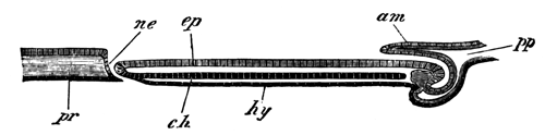
Fig. 1. Diagrammatic
longitudinal section of an embryo of Lacerta. pp. body-cavity. am. Amnion. ne.
Neurenteric canal. ch. Notochord. hy. Hypoblast. ep. Epiblast. pr.
Primitive streak.
The woodcut (fig. 1) represents a diagrammatic longitudinal section
through this embryo, and the sections belonging to Series B illustrate the features of the hind end of the
embryo and of the primitive streak.
As is shown in fig. 1, the notochord (ch.) has now throughout the region of the embryo
become separated from the subjacent hypoblast, and the lateral plates of
mesoblast are distinctly divided into somatic and splanchnic layers. The
medullary groove is continued as a deepish groove up to the opening of the
neurenteric passage, which thus forms a perforation in the floor of the
hinder end of the medullary groove (vide Series
B, figs. 2, 3, and 4).
The passage itself is somewhat shorter than in the previous stage, and
the whole of it is shown in a single section (fig. 4). This section must
either have been taken somewhat obliquely, or else the passage
have been exceptionally short in this embryo, since in an older embryo it
could not all be seen in one section.
The front wall of the passage is continuous with the notochord, which
for two sections or so in front remains attached to the hypoblast (figs. 2
and 3). Behind the perforation in the floor of the medullary groove is
placed the primitive streak (fig. 5), where all the layers become fused
together, as in the earlier stage. Into this part a narrow diverticulum
from the end of the medullary groove is continued for a very short distance
(vide fig. 5, mc.).
The general features of the stage will best be understood by an
examination of the diagrammatic longitudinal section, represented in
woodcut, fig. 1. In front is shown the amnion (am.), growing over the head of the embryo. The
notochord (ch.) is seen as an independent
cord for the greater part of the length of the embryo, but falls into the
hypoblast shortly in front of the neurenteric passage. The neurenteric
passage is shown at ne., and behind it
is shown the primitive streak.
In a still older stage, represented in surface view on Pl. 29, fig. C, the
medullary folds have nearly met above, but have not yet united. The
features of the passage from the neural groove to the hypoblast are
precisely the same in the embryo just described, although the lumen of the
passage has become somewhat narrower. There is still a short primitive
streak behind the embryo.
The neurenteric passage persists but a very short time after the
complete closure of the medullary canal. It is in no way connected with the
allantois, as conjectured by Kupffer and Benecke, but the allantois is
formed, as I have satisfied myself by longitudinal sections of a later
stage, in the manner already described by Dobrynin, Gasser, and Kölliker
for the bird and mammal.
The general results of Kupffer's and Benecke's observations, with the
modifications introduced by my own observations, are as
follows:—After the segmentation and the formation of the embryonic
shield (area pellucida) the blastoderm becomes distinctly divided into
epiblast and hypoblast[449]. At the hind end of the
shield a somewhat triangular primitive streak is formed by the
fusion of the epiblast and hypoblast with a number of cells between them,
which are probably derived from the lower rows of the segmentation cells.
At the front end of the streak a passage arises, open at both extremities,
leading obliquely forwards through the epiblast to the space below the
hypoblast. The walls of the passage are formed of a layer of columnar cells
continuous both with epiblast and hypoblast. In front of the primitive
streak the body of the embryo becomes first differentiated by the formation
of a medullary plate, and at the same time there grows out from the
primitive streak a layer of mesoblast, which spreads out in all directions
between the epiblast and hypoblast. In the axis of the embryo the mesoblast
plate is stated by Kupffer and Benecke to be continuous across the middle
line, but this appears very improbable. In a slightly later stage the
medullary plate becomes marked by a shallow groove, and the mesoblast of
the embryo is then undoubtedly constituted of two lateral plates, one on
each side of the median line. In the median line the notochord arises as a
ridge-like thickening of the hypoblast, which becomes very soon quite
separated from the hypoblast, except at the hind end, where it is continued
into the front wall of the neurenteric passage. It is interesting to notice
the remarkable relation of the notochord to the walls of the neurenteric
passage. More or less similar relations are also well marked in the case of
the goose and the fowl (Gasser)[450],
and support the conclusion deducible from the lower forms of vertebrata,
that the notochord is essentially hypoblastic.
The passage at the front end of the primitive streak forms the posterior
boundary of the medullary plate, though the medullary groove is not at
first continued back to it. The anterior wall of this passage connects
together the medullary plate and the notochordal ridge of the hypoblast. In
the succeeding stages the medullary groove becomes continued back to the
opening of the passage, which then becomes enclosed in the medullary folds,
and forms a true neurenteric passage. It becomes narrowed as the medullary
folds finally unite to form the medullary canal, and eventually
disappears.
I conclude this paper with a concise statement of what appears
to me the probable nature of the much-disputed organ, the primitive streak,
and of the arguments in support of my view.
In a paper on the primitive streak in the Quart. Journ. of Mic. Sci.,
in 1873 (p. 280) [This edition, p. 45], I
made the following statement with reference to this subject:—It is
clear, therefore, that the primitive groove must be the rudiment of some
ancestral feature.... It is just possible that it is the last trace of that
involution of the epiblast by which the hypoblast is formed in most of the
lower animals.
At a later period, in July, 1876, after studying the development of
Elasmobranch fishes, I enlarged the hypothesis in a review of the first
part of Prof. Kölliker's Entwicklungsgeschichte. The
following is the passage in which I speak of it[451]:
"In treating of the exact relation of the primitive
groove to the formation of the embryo, Professor Kölliker gives it as his
view that though the head of the embryo is formed independently of the
primitive groove, and only secondarily unites with this, yet that the
remainder of the body is without doubt derived from the primitive groove.
With this conclusion we cannot agree, and the very descriptions of
Professor Kölliker appear to us to demonstrate the untenable nature of his
results. We believe that the front end of the primitive groove at first
occupies the position eventually filled by about the third pair of
protovertebræ, but that as the protovertebræ are successively formed, and
the body of the embryo grows in length, the primitive groove is carried
further and further back, so as always to be situated immediately behind
the embryo. As Professor Kölliker himself has shewn it may still be seen in
this position even later than the fortieth hour of incubation.
"Throughout the whole period of its existence it
retains a character which at once distinguishes it in sections from the
medullary groove.
"Beneath it the epiblast and mesoblast are always
fused, though they are always separate elsewhere; this fact, which was
originally shewn by ourselves, has been very clearly brought out by
Professor Kölliker's observations.
"The features of the primitive groove which throw
special light on its meaning are the following:—
"(1) It does not enter directly into the formation of
the embryo.
"(2) The epiblast and mesoblast always become fused
beneath it.
"(3) It is situated immediately behind the
embryo.
"Professor Kölliker does not enter into any
speculations as to the meaning of the primitive groove, but the
above-mentioned facts appear to us clearly to prove that the primitive
groove is a rudimentary structure, the origin of which can only be
completely elucidated by a knowledge of the development of the Avian
ancestors.
"In comparing the blastoderm of a bird with that of
any anamniotic vertebrate, we are met at the threshold of our
investigations by a remarkable difference between the two. Whereas in all
the lower vertebrates the embryo is situated at the edge of the
blastoderm, it is in birds and mammals situated in the centre. This
difference of position at once suggests the view that the primitive groove
may be in some way connected with the change of position in the blastoderm
which the ancestors of birds must have undergone. If we carry our
investigations amongst the lower vertebrates a little further, we find that
the Elasmobranch embryo occupies at first the normal position at the edge
of the blastoderm, but that in the course of development the blastoderm
grows round the yolk far more slowly in the region of the embryo than
elsewhere. Owing to this, the embryo becomes left in a bay, the two sides
of which eventually meet and coalesce in a linear fashion immediately
behind the embryo, thus removing the embryo from the edge of the blastoderm
and forming behind it a linear streak not unlike the primitive streak. We
would suggest the hypothesis that the primitive groove is a rudiment which
gives the last indication of a change made by the Avian ancestors in their
position in the blastoderm, like that made by Elasmobranch embryos when
removed from the edge of the blastoderm and placed in a central situation
similar to that of the embryo bird. On this hypothesis the
situation of the primitive groove immediately behind the embryo, as well as
the fact of its not becoming converted into any embryonic organ would be
explained. The central groove might probably also be viewed as the groove
naturally left between the coalescing edges of the blastoderm.
Would the fusion of epiblast and mesoblast also
receive its explanation on this hypothesis? We are of opinion that it
would. At the edge of the blastoderm which represents the blastopore mouth
of Amphioxus all the layers become fused together in the anamniotic
vertebrates. So that if the primitive groove is in reality a rudiment of
the coalesced edges of the blastoderm, we might naturally expect the layers
to be fused there, and the difficulty presented by the present condition of
the primitive groove would rather be that the hypoblast is not fused with
the other layers than that the mesoblast is indissolubly united with the
epiblast. The fact that the hypoblast is not fused with the other layers
does not appear to us to be fatal to our hypothesis, and in Mammalia, where
the primitive and medullary grooves present precisely the same relations as
in birds, all three layers are, according to Hensen's account, fused
together. This, however, is denied by Kölliker, who states that in Mammals,
as in Birds, only the epiblast and mesoblast fuse together. Our hypothesis
as to the origin of the primitive groove appears to explain in a fairly
satisfactory manner all the peculiarities of this very enigmatical organ;
it also relieves us from the necessity of accepting Professor Kölliker's
explanation of the development of the mesoblast, though it does not, of
course, render that explanation in any way untenable.
At a somewhat later period Rauber arrived at a more or less similar
conclusion, which, however, he mixes up with a number of opinions from
which I am compelled altogether to dissent[452].
The general correctness of my view, as explained in my second quotation,
appears to me completely established by Gasser's beautiful researches on
the early development of the chick and goose[453],
and by my own observations just recorded on the lizard. While at the same
time the parallel between the blastopore of Elasmobranchii and of the
Sauropsida, is rendered more complete by the discovery of the
neurenteric passage in the latter group, which was first of all made by
Gasser.
The following paragraphs contain a detailed attempt to establish the
above view by a careful comparison of the primitive streak and its adjuncts
in the amniotic vertebrates with the blastopore in Elasmobranchii.
In Elasmobranchii the blastopore consists of the following
parts:—(1), a section at the end of the medullary plate, which
becomes converted into the neurenteric canal[454];
(2), a section forming what may be called the yolk blastopore, which
eventually constitutes a linear streak connecting the embryo with the edge
of the blastoderm (vide monograph on Elasmobranch
fishes, pp. 281
and 296). In order to establish my hypothesis on
the nature of the primitive streak, it is necessary to find the
representatives of both these parts in the primitive streak of the amniotic
vertebrates. The first section ought to appear as a passage from the neural
to the enteric side of the blastoderm at the posterior end of the medullary
plate. At its front edge the epiblast and hypoblast should be continuous,
as they are at the hind end of the embryo in Elasmobranchii, and, finally,
the passage should, on the closure of the medullary groove, become
converted into the neurenteric canal. All these conditions are
exactly fulfilled by the opening at the front end of the primitive streak
of the lizard (vide woodcut, fig. 1, p. 647). In the chick there is at first no such opening,
but, as I hope to shew in a future paper, it is replaced by the epiblast
and hypoblast falling into one another at the front end of the primitive
streak. At a later period, as has been shewn by Gasser[455], there is a distinct rudiment of the neurenteric canal
in the chick, and a complete canal in the goose. Finally, in mammals, as
has been shewn by Schäffer[456] for the guinea-pig, there
is at the front end of the primitive streak a complete continuity between
epiblast and hypoblast. The continuity of the epiblast and hypoblast at the
hind end of the embryo in the bird and the mammal is a rudiment of the
continuity of these layers at the dorsal lip of the blastopore in
Elasmobranchii, Amphibia, &c. The
second section of the blastopore in Elasmobranchii or yolk blastopore is, I
believe, partly represented by the primitive streak. The yolk blastopore in
Elasmobranchii is the part of the blastopore belonging to the yolk sac as
opposed to that belonging to the embryo, and it is clear that the primitive
streak cannot correspond to the whole of this, since the primitive streak
is far removed from the edge of the blastoderm long before the yolk is
completely enclosed. Leaving this out of consideration the primitive
streak, in order that the above comparison may hold good, should satisfy
the following conditions:
1. It should connect the embryo with the edge of the blastoderm.
2. It should be constituted as if formed of the fused edges of the
blastoderm.
3. The epiblast of it should eventually not form part of the medullary
plate of the embryo, but be folded over on to the ventral side.
The first of these conditions is only partially fulfilled, but,
considering the rudimentary condition of the whole structure, no great
stress can, it seems to me, be laid on this fact.
The second condition seems to me very completely satisfied. Where the
two edges of the blastoderm become united we should expect to find a
complete fusion of the layers such as takes place in the primitive streak;
and the fact that in the primitive streak the hypoblast does not so
distinctly coalesce with the mesoblast as the mesoblast with the epiblast
cannot be urged as a serious argument against me.
The growth outwards of the mesoblast from the axis of the primitive
streak is probably a remnant of the invagination of the hypoblast and
mesoblast from the lip of the blastopore in Amphibia, &c.
The groove in the primitive streak may with great plausibility be
regarded as the indication of a depression which would naturally be found
along the line where the thickened edges of the blastoderm became
united.
With reference to the third condition, I will make the following
observations. The neurenteric canal, as it is placed at the extreme
end of the embryo, must necessarily, with reference to the embryo, be the
hindermost section of the blastopore, and therefore the part of the
blastopore apparently behind this can only be so owing to the embryo not
being folded off from the yolk sac; and as the yolk sac is in reality a
specialised part of the ventral wall of the body, the yolk blastopore must
also be situated on the ventral side of the embryo.
Kölliker and other distinguished embryologists have believed that the
epiblast of the whole of the primitive streak became part of the neural
plate. If this view were correct, which is accepted even by Rauber, the
hypothesis I am attempting to establish would fall to the ground. I have,
however, no doubt that these embryologists are mistaken. The very careful
observations of Gasser shew that the part of the primitive streak adjoining
the embryo becomes converted into the tail-swelling, and that the posterior
part is folded in on the ventral side of the embryo, and, losing its
characteristic structure, forms part of the ventral wall of the body. On
this point my own observations confirm those of Gasser. In the lizard the
early appearance of the neurenteric canal at the front end of the primitive
streak clearly shews that here also the primitive streak can take no share
in forming the neural plate.
The above considerations appear to me sufficient to establish my
hypothesis with reference to the nature of the primitive streak, which has
the merit of explaining, not only the structural peculiarities of the
primitive streak, but also the otherwise inexplicable position of the
embryo of the amniotic vertebrates in the centre of the blastoderm.
DESCRIPTION OF PLATE 29.
Complete List of Reference
Letters.
am. Amnion. ch. Notochord. ch´. Notochordal thickening of hypoblast. ep. Epiblast. hy. Hypoblast. m.g. Medullary groove. me.p. Mesoblastic plate. ne. Neurenteric canal (blastopore). pr. Primitive streak.
Series A. Sections through an
embryo shortly after the formation of the medullary groove. x 120[457].
Fig. 1. Section through the trunk of the embryo.
Figs. 2-5. Sections through the neurenteric canal.
Fig. B. Surface view of a somewhat older embryo than
that from which Series A is taken. x 30.
Series B. Sections through
the embryo represented in Fig. B. x 120.
Fig. 1. Section through the trunk of the embryo.
Figs. 2, 3. Sections through the hind end of the
medullary groove.
Fig. 4. Section through the neurenteric canal.
Fig. 5. Section through the primitive streak.
Fig. C. Surface view of a somewhat older embryo than
that represented in Fig. B. x 30.
The discovery by Mr Moseley[459] of
a tracheal system in Peripatus must be reckoned as one of the most
interesting results obtained by the naturalists of the Challenger.
The discovery clearly proves that the genus Peripatus, which is widely
distributed over the globe, is the persisting remnant of what was probably
a large group of forms, from which the present tracheate Arthropoda are
descended.
The affinities of Peripatus render any further light on its anatomy a
matter of some interest; and through the kindness of Mr Moseley I have had
an opportunity of making investigations on some well preserved examples of
Peripatus capensis, a few of the results of which I propose to lay
before the Society.
I shall confine my observations to three organs. (1) The segmental
organs, (2) the nervous system, (3) the so-called fat bodies of Mr
Moseley.
In all the segments of the body, with the exception of the first two or
three postoral ones, there are present glandular bodies, apparently
equivalent to the segmental organs of Annelids.
These organs have not completely escaped the attention of previous
observers. The anterior of them were noticed by Grube[460], but their relations were not made out. By Saenger[461], as I gather from Leuckart's Bericht for the years 1868-9, these structures were also
noticed, and they were interpreted as segmental organs. Their
external openings were correctly identified. They are not mentioned by
Moseley, and no notice of them is to be found in the text-books. The
observations of Grube and Saenger seem, in fact, to have been completely
forgotten.
The organs are placed at the bases of the feet in two lateral divisions
of the body-cavity shut off from the main central median division of the
body-cavity by longitudinal septa of transverse muscles.
Each fully developed organ consists of three parts:
(1) A dilated vesicle opening externally at the base of a foot.
(2) A coiled glandular tube connected with this and subdivided again
into several minor divisions.
(3) A short terminal portion opening at one extremity into the coiled
tube (2) and at the other, as I believe, into the body-cavity. This section
becomes very conspicuous in stained preparations by the intensity with
which the nuclei of its walls absorb the colouring matter.
The segmental organs of Peripatus, though formed on a type of their own,
more nearly resemble those of the Leech than of any other form with which I
am acquainted. The annelidan affinities shewn by their presence are of some
interest. Around the segmental organs in the feet are peculiar cells richly
supplied with tracheæ, which appear to me to be similar to the fat bodies
in insects. There are two glandular bodies in the feet in addition to the
segmental organs.
The more obvious features of the nervous system have been fully made out
by previous observers, who have shewn that it consists of large paired
supra-œsophageal ganglia connected with two widely separated ventral
cords—stated by them not to be ganglionated. Grube describes the two
cords as falling into one another behind the anus—a feature the
presence of which is erroneously denied by Saenger. The lateral cords are
united by numerous (5 or 6 for each segment) transverse cords.
The nervous system would appear at first sight to be very lowly
organised, but the new points I believe myself to have made out, as well as
certain previously known features in it appear to me to shew that this is
not the case.
The following is a summary of the fresh points I have observed
in the nervous system:
(1) Immediately underneath the œsophagus the œsophageal
commissures dilate and form a pair of ganglia equivalent to the annelidan
and arthropodan sub-œsophageal ganglia. These ganglia are closely
approximated and united by 5 or 6 commissures. They give off large nerves
to the oral papillæ.
(2) The ventral nerve cords are covered on their ventral side by a thick
ganglionic layer[462], and at each pair of feet they
dilate into a small but distinct ganglionic swelling. From each
ganglionic swelling are given off a pair of large nerves[463] to the feet; and the ganglionic swellings of the two
cords are connected together by a pair of commissures containing
ganglion cells[464]. The other commissures
connecting the two cords together do not contain ganglion cells.
The chief feature in which Peripatus was supposed to differ from normal
Arthropoda and Annelida, viz. the absence of
ganglia on the ventral cords, does not really exist. In other particulars,
as in the amount of nerve cells in the ventral cords and the completeness
of the commissural connections between the two cords, &c., the organisation of the nervous system of Peripatus
ranks distinctly high. The nervous system lies within the circular and
longitudinal muscles, and is thus not in proximity with the skin. In this
respect also Peripatus shews no signs of a primitive condition of the
nervous system.
A median nerve is given off from the posterior border of the
supra-œsophageal ganglion to the œsophagus, which probably
forms a rudimentary sympathetic system. I believe also that I have found
traces of a paired sympathetic system.
The organ doubtfully spoken of by Mr Moseley as a fat body, and by Grube
as a lateral canal, is in reality a glandular tube, lined by beautiful
columnar cells containing secretion globules, which opens by means of a
non-glandular duct into the mouth. It lies close above the ventral nerve
cords in a lateral compartment of the body-cavity, and extends backwards
for a varying distance.
This organ may perhaps be best compared with the simple salivary gland
of Julus. It is not to be confused with the slime glands of Mr Moseley,
which have their opening in the oral papillæ. If I am correct in regarding
it as homologous with the salivary glands so widely distributed amongst the
Tracheata, its presence indicates a hitherto unnoticed arthropodan affinity
in Peripatus.
Professor Schulze's[466]
last memoir on the development of Calcareous Sponges, confirms and enlarges
Metschnikoff's[467] earlier observations, and gives
us at last a fairly complete history of the development of one form of
Calcareous Sponge. The facts which have been thus established have
suggested to me a view of the morphology and systematic position of the
Spongida, somewhat different to that now usually entertained. In bringing
forward this view, I would have it understood that it does not claim to be
more than a mere suggestion, which if it serves no other function may,
perhaps, be of use in stimulating research.
To render clear what I have to say, I commence with a very brief
statement of the facts which may be considered as established with
reference to the development of Sycandra raphanus, the form which
was studied by both Metschnikoff and Schulze. The segmentation of the ovum,
though in many ways remarkable, is of no importance for my present purpose,
and I take up the development at the close of the segmentation, while the
embryo is still encapsuled in the parental tissues. It is at this stage
lens-shaped, with a central segmentation cavity. An equatorial plane
divides it into two parts, which have equal shares in bounding the
segmentation cavity. One of these halves is formed of about thirty-two
large, round, granular cells, the other of a larger number of ciliated
clear columnar cells. While the embryo is still encapsuled a partial
invagination of the granular cells takes place, reducing the
segmentation cavity to a mere slit; this invagination is, however, quite
temporary and unimportant, and on the embryo becoming free, which shortly
takes place, no trace of it is visible; but, on the contrary, the
segmentation cavity becomes larger, and the granular cells project very
much more prominently than in the encapsuled state.
Fig. 1.
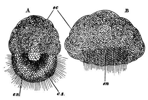
Two free stages in the development of Sycandra
raphanus (copied from Schulze).
A. Amphiblastula
stage; B. a later stage after the
ciliated cells have commenced to become invaginated; cs. segmentation cavity; ec. granular cells which will form the ectoderm;
en. ciliated cells which become
invaginated to form the entoderm
The larva, after it has left the parental tissues, has an oval form and
is transversely divided into two areas (fig. 1, A). One of these areas is formed of the elongated,
clear, ciliated cells, with a small amount of pigment near the inner ends
(en), and the other and larger area of
the thirty-two granular cells already mentioned (ec). Fifteen or sixteen of these are arranged as a
special ring on the border of the clear cells. In the centre of the embryo
is a segmentation cavity (cs) which lies
between the granular and the clear cells, but is mainly bounded by the
vaulted inner surface of the latter. This stage is known as the
amphiblastula stage. After the larva has for some time enjoyed a free
existence, a remarkable series of changes takes place, which result in the
invagination of the half of it formed of the clear cells, and form a
prelude to the permanent attachment of the larva. The entire process of
invagination is completed in about half an hour. The whole embryo first
becomes flattened, but especially the ciliated half which gradually becomes
less prominent (fig. 1, B), and still
later the cells composing it undergo a true process of invagination. As a
result of this invagination the segmentation cavity is obliterated and the
larva assumes a compressed plano-convex form with a central gastrula
cavity, and a blastopore in the middle of the flattened surface. The two
layers of the gastrula may now be spoken of as ectoderm and entoderm. The
blastopore becomes gradually narrowed by the growth over it of the outer
row of granular cells. When it has become very small the attachment of the
larva takes place by the flat surface where the blastopore is situated. It
is effected by protoplasmic processes of the outer ring of ectoderm cells,
which, together with the other ectoderm cells, now become amœboid. At
the same time they become clearer and permit a view of the interior of the
gastrula. Between the ectoderm cells and the entoderm cells which line the
gastrula cavity there arises a hyaline structureless layer, which is more
closely attached to the ectoderm than to the entoderm, and is probably
derived from the former. A view of the gastrula stage after the larva has
become fixed is given in fig. 2.
Fig. 2.
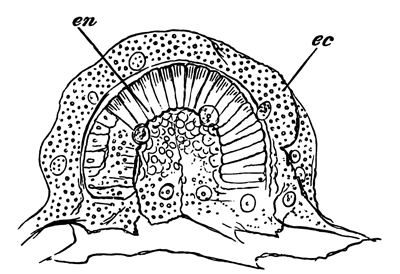
Fixed Gastrula stage of Sycandra raphanus
(copied from Schulze).
The figure shews the amœboid ectoderm cells
(ec) derived from the granular cells of
the earlier stage, and the columnar entoderm cells, lining the gastrula
cavity, derived from the ciliated cells of the earlier stage. The larva is
fixed by the amœboid cells on the side on which the blastopore is
situated.
Fig. 3.
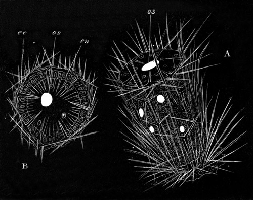
The young of Sycandra raphanus shortly after the
development of the spicula
(copied from Schulze).
A. View from the side;
B. view from the free extremity; os. osculum; ec. ectoderm; en.
entoderm composed of collared ciliated cells. The terminal osculum and
lateral pores are represented as oval white spaces.
After invagination the cilia of the entoderm cells can no longer be
seen, and are probably absorbed, and their disappearance is nearly
coincident with the complete obliteration of the blastopore, an event which
takes place shortly after the attachment of the larva. After the formation
of the structureless layer between the ectoderm and entoderm, calcareous
spicules make their appearance in it as delicate unbranched rods pointed at
both extremities. The larva when once fixed rapidly grows in length and
assumes a cylindrical form (fig. 3, A).
The sides of the cylinder are beset with calcareous spicules which project
beyond the surface, and in addition to the unbranched forms, spicules are
developed with three and four rays as well as some with a blunt extremity
and serrated edge. The extremity of the cylinder opposite the attached
surface is flattened, and though surrounded by a ring of four-rayed
spicules is itself free from them. At this extremity a small perforation is
formed leading into the gastric cavity which rapidly increases in size and
forms an exhalent osculum (os). A series
of inhalent apertures are also formed at the sides of the cylinder. The
relative times of appearance of the single osculum and smaller apertures is
not constant for the different larvæ. On the central gastrula cavity of the
sponge becoming placed in communication with the external water, the
entoderm cells lining it become ciliated afresh (fig. 3, B, en) and
develop the peculiar collar characteristic of the entoderm cells of the
Spongida. When this stage of development is reached we have a fully
developed sponge of the type made known by Haeckel as Olynthus.
Till the complete development of other forms of Spongida has been worked
out it is not possible to feel sure how far the phenomena observable in
Sycandra hold good in all cases. Quite recently the Russian embryologist,
M. Ganin[468], has given an account, without
illustrations, of the development of Spongilla fluviatilis, which
does not appear reconcileable with that of Sycandra. Considering the
difficulties of observation it appears better to assume for this and some
other descriptions that the observations are in error rather than that
there is a fundamental want of uniformity in development amongst the
Spongida.
The first point in the development of Sycandra which deserves notice is
the character of the free swimming larva. The peculiar larval form, with
one half of the body composed of amœboid granular cells and the other
of clear ciliated cells is nearly constant amongst the Calcispongiæ, and
widely distributed in a somewhat modified condition amongst the
Fibrospongiæ and Myxospongiæ. Does this larva retain the characters of an
ancestral type of the Spongida, and if so what does its form mean? It is,
of course, possible that it has no ancestral meaning but has been
secondarily acquired; I prefer myself to think that this is not the case,
more especially as it appears to me that the characters of the larva may be
plausibly explained by regarding it as a transitional form between the
Protozoa and Metazoa. According to this view the larva is to be considered
as a colony of Protozoa, one half of the individuals of which have become
differentiated into nutritive forms, and the other half into locomotor and
respiratory forms. The granular amœboid cells represent the nutritive
forms, and the ciliated cells represent the locomotor and respiratory
forms. That the passage from the Protozoa to the Metazoa may have been
effected by such a differentiation is not improbable on à
priori grounds, and fits in very well with the condition of the free
swimming larva of Spongida, though another and perhaps equally plausible
suggestion as to this passage has been put forward by my friend Professor
Lankester[469].
While the above view seems fairly satisfactory for the free swimming
stage of the larval Sponge there arises in the subsequent development a
difficulty which appears at first sight fatal to it. This difficulty is the
invagination of the ciliated cells instead of the granular ones. If the
granular cells represent the nutritive individuals of the colony, they and
not the ciliated cells ought most certainly to give rise to the lining of
the gastrula cavity, according to the generally accepted views of the
morphology of the Spongida. The suggestion which I would venture to put
forward in explanation of this paradox involves a completely new view of
the nature and functions of the germinal layers of adult Sponges.
It is as follows:—When the free swimming ancestor of the Spongida
became fixed, the ciliated cells by which its movements used to be effected
must have to a great extent become functionless. At the same time the
amœboid nutritive cells would need to expose as large a surface as
possible. In these two considerations there may, perhaps, be found a
sufficient explanation of the invagination of the ciliated cells, and the
growth of the amœboid cells over them. Though respiration was, no
doubt, mainly effected by the ciliated cells, it is improbable that it was
completely localised in them, but the continuation of their function was
provided for by the formation of an osculum and pores. The ciliated
collared cells which line the ciliated chambers, or in some cases the
radial tubes, are undoubtedly derived from the invaginated cells, and if
there is any truth in the above suggestion, the collared cells in the adult
Sponge must be mainly respiratory and not digestive in function, while the
normal epithelial cells which cover the surface of the sponge, and in most
cases line the greater part of the passages through its substance, must
carry on the digestion[470]. If the reverse is the
case the whole theory falls to the ground. It has not, so far as I know,
been definitely made out where the digestion is carried on. Lieberkühn
would appear to hold the view that the amœboid lining cells of the
passages are mainly concerned with digestion, while Carter holds that
digestion is carried on by the collared cells of the ciliated chambers.
If it is eventually proved by actual experiments on the nutrition of
Sponges, that digestion is carried on by the general cells lining the
passages, and not by the ciliated cells, it is clear that neither the
ectoderm nor entoderm of Sponges will correspond with the similarly named
layers in the Cœlenterata and the Metazoa[TN13]. The
invaginated entoderm will be the respiratory layer and the ectoderm the
digestive and sensory layer; the sensory function being probably mainly
localised in the epithelium on the surface, and the digestive one in the
epithelium lining the passages. Such a fundamental difference in the
germinal layers between the Spongida and the other Metazoa, would
necessarily involve the creation of a special division of the Metazoa for
the reception of the former group.
(With Plates 30, 31, 32.)
The following observations do not profess to contain a
complete history of the development even of a single species of spider.
They are the result of investigations carried on at intervals during rather
more than two years, on the ova of Agelena labyrinthica; and I
should not have published them now, if I had any hope of being able to
complete them before the appearance of the work I am in the course of
publishing on Comparative Embryology. It appeared to me, however, desirable
to publish in full such parts of my observations as are completed before
the appearance of my treatise, since the account of the development of the
Araneina is mainly founded upon them.
My investigations on the germinal layers and organs have been chiefly
conducted by means of sections. To prepare the embryos for sections, I
employed the valuable method first made known by Bobretzky. I hardened the
embryos in bichromate of potash, after placing them for a short time in
nearly boiling water. They were stained as a whole with hæmatoxylin after
the removal of the membranes, and embedded for cutting in coagulated
albumen.
The number of investigators who have studied the development of spiders
is inconsiderable. A list of them is given at the end of the paper.
The earliest writer on the subject is Herold (No. 4); he was followed after a very considerable interval
of time by Claparède (No. 3), whose
memoir is illustrated by a series of beautiful plates, and contains a very
satisfactory account of the external features of development.
Balbiani (No. 1) has gone with some detail
into the history of the early stages; and Ludwig (No. 5) has published some very important observations on
the development of the blastoderm. Finally, Barrois (No. 2) has quite recently taken up the study of the group,
and has added some valuable observations on the development of the germinal
layers.
In addition to these papers on the true spiders, important
investigations have been published by Metschnikoff on other groups of the
Arachnida, notably the scorpion. Metschnikoff's observations on the
formation of the germinal layers and organs accord in most points with my
own.
The development of the Araneina may be divided into four periods: (1)
the segmentation; (2) the period from the close of the segmentation up to
the period when the segments commence to be formed; (3) the period from the
commencing formation of the segments to the development of the full number
of limbs; (4) the subsequent stages up to the attainment of the adult
form.
In my earliest stage the segmentation was already completed, and the
embryo was formed of a single layer of large flattened cells enveloping a
central mass of polygonal yolk-segments.
Each yolk-segment is formed of a number of large clear somewhat oval
yolk-spherules. In hardened specimens the yolk-spherules become polygonal,
and in ova treated with hot water prior to preservation are not
unfrequently broken up. Amongst the yolk-segments are placed a fair number
of nucleated bodies of a very characteristic appearance. Each of them is
formed of (1) a large, often angular, nucleus, filled with deeply staining
bodies (nucleoli?); (2) [TN14] a layer of protoplasm
surrounding the nucleus, prolonged into a protoplasmic reticulum. The exact
relation of these nucleated bodies to the yolk-segments is not very easy to
make out, but the general tendency of my observations is to shew (1) that
each nucleated body belongs to a yolk-sphere, and (2) that it is generally
placed not at the centre, but to one side of a yolk-sphere. If the above
conclusions are correct each complete yolk-segment is a cell, and each such
cell consists of a normal nucleus, protoplasm, and yolk-spherules. There is
a special layer of protoplasm surrounding the nucleus, while the remainder
of the protoplasm consists of a reticulum holding together the
yolk-spherules. Yolk-cells of this character are seen in Pls. 31 and 32, figs. 10-21.
The nuclei of the yolk-cells are probably derived by division from the
nuclei of the segmentation rosettes (vide Ludwig,
No. 5), and it is probable that they take
their origin at the time when the superficial layer of protoplasm separates
from the yolk-columns below to form the blastoderm.
The protoplasm of the yolk-cells undergoes rapid division, as is shewn
by the fact that there are often two nucleated bodies close together, and
sometimes two nuclei in a single mass of protoplasm (fig. 10). It is
probable that in some cases the yolk-spheres divide at the same time as the
protoplasm belonging to them; the division of the nucleated bodies is,
however, in the main destined to give rise to fresh cells which enter the
blastoderm.
I have not elucidated to my complete satisfaction the next stage or two
in the development of the embryo; and have not succeeded in completely
reconciling the results of my own observations with those of Claparède and
Balbiani. In order to shew exactly where my difficulties lie it is
necessary briefly to state the results arrived at by the above authors.
According to Claparède the first differentiation in Pholcus consists in
the accumulation of the cells over a small area to form a protuberance,
which he calls the primitive cumulus. Owing to its smaller
specific gravity the part of the ovum with the cumulus always turns
upwards, like the blastodermic pole of a fowl's egg.
After a short time the cumulus elongates itself on one side, and becomes
connected by a streak with a white patch, which appears on the surface of
the egg, about 90° from the cumulus. This patch gradually enlarges, and
soon covers the whole surface of the ovum except the region where the
cumulus is placed. It becomes the ventral plate or germinal streak of the
embryo, its extremity adjoining the cumulus is the anal extremity, and its
opposite extremity the cephalic one. The cumulus itself is placed in a
depression on the dorsal surface of the ovum. Claparède compares
the cumulus to the dorsal organ of many Crustacea.
Balbiani (No. 1) describes the primitive
cumulus in Tegenaria domestica, Epeira diadema, and
Agelena labyrinthica, as originating as a protuberance at the centre
of the ventral surface, surrounded by a specialised portion of the
blastoderm (p. 57), which I will call the ventral plate. In
Tegenaria domestica he finds that it encloses the so-called
yolk-nucleus, p. 62. By an unequal growth of the ventral plate the
primitive cumulus comes to be placed at the cephalic pole of the ventral
plate. The cumulus now becomes less prominent, and in a few cases
disappears. In the next stage the central part of the ventral plate becomes
very prominent and forms the procephalic lobe, close to the anterior border
of which is usually placed the primitive cumulus (p. 67). The space
between the cumulus and the procephalic lobe grows larger, so that the
latter gradually travels towards the dorsal surface and finally vanishes.
Behind the procephalic lobe the first traces of the segments make their
appearance, as three transverse bands, before a distinct anal lobe becomes
apparent.
The points which require to be cleared up are, (1) what is the nature of
the primitive cumulus? (2) where is it situated in relation to the embryo?
Before attempting to answer these questions I will shortly describe the
development, so far as I have made it out, for the stages during which the
cumulus is visible.
The first change that I find in the embryo (when examined after it has
been hardened)[472] is the appearance of a small,
whitish spot, which is at first very indistinct. A section through such an
ovum (Pl. 31, fig. 10) shews that the cells of
about one half of the ovum have become more columnar than those of the
other half, and that there is a point (pr.c.) near one end of the thickened half where the
cells are more columnar, and about two layers or so deep. It appears to me
probable that this point is the whitish spot visible in the hardened ovum.
In a somewhat later stage (Pl. 30, fig. 1) the
whitish spot becomes more conspicuous (pc.), and appears as a distinct prominence, which is,
without doubt, the primitive cumulus, and from it there proceeds on one
side a whitish streak. The prominence, as noticed by Claparède and
Balbiani, is situated on the flatter side of the ovum. Sections at this
stage shew the same features as the previous stage, except that (1) the
cells throughout are smaller, (2) those of the thickened hemisphere of the
ovum more columnar, and (3) the cumulus is formed of several rows of cells,
though not divided into distinct layers. In the next stage the appearances
from the surface are rather more obscure, and in some of my best specimens
a coagulum, derived from the fluid surrounding the ovum, covers the most
important part of the blastoderm. In Pl. 30,
fig. 2, I have attempted to represent, as truly as I could, the appearances
presented by the ovum. There is a well-marked whitish side of the ovum,
near one end of which is a prominence (pc.), which must, no doubt, be identified with the
cumulus of the earlier stages. Towards the opposite end, or perhaps rather
nearer the centre of the white side of the ovum, is an imperfectly marked
triangular white area. There can be no doubt that the line connecting the
cumulus with the triangular area is the future long axis of the embryo, and
the white area is, without doubt, the procephalic lobe of Balbiani.
A section of the ovum at this stage is represented in Pl. 31, fig. 11. It is not quite certain in what direction
the section is taken, but I think it probable it is somewhat oblique to the
long axis. However this may be, the section shews that the whitish
hemisphere of the blastoderm is formed of columnar cells, for the most part
two or so layers deep, but that there is, not very far from the middle
line, a wedge-shaped internal thickening of the blastoderm where the cells
are several rows deep. With what part visible in surface view this
thickened portion corresponds is not clear. To my mind it most probably
corresponds to the larger white patch, in which case I have not got a
section through the terminal prominence. In the other sections of the same
embryo the wedge-shaped thickening was not so marked, but it, nevertheless,
extended through all the sections. It appears to me probable that it
constitutes a longitudinal thickened ridge of the blastoderm. In any case,
it is clear that the white hemisphere of the blastoderm is a thickened
portion of the blastoderm, and that the thickening is in
part due to the cells being more columnar, and, in part, to their being
more than one row deep, though they have not become divided into two
distinct germinal layers. It is further clear that the increase in the
number of cells in the thickened part of the blastoderm is, in the main,
a result of the multiplication of the original single row of cells,
while a careful examination of my sections proves that it is also partly
due to cells, derived from the yolk, having been added to the
blastoderm.
In the following stage which I have obtained (which cannot be very much
older than the previous stage, because my specimens of it come from the
same batch of eggs), a distinct and fairly circumscribed thickening forming
the ventral surface of the embryo has become established. Though its
component parts are somewhat indistinct, it appears to consist of a
procephalic lobe, a less prominent caudal lobe, and an intermediate portion
divided into about three segments; but its constituents cannot be clearly
identified with the structures visible in the previous stage. I am
inclined, however, to identify the anterior thickened area of the previous
stage with the procephalic lobe, and a slight protuberance of the caudal
portion (visible from the surface) with the primitive cumulus. I have,
however, failed to meet with any trace of the cumulus in my sections.
To this stage, which forms the first of the second period of the larval
history, I shall return, but it is necessary now to go back to the
observations of Claparède and Balbiani.
There can, in the first place, be but little doubt that what I have
called the primitive cumulus in my description is the structure so named by
Claparède and Balbiani.
It is clear that Balbiani and Claparède have both failed to appreciate
the importance of the organ, which my observations shew to be the part of
the ventral thickening of the blastoderm where two rows of cells are first
established, and therefore the point where the first traces of the future
mesoblast becomes visible.
Though Claparède and Balbiani differ somewhat as to the position of the
organ, they both make it last longer than I do: I feel certainly inclined
to doubt whether Claparède is right in considering a body he figures after
six segments are present, to be the same as the dorsal organ of the
embryo before the formation of any segments, especially as all the stages
between the two appear to have escaped him. In Agelena there is undoubtedly
no organ in the position he gives when six segments are found.
Balbiani's observations accord fairly with my own up to the stage
represented in fig. 2. Beyond this stage my own observations are not
satisfactory, but I must state that I feel doubtful whether Balbiani is
correct in his description of the gradual separation of the procephalic
lobe and the cumulus, and the passage of the latter to the dorsal surface,
and think it possible that he may have made a mistake as to which side of
the procephalic lobe, in relation to the parts of the embryo, the cumulus
is placed.
Although there appear to be grounds for doubting whether either Balbiani
and Claparède are correct in the position they assign to the cumulus, my
observations scarcely warrant me in being very definite in my statements on
this head, but, as already mentioned, I am inclined to place the organ near
the posterior end (and therefore, as will be afterwards shewn, in a
somewhat dorsal situation) of the ventral embryonic thickening.
In my earliest stage of the third period there is present, as has
already been stated, a procephalic lobe, and an indistinct and not very
prominent caudal portion, and about three segments between the two. The
definition of the parts of the blastoderm at this stage is still very
imperfect, but from subsequent stages it appears to me probable that the
first of the three segments is that of the first pair of ambulatory limbs,
and that the segments of the cheliceræ and pedipalpi are formed later than
those of the first three ambulatory appendages.
Balbiani believes that the segment of the cheliceræ is formed later than
that of the six succeeding segments. He further concludes, from the fact
that this segment is cut off from the procephalic portion in front, that it
is really part of the procephalic lobe. I cannot accept the validity of
this argument; though I am glad to find myself in, at any rate, partial
harmony with the distinguished French embryologist as to the facts.
Balbiani denies for this stage the existence of a caudal lobe. There is
certainly, as is very well shewn in my longitudinal sections, a
thickening of the blastoderm in the caudal region, though it is not so
prominent in surface views as the procephalic lobe.
A transverse section through an embryo at this stage (Pl. 31, fig. 12) shews that there is a ventral plate of
somewhat columnar cells more than one row deep, and a dorsal portion of the
blastoderm formed of a single row of flattened cells. Every section at this
stage shews that the inner layer of cells of the ventral plate is receiving
accessions of cells from the yolk, which has not to any appreciable extent
altered its constitution. A large cell, passing from the yolk to the
blastoderm, is shewn in fig. 12 at y.c.
The cells of the ventral plate are now divided into two distinct
layers. The outer of these is the epiblast, the inner the
mesoblast. The cells of both layers are quite continuous across
the median line, and exhibit no trace of a bilateral arrangement.
This stage is an interesting one on account of the striking similarity
which (apart from the amnion) exists between a section through the
blastoderm of a spider and that of an insect immediately after the
formation of the mesoblast. The reader should compare Kowalevsky's (Mém. Acad. Pétersbourg, Vol.
XVI. 1871)
fig. 26, Pl. IX. with my fig. 12. The existence
of a continuous ventral plate of mesoblast has been noticed by Barrois
(p. 532), who states that the two mesoblastic bands originate from the
longitudinal division of a primitive single band.
In a slightly later stage (Pl. 30, fig.
3a and 3b) six distinct segments are interpolated between the
procephalic and the caudal lobes. The two foremost, ch and pd
(especially the first), of these are far less distinct than the remainder,
and the first segment is very indistinctly separated from the procephalic
lobe. From the indistinctness of the first two somites, I conclude that
they are later formations than the four succeeding ones. The caudal and
procephalic lobes are very similar in appearance, but the procephalic lobe
is slightly the wider of the two. There is a slight protuberance on the
caudal lobe, which is possibly the remnant of the cumulus. The superficial
appearance of segmentation is produced by a series of transverse valleys,
separating raised intermediate portions which form the segments. The
ventral thickening of the embryo now occupies rather more than half the
circumference of the ovum.
Transverse sections shew that considerable changes have been effected in
the constitution of the blastoderm. In the previous stage, the ventral
plate was formed of an uniform external layer of epiblast, and a continuous
internal layer of mesoblast. The mesoblast has now become divided along the
whole length of the embryo, except, perhaps, the procephalic lobes, into
two lateral bands which are not continuous across the middle line (Pl. 31, fig. 13, me). It has, moreover, become a much more definite
layer, closely attached to the epiblast. Between each mesoblastic band and
the adjoining yolk there are placed a few scattered cells, which in a
somewhat later stage become the splanchnic mesoblast. These cells are
derived from the yolk-cells; and almost every section contains examples of
such cells in the act of joining the mesoblast.
The epiblast of the ventral plate has not, to any great extent, altered
in constitution. It is, perhaps, a shade thinner in the median line than it
is laterally. The division of the mesoblast plate into two bands, together,
perhaps, with the slight reduction of the epiblast in the median ventral
line, gives rise at this stage to an imperfectly marked median groove.
The dorsal epiblast is still formed of a single layer of flat cells. In
the neighbourhood of this layer the yolk nuclei are especially
concentrated. The yolk itself remains as before.
The segments continue to increase regularly, each fresh segment being
added in the usual way between the last formed segment and the unsegmented
caudal lobe. At the stage when about nine or ten segments have become
established, the first rudiments of appendages become visible. At this
period (Pl. 30, fig. 4) there is a distinct
median ventral groove, extending through the whole length of the embryo,
which becomes, however, considerably shallower behind. The procephalic
region is distinctly bilobed. The first segment (that of the cheliceræ) is
better marked off from it than in the previous stage, but is without a
trace of an appendage, and exhibits therefore, in respect to the
development of its appendages, the same retardation that characterised its
first appearance. The next five segments, viz.
those of the pedipalpi and four ambulatory appendages, present a very
well-marked swelling at each extremity. These swellings are the earliest
traces of the appendages. Of the three succeeding segments, only the first
is well differentiated. The caudal lobe, though less broad than the
procephalic lobe, is still a widish structure. The most important internal
changes concern the mesoblast, which is now imperfectly though distinctly
divided into somites, corresponding with segments visible externally. Each
mesoblastic somite is formed of a distinct somatic layer closely attached
to the epiblast, and a thinner and less well-marked splanchnic layer. In
the appendage-bearing segments the somatic layer is continued up into the
appendages.
The epiblast is distinctly thinner in the median line than at the two
sides.
The next stage figured (Pl. 30, figs. 5 and
6) is an important one, as it is characterized by the establishment of the
full number of appendages. The whole length of the ventral plate has
greatly increased, so that it embraces nearly the circumference of the
ovum, and there is left uncovered but a very small arc between the two
extremities of the plate (Pl. 30, fig. 6; Pl. 31, fig. 15). This arc is the future dorsal
portion of the embryo, which lags in its development immensely behind the
ventral portion.
There is a very distinctly bilobed procephalic region (pr.l) well separated from the segment with the
cheliceræ (ch). It is marked by a
shallow groove opening behind into a circular depression (st.)—the earliest rudiment of the stomodæum.
The six segments behind the procephalic lobes are the six largest, and each
of them bears two prominent appendages. They constitute the six
appendage-bearing segments of the adult. The four future ambulatory
appendages are equal in size: they are slightly larger than the pedipalpi,
and these again than the cheliceræ. Behind the six somites with prominent
appendages there are four well-marked somites, each with a small
protuberance. These four protuberances are provisional appendages. They
have been found in many other genera of Araneina (Claparède, Barrois). The
segments behind these are rudimentary and difficult to count, but there
are, at any rate, five, and at a slightly later stage probably six,
including the anal lobe. These fresh segments have been formed by the
continued segmentation of the anal lobe, which has greatly altered
its shape in the process. The ventral groove of the earlier stage is still
continued along the whole length of the ventral plate.
By the close of this stage the full number of post-cephalic segments has
become established. They are best seen in the longitudinal section (Pl. 31, fig. 15). There are six anterior
appendage-bearing segments, followed by four with rudimentary appendages
(not seen in this figure), and six without appendages behind. There are,
therefore, sixteen in all. This number accords with the result arrived at
by Barrois, but is higher by two than that given by Claparède.
The germinal layers (vide Pl. 31, fig. 14) have by this stage undergone a further
development. The mesoblastic somites are more fully developed. The general
relations of these somites is shewn in longitudinal section in Pl. 31, fig. 15, and in transverse section in Pl. 31, fig. 14. In the tail, where they are simplest (shewn
on the upper side in fig. 14), each mesoblastic somite is formed of a
somatic layer of more or less cubical cells attached to the epiblast, and a
splanchnic layer of flattened cells. Between the two is placed a completely
circumscribed cavity, which constitutes part of the embryonic body-cavity.
Between the yolk and the splanchnic layer are placed a few scattered cells,
which form the latest derivatives of the yolk-cells, and are to be reckoned
as part of the splanchnic mesoblast. The mesoblastic somites do not extend
outwards beyond the edge of the ventral plate, and the corresponding
mesoblastic somites of the two sides do not nearly meet in the middle line.
In the limb-bearing somites the mesoblast has the same general characters
as in the posterior somites, but the somatic layer is prolonged as
a hollow papilliform process into the limb, so that each limb has an axial
cavity continuous with the section of the body-cavity of its somite. The
description given by Metschnikoff of the formation of the mesoblastic
somites in the scorpion, and their continuation into the limbs, closely
corresponds with the history of these parts in spiders. In the region of
each procephalic lobe the mesoblast is present as a continuous layer
underneath the epiblast, but in the earlier part of the stage, at any rate,
is not formed of two distinct layers with a cavity between them.
The epiblast at this stage has also undergone important
changes. Along the median ventral groove it has become very thin. On each
side of this groove it exhibits in each appendage-bearing somite a
well-marked thickening, which gives in surface views the appearance of a
slightly raised area (Pl. 30, fig. 5), between
each appendage and the median line. These thickenings are the first
rudiments of the ventral nerve ganglia. The ventral nerve cord at this
stage is formed of two ridge-like thickenings of the epiblast, widely
separated in the median line, each of which is constituted of a series of
raised divisions—the ganglia—united by shorter, less prominent
divisions (fig. 14, vg). The nerve cords
are formed from before backwards, and are not at this stage found in the
hinder segments. There is a distinct ganglionic thickening for the
cheliceræ quite independent of the procephalic lobes.
In the procephalic lobes the epiblast is much thickened, and is formed
of several rows of cells. The greater part of it is destined to give rise
to the supra-œsophageal ganglia.
During the various changes which have been described the blastoderm
cells have been continually dividing, and, together with their nuclei, have
become considerably smaller than at first. The yolk cells have in the
meantime remained much as before, and are, therefore, considerably larger
than the nuclei of the blastoderm cells. They are more numerous than in the
earlier stages, but are still surrounded by a protoplasmic body, which is
continued into a protoplasmic reticulum. The yolk is still divided up into
polygonal segments, but from sections it would appear that the nuclei are
more numerous than the segments, though I have failed to arrive at quite
definite conclusions on this point.
As development proceeds the appendages grow longer, and gradually bend
inwards. They become very soon divided by a series of ring-like
constrictions which constitute the first indications of the future joints
(Pl. 30, fig. 6). The full number of joints are
not at once reached, but in the ambulatory appendages five only appear at
first to be formed. There are four joints in the pedipalpi, while the
cheliceræ do not exhibit any signs of becoming jointed till somewhat later.
The primitive presence of only five joints in the ambulatory appendages is
interesting, as this number is permanent in Insects and in Peripatus.
The next stage figured forms the last of the third period (Pl. 30, figs. 7 and 7a). The ventral plate is still
rolled round the egg (fig. 7), and the end of the tail and the procephalic
lobes nearly meet dorsally, so that there is but a very slight development
of the dorsal region. There are the same number of segments as before, and
the chief differences in appearance between the present and the previous
stage depend upon the fact (1) that the median ventral integument between
the nerve ganglia has become wider, and at the same time thinner; (2) that
the limbs have become much more developed; (3) that the stomodæum is
definitely established; (4) that the procephalic lobes have undergone
considerable development.
Of these features, the three last require a fuller description. The
limbs of the two sides are directed towards each other, and nearly meet in
the ventral line. The cheliceræ are two-jointed, and terminate in what
appear like rudimentary chelæ, a fact which perhaps indicates that the
spiders are descended from ancestors with chelate cheliceræ. The four
embryonic post-ambulatory appendages are now at the height of their
development.
The stomodæum (Pl. 30, fig. 7, and Pl. 31, fig. 17, st) is a deepish pit between the two procephalic
lobes, and distinctly in front of the segment of the cheliceræ. It is
bordered in front by a large, well-marked, bilobed upper lip, and behind by
a smaller lower lip. The large upper lip is a temporary structure, to be
compared, perhaps, with the gigantic upper lip of the embryo of Chelifer
(cf. Metschnikoff). On each side of and behind
the mouth two whitish masses are visible, which are the epiblastic
thickenings which constitute the ganglia of the cheliceræ (Pl. 30, fig. 7, ch.g).
The procephalic lobes (pr.l) now form
two distinct masses, and each of them is marked by a semicircular groove,
dividing them into a narrower anterior and a broader posterior
division.
In the region of the trunk the general arrangement of the germinal
layers has not altered to any great extent. The ventral ganglionic
thickenings are now developed in all the segments in the abdominal as well
as in the thoracic region. The individual thickenings
themselves, though much more conspicuous than in the previous stage (Pl. 31, fig. 16, v.c), are still integral parts of the epiblast. They
are more widely separated than before in the middle line. The mesoblastic
somites retain their earlier constitution (Pl.
31, fig. 16). Beneath the procephalic lobes the mesoblast has, in most
respects, a constitution similar to that of a mesoblastic somite in the
trunk. It is formed of two bodies, one on each side, each composed of a
splanchnic and somatic layer (Pl. 31, fig. 17,
sp. and so), enclosing between them a section of the
body-cavity. But the cephalic somites, unlike those of the trunk, are
united by a median bridge of mesoblast, in which no division into two
layers can be detected. This bridge assists in forming a thick investment
of mesoblast round the stomodæum (st).
The existence of a section of the body-cavity in the præoral region is a
fact of some interest, especially when taken in connection with the
discovery, by Kleinenberg, of a similar structure in the head of Lumbricus.
The procephalic lobe represents the præoral lobe of Chætopod larvæ, but the
prolongation of the body-cavity into it does not, in my opinion,
necessarily imply that it is equivalent to a post-oral segment.
The epiblast of the procephalic lobes is a thick layer several cells
deep, but without any trace of a separation of the ganglionic portion from
the epidermis.
The nuclei of the yolk have increased in number, but the yolk, in other
respects, retains its earlier characters.
The next period in the development is that in which the body of the
embryo gradually acquires the adult form. The most important event which
takes place during this period is the development of the dorsal region of
the embryo, which, up to its commencement, is practically non-existent. As
a consequence of the development of the dorsal region, the embryo, which
has hitherto had what may be called a dorsal flexure, gradually unrolls
itself, and acquires a ventral flexure. This change in the flexure of the
embryo is in appearance a rather complicated phenomenon, and has been
somewhat differently described by the two naturalists who have studied it
in recent times.
For Claparède the prime cause of the change of flexure is the
translation dorsalwards of the limbs. He compares the dorsal region of the
embryo to the arc of a circle, the two ends of which are united by a cord
formed by the line of insertion of the limbs. He points out that if you
bring the middle of the cord, so stretched between the two ends of the arc,
nearer to the summit of the arc, you necessarily cause the two ends of the
arc to approach each other, or, in other words, if the insertion of the
limbs is drawn up dorsally, the head and tail must approach each other
ventrally.
Barrois takes quite a different view to that of Claparède, which will
perhaps be best understood if I quote a translation of his own words. He
says: At the period of the last stage of the embryonic band (the stage
represented in Pl. 31, fig. 7, in the present
paper) this latter completely encircles the egg, and its posterior
extremity nearly approaches the cephalic region. Finally, the germinal
bands, where they unite at the anal lobe (placed above on the dorsal
surface), form between them a very acute angle. During the following stages
one observes the anal segment separate further and further from the
cephalic region, and approach nearer and nearer to the ventral region. This
displacement of the anal segment determines, in its turn, a modification in
the divergence of the anal bands; the angle which they form at their
junction tends to become more obtuse. The same processes continue regularly
till the anal segment comes to occupy the opposite extremity to the
cephalic region, a period at which the two germinal bands are placed in the
same plane and the two sides of the obtuse angle end by meeting in a
straight line. If we suppose a continuation of the same phenomenon it is
clear that the anal segment will come to occupy a position on the ventral
surface, and the germinal bands to approach, but in the inverse way, so as
to form an angle opposite to that which they formed at first. This
condition ends the process by which the posterior extremity of the
embryonic band, at first directed towards the dorsal side, comes to bend in
towards the ventral region.
Neither of the above explanations is to my mind perfectly satisfactory.
The whole phenomenon appears to me to be very simple, and to be caused by
the elongation of the dorsal region, i.e. the region on the dorsal
surface between the anal and procephalic lobes. Such an
elongation necessarily separates the anal and procephalic lobes; but, since
the ventral plate does not become shortened in the process, and the embryo
cannot straighten itself on account of the egg-shell, it necessarily
becomes flexed, and such flexure can only be what I have already called a
ventral flexure. If there were but little food yolk this flexure would
cause the whole embryo to be bent in, so as to have the ventral surface
concave, but instead of this the flexure is confined at first to the two
bands which form the ventral plate. These bands are bent in the natural way
(Pl. 30, fig. 8b), but the yolk forms a
projection, a kind of yolk-sack as Barrois calls it, distending the thin
integument between the two ventral bands. This yolk-sack is shewn in
surface view in Pl. 30, fig. 8, and in section
in Pl. 32, fig. 18. At a later period, when the
yolk has become largely absorbed in the formation of various organs, the
true nature of the ventral flexure becomes apparent, and the abdomen of the
young Spider is found to be bent over so as to press against the ventral
surface of the thorax (Pl. 30, fig. 9). This
flexure is shewn in section in Pl. 32, fig.
21.
At the earliest stage of this period of which I have examples, the
dorsal region has somewhat increased, though not very much. The limbs have
grown very considerably and now cross in the middle line.
The ventral ganglia, though not the supra-œsophageal, have become
separated from the epiblast.
The yolk nuclei, each surrounded by protoplasm as before, are much more
numerous.
In other respects there are no great changes in the internal
features.
In my next stage, represented in Pl. 30,
figs. 8a, and 8b, a very considerable advance has become
effected. In the first place the dorsal surface has increased in length to
rather more than one half the circumference of the ovum. The dorsal region
has, however, not only increased in length, but also in definiteness, and a
series of transverse markings (figs. 8a and b), which are
very conspicuous in the case of the four anterior abdominal segments (the
segments with rudimentary appendages), have appeared, indicating the limits
of segments dorsally. The terga of the somites may, in fact, be said to
have become formed. The posterior terga (fig. 8a) are
very narrow compared to the anterior.
The caudal protuberance is more prominent than it was, and somewhat
bilobed; it is continued on each side into one of the bands, into which the
ventral plate is divided. These bands, as is best seen in side view (fig.
8b), have a ventral curvature, or, perhaps more correctly, are
formed of two parts, which meet at a large angle open towards the ventral
surface. The posterior of these parts bears the four still very conspicuous
provisional appendages, and the anterior the six pairs of thoracic
appendages. The four ambulatory appendages are now seven-jointed, as in the
adult, but though longer than in the previous stage they do not any longer
cross or even meet in the middle line, but are, on the contrary,
separated by a very considerable interval. This is due to the great
distension by the yolk of the ventral part of the body, in the interval
between the two parts of the original ventral plate. The amount of this
yolk may be gathered from the section (Pl. 32,
fig. 18). The pedipalpi carry a blade on their basal joint. The cheliceræ
no longer appear to spring from an independent postoral segment.
There is a conspicuous lower lip, but the upper is less prominent than
before. Sections at this stage shew that the internal changes have been
nearly as considerable as the external.
The dorsal region is now formed of a (1) flattened layer of epiblast
cells, and a (2) fairly thick layer of large and rather characteristic
cells which any one who has studied sections of spider's embryos will
recognize as derivatives of the yolk. These cells are not, therefore,
derived from prolongations of the somatic and splanchnic layers of the
already formed somites, but are new formations derived from the yolk. They
commenced to be formed at a much earlier period, and some of them are shewn
in the longitudinal section (Pl. 31, fig. 15).
In the next stage these cells become differentiated into the somatic and
splanchnic mesoblast layers of the dorsal region of the embryo.
In the dorsal region of the abdomen the heart has already become
established. So far as I have been able to make out it is formed from a
solid cord of the cells of the dorsal region. The peripheral
layer of this cord gives rise to the walls of the heart, while the central
cells become converted into the corpuscles of the blood.
The rudiment of the heart is in contact with the epiblast above, and
there is no greater evidence of its being derived from the splanchnic than
from the somatic mesoblast; it is, in fact, formed before the dorsal
mesoblast has become differentiated into two layers.
In the abdomen three or four transverse septa, derived from the
splanchnic mesoblast, grow a short way into the yolk. They become more
conspicuous during the succeeding stage, and are spoken of in detail in the
description of that stage. In the anterior part of the thorax a
longitudinal and vertical septum is formed, which grows downwards from the
median dorsal line, and divides the yolk in this region into two parts. In
this septum there is formed at a later stage a vertical muscle attached to
the suctorial part of the stomodæum.
The mesoblastic somites of the earlier stage are but little modified;
and there are still prolongations of the body-cavity into the limbs (Pl. 32, fig. 18).
The lateral parts of the ventral nerve cords are now at their maximum of
separation (Pl. 32, fig. 18, v.g.). Considerable differentiation has already set
in in the constitution of the ganglia themselves, which are composed of an
outer mass of ganglion cells enclosing a kernel of nerve fibres, which lie
on the inner side and connect the successive ganglia. There are still
distinct thoracic and abdominal ganglia for each segment, and there is also
a pair of separate ganglion for the cheliceræ, which assists, however, in
forming the œsophageal commissures.
The thickenings of the præoral lobe which form the
supra-œsophageal ganglia are nearly though not quite separated from
the epiblast. The semicircular grooves of the earlier stages are now deeper
than before, and are well shewn in sections nearly parallel to the outer
anterior surface of the ganglion (Pl. 32, fig.
19). The supra-œsophageal ganglia are still entirely formed of
undifferentiated cells, and are without commissural tissue like that
present in the ventral ganglia.
The stomodæum has considerably increased in length, and the proctodæum
has become formed as a short, posteriorly directed involution
of the epiblast. I have seen traces of what I believe to be two outgrowths
from it, which form the Malpighian bodies.
The next stage constitutes (Pl. 30, fig. 9)
the last which requires to be dealt with so far as the external features
are concerned. The yolk has now mainly passed into the abdomen, and the
constriction separating the thorax and abdomen has begun to appear. The
yolk-sack has become absorbed, so that the two halves of the ventral plate
in the thorax are no longer widely divaricated. The limbs have to a large
extent acquired their permanent structure, and the rings of which they are
formed in the earlier stages are now replaced by definite joints. A
delicate cuticle has become formed, which is not figured in my sections.
The four rudimentary appendages have disappeared, unless, which seems to me
in the highest degree improbable, they remain as the spinning mammillæ, two
pairs of which are now present. Behind is the anal lobe, which is much
smaller and less conspicuous than in the previous stage. The spinnerets and
anal lobe are shewn as five papillæ in Pl. 30,
fig. 9. Dorsally the heart is now very conspicuous, and in front of the
cheliceræ may be seen the supra-œsophageal ganglia.
The indifferent mesoblast has now to a great extent become converted
into the permanent tissues. On the dorsal surface there was present in the
last stage a great mass of unformed mesoblast cells. This mass of cells has
now become divided into a somatic and splanchnic layer (Pl. 32, fig. 22). It has, moreover, in the abdominal region
at any rate, become divided up into somites. At the junction between the
successive somites the splanchnic mesoblast on each side of the abdomen
dips down into the yolk and forms a septum (Pl.
32, fig. 22, s). The septa so formed, which were first described by
Barrois, are not complete. The septa of the two sides do not, in the first
place, quite meet along the median dorsal or ventral lines, and in the
second place they only penetrate the yolk for a certain distance.
Internally they usually end in a thickened border.
Along the line of insertion of each of these septa there is developed a
considerable space between the somatic and splanchnic layers of mesoblast.
The parts of the body-cavity so established are transversely
directed channels passing from the heart outwards. They probably constitute
the venous spaces, and perhaps also contain the transverse aortic
branches.
In the intervals between these venous spaces the somatic and splanchnic
layers of mesoblast are in contact with each other.
I have not been able to work out satisfactorily the later stages of
development of the septa, but I have found that they play an important part
in the subsequent development of the abdomen. In the first place they send
off lateral offshoots, which unite the various septa together, and divide
up the cavity of the abdomen into a number of partially separated
compartments. There appears, however, to be left a free axial space for the
alimentary tract, the mesoblastic walls of which are, I believe, formed
from the septa.
At the present stage the splanchnic mesoblast, apart from the septa, is
a delicate membrane of flattened cells (fig. 22, sp). The somatic mesoblast is thicker, and is formed
of scattered cells (so).
The somatic layer is in part converted, in the posterior region of the
abdomen, into a delicate layer of longitudinal muscles, the fibres of which
are not continuous for the whole length of the body, but are interrupted at
the lines of junction of the successive segments. They are not present in
the anterior part of the abdomen. The longitudinal direction of these
fibres, and their division with myotomes, is interesting, since both these
characters, which are preserved in Scorpions, are lost in the abdomen of
the adult Spider.
The original mesoblastic somites have undergone quite as important
changes as the dorsal mesoblast. In the abdominal region the somatic layer
constitutes two powerful bands of longitudinal muscles, inserted anteriorly
at the root of the fourth ambulatory appendage, and posteriorly at the
spinning mammillæ. Between these two bands are placed the nervous bands.
The relation of these parts are shewn in the section in Pl. 32, fig. 20d, which cuts the abdomen horizontally
and longitudinally. The mesoblastic bands are seen at m., and the
nervous bands within them at ab.g. In
the thoracic region the part of the somatic layer in each limb is converted
into muscles, which are continued into dorsal and ventral muscles in the
thorax (vide fig. 20c). There are, in
addition to these, intrinsic transverse fibres on the ventral side of the
thorax. Besides these muscles there are in the thorax, attached to the
suctorial extremity of the stomodæum, three powerful muscles, which I
believe to be derived from the somatic mesoblast. One of these passes
vertically down from the dorsal surface, in the septum the commencement of
which was described in the last stage. The two other muscles are lateral,
one on each side (Pl. 31, fig.
20c.).
The heart has now, in most respects, reached its full development. It is
formed of an outer muscular layer, within which is a doubly-contoured
lining, containing nuclei at intervals, which is probably of the nature of
an epithelioid lining (Pl. 32, fig. 22, ht). In its lumen are numerous
blood-corpuscles (not represented in my figure). The heart lies in a space
bound below by the splanchnic mesoblast, and to the sides by the somatic
mesoblast. This space forms a kind of pericardium (fig. 22, pc), but dorsally the heart is in contact with the
epiblast. The arterial trunks connected with it are fully established.
The nervous system has undergone very important changes.
In the abdominal region the ganglia of each side have fused together
into a continuous cord (fig. 21, ab.g).
In fig. 20, in which the abdomen is cut horizontally and longitudinally,
there are seen the two abdominal cords (ab.g) united by two transverse commissures; and I
believe that there are at this stage three or four transverse commissures
at any rate, which remain as indications of the separate ganglia, from the
coalescence of which the abdominal cords are formed. The two abdominal
cords are parallel and in close contact.
In the thoracic region changes of not less importance have taken place.
The ganglia are still distinct. The two cords formed of these ganglia are
no longer widely separated in median line, but meet, in the usual way, in
the ventral line. Transverse commissures have become established (fig.
20c) between the ganglia of the two sides. There is as little trace
at this, as at the previous stages, of an ingrowth of epiblast, to form a
median portion of the central nervous system. Such a median structure has
been described by Hatschek for Lepidoptera, and he states that it gives
rise to the transverse commissures between the
ganglia. My observations shew that for the spider, at any rate, nothing of
the kind is present.
As shewn in the longitudinal section (Pl.
32, fig. 21), the ganglion of the cheliceræ has now united with the
supra-œsophageal ganglion. It forms, as is shewn in fig. 20b
(ch.g.), a part of the œsophageal
commissure, and there is no sub-œsophageal commissure uniting the
ganglia of the cheliceræ, but the œsophageal ring is completed below
by the ganglia of the pedipalpi (fig. 20c, pd.g.).
The supra-œsophageal ganglia have become completely separated from
the epiblast.
I have unfortunately not studied their constitution in the adult, so
that I cannot satisfactorily identify the parts which can be made out at
this stage.
I distinguish, however, the following regions:
(1) A central region containing the commissural part, and continuous
below with the ganglia of the cheliceræ.
(2) A dorsal region formed of two hemispherical lobes.
(3) A ventral anterior region.
The central region contains in its interior the commissural portion,
forming a punctiform, rounded mass in each ganglion. A transverse
commissure connects the two (vide fig. 20b).
The dorsal hemispherical lobes are derived from the part which, at the
earlier stage, contained the semicircular grooves. When the
supra-œsophageal ganglia become separated from the epidermis the
cells lining these grooves become constricted off with them, and form part
of these ganglia. Two cavities are thus formed in this part of the
supra-œsophageal ganglia. These cavities become, for the most part,
obliterated, but persist at the outer side of the hemispherical lobes
(figs. 20a and 21).
The ventral lobe of the brain is a large mass shewn in longitudinal
section in fig. 21. It lies immediately in front of and almost in contact
with the ganglia of the cheliceræ.
The two hemispherical lobes agree in position with the fungiform body
(pilzhutförmige Körpern), which has attracted so much the
attention of anatomists, in the supra-œsophageal ganglia of Insects
and Crustacea; but till the adult brain of Spiders has been more fully
studied it is not possible to state whether the hemispherical lobes become
fungiform bodies.
Hatschek[473] has described a special
epiblastic invagination in the supra-œsophageal ganglion of Bombyx,
which is probably identical with the semicircular groove of Spiders and
Scorpions, but in the figure he gives the groove does not resemble that in
the Arachnida. A similar groove is found in Peripatus, and there forms, as
I have found, a large part of the supra-œsophageal ganglia. It is
figured by Moseley, Phil.
Trans., Vol. CLXIV. pl. lxxv, fig. 9.
The stomodæum is considerably larger than in the last stage, and is
lined by a cuticle; it is a blind tube, the blind end of which is the
suctorial pouch of the adult. To this pouch are attached the vertical
dorsal, and two lateral muscles spoken of above.
The proctodæum[TN15] (pr.) has also grown in length, and the two Malpighian
vessels which grow out from its blind extremity (fig. 20e, mp.g.) have become quite distinct. The part
now formed is the rectum of the adult. The proctodæum is surrounded by a
great mass of splanchnic mesoblast. The mesenteron has as yet hardly
commenced to be developed. There is, however, a short tube close to the
proctodæum (fig. 20e, mes), which
would seem to be the commencement of it. It ends blindly on the side
adjoining the rectum, but is open anteriorly towards the yolk, and there
can be very little doubt that it owes its origin to cells derived from the
yolk. On its outer surface is a layer of mesoblast.
From the condition of the mesenteron at this stage there can be but
little doubt that it will be formed, not on the surface, but in the
interior of the yolk. I failed to find any trace of an anterior part
of the mesenteron adjoining the stomodæum. In the posterior part of the
thorax (vide fig. 20d), there is undoubtedly
no trace of the alimentary tract.
The presence of this rudiment shews that Barrois is mistaken in
supposing that the alimentary canal is formed entirely from the stomodæum
and proctodæum, which are stated by him to grow towards each other, and to
meet at the junction of the thorax and abdomen. My own impression is that
the stomodæum and proctodæum have reached their full extension at the
present stage, and that both the stomach in the thorax and the intestine in
the abdomen are products of the mesenteron.
The yolk retains its earlier constitution, being divided into polygonal
segments, formed of large yolk vesicles. The nuclei are more numerous than
before. In the thorax the yolk is anteriorly divided into two lobes by the
vertical septum, which contains the vertical muscle of the suctorial pouch.
In the posterior part of the thorax it is undivided.
I have not yet been able clearly to make out the eventual fate of the
yolk. At a subsequent stage, when the cavity of the abdomen is cut up into
a series of compartments by the growth of the septa, described above, the
yolk fills these compartments, and there is undoubtedly a proliferation of
yolk cells round the walls of these compartments. It would not be
unreasonable to conclude from this that the compartments were destined to
form the hepatic cæca, each cæcum being enclosed in a layer of splanchnic
mesoblast, and its hypoblastic wall being derived from the yolk cells. I
think that this hypothesis is probably correct, but I have met with some
facts which made me think it possible that the thickenings at the ends of
the septa, visible in Pl. 32, fig. 22, were the
commencing hepatic cæca.
I must, in fact, admit that I have hitherto failed to work out
satisfactorily the history of the mesenteron and its appendages. The firm
cuticle of young spiders is an obstacle both in the way of making sections
and of staining, which I have not yet overcome.
General Conclusions.
Without attempting to compare at length the development of the spiders
with that of other Arthropoda, I propose to point out a few features in the
development of spiders, which appear to shew that the Arachnida are
undoubtedly more closely related to the other Tracheata than to the
Crustacea.
The whole history of the formation of the mesoblast is very similar to
that in insects. The mesoblast in both groups is formed by a thickening of
the median line of the ventral plate (germinal streak).
In insects there is usually formed a median groove, the walls
of which become converted into a plate of mesoblast. In spiders there is no
such groove, but a median keel-like thickening of the ventral plate (Pl. 31, fig. 11), is very probably an homologous
structure. The unpaired plate of mesoblast formed in both insects and
Arachnida is exactly similar, and becomes divided, in both groups, into two
bands, one on each side of the middle line. Such differences as there are
between Insects and Arachnida sink into insignificance compared with the
immense differences in the origin of the mesoblast between either group,
and that in the Isopoda, or, still more, the Malacostraca and most
Crustacea. In most Crustacea we find that the mesoblast is budded off from
the walls of an invagination, which gives rise to the mesenteron.
In both spiders and Myriopoda, and probably insects, the mesoblast is
subsequently divided into somites, the lumen of which is continued into the
limbs. In Crustacea mesoblastic somites have not usually been found, though
they appear occasionally to occur, e.g. Mysis, but they are in no
case similar to those in the Tracheata.
In the formation of the alimentary tract, again, the differences between
the Crustacea and Tracheata are equally marked, and the Arachnida agree
with the Tracheata. There is generally in Crustacea an invagination, which
gives rise to the mesenteron. In Tracheata this never occurs. The
proctodæum is usually formed in Crustacea before or, at any rate, not later
than the stomodæum[474]. The reverse is true for the
Tracheata. In Crustacea the proctodæum and stomodæum, especially the
former, are very long, and usually give rise to the greater part of the
alimentary tract, while the mesenteron is usually short.
In the Tracheata the mesenteron is always considerable, and the
proctodæum is always short. The derivation of the Malpighian bodies from
the proctodæum is common to most Tracheata. Such organs are not found in
the Crustacea.
With reference to other points in my investigations, the evidence which
I have got that the cheliceræ are true postoral appendages supplied in the
embryo from a distinct postoral ganglion, confirms the conclusions of most
previous investigators, and shews that these appendages are equivalent to
the mandibles, or possibly the first pair of maxillæ of other Tracheata.
The invagination, which I have found, of part of a groove of epiblast in
the formation of the supra-œsophageal ganglia is of interest, owing
to the wide extension of a similar occurrence amongst the Tracheata.
The wide divarication of the ventral nerve cords in the embryo renders
it easy to prove that there is no median invagination of epiblast between
them, and supports Kleinenberg's observations on Lumbricus as to the
absence of this invagination. I have further satisfied myself as to the
absence of such an invagination in Peripatus. It is probable that Hatschek
and other observers who have followed him are mistaken in affirming the
existence of such an invagination in either the Chætopoda or the
Arthropoda.
The observations recorded in this paper on the yolk cells and their
derivations are, on the whole, in close harmony with the observations of
Dohrn, Bobretzky, and Graber, on Insects. They shew, however, that the
first formed mesoblastic plate does not give rise to the whole of the
mesoblast, but that during the whole of embryonic life the mesoblast
continues to receive accessions of cells derived from the cells of the
yolk.
Araneina.
1. Balbiani, Mémoire sur le Développement des Araneides,
Ann. Sci. Nat., series v, Vol. XVII. 1873.
2. J. Barrois, Recherches s. l. Développement des Araignées,
Journal de l'Anat. et de la Physiol., 1878.
3. E. Claparède, Recherches s. l'Evolution des Araignées, Utrecht,
1860.
4. Herold, De
Generatione Araniorum in Ovo, Marburg, 1824.
5. H. Ludwig, Ueb.
d. Bildung des Blastoderm bei d. Spinnen,
Zeit. f.
wiss. Zool., Vol. XXVI. 1876.
EXPLANATION OF PLATES 30, 31, AND 32.
Plate 30.
Complete List of Reference
Letters.
ch. Cheliceræ. ch.g. Ganglion of cheliceræ. c.l. Caudal lobe. p.c. Primitive cumulus. pd. Pedipalpi. pr.l. Præoral lobe. pp1. pp2. etc. Provisional appendages.
sp. Spinnerets. st. Stomodæum.
I-IV. Ambulatory appendages. 1-16. Postoral
segments.
Fig. 1. Ovum, with primitive cumulus and streak
proceeding from it.
Fig. 2. Somewhat later stage, in which the primitive
cumulus is still visible. Near the opposite end of the blastoderm is a
white area, which is probably the rudiment of the procephalic lobe.
Fig. 3a and 3b. View of an embryo from the
ventral surface and from the side when six segments have become
established.
Fig. 4. View of an embryo, ideally unrolled, when the
first rudiments of the appendages become visible.
Fig. 5. Embryo ideally unrolled at the stage when all
the appendages have become established.
Fig. 6. Somewhat older stage, when the limbs begin to be
jointed. Viewed from the side.
Fig. 7. Later stage, viewed from the side.
Fig. 7a. Same embryo as fig. 7, ideally
unrolled.
Figs. 8a and 8b. View from the ventral
surface and from the side of an embryo, after the ventral flexure has
considerably advanced.
Fig. 9. Somewhat older embryo, viewed from the ventral
surface.
PLATES 31 AND 32.
Complete List of Reference
Letters.
ao. Aorta. ab.g. Abdominal nerve cord. ch. Cheliceræ. ch.g. Ganglion of cheliceræ. ep. Epiblast. hs.
Hemispherical lobe of supra-œsophageal ganglion. ht. Heart. l.l.
Lower lip. m. Muscles. me.
Mesoblast. mes. Mesenteron. mp.g. Malpighian tube. ms. Mesoblastic somite. œ. Œsophagus. p.c. Pericardium. pd. Pedipalpi. pd.g. Ganglion of pedipalpi. pr. Proctodæum (rectum). pr.c. Primitive cumulus. s. Septum in
abdomen. so. Somatopleure. sp. Splanchnopleure. st. Stomodæum. su.
Suctorial apparatus. su.g.
Supra-œsophageal ganglion. th.g.
Thoracic ganglion. v.g. Ventral nerve
cord. y.c. Cells derived from yolk.
yk. Yolk. y.n. Nuclei of yolk cells.
Ig-IVg. Ganglia of ambulatory limbs. 1-16.
Postoral segments.
Fig. 10. Section through an ovum, slightly younger than
fig. 1. Shewing the primitive cumulus and the columnar character of the
cells of one half of the blastoderm.
Fig. 11. Section through an embryo of the same age as
fig. 2. Shewing the median thickening of the blastoderm.
Fig. 12. Transverse section through the ventral plate of
a somewhat older embryo. Shewing the division of the ventral plate into
epiblast and mesoblast.
Fig. 13. Section through the ventral plate of an embryo
of the same age as fig. 3, shewing the division of the mesoblast of the
ventral plate into two mesoblastic bands.
Fig. 14. Transverse section through an embryo of the
same age as fig. 5, passing through an abdominal segment above and a
thoracic segment below.
Fig. 15. Longitudinal section slightly to one side of
the middle line through an embryo of the same age.
Fig. 16. Transverse section through the ventral plate in
the thoracic region of an embryo of the same age as fig. 7.
Fig. 17. Transverse section through the procephalic
lobes of an embryo of the same age. gr.
Section of hemicircular groove in procephalic lobe.
Fig. 18. Transverse section through the thoracic region
of an embryo of the same age as fig. 8.
Fig. 19. Section through the procephalic lobes of an
embryo of the same age.
Fig. 20a, b, c, d, e.
Five sections through an embryo of the same age as fig. 9. a and
b are sections through the procephalic lobes, c through the
front part of the thorax. d cuts transversely the posterior parts of
the thorax, and longitudinally and horizontally the ventral surface of the
abdomen. e cuts the posterior part of the abdomen longitudinally and
horizontally, and shews the commencement of the mesenteron.
Fig. 21. Longitudinal and vertical section of an embryo
of the same age. The section passes somewhat to one side of the middle
line, and shews the structure of the nervous system.
Fig. 22. Transverse section through the dorsal part of
the abdomen of an embryo of the same stage as fig. 9.
In an interesting memoir devoted to the elucidation of a
series of points in the anatomy and development of the Vertebrata,
Schneider[476] has described what he believes
to be motor nerves in Amphioxus, which spring from the anterior side of the
spinal cord. According to Schneider these nerves have been overlooked by
all previous observers except Stieda.
I[477] myself attempted to shew some time ago that
anterior roots were absent in Amphioxus; and in some speculations on the
cranial nerves, I employed this peculiarity of the nervous system of
Amphioxus to support a view that Vertebrata were primitively provided only
with nerves of mixed function springing from the posterior side of the
spinal cord. Under these circumstances, Schneider's statement naturally
attracted my attention, and I have made some efforts to satisfy myself as
to its accuracy. The nerves, as he describes them, are very peculiar. They
arise from a number of distinct roots in the hinder third of each segment.
They form a flat bundle, of which part passes upwards and part downwards.
When they meet the muscles they bend backwards, and fuse with the free
borders of the muscle-plates. The fibres, which at first sight appear to
form the nerve, are, however, transversely striated, and are regarded by
Schneider as muscles; and he holds that each muscle-plate sends a process
to the edge of the spinal cord, which there receives its innervation. A
considerable body of evidence is requisite to justify a belief in the
existence of such very extraordinary and unparalleled motor nerves; and for
my part I cannot say that Schneider's observations are convincing to me. I
have attempted to repeat his observations, employing the methods he
describes.
In the first place, he states that by isolating the spinal cord
by boiling in acetic acid, the anterior roots may be brought into view as
numerous conical processes of the spinal cord in each segment. I find by
treating the spinal cord in this way, that processes more or less similar,
but more irregular than those which he figures, are occasionally present;
but I cannot persuade myself that they are anything but parts of the sheath
of the spinal cord which is not completely dissolved by treatment with
acetic acid. By treatment with nitric acid no such processes are to be
seen, though the whole length and very finest branches of the posterior
nerves are preserved.
By treating with nitric acid and clarifying by oil of cloves, and
subsequently removing one half of the body so as to expose the spinal cord
in sitû, the origin and distribution of the posterior
nerves is very clearly exhibited. But I have failed to detect any trace of
the anterior nerve-roots. Horizontal section, which ought also to bring
them clearly into view, failed to shew me anything which I could interpret
as such. I agree with Schneider that a process of each muscle-plate is
prolonged up to the anterior border of the spinal cord, but I can find no
trace of a connection between it and the cord.
Schneider has represented a transverse section in which the anterior
nerves are figured. I am very familiar with an appearance in section such
as that represented in his figure, but I satisfied myself when I previously
studied the nerves in Amphioxus, that the body supposed to be a nerve by
Schneider was nothing else than part of the intermuscular septum, and after
re-examining my sections I see no reason to alter my view.
A very satisfactory proof that the ventral nerves do not exist would be
found, if it could be established that the dorsal nerves contained both
motor and sensory fibres. So far I have not succeeded in proving this; I
have not, however, had fresh specimens to assist me in the investigation.
Langerhans[478], whose careful observations
appear to me to have been undervalued by Schneider, figures a branch
distributed to the muscles, which passes off from the dorsal roots. Till
the inaccuracy of this observation is demonstrated, the balance of evidence
appears to me to be opposed to Schneider's view.
In the spring of the present year, Professor Huxley delivered
an address at the Royal Institution, to which he gave the felicitous title
of 'The coming of age of the origin of species.' It is, as he
pointed out, twenty-one years since Mr Darwin's great work was published,
and the present occasion is an appropriate one to review the effect which
it has had on the progress of biological knowledge.
There is, I may venture to say, no department of biology the growth of
which has not been profoundly influenced by the Darwinian theory. When
Messrs Darwin and Wallace first enunciated their views to the scientific
world, the facts they brought forward seemed to many naturalists
insufficient to substantiate their far-reaching conclusions. Since that
time an overwhelming mass of evidence has, however, been rapidly
accumulating in their favour. Facts which at first appeared to be opposed
to their theories have one by one been shewn to afford striking proofs of
their truth. There are at the present time but few naturalists who do not
accept in the main the Darwinian theory, and even some of those who reject
many of Darwin's explanations still accept the fundamental position that
all animals are descended from a common stock.
To attempt in the brief time which I have at my disposal to trace the
influence of the Darwinian theory on all the branches of anatomy and
physiology would be wholly impossible, and I shall confine myself to an
attempt to do so for a small section only. There is perhaps no department
of Biology which has been so revolutionised, if I may use the term, by the
theory of animal evolution, as that of Development or Embryology. The
reason of this is not far to seek. According to the Darwinian theory,
the present order of the organic world has been caused by the action of two
laws, known as the laws of heredity and of variation. The law of heredity
is familiarly exemplified by the well-known fact that offspring resemble
their parents. Not only, however, do the offspring belong to the same
species as their parents, but they inherit the individual peculiarities of
their parents. It is on this that the breeders of cattle depend, and it is
a fact of every-day experience amongst ourselves. A further point with
reference to heredity to which I must call your attention is the fact that
the characters, which display themselves at some special period in the life
of the parent, are acquired by the offspring at a corresponding period.
Thus, in many birds the males have a special plumage in the adult state.
The male offspring is not, however, born with the adult plumage, but only
acquires it when it becomes adult.
The law of variation is in a certain sense opposed to the law of
heredity. It asserts that the resemblance which offspring bear to their
parents is never exact. The contradiction between the two laws is only
apparent. All variations and modifications in an organism are directly or
indirectly due to its environments; that is to say, they are either
produced by some direct influence acting upon the organism itself, or by
some more subtle and mysterious action on its parents; and the law of
heredity really asserts that the offspring and parent would resemble each
other if their environments were the same. Since, however, this is never
the case, the offspring always differ to some extent from the parents. Now,
according to the law of heredity, every acquired variation tends to be
inherited, so that, by a summation of small changes, the animals may come
to differ from their parent stock to an indefinite extent.
We are now in a position to follow out the consequences of these two
laws in their bearing on development. Their application will best be made
apparent by taking a concrete example. Let us suppose a spot on the surface
of some very simple organism to become, at a certain period of life,
pigmented, and therefore to be especially sensitive to light. In the
offspring of this form, the pigment-spot will reappear at a corresponding
period; and there will therefore be a period in the life of the offspring
during which there is no pigment-spot, and a second period in which
there is one. If a naturalist were to study the life-history, or, in other
words, the embryology of this form, this fact about the pigment-spot would
come to his notice, and he would be justified, from the laws of heredity,
in concluding that the species was descended from an ancestor without a
pigment-spot, because a pigment-spot was absent in the young. Now, we may
suppose the transparent layer of skin above the pigment-spot to become
thickened, so as gradually to form a kind of lens, which would throw an
image of external objects on the pigment-spot. In this way a rudimentary
eye might be evolved out of the pigment-spot. A naturalist studying the
embryology of the form with this eye would find that the pigment-spot was
formed before the lens, and he would be justified in concluding, by the
same process of reasoning as before, that the ancestors of the form he was
studying first acquired a pigment-spot and then a lens. We may picture to
ourselves a series of steps by which the simple eye, the origin of which I
have traced, might become more complicated; and it is easy to see how an
embryologist studying the actual development of this complicated eye would
be able to unravel the process of its evolution.
The general nature of the methods of reasoning employed by
embryologists, who accept the Darwinian theory, is exemplified by the
instance just given. If this method is a legitimate one, and there is no
reason to doubt it, we ought to find that animals, in the course of their
development, pass through a series of stages, in each of which they
resemble one of their remote ancestors; but it is to be remembered that, in
accordance with the law of variation, there is a continual tendency to
change, and that the longer this tendency acts the greater will be the
total effect. Owing to this tendency, we should not expect to find a
perfect resemblance between an animal, at different stages of its growth,
and its ancestors; and the remoter the ancestors, the less close ought the
resemblance to be. In spite, however, of this limitation, it may be laid
down as one of the consequences of the law of inheritance that every animal
ought, in the course of its individual development, to repeat with more or
less fidelity the history of its ancestral evolution.
A direct verification of this proposition is scarcely possible. There is
ample ground for concluding that the forms from which existing animals
are descended have in most instances perished; and although there is no
reason why they should not have been preserved in a fossil state, yet,
owing to the imperfection of the geological record, palæontology is not so
often of service as might have been hoped.
While, for the reasons just stated, it is not generally possible to
prove by direct observation that existing forms in their embryonic state
repeat the characters of their ancestors, there is another method by which
the truth of this proposition can be approximately verified.
A comparison of recent and fossil forms shews that there are actually
living at the present day representatives of a considerable proportion of
the groups which have in previous times existed on the globe, and there are
therefore forms allied to the ancestors of those living at the present day,
though not actually the same species. If therefore it can be shewn that the
embryos of existing forms pass through stages in which they have the
characters of more primitive groups, a sufficient proof of our proposition
will have been given.
That such is often the case is a well-known fact, and was even known
before the publication of Darwin's works. Von Baer, the greatest
embryologist of the century, who died at an advanced age but a few years
ago, discussed the proposition at considerable length in a work published
between the years 1830 and 1840. He came to the conclusion that the embryos
of higher forms never actually resemble lower forms, but only the embryos
of lower forms; and he further maintained that such resemblances did not
hold at all, or only to a very small extent, beyond the limits of the
larger groups. Thus he believed that, though the embryos of Vertebrates
might agree amongst themselves, there was no resemblance between them and
the embryos of any invertebrate group. We now know that these limitations
of Von Baer do not hold good, but it is to be remembered that the meaning
now attached by embryologists to such resemblances was quite
unknown to him.
These preliminary remarks will, I trust, be sufficient to demonstrate
how completely modern embryological reasoning is dependent on the two laws
of inheritance and variation, which constitute the keystones of the
Darwinian theory.
Before the appearance of the Origin of Species
many very valuable embryological investigations were made, but the facts
discovered were to their authors merely so many ultimate facts, which
admitted of being classified, but could not be explained. No explanation
could be offered of why it is that animals, instead of developing in a
simple and straightforward way, undergo in the course of their growth a
series of complicated changes, during which they often acquire organs which
have no function, and which, after remaining visible for a short time,
disappear without leaving a trace.
No explanation, for instance, could be offered of why it is that a frog
in the course of its growth has a stage in which it breathes like a fish,
and then why it is like a newt with a long tail, which gradually becomes
absorbed, and finally disappears. To the Darwinian the explanation of such
facts is obvious. The stage when the tadpole breathes by gills is a
repetition of the stage when the ancestors of the frog had not advanced in
the scale of development beyond a fish, while the newt-like stage implies
that the ancestors of the frog were at one time organized very much like
the newts of to-day. The explanation of such facts has opened out to the
embryologist quite a new series of problems. These problems may be divided
into two main groups, technically known as those of phylogeny and those of
organogeny. The problems of phylogeny deal with the genealogy of the animal
kingdom. A complete genealogy would form what is known as a natural
classification. To attempt to form such a classification has long been the
aim of a large number of naturalists, and it has frequently been attempted
without the aid of embryology. The statements made in the earlier part of
my address clearly shew how great an assistance embryology is capable of
giving in phylogeny; and as a matter of fact embryology has been during the
last few years very widely employed in all phylogenetic questions, and the
results which have been arrived at have in many cases been very striking.
To deal with these results in detail would lead me into too technical a
department of my subject; but I may point out that amongst the more
striking of the results obtained entirely by embryological methods
is the demonstration that the Vertebrata are not, as was nearly universally
believed by older naturalists, separated by a wide gulf from
the Invertebrata, but that there is a group of animals, known as the
Ascidians, formerly united with the Invertebrata, which are now universally
placed with the Vertebrata.
The discoveries recently made in organogeny, or the genesis of organs,
have been quite as striking, and in many respects even more interesting,
than those in phylogeny, and I propose devoting the remainder of my address
to a history of results which have been arrived at with reference to the
origin of the nervous system.
To render clear the nature of these results I must say a few words as to
the structure of the animal body. The body is always built of certain
pieces of protoplasm, which are technically known to biologists as cells.
The simplest organisms are composed either of a single piece of this kind,
or of several similar pieces loosely aggregated together. Each of these
pieces or cells is capable of digesting and assimilating food, and of
respiring; it can execute movements, and is sensitive to external stimuli,
and can reproduce itself. All the functions of higher animals can, in fact,
be carried on in this single cell. Such lowly organized forms are known to
naturalists as the Protozoa. All other animals are also composed of cells,
but these cells are no longer complete organisms in themselves. They
exhibit a division of labour: some carrying on the work of digestion; some,
which we call nerve-cells, receiving and conducting stimuli; some, which we
call muscle-cells, altering their form—in fact, contracting in one
direction—under the action of the stimuli brought to them by the
nerve-cells. In most cases a number of cells with the same function are
united together, and thus constitute a tissue. Thus the cells which carry
on the work of digestion form a lining membrane to a tube or sack, and
constitute a tissue known as a secretory epithelium. The whole of the
animals with bodies composed of definite tissues of this kind are known as
the Metazoa.
A considerable number of early developmental processes are common to the
whole of the Metazoa.
In the first place every Metazoon commences its existence as a simple
cell, in the sense above defined; this cell is known as the ovum. The first
developmental process which takes place consists in
the division or segmentation of the single cell into a number of smaller
cells. The cells then arrange themselves into two groups or layers known to
embryologists as the primary germinal layers. These two layers are
usually placed one within the other round a central cavity. The inner of
the two is called the hypoblast, the outer the epiblast. The existence of
these two layers in the embryos of vertebrated animals was made out early
in the present century by Pander, and his observations were greatly
extended by Von Baer and Remak. But it was supposed that these layers were
confined to vertebrated animals. In the year 1849, and at greater length in
1859, Huxley demonstrated that the bodies of all the polype tribe or
Cœlenterata—that is to say of the group to which the common
polype, jelly-fish and the sea-anemone belong—were composed of two
layers of cells, and stated that in his opinion these two layers were
homologous with the epiblast and hypoblast of vertebrate embryos. This very
brilliant discovery came before its time. It fell upon barren ground, and
for a long time bore no fruit. In the year 1866 a young Russian naturalist
named Kowalevsky began to study by special histological methods the
development of a number of invertebrated forms of animals, and discovered
that at an early stage of development the bodies of all these animals were
divided into germinal layers like those in vertebrates. Biologists were not
long in recognizing the importance of these discoveries, and they formed
the basis of two remarkable essays, one by our own countryman, Professor
Lankester, and the other by a distinguished German naturalist, Professor
Haeckel, of Jena.
In these essays the attempt was made to shew that the stage in
development already spoken of, in which the cells are arranged in the form
of two layers enclosing a central cavity has an ancestral meaning, and that
it is to be interpreted to signify that all the Metazoa are descended from
an ancestor which had a more or less oval form, with a central digestive
cavity provided with a single opening, serving both for the introduction of
food and for the ejection of indigestible substances. The body of this
ancestor was supposed to have been a double-walled sack formed of an inner
layer, the hypoblast, lining the digestive cavity, and an
outer layer, the epiblast. To this form Haeckel gave the name of gastræa or
gastrula.
There is every reason to think that Lankester and Haeckel were quite
justified in concluding that a form more or less like that just described
was the ancestor of the Metazoa; but the further speculations contained in
their essays as to the origin of this form from the Protozoa can only be
regarded as suggestive feelers, which, however, have been of great
importance in stimulating and directing embryological research. It is,
moreover, very doubtful whether there are to be found in the developmental
histories of most animals any traces of this gastræa ancestor, other than
the fact of their passing through a stage in which the cells are divided
into two germinal layers.
The key to the nature of the two germinal layers is to be found in
Huxley's comparison between them, and the two layers in the fresh-water
polype and the sea-anemone. The epiblast is the primitive skin, and the
hypoblast is the primitive epithelial wall of the alimentary tract.
In the whole of the polype group, or Cœlenterata, the body remains
through life composed of the two layers, which Huxley recognized as
homologous with the epiblast and hypoblast of the Vertebrata; but in all
the higher Metazoa a third germinal layer, known as the mesoblast, early
makes its appearance between the two primary layers. The mesoblast
originates as a differentiation of one or of both the primary germinal
layers; but although the different views which have been held as to its
mode of origin form an important section of the history of recent
embryological investigations, I must for the moment confine myself to
saying that from this layer there take their origin—the whole of the
muscular system, of the vascular system, and of that connective-tissue
system which forms the internal skeleton, tendons, and other parts.
We have seen that the epiblast represents the skin or epidermis of the
simple sack-like ancestor common to all the Metazoa. In all the higher
Metazoa it gives rise, as might be expected, to the epidermis, but it gives
rise at the same time to a number of other organs; and, in accordance with
the principles laid down in the earlier part of my address, it is to be
concluded that the organs so derived have been formed as
differentiations of the primitive epidermis. One of the
most interesting of recent embryological discoveries is the fact that the
nervous system is, in all but a very few doubtful cases, derived from the
epiblast. This fact was made out for vertebrate animals by the great
embryologist Von Baer; and the Russian naturalist Kowalevsky, to whose
researches I have already alluded, shewed that this was true for a large
number of invertebrate animals. The derivation of the nervous system from
the epiblast has since been made out for a sufficient number of forms
satisfactorily to establish the generalization that it is all but
universally derived from the epiblast.
In any animal in which there is no distinct nervous system, it is
obvious that the general surface of the body must be sensitive to the
action of its surroundings, or to what are technically called stimuli. We
know experimentally that this is so in the case of the Protozoa, and of
some very simple Metazoa, such as the freshwater Polype or Hydra, where
there is no distinct nervous system. The skin or epidermis of the ancestor
of the Metazoa was no doubt similarly sensitive; and the fact of the
nervous system being derived from the epiblast implies that the functions
of the central nervous system, which were originally taken by the whole
skin, became gradually concentrated in a special part of the skin which was
step by step removed from the surface, and finally became a well-defined
organ in the interior of the body.
What were the steps by which this remarkable process took place? How has
it come about that there are nerves passing from the central nervous system
to all parts of the skin, and also to the muscles? How have the
arrangements for reflex actions arisen by which stimuli received on the
surface of the body are carried to the central part of the nervous system,
and are thence transmitted to the appropriate muscles, and cause them to
contract? All these questions require to be answered before we can be said
to possess a satisfactory knowledge of the origin of the nervous system. As
yet, however, the knowledge of these points derived from embryology is
imperfect, although there is every hope that further investigation will
render it less so. Fortunately, however, a study of comparative anatomy,
especially that of the Cœlenterata, fills up some of the gaps left
from our study of embryology.
From embryology we learn that the ganglion-cells of the central
part of the nervous system are originally derived from the simple
undifferentiated epithelial cells of the surface of the body. We further
learn that the nerves are out-growths of the central nervous system. It was
supposed till quite recently that the nerves in Vertebrates were derived
from parts of the middle germinal layer or mesoblast, and that they only
became secondarily connected with the central nervous system. This is now
known not to be the case, but the nerves are formed as processes growing
out from the central part of the nervous system.
Another important fact shewn by embryology is that the central nervous
system, and percipient portion of the organs of special sense, are often
formed from the same part of the primitive epidermis. Thus, in ourselves
and in other vertebrate animals the sensitive part of the eye, known as the
retina, is formed from two lateral lobes of the front part of the primitive
brain. The crystalline lens and cornea of the eye are, however,
subsequently formed from the skin.
The same is true for the peculiar compound eyes of crabs or Crustacea.
The most important part of the central nervous system of these animals is
the supra-œsophageal ganglia, often known as the brain, and these are
formed in the embryo from two thickened patches of the skin at the front
end of the body. These thickened patches become gradually detached from the
surface, remaining covered over by a layer of skin. They then constitute
the supra-œsophageal ganglia; but they form not only the ganglia, but
also the rhabdons or retinal elements of the eye—the parts in fact
which correspond to the rods and cones in our own retina. The layer of
epidermis or skin which lies immediately above the supra-œsophageal
ganglia becomes gradually converted into the refractive media of the
crustacean eye. A cuticle which lies on its surface forms the peculiar
facets on the surface of the eye, which are known as the corneal lenses,
while the cells of the epidermis give rise to lens-like bodies known as the
crystalline cones.
It would be easy to quote further instances of the same kind, but I
trust that the two which I have given will be sufficient to shew the kind
of relation which often exists between the organs of special sense,
especially those of vision, and the central nervous system. It might have
been anticipated à priori that organs of special sense
would only appear in animals provided with a well-developed central nervous
system. This, however, is not the case. Special cells, with long delicate
hairs, which are undoubtedly highly sensitive structures, are present in
animals in which as yet nothing has been found which could be called a
central nervous system; and there is every reason to think that the organs
of special sense originated pari passu with the central
nervous system. It is probable that in the simplest organisms the whole
body is sensitive to light, but that with the appearance of pigment-cells
in certain parts of the body, the sensitiveness to light became localised
to the areas where the pigment-cells were present. Since, however, it was
necessary that stimuli received by such organs should be communicated to
other parts of the body, some of the epidermic cells in the neighbourhood
of the pigment-spots, which were at first only sensitive, in the same
manner as other cells of the epidermis, became gradually differentiated
into special nerve-cells. As to the details of this differentiation,
embryology does not as yet throw any great light; but from the study of
comparative anatomy there are grounds for thinking that it was somewhat as
follows:—Cells placed on the surface sent protoplasmic processes of a
nervous nature inwards, which came into connection with nervous processes
from similar cells placed in other parts of the body. The cells with such
processes then became removed from the surface, forming a deeper layer of
the epidermis below the sensitive cells of the organ of vision. With these
cells they remained connected by protoplasmic filaments, and thus they came
to form a thickening of the epidermis underneath the organ of vision, the
cells of which received their stimuli from those of the organ of vision,
and transmitted the stimuli so received to other parts of the body. Such a
thickening would obviously be the rudiment of a central nervous system, and
it is easy to see by what steps it might become gradually larger and more
important, and might gradually travel inwards, remaining connected with the
sense organ at the surface by protoplasmic filaments, which would then
constitute nerves. The rudimentary eye would at first merely consist partly
of cells sensitive to light, and partly of optical structures
constituting the lens, which would throw an image of external objects upon
it, and so convert the whole structure into a true organ of vision. It has
thus come about that, in the development of the individual, the retina or
sensitive part of the eye is first formed in connection with the central
nervous system, while the lenses of the eye are independently evolved from
the epidermis at a later period.
The general features of the origin of the nervous system which have so
far been made out by means of the study of embryology are the
following:—
(1) That the nervous system of the higher Metazoa has been developed in
the course of a long series of generations by a gradual process of
differentiation of parts of the epidermis.
(2) That part of the central nervous system of many forms arose as a
local collection of nerve-cells in the epidermis, in the neighbourhood of
rudimentary organs of vision.
(3) That ganglion cells have been evolved from simple epithelial cells
of the epidermis.
(4) That the primitive nerves were outgrowths of the original ganglion
cells; and that the nerves of the higher forms are formed as outgrowths of
the central nervous system.
The points on which embryology has not yet thrown a satisfactory light
are:—
(1) The steps by which the protoplasmic processes, from the primitive
epidermic cells, became united together so as to form a network of
nerve-fibres, placing the various parts of the body in nervous
communication.
(2) The process by which nerves became connected with muscles, so that a
stimulus received by a nerve-cell could be communicated to and cause a
contraction in a muscle.
Recent investigations on the anatomy of the Cœlenterata,
especially of jelly-fish and sea-anemones, have thrown some light on these
points, although there is left much that is still obscure.
In our own country Mr Romaines has conducted some interesting
physiological experiments on these forms; and Professor Schäfer has made
some important histological investigations upon them. In Germany a series
of interesting researches have also been made on them by Professors
Kleinenberg, Claus and Eimer, and more especially by the brothers
Hertwig, of Jena. Careful histological investigations, especially those of
the last-named authors, have made us acquainted with the forms of some very
primitive types of nervous system. In the common sea-anemones there are,
for instance, no organs of special sense, and no definite central nervous
system. There are, however, scattered throughout the skin, and also
throughout the lining of the digestive tract, a number of specially
modified epithelial cells, which are no doubt delicate organs of sense.
They are provided at their free extremity with a long hair, and are
prolonged on their inner side into a fine process which penetrates the
deeper part of the epithelial layer of the skin or digestive wall. They
eventually join a fine network of protoplasmic fibres which forms a special
layer immediately within the epithelium. The fibres of this network are no
doubt essentially nervous. In addition to fibres there are, moreover,
present in the network cells of the same character as the multipolar
ganglion-cells in the nervous system of Vertebrates, and some of these
cells are characterized by sending a process into the superjacent
epithelium. Such cells are obviously epithelial cells in the act of
becoming nerve-cells; and it is probable that the nerve-cells are, in fact,
sense-cells which have travelled inwards and lost their epithelial
character.
There is every reason to think that the network just described is not
only continuous with the sense-cells in the epithelium, but that it is also
continuous with epithelial cells which are provided with muscular
prolongations. The nervous system thus consists of a network of
protoplasmic fibres, continuous on the one hand with sense-cells in the
epithelium, and on the other with muscular cells. The nervous network is
generally distributed both beneath the epithelium of the skin and that of
the digestive tract, but is especially concentrated in the disc-like region
between the mouth and tentacles. The above observations have thrown a very
clear light on the characters of the nervous system at an early stage of
its evolution, but they leave unanswered the questions (1) how the nervous
network first arose, and (2) how its fibres became continuous with muscles.
It is probable that the nervous network took its origin from processes of
the sense-cells. The processes of the different cells probably first met
and then fused together, and, becoming more arborescent,
finally gave rise to a complicated network.
The connection between this network and the muscular cells also probably
took place by a process of contact and fusion.
Epithelial cells with muscular processes were discovered by Kleinenberg
before epithelial cells with nervous processes were known, and he suggested
that the epithelial part of such cells was a sense-organ, and that the
connecting part between this and the contractile processes was a
rudimentary nerve. This ingenious theory explained completely the fact of
nerves being continuous with muscles; but on the further discoveries being
made which I have just described, it became obvious that this theory would
have to be abandoned, and that some other explanation would have to be
given of the continuity between nerves and muscles. The hypothetical
explanation just offered is that of fusion.
It seems very probable that many of the epithelial cells were originally
provided with processes the protoplasm of which, like that of the Protozoa,
carried on the functions of nerves and muscles at the same time, and that
these processes united amongst themselves into a network. By a process of
differentiation parts of this network may have become specially
contractile, and other parts may have lost their contractility and become
solely nervous. In this way the connection between nerves and muscles might
be explained, and this hypothesis fits in very well with the condition of
the neuro-muscular system as we find it in the Cœlenterata.
The nervous system of the higher Metazoa appears then to have originated
from a differentiation of some of the superficial epithelial cells of the
body, though it is possible that some parts of the system may have been
formed by a differentiation of the alimentary epithelium. The cells of the
epithelium were most likely at the same time contractile and sensory, and
the differentiation of the nervous system may very probably have commenced,
in the first instance, from a specialization in the function of part of a
network formed of neuro-muscular prolongations of epithelial cells. A
simultaneous differentiation of other parts of the network into muscular
fibres may have led to the continuity at present obtaining between nerves
and muscles.
Local differentiations of the nervous network, which was no
doubt distributed over the whole body, took place on the formation of
organs of special sense, and such differentiations gave rise to the
formation of a central nervous system. The central nervous system was at
first continuous with the epidermis, but became separated from it and
travelled inwards. Ganglion-cells took their origin from sensory epithelial
cells, provided with prolongations, continuous with the nervous network.
Such epithelial cells gradually lost their epithelial character, and
finally became completely detached from the epidermis.
Nerves, such as we find them in the higher types, originated from
special differentiations of the nervous network, radiating from the parts
of the central nervous system.
Such, briefly, is the present state of our knowledge as to the genesis
of the nervous system. I ought not, however, to leave this subject without
saying a few words as to the hypothetical views which the distinguished
evolutionist Mr Herbert Spencer has put forward on this subject in his work
on Psychology.
For Herbert Spencer nerves have originated, not as processes of
epithelial cells, but from the passage of motion along the lines of least
resistance. The nerves would seem, according to this view, to have been
formed in any tissue from the continuous passage of nervous impulses
through it. A wave of molecular disturbance,
he says, passing
along a tract of mingled colloids closely allied in composition, and
isomerically transforming the molecules of one of them, will be apt at the
same time to form some new molecules of the same type,
and thus a nerve
becomes established.
A nervous centre is formed, according to Herbert Spencer, at the point
in the colloid in which nerves are generated, where a single nervous wave
breaks up, and its parts diverge along various lines of least resistance.
At such points some of the nerve-colloid will remain in an amorphous state,
and as the wave of molecular motion will there be checked, it will tend to
cause decompositions amongst the unarranged molecules. The decompositions
must, he says, cause additional molecular motion to be disengaged; so
that along the outgoing lines there will be discharged an augmented wave.
Thus there will arise at this point something having the character of a
ganglion corpuscle.
These hypotheses of Herbert Spencer, which have been widely
adopted in this country, are, it appears to me, not borne out by the
discoveries to which I have called your attention to-day. The discovery
that nerves have been developed from processes of epithelial cells, gives a
very different conception of their genesis to that of Herbert Spencer,
which makes them originate from the passage of nervous impulses through a
tract of mingled colloids; while the demonstration that ganglion-cells
arose as epithelial cells of special sense, which have travelled inwards
from the surface, admits still less of a reconciliation with Herbert
Spencer's view on the same subject.
Although the present state of our knowledge on the genesis of the
nervous system is a great advance on that of a few years ago, there is
still much remaining to be done to make it complete.
The subject is well worth the attention of the morphologist, the
physiologist, or even of the psychologist, and we must not remain satisfied
by filling up the gaps in our knowledge by such hypotheses as I have been
compelled to frame. New methods of research will probably be required to
grapple with the problems that are still unsolved; but when we look back
and survey what has been done in the past, there can be no reason for
mistrusting our advance in the future.
(With Plate 33.)
Some years ago the study of the development of the soft parts
of the fins in several Elasmobranch types, more especially in
Torpedo, led me to the conclusion that the vertebrate limbs were
remnants of two continuous lateral fins[480].
More or less similar views (which I was not at that time acquainted with)
had been previously held by Maclise, Humphrey, and other anatomists; these
views had not, however, met with much acceptance, and diverge in very
important points from those put forward by me. Shortly after the appearance
of my paper, J. Thacker published two interesting memoirs comparing the
skeletal parts of the paired and unpaired fins[481].
In these memoirs Thacker arrives at conclusions as to the nature of the
fins in the main similar to mine, but on entirely independent grounds. He
attempts to shew that the structure of the skeleton of the paired fins is
essentially the same as that of the unpaired fins, and in this comparison
lays special stress on the very simple skeleton of the pelvic fin in the
cartilaginous Ganoids, more especially in Acipenser and
Polyodon. He points out that the skeleton of the pelvic fin of
Polyodon consists essentially of a series of nearly isolated rays,
which have a strikingly similar arrangement to that of the rays of the
skeleton in many unpaired fins. He sums up his views in the following way[482]:—
"As the dorsal and anal fins were specializations of
the median folds of Amphioxus, so the paired fins were
specializations of the two lateral folds which are supplementary to the
median in completing the circuit of the body. These lateral folds, then,
are the homologues of Wolffian ridges, in embryos of higher forms. Here, as
in the median fins, there were formed chondroid and finally cartilaginous
rods. These became at least twice segmented. The orad ones, with more or
less concrescence proximally, were prolonged inwards. The cartilages
spreading met in the middle line; and a later extension of the cartilages
dorsad completed the limb-girdle.
The limbs of the Protognathostomi consisted of a
series of parallel articulated cartilaginous rays. They may have coalesced
somewhat proximally and orad. In the ventral pair they had extended
themselves mesiad until they had nearly or quite met and formed the
hip-girdle; they had not here extended themselves dorsad. In the pectoral
limb the same state of things prevailed, but was carried a step further,
namely, by the dorsal extension of the cartilage constituting the scapular
portion, thus more nearly forming a ring or girdle.
The most important point in Thacker's theories which I cannot accept is
the derivation of the folds, of which the paired fins of the Vertebrata are
supposed to be specializations, from the lateral folds of Amphioxus;
and Thacker himself recognizes that this part of his theory stands on quite
a different footing to the remainder.
Not long after the publication of Thacker's paper, an important memoir
was published by Mivart in the Transactions of this Society[483]. The object of the researches recorded in this paper
was, as Mivart explains, to test how far the hard parts of the limbs and of
the azygos fins may have arisen through centripetal chondrifications or
calcifications, and so be genetically exoskeletal[484].
Mivart's investigations and the majority of his views were
independent of Thacker's memoir; but he acknowledges that he has derived
from Thacker the view that pelvic and pectoral girdles, as well as the
skeleton of the limbs, may have arisen independently of the axial
skeleton.
The descriptive part of Mivart's paper contains an account of the
structure of a great variety of interesting and undescribed types of paired
and unpaired fins, mainly of Elasmobranchii. The following is the summary
given by Mivart of the conclusions at which he has arrived[485]:—
"1. Two continuous lateral longitudinal folds were
developed, similar to dorsal and ventral median longitudinal folds.
"2. Separate narrow solid supports (radials), in
longitudinal series, and with their long axes directed more or less
outwards at right angles with the long axis of the body, were developed in
varying extents in all these four longitudinal folds.
"3. The longitudinal folds became interrupted
variously, but so as to form two prominences on each side, i.e. the
primitive paired limbs.
"4. Each anterior paired limb increased in size more
rapidly than the posterior limb.
"5. The bases of the cartilaginous supports coalesced
as was needed, according to the respective practical needs of the different
separate portions of the longitudinal folds, i.e. the respective
needs of the several fins.
"6. Occasionally the dorsal radials coalesced (as in
Notidanus, &c.) and sought
centripetally (Pristis, &c.)
adherence to the skeletal axis.
"7. The radials of the hinder paired limb did so more
constantly, and ultimately prolonged themselves inwards by mesiad growth
from their coalesced base, till the piscine pelvic structure arose, as,
e.g., in Squatina.
"8. The pectoral radials with increasing development
also coalesced proximally, and thence prolonging themselves inwards to seek
a point d'appui, shot dorsad and ventrad to obtain a firm
support, and at the same time to avoid the visceral cavity. Thus
they came to abut dorsally against the axial skeleton, and to meet
ventrally together in the middle line below.
"9. The lateral fins, as they were applied to support
the body on the ground, became elongated, segmented, and narrowed, so that
probably the line of the propterygium, or possibly that of the
mesopterygium, became the cheiropterygial axis.
"10. The distal end of the incipient cheiropterygium
either preserved and enlarged preexisting cartilages or developed fresh
ones to serve fresh needs, and so grew into the developed cheiropterygium;
but there is not yet enough evidence to determine what was the precise
course of this transformation.
"11. The pelvic limb acquired a solid connection with
the axial skeleton (a pelvic girdle) through its need of a point d'appui as a locomotive organ on land.
12. The pelvic limb became also elongated; and
when its function was quite similar to that of the pectoral limb, its
structure became also quite similar (e.g. Ichthyosaurus,
Plesiosaurus, Chelydra, &c.); but for the ordinary quadrupedal mode of
progression it became segmented and inflected in a way generally parallel
with, but (from its mode of use) in part inversely to, the inflections of
the pectoral limb.
Günther[486] has propounded a theory on the
primitive character of the fins, which, on the whole, fits in with the view
that the paired fins are structures of the same nature as the unpaired
fins. The interest of Günther's views on the nature of the skeleton of the
fins more especially depends upon the fact that he attempts to evolve the
fin of Ceratodus from the typical Selachian type of pectoral fin.
His own statement on this subject is as follows[487]:—
"On further inquiry into the more distant relations
of the Ceratodus-limb, we may perhaps be justified in recognizing in
it a modification of the typical form of the Selachian pectoral fin.
Leaving aside the usual treble division of the carpal cartilage (which,
indeed, is sometimes simple), we find that this shovel-like carpal forms
the base for a great number of phalanges, which are arranged in more or
less regular transverse rows (zones) and in longitudinal rows (series). The
number of phalanges of the zones and series varies according to
the species and the form of the fin; in Cestracion philippi the
greater number of phalanges is found in the proximal zones and middle
series, all the phalanges decreasing in size from the base of the fin
towards the margins. In a Selachian with a long, pointed, scythe-shaped
pectoral fin, like that of Ceratodus, we may, from analogy, presume
that the arrangement of the cartilages might be somewhat like that shewn in
the accompanying diagram, which I have divided into nine zones and fifteen
series.
When we now detach the outermost phalanx from each
side of the first horizontal zone, and with it the other phalanges of the
same series, when we allow the remaining phalanges of this zone to coalesce
into one piece (as, in nature, we find coalesced the carpals of
Ceratodus and many phalanges in Selachian fins), and when we repeat
this same process with the following zones and outer series, we arrive at
an arrangement identical with what we actually find in
Ceratodus.
While the researches of Thacker and Mivart are strongly confirmatory of
the view at which I had arrived with reference to the nature of the paired
fins, other hypotheses as to the nature of the skeleton of the fins have
been enunciated, both before and after the publication of my memoir, which
are either directly or indirectly opposed to my view.
Huxley in his memoir on Ceratodus, which throws light on so many
important morphological problems, has dealt with the nature of paired
fins[488].
He holds, in accordance with a view previously adopted by Gegenbaur,
that the limb of Ceratodus presents us with the nearest known
approximation to the fundamental form of vertebrate limb or
archipterygium,
and is of opinion that in a still more archaic fish than
Ceratodus the skeleton of the fin would be made up of homologous
segments, which might be termed pteromeres, each of which would consist of
a mesomere with a preaxial and a postaxial paramere.
He considers that
the pectoral fins of Elasmobranchii, more especially the fin of
Notidanus, which he holds to be the most primitive form of
Elasmobranch fin, results in the simplest possible manner from the
shortening of the axis of such a fin-skeleton as that of Ceratodus,
and the coalescence of some of its elements.
Huxley does not enter into
the question of the origin of the skeleton of the pelvic fin of
Elasmobranchii.
It will be seen that Huxley's idea of the primitive structure of the
archipterygium is not easily reconcilable with the view that the paired
fins are parts of a once continuous lateral fin, in that the skeleton of
such a lateral fin, if it has existed, must necessarily have consisted of a
series of parallel rays.
Gegenbaur[489] has done more than any other
living anatomist to elucidate the nature of the fins; and his views on this
subject have undergone considerable changes in the course of his
investigations. After Günther had worked out the structure of the fin of
Ceratodus, Gegenbaur suggested that it constituted the most
primitive persisting type of fin, and has moreover formed a theory
as to the origin of the fins founded on this view, to the effect that the
fins, together with their respective girdles, are to be derived from
visceral arches with their rays.
His views on this subject are clearly explained in the subjoined
passages quoted from the English translation of his Elements of
Comparative Anatomy, pp. 473 and
477.
"The skeleton of the free appendage is attached to
the extremity of the girdle. When simplest, this is made up of
cartilaginous rods (rays), which differ in their size, segmentation, and
relation to one another. One of these rays is larger than the rest, and has
a number of other rays attached to its sides. I have given the name of
archipterygium to the ground-form of the skeleton which extends from
the limb-bearing girdle into the free appendage. The primary ray is the
stem of this archipterygium, the characters of which enable us to follow
out the lines of development of the skeleton of the appendage.
Cartilaginous arches beset with the rays form the branchial skeleton. The
form of skeleton of the appendages may be compared with them; and we are
led to the conclusion that it is possible that they may have been derived
from such forms. In the branchial skeleton of the Selachii the
cartilaginous bars are beset with simple rays. In many a median one is
developed to a greater size. As the surrounding rays become smaller, and
approach the larger one, we get an intermediate step towards that
arrangement in which the larger median ray carries a few smaller ones. This
differentiation of one ray, which is thereby raised to a higher grade, may
be connected with the primitive form of the appendicular skeleton; and as
we compare the girdle with a branchial arch, so we may compare the median
ray and its secondary investment of rays with the skeleton of the free
appendage.
"All the varied forms which the skeleton of the free
appendages exhibits may be derived from a ground-form which persists in a
few cases only, and which represents the first, and consequently the
lowest, stage of the skeleton in the fin—the archipterygium.
This is made up of a stem which consists of jointed pieces of cartilage,
which is articulated to the shoulder-girdle and is beset on either side
with rays which are likewise jointed. In addition to the rays of the stem
there are others which are directly attached to the limb-girdle.
"Ceratodus has a fin-skeleton of this form; in
it there is a stem beset with two rows of rays. But there are no rays in
the shoulder-girdle. This biserial investment of rays on the stem of the
fin may also undergo various kinds of modifications. Among the Dipnoi,
Protopterus retains the medial row of rays only, which have the form
of fine rods of cartilage; in the Selachii, on the other hand, the lateral
rays are considerably developed. The remains of the medial row are
ordinarily quite small, but they are always sufficiently distinct to
justify us in supposing that in higher forms the two sets of rays might be
better developed. Rays are still attached to the stem and are connected
with the shoulder-girdle by means of larger plates. The joints of the rays
are sometimes broken up into polygonal plates which may further fuse with
one another; concrescence of this kind may also affect the pieces which
form the base of the fin. By regarding the free rays, which are attached to
these basal pieces, as belonging to these basal portions, we are able to
divide the entire skeleton of the fin into three segments—pro-,
meso-, and metapterygium.
The metapterygium represents the stem of the
archipterygium and the rays on it. The propterygium and the mesopterygium
are evidently derived from the rays which still remain attached to the
shoulder-girdle.
Since the publication of the memoirs of Thacker, Mivart, and myself, a
pupil of Gegenbaur's, M. v. Davidoff[490],
has made a series of very valuable observations, in part directed towards
demonstrating the incorrectness of our theoretical views, more especially
Thacker's and Mivart's view of the genesis of the skeleton of the limbs.
Gegenbaur[491] has also written a short paper
in connection with Davidoff's memoir, in support of his own as against our
views.
It would not be possible here to give an adequate account of Davidoff's
observations on the skeleton, muscular system, and nerves of the pelvic
fins. His main argument against the view that the paired fins are the
remains of a continuous lateral fin is based on the fact that a variable
but often considerable number of the spinal nerves in front of the pelvic
fin are united by a longitudinal commissure with the true plexus of the
nerves supplying the fin. From this he concludes that the pelvic fin has
shifted its position, and that it may once therefore have been situated
close behind the visceral arches. Granting, however, that Davidoff's
deduction from the character of the pelvic plexus is correct, there is, so
far as I see, no reason in the nature of the lateral-fin theory why the
pelvic fins should not have shifted; and, on the other hand, the
longitudinal cord connecting some of the ventral roots in front of the
pelvic fin may have another explanation. It may, for instance, be a remnant
of the time when the pelvic fin had a more elongated form than at present,
and accordingly extended further forwards.
In any case our knowledge of the nature and origin of nervous plexuses
is far too imperfect to found upon their characters such conclusions as
those of Davidoff.
Gegenbaur, in his paper above quoted, further urges against
Thacker and Mivart's views the fact that there is no proof that the fin of
Polyodon is a primitive type; and also suggests that the epithelial
line which I have found connecting the embryonic pelvic and pectoral fins
in Torpedo may be a rudiment indicating a migration backwards of the
pelvic fin.
With reference to the development of the pectoral fin in the Teleostei
there are some observations of 'Swirski[492],
which unfortunately do not throw very much light upon the nature of the
limb.
'Swirski finds that in the Pike the skeleton of the limb is formed of a
plate of cartilage continuous with the pectoral girdle, which soon becomes
divided into a proximal and a distal portion. The former is subsequently
segmented into five basal rays, and the latter into twelve parts, the
number of which subsequently becomes reduced.
* * * * *
The observations which I have to lay before the Society were made with
the object of determining how far the development of the skeleton of the
limbs throws light on the points on which the anatomists whose opinions
have just been quoted are at variance.
They were made, in the first instance, to complete a chapter in my work
on comparative embryology; and, partly owing to the press of other
engagements, but still more to the difficulty of procuring material, my
observations are confined to the two British species of the genus
Scyllium, viz. Sc. stellare and Sc. canicula; yet I venture to believe that the
results at which I have arrived are not wholly without interest.
Before dealing with the development of the skeleton of the fin, it will
be convenient to describe with great brevity the structure of the pectoral
and pelvic fins of the adult. The pectoral fins consist of broad plates
inserted horizontally on the sides of the body; so that in each there may
be distinguished a dorsal and a ventral surface, and an anterior and a
posterior border. Their shape may best be gathered from the woodcut (fig.
1); and it is to be especially noted that the narrowest part of the
fin is the base, where it is[TN16]
attached to the side of the body. The cartilaginous skeleton only occupies
a small zone at the base of the fin, the remainder being formed of a fringe
supported by radiately arranged horny fibres[493].
Fig. 1.
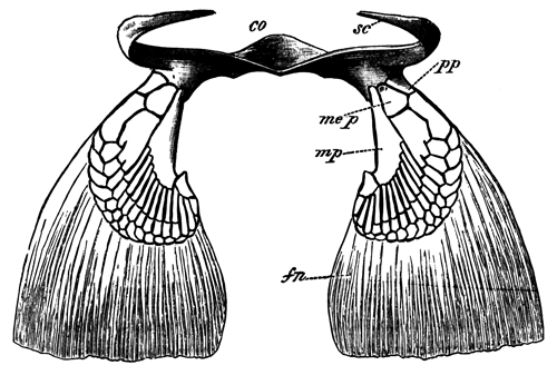
Pectoral fins and girdle of an adult of Scyllium
canicula (natural size, seen from behind and above).
co. Coracoid. sc. scapula. pp. propterygium. mep. mesopterygium. mp. metapterygium. fn. part of fin supported by horny fibre.
Fig. 2.
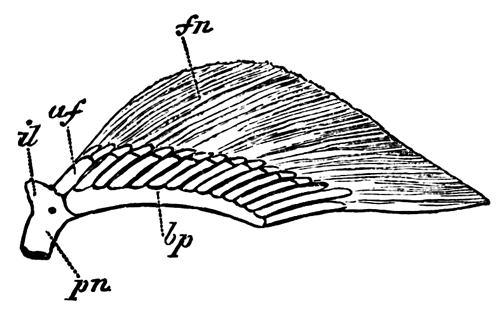
Right pelvic fin and part of pelvic girdle of an adult
female of Scyllium canicula (natural size).
il. iliac process.
pn. pubic process, cut across below.
bp. basipterygium. af. anterior cartilaginous fin-ray articulated to
pelvic girdle. fn. part of fin
supported by horny fibres.
The true skeleton consists of three basal pieces articulating
with the pectoral girdle; on the outer side of which there is a series of
more or less segmented cartilaginous fin-rays. Of the basal cartilages one
(pp) is anterior, a second (mep) is placed in the middle, and a third is
posterior (mp). They have been named by
Gegenbaur the propterygium, the mesopterygium, and the
metapterygium; and these names are now generally adopted.
The metapterygium is by far the most important of the three, and in
Scyllium canicula supports 12 or 13 rays[494]. It
forms a large part of the posterior boundary of the fin, and bears rays
only on its anterior border.
The mesopterygium supports 2 or 3 rays, in the basal parts of which the
segmentation into distinct rays is imperfect; and the propterygium supports
only a single ray.
The pelvic fins are horizontally placed, like the pectoral fins, but
differ from the latter in nearly meeting each other along the median
ventral line of the body. They also differ from the pectoral fins in having
a relatively much broader base of attachment to the sides of the body.
Their cartilaginous skeleton (woodcut, fig. 2) consists of a basal bar,
placed parallel to the base of the fin, and articulated in front with the
pelvic girdle.
On its outer border it articulates with a series of cartilaginous
fin-rays. I shall call the basal bar the basipterygium. The rays which it
bears are most of them less segmented than those of the pectoral fin, being
only divided into two; and the posterior ray, which is placed in the free
posterior border of the fin, continues the axis of the basipterygium. In
the male it is modified in connection with the so-called clasper.
The anterior fin-ray of the pelvic fin, which is broader than the other
rays, articulates directly with the pelvic girdle, instead of with the
basipterygium. This ray, in the female of Scyllium canicula and in
the male of Scyllium catulus (Gegenbaur), is peculiar in the fact
that its distal segment is longitudinally divided into two or more pieces,
instead of being single as is the case with the remaining rays. It is
probably equivalent to two of the posterior rays.
Development of the paired Fins.—The first
rudiments of the limbs appear in Scyllium, as in other fishes, as
slight longitudinal ridge-like thickenings of the epiblast, which closely
resemble the first rudiments of the unpaired fins.
These ridges are two in number on each side—an anterior
immediately behind the last visceral fold, and a posterior on the level of
the cloaca. In most Fishes they are in no way connected; but in some
Elasmobranch embryos, more especially in that of Torpedo, they are
connected together at their first development by a line of columnar
epiblast cells. This connecting line of columnar epiblast, however, is a
very transitory structure. The rudimentary fins soon become more prominent,
consisting of a projecting ridge both of epiblast and mesoblast, at the
outer edge of which is a fold of epiblast only, which soon reaches
considerable dimensions. At a later stage the mesoblast penetrates into
this fold, and the fin becomes a simple ridge of mesoblast covered by
epiblast. The pectoral fins are at first considerably ahead of the pelvic
fins in development.
The direction of the original epithelial line which connected the two
fins of each side is nearly, though not quite, longitudinal, sloping
somewhat obliquely ventralwards. It thus comes about that the attachment of
each pair of limbs is somewhat on a slant, and that the pelvic pair nearly
meet each other in the median ventral line shortly behind the anus.
The embryonic muscle-plates, as I have elsewhere shewn, grow into the
bases of the fins; and the cells derived from these ingrowths, which are
placed on the dorsal and ventral surfaces in immediate contact with the
epiblast, probably give rise to the dorsal and ventral muscular layers of
the limb, which are shewn in section in Plate 33, fig. 1, m, and in
Plate 33, fig. 7, m.
The cartilaginous skeleton of the limbs is developed in the indifferent
mesoblast cells between the two layers of muscles. Its early development in
both the pectoral and the pelvic fins is very similar. When first visible
it differs histologically from the adjacent mesoblast simply in the fact of
its cells being more concentrated; while its boundary is not sharply
marked.
At this stage it can only be studied by means of sections. It arises
simultaneously and continuously with the pectoral and pelvic girdles, and
consists, in both fins, of a bar springing at right angles from
the posterior side of the pectoral or pelvic girdle, and running parallel
to the long axis of the body along the base of the fin. The outer side of
this bar is continued into a thin plate, which extends into the fin.
The structure of the skeleton of the fin slightly after its first
differentiation will be best understood from Plate 33, fig. 1, and Plate
33, fig. 7. These figures represent transverse sections through the pelvic
and pectoral fins of the same embryo on the same scale. The basal bar is
seen at bp, and the plate at this stage
(which is considerably later than the first differentiation) already
partially segmented into rays at br.
Outside the region of the cartilaginous plate is seen the fringe with the
horny fibres (h.f.); and dorsally and
ventrally to the cartilaginous skeleton are seen the already
well-differentiated muscles (m).
The pectoral fin is shewn in horizontal section in Plate 33, fig. 6, at
a somewhat earlier stage than that to which the transverse sections belong.
The pectoral girdle (p.g.) is cut
transversely, and is seen to be perfectly continuous with the basal bar
(vp) of the fin. A similar continuity
between the basal bar of the pelvic fin and the pelvic girdle is shewn in
Plate 33, fig. 2, at a somewhat later stage. The plate continuous with the
basal bar of the fin is at first, to a considerable extent in the pectoral,
and to some extent in the pelvic fin, a continuous lamina, which
subsequently segments into rays. In the parts of the plate which eventually
form distinct rays, however, almost from the first the cells are more
concentrated than in those parts which will form the tissue between the
rays; and I am not inclined to lay any stress whatever upon the fact of the
cartilaginous fin-rays being primitively part of a continuous lamina, but
regard it as a secondary phenomenon, dependent on the mode of conversion of
embryonic mesoblast cells into cartilage. In all cases the separation into
distinct rays is to a large extent completed before the tissue of which the
plates are formed is sufficiently differentiated to be called cartilage by
an histologist.
The general position of the fins in relation to the body, and their
relative sizes, may be gathered from Plate 33, figs. 4 and 5, which
represent transverse sections of the same embryo as that from which the
transverse sections shewing the fin on a larger scale were taken.
During the first stage of its development the skeleton of both
fins may thus be described as consisting of a longitudinal bar running
along the base of the fin, and giving off at right angles series of rays
which pass into the fin. The longitudinal bar may be called the
basipterygium; and it is continuous in front with the pectoral or pelvic
girdle, as the case may be.
The further development of the primitive skeleton is different in the
case of the two fins.
The Pelvic Fin.—The changes in the pelvic fin are
comparatively slight. Plate 33, fig. 2, is a representation of the fin and
its skeleton in a female of Scyllium stellare shortly after the
primitive tissue is converted into cartilage, but while it is still so soft
as to require the very greatest care in dissection. The fin itself forms a
simple projection of the side of the body. The skeleton consists of a
basipterygium (bp), continuous in front
with the pelvic girdle. To the outer side of the basipterygium a series of
cartilaginous fin-rays are attached—the posterior ray forming a
direct prolongation of the basipterygium, while the anterior ray is united
rather with the pelvic girdle than with the basipterygium. All the
cartilaginous fin-rays except the first are completely continuous with the
basipterygium, their structure in section being hardly different from that
shewn in Plate 33, fig. 1.
The external form of the fin does not change very greatly in the course
of the further development; but the hinder part of the attached border is,
to some extent, separated off from the wall of the body, and becomes the
posterior border of the adult fin. With the exception of a certain amount
of segmentation in the rays, the character of the skeleton remains almost
as in the embryo. The changes which take place are illustrated by Plate 33,
fig. 3, shewing the fin of a young male of Scyllium stellare. The
basipterygium has become somewhat thicker, but is still continuous in front
with the pelvic girdle, and otherwise retains its earlier characters. The
cartilaginous fin-rays have now become segmented off from it and from the
pelvic girdle, the posterior end of the basipterygial bar being segmented
off as the terminal ray.
The anterior ray is directly articulated with the pelvic girdle, and the
remaining rays continue articulated with the basipterygium. Some of the
latter are partially segmented.
As may be gathered by comparing the figure of the fin at the
stage just described with that of the adult fin (woodcut, fig. 2), the
remaining changes are very slight. The most important is the segmentation
of the basipterygial bar from the pelvic girdle.
The pelvic fin thus retains in all essential points its primitive
structure.
The Pectoral Fin.—The earliest stage of the pectoral fin
differs, as I have shewn, from that of the pelvic fin only in minor points
(Pl. 33, fig. 6). There is the same
longitudinal or basipterygial bar (bp),
to which the fin-rays are attached, which is continuous in front with the
pectoral girdle (pg). The changes which
take place in the course of the further development, however, are very much
more considerable in the case of the pectoral than in that of the pelvic
fin.
The most important change in the external form of the fin is caused by a
reduction in the length of its attachment to the body. At first (Pl. 33, fig. 6), the base of the fin is as long as
the greatest breadth of the fin; but it gradually becomes shortened by
being constricted off from the body at its hinder end. In connection with
this process the posterior end of the basipterygial bar is gradually
rotated outwards, its anterior end remaining attached to the pectoral
girdle. In this way this bar comes to form the posterior border of the
skeleton of the fin (Pl. 33, figs. 8 and 9),
constituting the metapterygium (mp). It
becomes eventually segmented off from the pectoral girdle, simply
articulating with its hinder edge.
The plate of cartilage, which is continued outwards from the
basipterygium, or, as we may now call it, the metapterygium, into the fin,
is not nearly so completely divided up into fin-rays as the homologous part
of the pelvic fin; and this is especially the case with the basal part of
the plate. This basal part becomes, in fact, at first only divided into two
parts (Pl. 33, fig. 8)—a small anterior
part at the front end (me.p), and a
larger posterior along the base of the metapterygium (mp); and these two parts are not completely segmented
from each other. The anterior part directly joins the pectoral girdle at
its base, resembling in this respect the anterior fin-ray of the pelvic
girdle. It constitutes the (at this stage undivided) rudiment of the
mesopterygium and propterygium of Gegenbaur. It bears in
my specimen of this age four fin-rays at its extremity, the anterior not
being well marked. The remaining fin-rays are prolongations outwards of the
edge of the plate continuous with the metapterygium. These rays are at the
stage figured more or less transversely segmented; but at their outer edge
they are united together by a nearly continuous rim of cartilage. The
spaces between the fin-rays are relatively considerably larger than in the
adult.
The further changes in the cartilages of the pectoral limb are,
morphologically speaking, not important, and are easily understood by
reference to Pl. 33, fig. 9 (representing the
skeleton of the limb of a nearly ripe embryo). The front end of the
anterior basal cartilage becomes segmented off as a propterygium (pp), bearing a single fin-ray, leaving the
remainder of the cartilage as a mesopterygium (mes). The remainder of the now considerably segmented
fin-rays are borne by the metapterygium.
* * * * *
General Conclusions.—From the above observations,
conclusions of a positive kind may be drawn as to the primitive structure
of the skeleton; and the observations have also, it appears to me,
important bearings on the theories of my predecessors in this line of
investigation.
The most obvious of the positive conclusions is to the effect that the
embryonic skeleton of the paired fins consists of a series of parallel rays
similar to those of the unpaired fins. These rays support the soft parts of
the fins, which have the form of a longitudinal ridge; and they are
continuous at their base with a longitudinal bar. This bar, from its
position at the base of the fin, can clearly never have been a median axis
with the rays on both sides. It becomes the basipterygium in the pelvic
fin, which retains its embryonic structure much more completely than the
pectoral fin; and the metapterygium in the pectoral fin. The metapterygium
of the pectoral fin is thus clearly homologous with the basipterygium of
the pelvic fin, as originally supposed by Gegenbaur, and as has since been
maintained by Mivart. The propterygium and mesopterygium are obviously
relatively unimportant parts of the skeleton as compared with the
metapterygium.
My observations on the development of the skeleton of the fins
certainly do not of themselves demonstrate that the paired fins are
remnants of a once continuous lateral fin; but they support this view in
that they shew the primitive skeleton of the fins to have exactly the
character which might have been anticipated if the paired fins had
originated from a continuous lateral fin. The longitudinal bar of the
paired fins is believed by both Thacker and Mivart to be due to the
coalescence of the bases of the primitively independent rays of which they
believe the fin to have been originally composed. This view is probable
enough in itself, and is rendered more so by the fact, pointed out by
Mivart, that a longitudinal bar supporting the cartilaginous rays of
unpaired fins is occasionally formed; but there is no trace in the embryo
Scylliums of the bar in question being formed by the coalescence of rays,
though the fact of its being perfectly continuous with the bases of the
fin-rays is somewhat in favour of such coalescence.
Thacker and Mivart both hold that the pectoral and pelvic girdles are
developed by ventral and dorsal growths of the anterior end of the
longitudinal bar supporting the fin-rays.
There is, so far as I see, no theoretical objection to be taken to this
view; and the fact of the pectoral and pelvic girdles originating
continuously and long remaining united with the longitudinal bars of their
respective fins is in favour of it rather than the reverse. The same may be
said of the fact that the first part of each girdle to be formed is that in
the neighbourhood of the longitudinal bar (basipterygium) of the fin, the
dorsal and ventral prolongations being subsequent growths.
On the whole my observations do not throw much light on the theories of
Thacker and Mivart as to the genesis of the skeleton of the paired fin;
but, so far as they bear on the subject, they are distinctly favourable to
those theories.
The main results of my observations appear to me to be decidedly adverse
to the views recently put forward on the structure of the fin by Gegenbaur
and Huxley, both of whom, as stated above, consider the primitive type of
fin to be most nearly retained in Ceratodus, and to consist of a
central multisegmented axis with numerous lateral rays.
Gegenbaur derives the Elasmobranch pectoral fin from a form
which he calls the archipterygium, nearly like that of Ceratodus,
with a median axis and two rows of rays—but holds that in addition to
the rays attached to the median axis, which are alone found in
Ceratodus, there were other rays directly articulated to the
shoulder-girdle. He considers that in the Elasmobranch fin the majority of
the lateral rays on the posterior (or median according to his view of the
position of the limb) side have become aborted, and that the central axis
is represented by the metapterygium; while the pro- and mesopterygium and
their rays are, he believes, derived from those rays of the archipterygium
which originally articulated directly with the shoulder-girdle.
This view appears to me to be absolutely negatived by the facts of
development of the pectoral fin in Scyllium—not so much
because the pectoral fin in this form is necessarily to be regarded as
primitive, but because what Gegenbaur holds to be the primitive axis of the
biserial fin is demonstrated to be really the base, and it is only in the
adult that it is conceivable that a second set of lateral rays could have
existed on the posterior side of the metapterygium. If Gegenbaur's view
were correct, we should expect to find in the embryo, if anywhere, traces
of the second set of lateral rays; but the fact is that, as may easily be
seen by an inspection of figs. 6 and 7, such a second set of lateral rays
could not possibly have existed in a type of fin like that found in the
embryo. With this view of Gegenbaur's it appears to me that the theory held
by this anatomist to the effect that the limbs are modified gill-arches
also falls, in that his method of deriving the limbs from gill-arches
ceases to be admissible, while it is not easy to see how a limb, formed on
the type of the embryonic limb of Elasmobranchii, could be derived from a
gill-arch with its branchial rays.
Gegenbaur's older view, that the Elasmobranch fin retains a primitive
uniserial type, appears to me to be nearer the truth than his more recent
view on this subject; though I hold the fundamental point established by
the development of these parts in Scyllium to be that the posterior
border of the adult Elasmobranch pectoral fin is the primitive
base-line,i.e.line of attachment of the fin to the side of the
body.
Huxley holds that the mesopterygium is the proximal piece of
the axial skeleton of the limb of Ceratodus, and derives the
Elasmobranch fin from that of Ceratodus by the shortening of its
axis and the coalescence of some of its elements. The entirely secondary
character of the mesopterygium, and its total absence in the young embryo
Scyllium, appear to me as conclusive against Huxley's view as the
character of the embryonic fin is against that of Gegenbaur; and I should
be much more inclined to hold that the fin of Ceratodus has been
derived from a fin like that of the Elasmobranchii by a series of steps
similar to those which Huxley supposes to have led to the establishment of
the Elasmobranch fin, but in exactly the reverse order.
There is one statement of Davidoff's which I cannot allow to pass
without challenge. In comparing the skeletons of the paired and unpaired
fins he is anxious to prove that the former are independent of the axial
skeleton in their origin and that the latter have been segmented from the
axial skeleton, and thus to shew that an homology between the two is
impossible. In support of his view he states[495]
that he has satisfied himself, from embryos of Acanthias and
Scyllium, that the rays of the unpaired fins are undoubtedly
products of the segmentation of the dorsal and ventral spinous
processes.
This statement is wholly unintelligible to me. From my examination of
the development of the first dorsal and the anal fins of Scyllium I
find that their rays develop at a considerable distance from, and quite
independently of, the neural and hæmal arches, and that they are at an
early stage of development distinctly in a more advanced state of
histological differentiation than the neural and hæmal arches of the same
region. I have also found exactly the same in the embryos of
Lepidosteus.
I have, in fact, no doubt that the skeleton of both the paired and the
unpaired fins of Elasmobranchii and Lepidosteus is in its development
independent of the axial skeleton. The phylogenetic mode of origin of the
skeleton both of the paired and of the unpaired fins cannot, however, be
made out without further investigation.
EXPLANATION OF PLATE 33.[496]
Fig. 1. Transverse section through the pelvic fin of an
embryo of Scyllium belonging to stage P1, magnified 50
diameters. bp. basipterygium. br. fin ray. m. muscle. hf. horny fibres supporting the peripheral part of
the fin.
Fig. 2. Pelvic fin of a very young female embryo of
Scyllium stellare, magnified 16 diameters. bp. basipterygium. pu. pubic process of pelvic girdle (cut across
below). il. iliac process of pelvic
girdle. fo. foramen.
Fig. 3. Pelvic fin of a young male embryo of Scyllium
stellare, magnified 16 diameters. bp. basipterygium. mo. process of basipterygium continued into clasper.
il. iliac process of pelvic girdle.
pu. pubic section of pelvic girdle.
Fig. 4. Transverse section through the ventral part of
the trunk of an embryo Scyllium of stage P, in the region of the
pectoral fins, to shew how the fins are attached to the body, magnified 18
diameters. br. cartilaginous fin-ray.
bp. basipterygium. m. muscle of
fin. mp. muscle-plate.
Fig. 5. Transverse section through the ventral part of
the trunk of an embryo Scyllium of stage P, in the region of the
pelvic fin, on the same scale as fig. 4. bp. basipterygium. br. cartilaginous fin-rays. m. muscle of the
fins. mp. muscle-plate.
Fig. 6. Pectoral fin of an embryo of Scyllium
canicula, of a stage between O and P, in longitudinal and horizontal
section (the skeleton of the fin was still in the condition of embryonic
cartilage), magnified 36 diameters. bp.
basipterygium (eventual metapterygium). fr. cartilaginous fin-rays. pg. pectoral girdle in transverse section. fo. foramen in pectoral girdle. pe. epithelium of peritoneal cavity.
Fig. 7. Transverse section through the pectoral fin of a
Scyllium embryo of stage P, magnified 50 diameters. bp. basipterygium. br. cartilaginous fin-ray. m. muscle. hf. horny fibres.
Fig. 8. Pectoral fin of an embryo of Scyllium
stellare, magnified 16 diameters. mp. metapterygium (basipterygium of earlier stage).
me.p. rudiment of future pro- and
mesopterygium. sc. cut surface of a
scapular process. cr. coracoid process.
fr. foramen. hf. horny fibres.
Fig. 9. Skeleton of the pectoral fin and part of
pectoral girdle of a nearly ripe embryo of Scyllium stellare,
magnified 10 diameters. mp.
metapterygium. mes. mesopterygium. pp. propterygium. cr. coracoid process.
From Owen's observations on the Marsupials it is clear that
the yolk-sack in this group plays an important (if not the most important)
part, in absorbing the maternal nutriment destined for the fœtus. The
fact that in Marsupials both the yolk-sack and the allantois are concerned
in rendering the chorion vascular, makes it à priori
probable that this was also the case in the primitive types of the
Placentalia; and this deduction is supported by the fact that in the
Rodentia, Insectivora, and Cheiroptera this peculiarity of the fœtal
membranes is actually found. In the primitive Placentalia it is also
probable that from the discoidal allantoic region of the chorion simple
fœtal villi, like those of the Pig, projected into uterine crypts;
but it is not certain how far the umbilical region of the chorion, which
was no doubt vascular, may also have been villous. From such a primitive
type of fœtal membranes divergencies in various directions have given
rise to the types of fœtal membranes found at the present day.
In a general way it may be laid down that variations in any direction
which tended to increase the absorbing capacities of the chorion would be
advantageous. There are two obvious ways in which this might be done, viz. (1) by increasing the complexity of the
fœtal villi and maternal crypts over a limited area, (2) by
increasing the area of the part of the chorion covered by the placental
villi. Various combinations of the two processes would also, of course, be
advantageous.
The most fundamental change which has taken place in all the
existing Placentalia is the exclusion of the umbilical vesicle from any
important function in the nutrition of the fœtus.
The arrangement of the fœtal parts in the Rodentia, Insectivora,
and Cheiroptera may be directly derived from the primitive form by
supposing the villi of the discoidal placental area to have become more
complex, so as to form a deciduate discoidal placenta, while the yolk-sack
still plays a part, though physiologically an unimportant part, in
rendering the chorion vascular.
In the Carnivora, again, we have to start from the discoidal placenta,
as evinced by the fact that in the growth of the placenta the allantoic
region of the placenta is at first discoidal, and only becomes
zonary at a later stage. A zonary deciduate placenta indicates an increase
both in area and in complexity. The relative diminution of the breadth of
the placental zone in late fœtal life in the zonary placenta of the
Carnivora is probably due to its being on the whole advantageous to secure
the nutrition of the fœtus by insuring a more intimate relation
between the fœtal and maternal parts, than by increasing their area
of contact. The reason of this is not obvious, but, as shewn below, there
are other cases where it is clear that a diminution in the area of the
placenta has taken place, accompanied by an increase in the complexity of
its villi.
The second type of differentiation from the primitive form of placenta
is illustrated by the Lemuridæ, the Suidæ, and Manis. In all these
cases the area of the placental villi appears to have increased so as to
cover nearly the whole subzonal membrane, without the villi increasing to
any great extent in complexity. From the diffused placenta covering the
whole surface of the chorion, differentiations appear to have taken place
in various directions. The placenta of Man and Apes, from its mode of
ontogeny, is clearly derived from a diffused placenta (very probably
similar to that of Lemurs) by a concentration of the fœtal villi,
which are originally spread over the whole chorion, to a disk-shaped area,
and by an increase in their arborescence. Thus the discoidal placenta of
Man has no connexion with, and ought not to be placed in, the same class as
those of the Rodentia, Cheiroptera, and Insectivora.
The polycotyledonary forms of placenta are due to similar
concentrations of the fœtal villi of an originally diffused
placenta.
In the Edentata we have a group with very varying types of placenta.
Very probably these may all be differentiations within the group itself
from a diffused placenta such as that found in Manis. The zonary
placenta of Orycteropus is capable of being easily derived from that
of Manis by the disappearance of the fœtal villi at the two
poles of the ovum. The small size of the umbilical vesicle in
Orycteropus indicates that its discoidal placenta is not, like that
of the Carnivora, directly derived from a type with both allantoic and
umbilical vascularization of the chorion. The discoidal and dome-shaped
placentæ of the Armadillos, Myrmecophaga, and the Sloths may easily
have been formed from a diffused placenta, just as the discoidal placenta
of the Simiidæ and Hominidæ appears to have been formed from a diffused
placenta like that of the Lemuridæ.
The presence of zonary placenta in Hyrax and Elephas does
not necessarily afford any proof of affinity of these types with the
Carnivora. A zonary placenta may be quite as easily derived from a diffused
placenta as from a discoidal placenta; and the presence of two villous
patches at the poles of the chorion in Elephas very probably
indicates that its placenta has been evolved from a diffused placenta.
Although it would not be wise to attempt to found a classification upon
the placental characters alone, it may be worth while to make a few
suggestions as to the affinities of the orders of Mammalia indicated by the
structure of the placenta. We clearly, of course, have to start with forms
which could not be grouped with any of the existing orders, but which might
be called the Protoplacentalia. They probably had the primitive type of
placenta described above: the nearest living representatives of the group
are the Rodentia, Insectivora, and Cheiroptera. Before, however, these
three groups had become distinctly differentiated, there must have branched
off from the primitive stock the ancestors of the Lemuridæ, the Ungulata,
and the Edentata.
It is obvious on general anatomical grounds that the Monkeys and Man are
to be derived from a primitive Lemurian type; and with this
conclusion the form of the placenta completely tallies. The primitive
Edentata and Ungulata had no doubt a diffused placenta which was probably
not very different from that of the primitive Lemurs; but how far these
groups arose quite independently from the primitive stock, or whether they
may have had a nearer common ancestor, cannot be decided from the structure
of the placenta. The Carnivora were certainly an offshoot from the
primitive placental type which was quite independent of the three groups
just mentioned; but the character of the placenta of the Carnivora does not
indicate at what stage in the evolution of the placental Mammalia a
primitive type of Carnivora was first differentiated.
No important light is thrown by the placenta on the affinities of the
Proboscidea, the Cetacea, or the Sirenia; but the character of the placenta
in the latter group favours the view of their being related to the
Ungulata.
(With Plates 34-42.)
| TABLE OF CONTENTS. |
|---|
| PAGE |
| Introduction |
739 |
| General Development |
740 |
| Brain— |
| Adult brain |
759 |
| Development of the brain |
764 |
| Comparison of the larval and adult brain of
Lepidosteus, together with some observations on the systematic value
of the characters of the Ganoid brain |
767 |
| Sense Organs— |
| Olfactory organ |
771 |
| Anatomy of the eye |
ib. |
| Development of the eye |
772 |
| Suctorial Disc |
774 |
| Muscular System |
775 |
| Skeleton— |
| Vertebral column and ribs of the adult |
776 |
| Development of the vertebral column and
ribs. |
778 |
| Comparison of the vertebral column of
Lepidosteus with that of other forms |
792 |
| The ribs of Fishes |
793 |
| The skeleton of the ventral lobe of the tail
fin, and its bearing on the nature of the tail fin of the various types of
Pisces |
801 |
| Excretory and Generative
Organs— |
| Anatomy of the excretory and generative organs
of the female |
810 |
| Anatomy of the excretory and generative organs
of the male |
813 |
| Development of the excretory and generative
organs |
815 |
| Theoretical considerations |
822 |
| The Alimentary Canal and its
Appendages— |
| Topographical anatomy of the alimentary
canal |
828 |
| Development of the alimentary canal and its
appendages |
831 |
| The Gill on the Hyoid
Arch |
835 |
| The systematic position of
Lepidosteus |
836 |
| List of memoirs on the Anatomy and
Development of Lepidosteus |
840 |
| List of Reference Letters |
841 |
| Explanation of Plates |
842 |
The following paper is the outcome of the very valuable gift
of a series of embryos and larvæ of Lepidosteus by Professor Alex.
Agassiz, to whom we take this opportunity of expressing our most sincere
thanks. The skull of these embryos and larvæ has been studied by Professor
Parker, and forms the subject of a memoir already presented to the Royal
Society.
Considering that Lepidosteus is one of the most interesting of
existing Ganoids, and that it is very closely related to species of Ganoids
which flourished during the Triassic period, we naturally felt keenly
anxious to make the most of the opportunity of working at its development
offered to us by Professor Agassiz' gift. Professor Agassiz, moreover, most
kindly furnished us with four examples of the adult Fish, which have
enabled us to make this paper a study of the adult anatomy as well as of
the development.
The first part of our paper is devoted to the segmentation, formation of
the germinal layers, and general development of the embryo and larva. The
next part consists of a series of sections on the organs, in which both
their structure in the adult and their development are dealt with. This
part is not, however, in any sense a monograph, and where already known,
the anatomy is described with the greatest possible brevity. In this part
of the paper considerable space is devoted to a comparison of the organs of
Lepidosteus with those of other Fishes, and to a statement of the
conclusions which follow from such comparison.
The last part of the paper deals with the systematic position of
Lepidosteus and of the Ganoids generally.
The spawning of Lepidosteus takes place in the neighbourhood of
New York about May 20th. Agassiz (No. 1)[499] gives an account of the process from Mr S. W. Garman's
notes, which we venture to quote in full.
"Black Lake is well stocked with Bill-fish. When they
appear, they are said to come in countless numbers. This is only for a few
days in the spring, in the spawning season, between the 15th of May and the
8th of June. During the balance of the season they are seldom seen. They
remain in the deeper parts of the lake, away from the shore, and, probably,
are more or less nocturnal in habits. Out of season, an occasional one is
caught on a hook baited with a minnow. Commencing with the 20th of April,
until the 14th of May we were unable to find the Fish, or to find persons
who had seen them during this time. Then a fisherman reported having seen
one rise to the surface. Later, others were seen. On the afternoon of the
18th, a few were found on the points, depositing the spawn. The
temperature at the time was 68° to 69° on the shoals, while out in the lake
the mercury stood at 62° to 63°. The points on which the eggs were
laid were of naked granite, which had been broken by the frost and heat
into angular blocks of 3 to 8 inches in diameter. The blocks were tumbled
upon each other like loose heaps of brick-bats, and upon and between them
the eggs were dropped. The points are the extremities of small
capes that make out into the lake. The eggs were laid in water varying in
depth from 2 to 14 inches. At the time of approaching the shoals, the Fish
might be seen to rise quite often to the surface to take air. This they did
by thrusting the bill out of the water as far as the corners of the mouth,
which was then opened widely and closed with a snap. After taking the air,
they seemed more able to remain at the surface. Out in the lake they are
very timid, but once buried upon the shoals they become quite reckless as
to what is going on about them. A few moments after being driven off, one
or more of the males would return as if scouting. If frightened, he would
retire for some time; then another scout would appear. If all promised
well, the females, with the attendant males, would come back. Each female
was accompanied by from one to four males. Most often, a male rested
against each side, with their bills reaching up toward the back of her
head. Closely crowded together, the little party would pass back and forth
over the rocky bed they had selected, sometimes passing the same spot
half-a-dozen times without dropping an egg, then suddenly would indulge in
an orgasm; and, lashing and plashing the water in all directions with their
convulsive movements, would scatter at the same instant the eggs and the
sperm. This ended, another season of moving slowly back and forth was
observed, to be in turn followed by another of excitement. The eggs were
excessively sticky. To whatever they happened to touch, they stuck, and so
tenaciously that it was next to impossible to release them without tearing
away a portion of their envelopes. It is doubtful whether the eggs would
hatch if removed. As far as could be seen at the time, upon or under the
rocks to which the eggs were fastened there was an utter absence of
anything that might serve as food for the young Fishes.
Other Fishes, Bull-heads, &c., are said to follow the Bill-fish to eat the spawn.
It may be so. It was not verified. Certainly the points under observations
were unmolested. During the afternoon of the 18th of May a few eggs were
scattered on several of the beds. On the 19th there were more. With the
spear and the snare, several dozens of both sexes of the Fish were taken.
Taking one out did not seem greatly to startle the others. They returned
very soon. The males are much smaller than the average size of the females;
and, judging from those taken, would seem to have as adults greater
uniformity in size. The largest taken was a female, of 4 feet 1½ inch
in length. Others of 2 feet 6 inches contained ripe ova. With the 19th of
May all disappeared, and for a time—the weather being meanwhile cold
and stormy—there were no signs of their continued existence to be met
with. Nearly two weeks later, on the 31st of May, as stated by Mr Henry J.
Perry, they again came up, not in small detachments on scattered points as
before, but in multitudes, on every shoal at all according
with their ideas of spawning beds. They remained but two days. During the
summer it happens now and then that one is seen to come up for his mouthful
of air; beyond this there will be nothing to suggest the ravenous masses
hidden by the darkness of the waters.
Egg membranes.—The ova of Lepidosteus are spherical
bodies of about 3 millims. in diameter.
They have a double investment consisting of (1) an outer covering formed of
elongated, highly refractive bodies, somewhat pyriform at their outer ends
(Plate 34, fig. 17, f.e.), which are
probably metamorphosed follicular cells[500],
and (2) of an inner membrane, divided into two zones, viz.: an outer and thicker zone, which is radially
striated, and constitutes the zona radiata (z.r.), and an inner and narrow homogeneous zone
(z.r´.).
Segmentation.—We have observed several stages in the
segmentation, which shew that it is complete, but that it approaches the
meroblastic type more nearly than in the case of any other known
holoblastic ovum.
Our earliest stage shewed a vertical furrow at the upper or animal pole,
extending through about one-fifth of the circumference (Plate 34, fig. 1),
and in a slightly later stage we found a second similar furrow at right
angles to the first (Plate 34, fig. 2). We have not been fortunate enough
to observe the next phases of the segmentation, but on the second day after
impregnation (Plate 34, fig. 3), the animal pole is completely divided into
small segments, which form a disc, homologous to the blastoderm of
meroblastic ova; while the vegetative pole, which subsequently forms a
large yolk-sack, is divided by a few vertical furrows, four of which nearly
meet at the pole opposite the blastoderm (Plate 34, fig. 4). The majority
of the vertical furrows extend only a short way from the edge of the small
spheres, and are partially intercepted by imperfect equatorial furrows.
Development of the embryo.—We have not been able
to work out the stages immediately following the segmentation, owing to
want of material; and in the next stage satisfactorily observed, on the
third day after impregnation, the body of the embryo is distinctly
differentiated. The lower pole of the ovum is then formed of a mass in
which no traces of the previous segments or segmentation furrows could any
longer be detected.
Some of the dates of the specimens sent to us appear to have been
transposed; so that our statements as to ages must only be taken as
approximately correct.
Third day after impregnation.—In this stage the embryo is
about 3.5 millims. in length, and has a
somewhat dumb-bell shaped outline (Plate 34, fig. 5). It consists of (1) an
outer area (p.z) with some resemblance to
the area pellucida of the Avian embryo, forming the parietal part of the
body; and (2) a central portion consisting of the vertebral and medullary
plates and the axial portions of the embryo. In hardened specimens the
peripheral part forms a shallow depression surrounding the central part of
the embryo.
The central part constitutes a somewhat prominent ridge, the axial part
of it being the medullary plate. Along the anterior half of this part a
dark line could be observed in all our specimens, which we at first
imagined to be caused by a shallow groove. We have, however, failed to find
in our sections a groove in this situation except in a single instance
(Plate 35, fig. 20, x), and are inclined to attribute the appearance
above-mentioned to the presence of somewhat irregular ridges of the outer
layer of the epiblast, which have probably been artificially produced in
the process of hardening.
The anterior end of the central part is slightly dilated to form the
brain (b); and there is present a pair of lateral swellings near the
anterior end of the brain which we believe to be the commencing optic
vesicles. We could not trace any other clear indications of the
differentiation of the brain into distinct lobes.
At the hinder end of the central part of the embryo a very distinct
dilatation may also be observed, which is probably homologous with the tail
swelling of Teleostei. Its structure is more particularly dealt with in the
description of our sections of this stage.
After the removal of the egg-membranes described above we find
that there remains a delicate membrane closely attached, to the epiblast.
This membrane can be isolated in distinct portions, and appears to be too
definite to be regarded as an artificial product.
We have been able to prepare several more or less complete series of
sections of embryos of this stage (Plate 35, figs. 18-22). These sections
present as a whole a most striking resemblance to those of Teleostean
embryos at a corresponding stage of development.
Three germinal layers are already fully established. The epiblast
(ep.) is formed of the same parts as in
Teleostei, viz.:—of an outer epidermic
and an inner nervous or mucous stratum. In the parietal region of the
embryo these strata are each formed of a single row of cells only. The
cells of both strata are somewhat flattened, but those of the epidermic
stratum are decidedly the more flattened of the two.
Along the axial line there is placed, as we have stated above, the
medullary plate. The epidermic stratum passes over this plate without
undergoing any change of character, and the plate is entirely
constituted of the nervous stratum of the epidermis.
The medullary plate has, roughly speaking, the form of a solid keel,
projecting inwards towards the yolk. There is no trace, at this stage at
any rate, of a medullary groove; and as, we shall afterwards shew, the
central canal of the cerebro-spinal cord is formed in the middle of the
solid keel. The shape of this keel varies according to the region of the
body. In the head (Plate 35, fig. 18, m.c.), it is very prominent, and forming, as it does,
the major part of the axial tissue of the body, impresses its own shape on
the other parts of the head and gives rise to a marked ridge on the surface
of the head directed towards the yolk. In the trunk (Plate 35, figs. 19,
20) the keel is much less prominent, but still projects sufficiently to
give a convex form to the surface of the body turned towards the yolk.
In the head, and also near the hind end of the trunk, the nervous layer
of the epiblast continuous with the keel on each side is considerably
thicker than the lateral parts of the layer. The thickening of the nervous
layer in the head gives rise to what has been called by Götte[501] the special sense plate,
owing to its being
subsequently concerned in the formation of parts of the organs of special
sense. We cannot agree with Götte in regarding it as part of the brain.
In the keel itself two parts may be distinguished, viz.: a superficial part, best marked in the region of the
brain, formed of more or less irregularly arranged polygonal cells, and a
deeper part of horizontally placed flatter cells. The upper part is mainly
concerned in the formation of the cranial nerves, and of the dorsal roots
of the spinal nerves.
The mesoblast (ms.) in the trunk
consists of a pair of independent plates which are continued forwards into
the head, and in the prechordal region of the latter, unite below the
medullary keel.
The mesoblastic plates of the trunk are imperfectly divided into
vertebral and lateral regions. Neither longitudinal sections nor surface
views shew at this stage any trace of a division of the mesoblast into
somites. The mesoblast cells are polygonal, and no indication is as yet
present of a division into splanchnic and somatic layers.
The notochord (nc.) is well
established, so that its origin could not be made out. It is, however, much
more sharply separated from the mesoblastic plates than from the hypoblast,
though the ventral and inner corners of the mesoblastic plates which run in
underneath it on either side, are often imperfectly separated from it. It
is formed of polygonal cells, of which between 40 and 50 may as a rule be
seen in a single section. No sheath is present around it. It has the usual
extension in front.
The hypoblast (hy.) has the form of a
membrane, composed of a single row of oval cells, bounding the embryo on
the side adjoining the yolk.
In the region of the caudal swelling the relations of the germinal
layers undergo some changes. This region may, from the analogy of other
Vertebrates, be assumed to constitute the lip of the blastopore. We find
accordingly that the layers become more or less fused. In the anterior part
of the tail swelling, the boundary between the notochord and hypoblast
becomes indistinct. A short way behind this point (Plate 35, fig. 21), the
notochord unites with the medullary keel, and a neurenteric cord,
homologous with the neurenteric canal of other Ichthyopsida, is thus
established. In the same region the boundary between the lateral plates of
mesoblast and the notochord, and further back (Plate 35, fig. 22), that
between the mesoblast and the medullary keel, becomes obliterated.
Fifth day after impregnation.—Between the stage last
described and the next stage of which we have specimens, a considerable
progress has been made. The embryo (Plate 34, figs. 6 and 7) has grown
markedly in length and embraces more than half the circumference of the
ovum. Its general appearance is, however, much the same as in the earlier
stage, but in the cephalic region the medullary plate is divided by
constrictions into three distinct lobes, constituting the regions of the
fore-brain, the mid-brain, and the hind-brain. The fore-brain (Plate 34,
fig. 6, f.b.) is considerably the largest
of the three lobes, and a pair of lateral projections forming the optic
vesicles are decidedly more conspicuous than in the previous stage. The
mid-brain (m.b.) is the smallest of the
three lobes, while the hind-brain (h.b.)
is decidedly longer, and passes insensibly into the spinal cord behind.
The medullary keel, though retaining to a great extent the shape it had
in the last stage, is no longer completely solid. Throughout the whole
region of the brain and in the anterior part of the trunk (Plate 35, figs.
23, 24, 25) a slit-like lumen has become formed. We are inclined to hold
that this is due to the appearance of a space between the cells, and not,
as supposed by Oellacher for Teleostei, to an actual absorption of cells,
though we must admit that our sections are hardly sufficiently well
preserved to be conclusive in settling this point. Various stages in its
growth may be observed in different regions of the cerebro-spinal cord.
When first formed, it is a very imperfectly defined cavity, and a few cells
may be seen passing right across from one side of it to the other. It
gradually becomes more definite, and its wall then acquires a regular
outline.
The optic vesicles are now to be seen in section (Plate 35, fig. 23,
op.) as flattish outgrowths of the wall
of the fore-brain, into which the lumen of the third ventricle
is prolonged for a short distance.
The brain has become to some extent separate from the superjacent
epiblast, but the exact mode in which this is effected is not clear to us.
In some sections it appears that the separation takes place in such a way
that the nervous keel is only covered above by the epidermic layer of the
epiblast, and that the nervous layer, subsequently interposed between the
two, grows in from the two sides. Such a section is represented in Plate
35, fig. 24. Other sections again favour the view that in the isolation of
the nervous keel, a superficial layer of it remains attached to the nervous
layer of the epidermis at the two sides, and so, from the first, forms a
continuous layer between the nervous keel and the epidermic layer of the
epiblast (Plate 35, fig. 25). In the absence of a better series of sections
we do not feel able to determine this point. The posterior part of the
nervous keel retains the characters of the previous stage.
At the sides of the hind-brain very distinct commencements of the
auditory vesicles are apparent. They form shallow pits (Plate 35, fig. 24,
au.) of the thickened part of the nervous
layer adjoining the brain in this region. Each pit is covered over by the
epidermic layer above, which has no share in its formation.
In many parts of the lateral regions of the body the nervous layer of
the epidermis is more than one cell deep.
The mesoblastic plates are now divided in the anterior part of the trunk
into a somatic and a splanchnic layer (Plate 35, fig. 25, so., sp.), though no distinct cavity is
as yet present between these two layers. Their vertebral extremities are
somewhat wedge-shaped in section, the base of the wedge being placed at the
sides of the medullary keel. The wedge-shaped portions are formed of a
superficial layer of palisade-like cells and an inner kernel of polygonal
cells. The superficial layer on the dorsal side is continuous with the
somatic mesoblast, while the remainder pertains to the splanchnic
layer.
The diameter of the notochord has diminished, and the cells have assumed
a flattened form, the protoplasm being confined to an axial region. In
consequence of this, the peripheral layer appears clear in transverse
sections. A delicate cuticular sheath is formed around
it. This sheath is probably the commencement of the permanent sheath of
later stages, but at this stage it cannot be distinguished in structure
from a delicate cuticle which surrounds the greater part of the medullary
cord.
The hypoblast has undergone no changes of importance.
The layers at the posterior end of the embryo retain the characters of
the last stage.
Sixth day after impregnation.—At this stage (Plate 34, fig.
8) the embryo is considerably more advanced than at the last stage. The
trunk has decidedly increased in length, and the head forms a relatively
smaller portion of the whole. The regions of the brain are more distinct.
The optic vesicles (op.) have grown
outwards so as to nearly reach the edges of the area which forms the
parietal part of the body. The fore-brain projects slightly in front, and
the mid-brain is seen as a distinct rounded prominence. Behind the latter
is placed the hind-brain, which passes insensibly into the spinal cord. On
either side of the mid- and hind-brain a small region is slightly marked
off from the rest of the parietal part, and on this are seen two more or
less transversely directed streaks, which, by comparison with the
Sturgeon[502], we are inclined to regard as
the two first visceral clefts (br.c.). We
have, however, failed to make them out in sections, and owing to the
insufficiency of our material, we have not even studied them in surface
views as completely as we could have wished.
The body is now laterally compressed, and more decidedly raised from the
yolk than in the previous stages. In the lateral regions of the trunk the
two segmental or archinephric ducts (sg.) are visible in surface views: the front end of
each is placed at the level of the hinder border of the head, and is marked
by a flexure inwards towards the middle line. The remainder of each duct is
straight, and extends backwards for about half the length of the embryo.
The tail has much the same appearance as in the last stage.
The vertebral regions of the mesoblastic plates are now segmented for
the greater part of the length of the trunk, and the somites of which
they are composed (Plate 36, fig. 30, pr.) are very conspicuous in surface views.
Our sections of this stage are not so complete as could be desired: they
shew, however, several points of interest.
The central canal of the nervous system is large, with well-defined
walls, and in hardened specimens is filled with a coagulum. It extends
nearly to the region of the tail.
The optic vesicles, which are so conspicuous in surface views, appear in
section (Plate 35, fig. 26, op.) as
knob-like outgrowths of the fore-brain, and very closely resemble the
figures given by Oellacher of these vesicles in Teleostei[503].
From the analogy of the previous stage, we are inclined to think that
they have a lumen continuous with that of the fore-brain. In our only
section through them, however, they are solid, but this is probably due to
the section merely passing through them to one side.
The auditory pits (Plate 35, fig. 27, au.) are now well marked, and have the form of
somewhat elongated grooves, the walls of which are formed of a single layer
of columnar cells belonging to the nervous layer of the epidermis, and
extending inwards so as nearly to touch the brain.
In an earlier stage it was pointed out that the dorsal part of the
medullary keel was different in its structure from the remainder, and that
it was destined to give rise to the nerves. The process of differentiation
is now to a great extent completed, and may best be seen in the auditory
region (Plate 35, fig. 27, VIII.). In this region there was present during
the last stage a great rhomboidal mass of cells at the dorsal region of the
brain (Plate 35, fig. 24, VIII.). In the present stage, this, which is the
rudiment of the seventh and auditory nerves, is seen growing down on each
side from the roof of the hind-brain, between the brain and the auditory
involution, and abutting against the wall of the latter.
Rudiments of the spinal nerves are also seen at intervals as projections
from the dorsal angles of the spinal cord (Plate 36, fig. 29, sp.n.). They extend only for a short distance
outwards, gradually tapering off to a point, and situated between
the epiblast and the dorsal angles of the mesoblastic somites.
The process of formation of the cranial nerves and dorsal roots of the
spinal nerves is, it will be seen, essentially the same as that already
known in the case of Elasmobranchii, Aves, &c. The nerves arise as outgrowths of a special crest of
cells, the neural crest of Marshall, which is placed along the
dorsal angle of the cord. The peculiar position of the dorsal roots of the
spinal nerves is also very similar to what has been met with in the early
stages of these structures by Marshall in Birds[504],
and by one of us in Elasmobranchii[505].
In the parietal region a cavity has now appeared in part of the trunk
between the splanchnic and somatic layers of the mesoblast (Plate 36, fig.
29, b.c.), the somatic layer (so.) consisting of a single row of columnar
cells on the dorsal side, while the remainder of each somite is formed of
the splanchnic layer (sp.). In many of
the sections the somatic layer is separated by a considerable interval from
the epiblast.
We have been able to some extent to follow the development of the
segmental duct. The imperfect preservation of our specimens has, as in
other instances, rendered the study of the point somewhat difficult, but we
believe that the figure representing the development of the duct some way
behind its front end (Plate 36, fig. 29) is an accurate representation of
what may be seen in a good many of our sections.
It appears from these sections that the duct (Plate 36, fig. 29, sg.) is developed as a hollow ridge-like
outgrowth of the somatic layer of mesoblast, directed towards the epiblast,
in which it causes a slight bulging. The cavity of the ridge freely
communicates with the body-cavity. The anterior part of this ridge appears
to be formed first. Very soon, in fact, in an older embryo belonging to
this stage, the greater part of the groove becomes segmented off as a duct
lying between the epiblast and somatic mesoblast (Plate 36, fig. 28, sg.), while the front end still remains, as we
believe, in communication with the body-cavity by an anterior pore.
This mode of development corresponds in every particular with
that observed in Teleostei by Rosenberg and Oellacher.
The structure of the notochord (nc.)
at this stage is very similar to that observed by one of us in
Elasmobranchii[506]. The cord is formed of
transversely arranged flattened cells, the outer parts of which are
vacuolated, while the inner parts are granular, and contain the nuclei.
This structure gives rise to the appearance in transverse sections of an
axial darker area and a peripheral lighter portion.
The hypoblast retains for the most part its earlier constitution, but
underneath the notochord, in the trunk, it is somewhat thickened, and the
cells at the two sides spread in to some extent under the thickened portion
(Plate 36, fig. 29, s.nc.). This
thickening, as is shewn in transverse sections at the stage when the
segmental duct becomes separated from the somatic mesoblast (Plate 36, fig.
28, s.nc.), is the commencement of the
subnotochordal rod.
The tail end of the embryo still retains its earlier characters.
Seventh day after impregnation.—Our series of specimens of
this stage is very imperfect, and we are only able to call attention to the
development of a certain number of organs.
Our sections clearly establish the fact that the optic vesicles are now
hollow processes of the fore-brain. Their outer ends are dilated, and are
in contact with the external skin. The formation of the optic cup has not,
however, commenced. The nervous layer of the skin adjoining the outer wall
of the optic cup is very slightly thickened, constituting the earliest
rudiment of the lens.
In one of our embryos of this day the developing auditory vesicle still
has the form of a pit, but in the other it is a closed vesicle, already
constricted off from the nervous layer of the epidermis.
With reference to the development of the excretory duct we cannot add
much to what we have already stated in describing the last stage.
The duct is considerably dilated anteriorly (Plate 36, fig. 31, sg.); but our sections throw no light on the
nature of the abdominal pore. The posterior part of the duct has still the
form of a hollow ridge united with somatic mesoblast (Plate 36, fig.
32, sg.).
During this stage, the embryo becomes to a small extent folded off from
the yolk-sack both in front and behind, and in the course of this process
the anterior and posterior extremities of the alimentary tract become
definitely established.
We have not got as clear a view of the process of formation of these two
sections of the alimentary tract as we could desire, but our observations
appear to shew that the process is in many respects similar to that which
takes place in the formation of the anterior part of the alimentary tract
in Elasmobranchii[507]. One of us has shewn that in
Elasmobranchii the ventral wall of the throat is formed not by a
process of folding in of the hypoblastic sheet as in Birds, but by a growth
of the ventral face of the hypoblastic sheet on each side of and at some
little distance from the middle line. Each growth is directed inwards, and
the two eventually meet and unite, thus forming a complete ventral wall for
the gut. Exactly the same process would seem to take place in
Lepidosteus, and after the lumen of the gut is in this way
established, a process of mesoblast on each side also makes its appearance,
forming a mesoblastic investment on the ventral side of the alimentary
tract. Some time after the alimentary tract has been thus formed, the
epiblast becomes folded in, in exactly the same manner as in the Chick, the
embryo becoming thereby partially constricted off from the yolk (Plate 36,
figs. 33, 34).
The form of the lumen of the alimentary tract differs somewhat in front
and behind. In front, the hypoblastic sheet remains perfectly flat during
the formation of the throat, and thus the lumen of the latter has merely
the form of a slit. The lumen of the posterior end of the alimentary tract
is, however, narrower and deeper (Plate 36, figs. 33, 34, al.). Both in front and behind, the lateral parts of
the hypoblastic sheet become separated from the true alimentary tract as
soon as the lumen of the latter is established.
It is quite possible that at the extreme posterior end of the embryo a
modification of the above process may take place, for in this region the
hypoblast appears to us to have the form of a solid cord.
We could detect no true neurenteric canal, although a more or less
complete fusion of the germinal layers at the tail end of the embryo may
still be traced.
During this stage the protoplasm of the notochordal cells, which in the
last stage formed a kind of axial rod in the centre of the notochord,
begins to spread outwards toward the sheath of the notochord.
Eighth day after impregnation.—The external form of the
embryo (Plate 34, fig. 9) shews a great advance upon the stage last
figured. Both head and body are much more compressed laterally and raised
from the yolk, and the head end is folded off for some distance. The optic
vesicles are much less prominent externally. A commencing opercular fold is
distinctly seen. Our figure of this stage is not, however, so satisfactory
as we could wish.
A thickening of the nervous layer of the external epiblast which will
form the lens (Plate 36, fig. 35, l.) is more marked than in the
last stage, and presses against the slightly concave exterior wall of the
optic vesicle (op.). The latter has now
a large cavity, and its stalk is considerably narrowed.
The auditory vesicles (Plate 36, fig. 36, au.) are closed, appearing as hollow sacks one on
each side of the brain, and are no longer attached to the epiblast.
The anterior opening of the segmental duct can be plainly seen close
behind the head. The lumen of the duct is considerably larger.
The two vertebral portions of the mesoblast are now separated by a
considerable space from the epiblast on one side and from the notochord on
the other, and the cells composing them have become considerably elongated
from side to side (Plate 36, fig. 37, ms.).
In some sections the aorta can be seen (Plate 36, fig. 37, ao.) lying close under the subnotochordal rod,
between it and the hypoblast, and on either side of it a slightly larger
cardinal vein (cd.v.).
The protoplasm of the notochord has now again retreated towards the
centre, shewing a clear space all round. This is most marked in the
region of the trunk (Plate 36, fig. 37). The subnotochordal rod
(s.nc.) lies close under it.
A completely closed fore-gut, lined by thickened hypoblast, extends
about as far back as the auditory sacks (Plate 36, figs. 35 and 36, al.). In the trunk the hypoblast, which will
form the walls of the alimentary tract, is separated from the notochord by
a considerable interval.
Ninth day after impregnation: External characters.—Very
considerable changes have taken place in the external characters of the
embryo. It is about 8 millims. in length,
and has assumed a completely piscine form. The tail especially has grown in
length, and is greatly flattened from side to side: it is wholly detached
from the yolk, and bends round towards the head, usually with its left side
in contact with the yolk. It is provided with well-developed dorsal and
ventral fin-folds, which meet each other round the end of the tail, the
tail fin so formed being nearly symmetrical. The head is not nearly so much
folded off from the yolk as the tail. At its front end is placed a disc
with numerous papillæ, of which we shall say more hereafter. This disc is
somewhat bifid, and is marked in the centre by a deep depression.
Dorsal to it, on the top of the head, are two widely separated nasal
pits. On the surface of the yolk, in front of the head, is to be seen the
heart, just as in Sturgeon embryos. Immediately below the suctorial disc is
a slit-like space, forming the mouth. It is bounded below by the two
mandibular arches, which meet ventrally in the median line. A shallow but
well-marked depression on each side of the head indicates the posterior
boundary of the mandibular arch. Behind this is placed the very conspicuous
hyoid arch with its rudimentary opercular flap; and in the depression,
partly covered over by the latter, may be seen a ridge, the external
indication of the first branchial arch.
Eleventh day after impregnation: External characters.—The
embryo (Plate 34, fig. 10) is now about 10 millims. in length, and in several features exhibits
an advance upon the embryo of the previous stage.
The tail fin is now obviously not quite symmetrical, and the dorsal
fin-fold is continued for nearly the whole length of the trunk. The
suctorial disc (Plate 34, fig. 11, s.d.)
is much more prominent, and the papillæ (about 30 in number)
covering it are more conspicuous from the surface. It is not obviously
composed of two symmetrical halves. The opercular flap is larger, and the
branchial arches behind it (two of which may be made out without
dissection) are more prominent.
The anterior pair of limbs is now visible in the form of two
longitudinal folds projecting in a vertical direction from the
surface of the yolk-sack at the sides of the body.
The stages subsequent to hatching have been investigated with reference
to the external features and to the habits by Agassiz, and we shall enrich
our own account by copious quotations from his memoir.
He states that the first batch were hatched on the eighth[508] day after being laid. The young Fish possessed a
gigantic yolk-bag, and the posterior part of the body presented nothing
specially different from the general appearance of a Teleostean embryo,
with the exception of the great size of the chorda. The anterior part,
however, was most remarkable; and at first, on seeing the head of this
young Lepidosteus, with its huge mouth-cavity extending nearly to
the gill-opening, and surmounted by a hoof-shaped depression edged with a
row of protuberances acting as suckers, I could not help comparing this
remarkable structure, so utterly unlike anything in Fishes or Ganoids, to
the Cyclostomes, with which it has a striking analogy. This organ is also
used by Lepidosteus as a sucker, and the moment the young Fish is
hatched he attaches himself to the sides of the disc, and there remains
hanging immovable; so firmly attached, indeed, that it requires
considerable commotion in the water to make him loose his hold. Aërating
the water by pouring it from a height did not always produce sufficient
disturbance to loosen the young Fishes. The eye, in this stage, is rather
less advanced than in corresponding stages in bony Fishes; the brain is
also comparatively smaller, the otolith ellipsoidal, placed obliquely in
the rear above the gill-opening.... Usually the gill-cover is pressed
closely against the sides of the body, but in breathing an opening is seen
through which water is constantly passing, a strong current
being made by the rapid movement of the pectorals, against the base of
which the extremity of the gill-cover is closely pressed. The large
yolk-bag is opaque, of a bluish-gray colour. The body of the young
Lepidosteus is quite colourless and transparent. The embryonic fin
is narrow, the dorsal part commencing above the posterior end of the
yolk-bag; the tail is slightly rounded, the anal opening nearer the
extremity of the tail than the bag. The intestine is narrow, and the
embryonic fin extending from the vent to the yolk-bag is quite narrow. In a
somewhat more advanced stage,—hatched a few hours earlier,—the
upper edge of the yolk-bag is covered with black pigment cells, and minute
black pigment cells appear on the surface of the alimentary canal. There
are no traces of embryonic fin-rays either in this stage or the one
preceding; the structure of the embryonic fin is as in bony
Fishes—previous to the appearance of these embryonic
fin-rays—finely granular. Seen in profile, the yolk-bag is ovoid; as
seen from above, it is flattened, rectangular in front, with rounded
corners, tapering to a rounded point towards the posterior extremity, with
re-entering sides.
We have figured an embryo of 11 millims. in length, shortly after hatching (Plate 34,
fig. 12), the most important characters of which are as follows:—The
yolk-sack, which has now become much reduced, forms an appendage attached
to the ventral surface of the body, and has a very elongated form as
compared with its shape just before hatching. The mouth, as also noticed by
Agassiz, has a very open form. It is (Plate 34, fig. 13, m.) more or
less rhomboidal, and is bounded behind by the mandibular arch (mn.) and laterally by the superior maxillary
processes (s.mx). In front of the mouth
is placed the suctorial disc (s.d.), the
central papillæ of which are arranged in groups. The opercular fold (h.op.) is very large, covering the arches
behind. A well-marked groove is present between the mandibular and
opercular arches, but so far as we can make out it is not a remnant of the
hyomandibular cleft.
The pectoral fins (Plate 34, fig. 12, pc.f.) are very prominent longitudinal ridges, which,
owing to their being placed on the surface of the yolk-sack, project in a
nearly vertical direction: a feature which is also found in many Teleostean
embryos with large yolk-sacks.
No traces of the pelvic fins have yet become developed.
The positions of the permanent dorsal, anal, and caudal fins, as pointed
out by Agassiz, are now indicated by a deposit of pigment in the embryonic
fin.
In an embryo on the sixth day after hatching, of about 15 millims. in length, of which we have also given a
figure (Plate 34, fig. 14), the following fresh features deserve special
notice.
In the region of the head there is a considerable elongation of the
pre-oral part, forming a short snout, at the end of which is placed the
suctorial disc. At the sides of the snout are placed the nasal pits, which
have become somewhat elongated anteriorly.
The mouth has lost its open rhomboidal shape, and has become greatly
narrowed in an antero-posterior direction, so that its opening is reduced
to a slit. The mandibles and maxillary processes are nearly parallel,
though both of them are very much shorter than in the adult. The operculum
is now a very large flap, and has extended so far backwards as to cover the
insertion of the pectoral fin. The two opercular folds nearly meet
ventrally.
The yolk-sack is still more reduced in size, one important consequence
of which is that the pectoral fins (pc.f.) appear to spring out more or less horizontally
from the sides of the body, and at the same time their primitive line of
attachment to the body becomes transformed from a longitudinal to a more or
less transverse one.
The first traces of the pelvic fins are now visible as slight
longitudinal projections near the hinder end of the yolk-sack (pl.f.).
The pigmentation marking the regions of the permanent fins has become
more pronounced, and it is to be specially noted that the ventral part of
the caudal fin (the permanent caudal) is considerably more prominent than
the dorsal fin opposite to it.
The next changes, as Agassiz points out, are mainly in the lengthening
of the snout; the increase in length both of the lower and upper jaw; the
concentration of the sucker of the sucking disc; and the adoption of the
general colouring of somewhat older Fish. The lobe of the pectoral has
become specially prominent, and the outline of the fins is now indicated by
a fine milky granulation. Seen from above, the gill-cover is seen to
leave a large circular opening leading to the gill-arches, into which a
current of water is constantly passing, by the lateral expansion and
contraction of the gill-cover; the outer extremity of the gill-cover covers
the base of the pectorals. In a somewhat older stage the snout has become
more elongated, the sucker more concentrated, and the disproportionate size
of the terminal sucking-disc is reduced; the head, when seen from above,
becoming slightly elongated and pointed.
In a larva of about 18 days old and 21 millims. in length, of which we have not given a
figure, the snout has grown greatly in length, carrying with it the nasal
organs, the openings of which now appear to be divided into two parts. The
suctorial disc is still a prominent structure at the end of the snout. The
lower jaw has elongated correspondingly with the upper, so that the gape is
very considerable, though still very much less than in the adult.
The opercular flaps overlap ventrally, the left being superficial. They
still cover the bases of the pectoral fins. The latter are described by
Agassiz as being kept in constant rapid motion, so that the fleshy edge is
invisible, and the vibration seems almost involuntary, producing a constant
current round the opening leading into the cavity of the gills.
The pelvic fins are somewhat more prominent.
The yolk-sack, as pointed out by Agassiz, has now disappeared as an
external appendage.
After the stage last described the young Fish rapidly approaches the
adult form. To shew the changes effected we have figured the head of a
larva of about a month old and 23 millims.
in length (Plate 34, fig. 15). The suctorial disc, though much reduced, is
still prominent at the end of the snout. Eventually, as shewn by Agassiz,
it forms the fleshy globular termination of the upper jaw.
The most notable feature in which the larva now differs in its external
form from the adult is in the presence of an externally heterocercal tail,
caused by the persistence of the primitive caudal fin as an elongated
filament projecting beyond the permanent caudal (Plate 41, fig. 68).
Delicate dermal fin-rays are now conspicuous in the peripheral parts of
all the permanent fins. These rays closely resemble the horny
fin-rays in the fins of embryo Elasmobranchii in their development and
structure. They appear gradually to enlarge to form the permanent rays, and
we have followed out some of the stages of their growth, which is in many
respects interesting. Our observations are not, however, complete enough to
publish, and we can only say here that their early development and
structure proves their homology with the horny fibres or rays in fins of
Elasmobranchii. The skin is still, however, entirely naked, and without a
trace of its future armour of enamelled scales.
The tail of a much older larva, 11 centims. in length, in which the scales have begun to
be formed, is shewn in Plate 34, fig. 16.
We complete this section of our memoir by quoting the following passages
from Agassiz as to the habits of the young fish at the stages last
described:—
In the stages intervening between plate iii, fig.
19, and plate iii, fig. 30, the young Lepidosteus frequently swim
about, and become readily separated from their point of attachment. In the
stage of plate iii, fig. 30, they remain often perfectly quiet close to the
surface of the water; but, when disturbed, move very rapidly about through
the water.... The young already have also the peculiar habit of the adult
of coming to the surface to swallow air. When they go through the process
under water of discharging air again they open their jaws wide, and spread
their gill-covers, and swallow as if they were choking, making violent
efforts, until a minute bubble of air has become liberated, when they
remain quiet again. The resemblance to a Sturgeon in the general appearance
of this stage of the young Lepidosteus is quite marked.
I. Anatomy.
The brain of Lepidosteus has been figured by Busch (whose figure
has been copied by Miklucho-Maclay, and apparently by Huxley), by Owen and
by Wilder (No. 15). The figure of the latter
author, representing a longitudinal section through the brain, is the most
satisfactory, the other figures being in many respects inaccurate; but even
Wilder's figure and description, though taken from the fresh object, appear
to us in some respects inadequate. He offers, moreover, fresh
interpretations of certain parts of the brain which we shall discuss in the
sequel.
We have examined two brains which, though extremely soft, were,
nevertheless, sufficiently well preserved to enable us to study the
external form. We have, moreover, made a complete series of transverse
sections through one of the brains, and our sections, though utterly
valueless from a histological point of view, have thrown some light on the
topographical anatomy of the brain.
Plate 38, figs. 47A, B, and C, represent three views of the brain, viz.: from the side, from above, and from below. We
will follow in our description the usual division of the brain into
fore-brain, mid-brain, and hind-brain.
The fore-brain consists of an anterior portion forming the cerebrum, and
a posterior portion constituting the thalamencephalon.
The cerebrum at first sight appears to be composed of (a) a pair
of posterior and somewhat dorsal lobes, forming what have usually been
regarded as the true cerebral hemispheres, but called by Wilder the
prothalami, and (b) a pair of anterior and ventral lobes, usually
regarded as the olfactory lobes, from which the olfactory nerves spring.
Mainly from a comparison with our embryonic brains described in the sequel,
we are inclined to think that the usual interpretations are not wholly
correct, but that the true olfactory lobes are to be sought for in small
enlargements (Plate 38. figs. 47A, B, and C, olf.) at the front end of the brain[509] from which the olfactory nerves spring. The cerebrum
proper would then consist of a pair of anterior and ventral lobes (ce.), and of a pair of posterior lobes (ce´.), both pairs uniting to form a basal
portion behind.
The two pairs of lobes probably correspond with the two parts of the
cerebrum of the Frog, the anterior of which, like that of
Lepidosteus, was held to be the olfactory lobe, till Götte's
researches shewed that this view was not tenable.
The anterior lobes of the cerebrum have a conical form,
tapering anteriorly, and are completely separated from each other. The
posterior lobes, as is best shewn in side views, have a semicircular form.
Viewed from above they appear as rounded prominences, and their dorsal
surface is marked by two conspicuous furrows (Plate 38, fig. 47B, ce´.), which have been noticed by Wilder, and
are similar to those present in many Teleostei. Their front ends overhang
the base of the anterior cerebral lobes. The basal portion of the cerebrum
is an undivided lobe, the anterior wall of which forms the lamina
terminalis.
What we have above described as the posterior cerebral lobes have been
described by Wilder as constituting the everted dorsal border of the basal
portion of the cerebrum.
The portion of the cerebro-spinal canal within the cerebrum presents
certain primitive characters, which are in some respects dissimilar to
those of higher types, and have led Wilder to hold the posterior cerebral
lobes, together with what we have called the basal portion of the cerebrum,
to be structures peculiar to Fishes, for which he has proposed the name
prothalami.
In the basal portion of the cerebrum there is an unpaired slit-shaped
ventricle, the outer walls of which are very thick. It is provided with a
floor formed of nervous matter, in part of which, judging from Wilder's
description, a well-marked commissure is placed. We have found in the larva
a large commissure in this situation (Plate 37, figs. 44 and 45, a.c.); and it may be regarded as the homologue
of the anterior commissure of higher types. This part of the ventricle is
stated by Wilder to be without a roof. This appears to us highly
improbable. We could not, however, determine the nature of the roof from
our badly preserved specimens, but if present, there is no doubt that it is
extremely thin, as indeed it is in the larva (Plate 37, fig. 46B). In a
dorsal direction the unpaired ventricle extends so as to separate the two
posterior cerebral lobes. Anteriorly the ventricle is prolonged into two
horns, which penetrate for a short distance, as the lateral
ventricles, into the base of the anterior cerebral lobes. The front
part of each anterior cerebral lobe, as well as of the whole of the
posterior lobes, appears solid in our sections; but Wilder describes the
anterior horns of the ventricle as being prolonged for the whole
length of the anterior lobes.
In the embryos of all Vertebrates the cerebrum is not at first divided
into two lobes, so that the fact of the posterior part of the cerebrum in
Lepidosteus and probably other Ganoids remaining permanently in the
undivided condition does not appear to us a sufficient ground for giving to
the lobes of this part of the cerebrum the special name of prothalami, as
proposed by Wilder, or for regarding them as a section of the brain
peculiar to Fishes.
The thalamencephalon (th.) contains
the usual parts, but is in[TN17] some
respects peculiar. Its lateral walls, forming the optic thalami, are thick,
and are not sharply separated in front from the basal part of the cerebrum;
between them is placed the third ventricle. The thalami are of considerable
extent, though partially covered by the optic lobes and the posterior lobes
of the cerebrum. They are not, however, relatively so large as in other
Ganoid forms, more especially the Chondrostei and Polypterus.
On the roof of the thalamencephalon is placed a large thin-walled
vesicle (Plate 38, figs. 47A and B, v.th.), which undoubtedly forms the most
characteristic structure connected with this part of the brain. Owing to
the wretched state of preservation of the specimens, we have found it
impossible to determine the exact relations of this body to the remainder
of the thalamencephalon; but it appears to be attached to the roof of the
thalamencephalon by a narrow stalk only. It extends forwards so as to
overlap part of the cerebrum in front, and is closely invested by a highly
vascular layer of the pia mater.
No mention is made by Wilder of this body; nor is it represented in his
figures or in those of the other anatomists who have given drawings of the
brain of Lepidosteus. It might at first be interpreted as a
highly-developed pineal gland, but a comparison with the brain of the larva
(vide p. 764) shews
that this is not the case, but that the body in question is represented in
the larva by a special outgrowth of the roof of the thalamencephalon. The
vesicle of the roof of the thalamencephalon is therefore to be regarded as
a peculiar development of the tela choroidea of the third ventricle.
How far this vesicle has a homologue in the brains of other
Ganoids is not certain, since negative evidence on this subject is all but
valueless. It is possible that a vesicular sack covering over the third
ventricle of the Sturgeon described by Stannius[510],
and stated by him to be wholly formed of the membranes of the brain, is
really the homologue of our vesicle.
Wiedersheim[511] has recently described in
Protopterus a body which is undoubtedly homologous with our vesicle,
which he describes in the following way:—
Dorsalwärts ist das Zwischenhirn durch
ein tiefes, von Hirnschlitz eingenommenes Thal von Vorderhirn abgesetzt;
dasselbe ist jedoch durch eine häutige, mit der Pia mater zusammenhängende
Kuppel oder Kapsel überbrückt.
This Kuppel
has precisely the same relations and a very similar
appearance to our vesicle. The true pineal gland is placed behind it. It
appears to us possible that the body found by Huxley[512]
in Ceratodus, which he holds to be the pineal gland, is in reality
this vesicle. It is moreover possible that what has usually been regarded
as the pineal gland in Petromyzon may in reality be the homologue of
the vesicle we have found in Lepidosteus.
We have no observations on the pineal gland of the adult, but must refer
the reader for the structure and relations of this body to the
embryological section.
The infundibulum (Plate 38, fig. 47A, in.) is very elongated. Immediately in front of it is
placed the optic chiasma (Plate 38, figs. 47A and C, op.ch.) from which the optic fibres can be traced
passing along the sides of the optic thalami and to the optic lobes, very
much as in Müller's figure of the brain of Polypterus.
On the sides of the infundibulum are placed two prominent bodies, the
lobi inferiores (l.in.), each of which
contains a cavity continuous with the prolongation of the third ventricle
into the infundibulum. The apex of the infundibulum is enlarged, and to it
is attached a pituitary body (pt.).
The mid-brain is of considerable size, and consists of a basal portion
connecting the optic thalami with the medulla, and a pair of large optic
lobes (op.l.). The iter a tertio ad
quartum ventriculum, which forms the ventricle of this part of the brain,
is prolonged into each optic lobe, and the floor of each prolongation is
taken up by a dome-shaped projection, the homologue of the torus
semicircularis of Teleostei.
The hind-brain consists of the usual parts, the medulla oblongata and
the cerebellum. The medulla presents no peculiar features. The sides of the
fourth ventricle are thickened and everted, and marked with peculiar folds
(Plate 38, figs. 47A and B, m.o.).
The cerebellum is much larger than in the majority of Ganoids, and
resembles in all essential features the cerebellum of Teleostei. In side
views it has a somewhat S-shaped form, from the presence of a peculiar
lateral sulcus (Plate 38, fig. 47A, cb.). As shewn by Wilder, its wall actually has in
longitudinal section this form of curvature, owing to its anterior part
projecting forwards into the cavity of the iter[513].
This forward projection is not, however, so conspicuous as in most
Teleostei. The cerebellum contains a large unpaired prolongation of the
fourth ventricle.
II. Development.
The early development of the brain has already been described; and,
although we do not propose to give any detailed account of the later stages
of its growth, we have thought it worth while calling attention to certain
developmental features which may probably be regarded as to some extent
characteristic of the Ganoids. With this view we have figured (Plate 37,
figs. 44, 45) longitudinal sections of the brain at two stages, viz.: of larvæ of 15 and 26 millims., and transverse sections (Plate 37, figs.
46A-G) of the brain of a larva at about the latter stage (25 millims.).
The original embryonic fore-brain is divided in both embryos
into a cerebrum (ce.) in front and a
thalamencephalon (th.) behind. In the
younger embryo the cerebrum is a single lobe, as it is in the brains of all
Vertebrate embryos; but in the older larva it is anteriorly (Plate 37, fig.
46A) completely divided into two hemispheres. The roof of the undivided
posterior part of the cerebrum is extremely thin (Plate 37, fig. 46B). Near
the posterior border of the base of the cerebrum there is a great
development of nervous fibres, which may probably be regarded as in part
equivalent to the anterior commissure (Plate 37, figs. 44, 45 a.c.).
Even in the oldest of the two brains the olfactory lobes are very
slightly developed, constituting, however, small lateral and ventral
prominences of the front end of the hemispheres. From each of them there
springs a long olfactory nerve, extending for the whole length of the
rostrum to the olfactory sack.
The thalamencephalon presents a very curious structure, and is
relatively a more important part of the brain than in the embryo of any
other form which we know of. Its roof, instead of being, as usual,
compressed antero-posteriorly[514], so
as to be almost concealed between the cerebral hemispheres and the optic
lobes (mid-brain), projects on the surface for a length quite equal to that
of the cerebral hemispheres (Plate 37, figs. 44 and 45, th.). In the median line the roof of the
thalamencephalon is thin and folded; at its posterior border is placed the
opening of the small pineal gland. This body is a papilliform process of
the nervous matter of the roof of this part of the brain, and instead of
being directed forwards, as in most Vertebrate types, tends somewhat
backwards, and rests on the mid-brain behind (Plate 37, figs. 44, 45, and
46C and D, pn.). The roof of the
thalamencephalon immediately in front of the pineal gland forms a sort of
vesicle, the sides of which extend laterally as a pair of lobes, shewn in
transverse sections in Plate 37, figs. 46C and D, as th.l. This vesicle becomes, we cannot doubt, the
vesicle on the roof of the thalamencephalon which we have described in the
adult brain. Immediately in front of the pineal gland the roof of the
thalamencephalon contains a transverse commissure (Plate 37, fig.
46C, z.), which is the homologue of a similarly situated commissure
present in the Elasmobranch brain[515],
while behind the pineal gland is placed the posterior commissure. The sides
of the thalamencephalon are greatly thickened, forming the optic thalami
(Plate 37, figs. 46C and D, op.th.),
which are continuous in front with the thickened outer walls of the
hemispheres. Below, the thalamencephalon is produced into a very elongated
infundibulum (Plate 37, figs. 44, 45, 46E, in.), the apex of which is trilobed as in
Elasmobranchii and Teleostei. The sides of the infundibulum exhibit two
lobes, the lobi inferiores (Plate 37, fig. 46E, l.in.), which are continued posteriorly into the
crura cerebri.
The pituitary body[516] (Plate 37, figs. 44, 45,
46E, pt.) is small, not divided into
lobes, and provided with a very minute lumen.
In front of the infundibulum is the optic chiasma (Plate 37, fig. 46D,
op.ch.), which is developed very early.
It is, as stated by Müller, a true chiasma.
The mid-brain (Plate 37, figs. 44 and 45, m.b.) is large, and consists in both stages of (1) a
thickened floor forming the crura cerebri, the central canal of which
constitutes the iter a tertio ad quartum ventriculum; and (2) the optic
lobes (Plate 37, figs. 46E, F, G, op.l.)
above, each of which is provided with a cavity continuous with the median
iter. The optic lobes are separated dorsally and in front by a well-marked
median longitudinal groove. Posteriorly they largely overlap the
cerebellum. In the anterior part of the optic lobes, at the point where the
iter joins the third ventricle, there may be seen slight projections of the
floor into the lumen of the optic lobes (Plate 37, fig. 46E). These masses
probably become in the adult the more conspicuous prominences of the
floor of the ventricles of the optic lobes, which we regard as homologous
with the tori semicirculares of the brain of the Teleostei.
The hind-brain is formed of the usual divisions, viz.: cerebellum and medulla oblongata (Plate 37, figs. 44
and 45, cb., md.). The former
constitutes a bilobed projection of the roof of the hind-brain. Only a
small portion of it is during these stages left uncovered by the optic
lobes, but the major part extends forwards for a considerable distance
under the optic lobes, as shewn in the transverse sections (Plate 37, figs.
46F and G, cb.); and its two lobes, each
with a prolongation of its cavity, are continued forwards beyond the
opening of the iter into the fourth ventricle.
It is probable that the anterior horns of the cerebellum are equivalent
to the prolongations of the cerebellum into the central cavity of the optic
lobes of Teleostei, which are continuous with the so-called fornix of
Göttsche.
III. Comparison of the larval and adult brain of
Lepidosteus, together with some observations on the systematic value
of the characters of the Ganoid brain.
The brain of the older of the two larvæ, which we have described,
sufficiently resembles in most of its features that of the adult to render
material assistance in the interpretation of certain of the parts of the
latter. It will be remembered that in the adult brain the parts usually
held to be olfactory lobes were described as the anterior cerebral lobes.
The grounds for this will be apparent by a comparison of the cerebrum of
the larva and adult. In the larva the cerebrum is formed of (1) an unpaired
basal portion with a thin roof, and (2) of a pair of anterior lobes, with
small olfactory bulbs at their free extremities.
The basal portion in the larva clearly corresponds in the adult with the
basal portion, together with the two posterior cerebral lobes, which are
merely special outgrowths of the dorsal edge of the primitive basal
portion. The pair of anterior lobes have exactly the same relations in the
larva as in the adult, except that in the former the ventricles are
prolonged for their whole length instead of being confined to
their proximal portions. If, therefore, our identifications of the larval
parts of the brain are correct, there can hardly be a question as to our
identifications of the parts in the adult. As concerns these
identifications, the comparison of the brain of our two larvæ appears
conclusive in favour of regarding the anterior lobes as parts of the
cerebrum, as distinguished from the olfactory lobes, in that they are
clearly derived from the undivided anterior portion of the cerebrum of the
younger larva.
The comparison of the larval brain with that of the adult again appears
to us to leave no doubt that the vesicle attached to the roof of the
thalamencephalon in the adult is the same structure as the bilobed
outgrowth of this roof in the larva; and since there is in addition a
well-developed pineal gland in the larva with the usual relations, there
can be no ground for identifying the vesicle in the adult with the pineal
gland.
Müller, in his often quoted memoir (No.
13), states that the brains of Ganoids are peculiar and distinct from those
both of Teleostei and Elasmobranchii; but in addition to pointing out that
the optic nerves form a chiasma he does not particularly mention the
features, to which he alludes in general terms. More recently Wilder (No. 15) has returned to this subject; and though, as
we have already had occasion to point out, we cannot accept all his
identifications of the parts of the Ganoid brain, yet he has called
attention to certain characteristic features of the cerebrum which have an
undoubted systematic value.
The distinctive characters of the Ganoid brain are, in our opinion, (1)
the great elongation of the region of the thalamencephalon; and (2) the
unpaired condition of the posterior part of the cerebrum, and the presence
of so thin a roof to the ventricle of this part as to cause it to appear
open above.
The immense length of the region of the thalamencephalon is a feature in
the Ganoid brain which must at once strike any one who examines figures of
the brains of Chondrostei, Polypterus, or Amia. It is less
striking in the adult Lepidosteus, though here also we have shewn
that the thalamencephalon is really very greatly developed; but in the
larva of Lepidosteus this feature is still better marked, so that
the brain of the larva may be described as being more characteristically
Ganoid than that of the adult.
The presence of a largely developed thalamencephalon at once
distinguishes a Ganoid brain from that of a Teleostean Fish, in which the
optic thalami are very much reduced; but Lepidosteus shews its
Teleostean affinities by a commencing reduction of this part of the
brain.
The large size of the thalamencephalon is also characteristic of the
Ganoid brain in comparison with the brain of the Dipnoi; but is not however
so very much more marked in the Ganoids than it is in some
Elasmobranchii.
On the whole, we may consider the retention of a large thalamencephalon
as a primitive character.
The second feature which we have given as characteristic of the Ganoid
brain is essentially that which has been insisted upon by Wilder, though
somewhat differently expressed by him.
The simplest condition of the cerebrum is that found in the larva of
Lepidosteus, where there is an anterior pair of lobes, and an
undivided posterior portion with a simple prolongation of the third
ventricle, and a very thin roof. The dorsal edges of the posterior portion,
adjoining the thin roof, usually become somewhat everted (cf. Wilder), and in Lepidosteus these edges
have in the adult a very great development, and form (vide Plate 38, fig. 47A-C, ce´.) two prominent lobes, which we have spoken of as
the posterior cerebral lobes.
These characters of the cerebrum are perhaps even more distinctive than
those of the thalamencephalon.
In Teleostei the cerebrum appears to be completely divided into two
hemispheres, which are, however, all but solid, the lateral ventricles
being only prolonged into their bases. In Dipnoi again there is either
(Protopterus, Wiedersheim[517]) a
completely separated pair of oval hemispheres, not unlike those of the
lower Amphibia, or the oval hemispheres are not completely separated from
each other (Ceratodus, Huxley[518],
Lepidosiren, Hyrtl[519]); in either case the
hemispheres are traversed for the whole length by lateral ventricles which
are either completely or nearly completely separated from each other.
In Elasmobranchii the cerebrum is an unpaired though bilobed
body, but traversed by two completely separated lateral ventricles, and
without a trace of the peculiar membranous roof found in Ganoids.
Not less interesting than the distinguishing characters of the Ganoid
brain are those cerebral characters which indicate affinities between
Lepidosteus and other groups. The most striking of these are, as
might have been anticipated, in the direction of the Teleostei.
Although the foremost division of the brain is very dissimilar in the
two groups, yet the hind-brain in many Ganoids and the mid-brain also in
Lepidosteus approaches closely to the Teleostean type. The most
essential feature of the cerebellum in Teleostei is its prolongation
forwards into the ventricles of the optic vesicles as the valvula
cerebelli. We have already seen that there is a homologous part of the
cerebellum in Lepidosteus; Stannius also describes this part in the
Sturgeon, but no such part is represented in Müller's figure of the brain
of Polypterus, or described by him in the text.
The cerebellum is in most Ganoids relatively smaller, and this is even
the case with Amia; but the cerebellum of Lepidosteus is
hardly less bulky than that of most Teleostei.
The presence of tori semicirculares on the floor of the mid-brain of
Lepidosteus again undoubtedly indicates its affinities with the
Teleostei, and such processes are stated by Stannius to be absent in the
Sturgeon, and have not, so far as we are aware, been described in other
Ganoids. Lastly we may point to the presence of well-developed lobi
inferiores in the brain of Lepidosteus as an undoubted Teleostean
character.
On the whole, the brain of Lepidosteus, though preserving its
true Ganoid characters, approaches more closely to the brain of the
Teleostei than that of any other Ganoid, including even Amia.
It is not easy to point elsewhere to such marked resemblances of the
Ganoid brain, as to the brain of the Teleostei.
The division of the cerebrum into anterior and posterior lobes, which is
found in Lepidosteus, probably reappears again, as already
indicated, in the higher Amphibia. The presence of the peculiar vesicle
attached to the roof of the thalamencephalon has its parallel in
the brain of Protopterus, and as pointing in the same direction a
general similarity in the appearance of the brain of Polypterus to
that of the Dipnoi may be mentioned.
There appears to us to be in no points a close resemblance between the
brain of Ganoids and that of Elasmobranchii.
Olfactory organ.
Development.—The nasal sacks first arise during the late
embryonic period in the form of a pair of thickened patches of the nervous
layer of the epiblast on the dorsal surface of the front end of the head
(Plate 37, fig. 39, ol.). The patches
very soon become partially invaginated; and a small cavity is developed
between them and the epidermic layer of the epiblast (Plate 37, figs. 42
and 43, ol.). Subsequently, the roof of
this space, formed by the epidermic layer of the epiblast, is either broken
through or absorbed; and thus open pits, lined entirely by the nervous
layer of the epidermis, are formed.
We are not acquainted with any description of an exactly similar mode of
origin of the olfactory pits, though the process is almost identical with
that of the other sense organs.
We have not worked out in detail the mode of formation of the double
openings of the olfactory pits, but there can be but little doubt that it
is caused by the division of the single opening into two.
The olfactory nerve is formed very early (Plate 37, fig. 39, I), and, as
Marshall has found in Aves and Elasmobranchii, it arises at a stage prior
to the first differentiation of an olfactory bulb as a special lobe of the
brain.
The Eye.
Anatomy.—We have not made a careful histological
examination of the eye of Lepidosteus, which in our specimens was
not sufficiently well preserved for such a purpose; but we have found a
vascular membrane enveloping the vitreous humour on its retinal aspect,
which, so far as we know, is unlike anything which has so far been met with
in the eye of any other adult Vertebrate.
The membrane itself is placed immediately outside the hyaloid membrane,
i.e. on the side of the hyaloid membrane bounding the vitreous
humour. It is easily removed from the retina, to which it is only adherent
at the entrance of the optic nerve. In both the eyes we examined it also
adhered, at one point, to the capsule of the lens, but we could not make
out whether this adhesion was natural, or artificially produced by the
coagulation of a thin layer of albuminous matter. In one instance, at any
rate, the adhesion appeared firmer than could easily be produced
artificially.
The arrangement of the vessels in the membrane is shewn diagrammatically
in Plate 38, fig. 49, while the characteristic form of the capillary plexus
is represented in Plate 38, fig. 50.
The arterial supply appears to be derived from a vessel perforating the
retina close to the optic nerve, and obviously homologous with the artery
of the processus falciformis and pecten of Teleostei and Birds, and with
the arteria centralis retinæ of Mammals. From this vessel branches diverge
and pursue a course towards the periphery. They give off numerous branches,
the blood from which enters a capillary plexus (Plate 38, figs. 49 and 50)
and is collected again by veins, which pass outwards and finally bend over
and fall into (Plate 38, fig. 49) a circular vein (cr.v.) placed at the outer edge of the retina along
the insertion of the iris (ir). The
terminal branches of some of the main arteries appear also to fall directly
into this vein.
The membrane supporting the vessels just described is composed of a
transparent matrix, in which numerous cells are embedded (Plate 38, fig.
50).
Development.—In the account of the first stages of
development of Lepidosteus, the mode of formation of the optic cup,
the lens, &c., have been described
(vide Plates 35 and 36, figs. 23, 26, 35). With
reference to the later stages in the development of the eye, the only
subject with which we propose to deal is the growth of the mesoblastic
processes which enter the cavity of the vitreous humour through the choroid
slit.
Lepidosteus is very remarkable for the great number of
mesoblast cells which thus enter the cavity of the vitreous humour, and for
the fact that these cells are at first unaccompanied by any vascular
structures (Plate 37, fig. 43, v.h). The mesoblast cells are scattered through the
vitreous humour, and there can be no doubt that during early larval life,
at a period however when the larva is certainly able to see, every
histologist would consider the vitreous humour to be a tissue formed of
scattered cells, with a large amount of intercellular substance; and the
fact that it is so appears to us to demonstrate that Kessler's view of the
vitreous humour being a mere transudation is not tenable.
In the larva five or six days after hatching, and about 15 millims. in length, the choroid slit is open for
its whole length. The edges of the slit near the lens are folded, so as to
form a ridge projecting into the cavity of the vitreous humour, while
nearer the insertion of the optic nerve they cease to exhibit any such
structure. The mesoblast, though it projects between the lips of the ridge
near the lens, only extends through the choroid slit into the cavity of the
vitreous humour in the neighbourhood of the optic nerve. Here it forms a
lamina with a thickened edge, from which scattered cells in the cavity of
the vitreous humour seem to radiate.
At a slightly later stage than that just described, blood-vessels become
developed within the cavity of the vitreous humour, and form the vascular
membrane already described in the adult, placed close to the layer of
nerve-fibres of the retina, but separated from this layer by the hyaloid
membrane (Plate 38, fig. 48, v.sh.). The
artery bringing the blood to the above vascular membrane is bound up in the
same sheath as the optic nerve, and passes through the choroid slit very
close to the optic nerve. Its entrance into the cavity of the vitreous
humour is shewn in Plate 38, fig. 48 (vs.); its relation to the optic nerve in Plate 37,
fig. 46, C and D (vs.).
The above sheath has, so far as we know, its nearest analogue in the eye
of Alytes, where, however, it is only found in the larva.
The reader who will take the trouble to refer to the account of the
imperfectly-developed processus falciformis of the Elasmobranch eye in
the treatise On Comparative Embryology, by one of us[520], will not fail to recognize that the folds of the
retina at the sides of the choroid slit, and the mesoblastic process
passing through this slit, are strikingly similar in Lepidosteus and
Elasmobranchii; and that, if we are justified in holding them to be an
imperfectly-developed processus falciformis in the one case, we are equally
so in the other.
Johannes Müller mentions the absence of a processus falciformis as one
of the features distinguishing Ganoids and Teleostei. So far as the
systematic separation of the two groups is concerned, he is probably
perfectly justified in this course; but it is interesting to notice that
both in Ganoids and Elasmobranchii we have traces of a structure which
undergoes a very special development in the Teleostei, and that the
processus falciformis of Teleostei is therefore to be regarded, not as an
organ peculiar to them, but as the peculiar modification within the group
of a primitive Vertebrate organ.
One of the most remarkable organs of the larval Lepidosteus is
the suctorial disc, placed at the front end of the head, to which we have
made numerous allusions in the first section of this memoir.
The external features of the disc have been fully dealt with by Agassiz,
and he also explained its function by observations on the habits of the
larva. We have already quoted (p. 755) a
passage from Agassiz' memoir shewing how the young Fishes use the disc to
attach themselves firmly to any convenient object. The discs appear in fact
to be highly efficient organs of attachment, in that the young Fish can
remain suspended by them to the sides of the jar, even after the water has
been lowered below the level at which they are attached.
The disc is formed two or three days before hatching, and from Agassiz'
statements, it appears to come into use immediately the young Fish is
liberated from the egg membranes.
We have examined the histological structure of the disc at various ages
of its growth, and may refer the reader to Plate 34, figs. 11 and 13,
and Plate 37, figs. 40 and 44. The result of our examination has been to
shew that the disc is provided with a series of papillæ often exhibiting a
bilateral arrangement. The papillæ are mainly constituted of highly
modified cells of the mucous layer of the epidermis. These cells have the
form of elongated columns, the nucleus being placed at the base, and the
main mass of the cells being filled with a protoplasmic reticulum. They may
probably be regarded as modified mucous cells. In the mesoblast adjoining
the suctorial disc there are numerous sinus-like vascular channels.
It does not appear probable that the disc has a true sucking action. It
is unprovided with muscular elements, and there appears to be no mechanism
by which it could act as a sucking organ. We must suppose, therefore, that
its adhesive power depends upon the capacity of the cells composing its
papillæ to pour out a sticky secretion.
There is a peculiarity in the muscular system of Lepidosteus,
which so far as we know has not been previously noticed. It is that the
lateral muscles of each side are not divided, either in the region of the
trunk or of the tail, into a dorso-lateral and ventro-lateral division.
This peculiarity is equally characteristic of the older larvæ as of the
adult, and is shewn in Plate 41, figs. 67, 72, and 73, and Plate 42, figs.
74-76. In the Cyclostomata the lateral muscles are not divided into dorsal
and ventral sections; but except in this group such a division has been
hitherto considered as invariable amongst Fishes.
This character must, without doubt, be held to be the indication of a
very primitive arrangement of the muscular system. In the embryos of all
Fishes with the usual type of the lateral muscles, the undivided condition
of the muscles precedes the divided condition; and in primitive forms such
as the Cyclostomata and Amphioxus the embryonic condition is retained, as
it is in Lepidosteus.
Part I.—Vertebral
column and ribs of the adult.
A typical vertebra from the trunk of Lepidosteus has the
following characters (Plate 42, figs. 80 and 81).
The centrum is slightly narrower in the middle than at its two
extremities. It articulates with adjacent vertebræ by a convex face in
front and a concave face behind, being thus, according to Owen's
nomenclature, opisthocœlous. It presents on its under surface a
well-marked longitudinal ridge, which in many vertebræ is only united at
its two extremities with the main body of the vertebra.
From the lateral borders of the centrum there project, at a point
slightly nearer the front than the hind end, a pair of prominent hæmal
processes (h.a.), to the ends of which
are articulated the ribs. These processes have a nearly horizontal
direction in the greater part of the trunk, though bent downwards in the
tail.
The neural arches (n.a.) have a
somewhat complicated form. They are mainly composed of two vertical plates,
the breadth of the basal parts of which is nearly as great as the length of
the vertebræ, so that comparatively narrow spaces are left between the
neural arches of successive vertebræ for the passage of the spinal nerves.
Some little way from its dorsal extremity each neural arch sends a
horizontal process inwards, which meets its fellow and so forms a roof for
the spinal canal. These processes appear to be confined to the posterior
parts of the vertebræ, so that at the front ends of the vertebræ, and in
the spaces between them, the neural canal is without an osseous roof. Above
the level of this osseous roof there is a narrow passage, bounded laterally
by the dorsal extremities of the neural plates. This passage is mainly
filled up by a series of cartilaginous elements (Plate 42, figs. 80 and 81,
i.c.) (probably fibro-cartilage), which
rest upon the roof of the neural canal. Each element is situated
intervertebrally, its anterior end being wedged in between the two
dorsal processes of the neural arch of the vertebra in front, and its
posterior end extending for some distance over the
vertebra behind. The successive elements are connected by fibrous tissue,
and are continuous dorsally with a fibrous band, known as the ligamentum
longitudinale superius (Plate 42, figs. 80 and 81, l.l.), characteristic of Fishes generally, and
running continuously for the whole length of the vertebral column. Each of
the cartilaginous elements is, as will be afterwards shewn, developed as
two independent pieces of cartilage, and might be compared with the dorsal
element which usually forms the keystone of the neural arch in
Elasmobranchii, were not the latter vertebral instead of intervertebral in
position. More or less similar elements are described by Götte in the
neural arches of many Teleostei, which also, however, appear to be
vertebrally placed, and he has compared them and the corresponding elements
in the Sturgeon with the Elasmobranch cartilages forming the keystone of
the neural arch. Götte does not, however, appear to have distinguished
between the cartilaginous elements, and the osseous elements forming the
roof of the spinal canal, which are true membrane bones; it is probable
that the two are not so clearly separated in other types as in
Lepidosteus.
The posterior ends of the neural plates of the neural arches are
continued into the dorsal processes directed obliquely upwards and
backwards, which have been somewhat unfortunately described by Stannius as
rib-like projections of the neural arch. The dorsal processes of the two
sides do not meet, but between them is placed a median free spinous
element, also directed obliquely upwards and backwards, which forms a kind
of roof for the groove in which the cartilaginous elements and the
ligamentum longitudinale are placed.
The vertebræ are wholly formed of a very cellular osseous tissue, in
which a distinction between the bases of the neural and hæmal processes and
the remainder of the vertebra is not recognizable. The bodies of the
vertebræ are, moreover, directly continuous with the neural and hæmal
arches.
The ribs in the region of the trunk are articulated to the ends of the
long hæmal processes. They envelop the body-cavity, their proximal parts
being placed immediately outside the peritoneal membrane, along the bases
of the intermuscular septa. Their distal ends do not, however, remain close
to the peritoneal membrane, but pass outwards along the
intermuscular septa till their free ends come into very close proximity
with the skin. This peculiarity, which holds good in the adult for all
the free ribs, is shewn in one of the anterior ribs of an advanced larva in
Plate 41, fig. 72 (rb.). We are not
aware that this has been previously noticed, but it appears to us to be a
point not without interest in all questions which concern the homology of
rib-like structures occupying different positions in relation to the
muscles. Its bearings are fully dealt with in the section of this paper
devoted to the consideration of the homologies of the ribs in Fishes.
As regards the behaviour of the ribs in the transitional region between
the trunk and the tail, we cannot do better than translate the description
given by Gegenbaur of this region (No. 6,
p. 411):—Up to the
34th vertebra the ribs borne by the laterally and posteriorly directed
processes present nothing remarkable, though they have gradually become
shorter. The ribs of the 35th vertebra exhibit a slight curvature outwards
of their free ends, a peculiarity still more marked in the 36th. The last
named pair of ribs converge somewhat in their descent backwards so that
both ribs decidedly approach before bending outwards. The 37th vertebra is
no longer provided with freely terminating ribs, but on the contrary, the
same pair of processes which in front was provided with ribs, bears a short
forked process as the hæmal arch. The two, up to this point separated
ribs, have here formed a hæmal arch by the fusion of their lower ends,
which arch is movable just like the ribs, and, like them, is attached to
the vertebral column.
In the region of the tail-fin the hæmal arches supporting the caudal
fin-rays are very much enlarged.
Part II.—Development
of the vertebral column and ribs.
The first development and early histological changes of the notochord
have already been given, and we may take up the history of the vertebral
column at a period when the notochord forms a large circular rod, whose
cells are already highly vacuolated, while the septa between the vacuoles
form a delicate wide-meshed reticulum. Surrounding the
notochord is the usual cuticular sheath, which is still thin.
The first indications of the future vertebral column are to be found in
the formation of a distinct mesoblastic investment of the notochord. On the
dorsal aspect of the notochord, the mesoblast forms two ridges, one on each
side, which are prolonged upwards so as to meet above the neural canal, for
which they form a kind of sheath. On the ventral side of the notochord
there are also two ridges, which are, however, except on the tail, much
less prominent than the dorsal ridges.
The changes which next ensue are practically identical with those which
take place in Teleostei. Around the cuticular sheath of the notochord there
is formed an elastic membrane—the membrana elastica externa. At the
same time the basal parts of the dorsal, or as we may perhaps more
conveniently call them, the neural ridges of the notochord become enlarged
at each intermuscular septum, and the tissue of these enlargements soon
becomes converted into cartilage, thus forming a series of independent
paired neural processes riding on the membrana elastica externa surrounding
the notochord, and extending about two-thirds of the way up the sides of
the medullary cord. They are shewn in transverse section in Plate 41, fig.
67 (n.a.), and in a side view in fig. 68
(n.a.).
Simultaneously with the neural arches, the hæmal arches also become
established, and arise by the formation of similar enlargements of the
ventral or hæmal ridges. In the trunk they are very small, but in the
region of the tail their condition is very different. At the front end of
the anal fin the paired hæmal arches suddenly enlarge and extend
ventralwards (Plate 41, fig. 67, h.a.).
Each succeeding pair of arches becomes larger than the one in front, and
the two elements of each arch first nearly meet below the caudal vein
(Plate 41, fig. 67) and finally actually do so, forming in this way a
completely closed hæmal canal. At the point where they first meet the
permanent caudal fin commences, and here (Plate 41, fig. 68) we find that
not only do the hæmal arches meet and coalesce below the caudal vein, but
they are actually produced into long spines supporting the fin-rays of the
caudal fin, which thus differs from the other fins in being
supported by parts of the true vertebral column and not by independently
formed elements of the skeleton.
Each of the large caudal hæmal arches, including the spine, forms a
continuous [TN18] whole, and arises at an earlier
period of larval life than any other part of the vertebral column. We
noticed the first indications of the neural arches in the larva of about a
week old, while they are converted into fully formed cartilage in the larva
of three weeks.
The neural and hæmal arches, resting on the membrana elastica externa,
do not at this early stage in the least constrict the notochord. They grow
gradually more definite, till the larva is five or six weeks old and about
26 millims. in length, but otherwise for a
long time undergo no important changes. During the same period, however,
the true sheath of the notochord greatly increases in thickness, and the
membrana elastica externa becomes more definite. So far it would be
impossible to distinguish the development of the vertebral column of
Lepidosteus from that of a Teleostean Fish.
Of the stages immediately following we have unfortunately had no
examples, but we have been fortunate enough to obtain some young specimens
of Lepidosteus[521], which have enabled us to
work out with tolerable completeness the remainder of the developmental
history of the vertebral column. In the next oldest larva, of about 5.5
centims., the changes which have taken
place are already sufficient to differentiate the vertebral column of
Lepidosteus from that of a Teleostean, and to shew how certain of
the characteristic features of the adult take their origin.
In the notochord the most important and striking change consists in the
appearance of a series of very well marked vertebral constrictions
opposite the insertions of the neural and hæmal arches. The first
constrictions of the notochord are thus, as in other Fishes, vertebral; and
although, owing to the growth of the intervertebral cartilage, the
vertebral constrictions are subsequently replaced by intervertebral
constrictions, yet at the same time the primitive occurrence of vertebral
constrictions demonstrates that the vertebral column of Lepidosteus
is a modification of a type of vertebral column with biconcave
vertebræ.
The structure of the gelatinous body of the notochord has
undergone no important change. The sheath, however, exhibits certain
features which deserve careful description. In the first place the
attention of the observer is at once struck by the fact that, in the
vertebral regions, the sheath is much thicker (.014 millims.) than in the intervertebral (.005 millims.), and a careful examination of the
sheath in longitudinal sections shews that the thickening is due to the
special differentiation of a superficial part (Plate 41, fig. 69, sh.) of the sheath in each vertebral region.
This part is somewhat granular as compared to the remainder, especially in
longitudinal sections. It forms a cylinder (the wall of which is about .01
millim. thick) in each vertebral region,
immediately within the membrana elastica externa. Between it and the
gelatinous tissue of the notochord within there is a very thin unmodified
portion of the sheath, which is continuous with the thinner intervertebral
parts of the sheath. This part of the sheath is faintly, but at the same
time distinctly, concentrically striated—a probable indication of
concentric fibres. The inner unmodified layer of the sheath has the
appearance in transverse sections through the vertebral regions of an inner
membrane, and may perhaps be Kölliker's membrana elastica
interna.
We are not aware that any similar modification of the sheath has been
described in other forms.
The whole sheath is still invested by a very distinct membrana elastica
externa (m.el).
The changes which have taken place in the parts which form the permanent
vertebræ will be best understood from Plate 41, figs. 69-71. From the
transverse section (fig. 70) it will be seen that there are still neural
and hæmal arches resting upon the membrana elastica externa; but
longitudinal sections (fig. 69) shew that laterally these arches join a
cartilaginous tube, embracing the intervertebral regions of the notochord,
and continuous from one vertebra to the next.
It will be convenient to treat separately the neural arches, the hæmal
arches with their appendages, and the intervertebral cartilaginous
rings.
The neural arches, except in the fact of embracing a relatively smaller
part of the neural tube than in the earlier stage, do not at first
sight appear to have undergone any changes. Viewed from the side, however,
in dissected specimens, they are seen to be prolonged upwards so as to
unite above with bars of cartilage directed obliquely backwards. An
explanation of this appearance is easily found in the sections. The
cartilaginous neural arches are invested by a delicate layer of homogeneous
bone, developed in the perichondrium, and this bone is prolonged beyond the
cartilage and joins a similar osseous investment of the dorsal bars above
mentioned. The whole of these parts may, it appears to us, be certainly
reckoned as parts of the neural arches, so that at this stage each neural
arch consists of: (1) a pair of basal portions resting on the notochord
consisting of cartilage invested by bone, (2) of a pair of dorsal
cartilaginous bars invested in bone (n.a´.), and (3) of osseous bars connecting (1) and
(2).
Though, in the absence of the immediately preceding stages, it is not
perfectly certain that the dorsal pieces of cartilage are developed
independently of the ventral, there appears to us every probability that
this is so; and thus the cartilage of each neural arch is developed
discontinuously, while the permanent bony neural arch, which commences as a
deposit of bone partly in the perichondrium and partly in the intervening
membrane, forms a continuous structure.
Analogous occurrences have been described by Götte in Teleostei.
The dorsal portion of each neural arch becomes what we have called the
dorsal process of the adult arch.
Between the dorsal processes of the two sides there is placed a median
rod of cartilage (Plate 41, fig. 70, i.s.), which in its development is wholly independent
of the true neural arches, and which constitutes the median spinous element
of the adult. In tracing these backwards it becomes obvious that they are
homologous with the interspinous elements supporting the dorsal fin, in
that they are replaced by these interspinous elements in the region of the
dorsal fin, and that the interspinous bones occupy the same position as the
median spinous processes. This homology was first pointed out by Götte in
the case of the Teleostei.
Immediately beneath this rod is placed the longitudinal ligament
(Plate 41, fig. 70, l.l.), but there is
as yet no trace of a junction between the neural arches of the two sides in
the space between the longitudinal ligament and the spinal cord.
The basal parts of the neural arches of the two sides are united
dorsally by a thin cartilaginous layer resting on the sheath of the
notochord, but they are not united ventrally with the hæmal arches.
The hæmal processes in the trunk are much more prominent than in the
preceding stage, and their bases are united ventrally by a tolerably thick
layer of cartilage. In the trunk they are continuous with the so-called
ribs of the adult (Plate 41, fig. 70); but in order to study the nature of
these ribs it is necessary to trace the modifications undergone by the
hæmal arches in passing from the tail to the trunk.
It will be remembered that at an earlier stage the hæmal arches in the
region of the tail-fin were fully formed, and that through the anterior
part of the caudal region the hæmal processes were far advanced in
development, and just in front of the caudal fin had actually met below the
caudal vein.
The mode of development of the hæmal arches in the tail as
unjointed cartilaginous bars investing the caudal arteries and
veins is so similar to that of the caudal hæmal arches of Elasmobranchii,
that it appears to us impossible to doubt their identity in the two
groups[522].
The changes which have taken place by this stage with reference to the
hæmal arches of the tail are not very considerable.
In the case of a few more vertebræ the hæmal processes have
united into an arch, and the spinous processes of the arches in the region
of the caudal fin have grown considerably in length. A more important
change is perhaps the commencement of a segmentation of the distal parts of
the hæmal arches from the proximal. This process has not, however, as yet
resulted in a complete separation of the two, such as we find in the
adult.
If the hæmal processes are traced forwards (Plate 42, figs. 75 and 76)
from the anterior segment where they meet ventrally, it will be found that
each hæmal process consists of a basal portion, adjoining the notochord,
and a peripheral portion. These two parts are completely continuous, but
the line of a future separation is indicated by the structure of the
cartilage, though not shewn in our figures. As the true body-cavity of the
trunk replaces the obliterated body-cavity of the caudal region, no break
of continuity will be found in the structure of the hæmal processes (Plates
41 and 42, figs. 73 and 74), but while the basal portions grow somewhat
larger, the peripheral portions gradually elongate and take the form of
delicate rods of cartilage extending ventralwards, on each side of the
body-cavity, immediately outside the peritoneal membrane, and along the
lines of insertion of the intermuscular septa. These rods obviously become
the ribs of the adult.
As one travels forwards the ribs become continually longer and more
important, and though they are at this stage united with the hæmal
processes in every part of the trunk, yet they are much more completely
separated from these processes in front than behind (Plate 41, fig.
72).
In front (Plate 41, fig. 72), each rib (rb.), after continuing its ventral course for some
distance, immediately outside the peritoneal membrane, turns outwards, and
passes along one of the intermuscular septa till it reaches the epidermis.
This feature in the position of the ribs is, as has been already pointed
out in the anatomical part of this section, characteristic of all the ribs
of the adult.
It is unfortunate that we have had no specimens shewing the ribs at an
earlier stage of development; but it appears hardly open to doubt that
the ribs are originally continuous with the hæmal processes, and
that the indications of a separation between those two parts at
this stage are not due to a secondary fusion, but to a commencing
segmentation.
It further appears, as Müller, Gegenbaur and others have stated, that
the ribs and hæmal processes of the tail are serially homologous
structures; but that the view maintained by Götte in his very valuable
memoirs on the Vertebrate skeleton is also correct to the effect that
the hæmal arches of the tail are homologous throughout the series of
Fishes.
To this subject we shall return again at the end of the section.
Before leaving the hæmal arches it may be mentioned that behind the
region of the ventral caudal fin the two hæmal processes merge into one,
and form an unpaired knob resting on the ventral side of the notochord, and
not perforated by a canal.
There are now present well-developed intervertebral rings of cartilage,
each of which eventually becomes divided into two parts, and converted into
the adjacent faces of the contiguous vertebræ. These rings are united with
the neural and hæmal arches of the vertebræ in front and behind.
Each ring, as shewn by the transverse section (Plate 41, fig. 71), is
not uniformly thick, but exhibits four projections, two dorsal and two
ventral. These four projections are continuous with the bases of the neural
and hæmal arches of the adjacent vertebræ, and afford presumptive evidence
of the derivation of the intervertebral rings from the neural and hæmal
arches; in that had they so originated, it would be natural to anticipate
the presence of four thickenings indicating the four points from which the
cartilage had spread, while if the rings had originated independently, it
would not be easy to give any explanation of the presence of such
thickenings. Gegenbaur (No. 6), from the
investigation of a much older larva than that we are now describing, also
arrived at the conclusion that the intervertebral cartilages were derived
from the neural and hæmal arches; but as doubts have been thrown upon this
conclusion by Götte, and as it obviously required further confirmation, we
have considered it important to attempt to settle this point. From the
description given above, it is clear that we have not, however, been able
absolutely to trace the origin of this cartilage, but at the same time we
think that we have adduced weighty evidence in corroboration of Gegenbaur's
view.
As shewn in longitudinal section (Plate 41, fig. 69, iv.r.), the intervertebral rings are thicker in the
middle than at the two ends. In this thickened middle part the division of
the cartilage into two parts to form the ends of two contiguous vertebræ is
subsequently effected. The curved line which this segmentation will follow
is, however, already marked out, and from surface views it might be
supposed that this division had actually occurred.
The histological structure of the intervertebral cartilage is very
distinct from that of the cartilage of the bases of the arches, the nuclei
being much more closely packed. In parts, indeed, the intervertebral
cartilage has almost the character of fibro-cartilage. On each side of the
line of division separating two vertebræ it is invested by a superficial
osseous deposit.
The next oldest larva we have had was 11 centims. in length. The filamentous dorsal lobe of the
caudal fin still projected far beyond the permanent caudal fin (Plate 34,
fig. 16).
The vertebral column was considerably less advanced in development than
that dissected by Gegenbaur, though it shews a great advance on the
previous stage. Its features are illustrated by two transverse sections,
one through the median plane of a vertebral region (Plate 42, fig. 78) and
the other through that of an intervertebral region (Plate 42, fig. 79), and
by a horizontal section (Plate 42, fig. 77).
In the last stage the notochord was only constricted vertebrally. Now,
however, by the great growth of intervertebral cartilage there have
appeared (Plate 42, fig. 77) very well-marked intervertebral
constrictions, by the completion of which the vertebræ of
Lepidosteus acquire their unique character amongst Fishes.
These constrictions still, however, coexist with the earlier, though at
this stage relatively less conspicuous, vertebral constrictions.
The gelatinous body of the notochord retains its earlier condition. The
sheath has, however, undergone some changes. In the vertebral regions there
is present in any section of the sheath—(1) externally, the membrana
elastica externa (m.el.); then (2)
the external layer of the sheath (sh.),
which is, however, less thick than before, and exhibits a very faint form
of radial striation; and (3) internally, a fairly thick and concentrically
striated layer. The whole thickness is, on an average, 0.18 millim.
In the intervertebral regions the membrana elastica externa is still
present in most parts, but has become absorbed at the posterior border of
each vertebra, as shewn in longitudinal section in Plate 42, fig. 77. It is
considerably puckered transversely. The sheath of the notochord within the
membrana elastica externa is formed of a concentrically striated layer,
continuous with the innermost layer of the sheath in the vertebral regions.
It is puckered longitudinally. Thus, curiously enough, the membrana
elastica externa and the sheath of the notochord in the intervertebral
regions are folded in different directions, the folds of the one being only
visible in transverse sections (Plate 42, fig. 79), and those of the other
in longitudinal sections (Plate 42, fig. 77).
The osseous and cartilaginous structures investing the notochord may
conveniently be dealt with in the same order as before, viz.: the neural arches, the hæmal arches, and the
intervertebral cartilages.
The cartilaginous portions of the neural arches are still unossified,
and form (Plate 42, fig. 78, n.a.) small
wedge-shaped masses resting on the sheath of the notochord. They are
invested by a thick layer of bone prolonged upwards to meet the dorsal
processes (n.a´.), which are still
formed of cartilage invested by bone.
It will be remembered that in the last stage there was no key-stone
closing in the neural arch above. This deficiency is now however supplied,
and consists of (1) two bars of cartilage repeated for each vertebra, but
intervertebrally placed, which are directly differentiated from the
ligamentum longitudinale superius, into which they merge above; and (2) two
osseous plates placed on the outer sides of these cartilages, which are
continuous with the lateral osseous bars of the neural arch. The former of
these elements gives rise to the cartilaginous elements above the osseous
bridge of the neural arch in the adult. The two osseous plates supporting
these cartilages clearly form what we have called in our
description of the adult the osseous roof of the spinal canal.
A comparison of the neural arch at this stage with the arch in the
adult, and in the stage last described, shews that the greater part of the
neural arch of the adult is formed of membrane-bone, there being preformed
in cartilage only a small basal part, a dorsal process, and paired
key-stones below the ligamentum longitudinale superius.
The hæmal arches (Plate 42, fig. 78) are still largely cartilaginous,
and rest upon the sheath of the notochord. They are invested by a thick
layer of bone. The bony layer investing the neural and hæmal arches is
prolonged to form a continuous investment round the vertebral portions of
the notochord (Plate 42, fig. 78). This investment is at the sides
prolonged outwards into irregular processes (Plate 42, fig. 78), which form
the commencement of the outer part of the thick but cellular osseous
cylinder forming the middle part of the vertebral body.
The intervertebral cartilages are much larger than in the earlier stage
(Plate 42, figs. 77 and 79), and it is by their growth that the
intervertebral constrictions of the notochord are produced. They have
ceased to be continuous with the cartilage of the arches, the intervening
portion of the vertebral body between the two being only formed of bone.
They are not yet divided into two masses to form the contiguous ends of
adjacent vertebræ.
Externally, the part of each cartilage which will form the hinder end of
a vertebral body is covered by a tube of bone, having the form of a
truncated funnel, shewn in longitudinal section in Plate 42, fig. 77, and
in transverse section in Plate 42, fig. 79.
At each end, the intervertebral cartilages are becoming penetrated and
replaced by beautiful branched processes from the homogeneous bone which
was first of all formed in the perichondrium (Plate 42, fig. 77).
This constitutes the latest stage which we have had.
Gegenbaur (No. 6) has described the
vertebral column in a somewhat older larva of 18 centims.
The chief points in which the vertebral column of this larva differed
from ours are: (1) the disappearance of all trace of the
primitive vertebral constriction of the notochord; (2) the nearly completed
constriction of the notochord in the intervertebral regions; (3) the
complete ossification of the vertebral portions of the bodies of the
vertebræ, the terminal so-called intervertebral portions alone remaining
cartilaginous; (4) the complete ossification of the basal portions of the
hæmal and neural processes included within the bodies of the vertebræ, so
that in the case of the neural arch all trace of the fact that the greater
part was originally not formed in cartilage had become lost. The cartilage
of the dorsal spinous processes was, however, still persistent.
The only points which remain obscure in the later history of the
vertebral column are the history of the notochord and of its sheath. We do
not know how far these are either simply absorbed or partially or wholly
ossified.
Götte in his memoir on the formation of the vertebral bodies of the
Teleostei attempts to prove (1) that the so-called membrana elastica
externa of the Teleostei is not a homogeneous elastica, but is formed of
cells, and (2) that in the vertebral regions ossification first occurs in
it.
In Lepidosteus we have met with no indication that the membrana
elastica externa is composed of cells; though it is fair to Götte to state
that we have not examined such isolated portions of it as he states are
necessary in order to make out its structure. But further than this we have
satisfied ourselves that during the earlier stage of ossification this
membrane is not ossified, and indeed in part becomes absorbed in proximity
to the intervertebral cartilages; and Gegenbaur met with no ossification of
this membrane in the later stage described by him.
Summary of the development of the vertebral column
and ribs.
A mesoblastic investment is early formed round the notochord, which is
produced into two dorsal and two ventral ridges, the former uniting above
the neural canal. Around the cuticular sheath of the notochord an elastic
membrane, the membrana elastica externa, is next developed. The neural
ridges become enlarged at each inter-muscular septum, and these
enlargements soon become converted into cartilage, thus
forming a series of neural processes riding on the membrana elastica
externa, and extending about two-thirds of the way up the sides of the
neural canal. The hæmal processes arise simultaneously with, and in the
same manner as, the neural. They are small in the trunk, but at the front
end of the anal fin they suddenly enlarge and extend ventralwards. Each
succeeding pair of hæmal arches becomes larger than the one in front, each
arch finally meeting its fellow below the caudal vein, thus forming a
completely closed hæmal canal. These arches are moreover produced into long
spines supporting the fin-rays of the caudal fin, which thus differs from
the other unpaired fins in being supported by parts of the vertebral
column, and not by separately formed skeletal elements.
In the next stage which we have had the opportunity of studying (larva
of 5½ centims.), a series of very
well-marked vertebral constrictions are to be seen in the
notochord. The sheath is now much thicker in the vertebral than in the
intervertebral regions: this is due to a special differentiation of a
superficial part of the sheath, which appears more granular than the
remainder. This granular part of the sheath thus forms a cylinder in each
vertebral region. Between it and the gelatinous tissue of the notochord
there remains a thin unmodified portion of the sheath, which is continuous
with the intervertebral parts of the sheath. The neural and hæmal arches
are seen to be continuous with a cartilaginous tube embracing the
intervertebral regions of the notochord, and continuous from one vertebra
to the next. A delicate layer of bone, developed in the perichondrium,
invests the cartilaginous neural arches, and this bone grows upwards so as
to unite above with the osseous investment of separately developed bars of
cartilage, which are directed obliquely backwards. These bars, or dorsal
processes, may be reckoned as parts of the neural arches. Between the
dorsal processes of the two sides is placed a median rod of cartilage,
which is developed separately from the true neural arches, and which
constitutes the median spinous element of the adult. Immediately below this
rod is placed the ligamentum longitudinale superius. There is now a
commencement of separation between the dorsal and ventral parts of the
hæmal arches, not only in the tail, but also in the trunk, where
they pass ventralwards on each side of the body-cavity, immediately outside
the peritoneal membrane, along the lines of insertion of the intermuscular
septa. These are obviously the ribs of the adult, and there is no break of
continuity of structure between the hæmal processes of the tail and the
ribs. In the anterior part of the trunk the ribs pass outwards along the
intermuscular septa till they reach the epidermis. Thus the ribs are
originally continuous with the hæmal processes. Behind the region of the
ventral caudal fin the two hæmal processes merge into one, which is not
perforated by a canal.
Each of the intervertebral rings of cartilage becomes eventually divided
into two parts, and converted into the adjacent faces of contiguous
vertebræ, the curved line where this will be effected being plainly marked
out. These rings are united with the neural and hæmal arches of the
vertebræ next in front and behind. As these rings are formed originally by
the spreading of the cartilage from the primitive neural and hæmal
processes, the intervertebral cartilages are clearly derived from the
neural and hæmal arches. The intervertebral cartilages are thicker in the
middle than at their two ends.
In our latest stage (11 centims.), the
vertebral constrictions of the notochord are rendered much less conspicuous
by the growth of the intervertebral cartilages giving rise to marked
intervertebral constrictions. In the intervertebral regions the membrana
elastica externa has become aborted at the posterior border of each
vertebra, and the remaining part is considerably puckered transversely. The
inner sheath of the notochord is puckered longitudinally in the
intervertebral regions. The granular external layer of the sheath in the
vertebral regions is less thick than in the last stage, and exhibits faint
radial striations.
Two closely approximated cartilaginous elements now form a key-stone to
the neural arch above: these are directly differentiated from the
ligamentum longitudinale superius, into which they merge above. An osseous
plate is formed on the outer side of each of these cartilages. These plates
are continuous with the lateral osseous bars of the neural arches, and also
give rise to the osseous roof of the spinal canal of the adult.
Thus the greater part of the neural arches is formed of
membrane bone. The hæmal arches are invested by a thick layer of bone, and
there is also a continuous osseous investment round the vertebral portions
of the notochord. The intervertebral cartilages become penetrated by
branched processes of bone.
Comparison of the vertebral column of Lepidosteus
with that of other forms.
The peculiar form of the articulatory faces of the vertebræ of
Lepidosteus caused L. Agassiz (No. 2)
to compare them with the vertebræ of Reptiles, and subsequent anatomists
have suggested that they more nearly resemble the vertebræ of some
Urodelous Amphibia than those of any other form.
If, however, Götte's account of the formation of the amphibian vertebræ
is correct, there are serious objections to a comparison between the
vertebræ of Lepidosteus and Amphibia on developmental grounds. The
essential point of similarity supposed to exist between them consists in
the fact that in both there is a great development of intervertebral
cartilage which constricts the notochord intervertebrally, and forms the
articular faces of contiguous vertebræ.
In Lepidosteus this cartilage is, as we have seen, derived from
the bases of the arches; but in Amphibia it is held by Götte to be formed
by a special thickening of a cellular sheath round the notochord which is
probably homologous with the cartilaginous sheath of the notochord of
Elasmobranchii, and therefore with part of the notochordal sheath placed
within the membrana elastica externa.
If the above statements with reference to the origin of the
intervertebral cartilage in the two types are true, it is clear that no
homology can exist between structures so differently developed.
Provisionally, therefore, we must look elsewhere than in Lepidosteus
for the origin of the amphibian type of vertebræ.
The researches which we have recorded demonstrate, however, in a very
conclusive manner that the vertebræ of Lepidosteus have very close
affinities with those of Teleostei.
In support of this statement we may point: (1) To the structure
of the sheath of the notochord; (2) to the formation of the greater part of
the bodies of the vertebræ from ossification in membrane around the
notochord; (3) to the early biconcave form of the vertebræ, only masked at
a later period by the development of intervertebral cartilages; (4) to the
character of the neural arches.
This latter feature will be made very clear if the reader will compare
our figures of the sections of later vertebræ (Plate 42, fig. 78) with
Götte's[523] figure of the section of the
vertebra of a Pike (Plate 7, fig. 1). In Götte's figure there are shewn
similar intercalated pieces of cartilage to those which we have found, and
similar cartilaginous dorsal processes of the vertebræ. Thus we are
justified in holding that whether or no the opisthocœlous form of the
vertebræ of Lepidosteus is a commencement of a type of vertebræ
inherited by the higher forms, yet in any case the vertebræ are essentially
built on the type which has become inherited by the Teleostei from the bony
Ganoids.
Part III.—The ribs of
Fishes.
The nature and homologies of the ribs of Fishes have long been a matter
of controversy; but the subject has recently been brought forward in the
important memoirs of Götte[524] on the Vertebrate
skeleton. The alternatives usually adopted are, roughly speaking,
these:—Either the hæmal arches of the tail are homologous throughout
the piscine series, while the ribs of Ganoids and Teleostei are not
homologous with those of Elasmobranchii; or the ribs are homologous in all
the piscine groups, and the hæmal arches in the tail are differently formed
in the different types. Götte has brought forward a great body of evidence
in favour of the first view; while Gegenbaur[525] may
be regarded as more especially the champion of the second view.
One of us held in a recent publication[526]
that the question was not yet settled, though the view that the ribs are
homologous throughout the series was provisionally accepted.
It is admitted by both Gegenbaur and Götte that in Lepidosteus
the ribs, in the transition from the trunk to the tail, bend inwards, and
finally unite in the region of the tail to form the ventral parts of the
hæmal arches, and our researches have abundantly confirmed this
conclusion.
Are the hæmal arches, the ventral parts of which are thus formed by the
coalescence of the ribs, homologous with the hæmal arches in
Elasmobranchii? The researches recorded in the preceding pages appear to us
to demonstrate in a conclusive manner that they are so.
The development of the hæmal arches in the tail in these two groups is
practically identical; they are formed in both as simple elongations of the
primitive hæmal processes, which meet below the caudal vein. In the adult
there is an apparent difference between them, arising from the fact that in
Lepidosteus the peripheral parts of the hæmal processes are only
articulated with the basal portions, and not, as in Elasmobranchii,
continuous with them. This difference does not, however, exist in the early
larva, since in the larval Lepidosteus the hæmal arches of the tail
are unsegmented cartilaginous arches, as they permanently are in
Elasmobranchii. If, however, the homology between the hæmal arches of the
two types should still be doubted, the fact that in both types the hæmal
arches are similarly modified to support the fin-rays of the ventral lobe
of the caudal fin, while in neither type are they modified to support the
anal fin, may be pointed out as a very strong argument in confirmation of
their homology.
The demonstration of the homology of the hæmal arches of the tail in
Lepidosteus and Elasmobranchii might at first sight be taken as a
conclusive argument in favour of Götte's view, that the ribs of
Elasmobranchii are not homologous with those of Ganoidei. This view is
mainly supported by two facts:—
(1) In the first place, the ribs in Elasmobranchii do not at
first sight appear to be serially homologous with the ventral parts of the
hæmal arches of the tail, but would rather seem to be lateral offshoots of
the hæmal processes, while the hæmal arches of the tail appear to be
completed by the coalescence of independent ventral prolongations of the
hæmal processes.
(2) In the second place, the position of the ribs is different in the
two groups. In Elasmobranchii they are situated between the dorso-lateral
and ventro-lateral muscles (woodcut, fig. 1, rb.), while in Lepidosteus and other Ganoids
they immediately girth the body-cavity.
Fig. 1.
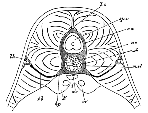
Diagrammatic section through the trunk of an advanced
embryo of Scyllium, to shew the position of the ribs.
ao., aorta; c.sh., cartilaginous notochordal sheath;
cv., cardinal vein; hp., hæmal process; k., kidney; l.s., ligamentum longitudinale superius; m.el., membrana elastica externa; na., neural arch; no., notochord; ll., lateral line; rb., rib; sp.c.,
spinal cord.
There is much, therefore, to be said in favour of Götte's view. At the
same time, there is another possible interpretation of the facts which
would admit the homology of the ribs as well as of the hæmal arches
throughout the Pisces.
Let us suppose, to start with, that the primitive arrangement
of the parts is more or less nearly that found in Lepidosteus, where
we have well-developed ribs in the region of the trunk, girthing the
body-cavity, and uniting in the caudal region to form the ventral parts of
the hæmal arches. It is easy to conceive that the ribs in the trunk might
somewhat alter their position by passing into the muscles, along the
inter-muscular septa, till they come to lie between the dorso-lateral and
ventro-lateral muscles, as in Elasmobranchii. Lepidosteus itself
affords a proof that such a change in the position of the ribs is not
impossible, in that it differs from other Ganoids and from Teleostei in the
fact that the free ends of the ribs leave the neighbourhood of the
body-cavity and penetrate into the muscles.
If it be granted that the mere difference in position between the ribs
of Ganoids and Elasmobranchii is not of itself sufficient to disprove their
homology, let us attempt to picture what would take place at the junction
of the trunk and tail in a type in which the ribs had undergone the above
change in position. On nearing the tail it may be supposed that the ribs
would gradually become shorter, and at the same time alter their position,
till finally they shaded off into ordinary hæmal processes. If, however,
the hæmal canal became prolonged forwards by the formation of some
additional complete or nearly complete hæmal arches, an alteration in the
relation of the parts would necessarily take place. Owing to the position
of the ribs, these structures could hardly assist in the new formation of
the anterior part of the hæmal canal, but the continuation forwards of the
canal would be effected by prolongations of the hæmal processes supporting
the ribs. The new arches so formed would naturally be held to be homologous
with the hæmal arches of the tail, though really not so, while the true
nature of the ribs would also be liable to be misinterpreted, in that the
ribs would appear to be lateral outgrowths of the hæmal processes of a
wholly different nature to the ventral parts of the hæmal arches of the
tail.
In some Elasmobranchii, as shewn in the accompanying woodcut (fig. 2),
in the transitional vertebræ between the trunk and the tail, the ribs are
supported by lateral outgrowths of the hæmal processes, while the wholly
independent prolongations of the hæmal processes appear to be about to
give rise to the hæmal arches of the tail.
This peculiar state of things led Götte, and subsequently one of us, to
deny for Elasmobranchii all homology between the ribs and any part of the
hæmal arches of the tail; but in view of the explanation just suggested,
this denial was perhaps too hasty.
Fig. 2.
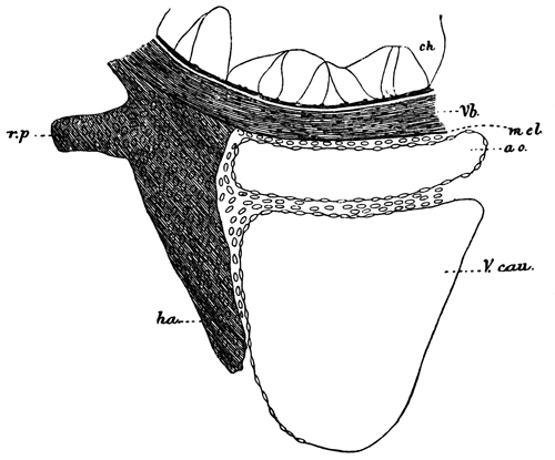
Transverse section through the ventral part of the
notochord, and adjoining structures of an advanced Scyllium embryo
at the root of the tail.
Vb., cartilaginous
sheath of the notochord; ha., hæmal
process; r.p., process to which the rib
is articulated; m.el., membrana
elastica externa; ch., notochord; ao., aorta; V.cau., caudal vein.
We are the more inclined to take this view because the researches of
Götte appear to shew that an occurrence, in many respects analogous, has
taken place in some Teleostei.
In Teleostei, Johannes Müller, and following him Gegenbaur, do not admit
that the hæmal arches of the tail are in any part formed by the ribs.
Gegenbaur (Elements of Comp.
Anat., translation, p. 431) says, In the Teleostei,
the costiferous transverse processes
(what we have called the hæmal
processes) gradually converge in the caudal region, and form
inferior arches, which are not homologous with those of Selachii and
Ganoidei, although they also form spinous processes.
The opposite view, that the hæmal arches of the tail in Teleostei
contain parts serially homologous with the basal parts of the hæmal
processes as well as with the ribs, has been also maintained by many
anatomists, e.g., Meckel, Aug. Müller, &c., and has recently found a powerful ally in
Götte.
In many cases, the relations of the parts appear to be fundamentally
those found in Lepidosteus and Amia, and Götte has shewn by
his careful embryological investigations on Esox and
Anguilla, that in these two forms there is practically conclusive
evidence that the ribs as well as the hæmal costiferous processes of
Gegenbaur, which support them, enter into the formation of the hæmal arches
of the tail.
In a great number of Teleostei, e.g., the Salmon and most
Cyprinoids, &c., the hæmal arches in
the region of transition from the trunk to the tail have a structure which
at first sight appears to support Johannes Müller's and Gegenbaur's view.
The hæmal processes grow larger and meet each other ventrally; while the
ribs articulated to them gradually grow smaller and disappear.
The Salmon is typical in this respect, and has been carefully studied by
Götte, who attempts to shew (with, in our opinion, complete success) that
the anterior hæmal arches are really not entirely homologous with the true
hæmal arches behind, but that in the latter, the closure of the arch below
is effected by the hæmal spine, which is serially homologous with a pair of
coalesced ribs, while in the anterior hæmal arches, i.e., those of
the trunk, the closure of the arch is effected by a bridge of bone uniting
the hæmal processes.
The arrangement of the parts just described, as well as the view of
Götte with reference to them, will be best understood from the accompanying
woodcut (fig. 3), copied from Götte's memoir.
Götte sums up his own results on this point in the following words
(p. 138): It follows from this, that the half rings, forming the
hæmal canal in the hindermost trunk vertebræ of the Salmon, are not (with
the exception of the last) completely homologous with those of the
tail, but are formed by a connecting piece between the basal stumps (hæmal
processes), which originates as a paired median process of these
stumps.
The incomplete homology between the anterior hæmal arches and the true
caudal hæmal arches which follow them is exactly what we suggest may be the
case in Elasmobranchii, and if it be admitted in the one case, we see no
reason why it should not also be admitted in the other.
Fig. 3.
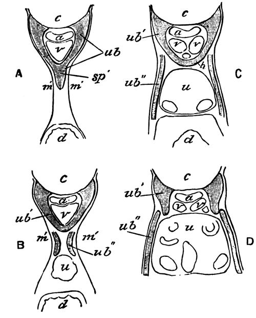
Semi-diagrammatic transverse sections through the first
caudal vertebra (A), the last trunk vertebra (B), and the two trunk
vertebræ in front (C and D), of a Salmon embryo of 2-3 centims. (From Götte.)
ub., hæmal arch;
ub´., hæmal process; ub´´., rib; c., notochord; a., aorta;
v., vein; h., connecting pieces between hæmal processes;
u., kidney; d., intestine; sp´., hæmal spine; m´., muscles.
If this admission is made, the only ground for not regarding the ribs of
Elasmobranchii as homologous with those of Ganoids is their different
position, and we have already attempted to prove that this is not a
fundamental point.
The results of our researches appear to us, then, to leave two
alternatives as to the ribs of Fishes. One of these, which may be called
Götte's view, may be thus stated:—The hæmal arches are
homologous throughout the Pisces: in Teleostei, Ganoidei, and Dipnoi[527], the ribs, placed on the inner face of the body-wall,
are serially homologous with the ventral parts of the hæmal arches of the
tail; in Elasmobranchii, on the other hand, the ribs are neither serially
homologous with the hæmal arches of the tail nor homologous with the ribs
of Teleostei and Ganoidei, but are outgrowths of the hæmal processes into
the space between the dorso-lateral and ventro-lateral muscles, which may
perhaps have their homologues in Teleostei and Ganoids in certain accessory
processes of the vertebræ.
The other view, which we are inclined to adopt, and the arguments for
which have been stated in the preceding pages, is as follows:—The
Teleostei, Ganoidei, Dipnoi, and Elasmobranchii are provided with
homologous hæmal arches, which are formed by the coalescence below the
caudal vein of simple prolongations of the primitive hæmal processes of the
embryo. The canal enclosed by the hæmal arches can be demonstrated
embryologically to be the aborted body-cavity.
In the region of the trunk the hæmal processes and their prolongations
behave somewhat differently in the different types.
In Ganoids and Dipnoi, in which the most primitive arrangement is
probably retained, the ribs are attached to the hæmal processes, and are
placed immediately without the peritoneal membrane at the insertions of the
intermuscular septa. These ribs are in many instances (Lepidosteus,
Acipenser), and very probably in all, developed continuously with
the hæmal processes, and become subsequently segmented from them. They are
serially homologous with the ventral parts of the hæmal arches of the tail,
which, like them, are in many instances (Ceratodus,
Lepidosteus, Polypterus, and to some extent in Amia)
segmented off from the basal parts of the hæmal arches.
In Teleostei the ribs have the same position and relations as those in
Ganoids and Dipnoi, but their serial homology with the ventral parts of the
hæmal processes of the tail, is often (e.g., the Salmon) obscured by
some of the anterior hæmal arches in the posterior part of the trunk being
completed, not by the ribs, but by independent outgrowths of the basal
parts of the hæmal processes.
In Elasmobranchii a still further divergence from the primitive
arrangement is present. The ribs appear to have passed outwards along the
intermuscular septa into the muscles, and are placed between the
dorso-lateral and ventro-lateral muscles (a change of position of the ribs
of the same nature, but affecting only their ends, is observable in
Lepidosteus). This change of position, combined probably with the
secondary formation of a certain number of anterior hæmal arches similar to
those in the Salmon, renders their serial homology with the ventral parts
of the hæmal processes of the tail far less clear than in other types, and
further proof is required before such homology can be considered as
definitely established.
This is not the place to enter into the obscure question as to how far
the ribs of the Amphibia and Amniota are homologous with those of Fishes.
It is to be remarked, however, that the ribs of the Urodela (1) occupy the
same position in relation to the muscles as the Elasmobranch ribs, (2) that
they are connected with the neural arches, and (3) that they coexist in the
tail with the hæmal arches, and seem, therefore, to be as different as
possible from the ribs of the Dipnoi.
Part IV.—The skeleton
of the ventral lobe of the tail fin, and its bearing on the nature of the
tail fin of the various types of Pisces.
In the embryos or larvæ of all the Elasmobranchii, Ganoidei, and
Teleostei which have up to this time been studied, the unpaired fins arise
as median longitudinal folds of the integument on the dorsal and ventral
sides of the body, which meet at the apex of the tail. The tail at first is
symmetrical, having a form which has been called diphycercal or
protocercal. At a later stage, usually, though not always, parts of these
fins atrophy, while other parts undergo a special development and
constitute the permanent unpaired fins.
Since the majority of existing as well as extinct Fishes are provided
with discontinuous fins, those forms, such as the Eel (Anguilla), in
which the fins are continuous, have probably reverted to an embryonic
condition: an evolutional process which is of more frequent occurrence than
has usually been admitted.
In the caudal region there is almost always developed in the larvæ of
the above groups a special ventral lobe of the embryonic fin a short
distance from the end of the tail. In Elasmobranchii and Chondrostean
Ganoids the portion of the embryonic tail behind this lobe persists through
life, and a special type of caudal fin, which is usually called
heterocercal, is thus produced. This type of caudal fin appears to have
been the most usual in the earlier geological periods.
Simultaneously with the formation of the ventral lobe of the
heterocercal caudal fin, the notochord with the vertebral tissues
surrounding it, becomes bent somewhat dorsalwards, and thus the primitive
caudal fin forms a dorsally directed lobe of the heterocercal tail. We
shall call this part the dorsal lobe of the tail-fin, and the secondarily
formed lobe the ventral lobe.
Lepidosteus and Amia (Wilder, No. 15) amongst the bony Ganoids, and, as has recently been
shewn by A. Agassiz[528], most Teleostei acquire at an
early stage of their development heterocercal caudal fins, like those of
Elasmobranchii and the Chondrostean Ganoids; but in the course of their
further growth the dorsal lobe partly atrophies, and partly disappears as
such, owing to the great prominence acquired by the ventral lobe. A portion
of the dorsally flexed notochord and of the cartilage or bone replacing or
investing it remains, however, as an indication of the original dorsal
lobe, though it does not project backwards beyond the level of the end of
the ventral lobe, which in these types forms the terminal caudal fin.
The true significance of the dorsally flexed portion of the vertebral
axis was first clearly stated by Huxley[529],
but as A. Agassiz has fairly pointed out in the paper already quoted, this
fact does not in any way militate against the view put forward by L.
Agassiz that there is a complete parallelism between the embryonic
development of the tail in these Fishes and the palæontological development
of this organ. We think that it is moreover convenient to retain
the term homocercal for those types of caudal fin in which the dorsal lobe
has atrophied so far as not to project beyond the ventral lobe.
We have stated these now well-known facts to enable the reader to follow
us in dealing with the comparison between the skeleton supporting the
fin-rays of the ventral lobe of the caudal fin, and that supporting the
fin-rays of the remaining unpaired fins.
It has been shewn that in Lepidosteus the unpaired fins fall into
two categories, according to the nature of the skeletal parts supporting
them. The fin-rays of the true ventral lobe of the caudal fin are supported
by the spinous processes of certain of the hæmal arches. The remaining
unpaired fins, including the anal fin, are supported by the so-called
interspinous bones, which are developed independently of the vertebral
column and its arches.
The question which first presents itself is, how far does this
distinction hold good for other Fishes? This question, though interesting,
does not appear to have been greatly discussed by anatomists. Not
unfrequently the skeletal supports of the ventral lobe of the caudal fin
are assumed to be the same as those of the other fins.
Davidoff[530], for instance, in speaking of
the unpaired fins of Elasmobranch embryos, says (p. 514): The
cartilaginous rays of the dorsal fins agreed not only in number with the
spinous processes (as indeed is also found in the caudal fin of the
full-grown Dog-fish),
&c.
Thacker[531], again, in his memoir on the
Median and Paired Fins, states at p. 284: We shall here consider the
skeleton of the dorsal and anal fins alone. That of the caudal fin has
undergone peculiar modifications by the union of fin-rays with hæmal
spines.
Mivart[532] goes into the question more
fully. He points out (p. 471) that there is an essential difference
between the dorsal and ventral parts of the caudal fin in Elasmobranchii,
in that in the former the radials are more numerous than the vertebræ and
unconformable to them, while in the latter they are equal in number to the
vertebræ and continuous with them. This,
he goes on to say,
seems to point to a difference in nature between the dorsal and ventral
portions of the caudal fin, in at least most Elasmobranchii.
He further
points out that Polyodon resembles Elasmobranchii. As to Teleostei,
he does not express himself decidedly except in the case of Muræna,
to which we shall return.
Mivart expresses himself as very doubtful as to the nature of the
supports of the caudal fin, and thinks that the caudal fin of different
kinds of Fishes may have arisen in different ways in different cases.
An examination of the ventral part of the caudal fin in various Ganoids,
Teleostei, and Elasmobranchii appears to us to shew that there can be but
little doubt that, in the majority of the members of these groups at any
rate, and we believe in all, the same distinction between the ventral lobe
of the caudal fin and the remaining unpaired fins is found as in
Lepidosteus.
In the case of most Elasmobranchii, a simple inspection of the caudal
fin suffices to prove this, and the anatomical features involved in this
fact have usually been recognized; though, in the absence of embryological
evidence, the legitimate conclusion has not always been drawn from
them.
The difference between the ventral lobe of the caudal fin and the other
fins in the mode in which the fin-rays are supported is as obvious in
Chondrostean Ganoids as it is in Elasmobranchii; it would appear also to
hold good for Amia. Polypterus we have had no opportunity of
examining, but if, as there is no reason to doubt, the figure of its
skeleton given by Agassiz (Poissons Fossiles) is
correct, there can be no question that the ventral lobe of the caudal fin
is supported by the hæmal arches, and not by interspinous bones. In
Calamoicthys, the tail of which we have had an opportunity of
dissecting through the kindness of Professor Parker, the fin-rays of the
ventral lobe of the true caudal fin are undoubtedly supported by true hæmal
arches.
There is no unanimity of opinion as to the nature of the elements
supporting the fin-rays of the caudal fin of Teleostei.
Huxley[533] in his paper on the development
of the caudal fin of the Stickleback, holds that these elements are of the
nature of interhæmal bones. He says (p. 39): "The last of these rings
lay just where the notochord began to bend up. It was slightly longer than
the bony ring which preceded it, and instead of having its posterior margin
parallel with the anterior, it sloped from above downwards and backwards.
Two short osseous plates, attached to the anterior part of the inferior
surface of the penultimate ring, or rudimentary vertebral centrum, passed
downwards and a little backwards, and abutted against a slender elongated
mass of cartilage. Similar cartilaginous bodies occupy the same relation to
corresponding plates of bone in the anterior vertebræ in the region of the
anal fin; and it is here seen, that while the bony plates coalesce and form
the inferior arches of the caudal vertebræ, the cartilaginous elements at
their extremities become the interhæmal bones. The cartilage connected with
the inferior arch of the penultimate centrum is therefore an
interhæmal
cartilage. The anterior part of the inferior surface of
the terminal ossification likewise has its osseous inferior arch, but the
direction of this is nearly vertical, and though it is connected below with
an element which corresponds in position with the interhæmal cartilage,
this cartilage is five or six times as large, and constitutes a broad
vertical plate, longer than it is deep, and having its longest axis
inclined downwards and backwards....
Immediately behind and above this anterior hypural
apophysis (as it may be termed) is another very much smaller vertical
cartilaginous plate, which may be called the posterior hypural
apophysis.
We have seen that Mivart expresses himself doubtful on the subject.
Gegenbaur[534] appears to regard them as hæmal
arches.
The latter view appears to us without doubt the correct one. An
examination of the tail of normal Teleostei shews that the fin-rays of that
part of the caudal fin which is derived from the ventral lobe of the larva
are supported by elements serially homologous with the hæmal arches, but in
no way homologous with the interspinous bones of the anal
fin. The elements in question formed of cartilage in the larva, become
ossified in the adult, and are known as the hypural bones. They may appear
in the form of a series of separate hæmal arches, corresponding in number
with the primitive somites of this region, which usually, however, atrophy
in the adult, or more often are from the first imperfectly segmented, and
have in the adult the form of two or three or even of a single broad bony
plate. The transitional forms between this state of things and that, for
instance, in Lepidosteus are so numerous, that there can be no doubt
that even the most peculiar forms of the hypural bones of Teleostei are
simply modified hæmal arches.
This view of the hypural bones is, moreover, supported by embryological
evidence, since Aug. Müller[535]
(p. 205) describes their development in a manner which, if his
statements are to be trusted, leaves no doubt on this point.
There are a considerable number of Fishes which are not provided with an
obvious caudal fin as distinct from the remaining unpaired fins,
i.e. Chimæra, Eels, and various Eel-like forms amongst Teleostei,
and the Dipnoi. Gegenbaur appears to hold that these Fishes ought to be
classed together in relation to the structure of the caudal portion of
their vertebral column, as he says on p. 431 of his Comparative
Anatomy (English Translation): In the Chimæræ, Dipnoi, and many
Teleostei, the caudal portion of the vertebral column ends by gradually
diminishing in size, but in most Fishes, &c.
For our purpose it will, however, be advisable to treat them
separately.
The tail of Chimæra appears to us to be simply a peculiar modification
of the typical Elasmobranch heterocercal tail, in which the true ventral
lobe of the caudal fin may be recognized in the fin-fold immediately in
front of the filamentous portion of the tail. In the allied genus
Callorhynchus this feature is more distinct. The filamentous portion
of the tail of Chimæra constitutes, according to the nomenclature adopted
above, the true dorsal lobe, and may be partially paralleled in the
filamentous dorsal lobe of the tail of the larval Lepidosteus (Plate
34, fig. 16).
The tail of the eel-like Teleostei is again undoubtedly a
modification of the normal form of tail characteristic of the Teleostei, in
which, however, the caudal fin has become very much reduced and merged into
the prolongations of the anal and dorsal fins.
This can be very clearly seen in Siluroid forms with an Eel-like tail,
such as Cnidoglanis. Although the dorsal and ventral fins appear to
be continuous round the end of the tail, and there is superficially no
distinct caudal fin, yet an examination of the skeleton of
Cnidoglanis shews that the end of the vertebral column is modified
in the usual Teleostean fashion, and that the hæmal arches of the modified
portion of the vertebral column support a small number of fin-rays; the
adjoining ventral fin-rays being supported by independent osseous
fin-supports (interspinous bones).
In the case of the Eel (Anguilla anguilla) Huxley (loc. cit.) long ago pointed out that the terminal portion of
the vertebral column was modified in an analogous fashion to that of other
Teleostei, and we have found that the modified hæmal arches of this part
support a few fin-rays, though a still smaller number than in
Cnidoglanis. The fin-rays so supported clearly constitute an aborted
ventral lobe of the caudal fin.
Under these circumstances we think that the following statement by
Mivart (Zool.
Trans. Vol. X., p. 471) is somewhat misleading:—
As to the condition of this part (i.e. the
ventral lobe of the tail-fin) in Teleosteans generally, I will not venture
as yet to say anything generally, except that it is plain that in such
forms as Muræna, the dorsal and ventral parts of the caudal fin are similar
in nature and homotypal with ordinary dorsal and anal fins[536].
The italicized portion of this sentence is only true in respect to that
part of the fringe of fin surrounding the end of the body, which is not
only homotypal with, but actually part of, the dorsal and anal fins.
Having settled, then, that the tails of Chimæra and of Eel-like
Teleostei are simply special modifications of the typical form of tail of
the group of Fishes to which they respectively belong, we come to
the consideration of the Dipnoi, in which the tail-fin presents problems of
more interest and greater difficulty than those we have so far had to deal
with.
The undoubtedly very ancient and primitive character of the Dipnoi has
led to the view, implicitly if not definitely stated in most text-books,
that their tail-fin retains the character of the piscine tail prior to the
formation of the ventral caudal lobe, a stage which is repeated
embryologically in the pre-heterocercal condition of the tail in ordinary
Fishes.
Through the want of embryological data, and in the absence of really
careful histological examination of the tail of any of the Dipnoi, we are
not willing to speak with very great confidence as to its nature; we are
nevertheless of the opinion that the facts we can bring forward on this
head are sufficient to shew that the tail of the existing Dipnoi is largely
aborted, so that it is more or less comparable with that of the Eel.
We have had opportunities of examining the structure of the tail of
Ceratodus and Protopterus in dissected specimens in the
Cambridge Museum. The vertebral axis runs to the ends of the tail without
shewing any signs of becoming dorsally flexed. At some distance from the
end of the tail the fin-rays are supported by what are apparently segmented
spinous prolongations of the neural and hæmal arches. The dorsal elements
are placed above the longitudinal dorsal cord, and occupy therefore the
same position as the independent elements of the neural arches of
Lepidosteus. They are therefore to be regarded as homologous with
the dorsal fin-supports or interspinous bones of other types. The
corresponding ventral elements are therefore also to be regarded as
interspinous bones.
In view of the fact that the fin-supports, whenever their development
has been observed, are found to be formed independently of the neural and
hæmal arches, we may fairly assume that this is also true for what we have
identified as the interspinous elements in the Dipnoi.
The interspinous elements become gradually shorter as the end of the
tail is approached, and it is very difficult from a simple examination of
dissected specimens to make out how far any of the posterior fin-rays are
supported by the hæmal arches only. To this question we shall return, but
we may remark that, although there is a prolongation backwards
of the vertebral axis beyond the last interspinous elements, composed it
would seem of the coalesced neural and hæmal arches but without the
notochord, yet by far the majority of the fin-rays which constitute the
apparent caudal fin are supported by interspinous elements.
The grounds on which we hold that the tail of the Dipnoi is to be
regarded as a degenerate rather than primitive type of tail are the
following:—
(1) If it be granted that a diphycercal or protocercal form of tail must
have preceded a heterocercal form, it is also clear that the ventral
fin-rays of such a tail must have been supported, as in Polypterus
and Calamoicthys, by hæmal arches, and not by interspinous elements;
otherwise, a special ventral lobe, giving a heterocercal character to the
tail, and provided with fin-rays supported only by hæmal arches, could
never have become evolved from the protocercal tail-fin. Since the ventral
fin-rays of the tail of the Dipnoi are supported by interspinous elements
and not by hæmal arches, this tail-fin cannot claim to have the character
of that primitive type of diphycercal or protocercal tail from
which the heterocercal tail must be supposed to have been evolved.
(2) Since the nearest allies of the Dipnoi are to be found in
Polypterus and the Crossopterygidæ of Huxley, and since in these
forms (as evinced by the structure of the tail-fin of Polypterus,
and the transitional type between a heterocercal and diphycercal form of
fin observable in fossil Crossopterygidæ) the ventral fin-rays of the
caudal fin were clearly supported by hæmal arches and not by interspinous
elements, it is rendered highly probable that the absence of fin-rays so
supported in the Dipnoi is a result of degeneration of the posterior part
of the tail.
[We use this argument without offering any opinion as to whether the
diphycercal character of the tail of many Crossopterygidæ is primary or
secondary.]
(3) The argument just used is supported by the degenerate and variable
state of the end of the vertebral axis in the Dipnoi—a condition most
easily explained by assuming that the terminal part of the tail has become
aborted.
(4) We believe that in Ceratodus we have been able to
trace a small number of the ventral fin-rays supported by hæmal arches
only, but these rays are so short as not to extend so far back as some of
the rays attached to the interspinous elements in front. These rays may
probably be interpreted, like the more or less corresponding rays in the
tail of the Eel, as the last remnant of a true caudal fin.
The above considerations appear to us to shew with very considerable
probability that the true caudal fin of the Dipnoi has become all but
aborted like that of various Teleostei; and that the apparent caudal fin is
formed by the anal and dorsal fins meeting round the end of the stump of
the tail.
From the adult forms of Dipnoi we are, however, of opinion that no
conclusion can be drawn as to whether their ancestors were provided with a
diphycercal or a heterocercal form of caudal fin.
The general conclusions with reference to the tail-fin at which we have
arrived are the following:—
(1) The ventral lobe of the tail-fin of Pisces differs from the other
unpaired fins in the fact that its fin-rays are directly supported by
spinous processes of certain of the hæmal arches instead of independently
developed interspinous bones.
(2) The presence or absence of fin-rays in the tail-fin supported by
hæmal arches may be used in deciding whether apparently diphycercal
tail-fins are aborted or primitive.
I.—Anatomy.
The excretory organs of Lepidosteus have been described by Müller
(No. 13) and Hyrtl (No. 11). These anatomists have given a fairly adequate
account of the generative ducts in the female, and Hyrtl has also described
the male generative ducts and the kidney and its duct, but his description
is contradicted by our observations in some of the most fundamental
points.
In the female example of 100.5 centims.
which we dissected, the kidney forms a paired gland, consisting of a narrow
strip of glandular matter placed on each side of the vertebral column, on
the dorsal aspect of the body-cavity. It is covered on its ventral aspect
by the oviduct and by its own duct, but is separated from both of these by
a layer of the tough peritoneal membrane, through which the collecting
tubes pass. It extends forwards from the anus for about three-fifths of the
length of the body-cavity, and in our example had a total length of about
28 centims. (Plate 39, fig. 60, k).
Anteriorly the two kidneys are separated by a short interval in the median
line, but posteriorly they come into contact, and are so intimately united
as almost to constitute a single gland.
A superficial examination might lead to the supposition that the kidney
extended forwards for the whole length of the body-cavity up to the region
of the branchial arches, and Hyrtl appears to have fallen into this error;
but what appears to be its anterior continuation is really a form of
lymphatic tissue, something like that of the spleen, filled with numerous
cells. This matter (Plate 39, fig. 60, ly.) continues from the kidney forwards without any
break, and has a colour so similar to that of the kidney as to be hardly
distinguishable from it with the naked eye. The true anterior end of the
kidney is placed about 3 centims. in front
on the left side, and on the same level on the right side as the wide
anterior end of the generative duct (Plate 39, fig. 60, od.). It is not obviously divided into segments, and
is richly supplied with malpighian bodies.
It is clear from the above description that there is no trace of
head-kidney or pronephros visible in the adult. To this subject we shall,
however, again return.
As will appear from the embryological section, the ducts of the kidneys
are probably simply the archinephric ducts, but to avoid the use of terms
involving a theory, we propose in the anatomical part of our work to call
them kidney ducts. They are thin-walled widish tubes coextensive with the
kidneys. If cut open there may be seen on their inner aspect the numerous
openings of the collecting tubes of the kidneys. They are placed ventrally
to and on the outer border of the kidneys (Plate 39, fig. 60, s.g.). Posteriorly they gradually enlarge, and
approaching each other in the median line, coalesce, forming an unpaired
vesicle or bladder (bl.)—about 6
centims. long in our example—opening
by a median pore on a more or less prominent papilla
(u.g.) behind the anus. The dilated
portions of the two ducts are called by Hyrtl the horns of the bladder.
The sides of the bladder and its so-called horns are provided with
lateral pockets into which the collecting tubes of the kidney open. These
pockets, which we have found in two female examples, are much larger in the
horns of the bladder than in the bladder itself. Similar pockets, but
larger than those we have found, have been described by Hyrtl in the male,
but are stated by him to be absent in the female. It is clear from our
examples that this is by no means always the case.
Hyrtl states that the wide kidney ducts, of which his description
differs in no material point from our own, suddenly narrow in front, and,
perforating the peritoneal lining, are continued forwards to supply the
anterior part of the kidney. We have already shewn that the anterior part
of the kidney has no existence, and the kidney ducts supplying it are,
according to our investigations, equally imaginary.
It was first shewn by Müller, whose observations on this point have been
confirmed by Hyrtl, &c., that the
ovaries of Lepidosteus are continuous with their ducts, forming in
this respect an exception to other Ganoids.
In our example of Lepidosteus the ovaries (Plate 39, fig. 60,
ov.) were about 18 centims. in length. They have the form of simple
sacks, filled with ova, and attached about their middle to their generative
duct, and continued both backwards and forwards from their attachment into
a blind process.
With reference to these sacks Müller has pointed out—and the
importance of this observation will become apparent when we deal with the
development—that the ova are formed in the thickness of the inner
wall of the sack. We hope to shew that the inner wall of the sack is alone
equivalent to the genital ridge of, for instance, the ovary of
Scyllium. The outer aspect of this wall—i.e., that
turned towards the interior of the sack—is equivalent to the outer
aspect of the Elasmobranch genital ridge, on which alone the ova are
developed[537]. The sack into which the ova
fall is, as we shall shew in the embryological section, a special section
of the body-cavity shut off from the remainder, and the dehiscence
of the ova into this cavity is equivalent to their discharge into the
body-cavity in other forms.
The oviduct (Plate 39, fig. 60, od.)
is a thin-walled duct of about 21 centims.
in length in the example we are describing, continuous in front with the
ovarian sack, and gradually tapering behind, till it ends (od´.) by opening into the dilated terminal section of
the kidney duct on the inner side, a short distance before the latter
unites with its fellow. It is throughout closely attached to the ureter and
placed on its inner, and to some extent on its ventral, aspect. The
hindermost part of the oviduct which runs beside the enlarged portion of
the kidney duct—that portion called by Hyrtl the horn of the urinary
bladder—is so completely enveloped by the wall of the horn of the
urinary bladder as to appear like a projection into the lumen of the latter
structure, and the somewhat peculiar appearance which it presents in
Hyrtl's figure is due to this fact. In our examples the oviduct was
provided with a simple opening into the kidney duct, on a slight papilla;
the peculiar dilatations and processes of the terminal parts of the
oviduct, which have been described by Hyrtl, not being present.
The results we have arrived at with reference to the male organs are
very different indeed from those of our predecessor, in that we find
the testicular products to be carried off by a series of vasa
efferentia, which traverse the mesorchium, and are continuous with the
uriniferous tubuli; so that the semen passes through the uriniferous tubuli
into the kidney duct and so to the exterior. We have moreover been unable
to find in the male a duct homologous with the oviduct of the
female.
This mode of transportation outwards of the semen has not hitherto been
known to occur in Ganoids, though found in all Elasmobranchii, Amphibia,
and Amniota. It is not, however, impossible that it exists in other
Ganoids, but has hitherto been overlooked.
Our male example of Lepidosteus was about 60 centims. in length, and was no doubt mature. It was
smaller than any of our female examples, but this according to Garman
(vide, p. 361) is usual. The testes (Plate 39, fig. 58A., t.)
occupied a similar position to the ovaries, and were about 21 centims. long. They were, as is frequently the case
with piscine testes, divided into a series of lobes (10-12), and were
suspended by a delicate mesentery (mesorchium) from the dorsal wall of the
abdomen on each side of the dorsal aorta. Hyrtl (No. 11) states that air or quicksilver injected between the
limbs of the mesentery, passed into a vas deferens homologous with the
oviduct which joins the ureter. We have been unable to find such a vas
deferens; but we have found in the mesorchium a number of tubes of a yellow
colour, the colour being due to a granular substance quite unlike
coagulated blood, but which appeared to us from microscopic examination to
be the remains of spermatozoa[538].
These tubes to the number of 40-50 constitute, we believe, the vasa
efferentia. Along the line of suspension of the testis on its inner border
these tubes unite to form an elaborate network of tubes placed on the inner
face of the testis—an arrangement very similar to that often found in
Elasmobranchii (vide F. M. Balfour, Monograph
on the Development of Elasmobranch Fishes, plate 20, figs. 4 and
8).
We have figured this network on the posterior lobe of the testis (fig.
58B), and have represented a section through it (fig. 59A, n.v.e.), and through one of the vasa efferentia
(v.e.) in the mesorchium. Such a section
conclusively demonstrates the real nature of these passages: they are
filled with sperm like that in the body of the testis, and are, as may be
seen from the section figured, continuous with the seminal tubes of the
testis itself.
At the attached base of the mesorchium the vasa efferentia unite into a
longitudinal canal, placed on the inner side of the kidney duct (Plate 39,
fig. 58A, l.c., also shewn in section in
Plate 39, fig. 59B, l.c.). From this
canal tubules pass off which are continuous with the tubuli uriniferi, as
may be seen from fig. 59B, but the exact course of these tubuli through the
kidney could not be made out in the preparations we were able to make of
the badly conserved kidney. Hyrtl describes the arrangement of the vascular
trunks in the mesorchium in the following way (No. 11, p. 6): The mesorchium contains vascular
trunks, viz., veins, which through their
numerous anastomoses form a plexus at the hilus of the testis,
whose efferent trunks, 13 in number, again unite into a plexus on the
vertebral column, which is continuous with the cardinal veins.
The
arrangement (though not the number) of Hyrtl's vessels is very similar to
that of our vasa efferentia, and we cannot help thinking that a confusion
of the two may have taken place; which, in badly conserved specimens, not
injected with semen, would be very easy.
We have, as already stated, been unable to find in our dissections any
trace of a duct homologous with the oviduct of the female, and our sections
through the kidney and its ducts equally fail to bring to light such a
duct. The kidney ducts are about 19 centims. in length, measured from the genital aperture
to their front end. These ducts are generally similar to those in the
female; they unite about 2 centims. from
the genital pore to form an unpaired vesicle. Their posterior parts are
considerably enlarged, forming what Hyrtl calls the horns of the urinary
bladder. In these enlarged portions, and in the wall of the unpaired
urinary bladder, numerous transverse partitions are present, as correctly
described by Hyrtl, which are similar to those in the female, but more
numerous. They give rise to a series of pits, at the blind ends of which
are placed the openings of the kidney tubules. The kidney duct without
doubt serves as vas deferens, and we have found in it masses of yellowish
colour similar to the substance in the vasa efferentia identified by us as
remains of spermatozoa.
II.—Development.
In the general account of the development we have already called
attention to the earliest stages of the excretory system.
We may remind the reader that the first part of the system to be formed
is the segmental or archinephric duct (Plate 36, figs. 28 and 29, sg.). This duct arises, as in Teleostei and
Amphibia, by the constriction of a hollow ridge of the somatic mesoblast
into a canal, which is placed in contiguity with the epiblast, along the
line of junction between the mesoblastic somites and the lateral plates of
mesoblast. Anteriorly the duct does not become shut off from the
body-cavity, and also bends inwards towards the middle line. The inflected
part of the duct is the first rudiment of the pronephros, and very soon
becomes considerably dilated relatively to the posterior part of the
duct.
The posterior part of each segmental duct acquires an opening into the
cloacal section of the alimentary tract. Apart from this change, the whole
of the ducts, except their pronephric sections, remain for a long time
unaltered, and the next changes we have to speak of concern the definite
establishment of the pronephros.
The dilated incurved portion of each segmental duct soon becomes
convoluted, and by the time the embryo is about 10 millims. in length, but before the period of hatching,
an important change is effected in the relations of their peritoneal
openings[539].
Instead of leading into the body-cavity, they open into an isolated
chamber on each side (Plate 38, fig. 51, pr.c.), which we will call the pronephric
chamber. The pronephric chamber is not, however, so far as we can
judge, completely isolated from the body-cavity. We have not, it is true,
detected with certainty at this stage a communication between the two; but
in later stages, in larvæ of from 11 to 26 millims., we have found a richly ciliated passage
leading from the body-cavity into the pronephros on each side (Plate 38,
fig. 52, p.f.p.). We have not succeeded
in determining with absolute certainty the exact relations between this
passage and the tube of the pronephros, but we are inclined to believe that
it opens directly into the pronephric chamber just spoken of.
As we hope to shew, this chamber soon becomes largely filled by a
vascular glomerulus. On the accomplishment of these changes, the pronephros
is essentially provided with all the parts typically present in a segment
of the mesonephros (woodcut, fig. 4). There is a peritoneal tube
(f)[540], opening into a vesicle
(v); from near the neck of the peritoneal tube there comes
off a convoluted tube (pr.n.), forming
the main mass of the pronephros, and ending in the segmental duct (sd.).
Fig. 4.
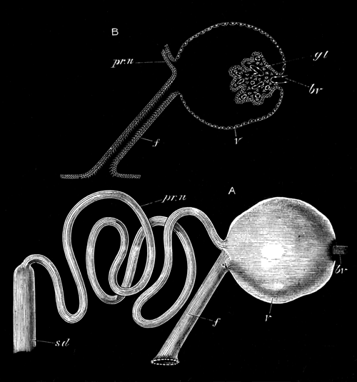
Diagrammatic views of the pronephros of
Lepidosteus.
A, pronephros supposed to be isolated and seen from the
side; B, section through the vesicle of the pronephros and the ciliated
peritoneal funnel leading into it; pr.n., coiled tube of pronephros; sd., segmental or archinephric duct; f.,
peritoneal funnel; v., vesicle of pronephros; bv., blood vessel of glomerulus; gl., glomerulus.
The different parts do not, however, appear to have the same
morphological significance as those in the mesonephros.
Judging from the analogy of Teleostei, the embryonic structure of whose
pronephros is strikingly similar to that of Lepidosteus, the two
pronephric chambers into which the segmental ducts open are constricted off
sections of the body-cavity.
With the formation of the convoluted duct opening into the
isolated section of the body-cavity we may speak of a definite pronephros
as having become established. The pronephros is placed, as can be made out
in later stages, on the level of the opening of the air-bladder into the
throat.
The pronephros increases in size, so far as could be determined, by the
further convolution of the duct of which it is mainly formed; and the next
change of importance which we have noticed is the formation of a vascular
projection into the pronephric chamber, forming the glomerulus already
spoken of (vide woodcut, fig. 4, gl.), which is similar to that of the pronephros of
Teleostei. We first detected these glomeruli in an embryo of about 15 millims., some days after hatching (Plate 38,
fig. 52, gl.), but it is quite possible
that they may be formed considerably earlier.
In the same embryo in which the glomeruli were found we also detected
for the first time a mesonephros consisting of a series of isolated
segmental or nephridial tubes, placed posteriorly to the pronephros along
the dorsal wall of the abdomen.
These were so far advanced at this stage that we are not in a position
to give any account of their mode of origin. They are, however, formed
independently of the segmental ducts, and in the establishment of the
junction between the two structures, there is no outgrowth from the
segmental duct to meet the segmental tubes. We could not at this stage find
peritoneal funnels of the segmental tubes, though we have met with them at
a later stage (Plate 38, fig. 53, p.f.),
and our failure to find them at this stage is not to be regarded as
conclusive against their existence.
A very considerable space exists between the pronephros and the foremost
segmental tube of the mesonephros. The anterior mesonephric tubes are,
moreover, formed earlier than the posterior.
In the course of further development, the mesonephric tubules increase
in size, so that there ceases to be an interval between them, the
mesonephros thus becoming a continuous gland. In an embryo of 26 millims. there was no indication of the
formation of segmental tubes to fill up the space between the pronephros
and mesonephros.
The two segmental ducts have united behind into an unpaired
structure in an embryo of 11 millims. This
structure is no doubt the future unpaired urinogenital chamber (Plate 39,
figs. 58A, and 60, bl.). Somewhat later,
the hypoblastic cloaca becomes split into two sections, the hinder one
receiving the coalesced segmental ducts, and the anterior remaining
continuous with the alimentary tract. The opening of the hinder one forms
the urinogenital opening, and that of the anterior the anus.
In an older larva of about 5.5 centims.
the pronephros did not exhibit any marked signs of atrophy, though the duct
between it and the mesonephros was somewhat reduced and surrounded by the
trabecular tissue spoken of in connection with the adult. In the region
between the pronephros and the front end of the fully developed part of the
mesonephros very rudimentary tubules had become established.
The latest stage of the excretory system which we have studied is in a
young Fish of about 11 centims. in length.
The special interest of this stage depends upon the fact that the ovary is
already developed, and not only so, but the formation of the oviducts has
commenced, and their condition at this stage throws considerable light on
the obscure problem of their nature in the Ganoids.
Unfortunately, the head of the young Fish had been removed before it was
put into our hands, so that it was impossible for us to determine whether
the pronephros was still present; but as we shall subsequently shew, the
section of the segmental duct, originally present between the pronephros
and the front end of the permanent kidney or mesonephros, has in any case
disappeared.
In addition to an examination of the excretory organs in
situ, which shewed little except the presence of the generative ridges,
we made a complete series of sections through the excretory organs for
their whole length (Plate 39, figs. 54-57).
Posteriorly these sections shewed nothing worthy of note, the excretory
organs and their ducts differing in no important particular from these
organs as we have described them in the adult, except in the fact that the
segmental ducts are not joined by the oviducts.
Some little way in front of the point where the two segmental ducts
coalesce to form the urinary bladder, the genital ridge comes into view.
For its whole extent, except near its anterior part (of which more
hereafter) this ridge projects freely into the body-cavity, and in this
respect the young Fish differs entirely from the adult. As shewn in Plate
39, figs. 56 and 57 (g.r.), it is
attached to the abdominal wall on the ventral side of, and near the inner
border of each kidney. The genital ridge itself has a structure very
similar to that which is characteristic of young Elasmobranchii, and it may
be presumed of young Fishes generally. The free edge of the ridge is
swollen, and this part constitutes the true generative region of the ridge,
while its dorsal portion forms the supporting mesentery. The ridge itself
is formed of a central stroma and a germinal epithelium covering it. The
epithelium is thin on the whole of the inner aspect of the ridge, but, just
as in Elasmobranchii, it becomes greatly thickened for a band-like strip on
the outer aspect. Here, the epithelium is several layers deep, and contains
numerous primitive germinal cells (p.o.).
Though the generative organs were not sufficiently advanced for us to
decide the point with certainty, the structure of the organ is in favour of
the view that this specimen was a female, and, as will be shewn directly,
there can on other grounds be no doubt that this is so. The large size of
the primitive germinal cells (primitive ova) reminded us of these bodies in
Elasmobranchii.
In the region between the insertion of the genital ridge (or ovary, as
we may more conveniently call it) and the segmental duct we detected the
openings of a series of peritoneal funnels of the excretory tubes (Plate
39, fig. 57, p.f.), which clearly
therefore persist till the young Fish has reached a very considerable
size.
As we have already said, the ovary projects freely into the body-cavity
for the greater part of its length. Anteriorly, however, we found that a
lamina extended from the free ventral edge of the ovary to the dorsal wall
of the body-cavity, to which it was attached on the level of the outer side
of the segmental duct. A somewhat triangular channel was thus constituted,
the inner wall of which was formed by the ovary, the outer by the lamina
just spoken of, and the roof by the strip of the peritoneum of the
abdominal wall covering that part of the ventral surface of the kidney in
which the openings of the peritoneal funnels of the excretory tubes are
placed. The structure of this canal will be at once understood by the
section of it shewn in Plate 39, fig. 55.
There can be no doubt that this canal is the commencing ovarian sack. On
tracing it backwards we found that the lamina forming its outer wall arises
as a fold growing upwards from the free edge of the genital ridge meeting a
downward growth of the peritoneal membrane from the dorsal wall of the
abdomen; and in Plate 39, fig. 56, these two laminæ may be seen before they
have met. Anteriorly the canal becomes gradually smaller and smaller in
correlation with the reduced size of the ovarian ridge, and ends blindly
nearly on a level with the front end of the excretory organs.
It should be noted that, owing to the mode of formation of the ovarian
sack, the outer side of the ovary with the band of thickened germinal
epithelium is turned towards the lumen of the sack; and thus the fact of
the ova being formed on the inner wall of the genital sack in the adult is
explained, and the comparison which we instituted in our description of the
adult between the inner wall of the genital sack and the free genital ridge
of Elasmobranchii receives its justification.
It is further to be noticed that, from the mode of formation of the
ovarian sack, the openings of the peritoneal funnels of the excretory
organs ought to open into its lumen; and if these openings persist in the
adult, they will no doubt be found in this situation.
Before entering on further theoretical considerations with reference to
the oviduct, it will be convenient to complete our description of the
excretory organs at this stage.
When we dissected the excretory organs out, and removed them from the
body of the young Fish, we were under the impression that they extended for
the whole length of the body-cavity. Great was our astonishment to find
that slightly in front of the end of the ovary both excretory organs and
segmental ducts grew rapidly smaller and finally vanished, and that what we
had taken to be the front part of the kidney was nothing else but a linear
streak of tissue formed of cells with peculiar granular
contents supported in a trabecular work (Plate 39, fig. 54). This discovery
first led us to investigate histologically what we, in common with previous
observers, had supposed to be the anterior end of the kidneys in the adult,
and to shew that they were nothing else but trabecular tissue with cells
like that of lymphatic glands. The interruption of the segmental duct at
the commencement of this tissue demonstrates that if any rudiment of the
pronephros still persists, it is quite functionless, in that it is not
provided with a duct.
III.—Theoretical considerations.
There are three points in our observations on the urinogenital system
which appear to call for special remark. The first of these concerns the
structure and fate of the pronephros, the second the nature of the oviduct,
and the third the presence of vasa efferentia in the male.
Although the history we have been able to give of the pronephros is not
complete, we have nevertheless shewn that in most points it is essentially
similar to the pronephros of Teleostei. In an early stage we find the
pronephros provided with a peritoneal funnel opening into the body-cavity.
At a later stage we find that there is connected with the pronephros on
each side, a cavity—the pronephric cavity—into which a
glomerulus projects. This cavity is in communication on the one hand with
the lumen of the coiled tube which forms the main mass of the pronephros,
and on the other hand with the body-cavity by means of a richly ciliated
canal (woodcut, fig. 4, p. 817).
In Teleostei the pronephros has precisely the same characters, except
that the cavity in which the glomerulus is placed is without a peritoneal
canal.
The questions which naturally arise in connection with the pronephros
are: (1) what is the origin of the above cavity with its glomerulus; and
(2) what is the meaning of the ciliated canal connecting this cavity with
the peritoneal cavity?
We have not from our researches been able to answer the first of these
questions. In Teleostei, however, the origin of this cavity has been
studied by Rosenberg[541] and Götte[542]. According to the account of the latter, which we have
not ourselves confirmed but which has usually been accepted, the front end
of the segmental duct, instead of becoming folded off from the body-cavity,
becomes included in a kind of diverticulum of the body-cavity, which only
communicates with the remainder of the body-cavity by a narrow opening. On
the inner wall of this diverticulum a projection is formed which becomes a
glomerulus. At this stage in the development of the pronephros we have
essentially the same parts as in the fully formed pronephros of
Lepidosteus, the only difference being that the passage connecting
the diverticulum containing the glomerulus with the remainder of the
body-cavity is short in Teleostei, and in Lepidosteus forms a
longish ciliated canal. In Teleostei the opening into the body-cavity
becomes soon closed. If the above comparison is justified, and if the
development of these parts in Lepidosteus takes place as it is
described as doing in Teleostei, there can, we think, be no doubt that the
ciliated canal of Lepidosteus, which connects the pronephric cavity
with the body-cavity, is a persisting communication between this cavity and
the body-cavity; and that Lepidosteus presents in this respect a
more primitive type of pronephros than Teleostei.
It may be noted that in Lepidosteus the whole pronephros has
exactly the character of a single segmental tube of the mesonephros. The
pronephric cavity with its glomerulus is identical in structure with a
malpighian body. The ciliated canal is similar in its relations to the
peritoneal canal of such a segmental tube, and the coiled portion of the
pronephros resembles the secreting part of the ordinary segmental tube.
This comparison is no doubt an indication that the pronephros is
physiologically very similar to the mesonephros, and so far justifies
Sedgwick's[543] comparison between the two, but
it does not appear to us to justify the morphological conclusions at
which he has arrived, or to necessitate any modification in the views on
this subject expressed by one of us[544].
The genital ducts of Ganoids and Teleostei have for some time been a
source of great difficulty to morphologists; and any contributions with
reference to the ontogeny of these structures are of interest.
The essential point which we have made out is that the anterior part of
the oviduct of Lepidosteus arises by a fold of the peritoneum
attaching itself to the free edge of the genital ridge. We have not,
unfortunately, had specimens old enough to decide how the posterior part of
the oviduct is formed; and although in the absence of such stages it would
be rash in the extreme to speak with confidence as to the nature of this
part of the duct, it may be well to consider the possibilities of the case
in relation to other Ganoids and Teleostei.
The simplest supposition would be that the posterior part of the genital
duct had the same origin as the anterior, i.e., that it was formed
for its whole length by the concrescence of a peritoneal fold with the
genital ridge, and that the duct so formed opened into the segmental
duct.
The other possible supposition is that a true Müllerian
duct—i.e., a product of the splitting of the segmental
duct—is subsequently developed, and that the open end of this duct
coalesces with the duct which has already begun to be formed in our oldest
larva.
In attempting to estimate the relative probability of these two views,
one important element is the relation of the oviducts of Lepidosteus
to those of other Ganoids.
In all other Ganoids (vide Hyrtl, No. II) there are stated to be genital ducts in both
sexes which are provided at their anterior extremities with a funnel-shaped
mouth open to the abdominal cavity. At first sight, therefore, it might be
supposed that they had no morphological relationship with the oviducts of
Lepidosteus, but, apart from the presence of a funnel-shaped mouth,
the oviducts of Lepidosteus are very similar to those of
Chondrostean Ganoids, being thin-walled tubes opening on a projecting
papilla into the dilated kidney ducts (horns of the urinary bladder,
Hyrtl). These relations seem to prove beyond a doubt that the oviduct of
Lepidosteus is for its major part homologous with the genital ducts
of other Ganoids.
The relationship of the genital ducts to the kidney ducts in Amia
and Polypterus is somewhat different from that in the Chondrostei
and Lepidosteus. In Amia the ureters are so small that they
may be described rather as joining the coalesced genital ducts than vice
versâ, although the apparent coalesced portion of the genital ducts is
shewn to be really part of the kidney ducts by receiving the secretion of a
number of mesonephric tubuli. In Polypterus the two ureters are
stated to unite, and open by a common orifice into a sinus formed by the
junction of the two genital ducts, which has not been described as
receiving directly the secretion of any part of the mesonephros.
It has been usual to assume that the genital ducts of Ganoids are true
Müllerian ducts in the sense above defined, on the ground that they are
provided with a peritoneal opening and that they are united behind with the
kidney ducts. In the absence of ontological evidence this identification is
necessarily provisional. On the assumption that it is correct we should
have to accept the second of the two alternatives above suggested as to the
development of the posterior parts of the oviduct in
Lepidosteus.
There appear to us, however, to be sufficiently serious objections to
this view to render it necessary for us to suspend our judgment with
reference to this point. In the first place, if the view that the genital
ducts are Müllerian ducts is correct, the true genital ducts of
Lepidosteus must necessarily be developed at a later period than the
secondary attachment between their open mouths and the genital folds, which
would, to say the least of it, be a remarkable inversion of the natural
order of development. Secondly, the condition of our oldest larva shews
that the Müllerian duct, if developed later, is only split off from quite
the posterior part of the segmental duct; yet in all types in which the
development of the Müllerian duct has been followed, its anterior
extremity, with the abdominal opening, is split off from either the
foremost or nearly the foremost part of the segmental duct.
Judging from the structure of the adult genital ducts of other
Ganoids they must also be developed only from the posterior part of the
segmental duct, and this peculiarity so struck one of us that in a previous
paper[545] the suggestion was put forward
that the true Ganoid genital ducts were perhaps not Müllerian ducts, but
enlarged segmental tubes with persisting abdominal funnels belonging to the
mesonephros.
If the possibility of the oviduct of Lepidosteus not being a
Müllerian duct is admitted, a similar doubt must also exist as to the
genital ducts of other Ganoids, and we must be prepared to shew that there
is a reasonable ground for scepticism on this point. We would in this
connexion point out that the second of the two arguments urged against the
view that the genital duct of Lepidosteus is not a Müllerian duct
applies with equal force to the case of all other Ganoids.
The short funnel-shaped genital duct of the Chondrostei is also very
unlike undoubted Müllerian ducts, and could moreover easily be conceived as
originating by a fold of the peritoneum, a slight extension of which would
give rise to a genital duct like that of Lepidosteus.
The main difficulty of the view that the genital ducts of Ganoids are
not Müllerian ducts lies in the fact that they open into the segmental
duct. While it is easy to understand the genesis of a duct from a folding
of the peritoneum, and also easy to understand how such a duct might lead
to the exterior by coalescing, for instance, with an abdominal pore, it is
not easy to see how such a duct could acquire a communication with the
segmental duct.
We do not under these circumstances wish to speak dogmatically, either
in favour of or against the view that the genital ducts of Ganoids are
Müllerian ducts. Their ontogeny would be conclusive on this matter, and we
trust that some of the anatomists who have the opportunity of studying the
development of the Sturgeon will soon let us know the facts of the case. If
there are persisting funnels of the mesonephric segmental tubes in adult
Sturgeons, some of them ought to be situated within the genital ducts, if
the latter are not Müllerian ducts; and naturalists who
have the opportunity ought also to look out for such openings.
The mode of origin of the anterior part of the genital duct of
Lepidosteus appears to us to tell strongly in favour of the view,
already regarded as probable by one of us[546],
that the Teleostean genital ducts are derived from those of Ganoids; and
if, as appears to us indubitable, the most primitive type of Ganoid genital
ducts is found in the Chondrostei, it is interesting to notice that the
remaining Ganoids present in various ways approximations to the arrangement
typically found in Teleostei. Lepidosteus obviously approaches
Teleostei in the fact of the ovarian ridge forming part of the wall of the
oviduct, but differs from the Teleostei in the fact of the oviduct opening
into the kidney ducts, instead of each pair of ducts having an independent
opening in the cloaca, and in the fact that the male genital products are
not carried to the exterior by a duct homologous with the oviduct.
Amia is closer to the Teleostei in the arrangement of the posterior
part of the genital ducts, in that the two genital ducts coalesce
posteriorly; while Polypterus approaches still nearer to the
Teleostei in the fact that the two genital ducts and the two kidney ducts
unite with each other before they join; and in order to convert this
arrangement into that characteristic of the Teleostei we have only to
conceive the coalesced ducts of the kidneys acquiring an independent
opening into the cloaca behind the genital opening.
The male genital ducts.—The discovery of the vasa
efferentia in Lepidosteus, carrying off the semen from the testis,
and transporting it to the mesonephros, and thence through the mesonephric
tubes to the segmental duct, must be regarded as the most important of our
results on the excretory system.
It proves in the first place that the transportation outwards of the
genital products of both sexes by homologous ducts, which has been hitherto
held to be universal in Ganoids, and which, in the absence of evidence to
the contrary, must still be assumed to be true for all Ganoids except
Lepidosteus, is a secondary arrangement. This conclusion follows
from the fact that in Elasmobranchii, &c., which are not descendants of the Ganoids, the
same arrangement of seminal ducts is found as in Lepidosteus, and it
must therefore have been inherited from an ancestor common to the two
groups.
If, therefore, the current statements about the generative ducts of
Ganoids are true, the males must have lost their vasa efferentia, and the
function of vas deferens must have been taken by the homologue of the
oviduct, presumably present in the male. The Teleostei must, moreover, have
sprung from Ganoidei in which the vasa efferentia had become aborted.
Considerable phylogenetic difficulties as to the relationships of
Ganoidei and Elasmobranchii are removed by the discovery that Ganoids were
originally provided with a system of vasa efferentia like that of
Elasmobranchii.
I.—Anatomy.
Agassiz (No. 2) gives a short description
with a figure of the viscera of Lepidosteus as a whole. Van der
Hœven has also given a figure of them in his memoir on the
air-bladder of this form (No. 8), and Johannes
Müller first detected the spiral valve and gave a short account of it in
his memoir (No. 13). Stannius, again, makes
several references to the viscera of Lepidosteus in his anatomy of
the Vertebrata, and throws some doubt on Müller's determination of the
spiral valve.
The following description refers to a female Lepidosteus of 100.5
centims. (Plate 40, fig. 66).
With reference to the mouth and pharynx, we have nothing special to
remark. Immediately behind the pharynx there comes an elongated tube, which
is not divisible into stomach and œsophagus, and may be called the
stomach (st.). It is about 44.6 centims. long, and gradually narrows from the
middle towards the hinder or pyloric extremity. It runs straight backwards
for the greater part of its length, the last 3.8 centims., however, taking a sudden bend forwards. For
about half its length the walls are thin, and the mucous membrane is
smooth; in the posterior half the walls are thick, and the mucous
membrane is raised into numerous longitudinal ridges. The peculiar
glandular structure of the epithelium of this part in the embryo is shewn
in Plate 40, fig. 62 (st.). Its opening
into the duodenum is provided with a very distinct pyloric valve (py.). This valve projects into a kind of
chamber, freely communicating with the duodenum, and containing four large
pits (c´), into each of which a group of
pyloric cæca opens. These cæca form a fairly compact gland (c.)
about 6.5 centims. long, which overlaps
the stomach anteriorly, and the duodenum posteriorly.
Close to the pyloric valve, on its right side, is a small papilla, on
the apex of which the bile duct opens (b.d´).
A small, apparently glandular, mass closely connected with the bile
duct, in the position in which we have seen the pancreas in the larva
(Plate 40, figs. 62 and 63, p.), is almost certainly a rudimentary
pancreas, like that of many Teleostei; but its preservation was too bad for
histological examination. We believe that the pancreas of
Lepidosteus has hitherto been overlooked.
The small intestine passes straight backwards for about 8 centims., and then presents three compact coils. From
the end of these a section, about 5 centims. long, the walls of which are much thicker,
runs forwards. The intestine then again turns backwards, making one spiral
coil. This spiral part passes directly, without any sharp line of
demarcation, into a short and straight tube, which tapers slightly from
before backwards, and ends at the anus. The mucous membrane of the
intestine for about the first 3.5 centims. is smooth, and the muscular walls thin: the
rest of the small intestine has thick walls, and the mucous membrane is
reticulated.
A short spiral valve (sp.v.), with a
very rudimentary epithelial fold, making nearly two turns, begins in about
the posterior half of the spiral coil of the intestine, extending backwards
for slightly less than half the straight terminal portion of the intestine,
and ending 4 centims. in front of the
anus. Its total length in one example was about 4.5 centims.
The termination of the spiral valve is marked by a slight constriction,
and we may call the straight portion of the intestine behind it the rectum
(rc.).
The posterior part of the intestine, from the beginning of the
spiral valve to the anus, is connected with the ventral wall of the
abdomen by a mesentery.
The air-bladder (a.b.) is 45 centims. long, and opens into the alimentary
canal by a slit-like aperture (a.b´.) on
the median dorsal line, immediately behind the epipharyngeal teeth. Each
lip of this aperture is largely formed by a muscular cushion, thickest at
its posterior end, and extending about 6 millims. behind the aperture itself. A narrow passage
is bounded by these muscular walls, which opens dorsally into the
air-bladder.
The air-bladder is provided with two short anterior cornua, and tapers
to a point behind: it shews no indication of any separation into two parts.
A strong band of connective tissue runs along the inner aspect of its whole
dorsal region, from which there are given off on each side—at
intervals of about 12 millims. anteriorly,
gradually increasing to 18 millims.
posteriorly—bands of muscle, which pass outwards towards its side
walls, and then spread out into the numerous reticulations with which the
air-bladder is lined throughout. By the contraction of these muscles the
cavity of the air-bladder can doubtless be very much diminished.
The main muscular bands circumscribe a series of more or less complete
chambers, which were about twenty-seven in number on each side in our
example. The chambers are confined to the sides, so that there is a
continuous cavity running through the central part of the organ. The whole
organ has the characteristic structure of a simple lung.
The liver (lr.) consists of a single
elongated lobe, about 32 centims. long,
tapering anteriorly and posteriorly, the anterior half being on the average
twice as thick as the posterior half. The gall-bladder (g.b.) lies at its posterior end, and is of
considerable size, tapering gradually so as to pass insensibly into the
bile duct. The hepatic duct (hp.d.)
opens into the gall-bladder at its anterior end.
The spleen (s.) is a large, compact, double gland, one lobe lying
in the turn of the intestine immediately above the spiral valve, and the
other on the opposite side of the intestine, so that the intestine is
nearly embraced between the two lobes.
II.—Development.
We have already described in detail the first formation of the
alimentary tract so far as we have been able to work it out, and we need
only say here that the anterior and posterior ends of the canal become
first formed, and that these two parts gradually elongate, so as to
approach each other; the growth of the posterior part is, however, the most
rapid. The junction of the two parts takes place a very short distance
behind the opening of the bile duct into the intestine.
For some time after the two parts of the alimentary tract have nearly
met, the ventral wall of the canal at this point is not closed; so that
there is left a passage between the alimentary canal and the yolk-sack,
which forms a vitelline duct.
After the yolk-sack has ceased to be visible as an external appendage it
still persists within the abdominal cavity. It has, however, by this stage
ceased to communicate with the gut, so that the eventual absorption of the
yolk is no doubt entirely effected by the vitelline vessels. At these later
stages of development we have noticed that numerous yolk nuclei, like those
met with in Teleostei and Elasmobranchii[547],
are still to be found in the yolk.
It will be convenient to treat the history of sections of the alimentary
tract in front of and behind the vitelline duct separately. The former
gives rise to the pharyngeal region, the œsophagus, the stomach, and
the duodenum.
The pharyngeal region, immediately after it has become established,
gives rise to a series of paired pouches. These may be called the branchial
pouches, and are placed between the successive branchial arches. The first
or hyomandibular pouch, placed between the mandibular and hyoid arches, has
rather the character of a double layer of hypoblast than of a true pouch,
though in parts a slight space is developed between its two walls. It is
shewn in section in Plate 37, fig. 43 (h.m.), from an embryo of about 10 millims., shortly before hatching. It does not
appear to undergo any further development, and, so far as we can make out,
disappears shortly after the embryo is hatched, without acquiring an
opening to the exterior.
It is important to notice that this cleft, which in the cartilaginous
Ganoids and Polypterus remains permanently open as the spiracle, is
rudimentary even in the embryo of Lepidosteus.
The second pouch is the hyobranchial pouch: its outer end meets the
epiblast before the larva is hatched, and a perforation is effected at the
junction of the two layers, converting the pouch into a visceral cleft.
Behind the hyobranchial pouch there are four branchial pouches, which
become perforated and converted into branchial clefts shortly after
hatching.
The region of the œsophagus following the pharynx is not separated
from the stomach, unless a glandular posterior region (vide description of adult) be regarded as the stomach, a
non-glandular anterior region forming the œsophagus. The lumen of
this part appears to be all but obliterated in the stages immediately
before hatching, giving rise for a short period to a solid œsophagus
like that of Elasmobranchii and Teleostei[548].
From the anterior part of the region immediately behind the pharynx the
air-bladder arises as a dorsal unpaired diverticulum. From the very first
it has an elongated slit-like mouth (Plate 40, fig. 64, a.b´.), and is placed in the mesenteric attachment of
the part of the throat from which it springs.
We have first noticed it in the stages immediately after hatching. At
first very short and narrow, it grows in succeeding stages longer and
wider, making its way backwards in the mesentery of the alimentary tract
(Plate 40, fig. 65, a.b.). In the larva
of a month and a half old (26 millims.)
it has still a perfectly simple form, and is without traces of its adult
lung-like structure; but in the larva of 11 centims. it has the typical adult structure.
The stomach is at first quite straight, but shortly after the larva is
hatched its posterior end becomes bent ventralwards and forwards, so that
the flexure of its posterior end (present in the adult) is very early
established. The stomach is continuous behind with the duodenum,
the commencement of which is indicated by the opening of the bile duct.
The liver is the first-formed alimentary gland, and is already a compact
body before the larva is hatched. We have nothing to say with reference to
its development, except that it exhibits the same simple structure in the
embryo that it does in the adult.
A more interesting glandular body is the pancreas. It has already been
stated that in the adult we have recognized a small body which we believe
to be the pancreas, but that we were unable to study its histological
characters.
In the embryo there is a well-developed pancreas which arises in the
same position and the same manner as in those Vertebrata in which the
pancreas is an important gland in the adult.
We have first noticed the pancreas in a stage shortly after hatching
(Plate 40, fig. 61, p.). It then has the form of a funnel-shaped
diverticulum of the dorsal wall of the duodenum, immediately
behind the level of the opening of the bile duct. From the apex of this
funnel numerous small glandular tubuli soon sprout out.
The similarity in the development of the pancreas in Lepidosteus
to that of the same gland in Elasmobranchii is very striking[549].
The pancreas at a later stage is placed immediately behind the end of
the liver in a loop formed by the pyloric section of the stomach (Plate 40,
fig. 62, p.). During larval life it constitutes a considerable
gland, the anterior end of which partly envelopes the bile duct (Plate 40,
fig. 63, p.).
Considering the undoubted affinities between Lepidosteus and the
Teleostei, the facts just recorded with reference to the pancreas appear to
us to demonstrate that the small size and occasional absence (?) of this
gland in Teleostei is a result of the degeneration of this gland; and it
seems probable that the pancreas will be found in the larvæ of most
Teleostei. These conclusions render intelligible, moreover, the great
development of the pancreas in the Elasmobranchii.
We have first noticed the pyloric cæca arising as outgrowths of
the duodenum in larvæ of about three weeks old, and they become rapidly
longer and more prominent (Plate 40, fig. 62, c.).
The portion of the intestine behind the vitelline duct is, as in all the
Vertebrata, at first straight. In Elasmobranchii the lumen of the part of
the intestine in which a spiral valve is present in the adult, very early
acquires a more or less semilunar form by the appearance of a fold which
winds in a long spiral. In Lepidosteus there is a fold similar in
every respect (Plate 38, fig. 53, sp.v.), forming an open spiral round the intestine.
This fold is the first indication of the spiral valve, but it is relatively
very much later in its appearance than in Elasmobranchii, not being formed
till about three weeks after hatching. It is, moreover, in correlation with
the small extent of the spiral valve of the adult, confined to a much
smaller portion of the intestine than in Elasmobranchii, although owing to
the relative straightness of the anterior part of the intestine it is
proportionately longer in the embryo than in the adult.
The similarity of the embryonic spiral valve of Lepidosteus to
that of Elasmobranchii shews that Stannius' hesitation in accepting
Müller's discovery of the spiral valve in Lepidosteus is not
justified.
J. Müller (Bau u. Entwick. d. Myxinoiden) holds
that the so-called bursa entiana of Elasmobranchii (i.e., the
chamber placed between the part of the intestine with the spiral valve and
the end of the pylorus) is the homologue of the more elongated portion of
the small intestine which occupies a similar position in the Sturgeon. This
portion of the small intestine is no doubt homologous with the still more
elongated and coiled portion of the small intestine in Lepidosteus
placed between the chamber into which the pyloric cæca, &c., open and the region of the spiral valve. The fact
that the vitelline duct in the embryo Lepidosteus is placed close to
the pyloric end of the stomach, and that the greater portion of the small
intestine is derived from part of the alimentary canal behind this, shews
that Müller is mistaken in attempting to homologise the bursa entiana of
Elasmobranchii, which is placed in front of the vitelline duct, with the
coiled part of the small intestine of the above forms. The latter is either
derived from an elongation of the very short portion of the
intestine between the vitelline duct and the primitive spiral valve, or
more probably by the conversion of the anterior part of the intestine,
originally provided with a spiral valve into a coiled small intestine not
so provided.
We have already called attention to the peculiar mesentery present in
the adult attaching the posterior straight part of the intestine to the
ventral wall of the body. This mesentery, which together with the dorsal
mesentery divides the hinder section of the body-cavity into two lateral
compartments is, we believe, a persisting portion of the ventral mesentery
which, as pointed out by one of us[550], is
primitively present for the whole length of the body-cavity. The
persistence of such a large section of it as that found in the adult
Lepidosteus is, so far as we know, quite exceptional. This mesentery
is shewn in section in the embryo in Plate 38, fig. 53 (v.mt.). The small vessel in it appears to be the
remnant of the subintestinal vein.
It is well known that Lepidosteus is provided with a gill on the
hyoid arch, divided on each side into two parts. An excellent figure of
this gill is given by Müller (No. 13, plate 5,
fig. 6), who holds from a consideration of the vascular supply that the two
parts of this gill represent respectively the hyoid gill and the mandibular
gill (called by Müller pseudobranch). Müller's views on this subject have
not usually been accepted, but it is the fashion to regard the whole of the
gill as the hyoid gill divided into two parts. It appeared to us not
improbable that embryology might throw some light on the history of this
gill, and accordingly we kept a look out in our embryos for traces of gills
on the hyoid and mandibular arches. The results we have arrived at are
purely negative, but are not the less surprising for this fact. The
hyomandibular cleft as shewn above, is never fully developed, and early
undergoes a complete atrophy—a fact which is, on the whole, against
Müller's view; but what astonished us most in connection with the gill in
question is that we have been unable to find any trace of it even in the
oldest larva whose head we have had (26 millims.), and at a period when the gills on the
hinder arches have reached their full development.
We imagined the gill in question to be the remnant of a gill fully
formed in extinct Ganoid types, and therefore expected to find it better
developed in the larva than in the adult. That the contrary is the fact
appears to us fairly certain, although we cannot at present offer any
explanation of it.
A. Agassiz concludes his memoir on the development of Lepidosteus
by pointing out that in spite of certain affinities in other directions
this form is not so far removed from the bony Fishes as has been
supposed.
Our own observations go far to confirm Agassiz' opinion.
Apart from the complete segmentation, the general development of
Lepidosteus is strikingly Teleostean. In addition to the general
Teleostean features of the embryo and larva, which can only be appreciated
by those who have had an opportunity of practically working at the subject,
we may point to the following developmental features[551]
as indicative of Teleostean affinities:—
(1) The formation of the nervous system as a solid keel of the
epiblast.
(2) The division of the epiblast into a nervous and epidermic
stratum.
(3) The mode of development of the gut (vide
pp. 752-754).
(4) The mode of development of the pronephros; though, as shewn on
p. 822, the pronephros of Lepidosteus
has primitive characters not retained by Teleostei.
(5) The early stages in the development of the vertebral column (vide p. 779).
In addition to these, so to speak, purely embryonic characters there are
not a few important adult characters:—
(1) The continuity of the oviducts with the genital glands.
(2) The small size of the pancreas, and the presence of
numerous so-called pancreatic cæca.
(3) The somewhat coiled small intestine.
(4) Certain characters of the brain, e.g., the large size of the
cerebellum; the presence of the so-called lobi inferiores on the
infundibulum; and of tori semicirculares in the mid-brain.
In spite of the undoubtedly important list of features to which we have
just called attention, a list containing not less important characters,
both embryological and adult, separating Lepidosteus from the
Teleostei, can be drawn up:—
(1) The character of the truncus arteriosus.
(2) The fact of the genital ducts joining the ureters.
(3) The presence of vasa efferentia in the male carrying the semen from
the testes to the kidney, and through the tubules of the latter into the
kidney duct.
(4) The presence of a well-developed opercular gill.
(5) The presence of a spiral valve; though this character may possibly
break down with the extension of our knowledge.
(6) The typical Ganoid characters of the thalamencephalon and the
cerebral hemispheres (vide pp. 769 and 770).
(7) The chiasma of the optic nerves.
(8) The absence of a pecten, and presence of a vascular membrane between
the vitreous humour and the retina.
(9) The opisthocœlous form of the vertebræ.
(10) The articulation of the ventral parts of the hæmal arches of the
tail with processes of the vertebral column.
(11) The absence of a division of the muscles into dorso-lateral and
ventro-lateral divisions.
(12) The complete segmentation of the ovum.
The list just given appears to us sufficient to demonstrate that
Lepidosteus cannot be classed with the Teleostei; and we hold that
Müller's view is correct, according to which Lepidosteus is a true
Ganoid.
The existence of the Ganoids as a distinct group has, however, recently
been challenged by so distinguished an Ichthyologist as Günther, and it may
therefore be well to consider how far the group as defined by Müller is a
natural one for living forms[552],
and how far recent researches enable us to improve upon Müller's
definitions. In his classical memoir (No. 13)
the characters of the Ganoids are thus shortly stated:—
"These Fishes are either provided with plate-like
angular or rounded cement-covered scales, or they bear osseous plates, or
are quite naked. The fins are often, but not always, beset with a double or
single row of spinous plates or splints. The caudal fin occasionally
embraces in its upper lobe the end of the vertebral column, which may be
prolonged to the end of the upper lobe. Their double nasal openings
resemble those of Teleostei. The gills are free, and lie in a branchial
cavity under an operculum, like those of Teleostei. Many of them have an
accessory organ of respiration, in the form of an opercular gill, which is
distinct from the pseudobranch, and can be present together with the
latter; many also have spiracles like Elasmobranchii. They have many valves
in the stem of the aorta like the latter, also a muscular coat in the stem
of the aorta. Their ova are transported from the abdominal cavity by
oviducts. Their optic nerves do not cross each other. The intestine is
often provided with a spiral valve, like Elasmobranchii. They have a
swimming-bladder with a duct, like many Teleostei. Their pelvic fins are
abdominal.
If we include in a definition only those characters
which are invariable, the Ganoids may be shortly defined as being those
Fish with numerous valves to the stem of the aorta, which is also provided
with a muscular coat; with free gills and an operculum, and with abdominal
pelvic fins.
To these distinctive characters, he adds in an appendix to his paper,
the presence of the spiral valve, and the absence of a processus
falciformis and a choroid gland.
To the distinctive set of characters given by Müller we may probably add
the following:—
(1) Oviducts and urinary ducts always unite, and open by a common
urinogenital aperture behind the anus.
(2) Skull hyostylic.
(3) Segmentation complete in the types so far investigated,
though perhaps Amia may be found to resemble the Teleostei in this
particular.
(4) A pronephros of the Teleostean type present in the larva.
(5) Thalamencephalon very large and well developed.
(6) The ventricle in the posterior part of the cerebrum is not divided
behind into lateral halves, the roof of the undivided part being extremely
thin.
(7) Abdominal pores always present.
The great number of characters just given are amply sufficient to
differentiate the Ganoids as a group; but, curiously enough, the only
characters amongst the whole series which have been given, which can be
regarded as peculiar to the Ganoids, are (1) the characters of the brain,
and (2) the fact of the oviducts and kidney ducts uniting together and
opening by a common pore to the exterior.
This absence of characters peculiar to the Ganoids is an indication of
how widely separated in organization are the different members of this
great group.
At the same time, the only group with which existing Ganoids have close
affinities is the Teleostei. The points they have in common with the
Elasmobranchii are merely such as are due to the fact that both retain
numerous primitive Vertebrate characters[553],
and the gulf which really separates them is very wide.
There is again no indication of any close affinity between the Dipnoi
and, at any rate, existing Ganoids.
Like the Ganoids, the Dipnoi are no doubt remnants of a very primitive
stock; but in the conversion of the air-bladder into a true lung, the
highly specialized character of their limbs[554],
their peculiar autostylic skulls, the fact of their ventral nasal openings
leading directly into the mouth, their multisegmented bars (interspinous
bars), directly prolonged from the neural and hæmal arches and supporting
the fin-rays of the unpaired dorsal and ventral fins, and their
well-developed cerebral hemispheres, very unlike those of
Ganoids and approaching the Amphibian type, they form a very well-defined
group, and one very distinctly separated from the Ganoids.
No doubt the Chondrostean Ganoids are nearly as far removed from the
Teleostei as from the Dipnoi, but the links uniting these Ganoids with the
Teleostei have been so fully preserved in the existing fauna of the globe,
that the two groups almost run into each other. If, in fact, we were
anxious to make any radical change in the ordinary classification of
Fishes, it would be by uniting the Teleostei and Ganoids, or rather
constituting the Teleostei into one of the sub-groups of the Ganoids,
equivalent to the Chondrostei. We do not recommend such an arrangement,
which in view of the great preponderance of the Teleostei amongst living
Fishes would be highly inconvenient, but the step from Amia to the
Teleostei is certainly not so great as that from the Chondrostei to
Amia, and is undoubtedly less than that from the Selachii to the
Holocephali.
1. Agassiz, A. The
Development of Lepidosteus.
Part 1., Proc. Amer. Acad. Arts and
Sciences, Vol. XIV. 1879.
2. Agassiz, L. Recherches s. l. Poissons Fossiles. Neuchatel.
1833-45.
3. Boas, J. E. Ueber Herz u. Arterienbogen bei Ceradotus u.
Protopterus,
Morphol. Jahrbuch, Vol. VI. 1880.
4. Davidoff, M. von. Beiträge z. vergleich. Anat. d. hinteren Gliedmassen d.
Fische,
Morphol. Jahrbuch, Vol. VI. 1880.
5. Gegenbaur, C. Untersuch. z. vergleich. Anat. d. Wirbelthiere, Heft
II., Schultergürtel d. Wirbelthiere. Brustflosse der Fische.
Leipzig, 1865.
6. Gegenbaur, C. Zur Entwick. d. Wirbelsäule d. Lepidosteus, &c.
Jenaische
Zeitschrift, Vol. III. 1867.
7. Hertwig, O. Ueber d. Hautskelet d. Fische (Lepidosteus u.
Polypterus),
Morphol. Jahrbuch, Vol. V. 1879.
8. Hœven, Van der. Ueber d. zellige Schwimmblase d. Lepidosteus.
Müller's
Archiv, 1841.
9. Hyrtl, J. Ueber d. Schwimmblase von Lepidosteus osseus,
Sitz. d. Wiener Akad. Vol.
VIII.
1852.
10. Hyrtl, J. Ueber
d. Pori abdominales, d. Kiemen-Arterien, u. d. Glandula thyroidea d.
Ganoiden,
Sitz. d. Wiener Akad. Vol. VIII. 1852.
11. Hyrtl, J. Ueber d. Zusammenhang [TN19] d.
Geschlechts u. Harnwerkzeuge bei d. Ganoiden, Wien, 1855.
12. Kölliker, A. Ueber d. Ende d. Wirbelsäule b. Ganoiden, Leipzig,
1860.
13. Müller, J. Ueber d. Bau u. d. Grenzen d. Ganoiden,
Berlin Akad. 1844.
14. Schneider, H. Ueber d. Augenmuskelnerven d. Ganoiden,
Jenaische Zeitschrift, Vol.
XV. 1881.
15. Wilder, Burt G. Notes on
the North American Ganoids, Amia, Lepidosteus,
Acipenser, and Polyodon.
Proc. Amer. Assoc. for the Advancement
of Science, 1875.
a. Anus. ab.
Air-bladder. ab´. Aperture of
air-bladder into throat. ac. Anterior
commissure. af. Anal fin. al. Alimentary canal. ao. Aorta. ar. Artery. au. Auditory pit. b. Brain. bc. Body-cavity. bd. Bile duct. bd´. Aperture of bile duct into duodenum. bl. Coalesced portion of segmental ducts,
forming urinogenital bladder. bra.
Branchial arches. brc. Branchial clefts.
c. Pyloric caæca. c´. Apertures
of caæca into duodenum. cb. Cerebellum.
cdv. Cardinal vein. ce. Cerebrum: in figs. 47A and B, anterior lobe of
cerebrum. ce´. Posterior lobe of
cerebrum. cf. Caudal fin. cn. Centrum. ch.
Choroidal fissure. crv. Circular vein of
vascular membrane of eye. csh. Cuticular
sheath of notochord. cv. Caudal vein.
d. Duodenum. dc. Dorsal cartilage
of neural arch. df. Dermal fin-rays.
dl. Dorsal lobe of caudal fin. dlf. Dorsal fin. e. Eye. ed. Epidermis. ep.
Epiblast. fb. Fore-brain. fe. Pyriform bodies surrounding the zona radiata of
the ovum, probably the remains of epithelial cells. gb. Gall-bladder. gd. Genital duct. gl. Glomerulus. gr.
Genital ridge. h. Heart. ha.
Hæmal arch. hb. Hind-brain. hc. Head-cavity. hpd. Hepatic duct. hm. Hyomandibular cleft. hop. Operculum. hy. Hypoblast; in fig. 10, hyoid arch. hyl. Hyaloid membrane. ic. Intercalated cartilaginous elements of the neural
arches. in. Infundibulum. ir. Iris. is.
Interspinous cartilage or bones. iv.
subintestinal vein. ivr. Intervertebral
ring of cartilage. k. Kidney. l. Lens. lc. Longitudinal canal, formed by union of the vasa
efferentia. lin. Lobi inferiores. ll. Ligamentum longitudinale superius. lr. Liver. lt. Lateral line. ly. Lymphatic body in front of kidney. m.
Mouth. mb. Mid-brain. mc. Medullary cord. mel. Membrana elastica externa. mes. Mesorchium. mn. Mandible. md.
and mo. Medulla oblongata. ms. Mesoblast. na.
Neural arch. na´. Dorsal element of
neural arch. nc. Notochord. nve. Network formed by vasa efferentia on inner face
of testis. od. Oviduct. od´. Aperture of oviduct into bladder. ol. Nasal pit or aperture. olf. Olfactory lobe. op. Optic vesicle. opch. Optic chiasma. opl. Optic lobes. opth. Optic thalami. orep. Oral epithelium. ov. Ovary. p. Pancreas. pc. Pericardium. pcf. Pectoral fin. pch. Pigmented layer of choroid. pf. Peritoneal funnel of segmental tube of
mesonephros. pfp. Peritoneal funnel
leading into pronephric chamber. pg.
Pectoral girdle. plf. Pelvic fin. pn. Pineal gland. po. Primitive germinal cells. pr. Mesoblastic somite. prc. Pronephric chamber. prn. Pronephros. prn´. Opening of pronephros into pronephric chamber.
pt. Pituitary body. py. Pyloric valve. pz. Parietal zone of blastoderm. r. Rostrum.
rb. Rib. rc. Rectum. s. Spleen. sc. Seminal vessels passing from the longitudinal
canal into the kidney. sd. Suctorial
disc. sg. Segmental or archinephric
duct. sgt. Segmental tubules. sh. Granular outer portion of the sheath of
the notochord in the vertebral regions. smx. Superior maxillary process. snc. subnotochordal rod. so. Somatic mesoblast. sp. Splanchnic mesoblast. spn. Spinal nerve. spv. Spiral valve. st. Stomach. st.
Seminal tubes of the testis. sup.
Suctorial papillæ. t. Testis. th.
Thalamencephalon. thl. Lobes of the roof
of the thalamencephalon. tr. Trabeculæ.
ug. Urinogenital aperture. v.
Ventricle. ve. Vasa efferentia. vh. Vitreous humour. vl. Ventral lobe of the caudal fin. vmt. Ventral mesentery. vn. Vein. vs.
Blood-vessel. vsh. Vascular sheath
between the hyaloid membrane and the vitreous humour. vth. Vesicle of the thalamencephalon. x.
Groove in epiblast, probably formed in process of hardening. y.
Yolk. z. Commissure in front of pineal gland. zr. Outer striated portion of investing membrane
(zona radiata) of ovum. zr´. Inner
non-striated portion of investing membrane of ovum. I. Olfactory nerve. II.
Optic nerve. III. Oculomotor nerve. V. Trigeminal nerve. VIII. Facial and
auditory nerves.
EXPLANATION OF PLATES 34-42.
Plate 34.
Figs. 1-4. Different stages in the segmentation of
the ovum.
Fig. 1. Ovum with a single vertical furrow, from
above.
Fig. 2. Ovum with two vertical furrows, from
above.
Fig. 3. Side view of an ovum with a completely
formed blastodermic disc.
Fig. 4. The same ovum as fig. 3, from below,
shewing four vertical furrows nearly meeting at the vegetative pole.
Figs. 5-10. External views of embryos up to time of
hatching.
Fig. 5. Embryo, 3.5 millims. long, third day after impregnation.
Fig. 6. Embryo on the fifth day after
impregnation.
Fig. 7. Posterior part of same embryo as fig. 6,
shewing tail swelling.
Fig. 8. Embryo on the sixth day after
impregnation.
Fig. 9. Embryo on the seventh day after
impregnation.
Fig. 10. Embryo on the eleventh day after
impregnation (shortly before hatching).
Fig. 11. Head of embryo about the same age as fig.
10, ventral aspect.
Fig. 12. Side view of a larva about 11 millims. in length, shortly after hatching.
Fig. 13. Head of a larva about the same age as fig.
12, ventral aspect.
Fig. 14. Side view of a larva about 15
millims. long, five days after
hatching.
Fig. 15. Head of a larva 23 millims. in length.
Fig. 16. Tail of a larva 11 centims. in length.
Fig. 17. Transverse section through the egg-membranes
of a just-laid ovum.
We are indebted to Professor W. K. Parker for figs. 12,
14 and 15.
Plate 35.
Figs. 18-22. Transverse sections of embryo on the
third day after impregnation.
Fig. 18. Through head, shewing the medullary
keel.
Fig. 19. Through anterior part of trunk.
Fig. 20. Through same region as fig. 19, shewing
a groove (x) in the epiblast, probably artificially formed in the
process of hardening.
Fig. 21. Through anterior part of tail region,
shewing partial fusion of layers.
Fig. 22. Through posterior part of tail region,
shewing more complete fusion of layers than fig. 21.
Figs. 23-25. Transverse sections of an embryo on the
fifth day after impregnation.
Fig. 23. Through fore-brain and optic
vesicles.
Fig. 24. Through hind-brain and auditory
pits.
Fig. 25. Through anterior part of trunk.
Figs. 26-27. Transverse[TN20]
sections of the head of an embryo on the sixth day after impregnation.
Fig. 26. Through fore-brain and optic
vesicles.
Fig. 27. Through hind-brain and auditory
pits.
Plate 36.
Figs. 28-29. Transverse sections of the trunk of an
embryo on the sixth day after impregnation.
Fig. 28. Through anterior part of trunk (from a
slightly older embryo than the other sections of this stage).
Fig. 29. Slightly posterior to fig. 28, shewing
formation of segmental duct as a fold of the somatic mesoblast.
Fig. 30. Longitudinal horizontal section of embryo on
the sixth day after impregnation, passing through the mesoblastic somites,
notochord, and medullary canal.
Figs. 31-34. Transverse sections through an embryo on
the seventh day after impregnation.
Fig. 31. Through anterior part of trunk.
Fig. 32. Through the trunk somewhat behind fig.
31.
Fig. 33. Through tail region.
Fig. 34. Further back than fig. 33, shewing
constriction of tail from the yolk.
Figs. 35-37. Transverse sections through an
embryo on the eighth day after impregnation.
Fig. 35. Through fore-brain and optic
vesicles.
Fig. 36. Through hind-brain, shewing closed
auditory pits, &c.
Fig. 37. Through anterior part of trunk.
Fig. 38. Section through tail of an embryo on the ninth
day after impregnation.
Plate 37.
Fig. 39. Section through the olfactory involution and
part of fore-brain of a larva on the ninth day after impregnation, shewing
olfactory nerve.
Fig. 40. Section through the anterior part of the
head of the same larva, shewing pituitary involution.
Figs. 41-43. Transverse sections through an embryo on
the eleventh day after impregnation.
Fig. 41. Through fore-part of head, shewing the
pituitary body still connected with the oral epithelium.
Fig. 42. Slightly further back than fig. 41,
shewing the pituitary body constricted off from the oral epithelium.
Fig. 43. Slightly posterior to fig. 42, to shew
olfactory involution, eye, and hyomandibular cleft.
Fig. 44. Longitudinal section of the head of an
embryo of 15 millims. in length, a few
days after hatching, shewing the structure of the brain.
Fig. 45. Longitudinal section of the head of an
embryo, about five weeks after hatching, 26 millims. in length, shewing the structure of the
brain. In the front part of the brain the section passes slightly to one
side of the median line.
Figs. 46A to 46G. Transverse sections through the
brain of an embryo 25 millims. in length, about a month after hatching.
Fig. 46A. Through anterior lobes of cerebrum.
Fig. 46B. Through posterior lobes of
cerebrum.
Fig. 46C. Through thalamencephalon.
Fig. 46D. Through optic thalami and optic
chiasma.
Fig. 46E. Through optic lobes and
infundibulum.
Fig. 46F. Through optic lobes and cerebellum.
Fig. 46G. Through optic lobes and cerebellum,
slightly in front of fig. 46F.
Plate 38.
Figs. 47A, B, C. Figures of adult brain.
Fig. 47A. From the side.
Fig. 47B. From above.
Fig. 47C. From below.
Fig. 48. Longitudinal vertical section through the
eye of an embryo, about a week after hatching, shewing the vascular
membrane surrounding the vitreous humour.
Fig. 49. Diagram shewing the arrangement of
the vessels in the vascular membrane of the vitreous humour of adult
eye.
Fig. 50. Capillaries of the same vascular
membrane.
Fig. 51. Transverse section through anterior part of
trunk of an embryo on the ninth day after impregnation, shewing the
pronephros and pronephric chamber.
Fig. 52. Transverse section through the region of the
stomach of an embryo 15 millims. in
length, shortly after hatching, to shew the glomerulus and peritoneal
funnel of pronephros.
Fig. 53. Transverse section through posterior part of
the body of an embryo, about a month after hatching, shewing the structure
of the mesonephros, the spiral valve, &c.
Plate 39.
Figs. 54, 55, 56, and 57 are a series of transverse
sections through the genital ridge and mesonephros of one side from a larva
of 11 centims.
Fig. 54. Section of the lymphatic organ which
lies in front of the mesonephros.
Fig. 55. Section near the anterior end of the
mesonephros, where the genital sack is completely formed.
Fig. 56. Section somewhat further back, shewing
the mode of formation of the genital sack.
Fig. 57. Section posterior to the above, the
formation of the genital sack not having commenced, and the genital ridge
with primitive germinal cells projecting freely into the body-cavity.
Fig. 58A. View of the testis, mesorchium, and duct of
the kidney of the left side of an adult male example of Lepidosteus,
60 centims. in length, shewing the vasa
efferentia and the longitudinal canal at the base of the mesorchium. The
kidney ducts have been cut open posteriorly to shew the structure of the
interior.
Fig. 58B. Inner aspect of the posterior lobe of the
testis from the same example, to shew the vasa efferentia forming a network
on the face of the testis.
Figs. 59A and B. Two sections shewing the structure
and relations of the efferent ducts of the testis in the same example.
Fig. 59A. Section through the inner aspect of a
portion of the testis and mesorchium, to shew the network of the vasa
efferentia (nve) becoming continuous
with the seminal tubes (st). The
granular matter nearly filling the vasa efferentia and the seminal tubes
represent the spermatozoa.
Fig. 59B. Section through part of the kidney and
its duct and the longitudinal canal (lc)
at the base of the mesorchium. Canals (sc) are seen passing off from the latter, which enter
the kidney and join the uriniferous tubuli. Some of the latter (as well as
the seminal tubes) are seen to be filled with granular matter, which we
believe to be the remains of spermatozoa.
Fig. 60. Diagram of the urinogenital organs
of the left side of an adult female example of Lepidosteus 100 centims. in length. This figure shews the
oviduct (od) continuous with the
investment of the ovary, opening at od´
into the dilated part of the kidney duct (segmental duct). It also shews
the segmental duct and the junction of the latter with its fellow of the
right side to form the so-called bladder, this part being represented as
cut open. The kidney (k) and lymphatic organ (ly) in front of it are also shewn.
Plate 40.
Fig. 61. Transverse section through the developing
pancreas (p) of a larva 11 millims.
in length.
Fig. 62. Longitudinal section through portions of the
stomach, liver, and duodenum of an embryo about a month after hatching, to
shew the relations of the pancreas (p) to the surrounding parts.
Fig. 63. External view of portions of the liver,
stomach, duodenum, &c., of a young
Fish, 11 centims. in length, to shew the
pancreas (p).
Fig. 64. Transverse section through the anterior part
of the trunk of an embryo, about a month after hatching, shewing the
connection of the air-bladder with the throat (ab´).
Fig. 65. Transverse section through the same embryo
as fig. 64 further back, shewing the posterior part of the air-bladder
(ab).
Fig. 66. Viscera of an adult female, 100 centims. in length, shewing the alimentary canal
with its appended glands in natural position, and the air-bladder with its
aperture into the throat (ab´). The
proximal part of the duodenum and the terminal part of the intestine are
represented as cut open, the former to shew the pyloric valve and the
apertures of the pyloric cæca and bile duct, and the latter to shew the
spiral valve.
This figure was drawn for us by Professor A. C.
Haddon.
Plate 41.
Fig. 67. Transverse section through the tail of an
advanced larva, shewing the neural and hæmal processes, the independently
developed interneural and interhæmal elements (is), and the commencing dermal fin-rays (df).
Fig. 68. Side view of the tail of a larva, 21 minims.
in length, dissected so as to shew the structure of the skeleton.
Fig. 69. Longitudinal horizontal section through the
vertebral column of a larva, 5.5 centims.
in length, on the level of the hæmal arches, shewing the intervertebral
rings of cartilage continuous with the arches, the vertebral constriction
of the notochord, &c.
Figs. 70 and 71. Transverse sections through the
vertebral column of a larva of 5.5 centims. The red represents bone, and the blue
cartilage.
Fig. 70. Through the vertebral region, shewing
the neural and hæmal arches, the notochordal sheath, &c.
Fig. 71. Through the intervertebral region,
shewing the intervertebral cartilage.
Figs. 72 and 73. Transverse sections
through the trunk of a larva of 5.5 centims. to shew the structure of the ribs and
hæmal arches.
Fig. 72. Through the anterior part of the
trunk.
Fig. 73. Through the posterior part of the
trunk.
Plate 42.
Figs. 74-76. Transverse sections through the trunk of
the same larva as figs. 72 and 73.
Fig. 74. Through the posterior part of the trunk
(rather further back than fig. 73).
Fig. 75. Through the anterior part of the
tail.
Fig. 76. Rather further back than fig. 75.
Fig. 77. Longitudinal horizontal section through the
vertebral column of a larva of 11 centims., passing through the level of the hæmal
arches, and shewing the intervertebral constriction of the notochord, the
ossification of the cartilage, &c.
Fig. 78. Transverse section through a vertebral
region of the vertebral column of a larva 11 centims. in length.
Fig. 79. Transverse section through an intervertebral
region of the same larva as fig. 78.
Fig. 80. Side view of two trunk vertebræ of an adult
Lepidosteus.
Fig. 81. Front view of a trunk vertebra of adult.
In figures 80 and 81 the red does not represent bone as
in the other figures, but simply the ligamentum longitudinale superius.
While working at the anatomy of Lepidosteus I was led
to doubt the accuracy of the accepted accounts of the anterior part of the
kidneys in this[556] and in allied species of
Fishes. In order to test my doubts I first examined the structure of the
kidneys in the Sturgeon (Acipenser), of which I fortunately had a
well-preserved specimen.
The bodies usually described as the kidneys consist of two elongated
bands, attached to the dorsal wall of the abdomen, and extending for the
greater part of the length of the abdominal cavity. In front each of these
bands first becomes considerably narrowed, and then expands and terminates
in a great dilatation, which is usually called the head-kidney. Along the
outer border of the hinder part of each kidney is placed a wide ureter,
which ends suddenly in the narrow part of the body, some little way behind
the head-kidney. To the naked eye there is no distinction in structure
between the part of the so-called kidney in front of the ureter and that in
the region of the ureter. Any section through the kidney in the region of
the ureter suffices to shew that in this part the kidney is really formed
of uriniferous tubuli with numerous Malpighian bodies. Just in front,
however, of the point where the ureter ends the true kidney substance
rapidly thins out, and its place is taken by a peculiar tissue formed of a
trabecular work filled with cells, which I shall in
future call lymphatic tissue. Thus the whole of that part of the
apparent kidney in front of the ureter, including the whole of the
so-called head-kidney, is simply a great mass of lymphatic tissue, and does
not contain a single uriniferous tubule or Malpighian body.
The difference in structure between the anterior and posterior parts of
the so-called kidney, although not alluded to in most modern works on the
kidneys, appears to have been known to Stannius, at least I so interpret a
note of his in the second edition of his Comparative Anatomy,
p. 263, where he describes the kidney of the Sturgeon as being
composed of two separate parts, viz. a spongy
vascular substance (no doubt the so-called head-kidney) and a true
secretory substance.
After arriving at the above results with reference to the Sturgeon I
proceeded to the examination of the structure of the so-called head-kidney
in Teleostei.
I have as yet only examined four forms, viz. the Pike (Esox lucius), the Smelt (Osmerus
eperlanus), the Eel (Anguilla anguilla), and the Angler
(Lophius piscatorius).
The external features of the apparent kidney of the Pike have been
accurately described by Hyrtl[557]. He
says: The kidneys extend from the second trunk vertebra to the end of
the abdominal cavity. Their anterior extremities, which have the form of
transversely placed coffee beans, are united together, and lie on the
anterior end of the swimming bladder. The continuation of the kidney
backwards forms two small bands, separated from each other by the whole
breadth of the vertebral column. They gradually, however, increase in
breadth, so that about the middle of the vertebral column they unite
together and form a single symmetrical, keel-shaped body,
&c.
The Pike I examined was a large specimen of about 58 centimètres in
length, and with an apparent kidney of about 25½ centimètres. The
relations of lymphatic tissue and kidney tissue were much as in the
Sturgeon. The whole of the anterior swelling, forming the so-called
head-kidney, together with a considerable portion of the part immediately
behind, forming not far short of half the whole length of the apparent
kidney, was entirely formed of lymphatic tissue. The posterior part of
the kidney was composed of true kidney substance, but even at 16
centimètres from the front end of the kidney the lymphatic tissue formed a
large portion of the whole.
A rudiment of the duct of the kidney extended forwards for a short way
into the lymphatic substance beyond the front part of the functional
kidney.
In the Smelt (Osmerus eperlanus) the kidney had the typical
Teleostean form, consisting of two linear bands stretching for the whole
length of the body-cavity, and expanding into a great swelling in front on
the level of the ductus Cuvieri, forming the so-called head-kidney. The
histological examination of these bodies shewed generally the same features
as in the case of the Sturgeon and Pike. The posterior part was formed of
the usual uriniferous tubuli and Malpighian bodies. The anterior swollen
part of these bodies, and the part immediately following, were almost
wholly formed of a highly vascular lymphatic tissue; but in a varying
amount in different examples portions of uriniferous tubules were present,
mainly, however, in the region behind the anterior swelling. In some cases
I could find no tubules in the lymphatic tissue, and in all cases the
number of them beyond the region of the well-developed part of the kidney
was so slight, that there can be little doubt that they are functionless
remnants of the anterior part of the larval kidney. Their continuation into
the anterior swelling, when present, consisted of a single tube only.
In the Eel (Anguilla anguilla), which, however, I have not
examined with the same care as the Smelt, the true excretory part of the
kidney appears to be confined to the posterior portion, and to the portion
immediately in front of the anus, the whole of the anterior part of each
apparent kidney, which is not swollen in front, being composed of lymphatic
tissue.
Lophius piscatorius is one of the forms which, according to
Hyrtl[558], is provided with a head-kidney
only, i.e. with that part of the kidney which corresponds with the
anterior swelling of the kidney of other types. For this reason I was
particularly anxious to investigate the structure of its kidneys.
Each of these bodies forms a compact oval mass, with the ureter
springing from its hinder extremity, situated in a forward position in the
body-cavity. Sections through the kidneys shewed that they were throughout
penetrated by uriniferous tubules, but owing to the bad state of
preservation of my specimens I could not come to a decision as to the
presence of Malpighian bodies. The uriniferous tubules were embedded in
lymphatic tissue, similar to that which forms the anterior part of the
apparent kidneys in other Teleostean types.
With reference to the structure of the Teleostean kidneys, the account
given by Stannius is decidedly more correct than that of most subsequent
writers. In the note already quoted he gives it as his opinion that there
is a division of the kidney into the same two parts as in the Sturgeon,
viz. into a spongy vascular part and a true
secreting part; and on a subsequent page he points out the absence or
poverty of the uriniferous tubules in the anterior part of the kidney in
many of our native Fishes.
Prior to the discovery that the larvæ of Teleosteans and Ganoids were
provided with two very distinct excretory organs, viz. a pronephros or head-kidney, and a mesonephros or
Wolffian body, which are usually separated from each other by a more or
less considerable interval, it was a matter of no very great importance to
know whether the anterior part of the so-called kidney was a true excretory
organ. In the present state of our knowledge the question is, however, one
of considerable interest.
In the Cyclostomata and Amphibia the pronephros is a purely larval
organ, which either disappears or ceases to be functionally active in the
adult state.
Rosenberg, to whom the earliest satisfactory investigations on the
development of the Teleostean pronephros are due, stated that he had traced
in the Pike (Esox lucius) the larval organ into the adult part of
the kidney, called by Hyrtl the pronephros; and subsequent investigators
have usually assumed that the so-called head-kidney of adult Teleosteans
and Ganoids is the persisting larval pronephros.
We have already seen that Rosenberg was entirely mistaken on this point,
in that the so-called head-kidney of the adult is not part of the
true kidney. From my own studies on young Fishes I do not believe that the
oldest larvæ investigated by Rosenberg were sufficiently advanced to settle
the point in question; and, moreover, as Rosenberg had no reason for
doubting that the so-called head-kidney of the adult was part of the
excretory organ, he does not appear to have studied the histological
structure of the organ which he identified with the embryonic pronephros in
his oldest larva.
The facts to which I have called attention in this paper demonstrate
that in the Sturgeon the larval pronephros undoubtedly undergoes atrophy
before the adult stage is reached. The same is true for Lepidosteus,
and may probably be stated for Ganoids generally.
My observations on Teleostei are clearly not sufficiently extensive to
prove that the larval pronephros never persists in this
group. They appear to me, however, to shew that in the normal types of
Teleostei the organ usually held to be the pronephros is actually nothing
of the kind.
A different interpretation might no doubt be placed upon my observations
on Lophius piscatorius, but the position of the kidney in this
species appears to me to be far from affording a conclusive proof that it
is homologous with the anterior swelling of the kidney of more normal
Teleostei.
When, moreover, we consider that Lophius, and the other forms mentioned
by Hyrtl as being provided with a head-kidney only, are all of them
peculiarly modified and specialized types of Teleostei, it appears to me
far more natural to hold that their kidney is merely the ordinary
Teleostean kidney, which, like many of their other organs, has become
shifted in position, than to maintain that the ordinary excretory organ
present in other Teleostei has been lost, and that a larval organ has been
retained, which undergoes atrophy in less specialized Teleostei.
As the question at present stands, it appears to me that the
probabilities are in favour of there being no functionally active remains
of the pronephros in adult Teleostei, and that in any case the burden of
proof rests with those who maintain that such remnants are to be found.
The general result of my investigations is thus to render it probable
that the pronephros, though found in the larvæ or embryos of
almost all the Ichthyopsida, except the Elasmobranchii, is always a purely
larval organ, which never constitutes an active part of the excretory
system in the adult state.
This conclusion appears to me to add probability to the view of
Gegenbaur that the pronephros is the primitive excretory gland of the
Chordata; and that the mesonephros or Wolffian body, by which it is
replaced in existing Ichthyopsida, is phylogenetically a more recent
organ.
In the preceding pages I have had frequent occasion to allude to the
lymphatic tissue which has been usually mistaken for part of the excretory
organ. This tissue is formed of trabecular work, like that of lymphatic
glands, in the meshes of which an immense number of cells are placed, which
may fairly be compared with the similarly placed cells of lymphatic glands.
In the Sturgeon a considerable number of cells are found with peculiar
granular nuclei, which are not found in the Teleostei. In both groups, but
especially in the Teleostei, the tissue is highly vascular, and is
penetrated throughout by a regular plexus of very large capillaries, which
appear to have distinct walls, and which pour their blood into the
posterior cardinal vein as it passes through the organ. The relation of
this tissue to the lymphatic system I have not made out.
The function of the tissue is far from clear. Its great abundance,
highly vascular character, and presence before the atrophy of the
pronephros, appear to me to shew that it cannot be merely the non-absorbed
remnant of the latter organ. From its size and vascularity it probably has
an important function; and from its structure this must either be the
formation of lymph corpuscles or of blood corpuscles.
In structure it most resembles a lymphatic gland, though, till it has
been shewn to have some relation to the lymphatic system, this can go for
very little.
On the whole, I am provisionally inclined to regard it as a form of
lymphatic gland, these bodies being not otherwise represented in
fishes.
(With Plates 43, 44, 45.)
The formation of the germinal layers in the chick has been so
often and so fully dealt with in recent years, that we consider some
explanation to be required of the reasons which have induced us to add to
the long list of memoirs on this subject. Our reasons are twofold. In the
first place the principal results we have to record have already been
briefly put forward in a Treatise on Comparative Embryology by
one of us; and it seemed desirable that the data on which the conclusions
there stated rest should be recorded with greater detail than was possible
in such a treatise. In the second place, our observations differ from those
of most other investigators, in that they were primarily made with the
object of testing a theory as to the nature of the primitive streak. As
such they form a contribution to comparative embryology; since our object
has been to investigate how far the phenomena of the formation of the
germinal layers in the chick admit of being compared with those of lower
and less modified vertebrate types.
We do not propose to weary the reader by giving a new version of the
often told history of the views of various writers on the germinal layers
in the chick, but our references to other investigators will be in the main
confined to a comparison of our results with those of two embryologists who
have published their memoirs since our observations were made. One of them
is L. Gerlach, who published a short memoir[560] in
April last, and the other is C. Koller, who has published
his memoir[561] still more recently. Both of
them cover part of the ground of our investigations, and their results are
in many, though not in all points, in harmony with our own. Both of them,
moreover, lay stress on certain features in the development which have
escaped our attention. We desired to work over these points again, but
various circumstances have prevented our doing so, and we have accordingly
thought it best to publish our observations as they stand, in spite of
their incompleteness, merely indicating where the most important gaps
occur.
Our observations commence at a stage a few hours after hatching, but
before the appearance of the primitive streak.
The area pellucida is at this stage nearly spherical. In it there is a
large oval opaque patch, which is continued to the hinder border of the
area. This opaque patch has received the name of the embryonic
shield—a somewhat inappropriate name, since the structure in question
has no very definite connection with the formation of the embryo.
Koller describes, at this stage, in addition to the so-called embryonic
shield, a sickle-shaped opaque appearance at the hinder border of the area
pellucida.
We have not made any fresh investigations for the purpose of testing
Koller's statements on this subject.
Embryologists are in the main agreed as to the structure of the
blastoderm at this stage. There is (Pl. 43,
Ser. A, 1 and
2) the epiblast above, forming a continuous layer, extending over the whole
of the area opaca and area pellucida. In the former its cells are arranged
as a single row, and are cubical or slightly flattened. In the latter the
cells are more columnar, and form, in the centre especially, more or less
clearly, a double row; many of them, however, extend through the whole
thickness of the layer.
We have obtained evidence at this stage which tends to shew that at its
outer border the epiblast grows not merely by the division of its own
cells, but also by the addition of cells derived from the yolk below. The
epiblast has been observed to extend itself over the yolk by a similar
process in many invertebrate forms.
Below the epiblast there is placed, in the peripheral part of
the area opaca, simply white yolk; while in a ring immediately outside and
concentric with the area pellucida, there is a closely-packed layer of
cells, known as the germinal wall. The constituent cells of this
wall are in part relatively small, of a spherical shape, with a distinct
nucleus, and a granular and not very abundant protoplasm; and in part large
and spherical, filled up with highly refracting yolk particles of variable
size, which usually render the nucleus (which is probably present)
invisible (A, 1 and 2). This mass of cell
rests, on its outer side, on a layer of white yolk.
The sickle-shaped structure, visible in surface veins, is stated by
Koller to be due to a special thickening of the germinal wall. We have not
found this to be a very distinctly marked structure in our sections.
In the region of the area pellucida there is placed below the epiblast a
more or less irregular layer of cells. This layer is continuous,
peripherally, with the germinal wall; and is composed of cells, which are
distinguished both by their flattened or oval shape and more granular
protoplasm from the epiblast-cells above, to which, moreover, they are by
no means closely attached. Amongst these cells a few larger cells are
usually present, similar to those we have already described as forming an
important constituent of the germinal wall.
We have figured two sections of a blastoderm of this age (Ser. A, 1 and 2)
mainly to shew the arrangement of these cells. A large portion of them,
considerably more flattened than the remainder, form a continuous membrane
over the whole of the area pellucida, except usually for a small area in
front, where the membrane is more or less interrupted. This layer is the
hypoblast (hy.). The remaining cells are
interposed between this layer and the epiblast. In front of the embryonic
shield there are either comparatively few or none of these cells present
(Ser. A, 1),
but in the region of the embryonic shield they are very numerous (ser. A, 2), and are,
without doubt, the main cause of the opacity of this part of the area
pellucida. These cells may be regarded as not yet completely differentiated
segmentation spheres.
In many blastoderms, not easily distinguishable in surface views
from those which have the characters just described, the hypoblastic sheet
is often much less completely differentiated, and we have met with other
blastoderms, again, in which the hypoblastic sheet was completely
established, except at the hinder part of the embryonic shield; where, in
place of it and of the cells between it and the epiblast, there was only to
be found a thickish layer of rounded cells, continuous behind with the
germinal wall.
In the next stage, of which we have examined surface views and sections,
there is already a well-formed primitive streak.
The area pellucida is still nearly spherical, the embryonic shield has
either disappeared or become much less obvious, but there is present a dark
linear streak, extending from the posterior border of the area pellucida
towards the centre, its total length being about one third, or even less,
of the diameter of the area. This streak is the primitive streak.
It enlarges considerably behind, where it joins the germinal wall. By
Koller and Gerlach it is described as joining the sickle-shaped structure
already spoken of. We have in some instances found the posterior end of the
primitive streak extending laterally in the form of two wings (Pl. 45, fig. L). These
extensions are, no doubt, the sickle; but the figures given by Koller
appear to us somewhat diagrammatic. One or two of the figures of early
primitive streaks in the sparrow, given by Kupffer and Benecke[562], correspond more closely with what we have found,
except that in these figures the primitive streak does not reach the end of
the area pellucida, which it certainly usually does at this early stage in
the chick.
Sections through the area pellucida (Pl. 43,
Ser. B and
C) give the following results as to the
structure of its constituent parts.
The epiblast cells have undergone division to a considerable extent, and
in the middle part, especially, are decidedly more columnar than at an
earlier stage, and distinctly divided into two rows, the nuclei of which
form two more or less distinct layers.
In the region in front of the primitive streak the cells of the lower
part of the blastoderm have arranged themselves as a definite layer, the
cells of which are not so flat as is the case with the hypoblast cells of
the posterior part of the blastoderm, and in the older specimens of this
stage they are very decidedly more columnar than in the younger
specimens.
The primitive streak is however the most interesting structure in the
area pellucida at this stage.
The feature which most obviously strikes the observer in transverse
sections through it is the fact, proved by Kölliker, that it is mainly due
to a proliferation of the epiblast cells along an axial streak, which,
roughly speaking, corresponds with the dark line visible in surface views.
In the youngest specimens and at the front end of the primitive streak, the
proliferated cells do not extend laterally beyond the region of their
origin, but in the older specimens they have a considerable lateral
extension.
The hypoblast can, in most instances, be traced as a distinct layer
underneath the primitive streak, although it is usually less easy to follow
it in that region than elsewhere, and in some cases it can hardly be
distinctly separated from the superjacent cells.
The cells, undoubtedly formed by a proliferation of the epiblast, form a
compact mass extending downwards towards the hypoblast; but between this
mass and the hypoblast there are almost always present along the whole
length of the primitive streak a number of cells, more or less loosely
arranged, and decidedly more granular than the proliferated cells. Amongst
these loosely arranged cells there are to be found a certain number of
large spherical cells filled with yolk granules. Sometimes these cells are
entirely confined to the region of the primitive streak, at other times
they are continuous laterally with cells irregularly scattered between the
hypoblast and epiblast (Ser. C, 2), which are clearly the remnants of the
undifferentiated cells of the embryonic shield. The junction between these
cells and the cells of the primitive streak derived from the epiblast is
often obscure, the two sets of cells becoming partially intermingled. The
facility with which the cells we have just spoken of can be recognized
varies moreover greatly in different instances. In some cases they are very
obvious (Ser. C), while in other cases they can only be
distinguished by a careful examination of good sections.
The cells of the primitive streak between the epiblast and the
hypoblast are without doubt mesoblastic, and constitute the first portion
of the mesoblast which is established. The section of these cells attached
to the epiblast, in our opinion, clearly originates from the epiblast;
while the looser cells adjoining the hypoblast must, it appears to us, be
admitted to have their origin in the indifferent cells of the embryonic
shield, placed between the epiblast and the hypoblast, and also very
probably in a distinct proliferation from the hypoblast below the primitive
streak.
Posteriorly the breadth of the streak of epiblast which buds off the
cells of the primitive streak widens considerably, and in the case of the
blastoderm with the earliest primitive streaks extends into the region of
the area opaca. The widening of the primitive streak behind is shewn in
Ser. B, 3;
Ser. C, 2; and
Ser. E, 4.
Where very marked it gives rise to the sickle-shaped appearance upon which
so much stress has been laid by Koller and Gerlach. In the case of one of
the youngest of our blastoderms of this stage in which we found in surface
views (Pl. 45, fig. L) a very well-marked sickle-shaped appearance
at the hind end of the primitive streak, the appearance was caused, as is
clearly brought out by our sections, by a thickening of the hypoblast of
the germinal wall.
There is a short gap in our observations between the stage with a young
primitive streak and the first described stage in which no such structure
is present. This gap has been filled up both by Gerlach and Koller.
Gerlach states that during this period a small portion of the epiblast,
within the region of the area opaca, but close to the posterior border of
the area pellucida, becomes thickened by a proliferation of its cells. This
portion gradually grows outwards laterally, forming in this way a
sickle-shaped structure. From the middle of this sickle a process next
grows forward into the area pellucida. This process is the primitive
streak, and it is formed, like the sickle, of proliferating epiblast
cells.
Koller[563] described the sickle and the
growth forwards from it of the primitive streak in surface views somewhat
before Gerlach; and in his later memoir has entered with
considerable detail into the part played by the various layers in the
formation of this structure.
He believes, as already mentioned, that the sickle-shaped structure,
which appears according to him at an earlier stage than is admitted by
Gerlach, is in the first instance due to a thickening of the hypoblast. At
a later stage he finds that the epiblast in the centre of the sickle
becomes thickened, and that a groove makes its appearance in this
thickening which he calls the Sichel-rinne.
This groove is
identical with that first described by Kupffer and Benecke[564] in the sparrow and fowl. We have never, however, found
very clear indications of it in our sections.
In the next stage, Koller states that, in the region immediately in
front of the Sichel-rinne,
a prominence appears which he
calls the Sichelknopf, and from this a process grows forwards which
constitutes the primitive streak. This structure is in main derived from a
proliferation of epiblast cells, but Koller admits that some of the cells
just above the hypoblast in the region of the Sichelknopf are probably
derived from the hypoblast. Since these cells form part of the mesoblast it
is obvious that Koller's views on the origin of the mesoblast of the
primitive streak closely approach those which we have put forward.
The primitive streak starting, as we have seen, at the hinder border of
the area pellucida, soon elongates till it eventually occupies at least
two-thirds of the length of the area. As Koller (loc.
cit.) has stated, this can only be supposed to happen in one of two
ways, viz. either by a progression forward of
the region of epiblast budding off mesoblast, or by an interstitial growth
of the area of budding epiblast. Koller adopts the second of these
alternatives, but we cannot follow him in doing so. The simplest method of
testing the point is by measuring the distance between the front end of the
primitive streak and the front border of the area pellucida at different
stages of growth of the primitive streak. If this distance diminishes with
the elongation of the primitive streak then clearly the second of the two
alternatives is out of the question.
We have made measurements to test this point, and find that the
diminution of the space between the front end of the primitive streak and
the anterior border of the area pellucida is very marked up to the period
in which the medullary plate first becomes established. We can further
point in support of our view to the fact that the extent of the growth
lateralwards of the mesoblast from the sides of the primitive streak is
always less in front than behind; which would seem to indicate that the
front part of the streak is the part formed latest. Our view as to the
elongation of the primitive streak appears to be that adopted by
Gerlach.
Our next stage includes roughly the period commencing slightly before
the first formation of a groove along the primitive streak, known as the
primitive groove, and terminating immediately before the first trace of the
notochord makes its appearance. After the close of the last stage the
primitive streak gradually elongates, till it occupies fully two-thirds of
the diameter of the area pellucida. The latter structure also soon changes
its form from a circular to an oval, and finally becomes pyriform with the
narrow end behind, while the primitive streak occupying two-thirds of its
long axis becomes in most instances marked by a light linear band along the
centre, which constitutes the primitive groove.
In surface views the primitive streak often appears to stop short of the
hinder border of the area pellucida.
During the period in which the external changes, which we have thus
briefly described, take place in the area pellucida, great modifications
are effected in the characters of the germinal layers. The most important
of these concern the region in front of the primitive streak; but they will
be better understood if we commence our description with the changes in the
primitive streak itself.
In the older embryos belonging to our last stage we pointed out that the
mesoblast of the primitive streak was commencing to extend outwards from
the median line in the form of two lateral sheets. This growth of the
mesoblast is continued rapidly during the present stage, so that during the
latter part of it any section through the primitive streak has
approximately the characters of Ser. I, 5.
The mesoblast is attached in the median line to the epiblast.
Laterally it extends outwards to the edge of the area pellucida, and in
older embryos may even form a thickening beyond the edge (fig. G). Beneath the denser part of the mesoblast, and
attached to the epiblast, a portion composed of stellate cells may in the
majority of instances be recognized, especially in the front part of the
primitive streak. We believe these stellate cells to be in the main
directly derived from the more granular cells of the previous stage. The
hypoblast forms a sheet of flattened cells, which can be distinctly traced
for the whole breadth of the area pellucida, though closely attached to the
mesoblast above.
In sections we find that the primitive streak extends back to the border
of the area pellucida, and even for some distance beyond. The attachment to
the epiblast is wider behind; but the thickness of the mesoblast is not
usually greater in the median line than it is laterally, and for this
reason probably the posterior part of the streak fails to shew up in
surface views. The thinning out of the median portion of the mesoblast of
the primitive streak is shewn in a longitudinal section of a duck's
blastoderm of this stage (fig. D). The
same figure also shews that the hypoblastic sheet becomes somewhat thicker
behind, and more independent of the parts above.
A careful study of the peripheral part of the area pellucida, in the
region of the primitive streak, in older embryos of this stage, shews that
the hypoblast is here thickened, and that its upper part, i.e. that
adjoining the mesoblast, is often formed of stellate cells, many of which
give the impression of being in the act of passing into the mesoblast
above. At a later stage the mesoblast of the vascular area undoubtedly
receives accessions of cells from the yolk below; so that we see no grounds
for mistrusting the appearances just spoken of, or for doubting that they
are to be interpreted in the sense suggested.
We have already stated that during the greater part of the present stage
a groove, known as the primitive groove, is to be found along the dorsal
median line of the primitive streak.
The extent to which this groove is developed appears to be subject to
very great variation. On the average it is, perhaps, slightly deeper than
it is represented in Ser. I, 5. In some cases it is very much
deeper. One of the latter is represented in fig. G. It has here the appearance of a narrow slit, and
sections of it give the impression of the mesoblast originating from the
lips of a fold; in fact, the whole structure appears like a linear
blastopore, from the sides of which the mesoblast is growing out; and this
as we conceive actually to be the true interpretation of the structure.
Other cases occur in which the primitive groove is wholly deficient, or at
the utmost represented by a shallow depression along the median axial line
of a short posterior part of the primitive streak.
We may now pass to the consideration of the part of the area pellucida
in front of the primitive streak.
We called attention to a change in the character of the hypoblast cells
of this region as taking place at the end of the last stage. During the
very early part of this stage the change in the character of these cells
becomes very pronounced.
What we consider to be our earliest stage in this change we have only so
far met with in the duck, and we have figured a longitudinal and median
section to shew it (Pl. 43, fig. D). The hypoblast (hy) has become a thick layer of somewhat cubical
cells several rows deep. These cells, especially in front, are
characterized by their numerous yolk spherules, and give the impression
that part of the area pellucida has been, so to speak, reclaimed from the
area opaca. Posteriorly, at the front end of the primitive streak, the
thick layer of hypoblast, instead of being continuous with the flattened
hypoblast under the primitive streak, falls, in the axial line, into the
mesoblast of the primitive streak (Pl. 43,
fig. D).
In a slightly later stage, of which we have specimens both of the duck
and chick, but have only figured selected sections of a chick series, still
further changes have been effected in the constitution of the hypoblast
(Pl. 44, Ser. H, 1 and 2).
Near the front border of the area pellucida (1) it has the general
characters of the hypoblast of the duck's blastoderm just described.
Slightly further back the cells of the hypoblast have become differentiated
into stellate cells several rows deep, which can hardly be resolved in
the axial line into hypoblast and mesoblast, though one can fancy that
in places, especially laterally, they are partially differentiated into two
layers. The axial sheet of stellate cells is continuous
laterally with cubical hypoblast cells.
As the primitive streak is approached an axial prolongation forwards of
the rounded and closely-packed mesoblastic elements of the primitive streak
is next met with; and at the front end of the primitive streak, where this
prolongation unites with the epiblast, it also becomes continuous with the
stellate cells just spoken of. In fact, close to the end of the primitive
streak it becomes difficult to say which mesoblast cells are directly
derived from the primitive layer of hypoblast in front of the primitive
streak, and which from the forward growth of the mesoblast of the primitive
streak. There is, in fact, as in the earlier stage, a fusion of the layers
at this point.
Sections of a slightly older chick blastoderm are represented in Pl. 45, Ser. I, 1, 2, 3, 4 and 5.
Nearly the whole of the hypoblast in front of the primitive streak has
now undergone a differentiation into stellate cells. In the second section
the products of the differentiation of this layer form a distinct mesoblast
and hypoblast laterally, while in the median line they can hardly be
divided into two distinct layers.
In a section slightly further back the same is true, except that we have
here, in the axial line above the stellate cells, rounded elements derived
from a forward prolongation of the cells of the primitive streak. In the
next section figured, passing through the front end of the primitive
streak, the axial cells have become continuous with the axial mesoblast of
the primitive streak, while below there is an independent sheet of
flattened hypoblast cells.
The general result of our observations on the part of the blastoderm in
front of the primitive streak during this stage is to shew that the
primitive hypoblast of this region undergoes considerable changes,
including a multiplication of its cells; and that these changes result in
its becoming differentiated on each side of the middle line, with more or
less distinctness, into (1) a hypoblastic sheet below, formed of a single
row of flattened cells, and (2) a mesoblast plate above formed of stellate
cells, while in the middle line there is a strip of stellate cells in which
there is no distinct differentiation into two layers.
Since the region in which these changes take place is that in
which the medullary plate becomes subsequently formed, the lateral parts of
the mesoblast plate are clearly the permanent lateral plates of the trunk,
from which the mesoblastic somites, &c., become subsequently formed; so that the main
part of the mesoblast of the trunk is not directly derived from the
primitive streak.
Before leaving this stage we would call attention to the presence, in
one of our blastoderms of this stage, of a deep pit at the junction of the
primitive streak with the region in front of it (Pl. 44, Ser. F, 1 and 2). Such a pit is unusual, but we think it
may be regarded as an exceptionally early commencement of that most
variable structure in the chick, the neurenteric canal.
The next and last stage we have to deal with is that during which the
first trace of the notochord and of the medullary plate make their
appearance.
In surface views this stage is marked by the appearance of a faint dark
line, extending forwards, from the front end of the primitive streak, to a
fold, which has in the mean time made its appearance near the front end of
the area pellucida, and constitutes the head fold.
Pl. 45, Ser.
K, represents a series of sections through
a blastoderm of this stage, which have been selected to illustrate the mode
of formation of the notochord.
In a section immediately behind the head fold the median part of the
epiblast is thicker than the lateral parts, forming the first indication of
a medullary plate (Ser. K, 1). Below the median line of the epiblast is a
small cord of cells, not divided into two layers, but continuous laterally,
both with the hypoblast and mesoblast, which are still more distinctly
separated than in the previous stage.
A section or so further back (Ser. K, 2) the axial cord, which we need scarcely say
is the rudiment of the notochord, is thicker, and causes a slight
projection in the epiblast above. It is, as before, continuous laterally,
both with the mesoblast and with the hypoblast. The medullary plate is more
distinct, and a shallow but unmistakable medullary groove has made its
appearance.
As we approach the front end of the primitive streak the
notochord becomes (Ser. K, 3) very much more prominent, though retaining the
same relation to the germinal layers as in front.
In the section immediately behind (Ser.
K, 4) the convex upper surface of the
notochord has become continuous with the epiblast for a very small region.
The section, in fact, traverses the front end of the primitive streak.
In the next section the attachment between the epiblast and the cells
below becomes considerably wider. It will be noticed that this part of the
primitive streak is placed on the floor of the wide medullary groove, and
there forms a prominence known as the anterior swelling of the primitive
streak.
It will further be noticed that in the two sections passing through the
primitive streak, the hypoblast, instead of simply becoming continuous with
the axial thickening of the cells, as in front, forms a more or less
imperfect layer underneath it. This layer becomes in the sections following
still more definite, and forms part of the continuous layer of hypoblast
present in the region of the primitive streak.
A comparison of this stage with the previous one shews very clearly that
the notochord is formed out of the median plate of cells of the earlier
stage, which was not divided into mesoblast and hypoblast, together with
the short column of cells which grew forwards from the primitive
streak.
The notochord, from its mode of origin, is necessarily continuous behind
with the axial cells of the primitive streak.
The sections immediately behind the last we have represented shew a
rudiment of the neurenteric canal of the same form as that first figured by
Gasser, viz. a pit perforating the epiblast
with a great mass of rounded cells projecting upwards through it.
* * * * *
The observations just recorded practically deal with two much disputed
points in the ontogeny of birds, viz. the
origin of the mesoblast and the origin of the notochord.
With reference to the first of these our results are briefly as
follows:
The first part of the mesoblast to be formed is that which arises in
connection with the primitive streak. This part is in the main formed by
a proliferation from an axial strip of the epiblast along the line of the
primitive streak, but in part also from a simultaneous differentiation of
hypoblast cells also along the axial line of the primitive streak. The two
parts of the mesoblast so formed become subsequently indistinguishable. The
second part of the mesoblast to be formed is that which gives rise to the
lateral plates of mesoblast of the head and trunk of the embryo. This part
appears as two plates—one on each side of the middle line—which
arise by direct differentiation from the hypoblast in front of the
primitive streak. They are continuous behind with the lateral wings of
mesoblast which grow out from the primitive streak, and on their inner side
are also at first continuous with the cells which form the notochord.
In addition to the parts of mesoblast, formed as just described, the
mesoblast of the vascular area is in a large measure developed by a direct
formation of cells round the nuclei of the germinal wall.
The mesoblast formed in connection with the primitive streak gives rise
in part to the mesoblast of the allantois, and ventral part of the tail of
the embryo (?), and in part to the vascular structures found in the area
pellucida.
With reference to the formation of the mesoblast of the primitive
streak, our conclusions are practically in harmony with those of Koller;
except that Koller is inclined to minimise the share taken by the hypoblast
in the formation of the mesoblast of the primitive streak.
Gerlach, with reference to the formation of this part of the mesoblast,
adopts the now generally accepted view of Kölliker, according to which the
whole of the mesoblast of the primitive streak is derived from the
epiblast.
As to the derivation of the lateral plates of mesoblast of the trunk
from the hypoblast of the anterior part of the primitive streak, our
general result is in complete harmony with Gerlach's results, although in
our accounts of the details of the process we differ in some not
unimportant particulars.
As to the origin of the notochord, our main result is that this
structure is formed as an actual thickening of the primitive hypoblast of
the anterior part of the area pellucida. We find that it unites
posteriorly with a forward growth of the axial tissue of the primitive
streak, while it is laterally continuous, at first, both with the mesoblast
of the lateral plates and with the hypoblast. At a later period its
connection with the mesoblast is severed, while the hypoblast becomes
differentiated as a continuous layer below it.
As to the hypoblastic origin of the notochord, we are again in complete
accord with Gerlach; but we differ from him in admitting that the notochord
is continuous posteriorly with the axial tissue of the primitive streak,
and also at first continuous with the lateral plates of mesoblast.
The account we have given of the formation of the mesoblast may appear
to the reader somewhat fantastic, and on that account not very credible. We
believe, however, that if the view which has been elsewhere urged by one of
us, that the primitive streak is the homologue of the blastopore of the
lower vertebrates is accepted, the features we have described receive an
adequate explanation.
The growth outwards of part of the mesoblast from the axial line of the
primitive streak is a repetition of the well-known growth from the lips of
the blastopore. It might have been anticipated that all the layers would
fuse along the line of the primitive streak, and that the hypoblast as well
as part of the mesoblast would grow out from it. There is, however, clearly
a precocious formation of the hypoblast; but the formation of the mesoblast
of the primitive streak, partly from the epiblast and partly from the
hypoblast, is satisfactorily explained by regarding the whole structure as
the blastopore. The two parts of the mesoblast subsequently become
indistinguishable, and their difference in origin is, on the above view, to
be regarded as simply due to a difference of position, and not as having a
deeper significance.
The differentiation of the lateral plates of mesoblast of the trunk
directly from the hypoblast is again a fundamental feature of vertebrate
embryology, occurring in all types from Amphioxus upwards, the meaning of
which has been fully dealt with in the Treatise on Comparative
Embryology by one of us. Lastly, the formation of the notochord from
the hypoblast is the typical vertebrate mode of formation of this organ,
while the fusion of the layers at the front end of the primitive
streak is the universal fusion of the layers at the dorsal lip of the
blastopore, which is so well known in the lower vertebrate types.
EXPLANATION OF PLATES 43-45.
N. B. The series of sections are in all cases
numbered from before backwards.
List of Reference Letters.
a.p. Area pellucida.
ep. Epiblast. ch. Notochord. gr.
Germinal wall. hy. Hypoblast. m.
Mesoblast. o.p. Area opaca. pr.g. Primitive groove. pvs. Primitive streak. yk. Yolk of germinal wall.
Plate 43.
Series A, 1 and 2. Sections
through the blastoderm before the appearance of primitive streak.
1. Section through anterior part of area pellucida in
front of embryonic shield. The hypoblast here forms an imperfect layer. The
figure represents about half the section. 2. Section through same
blastoderm, in the region of the embryonic shield. Between the epiblast and
hypoblast are a number of undifferentiated cells. The figure represents
considerably more than half the section.
Series B, 1, 2 and 3.
Sections through a blastoderm with a very young primitive streak.
1. Section through the anterior part of the area
pellucida in front of the primitive streak. 2. Section through about the
middle of the primitive streak. 3. Section through the posterior part of
the primitive streak.
Series C, 1 and 2. Sections
through a blastoderm with a young primitive streak.
1. Section through the front end of the primitive
streak. 2. Section through the primitive streak, somewhat behind 1. Both
figures shew very clearly the difference in character between the cells of
the epiblastic mesoblast of the primitive streak, and the more granular
cells of the mesoblast derived from the hypoblast.
Fig. D. Longitudinal section
through the axial line of the primitive streak, and the part of the
blastoderm in front of it, of an embryo duck with a well-developed
primitive streak.
Plate 44.
Series E, 1, 2, 3 and 4.
Sections through blastoderm with a primitive streak, towards the end of the
first stage.
1. Section through the anterior part of the area
pellucida. 2. Section a little way behind 1 shewing a forward growth of
mesoblast from the primitive streak. 3. Section through primitive streak.
4. Section through posterior part of primitive streak, shewing the great
widening of primitive streak behind.
Series F, 1 and 2.
Sections through a blastoderm with primitive groove.
1. Section shewing a deep pit in front of primitive
streak, probably an early indication of the neurenteric canal. 2. Section
immediately following 1.
Fig. G. Section through
blastoderm with well-developed primitive streak, shewing an exceptionally
deep slit-like primitive groove.
Series H, 1 and 2. Sections
through a blastoderm with a fully-developed primitive streak.
1. Section through the anterior part of area pellucida,
shewing the cubical granular hypoblast cells in this region. 2. Section
slightly behind 1, shewing the primitive hypoblast cells differentiated
into stellate cells, which can hardly be resolved in the middle line into
hypoblast and mesoblast.
Plate 45.
Series I, 1, 2, 3, 4 and 5.
Sections through blastoderm somewhat older than Series H.
1. Section through area pellucida well in front of
primitive streak. 2. Section through area pellucida just in front of
primitive streak. 3. Section through the front end of primitive streak. 4.
Section slightly behind 3. 5. Section slightly behind 4.
Series K, 1, 2, 3, 4 and 5.
Sections through a blastoderm in which the first traces of notochord and
medullary groove have made their appearance. Rather more than half the
section is represented in each figure, but the right half is represented in
1 and 3, and the left in 2 and 4.
1. Section through notochord immediately behind the head
fold. 2. Section shewing medullary groove a little behind 1. 3. Section
just in front of the primitive streak. 4 and 5. Sections through the front
end of the primitive streak.
Fig. L. Surface view of
blastoderm with a very young primitive streak.
(With Plates 46-53.)
The late Professor Balfour was engaged just before his death
in investigating the structure and embryology of Peripatus capensis,
with the view of publishing a complete monograph of the genus. He left
numerous drawings intended to serve as illustrations to the monograph,
together with a series of notes and descriptions of a large part of the
anatomy of Peripatus capensis. Of this manuscript some portions were
ready for publication, others were more or less imperfect; while of the
figures many were without references, and others were provided with only a
few words of explanation.
It was obviously necessary that Professor Balfour's work—embodying
as it did much important discovery—should be published without delay;
and the task of preparing his material for the press was confided to us. We
have printed all his notes and descriptions without alteration[566]. Explanations which appeared to be necessary, and
additions to the text in cases in which he had prepared figures without
writing descriptions, together with full descriptions of all the plates,
have been added by us, and are distinguished by enclosure in square
brackets[567].
We have to thank Miss Balfour, Professor Balfour's sister, for the
important service which she has rendered by preparing a large part of the
beautiful drawings with which the monograph is illustrated. Many of these
had been executed by her under Professor Balfour's personal supervision;
and the knowledge of his work which she then acquired has been of the
greatest assistance to us in preparing the MSS. and drawings for publication.
Since his death she has spared no pains in studying the structure of
Peripatus, so as to enable us to bring out the first part of the
monograph in as complete a state as possible. It is due to her skill that
the first really serviceable and accurate representation of the legs of any
species of Peripatus available for scientific purposes are issued
with the present memoir[568].
We have purposely refrained from introducing comments on the general
bearing of the new and important results set forth in this memoir, and have
confined ourselves to what was strictly necessary for the presentation of
Mr Balfour's discoveries in a form in which they could be fully
comprehended.
Mr Balfour had at his disposal numerous specimens of Peripatus novæ
zealandiæ, collected for him by Professor Jeffrey Parker, of
Christchurch, New Zealand; also specimens from the Cape of Good Hope
collected by Mr Lloyd Morgan, and brought to England by Mr Roland Trimen in
1881; and others given to him by Mr Wood Mason, together with all the
material collected by Mr Moseley during the Challenger
voyage.
A preliminary account of the discoveries as to the embryology of
Peripatus has already been communicated to the Royal Society[569]. It is intended that the present memoir shall be
followed by others, comprising a complete account of all the species of the
genus Peripatus.
H. M. Moseley.
A. Sedgwick.
Peripatus capensis (fig. 1).
[The body is elongated, and slightly flattened
dorso-ventrally. The dorsal surface is arched, and darkly pigmented; while
the ventral surface is nearly flat, and of a lighter colour.
The mouth is placed at the anterior end of the body, on the ventral
surface.
The anus is posterior and terminal.
The generative opening is single and median, and placed in both sexes on
the ventral surface, immediately in front of the anus.
There are a pair of ringed antennæ projecting from the anterior end of
the head, and a pair of simple eyes, placed on the dorsal surface at the
roots of the antennæ.
The appendages of the body behind the antennæ are disposed in twenty
pairs.
1. The single pair of jaws placed within the buccal cavity in front of
the true mouth opening, and consisting each of a papilla, armed at its
termination with two cutting blades.
2. The oral papillæ placed on each side of the mouth. At their apices
the ducts of the slime glands open.
3. The seventeen pairs of ambulatory appendages, each provided with a
pair of chitinous claws at its extremity.
4. The anal papillæ placed on each side of the generative opening.
Colour.—The following statements on this head are derived
from observations of spirit specimens. The colour varies in different
individuals. It always consists of a groundwork of green and bluish grey,
with a greater or less admixture of brown. The chief variations in the
appearance of the animal, so far as colour is concerned, depend on the
shade of the green. In some it is dark, as in the specimen figured (fig.
1); in others it is of a lighter shade.
There is present in most specimens a fairly broad light band on each
side of the body, immediately dorsal to the attachment of the legs. This
band is more prominent in the lighter coloured varieties than in the dark,
and is especially conspicuous in large individuals. It is due to a
diminution in the green pigment, and an increase in the brown.
There is a dark line running down the middle of the dorsal surface, in
the middle of which is a fine whitish line.
The ventral surface is almost entirely free from the green pigment, but
possesses a certain amount of light brown. This brown pigment is more
conspicuous and of a darker shade on the spinous pads of the foot.
In parts of the body where the pigment is scarce, it is seen to be
confined to the papillæ. This is especially evident round the mouth, where
the sparse green pigment is entirely confined to the papillæ.
In some specimens a number of white papillæ, or perhaps light brown, are
scattered over the dorsal surface; and sometimes there is a scattering of
green papillæ all over the ventral surface. These two peculiarities are
more especially noticeable in small specimens.
Ridges and Papillæ of the Skin.—The skin is thrown into a
number of transverse ridges, along which the primary wart-like papillæ are
placed.
The papillæ, which are found everywhere, are specially developed on the
dorsal surface, less so on the ventral. The papillæ round the lips differ
from the remaining papillæ of the ventral surface in containing a green
pigment. Each papilla bears at its extremity a well-marked spine.
The ridges of the skin are not continued across the dorsal middle line,
being interrupted by the whitish line already mentioned. Those which lie in
the same transverse line as the legs are not continued on to the latter,
but stop at the junction of the latter with the body. All the others pass
round to the ventral surface and are continued across the middle line; they
do not, however, become continuous with the ridges of the other side, but
passing between them gradually thin off and vanish.
The ridges on the legs are directed transversely to their long axes,
i.e. are at right angles to the ridges of the rest of the body.
The antennæ are ringed and taper slightly till near their
termination, where they present a slight enlargement in spirit specimens,
which in its turn tapers to its termination.
The rings consist essentially of a number of coalesced primary papillæ,
and are, therefore, beset by a number of spines like those of the primary
papillæ (described below). They are more deeply pigmented than the rest of
the antenna.
The free end of the antenna is covered by a cap of tissue like that of
the rings. It is followed by four or more rings placed close together on
the terminal enlargement. There appears to be about thirty rings on the
antennæ of all adults of this species. But they are difficult to count, and
a number of small rings occur between them, which are not included in the
thirty.
The antennæ are prolongations of the dorso-lateral parts of the anterior
end of the body.
The eyes are paired and are situated at the roots of the antennæ on the
dorso-lateral parts of the head. Each is placed on the side of a
protuberance which is continued as the antenna, and presents the appearance
of a small circular crystalline ball inserted on the skin in this
region.
The rings of papillæ on that part of the head from which the antennæ
arise lose their transverse arrangement. They are arranged concentrically
to the antennal rings, and have a straight course forwards between the
antennæ.
The oral papillæ are placed at the side of the head. They are attached
ventro-laterally on each side of the lips. The duct of the slime gland
opens through their free end. They possess two main rings of projecting
tissue, which are especially pigmented on the dorsal side; and their
extremities are covered by papillæ irregularly arranged.
The buccal cavity, jaws, and lips are described below.
The Ambulatory Appendages.—The claw-bearing legs are
usually seventeen in number; but in two cases of small females we have
observed that the anal papillæ bear claws, and present all the essential
features of the ambulatory appendages. In one small female specimen there
were twenty pairs of claw-bearing appendages, the last being like the
claw-bearing anal papillæ last mentioned, and the generative opening being
placed between them.
The ambulatory appendages, with the exception of the fourth and
fifth pairs in both sexes, and the last pair (seventeenth) in the male, all
resemble each other fairly closely. A typical appendage (figs. 2 and 3)
will first be described, and the small variations found in the appendages
just mentioned will then be pointed out. Each consists of two main
divisions, a larger proximal portion, the leg, and a narrow distal
claw-bearing portion, the foot.
The leg has the form of a truncated cone, the broad end of which is
attached to the ventro-lateral body-wall, of which it appears to be, and
is, a prolongation. It is marked by a number of rings of primary papillæ,
placed transversely to the long axis of the leg, the dorsal of which
contain a green and the ventral a brown pigment. These rings of papillæ, at
the attachment of the leg, gradually change their direction and merge into
the body rings. At the narrow end of the cone there are three ventrally
placed pads, in which the brown pigment is dark, and which are covered by a
number of spines precisely resembling the spines of the primary papillæ.
These spinous pads are continued dorsally, each into a ring of papillæ.
The papillæ of the ventral row next the proximal of these spinous pads
are intermediate in character between the primary papillæ and the spinous
pads. Each of these papillæ is larger than a normal papilla, and bears
several spines (fig. 2). This character of the papilla of this row is even
more marked in some of the anterior legs than in the one figured; it seems
probable that the pads have been formed by the coalescence of several rows
of papillæ on the ventral surface of the legs. On the outer and inner sides
of these pads the spines are absent, and secondary papillæ only are
present.
In the centre of the basal part of the ventral surface of the foot there
are present a group of larger papillæ, which are of a slightly paler colour
than the others. They are arranged so as to form a groove, directed
transversely to the long axis of the body, and separated at its internal
extremity by a median papilla from a deep pit which is placed at the point
of junction of the body and leg. The whole structure has the appearance,
when viewed with the naked eye, of a transverse slit placed at the base of
the leg. The segmental organs open by the deep pit placed at the
internal end of this structure. The exact arrangement of the papillæ round
the outer part of the slit does not appear to be constant.
The foot is attached to the distal end of the leg. It is slightly
narrower at its attached extremity than at its free end, which bears the
two claws. The integument of the foot is covered with secondary papillæ,
but spines and primary papillæ are absent, except at the points now to be
described.
On each side of the middle ventral line of the proximal end of the foot
is placed an elliptical elevation of the integument covered with spines.
Attached to the proximal and lateral end of this is a primary papilla. At
the distal end of the ventral side of the foot on each side of the middle
line is a group of inconspicuous pale elevations, bearing spines.
On the front side of the distal end of the foot, close to the socket in
which the claws are placed, are two primary papillæ, one dorsal and the
other ventral.
On the posterior side of the foot the dorsal of these only is present.
The claws are sickle-shaped, and placed on papillæ on the terminal portion
of the foot. The part of the foot on which they are placed is especially
retractile, and is generally found more or less telescoped into the
proximal part (as in the figure).
The fourth and fifth pairs of legs exactly resemble the others, except
in the fact that the proximal pad is broken up into three, a small central
and two larger lateral. The enlarged segmental organs of these legs open on
the small central division.
The last (17) leg of the male (Pl. 47, fig.
4) is characterized by possessing a well-marked white papilla on the
ventral surface. This papilla, which presents a slit-like opening at its
apex, is placed on the second row of papillæ counting from the innermost
pad, and slightly posterior to the axial line of the leg.
The anal papillæ, or as they should be called, generative papillæ, are
placed one on each side of the generative aperture. They are most marked in
small and least so in large specimens. That they are rudimentary ambulatory
appendages is shewn by the fact that they are sometimes provided with
claws, and resemble closely the anterior appendages.]
The alimentary canal of Peripatus capensis forms, in the extended
condition of the animal, a nearly straight tube, slightly longer than the
body, the general characters of which are shewn in figs. 6 and 7.
For the purposes of description, it may conveniently be divided into
five regions, viz. (1) the buccal cavity with
the tongue, jaws, and salivary glands, (2) pharynx, (3) the
œsophagus, (4) the stomach, (5) the rectum.
The Buccal Cavity.—The buccal cavity has the form of a
fairly deep pit, of a longitudinal oval form, placed on the ventral surface
of the head, and surrounded by a tumid lip.
[The buccal cavity has been shewn by Moseley to be formed in the embryo
by the fusion of a series of processes surrounding the true mouth-opening,
and enclosing in their fusion the jaws.]
The lip is covered by a soft skin, in which are numerous organs of
touch, similar to those in other parts of the skin having their projecting
portions enclosed in delicate spines formed by the cuticle. The skin of the
lips differs, however, from the remainder of the skin, in the absence of
tubercles, and in the great reduction of the thickness of the dermis. It is
raised into a series of papilliform ridges, whose general form is shewn in
fig. 5; of these there is one unpaired and median behind, and a pair,
differing somewhat in character from the remainder, in front, and there
are, in addition, seven on each side.
The structures within the buccal cavity are shewn as they appear in
surface views in figs. 5 and 7, but their real nature is best seen in
sections, and is illustrated by Pl. 49, figs.
11 and 12, representing the oral cavity in transverse section, and by Pl. 49, figs. 17 and 18, representing it in
horizontal longitudinal sections. In the median line of the buccal cavity
in front is placed a thick muscular protuberance, which may perhaps
conveniently be called the tongue, though attached to the dorsal instead of
the ventral wall of the mouth. It has the form of an elongated ridge,
which ends rather abruptly behind, becoming continuous with the dorsal wall
of the pharynx. Its projecting edge is armed by a series of small teeth,
which are thickenings of the chitinous covering, prolonged from the surface
of the body over the buccal cavity. Where the ridge becomes flatter behind,
the row of teeth divides into two, with a shallow groove between them
(Pl. 48, fig. 7).
The surface of the tongue is covered by the oral epithelium, in parts of
which are organs of special sense, similar to those in the skin; but its
interior is wholly formed of powerful muscles. The muscles form two groups,
intermingled amongst each other. There are a series of fibres inserted in
the free edge of the tongue, which diverge, more or less obliquely, towards
the skin at the front of the head anteriorly, and towards the pharynx
behind. The latter set of fibres are directly continuous with the radial
fibres of the pharynx. The muscular fibres just described are clearly
adapted to give a sawing motion to the tongue, whose movements may thus, to
a certain extent, be compared to those of the odontophore[TN21] of a mollusc.
In addition to the above set of muscles, there are also transverse
muscles, forming laminæ between the fibres just described. They pass from
side to side across the tongue, and their action is clearly to narrow it,
and so cause it to project outwards from the buccal cavity.
On each side of the tongue are placed the jaws, which are, no doubt, a
pair of appendages, modified in the characteristic arthropodan manner, to
subserve mastication. Their structure has never been satisfactorily
described, and is very complicated. They are essentially short papillæ,
moved by an elaborate and powerful system of muscles, and armed at their
free extremities by a pair of cutting blades or claws. The latter
structures are, in all essential points, similar to the claws borne by the
feet, and, like these, are formed as thickenings of the cuticle. They have
therefore essentially the characters of the claws and jaws of the
Arthropoda, and are wholly dissimilar to the setæ of Chætopoda. The claws
are sickle-shaped and, as shewn in Pl. 47, fig.
5, have their convex edge directed nearly straight forwards, and their
concave or cutting edge pointed backwards. Their form differs somewhat in
the different species, and, as will be shewn in the
systematic part of this memoir[570],
forms a good specific character. In Peripatus capensis (Pl. 48, fig. 10) the cutting surface of the outer blade is
smooth and without teeth, while that of the inner blade (fig. 9), which is
the larger of the two, is provided with five or six small teeth, in
addition to the main point. A more important difference between the two
blades than that in the character of the cutting edge just spoken of, is to
be found in their relation to the muscles which move them. The anterior
parts of both blades are placed on two epithelial ridges, which are moved
by muscles common to both blades (Pl. 49, fig.
11). Posteriorly, however, the behaviour of the two blades is very
different. The epithelial ridge bearing the outer blade is continued back
for a short distance behind the blade, but the cuticle covering it becomes
very thin, and it forms a simple epithelial ridge placed parallel to the
inner blade. The cuticle covering the epithelial ridge of the inner blade
is, on the contrary, prolonged behind the blade itself as a thick rod,
which, penetrating backwards along a deep pocket of the buccal epithelium,
behind the main part of the buccal cavity for the whole length of the
pharynx, forms a very powerful lever, on which a great part of the muscles
connected with the jaws find their insertion. The relations of the
epithelial pocket bearing this lever are somewhat peculiar.
The part of the epithelial ridge bearing the proximal part of this lever
is bounded on both its outer and inner aspect by a deep groove. The wall of
the outer groove is formed by the epithelial ridge of the outer blade, and
that of the inner by a special epithelial ridge at the side of the tongue.
Close to the hinder border of the buccal cavity (as shewn in Pl. 49, fig. 12, on the right hand side), the outer walls of
these two grooves meet over the lever, so as completely to enclose it in an
epithelial tube, and almost immediately behind this point the epithelial
tube is detached from the oral epithelium, and appears in section as a tube
with a chitinous rod in its interior, lying freely in the body-cavity
(shewn in Pl. 49, figs. 13-16, le). This apparent tube is the section of the deep
pit already spoken of. It may be traced back even beyond the end of the
pharynx, and serves along its whole length for the attachment of
muscles.
The greater part of the buccal cavity is filled with the tongue and jaws
just described. It opens dorsally and behind by the mouth into the pharynx,
there being no sharp line of demarcation between the buccal cavity and the
pharynx. Behind the opening into the pharynx there is a continuation of the
buccal cavity shewn in transverse section in fig. 13, and in longitudinal
and horizontal section in fig. 17, into which there opens the common
junction of the two salivary glands. This diverticulum is wide at first and
opens by a somewhat constricted mouth into the pharynx above (Pl. 49, fig. 13, also shewn in longitudinal and horizontal
section in fig. 17). Behind it narrows, passing insensibly into what may
most conveniently be regarded as a common duct for the two salivary glands
(Pl. 49, fig. 17).
The Salivary Glands.—These two bodies were originally
described by Grube, by whom their nature was not made out, and subsequently
by Moseley, who regarded them as fat bodies. They are placed in the lateral
compartments of the body-cavity immediately dorsal to the ventral nerve
cords, and extend for a very variable distance, sometimes not more than
half the length of the body, and in other instances extending for nearly
its whole length. Their average length is perhaps about two-thirds that of
the body. Their middle portion is thickest, and they thin off very much
behind and to a slight extent in front. Immediately behind the mouth and in
front of the first pair of legs, they bend inwards and downwards, and fall
(fig. 7) one on each side into the hind end of the narrow section of the
oral diverticulum just spoken of as the common duct for the two salivary
glands. The glandular part of these organs is that extending back from the
point where they bend inwards. This part (fig. 16) is formed of very
elongated cells supported by a delicate membrana propria. The section of
this part is somewhat triangular, and the cells are so long as to leave a
comparatively small lumen. The nuclei of the cells are placed close to the
supporting membrane, and the remainder of the cells are filled with very
closely packed secretory globules, which have a high index of refraction.
It was the presence of these globules which probably led Moseley to regard
the salivary glands as fat bodies. The part of each gland which bends
inwards must be regarded as the duct.
The cells lining the ducts are considerably less columnar than those of
the gland proper. Their nuclei (fig. 14) are situated at the free
extremities instead of at the base of the cells, and they are without
secretory globules. The cells lining the ducts of the salivary glands pass,
without any sharp line of demarcation, into those of the oral epithelium,
which are flatter and have their nuclei placed in the middle.
The Pharynx.—The Pharynx is a highly muscular tube (fig. 7)
with a triangular lumen (figs. 14, 15), which extends from the mouth to
about half way between the first and second pair of legs. It is lined by a
flattish epithelium bounded by a cuticle continuous with that of the mouth.
On the dorsal side is a ridge projecting into the lumen of the pharynx.
This ridge may be traced forwards (Pl. 49,
figs. 11-14) into the tongue, and the two grooves at the side of this
ridge, forming the two upper angles of the triangular lumen, may be
followed into those at the sides of the tongue. The muscles of the pharynx
are very highly developed, consisting of an intrinsic and an extrinsic set.
The former consists, as is best seen in longitudinal sections, of (Pl. 51, fig. 23) radial fibres, arranged in somewhat
wedge-shaped laminæ, between which are rings of circular fibres. The latter
are thicker externally than internally, and so also appear wedge-shaped in
longitudinal sections. Very characteristic of the pharynx are the two
sympathetic nerves placed close to the two dorsal angles of the triangular
lumen (fig. 14, sy).
The pharynx of Peripatus is interesting in that it is unlike, so far as
I know, the pharynx of any true Arthropod, in all of which the region
corresponding with the pharynx of Peripatus is provided with relatively
very thin walls.
The pharynx of Peripatus has, on the other hand, a very close and
obvious resemblance to that of many of the Chætopoda, a resemblance which
is greatly increased by the characteristic course of the sympathetic
nerves.
The form of the lumen, as already pointed out by Grube, resembles that
of the Nematoda.
The Œsophagus.—Behind the pharynx there follows a
narrow œsophagus (fig. 7, œ)
shewn in section in fig. 16. It has somewhat folded and fairly
thick walls, and lies freely in the central division of the body-cavity
without any mesenteric support. Its walls are formed of five layers, viz. from without inwards.
(1) A peritoneal investment.
(2) A layer of longitudinal fibres.
(3) A layer of circular fibres, amongst which are numerous nuclei.
(4) A connective-tissue layer supporting (5) a layer of fairly columnar
hyaline epithelium, bounded on its inner aspect by a cuticle continued from
that of the pharynx. In front it passes insensibly into the pharynx, and
beyond the region where the dorsal walls of the pharynx have clearly
commenced, the ventral walls still retain the characters of the
œsophageal walls. The œsophagus is vertically oval in front,
but more nearly circular behind. Characteristic of the œsophagus is
the junction of the two sympathetic nerves on its dorsal wall (fig. 16).
These nerves cannot be traced far beyond their point of junction.
The Stomach.—The next section of the alimentary tract is
the stomach or mesenteron (fig. 6). It is by far the largest part of the
alimentary tract, commencing at about the second pair of legs and extending
nearly to the hind end of the body. It tapers both in front and behind, and
is narrowest in the middle, and is marked off sharply both from the
œsophagus in front and the rectum behind, and is distinguished from
both of these by its somewhat pinker hue. In the retracted condition of the
animal it is, as pointed out by Moseley, folded in a single short dorsal
loop, at about the junction of its first with its second third, and also,
according to my observations, at its junction with the rectum; but in the
extended condition it is nearly straight, though usually the posterior fold
at the junction of the rectum is not completely removed. Its walls are
always marked by plications which, as both Moseley and Grube have stated,
do not in any way correspond with the segmentation of the body. In its
interior I have frequently found the chitinous remains of the skins of
insects, so that we are not justified in considering that the diet is
purely vegetable. It lies free, and is, like the remainder of the
alimentary tract, without a mesentery. The structure of the walls of the
stomach has not hitherto been very satisfactorily described.
The connective tissue and muscular coats are extremely thin.
There is present everywhere a peritoneal covering, and in front a fairly
well-marked though very thin layer of muscles formed of an external
circular and an internal longitudinal layer. In the middle and posterior
parts, however, I was unable to recognize these two layers in section;
although in surface view Grube found an inner layer of circular fibres and
an outer layer formed of bands of longitudinal fibres, which he regards as
muscular.
The layer supporting the epithelium is reduced to a basement membrane.
The epithelial part of the wall of the stomach is by far the thickest (fig.
20), and is mainly composed of enormously elongated, fibre-like cells,
which in the middle part of the stomach, where they are longest, are nearly
half a millimètre in length, and only about .006 mm. in breadth. Their nuclei, as seen in fig. 20, are
very elongated, and are placed about a quarter of the length from the
base.
The cells are mainly filled with an immense number of highly refracting
spherules, probably secretory globules, but held by Grube, from the fact of
their dissolving in ether, to be fat. The epithelial cells are raised into
numerous blunt processes projecting into the lumen of the stomach.
In addition to the cells just described there are present in the
anterior part of the stomach a fair sprinkling of mucous cells. There are
also everywhere present around the bases of the columnar cells short cells
with spherical nuclei, which are somewhat irregularly scattered in the
middle and posterior parts of the stomach, but form in the front part a
definite layer. I have not been able to isolate these cells, and can give
no account of their function.
The rectum extends from the end of the stomach to the anus. The region
of junction between the stomach and the rectum is somewhat folded. The
usual arrangement of the parts is shewn in fig. 6, where the hind end of
the stomach is seen to be bent upon itself in a U-shaped
fashion, and the rectum extending forwards under this bent portion and
joining the front end of the dorsal limb of the U. The
structure of the walls of the rectum is entirely different to that of the
stomach, and the transition between the two is perfectly sudden. Within
the peritoneal investment comes a well-developed muscular layer with a
somewhat unusual arrangement of its layers, there being an external
circular layer and an internal layer formed of isolated longitudinal bands.
The epithelium is fairly columnar, formed of granular cells with large
nuclei, and is lined by a prolongation of the external cuticle. It is
raised into numerous longitudinal folds, which are visible from the
surface, and give a very characteristic appearance to this part of the
alimentary tract. The muscular layers do not penetrate into the epithelial
folds, which are supported by a connective tissue layer.
The central nervous system consists of a pair of supra-œsophageal
ganglia united in the middle line, and of a pair of widely divaricated
ventral cords, continuous in front with the supra-œsophageal
ganglia.
It will be convenient in the first instance to deal with the general
anatomy of the nervous system and then with the histology.
Ventral Cords.—The ventral cords at first sight appear to
be without ganglionic thickenings, but on more careful examination they are
found to be enlarged at each pair of legs (Pl.
48, fig. 8). These enlargements may be regarded as imperfect ganglia. There
are, therefore, seventeen such pairs of ganglia corresponding to the
seventeen pairs of legs. There is in addition a ganglionic enlargement at
the commencement of the œsophageal commissures, where the nerves to
the oral papillæ are given off (Pl. 51, fig.
22, or.g.), and the region of junction
between the œsophageal commissures with the supra-œsophageal
ganglia, where another pair of nerves are given off to the jaws (Pl. 51, fig. 22, jn), may be regarded as the
anterior ganglion of the ventral cords. There are, therefore, according to
the above reckoning, nineteen pairs of ganglia connected with the ventral
cords.
The ventral cords are placed each in the lateral compartments of the
body-cavity, immediately within the longitudinal layer of muscles.
They are connected with each other, rather like the pedal
nerves of Chiton and the lower Prosobranchiata, by a number of commissures.
These commissures exhibit a fairly regular arrangement from the region
included between the first and the last pair of true feet. There are nine
or ten of them between each pair of feet (Pl.
52, fig. 26). They pass along the ventral wall of the body, perforating the
ventral mass of longitudinal muscles. On their way they give off nerves
which innervate the skin.
In Peripatus novæ zealandiæ, and probably also in P. capensis, two of these nerves, coming off
from each pair of ganglia, are distinguished from the remainder by the fact
that they are provided with numerous nerve-cells, instead of being composed
of nerve-fibres only, like the remaining commissures (Pl. 52, fig. 26 gco). In
correlation with the nerves given off from them to the skin the commissures
are smaller in the middle than at the two ends.
Posteriorly the two nerve-cords nearly meet immediately in front of the
generative aperture, and between this aperture and the last pair of feet
there are about six commissures passing between them (Pl. 48, fig. 8). Behind the generative aperture the two
cords bend upwards, and, as is shewn in fig. 8, fall into each other
dorsally to the rectum. The section of the two cords placed dorsally to the
rectum is solely formed of nerve-fibres; the nerve-cells, present
elsewhere, being here absent.
In front of the ganglion of the first foot the commissures have a more
dorsal situation than in the remainder of the body. The median longitudinal
ventral muscle here gradually thins out and comes to an end, while the
commissures pass immediately below the wall of the pharynx (Pl. 49, figs. 14, 15). The ventral cords themselves at first
approach very close to each other in this region, separating again,
however, to envelope between them the pharynx (Pl. 51, fig. 22).
There are eleven commissures in front of the first pair of legs (Pl. 51, fig. 22). The three foremost of these are
very close together, the middle one arising in a more ventral position than
the other two, and joining in the median ventral line a peculiar mass of
cells placed in contact with the oral epithelium (fig. 14). It is probably
an organ of special sense.
The ventral cords give off a series of nerves from their outer
borders, which present throughout the trunk a fairly regular arrangement.
From each ganglion two large nerves (figs. 8, 22, 26) are given off, which,
diverging somewhat from each other, pass into the feet, and, giving off
branches on their way, may be traced for a considerable distance within the
feet along their anterior and posterior borders.
In front of each of the pair of pedal nerves a fairly large nerve may be
seen passing outwards towards the side of the body (fig. 22). In addition
to this nerve there are a number of smaller nerves passing off from the
main trunk, which do not appear to be quite constant in number, but which
are usually about seven or eight. Similar nerves to those behind are given
off from the region in front of the first pair of legs, while at the point
where the two ventral cords pass into the œsophageal commissures two
large nerves (fig. 22), similar to the pairs of pedal nerves, take their
origin. These nerves may be traced forwards into the oral papillæ, and are
therefore to be regarded as the nerves of these appendages. On the ventral
side of the cords, where they approach most closely, between the oral
papillæ and the first pair of legs, a number of small nerves are given off
to the skin, whose distribution appears to be to the same region of the
skin as that of the branches from the commissures behind the first pair of
legs.
From the œsophageal commissures, close to their junction with the
supra-œsophageal ganglia, a nerve arises on each side which passes to
the jaws, and a little in front of this, apparently from the
supra-œsophageal ganglion itself, a second nerve to the jaws also
takes its origin (Pl. 51, fig. 22, jn). These two nerves I take to be homologous with a
pair of pedal nerves.
Between the nerves to the jaws and those to the oral papillæ a number of
small nerves take their origin. Three of these on each side pass in a
dorsal direction and one or two in a ventral one.
The Supra-œsophageal Ganglia.—The
supra-œsophageal ganglia (figs. 8 and 22) are large, somewhat oval
masses, broader in front than behind, completely fused in the middle, but
free at their extremities. Each of them is prolonged anteriorly into an
antennary nerve, and is continuous behind with one of the
œsophageal commissures. On the ventral surface of each, rather behind
the level of the eye, is placed a very peculiar protuberance (fig. 22,
d), of which I shall say more in dealing with the histology of the
nervous system.
A number of nerves arise from the supra-œsophageal ganglia, mainly
from their dorsal surface.
In front are the immense antennary nerves extending along the whole
length of each antenna, and giving off numerous lateral twigs to the sense
organs. Near the origin of the antennary nerves, and rather on the dorsal
surface, there spring a few small twigs, which pass to the skin, and are
presumably sensory. The largest of them is shewn in Pl. 50, fig. 19A. About
one-third of the way back the two large optic nerves take their origin,
also arising laterally, but rather from the dorsal surface (Pl. 50, fig. 19D and E). Each of them joins a large ganglionic mass
placed immediately behind the retina. Nearly on a level with the optic
nerves and slightly nearer the middle dorsal line a pair of small nerves
(fig. 19D) spring from the brain and pass
upwards, while nearly in the same line with the optic nerves and a little
behind them a larger pair of nerves take their origin.
Behind all these nerves there arises from the line of suture between the
two supra-œsophageal ganglia a large median nerve which appears to
supply the integument of the dorsal part of the head (Pl. 48, fig. 8; Pl. 49, figs.
11-14, dn).
Sympathetic System.—In addition to the nerves just
described there are two very important nerves which arise near the median
ventral line, close to the hind end of the supra-œsophageal ganglia.
The origin of these two nerves is shewn in the surface view (fig. 22, sy, and in section in fig. 11). They at first
tend somewhat forwards and pass into the muscles near the epithelium lining
the groove on each side of the tongue. Here they suddenly bend backwards
again and follow the grooves into the pharynx.
The two grooves are continuous with the two dorsal angles of the
pharynx; and embedded in the muscles of the pharynx, in juxtaposition with
the epithelium, these two nerves may easily be traced in sections. They
pass backwards the whole length of the pharynx till the latter joins the
œsophagus. Here they at once approach and shortly meet
in the median dorsal line (fig. 16). They can only be traced for a very
short distance beyond their meeting point. These nerves are, without doubt,
the homologues of the sympathetic system of Chætopods, occupying as they do
the exact position which Semper has shewn to be characteristic of the
sympathetic nerves in that group, and arising from an almost identical part
of the brain[571].
Histology of the Nervous System.
Ventral Cords.—The histology of the ventral cords and
œsophageal commissures is very simple and uniform. They consist of a
cord almost wholly formed of nerve-fibres, placed dorsally, and a ventral
layer of ganglion cells (figs. 16 and 20).
The fibrous portion of the cord has the usual structure, being formed
mainly of longitudinal fibres, each probably being a bundle of fibres of
various sizes, enveloped in a sponge-work of connective tissue. The larger
bundles of fibres are placed near the inner borders of the cords. In this
part of the cord there are placed a very small number of ganglion
cells.
The layer of ganglion cells is somewhat crescent-shaped in section, and,
as shewn in figs. 16 and 20, envelopes the whole ventral aspect of the
fibrous parts of the cord, and even creeps up slightly on to the dorsal
side. It is thicker on the inner than on the outer side, and increases
considerably in bulk at each ganglionic enlargement. The cells of which it
is composed are for the most part of a nearly uniform size, but at the
border of the fibrous matter a fair sprinkling of larger cells is
found.
The tracheal vessels supplying the nervous system are placed amongst the
larger cells, at the boundary between the ganglionic and fibrous regions of
the cords.
With reference to the peripheral nerve-stems there is not much to be
said. They have for the most part a similar structure to the fibrous parts
of the main cord, but are provided with a somewhat larger number of
cells.
Sheath of the Ventral Cords.—The ventral cords are
enveloped by a double sheath, the two layers of which are often in contact,
while in other cases they may be somewhat widely separated from each other.
The inner layer is extremely thin and always very closely envelopes the
nerve-cords. The outer layer is thick and fibrous, and contains a fair
sprinkling of nuclei.
Supra-œsophageal Ganglia.—In the present state of our
knowledge a very detailed description of the histology of the
supra-œsophageal ganglia would be quite superfluous, and I shall
confine myself to a description of the more obvious features in the
arrangement of the ganglionic and fibrous portions (Pl. 50, fig. 19A-G).
The ganglion cells are in the first place confined, for the most part,
to the surface. Along the under side of each ganglion there is a very thick
layer of cells, continuous behind, with the layer of ganglion cells which
is placed on the under surface of the œsophageal commissures. These
cells have, moreover, an arrangement very similar to that in the ventral
cords, so that a section through the supra-œsophageal ganglia has an
obvious resemblance to what would be the appearance of a section through
the united ventral cords. On the outer borders of the ganglia the cells
extend upwards, but they end on about the level of the optic nerve (fig.
19D). Immediately dorsal to this point the
fibrous matter of the brain is exposed freely on the surface (fig. 19A, B, &c., a). I shall call the
region of fibrous matter so exposed the dorso-lateral horn of white
matter.
Where the two ganglia separate in front the ganglion cells spread up the
inner side, and arch over so as to cover part of the dorsal side. Thus, in
the anterior part, where the two ganglia are separate, there is a complete
covering of ganglionic substance, except for a narrow strip, where the
dorso-lateral lobe of white matter is exposed on the surface (fig. 19A). From the point where the two ganglia meet in
front the nerve-cells extend backwards as a median strip on the dorsal
surface (fig. 19D and E). This strip, becoming gradually smaller behind,
reaches nearly, though not quite, the posterior limit of the junction of
the ganglia. Behind it there is, however, a region where the
whole dorsal surface of the ganglia is without any covering of
nerve-cells.
This tongue of ganglion cells sends in, slightly behind the level of the
eyes, a transverse vertical prolongation inwards into the white matter of
the brain, which is shewn in the series of transverse sections in fig.
19E, and also in the vertical longitudinal
section (Pl. 51, fig. 21), and in horizontal
section in Pl. 51, fig. 22.
On the ventral aspect of each lobe of the brain there is present a very
peculiar, bluntly conical protuberance of ganglion cells (Pl. 51, fig. 22), which was first detected by Grube (No. 10), and described by him as a white thick
body of a regular tetrahedral form, and exhibiting an oval dark spot in the
middle of two of the faces.
He further states that it is united by a
delicate nerve to the supra-œsophageal ganglion, and regards it as an
organ of hearing.
In Peripatus capensis the organ in question can hardly be
described as tetrahedral. It is rather of a flattened oval form, and
consists, as shewn in sections (Pl. 50, fig.
19C and D, d), mainly of ganglion cells. In its
interior is a cavity with a distinct bounding membrane: the cells of which
it is composed vary somewhat in size, being smallest near the point of
attachment. At its free end is placed a highly refractive, somewhat oval
body, probably forming what Grube describes as a dark spot, half embedded
in its substance, and kept in place by the sheath of nervous matter
surrounding it. This body appears to have fallen out in my sections. The
whole structure is attached to the under surface of the brain by a very
short stalk formed of a bundle of cells and nervous fibres.
It is difficult to offer any interpretation of the nature of this body.
It is removed considerably from the surface of the animal, and is not,
therefore, so far as I can see, adapted to serve as an organ of
hearing.
The distribution of the white or fibrous matter of the ganglia is not
very easy to describe.
There is a central lobe of white matter (fig. 19E), which is continuous from ganglion to ganglion,
where the two are united. It is smaller behind than in front. On its
ventral side it exhibits fairly well-marked transverse commissural fibres,
connecting the two halves of the ganglion. Laterally and somewhat
ventrally it is prolonged into a horn (fig. 19D, E, b),
which I propose calling the ventro-lateral horn. In front it is placed in a
distinct protuberance of the brain, which is placed ventrally to and nearly
in the same vertical plane as the optic nerve. This protuberance is best
shewn in the view of the brain from below given in Pl. 51, fig. 22. This part of the horn is characterized by
the presence of large vertically-directed bundles of nerve-fibres, shewn in
transverse section in fig. 19 D.
Posteriorly the diameter of this horn is larger than in front (fig. 19E, F, G), but does not give rise to a
protuberance on the surface of the brain owing to the smaller development
of the median lobe behind.
The median lobe of the brain is also prolonged into a dorso-lateral lobe
(fig. 19, a), which, as already mentioned, is freely exposed on the
surface. On its ventral border there springs the optic nerve, and several
pairs of sensory nerves already described (fig. 19D, E), while from its dorsal border a pair of sensory
nerves also spring, nearly in the same vertical plane as the optic
nerves.
Posteriorly where the dorsal surface of the brain is not covered in with
ganglion cells the dorso-lateral horn and median lobe of the brain become
indistinguishable.
In the front part of the brain the median lobe of white matter extends
dorsalwards to the dorsal strip of ganglion cells, but behind the region of
the transverse prolongation of these cells, into the white matter already
described (p. 890), there is a more or less
distinctly defined lobe of white matter on the dorsal surface, which I
propose calling the postero-dorsal lobe of white matter. It is shewn in the
transverse sections (fig. 19F and G, c). It gradually thins away and
disappears behind. It is mainly characterized by the presence on the
ventral border of definite transverse commissural fibres.
The skin is formed of three layers.
1. The cuticle.
2. The epidermis or hypodermis.
3. The dermis.
The cuticle is a layer of about 0.002 mm. in thickness. Its surface is not, however, smooth,
but is everywhere, with the exception of the perioral region, raised into
minute secondary papillæ, the base of which varies somewhat in diameter,
but is usually not far from 0.02 mm. On
the ventral surface of the body these papillæ are for the most part
somewhat blunt, but on the dorsal surface they are more or less sharply
pointed. In most instances they bear at their free extremity a somewhat
prominent spine. The whole surface of each of the secondary papillæ just
described is in its turn covered by numerous minute spinous tubercles. In
the perioral region, where the cuticle is smooth, it is obviously formed of
two layers which easily separate from each other, and there is I believe a
similar division elsewhere, though it is not so easy to see. It is to be
presumed that the cuticle is regularly shed.
The epidermis, placed immediately within the cuticle, is composed of a
single row of cells, which vary, however, a good deal in size in different
regions of the body. The cells excrete the cuticle, and, as shewn in fig.
32, they stand in a very remarkable relation to the secondary papillæ of
the cuticle just described. Each epidermis cell is in fact placed within
one of these secondary papillæ, so that the cuticle of each secondary
papilla is the product of a single epidermis cell. This relation is easily
seen in section, while it may also be beautifully shewn by taking a part of
the skin which is not too much pigmented, and, after staining it, examining
from the surface.
In fig. 32 a region of the epidermis is figured, in which the cells are
exceptionally columnar. The cuticle has, moreover, in the process of
cutting the section, been somewhat raised and carried away from the
subjacent cells. The cells of the epidermis are provided with large oval
nuclei, which contain a well-developed reticulum, giving
with low powers a very granular appearance to the nuclei. The protoplasm of
the cells is also somewhat granular, and the granules are frequently so
disposed as to produce a very well-marked appearance of striation on the
inner end of the cells. The pigment which gives the characteristic colour
to the skin is deposited in the protoplasm of the outer ends of the cells
in the form of small granules. An attempt is made to shew this in fig.
32.
At the apex of most, if not all, the primary wart-like papillæ there are
present oval aggregations, or masses of epidermis cells, each such mass
being enclosed in a thickish capsule (fig. 31). The cells of these masses
appear to form the wall of a cavity which leads into the hollow interior of
a long spine. These spines when carefully examined with high objectives
present a rather peculiar structure. The base of the spine is enveloped by
the normal cuticle, but the spine itself, which terminates in a very fine
point, appears, as shewn in fig. 31, to be continuous with the inner layer
of the cuticle. In the perioral region the outer layer of the cuticle, as
well as the inner, appear to be continued to the end of the spines. Within
the base of the spine there is visible a finely striated substance which
may often be traced into the cavity enclosed by the cells, and appears to
be continuous with the cells. Attached to the inner ends of most of the
capsules of these organs a delicate fibrillated cord may be observed, and
although I have not in any instance succeeded in tracing this cord into one
of the nerve-stems, yet in the antennæ, where the nerve-stems are of an
enormous size, I have satisfied myself that the minute nerves leaving the
main nerve-stems and passing out towards the skin are histologically not to
be distinguished from these fibrillated cords. I have therefore but little
hesitation in regarding these cords as nerves.
In certain regions of the body the oval aggregations of cells are
extremely numerous; more especially is this the case in the antennæ, lips,
and oral papillæ. On the ventral surface of the peripheral rings of the
thicker sections of the feet they are also very thick set (fig. 20, P). They here form a kind of pad, and have a more
elongated form than in other regions. In the antennæ they are thickly set
side by side on the rings of skin which give such an
Arthropod appearance to these organs in Peripatus.
The arrangement of the cells in the bodies just described led me at
first to look upon them as glands, but a further investigation induced me
to regard them as a form of tactile organ. The arguments for this view are
both of a positive and a negative kind.
The positive arguments are the following:
(1) The organs are supplied with large nerves, which is distinctly in
favour of their being sense organs rather than glands.
(2) The peculiar striæ at the base of the spines appear to me like the
imperfectly preserved remains of sense hairs.
(3) The distribution of these organs favours the view that they are
tactile organs. They are most numerous on the antennæ, where such organs
would naturally be present, especially in a case like that of Peripatus,
where the nerve passing to the antennæ is simply gigantic. On the other
hand, the antennæ would not be a natural place to look for an enormous
development of dermal glands.
The lips, oral papillæ, and under surface of the legs, where these
bodies are also very numerous, are situations where tactile organs would be
of great use.
Under the head of negative arguments must be classed those which tell
against these organs being glandular. The most important of these is the
fact that they have no obvious orifice. Their cavities open no doubt into
the spines, but the spines terminate in such extremely fine points that the
existence of an orifice at their apex is hardly credible.
Another argument, from the distribution of these organs over the body is
practically the converse of that already used. The distribution being as
unfavourable to the view that they are glands, as it is favourable to that
of their being sense organs.
The apertures of the tracheal system are placed in the depressions
between the papillæ or ridges of the skin. Each of them leads into a tube,
which I shall call the tracheal pit (fig. 30), the walls of which are
formed of epithelial cells bounded towards the lumen
of the pit by a very delicate cuticular membrane continuous with the
cuticle covering the surface of the body. The pits vary somewhat in depth;
the pit figured was about 0.09 mm. It
perforates the dermis and terminates in the subjacent muscular layer. The
investigation of the inner end of the pit gave me some little trouble.
Transverse sections (fig. 30) through the trunk containing a tracheal
opening shew that the walls of the pit expanded internally in a
mushroom-like fashion, the narrow part being, however, often excentric in
relation to the centre of the expanded part.
Although it was clear that the tracheæ started from the expanded region
of the walls of the pit, I could not find that the lumen of the pit dilated
into a large vesicle in this part, and further investigation proved that
the tracheæ actually started from the slightly swollen inner extremity of
the narrow part of the pit, the expanded walls of the pit forming an
umbrella-like covering for the diverging bundles of tracheæ.
I have, in fig. 30, attempted to make clear this relation between the
expanded walls of the tracheal pits and the tracheæ. In longitudinal
sections of the trunk the tracheal pits do not exhibit the lateral
expansion which I have just described, which proves that the divergence of
the bundles of tracheæ only takes place laterally and not in an
antero-posterior direction. Cells similar in general character to those of
the walls of the tracheal pits are placed between the branches of tracheæ,
and somewhat similar cells, though generally with more elongated nuclei,
accompany the bundles of tracheæ as far as they can be followed in my
sections. The structure of these parts in the adult would, in fact, lead
one to suppose that the tracheæ had originated at the expense of the cells
of pits of the epidermis, and that the cells accompanying the bundles of
tracheæ were the remains of cords of cells which sprouted out from the
blind ends of the epidermis pits and gave rise in the first instance to the
tracheæ.
The tracheæ themselves are extremely minute, unbranched (so far as I
could follow them) tubes. Each opening by a separate aperture into the base
of the tracheal pit, and measuring about 0.002 mm. in diameter. They exhibit a faint transverse
striation, which I take to be the indication of a spiral fibre. [Moseley
(Phil. Trans.,
1874, Pl. 73, fig. 1) states that the tracheæ
branch, but only exceptionally.]
Situation of the tracheal apertures.—Moseley states (No. 13) that the tracheæ arise from the skin all
over the surface of the body, but are especially developed in certain
regions. He finds a row of minute oval openings on the ventral surface of
the body,
the openings being situate with tolerable regularity in the
centres of the interspaces between the pairs of members, but additional
ones occurring at irregular intervals. Other similar openings occur in
depressions on the inner side of the conical foot protuberance.
It is
difficult in preserved specimens to make out the exact distributions of the
tracheal apertures, but I have been able to make out certain points about
them.
There is a double row of apertures on each side of the median dorsal
line, forming two sub-dorsal rows of apertures. The apertures are
considerably more numerous than the legs. There is also a double row of
openings, again more numerous than the legs, on each side of the median
ventral line between the insertions of the legs. Moseley speaks of a median
row in this position. I think this must be a mistake.
Posteriorly the two inner rows approach very close to each other in the
median ventral line, but I have never seen them in my section opening quite
in the middle line. Both the dorsal and ventral rows are very
irregular.
I have not found openings on the ventral or dorsal side of the feet but
there are openings at the anterior and posterior aspects of the feet. There
are, moreover, a considerable number of openings around the base of the
feet.
The dorsal rows of tracheal apertures are continued into the head and
give rise in this situation to enormous bundles of tracheæ.
In front of the mouth there is a very large median ventral tracheal pit,
which gives off tracheæ to the ventral part of the nervous system, and
still more in front a large number of such pits close together. The tracheæ
to the central nervous system in many instances enter the nervous system
bound up in the same sheath as the nerves.
The general muscular system consists of—(1) the general wall of
the body; (2) the muscles connected with the mouth, pharynx, and jaws; (3)
the muscles of the feet; (4) the muscles of the alimentary tract.
The muscular wall of the body is formed of—(1) an external layer
of circular fibres; (2) an internal layer of longitudinal muscles; (3) a
layer of transverse fibres.
The layer which I have spoken of as formed of circular fibres is formed
of two strata of fibres which girth the body somewhat obliquely (Pl. 51, fig. 25). In the outer stratum the rings are
arranged so that their ventral parts are behind, while the ventral parts of
the rings of the inner stratum are most forward. Both in the median dorsal
and ventral lines the layer of circular fibres become somewhat thinner, and
where the legs are attached the regularity of both strata is somewhat
interfered with, and they become continuous with a set of fibres inserted
in the wall of the foot.
The longitudinal muscles are arranged as five bands (vide fig. 16), viz. two dorsal,
two lateral, and three ventral. The three ventral may be spoken of as the
latero-ventral and medio-ventral bands.
The transverse fibres consist of (1) a continuous sheet on each side
inserted dorsally in the cutis, along a line opposite the space between the
dorsal bands of longitudinal fibres, and ventrally between the
ventro-median and ventro-lateral bands. Each sheet at its insertion
slightly breaks up into separate bands. They divide the body-cavity into
three regions—a median, containing the alimentary tract, slime
glands, &c., and two lateral, which are
less well developed, and contain the nervous system, salivary glands,
segmental organs, &c.
(2) Inserted a little dorsal to the transverse band just described is a
second band which immediately crosses the first, and then passes on the
outer side of the nervous cord and salivary gland, where such is present,
and is inserted ventrally in the space between the ventro-lateral and
lateral longitudinal band.
Where the feet are given off the second transverse band becomes
continuous with the main retractor muscular fibres in the foot, which are
inserted both on to the dorsal side and ventral side.
Muscular system of the feet.—This consists of the
retractors of the feet connected with the outer transverse muscle and the
circular layer of muscles. In addition to these muscles there are intrinsic
transverse muscles which cross the cavity of the feet in various directions
(Pl. 51, fig. 20). There is no special circular
layer of fibres.
Histology of the muscle.—The main muscles of the body are
unstriated and divided into fibres, each invested by a delicate membrane.
Between the membrane and muscle are scattered nuclei, which are never found
inside the muscle fibres. The muscles attached to the jaws form an
exception in that they are distinctly transversely striated.
The body-cavity, as already indicated, is formed of three
compartments—one central and two lateral. The former is by far the
largest, and contains the alimentary tract, the generative organs, and the
mucous glands. It is lined by a delicate endothelial layer, and is not
divided into compartments nor traversed by muscular fibres.
The lateral divisions are much smaller than the central, and are shut
off from it by the inner transverse band of muscles. They are almost
entirely filled with the nerve-cord and salivary gland in front and with
the nerve-cord alone behind, and their lumen is broken up by muscular
bands. They further contain the segmental organs which open into them. They
are prolonged into the feet, as is the embryonic body-cavity of most
Arthropoda.
The vascular system is usually stated to consist of a dorsal heart. I
find between the dorsal bands of longitudinal fibres a vessel in a space
shut off from the body-cavity by a continuation of the endothelial lining
of the latter (fig. 16). The vessel has definite walls and an endothelial
lining, but I could not make out whether the walls were muscular. The
ventral part of it is surrounded by a peculiar cellular tissue,
probably, as suggested by Moseley, equivalent to the fat bodies of insects.
It is continued from close to the hind end of the body to the head, and is
at its maximum behind. In addition to this vessel there is present a very
delicate ventral vessel, by no means easy to see, situated between the
cutis and the outer layer of circular muscles.
A series of glandular organs are found in Peripatus which have their
external openings situated on the ventral surface of a certain number of
the legs, and which, to the best of my belief, end internally by opening
into the lateral compartments of the body-cavity. These organs are probably
of an excretory nature, and I consider them homologous with the nephridia
or segmental organs of the Chætopoda.
In Peripatus capensis they are present in all the legs. In all of
them (except the first three) the following parts may be recognized:
(1) A vesicular portion opening to the exterior by a narrow passage.
(2) A coiled portion, which is again subdivided into several
sections.
(3) A terminal section ending by a somewhat enlarged opening into the
lateral compartment of the body-cavity.
The last twelve pairs of these organs are all constructed in a very
similar manner, while the two pairs situated in the fourth and fifth pairs
of legs are considerably larger than those behind, and are in some respects
very differently constituted.
It will be convenient to commence with one of the hinder nephridia. Such
a nephridium from the ninth pair of legs is represented in fig. 28. The
external opening is placed at the outer end of a transverse groove placed
at the base of one of the feet, while the main portion of the organ lies in
the body-cavity in the base of the leg, and extends into the trunk to about
the level of the outer edge of the nerve-cord of its side. The external
opening (os) leads into a narrow tube
(sd), which gradually dilates into a
large sack (s).
The narrow part is lined by small epithelial cells, which are
directly continuous with and perfectly similar to those of the epidermis
(fig. 20). It is provided with a superficial coating of longitudinal
muscular fibres, which thins out where it passes over the sack, along which
it only extends for a short distance.
The sack itself, which forms a kind of bladder or collecting vesicle for
the organ, is provided with an extremely thin wall, lined with very large
flattened cells. These cells are formed of granular protoplasm, and each of
them is provided with a large nucleus, which causes a considerable
projection into the lumen of the sack (figs. 20, 29, s). The
epithelial wall of the sack is supported by a membrana propria, over which
a delicate layer of the peritoneal epithelium is reflected.
The coiled tube forming the second section of the nephridium varies in
length, and by the character of the epithelium lining it may be divided
into four regions. It commences with a region lined by a fairly columnar
epithelium with smallish nuclei (fig. 28, sc1). The boundaries of the cells of this epithelium
are usually very indistinct, and the protoplasm contains numerous minute
granules, which are usually arranged in such a manner as to give to optical
or real sections of the wall of this part of the tube a transversely
striated appearance. These granules are very probably minute balls of
excretory matter.
The nuclei of the cells are placed near their free extremities, contrary
to what might have been anticipated, and the inner ends of the cells
project for very different lengths into the interior, so causing the inner
boundary of the epithelium of this part of the tube to have a very ragged
appearance. This portion of the coiled tube is continuous at its outer end
with the thin-walled vesicle. At its inner end it is continuous with region
No. 2 of the coiled tube (fig. 28, sc2), which is lined by small closely-packed
columnar cells. This portion is followed by region No. 3, which has a very characteristic structure (fig. 28
sc3). The cells lining this part are
very large and flat, and contain large disc-shaped nuclei, which are
usually provided with large nucleoli, and often exhibit a beautiful
reticulum. They may frequently be observed in a state of division. The
protoplasm of this region is provided with similar granules to that in the
first region, and the boundaries of the cells are usually very
indistinct. The fourth region is very short (fig. 28, sc4), and is formed of small columnar cells. It
gradually narrows till it opens suddenly into the terminal section (sot), which ends by opening into the
body-cavity, and constitutes the most distinct portion of the whole organ.
Its walls are formed of columnar cells almost filled by oval nuclei, which
absorb colouring matters with very great avidity, and thus renders this
part extremely conspicuous. The nuclei are arranged in several rows.
The study of the internal opening of this part gave me some trouble. No
specimens ever shew it as rounded off in the characteristic fashion of
tubes ending in a cul-de-sac. It is usually somewhat ragged and apparently
open. In the best preserved specimens it expands into a short funnel-shaped
mouth, the free edge of which is turned back. Sections confirm the results
of dissections. Those passing longitudinally through the opening prove its
edges are turned back, forming a kind of rudimentary funnel. This is
represented in fig. 29, from the last leg of a female. I have observed
remains of what I consider to be cilia in this section of the organ. The
fourth region of the organ is always placed close to the thin-walled
collecting vesicle (figs. 28 and 29). In the whole of the coiled tube just
described the epithelium is supported by a membrana propria, which in its
turn is invested by a delicate layer of peritoneal epithelium.
The fourth and fifth pairs are very considerably larger than those
behind, and are in other respects peculiar. The great mass of each organ is
placed behind the leg, on which the external opening is placed, immediately
outside one of the lateral nerve-cords. Its position is shewn in fig.
8.
The external opening, instead of being placed near the base of the leg,
is placed on the ventral side of the third ring (counting from the outer
end) of the thicker portion of the leg. It leads (fig. 27) into a portion
which clearly corresponds with the collecting vesicle of the hinder
nephridia. This part is not, however, dilated into a vesicle in the same
sort of way, and the cells which form the lining epithelium have not the
same characteristic structure, but are much smaller. Close to the point
where the vesicle joins the coiled section of the nephridium the former
has a peculiar nick or bend in it. At this nick it is firmly attached to
the ventral side of the foot by muscles and tracheæ, and when cut away from
its attachment the muscles and tracheæ cannot easily be detached from it.
The main part of the coils are formed by region No. 1, and the epithelial cells lining this part present
very characteristically the striated appearance which has already been
spoken of. The large-celled region of the coiled tube (fig. 27) is also of
considerable dimensions, and the terminal portion is wedged in between this
and the commencing part of the coiled tube. The terminal portion with its
internal opening is in its histological characters exactly similar to the
homologous region in the hinder nephridia.
The three pairs of nephridia in the three foremost pairs of legs are
very rudimentary, consisting, so far as I have been able to make out,
solely of the collecting vesicle and the duct leading from them to the
exterior. The external opening is placed on the ventral side of the base of
the feet, in the same situation as that of the posterior nephridia, but the
histological characters of the vesicle are similar to those of the fourth
and fifth pairs.
[The sexes are distinct, and the average size of the females appears to
be greater than that of the males.
The only outward characteristic by which the males can be distinguished
from the females is the presence in the former of a small white papilla on
the ventral side of the 17th pair of legs (Pl.
47, fig. 4). At the extremity of this papilla the modified crural gland of
the last leg opens by a slit-like aperture.
The generative orifice in both sexes is placed on the ventral surface of
the body, close to the anus, and between the two anal papillæ, which are
much more marked in small specimens than in large ones, and in two cases
(of females) were observed to bear rudimentary claws.
1. The Male Organs. Pl. 53, fig.
43.
The male organs consist of a pair of testes (te), a pair of prostrates (pr) and vasa deferentia (vd) and accessory glandular tubules (f).
All the above parts lie in the central compartment of the
body-cavity. In addition, the accessory glandular bodies or crural glands
of the last (17th) pair of legs [TN22] are
enlarged and prolonged into an elongated tube placed in the lateral
compartment of the body-cavity (ag).
The arrangement of these parts represented in the figure appears
essentially that which Moseley has already described for this species. The
dilatations on the vasa deferentia, which he calls vesiculæ seminales, is
not so marked; nor can the peculiar spiral twisting of this part of the vas
deferens which he figures (No. 13) be made out
in this specimen. The testes are placed at different levels in the median
compartment of the body-cavity, and both lie on the same side of the
intestine (right side).
The arrangement of the terminal portions of the vas deferens is
precisely that described by Moseley. The right vas deferens passes under
both nerve-cords to join the left, and from the enlarged tube (p),
which, passing beneath the nerve-cord of its side, runs to the external
orifice. The enlarged terminal portion possesses thick muscular walls, and
possibly constitutes a spermatophore maker, as has been shewn to be the
case in P. N. Zealandiæ, by Moseley.
In some specimens a different arrangement obtains, in that the left vas
deferens passes under both nerve-cords to join the right.
In addition to the above structures, which are all described by Moseley,
there are a pair of small glandular tubes (f), which open with the
unpaired terminal portion of the vas deferens at the generative
orifice.
2. Female Organs. Pl. 52, fig.
33.
The female organs consist of a median unpaired ovary and a pair of
oviducts, which are dilated for a great part of their course to perform a
uterine function, and which open behind into a common vestibule
communicating directly with the exterior.
Ovary.—In the specimen figured the following is the
arrangement:
The ovary lies rather to the dorsal side in the central compartment of
the body-cavity, and is attached to one of the longitudinal septa
separating this from the lateral compartment. It lies between the
penultimate and antepenultimate pair of legs.
The oviducts cross before opening to the exterior. The right oviduct
passes under the rectum, and the left over the rectum. They meet by opening
into a common vestibule, which in its turn opens to the exterior
immediately ventral to the anus. It has not been ascertained how far this
arrangement, which differs from that observed by Moseley, is a normal one.
The young undergo nearly the whole of their development within the uterus.
They possess at birth the full number of appendages, and differ from the
parent only in size and colour.]
1. They are present in all except the first.
2. They open externally to the nephridia (Pl. 51, fig. 20), except in the fourth and fifth pairs of
legs, in which they are internal.
3. A muscular layer covers the whole gland, consisting, I believe, of an
oblique circular layer.
4. The accessory gland in the male (fig. 43, ag) is probably a modification of one of these
organs.
[The structure and relations of these glands may be best understood by
reference to Pl. 51, fig. 20. Each consists of
a dilated vesicular portion (fgl) placed
in the lateral compartment of the body-cavity in the foot, and of a narrow
duct leading to the exterior, and opening on the ventral surface amongst
the papillæ of the second row (counting from the internal of the three foot
pads—fig. 20, P).
The vesicular portion is lined by columnar cells, with very large oval
nuclei, while the duct is lined by cells similar to the epidermic cells,
with which they are continuous at the opening.
In the last (17th) leg of the males of this species, this gland (vide above, note 4) possesses a slit-like opening placed
at the apex of a well-developed white papilla (Pl. 47, fig. 4). It is enormously enlarged, and is prolonged
forward as a long tubular gland, the structure of which resembles that of
the vesicles of the crural glands in the other legs. This gland lies in the
lateral compartment of the body-cavity, and extends forward to the level of
the 9th leg (Pl. 48, fig. 8, and Pl. 53, fig. 43). It is described by Professor Balfour as
the accessory gland of the male, and is seen in section lying immediately
dorsal to the nerve-cord in fig. 20, ag.]
[The remarkable discoveries about the early development of
Peripatus, which Balfour made in June last, shortly before starting for
Switzerland, have already been the subject of a short communication to the
Royal Society (Proc.
Roy. Soc. No. 222, 1882). They
relate (1) to the blastopore, (2) to the origin of the mesoblast.
Balfour left no manuscript account or notes of his discovery in
connection with the drawings which he prepared in order to illustrate it,
but he spoke about it to Professor Ray Lankester and also to us, and he
further gave a short account of the matter in a private letter to Professor
Kleinenberg.
In this letter, which by the courtesy of Professor Kleinenberg we have
been permitted to see, he describes the blastopore as an elongated
slit-like structure extending along nearly the whole ventral surface; and
further states, as the result of his examination of the few and
ill-preserved embryos in his possession, that the mesoblast appears to
originate as paired outgrowths from the lips of the blastopore.
The drawings left by Balfour in connection with the discoveries are four
in number: one of the entire embryo, shewing the slit-like blastopore and
the mesoblastic somites, the other three depicting the transverse sections
of the same embryo.
The first drawing (fig. 37), viz.
that of the whole embryo, shews an embryo of an oval shape, possessing six
somites, whilst along the middle of its ventral surface there are two
slit-like openings, lying parallel to the long axis of the body, and placed
one behind the other. The mesoblastic somites are arranged bilaterally in
pairs, six on either side of these slits. The following note in his
handwriting is attached to this drawing:
Young larva of Peripatus capensis.—I could not make out
for certain which was the anterior end. Length 1.34 millimetres.
Balfour's three remaining drawings (figs. 40-42) are, as already stated,
representations of transverse sections of the embryo figured by him as a
whole. They tend to shew, as he stated in the letter referred to above,
that the mesoblast originates as paired outgrowths from the hypoblast, and
that these outgrowths are formed near the junction of the hypoblast with
the epiblast at the lips of the blastopore.
In fig. 42 the walls of the mesoblastic somites appear continuous with
those of the mesenteron near the blastopore.
In fig. 40, which is from a section a little in front of fig. 42, the
walls of the mesoblastic somites are independent of those of the
mesenteron.
Fig. 41 is from a section made in front of the region of the
blastopore.
In all the sections the epiblast lying over the somites is thickened,
while elsewhere it is formed of only one layer of cells; and this
thickening subsequently appears to give rise to the nervous system. Balfour
in his earlier investigations on the present subject found in more advanced
stages of the embryo the nerve-cords still scarcely separated from the
epiblast[572].
We have since found, in Balfour's material, embryos of a slightly
different age to that just described. Of these, three (figs. 34, 35, 36)
are younger, while one (fig. 38) is older than Balfour's embryo.
Stage A.—The youngest (fig. 34) is of a slightly oval form, and
its greatest length is .48 mm. It possesses
a blastopore, which is elongated in the direction of the
long axis of the embryo, and is slightly narrower in its middle than at
either end. From one end of the blastopore there is continued an opaque
band. This we consider to be the posterior end of the blastopore of the
embryo. The blastopore leads into the archenteron.
Stage B.—In the next stage (fig. 35) the embryo is elongate-oval
in form. Its length is .7 mm. The
blastopore is elongated and slightly narrowed in the middle. At the
posterior end of the embryo there is a mass of opaque tissue. On each side
of the blastopore are three mesoblastic somites. The length of the
blastopore is .45 mm.
Stage C.—In the next stage (fig. 36) the features are much the
same as in the preceding. The length of the whole embryo is .9 mm.
The following were the measurements of an embryo of this stage with five
somites, but slightly younger than that from which fig. 36 was drawn.
| Length of embryo |
.74 mm. |
| Length of blastopore |
.46 mm. |
| Distance between hind end of blastopore and hind end
of body |
.22 mm. |
| Distance between front end of body and front end of
blastopore |
.06 mm. |
The somites have increased to five, and there are indications of a sixth
being budded off from the posterior mass of opaque tissue. The median parts
of the lips of the blastopore have come together preparatory to the
complete fusion by which the blastopore becomes divided into two parts.
Stage D.—The next stage is Balfour's stage, and has been already
described.
The length is 1.34.
It will be observed, on comparing it with the preceding embryos, that
while the anterior pair of somites in figs. 35 and 36 lie at a considerable
distance from what we have called the anterior end of the embryo
(a), in the embryo now under consideration they are placed at the
anterior end of the body, one on each side of the middle line. We cannot
speak positively as to how they come there, whether by a pushing forward of
the anterior somites of the previous stage, or by the formation of new
somites anteriorly to those of the previous stage.
In the next stage it is obvious that this anterior pair of somites has
been converted into the præoral lobes.
The anterior of the two openings to which the blastopore gives rise is
placed between the second pair of somites; we shall call it the embryonic
mouth. The posterior opening formed from the blastopore is elongated, being
dilated in front and continued back as a narrow slit (?) to very near the
hind end of the embryo, where it presents a second slight dilatation. The
anterior dilatation of the posterior open region of the blastopore we shall
call the embryonic anus.
Lately, but too late to be figured with this memoir, we have been
fortunate enough to find an embryo of apparently precisely the same stage
as fig. 37. We are able, therefore, to give a few more details about the
stage.
The measurements of this embryo were:
| Length of whole embryo |
1.32 mm. |
| Distance from front end of body to front end of mouth |
.32 mm. |
| Distance from embryonic mouth to hind end of embryonic
anus |
.52 mm. |
| Distance from hind end of embryonic anus to hind end of
body |
.45 mm. |
| Length of embryonic anus |
.20 mm. |
| Length of part of blastopore behind embryonic anus |
.20 mm. |
| Greatest width of embryo |
.64 mm. |
Stage E.—In the next stage (figs. 38 and 39) the flexure of the
hind end of the body has considerably increased. The anterior opening of
the blastopore, the embryonic mouth, has increased remarkably in size. It
is circular, and is placed between the second pair of mesoblastic somites.
The anterior dilatation of the posterior opening of the blastopore, the
embryonic anus, has, like the anterior opening, become much enlarged. It is
circular, and is placed on the concavity of the ventral flexure. From its
hind end there is continued to the hind end of the body a groove (shewn in
fig. 39 as a dotted line), which we take to be the remains of the posterior
slit-like part of the posterior opening of the blastopore of the preceding
stage. The posterior dilatation has disappeared. The embryo has
apparently about thirteen somites, which are still quite distinct from one
another, and apparently do not communicate at this stage with the
mesenteron.
The epiblast lying immediately over the somites is, as in the, earlier
stages, thickened, and the thickenings of the two sides join each other in
front of the embryonic mouth, where the anterior pair of mesoblastic
somites (the præoral lobes) are almost in contact.
The median ventral epiblast, i.e. the epiblast in the area,
bounded by the embryonic mouth and anus before and behind and by the
developing nerve-cords laterally, is extremely thin, and consists of one
layer of very flat cells. Over the dorsal surface of the body the epiblast
cells are cubical, and arranged in one layer.
| Measurements of Embryo of Stage
E. |
|---|
| Length of embryo |
1.12 mm. |
| Greatest width |
.64 mm. |
| Distance from front end of embryonic mouth to hind end of
embryonic anus |
.48 mm. |
| Greatest length of embryonic mouth |
.16 mm. |
| Length between hind end of embryonic mouth and front
end of embryonic anus |
.29 mm. |
These measurements were made with a micrometer eyepiece, with the embryo
lying on its back in the position of fig. 38, so that they simply indicate
the length of the straight line connecting the respective points.
This is the last embryo of our series of young stages. The next and
oldest embryo was 3.2 mm. in length. It
had ringed antennæ, seventeen (?) pairs of legs, and was completely doubled
upon itself, as in Moseley's figure.
The pits into the cerebral ganglia and a mouth and anus were present.
There can be no doubt that the mouth and anus of this embryo become the
mouth and anus of the adult.
The important question as to the connection between the adult mouth and
anus, and the embryonic mouth and anus of the Stage E, must, considering
the great gap between Stage E and the next oldest embryo, be left open.
Meanwhile, we may point out that the embryonic mouth of Stage E has exactly
the same position as that of the adult; but that the anus is
considerably in front of the hind end of the body in Stage E, while it is
terminal in the adult.
If the embryonic mouth and anus do become the adult mouth and anus,
there would appear to be an entire absence of stomodæum and proctodæum in
Peripatus, unless the buccal cavity represents the stomodæum. The
latter is formed, as has been shewn by Moseley, by a series of outgrowths
round the simple mouth-opening of the embryo, which enclosing the jaws give
rise to the tumid lips of the adult.
For our determination of the posterior and anterior ends of each of
these embryos, Stage A to E, we depend upon the opaque tissue seen in each
case at one end of the blastopore.
In Stage A it has the form of a band, extending backwards from the
blastopore.
In Stages B-D, it has the form of an opaque mass of tissue occupying the
whole hind end of the embryo, and extending a short distance on either side
of the posterior end of the blastopore.
This opacity is due in each case to a proliferation of cells of the
hypoblast, and, perhaps, of the epiblast (?).
There can be no doubt that the mesoblast so formed gives rise to the
great majority of the mesoblastic somites.
This posterior opacity is marked in Stage C by a slight longitudinal
groove extending backwards from the hind end of the blastopore. This is
difficult to see in surface views, and has not been represented in the
figure, but is easily seen in sections.
But in Stage D this groove has become very strongly marked in surface
views, and looks like a part of the original blastopore of Stage C.
Sections shew that it does not lead into the archenteron, but only into
the mass of mesoblast which forms the posterior opacity. It presents an
extraordinary resemblance to the primitive streak of vertebrates, and the
ventral groove of insect embryos.
We think that there can be but little doubt that it is a part of the
original blastopore, which, on account of its late appearance (this being
due to the late development of the posterior part of the body to
which it belongs), does not acquire the normal relations of a blastopore,
but presents only those rudimentary features (deep groove connected with
origin of mesoblast) which the whole blastopore of other tracheates
presents.
We think it probable that the larval anus eventually shifts to the hind
end of the body, and gives rise to the adult anus. We reserve the account
of the internal structure of these embryos (Stages A-E) and of the later
stages for a subsequent memoir.
We may briefly summarise the more important facts of the early
development of Peripatus capensis, detailed in the preceding
account.
1. The greater part of the mesoblast is developed from the walls of the
archenteron.
2. The embryonic mouth and anus are derived from the respective ends of
the original blastopore, the middle part of the blastopore closing up.
3. The embryonic mouth almost certainly becomes the adult mouth,
i.e. the aperture leading from the buccal cavity into the pharynx,
the two being in the same position. The embryonic anus is in front of the
position of the adult anus, but in all probability shifts back, and
persists as the adult anus.
4. The anterior pair of mesoblastic somites gives rise to the swellings
of the præoral lobes, and to the mesoblast of the head[573].
There is no need for us to enlarge upon the importance of these facts.
Their close bearing upon some of the most important problems of morphology
will be apparent to all, and we may with advantage quote here some passages
from Balfour's Comparative Embryology, which shew that he
himself long ago had anticipated and in a sense predicted their
discovery.
Although the mesoblastic groove of insects is not a gastrula, it is
quite possible that it is the rudiment of a blastopore, the gastrula
corresponding to which has now vanished from
development.
(Comparative Embryology, Vol. I. p. 378, the
original edition[574].)
Tracheata.—Insecta. It (the mesoblast)
grows inwards from the lips of the germinal groove, which probably
represents the remains of a blastopore.
(Comparative
Embryology, Vol. II. p. 291, the
original edition[575].)
It is, therefore, highly probable that the paired ingrowths of the
mesoblast from the lips of the blastopore may have been, in the first
instance, derived from a pair of archenteric diverticula.
(Comparative Embryology, Vol.
II.
p. 294, the original edition[576].)
The facts now recorded were discovered in June last, only a short time
before Balfour started for Switzerland; we know but little of the new ideas
which they called up in his mind. We can only point to passages in his
published works which seem to indicate the direction which his speculations
would have taken.
After speculating as to the probability of a genetic connection between
the circumoral nervous system of the Cœlenterata, and the nervous
system of Echinodermata, Platyhelminthes [TN23],
Chætopoda, Mollusca, &c., he goes on to
say:
A circumoral nerve-ring, if longitudinally
extended, might give rise to a pair of nerve-cords united in front and
behind—exactly such a nervous system, in fact, as is present in many
Nemertines (the Enopla and Pelagonemertes), in Peripatus and in
primitive molluscan types (Chiton, Fissurella, &c.). From the lateral parts of this ring it would be
easy to derive the ventral cord of the Chætopoda and Arthropoda. It is
especially deserving of notice, in connection with the nervous system of
the above mentioned Nemertines and Peripatus, that the commissure
connecting the two nerve-cords behind is placed on the dorsal side of the
intestines. As is at once obvious, by referring to the diagram (fig. 231
B), this is the position this commissure
ought, undoubtedly, to occupy if derived from part of a nerve-ring which
originally followed more or less closely the ciliated edge of the body of
the supposed radiate ancestor.
(Comparative Embryology,
Vol. II. pp. 311, 312,
the original edition[577].)
The facts of development here recorded give a strong additional
support to this latter view, and seem to render possible a considerable
extension of it along the same lines.]
List of Memoirs on
Peripatus.
1. M. Lansdown Guilding. An
Account of a New Genus of Mollusca,
Zoological Journal,
Vol. II. p. 443, 1826.
2. M. Andouin and Milne-Edwards. Classific. des Annélides et
description de celles qui habitent les côtes de France,
p. 411,
Ann. Scien. Nat. ser.
I. Vol. XXX. 1833.
3. M. Gervais. Études p. servir à l'histoire naturelle des Myriapodes,
Ann. Scien. Nat. ser. II. Vol. VII. 1837, p. 38.
4. Wiegmann. Wiegmann's Archiv, 1837.
5. H. Milne-Edwards. Note sur le Peripate juluforme,
Ann. Scien.
Nat. ser. II. Vol. XVIII. 1842.
6. Blanchard. Sur
l'organisation des Vers,
chap. IV. pp. 137-141, Ann. Scien. Nat.
ser. III. Vol. VIII. 1847.
7. Quatrefages. Anat. des Hermelles, note on,
p. 57, Ann. Scien. Nat. ser. III. Vol. X. 1848.
8. Quatrefages. Hist. Nat. des Annelés, 1865, Appendix, pp. 675-6.
9. De Blainville. Suppl. au Dict. des Sc. Nat. Vol. I.
10. Ed. Grube. Untersuchungen üb. d. Bau von Peripatus Edwardsii,
Archiv für Anat. und Physiol. 1853.
11. Saenger. Moskauer Naturforscher Sammlung,
Abth.
Zool. 1869.
12. H. N. Moseley. On the
Structure and Development of Peripatus capensis,
Proc. Roy. Soc. No. 153, 1874.
13. H. N. Moseley. On the
Structure and Development of Peripatus capensis,
Phil. Trans. Vol. CLXIV. 1874.
14. H. N. Moseley. Remarks on
Observations by Captain Hutton, Director of the Otago Museum, on
Peripatus novæ zealandiæ,
Ann. and Mag. of Nat. History, Jan. 1877.
15. Captain Hutton.
Observations on Peripatus novæ zealandiæ,
Ann. and Mag. of Nat.
History, Nov. 1876.
16. F. M. Balfour. On Certain
Points in the Anatomy of Peripatus capensis,
Quart. Journ. of Micr.
Science, Vol. XIX. 1879.
17. A. Ernst.
Nature, March 10th, 1881.
EXPLANATION OF PLATES 46-53[578].
Complete List of Reference
Letters.
A. Anus. a.
Dorso-lateral horn of white matter in brain. a.g. Accessory gland of male (modified accessory leg
gland). at. Antenna. at.n. Antennary nerve. b. Ventro-lateral horn
of white matter of brain. b.c.
Body-cavity. bl. Blastopore. C. Cutis. c. Postero-dorsal lobe of white
matter of brain. c.g.
Supra-œsophageal ganglia. cl.
Claw. c.m. Circular layer of muscles.
co. Commissures between the ventral
nerve-cords. co.2. Second commissure
between the ventral nerve-cords. co1. 2. Mass of cells developed on second
commissure. cor. Cornea. c.s.d. Common duct for the two salivary glands. cu. Cuticle. d. Ventral protuberance of
brain. d.l.m. Dorsal longitudinal muscle
of pharynx. d.n. Median dorsal nerve to
integument from supra-œsophageal ganglia. d.o. Muscular bands passing from the ventro-lateral
wall of the pharynx at the region of its opening into the buccal cavity.
E. Eye. E. Central lobe of white matter of brain. e.n. Nerves passing outwards from the ventral
cords. ep. Epidermis. ep.c. Epidermis cells. F.1, F.2,
&c. First and second pair of
feet, &c. f. Small accessory
glandular tubes of the male generative apparatus. F.g. Ganglionic enlargement on ventral
nerve-cord, from which a pair of nerves to foot pass off. f.gl. Accessory foot-gland. F.n. Nerves to feet. g.co. Commissures between the ventral nerve-cords
containing ganglion cells. g.o.
Generative orifice. H. Heart. h.
Cells in lateral division of body-cavity. hy. Hypoblast. i.j. Inner jaw. j. Jaw. j.n. Nerves to jaws. L. Lips. l. Lens. l.b.c. Lateral compartment of body-cavity. le. Jaw lever (cuticular prolongation of inner
jaw lying in a backwardly projecting diverticulum of the buccal cavity).
l.m. Bands of longitudinal muscles.
M. Buccal cavity. M1. Median backward diverticulum of
mouth or common salivary duct which receives the salivary ducts. me. Mesenteron. mes. Mesoblastic somite. m.l. Muscles of jaw lever. m.s. Sheets of muscle passing round the side walls of
pharynx to dorsal body-wall. od.
Oviduct. œ. Œsophagus. œs.co. Œsophageal commissures.
o.f.g. Orifice of duct of foot-gland.
o.j. Outer jaw. op. Optic ganglion. op.n. Optic nerve. or.g. Ganglionic enlargements for oral papillæ. or.n. Nerves to oral papillæ. or.p. Oral papillæ. o.s. Orifice of duct of segmental organ. ov. Ovary. P. Pads
on ventral side of foot. p. Common duct into which the vasa
deferentia open. p.c. Posterior lobe of
brain. p.d.c. Posterior commissure
passing dorsal to rectum. p.f. Internal
opening of nephridium into body-cavity. ph. Pharynx. pi.
Pigment in outer ends of epidermic cells. pi.r. Retinal pigment. p.n. Nerves to feet. p.p. Primary papilla. pr. Prostate. R. Rectum. re.
Retinal rods. R.
m. Muscle of claw. s. Vesicle of nephridium. s1. Part of 4th or 5th nephridium
which corresponds to vesicle of other nephridia. s.c.1. Region No. 1 of
coiled tube of nephridium. s.c.2. Region
No. 2 of ditto. s.c.3. Region No. 3 of
ditto. s.c.4. Region No. 4 of ditto. s.d.
Salivary duct. s.g. Salivary gland.
sl.d. Reservoir of slime gland. sl.g. Tubules of slime gland. s.o.1, 2, 3, &c. Nephridia of 1st, 2nd, &c., feet. s.o.f.
Terminal portion of nephridium. s.p.
Secondary papilla. st. Stomach. st.e. Epithelium of stomach. sy. Sympathetic nerve running in muscles of tongue
and pharynx. sy´. Origin of pharyngeal
sympathetic nerves. T. Tongue. t.
Teeth on tongue. te. Testis. tr. Tracheæ. tr.c. Cells found along the course of the tracheæ.
tr.o. Tracheal stigma. tr.p. Tracheal pit. ut. Uterus. v.c.
Ventral nerve cord. v.d. Vas deferens.
v.g. Imperfect ganglia of ventral
cord.
Plate 46.
Fig. 1. Peripatus capensis, x 4; viewed from the
dorsal surface. (From a drawing by Miss Balfour.)
Plate 47.
Fig. 2. A left leg of Peripatus capensis, viewed
from the ventral surface; x 30. (From a drawing by Miss Balfour.)
Fig. 3. A right leg of Peripatus capensis, viewed
from the front side. (From a drawing by Miss Balfour.)
Fig. 4. The last left (17th) leg of a male Peripatus
capensis, viewed from the ventral side to shew the papilla at the apex
of which the accessory gland of the male, or enlarged crural gland, opens
to the exterior. (From a drawing by Miss Balfour.) Prof. Balfour left a
rough drawing (not reproduced) shewing the papilla, to which is appended
the following note. Figure shewing the accessory genital gland of male,
which opens on the last pair of legs by a papilla on the ventral side. The
papilla has got a slit-like aperture at its extremity.
Fig. 5. Ventral view of head and oral region of
Peripatus capensis. (From a drawing by Miss Balfour.)
Plate 48.
Figs. 6 and 7 are from one drawing.
Fig. 6. Peripatus capensis dissected so as to
shew the alimentary canal, slime glands, and salivary glands; x 3. (From a
drawing by Miss Balfour.)
Fig. 7. The anterior end of Fig. 6 enlarged; x 6. (From
a drawing by Miss Balfour.) The dissection is viewed from the ventral side,
and the lips, L., have been cut through in
the middle line behind and pulled outwards, so as to expose the jaws,
j., which have been turned outwards, and the tongue, T., bearing a median row of chitinous teeth,
which branches behind into two. The junction of the salivary ducts, s.d., and the opening of the median duct so
formed into the buccal cavity is also shewn. The muscular pharynx,
extending back into the space between the 1st and 2nd pairs of legs, is
followed by a short tubular œsophagus. The latter opens into the
large stomach with plicated walls, extending almost to the hind end of the
animal. The stomach at its point of junction with the rectum presents an
S-shaped ventro-dorsal curve.
A. Anus.
at. Antenna. F.1, F.2. First
and second feet. j. Jaws. L. Lips.
œ. Œsophagus. or.p. Oral papilla. ph. Pharynx. R.
Rectum. s.d. Salivary duct. s.g. Salivary gland. sl.d. Slime reservoir. sl.g. Portion of tubules of slime gland. st. Stomach. T.
Tongue in roof of mouth.
Fig. 8. Peripatus capensis, x 4; male. (From a
drawing by Miss Balfour.) Dissected so as to shew the nervous system, slime
glands, ducts of the latter passing into the oral papilla, accessory glands
opening on the last pair of legs (enlarged crural glands), and segmental
organs, viewed from dorsal surface. The first three pairs of segmental
organs consist only of the vesicle and duct leading to the exterior. The
fourth and fifth pairs are larger than the succeeding, and open externally
to the crural glands. The ventral nerve-cords unite behind dorsal to the
rectum.
A. Anus. a.g. Accessory generative gland, or enlarged crural
gland of the 17th leg. at. Antenna.
c.g. Supra-œsophageal ganglia with
eyes. co. Commissures between the
ventral nerve-cords. d.n. Large median
nerve to dorsal integument from hinder part of brain. F.1, 2, &c.
Feet. g.o. Generative orifice.
œ. Œsophagus. œs.co. Œsophageal commissures. or.p. Oral papilla. p.d.c. Posterior dorsal commissure between the
ventral nerve-cords. ph. Pharynx. p.n. Nerves to feet, one pair from each
ganglionic enlargement. sl.d. Reservoir
of slime gland. sl.g. Tubules of slime
gland. s.o.1, 2, 3, &c. Segmental organs. v.c. Ventral nerve-cords. v.g. Imperfect ganglia of ventral cords.
Figs. 9 and 10. Left jaw of Peripatus capensis
(male), shewing reserve jaws. (From a drawing by Miss Balfour.)
Fig. 9. Inner jaw.
Fig. 10. Outer jaw.
Plate 49.
Figs. 11-16. A series of six transverse sections through
the head of Peripatus capensis.
Fig. 11. The section is taken immediately behind the
junction of the supra-œsophageal ganglia, c.g., and passes through the buccal cavity, M., and jaws, o.j. and i.j.
Fig. 12. The section is taken through the hinder part of
the buccal cavity at the level of the opening of the mouth into the pharynx
and behind the jaws. The cuticular rod-like continuation (le.) of the inner jaw lying in a backwardly directed
pit of the buccal cavity is shewn; on the right hand side the section
passes through the opening of this pit.
Fig. 13. The section passes through the front part of
the pharynx, and shews the opening into the latter of the median backward
diverticulum of the mouth (M1),
which receives the salivary ducts. It also shews the commencement of the
ventral nerve-cords, and the backwardly projecting lobes of the brain.
Fig. 14. The section passes through the anterior part of
the pharynx at the level of the second commissure (co.2), between the ventral nerve-trunks, and shews
the mass of cells developed on this commissure, which is in contact with
the epithelium of the backward continuation of the buccal cavity (M1).
Fig. 15. Section through the point of
junction of the salivary ducts with the median oral diverticulum.
Fig. 16. Section behind the pharynx through the
œsophagus.
b.c. Body-cavity.
C. Cutis. c.b.c. Central compartment of body-cavity. c.g. Supra-œsophageal ganglia. c.m. Layer of circular muscles. co. Commissure between ventral nerve-cords. co. 2. Second commissure between the ventral
nerve-cords. co1. 2. Mass of
cells developed on second commissure (probably sensory). c.s.d. Common duct for the two salivary glands. d.l.m. Dorsal longitudinal muscles of pharynx.
d.o. Muscles serving to dilate the
opening of the pharynx. Ep. Epidermis.
e.n. Nerve passing outwards from ventral
nerve-cord. H. Heart. i.j. Inner jaw. j.p. Jaw papillæ. L. Lips of buccal cavity.
l.b.c. Lateral compartment of
body-cavity. le. Rod-like cuticular
continuation of inner jaw, lying in a pit of the buccal cavity. l.m. Bands of longitudinal muscles. M. Buccal cavity. M1. Median backward continuation of
buccal cavity. m.l. Muscles of jaw
lever. m.s. Muscular sheets passing from
side walls of pharynx to dorsal body-wall. œ. Œsophagus. œs.co. Œsophageal commissures. o.j. Outer jaw. ph. Pharynx. s.d.
Salivary duct. s.g. Salivary gland.
sl.d. Reservoir of slime gland. sy. Sympathetic nerves running in muscles of
tongue or pharynx. sy1.
Origin of sympathetic nerves to pharynx. T. Tongue. v.c. Ventral nerve-cords.
Figs. 17, 18. Two longitudinal horizontal sections
through the head of Peripatus capensis. Fig. 17 is the most ventral.
They are both taken ventral to the cerebral ganglia. In Fig. 17 dorsal
tracheal pits are shewn with tracheæ passing off from them. (Zeiss a a,
Hartnack's camera.) C. Cutis. c.s.d. Common salivary duct. ep. Epidermis. i.j. Inner jaw. M.
Buccal cavity. M1. Median
backward diverticulum of mouth. o.j.
Outer jaw. s.d. Salivary ducts. T. Tongue. t. Teeth on tongue. tr. Tracheæ. tr.p. Tracheal pits.
Plate 50.
Fig. 19. "A, B, C, D, E, F, G." Seven transverse sections
illustrating the structure of the supra-œsophageal ganglia. (Zeiss A,
Hartnack's camera.) a. Dorso-lateral horn of white matter. b.
Ventro-lateral horn of white matter. c. Postero-dorsal lobe of white
matter. d. Ventral protuberance of brain. e. Central lobe of
white matter. o.p. Optic ganglion.
A. Section through
anterior portions of ganglia close to the origin of the antennary nerve.
B. Section a little in front of the point
where the two ganglia unite. C. Section
close to anterior junction of two ganglia. D. Section through origin of optic nerve on the right
side. E. Section shewing origin of the
optic nerve on the left side. F. Section
through the dorso-median lobe of white matter. G. Section near the termination of the dorsal tongue
of ganglion cells.
Plate 51.
Fig. 20. Portion of a transverse section through the
hinder part of Peripatus capensis (male). The section passes through
a leg, and shews the opening of the segmental organ (o.s.), and of a crural gland, o.f.g., and the forward continuation of the
enlarged crural gland of the 17th leg (f.gl.). (Zeiss a a, Hartnack's camera.) a.g. accessory gland of male (modified crural gland
of last leg). C. Cutis. cl. Claw. cu.
Cuticle. ep. Epidermis. f.gl. Crural gland. h. Cells in lateral
compartment of body-cavity. o.f.g.
Orifice of accessory foot gland. o.s.
Opening of segmental organ. P. Three
spinous pads on ventral surface of foot. pr. Prostate. R.M. Retractor muscle of claw. s. Vesicle of
nephridium. s.c.i. Region No. 1 of coiled part of nephridium. sl.g. Tubule of slime gland. s.o.t. Terminal portion of nephridium. st. Stomach. st.e.
Epithelium of stomach. v.c. Ventral
nerve-cord. v.d. Vas deferens.
Fig. 21. Longitudinal vertical section through the
supra-œsophageal ganglion and œsophageal commissures of
Peripatus capensis. (Zeiss a a, Hartnack.)
at. Antenna. e. Central lobe of white matter.
j. Part of jaw. s.g. Salivary
gland.
Fig. 22: drawn by Miss Balfour. Brain and anterior part
of the ventral nerve-cords of Peripatus capensis enlarged and viewed
from the ventral surface. The paired appendages (d) of the ventral
surface of the brain are seen, and the pair of sympathetic nerves (sy1) arising from the ventral
surface of the hinder part.
From the commencement of the œsophageal
commissures (œs.co.) pass off on
each side a pair of nerves to the jaws (j.n.).
The three anterior commissures between the ventral
nerve-cords are placed close together; immediately behind them the
nerve-cords are swollen, to form the ganglionic enlargements from which
pass off to the oral papillæ a pair of large nerves on each side (or.n.).
Behind this the cords present a series of enlargements,
one pair for each pair of feet, from which a pair of large nerves pass off
on each side to the feet (p.n). at.n. Antennary nerves. co. Commissures between ventral cords. d.
Ventral appendages of brain. E. Eye. e.n. Nerves passing outwards from ventral
cord. F.g. Ganglionic enlargements from
which nerves to feet pass off. j.n.
Nerves to jaws. or.g. Ganglionic
enlargement from which nerves to oral papillæ pass off. or.n. Nerves to oral papillæ. p.c. Posterior lobe of brain. p.n. Nerves to feet. s.y. Sympathetic nerves.
Fig. 23. Longitudinal horizontal section through the
head of Peripatus capensis, shewing the structure of the brain, the
antennary and optic nerves, &c. (Zeiss
a a, Hartnack's camera.)
at.
Antenna. at.n. Antennary nerve. cor. Cornea. e. Central mass of white
matter. l. Lens. op.n. Optic
nerve. ph. Pharynx. p.p. Primary papilla covered with secondary papillæ
and terminating in a long spine. sy.
Pharyngeal sympathetic nerves.
Fig. 24. Eye of Peripatus capensis, as shewn
in a longitudinal horizontal section through the head. The figure is so far
diagrammatic that the lens is represented as filling up the whole space
between the rods and the cornea. In the actual section there is a
considerable space between the parts, but this space is probably
artificial, being in part caused by the shrinkage of the lens and in part
by the action of the razor. (Zeiss C,
Hartnack's camera.)
(It appears that the ganglionic region of the eye
is covered by a thin capsule, which is omitted in the figure.)
cor. Cornea. l. Lens. op. Optic ganglion. op.n. Optic nerve. pi.r. Pigment. Re.
rods. s.p. Secondary papillæ.
Fig. 25. Longitudinal horizontal section
through the dorsal skin, shewing the peculiar arrangement of the circular
muscular fibres. (Zeiss A, Hartnack's
camera.)
Plate 52.
Fig. 26. Portion of ventral cord of Peripatus
capensis enlarged, shewing two ganglionic enlargements and the origin
of the nerves and commissures. (From a drawing by Miss Balfour.)
co. Commissures. E.n. Nerves passing out from ventral
cords. F.n. Nerves to feet. g.co. Commissures between the ventral cords
containing ganglion cells. v.g.
Ganglionic enlargements.
Fig. 27. Segmental organ from the 5th pair of legs of
Peripatus capensis. This nephridium resembles those of the 4th legs,
and differs from all the others in its large size and in the absence of any
dilatation giving rise to a collecting vesicle on its external portion
(enlarged). The terminal portion has the same histological characters as in
the case of the hinder segmental organs. (From a drawing by Miss
Balfour.)
Fig. 28. Segmental organ or nephridium from the 9th pair
of legs of Peripatus capensis, shewing the external opening, the
vesicle, the coiled portion and the terminal portion with internal opening
(enlarged). (From a drawing by Miss Balfour.)
o.s. External opening
of segmental organ. p.f. Internal
opening of nephridium into the body-cavity (lateral compartment). s.
Vesicle of segmental organ. s1. Portion of segmental
organ of 4th and 5th legs, corresponding to vesicle of the other nephridia.
s.c.1. First or external portion of
coiled tube of nephridium, lined by columnar epithelium with small nuclei;
the cells project for very different distances, giving the inner boundary
of this region a ragged appearance. s.c.2. Region No. 2 of
coiled tube of nephridium, lined by small closely-packed columnar cells.
s.c.3. Region No. 3 of coiled tube of segmental organ, lined by large
flat cells with large disc-shaped nuclei. s.c.4. Region No. 4 of
coiled tube of nephridium; this region is very short and lined by small
columnar cells. s.o.t. Terminal portion
of nephridium.
Fig. 29. Portion of nephridium of the hindermost leg
of Peripatus capensis, seen in longitudinal and vertical section.
The figure is given to shew the peritoneal funnel of the nephridium.
Portions of the collecting sack (s.) and other parts are also
represented. (Zeiss B, Hartnack's
camera.)
p.f. Peritoneal
funnel. s. Vesicle. s.c.1, s.c.2,
s.c.3. Portions of coiled tube.
Fig. 30. Section through a tracheal pit and diverging
bundles of tracheal tubes
taken transversely to the long axis of the
body. (Zeiss E, oc. 2.) (From a rough drawing by Prof. Balfour.)
tr. Tracheæ,
shewing rudimentary spiral fibre. tr.c.
Cells resembling those lining the tracheal pits, which occur at intervals
along the course of the tracheæ. tr.s.
Tracheal stigma. tr.p. Tracheal pit.
Fig. 31. Sense organs and nerves attached from
antenna of Peripatus capensis (Zeiss, immersion 2, oc. 2.)
(From a rough drawing by Prof. Balfour.) The
figure shews the arrangement of the epidermis cells round the base of the
spine. The spine is seen to be continuous with the inner layer of the
cuticle.
Fig. 32. Section through the skin of
Peripatus capensis; it shews the secondary papillæ covered with
minute spinous tubercles and the relation of the epidermis to them. (The
cuticle in the process of cutting has been torn away from the subjacent
cells.) The cells of the epidermis are provided with large oval nuclei, and
there is a deposit of pigment in the outer ends of the cells. The granules
in the protoplasm of the inner ends of the cells are arranged in lines, so
as to give a streaked appearance. (Zeiss E, oc. 2.) (From a rough
drawing by Prof. Balfour.)
c. Dermis. cu.
Cuticle. ep.c. Epidermis cells. pi. Deposit of pigment in outer ends of
epidermis cells. s.p. Secondary
papillæ.
Fig. 33. Female generative organs of Peripatus
capensis, × 5. (From a rough drawing by Prof. Balfour.) The following
note was appended to this drawing: Ovary rather to dorsal side, lying in
a central compartment of body-cavity and attached to one of the
longitudinal septa, dividing this from the lateral compartment between the
penultimate pair of legs and that next in front. The oviducts cross before
opening to the exterior, the right oviduct passing under the rectum and the
left over it. They meet by opening into a common vestibule, which in its
turn opens below the anus. On each side of it are a pair of short papillæ
(aborted feet?).
F. 16, 17. Last two
pairs of legs. od. Oviduct. ov. Ovary. ut.
Uterus. v.c. Nerve-cord.
Plate 53.
Figs. 34-39. Five young embryos of Peripatus
capensis; ventral view. All, excepting Fig. 37, from drawings by Miss
Balfour. In figures 34 to 38a denotes what is probably the anterior
extremity.
Fig. 34, Stage A. Youngest embryo found, with slightly
elongated blastopore.
Fig. 35, Stage B. Embryo with three mesoblastic somites
and elongated blastopore. The external boundaries of the somites are not
distinct.
Fig. 36, Stage C. Embryo with five somites. The
blastopore is closing in its middle portion.
Fig. 37, Stage D. The blastopore has completely closed
in its middle portion, and given rise to two openings, the future mouth and
anus. (From a rough drawing left by Professor Balfour.) (Zeiss A, Camera
Oberhaus. on level of stage.)
The following note was appended to this drawing in his
handwriting: Young larva of Peripatus capensis. I could not tell
for certain which was the anterior end. Length, 1.34 mm.
Fig. 38, Stage E. Embryo with about thirteen mesoblastic
somites in which the flexure of the hind part of the body has commenced.
The remains of the original blastopore are present as the mouth, placed
between the second pair of mesoblastic somites, and the anus placed on the
concavity of the commencing flexure of the hind part of the body.
Fig. 39. Side view of same embryo.
Figs. 40-42. Drawings by Professor Balfour of three
transverse sections through the embryo from which fig. 36 was taken. (Zeiss
c, Camera.) Figs. 40 and 42 pass through the region of the blastopore.
bl. Blastopore. ep. Epiblast. hy. Hypoblast. me.
Mesenteron. mes. Mesoblastic somite.
Fig. 43. Male generative organs of Peripatus
capensis, viewed from the dorsal surface. (From a drawing by Miss
Balfour.)
a.g. Enlarged crural
glands of last pair of legs. F.16, 17.
Last pairs of legs. f. Small accessory glandular tubes. p.
Common duct into which vasa deferentia open. p.r. Prostate. te.
Testes. v.c. Nerve-cord. v.d. Vas deferens.
CAMBRIDGE: PRINTED BY C. J. CLAY, M.A., AND SON, AT THE
UNIVERSITY PRESS.
TRANSCRIBER'S NOTES:
Hyphens were spaced in ranges of small numbers to ease readability,
e.g., 1/2000-1/3000 of an inch
was changed to 1/2000 - 1/3000 of
an inch
.
Raised dots in numbers were converted to decimals.
Use of periods and commas in the abbreviations within and referring to
figures and plates is inconsistent. Often, punctuation marks do not match
the illustrations to which they refer. Periods were retained; commas were
added to separate figure numbers from abbreviations within the figure.
Spacing within the abbreviations was standardized.
Footnotes were renumbered sequentially, and were moved to the end of the
chapter. There is no anchor for footnote 496; anchor was placed at the spot
the transcriber deemed it likely belonged. Footnote 351 incorrectly
identifies the page number of the article on urinogential organs of
vertebrates. The number provided in the original text of the footnote was
retained, but the link was corrected.
Changes for consistency within the text of the book:
body cavity to body-cavity
body wall to body-wall
choroid-slit to choroid slit
develope(s) to develop(s)
dog fish to dog-fish
Elasmobranchs to Elasmobranchii
Entwickelung to Entwicklung
head-fold to head fold
inter-renal to interrenal
juxta-position to juxtaposition
lenslike to lens-like
re-agent(s) to reagent(s)
omphalo-meseraic to omphalomeseraic
pleuroperitoneal to pleuro-peritoneal
proto-vertebra(æ) to protovertebra(æ)
re-appear to reappear
semi-lunar to semilunar
side-fold to side fold
spongework to sponge-work
subgerminal to sub-germinal
sub-intestinal to subintestinal
sub-kingdom to subkingdom
sub-notochordal to subnotochordal
suboesophageal to sub-oesophageal
supraoesophageal to supra-oesophageal
urino-genital to urinogenital
Urogenital-system to Urogenitalsystem, except where cited
as a title of a work.
Verwandschaft to Verwandtschaft
widespread to wide-spread
wood-cut(s) to woodcut(s)
zool. zoot. to zool.-zoot.
italics removed from eight instances of vide
italics, where missing, were added to loc. cit.,
i.e. and e.g.
Other changes: