Embryology forms a large and important department of Biology. Strictly
interpreted according to the meaning of the word, it ought to deal
with the growth and structure of organisms during their development
within the egg membranes, before they are capable of leading an
independent existence. Modern investigations have however shewn that
such a limitation of the science would have a purely artificial
character, and the term Embryology is now employed to cover the
anatomy and physiology of the organism during the whole period
included between its first coming into being and its attainment of the
adult state.
The subject-matter of the science of Embryology admits of a twofold
classification. It may be placed under a series of heads, each dealing
either with a special group of organisms, or with a special department
of the whole science. If classified in the first of these ways the
science will naturally be divided into an Embryology of Plants, and an
Embryology of Animals; each of which admits of further subdivision. In
the second way the subject falls under two primary heads; viz.
Physiological Embryology and Anatomical Embryology.
The present treatise deals only with the Embryology of Animals, and is
further confined to those animals known as Metazoa. The science is
moreover treated from the morphological or anatomical, rather than
from the physiological side.
The marvellous phenomenon of the evolution of a highly complicated
living being from a simple undifferentiated germ in which it needs the
aid of the most modern microscopical appliances to detect any visible
signs of life, has not unnaturally attracted the attention of
biologists from the very earliest periods. Before the establishment of
the cell theory the origin of the organism from the germ was not known
to be an occurrence of the same nature as the growth of the fully
formed individual, and Embryological investigations were mixed up with
irrelevant speculations on the origin of life[1].
The difficulties of understanding the formation of the individual from
the structureless germ led anatomists at one time to accept the view
“according to which the embryo preexisted, even though invisible, in
the ovum, and the changes which took place during incubation consisted
not in a formation of parts, but in a growth, i.e. in an
expansion with concomitant changes of the already existing germ.”
Great as is the interest attaching to the simple and isolated life
histories of individual organisms, this interest has been increased
tenfold by the generalizations of Mr Charles Darwin.
It has long been recognized that the embryos and larvæ of the higher
forms of each group pass, in the course of their development, through
a series of stages in which they more or less completely resemble the
lower forms of the group[2]. This remarkable phenomenon receives its
explanation on Mr Darwin’s theory of descent. There are, according to
this theory, two guiding, and in a certain sense antagonistic
principles which have rendered possible the present order of the
organic world. These are known as the laws of heredity and variation.
The first of these laws asserts that the characters of an organism
at all stages of its existence are reproduced in its
descendants at corresponding stages. The second of these laws asserts
that offspring never exactly resemble their parents. By the common
action of these two principles continuous variation from a parent type
becomes a possibility, since every acquired variation has a tendency
to be inherited.
The remarkable law of development enunciated above, which has been
extended, especially by the researches of Huxley[3] and Kowalevsky,
beyond the limits of the more or less artificial groups created by
naturalists, to the whole animal kingdom, is a special case of the law
of heredity. This law, interpreted in accordance with the theory of
descent, asserts that each organism in the course of its individual
ontogeny repeats the history of its ancestral development. It may be
stated in another way so as to bring out its intimate connection with
the laws of inheritance and variation. Each organism reproduces the
variations inherited from all its ancestors at successive stages in
its individual ontogeny which correspond with those at which the
variations appeared in its ancestors. This mode of stating the law
shews that it is a necessary consequence of the law of inheritance.
The above considerations clearly bring out the fact that Comparative
Embryology has important bearings on Phylogeny, or the history of the
race or group, which constitutes one of the most important branches of
Zoology.
Were it indeed the case that each organism contained in its
development a full record of its origin, the problems of Phylogeny
would be in a fair way towards solution. As it is, however, the law
above enunciated is, like all physical laws, the statement of what
would occur without interfering conditions. Such a state of things is
not found in nature, but development as it actually occurs is the
resultant of a series of influences of which that of heredity is only
one. As a consequence of this, the embryological record, as it is
usually presented to us, is both imperfect and misleading. It may be
compared to an ancient manuscript with many of the sheets lost, others
displaced, and with spurious passages interpolated by a later hand.
The embryological record is almost always abbreviated in
accordance with the tendency of nature (to be explained on the
principle of survival of the fittest) to attain her ends by the
easiest means. The time and sequence of the development of parts is
often modified, and finally, secondary structural features make their
appearance to fit the embryo or larva for special conditions of
existence. When the life history of a form is fully known, the most
difficult part of his task is still before the scientific
embryologist. Like the scholar with his manuscript, the embryologist
has by a process of careful and critical examination to determine
where the gaps are present, to detect the later insertions, and to
place in order what has been misplaced.
The aims of Comparative Embryology as restricted in the present work
are two-fold: (1) to form a basis for Phylogeny, and (2) to form a
basis for Organogeny or the origin and evolution of organs. The
justification for employing the results of Comparative Embryology in
the solution of the problems in these two departments of science is to
be found in the law above enunciated, but the results have to be
employed with the qualifications already hinted at; and in both cases
a knowledge of Comparative Anatomy is a necessary prelude to their
application.
In accordance with the above objects Comparative Embryology may be
divided into two departments.
The scientific method employed in both of these departments is that of
comparison, and is in fact fundamentally the same as the method of
Comparative Anatomy. By this method it becomes possible with greater
or less certainty to distinguish the secondary from the primary or
ancestral embryonic characters, to determine the relative value to be
attached to the results of isolated observations, and generally to
construct a science out of the rough mass of collected facts. It
moreover enables each observer to know to what points it is important
to direct his attention, and so prevents that simple accumulation of
disconnected facts which is too apt to clog and hinder the advance of
the science it is intended to promote.
In the department of Phylogeny the following are the more important
points aimed at.
(1) To test how far Comparative Embryology brings to light ancestral
forms common to the whole of the Metazoa. Examples of such
forms have been identified by various embryologists in the ovum
itself, supposed to represent the unicellular ancestral form of the
Metazoa: in the ovum at the close of segmentation regarded as the
polycellular Protozoon parent form: in the two-layered gastrula, etc.,
regarded by Haeckel as the ancestral form of all the Metazoa[4].
(2) How far some special embryonic larval form is constantly
reproduced in the ontogeny of the members of one or more groups of the
animal kingdom; and how far such larval forms may be interpreted as
the ancestral type for those groups.
As examples of such forms may be cited the six-limbed Nauplius
supposed by Fritz Müller to be the ancestral form of the Crustacea;
the trochosphere larva of Lankester, which he considers to be common
to the Mollusca, Vermes, and Echinodermata; the planula of the
Cœlenterata, etc.
(3) How far such forms agree with living or fossil forms in the adult
state; such an agreement being held to imply that the living or fossil
form in question is closely related to the parent stock of the group
in which the larval form occurs. It is not easy to cite examples of a
very close agreement of this kind between the larval forms of one
group and the existing or fossil forms of another. The larvæ of some
of the Chætopoda with long provisional setæ resemble fossil Chætopods.
The Rotifers have many points of resemblance to the trochosphere,
especially to that form of trochosphere characteristic of the
Mollusca. The Turbellarians have some features in common with the
Cœlenterate planula. Some of the Gephyrea in the presence of a
præoral lobe resemble certain trochosphere types. The larva of the
Tunicata has the characters of a simple type of the Chordata.
Within the limits of a single group agreements of this kind are fairly
numerous. In the Craniata the tadpole of the Anura has its living
representative in the Pisces and perhaps especially in the Myxinoids.
The larval forms of the Insecta approach Peripatus. The stalked larva
of Comatula is reproduced by the living Pentacrinus and Rhizocrinus
etc. Numerous examples of the same phenomenon are found
amongst the Crustacea.
(4) How far organs appear in the embryo or larva which either atrophy
or become functionless in the adult state, and which persist
permanently in members of some other group or in lower members of the
same group. Cases of this kind are of the most constant occurrence,
and it is only necessary to cite such examples as the gill slits and
Wolffian body in the embryos of higher Craniata to illustrate the kind
of instance alluded to. The same conclusions may be drawn from them as
from the cases under the previous heading.
(5) How far organs pass in the course of their development through a
condition permanent in some lower form. Phylogenetic conclusions may
be drawn from instances of this character, though they have a more
important bearing on Organology than on Phylogeny.
The considerations which were used to shew that the ancestral history
is reproduced in the ontogeny of the individual apply with equal force
to the evolution of organs. The special questions in Organology, on
which Comparative Embryology throws light, may be classified under the
following heads.
(1) The origin and homologies of what are known as the germinal
layers; or the layers into which the embryo becomes divided
immediately after the segmentation.
(2) The origin of primary tissues, epithelial, nervous, muscular,
connective, etc., and their relation to the germinal layers.
(3) The origin of organs. The origin of the primitive organs is
intimately connected with that of the germinal layers. The first
differentiation of the segmented ovum results in the cells of the
embryo becoming arranged as two layers, an outer one known as the
epiblast and an inner one as the hypoblast. The outer of these forms a
primitive sensory organ, and the inner a primitive digestive organ.
(4) The gradual evolution of the more complicated organs and systems
of organs.
This part of the subject, even more than that dealing with questions
of Phylogeny, is intimately bound up with Comparative Anatomy; without
which indeed it becomes quite meaningless.
Reproduction.
A study of reproduction logically precedes that of Embryology.
Reproduction essentially consists in the separation of a portion of an
organism which has the capacity of developing into a form similar to
that which gave it origin. The simplest modes of reproduction are
those which occur amongst the Protozoa.
In this group, reproduction may take place in a great variety of ways.
These may be classified in three groups: (1) fission, (2) budding or
gemmation, (3) spore formation.
Reproduction in all these ways may take place either subsequently to
and apparently in consequence of a very important process known as
conjugation, which consists in the temporary or permanent fusion of
two or more individuals, or spontaneously, i.e. independently
of any such previous conjugation.
Reproduction by fission consists simply in the division of the
organism into two similar parts, the nucleus when present becoming
divided simultaneously with the cell body. This mode of reproduction
is the simplest conceivable, and is not followed by a development,
since the two organisms produced are exactly similar, except in size,
to the parent form. Besides single fission, a process of multiple
fission may take place, as amongst the Flagellata, where Drysdale and
Dallinger have shewn that an individual enclosed within a structureless
cyst may divide first into two, then into four, and so on.
The process of budding differs mainly from that of simple fission in
the fact that the two organisms produced are dissimilar in size, and
also that the separation of the smaller organism from the larger is
preceded by a process of growth in the latter, so that in the
separation of the bud no essential part of the parent form is removed.
This mode of reproduction is found amongst the Infusoria, Acineta, &c.
An interesting variation in it is the internal gemmation of many of
the Acineta, where a portion of the internal protoplasm with part of
the nucleus is separated off to form a fresh individual. This mode of
gemmation is connected by a series of gradations with the normal
external gemmation. The organisms produced by gemmation are
not always similar at birth to the parent; e.g. Acineta.
Both fission and gemmation when incomplete lead to the formation of
colonies.
The third mode of reproduction, by spore formation, does not
essentially differ from that by multiple fission. It consists in the
breaking up of the organisms into a number (usually very considerable)
of portions; each of which eventually develops into an organism like
the parent form. All gradations between a simultaneous division of the
organism into such spores and simple multiple fission are to be found,
but this process of reproduction may be sometimes distinguished from
that by such fission by the fact that the two processes may coexist in
a single form, e.g. the biflagellate monad of Drysdale and
Dallinger. In the majority of cases the spores produced differ at
first from the parent organism not only in size but in other points,
such as the possession of a flagellum, etc. They may even be without a
nucleus when the parent organism is nucleated, as in the Gregarinidæ.
The encystment, which in many cases precedes reproduction by any of
the above processes, and more especially by spores, is not an
essential condition of their occurrence; and is probably in the first
instance a protective arrangement which has become secondarily adapted
to and connected with reproduction.
As has been already stated, all the above modes of reproduction take
place in some of the Protozoa without any anterior process which can
be regarded as of a sexual nature; but very often they are preceded by
the temporary or permanent fusion of two or more individuals, such
fusion being known as conjugation.
In most cases reproduction by spores is the consequence of
conjugation, but in the Infusoria etc. where the fusion at conjugation
is temporary (except Vorticella), there is probably merely a renewed
activity—a rejuvenescence—which most likely results in active
fission or budding. In the Gregarinidæ reproduction by spores usually
follows conjugation, but may also take place without it. In some
Flagellata reproduction by spores follows the conjugation of two
individuals in a different stage of development. Thus in the
springing Monad, described by Drysdale and Dallinger, a form produced
by the fission of a monad in an amœboid condition fuses with an
ordinary monad to produce an individual, which then breaks up into
spores. Another instance of the fusion of dissimilar individuals is
afforded by Vorticella, where a free-swimming individual conjugates
and is permanently united with a fixed one (Engelmann, Bütschli).
Conjugation often consists in the fusion of more than two individuals.
In conjugation where the fusion is permanent, the nuclei of the
conjugating forms usually unite before the product breaks up into
spores and where temporary fusion occurs in the Infusoria a division
of the paranuclei and often of the nuclei takes place, followed by the
ejection of parts of them, and a reproduction of new paranuclei and
nuclei from the remainder of the original structures.
In order to understand the meaning of conjugation in connection with
reproduction, it is important to understand how the two became in the
first instance related. For the solution of this question the fact
that many Protozoa have the capacity of temporarily or permanently
fusing together without an immediate act of reproduction is of
great importance. A good example of such fusion is supplied by
Actinophrys. We must suppose in fact that the simple coalescence of
two or more individuals gives a sufficient amount of extra vigour to
their product, to compensate the race for the loss in number of
individuals so caused. This extra vigour probably first exhibited
itself especially by increased activity in reproduction, till finally
the two processes, viz. that of conjugation and that of reproduction,
came to be inseparably connected together.
The reproduction of the forms above the Protozoa, which are known as
the Metazoa, takes place by two methods, viz. a sexual and an asexual
one. The sexual process, which occurs in every known Metazoon[5],
consists essentially, as is shewn in the second chapter of this work,
in the fusion of two cells budded off from the parent organism, viz.
the female cell or ovum, and the male cell or spermatozoon, and of the
subsequent division of the compound cell so produced into a number of
parts which build themselves up into an organism resembling
one of the parents. The sexual process has obviously at first sight a
very close resemblance to the process of conjugation. Since it is a
question of fundamental importance to determine how sexual
reproduction originated, it becomes necessary to examine how far this
apparent resemblance is a real one, and how far sexual reproduction
can be derived from reproduction following upon conjugation.
In spite of the general similarity between the two processes there is
an obvious difficulty in comparing them, in that the result of
conjugation is usually the breaking up of the individual formed by the
fusion of two other individuals into a number of new organisms,
while the result of the fusion which takes place in sexual
reproduction is the formation of a single new organism. This
difference between the two processes, great as it is, is perhaps
apparent rather than real. It must be remembered that a single
individual Metazoon is equivalent to a number of Protozoa coalesced to
form a single organism in a higher state of aggregation. It results
from this that the segmentation of the ovum which follows the sexual
act may be compared to the breaking up of the product of conjugation
into spores, the difference between the two processes consisting in
the fact that in the one case the spores separate each to form an
independent organism, while in the other they remain united and give
rise to a single compound organism.
If the above considerations are well founded it seems permissible to
accept the general view according to which sexual reproduction is
derived from conjugation. It is necessary to suppose that, in a colony
of Protozoa in the course of becoming a Metazoon, the capacity of
reproduction by spores became localized in certain definite cells, and
although the formation of spores from these cells may have been
possible without previous conjugation, yet that conjugation gradually
became established as the rule. The differentiation of primitively
similar conjugating cells into male and female cells was probably a
very early occurrence, since indications of an analogous
differentiation, as has already been mentioned, are found in certain
existing Protozoa (Monads, Vorticella, etc.). I have attempted to shew
in the second chapter that the breaking up of the cell into spores
without previous conjugation is perhaps provided against in
the extrusion of the so-called ‘directive body’.
With the differentiation of special germinal cells, to take the place
of the whole individual in the act of conjugation, the possibility of
each act of conjugation resulting in the production of only a single
organism became introduced. Germinal cells can be indefinitely
produced, and the reproductive capacity of a single individual is
therefore unlimited; while if two whole individuals conjugated and
only produced one from the process, the result would be a
diminution instead of an increase in the race[6].
It must be admitted that, in the present state of our knowledge, the
passage from reproduction by spores following conjugation, to true
sexual reproduction, can only be traced in a very speculative manner,
and that a further advance in our knowledge may prove that the steps
which I have attempted to sketch out are far from representing the
true origin of sexual differentiation. The peculiar conjugation and
fusion of two individuals to form Diplozoon paradoxum may be
alluded to in this connection. This fusion merely results in the
attainment of sexual maturity by the two conjugating individuals. It
does not appear to me probable that this conjugation is in any way
connected with the conjugation of the Protozoa, but the reverse must
be borne in mind as a possibility.
It is not easy to decide whether the hermaphrodite or the
diœcious state is the primitive one, or in other words whether the
two conjugating cells, from which I have supposed the sexual products
to originate, were derived in the first instance from one or from two
colonies of Protozoa. On purely a priori grounds it seems
probable that they were originally formed in one colony, and that
their derivation from two colonies or individuals was inaugurated when
the spermatozoon became motile. There can be no doubt that the
diœcious state is a very early one, and that the majority of
existing cases of hermaphroditism are secondary.
The above considerations with reference to the male and female cells
appear to indicate that they were primitively homodynamous; a
conclusion which is on the whole borne out by the history of their
development.
Although the modes of reproduction amongst the Metazoa have been
divided into the classes sexual and asexual, there is nevertheless one
mode of asexual reproduction which ought to be classified with the
sexual rather than with the asexual modes. I mean parthenogenesis,
which consists essentially in the development of the ovum into a fresh
individual without previous coalescence with the male element. This
mode of reproduction, which has a very limited range in the animal
kingdom, being confined to the Arthropoda and Rotifera, is undoubtedly
secondarily derived from sexual reproduction. The conditions of its
occurrence are discussed in the second chapter.
It is remarkable that in certain cases the absence of fertilization
causes the production of males (Bees, a Saw-fly, Nematus ventricosus,
etc.); more usually it results in the production of females only, and
there are very often in the Arthropoda a series of successive
generations of females all producing ova which develop
parthenogenetically into females; eventually however, usually in
direct or indirect connection with a change of food or temperature, or
other conditions, ova are formed which give rise without fertilization
both to males and females.
The true asexual modes of reproduction amongst the Metazoa consist of
fission and gemmation. Gemmation is by far the most widely
disseminated of the two. Various as are the methods in which it takes
place, it seems nevertheless that cells derived from all the germinal
layers, and very frequently from all the important organs of
the adult, assist in forming the bud. Into the details of the process,
which require in many points a fuller elucidation, it is not my
purpose to enter.
Gemmation is a far commoner occurrence amongst the simpler than
amongst the more highly organised forms. It appears to have been
superadded to the sexual mode of reproduction quite independently in a
number of different instances.
While there is no difficulty in understanding how gemmation may have
started in such simple types as the Cœlenterata, the manner in
which it first originated in certain highly organised forms, as for
instance the Ascidians, is somewhat obscure, but it seems probable
that it began with the division of the developing germ into two or
more embryos, at a very early stage of growth.
Such a division of the germ is, as has been shewn by Kleinenberg,
normal in Lumbricus trapezoides[7] and Haeckel has shewn that an
artificial division of the germ in the Siphonophora leads to the
development of two individuals. It has been pointed out by various
naturalists that the production of double monsters is often a
phenomenon of the same nature. While it is next to impossible to
understand how production of a bud could commence for the first time
in the adult of a highly organised form, it is not difficult to form a
picture of the steps by which the fission of the germ might eventually
lead to the formation of buds in the adult state.
The coexistence of sexual reproduction with normal asexual
multiplication, or with parthenogenesis, has led to a remarkable
phenomenon in the animal kingdom known as alternations of
generations[8].
For the details of the various types of alternations of generations,
and their origin, the reader is referred to the body of the work; but
a few general remarks on the nature and origin of the process, and on
its nomenclature, may conveniently be introduced in this place. The
simplest cases are those in which an individual which produces
by sexual means gives origin to asexual individuals differently
organised to itself, which produce by budding the original sexual
form, and so complete a cycle. Instances of this kind are supplied by
the Hydrozoa, Annelida and Tunicata. In the case of the Tunicata
(Doliolum) two different asexual generations may be interpolated
between the sexual generations. In all these cases the origin of the
phenomenon is easily understood. It appears, as is most clearly shewn
in the case of the Annelida, that the ancestors of the species which
now exhibit alternations of generations originally reproduced
themselves at the same time both sexually and by budding, though
probably the two modes of reproduction did not take place at the same
season. Gradually a differentiation became established, by which
sexual reproduction was confined to certain individuals, which in most
instances did not also reproduce asexually. After the two modes of
reproduction became confined to separate individuals, the
dissimilarity in habits of life necessitated by their diverse
functions caused a difference in their organization; and thus a
complete alternation of generations became established. The above is
no merely speculative history, since all gradations between complete
alternations of generations and simple budding combined with sexual
reproduction can be traced in actually existing forms.
The alternation of generations as it is found amongst the
Entoparasitic Trematodes and most Cestodes, is to be explained in a
slightly different way.
It appears that in these parasitic forms a complicated metamorphosis
first arose from the parasite having to accommodate itself to the
different hosts it was compelled to inhabit, owing to the liability of
its primitive and subsequent hosts to be devoured[9]. A capacity for
asexual multiplication—obviously of immense advantage to a
parasite—appears to have been acquired in some of the stages of this
metamorphosis, and an alternation of generations thus established.
A nearly parallel series to that exhibiting alternations of sexual
generations with generations which produce by budding is supplied by
the cases where sexual generations alternate with parthenogenetic
ones, or in some instances even with larvæ which reproduce sexually or
else parthenogenetically.
The best known examples of this form of alternations of generations
are found amongst the Insecta[10]. A simple case is that of the
Aphides. The ova deposited by impregnated females give rise to forms
differently organised to the parents but provided with an ovary[11].
The eggs from the ovary develop parthenogenetically within the
oviduct, and so long as there is plenty of food and warmth the
generations produced are always parthenogenetic forms. The failure of
warmth and nutriment causes the production of true males and females,
and so the cycle is completed. We must suppose that the capacity
possessed by so many female insects of producing eggs capable of
developing without the influence of the male element, has been, so to
speak, taken hold of by natural selection, and has led to the
production of viviparous parthenogenetic forms, by which, so long as
food is abundant, a clear economy in reproduction is effected. The
continuance of the species during winter is secured by the production
of males and females, the females laying eggs in autumn which are
hatched in the spring.
In Chermes there is less modification of the primitive condition in
that the parthenogenetic generations lay their eggs like the
impregnated females. In the gall-flies (Cynipidæ), there is frequently
an alternation of generations of the same kind as in Chermes; there
being no viviparous forms. The individuals of the different
generations differ from each other to some extent in all these cases.
A second type of alternations of parthenogenetic and sexual
generations is exemplified by the cases of Chironomus and Cecidomyia,
where the larvæ which develop from the eggs of the fertilized
female produce parthenogenetically, by means of true ova, forms which
eventually after several generations (Cecidomyia) of larval
reproduction give rise to sexual forms. The explanation is
here practically the same as in the case of Aphis, and is paralleled
in the gemmiparous series by the production of buds in the
larval forms of Trematodes, etc. A very similar occurrence
takes place in Ascaris nigrovenosa (vide chapter on
Nematoidea), except that larval forms, which carry on reproduction and
then perish without developing farther, do so by a true sexual
process. Thus there is an alternation of generations of adult and
larval sexual forms. The Axolotl is an intermittent example of the
same phenomenon.
As might be anticipated from the mode in which alternations of
generations have become established, incomplete approximations to it
are not uncommon. Such approximations are especially found in the
Arthropoda, where alternations of sexual and parthenogenetic
generations frequently take place, in which the individuals of
different generations are similarly organised (Psychidæ, Apus, &c.).
Another approximation is afforded by the parthenogenetic winter eggs
of Leptodora amongst the Phyllopods, which give rise to Nauplius
larvæ, while the young hatched from the summer eggs do not pass
through a metamorphosis. Numerous transitional cases are also found
amongst the forms in which there is an alternation of sexual and
gemmiparous generations.
The whole of the cases to which allusion has been made in this section
may be conveniently classed under the term alternations of
generations, but the cases of alternation of two sexual generations,
and of sexual and parthenogenetic generations, are classified by
Leuckart, Claus, etc. as cases of heterogeny, which they oppose to the
other form of alternation of generations. If special terms are to be
adopted for the two kinds of alternation of generations, it would be
perhaps convenient to classify the cases of alternations of sexual and
gemmiparous generations under the term metagenesis, and to
employ the term heterogamy for the cases of alternation of
sexual and parthenogenetic generations.
The term Nurse (German Amme), employed for the asexual
generations in metagenesis, may advantageously be dropped altogether.
The Ovum.
The complete developmental history of any being constitutes a cycle.
It is therefore permissible in treating of this history to begin at
any point. As a matter of convenience the ovum appears to be the most
suitable point of departure. The question as to the germinal layer
from which it is ultimately derived is dealt with in a subsequent part
of the work; the present chapter deals with its origin and growth.
General History of the Ovum.
Every young ovum (fig. 1) has the character of a simple cell. It is
formed of a mass of naked protoplasm (a), containing in its
interior a nucleus (b), within which there is a nucleolus
(c). The nucleus and nucleolus are usually known as the
germinal vesicle and germinal spot.
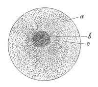
Fig. 1. Diagram Of the Ovum. (From
Gegenbaur.)
a. Granular protoplasm. b. Nucleus (germinal vesicle).
c. Nucleolus (germinal spot).
The ovum so constituted is developed either (1) from one cell out of
an aggregation or layer of cells all of which have the capacity of
becoming ova; or (2) from one of a number of cells segmented off from
a polynuclear mass of protoplasm, not divided into separate cells. In
both cases the cells which have the capacity of becoming ova may be
spoken of as germinal cells, and in the case where the ova are
ultimately developed from a polynuclear mass of protoplasm the
latter structure may be called a germogen.
In some cases the whole of the germinal cells eventually become ova,
but as a rule only a small proportion of them have this fate, the
remainder undergoing various changes to be spoken of in the sequel.
Extended investigations have shewn that the distinction between
germinal cells which are independent cells from the first, or derived
from a germogen in which the nucleated protoplasm is not divided into
cells, is an unimportant one; and closely allied forms may differ in
this respect. It is moreover probable that a germogen of nucleated
protoplasm is less common than is often supposed: it being a matter of
great difficulty to determine the structure of the organs usually so
described. A germogen is stated to be found in most Platyelminthes,
Nematoidea, Discophora, Insecta, and Crustacea.
A more important distinction in the origin of the germinal cells is
that afforded by their position. In this respect three groups may be
distinguished. (1) The germinal cells may form the lining of a sack or
tube, having the form of a syncytium or of an epithelium of separate
cells (Platyelminthes, Mollusca, Rotifera, Echinodermata, Nematoidea,
Arthropoda). (2) Or they may form a specialized part of the epithelium
lining the general body cavity (Chætopoda, Gephyrea, Vertebrata). (3)
Or they may form a mass placed between the two elsewhere contiguous
primitive germinal layers (Cœlenterata[12]).
Types of transition between the first and second group are not
uncommon. Such types, properly belonging to the second group,
originate by a special membranous sack continuous with the oviduct
being formed round the primitively free patch of germinal cells.
Examples of this are afforded by the Discophora, the Teleostei, etc.
It is very probable that all the cases which fall under the first
heading may have been derived from types which belonged to the second
group.
The mode of conversion of the germinal cells into ova is somewhat
diverse. Before the change takes place the germinal cells
frequently multiply by division. The change itself usually involves a
considerable enlargement of the germinal cell, and generally a change
in the character of the germinal vesicle, which in most young ova
(fig. 2) is very large as compared to the body of the ovum. The most
complicated history of this kind is that of the ovum of the Craniata.
(Vide pp. 56, 57.)
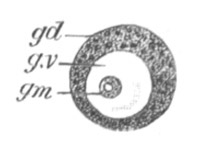
Fig. 2. Ovum Of Carmarina (Geryonia)
hastata. (Copied from Haeckel.)
gd. Body of ovum. gv. Germinal vesicle. gm.
Germinal spot.
The ovum in its young condition is obviously nothing but a simple
cell; and such it remains till the period when it attains maturity.
Nevertheless the changes which it undergoes in the course of its
growth are of a very peculiar kind, and, consisting as they do in many
instances of the absorption of other cells, have led various
biologists to hold that the ovum is a compound structure. It becomes
therefore necessary to consider the processes by which the growth and
nutrition of the ovum is effected before dealing with the structure of
the ovum at all periods of its history.
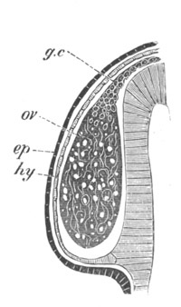
Fig. 3. Female gonophore of Tubularia
Mesembryanthemum. Containing One Large Ovum (ov)
and a number of germinal cells
(g.c.).
ep. Epiblast (Ectoderm). hy. Hypoblast (Entoderm).
ov. Ovum. g.c. Germinal cells.
The ovum is of course nourished like every other cell by the nutritive
fluids in which it is surrounded, and special provisions are made for
this, in that the ovary is very frequently placed in contiguity with
vascular channels. But in addition to such nutrition a further
nutrition, the details of which are given in the special part of this
chapter, is provided for in the germinal cells which do not become
ova.
In the simplest case, as in many Hydrozoa (fig. 3), the germinal cells
which do become ova are assimilated by the ovum much in the manner of
an Amœba.
In other cases the ovum becomes invested by a special layer of cells,
which then constitutes what is known as a follicle. The cells which
form the follicle are often germinal cells, e.g. Holothuria,
Insecta (fig. 17), Vertebrata (fig. 19). In other cases they
seem rather to be adjoining connective-tissue or epithelioid cells,
though it is sometimes difficult to draw the line between such cells
and germinal cells. Examples of follicles formed of ordinary
connective-tissue cells, are supplied by Asterias, Bonellia (fig. 16),
Cephalopoda (fig. 14), etc.
A membrane enclosing the ovum without a lining of cells, as in many
Arachnida, vide p. 51, has no true analogy with a follicle and
does not deserve the same name.
The function of the follicle cells appears to be, to elaborate
nutriment for the growth of the ovum. The follicle cells are not as a
rule directly absorbed into the body of the ovum, though in some
instances, as in Sepia (vide p. 40), they are eventually
assimilated in this way.
In many cases some of the germinal cells form a follicle, while other
germinal cells form a mass within the follicle destined eventually to
be used as pabulum. Insects supply the best known examples of this,
but Piscicola, Bonellia (?) may also be cited as examples of the same
character. In the Craniata (pp. 56‑58) some of the germinal cells
which advance a certain distance on the road towards becoming ova, are
eventually used as pabulum, before the formation of the follicle;
while other germinal cells form at a later period the follicular
epithelium. A peculiar case is that of the Platyelminthes (fig. 9),
where a kind of follicle is constituted by the cells of a specially
differentiated part of the ovary, known as the yolk-gland. The cells
of this follicle may either remain distinct, and continue to surround
the ovum after its development has commenced, and so be used as food
by the embryo; or they may secrete yolk particles, which enter
directly into the protoplasm of the ovum.
For further variations in the mode of nutrition the reader is referred
to the special part of this chapter. Suffice it to say that none of
the known modes of nutrition indicate that the ovum becomes a compound
body any more than the fact of an Amœba feeding on another Amœba
would imply that the first Amœba ceased thereby to be a unicellular
organism.
The constitution of the ovum may be considered under three heads:—
(1) The body of the ovum.
(2) The nucleus or germinal vesicle.
(3) The investing membranes.
The body of the ovum. The essential constituent of the body of
the ovum is an active living protoplasm. As a rule there are present
certain extraneous matters in addition, which have not the vital
properties of protoplasm. The most important of these is known as
food-yolk, which appears to be generally composed of an albuminoid
matter.
The body of the ovum is at first very small compared with the germinal
vesicle, but continually increases as the ovum approaches towards
maturity. It is at first comparatively free from food-yolk; but,
except in the rare instances where it is almost absent, food-yolk
becomes deposited in the form of granules, or highly refracting
spheres, by the inherent activity of the protoplasm during the later
stages in the ripening of the ovum. In many instances the protoplasm
of the ovum assumes a sponge-like or reticulate arrangement, a fluid
yolk substance being placed in the meshes of the reticulum. The
character of the food-yolk varies greatly. Many of its chief
modifications are described below. There is not unfrequently present
in the vitellus a peculiar body known as the yolk nucleus,
which is very possibly connected with the formation of the food-yolk.
It is found in many Arachnida, Myriapoda, Amphibia, etc.[13]
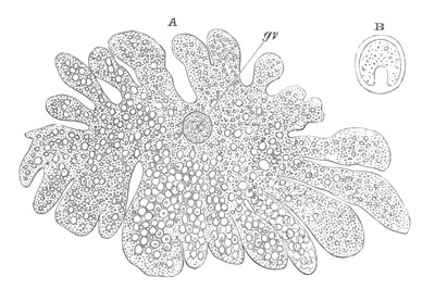
Fig. 4. A. Ovum of Hydra in the amœboid
state, with yolk-spherules (pseudocells) and Chlorophyll
Granules. (After Kleinenberg.)
gv. Germinal vesicle.
B. Single pseudocell of Hydra.
More important for the subsequent development than the variation in
the character of the food-yolk is its amount and distribution. In a
large number of forms it is distributed unsymmetrically, the yolk
being especially concentrated at one pole of the ovum, the germinal
vesicle, surrounded by a special layer of protoplasm comparatively
free from food-yolk, being placed at the opposite pole. In the
Arthropoda it has in most instances a symmetrical distribution.
Further details on this subject are given in connection with the
segmentation; the character of which is greatly influenced by the
distribution of food-yolk.
The body of the ovum is usually spherical, but during a period in its
development it not unfrequently exhibits a very irregular amœboid
form, e.g. Hydra (fig. 4), Halisarca.
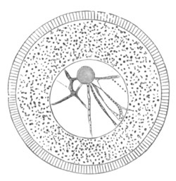
Fig. 5. Unripe ovum of Toxopneustes
lividus. (Copied from Hertwig.)
The germinal vesicle. The germinal vesicle exhibits all the
essential characters of a nucleus. It has a more or less spherical
shape, and is enveloped by a distinct membrane which seems, however,
in the living state to be very often of a viscous semi-fluid nature
and only to be hardened into a membrane by the action of reagents
(Fol). The contents of the germinal vesicle are for the most part
fluid, but may be more or less granular. Their most characteristic
components are, however, a protoplasmic network and the germinal
spots[14]. The protoplasmic network stretches from the germinal spots
to the investing membrane, but is especially concentrated round the
former. (Fig. 5.) The germinal spot forms a nearly homogeneous
body, with frequently one or more vacuoles. It often occupies an
eccentric position within the germinal vesicle, and is usually
rendered very conspicuous by its high refrangibility. In many
instances it has been shewn to be capable of amœboid movements
(Hertwig, Eimer), and is moreover more solid and more strongly tinged
by colouring reagents than the remaining constituents of the germinal
vesicle.
In many instances there is only one germinal spot, or else one main
spot and two or three accessory smaller spots. In other cases,
e.g. Osseous Fishes, Echinaster fallax, Eucope polystyla, there
are a large number of nearly equal germinal spots which appear to
result from the division or endogenous proliferation of the original
spot. Sometimes the germinal spots are placed immediately within the
membrane of the germinal vesicle (Elasmobranchii and Sagitta). In many
Lamellibranchiata, in the earthworm, and in many Chætopoda the
components of the germinal spot become separated into two nearly
spherical masses (fig. 12), which remain in contiguity along a small
part of their circumference, and are firmly united together. The
smaller of the two parts is more highly refractive than the larger.
Hertwig has shewn that the germinal spot is often composed of two
constituents as in the above cases, but that the more highly
refractive material is generally completely enclosed by the less dense
substance. By Fol the germinal spot is stated to be absent in a
species of Sagitta, but this must be regarded as doubtful. In young
ova the relative size of the germinal vesicle is very considerable. It
occupies in the first instance a central position in the ovum, but at
maturity is almost always found in close proximity to the surface. Its
change of position in a large number of instances is accomplished
during the growth of the ovum in the ovary, but in other cases does
not take place till the ovum has been laid.
As the ovum attains maturity, important changes take place in the
constitution of the germinal vesicle, which are described in the next
chapter.
The egg membranes. A certain number of ova when ready to be
fertilized are naked cells devoid of any form of protecting covering,
but as a rule the ovum is invested by some form of membrane. Such
coverings present great variety in their character and origin,
and may be conveniently (Ludwig, No. 4) divided into two great groups,
viz. (1) those derived from the protoplasm of the ovum itself or from
its follicle, which may be called primary egg membranes; and
(2) those formed by the wall of the oviduct or otherwise, such as the
egg-shell of a bird, which may be called secondary egg
membranes.
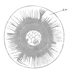
Fig. 6. Ovum of Toxopneustes variegatus with the
pseudopodia-like processes of the protoplasm penetrating the zona
radiata (zr). (After Selenka.)
The primary egg membranes may again be divided into two groups (Ed.
van Beneden, No. 1), viz., (1) those formed by the protoplasm of the
ovum, to which the name vitelline membranes will be applied;
and (2) those formed by the cells of the follicle, to which the name
chorion will be applied.
The secondary egg membranes will be dealt with in connection with the
systematic account of the development of the various groups. They
coexist as a rule with primary membranes, though in some types
(Cephalophorous Mollusca, many Platyelminthes, etc.), they constitute
the only protecting coverings of the ovum.
The vitelline membranes are either simple structureless membranes or
present numerous radial pores. Membranes with the latter structure are
very widely distributed, Echinodermata, Gephyrea, Vertebrata, etc.
(Vide figs. 5 and 7.) The function of the pores appears to be a
nutritive one. They either serve for the emission of pseudopodia-like
processes of the protoplasm of the ovum, as has been very beautifully
shown in the case of Toxopneustes by Selenka (fig. 6), or they admit
(?) processes of the follicular epithelial cells (Vertebrata). Their
presence is in fact probably caused by the existence of such
processes, which prevent the continuous deposition of the membrane.
The term zona radiata will be applied to perforated membranes
of this kind. Two vitelline membranes, one perforated and the other
homogeneous, may coexist at the same time, e.g. Sipunculida,
Vertebrata. (Fig. 7.)
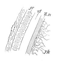
Fig. 7. Section through a small part of the
surface of an ovum of an immature female of Scyllium Canicula.
fe. Follicular epithelium. vt. Vitelline membrane.
Zn. Zona radiata. yk. Yolk with protoplasmic network.
The chorion is often ornamented with various processes, etc.
It is in many cases doubtful whether a particular membrane is a
chorion or a vitelline membrane.
All the membranes which surround the ovum may be provided with a
special aperture known as the micropyle. A micropyle is by no means
found in the majority of types, and there is no homology between the
various apertures so named. Micropyles have two functions, either
to assist in the nutrition of the ovum during its development, or (2)
to permit the entrance of the spermatozoa. The two functions may in
some cases coexist. Micropyles of the first class are developed at the
point of attachment of the ovum to the wall of the ovary or to its
follicle. Good examples of this kind of micropyle are afforded by the
Lamellibranchiata (fig. 12), Holothuria, and many Annelida (Polynoe,
etc.). The micropyle of the Lamellibranchiata (p. 37) probably serves
also to admit the spermatozoa. The second type of micropyle is found
in many Insecta, Teleostei, etc.
General Bibliography of the Ovum.
(1) Ed. van Beneden. “Recherches sur la composition et la
signification de l’œuf,” etc. Mém. cour. d. l’Acad. roy. des
Sciences de Belgique, Vol. XXXIV. 1870.
(2) R. Leuckart. Artikel “Zeugung,” R. Wagner’s
Handwörterbuch d. Physiologie, Vol. IV. 1853.
(3) Fr. Leydig. “Die Dotterfurchung nach ihrem Vorkommen in d.
Thierwelt u. n. ihrer Bedeutung.” Oken, Isis, 1848.
(4) Ludwig. “Ueber d. Eibildung im Thierreiche.” Arbeiten a.
d. zool.-zoot. Institut Würzburg, Vol. I. 1874[15].
(5) Allen Thomson. Article “Ovum” in Todd’s Cyclopædia of
Anatomy and Physiology, Vol. V. 1859.
(6) W. Waldeyer. Eierstock u. Ei. Leipzig, 1870.
Special History of the Ovum in different types.
Cœlenterata.
(7) Ed. van Beneden. “De la distinction originelle d. testicule
et de l’ovaire.” Bull. Acad. roy. Belgique, 3e série, Vol.
XXXVII. 1874.
(8) R. and O. Hertwig. Der Organismus d. Medusen. Jena,
1878.
(9) N. Kleinenberg. Hydra. Leipzig, 1872.
Amongst the Cœlenterata the ova are developed in imperfectly
specialized organs, which are situated in various parts of the body,
for the most part in the space between the epiblast and the hypoblast.
In Hydra the locality where the ova are developed only becomes
specialized at the time when an ovum is about to be formed. At one or
more points the interstitial cells of the epiblast increase in number
and form a protuberance of germinal cells, which may be called the
ovary. In this ovary a single ovum is formed by the special growth of
one cell. (Kleinenberg, No. 9.) In the free and attached gonophores of
Hydrozoa, the ova appear either around the walls of the stomach, or
the radial canals, or around other parts of the gastro-vascular canals.
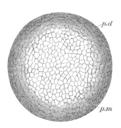
Fig. 8. Ripe Ovum Of Epibulia aurantiaca. The
Germinal Vesicle has become invisible without reagents.
Copied from Metschnikoff, “Entwicklung der Siphonophoren.”
Zeitschrift f. wiss. Zool., Vol. XXIV. 1874.
p.d. Peripheral layer of denser protoplasm. p.m.
Central area consisting of a protoplasmic meshwork.
Their close relations to the gastrovascular canals are probably
determined by the greater nutritive facilities thereby afforded.
(Hertwig, No. 8.)
In the permanent Medusa forms the ova have similar relations to the
gastro-vascular system. Amongst the Actinozoa the ova are usually
developed between the epiblast and the hypoblast in the walls of the
gastric mesenteries. Amongst the Ctenophora the ova are situated in
close relation with the peripheral canals of the gastro-vascular
system, which run along the bases of the ciliated bands. There are
many examples amongst the Cœlenterata of ova which retain
in their mature state the very simple constitution which has been
described as characteristic of all young ova; and which are, when
laid, absolutely without any trace of a vitelline membrane or chorion.
In many other cases both amongst the Medusæ, the Siphonophora, and the
Ctenophora, the ripe egg exhibits a distinction into two parts. The
outer part is composed of a dense protoplasm, while the interior is
composed of a network or more properly a spongework of protoplasm
enclosing in its meshes a more fluid substance. (Fig. 8.)
In some cases the ovum while still retaining the constitution last
described becomes invested by a very delicate membrane. Such is the
constitution of the ripe ovum of Hippopodius gleba amongst the
Siphonophora[16] and of the eggs of Geryonia amongst the permanent
Medusæ[17]. The ripe eggs of the Ctenophora usually present a similar
structure[18]. After being laid they are found to be invested by a
delicate membrane separated by a space filled with fluid from the body
of the ovum. The latter is composed of two layers, an outer one of
finely granular protoplasm and an inner layer consisting of a
protoplasmic spongework containing in its meshes irregular spheres.
These latter are stated by Agassiz to be of a fatty nature, and it is
probable that in most cases where a protoplasmic network is present,
this alone constitutes the active protoplasm and that the substance
which fills up its meshes is to be looked on as a form of food-yolk or
deutoplasm, though it appears sometimes to have the power of
assimilating the firmer yolk particles.
The membrane which invests the ovum of many of the Cœlenterata is
probably a vitelline membrane.
The ova of the Hydrozoa take their origin, in most groups at any
rate[19], from the deeper layer of the epiblast (interstitial layer of
Kleinenberg). The interstitial cells in the ovarian region form
primary germinal cells, and by an excess of nutrition certain of them
outstrip their fellows and become young ova. Such ova differ from the
full-grown ova already described, mainly in the fact that they
have a proportionately smaller amount of protoplasm round the germinal
vesicle. They grow to a considerable extent at the expense of germinal
cells which do not become converted into ova.
The ova of many Cœlenterata undergo changes of a more complicated
kind before attaining their full development. Of these ova that of
Hydra may be taken as the type. The ovary of Hydra (Kleinenberg, No.
9) is constituted of angular flattish germinal cells of which no
single one can be at first distinguished from the remainder. As growth
proceeds one of the cells occupying a central position becomes
distinguished from the remaining cells by its greater size, and
wedge-like shape. It constitutes the single ovum of the ovary. After
it has become prominent it grows rapidly in size, and throws out
irregular processes. The germinal vesicle, which for a considerable
time remains unaltered, also at length begins to grow; and the sharply
defined germinal spot which it contains after reaching a certain size
completely vanishes. After the atrophy of the germinal spot, there
appears in the middle of the ovum a number of roundish yolk granules.
The shape of the ovum becomes more irregular, and chlorophyll
granules, in addition to the yolk granules, make their appearance in
it. A fresh germinal spot of circular form also arises in the germinal
vesicle. Protoplasmic processes are next thrown out in all directions,
giving to the ovum a marvellous amœboid character. (Fig. 4.) The
amœboid form of the ovum serves no doubt to give it a larger
surface for nutrition. Coincidently with the assumption of an
amœboid form there appear in the ovum a great number of peculiar
bodies. They are vesicles with a thick wall bearing a conical
projection into the interior which is filled with fluid. (Fig. 4B.)
These bodies are formed directly from the protoplasm of the ovum, and
are to be compared both morphologically and physiologically with the
yolk-spherules of such an ovum as that of the Bird. They are called
pseudocells by Kleinenberg, and are found with slightly varying
characters in many ova of the Hydrozoa.
They first appear as small highly refracting granules; in these a
cavity is formed which is at first central but is eventually pushed to
one side by the formation of a conical projection from the wall of the
vesicle.
After the growth of the ovum is completed the amœboid processes
gradually withdraw themselves, and the ovum assumes a spherical form;
still however continuing to be invested by the remaining cells of the
ovary. It is important to notice that the egg of Hydra retains
throughout its whole development the characters of a single cell, and
that the pseudocells and other structures which make their appearance
in it are not derived from without, and supply not the slightest
ground for regarding the ovum as a structure compounded of more than
one cell.
The development of the ova of the Tubularidæ, which has been supposed
by many investigators to present very special peculiarities, takes
place on essentially the same type as that of Hydra, but the germinal
vesicle remains permanently very small and difficult to observe. The
mode of nutrition of the ovum may be very instructively studied in
this type. The process is one of actual feeding, much as an Amœba
might feed on other organisms. Adjoining one of the large ova of the
ovary there may be seen a number of small germinal cells. (Fig. 3.)
The boundary between these cells and the ovum is indistinct. Just
beyond the edge of the ovum the small cells have begun to undergo
retrogressive changes; while at a little distance from the ovum they
are quite normal (g.c.)[20].
Platyelminthes.
(10) P. Hallez. Contributions à l’Histoire naturelle des
Turbellariés. Lille, 1879.
(11) S. Max Schultze. Beiträge z. Naturgeschichte d.
Turbellarien. Greifswald, 1851.
(12) C. Th. von Siebold. “Helminthologische Beiträge.” Müller’s
Archiv, 1836.
(13) C. Th. von Siebold. Lehrbuch d. vergleich. Anat. d.
wirbellosen Thiere. Berlin, 1848.
(14) E. Zeller. “Weitere Beiträge z. Kenntniss d. Polystomen.”
Zeit. f. wiss. Zool., Bd. XXVII. 1876.
[Vide also Ed. van Beneden] (No. 1).
This group, under which I include the Trematodes, Cestodes,
Turbellarians and Nemertines, has played an important part in all
controversies relating to the nature and composition of the ovum. The
peculiarity in the development of the ovum in most members of this
group consists in the fact that two organs assist in forming what is
usually spoken of as the ovum. One of these is known as the ovary
proper, and the other as the vitellarium or yolk-gland. In the sequel
the term ovum will be restricted to the product of the first of these
organs. In Trematodes the ovary forms an unpaired organ directly
continuous with an oviduct into which there open the ducts from paired
yolk-glands.
The ovary has a sack-like form and contains in some instances a
central lumen (Polystomum integerrimum). At the blind end of the organ
is placed the germinal tissue. This part is, according to the accounts
of the majority of investigators, formed of a polynuclear mass of
protoplasm not divided into distinct cells. Whether it is really
formed of undivided protoplasm or not, it is quite certain that a
little lower down in the organ distinct cells are found, which have
been segmented off from the above mass, and are formed of a large
nucleus and nucleolus, surrounded by a delicate layer of protoplasm.
These cells are the young ova. They usually assume a more or less
angular form from mutual pressure, and, in the cases where the ovary
has a lumen, constitute a kind of epithelial lining for the ovarian
tube. They become successively larger in passing down the ovary, and,
though in most cases naked, are in some instances (Polystomum
integerrimum) invested by a delicate vitelline membrane. Eventually
the ova pass into the oviduct and become free and at the same time
assume a spherical form.
In the oviduct the ovum receives somewhat remarkable investing
structures, derived from the organ before spoken of as the yolk-gland.
The yolk-gland consists of a number of small vesicles, each provided
with a special duct, connected with the main duct of the gland. Each
vesicle is lined by an epithelium of cells provided with doubly
contoured membranes, and containing nuclei.
As the yolk cells grow older refracting spherules become deposited in
their protoplasm, which either completely hide the nucleus, or
render it very difficult to see. In the majority of cases the entire
cells forming the lining of the vesicles constitute the secretion of
the yolk-gland. They invest the ovum, and around them is formed a
shell or membrane. In some cases (e.g. Polystomum integerrimum)
the yolk cells retain their cellular character and vitality till the
embryo is far developed. In other cases they lose their membrane and
nucleus shortly after the formation of the egg-shell, and break up
into a fluid, holding in suspension a number of yolk granules. A
partial disorganisation of the yolk cells can also take place before
they surround the ovum; while in some species of Distomum they
completely break up before leaving the yolk-gland.
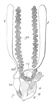
Fig. 9. Generative system of Vortex viridis.
(From Gegenbaur, after Max Schultze.)
t. Testis. v.d. Vasa differentia. v.s. Seminal
vesicle. p. Penis. u. Uterus. o. Ovary.
v. Vagina. g.v. Yolk-glands. r.s. Receptaculum
seminis.
There is thus a complete series of gradations between the investment
of the ovum by a number of distinct cells, and its investment by a
layer of fluid containing yolk-spherules in suspension. In neither the
one case nor the other do the investing structures take any share in
the direct formation of the embryo from the ovum. Physiologically
speaking they play the same part as the white in the fowl’s egg.
The egg-shell, which is usually formed by a secretion of a special
shell-gland opening into the oviduct, exhibits one or two
peculiarities in the different species of Trematodes. In Amphistomum
subclavatum it presents at one extremity a thickened area, which is
pierced by a narrow micropyle. In other cases one extremity of the
egg-shell is produced into a long process, and sometimes even both
extremities are armed in this way. Opercula and other types of
armature are also found in different forms.
The mode of development of the ovum in Cestodes is very nearly the
same as in Trematodes.
The ovum becomes enveloped in the usual secretion of the yolk-gland;
and an egg-shell is always formed by the secretion of a special
shell-gland.
Amongst the Turbellarians and Nemertines, there are greater variations
in the arrangement of the female generative glands, than in
the preceding types. In most of the Rhabdocœla and fresh-water
Dendrocœla these organs resemble in their fundamental characters
those of the Trematodes and Cestodes. There are present a paired or
single ovary and a paired yolk-gland. The general arrangement of the
organs is shewn in fig. 9.
The blind end of the ovaries is usually (Ed. van Beneden, etc.) stated
to be formed of a polynuclear protoplasmic basis, but Hallez (No. 10)
has recently insisted that, even at the extreme end of the ovary, the
germinal cells are quite distinct, and not confounded together.
With one or two exceptions the yolk cells secreted by the vitellarium
retain their vitality till they are swallowed by the embryo, after the
development of its mouth. The few not so swallowed become
disintegrated. They are granular nucleated cells, and, as was first
shewn by von Siebold, are remarkable for exhibiting spontaneous
amœboid movements.
Very important light on the nature of the vitellarium is afforded by
the structure of the generative organs in Prorhyncus and Macrostomum.
In Prorhyncus there is no separate vitellarium, but the lower part of
the ovarian tube functionally and morphologically replaces it. The
ovum becomes surrounded by yolk cells, which according to Hallez (No.
10) retain their vitality for a long time. According to Ed. van
Beneden yolk-spherules are formed in the protoplasm of the ovum
itself, in addition to and independently of the surrounding yolk
cells. In Convoluta paradoxa a special vitellarium is stated to be
absent; though a deposit of yolk is formed round the ovum (Claparède).
In Macrostomum again the yolk-glands are at most represented by a
lower specialized part of the ovarian tube. The ova in passing down
become filled with yolk-spherules. According to Ed. van Beneden these
spherules are formed in the protoplasm of the ovum itself; but this is
explicitly denied by Hallez, who finds that they are formed from the
lining cells of the ovarian tube, which, instead of retaining their
vitality as in Prorhyncus, break up and form a granular mass which is
absorbed by the protoplasm of the ovum.
In Prostomum caledonicum (Ed. van Beneden) the generative organs are
formed on the same plan as in other Rhabdocœla, but the
cells which form the yolk-gland give rise to yolk particles which
enter the ovum, instead of to a layer of yolk cells surrounding the
ovum.
Amongst the marine dendrocœlous Turbellarians the ova are formed in
separate sacks widely distributed in the parenchyma of the body
between the alimentary diverticula. In these the ova undergo their
complete development, without the intervention of yolk-glands.
The ovaries of the Nemertines more nearly resemble those of the marine
Dendrocœla than those of the Rhabdocœla. They consist of a
series of sacks situated on the two sides of the body between the
prolongations of the digestive canal. The eggs are developed in these
sacks in a perfectly normal manner, and in many cases become filled
with yolk-spherules which arise as differentiations of the protoplasm
of the ovum. The protecting membranes of the ova have not been
accurately studied. In some cases[21] two membranes are present, an
internal and an external. The former, immediately investing the
vitellus, is very delicate: the external one is thicker and hyaline.
The constitution of the female generative organs of the Trematodes was
first clearly ascertained by von Siebold (No. 12). He originally,
though not very confidently, propounded the view that the germinal
vesicles alone were formed in the ovary and that the protoplasm of the
ovum was supplied by the yolk-gland. This view has long been
abandoned, and von Siebold (No. 13) himself was the first to recognize
that true ova with a protoplasmic body containing a germinal vesicle
and germinal spot were formed in the ovary. The Trematodes have
however not ceased to play an important part in forming the current
views upon the development of ova, and have quite recently served Ed.
van Beneden as his type in exposing his general view upon this subject.
His view consists fundamentally in regarding the secretion of the
yolk-glands, which in most cases merely invests the ovum, as
homologous with the yolk-spherules which fill the protoplasm of many
eggs; and he considers the part of the ovary where in most forms the
ova receive their supply of yolk particles, as equivalent to the
vitellarium of the Platyelminthes. He further appears to regard the
primitive state as that exemplified in Trematodes, Cestodes, etc., and
holds that the ovarian types characteristic of other forms are
secondarily derived from this, by the coalescence of the primitively
distinct vitellarium with the ovary proper.
This appears to me a case of putting the cart before the horse. To my
mind the vitellarium is to be regarded, as has already been suggested
by Gegenbaur, Hallez, etc. as a special differentiation of the
primitively simple ovarian tube, and the instances Of Macrostomum and
Prorhyncus just cited appear to me to indicate some of the steps in
this differentiation. In Macrostomum the cells of the lower part of
the oviduct simply supply a kind of nutriment to the ovum in the form
of granular yolk particles, while in Prorhyncus the yolk cells of the
lower part of the ovarian tube form a complete investment of
independent cells for the ovum. If this lower part of the ovarian tube
were to grow out as a special diverticulum we should have produced a
normal vitellarium. But even with the above modification the theory of
van Beneden appears to me not completely satisfactory. The view that
the yolk-spherules are of the same nature as the yolk cells is mainly
supported by the case of Prostomum caledonicum, where the vitellarium
produces the yolk particles which fill the ovum. The cases Of
Prorhyncus and Macrostomum give a different complexion to that of
Prostomum caledonicum. From the first of these especially it appears
that, even when normal yolk cells surround the ovum, yolk particles
can be deposited independently in the protoplasm of the ovum.
The most probable view of the nature of the vitellarium is that of
Gegenbaur, Hallez, etc., according to which it is to be regarded as a
specially modified part of the ovarian tube. On this view the nature
and function of the yolk cells admit of a fairly simple explanation.
They are to be regarded as primary germinal cells like those in the
ovaries of Hydra, Tubularia, etc., which do not become converted into
ova. Like these cells they may in some instances, Macrostomum,
Prostomum, etc., serve directly in the nutrition of the ovum. In other
cases they retain their independence and serve for the late nutrition
of the embryo. In both instances they retain the faculty, normally
possessed by ova, of forming yolk particles in their protoplasm.
Echinodermata.
(15) C. K. Hoffmann. “Zur Anatomie d. Echiniden u. Spatangen.”
Niederländisch. Archiv f. Zoologie, Vol. I. 1871.
(16) C. K. Hoffmann. “Zur Anatomie d. Asteriden.”
Niederländisch. Archiv f. Zoologie, Vol. II. 1873.
(17) H. Ludwig. “Beiträge zur Anat. d. Crinoiden.” Zeit. f.
wiss. Zool., Vol. XXVIII. 1877.
(18) Joh. Müller. “Ueber d. Canal in d. Eiern d. Holothurien.”
Müller’s Archiv, 1854.
(19) C. Semper. Holothurien. Leipzig, 1868.
(20) E. Selenka. Befruchtung d. Eies v. Toxopneustes
variegatus, 1878.
[Vide also Ludwig (No. 4), etc.]
The eggs of the Echinodermata present in their development certain
points of interest.
The ovaries themselves are usually surrounded by a special vascular
dilatation. In the Asteroidea, the Echinoidea, and the Holothuroidea
the organs have the form of sacks; specially surrounded in the two
former groups, and probably the latter, by a vascular sinus formed as
a dilatation of one of the generative vessels. In the Crinoids they
have the form of a hollow rachis completely surrounded by a
blood-vessel. (Fig. 11, b.) The proximity of the ovaries
(generative organs) to the vascular system in these forms has clearly
the same physiological significance as the proximity of the ovaries
(generative organs) to the radial vessels in the Cœlenterata.
In the Asteroidea, the Echinoidea and the Holothuroidea the ovaries
have the form of sacks lined by an epithelium of germinal cells, and
the ova are formed by the enlargement of these cells, which, when they
have reached a certain size, become detached from the walls, and fall
into the cavity of the ovarian sack. In Toxopneustes (Selenka) and
very probably in other forms only a few of the epithelial cells
undergo conversion into ova: the remainder undergo repeated division,
and, as in so many other cases, are eventually employed in the
nutrition of the true ova. In the nearly ripe ova of Asterias Fol has
described a flattened follicular epithelium the origin of which is
unknown.
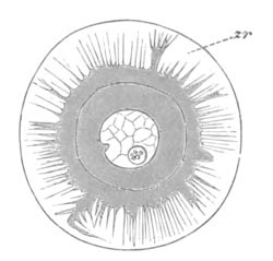
Fig. 10. Ovum of Toxopneustes variegatus with the
pseudopodia-like projections of the protoplasm penetrating the zona
radiata (zr). (After Selenka.)
In Holothuria (Semper) a further differentiation of the germinal
cells, not destined to become ova, takes place. They surround the
enlarged cell which forms the true ovum, for which they constitute a
kind of follicular capsule. This capsule is attached by a stalk to the
walls of the ovary, and the ovum lies freely in it except for an area
nearly opposite its (the capsule’s) point of attachment, where the
ovum adheres to the wall of the capsule. Subsequently the follicle
cells which form the capsule fuse together, and form a definite
membrane in which only the nuclei remain distinct. Within the
membranous capsule there is formed for the ovum an albuminous zona
radiata. At the point where the ovum is attached to its capsule this
membrane cannot be developed, and therefore remains
incomplete. The perforation so formed, becomes the micropyle of the
Holothurian egg, which was first discovered by Joh. Müller. The
albuminous membrane just described for Holothurians is also found in
Asteroids (fig. 5) and Echinoids. In these groups there is no proper
micropyle, though in Ophiothrix a nutritive passage perforates the
membrane at the attachment of the ovum before the period when the ovum
becomes free (Ludwig). The formation of the zona radiata has been
studied by Selenka. It is secreted by the protoplasm of the ovum, and
has a gelatinous consistency, and after it is formed the peripheral
layer of the protoplasm of the ovum sends out through it
pseudopodia-like processes to absorb nutriment from without. These
processes are at first large and irregular, but soon become finer and
finer (fig. 10), and acquire a regular radiating arrangement. They are
withdrawn when the ovum is ripe, but they nevertheless give rise to
the finely radiated appearance of the membrane, the radii being in
reality delicate pores.
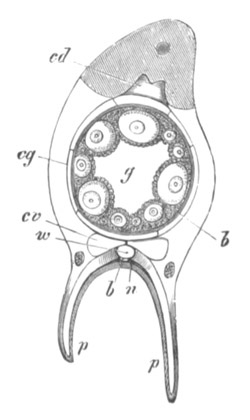
Fig. 11. Transverse section through the pinna of
a sexually mature Comatula. (From Gegenbaur, after Ludwig.)
p. Tentacle. g. Lumen of genital rachis. w.
Water-vascular vessel. n. Nerve cord. b. Blood-vessel
on nerve cord and round genital rachis. cg. Genital canal.
cd. Dorsal section of the body cavity. cv. Ventral
section of body cavity.
In the Crinoids the generative rachis consists of a tube, the
epithelium of which is formed of the primary germinal cells. (Fig.
11.) While some of these cells enlarge and become ova, the remainder
supply the elements for a follicular epithelium, which is established
round the ova, exactly as in Holothurians.
Mollusca.
Lamellibranchiata.
(21) H. Lacaze-Duthiers. “Organes génitaux des Acéphales
Lamellibranches.” Ann. Sci. Nat., 4me série, Vol.
II. 1854.
(22) W. Flemming. “Ueb. d. er. Entwick. am Ei d. Teichmuschel.”
Archiv f. mikr. Anat., Vol. X. 1874.
(23) W. Flemming. “Studien üb. d. Entwick. d. Najaden.”
Sitz. d. k. Akad. Wiss. Wien, Vol. LXXI. 1875.
(24) Th. von Hessling. “Einige Bemerkungen, etc.” Zeit. f.
wiss. Zool., Bd. V. 1854.
(25) H. von Jhering. “Zur Kenntniss d. Eibildung bei d.
Muscheln.” Zeit. f. wiss. Zool., Vol. XXIX. 1877.
(26) Keber. De Introitu Spermatozoorum in ovula, etc.
Königsberg, 1853.
(27) Fr. Leydig. “Kleinere Mittheilung etc.” Müller’s
Archiv, 1854.
Gasteropoda.
(28) C. Semper. “Beiträge z. Anat. u. Physiol. d.
Pulmonaten.” Zeit. f. wiss. Zool., Vol. VIII. 1857.
(29) H. Eisig. “Beiträge z. Anat. u. Entwick. d. Pulmonaten.”
Zeit. f. wiss. Zool., Vol. XIX. 1869.
(30) Fr. Leydig. “Ueb. Paludina vivipara.” Zeit. f. wiss.
Zool., Vol. II. 1850.
Cephalopoda.
(31) Al. Kölliker. Entwicklungsgeschichte d.
Cephalopoden. Zurich, 1844.
(32) E. R. Lankester. “On the developmental History of the
Mollusca.” Phil. Trans., 1875.
Lamellibranchiata.
The ova of the Lamellibranchiata present several points of interest.
They are developed in pouches of the ovary which are lined by a
flattened germinal epithelium, or sometimes (?) a syncytium. Some of
the cells of this epithelium enlarge and become ova, but remain
attached to the walls of their pouches by protoplasmic stalks. Round
the ovum there appears in some forms (Anodon, Unio) a delicate
vitelline membrane, which is incomplete at the protoplasmic stalk, and
is therefore perforated by an aperture which forms the micropyle.
(Fig. 12.) As the ovum becomes ripe a large space filled with
albuminous fluid becomes established between the ovum and its
membrane, but the ovum remains attached to the membrane at the
micropyle. In Scrobicularia (von Jhering, No. 25) the membrane round
the ovum appears from the first as an albuminous layer, the outermost
stratum of which becomes subsequently hardened as the vitelline
membrane. In this form also the protoplasmic stalk becomes, in pouches
largely filled with ova, extremely long. The ova become eventually
detached by the stalk rupturing, and the portion of it which remains
attached to the vitelline membrane falling off. The function of the
stalk and of the micropyle during the development of the ovum is
undoubtedly a nutritive one.
In Anodon and Unio yolk granules similar to those deposited in the
protoplasm of the ovum are also found in the epithelial cells of the
ovarian pouches (Flemming, 22), and there can be but little doubt that
they are directly transported from these cells into the ovum. These
cells would seem therefore to play much the same part as the
yolk-glands of some Turbellarians (Prostomum caledonicum). In
Scrobicularia yolk granules are not found in the epithelium of the
pouches, but are contained in the dilated disc by which the ovum is
attached to the wall of its pouch, as well as in the ovum itself.
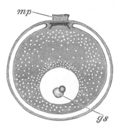
Fig. 12. Medium-sized ovum of Anodonta
complanata. (After Flemming.)
mp. micropyle. gs. germinal spot.
On the ovum becoming detached the micropyle still remains as an
aperture, which probably has the function of admitting the spermatozoa.
The shape and form of the micropyle vary greatly. In Anodon and Unio
it is a projecting trumpet-shaped structure, which after fertilization
becomes shortened and reduced to a mere aperture which is finally
stopped up. (Fig. 12.)
In other forms it is simply a perforation in the vitelline membrane
which is sometimes very large. In a species of Arca, which I had an
opportunity of observing at Valparaizo, it was equal to nearly the
circumference of the ovum.
The eggs of the Lamellibranchiata are not only remarkable in the
possession of a micropyle, but in certain peculiarities of the yolk
and of the germinal vesicle.
In the fresh-water mussels there is usually found in young and
medium-sized ova a peculiar lens-shaped body—Keber’s corpuscle—which
is placed immediately internal to the micropyle. It is probably in
some way connected with the nutrition of the ovum, though the fact
that it is not always present shews that it cannot be of great
importance.
A dark body found by von Jhering in the neighbourhood of the germinal
vesicle in the ripe ovum of Scrobicularia is probably of a similar
nature to Keber’s corpuscle. Both bodies may be placed in the same
category as the so-called yolk nucleus of the spider’s and frog’s ova.
In all except the youngest ova of Anodon and Unio the germinal spot is
composed of two nearly complete spheres united together for a small
part of their circumference. (Fig. 12, gs.) The smaller of
these has a higher refractive index than the larger, and often
contains a vacuole: the two parts together appear to be the separated
components (though not by simple division) of the primitive nucleolus.
A nucleolus of this character is not universal amongst
Lamellibranchiata, but a similar separation of the constituents of the
germinal spot has been found by Flemming in Tichogonia, in which
however the more highly refracting body envelopes part of the less
highly refracting body in a cap-like fashion.
Gasteropoda.
The ova of the Gasteropoda are developed, like those of the
Lamellibranchiata, from the epithelial cells of the ovarian acini or
pouches. In the hermaphrodite forms both ova and spermatozoa are
produced in the same pouches (fig. 13), some of the epithelial cells
becoming ova and others spermatozoa. The ova are usually formed in the
wall of the pouch, and the spermatozoa internally (Pulmonata) (fig. 13
A), or a further differentiation of parts may take place (fig.13 B).
The ova of Gasteropods are exceptional in the fact that
a vitelline membrane is rarely or never developed around them.
The ovum in its passage to the exterior becomes enclosed in a
secretion of the albuminous gland, which hardens externally to form a
special membrane.
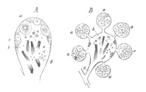
Fig. 13. Follicles of the hermaphrodite glands of
Gasteropoda.
(From Gegenbaur.)
A. Of Helix hortensis. The ova (aa) are developed on
the wall of the follicle, and the seminal masses (b)
internally.
B. Of Aeolidia. The seminal portion of a follicle is beset
peripherally by ovarian saccules (a). c. Common
afferent duct.
Cephalopoda.
Lankester (No. 32) has brought out some very interesting points with
reference to the nutrition of the eggs of Sepia during their growth.
The eggs develop in connective-tissue pouches which early give rise to
a double pedunculated capsule of connective tissue. The cells of the
inner layer of this capsule soon assume an epithelial character, and
become a definite follicular epithelium, while between the two layers
there penetrates a network of vascular channels. The follicular
epithelium becomes after the establishment of these vascular channels
folded in a most remarkable manner. The folds, which are shewn in
section in fig. 14, ic, project into and nearly completely fill
up the body of the ovum. An enormous increase is thus effected in the
nutritive surface exposed by the epithelium. Each fold is thoroughly
supplied with blood-vessels. The plications of the follicular
epithelium give rise to a basket-work tracery on the surface of the
ovum. During the stage when the follicular epithelium has the above
structure, its cells have a character similar to that of the
goblet-cells of a mucous membrane, and pour out their metamorphosed
protoplasm into the body of the ovum.
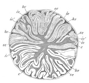
Fig. 14. Transverse section through an ovarian
egg of Sepia. (Copied from Lankester.)
o.c. outer capsular membrane. i.c. inner capsular
membrane with follicular epithelium. b.v. blood-vessels in
section between the outer and inner capsular membranes. c.
vitellus.
The section shews the folds of the inner capsule with their
epithelium, which penetrate into the substance of the ovum for the
purpose of supplying it with nourishment.
After the above mode of nutrition has gone on for a certain time a
change takes place, and the ridges gradually disappear. This is caused
by the epithelial cells passing off from the ridges into the
protoplasm of the ovum; and becoming assimilated, after retaining
their individuality for a longer or shorter period. When the
absorption of the ridges is completed the surface of the ovum assumes
a perfectly regular outline. The capsule of the ovum then bursts at
the opposite pole to the peduncle, and the ovum falls into the
oviduct.
The ova of the Cephalopoda, like those of the Gasteropoda, are quite
naked, being without a vitelline membrane or chorion. The egg-capsule
which is formed for them in their passage down the oviduct is
perforated in Sepia by a micropylar aperture.
Chætopoda.
(33) Ed. Claparède. “Les Annelides Chætopodes d. Golfe de
Naples.” Mém. d. l. Sociét. phys. et d’hist. nat. de Genève
1868‑9 and 1870.
(34) E. Ehlers. Die Borstenwürmer nach system. und anat.
Untersuchungen. Leipzig, 1864‑68.
(35) E. Selenka. “Das Gefäss-System d. Aphrodite aculeata.”
Niederländisches Archiv f. Zool., Vol. II. 1873.
The ova of the Chætopoda are in most cases developed from the special
tracts of the epithelial cells lining parts of the body
cavity, which constitute a germinal epithelium (fig. 15). Very
frequently (Aphrodite, Arenicola), as is so common in other types,
these tracts of germinal cells surround the blood-vessels. In some
cases the germinal epithelium thickens to form a compact organ, for
which the outermost cells may form a more or less definite membranous
covering (Oligochæta, etc.). The ova are formed by the enlargement,
accompanied by other changes, of these germinal cells. During their
early development the ova are frequently surrounded by a special
capsule, which is often stalked, and provided at its attachment with a
large micropylar aperture. In Aphrodite and Polynoe this arrangement,
which is clearly connected with the nutrition of the ovum, is very
easily seen. The ovum is dehisced into the body cavity by the bursting
of its capsule or the rupture of the stalk. The capsule is always
eventually thrown off; but a vitelline membrane is frequently
developed after the detachment of the ovum into the body cavity. The
vitelline membrane of Spio and other Polychæta is provided with an
equatorial ring of ampulliform vesicles.
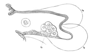
Fig. 15. A Parapodium of Tomopteris.
(From
Gegenbaur.)
o. Collection of germinal epithelial cells lining the body
cavity.
Discophora.
(36) H. Dorner. “Ueber d. Gattung Branchiobdella.” Zeit. f.
wiss. Zool., Vol. XV. 1865.
(37) R. Leuckart. Die menschlichen Parasiten.
(38) Fr. Leydig. “Zur Anatomie v. Piscicola geometrica, etc.”
Zeit. f. wiss. Zool., Vol. I. 1849.
(39) C. O. Whitman. “Embryology of Clepsine.” Quart. J. of
Micr. Sci., Vol. XVIII. 1878.
The ovary of the Discophora is formed of a mass of cells enveloped in
a membranous sack. In Branchiobdella there is placed in the
central axis of these cells a column of nucleated protoplasm from
which the cells themselves are budded off. The development of the ovum
takes place by the enlargement, etc. of one of the peripheral cells,
which eventually bursts the wall of the sack and is freely dehisced
into the body cavity.
In most other Leeches (except Piscicola and its allies) there is found
a more specialized arrangement of the same nature as in
Branchiobdella. There are one or more coiled egg-strings which lie
freely in a delicate sack continuous with the oviduct. Each egg-string
is formed of a central rachis and of a peripheral layer of cells[22].
The ova are formed by the enlargement of the peripheral cells
accompanied by a deposition of food-yolk. Food-yolk appears to be
formed in the rachis even more energetically than in the protoplasm of
the ova. When ripe the ova fall into the ovarian sack.
In Piscicola the development of the ovum is somewhat peculiar but
resembles in certain respects that of Bonellia (p. 45). The ova are
developed from the primitive germinal cells which fill up the ovarian
sack. The nuclei in these cells increase in number, and a nucleated
peripheral layer of each cell becomes separated from the central part,
which also contains nuclei. This latter part next divides into
numerous cells, of which one eventually forms the ovum, and the
remainder constitute a mass of cells adjoining it as in Bonellia (fig.
16). This mass of cells eventually disappears, and is probably
employed in the nutrition of the ovum.
The ovaries of the Leech appear to belong to the tubular type in that
the ova are not formed from part of the epithelium lining the body
cavity; but if, as seems probable, the true affinities of the Leeches
are with the Chætopoda, the investment of the ovaries must be of a
secondary nature. It should be noted that the ova are not, as in the
ordinary tubular ovary, developed from the epithelium lining the
ovarian tube.
Gephyrea.
(40) Keferstein u. Ehlers. Zoologische Beiträge.
Leipzig, 1861.
(41) C. Semper. Holothurien, 1868, p. 145.
(42) J. W. Spengel. “Beiträge z. Kenntniss d. Gephyreen.”
Beiträge a. d. zool. Station z. Neapel, Vol. I. 1879.
(43) J. W. Spengel. “Anatomische Mittheilungen üb. Gephyreen.”
Tagebl. d. Naturf. Vers. München, 1877.
In the Gephyrea, as in the Chætopoda, the ova are developed from the
lining cells of the peritoneum and frequently from the cells
surrounding parts of the vascular system (Bonellia, Thalassema). In
many cases (Sipunculus, Phascolosoma, Echiurus) the main growth of the
ovum takes place after it has been dehisced into the body cavity.
In Sipunculus the ova in the body cavity are surrounded by a follicle
which is thrown off before they become ripe.
Brandt denies the existence of this follicle or rather its cellular
nature. Spengel’s (43) observations are conclusive in favour of the
correctness of the original interpretation Of Keferstein and Ehlers.
The follicles would seem to be formed after the ova have become free.
In Phascolosoma there is no follicle (Semper, Spengel).
In both Phascolosoma and Sipunculus a vitelline membrane with radial
pores—zona radiata—is formed, and in Phascolosoma the
external part of this is separated off as a structureless vitelline
membrane. The formation of both these membranes from the protoplasm of
the ovum is rendered certain in the latter case by the absence of a
follicular epithelium.
Some interesting observations on the growth and origin of the ovum in
Bonellia have been made by Spengel.
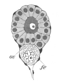
Fig. 16. Follicle Of Bonellia at a medium stage
of development. (After Spengel.)
ov. ovum. fe. flattened follicular epithelium.
The ova originate from certain cells (germinal cells) in the
peritoneal investment of the ventral vessel, overlying the nervous
cord. These cells, which are well marked off from the surrounding
flattened peritoneal elements, increase in number by division, and
form small masses surrounded by a follicle of peritoneal cells, and
attached by a stalk to the peritoneum. The central cell of each mass
grows larger than the rest, which arrange themselves in a columnar
fashion round it; it is not, however, destined to become the ovum. On
the contrary certain of the other cells adjoining the stalk grow
larger, and finally one of these becomes distinguished as the ovum by
its greater size and the character of its nucleus. The
remainder of the larger cells become of the same size as their
neighbours. The ovum now becomes more or less separate from the mass
of germinal cells, rapidly grows in size, and soon forms the most
considerable constituent of the follicle (fig. 16, ov). The
remaining germinal cells are quite passive, and though, with the
exception of the central cell, they do not appear to atrophy, they
soon constitute a relatively small prominence on the surface of the
ovum. By the rupture of the stalk the whole follicle becomes
eventually detached, and the further development of the ovum takes
place in the body cavity. A vitelline membrane is formed, and
eventually the ovum is taken into the oviduct (segmental organ). At
this time or slightly before, the follicle cells together with the
germinal mass, which throughout exhibits no signs of atrophy, become
thrown off, and the ovum is left invested in its vitelline membrane.
Nematoda.
(44) Ed. Claparède. De la formation et de la fécondation des
œufs chez les Vers Nématodes. Genève, 1859.
(45) R. Leuckart. Die menschlichen Parasiten.
(46) H. Munk. “Ueb. Ei- u. Samenbildung u. Befruchtung b. d.
Nematoden.” Zeit. f. wiss. Zool., Vol. IX. 1858.
(47) H. Nelson. “On the reproduction of Ascaris mystax, etc.”
Phil. Trans. 1852.
(48) A. Schneider. Monographie d. Nematoden. Berlin,
1866.
The female organs consist as a rule of two cæcal tubes which unite
before opening to the exterior. Each of these is divided into a
vagina, uterus, oviduct, and ovary. The ovary constitutes the blind
end of the tube, and is formed of a common protoplasmic column,
holding a number of nuclei in suspension. The protoplasm becomes cleft
around the nuclei in the uppermost part of the tube; the
circumscription of the ova proceeds, however, very gradually, and
since it commences at the periphery of the column the ova remain
attached by stalks to a central axis with one end free. In this way
there is formed a rod-like structure known as the
rachis, which consists of a central axis with a series of half
circumscribed ova radiately arranged round it. In the lowest part of
the ovary the ova become completely isolated and form separate cells.
The protoplasm of the ova, which is clear in the terminal division of
the ovary, becomes in most forms filled lower down with yolk-spherules
secreted in the body of the ova. These commence to appear at the
uppermost extremity of the rachis.
In some instances, e.g. Cucullanus elegans, yolk-spherules are
not formed. In the Oxyuridæ the ova are directly segmented off from
the terminal syncytium of protoplasm without the intervention of a
rachis; and are therefore formed in the same way as amongst
Trematodes, etc.
The origin of the membrane around the ova of the Nematoda has been
much disputed.
At the time when the ovum is detached from the rachis no membrane is
present, but it nevertheless appears from Schneider’s observations
that the region at which it is detached is softer than other parts, so
that a kind of micropyle is here formed which disappears after
impregnation. A delicate vitelline membrane then appears, around which
there is subsequently established an egg-shell, which is usually
stated to be formed as a secretion of the walls of the uterus; but
Schneider and Leuckart have given strong grounds for believing that it
is really a further differentiation of the vitelline membrane due to
the activity of the protoplasm of the ovum. The originally single
membrane becomes as it thickens split into two layers. The outer of
these forms the true egg-shell, and the fertilization of the ovum
appears to be a necessary prelude to its production. Round the
egg-shell the walls of the uterus often secrete a special albuminous
covering.
The egg-shell exhibits in many cases peculiar sculpturings as well as
terminal prolongations.
Insecta.
(49) A. Brandt. Ueber das Ei u. seine Bildungsstätte.
Leipzig, 1878.
(50) T. H. Huxley. “On the agamic reproduction and morphology
of Aphis.” Linnean Trans., Vol. XXII. 1858.
Vide also Manual of Invertebrated Animals, 1877.
(51) R. Leuckart. “Ueber die Micropyle u. den feinern Bau d.
Schalenhaut bei den Insecteneiern.” Müller’s Archiv, 1855.
(52) Fr. Leydig. Der Eierstock u. die Samentasche d.
Insecten. Dresden, 1866.
(53) Lubbock. “The ova and pseudova of Insects.” Phil.
Trans. 1859.
(54) Stein. Die weiblichen Geschlechtsorgane d. Käfer.
Berlin, 1847.
[Conf. also Claus, Landois, Weismann, Ludwig (No. 4).]
The ovum of Insects has formed the subject of numerous investigations,
and has played an important part in the controversies on the nature of
the ovum.
The ovaries are paired organs, rarely directly connected, each
consisting of more or fewer ovarian tubes which open into a common
oviduct. The oviducts unite into a vagina, usually provided with a
spermatheca and accessory glands, which need not be further alluded
to. Each ovary is invested by a peritoneal covering, which assumes
various characters, and either forms a loose network covering the
whole or a special tunic round each egg-tube. It is continuous with
the general peritoneal investment. Each ovarian tube (fig. 17)
consists of three sections: (1) a terminal thread, (2) the terminal
chamber or germogen, (3) the egg-tube proper.
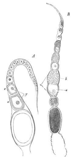
Fig. 17. A. Ovarian tube of the Flea,
Pulex irritans. (From Gegenbaur, after Lubbock.)
o. ovum. g. germinal vesicle.
B. Ovarian tube of a Beetle, Carabus violaceus.
(After Lubbock.)
o. ovarian segment, formed of an ovum a, and a mass of
yolk cells, b.
The whole egg-tube is invested in a structureless tunica propria.
The terminal threads are fine prolongations of the ends of the
egg-tubes usually continued close up to the heart. At their
extremities they frequently anastomose, or even unite into a common
thread. In some cases they are absent. They form either direct
continuations of the germogen and have the same histological
structure, or in other cases are simply prolongations of the tunica
propria, and serve as ligaments.
The germogen usually consists of two parts: an upper, filled with
nuclei imbedded in protoplasm, and a lower, in which distinct cells
have become differentiated.
The lower part of the egg-tubes is filled with ova which advance in
development towards the oviduct, and lie in chambers more or less
distinctly constricted from each other. In these chambers there are in
most forms in addition to the true ova a certain number of nutritive
cells. The true egg-tubes are moreover lined by an epithelial
layer which passes in and forms more or less complete septa between
the successive chambers. The points which have been especially
controverted are (1) the relation of the ovum to the germogen, and (2)
the relation of the nutritive or yolk cells to the ovum. To the
controversies on these points it will only be possible to give a
passing allusion.
As has been already hinted there are two distinct types of ovaries,
viz. those without the so-called nutritive or yolk cells and those
with them[23].
The formation of the ovum is most simple in the type without yolk
cells, which will for that reason be first considered (fig. 17
A).
The germogen is constituted of a number of nuclei imbedded in a scanty
cementing protoplasm. In the lower part of the germogen the nuclei are
larger, and become separated off from the nucleated protoplasm above,
as distinct cells with a thin layer of protoplasm round the germinal
vesicle. These cells are the ova. As they pass down the egg-tube their
protoplasm increases in bulk, and they become isolated by ingrowths of
the epithelial cells the origin of which is still uncertain, which
form round each ovum a special follicle, so that the egg-tube is
filled by a single row of ova each in an epithelial follicle (fig. 17
A). The larger the ova the more columnar is the epithelium of
the follicle. As the oviductal extremity of the egg-tube is approached
the ova increase in size, and their protoplasm is more and more filled
with yolk particles.
In the lower part of the egg-tube the epithelium gives rise to a
chorion.
The epithelium around each ovum has been spoken of as forming a
follicle, and it is implied that the epithelium round each ovum
travels down the egg-tube with the ovum. It is however by no means
clear from the observations of the majority of writers that this is
the case, and in fact the epithelium is generally spoken of as if it
were simply the epithelium of the egg-tube. In favour of the view here
adopted the following considerations may be urged.
Firstly, there is considerable evidence that the superficial layer of
the germogen gives rise to the epithelial cells, simultaneously with
the formation of the ova from the deeper layers.
Secondly, the fact that the epithelium grows in between the separate
ova appears to render it almost certain that this part of the
epithelium must travel down the egg-tubes with the ova.
Thirdly, the epithelium no doubt gives rise to the chorion, and
considering the peculiar structure of the chorion, this seems possible
only on the view that the epithelium travels down the egg-tube with
the ova.
Fourthly, when, or even before, the egg is laid the epithelium
undergoes atrophy, and the remains of it have been compared to the
corpora lutea.
If the view about the epithelium here adopted is correct, the
epithelium without doubt corresponds to the follicular epithelium of
other ova, and has the same origin as the ova themselves.
The ovaries with yolk cells differ in appearance from those without,
mainly in each ovarian chamber of an egg-tube containing two elements,
usually more or less distinctly separated. These two elements are (1)
at the lower end of the chamber, the ovum, and (2) at the upper, large
cells which gradually disappear as the ovum grows larger (fig. 17
B).
The uppermost part of the egg-tube is formed, as in the previous type,
by a mass of nucleated protoplasm, but the germinal cells formed from
it do not all become ova. The germinal cells leave the germogen in
batches, and in each batch one of the cells may usually be
distinguished from the very first as the ovum; the remainder forming
the nutritive cells. In the uppermost part of the egg-tube the whole
mass of each batch is very small, and the successive batches are very
imperfectly constricted from each other. Gradually however both the
nutritive cells and the ovum grow in size, and then as a rule, the
Diptera forming a marked exception, the chamber containing a batch
becomes constricted into an upper section with the nutritive cells and
a lower one with the ovum. The ovum in passing down the tube becomes
gradually invested by a layer of epithelial cells, which in many cases
pass in and partially separate the ovum from the nutritive cells. The
epithelium appears not unfrequently to be continued as a flat layer
between the nutritive cells and the wall of the egg-tube.
As was first shewn by Huxley and Lubbock, the protoplasm of the ovum
is often continued up as a solid cord, which terminates freely between
the nutritive cells, and serves to bring to the ovum the material
elaborated by them. It is present in its most primitive form in the
somewhat aberrant ovary of Coccus. In this ovary the terminal
chamber is filled with cells which are united to a central rachis, as
in Nematodes, and the prolongation from the ovum is continuous with
this rachis. This cord is known as the yolk-duct (Dottergang) by
German writers. Although it is not generally present in a distinct
form, there is always a passage connecting the ovum and yolk cells,
even when the follicular epithelium grows in and nearly separates them.
The number of nutritive cells varies from two (one ?) to several
dozen. After they have reached a maximum they gradually atrophy, and
are finally absorbed without apparently fusing directly with the ovum.
The two types of insect ovaries appear fundamentally to differ in
this. In the one type all the germinal cells develop into ova; in the
other the quantity is, so to speak, sacrificed to the quality, and the
majority of germinal cells are modified so as to subserve the
nutrition of the few. It is still undecided whether the yolk cells
absolutely elaborate yolk particles, or are merely conveyers of
nutriment to the ovum.
The egg membranes of Insects present many points of interest, which
are however for the most part beyond the scope of this work. There is
always a chorion formed as a cuticular deposit of the follicle cells,
which is frequently sculptured, finely perforated, etc., and is in
many instances provided with a micropyle, developed, according to
Leydig, at the upper end of the ovum.
Its development at this point appears to be due to the fact that the
follicle is here incomplete; so that the cuticular membrane deposited
by it is also incomplete.
A true vitelline membrane can in many instances be demonstrated
(Donacia, etc.).
Araneina.
(55) Victor Carus. “Ueb. d. Entwick. d. Spinneneies.” Zeit.
f. wiss. Zool., Vol. II. 1850.
(56) v. Wittich. “Die Entstehung d. Arachnideneies im
Eierstock, etc.” Müller’s Archiv. 1849.
[Conf. Leydig, Balbiani, Ludwig (No. 4), etc.]
The ova of many Araneina are remarkable for the presence in the ovum
of the so-called yolk-nucleus. The ova develop from the epithelial
cells lining the ovarian sack. Certain of these cells grow large and
project outwards, invested by the structureless membrane of
the ovarian wall. The stalks of projections so formed are turned
towards the lumen of the ovary, and are plugged with the epithelial
cells which line the ovarian sack. When ripe, the ova pass from their
sacks into the cavity of the ovary. The yolk-nucleus, which appears
very early, is a solid body present in the protoplasm of the ovum. It
is not found in all genera of Araneina. At its full development it
exhibits in the fresh condition a granular structure, but very soon
shews an irregularly concentric stratification which becomes more
marked on the addition of reagents. According to Balbiani this
stratification is confined to the superficial layers, while internally
there is a body with all the characters of a cell. The yolk-nucleus is
still found in the nearly ripe ovum, though it always disappears
before development commences. It is probably connected with the
nutrition of the ovum, though nothing is certainly known about its
function.
Crustacea.
(57) Aug. Weismann. “Ueb. d. Bildung von Wintereiern bei
Leptodora hyalina.” Zeit. f. wiss. Zool., Vol. XXVII.
1876.
[For general literature vide Ludwig No. 4 and Ed. van Beneden,
No. 1.]
Amongst the many interesting observations on the Crustacean ova I will
only allude to those of Weismann on the ova of Leptodora, a well-known
Cladoceran form.
The phenomena of the development of the ova in this form present a
close analogy with those in Insects.
The ovary is formed of (1) a germogen containing at its upper end
nucleated protoplasm and lower down germinal cells in groups of four;
(2) of a portion formed of successive chambers in each of which there
is a row of four germinal cells. Of the four cells only the third
develops into an ovum; the remainder are used as pabulum. This is the
mode of development in the summer. In the winter the sacrifice of a
larger number of germinal cells is required for the development of the
ova; and an ovum is produced only in the alternate chambers. In the
chambers where an ovum will not be formed an epithelial investment
becomes first established round the four germinal cells. The four
cells then coalesce, and form a spherical ball of protoplasm from
which portions are budded off and absorbed by the investing
epithelial cells, which at the same time lose their nuclei. When the
whole of the central ball is thus absorbed by the epithelial cells,
the latter become used by the winter ovum as food. The winter ovum at
its full development is formed of a central mass of food-yolk and
superficial layer of protoplasm.
Chordata.
Urochorda. (Tunicata.)
(58) A. Kowalevsky. “Weitere Studien ü. d. Entwicklung d.
Ascidien.” Archiv f. mikr. Anat., Vol. VII. 1871.
(59) A. Kowalevsky. “Ueber Entwicklungsgeschichte d. Pyrosoma.”
Arch. f. mikr. Anat., Vol. XI. 1875.
(60) Kupffer. “Stammverwandtschaft zwischen Ascidien u.
Wirbelthieren.” Arch. f. mikr. Anat., Vol. VI. 1870.
(61) Giard. “Études critiques des travaux, etc.” Archives
Zool. expériment., Vol. I. 1872.
(62) C. Semper. “Ueber die Entstehung, etc.” Arbeiten a. d.
zool.-zoot. Institut Würzburg, Bd. II. 1875.
Cephalochorda.
(63) P. Langerhans. “Z. Anatomie d. Amphioxus lanceolatus,” pp. 330‑3.
Archiv f. mikr. Anat., Vol. XII. 1876.
Craniata.
(64) F. M. Balfour. “On the structure and development of the
Vertebrate Ovary.” Quart. J. of Micr. Science, Vol.
XVIII. 1878.
(65) Th. Eimer. “Untersuchungen ü. d. Eier d. Reptilien.”
Archiv f. mikr. Anat., Vol. VIII. 1872.
(66) Pflüger. Die Eierstöcke d. Säugethiere u. d.
Menschen. Leipzig, 1863.
(67) J. Foulis. “On the development of the ova and structure of
the ovary in Man and other Mammalia.” Quart. J. of Micr.
Science, Vol. XVI. 1876.
(68) J. Foulis. “The development of the ova, etc.” Journal
of Anat. and Phys., Vol. XIII. 1878‑9.
(69) C. Gegenbaur. “Ueb. d. Bau u. d. Entwicklung d.
Wirbelthiereier mit partieller Dottertheilung.” Müller’s
Archiv, 1861.
(70) Alex. Götte. Entwicklungsgeschichte d. Unke.
Leipzig, 1875.
(71) W. His. Untersuchungen üb. d. Ei u. d. Eientwicklung
bei Knochenfischen. Leipzig, 1873.
(72) A. Kölliker. Entwicklungsgeschichte d. Menschen u.
höherer Thiere. Leipzig, 1878.
(73) J. Müller. “Ueber d. zahlreichen Porenkanäle in d.
Eikapsel d. Fische.” Müller’s Archiv, 1854.
(74) W. H. Ransom. “On the impregnation of the ovum in the
Stickleback.” Pro. R. Society, Vol. VII. 1854.
(75) C. Semper. “Das Urogenitalsystem d. Plagiostomen, etc.”
Arbeiten a. d. zool.-zoot. Instit. Würzburg, Vol. II.
1875.
[Cf. Ludwig, No. 4,
Ed. van Beneden, No. 1, Waldeyer, No. 6, &c. ]
There are some very obscure points connected with the growth of the
ovum of the Tunicata. When quite young the ovum is a naked cell with a
central nucleus containing a single large nucleolus. Around it is a
flat follicular epithelium enclosed in a membrana propria
folliculi. The follicle cells soon become larger and give rise to
an envelope round the egg of the nature of a chorion. At the same time
they frequently become cubical or even columnar, and filled with
numerous vacuoles.
During or after the completion of the above changes a number of bodies
usually spoken of as test-cells make their appearance in the
superficial protoplasm of the egg, which by the time the egg is ripe
arrange themselves in many species as a definite layer round the
periphery of the ovum. These bodies have received their name from the
opinion, now known to be erroneous (Hertwig and Semper), that they
eventually migrated into the test or mantle of the embryo which
becomes developed round the ovum. By Kowalevsky (No. 58) these bodies
are regarded as true cells, and are believed to be formed by some of
the cells of the original follicular epithelium making their way into
the vitellus of the ovum and multiplying there. By Kupffer (No. 60),
and Giard (No. 61), and Fol, they are also regarded as true cells but
are believed to originate spontaneously in the vitellus. Finally by
Semper they are believed not to be cells, but to be amœboid
protoplasmic bodies which are pressed out from the vitellus under the
stimulus of the sea-water or otherwise.
They do not according to this author naturally appear till the ovum is
quite ripe, though they can be artificially produced at an earlier
period by the action of reagents or sea-water. When produced in the
natural course of things the vitellus undergoes a contraction. They
are without any apparent function, and play no part in the embryonic
development. Semper’s results are very peculiar, but owing to the
careful study which his paper displays they no doubt deserve
attention. Further investigations are however very desirable.
Kowalevsky from his researches on Pyrosoma (No. 59) adheres to his
first opinion, though he abandons the view that these cells are
connected with the formation of the test.
In the passage of the egg through the oviduct the vacuolated follicle
cells grow out into very peculiar long processes or villi. In Ascidia
canina these processes become as long as the whole diameter of the
vitellus (Kupffer, No. 60).
In Amphioxus and the Craniata the ova are developed as in the
Chætopoda, Gephyrea, etc., from specialized germinal cells of the
peritoneal epithelium.
In Amphioxus the germinal epithelium which constitutes the essential
part of the ovary is divided into a number of distinct segments: in
the Craniata no such division is observable.
In young examples of Amphioxus the generative organs are in an
indifferent condition, and the two sexes cannot be distinguished. They
form isolated horse-shoe shaped masses of cells, which occupy a
position at the base of the myotomes, in the intervals between the
successive segments; and extend from the hinder end of the respiratory
sack to the abdominal pore. They are situated in the proper body
cavity, and are surrounded by the peritoneal membrane. Each generative
mass is at first solid, and is formed of an outer layer of more
flattened cells and an inner mass of large rounded or polygonal cells.
In its interior there appears at a somewhat later period a central
cavity. After the cavity has appeared the sexes can be distinguished
by the different behaviour of the cells.
In all the Craniata, the ovary forms a paired ridge (unless single by
abortion or fusion) attached by a mesentery to the dorsal wall of a
more or less extended region of the abdominal cavity. This ridge is at
first identical in the two sexes, and arises at an early period of
embryonic life. It is essentially formed of a thickening of the
peritoneal epithelium, and in Osseous Fish, Ganoids (?) and Amphibia
the ovary remains during embryonic life nearly in this condition,
though a small prominence of the adjacent stroma also becomes formed.
In other Craniata the ridge, though at first in this condition, very
soon becomes much more prominent, and is formed of a central core of
stroma enclosed in the germinal epithelium (fig. 18).
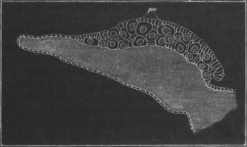
Fig. 18. Transverse section through the ovary of
a young embryo of Scyllium canicula, to shew the primitive germinal
cells (po) lying in the germinal epithelium on the
outer side of the ovarian ridge.
The thickened germinal epithelium gives rise (in the case of the
female) to the ova and the follicular epithelium. Whether the genital
ridge is provided with a core of stroma or no, the germinal epithelium
is always in contact on one side with the stroma, from which it is at
first separated by a well-marked boundary line; but after a certain
time there appear numerous vascular ingrowths from the stroma, which
penetrate through all parts of the germinal epithelium, and break it
up into a sponge-like structure formed of trabeculæ of
germinal epithelium interpenetrated by vascular strands of stroma. The
trabeculæ of the germinal epithelium form the egg-tubes of Pflüger.
With reference to the distribution of the stroma in the germinal
epithelium, it may be said in a general way that there is a special
layer close to the surface of the ovary, which, after the formation of
fresh ova has nearly ceased, completely isolates a superficial layer
of the germinal epithelium from the deeper and major part of it. The
superficial layer is frequently (but erroneously) regarded as
constituting the whole of the germinal epithelium. The layer of stroma
below the superficial epithelium forms in the mammalian ovary the
tunica albuginea. As the follicles are formed. in the trabeculæ of
germinal epithelium the stroma grows in around them, and forms for
each one of them a special tunic.
The adult ovaries differ in a corresponding manner to the embryonic
genital ridges as to the presence of a core of stroma. The ovaries
which are without such a core in the embryo, are also without it in
the adult, and are formed of a double layer of tissue entirely derived
from the germinal epithelium with its ingrowths of stroma, and
composed, for the most part, of ova in all stages of development. In
the case of the other ovaries there is a hilus of stroma—the
zona vasculosa—internal to the egg-bearing region.
In Mammalia, proportionately to the ovary, the zona vasculosa is at a
maximum, and in Birds and Reptiles it is relatively far less
developed. In these forms the germinal epithelium covers the whole
surface of the ovary. In Elasmobranchii the structure of the ovary is
somewhat different, owing to the presence in the ovarian ridge of a
large quantity of a peculiar lymphatic tissue, which has no homologue
in the other ovaries; and still more to the fact that the true
germinal epithelium is in most forms entirely confined to the outer
surface of the ovary, on which it forms a layer of thickened
epithelium in the embryo (fig. 17), and of ovigerous tissue in the
adult.
In the ovary of Mammalia and Reptilia and possibly other forms there
are present in the zona vasculosa during embryonic life cords of
epithelial tissue derived from the Malpighian bodies; these cords have
no function in the female, but in the male assist in forming the
seminiferous tubules.
In considering the development of the ova it is again convenient to
distinguish between Amphioxus and the Craniata.
In Amphioxus the germinal cells destined to become ova are first
distinguished by the larger size of their germinal vesicles and by the
presence of certain refracting granules in their protoplasm. They
subsequently rapidly enlarge and form protuberances on the surface of
the ovary, which are enveloped for three-quarters of their
circumference by the flattened epithelioid cells of the peritoneal
membrane, which thus form a kind of follicle. As the ova become ripe
yolk granules are deposited in their protoplasm, first in the
superficial layer and subsequently throughout. The germinal vesicle
also passes from the centre to the surface. A vitelline membrane is
formed when the ova are mature.
In the Craniata the ova are developed from the cells of the germinal
epithelium. In the types with larger ova (Teleostei, Elasmobranchii,
Amphibia, Reptilia, Aves), at a very early period, sometimes
(Elasmobranchii) even before the formation of the genital ridge,
certain of the cells which are destined to form ova become
distinguished by their greater size, and by the possession of an
abundant clear protoplasm and a large spherical granular nucleus.
(Fig. 18, po.) Such special cells form primitive germinal
cells, and are common to both sexes.
For a considerable period after their first formation these cells
remain stationary in their development; but their number increases,
partly, it appears, by an addition of fresh ones, and partly
by division. Owing to the latter process the germinal cells come to
form small masses or nests. The following description of the further
changes of these cells in the female refers in the first instance to
Elasmobranchii, but holds good in most respects for other types as
well.
It is convenient to distinguish two modes in which the primitive
germinal cells may become converted into permanent ova, though the
morphological difference between the two modes is of no great
importance.
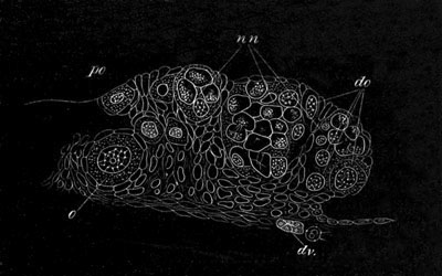
Fig. 19. Section through part of the germinal
epithelium of the ovary of Scyllium at the time when the primitive
germinal cells are becoming converted into ova.
nn. Nests formed of agglomerated germinal cells. The nuclei
of these cells are imbedded in undivided protoplasm. do.
developing ova. o. ovum with follicle. po. primitive
germinal cell. dv. blood-vessels.
In the first mode the protoplasm of all the cells forming a nest
unites into a single mass containing the nuclei of the previously
independent ova (fig. 19, nn). The nuclei in the nest increase
in number, probably by division, and at the same time the nest itself
increases in size. The nuclei while increasing in number also undergo
important changes. A segregation of their contents takes place, and
the granular part (nuclear substance) forms a mass close to one side
of the membrane of the nucleus, while the remainder of the nucleus is
filled with a clear fluid. The whole nucleus at the same time
increases somewhat in size. The granular mass gradually assumes a
stellate form, and finally becomes a beautiful reticulum, of
the character so well known in nuclei (fig. 19, do). Two or
three special nucleoli are present, and form the nodal points of the
reticulum, while its meshes are filled up with the clear fluid
constituents of the nucleus. Not all the nuclei undergo the above
changes; but some of them stop short in their development, undergo
atrophy, and appear finally to be absorbed as pabulum by the
protoplasm of the nest. Such nuclei in a state of degeneration are
shewn in fig. 19. Thus only a few nuclei out of a nest undergo a
complete development. At first the protoplasm of the nest is clear and
transparent, but as the nuclei undergo their changes the protoplasm
becomes more granular, and a specially large quantity of granular
protoplasm is generally present around the most developed nuclei, and
these with their protoplasm gradually become constricted off from the
nest, and constitute the permanent ova (fig. 19, do). The
relative number of ova which may develop from a single nest is subject
to great variation. The object of the whole occurrence of the fusion
of primitive ova and the subsequent atrophy of some of them is to
ensure the adequate nutrition of a certain number of them.
In the second and rarer mode of development of permanent ova from
primitive germinal cells, the nuclei and protoplasm undergo the same
changes as in the first mode, but the cells either remain isolated,
and never form part of a nest, or form part of a nest in which no
fusion of protoplasm takes place, and in which all the cells develop
into permanent ova.
The isolated ova and nests are situated, during the whole of the above
changes, amongst the general undifferentiated cells of the germinal
epithelium, but as soon as a permanent ovum becomes formed the cells
adjoining it arrange themselves around it as a special layer, and so
give rise to the epithelium of the follicle (fig. 19, o). The
growths of stroma into the germinal epithelium appear shortly after
the formation of the earlier follicles.
Mammalia. The development of the ovary in Mammalia differs
mainly from that just described in that the formation of primitive
germinal cells from the indifferent cells of the germinal epithelium
takes place at a relatively much later period.
The stroma grows into the germinal epithelium while it is still formed
of rounded indifferent cells, and divides it into trabeculæ as
described above. At a later period a number of the cells in the deeper
layer of the epithelium, as well as certain cells in the superficial
part, become primitive germinal cells, while the remainder of the
cells become smaller and are destined to form the follicle cells.
The most conspicuous primitive germinal cells are situated in the
superficial layer of epithelium; and the primitive germinal cells in
the deeper layers of the germinal epithelium are not nearly so marked
as in most Craniata, so that it is difficult in some cases to be sure
of their destination till their nucleus commences to undergo its
characteristic metamorphosis.
The change of the primitive ova into permanent ova takes place in the
same manner in Mammals as in Elasmobranchii, except that the fusion of
the primitive ova into polynuclear masses is much rarer. The formation
of the at first quite simple follicles takes place while the ova are
still aggregated in large masses; and the first follicles are formed
in the innermost part of the germinal epithelium. Soon after their
formation the follicles become isolated by connective-tissue growths.
Post-embryonic development of the ova.
The ova of the Vertebrata differ greatly in size and structure. The
differences in size depend upon the quantity of the food-yolk. In the
Amphioxus and Mammalia, in which the ova are smallest, the
comparatively insignificant amount of food-yolk is distributed
uniformly through the ovum. A larger quantity of it is present in the
ova of Amphibia, Marsipobranchii and Teleostei, and it attains an
immense development in the ova of Elasmobranchii, Reptilia, and Aves.
The food-yolk originates from a differentiation of the protoplasm of
the egg. It arises as a number of small highly refracting particles in
a stratum slightly below the surface.
In the Mammalian ovum these particles spread through the protoplasm of
the egg, but do not attain any considerable development. In other
forms the case is different. In Elasmobranch Fishes the refracting
particles appear to develop into vesicles, in the interior of which
there arise solid oval or even rectangular highly refracting bodies,
in the substance of which a stratification may usually be observed,
which gives them an appearance not unlike that of striated
muscle. In Teleostei the yolk assumes very different characters in
different cases. It is often formed of larger or smaller vesicles
containing in their interior other bodies. Stratified plates like
those of Elasmobranchii are also not uncommon. In the ripe ovum of
Teleostei the food-yolk usually resolves itself into a large vitelline
sphere, which occupies the greater part of the ovum, and is formed of
a highly refracting fluid material which coagulates on the addition of
water. It contains in many instances one or more highly refracting
bodies known as oil globules, and is invested by a granular
protoplasmic layer continuous with the germinal disc, in which a
number of normal yolk-spherules are frequently present. In the ovum of
the Herring[24] no distinct investing protoplasmic layer or germinal
disc is present till after impregnation, but the ovum is formed of a
superficial layer with minute yolk-spherules, and of a central portion
with larger yolk-spheres.
In Amphibia the yolk very often appears in the form of oval or
quadrilateral plates. In Reptilia the yolk-spherules are vesicles,
somewhat similar to the white yolk-spheres of Aves, but as a rule
without the highly refracting spheres in their interior. The peculiar
and complicated arrangement and structure of the white and yellow yolk
in Birds is fully described in the “Elements Of Embryology,” and it
need only be said that the yolk develops in Birds in the same manner
as in other types, and that at first all the yolk-spherules appear in
the form of white yolk. The yellow yolk-spheres are a peculiar
modification of white yolk-spheres, formed comparatively late in the
development of the egg (fig. 20).
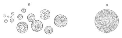
Fig. 20. Yolk elements from the egg of the
fowl.
A. Yellow yolk. B. White yolk.
In the eggs of many Amphibia a dark granular mass known as the yolk
nucleus makes its appearance; and is supposed, without any very clear
evidence, to be related to the formation of the yolk.
A body in the form of a shell enclosing a dark nucleus, which is
perhaps of the same nature, has been described by Eimer in the
Reptilian egg: it eventually resolves itself into a number Of angular
fragments. In Elasmobranchii a similar body is perhaps present.
The food-yolk just described is imbedded in the active protoplasmic
portion of the body of the ovum. In the case of the mammalian
ovum the food-yolk is fairly uniformly distributed, but in the case of
all other craniate ova the protoplasm of the ovum is especially
concentrated at one pole, which is known as the upper or animal pole,
and the food-yolk is more especially concentrated at the opposite
pole. The Herring’s ovum forms an apparent exception to this
statement, in that the concentration of the protoplasm to form the
germinal disc does not take place till after impregnation. In Amphibia
the animal pole is mainly marked by the smaller size of the
yolk-spherules, but in most other forms a small portion of the ovum in
the region of the germinal vesicle is nearly free from yolk-spherules,
and then forms a more or less specialized part known as the germinal
disc. In Aves, Reptilia, and Elasmobranchii the germinal disc shades
off insensibly into the yolk; but in Teleostei it is more sharply
marked off, and is continued more or less completely round the
periphery of the ovum. In ova with true germinal discs it is the
germinal disc alone which undergoes segmentation. The protoplasm of
vertebrate ova frequently exhibits a reticulate or sponge-like
structure (fig. 21) and the reticulum in many cases, e.g.
Elasmobranchii and Reptilia, serves to hold the yolk-spheres together.
In the Tench it has been observed by Bambeke to penetrate into the
vitelline sphere.
In the ova of the Craniata the germinal vesicle is generally
polynucleolar. In Amphioxus and Petromyzon there is however but a
single nucleolus, and in Mammalia there is usually one special
nucleolus and two or three accessory ones. The opposite extreme is
reached in many osseous fish where the nucleoli are extremely
numerous. The protoplasmic reticulum of the embryonic germinal vesicle
may in some instances be retained till the ovum is nearly ripe, but
usually assumes a very granular form. It is at first connected with
the nucleoli which form nodal points in it, but this relation cannot
always be detected in the later stages. A membrane, which in the case
of the larger ova becomes very thick, is always present round the
germinal vesicle. It is said to be perforated in some Reptilian ova
(Eimer). As to the position of the germinal vesicle, it is at first
situated in the centre of the ovum, but always eventually travels to
the animal pole, and as the egg becomes ripe undergoes changes which
will be more especially detailed in the next chapter. In the
ova with a large amount of food-yolk it assumes an eccentric position
very early.
The homologies of the primary egg membranes of Craniata are still
involved in some obscurity. There seem to be three membranes, which
may all coexist, and of which one or more are almost always present.
These membranes are—
(1) An outermost usually homogeneous non-perforated membrane, which is
by most authors regarded as a chorion, but is probably a vitelline
membrane—by which name I shall speak of it.
(2) A radiately striated membrane (internal to the former when the two
coexist) which can be broken up into a series of separate columns.
These give to the membrane its radiate striation, but it is probable
that between the columns there are pores sufficiently large to admit
of the passage of protoplasmic filaments. This membrane will be spoken
of as the zona radiata. It is a differentiation of the outermost layer
of the yolk.
(3) Within the zona radiata a third and delicate membrane is
occasionally found, especially when the ovum is approaching maturity.
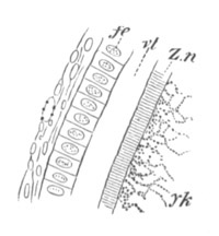
Fig. 21. Section through a small part of the
surface of an ovum of an immature female of Scyllium canicula.
fe. follicular epithelium. vt. vitelline membrane.
Zn. zona radiata. yk. yolk with protoplasmic network.
In Elasmobranchii the first membrane to be formed is the vitelline
membrane, which appears in some instances before the formation of the
follicle—a fact which appears to shew that it is really formed as a
differentiation of the protoplasm of the egg. In most Elasmobranchii
this membrane attains a very considerable development. A zona radiata
is generally (if not always) present in Elasmobranchii, but arises at
a later period than the vitelline membrane (fig. 21, Zn). The zona
radiata always disappears long before the ovum is ripe. The vitelline
membrane also gradually atrophies, though it lasts much longer than
the zona radiata. When the egg is taken up by the oviduct all trace of
both membranes has vanished. In Reptilia precisely the same
arrangements of the membranes are found as in Elasmobranchii, except
that as a rule the zona radiata is relatively more important.
The vitelline membrane is thin except in the Crocodilia. The third
innermost membrane is found according to Eimer in many Reptilia. In
birds both vitelline membrane and zona radiata are present, but the
latter atrophies early, leaving the former as the sole membrane when
the egg is ripe.
In osseous fish the vitelline membrane is usually either absent or may
perhaps in some instances, e.g. the Perch, be imperfectly
represented. In the ripe ovum of the Herring there is a distinctly
developed membrane external to the zona radiata which is probably the
vitelline membrane. The zona radiata attains a very great development,
and is generally provided with knobs of various shapes on its outer
surface. A delicate membrane internal to this—my third membrane—has
often been described, but there is still some doubt about its
existence. In some cases an external less granular layer of the ovum
itself has been described as a special membrane. In the Perch a
peculiar mucous capsule, penetrated by irregular branched
prolongations of the follicle cells, is present in addition to the
ordinary membranes. In Petromyzon a zona radiata appears to be
present, which in the adult is divided into two layers, both of them
radiately striated according to Calberla, but according to Kupffer and
Benecke the outer one is not perforated, and would appear therefore to
be a vitelline membrane as defined above. A delicate membrane is
formed at a comparatively late period around the ova of the Amphibia,
and is stated (Waldeyer, No. 6, and Kolessnikow) to have a delicate
radial striation. It probably corresponds with the zona radiata.
In Mammalia a radiately striated membrane—the zona radiata—is
generally described as being present, and internal to it, in the
nearly ripe egg, a delicate membrane has been shewn by E. van Beneden
to exist. Externally to the zona radiata there may be observed a
granular membrane irregular on its outer surface on which the cells of
the discus are supported. This membrane is more or less distinctly
separated from the zona radiata; and by tracing back its development
it appears very probable that it is the remnant of the first-formed
membrane in the very young ovum, and therefore the vitelline membrane.
A micropyle (first discovered by Ransom, No. 74) is present in a large
number of osseous fish and in Petromyzon (Calberla). Doubts
have been thrown on its existence in the latter form by Kupffer and
Benecke; and at any rate it would only seem to perforate the zona
radiata. In the osseous fish in which it has been detected, Salmonidæ,
Percidæ (Gasterosteus), Clupeidæe, etc., it forms a minute perforation
of the zona radiata at the animal pole, just large enough to admit a
single spermatozoon. Its characters differ slightly in different
cases, but there is usually a shallow depression, in the centre of
which it is situated.
The eggs of all Craniata (except Petromyzon (?)) appear to be enclosed
in a cellular envelope known as the follicle. The cells which form
this are, as has been already explained, derived from the germinal
epithelium[25], and frequently arrange themselves around the ovum
before the appearance of the growths of stroma into the epithelium.
All young follicles are nearly alike, but as they grow older they
exhibit various modifications in the different groups. They retain
their simplest condition as a flat epithelial layer in most osseous
fish and Amphibia. In most other forms the cells become at some period
columnar, and are generally arranged in two or more layers. There is
formed externally to the epithelium a delicate membrane—the membrana
propria folliculi—which is in its turn enclosed in a vascular
connective-tissue sheath.
In Elasmobranchii and many Reptilia (Lacertilia,
Ophidia) some of the cells become much larger than the others,
and assume a funnel-shaped form with the narrow end in contact with
the egg membrane. These large cells, which have a regular arrangement
in the epithelium, are probably in some way connected with the
nutrition. They have only been noticed in large-yolked ova. Many
observers have described prolongations of the follicle cells through
the pores of the zona radiata in Aves, Reptilia and Teleostei.
The most remarkable modification of the follicle is that which is
found in Mammalia. At first the follicle is similar to that of other
Vertebrata, and is formed of flat cells derived from the germinal
cells adjoining the ovum. These cells next become columnar and then
one or two layers deep. Later they become thicker on one side
than on the other, and there appears in the thickened mass a cavity,
which gradually becomes more distended and is filled with an
albuminous fluid. As the cavity enlarges, the ovum with several layers
of cells around it forms a prominence projecting into it. The whole
structure with its tunic is known as the Graafian follicle. The
follicle cells are known as the membrana granulosa, and the
projection, in which the ovum lies, as the discus or cumulus
proligerus. The cells of the discus in immediate contiguity to the
ovum usually form a more or less specialized layer and are somewhat
more columnar than the adjoining cells.
The Spermatozoon.
Although there is no doubt that the spermatozoon in most instances
plays as important a part as the ovum in influencing the characters of
the organism which is evolved from the coalesced product of the ovum
and spermatozoon, yet the actual form of the spermatozoon has not,
like the form of the ovum, a secondary influence on the early phases
of development. A comparative history of the spermatozoon is therefore
of less importance for my purpose than that of the ovum; and I shall
confine myself to a few remarks on its general structure, and mode of
growth. The primary origin of the male germinal cells, and their
relation to the sperm-forming cells, is dealt with in the second part
of the treatise.
Although the minute size of most spermatozoa places great difficulties
in the way of a satisfactory investigation of them, yet there can be
but little doubt that they always have the value of cells. In the vast
majority of instances the spermatic cell or spermatozoon is composed
of (1) a spherical or oval portion known as the head, formed of a
nucleus enveloped in an extremely delicate layer of protoplasm, and
(2) of a motile protoplasmic flagellum known as the tail; which
together with the investing layer of the head forms the body of the
cell.
As might be anticipated, the proportion, size, and relations of the
parts of the spermatozoon are subject to great variations. The head is
often extremely elongated; and it is in many cases rather on
theoretical grounds, than as a result of actual observation,
that a protoplasmic layer is stated to be continued round the nucleus
which forms the main constituent of the head. In some of the elongated
forms of spermatozoa, e.g. in Insecta, there is no marked
distinction, except in the character of the protoplasm, between the
head and the tail. A connecting element is frequently interposed
between the head and tail, which appears however to be constituted of
the same material as the tail, and sometimes forms a thickening on the
tail close below the head (Amphioxus). A very remarkable modification
of the tail is found in many Amphibia, Reptilia and Mammalia. In these
types there is attached to what appears to be a normal tail a delicate
membrane, the outer edge of which is thickened to form a kind of
secondary filament. In the living spermatozoon this filament is in a
state of constant movement. The membrane winds spirally round the
tail.
In the majority of forms the tail of the living spermatozoon exhibits
sinuous cilia-like movements. In two groups the movements are however
of an amœboid character. These groups are the Nematoda and the
Crustacea; and the spermatozoa in both of them frequently present very
abnormal forms. In Nematoda they are pear-shaped, cylindrical,
spine-shaped, etc., and are mainly formed of protoplasm with a highly
refracting nucleus. In the Crustacea the variations of form are still
greater. In the Malacostraca they are sometimes simply spherical
(Squilla), while in Astacus and a large number of Decapoda they are
composed of a nucleated body with stellate rays. In Paludina amongst
the Mollusca there are two forms of completely developed
spermatozoa existing side by side in the same individual.
The spermatozoa are formed by the breaking up of the male germinal
cells, or of cells secondarily derived from them by division. The
cells which directly give rise by division to the spermatozoa may be
called spermospores and are equivalent to the ova or oospores.
Amongst the Sponges (Halisarca, Schultze, No. 141) a germinal cell,
similar to that which in the female becomes an ovum, repeatedly
divides and eventually gives rise to a ball of cells (a
spermosphere or sperm-morula), each constituent cell of
which becomes converted into a spermatozoon, and may be designated by
the special term ‘spermoblast’.
In most Hydrozoa the subepithelial epiblastic cells become converted
into germinal cells (spermospores), and then break up to form
spermoblasts, each of which becomes a spermatozoon.
In most higher Metazoa the spermospores usually form the epithelium of
an ampulla or tube, though more rarely (many Chætopoda, Gephyrea,
etc.) they may be derived from cells lining the body cavity, as in the
case of ova. The spermatozoa are formed either by the direct division
of the spermospores into a number of cells, spermoblasts, each of
which grows into a spermatozoon; or by the nucleus of the spermospore
becoming subdivided within the cell body, the latter differentiating
itself into the tails of the spermatozoa while the segments of the
nucleus give rise to the main part of the heads.
In many instances interstitial cells which do not give rise to
spermatozoa, are intermingled with the spermospores.
In a good many cases, as first pointed out by Blomfield[26], the whole
of each spermospore does not become converted into spermatozoa, but
part, either with or without a segment of the original nucleus,
remains passive, and carrying as it does the off-budded spermoblasts
may be called the ‘sperm-blastophor.’ This passive portion of
protoplasm is not employed in the regeneration of the spermoblast.
This very singular phenomenon has been observed in Elasmobranchii, the
Frog, the Earthworm, Helix, etc.[27], and probably has a much wider
extension. In Elasmobranchii (Semper) the passive portions of
protoplasm are nucleated, and are placed on the outer side of the
columnar spermospores which line the testicular ampullæ; they are not
distinctly differentiated till the nuclei, segmented from the nucleus
of the primitive spermospore to form the heads of the spermatozoa,
have become fairly numerous. In the Frog the passive blastophor also
occurs as a nucleated mass of protoplasm on the outer side of the
spermospore. In the Earthworm the blastophor forms a central
non-nucleated portion of the spermospore; and the whole periphery of
each spermospore becomes converted into spermoblasts.
It has been already stated in the introduction that the male and
female generative products are homodynamous, but the consideration of
the development of the products in the two sexes shews that a single
spermatozoon is not equivalent to an ovum, but rather that the
whole of the spermatozoa derived from a spermospore are together
equivalent to one ovum.
Maturation of the ovum and formation of the polar bodies.
In the preceding chapter the changes in the ovum were described nearly
up to the period when it became ripe, and ready to be impregnated.
Preparatory to the act of impregnation there take place however a
series of remarkable changes, which more especially concern the
germinal vesicle.
The attention of a large number of investigators has recently been
directed to these changes as well as to the phenomena of impregnation.
The results of their investigations will be described in the present
chapter; but for an historical account of these investigations, as
well as for a determination of the delicate questions of priority, the
reader is referred to Fol’s memoir (No. 87), and to a paper by the
author (No. 81).
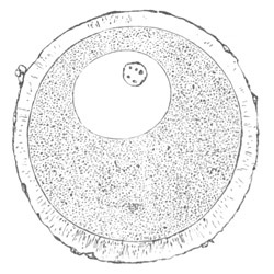
Fig. 22. Ripe ovum of Asterias glacialis
enveloped in a mucilaginous envelope, and containing an eccentric
germinal vesicle and germinal spot (copied from Fol).
The nature of the changes which take place in the maturation of the
ovum may perhaps be most conveniently displayed by following the
history of a single ovum. For this purpose the eggs of Asterias
glacialis, which have recently formed the subject of a series of
beautiful researches by Fol (87), may be selected.
The ripe ovum (fig. 22), when detached from the ovary is formed of a
granular vitellus enveloped in a mucilaginous coat, the zona
radiata. It contains an eccentrically-situated germinal vesicle and a
germinal spot. In the former is present the usual protoplasmic
reticulum. As soon as the ovum reaches the sea-water the germinal
vesicle commences to undergo a peculiar metamorphosis. It exhibits
frequent changes of form, the reticulum vanishes, its membrane becomes
gradually absorbed, its outline indented and indistinct, and finally
its contents become to a certain extent confounded with the vitellus
(fig. 23).
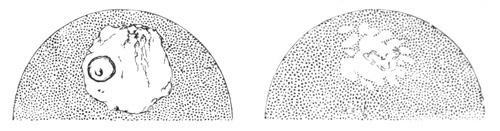
Fig. 23. Two successive stages in the gradual
metamorphosis of the germinal vesicle and spot of the ovum of
Asterias glacialis immediately after it is laid (copied from
Fol).
The germinal spot at the same time loses its clearness of outline and
gradually disappears from view.
At this stage, and between it and the stage represented in fig. 26,
the action of reagents brings to light certain appearances the nature
of which is not yet fully cleared up for Asterias, which have been
described somewhat differently by Fol for Ast. glacialis and Hertwig
for Asteracanthion.
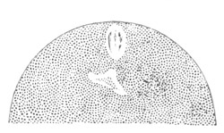
Fig. 24. Ovum of Asterias glacialis shewing the
clear spaces in the place of the germinal vesicle. Fresh
preparation (copied from Fol).
Fol finds immediately after the stage just described that a star is
visible between the remains of the germinal vesicle and the surface of
the egg, which is connected with an imperfectly-formed nuclear spindle
extending towards the germinal vesicle[28]. At the end of the nuclear
spindle may be seen the broken up fragments of the germinal spot.
At a slightly later stage, in the place of the original germinal
vesicle there may be observed in the fresh ovum two clear
spaces (fig. 24), one ovoid and nearer the surface, and the second
more irregular in form and situated rather deeper in the vitellus. In
the upper space parallel striæ may be observed. By treatment with
reagents the first clear space is found to be formed of a
horizontally-placed spindle with two terminal stars, near which
irregular remains of the germinal spot may be seen. Slightly later
(fig. 25) there may be seen on the lower side of the spindle a
somewhat irregular body, which may possibly be part of the remains of
the germinal spot, though Fol holds that it is probably part of the
membrane of the germinal vesicle. The lower clear space visible in the
fresh ovum now contains a round body, fig. 25. Fol concludes that the
spindle is formed out of part of the germinal vesicle and not from the
germinal spot, while he sees in the round body present in the lower of
the two clear spaces the metamorphosed germinal spot. He will not,
however, assert that no fragment of the germinal spot enters into the
formation of the spindle.
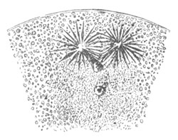
Fig. 25. Ovum of Asterias glacialis, at the same
stage as fig. 24, treated with picric acid (copied from Fol).
The following is Hertwig’s (No. 92) account of the changes in the
germinal vesicle in Asteracanthion. Shortly after the egg is laid the
protoplasm on the side of the germinal vesicle towards the surface of
the egg develops a prominence which presses inwards the wall of the
vesicle. At the same time the germinal spot develops a large vacuole,
in the interior of which is a body consisting of nuclear substance,
and formed of a firmer and more refractive material than the remainder
of the germinal spot. In the prominence first mentioned as projecting
inwards towards the germinal vesicle first one star, formed by radial
striæ of protoplasm, and then a second make their appearance; while
the germinal spot appears to have vanished, the outline of the
germinal vesicle to have become indistinct, and its contents to have
mingled with the surrounding protoplasm. Treatment with reagents
demonstrates that in the process of disappearance of the germinal spot
the nuclear mass in its vacuole forms a rod-like body, the free end of
which is situated between the two stars which occupy the prominence
indenting the germinal vesicle. At a later period granules may be seen
at the end of the rod and finally the rod itself vanishes. After these
changes by the aid of reagents there may be demonstrated a spindle
between the two stars, which Hertwig believes to grow in size as the
last remnants of the germinal spot gradually vanish, and he maintains
that the spindle is formed at the expense of the germinal spot. The
stage with this spindle corresponds with fig. 25.
Several of Hertwig’s figures closely correspond with those of Fol, and
considering how conflicting is the evidence before us, it seems
necessary to leave open for Asterias the question as to what
parts of the germinal vesicle are concerned in forming the first
spindle.
A clearer view of the phenomena which take place at this stage has
been obtained by Fol in the case of Heteropods (Pterotrachæa). In the
ovum a few minutes after it has been laid the germinal vesicle becomes
very pale, and two stars make their appearance round a clear substance
near its poles. The nucleus itself is somewhat elongated, and
commences to exhibit at its poles longitudinal striæ, which gradually
extend towards the centre at the expense of the nuclear reticulum,
from a metamorphosis of which they are directly derived. When the
striæ of the two sides have nearly met, thickenings may be observed in
the recticulum between them, which give rise, where the striæ of the
two sides unite, to the central thickenings of the fibres (nuclear
plate). In this way a complete nuclear spindle is established[29].
The important result of Fol’s observations on Heteropods, which
tallies also with what is found in Asterias, is that a spindle with
two stars at its poles is formed from the metamorphosis of the
germinal vesicle and surrounding protoplasm (fig. 25).
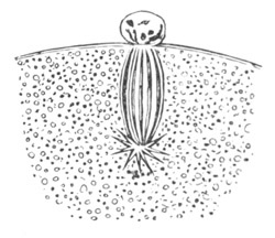
Fig. 26. Portion of the ovum of Asterias
glacialis at the moment of the detachment of the first polar body
and the withdrawal of the remaining part of the spindle within the
ovum. picric acid preparation (copied from Fol).
Polar cells. The spindle has up to this time been situated with
its axis parallel to the surface of the egg, but in somewhat older
specimens a vertical spindle is found, with one end projecting into a
protoplasmic prominence which makes its appearance on the surface of
the egg (fig. 26). Hertwig believes that the spindle simply travels
towards the surface, and while doing so changes the direction of its
axis. Fol asserts, however, that this is not the case, but that
between the two phases of the spindle an intermediate one is found in
which a spindle can no longer be seen in the egg, but its place is
taken by a body with a dentated outline. He has not been able to
arrive at a conclusion as to what meaning is to be attached to
this occurrence, which does not appear to take place in Heteropods.
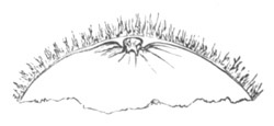
Fig. 27. Portion of the ovum of Asterias
glacialis, with the first polar cell as it appears when living
(copied from Fol).
In any case the spindle which projects into the prominence on the
surface of the egg divides into two parts, one in the prominence and
one in the egg (fig. 26). The prominence itself with the enclosed
portion of the spindle becomes constricted off from the egg to form a
body, well known to embryologists as the polar body or cell (fig. 27).
Since more than one polar cell is formed, that which is the earliest
to appear may be called the first polar cell.
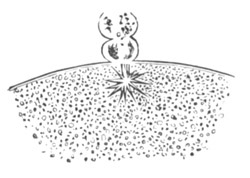
Fig. 28. Portion of the ovum of Asterias
glacialis immediately after the formation of the second polar cell.
Picric acid preparation (copied from Fol).
The part of the spindle which remains in the egg becomes directly
converted into a second spindle by the elongation of its fibres,
without passing through a typical nuclear condition. A second polar
cell next becomes formed in the same manner as the first (fig. 28),
and the portion of the spindle remaining in the egg becomes converted
into two or three clear vesicles (fig. 29), which soon unite to form a
single nucleus (fig. 30). The new nucleus which is clearly derived
from part of the original germinal vesicle is called the female
pronucleus, for reasons which will appear in the sequel.
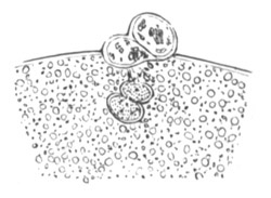
Fig. 29. Portion of the ovum of Asterias
glacialis after the formation of the second polar cell, shewing the
part of the spindle remaining in the ovum becoming converted into
two clear vesicles. Picric acid preparation (copied from Fol).
The two polar cells appear to be situated between two membranes, the
outer of which is very delicate, and only distinct where it covers the
polar cells, while the inner one is thicker and becomes, after
impregnation, more distinct, and then forms what Fol speaks of as the
vitelline membrane. It is clear, as Hertwig has pointed out, that the
polar bodies originate by a regular process of cell division
and have the value of cells.
A peculiar phenomenon makes its appearance in the eggs of Clepsine
shortly after the formation of the polar cells, which has been spoken
of by Whitman (No. 100) as the formation of the polar rings. The
following is his description of the occurrence.
“Fifteen minutes after the elimination of the polar globules
(i.e. cells) a ring-like depression or constriction appears in
the yolk around the oral pole, and in this depression a transparent
liquid substance (nuclear?) is collected forming the first polar
ring.... The same phenomena repeat themselves later at the aboral
pole.... The rings concentrate to form two discs.... Before the first
cleavage both discs plunge deep into the egg.”
The nature of these rings is at present quite obscure.
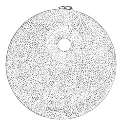
Fig. 30. Ovum of Asterias glacialis with the two
polar cells and the female pronucleus surrounded by radial striæ, as
seen in the living egg (copied from Fol).
Considering how few ova have been adequately investigated with
reference to the behaviour of the germinal vesicle, any general
conclusions which may at present be formed are to be regarded as
provisional.
There is however abundant evidence that at the time of maturation of
the egg the germinal vesicle undergoes peculiar changes, which are, in
part at least, of a retrogressive character. These changes may begin
considerably before the egg has reached the period of maturity, or may
not take place till after it has been laid. They consist in an
appearance of irregularity and obscurity in the outline of the
germinal vesicle, the absorption of its membrane, the partial
absorption of its contents in the yolk, the disappearance of the
reticulum, and the breaking up and disappearance of the germinal spot.
The exact fate of the single germinal spot, or the numerous spots
where they are present, is still obscure.
The retrogressive metamorphosis of the germinal vesicle is followed in
a large number of instances by the conversion of what remains into a
striated spindle similar in character to a nucleus previous to
division. This spindle travels to the surface of the ovum and
undergoes division to form the polar cell or cells in the
manner above described. The part which remains in the egg forms
eventually the female pronucleus.
The germinal vesicle has up to the present time only been observed to
undergo the above series of changes in a certain number of instances,
which, however, include examples from several divisions of the
Cœlenterata, the Echinodermata, and the Mollusca, some of the
Vermes [Turbellarians (Leptoplana), Nematodes, Hirudinea,
Alciope, Sagitta], Ascidians, etc. It is very possible, not to say
probable, that such changes are universal in the animal kingdom, but
the present state of our knowledge does not justify us in saying so.
In the Craniata especially our knowledge of the formation of the polar
bodies is very unsatisfactory. In Petromyzon Kupffer and Benecke have
brought forward evidence to shew that one polar body is formed prior
to the impregnation, and a second in connection with a peculiar
prominence of protoplasm after impregnation. Part of the germinal
vesicle remains in the egg as the female pronucleus. In the Sturgeon
the germinal vesicle atrophies and breaks up before impregnation, and
afterwards part is found as a granular mass on the surface of the egg,
while part forms a female pronucleus.
In Amphibia the observations of Hertwig (90) and Bambeke (77) tend to
shew that after the germinal vesicle has assumed a superficial
situation at the pigmented pole of the ovum its contents become
intermingled with the yolk, and are in part extruded from the ovum as
a granular mass after impregnation. Part of them remains in the ovum
and forms a female pronucleus. Whether there is a proper division of
the germinal vesicle as in typical cases is not known.
Oellacher (95) by a series of careful observations upon the egg of the
trout, and subsequently of the bird, demonstrated that in the ovum
while still in the ovary, the germinal vesicle underwent a kind of
degeneration and eventually became ejected, in part at any rate. My
own observations on Elasmobranchs, which require enlargement and
confirmation, tend to shew that this part may be the membrane. Ed. van
Beneden (78) has contributed some important observations on the
rabbit. His account is as follows. As the ovum approaches maturity the
germinal vesicle assumes an eccentric position, and fuses with the
peripheral layer of the egg to constitute the cicatricular
lens. The germinal spot next travels to the surface of the
cicatricular lens and forms the nuclear disc: at the same time
the membrane of the germinal vesicle vanishes, though it probably
unites with the nuclear disc. The plasma of the nucleus then collects
into a definite mass and forms the nucleoplasmic body. Finally the
nuclear disc assumes an ellipsoidal form and becomes the nuclear body.
Nothing is now left of the original germinal vesicle but the nuclear
body and the nucleoplasmic body, both still situated within the ovum.
In the next stage no trace of the germinal vesicle can be
detected in the ovum, but outside it, close to the point where the
modified remnants of the vesicle were previously situated, there is
present a polar body which is composed of two parts, one of which
stains deeply and resembles the nuclear body, and the other does not
stain but is similar to the nucleoplasmic body. Van Beneden concludes
that the parts of the polar body are the two ejected products of the
germinal vesicle. We may be perhaps permitted to hold that further
observations on this difficult object will demonstrate that part of
the germinal vesicle remains in the ovum to form the female
pronucleus.
With reference to invertebrate forms attention may be called to the
observations of Bütschli (80). Although in Cucullanus a normal
formation of the polar bodies takes place, yet in the Nematodes
generally, Bütschli has been unable to find the spindle modification
of the germinal vesicle, but states that the germinal vesicle
undergoes degeneration, its outline becoming indistinct and the
germinal spot vanishing. The position of the germinal vesicle
continues to be marked by a clear space, which gradually approaches
the surface of the egg. When it is in contact with the surface a small
spherical body, the remnant of the germinal vesicle, comes into view,
and eventually becomes ejected. The clear space subsequently
disappears.
In addition to the types just quoted, which may very probably turn out
to be normal in the mode of formation of the polar bodies, there is a
large number of types, including the whole of the Rotifera and
Arthropoda with a few doubtful exceptions[30], in which the polar
cells cannot as yet be said to have been satisfactorily observed.
The more important of the doubtful cases amongst the Rotifera and
Arthropoda are the following.
Flemming (83) finds that in the summer and probably parthenogenetic
eggs of Lacinularia socialis the germinal vesicle approaches
the surface and becomes invisible, and that subsequently a slight
indentation in the outline of the egg marks the point of its
disappearance. In the hollow of the indentation Flemming believes a
polar cell to be situated, though he has not definitely seen one.
Hoek[31] believes that he has found a polar body in the ovum of
Balanus balanoides, but his observations are not perfectly
satisfactory.
Bütschli, who has expressly searched for the polar bodies in the ova
of Rotifera, was unable to find any trace of them, though he found
that as the egg became ripe the germinal vesicle became half its
original size. In the parthenogenetic eggs of Aphis he also failed to
find a trace of polar bodies, though the germinal vesicle, after the
germinal spot had broken up into fragments, approached the surface and
disappeared.
Whatever may be the eventual result of more extended investigation, it
is clear that the formation of polar cells according to the type
described above is a very constant occurrence. Its importance is
increased by the discovery by Strasburger of the existence of an
analogous process amongst plants. Two questions about it obviously
present themselves for solution: (1) What are the conditions of its
occurrence with reference to impregnation? (2) What meaning has it in
the development of the ovum or the embryo?
The answer to the first of these questions is not difficult to find.
The formation of the polar bodies is independent of impregnation, and
is the final act of the normal growth of the ovum. In a few types the
polar cells are formed while the ovum is still in the ovary, as, for
instance, in some species of Echini, Hydra, etc., but, according to
our present knowledge, far more usually after the ovum has been laid.
In some instances the budding-off of the polar cells precedes, and in
other instances follows impregnation; but there is no evidence to shew
that in the latter cases the process is influenced by the contact with
the male element. In Asterias, as has been shewn by O. Hertwig and
Fol, the formation of the polar cells may indifferently either precede
or follow impregnation—a fact which affords a clear demonstration of
the independence of the two occurrences.
To the second of the two questions it does not unfortunately seem
possible at present to give an answer which can be regarded as
satisfactory.
The retrogressive changes in the membrane of the germinal vesicle
which usher in the formation of the polar bodies may very probably be
viewed as a prelude to a renewed activity of the contents of the
vesicle; and are perhaps rendered the more necessary from the
thickness of the membrane which results from a protracted period of
passive growth. This suggestion does not, however, help us to explain
the formation of polar bodies by a process identical with cell
division. The ejection of part of the germinal vesicle in the
formation of the polar cells may probably be paralleled by the
ejection of part or the whole of the original nucleus which, if we may
trust the beautiful researches of Bütschli, takes place during
conjugation in Infusoria as a preliminary to the formation of a fresh
nucleus. This comparison is due to Bütschli, and according to it the
formation of the polar bodies would have to be regarded as assisting,
in some as yet unknown way, the process of regeneration of the
germinal vesicle. Views analogous to this are held by Strasburger and
Hertwig, who regard the formation of the polar bodies in the light of
a process of excretion or removal of useless material. Such hypotheses
do not, unfortunately, carry us very far.
I would suggest that in the formation of the polar cells part of the
constituents of the germinal vesicle, which are requisite for its
functions as a complete and independent nucleus, is removed, to make
room for the supply of the necessary parts to it again by the
spermatic nucleus.
My view amounts to the following, viz. that after the formation of the
polar cells the remainder of the germinal vesicle within the ovum (the
female pronucleus) is incapable of further development without the
addition of the nuclear part of the male element (spermatozoon), and
that if polar cells were not formed parthenogenesis might normally
occur. A strong support for this hypothesis would be afforded were it
to be definitely established that a polar body is not formed in the
Arthropoda and Rotifera; since the normal occurrence of
parthenogenesis is confined to these two groups. It is certainly a
remarkable coincidence that they are the only two groups in which
polar bodies have not so far been satisfactorily observed.
It is perhaps possible that the part removed in the formation of the
polar cells is not absolutely essential; and this seems at first sight
to follow from the fact of parthenogenesis being possible in instances
where impregnation is the normal occurrence. The genuineness of the
observations on this head is too long a subject to enter into
here[32], but after admitting, as we probably must, that there
are genuine cases of such parthenogenesis, it cannot be taken for
granted without more extended observation that the occurrence of
development in these rare instances may not be due to the polar cells
not having been formed as usual, and that when the polar cells are
formed the development without impregnation is impossible.
Selenka found in the case of Purpura lapillus that no polar
body was formed in the eggs which did not develop, but in the case of
Neritina, Bütschli has found that this does not hold good.
The remarkable observations of Greeff (No. 88) on the parthenogenetic
development of the eggs of Asterias rubens tell, however, very
strongly against the above hypothesis. Greeff has found that under
normal circumstances the eggs of this species of starfish will develop
without impregnation in simple sea-water. The development is quite
regular and normal, though much slower than in the case of impregnated
eggs. It is not definitely stated that polar cells are formed, but
there can be no doubt that this is implied. Greeff’s account is so
precise and circumstantial that it is not easy to believe that any
error can have crept in; but neither Hertwig nor Fol have been able to
repeat his experiments, and we may be permitted to wait for further
confirmation before absolutely accepting them.
To the suggestion already made with reference to the function of the
polar cells, I will venture to add the further one, that the
function of forming polar cells has been acquired by the ovum for the
express purpose of preventing parthenogenesis.
The explanation given by Mr Darwin of the evil effects of
self-fertilization, viz. the want of sufficient differentiation in the
sexual elements[33], would apply with far greater force to cases of
parthenogenesis.
In the production of fresh individuals, two circumstances are
obviously favourable to the species. (1) That the maximum number
possible of fresh individuals should be produced, (2) That the
individuals should be as vigorous as possible. Sexual differentiation
(even in hermaphrodites) is clearly very inimical to the production of
the maximum number of individuals. There can be little doubt that the
ovum is potentially capable of developing by itself into a
fresh individual, and therefore, unless the absence of sexual
differentiation was very injurious to the vigour of the progeny,
parthenogenesis would most certainly be a very constant occurrence;
and, on the analogy of the arrangements in plants to prevent
self-fertilization, we might expect to find some contrivance both in
animals and in plants to prevent the ovum developing by itself
without fertilization. If my view about the polar cells is correct,
the formation of these bodies functions as such a contrivance.
Reproduction by budding or fission has probably arisen as a means of
increasing the number of individuals produced, so that the coexistence
of asexual with sexual reproduction is to be looked on as a kind of
compromise for the loss of the power of rapid reproduction due to the
absence of parthenogenesis. In the Arthropoda and Rotifera the place
of budding has been taken by parthenogenesis, which may be a frequent,
though not always a necessary occurrence, as in various Branchiopoda
(Apus, Limnadia, etc.) and Lepidoptera (Psyche
helix, etc.); or a regular occurrence for the production of one
sex, as in Bees, Wasps, Nematus, etc.; or an occurrence confined to a
certain stage in the cycle of development in which all the individuals
reproduce their kind parthenogenetically, as in Aphis, Cecidomyia,
Gall Insects (Neuroterus, etc.), Daphnia[34].
On my hypothesis the possibility of parthenogenesis, or at any rate
its frequency, in Arthropoda and Rotifera is possibly due to the
absence of polar cells. In the case of all animals, so far as is known
to me, fertilization of the ovum occasionally occurs[35], but there
are instances in the vegetable kingdom where so-called parthenogenesis
appears to be capable of recurring for an indefinite period. One of
the best instances appears to be that of Cœlebogyne, an introduced
exotic Euphorbiaceous plant which regularly produces fertile seeds
although a male flower never appears. The recent researches of
Strasburger have however shewn that in Cœlebogyne and other
parthenogenetic flowering plants, embryos are formed by the
budding and subsequent development of cells belonging to the
ovule. This being the case, it is impossible to assert of these plants
that they are really parthenogenetic, for the embryos contained in the
seed of a flower which has certainly not been fertilized, may have
been formed, not by the development of the ovum, but by budding
from the surrounding tissue of the ovule.
The above view with reference to the nature of the polar bodies is not
to be regarded as forming more than an hypothesis.
Impregnation of the Ovum.
A far greater amount of certainty has been attained as to the effects
of impregnation than as to the changes of the germinal vesicle which
precede this, and there appears, moreover, to be a greater uniformity
in the series of resulting phenomena.
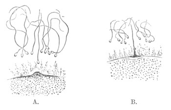
Fig. 31. Small portions of the ovum of
Asterias glacialis. The Spermatozoa are shewn enveloped in the
mucilaginous coat. In A. a prominence is rising from the surface of
the egg towards the nearest spermatozoon; and in B. the spermatozoon
and prominence have met. (Copied from Fol.)
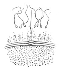
Fig. 32. Portion of the ovum of Asterias
glacialis after the entrance of a spermatozoon into the ovum. It
shews the prominence of the ovum through which the spermatozoon has
entered. A vitelline membrane with a crater-like opening has become
distinctly formed. (Copied from Fol.)
It will be convenient again to take Asterias glacialis as the type.
The part of the germinal vesicle which remains in the egg, after the
formation of the second polar cell, becomes converted into a number of
small vesicles (fig. 29), which aggregate themselves into a single
clear nucleus, which gradually travels toward the centre of the egg
and around which, as a centre, the protoplasm becomes radiately
striated (fig. 30). This nucleus is known as the female pronucleus. By
the action of reagents a nucleolus may be shewn in it. In Asterias
glacialis the most favourable period for fecundation is about an hour
after the formation of the female pronucleus. If at this time the
spermatozoa are allowed to come in contact with the egg, their heads
soon become enveloped in the investing mucilaginous coat. A
prominence, pointing towards the nearest spermatozoon, now arises from
the superficial layer of protoplasm of the egg, and grows till it
comes in contact with the spermatozoon (fig. 31, A and B). Under
normal circumstances the spermatozoon which meets the prominence is
the only one concerned in the fertilization, and it makes its
way into the egg by passing through the prominence. The tail of the
spermatozoon, no longer motile, remains visible for some time after
the head has bored its way in, but its place is soon taken by a pale
conical body, which is, however, probably in part a product of the
metamorphosis of the tail itself (fig. 32). It eventually becomes
absorbed into the body of the ovum.
At the moment of contact between the spermatozoon and the egg the
outermost layer of the protoplasm of the latter raises itself as a
distinct membrane, which separates from the egg and prevents the
entrance of other spermatozoa. At the point where the spermatozoon
entered a crater-like opening is left in the membrane, through which
the metamorphosed tail of the spermatozoon may at first be seen
projecting (fig. 32).
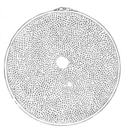
Fig. 33. Ovum of Asterias glacialis, with male
and female pronucleus and a radial striation of the protoplasm
around the former. (Copied from Fol.)
The head of the spermatozoon when in the egg forms a nucleus, for
which the name male pronucleus may be conveniently adopted. It grows
in size, probably by assimilating material from the ovum, and around
it is formed a clear space free from yolk-spherules. Shortly after its
formation the protoplasm in its neighbourhood assumes a radiate
arrangement (fig. 33). At whatever point of the egg the spermatozoon
may have entered, it gradually travels towards the female pronucleus.
The latter, around which the protoplasm no longer has a radiate
arrangement, remains motionless till the rays of the male pronucleus
come in contact with it, after which its condition of repose is
exchanged for one of activity, and it rapidly approaches the male
pronucleus, apparently by means of its inherent amœboid
contractions, and eventually fuses with it (figs. 34‑36).
As the male pronucleus approaches the female the latter, according to
Selenka, sends out protoplasmic processes which embrace the
former. The actual fusion does not take place till after the pronuclei
have been in contact for some time. While the two pronuclei are
approaching one another the protoplasm of the egg exhibits amœboid
movements.
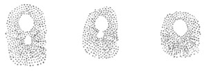
Figs. 34, 35, and 36. Three successive stages in
the coalescence of the male and female pronuclei in Asterias
glacialis. From the living ovum. (Copied from Fol.)
The product of the fusion of the two pronuclei forms the first
segmentation nucleus (fig. 37), which soon, however, divides into the
two nuclei of the two first segmentation spheres.
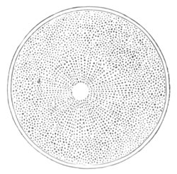
Fig. 37. Ovum of Asterias glacialis,
after the coalescence of the male and female pronuclei. (Copied
from Fol.)
The phenomenon which has just been described consists essentially in
the fusion of the male cell and the female cell. In this act the
protoplasm of the two cells as well as their nuclei coalesce, since
the whole spermatozoon which has been absorbed into the ovum is a cell
of which the head is the nucleus.
It is clear that the ovum after fertilization is an entirely different
body to the ovum prior to that act, and unless the use of the same
term for the two conditions of the ovum had become very familiar, a
special term, such as oosperm, for the ovum after its fusion with the
spermatozoon, would be very convenient.
Of the earlier observations on this subject there need perhaps only be
cited one of E. van Beneden, on the rabbit’s ovum, shewing the
presence of two nuclei before the commencement of segmentation.
Bütschli was the earliest to state from observations on Rhabditis
dolichura that the first segmentation nucleus arose from the
fusion of two nuclei, and this was subsequently shewn with greater
detail for Ascaris nigrovenosa, by Auerbach (76). Neither of
these authors gave at the first the correct interpretation of their
results. At a later period Bütschli (80) arrived at the conclusion
that in a large number of instances (Lymnæus, Nephelis,
Cucullanus, &c.), the nucleus in question was formed by
the fusion of two or more nuclei, and Strasburger at first made a
similar statement for Phallusia, though he has since withdrawn
it. Though Bütschli’s statements depend, as it seems, upon a false
interpretation of appearances, he nevertheless arrived at a correct
view with reference to what occurs in impregnation. Van Beneden (78)
described in the rabbit the formation of the original segmentation
nucleus from two nuclei, one peripheral and the other central, and
deduced from his observations that the peripheral nucleus was derived
from the spermatic element. It was reserved for Oscar Hertwig (89) to
describe in Echinus lividus the entrance of a spermatozoon into
the egg and the formation from it of the male pronucleus.
The general fact that impregnation consists in the fusion of the
spermatozoon and ovum has now been established for some forms in the
majority of invertebrate groups (Arthropoda and Rotifera excepted).
Amongst Vertebrata also it has been shewn by E. van Beneden that the
first segmentation nucleus is formed by the coalescence of the male
and female pronucleus. Calberla, and Kupffer and Benecke have
demonstrated that a single spermatozoon enters at first the ovum of
Petromyzon.
The contact of the spermatozoon with the egg membrane causes in
Petromyzon active movements of the protoplasm of the ovum, and a
retreat of the protoplasm from the membrane.
In Amphibia the appearance of a peculiar pigmented streak extending
inwards from the surface of the pigmented pole of the ovum, and
containing in a clear space at its inner extremity a nucleus, has been
demonstrated as the result of impregnation by Bambeke (77) and Hertwig
(90). There can be little doubt that this nucleus is the male
pronucleus, and that the pigmented streak indicates its path inwards.
Close to it Hertwig has shewn that another nucleus is to be found, the
female pronucleus, and that eventually the two join together. In
Amphibia the phenomena accompanying impregnation are clearly of the
same nature as in the Invertebrata. A precisely similar series of
phenomena to those in Amphibia has been shewn by Salensky to take
place in the Sturgeon.
Although there is a general agreement between the most recent
observers, Hertwig, Fol, Selenka, Strasburger, &c., as to the main
facts connected with the entrance of one spermatozoon into the egg,
the formation of the male pronucleus, and its fusion with the female
pronucleus, there still exist differences of detail in the different
descriptions, which partly, no doubt, depend upon the
difficulties of observation, but partly also upon the observations not
having all been made upon the same species. Hertwig does not enter
into details with reference to the actual entrance of the spermatozoon
into the egg, but in his latest paper points out that considerable
differences may be observed in the occurrences which succeed
impregnation, according to the relative period at which this takes
place. When, in Asterias, the impregnation is effected about an hour
after the egg is laid, and previously to the formation of the polar
cells, the male pronucleus appears at first to exert but little
influence on the protoplasm, but after the formation of the second
polar cell, the radial striæ around it become very marked, and the
pronucleus rapidly grows in size. When it finally unites with the
female pronucleus it is equal in size to the latter. In the case when
the impregnation is deferred for four hours the male pronucleus never
becomes so large as the female pronucleus. With reference to the
effect of the time at which impregnation takes place, Asterias would
seem to serve as a type. Thus in Hirudinea, Mollusca,
and Nematoidea impregnation normally takes place before the
formation of the polar bodies is completed, and the male pronucleus is
accordingly as large as the female. In Echinus, on the other
hand, where the polar bodies are formed in the ovary, the male
pronucleus is always small.
Selenka, who has investigated the formation of the male pronucleus in
Toxopneustes variegatus, differs in certain points from Fol. He
finds that usually, though not always, a single spermatozoon enters
the egg, and that though the entrance may be effected at any part of
the surface it generally occurs at the point marked by a small
prominence where the polar cells are formed. The spermatozoon first
makes its way through the mucous envelope of the egg, within which it
swims about, and then bores with its head into the polar prominence.
One important point has been so far only indirectly alluded to, viz.
the number of spermatozoa required to effect impregnation.
The concurrent testimony of almost all observers tends to shew that
one only is required for this purpose. But the number of cases tested
is too small to admit of satisfactory generalization.
Both Hertwig and Fol have made observations on the result of the
entrance into the egg of several spermatozoa. Fol finds that when the
impregnation has been too long delayed the vitelline membrane is
formed with comparative slowness, and several spermatozoa are thus
enabled to penetrate. Each spermatozoon forms a separate pronucleus
with a surrounding star; and several male pronuclei usually fuse with
the female pronucleus. Each male pronucleus appears to exercise a
repulsive influence on other male pronuclei, but to be
attracted by the female pronucleus. When there are several male
pronuclei the segmentation is irregular and the resulting larva a
monstrosity. These statements of Fol and Hertwig are up to a certain
point in contradiction with the more recent results of Selenka. In
Toxopneustes variegatus Selenka finds that though impregnation
is usually effected by a single spermatozoon yet several may be
concerned in the act. The development continues, however, to be normal
up to the gastrula stage, at any rate, if three or even four
spermatozoa enter the egg almost simultaneously. Under such
circumstances each spermatozoon forms a separate pronucleus and star.
Selenka is of opinion (apparently rather on a priori grounds
than as a result of direct observation) that normal development cannot
occur when more than one male pronucleus fuses with the female
pronucleus; and holds that, where he has observed such normal
development after the entrance of more than one spermatozoon, the
majority of male pronuclei become absorbed.
It may be noticed that, while the observations of Fol and Hertwig were
admittedly made upon eggs in which the impregnation was delayed till
they no longer displayed their pristine activity, Selenka’s were made
upon quite fresh eggs; and it seems not impossible that the
pathological symptoms in the embryos reared by the two former authors
may have been due to the imperfection of the egg, and not to the
entrance of more than one spermatozoon. This, of course, is merely a
suggestion which requires to be tested by fresh observations.
Kupffer and Benecke have further shewn that although only one
spermatozoon enters the ovum directly in Petromyzon yet other
spermatozoa pass through the vitelline membrane, and are taken into a
peculiar protoplasmic protuberance of the ovum which appears after
impregnation.
The act of impregnation may be described as the fusion of the ovum and
spermatozoon, and the most important feature in this act appears to be
the fusion of a male and female nucleus; not only does this appear in
the actual fusion of the two pronuclei, but it is brought into still
greater prominence by the fact that the female pronucleus is a product
of the nucleus of a primitive ovum, and the male pronucleus is the
metamorphosed head of the spermatozoon which, as stated
above, contains part of the nucleus of the primitive spermatic
cell. The spermatic cells originate from cells indistinguishable from
the primitive ova, so that the fusion which takes place is the fusion
of morphologically similar parts in the two sexes.
These conclusions tally very satisfactorily with the view adopted in
the Introduction, that impregnation amongst the Metazoa was derived
from the process of conjugation amongst the Protozoa.
Summary.
In what may probably be regarded as a normal case the following series
of events accompanies the maturation and impregnation of an ovum:—
(1) Transportation of the germinal vesicle to the surface of the egg.
(2) Absorption of the membrane of the germinal vesicle and
metamorphosis of the germinal spot and nuclear reticulum.
(3) Assumption of a spindle character by the remains of the germinal
vesicle, these remains being probably in part formed from the germinal
spot.
(4) Entrance of one end of the spindle into a protoplasmic prominence
at the surface of the egg.
(5) Division of the spindle into two halves, one remaining in the egg,
the other in the prominence; the prominence becoming at the same time
nearly constricted off from the egg as a polar cell.
(6) Formation of a second polar cell in the same manner as the first,
part of the spindle still remaining in the egg.
(7) Conversion of the part of the spindle remaining in the egg into a
nucleus—the female pronucleus.
(8) Transportation of the female pronucleus towards the centre of the
egg.
(9) Entrance of one spermatozoon into the egg.
(10) Conversion of the head of the spermatozoon into a nucleus—the
male pronucleus.
(11) Appearance of radial striæ round the male pronucleus, which
gradually travels towards the female pronucleus.
(12) Fusion of male and female pronuclei to form the first
segmentation nucleus.
(76) Auerbach. Organologische Studien, Heft 2. Breslau,
1874.
(77) Bambeke. “Recherches s. Embryologie des Batraciens.”
Bull. de l’Acad. royale de Belgique, 2me Sér., T.
LXI., 1876.
(78) E. van Beneden. “La Maturation de l’Œuf des
Mammifères.” Bull. de l’Acad. royale de Belgique, 2me Sér., T.
XL. No. 12, 1875.
(79) Idem. “Contributions à l’Histoire de la Vésicule
Germinative, &c. ” Bull. de l’Acad. royale de Belgique, 2me
Sér., T. XLI. No. 1, 1876.
(80) O. Bütschli. Eizelle, Zelltheilung, und Conjugation der
Infusorien. Frankfurt, 1876.
(81) F. M. Balfour. “On the Phenomena accompanying the
Maturation and Impregnation of the Ovum.” Quart. J. of Micros.
Science, Vol.
XVIII., 1878.
(82) Calberla. “Befruchtungsvorgang beim Ei von Petromyzon
Planeri.” Zeit. f. wiss. Zool., Vol. XXX.
(83) W. Flemming. “Studien in d. Entwickelungsgeschichte der
Najaden.” Sitz. d. k. Akad. Wien, B. LXXI., 1875.
(84) H. Fol. “Die erste Entwickelung des Geryonideneies.”
Jenaische Zeitschrift, Vol.
VII., 1873.
(85) Idem. “Sur le Développement des Ptéropodes.” Archives
de Zoologie Expérimentale et Générale, Vol. IV. and
V. 1875‑6.
(86) Idem. “Sur le Commencement de l’Hénogénie.” Archives
des Sciences Physiques et Naturelles. Genève, 1877.
(87) Idem. Recherches s. l. Fécondation et l. commen. d.
l’Hénogénie. Genève, 1879.
(88) R. Greeff. “Ueb. d. Bau u. d. Entwickelung d.
Echinodermen.” Sitzun. der Gesellschaft z. Beförderung d. gesammten
Naturwiss. z. Marburg, No. 5, 1876.
(89) Oscar Hertwig. “Beit. z. Kenntniss d. Bildung, &c., d.
thier. Eies.” Morphologisches Jahrbuch, Vol.
I., 1876.
(90) Idem.Ibid. Morphologisches Jahrbuch, Vol.
III. Heft 1, 1877.
(91) Idem. “Weitere Beiträge, &c.” Morphologisches
Jahrbuch, Vol.
III., 1877, Heft 3.
(92) Idem. “Beit. z. Kenntniss, &c. ” Morphologisches
Jahrbuch, Vol. IV. Heft 1 and 2, 1878.
(93) N. Kleinenberg. Hydra. Leipzig, 1872.
(94) C. Kupffer u. B. Benecke. Der Vorgang d. Befruchtung am
Eie d. Neunaugen. Königsberg, 1878.
(95) J. Oellacher. “Beiträge zur Geschichte des Keimbläschens
im Wirbelthiere.” Archiv f. mikr. Anat., Bd. VIII.,
1872.
(96) W. Salensky. “Befruchtung u. Furchung d. Sterlets-Eies.”
Zoologischer Anzeiger, No. 11, 1878.
(97) E. Selenka. Befruchtung des Eies von Toxopneustes
variegatus. Leipzig, 1878.
(98) Strasburger. Ueber Zellbildung u. Zelltheilung.
Jena, 1876.
(99) Idem. Ueber Befruchtung u. Zelltheilung. Jena,
1878.
(100) C. O. Whitman. “The Embryology of Clepsine.” Quart. J.
of Micr. Science, Vol.
XVIII., 1878.
The immediate result of the fusion of the male and female pronucleus
is the segmentation or division of the ovum usually into two, four,
eight, etc. successive parts. The segmentation may be dealt with from
two points of view, viz. (1) the nature of the vital phenomena which
take place in the ovum during its occurrence, which may be described
as the internal phenomena of segmentation. (2) The external characters
of the segmentation.
Internal Phenomena of Segmentation.
Numerous descriptions have been given during the last few years of the
internal phenomena of segmentation. The most recent contribution on
this head is that of Fol (No. 87). He appears to have been more
successful than other observers in obtaining a complete history of the
changes which take place, and it will therefore be convenient to take
as type the ovum of Toxopneustes (Echinus) lividus, on which he
made his most complete series of observations. The changes which take
place may be divided into a series of stages. The ovum immediately
after the fusion of the male and female pronucleus contains a central
segmentation nucleus.
In the first stage a clear protoplasmic layer derived from the plasma
of the cell is formed round the nucleus, from which there start
outwards a number of radial striæ, which are rendered conspicuous by
the radial arrangement of the yolk granules between them. The
nucleus during this process remains perfectly passive.
In the second stage the nucleus becomes less distinct and somewhat
elongated, and around it the protoplasmic layer of the earlier stage
is arranged in the form of a disc-shaped ring, compared by Fol to
Saturn’s ring. The protoplasmic rays still take their origin from the
perinuclear protoplasm. This stage has a considerable duration (20
minutes).
In the third stage the protoplasm around the nucleus becomes
transported to the two nuclear poles, at each of which it forms a
clear mass surrounded by a star-shaped figure formed by radial striæ.
The nucleus is hardly visible in the fresh condition, but when brought
into view by reagents is found to contain many highly refractive
particles, and to be still enveloped in a membrane.
In the fourth stage the nucleus when treated by reagents has assumed
the well-known spindle form. The striæ of which it is composed are
continuous from one end of the spindle to the other and are thickened
at the centre. The central thickenings constitute the so-called
nuclear plate. The clear protoplasmic masses and stars are present as
before at the apices of the nucleus, and the rays of the latter
converge as if they would meet at the centre of the clear masses, but
stop short at their periphery. There is no trace of a membrane round
either the nuclear spindle or the clear masses; and in the centre of
the latter is a collection of granules. The striæ of the polar stars
are very fine but distinct.
Between the stage with a completely formed spindle and the previous
one the intermediate steps have not been made out for Toxopneustes;
but for Heteropods Fol has been able to demonstrate that the striæ of
the spindle and their central thickenings are formed, as in the case
of the spindle derived from the germinal vesicle, from the
metamorphosis of the nuclear reticulum. They commence to be formed
at the two poles, and are then (in Heteropods) in immediate contiguity
with the striæ of the stars. The striæ gradually grow towards the
centre of the nucleus and there meet.
In the fifth stage the central thickenings of the spindle separate
into two sets, which travel symmetrically outwards towards the
clear masses, growing in size during the process. They remain however
united for a short time by delicate filaments—named by Fol connective
filaments—which very soon disappear. The clear masses also increase
in size. During this stage the protoplasm of the ovum exhibits active
amœboid movements preparatory to division.
In the sixth stage, which commences when the central thickenings of
the spindle have reached the clear polar masses, the division of the
ovum into two parts is effected by an equatorial constriction at right
angles to the long axis of the nucleus. The inner vitelline membrane
follows the furrow for a certain distance, but does not divide with
the ovum. All connection between the two parts of the spindle becomes
lost during this stage, and the thickenings of the fibres of the
spindle give rise to a number of spherical vesicular bodies, which
pass into the clear masses and become intermingled with the granules
which are placed there. The radii of the stars now extend round the
whole circumference of each of the clear masses.
In the seventh stage the two clear masses become elongated and travel
towards the outer sides of their segments; while the radii connected
with them become somewhat bent, as if a certain amount of traction had
been exercised on them in the movement of the clear masses. Shortly
afterwards the spherical vesicles, each of which appears like a small
nucleus and contains a central nucleolus, begin to unite amongst
themselves, and to coalesce with the neighbouring granules. Those in
each segment finally unite to form a nucleus which absorbs the
substance of the clear mass. The new nucleus is therefore partly
derived from the division of the old one and partly from the plasma of
the cell. The two segments formed by division are at first
spherical, but soon become flattened against each other. In each
subsequent division of these cells the whole of the above changes are
repeated.
The phenomena which have just been described would appear to occur in
the segmentation of ova with remarkable constancy and without any very
considerable variations.
The division of the ovum constitutes a special case of cell division,
and it is important to determine to what extent the phenomena of
ordinary cell division are related to those which take place in the
division of the ovum. Without attempting a full discussion of
the subject I will confine myself to a few remarks suggested by the
observations of Flemming, Peremeschko and Klein. The observations of
these authors shew that in the course of the division of nuclei in the
salamander, newt, etc. the nuclear reticulum undergoes a series of
peculiar changes of form, and after the membrane of the nucleus has
vanished divides into two masses. The masses form the basis for the
new nuclei, and become reconverted into an ordinary nuclear reticulum
after repeating, in the reverse order, the changes of form undergone
by the reticulum previous to its division.
It is clear without further explanation that the conversion of the
nuclear reticulum of the segmentation nucleus into the striæ of the
spindle is a special case of the same phenomenon as that first
described by Flemming in the salamander. There are however some
considerable differences. In the first place the fibres in the
salamander do not, according to Flemming, unite in the middle line,
though they appear to do so in the newt. This clearly cannot be
regarded as a fact of great importance; nor can the existence of the
central thickenings of the striæ (nuclear plate), constant as it is
for the division of the nucleus of the ovum, be considered as
constituting a fundamental difference between the two cases. More
important is the fact that the striæ in the case of the ovum do not
appear, at any rate have not been shewn, to form themselves again into
a nuclear network.
With reference to the last point it is however to be borne in mind (1)
that the gradual travelling outwards of the two halves of the nuclear
plate is up to a certain point a repetition, in the reverse order, of
the mode of formation of the striæ of the spindle, since the striæ
first appeared at the poles and gradually grew towards the middle of
the spindle; (2) that there is still considerable doubt as to how the
vesicular bodies formed out of the nuclear plate reconstitute
themselves into a nucleus.
The layer of clear protoplasm around the nucleus during its division
has its homologue in the case of the division of the nuclei of the
salamander, and the rays starting from this are also found. Klein has
suggested that the extra-nuclear rays of the stars around the poles of
the nucleus are derived from a metamorphosis of the extra-nuclear
reticulum, which he believes to be continuous with the intra-nuclear
reticulum.
The delicate connective filaments usually visible between the two
halves of the nuclear plate would seem from Strasburger’s latest
observations (No. 104) to be derived from the nuclear substance
between the striæ of the spindle, and to become eventually reabsorbed
into the newly-formed nuclei.
We are it appears to me still in complete ignorance as to the physical
causes of segmentation. The view that the nucleus is a single centre
of attraction, and that by its division the centre of attraction
becomes double and thereby causes division, appears to be quite
untenable. The description already given of the phenomena of
segmentation is in itself sufficient to refute this view. Nor
is it in the least proved by the fact (shewn by Hallez) that the plane
of division of the cell always bears a definite relation to the
direction of the axis of the nucleus.
The arguments by which Kleinenberg (93) attempted to demonstrate that
cell division was a phenomenon caused by alterations in the molecular
cohesion of the protoplasm of the ovum still in my opinion hold good,
but recent discoveries as to the changes which take place in the
nucleus during division probably indicate that the molecular changes
which take place in the cohesion of the protoplasm are closely related
to, and possibly caused by, those in the nucleus. These alterations of
cohesion are produced by a series of molecular changes, the external
indications of which are to be found in the visible alterations in the
constitution of the body of the cell and of the nucleus prior to
division.
Bibliography.
In addition to the papers cited in the last Chapter, vide
(101) W. Flemming. “Beiträge z. Kenntniss d. Zelle u. ihrer
Lebenserscheinungen.” Archiv f. mikr. Anat., Vol.
XVI., 1878.
(102) E. Klein. “Observations on the glandular epithelium and
division of nuclei in the skin of the Newt.” Quart. J. of Micr.
Science, Vol.
XIX., 1879.
(103) Peremeschko. “Ueber d. Theilung d. thierischen Zellen.”
Archiv f. mikr. Anat., Vol.
XVI., 1878.
(104) E. Strasburger. “Ueber ein z. Demonstration geeignetes
Zelltheilungs-Object.” Sitz. d. Jenaischen Gesell. f. Med. u.
Naturwiss., July 18, 1879.
External Features of Segmentation.
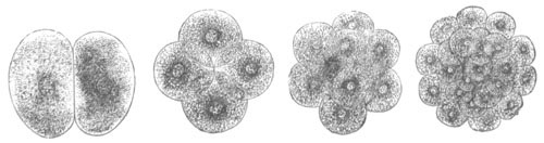
Fig. 38. Various stages in process of
segmentation.
(After Gegenbaur.)
In the simplest known type of segmentation the ovum first of all
divides into two, then four, eight, sixteen, thirty-two, sixty-four,
etc. cells (fig. 38). These cells so long as they are fairly large are
usually known as segments or spheres. At the close of such a
simple segmentation the ovum becomes converted into a sphere composed
of segments of a uniform size. These segments usually form a wall
(fig. 39, E), one row of cells thick, round a central cavity, which is
known as the segmentation cavity or cavity of Von Baer. Such a sphere
is known as a blastosphere. The central cavity usually appears very
early in the segmentation, in many cases when only four segments are
present (fig. 39, B).
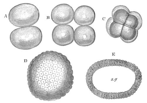
Fig. 39. The Segmentation of Amphioxux.
(Copied from Kowalevsky.)
sg. segmentation cavity. A. Stage with two equal segments. B.
Stage with four equal segments. C. Stage after the four segments
have become divided by an equatorial furrow into eight equal
segments. D. Stage in which a single layer of cells encloses a
central segmentation cavity. E. Somewhat older stage in optical
section.
In other instances, which however are rarer than those in which a
segmentation cavity is present, there is no trace of a central cavity,
and the sphere at the close of segmentation is quite solid. In such
instances the solid sphere is known as a morula. It is found in some
Sponges, many Cœlenterata, some Nemertines, etc., and in Mammals;
in which group the segmentation is not however quite regular. All
intermediate conditions between a large segmentation cavity, and a
very minute central cavity which may be surrounded by more than a
single row of cells have been described.
The segmentation cavity has occasionally, as in Sycandra, the
Ctenophora and Amphioxus, the form of an axial perforation of the ovum
open at both extremities.
When the process of regular segmentation is examined somewhat more in
detail it is found to follow as a rule a rather definite rhythm. The
ovum is first divided in a plane which may be called vertical, into
two equal parts (fig. 39, A). This division is followed by a second,
also in a vertical plane, but at right angles to the first plane, and
by it each of the previous segments is halved (fig. 39, B.) In the
third segmentation the plane of division is horizontal or equatorial
and divides each of the four segments into two halves, making eight
segments in all (fig. 39, C). In the fourth period the segmentation
takes place in two vertical planes each at an angle of 45° with one of
the previous vertical planes. All the segments are thus again divided
into two equal parts. In the fifth period there are two equatorial
planes one on each side of the original equatorial plane, and
thirty-two spheres are present at the close of this period. Sixty-four
segments are formed at the sixth period, but beyond the fourth and
fifth periods the original regularity is not usually preserved.
In many instances the type of segmentation just described cannot be
distinctly recognized. All that can be noticed is that at each fresh
segmentation every segment becomes divided into two equal parts. It is
not absolutely certain that there is not always some slight inequality
in the segments formed, by which, what are known as the animal and
vegetative poles of the ovum, can very early be distinguished. A
regular segmentation is found in species in most groups of the animal
kingdom. It is very common in Sponges and Cœlenterates. Though less
common so far as is known amongst the Vermes, it is yet found in many
of the lower types, viz. Nematoidea, Gordiacea, Trematoda, Nemertea
(apparently as a rule), Sagitta, Chætonotus, some
Gephyrea (Phoronis); though not usual it occurs amongst
Chætopoda, e.g. Serpula. It is the usual type of
segmentation amongst the Echinodermata. Amongst the Crustacea it
appears (for the earlier phases of segmentation at any rate) not
infrequently amongst the lower forms, and even occurs amongst the
Amphipoda (Phronima). It is however very rare amongst the
Tracheata, Podura affording the one example of it known to me.
It is almost as rare amongst Mollusca as amongst the Tracheata, but
occurs in Chiton and is nearly approached in some
Nudibranchiata. In Vertebrata it is most nearly approached in
Amphioxus[36].
Most of the eggs which have a perfectly regular segmentation are of a
very insignificant size and rarely contain much food-yolk: in
the vast majority of eggs there is present however a considerable bulk
of food material usually in the form of highly refracting
yolk-spherules. These yolk-spherules lie embedded in the protoplasm of
the ovum, but are in most instances not distributed uniformly, being
less closely packed and smaller at one pole of the ovum than
elsewhere. Where the yolk-spherules are fewest the active protoplasm
is necessarily most concentrated, and we can lay down as a general
law[37] that the velocity of segmentation in any part of the ovum is
roughly speaking proportional to the concentration of the protoplasm
there; and that the size of the segments is inversely proportional to
the concentration of the protoplasm. Thus the segments produced from
that part of an egg where the yolk-spherules are most bulky, and where
therefore the protoplasm is least concentrated, are larger than the
remaining segments, and their formation proceeds more slowly.
Though where much food-yolk is present it is generally distributed
unequally, yet there are many cases in which it is not possible to
notice this very distinctly. In most of these cases the segmentation
is all the same unequal, and it is probable that they form apparent
rather than real exceptions to the law laid down above. Although
before segmentation the protoplasm may be uniformly distributed, yet
in many instances, e.g. Mollusca, Vermes, etc., during or at
the commencement of segmentation the protoplasm becomes aggregated at
one pole, and one of the segments formed consists of clear protoplasm,
all the food-yolk being contained in the other and larger segment.
Unequal Segmentation. The type of segmentation I now proceed to
describe has been called by Haeckel (No. 105) ‘unequal segmentation’,
a term which may conveniently be adopted. I commence by describing it
as it occurs in the well-known and typical instance of the Frog[38].
The ripe ovum of the common Frog and of most other tailless Amphibians
presents the following structure. One half appears black and the other
white. The former I shall call the upper pole, the latter the
lower. The ovum is composed of protoplasm containing in suspension
numerous yolk-spherules. The largest of these are situated at the
lower pole, the smaller ones at the upper pole, and the smallest of
all in the peripheral layer of the upper pole, in which also pigment
is scattered and causes the black colour visible from the surface.
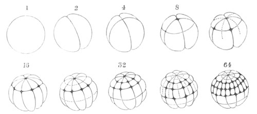
Fig. 40. Segmentation of Common Frog. Rana
Temporaria. (Copied from Ecker.)
The numbers above the figures refer to the number of segments at the
stage figured.
The first formed furrow is a vertical furrow. It commences in the
upper half of the ovum, through which it extends rapidly, and then
more slowly through the lower. As soon as the first furrow has
extended through the egg, and the two halves have become separated
from each other, a second vertical furrow appears at right angles to
the first and behaves in the same way (fig. 40, 4).
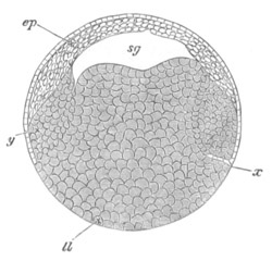
Fig. 41. Section through Frog’s ovum at the close
of segmentation.
sg. segmentation cavity. ll. large yolk-containing
cells. ep. small cells at formative pole (epiblast).
The next furrow is equatorial or horizontal (fig. 40, 8). It does not
arise at the true equator of the egg, but much nearer to its
upper pole. It extends rapidly round the egg and divides each of the
four previous segments into two parts, one larger and one smaller.
Thus at the end of this stage there are present four small and four
large segments. At the meeting point of these a small cavity
appears, which is the segmentation cavity, already described for
uniformly segmenting eggs. It increases in size in subsequent stages,
its roof being formed of the smaller cells and its floor of the
larger. The appearance of the equatorial furrow is followed by a
period of repose, after which two rapidly succeeding vertical furrows
are formed in the upper pole, dividing each of the four segments of
which this is composed into two. After a short period these furrows
extend to the lower pole, and when completed 16 segments are
present—eight larger and eight smaller (fig. 40, 16). A pause now
ensues, after which the eight upper segments become divided by an
equatorial furrow, and somewhat later a similar furrow divides the
eight lower segments. At the end of this stage there are therefore
present 16 smaller and 16 larger segments (fig. 40, 32). After 64
segments have been formed by vertical furrows which arise
symmetrically in the two poles (fig. 40, 64), two equatorial furrows
appear in the upper pole before a fresh furrow arises in the lower; so
that there are 128 segments in the upper half, and only 32 in the
lower. The regularity is quite lost in subsequent stages, but the
upper pole continues to undergo a more rapid segmentation than the
lower. While the segments have been increasing in number the
segmentation cavity has been rapidly growing in size; and at the close
of segmentation the egg forms a sphere, containing an excentric
cavity, and composed of two unequal parts (fig. 41). The upper part,
which forms the roof of the segmentation cavity, is formed of smaller
cells: the lower of larger yolk-containing cells.
The mode of segmentation of the Frog’s ovum is typical for unequally
segmenting ova, and it deserves to be noticed that as regards the
first three or more furrows the segmentation occurs with the same
rhythm in the unequally segmenting ova as in those which have an
uniform segmentation. There appear two vertical furrows followed by an
equatorial furrow. The general laws which were stated with reference
to the velocity of segmentation and the size of the resulting
segments are well exemplified in the case of the Frog’s ovum.
The majority of the smaller segments in the segmented Frog’s ovum are
destined to form into the epiblast, and the larger segments become
hypoblast and mesoblast.
With a few exceptions (the Rabbit, Lymnæus, etc.) the majority of the
smaller segments always become epiblast and of the larger segments
hypoblast.
The Frog’s ovum serves as a good medium type for unequally segmenting
ova. There are many cases however in which a regular segmentation is
far more closely approached, and others in which it is less so.
One familiar instance in which a regular segmentation is nearly
approached is afforded by the Rabbit’s ovum, which has indeed usually
been regarded as offering an example of a regular segmentation.
The ovum of the Rabbit[39] becomes first divided into two sub-equal
spheres. The larger and more transparent of the two may, from its
eventual fate, be called the epiblastic sphere, and the other the
hypoblastic. The two spheres are divided into four, and then by an
equatorial furrow into eight—four epiblastic and four hypoblastic.
One of the latter assumes a central position. The four epiblastic
spheres now divide before the four hypoblastic. There is thus
introduced a stage with twelve spheres. It is followed by one with
sixteen, and that by one with twenty-four. During the stages with
sixteen spheres and onwards the epiblastic spheres gradually envelop
the hypoblastic, which remain exposed on the surface at one point
only. There is no segmentation cavity.
In Pedicellina, one of the entoproctous Polyzoa, there is a
sub-regular segmentation, where however the two primary spheres can be
distinguished much in the same way as in the case of the Rabbit.
A very characteristic type of unequal segmentation is that presented
by the majority of Gasteropods and Pteropods and probably also of some
Lamellibranchiata. It is also found in some Turbellarians, in
Bonellia, some Annelids, etc. In many instances it offers a good
example of the type where in the course of segmentation the protoplasm
becomes aggregated at one pole of the ovum, or of its segments, to
become separated off as a clear sphere.
The first four segments formed by two vertical furrows at
right angles are equal, but from these there are budded off four
smaller segments, which in subsequent stages divide rapidly, receiving
however, a continual accession of segments budded off from the larger
spheres. The four larger spheres remain conspicuous till near the
close of the segmentation. The process of budding, by which the
smaller spheres become separated from the larger, consists in a larger
sphere throwing out a prominence, which then becomes constricted off
from it.
In the extreme forms of this unequal segmentation we find at the end
of the second cleavage two larger spheres filled with yolk material
and two smaller clear spheres; and in the later stages, though the
large spheres continue to bud off small spheres, only the two smaller
ones undergo a regular segmentation, and eventually completely envelop
the former. Such a case as this has been described in Aplysia by
Lankester[40].
The types I have described serve to exemplify unequal segmentation.
The Rabbit’s ovum stands at one end of the series, that of Aplysia at
the other; and the Frog’s ovum between the two.
Great variations are presented by the ova with unequal segmentation as
to the presence of a segmentation cavity. In some instances,
e.g. the Frog, such a cavity is well developed. In other cases
it is small, e.g. most Mollusca, while not unfrequently it is
altogether absent.
Before leaving this important type of segmentation, it will be well to
enter with slightly greater detail into some of the more typical as
well as some of the special forms which it presents.
As an example of the typical Molluscan type the normal Heteropod
segmentation, accurately described by Fol[41], may be selected.
The ovum divides into two and then four equal segments in the usual
vertical planes. Each segment has a protoplasmic and a vitelline pole.
The protoplasmic pole is turned towards the polar bodies. In the third
segmentation, which takes place along an equatorial plane, four small
protoplasmic cells or segments are segmented or rather budded off from
the four large segments, so that there are four small segments in one
plane and four large below these. In the fourth segmentation the four
large segments alone are active and give rise to four small and four
large cells; so that there are formed in all eight small and four
large cells. The four small cells of the third generation next
divide, forming in all twelve small cells and four large. The small
cells of the fourth generation then divide, and subsequently the four
large cells give rise to four new small ones, so that there are twenty
small cells and four large. The small cells form a cap embracing the
upper pole of the large segments. It may be noted that from the third
stage onwards the cells increase in arithmetical progression—a
characteristic feature of the typical gasteropod segmentation.
In the later stages of segmentation the large cells cease to give rise
to smaller ones in the same manner as before. One of them divides
first into two unequal parts, of which the smaller becomes pushed in
towards the centre of the egg. The larger cell then divides again into
two, and the three cells so formed occupy the centre of a shallow
depression. The remaining larger cells divide in the same way, and
give rise to smaller cells which line a pit which becomes formed on
one side of the ovum. The original smaller cells continue in the
meantime to divide and so form a layer enclosing the larger, leaving
exposed however the opening of the pit lined by the latest products of
the larger cells.
The eggs of Anodon and Unio serve as excellent examples of the type in
which the ovum has a uniform structure before the commencement of
segmentation, but in which a separation into a protoplasmic and a
nutritive portion becomes obvious during segmentation.
In Anodon[42] the egg is at first uniformly granular, but after
impregnation it throws out on one side a protuberance nearly free from
granules (fig. 42, 1)
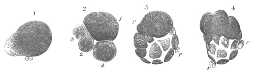
Fig. 42. Segmentation of Anodon piscinalis.
(Copied from Flemming.) r. polar cells. v. vitelline
sphere. 1. Commencing division into two segments; one mainly formed
of protoplasm, the other of yolk. 2. Stage with four segments. 3.
Formation of blastosphere, and segmentation cavity. 4. Definite
segmentation of the yolk sphere.
In the case of this clear protuberance and of the similar
protuberances which follow it, the protoplasm is not at first quite
free from food-yolk, but only becomes so on being separated from the
yolk-containing part of the ovum. We must therefore suppose that the
production of the clear segments is in part at least due to the
yolk-spherules becoming used up to form protoplasm. Such a formation
of protoplasm from yolk-spherules has been clearly shewn to occur in
other types by Bobretzky and Fol.
The protuberance soon becomes separated off from the larger part of
the egg as a small segment composed of clear protoplasm. From the
larger segment filled with food-yolk, a second small clear segment is
next budded off, and simultaneously (fig. 42, 2) the original small
segment divides into two. Thus there are formed four segments, one
large and three small; the large segment as before being filled with
food-yolk. The continuation of a similar process of budding off and
segmentation eventually results in the formation of a considerable
number of small and of one large segment (fig. 42, 3). Between this
large and the small segments is a segmentation cavity.
Eventually the large yolk segment, which has hitherto merely budded
off a series of small segments free from yolk, itself divides into two
similar parts. This process is then repeated (fig. 42, 4) and there is
at last formed a number of yolk segments filled with yolk spheres,
which occupy the place of the original large yolk segment. Between
these yolk segments and the small segments is placed the segmentation
cavity.
The segmentation of the ovum of Euaxes[43] resembles that of Unio in
the budding off of clear segments from those filled with yolk, but
presents many interesting individualities.
A very peculiar modification of the ordinary Gasteropod segmentation
is that described by Bobretzky for Nassa mutabilis[44].
The ovum contains a large amount of food-yolk, and the protoplasm is
aggregated at the formative pole, adjoining which are placed the polar
bodies. An equatorial and a vertical furrow (fig. 43 A), the former
near the upper pole, appear simultaneously, and divide the ovum into
three segments, two small, each with a protoplasmic pole, and one
large entirely formed of yolk material. One of the two small segments
next completely fuses with the large segment (fig. 43 B), and after
the fusion is complete, a triple segmentation of the large segment
takes place as at the first division, and at the same time the single
small segment divides into two. In this way four partially
protoplasmic segments and one yolk segment are formed (fig. 43 C). One
of the small segments again fuses with the large segment, so that the
number of segments becomes again reduced to four, three small and one
large. The protoplasmic ends of these segments are turned towards each
other, and where they meet four very small cells become budded off,
one from each segment (fig. 43 D). Four small cells are again budded
off twice in succession, while the original small cells remain
passive, so that there come to be twelve small and four large cells.
In later stages the four first-formed small cells give rise to still
smaller cells and then the next-formed do the same. The large cells
continue also to give rise to small ones, and finally, by a continuous
process of division, and fresh budding of small cells from large
cells, a cap of small cells becomes formed covering the four large
cells which have in the meantime pressed themselves together (fig. 43
E). A segmentation cavity of not inconsiderable dimensions becomes
established between this cap of small cells and the large cells.
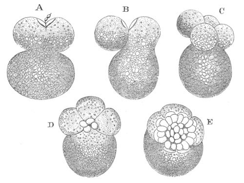
Fig. 43. Segmentation of Nassa mutabilis
(from Bobretzky). A. Upper half divided into two segments. B. One of
these has fused with the large lower segment. C. Four small and one
large segment, one of the former fusing with the large segment. D.
Each of the four segments has given rise to a small segment. E.
Small segments have increased to thirty-six.
Many eggs, such as those of the Myriapods[45], present an irregular
segmentation; but the segmentation is hardly unequal in the sense in
which I have been using the term. Such cases should perhaps be placed
in the first rather than in the present category.
The type of unequal segmentation is on the whole the most widely
distributed in the animal kingdom. There is hardly a group without
examples of it.
It occurs in Porifera, Hydrozoa, Actinozoa and Ctenophora. Amongst the
Ctenophora this segmentation is of the most typical kind. Four equal
segments are first formed in the two first periods. In the third
period a circumferential furrow separates four smaller from four
larger segments.
This type is also widely distributed amongst the unsegmented
(Gephyrea, Turbellaria), as well as the segmented Vermes, and is
typical for the Rotifera. It appears to be very rare in Echinoderms
(Echinaster Sarsii). It is not uncommon in early stages of the
segmentation of the lower Crustacea.
For Mollusca (except Cephalopoda) it is typical. Amongst the Ascidia
it occurs in several forms (Salpa, Molgula) and amongst
the Craniata it is typical in the Cyclostomata, Amphibia, and some
Ganoids, e.g. Accipenser.
Partial segmentation. The next type of segmentation we have to
deal with has long been recognized as partial segmentation. It is a
type in which only part of the ovum, called the germinal disc,
undergoes segmentation, the remainder usually forming an appendage of
the embryo known as the yolk-sack. Ova belonging to the two groups
already dealt with are frequently classed together as holoblastic ova,
in opposition to ova of the present group in which the segmentation is
only partial, and which are therefore called meroblastic. For
embryological purposes this is in many ways a very convenient
classification, but ova belonging to the present group are in reality
separated by no sharp line from those belonging to the group just
described.
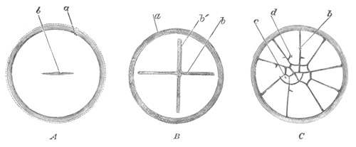
Fig. 44. Surface views of the early stages of the
segmentation in a fowl’s egg.
(After Coste.)
a. edge of germinal disc. b. vertical furrow.
c. small central segment. d. larger peripheral
segment.
The origin and nature of meroblastic ova will best be understood by
taking an ovum with an unequal segmentation, such as that of the frog,
and considering what would take place in accordance with the laws
already laid down, supposing the amount of food-yolk at the vitelline
pole to be enormously increased. What would happen may be conveniently
illustrated by fig. 44, representing the segmentation of a fowl’s egg.
There would first obviously appear a vertical furrow at the formative
or protoplasmic pole. (Fig. 44 A, b.) This would gradually
advance round the ovum and commence to divide it into two halves.
Before the furrow had however proceeded very far it would come
to the vitelline part of the ovum; here, according to the law
previously enunciated, it would travel very slowly, and if the amount
of the food-yolk was practically infinite as compared with the
protoplasm, it would absolutely cease to advance. A second vertical
furrow would soon be formed, crossing the first at right angles, and
like it not advancing beyond the edge of the germinal disc. (Fig. 44
B.)
The next furrow should be an equatorial one (as a matter of fact in
the fowl’s ovum an equatorial furrow is not formed till after two more
vertical furrows have appeared). The equatorial furrow would however,
in accordance with the analogy of the frog, not be formed at the
equator, but very close to the formative pole. It would therefore
separate off as a distinct segment (fig. 44 C, c), a small
central, i.e. polar, portion of each of the imperfect segments
formed by the previous vertical furrows. By a continuation of the
process of segmentation, with the same alternation of vertical and
equatorial furrows as in the frog, a cap or disc of small segments
would obviously be formed at the protoplasmic pole of the ovum,
outside which would be a number of deep radiating grooves (fig. 45),
formed by the vertical furrows, the advance of which round the ovum
has come to an end owing to the too great proportion of yolk spheres
at the vitelline pole.
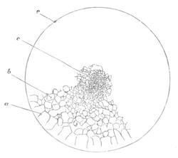
Fig. 45. Surface view of the germinal disc of
fowl’s egg during a late stage of the segmentation.
c. small central segmentation spheres; b. larger
segments outside these; a. large, imperfectly circumscribed,
marginal segments; e. margin of germinal disc.
It is clear from the above that an immense accumulation of food-yolk
at the vitelline pole necessarily causes a partial segmentation. It is
equally clear that the part of meroblastic ova which does not undergo
segmentation is not a new addition absent in other cases. It
is on the contrary to be regarded merely as a part of the ovum in
which the yolk-spherules have attained to a very great bulk as
compared with the protoplasm; sometimes even to the complete exclusion
of the protoplasm.
An ordinary meroblastic ovum consists then of a small disc at the
formative pole, known as the germinal disc, composed mainly of
protoplasm in which comparatively little food-yolk is present. This
graduates into the remainder of the ovum, being separated from it by a
more or less sharp line. This remainder of the ovum, which almost
always forms the major part, usually consists of numerous
yolk-spherules, embedded in a very scanty protoplasmic matrix.
In some cases, e.g. the eggs of Elasmobranchii[46], the
protoplasm is present in the form of a delicate network; in other and
perhaps the majority of cases, too little protoplasm is present to be
detected, or the protoplasm may even be completely absent. In some
Osseous Fishes, e.g. Lota, the yolk forms a homogeneous
transparent albuminoid substance containing a large globule at the
pole furthest removed from the germinal disc. In this case the
germinal disc is sharply separated from the yolk. In other Osseous
Fishes the separation between the two parts is not so sharp[47]. In
these cases we find adjoining the germinal disc a finely granular
material containing a large proportion of protoplasm; this graduates
into a material with very little protoplasm and numerous
yolk-spherules, which is in its turn continuous with an homogeneous
albuminoid yolk substance. In Elasmobranchii we find that immediately
beneath the germinal disc there is present a finely granular matter,
rich in protoplasm, which is continuous with the normal yolk.
The Elasmobranch ovum may conveniently serve as type for the
Vertebrata. The ovum is formed of a spherical vitellus without any
investing membrane. The germinal disc is recognizable on this as a
small yellow spot about 1½ millimetres in diameter. In the germinal
disc a furrow appears bisecting the disc, followed by a second furrow
at right angles to the first. Thus after the formation of the second
furrow the disc is divided into four equal areas. Fresh furrows
continue to rise, and eventually a circular furrow, equivalent to the
equatorial furrow of the frog’s ovum, makes its appearance, and
separates off a number of smaller central segments from peripheral
larger segments. In the later stages the smaller segments at first
divide more rapidly than the larger, but eventually the latter also
divide rapidly, and the germinal disc becomes finally formed of a
series of segments of a fairly uniform size. So much may be
observed in surface views of the segmenting ovum, and it may be noted
that there is not much difference to be observed between the
segmentation of the germinal disc of the Fowl’s ovum and that of the
Elasmobranchii. Indeed the figure of the former (fig. 44) would serve
fairly well for the latter. When however we examine the segmenting
germinal discs by means of sections, there are some differences
between the two types, and several interesting features which deserve
to be noticed in the segmentation of the Elasmobranchii. In the first
stages the furrows visible on the surface are merely furrows, which do
not meet so as to isolate distinct segments; they merely, in fact,
form a surface pattern. It is not till after the appearance of the
equatorial furrow that the segments begin to be distinctly isolated.
In the subsequent stages not only do the segments already existing in
the germinal disc increase by division, but fresh segments are
continually being formed from the adjacent yolk, and added to those
already present in the germinal disc. (Fig. 46.) This fact is one out
of many which prove that the germinal disc is merely part of the ovum
characterized by the presence of more protoplasm than the remainder
which forms the so-called food-yolk. During the latest stages of
segmentation there appear in the yolk around the blastoderm a number
of nuclei. (Fig. 46, nx´.) These are connected with a special
protoplasmic network (already described) which penetrates through the
yolk. Towards the end of segmentation, and during the early periods of
development which succeed the segmentation, these nuclei become very
numerous. (Fig. 47 A, n´.) Around many of them a protoplasmic
investment is established, and cells are thus formed which eventually
enter the blastoderm.
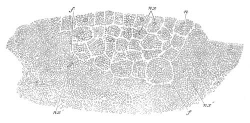
Fig. 46. Section through germinal disc of a
Pristiurus embryo during the segmentation.
n. nucleus; nx. nucleus modified prior to division;
nx´. modified nucleus of the yolk; f. furrow appearing
in the yolk adjacent to the germinal disc.
The result of segmentation is the formation of a lens-shaped mass of
cells lying in a depression on the yolk. In this a cavity appears, the
homologue of the segmentation cavity already spoken of. It lies at
first in the midst of the cells of the blastoderm, but very
soon its floor of cells vanishes, and it lies between the yolk and the
blastoderm. (Fig. 47 A.) Its subsequent history is given in a future
Chapter.
Segmentation proceeds in Osseous Fishes in nearly the same manner as
in Elasmobranchii. In some cases the germinal disc is small as
compared with the yolk, in other cases it is almost as large as the
yolk. The only points which deserve special notice are the following:
(1) Nuclei, precisely similar to those in the Elasmobranch yolk,
appear in the protoplasmic matter around the germinal disc; (2) After
the deposition of the ova there is present in some forms a network of
protoplasm extending from the germinal disc through the yolk[48]. At
impregnation this withdraws itself from the yolk. It is to be compared
to the protoplasmic network of the Elasmobranch ovum.
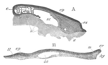
Fig. 47. Two longitudinal sections of the
blastoderm of a Pristiurus embryo at stages prior to the formation
of the medullary groove.
ep. epiblast; ll. lower layer cells; m.
mesoblast; hy. hypoblast; sc. segmentation cavity;
es. embryo swelling; n´. nuclei of yolk; er.
embryonic rim.
There are two types of meroblastic ova. In one of these (Aves,
Elasmobranchii) the germinal disc is formed in the ovarian ovum. In
the second type the germinal disc is formed after impregnation by a
concentration of the protoplasm at one pole. This concentration is
analogous to what has already been described for Anodon and other
Molluscan ova (p. 100).
The ova of some Teleostei are intermediate between the two types.
The ovum of the wood-louse, Oniscus murarius[49], may be taken as an
example of the second type of meroblastic ovum. In this egg
development commences by the appearance of a small clear mass with
numerous transparent vesicles. This mass is the protoplasm which has
become separated from the yolk. It undergoes segmentation in a
perfectly normal fashion. Examples of other cases of this kind have
been described by Van Beneden and Bessels[50] in Anchorella, and in
Hessia by Van Beneden[51]. It appears from their researches that the
protoplasm collects itself together, first of all in the interior of
the egg, and then travels to the surface. It arrives at the surface
after having already divided into two or more segments, which then
rapidly divide in the usual manner to form the blastoderm.
There are some grounds for thinking that the cases of partial
segmentation in the Arthropoda are not really quite comparable with
those in other groups, but more probably fall under the next type of
segmentation to be described. The grounds for this view are mentioned
in connection with the next type.
In most if not all meroblastic ova there appear during and after
segmentation a number of nuclei in the yolk adjoining the blastoderm,
around which cells become differentiated. (Figs. 46 and 47.) These
cells join the part of the blastoderm formed by the normal
segmentation of the germinal disc. Such nuclei are formed in all
craniate meroblastic ova[52]. In Cephalopods they have been found by
Lankester, and in Oniscus by Bobretzky. They have been by some authors
supposed to originate from the nuclei of the blastoderm, and by others
spontaneously in the yolk.
Some of the earliest observations on these nuclei were made by
Lankester[53] in the Cephalopods. He found that they appeared first in
a ring-like series round the edge of the blastoderm, and subsequently
all over the yolk in a layer a little below the surface. He observed
their development in the living ovum and found that they “commenced as
minute points, gradually increasing in size like other free-formed
nuclei.” A cell area subsequently forms around them.
By E. van Beneden[54] they were observed in a Teleostean ovum to
appear nearly simultaneously in considerable numbers in the granular
matter beneath the blastoderm. Van Beneden concludes from the
simultaneous appearance of these bodies that they develop
autogenously. Kupffer at an earlier period arrived at a similar
conclusion. My own observations on these nuclei in Elasmobranchii on
the whole support the conclusions to be derived from Lankester’s,
Kupffer’s and Van Beneden’s observations. As mentioned above, the
nuclei in Elasmobranchii do not appear simultaneously, but
increase in number as development proceeds; and it is possible that
Van Beneden may be mistaken on this point. No evidence came before me
of derivation from pre-existing nuclei in the blastoderm. My
observations prove however that the nuclei increase by division. This
is shewn by the fact that I have found them with the spindle
modification (fig. 46, nx´), and that in most cases they
usually exhibit the form of a number of aggregated vesicles[55], which
is a character of nuclei which have just undergone division. It should
be mentioned however that I failed to find a spindle modification of
the nuclei in the later stages. Against these observations must be set
those of Bobretzky, according to which the nuclei in Oniscus are
really the nuclei of cells which have migrated from the blastoderm.
Bobretzky’s observations do not however appear to be very conclusive.
It must be admitted that the general evidence at our command appears
to indicate that the nuclei of the yolk in meroblastic ova originate
spontaneously. There is however a difficulty in accepting this
conclusion in the fact that all the other nuclei of the embryo are
descendants of the first segmentation nucleus; and for this reason it
still appears to me possible that the nuclei of the yolk will be found
to originate from the continued division of one primitive nucleus,
itself derived from the segmentation nucleus.
The existence of these nuclei in the yolk and the formation of a
distinct cell body around them is a strong piece of evidence in favour
of the view above maintained, (which is not universally accepted,)
that the part of the ovum of meroblastic ova which does not segment is
of the same nature as that which does segment, and differs only in
being relatively deficient in active protoplasm.
The following forms have meroblastic ova of the first type: the
Cephalopoda, Pyrosoma, Elasmobranchii, Teleostei, Reptilia,
Aves, Ornithodelphia (?). The second type of meroblastic segmentation
occurs in many Crustacea, (parasitic Copepoda, Isopoda Mysis,
etc.). It is also stated to be found in Scorpio.
The ova of the majority of groups in the animal kingdom segment
according to one of the types which have just been described. These
types are not sharply separated, but form an unbroken series,
commencing with the ovum which segments uniformly, and ending with the
meroblastic ovum.
It is convenient to distinguish the ova which segment uniformly by
some term; and I should propose for this the term alecithal[56], as
implying that they are without food-yolk, or that what little
food-yolk there is, is distributed uniformly.
The ova in which the yolk is especially concentrated at one pole I
should propose to call telolecithal. They constitute together a
group with an unequal or partial segmentation.
The telolecithal ova may be defined in the following way: ova in which
the food-yolk is not distributed uniformly, but is concentrated at one
pole of the ovum. When only a moderate quantity of food-yolk is
present the pole at which it is concentrated merely segments more
slowly than the opposite pole; but when food-yolk is present in very
large quantity the part of the ovum in which it is located is
incapable of segmentation, and forms a special appendage known as the
yolk-sack.
There is a third group of ova including a series of types of
segmentation nearly parallel to the telolecithal group. This group
takes its start from the alecithal ovum as do the telolecithal ova,
and equally with these includes a series of varieties of segmentation
running parallel to the regular and unequal types of segmentation
which directly result from the presence of a greater or smaller
quantity of food-yolk. The food-yolk is however placed, not at one
pole, but at the centre of the ovum. This group of ova I
propose to name centrolecithal. It is especially characteristic of the
Arthropoda, if not entirely confined to that group.
Centrolecithal ova. As might be anticipated on the analogy of
the types of segmentation already described, the concentration of the
food-yolk at the centre of the ovum does not always take place before
segmentation, but is sometimes deferred till even the later stages of
this process.
Examples of a regular segmentation in centrolecithal ova are afforded
by Palæmon (Bobretzky) and Penæus (Hæckel). A type of unequal
segmentation like that of the Frog occurs in Gammarus locusta
(Beneden and Bessels), where however the formation of a central yolk
mass does not appear to take place till rather late in the
segmentation. More irregular examples of unequal segmentation are also
afforded by other Crustaceans, e.g. various members of the
genus Chondracanthus (Beneden and Bessels) and by Myriapods. In
all these cases segmentation ends in the formation of a layer of cells
enclosing a central mass of food-yolk.
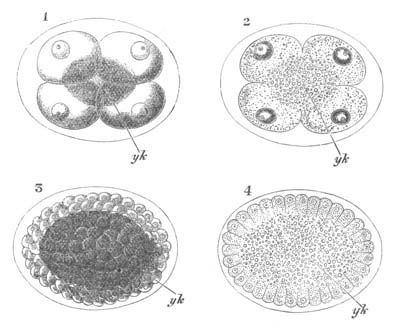
Fig. 48. Segmentation of a Crustacean ovum
(Penæus).
(After Hæckel.)
The sections illustrate the type of segmentation in which the yolk
is aggregated at the centre of the ovum.
yk. central yolk mass.
1 and 2. Surface view and section of the stage with four segments.
In 2 it is seen that the furrows visible on the surface do not
penetrate to the centre of the ovum.
3 and 4. Surface view and section of ovum near the end of
segmentation. The central yolk mass is very clearly seen in 4.
The peculiarity of the centrolecithal ova with regular or unequal
segmentation is that (owing to the presence of the yolk in the
interior) the furrows which appear on the surface are not continued to
the centre of the egg. The spheres which are thus distinct on the
surface are really united internally. Fig. 48, copied from Hæckel,
shews this in a diagrammatic way.
Many ova, which in the later stages of segmentation exhibit the
characteristics of true centrolecithal ova, in the early stages
actually pass through nearly the same phases as holoblastic ova.
Thus in Eupagurus prideauxii[57] (fig. 49), and
probably in the majority of Decapods, the egg is divided successively
into two, four and eight distinct segments, and it is not till after
the fourth phase of the segmentation that the spheres fuse in the
centre of the egg. Such ova belong to a type which is really
intermediate between the ordinary type of segmentation and that with a
central yolk mass. Eupagurus presents one striking peculiarity, viz.
that the nucleus divides into two, four and eight nuclei, each
surrounded by a delicate layer of protoplasm prolonged into a
reticulum, before the ovum itself commences to become segmented. The
ovum before segmentation is therefore in the condition of a syncytium.
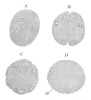
Fig. 49. Transverse section through four stages
in the segmentation of Eupagurus Prideauxii. (After P. Mayer.)
The segmentation of Asellus aquaticus[58] is very similar to that of
Eupagurus, etc. but the ovum at the very first divides into as many
segments (viz. eight) as there are nuclei.
In Gammarus locusta the resemblance to ordinary unequal segmentation
is very striking, and it is not till a considerable number of segments
have been formed that a central yolk mass appears.
In all the above types, as segmentation proceeds, the protoplasm
becomes more and more concentrated at the surface, and finally a
superficial layer of flat blastoderm cells is completely segmented off
from the yolk below (fig. 49 D).
In cases like those of Penæus, Eupagurus, etc., the yolk in the
interior is at first nearly homogeneous, but at a later period it
generally becomes divided up partially or completely into a number of
distinct spheres, which may have nuclei and therefore have the value
of cells. In many cases nuclei have however not been demonstrated in
these yolk spheres, though probably present; yet, till they have been
demonstrated, some doubt must remain on the nature of these yolk
spheres. It is probable that not all the nuclei which result
from the division of the first segmentation nucleus become concerned
in the formation of the superficial blastoderm, but that some remain
in the interior of the ovum to become the nuclei of the yolk spheres.
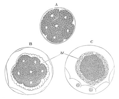
Fig. 50. Segmentation and formation of the
blastoderm in Chelifer.
(After Metschnikoff.)
In A the ovum is divided into a number of separate segments. In B a
number of small cells have appeared (bl) which form a
blastoderm enveloping the large yolk spheres. In C the blastoderm
has become divided into two layers.
In Myriapods (Chilognatha) a peculiar form of
segmentation has been observed by Metschnikoff[59]. The ovum
commences by undergoing a perfectly normal, though rather irregular
total segmentation. But after the process of division has reached a
certain point, scattered masses of very small cells make their
appearance on the surface of the large spheres. These small cells have
probably arisen in a manner analogous to that which characterizes the
formation of the superficial cells of the blastoderm in the types of
centrolecithal ova already described. They rapidly increase in number
and eventually form a continuous blastoderm; while the original large
segments remain in the centre as the yolk mass. In the interesting
Arachnid Chelifer segmentation takes place in nearly the same
manner as in Myriapods (fig. 50).
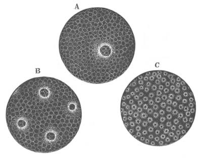
Fig. 51. Four successive stages in the
segmentation of the egg of Tetranychus Telarius. (After
Claparède.)
It is clear that it is not possible in centrolecithal ova to have any
type of segmentation exactly comparable with that of meroblastic ova.
There are however some types which fill the place of the meroblastic
ova in the present group, in as much as they are characterised by
the presence of a large bulk of food-yolk which either does not
segment, or does not do so till a very late stage in the
development. The essential character of this type of segmentation
consists in the division of the germinal vesicle in the interior, or
at the surface of the ovum into two, four, etc. nuclei (fig. 51).
These nuclei are each of them surrounded by a specially concentrated
layer of protoplasm (fig. 51) which is continuous with a
general protoplasmic reticulum passing through the ovum [not shewn in
fig. 51]. The yolk is contained in the meshes of this reticulum in the
manner already described for other ova.
The ovum, like that of Eupagurus before segmentation, is now a
syncytium. Eventually the nuclei, having increased by division and
become very numerous, travel, unless previously situated there, to the
surface of the ovum. They then either simultaneously or in succession
become, together with protoplasm around them, segmented off from the
yolk, and give rise to a peripheral blastoderm enclosing a central
yolk mass. In the latter however many of the nuclei usually remain,
and it also very often undergoes a secondary segmentation into a
number of yolk spheres.
The eggs of Insects afford numerous examples of this mode of
segmentation, of which the egg of Porthesia[60] may be taken as type.
After impregnation it consists of a central mass of yolk which passes
without a sharp line of demarcation into a peripheral layer of more
transparent (protoplasmic) material. In the earliest stage observed by
Bobretzky there were two bodies in the interior of the egg, each
consisting of a nucleus enclosed in a thin protoplasmic layer with
stellate prolongations. This stage corresponds with the division into
two, but though the nucleus divides, the preponderating amount of yolk
prevents the egg from segmenting at the same time. By a continuous
division of the nuclei there becomes scattered through the interior of
the ovum a series of bodies, each formed of nucleus and a thin layer
of protoplasm with reticulate processes. After a certain stage some of
these bodies pass to the surface, simultaneously (in Porthesia) or in
some cases successively. At the surface the protoplasm round each
nucleus contracts itself into a rounded cell body, distinctly cut off
from the adjacent yolk.
The cells so formed give rise to a superficial blastoderm of a single
layer of cells. Many of the nucleated bodies remain in the yolk, and
after a certain time, which varies in different forms, the yolk
becomes segmented up into a number of rounded or polygonal bodies, in
the interior of each of which one of the above nuclei with its
protoplasm is present. This process, known as the secondary
segmentation of the yolk, is really part of the true segmentation, and
the bodies to which it gives rise are true cells.
Other examples of this type may be cited. In Aphis[61] Metschnikoff
shewed that the first segmentation nucleus divides into two, each of
which takes up a position in the clearer peripheral protoplasmic layer
of the egg (fig. 52, 1 and 2). Following upon further division the
nuclei enveloped in a continuous layer of protoplasm arrange
themselves in a regular manner, and form a syncytium, which becomes
segmented into definite cells (fig. 52, 3 and 4). The existence of a
special clear superficial layer of protoplasm has been questioned by
Brandt.
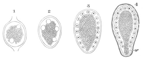
Fig. 52. Segmentation of Aphis Rosae.
(Copied from Metschnikoff.)
In all the stages there is seen to be a central yolk mass surrounded
by a layer of protoplasm.
In this protoplasm two nuclei have appeared in 1, four nuclei in 2.
In 3 the nuclei have arranged themselves regularly, and in 4 the
protoplasm has become divided into a number of columnar cells
corresponding to the nuclei.
w. pole of the blastoderm which has no share in forming the
embryo.
In Tetranychus telarius, one of the mites, Claparède found on
the surface of the ovum a nucleus surrounded by granular protoplasm
(fig. 51); which is no doubt the first segmentation nucleus. By a
series of divisions, all on the surface, a layer of cells becomes
formed round a central yolk mass. The result here is the same as in
Insects, but the nucleus with its granular protoplasm is from the
first superficial. In other cases, such as that of the common fly[62],
a layer of protoplasm is stated to appear investing the yolk; and in
this there arise simultaneously (?) a number of nuclei at regular
intervals, around each of which the protoplasm separates itself to
form a distinct cell. Closely allied is the type observed by
Kowalevsky in Apis. Development here commences by the appearance of a
number of protoplasmic prominences, each forming a cell
provided with a nucleus, the nuclei having no doubt been formed by
previous division in the interior of the ovum. They appear at the edge
of the yolk, and are separated from one another by short intervals.
Shortly after their appearance a second batch of similar bodies
appears, filling up the interspaces between the first-formed
prominences. In the fresh-water Gammarus fluviatilis the protoplasm is
stated first of all to collect at the centre of the ovum, where no
doubt the segmentation nucleus divides. Subsequently cells appear at
numerous points on the surface, and by repeated division constitute an
uniform blastoderm investing the central yolk mass. This mode of
formation of the blastoderm is closely allied to that observed by
Kowalevsky in Apis.
Between ova with a segmentation like that of Insects, and those with a
segmentation like that of Penæus, there is more than one intermediate
form. The Eupagurus type, with the division of the first nucleus into
eight nuclei before the division of the ovum, must be regarded in this
light; but the most instructive example of such a transitional type of
segmentation is that afforded by Spiders[63].
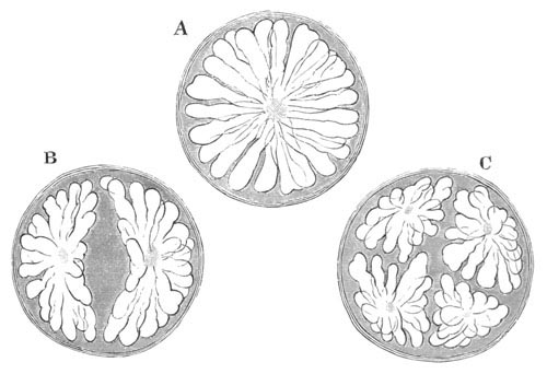
Fig. 53. Three stages in the segmentation of
Philodromus Limbatus.
(After Hub. Ludwig.)
The first phenomenon which can be observed after impregnation is the
conglomeration of the yolk spheres into cylindrical columns, which
finally assume a radiating form diverging from the centre of the egg.
In the centre of the radiate figure is a protoplasmic mass, probably
containing a nucleus, which sends out protoplasmic filaments
through the columns (fig. 53 A). After a certain period of repose the
figure becomes divided into two rosette-like masses, which remain
united for some time by a protoplasmic thread: this thread is finally
ruptured (fig. 53 B). The whole egg does not in this process divide
into two segments, but merely the radiate figure, which is enclosed in
a finely granular material. The two rosettes next become
simultaneously divided, giving rise to four rosettes (fig. 53 C): and
the whole process is repeated with the same rhythm as in a regular
segmentation till there are formed thirty-two rosettes in all (fig. 54
A). The rosettes by this time have become simple columns, which by
mutual pressure arrange themselves radiately around the centre of the
egg, which however they do not quite reach.
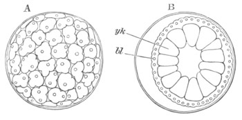
Fig. 54. Surface view and optical section of a
late stage in the segmentation of Philodromus Limbatus (Koch).
(After Hub. Ludwig.)
bl. blastoderm; yk. yolk spheres.
When only two rosettes are present the protoplasm with its nucleus
occupies a central position in each rosette, but gradually, in the
course of the subsequent subdivisions, it travels towards the
periphery, and finally occupies, when the stage with thirty-two
rosettes is reached, a peripheral position. The peripheral protoplasm
next becomes separated off as a nucleated layer (fig. 54 B). It forms
the proper blastoderm, and in it the nuclei rapidly multiply and
finally around each an hexagonal or polygonal area of protoplasm is
marked off; and a blastoderm, formed of a single layer of flattened
cells, is thus constituted. The columns within the blastoderm now form
(fig. 54 B) more or less distinct masses, which are stated by Ludwig
to be without protoplasm.
From observations of my own I am inclined to differ from Ludwig as to
the nature of the parts within the blastoderm. My observations have
been made on Agelena labyrinthica and commence at the close of
the segmentation. At this time I find a superficial layer of flattened
cells, and within these a number of large polyhedral yolk cells. In
many, and I believe all, of the yolk cells there is a nucleus
surrounded by protoplasm. It is generally placed at one side and not
in the centre of a yolk cell, and the nuclei are so often double that
I have no doubt they are rapidly undergoing division. It appears to me
probable that, at the time when the superficial layer of protoplasm is
segmented off from the yolk below, the nuclei undergo division, and
that a nucleus with surrounding protoplasm is left with each yolk
column. For further details vide Chapter on Arachnida.
Although by the close of the segmentation the protoplasm has travelled
to a superficial position, it may be noted that at first it forms a
small mass in the centre of the egg, and only eventually assumes its
peripheral situation. It is moreover clear that in the Spider’s ovum
there is, so to speak, an attempt at a complete segmentation, which
however only results in an arrangement of the constituents of the ovum
in masses round each nucleus, and not in a true division of the ovum
into distinct segments.
It seems very probable that Ludwig’s observations on the segmentation
of Spiders only hold good for species with comparatively small ova.
In connection with the segmentation of the Insects’ ovum and allied
types it should be mentioned that Bobretzky, to whose observations we
are largely indebted for our knowledge of this subject, holds somewhat
different views from those adopted in the text. He regards the nuclei
surrounded by protoplasm, which are produced by the division of the
primitive segmentation nucleus, as so many distinct cells. These cells
are supposed to move about freely in the yolk, which acts as a kind of
intercellular medium. This view does not commend itself to me. It is
opposed to my own observations on similar nuclei in the Spiders. It
does not fit in with our knowledge of the nature of the ovum, and it
cannot be reconciled with the segmentation of such types as Spiders or
even Eupagurus, with which the segmentation in Insects is undoubtedly
closely related.
The majority if not all the cases in which a central yolk mass is
formed occur in the Arthropoda, in which group centrolecithal ova are
undoubtedly in a majority. In Alcyonium palmatum the segmentation
appears however to resemble that of many insects.
One or two peculiar varieties in the segmentation of ova of this type
may be spoken of here. The first one I shall mention is detailed in
the important paper of E. Van Beneden and Bessels which I have already
so often had occasion to quote: it is characteristic of the eggs of
most of the species of Chondracanthus, a genus of parasitic
Crustaceans. The ovum divides in the usual way but somewhat
irregularly into 2, 4, 8 segments which meet in a central yolk mass;
but after the third division instead of each segment dividing into two
equal parts it divides at once into four, and the division into
four having started, reappears at every successive division. Thus the
number of the segments at successive periods is 2, 4, 8, 32, 128, etc.
In another peculiar case, an instance of which[64] is afforded by
Asellus aquaticus, after each of the earlier segmentations all
the segments fuse and become indistinguishable, but at the succeeding
segmentation double the number of segments appears.
Although, as has been already stated, it does not seem possible to
have a true meroblastic segmentation in centrolecithal ova, it does
nevertheless appear probable that the apparent cases of a meroblastic
segmentation in the Arthropoda are derivatives of this type of
segmentation. The manner in which the one type might pass into the
other may perhaps be explained by the segmentation in Asellus
aquaticus[65]. In this ovum large segments are at first formed
around a central yolk mass, in the peculiar manner mentioned in the
previous paragraph, but at the close of the first period of
segmentation minute cells, which eventually form a superficial
blastoderm, are produced from the yolk cells. They do not however
appear at once round the whole periphery of the egg, but at first only
on the ventral surface and later on the dorsal surface. If the amount
of food-yolk in the egg were to increase so as to render the formation
of the yolk cells impossible, and at the same time the formation of
the blastodermic cells were to take place at the commencement, instead
of towards the close of the segmentation, a mass of protoplasm with a
nucleus might first appear at the surface on the future ventral side
of the egg, then divide in the usual way for meroblastic ova, and give
rise to a layer of cells gradually extending round to the dorsal
surface. A meroblastic segmentation might perhaps be even more easily
derived from the type found in Insects. It is probable that the cases
of Scorpio, Mysis, Oniscus, the parasitic Isopoda, and some parasitic
Copepoda belong to this category; and it may be noticed that in these
cases the blastopore would be situated on the dorsal and not on the
ventral side of the ovum. The morphological importance of this latter
fact will appear in the sequel.
The results arrived at in the present section may be shortly restated
in the following way.
(1) A comparatively small number of ova contain very little or no
food-yolk embedded in their protoplasm; and have what food-yolk may be
present distributed uniformly. In such ova the segmentation is
regular. They may be described as alecithal ova.
(2) The distribution of food-yolk in the protoplasm of the ovum
exercises an important influence on the segmentation.
The rapidity with which any part of an ovum segments varies ceteris
paribus with the relative amount of protoplasm it contains; and
the size of the segments formed varies inversely to the relative
amount of protoplasm. When the proportion of protoplasm in any part of
an ovum becomes extremely small, segmentation does not occur in that
part.
Ova with food-yolk may be divided into two great groups according to
the eventual arrangement of the food-yolk in the protoplasm. In one of
these, the food-yolk when present is concentrated at the vegetative
pole of the ovum. In the other group it is concentrated at the centre
of the ovum. Ova belonging to the former group are known as
telolecithal ova, those to the latter as centrolecithal.
In each group more than one type may be distinguished. In the first
group these types are (1) unequal segmentation, (2) partial
segmentation. The features of these three types have been already so
fully explained that I need not repeat them here.
In the second group there are three distinct types, (1) equal
segmentation, (2) unequal segmentation. These two being externally
similar to the similarly named types in the first group. (3)
Superficial segmentation. This is unlike anything which is present in
the first group, and is characterized by the appearance of a
superficial layer of cells round a central yolk mass. These cells may
either appear simultaneously or successively, and their nuclei are
derived from the segmentation within the ovum of the first
segmentation nucleus.
The types of ova in relation to the characters of the segmentation may
be tabulated in the following way:
Segmentation.
(1) alecithal ova
regular
(2) telolecithal ova
(a) unequal
(b) partial
(3) centrolecithal ova
(a) regular (with segments united in central yolk mass)
(b) unequal (with segments united in central yolk mass)
(c) superficial.
Although the various types of segmentation which have been described
present very different aspects, they must nevertheless be looked on as
manifestations of the same inherited tendency to division, which
differ only according to the conditions under which the tendency
displays itself.
This tendency is probably to be regarded as the embryological
repetition of that phase in the evolution of the Metazoa, which
constituted the transition from the protozoon to the metazoon
condition.
From the facts narrated in this chapter the reader will have gathered
that similarity or dissimilarity of segmentation is no safe guide to
affinities. In many cases, it is true, a special type of segmentation
may characterize a whole group; but in other cases very closely allied
animals present the greatest differences with respect to their
segmentation; as for instance the different species of the genus
Gammarus. The character of the segmentation has great influence on the
early phenomena of development, though naturally none on the adult
form.
External Features of Segmentation.
(105) E. Haeckel. “Die Gastrula u. Eifurchung.” Jenaische
Zeitschrift, Vol. IX. 1877.
(106) Fr. Leydig. “Die Dotterfurchung nach ihrem Vorkommen in
d. Thierwelt u. n. ihrer Bedeutung.” Oken Isis. 1848.
SYSTEMATIC EMBRYOLOGY.
SYSTEMATIC EMBRYOLOGY.
Introduction.
In all the Metazoa the segmentation is followed by a series of changes
which result in the grouping of the embryonic cells into definite
layers, or membranes, known as the germinal layers. There are always
two of these layers, known as the epiblast and hypoblast; and in the
majority of instances a third layer, known as the mesoblast, becomes
interposed between them. It is by the further differentiation of the
germinal layers that the organs of the adult become built up. Owing to
this it is usual, in the language of Embryology, to speak of the
organs as derived from such or such a germinal layer.
At the close of the section of this work devoted to systematic
embryology, there is a discussion of the difficult questions which
arise as to the complete or partial homology of these layers
throughout the Metazoa, and as to the meaning to be attached to the
various processes by which they take their origin; but a few words as
to the general fate of the layers, and the general nature of the
processes by which they are formed, will not be out of place here.
Of the three layers the epiblast and hypoblast are to be regarded as
the primary. The epiblast is essentially the primitive integument, and
constitutes the protective and sensory layer. It gives rise to the
skin, cuticle, nervous system, and organs of special sense. The
hypoblast is essentially the digestive and secretory layer, and gives
rise to the epithelium lining the alimentary tract and the glands
connected with it.
The mesoblast is only found in a fully developed condition in the
forms more highly organized than the Cœlenterata. It gives origin
to the general connective tissue, internal skeleton, the muscular
system, the lining of the body cavity, the vascular, and excretory
systems. It probably in the first instance originated from
differentiations of the two primary layers, and in all groups with a
well-developed body cavity it is divided into two strata. One of them
forms part of the body wall and is known as the somatic mesoblast, the
other forms part of the wall of the viscera and is known as the
splanchnic mesoblast.
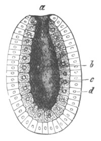
Fig. 55. Diagram of a Gastrula. (From
Gegenbaur.)
a. blastopore; b. archenteron; c. hypoblast;
d. epiblast.
A very large number not to say the majority of organs are derived from
parts of two of the germinal layers. Many glands for instance have a
lining of hypoblast which is coated by a mesoblastic layer.
The processes by which the germinal layers take their origin are
largely influenced by the character of the segmentation, which, as was
shewn in the last chapter, is mainly dependent on the distribution of
the food-yolk. When the segmentation is regular, and results in the
formation of a blastosphere, the epiblast and hypoblast are usually
differentiated from the uniform cells forming the wall of the
blastosphere in one of the two following ways.
(1) One-half of the blastosphere may be pushed in towards the other
half. A two-layered hemisphere is thus established which soon
elongates, while its opening narrows to a small pore (fig. 55). The
embryonic form produced by this process is known as a gastrula. The
process by which it originates is known as embolic invagination, or
shortly invagination. Of the two layers of which it is formed the
inner one (c) is known as the hypoblast and the outer
(d) as the epiblast, while the pore leading into its cavity
lined by the hypoblast is the blastopore (a). The cavity itself
is the archenteron (b).
(2) The cells of the blastosphere may divide themselves by a process
of concentric splitting into two layers (fig. 56, 3). The two layers
are as before the epiblast and hypoblast, and the process by
which they originate is known as delamination. The central cavity or
archenteron (F) is in the case of delamination the original
segmentation cavity; and not an entirely new cavity as in the case of
invagination. By the perforation of the closed two-walled vesicle
resulting from delamination an embryonic form is produced which cannot
be distinguished in structure from the gastrula produced by
invagination (fig. 56, 4). The opening (M) in this case is not
however known as the blastopore but as the mouth.
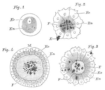
Fig. 56. Diagram shewing the formation of a
Gastrula by delamination.
(From Lankester.)
Fig. 1. Ovum.
Fig. 2. Stage in segmentation.
Fig. 3. Commencement of delamination after the appearance of a
central cavity.
Fig. 4. Delamination completed, mouth forming at M.
In fig. 1, 2 and 3 Ec. is ectoplasm, and En. is
entoplasm.
In fig. 4 Ec. is epiblast and En. hypoblast.
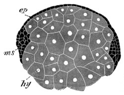
Fig. 57. Transverse section through the ovum of
Euaxes during an early stage of development. (After
Kowalevsky.)
ep. epiblast; ms. mesoblastic band; hy.
hypoblast.
When segmentation does not take place on the regular type the
processes above described are as a rule somewhat modified. The yolk is
usually concentrated in the cells which would, in the case of a simple
gastrula, be invaginated. As a consequence of this, these cells become
(1) distinctly marked off from the epiblast cells during the
segmentation; and (2) very much more bulky than the epiblast cells.
The bulkiness of the hypoblast cells necessitates a
modification of the normal process of embolic invagination, and causes
another process to be substituted for it, viz. the growth of the
epiblast cells as a thin layer over the hypoblast. This process (fig.
57) is known as epibolic invagination. The point where the complete
enclosure of the hypoblast cells is effected is known as the
blastopore. All intermediate conditions between epibolic and embolic
invagination have been found.
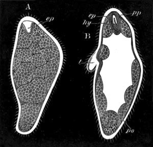
Fig. 58. Two stages in the development of
Stephanomia pictum.
(After Metschnikoff.)
A. Stage after the delamination. ep. epiblastic invagination
to form pneumatocyst.
B. Later stage after the formation of the gastric cavity in the
solid hypoblast. po. polypite; t. tentacle; pp.
pneumatophore; ep. epiblastic invagination to form
pneumatocyst; hy. hypoblast surrounding pneumatocyst.
In delamination, when the segmentation is not uniform, or when a solid
morula is formed, the differentiation of the epiblast and hypoblast is
effected by the separation of the central solid mass of cells from the
peripheral cells (fig. 58 A).
In the case of epibolic invagination as well as in that of the type of
delamination just spoken of, the archenteric cavity is in most cases
secondarily formed in the solid mass of hypoblast (fig. 58 B).
In ova with a partial segmentation there is usually some modification
of the epibolic gastrula.
Many varieties are found in the animal kingdom of the types of
invagination and delamination just characterized, and in not a few
forms the layers originate in a manner which cannot be brought into
connection with either of these processes.
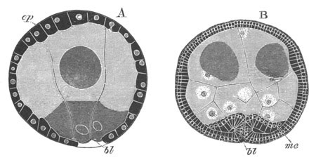
Fig. 59. Epibolic Gastrula of Bonellia.
(After Spengel.)
A. Stage when the four hypoblast cells are nearly enclosed.
B. Stage after the formation of the mesoblast has commenced by an
infolding of the lips of the blastopore.
ep. epiblast; me. mesoblast; bl. blastopore.
The mesoblast usually originates subsequently to the two primary
layers. It then springs from one or both of the other layers, but its
modes of origin are so various that it would be useless to attempt to
classify them here. In cases of invagination it often arises at the
lips of the blastopore (fig. 57 and 59), and in other cases part of it
springs as paired hollow outgrowths of the walls of the archenteron.
Such outgrowths are shewn in fig. 60, B and C at pv. The cavity
of the outgrowths forms the body cavity, and the walls of the
outgrowths the somatic and splanchnic layers of mesoblast (fig. C.
sp. and so.). The archenteron is in part always
converted into a section of the permanent alimentary tract and the
section of the alimentary tract so derived is known as the mesenteron.
There are however usually two additional parts of the alimentary
tract, known as the stomodaeum and proctodaeum, derived from
epiblastic invaginations. They give rise respectively to the oral and
anal extremities of the alimentary tract.
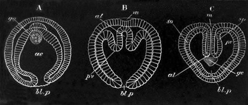
Fig. 60. Three stages in the development of
Sagitta. (A and C after Bütschli and B after Kowalevsky.) The
three embryos are represented in the same positions.
A. Represents the gastrula stage.
B. Represents a succeeding stage in which the primitive archenteron
is commencing to be divided into three parts, the two lateral of
which are destined to form the mesoblast.
C. Represents a later stage in which the mouth involution (m)
has become continuous with alimentary tract, and the blastopore has
become closed.
m. mouth; al. alimentary canal; ae.
archenteron; bl. p. blastopore; pv.
perivisceral cavity; sp. splanchnic mesoblast; so.
somatic mesoblast; ge. generative organs.
Bibliography.
(107) K. E. von Baer. “Ueb. Entwicklungsgeschichte d. Thiere.”
Königsberg, 1828‑1837.
(108) C. Claus. Grundzüge d. Zoologie. Marburg und
Leipzig, 1879.
(109) C. Gegenbaur. Grundriss d. vergleichenden
Anatomie. Leipzig, 1878. Vide also Translation. Elements
of Comparative Anatomy. Macmillan and Co., 1878.
(110) E. Haeckel. Studien z. Gastræa-Theorie. Jena,
1877, and also Jenaische Zeitschrift, Vols. VIII. and
IX.
(111) E. Haeckel. Schöpfungsgeschichte. Leipzig.
Vide also Translation. The History of Creation. King and
Co., London, 1876.
(112) E. Haeckel. Anthropogenie. Leipzig. Vide
also Translation. Anthropogeny (Translation). Kegan Paul and
Co., London, 1878.
(113) Th. H. Huxley. The Anatomy of Invertebrated
Animals. Churchill, 1877.
(114) E. R. Lankester. “Notes on Embryology and
Classification.” Quart. J. of. Micr. Science, Vol.
XVII. 1877.
(115) A. S. P. Packard. Life Histories of Animals, including
Man, or Outlines of Comparative Embryology. Holt and Co., New
York, 1876.
(116) H. Rathke. Abhandlungen z. Bildung und
Entwicklungsgesch. d. Menschen u. d. Thiere. Leipzig, 1833.
Dicyemidæ.
The structure and development of these remarkable parasites in the
renal organs of the Cephalopoda have recently been greatly elucidated
by the researches of E. van Beneden; and although a male element has
not been discovered, yet the embryos originate from bodies which have
a close similarity to ordinary ova.
Van Beneden has shewn that Dicyema consists in the adult state of (1)
a single layer of ciliated epiblast cells, somewhat modified
anteriorly to form a cephalic enlargement; and of (2) one large
nucleated hypoblast cell enclosed within the epiblast. There are two
kinds of embryo, both developed from germs which originate in the
hypoblast cell. The two kinds of embryo arise in individuals of
somewhat different forms. The one kind, called by Van Beneden the
vermiform embryo, arises in the more elongated and thinner examples of
Dicyema which have been named Nematogens. These embryos pass directly
into the parent form without metamorphosis.
The second kind of embryo, called infusoriform, is very different from
the parent, and has a free existence. Its eventual history is not
known. It originates in the shorter and thicker individuals of
Dicyema; which have been called Rhombogens.
The Vermiform Embryos. The germs or cells which give rise to
the vermiform embryos originate endogenously in the protoplasmic
reticulum of the axial hypoblast cell. They appear as small but
well-defined spheres, with a minute body in the centre. In
these spheres a cortical layer becomes differentiated, which gradually
increases in thickness and gives rise to the body of a cell, the
nucleus and nucleolus of which are respectively formed from the inner
part of the original sphere and the minute central body. These germs
can originate in all parts of the hypoblast cell and are frequently
very numerous.
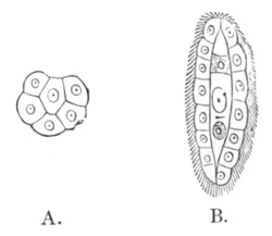
Fig. 61. A. Gastrula stage of Dicyema typus. B.
Veriform embryo of Dicyema typus. (From Gegenbaur, after E. van
Beneden.)
The germ when completely formed undergoes a segmentation very similar
to that of an ordinary ovum. It divides first into two and then into
four approximately equal segments. Of the four segments one, however,
remains passive for the remainder of the development. The other three
divide and arrange themselves so as partially to enclose in a cup-like
fashion the passive cell (fig. 61 A). The six cells resulting from
their division again divide, giving rise to twelve cells, which nearly
enclose the passive cell, leaving only a small aperture at one point.
The whole process by which the central cell becomes enclosed is, as E.
van Beneden points out, identical with a gastrula formation by
epibole, and the space where the central cell is left uncovered is the
blastopore. The central cell itself gives origin to the hypoblast cell
of the adult, and the peripheral cells to the epiblast.
By this time the embryo has assumed an oval form, and the blastopore
is situated at the pole of the long axis of the oval where the
cephalic enlargement is eventually formed.
The subsequent development consists mainly in the closure of the
blastopore, and an increase in the number of the epiblast cells.
Before the development is completed, and while the embryo is still in
the body of the parent, two germs, destined themselves to give rise to
fresh embryos, appear in the hypoblast cell, one on each side of the
nucleus (fig. 61 B). The embryo continues to elongate, while the
anterior cells become converted into the polar cells. Cilia appear
simultaneously over the general surface, and the embryo makes its way
out of the body of the parent, usually at the cephalic pole, and
becomes itself parasitic in the renal organ of the host in which it
finds itself. At the time of birth the embryo may contain a
number of germs and sometimes even developing embryos.
Infusoriform Embryos. The infusoriform embryos are capable of
living in sea-water and almost certainly lead a free existence. In
their most fully developed condition so far known they have the
following rather complicated structure (fig. 62 D, E, F, G).
The body is somewhat pyriform, with a blunt extremity which is
directed forwards in swimming, and a more pointed extremity directed
backwards. The former may be spoken of as the anterior, and the latter
as the posterior extremity or tail. At the anterior extremity are
situated a pair of refractive bodies (r) which lie above an
unpaired organ which may be called the urn.
The structure of the urn, the refractive bodies, and the tail may be
dealt with in succession.
The urn consists of three parts: (1) a wall (u), (2) a lid
(l), and (3) contents (gr). The wall of the urn is
hemispherical in form, and composed of two halves in apposition (fig.
F). Its concavity is directed forwards, and in its edge are imbedded a
number of rod-like corpuscles which appear as a ring near the surface
in a full-face view (fig. D). The lid has the form of a low pyramid
with its apex directed outwards. It is made up of four segments (fig.
D). The contents of the urn, which completely fill up its cavity, are
four polynuclear cells arranged in the form of a cross which appear
with low powers as granular bodies (fig. F). They are frequently
ejected, apparently at the will of the embryo.
The refractive bodies (r), two in number, one on each side of
the middle line, are composed of a material which is not of a fatty
nature, and which is passive to the majority of reagents. Each is
enveloped in a special capsule, and at times more than one refractive
body is present in each capsule. The tail is a conical structure
formed of ciliated granular cells.
No plausible guess has been made as to the function either of the urn
or of the refractive bodies.
The infusoriform embryos originate from germs, which have however a
different origin to the germs of the vermiform embryos. One to five
cells appear in the axial hypoblast cell, in a way not clearly
made out, and each of them gives rise by an endogenous process to
several generations of cells, all of which develop into infusoriform
embryos.
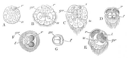
Fig. 62. Infusoriform embryo of Dicyema.
A. B. C. Three of the later stages in the development.
D. E. F. Three different views of the full-grown larva. D. from the
front, E. from the side, and F. from above.
G. side view of urn.
u. wall of urn; l. lid of urn; r. refractive
bodies; gr. granular bodies filling the interior of the urn.
The primitive cell is called by Van Beneden a Germogen. In its
protoplasm a number of germs first appear endogenously, but the
nucleus of the germogen does not assist in their formation. They
eventually become detached from the parent cell, around which they are
concentrically arranged. A second and then a third generation of germs
are formed in the same way, till the whole of the protoplasm of the
primitive cell is absorbed in the formation of these germs, and
nothing of it remains but the nucleus. The germs so formed are
arranged in about three concentric layers, of which the innermost is
the youngest. One to five masses of germs may be present in a single
Rhombogen. The germs undergo a division, in the course of which their
nuclei exhibit very beautifully a spindle modification. In the course
of the segmentation the embryo gradually assumes its permanent form,
and four of the cells composing it can be distinguished from the
remainder by their greater size (fig. 62 A, u). The two largest
of these give rise to the wall of the urn, and also give origin to
four smaller cells (fig. 62 B, gr) which eventually become
polynuclear and constitute the four granular cells in the urn. The two
other cells become the lid of the urn. The parts of the urn
lie at first side by side, but in the course of development the cells
which form the wall of the urn travel inwards, and the four granular
cells are carried into their concavity. At the same time the cells
which form the lid of the urn alter their position so as to overlie
the wall of the urn. The two cells immediately above the urn give rise
to the refractive bodies (fig. 62 A, B, C, r) and the remainder
of the cells of the embryo become the tail (fig. 62 C). The embryo
becomes ciliated, and attains its nearly full development before
leaving the parental tissues. It usually passes out at the cephalic
extremity.
As has already been stated, it is probable that the infusoriform
embryos leave the renal organs of their host and lead a free
existence. What becomes of them afterwards is not however known,
though there can be little doubt that they serve to carry the species
to new hosts.
Till the further development of the infusoriform embryo is known it is
not possible to arrive at a definite conclusion as to the affinities
of this strange parasite. Van Beneden is anxious to form it, on
account of its simple organization, into a group between the Protozoa
and the Metazoa. It appears however very possible that the simplicity
of its organization is the result of a parasitic existence; a view
which receives confirmation from the common occurrence of the process
of endogenous cell formation in the axial hypoblast cell. It has been
clearly shewn by Strasburger that endogenous cell formation is
secondarily derived from cell division; so that the occurrence of this
process in Dicyema probably indicates that the hypoblast was
primitively multicellular. It is not improbable that the enigmatical
infusoriform embryo may develop into a sexual form, the progeny of
which are destined to complete the cycle of development by becoming
again parasitic in the renal organ of a Cephalopod.
Bibliography.
(117) E. van Beneden. “Recherches sur les Dicyemides.” Bull.
d. l’Académie roy. de Belgique, 2e sér. T. XLI. No. 6
and T. XLII. No. 7, 1876. Vide this paper for a full
account of the literature.
(118) A. Kölliker. Ueber Dicyema paradoxum den Schmarotzer
der Venenanhänge der Cephalopoden.
(119) Aug. Krohn. “Ueb. d. Vorkommen von Entozoen, etc.”
Froriep Notizen, VII. 1839.
Orthonectidæ.
A number of minute parasites infesting various Nemertines,
Turbellarians, and Ophiuroids have recently been studied by Giard and
Metschnikoff, the former of whom has placed them in a special group
which he calls the Orthonectidæ. They were first discovered by W. C.
McIntosh.
In the adult state they are[66] (Metschnikoff) somewhat pear-shaped
bodies formed of a kind of plasmodium of cells with irregular lobate
processes. In the interior of this body are eggs in all stages of
development. In the type observed by Metschnikoff (Intoshia gigas) the
ova undergo a regular segmentation, resulting in the formation of a
blastosphere in which an inner layer is subsequently formed by
delamination. A smaller and a larger kind of embryo are formed; but
all the embryos in each female belong to one type. The larger become
females and the smaller males.
The female embryos are ovoid. The outer layer of cells or epiblast
becomes ciliated, and divided into nine segments, of which the second
is marked off from the remainder by the absence of cilia, and by being
provided with refractive corpuscles. The inner layer which surrounds a
central cavity, and might be supposed to be the hypoblast, becomes
according to Metschnikoff converted into ova.
The male embryos are more elongated than the female, from which they
further differ in only having six segments. The cells of the inner
layer eventually divide up into spermatozoa.
The larvæ probably become free, and while in the free state
impregnation would appear to be effected. When the female larvæ become
parasitic they undergo a metamorphosis, the stages of which have not
been observed; but in the course of which the epiblast cells probably
unite into a plasmodium.
The observations of Giard are in several points irreconcilable with
those of Metschnikoff, but from the statements of the latter it
appears possible that Giard has made two genera from the males and
females of one species; and that Giard’s account of an unequal
segmentation followed by an epibolic gastrula, in one of his species,
has arisen from two segmenting ova temporarily fusing together. Giard
has given a description of internal gemmiparous reproduction, upon the
accuracy of which doubts have been thrown by Metschnikoff. The
affinities of the Orthonectidæ are as obscure as those of the
Dicyemidæ; though there can be but little doubt that their
organization has been much simplified in correlation with their
parasitic habits. The origin of the genital products in the axial
tissue is a feature they have in common with the Dicyemidæ.
Bibliography.
(120) Alf. Giard. “Les Orthonectida classe nouv. d. Phylum des
Vers.” Journal de l’Anat. et de la Physiol., Vol. XV.
1879.
(121) El. Metschnikoff. “Zur Naturgeschichte d. Orthonectidæ.”
Zoologischer Anzeiger, No. 40‑43, 1879.
[Ch. Julin. “Rech. sur l’organization et le devel.
d’Orthonectides.” Arch. Biol. Vol. III. 1882.
E. Metschnikoff. “Untersuchungen üb. Orthonectidæ.” Zeit. f.
Wiss. Zoologie, Vol. XXXV. 1881.
For general account of Orthonectidæ, vide Spengel. Biolog.
Centralblatt, No. 6.]
Although within the last few years greater advances have probably been
made in our knowledge of the development of the Porifera than of any
other group, yet there is much that is still very obscure, and it is
not possible to make general statements applying to the whole group.
Calcispongiæ. The form which has so far been most completely
worked out is Sycandra raphanus, one of the Calcispongiæ
(Metschnikoff, Nos. 132 and 134,
F. E. Schulze, Nos. 139 and 142), and
I shall commence my account with the life history of this species.
The ovum in Sycandra as in other Spongida has the form of a naked
amœboid nucleated mass of protoplasm. From the analogy of the other
members of the group, there is no doubt that it is fertilized by a
male spermatic element, though this has not as yet been shewn to be
the case—and the changes which accompany fertilization are quite
unknown.
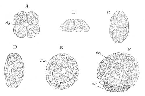
Fig. 63. Successive stages in the segmentation of
Sycandra raphanus.
(Copied from F. E. Schulze.)
A. stage with eight segments still arranged in pairs, from above.
B. side view of stage with eight segments.
C. side view of stage with sixteen segments.
D. side view of stage with forty-eight segments.
E. view from above of stage with forty-eight segments.
F. side view of embryo in the blastosphere stage, eight of the
granular cells which give rise to the epiblast of the adult are
present at the lower pole.
cs. segmentation cavity; ec. granular cells which form
the epiblast; en. clear cells which form the hypoblast.
The segmentation and early stages of development take place in the
tissues of the parent. The segmentation is somewhat peculiar, though a
modification of a regular segmentation. The ovum divides along a
vertical plane, first into two, and then into four equal segments. But
even when two segments are formed, each of them has one end pointed
and the other broader. The pointed ends give rise to the ciliated
cells of the future larva, and the broad ends to the granular cells.
Instead of the next division taking place, as is usually the case, in
a horizontal (equatorial) plane, it is actually effected along two
vertical planes intermediate in position between the two first
planes of segmentation. Eight equal segments are thus formed, each of
which has the form of a pyramid. All the segments are situated in a
single tier, and are so arranged as to give to the whole ovum the form
of a flat cone, the apex of which is formed by the pointed extremities
of the constituent segments (fig. 63 B). The apices of the segments do
not however quite meet, but they leave a central space, which is an
actual perforation (fig. 63 A) through the axis of the ovum, open at
both ends. The first indications of this perforation appear when only
four segments are present, and it is to be regarded as the homologue
of the segmentation cavity of other ova. The next plane of division is
horizontal (equatorial), and the apices of the eight cells are
segmented off as a tier of small cells. At the completion of this
division (fig. 63 C), the ovum is formed of sixteen cells arranged in
two superimposed tiers. The ovum now assumes somewhat the form of a
biconvex lens, in the axis of which the central perforation is still
present. At the close of the next stage, forty-eight cells are
present arranged in four tiers (fig. 63 D and E), the two outer tiers
containing eight cells each, and the two inner sixteen. The two inner
tiers probably arise by the simultaneous appearance of two equatorial
furrows dividing the original tiers into two, and by the subsequent
simple division of the cells of the two inner of the tiers so formed.
At the close of the stage the eight basal cells become granular (fig.
63 F). At the same time the central part of the segmentation cavity
becomes enlarged, while its terminal apertures become narrowed and
finally, shortly after the end of this stage, closed. The axial
perforation thus acquires the character of a closed segmentation
cavity. While the ovum itself becomes at the same time a blastosphere.
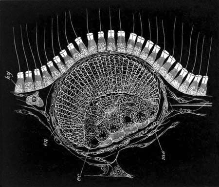
Fig. 64. Larva of Sycandra raphanus at
pseudogastrula stage, in situ in the maternal tissues. (Copied
from F. E. Schulze.)
me. mesoblast of adult; hy. collared cells forming
hypoblast of the adult; en. clear cells of larva which
eventually become involuted to form the hypoblast; ec.
granular cells of larva which give rise to the epiblast, which at
this stage are partially involuted.
This stage nearly completes the segmentation: in the next one, the
cells of the poles of the blastosphere increase in number, and
the cells of the greater part of the blastosphere become columnar and
ciliated, (fig. 64 en.) while the granular cells (ec.)
increase to about thirty-two in number and appear to be (partially at
least) involuted into the segmentation cavity, reducing this latter to
a mere slit. This stage forms the last passed by the embryo in the
tissues of the parent. The general position of the embryo while still
in this situation may be gathered from fig. 64, representing the
embryo in situ. The embryo is always placed close to one of the
radial canals. From this situation it makes its way through the lining
cells into a canal and is thence transported to the surrounding water.
By the time the larva has become free, the semi-invaginated granular
cells have increased in bulk and become everted so as to project very
much more prominently than in the encapsuled state. To the gastrula
stage, if it deserves the name, passed through by the embryo in the
tissues of the parent, no importance can be attached.
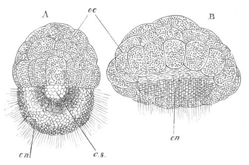
Fig. 65. Two free stages in the development of
Sycandra raphanus.
(Copied from Schulze.)
A. Amphiblastula stage.
B. A later stage after the ciliated cells have commenced to become
invaginated.
cs. segmentation cavity; ec. granular cells which will
form the epiblast; en. ciliated cells which become
invaginated to form the hypoblast.
The larva, after it has left the parental tissues, has an oval form
and is transversely divided into two areas (fig. 65 A). One of these
areas is formed of the elongated, clear, ciliated cells, with a small
amount of pigment near their inner ends (en.), and the
other and larger area of the thirty-two granular cells already
mentioned (ec.). Fifteen or sixteen of these are arranged as a
special ring on the border of the clear cells. In the centre of the
embryo is a segmentation cavity (c.s.) which lies between the
granular and the clear cells, but is mainly bounded by the vaulted
inner surface of the latter. This stage is known as the
amphiblastula stage. During the later periods of the
amphiblastula stage a cavity appears in the granular cells dividing
them into two layers. After the larva has for some time enjoyed a free
existence, a remarkable series of changes take place, which result in
the invagination of the half of it formed of the clear cells, and form
a prelude to the permanent attachment of the larva. The entire process
of invagination is completed in about half an hour. The whole embryo
first becomes flattened, but especially the ciliated half, which
gradually becomes less prominent (fig. 65 B); and still later the
cells composing it undergo a true process of invagination. As a result
of this invagination the segmentation cavity is obliterated, and the
larva assumes a compressed plano-convex form, with a central gastrula
cavity, and a blastopore in the middle of the flattened surface. The
two layers of the gastrula may now be spoken of as epiblast and
hypoblast. The blastopore becomes gradually narrowed by the growth
over it of the outer row of granular cells. When it has become very
small the attachment of the larva takes place by the flat surface
where the blastopore is situated. It is effected by protoplasmic
processes of the outer ring of epiblast cells, which, together with
the other epiblast cells, now become amœboid. They become
at the same time clearer and permit a view of the interior of the
gastrula. Between the epiblast cells and the hypoblast cells which
line the gastrula cavity there arises a hyaline structureless layer,
which is more closely attached to the epiblast than to the hypoblast,
and is probably derived from the former. A view of the gastrula stage
after the larva has become fixed is given in fig. 66.
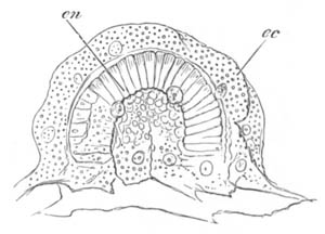
Fig. 66. Fixed Gastrula stage of Sycandra
raphanus. (Copied from Schulze.)
The figure shews the amœboid epiblast cells (ec.) derived
from the granular cells of the earlier stage, and the columnar
hypoblast cells, lining the gastrula cavity, derived from the
ciliated cells of the earlier stage. The larva is fixed by the
amœboid cells on the side on which the blastopore is situated.
There would seem according to Metschnikoff’s observations (No. 134) to
be a number of mesoblast cells interposed between the two primary
layers, which he derives from the inner part of the mass of granular
cells.
After invagination the cilia of the hypoblast cells can no longer be
seen, and are probably absorbed; and their disappearance is nearly
coincident with the complete obliteration of the blastopore, an event
which takes place shortly after the attachment of the larva.
Not long after the closure of the blastopore, calcareous spicules make
their appearance in the larva as delicate unbranched rods pointed at
both extremities. They appear to be formed on the mesoblast cells
situated between the epiblast and hypoblast[67]. The larva when once
fixed rapidly grows in length and assumes a cylindrical form (fig. 67
A). The sides of the cylinder are beset with calcareous spicules which
project beyond the surface, and, in addition to the unbranched forms,
spicules are developed with three and four rays as well as some with a
blunt extremity and serrated edge. The extremity of the cylinder
opposite the attached surface is flattened, and, though surrounded by
a ring of four-rayed spicules, is itself free from them. At this
extremity a small perforation is formed leading into the gastric
cavity, which rapidly increases in size and forms an exhalent osculum
(os.). A series of inhalent apertures is also formed at the
sides of the cylinder. The relative times of appearance of the single
osculum and the smaller apertures are not constant for the different
larvæ. On the central gastrula cavity of the sponge becoming placed in
communication with the external water, the hypoblast cells lining it
become ciliated afresh (fig. 67 B, en.) and develop the
peculiar collar characteristic of the hypoblast cells of the Spongida
(vide fig. 64, hy.). When this stage of development is
reached we have a fully formed sponge of the type made known by
Haeckel as Olynthus.
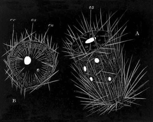
Fig. 67. The young of Sycandra raphanus shortly
after the development of the Spicula. (Copied from Schulze.)
A. View from the side.
B. View from the free extremity.
os. osculum; ec. epiblast; en. hypoblast
composed of ciliated cells. The terminal osculum and lateral pores
are represented as oval white spaces.
When young examples of Sycandra come in contact shortly after their
attachment they appear to fuse together temporarily or else
permanently. In the latter case colonies are produced by their fusion.
Amongst other calcareous sponges the larva of Ascandra contorta
(Haeckel No. 126, Barrois No. 122) presents the typical amphiblastula
stage, and so probably does that of Ascandra Lieberkühnii
(Keller No. 128). In Leucandra aspera (Keller No. 128,
Metschnikoff No. 134) the larva passes through an amphiblastula stage,
but the characters of the cells of the two halves of the larva do not
differ to nearly the same extent as in Sycandra.
Although the majority of calcareous sponges appear to agree in their
mode of development with Sycandra, nevertheless the concordant
researches of O. Schmidt (No. 138) and Metschnikoff (No. 134) have
shewn that this is not true for the genus Ascetta (As.
primordialis, clathrus and blanca).
The larvæ of these forms are very differently constituted to those of
Sycandra. They have an oval form and are composed of a single row of
ciliated columnar cells: their two extremities only differ in the
cells at one extremity being longer than those at the other.
Especially at the pole where the shorter cells are situated (Schmidt)
a metamorphosis of the cells takes place. One after the other they
lose their cilia, become granular, and pass into the interior of the
vesicle. Here they become differentiated into two classes
(Metschnikoff); one of larger and more granular cells, and the other
of smaller cells with clearer protoplasm. Cells of the former class
are mainly found at one of the poles. When the larva becomes free the
cells in the interior of the vesicle increase in number and nearly
fill up its central cavity. After a short free existence the larva
becomes fixed, and the epiblast cells lose their cilia and become
flattened. At a later period the large granular cells assume a radiate
arrangement round a central cavity and become clearly marked out as
the hypoblast cells. The smaller cells become placed between the
epiblast and hypoblast and constitute the mesoblast.
Myxospongiæ. In this group Halisarca has been investigated by
Carter (No. 123), Barrois (No. 122), Schulze (No. 141) and
Metschnikoff (No. 134). The ova develop in the mesoblast, and when
ripe occupy special chambers lined by a layer of epithelial cells.
Schulze has found the spermatozoa of this genus of sponge and has been
able to shew that the sexes may be distinct, though many species of
Halisarca are hermaphrodite.
The segmentation is, roughly speaking, regular, and a segmentation
cavity is early formed, which is never, as in Calcispongiæ, open at
the poles. When the larva leaves the parent it is an oval vesicle
formed of a single layer of columnar ciliated cells. Slight
differences may be observed between the two extremities of the larvæ
of most species. One of these—the hinder extremity—is directed
backwards in swimming.
The further history of the larva has been investigated by
Metschnikoff. He has found that the interior of the vesicle becomes
gradually filled with mesoblast cells of a peculiar type, called by
him rosette-cells, which are probably derived from the walls of the
vesicle.
When the metamorphosis commences, the larva assumes a flattened form,
and cells of a new type, viz. normal amœboid cells, grow in
amongst the rosette cells. The new cells are also derived from the
epiblast. The larvæ appear to fix themselves by the hinder extremity.
The cilia gradually disappear, and the epiblast cells flatten out and
form a kind of cuticle. For some time the larva remains in the
two-layered condition, but gradually canals (? ciliated chambers)
lined by hypoblast cells become formed. They appear as closed spaces
with walls of ciliated cells derived from the amœboid cells, and
the different parts of the system of chambers are established
independently. In H. pontica the ciliated chambers are formed
before the attachment of the larva. The development was not followed
up to the formation of the pores placing the canal system in
communication with the exterior.
The young sponges at a somewhat later stage have been studied by
Schulze and Barrois. They are formed of an external layer of flattened
cells, not clearly ciliated as in the adult, within which are a normal
mesoblastic tissue, and several spherical chambers lined by ciliated
cells exactly like the ciliated chambers of the full-grown sponge.
Irregular invaginations of the epiblast give to the young sponge a
honeycombed structure. The ciliated chambers in the youngest condition
of the sponge are closed; but in slightly older examples they come
into communication with the passages lined by hypoblast, and so
indirectly with the external medium.
Ceratospongiæ. Amongst the true Ceratospongiæ the embryos of
two of the Aplysinidæ, and of Spongelia and Euspongia have been to
some extent worked out by Barrois and Schulze. The form worked out by
Barrois is called by him Verongia rosea. The segmentation is
nearly regular, but from the first the segments may be divided
according to their constitution into two categories. At the close of
segmentation the embryo is oval and covered by a single layer of
columnar ciliated cells; these cells may however be divided into two
categories, corresponding with those observable during the
segmentation. A certain number are coloured red and form a definite
circular mass at one pole, while the remainder, which constitute the
major part of the embryo, have a pale yellowish colour. Those at the
red pole lose their cilia in the free larva, but around the area
formed by them is a special ring of long cilia. The chief peculiarity
of the embryo (made known by Schulze) consists in the fact that the
layer of cells which covers the embryo does not, as in other sponge
embryos, simply enclose a space, but the interior of the embryo is
formed of a mass of stellate cells like the normal mesoblast of
full-grown sponges.
This feature is also characteristic of the embryos of Spongelia and
Euspongia.
The embryo of the Gummineæ (Gummina mimosa) has been
investigated by Barrois (No. 122), and has been shewn closely to
resemble the typical larvæ of calcareous sponges; one-half being
formed of elongated ciliated cells and the other of rounded
granular ones.
Silicispongiæ. The development of marine silicious sponges is
but very imperfectly understood. The larvæ of various forms—Reniera
(Isodyctia), Esperia (Desmacidon), Raspailia, Halichondria,
Tethya—have been described. Barrois has shewn that the egg segments
regularly and that in the earlier stages a segmentation cavity is
present. In the later stages the embryo appears to become solid.
Externally there is a layer of ciliated cells, and within a mass of
granular matter in which the separate cells cannot be made out. The
granular matter projects at one pole, and forms a prominence possibly
equivalent to the granular cells of Sycandra. In some forms,
e.g. Reniera, the edge of the unciliated granular prominence
may be surrounded by a row of long cilia. In later stages the granular
material may project at both poles or even at other points. One
remarkable feature in the development of the Silicispongiæ is the
appearance of spicula between the ciliated cells and the central mass,
while the larva is still free.
Professor Schulze has informed me that these spicula are developed in
mesoblast cells; while the horny fibres of the sponge are developed as
cuticular products of special mesoblast cells (spongioblasts).
The attachment and accompanying metamorphosis are so diversely
described that no satisfactory account can be given of them. The
general statements are in favour of the attachment taking place by the
posterior extremity where the granular matter projects.
Carter especially gives a very precise account, with figures, of the
attachment of the larva in this way. He also figures the appearance of
an osculum at the opposite pole[68].
A very elaborate account of the development of Spongilla has been
published in Russian by Ganin, of which a German abstract has also
appeared (No. 124).
The ovum undergoes a regular segmentation and becomes a solid ova
morula. An epiblast of smaller cells is early differentiated, and in
the interior of the inner cells an archenteron becomes subsequently
formed. The inner cells next become divided into an hypoblastic layer
lining the archenteron, and a mesoblastic layer between this
and the now ciliated epiblast. At the narrow hinder end of the embryo
the mesoblast becomes thickened, and largely obliterates the
archenteron. In this part of the mesoblast silicious spicula are
formed. The larva becomes attached by its hinder extremity, and in the
course of this process flattens itself out to a disc-like form. From
the nearly obliterated archenteric cavity outgrowths take place which
give rise to the ciliated chambers. These are not placed directly in
communication with the exterior, but open, if I understand Ganin
rightly, into a space in the mesoblast, which subsequently acquires an
exterior communication—the primitive osculum. The subsequent pores
and oscula are also formed as openings leading into the mesoblastic
cavity, which communicates in its turn with the ciliated chambers.
It appears that in the present unsatisfactory state of our knowledge
the larvæ of the Porifera may be divided into two groups: viz. (1)
those which have the form of a blastosphere or else of a solid morula;
(2) those which have the amphiblastula form.
In the former type the mesoblast and hypoblast are formed either from
cells budded off from the outer cells of the blastosphere or from the
solid inner mass of cells; while the outer ciliated cells become the
epiblast. This type of larva, which is found in the majority of
sponges, is very similar in its general characters and development to
many Cœlenterate planulæ.
The second type of larva is very peculiar, and though in its fully
developed form it is confined to the Calcispongiæ, where it is the
usual form, a larval type with the same characters is perhaps to be
found in other sponges, e.g. amongst the Gumminæ, and amongst
the Silicispongiæ where one-half of the embryo is without cilia,
though in the case of the Silicispongiæ the cells of the ciliated part
of the embryo correspond to the granular cells of the larva of
Sycandra.
The later stages in the development of the larvæ of the Porifera are
not similar to anything we know of in other groups.
It might perhaps be possible to regard sponges as degraded descendants
of some Actinozoon type such as Alcyonium, with branched prolongations
of the gastric cavity, but there does not appear to me to be
sufficient evidence for doing so at present. I should rather prefer to
regard them as an independent stock of the Metazoa.
In this connection the amphiblastula larva presents some points of
interest. Does this larva retain the characters of an ancestral type
of the Spongida, and if so, what does its form mean? It is, of course,
possible that it has no ancestral meaning but has been
secondarily acquired; but, assuming that this is not the case, it
appears to me that the characters of the larva may be plausibly
explained by regarding it as a transitional form between the Protozoa
and Metazoa. According to this view the larva is to be considered as a
colony of Protozoa, one-half of the individuals of which have become
differentiated into nutritive forms, and the other half into locomotor
and respiratory forms. The granular amœboid cells represent the
nutritive forms, and the ciliated cells represent the locomotor and
respiratory forms. That the passage from the Protozoa to the Metazoa
may have been effected by such a differentiation is not improbable on
a priori grounds.
While the above view seems fairly satisfactory for the free-swimming
stage of the larval sponge, there arises in the subsequent development
a difficulty which appears at first sight fatal to it. This difficulty
is the invagination of the ciliated cells instead of the granular
ones. If the granular cells represent the nutritive individuals of the
colony, they, and not the ciliated cells, ought most certainly to give
rise to the lining of the gastrula cavity, according to the generally
accepted views of the morphology of the Spongida. The suggestion which
I would venture to put forward in explanation of this paradox involves
a completely new view of the nature and functions of the germinal
layers of adult Spongida.
It is as follows:—When the free-swimming ancestor of the Spongida
became fixed, the ciliated cells by which its movements used to be
effected must have to a great extent become functionless. At the same
time the amœboid nutritive cells would need to expose as large a
surface as possible. In these two considerations there may, perhaps,
be found a sufficient explanation of the invagination of the ciliated
cells, and the growth of the amœboid cells over them. Though
respiration was, no doubt, mainly effected by the ciliated cells, it
is improbable that it was completely localized in them, but they were
enabled to continue performing this function through the formation of
an osculum and pores. The collared cells which line the ciliated
chambers, or in some cases the radial tubes, are undoubtedly derived
from the invaginated cells, and, if there is any truth in the above
suggestion, the collared cells in the adult sponge must be mainly
respiratory and not digestive in function, while the epiblastic cells,
which in most cases line the inhalent passages through its
substance[69], ought to be employed to absorb nutriment. The recent
researches of Metschnikoff (No. 134) on this head shew that the
nutriment is largely carried into the mesoblast cells, which in
Sycandra appear to be derived from the granular cells, and
also that it is taken up by the cells which line the passages, though
not by the superficial epiblast cells. Whether the collared cells
generally absorb nutriment is not clear from his statements: but he
finds that they do not do so in Silicispongiæ.
Professor Schulze has informed me by letter that he finds the collared
cells to be respiratory in function, while the cells derived from the
granular cells in Sycandra are nutritive. Carter[70], on the contrary,
from his observations on Spongilla, has fully satisfied himself that
the food is absorbed by the cells lining the ciliated chambers.
If it is eventually proved by further experiments on the nutrition of
sponges, that digestion is mainly carried on by the general cells
lining the passages and the mesoblast cells, and not for the most part
by the ciliated cells, it is clear that the epiblast, mesoblast and
hypoblast of sponges will not correspond with the similarly named
layers in the Cœlenterata and other Metazoa. The invaginated
hypoblast will be the respiratory layer and the epiblast and mesoblast
the digestive and sensory layers; the sensory function being probably
mainly localized in the epithelium on the surface, and the digestive
one in the epithelium lining the passages and in the mesoblast. Such a
fundamental difference in the primary function of the germinal layers
between the Spongida and the other Metazoa, would necessarily involve
the creation of a special division of the Metazoa for the reception of
the former group.
Bibliography.
(122) C. Barrois. “Embryologie de quelques éponges de la
Manche.” Annales des Sc. Nat. Zool., VI. ser., Vol.
III. 1876.
(123) Carter. “Development of the Marine Sponges.” Annals
and Mag. of Nat. Hist., 4th series, Vol. XIV. 1874.
(124) Ganin[71]. “Zur Entwicklung d. Spongilla fluviatilis.”
Zoologischer Anzeiger. Vol. I. No. 9, 1878.
(125) Robert Grant. “Observations and Experiments on the
Structure and Functions of the Sponge.” Edinburgh Phil. J.,
Vol. XIII. and XIV., 1825, 1826.
(126) E. Haeckel. Die Kalkschwämme, 1872.
(127) E. Haeckel. Studien zur Gastræa-Theorie. Jena,
1877.
(128) C. Keller. Untersuchungen über Anatomie und
Entwicklungsgeschichte einiger Spongien. Basel, 1876.
(129) C. Keller. “Studien üb. Organisation u. Entwick. d.
Chalineen.” Zeit. f. wiss. Zool., Bd. XXVIII. 1879.
(130) Lieberkühn. “Beitr. z. Entwick. d. Spongillen.” Müller’s
Archiv, 1856.
(131) Lieberkühn. “Neue Beiträge zur Anatomie der Spongien.”
Müller’s Archiv, 1859.
(132) El. Metschnikoff.
“Zur Entwicklungsgeschichte der Kalkschwämme.” Zeit. f. wiss.
Zool., Bd. XXIV. 1874.
(133) El. Metschnikoff. “Beiträge zur Morphologie der
Spongien.” Zeit. f. wiss. Zool., Bd. XXVII. 1876.
(134) El. Metschnikoff. “Spongeologische Studien.” Zeit. f.
wiss. Zool., Bd. XXXII. 1879.
(135) Miklucho-Maklay. “Beiträge zur Kenntniss der Spongien.”
Jenaische Zeitschrift, Bd. IV. 1868.
(136) O. Schmidt. “Zur Orientirung über die Entwicklung der
Schwämme.” Zeit. f. wiss. Zool., Bd. XXV. 1875.
(137) O. Schmidt. “Nochmals die Gastrula der Kalkschwämme.”
Archiv für mikrosk. Anat., Bd. XII. 1876.
(138) O. Schmidt. “Das Larvenstadium von Ascetta primordialis
und Asc. clathrus.” Archiv für mikrosk. Anatomie, Bd.
XIV. 1877.
(139) F. E. Schulze. “Ueber den Bau und die Entwicklung von
Sycandra raphanus.” Zeit. f. wiss. Zool., Bd. XXV.
1875.
(140) F. E. Schulze. “Zur Entwicklungsgeschichte von Sycandra.”
Zeit. f. wiss. Zool., Bd. XXVII. 1876.
(141) F. E. Schulze. “Untersuchung üb. d. Bau, etc. Die Gattung
Halisarca.” Zeit. f. wiss. Zool., Bd. XXVIII. 1877.
(142) F. E. Schulze. “Untersuchungen üb. d. Bau, etc. Die
Metamorphose von Sycandra raphanus.” Zeit. f. wiss. Zool., Bd.
XXXI. 1878.
(143) F. E. Schulze. “Untersuchungen ü. d. Bau, etc. Die
Familie Aplysinidæ.” Zeit. f. wiss. Zool., Bd. XXX.
1878.
(144) F. E. Schulze. “Untersuchungen ü. d. Bau, etc. Die
Gattung Spongelia.” Zeit. f. wiss. Zool., Bd. XXXII.
1878.
Hydroidea. The most typical mode of development of the
Hydroidea is that in which the segmentation leads directly to the
formation of a free ciliated two-layered larva, known since Dalyell’s
observations as a planula. The planula is characteristic of almost all
the Hydromedusæ with fixed hydrosomes including the Hydrocoralla
(Stylasteridæ and Millepora), the most important exceptions being the
genus Tubularia and one or two other genera, and the fresh-water
Hydra.
In a typical Sertularian the segmentation is approximately regular[73]
and ends according to the usual accounts in the formation of a solid
spherical mass of cells. A process of delamination now takes place,
which leads to the formation of a superficial layer of cubical or
pyramidal cells, enclosing a central solid mass of more or less
irregularly arranged cells.
The embryo, in the cases in which it is still contained within the
sporosack, now begins to exhibit slight changes of form, and
one extremity of it begins to elongate. It soon becomes free, and
rapidly assumes an elongated cylindrical form, while a coating of
cilia, by means of which it moves sluggishly about, appears on its
outer surface. A central cavity appears in the interior, and the inner
cells form themselves into a definite hypoblast. The larva has now
become a planula, and consists of a closed sack with double walls. It
continues for some few days to move about, but eventually drops its
cilia, and becomes dilated at one extremity, by which it then becomes
attached. The base of attachment becomes gradually enlarged so as to
form a disc, which spreads out and is frequently divided by fissures
into radiating lobes. The free extremity becomes enlarged to form the
eventual calyx.
Over the whole exterior a delicate pellicle—the future perisarc—now
becomes secreted. Round the edge of the anterior enlargement a row of
tentacles makes its appearance. These, in the embryos of the
Tubularian genera, lie some little way behind the apex of the body.
After a certain time the perisarc, which has hitherto been continuous,
becomes ruptured in the region of the calyx, and the tentacles become
quite free. At about the same period a mouth is formed at the oral
apex.
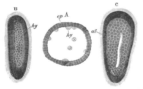
Fig. 68. Three larva stages of Eucope
polystyla.
(After Kowalevsky.)
A. Blastosphere stage with hypoblast spheres becoming budded off
into the central cavity.
B. Planula stage with solid hypoblast.
C. Planula stage with a gastric cavity.
ep. epiblast; hy. hypoblast; al. gastric
cavity.
The development of Eucope polystyla (fig. 68), one of the
Campanularidæ, deviates according to Kowalevsky (No. 147) in somewhat
important points from the usual type. The whole development takes
place after the deposition of the ovum. The segmentation results in
the formation of a single-walled blastosphere with a large central
cavity (fig. 68 A). This cavity, somewhat as in Ascetta, becomes
filled up with a not clearly (?) cellular material derived from the
walls of the blastosphere, which must be regarded as the hypoblast
(fig. 68 B). The larva elongates and becomes ciliated, and the
epiblast at its two extremities becomes thickened, and is stated by
Kowalevsky also to become divided into two layers. The alimentary
cavity appears as a slit in the middle of the hypoblast (fig. 68 C).
The cilia after a time disappear, and the larva then becomes fixed by
one extremity. It flattens itself out into a disc-like form, becomes
divided into four lobes, and covered by a cuticle (perisarc). From the
disc the stalk grows out which dilates at its free extremity into the
calyx.
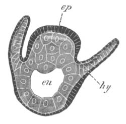
Fig. 69. Longitudinal section through a larva of
Tubularia mesembryanthemum while still in the gonophore. The
lower end is the oral one.
ep. epiblast; hy. hypoblast of tentacle; en.
enteric cavity.
In both the groups (Tubularia and Hydra) which are exceptional in not
having a ciliated planula stage, its absence may be put down to an
abbreviation of the development, and in fact a two-layered quiescent
stage, through which the embryo passes, may be regarded as
representing the planula stage.
The development of Tubularia, which has been described in detail by
Ciamician, takes place in the gonophore[74]. The segmentation is
irregular and leads to the formation of an epibolic gastrula, four
large central cells constituting the hypoblast[75]. The larva now
elongates, and grows out laterally into two processes which constitute
the first pair of tentacles. At this stage it closely resembles the
larvæ of some Medusæ. Additional tentacles are soon formed; and a
central cavity appears in the hypoblast, the cells of which have in
the meantime become more numerous (fig. 69). The tentacles are
directed towards the aboral side, which is considerably more
prominent than the oral one. They contain a hypoblastic axis. The
aboral end continues to grow and the tentacles gradually assume a
horizontal position. A constriction now appears, dividing the larva
into an aboral portion which will eventually form the stalk, and an
oral portion. At the apex of the latter a row of short tentacles—the
future oral tentacles—now appears. The larva has at this stage the
form known as Actinula. In this condition it becomes hatched, and
shortly afterwards it becomes fixed by the aboral end and grows into a
colony.
The development of Myriothela (Allman, No. 150) takes place on the
Tubularian type. The ovum invested by a delicate capsule becomes freed
by the rupture of the gonophore, and is then taken up by the
remarkable claspers characteristic of the genus. In the claspers it
becomes fecundated and undergoes its further development. After
segmentation a gastric cavity is formed, and provisional tentacles
arise as a series of conical involutions which subsequently become
evoluted. Permanent tentacles are formed as conical papillæ on a
truncated oral process. After hatching it has a few days’ free
existence, and then becomes attached, and loses its provisional
tentacles.
Although Hydra itself constitutes the simplest type of Hydrozoon, its
development, which has been fully investigated by Kleinenberg (No.
161), is in some respects a little exceptional. The segmentation is
regular, but a segmentation cavity is not formed. The peripheral layer
of cells gradually becomes converted into a chitinous membrane, which
is perhaps homologous with the perisarc of marine forms. Between the
membrane and the germ a second pellicle makes its appearance. The
above changes require about four days for their completion, but there
next sets in a period of relative quiescence which lasts for some 6‑8
weeks. During this period the remaining development is completed. The
cells of the germ first fuse together. In the interior of the
protoplasm a clear excentric space arises, which gradually extends
itself and forms the rudiment of the gastric cavity. The outer shell
in the meantime becomes less firm, and is finally burst and thrown
off, owing to the expansion of the embryo within.
The outermost layer of the protoplasm becomes, relatively to the inner
layer, clear and transparent, and there thus arises an indication of a
division of the walls of the archenteric cavity into two zones, or
layers. These layers, which form the epiblast and hypoblast, are
definitely established on the appearance of cells with contractile
tails[76] in the clear outer zone, between which the interstitial
epiblast cells subsequently arise.
The embryo, still forming a closed double-walled sack, elongates
itself, and at one pole its wall becomes very thin. And at this point
a rupture takes place which gives rise to the mouth. Simultaneously
with the mouth the tentacles become formed as hollow processes,
according to Mereschkowsky two being formed first and subsequently the
others in pairs. Very shortly afterwards the hitherto uniform
hypoblast becomes divided up into distinct cells. The thin inner
pellicle which persists after the rupture of the outer membrane
becomes in the meantime absorbed. With these changes the embryo
practically acquires the characters of the adult.
Trachymedusæ. Amongst the Trachymedusæ, which as has now been
satisfactorily established develop directly without alternations of
generations, the embryology of species both of the Geryonidæ and the
Æginidæ has been studied.
In all the types so far investigated the hypoblast is formed by
delamination, and there is a more or less well-marked planula stage.
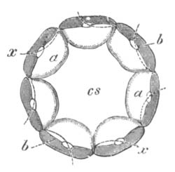
Fig. 70. Diagrammatic figure shewing the
delamination of the ovum of Geryonia. (Copied from Fol.)
cs. segmentation cavity; a. endoplasm; b.
ectoplasm. The dotted lines shew the course of the next planes of
division.
The development of Geryonia (Carmarina) hastata has been studied by
Fol (No. 155) and Metschnikoff (No. 163)[77]. The ovum, when laid, is
invested by a delicate vitelline membrane and mucous covering. Its
protoplasm is formed of an outer granular and dense layer, and a
central mass of a more spongy character. The segmentation is complete
and regular, and up to the time when thirty-two segments have appeared
each segment is composed of both constituents of the protoplasm of the
ovum. A segmentation cavity appears when sixteen segments are formed,
and becomes somewhat larger at the stage with thirty-two. At this
stage the process of delamination commences. Each of the thirty-two
segments, as shewn in the accompanying diagram (fig. 70), becomes
divided into two unequal parts. The smaller of these is formed almost
entirely of granular material; the larger contains portions of both
kinds of protoplasm. In the next segmentation the thirty-two large
cells only are concerned, and in each of these the line of division
passes between the granular and the transparent protoplasm. The
sixty-four lenticular masses of granular protoplasm thus formed
constitute an outer closed epiblastic vesicle, within which the
thirty-two masses of transparent protoplasm form an
hypoblastic vesicle. The embryo at this stage is shewn in optical
section in fig. 71.
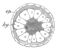
Fig. 71. Embryo of Geryonia after
delamination. (After Fol.)
ep. epiblast; hy. hypoblast.
The epiblastic vesicle now grows rapidly, while the hypoblastic
vesicle remains nearly passive and becomes somewhat lens-shaped. At
one point its wall comes in close contact with the epiblast. Elsewhere
a wide cavity is developed between the two vesicles which becomes
filled with gelatinous tissue. At this period cilia appear on the
surface, and the larva becomes a planula.
The succeeding changes lead rapidly to the formation of a typical
Medusa. Where the epiblast and hypoblast are in contact the former
layer becomes thickened and forms a disc-shaped structure. The centre
of this becomes somewhat protuberant, fuses with the hypoblast and
then becomes perforated to form the mouth (fig. 72 o). The edge
of the disc forms a thickened ridge, the rudiment of the velum
(v), which is entirely formed of epiblast. At its edge six
tentacles (t) arise, into which are continued solid
prolongations of the wall of the now somewhat hexagonal gastric
chamber. The hypoblastic axes of the tentacles soon lose their
connection with the gastric wall.
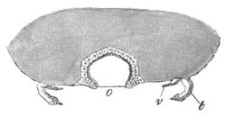
Fig. 72. Optical section through the oral pole of
Geryonia after the appearance of the gelatinous tissue of the
disc. (After Fol.)
o. mouth; v. velum; t. tentacle.
The shaded part represents the gelatinous tissue.
Up to this time the larva has retained a more or less spherical form,
and the cavity on the under side of the umbrella has not yet become
developed. The latter now becomes established by the whole disc
assuming a vaulted form with the concavity directed downwards. The
lining of the cavity so formed is derived from the epiblast of the
disc already spoken of.
The exact mode of formation of the gastrovascular canals has not been
worked out. It has however been established by the researches of the
Hertwigs (No. 146) and Claus (No. 153) that the radial and
circular vessels of this system are connected together in adult Medusæ
by an hypoblastic lamella; so that these canals would seem to be the
remnants of an once-continuous gastric cavity. This mode of formation
is established in the case of the medusiform buds; and it would
therefore seem, as pointed out by the Hertwigs, a fair deduction that
it occurs in the larva—a conclusion which is confirmed by the
primitive extension of the gastric cavity to the edge of the disc at
the time when its walls give rise to the solid axes of the tentacles.
In the course of the subsequent retirement of the gastric cavity from
the edge of the disc the gastrovascular canals probably take their
origin, though Fol was unable to follow the changes which result in
their formation.
On the completion of the above changes the larva has become a fully
formed Medusa, but it undergoes a not inconsiderable metamorphosis
before the attainment of the adult state.
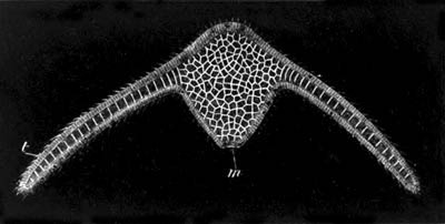
Fig. 73. A three-days’ larva of Æginopsis with
two tentacles.
(After Metschnikoff.)
m. mouth; t. tentacle.
Two species of Æginidæ have been studied by Metschnikoff (163), viz.
Polyxenia leucostyla (Ægineta flavescens), and
Æginopsis mediterranea. In both of these forms the segmentation
results in the formation of an elongated two-layered ciliated planula,
without a central cavity. The two ends of this grow out into two long
processes—the rudiments of a pair of at first aborally directed
arms—which contain a solid hypoblastic axis (fig. 73). At this stage
the larva closely resembles the larva of Tubularia. An alimentary
cavity is hollowed out in the centre of the hypoblast which soon opens
by a wide oral aperture (m). A second pair of arms becomes
formed, which are at first much shorter than the original pair; with
their formation a radial symmetry is acquired. Sense organs become at
the same time developed, and the whole embryo assumes a
medusiform character. Fresh tentacles arise, the velum and cavity of
the umbrella become established, but these changes do not involve any
points of very special interest.
Siphonophora. The development of the Siphonophora has been the
subject of careful investigation by Haeckel (158) and Metschnikoff
(163). The ova are large and usually (except Hippopodius) without a
membrane.
They are formed of a peripheral denser layer of protoplasm and a
central spongy mass. They usually undergo their entire development in
the water. In some instances they have been successfully reared by
artificial impregnation.
As an example of the Calycophoridæ I shall take Epibulia aurantiaca, a
form allied to Diphyes, the development of which has been studied by
Metschnikoff[78].
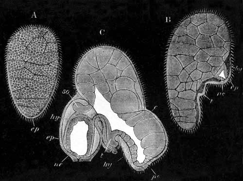
Fig. 74. Three larval stages of Epibulia
aurantiaca.
(After Metschnikoff.)
A. Planula stage.
B. Six-days’ larva with nectocalyx (nc) and tentacle
(t).
C. Somewhat older larva with gastric cavity.
ep. epiblast; hy. hypoblast; so. somatocyst;
nc. nectocalyx; t. tentacle; c. large yolk
cells; po. polypite.
There is a regular segmentation, unaccompanied by the formation of a
segmentation cavity. At its close the ovum becomes a spherical
ciliated embryo. This embryo soon becomes elongated, and its cells
differentiate themselves into a central and a peripheral layer—the
epiblast and the hypoblast (fig. 74 A). At this stage the larva has
the typical planula form. The epiblast is especially thickened at a
pole, which may be called the oral pole, and towards the side of this,
which will be spoken of as the ventral side. Adjoining this thickened
layer of epiblast a special thin layer of hypoblast becomes
differentiated, which in opposition to the main mass of large
nutritive cells forms the true hypoblastic epithelium (fig. 74 B,
hy). On this thickening two prominences make their appearance
(fig. 74 B). The oral of these is the rudiment of a tentacle
(t), and the aboral of a nectocalyx (nc).
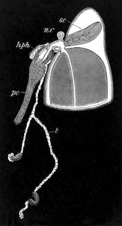
Fig. 75. An advanced larva of Epibulia aurantiaca
with one large nectocalyx. (After Metschnikoff.)
so. somatocyst; nc. second imperfectly developed
nectocalyx; hph. hydrophyllium; po. polypite;
t. tentacle.
The former of these elongates itself in succeeding stages into a
process of both epiblast and hypoblast. The central part of the
nectocalyx on the other hand appears to originate from a thickening of
the epiblast in which the cavity of the bell becomes subsequently
hollowed out. Between this part and the external epiblast which gives
origin to the outermost layer of the nectocalyx a layer of hypoblast
is interposed. When the nectocalyx has become to a certain extent
established a cavity—the commencement of the primitive
gastrovascular cavity of the adult—appears in the general hypoblast
between the epithelial and nutritive layers in the immediate
neighbourhood of its attachment. This cavity becomes prolonged into
the nectocalyx to form the four gastrovascular canals; while the
hypoblast at the upper end of the nectocalyx forms the somatocyst
(fig. 74 C, so). The primitive enteric cavity once formed
rapidly extends, especially in an oral direction (fig. 74 C), and
forms a widish cavity in the oral part of the embryo. At the pole of
this part (fig. 74, po) is eventually formed the opening of the
mouth, and the contained cavity becomes in a special sense the gastric
cavity. This region of the embryo may be spoken of as the polypite.
The nectocalyx grows with great rapidity and soon forms by far the
most prominent part of the larva (fig. 75). The true gastric region or
polypite (fig. 75, po) continues also to grow, and a mouth
becomes formed at its extremity. The aboral end of the original body
of the embryo gradually atrophies.
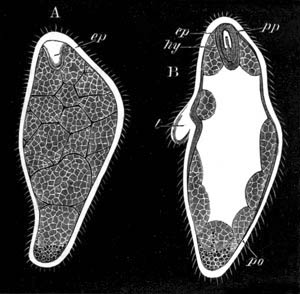
Fig. 76. Two stages in the development of
Stephanomia pictum.
(After Metschnikoff.)
A. Stage after the delamination. ep. epiblastic invagination
to form pneumatocyst.
B. Later stage after the formation of the gastric cavity in the
solid hypoblast, po. polypite; t. tentacle; pp.
pneumatophore; ep. epiblastic invagination to form
pneumatocyst; hy. hypoblast surrounding pneumatocyst.
At the junction of the nectocalyx and polypite the cœnosarc becomes
formed, and rudiments of a second nectocalyx (nc) and
second polypite early become visible; while a hydrophyllium is formed
as a bud which covers over the first polypite and tentacle
(hph). With the development of the hydrophyllium the first
segment, if the term may so be used, is complete. The second segment
of which a rudiment is already present as a second polypite is
intercalated between the first segment and the nectocalyces.
Amongst the Physophoridæ there is a considerable range of variation in
development; though the variations concern for the most part not very
important points. The simplest type hitherto observed is that of
Stephanomia (Halistemma) pictum. The segmentation and
formation of a two-layered planula (fig. 76) take place in the usual
way. Between the solid central mass of nutritive hypoblast cells and
the epiblast an epithelial hypoblastic layer becomes interposed which
undergoes a special thickening at the aboral pole. At this pole a
solid involution of epiblast next becomes formed, to which a layer of
hypoblast becomes applied. The structure so formed is the rudiment of
the pneumatocyst (ep). In the next stage the air-cavity of the
pneumatocyst becomes established within the epiblast.
The gastrovascular cavity is formed in the midst of the nutritive
hypoblast cells, which then become rapidly absorbed leaving the
gastrovascular cavity entirely enclosed by the epithelial layer of
hypoblast (fig. 76 B).
By the above changes the more important organs of the larva have
become established. The one end forms the pneumatophore, and the
other, the oral part, the polypite. Between the two there is already
present the rudiment of a tentacle, and a second tentacle soon becomes
formed. The mouth arises as a perforation at the oral end of the
larva.
The pneumatophore contains a prolongation of the gastrovascular
cavity, the fluid in which bathes the outer hypoblastic wall of the
pneumatocyst. It has however no communication with the enclosed cavity
of the pneumatocyst. In the later developmental stages the size of the
pneumatophore becomes immensely reduced in comparison with the
remainder of the larva.
The development of Physophora agrees closely with that of Stephanomia
except in one somewhat important point, viz. in the development of a
provisional hydrophyllium. This arises as a prominence at the
aboral pole, containing a prolongation of the gastrovascular cavity.
Between the epiblast and hypoblast of the prominence gelatinous tissue
becomes deposited, and the hydrophyllium is thus converted into a
large umbrella-like organ enclosing the polypite. The two together
have a close resemblance to an ordinary Medusa, the polypite forming
the manubrium, and the hydrophyllium the umbrella. The hydrophyllium
is eventually thrown off.
An important type of Physophorid development is exemplified in
Crystalloides, a genus closely allied to Agalma. In this type the
greater part of the original ovum, instead of directly giving rise to
the polypite, becomes a kind of yolk-sack, from which the polypite is
secondarily budded (fig. 77, yk). Agalma sarsii is in
this respect intermediate between Crystalloides and Physophora. Both
these types are remarkable for developing a series of provisional
hydrophyllia (fig. 77, h.ph.). In both genera the first of
these develops as in Physophora, and for a long time is the only one
functional.
The conclusions to be drawn from the above description may be summed
up as follows. In all the Siphonophora, so far observed, the starting
point for further development is a typical ciliated two-layered
planula. The inner layer or hypoblast is mainly formed of large
nutritive cells. From these cells an epithelial hypoblastic layer
becomes secondarily differentiated, the exact relations of which
differ somewhat in the various types. The nutritive cells themselves
do not appear to become directly converted into the permanent
hypoblastic tissues. The development of the adult from the planula
commences by the thickening of the epiblastic layer, usually at one
pole (the future proximal or aboral pole), and the formation at this
pole of a series of bud-like structures (in the growth of which both
embryonic layers have a share), which become converted into the
hydrophyllia, nectocalyces etc. The main oral part of the planula
becomes generally converted into the polypite, though in some
instances (Crystalloides) it remains as a yolk-sack, and only
secondarily gives rise to a polypite.
Two very different views have been taken as to the nature of the
various component parts of the Siphonophora, and the embryological
evidence has been appealed to by both sides in confirmation of their
views. By Huxley and Metschnikoff the various parts—nectocalyces,
hydrophyllia, hydrocysts, polypites, generative gonophores etc. are
regarded as simple organs, while by Leuckart, Haeckel, Claus etc. they
are regarded as so many different individuals forming a compound
stock. The difference between these two views is not merely as
to the definition of an individual[79]. The question really is, are
these parts originally derived by the modification of complete zooids
like the gonophores and trophosomes of the fixed Hydrozoa stocks, or
are they structures derived from the modification of the tentacles or
some other parts of a single zooid?
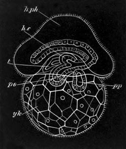
Fig. 77. Larva of Crystalloides, (After
Haeckel.)
h.ph. hydrophyllium; h. hydrocyst; t. tentacle;
pp. pneumatophore; po. polypite; yk.
yolk-sack.
The difficulty of deciding this point on embryological evidence
depends on the fact that ontologically a tentacle and a true bud arise
in the same way, viz. as papilliform outgrowths containing
prolongations of both the primitive germinal layers. The balance of
evidence is nevertheless in my opinion in favour of regarding the
Siphonophora as compound stocks, and the views of Claus on this
subject (Zoologie, p. 271) appear to me the most satisfactory.
The most primitive condition is probably that like Physophora in an
early stage with an hydrophyllium enclosing a polypite (cf. Haeckel
and Metschnikoff). In this condition the whole larva may be compared
to a single Medusa in which the primitive hydrophyllium represents the
umbrella of the Medusa, and the polypite the manubrium. The tentacle
which appears so early is probably not to be regarded as a modified
zooid, but as a true tentacle. The absence of a ring of tentacles is
correlated with the bilateral symmetry of the Siphonophora.
The primitive zooid of a Siphonophora stock is thus a Medusa. Like
Sarsia and Wilsia this Medusa must be supposed to have been capable of
budding. The ordinary nectocalyces by their resemblance to the
umbrellas of typical Medusæ are clearly such buds of the medusiform
type. The same may be said of the pneumatophore, which, as pointed out
by Metschnikoff, is identical in its development with a nectocalyx.
Both are formed by a solid process of epiblast in which a
cavity—the cavity of the nectocalyx or pneumatocyst—is eventually
hollowed out. Around this there appears a double layer of hypoblast
containing a prolongation of the gastrovascular cavity; and this is in
its turn enclosed by a layer of epiblast which forms the covering of
the convex surface of the nectocalyx and the external epiblast of the
pneumatophore.
The generative gonophores are clearly also zooids, and the
hydrophyllia are probably a rudimentary form of umbrella. In many
cases (Epibulia, Stephanomia, Halistemma etc.) the hydrophyllium of
the primitive polypite (manubrium) is absent. In such instances it is
necessary to suppose that the umbrella of the primitive zooid of the
whole colony has become aborted. Leuckart originally took a somewhat
different view from the above in that he regarded the starting point
of the Siphonophora to be a compound fixed Hydrozoon stock, which
became detached and free-swimming.
Acraspeda[80]. The embryonic development of several of the
forms of the Acraspeda has been investigated by Kowalevsky (No. 147)
and Claus (No. 153). Their observations seem to point to an invaginate
gastrula being characteristic of this group.
Amongst the forms with alternations of generations and a fixed larval
form Chrysaora and Cassiopea have been most fully investigated. The
ovum of the former undergoes the first embryonic phases while still in
the ovary. In the latter it is enclosed amongst the oral processes. A
complete and more or less regular segmentation leads to the formation
of a single-walled blastosphere with a small segmentation cavity. The
wall of the blastosphere next becomes invaginated, giving rise to an
archenteron (fig. 78 A). The blastopore soon closes up, and the
archenteron is converted into a closed sack completely isolated from
the epiblast (fig. 78 B). The surface of the larva becomes in the
meantime covered with cilia. The free larval stage thus reached is
similar to the ordinary Hydrozoon planula. After the closure of the
blastopore the larva becomes elongated, and one end becomes narrowed.
By this narrowed extremity the larva soon attaches itself, and at the
opposite and broader end a fresh involution of the epiblast appears
(fig. 78 C); this gives rise to the stomodæum, which is placed in
communication with the archenteron on the absorption of the septum
dividing them. The relation of the stomodæum to the original
blastopore has not been determined.
At the point of attachment there is developed a peculiar pedal disc,
and around the mouth there appears a fold of epiblast which gives rise
to an oral disc (fig. 78 D). Two tentacles first make their
appearance, but one of these is primarily much the largest, though
eventually the second overtakes it in its growth. A second pair of
tentacles next becomes formed, giving to the larva a 4-radial
symmetry. Between these four new tentacles subsequently sprout out,
and in the intermediate planes four ridge-like thickenings of the
hypoblast, projecting into the cavity of the stomach, make their
appearance. They imperfectly divide the stomach into four chambers, to
each of which one of the primary tentacles corresponds; they may be
regarded as homologous with the mesenteries of the Actinozoa. The
number of tentacles goes on increasing somewhat irregularly up to
sixteen. All the tentacles contain a solid hypoblastic axis. Muscular
elements are developed from the epiblast.
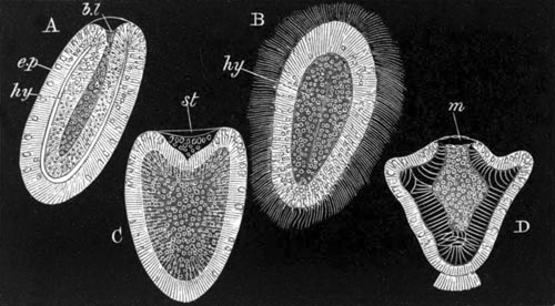
Fig. 78. Four stages in the development of
Chrysaora.
(After Claus.)
A. Gastrula stage.
B. Stage after closure of blastopore.
C. Fixed larva with commencing stomodæum.
D. Fixed larva with mouth, short tentacles, etc.
ep. epiblast; hy. hypoblast; st. stomodæum;
m. mouth; bl. blastopore.
With the above changes the so-called Hydra tuba or Scyphistoma form is
reached (vide fig. 85). The peculiar strobilization of this form is
dealt with in the section devoted to the metamorphosis.
Aurelia is stated by Kowalevsky to develop in the same way as
Cassiopea; and the one stage of Rhizostoma observed is that in which
it has a (probably invaginate) gastrula form.
In Pelagia the ovum directly gives rise to a form like the parent. The
segmentation and the invagination take place nearly as in Cassiopea,
but the archenteric cavity is relatively much smaller, and the large
space between it and the epiblast becomes filled with the gelatinous
tissue which forms the umbrella. The blastopore does not appear to
close but to become directly converted into the mouth. As in Cassiopea
the larva takes a somewhat four-sided pyramidal form. The mouth is
placed at the base. The pyramid becomes subsequently flatter, and at
the four corners four tentacles grow out which increase to eight by
division. The flattening continues till the larva reaches a form
hardly to be distinguished from the Ephyra resulting from the
strobilization of the fixed Scyphistoma form of other Acraspeda.
Alcyonidæ. In the Alcyonidæ the segmentation appears always to
lead to the formation of a solid morula, which becomes a planula by
delamination. The true enteric cavity is formed by an absorption of
the central cells, but the axial portion of the gastric cavity and
mouth are formed by an epiblastic invagination.
The development of these types has been mainly studied by Kowalevsky
(147), and my knowledge of his results is derived from German
abstracts of the original Russian memoirs.
In Alcyonium palmatum the impregnation is external. The
segmentation is very exceptional in character. It commences with the
formation of a series of irregular prominences on the surface of the
ovum, which become segmented off to form a superficial layer of
epiblast cells. The inner mass of protoplasm then divides up into
polygonal cells to form the hypoblast, which would thus seem to be
formed by a kind of delamination. In Clavularia crassa (No.
168) there is a complete segmentation followed by a delamination. The
larva of Al. palmatum elongates and becomes ciliated, and so
assumes the characters of a typical planula. The central hypoblast is
formed of an outer granular stratum with imperfectly differentiated
cells—the true hypoblast—and an inner homogeneous mass with
vacuoles.
Some of the larvæ become fixed, while others coalesce together and
form a large mass, the fate of which has not been further studied. An
invagination of epiblast takes place at the free end of the fixed
larva, which gives rise to the so-called gastric cavity, i.e.
the axial portion of the general enteric cavity, which would appear to
be in reality a kind of stomodæum. Around the gastric cavity the
hypoblast forms eight mesenteries, the chambers between which are
filled with the homogeneous material which occupied the centre of the
ovum in the previous stage. It is to be presumed, though not stated,
that by an absorption of the blind end of the stomodæal invagination
the gastric chamber is placed in free communication with the
spaces between the mesenteries[81]. During the next stage the young
Alcyonium also acquires eight tentacles, which arise as hollow papillæ
opening into the eight mesenteric chambers. By this stage also the
matter filling up the mesenteric chambers is nearly absorbed.
Between the epiblast and hypoblast there is formed an homogeneous
membrane, which penetrates in between the two layers of hypoblast
which form the mesenteries. On the outer side of this membrane, and
therefore presumably derived from the epiblast, is a layer of
connective-tissue cells, which eventually gives rise to the abundant
gelatinous tissue (cœnenchyma) in which the skeletal elements are
deposited. In Sympodium coralloides Kowalevsky (No. 168) has
shewn still more completely the derivation of the stellate mesoblast
cells from the epiblast. He finds that the calcareous spicula develop
in these cells as in the mesoblast cells of sponges. The branched
gastrovascular canals in this tissue are outgrowths of the primitive
enteric cavity. A layer of circular muscles is formed at a late period
from the epiblast, but the longitudinal muscles of the mesenteries on
the inner side of the homogeneous membrane are regarded by Kowalevsky
as hypoblastic.
A ciliated planula with delaminated hypoblast is also found in
Gorgonia and Corallium rubrum. In the former genus at the time
when the larva becomes fixed, the hypoblast is formed of two strata,
an outer one of columnar cells, and an inner one of round ciliated
cells lining a central enteric cavity. The inner layer is believed by
Kowalevsky to become eventually absorbed and to be homologous with the
inner granular mass of Alcyonium.
Zoantharia. Amongst the Zoantharia several forms have been
investigated by Kowalevsky (147) and Lacaze Duthiers (170), of which
some are stated by the former author to pass through an invaginate
gastrula stage, while in other instances the hypoblast is probably
formed by delamination.
To the first group belongs an edible form of Sea Anemone found near
Messina, Cerianthus, and perhaps also Caryophyllium. In the first of
these segmentation results in the formation of a blastosphere. A
normal invagination obliterating the segmentation cavity then ensues,
and the blastopore narrows to form the mouth. The borders of the mouth
bend inwards and so give rise to the gastric cavity (stomodæum) which
as in the Alcyonidæ is lined by epiblast. Simultaneously with the
formation of the mouth there appear the two first mesenteries.
In Cerianthus the segmentation is unequal, the early stages are the
same as in the Actinia just described, but the hypoblast cells give
rise to a mass of fatty material filling up the enteric
cavity, which becomes eventually absorbed.
In the majority of the Zoantharia so far investigated, including
species of Actinia, Sagartia, Bunodes, Astroides, Astræa, the
segmentation, which is often unequal[82] and not accompanied by the
formation of a segmentation cavity, results in a solid two-layered
ciliated planula. In these forms the impregnation takes place in the
ovary, and the early stages of development are passed through in the
maternal tissues.
One end of the planula becomes somewhat oval and develops a special
bunch of cilia. At the other end a shallow depression appears, which
becomes deeper and forms an involution lined by epiblast. This
involution is the stomodæum, and becomes the so-called gastric cavity.
The true enteric cavity lined by hypoblast is for some time filled
with yolk material. The larva always swims with the aboral end
directed forwards.
Between the two embryonic layers a homogeneous membrane is formed,
similar to that already described in the Alcyonidæ.
The further development of the larvæ especially concerns the formation
of mesenteries, tentacles and calcareous skeleton. With reference to
this subject the observations of Lacaze Duthiers are especially
valuable and striking.
In the adult it is usually possible to recognise in the tentacles a
symmetry of six. There are six primary tentacles, six secondary,
twelve tertiary, twenty-four quaternary, etc. In the hard septa of the
skeleton the same law is followed up to the third cycle, but beyond
that, in the cases where the point can be verified, there appear to be
only twelve septa in each additional cycle. The observations of Lacaze
Duthiers have shewn that this symmetry is only secondarily acquired
and does not in the least correspond with the succession of the parts
in development.
His observations were conducted on three species of Zoantharia without
a skeleton, viz. Actinia mesembryanthemum, Sagartia, and Bunodes
gemmacea; while Astroides calycularis served as the type for his
investigations on the corallum. It will be convenient to commence with
his results on Actinia mesembryanthemum which served as his type.
The free cylindrical embryo, with the aboral end directed forwards in
swimming, first becomes somewhat flattened and the mouth elongated. A
bilateral symmetry is thus brought about. Two mesenteries now make
their appearance transversely to the long axis of the mouth, which
divide the enteric cavity into two unequal chambers. The
mesenteries consist of a fold of hypoblast with a prolongation of the
epiblast between the two limbs of the fold. The larger chamber
next becomes divided by two fresh mesenteries into three, and a
similar division then takes place in the smaller chamber. The stage
with six chambers is almost immediately succeeded by one with eight,
owing to the appearance of two fresh mesenteries in the second-formed
set of chambers. At the stage with eight chambers there is a marked
period of repose. The number of chambers is increased to ten by the
division of the third-formed set of chambers, and to twelve by the
division of the fourth-formed set. It will be observed that the number
of the chambers increases in arithmetical progression by the continual
addition of two, alternately cut off from the primitive large and
small chambers. The freshly formed chambers are always formed
immediately on one side of the primitive mesenteries. The stages with
six and ten are of very short duration. The two primitive chambers are
necessarily at the ends of the long axis of the mouth. After the
division of the enteric cavity into twelve chambers, these chambers
become about equal in size, and the formation of the tentacles
commences. The law regulating the appearance of the tentacles is
nearly the same as that for the mesenteries, but is not quite so
precise. One tentacle makes its appearance for each chamber. The most
remarkable feature in the appearance of the tentacles is due to the
fact that the tentacle surmounting the primitive largest chamber
arises before any of the others, and long retains its supremacy (fig.
80 A). This fact, coupled with the inequality of the two primitive
chambers, supplies some grounds for speculating on a possible descent
of the Cœlenterata from bilaterally symmetrical forms with
distinctly differentiated dorsal and ventral surfaces. The supremacy
of the first-formed tentacle is not confined to the Actinozoa, but as
has already been indicated, is also found in the Scyphistoma (p. 166)
of the Acraspeda.
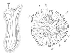
Fig. 80. Two stages in the development Of Actinia
mesembryanthemum. (After Lacaze Duthiers.)
In the younger ciliated embryo A, viewed from the side, only one
tentacle is developed. m. mouth.
The older larva B is viewed from the face when 24 tentacles have
just become established. The letters shew the true order of
succession of the tentacles; but e and f are
transposed.
After the twelve tentacles have become established they become
secondarily divided into two cycles of six respectively larger and
smaller tentacles, which alternate with each other. The two tentacles
pertaining to the two original chambers belong to the cycle of larger
tentacles. The mesenteric filaments appear first of all on the primary
pair of septa. The increase in the number of tentacles and chambers
from 12 to 24 has been found to take place in a very
remarkable and unexpected way. The law is expressed by Lacaze Duthiers
as follows. “The appearance of the new chambers is not, as has been
believed, a consequence of the production of a single chamber between
each of the twelve already existing chambers, but of the birth of two
new chambers in each of the six elements (chambers) of the smaller
cycle.” The result of this law is that a pair of tentacles of the
third cycle is placed in every alternate space, between a large and a
small tentacle, of the two already existing cycles, which may
conveniently be called the first and second cycles (fig. 80 B).
The twenty-four tentacles formed in the above manner are obviously at
first very irregularly arranged (fig. 80 B), but they soon acquire a
regular arrangement in three graduated cycles of 6, 6 and 12. The
first cycle of the six largest tentacles is the large cycle of the
previous stage, but the two other cycles are heterogeneous in their
origin, each of them being composed partly of the twelve tentacles
last formed, and partly of the six tentacles of the second cycle of
the previous stage.
The further law of multiplication has been thus expressed by Lacaze
Duthiers: “The number of chambers and still later that of the
corresponding tentacles is carried from 24‑48 and from 48‑96 by the
birth of a pair of elements in each of the 12 or 24 chambers, above
which are placed the smallest tentacles which together constitute the
fourth or fifth cycle. Since, after the formation of each fresh cycle,
the arrangement of the tentacles again becomes symmetrical, it is
obvious that all the equal sized cycles except the first are formed of
tentacles entirely heterogeneous as to age.”
The fixation of the free-swimming larva takes place during the period
when the tentacles are increasing from 12 to 24.
The general formation of the chambers in Bunodes and Sagartia is
nearly the same as in Actinia.
In the two types of Actinozoa with an embolic gastrula stage the laws
as to the formation of the tentacles do not appear to be the same as
those regulating the forms observed by Lacaze Duthiers.
In Cerianthus four tentacles are formed simultaneously at the period
when only four chambers are present. In Arachnitis (Edwarsia) the
succession of the tentacles is stated (A. Agassiz, 166) to resemble
that in Cerianthus. There are originally four tentacles, and at one
extremity of the long axis of the mouth are the oldest tentacles,
while at the other tentacles are constantly added in pairs. An odd
tentacle is always found at the extremity of the mouth opposite the
oldest tentacles.
In the other species with an embolic gastrula eight tentacles would
seem to appear simultaneously at the period when eight chambers are
present; though on this point Kowalevsky’s description is not very
clear. The presence of such a stage would seem to indicate a close
affinity to the Alcyonidæ.
Amongst the sclerodermatous Actinozoa, except Caryophyllium, the
embryo closely resembles that of the delaminate Malacodermata. The
first stages occur in the ovary, and the larva is dehisced
into the body cavity as a two-layered ciliated planula.
The laws affecting the formation of the first twelve tentacles and
septa appear to be nearly the same as for the Malacodermata. The hard
parts begin as a rule to be formed when twelve tentacles have
appeared, at which period also the fixation of the larva takes place.
On fixation the larva becomes very much flattened.
The first parts of the corallum to appear are twelve of the septa,
which arise simultaneously in folds of the enteric wall in the
chambers between the mesenteries, and correspond therefore with
the tentacles and not, as might be supposed, with the mesenteries.
Each septum is formed by the coalescence of three calcareous plates
which originate in separate centres of calcification. The concrescence
of the three produces a Y-shaped plate with the single limb directed
inwards and the two limbs outwards (fig. 81). The theca does not arise
till after the septa have become formed, and is at first a somewhat
membranous cup quite distinct from the septa. The columella is formed
still later by the coalescence of a series of nodules which are formed
in a central axis enclosed by the inner ends of the septa.
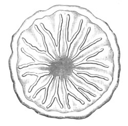
Fig. 81. Larva of Astroides calycularis shortly
after it has become attached. (After Lacaze Duthiers.)
The figure shews the development of the Y-shaped septa in the
intervals between the mesenteries. The position of the latter is
indicated by the faint shading. The theca has become developed
externally.
After the formation of the theca the septa become divided into two
cycles by the predominant growth of six of them. On the coalescence of
the septa with the theca the space between the two limbs of the Y
becomes filled up with calcareous tissue. The law of the formation of
the third cycle of septa (12‑24) has not been worked out, so that it
is not possible to state whether it follows the peculiar principles
regulating the growth of the tentacles.
The whole of the skeletal parts occupy a position between the epiblast
and hypoblast, and are exactly homologous in this respect with the
skeleton of the Alcyonidæ. By Lacaze Duthiers they are however
believed to originate in the hypoblast, but from the observations of
Kowalevsky there be little doubt that they arise in the connective
tissue between the two embryonic layers which is probably epiblastic
in origin.
A peculiar larva, probably belonging to the Actinozoa, has been
described by Semper[83]. It has an elongated form and is provided with
a longitudinal ridge of cilia. There is a mouth at one end of the body
and an anus at the opposite extremity. The mouth leads into an
œsophagus, which opens freely into a stomach with six
mesenteries. In the skin are numerous thread-cells. A mesotrochal
worm-like larva, also provided with thread-cells, and found at the
same time, was conjectured by Semper to be a younger form of this
larva.
Ctenophora. The ovum of the Ctenophora is formed of an outer
granular protoplasmic layer and an inner spongy mass with fatty
spherules. It is enveloped in a delicate vesicle, the diameter of
which is very much greater than that of the contained ovum. This
vesicle appears to be filled with sea-water, in which the ovum floats.
Fertilized ova may usually be easily obtained by keeping the captured
adults in water from 12‑24 hours. The two main authorities on the
development of these forms (Kowalevsky, No. 147 and 178 and Agassiz,
No. 172) are unfortunately at variance on one or two of the most
fundamental points. It seems however that the embryonic layers are
formed by a kind of epibolic gastrula; while the true gastric cavity,
as distinct from the gastrovascular, is formed by an invagination, and
deserves therefore to be regarded as a form of stomodæum.
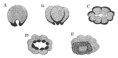
Fig. 82. Five stages in the development of Idyia
roseola.
(After Agassiz.)
The protoplasmic layer of the ovum is represented in black.
The early stages are very closely similar in all the types so far
observed. Segmentation commences by the outer layer of the ovum, which
throughout behaves as the active layer, forming a protuberance at one
pole, which may be called the formative pole. Close below this
protuberance is placed the nucleus. In the median line of the
protuberance a furrow appears (fig. 82 A), which gradually deepens
till it divides the ovum into two. The granular layer follows the
furrow so that each of the fresh segments, like the original ovum is
completely invested by a layer of granular protoplasm. Each
segment contains a nucleus. A second similar division at right angles
to the first gives rise to four segments (fig. 82 B), and the segments
so formed become again divided into eight (fig. 82 C). In the division
into eight, which takes place in a vertical plane, the segments formed
are of unequal size, four of them being much smaller than the others.
The eight segments are arranged in the form of a slightly curved disc
round a vertical axis—the future long axis of the body;—and there is
a cavity in this axis which, like the segmentation cavity of
Sycandra raphanus, is open at both extremities. The disc with
its concavity on the side of the formative pole has the shape
sometimes of an ellipse (fig. 82 C) and sometimes of a rectangle, in
which the four small spheres occupy the poles of the longer axis. A
bilateral symmetry is thus even at this stage clearly indicated.
In the next phase of segmentation the granular layer surrounding each
segment again forms a protuberance at the formative pole, but, instead
of each segment becoming divided into two equal parts, the
protoplasmic protuberance alone is divided off from the main segment.
In this way sixteen spheres become formed, of which eight are large
and are formed mainly of the yolk material of the inner part of the
ovum, and eight are small and entirely composed of the granular
protoplasm. The eight small spheres form a ring on the formative
surface of the large spheres (fig. 82 D).
The small spheres now increase very rapidly (fig. 82 E), partly by
division and partly by the formation of fresh cells from the large
spheres; and spread over the large spheres, forming in this way an
epibolic gastrula. They constitute a layer of epiblast. (fig. 83 A.)
The large cells in the meantime remain relatively passive, though
during the process they divide, in some cases more or less
irregularly, while in Eucharis they divide into sixteen. The axial
segmentation cavity would seem during the process to become
obliterated.
There is an important discrepancy between the statements of Kowalevsky
and Agassiz as to the course of the growth of the small cells.
According to Agassiz the small cells grow most rapidly at the
formative pole and cover this before they meet at the opposite pole.
The reverse statement is made by Kowalevsky. It would seem that the
above discrepancy is due to an interchange on the part of the
one or the other of these authors of the two poles of the embryo, in
that according to Agassiz the formation of the mouth takes place at
the formative pole, and according to Kowalevsky at the pole
opposite to this.
Without attempting to decide between the above views, we shall speak
of the pole at which the mouth is formed as the oral pole.
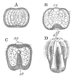
Fig. 83. Four stages in the development of Idyia
roseola. (After Agassiz.)
s.c. sense capsule; st. stomodæum.
The formation of the alimentary cavity commences shortly after the
complete investiture of the embryo by the epiblast cells. At the oral
pole an invagination of epiblast cells takes place (fig. 83 B), which
makes its way towards the opposite pole. More especially from the
figures given by Agassiz, and from the explanation of his plates, it
would seem that a large chamber is formed in the hypoblast at the end
of the invaginated tube, into which this tube soon opens (fig. 83 C).
The invaginated tube would seem to give rise to the so-called stomach,
while the chamber at its aboral extremity is no doubt the
infundibulum, which as may be gathered from Kowalevsky’s statements,
is lined by a flattened epithelium. At a later period the
gastrovascular canals grow out from the infundibulum as four pouches,
which are surrounded by, and grow at the expense of, the large central
cells, which have in the meantime arranged themselves in four masses,
and appear to serve as a kind of yolk. The nuclei of these large cells
according to Kowalevsky disappear, and the cells themselves break up
into continually smaller masses.
The main difficulty in the above description of Agassiz is the origin
of the infundibulum. In the absence of definite statements on this
head it seems reasonable to conclude that it arises as a space
hollowed out in the central cells, and that its walls are formed of
elements derived from the yolk cells[84]. On this interpretation the
alimentary canal of the Ctenophora would consist, as in the
Acraspedote Medusæ and Actinozoa, of two sections: (1) A true
hypoblastic section consisting of the infundibulum and the
gastrovascular canals derived from it; and (2) an epiblastic
section—the stomodæum—forming the stomach.
The observations of Kowalevsky on the alimentary system do not wholly
tally with those of Agassiz. He finds that the oral side of the embryo
becomes hollowed out, and that the hollow, lined by flattened cells,
becomes constricted off as the infundibulum, from which the radial
canals subsequently grow out. To the infundibulum there leads a narrow
canal lined by a columnar epithelium which becomes the gastric cavity.
While the alimentary canal is becoming formed a series of important
changes takes place in other parts of the embryo. The rows of
locomotive paddles first appear as four longitudinal equidistant
linear thickenings of the epiblast near the aboral pole (fig. 83 D).
On the projecting surface of these ridges stiff cilia appear which
coalesce together to form the paddles. While the embryo is still
within the egg the rows of paddles are quite short and also double.
There are in Pleurobrachia about eight or nine pairs of paddles in
each row. Each double row eventually separates into two.
In all the forms except the Eurostomata (Beroe) two tentacles grow out
as thickenings of the epiblast (fig. 84 B, t.). They are placed
at the opposite poles of the long transverse axis of the embryo.
A process of the contractile gelatinous tissue of the body, the origin
of which is described below, makes its way, according to Kowalevsky,
into the tentacles.
The central apparatus of the nervous system and the otoliths are
formed at the aboral pole from a thickening of the epiblast, but the
full details of their formation have not been elucidated. It may be
well to preface my account of their development with a short statement
of their adult structure.
They consist in the adult of a vesicle with a ciliated lining situated
at the bifurcation of the two anal tubes, and of certain structures
connected with this vesicle. From the floor of the vesicle is
suspended a mass of otoliths by four leaf-like bodies known as
suspenders. The roof is very delicate and has the form of a four-sided
pyramid. Six openings lead into the vesicle. Through four of these,
placed at the four corners, there pass out four ciliated grooves
continuous with the suspenders. These grooves, after leaving the
otolithic vesicle, bifurcate and pass to the eight rows of paddles. At
the two sides the walls of the vesicle are continuous with two
thickened ciliated plates with swollen edges, opposite the centres of
which are two lateral openings into the vesicle, completing the six
openings. Through the lateral openings the sea-water is driven by the
action of the cilia of the plates.
The development of these parts is as follows—In the aboral thickening
of epiblast a cavity makes its appearance, the walls of which
constitute the rudiment of the otolithic vesicle (fig. 83 B and C,
s.c.). The roof of the cavity is extremely delicate. On each
side of it a thickening of cells becomes established, regarded by
Kowalevsky as the rudiment of the nervous ganglia. These thickenings
appear to give origin to the lateral ciliated plates. The otoliths
arise from cells at four separate points at the corners of the
ciliated plates opposite the rows of paddles (fig. 84 A, ot.).
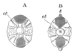
Fig. 84. Two stages in the development of
Pleurobrachia rhododactyla. (After Agassiz.)
ot. otolith; t. tentacle.
In Pleurobrachia there is at first only one otolith at each corner.
The otoliths are gradually transported towards the centre of the
vesicle (fig. 84 B, ot.) and are there attached, though the
four leaf-like suspenders do not arise till very late. The otoliths go
on increasing in number throughout life.
The gelatinous tissue of the Ctenophora appears as a homogeneous layer
between the epiblast and the yolk cells, and is probably homologous
with the layer formed in the same situation in all other
cœlenterate forms. Into the layer a number of anastomosing cells,
mainly derived from the epiblast, though according to Chun (No. 174)
also in part from the hypoblast, make their way. These cells would
appear to be mainly, if not entirely (Chun), of a contractile nature.
It is probable that the great mass of the gelatinous tissue of the
adult is an intercellular substance derived from these cells.
The whole of the above changes are completed while the embryo is still
enclosed in the egg-capsule. During their accomplishment the oro-anal
axis, which was originally very short, increases greatly in length
(fig. 83), so that the embryo acquires an oval form similar to that of
the adult.
The exact period of leaving the egg does not appear to be very
constant but the hatching never takes place till the embryo has
practically acquired all the organs of the adult.
In the majority of types the differences between the just hatched
larva and the adult are inconsiderable, and in all cases the larva has
a somewhat oval form. In the case of the Tæniatæ (Cestum, etc.), the
larva has the characteristic oval form, and the subsequent changes
amount almost to a metamorphosis.
The larva of the Lobatæ, such as Eucharis, Bolina, etc., can hardly be
distinguished from Pleurobrachia, and undergoes therefore considerable
changes after hatching.
Eucharis multicornis while still in the larval condition is
stated by Chun to become sexually mature.
The new genus Ctenaria recently described by Haeckel, which is
intermediate between the Ctenophora and the Medusæ clearly proves that
the Ctenophora are more closely related to the Medusæ than to the
Actinozoa but their development, especially the presence of a
stomodæum, shews that they have affinities (in spite of the
rudimentary velum of Ctenaria) with the Acraspedote as well as with
the Craspedote Medusæ; and it may be noted that the Acraspeda have
undoubted affinities with the Actinozoa.
Summary and general considerations.
Even in the adult condition the lower forms of Cœlenterata do not
rise in complexity much beyond a typical gastrula. Ontogeny
nevertheless brings clearly to light the existence of a larval
form—the planula—which recurs with fair constancy amongst all the
groups except the Ctenophora.
We are probably justified in assuming that the planula is a repetition
of a free ancestral form of the Cœlenterata. The planula, as it
most frequently occurs, is a two-layered ciliated nearly cylindrical
organism, with at most a rudimentary digestive cavity hollowed out in
the inner layer, and as a rule no mouth. In the outer layer are
numerous thread-cells.
How many of these characters did the ancestral planula possess? I
think it is not unreasonable to assume that the only two characters
about which there can be much doubt are the rudimentary condition of
the digestive cavity and the absence of a mouth. Paradoxical as it may
seem, it appears to me not impossible that the Cœlenterata may have
had an ancestor in which a digestive tract was physiologically
replaced by a solid mass of amœboid cells. This ancestor
was perhaps common to the Turbellarians also. The constant presence of
thread-cells in the inner layer of their epiblast fits in with their
derivation from a form similar to the planula. While the solid
parenchymatous digestive canal of Convoluta and Schizoprora and other
forms amongst the Turbellarians, though very probably secondary, may
perhaps be explained by such a view of their origin.
The planula in its primitive condition is not bilaterally symmetrical,
but frequently, as amongst the Actinozoa, it becomes flattened on two
sides before undergoing its conversion into the adult form. Perhaps
the bilateral form of planula is the starting point both for the
Cœlenterata and the Turbellaria. In this connection the peculiar
unilateral development of a tentacle in Scyphistoma and Actinia should
be noted.
The planula occurs in the majority of sessile forms of Hydrozoa except
the Tubularidæ and Hydra. It is also characteristic of the
Trachymedusæ and Siphonophora. Amongst the Acraspeda it is also
present, but has an exceptional mode of ontogeny which is discussed in
connection with the germinal layers.
It is characteristic both of the Octocoralla and Hexacoralla, but is
not found in the Ctenophora.
In the Tubularidæ and in Hydra an abbreviated development leads no
doubt to the absence of a free planula stage, and the absence
of a larval form amongst the Ctenophora may, as has already been
stated, be probably explained in the same way.
The Cœlenterata of all the Metazoa are characterized by the
greatest simplicity in the arrangement of their germinal layers; and
for this reason very considerable interest attaches to the mode of
formation of the layers amongst them. Two germinal layers are
constantly found, which correspond in a general way to the
epiblast and hypoblast. It might have been anticipated that a certain
amount of uniformity would have existed in the mode of formation of
the layers. This however is not the case. In perhaps the majority of
forms they become differentiated by a process of delamination, but in
a not inconsiderable minority the two layers owe their origin to an
invagination.
Delamination is constant (with the doubtful exception of some
Tubularidæ) amongst the Hydromedusæ and Siphonophora. It is perhaps in
the main characteristic of the Actinozoa.
Invagination by embole takes place, so far as is known, constantly
amongst the Acraspeda and frequently amongst the Actinozoa;
and an epibolic invagination is characteristic of the Ctenophora.
If confidence is to be placed in the recorded observations on which
this summary is founded, and there is no reason why in a general way
it should not be so placed, the conclusion is inevitable that of the
above modes of development the one must be primitive and the other a
derivative from it, for, if this conclusion be not accepted, the
absolutely inadmissible hypothesis of a double origin for the
Cœlenterata would have to be adopted.
Two questions arise from these considerations:—
(1) Which is the primitive, delamination or invagination?
(2) How is the one of these to be derived from the other?
There is a great deal to be said in favour of both delamination and
invagination; but it will be convenient to defer all discussion of the
question to the general chapter on the formation of the layers
throughout the animal kingdom.
The hypoblast cells are often filled with yolk material, and secondary
modifications are thus produced in the development. The most important
examples of such modifications are found in the Siphonophora and
Ctenophora.
In the simplest forms amongst the Hydrozoa there is no trace of a
third layer or mesoblast. The epiblast is typically formed, as was
first shewn by Kleinenberg, of an epithelial layer and a subepithelial
interstitial layer of cells. The cells of the former are frequently
produced into muscular or nervous tails, and those of the latter give
rise to the thread-cells and generative organs and in some cases to
muscles[85]. In many cases, amongst all the Cœlenterate groups, and
constantly amongst the Ctenophora the epiblast is simplified and
reduced to a single layer. The hypoblast undergoes in most cases no
such differentiation but simply forms a glandular layer lining the
gastric chamber and its prolongations into the tentacles; but in the
Actinozoa it appears to give rise to muscles, and strong evidence has
been brought forward to shew that in some groups it gives rise to the
generative organs.
Between the epiblast and hypoblast a structureless lamella appears
always to be interposed.
In many Cœlenterata further differentiations of the epiblast are
present. In many forms the layer gives rise to a hard external
skeleton. This is most widely spread amongst the Hydrozoa, where in
the majority of cases it takes the form of the horny perisarc, and in
the Hydrocoralla (Millepora and Stylasteridæ) of a hard calcareous
skeleton. The skeleton in these forms, though closely resembling the
mesoblastic skeleton of the Actinozoa, has been shewn by Moseley (164)
to be epiblastic.
In the Actinozoa an epiblastic skeleton is exceptional, and according
to most authorities absent. Quite recently however Koch (167) has
found that the axial branched skeleton of most of the Gorgonidæ, viz.
the Gorgoninæ and Isidinæ, is separated from the cœnosarc by an
epithelium, which he believes to be epiblastic, and to which no doubt
the axial skeleton owes its origin. A similar epithelium surrounds the
axis of the Pennatulidæ.
In the Medusæ the epiblast also gives rise to a central nervous
system, which however continues to form a constituent part of the
layer, and to the organs of special sense[86].
A special differentiation of the hypoblast is found in the solid axis
of the tentacles. This axis replaces the gastric prolongation found in
many forms, and the cells composing it differentiate themselves into a
chorda-like tissue, which has a skeletal function, and is no longer
connected with nutrition. This axis is placed by many morphologists
amongst the mesoblastic structures.
In all the higher Cœlenterata certain tissues become interposed
between the epiblast and hypoblast, which may be classified together
as the mesoblast.
The most important of these are:
(1) The various distinct muscular layers.
(2) The gelatinous tissue of the Medusæ and Ctenophora.
(3) The skeletogenous tissue of the Actinozoa.
In most cases the muscular fibres are connected with epithelial cells,
but in certain forms amongst the Medusæ and in the majority if not all
the Actinozoa they constitute a distinct layer, sometimes separated
from the epiblast by a structureless membrane, Æquorea
Mitrocoma. Such layers when on the outer side of the membrane
separating epiblast and hypoblast are undoubtedly epiblastic in
origin, but in some cases amongst the Actinozoa they adjoin the
hypoblast, and are very probably derived from this layer.
The origin of the gelatinous tissue is still involved in much
obscurity.
It originates as a homogeneous layer between epiblast and hypoblast,
which in the Hydromedusæ never becomes cellular though traversed by
elastic fibres.
In the Acraspeda it contains anastomosing cells in the main apparently
(Claus) derived from the hypoblast, and in the Ctenophora it is richly
supplied with muscular stellate cells for the most part of epiblastic
origin, though some are stated by Chun to come from the hypoblast. On
the whole it seems probable, that the gelatinous tissue may be
regarded as a product of both layers; and there are some
grounds for thinking that it is an immense development of the membrane
always interposed between the two primary layers. It must however be
borne in mind that a membrane, regarded by the Hertwigs as the
equivalent of the ordinary membrane between the epiblast and
hypoblast, can be usually demonstrated on both surfaces of the
gelatinous tissues in Medusæ. The skeletogenous layer of the Actinozoa
is probably the morphological homologue of the gelatinous tissue; but
the evidence we have is on the whole in favour of the
connective-tissue cells it contains being epiblastic in origin. It
gives rise to the skeleton of the Hexacoralla, to the spicular
skeleton of Alcyonium, the axial skeleton of Corallium, and the
skeleton of the Helioporidæ and Tubiporidæ.
Alternations of generations.
Alternation of generations is of common occurrence amongst the
Hydrozoa, and something analogous to it has been found to take place
in Fungia amongst the Actinozoa. It is not known to occur in the
Ctenophora.
The chief interest of its occurrence amongst the Hydromedusæ and
Siphonophora is the fact that its origin can be traced to a
division of labour in the colonial systems of zooids so characteristic
of these types.
In the Hydromedusæ an interesting series of relations between
alternation of generations and the division of the zooids into
gonophores and trophosomes can be made out. In Hydra the generative
and nutritive functions are united in the same individual. The
generative swellings in these forms cannot, as has been ably argued by
Kleinenberg, be regarded as rudimentary gonophores, but are to be
compared to the generative bands developed in the Medusæ around parts
of the gastro-vascular system. A condition like that of Hydra, in
which the ovum directly gives rise to a form like its parent, is no
doubt the primitive one, though it is not so certain that Hydra itself
is a primitive form. The relation of Hydra to the Tubularidæ and
Campanularidæ may best be conceived by supposing that in Hydra most
ordinary buds did not become detached, so that a compound Hydra became
formed; but that at certain periods particular buds retained their
primitive capacity of becoming detached and subsequently developed
generative organs, while the ordinary buds lost their generative
function.
It would obviously be advantageous for the species that the detached
buds with generative organs should be locomotive, so as to distribute
the species as widely as possible, and such buds in connection with
their free existence would naturally acquire a higher organization
than the attached trophosomes. It is easy to see how, by a series of
steps such as I have sketched out, a division of labour might take
place, and it is obvious that the embryos produced by the highly
organized gonophores would give rise to a fixed form from which the
fixed colony would be budded. Thus an alternation of generations would
be established as a necessary sequel to such a division of labour. To
test the above explanation it is necessary to review the main facts
with reference to alternations of generations amongst the Hydromedusæ.
Hydromedusæ[87]. In many instances amongst the Tubularidæ,
Sertularidæ and Campanularidæ medusiform buds are produced which
become detached and develop sexual organs.
Such Medusæ are divided into two great groups, the Ocellata and
Vesiculata, according to the characters of the marginal sense organs.
In the Ocellata the sense organs have the form of eyes, and in the
Vesiculata of auditory vesicles. The latter seem to be usually budded
off from the Campanularia stocks, and the generative organs extend in
folded bands over the radial canals. These bands have been regarded by
Allman as composed of rudimentary gonophores, and he called the Medusæ
which give rise to them blastochemes. He regards them as representing
a more complicated type of alternation of generations with three
instead of two generations in the series. The Hertwigs have brought
what appear to me conclusive grounds for rejecting this view, and have
demonstrated that the generative organs of these types resemble those
of ordinary Medusæ.
In many forms the medusiform buds though fully developed do not become
detached; whether detached or not they are known as phanerocodonic
gonophores. In other forms again buds which begin as if they were
going to form Medusæ never reach that condition but remain permanently
in an undeveloped state. They have been called by Allman adelocodonic
gonophores.
In all the above cases two generations at the least interpose between
the successive sexual periods, viz.:—
(1) A trophosome produced directly from the ovum.
(2) A gonophore budded from this.
In a very large number of types the gonophores do not develop directly
on the hydroid stem, but arise on specially modified zooids resembling
rudimentary trophosomes which have been named blastostyles by Allman.
On the sides of each blastostyle a series of gonophores usually
becomes developed. The blastostyles either remain exposed as in all
the Gymnoblastic or Tubularian Hydroids, or as in all the
Calyptoblastic Hydroids (Sertularidæ and Campanularidæ) they become
invested by a special case—known as the gonangium—which is formed of
perisarc lined by epiblast. In the forms with blastostyles three
generations interpose between the successive stages of sexual
reproduction, (1) the trophosome developed directly from the ovum, (2)
the blastostyle budded from this, (3) the gonophore budded from the
blastostyle.
Such being the main facts, in order to prove that the existing
condition of polymorphism amongst the Hydromedusæ is to be explained
as hypothetically suggested above, it is still necessary to shew that
(1) the free medusiform gonophores are really only modified
trophosomes, or rather that the trophosomes and gonophores are both
modifications of some common type, and (2) that the fixed so-called
adelocodonic gonophores are retrograde derivatives of the free
medusiform gonophores. Unless these points can be established it might
be maintained that the Medusæ were special zooids, developed de
novo and not by a modification of trophosome zooids. To
demonstrate these propositions at length would carry me too far into
the region of simple Comparative Anatomy, and I content myself with
referring the reader to a discussion of the Hertwigs (No. 146, p. 62)
where the first point appears to me fully established. With reference
to the second point I will only say that the structure and development
of the adelocodonic gonophores can only be explained on the assumption
that they are retrograde forms of the phanerocodonic gonophores, and
that the opposite view, that the phanerocodonic gonophores are derived
from the adelocodonic, leads to a series of untenable positions.
The Trachymedusæ, as has been shewn above, develop directly. They are
probably derived from gonophores in which the trophosome has
disappeared from the developmental cycle.
To sum up, three types of development are found amongst the
Hydromedusæ.
(1) No alternations of generations. Permanent form, a sexual
trophosome. Ex. Hydra.
(2) Alternations of generations. Trophosome fixed, gonophore free or
attached. Ex. Gymnoblastic and Calyptoblastic Hydroids, and
Hydrocoralla.
(3) No alternations of generations. Permanent form, a sexual Medusa.
Ex. Trachymedusæ.
Siphonophora. In the Siphonophora alternations of generations
take place in the same way as in the Hydromedusæ, but the starting
point appears to be a Medusa. The gonophores may remain fixed or
become detached.
Acraspeda. With the exception of Pelagia and Lucernaria, in
which the development involves a simple metamorphosis, all the
Acraspeda undergo a form of alternations of generations. The ovum, as
already described, develops into a fixed form—the Scyphistoma—which
increases asexually by normal budding, and can even form a permanent
colony.
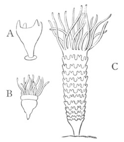
Fig. 85. Three stages in the alternations of
generations of Aurelia aurita. (From Gegenbaur.)
A. Polype stage.
B. Commencing strobilization.
C. Completed
strobilization.
The formation of the sexual Medusa form takes place by a kind of
strobilization of the body of the fixed Scyphistoma. A series of
transverse constrictions becomes formed round the body below the
mouth, dividing it up into corresponding rings, each of which
eventually gives rise to a Medusa known as an Ephyra (fig. 85). In
each of these rings is a dilation of the stomach, and a section of
each of the four rudimentary mesenteries described in connection with
the development of the Scyphistoma. As the constrictions become deeper
the segments of the body between them become disc-like, and their
edges are produced into eight lobes containing prolongations of the
gastric cavity (fig. 85 C). The lower surface of each disc, which
forms the future aboral surface of the Medusa, becomes convex, in part
owing to the development of gelatinous tissue. On the opposite surface
a muscular layer becomes developed. During the above process the body
of the Scyphistoma gradually grows in length and continues to be
segmented, so that a series of Ephyræ are uninterruptedly formed, of
which those near the base are the youngest. The original terminal ring
of tentacles of the Scyphistoma gradually atrophies.
In the further development of the Ephyræ each of their eight lobes
becomes bifid at its extremity.
As the Ephyræ successively reach this condition they become detached,
and by a series of remarkable changes, amounting almost to a
metamorphosis, and accompanied by an enormous growth in size, reach
the adult condition.
The alternation of generations in the Acraspeda cannot be quite so
simply explained as in the Hydromedusæ, though the principle is
probably the same in the two cases.
Actinozoa. Amongst the Actinozoa there occurs in Fungia a
peculiar process which is, as shewn by Semper (171), in many ways
analogous to alternations of generations[88]. From the larva a
nurse-stock is developed, at the end of which a cup-like coral
resembling the adult is formed as a bud. The bud becomes detached and
then gives rise to a permanent sexual Fungia. From the nurse-stock
there is formed however a fresh bud at the centre of the scar left on
the detachment of the old one. The fresh bud eventually becomes
separated from the nurse-stock leaving a small portion of its stem
behind; each succeeding bud similarly leaves a small portion of its
stem, so that the nurse-stock eventually acquires a jointed
appearance. In the above process we clearly have, as in the
Hydromedusæ, a non-sexual form—the nurse-stock—produced directly
from the larva, giving rise by budding to a sexual form; all the
conditions of an alternation of generations are therefore fulfilled.
It seems however possible that the nurse-stock itself may eventually
become sexual.
Bibliography.
Cœlenterata. General.
(145) Alex. Agassiz. Illustrated Catalogue of the Museum of
Comparative Anatomy at Harvard College, No. II. American
Acalephæ. Cambridge, U. S., 1865.
(146) O. and R. Hertwig. Der Organismus d. Medusæ
u. seine Stellung z. Keimblättertheorie. Jena, 1878.
(147) A. Kowalevsky. “Untersuchungen üb. d. Entwicklung d.
Cœlenteraten.” Nachrichten d. kaiser. Gesell. d. Freunde d.
Naturer kenntniss d. Anthropologie u. Ethnographie. Moskau, 1873.
(Russian.) For abstract vide Jahresberichte d. Anat. u. Phys.
(Hoffman u. Schwalbe), 1873.
Hydrozoa.
(148) L. Agassiz. Contributions to the Natural History of
the United States of America. Boston, 1862. Vol. IV.
(149) G. J. Allman. A Monograph of the Gymnoblastic or
Tubularian Hydroids. Ray Society, 1871‑2.
(150) G. J. Allman. “On the structure and development of
Myriothela.” Phil. Trans., Vol. CLXV. p. 2.
(151) P. J. van Beneden. “Mém. sur les Campanulaires de la Côte
d’Ostende considérés sous le rapport physiologique, embryogénique, et
zoologique.” Nouv. Mém. de l’Acad. de Brux., Tom.
XVII. 1844.
(152) P. J. van Beneden. “Recherches sur l’Embryogénie des
Tubulaires et l’histoire naturelle des différents genres de cette
famille qui habitent la Côte d’Ostende.” Nouv. Mém. de l’Acad. de
Brux., Tom. XVII. 1844.
(153) C. Claus. “Polypen u. Quallen d. Adria.” Denk. d.
math.-naturwiss. Classe d. k. k. Akad. d. Wiss. Wien, Vol.
XXXVIII. 1877.
(154) J. G. Dalyell. Rare and Remarkable Animals of
Scotland. London, 1847.
(155) H. Fol. “Die erste Entwicklung d. Geryonideneies.”
Jenaische Zeitschrift, Vol. VII. 1873.
(156) Carl Gegenbaur. Zur Lehre vom Generationswechsel und
der Fortpflanzung bei Medusen und Polypen. Würzburg, 1854.
(157) Thomas Hincks. “On the development of the Hydroid
Polypes, Clavatella and Stauridia; with remarks on the relation
between the Polype and the Medusoid, and between the Polype and the
Medusa.” Brit. Assoc. Rep., 1861.
(158) E. Haeckel. Zur Entwicklungsgeschichte d.
Siphonophoren. Utrecht, 1869.
(159) Th. H. Huxley. Oceanic Hydrozoa. Ray Society,
1858.
(160) Geo. Johnston. A History of British Zoophytes.
Edin. 1838. 2nd Edition, 1847.
(161) N. Kleinenberg. Hydra, eine
anatomisch-entwicklungsgeschichtliche Untersuchung. Leipzig, 1872.
(162) El. Metschnikoff. “Ueber die Entwicklung einiger
Cœlenteraten.” Bull. de l’Acad. de St Pétersbourg,
XV. 1870.
(163) El. Metschnikoff. “Studien über Entwicklungsgeschichte d.
Medusen u. Siphonophoren.” Zeit. f. wiss. Zool., Bd.
XXIV. 1874.
(164) H. N. Moseley. “On the structure of the Stylasteridæ.”
Phil. Trans. 1878.
(165) F. E. Schulze. Ueber den Bau und die Entwicklung von
Cordylophora lacustris. Leipzig, 1871.
Actinozoa.
(166) Al. Agassiz. “Arachnitis (Edwarsia) brachiolata.”
Proc. Boston Nat. Hist. Society, 1860.
(167) Koch. “Das Skelet d. Alcyonarien.” Morpholog.
Jahrbuch, Bd. IV. 1878.
(168) A. Kowalevsky. “Z. Entwicklung d. Alcyoniden, Sympodium
coralloides und Clavularia crassa.” Zoologischer Anzeiger, No.
38, 1879.
(169) H. Lacaze Duthiers. Histoire nat. du Corail.
Paris, 1864.
(170) H. Lacaze Duthiers. “Développement des Coralliaires.”
Archives de Zoologie expérimentale et générale, Vol.
I. 1872 and Vol. II. 1873.
(171) C. Semper. “Ueber Generationswechsel bei Steinkorallen
etc.” Zeit. f. wiss. Zool., Bd. XXII. 1872.
Ctenophora.
(172) Alex. Agassiz. “Embryology of the Ctenophoræ.” Mem. of
the Amer. Acad. of Arts and Sciences, Vol. X. No.
III. 1874.
(173) G. J. Allman. “Contributions to our knowledge of the
structure and development of the Beroidæ.” Proc. Roy. Soc.
Edinburgh, Vol. IV. 1862.
(174) C. Chun. “Das Nervensystem u. die Musculatur d.
Rippenquallen.” Abhand. d. Senkenberg. Gesellsch., B.
XI. 1879.
(175) C. Claus. “Bemerkungen u. Ctenophoren u. Medusen.”
Zeit. f. wiss. Zool., XIV. 1864.
(176) H. Fol. Ein Beitrag z. Anat. u. Entwickl. einiger
Rippenquallen. 1869.
(177) C. Gegenbaur. “Studien ü. Organis. u. System d.
Ctenophoren.” Archiv. f. Naturgesch., XXII. 1856.
(178) A. Kowalevsky. “Entwicklungsgeschichte d. Rippenquallen.”
Mém. Acad. St Pétersbourg, VII. série, Tom.
X. No. 4. 1866.
(179) J. Price. “Embryology of Ciliogrades.” Proceed. of
British Assoc., 1846.
(180) C. Semper. “Entwicklung d. Eucharis multicornis.”
Zeit. f. wiss. Zool., Vol. IX. 1858.
Turbellaria.
Although there is perhaps no group in the animal kingdom the ontogeny
of which would better repay a thorough investigation than the
Turbellarians, yet the difficulties to be overcome have hitherto
proved too great.
The fresh-water Rhabdocœla and Dendrocœla do not undergo any
metamorphosis, and leave the ovum in a condition in which they cannot
easily be distinguished in their general appearance from Infusoria.
Many marine Dendrocœla also develop directly, while, as was first
shewn by Joh. Müller, other marine Dendrocœla undergo a more or
less complicated metamorphosis.
Marine Dendrocœla. Of the marine Dendrocœla which do not
undergo a metamorphosis the form most fully worked out is Leptoplana
tremellaris—(vide Keferstein, No. 187, and Hallez, No. 185).
The ova are surrounded by large albuminous capsules secreted by a
special gland. They are laid a great number at a time, and
adhere together so as to form masses not unlike the spawn of
nudibranchiate Molluscs.
Within the egg-capsule the ovum floats freely and undergoes a
segmentation similar in many respects to the characteristic molluscan
type. The ovum divides into two, and then into four parts, from each
of which a small segment is then separated off. The four small
segments, which appear to give rise to the epiblast, increase in
number by division and gradually envelop the large segments[90]; so
that an epibolic invagination clearly takes place. Between the small
and the large cells is a small segmentation cavity, fig. 86 A and B.
At the time when twelve epiblast cells are present, each of the four
large cells divides into two unequal parts (Hallez), fig. 86 A. In
this way four large (hy) and four small cells (m) are
formed. The latter are placed at the opposite pole of the ovum to the
epiblast cells, and give rise to the mesoblast, while the four large
cells remain as the hypoblast.
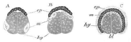
Fig. 86. Sections through the ovum of Leptoplana
tremellaris in three stages of development.
(After Hallez.)
ep. epiblast; m. mesoblast; hy. yolk cells
(hypoblast); bl. blastopore.
In the course of the enclosure of the hypoblast cells by the epiblast,
the mesoblast cells gradually travel towards the formative pole (fig.
86 B). In the process they become first of all divided so as to form
four linear streaks, and finally unite into a continuous layer between
the epiblast and hypoblast, which obliterates the segmentation cavity
(fig. 86 C, m).
Before the completion of the epibole a closely packed layer of fine
cilia appears, which causes a rotation of the embryo within the
egg-capsule. During the above changes a fifth hypoblast cell is formed
by the division of one of those already present; and at a later period
four of the hypoblast cells give rise within the nearly closed
blastoporic area to four small cells. In connection with these cells a
complete hyploblastic wall becomes subsequently established, which
encloses the original large hypoblast cells. The latter then become
resolved into a vitelline mass.
From a comparison with other types it may be regarded as probable that
the enteric wall originates by a process of continuous budding off of
small cells from the large cells, which commences with the formation
of the four cells above mentioned.
The blastopore becomes nearly obliterated, but whether it gives rise
to the mouth, which is formed in the same place, has not been
determined. In front of the mouth a small and very transitory rudiment
of an upper lip makes its appearance. The protrusible pharynx is
stated by Hallez to arise as an hypoblastic bud, while its sheath has
an epiblastic origin. Two pairs of eyes and the supra-œsophageal
ganglia also become early developed.
The peripheral ciliated layer of small cells becomes divided into two
strata, of which the outer remains ciliated and forms the true
epiblast: the inner probably forms the cutis. In it are developed
rod-like bodies, which seem to be homologous with the thread-cells of
the Cœlenterata, so that if the views put forward in the previous
chapter as to the similarity of the turbellarian and cœlenterate
larvæ are correct, the cutis corresponds with the deeper layer of the
cœlenterate epiblast. The mesoblast, like the epiblast, becomes
divided into two strata. The outer one is stated to form the circular
and longitudinal muscles; the inner one to give rise to a muscular
reticulum, the spaces within which constitute the parenchymatous body
cavity.
The later changes are not of great importance. At a period slightly
after the formation of the mouth and ganglia two pairs of stiff hairs
become formed at the sides of the body. The embryo has by this time
grown so as to fill up its capsule, in which however it continues
rapidly to rotate, and also commences to exhibit active contractions.
It next becomes hatched, and passes from a spherical to a flattened
elongated form. The ventral oral opening is at first central, but
soon, by a process of unequal growth, becomes carried towards the
posterior end of the body. The pairs of stiff hairs in the meantime
considerably increase in number. The remains of the yolk cells now
disappear, and the enteric walls become more distinct. The alimentary
canal, which is at first simple in outline like that of a
rhabdocœlous Turbellarian, soon assumes a dendritic form.
The young animal after these changes resembles its parent, except in
the possession of only two pairs of eyes and in the absence of
generative organs.
Of the types with a complete metamorphosis the free larvæ of various
species of Thysanozoon have been observed by Joh. Müller (190) and
Moseley (189), and the complete development of Eurylepta auriculata
has been studied by Hallez.
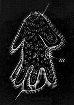
Fig. 87. Larva of Eurylepta auriculata
immediately after hatching. Viewed from the side. (After
Hallez.)
m. mouth.
The stages within the egg of this latter type agree precisely with
those already described in Leptoplana. After the formation of the
mouth the body elongates, remaining however cylindrical. A fold forms
on the anterior side of the mouth, giving rise to a large upper lip.
Two posterior processes are next formed, and other processes soon
arise, constituting the whole of those found in the free larva. The
embryo next shakes off its egg membranes by a series of vigorous
contractions. When free it has the form represented in the annexed
figure (fig. 87).
It is so similar to Müller’s (fig. 88) and Moseley’s
larvæ that all three may be dealt with together.
The body is somewhat oval, and slightly pointed behind. At the
anterior end are placed the eyes, two in the youngest larva of Müller,
and twelve in the older larva (fig. 88), and in the middle of the
ventral surface is the mouth. It is surrounded by a strong fold, and
leads into an alimentary canal, which is at first simple, but in the
older larvæ is much branched. A bilobed ganglion connected with two
nerve cords is placed anteriorly. The superficial epithelium is
ciliated, and below it is a layer of cells (cutis) derived from the
primitive epiblast, in which are formed the usual rods (Hallez). The
chief peculiarity of the larva consists in the presence of elongated
processes covered with long cilia, and so connected together by a
ciliated band that the whole together forms, in Müller’s larva at any
rate, a lobed præoral ciliated band (fig. 88).
This band is not quite so clear in Hallez’ figures. Müller’s youngest
larva was provided with eight very long lobes; three were dorsal, viz.
a median anterior, and two lateral placed far back; three ventral,
viz. a median in the front of the mouth forming a large upper lip, and
two processes at the sides of the mouth. The number was completed by
two lateral processes of the body. All the processes except the dorsal
median one are shewn in fig. 88. In Hallez’ larva, fig. 87, the six
posterior processes form a rather definite ring, while one flagellum
projects from the front end of the body immediately below the eyes,
and a second flagellum behind. In Moseley’s youngest larva six
processes only were present, though subsequently eight became formed
as in Müller’s larvæ.
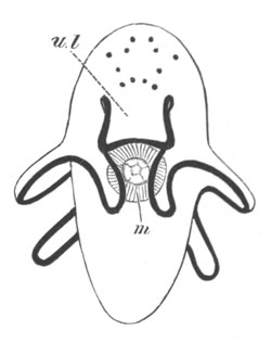
Fig. 88. Müller’s Turbellarian larva (probably
thysanozoon). Viewed from the ventral surface. (After Müller.)
The ciliated band is represented by the black line.
m. mouth; u.l. upper lip.
The metamorphosis consists in the whole animal growing longer and
flatter, and in the arms becoming gradually shorter and shorter till
they finally disappear altogether, and the larva acquires the ordinary
adult form.
The lobed larval form of the Turbellaria has some points of
resemblance to the Pilidium form of nemertine larva described below,
yet its resemblance to this interesting larva is less close than would
appear to be the case with certain turbellarian larval forms recently
described by Götte and Metschnikoff, which are in some respects
intermediate in character between the larva of Leptoplana and those
just described.
The observations of Götte (No. 184) were made on Planaria Neapolitana
and Thysanozoon Diesingi, and those of Metschnikoff (No. 188) on
Stylochopsis ponticus. The larvæ of all these forms undergo more or
less of a metamorphosis, but the accounts of their development are not
easily reconciled.[91] The early stages of Planaria are like those of
Leptoplana, as described by Keferstein. Four large hypoblast
cells become surrounded by small epiblast cells, which commence to be
formed on the dorsal side. The hypoblast cells divide and arrange
themselves in two bilaterally symmetrical rows. A small blastopore is
left by the small cells on the ventral surface, which
communicates with an otherwise closed and ciliated cavity which is
formed between the two rows of hypoblast cells. The blastopore would
seem to remain permanently open, and to be placed at the base of a
deep pit, lined by epiblast cells, which constitutes the stomodæum.
Fig. 89. Planarian larva (probably Planaria
angulata). (From Agassiz.)
Fig. 90. Planarian larva (probably Plamaria
angulata). (From Agassiz.)
The embryo now becomes dorsally convex, while the ventral surface
becomes marked with a median furrow and grows out laterally into two
lobes, and anteriorly into a ventrally-directed upper lip. The whole
surface becomes ciliated, and the cilia are especially prominent on
the ventral processes and the summit of the dorsal dome. A bunch of
strong cilia becomes formed in front of the dome, and a less marked
bunch behind. The larva is now stated by Götte closely to resemble a
Pilidium. It soon, however, extends itself, and the two bunches of
cilia become situated at the anterior and posterior extremities of the
body. The ventral processes become inconspicuous prominences of the
side of the body. Götte believes that the larva undergoes no further
metamorphosis.
A type of Planarian larva (figs. 89 and 90)—possibly Plan. angulata,
observed by Alex. Agassiz (No. 181),—is very different from any other
so far described, and is remarkable for being divided into a series of
segments corresponding in number with the diverticula of the digestive
cavity. In the youngest specimen (fig. 89) the body was nearly
cylindrical, and divided into eleven rings, corresponding with as many
digestive diverticula. Two eye-spots were present. In a later stage
(fig. 90) the body was considerably flattened and had
approached more to the planarian form.
If Agassiz’ interesting observations can be trusted we have in this
larva indications of a distinct segmentation, which are of some
morphological importance, especially when taken in connection with the
traces of segmentation found amongst the Nemertines.
A further type, with an incomplete metamorphosis, has been observed by
Girard (183). It is remarkable for having an uniform segmentation, and
for presenting a quiescent stage after passing through a free larval
condition with a large upper lip.
Fresh-water Dendrocœla. The development of the fresh-water
Dendrocœla has been especially investigated by Knappert (No. 186)
and Metschnikoff (No. 188).
The ova are very delicate minute naked cells, which to the number of
4‑6 or more become enveloped in a capsule or cocoon together with a
large mass of yolk cells derived from the vitellarium. The yolk cells
exhibit peristaltic movements and send out amœboid processes. Each
ovum when laid becomes surrounded by an extremely delicate membrane,
which disappears during the course of development. The capsules
consist of a spherical case and a stalk. The latter is first emitted
from the female opening as a thread-like body. Its free end becomes
attached, and then the remainder of the capsule is ejected.
Impregnation takes place before the formation of the capsule. The
segmentation is complete. The ovum first divides into two segments.
One of these next divides, forming three segments. There are
subsequently stages with four, eight, sixteen, and thirty-two
segments.
Metschnikoff’s results on the stages subsequent to the segmentation
are not in complete harmony with those of Knappert; but no doubt
represent an advance in our knowledge, and I shall follow them here.
His observations were made on Planaria polychroa.
In the earliest stage observed by him the segmentation was already far
advanced, but no membrane was present round the ovum. At a later stage
the ovum becomes more or less bell-shaped or hemispherical, and
encloses within its concavity a mass of yolk elements. It is now
formed of three concentric layers. An outer layer of flattened
cells—the epiblast, a middle layer of fused cells—the mesoblast, and
an inner solid mass of yolk cells—the hypoblast.
At the upper pole is formed the protrusible pharynx (cf. Knappert),
which is provided with a provisional musculature and a lumen. By its
contractions it takes up the yolk elements which surround the embryo,
and the rapid growth of the embryo no doubt takes place at their
expense. The embryo gradually loses its hemispherical form,
and assumes an elongated and flattened shape. It acquires a coating of
cilia by means of which it rotates. On the fifth day it is hatched.
The alimentary tract long remains solid, even after it has acquired
its branched form. The pharynx becomes withdrawn as soon as the larva
is hatched. It loses its provisional muscles, and subsequently
acquires a permanent musculature. The young after hatching attach
themselves to the body of their parent, on which they feed (?).
Rhabdocœla. The development of some of the Rhabdocœla has
recently been studied by Hallez. The ova are mostly laid in capsules,
one in each capsule. Sometimes the development commences before the
capsules are laid, at other times not till afterwards. In certain
forms (Mesostomum) there are summer eggs with thin capsules which
develop within the parent, while hard capsules, forming what are known
as winter eggs, are laid in the autumn, and the embryo hatched in the
spring.
The ova of the Rhabdocœla like those of the fresh-water
Dendrocœla are enveloped in yolk elements derived from the
vitellarium.
The segmentation probably takes place in the same way as in
Leptoplana. A stage with four equal cells has been observed by Hallez,
and there is subsequently an epibolic gastrula. The embryo becomes
ciliated while still within the capsule and, according to Hallez, the
pharynx arises as a bud of the hypoblast. The proboscis in Prostomum
originates as an epiblastic invagination.
Nemertea.
Some Nemertea develop without and some with a metamorphosis.
The most remarkable type of Nemertine development with a metamorphosis
is that in which the ovum develops into a peculiar larval form known
as Pilidium, within which the perfect worm is subsequently evolved.
Closely allied to this type is one in which the sexual worm is
developed within a larval form as in Pilidium, but in which the larva
has no free-swimming stage, and is therefore without the
characteristic appendages of the Pilidium. This is known as the type
of Desor and is confined (?) to the genus Lineus. The Pilidium and the
Desor type may be first considered (vide Barrois, No. 192).
The type of Desor. The segmentation is regular and leads to the
formation of a blastosphere with a large segmentation cavity.
The blastosphere is converted by invagination into a gastrula (fig. 91
A). The blastopore is soon carried relatively forwards by the
elongation backwards of the archenteron, and, according to Barrois,
actually forms the mouth. Owing to the elongation of the archenteric
cavity the embryo assumes a bilateral form (fig. 92 A) in which the
dorsal and ventral surfaces can be distinguished, the mouth
(m.) being situated on the ventral surface.
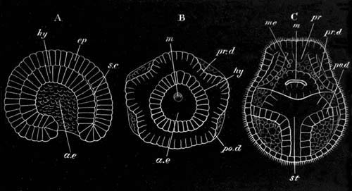
Fig. 91. Three stages in the development of
Lineus.
(After Barrois.)
A is a side view in optical section.
B and C are two later stages from the ventral (oral) surface.
ae. archenteron; sc. segmentation cavity; hy.
hypoblast; me. mesoblast; ep. epiblast; m.
mouth; st. stomach; pr.d. prostomial disc;
po.d. metastomial disc; pr. proboscis.
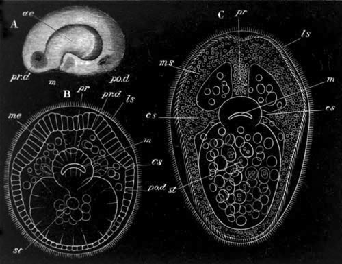
Fig. 92. Three Stages in the development of
Lineus.
(After Barrois.)
A. Side view of an embryo at a very early stage as an opaque object.
B and C. Two late stages, seen as transparent objects from the
ventral surface.
ae. archenteron; m. mouth; pr.d. prostomial disc; po.d.
metastomial disc; cs. lateral pit developing in B as a
diverticulum from the œsophagus; pr. proboscis; ms.
muscular layer (?); ls. larval skin about to be thrown off;
me. mesoblast; st. stomach.
Immediately after the completion of the gastrula a remarkable series
of phenomena takes place. The embryo when viewed from the ventral
surface assumes a pentagonal form (fig. 91 B), and four invaginations
of the epiblast make their appearance on the ventral surface (fig. 92
A), two in front of (pr.d.) and two behind (po.d.) the mouth;
they result in the formation of four thickened discs. These discs soon
become separated from the external skin, which closes in forming an
unbroken layer over them (fig. 91 C). The discs grow rapidly, and
first the prostomial pair and subsequently the metastomial fuse
together, and finally the whole four unite into a continuous ventral
plate; analogous it would seem to the ventral plate of chætopodan and
arthropodan embryos. The plate so formed gradually extends
itself so as to close over the dorsal surface, and to form a complete
skin within the primitive larval skin, which at this period is richly
ciliated, though the embryo is not yet hatched (fig. 91 C). While
these changes are taking place, there are budded off from the
invaginated discs a number of fatty cells, which fill up the space
between the discs and the archenteron, and eventually form the
mesoblastic reticulum. During this stage the rudiment of the proboscis
also makes its appearance as a solid process of epiblast, which grows
backwards from the point of fusion of the two prostomial discs at the
front end of the embryo (fig. 91 C, pr.). A lumen is excavated
in it at a later period. The lateral organs or cephalic pits arise in
a somewhat unexpected fashion as a pair of diverticula from the
œsophagus (fig. 92 B, cs.)[92], which soon fuse with
the walls of the body at the junction of the prostomial and
metastomial plates (fig. 92 C, cs.), although they remain for
some time attached to the œsophagus by a solid cord.
During these changes the original larval skin separates itself from
the subjacent layer formed by the discs (fig. 92, B and C), and is
soon thrown off completely, leaving the already ciliated (fig. 92 C)
external layer of the invaginated discs as the external skin of the
young Nemertine. During, and subsequently to, the casting off of the
embryonic skin, important changes take place in the constitution of
the various layers of the body, resulting in the formation of the
vascular system and other mesoblastic organs, the nervous system, and
the permanent alimentary tract. These changes appear to me to stand in
need of further elucidation; and the account below must be received
with a certain amount of caution.
It has been already stated that the two discs give rise to fatty
cells, which occupy the space between the walls of the body and the
archenteron. At the period of the casting off of the embryonic skin
fresh changes take place. The discs become very much thickened, and
then divide into two layers, which become the epidermis and subjacent
muscular layers. The muscular layers arise in two masses, separated by
the two cephalic sacks. The anterior mass is formed as an unpaired
anterior thickening, followed by two lateral thickenings. The
posterior mass is much thinner, in correspondence with the rapid
elongation of the metastomial portion of the embryo.
The cells originally split off from the discs undergo considerable
changes, some of them arrange themselves around the proboscis as a
definite membrane, which becomes the proboscidean sheath, some
also form a true splanchnic layer of mesoblast, and the remainder,
which are especially concentrated during early embryonic life in the
anterior parts of the body, form the general interstitial connective
tissue. The cephalic ganglia are stated to become gradually
differentiated in the prostomial mesoblast, and the two cords
connected with them in the metastomial mesoblast.
At the time when the larval skin is cast off the original mouth
becomes closed, and it is not till some time afterwards that a
permanent mouth is formed in the same situation. During the early part
of embryonic life the intestine is lined with columnar cells, but,
before the loss of the larval skin, the walls of the intestine undergo
a peculiar metamorphosis. Their cells either fuse or become
indistinguishable, and their protoplasm appears to become converted
into yolk-spherules, which fill up the whole space within the
walls of the body, and are only prevented from extending forwards by a
membrane of connective tissue. This mass gradually forms itself into a
distinct canal, lined by columnar cells.
Pilidium. In the case of the true Pilidium type, the larva is
hatched very early and leads the usual existence of surface larvæ. A
regular segmentation is followed by an invagination which does not
however cause the complete obliteration of the segmentation cavity
(fig. 93 A, a.e.).
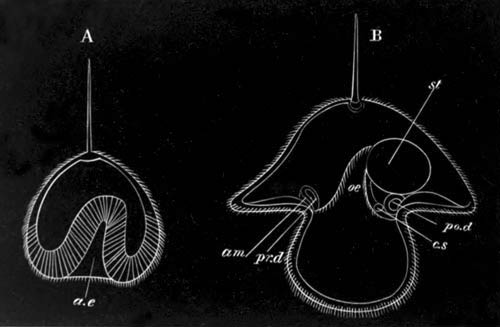
Fig. 93. Two stages in the development of
Pilidium.
(After Metschnikoff.)
ae. archenteron; œ. œsophagus; st.
stomach; am. amnion; pr.d. prostomial disc; po.d. metastomial disc; c.s. cephalic sack.
The primitive alimentary tract so formed becomes divided into
œsophageal and gastric regions (fig. 93 B, œ. and
st.). Even while the invagination of the archenteron is
proceeding, the larva becomes ciliated throughout, and assumes a
somewhat conical form, the apex of the cone being opposite the flat
ventral surface on which the mouth is situated (fig. 93, A and B).
From the apex a flagellum projects in many forms, giving the larva a
helmet-like appearance. In other forms a bunch of long cilia takes the
place of the flagellum (fig. 94), and in others again the flagellum is
not represented. After the completion of the invagination a lobe grows
out on each side of the mouth, and less well-developed lobes may
appear anteriorly and posteriorly. Round the edge of the ventral
surface a ciliated band makes its appearance.
Two pairs of invaginations of the skin, just as in the type of Desor,
now make their appearance, one pair in front of and the other behind
the mouth (fig. 93 B, pr.d. and po.d.), and each of them
by the closure of the opening of invagination forms a sack, the outer
wall of which becomes very thin and the inner wall (corresponding with
the whole invagination of the type of Desor) very thick. The inner
walls of the four thickenings, which I may speak of as discs, now fuse
together, each disc first uniting with its fellow, and finally the two
pairs uniting.
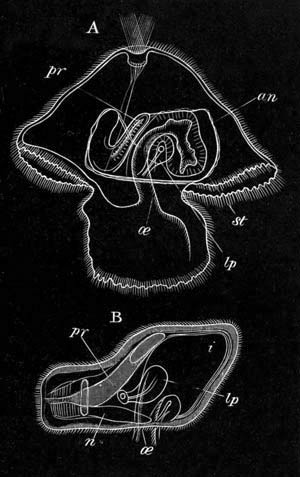
Fig. 94.
A. Pilidium with an advanced nemertine worm.
B. Ripe embryo of the Nemertea in the position it occupies in
Pilidium. (Both after Bütschli.)
œ. œsophagus; st. stomach; i. intestine;
pr. proboscis; lp. lateral pit; an. amnion;
n. nervous system.
A ventral germinal plate is thus established, which gradually grows
round the intestine of the Pilidium to form the skin of the future
Nemertine. The outer thin layer of each of the discs grows pari
passu with the inner layer, and furnishes an amnion-like covering
for the embryo which is forming within the Pilidium (fig. 94,
an).
In connection with the young vermiform Nemertine there is formed on
each side an outgrowth from the œsophagus (fig. 94) which is
eventually placed in communication with the exterior by a
ciliated canal[93]. The proboscis arises as an hollow invagination at
the point where the two anterior discs fuse in front.
When the young Nemertine has become pretty well formed within the
Pilidium it becomes ciliated, begins to move, and eventually frees
itself and leads an independent existence, leaving its amnion in the
Pilidium which continues to live for some time.
The central nervous system (fig. 94) is developed either before or
after the detachment of the young Nemertine, according to Metschnikoff
as a thickening of the epiblast. The young Nemertine is at first
without an anus.
The development of the Nemertine within the Pilidium is clearly
identical with that of the Lineus embryo within the larval skin; the
formation of an amnion in the Pilidium constituting the only important
difference which can be pointed out between the modes of origin of the
young Nemertine in the two types.
So far as is known the forms which develop in a Pilidium, or according
to the type of Desor, all belong to the division of the Nemertines
without stylets in the proboscis, known as the Anopla.
Development without Metamorphosis. The majority of the
Nemertea, including the whole (?) of the Enopla, develop without a
metamorphosis. The observations which have been made on this type are
not very satisfactory, but appear to indicate that the formation of
the hypoblast may take place either by invagination or by delamination.
Invaginate types have been observed by Barrois (No. 192), Dieck (No.
196) and Hubrecht.
Barrois’ fullest observations were made on Amphiporus
lactifloreus (one of the Enopla), and those of Dieck on
Cephalothrix galatheæ (one of the Anopla).
A regular segmentation is followed by a blastosphere stage with a
small segmentation cavity. In Barrois’ type the inner ends of the
cells of the blastosphere are stated to fuse into a kind of syncytium.
A small invagination takes place, and the cells which take part in it
separate from the epiblast, and then fuse with the syncytium
within the blastosphere. Dieck finds that in Cephalothrix the
invaginated mass simply vanishes.
Barrois’ statements about the fusion of the syncytium derived from the
epiblast cells with the invaginated cells must be regarded as very
doubtful. The formation of the germinal layers takes place, according
to Barrois, by the separation of the internal mass of cells into
mesoblast and hypoblast. The proboscis is formed, according to this
author, from the mesoblastic tissues. Dieck, on the other hand, with
greater probability, states that the proboscis is formed by an
invagination. In Cephalothrix a further point deserves notice, in that
the whole of the primitive epiblast becomes shed. In this fact there
may perhaps be recognised the last trace of a metamorphosis like that
in the type of Desor.
Delaminate types have been studied by Barrois (No. 192) and Hoffman
(No. 198), both of whom give circumstantial accounts of their
development.
Hoffman’s account is especially deserving of attention, since his
observations were, to a great extent, made by means of artificial
sections. The following account is taken from him. His observations
were made on Tetrastemma varicolor, and Tetrastemma appears to
be the genus in which this type of development has been most
completely made out. After a regular segmentation the embryo forms a
solid mass of cells, the outermost of which soon become distinguished
as a separate epiblastic layer. At the same time the larva leaves the
egg, and the epiblast cells become coated by an uniform covering of
cilia. At the anterior extremity of the body is a bunch of long cilia;
and at the hinder end two stiff bristles are formed, but soon
disappear.
The internal mass of cells is still quite uniform, but as the larva
grows in length the outermost of them arrange themselves as a columnar
layer, constituting the mesoblast. Of the cells internal to the
mesoblast the outer become columnar, and are converted into the walls
of the alimentary tract, while the inner ones undergo fatty
degeneration, and form a kind of food-yolk. In the later development
the characters of the adult are gradually acquired without
metamorphosis, and the larval skin passes directly into that of the
adult. Both mouth and anus are formed nearly simultaneously by a
rupture of the enteric wall from within. The nervous system arises as
a thickening of the epiblast, which Hoffman states he has been able to
see in sections. Hoffman also states that the epithelium of the
proboscis is formed as a diverticulum of the alimentary tract, and
that its sheath is formed by a special mesoblastic growth.
Barrois is less precise than Hoffman, from whom he differs in certain
particulars. Hoffman’s statements about the proboscis are important if
accurate, but require further confirmation.
Malacobdella. The early stages in development of the peculiar
ectoparasitic Nemertine Malacobdella have been worked by
Hoffman (No. 199) by means of sections, and there appears to be a
close agreement between the development of Malacobdella and that of
Tetrastemma.
The segmentation is uniform, and there is no trace of a segmentation
cavity. On the third day after impregnation the outermost
cells of the embryo become flattened and ciliated, and distinguished
from the remaining spherical cells of the embryo as the epiblast. With
the appearance of cilia a rotation of the embryo commences. On the
fourth day the embryo becomes oval, and at one of the poles—the
future anal pole—a separation takes place between the epiblast and
the inner cells, giving rise to the body cavity. In it are a number of
loose oval cells, which soon become stellate, and form a mesoblastic
reticulum connecting the body wall and central cells of the embryo,
which may now be spoken of as hypoblast. The body cavity increases in
size, leaving at last the hypoblast and epiblast united only at one
point—the oral pole—at which, on the fifth day, a crown of long
cilia appears. The solid mass of hypoblast in the interior becomes
differentiated into an outer layer of cells—the true glandular
epithelium of the alimentary tract—and an inner core, the cells of
which soon undergo fatty degeneration, and serve as food-yolk.
The later stages of development, and the formation of the proboscis,
etc., have not been worked out.
General considerations. Of the types of larvæ hitherto found
amongst the Nemertea, those with a metamorphosis, viz. the Pilidium
type and that of Desor, are to be regarded as the primitive. But even
in Pilidium there are evidences of a great abbreviation in
development. Pilidium itself is probably a more or less modified
ancestral form, while the peculiar development of the Nemertine within
it is to be explained as a very much shortened record of a long series
of changes by which the Pilidium became gradually converted into a
Nemertine. The formation of the body wall of the Nemertine by four
epiblastic invaginations is a remarkable embryological phenomenon, for
which it is not easy to assign a satisfactory meaning; and it is
probable that it is merely a secondary process of growth similar to
the formation of imaginal discs in the larvæ of Diptera (vide Chapter
on Tracheata), which has had its origin in the abbreviation of the
development just alluded to. The development of the type of Desor is
clearly a simplification of the Pilidium type, and its peculiarities
are to be explained by the fact that the first larval form has no free
existence. The types without metamorphosis have no doubt a development
of a still more simplified character; they are remarkable however in
presenting us, if the existing descriptions are to be trusted, with
examples of delamination and invagination coexisting in closely allied
forms.
Trematoda.
The eggs of the Trematoda consist of a germ or true ovum enclosed in a
mass of yolk cells, which undergo disintegration and subsequent
absorption at varying periods of the development. From the
observations of E. van Beneden (No. 218), Zeller (No. 217), etc. it is
known that the segmentation is usually complete, but generally
somewhat irregular.
Unfortunately we are still completely in the dark as to the mode of
formation of the germinal layers. The embryos of the entoparasitic
forms or Distomeæ become free in a very imperfect condition, and the
ova are small while in the Polystomeæ the development is as a rule
nearly completed before hatching, and the ova are large. It will be
convenient to treat separately the development of the two groups.
Distomeæ. The embryos of the Distomeæ are hatched either in
some moist place or more usually in water. In the majority of genera
the larvæ pass through a complicated metamorphosis, accompanied by
alternations of generations. But for some genera, e.g.
Holostomum, etc., the life history has not yet been made out. The
whole life history of comparatively few forms has been followed, but
sufficient fragments are known to justify us in making certain general
statements, which no doubt hold true for a large proportion of the
Distomeæ.
The larvæ are usually ciliated (fig. 95 A), but sometimes naked.
The ciliated forms are generally completely covered with cilia, but in
Distomum lanceolatum the cilia are confined to an area at the
front end of the body, in the centre of which a median spine is
placed. An x shaped pigment spot, sometimes provided with a
rudimentary lens (Monostomum mutabile), is also generally
situated on the dorsal surface.
In some instances a more or less completely developed alimentary tract
is present (Monostomum capitellum, Amphistomum
subclavatum), but usually there can only be distinguished in the
interior of the larva a transparent mass of cells bounded by a more or
less distinctly marked body wall with ciliated excretory channels.
Ed. van Beneden has shewn that the ciliated covering is developed
while the embryo is still in the egg, and long before the yolk cells
are completely absorbed. It would seem that even before hatching this
ciliated covering is to a great extent independent of the mass within.
In the larva of Monostomum mutabile (fig. 95 A), which offers
an example of an extreme case of the kind, there is present within the
ciliated epidermis a fully developed independent worm.
The non-ciliated larvæ are less highly organized than the ciliated
forms, and are covered by a cuticle: their anterior extremity is
sometimes provided with a circular plate armed with radiate ridges and
spines.
The free-swimming or creeping embryos make their way into or on to the
body of some invertebrate (occasionally vertebrate) form, usually a
Mollusc, to undergo the first stage in their metamorphosis. They may
either do this on the gills of their host, or very frequently they
bore their way into the interior of the body. Soon after the larvæ
have reached a satisfactory position the epidermis becomes stripped
off, and there emerges a second larval form developed in the interior
of the first larva, much as a Nemertine is developed within the larva
of Desor. In the case of Monostomum mutabile the new worm is, as
stated above, fully formed within the ciliated larva at the time of
hatching.
The worm which proceeds from the above metamorphosis has different
characters corresponding with those of the larva from which it
proceeded. If the original larva had an alimentary canal it has one
also, and then grows into the form known as a Redia (Fig. 95, B and
C).
The Redia has anteriorly a mouth leading into a muscular pharynx and
thence into a cæcal stomach. Posteriorly the body is prolonged into a
kind of blunt caudal process, at the commencement of which are a pair
of lateral papillæ. There is a perivisceral cavity, and the body walls
are traversed by excretory tubes.
If the original larva is without an alimentary tract, the second form
becomes what is known as a Sporocyst. The Sporocyst is a simple
elongated sack with a central body cavity; when derived from the
metamorphosis of a ciliated embryo its walls are provided with
excretory tubes, but such tubes are absent in Sporocysts developed
from non-ciliated larvæ. Some Sporocysts send out numerous branches
amongst the viscera of their hosts.
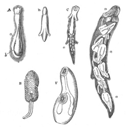
Fig. 95. Various stages in the Metamorphosis of
the Distomeæ (from Huxley.)
A. Ciliated larva of Monostomum mutabile. a. larval
skin. b. Redia developed within it. B. Redia of
Monostomum mutabile. C. Redia of Distomum pacificum, with
germs of a second brood of Rediæ. D. Redia containing
Cercariæ. E. Cercaria. F. Full-grown Distomum.
The Rediæ and Sporocysts rapidly grow in size and sometimes increase
by transverse division. In the course of their further
development one of two things may happen. They may either (1) develop
fresh Rediæ or Sporocysts by a process of internal budding (fig. 95
C); or else (2) there may be formed in them, by an analogous process,
larvæ with long tails known as Cercariæ (fig. 95 D.) The direct
development of Cercariæ is the usual course, though in Distomum
globiparum the reverse is true; but where this does not take place
the Rediæ or Sporocysts of the second generation give rise to
Cercariæ.
The Cercariæ are developed from spherical masses of cells found in the
body cavity of the Sporocyst or Redia. The exact origin of these
masses is still somewhat obscure, but they are stated by Wagener (No.
212) to be derived from the body wall. They are probably to be
regarded as internal buds.
The spherical bodies grow rapidly in size, their posterior extremity
is prolonged into a process which forms the tail, while the anterior
part forms the trunk. When fully formed (fig. 95 E), the trunk has
very much the organization of an adult Distomum. There is an anterior
and a ventral sucker, the former of which contains the opening of the
mouth, and is often provided with a special chitinous armature. The
mouth leads into a muscular pharynx, and this into a bilobed cæcal
alimentary tract. An excretory system of the ordinary type is present,
consisting of longitudinal contractile trunks continuous anteriorly
with branched ciliated canals, which, as has recently been shewn by
Bütschli, may be provided with funnel-shaped ciliated internal
openings[94]. The contractile trunks unite posteriorly, but
instead of opening directly to the exterior are prolonged into a
vessel which traverses the substance of the tail, and after a longer
or shorter course bifurcates into two branches which open laterally.
The tail is provided with an axial rod of hyaline connective tissue,
like the notochord of the tail of a larval Ascidian, and is frequently
provided with membranous expansions. It is used as a swimming organ.
Beneath the epidermis are layers of circular and longitudinal muscular
fibres, the latter arranged in the tail as two bands.
The Cercariæ when fully developed leave the Sporocyst or Redia, and
then their host, and become free. In most Rediæ there is a special
opening, not far from the mouth, by which they pass out. There is no
such opening in Sporocysts, but the Cercariæ bore their way through
the walls.
After leaving their parent the Cercariæ pass into the external medium,
and for a short period have a free existence. They soon however enter
a new host, making their way into its body by a process of boring,
which is effected by the head (especially when armed with chitinous
processes) assisted by movements of the tail.
The second host is usually some Invertebrate (Mollusc, Worm,
Crustacean, Insect larva, &c.), but occasionally a Fish or Amphibian
or even a vegetable. The tail is very often lost as the Cercaria bores
its way into its host, but whether it is so or not, the Cercaria,
after it has once reached a suitable post in its new host, assumes a
quiescent condition, and surrounds itself with a many-layered capsule.
The cephalic armature and tail (if still present) are then exuviated,
and the generative organs gradually become apparent though very small.
In other respects the organization is not much altered.
Though an encysted Cercaria may remain some months without further
change, it eventually dies unless it be introduced into its permanent
vertebrate host, an act which is usually effected by the host in which
it is encysted being devoured. It then becomes freed from its capsule
as a fully formed Trematode, in which the generative organs rapidly
complete their development.
In some cases the Rediæ or Sporocysts do not give rise to
tailed Cercariæ, but to tailless forms. In such cases, as a rule, the
encystment takes place in the host of the Redia or Sporocyst, but the
tailless larvæ sometimes pass through a free stage like the Cercariæ.
In the case of Distomum cygnoides, parasitic in the bladder of
the Frog, the Cercaria passes directly into the adult host without the
intervention of an intermediate host.
The life history of a typical entoparasitic Trematode is shortly as
follows:
(1) It leaves the egg as a ciliated or non-ciliated free larva.
(2) This larva makes its way on to the gills or into the body of some
Mollusc or other host, throws off its epidermis and becomes a Redia or
Sporocyst.
(3) In the body cavity of the Redia or Sporocyst numerous tailed
larvæ, known as Cercariæ, are developed by a process of internal
gemmation.
(4) The Cercariæ pass out of the body of their parent, and out of
their host, and become for a short time free. They then pass into a
second, usually invertebrate host, and encyst.
(5) If their second host is swallowed by the vertebrate host of the
adult of the species, the encysted forms become free, and attain to
sexual maturity.
The majority of these stages are simply parts of a complicated
metamorphosis, but in the coexistence of larval budding (giving rise
to Cercariæ or fresh Rediæ) with true sexual reproduction there is in
addition a true alternation of generations.
Polystomeæ. The ova of the Polystomeæ are usually large and not
very numerous, and they are in most cases provided with some process
for attachment. Some species of Polystomeæ, e.g. Gyrodactylus,
are however viviparous. The young leave the egg in a nearly perfect
state, and at the utmost undergo a slight metamorphosis and no
alternations of generations. Some however (Polystomum, Diplozoon) are
provided with temporary cilia, but the number investigated is too
small to determine whether ciliation is the rule or the exception. The
ciliated larvæ have a short free existence. The cilia are developed on
special cells which may be arranged in transverse bands in the same
way as in the larvæ of many Chætopods, but are not, in the larvæ at
present known, distributed uniformly. When the free larvæ become
parasitic the cells with cilia shrink up.
In Polystomum integerrimum, which lives in the urinary bladder
of Rana temporaria, the eggs when laid in the spring pass out
into the water. The segmentation is complete, and the embryo when
hatched is provided with most of the adult organs, but presents
certain striking larval characters. It has five rings of ciliated
cells. Three of these are placed anteriorly, and are especially
developed on the ventral surface, the posterior one being incomplete
dorsally; two are placed posteriorly, and are especially developed on
the dorsal surface. Anteriorly there is a tuft of cilia.
The larva itself resembles somewhat an adult Gyrodactylus, and is
provided (1) with a large posterior disc armed with hooks, and (2)
with two pairs of eyes which persist in the adult state. After a
certain period of free existence the larva attaches itself to the
gills of a tadpole. The rings of ciliated cells shrink up, and some of
the six pairs of suckers found in the adult commence to be formed on
the posterior disc. When the bladder of the tadpole is developed, the
young Polystomum passes down the alimentary tract to the cloaca, and
thence to the urinary bladder, where it slowly attains to sexual
maturity. When the larva becomes attached to the gills of a very young
tadpole, its development is somewhat more rapid in consequence of
better nutrition from the more delicate gills. It then reaches its
full development in the gill cavity, and, though smaller and provided
with differently organised generative organs to the normal form,
produces generative products and dies without being transported to the
bladder (vide Zeller, Nos. 216 and 217).
The ova of Diplozoon, a form parasitic on the gills of freshwater fish
(Phoxinus, etc.), are provided with a long spiral filament (Zeller,
No. 215). The embryo has five ciliated areas, four lateral and one
posterior. The young form is known as Diporpa. Sexual maturity is not
attained till two individuals unite permanently together. They unite
by the ventral sucker of each of them becoming attached to the dorsal
papilla of the other. Subsequently these parts coalesce, and the
ventral suckers disappear in the process. Gyrodactylus, parasitic,
like Diplozoon, on the gills of freshwater fishes (Gasterosteus,
etc.), is remarkable for its mode of reproduction. It is viviparous,
producing a single young one at a time, and, what is still more
remarkable, the young while still within its parent produces a young
one, and this again a young one, so that three generations may be
present within the parent. It seems probable that the second and third
generations are produced asexually, the generative organs not being
developed; while the young Gyrodactylus of the first generation
springs from a fertilized ovum (Wagener, No. 214).
Cestoda.
On anatomical grounds the affinity of the Cestoda to the Trematoda has
been insisted on by the majority of anatomists. The existence of such
intermediate forms as Amphilina tends to strengthen this view;
and the striking resemblances between the two groups in the structure
of the egg and characters of the metamorphosis appear to me to remove
all doubt about the matter.
The ripe egg is formed of a minute germ enveloped in yolk cells, the
whole being surrounded by a membrane, which is very delicate in most
forms, but in certain types has a firmer consistency, and is provided
with an aperture, covered by an operculum, by which the larva escapes.
The early development, up to the formation of a six-hooked larva,
generally takes place in the uterus, but in the types with a firmer
egg-shell it takes place after the egg has been deposited in water.
The segmentation (E. van Beneden, No. 218, Metschnikoff, No. 228) is
complete, and during its occurrence the yolk cells surrounding the
germ are gradually absorbed, so that the mass of segmentation spheres
grows in size, till at the close of segmentation it fills up nearly
the whole egg-shell.
As was first shewn by Kölliker for Bothriocephalus salmonis, the
embryonic cells separate themselves at the close of segmentation into
a superficial layer and a central mass.
The further development takes place on two types. In the cases where
the egg-shell is strong, and the egg is laid prior to the formation of
the embryo, a ciliated larva is developed (Bothriocephalus latus,
ditremus, Schistocephalus dimorphus, Ligula simplicissima, etc.[95]).
Of these forms Bothriocephalus latus may be taken as type.
The development of the embryo requires many months for its completion.
The outer layer becomes ciliated while the central mass has already
become developed into a six-hooked embryo. The embryo leaves its shell
by the opercular aperture, and for some time swims rapidly about by
means of its long cilia. The ciliated coating is eventually stripped
off, and the six-hooked larva emerges.
In the second type of embryo the external cellular layer does not
become ciliated. This is the most usual arrangement, and is even found
in many species of Bothriocephalus.
The central mass of cells becomes developed, as in the other type,
into a six-hooked (rarely four-hooked) embryo (fig. 96 G), but the
superficial layer separates from the central, and either disappears or
becomes (Bothriocephalus proboscideus) a cuticular layer.
Between the six-hooked embryo and the outer layer of cells one or more
thick membranes become deposited (E. van Beneden). The eggs are
carried out of the alimentary canal in the proglottis and transported
to various situations on land or in water. They usually remain within
the proglottis, invested by their thick shell, till taken up into the
alimentary canal of a suitable host, or they may be swallowed after
the death and decay of the proglottis. They are subsequently hatched
after their shell has become softened by the action of the digestive
fluids.
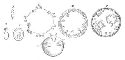
Fig. 96. Diagrams of various stages in the
development of the Cestoda.
(From Huxley.)
A. Cysticercus. B. and C. Cysticerci in the everted (B) and inverted
(C) condition. D. Cœnurus. E. and F. Diagrams of Echinococcus. It
is most probable that Tænia heads are not developed directly from
the wall of the cyst as represented in the diagram. G. Six-hooked
embryo.
Before proceeding to describe their further history, the close
resemblance between the first developmental stages of Cestoda,
especially in the case of the ciliated larvæ, and those of Trematoda,
may be pointed out.
In both there is a ciliated larva, and in both there is developed
within the ciliated skin a second larva, which becomes freed by the
stripping off of the ciliated skin.
The type of development has moreover many analogies with that of the
Nemertine larva of Desor, p. 163 (cf. Metschnikoff), and is probably
like that an abbreviated record of a long history.
The suitable host for the six-hooked embryo to enter is rarely
the same as the host for the sexual form. The embryos having become
transported into the alimentary canal of such a host, and become free,
if previously invested by the egg-shell, soon make their way,
apparently by the help of their hooks, through the wall of the
alimentary tract, and are transported in the blood or otherwise into
some suitable place for them to undergo their next transformation.
This place may be the liver, lungs, muscles, connective tissue, or
even the brain (e.g. Cœnurus cerebralis in the brain
of sheep).
Here they become enclosed in a granular deposit from the surrounding
tissues, which becomes in its turn enclosed in a connective-tissue
coat. Within lies the solid embryo, the hooks of which in many cases
disappear or become impossible to make out. In other forms,
e.g. Cysticercus limacis, they remain visible, and then
mark the anterior pole of the worm (fig. 98, c.). The central
part of the body next becomes transformed into a material composed of
clear non-nucleated vesicles. Accompanying these changes the embryo
grows rapidly in size; a cuticle is deposited by its outer layer, in
which also an external layer of circular muscular fibres and an
internal layer of longitudinal fibres become differentiated, and
internal to both there is formed a layer of granular cells.
With the rapid growth of the body a central cavity is formed, which
becomes filled with fluid, and the embryo assumes the form of a
vesicle. At the same time a system of excretory vessels, sometimes
opening by a posterior pore, becomes visible in the wall of the
vesicle.
The embryo has now reached a condition in which it is known as a
cystic- or bladder-worm, and may be compared in almost every respect
with the sporocyst of a Trematode (Huxley).
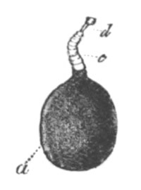
Fig. 97. Cysticercus cellulosæ. (From
Gegenbaur, after von Siebold.)
a. Caudal vesicle. c. Anterior part of body. d.
head.
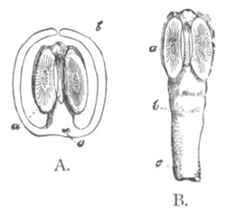
Fig. 98. Cysticercus with small caudal
vesicle.
A. Head involuted. B. Head everted.
a. Scolex. b. caudal vesicle. c. (in A) six
embryonic hooks.
The next important change consists in the development of a head, which
becomes the head of the adult Tænia. This is formed in an involution
of the outer wall of the anterior extremity of the cystic worm. This
involution forms a papilliform projection on the inner surface of the
wall of the cystic worm, with an axial cavity opening by a pore on the
outer surface. The layer of cells forming the papilla soon becomes
divided into two laminæ, of which the outer forms a kind of investing
membrane for the papilla. The papilla itself now becomes
moulded into a Cestode head, which however is developed in an inverted
position. The suckers and hooks (when present) of the head are
developed on a surface bounding the axial lumen of the papilla, which
is the true morphological outer surface, while the apparent outer
surface of the papilla is that which eventually forms the interior of
the (at first) hollow head. Before the external armature of the head
has become established, four longitudinal excretory vessels,
continuous with those in the body of the cystic worm, make their
appearance. They are united by a circular vessel at the apex of the
head. The development is by no means completed with the simple growth
of the head, but the whole inverted papilla continues to grow in
length, and gives rise to what afterwards becomes part of the trunk.
The whole papilla eventually becomes everted, and then the cystic worm
takes the form (fig. 97) of a head and unsegmented trunk with a
vesicle—the body of the cystic worm—attached behind. The whole larva
is known as a Cysticercus. The term scolex, which is
also sometimes employed, may be conveniently retained for the head and
trunk only. The head differs mainly from that of the adult in being
hollow.
There are great variations in the relative size of the head and the
vesicle of Cysticerci. In some forms the vesicle is very small (fig.
98), e.g. Cysticercus limacis; it is medium-sized in
Cysticercus cellulosæ (fig. 97), and in some forms is much
larger. The embryonic hooks, when they persist, are found at the
junction of the trunk and the vesicle (fig. 98 A, c). Though
the majority of cystic worms only develop one head, this is not
invariably the case. There is a cystic worm found in the brain of the
sheep known as Cœnurus cerebralis—the larva of Tænia
cœnurus, parasitic in the intestine of the dog—which forms an
exception to this rule. There appears, to start with, a tuft of three
or four heads, and finally many hundred heads are developed (fig. 96
D). They are arranged in groups at one (the anterior?) pole of the
cystic worm.
A still more complicated form of cystic worm is that known as
Echinococcus, parasitic in the liver, lungs, etc. of man and various
domestic Ungulata. In the adult state it is known as Tænia
echinococcus and infests the intestine of the dog. The cystic worm
developed from the six-hooked embryo has usually a spherical form, and
is invested in a very thick cuticle (fig. 96 E and F, and fig. 99). It
does not itself directly give rise to Tænia heads, but after it
reaches a certain size there are formed on the inner side of its walls
small protuberances, which soon grow out into vesicles connected with
the walls of the cyst by narrow stalks (figs. 96 F and 99 C). In the
interior of these vesicles a cuticle is developed. It is in these
secondary vesicles that the heads originate. According to Leuckart,
they either arise as outgrowths of the wall of the vesicle on the
inner face of which the armature is developed, which subsequently
become involuted and remain attached to the wall of the vesicle by a
narrow stalk, or they arise from the first as papilliform projections
into the lumen of the vesicle, on the outer side of which the armature
is formed. Recent observers only admit the second of these modes of
development. The Echinococcus larva, in addition to giving rise to the
above head-producing vesicles, also gives rise by budding to fresh
cysts, which resemble in all respects the parent cyst. These cysts may
either be detached in the interior (fig. 96 F) of the parent or
externally. They appear to spring in most cases from the walls of the
parent cyst, but there are some discrepancies between the various
accounts of the process. In the cysts of the second generation
vesicles are produced in which new heads are formed. As the primitive
cyst grows, it naturally becomes more and more complicated, and the
number of heads to which one larva may give rise becomes in this way
almost unlimited.
Cysticerci may remain a long time without further development, and
human beings have been known to be infested with an Echinococcus cyst
for over thirty years. When however the Cysticercus with its head is
fully developed, it is in a condition to be carried into its final
host. This takes place by the part of one animal infested with
cysticerci becoming eaten by the host in question. In the alimentary
canal of the final host the connective-tissue capsule is digested, and
then the vesicular caudal appendage undergoes the same fate, while the
head, with its suckers and hooks, attaches itself to the walls of the
intestine. The head and rudimentary trunk, which have been up to this
time hollow, now become solid by the deposition of an axial tissue;
and the trunk very soon becomes divided into segments, known as
proglottides (fig. 99 A). These segments are not formed in
the same succession as those of Chætopods; the youngest of
them is that nearest to the head, and the oldest that furthest removed
from it. Each segment appears in fact to be a sexual individual, and
is capable of becoming detached and leading for some time an
independent existence. In some cases, e.g. Cysticercus
fasciolaris, the segmentation of the trunk may take place while
the larva is still in its intermediate host.
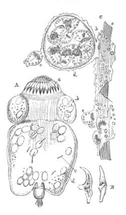
Fig. 99. Echinococcus veterinorum. (From
Huxley.)
A. Tænia head or scolex. a. hooks. b. suckers.
c. cilia in water vessel. d. refracting particles in
body wall.
B. single hooks.
C. portion of cyst. a. cuticle. b. membranous wall of
primary cyst. c. and e. scolex heads. d.
secondary cyst.
The stages in the evolution of the Cestoda are shortly as follows:
1. Stage with embryonic epidermis either ciliated (Bothriocephalus,
etc.) or still enclosed in the egg-shell. This stage corresponds to
the ciliated larval stage of the Trematoda.
2. Six-hooked embryonic stage after the embryonic epidermis has been
thrown off. During this stage the embryo is transported into the
alimentary tract of its intermediate host, and boring its way into the
tissues, becomes encapsuled.
3. It develops during the encapsuled state into a cystic worm,
equivalent to the sporocyst of Trematoda.
4. The cystic worm while still encapsuled develops a head with suckers
and hooks, becoming a Cysticercus. In some forms (Cœnurus,
Echinococcus) reproduction by budding takes place at this
stage. The head and trunk are known as the scolex.
5. The Cysticercus is transported into the second and permanent host
by the infested tissue being eaten. The bladder-like remains of the
cystic worm are then digested, and by a process of successive budding
a chain of sexual proglottides are formed from the head, which remains
asexual.
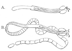
Fig. 99 a. Tetrarhyncus. (From Gegenbaur;
after Van Beneden.)
A. Asexual state.
B. Sexual stage with ripe proglottides.
The above development is to be regarded as a case of
complicated metamorphosis secondarily produced by the necessities of a
parasitic condition, to which an alternation of sexual and gemmiparous
generations has been added. The alternation of generations only occurs
at the last stage of the development, when the so-called head, without
generative organs, produces by budding a chain of sexual forms, the
embryos of which, after passing through a complicated metamorphosis,
again become Cestode heads.
In the case of Cœnurus and Echinococcus two or more asexual
generations are interpolated between the sexual ones. It is not quite
clear whether the production of the Tænia head from the cystic worm
may not be regarded as a case of budding. There are some grounds for
comparing the scolex to the Cercaria of Trematodes, cf. Archigetes.
As might be anticipated from the character of the Cestode
metamorphosis, the two hosts required for the development are usually
forms so related that the final host feeds upon the intermediate host.
As familiar examples of this may be cited the pig, the muscles of
which may be infested by Cysticercus cellulosæ, which becomes
the Tænia solium of man. Similarly a Cysticercus infesting the
muscles of the ox becomes the Tænia mediocanellata of man. The
Cysticercus pisciformis of the rabbit becomes the Tænia
serrata of the dog. The Cœnurus cerebralis of the
sheep’s brain becomes the Tænia cœnurus of the dog. The
Echinococcus of man and the domestic herbivores becomes the Tænia
echinococcus of the dog.
Cystic worms infest not only Mammalian forms, but lower Vertebrates,
various fishes which form the food of other fishes, and Invertebrates
liable to be preyed on by vertebrate hosts. So far the Cestodes
(except Archigetes) are only known to attain sexual maturity in the
alimentary tracts of Vertebrata.
The rule that the intermediate host is not the same as the final host
does not appear to be without exception. Redon[96] has shewn by
experiments on himself that a Cysticercus (cellulosæ)
taken from a human subject develops into Tænia solium in the
intestines of a man. Redon took four cysts of a Cysticercus from a
human subject, and after three months passed some proglottides, and
subsequently the head of Tænia solium.
Some important variations of the typical development are known.
The so-called head or scolex may be formed without the intervention of
a cystic stage. In Archigetes (Leuckart, No. 227), which infests, in
the Cysticercus condition, the body cavity of various invertebrate
forms (Tubifex, etc.), the six-hooked embryo becomes elongated and
divided into two sections, one forming the head, while the other, with
the six embryonic hooks, forms an appendage, homologous with the
caudal vesicle of other Cysticerci.
The embryo of Tænia elliptica similarly gives rise to a
Cysticercus infesting the dog-louse (Trichodectes canis),
without passing through a vesicular condition; but the caudal vesicle
disappears, so that it forms simply a scolex. These cases may, it
appears to me, be probably regarded as more primitive than the
ordinary ones, where the cystic condition has become exaggerated as an
effect of a parasitic life.
In some cases the larva of a Tænia has a free existence in the scolex
condition. Such a form, the larva of Phyllobothrium, has been observed
by Claparède[97]. It was not ciliated, and was without a caudal
vesicle; and was no doubt actively migrating from an intermediate host
to its permanent host.
Scolex forms, without a caudal vesicle, are found in the mantle cavity
of Cephalopoda, and appear to be occupying an intermediate host in
their passage from the host of the cystic worm to that of the sexual
form.
Archigetes, already mentioned, has been shewn by Leuckart (No. 227) to
become sexually mature in the Cysticercus state, and thus affords an
interesting example of pædogenesis. It is not known for certain
whether under normal circumstances it reaches the mature state in
another host.
Amphilina. The early stages of this interesting form
have been investigated by Salensky (No. 229), and exhibit clear
affinities to those of the true Cestoda. An embryonic provisional skin
is formed as in Cestodes; and pole cells also appear. Within the
provisional skin is formed an embryo with ten hooks. After hatching
the provisional skin is at once thrown off, and the larva, which is
then covered by a layer of very fine cilia, becomes free. The further
metamorphosis is not known.
Bibliography.
Turbellaria.
(181) Alex. Agassiz. “On the young stages of a few Annelids”
(Planaria angulata). Annals Lyceum Nat. Hist. of New
York, Vol. VIII. 1866.
(182) Dalyell. “Powers of the Creator.”
(183) C. Girard. “Embryonic development of Planocera
elliptica.” Jour. of Acad. of Nat. Sci. Philadelphia. New
Series, Vol. II. 1854.
(184) Alex. Götte. “Zur Entwicklungsgeschichte d.
Seeplanarien.” Zoologischer Anzeiger, No. 4, 1878.
(185) P. Hallez. Contributions à l’histoire naturelle des
Turbellariés. Thésis à la faculté des Sciences p. le grade d. Docteur
ès-sciences naturelles, Lille, 1879.
(186) Knappert. “Bijdragen tot de Ontwikkelings-Geschiedenis
der Zoetwater-Planarien.” Provinciaal Utrechtsch Genootschap van
Kunsten en Wetenschappen. Utrecht, 1865.
(187) W. Keferstein. “Beiträge z. Anat. u. Entwick. ein.
Seeplanarien von St. Malo.” Abh. d. könig. Gesell. d. Wiss. zu
Göttingen. Bd. XIV. 1868.
(188) El. Metschnikoff. “Untersuchungen üb. d. Entwicklung d.
Planarien.” Notizen d. neurussischen Gesellschaft d.
Naturforscher. Odessa, Bd. V. 1877. Vide Hoffman and
Schwalbe’s Bericht for 1878.
(189) H. N. Moseley. “On Stylochus pelagicus and a new species
of pelagic Planarian, with notes on other pelagic species, on the
larval forms of Thysanozoon, etc.” Quart. Journ. of Micr.
Science. Vol. XVII. 1877.
(190) J. Müller. “Ueber eine eigenthümliche Wurmlarva a. d.
Classe d. Turbellarien, etc.” Müller’s Archiv f. Anat. u. Phys.
1850.
(191) —— “Ueber verschiedene Formen von Seethieren.” Müller’s
Archiv f. Anat. und Phys. 1854.
Nemertea.
(192) J. Barrois. “l’Embryologie des Némertes.” An. Sci.
Nat. Vol. VI. 1877.
(193) O. Bütschli. Archiv f. Naturgeschichte, 1873.
(194) A. Krohn. “Ueb. Pilidium u. Actinotrocha.” Müller’s
Archiv, 1858.
(195) E. Desor. “Embryology of Nemertes.” Proceedings of the
Boston Nat. History Society, Vol. VI. 1848.
(196) G. Dieck. “Entwicklungsgeschichte d. Nemertinen.”
Jenaische Zeitschrift, Vol. VIII. 1874.
(197) C. Gegenbaur. “Bemerkungen üb. Pilidium gyrans, etc.”
Zeitschrift für wiss. Zool., Bd. V. 1854.
(198) C. K. Hoffmann. “Entwicklungsgeschichte von Tetrastemma
tricolor.” Niederländisches Archiv, Vol. III. 1876,
1877.
(199) —— “Zur Anatomie und Ontogenie von Malacobdella.”
Niederländisches Archiv, Vol. IV. 1877.
(200) W. C. McIntosh. British Annelids. The
Nemerteans. Ray Society, 1873‑4.
(201) Leuckart u. Pagenstecher. “Untersuchungen üb. niedere
Seethiere.” Müller’s Archiv, 1858.
(202) E. Metschnikoff. “Studien üb. die Entwicklung d.
Echinodermen u. Nemertinen.” Mém. Acad. imp. Pétersbourg,
VII. Ser., Tom. XIV. No. 8, 1869.
Trematoda.
(203) T. S. Cobbold. Entozoa. Groombridge and Son, 1864.
(204) —— Parasites; a Treatise on the Entozoa, etc.
Churchill, 1879.
(205) Filippi. Mém. p. servir à l’histoire génétique des
Trématodes. Ann. Scien. Nat. 4th Series, Vol. II. 1854,
and Mem. Acad. Torino, 1855‑1859.
(206) R. Leuckart. Die menschlichen Parasiten, Vol.
I. 1863, p. 485, et seq.
(207) H. A. Pagenstecher. Trematoden u.
Trematodenlarven. Heidelberg, 1857.
(208) C. Th. von Siebold. Lehrbuch d. vergleich. Anat.
wirbelloser Thiere. Berlin, 1848.
(209) J. J. S. Steenstrup. Generationswechsel. 1842.
(210) R. v. Willemoes-Suhm. “Zur Naturgeschichte d. Polystomum
integerrimum, etc.” Zeit. f. wiss. Zool. Vol. XXII.
1872.
(211) —— “Helminthologische Notizen III.” Zeit. f. wiss.
Zool. Vol. XXIII. 1873. Vide this paper for a summary of
known observations and literature.
(212) G. R. Wagener. Beiträge zur Entwicklungsgeschichte d.
Eingeweidewürmer. Haarlem, 1855.
(213) G. R. Wagener. “Helminthologische Bemerkungen, etc.”
Zeit. f. wiss. Zool. Vol. IX. 1850.
(214) G. R. Wagener. “Ueb. Gyrodactylus elegans.” Archiv f.
Anat. u. Phys. 1860.
(215) E. Zeller. “Untersuchungen üb. d. Entwicklung d.
Diplozoon paradoxum.” Zeit. f. wiss. Zool. Vol. XXII.
1872.
(216) E. Zeller. “Untersuchungen ü. d. Entwick. u. Bau d.
Polystomum integerrimum.” Zeit. f. wiss. Zool. Vol.
XXII. 1872.
(217) E. Zeller. “Weitere Beiträge z. Kenntniss d. Polystomen.”
Zeit. f. wiss. Zool. Vol. XXVII. 1876.
Cestoda.
(218) Ed. van Beneden. “Recherches sur la composition et la
signification d. l’œuf.” Mém. cour. Acad. roy. Belgique.
Vol. XXXIV. 1868.
(219) P. J. van Beneden. “Les vers Cestoïdes considérés sous le
rapport physiologique embryogénique, etc.” Bul. Acad. Scien.
Bruxelles. Vol. XVII. 1850.
(220) T. S. Cobbold. Entozoa. Groombridge and Son, 1864.
(221) —— Parasites; a treatise on the Entozoa, etc.
Churchill, 1879.
(222) Th. H. Huxley. “On the Anatomy and Development of
Echinococcus veterinorum.” Proc. Zool. Soc. Vol. XX.
1852.
(223) J. Knoch. “Die Naturgesch. d. breiten Bandwürmer.”
Mém. Acad. Imp. Pétersbourg, Vol. V. Ser. 7, 1863.
(224) F. Küchenmeister. “Ueber d. Umwandlung d. Finnen
Cysticerci in Bandwürmer (Tænien).” Prag Vierteljahrsschr.
1852.
(225) —— “Experimente üb. d. Entstehung d. Cestoden. 2o Stufe
zunächst d. Cœnurus cerebralis.” Günsburg, Zeitsch. klin.
Med. IV. 1853.
(226) R. Leuckart. Die Menschlichen Parasiten, Vol.
I. Leipzig, 1863. Vide also additions at the end of the 1st
and 2nd volume.
(227) R. Leuckart. “Archigetes Sieboldii, eine geschlechtsreife
Cestodenamme.” Zeit. f. wiss. Zool., Vol. XXX.
Supplement, 1878.
(228) El. Metschnikoff. “Observations sur le développement de
quelques animaux (Bothriocephalus proboscideus).” Bull. Acad. Imp.
St Pétersbourg, Vol. XIII. 1869.
(229) W. Salensky. “Ueb. d. Bau u. d. Entwicklungsgeschichte d.
Amphilina.” Zeit. f. wiss. Zool., Vol. XXIV. 1874.
(230) Von Siebold.Burdach’s Physiologie.
(231) R. von Willemoes-Suhm. “Helminthologische Notizen.”
Zeit. f. wiss. Zool., Vol. XIX. XX.
XXII. 1869, 70 and 73.
For many reasons a complete knowledge of the ontogeny of the Rotifera
is desirable. They constitute a group which retain in the trochal disc
an organ common to the embryos of many other groups, but which in most
other instances is lost in the adult state. In the character of the
excretory organs they exhibit affinities with the Platyelminthes,
while in other respects they possibly approach the Arthropoda
(e.g. Pedalion ?). The interesting Trochosphæra
æquatorialis of Semper closely resembles a monotrochal polychætous
larva.
Up to the present time our embryological knowledge is mainly confined
to a series of observations by Salensky on Brachionus
urceolaris, and to scattered statements on other larval forms by
Huxley, etc.
In many cases Rotifers lay summer and winter eggs of a different
character. The former are always provided with a thin membrane, and
frequently undergo development within the oviduct. They are hatched in
the autumn. The winter eggs are always provided with a thick shell.
The summer eggs are of two kinds, viz. smaller eggs which become
males, and larger, females. On the authority of Cohn (No. 232) they
are believed to develop parthenogenetically. Males are not found in
summer, and only seem to be produced from the summer eggs. Cohn’s
observations, especially on Conochilus volvox, are however not
quite satisfactory. Huxley (No. 234) came to the conclusion that the
winter eggs of Lacinularia developed without previous fertilization.
The following are the more important results of Salensky’s
observations (No. 236) on Brachionus urceolaris.
The ovum is attached by a short stalk to the hind end of the body of
the female, in which position it undergoes its development. It will be
convenient to treat separately the development of the female and male,
and to commence with the former. The female ovum divides into
two unequal spheres, of which the smaller in the subsequent stages
segments more rapidly than the larger. The segmentation ends with the
formation of an epibolic gastrula. The solid inner mass of cells
derived from the larger sphere constitutes the hypoblast, and is more
granular than the epiblast. The evolution of the embryo commences with
the formation of a depression on the ventral surface, at the bottom of
which the stomodæum is formed by an invagination. At the hinder part
of the depression there rises up a rounded protuberance which
eventually becomes the caudal appendage or foot. Immediately behind
the mouth is formed an underlip.
On the sides of the ventral depression are two ridges which form the
lateral boundaries of the trochal disc. They appear to unite with the
under lip.
In a later stage the anterior part of the body becomes marked off from
the posterior as a præoral lobe, and the hypoblast is at the same time
confined to the posterior part. The supra-œsophageal ganglion is
early formed as an epiblastic thickening on the dorsal side of the
præoral lobe.
The first cilia to appear arise at the apex of the præoral lobe. At a
later period the lateral ridges of the trochal disc meet dorsally and
so enclose the præoral lobe. They then become coated by a ring of
cilia, to which a second ring, completing the double ring of the
adult, is added later.
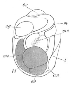
Fig. 100. Embryo of Brachionus urceolaris shortly
before it is hatched. (After Salensky.)
m. mouth; ms. masticatory apparatus; me.
mesenteron; an. anus; ld. lateral gland; ov.
ovary; t. tail, i.e. foot; tr. trochal disc;
sg. supra-œsophageal ganglion.
In the trunk an indication of a division into two segments makes its
appearance shortly after the development of the præoral lobe. Before
this period the proctodæum is established as a shallow pit immediately
behind the insertion of the foot. The latter structure soon becomes
pointed and forked (fig. 100, t).
The complete establishment of the alimentary canal occurs
late. The stomodæum (fig. 100) gives rise to the mouth (m),
œsophagus and masticatory apparatus (ms). The mesenteron is
formed from the median part of the hypoblast; the lateral parts of
which appear to give rise to the great lateral glandular structures
(ld) which open into the stomach, and to the ovaries (?)
(ov) etc. The proctodæum becomes the cloaca and anus
(an). The origin of the mesoblast is not certainly known. The
shell is formed before the larva is hatched—an occurrence which does
not take place till the larva closely resembles the adult.
The early developmental stages of the male are closely similar to
those of the female; and the chief difference between the two appears
to consist in the development of the male being arrested at a certain
point.
The larvæ of Lacinularia (Huxley, No. 234) are provided with a præoral
circlet of cilia containing two eye-spots[98], and a peri-anal patch
of cilia. They closely resemble some telotrochal polychætous larvæ.
Salensky has compared the larva of Brachionus to that of a
cephalophorous Mollusc, more especially to the larva of Calyptræa on
which he has made important observations. The præoral lobe, with the
ciliated band, no doubt admits of a comparison with the velum of the
larva of Molluscs; but it does so equally, as was first pointed out by
Huxley, with the ciliated præoral lobe of the larvæ of many Vermes. It
further deserves to be noted that the trochal disc of a Rotifer
differs from the velum of a Mollusc in that the eyes and ganglia are
placed dorsally to it, and not, as in the velum of a Mollusc, within
it. The larva of Lacinularia appears to be an exception to this, since
two eye-spots are stated to lie within the circlet of cilia. More
important in the comparison is the so-called foot (tail), which arises
in the embryo as a prominence between the mouth and anus, and in this
respect exactly corresponds with the Molluscan foot.
If Salensky’s comparison is correct, and there is something to be said
for it, the foot or tail of Rotifers is not a post-anal portion of the
trunk, but a ventral appendage, and the segmentation which it
frequently exhibits is not to be compared with a true segmentation of
the trunk. If the Rotifers, as seems not impossible, exhibit
crustacean affinities, the ‘foot’ may perhaps be best compared with
the peculiar ventral spine of the Nauplius larva of Lepas
fascicularis (vide Chapter on Crustacea) which in the arrangement
of its spines and other points also exhibits a kind of segmentation.
Bibliography.
(232) F. Cohn. “Ueb. d. Fortpflanzung von Räderthiere.”
Zeit. f. wiss. Zool. Vol. VII. 1856.
(233) F. Cohn. “Bemerkungen ü. Räderthiere.” Zeit. f. wiss.
Zool. Vol. IX. 1858, and Vol. XII. 1862.
(234) T. H. Huxley. “Lacinularia socialis.” Trans. of the
Microscopical Society, 1853.
(235) Fr. Leydig. “Ueb. d. Bau u. d. systematische Stellung d.
Räderthiere.” Zeit. f. wiss. Zool. Vol. VI. 1854.
(236) W. Salensky. “Beit. z. Entwick. von Brachionus
urceolaris.” Zeit. f. wiss. Zool. Vol. XXII. 1872.
(237) C. Semper. “Zoologische Aphorismen. Trochosphæra
æquatorialis.” Zeit. f. wiss. Zool. Vol. XXII. 1872.
Although the majority of important developmental features are common
to the whole of the Mollusca, yet at the same time many of the
subdivisions have well-marked larval types of their own. It will for
this reason be convenient in considering the larval characters to deal
successively with the different subdivisions, but to take the whole
group at once in considering the development of the organs.
Formation of the layers and larval characters.
Odontophora.
Gasteropoda and Pteropoda. There is a very close agreement
amongst the Gasteropoda and Pteropoda in the general characters of the
larva; but owing to the fact that the eggs of the various
species differ immensely as to the amount of food-yolk, considerable
differences obtain in the mode of formation of the layers and of the
alimentary tract.
The spheres at a very early stage of segmentation[100] become divided
into two categories, one of them destined to give rise mainly to the
hypoblast, the other mainly to the epiblast. According as there is
much or little food-yolk the hypoblast spheres are either very bulky
or the reverse. In all cases the epiblast cells lie at one pole, which
may be called the formative pole, and the hypoblast cells at
the opposite pole. When the bulk of the food-yolk is very great, the
number of hypoblast spheres is small. Thus in Aplysia there are only
two such spheres. In other cases, where there is but little food-yolk,
they may be nearly as numerous as the epiblast cells. In all these
cases, however, as was first shewn by Lankester and Selenka, a
gastrula becomes formed either by normal invagination as in the case
of Paludina (fig. 107), or by epibole as in Nassa mutabilis
(fig. 105). In both cases the hypoblast becomes completely enclosed by
the epiblast. The blastopore is always situated opposite the
original formative pole. In the large majority of cases
(i.e. Marine Gasteropoda, Heteropoda, and Pteropoda) the
blastopore becomes gradually narrowed to a circular opening which
eventually occupies the position of the mouth. It either closes or
remains permanently open at this point. In some cases the blastopore
remains permanently open and becomes the anus. The best authenticated
instance of this is Paludina vivipara, as was first shewn by
Lankester (No. 263).
In some instances the blastopore assumes before closing a very narrow
slit-like form, and would seem to extend along the future ventral
region of the body from the mouth to the anus. This appears, according
to Lankester (No. 262), to be the condition in Lymnæus, but while
Lankester believes that the closure proceeds from the oral towards the
anal extremity, other investigators hold that it does so in the
reverse direction. Fol (No. 249) has also described a similar type of
blastopore. In an undetermined marine Gasteropod, with an embolic
gastrula, observed by myself at Valparaiso, the blastopore had the
same elongated form as in Lymnæus, but the whole of it soon
became closed except the oral extremity; but whether this finally
closed could not be determined. It is probable that the typical form
of the blastopore is the elongated form observed by Lankester and
myself, in which an unclosed portion can indifferently remain at
either extremity; and that from this primitive condition the various
modifications above described have been derived[101].
Before the blastopore closes or becomes converted into the oral or
anal aperture, a number of very important embryonic organs make their
appearance; but before describing these it will be convenient to state
what is known with reference to the third embryonic layer or
mesoblast.
This layer generally originates in a number of cells at the lips of
the blastopore, which then gradually make their way dorsalwards and
forwards, and form a complete layer between the epiblast and
hypoblast. The above general mode of formation of the mesoblast may be
seen in fig. 107, representing three stages in the development of
Paludina.
In some cases the mesoblast arises from certain of the segmentation
spheres intermediate in size between the epiblast and hypoblast
spheres. This is the case in Nassa mutabilis, where the
mesoblast appears when the epiblast only forms a very small cap at the
formative pole of the ovum; and in this case the mesoblast cells
accompany the epiblast cells in their growth over the hypoblast (fig.
105).
In other cases the exact derivation of the mesoblast cells is quite
uncertain. The evidence is perhaps in favour of their originating from
the hypoblast. It is also uncertain whether the mesoblast is
bilaterally symmetrical at the time of its origin. It is stated by
Rabl to be so in Lymnæus[102].
In the case of Paludina the mesoblast becomes two layers
thick, and then splits into a splanchnic and somatic layer, of which
the former attaches itself to the hypoblast, and gives rise to the
muscular and connective-tissue wall of the alimentary tract, and the
latter attaches itself to the epiblast, and forms the muscular and
connective-tissue wall of the body and other structures. The two
layers remain connected by protoplasmic strands, and the space between
them forms the body cavity (fig. 107). In most instances there would
appear to be at first no such definite splitting of the mesoblast, but
the layer has the form of a scattered network of cells between the
epiblast and the hypoblast. Finally certain of the cells form a
definite layer over the walls of the alimentary canal, and constitute
the splanchnic mesoblast, and the remaining cells constitute the
somatic mesoblast.
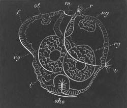
Fig. 101. Diagram of an embryo of
Pleurobranchidium. (From Lankester.)
f. foot; ot. otocyst; m. mouth; v.
velum; ng. nerve ganglion; ry. residual yolk spheres;
shs. shell-gland; i. intestine.
We must now return to the embryo at the time when the blastopore is
becoming narrowed. First of all it will be necessary to define the
terms to be applied to the various regions of the body—and these will
best be understood by taking a fully formed larva such as that
represented in fig. 101. The ventral surface I consider to be that
comprised between the mouth (m) and the anus, which is very
nearly in the position (i) in the figure. As a great
protuberance on the ventral surface is placed the foot f. The
long axis of the body, at this period though not necessarily in the
adult, is that passing through the mouth and the shell-gland
(shs.): while the dorsal surface is that opposite the ventral
as already defined.
Before the blastopore has attained its final condition three organs
make their appearance, which are eminently characteristic of the
typical molluscan larva. These organs are (1) the velum, (2) the
shell-gland, (3) the foot.
The velum is a provisional larval organ, which has the form of a
præoral ring of cilia, supported by a ridge of cells, often in the
form of a double row, the ventral end of which lies immediately dorsal
to the mouth. Its typical position is shewn in fig. 101, v.
There are considerable variations in its mode and extent of
development etc., but there is no reason to think that it is entirely
absent in any group of Gasteropoda or Pteropoda. In a few individual
instances, especially amongst viviparous forms and land Pulmonata, it
has been stated to be absent. Semper (No. 274) failed to find it in
Vitrina, Bulimus citrinus, Vaginulus luzonicus, and Paludina costata.
It is very probably absent in Helix, etc.
In some cases, e.g. Limax (Gegenbaur), Neritina (Claparède),
Pterotrachæa (Gegenbaur), the larva is stated to be coated by an
uniform covering of cilia before the formation of the velum, but the
researches of Fol have thrown very considerable doubt on these
statements. In some cases amongst the Nudibranchiata (Haddon) and
Pteropoda there are one or two long cilia in the middle of the velar
area. In many Nudibranchiata (Haddon) there is present a more or less
complete post-oral ring of small cilia, which belongs to the
velum.
The cilia on the velum cause a rotation of the larva within the
egg-capsule. Cilia are in most cases (Paludina, etc.) developed on the
foot and on a small anal area.
The shell-gland arises as an epiblastic thickening on the posterior
and dorsal side. In this thickening a deep invagination (fig. 101,
shs.) is soon formed, in which a chitinous plug may become
developed (Paludina, Cymbulia? etc.), and in abnormal larvæ such a
chitinous plug is generally formed.
The foot is a simple prominence of epiblast on the ventral surface, in
the cavity of which there are usually a number of mesoblast cells
(fig. 101, f). The larval form just described has been named by
Lankester the trochosphere larva.
Before considering the further external changes which the larva
undergoes, it will be well to complete the history of the invaginated
hypoblast.
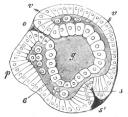
Fig. 102. Embryo of A Heteropod. (From
Gegenbaur; after Fol.)
o. mouth; v. velum; g. archenteron; p.
foot; c. body cavity; s. shell-gland.
The hypoblast has after its invagination either the form of a sack
(fig. 102) or of a solid mass (fig. 101). Whether the mouth be the
blastopore or no, the permanent œsophagus is formed of epiblast
cells, so that the œsophagus and buccal cavity are always lined by
epiblast. When the blastopore remains permanently open the outer part
of the œsophagus grows as a prominent ridge round the opening.
The mesenteric sack itself becomes differentiated into a stomach
adjoining the œsophagus, a liver opening immediately behind this,
and an intestine. The cells forming the hepatic diverticula and
sometimes also those of the stomach may during larval life secrete in
their interior peculiar albuminous products, similar to ordinary
food-yolk.
The proctodæum, except when it is the blastopore, arises later than
the mouth. It is frequently developed from a pair of projecting
epiblast cells symmetrically placed in the median ventral line behind
the foot. It eventually forms a very shallow invagination meeting the
intestine. Its opening is the anus. The anus, though at first always
symmetrical and ventral, subsequently, on the formation of the pallial
cavity, opens into this usually on the right and dorsal side.
In the cases where the hypoblast is not invaginated in the form of a
sack the formation of the mesenteron is somewhat complicated, and is
described in the sequel.
From the trochosphere stage the larva passes into what has been called
by Lankester the veliger stage (fig. 103), which is especially
characteristic of Gasteropod and Pteropod Mollusca.
The shell-gland (with a few exceptions to be spoken of subsequently)
of the previous stage flattens out, forming a disc-like area, on the
surface of which a delicate shell becomes developed, while the
epiblast of the edges of the disc becomes thickened. The disc-like
area is the mantle. The edge of the area and with it the shell
now rapidly extend, especially in a dorsal direction. Up to this time
the embryo has been symmetrical, but in most Gasteropods the shell and
mantle extend very much more towards the left than towards the right
side, and a commencement of the permanent spiral shell is thus
produced.
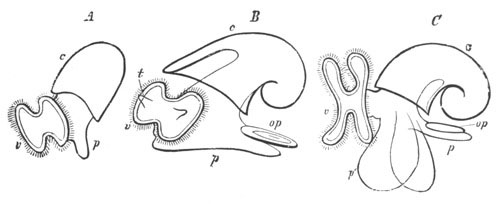
Fig. 103. Larvæ of Cephalophorous Mollusca in the
veliger stage.
(From Gegenbaur.)
A. and B. Earlier and later stage of Gasteropod. C. Pteropod
(Cymbulia).
v. velum; c. shell; p. foot;
op. operculum; t. tentacle.
The edge of the mantle forms a projecting lip separating the dorsal
visceral sack from the head and foot. An invagination appears, usually
on the right in Gasteropods, and eventually extends to the dorsal side
(fig. 103 B). It gives rise to the pallial or branchial cavity, and
receives also the openings of the digestive, generative and urinary
organs. In most Pteropods it is also formed to the right, and usually
eventually extends afterwards towards the ventral surface (fig. 103
C). In the pallial cavity the gills are formed, in those groups in
which they are present, as solid processes frequently ciliated. They
are coated by epiblast and contain a core of mesoblast. They soon
become hollow and contractile.
The velum in the more typical forms loses its simple circular form,
and becomes a projecting bilobed organ, which serves the larva after
it is hatched as the organ of locomotion (fig. 103 B and C). The
extent of the development of the velum varies greatly. In the
Heteropods especially it becomes very large, and in Atlanta it becomes
six-lobed, each lateral half presenting three subdivisions. It is
usually armed on its projecting edge with several rows of long cilia,
and below this with short cilia which bring food to the mouth.
It persists in many forms for a very long period. Within the area of
the velum there appear the tentacles and eyes (fig. 103 B). The latter
are usually formed at the base of the tentacles.
The foot grows in most forms to a very considerable size. On its
hinder and dorsal surface is formed the operculum as a chitinos plate
which originates in a depression lined by thickened epiblast, much in
the same way as the shell (fig. 103 B and C, op). In the
typical larval forms it is only possible to distinguish the anterior
flattened surface of the foot for locomotion and the posterior
opercular region, but special modifications of the foot are found in
the Pteropods and Heteropods, which are described with those groups.
The foot very often becomes richly ciliated, and otic vesicles are
early developed in it (fig. 101, ot).
All the Gasteropods and Pteropods have a shell-bearing larval form
like that first described, with the exception of a few forms, such as
Limax and perhaps some other Pulmonata, in which the shell-gland
closes up and gives rise to an internal shell.
The subsequent metamorphosis in the different groups is very various,
but in all cases it is accompanied by the disappearance of the velum,
though in some cases remnants of the velum may persist as the
subtentacular lobes (Lymnæus, Lankester) or the lip tentacles
(Tergipes, Nordmann). In prosobranchiate Gasteropods the larval
shell is gradually added to, and frequently replaced by, a permanent
shell, though the free-swimming veligerous larva may have a long
existence. In many of the Opisthobranchiata the larval shell is lost
in the adult and in others reduced. Lankester, who has especially
worked at the early stages of this group, has shewn that the larvæ are
in almost every respect identical with those of prosobranchiate
Gasteropods. They are all provided with a subnautiloid shell, an
operculated foot, etc. The metamorphosis has unfortunately been
satisfactorily observed in but few instances. In Heteropods and
Pteropods the embryonic shell is in many cases lost in the adult.
The following sections contain a special account of the development in
the various groups of Gasteropoda and Pteropoda which will complete
the necessarily sketchy account of the preceding pages.
Gasteropoda. To illustrate the development of the Gasteropoda I
have given a detailed description of two types, viz. Nassa
mutabilis and Paludina vivipara.
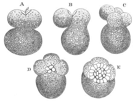
Fig. 104. Segmentation of Nassa mutabilis.
(From Bobretzky.)
A. Upper half divided into two segments. B. One of these has fused
with the large lower segment. C. Four small and one large segment,
one of the former fusing with the large segment. D. Each of the four
segments has given rise to a fresh small segment. E. Small segments
have increased to thirty-six.
Nassa mutabilis. This form, the development of which has been
very thoroughly worked out by Bobretzky (No. 242), will serve as an
example of a marine Gasteropod with a large food-yolk. The
segmentation has already been described, p. 102. It will be convenient
to take up the development at a late stage of the segmentation. The
embryo is then formed of a cap of small cells which may be spoken of
as the blastoderm resting upon four large yolk cells of which one is
considerably larger than the others (fig. 104 A). The small and the
large cells are separated by a segmentation cavity. The general
features at this stage are shewn in fig. 105 A, representing a
longitudinal section through the largest yolk cell and a smaller yolk
cell opposite to it. The blastoderm is for the most part one cell
thick, but it will be noticed that, at the edge of the blastoderm
adjoining the largest yolk cell, there are placed two cells underneath
the edge of the blastoderm (me). These cells are the
commencement of the mesoblast. In the later stages of development
the blastoderm continues to grow over the yolk cells, and as it grows
the three smaller yolk cells travel round the side of the largest yolk
cell with it. As they do so they give rise to a layer of protoplasmic
cells (fig. 105, hy) which form a thickened layer at the edge
of the blastoderm and therefore round the lips of the
blastopore. These cells form the hypoblast. The whole of the
protoplasmic matter of the yolk cells is employed in the formation of
the hypoblast. The rest of them remains as a mass of yolk. A
longitudinal section of the embryo at a slightly later stage, when the
blastopore has become quite narrowed, is represented in fig. 105 C.
The greater part of the dorsal surface is not represented.
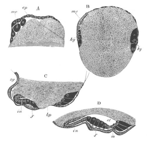
Fig. 105. Longitudinal section through the embryo
of Nassa mutabilis.
(After Bobretzky.)
A. Stage when the mesoblast is commencing to be formed.
B. Stage when the yolk is half enclosed. The hypoblast is seen at
the lips of the blastopore.
C. Stage when the blastopore (bp) is nearly obliterated.
D. The blastopore is closed.
ep. epiblast; me. mesoblast; hy. hypoblast;
bp. blastopore; in. intestine; st. stomach; f.
foot; sg. shell-gland; m. mouth.
Two definite organs have already become established. One of these is a
pit lined by thickened epiblast on the posterior and dorsal side
(sg). This is the shell-gland. The other is the foot (f)
which arises as a ventral prominence of thickened epiblast immediately
behind the blastopore. The hypoblast forms a ring of columnar cells
round the blastopore. On the posterior side its cells have
bent over so as to form a narrow tube (in), the rudiment of the
intestine.
In the next stage (fig. 105 D) the blastopore completely closes, but
its position is marked by a shallow pit (m) where the stomodæum is
eventually formed. The foot (f) is more prominent, and on its
hinder border is formed the operculum. The shell-gland (not shewn in
the figure) has flattened out, and its thickened borders commence to
extend especially over the dorsal side of the embryo. A delicate shell
has become formed. In front of and dorsal to the mouth, a ciliated
ring-shaped ridge of cells, which is however incomplete dorsally,
gives rise to the velum. On each side of the foot there appears a
protuberance of epiblast cells, which forms a provisional renal organ.
The hypoblast now forms a complete layer ventrally, bounding a cavity
which may be conveniently spoken of as the stomach (st), which
is open to the yolk above. Posteriorly however a completely closed
intestine is present, which ends blindly behind (in).
The shell and with it the mantle grow rapidly, and the primitive
symmetry is early interfered with by the shell extending much more
towards the left than the right. The anus soon becomes formed and
places the intestine in communication with the exterior.
Fig. 106. Longitudinal section through an
advanced embryo of Nassa mutabilis. (After Bobretzky.)
f. foot; m. mouth; ce.v. cephalic vesicle;
st. stomach.
With the growth of the shell and mantle the foot and the head become
sharply separated from the visceral sack (fig. 106). The œsophagus
(m) becomes elongated. The eyes and auditory sacks become
formed.
With further growth the asymmetry of the embryo becomes more marked.
The intestine takes a transverse direction to the right side of the
body, and the anus opens on the right side and close to the foot in
the mantle cavity which is formed by an epiblastic invagination in
this region. The cavity of the stomach (fig. 106, st) increases
enormously and passes to the left side of the body, pushing the
food-yolk at the same time to the right side, and the point where it
communicates with the intestine becomes carried towards the posterior
dorsal end of the visceral sack. The walls of the stomach gradually
extend so as to narrow the opening to the yolk. The part of it
adjoining the œsophagus becomes the true stomach, the remainder the
liver; its interior is filled with coagulable fluid.
Paludina. Paludina—Lankester (No. 263) and Bütschli (No.
244)—is a viviparous form characterised by the small amount of
food-yolk. The hypoblast and epiblast cells are distinguished very
early, but soon become of nearly the same size.
In the later stages of segmentation the epiblast cells differ from the
hypoblast cells in the absence of pigment. The segmentation cavity, if
developed, is small. A perfectly regular gastrula is formed (fig. 107
A and B), which is preceded by the embryo assuming a flattened form.
The blastopore is at first wide, but gradually narrows, and finally
assumes a slightly excentric position. It becomes not the mouth,
but the anus.
When the blastopore has become fairly narrow, mesoblast cells (B,
me.) appear around it, between the epiblast and hypoblast.
Whether they are bilaterally arranged or no is not clear; and though
coloured like the hypoblast, their actual development from this layer
has not been followed.
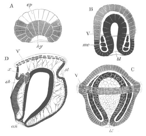
Fig. 107. Four stages in the development of
Paludina vivipara.
(Copied from Bütschli.)
ep. epiblast; hy. hypoblast; me. mesoblast;
bl. blastopore; an. anus; st. stomodæum;
sh. shell-gland; V. velum; x. primitive
excretory organ.
The velum appears about the same time as the mesoblast, in the form of
a double ring of ciliated cells at about the middle of the body (B and
C, V). The mesoblast rapidly extends so as to occupy the whole
space between the epiblast and hypoblast, and at the same time becomes
divided into two layers (C). Shortly afterwards a space—the body
cavity—appears between the two layers (D) which then attach
themselves respectively to the epiblast and hypoblast, and constitute
the somatic and splanchnic layers of mesoblast. The two layers remain
connected by transverse strands.
By a change in the relations of the various parts and especially by
the growth of the posterior region of the body, the velum now occupies
a position at the end of the body opposite the blastopore. Immediately
behind it there appear two organs, one on the dorsal and one on the
ventral side. That on the dorsal side (sh) is a deep pit—the
shell-gland—which is continuous with a layer of columnar epiblast
which ends near the anus. The other organ (st), situated on the
ventral side, is a simple depression, and is the rudiment of the
stomodæum. Between it and the dorsally placed anus is a slight
prominence—the rudiment of the foot. On the two sides of the body,
between the epiblast and hypoblast on a level with the shell-gland are
placed two masses of excretory cells, the provisional kidneys (D,
x). These are probably not homologous with the
provisional renal organ of Nassa and other marine Prosobranchiata. At
a later period a ciliated cavity appears in them, which probably
communicates with the exterior at the side of the throat.
In the later stages the foot grows rapidly, and forms a very prominent
mass between the mouth and the anus. An operculum is developed
somewhat late in a shallow groove lined by thickened epiblast.
A provisional chitinous plug is formed in the shell-gland which soon
becomes everted. The shell is formed in the usual way on the everted
surface of the shell-gland. The thickened edge of this part becomes
the edge of the mantle, and soon projects in the neighbourhood of the
anus as a marked fold.
With the rapid growth of the larva the invaginated mesenteron becomes
relatively reduced in size. In its central part yolk-spherules become
deposited, while the part adjoining the blastopore (anus) becomes
elongated to give rise to the intestine. The stomodæum grows greatly
in length and joins the dorsal part of the archenteron which then
becomes the stomach. The part of the mesenteron with yolk-spherules
forms the liver. With the development of the visceral sack the anus
shifts its position. It first passes somewhat to the left, and is then
carried completely to the right.
The development of Entoconcha mirabilis (Joh. Müller, No. 265),
a remarkable Prosobranchiate parasitic in the body cavity of Synapta,
which in the adult state is reduced to little more than an
hermaphrodite generative sack, deserves a short description. It is
viviparous, and the ovum gives rise to a larva which from the hardly
sufficient characters of the foot and shell is supposed to be related
to Natica.
There is nothing very striking in the development. The food-yolk is
scanty. The velum, as might be anticipated from the viviparous
development, is small. The tentacles are placed not within, but behind
the velar area. There is a nautica-like shell, a large mantle cavity,
and a large two-lobed foot.
In Buccinum, and Neritina only one out of the many ova included in
each egg-capsule develops. The rest atrophy and are used as food by
the one which develops.
Opisthobranchiata. It will be convenient to take a species of
Pleurobranchidium (Aplysia), observed by Lankester (No. 239),
as a type of Nudibranchiate development. The ovum first divides into
two segments, and from these small segments are budded off, which
gradually grow round and enclose the two large segments. The small
segments now form the epiblast.
At the aboral pole the epiblast becomes thickened and invaginated to
form the shell-gland, and shortly afterwards the velum and foot are
formed in the normal way, and a stomodæum appears close to the ventral
edge of the velum (fig. 101). The two yolk cells (ry) still
remain distinct, but a true hypoblastic layer (probably derived from
them, though this has not been made out) soon becomes established.
Prominent cells early make their appearance at the base of the foot,
which become at a later period invaginated to form the anus. Otolithic
sacks (ot) become formed in the foot, and the
supraœsophageal ganglia from a differentiation of the epiblast
(ng).
At a later period the shell-gland becomes everted, and a nautiloid
shell developed. The alimentary tract becomes completed, though the
two yolk cells long retain their original distinctness. The
shell-muscle is developed, and peculiar pigmented bodies are formed
below the velum. The foot becomes prominent and acquires an operculum.
The metamorphosis of Tergipes has been more or less completely worked
out by Nordmann and by Schultze (No. 271).
In Tergipes Edwardsii worked out by the former author, the
larva when hatched is provided with a large velum, eyes, tentacles, an
elongated operculated foot, and mantle. In the next stage both shell
and operculum are thrown off, and the body becomes elongated and
pointed behind. Still later a pair of gill-processes with hepatic
diverticula becomes formed.
The velum next becomes reduced, and two small processes, which give
rise to the lip tentacles and a second pair of gills, sprout out. An
ecdysis now takes place, and leads to further changes which soon
result in the attainment of the adult form.
In Tergipes lacinulatus, observed by Schultze, the velum
atrophies before the shell and operculum are thrown off.
Pulmonata. The development of the fresh-water Pulmonata appears
from Lankester’s observations on the pond-snail (Lymnæus) to be very
similar in all important particulars to that of marine
Branchiogasteropoda. The velum is however less developed than in most
marine forms. The shell-gland, etc. have the normal development. In
Lymnæus the blastopore has an elongated form and it is still a matter
of dispute whether it closes at the mouth or anus.
In the Helicidæ there is a gastrula by epibole. The shell-gland, as
may be gathered from Von Jhering’s figures, has the usual form, and an
external shell of the usual larval type is developed. There is a
ciliated process above the mouth, which extends into the lumen of the
mouth. This process is often regarded as a rudimentary velum, but
probably has not this value. There is no other organ which can be
homologous with the velum.
The development of Limax presents some peculiarities. The yolk-spheres
(hypoblast) form a large mass enclosed by the epiblast cells. A
shell-gland is formed in the usual situation, which however, instead
of being everted, as in ordinary forms, becomes closed, and in its
interior are deposited calcareous plates which give rise to the
permanently internal shell. The foot grows out posteriorly, and
contains a large provisional contractile vesicle, traversed by
muscular strands which contract rhythmically.
Although an external shell is present in Clausilia in the adult, the
shell-gland becomes closed in the embryo as in Limax, and an internal
plate-like shell is developed. The shell is at first covered by a
complete epithelium, which eventually gives way in the centre, leaving
covered only the edges of the shell. It thus comes about that the
original internal shell becomes an external one. It is very difficult
to bring this mode of development of the external shell into relation
with that of other forms. Clausilia like Limax develops a large pedal
sinus.
In both Limax and Clausilia cilia are early developed and cause a
rotation of the embryo, but how far they give rise to a distinct velum
is not clear.
Heteropoda. The Heteropod embryos present in their early
development the closest resemblance to those of other Gasteropods. The
segmentation takes place according to the most usual Gasteropod type;
(vide p. 99) and after the yolk cells have ceased to give origin to
epiblast cells they divide towards the nutritive pole, become
invaginated, and line a spacious archenteron. The epiblast cells at
the formative pole gradually envelop the yolk (hypoblast) cells, and
the blastopore very early narrows and becomes the permanent mouth.
Simultaneously with the narrowing of the blastopore, the shell-gland
is formed at the aboral pole, and the foot on the ventral side. The
velum appears as a patch of cilia on the dorsal side, which then
gradually extends ventrally so as to form a complete circle just
dorsal to the mouth.
The larva, after these changes have been completed, is represented in
fig. 102.
In later stages the shell-gland becomes everted, and a shell is
developed in all the forms both with and without shells in the adult.
The foot grows very rapidly, and an operculum is in all cases formed
behind. A bilobed invagination in front gives rise to the mucous
gland. The velum enlarges and becomes bilobed.
Though the blastopore remains permanently open as the mouth, the
œsophagus is formed as an epiblastic ingrowth. The rudiment of the
proctodæum appears as two epiblastic cells symmetrically placed behind
the foot, which subsequently pass to the right side, and give rise to
a shallow invagination which meets the mesenteric sack. In the latter
structure the cells of part of the wall develop a peculiar nutritive
material, and form a nutritive sack which eventually becomes the
liver. The part of the sack connected with the epiblastic œsophagus
becomes constricted off as the stomach. The remainder, which
unites with the proctodæum, forms the intestine.
The structural peculiarities of the adult are formed by a post-larval
metamorphosis. The caudal appendage of Pterotrachea and Firoloidea is
formed as an outgrowth of the upper border of the hind end of the
foot. The so-called fin arises as a cylindrical process in front of
the base of the foot, which is eventually flattened laterally. In the
Atlantidæ it is in some cases at first vermiform, and in other cases
attains directly its adult structure. The embryonic foot itself gives
rise in Pterotrachea, Firoloidea and Carinaria to the tail, on the
dorsal and posterior side of which the operculum may still be seen in
young specimens. In Atlanta it forms the posterior part of the foot on
which the operculum persists through life.
The embryonic shell is completely lost in Pterotrachea and Firoloidea,
and the shell is rudimentary in Carinaria. With its atrophy the mantle
region also becomes much reduced.
The velum is enormously developed in many Heteropods. In Atlanta it is
six-lobed, each of the two primitive lateral lobes being prolonged
into three processes, two in front, and one behind. As in all other
cases, it atrophies in the course of the post-larval metamorphosis.
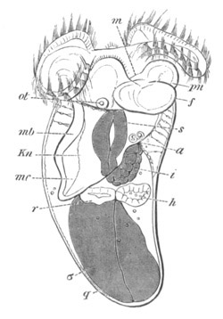
Fig. 108. Embryo of Cavolinia (Hyalea)
Tridentata. (After Fol.)
m. mouth; a. anus; s. stomach; i.
intestine; σ. nutritive sack; mb. mantle;
mc. mantle cavity; Kn. contractile sinus; h.
heart; r. renal sack: f. foot; pn. epipodia;
q. shell; ot. otolithic sack.
Pteropoda. The early larval form of the Pteropods is closely
similar to that of marine Gasteropods. There are usually only three
hypoblastic spheres at the close of the segmentation in the
Thecosomata, and a somewhat larger number in the Gymnosomata. The
blastopore closes at the oral region, on the nutritive side of the
ovum, and the shell-gland is placed at the original formative pole.
The velum, shell-gland and foot have the usual relations. Although
many of the adult forms are symmetrical, there is very early an
asymmetry visible in the larva, shewing that the Pteropods are
descended from asymmetrical ancestors. In the Gymnosomata there is a
second larval stage after the loss of the shell when the larva is
provided with three rings of cilia (fig. 109). In most forms of
Pteropods the dorsal part of the body, covered by the mantle, is
produced into a visceral sack like that of the Cephalopoda (fig. 108).
The velum varies considerably in its development in different forms.
In the Hyaleidæ it is comparatively small and atrophies early; while
in Cymbulia (fig. 103) and the Gymnosomata it is large and bilobed,
and persists till after the foot has attained its full development.
The free edge of the velum is provided with long motor cilia, and its
lower border with small cilia which bring the food to the mouth. In
Cleodora there is a median bunch of cilia in the centre of the velum
like that in the Lamellibranchiata, Nudibranchiata, etc.
The shell-gland forms a pit at the aboral end of the body, and in
Cymbulia a chitinous plug appears to be normally formed in this pit.
The pit afterwards everts itself. The edge of the everted area becomes
thickened and gradually travels towards the anterior end of the body.
On this everted area a small plate is developed, which forms the
commencement of the embryonic shell with which the larvæ of all
Pteropods are provided.
The remainder of the embryonic shell is secreted in successive rings
by the thickened edge of the mantle, and grows with this till it
reaches the neck (fig. 108). The permanent shell is added
subsequently, usually on a very different model to the larval shell.
The fate of the embryonic shell is very various in different forms. In
the Hyaleidæ the animal withdraws itself from the larval shell, which
becomes shut off from the permanent shell by a diaphragm. The larval
shell then becomes detached.
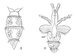
Fig. 109. Free-swimming Pneumodermon Larvæ.
(After Gegenbaur, copied from Bronn.)
The velum has atrophied in both larvæ.
In A three ciliated bands are present, and the auditory vesicles are
visible.
In B the tentacles with suckers and the epipodia have become
developed.
an. anus.
In the Styliolidæ the permanent shell becomes twice the size of the
embryonic shell while the animal is still in an embryonic condition,
but the larval shell persists for life. In the Cymbulidæ there is an
embryonic and secondary shell, which persist together during larval
life. They are eventually cast off at the same time and replaced by a
permanent shell.
In the Gymnosomata an embryonic shell is developed, and a secondary
shell added to it during embryonic life. Both are cast off before the
adult condition is attained. After the shell has been cast off three
ciliated rings are developed (fig. 109). The anterior of these is
placed between the velum and the foot, and the two hinder ones on the
elongated posterior part of the body.
The ciliated rings give to these larvæ a resemblance to Chætopod
larvæ; but there can be no doubt that this resemblance is a purely
superficial one. The anterior ring atrophies early (fig. 109 B), and
the second one soon follows suit. It is probable that the hindermost
one does not persist through life, although it has been observed in
forms with fully developed sexual organs. Most of these larvæ have not
been traced to their adult forms. They have been referred to
Pneumodermon, Clio, etc.
The most characteristic organ of the Pteropods is the foot, which is
prolonged into two enormous lateral wings, the epipodia. These develop
at different periods in different larvæ, but are always distinct
lateral outgrowths of the foot.
In the Hyaleidæ the foot is early conspicuous, and soon sends out two
lateral prolongations (fig. 108 pn.) which develop with
enormous rapidity as compared with the medium portion, and give rise
to the epipodia. The whole of the foot becomes ciliated.
In the Cymbulidæ, though not in other forms, an operculum is developed
on the hinder surface of the foot (fig. 103 C). The epipodia are late
in appearing.
In the Gymnosomata the foot is developed very early, but remains
small. The epipodia do not appear till very late in larval life (fig.
109 B).
In Pneumodermon and some other Gymnosomata there appear on the hinder
part of the head peculiar tentacles with suckers like those of the
Cephalopoda (fig. 109 B). It is not certain that these tentacles are
genetically related to the arms of the Cephalopoda.
Cephalopoda. The eggs of the Cephalopoda are usually laid in
special capsules formed in the oviduct, which differ considerably in
the different members of the group.
In the case of Argonauta each egg is enveloped in an elongated capsule
provided with a stalk. By means of the stalk the eggs are attached
together in bunches, and these again are connected together and form
transparent masses, which are placed in the back of the shell. In
octopus the eggs are small and transparent: each of them is enclosed
in a stalked capsule. In Loligo the eggs are enveloped in elongated
sack-like gelatinous cords, each containing about thirty or forty
eggs. The cords are attached in bunches to submarine objects. In Sepia
each egg is independently enveloped in a spindle-shaped black capsule,
which is attached to a stone or other object.
In a decapod form with pelagic larvæ, described by Grenacher (No.
280), the eggs were enclosed in a somewhat cylindrical gelatinous
mass. In each mass there were an immense number of eggs arranged in
spirals. Each ovum was enclosed in a structureless membrane, within
which it floated in a colourless albumen.
The ovum itself within the capsule is a nearly homogeneous granular
mass, without a distinct envelope. Development commences by
the segregation, at the narrow pole of the ovum opposite the
egg-stalk, of the greater part of the protoplasmic formative
material[103]. This material forms a disc equivalent to the germinal
disc of meroblastic vertebrate ova. The germinal disc in Sepia and
Loligo does not, however, undergo a quite symmetrical segmentation
(Bobretzky, No. 279). When eight segments are present, two of them
close together are much smaller and narrower than the remainder; and
when, in the succeeding stages small segments are formed from the
inner ends of the large ones, those derived from the two smaller
segments continue to be smaller than the remainder: so that throughout
the segmentation one pole of the blastoderm is formed of smaller
segments, and the blastoderm exhibits a bilateral symmetry[104]. The
partial segmentation results in the formation of a blastoderm covering
one pole of the egg, but, unlike the vertebrate blastoderm, formed of
a single row of cells. This blastoderm very soon becomes two or three
cells deep at its edge, and the cells below the surface constitute the
layer from which the mesoblast and hypoblast originate (fig. 110
ms). The origin of the mesoblast at the edge of the blastoderm
is a phenomenon equivalent to its origin at the lips of the blastopore
in so many other types. The external layer forms the epiblast.
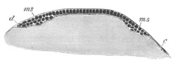
Fig. 110. Section through the blastoderm of a
Loligo ovum at the beginning of the fourth day. (After
Bobretzky.)
ms. mesoblast; d. cell at the edge of the blastoderm;
c. one of the segmentation cells.
The whole blastoderm does not take its origin from the segmentation
spheres, but, as was discovered by Lankester (282), a number of nuclei
arise spontaneously in the yolk outside the blastoderm, around which
cell bodies become subsequently formed. They make their appearance
near to, but not at the surface, extending first in a ring-like series
in advance of the margin of the blastoderm, but subsequently appearing
indiscriminately over all parts of the egg. They take no share in
forming the epiblast, but would seem, according to Lankester, to
assist in giving rise to the lower layer cells, and also to a layer of
flattened cells which eventually completely encloses the yolk, and may
be called the yolk membrane. The cells of the yolk membrane first of
all appear at the thickened edge of the blastoderm. From this
point they spread inwards under the centre of the blastoderm (fig. 115
m´), and, together with the epiblast cells, outwards over the
yolk generally; so that before long (on the tenth day in Loligo) the
yolk becomes completely invested by a membrane of cells.
In the non-germinal region the blastoderm is formed of two layers, (1)
a flattened epiblast, and (2) the yolk membrane. In the region of the
original germinal disc the epiblast cells become columnar, and below
them is placed a ring of lower layer cells, which gradually extends
towards the centre so as finally to form a complete layer. Below this
again comes the yolk membrane just spoken of.
Before describing the further fate of the separate layers it is
necessary to say a few words as to the external features of the
embryo. In the adult Cephalopod it is convenient, for the sake of
comparison with other Mollusca, to speak of the narrow space enclosed
in the arms, which contains the mouth, as the ventral surface; the
aboral apex as the dorsal surface; and what is usually called the
upper surface as the anterior and the lower one as the posterior.
Employing this terminology the centre of the original blastoderm is
the dorsal apex of the embryo. In the typical forms with a large
yolk-sack the whole embryo is formed out of the original germinal
disc; the part of the blastoderm which is continued as a thin layer
over the remainder of the egg forms a large ventral yolk-sack appended
to the head of the embryo. The following description applies
especially to two types, which form the extremes of the series in
reference to the development of the yolk-sack. The first of these with
a large yolk-sack is Sepia, of which Kölliker in his classical memoir
(No. 281) has published a series of beautiful figures. The second,
with a small yolk-sack, is the pelagic larva of an unknown adult
described by Grenadier (No. 280).
In a young blastoderm of Sepia viewed from the dorsal surface, a
series of structures appear which are represented in fig. 111 A. In
the middle is a somewhat rhomboid prominence which forms the rudiment
of the mantle (mt). In its centre is a pit which forms the
shell-gland. On each side of the mantle is a somewhat curved fold
(f). These folds eventually coalesce to form the funnel. They
are divided into two parts by a small body which forms the cartilage
of the funnel. The smaller part of the fold behind this body gives
rise to the true funnel, the part in front becomes (Kölliker) the
strong muscle connecting the funnel with the neck-cartilage. In front
and to the sides are two kidney-shaped bodies (oc), the optic
pits. Behind the mantle are two buds (br), the rudiments of the
gills.
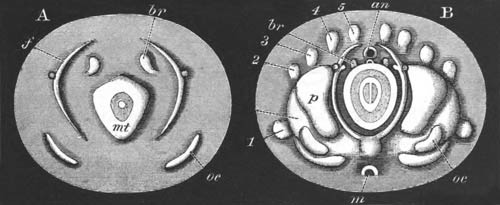
Fig. 111. Two surface views of the germinal disc
of Sepia.
(After Kölliker.)
mt. mantle; oc. eye; f. folds of funnel; br.
branchiæ; an. posterior portion of alimentary tract;
m. mouth. 1, 2, 3, 4, 5, arms; p. cephalic lobe.
In the somewhat later stage rudiments of the two posterior pairs of
arms make their appearance outside and behind the rudiments of the
funnel. The head is indicated by a pair of lateral swellings on each
side, the outer of which carries the eyes. The whole embryo now
becomes ciliated, though the ciliation does not cause the usual
rotation. At a slightly later stage the second, third, and fourth
pairs of arms make their appearance slightly in front of those already
present. The posterior parts of the funnel rudiments approach each
other, and the anterior meet the rudiments of the neck-cartilage. The
gills begin to be covered by the mantle-edge, which now projects as a
marked fold. At a slightly later period two fresh rudiments may be
noted, viz. the oral (fig. 111 B, m) and anal
invaginations, the latter of which is extremely shallow and appears at
the apex of a small papilla which may be spoken of as the anal
papilla. These invaginations appear at the two opposite poles
(anterior and posterior) of the blastoderm. Shortly after this the
rudiment of the first pair of arms arises considerably in front of the
other rudiments, at the sides of the outer pair of cephalic swellings
(fig. 111 B, 1).
Fig. 111 B represents a view from the dorsal surface of an embryo at
this stage. In the centre is the mantle with the shell-gland which is
now very considerably raised beyond the general surface. Concentric
with the edge of the mantle are the two halves of the funnel, the
anterior half meeting the dorsal or neck-cartilage and the posterior
halves approaching each other. The oral invagination is shewn at
m and the anal immediately in front of an. The gills,
nearly covered by the mantle, are seen at br. At p are
the cephalic swellings, and the eye is seen at oc. The
arms 1‑5 form a ring outside these parts. The whole of the embryo,
with the exception of the gills, the funnel, and the outer border of
the blastoderm, is richly ciliated.
The embryo up to this time has had the form of a disc or saucer on the
surface of the yolk. After this stage it rapidly assumes its permanent
dome-like form, and becomes at the same time folded off from the yolk.
The blastoderm is very slow in enveloping the yolk, and the whole yolk
is not completely invested till a considerably later stage than that
represented in fig. 111 B. As soon as the blastoderm covers the
yolk-sack cilia appear upon it. The mantle grows very rapidly, and its
free border soon projects over the funnel and gills. After the two
halves of the funnel have coalesced into a tube, it comes to project
again beyond the edge of the mantle.
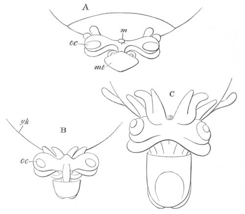
Fig. 112. Side views of three late stages in the
development of Sepia.
(After Kölliker.)
m. mouth; yk. yolk-sack; oc. eye; mt.
mantle.
On the completion of the above changes the resemblance of the embryo
to a Cuttle-fish becomes quite obvious. Three of the stages in the
accomplishment of these changes are represented in fig. 112.
To the ventral side of the embryo is attached the enormous external
yolk-sack (yk), which is continuous with an internal section
situated within the body of the embryo. The general relations of the
embryo to the yolk will best be understood by reference to the
longitudinal section of Loligo, fig. 127.
The arms gradually increase in length, and the second pair passes in
front of the first so as eventually to lie completely in front of the
mouth. The arms thus come to form a complete ring surrounding the
mouth, of which the original second pair, and not, as might be
anticipated, the first, completes the circle in front. The second pair
develops into the long arms of the adult.
After the embryo has attained more or less completely its definite
form (fig. 112 C) it grows rapidly in size as compared with the
yolk-sack. The latter structure is at first four or five times as big
as the embryo, but, by the time of hatching, the embryo is two to
three times as big as the yolk-sack.
Loligo mainly differs from Sepia in the early enclosure of the yolk by
the blastoderm, and in the embryo exhibiting the phenomena of rotation
within the egg-capsule so characteristic of other Mollusca.
In Argonauta the yolk-sack is still smaller than in Loligo, and the
yolk is early completely enclosed by the blastoderm. A
well-developed outer yolk-sack is present during early embryonic life,
but is completely absorbed within the body before its close. Cilia
appear on the blastoderm very early, but vanish again when the yolk is
about two-thirds enclosed. There is, during embryonic life, no trace
of a shell, but the mantle and other parts of the body become covered
by peculiar bunches of fine setæ. The shell-gland develops normally in
both Octopus and Argonauta, but disappears again without closing up to
form a sack (Lankester).
The pelagic Decapod larva described by Grenacher, which forms my
second type, must be placed with reference to the development of the
yolk-sack at the opposite pole to Sepia. Segmentation, as in other
Cephalopods, is partial, but the blastoderm almost completely envelops
the yolk before any organs are developed; and no external yolk-sack is
present. At a stage slightly before the closure of the yolk-blastopore
the mantle is formed as a slight prominence at the blastodermic pole
of the egg, and even at this early stage is marked by the presence of
chromatophores. The edge of the blastoderm is ciliated. At a slightly
later stage the embryo becomes more cylindrical, the edge of the
mantle becomes marked by a fold, which divides the embryo transversely
into two unequal parts, a smaller region covered by the mantle, and a
larger region beyond this. The yolk is still exposed, but rudiments of
the optic pit and of two pairs of arms have appeared. The first-formed
arms are apparently the anterior, and not, as in Sepia, the posterior.
At a still later stage, represented in lateral and posterior views in
fig. 113 A and B, considerable changes are effected. The
yolk-blastopore is nearly though not quite closed. The mantle fold
(mt) is much more prominent, and on the posterior side on a
level with its edge may be seen the rudiments of the gills
(br). The funnel is formed as two independent folds on each
side (inf1 and inf2), which apparently correspond with
the two divisions of the funnel rudiments in Sepia. The eye has
undergone considerable changes. Close to each rudiment of the funnel
may be seen a fresh sense organ—the auditory sack (ac). The
ventral (upper in the figure) end of the body now forms a marked
protuberance, probably equivalent to the foot of other Mollusca
(vide p. 225), at the sides of which are seen the rudiments of
the arms (1, 2, 3). To the two previously present a third one, on the
posterior side, has been added. The blastopore is placed on
the anterior side of the ventral protuberance, and immediately dorsal
to this is an invagination (os) which gives rise to the
stomodæum. The ciliation at the edge of the blastopore still persists,
but does not lead to the rotation of the embryo.
In later stages (fig. 113 C) the blastopore becomes closed, and the
mantle region increases in length as compared with the remainder of
the body. The ventral halves of the funnel, each in the form of a half
tube, coalesce together to form a single tube (inf) in the same
manner as in Sepia. A shallow proctodæum (an) is formed between
the two branchiæ. The eyes (oc) stand out as lateral
projections, and the arms become much longer.
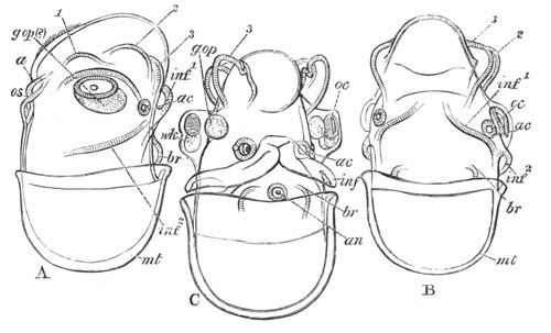
Fig. 113. Three Embryos of a Cephalopod with a
very small yolk-sack.
(After Grenacher.)
a. blastopore; br. branchiæ; inf.1 and
inf.2 posterior and anterior folds of the funnel; g.op.
optic ganglion (?); oc. eye; wk. white body;
ac. auditory pit; os. stomodæum; an. anus;
mt. mantle; 1, 2, 3. 1st, 2nd, and 3rd pairs of arms.
Still later a fourth pair of arms is added as a bud from each of the
posterior pair, and with the growth in length of the arms the suckers
make their appearance. The mouth is gradually carried up so as to be
surrounded by the arms. The ciliation of the surface becomes more
extensive.
During the whole of the above development the interior of the embryo
is filled with yolk, although no external yolk-sack is
present. The internal yolk-sack falls into three sections; a cephalic
section, a section in the neck, and an abdominal section. Of these,
that in the neck is the first to be absorbed. The cephalic portion
fills out the ventral protuberance already spoken of. The hinder
section becomes occupied by the liver which exactly fits itself into
this space as it absorbs the material previously there.
It will be convenient at this point to complete the account of the
Cephalopoda by a short history of their germinal layers, and by a
fuller description of the mantle, shell, and funnel than that given in
the preceding pages.
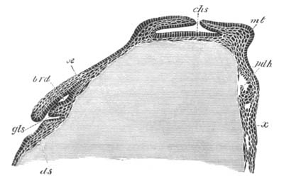
Fig. 114. Longitudinal vertical section through a
Loligo ovum when the mesenteric cavity is just commencing to be
formed.
(After Bobretzky.)
gls. salivary gland; brd. sheath of radula; oe.
œsophagus; ds. yolk-sack; chs. shell-gland;
mt. mantle; pdh. mesenteron; x. epiblastic
thickening between the folds of the funnel.
It has already been shewn that in the region of the germinal disc a
thick layer of cells becomes interposed between the epiblast and the
yolk membrane. This layer (fig. 115 m) is mainly mesoblastic,
but also contains the elements which form the lining of the alimentary
tract. Its cells first become differentiated into mesoblast and
hypoblast after the shell-gland has become a fairly deep pit. The mode
of differentiation is shewn in fig. 114. On the posterior side of the
mantle, at the point marked in fig. 111 B, an, a cavity is
formed between the yolk membrane and the mesoblast cells (fig. 114,
pdh). This cavity is the commencement of the anal extremity of
the mesenteron, and the columnar cells lining it constitute the
hypoblast. The remainder of the lower layer cells are the
mesoblast. The mesenteron gradually extends itself till it meets the
stomodæum (fig. 127). The proctodæum is formed as a shallow pit close
to the first formed part of the mesenteron.
The mesoblast gives rise not only to the organs usually formed in this
layer, but also to the nervous centres, etc.
The mantle and shell. The mantle first arises as a thickening
of the epiblast on the dorsal surface of the embryo. The thickened
integument, with the subjacent mesoblast, soon forms a definite
projection, in the centre of which appears a circular pit (figs. 114
chs and 115 shs). This pit, which has already been
spoken of as the shell-gland, resembles very closely the shell-gland
of other Mollusca. The fold around the edge of the shell-gland grows
inwards so as gradually to circumscribe its opening, which before long
becomes completely obliterated; and the gland forms a closed sack
lined by epiblast which grows in an anterior direction (figs. 114 and
127 cch).
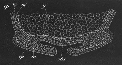
Fig. 115. Diagram of a vertical section through
the mantle region of an embryo Loligo. (From Lankester.)
[This figure is turned the reverse way up to fig. 114.]
ep. epiblast; y. food-yolk; m. mesoblast;
m´. cellular yolk membrane; shs. shell-gland.
The edges of the mantle now begin to project, especially on the
posterior side (fig. 127), and within the cavity formed by this
projecting lip there are placed the anus (an), gills, etc. The
projecting lip of the mantle is formed both of epiblast and mesoblast.
The whole of the anterior side of the mantle is filled by the
elongated shell-sack (cch), within which the shell or pen soon
becomes secreted.
There are certain difficulties in comparing the shell-gland of the
Cephalopoda with that of other Mollusca which will best be rendered
clear by the following quotation from Lankester[105]:
“The position and mode of development of the shell-gland of the
Cephalopoda exactly agree with that of the shell-gland as seen in the
other Molluscan embryos figured in this paper. We are therefore fairly
entitled to conclude from the embryological evidence that the pen-sack
of Cephalopoda is identical with the shell-gland of other Mollusca.
“But here—forming an interesting example of the interaction of the
various sources of evidence in genealogical biology—palæontology
crosses the path of embryology. I think it is certain that if we
possessed no fossil remains of Cephalopoda the conclusion that the
pen-sack is a special development of the shell-gland would have to be
accepted.
“But the consideration of the nature of the shell of the Belemnites
and its relation to the pen of living Cuttle-fish brings a new light
to bear on the matter. Reserving anything like a decided opinion as to
the question in hand, I may briefly state the hypothesis suggested by
the facts ascertained as to the Belemnitidæ. The complete shell of a
Belemnite is essentially a straightened nautilus-shell (therefore an
external shell inherited from a nautilus-like ancestor), which, like
the nautiloid shell of Spirula, has become enclosed by growths
of the mantle, and unlike the shell of Spirula, has received
large additions of calcareous matter from those enclosing overgrowths.
On the lower surface of the enclosed nautilus-shell of the
Belemnite—the phragmacone—a series of layers of calcareous matter
have been thrown down forming the guard; above, the shell has been
continued into the extensive chamber formed by the folds of the
mantle, so as to form the flattened pen-like pro-ostracum of Huxley.
“Whether in the Belemnites the folds of the mantle which thus covered
in and added to the original chambered shell, were completely closed
so as to form a sack or remained partially open with contiguous flaps
must be doubtful.
“In Spirula we have an originally external shell enclosed but
not added to by the enclosing mantle sack.
“In Spirulirostra, a tertiary fossil, we have a shell very
similar to that of Spirula, with a small guard of laminated
structure developed as in the Belemnite (see the figures in Bronn
Classen u. Ordnungen des Thierreichs).
“In the Belemnites the original nautiloid shell is small as compared
with Spirulirostra. It appears to be largest in Huxley’s genus
Xiphoteuthis. Hence in the series Spirula,
Spirulirostra, Xiphoteuthis, Belemnites, we have
evidence of the enclosure of an external shell by growths from the
mantle (as in Aplysia), of the addition to that shell of calcareous
matter from the walls of its enclosing sack, and of the gradual change
of the relative proportions of the original nucleus (the nautiloid
phragmacone) and its superadded pro-ostracal and rostral
elements tending to the disappearance of the nucleus (the original
external shell). If this view be correct as to the nature of these
shells, it is clear that the shell-gland and its plug has nothing to
do with them. The shell-gland must have preceded the original
nautiloid shell, and must be looked for in such a relation whenever
the embryology of the pearly Nautilus can be studied. Now, everything
points to the close agreement of the Belemnitidæ with the living
Dibranchiata. The hooklets on the arms, the ink bag, the horny jaws,
and general form of the body, leave no room for doubt on that point;
it is more than probable that the living Dibranchiata are modified
descendants of the mesozoic Belemnitidæ. If this be so, the pens of
Loligo and Sepia must be traced to the more complex
shell of the Belemnite. This is not difficult if we suppose the
originally external shell the phragmacone, around which as a nucleus
the guard and pro-ostracum were developed, to have finally
disappeared. The enclosing folds of the mantle remain as a sack and
perform their part, producing the chitino-calcareous pen of the living
Dibranch, in which parts can be recognised as corresponding to the
pro-ostracum, and probably also to the guard of the Belemnite. If this
be the case, if the pen of Sepia and Loligo correspond
to the entire Belemnite shell minus the phragmacone-nucleus, it is
clear that the sack which develops so early in Loligo and which
appears to correspond to the shell-gland of the other Molluscs cannot
be held to do so. The sack thus formed in Loligo must be held
to represent the sack formed by the primæval upgrowth of mantle folds
over the young nautiloid shell of its Belemnitoid ancestors, and has
accordingly no general significance for the whole Molluscan group, but
is a special organ belonging only to the Dibranchiate stem, similar
to—but not necessarily genetically connected with—the mantle fold in
which the shell of the adult Aplysia and its congeners is
concealed. The pen, then, of Cephalopods would not represent the plug
of the shell-gland. In regard to this view of the case, it may be
remarked that I have found no trace in the embryonic history of the
living Dibranchiata of a structure representing the phragmacone; and
further, it is possible, though little importance can be attached to
this suggestion, that the Dibranchiate pen-sack, as seen in its
earliest stage in the embryo Loligo, etc., is fused with the
surviving remnants of an embryonic shell-gland. When the embryology of
Nautilus pompilius is worked out, we shall probably know with
some certainty the fate of the Molluscan shell-gland in the group of
the Cephalopoda.”
The funnel. The general development of the funnel has already
been sufficiently indicated. The folds of which it is formed are
composed both of epiblast and mesoblast. The mesoblast of the anterior
part of each half of the funnel would appear to give rise to a muscle
passing from the cartilage of the neck to the funnel proper. The
posterior parts gradually approximate, but meet in the first instance
ventrally. The two folds at first merely form the side of a
groove or imperfect tube (fig. 113 C and 124 ff.), but soon the free
edges unite and so give rise to a perfect tube, the primitive origin
of which by the coalescence of two halves would not be suspected. In
Nautilus the two halves remain permanently separate but overlap each
other, so as to form a functional tube.
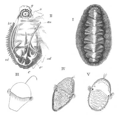
Fig. 116.
I. Chiton Wossnessenskii. (After Middendorf.)
II. Chiton dissected to shew o. the mouth; g.
the nervous ring; ao. the aorta; c. the ventricle;
c´. an auricle; br. the left branchiæ; od.
oviducts. (After Cuvier.)
III., IV., V. Stages of development of Chiton cinereus.
(After Lovén.)
The figure is taken from Huxley.
Polyplacophora. The external characters of the embryo of Chiton
have long been known through the classical observations of Lovén (No.
285), while the formation of the layers and the internal phenomena of
development have recently been elucidated by Kowalevsky (No. 284). The
eggs are laid in April, May, and June, and are enclosed in a kind of
chorion with calcareous protuberances. The segmentation remains
regular till sixty-four segments are formed. The cells composing the
formative half of the ovum then divide more rapidly than the
remainder; there is in this way formed an elongated sphere, half of
which is composed of small cells and half of larger cells. In the
interior is a small segmentation cavity. From its eventual fate the
hemisphere of the smaller cells may be called the anterior pole, and
that of the larger cells the posterior. An involution of the cells at
the apex of the posterior pole (though not of the whole hemisphere of
larger cells) now takes place, and gives rise to the archenteron. At
the same time an equatorial double ring of large cells appears on the
surface between the two poles, which becomes ciliated and forms the
velum. At the apex of the anterior pole a tuft of cilia, or at first a
single flagellum, is established (fig. 116 III. and
IV.).
In the succeeding developmental period the blastopore, which has so
far had the form of a circular pore at the posterior extremity of the
body, undergoes a series of very remarkable changes. In conjunction
with a gradual elongation of the larva it travels to the ventral side,
and is prolonged forwards to the velum as a groove. The middle part of
the groove is next converted into a tube, which opens externally in
front, and posteriorly communicates with the archenteron. The walls of
this tube subsequently fuse together, obliterating the lumen, and
necessarily causing at the same time the closure of the blastopore.
The tube itself becomes thereby converted into a plate of cells on the
ventral surface between the epiblast and the hypoblast[106].
While the above changes have been taking place the mesoblast has
become established. It is derived from the lateral and ventral cells
of the hypoblast.
After the establishment of the germinal layers the further evolution
of the larva makes rapid progress. A transverse groove is formed
immediately behind the velum, which is especially deep on the ventral
surface; and the stomodæum is formed as an invagination of the
anterior wall of the deeper section of the groove. Behind the
stomodæum the remainder of the ventral surface grows out as a
flattened foot.
The dorsal surface behind the velum constitutes the mantle, and
becomes divided by six or seven transverse grooves into segment-like
areas, which may be called mantle plates (fig. 116 IV.).
These areas would seem (?) to correspond to so many flattened out
shell-glands. Immediately behind the velum the eyes appear as two
black spots (fig. 116 IV.).
While the above external changes take place the archenteron undergoes
considerable modifications. Its anterior section gives rise, according
to Kowalevsky, to a dorsal (?) sack in which the radula is formed;
while the liver arises from it as two lateral diverticula.
From the above statements it would appear that Kowalevsky holds that
the œsophagus and radula sack are both derived from the walls of
the archenteron and not from the stomodæum. Such an origin for these
organs is without parallel amongst Mollusca.
The larva becomes about this time hatched, and after swimming about
for some time attaches itself by the foot, throws off its larval
organs, cilia, etc., and develops the shell.
The shell appears first of all during larval life in the form of
spicula on the middle and sides of the head, and later on the middle
and sides of the post-oral mantle plates (fig. 116 V.). The
permanent shell arises somewhat later as a series of median and
lateral calcareous plates, first of all on the posterior part of the
velar area, and subsequently on the mantle plates behind. The three
calcareous patches of each plate fuse together and give rise to the
permanent shell plates. The original spicula are displaced to the
sides, where they partly remain, and are partly replaced by new
spicula.
The nervous system is formed during larval life as four longitudinal
cords:—two lateral—the branchial cords, and two ventral—the pedal.
Paired anterior thickenings of the pedal cords meet in front of the
mouth to form the œsophageal ring. The pedal cords and their
derivatives are believed by Kowalevsky to be developed from the
lateral parts of the plate formed by the metamorphosis of the
blastopore. The median part of the plate is still visible after the
formation of these parts.
The chief peculiarity of the larva of Chiton (apart from the peculiar
ventral plate) consists in the elongation and dorsal segmentation of
the posterior part of the body. The velum has the normal situation and
relation to its mouth. The position of the eyes behind it is however
abnormal.
The elongation and segmentation of the posterior part of the trunk is
probably to be regarded as indicating that Chiton has early
branched off from the main group of the Odontophora along a special
line of its own, and not that the remaining Odontophora are
descended from Chiton-like ancestral forms. The shell of Mollusca on
this view is not to be derived from one of the plates of Chiton, but
the plates of Chiton are to be derived from the segmentation of a
primitive simple shell. The segmentation exhibited is of a kind which
all the trochosphere larval forms seem to have been capable of
acquiring. The bilateral symmetry of Chiton, which is quite as well
marked as that of the Lamellibranchiata, indicates that it is a
primitive phylum of the Odontophora.
Scaphopoda. The external characters of the peculiar larva of
this interesting group have been fully worked out by Lacaze Duthiers
(No. 286).
The segmentation is unequal and conforms to the usual molluscan type.
At its close the embryo becomes somewhat elongated, and there appears
on its surface a series of transverse ciliated rings. As soon as these
become formed the larva is hatched, and swims about by means of its
cilia. Six ciliated bands are formed in all, and in addition a tuft of
cilia is formed in a depression at the anterior extremity.
The larva thus constituted is very different in appearance to the
larvæ already described, and its parts very difficult to identify; the
next stages in the development shew however that the whole region of
the body taken up by the ciliated rings is part of the velar area,
while the small papilliform region behind is the post-velar part of
the embryo. This latter part grows rapidly, and at the same time the
ciliated rings become reduced to four; which gradually approach each
other, while the region on which they are placed grows in diameter.
The rings finally unite, and form a single ring on a projecting velar
ridge. In the centre of this ring is placed the terminal tuft of cilia
on a much reduced prominence.
By the time that these changes have been effected in the velum, the
post-velar part of the embryo has become by far the largest section of
the embryo, so that the velum forms a projecting disc at the front end
of an elongated body. The mantle is formed as two lateral outgrowths
near the hinder extremity of the body which leave between them a
ventral groove lined by cilia; on their dorsal side is formed
a delicate shell. The mantle lobes continue to grow, and by the time
the above changes in the velum are effected they meet and unite in the
ventral line and convert the groove between them into a complete tube
open in front and behind. A stream of water is driven through this
tube by the action of the cilia. The shell, which is at first
disc-shaped like the shell of other molluscan larvæ, moulds itself
upon the mantle and is so converted into a tube. At the front end of
the mantle tube, which does not at first cover the velum, there is
formed the foot. It arises as a protuberance of the ventral wall of
the body, which rapidly grows forwards, becomes trilobed as in the
adult, and ciliated.
On the completion of these changes the larva mainly differs in
appearance from the adult by the projection of the velum beyond the
edge of the shell. The velum soon however begins to atrophy; and the
larva sinks to the bottom. The mantle tube and shell grow forward and
completely envelop the velum, which shortly afterwards disappears. The
mouth is formed on the ventral side of the velum at the base of the
foot; at its sides arise the peculiar tentacles so characteristic of
the adult Dentalium.
Lamellibranchiata.
The larvæ of Lamellibranchiata have in a general way the same
characters as those of Gasteropods and Pteropods. A trochosphere stage
with a velum but without a shell is succeeded by a veliger stage with
a still more developed velum, a dorsal shell, and a ventral foot.
The segmentation is unequal, and in a general way like that of
Gasteropoda, but the specially characteristic Gasteropodan type with
four large yolk spheres is only known to occur in Pisidium, and a type
of segmentation similar to that of Anodon (p. 100) appears to be the
most frequent.
There is an epibolic or embolic gastrula, but the further history of
the formation of the germinal layers has been worked out so
imperfectly, and for so few types, that it is not possible to make
general statements about it. What is known on this head is
mentioned in connection with the description of the development of
special types.
The blastopore in some cases closes at the point where the anus
(Pisidium), and probably in other cases where the mouth, is eventually
formed. In Anodon it is stated to close at a point corresponding
neither with the mouth nor the anus, but on the dorsal surface!
The embryo assumes a somewhat oval form, and in the free marine forms
there appears very early in front of the mouth a well-developed velum.
This is formed according to Lovén from two papillæ, and takes the form
of a circular ridge armed with long cilia. In the centre of the velar
area there is usually present a single long flagellum (fig. 117 B and
C). The velum never becomes bilobed.
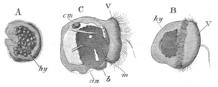
Fig. 117. Three stages in the development of
Cardium.
(After Lovén.)
hy. hypoblast; b. foot; m. mouth; an.
anus; V. velum; cm. anterior adductor muscle.
In the later stages, after the development of the shell, the velum
becomes highly retractile and can be nearly completely withdrawn
within the mantle by special muscles. It forms the chief organ of
locomotion of the free larva.
In some fresh-water forms, which have no free larval existence, the
velum is very much reduced (Anodon, Unio, Cyclas) or even aborted
(Pisidium). In these forms as well as in Teredo and probably other
marine forms (e.g. Ostrea) the central flagellum is absent. It
has been suggested by Lovén, though without any direct evidence, that
the labial tentacles of adult Lamellibranchiata are the remains of the
velum. The velar area is in any case the only representative of the
head. In some marine forms a general covering of cilia arises before
the formation of the velum; and in Montacuta and other types
there is developed, as in many Gasteropoda, a circumanal patch of
cilia.
A shell-gland appears at a very early period on the dorsal surface in
Pisidium, Cyclas and Ostrea, and probably in most marine forms (fig.
118, shs). It is somewhat saddle-shaped, and formed of
elongated non-ciliated cells bounding a groove. It flattens out and on
its surface is formed the shell, which appears usually to have the
form of an unpaired saddle-shaped cuticle, on the two sides of which
the valves are subsequently formed by a deposit of calcareous salts.
In Pisidium the two valves are stated by Lankester to be at first
quite independent and widely separated, and it has been suggested by
Lankester, though not proved, that the ligament of the shell is
developed in the median part of the groove of the shell-gland.
The mantle lobes are developed as lateral outgrowths of the body: they
usually have a considerable extension before they are covered by the
shell. In Anodon and Unio the larval mantle lobes are, however, formed
in a somewhat exceptional way, and are from the first completely
covered by the valves of the larval shell. The larval mantle lobes and
shell in Anodon and Unio are subsequently replaced by the permanent
structures.
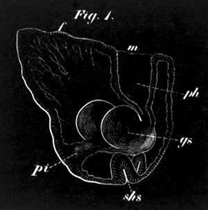
Fig. 118. An embryo of Pisidium pusillum.
(From Lankester.)
f. foot; m. mouth; ph. pharynx; gs.
bilobed stomach; pi. intestine; shs. shell-gland.
The adductor muscles are formed soon after the appearance of the
shell. The posterior sometimes appears first, e.g. Mytilus, and
at other times the anterior, e.g. Cardium.
The foot arises in the usual way as a prominence between the mouth and
anus. In comparison with Gasteropoda it is late in appearing, and in
many cases does not become prominent till the shell has attained
a considerable size. In its hinder part a provisional paired
byssus-gland is developed from the epidermis in Cyclas and other
forms. In other cases, e.g. Mytilus, the byssus-gland is
permanent. The byssus-gland occupies very much the position of the
Gasteropod operculum, and would appear very probably to correspond
with this organ. The anterior part of the foot is usually ciliated.
The gills appear rather late in larval development along the base of
the foot on either side, between the mantle and the foot (fig. 120,
br). They arise as a linear row of separate ciliated somewhat
knobbed papillæ. A second row appears later. The two rows give rise
respectively to the two gill lamellæ of each side.
The further history of the development of the gills has been studied
by Lacaze Duthiers (No. 297) in Mytilus. The first row of gill papillæ
formed becomes the innermost of the two lamellæ of the adult. The
number of papillæ goes on increasing from before backwards. When about
eleven have been formed, their somewhat swollen free extremities unite
together, the basal portions being separated by slits.
The free limb is formed by the free end of the gill lamella bending
upon itself towards the inner side and growing towards the line of
attachment of the lamella. The free limb is at first not composed of
separate bars, but of a continuous membrane. Before this membrane has
grown very wide, perforations are formed in it corresponding to the
spaces between the bars of the attached limb.
The outer gill lamella develops in precisely the same way as, but
somewhat later than, the inner. The rudiments of it appear when about
twenty papillæ of the inner lamella are formed. Its first papillæ are
formed near the hind border of the inner lamella, and new papillæ are
added both in front and behind. Its free limb is on the outer side.
In Mytilus the two limbs (free and attached) of each bar of the gill
are joined at wide intervals by extensile processes, the
‘inter-lamellar junctions,’ and the successive bars are attached
together by ciliated junctions. In many other types the concrescences
between the various parts of the gills are carried much further; the
maximum of concrescence being perhaps attained in Anodon and Unio[107].
Large paired auditory sacks seem always to be developed in the foot;
and clearly correspond with the auditory sacks in Gasteropoda.
Eyes are frequently present in the larva, though they disappear in the
adult. In Montacuta and other types a pair of these organs is formed
at the base of the velum on each side of the œsophagus, not far
from the auditory sacks. They are provided with a lens.
A row of similar organs is present in the larva of Teredo in front of
the foot.
Cardium. As an example of a marine Lamellibranchiate I may take
Cardium pygmaeum, the development of which has been studied by
Lovén (No. 291). The ova, surrounded by a thickish capsule, are
impregnated in the cloaca. The segmentation takes place much as in
Nassa (vide p. 101), and the small segments gradually envelop
the large hypoblast spheres; so that there would seem to be a gastrula
by epibole. After the hypoblast has become enveloped by the epiblast,
one side of the embryo is somewhat flattened and marked by a deepish
depression (fig. 117 A). From Lovén’s description it appears to me
probable that the depression on the flattened side occupies the
position of the blastopore, and that the depression itself is the
stomodæum. At this stage the embryo becomes covered with short cilia
which cause it to rotate within the egg-capsule.
Close above the mouth there appear two small papillæ. These gradually
separate and give rise to a circular ridge covered with long cilia,
which encircles the embryo anteriorly to the ventrally-placed mouth.
This structure is the velum. In its centre is a single long flagellum
(fig. 117 B). Shortly after this the shell appears as a saddle-shaped
structure on the hinder part of the dorsal surface of the embryo. It
is formed at first of two halves which meet behind without the trace
of a hinge (fig. 117 C). The two halves rapidly grow and partially
cover over the velum, and below them the mantle folds soon sprout out
as lateral flaps.
The alimentary tract has by this time become differentiated (fig. 117
C). It consists of a mouth (m) and ciliated œsophagus
probably derived from the stomodæum, a stomach and intestine derived
from the true hypoblast, and an hepatic organ consisting of two
separate lobes opening into the stomach. The anus (an) appears
not far behind the mouth, and between the two is a very slightly
developed rudiment of the foot (b). The anterior adductor
muscle (cm) appears at this stage, though the posterior is not
yet differentiated.
The larva is now ready to be hatched, but the further stages of its
development were not followed.
Ostrea. The larvæ of Ostrea, figured by Salensky (No. 293),
shew a close resemblance to those of Cardium. The velum is however a
simple ring of cilia without a central flagellum. The proctodæum would
appear to be formed later than the stomodæum, and the earliest stage
figured is too far advanced to throw light on the position of the
blastopore.
Pisidium. The development of Pisidium has been investigated by
Lankester (No. 239). The ovum is invested by a vitelline
membrane and undergoes development in a brood-pouch at the base of the
inner gill lamella.
The segmentation commences by a division into four equal spheres, each
of which, as in so many other Mollusca, then gives rise by budding to
a small sphere. The later stages of segmentation have not been
followed in detail, but the result of segmentation is a blastosphere.
An invagination, presumably at the lower pole, now takes place, and
gives rise to an archenteric sack.
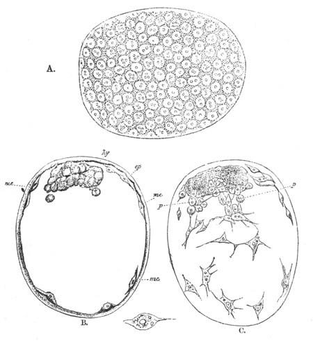
Fig. 119. Three views of an embryo of Pisidium
immediately after the closure of the blastopore. (After
Lankester.)
A. View from the surface.
B. Optical section through the median plane.
C. Optical section through a plane a little below the surface.
ep. epiblast; me. mesoblast; hy. hypoblast;
p. cells apparently budding from the hypoblast to form
mesoblastic elements.
The embryo now rapidly grows in size. The blastopore becomes closed
and the archenteric sack forms a small mass attached at one point to
the walls of the embryonic vesicle (fig. 119, hy). In the space
between the walls of the archenteron and those of the embryonic
vesicle stellate mesoblast cells make their appearance,
derived in the main from the epiblast, though probably in part also
from the hypoblastic vesicle (vide fig. 119 C, p). The
cavity between the hypoblast and epiblast, which contains these cells,
is the body cavity. Fig. 119 represents three views of the embryo at
this stage. A is a surface view shewing the epiblast; B is an optical
section through the median plane shewing the hypoblast and some of the
mesoblast cells; and C is an optical section shewing the mesoblast
cells. A prominence on one side of the embryo now develops which forms
the commencement of the foot, and the archenteric sack grows out at
its free extremity into two lobes, but remains attached to the
epiblast by an imperforate pedicle. The next organ to appear is the
stomodæum. It arises as a ciliated epiblastic ingrowth which meets the
free end of the archenteric sack, fuses with it, and shortly
afterwards opens into it (fig. 118, ph). Between the mouth and
the attachment of the enteric pedicle is placed the foot (f),
which becomes ciliated. On the dorsal side of the enteric pedicle
there appears a saddle-shaped patch of epiblast cells bounding the
sides of a groove (shs). This is the rudiment of the
shell-gland.
The enteric pedicle, or intestine as it may now be called, soon
acquires a lumen, though still imperforate at its termination where
the anus is eventually formed. Ventral to the intestine is placed a
mass of cells—the rudiment of the organ of Bojanus. It is stated to
be developed as an ingrowth of the epiblast.
In a slightly later stage the shell-gland rapidly increases in size
and flattens out, and on the two sides of it there appear the
rudiments of the two valves, which are at first quite distinct, and
separated by a considerable interval (fig. 120). Before the appearance
of the valves of the shell, the mantle folds have already grown out
from the sides of the body.
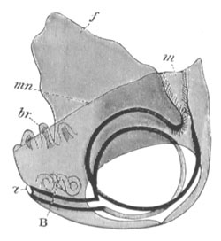
Fig. 120. Diagrammatic view of advanced larva of
Pisidium. (Copied from Lankester.)
m. mouth; a. anus; B. organ of Bojanus; mn.
mantle; f. foot.
At a somewhat later stage the gills appear as a linear series of small
independent buds within the folds of the mantle behind the foot (fig.
120, br). The anterior adductor also becomes differentiated.
The alimentary tract in the meantime has undergone considerable
changes. The primitive lateral lobes dilate enormously and become
ciliated. At a still later stage their walls undergo peculiar changes,
the nature of which is somewhat obscure, but they appear to me to be
of the same character as those in many Pteropods and Gasteropods,
where the cells of the hepatic diverticula, to which the lobes of
Pisidium apparently correspond, become filled with an albuminous
material.
The later stages in Pisidium have not been followed.
It is remarkable that in Pisidium a veliger stage does not occur. This
is probably due to the development taking place within the
brood-pouch. The late development of the otocysts is also remarkable.
A byssus-gland was not formed up to the stage observed. In Cyclas
calyculata (Schmidt), a byssus-gland also appears to be absent.
Cyclas. The development of Cyclas as described by Von Jhering
is very unlike that of Pisidium, and the differences would seem to be
too great to be accounted for except by errors of observation.
The segmentation of Cyclas is similar to that of Anodon (vide
p. 82), and a mass of large cells enclosed by the smaller cells gives
rise to the hypoblast. In the interior of this mass there appears a
lumen, and a process from it grows towards and meets the epiblast, and
gives rise to the œsophagus and mouth,—a mode of development of
these parts without parallel amongst Mollusca. A very rudimentary
velum would appear, according to Leydig (No. 290), to be developed at
the cephalic extremity. A shell-gland is formed of the same character
as in Gasteropods. According to Leydig the shell appears as a single
saddle-like structure on the dorsal surface; the lateral parts of this
become calcified, and give rise to the two valves, but are united in
the middle by the membranous median portion. At the two sides of the
body the mantle lobes are formed, as in Pisidium.
Very shortly after the formation of the shell the byssus-gland appears
as a pair of small follicles in the hinder part of the foot. It
rapidly grows larger and becomes a paired pyriform gland, in which are
secreted the byssus threads which serve to attach all the embryos at a
common point to the walls of the brood-pouch.
The foot is large, and ciliated anteriorly. Otolithic sacks and peda
ganglia are developed in it very early.
Unio. The ovum of Anodonta and Unio is enveloped in a vitelline
membrane, the surface of which is raised into a projecting
trumpet-like tube perforated at its extremity (fig. 12). This
structure is the micropyle. The micropyle disappears in Anodonta
piscinalis when the egg is ripe, but in Unio persists during the whole
development. The ova are transported, in a manner not certainly made
out, into the space between the two limbs of the outer gills of the
mother, and there undergo their early development. The animal or upper
pole of the egg is placed at the pole opposite to the micropyle.
The segmentation is unequal (vide p. 100) and results in
the formation of a blastosphere with a large segmentation cavity. The
greater part of the circumference of the egg is formed of small
uniform spheres, but the lower (with reference to the segmentation)
pole is taken up by a single large cell. The small spheres become the
epiblast, and the large cell gives rise to hypoblast and
mesoblast[108].
The single large cell next divides into two, and then four, and
finally into about ten to fifteen cells. These cells form an especial
area of more granular cells than the other cells of the blastosphere.
Most of them are nearly of the same size, but two of them (according
to Rabl), in contact with each other, but placed on the future right
and left sides of the embryo, are considerably larger than the
remainder. These two cells soon pass into the cavity of the
blastosphere, while at the same time the area of granular cells
becomes flattened out, and then becomes involuted as a small sack with
a transversely elongated opening, which does not nearly fill up the
cavity of the blastosphere. This involuted sack is the archenteron.
The two large cells, which lie in immediate contact with what,
following Rabl, I shall call the anterior lip of the blastopore, next
bud off small cells, which first form a layer covering the walls of
the archenteron, but subsequently develop into a network filling up
the whole cavity of the primitive blastosphere. The space between
these cells is the primitive body cavity. For a long time the two
primitive mesoblast cells retain their preponderating size[109]. At
the hinder end of the body, and at the end opposite therefore to the
two mesoblast cells, are placed three especially large epiblast cells.
In Anodonta and Unio tumidus there appears at this period a patch of
long cilia at the anterior end of the body. These cilia cause a
rotation of the embryo and would appear to be the velum. In Unio
pictorum they do not appear till much later.
Immediately following this stage the changes in the embryo take place
with great rapidity. In the first place a special mass of mesoblast
cells appears at the hinder end of the archenteric sack; and becoming
elongated transversely gives rise to the single adductor muscle. On
the subsequent formation of the shell the muscle becomes inserted in
its two valves. The blastopore next becomes closed, and the small
archenteron grows forwards till it meets the epiblast anteriorly, and
at the same time detaches itself from the epiblast in the region where
the blastopore was placed. Where it comes in contact with the wall of
the body in front a small epiblastic invagination arises, which meets
and opens into the archenteric sack and forms the permanent mouth.
While these changes have been taking place the shell is formed as a
continuous saddle-shaped plate on the dorsal surface. From this plate
the two valves are subsequently differentiated. On the dorsal surface
they meet with a straight hinge-line. Each valve is at first rounded,
but subsequently becomes triangular with the hinge-line as base. The
valves are not quite equi-sided, but the anterior side is less convex
than the posterior. At a later period a beak-shaped organ is formed at
the apex of each valve in the same manner as the remainder of the
shell. This organ is placed at about a right angle with the main
portion of the valve. It is pointed at its extremity and bears
numerous sharp spines on its outer side, which are especially large in
the median line (vide fig. 121 A). It is employed in fixing the
larva, after it is hatched, on to the fish on which it is for some
time parasitic. The shell is perforated by numerous pores.
After the shell has become formed a new structure makes its appearance
which is known as the byssus-gland. It is developed as an invagination
of the epiblast at the hinder end of the body: Rabl was unable to
determine whether it was formed from the three large epiblastic cells
present there or no. It subsequently forms an elongated gland with
three coils or so round the adductor muscle on the left side of the
body, but opening in the median ventral line. It secretes an elongated
cord by which the larva becomes suspended after hatching.
For some time the ventral portion of the body projects behind the ends
of the valves of the shell, but before these are completely formed a
median invagination of the body wall takes place, which obliterates to
a large extent the body cavity, and gives rise to two great lateral
lobes, one for each valve. These lobes are the mantle lobes.
Before the mantle lobes are fully formed peculiar sense organs,
usually four in number, make their appearance on each lobe. Each of
them consists of a columnar cell, bearing at its free end a cuticle
from which numerous fine bristles proceed. Covering the cell and the
parts adjoining it is a delicate membrane perforated for the passage
of the bristles. The largest and first formed of these organs is
placed near the anterior and dorsal part of the mantle. The three
others are placed near the free end of the mantle (vide fig.
121 A). These organs probably have the function of enabling the larva
to detect the passage of a fish in its vicinity, and to assist it
therefore in attaching itself. When the embryo is nearly ripe there
appears immediately ventral to and behind the velum a shallow pit on
each side of the middle line, and the two pits appear to be connected
by a median transverse bridge. These structures have been the cause of
great perplexity to different investigators, and their meaning is not
yet clear. According to Rabl the median structure is the somewhat
bilobed archenteron, and according to his view it is not really
connected with the laterally placed pits. The cilia of the velum
overlie these latter structures and make them appear as if their edges
were ciliated. They are regarded by Rabl as the rudiments of the
nervous system.
With the development of the shell, the mantle, and the sense organs,
the young mussel reaches its full larval development, and is now known
as a Glochidium (fig. 121 A).
If the parent, with Glochidia in its gills, is placed in a tank with
fish, it very soon (as I have found from numerous experiments) ejects
the larvæ from its gills, and as soon as this occurs the larvæ become
free from the egg membrane, attach themselves by the byssus-cord, and
when suspended in this position continually close and open their
shells by the contraction of the adductor muscle. If the mussels are
not placed in a tank with fish the larvæ may remain for a long time in
the gills.
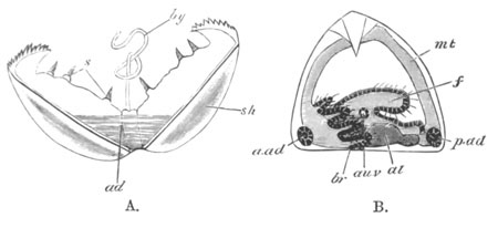
Fig. 121.
A. Glochidium Immediately After It Is Hatched.
ad. adductor; sh. shell; by. byssus cord;
s. sense organs.
B. Glochidium After It Has Been on the Fish for Some Weeks.
br. branchiæ; au.v. auditory sack; f. foot;
a.ad. and p.ad. anterior and posterior adductors;
al. mesenteron; mt. mantle.
Before passing on to state what is known with reference to the larval
metamorphosis, it may be well to call attention to certain, and to my
mind not inconsiderable, difficulties in the way of accepting in all
particulars Rabl’s account of the development.
In all Gasteropod Molluscs the lower or vegetative pole of the ovum is
ventral, not dorsal as Rabl would make it in Unio. The blastopore in
other Molluscs always coincides either with the mouth or anus, or
extends between the two. The surface on which the foot is formed is
the ventral surface. On the dorsal surface are placed, (1) the velum
near the mouth, (2) the shell-gland near the anus. In Anodon the velum
is placed just dorsal to the mouth, then according to Rabl follows the
blastopore, and in the region of the blastopore is formed the shell.
The blastopore is therefore dorsal in position. It occupies in fact
the ordinary place of the shell-gland, and looks very much like this
organ (which is not otherwise present in Anodon and Unio). Without
necessarily considering Rabl’s interpretations false, I think that the
above difficulties should have been at any rate discussed in his
paper. More especially is this the case when there is no doubt that
Rabl has made in his paper on Lymnæus a confusion between the mouth
and the shell-gland.
Investigations on the post-embryonic metamorphosis of Glochidium have
been made by Braun (No. 287), and several years ago I made a series of
observations on this subject, the results of which agree in most
points with those of Braun. I was however unsuccessful in carrying on
my observations till the young mussel left its host.
The free Glochidia very soon attach themselves to the gills, fins, or
other parts of fish which are placed in the tank containing them;
after attachment they become covered by a growth of the epidermic
cells of their host, and undergo their metamorphosis.
The first change that takes place is the disappearance of the byssus
and the byssus organ. This occurs very soon; shortly afterwards all
traces of the velum and sense organs also become lost.
At the time of the disappearance of these bodies, at the point of the
projection from which the byssus cord arose, and very possibly from
this very projection, the foot arises as a rounded process which
rapidly grows and soon becomes ciliated (fig. 121 B, f).
The single adductor muscle begins to atrophy very early, but before
its entire disappearance rudiments are formed at the two ends of the
body, which at a later period can be distinctly recognised as the
anterior and posterior adductor muscles (fig. 1211 B, a.ad and
p.ad).
After the formation of these parts the gills arise as solid and at
first somewhat knobbed papillæ covered with a ciliated epidermis, on
each side of, but somewhat in front of (!) the foot (fig. 121 B,
br). In the foot there soon appear the auditory sacks
(au.v), and the foot itself becomes a long tongue-like ciliated
organ projecting backwards[110].
The mantle lobes undergo great changes, and indeed by Braun the mantle
lobes are stated to be formed almost entirely de novo. The
permanent shell is (Braun) formed on the dorsal surface of the still
parasitic larva in the form of two small independent plates. I have
not followed the changes of the alimentary canal, etc., but at an
early stage there is visible, dorsal to the foot, a simple enteric
sack.
By the time the larva leaves its host all the organs of the adult,
except the generative organs, have become established.
The post-embryonic development of the organs of Glochidium is similar
in the main to that of other Lamellibranchiata. This fact is of some
importance on account of the peculiarities of the earlier
developmental stages.
The byssus organ, the toothed processes of the shell, and the sense
organs of the Glochidium can hardly be ancestral rudiments, but must
be organs which have been specially developed for the peculiar mode of
life of the Glochidium. Whether the single muscle is to be counted
amongst such provisional organs is perhaps a more doubtful point, but
I am inclined to think that it ought to be so.
If however the single muscle is an ancestral organ, it is important to
observe that it entirely disappears as development goes on and the two
adductor muscles in the adult are developed independently of it.
General review of the characters of the Molluscan larvæ.
The typical larva of a Mollusc, as has been more especially pointed
out by Lankester, is essentially similar to the larva of a number of
invertebrate types, and especially the Chætopoda, with the addition of
certain special organs characteristic of the Mollusca.
It has a bent alimentary tract, with a mouth on the ventral surface
and a terminal or ventral anus. The alimentary tract is divided into
three regions: œsophagus, stomach, and intestine. There is a
variously developed præoral lobe with a ring of cilia—the velum, and
a peri-anal lobe, often with a patch of cilia (Paludina, etc.). In all
these characters it is essentially similar to a Chætopod larva. The
two characteristic molluscan organs are (1) a foot between the mouth
and anus, and (2) an invagination of the epiblast on the dorsal side
at the hinder end of the body, which is connected with the formation
of the shell.
The larvæ of most Gasteropoda, Pteropoda, and Lamellibranchiata
present no features which call for special remark; but the larvæ of
the Gymnosomata amongst the Pteropoda, and of the Scaphopoda,
Polyplacophora and Cephalopoda present interesting peculiarities.
The larvæ of the Gymnosomata are peculiar in the presence of three
transverse ciliated rings, situated behind the velum (Fig.
109). These rings might be regarded as indications of a rudimentary
segmentation; but, as already indicated, this view is not
satisfactory. There is every reason for thinking that these rings have
been specially acquired by these larvæ.
At first sight the larvæ of the Gymnosomata might be supposed to
resemble those of the Scaphopoda, which are also provided with
transverse ciliated rings; but, as shewn above, the rings of the
Scaphopoda are merely parts of the extended velar ring.
Thus, the ciliated rings of the two larvæ—so similar in
appearance—are in reality structures of entirely different values,
being in the one case parts of the velum, and in the other special
developments of cilia behind the velum.
The great peculiarity of the early larva of the Scaphopoda is the
enormous development of the præoral lobe, which gives room for the
development of the ciliated rings. In the presence of a central tuft
of cilia, at the anterior extremity, the larva of the Scaphopoda
resembles that of the Lamellibranchiata, etc.
The larva of the Polyplacophora resembles that of Lamellibranchiata in
its anterior flagellum, and that of the Scaphopoda in the large
development of the præoral lobe; but is quite peculiar amongst
Mollusca in the transverse segmentation of the mantle area.
The embryo of the Cephalopoda agrees very closely with that of normal
Odontophora in the formation of the mantle and (?) of the shell-gland,
but is quite exceptional (1) in the almost invariable presence of a
more or less developed external yolk-sack, (2) in the absence of a
velum, (3) in the absence of a median foot, and in the presence of the
arms.
The presence of a yolk-sack may most conveniently be spoken of in
connection with the foot, and we may therefore pass on to the question
of the velum.
The velum is one of the most characteristic embryonic appendages of
the Mollusca, and its absence in the Cephalopoda is certainly very
striking. By some investigators the arms have been regarded as
representing the velum, but considering that they are primitively
placed on the posterior and ventral side of the mouth, and that the
velum is essentially an organ on the dorsal side of the mouth, this
view cannot, in my opinion, be maintained with any plausibility.
Various views have been put forward with reference to the Cephalopod
foot. Huxley’s view, which is the one most generally adopted, is given
in the following quotation[111].
“But that which particularly distinguishes the Cephalopoda is the form
and disposition of the foot. The margins of this organ are, in fact,
produced into eight or more processes termed arms, or brachia;
and its anterolateral portions have grown over and united in front of
the mouth, which thus comes, apparently, to be placed in the centre of
the pedal disk. Moreover, two muscular lobes which correspond
with the epipodia of the Pteropods and Branchiogasteropods, developed
from the sides of the foot, unite posteriorly, and, folding over, give
rise to a more or less completely tubular organ—the funnel or
infundibulum.”
Grenacher, from his observations on the development of Cephalopoda,
argues strongly against this view, and maintains that no median
structure comparable with the foot is present in this group: and that
the arms cannot be regarded as taking the place of the foot, but are
more probably representatives of the velum.
The difficulty of arriving at a decision on this subject is mainly due
to the presence of the yolk-sack, which, amongst the Cephalopoda as
amongst the Vertebrata, is the cause of considerable modifications in
the course of the development. The foot is essentially a protuberance
on the ventral surface, between the mouth and the anus. In Gasteropods
it is usually not filled with yolk, but contains a cavity, traversed
by contractile mesoblastic cells. In this group the blastopore is a
slit-like opening (vide p. 187) extending over the region of
the foot, from the mouth to the anus, the final point of the closure
of which is usually at the oral but sometimes at the anal extremity.
In Cephalopods the position of the Gasteropod foot is occupied by the
external yolk-sack. In normal forms the blastopore closes at the apex
of the yolk-sack, and at the two sides of the yolk-sack the arms grow
out. These considerations seem to point to the conclusion that the
normal Gasteropod foot is represented in the Cephalopod embryo by the
yolk-sack, which has, owing to the immense bulk of food-yolk present
in the ovum, become filled with food-yolk and enormously dilated. The
closure of the blastopore at the apex of the yolk-sack, and not at its
oral or anal side, is what might naturally be anticipated from the
great extension of this part.
Grenacher’s type of larva, where the external yolk-sack is practically
absent, appears to me to lend confirmation to this view. If the reader
will turn to fig. 113, he will observe a prominence between the mouth
and anus, which exactly resembles the ordinary Gasteropod foot. At the
sides of this prominence are placed the rudiments of the arms. This
prominence is filled with yolk, and represents the rudiment of
the external yolk-sack of the typical Cephalopod embryo. The
blastopore, owing to the smaller bulk of the food-yolk, reverts more
nearly to its normal position on the oral side of this prominence.
If the above considerations have the weight which I attribute to them,
the unpaired part of the Cephalopod foot has been overlooked in the
embryo on account of the enormous dilatation it has undergone from
being filled with food-yolk; and also owing to the fact that in the
adult the median part of the foot is unrepresented. The arms are
clearly, as Huxley states, processes of the margin of the foot.
Both Grenacher and Huxley agree in regarding the funnel as
representing the coalesced epipodia; but Grenacher points out that the
anterior folds which assist in forming the funnel (vide p. 253)
represent the great lateral epipodia of the Pteropod foot, and the
posterior folds the so-called horse-shoe shaped portion of the
Pteropod foot.
Development of Organs.
The epiblast. With reference to the general structure of the
epiblast there is nothing very specially deserving of notice. It gives
rise to the whole of the general epidermis and to the epithelium of
the organs of sense. The most remarkable feature about it is a
negative one, viz. that it does not, in all cases at any rate, give
rise to the nervous system.
The epiblast of the mantle has the special capacity of secreting a
shell, and the integument of the foot has also a more or less similar
property in that it forms the operculum, and a byssus in some
Lamellibranchiata, other parts of the integument form the radula, setæ
in Chiton, and other similar structures.
Nervous system. The origin of the nervous system in Mollusca is
still involved in some obscurity. It is the general opinion amongst
the majority of investigators that the nervous ganglia in Gasteropods
and Pteropods are formed from detached thickenings of the epiblast.
Both Lankester (No. 239) and Fol
(No. 249‑251) have arrived at this
conclusion, and Rabl has shewn by sections that in Planorbis there are
two lateral thickenings of the epiblast in the velar area; from which
the supra-œsophageal ganglia become subsequently separated
off. The observations on the pedal ganglia are less precise: they very
probably arise as thickenings of the epiblast of the side of the foot.
According to Fol, the nervous system in the Hyaleacea amongst the
Pteropoda originates in a somewhat different way. A disc-like area
appears in the centre of the velum, which soon becomes nearly divided
into two halves. From each of these there is formed by invagination a
small sack. The axes of invagination of the two sacks meet at an angle
on the surface. The cavities of the sacks become obliterated; the
sacks themselves become detached from the surface, fuse in the middle
line, and come to lie astride of the œsophagus. Fol has detected a
similar process in Limax. The exact origin of the pedal ganglia was
not observed, but Fol is inclined to believe that they develop from
the mesoblast of the foot.
A very different view is held by Bobretzky (No. 242), whose
observations were made by means of sections.
The supra-œsophageal and pedal ganglia are formed according to this
author as independent and ill-defined local thickenings of cells which
are apparently mesoblastic. The two sets of ganglia appear nearly
simultaneously, and later than the rudiments of the auditory and optic
organs.
In the Cephalopoda there seems to be but little doubt, as first
pointed out by Lankester, that the various ganglia originate in what
is apparently mesoblastic tissue.
There is still very much requiring to be made out with reference to
their origin, unless details on this subject are given in Bobretzky’s
Russian memoir. It would seem however that each ganglion develops as
an independent differentiation of the mesoblast (unless the optic and
cerebral ganglia are from the first continuous)[112]. The
corresponding ganglia of the two sides become subsequently united and
the various ganglia become connected by their proper commissural
cords. The ganglia are shewn in figures 124, 126, and 127.
In Lamellibranchiata the development of the nervous system has not
been worked out.
The two points which are most striking in the development of the
nervous system of Mollusca are (1) the fact that in the Cephalopoda at
any rate it is developed from tissue apparently mesoblastic; and (2)
the fact that the several ganglia frequently originate quite
independently, and subsequently become connected.
With reference to the first of these points it should be noticed that
the supra-œsophageal and pedal ganglia are at first respectively
connected with the optic and auditory organs, and that these sense
organs are in some cases at any rate developed anteriorly in point of
time to the ganglia. It seems perhaps not impossible that primitively
the ganglia may have been simply differentiations of the walls of the
sense organ, and perhaps their apparent derivation from the mesoblast
is really a derivation from cells which primitively belonged to the
walls of these sense organs. Bobretzky’s observations on Fusus fit in
well with this view.
In the Hyaleacea and in other Pteropods, where the eyes are absent in
the adult, Fol finds the supra-œsophageal ganglia resulting from a
pair of epiblastic invaginations. May not these invaginations be
really rudiments of the eyes as well as of the ganglia? Fol also, it
is true, describes a similar mode of origin for these ganglia in
Limax. It would be interesting to have further observations on this
subject. The independent origin of the pedal and supra-œsophageal
ganglia finds its parallel amongst the Chætopoda.
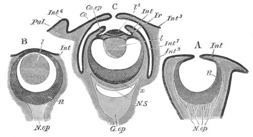
Fig. 122. Three diagrammatic sections of the eyes
of Mollusca.
(After Grenacher.)
A. Nautilus. B. Gasteropod (Limax or Helix). C. Dibranchiate
Cephalopod.
Pal. eyelid; Co. cornea; Co.ep. epithelium of
ciliary body; Ir. iris; Int. Int1 ...
Int4. different parts of the integument; l. lens;
l1. outer segment of lens; R. retina; N.op.
optic nerve; G.op. optic ganglion; x. inner layer of
retina; N.S. nervous stratum of retina.
The supra-œsophageal ganglia appear always to develop within the
region of the velar area. This area corresponds with the præ-oral lobe
of the Chætopod larva, at the apex of which is developed the
supra-œsophageal ganglion. Embryology thus confirms the results of
Comparative Anatomy in reference to the homology of these ganglia in
the two groups.
Optic organs[113]. An eye is present in most Gasteropods and
in many larval Pteropods. Although its development has not
been fully worked out, yet it has clearly been shewn by Bobretzky and
other investigators that it originates as an involution of the
epidermis, which first forms a cup and eventually a closed vesicle.
The posterior wall of the vesicle gives rise to the retina, the
anterior to the inner epithelium of the cornea. The external epidermis
becomes continued over the outer surface of the vesicle.
The lens is formed in the interior of the vesicle, probably as a
cuticular deposit, which increases by the addition of concentric
layers. Pigment becomes deposited between the cells of the retina.
Fig. 122 B is a diagrammatic representation of the adult eye of a
Gasteropod.
The Cephalopod eye is formed, as first shewn by Lankester, as a pit in
the epiblast round which a fold arises (fig. 123 A) and gradually
grows over the mouth of the pit so as to shut it off from
communication with the exterior (fig. 123 B).
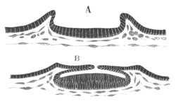
Fig. 123. Two sections through the developing eye
of a Cephalopod to shew the formation of the optic cup. (After
Lankester.)
The epiblast lining the posterior region of the vesicle gives rise to
the retina, that lining the anterior region to the ciliary body and
processes. It is important to notice that the condition of the eye
just before the above pit becomes closed is exactly that which is
permanent in Nautilus (vide fig. 122 A). After the pit has
become closed a mesoblastic layer grows in between its wall and the
external epiblast.
The lens becomes formed in two independent segments. The inner and
larger of these arises as a rod-like process (fig. 124) projecting
from the front wall of the optic vesicle into the cavity of the
vesicle. It is a cuticular structure and therefore without cells. By
the deposition of a series of concentric layers it soon assumes a
spherical form (fig. 125, hl). The condition of the
eye, with a closed optic vesicle and the lens projecting into it, is
that which is permanent in the majority of Gasteropods (vide
fig. 122 B). At about the time when the lens first becomes formed a
fold composed of epiblast and mesoblast appears round the edge of the
optic cup (fig. 124, cc), and gives rise to a structure known
in the adult as the iris. Shortly afterwards this becomes more
prominent (fig. 125, if), and at the same time the layers of
cells of the ciliary region in front of the inner segment of the lens
become reduced to the condition of mere membranes (fig. 125 B); and in
front of them the anterior or outer segment of the lens becomes formed
as a cuticular deposit (fig. 125 B, vl). At a still later
period a fresh fold of epiblast and mesoblast appears round the eye
and gradually constitutes the anterior optic chamber (vide fig. 122
C, Co). In most forms this chamber communicates with the
exterior by a small aperture, but in some it is completely closed. The
fold itself gives rise to the cornea in front and to the sclerotic at
the sides. At a later period another fold may appear forming
the eyelids (fig. 122 C, Pal).
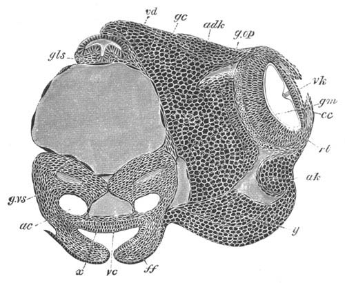
Fig. 124. Transverse section through the head of
an advanced embryo of Loligo. (After Bobretzky.)
vd. œsophagus; gls. salivary gland; g.vs.
visceral ganglion; gc. cerebral ganglion; g.op. optic
ganglion; adk. optic cartilage; ak. and y.
lateral cartilage or (?) white body; rt. retina; gm.
limiting membrane; vk. ciliary region of eye; cc.
iris; ac. auditory sack (the epithelium lining the auditory
sacks is not represented); vc. vena cava; ff. folds of
funnel.
Auditory organs. A pair of auditory sacks is found in the larvæ
of almost all Gasteropods and Pteropods, and usually originates very
early. They are placed in the front part of the foot, and on the
formation of the pedal ganglia come into close connection with it,
though they receive their nervous supply in the adult from the
supra-œsophageal ganglia.
In a very considerable number of cases amongst Gasteropods and
Pteropods the auditory organs have been observed to develop as
invaginations of the epiblast, which give rise to closed vesicles
lying in the foot, e.g. Paludina, Nassa, Heteropods, Limax,
some Pteropods (Clio).
This is no doubt the primitive mode of origin, but in other cases,
which perhaps require confirmation, the sacks are stated to originate
from a differentiation of solid thickenings of the epidermis or of the
tissues subjacent to it.
The auditory sacks are provided with an otolith, which according to
Fol’s observations is first formed in the wall of the sack.
In Cephalopods the auditory organs are formed as epiblastic pits on
the posterior surface of the embryo, and are at first widely separated
(fig. 113, ac). The openings of the pits become narrowed, and
finally the original pits form small sacks lined by an epithelium, and
communicating with the exterior by narrow ducts, equivalent to the
recessus vestibuli of Vertebrates, and named, after their
discoverer, Kölliker’s ducts. The external openings of these ducts
become completely closed at about the same time as the shell-gland,
and the ducts remain as ciliated diverticula of the auditory pits. The
widely separated auditory sacks gradually approach in the middle
ventral line, and are immediately invested by the visceral ganglia
(fig. 124, ac). They finally come to lie in contact on the
inner side of the funnel.
On the side opposite Kölliker’s duct, an epithelial ridge is
formed—the crista acustica—the cells of which give rise to an
otolith connected with the crista by a granular material. At a later
period of development three regions of the epithelium of the sack
become especially differentiated. Each of these regions is
provided with two rows of cells, bearing on their free edges numerous
very short auditory hairs. The cells of each row are placed nearly at
right angles to those of the adjoining row.
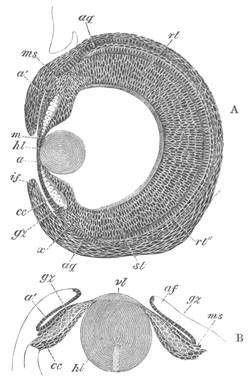
Fig. 125. Sections through the developing eye of
Loligo at two stages. (After Bobretzky.)
hl. inner segment of lens; vl. outer segment of lens;
a and a´. epithelium lining the anterior optic
chamber; gz. large epiblast cells of ciliary body; cc.
small epiblast cells of ciliary body; ms. layer of mesoblast
between the two epiblastic layers of the ciliary body; af.
and if. fold of iris; rt. retina; rt´´. inner
layer of retina; st. rods; aq. equatorial cartilage.
Muscular system. The muscular system in all groups of Molluscs
is derived entirely from the mesoblast.
The greater part of the system takes its origin from the somatic
mesoblast. In almost all Gasteropod and Pteropod larvæ there is
present a well-developed spindle muscle attaching the embryo to the
shell. This muscle appears to be absent in the Cephalopoda.
Body cavity and vascular system. The body cavity in Gasteropods
and Pteropods originates either by a definite splitting of the
mesoblast, or by the appearance of intercellular spaces. It becomes
divided into numerous sinuses which freely communicate with the
vascular system.
Very different accounts have been given by different investigators of
the development of the heart in the Gasteropoda and Pteropoda.
It would seem however in most cases to arise as a solid mass of
mesoblast cells at the hind end of the pallial cavity, which
subsequently becomes hollowed out and divided into an auricle and
ventricle. Bobretzky’s careful observations have fully established
this mode of development for Nassa.
In Pteropods the heart is formed (Fol) close to the anus, but slightly
dorsal to it (fig. 108, h). The pericardium is formed from the
mesoblast at a considerably later period than the heart.
A very different account of the formation of the heart is given by
Bütschli for Paludina. He states that there appears an immense
contractile sack on the left side of the body. This becomes
subsequently reduced in size, and in the middle of it appears the
heart, probably from a fold of its wall. The original sack would
appear to give rise to the pericardium.
In connection with the vascular system mention may be made of certain
contractile sinuses frequently found in the larvæ of Gasteropoda and
Pteropoda. One of these is placed at the base of the foot, and the
other on the dorsal surface within the mantle cavity immediately below
the velum[114]. The completeness of the differentiation of these
sinuses varies considerably; in some forms they are true sacks with
definite walls, in other cases mere spaces traversed by muscular
strands. They are found in the majority of marine Gasteropods,
Heteropods and Pteropods. In Limax a large posteriorly placed pedal
sinus is well developed, and there is also a sinus in the visceral
sack. The rhythmical contraction of the yolk-sack of Cephalopods
appears to be a phenomenon of the same nature as the contraction of
the foot sinus of Limax.
In Calyptræa (Salensky) there is an enormous provisional cephalic
dilatation within the velum which does not appear to be contractile.
Similar though less marked cephalic vesicles are found in Fusus,
Buccinum and most marine Gasteropods.
In Cephalopods the vascular system is formed by a series of
independent (?) spaces originating in the mesoblast, the cells around
which give rise to the walls of the vessels. The branchial hearts are
formed at about the time at which the shell-gland becomes closed. The
aortic heart (fig. 127, c) is formed of two independent halves
which subsequently coalesce (Bobretzky).
The true body cavity arises as a space in the mesoblast subsequently
to the formation of the main vascular trunks.
Renal organs. Amongst the Gasteropods and Pteropods there are
present provisional renal organs, which may be of two kinds, and a
permanent renal organ.
The provisional organs consist of either (1) an external paired mass
of excretory cells or (2) an internal organ provided with a duct,
which is not in all cases certainly known to open externally. The
former structure is found especially in the marine Prosobranchiates
(Nassa, etc.) where it has been fully studied by Bobretzky. It
consists of a mass of cells on each side of the body, close to the
base of the foot, and not far behind the velum. This mass grows very
large, and below it may be seen a continuous layer of epiblast. The
cells forming it fuse together, their nuclei disappear, and numerous
vacuoles containing concretions arise in them. At a later stage all
the vacuoles unite together and form a cavity filled with a brown
granular mass.
The provisional internal renal organ is found in many pulmonate
Gasteropods—Lymnæus, Planorbis, etc. It consists of a paired
V-shaped ciliated tube with a pedal and cephalic limb. The
former has an external opening, but the termination of the latter is
still in doubt.
It consists, according to Büschli’s description (No. 244), in the
freshwater Pulmonata (Lymnæus, Planorbis) of a round sack, close to
the head, opening by an elongated and richly ciliated tube in the
neighbourhood of the eye. From the sack a second shorter tube passes
off towards the foot, which seems however to end blindly. The cells
lining the sack contain concretions, and there is one especially large
cell in the lumen of the sack attached on the side turned towards the
eye. It coexists in Lymnæus with provisional renal organs of the type
of those in marine Prosobranchiata.
A somewhat different description of the structure and development of
this organ in Planorbis has recently been given by Rabl (No. 268). It
consists of a V-shaped tube on each side with both extremities
opening into the body cavity. The one limb is directed towards the
velar area, the other towards the foot. It is developed from the
mesoblast cells of the anterior part of the mesoblastic band. The
large mesoblast (p. 227) of each side grows into two processes, the
two limbs of the future organ. A lumen in the cell is continued into
each limb, while continuations of the two limbs of the V are
formed from the hollowing out of the central parts of the adjoining
mesoblast cells.
In Limax embryos Gegenbaur found a pair of elongated provisional
branched renal sacks, the walls of which contained concretions. These
sacks are provided with anteriorly directed ducts opening on the
dorsal side of the mouth. This organ is probably of the same
nature as the provisional renal organ in other Pulmonata.
Permanent renal organ. According to the most recent observer
(Rabl, No. 268), whose statements are supported by the sections
figured, the permanent renal organ in Gasteropods is developed from a
mass of mesoblast cells close to the end of the intestine. This is
first carried somewhat to the left side, and then becomes elongated
and hollow, and attaches itself to the epiblast on the left side of
the anus (fig. 108, r). After the formation of the heart the
inner end opens into the pericardium and becomes ciliated, the median
part becomes glandular and concrements appear in its lining cells, and
the terminal part forms the duct.
Previous observers have usually derived this organ from the epiblast;
according to Rabl this is owing to their having studied too late a
stage in the development.
In Cephalopoda the excretory sacks or organ of Bojanus are apparently
differentiations of the mesoblast[115]. At an early stage part of
their walls envelops the branchial veins. From this part of the wall
the true glandular section of the organ would seem to be formed. The
epithelium forming the inner wall of each sack is at an early age very
columnar.
The development of the organ of Bojanus in Lamellibranchiata has been
studied by Lankester. He finds that it develops as a paired
invagination of the epiblast immediately ventral to the anus.
Generative glands. The generative glands in Mollusca would
appear to be usually developed in the post-larval period, but our
knowledge on this subject is extremely scanty.
In Pteropods Fol believes that he has proved that the hermaphrodite
gland originates from two independent formations, one (the testicular)
epiblastic in origin, and the other (the ovarian) hypoblastic.
These views of Fol do not appear to me nearly sufficiently
substantiated to be at present accepted.
The generative glands in Cephalopoda appear to be simple
differentiations of the mesoblast. They are at first very closely
connected with the aortic heart (fig. 127, kd), but
soon become completely separated from it.
Alimentary tract. The formation of the archenteron, and the
relation of its opening to the permanent mouth and anus, has already
been described and needs no further elucidation. It will be convenient
to treat the subject of this section under three headings for each
group—viz. (1) the mesenteron, (2) the stomodæum, and (3) the
proctodæum.
The mesenteron. In the Gasteropoda and Pteropoda the
mesenteron, as has already been mentioned, forms a simple sack, which
may however, owing to the presence of food-yolk, be at first without a
lumen. Of this sack an anterior portion gives rise to the stomach and
liver, and a posterior to the intestine. This latter portion is the
first to be distinctly differentiated as such, and forms a narrowish
tube connecting the anterior dilatation with the anus. In the meantime
the cells of a great part of the anterior portion of the mesenteron
undergo peculiar changes. They enlarge, and in each of them a deposit
of food material appears, which is often at any rate derived from the
absorption of the albumen in which the embryo floats. The cells on the
dorsal side, adjoining the œsophageal invagination, and the whole
of the cells on the ventral side do not however undergo these changes.
There thus arises an anterior and ventral region adjoining the
œsophagus, which becomes completely enclosed by small cells and
forms the true stomach. The part behind and dorsal to the stomach is
lined by the large nutritive cells and forms the liver. It opens into
the stomach at the junction of the latter with the intestine, which in
the later stages becomes bent somewhat forwards and to the right.
Still later the hepatic region becomes branched, the albuminous
contents of its cells are replaced by a coloured secretion, and it
becomes bodily converted into the liver. The stomach is usually richly
ciliated.
The various modifications of the above type of development of the
alimentary tract are to be regarded as due to the disturbing influence
of food-yolk. Where primitively the hypoblast cells are very bulky,
though invaginated in a normal way, the wall of the hepatic region
becomes immensely swollen with food-yolk, e.g. Nautica. In
other cases amongst certain Pteropods (Fol, No. 249) where the
hypoblast is still more bulky, part of the archenteric walls becomes
converted into a bilobed sack opening into the pyloric region,
in the walls of which a large deposit of food material is stored,
which gradually passes into the remainder of the alimentary tract and
is there digested. The bilobed nutritive sack, as it is called by Fol,
is eventually completely absorbed, though the liver in some, if not
all cases, grows out as a fresh sack from its duct.
The formation of the permanent alimentary tract, when the hypoblast is
so bulky that there is no true archenteric cavity, has been especially
investigated by Bobretzky (No. 242).
In the case of a species of Fusus the hypoblast, when enclosed by the
epiblast, is composed of four cells only. The blastopore remains
permanently open at the oral region, and around it the œsophagus
grows in a wall-like fashion. The protoplasmic portions of the four
hypoblast cells are turned towards the œsophageal opening, and from
them are budded off small cells which are continuous at the blastopore
with the epiblast of the œsophagus. These cells give rise
posteriorly to the intestine and anteriorly to the sack, which becomes
the stomach and liver. This sack always remains open towards the four
primitive yolk cells. The cells of the posterior part of it become
larger and larger and form the hepatic sack, which fills up the left
and posterior part of the visceral sack, pushing the yolk cells to the
right. The cells lining the hepatic sack become pyramidal in shape,
and each of them is filled with a peculiar mass of albuminous
material. The cells adjoining the opening of the œsophagus remain
small, become ciliated, and form the stomach. They are not sharply
separated off from the cells of the hepatic sack. The yolk cells
remain distinct on the right side of the body during larval life, and
their food material is gradually absorbed for the nutrition of the
embryo.
A modification of the above mode of development, where the food
material is still more bulky and the blastopore closed, is found in
Nassa, and has already been described (vide p. 233).
The stomodæum. The stomodæum in most cases is formed as a
simple epiblastic invagination which meets and opens into the
mesenteron. When the blastopore remains permanently open at the oral
region the stomodæum is formed as an epiblastic wall round its
opening. In all cases the stomodæum gives rise to the mouth and
œsophagus. At a subsequent period there are developed in the oral
region of the stomodæum the radula in a special ventral pit, and the
salivary glands—the latter as simple outgrowths.
The œsophagus is usually ciliated.
The proctodæum. Except where the blastopore remains as the
permanent anus (Paludina) the proctodæum is always formed subsequently
to the mouth. Its formation is usually preluded by the appearance of
two projecting epiblast cells, but it is always developed as a
very shallow epiblastic invagination, which does not give rise to any
part of the true intestine.
In the Cephalopods the alimentary tract is formed, as in other
cephalophorous Mollusca, of three sections. (1) A stomodæum, formed by
an epiblastic invagination, which gives rise to the mouth,
œsophagus and salivary glands. (2) A proctodæum, which is an
extremely small epiblastic invagination. (3) A mesenteron, lined by
true hypoblast, which forms the main section of the alimentary tract,
viz. the stomach, intestine, the liver, and ink sack[116].
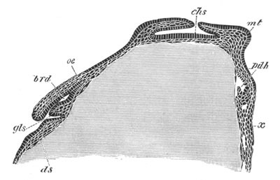
Fig. 126. Longitudinal vertical section through a
Loligo ovum when the mesenteric cavity is just commencing to be
formed. (After Bobretzky.)
gls. salivary gland; brd. sheath of radula; oe.
œsophagus; ds. yolk-sack; chs. shell-gland; mt.
mantle; pdh. mesenteron; x. epiblastic thickening
between the folds of the funnel.
The mesenteron. The mesenteron is first visible from the
surface as a small tubercle on the posterior side of the mantle
between the rudiments of the two gills (fig. 111 B, an). Within
this, as was first shewn by Lankester, a cavity appears.
This cavity is as in Gasteropods open to the yolk-sack, and only
separated from the yolk itself by the yolk membrane already spoken of.
It is at first lined by indifferent cells of the lower layer of the
blastoderm, which however soon become columnar and form a definite
hypoblastic layer (fig. 126, pdh). Between the hypoblast and
epiblast there is a very well marked layer of mesoblast. As the
mesenteric cavity extends, its walls meet the epiblast, and at
the point of contact of the two layers the epiblast becomes slightly
pitted in. At this point the anus is formed at a considerably later
period (fig. 127, an).
On the ventral side of the primitive mesenteron an outgrowth appears
very early, which becomes the ink sack (fig. 127, bi).
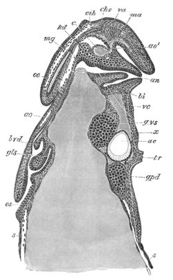
Fig. 127. Longitudinal section through an
advanced embryo of Loligo. (After Bobretzky.)
os. mouth; gls. salivary gland; brd. sheath of
radula; ao. anterior aorta; ao1. posterior aorta;
va. branch of posterior aorta to shell sack; ma.
branch of posterior aorta to mantle; c. aortic heart;
oe. œsophagus; mg. stomach; an. anus;
bi. ink sack; kd. germinal tissue; eih. shell
sack; vc. vena cava; g.vs. visceral ganglion;
g.pd. pedal ganglion; ac. auditory sack; tr.
funnel.
The mesenteric cavity, still open to the yolk, gradually extends
itself in a dorsal direction over the yolk-sack, but remains for some
time completely open to it ventrally, and only separated from the
actual yolk by the yolk membrane. There early grow out from the walls
of the mesenteron a pair of hepatic diverticula.
As the mesenteric cavity extends it dilates at its distal extremity
into a chamber destined to form the stomach (fig. 127, mg). At
about this time the anus becomes perforated. Shortly afterwards the
mesenteron meets and opens into the œsophagus at the dorsal
extremity of the yolk-sack, but at the time when this takes place the
hypoblast has extended round the entire cavity, and has shut it off
from the yolk. The yolk membrane throughout the whole of this period
is quite passive, and has no share in forming the walls of the
alimentary tract.
The stomodæeum. The stomodæum appears as an epiblastic
invagination at the anterior side of the blastoderm, before any trace
of the mesenteron is present. It rapidly grows deeper, and, shortly
after the mesenteric cavity becomes formed, an outgrowth arises from
its wall adjoining the yolk-sack, which gives rise to the salivary
glands (figs. 126 and 127, gls). Immediately behind the opening
of the salivary glands there appears on its floor a swelling which
becomes the odontophore, and behind this a pocket of the stomodæal
wall forms the sheath of the radula (figs. 126 and 127, brd).
Behind this again the œsophagus is continued dorsalwards as a very
narrow tube, which eventually opens into the stomach (fig. 127).
The terminal portion of the rudiment of the salivary gland divides
into two parts, each of which sends out numerous diverticula which
constitute the permanent glands. The greater part of the original
outgrowth remains as the unpaired duct of the two glands[117].
In the larva observed by Grenacher the anterior pair of salivary
glands originated from independent lateral outgrowths of the floor of
the mouth, close to the opening of the posterior salivary glands.
The yolk-sack of the Cephalopoda. The yolk, as has already been
stated, becomes at an early period completely enclosed in a membrane
formed of flattened cells, which constitutes a definite yolk-sack. It
is, in the more typical forms of Cephalopoda, divided into an external
and an internal section, of which the former is probably a special
differentiation of the median part of the foot of other cephalophorous
Mollusca (vide p. 272). At no period does the yolk-sack
communicate with the alimentary tract. The two sections of the
yolk-sack are at first not separated by a constriction. In the second
half of embryonic life the condition of the yolk-sack undergoes
considerable changes. The internal part grows greatly in size at the
expense of the external, and the latter diminishes very rapidly and
becomes constricted off from the internal part of the sack, with which
it remains connected by a narrow vitelline duct.
The internal yolk-sack becomes divided into three sections: a dilated
section in the head, a narrow section in the neck, and an enormously
developed portion in the mantle region. It is the latter part which
mainly grows at the expense of the external yolk-sack. It gives off at
its dorsal end two lobes, which pass round and embrace the lower part
of the œsophagus. The passage of the yolk from the external
to the internal yolk-sack is probably largely due to the contractions
of the former.
The external yolk-sack is not vascular, and probably the absorption of
the yolk for the nutrition of the embryo can only take place in the
internal yolk-sack. The most remarkable feature of the Cephalopod
yolk-sack is the fact that it lies on the opposite side of the
alimentary tract to the yolk cells, which form a rudimentary yolk-sack
in such Gasteropoda as Nassa and Fusus. In these forms, the yolk-sack
is at first dorsal, but subsequently is carried by the growth of the
liver to the right side. In Cephalopoda on the contrary, the yolk-sack
is placed on the ventral side of the body.
What is known of the development of the alimentary tract in the
Polyplacophora has already been mentioned.
In the Lamellibranchiata (Lankester, No. 239), the mesenteron early
grows out into two lateral lobes which form the liver, while the part
between them forms the stomach.
In Pisidium the intestine is formed from the original pedicle of
invagination, which remains permanently attached to the epiblast. The
stomodæum is formed by the usual epiblastic invagination, and becomes
the mouth and œsophagus. The development of the crystalline rod and
its sack do not appear to be known. In the adult the sack of the
crystalline rod opens into a part of the alimentary tract which
appears to belong to the mesenteron. Were however the development to
shew them to be really derived from the stomodæum they might be
interpreted as rudiments of the organ which constitutes the
odontophore and its sack in cephalophorous Mollusca—an interpretation
which would be of considerable phylogenetic interest.
Bibliography.
General.
(238) T. H. Huxley. “On the Morphol. of the Cephal. Mollusca.”
Phil. Trans. 1853.
(239) E. R. Lankester. “On the developmental history of the
Mollusca.” Phil. Trans. 1875.
(240) H. G. Bronn and W. Keferstein. Die Klassen u.
Ordnungen d. Thierreichs, Vol. III. 1862‑1866.
Gasteropoda and Pteropoda.
(241) J. Alder and A. Hancock. “Devel. of Nudibr.”
Ann. and Magaz. Nat. Hist., Vol. XII. 1843.
(242) N. Bobretzky. “Studien über die embryonale Entwicklung d.
Gasteropoden.” Archiv f. mikr. Anat., Vol. XIII.
(243) W. K. Brooks. “Preliminary Observations on the
Development of Marine Gasteropods.” Chesapeake Zoological
Laboratory, Session of 1878. Baltimore, 1879.
(244) O. Bütschli. “Entwicklungsgeschichtliche Beiträge
(Paludina vivipara).” Zeit. f. wiss. Zool., Vol. XXIX.
1877.
(245) W. Carpenter. “On the devel. of the embr. of Purpura
lapillus.” Trans. Micros. Soc., 2d series, Vol. III.
1855.
(246) W. Carpenter. “On the devel. of the Purpura.” Ann. and
Mag. of Nat. Hist., 2d series, Vol. XX. 1857.
(247) E. Claparède. “Anatomie u. Entwickl. der Neritina
fluviatilis.” Müller’s Archiv, 1857.
(248) H. Eisig. “Beitr. z. Anat. u. Entwickl. der
Geschlechtsorg. von Lymnæus.” Zeitschr. f. wiss. Zool., Vol.
XIX. 1869.
(249) H. Fol. “Sur le développement des Ptéropodes.” Archiv.
de Zool. expérim. et générale, Vol. IV. 1875.
(250) H. Fol. “Sur le développement des Gastéropodes pulmonés.”
Compt. rend., 1875, pp. 523‑526.
(251) H. Fol. “Sur le développement des Hétéropodes.”
Archiv. de Zool. expérim. et générale, Vol. V. 1876.
(252) C. Gegenbaur. “Beit. z. Entwicklungsgesch. der
Landgasteropoden.” Zeitschr. f. w. Zool., Vol. III.
1851.
(253) C. Gegenbaur. Untersuch. üb. Pteropoden u.
Heteropoden, Leipzig, 1855.
(254) H. von Jhering. “Entwicklungsgeschichte von Helix.”
Jenaische Zeitschrift, Vol. IX. 1875.
(255) W. Keferstein and E. Ehlers. “Beob. üb. d.
Entwick. v. Æolis peregr.” Zool. Beitr., 1861.
(256) J. Koren and D. C. Danielssen. “Bemärk. til
Mollusk Udvikling.” Nyt Mag. f. Naturvidensk., Vol. V.
1847. Isis, p. 202. 1848.
(257) J. Koren and D. C. Danielssen. Bidrag til
Pectinibr. Udvikl. Bergen, 1851 (supplement, 1852). Ann. and
Mag. Nat. Hist., 1857.
(258) A. Krohn. “Beobacht. aus d. Entwickl. der Pteropoden u.
Heterop.” Müller’s Archiv, 1856 and 1857.
(259) A. Krohn. Beitr. zur Entwickl. der Pteropoden u.
Heteropoden. Leipzig, 1860.
(260) H. de Lacaze-Duthiers. “Mém. sur l’anat. et l’embryog.
des Vermets.” 2e partie. Ann. sc. nat., 4e série,
T. XIII. 1860.
(261) P. Langerhans. “Zur Entwickl. der Gasterop. Opisthobr.”
Zeitschr. f. w. Zool., Vol. XXIII. 1873.
(262) E. R. Lankester. “On the development of the Pond-Snail.”
Quart. J. of Micr. Scie., Vol. XIV. 1874.
(263) E. R. Lankester. “On the coincidence of the blastopore
and anus in Paludina vivipara.” Quart. J. of Micr. Scie., Vol.
XVI. 1876.
(264) F. Leydig. “Ueber Paludina vivipara.” Zeitschr. f. w.
Zool., Vol. II. 1850.
(265) J. Müller. Ueber Synapta dig. u. üb. d. Erzeug. v.
Schnecken in Holoth., 1852.
(266) J. Müller. “Bemerk. aus d. Entwickl. der Pteropoden.”
Monatsber. Berl. Akad., 1857.
(267) C. Rabl. “Die Ontogenie d. Süsswasser-Pulmonaten.”
Jenaische Zeitschrift, Vol. IX. 1875.
(268) C. Rabl. “Ueb. d. Entwick. d. Tellerschnecke
(Planorbis).” Morph. Jahrbuch, Vol. V. 1879.
(269) W. Salensky. “Beitr. zur Entwickl. d. Prosobr.”
Zeitschr. f. w. Zool., Vol. XXII. 1872.
(270) O. Schmidt. “Ueb. Entwick. von Limax agrestis.” Müller’s
Archiv, 1851.
(271) Max S. Schultze. “Ueber d. Entwick. des Tergipes
lacinulatus.” Arch. f. Naturg., Jahrg. XV. 1849.
(272) E. Selenka. “Entwick. von Tergipes claviger.” Niederl.
Arch. f Zool., Vol. I. 1871.
(273) E. Selenka. “Die Anlage d. Keimbl. bei Purpura lapillus.”
Niederl. Arch. f. Zool., Vol. I. 1872.
(274) C. Semper. “Entwickl. der Ampullaria polita, etc.”
Natuurk. Verhandl. Utrechts Genootsch., 1862.
(275) An. Stecker. “Furchung u. Keimblatterbildung bei
Calyptræa.” Morphol. Jahrbuch, Vol. II. 1876.
(276) A. Stuart. “Ueb. d. Entwickl. einiger Opisthobr.”
Zeitschr. f. w. Zool., Vol. XV. 1865.
(277) N. A. Warneck. “Ueber d. Bild. u. Entwick. d. Embryos bei
Gasterop.” Bullet. Soc. natural. de Moscou, T. XXIII.
1850.
Cephalopoda.
(278) P. J. van Beneden. “Recherches sur l’Embryogénie des
Sépioles.” Nouv. Mém. Acad. Roy. de Bruxelles, Vol.
XIV. 1841.
(279) N. Bobretzky. Observation on the development of the
Cephalopoda (Russian). Nachrichten d. kaiserlichen Gesell. d.
Freunde der Naturwiss. Anthropolog. Ethnogr. bei d. Universität
Moskau.
(280) H. Grenacher. “Zur Entwicklungsgeschichte d.
Cephalopoden.” Zeit. f. wiss. Zool., Bd. XXIV. 1874.
(281) A. Kölliker. Entwicklungsgeschichte d.
Cephalopoden. Zurich, 1844.
(282) E. R. Lankester. “Observations on the development of the
Cephalopoda.” Quart. J. of Micr. Science, Vol. XV.
1875.
(283) E. Metschnikoff. “Le développement des Sépioles.”
Archiv d. Sc. phys. et nat., Vol. XXX. Genève, 1867.
Polyplacophora.
(284) A. Kowalevsky. “Ueb. d. Entwick. d. Chitonen.”
Zoologischer Anzeiger, No. 37. 1879.
(285) S. L. Lovén. “Om utvecklingen hos slägtet Chiton.”
Stockholm öfversigt, XII. 1855. [Vide also Ann. and
Mag. of Nat. Hist., Vol. XVII. 1856, and Archiv f.
Naturgeschichte, 1856.]
Scaphopoda.
(286) H. Lacaze-Duthiers. “Développement du Dentale.” Ann.
d. Sci. Nat., Series IV. Vol. VII. 1857.
Lamellibranchiata.
(287) M. Braun. “Postembryonale Entwicklung d.
Süsswasser-Muscheln.” Zoologischer Garten.
(288) C. G. Carus. “Neue Untersuch. üb. d. Entwickl. unserer
Flussmuschel.” Verh. Leop.-Car. Akad., Vol. XVI. 1832.
(289) W. Flemming. “Studien in d. Entwicklungsgeschichte der
Najaden.” Sitz. d. k. Akad. Wiss. Wien, Vol. LXXI.
1875.
(290) F. Leydig. “Ueber Cyclas Cornea.” Müller’s Archiv,
1855.
(291) S. L. Lovén. “Bidrag til Känned. om Utweckl. af Moll.
Acephala Lamellibr.” Vetensk. Akad. Handl., 1848. [Vide also
Arch. f. Naturg., 1849.]
(292) C. Rabl. “Ueber d. Entwicklungsgeschichte d.
Malermuschel.” Jenaische Zeitschrift, Vol. X. 1876.
(293) W. Salensky. “Bemerkungen über Haeckels Gastræa-Theorie
(Ostrea).” Arch. f. Naturg., 1874.
(294) O. Schmidt. “Ueb. d. Entwick. von Cyclas calyculata.”
Müller’s Arch., 1854.
(295) O. Schmidt. “Zur Entwickl. der Najaden.” Wien,
Sitzungsber. math.-nat. Cl., Vol. XIX. 1856.
(296) P. Stepanoff. “Ueber die Geschlechtsorgane u. die
Entwicklung von Cyclas.” Archiv f. Naturgeschichte, 1865.
(297) H. Lacaze-Duthiers. “Développement d. branchies d.
Mollusques Acéphales.” An. Sc. Nat., Ser. IV. Vol.
V. 1856.
Entoprocta.
The development of the larvæ of Pedicellina is known from the
researches of Hatschek (No. 299) far more completely than that of
Loxosoma, though it does not apparently differ from it except in
certain details. In both the known Entoproctous genera the
segmentation is regular or nearly so, though Hatschek believes that he
has detected in Pedicellina a slight difference between the two first
segmentation spheres, and regards them as constituting the animal and
vegetative poles of the embryo. The segmentation in Pedicellina, to
which genus alone the remainder of the description applies, results in
the formation of a single-layered blastosphere, with a small
segmentation cavity, in which the animal and vegetative poles can
readily be distinguished owing to the smaller size of the cells at the
animal pole.
The hypoblast cells and the vegetative[119] pole become invaginated
in the normal manner (fig. 128 A), the blastopore becomes
narrowed to a slit with an anteroposterior direction, i.e.
parallel to the line connecting the mouth and anus in the adult. At
the hinder extremity of the blastopore there are present two
conspicuously large cells (fig. 128 B, me), one on each side of
the middle line. These cells give rise to the mesoblast. On the
completion of the invagination the mesoblasts become covered by the
epiblast (fig. 128 C, me). The blastopore then closes, but in
the position it occupied the epiblast becomes thickened to form the
rudiment of the vestibule, which at this stage constitutes a disc
marked off by a shallow groove from the remainder of the body.
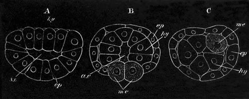
Fig. 128. Three stages in the development of
Pedicellina echinata.
(After Hatschek.)
s.c. segmentation cavity; a.e. archenteron; ep.
epiblast; me. mesoblast; hy. hypoblast.
A is the commencing gastrula stage from the side in optical section.
B is a slightly later stage from above in optical section. It shews
the two primitive mesoblast cells.
C is a later stage after the closure of the blastopore, viewed from
the side in optical section.
At the anterior extremity of this disc an invagination arises to form
the œsophagus (fig. 129 A, oe); and not long afterwards a
posterior invagination to form the rectum (fig. 129 B, an.i).
The oral disc and the œsophagus are richly ciliated. The
œsophagus first, and afterwards the rectum unite with the
archenteron (fig. 130), the walls of which soon become differentiated
into a stomach and intestine, and on the upper wall of the former the
hepatic cells become especially conspicuous (fig. 130).
During the completion of the alimentary canal a number of important
structures is formed. The disc in which the oral and anal apertures
are situated becomes converted into a true vestibule. On its floor,
between the mouth and the anus, there arises a marked prominence with
a tuft of cilia (fig. 130 B), which persists in the adult.
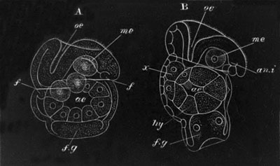
Fig. 129. Two stages in the development of
Pedicellina.
(After Hatschek.)
oe. œsophagus; ae. archenteron; an.i. anal
invagination; f. fold of epiblast; f.g. ciliated disc;
x. problematical body derived from hypoblast (probably a
bud).
This prominence is perhaps equivalent to the epistome of the
Phylactolæmata and the disc-like organ of Rhabdopleura, which
Lankester has compared to the molluscan foot[120].
Very shortly after the first formation of the vestibule there appears
at the opposite end of the larva a thickening of the epiblast, which
soon becomes invaginated, and forms an eversible pit (fig. 129 A and
B, f.g.). Round its mouth there is formed a ring of stiff cilia
(fig. 130, f.g.). This organ is very possibly equivalent to the
cement gland described by Kowalevsky in the adult Loxosoma. I shall
speak of it as the ciliated disc.
The epiblast cells early secrete a cuticle.
The two mesoblast cells soon increase by division, and occupy the
space between the alimentary canal and the body wall. They do not
become divided into a splanchnic and somatic layer; but give rise to
the interstitial connective tissue and muscles. From the
mesoblast there is also formed, according to Hatschek, a pair of
ciliated excretory canals, in the space between the mouth and anus
(fig. 130 B, nph.). The development of the nervous system has
not been observed.
At a comparatively late stage in the development there is formed round
the edge of the vestibule a ring of long cilia (fig. 130 B, m.).
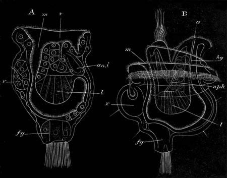
Fig. 130. Two stages in the development of
Pedicellina.
(After Hatschek.)
v. vestibule; m. mouth; l. liver; hg.
hind-gut; a. anus; an.i. anal invagination;
nph. duct of kidney; fg. ciliated disc; x.
dorsal organ (probably bud).
A remarkable organ makes its appearance on the dorsal side of the
œsophagus (the side opposite the adult ganglion) formed of an oval
mass of cells attached to the epiblast at the apex of a small ciliated
papilla (fig. 130 A and B, x.). This organ will be spoken of as
the dorsal organ. According to Hatschek it develops as a solid
outgrowth of the hypoblastic walls of the mesenteron shortly before
the mesenteron joins the œsophagus (fig. 129 B, x.). The
cells composing it arrange themselves as a sack, which acquires an
external opening on the dorsal surface (fig. 130 A, x.). In a
later stage the lumen of the sack disappears, but at the junction of
the organ with the epiblast a pit is formed, lined with
ciliated cells, which is capable of being protruded as a papilla. The
organ itself becomes invested by a lining of cells, which Hatschek
regards as mesoblastic. A nearly similar organ to this is found in the
embryo of Loxosoma [Vogt (No. 302) and Barrois (No. 298)]. Here
however it is double, and forms a kind of disc connected with two eye
spots.
Hatschek has made with reference to the dorsal organ the extremely
plausible suggestion that it is a rudimentary bud, and that the
hypoblastic sack it contains gives rise to the hypoblast of the young
polype developed from the bud. Although, owing to the deficiency of
our observations on the attachment of the larva, this suggestion has
not received direct confirmation, yet the relations of dorsal organs
in Pedicellina and Loxosoma respectively strongly confirm Hatschek’s
view of their nature. Both of these forms increase in the adult state
by budding: in Pedicellina there is a single row of buds formed
successively on the dorsal side of the stem, corresponding with the
single dorsal organ of the embryo; while in Loxosoma a double row of
buds, right and left, is formed, in correspondence with the double
nature of the dorsal organ.
As to the mode of attachment of the embryo next to nothing is known,
the few observations we have being due to Barrois. From these
observations it would appear probable that the larva, as is usual
amongst Polyzoa, does not become directly converted into the permanent
form, but that, on becoming fixed, it undergoes a metamorphosis in the
course of which its organs atrophy. I would venture to suggest that
the whole free-swimming larva atrophies, while the dorsal organ alone
develops into the fixed form[121].
Although the changes which take place during budding do not fall
within the province of this work, it may be well to state that
Hatschek has observed during this process the development of the
nervous system and the generative organs. The nervous system arises as
an unpaired thickening of the epiblastic floor of the vestibule,
between the mouth and the anus. On becoming constricted off from the
epiblast the nerve ganglion contains a central cavity which afterwards
vanishes.
The generative organs originate as a pair of specially large mesoblast
cells in the space between the stomach and the floor of the vestibule.
These two cells, surrounded by an investment of flattened mesoblast
cells, subsequently divide and form two masses. At a still
later period each mass divides into an anterior and a posterior part;
the former giving rise to the ovary, the latter to the testis. The
similarity of this mode of development of the generative organs to
that observed by Bütschli in Sagitta, which is described in the
sequel, is very striking.
Ectoprocta.
Although the embryology of the Ectoprocta has been investigated by a
very considerable number of the distinguished naturalists of the
century, many points connected with it still stand in great need of
further elucidation. The original nature of the embryo was rightly
interpreted by Grant, Dalyell and other naturalists, but it was not
till Huxley demonstrated the presence of both the ovary and testis
that the true sexual origin of the embryo in the ovicells became an
established fact in science. The recent memoir of Barrois (No. 298),
though it contains the record of a vast amount of research, and marks
a great advance in our knowledge, still leaves a great number of
points, both with reference to the early development and to the larval
metamorphosis in a very unsatisfactory condition.
Four larval forms can be distinguished, viz.
(1) A larval form which with slight modifications is common to all the
genera of the Chilostomata (except Membranipora and Flustrella) and of
the Ctenostomata.
(2) A bivalved larva of Membranipora known as Cyphonautes, the
true nature of which was first recognized by Schneider (No. 322), and
the closely allied larva of Flustrella.
(3) The typical Cyclostomatous larva, for the first full description
of which we are indebted to Barrois (No. 298).
(4) The larva of the Phylactolæmata.
Chilostomata and Ctenostomata. As an example of the first type
of larvæ, Alcyonidium mytili, one of the Ctenostomata, may be
conveniently selected for description, as having been more completely
worked out by Barrois than perhaps any other form. The segmentation
commences in the normal manner by the appearance of two vertical
furrows followed by an equatorial furrow, which divide the ovum into
eight equal spheres. The stage with eight spheres is followed,
according to Barrois, by one with sixteen, formed in a remarkable
manner by the simultaneous appearance of two vertical furrows,
both parallel to one of the original vertical furrows, so that
the segmentation spheres at this stage are arranged in two layers of
eight each. In the next stage segmentation takes place along two fresh
vertical planes, similar to those of the last stage, but at right
angles to them, and therefore parallel to the second of the two
primitive vertical furrows. At the close of this stage there are
thirty-two cells arranged in two layers of sixteen each; and when
viewed from the surface each of these layers presents a regularly
symmetrical pattern. Up to the stage with sixteen cells the two poles
of the egg, separated by the primitive equatorial plane of
segmentation, remain equal, but during the stage with thirty-two cells
a peculiar change takes place in the character of the cells at the two
poles. At the one pole, which will be spoken of as the oral pole, the
four central cells become much larger than the twelve peripheral
cells.
The stages immediately following are still involved in much obscurity,
and have been described very differently by Barrois in his original
memoir (No. 298), and in a subsequent note (No. 307)[122]. In the
latter he states that the four large cells of the oral face become
enclosed by the division and growth of the twelve peripheral cells.
They are thus carried into the interior of the ovum; and there divide
into a central vitelline mass—the hypoblast—and a peripheral
mesoblastic layer.
The eight peripheral cells of the aboral pole divide vertically, and,
owing to the eight central cells at the aboral pole dividing
transversely so as to form a protuberance on the aboral surface, they
constitute a transverse ring of large cells round the ovum, which
become ciliated and constitute the main ciliated band of the embryo,
corresponding to the ciliated band at the edge of the vestibule of the
entoproctous larvæ. They divide the embryo into an aboral and an oral
region. The central part of the aboral projection forms a structure
which I shall speak of as the ciliated disc. It probably corresponds
with the ciliated disc in the Entoprocta. An invagination is next
formed on the oral surface, which gives rise to a sack opening
to the exterior (fig. 131, st.). This was originally held by
Barrois to be the stomach; but Barrois now prefers to call it ‘the
internal sack.’ To my mind it is probably the stomodæum. The embryo
has become in the meantime laterally compressed, and, at what I shall
call the anterior end of the oral disc, a structure makes its
appearance (fig. 131, m), which is probably homologous with the
dorsal organ of the larva of Pedicellina and may go by the same name.
It was originally interpreted by Barrois as the pharynx[123].
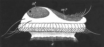
Fig. 131. Free-swimming larva of Alcyonidium
mytili. (After Barrois.)
m (?) dorsal organ; st.
stomodæum (?); s. ciliated disc.
The larva, having now acquired all the important structures it is
destined to possess, becomes free. It is shewn in fig. 131; the oral
face being turned upwards. There are two rings of cilia, one round the
edge of the ciliated disc, and a second with larger cilia on the ring
of large cells described above. This ring projects somewhat; its
projecting edge being directed towards the ciliated disc. The dorsal
organ (m?) is placed on the oral face at the bottom of an
elongated groove, in front of which is a bunch of long cilia or
flagella. Two long flagella are also developed at the posterior
extremity of the oral face, and two pairs (an anterior and a
posterior) of eye-spots also appear. Towards the posterior extremity
of the oral face is seen a body marked st, which forms the
internal sack. If I am right in regarding this as the
stomodæum, it is probable that it never unites with the invaginated
hypoblast, and that the alimentary tract of the larva remains
therefore permanently in an imperfect condition.
Careful observations have been made by Repiachoff (No. 318) on the
early development of Tendra, which accord in some respects with the
results arrived at by Barrois in his second memoir. The observations
are not, unfortunately, carried down to the complete development of
the larva.
The ovum divides in the normal way into two and then four uniform
segments. These four next become divided by an equatorial furrow into
four dorsal and four ventral segments, the former constituting the
aboral pole and forming the epiblast, and the latter the oral pole.
The stages with sixteen and thirty-two cells appear to be formed in
the same manner as in Alcyonidium—but between the two layers of cells
forming the oral and aboral poles a well-marked segmentation cavity
arises at the stage with sixteen segments. At the stage with
thirty-two cells the four middle cells of the oral side, which are
larger than the others, become divided into two tiers, in such a
manner as to form a prominence projecting into the segmentation
cavity. By the appearance of a lumen in this prominence it becomes
converted into an archenteron, which communicates with the exterior by
a blastopore in the middle of the oral surface. The blastopore becomes
eventually closed.
The archenteric sack of Repiachoff is clearly the same structure as
Barrois’ four invaginated cells of the oral face, their further
history has unfortunately not been followed out by Repiachoff.
The free larva swims about for some time, and then fixes itself and
undergoes a metamorphosis; but the exact course of this metamorphosis
is still very imperfectly known.
According to the latest statements of Barrois the attachment takes
place by the oral face[124]. The ciliated disc, which in the free
larva forms a kind of cup directed towards the aboral end, turns in
upon itself towards the oral face. It subsequently undergoes
degeneration and forms a nutritive or yolk-mass. The skin of the larva
after these changes gives rise to the ectocyst or cell of the future
polype. The future polype itself appears to originate, in part at any
rate, from the so-called dorsal organ[125].
The first distinct rudiment of the polype appears as a white body,
which gradually develops into the alimentary canal and lophophore.
While this is developing the ectocyst grows rapidly larger, and the
yolk in its interior separates from the walls and occupies a position
in the body cavity of the future polype, usually behind the developing
alimentary canal. According to Nitsche (No. 316) it is attached to a
protoplasmic cord (funiculus) which connects the fundus of the stomach
with the wall of the cell. It is probably (Nitsche, etc.) simply
employed as nutritive material, but, according to Barrois, becomes
converted into the muscles, especially the retractor muscles.
Adopting the hypothesis already suggested in the case of the
Entoprocta the metamorphosis just described would seem to be a case of
budding accompanied by the destruction of the original larva.
This view of the nature of the post-embryonic metamorphosis is
apparently that of Claparède and Salensky, and is supported by
Claparède’s statement that the formation of the first polype
‘resembles to a hair’ that of the subsequent buds. The mode of budding
would, however, appear to present certain peculiarities, in that the
whole larval skin passes directly into the bud, while from the
rudimentary bud of the larva the lophophore and alimentary tract only
of the fixed polype are formed.
Flustrella and Cyphonautes. The next group of larval forms is
that of which Cyphonautes is the best known type. The larvæ composing
it at first sight appear to have but little in common with the larvæ
hitherto described. The researches of Barrois (No. 298) and
Metschnikoff (No. 314), (but especially those of the former on the
early stages of Flustrella hispida, the larva of which is very
similar in form to Cyphonautes, though without so great a complexity
of organisation), have given a satisfactory basis for a general
comparison of Cyphonautes with other ectoproctous larvæ.
The segmentation and early stages of the embryo of Flustrella resemble
closely those of Alcyonidium. A projecting ring of large cells is
formed, dividing the larva into oral and aboral parts. The oral part
soon however becomes very small as compared with the aboral, and
becomes vertically flattened so as to be nearly on a level with the
ring of large cells. In the next stage the flattening becomes
completed; and the ring of large cells surrounds, like the
vestibule of the Entoprocta, a flat oral disc. The aboral side is
dome-shaped, and forms the greater part of the embryo.
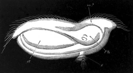
Fig. 132. Advanced larva of Flustrella
hispida.
(After Barrois.)
m (?) groove above dorsal organ; Ph. dorsal organ;
st. stomodæum (?); s. ciliated disc at aboral end of
body.
In the next stage a small disc—the ciliated disc—is formed in the
middle of the aboral dome. The larva becomes laterally compressed. The
ring of large cells which now constitute the edge of the vestibule is
covered, as in the larva of Pedicellina, by cilia, which are specially
long in front of the dorsal organ.
In the next stage the ciliated disc (fig. 132, s.) becomes
reduced in size, but surmounted by a ring of cilia round the edge, and
a tuft of cilia in the centre. The chief difference between this larva
and that of Alcyonidium depends on the small size of the ciliated
disc, and the oral position of the ciliated ring in the former. There
are intermediate types between these forms of larvaæ.
This stage immediately precedes the liberation of the larva. The free
larva differs from that in the ovicell mainly in the possession of a
shell formed as a cuticular structure, composed of two valves placed
on the two sides of the embryo. The aboral ciliated disc, still more
reduced in size, loses its cilia, and becomes enclosed between the two
valves of the shell.
The post-embryonic metamorphosis follows, so far as is known, the
course already described for the larva of Alcyonidium.
Cyphonautes (fig. 133) forms at certain seasons of the year one of the
commonest captures in the surface net. It was originally described by
Ehrenberg, but the important discovery of its true nature as the larva
of Membranipora (the common species C. compressus is the larva
of Mem. pilosa), a genus of the chilostomatous Polyzoa, was
made by Schneider (No. 322). The younger stages of the larva have not
been worked out, but from a comparison with the last described larva
it is easy to make out the general relationship of the parts. The
larva has a triangular form with an aboral apex, corresponding with
the summit of the dome of the Flustrella larva, and an oral base. It
is enclosed in a bivalve shell, the two valves of which meet along the
two sides, but are separate along the base. At the apex an opening is
left between the two valves, through which a ciliated disc
(f.g) of the same character and nature as that of previous
larvæ can be protruded.
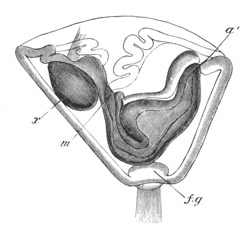
Fig. 133. Cyphonautes (larva of
Membranipora). (After Hatschek.)
m. mouth; a´ anus; f.g. ciliated disc;
x. problematical body (probably a bud).
The oral side or base is girthed by a somewhat sinuous ciliated edge,
which is continued round the anterior and posterior extremities of the
oral disc. It is no doubt equivalent to the ciliated ring of other
larvæ. Two openings are present on the oral face, both enclosed in a
special lobe of the ciliated ring. The larger of these leads into a
depression, which may be called the vestibule; and is situated on the
posterior side of the oral surface. The smaller of the two, on the
anterior side, leads into a cavity which is apparently (Hatschek)
equivalent to the rudimentary bud or dorsal organ of other larvæ. The
deeper part of the vestibule leads into the mouth (m) and
œsophagus; the latter is continued till close to the apex of the
larva, there bends upon itself, dilates into a stomach, and is
continued parallel to the œsophagus as the rectum which opens by an
anus (a´) at the posterior end of the vestibule. A peculiar
paired organ is situated on each side nearly above the stomach. Its
nature is somewhat doubtful. It was regarded as muscular by Claparède
(No. 309), though this, as shewn by Schneider, is no doubt a mistake.
Allman (No. 305) regards it as hepatic, and Hatschek as a thickening
of the epidermis. Close to each of these organs is a small body
regarded by Claparède as an accessory muscle. It is placed in the
normal position for a Polyzoon ganglion, and may perhaps be therefore
regarded as nervous in nature. Allman points out its similarity to a
bilobed ganglion, but is not inclined to take this view of it. The
constitution of the parts contained in the anterior cavity (x)
is somewhat obscure. The most elaborate descriptions of them are given
by Schneider and Allman. Lining the cavity is apparently a mass of
spherical bodies, connected with which is a tongue-like process
provided with long cilia, which can be protruded from the orifice.
Internal to this is a striated body. A good figure of the whole
structure is given by Schneider.
The general similarity of Cyphonautes to the other larvæ is quite
obvious from the above description and figure. In the presence of an
anus, a vestibule, and possibly a nervous system, it clearly exhibits
a far more complicated organisation than any other Polyzoon larvæ
except those of the Entoprocta.
The post-embryonic metamorphosis of Cyphonautes, admirably
investigated by Schneider, takes place in the same manner as that of
other larvæ, and is accompanied by the degeneration of the larval
organs, and the formation of a clear body, which gives rise to the
alimentary cavity and lophophore of the fixed polype. The larval shell
takes part in the formation of the ectocyst of the polype.
Cyclostomata. We owe to Barrois by far the fullest account of
the development of the Cyclostomata, but how far his interpretations
are to be trusted is very doubtful. The larvæ differ very considerably
from the normal larvæ of the Chilostomata and Ctenostomata; the
difference being mainly due to the enormous development of the
ciliated disc. Barrois has investigated the larvæ of three genera,
Phalangella, Crisia, and Diastopora, and states that they very
closely resemble each other. The ovum is extremely minute.
The segmentation, so far as it has been made out, is regular. During
the segmentation growth is very rapid, and eventually there is formed
a blastosphere many times larger than the original ovum. The
blastosphere becomes flattened, and is converted into a gastrula by
bending up into a cup-like form. The gastrula opening is stated to
remain as the permanent mouth, which has a terminal and central
position. A transverse ring-like thickening is formed round the larva,
which probably corresponds with the ciliated ring of previous larvæ;
and the body of the larva in front of this ring becomes ciliated. The
aboral end of the larva becomes thickened, and grows out into an
elongated prominence, which probably corresponds to the ciliated disc.
The ring before mentioned becomes at the same time more prominent, and
forms a cylindrical sheath for the ciliated disc. At the time when the
larva becomes liberated from the maternal cell it has the form of a
barrel with a slight constriction in the middle separating the oral
from the aboral end. At the centre of the oral face is situated the
mouth, leading into a wide stomach, while the aboral end is formed of
the ciliated disc enclosed in its sheath. The whole surface is now
ciliated. No structure equivalent to the dorsal organ or bud is
described by Barrois, but in other respects, if the ciliated disc is
really equivalent in the two forms, a general comparison on the line
indicated above between this larva and the normal larvæ of the
Ctenostomata and Chilostomata seems quite possible. The fixation and
subsequent development of the larva take place in the normal manner.
Phylactolæmata. The development of the phylactolæmatous Polyzoa
has been studied by Metschnikoff (No. 315), who describes the eggs as
undergoing a complete segmentation within a peculiar brood-pouch
developed from the walls of the body of the parent. After segmentation
the cells of the embryo arrange themselves in two layers round a
central cavity. The embryo then forms the well-known cyst, from which
a colony is formed by a process of budding.
General considerations on the Larvæ of the Polyzoa.
The different forms of embryo amongst the Polyzoa are represented in
figs. 130 B, 131, 132,
and 133 in what I regard as identical
positions, and fig. 133 A is a figure of what may be regarded
as an idealized larval Polyzoon. In all the larvæ there is present a
ciliated ring, which separates an oral from an aboral face, and is
apparently homologous throughout the series. In the adult it is
probably represented by the lophophore. On the oral face is
situated in all cases the mouth, and in the entoproctous larvæ and
Cyphonautes also the anus. It thus appears that Cyphonautes, though
the larva of an ectoproctous form, is itself entoproctous—a fact
which tends to shew that the Entoprocta are the more primitive forms.
In all the larvæ, except possibly those of the Cyclostomata, there is
present on the anterior side of the mouth, in the Ectoprocta on the
oral, and in the Entoprocta on the aboral side of the ciliated ring,
an organ, to which is attached externally a plume of long cilia. This
organ has been identified throughout the series in accordance with
Hatschek’s view as the dorsal organ or rudimentary bud; but it is well
to bear in mind that this identification is of a purely hypothetical
character.
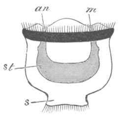
Fig. 133 a. Diagram of an ideal larva of a
Polyzoon.
m. mouth; an. anus; st. stomach; s.
ciliated disc.
On the aboral side of the ciliated ring there is present in all the
larvæ an organ, which has been called the ciliated disc, which is
probably homologous throughout the series. It perhaps remains in the
adult of Loxosoma as the cement gland, but not in other forms.
The Polyzoa present a simple and almost certainly degraded
organisation in the adult state; it is therefore more than usually
necessary to turn to their larvæ for the elucidation of their
affinities, and various plausible suggestions have been made as to the
interpretation of the characters of the larvæ.
Lankester[126] has suggested that the larvæ are essentially similar to
those of Molluscs. He compares the main ciliated ring to the velum,
but has ingeniously suggested that it represents not the simple velar
ring of most molluscan larvæ, but a more extended longitudinal ring,
of which the gills of Lamellibranchiata are supposed by him to be
remnants, and to which the Echinoderm larvæ with one continuous
ciliated band furnish a parallel.
The foot he finds in the epistome of the Phylactolæmata, and the disc
of Rhabdopleura—both situated between the mouth and anus, and
therefore in the situation of the molluscan foot. The peculiar
prominence between the mouth and the anus in Pedicellina (vide
fig. 130 B) and Loxosoma is probably the same structure.
Finally he identifies my ciliated disc, which as mentioned above is
perhaps equivalent to the cement gland in the adult Loxosoma, as the
molluscan shell-gland. Lankester’s interpretations are very plausible,
but at the same time they appear to me to involve considerable
difficulties.
There is absolutely no evidence amongst the Mollusca of the existence
of a primitive longitudinal ciliated ring, such as he supposes to have
existed, and Lankester is debarred from regarding the ciliated ring of
the Polyzoa as equivalent to the simple velar ring of the Mollusca,
because his shell-gland lies in the centre and not as it should do on
the posterior side of the ciliated ring.
Another difficulty which I find is the invariable ciliation of
Lankester’s shell-gland—a ciliation which never occurs amongst
Mollusca.
It appears to me that a more satisfactory comparison of the larvæ of
the Polyzoa with those of the Mollusca is obtained by dropping the
view that the ciliated disc is the shell-gland, and by regarding the
ciliated ring as equivalent to the velum. This mode of comparison has
been adopted by Hatschek.
The larva ceases however on this view to have any special molluscan
characters (except possibly the organ which Lankester has identified
as the foot), and only resembles a molluscan larva to the same extent
as it does a larva of the Polychæta. The ciliated disc lies according
to this view in the centre of the velar area or præ-oral lobe, and
therefore in the situation in which a tuft of cilia is often present
in lamellibranchiate and other molluscan larvæ, and also in the larvæ
of most Chætopoda. It is moreover at this point that the
supra-œsophageal ganglion is always formed in the Mollusca and
Chætopoda as a thickening of the epiblast (fig. 134, sg.), so
that the thickening of the epiblast in the ciliated disc of the
Polyzoa may perhaps be a rudiment of the supra-œsophageal ganglion,
which entirely atrophies in the adult after the attachment has been
effected in the region of this disc.
The comparison between the Polyzoon larva and that of a
Chætopod becomes very much strengthened by taking as types
Mitraria[127] (fig. 134) and Cyphonautes (fig. 133). The similarity
between these two forms is so striking that I am certainly inclined to
view the larvæ of the Polyzoa as trochospheres similar to those of
Chætopods, Rotifera, etc., which become fixed in the adult by the
extremity of their præ-oral lobe.
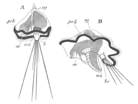
Fig. 134. Two stages in the development of
Mitraria.
(After Metschnikoff.)
m. mouth; an. anus; sg. supra-œsophageal
ganglion; br. and b. provisional bristles;
pr.b. præ-oral ciliated band.
The attachment of the larva by the præ-oral lobe is not more
extraordinary than the attachment of a Barnacle by its head, and after
such a mode of attachment the atrophy of the supra-œsophageal
ganglion would be only natural.
There is one important fact which deserves to be noted in the
development of the Polyzoa, viz. that if the suggestion in the text as
to the mode of development of the adult from the so-called larva is
accepted, the Polyzoa exhibit universally the phenomenon of
alternations of generations. The ovum gives rise to a free form
which never becomes sexual, but produces by budding the sexual
attached form.
Bibliography.
General.
(298) J. Barrois. Recherches sur l’embryologie des
Bryozoaires. Lille, 1877.
Entoprocta.
(299) B. Hatschek. “Embryonalentwicklung u. Knospung d.
Pedicellina echinata.” Zeitschrift für wiss. Zool., Bd.
XXIX. 1877.
(300) M. Salensky. “Etudes sur les Bryozoaires entoproctes.”
Ann. Scien. Nat., 6th Ser. Tom. V. 1877.
(301) O. Schmidt. “Die Gattung Loxosoma.” Archiv f. mik.
Anat., Bd. XII. 1876.
(302) C. Vogt. “Sur le Loxosome des Phascolosomes.” Archives
de Zool. expér. et génér., Tom. V. 1876.
(303) C. Vogt. “Bemerkungen zu Dr Hatschek’s Aufsatz üb.
Embryonalentwicklung u. Knospung von Pedicellina echinata.” Zeit.
f. wiss. Zool., Bd. XXX. 1878.
Ectoprocta.
(304) G. J. Allman. Monograph of fresh-water Polyzoa.
Ray Society.
(305) G. J. Allman. “On the structure of Cyphonautes.”
Quart. J. of Micr. Scie., Vol. XII. 1872.
(306) G. J. Allman. “On the structure and development of the
Phylactolæmatous Polyzoa.” Journal of the Linnean Society, Vol.
XIV. No. 77. 1878.
(307) J. Barrois. “Le développement d. Bryozoaires
Chilostomes.” Comptes rendus, Sept. 23, 1878.
(308) E. Claparède. “Beiträge zur Anatomie u.
Entwicklungsgeschichte d. Seebryozoen.” Zeit. für wiss. Zool.,
Bd. XXI. 1871.
(309) E. Claparède. “Cyphonautes.” Anat. u. Entwick.
wirbell. Thiere. Leipzig, 1864.
(310) R. E. Grant. “Observations on the structure and nature of
Flustræ.” Edinburgh New Philosoph. Journal, 1827.
(311) B. Hatschek. “Embryonalentwicklung u. Knospung d.
Pedicellina echinata.” (Description of Cyphonautes). Zeit.
f. wiss. Zool., Bd. XXIX. 1877.
(312) T. H. Huxley. “Note on the reproductive organs of the
Cheilostome Polyzoa.” Quart. Jour. of Micr. Science, Vol.
IV. 1856.
(313) L. Joliet. “Contributions à l’histoire naturelle des
Bryozoaires des côtes de France.” Archives de Zoologie
Expérimentale, Vol. VI. 1877.
(314) E. Metschnikoff. “Ueber d. Metamorphose einiger
Seethiere.” Göttingische Nachrichten, 1869.
(315) E. Metschnikoff. Bull. de l’Acad. de St
Pétersbourg, XV. 1871, p. 507.
(316) H. Nitsche. “Beiträge zur Kenntniss d. Bryozoen.”
Zeit. f. wiss. Zool., Bd. XX. 1870.
(317) W. Repiachoff. “Zur Naturgeschichte d. chilostomen
Seebryozoen.” Zeit. f. wiss. Zool., Bd. XXVI. 1876.
(318) W. Repiachoff. “Ueber die ersten Entwicklungsvorgänge bei
Tendra zostericola.” Zeit. f. wiss. Zool., Bd. XXX.
1878. Supplement.
(319) W. Repiachoff. “Zur Kenntniss der Bryozoen.”
Zoologischer Anzeiger, No. 10, Vol. I. 1878.
(320) W. Repiachoff. “Bemerkungen üb. Cyphonautes.”
Zoologischer Anzeiger, Vol. II. 1879.
(321) M. Salensky. “Untersuchung an Seebryozoen.” Zeit. für
wiss. Zool., Bd. XXIV. 1874.
(322) A. Schneider. “Die Entwicklung u. syst. Stellung d.
Bryozoen u. Gephyreen.” Archiv f. mikr. Anat., Vol. V.
1869.
(323) Smitt. “Om Hafsbryozoernas utveckling och fettkroppar.”
Aftryck ur öfvers. af Kong. Vet. Akad. Förh. Stockholm, 1865.
(324) T. Hincks. British Marine and Polyzoa. Van Voorst,
1880.
[Conf. also works by Farre, Hincks, Van Beneden, Dalyell, Nordmann.]
The observations which have been made on the developmental history of
the Brachiopoda have thrown very considerable light on the systematic
position of this somewhat isolated group.
Development of the Layers.
For our knowledge of the early stages in the development of the
Brachiopoda we are almost entirely indebted to Kowalevsky[129] (No.
326). His researches extend to four forms, Argiope, Terebratula,
Terebratulina, and Thecidium. The early development of the first three
of these takes place on one plan, and that of Thecidium on a second
plan.
In Argiope, which may be taken as typical of the first group, the ova
are transported into the oviducts (segmental organs) where they
undergo their early development. The segmentation leads to the
formation of a blastosphere, which then becomes a gastrula by
invagination. The blastopore gradually narrows, and finally closes,
while at the same time the archenteric cavity (fig. 135 A)
becomes divided into three lobes, a median (me) and two lateral
(pv). These lobes next become completely separated, and the
middle one forms the mesenteron, while the two lateral ones give rise
to the body cavity, their outer walls forming the somatic mesoblast,
and their inner the splanchnic (fig. 135 B). The embryo now elongates,
and becomes divided into three successive segments (fig. 135 B), which
are usually, though on insufficient grounds (vide Thecidium),
regarded as equivalent to the segments of the Chætopoda. The
alimentary tract is not continued into the hindermost of them.
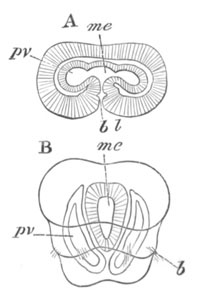
Fig. 135. Two stages in the development of
Argiope. (After Kowalevsky.)
A. Late gastrula stage.
B. Stage after the larva has become divided into three segments.
bl. blastopore; me. mesenteron; pv. body
cavity; b. temporary bristles.
In Thecidium the ova are very large, and development takes place in a
special incubatory pouch in the ventral valve. The embryos are
attached by suspenders to the two cirri of the arms which immediately
adjoin the mouth. There is a nearly regular segmentation, and a very
small segmentation cavity is developed. There is no invagination; but
cells are budded off from the walls of the blastosphere, which soon
form a solid central mass, enclosed by an external layer—the
epiblast. In this central mass three cavities are developed, which
constitute the mesenteron and the two halves of the body cavity.
Around these cavities distinct walls become differentiated. The body
(Lacaze Duthiers, No. 327) soon after becomes divided into two
segments, of which the posterior is the smaller. The hinder part of
the large anterior segment next becomes constricted off as a fresh
segment, and subsequently the remaining part becomes divided into two,
of which the anterior is the smallest. The embryo thus becomes divided
into four segments, of which the two foremost appear (?) together to
correspond to the cephalic segment of Argiope; but these segments are
formed not, as in Chætopoda and other truly segmented forms, by the
addition of fresh segments between the last-formed segment and
the unsegmented end of the body, but by the interpolation of fresh
segments at the cephalic end of the body as in Cestodes; so that the
hindermost segment is the oldest. Assuming the correctness[130] of
Lacaze Duthiers’ observations, the mode of formation of these segments
appears to me to render it probable that they are not identical with
the segments of a Chætopod. A suspender is attached to the front end
of each embryo. Before the four segments are established the whole
embryo is covered with cilia[131], and two and then four rudimentary
eyes are developed on the anterior segment of the body.
The history of the Larva and the development of the organs of the
Adult.
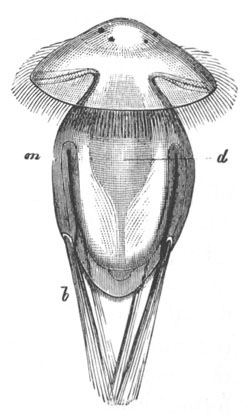
Fig. 136. Larva of Argiope. (From Gegenbaur,
after Kowalevsky.)
m. mantle; b. setæ; d. archenteron.
Articulata. The observations of Kowalevsky and Morse have given
us a fairly complete history of the larval metamorphosis of some of
the Articulata, while some of the later larval stages in the history
of the Inarticulata have been made known to us from the researches of
Fritz Müller, Brooks, etc. The embryo of Argiope, which may be taken
as the type for the Articulata, was left (fig. 135 B) as a three lobed
organism with a closed mesenteron and a body cavity divided into two
lateral compartments. On the middle segment of the body dorsal and
ventral folds, destined to form the mantle lobes, make their
appearance, and on the latter two pairs of bundles of setæ are present
(fig. 135 B). The setæ together with the mantle folds grow greatly,
and the setæ resemble in appearance the provisional setæ of many
Chætopods (fig. 152). On the hinder border of the mantle cilia make
their appearance. The anterior or cephalic segment assumes a somewhat
umbrella-like form, and round its edge is a circlet of long
cilia, while elsewhere it is provided with a coating of short cilia.
Two pairs of eyes also arise on its anterior surface (fig. 136).
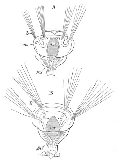
Fig. 137. Two stages in the development of
Argiope, shewing the folds of the mantle growing over the cephalic
lobe. (After Kowalevsky.)
m. mantle fold; me. mesenteron; pd. peduncle;
b. provisional setæ.
After swimming about for some time the larva becomes fixed by its hind
lobe, and becomes gradually transformed into the adult. The hind lobe
itself becomes the peduncle. After attachment the mantle lobes bend
forward (fig. 137 A, m), and enclose the cephalic lobe. The
valves of the shell are formed on their outer surface as two delicate
chitinous plates (fig. 137 B). At a somewhat later stage the
provisional bristles are thrown off, and are eventually replaced by
permanent setæ round the edge of the mantle. The cephalic lobe becomes
located in the dorsal valve of the shell, and the mouth is formed near
the apex of the cephalic lobe immediately ventral to the eye-spots, by
an epiblastic invagination. The permanent muscles are formed out of
the muscles already present in the embryo.
Around the mouth there arises a ring of tentacles, very
possibly derived from the ciliated ring visible in fig. 136[132]. The
ring of tentacles is placed obliquely, and the mouth is situated near
its ventral side. The tentacles appear to form a post-oral circlet,
like that of Phoronis (Actinotrocha): they gradually increase in
number as the larva grows older.
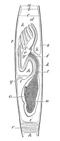
Fig. 138. Diagram of a longitudinal vertical
section of an advanced embryo of Lingula. (After Brooks.)
a. end of valves; b. thickened margin of mantle;
c. mantle; d. dorsal median tentacle; e.
lophophore; f. lip; g. mouth; h. mantle cavity;
i. body cavity; k. wall of œsophagus; l.
œsophagus; m. hepatic chamber of stomach; n.
intestinal chamber of stomach; o. intestine; q.
ventral ganglion; r. posterior muscle; s. dorsal valve
of shell; t. ventral valve of shell.
Some of the later stages in the development of the Terebratulidæ have
been made known to us by the observations of Morse (No. 328‑9) on
Terebratulina septentrionalis.
The most interesting point in Morse’s observations on the later stages
is the description of the gradual conversion of the disc bearing the
circlet of tentacles into the arms of the adult. The tentacles, six in
number, first form a ring round the edge of a disc springing from the
dorsal lobe of the mantle; in their centre is the mouth. In the later
stages calcareous spicula become developed on the tentacles. When the
embryo is far advanced the tentacles begin to assume a horse-shoe
arrangement, which bears a striking, though probably accidental,
resemblance to that of the tentacles on the lophophore of the
fresh-water Polyzoa. The disc bearing the tentacles is prolonged
anteriorly into two processes, the free ends of the future arms. By
this change of shape in the disc the tentacles form two rows, one on
the anterior and one on the posterior border of the disc, and
eventually become the cirri of the arms. The mouth is placed between
the two rows of tentacles, where the two arms of the lophophore meet
behind. The position of the mouth was the original centre of the ring
of tentacles before they became pulled out into a horse-shoe form. In
front of the mouth is a lip. The arms grow greatly in length in the
adult Terebratulina. In Thecidium the oral disc retains the horse-shoe
form, while in Argiope the embryonic circular arrangement of the
tentacles is only interfered with by the appearance of marginal
sinuations.
The shell is deposited as to chitinous plates, which subsequently
become calcified. It undergoes in the different genera great changes
of form during its growth.
With reference to the larval stages of other Articulata, a few points
may be noted.
The three-lobed larva of Terebratulina septentrionalis is provided
with a special tuft of cilia at the apex of the front lobe. The arms
appear to originate, in Terebratulina caput serpentis, as two
processes at the sides of the mouth, on which the tentacles are
formed.
Provisional setæ do not appear to be formed in the lobed embryos of
Thecidium and Terebratulina, but they appear at a later stage at the
edge of the mantle in the latter form. The third lobe of Thecidium
gives rise to the dorsal and ventral mantle lobes.
Inarticulata. The youngest stages in the development of the
Inarticulata are not known, and in the earliest stages observed the
shell is already developed. The young larvæ with shells differ however
from those of the Articulata in the fact that they are free-swimming,
and that the peduncle is not developed.
The larva of Discina radiata has been described by Fritz Müller (No.
331). It resembles generally a larva of the Articulata shortly after
the tentacles have become developed. Five pairs of long provisional
setæ are present, of which all but the hindermost are seated on the
ventral lobe of the mantle. Shorter setæ are also lodged on the edge
of the dorsal lobe. The mouth is placed on the ventral side of a
protrusible oral lobe. It is imperfectly surrounded by four pairs of
tentacles, which form a swimming apparatus.
A fuller history of the development of Lingula has been recently
supplied by Brooks (No. 325). The youngest larva is enveloped in two
nearly similar plate-like valves, covering the two mantle lobes. The
mouth is placed at the centre of a disc, attached to the dorsal valve,
on the margin of which is a ring of ciliated tentacles. The general
position of the disc and its relations may be gathered from fig. 138,
which represents a diagrammatic longitudinal vertical section of the
embryo.
With the growth of the embryo the tentacles increase in number, the
new pairs being always added between the odd dorsal tentacle and the
next pair. There is an axial cavity in the tentacles which, unlike the
cavity in the tentacles of the Polyzoa, does not communicate with the
perivisceral cavity. As the tentacles increase in number, the lateral
parts of the tentacular disc grow out into the two lateral arms of the
adult, while the dorsal margin forms the median coiled arm. These
changes are not effected till the larva has become fixed.
The attachment of the larva was not observed; but the peduncle, of
which there is no trace in the young stages, grows out as a
simple prolongation of the hinder end of the body while the larva is
still free. It had already reached a very great length in the youngest
fixed larva observed.
Development of Organs.
The alimentary tract after the obliteration of the blastopore forms a
closed sack, which becomes subsequently placed in communication with
the exterior by the stomodæal invagination. The liver is formed as a
pair of dorsal outgrowths of the mesenteron. From Brooks’ observations
on Lingula it would appear that the primitive mesenteron forms the
stomach of the adult only, and that the intestine grows out from this
as a solid process: this eventually meets the skin, and here the anus
is formed. In the Articulata the mesenteron is aproctous.
The origin of the body cavity as paired archenteric diverticula has
already been described. Its somatic wall becomes in Lingula ciliated,
and its cavity filled with a corpusculated fluid, as in many
Chætopods. It is eventually prolonged into the dorsal and ventral
mantle lobes as a pair of horn-like prolongations into each lobe,
which communicate with the body cavity by large ciliated openings.
Some incomplete observations of Brooks on the development of the
nervous system in Lingula shew that it arises in the embryo as a ring
round the œsophagus with a ventral sub-œsophageal (fig. 138
q), and two lateral ganglia, and two dorsal otocysts. The
ventral ganglion is formed as a thickening of the epiblast, with which
it remains in continuity for life. The remainder of the ring grows out
from the ventral ganglion as two cords, which gradually meet on the
dorsal side of the œsophagus.
General observations on the Affinity of the Brachiopoda.
The larva of Argiope, as has been noticed by many observers, has
undoubtedly very close affinities with the Chætopoda. It resembles, in
fact, a mesotrochal larval Chætopod with provisional setæ (vide
Chapter on Chætopoda). Lacaze Duthiers’ observations point to the
lobes of the larva not being true segments, and certainly the
mesoblast does not in the embryo become segmented as it ought to do
were these lobes true segments. If this view is correct the larva is
to be compared to an unsegmented Chætopod larva. In Rhynchonella,
however, indications of two segments are afforded in the adult in the
two pairs of segmental organs.
Though the larval Brachiopod resembles a mesotrochal Chætopod larva,
it does not appear to resemble the trochosphere larvæ so far
described, or the more typical larvæ of the Chætopoda, in that the
ring of tentacles, which is probably, as already mentioned, derived
from the ciliated ring shewn in fig. 137, is post-oral, and not
præ-oral. The ring of tentacles is like the ring in
Actinotrocha (the larva of Phoronis) amongst the Gephyrea. Although
there is no doubt a striking resemblance between the tentacular disc
of a larval Brachiopod and the lophophore of a Polyzoon, which has
been pointed out by Lankester, Morse, Brooks, etc., their homology is
rendered, to my mind, very doubtful (1) by the fact that the
lophophore is præ-oral in Polyzoa[133] and post-oral in Brachiopoda;
and (2) by the fact that the concave side of the lophophore is turned
in nearly opposite directions in the two forms. In Brachiopods it is
turned dorsalwards, and in phylactolæmatous Polyzoa ventralwards.
The view of Morse, that the Brachiopoda are degraded tubicolous
Chætopods, is not so far supported by any definite embryological
facts. The development of the tentacular ring as well as its
innervation from the sub-œsophageal ganglion prohibit us, as has
been pointed out by Gegenbaur, from comparing it with the tentacles of
tubicolous Chætopoda.
Bibliography.
(325) W. K. Brooks. “Development of Lingula.” Chesapeake
Zoological Laboratory, Scientific Results of the Session of 1878.
Baltimore, J. Murphy and Co.
(326) A. Kowalevsky. “Development of the Brachiopoda.”
Protocol of the First Session of the United Sections of Anatomy,
Physiology, and Comparative Anatomy at the Meeting of Russian
Naturalists in Kasan, 1873. (Russian.)
(327) H. Lacaze Duthiers. “Histoire de la Thécidie.” Ann.
Scien. Nat. etc. Ser. 4, Vol. XV. 1861.
(328) Morse. “On the Early Stages of Terebratulina
septentrionalis.” Mem. Boston Soc. Nat. History, Vol.
II. 1869, also Ann. & Mag. of Nat. Hist., Series 4,
Vol. VIII. 1871.
(329) —— “On the Embryology of Terebratulina.” Mem. Boston Soc.
Nat. History, Vol.
III., 1873.
(330) —— “On the Systematic Position of the Brachiopoda.”
Proceedings of the Boston Soc. of Nat. Hist., 1873.
(331) Fritz Müller. “Beschreibung einer Brachiopoden Larve.” Müller’s
Archiv, 1860.
Formation of the Germinal Layers.
Most Chætopoda deposit their eggs before development. The Oligochæta
lay them in peculiar cocoons or sacks formed by a secretion of the
integument. Some marine Polychæta carry them about during their
development. Autolytus cornutus has a special sack on the ventral
surface in which they are hatched. In Spirorbis Pagenstecheri they
develop inside the opercular tentacle, and in Spirorbis spirillum
inside the tube of the parent.
A few forms (e.g. Eunice sanguinea, Syllis vivipara, Nereis
diversicolor) are viviparous.
Perhaps the most primitive type of Chætopod development so far
observed is that of Serpula (Stossich, No. 357)[135]. There is a
regular segmentation resulting in the formation of a blastosphere with
a central segmentation cavity. An invagination of the normal type now
ensues. The blastopore soon narrows to become the permanent anus,
while the invaginated hypoblast forms a small prominence with an
imperfectly developed lumen, which does not nearly fill up the
segmentation cavity (fig. 139 A). The embryo, which has in the
meantime become completely covered with cilia, now assumes
more or less the form of a cone, at the apex of which is the anus,
while the base forms the rudiment of a large præ-oral lobe. The
alimentary sack grows forwards and then bends upon itself nearly at
right angles, and meets a stomodæal invagination from the ventral side
some way from the front end of the body.
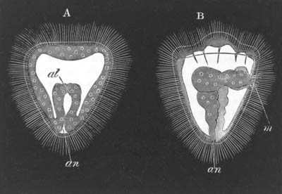
Fig. 139. Two stages in the development of
Serpula.
(After Stossich.)
m. mouth; an. anus; al. archenteron.
The alimentary canal soon differentiates itself into three regions (1)
œsophagus, (2) stomach, and (3) intestine. With these changes the
larva, which in the meantime becomes hatched, assumes the characters
of a typical Annelid larva (fig. 139 B). In front is a large præ-oral
lobe, at the sides of which the eye-spots soon appear. The primitive
segmentation cavity remains as a wide space between the curved
alimentary tract and the body walls, and becomes traversed by muscular
fibres passing between the two. The original chorion appears to serve
as cuticle, and is perforated by the cilia.
The further changes in this larval form do not present features of
general importance. A peculiar vesicle, which in anomalous cases is
double, is formed near the anus. If it were shewn to occur widely
amongst Chætopoda, it might be perhaps regarded as homologous with the
anal vesicles of the Gephyrea.
Serpula is one of the few Chætopoda at present known in which
the segmentation is quite regular[136]. In other forms it is more or
less unequal. The formation of the germinal layers has been far more
fully studied in the Oligochæta than in the Polychæta, and though
unfortunately the development is much abbreviated in the former group,
they nevertheless have to serve as our type; and unless the contrary
is indicated the statements in the remainder of the section apply to
the Oligochæta. The segmentation is nearly regular in Lumbricus
agricola (Kowalevsky) and results in the formation of a flattened
blastosphere, one of the sides of which is hypoblastic and the other
epiblastic, the hypoblast cells being easily distinguished from the
epiblast cells by their clearer aspect. An invagination takes place,
in the course of which the hypoblast becomes enclosed by the epiblast,
and a somewhat cylindrical two-layered gastrula is formed. The opening
of this gastrula at first extends over the whole of what becomes the
ventral surface of the future worm, but gradually narrows to a small
pore—the permanent mouth—near the front end. The central cavity of
the gastrula is lined by hypoblast cells, but the oral opening, which
leads by a narrow passage into the gastric cavity, is lined by
epiblast cells.
The segmentation of Lumbricus trapezoides (Kleinenberg, No. 341), and
of Criodrilus (Hatschek, No. 339), is more unequal and more irregular
than that of Lumbricus agricola, and there is an invagination which is
intermediate between the embolic and epibolic types.
The segmentation of Lumbricus trapezoides is especially remarkable. It
is strangely irregular and at one period the segmentation cavity
communicates by a pore with the exterior. Before the completion of the
gastrula stage the ovum becomes partially divided into two halves,
each of which gives rise to a complete embryo. The two embryos are at
first united by an epiblast cord which connects their necks (fig. 141
A), but this cord is very early ruptured, and the two embryos then
become quite independent. Some of the peculiarities of the
segmentation may no doubt be explained by this remarkable embryonic
fission.
The gastrula opening in both Lumbricus trapezoides and Criodrilus is
placed on the ventral surface, and eventually narrows to form the
mouth or possibly (Criodrilus) closes at the position of the mouth. In
Lumbricus trapezoides the oral opening is at first lined by hypoblast,
and in Criodrilus is bounded anteriorly by three large peculiar
epiblast cells, which are believed by Hatschek to assist in
absorbing the albuminous fluid in which the eggs are suspended. These
large cells are eventually covered by the normal epiblast cells and
subsequently disappear. In both these types the hypoblast cells
undergo, during their invagination, peculiar changes connected with
their nutritive function.
In Euaxes (Kowalevsky) the segmentation is far more unequal than in
the other types; a typical epibolic invagination takes place (fig.
140), and the blastopore closes completely along the ventral surface.
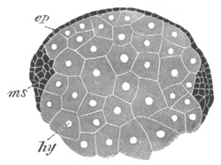
Fig. 140. Transverse section through the ovum of
Euaxes during an early stage of development. (After
Kowalevsky.)
ep. epiblast; ms. mesoblastic band; hy.
hypoblast.
In all the oligochætous types, with the exception of Euaxes, where the
blastopore closes completely, the blastopore becomes, or coincides
with the mouth. In Serpula it is stated (Stossich), as we have seen,
to coincide with the anus: a statement which receives confirmation
from the similar statements of Willemoes-Suhm (No. 358). It is
necessary either to suppose a mistake on the part of Stossich, or that
we have in Chætopods a case like that of Gasteropods in which a
slit-like blastopore originally extending along the ventral surface
may in some forms become reduced to a pore at the oral, or in other
forms at the anal extremity.
So far only two germinal layers—the epiblast and the hypoblast—have
been spoken of. Before the invagination of the hypoblast is completed
the mesoblast makes its appearance in the form of two bands or
streaks, extending longitudinally for the whole length of the embryo.
These are usually spoken of as germinal streaks, but to avoid the
ambiguity of this term they will be spoken of as mesoblastic bands.
Their origin and growth has been most fully studied by Kleinenberg
(No. 341) in Lum. trapezoides. They commence in this species shortly
before the gastrula stage as two large cells on the surface of the
blastoderm, which may be called mesoblasts. These cells lie one on
each side of the median line at the hind end of the embryo. They soon
travel inwards and become covered by the epiblast (fig. 141 A,
m´), while on their inner and anterior side a row of small
cells appears (ms). These rows of cells form the
commencement of the mesoblastic bands, and in the succeeding stages
they extend one on each side of the body (fig. 141 B, ms) till
they reach the sides of the mouth. Their forward growth takes place
mainly at the expense of the superjacent epiblast cells, but the two
mesoblasts at their hinder extremities probably assist in their
growth. Each mesoblastic band is at first composed of only a single
row of cells, but soon becomes thicker, first of all in front, and
becomes composed of two, three or more rows of cells abreast. From the
above it is clear that the mesoblastic bands have, in L. trapezoides
at any rate, in a large measure an epiblastic origin.
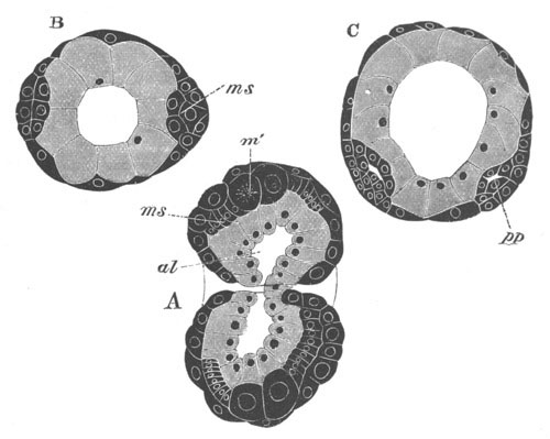
Fig. 141. Three sections illustrating the
development of Lumbricus trapezoides. (After Kleinenberg.)
ms. mesoblastic band; m´. mesoblast; al.
archenteron; pp. body cavity.
A. Horizontal and longitudinal section of an embryo which is
dividing into two embryos at the gastrula stage. It shews the
mesoblasts and the mesoblastic bands proceeding from them.
B. Transverse section shewing the two widely separated mesoblastic
bands.
C. Transverse section at a later stage shewing the mesoblastic bands
which have approached the ventral line and developed a body cavity
pp.
At first the two bands end in front at the sides of the mouth,
but subsequently their front ends grow dorsalwards at the
expense of the adjoining epiblast cells, and meet above the mouth,
forming in this way a mesoblastic dorsal commissure.
The mesoblastic bands soon travel from the lateral position, which
they at first occupy, towards the ventral surface. They do not however
meet ventrally for some time, but form two bands, one on each side of
the median ventral line (fig. 141 C).
The usual accounts of the origin and growth of the bands differ
somewhat from the above. By Kowalevsky (No. 342) and Hatschek (No.
339) they are believed to increase in Lumbricus rubellus and
Criodrilus entirely at the expense of the mesoblasts. Kowalevsky
moreover holds that in L. rubellus the original mesoblasts spring from
the hypoblast. In some forms, e.g. Lumbricus agricola, the
mesoblasts are not present.
In Euaxes the origin of the mesoblast bands is somewhat interesting as
illustrating the relation of the Chætopod mesoblastic bands to the
mesoblast of other forms. To render intelligible the origin of the
mesoblast in this form, it is necessary to say a few words about the
segmentation.
By a somewhat abnormal process of segmentation the ovum divides into
four spheres, of which one is larger than the others, and occupies a
position corresponding with the future hind end of the embryo. The
three smaller spheres give rise on their dorsal side by a kind
of budding to small cells, which become the epiblast; and the epiblast
is also partly formed from the hinder large cell in that this cell
produces by budding a small cell, which again divides into two. The
anterior of the two cells so formed divides still further and becomes
incorporated in the epiblast; the posterior only divides into two
which form the two mesoblasts. The remainder of the mesoblast
is formed by further division of the three smaller of the primitive
large spheres, and at first forms a continuous layer between the
dorsal cap of epiblast and the four largest cells which, after giving
rise to the epiblast and mesoblast, constitute the hypoblast. As the
epiblast spreads over the hypoblast the mesoblastic sheet gives way in
the middle, and the mesoblast remains as a ridge of cells at the edge
of the epiblastic cup. It forms in fact a thickening of the lips of
the blastopore. Behind the thickening is completed by the two
mesoblasts. The appearance of the mesoblast in section is shewn in
fig. 140. As the epiblast accompanied by the mesoblast grows round the
hypoblast, the blastopore assumes an oval form, and the mesoblast
appears as two bands forming the sides of the oval. The epiblast
travels over the hypoblast more rapidly than the mesoblast, so that
when the blastopore becomes closed ventrally the mesoblastic bands are
still some little way apart on the ventral side.
In Euaxes the mesoblast originates in a manner which is very similar
to that in some of the Gasteropoda, e.g. Nassa, vide p. 234,
and Vermes, e.g. Bonellia, etc. As mentioned in the
chapter on the Mollusca the origin of the mesoblast in
Planorbis, p. 227, is very similar to that in Lumbricus.
Hatschek has shewn that in Polygordius the mesoblast arises in
fundamentally the same way as in the Oligochæta.
Besides the mesoblast which arises from the mesoblastic bands, there
is evidence of the existence of further mesoblast in the larvæ of many
Polychæta in the form of muscular fibres which traverse the space
between the body wall and the wall of the enteric cavity prior to the
formation of the permanent body cavity. These fibres have already been
described in the embryo of Serpula, and are probably represented by
stellate cells in the cephalic region (præ-oral lobe) of the
Oligochæta. These cells are probably of the same nature as the
amœboid cells in the larvæ of Echinodermata, some Mollusca and
other types.
The Larval form.
True larval forms are not found in the Oligochæta where the
development is abbreviated. They occur however in the majority of the
marine Polychæta.
They present a great variety of characters with variously arranged
ciliated bands. Most of these forms can be more or less satisfactorily
derived from a larval form, like that of Serpula (fig. 139 B) or
Polygordius (fig. 142); and the constant recurrence of this form
amongst the Chætopoda, combined with the fact that it presents many
points of resemblance to the larval forms of many Rotifers, Molluscs,
and Gephyreans, seems to point to its being a primitive ancestral form
for all these groups.
The important characters of this larval form are (1) the division of
the body into a large præ-oral lobe and a relatively small post-oral
region containing the greater part of the alimentary tract; (2) the
presence of a curved alimentary canal divided into stomodæum
(œsophagus), stomach and intestine, and opening by a ventrally
placed mouth, and an anus near the hind end of the body. To these may
be added the frequent presence of (1) a ganglion at the apex of the
præ-oral lobe, (2) a large cavity between the wall of the gut and the
skin, which is the remnant of the segmentation cavity, and is usually
traversed by muscular strands, of which one connecting the apex of the
præ-oral lobe and the stomach or œsophagus is very commonly present
(fig. 142).
The arrangement of the ciliated bands presents great variations,
though in some instances it is constant through large groups.
In Chætopods there is a widely distributed præ-oral ciliated band,
which is similarly placed to the ring constantly found in the larvæ of
Molluscs, Rotifers, etc. In many of these forms the band is
practically double, the opening of the mouth being placed between its
two component rings (vide fig. 142). The best introduction to
the study of the Chætopod larval forms will be the history of the
changes of a typical larval form in becoming converted into the adult.
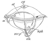
Fig. 142. Polygordius larva. (After
Hatschek.)
m. mouth; sg. supra-œsophageal ganglion;
nph. nephridion; me.p. mesoblastic band; an.
anus; ol. stomach.
For this purpose no better form can be selected than the interesting
larva of Polygordius (vide Agassiz, No. 332, Schneider, No.
352, and Hatschek, No. 339), which was first discovered by Lovén, and
believed by him to be the larva of an ordinary Chætopod. Its true
nature was determined by Schneider.
At a very young stage the larva has the form (fig. 142) of a flattened
sphere, with a small conical knob at the posterior extremity.
At the equator are situated two parallel ciliated bands[137], between
which lies the ventrally placed mouth (m). The more conspicuous
ciliated band is formed of a double row of cilia, and is situated in
front of the mouth. The thinner ciliated band behind the mouth appears
to be absent in the American species.
The mouth leads into an œsophagus, and this into a globular stomach
(ol), which is continuous with a rectum terminating by an anus
(an) placed at the hind end of the posterior conical knob. The
whole alimentary tract is ciliated. In the American form of larva
there is a ring of cilia round the anus, which is developed at a
somewhat later stage in the form observed by Hatschek.
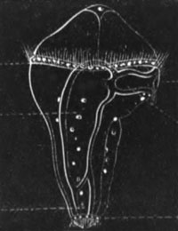
Fig. 143 Polygordius larva. (From Alex.
Agassiz.)
The position of the ciliated bands and the alimentary tract enables us
to divide the embryo into three regions: a præ-oral region bounded by
the anterior ciliated band, a gastric region in which the embryonic
stomach is situated, and an abdominal region formed of the posterior
conical portion, which by its subsequent elongation gives rise
to the whole segmented portion of the future Polygordius.
At the front end of the præ-oral lobe is situated the early formed
supra-œsophageal ganglion (sg) (first noticed by Agassiz) in
connection with which is a pair of eyes, and a ramified system of
nerves. The ganglion is marked externally by a crown of cilia.
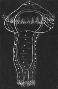
Fig. 144. Polygordius larva. (From Alex.
Agassiz.)
The larval epidermis bears a delicate cuticula, and is separated by a
considerable interval from the walls of the alimentary tract. The
space between the two represents a provisional body cavity, which is
eventually replaced by the permanent body cavity formed between the
two layers of the mesoblast. It is doubtful when the replacement takes
place in the head. It probably does so very early. The mesoblast is
present in the usual form of two bands (me.p) (germinal
streaks), which are anteriorly continued into two muscular bands which
pass through the embryonic body cavity to the front end of the
præ-oral lobe. Another pair of contractile bands passes from the same
region of the præ-oral lobe to the œsophagus.
There is no trace of the ventral nerve cord. The most remarkable organ
of the larva is a paired excretory organ (nph) discovered by
Hatschek. This is a ciliated canal with at first one and subsequently
several funnel-shaped openings into the body cavity in front and an
external opening behind. It is situated immediately anterior to the
lateral band of mesoblast, and is parallel with, and dorsal to, the
contractile band which passes off from this. It occupies therefore a
position in front of the segmented region of the adult Polygordius.
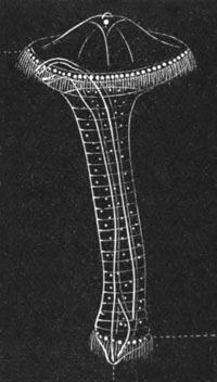
Fig. 145. Polygordius larva. (From Alex.
Agassiz.)
The changes by which this peculiar larval form reaches the adult
condition will be easily gathered from an inspection of figs. 143‑148.
They consist essentially in the elongation of what has been termed the
abdominal region of the body, and the appearance of a segmentation in
the mesoblast; the segments being formed from before backwards, and
each fresh segment being interpolated between the anus-bearing end of
the body and the last segment.
As the hind portion of the body becomes elongated the stomach extends
into it, and gives rise to the mesenteron of the adult (figs. 143,
144, and 145). For a long time the anterior spherical dilated portion
of the larva remains very large, consisting of a præ-oral lobe and a
post-oral section. The two together may be regarded as constituting
the head.
At a comparatively late stage a pair of tentacles arises from the
front end of the præ-oral lobe (fig. 146), and finally the
head becomes relatively reduced as compared with the body, and gives
rise to the simple head of the fully formed worm (fig. 148). The two
ciliated bands disappear, the posterior vanishing first. The ciliated
band at the hind end of the body also atrophies; and just in front of
it the ring of wart-like prominences used by the adult to attach
itself becomes developed.
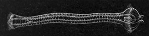
Fig. 146. Polygordius larva.
(From Alex.
Agassiz.)
At the sides of the head there is formed a pair of ciliated pits, also
found by Hatschek in the embryo of Criodrilus, and characteristic of
many Chætopod larvæ, but persistent in the adult Polygordius,
Saccocirrus, Polyophthalmus, etc. They are perhaps the same structures
as the ciliated pits in Nemertines.
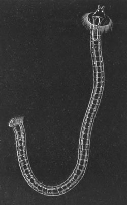
Fig. 147. Polygordius larva. (From Alex.
Agassiz.)
During the external changes above described, by which the adult form
of Polygordius is reached, a series of internal changes also takes
place which are for the most part the same as in other Chætopoda; and
do not require a detailed description. The nervous[138] and muscular
systems have precisely the normal development. The division of the
mesoblast into somites is not externally indicated. The organs most
worthy of notice are the excretory organs.
The essential points in the above development of Polygordius are (1)
the gradual elongation and corresponding segmentation of the
post-cephalic part of the body; and (2) the relative reduction in size
of the præ-oral lobe and its conversion together with the oral region
into the head; (3) the atrophy of the ciliated bands. The conversion
of the larva into the adult takes place in fact by the intercalation
of a segmented region between a large mouth-bearing portion of
the primitive body and a small anus-bearing portion[139].
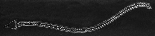
Fig. 148. Polygordius larva.
(From Alex.
Agassiz.)
The general mode of development of Chætopod larvæ is similar to the
above except in details, which are however no doubt often of great
importance. The history of the larvæ may be conveniently treated under
three heads. (1) The form of the primitive unsegmented larva; (2) the
arrangement of the cilia on the unsegmented larva, and on the larva at
later stages; (3) the character of the metamorphosis and the
development of the permanent external organs.
A larva similar to the Polygordius larva with a greatly developed
præ-oral lobe is widely distributed amongst the Annelids.
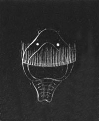
Fig. 149. Larva of Phyllodoce. (From Alex.
Agassiz.)
An almost identical form is that of Nepthys scolopendroides (Claparède
and Metschnikoff, No. 336); that of Phyllodoce (fig. 149) is also very
similar, and that of Saccocirrus (Metsch. and Clap. No. 336, Pl.
XIII. fig. 1), a very primitive form most nearly related to
Polygordius, clearly belongs to the same type. Many other larval
forms, such as that of Spio fuliginosus (Metsch. and Clap. No. 336),
Terebella, Nerine, etc., also closely approach this form.
Other really similar forms at first sight appear very different, but
this is mainly owing to the fact that their præ-oral lobe never
attains a considerable development. Its smallness, though obviously of
no deep morphological significance, at once produces a very different
appearance in a larva.
A good example of a larval form with a small præ-oral lobe is afforded
by Capitella, which is figured by Clap. and Metsch. (No. 336, Pl.
XVII. fig. 2). The imperfect development of the præ-oral lobe
is also generally characteristic of the Oligochæta. The persistence of
a relatively large præ-oral lobe for so long a time as in Polygordius
is very unusual.
The arrangement of the cilia in Chætopod larvæ has been employed as a
means of classifying them. Although a classification so framed has no
morphological value, yet the terms themselves which have been invented
are convenient. The terms most usually employed are Atrochæ,
Monotrochæ, Telotrochæ, Polytrochæ,
Mesotrochæ. The polytrochæ may again be subdivided into
Polytrochæ proper, Nototrochæ, Gasterotrochæ, and
Amphitrochæ.
The atrochæ contain forms (fig. 139) in which the larva is at first
coated by an uniform covering of cilia, which, though it may
subsequently disappear from certain areas, does not break up into a
series of definite bands.
The monotrochæ or cephalotrochæ are larvæ in which only a single
præ-oral ring is developed (fig. 150 B).
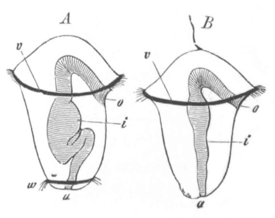
Fig. 150. Two Chætopod larvæ. (From
Gegenbaur.)
o. mouth; i. intestine; a. anus; v.
præ-oral ciliated band; w. peri-anal ciliated band.
In the telotrochæ there is present a præ-oral and a post-oral,
i.e. peri-anal ring (fig. 150 A); the latter sometimes having
the form of a peri-anal patch.
The polytrochæ are segmented larvæ with perfect or imperfect rings of
cilia on the segments of the body—usually one ring to each
segment—between the two characteristic telotrochal rings. When these
rings are complete the larvæ are polytrochæ proper, when they are only
half rings they are either nototrochæ or gasterotrochæ. Sometimes
there are both dorsal and ventral half rings which do not however
correspond, such forms constitute the amphitrochæ.
In the mesotrochæ one or two rings are present in the middle of the
body, and the characteristic telotrochal rings are absent.
Larvæ do not necessarily continue to belong to the same group at all
ages. A larva may commence as a monotrochal form and then become
telotrochal and from this pass into a polytrochal condition, etc.
The atrochal forms are to be regarded as larvæ which never pass beyond
the primitive stage of uniform ciliation, which in other instances may
precede that of definite rings. They usually lose their cilia early,
as in the cases of Serpula and other larvæ described below.
The atrochal larvæ are not common. The following history of an
Eunicidan larva (probably Lumbriconereis) from Claparède and
Metschnikoff (No. 336) will illustrate their general history.
In the earliest stage noticed the larva has a spherical form, the
præ-oral lobe not being very well marked. In the interior is a
globular digestive tract. The cilia form a broad central band leaving
free a narrow space at the apex of the præ-oral lobe, and also a
circumanal space. At the apex of the præ-oral lobe is placed a bunch
of long cilia, and a patch of cilia also marks out the anal area.
As the larva grows older it becomes elongated, and the anterior bunch
of cilia is absorbed. The alimentary canal divides itself into pharynx
and intestine. The former opens (?) by the mouth in the middle of the
central band of cilia, the latter in the anal patch. The setæ
indicating the segmentation are formed successively in the posterior
ring-like area free from cilia. The cilia disappear after the
formation of two segments.
In Lumbricus, the embryo of which ought perhaps to be grouped with the
atrochæ, the cilia (Kleinenberg) cover a ventral tract of epiblast
between the two mesoblastic cords, and are continued anteriorly to
form a circle round the mouth.
The monotrochal larvæ are provided only with the important præ-oral
ciliated ring before mentioned. In the majority of cases they are
transitional forms destined very shortly to become telotrochal, and in
such instances they usually have a more or less spherical body which
is nearly divided into two equal halves by a ciliated ring. In some
few instances, such as Polynoe, Dasychone, etc., the monotrochal
characters are not lost till the larval cilia are exuviated.
The telotrochal forms (of which examples are shewn in figs. 144, 150,
etc.) may (1) start as monotrochal; or (2) from the first have a
telotrochal character; or (3) be derived from atrochal forms. The last
mode of origin probably represents the ancestral one.
Their mode of development is well illustrated by the case of Terebella
nebulosa (vide Milne-Edwards, No. 347). The embryo is at first
a nearly spherical ciliated mass. One end slightly elongates and
becomes free from cilia, and, acquiring dorsally two eye-spots,
constitutes a præ-oral lobe. The elongation continues at the opposite
end, and near this is formed a narrow area free from cilia. The larva
now has the same characters as the atrochal Eunicidan larva described
above. It consists of a non-ciliated præ-oral lobe, followed by a wide
ciliated band, behind which is a ring-like area free from cilia; and
behind this again a peri-anal patch of cilia. The ring-like area free
from cilia is, as in the Eunicidan larva, the region which becomes
segmented. It soon becomes longer, and is then divided into two
segments; a third and fourth etc. non-ciliated segment becomes
successively interposed immediately in front of the peri-anal patch;
and, after a certain number of segments have become formed, there
appear on some of the hinder of them short tubercles, provided with
single setæ (the notopodia), which are formed from before backwards,
like the segments.
The mouth, anus, and intestine become in the meantime clearly visible.
The mouth is on the posterior side of the ciliated band, and the anus
in the centre of the peri-anal patch.
The ciliated band in front now becomes contracted and provided with
long cilia. It passes below completely in front of the mouth, and
constitutes, in fact, a well-marked præ-oral ring, while the cilia
behind constitute an equally marked peri-anal ring. The larva has in
fact now acquired all the characters of a true telotrochal form.
Only a comparatively small number of Chætopod larvæ remain permanently
telotrochal. Of these Terebella nebulosa, already cited (though not
Terebella conchilega), is one; Polygordius, Saccocirrus and Capitella
are other examples of the same, though in the latter form the whole
ventral surface becomes ciliated.
The majority of the originally telotrochal forms become polytrochal.
In most cases the ciliated rings or half rings of the polytrochal
forms are placed at equal distances, one for each segment. They are
especially prominent in surface-swimming larvæ, and are in rare cases
preserved in the adult. In some instances (e.g. Nerine and
Spio) the ventral half rings, instead of being segmentally arranged,
are somewhat irregularly distributed amongst the segments, so that
there does not seem to be a necessary correspondence between the
ciliated rings and the segments. This is further shewn by the fact
that the ciliated rings are not precursors of the true segmentation,
but are developed after the establishment of the segments, and
thus seem rather to be secondarily adapted to the segments than
primarily indicative of them.
In most Polytrochæ the rings are incomplete, so that they fall under
the category of Nototrochæ or Gasterotrochæ.
The larva of Odontosyllis is an example of the former, and that of
Magelona of the latter. The larvæ of Nerine and Spio, already quoted
as examples of an unsegmented arrangement of the ventral ciliated half
rings, are both amphitrochal forms.
As an example of a polytrochal form with complete ciliated rings
Ophryotrocha puerilis may be cited. This form, discovered by Claparède
and Metschnikoff, develops a complete ciliated ring on each segment:
and the præ-oral ring, though at first single, becomes at a later
period divided into two. This form is further exceptional in that the
ciliated rings are persistent in the adult.
The unimportance of the character of the rings in the polytrochal
forms is shewn by such facts as the absence of these rings in
Terebella nebulosa and the presence of dorsal half rings in Terebella
conchilega.
The mesotrochal forms are the rarest of Chætopod larvæ, and would seem
to be confined to the Chætopteridæ.
Their most striking character is the presence of one or two complete
ciliated rings which girth the body between the mouth and anus. The
whole body is further covered with short cilia. The anus has a
distinct dorsal situation, while on its ventral side there projects
backwards a peculiar papilla.
The total absence of the typical præ-oral and of the peri-anal bands
separates the mesotrochal larvæ very sharply from all the previous
types.
A characteristic of many Chætopod larvæ is the presence of a bunch of
cilia or a single flagellum at the apex of the præ-oral lobe. The
presence of such a structure is characteristic of the larval forms of
many other groups, Turbellarians, Nemertines, Molluscs, etc.
In the preceding section the mode of multiplication of the segments
has already been sufficiently described[140].
Fig. 151. Larva of Phyllodoce from the ventral
side. (From Alex. Agassiz.)
Apart from the formation of the segments the larval metamorphosis
consists in the atrophy of the provisional ciliated rings and other
provisional organs, and in the acquirement of the organs of the adult.
The great variations in the nature of the Chætopod appendages render
it impossible to treat this part of the developmental history of the
Chætopoda in a systematic way.
The mode of development of the appendages is not constant, so that it
is difficult to draw conclusions as to the primitive form from which
the existing types of appendages are derived.
In a large number of cases the primitive rudiments of the feet exhibit
no indication of a division into notopodium and neuropodium; while in
other instances (e.g. Terebella and Nerine, fig. 152) the
notopodium is first developed, and subsequently the neuropodium quite
independently.
Fig. 152. Larva of Nerine, with provisional
setæ. (From Alex. Agassiz.)
In many cases the setæ appear before there are any other visible
rudiments of the feet (e.g. Lumbriconereis); while in other
cases the reverse holds good. The gills are usually the last parts to
appear.
Not only does the mode of development of the feet differ greatly in
different types, but also the period. The appearance of setæ may
afford the first external indication of segmentation, or the rudiments
of the feet may not appear till a large number of segments are
definitely established.
A very considerable number of Chætopod larvæ are provided with very
long provisional setæ (figs. 152 and 153). These setæ are
usually placed at the sides of the anterior part of the body,
immediately behind the head, and also sometimes on the posterior parts
of the body. In some instances (e.g. fig. 153) they form the
only appendages of the trunk. Alex. Agassiz has pointed out that setæ
of this kind, though not found in existing Chætopods, are
characteristic of the fossil forms. Setæ of this kind are found in
chætopod-like larvæ of some Brachiopods (Argiope, fig. 136).
Fig. 153. Embryo Chætopod with provisional
setæ. (From Agassiz.)
It is tempting to suppose that the long provisional bristles springing
from the oral region are the setiform appendages handed down from the
unsegmented ancestors of the existing Chætopod forms. Claparède has
divided Chætopod larvæ into two great groups of Metachætæ and
Perennichætæ, according as they possess or are without provisional
setæ.
With reference to the head and its appendages it has already been
stated that the head is primarily formed of the præ-oral lobe and of
the peristomial region.
The embryological facts are opposed to the view that the præ-oral
region either represents a segment or is composed of segments
equivalent to those of the trunk. The embryonic peristomial region
may, on the other hand, be regarded as in a certain sense the first
segment. Its exact relations to the succeeding segments become
frequently more or less modified in the adult. The præ-oral region is
in most larvæ bounded behind by the ciliated ring already described.
On the dorsal part of the præ-oral lobe in front of this ring are
placed the eyes, and from it there may spring a variable number of
processes which form antennæ or cephalic tentacles. The number and
position of these latter are very variable. They appear as simple
processes, sometimes arising in pairs, and at other times
alternating on the two sides. There is frequently a median unpaired
tentacle.
The development of the median tentacle in Terebella, where there is in
the adult a great number of similar tentacles, is sufficiently
remarkable to deserve special notice; vide Milne-Edwards,
Claparède, etc. It arises long before any of the other tentacles as a
single anterior prolongation of the præ-oral lobe containing a
parenchymatous cavity, which communicates freely with the general
perivisceral cavity. It soon becomes partially constricted off at its
base from the procephalic lobe, but continues to grow till it becomes
fully half as long as the remainder of the body. A very characteristic
figure of the larva at this stage is given by Claparède and
Metschnikoff, Pl. XVII., Fig. 1 E. It now strikingly
resembles the larval proboscis of Balanoglossus, and it is not easy to
avoid the conclusion that they are homologous structures.
Another peculiar cephalic structure which deserves notice is the gill
apparatus of the Serpulidæ.
Fig. 154. Larva of Spirorbis. (From Alex.
Agassiz.)
The first odd tentacle (t) is shewn on the right side.
Behind the præ-oral ciliated ring is the large collar.
In Dasychone (Sabella) the gill apparatus arises (Claparède and
Metschnikoff, No. 336) as a pair of membranous wing-like organs on the
dorsal side of the præ-oral lobe immediately in front of the ciliated
ring. Each subsequently becomes divided into two rays, and new rays
then begin to sprout on the ventral side of the two pairs already
present. A cartilaginous axis soon becomes formed in these rays, and
after this is formed fresh rays sprout irregularly from the
cartilaginous skeleton.
In Spirorbis spirillum as observed by Alex. Agassiz, the right
gill-tentacle (fig. 154, t) first appears, and then the left,
and subsequently the odd opercular tentacle which covers the right
original tentacle. The third and fourth tentacles are formed
successively on the two sides, and rapidly become branched in the
succeeding stages.
With reference to the sense organs it may be noted that the eyes, or
at any rate the cephalic pigment spots, are generally more numerous in
the embryo than in the adult, and that they are usually present in the
larvæ of the Sedentaria, though absent in the adults of these forms.
The Sedentaria thus pass through a larval stage in which they resemble
the Errantia.
Paired auditory vesicles of a provisional character have been found on
the ventral side of the body, in the fourth segment behind the
mouth, in the larva of Terebella conchilega (Claparède).
Mitraria. A peculiar larval Chætopod form known as Mitraria,
the metamorphosis of which was first worked out by Metschnikoff,
deserves a special notice.
This form (fig. 155 A) in spite of its remarkable appearance can
easily be reduced to the normal type of larva.
The mouth (m) and anus (an) (fig. 155 A) are closely
approximated, and situated within a vestibule the edge of which is
lined by a simple or lobed ciliated ring. The shape of the body is
somewhat conical. The cavity of the vestibule forms the base of the
cone, and at the apex is placed a ciliated patch (sg). A pair
of lobes (br) bear provisional setæ. The alimentary canal is
formed of the three normal parts, œsophagus, stomach, and intestine.
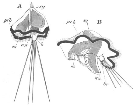
Fig. 155. Two stages in the development of
Mitraria.
(After Metschnikoff.)
m. mouth; an. anus; sg. supra-œsophageal
ganglion; br. provisional bristles; pr.b. præ-oral
ciliated band.
To compare this larva with an ordinary Chætopod larva one must suppose
that the alimentary canal is abnormally bent, so that the post-oral
ventral surface is reduced to the small space between the mouth and
the anus. The ciliated band surrounding the vestibule is merely the
usual præ-oral band, borne on the very much extended edge of the
præ-oral lobe. The apex of the larva is the front end of the præ-oral
lobe with the usual ciliated patch. The two lobes with provisional
bristles are really dorsal and not posterior.
The correctness of the above interpretation is clearly shewn by the
metamorphosis.
The first change consists in the pushing in of a fold of skin, between
the mouth and anus, towards the intestine, which at the same time
rapidly elongates, and forms the axis of a conical projection, which
thereupon becomes segmented and is thereby shewn to be the rudiment of
the trunk (fig. 155 B). On the elongation of the trunk in this way the
præ-oral lobe and its ciliated ring assume an appearance not very
dissimilar to the same structures in Polygordius. At the ciliated apex
of the præ-oral lobe a paired thickening of epiblast gives rise to the
supra-œsophageal ganglia (sg). In the further metamorphosis,
the præ-oral lobe and its ciliated ring gradually become reduced, and
finally atrophy in the normal way, while the trunk elongates and
acquires setæ. The dorsally situated processes with provisional setæ
last for some time, but finally disappear. The young worm then
develops a tube and shews itself as a normal tubicolous Chætopod.
Formation of Organs.
Except in the case of a few organs our knowledge of the formation of
the organs in the Chætopoda is derived from investigations on the
Oligochæta.
The embryo of the Oligochæta has a more or less spherical form, but it
soon elongates, and becoming segmented acquires a distinct vermiform
character. The ventral surface is however for a considerable time
markedly convex as compared to the dorsal.
The ventrally placed mouth is surrounded by a well-marked lip, and in
front of it is placed a small præ-oral lobe.
Fig. 156. Section through the head of a young
embryo of lumbricus trapezoides. (After Kleinenberg.)
c.g. cephalic ganglion; cc. cephalic portion of the
body cavity; x. œsophagus.
The epiblast. The epiblast cells at the commencement of the
gastrula stage become much flattened, and on the completion of the
invagination form an investment of flattened cells, only thickened in
the neighbourhood of the mesoblastic bands (fig. 141 B and C). In the
Polychæta at any rate the statements of several investigators would
seem to indicate that the cuticle is derived from the chorion. It is
difficult to accept this conclusion, but it deserves further
investigation.
Nervous system. The most important organ derived from the
epiblast is the nervous system; the origin of which from this
layer was first established by Kowalevsky (No. 342).
Fig. 157. Section through part of the ventral
wall of the trunk of an embryo of Lumbricus trapezoides. (After
Kleinenberg.)
m. longitudinal muscles; so. somatic mesoblast;
sp. splanchnic mesoblast; hy. hypoblast; Vg.
ventral nerve cord; vv. ventral vessel.
It arises[141] (Kleinenberg, No. 341) from two at first quite distinct
structures, viz. (1) the supra-œsophageal rudiment and (2) the
rudiment of the ventral cord. The former of these takes its origin as
an unpaired dorsal thickening of the epiblast at the front end of the
head (fig. 156, c.g.), which sends two prolongations downwards
and backwards to meet the ventral cord. The latter arises as two
independent thickenings of the epiblast, one on each side of the
ventral furrow (fig. 157, Vg). These soon unite underneath the
furrow, in the median line, and after being differentiated into
segmentally arranged ganglionic and interganglionic regions become
separated from the epiblast. Both the supra-œsophageal and ventral
cord become surrounded by a layer of somatic mesoblast. The junction
between the two parts of the central nervous system takes place
comparatively late.
The mesoblast. It is to Kowalevsky (No. 342) and Kleinenberg
(No. 341) that we mainly owe our knowledge of the history of the
mesoblast. The fundamental processes which take place are (1) the
splitting of the mesoblast into splanchnic and somatic layers with the
body cavity between them, (2) the transverse division of the mesoblast
of the trunk into distinct somites.
The former process commences in the cephalic mesoblastic commissure,
where it results in the formation of a pair of cavities each with a
thin somatic and thick splanchnic layer (fig. 156, cc); and
thence extends gradually backwards into the trunk (fig. 141 C,
pp). In the trunk however the division into somites precedes
the horizontal splitting of the mesoblast. The former process
commences when the mesoblastic bands form widish columns quite
separate from each other. These columns become broken up
successively from before backwards into somewhat cubical bodies, in
the centre of which a cavity soon appears. The cavity in each somite
is obviously bounded by four walls, (1) an outer, the somatic, which
is the thickest; (2) an inner, the splanchnic; and (3, 4) an anterior
and posterior. The adjoining anterior and posterior walls of
successive somites unite together to form the transverse dissepiments
of the adult, which subsequently become very thin and are perforated
in numerous places, thus placing in communication the separate
compartments of the body cavity. The somites, though at first confined
to a small area on the ventral side, gradually extend so as to meet
their fellows above and below and form complete rings (fig. 157) of
which the splanchnic layer (sp) attaches itself to the enteric
wall and the somatic (so) to the epiblast. In Polygordius and
probably also Saccocirrus and other forms the cavities of the somites
of the two sides do not coalesce; and the walls which separate them
constitute dorsal and ventral mesenteries. The two cavities in the
cephalic commissure unite dorsally, but ventrally open into the first
somite of the trunk.
The mesoblastic masses of the head are probably not to be regarded as
forming a pair of somites equivalent to those in the trunk, but as
forming the mesoblastic part of the præ-oral lobe, of which so much
has been said in the preceding pages. Kleinenberg’s observations are
however of great importance as shewing that the cephalic cavities are
simply an anterior part of the true body cavity.
The splanchnic layer of the head cavity gives rise to the musculature
of the œsophagus.
The somatic layer of the trunk somites becomes converted into the
musculature of the body wall and the external peritoneal layer of body
cavity. The first part of the muscular system to be definitely formed
is the ventral band of longitudinal muscles which arises on each side
of the nervous system in contact with the epidermis (fig. 157,
m). How the circular muscles become subsequently formed outside
these muscles has not been made out.
The splanchnic layer of the trunk somites gives rise to the muscular
and connective-tissue wall of the mesenteron, and also to the walls of
the vascular trunks. The ventral vessel is first formed (Kowalevsky)
as a solid mass of cells which subsequently becomes hollowed
out. The dorsal vessel in Lumbricus and Criodrilus is stated by
Kowalevsky and Vejdovsky to be formed by the coalescence of two
lateral vessels; a peculiarity which is probably to be explained by
the late extension of the mesoblast into the dorsal region.
The layer from which the sacks for the setæ and the segmental organs
spring is still doubtful. The sacks for the setæ are believed by
Kowalevsky (No. 342) to be epiblastic invaginations, but are stated by
Hatschek (No. 339) to be mesoblastic products. For the development of
the segmental organs the reader is referred to the chapter on the
excretory system.
In marine Polychæta the generative organs are no doubt mesoblastic
products, as they usually spring from the peritoneal epithelium,
especially the parts of it covering the vascular trunks.
The Alimentary Canal. In Lumbricus the enteric cavity is formed
during the gastrula stage. In Criodrilus the hypoblast has at first no
lumen, but this becomes very soon established. In Euaxes on the other
hand, where there is a true epibolic gastrula, the mesenteron is at
first represented by a solid mass of yolk (i.e. hypoblast)
cells. As the central amongst these become absorbed a cavity is
formed. The protoplasm of the yolk cells which line this cavity unites
into a continuous polynuclear layer containing at intervals masses of
yolk. These masses become gradually absorbed, and the protoplasmic
wall of the mesenteron then breaks up into a cylindrical glandular
epithelium similar to that of the other types.
In Lumbricus and Criodrilus the blastopore remains as the mouth, but
in Euaxes a new mouth or rather stomodæum is formed by an epiblastic
invagination between the front end of the two mesoblastic bands. This
epiblastic invagination forms the permanent œsophagus; and in
Lumbricus trapezoides and Criodrilus, where the oral opening is at
first lined by hypoblast, the epiblast soon becomes inflected so as to
line the œsophageal region. The splanchnic mesoblast of the
cephalic region subsequently invests the œsophagus, and some of its
cells penetrating between the adjoining epiblast cells give rise to a
thick wall for this part of the alimentary tract; the original
epiblast cells being reduced to a thin membrane. This mesoblastic wall
is sharply separated from the muscular wall outside, which is
also formed of splanchnic mesoblast.
The anus is a late formation.
Alternations of generations.
Amongst Chætopoda a considerable number of forms exhibit the
phenomenon of alternations of generations, which in the same general
way as in the case of the Cœlenterata, is secondarily caused by
budding or fission.
The process of fission essentially consists in the division of a
parent form into two zooids by the formation of a zone of fission
between two old rings, which becomes differentiated (1) into an anal
zone in front which forms the anal region of the anterior zooid, and
(2) into a cephalic zone behind which forms the head and some of the
succeeding segments of the posterior zooid. The anal zone is capable,
by growth and successive segmentation, of giving rise to an indefinite
number of fresh segments.
In Protula Dysteri, as shewn by Huxley, there is a simple fission into
two in the way described. Sexual reproduction does not take place at
the same time as reproduction by fission, but both zooids produced are
quite similar and multiply sexually.
In the freshwater forms Nais and Chætogaster a more or less similar
phenomenon takes place. By a continual process of growth in the anal
zones, and the formation of fresh zones of fission whenever four or
five segments are added in front of an anal zone, complicated chains
of adhering zooids are produced, each with a small number of segments.
As long as the process of fission continues sexual products are not
developed, but eventually the chains break up, the individuals derived
from them cease to go on budding, and, after developing a considerably
greater number of segments than in the asexual state, reproduce
themselves sexually. The forms developed from the ovum then repeat
again the phenomenon of budding, etc., and so the cycle is
continued[142].
The phenomena so far can hardly be described as cases of
alternation of generations. The process is however in certain types
further differentiated. In Syllis (Quatrefages) fission takes place,
the parent form dividing into two, of which only the posterior after
its detachment develops sexual organs. The anterior asexual zooid
continues to produce fresh sexual zooids by fission. In Myrianida
also, where a chain of zooids is formed, the sexual elements seem to
be confined to the individuals produced by budding.
The cases of Syllis and Myrianida seem to be genuine examples of
alternations of generations, but a still better instance is afforded
by Autolytus (Krohn, No. 343, and Agassiz, No. 333).
In Autolytus cornutus the parent stock, produced directly from the
egg, acquires about 40‑45 segments, and then gives rise by fission,
with the production of a zone of fission between about the 13th and
14th rings, to a fresh zooid behind. This after becoming fully
developed into either a male or a female is detached from the parent
stock, from which it very markedly differs. The males and females are
moreover very different from each other. In the female zooid the eggs
are carried into a kind of pouch where they undergo their development
and give rise to asexual parent stocks. After the young are hatched
the female dies. The asexual stock, after budding off one asexual
zooid, elongates again and buds off a second zooid. It never develops
generative organs.
The life history of some species of the genus Nereis presents certain
very striking peculiarities which have not yet been completely
elucidated.
As was first shewn by Malmgren asexual examples of various species of
Nereis may acquire the characters of Heteronereis and become sexually
mature.
The metamorphosis of Nereis Dumerilii has been investigated by
Claparède, who has arrived at certain very remarkable conclusions. He
finds that there are two distinct sexual generations of the Nereis
form of this species, and two distinct sexual generations of the
Heteronereis form.
One sexual Nereis, characterized by its small size, is diœcious,
the other discovered by Metschnikoff is hermaphrodite.
Of the Heteronereis sexual forms, both are diœcious, one is small,
and swims on the surface, the other is larger and lives at the bottom.
How these various generations are mutually related has not been made
out; but Claparède traced the passage of large asexual examples of the
Nereis form into the large Heteronereis form.
Bibliography.
(332) Alex. Agassiz. “On the young stages of a few Annelids.”
Annals Lyceum Nat. Hist. of New York, Vol. VIII. 1866.
(333) Alex. Agassiz. “On the embryology of Autolytus cornutus
and alternations of generations, etc.” Boston Journal of Nat.
History, Vol. VII. 1859‑63.
(334) W. Busch. Beobachtungen ü. Anat. u. Entwick. einiger
wirbelloser Seethiere, 1851.
(335) Ed. Claparède. Beobachtungen ü. Anat. u. Entwick.
wirbelloser Thiere an d. Küste von Normandie. Leipzig, 1863.
(336) Ed. Claparède u. E. Metschnikoff. “Beiträge z.
Kenntniss üb. Entwicklungsgeschichte d. Chætopoden.” Zeit. f. wiss.
Zool. Vol. XIX. 1869.
(337) E. Grube. Untersuchungen üb. Entwicklung d.
Anneliden. Königsberg, 1844.
(338) B. Hatschek. “Beiträge z. Entwick. u. Morphol. d.
Anneliden.” Sitz. d. k. Akad. Wiss. Wien, Vol. LXXIV.
1876.
(339) B. Hatschek. “Studien über Entwicklungsgeschichte der
Anneliden.” Arbeiten aus d. zoologischen Institute d. Universität
Wien. Von C. Claus. Heft III. 1878.
(340) Th. H. Huxley. “On hermaphrodite and fissiparous species
of tubicolar Annelidæ (Protula).” Edinburgh New Phil. Journal,
Vol. I. 1855.
(341) N. Kleinenberg. “The development of the earthworm
Lumbricus trapezoides.” Quart. J. of Micr. Science, Vol.
XIX. 1879. Sullo sviluppo del Lumbricus trapezoides.
Napoli, 1878.
(342) A. Kowalevsky. “Embryologische Studien an Würmern u.
Arthropoden.” Mém. Acad. Pétersbourg, Series VII. Vol.
XVI. 1871.
(343) A. Krohn. “Ueber die Erscheinungen bei d. Fortpflanzung
von Syllis prolifera u. Autolytus prolifer.” Archiv f.
Naturgesch. 1852.
(344) R. Leuckart. “Ueb. d. Jugendzustände ein. Anneliden,
etc.” Archiv f. Naturgesch. 1855.
(345) S. Lovén. “Beobachtungen ü. die Metamorphose von
Anneliden.” Weigmann’s Archiv, 1842.
(346) E. Metschnikoff. “Ueber die Metamorphose einiger
Seethiere (Mitraria).” Zeit. f. wiss. Zool. Vol. XXI.
1871.
(347) M. Milne-Edwards. “Recherches zoologiques, etc.” Ann.
Scie. Natur. III. Série, Vol. III. 1845.
(348) J. Müller. “Ueb. d. Jugendzustände einiger Seethiere.”
Monats. d. k. Akad. Wiss. Berlin, 1851.
(349) Max Müller. “Ueber d. weit. Entwick. von Mesotrocha
sexoculata.” Müller’s Archiv, 1855.
(350) Quatrefages. “Mémoire s. l’embryogénie des Annelides.”
Ann. Scie. Natur. III. Série, Vol. X. 1848.
(351) M. Sars. “Zur Entwicklung d. Anneliden.” Archiv f.
Naturgeschichte, Vol. XI. 1845.
(352) A. Schneider. “Ueber Bau u. Entwicklung von Polygordius.”
Müller’s Archiv, 1868.
(353) A. Schneider. “Entwicklung u. system. Stell. d. Bryozoen
u. Gephyreen (Mitraria).” Archiv f. mikr. Anat. Vol.
V. 1869.
(354) M. Schultze. Ueb. die Entwicklung von Arenicola
piscatorum u. anderer Kiemenwürmer. Halle, 1856.
(355) C. Semper. “Die Verwandschaftbeziehungen d. gegliederten
Thiere.” Arbeiten a. d. zool.-zoot. Instit. Würzburg, Vol.
III. 1876‑7.
(356) C. Semper. “Beiträge z. Biologie d. Oligochæten.”
Arbeiten a. d. zool.-zoot. Instit. Würzburg, Vol. IV.
1877‑8.
(357) M. Stossich. “Beiträge zur Entwicklung d. Chætopoden.”
Sitz. d. k. k. Akad. Wiss. Wien, B. LXXVII. 1878.
(358) R. v. Willemoes-Suhm. “Biologische Beobachtungen ü.
niedrige Meeresthiere.” Zeit. f. wiss. Zool. Bd. XXI.
1871.
The eggs of the Discophora, each enclosed in a delicate membrane, are
enveloped in a kind of mucous case formed by a secretion of the
integument, which hardens into a capsule or cocoon. In each cocoon
there are a limited number of eggs surrounded by albumen. The cocoons
are attached to water plants, etc. In Clepsine the embryos leave the
cocoon very soon after they get rid of the egg membrane, but in
Nephelis they remain within the cocoon for a very much longer period
(27‑28 days after hatching). The young of Clepsine, after their
liberation, attach themselves to the ventral surface of their parent.
Our knowledge of the development of the Discophora is in a very
unsatisfactory state; but sufficient is known to shew that it has very
many points in common with that of the Oligochæta, and that the
Discophora are therefore closely related to the Chætopoda. In Clepsine
there is an epibolic gastrula, and mesoblastic bands like those in
Euaxes are also formed. In Nephelis however the segmentation is very
abnormal, and the formation of the germinal layers cannot easily be
reduced to an invaginate gastrula type, though probably it is modified
from such a type. Mesoblastic bands similar to those in the Oligochæta
occur in this form also.
The embryology of Clepsine, which will serve as type for the Leeches
without jaws (Rhyncobdellidæ), has recently been studied by Whitman
(No. 365), and that of Nephelis, which will serve as type for
the Leeches with jaws (Gnathobdellidæ), has been studied by Bütschli
(No. 359). The early history of both types is imperfectly known[144].
Formation of the layers.
Clepsine. It is necessary to give a full account of the
segmentation of Clepsine, as the formation of the germinal layers
would be otherwise unintelligible.
Segmentation commences with the division of the ovum into two unequal
spheres by a vertical cleavage passing from the animal to the
vegetative pole. By a second vertical cleavage the large segment is
divided into two unequal parts, and the small one into two equal
parts. Of the four segments so produced three are relatively small,
and one, placed at the posterior end, is large. Each of the four
segments next gives rise to a small cell at the animal pole. These
small cells form the commencement of the epiblast, and, according to
Whitman, the mouth is eventually placed in their centre. Such a
position for the mouth, at the animal pole, is extremely unusual, and
the statements on this head require further confirmation.
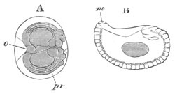
Fig. 158. Two views of the larva of
Clepsine. (After Whitman.)
o. oral extremity; m. mouth; pr. germinal
streak.
A. This figure shews the blastoderm (shaded) with a thickened edge
formed by the primitive (i.e. mesoblastic) streaks with the
four so-called neuroblasts posteriorly. The vitelline spheres are
left without shading.
B. represents an embryo in which the blastoderm has enclosed the
yolk, and in which the division into segments has taken place. At
the hind end are shewn the so-called neuroblasts forming the
termination of the germinal streak.
The posterior large segment now divides into two, one of which is
dorsal, and the other and larger ventral. The former I shall call with
Whitman the neuroblast, and the latter the mesoblast. The mesoblast
very shortly divides again. During the formation of the neuroblast and
mesoblast additional epiblastic small cells are added from the three
spheres which give rise to three of the primitive epiblast cells,
which may now be called the vitelline spheres.
The neuroblast next divides into ten cells, of which the two smaller
are soon broken up into epiblastic cells, while the remaining eight
arrange themselves in two groups of four each, one group on each side
at the posterior border of the epiblastic cap. The two mesoblasts also
take up a position on the right and left sides immediately ventral to
the four neuroblasts of each side. The neuroblasts and mesoblasts now
commence to proliferate at their anterior border, and produce
on each side a thickened band of cells underneath the edge of the cap
of epiblast cells. Each of these bands is formed of a superficial
quadruple[145] row of neuroblasts budded off from the four primary
neuroblasts, and a deeper row of mesoblasts. The compound streaks so
formed may be called the germinal streaks.
The general appearance of the embryo as seen from the dorsal surface,
after the appearance of the two germinal streaks, may be gathered from
fig. 158 A. The epiblastic cap in this figure is shaded. The
epiblastic cap, accompanied by the germinal streaks, now rapidly
extends and encloses the three vitelline spheres by a process
equivalent to that of an ordinary epibolic gastrula; but the front and
hind ends of the streaks remain practically stationary. Owing to this
mode of growth the edges of the epiblastic cap and the germinal
streaks meet in a linear fashion along the ventral surface of the
embryo (fig. 159, A and B). The germinal streaks first meet anteriorly
(B) and their junction is then gradually continued backwards. The
process is completed at about the time of hatching.
During the above changes the nuclei of the vitelline spheres pass to
the surface and rapidly divide. Eventually, together with part of the
protoplasm of the vitelline spheres, they appear to give rise to a
layer of hypoblastic cells. This layer encloses the remains of the
vitelline spheres, which become the yolk.
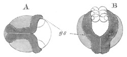
Fig. 159. Two embryos of Clepsine in which the
germinal streaks have partially met along the ventral line.
(After Robin.)
gs. germinal, i.e. mesoblastic streaks.
The area covered by epiblast is shaded. The so-called neuroblasts at
the end of the germinal streaks are shewn in B.
At the front end of the germinal streaks, in a position corresponding
with that of the four original epiblast cells, two depressions appear
which coalesce to form the single oral invagination; in the centre of
which are formed the mouth and pharynx by a second epiblastic
invagination.
The most important point in connection with the above history is the
fate of what have been called the germinal streaks. According to
Whitman they are composed of two kinds of cells, viz. four rows of
smaller superficial cells, which he calls neuroblasts, and, in the
later stages at any rate, a row of deeper large cells, which he calls
mesoblasts. As to the eventual fate of these cells he states that the
neuroblasts uniting together in the median line form the rudiment of
the ventral ganglionic chain, while the mesoblasts equally coalesce
and give rise to the mesoblast. Such a mode of origin for a ventral
ganglionic chain is, so far as I know, without a parallel in the whole
animal kingdom; and whatever evidence Whitman may have that the cells
in question really do give rise to the nervous system he has
not thought fit to produce it in his paper. He figures a section with
the eight neuroblastic cells in the middle ventral line, and in the
next stage described the nervous system is divided up into ganglia!
The first stage, in which the so-called nervous system has the form of
a single row of eight cells, is quite unlike any rudiment of the
nervous system such as is usually met with in the Chætopoda, and not a
single stage between this and a ganglionated cord is described or
figured. Whitman, whose views seem to have been influenced by a
peculiar, and in my opinion erroneous, theory of Rauber’s about the
relation of the neural groove of Vertebrata to the blastopore, does
not seem to be aware that his determination of the fate of his
neuroblasts requires any special support.
He quotes the formation of these parts in Euaxes (vide
preceding Chapter, p. 324) as similar to that in Clepsine. In this
comparison it appears to me probable that he may be quite correct, but
the result of the comparison would be to shew that the neuroblasts and
mesoblasts composed together a mesoblastic band similar to that of the
Oligochæta. Till more evidence is brought forward by Whitman or some
other observer in support of the view that the so-called neuroblasts
have any share in forming the nervous system, they must in my opinion
be regarded as probably forming, in conjunction with the mesoblasts,
two simple mesoblastic bands. Kowalevsky has moreover briefly stated
that he has satisfied himself that the nervous system in Clepsine
originates from the epiblast—a statement which certainly could not be
brought into harmony with Whitman’s account.
Nephelis. Nephelis will form my type of the Gnathobdellidæ. The
segmentation of this form has not yet been thoroughly investigated,
but Bütschli’s (No. 359) observations are probably the most
trustworthy.
The ovum first divides into two, and then into four segments of which
two are slightly smaller than the others. Four small cells which form
the commencement of the epiblast are now formed. Three of them are
derived by budding from the two larger and one of the smaller of the
four cells, and the fourth from a subsequent division of one of the
larger cells[146]. The three cells which assisted in the formation of
the epiblast cells again give rise each to a small cell; and the small
cells so formed constitute a layer underneath the epiblast which is
the commencement of the hypoblast, while the cells from which they
originated form the vitelline spheres. Shortly after the formation of
the hypoblast, the large sphere which has hitherto been quiescent
divides into two, one of which then gives rise in succession to two
small epiblastic elements.
The two large spheres, resulting from the division of the originally
quiescent sphere, next divide again on the opposite side of the
embryo, and form a layer of epiblast there; so that there is now on
one side of the embryo (the ventral according to Robin) a layer of
epiblast formed of six cells, and on the opposite side a layer
formed of four cells. The two layers meet at the front border of the
embryo and between them are placed the three large vitelline spheres.
The two patches of epiblast cells now rapidly increase, and gradually
spread over the three large vitelline spheres. Except where they meet
each other at the front edge they leave uncovered a large part of the
margin of the vitelline spheres.
While these changes have been taking place on the exterior, the
hypoblast cells have increased in number (additional cells being
probably derived from the three large vitelline spheres) and fill up
in a column-like fashion a space which is bounded behind by the three
vitelline spheres, and in front by the epiblast of the anterior end of
the embryo. At the sides of the hypoblast the mesoblast has become
established, probably as two lateral bands. The origin of the cells
forming it has not yet been determined. The hypoblast cells in the
succeeding stage arrange themselves round a central archenteric
cavity, and at the same time rapidly increase in size and become
filled with a secondary deposit of food-yolk. Shortly afterwards a
mouth and thick-walled œsophagus are formed, probably from an
epiblastic invagination. The mesoblast now forms two curved lateral
bands at the two sides of the body, equivalent to the mesoblastic
bands of the Chætopoda. The three vitelline spheres, still largely
uncovered by the epiblast, lie at the posterior end of the body. The
embryo grows rapidly, especially anteriorly, and the three vitelline
spheres become covered by a layer of flattened epiblast cells. Around
the œsophagus a cavity traversed by muscular fibres is established.
Elsewhere there is no trace of such a cavity. The cephalic region
becomes ciliated, and the dorsal part of it, which represents a
rudimentary præ-oral lobe, is especially prominent. The cilia of the
oral region are continued into the lumen of the œsophagus, and at a
later period are prolonged, as in Lumbricus, along the median line of
the ventral surface.
The mesoblastic bands would seem from Bütschli’s observations, which
receive confirmation from Kleinenberg’s researches on Lumbricus, to be
prolonged dorsally to the œsophagus into the cephalic region.
Posteriorly they abut on the large vitelline spheres, which were
supposed by Kowalevsky to give origin to them, and to play the same
part as the large mesoblasts in Lumbricus. It has already been shewn
that the function of the large cells in Lumbricus has been
exaggerated, and Bütschli denies to them in Nephelis any share in the
production of the mesoblast. It seems in fact probable that they are
homologous with the three vitelline spheres of Clepsine; and that
their primitive function is to give origin to the hypoblast. They are
visible for a long time at the hind end of the embryo, but eventually
break up into smaller cells, the fate of which is unknown.
The embryo of Hirudo would appear from the researches of Robin to
develop in nearly the same way as that of Nephelis. The anterior part
is not however ciliated. The three large posterior cells disappear
relatively early.
General history of the larva.
The larva of Clepsine, at the time when the mesoblastic bands have met
along the ventral line, is represented in fig. 158 B. It is seen to be
already segmented, the process having proceeded pari passu with
the ventral coalescence of the mesoblastic bands. The segments are
formed from before backwards as in Chætopoda. The dorsal surface is
flat and short, and the ventral very convex. The embryo about this
time leaves its capsule, and attaches itself to its parent. It rapidly
elongates, and the dorsal surface, growing more rapidly than the
ventral, becomes at last the more convex. Eventually thirty-three
post-oral segments become formed; of which the eight last coalesce to
form the posterior sucker.
The general development of the body of Nephelis and Hirudo is nearly
the same as that of Clepsine. The embryo passes from a spherical to an
oval, and then to a vermiform shape. For full details the reader is
referred to Robin’s memoir.
The presence of a well-marked protuberance above the œsophagus,
which forms the rudiment of a præ-oral lobe, has already been
mentioned as characteristic of the embryo of Nephelis; no such
structure is found in Clepsine.
History of the germinal layers and development of organs.
The epiblast. The epiblast is formed of a single layer of cells
and early develops a delicate cuticle which is clearly formed quite
independently of the egg membrane. It becomes raised into a series of
transverse rings which bear no relation to the true somites of the
mesoblast.
The nervous system. The nervous system is probably derived from
the epiblast, but its origin still requires further investigation. The
ventral cord breaks up into a series of ganglia, which at first
correspond exactly with the somites of the mesoblast. Of these, four
or perhaps three eventually coalesce to form the sub-œsophageal
ganglion, and seven or eight become united in the posterior sucker.
It would appear from Bütschli’s statements that the
supra-œsophageal ganglion arises, as in Oligochæta,
independently of the ventral cord.
Mesoblast. It has already been indicated that the mesoblast
probably takes its origin both in Nephelis and Clepsine from the two
mesoblastic bands which unite in the median ventral line. The further
history of these bands is only imperfectly known. They become
segmented from before backwards. The somites formed by the
segmentation gradually grow upwards and meet in the dorsal line. Septa
are formed between the somites probably in the same way as in the
Oligochæta.
In Clepsine the mesoblastic bands are stated by Kowalevsky to become
split into somatic and splanchnic layers, between which are placed the
so-called lateral sinuses. These sinuses form, according to Whitman, a
single continuous tube investing the alimentary tract; a tube which
differs therefore to a very small extent from the normal body cavity
of the Chætopoda. The somatic layer of mesoblast no doubt gives rise
to the circular and longitudinal muscular layers of the embryo. The
former is stated to appear the earliest, while the latter, as in the
Oligochæta, first takes its origin on the ventral side.
A delicate musculature, formed mainly of transverse but also of
longitudinal fibres, would appear to be developed independently of the
mesoblastic bands in Nephelis and Hirudo (Rathke, Leuckart, Robin, and
Bütschli). It develops apparently from certain stellate cells which
are found between the walls of the alimentary tract and the skin, and
which probably correspond to the system of contractile fibres which
pass from the body wall to the alimentary tract through the
segmentation cavity in the larva of Chætopoda, various Vermes and
Mollusca[147].
The mesoblast, so far as is known, gives rise, in addition to the
parts already mentioned, to the excretory organs, generative organs,
vascular system, etc.
Excretory organs. There are found in the embryo of Nephelis and
Hirudo certain remarkable provisional excretory organs the origin and
history of which is not yet fully made out. In Nephelis they appear as
one (according to Robin, No. 364), or (according to Bütschli, No. 359)
as two successive pairs of convoluted tubes on the dorsal side
of the embryo, which are stated by the latter author to develop from
the scattered mesoblast cells underneath the skin. At their fullest
development they extend, according to Robin, from close to the head to
near the ventral sucker. Each of them is U-shaped, with the open end
forwards, each limb of the U being formed by two tubes united in
front. No external opening has been clearly made out. Semper believed
that the tubes were continuous with the three posterior vitelline
cells, but this has been shewn not to be the case. Fürbringer[148] is
inclined from his own researches to believe that they open laterally.
They contain a clear fluid.
In Hirudo, Leuckart (No. 362) has described three similar pairs of
organs the structure of which he has fully elucidated. They are
situated in the posterior part of the body, and each of them commences
with an enlargement from which a convoluted tube is continued for some
distance backwards; it then turns forwards again and afterwards bends
upon itself to open to the exterior. The anterior part is broken up
into a kind of labyrinthic network.
The true segmental organs are found in a certain number of the
segments and are stated (Whitman) to develop from groups of mesoblast
cells. Their origin requires however further investigation.
A double row of colossal cells on each side of the body has been
described in Clepsine by Whitman as derived from the mesoblastic
plates. These cells (fig. 58 B), which he calls segment-cells, lie
opposite the walls of the septa. The inner row is stated to be
connected with the segmental organs. Their eventual history is
unknown, but they are conjectured by Whitman to be the mother cells of
the testes.
The alimentary tract. This is formed primitively of two
parts—the epiblastic stomodæum—forming mouth, pharynx, and
œsophagus, and the hypoblastic mesenteron. The anus is formed very
late as a simple perforation immediately dorsal to the posterior
sucker.
In Clepsine, where there is an epibolic gastrula, the rudiment
of the mesenteron is at first formed of the three vitelline spheres,
from the surface of which a true hypoblastic layer enclosing a central
yolk mass becomes differentiated, as already described. The mesenteric
sack so formed is constricted by the growth of the mesoblastic septa
into a series of lobes, while the posterior part forms a narrow and at
first very short tube opening by the anus.
The lobed region forms the sacculated stomach of the adult. The
sacculations of the stomach by their mode of origin necessarily
correspond with the segments. In the adult however the anterior lobe
is really double and has two divisions for the two segments it fills,
while the posterior lobe, which, as is well known, extends backwards
parallel with the rectum, is composed of five segmental sacculations.
In connection with the stomodæum a protrusible pharynx is developed.
In Hirudo and Nephelis the mesenteron has from the first a sack-like
form. The cells which compose the sack give rise to a secondary
deposit of food-yolk. The further changes are practically the same as
in Clepsine. In Hirudo the posterior sacculation of the stomach is
primitively unpaired. The jaws are formed at about the same time as
the eyes as protuberances on the wall of the oral cavity.
Bibliography.
(359) O. Bütschli. “Entwicklungsgeschichtliche Beiträge
(Nephelis).” Zeit. f. wiss. Zool. Vol. XXIX. 1877.
(360) E. Grube. Untersuchungen üb. d. Entwicklung d.
Anneliden. Königsberg, 1844.
(361) C. K. Hoffmann. “Zur Entwicklungsgeschichte d.
Clepsineen.” Niederländ. Archiv f. Zool. Vol. IV.
1877.
(362) R. Leuckart. Die menschlichen Parasiten (Hirudo),
Vol. I. p. 686, et seq.
(363) H. Rathke. Beit. z. Entwicklungsgesch. d.
Hirudineen. Leipzig, 1862.
(364) Ch. Robin. Mém. sur le Développement embryogenique des
Hirudinées. Paris, 1875.
(365) C. O. Whitman. “Embryology of Clepsine.” Quart. J. of
Micro. Science, Vol. XVIII. 1878.
[Vide also C. Semper (No. 355) and Kowalevsky (No. 342) for
isolated observations.]
It is convenient for the purposes of embryology to divide the Gephyrea
into two groups, viz. (1) Gephyrea nuda or true Gephyrea; and (2)
Gephyrea tubicola formed by the genus Phoronis.
Gephyrea nuda.
Segmentation and formation of the layers.
An embolic or epibolic gastrula is characteristic of the Gephyrea, and
the blastopore appears, in some cases at any rate (Phascolosoma,
Thalassema), to become the mouth.
Bonellia. In Bonellia (Spengel, No. 370) the segmentation is
unequal but complete, and, as in many Molluscs etc., the ovum exhibits
before its commencement a distinction into a protoplasmic and a yoke
pole. The ovum first divides into four equal segments, each of them
formed of the same constituents as the original ovum. At the animal
pole four small cells, entirely formed of protoplasm, are next formed
by an equatorial furrow. They soon place themselves in the intervals
between the large spheres. Four small cells are again budded off from
the large spheres and the eight small cells then divide. By a further
continuation of the division of the existing small cells, and the
formation of fresh ones from the large spheres, a layer of small
cells is eventually formed, which completely envelops the four
large spheres except for a small blastopore at the vegetative pole of
the ovum (fig. 160 A). The large spheres continue to give rise to
smaller cells which however no longer take a superficial position but
lie within the layer of small cells, and give rise to the hypoblast
(fig. 160 B). The small cells become the epiblast, and at the
blastopore they curl inwards (fig. 160 B) and give rise to a layer of
cells, which appears to extend as an unbroken sheet between the
epiblast and hypoblast, and to form the mesoblast. The blastopore now
closes up, but its position in relation to the parts of the embryo has
not been made out.
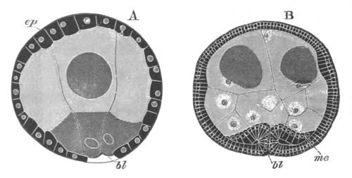
Fig. 160. Epibolic gastrula of Bonellia.
(After Spengel.)
A. Stage when the four hypoblast cells are nearly enclosed.
B. Stage after the formation of the mesoblast has commenced by an
infolding of the lips of the blastopore.
ep. epiblast; me. mesoblast; bl. blastopore.
In Phascolosoma (Selenka, No. 369) the ovum, enclosed in a porous zona
radiata, divides into two unequal spheres, of which the smaller next
divides into two and then into four. An invagination takes place which
is intermediate between the embolic and the epibolic types. The small
cells, the number of which is increased by additions from the large
sphere, divide, and grow round the large sphere. The latter in the
meantime also divides, and the cells produced from it form on the one
hand a small sack which opens by the blastopore, and on the other they
fill up the segmentation cavity, and become the mesoblast and blood
corpuscles. The blastopore becomes the permanent mouth.
Larval forms and development of organs.
Amongst the Gephyrea armata the larva has as a rule (Thalassema,
Echiurus) the characters of a trochosphere, and closely approaches the
typical form characteristic of the larva of Polygordius, often known
as Lovén’s larva. In Bonellia this larval form is less perfectly
preserved.
Echiurus. In Echiurus (Salensky, No. 368) the youngest known
larva has all the typical trochosphere characters (fig. 161). It is
covered with cilia and divided into a præ-oral lobe and post-oral
region of nearly equal dimensions. There is a double ciliated ring
which separates the two sections of the body as in the larva of
Polygordius: the mouth (m) opens between its two elements. The
alimentary canal is divided into a stomodæum with a ventral opening, a
large stomach, and a short intestine opening by a terminal anus
(an). Connecting the œsophagus with the apex of the præ-oral
lobe is the usual contractile band, and at the insertion of this band
is a thickening of the epiblast which probably represents the rudiment
of the supra-œsophageal ganglion. A ventral nerve cord is stated by
Salensky to be present, but his observations on this point are not
quite satisfactory.
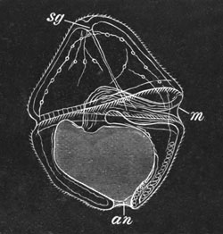
Fig. 161. Larva of Echiurus. (After
Salensky.)
m. mouth; an. anus; sg. supra-œsophageal
ganglion (?).
The metamorphosis is accompanied by the loss of swimming power, and
consists in the enlargement of the post-oral portion of the trunk, and
in the simultaneous reduction of the præ-oral lobe, which remains
however permanently as the cylindrical proboscis. A groove which
terminates posteriorly at the mouth is very early formed on its
ventral side. The ciliated rings gradually disappear during the
metamorphosis.
Of the further external changes the most important are (1) the
early appearance round the anal end of the body of a ring of bristles;
and (2) the appearance of a pair of ventral setæ in the anterior part
of the body. The anterior ring of bristles characteristic of the adult
Echiurus does not appear till a late period.
Of the internal changes the earliest is the formation of the anal
respiratory sacks. With the growth of the posterior part of the trunk
the intestine elongates, and becomes coiled.
Bonellia. The embryo of Bonellia, while still within the egg,
retains a spherical form and acquires an equatorial band of cilia,
behind which a second narrower band is soon established, while in
front of the first one a pair of eye-spots becomes formed (fig. 162
A). The embryo on becoming hatched rapidly elongates, while at the
same time it becomes dorso-ventrally flattened and acquires a complete
coating of cilia (fig. 162 B). According to Spengel it resembles at
this time in its form and habits a rhabdocœlous Turbellarian. The
anterior part is however somewhat swollen and presents an indication
of a præ-oral lobe.
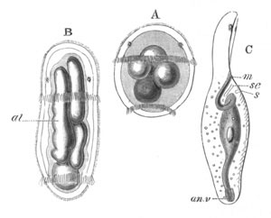
Fig. 162. Three stages in the development of
Bonellia.
(After Spengel.)
A. Larva with two ciliated bands and two eye-spots.
B. Ripe larva from the dorsal surface.
C. Young female Bonellia from the side.
al. alimentary tract; m. mouth; se. provisional
excretory tube; s. ventral hook; an.v. anal vesicle.
During the above changes important advances are made in the formation
of the organs from the embryonic layers.
The epiblast acquires a superficial cuticula, which is perhaps
directly derived from the vitelline membrane. The nervous system is
also formed, probably from the epiblast. The band-like
supra-œsophageal ganglion is the first part of the nervous system
formed, and appears to be undoubtedly derived from the epiblast. The
ventral cord arises somewhat later, but the first stages in its
development have not been satisfactorily traced. It is continuous with
the supra-œsophageal band which completely girths the œsophagus
without exhibiting any special dorsal enlargement. After the ventral
cord has become completely separated from the epiblast a central
fibrous mass becomes differentiated in it, while the lateral parts are
composed of ganglion cells. In the arrangement of its cells it
presents indications of being composed of two lateral halves. It is,
however, without ganglionic swellings.
The mesoblast, though at first very thin, soon exhibits a
differentiation into a splanchnic and somatic layer—though the two do
not become distinctly separated by a body cavity. The somatic layer
rapidly becomes thicker, and enlarges laterally to form two bands
united dorsally and ventrally by narrow, thinner bands. The outermost
parts of each of these bands become differentiated into an external
circular and an internal longitudinal layer of muscles. In the
præ-oral lobe the mesoblast assumes a somewhat vacuolated character.
The hypoblast cells form a complete layer round the four yolk cells
from which they arise (fig. 162 B, al), but at first no
alimentary lumen is developed. The œsophagus appears during this
period as an, at first solid, but subsequently hollow, outgrowth of
the hypoblast towards the epiblast.s
The metamorphosis of the larva into the adult female Bonellia
commences with the conversion of many of the indifferent mesoblast
cells into blood corpuscles, and the introduction into the body cavity
of a large amount of fluid, which separates the splanchnic and somatic
layers of mesoblast. The fluid is believed by Spengel to be sea-water,
introduced by two anal pouches, the development of which is described
below.
The body cavity is lined by a peritoneum, and very soon distinct
vessels, formed by folds of the peritoneum, become established. Of
these there are three trunks, two lateral and a median in the præ-oral
lobe (proboscis), and in the body a ventral trunk above the nerve
cord, and an intestinal trunk opening anteriorly into the ventral one.
The vessels appear to communicate with the body cavity.
In the course of the above changes the two ciliated bands
disappear, the hinder one first. The cilia covering the general
surface become atrophied, with the exception of those on the ventral
side of the præ-oral lobe. The latter structure becomes more
prominent; the stellate mesoblast cells, which fill up its interior,
become contractile, and it gives rise to the proboscis (fig. 162 C).
At the point where the œsophageal protuberance joined the epiblast
at a previous stage the mouth becomes established (fig. 162 C,
m), and though it is formed subsequently to the atrophy of the
anterior ciliated band, yet there is evidence that it is potentially
situated behind this band. The lumen of the alimentary canal becomes
established by the absorption of the remains of the four central
cells. The anus is formed on the ventral side of the posterior end of
the body, and close to it the pouches already noticed grow out from
the hindermost part of the alimentary tract (fig. 162 C, an.v).
They are at first simple blind pouches, but subsequently open into the
body cavity[150]. They become the anal pouches of the adult. There is
present when the mouth is first formed a peculiar process of the
alimentary tract projecting into the præ-oral lobe, which appears to
atrophy shortly afterwards.
After the formation of the mouth, there are formed on the ventral side
of and slightly behind it (1) anteriorly a pair of tubes, which appear
to be provisional excretory organs and soon disappear (fig. 162 C,
sc); and (2) behind them a pair of bristles (s) which
remain in the adult. The formation of the permanent excretory (?)
organ (oviduct and uterus) has not been followed out. The ovary
appears very early as a differentiation of the epithelium lining the
ventral vessel.
The larvæ, which become the minute parasitic males, undergo a very
different and far less complete metamorphosis than those which become
females. They attach themselves to the proboscis of an adult female,
and lose their ciliated bands. Germinal cells make their appearance in
the mesoblast, which form spherical masses, and, like the germinal
balls in the female ovary, consist of a central cell, and an
epithelium around it. The central cell becomes very large, while the
peripheral cells give rise to the spermatozoa. A body cavity becomes
developed in the larvæ, into which the spermatic balls are dehisced.
Neither mouth nor anus is formed. The further changes have not been
followed out.
The larval males make their way into the œsophagus of the female,
where they no doubt live for some time, and probably become mature,
though the seminal pouch of the adult is not found in many of the
males living in the œsophagus. When mature the males leave the
œsophagus, and pass into the uterus.
Phascolosoma. Cilia appear in Phascolosoma (Selenka, No. 369)
while the ovum is still segmenting. After segmentation they form a
definite band immediately behind the mouth, which divides the
larva into two hemispheres—a præ-oral and a post-oral. A præ-oral
band of cilia is soon formed close to the post-oral band, and at the
apex of the præ-oral lobe a tuft of cilia also appears.
The larva has now the characters of a trochosphere, but differs from
the typical trochosphere in the post-oral part of the ciliated
equatorial ring being more important than the præ-oral, and in the
absence of an anus.
The metamorphosis commences very early. The trunk rapidly elongates,
and the præ-oral lobe becomes relatively less and less conspicuous.
The zona radiata becomes the larval cuticle.
Three pairs of bristles are formed on the trunk, of which the
posterior pair appears first, then the anterior, and finally the
middle pair: an order of succession which clearly proves they can have
no connection with a true segmentation.
The tentacles become developed between the two parts of the
ciliated ring, and finally the præ-oral lobe, unlike what takes place
in the Gephyrea armata, nearly completely vanishes.
The anus appears fairly late on the dorsal surface, and the ventral
nerve cord is established as an unganglionated thickening of the
ventral epiblast.
Gephyrea tubicola.
The larva of Phoronis was known as Actinotrocha long before its
connection with Phoronis was established by Kowalevsky (No. 372).
There is a complete segmentation leading to the formation of a
blastosphere, which is followed by an invagination, the opening of
which is said by Kowalevsky to remain as the mouth[151]. It is
at first terminal, but on the development of a large præ-oral lobe it
assumes a ventral position. The anus is formed at a later period at
the posterior end of the body.
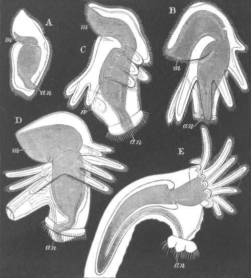
Fig. 163. A series of stages in the development
of Phoronis from Actinotrocha.
(After Metschnikoff.)
A. Young larva.
B. Larva after the formation of post-oral ring of tentacles.
C. Larva with commencing invagination to form the body of Phoronis.
D. Invagination partially everted.
E. Invagination completely everted.
m. mouth; an. anus; iv. invagination to form
the body of Phoronis.
The youngest free larva observed by Metschnikoff (No. 373) was less
developed than the oldest larva found by Kowalevsky. It
probably belongs to a different species. The body is uniformly
ciliated (fig. 163 A). There is a large contractile præ-oral lobe, and
the body ends behind in two processes. The mouth (m) is
ventral, and the anus (an) dorsal, and not terminal as in
Kowalevsky’s larva.
The alimentary tract is divided into stomodæum, stomach and intestine.
The two processes at the hind end of the body are the rudiments of the
first-formed pair of the arms which are so characteristic of the fully
developed Actinotrocha. A second pair of arms next become established
on the dorsal side of the previously existing pair, and the region
where the anus is placed grows out as a special process. New pairs of
arms continue to be formed in succession dorsalwards and forwards, and
soon constitute a complete oblique post-oral ring (fig. 163 B).
They are covered by long cilia. Round the anal process a very
conspicuous ciliated ring also becomes established.
At the period when five pairs of arms are present a delicate membrane
becomes visible on the ventral side of the intestine which joins the
somatic mesoblast anteriorly. This membrane is the rudiment of the
future ventral vessel. The somatic mesoblast is present even before
this period as a delicate layer of circular muscular fibres.
When six pairs of arms have become formed an involution (fig. 163 C,
iv) appears on the ventral side, immediately behind the ring of
arms. This involution consists both of the epiblast and somatic
mesoblast. It grows inwards towards the intestine, and, increasing
greatly in length, becomes at the same time much folded.
When it has reached its full development the critical period of the
metamorphosis of Actinotrocha into Phoronis is reached, and is
completed in about a quarter of an hour. The ventral involution
becomes evoluted (fig. 163 D), just as one might turn out the finger
of a glove which had been pulled inwards. When the involution has been
to a certain extent everted, the alimentary canal passes into it, and
at the same time the body of the larva becomes violently contracted.
By the time the evagination is completed it forms (fig. 162 E) a long
conical body, containing the greater part of the alimentary tract, and
constituting the body of the young Phoronis. The original anal
process remains on the dorsal side as a small papilla (fig. 162 E,
an).
While these changes have been taking place the præ-oral lobe has
become much contracted, and partly withdrawn into the stomodæum. At
the same time the arms have become bent forward, so as to form a ring
round the mouth. Their bases become much thickened. The metamorphosis
is completed by the entire withdrawal of the præ-oral lobe within the
œsophagus, and by the casting off of the ends of the arms, their
bases remaining as the circumoral ring of tentacles, which form
however a lophophore rather than a complete ring. The peri-anal ring
of cilia is also thrown off, and the anal process withdrawn into the
body of the young Phoronis. There are now three longitudinal vascular
trunks, united anteriorly by a circular vessel which is prolonged into
the tentacles.
General Considerations.
The development of Phoronis is so different from that of the other
Gephyrea that further investigations are required to shew whether
Phoronis is a true Gephyrean. Apart from its peculiar metamorphosis
Actinotrocha is a very interesting larval form, in that it is without
a præ-oral ciliated ring, and that the tentacles of the adult are
derived from a true post-oral ring, prolonged into arm-like processes.
The other Gephyrea present in their development an obvious similarity
to the normal Chætopoda, but their development stops short of that of
the Chætopoda, in that they are clearly without any indications of a
true segmentation. In the face of what is known of their development
it is hardly credible that they can represent a degenerate
Chætopod phylum in which segmentation has become lost. Further than
this the Gephyrea armata seem in one respect to be a very primitive
type in that they retain through life a well-developed præ-oral lobe,
which constitutes their proboscis. In almost all other forms, except
Balanoglossus, the larval præ-oral lobe becomes reduced to a
relatively insignificant anterior part of the head.
Bibliography.
Gephyrea nuda.
(366) A. Kowalevsky. Sitz. d. zool. Abth. d. III. Versam.
russ. Naturj. (Thalassema). Zeit. f. wiss. Zool. Vol.
XXII. 1872, p. 284.
(367) A. Krohn. “Ueb. d. Larve d. Sipunculus nudus nebst
Bemerkungen,” etc. Müller’s Archiv, 1857.
(368) M. Salensky. “Ueber die Metamorphose d. Echiurus.”
Morphologisches Jahrbuch, Bd. II.
(369) E. Selenka. “Eifurchung u. Larvenbildung von Phascolosoma
elongatum.” Zeit. f. wiss. Zool. 1875, Bd. XXV. p. 1.
(370) J. W. Spengel. “Beiträge z. Kenntniss d. Gephyreen
(Bonellia).” Mittheil. a. d. zool. Station z. Neapel, Vol.
I. 1879.
Gephyrea tubicola (Actinotrocha).
(371) A. Krohn. “Ueb. Pilidium u. Actinotrocha.” Müller’s
Archiv, 1858.
(372) A. Kowalevsky. “On anatomy and development of Phoronis,”
Pétersbourg, 1867. 2 Pl. Russian. Vide Leuckart’s
Bericht, 1866‑7.
(373) E. Metschnikoff. “Ueber d. Metamorphose einiger Seethiere
(Actinotrocha).” Zeit. f. wiss. Zool. Bd. XXI. 1871.
(374) J. Müller. “Bericht üb. ein. Thierformen d. Nordsee.”
Müller’s Archiv, 1846.
(375) An. Schneider. “Ueb. d. Metamorphose d. Actinotrocha
branchiata.” Müller’s Arch. 1862.
The present chapter deals with three small isolated groups, which only
resemble each other in that the systematic position of all of them is
equally obscure.
Chætognatha.
The discoveries of Kowalevsky (No. 378) confirmed by Bütschli (No.
376) with reference to the development of Sagitta, though they have
not brought us nearer to a knowledge of the systematic position of
this remarkable form, are nevertheless of great value for the
more general problems of embryology. The development commences after
the eggs are laid. The segmentation is uniform, and a blastosphere,
formed of a single layer of columnar cells, is the product of it. An
invagination takes place, the opening of which narrows to a blastopore
situated at the pole of the embryo opposite that at which the mouth
subsequently appears (fig. 164 A). The simple archenteron soon becomes
anteriorly divided into three lobes, which communicate freely with the
still single cavity behind (fig. 164 B). The two lateral lobes are
destined to form the body cavity, and the median lobe the alimentary
tract of the adult. An invagination soon arises at the opposite pole
of the embryo to the blastopore and forms the mouth and œsophagus
(fig. 164 B and C, m).
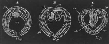
Fig. 164. Three stages in the development of
Sagitta. (A and C after Bütschli and B after Kowalevsky.) The
three embryos are represented in the same positions.
A. The gastrula stage.
B. A succeeding stage in which the primitive archenteron is
commencing to be divided into three parts, the two lateral of which
are destined to form the body cavity.
C. A later stage in which the mouth involution (m) has become
continuous with the alimentary tract, and the blastopore has become
closed.
m. mouth; al. alimentary canal; ae. archenteron;
bl.p. blastopore; pv. perivisceral cavity; sp.
splanchnopleuric mesoblast; so. somatopleuric mesoblast;
ge. generative organs.
At the gastrula stage there is formed a paired mass destined to give
rise to the generative organs. It arises as a prominence of six cells,
projecting from the hypoblast at the anterior pole of the archenteron,
and soon separates itself as a mass, or probably a pair of masses,
lying freely in the cavity of the archenteron (fig. 164 A, ge).
When the folding of the primitive cavity takes place the generative
rudiment is situated at the hind end of the median lobe of the
archenteron in the position represented in fig. 164 C, ge.
An elongation of the posterior end of the embryo now takes place, and
the embryo becomes coiled up in the egg, and when eventually hatched
sufficiently resembles the adult to be recognisable as a young
Sagitta.
Before hatching takes place various important changes become manifest.
The blastopore disappears after being carried to the ventral surface.
The middle section of the trilobed region of the archenteron becomes
separated from the unpaired posterior part, and forms a tube, blind
behind, but opening in front by the mouth (fig. 165 A, al). It
constitutes the permanent alimentary tract, and is formed of a
pharyngeal epiblastic invagination, and a posterior hypoblastic
section derived from the primitive archenteron. The anus is apparently
not formed till comparatively late. After the isolation of the
alimentary tract the remainder of the archenteron is formed of two
cavities in front, which open freely into a single cavity behind (fig.
165 A). The whole of it constitutes the body cavity and its
walls the mesoblast. The anterior paired part
becomes partitioned off into a head section and a trunk section (fig.
165 A and B). The former constitutes a pair of distinct cavities
(c.pv) in the head, and the latter two cavities opening freely
into the unpaired portion behind. At the junction of the paired
cavities with the unpaired cavity are situated the generative organs
(ge). The inner wall of each of the paired cavities forms the
splanchnopleuric mesoblast, and the outer wall of the whole the
somatic mesoblast. The inner walls of the posterior cavities unite
above and below the alimentary tract, and form the dorsal and ventral
mesenteries, which divide the body cavity into two compartments in the
adult. Before the hatching of the embryo takes place this mesentery is
continued backwards so as to divide the primitively unpaired caudal
part of the body cavity in the same way.
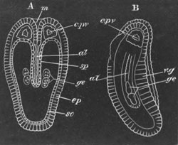
Fig. 165. Two views of a late embryo of
Sagitta. A. from the dorsal surface. B. from the side. (After
Bütschli.)
m. mouth; al. alimentary canal; v.g. ventral
ganglion (thickening of epiblast); ep. epiblast; c.pv.
cephalic section of body cavity; so. somatopleure; sp.
splanchnopleure; ge. generative organs.
From the somatic mesoblast of the trunk is derived the single layer of
longitudinal muscles of Sagitta, and part of the epithelioid lining of
the body cavity. The anterior termination of the trunk division of the
body cavity is marked in the adult by the mesentery dividing into two
laminæ, which bend outwards to join the body wall.
The cephalic section of the body cavity seems to atrophy, and its
walls to become converted into the complicated system of muscles
present in the head of the adult Sagitta.
In the presence of a section of the body cavity in the head the embryo
of Sagitta resembles Lumbricus, Spiders, etc.
The generative rudiment of each side divides into an anterior and a
posterior part (fig. 165, ge). The former constitutes
the ovary, and is situated in front of the septum dividing the tail
from the body; and the latter, in the caudal region of the trunk,
forms the testis.
The nervous system originates from the epiblast. There is a ventral
thickening (fig. 165 B, v.g) in the anterior region of the
trunk, and a dorsal one in the head. The two are at first continuous,
and on becoming separated from the epiblast remain united by thin
cords.
The ventral ganglion is far more prominent during embryonic life than
in the adult. Its position and early prominence in the embryo perhaps
indicate that it is the homologue of the ventral cord of
Chætopoda[152].
Bibliography.
(376) O. Bütschli. “Zur Entwicklungsgeschichte der Sagitta.”
Zeitschrift f. wiss. Zool., Vol. XXIII. 1873.
(377) C. Gegenbaur. “Über die Entwicklung der Sagitta.”
Abhand. d. naturforschenden Gesellschaft in Halle, 1857.
(378) A. Kowalevsky. “Embryologische Studien an Würmern u.
Arthropoden.” Mém. Acad. Pétersbourg, VII. sér., Tom.
XVI., No. 12. 1871.
Myzostomea.
The development of these peculiar parasites on Crinoids has been
investigated by Metschnikoff (No. 380), Semper (No. 381), and Graff
(No. 379).
The segmentation is unequal, and would appear to be followed by an
epibolic invagination. The outer layer of cells (epiblast) becomes
covered with cilia, and the inner is transformed into a non-cellular
(?) central yolk mass. At this stage the larva is hatched, and
commences to lead a free existence. In the next stage observed by
Metschnikoff, the mouth, œsophagus, stomach, and anus had become
developed; and two pairs of feet were present. In both of these feet
Chætopod-like setæ were present, which in the hinder pair were simple
fine bristles without a terminal hook. The papilliform portion of the
foot is at first undeveloped. The feet become successively added, like
Chætopod segments, and the stomach does not become dendriform till the
whole complement of feet (5 pairs) are present.
In the primitive covering of cilia, combined with a subsequent
indication of segments in the formation of the feet and setæ,
the larva of the Myzostomea shews an approximation to the Chætopoda,
and the group is probably to be regarded as an early Chætopod type
specially modified in connection with its parasitic habits.
Bibliography.
(379) L. Graff. Das Genus Myzostoma. Leipzig, 1877.
(380) E. Metschnikoff. “Zur Entwicklungsgeschichte d.
Myzostomum.” Zeit. f. wiss. Zool., Vol. XVI. 1866.
(381) C. Semper. “Z. Anat. u. Entwick. d. Gat. Myzostomum.”
Zeit. f. wiss. Zool., Vol. IX. 1858.
Gastrotricha.
A few observations of Ludwig on the winter eggs of Ichthydium larus
shew that the segmentation is a total and apparently a regular one. It
leads to the formation of a solid morula. The embryo has a ventral
curvature, and the caudal forks are early formed as cuticular
structures. By the time the embryo leaves the egg, it has almost
reached the adult state. The ventral cilia arise some little time
prior to the hatching.
Bibliography.
(382) H. Ludwig. “Ueber die Ordnung Gastrotricha
Metschn.” Zeit. f. wiss. Zool., Vol. XXVI. 1876.
Nematelminthes[153].
Nematoidea. Although the ova of various Nematodes have formed
some of the earliest, as well as the most frequent objects of
embryological observation, their development is still but very
imperfectly known. Both viviparous and oviparous forms are common, and
in the case of the oviparous forms the eggs are usually enveloped in a
hard shell. The segmentation is total and nearly regular, though the
two first segments are often unequal. The relation of the segmentation
spheres to the germinal layers is however only satisfactorily
established (through the researches of Bütschli (No. 383)) in the case
of Cucullanus elegans, a form parasitic in the Perch[154].
The early development of this embryo takes place within the body of
the parent, and the egg is enveloped in a delicate membrane. After the
completion of the early stages of segmentation the embryo acquires the
form of a thin flat plate composed of two layers of cells (fig. 166 A
and B). The two layers of this plate give rise respectively to the
epiblast and hypoblast, and at a certain stage the hypoblastic layer
ceases to grow, while the growth of the epiblastic layer
continues. As a consequence of this the sides of the plate begin to
fold over towards the side of the hypoblast (fig. 166 D.) This folding
results in the formation of a remarkably constituted gastrula, which
has the form of a hollow two-layered cylinder with an incompletely
closed slit on one side (fig. 166 E, bl.p). This slit has the
value of a blastopore. It becomes closed by the coalescence of the two
edges, a process which commences posteriorly, and then gradually
extends forwards. In front the blastopore never becomes completely
closed, but remains as the permanent mouth. The embryo after these
changes has a worm-like form, which becomes the more obvious as it
grows in length and becomes curved (fig. 166 F).
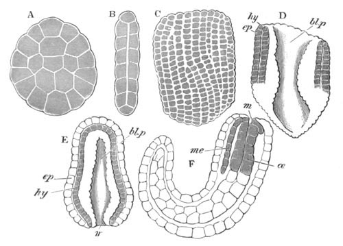
Fig. 166. Various stages in the development of
Cucullanus elegans.
(From Bütschli.)
A. Surface view of flattened embryo at an early stage in the
segmentation.
B. Side view of an embryo at a somewhat later stage, in optical
section.
C. Flattened embryo at the completion of segmentation.
D. Embryo at the commencement of the gastrula stage.
E. Embryo when the blastopore is reduced to a mere slit.
F. Vermiform embryo after the division of the alimentary tract into
œsophageal and glandular divisions.
m. mouth; ep. epiblast; hy. hypoblast;
me. mesoblast; œ. œsophagus; bl.p.
blastopore.
The hypoblast of the embryo gives rise to the alimentary
canal, and soon becomes divided into an œsophageal section (fig.
166 F, œ) formed of granular cells, and a posterior division
formed of clear cells. The mesoblast (fig. 166, me) takes its
origin from certain special hypoblast cells around the mouth, and
thence grows backwards towards the posterior end of the body.
The young Cucullanus becomes hatched while still in the generative
ducts of its parent, and is distinguished by the presence of a
remarkable thread-like tail. On the dorsal surface is a provisional
boring apparatus in the form of a conical papilla. A firm cuticle
enveloping the body is already present. In this condition it leaves
its parent and host, and leads for a time a free existence in the
water. Its metamorphosis is dealt with in another section.
The ova of the Oxyuridæ parasitic in Insects are stated by Galeb (No.
386) to take the form of a blastosphere at the close of segmentation.
An inner layer is then formed by delamination. What the inner layer
gives rise to is not clear, since the whole alimentary canal is stated
to be derived from two buds, which arise at opposite ends of the body,
and grow inwards till they meet.
The generative organs. The study of the development of the
generative organs of Nematodes has led to some interesting results. In
the case of both sexes the generative organs originate (Schneider, No.
390) from a single cell. This cell elongates and its nuclei multiply.
After assuming a somewhat columnar form, it divides into (1) a
superficial investing layer, and (2) an axial portion.
In the female the superficial layer is only developed distinctly in
the median part of the column. In the course of the further
development the two ends of the column become the blind ends of the
ovary, and the axial tissue they contain forms the germinal tissue of
nucleated protoplasm. The superficial layer gives rise to the
epithelium of the uterus and oviduct. The germinal tissue, which is
originally continuous, is interrupted in the middle part (where the
superficial layer gives rise to the uterus and oviduct), and is
confined to the two blind extremities of the tube.
In the male the superficial layer, which gives rise to the epithelium
of the vas deferens, is only formed at the hinder end of the
original column. In other respects the development takes place as in
the female.
Gordioidea. The ovum of Gordius undergoes a regular
segmentation. According to Villot (No. 391) it forms at the close of
segmentation a morula, which becomes two-layered by delamination. The
embryo is at first spherical, but soon becomes elongated.
By an invagination at the anterior extremity the head is formed. It
consists of a basal portion, armed with three rings of stylets, and a
conical proboscis, armed with three large stylets. When the larva
becomes free the head becomes everted, though it remains retractile.
By the time the embryo is hatched a complete alimentary tract is
formed with an oral opening at the end of the proboscis, and a
subterminal ventral anal opening. It is divided into an œsophagus
and stomach, and a large gland opens into it at the base of the
proboscis.
The body has a number of transverse folds, which give it a ringed
appearance.
Metamorphosis and life history.
Nematoidea. Although a large number of Nematodes have a free
existence and simple life history, yet the greater number of known
genera are parasitic, and undergo a more or less complicated
metamorphosis[155]. According to this metamorphosis they may be
divided into two groups (which by no means closely correspond with the
natural divisions), viz. those which have a single host, and those
with two hosts. Each of these main divisions may be subdivided again
into two.
In the first group with one host the simplest cases are those in which
the adult sexual form of parasite lays its eggs in the alimentary
tract of its host, and the eggs are thence transported to the
exterior. The embryo still in the egg, if favoured by sufficient
warmth and moisture, completes its development up to a certain point,
and, if then swallowed by an individual of the species in which it is
parasitic in the adult condition, it is denuded of its shell by the
action of the gastric juice, and develops directly into the sexual
form.
Leuckart has experimentally established this metamorphosis in the case
of Trichocephalus affinis, Oxyurus ambigua, and Heterakis
vermicularis. The Oxyuridæ of Blatta and Hydrophilus have a similar
life history (Galeb, No. 386), and it is almost certain that
the metamorphosis of the human parasites, Ascaris lumbricoides and
Oxyurus vermicularis, is of this nature.
A slightly more complicated metamorphosis is common in the genera
Ascaris and Strongylus. In these cases the egg-shell is thin, and the
embryo becomes free externally, and enjoys for a shorter or longer
period a free existence in water or moist earth. During this period it
grows in size, and though not sexual usually closely resembles the
adult form of the permanently free genus Rhabditis. In some cases the
free larva becomes parasitic in a freshwater Mollusc, but without
thereby undergoing any change. It eventually enters the alimentary
tract of its proper host and there become sexual.
As examples of this form of development worked out by Leuckart may be
mentioned Dochmius trigonocephalus, parasitic in the dog, and Ascaris
acuminata, in the frog. The human parasite Dochmius duodenale
undergoes the same metamorphosis as Dochmius trigonocephalus.
A remarkable modification of this type of metamorphosis is found in
Ascaris (Rhabdonema) nigrovenosa, which in its most developed
condition is parasitic in the lungs of the frog (Metschnikoff,
Leuckart, No. 388). The embryos pass through their first developmental
phases in the body of the parent. They have the typical Rhabditis
form, and make their way after birth into the frog’s rectum. From this
they pass to the exterior, and then living either in moist earth, or
the fæces of the frog, develop into a sexual form, but are very much
smaller than in the adult condition. The sexes are distinct, and the
males are distinguished from the females by their smaller size,
shorter and rounded tails, and thinner bodies. The females have paired
ovaries with a very small number of eggs, but the testis of the males
is unpaired. Impregnation takes place in the usual way, and in summer
time about four embryos are developed in each female, which soon burst
their egg-capsules, and then move freely in the uterus. Their active
movements soon burst the uterine walls, and they then come to lie
freely in the body cavity. The remaining viscera of the mother are
next reduced to a finely granular material, which serves for the
nutrition of the young forms which continue to live in the maternal
skin. The larvæ eventually become free, and though in many respects
different from the parent form which gave rise to them, have
nevertheless the Rhabditis form. They live in water or slime, and
sometimes become parasitic in water-snails; in neither case however do
they undergo important changes unless eventually swallowed by a frog.
They then pass down the trachea into the lungs and there rapidly
develop into the adult form. No separate males have been found in the
lungs of the frog, but it has been shewn by Schneider (No. 390) that
the so-called females are really hermaphrodites; the same gland giving
origin to both spermatozoa and ova, the former being developed
before the latter[156]. The remarkable feature of the above life
history is the fact that in the stage corresponding with the free
larval stage of the previous forms the larvæ of this species become
sexual, and give rise to a second free larval generation, which
develops into the adult form on again becoming parasitic in the
original host. It constitutes a somewhat exceptional case of
heterogamy as defined in the introduction.
Amongst the Nematodes with but a single host a remarkable parasite in
wheat has its place. This form, known as Anguillula scandens, inhabits
in the adult condition the ears of wheat, in which it lays its eggs.
After hatching, the larvæ become encysted, but become free on the
death of the plant. They now inhabit moist earth, but eventually make
their way into the ears of the young wheat and become sexually mature.
The second group of parasitic Nematodes with two hosts may be divided
into two groups, according to whether the larva has a free existence
before passing into its first or intermediate host, or is taken into
it while still in the egg. In the majority of cases the larval forms
live in special connective-tissue capsules, or sometimes free in the
tissues of their intermediate hosts; but the adults, as in the cases
of other parasitic Nematodes, inhabit the alimentary tract.
The life history of Spiroptera obtusa may be cited as an example of a
Nematode with two hosts in which the embryo is transported into its
intermediate host while still within the egg. The adult of this form
is parasitic in the mouse, and the ova pass out of the alimentary
tract with the excreta, and may commonly be found in barns, etc. If
one of the ova is now eaten by the meal-worm (larva of Tenebrio), it
passes into the body cavity of this worm and undergoes further
development. After about five weeks it becomes encapsuled between the
‘fat bodies’ of the meal-worm. It then undergoes an ecdysis, and, if
the meal-worm with its parasites is now eaten by the mouse, the
parasites leave their capsule and develop into the sexual form.
As examples of life histories in which a free state intervenes before
the intermediate host, Cucullanus elegans and Dracunculus may be
selected. The adult Cucullanus elegans is parasitic in the alimentary
tract of the Perch and other freshwater fishes. It is a viviparous
form, and the young after birth pass out into the water. They next
become parasitic in Cyclops, passing in through the mouth, so into the
alimentary tract, and thence into the body cavity. They soon undergo
an ecdysis, in the course of which the œsophagus becomes divided
into a muscular pharynx and true glandular œsophagus. They
then grow rapidly in length, and at a second ecdysis acquire a
peculiar beaker-like mouth cavity approaching that of the adult. They
do not become encapsuled. No further development of the worm takes
place so long as it remains in the Cyclops, but, if the Cyclops is now
swallowed by a Perch, the worm undergoes a further ecdysis, and
rapidly attains to sexual maturity.
The observations of Fedschenko on Dracunculus medinensis[157], which
is parasitic in the subcutaneous connective tissue in Man, would seem
to shew that it undergoes a metamorphosis very similar to that of
Cucullanus. There is moreover a striking resemblance between the larvæ
of the two forms. The larvæ of Dracunculus become transported into
water, and then make their way into the body cavity of a Cyclops by
boring through the soft skin between the segments on the ventral
surface of the body. In the body cavity the larvæ undergo an ecdysis
and further development. But on reaching a certain stage of
development, though they remain a long time in the Cyclops, they grow
no further. The remaining history is unknown, but probably the next
host is man, in which the larva comes to maturity. In the adult
condition only females of Dracunculus are known, and it has been
suggested by various writers that the apparent females are in reality
hermaphrodites, like Ascaris nigrovenosa, in which the male organs
come to maturity before the female.
The sexual form is parasitic in warm climates in the human tissues,
and produces multitudes of larvæ which pass into the blood, and are
sometimes voided with the urine. The larvæ in the blood do not undergo
a further development, and unless transported to an intermediate host
die before very long. Some, though as yet hardly sufficient, evidence
has been brought forward to shew that if the blood of an infected
patient is sucked by a mosquito the larvæ develop further in the
alimentary tract of the mosquito, pass through a more or less
quiescent stage, and eventually grow considerably in size, and on the
death of the mosquito pass into the water. From the water they are
probably transported directly or indirectly into the human intestines,
and then bore their way into the tissues in which they are parasitic,
and become sexually mature.
The well-known Trichina spiralis has a life history unlike that of
other known Nematodes, though there can be little doubt that this form
should be classified in respect to its life history with the
last-described forms. The peculiarity of the life history of Trichina
is that the embryos set free in the alimentary canal pass through the
walls into the muscular tissues and there encyst; but do not in a
general way pass out from the alimentary canal of one host and
thence into a fresh host to encyst. It occasionally however happens
that this migration does take place, and the life history of Trichina
spiralis then becomes almost identical with that of some of the forms
of the third type. Trichina is parasitic in man, and in swine, and
also in the rat, mouse, cat, fox and other forms which feed upon them.
Artificially it can be introduced into various herbivorous forms
(rabbit, guinea-pig, horse) and even birds.
The sexual form inhabits the alimentary canal. The female is
viviparous, and produces myriads of embryos, which pass into the
alimentary canal of their host, through the walls of which they make
their way, and travelling along lines of connective tissue pass into
the muscles. Here the embryos, which are born in a very imperfect
condition, rapidly develop, and eventually assume a quiescent
condition in a space inclosed by sarcolemma. Within the sarcolemma a
firm capsule is developed for each larva, which after some months
becomes calcified; and after the atrophy of the sarcolemma a
connective-tissue layer is formed around it. Within its capsule the
larva can live for many years, even ten or more, without undergoing
further development, but if at last the infected flesh is eaten by a
suitable form, e.g. the infected flesh of the pig by man, the
quiescent state of the larva is brought to a close, and sexual
maturity is attained in the alimentary tract of the new host.
Gordioidea. The free larva of Gordius already described usually
penetrates into the larva of Chironomus where it becomes encysted. On
the Chironomus being eaten by some fish (Villot, No. 391) (Phoxinus
lævis or Cobitis barbatula), it penetrates into the wall of the
intestine of its second host, becomes again encysted and remains
quiescent for some time. Eventually in the spring it leaves its
capsule, and enters the intestine, and passes to the exterior with the
fæces. It then undergoes a gradual metamorphosis, in the course of
which it loses its ringed structure and cephalic armature, grows in
length, acquires its ventral cord, and on the development of the
generative organs loses the greater part of its alimentary tract.
Young examples of Gordius have often been found in various terrestrial
carnivorous Insecta, but the meaning of this fact is not yet clear.
Bibliography.
(383) O. Bütschli. “Entwicklungsgeschichte d. Cucullanus
elegans.” Zeit. f. wiss. Zool., B. XXVI. 1876.
(384) T. S. Cobbold. Entozoa. Groombridge and Son, 1864.
(385) T. S. Cobbold. Parasites: A Treatise on the Entozoa of
Man and Animals. Churchill, 1879.
(386) O. Galeb. “Organisation et développement des Oxyuridés,”
&c. Archives de Zool. expér. et génér., Vol. VII.
1878.
(387) R. Leuckart. Untersuchungen üb. Trichina spiralis.
2nd ed. Leipzig, 1866.
(388) R. Leuckart. Die menschlichen Parasiten, Bd.
II. 1876.
(389) H. A. Pagenstecher. Die Trichinen nach Versuchen
dargestellt. Leipzig, 1865.
(390) A. Schneider. Monographie d. Nematoden. Berlin,
1866.
(391) A. Villot. “Monographie des Dragoneaux” (Gordioidea).
Archives de Zool. expér. et génér., Vol. III. 1874.
Acanthocephala.
The Acanthocephala appear to be always viviparous. At the time of
impregnation the ovum is a naked cell, and undergoes in this condition
the earlier phases of segmentation.
The segmentation is unequal (Leuckart, No. 393), but whether there is
an epibolic gastrula has not clearly been made out.
Before segmentation is completed there are formed round the ovum thick
protecting membranes, which are usually three in number, the middle
one being the strongest. After segmentation the central cells of the
ovum fuse together to give rise to a granular mass, while the
peripheral cells at a slightly later period form a more transparent
syncytium. At the anterior end of the embryo there appears a
superficial cuticle bearing in front a ring of hooks.
The embryo is now carried out with the excreta from the intestine of
the vertebrate host in which its parent lives. It is then swallowed by
some invertebrate host[159].
Another very remarkable human parasite belonging to the same group as
Dracunculus is the form known as Filaria sanguinis hominis, or Filaria
Bancrofti[158].
In the intestine of the invertebrate host the larva is freed from its
membranes, and is found to have a somewhat elongated conical form,
terminating anteriorly in an obliquely placed disc, turned slightly
towards the ventral surface and armed with hooks. Between this disc
and the granular mass, already described as formed from the central
cells of the embryo, is a rather conspicuous solid body. Leuckart
supposes that this body may represent a rudimentary functionless
pharynx, while the granular mass in his opinion is an equally
rudimentary and functionless intestine. The body wall is formed of a
semifluid internal layer surrounding the rudimentary intestine, if
such it be, and of a firmer outer wall immediately within the cuticle.
The adult Echinorhyncus is formed by a remarkable process of
development within the body of the larva, and the skin is the only
part of the larva which is carried over to the adult.
In Echinorhyncus proteus the larva remains mobile during the formation
of the adult, but in other forms the metamorphosis takes place during
a quiescent condition of the larva.
The organs of the adult are differentiated from a mass of cells which
appears to be a product of the central embryonic granular mass, and is
called by Leuckart the embryonic nucleus. The embryonic
nucleus becomes divided into four linearly arranged groups of cells,
of which the hindermost but one is the largest, and very early
differentiates itself into (1) a peripheral layer, and (2) a central
mass formed of two distinct bodies. The peripheral layer of this
segment grows forwards and backwards, and embraces the other segments,
with the exception of the front end of the first one which is left
uncovered. The envelope so formed gives rise to the splanchnic and
somatic mesoblast of the adult worm. Of the four groups of cells
within it the anterior gives rise to the proboscis, the next to the
nerve ganglion, the third, formed of two bodies, to the paired
generatives, and the fourth to the generative ducts. The whole of the
above complex rapidly elongates, and as it does so the enveloping
membrane becomes split into two layers; of which the outer forms the
muscular wall of the body (somatic mesoblast), and the inner the
muscular sheath of the proboscis and the so-called generative ligament
enveloping the generative organs. The inner layer may be called the
splanchnic mesoblast in spite of the absence of an intestine. The
cavity between the two mesoblastic layers forms the body cavity.
The various parts of the adult continue to differentiate themselves as
the whole increases in size. The generative masses very early shew
traces of becoming differentiated into testes or ovaries. In the male
the two generative masses remain spherical, but in the female become
elongated: the rudiment of the generative ducts becomes divided into
three sections in both sexes. The most remarkable changes are,
however, those undergone by the rudiment of the proboscis.
In its interior there is formed a cavity, but the wall bounding the
front end of the cavity soon disappears. By the time that this has
taken place the body of the adult completely fills up the larval skin,
to which it very soon attaches itself. The hollow rudiment of the
proboscis then becomes everted, and forms a papilla at the end of the
body, immediately adjoining the larval skin. This papilla, with the
larval skin covering it, constitutes the permanent proboscis. The
original larval cuticle is either now or at an earlier period thrown
off and a fresh cuticle developed. The hooks of the proboscis are
formed from cells of the above papilla, which grow through the larval
skin as conical prominences, on the apex of which a chitinous hook is
modelled. The remainder of the larval skin forms the skin of the
adult, and at a later period develops in its deeper layer the peculiar
plexus of vessels so characteristic of the Acanthocephala. The
anterior oval appendages of the adult cutis, known as the lemnisci,
are outgrowths from the larval skin.
The Echinorhyncus has with the completion of these changes practically
acquired its adult structure; but in the female the ovaries undergo at
this period remarkable changes, in that they break up into a number of
spherical masses, which lie in the lumen of the generative ligaments,
and also make their way into the body cavity.
The young Echinorhyncus requires to be transported to its permanent
host, which feeds on its larval host, before attaining to sexual
maturity.
Bibliography.
(392) R. Greeff. “Untersuchungen ü. d. Bau u. Entwicklung des
Echin. miliarius.” Archiv f. Naturgesch. 1864.
(393) R. Leuckart. Die menschlichen Parasiten. Vol.
II. p. 801 et seq. 1876.
(394) An. Schneider. “Ueb. d. Bau d. Acanthocephalen.”
Archiv f. Anat. u. Phys. 1868.
(395) G. R. Wagener. Beiträge z. Entwicklungsgeschichte d.
Eingeweidewürmer. Haarlem, 1865.
Prototracheata.
The remarkable researches of Moseley (No. 396) on Peripatus capensis
have brought clearly to light the affinities of this form with the
tracheate Arthropoda; and its numerous primitive characters, such as
the generally distributed tracheal apertures, the imperfectly
segmented limbs, the diverging ventral nerve cords with
imperfectly marked ganglia, and the nephridia (segmental organs[160]),
would render its embryology of peculiar interest. Unfortunately
Moseley was unable, from want of material, to make so complete a study
of its development as of its anatomy. The youngest embryo observed was
in part distinctly segmented, and coiled up within the egg (fig. 168
A). The procephalic lobes resemble those of the Arthropoda generally,
and are unlike the præ-oral lobe of Chætopods or Discophora. They are
not marked off by a transverse constriction from the succeeding
segments. The three embryonic layers are differentiated, and the
interior is filled with a brownish mass—the remnant of the
yolk—which is probably enclosed in a distinct intestinal wall, and is
lobed in correspondence with the segmentation of the body. The mouth
invagination is not present, and but two pairs of slight prominences
mark the rudiments of the two anterior post-oral appendages.
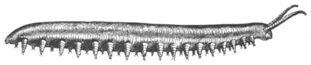
Fig. 167. Adult example of Peripatus
capensis, natural size.
(From Moseley.)
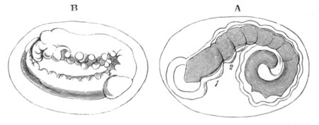
Fig. 168. Two stages in the development of
Peripatus capensis.
(After Moseley.)
A. Youngest stage hitherto observed before the appearance of the
legs.
B. Later stage after the legs and antennæ have become developed.
Both figures represent the larva as it appears within the egg.
1 and 2. First and second post-oral appendages.
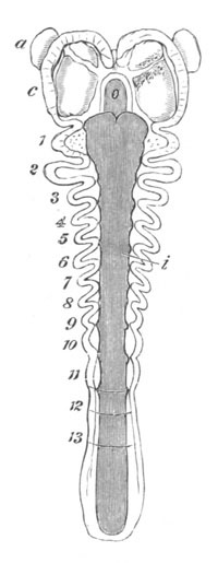
Fig. 169. Embryo of Peripatus capensis.
Slightly older than A in fig. 168; unrolled. (After Moseley.)
a. antennæ; o. mouth; i. intestine; c.
procephalic lobe. 1, 2, 3, etc., post-oral appendages.
The single pair of antennæ is formed in the next stage, and is
followed by the remaining post-oral appendages, which arise in
succession from before backwards somewhat later than the segments to
which they appertain.
The posterior part of the embryo becomes uncoiled, and the whole
embryo bent double in the egg (fig. 168 B).
The mouth appears as a slit-like opening between and below the
procephalic lobes. On each side and somewhat behind it there grows out
an appendage—the first post-oral pair (fig. 169, 1)—while in front
and behind it are formed the upper and lower lips. These two
appendages next turn inwards towards the mouth, and their
bases become gradually closed over by two processes of the procephalic
region (fig. 170, m). The whole of these structures assist in
forming a kind of secondary mouth cavity, which is at a later period
further completed by the processes of the procephalic region meeting
above the mouth, covering over the labrum, and growing backwards to
near the origin of the second pair of post-oral appendages.
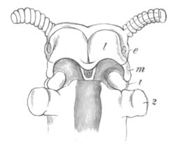
Fig. 170. Ventral view of the head of an embryo
of Peripatus capensis at a late stage of development.
l. thickening of epiblast of procephalic lobe to form
supra-œsophageal ganglion; m. process from procephalic
lobe growing over the first post-oral appendage; o. mouth;
e. eye; 1 and 2, first and second pair of post-oral
appendages.
The antennæ early become jointed, and fresh joints continue to be
added throughout embryonic life; in the adult there are at present
fully thirty joints. It appears to me probable (though Mr Moseley
takes the contrary view) from the late development of the paired
processes of the procephalic lobes, which give rise to the circular
lip of the adult, that they are not true appendages. The next pair
therefore to the antennæ is the first post-oral pair. It is the only
pair connected with the mouth. At their extremities there is formed a
pair of claws similar to those of the ambulatory legs (fig. 171). The
next and largest pair of appendages in the embryo are the oral
papillæ. They are chiefly remarkable for containing the ducts of the
slime glands which open at their bases. They are without claws. The
succeeding appendages become eventually imperfectly five-jointed; two
claws are formed as cuticular investments of papillæ in
pockets of the skin at the ends of their terminal joints.
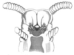
Fig. 171. Head of an embryo Peripatus. (From
Moseley.)
The figure shews the jaws (mandibles), and close to them epiblastic
involutions, which grow into the supra-œsophageal ganglia. The
antennæ, oral cavity, and oral papillæ are also shewn.
I have been able to make a few observations on the internal structure
of the embryos from specimens supplied to me by Moseley. These are so
far confined to a few stages, one slightly earlier, the others
slightly later, than the embryo represented in fig. 168 B. The
epiblast is formed of a layer of columnar cells, two deep on the
ventral surface, except along the median line where there is a
well-marked groove and the epiblast is much thinner (fig. 172).
The ventral cords of the trunk are formed as two independent
epiblastic thickenings. In my earlier stage these are barely separated
from the epiblast, but in the later ones are quite independent (fig.
172, v.n), and partly surrounded by mesoblast.
The supra-œsophageal ganglia are formed as thickenings of the
epiblast of the ventral side of the procephalic lobes in front of the
stomodæum. They are shewn at l in fig. 170. The thickenings of
the two sides are at first independent. At a somewhat later period an
invagination of the epiblast grows into each of these lobes. The
openings of these invaginations extend from the oral cavity forwards;
and they are shewn in fig. 171[161]. Their openings become closed, and
the walls of the invaginations constitute a large part of the
embryonic supra-œsophageal ganglia.
Similar epiblastic invaginations assist in forming the
supra-œsophageal ganglia of other Tracheata. They are described in
the sequel for Insects, Spiders and Scorpions. The position of the
supra-œsophageal ganglia on the ventral side of the procephalic
lobes is the same as that in other Tracheata.
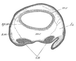
Fig. 172. Section through the trunk of an embryo
of Peripatus. The embryo from which the section is taken was
somewhat younger than fig. 171.
sp.m. splanchnic mesoblast. s.m. somatic mesoblast.
mc. median section of body cavity. lc. lateral section
of body cavity. v.n. ventral nerve cord. me.
mesenteron.
The mesoblast is formed, in the earliest of my embryos, of scattered
cells in the fairly wide space between the mesenteron and the
epiblast. There are two distinct bands of mesoblast on the outer sides
of the nervous cords. In the later stage the mesoblast is divided into
distinct somatic and splanchnic layers, both very thin; but the two
layers are connected by transverse strands (fig. 172). There
are two special longitudinal septa dividing the body cavity into three
compartments, a median (mc), containing the mesenteron, and two
lateral (lc) containing the nerve cords. This division of the
body cavity persists, as I have elsewhere shewn, in the adult. A
similar division is found in some Chætopoda, e.g. Polygordius.
I failed to make out that the mesoblast was divided into somites, and
feel fairly confident that it is not so in the stages I have
investigated.
There is a section of the body cavity in the limbs as in embryo
Myriapods, Spiders, etc.
In the procephalic lobe there is a well-developed section of the body
cavity, which lies dorsal to and in front of the rudiment of the
supra-œsophageal ganglia.
The alimentary tract is formed of a mesenteron (fig. 172), a
stomodæum, and proctodæum. The wall of the mesenteron is formed, in
the stages investigated by me, of a single layer of cells with yolk
particles, and encloses a lumen free from yolk. The forward extension
of the mesenteron is remarkable.
The stomodæum in the earlier stage is a simple pit, which meets but
does not open into the mesenteron. In the later stage the external
opening of the pit is complicated by the structures already described.
The proctodæum is a moderately deep pit near the hinder end of the
body.
The existence of a tracheal system[162] is in itself almost sufficient
to demonstrate the affinities of Peripatus with the Tracheata, in
spite of the presence of nephridia. The embryological characters of
the procephalic lobes, of the limbs and claws, place however this
conclusion beyond the reach of scepticism. If the reader will compare
the figure of Peripatus with that of an embryo Scorpion (fig. 196 A)
or Spider (fig. 200 C) or better still with Metschnikoff’s figure of
Geophilus (No. 399) Pl. XXI. fig. II, he will be
satisfied on this point.
The homologies of the anterior appendages are not very easy to
determine; but since there does not appear to me to be sufficient
evidence to shew that any of the anterior appendages have become
aborted, the first post-oral appendages embedded in the lips may
provisionally be regarded as equivalent to the mandibles, and the oral
papillæ to the first pair of maxillæ, etc. Moseley is somewhat
doubtful about the homologies of the appendages, and hesitates between
considering the oral papillæ as equivalent to the second pair of
maxillæ (on account of their containing the openings of the mucous
glands, which he compares with the spinning glands of caterpillars),
or to the poison claws (fourth post-oral appendages) of the
Chilopoda (on account of the poison glands which he thinks may be
homologous with the mucous glands).
The arguments for either of these views do not appear to me
conclusive. There are glands opening into various anterior appendages
in the Tracheata, such as the poison glands in the Cheliceræ
(mandibles) of Spiders, and there is some evidence in Insects for the
existence of a gland belonging to the first pair of maxillæ, which
might be compared with the mucous gland of Peripatus. For reasons
already stated I do not regard the processes of the cephalic lobes,
which form the lips, as a pair of true appendages.
Bibliography.
(396) H. N. Moseley. “On the Structure and Development of
Peripatus capensis.” Phil. Trans. Vol. 164, 1874.
Myriapoda[163].
Chilognatha. The first stages in the development of the
Chilognatha have been investigated by Metschnikoff and Stecker, but
their accounts are so contradictory as hardly to admit of
reconciliation.
According to Metschnikoff, by whom the following four species have
been investigated, viz., Strongylosoma Guerinii, Polydesmus
complanatus, Polyxenus lagurus, and Julus Moneletei, the segmentation
is at first regular and complete, but, when the segments are still
fairly large, the regular segmentation is supplemented by the
appearance of a number of small cells at various points on the
surface, which in time give rise to a continuous blastoderm.
The blastoderm becomes thickened on the ventral surface, and so forms
a ventral plate[164].
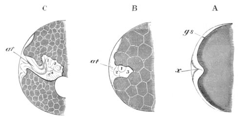
Fig. 173. Three stages in the development of
Strongylosoma Guerinii.
(After Metschnikoff.)
A. Embryo on eleventh day with commencing ventral flexure (x)
B. Embryo with three pairs of post-oral appendages.
C. Embryo with five pairs of post-oral appendages.
gs. ventral plate; at. antennæ; 1‑5 post-oral
appendages; x. point of flexure of the ventral plate.
The most important sources of information for the general embryology
of the Chilognatha are the papers of Newport (No. 397) and
Metschnikoff (No. 398). The development of Strongylosoma may be taken
as fairly typical for the group; and the subsequent statements, unless
the reverse is stated, apply to the species of Strongylosoma
investigated by Metschnikoff.
After the segmentation and formation of the layers the first
observable structure is a transverse furrow in the thickening of the
epiblast on the ventral surface of the embryo. This furrow rapidly
deepens, and gives rise to a ventral flexure of the embryo (fig. 173
A, x), which is much later in making its appearance in Julus
than in Strongylosoma and Polyxenus. A pair of appendages, which
become the antennæ, makes its appearance shortly after the formation
of the transverse furrow, and there soon follow in order the next
three pairs of appendages. All these parts are formed in the infolded
portion of the ventral thickening of the blastoderm (fig. 173 B). The
ventral thickening has in the meantime become marked by a longitudinal
furrow, but whether this is connected with the formation of the
nervous system, or is equivalent to the mesoblastic furrow in Insects,
and connected with the formation of the mesoblast, has not been made
out. Shortly after the appearance of the three pairs of appendages
behind the antennæ two further pairs become added, and at the same
time oral and anal invaginations become formed (fig. 173 C). In front
of the oral opening an unpaired upper lip is developed. The præ-oral
part of the ventral plate develops into the bilobed procephalic lobes,
the epiblast of which is mainly employed in the formation of the
supra-œsophageal ganglia. The next important change which takes
place is the segmentation of the body of the embryo (fig. 174 A), the
most essential feature in which is the division of the mesoblast into
somites. Segments are formed in order from before backwards, and soon
extend to the region behind the appendages. On the appearance of
segmentation the appendages commence to assume their permanent form.
The two anterior pairs of post-oral appendages become jaws; and the
part of the embryo which carries them and the antennæ is marked off
from the trunk as the head. The three following pairs of appendages
grow in length and assume a form suited for locomotion. Behind
the three existing pairs of limbs there are developed three fresh
pairs, of which the two anterior belong to a single primitive
segment. While the above changes take place in the appendages the
embryo undergoes an ecdysis, which gives rise to a cuticular membrane
within the single egg membrane (chorion, Metschnikoff). On this
cuticle a tooth-like process is developed, the function of which is to
assist in the hatching of the embryo (fig. 174 A).
In Polyxenus a cuticular membrane is present as in Strongylosoma, but
it is not provided with a tooth-like process. In the same form
amœboid cells separate themselves from the blastoderm at an early
period. These cells have been compared to the embryonic envelopes of
Insects described below.
In Julus two cuticular membranes are present at the time of
hatching: the inner one is very strongly developed and encloses the
embryo after hatching. After leaving the chorion the embryo Julus
remains connected with it by a structureless membrane which is
probably the outer of the two cuticular membranes.
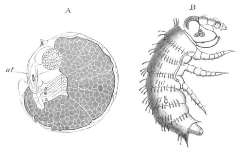
Fig. 174. Two stages in the development of
Strongylosoma Guerinii.
(After Metschnikoff.)
A. A seventeen days’ embryo, already segmented.
B. A just hatched larva.
At the time when the embryo of Strongylosoma is hatched (fig. 174 B)
nine post-cephalic segments appear to be present. Of these segments
the second is apparently (from Metschnikoff’s figure, 174 B) without a
pair of appendages; the third and fourth are each provided
with a single functional pair of limbs; the fifth segment is provided
with two pairs of rudimentary limbs, which are involuted in a single
sack and not visible without preparation, and therefore not shewn in
the figure. The sixth segment is provided with but a single pair of
appendages, though a second pair is subsequently developed on it[165].
Julus, at the time it leaves the chorion, is imperfectly segmented,
but is provided with antennæ, mandibles, and maxillæ, and seven pairs
of limbs, of which the first three are much more developed than the
remainder. Segmentation soon makes its appearance, and the head
becomes distinct from the trunk, and on each of the three anterior
trunk segments a single pair of limbs is very conspicuous
(Metschnikoff)[166]. Each of the succeeding segments bears eventually
two pairs of appendages. At the time when the inner embryonic cuticle
is cast off, the larva appears to be hexapodous, like the young
Strongylosoma, but there are in reality four pairs of rudimentary
appendages behind the three functional pairs. The latter only appear
on the surface after the first post-embryonic ecdysis. Pauropus
(Lubbock) is hexapodous in a young stage. At the next moult two pairs
of appendages are added, and subsequently one pair at each moult.
There appear to be eight post-oral segments in Julus at the time of
hatching. According to Newport fresh segments are added in
post-embryonic life by successive budding from a blastema between the
penultimate segment and that in front of it. They arise in batches of
six at the successive ecdyses, till the full number is completed. A
functional, though not a real hexapodous condition, appears to be
characteristic of Chilognatha generally at the time of hatching.
The most interesting anatomical feature of the Chilognatha is the
double character of their segments, the feet (except the first three
or four, or more), the circulatory, the respiratory, and the nervous
systems shewing this peculiarity. Newport’s and Metschnikoff’s
observations have not thrown as much light on the nature of the double
segments as might have been hoped, but it appears probable that they
have not originated from a fusion of two primitively distinct
segments, but from a later imperfect division of each of the primitive
segments into two, and the supply to each of the divisions of a
primitive segment of a complete set of organs.
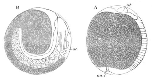
Fig. 175. Two stages in the development of
Geophilus.
(After Metschnikoff.)
A. Side view of embryo at the stage when the segments are beginning
to be formed.
B. Later stage after the appendages have become
established.
at. antennæ; an.i. proctodæum.
Chilopoda. Up to the present time the development of only one
type of Chilopoda, viz. that of Geophilus, has been worked out. Most
forms lay their eggs, but Scolopendra is viviparous. The segmentation
appears to resemble that in the Chilognatha, and at its close there is
present a blastoderm surrounding a central mass of yolk cells. A
ventral thickening of the blastoderm is soon formed. It becomes
divided into numerous segments, which continue to be formed
successively from the posterior unsegmented part. The antennæ are the
first appendages to appear, and are well developed when eighteen
segments have become visible (fig. 175 A). The post-oral appendages
are formed slightly later, and in order from before backwards. As the
embryo grows in length, and fresh segments continue to be formed, the
posterior part of it becomes bent over so as to face the ventral
surface of the anterior, and it acquires an appearance
something like that of many embryo Crustaceans (fig. 175 B). Between
forty and fifty segments are formed while the embryo is still in the
egg. The appendages long remain unjointed. The fourth post-oral
appendage, which becomes the poison claw, is early marked out by its
greater size: on the third post-oral there is formed a temporary spine
to open the egg membrane.
It does not appear, from Metschnikoff’s figures of Geophilus, that any
of the anterior segments are without appendages, and it is very
probable that Newport is mistaken in supposing that the embryo has a
segment without appendages behind that with the poison claws, which
coalesces with the segment of the latter. It also appears to me rather
doubtful whether the third pair of post-oral appendages, i.e.
those in front of the poison claws, can fairly be considered as
forming part of the basilar plate. The basilar plate is really the
segment of the poison claws, and may fuse more or less completely with
the segment in front and behind it, and the latter is sometimes
without a pair of appendages (Lithobius, Scutigera).
Geophilus, at the time of birth, has a rounded form like that of the
Chilognatha.
The young of Lithobius is born with only six pairs of limbs.
General observation on the homologies of the appendages of
Myriapoda.
The chief difficulty in this connection is the homology of the third
pair of post-oral appendages.
In adult Chilognatha there is present behind the mandibles a
four-lobed plate, which is usually regarded as representing two pairs
of appendages, viz. the first and second pairs of maxillæ of Insects.
Metschnikoff’s observations seem however to shew that this plate
represents but a single pair of appendages, which clearly corresponds
with the first pair of maxillæ in Insects. The pair of appendages
behind this plate is ambulatory, but turned towards the head; it is in
the embryo the foremost of the three functional pairs of legs with
which the larva is born. Is it equivalent to the second pair of
maxillæ of Insects or to the first pair of limbs of Insects? In favour
of the former view is the fact (1) that in embryo Insects the second
pair of maxillæ sometimes resembles the limbs rather than the jaws, so
that it might be supposed that in Chilognatha a primitive ambulatory
condition of the third pair of appendages has been retained; (2) that
the disappearance of a pair of appendages would have to be postulated
if the second alternative is adopted, and that if Insects are
descended from forms related to the Myriapods it is surprising to find
a pair of appendages always present in the former, absent in the
latter. The arguments which can be urged for the opposite view
do not appear to me to have much weight, so that the homology of the
appendages in question with the second pair of maxillæ may be
provisionally assumed.
The third pair of post-oral appendages of the Chilopoda may probably
also be assumed to be equivalent to the second pair of maxillæ; though
they are limb-like and not connected with the head. The subjoined
table shews the probable homologies of the appendages.
|
Chilognatha
(Strongylosoma
at time of birth). |
Chilopoda
(Scolopendra adult). |
| Pre-oral region. |
Antennæ. |
Antennæ. |
| 1st Post-oral segment. |
Mandibles. |
Mandibles. |
| 2nd Post-oral segment. |
Maxillæ 1. (Four-lobed
plate in adult, but a
simple pair of appendages in embryo). |
Maxillæ 1. (Palp and bilobed median process). |
3rd Post-oral segment.
(probably equivalent to segment bearing 2nd pair of maxillæ in Insects). |
1st pair of ambulatory limbs. |
Limb-like appendages with basal parts in contact. |
| 4th Post-oral segment. |
(?) Apodous. |
Poison claws. |
| 5th Post-oral segment. |
2nd pair of ambulatory limbs. |
1st pair of ambulatory limbs. |
| 6th Post-oral segment. |
3rd pair of ambulatory limbs. |
2nd pair of ambulatory limbs. |
| 7th Post-oral segment. |
4th and 5th pair of ambulatory limbs. |
3rd pair of ambulatory limbs. |
| 8th Post-oral segment. |
6th pair of ambulatory limbs. |
4th pair of ambulatory limbs. |
| 9th Post-oral segment. |
Apodous. |
5th pair of ambulatory limbs. |
| 10th Post-oral segment. |
" (last segment in embryo). |
6th pair of ambulatory limbs. |
The germinal layers and formation of organs.
The development of the organs of the Myriapoda, and the origin of the
germinal layers, are very imperfectly known: Myriapoda appear however
to be closely similar to Insects in this part of their development,
and the general question of the layers will be treated more fully in
connection with that group.
The greater part of the blastoderm gives rise to the epiblast, which
furnishes the skin, nervous system, tracheal system, and the stomodæum
and proctodæum.
The mesoblast arises in connection with the ventral thickening of the
blastoderm, but the details of its formation are not known.
Metschnikoff describes a longitudinal furrow which appears very early
in Strongylosoma, which is perhaps equivalent to the mesoblastic
furrows of Insects, and so connected with the formation of the
mesoblast.
The mesoblast is divided up into a series of protovertebra-like
bodies—the mesoblastic somites—the cavities of which become the body
cavity and the walls the muscles and probably the heart. They are
(Metschnikoff) prolonged into the legs, though the prolongations
become subsequently segmented off from the main masses. The splanchnic
mesoblast is, according to Metschnikoff, formed independently of the
somites, but this point requires further observation.
The origin of the hypoblast remains uncertain, but it appears probable
that it originates, in a large measure at least, from the yolk
segments. In the Chilognatha the mesenteron is formed in the interior
of the yolk segments, so that those yolk segments which are not
employed in the formation of the alimentary canal lie freely in the
body cavity. In the relation of the yolk segments to the alimentary
canal the Chilopoda present a strong contrast to the Chilognatha, in
that the greater part of the yolk lies within their mesenteron. The
mesenteron is at first a closed sack, but is eventually placed in
communication with the stomodæum and the proctodæum. The Malpighian
bodies arise as outgrowths from the blind extremity of the latter.
Bibliography.
(397) G. Newport. “On the Organs of Reproduction and
Development of the Myriapoda.” Philosophical Transactions,
1841.
(398) E. Metschnikoff. “Embryologie der doppeltfüssigen
Myriapoden (Chilognatha).” Zeit. f. wiss. Zool., Vol.
XXIV. 1874.
(399) —— “Embryologisches über Geophilus.” Zeit. f. wiss.
Zool., Vol. XXV. 1875.
(400) Anton Stecker. “Die Anlage d. Keimblatter bei den
Diplopoden.” Archiv f. mik. Anatomie, Bd. XIV. 1877.
Insecta[167].
The formation of the embryonic layers in Insects has not been followed
out in detail in a large number of types; but, as in so many
other instances, some of the most complete histories we have are due
to Kowalevsky (No. 416). The development of Hydrophilus has been
worked out by him more fully than that of any other form, and will
serve as a type for comparison with other forms.
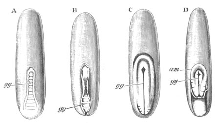
Fig. 176. Four embryos of Hydrophilus piceus
viewed from the ventral surface. (After Kowalevsky.)
The upper end is the anterior. gg. germinal groove;
am. amnion.
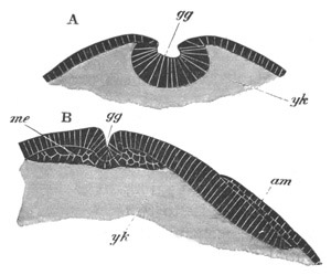
Fig. 177. Two transverse sections through embryos
of Hydrophilus piceus. (After Kowalevsky.)
A. Section through an embryo of the stage represented in fig. 176 B,
at the point where the two germinal folds most approximate.
B. Section through an embryo somewhat later than the stage fig. 176
D, through the anterior region where the amnion has not completely
closed over the embryo.
gg. germinal groove; me. mesoblast; am. amnion;
yk. yolk.
The segmentation has not been studied, but no doubt belongs to the
centrolecithal type (vide pp. 110‑120). At its close there is
an uniform layer of cells enclosing a central mass of yolk. These
cells, in the earliest observed stage, were flat on the dorsal, but
columnar on part of the ventral surface of the egg, where they form a
thickening which will be called the ventral plate. At the posterior
part of the ventral plate two folds, with a furrow between them, make
their appearance. They form a structure which may be spoken of as the
germinal groove (fig. 176 A, gg). The cells which form
the floor of the groove are far more columnar than those of other
parts of the blastoderm (fig. 177 A). The two folds on each side of it
gradually approach each other. They do so at first behind, and then in
the middle; from the latter point the approximation gradually extends
backwards and forwards (fig. 176 B and C). In the middle and hinder
parts of the ventral plate the groove becomes, by the coalescence of
the folds, converted into a canal (fig. 178 A, gg), the central
cavity of which soon disappears, while at the same time the cells of
the wall undergo division, become more rounded, and form a definite
layer (me)—the mesoblast—beneath the columnar cells of the
surface. Anteriorly the process is slightly different, though it leads
to the similar formation of mesoblast (fig. 177 B). The flat floor of
the groove becomes in front bodily converted into the mesoblast, but
the groove itself is never converted into a canal. The two folds
simply meet above, and form a continuous superficial layer.
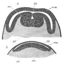
Fig. 178. Sections through two embryos of
Hydrophilus piceus. (After Kowalevsky.)
A. Section through the posterior part of the embryo fig. 176 D,
shewing the completely closed amnion and the germinal groove.
B. Section through an older embryo in which the mesoblast has grown
out into a continuous plate beneath the epiblast.
gg. germinal groove; am. amnion; yk. yolk;
ep. epiblast.
During the later stages of the process last described remarkable
structures, eminently characteristic of the Insecta, have made their
first appearance. These structures are certain embryonic membranes or
coverings, which present in their mode of formation and arrangement a
startling similarity to the true and false amnion of the Vertebrata.
They appear as a double fold of the blastoderm round the edge of the
germinal area, which spreads over the ventral plate, from behind
forwards, in a general way in the same manner as the amnion
in, for instance, the chick. The folds at their origin are shewn in
surface view in fig. 176 D, am, and in section in fig. 177 B,
am. The folds eventually meet, coalesce (fig. 178, am) and give
rise to two membranes covering the ventral plate, viz. an inner one,
which is continuous with the edge of the ventral plate; and an outer,
continuous with the remainder of the blastoderm. The vertebrate
nomenclature may be conveniently employed for these membranes. The
inner limb of the fold will therefore be spoken of as the amnion, and
the outer one, including the dorsal part of the blastoderm, as the
serous envelope[168]. A slight consideration of the mode of formation
of the membranes, or an inspection of the figures illustrating their
formation, makes it at once clear that the yolk can pass in freely
between the amnion and serous envelope (vide fig. 181). At the
hind end of the embryo this actually takes place, so that the ventral
plate covered by the amnion appears to become completely imbedded in
the yolk: elsewhere the two membranes are in contact. At first (fig.
176) the ventral plate occupies but a small portion of the ventral
surface of the egg, but during the changes above described it extends
over the whole ventral surface, and even slightly on the dorsal
surface both in front and behind. It becomes at the same time (fig.
179) divided by a series of transverse lines into segments,
which increase in number and finally amount in all to seventeen, not
including the most anterior section, which gives off as lateral
outgrowths the two procephalic lobes (pc.l). The changes so far
described are included within what Kowalevsky calls his first
embryonic period; at its close the parts contained within the chorion
have the arrangement shewn in fig. 178 B. The whole of the body of the
embryo is formed from the ventral plate, and no part from the amnion
or serous envelope.
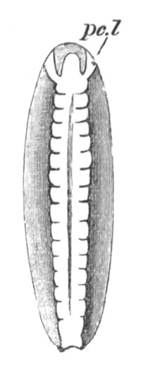
Fig. 179. Embryo Of Hydrophilus piceus viewed
from the ventral surface. (After Kowalevsky.)
pc.l. procephalic lobe.
The general history of the succeeding stages may be briefly told.
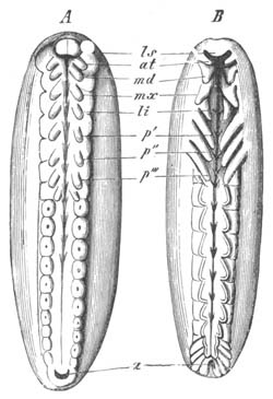
Fig. 180. Two stages in the development of
Hydrophilus piceus. (From Gegenbaur, after Kowalevsky.)
ls. labrum; at. antenna; md. mandible; mx.
maxilla I.; li. maxilla II.; p´ p´´ p´´´. feet;
a. anus.
The appendages appear as very small rudiments at the close of the last
stage, but soon become much more prominent (fig. 180 A). They are
formed as outgrowths of both layers, and arise nearly simultaneously.
There are in all eight pairs of appendages. The anterior or antennæ
(at) spring from the procephalic lobes, and the succeeding
appendages from the segments following. The last pair of embryonic
appendages, which disappears very early, is formed behind the third
pair of the future thoracic limbs. Paired epiblastic involutions,
shewn as pits in the posterior segments in fig. 180 A, give rise to
the tracheæ; and the nervous system is formed as two lateral
epiblastic thickenings, one on each side of the mid-ventral line.
These eventually become split off from the skin; while between them
there passes in a median invagination of the skin (fig. 189
C). The two nervous strands are continuous in front with the
supra-œsophageal ganglia, which are formed of the epiblast of the
procephalic lobes. These plates gradually grow round the dorsal side
of the embryo, and there is formed immediately behind them an oral
invagination, in front of which there appears an upper lip (fig. 180,
ls). A proctodæum is formed at the hind end of the body
slightly later than the stomodæum. The mesoblast cells become divided
into two bands, one on each side of the middle line (fig. 189 A), and
split into splanchnic and somatic layers. The central yolk mass at
about the stage represented in fig. 179 begins to break up into yolk
spheres. The hypoblast is formed first on the ventral side at the
junction of the mesoblast and the yolk, and gradually extends and
forms a complete sack-like mesenteron, enveloping the yolk (fig. 185
al). The amnion and serous membrane retain their primitive
constitution for some time, but gradually become thinner on the
ventral surface, where a rupture appears eventually to take place. The
greater part of them disappears, but in the closure of the dorsal
parietes the serous envelope plays a peculiar part, which is not yet
understood. It is described on p. 404. The heart is formed from the
mesoblastic layers, where they meet in the middle dorsal line (fig.
185 C, ht). The somatic mesoblast gives rise to the muscles and
connective tissue, and the splanchnic mesoblast to the muscular part
of the wall of the alimentary tract, which accompanies the hypoblast
in its growth round the yolk. The proctodæum forms the rectum and
Malpighian bodies[169], and the stomodæum the œsophagus and
proventriculus. The two epiblastic sections of the alimentary tract
are eventually placed in communication with the mesenteron.
The development of Hydrophilus is a fair type of that of Insects
generally, but it is necessary to follow with somewhat greater detail
the comparative history of the various parts which have been briefly
described for this type.
The embryonic membranes and the formation of the layers.
All Insects have at the close of segmentation a blastoderm formed of a
single row of cells enclosing a central yolk mass, which
usually contains nuclei, and in the Poduridæ is divided up in the
ordinary segmentation into distinct yolk cells. The first definite
structure formed is a thickening of the blastoderm, which forms a
ventral plate.
The ventral plate is very differently situated in relation to the yolk
in different types. In most Diptera, Hymenoptera and (?) Neuroptera
(Phryganea) it forms from the first a thickening extending over nearly
the whole ventral surface of the ovum, and in many cases extends in
its subsequent growth not only over the whole ventral surface, but
over a considerable part of the apparent dorsal surface as well
(Chironomus, Simulia, Gryllotalpa, etc.). In Coleoptera, so far as is
known, it commences as a less extended thickening either of the
central part (Donacia) or posterior part (Hydrophilus) of the ventral
surface, and gradually grows in both directions, passing over to the
dorsal surface behind.
Embryonic membranes. In the majority of Insects there are
developed enveloping membranes like those of Hydrophilus.
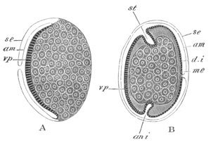
Fig. 181. Diagrammatic longitudinal sections of
an Insect embryo at two stages to shew the development of the
embryonic envelopes.
In A the amniotic folds have not quite met so as to cover the
ventral plate. The yolk is represented as divided into yolk cells.
In B the sides of the ventral plate have extended so as nearly to
complete the dorsal integument. The mesenteron is represented as a
closed sack filled with yolk cells. am. amnion; se.
serous envelope; v.p. ventral plate; d.i. dorsal
integument; me. mesenteron; st. stomodæum; an
i. proctodæum.
The typical mode of formation of these membranes is represented
diagrammatically in fig. 181 A and B. A fold of the blastoderm arises
round the edge of the ventral plate. This fold, like the amniotic fold
of the higher Vertebrata, is formed of two limbs, an outer, the serous
membrane (se), and an inner, the true amnion (am). Both
limbs extend so as to cover over the ventral plate, and finally meet
and coalesce, so that a double membrane is present over the ventral
plate. At the same time (fig. 181 B) the point where the fold
originates is carried dorsalwards by the dorsal extension of
the edges of the ventral plate, which give rise to the dorsal
integument (d.i). This process continues till the whole dorsal
surface is covered by the integument. The amnion then separates from
the dorsal integument, and the embryo becomes enveloped in two
membranes—an inner, the amnion, and an outer, the serous membrane. In
fig. 181 B the embryo is represented at the stage immediately
preceding the closure of the dorsal surface.
By the time that these changes are effected, the serous membrane and
amnion are both very thin and not easily separable. The amnion appears
to be usually absorbed before hatching; but in hatching both
membranes, if present, are either absorbed, or else ruptured and
thrown off.
The above mode of development of the embryonic membranes has been
especially established by the researches of Kowalevsky (No. 416) and
Graber (No. 412) for various Hymenoptera (Apis), Diptera
(Chironomus), Lepidoptera and Coleoptera (Melolontha,
Lina).
Considerable variations in the development of the enveloping membranes
are known.
When the fold which gives rise to the membranes is first formed, there
is, as is obvious in fig. 181 A, a perfectly free passage by which the
yolk can pass in between the amnion and serous membrane. Such a
passage of the yolk between the two membranes takes place posteriorly
in Hydrophilus and Donacia: in Lepidoptera the yolk passes in
everywhere, so that in this form the ventral plate becomes first of
all imbedded in the yolk, and finally, on the completion of the dorsal
integument, the embryo is enclosed in a complete envelope of yolk
contained between the amnion and the serous membrane. During the
formation of the dorsal integument the external yolk-sack communicates
by a dorsally situated umbilical canal with the yolk cavity within the
body. On the rupture of the amnion the embryo is nourished at the
expense of the yolk contained in the external yolk-sack.
In the Hemiptera and the Libellulidæ the ventral plate also becomes
imbedded in the yolk, but in a somewhat different fashion to the
Lepidoptera, which more resembles on an exaggerated scale what takes
place in Hydrophilus.
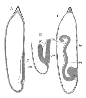
Fig. 182. Three stages in the development of the
embryo of Calopteryx. (After Brandt.)
The embryo is represented in the egg-shell.
A. Embryo with ventral plate.
B. Commencing involution of ventral plate.
C. Involution of ventral plate completed.
ps. ventral plate; g. edge of ventral plate;
am. amnion; se. serous envelope.
In the Libellulidæ (Calopteryx) there is first of all formed
(Brandt, No. 403) a small ventral and posterior thickening of the
blastoderm (fig. 182 A). The hinder part of this becomes infolded into
the yolk as a projection (fig. 182 B), which consists of two laminæ,
an anterior and a posterior, continuous at the apex of the
invagination. The whole structure, which is completely imbedded within
the yolk, rapidly grows in length, and turns towards the front end of
the egg (fig. 182 C). Its anterior lamina remains thick and gives rise
to the ventral plate (ps), the posterior (am) on the
other hand becomes very thin, and forms a covering
corresponding with the amnion of the more ordinary types. The
remainder of the blastoderm covering the yolk (se) forms the
homologue of the serous membrane of other types. The ventral surface
of the ventral plate is turned towards the dorsal side (retaining the
same nomenclature as in ordinary cases) of the egg, and the cephalic
extremity is situated at the point of origin of the infolding.
The further history is however somewhat peculiar. The amnion is at
first (fig. 182 C) continuous with the serous envelope on the
posterior side only, so that the serous envelope does not form a
continuous sack, but has an opening close to the head of the embryo.
In the Hemiptera parasita this opening (Melnikow, No. 422) remains
permanent, and the embryo, after it has reached a certain stage of
development, becomes everted through it, while the yolk, enclosed in
the continuous membrane formed by the amnion and serous envelope,
forms a yolk-sack on the dorsal surface. In the Libellulidæ however
and most Hemiptera, a fusion of the two limbs of the serous membrane
takes place in the usual way, so as to convert it into a completely
closed sack (fig. 183 A). After the formation of the appendages a
fusion takes place between the amnion and serous envelope over a small
area close to the head of the embryo. In the middle of this area a
rupture is then effected, and the head of the embryo followed by the
body is gradually pushed through the opening (fig. 183 B and C). The
embryo becomes in the process completely rotated, and carried into a
position in the egg-shell identical with that of the embryos of other
orders of Insects (fig. 183 C).
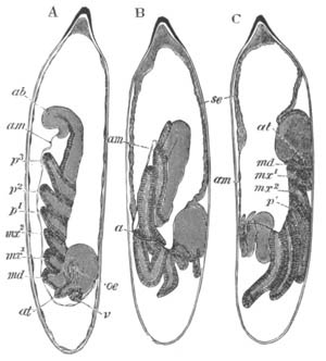
Fig. 183. Three stages in the development of
Calopteryx. (After Brandt.)
The embryo is represented in the egg-shell; B. and C. shew the
inversion of the embryo.
se. serous envelope; am. amnion; ab. abdomen;
v. anterior end of head; at. antennæ; md.
mandible; mx1. maxilla 1; mx2. maxilla 2;
p1-p3. three pairs of legs; oe. œsophagus.
Owing to the rupture of the embryonic envelopes taking place at the
point where they are fused into one, the yolk does not escape in the
above process, but is carried into a kind of yolk-sack, on the dorsal
surface of the embryo, formed of the remains of the amnion and serous
envelope. The walls of the yolk-sack either assist in forming
the dorsal parietes of the body, or are more probably enclosed within
the body by the growth of the dorsal parietes from the edge of the
ventral plate.
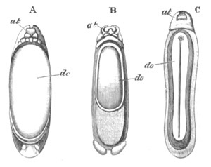
Fig. 184. Three larval stages of Hydrophilus from
the dorsal side, shewing the gradual closing in of the dorsal region
with the formation of the peculiar dorsal organ do.
(After Kowalevsky.)
do. dorsal organ; at. antennæ.
In Hydrophilus and apparently in the Phryganidæ also, there are
certain remarkable peculiarities in the closure of the dorsal surface.
The fullest observations on the subject have been made by Kowalevsky
(No. 416), but Dohrn (No. 408) has with some probability thrown doubts
on Kowalevsky’s interpretations. According to Dohrn the part of the
serous envelope which covers the dorsal surface becomes thickened, and
gives rise to a peculiar dorsal plate which is shewn in surface view
in fig. 184 A, do, and in section in fig. 185 A, do. The
ventral parts of the amnion and serous membrane have either been
ruptured or have disappeared. While the dorsal plate is being formed,
the mesoblast, and somewhat later the lateral parts of the epiblast of
the ventral plate gradually grow towards the dorsal side and enclose
the dorsal plate, the wall of which in the process appears to be
folded over so as first of all to form a groove and finally a canal.
The stages in this growth are shewn from the surface in fig. 184 B and
C and in section in fig. 185 B, do. The canal is buried
on the dorsal part of the yolk, but for some time remains open by a
round aperture in front (fig. 184 C). The whole structure is known as
the dorsal canal. It appears to atrophy without leaving a trace. The
heart when formed lies immediately dorsal to it[170].
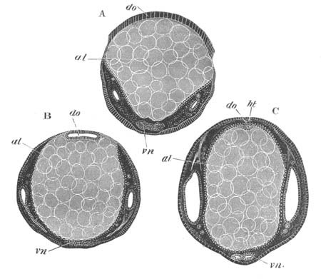
Fig. 185. Three transverse sections through
advanced embryos of Hydrophilus.
A. Section through the posterior part of the body of the same age as
fig. 184 A.
B. Section through the embryo of the same age as fig. 184 C.
C. Section through a still older embryo.
do. dorsal plate; vn. ventral nerve cord; al.
mesenteron; ht. heart.
The large spaces at the sides are parts of the body cavity.
In the Poduridæ the embryonic membranes appear to be at any rate
imperfect. Metschnikoff states in his paper on Geophilus that in some
ants no true embryonic membranes are found, but merely scattered cells
which take their place. In the Ichneumonidæ the existence of two
embryonic membranes is very doubtful.
Formation of the embryonic layers. The formation of the layers
has been studied in sections by Kowalevsky (No. 416), Hatschek
(No. 414), and Graber (No. 412), etc. From their researches it would
appear that the formation of the mesoblast always takes place in a
manner closely resembling that in Hydrophilus. The essential features
of the process (figs. 177 and 178) appear to be that a groove is
formed along the median line of the ventral plate, and that the sides
of this groove either (1) simply close over like the walls of the
medullary groove in Vertebrates, and so convert the groove into a
tube, which soon becomes solid and forms a mass or plate of cells
internal to the epiblast; or (2) that the cells on each side of the
groove grow over it and meet in the middle line, forming a layer
external to the cells which lined the groove. The former of these
processes is the most usual; and in the Muscidæ the dimensions of the
groove are very considerable (Graber, No. 411). In both cases the
process is fundamentally the same, and causes the ventral plate to
become divided into two layers[171]. The external layer or epiblast is
an uniform sheet forming the main part of the ventral plate (fig. 178
B, ep). It is continuous at its edge with the amnion. The inner
layer or mesoblast constitutes an independent plate of cells internal
to the epiblast (fig. 178 B, me). The mesoblast soon becomes
divided into two lateral bands.
The origin of the hypoblast is still in dispute. It will be remembered
(vide pp. 114 and 116) that after the segmentation a number of
nuclei remain in the yolk; and that eventually a secondary
segmentation of the yolk takes place around these nuclei, and gives
rise to a mass of yolk cells, which fill up the interior of the
embryo. These cells are diagrammatically shewn in figs. 181 and 189,
and it is probable that they constitute the true hypoblast. Their
further history is given below.
Formation of the organs and their relation to the germinal
layers.
The segments and appendages. One of the earliest phenomena in
the development is the appearance of transverse lines indicating
segmentation (fig. 186). The transverse lines are apparently caused by
shallow superficial grooves, and also in many cases by the
division of the mesoblastic bands into separate somites. The most
anterior line marks off a præ-oral segment, which soon sends out two
lateral wings—the procephalic lobes. The remaining segments are at
first fairly uniform. Their number does not, however, appear to be
very constant. So far as is known they never exceed seventeen, and
this number is probably the typical one (figs. 186 and 187).
In Diptera the number appears to be usually fifteen though it may be
only fourteen. In Lepidoptera and in Apis there appear to be sixteen
segments. These and other variations affect only the number of the
segments which form the abdomen of the adult.
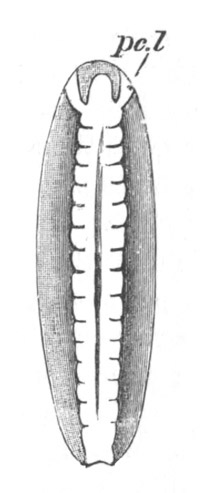
Fig. 186. Embryo of Hydrophilus piceus viewed
from the ventral surface.
(After Kowalevsky.)
pc.l. procephalic lobe.
The appendages arise as paired pouch-like outgrowths of the epiblast
and mesoblast; and their number and the order of their appearance are
subject to considerable variation, the meaning of which is not yet
clear. As a rule they arise subsequently to the segmentation of the
parts of the body to which they belong. There is always formed one
pair of appendages which spring from the lateral lobes of the
procephalic region, or from the boundary line between these and the
median ventral part of this region. These appendages are the antennæ.
They have in the embryo a distinctly ventral position as compared to
that which they have in the adult.
In the median ventral part of the procephalic region there arises the
labrum (fig. 187, ls). It is formed by the coalescence of a
pair of prominences very similar to true appendages, though it is
probable that they have not this value[172].
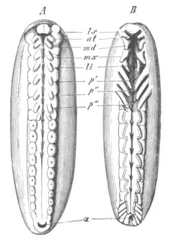
Fig. 187. Two stages in the development of
Hydrophilus piceus. (From Gegenbaur, after Kowalevsky.)
ls. labrum; at. antenna; md. mandible;
mx. maxilla I.; li. maxilla II.; p´ p´´ p´´´.
feet; a. anus.
The antennæ themselves can hardly be considered to have the same
morphological value as the succeeding appendages. They are rather
equivalent to paired processes of the præ-oral lobes of the Chætopoda.
From the first three post-oral segments there grow out the mandibles
and two pairs of maxillæ, and from the three following segments the
three pairs of thoracic appendages. In many Insects (cf. Hydrophilus)
a certain number of appendages of the same nature as the anterior ones
are visible in the embryo on the abdominal segments, a fact which
shews that Insects are descended from ancestors with more than three
pairs of ambulatory appendages.
In Apis according to Bütschli (No. 405) all the abdominal segments are
provided with appendages, which always remain in a very rudimentary
condition. All trace of them as well as of the thoracic appendages is
lost by the time the embryo is hatched. In the phytophagous
Hymenoptera the larva is provided with 9‑11 pairs of legs.
In the embryo of Lepidoptera there would appear from Kowalevsky’s
figures to be rudiments of ten pairs of post-thoracic appendages. In
the caterpillar of this group there are at the maximum five pairs of
such rudimentary feet, viz. a pair on the 3rd, 4th, 5th, and 6th, and
on the last abdominal segment. The embryos of Hydrophilus (fig. 187),
Mantis, etc. are also provided with additional appendages. In various
Thysanura small prominences are present on more or fewer of the
abdominal segments (fig. 192), which may probably be regarded as
rudimentary feet.
Whether all or any of the appendages of various kinds connected with
the hindermost segments belong to the same category as the legs is
very doubtful. Their usual absence in the embryo or in any case their
late appearance appears to me against so regarding them; but Bütschli
is of opinion that in the Bee the parts of the sting are related
genetically to the appendages of the penultimate and antepenultimate
abdominal segments, and this view is to some extent supported by more
recent observations (Kraepelin, etc.), and if it holds true
for the Bee must be regarded as correct for other cases also.
As to the order of the appearance of the appendages observations are
as yet too scanty to form any complete scheme. In many cases all the
appendages appear approximately at the same moment, e.g.
Hydrophilus, but whether this holds good for all Coleoptera is by no
means certain. In Apis the appendages are stated by Bütschli to arise
simultaneously, but according to Kowalevsky the two mouth appendages
first appear, then the antennæ, and still later the thoracic
appendages. In the Diptera the mouth appendages are first formed, and
either simultaneously with these, or slightly later, the antennæ. In
the Hemiptera and Libellulidæ the thoracic appendages are the first to
be formed, and the second pair of maxillæ makes its appearance before
the other cephalic appendages.
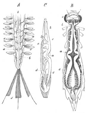
Fig. 188. Figures illustrating aquatic
respiration in Insects. (After Gegenbaur.)
A. Hinder portion of the body of Ephemera vulgata. a.
longitudinal tracheal trunks; b. alimentary canal; c.
tracheal gills.
B. Larva of Æschna grandis. a. superior longitudinal tracheal
trunks; b. their anterior end; c. portion branching on
proctodæum; o. eyes.
C. Alimentary canal of the same larva from the side. a, b,
and c. as in B; d. inferior tracheal trunk; e.
transverse branches between upper and lower tracheal trunks.
The history of the changes in the embryonic appendages during the
attainment of the adult condition is beyond the scope of this
treatise, but it may be noted that the second pair of maxillæ are
relatively very large in the embryo, and not infrequently (Libellula,
etc.) have more resemblance to the ambulatory than to the masticatory
appendages.
The exact nature of the wings and their relation to the other segments
is still very obscure. They appear as dorsal leaf-like appendages on
the 2nd and 3rd thoracic segments, and are in many respects similar to
the tracheal gills of the larvæ of Ephemeridæ and Phryganidæ (fig. 188
A), of which they are supposed by Gegenbaur and Lubbock to be
modifications. The undoubtedly secondary character of the
closed tracheal system of larvæ with tracheal gills tells
against this view. Fritz Müller finds in the larvæ of Calotermes
rugosus (one of the Termites) that peculiar and similar dorsal
appendages are present on the two anterior of the thoracic segments.
They are without tracheæ. The anterior atrophies, and the posterior
acquires tracheæ and gives rise to the first pair of wings. The second
pair of wings is formed from small processes on the third thoracic
segment like those on the other two. Fritz Müller concludes from these
facts that the wings of Insects are developed from dorsal processes of
the body, not equivalent to the ventral appendages. What the primitive
function of these appendages was is not clear. Fritz Müller suggests
that they may have been employed as respiratory organs in the passage
from an aqueous to a terrestrial existence, when the Termite ancestors
lived in moist habitations—a function for which processes supplied
with blood-channels would be well adapted. The undoubted affinity of
Insects to Myriapods, coupled with the discovery by Moseley of a
tracheal system in Peripatus, is however nearly fatal to the view that
Insects can have sprung directly from aquatic ancestors not provided
with tracheæ. But although this suggestion of Fritz Müller cannot be
accepted, it is still possible that the processes discovered by him
may have been the earliest rudiments of wings, which were employed
first as organs of propulsion by a water-inhabiting Insect ancestor
which had not yet acquired the power of flying.
The nervous system. The nervous system arises entirely from the
epiblast; but the development of the præ-oral and post-oral sections
may be best considered separately.
The post-oral section, or ventral cord of the adult, arises as two
longitudinal thickenings of the epiblast, one on each side of the
median line (fig. 189 B, vn), which are subsequently split off
from the superficial skin and give rise to the two lateral strands of
the ventral cord. At a later period they undergo a differentiation
into ganglia and connecting cords.
Between these two embryonic nerve cords there is at first a shallow
furrow, which soon becomes a deep groove (fig. 189 C). At this stage
the differentiation of the lateral elements into ganglia and
commissures takes place, and, according to Hatschek (No. 414), the
median groove becomes in the region of the ganglia converted into a
canal, the walls of which soon fuse with those of the ganglionic
enlargements of the lateral cords, and connect them across the middle
line. Between the ganglia on the other hand the median groove
undergoes atrophy, becoming first a solid cord interposed between the
lateral strands of the nervous system, and finally disappearing
without giving rise to any part of the nervous system. It is probable
that Hatschek is entirely mistaken about the entrance of a median
element into the ventral cord, and that the appearances he has
described are due to shrinkage. In Spiders the absence of a median
element can be shewn with great certainty, and, as already stated,
this element is not present in Peripatus. Hatschek states that
in the mandibular segment the median element is absorbed, and that the
two lateral cords of that part give rise to the œsophageal
commissures, while the sub-œsophageal ganglion is formed from the
fusion of the ganglia of the two maxillary segments.
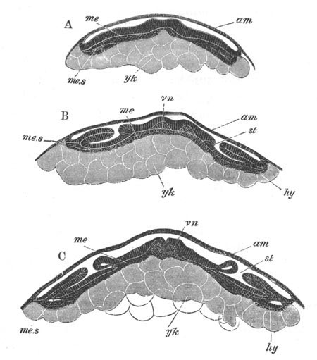
Fig. 189. Three transverse sections through the
embryo of Hydrophilus. (After Kowalevsky.)
A. Transverse section through the larva represented in fig. 187 A.
B. Transverse section through a somewhat older embryo in the region
of one of the stigmata.
C. Transverse section through the larva represented in fig. 187 B.
vn. ventral nerve cord; am. amnion and serous
membrane; me. mesoblast; me.s. somatic mesoblast;
hy. hypoblast(?); yk. yolk cells (true hypoblast);
st. stigma of trachea.
The præ-oral portion of the nervous system consists entirely of the
supra-œsophageal ganglion. It is formed, according to Hatschek, of
three parts. Firstly and mainly, of a layer separated from the
thickened inner part of the cephalic lobe on each side; secondly, of
an anterior continuation of the lateral cords; and thirdly, of a pit
of skin invaginated on each side close to the dorsal border of
the antennæ. This pit is at first provided with a lumen, which is
subsequently obliterated; while the walls of the pit become converted
into true ganglion cells. The two supra-œsophageal ganglia remain
disconnected on the dorsal side till quite the close of embryonic
life.
The tracheæ and salivary glands. The tracheæ, as was first
shewn by Bütschli (No. 405), arise as independent segmentally arranged
paired invaginations of the epiblast (fig. 189 B and C, st).
Their openings are always placed on the outer sides of the appendages
of their segments, where such are present.
Although in the adult stigmata are never found in the space between
the prothorax and head[173], in the embryo and the larva tracheal
invaginations may be developed in all the thoracic (and possibly in
the three jaw-bearing segments) and in all the abdominal segments
except the two posterior.
In the embryo of the Lepidoptera, according to Hatschek (No. 414),
there are 14 pairs of stigmata, belonging to the 14 segments of the
body behind the mouth; but Tichomiroff states that Hatschek is in
error in making this statement for the foremost post-oral segments.
The last two segments are without stigmata. In the larvæ of
Lepidoptera as well as those of many Hymenoptera, Coleoptera and
Diptera, stigmata are present on all the postcephalic segments except
the 2nd and 3rd thoracic and the two last abdominal. In Apis there are
eleven pairs of tracheal invaginations according to Kowalevsky (No.
416), but according to Bütschli (No. 405) only ten, the prothorax
being without one. In the Bee they appear simultaneously, and before
the appendages.
The blind ends of the tracheal invaginations frequently (e.g.
Apis) unite together into a common longitudinal canal, which forms a
longitudinal tracheal stem. In other cases (e.g. Gryllotalpa,
Dohrn, No. 408) they remain distinct, and each tracheal stem
has a system of branches of its own.
The development of the tracheæ strongly supports the view, arrived at
by Moseley from his investigations on Peripatus, that they are
modifications of cutaneous glands.
The salivary and spinning glands are epiblastic structures, which in
their mode of development are very similar to the tracheæ, and perhaps
have a similar origin. The salivary glands arise as paired
epiblastic invaginations, not, as might be expected, of the Stomodæum,
but of the ventral plate behind the mouth on the inner side of the
mandibles. At first independent, they eventually unite in a common
duct, which falls into the mouth. The spinning glands arise on the
inner side of the second pair of maxillæ in Apis and Lepidoptera, and
form elongated glands extending through nearly the whole length of the
body. They are very similar in their structure and development to
salivary glands, and are only employed during larval life. They no
doubt resemble the mucous glands of the oral papillæ of Peripatus,
with which they have been compared by Moseley. The mucous glands of
Peripatus may perhaps be the homologous organs of the first pair of
maxillæ, for the existence of which there appears to be some evidence
amongst Insects.
Mesoblast. It has been stated that the mesoblast becomes
divided in the region of the body into two lateral bands (fig. 189 A).
These bands in many, if not all forms, become divided into a series of
somites corresponding with the segments of the body. In each of them a
cavity appears—the commencing perivisceral cavity—which divides them
into a somatic plate in contact with the epiblast, and a splanchnic
plate in contact with the hypoblast (fig. 189). In the interspaces
between the segments the mesoblast is continuous across the median
ventral line. The mesoblast is prolonged into each of the appendages
as these are formed, and in the appendages there is present a central
cavity. By Metschnikoff these cavities are stated to be continuous, as
in Myriapods and Arachnida, with those of the somites; but by Hatschek
(No. 414) they are stated to be independent of those in the somites
and to be open to the yolk.
The further details of the history of the mesoblast are very
imperfectly known, and the fullest account we have is that by Dohrn
(No. 408) for Gryllotalpa. It would appear that the mesoblast grows
round and encloses the dorsal side of the yolk earlier than the
epiblast. In Gryllotalpa it forms a pulsating membrane. As the
epiblast extends dorsalwards the median dorsal part of the membrane is
constricted off as a tube which forms the heart. At the same time the
free space between the pulsating membrane and the yolk is obliterated,
but transverse passages are left at the lines between the somites,
through which the blood passes from the ventral part of the body to
corresponding openings in the wall of the heart. The greater part of
the membrane gives rise to the muscles of the trunk.
Ventrally the mesoblastic bands soon meet across the median line. The
cavities in the appendages become obliterated and their mesoblastic
walls form the muscles, etc. The cavities in the separate mesoblastic
somites also cease to be distinctly circumscribed.
The splanchnic mesoblast follows the hypoblast in its growth, and
gives rise to the connective tissue and muscular parts of the walls of
the alimentary tract. The mesoblastic wall of the proctodæum is
probably formed independently of the mesoblastic somites. In the head
the mesoblast is stated to form at first a median ventral mass, which
does not pass into the procephalic lobe; though it assists in forming
both the antennæ and upper lip.
The alimentary canal. The alimentary tract of Insects is formed
of three distinct sections (fig. 181)—a mesenteron or middle section
(me), a stomodæum (st) and a proctodæum (an). The
stomodæum and proctodæum are invaginations of the epiblast, while the
mesenteron is lined by the hypoblast. The distinction between the
three is usually well marked in the adult by the epiblastic
derivatives being lined by chitin. The stomodæum consists of mouth,
œsophagus, crop, and proventriculus or gizzard, when such are
present. The mesenteron includes the stomach, and is sometimes
(Orthoptera, etc.) provided at its front end with pyloric
diverticula—posteriorly it terminates just in front of the Malpighian
bodies. These latter fall into the proctodæum, which includes the
whole of the region from their insertion to the anus.
The oral invagination appears nearly coincidently with the first
formation of segments at the front end of the groove between the
lateral nerve cords, and the anal invagination appears slightly later
at the hindermost end of the ventral plate.
The Malpighian bodies arise as two pairs of outgrowths of the
epiblast of the proctodæum, whether solid at first is not certain.
The subsequent increase which usually takes place in their number is
due to sproutings (at first solid) of the two original vessels.
The glandular walls of the mesenteron are formed from the hypoblast;
but the exact origin of the layer has not been thoroughly worked out
in all cases. In Hydrophilus it is stated by Kowalevsky (No. 416) to
appear as two sheets split off from the lateral masses of mesoblast,
which gradually grow round the yolk, and a similar mode of formation
would seem to hold good for Apis. Tichomiroff (No. 420) confirms
Kowalevsky on this point, and further states that these two
masses meet first ventrally and much later on the dorsal side. In
Lepidoptera, on the other hand, Hatschek finds that the hypoblast
arises as a median mass of polygonal cells in the anterior part of the
ventral plate. These cells increase by absorbing material from the
yolk, and then gradually extend themselves and grow round the yolk.
Dohrn (No. 408) believes that the yolk cells, the origin of which has
already been spoken of, give rise to the hypoblastic walls of the
mesenteron, and this view appears to be shared by Graber (No. 412),
though the latter author holds that some of the yolk cells are derived
by budding from the blastoderm[174].
From the analogy of Spiders I am inclined to accept Dohrn’s and
Graber’s view. It appears to me probable that Kowalevsky’s
observations are to be explained by supposing that the hypoblast
plates which he believes to be split off from the mesoblast are really
separated from the yolk.
It will be convenient to add here a few details to what has already
been stated as to the origin of the yolk cells. As mentioned above,
the central yolk breaks up at a period, which is not constant in the
different forms, into polygonal or rounded masses, in each of which a
nucleus has in many instances been clearly demonstrated although in
others such nuclei have not been made out. It is probable however that
nuclei are in all cases really present, and that these masses must be
therefore regarded as cells. They constitute in fact the yolk cells.
The periphery of the yolk breaks up into cells while the centre is
still quite homogeneous.
The hypoblastic walls of the mesenteron appear to be formed in the
first instance laterally (fig. 189 B and C, hy). They then meet
ventrally (fig. 185 A and B), and finally close in the mesenteron on
the dorsal side.
The mesenteron is at first a closed sack, independent of both
stomodæum and proctodæum; and in the case of the Bee it so remains
even after the close of embryonic life. The only glandular organs of
the mesenteron are the not unfrequent pyloric tubes, which are simple
outgrowths of its anterior end. It is possible that in some instances
they may be formed in situ around the lateral parts of the yolk.
In many instances the whole of the yolk is enclosed in the walls of
the mesenteron, but in other cases, as in Chironomus and Simulia
(Weismann, No. 430; Metschnikoff, No. 423), part of the yolk may be
left between the ventral wall of the mesenteron and the ventral plate.
In Chironomus the mass of yolk external to the mesenteron
takes the form of a median and two lateral streaks. Some of the yolk
cells either prior to the establishment of the mesenteron, or derived
from the unenclosed portions of the yolk, pass into the developing
organs (Dohrn, 408) and serve as a kind of nutritive cell. They also
form blood corpuscles and connective-tissue elements. Such yolk cells
may be compared to the peculiar bodies described by Reichenbach in
Astacus, which form the secondary mesoblast. Similar cells play a very
important part in the development of Spiders.
Generative organs. The observations on the development of the
generative organs are somewhat scanty. In Diptera certain cells—known
as the pole cells—are stated by both Metschnikoff (No. 423) and
Leuckart to give rise to the generative organs. The cells in question
(in Chironomus and Musca vomitoria, Weismann, No. 430) appear at the
hinder end of the ovum before any other cells of the blastoderm. They
soon separate from the blastoderm and increase by division. In the
embryo, produced by the viviparous larva of Cecidomyia, there is at
first a single pole cell, which eventually divides into four, and the
resulting cells become enclosed within the blastoderm. They next
divide into two masses, which are stated by Metschnikoff (No. 423) to
become surrounded by indifferent embryonic cells[175]. Their
protoplasm then fuses, and their nuclei divide, and they give rise to
the larval ovaries, for which the enclosing cells form the tunics.
In Aphis Metschnikoff (No. 423) detected at a very early stage
a mass of cells which give rise to the generative organs. These cells
are situated at the hind end of the ventral plate; and, except in the
case of one of the cells which gives rise by division to a green mass
adjoining the fat body, the protoplasm of the separate cells fuses
into a syncytium. Towards the close of embryonic life the syncytium
assumes a horse-shoe form. The mass is next divided into two, and the
peripheral layer of each part gives rise to the tunic, while from the
hinder extremity of each part an at first solid duct—the
egg-tube—grows out. The masses themselves form the germogens. The
oviduct is formed by a coalescence of the ducts from each germogen.
Ganin derives the generative organs in Platygaster (vide p.
347) from the hind end of the ventral plate close to the proctodæum;
while Suckow states that the generative organs are outgrowths of the
proctodæum. According to these two sets of observations the generative
organs would appear to have an epiblastic origin—an origin which is
not incompatible with that from the pole cells.
In Lepidoptera the genital organs are present in the later periods of
embryonic life as distinct paired organs, one on each side of the
heart, in the eighth postcephalic segment. They are elliptical bodies
with a duct passing off from the posterior end in the female or from
the middle in the male. The egg-tubes or seminal tubes are outgrowths
of the elliptical bodies.
In other Insects the later stages in the development of the generative
organs closely resemble those in the Lepidoptera, and the organs are
usually distinctly visible in the later stages of embryonic life.
It may probably be laid down, in spite of some of Metschnikoff’s
observations above quoted, that the original generative mass gives
rise to both the true genital glands and their ducts. It appears also
to be fairly clear that the genital glands of both sexes have an
identical origin.
Special types of larvæ.
Certain of the Hymenopterous forms, which deposit their eggs in the
eggs or larvæ of other Insects, present very peculiar modifications in
their development. Platygaster, which lays its egg in the larvæ of
Cecidomyia, undergoes perhaps the most remarkable development amongst
these forms. It has been studied especially by Ganin (No. 410), from
whom the following account is taken.
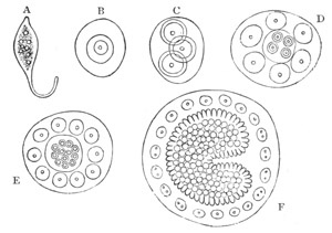
Fig. 190. A series of stages in the development
of Platygaster. (From Lubbock; after Ganin.)
The very first stages are unfortunately but imperfectly known, and the
interpretations offered by Ganin do not in all cases appear quite
satisfactory. In the earliest stage after being laid the egg is
enclosed in a capsule produced into a stalk (fig. 190 A). In the
interior of the egg there soon appears a single spherical body,
regarded by Ganin as a cell (fig. 190 B). In the next stage three
similar bodies appear in the vitellus, no doubt derived from the first
one (fig. 190 C). The central one presents somewhat different
characters to the two others, and, according to Ganin, gives rise to
the whole embryo. The two peripheral bodies increase by
division, and soon appear as nuclei imbedded in a layer of protoplasm
(fig. 190 D, E, F). The layer so formed serves as a covering for the
embryo, regarded by Ganin as equivalent to the amnion (? serous
membrane) of other Insect embryos. In the embryo cell new cells are
stated to be formed by a process of endogenous cell formation (fig.
190 D, E). It appears probable that Ganin has mistaken nuclei for
cells in the earlier stages, and that a blastoderm is formed as in
other Insects, and that this becomes divided in a way not explained
into a superficial layer which gives rise to the serous envelope, and
a deeper layer which forms the embryo. However this may be, a
differentiation into an epiblastic layer of columnar cells and a
hypoblastic layer of more rounded cells soon becomes apparent in the
body of the embryo. Subsequently to this the embryo grows rapidly,
till by a deep transverse constriction on the ventral surface it
becomes divided into an anterior cephalothoracic portion and a
posterior caudal portion (fig. 190 F). The cephalothorax grows in
breadth, and near its anterior end an invagination appears, which
gives rise to the mouth and œsophagus. On the ventral side of the
cephalothorax there is first formed a pair of claw-like appendages on
each side of the mouth, then a posterior pair of appendages near the
junction of the cephalothorax and abdomen, and lastly a pair of short
conical antennæ in front.
At the same time the hind end of the abdomen becomes bifid, and gives
rise to a fork-like caudal appendage; and at a slightly later period
four grooves make their appearance in the caudal region, and divide
this part of the embryo into successive segments. While these changes
have been taking place in the general form of the embryo, the epiblast
has given rise to a cuticle, and the hypoblastic cells have become
differentiated into a central hypoblastic axis—the mesenteron—and a
surrounding layer of mesoblast, some of the cells of which form
longitudinal muscles.
With this stage closes what may be regarded as the embryonic
development of Platygaster. The embryo becomes free from the amnion,
and presents itself as a larva, which from its very remarkable
characters has been spoken of as the Cyclops larva by Ganin.
The larvæ of three species have been described by Ganin, which are
represented in fig. 191 A, B, C. These larvæ are strangely dissimilar
to the ordinary Hexapod type, whether larval or adult. They are formed
of a cephalothoracic shield with the three pairs of appendages
(a, kf, lfg), the development of which has
already been described, and of an abdomen formed of five segments, the
last of which bears the somewhat varying caudal appendages. The
nervous system is as yet undeveloped.
The larvæ move about in the tissues of their hosts by means of their
claws.
The first larval condition is succeeded by a second with very
different characters, and the passage from the first to the second is
accompanied by an ecdysis.
The ecdysis commences at the caudal extremity, and the whole of the
last segment is completely thrown off. As the ecdysis extends forwards
the tail loses its segmentation and becomes strongly compressed, the
appendages of the cephalothorax are thrown off, and the whole embryo
assumes an oval form without any sharp distinction into different
regions and without the slightest indication of segmentation
(fig. 191 D). Of the internal changes which take place during the
shedding of the cuticle, the first is the formation of a proctodæum
(gh) by an invagination, which ends blindly in contact with the
mesenteron. Shortly after this a thickening of the epiblast
(bsm) appears along the ventral surface, which gives rise
mainly to the ventral nerve cord; this thickening is continuous behind
with the epiblast which is invaginated to form the proctodæum,
and in front is prolonged on each side into two procephalic lobes, in
which there are also thickenings of the epiblast (gsae), which
become converted into supra-œsophageal ganglia, and possibly other
parts.
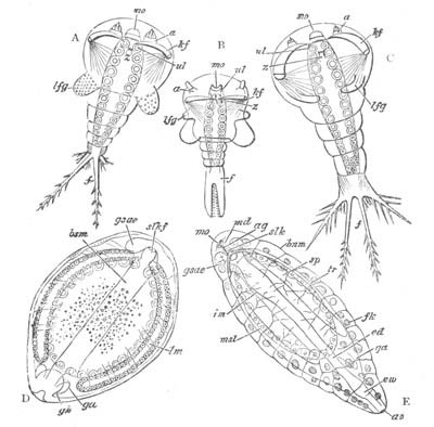
Fig. 191. A series of stages in the development
of Platygaster. (From Lubbock; after Ganin.)
A. B. C. Cyclops larvæ of three species of Platygaster.
D. Second larval stage. E. Third larval stage.
mo. mouth; a. antenna; hf. hooked feet;
lfg. lateral feet; f. branches of tail; ul.
lower lip; slkf. œsophagus; gsae.
supra-œsophageal ganglion; bsm. ventral epiblastic plate;
lm. lateral muscles (the letters also point in D to the
salivary glands); gh. proctodæum; ga. generative
organs; md. mandibles; ag. ducts of salivary glands;
sp. (in E) salivary glands; mls. stomach; ed.
intestine; ew. rectum; ao. anus; tr. tracheæ;
fk. fat body.
Towards the close of the second larval period the muscles (lm)
become segmentally arranged, and give indications of the segmentation
which becomes apparent in the third larval period. The third and last
larval stage (fig. 191 E) of Platygaster, during which it still
remains in the tissues of its host, presents no very peculiar
features. The passage from the second to the third form is accompanied
by an ecdysis.
Remarkable as are the larvæ just described, there can I think be no
reason, considering their parasitic habits, for regarding them as
ancestral.
Metamorphosis and heterogamy.
Metamorphosis. The majority of Insects are born in a condition
in which they obviously differ from their parents. The extent of this
difference is subject to very great variations, but as a rule the
larvæ pass through a very marked metamorphosis before reaching the
adult state. The complete history of this metamorphosis in the
different orders of Insects involves a far too considerable amount of
zoological detail to be dealt with in this work; and I shall confine
myself to a few observations on the general characters and origin of
the metamorphosis, and of the histological processes which take place
during its occurrence[176].
In the Aptera the larva differs from the adult only in the number of
facets in the cornea and joints in the antennæ.
In most Orthoptera and Hemiptera the larvæ differ from the adult in
the absence of wings and in other points. The wings, etc., are
gradually acquired in the course of a series of successive moultings.
In the Ephemeridæ and Libellulidæ, however, the metamorphosis is more
complicated, in that the larvæ have provisional tracheal gills which
are exuviated before the final moult. In the Ephemeridæ there are
usually a great number of moultings; the tracheal gills appear after
the second moult, and the rudiments of the wings when the larva is
about half grown. Larval life may last for a very long period.
In all the other groups of Insects, viz. the Diptera, Neuroptera,
Coleoptera, Lepidoptera, and Hymenoptera, the larva passes—with a few
exceptions—through a quiescent stage, in which it is known as a pupa,
before it attains the adult stage. These forms are known as the
Holometabola.
In the Diptera the larvæ are apodous. In the true flies (Muscidæ) they
are without a distinct head and have the jaws replaced by hooks. In
the Tipulidæ there is on the other hand a well-developed head with the
normal appendages. The pupæ of the Muscidæ are quiescent, and are
enclosed in the skin of the larva which shrinks and forms a firm oval
case. In the Tipulidæ the larval skin is thrown off at the
pupa stage, and in some cases the pupæ continue to move about.
The larvæ of the Neuroptera are hexapodous voracious forms. When the
larva becomes a pupa all the external organs of the imago are already
established. The pupa is often invested in a cocoon. It is usually
quiescent, though sometimes it begins to move about shortly before the
imago emerges.
In the Coleoptera there is considerable variety in the larval forms.
As a rule the larvæ are hexapodous and resemble wingless Insects. But
some herbivorous larvæ (e.g. the larva of Melolontha) closely
resemble true caterpillars, and there are also grub-like larvæ without
feet (Curculio) which resemble the larvæ of Hymenoptera. The pupa is
quiescent, but has all the parts of the future beetle plainly visible.
The most interesting larvæ among the Coleoptera are those of Sitaris,
one of the Meloidæ (Fabre, No. 409). They leave the egg as active
hexapodous larvæ which attach themselves to the bodies of Hymenoptera,
and are thence transported to a cell filled with honey. Here they eat
the ovum of the Hymenopterous form. They then undergo an ecdysis, in
which they functionally lose their appendages, retaining however small
rudiments of them, and become grubs. They feed on the honey and after
a further ecdysis become pupæ.
In the Lepidoptera the larva has the well-known form of a caterpillar.
The caterpillars have strong jaws, adapted for biting vegetable
tissues, which are quite unlike the oral appendages of the adult. They
have three pairs of jointed thoracic legs, and a variable number
(usually five) of pairs of rudimentary abdominal legs—the so-called
pro-legs. The larva undergoes numerous ecdyses, and the external parts
of the adult such as the wings, etc., are formed underneath the
chitinous exoskeleton before the pupa stage. The pupa is known as a
chrysalis and in some Lepidoptera is enveloped in a cocoon.
The Hymenoptera present considerable variations in the character of
the larvæ. In the Aculeata, many Entomophaga, the Cynipidæ, etc., the
larvæ are apodous grubs, incapable of going in search of their food;
but in the Siricidæ they are hexapodous forms like caterpillars, which
are sometimes even provided with pro-legs. In some of the Entomophaga
the larvæ have very remarkable characters which have already been
described in a special section, vide pp. 418, 419.
Before proceeding to the consideration of the value of the various
larval forms thus shortly enumerated, it is necessary to say a few
words as to the internal changes which take place during the
occurrence of the above metamorphosis. In the simplest cases, such as
those of the Orthoptera and Hemiptera, where the metamorphosis is
confined to the gradual formation of the wings, etc. in a series of
moults, the wings first appear as two folds of the epidermis beneath
the cuticle on the two posterior thoracic segments. At the next moult
these processes become covered by the freshly formed cuticle,
and appear as small projections. At every successive moult these
projections become more prominent owing to a growth in the epidermis
which has taken place in the preceding interval. Accompanying the
formation of such organs as the wings, internal changes necessarily
take place in the arrangement of the muscles, etc. of the thorax,
which proceed pari passu with the formation of the organs to
which they belong. The characters of the metamorphosis in such forms
as the Ephemeridæ only differ from the above in the fact that
provisional organs are thrown off at the same time that the new ones
are formed.
In the case of the Holometabola the internal phenomena of the
metamorphosis are of a very much more remarkable character. The
details of our knowledge are largely due to Weismann (Nos. 430 and
431). The larvæ of the Holometabola have for the most part a very
different mode of life to the adults. A simple series of transitions
between the two is impossible, because intermediate forms would be for
the most part incapable of existing. The transition from the larval to
the adult state is therefore necessarily a more or less sudden one,
and takes place during the quiescent pupa condition. Many of the
external adult organs are however formed prior to the pupa stage, but
do not become visible on the surface. The simplest mode of
Holometabolic metamorphosis may be illustrated by the development of
Corethra plumicornis, one of the Tipulidæ. This larva, like that of
other Tipulidæ, is without thoracic appendages, but before the last
larval moult, and therefore shortly before the pupa stage, certain
structures are formed, which Weismann has called imaginal
discs. These imaginal discs are in Corethra simply invaginations
of the epidermis. There are in the thorax six pairs of such
structures, three dorsal and three ventral. The three ventral are
attached to the terminations of the sensory nerves, and the limbs of
the imago are formed as simple outgrowths of them, which as they grow
in length take a spiral form. In the interior of these outgrowths are
formed the muscles, tracheæ, etc., of the limbs; which are believed by
Weismann (it appears to me without sufficient ground) to be derived
from a proliferation of the cells of the neurilemma. The wings are
formed from the two posterior dorsal imaginal discs. The
hypodermis of the larva passes directly into that of the imago.
The pupa stage of Corethra is relatively very short, and the changes
in the internal parts which take place during it are not considerable.
The larval abdominal muscles pass for the most part unchanged into
those of the imago, while the special thoracic muscles connected with
the wings, etc., develop directly during the latest larval period from
cords of cells already formed in the embryo.
In the Lepidoptera the changes in the passage from the larval to the
adult state are not very much more considerable than those in
Corethra. Similar imaginal discs give rise during the later larval
periods to the wings, etc. The internal changes during the longer pupa
period are somewhat more considerable. Important modifications and new
formations arise in connection with the alimentary tract, the nervous
and muscular systems.
The changes which take place in the true flies (Muscidæ) are far more
complicated than either those in Corethra or in the Lepidoptera. The
abdomen of the larva of Musca becomes bodily converted into the
abdomen of the imago as in the above types, but the whole epidermis
and appendages of the head and thorax are derived from imaginal discs
which are formed within and (so far as is known) independently of the
epidermis of the larva or embryo. These imaginal discs are simple
masses of apparently indifferent cells, which for the most part appear
at the close of embryonic life, and are attached to nerves or tracheæ.
They grow in size during larval life, but during the relatively long
pupa stage they unite together to give rise to a continuous epidermis,
from which the appendages grow out as processes. The epidermis of the
anterior part of the larva is simply thrown off, and has no share in
forming the epidermis of the adult.
There are a pair of cephalic imaginal discs and six pairs of thoracic
discs. Two pairs, a dorsal and a ventral, give rise to each thoracic
ring, and the appendages attached to it.
Though, as mentioned above, no evidence has yet been produced to shew
that the imaginal discs of Musca are derived from the embryonic
epiblast, yet their mode of growth and eventual fate proves
beyond the shadow of a doubt that they are homologous with the
imaginal discs of Corethra. Their earliest origin is well worth
further investigation.
The metamorphosis of the internal organs is still more striking than
that of the external. There is a disruption, total or partial, of all
the internal organs except the generative organs. In the case of the
alimentary tract, the Malpighian vessels, the heart and the central
nervous system, the disruption is of a partial kind, which has been
called by Weismann histolysis. The cells of these organs undergo a
fatty degeneration, the nuclei alone in some cases remaining. The kind
of plasma resulting from this degeneration retains the shape of the
organs, and finally becomes built up again into the corresponding
organs of the imago. The tracheæ, muscles and peripheral nerves, and
an anterior part of the alimentary tract, are entirely disrupted. They
seem to be formed again from granular cells derived from the enormous
fat body.
The phenomena of the development of the Muscidæ are undoubtedly of
rather a surprising character. Leaving for the moment the question of
the origin of the pupa stage to which I return below, it will be
admitted on all hands that during the pupa stage the larva undergoes a
series of changes which, had they taken place by slow degrees, would
have involved, in such a case as Musca, a complete though gradual
renewal of the tissues. Such being the case, the cells of the organs
common to the larva and the imago would, in the natural course of
things, not be the same cells as those of the larva but descendants of
them. We might therefore expect to find in the rapid conversion of the
larval organs into those of the adult some condensation, so to speak,
of the process of ordinary cell division. Such condensations are
probably represented in the histolysis in the case of the internal
organs, and in the formation of imaginal discs in the case of the
external ones, and I think it probable that further investigation will
shew that the imaginal discs of the Muscidæ are derivatives of the
embryonic epiblast. The above considerations by no means explain the
whole of Weismann’s interesting observations, but an explanation is I
believe to be found by following up these lines.
More or less parallel phenomena to those in Insects are found in the
development of the Platyelminthes and Echinoderms. The four disc-like
invaginations of the skin in many larval Nemertines (vide p.
198), which give rise to the permanent body wall of the Nemertine, may
be compared to the imaginal discs. The subsequent throwing off of the
skin of Pilidium or larva of Desor is a phenomenon like the absorption
of part of the larval skin of Musca. The formation of an independent
skin within the first larval form in the Distomeæ and in the
Cestoda may be compared to the apparently independent formation of the
imaginal discs in Musca.
The fact that in a majority of instances it is possible to trace an
intimate connection between the surroundings of a larva and its
organization proves in the clearest way that the characters of the
majority of existing larval forms of Insects have owed their origin to
secondary adaptations. A few instances will illustrate this point.
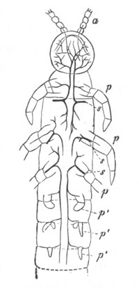
Fig. 192. Anterior half of Campodea
Fragilis. (From Gegenbaur; after Palmen.)
a. antennæ; p. feet; p´. post-thoracic
rudimentary feet; s. stigma.
In the simplest types of metamorphosis, e.g. those of the
Orthoptera genuina, the larva has precisely the same habits as the
adult. We find that a caterpillar form is assumed by phytophagous
larvæ amongst the Lepidoptera, Hymenoptera and Coleoptera. Where the
larva has not to go in search of its nutriment the grub-like apodous
form is assumed. The existence of such an apodous larva is especially
striking in the Hymenoptera, in that rudiments of thoracic and
abdominal appendages are present in the embryo and disappear again in
the larva. The case of the larva of Sitaris, already described (p.
421), affords another very striking proof that the organization of the
larva is adapted to its habits.
It follows from the above that the development of such forms as the
Orthoptera genuina is more primitive than that of the holometabolous
forms; a conclusion which tallies with the fact that both
palæontological and anatomical evidence shew the Orthoptera to be a
very primitive group of Insects.
The above argument probably applies with still greater force to the
case of the Thysanura; and it seems to be probable that this group is
more nearly related than any other to the primitive wingless ancestors
of Insects[177]. The characters of the oral appendages in this
group, the simplicity of their metamorphosis, and the presence of
abdominal appendages (fig. 192), all tell in favour of this view,
while the resemblance of the adult to the larvæ of the
Pseudoneuroptera, etc., points in the same direction. The Thysanura
and Collembola are not however to be regarded as belonging to the true
stock of the ancestors of Insects, but as degenerated relations of
this stock; much as Amphioxus and the Ascidians are degenerate
relations of the ancestral stock of Vertebrates, and Peripatus of that
of the Tracheata. It is probable that all these forms have succeeded
in retaining their primitive characters from their degenerate habits,
which prevented them from entering into competition in the struggle
for existence with their more highly endowed relatives. While in a
general way it is clear that the larval forms of Insects cannot be
expected to throw much light on the nature of Insect ancestors, it
does nevertheless appear to me probable that such forms as the
caterpillars of the Lepidoptera are not without a meaning in this
respect. It is easy to conceive that even a secondary larval form may
have been produced by the prolongation of one of the embryonic stages;
and the general similarity of a caterpillar to Peripatus, and the
retention by it of post-thoracic appendages, are facts which appear to
favour this view of the origin of the caterpillar form.
The two most obscure points which still remain to be dealt with in the
metamorphosis of Insects are (1) the origin of the quiescent pupa
stage; (2) the frequent dissimilarity between the masticatory
apparatus of the larva and adult.
These two points may be conveniently dealt with together, and some
valuable remarks about them will be found in Lubbock (No. 420).
On grounds already indicated it may be considered certain that the
groups of Insects without a pupa stage, and with a larva very
similarly organised to the adult, preceded the existing holometabolic
groups. The starting point in the metamorphosis of the latter groups
was therefore something like that of the Orthoptera. Suppose it became
an advantage to a species that the larva and adult should feed in a
somewhat different way, a difference in the character of their mouth
parts would soon make itself manifest; and, since an intermediate type
of mouth parts would probably be disadvantageous, there would
be a tendency to concentrate into a single moult the transition from
the larval to the adult form of mouth parts. At each ordinary moult
there is a short period of quiescence, and this period of quiescence
would naturally become longer in the important moult at which the
change in the mouth parts was effected. In this way a rudimentary pupa
stage might be started. The pupa stage, once started, might easily
become a more important factor in the metamorphosis. If the larva and
imago diverged still more from each other, a continually increasing
amount of change would have to be effected at the pupa stage. It would
probably be advantageous to the species that the larva should not have
rudimentary functionless wings; and the establishment of the wings as
external organs would therefore become deferred to the pupa stage. The
same would probably apply to other organs.
Insects usually pass through the pupa stage in winter in cold climates
and during the dry season in the tropics, this stage serving therefore
apparently for the protection of the species during the inclement
season of the year. These facts are easily explained on the
supposition that the pupa stage has become secondarily adapted to play
a part in the economy of the species quite different from that to
which it owes its origin.
Heterogamy. The cases of alternations of generations amongst
Insects all fall under the heading already defined in the introduction
as Heterogamy. Heterogamy amongst Insects has been rendered possible
by the existence of parthenogenesis, which, as stated in the
introduction, has been taken hold of by natural selection, and has led
to the production of generations of parthenogenetic forms, by which a
clear economy in reproduction is effected. Parthenogenesis without
heterogamy occurs in a large number of forms. In Bees, Wasps, and a
Sawfly (Nematus ventricosus) the unfertilized ova give rise to males.
In two Lepidopterous genera (Psyche and Solenobia) the unfertilized
ova give rise mainly, if not entirely, to females. Heterogamy occurs
in none of the above types, but in Psyche and Solenobia males are only
occasionally found, so that a series of generations producing female
young from unfertilized ova are followed by a generation producing
young of both sexes from fertilized ova. It would be
interesting to know if the unimpregnated female would not after a
certain number of generations give rise to both males and females;
such an occurrence might be anticipated on grounds of analogy. In the
cases of true heterogamy parthenogenesis has become confined to
special generations, which differ in their character from the
generations which reproduce themselves sexually. The parthenogenetic
generations generally flourish during the season when food is
abundant; while the sexual generations occur at intervals which are
often secondarily regulated by the season, supply of food, etc.
A very simple case of this kind occurs, if we may trust the recent
researches of Lichtenstein[178], in certain Gall Insects (Cynipidæ).
He finds that the female of a form known as Spathegaster baccarum, of
which both males and females are plentiful, pricks a characteristic
gall in certain leaves, in which she deposits the fertilized eggs. The
eggs from these galls give rise to a winged and apparently adult form,
which is not, however, Spathegaster, but is a species considered to
belong to a distinct genus known as Neuroterus ventricularis. Only
females of Neuroterus are found, and they lay unfertilized ova in
peculiar galls which develop into Spathegaster baccarum. Here we have
a true case of heterogamy, the females which produce
parthenogenetically having become differentiated from those which
produce sexually. Another interesting type of heterogamy is that which
has been long known in the Aphides. In the autumn impregnated eggs are
deposited by females, which give rise in the course of the spring to
females which produce parthenogenetically and viviparously. The
viviparous females always differ from the females which lay the
fertilized eggs. The generative organs are of course differently
constituted, and the ova of the viviparous females are much smaller
than those of the oviparous females, as is generally the case in
closely allied viviparous and oviparous forms; but in addition the
former are usually without wings, while the latter are winged. The
reverse is however occasionally the case. An indefinite number of
generations of viviparous females may be produced if they are
artificially kept warm and supplied with food; but in the course of
nature the viviparous females produce in the autumn males and
females which lay eggs with firm shells, and so preserve the species
through the winter. The heterogamy of the allied Coccidæ is
practically the same as that of the Aphidæ. In the case of Chermes and
Phylloxera the parthenogenetic generations lay their eggs in the
normal way.
The complete history of Phylloxera quercus has been worked out by
Balbiani (No. 401). The apterous females during the summer lay eggs
developing parthenogenetically into apterous females, which continue
the same mode of reproduction. In the autumn, however, the eggs which
are laid give rise in part to winged forms and in part to apterous
forms. Both of these forms lay smaller and larger eggs, which develop
respectively into very minute males and females without digestive
organs. The fertilized eggs laid by these forms probably give rise to
the parthenogenetic females.
A remarkable case of heterogamy accompanied by pædogenesis was
discovered by Wagner to take place in certain species of Cecydomyia
(Miastor), a genus of the Diptera. The female lays a few eggs in the
bark of trees, etc. These eggs develop in the winter into larvæ, in
which ovaries are early formed. The ova become detached into the body
cavity, surrounded by their follicles, and grow at the cost of the
follicles. They soon commence to undergo a true development, and on
becoming hatched they remain for some time in the body cavity of the
parent, and are nourished at the expense of its viscera. They finally
leave the empty skin of their parent, and subsequently reproduce a
fresh batch of larvæ in the same way. After several generations the
larvæ undergo in the following summer a metamorphosis, and develop
into the sexual form.
Another case of pædogenesis is that of the larvæ of Chironomus, which
have been shewn by Grimm (No. 413) to lay eggs which develop exactly
in the same way as fertilized eggs into larvæ.
Bibliography.
(401) M. Balbiani. “Observations s. la reproduction d.
Phylloxera du Chêne.” An. Sc. Nat. Ser. V. Vol. XIX.
1874.
(402) E. Bessels. “Studien ü. d. Entwicklung d. Sexualdrüsen
bei den Lepidoptera.” Zeit. f. wiss. Zool. Bd. XVII.
1867.
(403) Alex. Brandt. “Beiträge zur Entwicklungsgeschichte d.
Libellulida u. Hemiptera, mit besonderer Berücksichtigung d.
Embryonalhüllen derselben.” Mém. Ac. Pétersbourg, Ser.
VII. Vol. XIII. 1869.
(404) Alex. Brandt. Ueber das Ei u. seine
Bildungsstätte. Leipzig, 1878.
(405) O. Bütschli. “Zur Entwicklungsgeschichte d. Biene.”
Zeit. f. wiss. Zool. Bd. XX. 1870.
(406) H. Dewitz. “Bau u. Entwicklung d. Stachels, etc.”
Zeit. f. wiss. Zool. Vols. XXV. and XXVIII.
1875 and 1877.
(407) H. Dewitz. “Beiträge zur Kenntniss d.
Postembryonalentwicklung d. Gliedmassen bei den Insecten.” Zeit. f.
wiss. Zool. XXX. Supplement. 1878.
(408) A. Dohrn. “Notizen zur Kenntniss d. Insectenentwicklung.”
Zeitschrift f. wiss. Zool. Bd. XXVI. 1876.
(409) M. Fabre. “l’hypermétamorphose et les mœurs des
Méloïdes.” An. Sci. Nat. Series IV. Vol. VII.
1857.
(410) Ganin. “Beiträge zur Erkenntniss d.
Entwicklungsgeschichte d. Insecten.” Zeit. f. wiss. Zool. Bd.
XIX. 1869.
(411) V. Graber. Die Insecten. München, 1877.
(412) V. Graber. “Vorlauf. Ergeb. üb. vergl. Embryologie d.
Insecten.” Archiv f. mikr. Anat. Vol. XV. 1878.
(413) O. v. Grimm. “Ungeschlechtliche Fortpflanzung einer
Chironomus-Art u. deren Entwicklung aus dem unbefruchteten Ei.”
Mém. Acad. Pétersbourg. 1870.
(414) B. Hatschek. “Beiträge zur Entwicklung d. Lepidopteren.”
Jenaische Zeitschrift, Bd. XI.
(415) A. Kölliker. “Observationes de primâ insectorum genese,
etc.” Ann. Sc. Nat. Vol. XX. 1843.
(416) A. Kowalevsky. “Embryologische Studien an Würmern u.
Arthropoden.” Mém. Ac. imp. Pétersbourg, Ser. VII.
Vol. XVI. 1871.
(417) C. Kraepelin. “Untersuchungen üb. d. Bau, Mechanismus u.
d. Entwick, des Stachels d. bienartigen Thiere.” Zeit. f. wiss.
Zool. Vol. XXIII. 1873.
(418) C. Kupffer. “Faltenblatt an d. Embryonen d. Gattung
Chironomus.” Arch. f. mikr. Anat. Vol. II. 1866.
(419) R. Leuckart. Zur Kenntniss d. Generationswechsels u.
d. Parthenogenese b. d. Insecten. Frankfurt, 1858.
(420) Lubbock. Origin and Metamorphosis of Insects.
1874.
(421) Lubbock. Monograph on Collembola and Thysanura.
Ray Society, 1873.
(422) Melnikow. “Beiträge z. Embryonalentwicklung d. Insecten.”
Archiv f. Naturgeschichte, Bd. XXXV. 1869.
(423) E. Metschnikoff. “Embryologische Studien an Insecten.”
Zeit. f. wiss. Zool. Bd. XVI. 1866.
(424) P. Meyer. “Ontogenie und Phylogenie d. Insecten.”
Jenaische Zeitschrift, Vol. X. 1876.
(425) Fritz Müller. “Beiträge z. Kenntniss d. Termiten.”
Jenaische Zeitschrift, Vol. IX. 1875.
(426) A. S. Packard. “Embryological Studies on Diplex,
Perithemis, and the Thysanurous genus Isotoma.” Mem. Peabody Acad.
Science, 1. 2. 1871.
(427) Suckow. “Geschlechtsorgane d. Insecten.” Heusinger’s
Zeitschrift f. organ. Physik, Bd. II. 1828.
(428) Tichomiroff. “Ueber die Entwicklungsgeschichte des
Seidenwurms.” Zoologischer Anzeiger, II. Jahr. No. 20
(Preliminary Notice).
(429) Aug. Weismann. “Zur Embryologie d. Insecten.” Archiv
f. Anat. und Phys. 1864.
(430) Aug. Weismann. “Entwicklung d. Dipteren.” Zeit. f.
wiss. Zool. Vols. XIII. and XIV. Leipzig,
1863‑4.
(431) Aug. Weismann. “Die Metamorphose d. Corethra
plumicornis.” Zeit. f. wiss. Zool. Vol. XVI. 1866.
(432) N. Wagner. “Beitrag z. Lehre d. Fortpflanzung d.
Insectenlarven.” Zeit. f. wiss. Zool. Vol. XIII. 1860.
(433) Zaddach. Untersuchungen üb. d. Bau u. d. Entwicklung
d. Gliederthiere. Berlin, 1854.
Arachnida[179].
The development of several divisions of this interesting group has
been worked out; and it will be convenient to deal in the first
instance with the special history of each of these divisions, and then
to treat in a separate section the development of the organs for the
whole group.
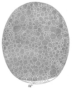
Fig. 193. Ovum of Scorpion with the already
formed blastoderm shewing the partial segmentation. (After
Metschnikoff.)
bl. blastoderm.
Scorpionidæ. The embryonic development always takes place
within the female Scorpion. In Buthus it takes place within
follicle-like protuberances of the wall of the ovary. In Scorpio also
development commences while the egg is still in the follicle, but when
the trunk becomes segmented the embryo passes into the ovarian tube.
The chief authority for the development of the Scorpionidæ is
Metschnikoff (No. 434).
At the pole of the ovum facing the ovarian tube there is
formed a germinal disc which undergoes a partial segmentation (fig.
193 bl). A somewhat saucer-shaped one-layered blastoderm is
then formed, which soon becomes thickened in the centre and then
divided into two layers. The outer of these is the epiblast, the inner
the mesoblast. Beneath the mesoblast there subsequently appear
granular cells, which form the commencement of the hypoblast[180].
During the formation of the blastoderm a cellular envelope is formed
round the embryo. Its origin is doubtful, though it is regarded by
Metschnikoff as probably derived from the blastoderm and homologous
with the amnion of Insects. It becomes double in the later stages
(fig. 195).
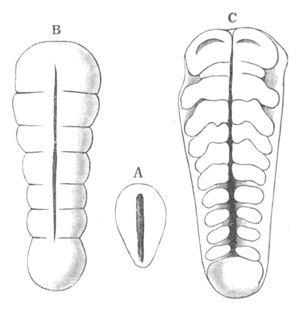
Fig. 194. Three surface views of the ventral
plate of a developing Scorpion. (After Metschnikoff.)
A. Before segmentation.
B. After five segments have become formed.
C. After the appendages have begun to be formed.
During the differentiation of the three embryonic layers the germinal
disc becomes somewhat pyriform, the pointed end being the posterior.
At this extremity there is a special thickening which is perhaps
equivalent to the primitive cumulus of Spiders. The germinal disc
continues gradually to spread over the yolk, but the original pyriform
area is thicker than the remainder, and is marked off anteriorly and
posteriorly by a shallow furrow. It constitutes a structure
corresponding with the ventral plate of other Tracheata. It soon
becomes grooved by a shallow longitudinal furrow (fig. 194 A) which
subsequently becomes less distinct. It is then divided by two
transverse lines into three parts.[181]
In succeeding stages the anterior of the three parts is clearly marked
out as the procephalic lobe, and soon becomes somewhat broader. Fresh
segments are added from before backwards, and the whole ventral plate
increases rapidly in length (fig. 194 B).
When ten segments have become formed, appendages appear as paired
outgrowths of the nine posterior segments (fig. 194 C). The second
segment bears the pedipalpi, the four succeeding segments the four
ambulatory appendages, and the four hindermost segments smaller
provisional appendages which subsequently disappear, with the possible
exception of the second. The foremost segment, immediately behind the
procephalic lobes, is very small, and still without a rudiment of the
cheliceræ, which are subsequently formed on it. It would appear from
Metschnikoff’s figures to be developed later than the other post-oral
segments present at this stage. The still unsegmented tail has become
very prominent and makes an angle of 180° with the remainder of the
body, over the ventral surface of which it is flexed.
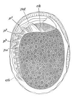
Fig. 195. A fairly advanced embryo of the
Scorpion enveloped in its membranes. (After Metschnikoff.)
ch. cheliceræ; pd. pedipalpi; p1-p4.
ambulatory appendages; ab. post-abdomen (tail).
By the time that twelve segments are definitely formed, the
procephalic region is distinctly bilobed, and in the median groove
extending along it the stomodæum has become formed (fig. 196 A). The
cheliceræ (ch) appear as small rudiments on the first post-oral
segment, and the nerve cords are distinctly differentiated and
ganglionated. In the embryonic state there is one ganglion for each
segment. The ganglion in the first segment (that bearing the
cheliceræ) is very small, but is undoubtedly post-oral.
At this stage, by a growth in which all the three germinal layers have
a share, the yolk is completely closed in by the blastoderm. It is a
remarkable fact with only few parallels, and those amongst the
Arthropoda, that the blastopore, or point where the embryonic
membranes meet in closing in the yolk, is situated on the dorsal
surface of the embryo.
The general relations of the embryo at about this stage are shewn in
fig. 195, where the embryo enclosed in its double cellular membrane is
seen in a side view. This embryo is about the same age as that seen
from the ventral surface in fig. 196 A.
The general nature of the further changes may easily be gathered from
an inspection of fig. 196 B and C, but a few points may be noted.
An upper lip or labrum is formed as an unpaired organ in the line
between the procephalic lobes. The pedipalpi become chelate before
becoming jointed, and the cheliceræ also early acquire their
characteristic form. Rudimentary appendages appear on the six segments
behind the ambulatory legs, five of which are distinctly shewn in fig.
195; they persist only on the second segment, where they appear to
form the comb-like organs or pectines. The last abdominal segment,
i.e. that next the tail, is without provisional appendages. The
embryonic tail is divided into six segments including the telson (fig.
196 C, ab). The lungs (st) are formed by paired
invaginations, the walls of which subsequently become plicated, on the
four last segments which bear rudimentary limbs, and simultaneously
with the disappearance of the rudimentary limbs.
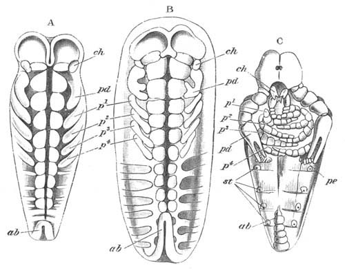
Fig. 196. Three stages in the development of the
Scorpion. The embryos are represented as if seen extended on a
plane.
(After Metschnikoff.)
ch. cheliceræ; pd. pedipalpi; p1-p4. ambulatory
appendages; pe. pecten; st. stigmata; ab. post
abdomen (tail).
Pseudoscorpionidæ. The development of Chelifer has been
investigated by Metschnikoff (436), and although (except that it is
provided with tracheæ instead of pulmonary sacks) it might be supposed
to be closely related to Scorpio, yet in its development is strikingly
different.
The eggs after being laid are carried by the female attached to the
first segment of the abdomen. The segmentation (vide p. 93) is
intermediate between the types of complete and superficial
segmentation. The ovum, mainly formed of food-yolk, divides into two,
four, and eight equal segments (fig. 197 A). There then appear
one or more clear segments on the surface of these, and finally a
complete layer of cells is formed round the central yolk spheres (fig.
197 B), which latter subsequently agglomerate into a central mass. The
superficial cells form what may be called a blastoderm, which soon
becomes divided into two layers (fig. 197 C). There now appears a
single pair of appendages (the pedipalpi) (fig. 198 A, pd),
while at the same time the front end of the embryo grows out into a
remarkable proboscis-like prominence—a temporary upper lip (concealed
in the figure behind the pedipalpus), and the abdomen (ab)
becomes bent forwards towards the ventral surface. In this very
rudimentary condition, after undergoing an ecdysis, the larva is
hatched, although it still remains attached to its parent. After
hatching it grows rapidly, and becomes filled with a peculiar
transparent material. The first pair of ambulatory appendages is
formed behind the pedipalpi and then the three succeeding pairs, while
at the same time the cheliceræ appear as small rudiments in front.
External signs of segmentation have not yet appeared, but about this
period the nervous system is formed. The supra-œsophageal ganglia
are especially distinct, and provided with a central cavity, probably
formed by an invagination, as in other Arachnida. In the succeeding
stages (fig. 198 B) four provisional pairs of appendages
(shewn as small knobs at ab) appear behind the ambulatory feet.
The abdomen is bent forwards so as to reach almost to the pedipalpi.
In the later stages (fig. 198 C) the adult form is gradually attained.
The enormous upper lip persists for some time, but subsequently
atrophies and is replaced by a normal labrum. The appendages behind
the ambulatory feet atrophy, and the tail is gradually bent back into
its final position. The segmentation and the gradual growth of the
limbs do not call for special description, and the formation of the
organs, so far as is known, agrees with other types.
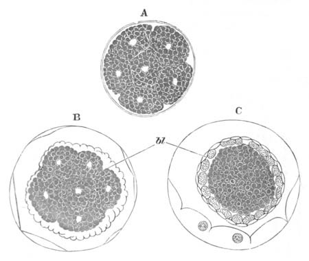
Fig. 197. Segmentation and formation of the
blastoderm in Chelifer.
(After Metschnikoff.)
In A the ovum is divided into a number of separate segments. In B a
number of small cells have appeared (bl) which form a
blastoderm enveloping the large yolk spheres. In C the blastoderm
has become divided into two layers.
The segmentation of Chthonius is apparently similar to that of
Chelifer (Stecker, No. 437).
Phalangidæ. Our knowledge of the development of Phalangium is
unfortunately confined to the later stages (Balbiani, No. 438). These
stages do not appear however to differ very greatly from those of true
Spiders.
Araneina. The eggs of true Spiders are either deposited in
nests made specially for them, or are carried about by the females.
Species belonging to a considerable number of genera, viz. Pholcus,
Epeira, Lycosa, Clubione, Tegenaria and Agelena have been
studied by Claparède (No. 442), Balbiani (No. 439), Barrois (No. 441)
and myself (No. 440), and the close similarity between their embryos
leaves but little doubt that there are no great variations in
development within the group.
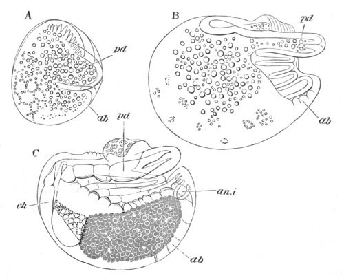
Fig. 198. Three stages in the development of
chelifer.
(After Metschnikoff.)
pd. pedipalpi; ab. abdomen; an.i. anal
invagination; ch. cheliceræ.
The ovum is enclosed in a delicate vitelline membrane, enveloped in
its turn by a chorion secreted by the walls of the oviduct. The
chorion is covered by numerous rounded prominences, and occasionally
exhibits a pattern corresponding with the areas of the cells which
formed it. The segmentation has already been fully described, pp. 118
and 119. At its close there is present an enveloping blastoderm formed
of a single layer of large flattened cells. The yolk within is formed
of a number of large polygonal segments; each of which is composed of
large yolk-spherules, and contains a nucleus surrounded by a layer of
protoplasm, which is prolonged into stellate processes holding
together the yolk-spherules. The nucleus, surrounded by the major part
of the protoplasm of each yolk cell, appears, as a rule, to be
situated not at the centre, but on one side of its yolk segment.
The further description of the development of Spiders applies more
especially to Agelena labyrinthica, the species which formed the
subject of my own investigations.
The first differentiation of the blastoderm consists in the cells of
nearly the whole of one hemisphere becoming somewhat more columnar
than those of the other hemisphere, and in the cells of a small area
near one end of the thickened hemisphere becoming distinctly more
columnar than elsewhere, and two layers thick. This area forms a
protuberance on the surface of the ovum, originally discovered by
Claparède, and called by him the primitive cumulus. In the next stage
the cells of the thickened hemisphere of the blastoderm become still
more columnar; and a second area, at first connected by a whitish
streak with the cumulus, makes its appearance. In the second area the
blastoderm is also more than one cell deep (fig. 199). It will be
noticed that the blastoderm, though more than one cell thick over a
large part of the ventral surface, is not divided into distinct
layers. The second area appears as a white patch and soon becomes more
distinct, while the streak continued to it from the cumulus is no
longer visible. It is shewn in surface view in fig. 200 A. Though my
observations on this stage are not quite satisfactory, yet it appears
to me probable that there is a longitudinal thickened ridge of the
blastoderm extending from the primitive cumulus to the large white
area. The section represented in fig. 199, which I believe to be
oblique, passes through this ridge at its most projecting part.
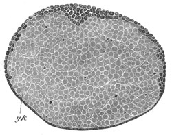
Fig. 199. Section through the embryo of Agelena
labyrinthica.
The section is from an embryo of the same age as fig. 200 A, and is
represented with the ventral plate upwards. In the ventral plate is
seen a keel-like thickening, which gives rise to the main mass of
the mesoblast.
yk. yolk divided into large polygonal cells, in several of
which nuclei are shewn.
The nuclei of the yolk cells during the above stages multiply rapidly,
and cells are formed in the yolk which join the blastoderm; there can
however be no doubt that the main increase in the cells of the
blastoderm has been due to the division of the original blastoderm
cells.
In the next stage I have been able to observe there is, in the place
of the previous thickened half of the blastoderm, a well-developed
ventral plate with a procephalic lobe in front, a caudal lobe behind,
and an intermediate region marked by about three transverse grooves,
indicating a division into segments. This plate is throughout two or
more rows of cells thick, and the cells which form it are
divided into two distinct layers—a columnar superficial layer
of epiblast cells, and a deeper layer of mesoblast cells (fig. 203 A).
In the latter layer there are several very large cells which are in
the act of passing from the yolk into the blastoderm. The
identification of the structures visible in the previous stage with
those visible in the present stage is to a great extent a matter of
guess-work, but it appears to me probable that the primitive cumulus
is still present as a slight prominence visible in surface views on
the caudal lobe, and that the other thickened patch persists as the
procephalic lobe. However this may be, the significance of the
primitive cumulus appears to be that it is the part of the blastoderm
where two rows of cells become first established[182].
The whole region of the blastoderm other than the ventral plate is
formed of a single row of flattened epiblast cells. The yolk retains
its original constitution.
By this stage the epiblast and mesoblast are distinctly
differentiated, and the homologue of the hypoblast is to be sought for
in the yolk cells. The yolk cells are not however entirely
hypoblastic, since they continue for the greater part of the
development to give rise to fresh cells which join the mesoblast.
The Spider’s blastoderm now resembles that of an Insect (except for
the amnion) after the establishment of the mesoblast, and the mode of
origin of the mesoblast in both groups is very similar, in that the
longitudinal ridge-like thickening of the mesoblast shewn in
fig. 199 is probably the homologue of the mesoblastic groove of the
Insects’ blastoderm.
The ventral plate continues to grow rapidly, and at a somewhat later
stage (fig. 200 B) there are six segments interposed between the
procephalic and caudal lobes. The two anterior of these (ch and
pd), especially the foremost, are less distinct than the
remainder; and it is probable that both of them, and in any case the
anterior one, are formed later than the three segments following.
These two segments are the segments of the cheliceræ and pedipalpi.
The four segments following belong to the four pairs of ambulatory
legs. The segments form raised transverse bands separated by
transverse grooves. There is at this stage a faintly marked groove
extending along the median line of the ventral plate. This groove is
mainly caused by the originally single mesoblastic plate having become
divided throughout the whole region of the ventral plate, except
possibly the procephalic lobes, into two bands, one on each side of
the middle line (fig. 203 B).
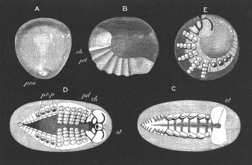
Fig. 200. Four stages in the development of
Agelena labyrinthica.
A. Stage when the ventral plate is very imperfectly differentiated.
pr.c. primitive cumulus.
B. Ovum viewed from the side when the ventral plate has become
divided into six segments. ch. segment of cheliceræ
imperfectly separated from procephalic lobe; pd. segment of
pedipalpi.
C. Ventral plate ideally unrolled after the full number of segments
and appendages are established. st. stomodæum between the two
præ-oral lobes. Behind the six pairs of permanent appendages are
seen four pairs of provisional appendages.
D and E. Two views of an embryo at the same stage. D ideally
unrolled, E seen from the side. st. stomodæum; ch.
cheliceræ; on their inner side is seen the ganglion belonging to
them. pd. pedipalpi; pr.p. provisional appendages.
The segments continue to increase in number by the continuous addition
of fresh segments between the one last formed and the caudal lobe. By
the stage with nine segments the first rudiments of the limbs make
their appearance. The first rudiments to appear are those of the
pedipalpi and four ambulatory limbs: the cheliceræ, like the segment
to which they belong, lag behind in development. The limbs appear as
small protuberances at the borders of their segments. By the stage
when they are formed the procephalic region has become bilobed, and
the two lobes of which it is composed are separated by a shallow
groove.
By a continuous elongation the ventral plate comes to form a nearly
complete equatorial ring round the ovum, the procephalic and caudal
lobes being only separated by a very narrow space, the undeveloped
dorsal region of the embryo. This is shewn in longitudinal section in
fig. 204. In this condition the embryo may be spoken of as having a
dorsal flexure. By the time that this stage is reached (fig.
200 C) the full number of segments and appendages has become
established. There are in all sixteen segments (including the caudal
lobe). The first six of these bear the permanent appendages of the
adult; the next four are provided with provisional appendages;
while the last six are without appendages. The further features of
this stage which deserve notice are (1) the appearance of a shallow
depression (st)—the rudiment of the stomodæum—between the
hinder part of the two procephalic lobes; (2) the appearance of raised
areas on the inner side of the six anterior appendage-bearing
segments. These are the rudiments of the ventral ganglia. It deserves
to be especially noted that the segment of the cheliceræ, like
the succeeding segments, is provided with ganglia; and that the
ganglia of the cheliceræ are quite distinct from the
supra-œsophageal ganglia derived from the procephalic lobes. (3)
The pointed form of the caudal lobe. In Pholcus (Claparède, No. 442)
the caudal lobe forms a projecting structure which, like the caudal
lobe of the Scorpion, bends forward so as to face the ventral surface
of the part of the body immediately in front. In most Spiders such a
projecting caudal lobe is not found. While the embryo still retains
its dorsal flexure considerable changes are effected in its general
constitution. The appendages (fig. 200 D and E) become imperfectly
jointed, and grow inwards so as to approach each other in the middle
line. Even in the stage before this, the ventral integument between
the rudiments of the ganglia had become very much thinner, and had in
this way divided the ventral plate into two halves. At the present
stage the two halves of the ventral plate are still further separated,
and there is a wide space on the ventral side only covered by a
delicate layer of epiblast. This is shewn in surface view (fig. 200 D)
and in section in fig. 203 C.
The stomodæum (st) is much more conspicuous, and is bounded in
front by a prominent upper lip, and by a less marked lip behind. The
upper lip becomes less conspicuous in later stages, and is perhaps to
be compared with the provisional upper lip of Chelifer. Each
procephalic lobe is now marked by a deep semicircular groove.
The next period in the development is characterised by the gradual
change in the flexure of the embryo from a dorsal to a ventral one;
accompanied by the division of the body into an abdomen and
cephalo-thorax, and the gradual assumption of the adult characters.
The change in the flexure of the embryo is caused by the elongation of
the dorsal region, which has hitherto been hardly developed. Such an
elongation increases the space on the dorsal surface between the
procephalic and caudal regions, and therefore necessarily separates
the caudal and procephalic lobes; but, since the ventral plate does
not become shortened in the process, and the embryo cannot straighten
itself in the egg-shell, it necessarily becomes ventrally flexed.
If there were but little food-yolk this flexure would naturally
cause the whole embryo to be bent in so as to have the ventral
surface concave. But instead of this the flexure is at first confined
to the two bands which form the ventral plate. These bands, as shewn
in fig. 201 A, acquire a true ventral flexure, but the yolk forms a
projection—a kind of yolk-sack as Barrois (No. 441) calls
it—distending the thin integument between the two ventral bands. This
yolk-sack is shewn in surface view in fig. 201 A and in section in
fig. 206. At a later period, when the yolk has become largely
absorbed, the true nature of the ventral flexure becomes quite
obvious, since the abdomen of the young Spider, while still in the
egg, is found to be bent over so as to press against the ventral
surface of the thorax (fig. 201 B).
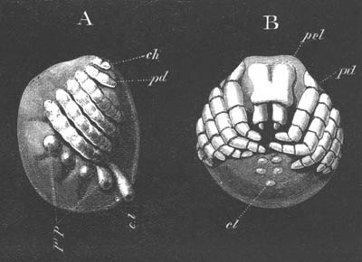
Fig. 201. Two late stages in the development of
Agelena labyrinthica.
A. Embryo from the side at the stage when there is a large ventral
protuberance of yolk. The angle between the line of insertion of the
permanent and provisional appendages shews the extent of the ventral
flexure.
B. Embryo nearly ready to be hatched. The abdomen which has not
quite acquired its permanent form is seen to be pressed against the
ventral side of the thorax.
pr.l. procephalic lobe; pd. pedipalpi; ch.
cheliceræ; c.l. caudal lobe; pr.p. provisional
appendages.
The general character of the changes which take place during this
period in the development is shewn in fig. 201 A and B representing
two stages in it. In the first of these stages there is no
constriction between the future thorax and abdomen. The four
pairs of provisional appendages exhibit no signs of atrophy; and the
extent of the ventral flexure is shewn by the angle formed between the
line of their insertion and that of the appendages in front. The yolk
has enormously distended the integument between the two halves of the
ventral plate, as is illustrated by the fact that, at a somewhat
earlier stage than that figured, the limbs cross each other in the
median ventral line, while at this stage they do not nearly meet. The
limbs have acquired their full complement of joints, and the pedipalpi
bear a cutting blade on their basal joint.
The dorsal surface between the prominent caudal lobe and the
procephalic lobes forms more than a semicircle. The terga are fully
established, and the boundaries between them, especially in the
abdomen, are indicated by transverse markings. A large lower lip now
bounds the stomodæum, and the upper lip has somewhat atrophied. In the
later stage (fig. 201 B) the greater part of the yolk has passed into
the abdomen, which is now to some extent constricted off from the
cephalo-thorax. The appendages of the four anterior abdominal somites
have disappeared, and the caudal lobe has become very small. In front
of it are placed two pairs of spinning mammillæ. A delicate cuticle
has become established, which is very soon moulted.
Acarina. The development of the Acarina, which has been mainly
investigated by Claparède (No. 446), is chiefly remarkable from the
frequent occurrence of several larval forms following each other after
successive ecdyses. The segmentation (vide p. 116) ends in the
formation of a blastoderm of a single layer of cells enclosing a
central yolk mass.
A ventral plate is soon formed as a thickening of the blastoderm, in
which an indistinct segmentation becomes early observable. In Myobia,
which is parasitic on the common mouse, the ventral plate becomes
divided by five constrictions into six segments (fig. 202 A), from the
five anterior of which paired appendages very soon grow out (fig. 202
B). The appendages are the cheliceræ (ch) and pedipalpi
(pd) and the first three pairs of limbs (p1-p3). On
the dorsal side of the cheliceræ a thickened prominence of the ventral
plate appears to correspond to the procephalic lobes of other
Arachnida. The part of the body behind the five primitive
appendage-bearing segments appears to become divided into at least two
segments. In other mites the same appendages are formed as in Myobia,
but the preceding segmentation of the ventral plate is not always very
obvious.
In Myobia two moultings take place while the embryo is still within
the primitive egg-shell. The first of these is accompanied by the
apparently total disappearance of the three pediform
appendages, and the complete coalescence of the two
gnathiform appendages into a proboscis (fig. 202 C). The feet next
grow out again, and a second ecdysis then takes place. The embryo
becomes thus inclosed within three successive membranes, viz. the
original egg-shell and two cuticular membranes (fig. 202 D). After the
second ecdysis the appendages assume their final form, and the embryo
leaves the egg as an hexapodous larva. The fourth pair of appendages
is acquired by a post-embryonic metamorphosis. From the proboscis are
formed the rudimentary palpi of the second pair of appendages, and two
elongated needles representing the cheliceræ.
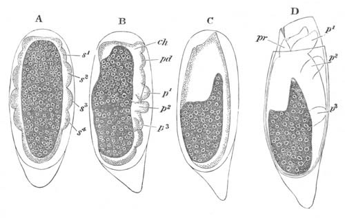
Fig. 202. Four successive stages in the
development of Myobia musculi.
(After Claparède.)
s1-s4. post-oral segments; ch. cheliceræ; pd.
pedipalpi; pr proboscis formed by the coalescence of the
cheliceræ and pedipalpi; p1, p2, etc. ambulatory
appendages.
In the cheese mite (Tyroglyphus) the embryo has two ecdyses which are
not accompanied by the peculiar changes observable in Myobia: the
cheliceræ and pedipalpi fuse however to form the proboscis. The first
larval form is hexapodous, and the last pair of appendages is formed
at a subsequent ecdysis.
In Atax Bonzi, a form parasitic on Unio, the development and
metamorphosis are even more complicated than in Myobia. The first
ecdysis occurs before the formation of the limbs, and shortly after
the ventral plate has become divided into segments. Within the
cuticular membrane resulting from the first ecdysis the anterior five
pairs of limbs spring out in the usual fashion. They undergo
considerable differentiation; the cheliceræ and pedipalpi approaching
each other at the anterior extremity of the body, and the three
ambulatory legs becoming segmented and clawed. An œsophagus, a
stomach, and an œsophageal nerve-ring are also formed. When the
larva has attained this stage the original egg-shell is split
into two valves and eventually cast off, but the embryo remains
enclosed within the cuticular membrane shed at the first ecdysis. This
cuticular membrane is spoken of by Claparède as the deutovum. In the
deutovum the embryo undergoes further changes; the cheliceræ and
pedipalpi coalesce and form the proboscis; a spacious body cavity with
blood corpuscles appears; and the alimentary canal enclosing the yolk
is formed.
The larva now begins to move, the cuticular membrane enclosing it is
ruptured, and the larva becomes free. It does not long remain active,
but soon bores its way into the gills of its host, undergoes a fresh
moult, and becomes quiescent. The cuticular membrane of the moult just
effected swells up by the absorption of water and becomes spherical.
Peculiar changes take place in the tissues, and the limbs become, as
in Myobia, nearly absorbed, remaining however as small knobs. The
larva swims about as a spherical body within its shell. The feet next
grow out afresh, and the posterior pair is added. From the proboscis
the palpi (of the pedipalpi) grow out below. The larva again becomes
free, and amongst other changes the cheliceræ grow out from the
proboscis. A further ecdysis, with a period of quiescence, intervenes
between this second larval form and the adult state.
The changes in the appendages which appear common to the Mites
generally are (1) the late development of the fourth pair of
appendages, which results in the constant occurrence of an hexapodous
larva; and (2) the early fusion of the cheliceræ and pedipalpi to form
a proboscis in which no trace of the original appendages can be
discerned. In most instances palpi and stilets of variable form are
subsequently developed in connexion with the proboscis, and, as
indicated in the above descriptions, are assumed to correspond with
the two original embryonic appendages.
The history of the germinal layers.
It is a somewhat remarkable fact that each of the groups of the
Arachnida so far studied has a different form of segmentation. The
types of Chelifer and the Spiders are simple modifications of the
centrolecithal type, while that of Scorpio, though apparently
meroblastic, is probably to be regarded in the same light (vide
p. 120 and
p. 434). The early development begins in the Scorpion and
Spiders with the formation of a ventral plate, and there can be but
little doubt that Chelifer is provided with an homologous structure,
though very probably modified, owing to the small amount of food-yolk
and early period of hatching.
The history of the layers and their conversion into the organs has
been studied in the case of the Scorpion (Metschnikoff, No.
434), and of the Spiders; and a close agreement has been found to
obtain between them.
It will be convenient to take the latter group as type, and simply to
call attention to any points in which the two groups differ.
The epiblast. The epiblast, besides giving rise to the skin
(hypodermis and cuticle), also supplies the elements for the nervous
system and organs of sense, and for the respiratory sacks, the
stomodæum and proctodæum.
At the period when the mesoblast is definitely established, the
epiblast is formed of a single layer of columnar cells in the region
of the ventral plate, and of a layer of flat cells over other parts of
the yolk.
When about six segments are present the first changes take place. The
epiblast of the ventral plate then becomes somewhat thinner in the
median line than at the two sides (fig. 203 B). In succeeding stages
the contrast between the median and the lateral parts becomes still
more marked, so that the epiblast becomes finally constituted of two
lateral thickened bands, which meet in front in the procephalic lobes,
and behind in the caudal lobe, and are elsewhere connected by a very
thin layer (fig. 203 C). Shortly after the appendages begin to be
formed, the first rudiments of the ventral nerve cord become
established as epiblastic thickenings on the inner side of each of the
lateral bands. The thickenings of the epiblast of the two sides are
quite independent, as may be seen in fig. 203 C, vn, taken from
a stage somewhat subsequent to their first appearance. They are
developed from before backwards, but either from the first, or in any
case very soon afterwards, cease to form uniform thickenings, but
constitute a linear series of swellings—the future ganglia—connected
by very short less prominent thickenings of the epiblast (fig. 200 C).
The rudiments of the ventral nerve cord are for a long time continuous
with the epiblast, but shortly after the establishment of the dorsal
surface of the embryo they become separated from the epiblast and
constitute two independent cords, the histological structure of which
is the same as in other Tracheata (fig. 206, vn).
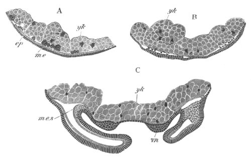
Fig. 203. Transverse sections through the ventral
plate of Agelena labyrinthica at three stages.
A. Stage when about three segments are formed. The mesoblastic plate
is not divided into two bands.
B. Stage when six segments are present (fig. 200 B). The mesoblast
is now divided into two bands.
C. Stage represented in fig. 200 D. The ventral cords have begun to
be formed on thickenings of the epiblast, and the limbs are
established.
ep. epiblast; me. mesoblast; me.s. mesoblastic
somite; vn. ventral nerve cord; yk. yolk.
The ventral cords are at first composed of as many ganglia as there
are segments. The foremost pair, belonging to the segment of
the cheliceræ, lie immediately behind the stomodæum, and are as
independent of each other as the remaining ganglia. Anteriorly they
border on the supra-œsophageal ganglia. When the yolk-sack is
formed in connection with the ventral flexure of the embryo, the two
nerve cords become very widely separated (fig. 206, vn) in
their middle region. At a later period, at the stage represented in
fig. 201 B, they again become approximated in the ventral line, and
delicate commissures are formed uniting the ganglia of the two sides,
but there is no trace at this or any other period of a median
invagination of epiblast between the two cords, such as Hatschek and
other observers have attempted to establish for various Arthropoda and
Chætopoda. At the stage represented in fig. 201 A the nerve ganglia
are still present in the abdomen, though only about four ganglia can
be distinguished. At a later stage these ganglia fuse into two
continuous cords, united however by commissures corresponding
with the previous ganglia.
The ganglia of the cheliceræ have, by the stage represented in fig.
201 B, completely fused with the supra-œsophageal ganglia and form
part of the œsophageal commissure. The œsophageal commissure is
however completed ventrally by the ganglia of the pedipalpi.
The supra-œsophageal ganglia are formed independently of the
ventral cords as two thickenings of the procephalic lobes (fig. 205).
The thickenings of the two lobes are independent, and each of them
becomes early marked out by a semicircular groove (fig. 200 D) running
outwards from the upper lip. Each thickening eventually becomes
detached from the superficial epiblast, but before this takes place
the two grooves become deeper, and on the separation of the ganglia
from the epiblast, the cells lining the grooves become involuted and
detached from the skin, and form an integral part of the
supra-œsophageal ganglia.
At the stage represented in fig. 201 B the supra-œsophageal ganglia
are completely detached from the epiblast, and are constituted of the
following parts: (1) A dorsal section formed of two hemispherical
lobes, mainly formed of the invaginated lining of the semicircular
grooves. The original lumen of the groove is still present on the
outer side of these lobes. (2) Two central masses, one for each
ganglion, formed of punctiform tissue, and connected by a transverse
commissure. (3) A ventral anterior lobe. (4) The original ganglia of
the cheliceræ, which form the ventral parts of the ganglia[183].
The later stages in the development of the nervous system have not
been worked out.
The development of the nervous system in the Scorpion is almost
identical with that in Spiders, but Metschnikoff believes, though
without adducing satisfactory evidence, that the median integument
between the two nerve cords assists in forming the ventral nerve cord.
Grooves are present in the supra-œsophageal ganglia similar to
those in Spiders.
The mesoblast. The history of the mesoblast, up to the
formation of a ventral plate subjacent to the thickened plate of
epiblast, has been already given. The ventral plate is shewn in fig.
203 A. It is seen to be formed mainly of small cells, but some
large cells are shewn in the act of passing into it from the yolk.
During a considerable section of the subsequent development the
mesoblast is confined to the ventral plate.
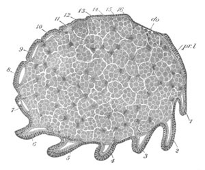
Fig. 204. Longitudinal section through an embryo
of Agelena labyrinthica.
The section is through an embryo of the same age as that represented
in fig. 200 C, and is taken slightly to one side of the middle line
so as to shew the relation of the mesoblastic somites to the limbs.
In the interior are seen the yolk segments and their nuclei.
1‑16. the segments; pr.l. procephalic lobe; do. dorsal
integument.
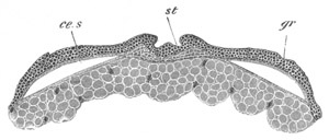
Fig. 205. Section through the procephalic lobes
of an embryo of Agelena labyrinthica.
The section is taken from an embryo of the same age as fig. 200 D.
st. stomodæum; gr. section through semicircular groove
in procephalic lobe; ce.s. cephalic section of body cavity.
The first important change takes place when about six somites are
established; the mesoblast then becomes divided into two lateral
bands, shewn in section in fig. 203 B, which meet however in front in
the procephalic lobes, and behind in the caudal lobes. Very shortly
afterwards these bands become broken up into a number of parts
corresponding to the segments, each of which soon becomes divided into
two layers, which enclose a cavity between them (vide fig. 204
and fig. 207). The outer layer (somatic) is thicker and attached to
the epiblast, and the inner layer (splanchnic) is thinner and mainly,
if not entirely, derived (in Agelena) from cells which originate in
the yolk. These structures constitute the mesoblastic somites. In the
appendage-bearing segments the somatic layer of each of them, together
with a prolongation of the cavity, is continued into the
appendage (fig. 203 C). Since the cavity of the mesoblastic somites is
part of the body cavity, all the appendages contain prolongations of
the body cavity. Not only is a pair of mesoblastic somites formed for
each segment of the body, but also for the procephalic lobes (fig.
205). The mesoblastic somites for these lobes are established somewhat
later than for the true segments, but only differ from them in the
fact that the somites of the two sides are united by a median bridge
of undivided mesoblast. The development of a somite for the
procephalic lobes is similar to what has been described by Kleinenberg
for Lumbricus (p. 339), but must not be necessarily supposed to
indicate that the procephalic lobes form a segment equivalent to the
segments of the trunk. They are rather equivalent to the præ-oral lobe
of Chætopod larvæ. When the dorsal surface of the embryo is
established a thick layer of mesoblast becomes formed below the
epiblast. This layer is not however derived from an upgrowth of the
mesoblast of the somites, but from cells which originate in the yolk.
The first traces of the layer are seen in fig. 204, do, and it
is fully established as a layer of large round cells in the stage
shewn in fig. 206. This layer of cells is seen to be quite independent
of the mesoblastic somites (me.s). The mesoblast of the dorsal
surface becomes at the stage represented in fig. 201 B divided into
splanchnic and somatic layers, and in the abdomen at any rate into
somites continuous with those of the ventral part of the mesoblast. At
the lines of junction of successive somites the splanchnic layer of
mesoblast dips into the yolk, and forms a number of transverse septa,
which do not reach the middle of the yolk, but leave a central part
free, in which the mesenteron is subsequently formed. At the insertion
of these septa there are developed widish spaces between the
layers of somatic and splanchnic mesoblast, which form transversely
directed channels passing from the heart outwards. They are probably
venous. At a later stage the septa send out lateral offshoots, and
divide the peripheral part of the abdominal cavity into a number of
compartments filled with yolk. It is probable that the hepatic
diverticula are eventually formed in these compartments.
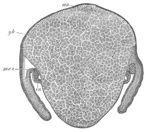
Fig. 206. Transverse section through the thoracic
region of an embryo of Agelena labyrinthica.
The section is taken from an embryo of the same age as fig. 201 A,
and passes through the maximum protuberance of the ventral
yolk-sack.
vn. ventral nerve cord;
yk. yolk; me.s.
mesoblastic somite; ao. aorta.
The somatic layer of mesoblast is converted into the muscles, both of
the limbs and trunk, the superficial connective tissue, nervous
sheath, etc. It probably also gives rise to the three muscles attached
to the suctorial apparatus of the œsophagus.
The heart and aorta are formed as a solid rod of cells of the dorsal
mesoblast, before it is distinctly divided into splanchnic and somatic
layers. Eventually the central cells of the heart become blood
corpuscles, while its walls are constituted of an outer muscular and
inner epithelioid layer. It becomes functional, and acquires its
valves, arterial branches, etc., by the stage represented in fig. 201
B.
The history of the mesoblast, more especially of the mesoblastic
somites, of the Scorpion is very similar to that in Spiders: their
cavity is continued in the same way into the limbs. The general
character of the somites in the tail is shewn in fig. 207. The caudal
aorta is stated by Metschnikoff to be formed from part of the
mesenteron, but this is too improbable to be accepted without further
confirmation.
The hypoblast and alimentary tract. It has already been stated
that the yolk is to be regarded as corresponding to the hypoblast of
other types.
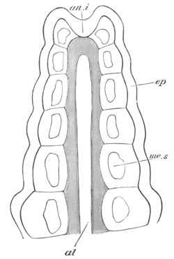
Fig. 207. Tail of an advanced embryo of the
Scorpion to illustrate the structure of the mesoblastic
somites. (After Metschnikoff.)
al. alimentary tract; an.i. anal invagination;
ep. epiblast; me.s. mesoblastic somite.
For a considerable period it is composed of the polygonal yolk cells
already described and shewn in figs. 203, 204, and 205. The yolk cells
divide and become somewhat smaller as development proceeds; but the
main products of the division of the yolk nuclei and the protoplasm
around them are undoubtedly cells which join the mesoblast (fig. 203
A). The permanent alimentary tract is formed of three sections, viz.
stomodæum, proctodæum, and mesenteron. The stomodæum and proctodæum
are both formed before the mesenteron. The stomodæum is formed as an
epiblastic pit between the two procephalic lobes (figs. 200 and 205,
st). It becomes deeper, and by the latest stage figured is a
deep pit lined by a cuticle and ending blindly. To its hinder section,
which forms the suctorial apparatus of the adult, three powerful
muscles (a dorsal and two lateral) are attached.
The proctodæum is formed considerably later than the stomodæum. It is
a comparatively shallow involution, which forms the rectum of the
adult. It is dilated at its extremity, and two Malpighian vessels
early grow out from it.
The mesenteron is formed in the interior of the yolk. Its walls
are derived from the cellular elements of the yolk, and the first
section to be formed is the hinder extremity, which appears as a short
tube ending blindly behind in contact with the proctodæum, and open to
the yolk in front. The later history of the mesenteron has not been
followed, but it undoubtedly includes the whole of the
abdominal section of the alimentary canal of the adult, except the
rectum, and probably also the thoracic section. The later history of
the yolk which encloses the mesenteron has not been satisfactorily
studied, though it no doubt gives rise to the hepatic tubes, and
probably also to the thoracic diverticula of the alimentary tract.
The general history of the alimentary tract in Scorpio is much the
same as in Spiders. The hypoblast, the origin of which as mentioned
above is somewhat uncertain, first appears on the ventral side and
gradually spreads so as to envelop the yolk, and form the wall of the
mesenteron, from which the liver is formed as a pair of lateral
outgrowths. The proctodæum and stomodæum are both short, especially
the former (vide fig. 207).
Summary and general conclusions.
The embryonic forms of Scorpio and Spiders are very similar, but in
spite of the general similarity of Chelifer to Scorpio, the embryo of
the former differs far more from that of Scorpio than the latter does
from Spiders. This peculiarity is probably to be explained by the
early period at which Chelifer is hatched; and though a more thorough
investigation of this interesting form is much to be desired, it does
not seem probable that its larva is a primitive type.
The larvæ of the Acarina with their peculiar ecdyses are to be
regarded as much modified larval forms. It is not however easy to
assign a meaning to the hexapodous stage through which they generally
pass.
With reference to the segments and appendages, some interesting points
are brought out by the embryological study of these forms.
The maximum number of segments is present in the Scorpion, in which
nineteen segments (not including the præ-oral lobes, but including the
telson) are developed. Of these the first twelve segments have traces
of appendages, but the appendages of the six last of these (unless the
pecten is an appendage) atrophy. In Spiders there are indications in
the embryo of sixteen segments and in all the Arachnida, except the
Acarina, at the least four segments bear appendages in the embryo
which are without them in the adult. The morphological bearings of
this fact are obvious.
It deserves to be noted that, in both Scorpio and the Spider, the
cheliceræ are borne in the embryo by the first post-oral segment, and
provided with a distinct ganglion, so that they cannot correspond (as
they are usually supposed to do) with the antennæ of Insects, which
are always developed on the præ-oral lobes, and never supplied by an
independent ganglion.
The cheliceræ would seem probably to correspond with the mandibles of
Insects, and the antennæ to be absent. In favour of this view is the
fact that the embryonic ganglion of the mandibles of Insects is stated
(cf. Lepidoptera, Hatschek, p. 340) to become, like the
ganglion of the cheliceræ, converted into part of the œsophageal
commissure.
If the above considerations are correct, the appendages of the
Arachnida retain in many respects a very much more primitive condition
than those of Insects. In the first place, both the cheliceræ and
pedipalpi are much less differentiated than the mandibles and first
pair of maxillæ with which they correspond. In the second place, the
first pair of ambulatory limbs must be equivalent to the second pair
of maxillæ of Insects, which, for reasons stated above, were probably
originally ambulatory. It seems in fact a necessary deduction from the
arguments stated that the ancestors of the present Insecta and
Arachnida must have diverged from a common stem of the Tracheata at a
time when the second pair of maxillæ were still ambulatory in function.
With reference to the order of the development of the appendages and
segments, very considerable differences are noticeable in the
different Arachnoid types. This fact alone appears to me to be
sufficient to prove that the order of appearance of the appendages is
often a matter of embryonic convenience, without any deep
morphological significance. In Scorpio the segments develop
successively, except perhaps the first post-oral, which is developed
after some of the succeeded segments have been formed. In Spiders the
segment of the cheliceræ, and probably also of the pedipalpi, appears
later than the next three or four. In both these types the segments
arise before the appendages, but the reverse appears to be the case in
Chelifer. The permanent appendages, except the cheliceræ, appear
simultaneously in Scorpions and Spiders. The second pair appears long
before the others in Chelifer, then the third, next the first, and
finally the three hindermost.
Bibliography.
Scorpionidæ.
(434) El. Metschnikoff. “Embryologie des Scorpions.” Zeit.
f. wiss. Zool. Bd. XXI. 1870.
(435) H. Rathke. Reisebemerkungen aus Taurien (Scorpio).
Leipzig, 1837.
Pseudoscorpionidæ.
(436) El. Metschnikoff. “Entwicklungsgeschichte d. Chelifer.”
Zeit. f. wiss. Zool., Bd. XXI. 1870.
(437) A. Stecker. “Entwicklung der Chthonius-Eier im
Mutterleibe und die Bildung des Blastoderms.” Sitzung. königl.
böhmisch. Gesellschaft Wissensch., 1876, 3. Heft, and Annal.
and Mag. Nat. History, 1876, XVIII. 197.
Phalangidæ.
(438) M. Balbiani. “Mémoire sur le développement des
Phalangides.” Ann. Scien. Nat. Series V. Vol.
XVI. 1872.
Araneina.
(439) M. Balbiani. “Mémoire sur le développement des
Aranéides.” Ann. Scien. Nat. Series V. Vol.
XVII. 1873.
(440) F. M. Balfour. “Notes on the development of the
Araneina.” Quart. Journ. of Micr. Science, Vol. XX.
1880.
(441) J. Barrois. “Recherches s. l. développement des
Araignées.” Journal de l’Anat. et de la Physiol. 1878.
(442) E. Claparède. Recherches s. l’évolution des
Araignées. Utrecht, 1862.
(443) Herold. De generatione Araneorum in Ovo. Marburg,
1824.
(444) H. Ludwig. “Ueber die Bildung des Blastoderms bei den
Spinnen.” Zeit. f. wiss. Zool., Vol. XXVI. 1876.
Acarina.
(445) P. van Beneden. “Développement de l’Atax ypsilophora.” Acad.
Bruxelles, t. XXIV.
(446) Ed. Claparède. “Studien über Acarinen.” Zeit. f. wiss.
Zool., Bd. XVIII. 1868.
Formation of the layers and the embryonic envelopes in the
Tracheata.
There is a striking constancy in the mode of formation of the layers
throughout the group. In the first place the hypoblast is not formed
by a process which can be reduced to invagination: in other words,
there is no gastrula stage.
Efforts have been made to shew that the mesoblastic groove of Insects
implies a modified gastrula, but since it is the essence of a gastrula
that it should directly or indirectly give rise to the archenteron,
the groove in question cannot fall under this category. Although the
mesoblastic groove of Insects is not a gastrula, it is quite possible
that it is the rudiment of a blastopore, the gastrula corresponding to
which has now vanished from the development. It would thus be
analogous to the primitive streak of Vertebrates[184].
The growth of the blastoderm over the yolk in Scorpions admits no
doubt of being regarded as an epibolic gastrula. The blastopore would
however be situated dorsally, a position which it does not occupy in
any gastrula type so far dealt with. This fact, coupled with the
consideration that the partial segmentation of Scorpio can be derived
without difficulty from the ordinary Arachnidan type (vide p.
120), seems to shew that there is no true epibolic invagination in the
development of Scorpio.
On the formation of the blastoderm traces of two embryonic layers are
established. The blastoderm itself is essentially the epiblast, while
the central yolk is the hypoblast. The formation of the embryo
commences in connection with a thickening of the blastoderm, known as
the ventral plate. The mesoblast is formed as an unpaired plate split
off from the epiblast of the ventral plate. This process takes place
in at any rate two ways. In Insects a groove is formed, which becomes
constricted off to form the mesoblastic plate: in Spiders there is a
keel-like thickening of the blastoderm, which takes the place of the
groove.
The unpaired mesoblastic plate becomes in all forms very soon divided
into two mesoblastic bands.
The mesoblastic bands are very similar to, and probably homologous
with, those of Chætopoda; but the different modes by which they arise
in these two groups are very striking, and probably indicate that
profound modifications have taken place in the early development of
the Tracheata. In the Chætopoda the bands are from the first widely
separated, and gradually approach each other ventrally, though without
meeting. In the Tracheata they arise from the division of an unpaired
ventral plate.
The further history of the mesoblastic bands is nearly the
same for all the Tracheata so far investigated, and is also very much
the same as for the Chætopoda. There is a division into somites, each
containing a section of the body cavity. In the cephalic section of
the mesoblastic bands a section of the body cavity is also formed. In
Arachnida, Myriapoda, and probably also Insecta, the body cavity is
primitively prolonged into the limbs.
In Spiders at any rate, and very probably in the other groups of the
Tracheata, a large part of the mesoblast is not derived from the
mesoblastic plate, but is secondarily added from the yolk cells.
In all Tracheata the yolk cells give rise to the mesenteron which, in
opposition, as will hereafter appear, to the mesenteron of the
Crustacea, forms the main section of the permanent alimentary tract.
One of the points which is still most obscure in connection with the
embryology of the Tracheata is the origin of the embryonic membranes.
Amongst Insects, with the exception of the Thysanura, such membranes
are well developed. In the other groups definite membranes like those
of Insects are never found, but in the Scorpion a cellular envelope
appears to be formed round the embryo from the cells of the
blastoderm, and more or less similar structures have been described in
some Myriapods (vide p. 390). These structures no doubt further
require investigation, but may provisionally be regarded as homologous
with the amnion and serous membrane of Insects. In the present state
of our knowledge it does not seem easy to give any explanation of the
origin of these membranes, but they may be in some way derived from an
early ecdysis.
History of the larval forms[186].
The larval forms of the Crustacea appear to have more faithfully
preserved their primitive characters than those of almost any other
group.
Branchiopoda.
The Branchiopoda, comprising under that term the Phyllopoda and
Cladocera, contain the Crustacea with the maximum number of segments
and the least differentiation of the separate appendages. This and
other considerations render it probable that they are to be regarded
as the most central group of the Crustaceans, and as in many respects
least modified from the ancestral type from which all the groups have
originated.
The free larval stages when such exist commence with a larval form
known as the Nauplius.
The term Nauplius was applied by O. F. Müller to certain larval forms
of the Copepoda (fig. 229) in the belief that they were adult.
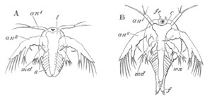
Fig. 208. Two stages in the development of Apus
cancriformis.
(After Claus.)
A. Nauplius stage at the time of hatching.
B. Stage after first ecdysis.
an1. and an2. First and second antennæ; md.
mandible; mx. maxilla; l. labrum; fr. frontal
sense organ; f. caudal fork; s. segments.
The term has now been extended to a very large number of larvæ which
have certain definite characters in common. They are provided (fig.
208 A) with three pairs of appendages, the future two pairs of antennæ
and mandibles. The first pair of antennæ (an1) is uniramous
and mainly sensory in function, the second pair of antennæ
(an2) and mandibles (md) are biramous swimming
appendages, and the mandibles are without the future cutting blade.
The Nauplius mandibles represent in fact the palp. The two posterior
appendages are both provided with hook-like prominences on their basal
joints, used in mastication. The body in most cases is unsegmented,
and bears anteriorly a single median eye. There is a large upper lip,
and an alimentary canal formed of œsophagus, stomach and rectum.
The anus opens near the hind end of the body. On the dorsal surface
small folds of skin frequently represent the commencement of a dorsal
shield. One very striking peculiarity of the Nauplius according to
Claus and Dohrn is the fact that the second pair of antennæ is
innervated from a sub-œsophageal ganglion. A larval form
with the above characters occurs with more or less frequency in all
the Crustacean groups. In most instances it does not
exactly conform to the above type, and the divergences are more
considerable in the Phyllopods than in most other groups. Its
characters in each case are described in the sequel.
Phyllopoda..For the Phyllopoda the development of Apus
cancriformis may conveniently be taken as type (Claus, No. 454). The
embryo at the time it leaves the egg (fig. 208 A) is somewhat oval in
outline, and narrowed posteriorly. There is a slight V-shaped
indentation behind, at the apex of which is situated the anus. The
body, unlike that of the typical Nauplius, is already divided into two
regions, a cephalic and post-cephalic. On the ventral side of the
cephalic region there are present the three normal pairs of
appendages. Foremost there are the small anterior antennæ
(an1), which are simple unjointed rod-like bodies with two
moveable hairs at their extremities. They are inserted at the sides of
the large upper lip or labrum (l). Behind these are the
posterior antennæ, which are enormously developed and serve as the
most important larval organs of locomotion. They are biramous, being
formed of a basal portion with a strong hook-like bristle projecting
from its inner side, an inner unjointed branch with three bristles,
and an outer large imperfectly five-jointed branch with five long
lateral bristles. The hook-like organ attached to this pair of
appendages would seem to imply that it served in some ancestral form
as jaws (Claus). This character is apparently universal in the embryos
of true Phyllopods, and constantly occurs in the Copepoda, etc.
The third pair of appendages or mandibles (md) is attached
close below the upper lip. They are as yet unprovided with cutting
blades, and terminate in two short branches, the inner with two and
the outer with three bristles.
At the front of the head there is the typical unpaired eye. On the
dorsal surface there is already present a rudiment of the cephalic
shield, continuous anteriorly with the labrum (l) or upper lip,
the extraordinary size of which is characteristic of the larvæ of
Phyllopods. The post-cephalic region, which afterwards becomes the
thorax and abdomen, contains underneath the skin rudiments of the five
anterior thoracic segments and their appendages, and presents in this
respect an important variation from the typical Nauplius form. After
the first ecdysis the larva (fig. 208 B) loses its oval form,
mainly owing to the elongation of the hinder part of the body and the
lateral extension of the cephalic shield, which moreover now
completely covers over the head and has begun to grow backwards so as
to cover over the thoracic region. At the second ecdysis there appears
at its side a rudimentary shell-gland. In the cephalic region two
small papillæ (fr) are now present at the front of the head
close to the unpaired eye. They are of the nature of sense organs, and
may be called the frontal sense papillæ. They have been shewn by Claus
to be of some phylogenetic importance. The three pairs of Nauplius
appendages have not altered much, but a rudimentary cutting blade has
grown out from the basal joint of the mandible. A gland opening at the
base of the antennæ is now present, which is probably equivalent to
the green gland often present in the Malacostraca. Behind the
mandibles a pair of simple processes has appeared, which forms the
rudiment of the first pair of maxillæ (mx).
In the thoracic region more segments have been added posteriorly, and
the appendages of the three anterior segments are very distinctly
formed. The tail is distinctly forked. The heart is formed at the
second ecdysis, and then extends to the sixth thoracic segment: the
posterior chambers are successively added from before backwards.
At the successive ecdyses which the larva undergoes new segments
continue to be formed at the posterior end of the body, and limbs
arise on the segments already formed. These limbs probably represent
the primitive form of an important type of Crustacean appendage, which
is of value for interpreting the parts of the various malacostracan
appendages. They consist (fig. 209) of a basal portion (protopodite of
Huxley) bearing two rami. The basal portion has two projections on the
inner side. To the outer side of the basal portion there is attached a
dorsally directed branchial sack (br) (epipodite of Huxley).
The outer ramus (ex) (exopodite of Huxley) is formed of a
single plate with marginal setæ. The inner one (en) (endopodite
of Huxley) is four-jointed, and a process similar to those of the
basal joint is given off from the inner side of the three proximal
joints.
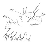
Fig. 209. Typical phyllopod appendage.
(Copied from Claus.)
ex. exopodite; en. endopodite; br. branchial
appendage (epipodite). The basal portion bearing the two proximal
projections is not sharply separated from the endopodite.
At the third ecdysis several new features appear in the cephalic
region, which becomes more prominent in the succeeding stages.
In the first place the paired eyes are formed at each side of and
behind the unpaired eye, secondly the posterior pair of maxillæ is
formed though it always remains very rudimentary. The shell-gland
becomes fully developed opening at the base of the first pair of
maxillæ. The dorsal shield gradually grows backwards till it covers
its full complement of segments.
After the fifth ecdysis the Nauplius appendages undergo a rapid
atrophy. The second pair of antennæ especially becomes reduced in
size, and the mandibular palp—the primitive Nauplius portion of the
mandible—is contracted to a mere rudiment, which eventually
completely disappears, while the blade is correspondingly enlarged and
also becomes toothed. The adult condition is only gradually attained
after a very large number of successive changes of skin.
The chief point of interest in the above development is the fact of
the primitive Nauplius form becoming gradually converted without any
special metamorphosis into the adult condition[187].
Branchipus like Apus is hatched as a somewhat modified Nauplius, which
however differs from that of Apus in the hinder region of the body
having no indications of segments. It goes through a very similar
metamorphosis, but is at no period of its metamorphosis provided with
a dorsal shield: the second pair of antennæ does not abort, and in the
male is provided with clasping organs, which are perhaps remnants of
the embryonic hooks so characteristic of this pair of antennæ.
The larva of Estheria when hatched has a Nauplius form, a large upper
lip, caudal fork and single eye. There are two functional pairs of
swimming appendages—the second pair of antennæ and mandibles. The
first pair of antennæ has not been detected, and a dorsal mantle to
form the shell is not developed. At the first moult the anterior pair
of antennæ arises as small stump-like structures, and a small dorsal
shield is also formed. Rudiments of six or seven pairs of appendages
sprout out in the usual way, and continue to increase in
number at successive moults: the shell is rapidly developed. The chief
point of interest in the development of this form is the close
resemblance of the young larva to a typical adult Cladocera (Claus).
This is shewn in the form of the shell, which has not reached its full
anterior extension, the rudimentary anterior antennæ, the large
locomotor second pair of antennæ, which differ however from the
corresponding organs in the Cladocera in the presence of typical
larval hooks. Even the abdomen resembles that of Daphnia. These
features perhaps indicate that the Cladocera are to be derived from
some Phyllopod form like Estheria by a process of retrogressive
metamorphosis. The posterior antennæ in the adult Estheria are large
biramous appendages, and are used for swimming; and though they have
lost the embryonic hook, they still retain to a larger extent than in
other Phyllopod families their Nauplius characteristics.
The Nauplius form of the Phyllopods is marked by several definite
peculiarities. Its body is distinctly divided into a cephalic and
post-cephalic region. The upper lip is extraordinarily large,
relatively very much more so than at the later stages. The first pair
of antennæ is usually rudimentary and sometimes even absent; while the
second pair is exceptionally large, and would seem to be capable of
functioning not only as a swimming organ, but even as a masticating
organ. A dorsal shield is nearly or quite absent.
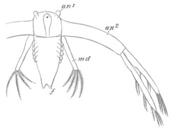
Fig. 209 A. Nauplius larva of Leptodora hyalina
from winter egg. (Copied from Bronn; after Sars.)
an1. antenna of first pair; an2. antenna of second
pair; md. mandible; f. caudal fork.
Cladocera. The probable derivation of the Cladocera from a form
similar to Estheria has already been mentioned, and it might have been
anticipated that the development would be similar to that of the
Phyllopods. The development of the majority of the Cladocera takes
place however in the egg, and the young when hatched closely resembles
their parents, though in the egg they pass through a Nauplius stage
(Dohrn). An exception to the general rule is however offered by the
case of the winter eggs of Leptodora, one of the most primitive of the
Cladoceran families. The summer eggs develop without metamorphosis,
but Sars (No. 461) has discovered that the larva leaves the
winter eggs in the form of a Nauplius (fig. 209). This Nauplius
closely resembles that of the Phyllopods. The body is elongated and in
addition to normal Nauplius appendages is marked by six pairs of
ridges—the indications of the future feet. The anterior antennæ are
as usual small; the second large and biramous, but the masticatory
bristle characteristic of the Phyllopods is not present. The mandibles
are without a cutting blade. A large upper lip and unpaired eye are
present.
The adult form is attained in the same manner as amongst the
Phyllopods after the third moult.
Malacostraca.
Owing to the size and importance of the various forms included in the
Malacostraca, greater attention has been paid to their embryology than
to that of any other division of the Crustacea; and the proper
interpretation of their larval forms involves some of the most
interesting problems in the whole range of Embryology.
The majority of Malacostraca pass through a more or less complicated
metamorphosis, though in the Nebaliadæ, the Cumaceæ, some of the
Schizopoda, a few Decapoda (Astacus, Gecarcinus, etc.), and in the
Edriophthalmata, the larva on leaving the egg has nearly the form of
the adult. In contradistinction to the lower groups of Crustacea the
Nauplius form of larva is rare, though it occurs in the case of one of
the Schizopods (Euphausia, fig. 212), in some of the lower forms of
the Decapods (Penæus, fig. 214), and perhaps also, though this has not
been made out, in some of the Stomatopoda.
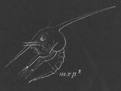
Fig. 210. Zoæa of Thia polita. (After
Claus.) mxp2. second maxillipede.
In the majority of the Decapoda the larva leaves the egg in a form
known as the Zoæa (fig. 210). This larval form is characterised by the
presence of a large cephalo-thoracic shield usually armed with
lateral, anterior, and dorsal spines. The caudal segments are well
developed, though without appendages, and the tail,
which functions in swimming, is usually forked. The six
posterior thoracic segments are, on the other hand, rudimentary or
non-existent. There are seven anterior pairs of appendages shewn
in detail in fig. 211, viz. the two pairs of antennæ (At. I. and At.
II.), neither of them used as swimming organs, the mandibles without a
palp (md), well-developed maxillæ (two pairs, mx 1 and
mx 2), and two or sometimes (Macrura) three pairs of biramous
natatory maxillipeds (mxp 1 and mxp 2). Two lateral
compound stalked eyes are present, together with a median Nauplius
eye. The heart has in the majority of cases only one or two
(Brachyura) pairs of ostia.
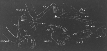
Fig. 211. The Appendages of a Crab Zoæa.
At. I. first antenna; At. II. second antenna;
md. mandible (without a palp); mx. 1. first maxilla;
mx. 2. second maxilla; mxp. 1. first maxilliped;
mxp. 2. second maxilliped.
ex. exopodite; en. endopodite.
The Zoæa larva, though typically developed in the Decapoda, is not
always present (e.g. Astacus and Homarus), and sometimes occurs
in a very modified form. It makes its appearance in an altered garb in
the ontogeny of some of the other groups.
The two Malacostracan forms, amongst those so far studied, in which
the phylogenetic record is most fully preserved in the ontogeny, are
Euphausia amongst the Schizopods and Penæus amongst the Decapods.
Schizopoda. Euphausia leaves the egg (Metschnikoff, No. 468‑9)
as a true Nauplius with only three pairs of appendages, the two hinder
biramous, and an unsegmented body. The second pair of antennæ
has not however the colossal dimensions so common in the lower types.
A mouth is present, but the anus is undeveloped.
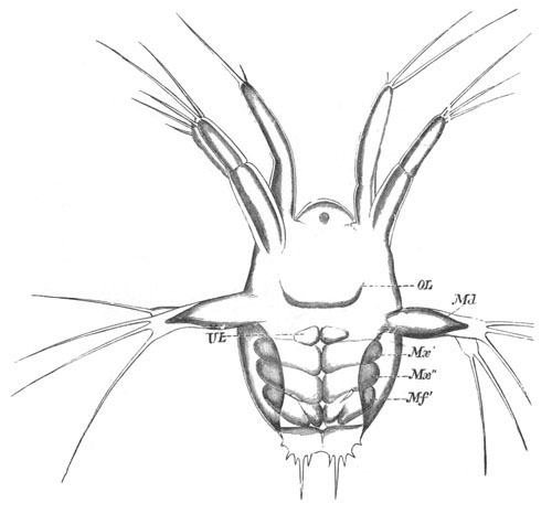
Fig. 212. Nauplius of Euphausia. (From
Claus; after Metschnikoff.)
The Nauplius is represented shortly before an ecdysis, and in
addition to the proper appendages rudiments of the three following
pairs are present.
OL. upper lip; UL. lower lip; Md. mandible;
Mx´. and Mx´´. two pairs of maxillæ; mf´.
maxilliped 1.
After the first moult three pairs of prominences—the rudiments of the
two maxillæ and 1st maxillipeds arise behind the Nauplius appendages
(fig. 212). At the same time an anus appears between the two limbs of
a rudimentary caudal fork; and an unpaired eye and upper lip appear in
front. After another moult (fig. 212) a lower lip is formed
(UL) as a pair of prominences very similar to true appendages;
and a delicate cephalo-thoracic shield also becomes developed. Still
later the cutting blade of the mandible is formed, and the palp
(Nauplius appendage) is greatly reduced. The cephalo-thoracic shield
grows over the front part of the embryo, and becomes
characteristically toothed at its edge. There are also two
frontal papillæ very similar to those already described in the
Phyllopod larvæ. Rudiments of the compound eyes make their appearance,
and though no new appendages are added, those already present undergo
further differentiations. They remain however very simple; the
maxillipeds especially are very short and resemble somewhat Phyllopod
appendages.
Up to this stage the tail has remained rudimentary and short, but
after a further ecdysis (Claus) it grows greatly in length. At the
same time the cephalo-thoracic shield acquires a short spine directed
backwards. The larva is now in a condition to which Claus has given
the name of Protozoæa (fig. 213 A).
Very shortly afterwards the region immediately following the segments
already formed becomes indistinctly segmented, while the tail is still
without a trace of segmentation. The region of the thorax proper soon
becomes distinctly divided into seven very short segments, while at
the same time the now elongated caudal region has become divided into
its normal number of segments (fig. 213 B). By this stage the larva
has become a true Zoæa—though differing from the normal Zoæa in the
fact that the thoracic region is segmented, and in the absence of a
second pair of maxillipeds.
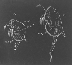
Fig. 213. Larvæ of Euphausia. (After Claus.)
From the side.
A. Protozoæa larva. B. Zoæa larva.
mx.´ and mx´´. maxillæ 1 and 2; mxp1.
maxilliped 1.
The adult characters are very gradually acquired in a series of
successive moults; the later development of Euphausia resembling in
this respect that of the Phyllopods. On the other hand Euphausia
differs from that group in the fact that the abdominal (caudal) and
thoracic appendages develop as two independent series from
before backwards, of which the abdominal series is the earliest to
attain maturity.
This is shewn in the following table compiled from Claus’ observations.
| Length of Larva. |
Appendages of thoracic region;
viz. the 2nd and 3rd maxilliped and 5 ambulatory appendages. |
Appendages of abdomen. |
|---|
| 3-3½ mm. |
2nd maxilliped, rudimentary. |
1st abdominal appendage. |
| 3½-4 mm. |
2nd maxilliped, biramous.
3rd rudimentary.
1st and 2nd ambulatory
appendages, rudimentary. |
2nd and 3rd abdominal appendages.
4th and 5th rudimentary. |
| 4½-5 mm. |
3rd maxilliped, biramous. |
4th, 5th, and 6th fully developed. |
| 5-5½ mm. |
3rd and 4th ambulatory appendages. |
|
| 6 mm. |
5th ambulatory appendage. |
|
All the appendages following the second pair of maxillæ are biramous,
and the first eight of these bear branched gills as their epipodites.
It is remarkable that the epipodite is developed on all the appendages
anteriorly in point of time to the outer ramus (exopodite).
Although in Mysis there is no free larval stage, and the development
takes place in a maternal incubatory pouch, yet a stage may be
detected which clearly corresponds with the Nauplius stage of
Euphausia (E. van Beneden, No. 465). At this stage, in which only the
three Nauplius appendages are developed, the Mysis embryo is hatched.
An ecdysis takes place, but the Nauplius skin is not completely thrown
off, and remains as an envelope surrounding the larva during its later
development.
Decapoda. Amongst the Decapoda the larva usually leaves the egg
in the Zoæa form, but a remarkable exception to this general rule is
afforded by the case of one or more species of Penæus. Fritz Müller
was the first to shew that the larva of these forms leaves the egg as
a typical Nauplius, and it is probable that in the successive
larval stages of these forms the ancestral history of the Decapoda is
most fully preserved[188].
The youngest known larva of Penæus (fig. 214) has a somewhat oval
unsegmented body. There spring from it the three typical pairs of
Nauplius appendages. The first is uniramous, the second and third are
biramous, and both of them adapted for swimming, and the third
of them (mandibles) is without a trace of the future blade. The body
has no carapace, and bears anteriorly a single median simple eye.
Posteriorly it is produced into two bristles.
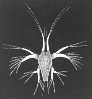
Fig. 214. Nauplius stage of Penæus. (After
Fritz Müller.)
After the first moult the larva has a rudiment of a forked tail, while
a dorsal fold of skin indicates the commencement of the
cephalo-thoracic shield. A large provisional helmet-shaped upper lip
like that in Phyllopods has also appeared. Behind the appendages
already formed there are stump-like rudiments of the four succeeding
pairs (two pairs of maxillæ and two pairs of maxillipeds); and in a
slightly older larva the formation of the mandibular blade has
commenced, together with the atrophy of the palp or Nauplius appendage.
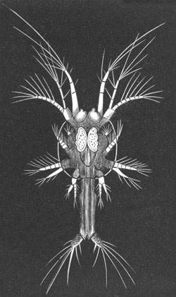
Fig. 215. Protozoæa stage of Penæus. (After
Fritz Müller.)
Between this and the next observed stage there is possibly a slight
lacuna. The next stage (fig. 215) at any rate represents the
commencement of the Zoæa series. The cephalo-thoracic shield has
greatly grown, and eventually acquires the usual dorsal spine. The
posterior region of the body is prolonged into a tail, which is quite
as long as the whole of the remainder of the body. The four appendages
which were quite functionless at the last stage have now sprouted into
full activity. The region immediately behind them is divided
(fig. 215) into six segments (the six thoracic segments) without
appendages, while somewhat later the five anterior abdominal segments
become indicated, but are equally with the thoracic segments without
feet. The mode of appearance of these segments shews that the thoracic
and abdominal segments develop in regular succession from before
backwards (Claus). Of the palp of the mandibles, as is usual amongst
Zoæa forms, not a trace remains, though in the youngest Zoæa caught by
Fritz Müller a very small rudiment of the palp was present. The first
pair of antennæ is unusually long, and the second pair continues to
function as a biramous swimming organ; the outer ramus is
multiarticulate. The other appendages are fully jointed, and the two
maxillipeds biramous. On the dorsal surface of the body the unpaired
eye is still present, but on each side of it traces of the stalked
eyes have appeared. Frontal sense organs like those of Phyllopods are
also present.
From the Protozoæa form the larva passes into that of a true Zoæa with
the usual appendages and spines, characterised however by certain
remarkable peculiarities. Of these the most important are (1) the
large size of the two pairs of antennæ and the retention of its
Nauplius function by the second of them; (2) the fact that the
appendages of the six thoracic segments appear as small biramous
Schizopod legs, while the abdominal appendages, with the exception of
the sixth, are still without their swimming feet. The early
appearance of the appendages of the sixth abdominal segment is
probably correlated with their natatory function in connection with
the tail. As a point of smaller importance which may be mentioned is
the fact that both pairs of maxillæ are provided with small
respiratory plates (exopodites) for regulating the flow of water under
the dorsal shield. From the Zoæa form the larva passes into a Mysis or
Schizopod stage (fig. 216), characterised by the thoracic feet and
maxillipeds resembling in form and function the biramous feet of
Mysis, the outer ramus being at first in many cases much larger than
the inner. The gill pouches appear at the base of these feet nearly at
the same time as the endopodites become functional. At the same time
the antennæ become profoundly modified. The anterior antennæ shed
their long hairs, and from the inner side of the fourth joint there
springs a new process, which eventually elongates and becomes the
inner flagellum. The outer ramus of the posterior antennæ is reduced
to a scale, while the flagellum is developed from a stump-like
rudiment of the inner ramus (Claus). A palp sprouts on the mandible
and the median eye disappears.
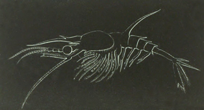
Fig. 216. Penæus larva in the Mysis stage.
(After Claus.)
The abdominal feet do not appear till the commencement of the Mysis
stage, and hardly become functional till its close.
From the Mysis stage the larva passes quite simply into the adult
form. The outer ramus of the thoracic feet is more or less completely
lost. The maxillipeds, or the two anterior pairs at any rate, lose
their ambulatory function, cutting plates develop on the inner side of
their basal joints, and the two rami persist as small
appendages on their outer side. Gill pouches also sprout from their
outer side.
The respiratory plate of the second maxilla attains its full
development and that on the first maxilla disappears[189]. The
Nauplius, so far as is known, does not occur in any other Decapod form
except Penæus.
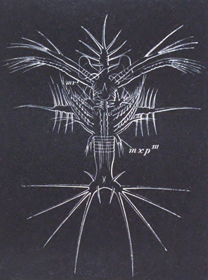
Fig. 217. Latest Protozoæa stage of Sergestes
larva (Elaphocaris). (After Claus.)
mxp´´´. third pair of maxillipeds.
The next most primitive larval history known is that which appears in
the Sergestidæ. The larval history, which has been fully elucidated by
Claus, commences with a Protozoæa form (fig. 217), which develops into
a remarkable Zoæa first described by Dohrn as Elaphocaris. This
develops into a form originally described by Claus as Acanthosoma, and
this into a form known as Mastigopus (fig. 218) from which it is easy
to pass to the adult.
The remarkable Protozoæa (fig. 217) is characterised by the presence
on the dorsal shield of a frontal, dorsal and two lateral spikes, each
richly armed with long side spines. The normal Zoæa appendages are
present, and in addition to them a small third pair of maxillipeds.
The thoracic region is divided into five short rings, but the abdomen
is unsegmented. The tail is forked and provided with long spines. The
antennæ, like those of Penæus, are long—the second pair biramous; the
mandibles unpalped. Both pairs of maxillæ are provided with
respiratory plates; the second pair is footlike, and has at its base a
glandular mass believed by Claus to be the equivalent of the
Entomostracan shell-gland. The maxillipeds have the usual biramous
characters. A helmet-shaped upper lip like that of a typical
Nauplius is present, and the eyes are situated on very long stalks.
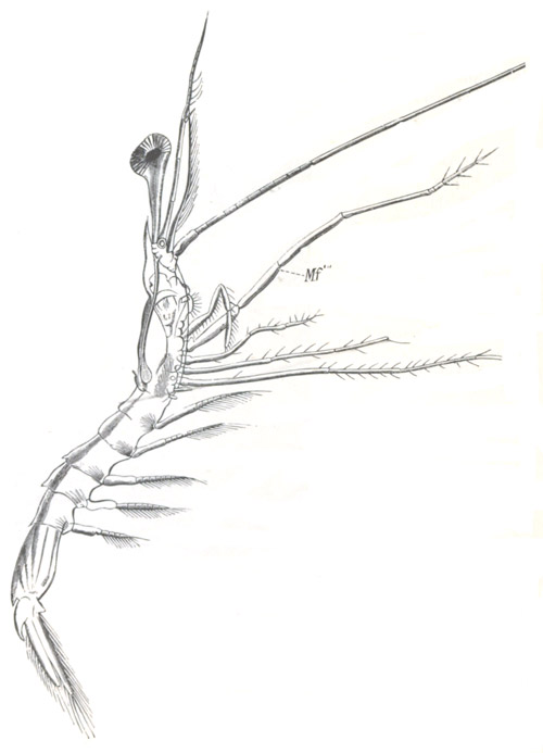
Fig. 218. Mastigopus Stage Of Sergestes.
(From Claus.)
Mf´´´. maxilliped 3.
In the true Zoæa stage there appear on the five thoracic segments
pouch-like biramous rudiments of the limbs. The tail becomes
segmented; but the segments, with the exception of the sixth, remain
without appendages. On the sixth a very long bilobed pouch appears as
the commencement of the swimming feet of this segment. The segments of
the abdomen are armed with lateral spines.
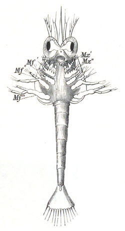
Fig. 219. Larva of Hippolyte In Zoæa Stage.
(From Claus.)
Mx´. and Mx´´. maxillæ 1 and 2; Mf´.
Mf´´. Mf´´´. maxillipeds.
From the Zoæa stage the larva passes into the form known as
Acanthosoma, which represents the Mysis stage of Penæus. The complex
spikes on the dorsal shield of the Zoæa stage are reduced to simple
spines, but the spines of the tail still retain their full size. In
the appendages the chief changes consist (1) in the reduction of the
jointed outer ramus of the second pair of antennæ to a stump
representing the scale, and the elongation of the inner one to the
flagellum; (2) in the elongation of the five ambulatory thoracic
appendages into biramous feet, like the maxillipeds, and in the
sprouting forth of rudimentary abdominal feet.
The most obvious external indications of the passage from the
Acanthosoma to the Mastigopus stage (fig. 218) are to be found in the
elongation of the abdomen, the reduction and flattening of the
cephalo-thoracic shield, and the nearly complete obliteration of all
the spines but the anterior. The eyes on their elongated stalks are
still very characteristic, and the elongation of the flagellum of the
second pair of antennæ is very striking.
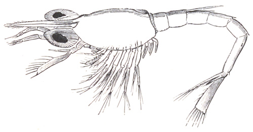
Fig. 220. Older Larva of Hippolyte after the
thoracic appendages have become formed. (From Claus.)
The maxillæ and maxillipeds undergo considerable metamorphosis, the
abdominal feet attain their adult form, and the three anterior
thoracic ambulatory legs lose their outer rami. The most remarkable
change of all concerns the two last pairs of thoracic appendages,
which, instead of being metamorphosed like the preceding ones, are
completely or nearly completely thrown off in the moult which
inaugurates the Mastigopus stage, and are subsequently redeveloped.
With the reappearance of these appendages, and the changes in the
other appendages already indicated, the adult form is practically
attained.
With reference to the development of the majority of the Carabidæ,
Penæinæ, Palæmoninæ, Crangoninæ, it may be stated generally that they
leave the egg in the Zoæa stage (fig. 219) with anterior appendages up
to the third pair of maxillipeds. The thorax is unsegmented and indeed
almost unrepresented, but the abdomen is long and divided into
distinct segments. Both thoracic and abdominal appendages are absent,
and the tail is formed by a simple plate with numerous bristles, not
forked, as in the case of the Zoæa of Fritz Müller’s Penæus and
Sergestes. A dorsal spine is frequently found on the second abdominal
segment. From the Zoæa form the embryo passes into a Mysis stage (fig.
220), during which the thoracic appendages gradually appear as
biramous swimming feet; they are all developed before any of the
abdominal appendages, except the last. In some cases the development
is still further abbreviated. Thus the larvæ of Crangon and
Palæmonetes (Faxon, No. 476) possess at hatching the rudiments of the
two anterior pairs of thoracic feet, and Palæmon of three pairs[190].
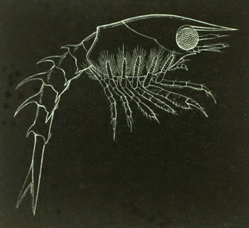
Fig. 221. Newly-hatched Larva of the American
Lobster.
(After Smith.)
Amongst the other Macrura the larva generally leaves the egg as a Zoæa
similar to that of the prawns. In the case of the Thalassinidæ
and Paguridæ a Mysis stage has disappeared. The most remarkable
abbreviations of the typical development are presented on the one hand
by Homarus and Astacus, and on the other by the Loricata.
The development of Homarus has been fully worked out by S. J. Smith
(No. 491) for the American lobster (Homarus americanus). The larva
(fig. 221) leaves the egg in an advanced Mysis stage. The
cephalo-thoracic shield is fully developed, and armed with a rostrum
in front. The first pair of antennæ is unjointed but the second is
biramous, the outer ramus forming a large Mysis-like scale. The
mandibles, which are palped, the maxillæ, and the two anterior
maxillipeds differ only in minor details from the same appendages of
the adult. The third pair of maxillipeds is Mysis-like and biramous,
and the five ambulatory legs closely resemble them, the endopodite of
the first being imperfectly chelate. The abdomen is well developed but
without appendages. The second, third, fourth and fifth segments are
armed with dorsal and lateral spines.
In the next stage swimming feet have appeared on the second, third,
fourth and fifth abdominal segments, and the appendages already
present have approached their adult form. Still later, when the larva
is about half an inch in length, the approach to the adult form is
more marked, and the exopodites of the ambulatory legs though present
are relatively much reduced in size. The swimmerets of the sixth
abdominal segment are formed. In the next stage observed the larva has
entirely lost its Schizopod characters, and though still retaining its
free-swimming habits differs from the adult form only in generic
characters.
As has been already stated, no free larval stages occur in the
development of Astacus, but the young is hatched in a form in which it
differs only in unimportant details from the adult.
The peculiar larval form of the Loricata (Scyllarus, Palinurus) has
long been known under the name Phyllosoma (fig. 222 C), but its true
nature was first shewn by Couch (No. 474) [Couch did not however
recognise the identity of his larva with Phyllosoma; this was first
done by Gerstäcker] and shortly afterwards by Gerbe and Coste. These
observations were however for a long time not generally accepted, till
Dohrn (No. 477) published his valuable memoir giving an account of how
he succeeded in actually rearing Phyllosoma from the eggs of Scyllarus
and Palinurus, and shewing that some of the most remarkable features
of the metamorphosis of the Loricata occur before the larva is
hatched.
The embryo of Scyllarus in the egg first of all passes through the
usual Nauplius stage, and then after the formation of a cuticle
develops an elongated thoracico-abdominal region bent completely over
the anterior part of the body. There appear moreover a number of
appendages and the rudiments of various organs; and the embryo passes
into a form which may be described as the embryonic Phyllosoma stage.
In this stage there are present on the anterior part of the body, in
front of the ventral flexure, two pairs of antennæ, mandibles,
two pairs of maxillæ, the second commencing to be biramous, and a
small stump representing the first pair of maxillipeds. The part
of the body bent over consists of a small quadrate caudal plate, and
an appendage-bearing region to which are attached anteriorly three
pairs of biramous appendages—the second and third maxillipeds, and
the anterior pair of ambulatory legs—and two pairs of undivided
appendages—the second and third pairs of ambulatory legs. In a
slightly later stage the first pair of maxillæ becomes biramous, as
also does the first pair of maxillipeds in a very rudimentary fashion.
The second and third pairs of ambulatory legs become biramous, while
the second and third maxilliped nearly completely lose their outer
ramus. Very small rudiments of the two hinder ambulatory legs become
formed. If the embryo is taken at this stage (vide fig. 222 A,
which represents a nearly similar larva of Palinurus) out of the egg,
it is seen to consist of (1) an anterior enlargement with a vaulted
dorsal shield enclosing the yolk, two stalked eyes, and a median eye;
(2) a thoracic region in which the indications of segmentation are
visible with the two posterior pairs of maxillipeds (mxp2 and
mxp3) and the ambulatory legs (p1); (3) an abdominal
region distinctly divided into segments and ending in a fork.
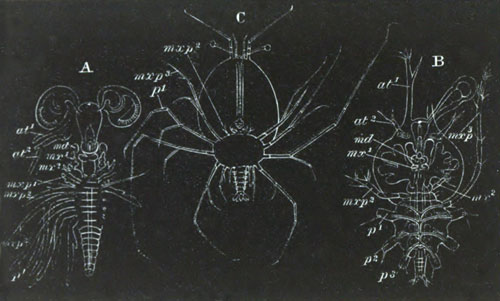
Fig. 222. Larvæ of the Loricata. (After
Claus.)
A. Embryo of Palinurus shortly before hatching.
B. Young Phyllosoma larva of Scyllarus, without the first
maxilliped, the two last thoracic appendages, or the abdominal
appendages.
C. Fully-grown Phyllosoma with all the Decapod appendages.
at1. antenna of first pair; at2. antenna of second
pair; md. mandible; mx1. first maxilla; mx2.
second maxilla; mxp1-mxp3. maxillipeds; p1-p3.
thoracic appendages.
Before the embryo becomes hatched the first pair of maxillipeds
becomes reduced in size and finally vanishes. The second pair of
maxillæ becomes reduced to simple stumps with a few bristles, the
second pair of antennæ also appears to undergo a retrogressive
change, while the two last thoracic segments cease to be
distinguishable. It thus appears that during embryonic life the second
pair of antennæ, the second pair of maxillæ, and the second and third
pair of maxillipeds and the two hinder ambulatory appendages undergo
retrogressive changes, while the first pair of maxillipeds is
completely obliterated!
The general form of the larva when hatched (fig. 222 B) is not very
different from that which it has during the later stages within the
egg. The body is divided into three regions: (1) an anterior cephalic,
(2) a middle thoracic, and (3) a small posterior abdominal portion;
and all of them are characterised by their extreme dorso-ventral
compression, so that the whole animal has the form of a three-lobed
disc, the strange appearance of which is much increased by its
glass-like transparency.
The cephalic portion is oval and projects slightly behind so as to
overlap the thorax. Its upper surface constitutes the dorsal shield,
from which there spring anteriorly the two compound eyes on long
stalks, between which is a median Nauplius eye. The mouth is situated
about the middle of the under surface of the anterior disc. It leads
into a stomach from which an anterior and a lateral hepatic
diverticulum springs out on each side. The former remains as a simple
diverticulum through larval life, but the latter becomes an extremely
complicated glandular structure.
At the front border of the disc is placed the unjointed but elongated
first pair of antennæ (at1). Externally to and behind these
there spring the short posterior antennæ (at2), at the base of
which the green gland is already formed. Surrounding the mouth are the
mandibles (md) and anterior pair of maxillæ (mx1), and
some distance behind the second pair of maxillæ (mx2),
consisting of a cylindrical basal joint and short terminal joint armed
with bristles. The first pair of maxillipeds is absent.
The thoracic region is formed of an oval segmented disc attached to
the under surface of the cephalic disc. From its front segment arises
the second pair of maxillipeds (mxp2) as single five-jointed
appendages, and from the next segment springs the five-jointed
elongated but uniramous third pair of maxillipeds (mxp3), and
behind this there arise three pairs of six-jointed ambulatory
appendages (p1, p2, p3, of which only the basal
joint is represented in the figure) with an exopodite springing from
their second joint. The two posterior thoracic rings and their
appendages cannot be made out.
The abdomen is reduced to a short imperfectly segmented stump, ending
in a fork, between the prongs of which the anus opens. Even the
youngest larval Phyllosoma, such as has just been described, cannot be
compared with a Zoæa, but belongs rather, in the possession of
biramous thoracic feet, to a Mysis stage. In the forked tail and
Nauplius eye there appear however to be certain very primitive
characters carried on to this stage.
The passage of this young larva to the fully formed Phyllosoma (fig.
222 C) is very simple. It consists essentially in the fresh
development of the first pair of maxillipeds and the two last
ambulatory appendages, the growth and segmentation of the abdomen, and
the sprouting on it of biramous swimming feet. In the course
of these changes the larva becomes a true Decapod in the arrangement
and number of its appendages; and indeed it was united with this group
before its larval character was made out. In addition to the
appearance of new appendages certain changes take place in those
already present. The two posterior maxillipeds, in the Palinurus
Phyllosoma at any rate, acquire again an exopodite, and together with
the biramous ambulatory feet develop epipodites in the form of gill
pouches.
The mode of passage of the Phyllosoma to the adult is not known, but
it can easily be seen from the oldest Phyllosoma forms that the dorsal
cephalic plate grows over the thorax, and gives rise to the
cephalo-thoracic shield of the adult.
There are slight structural differences, especially in the antennæ,
between the Phyllosoma of Scyllarus and that of Palinurus, but the
chief difference in development is that the first pair of maxillipeds
of the Palinurus embryo, though reduced in the embryonic state, does
not completely vanish, at any rate till after the free larval state
has commenced; and it is doubtful if it does so even then. The freshly
hatched Palinurus Phyllosoma is very considerably more developed than
that of Scyllarus.
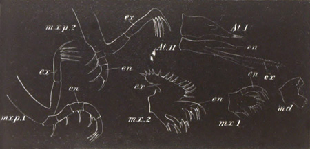
Fig. 223. The Appendages of a Crab Zoæa.
At. I. first antenna; At. II. second antenna; md.
mandible (without a palp); mx. 1. first maxilla; mx.
2. second maxilla; mx. 3. third maxilla; mxp. 1.
first maxilliped; mxp. 2. second maxilliped.
ex. exopodite; en. endopodite.
Brachyura. All the Brachyura, with the exception of one or more
species of land crabs[191], leave the egg in the Zoæa condition, and
though there are slight variations of structure, yet on the whole the
Crab Zoæa is a very well marked form. Immediately after leaving the
egg (fig. 210) it has a somewhat oval shape with a long
distinctly segmented abdomen bent underneath the thorax. The
cephalo-thoracic shield covers over the front part of the body, and is
prolonged into a long frontal spine pointing forwards, and springing
from the region between the two eyes; a long dorsal spine pointing
backwards; and two lateral spines.
To the under surface of the body are attached the anterior appendages
up to the second maxilliped, while the six following pairs of thoracic
appendages are either absent or represented only in a very rudimentary
form. The abdomen is without appendages.
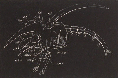
Fig. 224. Crab Zoæa after the third pair of
maxillipeds and the thoracic and abdominal appendages have become
developed.
at1. antenna of first pair; at2. antenna of second
pair; mx1. first maxilla; mx2. second maxilla;
mxp1. first maxilliped; mxp2. second maxilliped;
mxp3. third maxilliped; oc. eye; ht. heart.
The anterior antennæ are single and unjointed, but provided at their
extremity with a few olfactory hairs (only two in Carcinus Mœnas)
and one or two bristles. The rudiment of the secondary flagellum
appears in very young Zoææ on the inner side of the antennules (fig.
223 At. I.). The posterior antennæ are without the flagellum,
but are provided with a scale representing the exopodite (fig. 223
At. II. ex) and usually a spinous process. The flagellum is
very early developed and is represented in fig. 223, At. II.
en. The mandibles (md) are large but without a palp. The
anterior maxillæ (mx 1) have a short two-jointed endopodite
(palp) with a few hairs, and a basal portion with two blades,
of which the distal is the largest, both armed with stiff bristles.
The posterior maxillæ have a small respiratory plate (exopodite), an
endopodite (palp) shaped like a double blade, and two basal joints
each continued into a double blade. The two maxillipeds (mxp 1
and mxp 2) have the form and function of biramous swimming
feet. The exopodite of both is two-jointed and bears long bristles at
its extremity; the endopodite of the anterior is five-jointed and
long, that of the second is three-jointed and comparatively short.
In the six-jointed tail the second segment has usually two dorsally
directed spines, and the three succeeding segments each of them two
posteriorly directed. The telson or swimming plate is not at first
separated from the sixth segment; on each side it is prolonged into
two well-marked prongs; and to each prong three bristles are usually
attached (fig. 224). The heart (fig. 224 ht) lies under the
dorsal spine and is prolonged into an anterior, posterior, and dorsal
aorta. It has only two pairs of venous ostia.
During the Zoæa stage the larva rapidly grows in size, and undergoes
considerable changes in its appendages which reach the full Decapod
number (fig. 224). On both pairs of antennæ a flagellum becomes
developed and grows considerably in length. Before the close of the
Zoæa condition a small and unjointed palp appears on the mandible.
Behind the second maxilliped the third maxilliped (mxp3) early
appears as a small biramous appendage, and the five ambulatory feet
become distinctly formed as uniramous appendages—the exopodites not
being present. The third pair of maxillipeds and three following
ambulatory appendages develop gill pouches. The abdominal feet are
formed on the second to the sixth segments of the tail as simple
pouches.
The oldest Zoæa is transmuted at its moult into a form known as
Megalopa, which is really almost identical with an anomurous Decapod.
No Schizopod stage is intercalated, which shews that the development
is in many respects greatly abbreviated. The essential characters of
the Megalopa are to be found in (1) the reduction of the two anterior
maxillipeds, which cease to function as swimming feet, and together
with the appendages in front of them assume the adult form; (2) the
full functional development of the five ambulatory appendages;
(3) the reduction of the forked telson to an oval swimming plate, and
the growth in size of the abdominal feet, which become large swimming
plates and are at the same time provided with short endopodites which
serve to lock the feet of the two sides.
With these essential characters the form of the Megalopa differs
considerably in different cases. In some instances (e.g.
Carcinus mœnas) the Zoæa spines of the youngest Megalopa are so
large that the larva appears almost more like a Zoæa than a Megalopa
(Spence Bate, No. 470). In other cases, e.g. that represented
on fig. 225, the Zoæa spines are still present but much reduced; and
the cephalo-thoracic shield has very much the adult form. In other
cases again (e.g. Portunus) the Zoæa spines are completely
thrown off at the youngest Megalopa stage.
There is a gradual passage from the youngest Megalopa to the adult
form by a series of moults.
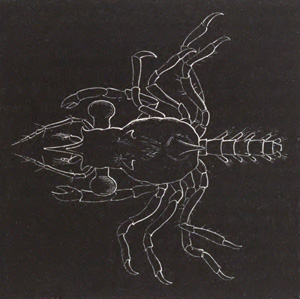
Fig. 225. Megalopa stage of Crab larva.
Some of the brachyurous Zoæa forms exhibit considerable divergences
from the described type, more especially in the armature of the
shield. In some forms the spines are altogether absent, e.g.
Maja (Couch, No. 474) and Eurynome. In other forms the frontal spine
may be much reduced or absent (Inachus and Achæus). The dorsal spine
may also be absent, and in one form described by Dohrn (No. 478) there
is a long frontal spine and two pairs of lateral spines, but no dorsal
spine. Both dorsal and frontal spines may attain enormous dimensions
and be swollen at their extremities (Dohrn). A form has been described
by Claus as Pterocaris in which the cephalo-thoracic shield is
laterally expanded into two wing-like processes.
The Zoæa of Porcellana presents on the whole the most remarkable
peculiarities and, as might be anticipated from the systematic
position of the adult, is in some respects intermediate between the
macrurous Zoæa and that of the Brachyura. It is characterized by the
oval form of the body, and by the presence of one enormously
long frontal spine and two posterior spines. The usual dorsal spine is
absent. The tail plate is rounded and has the character of the tail of
a macrurous Zoæa, but in the young Zoæa the third pair of maxillipeds
is absent and the appendages generally have a brachyurous character. A
Megalopa stage is hardly represented, since the adult may almost be
regarded as a permanent Megalopa.
Stomatopoda. The history of the larval forms of the Stomatopoda
(Squilla etc.) has not unfortunately been thoroughly worked out, but
what is known from the researches of Fritz Müller (No. 495) and Claus
(No. 494) is of very great importance. There are it appears two types,
both of which used to be described as adult forms under the respective
names Erichthus and Alima.
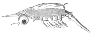
Fig. 226. Second stage of Erichthus larva of
Squilla with five maxillipeds and the first pair of abdominal
appendages. (From Claus.)
The youngest known Erichthus form is about two millimetres in length,
and has the characters of a modified Zoæa (fig. 226). The body is
divided into three regions, an anterior unsegmented region to which
are attached two pairs of antennæ, mandibles, and maxillæ (two pairs).
This portion has a dorsal shield covering the next or middle region,
which consists of five segments each with a pair of biramous
appendages. These appendages represent the five maxillipeds of the
adult[192]. The portion of the body behind this is without appendages.
It consists of three short anterior segments,—the three posterior
thoracic segments of the adult,—and a long unsegmented tail. The
three footless thoracic segments are covered by the dorsal shield.
Both pairs of antennæ are uniramous and comparatively short. The
mandibles, like those of Phyllopods, are without palps, and the two
following pairs of maxillæ are small. The five maxillipeds have the
characters of normal biramous Zoæa feet. From the front of the head
spring a pair of compound eyes with short stalks, which grow longer in
the succeeding stages; between them is a median eye. The dorsal shield
is attached just behind this eye, and is provided, as in the typical
Zoæa, with a frontal spike—while its hinder border is produced into
two lateral spikes and one median. In a larva of about three
millimetres a pair of biramous appendages arises behind the three
footless thoracic segments. It is the anterior pair of abdominal feet
(fig. 226). The inner ramus of the second pair of maxillipeds soon
grows greatly in length, indicating its subsequent larger size and
prehensile form (fig. 227 g). When the larva after one or two
moults attains a length of six millimetres (fig. 227) the abdomen has
six segments (the sixth hardly differentiated), each with a
pair of appendages (the two hindermost still rudimentary) which have
become gradually developed from before backwards. The three hindermost
thoracic segments are still without appendages.
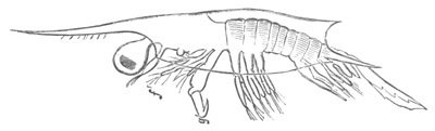
Fig. 227. Advanced Erichthus larva of Squilla
with five pairs of abdominal appendages. (From Claus.)
f. first maxilliped; g. second maxilliped.
Some changes of importance have occurred in the other parts. Both
antennæ have acquired a second flagellum, but the mandible is still
without a palp. The first and second pair of maxillipeds have both
undergone important modifications. Their outer ramus (exopodite) has
been thrown off, and a gill plate (epipodite) has appeared as an
outgrowth from their basal joint. Each of them is composed of six
joints. The three following biramous appendages have retained their
earlier characters but have become much reduced in size. In the
subsequent moults the most remarkable new features concern the three
posterior maxillipeds, which undergo atrophy, and are either
completely lost or reduced to mere unjointed sacks (fig. 228). In
the stage where the complete Erichthus type has been reached, these
three appendages have again sprouted forth in their permanent form and
each of them is provided with a gill sack on its coxal joint. Behind
them the three ambulatory appendages of the thorax have also appeared,
first as simple buds, which subsequently however become biramous. On
their development the full number of adult appendages is acquired.
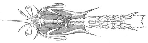
Fig. 228. Advanced Erichthus larva of Squilla
when the three posterior maxillipeds have become reduced to minute
pouches. (From Claus.)
The most noteworthy points in the developmental history detailed above
are the following:
(1) The thoracic and abdominal segments (apart from their appendages)
develop successively from before backwards.
(2) The three last maxillipeds develop before the abdominal feet, as
biramous appendages, but subsequently completely atrophy, and then
sprout out again in their permanent form.
(3) The abdominal feet develop in succession from before backwards,
and the whole series of them is fully formed before a trace of the
appendages of the three hindermost thoracic segments has appeared. It
may be mentioned as a point of some importance that the Zoæa of
Squilla has an elongated many-chambered heart, and not the short
compact heart usually found in the Zoæa.
The younger stages of the Alima larva are not known[193], but the
earliest stage observed is remarkable for presenting no trace of the
three posterior pairs of maxillipeds, or of the three following pairs
of thoracic appendages. The segments belonging to these appendages are
however well developed. The tail has its full complement of segments
with the normal number of well-developed swimming feet. The larva
represents in fact the stage of the Erichthus larva when the three
posterior pairs of maxillipeds have undergone atrophy; but it is
probable that these appendages never become developed in this form of
larva.
Apart from the above peculiarities the Alima form of larva closely
resembles the Erichthus form.
Nebaliadæ. The development of Nebalia is abbreviated, but from
Metschnikoff’s figures[194] may be seen to resemble closely that of
Mysis. The abdomen has comparatively little yolk, and is bent over the
ventral surface of the thorax. There is in the egg a Nauplius stage
with three appendages, and subsequently a stage with the Zoæa
appendages.
The larva when it leaves the egg has the majority of its appendages
formed, but is still enveloped in a larval skin, and like Mysis bends
its abdomen towards the dorsal side. When the larva is finally hatched
it does not differ greatly from the adult.
Cumaceæ. The development of the Cumaceæ takes place for the
most part within the egg, and has been shewn by Dohrn (No. 496) to
resemble in many points that of the Isopods. A dorsal organ is
present, and a fold is formed immediately behind this which gives to
the embryo a dorsal flexure. Both of these features are eminently
characteristic of the Isopoda.
The formation of the two pairs of antennæ, mandibles, and two pairs of
maxillæ and the following seven pairs of appendages takes place very
early. The pair of appendages behind the second maxillæ assumes an
ambulatory form, and exhibits a Schizopod character very early,
differing in both these respects from the homologous appendages in the
Isopoda. The cephalo-thoracic shield commences to be formed when the
appendages are still quite rudimentary as a pair of folds in the
maxillary region. The eyes are formed slightly later on each
side of the head, and only coalesce at a subsequent period to form the
peculiar median sessile eye of the adult.
The two pairs of appendages behind the second maxillæ become converted
into maxillipeds, and the exopodite of the first of them becomes the
main ramus, while in the externally similar second maxilliped the
exopodite atrophies and the endopodite alone remains.
The larva is hatched without the last pair of thoracic limbs or the
abdominal appendages (which are never developed in the female), but in
other respects closely resembles the adult. Before hatching the dorsal
flexure is exchanged for a ventral one, and the larva acquires a
character more like that of a Decapod.
Copepoda.
Natantia. The free Copepoda are undoubtedly amongst the lowest
forms of those Crustacea which are free or do not lead a parasitic
existence. Although some features of their anatomy, such for instance
as the frequent absence of a heart, may be put down to a retrogressive
development, yet, from their retention of the median frontal eye of
the Nauplius as the sole organ of vision[195], their simple biramous
swimming legs, and other characters, they may claim to be very
primitive forms, which have diverged to no great extent from the main
line of Crustacean development. They supply a long series of
transitional steps from the Nauplius stage to the adult condition.
While still within the egg-shell the embryo is divided by two
transverse constrictions into three segments, on which the three
Nauplius appendages are developed, viz. the two pairs of antennæ and
the mandibles. When the embryo is hatched the indication of a division
into segments has vanished, but the larva is in the fullest sense a
typical Nauplius[196]. There are slight variations in the shape of the
Nauplius in different genera, but its general form and character are
very constant. It has (fig. 229 A) an oval unsegmented body with three
pairs of appendages springing from the ventral surface. The anterior
of these (at 1) is uniramous, and usually formed of three
joints which bear bristles on their under surface. The two posterior
pairs of appendages are both biramous. The second pair of
antennæ (at 2) is the largest. Its basal portion (protopodite)
bears on its inner side a powerful hook-like bristle. The outer ramus
is the longest and many-jointed; the inner ramus has only two joints.
The mandibles (md), though smaller than the second pair of
antennæ, have a nearly identical structure. No blade-like projection
is as yet developed on their protopodite. Between the points of
insertion of the first pair of antennæ is the median eye (oc),
which originates by the coalescence of two distinct parts. The mouth
is ventral, and placed in the middle line between the second pair of
antennæ and the mandibles: it is provided with an unpaired upper lip.
There are two bristles at the hind end of the embryo between which the
anus is placed; and in some cases there is at this part a slight
indication of the future caudal fork.
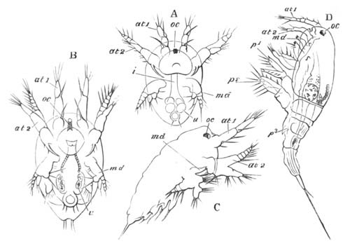
Fig. 229. Successive stages in the development of
Cyclops tenuicornis.
(Copied from Bronn; after Claus.)
A. B. and C. Nauplius stages. D. Youngest Copepod stage. In this
figure maxillæ and the two rami of the maxilliped are seen
immediately behind the mandible md.
oc. eye; at1. first pair of antennæ; at2.
second pair of antennæ; md. mandible; p1. first pair
of feet; p2. second pair of feet; p3. third pair of
feet; u. excretory concretions in the intestine.
The larva undergoes a number of successive ecdyses, at each of which
the body becomes more elongated, and certain other changes
take place. First of all a pair of appendages arises behind the
mandibles, which form the maxillæ (fig. 229 B); at the same time the
basal joint of the maxillæ develops a cutting blade. Three successive
pairs of appendages (fig. 229 C) next become formed—the so-called
maxillipeds (the homologues of the second pair of maxillæ), and the
two first thoracic limbs. Each of these though very rudimentary is
nevertheless bifid. The body becomes greatly elongated, and the caudal
fork more developed.
Up to this stage of development the Nauplius appendages have retained
their primitive character almost unaltered; but after a few more
ecdyses a sudden change takes place; a cephalo-thoracic shield becomes
fully developed, and the larva comes to resemble in character an adult
Copepod, from which it mainly differs in the smaller number of
segments and appendages. In the earliest ‘Cyclops’ stage the same
number of appendages are present as in the last Nauplius stage. There
(fig. 229 D) is a well-developed cephalo-thorax, and four free
segments behind it. To the cephalo-thoracic region the antennæ,
mandibles, maxillæ, the now double pair of maxillipeds (derived from
the original single pair of appendages), and first pair of thoracic
appendages (p1) are attached. The second pair of thoracic
appendages (p2) is fixed to the first free segment, and the
rudiment of a third pair (p3) projects from the second free
segment. The first pair of antennæ has grown longer by the addition of
new joints, and continues to increase in length in the following
ecdyses till it attains its full adult development, and then forms the
chief organ of locomotion. The second pair of antennæ is much reduced
and has lost one of its rami. The two rami of the mandibles are
reduced to a simple palp, while the blade has assumed its full
importance. The maxillæ and following appendages have greatly
increased in size. They are all biramous, though the two rami are not
as yet jointed. The adult state is gradually attained after a number
of successive ecdyses, at which new segments and appendages are added,
while new joints are formed for those already present.
Parasita. The earliest developmental stages of the parasitic
types of Copepoda closely resemble those of the free forms, but, as
might be expected from the peculiarly modified forms of the adult,
they present a large number of secondary characters. So far as
is known a more or less modified Nauplius larva is usually preserved.
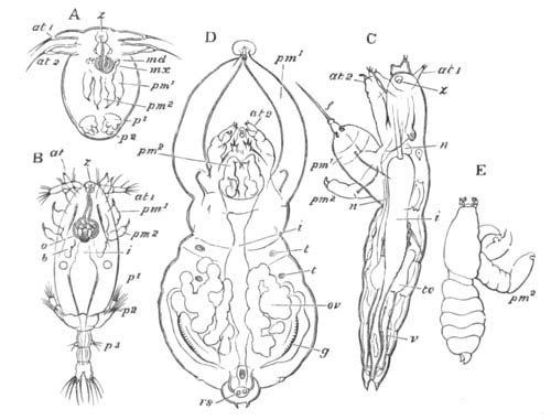
Fig. 230. Successive stages in the development of
Achtheres percarum.
(Copied from Bronn; after Claus.)
A. Modified Nauplius stage. B. Cyclops stage. C. Late stage of male
embryo. D. Sexually mature female. E. Sexually mature male.
at1. first pair of antennæ; at2. second pair of
antennæ; md. mandible; mx. maxillæ; pm1. outer
pair of maxillipeds; pm2. inner pair of maxillipeds;
p1. first pair of legs; p2. second pair of legs;
z. frontal organ; i. intestine; o. larval eye;
b. glandular body; t. organ of touch; ov.
ovary; f. rod projecting from coalesced maxillipeds;
g. cement gland; rs. receptaculum seminis; n.
nervous system; te. testis; v. vas deferens.
The development of Achtheres percarum, one of the Lernæopoda parasitic
in the mouth, etc. of the common Perch, may be selected to illustrate
the mode of development of these forms. The larva leaves the egg as a
much simplified Nauplius (fig. 230 A). It has an oval body with only
the two anterior pairs of Nauplius appendages; both of them in the
rudimentary condition of unjointed rods. The usual median eye is
present, and there is also found a peculiar sternal papilla, on which
opens a spiral canal filled with a glutinous material, which is
probably derived from a gland which disappears on the completion of
the duct. The probable function of this organ is to assist at a later
period in the attachment of the parasite to its host. Underneath the
Nauplius skin a number of appendages are visible, which become
functional after the first ecdysis. This takes place within a few
hours after the hatching of the Nauplius, and the larva then passes
from this rudimentary Nauplius stage into a stage
corresponding with the Cyclops stage of the free forms (fig. 230 B).
In the Cyclops stage the larva has an elongated body with a large
cephalo-thoracic shield, and four free posterior segments, the last of
which bears a forked tail.
There are now present eight pairs of appendages, viz. antennæ (two
pairs), mandibles, maxillæ, maxillipeds, and three pairs of swimming
feet. The Nauplius appendages are greatly modified. The first pair of
antennæ is three-jointed, and the second biramous. The outer ramus is
the longest, and bears a claw-like bristle at its extremity. This pair
of appendages is used by the larva for fixing itself. The mandibles
are small and connected with the proboscidiform mouth; and the single
pair of maxillæ is small and palped. The maxillipeds (pm1 and
pm2) are believed by Claus to be primitively a single biramous
appendage, but early appear as two distinct structures[197], the outer
and larger of which becomes the main organ by which the larva is
fixed. Both are at this stage simple two-jointed appendages. The two
anterior pairs of swimming feet have the typical structure, and
consist of a protopodite bearing an unjointed exopodite and
endopodite. The first pair is attached to the cephalo-thorax and the
second (p2) to the first free thoracic segment. The third pair
is very small and attached to the second free segment. The mouth is
situated at the end of a kind of proboscis formed by prolongations of
the upper and lower lips. The alimentary tract is fairly simple, and
the anus opens between the caudal forks.
Between this and the next known stage it is quite possible that one or
more may intervene. However this may be the larva in the next stage
observed (fig. 230 C) has already become parasitic in the mouth of the
Perch, and has acquired an elongated vermiform aspect. The body is
divided into two sections, an anterior unsegmented, and a posterior
formed of five segments, of which the foremost is the first thoracic
segment which in the earlier stage was fused with the cephalo-thorax.
The tail bears a rudimentary fork between the prongs of which the anus
opens. The swimming feet have disappeared, so also has the eye and the
spiral duct of the embryonic frontal organ. The outer of the two
divisions of the maxilliped have undergone the most important
modification, in that they have become united at their ends, where
they form an organ from which an elongated rod (f) projects,
and attaches the larva to the mouth or gills of its host. The antennæ
and jaws have nearly acquired their adult form. The nervous system
consists of supra- and infra-œsophageal ganglia and two lateral
trunks given off from the latter. At this stage the males and females
can already be distinguished, not only by certain differences in the
rudimentary generative organs, but also by the fact that the outer
branch of the maxillipeds is much longer in the female than in the
male, and projects beyond the head.
In the next ecdysis the adult condition is reached. The outer
maxillipeds of the male (fig. 230 E, pm2) separate
again; while in the female (fig. 230 D) they remain fused and develop
a sucker. The male is only about one-fifth the length of the female.
In both sexes the abdomen is much reduced.
In the genera Anchorella, Lernæopoda, Brachiella and Hessia, Ed.
van Beneden (No. 506) has shewn that the embryo, although it
passes through a crypto-Nauplius stage in the egg, is when hatched
already in the Cyclops stage.
Branchiura. The peculiar parasite Argulus, the affinities of
which with the Copepoda have been demonstrated by Claus (No. 511), is
hatched in a Cyclops stage, and has no Nauplius stage. At the time of
hatching it closely resembles the adult in general form. Its
appendages are however very nearly those of a typical larval Copepod.
The body is composed of a cephalo-thorax and free region behind this.
The cephalo-thorax bears on its under surface antennæ (two pairs),
mandibles, maxillipeds, and the first pair of thoracic feet.
The first pair of antennæ is three-jointed, but the basal joint bears
a hook. The second pair is biramous, the inner ramus terminating in a
hook. The mandible is palped, but the palp is completely separated
from the cutting blade[198]. The maxilla would, according to Claus,
appear to be absent.
The two typical divisions of the Copepod maxillipeds are present, viz.
an outer and anterior larger division, and an inner and posterior
smaller one. The first pair of thoracic feet, as is usual amongst
Copepoda, is attached to the cephalo-thorax. It has not the typical
biramous Copepod character. There are four free segments behind the
cephalo-thorax, the last of which ends in a fork. Three of them bear
appendages, which are rudimentary in this early larval stage. On the
dorsal surface are present paired eyes as well as an unpaired median
eye.
Between the larval condition and that of the adult a number of ecdyses
intervene.
Cirripedia.
The larvæ of all the Cirripedia, with one or two exceptions, leave the
egg in the Nauplius condition. The Nauplii differ somewhat in the
separate groups, and the post-nauplial stages vary not inconsiderably.
It will be most convenient to treat successively the larval
history of the four sub-orders, viz. Thoracica, Abdominalia, Apoda,
and Rhizocephala.
Thoracica. The just hatched larvæ at once leave the egg lamellæ
of their parent. They pass out through an opening in the mantle near
the mouth, and during this passage the shell of the parent is opened
and the movements of the cirriform feet cease.
The larval stages commence with a Nauplius[199] which, though regarded
by Claus as closely resembling the Copepod Nauplius (figs. 231 and 232
A), certainly has very marked peculiarities of its own, and in some
respects approaches the Phyllopod Nauplius. It is in the youngest
stage somewhat triangular in form, and covered on the dorsal side by a
very delicate and hardly perceptible dorsal shield, which is prolonged
laterally into two very peculiar conical horns (fig. 231 lh),
which are the most characteristic structures of the Cirriped Nauplius.
They are connected with a glandular mass, the secretion from which
passes out at their apex. Anteriorly the dorsal shield has the same
extension as the body, but posteriorly it projects slightly.
An unpaired eye is situated on the ventral surface of the head, and
immediately behind it there springs a more or less considerable upper
lip (lb), which resembles the Phyllopod labrum rather than that
of the Copepoda. Both mouth and anus are present, and the hind end of
the body is slightly forked in some forms, but ends in others,
e.g. Lepas fascicularis, in an elongated spine. The anterior of
the three pairs of Nauplius appendages (At1) is uniramous, and
the two posterior (At2, and md) are biramous. From the
protopodites of both the latter spring strong hooks like those of the
Copepod and Phyllopod Nauplii. In some Nauplii, e.g. that of
Balanus, the appendages are at first not jointed, but in other
Nauplii, e.g. that of Lepas fascicularis, the jointing is well
marked. In Lepas fascicularis the earliest free Nauplius is enveloped
in a larval skin, which is thrown off after a few hours. The Nauplii
of all the Thoracica undergo a considerable number of moults before
their appendages increase in number or segmentation of the body
appears. During these moults they grow larger, and the posterior part
of the body—the future thoracic and abdominal region—grows
relatively in length. There also appear close to the sides of the
unpaired eye two conical bodies, which correspond with the frontal
sense organs of the Phyllopods. During their growth the different
larvæ undergo changes varying greatly in degree.
In Balanus the changes consist for the most part in the full
segmentation of the appendages and the growth and distinctness of the
dorsal shield, which forms a somewhat blunt triangular plate, broadest
in front, with the anterior horns very long, and two short posterior
spines. The tail also becomes produced into a long spine.
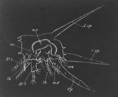
Fig. 231. Nauplius larva of Lepas fascicularis
viewed from the side.
oc. eye; At. 1. antenna of first pair; At. 2.
antenna of second pair; md. mandible; lb. labrum;
an. anus; me. mesenteron; d.sp. dorsal spine;
c.sp. caudal spine; Vp. ventral spine; lh.
lateral horns.
In Lepas fascicularis the changes in appearance of the Nauplius, owing
to a great spinous development on its shield, are very considerable;
and, together with its enormous size, render it a very remarkable
form. Dohrn (No. 520), who was the first to describe it, named it
Archizoæa gigas.
The dorsal shield of the Nauplius of Lepas fascicularis (fig. 231)
becomes somewhat hexagonal, and there springs from the middle of the
dorsal surface an enormously long spine (d.sp), like the dorsal
spine of a Zoæa. The hind end of the shield is also produced into a
long caudal spine (c.sp) between which and the dorsal spine are
some feather-like processes. From its edge there spring in addition to
the primitive frontal horns three main pairs of horns, one pair
anterior, one lateral, and one posterior, and smaller ones in
addition. All these processes (with the exception of the dorsal and
posterior spines) are hollow and open at their extremities, and like
the primitive frontal horns contain the ducts of glands situated under
the shield. On the under surface of the larva is situated the unpaired
eye (oc) on each side of which spring the two-jointed frontal
sense organs. Immediately behind these is the enormous upper lip
(lb) which covers the mouth[200]. At the sides of the lip lie
the three pairs of Nauplius appendages, which are very characteristic
but present no special peculiarities. Posteriorly the body is produced
into a long ventral spine-like process (Vp) homologous with
that of other more normal Nauplii. At the base of this process large
moveable paired spines appear at successive moults, six pairs being
eventually formed. These spines give to the region in which they are
situated a segmented appearance, and perhaps similar structures have
given rise to the appearance of segmentation in Spence Bate’s figures.
The anus is situated on the dorsal side of this ventral process, and
between it and the caudal spine of the shield above. The fact that the
anus occupies this position appears to indicate that the ventral
process is homologous with the caudal fork of the Copepoda, on the
dorsal side of which the anus so often opens[201].
From the Nauplius condition the larvæ pass at a single moult into an
entirely different condition known as the Cypris stage. In preparation
for this condition there appear, during the last Nauplius moults, the
rudiments of several fresh organs, which are more or less developed in
different types. In the first place a compound eye is formed on each
side of the median eye. Secondly there appears behind the mandibles a
fourth pair of appendages—the first pair of maxillæ—and internal to
these a pair of small prominences, which are perhaps
equivalent to the second pair of maxillæ, and give rise to the third
pair of jaws in the adult (sometimes spoken of as the lower lip).
Behind these appendages there are moreover formed the rudiments of six
pairs of feet. Under the cuticle of the first pair of antennæ there
may be seen just before the final moult the four-jointed antennæ of
the Cypris stage with the rudiment of a disc on the second joint by
which the larvæ eventually become attached.
By the free Cypris stage, into which the larva next passes, a very
complete metamorphosis has been effected. The median and paired eyes
are present as before, but the dorsal shield has become a bivalve
shell, the two valves of which are united along their dorsal,
anterior, and posterior margins. The two valves are further kept in
place by an adductor muscle situated close below the mouth. Remains of
the lateral horns still persist. The anterior antennæ have undergone
the metamorphosis already indicated. They are four-jointed, the two
basal joints being long, and the second provided with a suctorial
disc, in the centre of which is the opening of the duct of the
so-called antennary or cement gland, which is a granular mass lying on
the ventral side of the anterior region of the body. The gland arises
(Willemoes Suhm) during the Nauplius stage in the large upper lip. The
two distal joints of the antennæ are short, and the last of them is
provided with olfactory hairs. The great upper lip and second pair of
antennæ and mandibles have disappeared, but a small papilla, forming
the commencement of the adult mandibles, is perhaps developed in the
base of the Nauplius mandibles. The first pair of maxillæ have become
small papillæ and the second pair probably remain. The six posterior
pairs of appendages have grown out as functional biramous swimming
feet, which can project beyond the shell and are used in the
locomotion of the larva. They are composed of two basal joints, and
two rami with swimming hairs, each two-jointed. These feet resemble
Copepod feet, and form the main ground for the views of Claus and
others that the Copepoda and Cirripedia are closely related. They are
regarded by Claus as representing the five pairs of natatory feet of
Copepoda, and the generative appendages of the segment behind these.
Between the natatory feet are delicate chitinous lamellæ, in
the spaces between which the cirriform feet of the adult become
developed. The ventral spinous process of the Nauplius stage is much
reduced, though usually three-jointed. It becomes completely aborted
after the larva is fixed.
In addition to the antennary gland there is present, near the dorsal
side of the body above the natatory feet, a peculiar paired glandular
mass, the origin of which has not been clearly made out, but which is
perhaps equivalent to the entomostracan shell-gland. It probably
supplies the material for the shell in succeeding stages[202].
Fig. 232. Larval forms of the Thoracica.
(From Huxley.)
A. Nauplius of Balanus balanoides. (After Sp. Bate.) B. Pupa stage
of Lepas australis. (After Darwin.)
n. antennary apodemes; t. cement gland with duct to
antenna.
The free Cypris stage is not of long duration; and during it the larva
does not take food. It is succeeded by a stage known as the pupa stage
(fig. 232 B), in which the larva becomes fixed, while underneath the
larval skin the adult structures are developed. This stage fully
deserves its name, since it is a quiescent stage during which no
nutriment is taken. The attachment takes place by the sucker of the
antennæ, and the cement gland (t) supplies the cementing
material for effecting it. A retrogressive metamorphosis of a large
number of the organs sets in, while at the same time the formation of
new adult structures is proceeded with. The eyes become gradually
lost, but the Nauplius eye is retained, though in a rudimentary state,
and the terminal joints of the antennæ with their olfactory hairs are
thrown off. The bivalve shell is moulted about the same time as the
eyes, the skin below it remaining as the mantle. The caudal process
becomes aborted. Underneath the natatory feet, and between the
above-mentioned chitinous lamellæ, the cirriform feet are formed; and
on their completion the natatory feet become thrown off and replaced
by the permanent feet. In the Lepadidæ, in which the metamorphosis of
the pupa stages has been most fully studied, the anterior part of the
body with the antennæ gradually grows out into an elongated stalk,
into which pass the ovaries, which are formed during the Cypris stage.
At the base of the stalk is the protuberant mouth, the appendages of
which soon attain a higher development than in the Cypris stage. At
the front part of it a large upper lip becomes formed. Above the
mantle and between it and the shell there are formed in the Lepadidæ
the provisional valves of the shell. These valves are chitinous, and
have a fenestrated structure, owing to the chitin being deposited
round the margin of the separate epidermis (hypodermis) cells. These
valves in the Lepadidæ “prefigure in shape, size, and direction of
growth, the shelly valves to be formed under and around them” (Darwin,
No. 519, p. 129).
Whatever may be the number of valves in the adult the provisional
valves never exceed five, viz. the two scuta, the two terga and the
carina. They are relatively far smaller than the permanent valves and
are therefore separated by considerable membranous intervals. They are
often preserved for a long time on the permanent calcareous valves. In
the Balanidæ the embryonic valves are membranous and do not overlap,
but do not present the peculiar fenestrated structure of the
primordial valves of the Lepadidæ.
In connection with the moult of the pupa skin, and the conversion of
the pupa into the adult form, a remarkable change in the position
takes place. The pupa lies with the ventral side parallel to and
adjoining the surface of attachment, while the long axis of the body
of the young Cirriped is placed nearly at right angles to the surface
of attachment. This change is connected with the ecdyses of the
antennary apodemes (n), which leave a deep bay on the ventral
surface behind the peduncle. The chitinous skin of the Cirriped passes
round the head of this bay, but on the moult of the pupa skin taking
place becomes stretched out, owing to the posterior part of the larva
bending dorsalwards. It is this flexure which causes the change in the
position of the larva.
In addition to the remarkable external metamorphosis undergone during
the pupa stage, a series of hardly less considerable internal changes
take place, such as the atrophy of the muscles of the antennæ, a
change in the position of the stomach, etc.
Abdominalia. In the Alcippidæ the larva leaves the egg as a
Nauplius, and this stage is eventually followed by a pupa stage
closely resembling that of the Thoracica. There are six pairs of
thoracic natatory legs (Darwin, No. 519). Of these only the first and
the last three are preserved in the adult, the first being bent
forward in connection with the mouth. The body moreover partially
preserves its segmentation, and the mantle does not secrete calcareous
valves.
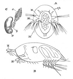
Fig. 233. Stages in the development of the
Rhizocephala. (From Huxley, after Fritz Müller.)
A. Nauplius of Sacculina purpurea.
B. Cypris stage of Lernæodiscus porcellanæ.
C. Adult of Peltogaster paguri.
II. III. IV. Two pairs of antennæ and
mandibles; cp. carapace; a. anterior end of body;
b. generative aperture; c. root-like processes.
The very remarkable genus Cryptophialus, the development of which is
described by Darwin (No. 519) in his classical memoir, is without a
free Nauplius stage. The embryo is at first oval but soon acquires two
anterior processes, apparently the first pair of antennæ, and a
posterior prominence, the abdomen. In a later stage the abdominal
prominence disappears, and the antennary processes, within which the
true antennæ are now visible, are carried more towards the ventral
surface. The larva next passes into the free Cypris stage, during
which it creeps about the mantle cavity of its parent. It is enveloped
in a bivalve shell, and the antennæ have the normal cirriped
structure. There are no other true appendages, but posteriorly three
pairs of bristles are attached to a rudimentary abdomen. Paired
compound eyes are present. During the succeeding pupa stage the
metamorphosis into the adult form takes place, but this has not been
followed out in detail.
In Kochlorine, a form discovered by Noll (No. 526) and closely related
to Cryptophialus, the larvæ found within the mantle represent
apparently two larval stages, similar to two of the larval stages
described by Darwin.
Rhizocephala. The Rhizocephala, as might have been anticipated
from their close relationship to Anelasma squalicola amongst the
Thoracica, undergo a development differing much less from the type of
the Thoracica than that of Cryptophialus and Kochlorine.
Sacculina leaves the egg as a Nauplius (fig. 233 A), which differs
from the ordinary type mainly (1) in the large development of an oval
dorsal shield (cp) which projects far beyond the edge of the
body, but is provided with the typical sternal horns, etc.; and (2) in
the absence of a mouth. The Cypris and pupa stages of Sacculina and
other Rhizocephala (fig. 233 B) are closely similar to those of the
Thoracica, but the paired eye is absent. The attachment takes place in
the usual way, but the subsequent metamorphosis leads to the loss of
the thoracic feet and generally to retrogressive changes.
Ostracoda.
Our knowledge of the development of this remarkable group is entirely
due to the investigations of Claus.
Some forms of Cythere are viviparous, and in the marine form Cypridina
the embryo develops within the valves of the shell. Cypris attaches
its eggs to water plants. The larvæ of Cypris are free, and their
development is somewhat complicated. The whole development is
completed in nine ecdyses, each of them accompanied by more or less
important changes in the constitution of the larva.
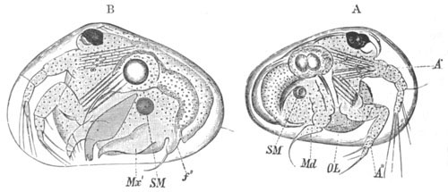
Fig. 234. Two stages in the development of
Cypris. (From Claus.)
A. Earliest (Nauplius) stage. B. Second stage.
A´. A´´. First and second pairs of antennæ; Md.
mandibles; OL. labrum; Mx´. first pair of maxillæ;
f´´. first pair of feet.
In the earliest free stage the larva has the characters of a true
Nauplius with three pairs of appendages (fig. 234 A). The Nauplius
presents however one or two very marked secondary characters. In the
first place it is completely enveloped in a fully formed bivalve
shell, differing in unessential points from the shell of the adult. An
adductor muscle (SM) for the shell is present. Again the second and
third appendages, though locomotive in function are neither of them
biramous, and the third one already contains a rudiment of the future
mandibular blade, and terminates in an anteriorly directed hook-like
bristle. The first pair of antennæ is moreover very similar to the
second and is used in progression. Neither of the pairs of
antennæ become much modified in the subsequent metamorphosis. The
Nauplius has a single median eye, as in the adult Cypris, and a fully
developed alimentary tract.
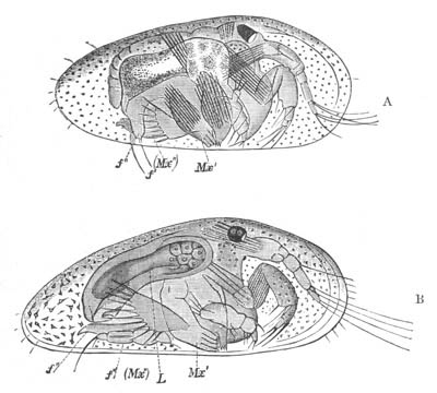
Fig. 235. Stages in the development of
Cypris. (From Claus.)
A. Fourth stage. B. Fifth stage.
Mx´. first maxilla; Mx´´. and f´. second
maxilla; f´´. first pair of feet; L. liver.
The second stage (fig. 234 B), inaugurated by the first moult, is
mainly characterized by the appearance of two fresh pairs of
appendages, viz. the first pair of maxillæ and the first pair of feet;
the second pair of maxillæ not being developed till later. The first
pair appear as leaf-like curved plates (Mx´) more or less like
Phyllopod appendages (Claus) though at this stage without an
exopodite. The first pair of feet (f´´) terminates in a curved
claw and is used for adhering. The mandibles have by this stage fully
developed blades, and have practically attained their adult form,
consisting of a powerful toothed blade and a four-jointed palp.
During the third and fourth stages the first pair of maxillæ acquire
their pectinated gill plate (epipodite) and four blades; and in the
fourth stage (fig. 235 A) the second pair of maxillæ (Mx´´)
arises, as a pair of curved plates, similar to the first pair of
maxillæ at their first appearance. The forked tail is indicated during
the fourth stage by two bristles. During the fifth stage (fig. 235 B)
the number of joints of the first pair of antennæ becomes increased,
and the posterior maxillæ develop a blade and become
four-jointed ambulatory appendages terminating in a hook. The caudal
fork becomes more distinct.
In the sixth stage (fig. 236) the second and hindermost pair of feet
becomes formed (f´´´) and the maxillæ of the second pair lose
their ambulatory function, and begin to be converted into definite
masticatory appendages by the reduced jointing of their palp, and the
increase of their cutting blades. By the seventh stage all the
appendages have practically attained their permanent form; the second
pair of maxillæ has acquired small branchial plates, and the two
following feet have become jointed. In the eighth and ninth stages the
generative organs attain their mature form.
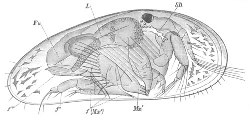
Fig. 236. Sixth stage in the development of
Cypris. (From Claus.)
Mx´. first maxilla; Mx´´. f´. second maxilla;
f´´. and f´´´. first and second pair of feet;
Fu. caudal fork; L. liver; S.D. shell-gland.
The larva of Cythere at the time of birth has rudiments of all the
limbs, but the mandibular palp still functions as a limb, and the
three feet (2nd pair of maxillæ and two following appendages) are very
rudimentary.
The larvæ of Cypridina are hatched in a condition which to all intents
and purposes resembles the adult.
Phylogeny of the Crustacea.
The classical work of Fritz Müller (No. 452) on the phylogeny of the
Crustacea has given a great impetus to the study of their larval
forms, and the interpretations of these forms which he has offered
have been the subject of a very large amount of criticism and
discussion. A great step forward in this discussion has been recently
made by Claus in his memoir (No. 448).
The most fundamental question concerns the meaning of the Nauplius. Is
the Nauplius the ancestral form of the Crustacea, as is believed by
Fritz Müller and Claus, or are its peculiarities and constant
occurrence due to some other cause? The most plausible explanation on
the second hypothesis would seem to be the following. The
segments with their appendages of Arthropoda and Annelida are normally
formed from before backwards, therefore every member of these two
groups with more than three segments must necessarily pass through a
stage with only three segments, and the fact that in a
particular group this stage is often reached on the larva being
hatched is in itself no proof that the ancestor of the group had only
three segments with their appendages. This explanation appears to me,
so far as it goes, quite valid; but though it relieves us from the
necessity of supposing that the primitive Crustacea had only three
pairs of appendages, it does not explain several other peculiarities
of the Nauplius[203]. The more important of these are the following.
1. That the mandibles have the form of biramous swimming feet and are
not provided with a cutting blade.
2. That the second pair of antennæ are biramous swimming feet with a
hook used in mastication, and are innervated (?) from the
subœsophageal ganglion.
3. The absence of segmentation in the Nauplius body. An absence which
is the more striking in that before the Nauplius stage is fully
reached the body of the embryo is frequently divided into three
segments, e.g. Copepoda and Cirripedia.
4. The absence of a heart.
5. The presence of a median single eye as the sole organ of vision.
Of these points the first, second, and fifth appear only to be capable
of being explained phylogenetically, while with reference to the
absence of a heart it appears very improbable that the ancestral
Crustacea were without a central organ of circulation. If the above
positions are accepted the conclusion would seem to follow that in a
certain sense the Nauplius is an ancestral form—but that, while it no
doubt had its three anterior pairs of appendages similar to those of
existing Nauplii, it may perhaps have been provided with a segmented
body behind provided with simple biramous appendages. A heart and
cephalo-thoracic shield may also have been present, though the
existence of the latter is perhaps doubtful. There was no doubt a
median single eye, but it is difficult to decide whether or no paired
compound eyes were also present. The tail ended in a fork between the
prongs of which the anus opened; and the mouth was protected by a
large upper lip. In fact, it may very probably turn out that the most
primitive Crustacea more resembled an Apus larva at the moult
immediately before the appendages lose their Nauplius characters (fig.
208 B), or a Cyclops larva just before the Cyclops stage (fig. 229),
than the earliest Nauplius of either of these forms.
If the Nauplius ancestor thus reconstructed is admitted to have
existed, the next question in the phylogeny of the Crustacea concerns
the relations of the various phyla to the Nauplius. Are the different
phyla descended from the Nauplius direct, or have they branched at a
later period from some central stem? It is perhaps hardly
possible as yet to give a full and satisfactory answer to this
question, which requires to be dealt with for each separate phylum;
but it may probably be safely maintained that the existing Phyllopods
are members of a group which was previously much larger, and the most
central of all the Crustacean groups; and which more nearly retains in
the characters of the second pair of antennæ etc. the Nauplius
peculiarities. This view is shared both by Claus and Dohrn, and
appears to be in accordance with all the evidence we have both
palæontological and morphological. Claus indeed carries this view
still further, and believes that the later Nauplius stages of the
different Entomostracan groups and the Malacostraca (Penæus larva)
exhibit undoubted Phyllopod affinities. He therefore postulates the
earlier existence of a Protophyllopod form, which would correspond
very closely with the Nauplius as reconstructed above, from which he
believes all the Crustacean groups to have diverged.
It is beyond the scope of this work to attempt to grapple with all the
difficulties which arise in connection with the origin and
relationships of the various phyla, but I confine myself to a few
suggestions arising out of the developmental histories recorded above.
Malacostraca. In attempting to reconstitute from the evidence
in our possession the ancestral history of the Malacostraca we may
omit from consideration the larval history of all those types which
leave the egg in nearly the adult form, and confine our attention to
those types in which the larval history is most completely preserved.
There are three forms which are of special value in this respect, viz.
Euphausia, Penæus and Squilla. From the history of these which has
already been given it appears that in the case of the Decapoda four
stages (Claus) may be traced in the best preserved larval histories.
1. A Nauplius stage with the usual Nauplius characters.
2. A Protozoæa stage in which the maxillæ and first pair of
maxillipeds are formed behind the Nauplius appendages; but in which
the tail is still unsegmented. This stage is comparatively rarely
preserved and usually not very well marked.
3. A Zoæa stage the chief features of which have already been fully
characterised (vide p. 465). Three more or less distinct types
of Zoæa are distinguished by Claus. (a) That of Penæus, in
which the appendages up to the third pair of maxillipeds are formed,
and the thorax and abdomen are segmented, the former being however
very short. The heart is oval, with one pair of ostia. From this type
the Zoæa forms of the other Decapoda are believed by Claus to be
derived. (b) That of Euphausia, with but one pair of
maxillipeds and those short and Phyllopod-like. The heart oval with
one pair of ostia. (c) That of Squilla, with an elongated
many-chambered heart, two pairs of maxillipeds and the abdominal
appendages in full activity.
4. A Mysis stage, which is only found in the macrurous Decapod larvæ.
The embryological questions requiring to be settled concern the value
of the above stages. Do they represent stages in the actual
evolution of the present types, or have their characters been
secondarily acquired in larval life?
With reference to the first stage this question has already been
discussed, and the conclusion arrived at, that the Nauplius does in a
much modified form represent an ancestral type. As to the fourth stage
there can be no doubt that it is also ancestral, considering that it
is almost the repetition of an actually existing form.
The second stage can clearly only be regarded as an embryonic
preparation for the third; and the great difficulty concerns the third
stage.
The natural view is that this stage like the others has an ancestral
value, and this view was originally put forward by Fritz Müller and
has been argued for also by Dohrn. On the other hand the opposite side
has been taken by Claus, who has dealt with the question very ably and
at great length, and has clearly shewn that some of Fritz Müller’s
positions are untenable. Though Claus’ opinion is entitled to very
great weight, an answer can perhaps be given to some of his
objections. The view adopted in this section can best be explained by
setting forth the chief points which Claus urges against Fritz
Müller’s view.
The primary question which needs to be settled is whether the
Malacostraca have diverged very early from the Nauplius root, or later
in the history of the Crustacea from the Phyllopod stem. On this
question Claus[204] brings arguments, which appear to me very
conclusive, to shew that the Malacostraca are derived from a late
Protophyllopod type, and Claus’ view on this point is shared also by
Dohrn. The Phyllopoda present so many characters (not possessed by the
Nauplius) in common with the Malacostraca or their larval forms, that
it is incredible that the whole of these should have originated
independently in the two groups. The more important of these
characters are the following.
1. The compound eyes, so often stalked in both groups.
2. The absence of a palp on the mandible, a very marked character of
the Zoæa as well as of the Phyllopoda.
3. The presence of a pair of frontal sense knobs.
4. The Phyllopod character of many of the appendages. Cf. first pair
of maxillipeds of the Euphausia Zoæa.
5. The presence of gill pouches (epipodites) on many of the
appendages[205].
In addition to these points, to which others might be added, Claus
attempts to shew that Nebalia must be regarded as a type intermediate
between the Phyllopods and Malacostraca. This view seems fairly
established, and if true is conclusive in favour of the Phyllopod
origin of the Malacostraca. If the Protophyllopod origin of the
Malacostraca is admitted, it seems clear that the ancestral forms of
the Malacostraca must have developed their segments regularly from
before backwards, and been provided with nearly similar appendages on
all the segments. This however is far from the case in existing
Malacostraca, and Fritz Müller commences his summary of the characters
of the Zoæa in the following words[206]. “The middle body with its
appendages, those five pairs of feet to which these animals owe their
name, is either entirely wanting or scarcely indicated.” This he
regards as an ancestral character of the Malacostraca, and is of
opinion that their thorax is to be regarded as a later acquirement
than the head or abdomen. Claus’ answer on this point is that in the
most primitive Zoææ, viz. those already spoken of as types, the
thoracic and abdominal segments actually develop in regular succession
from before backwards, and he therefore concludes that the late
development of the thorax in the majority of Zoæa forms is secondary
and not an ancestral Phyllopod peculiarity.
This is the main argument used by Claus against the Zoæa having any
ancestral meaning. His view as to the meaning of the Zoæa may be
gathered from the following passage. After assuming that none of the
existing Zoæa types could have been adult animals, he says—“Much more
probably the process of alteration of the metamorphosis, which the
Malacostracan phylum underwent in the course of time and in
conjunction with the divergence of the later Malacostracan groups, led
secondarily to the three different Zoæa configurations to which
probably later modifications were added, as for instance in the young
form of the Cumaceæ. We might with the same justice conclude that
adult Insects existed as caterpillars or pupæ as that the primitive
form of the Malacostraca was a Protozoæa or Zoæa.”
Granting Claus’ two main positions, viz. that the Malacostraca are
derived from Protophyllopods, and that the segments were in the
primary ancestral forms developed from before backwards, it does not
appear impossible that a secondary and later ancestral form may have
existed with a reduced thorax. This reduction may only have been
partial, so that the Zoæa ancestor would have had the following form.
A large cephalo-thorax and well-developed tail (?) with swimming
appendages. The appendages up to the second pair of maxillipeds fully
developed, but the thorax very imperfect and provided only
with delicate foliaceous appendages not projecting beyond the edge of
the cephalo-thoracic shield.
Another hypothesis for which there is perhaps still more to be said is
that there was a true ancestral Zoæa stage in which the thoracic
appendages were completely aborted. Claus maintains that the Zoæa form
with aborted thorax is only a larval form; but he would probably admit
that its larval characters were acquired to enable the larva to swim
better. If this much be admitted it is not easy to see why an actual
member of the ancestral series of Crustacea should not have developed
the Zoæa peculiarities when the mud-dwelling habits of the Phyllopod
ancestors were abandoned, and a swimming mode of life adopted. This
view, which involves the supposition that the five (or six including
the third maxillipeds) thoracic appendages were lost in the adult (for
they may be supposed to have been retained in the larva) for a series
of generations, and reappeared again in the adult condition, at a
later period, may at first sight appear very improbable, but there
are, especially in the larval history of the Stomatopoda, some actual
facts which receive their most plausible explanation on this
hypothesis.
These facts consist in cases of the actual loss of appendages during
development, and their subsequent reappearance. The two most striking
cases are the following.
1. In the Erichthus form of the Squilla larva the appendages
corresponding to the third pair of maxillipeds and first two pairs of
ambulatory appendages of the Decapoda are developed in the Protozoæa
stage, but completely aborted in the Zoæa stage, and subsequently
redeveloped.
2. In the case of the larva of Sergestes in the passage from the
Acanthosoma (Mysis) stage to the Mastigopus stage the two hindermost
thoracic appendages become atrophied and redevelop again later.
Both of these cases clearly fit in very well with the view that there
was an actual period in the history of the Malacostraca in which the
ancestors of the present forms were without the appendages which are
aborted and redeveloped again in these larval forms. Claus’ hypothesis
affords no explanation of these remarkable cases.
It is however always possible to maintain that the loss and
reappearance of the appendages in these cases may have no ancestral
meaning; and the abortion of the first pair of maxillipeds and
reduction of some of the other appendages in the case of the Loricata
is in favour of this explanation. Similar examples of the abortion and
reappearance of appendages, which cannot be explained in the way
attempted above, are afforded by the Mites and also by the Insects,
e.g. Bees.
On the other hand there is almost a conclusive indication that the
loss of the appendages in Sergestes has really the meaning assigned to
it, in that in the allied genius Leucifer the two appendages in
question are actually absent in the adult, so that the stage with
these appendages absent is permanently retained in an adult form. In
the absence of the mandibular palp in all the Zoæa forms, its actual
atrophy in the Penæus Zoæa, and its universal reappearance in
adult Malacostraca, are cases which tell in favour of the above
explanation. The mandibular palp is permanently absent in Phyllopods,
which clearly shews that its absence in the Zoæa stage is due to the
retention of an ancestral peculiarity, and that its reappearance in
the adult forms was a late occurrence in the Malacostracan history.
The chief obvious difficulty of this view is the redevelopment of the
thoracic feet after their disappearance for a certain number of
generations. The possibility of such an occurrence appears to me
however clearly demonstrated by the case of the mandibular palp, which
has undoubtedly been reacquired by the Malacostraca, and by the case
of the two last thoracic appendages of Sergestes just mentioned. The
above difficulty may be diminished if we suppose that the larvæ of the
Zoæa ancestors always developed the appendages in question. Such
appendages might first only partially atrophy in a particular Zoæa
form and then gradually come to be functional again; so that, as a
form with functional thoracic limbs came to be developed out of the
Zoæa, we should find in the larval history of this form that the limbs
were developed in the pre-zoæal larval stages, partially atrophied in
the Zoæa stage, and redeveloped in the adult. From this condition it
would not be difficult to pass to a further one in which the
development of the thoracic limbs became deferred till after the Zoæa
stage.
The general arguments in favour of a Zoæa ancestor with partially or
completely aborted thoracic appendages having actually existed in the
past appear to me very powerful. In all the Malacostracan groups in
which the larva leaves the egg in an imperfect form a true Zoæa stage
is found. That the forms of these Zoææ should differ considerably is
only what might be expected, considering that they lead a free
existence and are liable to be acted upon by natural selection, and it
is probable that none of those at present existing closely resemble
the ancestral form. The spines from their carapace, which vary so
much, were probably originally developed, as suggested by Fritz
Müller, as a means of defence. The simplicity of the heart—so
different from that of Phyllopods—in most forms of Zoæa is a
difficulty, but the reduction in the length of the heart may very
probably be a secondary modification; the primitive condition being
retained in the Squilla Zoæa. In any case this difficulty is not
greater on the hypothesis of the Zoæa being an ancestral form, than on
that of its being a purely larval one.
The points of agreement in the number and character of the appendages,
form of the abdomen, etc. between the various types of Zoæa appear to
me too striking to be explained in the manner attempted by Claus. It
seems improbable that a peculiarity of form acquired by the larva of
some ancestral Malacostracan should have been retained so permanently
in so many groups[207]—more permanently indeed than
undoubtedly ancestral forms like that of Mysis—and it would be still
more remarkable that a Zoæa form should have been two or more times
independently developed.
There are perhaps not sufficient materials to reconstruct the
characters of the Zoæa ancestor, but it probably was provided with the
anterior appendages up to the second pair of maxillipeds, and (?) with
abdominal swimming feet. The heart may very likely have been
many-chambered. Whether gill pouches were present on the maxillipeds
and abdominal feet does not appear to me capable of being decided. The
carapace and general shape were probably the same as in existing
Zoæas. It must be left an open question whether the six hindermost
thoracic appendages were absent or only very much reduced in size.
On the whole then it may be regarded as probable that the Malacostraca
are descended from Protophyllopod forms, in which, on the adoption of
swimming habits, six appendages of the middle region of the body were
reduced or aborted, and a Zoæa form acquired, and that subsequently
the lost appendages were redeveloped in the descendants of these
forms, and have finally become the most typical appendages of the
group.
The relationship of the various Malacostracan groups is too difficult
a subject to be discussed here, but it seems to me most likely that in
addition to the groups with a Zoæa stage the Edriophthalmata and
Cumaceæ are also post-zoæal forms which have lost the Zoæa stage.
Nebalia is however very probably to be regarded as a præ-zoæal form
which has survived to the present day; and one might easily fancy that
its eight thin thoracic segments with their small Phyllopod-like feet
might become nearly aborted.
Copepoda. The Copepoda certainly appear to have diverged very
early from the main stem, as is shewn by their simple biramous feet
and the retention of the median eye as the sole organ of vision. It
may be argued that they have lost the eye by retrogressive changes,
and in favour of this view cases of the Pontellidæ and of Argulus may
be cited. It is however more than doubtful whether the lateral eyes of
the Pontellidæ are related to the compound Phyllopod eye, and the
affinities of Argulus are still uncertain. It would moreover be a
great paradox if in a large group of Crustacea the lateral eyes had
been retained in a parasitic form only (Argulus), but lost in all the
free forms.
Cirripedia. The Cirripedia are believed by Claus to belong to
the same phylum as the Copepoda. This view does not appear to be
completely borne out by their larval history. The Nauplius differs
very markedly from that of the Copepoda, and this is still more true
of the Cypris stage. The Copepod-like appendages of this stage are
chiefly relied upon to support the above view, but this form of
appendages was probably very primitive and general, and the number
(without taking into consideration the doubtful case of Cryptophialus)
does not correspond to that in Copepoda. On the other hand the paired
eyes and the bivalve shell form great difficulties in the way of
Claus’ view. It is clear that the Cypris stage represents more or less
closely an ancestral form of the Cirripedia, and that both the
large bivalve shell and the compound eyes were ancestral characters.
These characters would seem incompatible with Copepod affinities, but
point to the independent derivation of the Cirripedia from some early
bivalve Phyllopod form.
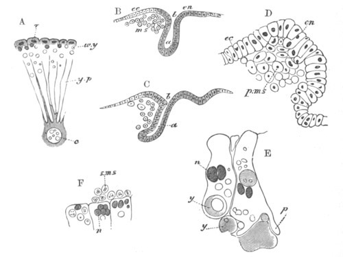
Fig. 237. Figures illustrating the development of
Astacus.
(From Parker; after Reichenbach.)
A. Section through part of the ovum during segmentation. n.
nuclei; w.y. white yolk; y.p. yolk pyramids; c.
central yolk mass.
B and C. Longitudinal sections during the gastrula stage. a.
archenteron; b. blastopore; ms. mesoblast; ec.
epiblast; en. hypoblast distinguished from epiblast by
shading.
D. Highly magnified view of the anterior lip of blastopore to shew
the origin of the primary mesoblast from the wall of the
archenteron. p.ms. primary mesoblast; ec. epiblast;
en. hypoblast.
E. Two hypoblast cells to shew the amœba-like absorption of yolk
spheres. y. yolk; n. nucleus; p. pseudopodial
process.
F. Hypoblast cells giving rise endogenously to the secondary
mesoblast (s.ms.,). n. nuclei.
Ostracoda. The independent origin of the Ostracoda from the
main Crustacean stem seems probable. Claus points out that the
Ostracoda present by no means a simple organisation, and concludes
that they were not descended from a form with a more complex
organisation and a larger number of appendages. Some simplifications
have however undoubtedly taken place, as the loss of the heart, and of
the compound eyes in many forms. These simplifications are probably to
be explained (as is done by Claus) as adaptations due to the small
size of body and its enclosure in a thick bivalve shell. Although
Claus is strongly opposed to the view that the number of the
appendages has been reduced, yet the very fact of the (in some
respects) complex organisation of this group might seem to indicate
that it cannot have diverged from the Phyllopod stem at so early a
stage as (on Claus’ view of the Nauplius) would seem to be implied by
the very small number of appendages which is characteristic of it, and
it therefore appears most probable that the present number may be
smaller than that of the ancestral forms.
The formation of the germinal layers.
The formation of the germinal layers has been more fully studied in
various Malacostraca, more especially in the Decapoda, than in other
groups.
Decapoda. To Bobretzky (No. 472) is due the credit of having
been the pioneer in this line of investigation; and his researches
have been followed up and enlarged by Haeckel, Reichenbach (No. 488),
and Mayer (No. 482). The segmentation is centrolecithal and regular
(fig. 237 A). At its close the blastoderm is formed of a single
uniform layer of lens-shaped cells enclosing a central sphere of yolk,
in which as a rule all trace of the division into columns, present
during the earlier stages of segmentation, has disappeared; though in
Palæmon the columns remain for a long period distinct. The cells of
the blastoderm are at first uniform, but in Astacus, Eupagurus, and
most Decapoda, soon become more columnar for a small area, and form a
circular patch. The whole patch either becomes at once invaginated
(Eupagurus, Palæmon, fig. 239 A) or else the edge of it is invaginated
as a roughly speaking circular groove deeper anteriorly than
posteriorly, within which the remainder of the patch forms a kind of
central plug, which does not become invaginated till a somewhat later
period (Astacus, fig. 237 B and C). After the invagination of the
above patch the remainder of the blastoderm cells form the epiblast.
The invaginated sack appears to be the archenteron and its mouth the
blastopore. The mouth finally becomes closed[208], and the sack itself
then forms the mesenteron.
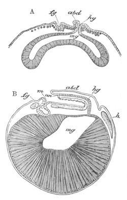
Fig. 238. Two longitudinal sections of the embryo
of Astacus. (From Parker; after Bobretzky.)
A. Nauplius stage. B. Stage after the hypoblast cells have absorbed
the food-yolk. The ventral surface is turned upwards. fg.
stomodæum; hg. proctodæum; an. anus; m. mouth;
mg. mesenteron; abd. abdomen; h. heart.
In Astacus the archenteron gradually grows forwards, its opening is at
first wide, but becomes continuously narrowed and is finally
obliterated. Very shortly after this occurrence there is formed,
slightly in front of the point where the last trace of the blastopore
was observable, a fresh epiblastic invagination, which gives rise to
the proctodæum, and the opening of which remains as the definite anus.
The proctodæum (fig. 238 A, hg) is very soon placed in
communication with the mesenteron (mg). The stomodæum
(fg) is formed during the same stage as the proctodæum. It
gives rise to the œsophagus and stomach. The hypoblast cells which
form the wall of the archenteron grow with remarkable rapidity at the
expense of the yolk; the spherules of which they absorb and digest in
an amœba-like fashion by means of their pseudopodia. They become
longer and longer, and finally, after absorbing the whole yolk,
acquire a form almost exactly similar to that of the yolk pyramids
during segmentation (fig. 238 B). They enclose the cavity of the
mesenteron, and their nuclei and protoplasm are situated externally.
The cells of the mesenteron close to its junction with the proctodæum
differ from those elsewhere in being nearly flat.
In Palæmon (Bobretzky) the primitive invagination (fig. 239 A) has far
smaller dimensions than in Astacus, and appears before the blastoderm
cells have separated from the yolk pyramids. The cells which are
situated at the bottom of it pass into the yolk, increase in number,
and absorb the whole yolk, forming a solid mass of hypoblast in which
the outlines of the individual cells would seem at first not to be
distinct. The blastopore in the meantime becomes closed. Some
of the nuclei now pass to the periphery of the yolk mass; the cells
appertaining to them gradually become distinct and assume a pyramidal
form (fig. 239 B, hy), the inner ends of the cells losing
themselves in a central mass of yolk, in the interior of which nuclei
are at first present but soon disappear. The mesenteron thus becomes
constituted of a layer of pyramidal cells which merge into a central
mass of yolk. Some of the hypoblast cells adjoining the junction of
the proctodæum and mesenteron become flattened, and in the
neighbourhood of these cells a lumen first appears. The stomodæum and
proctodæum are formed as in Astacus. Fig. 239 B shews the relative
positions of the proctodæum, stomodæum, and mesenteron. Although the
process of formation of the hypoblast and mesenteron is essentially
the same in Astacus and Palæmon, yet the differences between these two
forms are very interesting, in that the yolk is external to the
mesenteron in Astacus, but enclosed within it in Palæmon. This
difference in the position of the yolk is rendered possible by the
fact that the invaginated hypoblast cells in Palæmon do not, at first,
form a continuous layer enclosing a central cavity, while they do so
in Astacus.
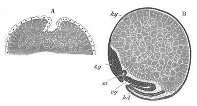
Fig. 239. Two stages in the development of
Palæmon seen in section.
(After Bobretzky.)
A. Gastrula stage.
B. Longitudinal section through a late stage. hy. hypoblast;
sg. supra-œsophageal ganglion; vg. ventral nerve
cord; hd. proctodæum; st. stomodæum.
The mesoblast appears to be formed of cells budded off from the
anterior wall of the archenteron (Astacus, fig. 237 D), or
from its lateral walls generally (Palæmon). They make their first
appearance soon after the invagination of the hypoblast has commenced.
The mesoblast cells are at first spherical, and gradually spread,
especially in an anterior direction, from their point of origin.
According to Reichenbach there are formed in Astacus at the Nauplius
stage a number of peculiar cells which he speaks of as ‘secondary
mesoblast cells.’ His account is not very clear or satisfactory, but
it appears that they originate (fig. 237 F) in the hypoblast cells by
a kind of endogenous growth, and though they have at first certain
peculiar characters they soon become indistinguishable from the
remaining mesoblast cells.
Towards the end of the Nauplius period the secondary mesoblast cells
aggregate themselves into a rod close to the epiblast in the median
ventral line, and even bifurcate round the mouth and extend forwards
to the extremity of the procephalic lobes. This rod of cells very soon
vanishes, and the secondary mesoblast cells become indistinguishable
from the primary. Reichenbach believes, on not very clear evidence,
that these cells have to do with the formation of the blood.
General form of the body. The ventral thickening of epiblast or
ventral plate, continuous with the invaginated patch already
mentioned, forms the first indication of the embryo. It is at first
oval, but soon becomes elongated and extended anteriorly into two
lateral lobes—the procephalic lobes. Its bilateral symmetry is
further indicated by a median longitudinal furrow. The posterior end
of the ventral plate next becomes raised into a distinct lobe—the
abdomen—which in Astacus at first lies in front of the still
open blastopore. This lobe rapidly grows in size, and at its extremity
is placed the narrow anal opening. It soon forms a well-marked abdomen
bent forwards over the region in front (figs. 239 B, and 240 A and B).
Its early development as a distinct outgrowth causes it to be without
yolk; and so to contrast very forcibly with the anterior thoracic and
cephalic regions of the body. In most cases this process corresponds
to the future abdomen, but in some cases (Loricata) it appears to
include part of the thorax. Before it has reached a considerable
development, three pairs of appendages spring from the region of the
head, viz. two pairs of antennæ and the mandibles, and inaugurate a
so-called Nauplius stage (fig. 240 A). These three appendages are
formed nearly simultaneously, but the hindermost appears to become
visible slightly before the two others (Bobretzky). The mouth
lies slightly behind the anterior pair of antennæ, but distinctly in
front of the posterior pair. The other appendages, the number of which
at the time of hatching varies greatly in the different Decapods
(vide section on larval development), sprout in succession from
before backwards (fig. 240 B). The food-yolk in the head and thoracic
region gradually becomes reduced in quantity with the growth of the
embryo, and by the time of hatching the disparity in size between the
thorax and abdomen has ceased to exist.
Isopoda. The early embryonic phases of the Isopoda have been
studied by means of sections by Bobretzky (No. 498) and Bullar (No.
499) and have been found to present considerable variations. When laid
the egg is enclosed in a chorion, but shortly after the commencement
of segmentation (Ed. van Beneden and Bullar) a second membrane
appears, which is probably of the nature of a larval membrane.
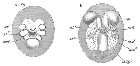
Fig. 240. Two stages in the development of
Palæmon.
A. Nauplius stage.
B. Stage with eight pairs of appendages. op. eyes;
at1. and at2. first and second antennæ; md.
mandibles; mx1, mx2., first and second maxillæ;
mxp3. third maxillipeds; lb. upper lip.
In all the forms the segmentation is followed by the formation of a
blastoderm, completely enclosing the yolk, and thickened along an area
which will become the ventral surface of the embryo. In this area the
blastoderm is formed of at least two layers of cells—an external
columnar epiblast, and an internal layer of scattered cells which form
the mesoblast and probably in part also the hypoblast (Oniscus,
Bobretzky; Cymothoa, Bullar).
In Asellus aquaticus there is a centrolecithal segmentation, ending in
the formation of a blastoderm, which appears first on the ventral
surface and subsequently extends to the dorsal.
In Oniscus murarius, and Cymothoa the segmentation is partial [for its
peculiarities and relationship vide p. 120] and a disc, formed
of a single layer of cells, appears at a pole of the egg which
corresponds to the future ventral surface (Bobretzky). This layer
gradually grows round the yolk partly by division of its cells, though
a formation of fresh cells from the yolk may also take place. Before
it has extended far round the yolk, the central part of it becomes two
or more layers deep, and the cells of the deeper layers rapidly
increase in number, and are destined to give rise to the mesoblast and
probably also to part or the whole of the hypoblast. In Cymothoa this
layer does not at first undergo any important change, but in Oniscus
it becomes very thick, and its innermost cells (Bobretzky) become
imbedded in the yolk, which they rapidly absorb; and increasing in
number first of all form a layer in the periphery of the yolk, and
finally fill up the whole of the interior of the yolk (fig. 241 A),
absorbing it in the process.
It appears possible that these cells do not, as Bobretzky believes,
originate from the blastoderm, but from nuclei in the yolk which have
escaped his observation. This mode of origin would be similar to that
by which yolk cells originate in the eggs of the Insecta, etc. If
Bobretzky’s account is correct we must look to Palæmon, as he himself
suggests, to find an explanation of the passage of the hypoblast cells
into the yolk. The thickening of the primitive germinal disc would,
according to this view, be equivalent to the invagination of the
archenteron in Astacus, Palæmon, etc.
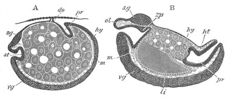
Fig. 241. Two longitudinal sections through the
embryo of Oniscus murarius. (After Bobretzky.)
st. stomodæum; pr. proctodæum; hy. hypoblast
formed of large nucleated cells imbedded in the yolk; m.
mesoblast; vg. ventral nerve cord; sg.
supra-œsophageal ganglion; li. liver; do. dorsal
organ; zp. rudiment of masticatory apparatus; ol.
upper lip.
Whatever may be the origin of the cells in the yolk they no doubt
correspond to the hypoblast of other types. In Cymothoa nothing
similar to them has been met with, but the hypoblast has a somewhat
different origin being apparently formed from some of the indifferent
cells below the epiblast, which collect as a solid mass on the ventral
surface, and then divide into two masses which become hollow and give
rise to the liver cæca. Their fate, as well as that of the hypoblast
in Oniscus, is dealt with in connection with the alimentary tract. The
completion of the enclosure of the yolk by the blastoderm takes place
on the dorsal surface. In all the Isopods which have been carefully
studied, there appears before any other organ a provisional
structure formed from the epiblast and known as the dorsal organ. An
account of it is given in connection with the development of the
organs. The general external changes undergone by the larva in its
development are as follows. The ventral thickened area of the
blastoderm (ventral plate) shapes itself and girths nearly the whole
circumference of the ovum in Oniscus (fig. 241 A) but is relatively
much shorter in Cymothoa. Anteriorly it dilates into the two
procephalic lobes. In Cymothoa it next becomes segmented; and the
anterior segments are formed nearly simultaneously, and those of the
abdomen somewhat later. At the same time a median depression appears
dividing the blastoderm longitudinally into two halves. The appendages
are formed later than their segments, and the whole of them are formed
nearly simultaneously, with the exception of the last thoracic, which
does not appear till comparatively late after the hatching of the
embryo. The late development of the seventh thoracic segment and
appendage is a feature common to the majority of the Isopoda (Fritz
Müller). In Oniscus the limbs are formed in nearly the same way as in
Cymothoa, but in Asellus they do not arise quite simultaneously. First
of all, the two antennæ and mandibles (the future palp) appear,
inaugurating a stage often spoken of as the Nauplius stage, which is
supposed to correspond with the free Nauplius stage of Penæus
and Euphausia. At this stage a cuticle is shed (Van Beneden) which
remains as an envelope surrounding the larva till the time of
hatching. Similar cuticular envelopes are formed in many Isopoda.
Subsequently the appendages of the thorax appear, and finally those of
the abdomen. Later than the appendages there arise behind the mouth
two prominences which resemble appendages, but give rise to a bilobed
lower lip (Dohrn).
In Asellus and Oniscus the ventral plate moulds itself to the shape of
the egg, and covers the greater part of the dorsal as well as of the
ventral side (fig. 241 A). As a result of this the ventral surface of
the embryo is throughout convex; and in Asellus a deep fold appears on
the back of the embryo, so that the embryo appears coiled up within
the egg with its ventral side outwards and its head and tail in
contact. In Oniscus the ventral surface is convex, but the dorsal
surface is never bent in as in Asellus. In Cymothoa the egg is very
big and the ventral plate does not extend nearly so far round to the
dorsal side as in Asellus, in consequence of which the ventral surface
is not nearly so convex as in other Isopoda. At the same time the
telson is early formed, and is bent forwards so as to lie on the under
side of the part of the blastoderm in front. In having this ventral
curvature of the telson Cymothoa forms an exception amongst Isopods;
and in this respect is intermediate between the embryos of Asellus and
those of the Amphipoda.
Amphipoda. Amongst the Amphipoda the segmentation is usually
centrolecithal. In the case of Gammarus locusta (Ed. van Beneden and
Bessels, No. 503) it commences with an unequal but total segmentation
like that of the Frog (vide p. 97), and the separation of a
central yolk mass is a late occurrence; and it is noticeable that the
part of the egg with the small segments eventually becomes the ventral
surface. In the fresh-water species of Gammarus (G. pulex and
fluviatilis) the segmentation is more like that of Insects; the
blastoderm cells being formed nearly simultaneously over a large part
of the surface of the egg.
Both forms of segmentation give rise to a blastoderm covering the
whole egg, which soon becomes thickened on the ventral
surface. There is formed, as in the Isopoda, a larval membrane at
about the time when the blastoderm is completed. Very soon after this
the egg loses its spherical shape, and becomes produced into a pointed
extremity—the future abdomen—which is immediately bent over the
ventral surface of the part in front. The ventral curvature of the
hinder part of the embryo at so early an age stands in marked contrast
to the usual condition of Isopod embryos, and is only approached in
this group, so far as is known, in the case of Cymothoa.
At the formation of the first larval membrane the blastoderm cells
separate themselves from it, except at one part on the dorsal surface.
The patch of cells adherent at this part gives rise to a dorsal organ,
comparable with that in Oniscus, connecting the embryo and its first
larval skin. A perforation appears in it at a later period.
The segments and limbs of the Amphipoda are all formed before the
larva leaves the egg.
Cladocera. The segmentation (Grobben, No. 455) takes place on
the normal centrolecithal type, but is somewhat unequal. Before the
close of the segmentation there may be seen at the apex of the
vegetative pole one cell marked off from the remainder by its granular
aspect. It gives rise to the generative organs. One of the cells
adjoining it gives rise to the hypoblast, and the other cells which
surround it form the commencement of the mesoblast. The remaining
cells of the ovum form the epiblast. By a later stage the hypoblast
cell is divided into thirty-two cells and the genital cell into four,
while the mesoblast forms a circle of twelve cells round the genital
mass.
The hypoblast soon becomes involuted; the blastopore probably closes,
and the hypoblast forms a solid cord of cells which eventually becomes
the mesenteron. The stomodæum is said to be formed at the point of
closure of the blastopore. The mesoblast passes inwards and forms a
mass adjoining the hypoblast, and somewhat later the genital mass also
becomes covered by the epiblast. The proctodæum appears to be formed
later than the stomodæum.
The embryo as first shewn by Dohrn passes through a Nauplius stage in
the brood-pouch, but is hatched, except in the case of the winter eggs
of Leptodora, in a form closely resembling the adult.
Copepoda. Amongst the free Copepoda the segmentation and
formation of the layers have recently been investigated by Hoek (No.
512). He finds that there is, in both the fresh-water and marine forms
studied by him, a centrolecithal segmentation similar to that of
Palæmon and Pagurus (vide p. 112), which might from the surface
be supposed to be complete and nearly regular. After the
formation of the blastoderm an invagination of some of its cells takes
place and is completed in about a quarter of an hour. The opening
becomes closed. This invagination is compared by Hoek to the
invagination in Astacus, and is believed by him to give rise to the
mesenteron. Its point of closing corresponds with the hind end of the
embryo. On the ventral surface there appear two transverse furrows
dividing the embryo into three segments, and a median longitudinal
furrow which does not extend to the front end of the foremost segment.
The three pairs of Nauplius appendages and upper lip become
subsequently formed as outgrowths from the sides of the ventral
blastodermic thickening.
Amongst the parasitic Copepoda there are found two distinct types of
segmentation, analogous to those in the Isopoda. In the case of
Condracanthus the segmentation is somewhat irregular, but on the type
of Eupagurus, etc. (vide p. 112). In the other group
(Anchorella, Clavella, Congericola, Caligus, Lerneopoda) the
segmentation nearly resembles the ordinary meroblastic type
(vide p. 120), and is to be explained in the same manner as in
the cases of Oniscus and Cymothoa. The first blastodermic cells
sometimes appear in a position corresponding with the head end of the
embryo (Anchorella), at other times at the hind end (Clavella), and
sometimes in the middle of the ventral surface. The dorsal surface of
the yolk is always the latest to be inclosed by the blastoderm cells.
A larval cuticle similar to that of the Isopoda is formed at the same
time as the blastoderm. At the sides of the ventral thickening of the
blastoderm there grow out the Nauplius appendages, of which only the
first two appear in Anchorella. In Anchorella and Lerneopoda the
embryos are not hatched at the Nauplius stage, but after the Nauplius
appendages have been formed a fresh cuticle—the Nauplius cuticle—is
shed, and within it the embryo develops till it reaches the so-called
Cyclops stage (vide p. 490). The embryo within the egg has its
abdomen curved dorsalwards as amongst the Isopoda.
Cirripedia. The segmentation of Balanus and Lepas commences by
the segregation of the constituents of the egg into a more
protoplasmic portion, and a portion formed mainly of food material.
The former separates from the latter as a distinct segment, and then
divides into two not quite equal portions. The division of the
protoplasmic part of the embryo continues, and the resulting segments
grow round the single yolk segment. The point where they finally
enclose it is situated on the ventral surface (Lang) at about the
position of the mouth (?).
After being enclosed by the protoplasmic cells the yolk divides, and
gives rise to a number of cells, which probably supply the material
for the walls of the mesenteron. The external layer of protoplasm
forms the so-called blastoderm, and soon (Arnold, Lang) becomes
thickened on the dorsal surface.
The embryo is next divided by two constrictions into three segments;
and there are formed the three appendages corresponding to these,
which are at first simple. The two posterior soon become
biramous. The larva leaves the egg before any further appendages
become formed.
Comparative development of the organs.
Central nervous system. The ventral nerve cord of the Crustacea
develops as a thickening of the epiblast along the median ventral
line; the differentiation of which commences in front, and thence
extends backwards. The ventral cord is at first unsegmented. The
supra-œsophageal ganglia originate as thickenings of the epiblast
of the procephalic lobes.
The details of the above processes are still in most cases very
imperfectly known. The fullest account we have is that of Reichenbach
(No. 488) for Astacus. He finds that the supra-œsophageal ganglia
and ventral cord arise as a continuous formation, and not
independently as would seem to be the case in Chætopoda. The
supra-œsophageal ganglia are formed from the procephalic lobes. The
first trace of them is visible in the form of a pair of pits, one on
each side of the middle line. These pits become in the Nauplius stage
very deep, and their walls are then continued into two ridges where
the epiblast is several cells deep, which pass backwards one on each
side of the mouth. The walls of the pits are believed by Reichenbach
to give rise to the optic portions of the supra-œsophageal ganglia,
and the epiblastic ridges to the remainder of the ganglia and to the
circum-œsophageal commissures. At a much later stage, when the
ambulatory feet have become formed, a median involution of epiblast in
front of the mouth and between the two epiblast ridges gives rise to a
central part of the supra-œsophageal ganglia. Five elements are
thus believed by Reichenbach to be concerned in the formation of these
ganglia, viz. two epiblast pits, two epiblast ridges, and an
involution of epiblast between the latter. It should be noted however
that the fate neither of the pair of pits, nor of the median
involution, appears to have been satisfactorily worked out. The two
epiblast ridges, which pass back from the supra-œsophageal ganglia
on each side of the mouth, are continued as a pair of thickenings of
the epiblast along the sides of a median ventral groove. This groove
is deep in front and shallows out posteriorly. The thickenings on the
sides of this groove no doubt give rise to the lateral halves of the
ventral cord, and the cells of the groove itself are believed by
Reichenbach, but it appears to me without sufficient evidence, to
become invaginated also and to assist in forming the ventral cord.
When the ventral cord becomes separated from the epiblast the two
halves of it are united in the middle line, but it is markedly bilobed
in section.
In the Isopoda it would appear both from Bobretzky’s and Bullar’s
observations that the ventral nerve cord arises as an unpaired
thickening of the epiblast in which there is no trace of anything
like a median involution. After this thickening has become
separated from the epiblast a slight median furrow indicates
its constitution out of two lateral cords. The supra-œsophageal
ganglia are stated to be developed quite simply as a pair of
thickenings of the procephalic lobes, but whether they are from the
first continuous with the ventral cord does not appear to have been
determined.
The later stages in the differentiation of the ventral cord are, so
far as is known, very similar throughout the Crustacea. The ventral
cord is, as has been stated, at first unsegmented (fig. 241 A,
vg), but soon becomes divided by a series of constrictions into
as many ganglia as there are pairs of appendages or segments (fig. 241
B, vg).
There appears either on the ventral side (Oniscus) or in the centre
(Astacus, Palæmon) of the two halves of each segment or ganglion a
space filled with finely punctuated material, which is the
commencement of the commissural portion of the cords. The commissural
tissue soon becomes continuous through the length of the ventral cord,
and is also prolonged into the supra-œsophageal ganglia.
After the formation of the commissural tissue the remaining cells of
the cord form the true ganglion cells. A gradual separation of the
ganglia next takes place, and the cells become confined to the
ganglia, which are finally only connected by a double band of
commissural tissue. The commissural tissue not only gives rise to the
longitudinal cords connecting the successive ganglia, but also to the
transverse commissures which unite the two halves of the individual
ganglia.
The ganglia usually, if not always, appear at first to correspond in
number with the segments, and the smaller number so often present in
the adult is due to the coalescence of originally distinct ganglia.
Organs of special sense. Comparatively little is known on this
head. The compound eyes are developed from the coalescence of two
structures, both however epiblastic, viz. (1) part of the superficial
epiblast of the procephalic lobes; (2) part of the supra-œsophageal
ganglia. The former gives rise to the corneal lenses, the crystalline
cones, and the pigment surrounding them; the latter to the rhabdoms
and the cells which encircle them. Between these two parts a
mesoblastic pigment is interposed.
Of the development of the auditory and olfactory organs almost nothing
is known.
Dorsal organ. In a considerable number of the Malacostraca and
Branchiopoda a peculiar organ is developed from the epiblast in the
anterior dorsal region. This organ has been called the dorsal organ.
It appears to be of a glandular nature, and is usually very large in
the embryo or larva and disappears in the adult; but in some
Branchiopoda it persists through life. In most cases it is unpaired,
but in some instances a paired organ appears to take its place.
Various views as to its nature have been put forward. There is but
little doubt of its being glandular, and it is possible that it is a
provisional renal organ, though so far as I know concretions have not
yet been found in it.
Its development has been most fully studied in the Isopoda.
Fig. 242. Diagrammatic section of Cymothoa
shewing the dorsal organ. (From Bullar.)
In Cymothoa (Bullar, No. 499) there appears on the dorsal surface, in
the region which afterwards becomes the first thoracic segment, an
unpaired linear thickening of the blastoderm. This soon becomes a
circular patch, the central part of which is invaginated so as to
communicate with the exterior by a narrow opening only (fig. 242). It
becomes at the same time attached to the inner egg membrane. It
retains this condition till the close of larval life.
In Oniscus (Dohrn, No. 500; Bobretzky, No. 498) there appears very
early a dorsal patch of thickened cells. These cells become attached
at their edge to the inner egg membrane and gradually separated from
the embryo, with which they finally only remain in connection by a
hollow column of cells (fig. 241 A, do). The original patch now
gradually spreads over the inner egg membrane, and forms a transverse
saddle-shaped band of flattened cells which engirths the embryo on all
but the ventral side.
In the Amphipods the epiblast cells remain attached for a small area
on the dorsal surface to the first larval skin, when this is formed.
This patch of cells, often spoken of as a micropyle apparatus, forms a
dorsal organ equivalent to that in Oniscus. A perforation is formed in
it at a later period. A perhaps homologous structure is found
in the embryos of Euphausia, Cuma, etc.
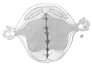
Fig. 243. Diagrammatic Section of an embryo of
Asellus Aquaticus To Shew the Paired Dorsal Organ. (From
Bullar; after E. van Beneden.)
In many Branchiopoda a dorsal organ is found. Its development has been
studied by Grobben in Moina. It persists in the adult in Branchipus,
Limnadia, Estherea, etc.
In the Copepoda a dorsal organ is sometimes found in the embryo;
Grobben at any rate believes that he has detected an organ of this
nature in the embryo of Cyclops serrulatus.
A paired organ which appears to be of the same nature has been found
in Asellus and Mysis.
In Asellus (Rathke (No. 501), Dohrn (No. 500), Van Beneden (No. 497))
this organ originates as two cellular masses at the sides of the body
just behind the region of the procephalic lobes. Each of them becomes
trifoliate and bends towards the ventral surface. In each of their
lobes a cavity arises and finally the three cavities unite, forming a
trilobed cavity open to the yolk. This organ eventually becomes so
large that it breaks through the egg membranes and projects at the
sides of the embryo (fig. 243). Though formed before the appendages it
does not attain its full development till considerably after the
latter have become well established.
In Mysis it appears during the Nauplius stage as a pair of cavities
lined by columnar cells, which atrophy very early.
Various attempts have been made to identify organs in other Arthropod
embryos with the dorsal organ of the Crustacea, but the only organ at
all similar which has so far been described is one found in the embryo
of Linguatula (vide Chapter XIX.), but there is no reason to
think that this organ is really homologous with the dorsal organ of
the Crustacea.
The mesoblast. The mesoblast in the types so far investigated
arises from the same cells as the hypoblast, and appears as a somewhat
irregular layer between the epiblast and the hypoblast. It gives rise
to the same parts as in other forms, but it is remarkable that it does
not, in most Decapods and Isopods (and so far we do not know
about other forms), become divided into somites, at any rate with the
same distinctness that is usual in Annelids and Arthropods. Not only
so, but there is at first no marked division into a somatic and
splanchnic layer with an intervening body cavity. Some of the cells
become differentiated into the muscles of the body wall and limbs; and
other cells, usually in the form of a very thin layer, into the
muscles of the alimentary tract. In the tail of Palæmon
Bobretzky noticed that the cells about to form the muscles of the body
were imperfectly divided into cubical masses corresponding with the
segments; which however, in the absence of a central cavity, differed
from typical mesoblastic somites. In Mysis Metschnikoff states
that the mesoblast becomes broken up into distinct somites. Further
investigations on this subject are required. The body cavity has the
form of irregular blood sinuses amongst the internal organs.
Heart. The origin and development of the heart and vascular
system are but very imperfectly known.
In Phyllopods (Branchipus) Claus (No. 454) has shewn that the heart is
formed by the coalescence of the lateral parts of the mesoblast of the
ventral plates. The chambers are formed successively as the segments
to which they belong are established, and the anterior chambers are in
full activity while the posterior are not yet formed.
In Astacus and Palæmon, Bobretzky finds that at the stage before the
heart definitely appears there may be seen a solid mass of mesoblast
cells in the position which it eventually occupies[209]; and considers
it probable that the heart originates from this mass. At the time when
the heart can first be made out and before it has begun to beat, it
has the form of an oval sack with delicate walls separated from the
mesenteron by a layer of splanchnic mesoblast. Its cavity is filled
with a peculiar plasma which also fills up the various cavities in the
mesoblast. Around it a pericardial sack is soon formed, and the walls
of the heart become greatly thickened. Four bands pass off from the
heart, two dorsalwards which become fixed to the integument, and two
ventralwards. There is also a median band of cells connecting the
heart with the dorsal integument. The main arteries arise as direct
prolongations of the heart. Dohrn’s observations on Asellus greatly
strengthen the view that the heart originates from a solid mesoblastic
mass, in that he was able to observe the hollowing out of the mass in
the living embryo (cf. the development of the heart in
Spiders). Some of the central cells (nuclei, Dohrn) become blood
corpuscles. The formation of these is not, according to Dohrn,
confined to the heart, but takes place in situ in all the parts
of the body (antennæ, appendages, etc.). The corpuscles are formed as
free nuclei and are primarily derived from the yolk, which at first
freely communicates with the cavities of the appendages.
Alimentary tract. In Astacus the formation of the mesenteron by
invagination, and the absorption of the yolk by the hypoblast cells,
have already been described. On the absorption of the yolk the
mesenteron has the form of a sack, the walls of which are formed of
immensely long cells—the yolk pyramids—at the base of which the
nucleus is placed (fig. 238 B). This sack gives rise both to the
portion of the alimentary canal between the abdomen and the stomach
and to the liver. The epithelial wall of both of these parts is formed
by the outermost portions of the pyramids with the nuclei and
protoplasm becoming separated off from the yolk as a layer of flat
epithelial cells. The yolk then breaks up and forms a mass of
nutritive material filling up the cavity of the mesenteron.
The differentiation both of the liver and alimentary tract proper
first takes place on the ventral side, and commences close to the
point where the proctodæum ends, and extends forward from this point.
A layer of epithelial cells is thus formed on the ventral side of the
mesenteron which very soon becomes raised into a series of
longitudinal folds, one of which in the middle line is very
conspicuous. The median fold eventually, by uniting with a
corresponding fold on the dorsal side, gives rise to the true
mesenteron; while the lateral folds form parallel hepatic cylinders,
which in front are not constricted off from the alimentary tract. The
lateral parts of the dorsal side of the mesenteron similarly give rise
to hepatic cylinders. The yolk pyramids of the anterior part of the
mesenteron, which projects forwards as a pair of diverticula on each
side to the level of the stomach, are not converted into hepatic
cylinders till after the larva is hatched.
The proctodæum very early opens into the mesenteron, but the stomodæum
remains closed till the differentiation of the mid-gut is nearly
completed. The proctodæum gives rise to the abdominal part of the
intestine, and the stomodæum to the œsophagus and stomach. The
commencement of the masticatory apparatus in the latter appears very
early as a dorsal thickening of the epithelium.
The primitive mesenteron in Palæmon differentiates itself into the
permanent mid-gut and liver in a manner generally similar to that in
Astacus, though the process is considerably less complicated. A
distinct layer of cells separates itself from the outer part of the
yolk pyramids, and gives rise to the glandular lining both of the
mid-gut and of the liver. The differentiation of this layer commences
behind, and the mid-gut very soon communicates freely with the
proctodæum. The lateral parts of the primitive mesenteron become
constricted into four wings, two directed forwards and two backwards;
these, after the yolk in them has become absorbed, constitute the
liver. The median part simply becomes the mesenteron. The
stomachic end of the stomodæum lies in contact with the mesenteron
close to the point where it is continued into the hepatic diverticula,
and, though the partition wall between the two becomes early very
thin, a free communication is not established till the yolk has been
completely absorbed.
The alimentary tract in the Isopoda is mainly if not entirely formed
from the proctodæum and stomodæum, both of which arise before any
other part of the alimentary system as epiblastic invaginations, and
gradually grow inwards (fig. 244). In Oniscus the liver is formed as
two discs at the surface of the yolk on each side of the anterior part
of the body. Their walls are composed of cubical cells derived from
the yolk cells, the origin of which was spoken of on p. 516. These two
discs gradually take the form of sacks (fig. 244 B, li.) freely
open on their inner side to the yolk. As these sacks continue to grow
the stomodæum and proctodæum do not remain passive. The stomodæum,
which gives rise to the œsophagus and stomach of the adult, soon
exhibits a posterior dilatation destined to become the stomach, on the
dorsal wall of which a well-marked prominence—the earliest trace of
the future armature—is soon formed (fig. 244 B, zp). The
proctodæum (pr) grows with much greater rapidity than the
stomodæum, and its end adjoining the yolk becomes extremely thin or
even broken through. In the earliest stages it was surrounded by the
yolk cells, but in its later growth the yolk cells become gradually
reduced in number and appear to recede before it—so much so that one
is led to conclude that the later growth of the proctodæum takes place
at the expense of the yolk cells.
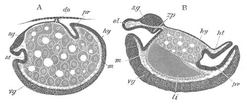
Fig. 244. Two longitudinal sections through the
embryo of Oniscus murarius. (After Bobretzky.)
st. stomodæum; pr. proctodæum; hy. hypoblast
formed of large nucleated cells imbedded in yolk; m.
mesoblast; vg. ventral nerve cord; sg.
supra-œsophageal ganglion; li. liver; do. dorsal
organ; zp. rudiment of masticatory apparatus.
The liver sacks become filled with a granular material without a trace
of cells; their posterior wall is continuous with the yolk cells, and
their anterior lies close behind the stomach. The proctodæum
continually grows forwards till it approaches close to the stomodæum,
and the two liver sacks, now united into one at their base,
become directly continuous with the proctodæum. By the stage when this
junction is effected the yolk cells have completely disappeared. It
seems then that in Oniscus the yolk cells (hypoblast) are mainly
employed in giving rise to the walls of the liver; but that they
probably also supply the material for the later growth of the apparent
proctodæum. It becomes therefore necessary to conclude that the
latter, which might seem, together with the stomodæum, to form the
whole alimentary tract, does in reality correspond to the proctodæum
and mesenteron together, though the digestive fluids are no doubt
mainly secreted not in the mesenteron but in the hepatic diverticula.
The proctodæum and stomodæum at first meet each other without
communicating, but before long the partition between the two is broken
through.
In Cymothoa (Bullar, No. 499) the proctodæum and stomodæum develop in
the same manner as in Oniscus, but the hypoblast has quite a different
form. The main mass of the yolk, which is much greater than in
Oniscus, is not contained in definite yolk cells, but the hypoblast is
represented by (1) two solid masses of cells, derived apparently from
the inner layer of blastoderm cells, which give rise to the liver; and
(2) by a membrane enclosing the yolk in which nuclei are present.
The two hepatic masses lie on the surface of the yolk, and each of
them becomes divided into three short cæcal tubes freely open to the
yolk. The stomodæum soon reaches its full length, but the proctodæum
grows forwards above the yolk till it meets the stomodæum. By the time
this takes place the liver cæca have grown into three large tubes
filled with fluid, and provided with a muscular wall. They now lie
above the yolk, and no longer communicate directly with the cavity of
the yolk-sack, but open together with the yolk-sack into the point of
junction of the proctodæum and stomodæum. The yolk-sack of Cymothoa no
doubt represents part of the mesenteron, but there is no evidence in
favour of any part of the apparent proctodæum representing it also,
though it is quite possible that it may do so. The relations of the
yolk-sack and hepatic diverticula in Cymothoa appear to hold good for
Asellus and probably for most Isopoda.
The differences between the Decapods and Isopods in the development of
the mesenteron are not inconsiderable, but they are probably to be
explained by the relatively larger amount of food-yolk in the latter
forms. The solid yolk in the Isopods on this view represents the
primitive mesenteron of Decapods after the yolk has been absorbed by
the hypoblast cells. Starting from this standpoint we find that in
both groups the lateral parts of the mesenteron become the liver. In
Decapods the middle part becomes directly converted into the mid-gut,
the differentiation of it commencing behind and proceeding forwards.
In the Isopods, owing to the mesenteron not having a distinct cavity,
the differentiation of it, which proceeds forwards as in Decapods,
appears simply like a prolongation forwards of the proctodæum, the
cells for the prolongation being probably supplied from the yolk. In
Cymothoa the food-yolk is so bulky that a special yolk-sack is
developed for its retention, which is not completely absorbed
till some time after the alimentary canal has the form of a continuous
tube. The walls of this yolk-sack are morphologically a specially
developed part of the mesenteron.
Bibliography.
General Works.
(447) C. Spence Bate. “Report on the present state of our
knowledge of the Crustacea.” Report of the British Association for
1878.
(448) C. Claus. Untersuchungen zur Erforschung der
genealogischen Grundlage des Crustaceen-Systems. Wien, 1876.
(449) A. Dohrn. “Geschichte des Krebsstammes.” Jenaische
Zeitschrift, Vol. VI. 1871.
(450) A. Gerstaecker. Bronn’s Thierreich, Bd.
V. Arthropoda, 1866.
(451) Th. H. Huxley. The Anatomy of Invertebrated
Animals. London, 1877.
(452) Fritz Müller. Für Darwin, 1864. Translation,
Facts for Darwin. London, 1869.
Branchiopoda.
(453) Brauer. “Vorläufige Mittheilung über die Entwicklung u.
Lebensweise des Lepidurus (Apus) productus.” Sitz. der Ak. d. Wiss.
Wien, Vol.
LXIX., 1874.
(454) C. Claus. “Zur Kenntniss d. Baues u. d. Entwicklung von
Branchipus stagnalis u. Apus cancriformis.” Abh. d. könig. Gesell.
der Wiss. Göttingen, Vol. XVIII. 1873.
(455) C. Grobben. “Zur Entwicklungsgeschichte d. Moina
rectirostris.” Arbeit. a. d. zoologisch. Institute Wien, Vol.
II., 1879.
(456) E. Grube. “Bemerkungen über die Phyllopoden nebst einer
Uebersicht etc.” Archiv f. Naturgeschichte, Vol.
XIX.,
1853.
(457) N. Joly. “Histoire d’un petit Crustacé (Artemia salina,
Leach) etc.” Annales d. Sciences Natur., 2nd ser., Vol.
XIII., 1840.
(458) N. Joly. “Recherches zoologiques anatomiques et
physiologiques sur l’Isaura cycladoides (=Estheria) nouveau genre,
etc.” Annales d. Sciences Nat., 2nd ser., Vol.
XVII.,
1842.
(459) Lereboullet. “Observations sur la génération et le
développement de la Limnadia de Hermann.” Annales d. Sciences
Natur., 5th ser., Vol.
V., 1866.
(460) F. Leydig. “Ueber Artemia salina u. Branchipus
stagnalis.” Zeit. f. wiss. Zool., Vol.
III., 1851.
(461) G. O. Sars. “Om en dimorph Udvikling samt
Generationsvexel hos Leptodora.” Vidensk. Selskab. Forhand,
1873.
(462) G. Zaddach. De apodis cancreformis Schaeff. anatome et
historia evolutionis. Dissertatio inauguralis zootomica. Bonnæ,
1841.
Nebaliadæ.
(463) C. Claus. “Ueber den Bau u. die systematische Stellung
von Nebalia.” Zeit. f. wiss. Zool., Bd. XXII. 1872.
(464) E. Metschnikoff. Development of Nebalia (Russian),
1868.
Schizopoda.
(465) E. van Beneden. “Recherches sur l’Embryogénie des
Crustacés. II. Développement des Mysis.” Bullet. de
l’Académie roy. de Belgique, second series, Tom. XXVIII.
1869.
(466) C. Claus. “Ueber einige Schizopoden u. niedere
Malakostraken.” Zeit. f. wiss. Zoologie, Bd. XIII.,
1863.
(467) A. Dohrn. “Untersuchungen üb. Bau u. Entwicklung d.
Arthropoden.” Zeit. f. wiss. Zool., Bd. XXI., 1871, p.
375. Peneus zoæa (larva of Euphausia).
(468) E. Metschnikoff. “Ueber ein Larvenstadium von Euphausia.”
Zeit. für wiss. Zool., Bd. XIX., 1869.
(469) E. Metschnikoff. “Ueber den Naupliuszustand von
Euphausia.” Zeit. für wiss. Zool., Bd. XXI., 1871.
Decapoda.
(470) Spence Bate. “On the development of Decapod Crustacea.”
Phil. Trans., 1858.
(471) Spence Bate. “On the development of Pagurus.” Ann. and
Mag. Nat. History, Series 4, Vol.
II., 1868.
(472) N. Bobretzky. Development of Astacus and Palæmon.
Kiew, 1873. (Russian.)
(473) C. Claus. “Zur Kenntniss d. Malakostrakenlarven.”
Würzb. naturw. Zeitschrift, 1861.
(474) R. Q. Couch. “On the Metamorphosis of the Decapod
Crustaceans.” Report Cornwall Polyt. Society. 1848.
(475) Du Cane. “On the Metamorphosis of Crustacea.” Ann. and
Mag. of Nat. History, 1839.
(476) Walter Faxon. “On the development of Palæmonetes
vulgaris.” Bull. of the Mus. of Comp. Anat. Harvard, Cambridge,
Mass., Vol.
V., 1879.
(477) A. Dohrn. “Untersuchungen üb. Bau u. Entwicklung d.
Arthropoden.” “Zur Entwicklungsgeschichte der Panzerkrebse.
Scyllarus Palinurus.” Zeit. f. wiss. Zool., Bd.
XX., 1870.
(478) A. Dohrn. “Untersuchungen üb. Bau u. Entwicklung d.
Arthropoden. Erster Beitrag z. Kenntniss d. Malacostraken u. ihrer
Larven Amphion Reynaudi, Lophogaster, Portunus, Porcellanus,
Elaphocaris.” Zeit. f. wiss. Zool., Bd. XX., 1870.
(479) A. Dohrn. “Untersuchungen üb. Bau u. Entwicklung d.
Arthropoden. Zweiter Beitrag, etc.” Zeit. f. wiss. Zool., Bd.
XXI., 1871.
(480) N. Joly. “Sur la Caridina Desmarestii.” Ann. Scien.
Nat., Tom. XIX., 1843.
(481) Lereboullet. “Recherches d. l’embryologie comparée sur le
développement du Brochet, de la Perche et de l’Écrevisse.” Mém.
Savans Étrang. Paris, Vol.
XVII., 1862.
(482) P. Mayer. “Zur Entwicklungsgeschichte d. Dekapoden.”
Jenaische Zeitschrift, Vol.
XI., 1877.
(483) Fritz Müller. “Die Verwandlung der Porcellana.” Archiv
f. Naturgeschichte, 1862.
(484) Fritz Müller. “Die Verwandlungen d. Garneelen,” Archiv
f. Naturgesch., Tom. XXIX.
(485) Fritz Müller. “Ueber die Naupliusbrut d. Garneelen.”
Zeit. f. wiss. Zool., Bd. XXX., 1878.
(486) T. J. Parker. “An account of Reichenbach’s researches on
the early development of the Fresh-water Crayfish.” Quart. J. of M.
Science, Vol.
XVIII., 1878.
(487) H. Rathke. Ueber die Bildung u. Entwicklung d.
Flusskrebses. Leipzig, 1829.
(488) H. Reichenbach. “Die Embryoanlage u. erste Entwicklung d.
Flusskrebses.” Zeit. f. wiss. Zool., Vol.
XXIX., 1877.
(489) F. Richters. “Ein Beitrag zur Entwicklungsgeschichte d.
Loricaten.” Zeit. f. wiss. Zool., Bd. XXIII., 1873.
(490) G. O. Sars. “Om Hummers postembryonale Udvikling.”
Vidensk Selsk. Forh. Christiania, 1874.
(491) Sidney J. Smith. “The early stages of the American
Lobster.” Trans. of the Connecticut Acad. of Arts and Sciences,
Vol. II., Part 2, 1873.
(492) R. v. Willemoes Suhm. “Preliminary note on the
development of some pelagic Decapoda.” Proc. of Royal Society,
1876.
Stomatopoda.
(493) W. K. Brooks. “On the larval stages of Squilla empusa.”
Chesapeake Zoological Laboratory, Scientific results of the Session
of 1878. Baltimore, 1879.
(494) C. Claus. “Die Metamorphose der Squilliden.” Abhand.
der königl. Gesell. der Wiss. zu Göttingen, 1871.
(495) Fr. Müller. “Bruchstück a. der Entwicklungsgeschichte d.
Maulfüsser I. und II.” Archiv f.
Naturgeschichte, Vol.
XXVIII., 1862, and Vol.
XXIX., 1863.
Cumacea.
(496) A. Dohrn. “Ueber den Bau u. Entwicklung d. Cumaceen.”
Jenaische Zeitschrift, Vol.
V., 1870.
Isopoda.
(497) Ed. van Beneden. “Recherches sur l’Embryogénie des
Crustacés. I. Asellus aquaticus.” Bull. de l’Acad. roy.
Belgique, 2me série, Tom. XXVIII., No. 7, 1869.
(498) N. Bobretzky. “Zur Embryologie des Oniscus murarius.”
Zeit. für wiss. Zool., Bd. XXIV., 1874.
(499) J. F. Bullar. “On the development of the parasitic
Isopoda.” Phil. Trans., Part II., 1878.
(500) A. Dohrn. “Die embryonale Entwicklung des Asellus
aquaticus.” Zeit. f. wiss. Zool., Vol.
XVII., 1867.
(501) H. Rathke. Untersuchungen über die Bildung und
Entwicklung der Wasser-Assel. Leipzig, 1832.
(502) H. Rathke. Zur Morphologie. Reisebemerkungen aus
Taurien. Riga u. Leipzig, 1837. (Bopyrus, Idothea, Ligia, Ianira.)
Amphipoda.
(503) Ed. van Beneden and E. Bessels. “Mémoire sur la
formation du blastoderme chez les Amphipodes, les Lernéens et les
Copépodes.” Classe des Sciences de l’Acad. roy. de Belgique,
Vol. XXXIV., 1868.
(504) De la Vallette St George. “Studien über die Entwicklung
der Amphipoden.” Abhand. d. naturfor. Gesell. zu Halle, Bd.
V., 1860.
Copepoda.
(505) E. van Beneden and E. Bessels. “Mémoire sur la
formation du blastoderme chez les Amphipodes, les Lernéens et
Copépodes.” Classe des Sciences de l’Acad. roy. de Belgique,
Vol. XXXIV., 1868.
(506) E. van Beneden. “Recherches sur l’Embryogénie des
Crustacés IV. Anchorella, Lerneopoda, Branchiella, Hessia.”
Bull. de l’Acad. roy. de Belgique, 2me série, T.
XXIX., 1870.
(507) C. Claus. Zur Anatomie u. Entwicklungsgeschichte d.
Copepoden.
(508) C. Claus. “Untersuchungen über die Organisation u.
Verwandschaft d. Copepoden.” Würzburger naturwiss. Zeitschrift,
Bd. III., 1862.
(509) C. Claus. “Ueber den Bau u. d. Entwicklung von Achtheres
percarum.” Zeit. f. wiss. Zool., Bd. XI., 1862.
(510) C. Claus. Die freilebenden Copepoden mit besonderer
Berücksichtigung der Fauna Deutschlands, des Nordsee u. des
Mittelmeeres. Leipzig, 1863.
(511) C. Claus. “Ueber d. Entwicklung, Organisation u.
systematische Stellung d. Argulidæ.” Zeit. f. wiss. Zool., Bd.
XXV., 1875.
(512) P. P. C. Hoek. “Zur Entwicklungsgeschichte d.
Entomostracen.” Niederländisches Archiv, Vol.
IV., 1877.
(513) Nordmann. Mikrographische Beiträge zur Naturgeschichte
der wirbellosen Thiere. Zweites Heft. 1832.
(514) Salensky. “Sphæronella Leuckartii.” Archiv f.
Naturgeschichte, 1868.
(515) F. Vejdovsky. “Untersuchungen üb. d. Anat. u. Metamorph.
v. Tracheliastes polycolpus.” Zeit. f. wiss. Zool., Vol.
XXIX., 1877.
Cirripedia.
(516) C. Spence Bate. “On the development of the Cirripedia.”
Annals and Mag. of Natur. History. Second Series,
VIII., 1851.
(517) E. van Beneden. “Développement des Sacculines.” Bull.
de l’Acad. roy. de Belg., 1870.
(518) C. Claus. Die Cypris-ähnliche Larve der
Cirripedien. Marburg, 1869.
(519) Ch. Darwin. A monograph of the sub-class
Cirripedia, 2 Vols., Ray Society, 1851‑4.
(520) A. Dohrn. “Untersuchungen über Bau u. Entwicklung d.
Arthropoden IX. Eine neue Naupliusform (Archizoëa gigas).”
Zeit. f. wiss. Zool., Bd. XX., 1870.
(521) P. P. C. Hoek. “Zur Entwicklungsgeschichte der
Entomostraken I. Embryologie von Balanus.”
Niederländisches Archiv für Zoologie, Vol.
III.,
1876‑7.
(522) R. Kossmann. “Suctoria u. Lepadidæ.” Arbeiten a. d.
zool.-zoot. Institute d. Univer. Würz., Vol.
I., 1873.
(523) Aug. Krohn. “Beobachtungen über die Entwicklung der
Cirripedien.” Wiegmann’s Archiv für Naturgesch.,
XXVI., 1860.
(524) E. Metschnikoff. Sitzungsberichte d. Versammlung
deutscher Naturforscher zu Hannover, 1865. (Balanus balanoides.)
(525) Fritz Müller. “Die Rhizocephalen.” Archiv f.
Naturgeschichte, 1862‑3.
(526) F. C. Noll. “Kochlorine hamata, ein bohrendes Cirriped.”
Zeit. f. wiss. Zool., Bd. XXV., 1875.
(527) A. Pagenstecher. “Beiträge zur Anatomie und
Entwicklungsgeschichte von Lepas pectinata.” Zeit. f. wiss.
Zool., Vol.
XIII., 1863.
(528) J. V. Thompson. Zoological Researches and
Illustrations, Vol.
I., Part I. Memoir
IV. On the Cirripedes or Barnacles. 8vo. Cork, 1830.
(529) J. V. Thompson. “Discovery of the Metamorphosis in the
second type of the Cirripedes, viz. the Lepades completing the natural
history of these singular animals, and confirming their affinity with
the Crustacea.” Phil. Trans. 1835. Part II.
(530) R. von Willemoes Suhm. “On the development of Lepas
fascicularis.” Phil. Trans., Vol. 166, 1876.
Ostracoda.
(531) C. Claus. “Zur näheren Kenntniss der Jugendformen von
Cypris ovum.” Zeit. f. wiss. Zool., Bd. XV., 1865.
(532) C. Claus. “Beiträge zur Kenntniss d. Ostracoden.
Entwicklungsgeschichte von Cypris ovum.” Schriften d. Gesell. zur
Beförderung d. gesamm. Naturwiss. zu Marburg, Vol.
IX., 1868.
The groups dealt with in the present Chapter undoubtedly belong to the
Arthropoda. They are not closely related, and in the case of each
group it is still uncertain with which of the main phyla they should
be united. It is possible that they may all be offshoots from the
Arachnidan phylum.
Pœcilopoda.
The development of Limulus has been studied by Dohrn (No. 533) and
Packard (No. 534). The ova are laid in the sand near the spring-tide
marks. They are enveloped in a thick chorion formed of several layers;
and (during the later stages of development at any rate) there is a
membrane within the chorion which exhibits clear indications of cell
outlines[210].
There is a centrolecithal segmentation, which ends in the formation of
a blastoderm enclosing a central yolk mass. A ventral plate is then
formed, which is thicker in the region where the abdomen is eventually
developed. Six segments soon become faintly indicated in the
cephalothoracic region, the ends of which grow out into prominent
appendages (fig. 245 A); of these there are six pairs, which increase
in size from before backwards. A stomodæum (m) is by this time
established and is placed well in front of the foremost pair of
appendages[211].
In the course of the next few days the two first appendages of the
abdominal region become formed (vide fig. 245 C shewing those
abdominal appendages at a later stage), and have a very different
shape and direction to those of the cephalothorax. The appendages of
the latter become flexed in the middle in such a way that
their ends become directed towards the median line (fig. 245 B). The
body of the embryo (fig. 245 B) is now distinctly divided into two
regions—the cephalothoracic in front, and the abdominal behind, both
divided into segments.
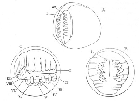
Fig. 245. Three stages in the development of
Limulus polyphemus.
(Somewhat modified from Packard.)
A. Embryo in which the thoracic limbs and mouth have become
developed on the ventral plate. The outer line represents what
Packard believes to be the amnion.
B. Later embryo from the ventral surface.
C. Later embryo, just before the splitting of the chorion from the
side. The full number of segments of the abdomen, and three
abdominal appendages, have become established; m. mouth;
I-IX. appendages.
Round the edge of the ventral plate there is a distinct ridge—the
rudiment of the cephalothoracic shield.
With the further growth of the embryo the chorion becomes split and
cast off, the embryo being left enclosed within the inner membrane.
The embryo has a decided ventral flexure, and the abdominal region
grows greatly and forms a kind of cap at the hinder end, while its
vaulted dorsal side becomes divided into segments (fig. 245 C). Of
these there are according to Dohrn seven, but according to Packard
nine, of which the last forms the rudiment of the caudal spine.
In the thoracic region the nervous system is by this stage formed as a
ganglionated cord (Dohrn), with no resemblance to the peculiar
œsophageal ring of the adult. The mouth is stated by Dohrn to lie
between the second pair of limbs, so that, if the descriptions we have
are correct, it must have by this stage changed its position with
reference to the appendages. Between the thorax and abdomen two
papillæ have arisen which form the so-called lower lip of the
adult, but from their position and late development they can hardly be
regarded as segmental appendages. In the course of further changes all
the parts become more distinct, while the membrane in which the larva
is placed becomes enormously distended (fig. 246 A). The rudiments of
the compound eyes are formed on the third (Packard) or fourth (Dohrn)
segment of the cephalothorax, and the simple eyes near the median line
in front. The rudiments of the inner process of the chelæ of the
cephalothoracic appendages arise as buds. The abdominal appendages
become more plate-like, and the rudiments of a third pair appear
behind the two already present. The heart appears on the dorsal
surface.
An ecdysis now takes place, and in the stage following the limbs have
approached far more closely to their adult state (fig. 246 A). The
cephalothoracic appendages become fully jointed; the two anterior
abdominal appendages (VII.) have approached, and begin to
resemble the operculum of the adult, and on the second pair is formed
a small inner ramus. The segmentation of the now vaulted cephalothorax
becomes less obvious, though still indicated by the arrangement of the
yolk masses which form the future hepatic diverticula.
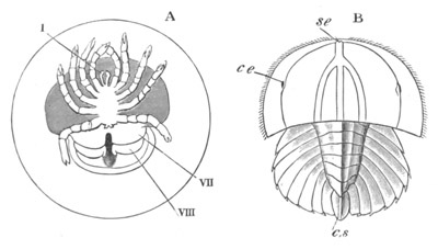
Fig. 246. Two stages in the development of
Limulus polyphemus.
(After Dohrn.)
A. An advanced embryo enveloped in the distended inner membrane
shortly before hatching; from the ventral side.
B. A later embryo at the Trilobite stage, from the dorsal side.
I., VII., VIII. First, seventh, and
eighth appendages.
cs. caudal spine; se. simple eye; ce. compound
eye.
Shortly after this stage the embryo is hatched, and at about the time
of hatching acquires a form (fig. 246 B) in which it bears, as pointed
out by Dohrn and Packard, the most striking resemblance to a
Trilobite.
Viewed from the dorsal surface (fig. 246 B) it is divided into two
distinct regions, the cephalothoracic in front and the abdominal
behind. The cephalothoracic has become much flatter and wider, has
lost all trace of its previous segmentation, and has become distinctly
trilobed. The central lobe forms a well-marked keel, and at
the line of insertion of the rim-like edge of the lateral lobes are
placed the two pairs of eyes (se and ce). The abdominal
region is also distinctly trilobed and divided into nine segments; the
last, which is merely formed of a median process, being the rudiment
of the caudal spine. The edges of the second to the seventh are armed
with a spine. The changes in the appendages are not very considerable.
The anterior pair nearly meet in the middle line in front or the
mouth; and the latter structure is completely covered by an upper lip.
Each abdominal appendage of the second pair is provided with four gill
lamellæ, attached close to its base.
Three weeks after hatching an ecdysis takes place, and the larva
passes from a trilobite into a limuloid form. The segmentation of the
abdomen has become much less obvious, and this part of the embryo
closely resembles its permanent form. The caudal spine is longer, but
is still relatively short. A fourth pair of abdominal appendages is
established, and the first pair have partially coalesced, while the
second and third pairs have become jointed, their outer ramus
containing four and their inner three joints. Additional gill lamellæ
attached to the two basal joints of the second and third abdominal
appendages have appeared.
The further changes are not of great importance. They are effected in
a series of successive moults. The young larvæ swim actively at the
surface.
Our, in many respects, imperfect knowledge of the development of
Limulus is not sufficient to shew whether it is more closely related
to the Crustacea or to the Arachnida, or is an independent phylum.
The somewhat Crustacean character of biramous abdominal feet, etc. is
not to be denied, but at the same time the characters of the embryo
appear to me to be decidedly more arachnidan than crustacean. The
embryo, when the appendages are first formed, has a decidedly
arachnidan facies. It will be remembered that when the limbs are first
formed they are all post-oral. They resemble in this respect
the limbs of the Arachnida, and it seems to be probable that the
anterior pair is equivalent to the cheliceræ of Arachnida, which, as
shewn in a previous section, are really post-oral appendages in no way
homologous with antennæ[212].
The six thoracic appendages may thus be compared with the six
Arachnidan appendages; which they resemble in their relation to the
mouth, their basal cutting blades, etc.
The existence of abdominal appendages behind the six cephalothoracic
does not militate against the Arachnidan affinities of Limulus,
because in the Arachnida rudimentary abdominal appendages are always
present in the embryo. The character of the abdominal appendages is
probably secondarily adapted to an aquatic respiration, since
it is likely (for the reasons already mentioned in connection with the
Tracheata) that if Limulus has any affinities with the stock of the
Tracheata it is descended from air-breathing forms, and has acquired
its aquatic mode of respiration. The anastomosis of the two halves of
the generative glands is an Arachnidan character, and the position of
the generative openings in Limulus is more like that in the Scorpion
than in Crustacea.
A fuller study of the development would be very likely to throw
further light on the affinities of Limulus, and if Packard’s view
about the nature of the inner egg membrane were to be confirmed,
strong evidence would thereby be produced in favour of the Arachnidan
affinities.
(533) A. Dohrn. “Untersuch. üb. Bau u. Entwick. d. Arthropoden
(Limulus polyphemus).” Jenaische Zeitschrift, Vol.
VI., 1871.
(534) A. S. Packard. “The development of Limulus polyphemus.”
Mem. Boston Soc. Nat. History, Vol.
II., 1872.
Pycnogonida.
The embryos, during the first phases of their development, are always
carried by the male in sacks which are attached to a pair of
appendages (the third) specially formed for this purpose. The
segmentation of the ovum is complete, and there is in most forms
developed within the eggshell a larva with three pairs of two-jointed
appendages, and a rostrum placed between the front pair.
It will be convenient to take Achelia lævis, studied by Dohrn (No.
536), as type.
The larva of Achelia when hatched is provided with the typical three
pairs of appendages. The foremost of them is chelate, and the two
following pairs are each provided with a claw. Of the three pairs of
larval appendages Dohrn states that he has satisfied himself that the
anterior is innervated by the supra-œsophageal ganglion, and the
two posterior by separate nerves coming from two imperfectly united
ventral ganglia. The larva is provided with a median eye formed of two
coalesced pigment spots, and with a simple stomach.
The gradual conversion of the larva into the adult takes place by the
elongation of the posterior end of the body into a papilla, and the
formation there, at a later period, of the anus; while at the two
sides of the anal papilla rudiments of a fresh pair of appendages—the
first pair of ambulatory limbs of the adult—make their appearance.
The three remaining pairs of limbs become formed successively as
lateral outgrowths, and their development is accomplished in a number
of successive ecdyses. As they are formed cæca from the stomach become
prolonged into them. For each of them there appears a special
ganglion. While the above changes are taking place the three pairs of
larval appendages undergo considerable reduction. The anterior pair
singly becomes smaller, the second loses its claw, and the third
becomes reduced to a mere stump. In the adult the second pair
of appendages becomes enlarged again and forms the so-called palpi,
while the third pair develops in the male into the egg-carrying
appendages, but is aborted in the female. The first pair form
appendages lying parallel to the rostrum, which are sometimes called
pedipalpi and sometimes antennæ.
The anal papilla is a rudimentary abdomen, and, as Dohrn has shewn,
contains rudiments of two pairs of ganglia.
The larvæ of Phoxichilidium are parasitic in various Hydrozoa
(Hydractinia, etc.). After hatching they crawl into the Hydractinia
stock. They are at first provided with the three normal pairs of
larval appendages. The two hinder of these are soon thrown off, and
the posterior part of the trunk, with the four ambulatory appendages
belonging to it, becomes gradually developed in a series of moults.
The legs, with the exception of the hindermost pair, are fully formed
at the first ecdysis after the larva has become free. In the genus
Pallene the metamorphosis is abbreviated, and the young are hatched
with the full complement of appendages.
The position of the Pycnogonida is not as yet satisfactorily settled.
The six-legged larva has none of the characteristic features of the
Nauplius, except the possession of the same number of appendages.
The number of appendages (7) of the Pycnogonida does not coincide with
that of the Arachnida. On the other hand, the presence of chelate
appendages innervated in the adult by the supra-œsophageal ganglia
rather points to a common phylum for the Pycnogonida and Arachnida;
though as shewn above (p. 455) all the appendages in the embryo of
true Arachnida are innervated by post-oral ganglia. The innervation of
these appendages in the larvæ of Pycnogonida requires further
investigation. Against such a relationship the extra pair of
appendages in the Pycnogonida is no argument, since the embryos of
most Arachnida are provided with four such extra pairs. The two groups
must no doubt have diverged very early.
Bibliography.
(535) G. Cavanna. “Studie e ricerche sui Picnogonidi.”
Pubblicazioni del R. Instituto di Studi superiori in Firenze,
1877.
(536) An. Dohrn. “Ueber Entwicklung u. Bau d. Pycnogoniden.”
Jenaische Zeitschrift, Vol. V. 1870, and “Neue
Untersuchungen üb. Pycnogoniden.” Mittheil. a. d. zoologischen
Station zu Neapel, Bd. I. 1878.
(537) G. Hodge. “Observations on a species of Pycnogon, etc.”
Annal. and Mag. of Nat. Hist. Vol. IX. 1862.
(538) C. Semper. “Ueber Pycnogoniden u. ihre in Hydroiden
schmarotzenden Larvenformen.” Arbeiten a. d. zool.-zoot. Instit.
Würzburg, Vol. I. 1874.
Pentastomida.
The development and metamorphosis of Pentastomum tænoides have been
thoroughly worked out by Leuckart (No. 540) and will serve as type for
the group.
In the sexual state it inhabits the nasal cavities of the dog. The
early embryonic development takes place as the ovum gradually passes
down the uterus. The segmentation appears to be complete; and gives
rise to an oval mass in which the separate cells can hardly be
distinguished. This gradually differentiates itself into a
characteristic embryo, divided into a tail and trunk. The tail is
applied to the ventral surface of the trunk, and on the latter two
pairs of stump-like unsegmented appendages arise, each provided with a
pair of claws. At the anterior extremity of the body is formed the
mouth, with a ventral spine and lateral hook, which are perhaps
degenerated jaws. The spine functions as a boring apparatus, and an
apparatus with a similar function is formed at the end of the tail. A
larval cuticle now appears, which soon becomes detached from the
embryo, except on the dorsal surface, where it remains firmly united
to a peculiar papilla. This papilla becomes eventually divided into
two parts, one of which remains attached to the cuticle, while the
part connected with the embryo forms a raised cross placed in a
cup-shaped groove. The whole structure has been compared, on
insufficient grounds, to the dorsal organ of the Crustacea.
The eggs, containing the embryo in the condition above described, are
eventually carried out with the nasal slime, and, if transported
thence into the alimentary cavity of a rabbit or hare, the embryos
become hatched by the action of the gastric juice. From the alimentary
tract of their new host they make their way into the lungs or liver.
They here become enveloped in a cyst, in the interior of which they
undergo a very remarkable metamorphosis. They are, however, so minute
and delicate that Leuckart was unable to elucidate their structure
till eight weeks after they had been swallowed. At this period they
are irregularly-shaped organisms, with a most distant resemblance to
the earlier embryos. They are without their previous appendages, but
the alimentary tract is now distinctly differentiated. The remains of
two cuticles in the cyst seem to shew that the above changes are
effected in two ecdyses.
In the course of a series of ecdyses the various organs of the larval
form known as Pentastomum denticulatum continue to become
differentiated. After the first (=third) ecdysis the œsophageal
nerve-ring and sexually undifferentiated generative organs are
developed. At the fourth (=sixth) ecdysis the two pairs of hooks of
the adult are formed in pockets which appeared at a somewhat earlier
stage; and the body acquires an annulated character. At a somewhat
earlier period rudiments of the external generative organs indicate
the sex of the larva.
After a number of further ecdyses, which are completed in about six
months after the introduction of the embryos into the intermediate
host, the larva attains its full development, and acquires a form in
which it has long been known as Pentastomum denticulatum. It now
leaves its cyst and begins to move about. It is in a state fit to be
introduced into its final host; but if it be not so introduced it may
become encysted afresh.
If the part of a rabbit or hare infected by a Pentastomum denticulatum
be eaten by a dog or wolf, the parasite passes into the nasal cavity
of the latter, and after further changes of cuticle becomes a
fully developed sexual Pentastomum tænioides, which does not differ to
any very marked extent from P. denticulatum.
In their general characters the larval migrations of Pentastomum are
similar to those of the Cestodes.
The internal anatomy of the adult Pentastomum, as well as the
characters of the larva with two pairs of clawed appendages, are
perhaps sufficient to warrant us in placing it with the Arthropoda,
though it would be difficult to shew that it ought not to be placed
with such a form as Myzostomum (vide p. 369). There do not
appear to be any sufficient grounds to justify its being placed with
the Mites amongst the Arachnida. If indeed the rings of the body of
the Pentastomida are to be taken as implying a true segmentation, it
is clear that the Pentastomida cannot be associated with the Mites.
Bibliography.
(539) P. J. van Beneden. “Recherches s. l’organisation et le
développement d. Linguatules.” Ann. d. Scien. Nat., 3 Ser.,
Vol. XI.
(540) R. Leuckart. “Bau u. Entwicklungsgeschichte d.
Pentastomen.” Leipzig and Heidelberg. 1860.
Tardigrada.
Very little is known with reference to the development of the
Tardigrada. A complete and regular segmentation (von Siebold,
Kaufmann, No. 541) is followed by the appearance of a groove on the
ventral side indicating a ventral flexure. At about the time of the
appearance of the groove the cells become divided into an epiblastic
investing layer and a central hypoblastic mass.
The armature of the pharynx is formed very early at the anterior
extremity, and the limbs arise in succession from before backwards.
The above imperfect details throw no light on the systematic position
of this group.
Tardigrada.
(541) J. Kaufmann. “Ueber die Entwicklung u. systematische
Stellung d. Tardigraden.” Zeit. f. wiss. Zool., Bd.
III. 1851.
Summary of Arthropodan Development.
The numerous characters common to the whole of the Arthropoda led
naturalists to unite them in a common phylum, but the later researches
on the genealogy of the Tracheata and Crustacea tend to throw doubts
on this conclusion, while there is not as yet sufficient evidence to
assign with certainty a definite position in either of these classes
to the smaller groups described in the present chapter. There seems to
be but little doubt that the Tracheata are descended from a
terrestrial Annelidan type related to Peripatus. The affinities of
Peripatus to the Tracheata are, as pointed out in a previous chapter
(p. 386), very clear, while at the same time it is not possible to
regard Peripatus simply as a degraded Tracheate, owing to the fact
that it is provided with such distinctly Annelidan organs as
nephridia, and that its geographical distribution shews it to be a
very ancient form.
The Crustacea on the other hand are clearly descended from a
Phyllopod-like ancestor, which can be in no way related to Peripatus.
The somewhat unexpected conclusion that the Arthropoda have a double
phylum is on the whole borne out by the anatomy of the two groups.
Without attempting to prove this in detail, it may be pointed out that
the Crustacean appendages are typically biramous, while those of the
Tracheata are never at any stage of development biramous[213]; and the
similarity between the appendages of some of the higher Crustacea and
those of many Tracheata is an adaptive one, and could in no case be
used as an argument for the affinity of the two groups.
The similarity of many organs is to be explained by both groups being
descendants of Annelidan ancestors. The similarity of the compound eye
in the two groups cannot however be explained in this way, and is one
of the greatest difficulties of the above view. It is moreover
remarkable that the eye of Peripatus[214] is formed on a different
type to either the single or compound eyes of most Arthropoda.
The conclusion that the Crustacea and Tracheata belong to two distinct
phyla is confirmed by a consideration of their development. They have
no doubt in common a centrolecithal segmentation, but, as already
insisted on, the segmentation is no safe guide to the affinities.
In the Tracheata the archenteron is never, so far as we know, formed
by an invagination[215], while in Crustacea the evidence is in
favour of such an invagination being the usual, and, without doubt,
the primitive, mode of origin.
The mesoblast in the Tracheata is formed in connection with a median
thickening of the ventral plate. The unpaired plate of mesoblast so
formed becomes divided into two bands, one on each side of the middle
line.
In both Spiders and Myriopods, and probably Insects, the two plates of
mesoblast are subsequently divided into somites, the lumen of which is
continued into the limbs.
In Crustacea the mesoblast usually originates from the walls of the
invagination, which gives rise to the mesenteron.
It does not become divided into two distinct bands, but forms a layer
of scattered cells between the epiblast and hypoblast, and does not
usually break up into somites; and though somites are stated in some
cases to be found they do not resemble those in the Tracheata.
The proctodæum is usually formed in Crustacea before and rarely
later[216] than the stomodæum. The reverse is true for the Tracheata.
In Crustacea the proctodæum and stomodæum, especially the former, are
very long, and usually give rise to the greater part of the alimentary
tract, while the mesenteron is usually short.
In the Tracheata the mesenteron is always considerable, and the
proctodæum is always short. The derivation of the Malpighian bodies
from the proctodæum is common to most Tracheata. Such diverticula of
the proctodæum are not found in Crustacea.
The development of the Echinodermata naturally falls into two
sections:—
(1) The development of the germinal layers and of the systems of
organs; (2) the development of the larval appendages and the
metamorphosis.
The Development of the Germinal Layers and of the Systems of
Organs.
The development of the systems of organs presents no very important
variations within the limits of the group.
Holothuroidea. The Holothurians have been most fully studied
(Selenka, No. 563), and may be conveniently taken as type.
The segmentation is nearly regular, though towards its close, and in
some instances still earlier, a difference becomes apparent between
the upper and the lower poles.
At the close of segmentation (fig. 247 A) the egg has a nearly
spherical form, and is constituted of a single layer of columnar cells
enclosing a small segmentation cavity. The lower pole is slightly
thickened, and the egg rotates by means of fine cilia.
An invagination now makes its appearance at the lower pole (fig. 247
B), and simultaneously there become budded off from the cells
undergoing the invagination amœboid cells, which
eventually form the muscular system and the connective tissue. These
cells very probably have a bilaterally symmetrical origin. This stage
represents the gastrula stage which is common to all Echinoderms. The
invaginated sack is the archenteron. As it grows larger one side of
the embryo becomes flattened, and the other more convex. On the
flattened side a fresh invagination arises, the opening of which forms
the permanent mouth, the opening of the first invagination remaining
as the permanent anus (fig. 248 A).
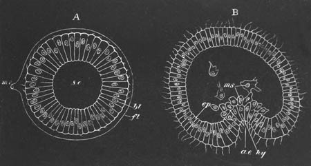
Fig. 247. Two stages in the development of
Holothuria tubulosa viewed in optical section. (After Selenka.)
A. Blastosphere stage at the close of segmentation. B. Gastrula
stage.
mr. micropyle; fl. chorion; s.c. segmentation
cavity; bl. blastoderm; ep. epiblast; hy.
hypoblast; ms. amœboid cells derived from hypoblast;
a.e. archenteron.
These changes give us the means of attaching definite names to the
various parts of the embryo. It deserves to be noted in the first
place that the embryo has assumed a distinctly bilateral form. There
is present a more or less concave surface extending from the mouth to
near the anus, which will be spoken of as the ventral surface. The
anus is situated at the posterior extremity. The convex surface
opposite the ventral surface forms the dorsal surface, which
terminates anteriorly in a rounded præ-oral prominence.
It will be noticed in fig. 248 A that in addition to the primitive
anal invagination there is present a vesicle (v.p.). This
vesicle is directly formed by a constriction of the primitive
archenteron (fig. 249 Vpv.), and is called by Selenka the
vaso-peritoneal vesicle. It gives origin to the epithelioid lining of
the body cavity and water-vascular system of the adult[218]. In the
parts now developed we have the rudiments of all the adult organs.
The mouth and anal involutions (after the separation of the
vaso-peritoneal vesicle) meet and unite, a constriction indicating
their point of junction (fig. 248 B). Eventually the former gives rise
to the mouth and œsophagus, and the latter to the remainder of the
alimentary canal[219].
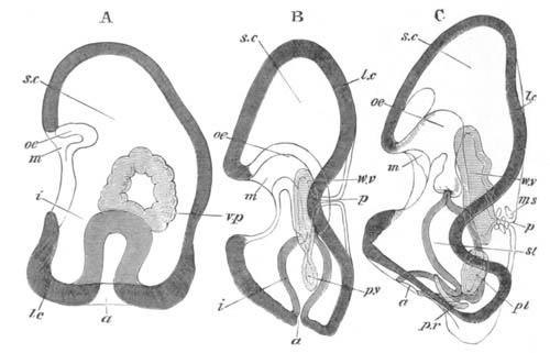
Fig. 248. Three stages in the development of
Holothuria tubulosa viewed from the side in optical section.
(After Selenka.)
m. mouth; oe. œsophagus; st. stomach;
i. intestine; a anus; l.c. longitudinal
ciliated band; v.p. vaso-peritoneal vesicle; p.v.
peritoneal vesicle; p.r. right peritoneal vesicle; pl.
left peritoneal vesicle; w.v. water-vascular vesicle;
p. dorsal pore of water-vascular system; ms. muscle
cells.
The vaso-peritoneal vesicle undergoes a series of remarkable changes.
After its separation from the archenteron it takes up a position on
the left side of this, elongates in an antero-posterior direction, and
from about its middle sends a narrow diverticulum towards the
dorsal surface of the body, where an opening to the
exterior becomes formed (fig. 248 B, p.). The diverticulum
becomes the madreporic canal, and the opening the dorsal pore.
The vaso-peritoneal vesicle next divides into two, an anterior vesicle
(fig. 248 B, w.v.), from which is derived the epithelium of the
water-vascular system, and a posterior (fig. 248 B, p.v.),
which gives rise to the epithelioid lining of the body cavity. The
anterior vesicle (fig. 248 C, w.v.) becomes five-lobed, takes a
horseshoe-shaped form, and grows round the œsophagus (fig. 256,
w.v.r). The five lobes form the rudiments of the water-vascular
prolongations into the tentacles. The remaining parts of the
water-vascular system are also developed as outgrowths of the original
vesicle. Five of these, alternating with the original diverticula,
form the five ambulacral canals, from which diverticula are produced
into the ambulacral feet; a sixth gives rise to the Polian vesicle.
The remaining parts of the original vesicle form the water-vascular
ring.
We must suppose that eventually the madreporic canal loses its
connection with the exterior so as to hang loosely in the interior,
though the steps of this process do not appear to have been made out.
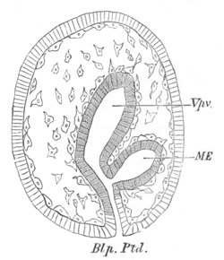
Fig. 249. Longitudinal section through an embryo
of Cucumaria doliolum at the end of the fourth day.
Vpv. vaso-peritoneal vesicle; ME. mesenteron;
Blp., Ptd. blastopore, proctodæum.
The original hinder peritoneal vesicle grows rapidly, and divides into
two (fig. 248 C, pl. and pr.), which encircle the two
sides of the alimentary canal, and meet above and below it. The outer
wall of each of them attaches itself to the skin, and the inner one to
the alimentary canal and water-vascular system; in both cases the
walls remain separated from the adjacent parts by a layer of the
amœboid cells already spoken of. The cavity of the peritoneal
vesicles becomes the permanent body cavity. Where the walls of
the two vesicles meet on the dorsal side, a mesentery, suspending the
alimentary canal and dividing the body cavity longitudinally, is often
formed. In other parts the partition walls between the two sacks
appear to be absorbed.
The amœboid cells, which were derived from the invaginated cells,
arrange themselves as a layer round all the organs (fig. 249). Some of
them remain amœboid, attach themselves to the skin, and form part
of the cutis; and in these cells the calcareous spicula of the larva
and adult are formed. Others form the musculature of the larval
alimentary tract, while the remainder give rise to the musculature and
connective tissue of the adult.
The development of the vascular system is not known, but the discovery
of Kowalevsky, confirmed by Selenka, that from the walls of the
water-vascular system corpuscles are developed, identical with those
in the blood-vessels, indicates that it probably develops in
connection with the water-vascular system. The observations of
Hoffmann and Perrier on the communication of the two systems in the
Echinoidea point to the same conclusion. Though nothing very definite
is known with reference to the development of the nervous system,
Metschnikoff suggests that it develops in connection with the
thickened bands of epiblast which are formed by a metamorphosis of the
ciliated bands of the embryo, and accompany the five radial tubes
(vide p. 555). In any case its condition in the adult leaves no
doubt of its being a derivative of the epiblast.
From the above description the following general conclusions may be
drawn:—
(1) The blastosphere stage is followed by a gastrula stage.
(2) The gastrula opening forms the permanent anus, and the mouth is
formed by a fresh invagination.
(3) The mesoblast arises entirely from the invaginated cells, but in
two ways:—
(a) As scattered amœboid cells, which give origin to the
muscles and connective tissue (including the cutis) of the body wall
and alimentary tract.
(b) As a portion separated off from the archenteron, which
gives rise both to the epithelioid lining of the body cavity, and of
the water-vascular system.
(4) The œsophagus is derived from an invagination of the epiblast,
and the remainder of the alimentary canal from the archenteron.
(5) The embryonic systems of organs pass directly into those of the
adult.
The development of Synapta diverges, as might be expected, to a very
small extent from that of Holothuria.
Asteroidea. In Asterias the early stages of development conform
to our type. There arise, however, two bilaterally symmetrical
vaso-peritoneal diverticula from the archenteron. These diverticula
give rise both to the lining of the body cavity and water-vascular
system. With reference to the exact changes they undergo there is,
however, some difference of opinion. Agassiz (543) maintains that both
vesicles are concerned in the formation of the water-vascular system,
while Metschnikoff (560) holds that the water-vascular system is
entirely derived from the anterior part of the larger left vesicle,
while the right and remainder of the left vesicle form the body
cavity. Metschnikoff’s statements appear to be the most probable. The
anterior part of the left vesicle, after separating from the
posterior, grows into a five-lobed rosette (fig. 260, i), and a
madreporic canal (h) with a dorsal pore opening to the
exterior. The rosette appears not to grow round the œsophagus, as
in the cases hitherto described. But the latter is stated to
disappear, and a new œsophagus to be formed, which pierces the
rosette, and places the old mouth in communication with the stomach.
Except where the anus is absent in the adult, the larval anus probably
persists.
Ophiuroidea. The early development of the Ophiuroidea is not so
fully known as that of other types. Most species have a free-swimming
larva, but some (Amphiura) are viviparous.
The early stages of the free-swimming larvæ have not been described,
but I have myself observed in the case of Ophiothrix fragilis that the
segmentation is uniform, and is followed by the normal invagination.
The opening of this no doubt remains as the larval anus, and there are
probably two outgrowths from this to form the vaso-peritoneal
vesicles. Each of these divides into two parts, an anterior lying
close to the œsophagus, and a posterior close to the stomach. The
anterior on the right side aborts; that on the left side becomes the
water-vascular vesicle, early opens to the exterior, and eventually
grows round the œsophagus, which, as in Holothurians, becomes the
œsophagus of the adult. The posterior vesicles give rise to the
lining of the body cavity, but are stated by Metschnikoff to be at
first solid, and only subsequently to acquire a cavity—the permanent
body cavity. The anus naturally disappears, since it is absent in the
adult. In the viviparous type the first stages are imperfectly known,
but it appears that the blastopore vanishes before the appearance of
the mouth. The development of the vaso-peritoneal bodies takes place
as in the free-swimming larvæ.
Echinoidea. In the Echinoidea (Agassiz, No. 542, Selenka, No.
564) there is a regular segmentation and the normal invagination (fig.
250 A). The amœboid mesoblast cells arise as two laterally placed
masses, and give rise to the usual parts. The archenteron grows
forward and bends towards the ventral side (fig. 250 B). It
becomes (fig. 250 C) divided into three chambers, of which the two
hindermost (d and c) form the stomach and intestine;
while the anterior forms the œsophagus, and gives rise to the
vaso-peritoneal vesicles. These latter appear as a pair of outgrowths
(fig. 251), but become constricted off as a single two-horned
vesicle, which subsequently divides into two. The left of these is
eventually divided, as in Asteroids, into a peritoneal and
water-vascular sack, while the right forms the right peritoneal sack.
An oral invagination on the flattened ventral side meets the
mesenteron after its separation from the vaso-peritoneal vesicle. The
larval anus persists, as also does the larval mouth, but owing to the
manner in which the water-vascular rosette is established the larval
œsophagus appears to be absorbed, and to be replaced by a fresh
œsophagus.
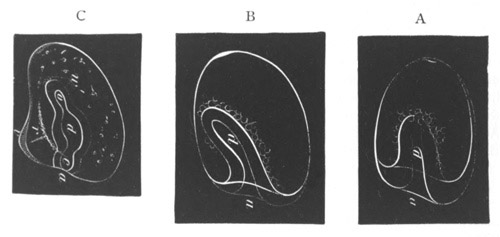
Fig. 250. Three side views of early stages in the
development of Strongylocentrus. (From Agassiz.)
a. anus (blastopore); d. stomach; o.
œsophagus; c. rectum; w. vaso-peritoneal vesicle;
v. ciliated ridge; r. calcareous rod.
Crinoidea. Antedon, the only Crinoid so far studied (Götte, No.
549), presents some not inconsiderable variations from the usual
Echinoderm type. The blastopore is placed on the somewhat flattened
side of the oval blastosphere, and not, as is usual, at the hinder end.
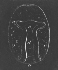
Fig. 251. Dorso-ventral view of an early larva of
Strongylocentrus. (From Agassiz.)
a. anus; d. stomach; o. œsophagus; w.
vaso-peritoneal vesicle; r. calcareous rod.
The blastopore completely closes, and is not converted into the
permanent anus. The archenteron gives rise to the epithelioid lining
of both body cavity and water-vascular system. These parts do not,
however, appear as a single or paired outgrowth from the archenteron,
but as three distinct outgrowths which are not formed
contemporaneously. Two of them are first formed, and become
the future body cavity; but their lumens remain distinct. Originally
appearing as lateral outgrowths, the right one assumes a dorsal
position and sends a prolongation into the stalk (fig. 252,
rp´), and the left one assumes first a ventral, and then an
oral position (fig. 252, lp).
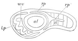
Fig. 252. Longitudinal section through an Antedon
larva. (From Carpenter; after Götte.)
al. mesenteron; wv. water-vascular ring; lp.
left (oral) peritoneal vesicle; rp. right peritoneal vesicle;
rp´. continuation of right vesicle into the stalk; st.
stalk.
The third outgrowth of the archenteron gives rise to the
water-vascular vesicle. It first grows round the region of the future
œsophagus and so forms the water-vascular ring. The wall of the
ring then grows towards the body wall so as to divide the oral (left)
peritoneal vesicle into two distinct vesicles, an anterior and a
posterior, shewn in fig. 253, lp´ and lp. Before this
division is completed, the water-vascular ring is produced in front
into five processes—the future tentacles (fig. 252, wv)—which
project into the cavity of the oral vesicle (lp). After the
oral peritoneal space has become completely divided into two parts,
the anterior dilates (fig. 253, lp´) greatly, and forms a large
vestibule at the anterior end of the body. This vestibule (lp´)
next acquires a communication with the mesenteron, shewn in fig. 253
at m. The anterior wall of this vestibule is finally broken through.
By this rupture the mesenteron is placed in communication with the
exterior by the opening at m, while at the same time the
tentacles of the water-vascular ring (t) project freely to the
exterior. Such is Götte’s account of the præ-oral body space, but, as
he himself points out, it involves our believing that the lining of
the diverticulum derived from the primitive alimentary vesicle becomes
part of the external skin. This occurrence is so remarkable, that more
evidence appears to me requisite before accepting it.
The formation of the anus occurs late. Its position appears to be the
same as that of the blastopore, and is indicated by a papilla of the
mesenteron attaching itself to the skin on the ventral side (fig. 253,
an). It eventually becomes placed in an interradial space
within the oral disc of the adult. The water-vascular ring has no
direct communication with the exterior, but the place of the
madreporic canal of other types appears to be taken in the larva by a
single tube leading from the exterior into the body cavity, the
external opening of which is placed on one of the oral plates
(vide p. 571) in the next interradial space to the right of the
anus, and a corresponding diverticulum of the water-vascular ring
opening into the body cavity. The line of junction between the left
and right peritoneal vesicles forms in the larva a ring-like mesentery
dividing the oral from the aboral part of the body cavity. In
the adult[220] the oral section of the larval body cavity becomes the
ventral part of the circumvisceral division of the body cavity, and
the subtentacular canals of the arms and disc; while the aboral
section becomes the dorsal part of the circumvisceral division of the
body cavity, the cœliac canals of the arms, and the cavity of the
centro-dorsal piece. The primitive distinction between the sections of
the larval body cavity becomes to a large extent obliterated, while
the axial and intervisceral sections of the body cavity of the adult
are late developments.
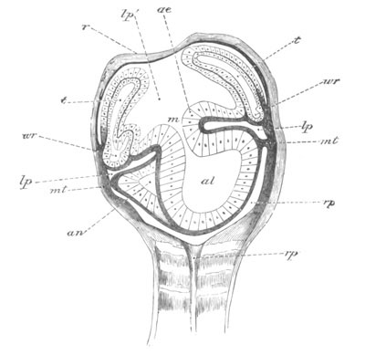
Fig. 253. Longitudinal section through the calyx
of an advanced Pentacrinoid Antedon larva with closed
vestibule.
(From Carpenter; after Götte.)
ae. epithelium of oral vestibule; m. mouth; al.
mesenteron; an. rudiment of permanent anus; lp.
posterior part of left (oral) peritoneal sack; lp´. anterior part of
left (oral) peritoneal sack; wr. water-vascular ring;
t. tentacle; mt. mesentery; rp. right
peritoneal sack; rp´. continuation of right peritoneal sack
into the stalk; r. roof of tentacular vestibule.
The more important points in the development indicated in the
preceding pages are as follows:
(1) The blastosphere is usually elongated in the direction of the axis
of invagination, but in Comatula it is elongated transversely to this
axis.
(2) The blastopore usually becomes the permanent anus, but it closes
at the end of larval life (there being no anus in the adult) in
Ophiuroids and some Asteroids, while in Comatula it closes very early,
and a fresh anus is formed at the point where the blastopore was
placed.
(3) The larval mouth always becomes the mouth of the adult.
(4) The archenteron always gives rise to outgrowths which form the
peritoneal membrane and water-vascular systems. In Comatula there are
three such outgrowths, two paired, which form the peritoneal vesicles,
and one unpaired, which forms the water-vascular vesicle. In Asteroids
and Ophiuroids there are two outgrowths. In Ophiuroids both of these
are divided into a peritoneal and a water-vascular vesicle, but the
right water-vascular vesicle atrophies. In Asteroids only one
water-vascular vesicle is formed, which is derived from the left
peritoneal vesicle. In Echinoids and Holothuroids there is a single
vaso-peritoneal vesicle.
(5) The water-vascular vesicle grows round the larval œsophagus in
Holothuroids, Ophiuroids, and Comatula; in these cases the larval
œsophagus is carried on into the adult. In other forms the
water-vascular vesicle forms a ring which does not enclose the
œsophagus (Asteroids and Echinoids); in such cases a new
œsophagus is formed, which perforates this ring.
Development of the larval appendages and metamorphosis.
Holothuroidea. The young larva of Synapta, to which J. Müller
gave the name Auricularia (fig. 255), is in many respects the simplest
form of Echinoderm larva. With a few exceptions the Auricularia type
of larva is common to the Holothuria.
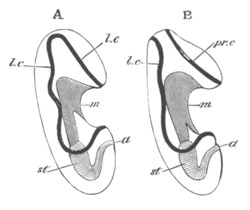
Fig. 254. A. The larva of a Holothuroid. B. The
larva of an Asteroid.
m. mouth; st. stomach; a. anus; l.c.
primitive longitudinal ciliated band; pr.c. præ-oral ciliated
band.
It is (fig. 254 A and fig. 255) bilaterally symmetrical, presenting a
flattened ventral surface, and a convex dorsal one. The anus
(an) is situated nearly at the hinder pole, and the mouth
(m) about the middle of the ventral surface. In front of the
mouth is a considerable process, the præ-oral lobe. Between the mouth
and anus is a space, more or less concave according to the age of the
embryo, interrupted by a ciliated ridge a little in front of
the anus. A similar ciliated ridge is present on the ventral surface
of the præ-oral lobe immediately in front of the mouth. The anal and
oral ridges are connected by two lateral ciliated bands, the whole
forming a continuous band, which, since the mouth lies in the centre
of it (fig. 255), may be regarded as a ring completely surrounding the
body behind the mouth, or more naturally as a longitudinal ring.
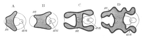
Fig. 255. Diagrammatic figures representing the
evolution of an Auricularia from the simplest Echinoderm larval
form. (Copied from Müller.)
The black line represents the ciliated ridge. The shaded part is the
oral side of the ring, the clear part the aboral side.
m. mouth; an. anus.
The bilateral Auricularia is developed from a slightly elongated
gastrula with an uniform covering of cilia. The gastrula becomes
flattened on the oral side. At the same time the cilia become
specially developed on the oral and anal ridges, and then on the
remainder of the ciliated ring, while they are simultaneously
obliterated elsewhere; and so a complete Auricularia is developed. The
water-vascular ring in the fully developed larva has already
considerably advanced in the growth round the œsophagus (fig. 256
w.v.r).
Most Holothurian larvæ, in their transformation from the bilateral
Auricularia form to the radial form of the adult, pass through a stage
in which the cilia form a number of transverse rings, usually
five in number, surrounding the body. The stages in this metamorphosis
are shewn in figs. 256, 257, and 258.
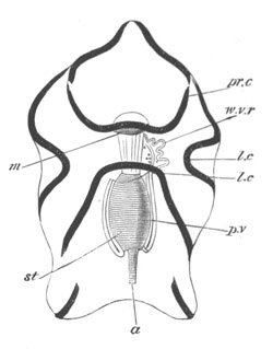
Fig. 256. Full-grown larva of Synapta.
(After Metschnikoff.)
m. mouth; st. stomach; a. anus; p.v.
left division of perivisceral cavity, which is still connected with
the water-vascular system; w.v.r. water-vascular ring which
has not yet completely encircled the œsophagus; l.c.
longitudinal part of ciliated band; pr.c. præ-oral part of
ciliated band.
The primitive ciliated band, at a certain stage of the metamorphosis,
breaks up into a number of separate portions (fig. 256), the whole of
which are placed on the ventral surface. Four of these (fig. 257 A and
B) arrange themselves in the form of an angular ring round the mouth,
which at this period projects considerably. The remaining portions of
the primitive band change their direction from a longitudinal one to a
transverse (fig. 257 B), and eventually grow into complete rings (fig.
257 C). Of these there are five. The middle one (257 B) is the first
to develop, and is formed from the dorsal parts of the primitive ring.
The two hinder rings develop next, and last of all the two anterior
ones, one of which appears to be in front of the mouth (fig. 257 C).
The later development of the mouth, and of the ciliated ridge
surrounding it, is involved in some obscurity. It appears from
Metschnikoff (No. 560) that an invagination of the œsophagus takes
place, carrying with it the ciliated ridge around the mouth. This
ridge becomes eventually converted into the covering for the five
tentacular outgrowths of the water-vascular ring (fig. 258), and
possibly also forms the nervous system.
The opening of the œsophageal invagination is at first behind the
foremost ciliated ring, but eventually comes to lie in front of it,
and assumes a nearly terminal though slightly ventral position (fig.
258). No account has been given of the process by which this takes
place, but the mouth is stated by Metschnikoff (though Müller
differs from him on this point) to remain open throughout. The further
changes in the metamorphosis are not considerable. The ciliated bands
disappear, and a calcareous ring of ten pieces, five ambulacral and
five interambulacral, is formed round the œsophagus. A provisional
calcareous skeleton is also developed.
All the embryonic systems of organs pass in this case directly into
those of the adult.
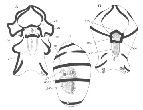
Fig. 257. Three stages in the development of
Synapta. A and B are viewed from the ventral surface, and C
from the side. (After Metschnikoff.)
m. mouth; oe. œsophagus; pv. walls of the
perivisceral cavity; wv. longitudinal vessel of the
water-vascular system; p. dorsal pore of water-vascular
system; cr. ciliated ring formed round the mouth from parts
of the primitive ciliated band.
The metamorphosis of most Holothuroidea is similar to that just
described. In Cucumaria (Selenka) there is however no Auricularia
stage, and the uniformly ciliated stage is succeeded by one with five
transverse bands of cilia, and a præ-oral and an anal ciliated cap.
The mouth is at first situated ventrally behind the præ-oral cap of
cilia, but the præ-oral cap becomes gradually absorbed, and the mouth
assumes a terminal position.
In Psolinus (Kowalevsky) there is no embryonic ciliated stage, and the
adult condition is attained without even a metamorphosis. There appear
to be five plates surrounding the mouth, which are developed
before any other part of the skeleton, and are regarded by P. H.
Carpenter (No. 548) as equivalent to the five oral plates of the
Crinoidea. The larval condition with ciliated bands is often spoken of
as the pupa stage, and during it the larvæ of Holothurians proper use
their embryonic tube feet to creep about.
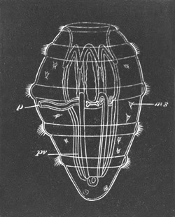
Fig. 258. A late stage in the development of
Synapta. (After Metschnikoff.)
The figure shews the vestibular cavity with retracted tentacles; the
ciliated bands; the water-vascular system, etc.
p. dorsal pore of water-vascular system; pv. walls of
perivisceral cavity; ms. amœboid cells.
Asteroidea. The commonest and most thoroughly investigated form
of Asteroid larva is a free-swimming form known as Bipinnaria.
This form in passing from the spherical to the bilateral condition
passes through at first almost identical changes to the Auricularian
larva. The cilia become at an early period confined to an oral and
anal ridge.
The anal ridge gradually extends dorsalwards, and finally forms a
complete longitudinal post-oral ring (fig. 259 A); the oral ridge also
extends dorsalwards, and forms a closed præ-oral ring (fig. 259 A),
the space within which is left unshaded in my figure.
The presence of two rings instead of one distinguishes the Bipinnaria
from the Auricularia. The two larvæ are shewn side by side in fig.
254, and it is obvious that the two bands of the Bipinnaria are (as
pointed out by Gegenbaur) equivalent to the single band of the
Auricularia divided into two. Ontologically, however, the two bands of
Bipinnaria do not appear to arise from the division of a single band.
As the Bipinnaria grows older, a series of arms grows out along lines
of the two ciliated bands (fig. 259 C), and, in many cases, three
special arms are formed, not connected with the ciliated bands, and
covered with warts. These latter arms are known as brachiolar
arms, and the larvæ provided with them as Brachiolaria (fig. 259 D).
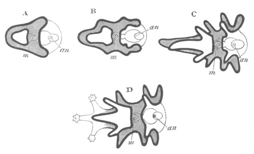
Fig. 259. Diagrammatic representation of various
forms of Asteroid Larvæ. A, B, C, Bipinnaria; D, Brachiolaria.
(Copied from Müller.)
The black lines represent the ciliated bands; and the shading the
space between the præ-oral and the post-oral bands.
m. mouth; an. anus.
As a rule the following arms can be distinguished (fig. 259 C and D),
on the hinder ring (Agassiz’ nomenclature) a median anal pair, a
dorsal anal pair, and a ventral anal pair, a dorsal oral pair, and an
unpaired anterior dorsal arm; on the præ-oral ring a ventral oral
pair, and sometimes (Müller) an unpaired anterior ventral arm.
The three brachiolar arms arise as processes from the base of the
unpaired dorsal arm, and the two ventral oral arms. The extent of the
development of the arms varies with the species.
The changes by which the Bipinnaria or Brachiolaria becomes converted
into the adult starfish are very much more complicated than those
which take place in Holothurians. For an accurate knowledge of them we
are largely indebted to Alex. Agassiz (No. 543). The development of
the starfish takes place entirely at the posterior end of the larva
close to the stomach.
On the right and dorsal side of the stomach, and externally to the
right peritoneal space, are formed five radially situated
calcareous rods arranged in the form of a somewhat irregular pentagon.
The surface on which they are deposited has a spiral form, and
constitutes together with its calcareous rods, the abactinal
or dorsal surface of the future starfish. Close to its dorsal,
i.e. embryonic dorsal, edge lies the dorsal pore of the
water-vascular system (madreporic canal), and close to its ventral
edge the anus. On the left and ventral side of the stomach is placed
the water-vascular rosette, the development of which was described on
p. 549. It is situated on the actinal or ventral surface of the future
starfish, and is related to the left peritoneal vesicle.
Metschnikoff (No. 560) and Agassiz (No. 543) differ slightly as to the
constitution of the water-vascular rosette. The former describes and
figures it as a completely closed rosette, the latter states that ‘it
does not form a completely closed curve but is always open, forming a
sort of twisted crescent-shaped arc.’
The water-vascular rosette is provided with five lobes, corresponding
to which are folds in the larval skin, and each lobe corresponds to
one of the calcareous plates developed on the abactinal disc. The
plane of the actinal surface at first meets that of the abactinal at
an acute or nearly right angle. The two surfaces are separated by the
whole width of the stomach. The general appearance of the larva from
the ventral surface after the development of the water-vascular
rosette (i) and abactinal disc (A) is shewn in fig. 260.
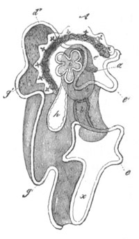
Fig. 260. Bipinnaria larva of an Asteroid.
(From Gegenbaur; after Müller.)
b. mouth; a. anus; h. madreporic canal;
i. ambulacral rosette; c. stomach; d. g. e.
etc. arms of Bipinnaria; A. abactinal disc of young
Asteroid.
As development proceeds the abactinal surface becomes a firm and
definite disc, owing to the growth of the original calcareous spicules
into more or less definite plates, and to the development of five
fresh plates nearer the centre of the disc and interradial in
position. Still later a central calcareous plate appears on the
abactinal surface, which is thus formed of a central plate, surrounded
by a ring of five interradial plates, and then again by a ring of five
radial plates. The abactinal disc now also grows out into five short
processes, separated by five shallow notches. These processes are the
rudiments of the five arms, and each of them corresponds to one of the
lobes of the water-vascular rosette. A calcareous deposit is formed
round the opening of the water-vascular canal, which becomes the
madreporic tubercle[221]. At about this stage the absorption of the
larval appendages takes place. The whole anterior part of the
larva with the great præ-oral lobe has hitherto remained unchanged,
but now it contracts and undergoes absorption, and becomes completely
withdrawn into the disc of the future starfish. The larval mouth is
transported into the centre of the actinal disc. In the larvæ observed
by Agassiz and Metschnikoff nothing was cast off, but the whole
absorbed.
According to Müller and Koren and Danielssen this is not the case in
the larva observed by them, but part of the larva is thrown off, and
lives for some time independently.
After the absorption of the larval appendages the actinal and
abactinal surfaces of the young starfish approach each other, owing to
the flattening of the stomach; at the same time they lose their spiral
form, and become flat discs, which fit each other. Each of the lobes
of the rosette of the water-vascular system becomes one of the radial
water-vascular canals. It first becomes five-lobed, each lobe forming
a rudimentary tube foot, and on each side of the middle lobe two fresh
ones next spring out, and so on in succession. The terminal median
lobe forms the tentacle at the end of the arm, and the eye is
developed at its base. The growth of the water-vascular canals keeps
pace with that of the arms, and the tube feet become supported at
their base by an ingrowth of calcareous matter. The whole of the
calcareous skeleton of the larva passes directly into that of the
adult, and spines are very soon formed on the plates of the abactinal
surface. The original radial plates, together with the spines which
they have, are gradually pushed outwards with the growth of the arms
by the continual addition of fresh rows of spines between the terminal
plate and the plate next to it. It thus comes about that the original
radial plates persist at the end of the arms, in connection with the
unpaired tentacles which form the apex of the radial
water-vascular tubes.
It has already been mentioned that according to Metschnikoff (No. 560)
a new œsophagus is formed which perforates the water-vascular ring,
and connects the original stomach with the original mouth. Agassiz
(No. 543) maintains that the water-vascular ring grows round the
primitive œsophagus. He says—“During the shrinking of the larva
the long œsophagus becomes shortened and contracted, bringing the
opening of the mouth of the larva to the level of the opening of the
œsophagus, which eventually becomes the true mouth of the
starfish.” The primitive anus is believed by Metschnikoff to
disappear, but by Agassiz to remain. This discrepancy very possibly
depends upon these investigators having worked at different species.
There is no doubt that the whole of the larval organs, with the
possible exception of the œsophagus, and anus (where absent in the
adult), pass directly into the corresponding organs of the
starfish—and that the præ-oral part of the body and arms of the larva
are absorbed and not cast off.
In addition to the Bipinnarian type of Asteroid larva a series of
other forms has been described by Müller (No. 561), Sars, Koren, and
Danielssen (No. 554) and other investigators, which are however very
imperfectly known. The best known form is one first of all discovered
by Sars in Echinaster Sarsii, and the more or less similar larvæ
subsequently investigated by Agassiz, Busch, Müller, Wyville Thomson,
etc. of another species of Echinaster and of Asteracanthion. These
larvæ on leaving the egg have an oval form, and are uniformly covered
by cilia. Four processes (or in Agassiz’ type one process) grow out
from the body; by these the larvæ fix themselves. In the case of
Echinaster the larvæ are fixed in the ventral concavity of the disc of
the mother, between the five arms, where a temporary brood-pouch is
established. The main part of the body is converted directly into the
disc of the young starfish, while the four processes come to spring
from the ventral surface, and are attached to the water-vascular ring.
Eventually they atrophy completely. Of the internal structure but
little is known; till the permanent mouth is formed, after the
development of the young starfish is pretty well advanced, the stomach
has no communication with the exterior.
A second abnormal type of development is presented by the embryo of
Pteraster miliaris, as described by Koren and Danielssen[222]. The
larvæ to the number of eight to twenty develop in a peculiar pouch on
the dorsal surface of the body. The early stages are not known, but in
the later ones the whole body assumes a pentagonal appearance with a
mouth at one edge of the disc. At a later stage the anus is
formed on the dorsal side of an arm opposite the mouth. The stomach is
surrounded by a water-vascular ring, from which the madreporic canal
passes to the dorsal surface, but does not open. At a later stage the
embryonic mouth and anus vanish, to be replaced by a permanent mouth
and anus in the normal positions.
A third, and in some respects very curious, form is a worm like larva of
Müller, which is without bands of cilia. The dorsal surface of the youngest
larva is divided by transverse constrictions into five segments. On the
under side of the first of these is a five-lobed disc, each lobe being provided
with a pair of tube feet.
At a later period only three segments are visible on the dorsal surface,
but the ventral surface has assumed a pentagonal aspect. The later stages
are not known.
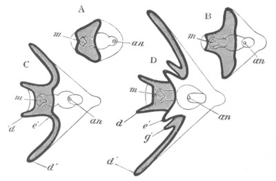
Fig. 261. Diagrammatic figures shewing the
evolution of an Ophiuroid Pluteus from a simple Echinoderm
larva. (Copied from Müller.) The calcareous skeleton is not
represented.
m. mouth; an. anus; d. anterior arms;
d´. lateral arms; e´. posterior arms; g´.
anterolateral arms.
Ophiuroidea. The full-grown larva of the Ophiuroids is known as
a Pluteus. It commences with the usual more or less spherical form;
from this it passes to a form closely resembling that of Auricularia
with a rounded dorsal surface, and a flattened ventral one. Soon
however it becomes distinguished by the growth of a post-anal lobe and
the absence of a præ-oral lobe (fig. 261 B). The post-anal lobe forms
the somewhat rounded apex of the body. In front of the mouth, and
between the mouth and anus, arise the anal and oral ciliated ridges,
which soon become continued into a single longitudinal ciliated ring.
At the same time the body becomes prolonged into a series of
processes along the ciliated band, which is continued to the extremity
of each. The primitive ciliated ring never becomes broken up into two
or more rings. A ciliated crown is usually developed at the extremity
of the post-anal lobe. The arms are arranged in the form of a ring
surrounding the mouth, and are all directed forwards.
The first arms to appear are two lateral ones, which usually remain
the most conspicuous (fig. 261 B and C, d´). Next arises a pair
on the sides of the mouth, which may be called the mouth or anterior
arms (C, d). A pair ventral to and behind the lateral arms is
then formed, constituting the posterior arms (D, e´), and
finally a pair between the lateral arms and the anterior, constituting
the anterolateral arms (D, g´).
The concave area between the arms forms the greater part of the
ventral surface of the body. Even before the appearance of any of the
arms, and before the formation of the mouth, two calcareous rods are
formed, which meet behind at the apex of the post-anal lobe, and are
continued as a central support into each of the arms as they are
successively formed. These rods are shewn at their full development in
fig. 262. The important points which distinguish a Pluteus larva from
the Auricularia or Bipinnaria are the following:
(1) The presence of the post-anal lobe at the hind end of the body.
(2) The slight development of a præ-oral lobe. (3) The provisional
calcareous skeleton in the larval arms.
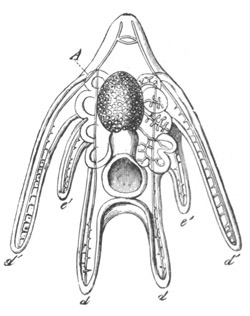
Fig. 262. Pluteus larva of an Ophiuroid. (From
Gegenbaur; after Müller.)
A. rudiment of young Ophiuroid; d´. lateral arms;
d. anterior arms; e´. posterior arms.
Great variations are presented in the development of the arms and
provisional skeleton. The presence of lateral arms is however a
distinctive characteristic of the Ophiuroid Pluteus. The other arms
may be quite absent, but the lateral arms never.
The formation of the permanent Ophiuroid takes place in much the same
way as in the Asteroidea.
There is formed (fig. 262) on the right and dorsal side of stomach the
abactinal disc supported by calcareous plates, at first only five in
number and radial in position[223]. The disc is at first not
symmetrical, but becomes so at the time of the resorption of the
larval arms. It grows out into five processes—the five future rays.
The original five radial plates remain as the terminal segments of the
adult rays, and new plates are always added between the ultimate and
penultimate plate (Müller), though it is probable that in the later
stages fresh plates are added in the disc.
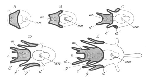
Fig. 263. Diagrammatic Figures Shewing the
Evolution of Echinoid Plutei. (Copied from Müller.) The
calcareous skeleton is not represented. E. Pluteus of Spatangus.
m. mouth; an. anus; d. anterior arms;
d´. point where lateral arms arise in the Ophiuroid Pluteus;
e. anterointernal arms; e´. posterior arms; g´.
anterolateral arms; g. anteroexternal arms.
The ventral surface of the permanent Ophiuroid is formed by the
concave surface between the mouth and anus. Between this and the
stomach is situated the water-vascular ring. It is at first not
closed, but is horseshoe-shaped, with five blind appendages (fig.
262). It eventually grows round the œsophagus, which, together with
the larval mouth, is retained in the adult. The five blind appendages
become themselves lobed in the same way as in Asterias, and grow out
along the five arms of the disc and become the radial canals and
tentacles. All these parts of the water-vascular system are of course
covered by skin, and probably also surrounded by mesoblast cells, in
which at a later period the calcareous plates which lie ventral to the
radial canal are formed. The larval anus disappears. As long as the
larval appendages are not absorbed the ventral and dorsal discs of the
permanent Ophiuroid fit as little as in the case of the Brachiolaria,
but at a certain period the appendages are absorbed. The calcareous
rods of the larval arms break up, the arms and anal lobe
become absorbed, and the dorsal and ventral discs, with the
intervening stomach and other organs, are alone left. After this the
discs fit together, and there is thus formed a complete young
Ophiuroid.
The whole of the internal organs of the larva (except the anus),
including the mouth, œsophagus, the body cavity, etc. are carried
on directly into the adult.
The larval skeleton is, as above stated, absorbed.
The viviparous larva of Amphiura squamata does not differ very greatly
from the larvæ with very imperfect arms. It does not develop a
distinct ciliated band, and the provisional skeleton is very
imperfect. The absence of these parts, as well as of the anus,
mentioned on p. 549, may probably be correlated with the viviparous
habits of the larva. With reference to the passage of this larva into
the adult there is practically nothing to add to what has just been
stated. When the development of the adult is fairly advanced the part
of the body with the provisional skeleton forms an elongated rod-like
process attached to the developing disc. It becomes eventually
absorbed.
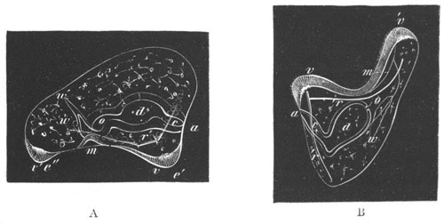
Fig. 264. Two larvæ of Strongylocentrus.
(From Agassiz.)
m. mouth; a. anus; o. œsophagus; d.
stomach; c. intestine; v´. and v. ciliated
ridges; w. water-vascular tube; r. calcareous rods.
Echinoidea. The Echinus larva (fig. 263) has a Pluteus form
like that of the Ophiuroids, and in most points, such as the presence
of the anal lobe, the ciliated band, the provisional skeleton, etc.,
develops in the same manner. The chief difference between the two
Pluteus forms concerns the development of the lateral arms. These,
which form the most prominent arms in the Ophiuroid Pluteus, are
entirely absent in the Echinoid Pluteus, which accordingly
has, as a rule, a much narrower form than the Ophiuroid Pluteus.
A pair of ciliated epaulettes on each side of and behind the ciliated
ring is very characteristic of some Echinoid larvæ. They are
originally developed from the ciliated ring (fig. 266 A and B,
v´´). The presence of three processes from the anal lobe
supported by calcareous rods is characteristic of the Spatangoid
Pluteus (fig. 263 E).
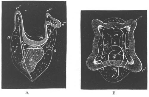
Fig. 265. Lateral and ventral view of a larva of
Strongylocentrus.
(From Agassiz.) General references as in fig. 264.
b. dorsal opening of madreporic canal; e´. posterior
arms; e´´´. anterior arms; eIV. anterointernal
arms.
The first two pairs of arms to develop, employing the same names as in
Ophiuroids, are the anterior attached to the oral process (fig. 263 C,
d) and the posterior pair (e´). A pair of anterolateral
arms next becomes developed (g´). A fourth pair (not
represented in Ophiuroids) appears on the inner side of the anterior
pair forming an anterointernal pair (e), and in the Spatangoid
Pluteus a fifth pair may be added on the external side of the anterior
pair forming an anteroexternal pair (g).
Each of the first-formed paired calcareous rods is composed of three
processes, two of which extend into the anterior and posterior arms;
and the third and strongest passes into the anal lobe, and there meets
its fellow (fig. 265). A transverse bar in front of the arms joins the
rods of the two sides meeting them at the point where the three
processes diverge. The process in the anterolateral arm (fig. 266 B)
is at first independent of this system of rods, but eventually unites
with it. Although our knowledge of the Pluteus types in the
different groups is not sufficient to generalise with great
confidence, a few points seem to have been fairly determined[224]. The
Plutei of Strongylocentrus (figs. 266 and 267) and Echinus have eight
arms and four ciliated epaulettes. The only Cidaris-like form, the
Pluteus of which is known, is Arbacia: it presents certain
peculiarities. The anal lobe develops a pair of posterior (auricular)
appendages, and the ciliated ring, besides growing out into the normal
eight appendages, has a pair of short blunt anterior and posterior
lobes. An extra pair of non-ciliated accessory mouth arms appears also
to be developed. Ciliated epaulettes are not present. So far as is
known the Clypeastroid larva is chiefly characterized by the round
form of the anal lobe. The calcareous rods are latticed. In the
Pluteus of Spatangoids there are (fig. 263) five pairs of arms around
the mouth pointing forwards, and three arms developed from the anal
lobe pointing backwards. One of these is unpaired, and starts from the
apex of the anal lobe. All the arms have calcareous rods which, in the
case of the posterior pair, the anterolateral pair, and the unpaired
arm of the anal lobe, are latticed. Ciliated epaulettes are not
developed.
Viviparous larvæ of Echinoids have been described by Agassiz[225].
The development of the permanent Echinus has been chiefly worked out by
Agassiz and Metschnikoff.
In the Pluteus of Echinus lividus the first indication of the adult
arises, when three pairs of arms are already developed, as an
invagination of the skin on the left side, between the posterior and
anterolateral arms, the bottom of which is placed close to the
water-vascular vesicle (fig. 266 B, w´). The base of this
invagination becomes very thick, and forms the ventral disc of the
future Echinus. The parts connecting this disc with the external skin
become however thin, and, on the narrowing of the external aperture of
invagination and the growth of the thickened disc, constitute a
covering for the disc, called by Metschnikoff the amnion. The
water-vascular vesicle adjoining this disc grows out into five
processes, forming as many tube feet, which cause the surface of the
involuted disc to be produced into the same number of processes. The
external opening of the invagination of the disc never closes, and
after the development of the tube feet begins to widen again, and the
amnion to atrophy. Through the opening of the invagination the tube
feet now project. The dorsal and right surface of the Pluteus, which
extends so as to embrace the opening of the madreporic canal and the
anus, forms the abactinal or dorsal surface of the future Echinus
(fig. 267, a). This disc fits on to the actinal invaginated
surface which arises on the left side of the Pluteus. On the right
surface of the larva (dorsal of permanent Echinus) two pedicellariæ
appear, and at a later period spines are formed, which are at first
arranged in a ring-like form round the edge of the primitively flat
test. While these changes are taking place, and the two surfaces of
the future Echinus are gradually moulding themselves so as to
form what is obviously a young Echinus, the arms of the Pluteus with
their contained skeleton have been gradually undergoing atrophy. They
become irregular in form, their contained skeleton breaks up into
small pieces, and they are gradually absorbed.
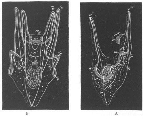
Fig. 266. Side and dorsal view of a larva of
Strongylocentrus.
(From Agassiz.) General reference letters as in figs. 264 and 265.
e´´. anterolateral arms; v´´. ciliated epaulettes;
w´. invagination to form the disc of Echinus.
The water-vascular ring is from the first complete, so that, as in
Asterias, it is perforated through the centre by a new œsophagus.
According to Agassiz the first five tentacles or tube feet grow into
the radial canals, and form the odd terminal tentacles exactly as in
Asterias[226]. Spatangus only differs in development from Echinus in
the fact that the opening of the invagination to form the ventral disc
becomes completely closed, and that the tube feet have eventually to
force their way through the larval epidermis of the amnion, which is
ruptured in the process and eventually thrown off.
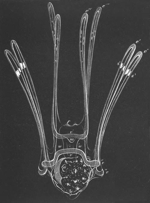
Fig. 267. Full-grown larva of
Strongylocentrus. (From Agassiz.)
The figure shews the largely-developed abactinal disc of the young
Echinus enclosing the larval stomach. Reference letters as in
previous figs.
Crinoidea. The larva of Antedon, while still within the
egg-shell, assumes an oval form and uniform ciliation. Before it
becomes hatched the uniform layer of cilia is replaced by four
transverse bands of cilia, and a tuft of cilia at the posterior
extremity. In this condition it escapes from the egg-shell (fig. 268
A), and becomes bilateral, owing to a flattening of the ventral
surface. On the flattened surface appears a ciliated
depression corresponding in position with the now closed blastopore
(vide p. 550). The third ciliated band bends forward to pass in
front of this (fig. 269). Behind the last ciliated band there is
present a small depression of unknown function, also situated
on the ventral surface. The posterior extremity of the embryo
elongates to form the rudiment of the future stem, and a fresh
depression, marking the position of the future mouth, makes its
appearance on the anterior and ventral part.
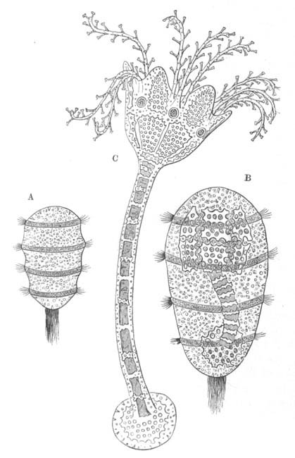
Fig. 268. Three stages in the development of
Antedon (Comatula.)
(From Lubbock; after Thomson.)
A. larva just hatched; B. larva with rudiment of the calcareous
plates; C. Pentacrinoid larva.
While the ciliated bands are still at their full development, the
calcareous skeleton of the future calyx makes its appearance in the
form of two rows, each of five plates, formed of a network of spicula
(figs. 268 B and 269). The plates of the anterior ring are known as
the orals, those of the posterior as the basals. The former surround
the left, i.e. anterior peritoneal sack; the latter the right,
i.e. posterior peritoneal sack. The two rows of plates are at
first not quite transverse, but form two oblique circles, the dorsal
end being in advance of the ventral. The rows soon become transverse,
while the originally somewhat ventral oral surface is carried into the
centre of the area enclosed by the oral plates.
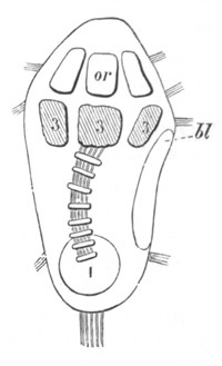
Fig. 269. Larva of Antedon with rudiments of
calcareous skeleton. (From Carpenter; after Thomson.)
1. Terminal plate at the end of the stem; 3. basals; or.
orals; bl. position of blastopore.
By the change in position of the original ventral surface relatively
to the axis of the body, the bilateral symmetry of the larva passes
into a radial symmetry. While the first skeletal elements of the calyx
are being formed, the skeleton of the stem is also established. The
terminal plate is first of all established, then the joints, eight at
first, of the stem. The centro-dorsal plate is stated by Thomson to be
formed as the uppermost joint of the stem[227]. The larva, after the
completion of the above changes, is shewn in fig. 268 B, and somewhat
more diagrammatically in fig. 269.
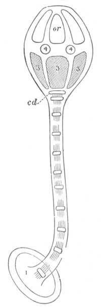
Fig. 270. Young Pentacrinoid larva of
Antedon. (From Carpenter; after Wyville Thomson.)
1. terminal plate of stem; cd. centro-dorsal plate; 3.
basals; 4. radials; or. orals.
After the above elements of the skeleton have become established the
ciliated bands undergo atrophy, and shortly afterwards the
larva becomes attached by the terminal plate of its stem. It then
passes into the Pentacrinoid stage. The larva in this stage is shewn
in fig. 268 C and fig. 270. New joints are added at the upper end of
the stem next the calyx, and a new element—the radials—makes its
appearance as a ring of five small plates, placed in the space between
the basals and orals, and in the intervals alternating with them (fig.
270, 4). The roof of the oral vestibule (vide fig. 253 and p. 551)
has in the meantime become ruptured; and the external opening of
the mouth thus becomes established. Surrounding the mouth are five
petal-like lobes, each of them supported by an oral plate (fig. 268
C). In the intervals between them five branched and highly contractile
tentacles, which were previously enclosed within the vestibule, now
sprout out: they mark the position of the future radial canals, and
are outgrowths of the water-vascular ring. At the base of each of them
a pair of additional tentacles is soon formed. Each primary tentacle
corresponds to one of the radials. These latter are therefore, as
their name implies, radial in position; while the basals and orals are
interradial. In addition to the contractile radial tentacles ten
non-contractile tentacles, also diverticula of the water-vascular
ring, are soon formed, two for each interradius.
In the course of the further development the equatorial space between
the orals and the basals enlarges, and gives rise to a wide oral disc,
the sides of which are formed by the radials resting on the basals;
while in the centre of it are placed the five orals, each with its
special lobe.
The anus, which is formed on the ventral side in the position
of the blastopore (p. 551), becomes surrounded by an anal plate, which
is interradial in position, and lies on the surface of the oral disc
between the orals and radials. On the oral plate in the next
interradius is placed the opening of a single funnel leading into the
body cavity, which Ludwig regards as equivalent to the opening of the
madreporic canal (vide p. 551)[228].
From the edge of the vestibule the arms grow out, carrying with them
the tentacular prolongation of the water-vascular ring. Two additional
rows of radials are soon added.
The stalked Pentacrinoid larva becomes converted, on the absorption of
the stalk, into the adult Antedon. The stalk is functionally replaced
by a number of short cirri springing from the centro-dorsal plate. The
five basals coalesce into a single plate, known as the rosette, and
the five orals disappear, though the lobes on which they were placed
persist. In some stalked forms, e.g. Rhizocrinus Hyocrinus, the
orals are permanently retained. The arms bifurcate at the end of the
third radial, and the first radial becomes in Antedon rosacea (though
not in all species of Antedon) concealed from the surface by the
growth of the centro-dorsal plate. An immense number of funnels,
leading into the body cavity, are formed in addition to the single one
present in the young larva. These are regarded by Ludwig as equivalent
to so many openings of the madreporic canal; and there are developed,
in correspondence with them, diverticula of the water-vascular ring.
Comparison of Echinoderm Larvæ and General Conclusions.
In any comparison of the various types of Echinoderm larvæ it is
necessary to distinguish between the free-swimming forms, and the
viviparous or fixed forms. A very superficial examination suffices to
shew that the free-swimming forms agree very much more closely amongst
themselves than the viviparous forms. We are therefore
justified in concluding that in the viviparous forms the development
is abbreviated and modified.
All the free forms are nearly alike in their earliest stage after the
formation of the archenteron. The surface between the anus and the
future mouth becomes flattened, and (except in Antedon, Cucumaria,
Psolinus, etc. which practically have an abbreviated development like
that of the viviparous forms) a ridge of cilia becomes established in
front of the mouth, and a second ridge between the mouth and the anus.
This larval form, which is shewn in fig. 264 A, is the type from which
the various forms of Echinoderm larvæ start.
In all cases, except in Bipinnaria, the two ciliated ridges soon
become united, and constitute a single longitudinal post-oral ciliated
ring.
The larvæ in their further growth undergo various changes, and in the
later stages they may be divided into two groups:
(1) The Pluteus larva of Echinoids and Ophiuroids.
(2) The Auricularia (Holothuroids) and Bipinnaria (Asteroids) type.
The first group is characterized by the growth of a number of arms
more or less surrounding the mouth, and supported by calcareous rods.
The ciliated band retains its primitive condition as a simple
longitudinal band throughout larval life. There is a very small
præ-oral lobe, while an anal lobe is very largely developed.
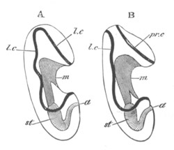
Fig. 271. A. The larva of a Holothuroid. B. The
larva of an Asterias.
m. mouth; st. stomach; a. anus; l.c.
primitive longitudinal ciliated band; pr.c. præ-oral ciliated
band.
The Auricularia and Bipinnaria resemble each other in shape, in the
development of a large præ-oral lobe, and in the absence of
provisional calcareous rods; but differ in the fact that the ciliated
band is single in Auricularia (fig. 271 A), and is double in
Bipinnaria (fig. 271 B).
The Bipinnaria larva shews a great tendency to develop soft arms;
while in the Auricularia the longitudinal ciliated band breaks
up into a number of transverse ciliated bands. This condition is in
some instances reached directly, and such larvæ undoubtedly
approximate to the larvæ of Antedon, in which the uniformly ciliated
condition is succeeded by one with four transverse bands, of which one
is præ-oral.
All or nearly all Echinoderm larvæ are bilaterally symmetrical, and
since all Echinodermata eventually attain a radial symmetry, a change
necessarily takes place from the bilateral to the radial type.
In the case of the Holothurians and Antedon, and generally in the
viviparous types, this change is more or less completely effected in
the embryonic condition; but in the Bipinnaria and Pluteus types a
radial symmetry does not become apparent till after the absorption of
the larval appendages. It is a remarkable fact, which seems to hold
for the Asteroids, Ophiuroids, Echinoids, and Crinoids, that the
dorsal side of the larva is not directly converted into the dorsal
disc of the adult; but the dorsal and right side becomes the adult
dorsal or abactinal surface, while the ventral and left becomes the
actinal or ventral surface.
It is interesting to note with reference to the larvæ of the
Echinodermata that the various existing types of larvæ must have been
formed after the differentiation of the existing groups of the
Echinodermata; otherwise it would be necessary to adopt the impossible
position that the different groups of Echinodermata were severally
descended from the different types of larvæ. The various special
appendages, etc. of the different larvæ have therefore a purely
secondary significance; and their atrophy at the time of the passage
of the larva into the adult, which is nothing else but a complicated
metamorphosis, is easily explained.
Originally, no doubt, the transition from the larva to the adult was
very simple, as it is at present in most Holothurians; but as the
larvæ developed various provisional appendages, it became necessary
that these should be absorbed in the passage to the adult state.
It would obviously be advantageous that their absorption should be as
rapid as possible, since the larva in a state of transition to the
adult would be in a very disadvantageous position. The rapid
metamorphosis, which we find in Asteroids, Ophiuroids, and Echinoids
in the passage from the larval to the adult state, has no doubt arisen
for this reason.
In spite of the varying provisional appendages possessed by Echinoderm
larvæ it is possible, as stated above (p. 574), to recognise a type of
larva, of which all the existing Echinoderm larval forms are
modifications. This type does not appear to me to be closely related
to that of the larvæ of any group described in the preceding pages. It
has no doubt certain resemblances to the trochosphere larva of
Chætopoda, Mollusca, etc., but the differences between the two types
are more striking than the resemblances. It firstly differs from the
trochosphere larva in the character of the ciliation. Both larvæ start
from the uniformly ciliated condition, but while the præ-oral ring is
almost invariable, and a peri-anal ring very common in the
trochosphere; in the Echinoderm larva such rings are rarely found; and
even when present, i.e. the præ-oral ring of Bipinnaria and the
terminal though hardly peri-anal patch of Antedon, do not resemble
closely the more or less similar structures of the trochosphere. The
two ciliated ridges (fig. 264 A) common to all the Echinoderm larvæ,
and subsequently continued into a longitudinal ring, have not yet been
found in any trochosphere. The transverse ciliated rings of the
Holothurian and Crinoid larvæ are of no importance in the comparison
between the trochosphere larvæ and the larvæ of Echinodermata, since
such rings are frequently secondarily developed. Cf. Pneumodermon and
Dentalium amongst Mollusca.
In the character of the præ-oral lobe the two types again differ.
Though the præ-oral lobe is often found in Echinoderm larvæ it is
never the seat of an important (supra-œsophageal) ganglion and
organs of special sense, as it invariably is in the trochosphere.
Nothing like the vaso-peritoneal vesicles of the Echinoderm larvæ has
been found in the trochosphere; nor have the characteristic
trochosphere excretory organs been found in the Echinoderm larvæ.
The larva which most nearly approaches those of the Echinodermata is
the larva of Balanoglossus described in the next chapter.
Bibliography.
(542) Alex. Agassiz. Revision of the Echini. Cambridge,
U.S. 1872‑74.
(543) Alex. Agassiz. “North American Starfishes.” Memoirs of
the Museum of Comparative Anatomy and Zoology at Harvard College,
Vol. V., No. 1. 1877 (originally published in 1864).
(544) J. Barrois. “Embryogénie de l’Asteriscus verruculatus.”
Journal de l’Anat. et Phys. 1879.
(545) A. Baur. Beiträge zur Naturgeschichte d. Synapta
digitata. Dresden, 1864.
(546) H. G. Bronn. Klassen u. Ordnungen etc.
Strahlenthiere, Vol. II. 1860.
(547) W. B. Carpenter. “Researches on the structure, physiology
and development of Antedon.” Phil. Trans. CLVI. 1866,
and Proceedings of the Roy. Soc., No. 166. 1876.
(548) P. H. Carpenter. “On the oral and apical systems of the
Echinoderms.” Quart. J. of Micr. Science, Vol. XVIII.
and XIX. 1878‑9.
(549) A. Götte. “Vergleichende Entwicklungsgeschichte d.
Comatula mediterranea.” Arch. für micr. Anat., Vol.
XII. 1876.
(550) R. Greeff. “Ueber die Entwicklung des Asteracanthion
rubens vom Ei bis zur Bipinnaria u. Brachiolaria.” Schriften d.
Gesellschaft zur Beförderung d. gesammten Naturwissenschaften zu
Marburg, Bd. XII. 1876.
(551) R. Greeff. “Ueber den Bau u. die Entwicklung d.
Echinodermen.” Sitz. d. Gesell. z. Beförderung d. gesam. Naturwiss.
zu Marburg, No. 4. 1879.
(552) T. H. Huxley. “Report upon the researches of Müller into
the anat. and devel. of the Echinoderms.” Ann. and Mag. of Nat.
Hist., 2nd Ser., Vol. VIII. 1851.
(553) Koren and Danielssen. “Observations sur la
Bipinnaria asterigera.” Ann. Scien. Nat., Ser. III.,
Vol. VII. 1847.
(554) Koren and Danielssen. “Observations on the
development of the Starfishes.” Ann. and Mag. of Nat. Hist.,
Vol. XX. 1857.
(555) A. Kowalevsky. “Entwicklungsgeschichte d. Holothurien.”
Mém. Ac. Pétersbourg, Ser. VII., Tom. XI.,
No. 6.
(556) A. Krohn. “Beobacht. a. d. Entwick. d. Holothurien u.
Seeigel.” Müller’s Archiv, 1851.
(557) A. Krohn. “Ueb. d. Entwick. d. Seesterne u. Holothurien.”
Müller’s Archiv, 1853.
(558) A. Krohn. “Beobacht. üb. Echinodermenlarven.” Müller’s
Archiv, 1854.
(559) H. Ludwig. “Ueb. d. primar. Steinkanal d. Crinoideen,
nebst vergl. anat. Bemerk. üb. d. Echinodermen.” Zeit. f. wiss.
Zool., Vol. XXXIV. 1880.
(560) E. Metschnikoff. “Studien üb. d. Entwick. d. Echinodermen
u. Nemertinen.” Mém. Ac. Pétersbourg, Series VII.,
Tom. XIV., No. 8. 1869.
(561)[229] Joh. Müller. “Ueb. d. Larven u. d. Metamorphose d.
Echinodermen.” Abhandlungen d. Berlin. Akad. (Five Memoirs),
1848, 49, 50, 52 (two Memoirs).
(562) Joh. Müller. “Allgemeiner Plan d. Entwicklung d.
Echinodermen.” Abhandl. d. Berlin. Akad., 1853.
(563) E. Selenka. “Zur Entwicklung d. Holothurien.” Zeit. f.
wiss. Zool., Bd. XXVII. 1876.
(564) E. Selenka. “Keimblätter u. Organanlage bei Echiniden.”
Zeit. f. wiss. Zool., Vol. XXXIII. 1879.
(565) Sir Wyville Thomson. “On the Embryology of the
Echinodermata.” Natural History Review, 1864.
(566) Sir Wyville Thomson. “On the Embryogeny of Antedon
rosaceus.” Phil. Trans. 1865.
The larva of Balanoglossus is known as Tornaria. The præ-larval
development is not known, and the youngest stage (fig. 272) so far
described (Götte, No. 569) has many remarkable points of resemblance
to a young Bipinnaria.
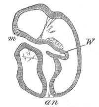
Fig. 272. Early stage in the development of
Tornaria. (After Götte.)
W. so-called water-vascular vesicle developing as an
outgrowth of the mesenteron; m. mouth; an. anus.
A mouth (m), situated on the ventral surface, leads into an
alimentary canal with a terminal anus (an). A præ-oral lobe is
well developed, as in Bipinnaria, but there is no post-anal lobe. The
bands of cilia have the same general form as in Bipinnaria. There is a
præ-oral band, and a longitudinal post-oral band; and the two bands
nearly meet at the apex of the præ-oral lobe (fig. 273). A contractile
band passes from the œsophagus to the apex of the præ-oral lobe,
and a diverticulum (fig. 272, W) from the alimentary tract,
directed towards the dorsal surface, is present. Contractile cells are
scattered in the space between the body wall and the gut.
In the following stage (fig. 274 A) a conspicuous transverse post-oral
band of a single row of long cilia is formed, and the original bands
become more sinuous. The alimentary diverticulum of the last stage
becomes an independent vesicle opening by a pore on the dorsal surface
(fig. 274 A, w). The contractile cord is now inserted on this
vesicle. Where this cord joins the apex of the præ-oral lobe between
the two anterior bands of cilia a thickening of the epiblast (? a
ganglion) has become established, and on it are placed two
eye-spots (fig. 273 oc, and fig. 274 A). A deep bay is formed
on the ventral surface of the larva.
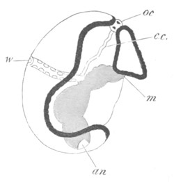
Fig. 273. Young Tornaria.
(After Müller.)
m. mouth; an. anus; w. water-vascular vesicle;
oc. eye-spots; c.c. contractile cord.
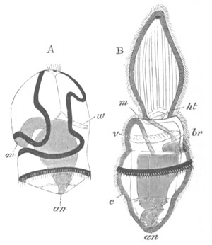
Fig. 274. Two stages in the development of
Tornaria. (After Metschnikoff.)
The black lines represent the ciliated bands.
m. mouth; an. anus; br. branchial cleft;
ht. heart; c. body cavity between splanchnic and somatic
mesoblast layers; w. water-vascular vesicle; v. circular
blood-vessel.
As the larva grows older the original bands of cilia become more
sinuous, and a second transverse band with small cilia is formed (in
the Mediterranean larva) between the previous transverse band and the
anus. The water-vascular vesicle is prolonged into two spurs, one on
each side of the stomach. A pulsating vesicle or heart is also formed
(fig. 274 B, ht), and arises, according to Spengel (No. 572),
as a thickening of the epidermis. It subsequently becomes enveloped in
a pericardium, and is placed in a depression in the water-vascular
vesicle. Two pairs of diverticula, one behind the other, grow out
(Agassiz, No. 568) from the gastric region of the alimentary canal.
The two parts of each pair form flattened compartments, which together
give rise to a complete investment of the adjoining parts of the
alimentary tract. The two parts of each coalesce, and thus form
a double-walled cylinder round the alimentary tract, but their
cavities remain separated by a dorsal and ventral septum.
Eventually (Spengel) the cavity of the anterior cylinder forms the
section of the body cavity in the collar of the adult, and that of the
posterior (fig. 274 B, c) the remainder of the body cavity. The
septa, separating the two halves of each, remain as dorsal and ventral
mesenteries.
The conversion of Tornaria (fig. 274 A) into Balanoglossus (fig. 274
B) is effected in a few hours, and consists mainly in certain changes
in configuration, and in the disappearance of the longitudinal
ciliated band.
The body of the young Balanoglossus (fig. 274 B) is divided into three
regions (1) the proboscidian region, (2) the collar, (3) the trunk
proper. The proboscidian region is formed by the elongation of the
præ-oral lobe into an oval body with the eye-spots at its extremity,
and provided with strong longitudinal muscles. The heart (ht)
and water-vascular vesicle lie near its base, but the contractile cord
connected with the latter is no longer present. The mouth is placed on
the ventral side at the base of the præ-oral lobe, and immediately
behind it is the collar. The remainder of the body is more or less
conical, and is still girt with the larval transverse ciliated band,
which lies in the middle of the gastric region in the Mediterranean
species, but in the œsophageal region in the American one.
The whole of the body, including the proboscis, becomes richly
ciliated.
One of the most important characters of the adult Balanoglossus
consists in the presence of respiratory structures comparable with the
vertebrate gill slits. The earliest traces of these structures are
distinctly formed while the larva is still in the Tornaria
condition, as one pair of pouches from the œsophagus in the
Mediterranean species, and four pairs in the American one (fig. 275,
br).
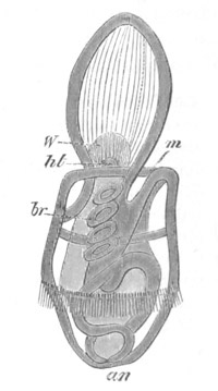
Fig. 275. Late stage in the development of
Balanoglossus with four branchial clefts. (After Alex.
Agassiz.)
m. mouth; an. anus; br. branchial cleft; ht.
heart; W. water-vascular vesicle.
In the Mediterranean Tornaria the two pouches meet the skin dorsally,
and in the young Balanoglossus (fig. 274 B, br) acquire an
external opening on the dorsal side. In the American species the first
four pouches are without external openings till additional pouches
have been formed. Fresh gill pouches continue to be formed both in the
American and probably the Mediterranean species, but the conversion of
the simple pouches into the complicated gill structure of the adult
has only been studied by Agassiz (No. 568) in the American species. It
would seem in the first place that the structure of the adult gill
slits is much less complicated in the American than in the
Mediterranean species. The simple pouches of the young become fairly
numerous. They are at first circular; they then become elliptical, and
the dorsal wall of each slit becomes folded; subsequently fresh folds
are formed which greatly increase the complexity of the gills. The
external openings are not acquired till comparatively late.
Our knowledge of the development of the internal organs, mainly
derived from Agassiz, is still imperfect. The vascular system appears
early in the form of a dorsal and a ventral vessel, both pointed, and
apparently ending blindly at their two extremities. The two spurs of
the water-vascular vesicle, which in the Tornaria stage rested upon
the stomach, now grow round the œsophagus, and form an anterior
vascular ring, which Agassiz describes as becoming connected with the
heart, though it still communicates with the exterior by the dorsal
pore and seems to become connected with the remainder of the vascular
system. According to Spengel (No. 572) the dorsal vessel becomes
connected with the heart, which remains through life in the proboscis:
the cavity of the water-vascular vesicle forms the cavity of the
proboscis in the adult, and its pore remains as a dorsal (not, as
usually stated, ventral) pore leading to the exterior.
The eye-spots disappear.
Tornaria is a very interesting larval form, since it is intermediate
in structure between the larva of an Echinoderm and trochosphere type
common to the Mollusca, Chætopoda, etc. The shape of the body
especially the form of the ventral depression, the character of the
longitudinal ciliated band, the structure and derivation of the
water-vascular vesicle, and the formation of the walls of the
body cavity as gastric diverticula, are all characters which point to
a connection with Echinoderm larvæ.
On the other hand the eye-spots at the end of the præ-oral lobe[230],
the contractile band passing from the œsophagus to the eye-spots
(fig. 273), the two posterior bands of cilia, and the terminal anus
are all trochosphere characters.
The persistence of the præ-oral lobe as the proboscis is interesting,
as tending to shew that Balanoglossus is the surviving representative
of a primitive group.
Bibliography.
(567) A. Agassiz. “Tornaria.” Ann. Lyceum Nat. Hist.
VIII. New York, 1866.
(568) A. Agassiz. “The History of Balanoglossus and Tornaria.”
Mem. Amer. Acad. of Arts and Scien., Vol. IX. 1873.
(569) A. Götte. “Entwicklungsgeschichte d. Comatula
Mediterranea.” Archiv für mikr. Anat., Bd. XII., 1876,
p. 641.
(570) E. Metschnikoff. “Untersuchungen üb d. Metamorphose, etc.
(Tornaria).” Zeit. für wiss. Zool., Bd. XX. 1870.
(571) J. Müller. “Ueb. d. Larven u. Metamor. d. Echinodermen.”
Berlin Akad., 1849 and 1850.
(572) J. W. Spengel. “Bau u. Entwicklung von Balanoglossus.”
Tagebl. d. Naturf. Vers. München, 1877.
- Abdominalia, 459, 493, 499
- Acanthocephala, 379
- Acanthosoma, 473, 474, 475
- Acarina, 444, 454
- Accipenser, 102
- Achæta, 319
- Achelia, 538
- Achtheres percarum, 490
- Acineta, 7, 8
- Acraspeda, 152, 165, 167, 178, 179, 182, 185, 186
- Actinia, 169, 171, 179
- Actinophrys, 9
- Actinotrocha, 315, 318, 363, 364
- Actinozoa, 26, 102, 152, 166, 170, 171, 172, 176, 178, 179, 181, 182, 186
- Actinula, 155
- Aculeata, 421
- Ægineta flavescens, 158
- Æginidæ, 156, 158
- Æginopsis Mediterranea, 158
- Æquorea Mitrocoma, 182
- Agalma, 163
- Agelena, 436, 450
- Agelena labyrinthica, 119, 438
- Alciope, 74
- Alcippidæ, 499
- Alcyonaria, 152
- Alcyonidæ, 167, 168
- Alcyonidium mytili, 297, 300, 302
- Alcyonium palmatum, 119, 148, 167, 182
- Alima, 484, 486
- Amœba, 19, 20
- Amphibia, 22, 54, 56, 59, 60, 63, 66, 74, 83, 102
- Amphilina, 218
- Amphioxus, 54, 56, 59, 61, 66, 93, 426
- Amphipoda, 518
- Amphiporus lactifloreus, 202
- Amphistomum, 31
- Amphitrochæ, 330
- Amphiura squamata, 565
- Anchorella, 108, 492, 520
- Anelasma squalicola, 499
- Anguillulidæ, 371
- Annelida, 14, 25, 98, 503, 525
- Anodon, 37, 38, 39, 100, 107, 259, 260, 265, 266, 268
- Anopla, 189, 202
- Anura, 5
- Antedon, 568, 573, 574
- Aphides, 15, 16, 76, 79, 116, 428, 429
- Aphrodite, 42
- Apis, 402, 407, 408, 412, 413
- Aplysia, 99, 226, 238, 252, 253
- Aplysinidæ, 146
- Apoda, 459, 493
- Aptera, 395, 420
- Apus, 16, 79, 460, 463
- Arachnida, 22, 114, 119, 413, 431, 435, 444, 454, 455, 458, 537, 539
- Arachnitis, 171
- Araneina, 50, 51, 436
- Arbacia, 567
- Arca, 38
- Archigetes, 218
- Archizoæa gigas, 494
- Arenicola, 42
- Argiope, 311, 312, 315, 317
- Argonauta, 247, 248
- Argulus, 492
- Armata, 355
- Arthropoda, 12, 16, 18, 22, 75, 77, 79, 83, 108, 110, 221, 382, 383, 434, 448, 503, 525, 534, 541, 542
- Articulata, 311, 313, 316, 317
- Ascaridiæ, 371
- Ascaris nigrovenosa, 16, 82
- Ascetta, 144
- Ascidia canina, 53
- Ascidians, 74, 102, 208, 426
- Asellus aquaticus, 112, 120, 516
- Astacus, 66, 465, 477, 511, 512, 513, 525
- Asteracanthion, 69, 70, 561
- Asterias, 20, 68, 69, 71, 78, 80, 84, 549, 564
- Asteroidea, 35, 36, 544, 549, 557, 563, 576
- Astræa, 169
- Astroides, 169
- Atax Bonzi, 445
- Atlanta, 231, 240
- Atrochæ, 330
- Aurelia, 167
- Auricularia, 553, 554, 562, 574
- Autolytus cornutus, 319, 343
- Aves, 56, 59, 61, 64, 107, 109
- Axolotl, 16
- Balanoglossus, 576, 579, 581
- Balanus balanoides, 75, 493
- Belemnites, 252, 253
- Bipinnaria, 557, 563, 574, 576, 579
- Blatta, 374, 395
- Bojanus, organ of, 264, 282
- Bonellia, 20, 43, 44, 98, 324, 355, 358, 359
- Bothriocephalus salmonis, 211
- Brachiella, 492
- Brachiolaria, 558, 564
- Brachiopoda, 311, 317, 318
- Brachyura, 466, 480, 483
- Branchiobdella, 42, 43, 346
- Branchiogasteropoda, 272
- Branchiopoda, 79, 459, 523, 524
- Branchipus, 463, 524
- Branchiura, 459, 492
- Branchionus urceolaris, 221
- Braula, 396
- Buccinum, 237, 280
- Bulimus citrinas, 229
- Bunodes, 169, 171
- Buthus, 431
- Calcispongiæ, 138, 148
- Calopteryx, 402
- Calycophoridæ, 152, 159
- Calyptoblastic Hydroids, 184, 185
- Calyptræa, 223, 280
- Campanularidæ, 183, 184
- Capitella, 330, 332
- Carabidæ, 476
- Carcinus Mœnas, 481, 483
- Cardium, 260, 262
- Carinaria, 240
- Caryophyllium, 168, 171
- Cassiopea, 165, 167
- Cecidomyia, 15, 79, 416, 417, 429
- Cephalopoda, 20, 40, 41, 102, 108, 109, 135, 225, 240, 242, 244, 250, 252, 253, 270, 271, 272, 274, 279, 282, 287
- Cephalothrix galatheae, 202
- Ceratospongiæ, 146
- Cercariae, 207, 208, 209
- Cerianthus, 168, 171
- Cestodes, 14, 29, 31, 32, 33, 189, 210, 212, 218, 313, 425, 541
- Chætogaster, 342
- Chætopoda, 5, 18, 23, 41, 43, 44, 54, 67, 209, 215, 270, 275, 307, 312, 317, 318, 319, 320, 326, 334, 338, 342, 346, 349, 350, 351, 364, 369, 383, 386, 408, 448, 457, 458, 521, 576, 582
- Chætopteridæ, 333
- Chætosomoidea, 371
- Chelifer, 434, 436, 442, 446, 454
- Chermes, 15, 429
- Chilognatha, 113, 387, 389, 391, 393, 395
- Chilopoda, 387, 393, 394
- Chilostomata, 292, 297, 298, 304, 305
- Chironomus, 15, 378, 401, 402, 415, 416, 429
- Chiton, 254, 256, 257,273
- Chordata, 5
- Chrysaora, 165
- Chthonius, 436
- Cicada, 395
- Cirripedia, 459, 492, 496, 503, 509, 520
- Cladocera, 459, 464, 519
- Clausilia, 239
- Clavella, 520
- Clavularia crassa, 167
- Cleodora, 241
- Clepsine, 73, 346, 347, 349, 351, 352, 353, 354
- Clio, 242, 278
- Clubione, 436
- Clupeidæ, 64
- Cobitis barbatula, 378
- Coccidæ, 429
- Coccus, 50
- Cœlebogyne, 79
- Cœlenterata, 3, 5, 13, 18, 26, 27, 28, 35, 74, 93, 94, 126, 148, 170, 178, 179, 180, 181, 191, 342
- Cœnurus cerebralis, 213, 214
- Coleochæte, 11
- Coleoptera, 396, 402, 409, 412, 420, 421, 425
- Collembola, 395, 426
- Comatula, 5, 552, 553
- Condracanthus, 111, 120, 520
- Conochilus volvox, 221
- Convoluta, 32
- Copepoda, 109, 120, 459, 460, 487, 489, 493, 496, 503, 509, 519
- Corallium rubrum, 168, 182
- Corethra, 422, 423, 424
- Crangoninæ, 476
- Craniadæ, 311
- Craniata, 5, 6, 19, 20, 54, 56, 59, 61, 62, 64, 74, 102
- Crinoidea, 35, 36, 544, 550, 568, 576
- Criodilus, 321, 324, 328, 341
- Crisia, 304
- Crocodilia, 63
- Crustacea, 5, 6, 18, 51, 66, 102, 109, 120, 458, 465, 487, 502, 521, 524, 537, 541
- Cryptophialus, 499, 509
- Crystalloides, 163
- Ctenophora, 26, 93, 102, 152, 173, 175, 177, 178, 179, 180, 181, 182
- Ctenostomata, 292, 297, 298, 304, 305
- Cucullanus elegans, 46, 75, 82, 371, 376
- Cucumaria, 546, 556, 574
- Cumaceæ, 459, 465, 486, 506
- Curculio, 421
- Cyclas, 259, 260, 261, 265
- Cyclops, 376, 377, 418, 489, 503
- Cyclostomata, 102, 292, 304
- Cymbulia, 241, 242
- Cymothoa, 516, 517, 519, 520, 524, 528
- Cynipidæ, 15, 421, 428
- Cyphonautes, 297, 301, 304, 306, 308
- Cypridina, 500, 502
- Cysticercus cellulosæ, 214, 217
- ” fasciolaris, 216
- ” limacis, 213
- Daphnia, 79, 464
- Dasychone, 331, 336
- Decapoda, 66, 248, 459, 465, 469, 504, 511
- Dendrocœla, 32, 33, 189, 195, 196
- Dentalium, 258, 576
- Desmacidon, 147
- Desor, type of, 196, 197, 201, 202, 204, 212, 424
- Diastopora, 304
- Dibranchiata, 225, 253
- Dicyema, 9, 131, 134, 135, 136
- Dimya, 225
- Diphyes, 159
- Diplozoon, 11, 209, 210
- Diporpa, 210
- Diptera, 49, 194, 204, 396, 401, 402, 407, 409, 412, 416, 420, 429
- Discina radiata, 317
- Discinidæ, 311
- Discophora, 18, 42, 165, 346, 383
- Distomeæ, 189, 205, 425
- Distomum, 31
- ” cygnoides, 209
- ” globiparum, 207
- ” lanceolatum, 205
- Dochmius duodenale, 375
- Donacia, 401
- Dracunculus, 376, 377
- Echinaster fallax, 23
- Echinodermata, 5, 18, 24, 35, 74, 102, 325, 424, 544, 573, 574, 576, 582
- Echinoidea, 35, 36, 544, 549, 565, 576
- Echinorhyncus, 379, 380
- Echinus lividus, 83, 84, 88
- Echiurus, 44, 357, 358
- Ectoprocta, 297, 306
- Edriophthalmata, 459, 465
- Elaphocaris, 473
- Elasmobranchii, 23, 56, 59, 61, 62, 64, 67, 105, 106, 107, 108, 109
- Enopla, 189, 202
- Entoconcha mirabilis, 237
- Entomophaga, 421
- Entoprocta, 292, 298, 300, 302, 304, 306
- Epeira, 436
- Ephemera, 395, 409, 420, 422
- Ephyra, 186
- Epibulia aurantiaca, 159, 165
- Erichthus, 484, 507
- Errantia, 319, 336
- Esperia, 147
- Estheria, 463, 464
- Euaxes, 101, 322, 324, 341, 346, 349
- Eucharis, 178
- Eucopepoda, 459
- Eucope polystyla, 23, 154
- Eunice sanguinea, 319
- Eupagurus prideauxii, 112, 113, 115, 511, 520
- Euphausia, 465, 468, 504, 505, 518, 523
- Eurostomata, 176
- Eurylepta auriculata, 192
- Eurynome, 483
- Euspongia, 146, 147
- Filaria, 377
- Filaridæ, 371
- Firoloidea, 240
- Flagellata, 7, 8
- Flustrella, 301, 303
- Formica, 396
- Fungia, 182, 186
- Fusus, 275, 280, 284, 288
- Gammarus, 122, 518
- Ganoids, 54, 102
- Gasteropoda, 39, 41, 98, 225, 226, 229, 230, 232, 233, 240, 258, 260, 261, 270, 272, 275, 279, 283, 324
- Gasterosteus, 64, 210
- Gastrotricha, 370
- Gasterotrochæ, 330, 333
- Gecarcinus, 465
- Geophilus, 392, 393
- Gephyrea, 5, 18, 24, 44, 54, 67, 102, 318, 320, 325, 355, 357, 361, 364
- Germogen, 134
- Geryonia hastata, 156
- Geryonidæ, 156
- Glochidia, 267, 268
- Gnathobdellidæ, 346, 349
- Gordiacea, 94
- Gordioidea, 371, 374, 378
- Gorgonia, 168
- Gorgonidæ, 181
- Gorgoninæ, 181
- Gregarinidæ, 8
- Gryllotalpa, 401, 412, 413
- Gummineæ, 147, 148
- Gymnoblastic Hydroids, 184, 185
- Gymnolæmata, 292
- Gymnosomata, 225, 240, 241, 242, 270
- Gyrodactylus, 210
- Halichondria, 147
- Halisarca, 22, 66, 145
- Halistemma, 165
- Helicidæ, 238
- Helioporidæ, 182
- Helix, 67, 229
- Hemiptera, 395, 402, 403, 409, 420, 421
- Hessia, 108, 492
- Heterakis vermicularis, 374
- Heteronereis, 343
- Heteropoda, 71, 72, 225, 226, 231, 278
- Hexacoralla, 152, 179, 182
- Hippopodius gleba, 27, 159
- Hirudinea, 74, 84
- Hirudo, 350, 351, 352, 353, 354
- Holometabola, 420, 422
- Holostomum, 205
- Holothuria, 19, 25, 35, 549, 558, 576
- Holothuroidea, 35, 544, 553, 556
- Homarus, 477
- Hyaleacea, 273, 275
- Hyaleidæ, 241
- Hydra, 21, 22, 26, 28, 29, 34, 152, 154, 155, 179, 183
- Hydractinia, 539
- Hydrocoralla, 152, 181, 185
- Hydroidea, 152
- Hydromedusæ, 152, 179, 182, 183, 184, 185, 186, 187
- Hydrophilus, 374, 396, 400, 401, 402, 404, 408, 409
- Hydrozoa, 14, 19, 26, 27, 67, 102, 152,155, 165, 179, 180, 181, 182, 539
- Hymenoptera, 396, 401, 402, 412, 420, 421, 425
- Ichneumon, 396
- Inarticulata, 311, 316
- Inermia, 355
- Infusoria, 7, 8
- Insecta, 5, 15, 18, 19, 25, 46, 395, 396, 425, 455, 458
- Intoshia gigas, 136
- Isidinæ, 181
- Isodyctia, 147
- Isopoda, 109, 515, 519, 521, 523, 527
- Lacertilia, 64
- Lacinularia, 221, 223
- Lamellibranchiata, 23, 25, 37, 39, 98, 225, 241, 257, 258, 259, 269, 270, 271, 273, 274, 288
- Lepadidæ, 498
- Lepas fascicularis, 224, 493, 494, 495
- Lepidoptera, 79, 396, 402, 407, 408, 412, 413, 415, 417, 420, 421, 423, 425, 426, 455
- Leptodora, 16, 51
- Leptoplana, 74, 189, 192, 193
- Lernæopoda, 490, 492, 520
- Leucifer, 507
- Libellulidæ, 402, 403, 409, 420
- Limax, 229, 232, 239, 278, 280
- Limnadia, 79, 524
- Limulus, 534
- Lina, 402
- Lingulidæ, 311, 316
- Lithobius, 393
- Lobatæ, 178
- Loligo, 242, 243, 244, 247, 253
- Loricata, 507, 514
- Lota, 105
- Loxosoma, 292, 294, 296, 306, 307
- Lucernaria, 185
- Lumbricus, 341, 368
- Lumbriconereis, 334
- Lymnæus, 82, 98, 226, 227, 232, 238, 281
- Lycosa, 436
- Macrostomum, 32, 34
- Macrura, 476
- Malacobdella, 203
- Malacodermata, 171
- Malacostraca, 66, 459, 462, 465, 504, 505, 506, 511, 523
- Mammalia, 56, 58, 59, 64, 66
- Marsipobranchii, 59
- Mastigopus, 473, 474
- Medusæ, 27, 154, 157, 158, 163, 164, 176, 178, 181, 182, 183, 184, 185, 186
- Megalopa, 482, 483, 484
- Melolontha, 402, 421
- Membranipora, 297, 303
- Mermithidæ, 371
- Mesotrochæ, 330
- Metachætæ, 335
- Metazoa, 9, 10, 12, 67, 86, 125, 135, 149, 150, 179
- Millepora, 152, 181
- Mitraria, 308, 337
- Molgula, 102
- Mollusca, 5, 18, 24, 66, 74, 84, 99, 225, 247, 248, 251, 256, 257, 262, 271, 285, 288, 307, 325, 333, 352, 576, 582
- Monomya, 225
- Monostomum capitellum, 205
- Monotrochæ, 330
- Montacuta, 260, 262
- Musca, 396
- Muscidæ, 420, 423
- Myobia, 444, 445
- Myrianida, 343
- Myriapoda, 22, 111, 113, 387, 394, 395, 413, 458
- Myriothela, 155
- Myrmeleon, 396
- Mysis, 120, 469, 472, 486, 504, 509, 525
- Mytilus, 260, 261
- Myxinoids, 5
- Myxispongiæ, 145
- Myzostomea, 369
- Nais, 342
- Nassa mutabilis, 101, 226, 227, 233, 262, 278, 279, 288, 324
- Natantia, 487
- Natica, 237, 283
- Nauplius, 5, 16, 460, 461, 463, 465, 466, 469, 473, 490, 491, 493, 497
- Nautilus pompilius, 253, 276
- Nebaliadæ, 459, 465, 486
- Nematoda, 45, 46, 50, 66, 74, 75, 371, 373, 374, 376
- Nematogens, 131
- Nematoidea, 18, 84, 94, 371, 374
- Nematus ventricosus, 13, 427
- Nemertea, 94, 189, 196, 202, 204
- Nemertines, 30, 31, 33, 93, 136, 195, 202, 328, 333, 424
- Nephelis, 82, 346, 349, 350, 351, 352, 354
- Nereis, 343
- ” diversicolor, 319
- ” Dumerilii, 343
- Neritina, 229, 237
- Neuroptera, 396, 401, 420, 421
- Neuroterus ventricularis, 428
- Notonecta, 395
- Nototrochæ, 330, 353
- Nudibranchiata, 229, 241
- Ocellata, 184
- Octocoralla, 152, 179
- Octopus, 248
- Odontophora, 225, 257, 271
- Odontosyllis, 333
- Oedogonium, 11
- Oligochæta, 42, 319, 321, 325, 330, 338, 346, 352
- Olynthus, 144
- Oniscus murarius, 107, 108, 109, 120, 516, 520, 528
- Opercula, 31
- Ophiothryx, 36, 549
- Ophidia, 64
- Ophiuroidea, 136, 544, 553, 562, 565, 576
- Ophryotrochæ puerilis, 333
- Opisthobranchiata, 225, 232, 237
- Ornithodelphia, 109
- Orthonectidæ, 136
- Orthoptera, 395, 414, 420, 421, 425, 426
- Ostracoda, 459, 500, 510
- Ostrea, 259, 260, 262
- Oxyuridæ, 46, 373, 374
- Oxyurus ambigua, 374
- Pæcilopoda, 534
- Paguridæ, 477
- Palæmon, 110
- Palæmonetes, 476
- Palæmoninæ, 476, 511, 512
- Palinurus, 478, 480
- Paludina, 66, 227, 229, 235, 270, 278, 280
- ” costata, 229
- ” vivipara, 226
- Pandorina, 11
- Parasita, 489
- Pedalion, 221
- Pedicellina, 98, 292, 296, 299, 307
- Pelagia, 167, 185
- Penæinæ, 476
- Penæus, 110, 113, 465, 469, 473, 474, 504, 518
- Pennatulidæ, 181
- Pentacrinus, 5
- Pentastomida, 539, 540
- Pentastomum denticulatum, 540, 541
- Percidæ, 64
- Perennichætæ, 335
- Peripatus, 5, 386, 411, 412, 413, 542
- Petromyzon, 61, 63, 64, 74, 83
- Phalangella, 304
- Phalangidæ, 436
- Phallusia, 83
- Phascolosoma, 44, 355, 356, 361
- Pholcus, 436, 442
- Phoronis, 315, 355, 363, 364
- Phoxinus lævis, 378
- Phryganea, 396, 401, 409
- Phylactolæmata, 292, 294, 297, 305, 306
- Phyllobothrium, 218
- Phyllodoce, 329
- Phyllopoda, 16, 459, 461, 505
- Phyllosoma, 479, 480
- Phylloxera, 429
- Physophoridæ, 152, 162, 164
- Pilidium, type of, 196, 200, 201, 202, 204, 424
- Pisces, 5
- Piscicola, 20, 43
- Pisidium, 259, 260, 262, 264
- Planaria Neapolitana, 193
- Planorbis, 273, 281, 325
- Platyelminthes, 18, 20, 24, 221, 424
- Platygaster, 396, 416, 417, 418, 419
- Pleurobrachia, 176, 177, 238
- Pneumodermon, 242, 576
- Podostomata, 292
- Poduridæ, 401, 405
- Polychæta, 42, 319, 325, 338
- Polydesmus complanatus, 387, 388
- Polygordius, 319, 325, 326, 327, 328, 332, 357, 386
- Polynœ, 42, 331
- Polyophthalmus, 328
- Polyplacophora, 225, 254, 270, 271, 288
- Polystomeæ, 189, 205, 209
- Polystomum, 209
- Polytrochæ, 330, 333
- Polyxenia leucostyla, 158
- Polyxenus lagurus, 387
- Polyzoa, 98, 303, 305, 306, 308, 315, 316
- Porcellana, 483
- Porifera, 102, 138, 148
- Porthesia, 115
- Prorhyncus, 32, 34
- Prosobranchiata, 225, 237, 281
- Prostomum, 32, 34, 38, 196
- Protozoa, 8, 9, 10, 11, 86, 135, 149
- Protozoæa, 471
- Protula Dysteri, 342
- Pseudoneuroptera, 426
- Pseudoscorpionidæ, 434
- Psolinus, 556, 574
- Psychidæ, 16
- Pteraster miliaris, 561
- Pteropoda, 98, 225, 226, 229, 230, 232, 240, 258, 270, 272, 279, 283
- Pterotrachæa, 71, 229, 240
- Pulex, 396
- Pulmonata, 39, 225, 232, 238, 281, 282
- Purpura lapillus, 78
- Pycnogonida, 538
- Pyrosoma, 13, 53, 109
- Rana temporaria, 210
- Raspailia, 147
- Redia, 206, 207, 208, 209
- Reniera, 147
- Reptilia, 56, 59, 60, 61, 62, 64, 109
- Rhabditis dolichura, 82
- Rhabdocœla, 32, 33, 189, 196
- Rhabdopleura, 294, 306
- Rhizocephala, 459, 493, 499, 500
- Rhizocrinus, 5
- Rhizostoma, 167
- Rhombogens, 131, 134
- Rhynchonellidæ, 311
- Rhyncobdellidæ, 346
- Rotifera, 5, 12, 18, 75, 76, 77, 79, 83, 102, 221, 308, 325
- Saccocirrus, 328, 329, 332, 340
- Sacculina, 500
- Sagartia, 169, 171
- Sagitta, 23, 74, 94, 130, 366, 367, 368
- Salmonidæ, 64
- Salpa, 102
- Sarcia, 164
- Scaphopoda, 225, 257, 270, 271
- Schistocephalus, 211
- Schizopoda, 459, 465, 466
- Scolopendra, 392
- Scorpio, 120, 431, 446, 454, 455, 457
- Scrobicularia, 38, 39
- Scyllarus, 477
- Scyphistoma, 179, 185, 186
- Sedentaria, 319, 336
- Sepia, 20, 40, 41, 242, 243, 244, 245, 247, 249, 253
- Sergestidæ, 473, 507
- Serpula, 319, 325, 331
- Sertularia, 152, 183, 184
- Silicispongiæ, 147
- Simulia, 401, 415
- Siphonophora, 13, 27, 152, 159, 163, 165, 179, 180, 182, 185
- Sipunculida, 24
- Sipunculus, 44
- Sirex, 396
- Sitaris, 421
- Spathegaster baccarum, 428
- Spio, 42, 332, 333
- Spiroptera obtusa, 376
- Spirorbis Pagenstecheri, 319
- Spirula, 252
- Spirulirostra, 252
- Spongelia, 147
- Spongida, 138, 144, 148, 149
- Spongilla, 147, 150
- Sporocysts, 206, 207, 208, 209
- Squilla, 66, 504, 507
- Stephanomia pictum, 162, 165
- Stomatopoda, 459, 465, 484
- Stomodæum, 413
- Strongylidæ, 371, 375
- Strongylocentrus, 567
- Strongysoloma Guerinii, 387, 388, 390
- Stylasteridæ, 152, 181
- Styliolidæ, 241
- Stylochopsis ponticus, 193
- Sycandra, 93, 138, 144, 145, 147, 150
- Syllis, 343
- Sympodium coralloides, 168
- Tæniatæ, 178
- Tardigrada, 541
- Tegenaria, 436
- Teleostei, 18, 25, 56, 59, 64, 107, 109
- Telotrochæ, 330
- Tendra, 300
- Tenthreds, 396
- Terebella conchilega, 332, 333, 337
- Terebella nebulosa, 332, 333
- Terebratula, 311, 315
- Terebratulina, 311, 315, 316
- Teredo, larva of, 262
- Tergipes, 232, 238
- ” Edwardsii, 238
- ” lacinulatus, 238
- Tethya, 147
- Tetrabranchiata, 225
- Tetranychus telarius, 116
- Tetrastemma varicolor, 203
- Thalassema, 44, 355, 357
- Thalassinidæ, 477
- Thallophytes, 11
- Thecidium, 311, 312, 315, 316
- Thecosomata, 225
- Thoracica, 459, 493, 499, 500
- Thysanozoon, 192, 193
- Thysanura, 395, 408, 425, 458
- Tichogonia, 39
- Tipula, 396
- Tipulidæ, 420, 421
- Tœnia cœnurus, 214
- Tornaria, 579, 581
- Toxopneustes, 22, 24, 35, 85, 88, 89
- Tracheata, 385, 426, 432, 448, 455, 457, 458, 538, 541
- Trachymedusæ, 152, 156, 179, 185
- Trematodes, 14, 16, 29, 30, 31, 32, 33, 46, 94, 189, 205, 208, 210, 212, 216
- Trichina, 377, 378
- Trichinidæ, 371
- Trichocephalus affinis, 374
- Trochosphæra æquatorialis, 221
- Tubiporidæ, 182
- Tubularia, 34, 38, 152, 154, 158
- Tubularidæ, 29, 179, 183
- Tunicata, 5, 14, 53
- Turbellaria, 5, 30, 31, 33, 74, 98, 102, 136, 179, 189, 193, 333
- Tyroglyphus, 445
- Unio, 37, 38, 39, 100, 101, 259, 260, 265, 266, 445
- Vaginulus luzonicus, 229
- Vermes, 5, 74, 102, 223, 324, 352
- Verongia rosea, 146
- Vertebrata, 14, 18, 19, 24, 59, 64, 83, 272, 349, 397, 426
- Vesiculata, 184
- Vitrina, 229
- Vorticella, 8, 9, 10
- Zoantharia, 152, 168, 169
- Zoæa, 465, 468, 471, 474, 482, 483, 484, 486, 504
The Ovum.
General Works.
(1) Ed. van Beneden. “Recherches sur la composition et la
signification de l’œuf,” etc. Mém. cour. d. l’Acad. roy. des
Sciences de Belgique, Vol. XXXIV. 1870.
(2) R. Leuckart. Artikel “Zeugung,” R. Wagner’s
Handwörterbuch d. Physiologie, Vol. IV. 1853.
(3) Fr. Leydig. “Die Dotterfurchung nach ihrem Vorkommen in d.
Thierwelt u. n. ihrer Bedeutung.” Oken, Isis, 1848.
(4) Ludwig. “Ueber d. Eibildung im Thierreiche.” Arbeiten a.
d. zool.-zoot. Institut Würzburg, Vol. I. 1874[231].
(5) Allen Thomson. Article “Ovum” in Todd’s Cyclopædia of
Anatomy and Physiology, Vol. V. 1859.
(6) W. Waldeyer. Eierstock u. Ei. Leipzig, 1870.
THE OVUM OF CŒLENTERATA.
(7) Ed. van Beneden. “De la distinction originelle d. testicule
et de l’ovaire.” Bull. Acad. roy. Belgique, 3e série, Vol.
XXXVII. 1874.
(8) R. and O. Hertwig. Der Organismus d. Medusen. Jena,
1878.
(9) N. Kleinenberg. Hydra. Leipzig, 1872.
THE OVUM OF PLATYELMINTHES.
(10) P. Hallez. Contributions à l’Histoire naturelle des
Turbellariés. Lille, 1879.
(11) S. Max Schultze. Beiträge z. Naturgeschichte d.
Turbellarien. Greifswald, 1851.
(12) C. Th. von Siebold. “Helminthologische Beiträge.” Müller’s
Archiv, 1836.
(13) C. Th. von Siebold. Lehrbuch d. vergleich. Anat. d.
wirbellosen Thiere. Berlin, 1848.
(14) E. Zeller. “Weitere Beiträge z. Kenntniss d. Polystomen.”
Zeit. f. wiss. Zool., Bd. XXVII. 1876.
[Vide also Ed. van Beneden] (No. 1).
THE OVUM OF ECHINODERMATA.
(15) C. K. Hoffmann. “Zur Anatomie d. Echiniden u. Spatangen.”
Niederländisch. Archiv f. Zoologie, Vol. I. 1871.
(16) C. K. Hoffmann. “Zur Anatomie d. Asteriden.”
Niederländisch. Archiv f. Zoologie, Vol. II. 1873.
(17) H. Ludwig. “Beiträge zur Anat. d. Crinoiden.” Zeit. f.
wiss. Zool., Vol. XXVIII. 1877.
(18) Joh. Müller. “Ueber d. Canal in d. Eiern d. Holothurien.”
Müller’s Archiv, 1854.
(19) C. Semper. Holothurien. Leipzig, 1868.
(20) E. Selenka. Befruchtung d. Eies v. Toxopneustes
variegatus, 1878.
[Vide also Ludwig (No. 4), etc.]
THE OVUM OF MOLLUSCA.
Lamellibranchiata.
(21) H. Lacaze-Duthiers. “Organes génitaux des Acéphales
Lamellibranches.” Ann. Sci. Nat., 4me série, Vol.
II. 1854.
(22) W. Flemming. “Ueb. d. er. Entwick. am Ei d. Teichmuschel.”
Archiv f. mikr. Anat., Vol. X. 1874.
(23) W. Flemming. “Studien üb. d. Entwick. d. Najaden.”
Sitz. d. k. Akad. Wiss. Wien, Vol. LXXI. 1875.
(24) Th. von Hessling. “Einige Bemerkungen, etc.” Zeit. f.
wiss. Zool., Bd. V. 1854.
(25) H. von Jhering. “Zur Kenntniss d. Eibildung bei d.
Muscheln.” Zeit. f. wiss. Zool., Vol. XXIX. 1877.
(26) Keber. De Introitu Spermatozoorum in ovula, etc.
Königsberg, 1853.
(27) Fr. Leydig. “Kleinere Mittheilung etc.” Müller’s
Archiv, 1854.
Gasteropoda.
(28) C. Semper. “Beiträge z. Anat. u. Physiol. d.
Pulmonaten.” Zeit. f. wiss. Zool., Vol. VIII. 1857.
(29) H. Eisig. “Beiträge z. Anat. u. Entwick. d. Pulmonaten.”
Zeit. f. wiss. Zool., Vol. XIX. 1869.
(30) Fr. Leydig. “Ueb. Paludina vivipara.” Zeit. f. wiss.
Zool., Vol. II. 1850.
Cephalopoda.
(31) Al. Kölliker. Entwicklungsgeschichte d.
Cephalopoden. Zurich, 1844.
(32) E. R. Lankester. “On the developmental History of the
Mollusca.” Phil. Trans., 1875.
THE OVUM OF THE CHÆTOPODA.
(33) Ed. Claparède. “Les Annelides Chætopodes d. Golfe de
Naples.” Mém. d. l. Sociét. phys. et d’hist. nat. de Genève,
1868‑9 and 1870.
(34) E. Ehlers. Die Borstenwürmer nach system. und anat.
Untersuchungen. Leipzig, 1864‑68.
(35) E. Selenka. “Das Gefäss-System d. Aphrodite aculeata.”
Niederländisches Archiv f. Zool., Vol. II. 1873.
THE OVUM OF DISCOPHORA.
(36) H. Dorner. “Ueber d. Gattung Branchiobdella.” Zeit. f.
wiss. Zool., Vol. XV. 1865.
(37) R. Leuckart. Die menschlichen Parasiten.
(38) Fr. Leydig. “Zur Anatomie v. Piscicola geometrica, etc.”
Zeit. f. wiss. Zool., Vol. I. 1849.
(39) C. O. Whitman. “Embryology of Clepsine.” Quart. J. of
Micr. Sci., Vol. XVIII. 1878.
THE OVUM OF GEPHYREA.
(40) Keferstein u. Ehlers. Zoologische Beiträge.
Leipzig, 1861.
(41) C. Semper. Holothurien, 1868, p. 145.
(42) J. W. Spengel. “Beiträge z. Kenntniss d. Gephyreen.”
Beiträge a. d. zool. Station z. Neapel, Vol. I. 1879.
(43) J. W. Spengel. “Anatomische Mittheilungen üb. Gephyreen.”
Tagebl. d. Naturf. Vers. München, 1877.
THE OVUM OF NEMATODA.
(44) Ed. Claparède. De la formation et de la fécondation des
œufs chez les Vers Nématodes. Genève, 1859.
(45) R. Leuckart. Die menschlichen Parasiten.
(46) H. Munk. “Ueb. Ei- u. Samenbildung u. Befruchtung b. d.
Nematoden.” Zeit. f. wiss. Zool., Vol. IX. 1858.
(47) H. Nelson. “On the reproduction of Ascaris mystax, etc.”
Phil. Trans. 1852.
(48) A. Schneider. Monographie d. Nematoden. Berlin,
1866.
THE OVUM OF INSECTA.
(49) A. Brandt. Ueber das Ei u. seine Bildungsstätte.
Leipzig, 1878.
(50) T. H. Huxley. “On the agamic reproduction and morphology
of Aphis.” Linnean Trans., Vol. XXII. 1858.
Vide also Manual of Invertebrated Animals, 1877.
(51) R. Leuckart. “Ueber die Micropyle u. den feinern Bau d.
Schalenhaut bei den Insecteneiern.” Müller’s Archiv, 1855.
(52) Fr. Leydig. Der Eierstock u. die Samentasche d.
Insecten. Dresden, 1866.
(53) Lubbock. “The ova and pseudova of Insects.” Phil.
Trans. 1859.
(54) Stein. Die weiblichen Geschlechtsorgane d. Käfer.
Berlin, 1847.
[Conf. also Claus, Landois, Weismann, Ludwig (No. 4).]
THE OVUM OF ARANEINA.
(55) Victor Carus. “Ueb. d. Entwick. d. Spinneneies.” Zeit.
f. wiss. Zool., Vol. II. 1850.
(56) v. Wittich. “Die Entstehung d. Arachnideneies im
Eierstock, etc.” Müller’s Archiv. 1849.
[Conf. Leydig, Balbiani, Ludwig (No. 4), etc.]
THE OVUM OF CRUSTACEA.
(57) Aug. Weismann. “Ueb. d. Bildung von Wintereiern bei
Leptodora hyalina.” Zeit. f. wiss. Zool., Vol. XXVII.
1876.
[For general literature vide Ludwig No. 4 and Ed. van Beneden,
No. 1.]
THE OVUM OF CHORDATA.
Urochorda (Tunicata).
(58) A. Kowalevsky. “Weitere Studien ü. d. Entwicklung d.
Ascidien.” Archiv f. mikr. Anat., Vol. VII. 1871.
(59) A. Kowalevsky. “Ueber Entwicklungsgeschichte d. Pyrosoma.”
Arch. f. mikr. Anat., Vol. XI. 1875.
(60) Kupffer. “Stammverwandtschaft zwischen Ascidien u.
Wirbelthieren.” Arch. f. mikr. Anat., Vol. VI. 1870.
(61) Giard. “Études critiques des travaux, etc.” Archives
Zool. expériment., Vol. I. 1872.
(62) C. Semper. “Ueber die Entstehung, etc.” Arbeiten a. d.
zool.-zoot. Institut Würzburg, Bd. II. 1875.
Cephalochorda.
(63) P. Langerhans. “Z. Anatomie d. Amphioxus lanceolatus,” pp. 330‑3.
Archiv f. mikr. Anat., Vol. XII. 1876.
Craniata.
(64) F. M. Balfour. “On the structure and development of the
Vertebrate Ovary.” Quart. J. of Micr. Science, Vol.
XVIII. 1878.
(65) Th. Eimer. “Untersuchungen ü. d. Eier d. Reptilien.”
Archiv f. mikr. Anat., Vol. VIII. 1872.
(66) Pflüger. Die Eierstöcke d. Säugethiere u. d.
Menschen. Leipzig, 1863.
(67) J. Foulis. “On the development of the ova and structure of
the ovary in Man and other Mammalia.” Quart. J. of Micr.
Science, Vol. XVI. 1876.
(68) J. Foulis. “The development of the ova, etc.” Journal
of Anat. and Phys., Vol. XIII. 1878‑9.
(69) C. Gegenbaur. “Ueb. d. Bau u. d. Entwicklung d.
Wirbelthiereier mit partieller Dottertheilung.” Müller’s
Archiv, 1861.
(70) Alex. Götte. Entwicklungsgeschichte d. Unke.
Leipzig, 1875.
(71) W. His. Untersuchungen üb. d. Ei u. d. Eientwicklung
bei Knochenfischen. Leipzig, 1873.
(72) A. Kölliker. Entwicklungsgeschichte d. Menschen u.
höherer Thiere. Leipzig, 1878.
(73) J. Müller. “Ueber d. zahlreichen Porenkanäle in d.
Eikapsel d. Fische.” Müller’s Archiv, 1854.
(74) W. H. Ransom. “On the impregnation of the ovum in the
Stickleback.” Pro. R. Society, Vol. VII. 1854.
(75) C. Semper. “Das Urogenitalsystem d. Plagiostomen, etc.”
Arbeiten a. d. zool.-zoot. Instit. Würzburg, Vol. II.
1875.
[Cf. Ludwig, No. 4, Ed. van Beneden, No. 1, Waldeyer, No. 6, etc.]
Maturation and Impregnation of the Ovum.
(76) Auerbach. Organologische Studien, Heft 2. Breslau,
1874.
(77) Bambeke. “Recherches s. Embryologie des Batraciens.”
Bull. de l’Acad. royale de Belgique, 2me Sér., T.
LXI., 1876.
(78) E. van Beneden. “La Maturation de l’Œuf des
Mammifères.” Bull. de l’Acad. royale de Belgique, 2me Sér., T.
XL. No. 12, 1875.
(79) Idem. “Contributions à l’Histoire de la Vésicule
Germinative, &c. ” Bull. de l’Acad. royale de Belgique, 2me
Sér., T. XLI. No. 1, 1876.
(80) O. Bütschli. Eizelle, Zelltheilung, und Conjugation der
Infusorien. Frankfurt, 1876.
(81) F. M. Balfour. “On the Phenomena accompanying the
Maturation and Impregnation of the Ovum.” Quart. J. of Micros.
Science, Vol.
XVIII., 1878.
(82) Calberla. “Befruchtungsvorgang beim Ei von Petromyzon
Planeri.” Zeit. f. wiss. Zool., Vol. XXX.
(83) W. Flemming. “Studien in d. Entwickelungsgeschichte der
Najaden.” Sitz. d. k. Akad. Wien, B. LXXI., 1875.
(84) H. Fol. “Die erste Entwickelung des Geryonideneies.”
Jenaische Zeitschrift, Vol.
VII., 1873.
(85) Idem. “Sur le Développement des Ptéropodes.” Archives
de Zoologie Expérimentale et Générale, Vol. IV. and
V. 1875‑6.
(86) Idem. “Sur le Commencement de l’Hénogénie.” Archives
des Sciences Physiques et Naturelles. Genève, 1877.
(87) Idem. Recherches s. l. Fécondation et l. commen. d.
l’Hénogénie. Genève, 1879.
(88) R. Greeff. “Ueb. d. Bau u. d. Entwickelung d.
Echinodermen.” Sitzun. der Gesellschaft z. Beförderung d. gesammten
Naturwiss. z. Marburg, No. 5, 1876.
(89) Oscar Hertwig. “Beit. z. Kenntniss d. Bildung, &c., d.
thier. Eies.” Morphologisches Jahrbuch, Vol.
I., 1876.
(90) Idem.Ibid. Morphologisches Jahrbuch, Vol.
III. Heft 1, 1877.
(91) Idem. “Weitere Beiträge, &c.” Morphologisches
Jahrbuch, Vol.
III., 1877, Heft 3.
(92) Idem. “Beit. z. Kenntniss, &c. ” Morphologisches
Jahrbuch, Vol. IV. Heft 1 and 2, 1878.
(93) N. Kleinenberg. Hydra. Leipzig, 1872.
(94) C. Kupffer u. B. Benecke. Der Vorgang d. Befruchtung am
Eie d. Neunaugen. Königsberg, 1878.
(95) J. Oellacher. “Beiträge zur Geschichte des Keimbläschens
im Wirbelthiere.” Archiv f. mikr. Anat., Bd. VIII.,
1872.
(96) W. Salensky. “Befruchtung u. Furchung d. Sterlets-Eies.”
Zoologischer Anzeiger, No. 11, 1878.
(97) E. Selenka. Befruchtung des Eies von Toxopneustes
variegatus. Leipzig, 1878.
(98) Strasburger. Ueber Zellbildung u. Zelltheilung.
Jena, 1876.
(99) Idem. Ueber Befruchtung u. Zelltheilung. Jena,
1878.
(100) C. O. Whitman. “The Embryology of Clepsine.” Quart. J.
of Micr. Science, Vol.
XVIII., 1878.
Division of Nucleus.
(101) W. Flemming. “Beiträge z. Kenntniss d. Zelle u. ihrer
Lebenserscheinungen.” Archiv f. mikr. Anat., Vol.
XVI. 1878.
(102) E. Klein. “Observations on the glandular epithelium and
division of nuclei in the skin of the Newt.” Quart. J. of Micr.
Science, Vol.
XIX. 1879.
(103) Peremeschko. “Ueber d. Theilung d. thierischen Zellen.”
Archiv f. mikr. Anat., Vol.
XVI. 1878.
(104) E. Strasburger. “Ueber ein z. Demonstration geeignetes
Zelltheilungs-Object.” Sitz. d. Jenaischen Gesell. f. Med. u.
Naturwiss., July 18, 1879.
Segmentation.
(105) E. Haeckel. “Die Gastrula u. Eifurchung.” Jenaische
Zeitschrift, Vol. IX. 1877.
(106) Fr. Leydig. “Die Dotterfurchung nach ihrem Vorkommen in
d. Thierwelt u. n. ihrer Bedeutung.” Oken Isis. 1848.
General Works on Embryology.
(107) K. E. von Baer. “Ueb. Entwicklungsgeschichte d. Thiere.”
Königsberg, 1828‑1837.
(108) C. Claus. Grundzüge d. Zoologie. Marburg und
Leipzig, 1879.
(109) C. Gegenbaur. Grundriss d. vergleichenden
Anatomie. Leipzig, 1878. Vide also Translation. Elements
of Comparative Anatomy. Macmillan and Co., 1878.
(110) E. Haeckel. Studien z. Gastræa-Theorie. Jena,
1877, and also Jenaische Zeitschrift, Vols. VIII. and
IX.
(111) E. Haeckel. Schöpfungsgeschichte. Leipzig.
Vide also Translation. The History of Creation. King and
Co., London, 1876.
(112) E. Haeckel. Anthropogenie. Leipzig. Vide
also Translation. Anthropogeny (Translation). Kegan Paul and
Co., London, 1878.
(113) Th. H. Huxley. The Anatomy of Invertebrated
Animals. Churchill, 1877.
(114) E. R. Lankester. “Notes on Embryology and
Classification.” Quart. J. of. Micr. Science, Vol.
XVII. 1877.
(115) A. S. P. Packard. Life Histories of Animals, including
Man, or Outlines of Comparative Embryology. Holt and Co., New
York, 1876.
(116) H. Rathke. Abhandlungen z. Bildung und
Entwicklungsgesch. d. Menschen u. d. Thiere. Leipzig, 1833.
Dicyemidæ.
(117) E. van Beneden. “Recherches sur les Dicyemides.” Bull.
d. l’Académie roy. de Belgique, 2e sér. T. XLI. No. 6
and T. XLII. No. 7, 1876. Vide this paper for a full
account of the literature.
(118) A. Kölliker. Ueber Dicyema paradoxum den Schmarotzer
der Venenanhänge der Cephalopoden.
(119) Aug. Krohn. “Ueb. d. Vorkommen von Entozoen, etc.”
Froriep Notizen, VII. 1839.
Orthonectidæ.
(120) Alf. Giard. “Les Orthonectida classe nouv. d. Phylum des
Vers.” Journal de l’Anat. et de la Physiol., Vol. XV.
1879.
(121) El. Metschnikoff. “Zur Naturgeschichte d. Orthonectidæ.”
Zoologischer Anzeiger, No. 40‑43, 1879.
Porifera.
(122) C. Barrois. “Embryologie de quelques éponges de la
Manche.” Annales des Sc. Nat. Zool., VI. ser., Vol.
III. 1876.
(123) Carter. “Development of the Marine Sponges.” Annals
and Mag. of Nat. Hist., 4th series, Vol. XIV. 1874.
(124) Ganin[232]. “Zur Entwicklung d. Spongilla fluviatilis.”
Zoologischer Anzeiger. Vol. I. No. 9, 1878.
(125) Robert Grant. “Observations and Experiments on the
Structure and Functions of the Sponge.” Edinburgh Phil. J.,
Vol. XIII. and XIV., 1825, 1826.
(126) E. Haeckel. Die Kalkschwämme, 1872.
(127) E. Haeckel. Studien zur Gastræa-Theorie. Jena,
1877.
(128) C. Keller. Untersuchungen über Anatomie und
Entwicklungsgeschichte einiger Spongien. Basel, 1876.
(129) C. Keller. “Studien üb. Organisation u. Entwick. d.
Chalineen.” Zeit. f. wiss. Zool., Bd. XXVIII. 1879.
(130) Lieberkühn. “Beitr. z. Entwick. d. Spongillen.” Müller’s
Archiv, 1856.
(131) Lieberkühn. “Neue Beiträge zur Anatomie der Spongien.”
Müller’s Archiv, 1859.
(132) El. Metschnikoff.
“Zur Entwicklungsgeschichte der Kalkschwämme.” Zeit. f. wiss.
Zool., Bd. XXIV. 1874.
(133) El. Metschnikoff. “Beiträge zur Morphologie der
Spongien.” Zeit. f. wiss. Zool., Bd. XXVII. 1876.
(134) El. Metschnikoff. “Spongeologische Studien.” Zeit. f.
wiss. Zool., Bd. XXXII. 1879.
(135) Miklucho-Maklay. “Beiträge zur Kenntniss der Spongien.”
Jenaische Zeitschrift, Bd. IV. 1868.
(136) O. Schmidt. “Zur Orientirung über die Entwicklung der
Schwämme.” Zeit. f. wiss. Zool., Bd. XXV. 1875.
(137) O. Schmidt. “Nochmals die Gastrula der Kalkschwämme.”
Archiv für mikrosk. Anat., Bd. XII. 1876.
(138) O. Schmidt. “Das Larvenstadium von Ascetta primordialis
und Asc. clathrus.” Archiv für mikrosk. Anatomie, Bd.
XIV. 1877.
(139) F. E. Schulze. “Ueber den Bau und die Entwicklung von
Sycandra raphanus.” Zeit. f. wiss. Zool., Bd. XXV.
1875.
(140) F. E. Schulze. “Zur Entwicklungsgeschichte von Sycandra.”
Zeit. f. wiss. Zool., Bd. XXVII. 1876.
(141) F. E. Schulze. “Untersuchung üb. d. Bau, etc. Die Gattung
Halisarca.” Zeit. f. wiss. Zool., Bd. XXVIII. 1877.
(142) F. E. Schulze. “Untersuchungen üb. d. Bau, etc. Die
Metamorphose von Sycandra raphanus.” Zeit. f. wiss. Zool., Bd.
XXXI. 1878.
(143) F. E. Schulze. “Untersuchungen ü. d. Bau, etc. Die
Familie Aplysinidæ.” Zeit. f. wiss. Zool., Bd. XXX.
1878.
(144) F. E. Schulze. “Untersuchungen ü. d. Bau, etc. Die
Gattung Spongelia.” Zeit. f. wiss. Zool., Bd. XXXII.
1878.
Cœlenterata.
General.
(145) Alex. Agassiz. Illustrated Catalogue of the Museum of
Comparative Anatomy at Harvard College, No. II. American
Acalephæ. Cambridge, U. S., 1865.
(146) O. and R. Hertwig. Der Organismus d. Medusæ
u. seine Stellung z. Keimblättertheorie. Jena, 1878.
(147) A. Kowalevsky. “Untersuchungen üb. d. Entwicklung d.
Cœlenteraten.” Nachrichten d. kaiser. Gesell. d. Freunde d.
Naturer kenntniss d. Anthropologie u. Ethnographie. Moskau, 1873.
(Russian.) For abstract vide Jahresberichte d. Anat. u. Phys.
(Hoffman u. Schwalbe), 1873.
Hydrozoa.
(148) L. Agassiz. Contributions to the Natural History of
the United States of America. Boston, 1862. Vol. IV.
(149) G. J. Allman. A Monograph of the Gymnoblastic or
Tubularian Hydroids. Ray Society, 1871‑2.
(150) G. J. Allman. “On the structure and development of
Myriothela.” Phil. Trans., Vol. CLXV. p. 2.
(151) P. J. van Beneden. “Mém. sur les Campanulaires de la Côte
d’Ostende considérés sous le rapport physiologique, embryogénique, et
zoologique.” Nouv. Mém. de l’Acad. de Brux., Tom.
XVII. 1844.
(152) P. J. van Beneden. “Recherches sur l’Embryogénie des
Tubulaires et l’histoire naturelle des différents genres de cette
famille qui habitent la Côte d’Ostende.” Nouv. Mém. de l’Acad. de
Brux., Tom. XVII. 1844.
(153) C. Claus. “Polypen u. Quallen d. Adria.” Denk. d.
math.-naturwiss. Classe d. k. k. Akad. d. Wiss. Wien, Vol.
XXXVIII. 1877.
(154) J. G. Dalyell. Rare and Remarkable Animals of
Scotland. London, 1847.
(155) H. Fol. “Die erste Entwicklung d. Geryonideneies.”
Jenaische Zeitschrift, Vol. VII. 1873.
(156) Carl Gegenbaur. Zur Lehre vom Generationswechsel und
der Fortpflanzung bei Medusen und Polypen. Würzburg, 1854.
(157) Thomas Hincks. “On the development of the Hydroid
Polypes, Clavatella and Stauridia; with remarks on the relation
between the Polype and the Medusoid, and between the Polype and the
Medusa.” Brit. Assoc. Rep., 1861.
(158) E. Haeckel. Zur Entwicklungsgeschichte d.
Siphonophoren. Utrecht, 1869.
(159) Th. H. Huxley. Oceanic Hydrozoa. Ray Society,
1858.
(160) Geo. Johnston. A History of British Zoophytes.
Edin. 1838. 2nd Edition, 1847.
(161) N. Kleinenberg. Hydra, eine
anatomisch-entwicklungsgeschichtliche Untersuchung. Leipzig, 1872.
(162) El. Metschnikoff. “Ueber die Entwicklung einiger
Cœlenteraten.” Bull. de l’Acad. de St Pétersbourg,
XV. 1870.
(163) El. Metschnikoff. “Studien über Entwicklungsgeschichte d.
Medusen u. Siphonophoren.” Zeit. f. wiss. Zool., Bd.
XXIV. 1874.
(164) H. N. Moseley. “On the structure of the Stylasteridæ.”
Phil. Trans. 1878.
(165) F. E. Schulze. Ueber den Bau und die Entwicklung von
Cordylophora lacustris. Leipzig, 1871.
Actinozoa.
(166) Al. Agassiz. “Arachnitis (Edwarsia) brachiolata.”
Proc. Boston Nat. Hist. Society, 1860.
(167) Koch. “Das Skelet d. Alcyonarien.” Morpholog.
Jahrbuch, Bd. IV. 1878.
(168) A. Kowalevsky. “Z. Entwicklung d. Alcyoniden, Sympodium
coralloides und Clavularia crassa.” Zoologischer Anzeiger, No.
38, 1879.
(169) H. Lacaze Duthiers. Histoire nat. du Corail.
Paris, 1864.
(170) H. Lacaze Duthiers. “Développement des Coralliaires.”
Archives de Zoologie expérimentale et générale, Vol.
I. 1872 and Vol. II. 1873.
(171) C. Semper. “Ueber Generationswechsel bei Steinkorallen
etc.” Zeit. f. wiss. Zool., Bd. XXII. 1872.
Ctenophora.
(172) Alex. Agassiz. “Embryology of the Ctenophoræ.” Mem. of
the Amer. Acad. of Arts and Sciences, Vol. X. No.
III. 1874.
(173) G. J. Allman. “Contributions to our knowledge of the
structure and development of the Beroidæ.” Proc. Roy. Soc.
Edinburgh, Vol. IV. 1862.
(174) C. Chun. “Das Nervensystem u. die Musculatur d.
Rippenquallen.” Abhand. d. Senkenberg. Gesellsch., B.
XI. 1879.
(175) C. Claus. “Bemerkungen u. Ctenophoren u. Medusen.”
Zeit. f. wiss. Zool., XIV. 1864.
(176) H. Fol. Ein Beitrag z. Anat. u. Entwickl. einiger
Rippenquallen. 1869.
(177) C. Gegenbaur. “Studien ü. Organis. u. System d.
Ctenophoren.” Archiv. f. Naturgesch., XXII. 1856.
(178) A. Kowalevsky. “Entwicklungsgeschichte d. Rippenquallen.”
Mém. Acad. St Pétersbourg, VII. série, Tom.
X. No. 4. 1866.
(179) J. Price. “Embryology of Ciliogrades.” Proceed. of
British Assoc., 1846.
(180) C. Semper. “Entwicklung d. Eucharis multicornis.”
Zeit. f. wiss. Zool., Vol. IX. 1858.
Platyelminthes.
Turbellaria.
(181) Alex. Agassiz. “On the young stages of a few Annelids”
(Planaria angulata). Annals Lyceum Nat. Hist. of New
York, Vol. VIII. 1866.
(182) Dalyell. “Powers of the Creator.”
(183) C. Girard. “Embryonic development of Planocera
elliptica.” Jour. of Acad. of Nat. Sci. Philadelphia. New
Series, Vol. II. 1854.
(184) Alex. Götte. “Zur Entwicklungsgeschichte d.
Seeplanarien.” Zoologischer Anzeiger, No. 4, 1878.
(185) P. Hallez. Contributions à l’histoire naturelle des
Turbellariés. Thésis à la faculté des Sciences p. le grade d. Docteur
ès-sciences naturelles, Lille, 1879.
(186) Knappert. “Bijdragen tot de Ontwikkelings-Geschiedenis
der Zoetwater-Planarien.” Provinciaal Utrechtsch Genootschap van
Kunsten en Wetenschappen. Utrecht, 1865.
(187) W. Keferstein. “Beiträge z. Anat. u. Entwick. ein.
Seeplanarien von St. Malo.” Abh. d. könig. Gesell. d. Wiss. zu
Göttingen. Bd. XIV. 1868.
(188) El. Metschnikoff. “Untersuchungen üb. d. Entwicklung d.
Planarien.” Notizen d. neurussischen Gesellschaft d.
Naturforscher. Odessa, Bd. V. 1877. Vide Hoffman and
Schwalbe’s Bericht for 1878.
(189) H. N. Moseley. “On Stylochus pelagicus and a new species
of pelagic Planarian, with notes on other pelagic species, on the
larval forms of Thysanozoon, etc.” Quart. Journ. of Micr.
Science. Vol. XVII. 1877.
(190) J. Müller. “Ueber eine eigenthümliche Wurmlarva a. d.
Classe d. Turbellarien, etc.” Müller’s Archiv f. Anat. u. Phys.
1850.
(191) J. Müller. “Ueber verschiedene Formen von Seethieren.” Müller’s
Archiv f. Anat. und Phys. 1854.
Nemertea.
(192) J. Barrois. “l’Embryologie des Némertes.” An. Sci.
Nat. Vol. VI. 1877.
(193) O. Bütschli. Archiv f. Naturgeschichte, 1873.
(194) A. Krohn. “Ueb. Pilidium u. Actinotrocha.” Müller’s
Archiv, 1858.
(195) E. Desor. “Embryology of Nemertes.” Proceedings of the
Boston Nat. History Society, Vol. VI. 1848.
(196) G. Dieck. “Entwicklungsgeschichte d. Nemertinen.”
Jenaische Zeitschrift, Vol. VIII. 1874.
(197) C. Gegenbaur. “Bemerkungen üb. Pilidium gyrans, etc.”
Zeitschrift für wiss. Zool., Bd. V. 1854.
(198) C. K. Hoffmann. “Entwicklungsgeschichte von Tetrastemma
tricolor.” Niederländisches Archiv, Vol. III. 1876,
1877.
(199) C. K. Hoffmann “Zur Anatomie und Ontogenie von Malacobdella.”
Niederländisches Archiv, Vol. IV. 1877.
(200) W. C. McIntosh. British Annelids. The
Nemerteans. Ray Society, 1873‑4.
(201) Leuckart u. Pagenstecher. “Untersuchungen üb. niedere
Seethiere.” Müller’s Archiv, 1858.
(202) E. Metschnikoff. “Studien üb. die Entwicklung d.
Echinodermen u. Nemertinen.” Mém. Acad. imp. Pétersbourg,
VII. Ser., Tom. XIV. No. 8, 1869.
Trematoda.
(203) T. S. Cobbold. Entozoa. Groombridge and Son, 1864.
(204) T. S. Cobbold. Parasites; a Treatise on the Entozoa, etc.
Churchill, 1879.
(205) Filippi. “Mém. p. servir à l’histoire génétique des
Trématodes.” Ann. Scien. Nat. 4th Series, Vol. II. 1854,
and Mem. Acad. Torino, 1855‑1859.
(206) R. Leuckart. Die menschlichen Parasiten, Vol.
I. 1863, p. 485, et seq.
(207) H. A. Pagenstecher. Trematoden u.
Trematodenlarven. Heidelberg, 1857.
(208) C. Th. von Siebold. Lehrbuch d. vergleich. Anat.
wirbelloser Thiere. Berlin, 1848.
(209) J. J. S. Steenstrup. Generationswechsel. 1842.
(210) R. v. Willemoes-Suhm. “Zur Naturgeschichte d. Polystomum
integerrimum, etc.” Zeit. f. wiss. Zool., Vol. XXII.
1872.
(211) R. v. Willemoes-Suhm. “Helminthologische Notizen III.” Zeit. f. wiss.
Zool. Vol. XXIII. 1873. Vide this paper for a summary of
known observations and literature.
(212) G. R. Wagener. Beiträge zur Entwicklungsgeschichte d.
Eingeweidewürmer. Haarlem, 1855.
(213) G. R. Wagener. “Helminthologische Bemerkungen, etc.”
Zeit. f. wiss. Zool. Vol. IX. 1850.
(214) G. R. Wagener. “Ueb. Gyrodactylus elegans.” Archiv f.
Anat. u. Phys. 1860.
(215) E. Zeller. “Untersuchungen üb. d. Entwicklung d.
Diplozoon paradoxum.” Zeit. f. wiss. Zool. Vol. XXII.
1872.
(216) E. Zeller. “Untersuchungen ü. d. Entwick. u. Bau d.
Polystomum integerrimum.” Zeit. f. wiss. Zool. Vol.
XXII. 1872.
(217) E. Zeller. “Weitere Beiträge z. Kenntniss d. Polystomen.”
Zeit. f. wiss. Zool. Vol. XXVII. 1876.
Cestoda.
(218) Ed. van Beneden. “Recherches sur la composition et la
signification d. l’œuf.” Mém. cour. Acad. roy. Belgique.
Vol. XXXIV. 1868.
(219) P. J. van Beneden. “Les vers Cestoïdes considérés sous le
rapport physiologique embryogénique, etc.” Bul. Acad. Scien.
Bruxelles. Vol. XVII. 1850.
(220) T. S. Cobbold. Entozoa. Groombridge and Son, 1864.
(221) T. S. Cobbold. Parasites; a treatise on the Entozoa, etc.
Churchill, 1879.
(222) Th. H. Huxley. “On the Anatomy and Development of
Echinococcus veterinorum.” Proc. Zool. Soc. Vol. XX.
1852.
(223) J. Knoch. “Die Naturgesch. d. breiten Bandwürmer.”
Mém. Acad. Imp. Pétersbourg, Vol. V. Ser. 7, 1863.
(224) F. Küchenmeister. “Ueber d. Umwandlung d. Finnen
Cysticerci in Bandwürmer (Tænien).” Prag Vierteljahrsschr.
1852.
(225) F. Küchenmeister. “Experimente üb. d. Entstehung d. Cestoden. 2o Stufe
zunächst d. Cœnurus cerebralis.” Günsburg, Zeitsch. klin.
Med. IV. 1853.
(226) R. Leuckart. Die Menschlichen Parasiten, Vol.
I. Leipzig, 1863. Vide also additions at the end of the 1st
and 2nd volume.
(227) R. Leuckart. “Archigetes Sieboldii, eine geschlechtsreife
Cestodenamme.” Zeit. f. wiss. Zool., Vol. XXX.
Supplement, 1878.
(228) El. Metschnikoff. “Observations sur le développement de
quelques animaux (Bothriocephalus proboscideus).” Bull. Acad. Imp.
St Pétersbourg, Vol. XIII. 1869.
(229) W. Salensky. “Ueb. d. Bau u. d. Entwicklungsgeschichte d.
Amphilina.” Zeit. f. wiss. Zool., Vol. XXIV. 1874.
(230) Von Siebold.Burdach’s Physiologie.
(231) R. von Willemoes-Suhm. “Helminthologische Notizen.”
Zeit. f. wiss. Zool., Vol. XIX. XX.
XXII. 1869, 70 and 73.
Rotifera.
(232) F. Cohn. “Ueb. d. Fortpflanzung von Räderthiere.”
Zeit. f. wiss. Zool. Vol. VII. 1856.
(233) F. Cohn. “Bemerkungen ü. Räderthiere.” Zeit. f. wiss.
Zool. Vol. IX. 1858, and Vol. XII. 1862.
(234) T. H. Huxley. “Lacinularia socialis.” Trans. of the
Microscopical Society, 1853.
(235) Fr. Leydig. “Ueb. d. Bau u. d. systematische Stellung d.
Räderthiere.” Zeit. f. wiss. Zool., Vol. VI. 1854.
(236) W. Salensky. “Beit. z. Entwick. von Brachionus
urceolaris.” Zeit. f. wiss. Zool., Vol. XXII. 1872.
(237) C. Semper. “Zoologische Aphorismen. Trochosphæra
æquatorialis.” Zeit. f. wiss. Zool., Vol. XXII. 1872.
Mollusca.
General.
(238) T. H. Huxley. “On the Morphol. of the Cephal. Mollusca.”
Phil. Trans., 1853.
(239) E. R. Lankester. “On the developmental history of the
Mollusca.” Phil. Trans., 1875.
(240) H. G. Bronn and W. Keferstein. Die Klassen u.
Ordnungen d. Thierreichs, Vol. III. 1862‑1866.
Gasteropoda and Pteropoda.
(241) J. Alder and A. Hancock. “Devel. of Nudibr.”
Ann. and Magaz. Nat. Hist., Vol. XII. 1843.
(242) N. Bobretzky. “Studien über die embryonale Entwicklung d.
Gasteropoden.” Archiv f. mikr. Anat., Vol. XIII.
(243) W. K. Brooks. “Preliminary Observations on the
Development of Marine Gasteropods.” Chesapeake Zoological
Laboratory, Session of 1878. Baltimore, 1879.
(244) O. Bütschli. “Entwicklungsgeschichtliche Beiträge
(Paludina vivipara).” Zeit. f. wiss. Zool., Vol. XXIX.
1877.
(245) W. Carpenter. “On the devel. of the embr. of Purpura
lapillus.” Trans. Micros. Soc., 2d series, Vol. III.
1855.
(246) W. Carpenter. “On the devel. of the Purpura.” Ann. and
Mag. of Nat. Hist., 2d series, Vol. XX. 1857.
(247) E. Claparède. “Anatomie u. Entwickl. der Neritina
fluviatilis.” Müller’s Archiv, 1857.
(248) H. Eisig. “Beitr. z. Anat. u. Entwickl. der
Geschlechtsorg. von Lymnæus.” Zeitschr. f. wiss. Zool., Vol.
XIX. 1869.
(249) H. Fol. “Sur le développement des Ptéropodes.” Archiv.
de Zool. expérim. et générale, Vol. IV. 1875.
(250) H. Fol. “Sur le développement des Gastéropodes pulmonés.”
Compt. rend., 1875, pp. 523‑526.
(251) H. Fol. “Sur le développement des Hétéropodes.”
Archiv. de Zool. expérim. et générale, Vol. V. 1876.
(252) C. Gegenbaur. “Beit. z. Entwicklungsgesch. der
Landgasteropoden.” Zeitschr. f. w. Zool., Vol. III.
1851.
(253) C. Gegenbaur. Untersuch. üb. Pteropoden u.
Heteropoden, Leipzig, 1855.
(254) H. von Jhering. “Entwicklungsgeschichte von Helix.”
Jenaische Zeitschrift, Vol. IX. 1875.
(255) W. Keferstein and E. Ehlers. “Beob. üb. d.
Entwick. v. Æolis peregr.” Zool. Beitr., 1861.
(256) J. Koren and D. C. Danielssen. “Bemärk. til
Mollusk Udvikling.” Nyt Mag. f. Naturvidensk., Vol. V.
1847. Isis, p. 202. 1848.
(257) J. Koren and D. C. Danielssen. Bidrag til
Pectinibr. Udvikl. Bergen, 1851 (supplement, 1852). Ann. and
Mag. Nat. Hist., 1857.
(258) A. Krohn. “Beobacht. aus d. Entwickl. der Pteropoden u.
Heterop.” Müller’s Archiv, 1856 and 1857.
(259) A. Krohn. Beitr. zur Entwickl. der Pteropoden u.
Heteropoden. Leipzig, 1860.
(260) H. de Lacaze-Duthiers. “Mém. sur l’anat. et l’embryog.
des Vermets.” 2e partie. Ann. sc. nat., 4e série,
T. XIII. 1860.
(261) P. Langerhans. “Zur Entwickl. der Gasterop. Opisthobr.”
Zeitschr. f. w. Zool., Vol. XXIII. 1873.
(262) E. R. Lankester. “On the development of the Pond-Snail.”
Quart. J. of Micr. Scie., Vol. XIV. 1874.
(263) E. R. Lankester. “On the coincidence of the blastopore
and anus in Paludina vivipara.” Quart. J. of Micr. Scie., Vol.
XVI. 1876.
(264) F. Leydig. “Ueber Paludina vivipara.” Zeitschr. f. w.
Zool., Vol. II. 1850.
(265) J. Müller. Ueber Synapta dig. u. üb. d. Erzeug. v.
Schnecken in Holoth., 1852.
(266) J. Müller. “Bemerk. aus d. Entwickl. der Pteropoden.”
Monatsber. Berl. Akad., 1857.
(267) C. Rabl. “Die Ontogenie d. Süsswasser-Pulmonaten.”
Jenaische Zeitschrift, Vol. IX. 1875.
(268) C. Rabl. “Ueb. d. Entwick. d. Tellerschnecke
(Planorbis).” Morph. Jahrbuch, Vol. V. 1879.
(269) W. Salensky. “Beitr. zur Entwickl. d. Prosobr.”
Zeitschr. f. w. Zool., Vol. XXII. 1872.
(270) O. Schmidt. “Ueb. Entwick. von Limax agrestis.” Müller’s
Archiv, 1851.
(271) Max S. Schultze. “Ueber d. Entwick. des Tergipes
lacinulatus.” Arch. f. Naturg., Jahrg. XV. 1849.
(272) E. Selenka. “Entwick. von Tergipes claviger.” Niederl.
Arch. f Zool., Vol. I. 1871.
(273) E. Selenka. “Die Anlage d. Keimbl. bei Purpura lapillus.”
Niederl. Arch. f. Zool., Vol. I. 1872.
(274) C. Semper. “Entwickl. der Ampullaria polita, etc.”
Natuurk. Verhandl. Utrechts Genootsch., 1862.
(275) An. Stecker. “Furchung u. Keimblatterbildung bei
Calyptræa.” Morphol. Jahrbuch, Vol. II. 1876.
(276) A. Stuart. “Ueb. d. Entwickl. einiger Opisthobr.”
Zeitschr. f. w. Zool., Vol. XV. 1865.
(277) N. A. Warneck. “Ueber d. Bild. u. Entwick. d. Embryos bei
Gasterop.” Bullet. Soc. natural. de Moscou, T. XXIII.
1850.
Cephalopoda.
(278) P. J. van Beneden. “Recherches sur l’Embryogénie des
Sépioles.” Nouv. Mém. Acad. Roy. de Bruxelles, Vol.
XIV. 1841.
(279) N. Bobretzky. Observation on the development of the
Cephalopoda (Russian). Nachrichten d. kaiserlichen Gesell. d.
Freunde der Naturwiss. Anthropolog. Ethnogr. bei d. Universität
Moskau.
(280) H. Grenacher. “Zur Entwicklungsgeschichte d.
Cephalopoden.” Zeit. f. wiss. Zool., Bd. XXIV. 1874.
(281) A. Kölliker. Entwicklungsgeschichte d.
Cephalopoden. Zurich, 1844.
(282) E. R. Lankester. “Observations on the development of the
Cephalopoda.” Quart. J. of Micr. Science, Vol. XV.
1875.
(283) E. Metschnikoff. “Le développement des Sépioles.”
Archiv d. Sc. phys. et nat., Vol. XXX. Genève, 1867.
Polyplacophora.
(284) A. Kowalevsky. “Ueb. d. Entwick. d. Chitonen.”
Zoologischer Anzeiger, No. 37. 1879.
(285) S. L. Lovén. “Om utvecklingen hos slägtet Chiton.”
Stockholm öfversigt, XII. 1855. [Vide also Ann. and
Mag. of Nat. Hist., Vol. XVII. 1856, and Archiv f.
Naturgeschichte, 1856.]
Scaphopoda.
(286) H. Lacaze-Duthiers. “Développement du Dentale.” Ann.
d. Sci. Nat., Series IV. Vol. VII. 1857.
Lamellibranchiata.
(287) M. Braun. “Postembryonale Entwicklung d.
Süsswasser-Muscheln.” Zoologischer Garten.
(288) C. G. Carus. “Neue Untersuch. üb. d. Entwickl. unserer
Flussmuschel.” Verh. Leop.-Car. Akad., Vol. XVI. 1832.
(289) W. Flemming. “Studien in d. Entwicklungsgeschichte der
Najaden.” Sitz. d. k. Akad. Wiss. Wien, Vol. LXXI.
1875.
(290) F. Leydig. “Ueber Cyclas Cornea.” Müller’s Archiv,
1855.
(291) S. L. Lovén. “Bidrag til Känned. om Utweckl. af Moll.
Acephala Lamellibr.” Vetensk. Akad. Handl., 1848. [Vide also
Arch. f. Naturg., 1849.]
(292) C. Rabl. “Ueber d. Entwicklungsgeschichte d.
Malermuschel.” Jenaische Zeitschrift, Vol. X. 1876.
(293) W. Salensky. “Bemerkungen über Haeckels Gastræa-Theorie
(Ostrea).” Arch. f. Naturg., 1874.
(294) O. Schmidt. “Ueb. d. Entwick. von Cyclas calyculata.”
Müller’s Arch., 1854.
(295) O. Schmidt. “Zur Entwickl. der Najaden.” Wien,
Sitzungsber. math.-nat. Cl., Vol. XIX. 1856.
(296) P. Stepanoff. “Ueber die Geschlechtsorgane u. die
Entwicklung von Cyclas.” Archiv f. Naturgeschichte, 1865.
(297) H. Lacaze-Duthiers. “Développement d. branchies d.
Mollusques Acéphales.” An. Sc. Nat., Ser. IV. Vol.
V. 1856.
Polyzoa.
General.
(298) J. Barrois. Recherches sur l’embryologie des
Bryozoaires. Lille, 1877.
Entoprocta.
(299) B. Hatschek. “Embryonalentwicklung u. Knospung d.
Pedicellina echinata.” Zeitschrift für wiss. Zool., Bd.
XXIX. 1877.
(300) M. Salensky. “Etudes sur les Bryozoaires entoproctes.”
Ann. Scien. Nat., Ser. VI.
Tom. V. 1877.
(301) O. Schmidt. “Die Gattung Loxosoma.” Archiv f. mik.
Anat., Bd. XII. 1876.
(302) C. Vogt. “Sur le Loxosome des Phascolosomes.” Archives
de Zool. expér. et génér., Tom. V. 1876.
(303) C. Vogt. “Bemerkungen zu Dr Hatschek’s Aufsatz üb.
Embryonalentwicklung u. Knospung von Pedicellina echinata.” Zeit.
f. wiss. Zool., Bd. XXX. 1878.
Ectoprocta.
(304) G. J. Allman. Monograph of fresh-water Polyzoa.
Ray Society.
(305) G. J. Allman. “On the structure of Cyphonautes.”
Quart. J. of Micr. Scie., Vol. XII. 1872.
(306) G. J. Allman. “On the structure and development of the
Phylactolæmatous Polyzoa.” Journal of the Linnean Society, Vol.
XIV. No. 77. 1878.
(307) J. Barrois. “Le développement d. Bryozoaires
Chilostomes.” Comptes rendus, Sept. 23, 1878.
(308) E. Claparède. “Beiträge zur Anatomie u.
Entwicklungsgeschichte d. Seebryozoen.” Zeit. für wiss. Zool.,
Bd. XXI. 1871.
(309) E. Claparède. “Cyphonautes.” Anat. u. Entwick.
wirbell. Thiere. Leipzig, 1864.
(310) R. E. Grant. “Observations on the structure and nature of
Flustræ.” Edinburgh New Philosoph. Journal, 1827.
(311) B. Hatschek. “Embryonalentwicklung u. Knospung d.
Pedicellina echinata.” (Description of Cyphonautes). Zeit.
f. wiss. Zool., Bd. XXIX. 1877.
(312) T. H. Huxley. “Note on the reproductive organs of the
Cheilostome Polyzoa.” Quart. Jour. of Micr. Science, Vol.
IV. 1856.
(313) L. Joliet. “Contributions à l’histoire naturelle des
Bryozoaires des côtes de France.” Archives de Zoologie
Expérimentale, Vol. VI. 1877.
(314) E. Metschnikoff. “Ueber d. Metamorphose einiger
Seethiere.” Göttingische Nachrichten, 1869.
(315) E. Metschnikoff. Bull. de l’Acad. de St
Pétersbourg, XV. 1871, p. 507.
(316) H. Nitsche. “Beiträge zur Kenntniss d. Bryozoen.”
Zeit. f. wiss. Zool., Bd. XX. 1870.
(317) W. Repiachoff. “Zur Naturgeschichte d. chilostomen
Seebryozoen.” Zeit. f. wiss. Zool., Bd. XXVI. 1876.
(318) W. Repiachoff. “Ueber die ersten Entwicklungsvorgänge bei
Tendra zostericola.” Zeit. f. wiss. Zool., Bd. XXX.
1878. Supplement.
(319) W. Repiachoff. “Zur Kenntniss der Bryozoen.”
Zoologischer Anzeiger, No. 10, Vol. I. 1878.
(320) W. Repiachoff. “Bemerkungen üb. Cyphonautes.”
Zoologischer Anzeiger, Vol. II. 1879.
(321) M. Salensky. “Untersuchung an Seebryozoen.” Zeit. für
wiss. Zool., Bd. XXIV. 1874.
(322) A. Schneider. “Die Entwicklung u. syst. Stellung d.
Bryozoen u. Gephyreen.” Archiv f. mikr. Anat., Vol. V.
1869.
(323) Smitt. “Om Hafsbryozoernas utveckling och fettkroppar.”
Aftryck ur öfvers. af Kong. Vet. Akad. Förh. Stockholm, 1865.
(324) T. Hincks. British Marine and Polyzoa. Van Voorst,
1880.
[Conf. also works by Farre, Hincks, Van Beneden, Dalyell, Nordmann.]
Brachiopoda.
(325) W. K. Brooks. “Development of Lingula.” Chesapeake
Zoological Laboratory, Scientific Results of the Session of 1878.
Baltimore, J. Murphy and Co.
(326) A. Kowalevsky. “Development of the Brachiopoda.”
Protocol of the First Session of the United Sections of Anatomy,
Physiology, and Comparative Anatomy at the Meeting of Russian
Naturalists in Kasan, 1873. (Russian.)
(327) H. Lacaze Duthiers. “Histoire de la Thécidie.” Ann.
Scien. Nat. etc. Ser. 4, Vol. XV. 1861.
(328) Morse. “On the Early Stages of Terebratulina
septentrionalis.” Mem. Boston Soc. Nat. History, Vol.
II. 1869, also Ann. & Mag. of Nat. Hist., Series 4,
Vol. VIII. 1871.
(329) Morse. “On the Embryology of Terebratulina.” Mem. Boston Soc.
Nat. History, Vol.
III., 1873.
(330) Morse. “On the Systematic Position of the Brachiopoda.”
Proceedings of the Boston Soc. of Nat. Hist., 1873.
(331) Fritz Müller. “Beschreibung einer Brachiopoden Larve.” Müller’s
Archiv, 1860.
Chætopoda.
(332) Alex. Agassiz. “On the young stages of a few Annelids.”
Annals Lyceum Nat. Hist. of New York, Vol. VIII. 1866.
(333) Alex. Agassiz. “On the embryology of Autolytus cornutus
and alternations of generations, etc.” Boston Journal of Nat.
History, Vol. VII. 1859‑63.
(334) W. Busch. Beobachtungen ü. Anat. u. Entwick. einiger
wirbelloser Seethiere, 1851.
(335) Ed. Claparède. Beobachtungen ü. Anat. u. Entwick.
wirbelloser Thiere an d. Küste von Normandie. Leipzig, 1863.
(336) Ed. Claparède u. E. Metschnikoff. “Beiträge z.
Kenntniss üb. Entwicklungsgeschichte d. Chætopoden.” Zeit. f. wiss.
Zool., Vol. XIX. 1869.
(337) E. Grube. Untersuchungen üb. Entwicklung d.
Anneliden. Königsberg, 1844.
(338) B. Hatschek. “Beiträge z. Entwick. u. Morphol. d.
Anneliden.” Sitz. d. k. Akad. Wiss. Wien, Vol. LXXIV.
1876.
(339) B. Hatschek. “Studien über Entwicklungsgeschichte der
Anneliden.” Arbeiten aus d. zoologischen Institute d. Universität
Wien. Von C. Claus. Heft III. 1878.
(340) Th. H. Huxley. “On hermaphrodite and fissiparous species
of tubicolar Annelidæ (Protula).” Edinburgh New Phil. Journal,
Vol. I. 1855.
(341) N. Kleinenberg. “The development of the earthworm
Lumbricus trapezoides.” Quart. J. of Micr. Science, Vol.
XIX. 1879. Sullo sviluppo del Lumbricus trapezoides.
Napoli, 1878.
(342) A. Kowalevsky. “Embryologische Studien an Würmern u.
Arthropoden.” Mém. Acad. Pétersbourg, Series VII. Vol.
XVI. 1871.
(343) A. Krohn. “Ueber die Erscheinungen bei d. Fortpflanzung
von Syllis prolifera u. Autolytus prolifer.” Archiv f.
Naturgesch. 1852.
(344) R. Leuckart. “Ueb. d. Jugendzustände ein. Anneliden,
etc.” Archiv f. Naturgesch. 1855.
(345) S. Lovén. “Beobachtungen ü. die Metamorphose von
Anneliden.” Weigmann’s Archiv, 1842.
(346) E. Metschnikoff. “Ueber die Metamorphose einiger
Seethiere (Mitraria).” Zeit. f. wiss. Zool. Vol. XXI.
1871.
(347) M. Milne-Edwards. “Recherches zoologiques, etc.” Ann.
Scie. Natur. III. Série, Vol. III. 1845.
(348) J. Müller. “Ueb. d. Jugendzustände einiger Seethiere.”
Monats. d. k. Akad. Wiss. Berlin, 1851.
(349) Max Müller. “Ueber d. weit. Entwick. von Mesotrocha
sexoculata.” Müller’s Archiv, 1855.
(350) Quatrefages. “Mémoire s. l’embryogénie des Annelides.”
Ann. Scie. Natur. III. Série, Vol. X. 1848.
(351) M. Sars. “Zur Entwicklung d. Anneliden.” Archiv f.
Naturgeschichte, Vol. XI. 1845.
(352) A. Schneider. “Ueber Bau u. Entwicklung von Polygordius.”
Müller’s Archiv, 1868.
(353) A. Schneider. “Entwicklung u. system. Stell. d. Bryozoen
u. Gephyreen (Mitraria).” Archiv f. mikr. Anat. Vol.
V. 1869.
(354) M. Schultze. Ueb. die Entwicklung von Arenicola
piscatorum u. anderer Kiemenwürmer. Halle, 1856.
(355) C. Semper. “Die Verwandschaftbeziehungen d. gegliederten
Thiere.” Arbeiten a. d. zool.-zoot. Instit. Würzburg, Vol.
III. 1876‑7.
(356) C. Semper. “Beiträge z. Biologie d. Oligochæten.”
Arbeiten a. d. zool.-zoot. Instit. Würzburg, Vol. IV.
1877‑8.
(357) M. Stossich. “Beiträge zur Entwicklung d. Chætopoden.”
Sitz. d. k. k. Akad. Wiss. Wien, B. LXXVII. 1878.
(358) R. v. Willemoes-Suhm. “Biologische Beobachtungen ü.
niedrige Meeresthiere.” Zeit. f. wiss. Zool. Bd. XXI.
1871.
Discophora.
(359) O. Bütschli. “Entwicklungsgeschichtliche Beiträge
(Nephelis).” Zeit. f. wiss. Zool. Vol. XXIX. 1877.
(360) E. Grube. Untersuchungen üb. d. Entwicklung d.
Anneliden. Königsberg, 1844.
(361) C. K. Hoffmann. “Zur Entwicklungsgeschichte d.
Clepsineen.” Niederländ. Archiv f. Zool. Vol. IV.
1877.
(362) R. Leuckart. Die menschlichen Parasiten (Hirudo),
Vol. I. p. 686, et seq.
(363) H. Rathke. Beit. z. Entwicklungsgesch. d.
Hirudineen. Leipzig, 1862.
(364) Ch. Robin. Mém. sur le Développement embryogenique des
Hirudinées. Paris, 1875.
(365) C. O. Whitman. “Embryology of Clepsine.” Quart. J. of
Micro. Science, Vol. XVIII. 1878.
[Vide also C. Semper (No. 355) and Kowalevsky (No. 342) for
isolated observations.]
Gephyrea.
Gephyrea nuda.
(366) A. Kowalevsky. Sitz. d. zool. Abth. d. III. Versam.
russ. Naturj. (Thalassema). Zeit. f. wiss. Zool. Vol.
XXII. 1872, p. 284.
(367) A. Krohn. “Ueb. d. Larve d. Sipunculus nudus nebst
Bemerkungen,” etc. Müller’s Archiv, 1857.
(368) M. Salensky. “Ueber die Metamorphose d. Echiurus.”
Morphologisches Jahrbuch, Bd. II.
(369) E. Selenka. “Eifurchung u. Larvenbildung von Phascolosoma
elongatum.” Zeit. f. wiss. Zool. 1875, Bd. XXV. p. 1.
(370) J. W. Spengel. “Beiträge z. Kenntniss d. Gephyreen
(Bonellia).” Mittheil. a. d. zool. Station z. Neapel, Vol.
I. 1879.
Gephyrea tubicola (Actinotrocha).
(371) A. Krohn. “Ueb. Pilidium u. Actinotrocha.” Müller’s
Archiv, 1858.
(372) A. Kowalevsky. “On anatomy and development of Phoronis,”
Pétersbourg, 1867. 2 Pl. Russian. Vide Leuckart’s
Bericht, 1866‑7.
(373) E. Metschnikoff. “Ueber d. Metamorphose einiger Seethiere
(Actinotrocha).” Zeit. f. wiss. Zool. Bd. XXI. 1871.
(374) J. Müller. “Bericht üb. ein. Thierformen d. Nordsee.”
Müller’s Archiv, 1846.
(375) An. Schneider. “Ueb. d. Metamorphose d. Actinotrocha
branchiata.” Müller’s Arch. 1862.
Chætognatha.
(376) O. Bütschli. “Zur Entwicklungsgeschichte der Sagitta.”
Zeitschrift f. wiss. Zool., Vol. XXIII. 1873.
(377) C. Gegenbaur. “Über die Entwicklung der Sagitta.”
Abhand. d. naturforschenden Gesellschaft in Halle, 1857.
(378) A. Kowalevsky. “Embryologische Studien an Würmern u.
Arthropoden.” Mém. Acad. Pétersbourg, VII. sér., Tom.
XVI., No. 12. 1871.
Myzostomea.
(379) L. Graff. Das Genus Myzostoma. Leipzig, 1877.
(380) E. Metschnikoff. “Zur Entwicklungsgeschichte d.
Myzostomum.” Zeit. f. wiss. Zool., Vol. XVI. 1866.
(381) C. Semper. “Z. Anat. u. Entwick. d. Gat. Myzostomum.”
Zeit. f. wiss. Zool., Vol. IX. 1858.
Gastrotricha.
(382) H. Ludwig. “Ueber die Ordnung Gastrotricha
Metschn.” Zeit. f. wiss. Zool., Vol. XXVI. 1876.
Nematelminthes.
(383) O. Bütschli. “Entwicklungsgeschichte d. Cucullanus
elegans.” Zeit. f. wiss. Zool., B. XXVI. 1876.
(384) T. S. Cobbold. Entozoa. Groombridge and Son, 1864.
(385) T. S. Cobbold. Parasites: A Treatise on the Entozoa of
Man and Animals. Churchill, 1879.
(386) O. Galeb. “Organisation et développement des Oxyuridés,”
etc. Archives de Zool. expér. et génér., Vol. VII.
1878.
(387) R. Leuckart. Untersuchungen üb. Trichina spiralis.
2nd ed. Leipzig, 1866.
(388) R. Leuckart. Die menschlichen Parasiten, Bd.
II. 1876.
(389) H. A. Pagenstecher. Die Trichinen nach Versuchen
dargestellt. Leipzig, 1865.
(390) A. Schneider. Monographie d. Nematoden. Berlin,
1866.
(391) A. Villot. “Monographie des Dragoneaux” (Gordioidea).
Archives de Zool. expér. et génér., Vol. III. 1874.
Acanthocephala.
(392) R. Greeff. “Untersuchungen ü. d. Bau u. Entwicklung des
Echin. miliarius.” Archiv f. Naturgesch. 1864.
(393) R. Leuckart. Die menschlichen Parasiten. Vol.
II. p. 801 et seq. 1876.
(394) An. Schneider. “Ueb. d. Bau d. Acanthocephalen.”
Archiv f. Anat. u. Phys. 1868.
(395) G. R. Wagener. Beiträge z. Entwicklungsgeschichte d.
Eingeweidewürmer. Haarlem, 1865.
Tracheata.
PROTOTRACHEATA.
(396) H. N. Moseley. “On the Structure and Development of
Peripatus capensis.” Phil. Trans. Vol. 164, 1874.
MYRIAPODA.
(397) G. Newport. “On the Organs of Reproduction and
Development of the Myriapoda.” Philosophical Transactions,
1841.
(398) E. Metschnikoff. “Embryologie der doppeltfüssigen
Myriapoden (Chilognatha).” Zeit. f. wiss. Zool., Vol.
XXIV. 1874.
(399) E. Metschnikoff. “Embryologisches über Geophilus.” Zeit. f. wiss.
Zool., Vol. XXV. 1875.
(400) Anton Stecker. “Die Anlage d. Keimblatter bei den
Diplopoden.” Archiv f. mik. Anatomie, Bd. XIV. 1877.
INSECTA.
(401) M. Balbiani. “Observations s. la reproduction d.
Phylloxera du Chêne.” An. Sc. Nat. Ser. V. Vol. XIX.
1874.
(402) E. Bessels. “Studien ü. d. Entwicklung d. Sexualdrüsen
bei den Lepidoptera.” Zeit. f. wiss. Zool. Bd. XVII.
1867.
(403) Alex. Brandt. “Beiträge zur Entwicklungsgeschichte d.
Libellulida u. Hemiptera, mit besonderer Berücksichtigung d.
Embryonalhüllen derselben.” Mém. Ac. Pétersbourg, Ser.
VII. Vol. XIII. 1869.
(404) Alex. Brandt. Ueber das Ei u. seine
Bildungsstätte. Leipzig, 1878.
(405) O. Bütschli. “Zur Entwicklungsgeschichte d. Biene.”
Zeit. f. wiss. Zool. Bd. XX. 1870.
(406) H. Dewitz. “Bau u. Entwicklung d. Stachels, etc.”
Zeit. f. wiss. Zool. Vols. XXV. and XXVIII.
1875 and 1877.
(407) H. Dewitz. “Beiträge zur Kenntniss d.
Postembryonalentwicklung d. Gliedmassen bei den Insecten.” Zeit. f.
wiss. Zool. XXX. Supplement. 1878.
(408) A. Dohrn. “Notizen zur Kenntniss d. Insectenentwicklung.”
Zeitschrift f. wiss. Zool. Bd. XXVI. 1876.
(409) M. Fabre. “l’hypermétamorphose et les mœurs des
Méloïdes.” An. Sci. Nat. Series IV. Vol. VII.
1857.
(410) Ganin. “Beiträge zur Erkenntniss d.
Entwicklungsgeschichte d. Insecten.” Zeit. f. wiss. Zool. Bd.
XIX. 1869.
(411) V. Graber. Die Insecten. München, 1877.
(412) V. Graber. “Vorlauf. Ergeb. üb. vergl. Embryologie d.
Insecten.” Archiv f. mikr. Anat. Vol. XV. 1878.
(413) O. v. Grimm. “Ungeschlechtliche Fortpflanzung einer
Chironomus-Art u. deren Entwicklung aus dem unbefruchteten Ei.”
Mém. Acad. Pétersbourg. 1870.
(414) B. Hatschek. “Beiträge zur Entwicklung d. Lepidopteren.”
Jenaische Zeitschrift, Bd. XI.
(415) A. Kölliker. “Observationes de primâ insectorum genese,
etc.” Ann. Sc. Nat. Vol. XX. 1843.
(416) A. Kowalevsky. “Embryologische Studien an Würmern u.
Arthropoden.” Mém. Ac. imp. Pétersbourg, Ser. VII.
Vol. XVI. 1871.
(417) C. Kraepelin. “Untersuchungen üb. d. Bau, Mechanismus u.
d. Entwick, des Stachels d. bienartigen Thiere.” Zeit. f. wiss.
Zool. Vol. XXIII. 1873.
(418) C. Kupffer. “Faltenblatt an d. Embryonen d. Gattung
Chironomus.” Arch. f. mikr. Anat. Vol. II. 1866.
(419) R. Leuckart. Zur Kenntniss d. Generationswechsels u.
d. Parthenogenese b. d. Insecten. Frankfurt, 1858.
(420) Lubbock. Origin and Metamorphosis of Insects.
1874.
(421) Lubbock. Monograph on Collembola and Thysanura.
Ray Society, 1873.
(422) Melnikow. “Beiträge z. Embryonalentwicklung d. Insecten.”
Archiv f. Naturgeschichte, Bd. XXXV. 1869.
(423) E. Metschnikoff. “Embryologische Studien an Insecten.”
Zeit. f. wiss. Zool. Bd. XVI. 1866.
(424) P. Meyer. “Ontogenie und Phylogenie d. Insecten.”
Jenaische Zeitschrift, Vol. X. 1876.
(425) Fritz Müller. “Beiträge z. Kenntniss d. Termiten.”
Jenaische Zeitschrift, Vol. IX. 1875.
(426) A. S. Packard. “Embryological Studies on Diplex,
Perithemis, and the Thysanurous genus Isotoma.” Mem. Peabody Acad.
Science, 1. 2. 1871.
(427) Suckow. “Geschlechtsorgane d. Insecten.” Heusinger’s
Zeitschrift f. organ. Physik, Bd. II. 1828.
(428) Tichomiroff. “Ueber die Entwicklungsgeschichte des
Seidenwurms.” Zoologischer Anzeiger, II. Jahr. No. 20
(Preliminary Notice).
(429) Aug. Weismann. “Zur Embryologie d. Insecten.” Archiv
f. Anat. und Phys. 1864.
(430) Aug. Weismann. “Entwicklung d. Dipteren.” Zeit. f.
wiss. Zool. Vols. XIII. and XIV. Leipzig,
1863‑4.
(431) Aug. Weismann. “Die Metamorphose d. Corethra
plumicornis.” Zeit. f. wiss. Zool. Vol. XVI. 1866.
(432) N. Wagner. “Beitrag z. Lehre d. Fortpflanzung d.
Insectenlarven.” Zeit. f. wiss. Zool. Vol. XIII. 1860.
(433) Zaddach. Untersuchungen üb. d. Bau u. d. Entwicklung
d. Gliederthiere. Berlin, 1854.
ARACHNIDA.
Scorpionidæ.
(434) El. Metschnikoff. “Embryologie des Scorpions.” Zeit.
f. wiss. Zool. Bd. XXI. 1870.
(435) H. Rathke. Reisebemerkungen aus Taurien (Scorpio).
Leipzig, 1837.
Pseudoscorpionidæ.
(436) El. Metschnikoff. “Entwicklungsgeschichte d. Chelifer.”
Zeit. f. wiss. Zool., Bd. XXI. 1870.
(437) A. Stecker. “Entwicklung der Chthonius-Eier im
Mutterleibe und die Bildung des Blastoderms.” Sitzung. königl.
böhmisch. Gesellschaft Wissensch., 1876, 3. Heft, and Annal.
and Mag. Nat. History, 1876, XVIII. 197.
Phalangidæ.
(438) M. Balbiani. “Mémoire sur le développement des
Phalangides.” Ann. Scien. Nat. Series V. Vol.
XVI. 1872.
Araneina.
(439) M. Balbiani. “Mémoire sur le développement des
Aranéides.” Ann. Scien. Nat. Series V. Vol.
XVII. 1873.
(440) F. M. Balfour. “Notes on the development of the
Araneina.” Quart. Journ. of Micr. Science, Vol. XX.
1880.
(441) J. Barrois. “Recherches s. l. développement des
Araignées.” Journal de l’Anat. et de la Physiol. 1878.
(442) E. Claparède. Recherches s. l’évolution des
Araignées. Utrecht, 1862.
(443) Herold. De generatione Araneorum in Ovo. Marburg,
1824.
(444) H. Ludwig. “Ueber die Bildung des Blastoderms bei den
Spinnen.” Zeit. f. wiss. Zool., Vol. XXVI. 1876.
Acarina.
(445) P. van Beneden. “Développement de l’Atax ypsilophora.” Acad.
Bruxelles, t. XXIV.
(446) Ed. Claparède. “Studien über Acarinen.” Zeit. f. wiss.
Zool., Bd. XVIII. 1868.
Crustacea.
General Works.
(447) C. Spence Bate. “Report on the present state of our
knowledge of the Crustacea.” Report of the British Association for
1878.
(448) C. Claus. Untersuchungen zur Erforschung der
genealogischen Grundlage des Crustaceen-Systems. Wien, 1876.
(449) A. Dohrn. “Geschichte des Krebsstammes.” Jenaische
Zeitschrift, Vol. VI. 1871.
(450) A. Gerstaecker. Bronn’s Thierreich, Bd.
V. Arthropoda, 1866.
(451) Th. H. Huxley. The Anatomy of Invertebrated
Animals. London, 1877.
(452) Fritz Müller. Für Darwin, 1864. Translation,
Facts for Darwin. London, 1869.
Branchiopoda.
(453) Brauer. “Vorläufige Mittheilung über die Entwicklung u.
Lebensweise des Lepidurus (Apus) productus.” Sitz. der Ak. d. Wiss.
Wien, Vol.
LXIX., 1874.
(454) C. Claus. “Zur Kenntniss d. Baues u. d. Entwicklung von
Branchipus stagnalis u. Apus cancriformis.” Abh. d. könig. Gesell.
der Wiss. Göttingen, Vol. XVIII. 1873.
(455) C. Grobben. “Zur Entwicklungsgeschichte d. Moina
rectirostris.” Arbeit. a. d. zoologisch. Institute Wien, Vol.
II., 1879.
(456) E. Grube. “Bemerkungen über die Phyllopoden nebst einer
Uebersicht etc.” Archiv f. Naturgeschichte, Vol.
XIX.,
1853.
(457) N. Joly. “Histoire d’un petit Crustacé (Artemia salina,
Leach) etc.” Annales d. Sciences Natur., 2nd ser., Vol.
XIII., 1840.
(458) N. Joly. “Recherches zoologiques anatomiques et
physiologiques sur l’Isaura cycladoides (=Estheria) nouveau genre,
etc.” Annales d. Sciences Nat., 2nd ser., Vol.
XVII.,
1842.
(459) Lereboullet. “Observations sur la génération et le
développement de la Limnadia de Hermann.” Annales d. Sciences
Natur., 5th ser., Vol.
V., 1866.
(460) F. Leydig. “Ueber Artemia salina u. Branchipus
stagnalis.” Zeit. f. wiss. Zool., Vol.
III., 1851.
(461) G. O. Sars. “Om en dimorph Udvikling samt
Generationsvexel hos Leptodora.” Vidensk. Selskab. Forhand,
1873.
(462) G. Zaddach. De apodis cancreformis Schaeff. anatome et
historia evolutionis. Dissertatio inauguralis zootomica. Bonnæ,
1841.
Nebaliadæ.
(463) C. Claus. “Ueber den Bau u. die systematische Stellung
von Nebalia.” Zeit. f. wiss. Zool., Bd. XXII. 1872.
(464) E. Metschnikoff. Development of Nebalia (Russian),
1868.
Schizopoda.
(465) E. van Beneden. “Recherches sur l’Embryogénie des
Crustacés. II. Développement des Mysis.” Bullet. de
l’Académie roy. de Belgique, second series, Tom. XXVIII.
1869.
(466) C. Claus. “Ueber einige Schizopoden u. niedere
Malakostraken.” Zeit. f. wiss. Zoologie, Bd. XIII.,
1863.
(467) A. Dohrn. “Untersuchungen üb. Bau u. Entwicklung d.
Arthropoden.” Zeit. f. wiss. Zool., Bd. XXI., 1871, p.
375. Peneus zoæa (larva of Euphausia).
(468) E. Metschnikoff. “Ueber ein Larvenstadium von Euphausia.”
Zeit. für wiss. Zool., Bd. XIX., 1869.
(469) E. Metschnikoff. “Ueber den Naupliuszustand von
Euphausia.” Zeit. für wiss. Zool., Bd. XXI., 1871.
Decapoda.
(470) Spence Bate. “On the development of Decapod Crustacea.”
Phil. Trans., 1858.
(471) Spence Bate. “On the development of Pagurus.” Ann. and
Mag. Nat. History, Series 4, Vol.
II., 1868.
(472) N. Bobretzky. Development of Astacus and Palæmon.
Kiew, 1873. (Russian.)
(473) C. Claus. “Zur Kenntniss d. Malakostrakenlarven.”
Würzb. naturw. Zeitschrift, 1861.
(474) R. Q. Couch. “On the Metamorphosis of the Decapod
Crustaceans.” Report Cornwall Polyt. Society. 1848.
(475) Du Cane. “On the Metamorphosis of Crustacea.” Ann. and
Mag. of Nat. History, 1839.
(476) Walter Faxon. “On the development of Palæmonetes
vulgaris.” Bull. of the Mus. of Comp. Anat. Harvard, Cambridge,
Mass., Vol.
V., 1879.
(477) A. Dohrn. “Untersuchungen üb. Bau u. Entwicklung d.
Arthropoden.” “Zur Entwicklungsgeschichte der Panzerkrebse.
Scyllarus Palinurus.” Zeit. f. wiss. Zool., Bd.
XX., 1870.
(478) A. Dohrn. “Untersuchungen üb. Bau u. Entwicklung d.
Arthropoden. Erster Beitrag z. Kenntniss d. Malacostraken u. ihrer
Larven Amphion Reynaudi, Lophogaster, Portunus, Porcellanus,
Elaphocaris.” Zeit. f. wiss. Zool., Bd. XX., 1870.
(479) A. Dohrn. “Untersuchungen üb. Bau u. Entwicklung d.
Arthropoden. Zweiter Beitrag, etc.” Zeit. f. wiss. Zool., Bd.
XXI., 1871.
(480) N. Joly. “Sur la Caridina Desmarestii.” Ann. Scien.
Nat., Tom. XIX., 1843.
(481) Lereboullet. “Recherches d. l’embryologie comparée sur le
développement du Brochet, de la Perche et de l’Écrevisse.” Mém.
Savans Étrang. Paris, Vol.
XVII., 1862.
(482) P. Mayer. “Zur Entwicklungsgeschichte d. Dekapoden.”
Jenaische Zeitschrift, Vol.
XI., 1877.
(483) Fritz Müller. “Die Verwandlung der Porcellana.” Archiv
f. Naturgeschichte, 1862.
(484) Fritz Müller. “Die Verwandlungen d. Garneelen,” Archiv
f. Naturgesch., Tom. XXIX.
(485) Fritz Müller. “Ueber die Naupliusbrut d. Garneelen.”
Zeit. f. wiss. Zool., Bd. XXX., 1878.
(486) T. J. Parker. “An account of Reichenbach’s researches on
the early development of the Fresh-water Crayfish.” Quart. J. of M.
Science, Vol.
XVIII., 1878.
(487) H. Rathke. Ueber die Bildung u. Entwicklung d.
Flusskrebses. Leipzig, 1829.
(488) H. Reichenbach. “Die Embryoanlage u. erste Entwicklung d.
Flusskrebses.” Zeit. f. wiss. Zool., Vol.
XXIX., 1877.
(489) F. Richters. “Ein Beitrag zur Entwicklungsgeschichte d.
Loricaten.” Zeit. f. wiss. Zool., Bd. XXIII., 1873.
(490) G. O. Sars. “Om Hummers postembryonale Udvikling.”
Vidensk Selsk. Forh. Christiania, 1874.
(491) Sidney J. Smith. “The early stages of the American
Lobster.” Trans. of the Connecticut Acad. of Arts and Sciences,
Vol. II., Part 2, 1873.
(492) R. v. Willemoes Suhm. “Preliminary note on the
development of some pelagic Decapoda.” Proc. of Royal Society,
1876.
Stomatopoda.
(493) W. K. Brooks. “On the larval stages of Squilla empusa.”
Chesapeake Zoological Laboratory, Scientific results of the Session
of 1878. Baltimore, 1879.
(494) C. Claus. “Die Metamorphose der Squilliden.” Abhand.
der königl. Gesell. der Wiss. zu Göttingen, 1871.
(495) Fr. Müller. “Bruchstück a. der Entwicklungsgeschichte d.
Maulfüsser I. und II.” Archiv f.
Naturgeschichte, Vol.
XXVIII., 1862, and Vol.
XXIX., 1863.
Cumacea.
(496) A. Dohrn. “Ueber den Bau u. Entwicklung d. Cumaceen.”
Jenaische Zeitschrift, Vol.
V., 1870.
Isopoda.
(497) Ed. van Beneden. “Recherches sur l’Embryogénie des
Crustacés. I. Asellus aquaticus.” Bull. de l’Acad. roy.
Belgique, 2me série, Tom. XXVIII., No. 7, 1869.
(498) N. Bobretzky. “Zur Embryologie des Oniscus murarius.”
Zeit. für wiss. Zool., Bd. XXIV., 1874.
(499) J. F. Bullar. “On the development of the parasitic
Isopoda.” Phil. Trans., Part II., 1878.
(500) A. Dohrn. “Die embryonale Entwicklung des Asellus
aquaticus.” Zeit. f. wiss. Zool., Vol.
XVII., 1867.
(501) H. Rathke. Untersuchungen über die Bildung und
Entwicklung der Wasser-Assel. Leipzig, 1832.
(502) H. Rathke. Zur Morphologie. Reisebemerkungen aus
Taurien. Riga u. Leipzig, 1837. (Bopyrus, Idothea, Ligia, Ianira.)
Amphipoda.
(503) Ed. van Beneden and E. Bessels. “Mémoire sur la
formation du blastoderme chez les Amphipodes, les Lernéens et les
Copépodes.” Classe des Sciences de l’Acad. roy. de Belgique,
Vol. XXXIV., 1868.
(504) De la Vallette St George. “Studien über die Entwicklung
der Amphipoden.” Abhand. d. naturfor. Gesell. zu Halle, Bd.
V., 1860.
Copepoda.
(505) E. van Beneden and E. Bessels. “Mémoire sur la
formation du blastoderme chez les Amphipodes, les Lernéens et
Copépodes.” Classe des Sciences de l’Acad. roy. de Belgique,
Vol. XXXIV., 1868.
(506) E. van Beneden. “Recherches sur l’Embryogénie des
Crustacés IV. Anchorella, Lerneopoda, Branchiella, Hessia.”
Bull. de l’Acad. roy. de Belgique, 2me série, T.
XXIX., 1870.
(507) C. Claus. Zur Anatomie u. Entwicklungsgeschichte d.
Copepoden.
(508) C. Claus. “Untersuchungen über die Organisation u.
Verwandschaft d. Copepoden.” Würzburger naturwiss. Zeitschrift,
Bd. III., 1862.
(509) C. Claus. “Ueber den Bau u. d. Entwicklung von Achtheres
percarum.” Zeit. f. wiss. Zool., Bd. XI., 1862.
(510) C. Claus. Die freilebenden Copepoden mit besonderer
Berücksichtigung der Fauna Deutschlands, des Nordsee u. des
Mittelmeeres. Leipzig, 1863.
(511) C. Claus. “Ueber d. Entwicklung, Organisation u.
systematische Stellung d. Argulidæ.” Zeit. f. wiss. Zool., Bd.
XXV., 1875.
(512) P. P. C. Hoek. “Zur Entwicklungsgeschichte d.
Entomostracen.” Niederländisches Archiv, Vol.
IV., 1877.
(513) Nordmann. Mikrographische Beiträge zur Naturgeschichte
der wirbellosen Thiere. Zweites Heft. 1832.
(514) Salensky. “Sphæronella Leuckartii.” Archiv f.
Naturgeschichte, 1868.
(515) F. Vejdovsky. “Untersuchungen üb. d. Anat. u. Metamorph.
v. Tracheliastes polycolpus.” Zeit. f. wiss. Zool., Vol.
XXIX., 1877.
Cirripedia.
(516) C. Spence Bate. “On the development of the Cirripedia.”
Annals and Mag. of Natur. History. Second Series,
VIII., 1851.
(517) E. van Beneden. “Développement des Sacculines.” Bull.
de l’Acad. roy. de Belg., 1870.
(518) C. Claus. Die Cypris-ähnliche Larve der
Cirripedien. Marburg, 1869.
(519) Ch. Darwin. A monograph of the sub-class
Cirripedia, 2 Vols., Ray Society, 1851‑4.
(520) A. Dohrn. “Untersuchungen über Bau u. Entwicklung d.
Arthropoden IX. Eine neue Naupliusform (Archizoëa gigas).”
Zeit. f. wiss. Zool., Bd. XX., 1870.
(521) P. P. C. Hoek. “Zur Entwicklungsgeschichte der
Entomostraken I. Embryologie von Balanus.”
Niederländisches Archiv für Zoologie, Vol.
III.,
1876‑7.
(522) R. Kossmann. “Suctoria u. Lepadidæ.” Arbeiten a. d.
zool.-zoot. Institute d. Univer. Würz., Vol.
I., 1873.
(523) Aug. Krohn. “Beobachtungen über die Entwicklung der
Cirripedien.” Wiegmann’s Archiv für Naturgesch.,
XXVI., 1860.
(524) E. Metschnikoff. Sitzungsberichte d. Versammlung
deutscher Naturforscher zu Hannover, 1865. (Balanus balanoides.)
(525) Fritz Müller. “Die Rhizocephalen.” Archiv f.
Naturgeschichte, 1862‑3.
(526) F. C. Noll. “Kochlorine hamata, ein bohrendes Cirriped.”
Zeit. f. wiss. Zool., Bd. XXV., 1875.
(527) A. Pagenstecher. “Beiträge zur Anatomie und
Entwicklungsgeschichte von Lepas pectinata.” Zeit. f. wiss.
Zool., Vol.
XIII., 1863.
(528) J. V. Thompson. Zoological Researches and
Illustrations, Vol.
I., Part I. Memoir
IV. On the Cirripedes or Barnacles. 8vo. Cork, 1830.
(529) J. V. Thompson. “Discovery of the Metamorphosis in the
second type of the Cirripedes, viz. the Lepades completing the natural
history of these singular animals, and confirming their affinity with
the Crustacea.” Phil. Trans. 1835. Part II.
(530) R. von Willemoes Suhm. “On the development of Lepas
fascicularis.” Phil. Trans., Vol. 166, 1876.
Ostracoda.
(531) C. Claus. “Zur näheren Kenntniss der Jugendformen von
Cypris ovum.” Zeit. f. wiss. Zool., Bd. XV., 1865.
(532) C. Claus. “Beiträge zur Kenntniss d. Ostracoden.
Entwicklungsgeschichte von Cypris ovum.” Schriften d. Gesell. zur
Beförderung d. gesamm. Naturwiss. zu Marburg, Vol.
IX., 1868.
Pœcilopoda.
(533) A. Dohrn. “Untersuch. üb. Bau u. Entwick. d. Arthropoden
(Limulus polyphemus).” Jenaische Zeitschrift, Vol.
VI., 1871.
(534) A. S. Packard. “The development of Limulus polyphemus.”
Mem. Boston Soc. Nat. History, Vol.
II., 1872.
Pycnogonida.
(535) G. Cavanna. “Studie e ricerche sui Picnogonidi.”
Pubblicazioni del R. Instituto di Studi superiori in Firenze,
1877.
(536) An. Dohrn. “Ueber Entwicklung u. Bau d. Pycnogoniden.”
Jenaische Zeitschrift, Vol. V. 1870, and “Neue
Untersuchungen üb. Pycnogoniden.” Mittheil. a. d. zoologischen
Station zu Neapel, Bd. I. 1878.
(537) G. Hodge. “Observations on a species of Pycnogon, etc.”
Annal. and Mag. of Nat. Hist. Vol. IX. 1862.
(538) C. Semper. “Ueber Pycnogoniden u. ihre in Hydroiden
schmarotzenden Larvenformen.” Arbeiten a. d. zool.-zoot. Instit.
Würzburg, Vol. I. 1874.
Pentastomida.
(539) P. J. van Beneden. “Recherches s. l’organisation et le
développement d. Linguatules.” Ann. d. Scien. Nat., 3 Ser.,
Vol. XI.
(540) R. Leuckart. “Bau u. Entwicklungsgeschichte d.
Pentastomen.” Leipzig and Heidelberg. 1860.
Tardigrada.
(541) J. Kaufmann. “Ueber die Entwicklung u. systematische Stellung d.
Tardigraden.” Zeit. f. wiss. Zool., Bd. III. 1851.
Echinodermata.
(542) Alex. Agassiz. Revision of the Echini. Cambridge,
U.S. 1872‑74.
(543) Alex. Agassiz. “North American Starfishes.” Memoirs of
the Museum of Comparative Anatomy and Zoology at Harvard College,
Vol. V., No. 1. 1877 (originally published in 1864).
(544) J. Barrois. “Embryogénie de l’Asteriscus verruculatus.”
Journal de l’Anat. et Phys. 1879.
(545) A. Baur. Beiträge zur Naturgeschichte d. Synapta
digitata. Dresden, 1864.
(546) H. G. Bronn. Klassen u. Ordnungen etc.
Strahlenthiere, Vol. II. 1860.
(547) W. B. Carpenter. “Researches on the structure, physiology
and development of Antedon.” Phil. Trans. CLVI. 1866,
and Proceedings of the Roy. Soc., No. 166. 1876.
(548) P. H. Carpenter. “On the oral and apical systems of the
Echinoderms.” Quart. J. of Micr. Science, Vol. XVIII.
and XIX. 1878‑9.
(549) A. Götte. “Vergleichende Entwicklungsgeschichte d.
Comatula mediterranea.” Arch. für micr. Anat., Vol.
XII. 1876.
(550) R. Greeff. “Ueber die Entwicklung des Asteracanthion
rubens vom Ei bis zur Bipinnaria u. Brachiolaria.” Schriften d.
Gesellschaft zur Beförderung d. gesammten Naturwissenschaften zu
Marburg, Bd. XII. 1876.
(551) R. Greeff. “Ueber den Bau u. die Entwicklung d.
Echinodermen.” Sitz. d. Gesell. z. Beförderung d. gesam. Naturwiss.
zu Marburg, No. 4. 1879.
(552) T. H. Huxley. “Report upon the researches of Müller into
the anat. and devel. of the Echinoderms.” Ann. and Mag. of Nat.
Hist., 2nd Ser., Vol. VIII. 1851.
(553) Koren and Danielssen. “Observations sur la
Bipinnaria asterigera.” Ann. Scien. Nat., Ser. III.,
Vol. VII. 1847.
(554) Koren and Danielssen. “Observations on the
development of the Starfishes.” Ann. and Mag. of Nat. Hist.,
Vol. XX. 1857.
(555) A. Kowalevsky. “Entwicklungsgeschichte d. Holothurien.”
Mém. Ac. Pétersbourg, Ser. VII., Tom. XI.,
No. 6.
(556) A. Krohn. “Beobacht. a. d. Entwick. d. Holothurien u.
Seeigel.” Müller’s Archiv, 1851.
(557) A. Krohn. “Ueb. d. Entwick. d. Seesterne u. Holothurien.”
Müller’s Archiv, 1853.
(558) A. Krohn. “Beobacht. üb. Echinodermenlarven.” Müller’s
Archiv, 1854.
(559) H. Ludwig. “Ueb. d. primar. Steinkanal d. Crinoideen,
nebst vergl. anat. Bemerk. üb. d. Echinodermen.” Zeit. f. wiss.
Zool., Vol. XXXIV. 1880.
(560) E. Metschnikoff. “Studien üb. d. Entwick. d. Echinodermen
u. Nemertinen.” Mém. Ac. Pétersbourg, Series VII.,
Tom. XIV., No. 8. 1869.
(561)[233] Joh. Müller. “Ueb. d. Larven u. d. Metamorphose d.
Echinodermen.” Abhandlungen d. Berlin. Akad. (Five Memoirs),
1848, 49, 50, 52 (two Memoirs).
(562) Joh. Müller. “Allgemeiner Plan d. Entwicklung d.
Echinodermen.” Abhandl. d. Berlin. Akad., 1853.
(563) E. Selenka. “Zur Entwicklung d. Holothurien.” Zeit. f.
wiss. Zool., Bd. XXVII. 1876.
(564) E. Selenka. “Keimblätter u. Organanlage bei Echiniden.”
Zeit. f. wiss. Zool., Vol. XXXIII. 1879.
(565) Sir Wyville Thomson. “On the Embryology of the
Echinodermata.” Natural History Review, 1864.
(566) Sir Wyville Thomson. “On the Embryogeny of Antedon
rosaceus.” Phil. Trans. 1865.
Enteropneusta.
(567) A. Agassiz. “Tornaria.” Ann. Lyceum Nat. Hist.
VIII. New York, 1866.
(568) A. Agassiz. “The History of Balanoglossus and Tornaria.”
Mem. Amer. Acad. of Arts and Scien., Vol. IX. 1873.
(569) A. Götte. “Entwicklungsgeschichte d. Comatula
Mediterranea.” Archiv für mikr. Anat., Bd. XII., 1876,
p. 641.
(570) E. Metschnikoff. “Untersuchungen üb d. Metamorphose, etc.
(Tornaria).” Zeit. für wiss. Zool., Bd. XX. 1870.
(571) J. Müller. “Ueb. d. Larven u. Metamor. d. Echinodermen.”
Berlin Akad., 1849 and 1850.
(572) J. W. Spengel. “Bau u. Entwicklung von Balanoglossus.”
Tagebl. d. Naturf. Vers. München, 1877.
Punctuation, hyphenization, and accent marks were standardized.
Missing letters were added, as appropriate.
Consistently misspelled words (e.g. hydrophillia) and inconsistent
superscripting of French abbreviations, 2me, 2me, were not changed.
Captions to some illustrations contain references to items not
indentified in the illustration. There is no Fig. 79 in the
original.
Footnotes were renumbered sequentially and moved to the end of the
chapter in which the anchor occurs. Anchors [64] and [65]
refer to the same footnote.
Names of authors in the bibliography were changed from spaced-out
text (gesperrt) to bold font. In addition, for
handheld devices, gesperrt within the text was replaced with
bold font.
The layout of several tables was adjusted to accommodate display on
ebook readers and handheld devices. An alpha jump table was added at
the beginning of the index for the convenience of users.
Other changes:
- 11 instances of ‘Müller’s Archiv’ to ‘Müller’s Archiv’
- 8 instances of ‘develope(s)’ to ‘develop(s)’
- ‘usally’ to ‘usually’ ... there is usually found in young ...
- ‘occurrs’ to ‘occurs’ ... It occurs in ...
- ‘investigagation’ to ‘investigation’ ... a thorough investigation ...
- ‘cillated’ to ‘ciliated’ ... the already ciliated ... external layer
- ‘on’ to ‘of’ ... development of the type of Desor ...
- ‘intances’ to ‘instances’ ... In some instances a more or less ...
- ‘natica’ to ‘nautica’ ... a nautica-like shell,...
- ‘valves’ to ‘halves’ ... The two halves rapidly grow ...
- ‘Natica’ to ‘Nautica’ ... e.g. Nautica....
- ‘suceeding’ to ‘succeeding’ ....the three succeeding pairs,...
- ‘espcially’ to ‘especially’ ... especially in the armature ...
- ‘pecularities’ to ‘peculiarities’ ... marked peculiarities of ...
Fig. 102 caption - ‘Fom’ to ‘From’ ... (From Gegenbauer ...
Fig. 162 caption - ‘sc.’ to ‘se.’ to match illustration
Fig. 253 caption - ‘Pentracrinoid’ to ‘Pentacrinoid’
Reference (95) - ‘Wirbelthiereie’ to ‘Wirbelthiere’
References (58) (59) (60) (95) (242) - ‘micr.’ to ‘mikr.’