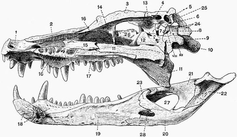
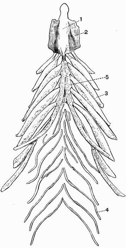
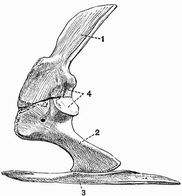
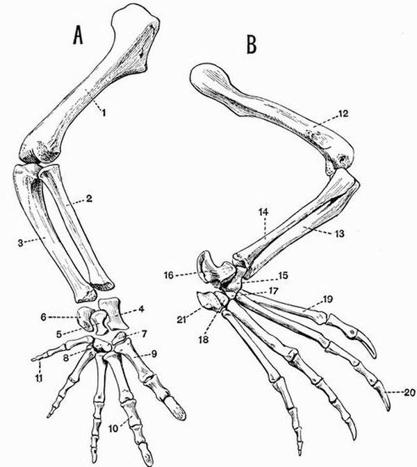
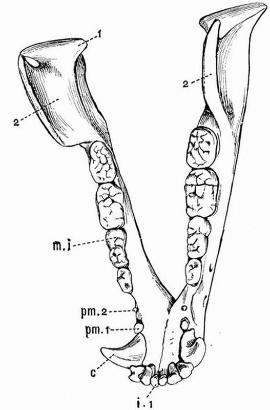
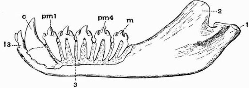
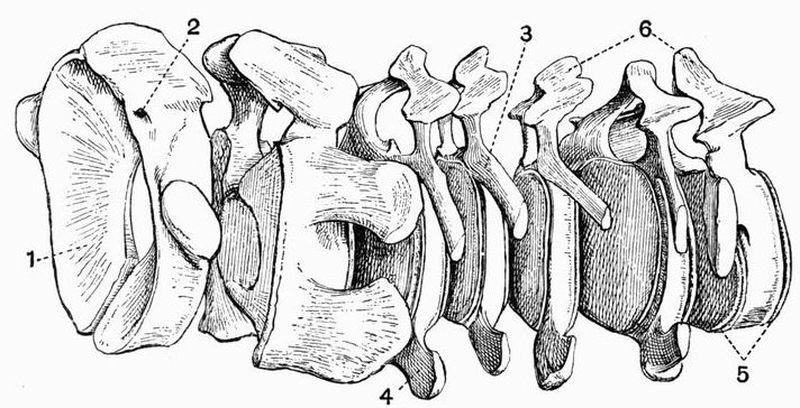
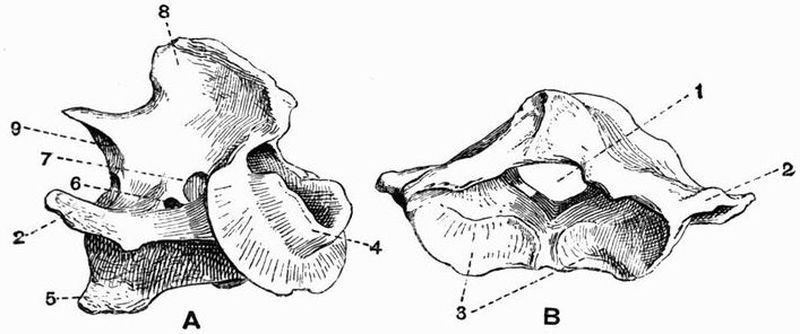
THE SKULL AND APPENDICULAR SKELETON.
The Skull.
Monotremata. In both genera the cranium is thin-walled,
has a fairly large cavity, and is very smooth and rounded externally.
The sutures between many of the bones early become
obliterated in a manner comparable to that in birds, and the
facial portion of the skull is much prolonged.
In Echidna the face is drawn out into a gradually tapering
rostrum, formed mainly by the premaxillae, maxillae and nasals.
The zygomatic arch is very weak, and the palate extends very
far back. The tympanic forms a slender ring. The mandible
is extremely slight, with no ascending portion, and but slight
traces of the coronoid process and angle. The hyoid has a
wide basi-hyal and stout thyro-hyals, while the anterior cornua
are slender, and include ossified epi-hyals and cerato-hyals.
In Ornithorhynchus the zygomatic arch is much stouter
than in Echidna. The face is produced into a wide beak,
mainly supported by the premaxillae, between whose diverging
anterior ends there is a dumb-bell-shaped bone. The maxillae
are flattened below, and each bears a large horny tooth, which
meets a corresponding structure borne on a surface near the
middle of the mandible. The mandible is considerably stouter
than in Echidna, but the angle and coronoid process are but
little developed. The infra-orbital foramen and the inferior
dental and mental foramina of the mandible are all very large.
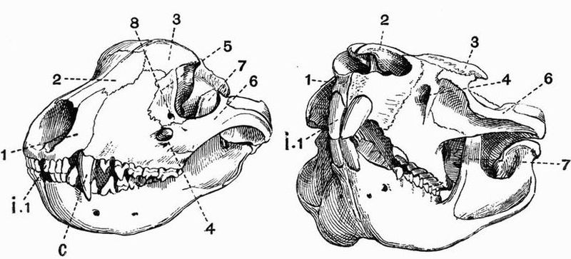
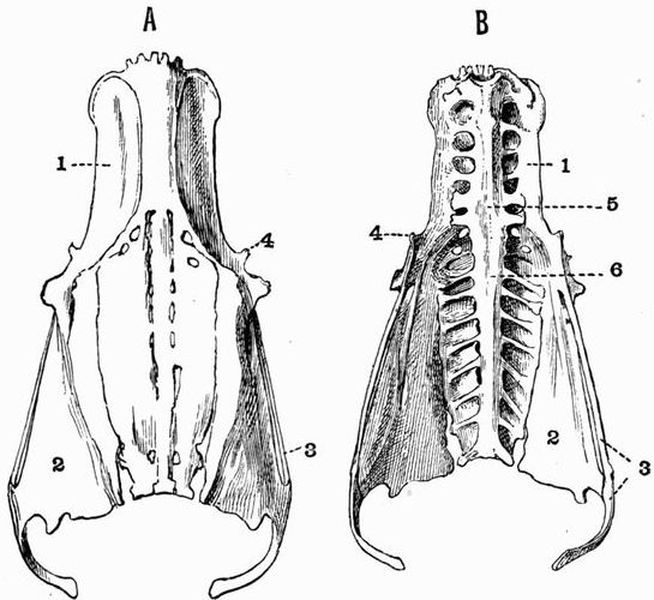
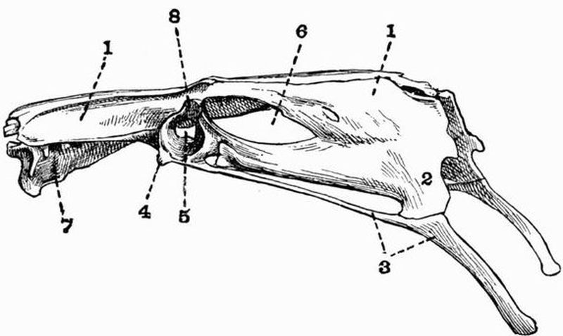
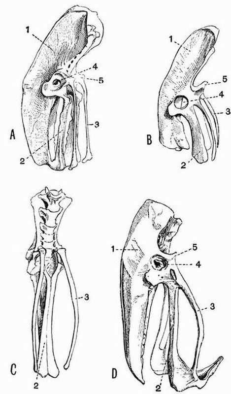
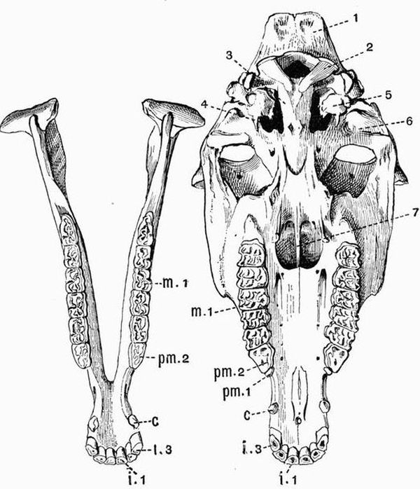
Fig. 90. Half front view[165] of the skulls of a Tasmanian Wolf
(Thylacinus cynocephalus) (to the left) × 3/8; and of a
hairy-nosed Wombat
(Phascolomys latifrons) (to the right) × 3/8. (Camb. Mus.)
| 1. premaxillae. | 7. coronoid process of the |
| 2. nasal. | mandible. |
| 3. frontal. | 8. lachrymal foramen. |
| 4. infra-orbital foramen. | i. 1. first upper incisor. |
| 5. lachrymal. | C. canine. |
| 6. jugal. |
Marsupialia. The skulls of the various types of the
Marsupials frequently bear a strong superficial resemblance to
those of some of the different groups of placental mammals.
Thus the skull of the Dasyuridae resembles that of the Carnivora,
the resemblance being most marked between the skulls of
Thylacinus and the dog. The skull of Notoryctes is strongly suggestive
of that of an Insectivore, and that of other Marsupials
such as the wombat, recalls equally the characteristic features
of a Rodent's skull. But, however much they may differ from
one another, the skulls of all Marsupials agree in the following
respects. (1) The brain cavity, and especially the cerebral
fossa, has a very small comparative size. (2) The nasals are
always large, and the mesethmoid is extensively ossified, and
terminated by a prominent vertical edge. (3) Processes from
the jugal and frontal in living forms never meet and enclose
the orbit, but the zygomatic arch is always complete. (4) The
jugal always extends back to form part of the glenoid fossa.
(5) The lachrymal canal opens either external to or upon the
margin of the orbit, and the nasal processes of the premaxillae
never quite reach the frontals. (6) The posterior part of the
palate is commonly pierced by large oval vacuities. (7) The
tympanic is small and never fused to the bones of the
cranium. (8) The carotid canal perforates the basisphenoid
and not the tympanic bulla. (9) The optic foramen and
sphenoidal fissure are confluent. (10) In every case except
Tarsipes the angle of the mandible is more or less inflected.
The skull of the extinct Thylacoleo differs from that of all
other Marsupials in the fact that the postorbital bar is complete.
The hyoid is constructed on much the same plan in all
Marsupials. It consists of a small basi-hyal, a pair of broad
cerato-hyals, and a pair of strong thyro-hyals. The epi-hyals
and stylo-hyals are generally unossified.
Edentata. In Sloths (Bradypodidae) the sutures become
early obliterated, the cranial portion of the skull is rather
high, and the facial portion very short. The lachrymal is very
small, and its canal opens outside the orbit. The zygomatic
arch is incomplete, and the jugal (fig. 91, 5) is curiously forked,
but in a manner differing in the two genera. The premaxillae
are very small,—in Bradypus quite vestigial. The mandible is
well developed, the angle being specially marked in Bradypus.
In Choloepus the symphysial part is drawn out in a somewhat
spout-like manner (fig. 91, 6). In both genera the thyro-hyals
are ankylosed with the basi-hyal.
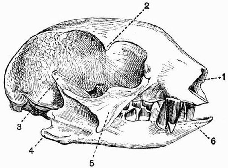
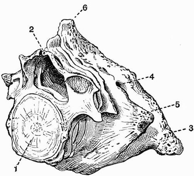
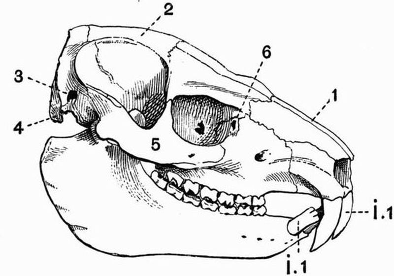
Fig. 91. Skull of a two-fingered Sloth
(Choloepus didactylus) × ½. (Camb. Mus.)
| 1. anterior nares. | 4. angle of the mandible. |
| 2. postorbital process of the | 5. jugal. |
| frontal. | 6. spout-like prolongation of the |
| 3. coronoid process. | mandible. |
In Megatherium the general appearance of the skull is
distinctly sloth-like, but the facial portion is more elongated,
partly owing to the development of a prenasal bone, and the
zygomatic arch is complete. The mandible is very deep in the
middle, and is drawn out into a long spout-like process in front.
Anteaters (Myrmecophagidae) have a much modified skull,
and this is especially the case in the Great Anteater, Myrmecophaga.
The skull is smooth and evenly-rounded, in these
respects recalling that of Echidna, but it is longer and tapers
much more gradually than in Echidna. The occipital condyles
are remarkably large. The premaxillae are small, and the
long rostrum is chiefly composed of the maxillae and nasals
with the mesethmoid and vomer. The zygomatic arch is
incomplete, and there is no trace of a separation between the
orbit and the temporal fossa. The palate is much elongated,
the pterygoids meeting in the middle line just like the palatines.
The mandible is very long and slender, there being no
definite coronoid process, and a short and slight symphysis.
The hyoid arch is noticeable for the length of the anterior
cornu.
In the Armadillos (Dasypodidae) the skull varies a good
deal in shape, but the facial portion is always tapering and
depressed. The zygomatic arch is complete. In Dasypus and
Chlamydophorus the tympanic bulla is well ossified.
In the Glyptodontidae the skull is very short and deep;
the zygomatic arch is complete, and has a long downwardly
projecting maxillary process. The mandible is massive, and
has a very high ascending portion.
In the Manidae the skull is smooth and rounded, the zygomatic
arch is incomplete, and the orbit is inconspicuous. The
palate is long and narrow, but the pterygoids do not take part
in its formation. The mandible is slightly developed and has
no angle or coronoid process.
In Orycteropus the zygomatic arch is complete, and there
is a small postorbital process to the frontal. The mandible is
well-developed, having a coronoid process and definite ascending
portion, and the hyoid is well ossified.
Sirenia. The skull, and especially the brain case of all
Sirenia, is remarkable for the general density of the component
bones, which, though often very thick, are without air sinuses.
It is noticeable also for the roughness of the bones, and the
irregular manner in which they are united together.
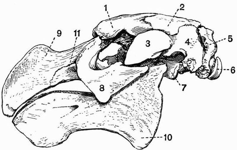
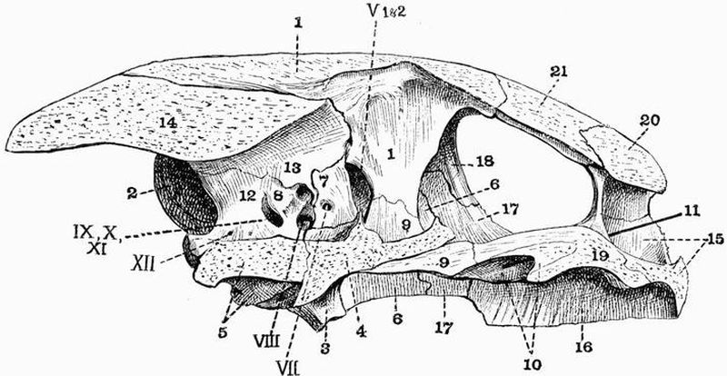
Fig. 92. Lateral view of the skull of Rhytina stelleri × 1/8.
(Brit. Mus.)
| 1. frontal. | 7. pterygoid process of the |
| 2. parietal. | alisphenoid. |
| 3. zygomatic process of the | 8. jugal. |
| squamosal. | 9. premaxillae. |
| 4. squamosal. | 10. angle of the mandible. |
| 5. exoccipital. | 11. maxillae. |
| 6. occipital condyle. |
The cranial cavity is decidedly small, the reduction being
specially noticeable in the cerebral fossa, which is not much
larger than the cerebellar fossa. The foramen magnum is
large, and the dorsal surface of the cranium narrow. The
zygomatic arch is very strongly developed, the squamosal (fig.
92, 4) being especially prominent, and being drawn out not only
into the zygomatic process, but also into a large post-tympanic
process which articulates with the exoccipital. At the side
of the skull between the squamosal, supra-occipital and exoccipital,
there is a wide vacuity in the cranial wall, partially filled
up by the very large periotic, which is ankylosed to the tympanic,
but is not united to any other bones of the skull. The
foramen lacerum medium is confluent with the foramen lacerum
anterius, and the two together form an enormous vacuity on
the floor of the skull, bounded chiefly by the exoccipital, basi-occipital,
alisphenoid and squamosal. The jugal (fig. 92, 8) is
large and in Manatus sends up a strong process, which nearly
or quite meets the postorbital process of the frontal, completing
the orbit. In the other Sirenia the orbit is completely confluent
with the very large temporal fossa. The lachrymal in Manatus
is very small, but is larger in Halicore. The premaxillae (fig.
92, 9) are large, but smaller in Manatus than in the other
genera, in all of which they are curiously bent down in front.
Their upper margin forms the anterior border of a very large
aperture lying high on the roof of the skull and extending
back for a considerable distance. This aperture is formed by
the union of the two anterior nares. The nasals are quite
vestigial or absent, and the narial aperture is bounded above
by the frontals; in its floor are seen the slender vomer and
large mesethmoid. The palate is long and narrow, and formed
mainly by the maxillae; behind it there is a large irregular
process formed by the union of the palatine, pterygoid, and
pterygoid plate of the alisphenoid. The mandible is very
massive and has a very high ascending portion, a rounded
angle (fig. 92, 10), and a prominent coronoid process; the two
rami are firmly ankylosed together. The hyoid consists principally
of the broad flat basi-hyal; the anterior cornua are but
slightly ossified, while the thyro-hyals are not ossified at all.
Cetacea. The skull in all Cetacea, especially in the Odontoceti,
is a good deal modified from the ordinary mammalian
type.
In the Archaeoceti this modification is less marked than
in either of the other suborders. The nasals and premaxillae
are a good deal larger than they are in living forms, and the
anterior nares are placed further forward. The maxillae do
not extend back over the frontals, and there is a well-marked
sagittal crest.
In the Mystacoceti the skull is always quite bilaterally
symmetrical, and is not so much modified from the ordinary
mammalian type as in the Odontoceti. The parietals are not,
as in the Odontoceti, separated by a wide interparietal, but
meet; they are, however, hidden under the very large supra-occipital.
The nasals are developed to a certain extent, and
the nares, though placed very far back and near the top of the
head, terminate forwardly-directed narial passages. Turbinal
bones are also developed to some extent; this fact, and the
occurrence of a definite though small olfactory fossa constituting
important distinctions from the Odontoceti. The maxillae are
large, but do not extend back to cover the frontals as in the
Odontoceti. The zygomatic process of the squamosal is very
large. The mandibular rami are not compressed, but are
rounded and arched outwards, and never meet in a long
symphysis.
Odontoceti. The skull departs widely from the ordinary
mammalian type. The following description will apply to any
of the following genera of the Delphinidae, Phocaena, Globicephalus,
Lagenorhynchus, Delphinus, Tursiops, Prodelphinus,
Sotalia.
The upper surface of the skull is more or less asymmetrical.
The cerebral cavity is high, short and broad; and formed mainly
by the cerebral fossa, the olfactory fossa being entirely absent.
The supra-occipital (fig. 93, 3) is very large, and forms much
of the posterior part of the roof of the skull. It has the
interparietal (fig. 93, 7) fused with it, and completely separates
the two parietals. The frontal (fig. 93, 10) is large and laterally
expanded, forming the roof of the orbit, but is almost
completely covered by an extension of the maxillae. The
zygomatic arch is very slender, and is mainly formed by a
rod-like process from the jugal (fig. 93, 15), the zygomatic
process of the squamosal being short and stout.
The nasal passages are peculiarly modified, instead of
passing horizontally forwards above the roof of the mouth,
they pass upwards and even somewhat backwards towards
the top of the skull (fig. 93, 23). They are bounded laterally
by two processes from the premaxillae, the left of which is
shorter than the right. The nasal cavities are narrow and
without turbinals and the nasals (fig. 93, 19) are almost as
much reduced as in Sirenia.
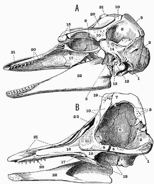
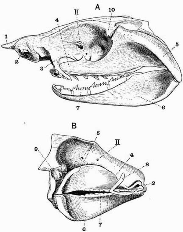
Fig. 93. A, Lateral view, and B, Longitudinal section of the skull
of a young Ca'ing Whale (Globicephalus melas) × 1/6. (Brit. Mus.)
| 1. basi-occipital. | 13. periotic. |
| 2. exoccipital. | 14. squamosal. |
| 3. supra-occipital. | 15. jugal. |
| 4. basisphenoid. | 16. vomer. |
| 5. alisphenoid. | 17. palatine. |
| 6. parietal. | 18. pterygoid. |
| 7. interparietal. | 19. nasal. |
| 8. presphenoid. | 20. maxillae. |
| 9. orbitosphenoid. | 21. premaxillae. |
| 10. frontal. | 22. mandible. |
| 11. mesethmoid. | 23. anterior nares. |
| 12. tympanic. |
In front of the nasal openings the face is prolonged as a
narrow beak or rostrum of varying length, formed by the
maxillae and premaxillae surrounding the vomer and large
mesethmoid (fig. 93, 11), which sends forwards a long partially
cartilaginous process, and is fused behind with the presphenoid
(fig. 93, 8). The basi-occipital (fig. 93, 1) too is fused with
the basisphenoid. The foramen rotundum is confluent with
the sphenoidal fissure, and the foramen ovale with the foramen
lacerum medium and the foramen lacerum posterius. The
palate is mainly formed by the maxillae; the premaxillae and
palatines (fig. 93, 17), though both meet in symphyses, forming
very little of it. The pterygoids vary in size in the different
genera, sometimes as in Lagenorhynchus and Delphinus meeting
in the middle line, sometimes as in Phocaena and Globicephalus
(fig. 93, 18) being widely separated. The tympanic
and periotic are not fused together, and the periotic has
generally no bony union with the rest of the skull. The
mandible is rather slightly developed, with the rami straight,
compressed and tapering to the anterior end. The condyle is
not raised at all above the edge of the ramus; the angle is
rounded and the coronoid process is very small. Platanista has
a curiously modified skull; the rostrum and mandible are
exceedingly long and narrow, and arising from the maxillae
are two great plates of bone which nearly meet above.
In the Physeteridae the skull is raised into a very prominent
crest at the vertex behind the nares. In front of this
in Hyperoödon a pair of ridges occur, formed by outgrowths
from the maxillae. In the old male these ridges reach an
enormous size and almost meet in the middle line. In Physeter,
the Sperm whale, these ridges are not developed; the
maxillae and premaxillae unite with the other bones of the
crest enclosing an enormous half basin-shaped cavity, at the
base of which are the very asymmetrical anterior narial apertures.
In all living Cetacea the hyoid has the same general shape,
consisting firstly of a crescentic bone formed by the fusion
of the thyro-hyals with the basi-hyal, and secondly of the
anterior cornu formed principally by the strong stylo-hyal.
Ungulata. None of the distinctive characters separating
the Ungulata from the other groups of mammals are drawn
from the skull. But in the Ungulata vera as opposed to the
Subungulata a distinguishing feature is found in the fact that
the lachrymal and jugal form a considerable part of the side
of the face, and that the jugal always forms the anterior part
of the zygomatic arch, the maxillae taking no part in it.
Ungulata vera.
Artiodactyla. The skull in Artiodactyla differs from that
in Perissodactyla in the fact that the posterior end of the
nasal is not expanded and there is no alisphenoid canal.
The skulls in the different groups of Artiodactyla differ
considerably from one another.
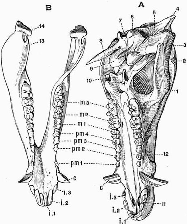
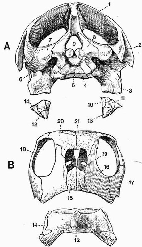
Fig. 94. A, Cranium and B, mandible of a Pig (Sus scrofa) × 1/5.
(Camb. Mus.)
| 1. jugal. | 11. anterior palatine foramen. |
| 2. postorbital process of the | 12. palatal plate of maxillae. |
| frontal. | 13. coronoid process. |
| 3. zygomatic process of the | 14. mandibular condyle. |
| squamosal. | i 1, i 2, i 3. first, second, and third |
| 4. supra-occipital. | incisors. |
| 5. glenoid cavity. | c. canine. |
| 6. occipital condyle. | pm 1, pm 2, pm 3, pm 4. first, |
| 7. foramen magnum. | second, third, and fourth |
| 8. paroccipital process of the | premolars. |
| exoccipital. | m 1, m 2, m 3. first, second, and |
| 9. tympanic bulla. | third molars. |
| 10. pterygoid. |
The skull of the Pig[166] will be described as illustrative of the
skull in the Suina. In the Pig as in most Artiodactyla the
face is bent sharply down on the basicranial axis, the commencement
of the vomer being situated below the mesethmoid
instead of in front of it as in most skulls. The occipital region
of the skull is small, and the line of junction of the supra-occipital
and parietals is raised into a prominent occipital crest.
The parietal completely fuses at an early stage with its fellow,
and the exoccipital is drawn out into a long paroccipital process
(fig. 94, A, 8). The frontal is large and broad and drawn out
into a small postorbital process. The lachrymal too is large and
takes a considerable part in forming the side of the face in front
of the orbit, as does also the jugal, though to a less extent.
The face is long and tapers much anteriorly. The nasals are
long and narrow, as are the nasal processes of the premaxillae,
which do not however reach the frontals. A prenasal ossicle
is developed in front of the mesethmoid. The palate is long
and narrow, the pterygoid (fig. 94, A, 10) is small, but the
pterygoid process of the alisphenoid is prominent. The
squamosal is small and has the tympanic fused with it; the
tympanic is dilated below, forming a bulla (fig. 94, A, 9) filled
with cancellous bone, and above forms the floor of a long upwardly-directed
auditory meatus. The mandible has a high
ascending portion and a small coronoid process (fig. 94, B, 13).
The hyoid differs from that of most Ungulates, the stylo-hyal
being very imperfectly ossified.
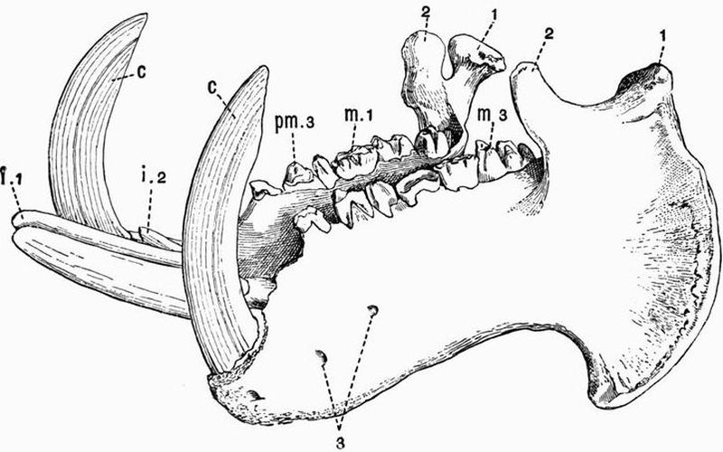
Fig. 95. Mandible of a Hippopotamus (H. amphibius) × 1/7.
(Camb. Mus.)
The second incisor of the left side is missing and the crowns of the
grinding teeth are much worn.
| 1. condyle. | c. canine. |
| 2. coronoid process. | pm 3. third premolar. |
| 3. mental foramina. | m 1, m 3. first and third molar. |
| i 1, i 2. first and second incisors. |
In Hippopotamus the skull though essentially like that of
the pig is much modified in detail. The brain cavity is very
small, while the jaws are immensely developed. The face contracts
in front of the orbits and then expands again greatly,
to lodge the enormous incisor and canine teeth. The postorbital
bar is complete or nearly so, and the orbits project curiously
outwards and slightly upwards; the lachrymal is thin and
much dilated. The squamosal is drawn out into a postglenoid
process, and the hamular process of the pterygoid is prominent.
The tympanic bulla is filled with cancellous bone. The mandible
is enormously large, the symphysis is long, the angle much
expanded and drawn out into a process which projects outwards
and forwards.
Among extinct forms related to the Suina, Cyclopidius is
noticeable for having large vacuities in the lachrymo-nasal
region, while Cotylops has the postorbital bar complete; both
these forms are from the North American Miocene.
In the Tylopoda and Tragulina the skull resembles in most
respects that of the Ruminants, shortly to be described; but it
is allied to that of the Suina in having the tympanic bulla filled
with cancellous bone. The tympanic bulla is better developed
in the Tragulina than in most Ungulates.
Among Ruminants, the Bovidae, that large group including
the Oxen, Sheep, and Antelopes, as a rule have the face
bent on the basicranial axis much as in the Suina. The
parietals are generally small and early coalesce, the frontals
are large and are usually drawn out into horn cores, which are
however absent in the skulls of some domestic varieties of sheep
and oxen, and also in some of the earlier extinct forms of Bovidae.
These horn cores are formed internally of cancellous
bone, and on them the true epidermal horns are borne. In
young animals there is a distinct interparietal, but this early
fuses with the supra-occipital, and in the oxen also with the
parietals. The occipital crest is generally well marked, but
in the genus Bos becomes merged in a very prominent straight
ridge running between the two horn cores; this ridge, which
contains air cells communicating with those in the horn cores, is
not nearly so well marked in Bison. There is often, as in Gazella,
a vacuity on the side of the face between the nasal, frontal,
lachrymal, and maxillae, but this is not found in oxen or sheep.
The premaxillae are small, the nasals are long and pointed,
and the turbinals are much developed. The Saiga antelope
has a curiously specialised skull; the nasals are absent or
have coalesced with the frontals and the anterior nares are
enormously large. In all Ruminants the lachrymal is large
and forms a considerable part of the side of the face; it often
bears a considerable depression, the suborbital or lachrymal
fossa, well seen in most of the smaller antelopes. The postorbital
bar is complete, and the orbit is prominent and nearly
circular. The palatines and pterygoids are moderately large,
and the pterygoids have a backwardly-projecting hamular process.
The squamosal is small, but has a postglenoid process.
The tympanic is not fused to the periotic and has a small bulla
not filled with cancellous bone. There is a large paroccipital
process to the exoccipital and the mandible has a long slender
coronoid process.
In the Cervidae and Giraffidae the face is not bent down
on the basicranial axis as it is in the Bovidae. The frontals are
drawn out, not into permanent horn cores as in the Bovidae,
but into short outgrowths, the pedicels, upon which in the
Cervidae long antlers are annually developed. These antlers are
outgrowths of bone, and are covered during development by
vascular integument, which dries up and peels off when growth
is complete. Every year they are detached, by a process of
absorption at the base, and shed. They may occur in both sexes,
as in the Reindeer, but as a rule they are found only in the
male. They are generally more or less branched, and are sometimes
of enormous size and weight, as in the extinct Cervus megaceros.
In young animals they are always simple, but become
annually more and more complicated as the animal grows older.
In the Giraffe the frontals bear a small pair of bony cores,
which are at first distinct, but subsequently become fused to
the skull. In the allied Sivatherium, a very large form from
the Indian Pliocene, the skull bears two pairs of bony outgrowths,
a pair of short conical outgrowths above the orbits,
and a pair of large expanded outgrowths on the occiput.
The opening of the lachrymal canal is commonly double
and the lachrymal fossa is large in the Cervidae and the Giraffidae
except Sivatherium. The vacuity between the frontal,
lachrymal, maxillae, and nasal is specially large.
The hyoid of Ruminants is noticeable for the development
of the anterior cornua, which include stout and short cerato-hyals
and epi-hyals, long and strong stylo-hyals and large
tympano-hyals which are more or less imbedded in the tympanics.
Perissodactyla. In the skull of Perissodactyles an alisphenoid
canal is found and the nasals are expanded behind.
Among the living animals belonging to this group the skull least
modified from the ordinary type is that in Rhinoceros. In this
form the skull is considerably elongated, the facial portion being
very large. The occipital region is elevated, but the cranial
cavity is small, the boundary line between the occipital and
parietal regions being drawn out into a prominent crest, which
is occupied by air cells. There is no postorbital process to the
frontal, and the orbit is completely confluent with the temporal
fossa. The nasals are fused together and are very strongly
developed, extending far forwards, sometimes considerably
beyond the premaxillae. In some extinct species, such as Elasmotherium
and the Tichorhine Rhinoceros, R. antiquitatis, the
mesethmoid is ossified as far forwards as the end of the nasals.
The nasals are arched and bear one or two roughened surfaces
to which the great nasal horns are attached. The premaxillae
are very small and the pterygoids are slender. The palate is
long, narrow, and deeply excavated behind. The postglenoid
process of the squamosal is well developed, and generally
longer than the paroccipital process of the exoccipital. The
tympanic and periotic are both small and are fused together.
The condyle of the mandible is very wide, the angle rounded,
and the coronoid process moderately developed.
In the Titanotheriidae, a family of extinct Perissodactyla
from the Miocene of North America, the occipital region is much
elevated, as is also the fronto-nasal region, the nasals (perhaps
only in the male) bearing a pair of blunt bony outgrowths.
Between these two elevated regions the skull is much depressed.
The cranial cavity is very small, the orbit confluent
with the temporal fossa, and the zygomatic arch massive.
In Tapirus the orbit and temporal fossa are confluent.
The nasals are small, wide behind and pointed in front, and
are supported by the mesethmoid; the anterior nares are
exceedingly large and their lateral boundaries are entirely
formed by the maxillae. The postglenoid and post-tympanic
processes of the squamosal are large. The periotic is not fused
to the squamosal or to the small tympanic. The mandible is
large and has the angle much developed and somewhat inflected.
Palaeotherium, which lived in early Tertiary times, has a
skull much like that of the Tapir, especially as regards the
nasal bones.
In the Horse and its allies (Equidae) the facial portion of
the skull is very large as compared with the cranial portion,
the nasals and nasal cavities being specially large. In the
living species of the genus Equus there is no fossa between the
maxillae and lachrymal, but it occurs in some extinct species.
The lachrymal and jugal form a considerable part of the side
of the face; and the orbit though small is complete and prominent.
The postorbital bar is formed by a strong outgrowth
from the frontal, which unites with a forward extension of the
squamosal. The squamosal may extend forwards and form
part of the wall of the orbit, a very unusual feature, as in most
mammals the squamosal stops before the postorbital bar. The
palate is narrow and excavated behind as in Rhinoceros; the
palatines take very little part in its formation. The glenoid
surface for the articulation of the mandible is very wide. The
squamosal gives rise to small postglenoid and post-tympanic
processes, and the exoccipital to a large paroccipital process.
The tympanic and periotic are ankylosed together, but not to
any other bones.
In the Subungulata, the lachrymal and jugal do not form
any considerable part of the side of the face, and the maxillae
commonly takes part in the formation of the zygomatic arch.
Toxodontia. The skull in the Toxodontia shows several
Artiodactyloid features, while the manus and pes are of a more
Perissodactyloid type. The Artiodactyloid features are (1) the
absence of an alisphenoid canal, (2) the fact that the palate is
not excavated behind, and that the palatines form a considerable
part of it, and (3) the fusion of the tympanic to the
squamosal and exoccipital, forming the floor of an upwardly
directed auditory meatus. The frontal has a fairly well developed
postorbital process, but the orbit is confluent with the
temporal fossa. The premaxillae is well developed, as is the paroccipital
process of the exoccipital, especially in Typotherium.
The mandible has a rounded angle and a coronoid process of
moderate size. In Typotherium the ascending portion is very
massive.
Condylarthra. As far as is known the skull of these
generalised Ungulates is depressed, and is frequently marked
by a strong sagittal crest. The cranial cavity is small, the
cerebral fossa in Phenacodus being exceptionally small. The
orbit is completely confluent with the temporal fossa.
Hyracoidea. The skull of Procavia resembles that of
Perissodactyles more than that of any other Ungulates, but
differs strongly in the comparatively small size of its facial
portion. The posterior portion of the cranium is rather high,
the occipital plane being nearly vertical. There is a small
interparietal. The nasals are wide behind, and the zygomatic
arch is strongly developed, its most anterior part being formed
by the maxillae. The jugal and parietal give rise to postorbital
processes which sometimes meet, but as a rule the orbit
is confluent with the temporal fossa; it is very uncommon
for the parietal to give rise to a postorbital process, and even
in Procavia the frontal often forms part of the process. The
alisphenoid canal, and postglenoid and paroccipital processes
are well developed. The tympanic bulla is large and the
periotic and tympanic are fused together, but not as a rule
to the squamosal. The ascending portion of the mandible is
very high and broad, the angle rounded and the coronoid
process moderate in size. The hyoid is singular, there is a
large flat basi-hyal prolonged laterally into two broad flattened
thyro-hyals. Articulating with its anterior end are two large
triangular cerato-hyals, which are drawn out into two processes
meeting in the middle line.
Amblypoda. In the Uintatheriidae (Dinocerata) the skull
has a very remarkable character, being long and narrow and
drawn out into three pairs of rounded protuberances, a small
pair on the nasals, a larger pair on the maxillae in front of the
orbits, and the largest pair on the parietals. The cranial cavity,
and especially the cerebral fossa, is extraordinarily small. The
orbit is not divided behind from the temporal fossa. The
mandible has a prominent angle, and a long curved coronoid
process; its symphysial portion bears a curious flattened outgrowth
to protect the great upper canines.
In Coryphodon the skull is of a more normal character,
being without the conspicuous protuberances. The cranial
cavity though very small is not so small as in Uintatherium.
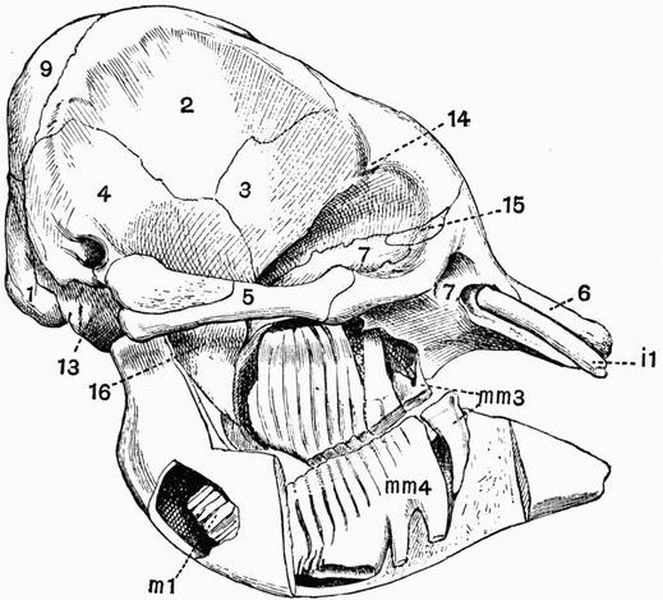
Fig. 96. Skull of a young Indian Elephant (Elephas indicus), SEEN
from the right side, the roots of the teeth have been exposed. × 1/8. (Camb. Mus.).
| 1. exoccipital. | 14. postorbital process of the |
| 2. parietal. | frontal. |
| 3. frontal. | 15. lachrymal. |
| 4. squamosal. | 16. pterygoid process of the |
| 5. jugal. | alisphenoid. |
| 6. premaxillae. | i 1. incisor. |
| 7. maxillae. | mm 3., mm 4. third and fourth |
| 9. supra-occipital. | milk molars. |
| 13. basi-occipital. | m 1. first molar. |
Proboscidea. The character of the skull in the young elephant
differs much from that in the old animal. In very young
individuals the skull is of a normal character, and the cranial
cavity is distinctly large in proportion to the bulk of the skull.
But as the animal gets older, while its brain does not grow
much, the size of its trunk and especially of its tusks increases
greatly; and consequently the skull wall is required to be of
very great superficial extent in order to afford space for the
attachment of the muscles necessary for the support of these
heavy weights. This increase in superficial extent is brought
about without much increase in weight of bone by the development
of an enormous number of air cells in nearly all the
bones of the skull; sometimes, as in the case of the frontal,
separating the inner wall of the bone from the outer, by as
much as a foot. This development of air cells is accompanied
by the obliteration of the sutures between the various bones.
The most noticeable point with regard to the cranial cavity
is the comparatively large size of the olfactory fossa. The
supra-occipital (figs. 96 and 97, 9) is large—exceedingly large
in the adult skull; the parietals (figs. 96 and 97, 2) are also
very large. The frontals send out small postorbital processes,
but these do not meet processes from the small jugal, which
forms only the middle part of the slender zygomatic arch, the
anterior part being formed by the maxillae. The lachrymal
(fig. 96, 15) is small and lies almost entirely inside the orbit.
The anterior narial aperture (fig. 97, 8) is wide and directed
upwards, opening high on the anterior surface of the skull. It
is bounded above by the short thick nasals and below by the
premaxillae. The narial passage is freely open, maxillo-turbinals
not being developed. The palatine is well developed,
the pterygoid is small and early fuses with the pterygoid
process of the alisphenoid. The tympanic is united with the
periotic but not with the squamosal, and forms a large auditory
bulla. There are no paroccipital or postglenoid processes.
The exoccipital is not perforated by the condylar foramen,—a
very exceptional condition.
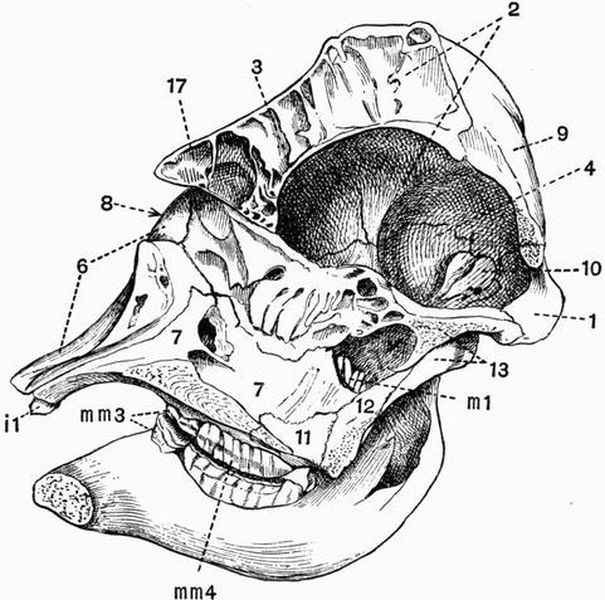
Fig. 97. Longitudinal section taken rather to the right of the
middle line of the skull of a young Indian Elephant (E.
Indicus) × 1/8. (Camb. Mus.)
| 8. anterior nares. | 12. pterygoid. |
| 10. periotic. | 17. nasal. |
| 11. palatine. | Other numbers as in Fig. 96. |
The mandible has a high ascending portion, is rounded off
below and has no angle. The symphysial portion is long,
narrow, and spout-like, and the coronoid process is small.
The thyro-hyals are ankylosed with the basi-hyal, which is
connected with the large forked stylo-hyals by ligament only.
Rodentia. The cranial cavity is depressed, elongated, and
rather small, and the cerebral fossa lies entirely in front of the
cerebellar fossa. The occipital plane is vertical or directed
somewhat backwards, and the supra-occipital does not form
much of the roof of the cranium. The paroccipital processes
of the exoccipitals are generally of moderate size; in the
Capybara (Hydrochaerus), however, they are very long, and
are laterally compressed and directed forwards. The parietals
are small, and often become completely fused together; there
is sometimes a small interparietal. The frontals in most genera
have no trace of a postorbital process; in Squirrels, Marmots
and Hares, however, one occurs, but in no case does it meet a
corresponding process from the zygomatic arch, so the orbit
and temporal fossa are completely confluent. In Hares the
postorbital process of the frontal is much flattened, and has
an irregular margin. The temporal fossa is always small, and
in Lophiomys is arched over by plates arising respectively
from the parietal and jugal; a secondary roof is thus partially
developed in a manner unique among mammals, but carried
to a great extent in many Chelonia. The nasal bones and
cavities are large, attaining their maximum development in
the Porcupines (fig. 98, 1). The premaxillae is always very
large, and sends back a long process which meets the frontal.
The vomer is occasionally found persisting in two separate
halves, a feature recalling the arrangement in Sauropsids. In
many Rodents there is an enormous vacuity at the base of the
maxillary portion of the zygomatic arch. It is sometimes as
large as the orbit, and attains its maximum development in
the Capybara and other Hystricomorpha; in the Marmots,
Beavers, and Squirrels (Sciuromorpha), and in the Hares it is
undeveloped. In Lagostomus the maxillae bears an upwardly
directed plate of bone, shutting off from this vacuity a space
which is the true infra-orbital foramen.
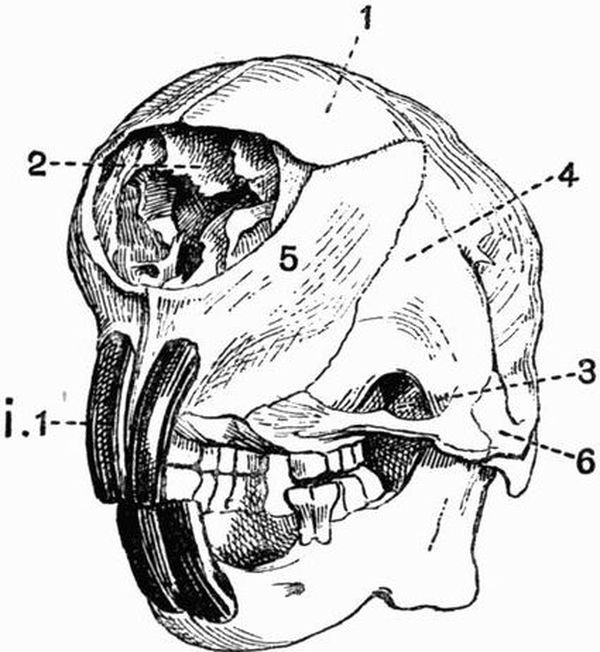
Fig. 98. Half front view of the skull of a Porcupine
(Hystrix cristata) × ½. (Camb. Mus.)
| 1. nasal. | 5. premaxillae. |
| 2. maxillo-turbinals. | 6. jugal. |
| 3. infra-orbital vacuity. | i 1. upper incisor. |
| 4. maxillae. |
The zygomatic arch is always complete, and in many cases
the jugal extends back to form part at least of the glenoid
surface for articulation with the mandible. In Coelogenys the
jugal and maxillary portion of the zygomatic arch is greatly
expanded and roughened, and the maxillary portion encloses a
large cavity. The palate in Rodents is narrow, and the space
between the incisor and molar teeth passes imperceptibly
into the sides of the face. The anterior palatine foramina
form long, rather narrow slits in this region. The bony
palate between the grinding teeth is sometimes as in the Hares
very short, sometimes as in the Capybara very long. The
maxillae extends back beneath the orbit to unite with the
squamosal. The pterygoid is always small, but sometimes
has a well-marked hamular process which in Hystrix, Lagostomus,
and some other genera unites with the tympanic bulla.
The periotic is large, and fused with the tympanic, which
forms a prominent bulla, and is generally drawn out into a
tubular meatus. The bulla attains its maximum development
in Chinchilla and Dipus.
The mandible is narrow and rounded in front, the two
halves meeting in a long symphysis. The angle is generally
drawn out into a long backwardly-projecting process, which is
often pointed and directed upwards. In the Hares the angle
is rounded. The coronoid process is never large.
There are a number of points in which the skull of the
Duplicidentata (Hares and Rabbits) differs from that of other
Rodents. (a) The sutures between the basi-occipital and
basisphenoid, and between the basisphenoid and presphenoid
remain open throughout life. (b) Much of the maxillae forming
the side of the face in front of the orbit is fenestrated. (c) The
optic foramina are united to form a single hole, much as in
birds. (d) The coronoid process is slightly differentiated from
the ascending portion of the mandible. The first two of these
points have been thought to indicate degradation of the
hares and rabbits as compared with higher mammals.
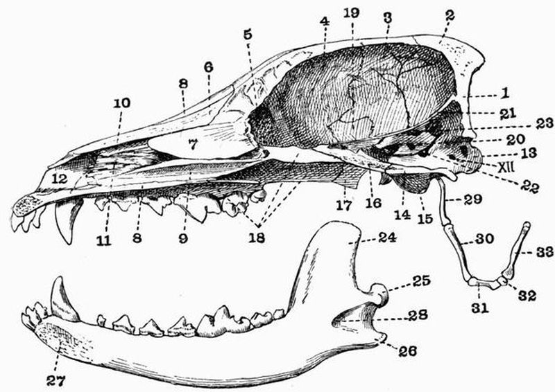
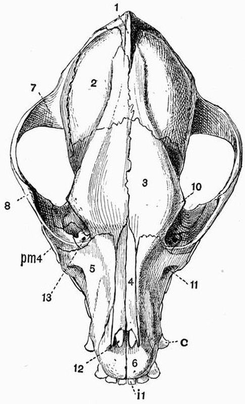
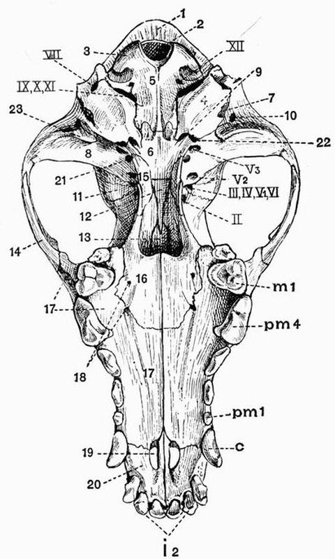
Carnivora[167]. It is characteristic of the skull in Carnivora
that the glenoid fossa is deep, and the postglenoid process
(fig. 75, 23) well developed. The condyle of the mandible is
much elongated transversely. The orbit and temporal fossa
in the great majority of forms communicate freely, the postorbital
bar being incomplete.
Carnivora vera. The axis of the facial portion of the
skull is a direct continuation of that of the cranial portion.
The cranial cavity though rather depressed is large, and
generally long, though in Cats it is comparatively short and
wide. The occipital plane is nearly vertical, and the exoccipitals
are developed into fairly prominent paroccipital processes.
The interparietal is commonly distinct, and the parietals unite
in a long sagittal suture, which is often developed into a crest.
The nasals (fig. 73, 4) are well developed, especially in Cats, and
the nasal processes of the premaxillae do not nearly reach the
frontals. A considerable part of the palate is formed by the
palatine, and the maxillary portion is pierced by rather long
anterior palatine foramina. The pterygoid has a hamular
process. The zygomatic arch is strong, especially in Cats.
Postorbital processes are developed on the frontal (fig. 73,
10) and jugal, but never form a complete postorbital bar. A
carotid canal is well seen in the Ursidae, and to a less extent
in the Felidae; in the Canidae there is an alisphenoid canal
(fig. 75, 21).
The auditory bulla differs a good deal in the different
groups. In the Bears (Ursidae) it is not much inflated, and
is most prominent along its inner border; it is not closely connected
with the paroccipital process. In the Cats it is very
prominent, and its cavity is almost divided by a septum into
two parts, the inner of which contains the auditory ossicles.
The paroccipital process is closely applied to the bulla. In the
Dogs the bulla is intermediate in character between that of
the Cats and that of the Bears; it is partially divided by a
septum, and is moderately expanded.
The mandible is well developed with a prominent angle
(fig. 72, 26), and a large coronoid process. The hyoid consists
of a broad basi-hyal, a long many-jointed anterior cornu and
short thyro-hyals (fig. 72, 33).
The skull in the Creodonta is in most respects allied to
that of the Canidae, but presents some ursine affinities. The
tympanic bulla is fairly prominent, but has no well-developed
septum. The cranial cavity is very small and narrow, the
zygomatic arch standing away from it. The temporal fossa is
of great size.
In the Pinnipedia the cranial cavity is large and rounded.
The skull is much compressed in the interorbital region, and
in correlation with this compression the ethmo-turbinals are
little developed, while the maxillo-turbinals are large. The
orbit is large, and the temporal fossa smaller than in the
Carnivora vera. In the Walrus (Trichechus) the anterior part
of the face is distorted by the development of the huge canines.
The Otariidae have an alisphenoid canal. The tympanic bulla
is small in Otaria, large in the Phocidae, and flattened in the
Walrus. The hyoid is similar to that in Carnivora vera.
Insectivora. The skull varies much in the different members
of the order Insectivora, but the following points of
agreement are found. The cranial cavity is of small size, and
is never much elevated. The facial part of the skull is generally
considerably elongated, and the nasals and premaxillae are
well developed. The zygomatic arch is usually slender or
incomplete, and the coronoid process and angle of the mandible
are commonly prominent.
In some Insectivora, such as Galeopithecus, Tupaia, and
Macroscelides, the skull shows a higher type of structure
than is met with in most members of the order. In these
genera the cranial cavity is comparatively large, and the
occipital plane is nearly vertical. The zygomatic arch is fairly
strong, and the frontal and jugal give rise to postorbital processes
which nearly or quite (Tupaia) meet. The tympanic
bulla is well developed, and produced into a tubular auditory
meatus, this being specially well marked in Macroscelides.
In the other Insectivora the cranial cavity is of smaller
comparative size, and the orbit and temporal fossa are completely
confluent, often without any trace of a postorbital
bar. The occipital plane commonly slopes forwards. In
the Hedgehogs (Erinaceidae) and Centetidae the tympanic is
very slightly developed, forming a small ring. The zygomatic
arch of Hedgehogs and Gymnura is very slender, the jugal
being but little developed and the squamosal and maxillae
meeting one another; in the Centetidae the jugal is absent
and the arch is incomplete.
The Moles (Talpidae) have an elongated, depressed and
rounded skull with a very slender zygomatic arch formed by
the squamosal and maxillae. The nasals are fused together,
and the mesethmoid is ossified very far forwards. In the
Shrews (Soricidae) there is no zygomatic arch; the tympanic
is ring-like, and the angle of the mandible is very prominent.
The hyoid has a transversely extended basi-hyal, a long
anterior cornu with three ossifications, and thyro-hyals which
are sometimes fused to the basi-hyal.
Chiroptera. In the frugivorous Flying Foxes (Pteropidae)
the skull is elongated, and the cranial cavity is large
and arched, though considerably contracted in front. There
are commonly strong sagittal and supra-orbital crests. The
parietals take a great part in the formation of the walls of
the cranial cavity, the supra-occipital and frontals being small.
The frontal is drawn out into a long postorbital process, but
the zygomatic arch, which is slender, and formed mainly by
the squamosal and maxillae, gives rise to only a small postorbital
process, so that the orbit and temporal fossa are confluent.
There is no alisphenoid canal, and the tympanics are
very slightly connected with the rest of the skull. The mandible
has a large coronoid process, a rounded angle, and a transversely
expanded condyle.
In Insectivorous Bats the skull is generally shorter and
broader than in the Pteropidae. The cranial cavity is large
and rounded, and has thin smooth walls. The zygomatic
arch is slender, and postorbital processes are not generally
well developed. The premaxillae is generally small, sometimes
absent. The tympanics are ring-like and are not connected with
the surrounding bones. The angle of the mandible is distinct.
The hyoid in most respects resembles that of the Insectivora.
Primates. The characters of the skull differ greatly in
the two suborders of Primates, the Anthropoidea and the
Lemuroidea.
In the Lemuroidea the general relative proportions of the
cranium and face are much as in most lower mammals, and the
occipital plane forms nearly a right angle with the basicranial
axis. The postorbital processes of the frontals are commonly
continued as a pair of ridges crossing the roof of the cranium
and meeting the occipital crest. Though the postorbital bar
is complete, the orbit and temporal fossa communicate freely
below it. The lachrymal canal opens outside the orbit, and the
lachrymal forms a considerable part of the side of the face.
The tympanic is developed into a large bulla. The hyoid
apparatus much resembles that of the Dog.
In the Anthropoidea the skull differs greatly from that in
the Lemuroidea. The cranial portion of the skull is very
large as compared with the facial portion, though the
comparative development varies, some monkeys, such as the
baboons (Cynocephali) having the facial portion relatively large.
The comparative size of the jaws does not vary inversely with
the general development of the animal, some of the Cercopithecidae
having comparatively larger jaws than some of the
Cebidae. The great size of the cranial part of the skull is
mainly due to the immense development of the cerebral fossa,
which commonly completely overlaps the olfactory fossa in
front, and the cerebellar fossa behind. This development also
has the effect of making the ethmoidal and occipital planes lie,
not at right angles to the basicranial axis, but almost in the
same straight line with it. This is, however, not always the
case, as the Howling Monkey (Mycetes) and also some of the
very highest monkeys, the Gibbons (Hylobates), have the occipital
plane nearly vertical to the basicranial axis. In adult
Man the basi-occipital, exoccipitals and supra-occipital coalesce,
forming the so-called occipital bone; while the basisphenoid,
presphenoid, alisphenoids, orbitosphenoids and pterygoids form
the sphenoid bone. The roof of the skull is partly formed
by the large supra-occipital and frontals, but mainly by the
parietals (fig. 99, 1), which in Man are of enormous extent.
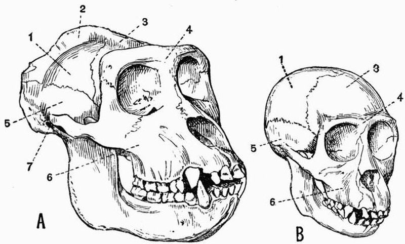
Fig. 99. Half front view of the skulls, A of an old, B of a
young Gorilla (Gorilla savagei) × ¼. (Camb. Mus.)
| 1. parietal. | 5. squamosal. |
| 2. sagittal crest. | 6. maxillae. |
| 3. frontal. | 7. external auditory meatus. |
| 4. supra-orbital ridge. |
In Man and in most monkeys, at any rate when young
(fig. 99, B), the roof of the skull is smooth and rounded, but
in many forms, such as the Baboons, in the adult the supra-orbital
and occipital ridges are much developed. In the Gorilla
this is also the case with the sagittal crest (fig. 99, A, 2). The
bones of the upper surface of the cranium interlock with wavy
outlines. The nasals vary much in length, being much shorter
in man than in most monkeys; they commonly become early
fused together, as do also the frontals. The vomer is well
developed, and the ethmo-turbinal always forms part of the
boundary of the orbit. There are frequently, as in many
Lemuroidea, a pair of more or less well-marked ridges, crossing
the roof of the skull from the postorbital processes of the
frontals to the occipital crest. The orbit is completely encircled
by bone, and the alisphenoid assists the jugal and frontal in
shutting it off from the temporal fossa, leaving however a
communication between the two as the sphenomaxillary fissure.
In most cases the frontals meet one another in the middle line
between the mesethmoid and orbitosphenoid, but in Man, Simia,
and some Cebidae this does not take place. In nearly all
Cebidae the parietal and jugal meet one another, separating
the frontal and alisphenoid on the skull wall; in Man and all
Old World monkeys, on the other hand, the alisphenoid and
frontal meet and separate the jugal and parietal. The premaxillae
nearly always send back processes which meet the
nasals. The palate is rather short and both the palatine and
the premaxillae take a considerable part in its formation. The
pterygoid plate of the alisphenoid is decidedly large, and there
is no alisphenoid canal. There is never any great development
either of the paroccipital process of the exoccipital, or
of the postglenoid process of the squamosal. The periotic
and tympanic are always fused together; in Cebidae they
form a small bulla, but a bulla is not developed in any Old
World forms. The periotic is large, especially the mastoid
portion, which forms a distinct portion of the skull wall between
the squamosal and exoccipital. In Man and still more
in Old World monkeys, the external auditory meatus is drawn
out into a definite tube, whose lower wall is formed by the
tympanic; in the Cebidae the tympanic is ring-like. The
perforation of the periotic by the carotid canal is always
conspicuous.
The mandible is rather short and broad, and the angle
formed by the meeting of the two rami is more obtuse than in
most mammals. The coronoid process is fairly well developed,
and the angle is more or less rounded. In most Primates the
condyle is considerably widened, but this is not the case in
Man. In Mycetes the mandible is very large, its ascending
portions being specially developed. The hyoid of Primates is
remarkable for the large expanded basi-hyal, which is generally
concave above and convex below. The anterior cornu is never
well ossified, but the thyro-hyal is always strong. In Mycetes
the basi-hyal is enormously large, forming a somewhat globular
thin-walled capsule.
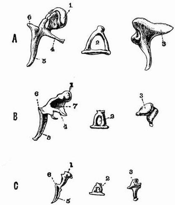
Fig. 100. Malleus, stapes and incus of
A. Man. B. Dog. C. Rabbit. (After Doran) x 1.
| 1. head of malleus. | 5. manubrium of malleus. |
| 2. canal of stapes. | 6. processus brevis. |
| 3. incus. | 7. lamella. |
| 4. processus longus (or gracilis). |
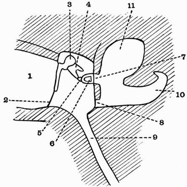
Auditory ossicles.
There are in mammals four auditory ossicles forming a
chain extending from the fenestra ovalis to the tympanic
membrane. Three of these, the malleus, incus and stapes,
are always distinct, while the fourth, the lenticular, is smaller
than the others and is sometimes not distinct. The names are
derived from human anatomy and indicate in the case of the
first three a more or less fanciful resemblance respectively to a
hammer, an anvil and a stirrup. The ossicles are homologous
as a whole to the hyomandibular of fishes and to the columellar
chain of Sauropsids and Amphibians. The malleus is homologous
to the extra-columella of Crocodiles and the stapes
to the columella. The malleus when typically developed
consists of a rounded head (fig. 100, 1) which bears a surface
articulating with the incus, and a short neck continued into a
process, the manubrium (fig. 100, 5), which comes into relation
with the tympanic membrane. From the junction of the neck
and manubrium two processes are given off, a processus longus
or gracilis (fig. 100, 4), which in the embryo is continuous
with Meckel's cartilage, and a processus brevis (fig. 100, 6).
The incus generally consists of a more or less anvil-shaped
portion which articulates with the malleus, and of a process
which is connected with the stapes by the small lenticular.
The stapes is generally stirrup shaped, consisting of a basal
portion from which arise two crura separated by a space the
canal through which a branch of the pharyngeal artery runs
The lenticular is frequently cartilaginous and sometimes is
not developed at all.
The above is the arrangement of the auditory ossicles met
with in the higher Mammalia, but in the lower Mammalia
the characters approach more nearly to those met with in
Sauropsids.
In Monotremes the ossicles, though distinctly mammalian
in character, show a very low type of development. The incus
is articulated, or often fused, with an outgrowth from the
head of the malleus. The stapes is very much like a reptilian
columella, having a single crus with no perforation.
In Marsupials the ossicles are of a low type, but not so
low as the rest of the skeleton might have led one to expect,
and all or almost all the points showing a low grade of development
may be paralleled among the Monodelphia. The
lowest Marsupials as regards the ossicles are the Peramelidae,
whose ossicles are of a frail papery consistence. The Didelphyidae
on the other hand have the most highly developed
ossicles, the malleus much resembling that of many Insectivores,
and the stapes having two definite crura separated by a canal.
In Edentates the character of the ossicles varies much.
In Sloths the stapes approaches that of Sauropsids in its
narrowness and the slight trace of a canal; this character
is however still more marked in Manis, whose stapes is as
Sauropsidan as that of Monotremes, and consists of a nearly
circular basal plate bearing a column which does not show any
sign of division into crura. The stapes of other Edentates,
such as ant-eaters, aard varks, and most armadillos, is of a high
type and has well-developed crura. Priodon has a lower type
of stapes than Dasypus and Tatusia.
The ossicles of the Sirenia differ widely from those of all
other mammals in their great density and clumsy form.
In Cetacea the ossicles are solid, though not so solid as in
Sirenia, and their details vary much. The malleus is always
firmly fused to the tympanic by means of the processus longus,
and the manubrium is very little if at all developed. The
incus has the stapedial end greatly developed, and the stapes
has very thick crura with hardly any canal. The ossicles of
the Mystacoceti are apparently less specialised than are those
of the Odontoceti.
The auditory ossicles of the Ungulata do not present any
characters common to all the members of the group.
Among Ruminants they are chiefly remarkable for the
development of a broad lamellar expansion between the head
and the processus longus of the malleus. In some cases the
malleus of the foetus differs strikingly from that of the adult.
Among Perissodactyla the Rhinoceros and Tapir have the
malleus of a low type, recalling those of Marsupials; while in
the Horse the head is well developed, and the malleus is of
a higher type.
The ossicles of Procavia, which recall those of the Equidae,
are chiefly remarkable for the small size of the body of the
incus. In Elephants the ossicles are large and massive.
In the Rodentia (fig. 100, C) the malleus is generally
characterised by a very broad manubrium. In many genera
such as Bathyergus, and most of the Hystricomorpha such as
Hystrix, Chinchilla and Dasyprocta, the malleus and incus are
ankylosed together.
Carnivora. In Carnivora vera the most striking feature
of the malleus is the occurrence of a broad lamellar expansion
between the head and neck and the processus longus.
This however does not occur in some Viverridae. In the
Carnivora vera the incus and stapes are small as compared
with the malleus, but in the Pinnipedia they are large. In
the Pinnipedia the auditory ossicles have a very dense
consistence, and except in the Otariidae are very large. The
stapes frequently has no canal, or only a very small one.
In Insectivora the characters of the auditory ossicles are
very diverse. Many forms such as shrews, moles, hedgehogs,
and the Centetidae have a low type of malleus resembling that
of Edentates. Chrysochloris has very extraordinary auditory
ossicles. The head of the malleus is drawn out into a great
club-shaped process, the incus is long and narrow, and differs
much from the ordinary type.
In Chiroptera the ossicles and especially the malleus much
resemble those of shrews. The stapes is always normal in
character, never becoming at all columelliform.
Primates. In Man and the Anthropoid Apes the malleus
has a rounded head, a short neck, and the manubrium, a
processus longus and a processus brevis. The incus consists of
an anvil-shaped portion from which arises a long tapering
process. The stapes has diverging crura and consequently a
wide canal. The crura in other monkeys do not diverge so
much as in man and anthropoid apes. The New World monkeys
have no neck to the malleus.
The Sternum[168].
In Monotremes and most Marsupials the sternum does
not present any characters of special importance. The presternum
is strongly keeled in Notoryctes.
The sternum in Edentates is very variable: in the Sloths
it is very long, the mesosternum of Choloepus having twelve
segments. In the ant-eaters and armadillos the presternum
is broad and sometimes as in Priodon strongly keeled. In
Manis macrura the xiphisternum is drawn out into a pair of
cartilaginous processes about nine inches long.
In the Sirenia the sternum is simple and elongated, and
of fairly equal width throughout, in the adult it shows no sign
of segmentation. Its origin from the union of two lateral
portions can be well seen in Manatus.
Two distinct types of sternum are met with in the Cetacea.
In the Odontoceti the sternum consists of a broad presternum
followed by three or four mesosternal segments, but with no
xiphisternum. Indications of the original median fissure can
be traced, and are very evident in Hyperoödon. In the
Mystacoceti, on the other hand, the sternum consists simply
of a broad flattened presternum which is sometimes more or
less heart-shaped, sometimes cross-shaped. Only a single pair
of ribs are united to it.
The sternum in Ungulata is generally long and narrow and
formed of six or generally seven segments. The presternum
is as a rule small and compressed, often much keeled, especially
in the horse and tapir. The segments of the mesosternum
gradually widen as followed back and the xiphisternum is
often terminated by a cartilaginous plate.
In the Rodentia the sternum is long and narrow and
generally has a large presternum, and a xiphisternum terminated
by a broad cartilaginous plate.
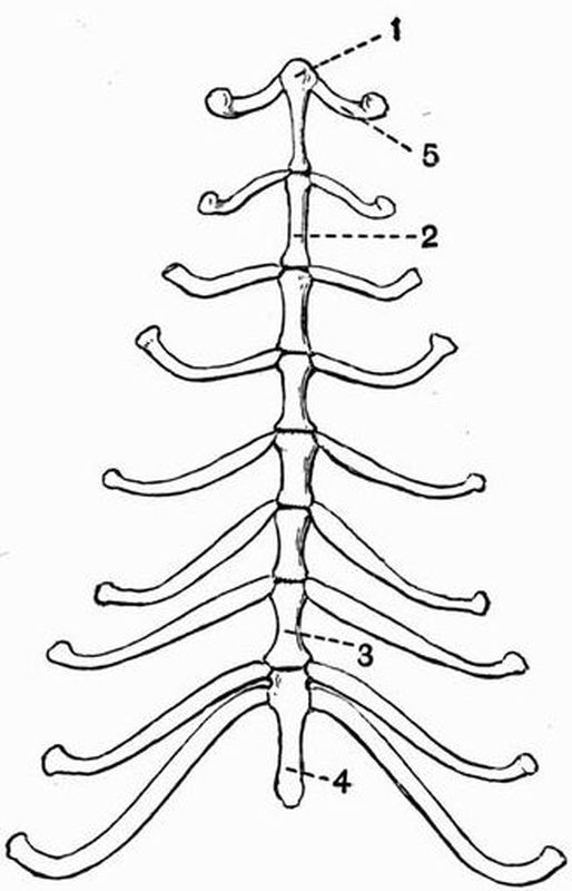
In the Carnivora, too, the sternum (fig. 76) is long and
narrow and formed of eight or nine pieces, all of nearly the
same size. The xiphisternum generally ends in an expanded
plate of cartilage.
In Insectivora the sternum is well developed but variable.
The presternum is commonly large and is sometimes as in the
Hedgehog (Erinaceus) bilobed in front, sometimes as in the
Shrew (Sorex) trilobed. It is especially large in the Mole
(Talpa) and is expanded laterally and keeled below.
In the Chiroptera the presternum is strongly keeled and
so is sometimes the mesosternum.
Among Primates, in Man and the Anthropoid Apes the
sternum is rather broad and flattened; the mesosternum
consists of four segments which are commonly fused together
and the xiphisternum is imperfectly ossified.
The Ribs.
Free ribs are borne as a rule only by the thoracic vertebrae;
ribs may be found in other regions, especially the
cervical and sacral, but these are almost always ankylosed to
the vertebrae. As a general rule the first thoracic rib
joins the presternum, while the succeeding ones are attached
between the several segments of the mesosternum. Some
of the posterior ribs frequently do not reach the sternum;
they may then be attached by fibrous tissue to the ribs in
front, or may end freely (floating ribs). There are generally
thirteen pairs of ribs, and in no case do they have uncinate
processes.
In Monotremes (fig. 102, B) each rib is divided not into
two but into three parts, an intermediate portion being interposed
between the vertebral and sternal parts. The sternal
ribs are well ossified, and some are very broad and flat. The
intermediate portions are unossified, those of the anterior ribs
are short and narrow, but they become longer and wider
further back.
In Marsupials there are almost always thirteen pairs
of ribs, whose sternal portions are very imperfectly ossified.
Notoryctes has fourteen pairs of ribs, eight of which are floating:
the first rib is very stout, and is abruptly bent on itself
to join the sternum. It has no distinct sternal portion. All
the other ribs are slender.
Of the Edentates the Sloths have very numerous ribs;
twenty-four pairs occur in Choloepus, and half of these reach
the sternum. In the Armadillos there are only ten or twelve
pairs of ribs, but the sternal portions are very strongly ossified.
The first rib is remarkably broad and flat, and is not divisible
into vertebral and sternal portions.
In the Sirenia there are a very large number of ribs
noticeable for their great thickness and solidity, but not
more than three are attached to the sternum.
Cetacea. In the Whalebone whales the ribs are remarkable
for their very loose connection both with the vertebral
column and with the sternum. The capitula are scarcely
developed, and the attachment of the tubercula to the transverse
processes is loose. The first rib is the only one connected
with the sternum. In the Toothed whales the anterior ribs
have capitula articulating with the centra, as well as tubercula
articulating with the transverse processes; in the posterior
ones, however, only the tubercula remain. Seven pairs of
well-ossified sternal ribs generally meet the sternum. In the
Physeteridae most of the ribs are connected to the vertebrae
by both capitula and tubercula.
In the Ungulata the ribs are generally broad and flattened,
and this is especially the case in the genera Bos and Bubalus
(fig. 101, 6). The anterior ribs are short and nearly straight,
and sternal ribs are well developed. The Artiodactyla have
twelve to fifteen pairs of ribs, the Perissodactyla eighteen or
nineteen, and Procavia twenty to twenty-two. The Elephant
has nineteen to twenty-one pairs, seven of which may be floating
ribs.
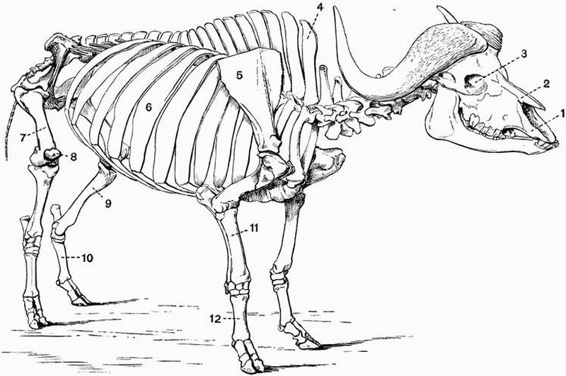
Fig. 101. Skeleton of a Cape Buffalo (Bubalus caffer).
The left scapula is omitted for the sake of clearness × 1/17. (Brit. Mus.)
| 1. premaxillae. | 7. femur. |
| 2. nasal. | 8. patella. |
| 3. orbit. | 9. tibia. |
| 4. neural spine of first thoracic | 10. metatarsals. |
| vertebra. | 11. radius. |
| 5. scapula. | 12. metacarpals. |
| 6. rib. |
In the Rodentia there are generally thirteen pairs of ribs,
which do not present any marked peculiarities.
The Carnivora have thirteen to fifteen pairs of ribs,
whose vertebral portions are slender, nearly straight and
subcylindrical, while their sternal portions are long and
imperfectly ossified (fig. 76, 5). There is nothing that calls
for special remark about the ribs, in either Insectivora or
Chiroptera.
Primates. In Man and the Orang (Simia) there are generally
twelve pairs of ribs; in the Gorilla and Chimpanzee
(Anthropopithecus), and Gibbons (Hylobates), there are thirteen,
in the Cebidae twelve to fifteen, and in the Lemuroidea twelve
to seventeen pairs. The first vertebral rib is shorter than the
others, and the sternal ribs generally remain cartilaginous
throughout life, though in man the first may ossify.
Appendicular Skeleton.
The Pectoral Girdle.
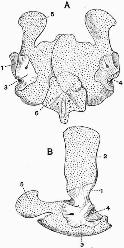
By far the most primitive type of the pectoral or shoulder
girdle is found in the Monotremata. The scapula (fig. 102,
A, 1) is long and recurved, and has only two surfaces, one
corresponding to the prescapular[1] fossa, the other to the postscapular[1]
and subscapular[169] fossae. The coracoid is a short bone
attached above to the scapula and below to the presternum;
it forms a large part of the glenoid cavity. In front of the
coracoid there is a fairly large flattened epicoracoid (fig. 102, 6);
there is also a large T-shaped interclavicle (fig. 102, 4),
which is expanded behind and rests on the presternum. The
clavicles rest on and are firmly united to the anterior border
of the interclavicle. This shoulder girdle differs greatly from
that of any other mammals, and recalls that of some Lacertilia.
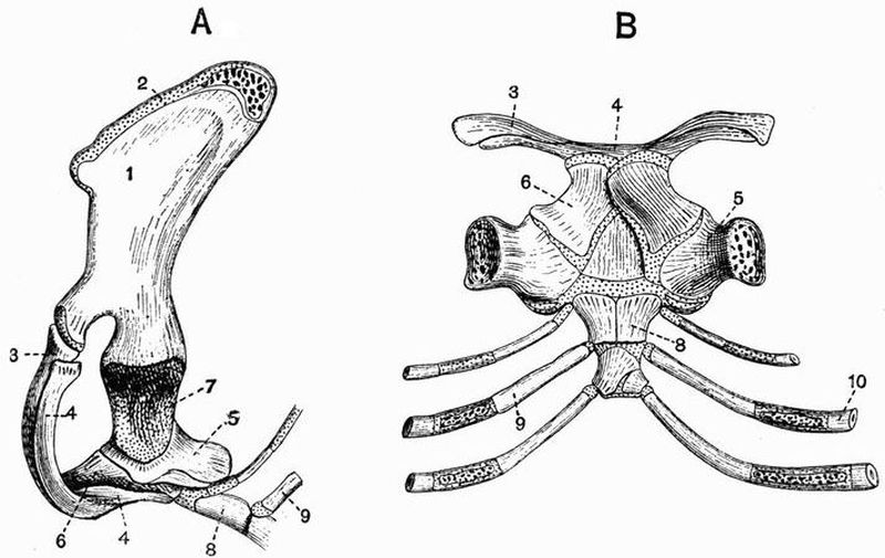
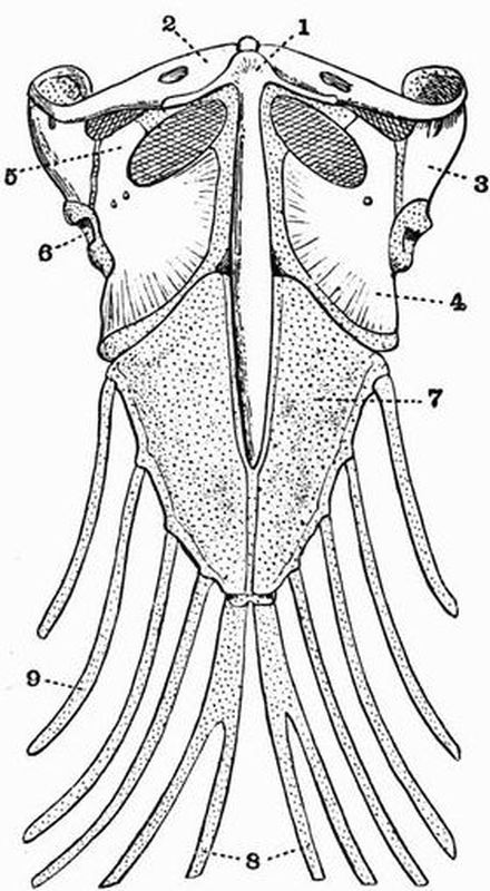
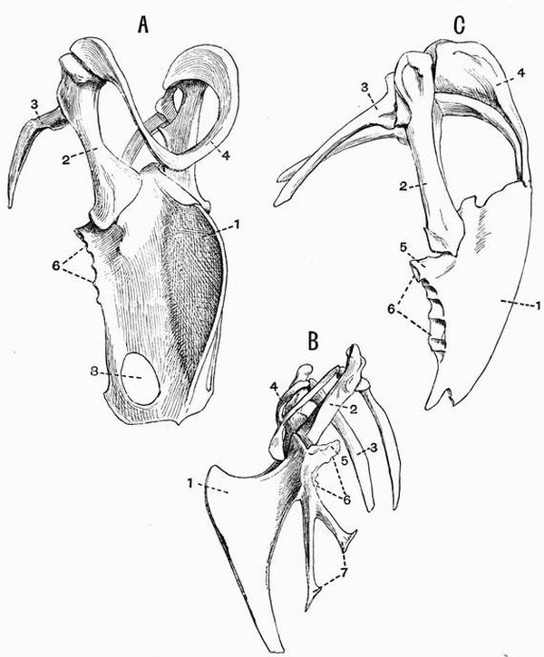
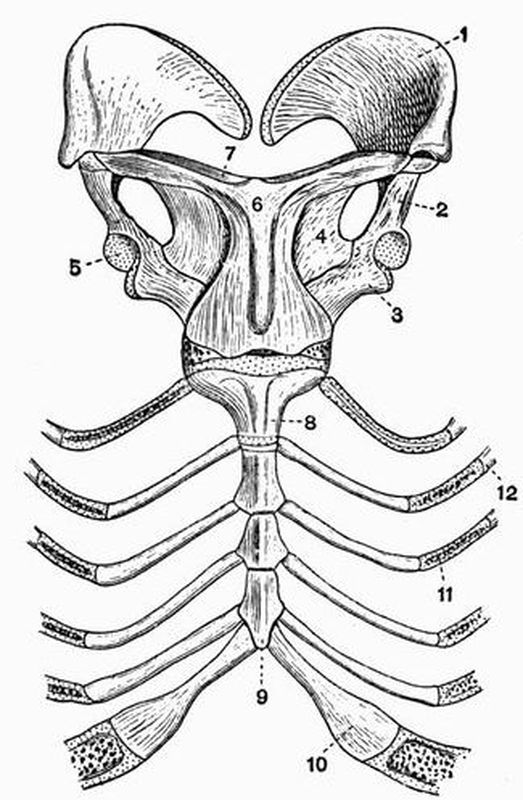
Fig. 102. A, Side view, B, Dorsal view of the shoulder girdle and
part or the sternum of the Spiny Anteater (Echidna aculeata)
× 1. (After Parker.)
| 1. scapula. | 6. epicoracoid. |
| 2. suprascapula. | 7. glenoid cavity. |
| 3. clavicle. | 8. presternum. |
| 4. interclavicle. | 9. second sternal rib. |
| 5. coracoid. | 10. second vertebral rib. |
In Marsupials, as in all mammals except the Monotremes,
the shoulder girdle is much reduced; there are no epicoracoids
and interclavicle, and the coracoid forms simply a small process
on the scapula, ossifying from a centre separate from that
giving rise to the rest of the bone. The scapula has a long
acromion, and a clavicle is always present except in Perameles.
Unossified remains of the precoracoids are found at either end
of the clavicle. The scapula of Notoryctes has a very high
overhanging spine, and there is a second strong ridge running
along the proximal part of the glenoid border.
The shoulder girdle of the Edentata shows some very
curious variations. In Orycteropus the scapula is of very
normal form and the clavicle is well developed. In the Pangolins
and Anteaters the scapula is very broad and rounded;
there is no clavicle in the Pangolins, and generally only a
vestigial one in Anteaters. In Armadillos, Sloths, and Megatheriidae,
the acromion is very long and the clavicle is well
developed. In the Sloths, Megatherium, and Myrmecophaga, a
connection is formed between the coracoid, which is unusually
large, and the coracoid border of the scapula, converting the
coraco-scapula notch into a foramen. In Bradypus the
clavicle is very small, and is attached to the coracoid, which
sometimes forms a distinct bone[170].
In the Sirenia the scapula is somewhat narrow and curved
backwards: the spine, acromion, and coracoid process are
moderately developed, and there is no clavicle.
Cetacea. In nearly all the Odontoceti the scapula is
broad and somewhat fan-shaped; the prescapular fossa is much
reduced, and the acromion and coracoid process form flattened
processes, extending forwards nearly parallel to one another.
Some of the Mystacoceti, such as Balaenoptera, have a broad,
fan-shaped scapula, with a long acromion and coracoid process,
extending parallel to one another. Others, such as Balaena,
have a higher and narrower scapula, with a smaller coracoid
process.
In Ungulata the scapula is always high and rather narrow,
and neither acromion nor coracoid process is ever much developed.
In no adult Ungulate except Typotherium is there any
trace of a clavicle, but a vestigial clavicle has been described
in early embryos of sheep[171].
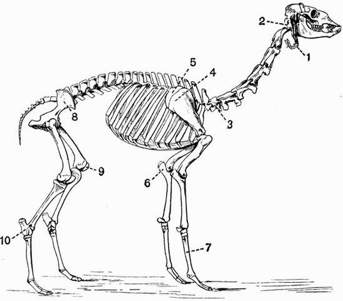
Fig. 103. Skeleton of a Llama (Auchenia glama) × 1/18.
(Brit. Mus.)
| 1. hyoid. | 6. olecranon process of ulna. |
| 2. atlas vertebra. | 7. metacarpals. |
| 3. seventh cervical vertebra. | 8. ilium. |
| 4. scapula. | 9. patella. |
| 5. imperfectly ossified | 10. calcaneum. |
| suprascapula. |
Ungulata vera. In the Ruminantia the suprascapular
region (fig. 103, 5) is very imperfectly ossified, and when this
is removed the upper border of the scapula is very straight
(fig. 101, 5). The spine is prominent, and generally has a
fairly well-marked acromion. In Hippopotamus the acromion
is fairly prominent, but in the other Suina, though the spine
is prominent, the acromion is not developed. The Perissodactyla
have no acromion, but while the Equidae and Hyracotherium
have the scapula long and slender, with the spine
very slightly developed, the other living Perissodactyla have
the spine prominent and strongly bent back at about the
middle of its length.
Subungulata. Typotherium (Toxodontia) differs from all
other known Ungulates in having well-developed clavicles;
its scapula has a strong backwardly-projecting process, much
like that in Rhinoceros.
Phenacodus (Condylarthra), has a curiously rounded
scapula, with the coracoid and suprascapular borders passing
imperceptibly into one another. The scapula resembles that
of a carnivore more than does that of any existing Ungulate.
Procavia has a triangular scapula with a prominent spine
and no acromion; there is a large unossified suprascapular
region.
The scapula in the Proboscidea has a large rounded suprascapular
border and a narrow, slightly concave glenoid border.
The spine is large, and has a prominent process projecting
backwards from about its middle. The spine lies towards
the front end of the scapula, so that the postscapular fossa is
much larger than the prescapular fossa.
In Rodentia the shoulder girdle is of a rather primitive
type. The scapula is generally high and narrow, somewhat
as in Ruminantia; it differs, however, from the Ruminant
scapula in having a high acromion, which is often, as in the
Hares and Rabbits, terminated by a long metacromion. The
development of the clavicle varies, and sometimes it is altogether
absent. It is frequently connected by cartilaginous
bands or ligaments (fig. 104, 7 and 9), on the one hand with
the scapula, and on the other with the sternum. These unossified
bands are remains of the precoracoid. Epicoracoidal
vestiges of the sternal ends of the coracoids (fig. 104, 11) are
also often present.
In the Carnivora vera the scapula is large, and generally
has rather rounded borders. The spine and acromion are well
developed, and the prescapular and postscapular fossae are
nearly equal in size. The coracoid is very small, and the
clavicle is never completely developed, being often absent, as
in the Bears and most of their allies. In the Seals (Phocidae)
the scapula is elongated and curved backwards, and has a very
concave glenoid border. In the Eared Seals (Otariidae) the
scapula is proportionally much larger and wider, the prescapular
fossa being specially large, and being traversed by a
ridge, which converges to meet the spine.
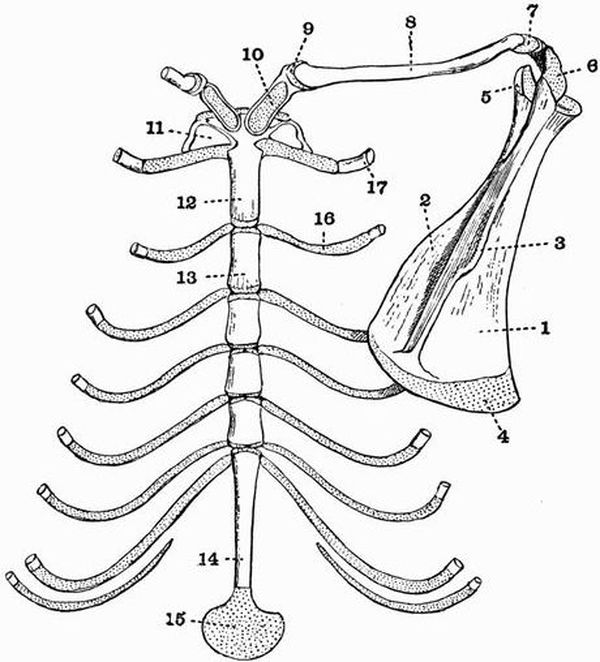
Fig. 104. Dorsal view of the sternum and right half of the
shoulder-girdle of Mus sylvaticus × 4. (After Parker.)
| 1. postscapular fossa. | precoracoid at sternal end of |
| 2. prescapular fossa. | clavicle. |
| 3. spine. | 10. omosternum. |
| 4. suprascapular border | 11. epicoracoid. |
| unossified. | 12. presternum. |
| 5. coracoid process. | 13. first segment of mesosternum. |
| 6. acromion. | 14. xiphisternum. |
| 7. cartilaginous vestige of | 15. cartilaginous termination of |
| precoracoid at scapular end of | xiphisternum. |
| clavicle. | 16. 2nd sternal rib. |
| 8. clavicle. | 17. 1st vertebral rib. |
| 9. cartilaginous vestige of |
In the Insectivora the shoulder girdle is well developed
and, as in Rodents, remains are met with of various parts not
generally seen in mammals. In the Shrews the scapula is
long and narrow, and has a well-marked spine, whose end
bifurcates, forming the acromion and metacromion. The
clavicle is long and slender, and is connected with the sternum
and acromion by vestiges of the precoracoid. Considerable
remains of the sternal end of the coracoid are also found. In
Potamogale, however, there are no clavicles. In the Mole the
shoulder girdle is greatly developed, and of very remarkable
form. The scapula is high and very narrow, with the spine
and acromion very little developed. The other shoulder girdle
element is an irregular bone, which articulates with the
humerus and presternum, and is connected by ligaments with
the scapula. This bone appears to represent both the coracoid
and the clavicle, being formed partly of cartilage bone, partly
of membrane bone.
In the Chiroptera the scapula is large and oval, and has
a moderately high spine and a large acromion. The coracoid
process is well developed and is often forked. The clavicles are
also well developed, and vestiges of the precoracoid and of the
sternal end of the coracoid are often found.
In Primates the clavicle and coracoid process are always
well developed. In Man and the Gorilla the scapula has a
long straight suprascapular border, a well-developed coracoid
process and spine, and a large curved acromion. Vestiges of
the precoracoid occur at each end of the clavicle. The shape
of the scapula varies much in the lower Primates.
The Upper arm and Fore-arm.
In the Monotremata the humerus is short, very broad at
each end and contracted in the middle. The radius and ulna
are stout and of nearly equal size, while the ulna has a greatly
expanded olecranon.
In the Marsupialia the humerus is generally a strong bone,
broad at the distal end and having well marked deltoid and
supinator ridges, which are specially large in Notoryctes. An
ent-epicondylar or supracondylar foramen (fig. 105, 5) is almost
always present except in Notoryctes. The radius and ulna are
always distinct and well developed, and a certain amount of
rotation can take place between them. The ulna of Notoryctes
has an enormous hooked olecranon which causes the bone to
be nearly twice as long as the radius.
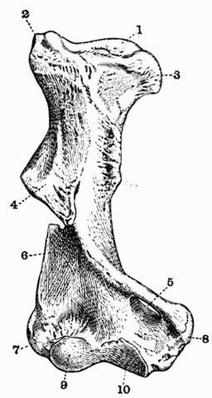
Fig. 105. Anterior surface of the right humerus of a Wombat
(Phascolomys latifrons). (After Owen.)
| 1. head. | 6. supinator ridge. |
| 2. greater tuberosity. | 7. external condyle. |
| 3. lesser tuberosity. | 8. internal condyle. |
| 4. deltoid ridge. | 9. articular surface for radius. |
| 5. ent-epicondylar | 10. articular surface for ulna. |
| (supracondylar) foramen. |
Edentata. The Sloths have long slender arm bones; the
humerus is nearly smooth and has a very large ent-epicondylar
foramen in Choloepus, but not in Bradypus. The radius
and ulna can be rotated on one another to a considerable
extent. The humerus in all other Edentates is very strong and
has the points for the attachment of muscles much developed,
especially in the Armadillos and Megatheriidae. An ent-epicondylar
foramen is found in all living forms. The radius
and ulna are well developed, but are not capable of much
rotation.
In the Sirenia the humerus is well developed and of a
normal character. It is expanded at each end and has a
prominent internal condyle, a small olecranon fossa, and no
ent-epicondylar foramen. In the Dugong and Rhytina there
is a bicipital groove and the tuberosities are distinct, but in
the Manatee there is no bicipital groove, and the tuberosities
coalesce. The radius and ulna are about equally developed
and ankylosed together at both ends.
In the Cetacea the arm bones are very short and thick.
The humerus has a globular head, and a distal end terminated
by two equal flattened surfaces to which the radius and ulna
are united. There is no bicipital groove, and the tuberosities
coalesce. The radius and ulna are flat expanded bones
fixed parallel to one another, but the ulna has a definite olecranon.
Scarcely any movement can take place between
them and the humerus, and in old animals the three bones are
often ankylosed together.
In the Ungulata vera the humerus is stout and rather
short. The great tuberosity is always large and often overhangs
the bicipital groove, it is especially large in Titanotherium
(Brontops). There is never an ent-epicondylar foramen. The
radius is always large at both ends, but the condition of the
ulna is very variable. Sometimes, as in Tapirus, Rhinoceros,
Macrauchenia, Suina and Tragulina, the ulna is well developed,
and quite distinct from the radius; but in most forms, although
complete, it is much reduced distally, and is fused to the
radius. Sometimes, as in the Horse and Giraffe, it is reduced
to the olecranon and to a very slender descending process
which does not nearly reach the carpus. In the Tylopoda,
though the ulna is complete and its distal end is often distinct,
it has coalesced with the radius throughout its whole length;
the olecranon is generally very large.
Subungulata. In the large Condylarthra the humerus
has an ent-epicondylar foramen, and the radius and ulna are
stout bones nearly equal in size.
In Procavia the humerus is rather long, and has a very
prominent greater tuberosity, and a large supra-trochlear fossa,
but no ent-epicondylar foramen.
In the Proboscidea the humerus is marked by a greatly
developed supinator ridge, and is very long, longer than the
radius and ulna. The ulna has a remarkable development,
having its distal end larger than that of the radius, it has
also a larger articular surface for the humerus than has the
radius.
In Rodentia the humerus varies much in its development
according to the animal's mode of life. In the Hares it is long
and straight, with a small distal end, and a slight deltoid ridge.
In the Beaver on the other hand the deltoid and supinator
ridges are considerably developed. There is generally a large
supra-trochlear fossa, but no ent-epicondylar foramen.
Carnivora. In the Carnivora vera the humerus has large
tuberosities, a prominent deltoid ridge and a deep olecranon
fossa. The shaft is generally curved, and an ent-epicondylar
foramen is often found, though not in the Canidae, Hyaenidae,
and Ursidae. The radius and ulna are never united. The
radius (fig. 77, B) has a very similar development throughout
its whole length, while the ulna has a large olecranon (fig.
77, C, 11) and a shaft tapering somewhat towards the distal
end.
In the Pinnipedia the arm bones are very strongly developed.
The humerus has a very prominent deltoid ridge,
and the proximal end of the ulna and distal end of the
radius are much expanded.
In the Insectivora the arm bones are well developed, and
the radius and ulna, though sometimes united, are generally
distinct; as a rule there is an ent-epicondylar foramen, but this
is absent in the Hedgehog. The Mole has an extraordinary
humerus, very short and curved, and much flattened and expanded
at both ends. It articulates both with the scapula
and coraco-clavicle. The ulna has a greatly developed olecranon.
In the Chiroptera both humerus and radius are exceedingly
long and slender; the ulna is reduced to little more than
the proximal end and is fused to the radius. There is no
ent-epicondylar
foramen.
All Primates have the power of pronation and supination
of the fore-arm, by the rotation of the distal end of the radius
round that of the ulna.
In Man and the Anthropoid Apes the humerus is long and
straight, and has a globular head; neither of the tuberosities,
nor the deltoid nor supinator ridges are much developed. The
olecranon fossa is deep and there is no ent-epicondylar foramen.
The radius is curved and has a narrow proximal, and
expanded distal end, the ulna is straighter than the radius
and has the distal end much smaller than the proximal; the
olecranon is not much developed.
In the lower Primates, although the radius and ulna are
always quite separate, the power of pronation and supination
is not nearly so great as in the higher forms. In most of the
Cebidae and Lemurs an ent-epicondylar foramen occurs.
The Manus.
The Manus is divisible into two parts, viz. the carpus or
wrist, and the hand which is composed of the metacarpals and
phalanges. The carpal bones are always modified from their
primitive arrangement, sometimes more, sometimes less. One
modification however is always found in mammals, viz. the
union of carpalia, 4 and 5 to form the unciform bone. Two
sesamoid bones are commonly developed, one on each side of
the carpus, the pisiform or one on the ulnar side being much
the larger and more constant: it has been suggested that
these represent respectively vestiges of a prepollex and a post-minimus
digit[172].
One or more of the five digits commonly present may be
lost, and sometimes all are lost except the third. The terminal
or ungual phalanges of the digits are commonly specially
modified to support nails, claws, or hoofs. There are as a rule
two small sesamoid bones developed on the ventral or flexor
side of the metacarpo-phalangeal articulations, and sometimes
similar bones occur on the dorsal or extensor side.
Monotremata. In Echidna the carpus is broad, the scaphoid
and lunar are united and there is no centrale. The
pisiform is large and several other sesamoid bones occur. Each
of the five digits is terminated by a large ungual phalanx. In
Ornithorhynchus the manus is more slender, but the general
arrangement is the same as in Echidna.
Marsupialia. The carpus has no centrale and the lunar
is generally small or absent. Five digits are almost always
present. In Choeropus however the only two functional digits
are the second and third, which have very long closely apposed
metacarpals; the fourth digit is vestigial, but has the normal
number of phalanges, while the first and fifth are absent. The
manus in Notoryctes is extraordinarily modified, the scaphoid
and all the distal carpalia are apparently fused, the first,
second, and fifth digits are very small, the third and fourth,
though having only one phalanx apiece, bear each an enormous
claw. Lying on and obscuring the ventral surface of the manus
is a large bone, probably a sesamoid.
Among the Edentata there is a great diversity in the
structure of the manus, the centrale is however always wanting,
and except in Manis the scaphoid and lunar are distinct.
In the Sloths the manus is very long, narrow, and curved,
and terminated by two or three long hooked claws, borne
by the second and third, or the second, third and fourth
digits. The fifth digit is absent, and the fourth is represented
only by a small metacarpal. In the Anteaters the third digit
is greatly developed and bears a long hooked claw. In Myrmecophaga
all five digits are fairly well though irregularly
developed, in Cycloturus the first, fourth, and fifth, are vestigial.
In the Armadillos the manus is broad, and has strongly
developed ungual phalanges. The digits, though almost always
five in number, vary much in their relative arrangement. In
Dasypus they are regular, but are remarkably irregular in
Priodon. The pollex is absent in Glyptodonts and in Megatherium.
In Megatherium the fifth digit is clawless while the
second, third, and fourth bear enormous claws. In the Manidae
the scaphoid and lunar are united; five digits are present,
the third and fourth being very large, and all being terminated
by deeply cleft ungual phalanges. In Orycteropus the pollex is
absent, while the other digits are terminated by pointed ungual
phalanges.
In Sirenia the general structure of the manus is quite of
the ordinary mammalian type. In Manatus most of the bones
of the carpus are distinct, but in Halicore many, especially
those of the distal row, have coalesced. The digits are always
five in number and have the normal number of flattened
phalanges.
In the Cetacea, on the other hand, the manus is much
modified by the fact that the number of phalanges may be
greatly increased above the normal number of three, thirteen
or fourteen sometimes occurring in each digit. These are
believed to be duplicated epiphyses. In the Mystacoceti the
manus remains largely cartilaginous, in the Odontoceti it is
better ossified, and the phalanges commonly have epiphyses at
both ends. In Physeter the carpal bones also have epiphyses.
The carpus generally consists of six bones arranged in two
rows of three each. Five digits are generally present, but
sometimes as in Balaenoptera musculus, there are four, the
third being suppressed. Their relative development varies
much. The Sperm Whale which till recently was placed in the
entrance hall of the Natural History Museum at South Kensington
has one phalanx to the first digit, four to the second,
five to the third, four to the fourth, and three to the fifth.
Generally the manus is short and broad, but sometimes, as in
Globicephalus, it is much elongated owing to the great development
of the second and third digits.
Ungulata[173]. The manus of the members of this great
order is of very great classificatory and morphological importance.
All the members agree in having the scaphoid and
lunar distinct, and in almost every case the ends of the digits
are either encased in hoofs or provided with broad flat nails.
It is by means of characters derived from the manus and pes
that the group is subdivided into the Ungulata vera and
the Subungulata.
In the Ungulata vera the manus is never plantigrade,
and there are not more than four digits, the pollex being almost
always completely suppressed: in Cotylops among extinct Artiodactyla
however a vestigial pollex is found. The centrale is
absent, and the magnum articulates freely with the scaphoid,
and is separated from the cuneiform by the unciform and lunar.
All the bones of the carpus interlock strongly, and the axis of
the third digit passes through the magnum and between the
scaphoid and lunar.
There is a very strong distinction between the manus of
the suborders Artiodactyla and Perissodactyla. In the Artiodactyla
the axis of the manus passes between the third and
fourth digits, which are almost equally developed and, except in
the Hippopotami and some extinct forms such as Anoplotherium,
have their ungual phalanges flattened on their contiguous
surfaces.
In all Artiodactyla the third and fourth digits are large, but
a gradual reduction in the second and fifth can be well traced.
Thus in the Suina the second and fifth digits, though smaller
than the third and fourth, are well developed and all four
metacarpals are distinct. In the Tragulina too all four metacarpals
are developed, and in Dorcatherium the third and fourth
commonly remain distinct as in the Suina. In the other Artiodactyla
however the third and fourth metacarpals are almost
always united, though indications of their separate origin
remain. In some Ruminantia, such as many Deer, the second
and fifth digits are reduced to minute splint bones attached
to the proximal end of the fused third and fourth metacarpals,
and to small hoof-bearing phalanges, sometimes attached to
splint-like distal vestiges of the metacarpals, sometimes altogether
unconnected with any other skeletal structures. In
some other Ruminants, such as the Sheep and Oxen, the only
remnants of the second and fifth digits are nodules of bone
supporting the hoofs, and in others, such as the Giraffe, Anoplotherium
commune, some Antelopes and the Tylopoda, all
traces of these digits have disappeared. The Camels differ
from all living Ungulata vera in not having the distal phalanges
completely encased in hoofs, and from all except the
Hippopotami in placing a considerable amount of the manus
on the ground in walking.
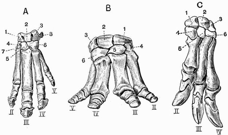
Fig. 106. Manus of Perissodactyles.
A. Left Manus of Tapirus. (After von Zittel.)
B. Right Manus of Titanotherium. (After Marsh.)
C. Left Manus of Chalicotherium gigantium. (After Gervais.)
| 1. scaphoid. | 6. unciform. |
| 2. lunar. | 7. trapezium. |
| 3. cuneiform. | II, III, IV, V. second, third, |
| 4. trapezoid. | fourth and fifth digits. |
| 5. magnum. |
While the manus of the Artiodactyla is symmetrical about
a line drawn between the third and fourth digits, that of the
Perissodactyla is symmetrical about a line drawn through the
middle of the third digit, which is larger than the others and
has its ungual phalanx evenly rounded and symmetrical in
itself. The most reduced manus in the whole of the mammalia
is found in the Horse and its allies, in which the third
digit, terminated by a very wide ungual phalanx, is the only
one functional. Small splint bones representing the second and
fourth metacarpals are attached to the upper part of the third
metacarpal. In Hipparion[174] and other early horse-like animals
the second and fourth digits, though very small and functionless,
are complete and are terminated by small hoofs. In Rhinoceros
the second and fourth digits are equally developed
and nearly as large as the third, and reach the ground in
walking, a vestige of the fifth is also present. In the Tapir
(fig. 106, A) and Hyracotherium the fifth digit is fully developed
but is scarcely functional. In Titanotherium (Brontops)
(fig. 106, B) it is nearly as well developed as any of the
others, and there is little or no difference between the relative
development of the third and fourth digits.
The Chalicotheriidae[175], though distinctly Perissodactyles in
various respects such as their cervical vertebrae and teeth,
differ not only from all other Perissodactyles, but from almost
all other Ungulates, in the very abnormal character of their
manus. For while the carpus and metacarpus are like those
of ordinary Perissodactyles, the phalanges resemble those of
Edentates, each second phalanx having a strongly developed
trochlea, and each distal one being curved, pointed and deeply
cleft at its termination (fig. 106, C).
The Macraucheniidae, while agreeing with Perissodactyles
in having only three digits, with the limb symmetrical about
a line drawn through the middle of the third, have a carpus
which approaches closely to the subungulate condition, the
magnum articulating regularly with the lunar, and only to a
slight extent with the scaphoid.
In the Subungulata the manus sometimes has five functional
digits, and a considerable part of it rests on the ground
in walking. The bones of the carpus retain their primitive
relation to one another, the magnum articulating with the
lunar, but not with the scaphoid. This character does not
however hold in the Toxodontia, for in most of the animals
belonging to this group the magnum does articulate with the
scaphoid. The corner of the scaphoid just reaches the magnum
also in Amblypoda.
As far as is known the Toxodontia generally have three,
sometimes five digits to the manus, and the third is symmetrical
in itself—a Perissodactyloid feature.
In Phenacodus (fig. 107, B) (Condylarthra) all five digits
are well developed, the pollex being the smallest. The carpal
bones retain their primitive arrangement, the magnum articulating
with the lunar and not with the scaphoid. There is no
separate centrale.
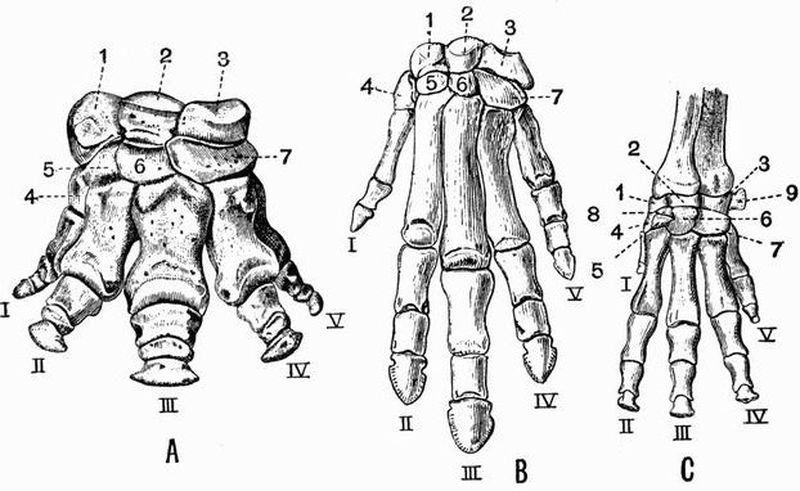
Fig. 107. Left manus of
A. Coryphodon hamatus. (After Marsh.) × 1/5.
B. Phenacodus primaevus. (After Cope.) × 1/3.
C. Procavia (Dendrohyrax) arboreus. (After von Zittel.) × 6/7.
| 1. scaphoid. | 7. unciform. |
| 2. lunar. | 8. centrale. |
| 3. cuneiform. | 9. pisiform. |
| 4. trapezium. | I, II, III, IV, V. first, second, |
| 5. trapezoid. | third, fourth and fifth |
| 6. magnum. | digits respectively. |
In the Hyracoidea (fig. 107, C) the manus is very similar
to that in Phenacodus, but a centrale is present and the pollex
is much reduced.
The manus of the Amblypoda, such as Coryphodon (fig.
107, A) and Uintatherium, is short and broad, with five well
developed digits and large carpal bones. The carpals however
interlock to a slight extent, and the corner of the magnum
reaches the scaphoid.
In the Proboscidea the manus is very short and broad, with
large somewhat cubical carpals which articulate by very flat
surfaces and do not interlock at all. All five digits are present,
and none of them are much reduced in size. The manus in
Proboscidea and in Coryphodon is subplantigrade.
In the Tillodontia the manus is plantigrade and has pointed
ungual phalanges, in this respect approaching the Carnivora.
It differs however from that of all living Carnivora in having
the scaphoid and lunar distinct.
In Rodentia the manus nearly always has five digits with
the normal number of phalanges: the pollex may however be
very small as in the Rabbit, or absent as sometimes in the
Capybara. The scaphoid and lunar are generally united, and
a centrale may be present or absent. In Pedetes caffer the
radial sesamoid is double and the distal bone bears a nail-like
horny covering. In Bathyergus the pisiform is double. It is
upon these facts that the contention for the former existence
of prehallux and post-minimus digits has partly been based.
In living Carnivora the scaphoid, lunar and centrale are
always united, forming a single bone. All five digits are
present, but as a rule in Carnivora vera the pollex is small, and in
Hyaena is represented only by a small metacarpal. Sometimes,
as in Cats and Dogs, the manus is digitigrade, sometimes, as in
Bears, plantigrade. The ungual phalanges are large and pointed,
and in forms like the Cats, whose claws are retractile, they
can be folded back into a deep hollow on the ulnar side of the
middle phalanx; a small radial sesamoid is often present.
In Pinnipedia the manus is large and flat and the digits
are terminated by ungual phalanges which are blunt (sea lions
and walrus), or slightly curved and pointed (seals). The pollex
is nearly or quite as long as the second digit, and as a rule
the digits then successively diminish in size.
The Creodonta differ from living Carnivora in the fact that
the scaphoid and lunar are usually separate.
In Insectivora the scaphoid and lunar are sometimes
united, sometimes separate, and a separate centrale is usually
present. There are generally five digits, but sometimes the
pollex is absent. In the Mole the manus is greatly developed
and considerably modified. It is very wide, its breadth being
increased by the great development of the radial sesamoid
which is very large and sickle-shaped. The ungual phalanges
are also large and are cleft at their extremities.
In the Chiroptera the manus is greatly modified for the
purpose of flight. The pollex is short and is armed with a
rather large curved claw, the other digits are enormously
elongated, the elongation in the case of the Insectivorous bats
being mainly due to the metacarpals, and in the Frugivorous
bats to the phalanges. In the Frugivorous bats the second
digit is clawed as well as the pollex, in other bats this claw is
always absent, and so is often the ungual phalanx, the middle
phalanx then tapering gradually to its termination.
In Primates as a rule the manus is moderately short and
wide. The carpus has the scaphoid and lunar distinct, and
generally also the centrale; sometimes however, as in Man,
the Gorilla, Chimpanzee, and some Lemurs, the centrale has
apparently fused with the scaphoid. There are almost always
five well-developed digits, but in the genera Colobus and Ateles
the pollex is vestigial.
The magnum in man is the largest bone of the carpus. The
pisiform also is well developed, but there is no radial sesamoid.
In Man, the Gorilla, Chimpanzee, and Orang, the carpus articulates
only with the radius, in most Primates it articulates
also with the ulna. The third digit of the Aye-Aye (Chiromys)
is remarkable for its extreme slenderness.
The Pelvic Girdle.
The pelvic girdle in all mammals except the Sirenia and
Cetacea consists of two halves, usually united with one another
at the symphysis in the mid-ventral line, and connected near
their upper ends, with the sacral vertebrae. Each half forms
one of the innominate bones, and includes at least three separate
elements, a dorsal bone, the ilium, and two ventral bones, the
ischium and pubis. Very often a fourth pelvic element, the
acetabular or cotyloid bone, occurs.
In the Monotremata the pelvis is short and broad, and the
pubes and ischia meet in a long symphysis. The acetabulum
is perforated in Echidna as in birds, but not in Ornithorhynchus.
A pair of elongated slender bones project forwards from the
edge of the pubes near the symphysis; these are sesamoid bones
formed by ossifications in the tendons of the external oblique
abdominal muscles, and are generally called marsupial bones.
In the Marsupialia the ilia are generally very simple,
straight, and narrow, while the pubes and ischia are well developed
and meet in a long symphysis. Marsupial bones are
nearly always prominent, but are not developed in Thylacinus
or Notoryctes. The ischium often has a well-marked tuberosity
and in Kangaroos the pubis bears a prominent pectineal
process on its anterior border close to the acetabulum. The
pelvis in Notoryctes differs much from that in all other Marsupials,
the ilium and ischium being ankylosed with six vertebrae
in a manner comparable to that of many Edentates.
In the Edentata the pelvis is generally well developed, but
the symphysis is very short. In the Sloths the pelvis is rather
weak and slender, the obturator foramina are very large and
the ischia do not meet in a symphysis. In the Megatheriidae
the pelvis is exceedingly wide and massive, and is firmly
ankylosed with a number of vertebrae. In the Armadillos,
Glyptodonts, Anteaters, and Pangolins it is much developed
and firmly united to the vertebral column by both the ilia and
the ischia. In Orycteropus however the ischium does not
become united to the vertebral column, and the pubis generally
has a strongly developed pectineal process.
In the Sirenia the pelvis is quite vestigial. In the Dugong
it consists on each side of two slender bones, one of which
represents the ilium and the other the ischium and pubis; the
two bones are placed end to end and are commonly fused
together. The ilium is attached by ligament to the transverse
process of one of the vertebrae. In the Manatee each half
of the pelvis is represented by a triangular bone connected by
ligaments with its fellow and with the vertebral column. In
neither Manatee nor Dugong is there any trace of an acetabulum
but one can be made out in Halitherium.
In the Cetacea the pelvis is even more vestigial than in
the Sirenia, consisting simply of a pair of small straight bones
which probably represent the ischia, and lie parallel to and
below the vertebral column at the point where the development
of chevron bones commences.
In Ungulata vera the pelvis is generally rather long and
narrow. The ilium is flattened and expanded in front (fig.
103, 8), but becomes much narrower and more cylindrical
before reaching the acetabulum. Both pubis and ischium
contribute to the symphysis which is often very long. The
ischia are large and have prominent tuberosities, especially in
Artiodactyles. In most Ruminantia there is a deep depression,
the supra-acetabular fossa above the acetabulum, but this is
not found in the Suina or Tylopoda.
Subungulata. In Procavia the pelvis is long and narrow,
and bears resemblance to that in Artiodactyles.
The Proboscidea have a very large pelvis set nearly at
right angles to the vertebral column; the ilium is very wide,
having expanded iliac and gluteal surfaces, and a narrow
sacral[176] surface. The pubes and ischia are rather small, but
both meet their fellows in the symphysis. Uintatherium (suborder
Amblypoda) also has a large and vertically placed pelvis
(fig. 108) with a much expanded ilium. The pelvis however
differs from that of the Proboscidea in the fact that the ischia
do not meet in a ventral symphysis.
In many Rodentia the ilia have their gluteal, iliac, and
sacral surfaces of nearly equal extent; in the Hares, however,
the gluteal and iliac surfaces are confluent. The pubes and
ischia are always well developed and sometimes, as in the
Hares, the acetabular bone also. In these animals the pubis
does not take part in the formation of the acetabulum, and
the ischium bears on its outer side a well-marked ischial
tuberosity.
In the Carnivora the pelvis is long and narrow. The
iliac surfaces (fig. 78, A, 5) are very small and the sacral large;
the crest or supra-iliac border is formed by the union of the
sacral and gluteal surfaces. The symphysis is long and includes
part of both pubis and ischium. The ischial tuberosity (fig.
78, A, 10) is often well marked, and sometimes as in Viverra
the acetabular bone is distinct. In the Pinnipedia the pelvic
symphysis is little developed, or sometimes not developed at
all, and the obturator foramina are remarkably large.
In some Insectivora such as Galeopithecus, there is a long
pelvic symphysis, in others such as Erinaceus and Centetes, it is
very short, in others again such as Talpa and Sorex, there is no
pelvic symphysis. The acetabular bone is exceptionally large
in Talpa and Sorex.
In Chiroptera the pelvis is small and narrow, and in the
great majority of cases the two halves do not meet in a ventral
symphysis. The pubis has a strongly developed pectineal
process, which occasionally unites with a process from the
ilium enclosing a large pre-acetabular foramen.
Primates. In Man and the Anthropoid Apes the pelvis is
very large and wide, and the ilium has much expanded iliac
and gluteal surfaces. The symphysis is rather short and formed
by the pubis alone. The acetabulum is deep and the obturator
foramen large, and there is frequently a well-marked ischial
tuberosity. In the lower Anthropoidea the ilium is long and
narrow and has a small iliac surface. The ischial tuberosities
are large in the old world monkeys.
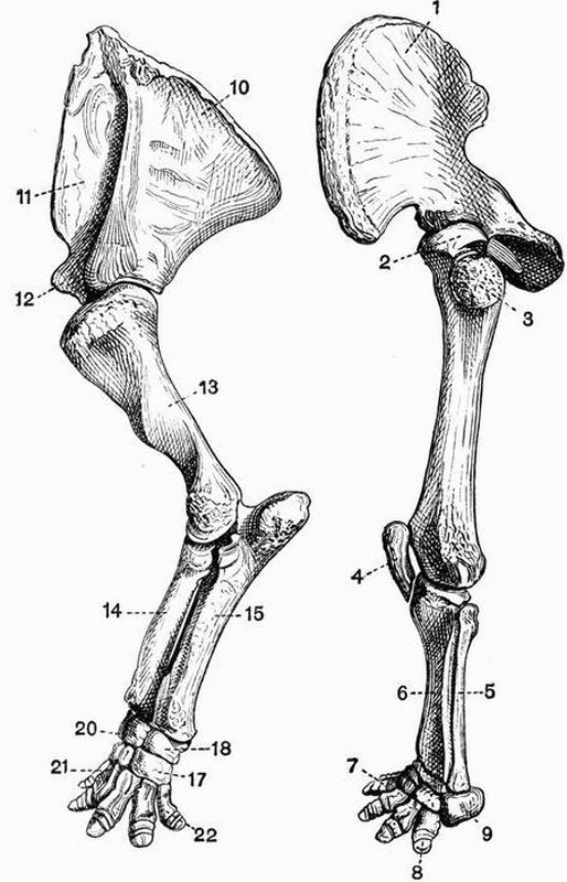
Fig. 108. Left anterior and posterior limb and limb girdle of
Uintatherium mirabile. The anterior limb is to the left, the posterior
to the right × 1/10. (From casts, Brit. Mus.)
| 1. ilium. | 11. prescapular fossa. |
| 2. head of femur. | 12. coracoid process. |
| 3. great trochanter. | 13. humerus. |
| 4. patella. | 14. radius. |
| 5. fibula. | 15. ulna. |
| 6. tibia. | 17. unciform. |
| 7. second digit of pes. | 18. cuneiform. |
| 8. ungual phalanx of fifth | 20. lunar. |
| digit of pes. | 21. first metacarpal. |
| 9. calcaneum. | 22. fifth metacarpal. |
| 10. postscapular fossa. |
The Thigh and Shin.
In the Monotremata the femur is short, rather narrow in
the middle, and expanded at each end. The great and lesser
trochanters are large and about equally developed, but there is
no third trochanter. The fibula is very large and is expanded
at its proximal end, forming a flattened plate much resembling
an olecranon. The patella is well developed.
In the Marsupialia there is no third trochanter to the
femur, the fibula is well developed but not the patella as a
general rule. Notoryctes has a femur with a prominent ridge
extending some little way down the shaft from the great trochanter;
the tibia has a remarkably developed crest, and the
fibula has its proximal end much expanded and perforated;
there is an irregularly shaped patella closely connected with
the proximal end of the tibia.
Edentata. In the Sloths the leg bones are all long and
slender. The femur has no third trochanter, and the fibula is
complete and nearly equal in size to the tibia. In the Megatheriidae
the leg bones are extraordinarily massive, the circumference
of the shaft of the femur in Megatherium equalling
or exceeding the length of the bone. There is no third trochanter
in Megatherium. In most of the remaining Edentata
the leg bones are strongly developed. The femur in the Armadillos
and Aard Varks has a strong third trochanter, and the
tibia and fibula are both large and are commonly ankylosed
together at either end. The limb bones are very massive also
in the Glyptodonts.
Sirenia. In no living Sirenian is there any trace of a
hind limb, but in Halitherium a vestigial femur is found,
which articulates with the pelvis by a definite acetabulum.
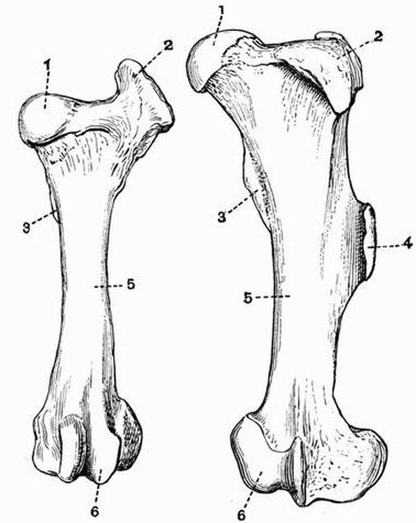
Fig. 109. Left femur of an Ox (Bos taurus) (to the left) and of
a Sumatran Rhinoceros (R. sumatrensis) (to the right). × 1/6.
(Camb. Mus.)
| 1. head. | 4. third trochanter. |
| 2. great trochanter. | 5. shaft. |
| 3. lesser trochanter. | 6. condyles. |
In the Mystacoceti among the Cetacea small nodules of
bone or cartilage occur connected with the vestigial pelvis,
and may represent the femur and tibia. No trace of the
skeleton of the hind limb is known in the Odontoceti.
In the Ungulata vera the femur is noticeable for the size
of the great trochanter (fig. 109, 2); there is no definitely
constricted neck separating the head from the rest of the
bone, and the lesser trochanter (fig. 109, 3) is not very prominent.
All Perissodactyles except the Chalicotheriidae show
a strongly marked third trochanter, but this is absent in all
known Artiodactyles. The development of the fibula in general
corresponds to that of the ulna. In Rhinoceros, Macrauchenia,
Tapirus and the Suina it is distinct and fairly well developed;
in the Tragulina on the other hand it is vestigial, being reduced
to the proximal end only. In the Ruminantia and Tylopoda
also, it is much reduced forming merely a small bone attached
to the distal end of the tibia, sometimes, as in the Red deer
a slender vestige of the proximal end also is preserved quite
detached from the distal portion; in the Horse this proximal
portion is all that there is found of the fibula. The progressive
diminution of the fibula can be well seen in the series of forms
that are regarded as the ancestors of the Horse. The patella
of the Ungulata vera is well ossified, but fabellae[177] are not
usually found.
Subungulata. Of the Toxodontia, Toxodon has no third
trochanter while Typotherium and Astrapotherium have one.
In the Condylarthra the femur has well-marked lesser and
third trochanters, and the fibula and patella are well developed.
In the Hyracoidea there is a slight ridge on the femur in the
place of the third trochanter, the fibula is complete, but is
generally fused to the tibia at its proximal end.
Of the Amblypoda, Coryphodon has a third trochanter, but
Uintatherium has none; in this respect, in the vertical position
and general appearance (fig. 108) of the limb, and in the
articulation of the fibula with the calcaneum, the leg of
Uintatherium closely approaches that of the Proboscidea.
In the Proboscidea the femur is very long and straight,
the development of trochanters is slight, and the fibula though
slender is complete and articulates with the calcaneum.
A third trochanter is found in the Tillodontia.
In Rodentia the femur is variable, the great trochanter is
generally large and so sometimes is the third as in the Hares.
In most Rodents as in the Beaver the fibula is distinct, sometimes
as in the Hares it is united distally with the tibia. The
patella is well developed, and so too are the fabellae as a
general rule.
Carnivora. In the Carnivora vera the femur (fig. 79, A)
is generally rather straight and slender, and has a very distinct
head. The fibula (fig. 79, C) is always distinct and there is
generally a considerable interval between it and the tibia.
Fabellae (fig. 79, 7) are commonly present.
In the Pinnipedia the femur is short, broad and flattened,
having a prominent great trochanter. The fibula is nearly as
large as the tibia, and the two bones are generally ankylosed
together at their proximal ends.
The Creodonta differ from all living Carnivores in having
a femur with a third trochanter.
In the Insectivora a third trochanter is sometimes
developed. The fibula is sometimes distinct, sometimes fused
distally with the tibia, thus differing from that of a Carnivore.
In Chiroptera the femur is straight, slender and rather
short, with a small but well-developed head. The fibula may
be well developed or quite vestigial or absent. Owing to the
connection of the hind limb with the wing membrane the knee
joint is directed backwards.
In Primates the femur is rather long and slender, having
a nearly spherical head and large great trochanter. The tibia
and fibula are always distinct and well developed. Fabellae
are not found in the highest forms but are generally present
in the others.
The Pes.
The skeleton of the pes is in most respects a counterpart
of that of the manus. Just as in the manus if one digit is
absent it is the pollex, so in the pes it is the hallux. But
while in the manus the third digit is always well developed,
however much the limb may be modified, in the pes any of the
digits may be lost. In all mammals the tibiale and intermedium
fuse to form the astragalus, and the fourth and fifth
tarsalia to form the cuboid. Sesamoid bones are considerably
developed. In almost every case the phalanges and first metatarsal
have epiphyses only on their proximal ends, while the
remaining four metatarsals have epiphyses only on their distal
ends.
In the Monotremata all the usual tarsal bones are distinct,
and the five digits have the normal number of phalanges.
Several sesamoid bones are developed, the most important one,
found only in the male, being articulated to the tibia and bearing
the curious horny spur. The ungual phalanges of the pes like
those of the manus, are deeply cleft at their extremities. In
the Echidnidae the pes is turned outwards and backwards in
walking.
In the Marsupialia the pes is subject to great modifications,
but in every case the seven usual tarsal bones are distinct.
In the Didelphyidae the foot is broad, all five digits are
well developed, and the hallux is opposable to the others.
In the Dasyuridae the foot is narrow, and the hallux may be
very small, or as in Thylacinus completely absent. In Notoryctes
the pes is much less abnormal than the manus, and
all five digits have the usual number of phalanges. The fifth
metatarsal has a curious projecting process, and there is a
large sesamoid above the hallux. In the Wombats (Phascolomyidae)
the foot is short and broad, the digits are all distinct,
and the hallux is divaricated from the others.
In the remaining marsupials the second and third metacarpals
and digits are very slender, and are enclosed within a
common integument. This condition is known as syndactylism,
and its effect is to produce the appearance of one toe with two
claws. In the Kangaroos (Macropodidae) the pes is very long
and narrow, owing to the elongation of the metacarpals. The
fourth digit is greatly developed, the fifth moderately so,
while the hallux is absent, and the second and third digits
are very small. The Peramelidae have the foot constructed
on the same plan as in the Kangaroos, and in one genus
Choeropus the same type of foot is carried to a greater
extreme than even in the Kangaroos. Thus the fourth digit
is enormously developed, the second and third are small, and
the fifth smaller still, while the hallux is absent. In the
Phalangers and Koalas though the second and third toes are
very slender, the hallux is well developed and opposable.
Edentata. In the Sloths the pes much resembles the
manus, being long and narrow, but in both genera the second,
third and fourth digits are well developed. Most of the other
Edentates have a but little modified pes with the normal
number of tarsal bones and the complete series of digits. In
Cycloturus however the hallux is vestigial and it is absent
in Glyptodonts. Megatherium has a greatly modified pes, the
hallux is absent, and the second digit vestigial, while the
third is very large, having an enormous ungual phalanx. The
calcaneum too is abnormally large.
No trace of the pes occurs in either Sirenia or Cetacea.
In the Ungulata the pes like the manus is subject to
much variation and is of great morphological importance.
In the Ungulata vera the pes is never plantigrade and
never has more than four digits, the hallux being absent.
The cuboid always articulates with the astragalus, and the
tarsal bones strongly interlock. As was the case also with the
manus, the pes is formed on two well-marked types characteristic
respectively of the Artiodactyla and Perissodactyla.
Artiodactyla. Just as in the manus, the third and
fourth digits are well and subequally developed; their ungual
phalanges have the contiguous sides flat, and the axis of the limb
passes between them, and between the cuboid and navicular.
The astragalus has both the proximal and distal surfaces
pulley-like, and articulates with the navicular and cuboid by
two facets of nearly equal size. The calcaneum articulates
with the lower end of the fibula if that bone is fully developed.
In the Suina four toes are developed, and though in the
Peccaries the third and fourth metatarsals are united, they
are all distinct in most members of the group, as are all
the tarsal bones. In the Hippopotami the four digits are of
approximately equal size, and the middle ones do not have
the contiguous faces of their ungual phalanges flattened.
In the Tragulina the cuboid, navicular, and two outer cuneiforms
are united forming a single bone; all four metatarsals
are complete and the two middle ones are united. In the
Tylopoda and Anoplotherium commune only the third and
fourth digits are developed, their metatarsals are free distally,
but are elsewhere united. In the Ruminantia the cuboid and
navicular are always united and so are the second and third
cuneiforms, while in Cervulus all four bones are united
together. The third and fourth metatarsals in Ruminants
are always united in the same way as are the third and fourth
metacarpals, while the second and fifth are always wanting.
In Deer the second and fifth digits are usually each represented
by three small phalanges, but in the Giraffe and most
Bovidae the bones of these digits are wanting.
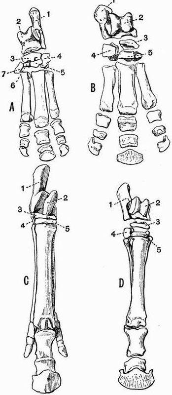
Fig. 110. A. Left pes of a Tapir (Tapirus americanus). × 1/6.
B. Right pes of a Rhinoceros (R. sumatrensis). × 1/8.
C. (Cast of) right pes of Hipparion gracile. × 1/7.
D. Right pes of a Horse (Equus caballus). × 1/10. (All Camb. Mus.)
| 1. calcaneum. | 5. external cuneiform. |
| 2. astragalus. | 6. middle cuneiform. |
| 3. navicular. | 7. internal cuneiform. |
| 4. cuboid. |
In the Perissodactyla the pes like the manus is symmetrical
about a line drawn through the third digit; this line
when continued passes through the external cuneiform, navicular
and astragalus. The astragalus has its distal portion
abruptly truncated, and the facet by which it articulates with
the cuboid is much smaller than that by which it articulates
with the navicular. The calcaneum does not articulate with
the fibula. The tarsus in Macrauchenia like the carpus
differs from that of other Perissodactyles and resembles that
of Subungulates in having the bones arranged in lines with
little or no interlocking. The calcaneum resembles that of
Artiodactyles in having a small facet for articulation with the
fibula. Tapirus (fig. 110, A), Rhinoceros (fig. 110, B) and
Titanotherium have a short and broad foot with the usual
tarsal bones and three well-developed digits,—a number never
exceeded by any Perissodactyle. From this tridactylate limb
a series of stages is exhibited by various extinct forms leading
gradually to the condition met with in the Horse (fig. 110, D)
in which the third toe is greatly developed, while the second
and fourth are reduced to slender metatarsals attached to the
proximal half of the third metatarsal.
In Chalicotherium and Agriochoerus the pes has the same
abnormal characters as the manus, the digits being clawed and
the ungual phalanges in Chalicotherium deeply cleft.
In the Subungulata the pes is sometimes plantigrade and
pentedactylate, the cuboid sometimes does not articulate with
the astragalus, and the tarsal bones sometimes do not interlock.
In Typotherium (Toxodontia) the hallux is absent and the
other four digits are well developed; in Toxodon and Nesodon
the pes is tridactylate. The tarsal bones have the regular Subungulate
arrangement, the cuboid not articulating with the
astragalus. The calcaneum articulates with the fibula as in
Artiodactyles. The astragalus in most forms, but not in
Astrapotherium, resembles that of the Ungulata vera in having
a grooved proximal surface.
In Phenacodus (Condylarthra) the tarsus is very little
modified, five digits are present, the first and fifth being small
and not reaching the ground.
In Procavia only the three middle digits are present with
a vestige of the fifth metacarpal.
In the Amblypoda the pes (fig. 108) is very short and
broad, all five digits are functional, and at any rate in
Coryphodon plantigrade, the hallux being the smallest. The
astragalus is very flat, and the tarsals interlock to a slight
extent, the cuboid articulating with both calcaneum and
astragalus.
The pes in the Proboscidea much resembles that in the
Amblypoda, but differs in that the astragalus does not articulate
with the cuboid, the tarsals not interlocking at all.
In the Rodentia the structure of the foot is very variable.
In Beavers the foot is very large, all five digits being well
developed; the fifth metatarsal articulates with the outer side
of the fourth metatarsal, and not with the cuboid, and there
is a large sesamoid bone on the tibial side of the tarsus. In
the Rats, Porcupines and Squirrels, there are five digits, in the
Hares only four, and in the Capybara and some of its allies
only three. In the Jerboa (Dipus) a curious condition of the
pes is met with, as it consists of three very long metatarsals
fused together and bearing three short toes, each formed of
three phalanges. Lophiomys differs from all other Rodents
in having the hallux opposable.
Carnivora. In the Carnivora vera the pes is regular and
shows little deviation from the normal condition. All the
usual tarsal bones are present, but sometimes as in the Dogs,
Cats, and Hyaenas, the hallux is vestigial. Sometimes as in
the Bears the pes is plantigrade, sometimes as in the Cats and
Dogs it is digitigrade. In this respect and in the character
of the ungual phalanges, the pes closely corresponds with
the manus. In the Sea Otter (Latax) the foot is large and
flattened and approaches in character that of the Pinnipedia.
In the Pinnipedia the pes differs much from that in the
Carnivora vera. In the Seals in which the foot cannot be
used for walking, and is habitually directed backwards, the
first and fifth digits are much longer and stouter than any
of the others. In the Sea Lions which can use the pes for
walking, the digits are all of nearly the same length, and
in the Walrus the fifth is somewhat the longest.
In the Insectivora the pes is almost always normal, and
provided with five digits.
In the Chiroptera the pes is pentedactylate, and the digits
are terminated by long curved ungual phalanges. In some
genera the toes have only two phalanges. The calcaneum is
sometimes produced into a long slender process which helps
to support the membrane between the leg and the tail.
Among the Primates Man has the simplest form of pes.
In Man all five digits are well developed, the hallux being
considerably the largest. Sesamoid bones occur only under
the metatarso-phalangeal joint of the hallux.
In the other Primates the internal cuneiform has a saddle-shaped
articulating surface for the hallux, which is obliquely
directed to the side of the foot and opposable to the other
digits. Two sesamoid bones are usually developed below
each metatarso-phalangeal joint, and one below the cuboid.
The second digit in Lemurs, and all except the hallux in
Chiromys have pointed ungual phalanges; in all other cases
the ungual phalanges are flat. In some of the Lemuroidea,
especially Tarsius, the tarsus is curiously modified by the
elongation of the calcaneum and navicular.