Fractures of the skull do not form
more than one-twentieth part of the fractures admitted annually
into the hospitals, but, in spite of this relative infrequency of occurrence,
the difficulties attendant on diagnosis, the numerous associated
complications, and the all-important question of treatment, invest
this subject with a special interest.
The whole question of skull fractures is beset with difficulties, many
of which, it is hoped, will be swept away in this and subsequent chapters.
Brief allusion must first be made to some important points in
connexion with the anatomical structure of the skull, such as bear
relation to fractures and aid in the appreciation of the extent and
mechanism of the fracture.
The vault varies in density to a remarkable degree, not only in
its several parts, but also in different individuals. Cases have now
and again been recorded in which a very trivial blow, totally insufficient
to produce any definite osseous lesion in the normal individual,
has resulted in the production of a vault or basic fracture. Each case,
therefore, must be judged on its own merits.
The vault derives its strength from its shape and structure. The
two tables are of equal strength, and, for the most part, separated
from one another by a variable amount of diploic tissue. This diploe
is most abundant in the frontal, parietal, and upper occipital regions.
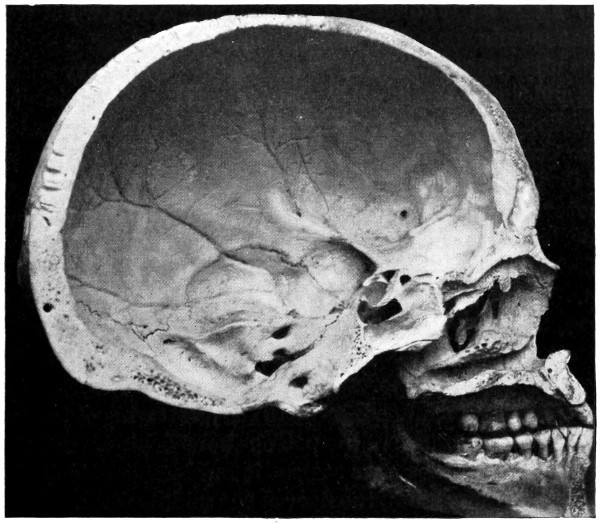
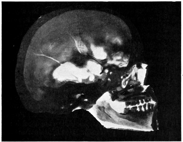
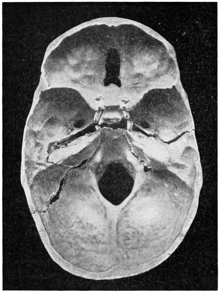
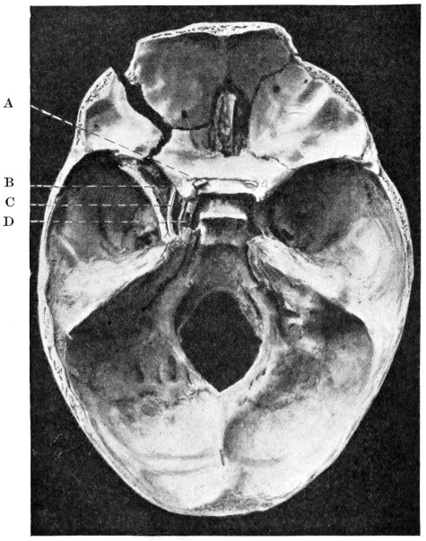
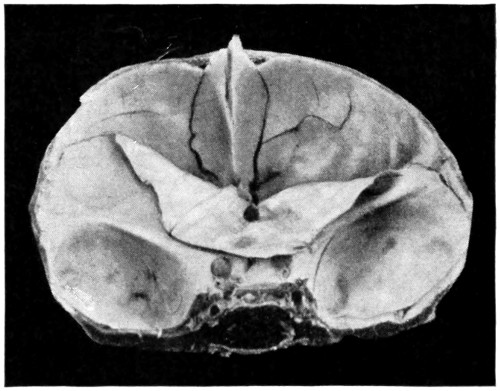
These parts are proportionately strong. Two regions are practically
devoid of this inter-tabular buffer—the squamo-temporal and cerebellar
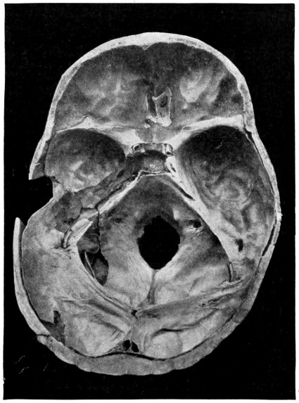
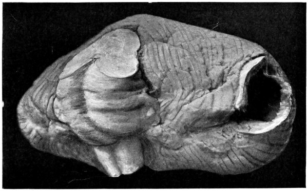
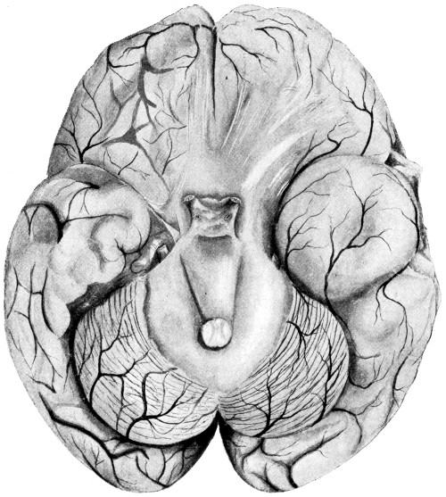
(see Figs. 29 and 30). A recognition of this comparative weakness
is of great practical importance in view of the fact that both these
regions are liable to special lesions—injury to the middle meningeal
artery in the first case, and, in the second, cerebello-medullary lesions.
Nature’s ‘mistake’ in providing coverings unsuited to requirements
has been compensated for in part by additional protection—the
temporal and nuchal muscles.
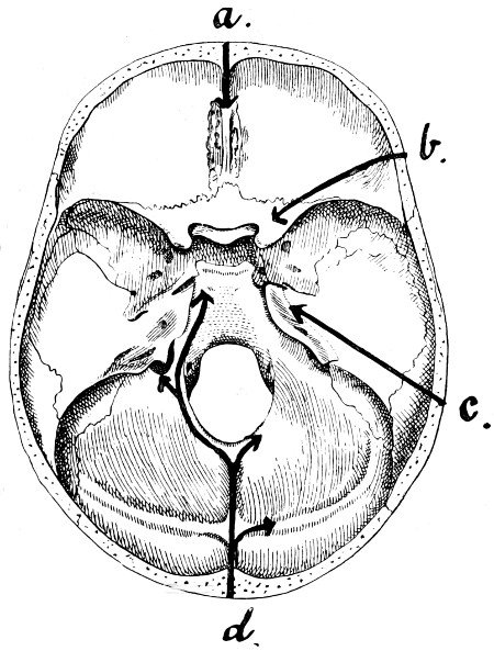
Fig. 28. Diagram illustrating the Lines along which Forces received
on the Vault are transmitted to the Base. (For further description, see text.)
Further, not only does the skull vary in density in its several
parts, but it is also ribbed and strengthened by various bony bars
and buttresses that pass up from base to vault (see Fig. 28).
These ‘ribbings’ are seen to extend upwards from the crista galli,
from the external angular frontal process, from the auditory region,
and from the occipital protuberance. Presumably, these ‘ribbings’
were so constituted for a definite purpose; in any case, it is clear
that they play an important part in the reception and conduction
of forces to the base of the skull. It is apparent, moreover, that the
parts intervening between these ‘ribbings’ are liable to injury in
direct proportion to their general position and strength. The deep
groovings of the bone for the reception of the middle meningeal artery
afford an additional source of weakness to the bone in the squamo-temporal
region. (See Fig. 50).
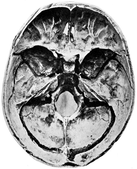
Fig. 29 a. The Base of the Skull.
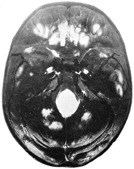
Fig. 29 b. The Base of the Skull as seen on Transillumination.
Further reference will be made to the relative strengths of the
various regions of the skull. Sufficient has been said to show that
nature has provided the skull with various paths by means of which
forces applied to the vault can be conducted and distributed to the base.
Before, however, proceeding to discuss the effects produced on
the base of the skull, it is necessary to add that nature provides other
methods by means of which the intensity of a blow, delivered over
the vertex of the skull, is diminished. The forces are broken up and
distributed in the following manner:—
1. Though the force tends to travel in the direction of the applied
force, yet the convexity of the skull allows of the dissemination of
that force over a large superficial area.
2. The intervention of cartilage or fibrous tissue between two or
more of the component bones of the vault tends to diminish the
intensity of the force, to break it up and to alter its direction.
3. The bony ridges, along which the forces tend to travel, themselves
terminate blindly (see Fig. 28). Thus,
(a) Forces passing from the frontal region converge, more or less,
to the crista galli.
(b) Forces from the external angular frontal process pass along
the wings of the sphenoid bone to the anterior clinoid process.
(c) Forces from the auditory region are projected along the
summit of the petrous bone towards the apex of that process and to
the posterior clinoid process.
(d) Forces applied to the occipital region travel inwards along
the internal occipital crest to the strengthened margins of the foramen
magnum, or are projected outwards along the lateral sinus ridges.
In the former case, the force either passes forwards towards the dorsum
ephipii and so again reaches the posterior clinoid process, or is
directed more laterally towards the jugular process of the occipital
bone, there meeting the fibrous tissue intervening between that
process and the corresponding part of the temporal bone.
4. All forces, whether transmitted along the internal occipital
crest, the temporal bone, the sphenoidal wings, or the crista galli
of the ethmoid, are further transmitted to the dura mater attached
to those prominences and ridges. The dura mater undoubtedly
plays an important part in the reception and transmission of the
forces.
5. The forces all show a tendency to converge towards the
pituitary region, the great ‘water-cushion’ of the brain—a region
bounded by the clinoid processes. That these processes receive
a considerable part of the forces transmitted is confirmed by the fact
that they are frequently torn away from their basic attachments.
This is especially the case with respect to the attenuated base of the
anterior clinoid process.
It is obvious, therefore, that forces tend to be transmitted from
the vault to the base, and yet the base is, in many respects, the
weakest part of the skull. It is perforated by numerous foramina,
it is hollowed out in places for the formation of air sinuses and for
the reception of the integral portions of the auditory apparatus.
Furthermore, it presents a more or less plane surface, one differing
in all respects from the marked convexity of the vault. Those forces,
therefore, which are received by the base of the skull are not subjected
to that diffusion which forms so conspicuous a feature in the case of
the vault.
All these points tend to show that the base of the skull is more
or less unsuited for the reception of severe blows, direct or transmitted,
whilst, on the other hand, nature has taken into consideration
and provided fairly adequately against the dangers incident to vault
injuries.
It is not proposed at this stage to discuss further the relative
strength of vault and base. Points, other than those already enumerated,
will be brought forward in subsequent sections.
Fractures of the base of the skull are produced in two ways:—
1. By violence applied directly to the base—perforating wounds
of the orbit, bullet-wounds through the mouth and base of skull,
the driving inwards of the bones of the face, the driving upwards
of the condyle of the jaw (the ‘knock-out’ blow of the pugilist), and,
as the result of heavy falls on to the feet or buttocks, the upward
driving of the condyles of the occipital bone.
2. By violence applied indirectly—blows applied directly to the
vault and transmitted to the base. This variety will be considered
first as it receives the greatest prominence in surgical textbooks.
Various explanatory theories have been advanced, of which the
following are the more important:—
This theory states that
‘fractures of the base result as extensions from fractures of the vault,
the fracture following the shortest anatomical route to the base’.
Although this theory must be accepted as offering a satisfactory
explanation for the occurrence of a certain proportion of basic
fractures, such, for instance, as result from a blow applied directly
to the vertex, it certainly cannot be accepted as accounting for the
great majority of basic fractures. The theory was advanced on the
hypothesis that basic and vault fractures were necessarily co-existent.
That combined lesions of this nature are frequently in evidence is
not to be doubted for one instant. It is, however, ‘putting the cart
before the horse’ to say that the vault fracture is always the primary
lesion. Such is by no means the case.
The skull is
here regarded as an highly elastic sphere, compression of which leads
to diminution in the diameter along the axis of greatest pressure,
bulging occurring in other diameters. The bulging exceeding the
limits of elasticity a fracture occurs, the line of fracture varying
according to the different features present. Thus, when the lines of
fracture run parallel to the direction of the compressing force the bone
bursts open along the convexity (bursting fractures), and when the
lines of fracture run at right angles to the direction of the compressing
force a fracture by compression is said to result (compression fractures).
These theories are based on experiments carried out on the
cadaver, the skull being enclosed in a tight-fitting box and subjected
to pressure sufficing to bring about a fracture. Undoubtedly, the head
may be compressed between two forces, as, for instance, when a
person is knocked down in the street, the vehicle passing over the
head, or, as in a case recently under my care, where a boy, hanging
by his feet from the side of a barge, was crushed between the barge
and the wharf as the vessel swung inwards with the tide. Cases of
bilateral compression are, however, of infrequent occurrence, the
great majority of basic fractures resulting from blows applied directly
at the region of the level of the base of the skull (see p. 76), or from
the forward propulsion of the body, the head coming into violent
contact with a resisting object, as, for instance, when a person is
thrown out from a motor-car, the head striking against a tree, brick
wall, &c. In the first case, there can be no question of bilateral
compression, and in the second the compression is exerted between
the vertex and the occipital condyloid region.
Moreover, the fundamental points on which the bursting and
compression theories are grounded are based on erroneous principles.
The skull cannot in any sense be regarded as a sphere, nor does it
possess the requisite elasticity to bulge and allow of compression
in the manner that the theory demands. The skull, in reality, forms
rather less than two-thirds of a sphere, the base passing inwards from
the lower limits of this partial sphere in a more or less horizontal
plane. This can be readily verified by placing the skull on a table
so that its base corresponds to the surface of the table. The elastic
properties of the skull have also been greatly exaggerated, and far
too little attention has been paid to the peculiar anatomical formation
of the base.
Fractures of the base occasionally
occur in which evidence is conclusive that the blow was
received on the vault, the vault itself remaining uninjured. Such
cases have given origin to this theory, one stating that ‘from the
point struck a wave is transmitted through the semi-fluid brain,
producing a fracture at some more distant point’. Helferich, for
instance, maintains that isolated fractures of the orbital roof, and
more rarely of other parts of the base, are produced by the influence
of hydrostatic pressure. There can be no question that waves are
transmitted through the brain and cerebro-spinal fluid, but that such
waves should be capable of producing a basic fracture is, in my opinion,
beyond the bounds of possibility. The theory is opposed to all my
experience of basic fractures, and the cases brought forward in support
are capable of a much more probable explanation. The base is undoubtedly
the weakest part of the skull, and a blow on the vault
may fail to produce a local lesion and yet, when the force is transmitted
to the weaker base, may there bring about a more definite
result. For instance, it is by no means uncommon to find that a blow
on the frontal region fails to fracture the vertical plate of that bone
and yet suffices to produce a fracture, often comminuted, of the
orbital plate of the frontal bone or of the cribriform plate of the
ethmoid, two fragile plates, either of which may shatter like a plate
of glass from the effect of forces transmitted across them. In further
support of this theory, the following case, recently under my care,
may be cited. The patient received a heavy blow over the left
occipital region. A fracture passed inwards across the left cerebellar
fossa towards the posterior border of the foramen magnum, and a
second fracture, entirely distinct from the first, passed across the
right orbital plate of the frontal bone. In this case, the force conducted
across the base, from behind forwards, failed to fracture the
strong basi-occiput, but succeeded in producing a more definite lesion
on reaching the fragile orbital plate.
Some of the celebrated surgeons of the last century insisted that
the course pursued by basic fractures was to be explained on
anatomical grounds, but their views have been neglected and theories
based on experimental evidence have been accepted in their place.
All experiments, such as those previously mentioned, are useless, and
definite conclusions can only be gained by carrying out in every case
the following method of investigation: (1) by obtaining in the first
case an accurate history as to the manner in which the injury was
received; (2) by noting all visible and palpable signs of external
injury; (3) by careful observation of all the clinical symptoms during
the progress of the case; (4) by comparison of such with the lesions
found in case of death.
Over 300 cases have been investigated by me after these principles.
In about 30 per cent. of the cases sufficient evidence was
obtained to show that the basic fracture resulted and extended from
a primary fracture of the vault. These cases were to be explained
by Aran’s theory of irradiation. This theory, however, errs in stating
further that the fracture follows the shortest anatomical route to the
base. This is not correct, for the line of fracture corresponds to the
direction of the applied force and is influenced to a very large extent
by the resistance offered, the weaker areas being picked out and the
strong buttresses avoided. It is only in the most severe cases that the
fracture travels to and traverses across the base in such a direction
as to show that, for the time being, all laws are in abeyance.
In about 5 per cent. of cases the fracture resulted from bilateral
compression, from falls on to the buttocks, &c., and from blows
applied to the angle of the jaw. These cases afforded examples of the
bursting and compression theories.
On the other hand, in over 60 per cent. of cases, the injury was
received over one of the following situations: (1) in front, over the
frontal eminence or supra-orbital ridges; (2) in the antero-lateral
region, over the external angular frontal process; (3) in the lateral
region, over the lower temporal, auricular, and mastoid regions;
(4) in the posterior region, over the superior curved line of the
occipital bone or over the external occipital protuberance.
In all these cases, therefore, the blow was inflicted at or near the
level of the base of the skull, the resultant fracture being a fracture
by direct violence, the fracture traversing the base and splitting it
much in the same way as a chisel splits a board of wood. The ‘grain’
of the wood may be regarded as representing the weaker basic lines,
and any ‘knot’ the resistance offered by the strong basic buttresses,
the forces being momentarily turned aside, but soon again passing
onwards, parallel to the original direction, but not necessarily in the
same straight line.
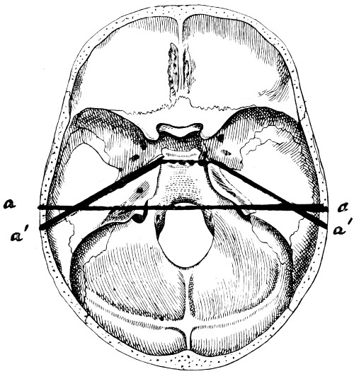
Fig. 30. Plan of the Base of the Skull. a, a, The transverse pre-condyloid
line; a′, a′, The line pursued, in whole or in part, by the ‘typical’ basic fracture.
Any blow delivered at or near the basic level tends primarily
to involve the weaker area, the base, passing secondarily upwards on to
the vault. One may go even so far as to say that in most combined
vault and basic fractures, the vault fracture is a secondary development,
the basic fracture being the primary lesion.
There is, however, still another important anatomical feature
bearing on the mechanism of basic fractures, one that must necessarily
come into force in the greater number of such fractures. The base
of the skull may be said to consist of two parts, one lying anterior
to the condyles of the occipital bone, the other posterior to and
including the condyles with their vertebral attachment. These two
segments are united to one another by a weak chain—represented
by a line drawn from one external auditory meatus to the other,
with, as a connecting link, the sphenoidal sinus in the middle line.
When the base of the skull is viewed from below, it will be seen
that the weak line includes both Glaserian fissures, both petro-sphenoidal
sutures, both foramina lacera media, with the sphenoidal
sinus again as a connecting link. The two parts of the skull are, to
all intents and purposes, merely cemented together by the union
of the basi-sphenoid and basi-occiput. Consequently, if a blow be
received on the antero-lateral region of the head, the anterior segment
tends to be split off from the more fixed posterior part, the fracture
following the weak line previously indicated. This weak line occupies
so important a position in the mechanism of basic fractures that careful
observation will show that the greater number of middle fossa
fractures follow that line, wholly or in part. Such a fracture of the
middle fossa may be termed ‘the typical fracture of the base of the
skull’ (see Figs. 30, 34 and 39).
Aran’s theory of irradiation, with
certain modifications, accounts satisfactorily for about 30 per cent.
of basic fractures.
The contre-coup theory may be rejected entirely.
The bursting and compression theories are unsatisfactory,
accounting for not more than about 5 per cent. of fractures.
The majority of cases result from direct violence applied at or
near the basic level, the fracture passing across the base in the general
direction of the applied force, but not necessarily in the same straight
line.
Up to this point certain facts and theories have been discussed,
such as bear on the general mechanism of basic fractures. It now
remains to consider other factors that exercise influence on the general
direction of the fracture.
Complete
maceration of the skull is always essential in endeavouring to
estimate in what way the various sutures of the skull influence the
extent and direction of a fracture. Sutural separation is generally
regarded as of infrequent occurrence. An examination of a large
number of macerated skulls has shown, however, that sutural separation
is in reality of common occurrence. Certain sutures show a
special liability to such changes, especially the masto-occipital, the
petro-occipital, and the petro-sphenoidal. Separation of the sutures
is more common in the young adult; in the infant and in the old
such conditions are seldom observed.
Allusion has already been made to the fact that forces transmitted
from the vault to the base, or vice versa, undergo a marked
diminution in intensity when the sutures of the skull are encountered,
the ‘fracture’ showing a marked disposition to follow the line of the
suture. When the force is excessive, all rules are temporarily in
abeyance, but, under ordinary circumstances, the separation along
the line of a suture corresponds fairly accurately with the dentations
and serrations of the suture involved. Sutural separation without
actual fracture is a possible occurrence, but is decidedly rare. Such
isolated fractures are confined, more or less, to the sagittal suture in
the vault, and the masto- and petro-occipital sutures in the base.
The sphenoidal sinuses, two in number, are usually
separated from one another by a thin septum. This septum is, however,
often deficient, and a single cavity exists. The sinuses make
their appearance about the seventh or eighth year; they vary
greatly in size but, when fully developed, occupy the greater part
of the so-called body of the sphenoid, extending backwards almost
as far as the junction of the basi-sphenoid and basi-occiput, and
spreading outwards into the wings of the sphenoid and over the roof
of the orbit.
The sinus is bounded on all sides by a thin lamella of bone; its
roof forms part of the middle fossa of the skull, the sides are separated
by a thin bony wall from the cavernous venous sinus, and the floor
aids in the formation of the roof of the naso-pharynx. There exists,
therefore, in the very centre of the base of the skull—in the region
of the so-called buttress of connexion between the posterior and
anterior segments of the skull—an exceedingly weak area, one which
must be implicated in the great majority of basic fractures. The
‘weak line’ of the base of the skull—previously referred to—is now
still more accentuated.
The sphenoidal sinus is involved in at least 40-50 per cent. of
basic fractures, comminution of the sinus wall being often so excessive
that a probe can be passed with the greatest ease from the middle
fossa into the naso-pharynx. Blood is thus allowed to escape readily
into the naso-pharynx, and a source is opened up for the possible
development of meningeal infection.
Reference to the various illustrations of fractures of the base
will supply further evidence as to the special liability of the sinus
region to injury. It will be seen that nearly all fractures that pass
one middle fossa to the other, or from one middle fossa to the
opposite anterior fossa, traverse this region.
The frontal sinuses, also two in number, are separated from one
another by a thin osseous septum. Up to the age of puberty these
sinuses are either absent or represented by a small cell. Subsequently,
they develop rapidly, often extending into the orbital roof. The
upper and inner boundary—usually very fragile—assists in the
formation of the anterior fossa of the skull. The outer boundary—the
perpendicular plate of the frontal bone—is much more dense,
and, consequently, a fracture of the outer wall is almost necessarily
associated with a fracture involving the inner or orbital boundary,
that is to say with a fracture of the anterior fossa.
The ethmoid cells.
The ethmoid bone consists of a collection of
cells which communicate with the nasal cavity (middle and superior
meati), and which are merely separated from the anterior fossa of
the skull by the thin cribriform plate. This plate of bone is of so
fragile a nature that splintering occurs in the great majority of
anterior fossa fractures. The special dangers that arise from the
possibility of meningeal infection are obvious.
The auditory region.
That part of the petrous bone which
encloses the auditory apparatus, and which transmits the seventh
and eighth pair of nerves, is proportionately weakened and correspondingly
liable to fracture. The special details of these fractures
are dealt with on p. 102.
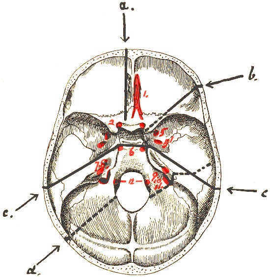
Fig. 31. To illustrate the relation of Basic Fractures to
Cranial Nerves.
It has often been stated
that a basic fracture is arrested on meeting one of the larger foramina
of the skull. With this view I am not in agreement, for not only are
the larger foramina frequently involved, such as the foramen lacerum
posterium and medium, but the largest foramen of all, the foramen
magnum, is often implicated. It will be granted that certain foramina
are but rarely involved, but this is due to the fact that they are
aside of the chosen and definite paths of basic fracture. Thus, the
foramen ovale and the foramen spinosum are only exceptionally
involved because they lie immediately anterior to the petro-sphenoidal
suture, whilst the anterior condyloid foramen—transmitting the
hypoglossal nerve—is rarely implicated because it lies internal to
the usual posterior fossa fracture. On the other hand, the foramen
lacerum medium is involved in nearly every fracture that passes
from one middle fossa to the other.
When the various weaker lines and areas are taken into consideration,
and when the direction and site of the applied force are known,
one is generally enabled to foretell with considerable accuracy the
probable transbasic course of the fracture. After investigating
over 300 cases, I was enabled to frame the following rules with
respect to the probable line of transbasic fracture.
| Direction, &c., of the applied force. |
Probable resultant basic fracture. |
| 1. Force applied to the median frontal region. |
The fracture passes backwards from the perpendicular plate of the frontal bone to the cribriform plate of the
ethmoid, thence between the optic foramina to the body of the sphenoid, the thin sinus roof being usually comminuted. From there the
fracture diverges to the opposite side, and tearing off the posterior clinoid process, passes along the petro-occipital suture to the
jugular foramen, being then continued on the other side of that foramen along the masto-occipital suture, and so again to the vault. |
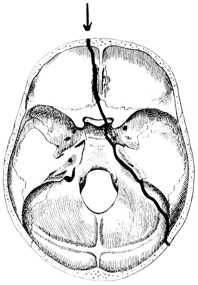
Fig. 32. Diagram of Lines pursued by Basic Fractures. Force applied
to the median frontal region.
|
| 2. Force applied to the lateral frontal region, in the situation of the external angular frontal process. |
The fracture passes across the anterior fossa towards the sphenoidal fissure, tearing away the anterior clinoid
process, and again comminutes the roof of the sphenoidal sinus. Progressing onwards, with or without fracturing the posterior clinoid
process, the fracture passes either along the anterior part of the petrous bone at its junction with the greater wing of the sphenoid
towards the opposite middle and external ears, or along the petro-occipital suture to the jugular foramen, and continued along the
masto-occipital suture as in the previous case. |
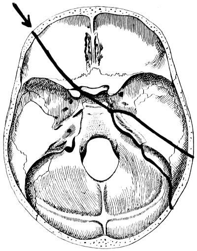
Fig. 33. Diagram of Lines pursued by Basic Fractures. Force applied
to the lateral frontal region in the situation of the external angular frontal process.
|
| 3. Force applied to the region of the external ear. |
The fracture passes across the roof of the bony auditory meatus towards the junction of the anterior and inner walls
of the middle ear, the membrane undergoing a variable amount of destruction and displacement. The fracture is then continued across the
tegmen tympani, and after following the petro-sphenoidal suture reaches the foramen lacerum medium, being again continued on the opposite
side of that foramen to the sphenoidal body. Thence it pursues one of two courses. Most commonly the fracture passes backwards obliquely
to the opposite middle and external ears, following a course similar to that already indicated. |
| In such cases the fracture may extend on each side up on to the vault in such a manner that the two segments are
merely united by the soft parts; whether the fracture be so complete or not, a more minute examination of the line of separation will
evidence many interesting points. An inspection of the anterior aspect of the posterior fragment shows that the fracture passes just in
front of the geniculate ganglion of the facial nerve, the ganglion being laid bare, whilst its petrosal branches are usually torn. The
fracture also passes anterior to the Eustachian tube and the horizontal part of the internal carotid artery. On examining this posterior
fragment the following structures will be seen, passing from without inwards: the posterior half of the external auditory meatus, the
mastoid antrum, the lacerated membrane and the ossicles of the middle ear, the geniculate ganglion of the facial nerve, the Eustachian
tube, the horizontal part of the internal carotid artery, the Gasserian ganglion, and the posterior half of the sphenoidal sinus in the
middle line (see also Fig. 39). |
| After reaching the sphenoidal body, the alternative course for the fracture to pursue is to pass towards the opposite
sphenoidal fissure and, tearing off the anterior clinoid process, to be directed across the anterior fossa, parallel to the original
direction but not in the same straight line. |
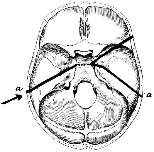
Fig. 34. Diagram of Lines pursued by Basic Fractures. Force applied
to the region of the external ear. a ... a, The ‘typical’ basic
fracture (see also Fig. 30).
|
| 4. Force applied to the mastoid region. |
The fracture follows the occipito-mastoid suture to the jugular foramen, and is again continued on the opposite side
of that foramen along the petro-occipital suture towards the apex of the petrous bone. It then passes across the sphenoidal body to the
sphenoidal fissure of the opposite side, and so across the anterior fossa. It is especially common in this particular variety of fracture
to find fissures diverging from the region of the sphenoidal sinus forwards towards the cribriform plate of the ethmoid, these fissures
usually passing between the optic foramina. |
| This fracture is also peculiar in so much that, when the degree of separation along the occipito-mastoid suture is
excessive, there is special liability to a tearing of the lateral sinus wall as the sinus begins to turn downwards and inwards. |
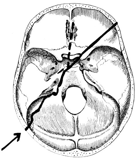
Fig. 35. Diagram of Lines pursued by Basic Fractures. Force applied
to the mastoid region.
|
| 5. Force applied to the lateral occipital region. |
The fracture passes across the thin cerebellar fossa and strikes the foramen magnum immediately behind the condyle.
Starting again from a similar point on the opposite side of the foramen, the fracture passes outwards to the jugular foramen. Again, two
courses are now available, the fracture either cutting outwards across the body of the petrous, ‘external’ to the internal
auditory meatus and cutting across the facial nerve in the region of the geniculate ganglion, and finally terminating in the roof of the
middle ear, or else passing along the petro-occipital suture and so to the foramen lacerum medium, the sphenoidal fissure, and the
anterior fossa as in the previous case. |
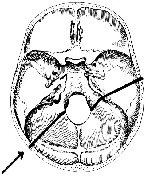
Fig. 36. Diagram of Lines pursued by Basic Fractures. Force applied
to the lateral occipital region.
|
| 6. Force applied to the posterior occipital region. |
The resultant fracture varies according to the direction of the applied force. A force which is applied to the
posterior occipital region at right angles to the transverse axis of the skull results in a fracture which, on reaching the posterior
margin of the foramen magnum, is continued again on the opposite side of the foramen along the dorsum ephipii. When the force is more
oblique in direction (as is usually the case) the fracture traverses the thin cerebellar fossa to the outer margin of the jugular foramen,
and then follows one of the two courses indicated in the previous case. |
| More commonly the fracture cuts across the petrous bone. |
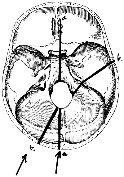
Fig. 37. Diagram of Lines pursued by Basic
Fractures. Force applied to the posterior occipital region, the fracture following
the course a. a. or b. b., according to
the direction of the applied force.
|
Basic fractures tend to follow certain definite paths,
this transbasic course varying according to the direction of the force
applied and the site of application of the same. Whether the fracture
completely traverses the base depends on the character of the force
and the resistance offered, for bases, as well as vaults, vary greatly
in strength. To every rule there must be exceptions, and cases are
at hand in which the fracture appears to have obeyed no law, or in
which the force applied was of so forcible a nature that the fracture
traversed the base, regardless of all the ordinary rules.
The principles enumerated above were formulated by me some
four years ago, and, in spite of certain adverse criticisms, I am more
than ever convinced that the rules are correct, and that time and
research are alone required to add to the strength of my assertions.
The symptoms resulting from a fracture of the base of the skull
vary according to the particular fossa fractured. From a general
point of view, the following symptoms require consideration:—
Hæmorrhages.
Escape of cerebro-spinal fluid.
Escape of brain-matter.
Escape of air from the air-sinuses into the surrounding tissues.
Involvement of certain cranial nerves.
Hæmorrhages:
(a) Subconjunctival hæmorrhage
usually makes
its appearance at the outer canthus of the eye, progressing inwards
towards the corneo-scleral margin, and, in the most severe cases,
completely surrounding the cornea, bulging the conjunctiva forwards
in such a manner as to constrict the field of vision. The extravasated
blood is usually bright red in colour, makes its appearance within
a few hours of the accident, and reaches its maximum within thirty-six
to forty-eight hours.
In some cases a condition of subconjunctival œdema (chemosis)
is observed. This also usually originates at the outer canthus.
Taken by themselves, neither hæmorrhage nor œdema are of any
great diagnostic value. Both conditions, however, aid materially in
confirming the diagnosis.
The blood is almost invariably completely absorbed, and no ill
effects remain.
(b) Palpebral and peri-palpebral hæmorrhage
is seen in most cases
of fracture of the anterior fossa. This form of hæmorrhage differs
from the one mentioned above in that it usually commences at the
inner canthus of the eye, thence progressing in the outward direction.
The extravasated blood may be wholly anterior to the suspensory
ligaments of the lid, in which case it may be surmised that the
fracture only involves the perpendicular plate of the frontal bone.
More commonly, however, the cribriform plate of the ethmoid shares
in the lesion, in which case palpebral, peri-palpebral, and subconjunctival
hæmorrhage are all present.
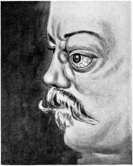
(c) Orbital hæmorrhages
may be so extensive that marked forward
protrusion of the globe exists. The time at which proptosis makes
its appearance, and the degree to which it progresses, vary according
to the nature of the lesion. Thus:—
| Proptosis severe, appearing almost at once, |
implies |
a fracture associated with injury to the cavernous sinus or internal carotid artery. |
| Proptosis moderate, and appearing after a few hours, |
„ |
a fracture involving the walls of the orbit, the blood being derived from lacerated ethmoidal and other small
vessels. |
| Proptosis appearing days or weeks after the accident, usually progressive, |
„ |
a fracture involving the region of the sphenoidal body and complicated by the formation of a fistulous communication
between the cavernous sinus and the carotid artery (see Traumatic orbital aneurysm). |
(d) Retinal hæmorrhages.
Fleming, in 1902, reported 12 cases of
fracture of the skull, all except one being fractures of the base, in
which retinal hæmorrhages were present. All cases were associated
with hæmorrhage into the subarachnoid space, and when this hæmorrhage
was of a unilateral nature the retinal changes were likewise
one-sided. It was also found that in 4 cases of cerebral hæmorrhage
without osseous lesion retinal hæmorrhages were present in three,
these three being all associated with considerable effusion into the
subarachnoid space.
These observations are not only of value in the general diagnosis
of intracranial lesions, but are also of considerable importance in the
differential diagnosis between extra- and intradural hæmorrhages.
(e) Hæmorrhage from the nose and mouth
is almost invariably
present in fractures of the anterior fossa, with the inference that the
fracture involves the cribriform plate. The blood—derived mainly
from lacerated ethmoidal vessels—escapes from the anterior nares or,
passing back into the naso-pharynx, escapes by the mouth or is
swallowed, to be vomited up later.
Escape of cerebro-spinal fluid.
Blandin, of the Hôtel-Dieu,
drew attention to this condition in the year 1840. The fracture
involves the cribriform plate of the ethmoid, and is associated with
laceration of the overlying dura mater and arachnoid, and of the
prolongations of those membranes along the olfactory nerves.
The escape of cerebro-spinal fluid from the nose may be regarded
as diagnostic of a fracture of the anterior fossa, in spite of the fact
that Goucard, Malgaigne, and others describe cases in which, as the
result of a severe fracture of the petrous bone (middle fossa) without
laceration of the membrana tympani, the fluid escaped along the
Eustachian tube to be expelled by mouth and nose.
At this stage, it will be necessary to allude more fully to the
general question of cerebro-spinal discharge from the nose, mouth,
and ear. The symptom is an important one, though undoubtedly
of far less frequent occurrence than stated in text-books. This was
proved by Crandon and Wilson, who reported 27 cases in which there
was a cerebro-spinal discharge out of a total number of 530 cases
examined. Phelps mentions 13 in a series of 286 cases of fractured
base. My experience coincides with these statements.
The escape of a slightly blood-stained fluid from the ear and
nose does not necessarily imply that the fluid is cerebro-spinal in
nature, for it has been proved on numerous occasions that fluid may
escape in considerable quantities without the existence of a basic
fracture. In such cases the fluid is derived either from the membranous
labyrinth (the liquor cotunnii) or from the mucous
membrane lining the ear and nose, the result of great vaso-motor
dilatation of aural and nasal vessels.
If the fluid be cerebro-spinal in nature, the natural inference is
that the subarachnoid space is opened up to the exterior, either
directly or indirectly along the course of a cranial nerve. A chemical
analysis of the fluid will determine the nature thereof, provided that
the fluid be collected EARLY.
| Thus, cerebro-spinal fluid is rich in chlorides, contains little or no albumen, but shows a trace of a reducing
substance, allied to pyro-catechin, |
whilst |
fluid derived from other sources contains chlorides, a fair amount of albumen, and no sugar. |
Certain factors, however, must be taken into consideration
which diminish the value of these chemical tests, for, though the fluid
be cerebro-spinal in nature, the admixture of blood at once interferes
with the delicacy of the test. Furthermore, even in the event of
a profuse discharge of cerebro-spinal fluid, the first part collected
alone contains sugar, the discharge soon becoming nothing more or
less than a serous exudation.
The fluid is almost certainly cerebro-spinal if the discharge begins
within twenty-four hours of the accident, if it be practically colourless,
fairly profuse, and continuing for two or three days, perhaps longer.
Though the discharge usually originates early, cases are recorded
in which the flow commenced some weeks after the accident. The
discharge may continue for hours or for weeks. Sir W. Savory
recorded a case in which fluid escaped for one month from both ears.
The quantity also varies greatly, usually a few ounces, but sometimes
many pints. Sir W. MacCormac recorded a case in which 10 pints
escaped within five hours. A profuse discharge is due to the fact that,
as the original cerebro-spinal fluid drains away, its place in the subarachnoid
space is taken by a serous exudation from the cerebral
venous system. This exudation occurs as soon as the subarachnoid
pressure is sufficiently reduced, the serous exudation progressing till
the venous and cerebro-spinal pressures are again equal.
As regards the prognosis, it is interesting to note that the escape
of cerebro-spinal fluid implies of necessity that the subarachnoid
space is opened up to the exterior, with all attendant dangers of
meningeal infection, yet that the prognosis is generally favourable.
One might even go further and state that the prognosis in such cases
is rather more favourable than under more ordinary circumstances.
Thus, Battle records 36 cases with a mortality of 25 per cent., the
general mortality of fractured base being about 44 per cent. My
own experience tallies with these statistics. The more favourable
result hinges no doubt on the fact that the prolonged outflow tends
to wash away organisms existent in the aural and nasal passages.
With regard to any special points in treatment, it is obvious that
syringing of ear or nose is absolutely contra-indicated. The cavities
should be lightly plugged with gauze, the dressings to be renewed
as soon as they are soaked. The patient must also be prevented from
interfering with the dressings.
With regard to the routine use of urotropin to guard against
the advent of meningitis, see p. 116.
Escape of brain-matter from the nose.
This diagnostic
symptom is of very rare occurrence. For its development the following
factors are requisite:—
Great comminution and destruction of the anterior fossa.
Severe local laceration of the brain (frontal lobes).
A copious discharge of blood and cerebro-spinal fluid.
A general increase in the intracranial pressure.
One case only of this nature has come under my own observation,
that of a man whose right nostril was blocked with brain substance.
He died shortly after admission into the hospital, and at the post-mortem
examination the above conditions were found.
Escape of air from the air-sinuses into the surrounding
regions.
When the fracture involves the frontal, ethmoidal, and
mastoid sinuses, any sudden increase of the intrasinus air-pressure,
such as is caused by sneezing, blowing of the nose, &c., may force
air into the surrounding tissues. A distinction must be made between
those cases in which the pericranium overlying the seat of fracture
is torn and those in which it remains intact. In the former case,
the escaping air may spread widely into the neighbouring loose
tissues, leading to a condition of ‘surgical emphysema’. In the
second case, the air remains confined to a smaller area, giving rise
to a soft, more or less rounded swelling. Gentle palpation of this
swelling imparts to the fingers that crackling sensation peculiar to
the condition. Firm pressure results in diminution in the size of the
tumour, the contained air being forced back into the sinus cavity.
Such localized collections of air are known as ‘pneumatoceles’.
As regards the special treatment of these conditions, the patient
must, in the first case, be warned against straining of all sorts.
Pneumatoceles require no other treatment. The condition soon
disappears if the patient recovers. In widespread surgical emphysema,
an incision should be made over the injured sinus, thus
allowing of the direct escape of the air expelled from that sinus.
The involvement of nerves.
The following nerves may be
involved in anterior fossa fractures:—
(a) The olfactory nerve.
The great majority of anterior fossa
fractures traverse the cribriform plate, necessarily injuring the fine
branches of the olfactory bulb. The bulb itself may be lacerated,
with or without injury to the under surface of the frontal lobes.
Sir Prescott Hewitt considered that anosmia, or loss of smell, resulted
most frequently from blows applied to the back of the head, the frontal
region being injured by contre-coup. From my own experience it
would appear, however, that anosmia, whether uni- or bilateral,
whether transient or permanent, generally results from direct injuries
of the cribriform plate with associated lacerations of the olfactory
nerves. It is difficult to estimate the presence or degree of immediate
loss of smell on account of the general condition of the patient and
because the nostrils are usually more or less filled with blood
coagulum. Experience shows, however, that early loss of smell is
the rule and total and permanent anosmia the exception. Anosmia
is usually associated with some degree of loss of taste.
(b) The optic nerve.
Many cases have been recorded in which
visual defects resulted from blows applied to the head. The blindness
may be partial or complete, immediate in onset or developing at some
future date. In the latter case, the loss of vision is due to retinal
changes or results from post-neuritic atrophy.
The occurrence of complete or partial blindness as the immediate
result of the injury is, at first sight, difficult to explain, for the vast
majority of anterior fossa fractures avoid the immediate vicinity of
the optic foramina, passing by preference between the two foramina
or diverging towards the sphenoidal fissures. Small fissured fractures
not infrequently radiate through the optic foramina, usually, however,
of so slight a nature as to be incapable of leading to any gross lesion
of the optic nerves. Hæmorrhage into the sheath of the nerve is
probably responsible for a certain proportion of cases, more especially
those in which there is a peripheral concentric loss of vision, the more
central fibres escaping. It is possible, also, that cases evidencing
temporal or nasal blindness may be due, as J. J. Evans[17] thinks, to
a contre-coup contusion of the nerve through it being forcibly driven
against the bony boundaries of the foramen. Taking into consideration,
however, the very frequent presence of a fracture through the
anterior clinoid process (see p. 82), and the usual displacement of
that process, it would appear probable that immediate and more or
less complete loss of vision results from the compression and crushing
of the optic nerve by reason of the pressure exercised by a displaced
clinoid process.
The following statistics add confirmation to this view. Thus,
Callen collected 17 cases in which the optic nerve was compressed
by osseous fragments in the region of the optic foramen, whilst
Holder observed injury to the bones entering into the formation
of the foramen in 53 out of 86 cases of fracture involving this
region.
The prognosis varies according to the cause of the blindness.
When resulting from concussion of the nerve trunk or from hæmorrhage
into its sheath, certain fibres may regain their function. In the
majority of cases, however, that have come under my own observation,
blindness of the affected eye was immediate and permanent.
(c) The nerves passing through the sphenoidal fissure.
The
ophthalmic division of the fifth nerve is rarely injured to such a degree
as to cause anæsthesia of all the regions supplied. Blood extravasation
into the surrounding regions, or direct involvement of one
of the branches of the nerve, often results in areas of anæsthesia, and
some few cases have been recorded in which there was complete
anæsthesia of both cornea and conjunctiva, with subsequent ulceration
and sloughing. The nasal nerve may be implicated as the result
of a fracture involving the cribriform plate, whilst the supra-orbital
and supra-trochlear branches may be damaged by fractures of the
vertical plate of the frontal bone.
The third nerve is similarly liable to injury, in any part of its
orbital course. It is quite exceptional, however, for the whole trunk
to be affected, some of the branches being taken, others left. The
fourth nerve is also occasionally involved, generally in association
with other orbital nerves.
When anæsthesia or paralysis of muscles results from pressure
exercised on the nerves by extravasated blood, the ultimate prognosis
is not unfavourable. When due to direct implication, in the line of
the fracture, the prognosis is much more uncertain, partial or complete
loss of function resulting.
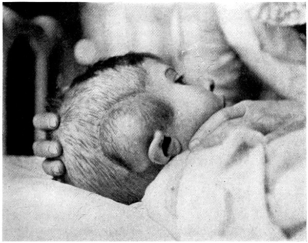
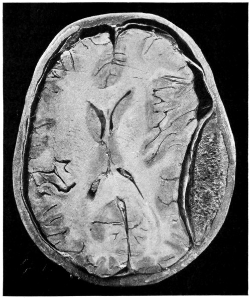
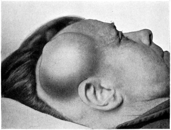
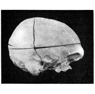
(a) Hæmorrhage into the temporal
region.
The extravasated blood may either be confined to the
temporal region—temporal hæmatoma—or diffused throughout the
subaponeurotic space. A temporal hæmatoma is always highly
suggestive of a fracture involving the temporal fossa, especially in
the event of marked outward bulging, with stretching and discoloration
of the overlying tissues. In many cases also the hæmatoma
pulsates, in which case it can be presumed that the fracture
of the temporal fossa is associated with hæmorrhage from a lacerated
middle meningeal artery (see Fig. 38). In such cases the application
of pressure to the hæmatoma may lead to the development of fits
on the contra-lateral side, originating in the face or arm regions and
spreading to the higher cortical motor area.
A
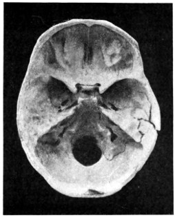
B
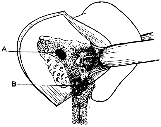
Fig. 38. To illustrate the probable Source of Profuse Hæmorrhage
from the Ear. A, The fracture the roof of the external auditory meatus.
B, Comminution of the tegmen tympani, the fracture involving the groove for the
posterior branch of the middle meningeal artery.
(b) Hæmorrhage from the ear and mouth. The great majority of
middle fossa fractures involve the external auditory meatus, passing
inwards across the roof and floor of the middle ear towards the body
of the sphenoid. Examination will show that the fracture passes
inwards towards the junction of the inner and anterior walls of
the middle ear, that is to say, towards the tympanic orifice of the
Eustachian tube. The membrana tympani undergoes a variable degree
of destruction. In the lesser cases the membrane is torn in its upper
and front part only—in the region of the membrane of Shrapnell—whilst
in the more serious cases it may be completely destroyed.
The blood that escapes from the ear is derived from those vessels
that supply the lining cuticle of the external and middle ears, from the
numerous tympanic vessels, from the lateral sinus, and from the middle
meningeal artery. The amount of blood which escapes varies according
to the source of the hæmorrhage. When hæmorrhage occurs from
the smaller vessels, the blood either clots in the external meatus or
trickles from the ear. In the most severe cases the hæmorrhage is
profuse and long-continued. Some years ago a case came under my
observation that threw light on the probable source of such severe
hæmorrhages.
A man was admitted into the hospital, suffering from profuse
hæmorrhage from the ear as the result of a fall down an area. The
bleeding continued for fourteen hours, soaking the dressings and
continuing so long as the man lived. At the post-mortem examination
an extensive comminution of the tegmen tympani was discovered,
the fracture being associated with great extra-dural extravasation
of blood from a lacerated middle meningeal artery. The extra-dural
hæmorrhage was enabled to escape through the tegmen tympani into
the middle ear, and thence by means of the lacerated membrana
tympani (see Fig. 38).
Profuse and long-continued hæmorrhage from the ear should
always arouse suspicion as to the possibility of injury to the middle
meningeal artery. Since meeting the case recounted above, many
similar cases have come under my care, and, in several instances,
guided by this symptom alone, operative measures have been carried
out successfully.
The two following cases show, however, that the extra-dural
extravasation may be derived not only from the middle meningeal
artery but also from the lateral sinus.
‘A man fell down an area and suffered from continuous hæmorrhage
from the ear. He remained in bed for a few days, and then,
becoming tired of confinement, got up, walked some distance on
a cold and frosty day, and visited a sage femme. On returning
home he complained of feeling ill, the hæmorrhage from the ear
ceased, and shortly afterwards he became unconscious and died.
The autopsy showed an extensive fracture of the petrous bone with
extensive extra-dural hæmorrhage from a torn lateral sinus and from
a lacerated meningeal artery.’[18]
The man had remained fairly well so long as the extra-dural
blood was permitted a free means of escape through the tegmen
tympani and external auditory meatus, but, so soon as clotting
occurred, compression symptoms developed and the man died in that
condition.
‘A man, 50 years of age, fell down, striking his head against
the kerb. On admission it was seen that blood was trickling freely
through a torn membrana tympani. He rapidly became unconscious
and died. The post-mortem examination revealed a fracture involving
the middle ear and external auditory meatus, passing backwards
across the lateral sinus, in which region there was a large extra-dural
extravasation of blood.’[19]
The above statements are confirmed by Dwight,[20] who, in 146
autopsies, found that, in 69 per cent. cases of fracture of the middle
fossa of the skull, there was bleeding from the ear, and that in
29 per cent. cases the fracture was associated with laceration of
branches of the middle meningeal artery.
Although hæmorrhage from the external auditory meatus may
be regarded as almost diagnostic of a middle fossa fracture, especially
of that variety previously described as the ‘typical basic fracture’,
yet the blood may be derived from a torn membrana tympani or
from laceration of the lining cuticle of the external meatus. Aural
examination will soon prove whether the blood is coming through
a rent in the membrane, in which case the diagnosis is clear. Sometimes
bleeding takes place from both ears, a symptom practically
diagnostic of the transverse middle fossa fracture known as the
‘typical basic fracture’.
The following statistics will supply further information as to the
relative frequency with which hæmorrhage occurs from ears, nose,
and mouth, and the proportionate mortality. The cases were collected
and tabulated by Crandon and Wilson.
| |
Cases. |
Lived. |
Died. |
Mortality. |
| Hæmorrhage from the ear |
281 |
170 |
111 |
39 per cent. |
| Hæmorrhage from both ears |
47 |
16 |
31 |
66 per cent. |
| Hæmorrhage from the nose |
44 |
17 |
27 |
61 per cent. |
| Hæmorrhage from the mouth |
168 |
73 |
93 |
33 per cent. |
Special points in prognosis and treatment.
It is not possible to
formulate any very definite prognosis when the hæmorrhage takes
place from one ear only, though the mortality is about 40 per cent.
When bleeding takes place from both ears the outlook is more grave,
the mortality being about 66 per cent.
With regard to special treatment, two points are obvious:
(1) that syringing of the ear is absolutely contra-indicated, on the
ground that such treatment carries with it a considerable risk of
bringing about meningeal infection; and (2) that plugging the external
meatus with strips of gauze is an unsurgical form of treatment,
insomuch as the escape of blood from the ear is an important factor
in preventing compression of the brain, more especially in those cases
where hæmorrhage is profuse. Under the last named conditions,
operative measures—exploration for a torn meningeal artery or
lacerated venous sinus—are to be carried out.
Hæmorrhage from the mouth may be slight or copious, according
to the source from which the blood is derived. In the former case,
the bleeding takes place from sphenoidal and pharyngeal vessels, in
the latter from the cavernous sinus or from the internal carotid artery
(see p. 148), the bone being shattered in the region of the sphenoidal
body, with comminution of the walls of the contained air-sinus.
This condition was first
investigated by Van der Wiel in 1727, and more completely by
Langier in 1839. The majority of those middle fossa fractures which
involve the petrous portion of the temporal bone pass immediately
anterior to the genu of the facial nerve (see p. 102), and it follows,
therefore, that the fracture cannot so involve the dural and arachnoid
prolongations of that nerve in such a manner as to allow of the escape
of cerebro-spinal fluid. This fact probably explains another fact,
namely, that aural cerebro-spinal discharge is an infrequent symptom
in middle fossa fractures. On the other hand, as a result of blows
applied to the occipital region, a fracture originating in the posterior
fossa may cut across the petrous bone, almost at right angles, in such
a manner as to sever the seventh nerve in the region of the genu
(see Fig. 41). This is the usual nature of a basic fracture associated
with the escape of cerebro-spinal fluid from the external auditory
meatus. More rarely, this particular class of fracture is unaccompanied
by any injury to the tympanic membrane, in which case the
fluid may escape along the Eustachian tube into the nose and naso-pharynx
(see p. 91). Cerebro-spinal fluid may also escape from the
nose and mouth in middle fossa fractures in the event of extensive
injury to the basi-sphenoid with involvement of the overlying
cisterna basalis. The following case exemplifies that condition:—
A man suffered from a severe fracture of the middle fossa. Three
weeks later there was a sudden and profuse discharge of cerebro-spinal
fluid from the nose. Meningitis developed and the patient died.
The basi-sphenoid was extensively comminuted, the overlying membranes
torn, whilst a probe could be passed readily from the cranial
cavity into the naso-pharynx.
Special points in prognosis and treatment.
The question of
cerebro-spinal discharges has been discussed previously (see p. 91).
It is therefore merely necessary to lay further stress on the fact that
syringing of the ear is absolutely contra-indicated, for reasons already
stated. The ear should be cleaned out with wool and gauze and
lightly packed with strips of gauze, these to be renewed when soaked
with fluid. When the cerebro-spinal discharge is long continued,
acute eczema of the side of the neck may develop as a result of the
irritating effect of the fluid. Under these circumstances it is advisable
to adopt precautionary measures, painting the skin with ‘new skin’
or collodion. Ointments are of but little use. The eczema will clear
up so soon as the discharge ceases.
The conditions needful for the
discharge of brain-matter from ear or nose have been enumerated
previously (see p. 93). There are but few cases recorded in literature,
and one case only has come under my own observation:—
A lad, 11 years of age, fell some distance out of window on to
his head. He was admitted under the care of my colleague, Mr. Lockwood.
On admission he was unconscious, and was bleeding freely
from the right ear and nose. Shortly afterwards it was noticed that
brain-matter was issuing from the right external auditory meatus,
sufficient brain-matter being obtained to fill a teaspoon. The extensive
nature of the brain-injury was confirmed by the fact that the
left arm and leg were paralysed for some days. On the fourth day
the boy regained consciousness and recognized his relations. From
this period onwards he made an uninterrupted recovery. I have
seen the lad on various occasions, the last time one year after the
accident. At that time he was an exceedingly bright and intellectual
boy.
Special points in treatment and prognosis.
The brain-matter
should be gently wiped away from the ear, and the meatus cleansed
and lightly plugged with gauze. Operative measures are required
in the event of the development of symptoms pointing to brain
compression. The prognosis must necessarily be unfavourable, but,
as the conditions are almost entirely confined to the young, the most
astonishing recoveries are reported.
The second and third divisions of
the fifth nerve pass respectively through the foramen ovale and the
foramen rotundum, two foramina which lie anterior to the petro-sphenoidal
suture, a suture traversed by the majority of middle
fossa fractures. These two nerves are therefore seldom involved.
In all the cases of fractured base which have come under my
observation I have never seen the foramen rotundum implicated,
and in one case only was the foramen ovale involved.
In certain rare instances, a fracture, passing in the antero-posterior
direction, may cut across the apex of the petrous bone in
close relation to the cavum Meckelii—the bed of the Gasserian ganglion—in
which case all three terminal divisions of the fifth nerve may
suffer. Thus, a case was reported by Lee in 1853 in which, seven
weeks after the accident, the following symptoms were present:—anæsthesia
of the left face and forehead, anterior two-thirds of
tongue, and left nostril, together with weakness of the left masticating
muscles, and an opaque left cornea.
The sixth nerve.
The sixth nerve may be involved either by
itself or in conjunction with other cranial nerves. In the latter case
the paresis is due to blood extravasated in the sphenoidal fissure or
in the orbital cavity. In the former case the nerve is injured where
it grooves the lateral aspect of the dorsum ephipii, a process frequently
fractured in lesions of the middle fossa. Fractures tend to
pass obliquely across this process, one nerve usually escaping. The
prognosis as to functional recovery is very problematical.
The seventh and eighth nerves.
There can be no doubt that the
seventh nerve, on account of its complicated intrapetrous course, is
more frequently involved than any other cranial nerve. Köhler
records 22 cases in 48 middle fossa fractures. My own experience
coincides closely with Köhler’s, facial paresis or paralysis being noted
in nearly 50 per cent. cases of middle fossa fracture.
The question of facial nerve implication is so intimately associated
with involvement of the eighth nerve that the two subjects
must be considered together. Thus, cases may be classified as
follows:—
1. Cases of paresis of the facial nerve with a variable degree
of deafness.
2. Cases of complete facial paralysis with complete deafness.
The greater number of middle fossa fractures involve the middle
and external ears, as is evidenced, amongst other symptoms, by
hæmorrhage from the ear. Some degree of facial paralysis is frequently
existent, not always evident at first sight, but requiring
careful examination and comparison between the two sides of the
face. The fracture involves both roof and floor of the external ear
and passes inwards towards the junction of anterior and inner walls
of the middle ear, the membrana tympani undergoing a variable
degree of destruction whilst the ossicles may also be injured. Thence,
the fracture passes inwards towards the petro-sphenoidal suture in
such a manner that the geniculate ganglion of the facial nerve is
exposed and laid bare on the anterior aspect of the posterior portion
of the skull.
The facial nerve, therefore, escapes direct injury except in so far
that the ganglion may be compressed by blood-clot or fragments
of bone. Partial loss of function results. In most cases the blood
is absorbed and a complete recovery may be anticipated. The degree
of deafness is directly proportionate to the damage incurred by the
membrana tympani and ossicles.
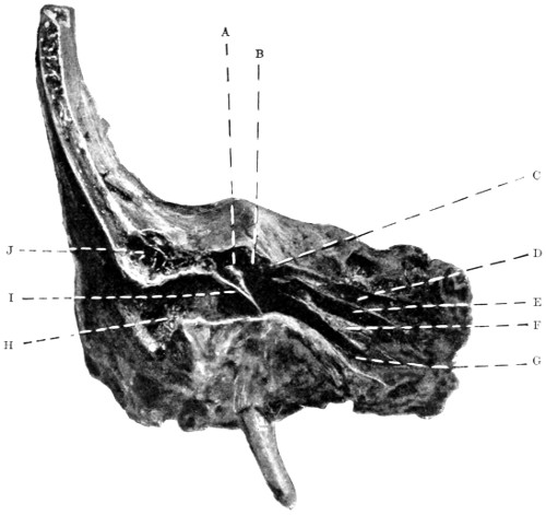
Fig. 39. To show the Relation of a Typical Basic Fracture to the
Middle Ear and its Adjuncts. A, Malleus;
B, Middle ear and aditus; C, Geniculate
ganglion (facial nerve); D, Groove for great superficial
petrosal nerve; E, Canal
for tensor tympani muscle; F, Processus cochleariformis;
G, Eustachian tube; H,
External auditory meatus; I, Membrana tympani;
J, Mastoid cells.
Larger illustration
In the second group of cases a different picture is obtained.
Usually the result of blows applied to the occipital region, the fracture
traverses the thin cerebellar fossa towards the outer angle of the
jugular foramen, thence cutting across the petrous bone, external to
the internal auditory meatus, and terminating, usually by comminution,
in the tegmen tympani. It is in the transpetrous part of
the fracture that the damage is done, for, not only is the facial nerve
cut across in the region of the ganglion, but the auditory apparatus
is also severed into two parts. The exact line of the fracture is shown
in Figs. 40 and 41.
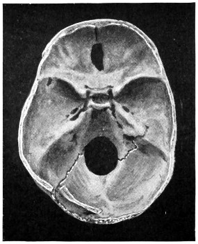
A
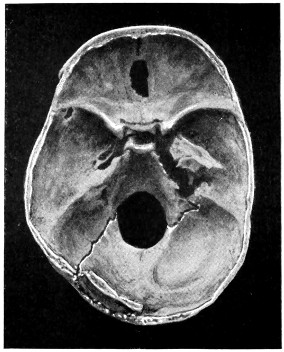
B
Fig. 40. To show the Relation of Basic Fractures to the Petrous Bone.
A, The basic fracture, resulting from a force applied to the left occipital region,
follows the course depicted in Fig. 36. B, The inner half of the petrous bone, being
loose, is thrown forward so as to show the relation of the fracture to the integral
parts of the petrous bone.
In this class of fracture, though facial paralysis and deafness are
both immediate in onset and permanent in duration, there is, in many
cases, no bleeding from the ear as the membrana tympani may be
uninjured.
The facial nerve may also be implicated in that rare type of
basic fracture which was described by Lèon Boullet in 1878, under
the title of ‘Fracture of the Mastoid portion of the Temporal bone’.
This fracture is fully described on p. 108. It will suffice
to mention
that the mastoid process may be torn away from the base of the
skull, the facial nerve being lacerated as it descends the aqueductus
Fallopii.
Bilateral facial paralysis is exceedingly rare. Two cases were
described by the late Professor von Bergmann. Its occurrence is
pathognomonic of the typical basic fracture (see p. 84).
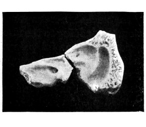
A
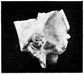
B
Fig. 41. To show the Relation of Basic Fractures to the Petrous Bone.
A shows the course pursued by an antero-posterior fracture of the petrous bone.
Note that it lies external to the internal auditory meatus. B shows—enlarged—the
inner aspect of the outer fragment. Note the relation of the fracture to
the semicircular canals, and that the membrana tympani and ossicles are quite
uninjured.
Complete facial paralysis may be associated with the following
symptoms:—
Epiphora, conjunctivitis, and keratitis (from paralysis of the
orbicularis palpebrarum).
Loss of taste (from involvement of the chorda tympani).
Impaired nasal air-entry (from paralysis of dilator alæ muscle).
Impaired acoustic sensibility (from paralysis of the stapedius).
Impaired mastication (from involvement of the buccinator
muscle).
Impaired secretion of saliva (from the cutting off of the secretory
and vaso-dilator fibres of the chorda tympani).
Lastly, it is necessary to add that facial paralysis developing
some days or weeks after the accident, though sometimes dependent
on degeneration of nerve-fibres as the result of pressure in the region
of the geniculate ganglion, may also arise from an ascending neuritis
or from meningeal infection.
In fractures of the posterior fossa,
blood effused into the deeper tissues of the scalp has considerable
difficulty in coming to the surface and thus making itself evident.
Furthermore, the resistance offered by the nuchal muscles tends to
confine the blood to the subtentorial region, thus adding to the already
grave prognosis of fractures in this region. On careful palpation,
however, it will be noted that the nuchal tissues present a doughy or
boggy condition, whilst ecchymosis becomes evident after twenty-four
to thirty-six hours. A peculiar ecchymotic patch is occasionally
observed, appearing in front of the mastoid process and travelling
upwards in a curved direction, concavity forwards, following the
outline of the ear. It is said to result from the tracking of blood
along the course of the posterior auricular artery. Whether this is
the case or not, the hæmorrhage usually implies a separation along
the line of the masto-occipital suture.
|
|
- |
Neither of these symptoms are present. |
|
When dealing with fractures of the
middle fossa, allusion was made to the implication of the seventh and
eighth pair of nerves as the result of a fracture traversing the posterior
fossa of the skull towards the outer angle of the jugular foramen and
cutting across the petrous bone (see p. 104).
The ninth, tenth, and eleventh cranial nerves may be injured
in the same variety of fracture. These three nerves are, however, so
protected by their dural sheaths that they generally escape
injury.[21]
In the following cases the nerves were involved:—
The patient was admitted suffering from a fracture resulting
from a blow on the posterior parietal region. During the next four
days no special symptoms developed. On the fifth day, during a
sudden attack of dyspnœa and dysphagia, death occurred. A fissured
fracture was found which extended into the jugular foramen, a region
occupied by blood-clot.
A man committed suicide by means of a pistol-bullet fired
through the mouth. The bullet lodged against the under surface
of the petrous bone, tearing the jugular vein and lacerating the nerves
passing through that foramen.
In another case the patient was admitted with a fractured base.
He progressed favourably until the tenth day when, on sitting up
suddenly in bed, he was seized with rigors, dyspnœa, and dysphagia,
dying shortly afterwards. A basic fracture was found, practically
dividing the skull into two parts and involving the jugular foramen.
Displacement had occurred with consequent compression of the
ninth, tenth, and eleventh nerves.
The twelfth nerve.
The anterior condyloid foramen is most
favourably situated with respect to the course pursued by posterior
fossa fractures. No instance of its involvement has come under my
own observation.
Stierlein records a case in which the tenth and twelfth nerves
were injured, with inability to speak or swallow, paralysis of the right
half of the tongue, soft palate, vocal cords and pharyngeal constrictions.
Death resulted in seven weeks.
The late Professor von Bergmann[22] mentions a case of hypoglossal
paralysis together with paralysis of the sterno-mastoid and trapezius
muscles (eleventh nerve).
For treatment of basic fractures, see p. 116.
Fracture of the Base of the Skull: Summary of Symptoms
| Anterior Fossa. |
Middle Fossa. |
Posterior Fossa. |
| Hæmorrhages. |
Hæmorrhages. |
Hæmorrhages. |
Subconjunctival.
Palpebral.
Peripalpebral.
Orbital.
Retinal.
From the nose.
From
the mouth. |
Into the temporal region.
From the mouth.
From the nose.
From the ear. |
Into the nuchal region.
Into the occipital region.
Into the post-auricular region. |
| Cerebro-spinal fluid. |
Cerebro-spinal fluid. |
Cerebro-spinal fluid. |
From the nose.
From the mouth. |
From the nose.
From the mouth.
From the ear. |
None. |
| Brain-matter. |
Brain-matter. |
Brain-matter. |
| From the nose. |
From the ear. |
None. |
| Air-escape. |
Air-escape. |
Air-escape. |
From the frontal sinus.
From the ethmoidal cells. |
From the mastoid antrum. |
From the mastoid antrum. |
| Nerve-involvement. |
Nerve-involvement. |
Nerve-involvement. |
Olfactory.
Optic.
Third.
Fourth.
Fifth (first division).
Sixth. |
Fifth (second and third divisions).
Sixth.
Seventh.
Eighth. |
Seventh.
Eighth.
Ninth.
Tenth.
Eleventh.
Twelfth (?). |
Fractures limited to this region are of rare occurrence. Our
knowledge of the condition is obtained from the researches of Lèon
Boullet, who first described the fracture in 1876, reporting at the
same time 26 cases.
The fracture usually results from sharp blows delivered along the
posterior border of the mastoid process, the force acting obliquely
from above downwards and forwards. More rarely, the process is
detached as the result of a blow delivered immediately above
the ear, that organ sharing in the displacement. In either case
the detachment is usually of an incomplete nature, mainly on
account of the muscular and pericranial attachments in the region
involved.
According to Boullet, the following symptoms result:—
Air escapes from
the mastoid cells and antrum into the overlying tissues, either
spreading widely through the subaponeurotic space of the scalp and
cellular tissues of the neck (surgical emphysema), or remaining
localized and forming a tumour of inconsiderable size (pneumatocele).
In either case palpation reveals that peculiar crepitation which is
pathognomonic of tumours of this nature.
Hæmorrhage takes place into the
middle ear, and, as the tympanic membrane is usually lacerated, the
blood escapes from the external ear. In the event of the tympanic
membrane being uninjured, the blood may escape along the Eustachian
tube into the naso-pharynx.
Boullet also pointed out that certain complications may be
associated with the mastoid lesion. These are as follows:—
Immediate.
Wound of the lateral
sinus.
Laceration of dura
and brain.
Injury to the aqueductus Fallopii and paralysis of the seventh nerve.
Remote.
Deafness.
Otitis
media.
Caries and necrosis of the petrous bone.
The displacement of the mastoid process is of
so incomplete a nature that the question of replacement by open
operation seldom arises. In the event, however, of intracranial
complications, operative treatment must be carried out. Under
ordinary circumstances the treatment consists mainly in the prevention
of suppuration. When suppuration occurs, frequent cleansing
of the ear must be carried out in order to prevent accumulation of
pus in middle ear and antrum. This object is to be attained, not
by syringing, but by gentle irrigation and light packing. Later on
it may become necessary to carry out the complete mastoid operation.
Fractures of the vault of the skull may be restricted to the vault
or associated with a basic fracture. Evidence has been brought
forward previously to show that many vault fractures may be regarded
as mere upward extension from a primary basic lesion. The limitation
of a fracture to the vault depends on the nature of the productive
force, the degree of violence used, the site of application, and the
direction of the force. Thus, the smaller the weapon, the greater
the violence, the nearer the site of application to the vertex, the more
direct the blow, the greater is the tendency to vault limitation.
Again, compound fractures are much more liable to vault limitation
than simple fractures, as is proved, for instance, by the reports of
Sir Prescott Hewitt—20 compound fractures in which the fracture
was restricted to the vault of the skull, and 56 simple fractures
in which the base was involved in all but one.
Fractures of the vault may involve:—
(a) the external table only;
(b) the internal table alone;
(c) the whole thickness of the skull.
These fractures are
excessively rare. Their existence was even doubted till the recent
South African War, when Makins[23] saw one case of this nature. They
appear to be due to the impact of a glancing bullet (see p. 297).
A ‘gutter-shaped’ depression results, the comminuted fragments of
the external table being carried away or distributed in the region
of the lacerated scalp (see Chapter IX).
Ambrose Paré drew
attention to this class of fracture in 1652, but it remained for Teevan
to investigate more fully the condition in 1865. Previous to Teevan’s
investigations, it had been considered that the internal table of the
skull was the more brittle, and that fractures confined to the internal
table were to be explained on that hypothesis. Teevan, however,
demonstrated the incorrectness of such a theory, for, on firing bullets
through the skull, from without inwards and from within outwards,
it was found that on all occasions the more distal table suffered the
more severely. This was explained in the following manner:—the
fracture of the proximal table was produced by the bullet, whilst
that of the distal table resulted, not from the passage of the bullet
alone, but also from various fragments of bone driven along with the
bullet.
Teevan’s experiments also proved that a fracture of the
internal table alone could be produced mechanically, this being
in obedience to the law that ‘when a pressure is applied to a
body the fracture commences on the line of extension, not that of
compression’.
Fractures of the internal table are undoubtedly more common
in those situations where diploic tissue is prevalent, e.g. in the
frontal, parietal, and upper occipital regions. In the squamo-temporal
and cerebellar regions fractures of the internal table are almost
unknown.
Teevan also stated that ‘but little force is required to produce
such a fracture, and that they are produced usually by some small
body, such as a stone’.
Fractures which involve the whole thickness of the skull may be:—
(a) Simple or compound.
(b) With or without depression.
When depressed, the fragments of bone may be either
loose or interlocked, forming in the latter case the so-called
pond and gutter fractures.
(c) Elevated, usually from sabre-cuts.
(d) Fissured, stellate, comminuted, punctured, &c.
(e) Explosive.
A diagnosis
of fracture of the vault is made:—
On the evidence obtained by local examination.
On the evidence supplied by symptoms dependent on injury to
the intracranial contents.
The various intracranial lesions are discussed elsewhere (see
Chapters III and IV).
A compound
fracture will be most readily determined by digital examination,
previous to which the scalp-wound must be carefully cleansed.
Digital examination is greatly preferable to investigation with the
aid of the probe. In any case care must be taken to avoid mistaking
one of the sutures of the skull for a fissured fracture.
In simple fractures the diagnosis is frequently obscured by an
extensive subaponeurotic or subpericranial hæmatoma. Irregularities
of surface are more or less diagnostic of a solution in the
surface of the bone, and a linear hæmatoma is of corresponding
clinical value. In any case, the presence of an extensive hæmatoma
must be regarded as of so suggestive a nature that exploration is
called for, more especially when prolonged concussion or compression
are co-existent. Such treatment is imperative when the hæmatoma—whether
diffuse, localized, or linear—pulsates, such a condition
implying a breach in the surface of both bone and dura with
communication between the extra-cranial and some intra-cranial
hæmorrhage.
Pringle[24] lays stress on the value of percussion as an aid to
diagnosis.
‘The patient’s head must be supported beneath the occiput, the
mouth either open or shut—it matters not, so long as it is the same
throughout the examination—and the skull is struck sharply with
the finger. When a fracture is present, two changes in note may
be elicited. Either a note lowered in pitch over the fracture zone,
or, in addition, a definite crack-pot sound. The note elicited is
most typical when comminution is present, and some fragments
loose. A fracture of a T or L or V-shape gives the best crack-pot
sound, and the crack quality is always most pronounced when
the percussing finger comes over the angular portion of the bone.
Hæmorrhage into the subaponeurotic region blurs the note.’
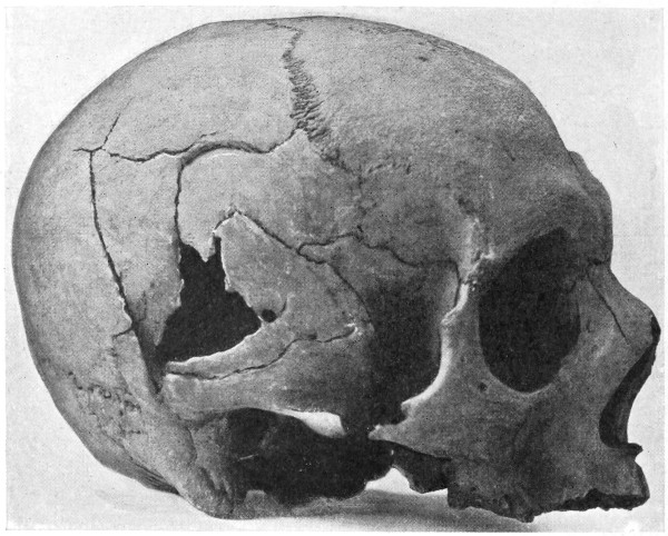
Fig. 42. A Comminuted Fracture of the Skull.
If time and occasion permit, an X-ray photograph will clinch
the matter.
When the fracture involves the internal table alone, the symptoms
are less definite. Teevan stated that this class of fracture does
not lead to the development of any symptoms unless:—
The middle meningeal artery be injured.
The dura mater and brain be irritated.
The brain be compressed.
He also adds that the existence of the following features suggests
the nature of the injury:—
The history of a slight blow, probably with a small body.
The blow situated over the parietal region.
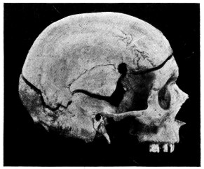
A
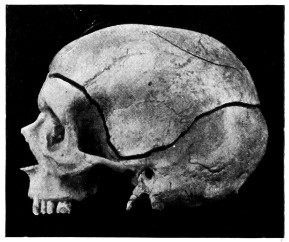
B
Fig. 43. An Explosive Fracture of the Vault of the Skull. A shows
the right side of the skull and the site of entry of the bullet. B shows the extension
of the fracture round the left half of the skull. In the left upper parietal region
(Fig. B) a fissured fracture is seen where the bullet failed to perforate the skull.
Chronic fixed pain, some days or weeks afterwards, with symptoms
of brain-irritation or encephalitis, and even suppuration,
suggesting irritation of the meninges and brain by spicules of bone.
Compression and paralysis on the opposite side of the body some
hours afterwards, as the result of injury to the middle meningeal
artery.
Symptoms of compression early and slight, combined with
partial paralysis of the opposite side of the body.
‘The less definite the symptoms of compression, the greater the
reason to believe that they are caused by the internal table only.’
Before entering into the question of treatment of fractures of the
skull, it is essential that allusion should be made to the temperature
in its relation to head-injuries. The great importance of temperature
changes in the consideration of operative treatment will be manifest
when discussing the treatment of fractures of the base.
The
various temperature changes observed in cases of head-injury have
afforded a subject prolific in discussion. Broca, Battle, Guyon,
Walsham, and others have endeavoured to deduce facts from the
examination of head cases, but the results have been rather indefinite.
In over 300 cases that have come under my observation,
accurate records of the temperature charts were obtained, the
patients were watched throughout their illness, and in the case of
death, the autopsies were attended.
The following deductions were made:—
1. That, for a variable period of time after the injury, the
temperature is always subnormal—sometimes so low that it cannot
be registered. This is the period of shock.
2. That the patient may die in this state of shock, but that,
if he lives, reaction takes place and the temperature rises.
3. That this rise in temperature is, in fatal cases, rapid and
progressive. In one case the temperature rose 6° in seven hours,
in another 8° in four hours. Death occurs when the temperature is
at its highest—anything up to 106°, and even more when registered
in the rectum.
4. That the temperature may rise to a moderate height, and
there ‘mark time’. This is the ‘crisis’ of the case. A subsequent
fall in temperature generally indicates recovery, a further rise usually
points to a fatal termination.
5. That the rise of temperature is independent of any special
osseous lesion, since similar changes are observed in fractures of the
vault and in fractures of the base.
6. That laceration of the brain is present in the majority of
cases in which marked temperature changes are observed, but that
the changes in temperature are totally independent of any special
regional brain-injury.
Richet arrives at the conclusion that two hypotheses present
themselves, accounting for temperature changes in general: (1) that
there are certain temperature regulators in the encephalon which,
when excited, become stimulated in function; (2) that the cortical
injury acts in a sort of reflex manner on the regulatory centres
situated in the pons or bulb.
Attention was also drawn to the experiments of Lorin and
van Benedin, who, after excising the two cerebral hemispheres of
a pigeon, showed that the heat regulatory centre was preserved
intact, proving that the corpus striatum is not necessarily the head-office
for heat-regulation in general.
Till, therefore, more evidence comes to hand with respect to
heat-regulation in general, one cannot go beyond the broad statement
that, in severe head-injuries, definite temperature changes occur,
that the changes are independent of any special lesion of bone
or brain, that they are generally associated with some brain-injury,
and that they are probably due to the influence exerted by the
lesion on the heat-regulating centres in the region of the pons
and bulb.
With regard to the value of the temperature, both with respect
to prognosis and treatment, a very much more definite statement
can be made.
First with respect to the prognosis. All cases of head-injury
may be grouped into four classes, according to the changes of temperature
present.
Group 1.
The temperature, at first subnormal, undergoes a rapid
and progressive rise. Prognosis most unfavourable.
Group 2.
The temperature, at first subnormal, rises gradually
to 101° or 102° and there ‘marks time’. This hesitancy marks the
crisis of the case, further elevation indicating a fatal result, whilst
a fall offers every hope of recovery.
Group 3.
The temperature, at first subnormal, rises to normal
and remains at that level. Prognosis very favourable.
Group 4.
The temperature, at first subnormal, remains subnormal.
The condition of shock persists, and the prognosis is
most unfavourable.
Practically all cases of head-injury fall readily into one or other
of these four groups, and the temperature chart presents so clear
a picture that it must be regarded as of the greatest possible value
in estimating the prognosis in any given case. In other words, the
temperature chart affords an almost infallible guide both to prognosis
and treatment.
With respect to alteration in temperature on the two sides of the
body, the clinical value of such changes must, from my own observations,
be regarded as of a very indefinite nature.
It now remains to discuss the value of temperature changes
with respect to treatment.
In the
earlier paragraphs of this chapter the special treatment of aural and
nasal hæmorrhages and cerebro-spinal discharge was discussed, and
some allusion was made to the necessity of exploring the middle
fossa in cases of profuse arterial or venous aural bleeding. The
general treatment of basic fractures now requires description, previous
to which, however, space must be made for a short account of the
routine treatment by Urotropin—as advised by Crowe and Cushing—as
a prophylactic against the development of meningeal infection.
The following is a summary of Crowe’s paper[25]
on the ‘excretion of
Urotropin in the cerebro-spinal fluid and its therapeutic value in
meningitis’.
1. Urotropin, when given by the mouth, invariably appears in
the cerebro-spinal fluid.
2. The largest amount of Urotropin is present in the cerebro-spinal
fluid from thirty minutes to an hour after the ingestion of
the drug.
3. After doses of Urotropin, within therapeutic limits, a sufficient
amount of the drug appears in the cerebro-spinal fluid to exercise
a decided inhibitory effect on the growth of organisms inoculated
into this fluid after its removal from the body.
4. Following a subdural inoculation of dogs and rabbits with
streptococcus, 60-80 grains of Urotropin a day, given under conditions
which insure absorption, will markedly defer, and in some
cases prevent, the onset of a fatal meningitis.
5. In view of these observations, the prompt administration of
Urotropin is advised in all clinical cases in which meningitis is a
possible or threatened complication, or even when meningeal infection
has actually occurred.
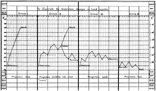
Fig. 44. A Temperature Chart illustrating the
Changes in Temperature observed in Head-injuries.
(For description, see text.)
Larger chart
Acting on these suggestions, I am accustomed to treat all head
cases with this drug, 10-20 grains three times a day by mouth.
It may be given per rectum, though probably with less satisfactory
results. Traumatic cases are treated as soon as possible, other cases
receive their dose for two or three days previous to the operation,
the drug being continued till all fear of possible meningeal infection
has vanished.
In discussing the general treatment of basic fractures, it must
be accepted that the basic fracture in itself requires no treatment.
Danger or death is dependent on intracranial complications, and an
uncomplicated basic fracture demands no active surgical treatment.
The special treatment applicable to cases complicated by hæmorrhages,
both external to and beneath the dura mater, is discussed in the next
chapter. It is therefore only necessary to enter into those cases of
fractured base which are associated with concussion, irritation, and
compression of the brain—in other words, the average case of fracture
of the base of the skull.
The surgeon must always be guided by the general condition of
the patient—the blood-pressure, character of pulse, state of respiration,
depth of unconsciousness, &c.—but, in my opinion, the temperature
chart affords perhaps the most valuable basis on which rules
can be formulated guiding the surgeon in his general line of treatment.
Thus (1) when the temperature remains subnormal the
patient is in a condition of severe cerebral shock, due, in the majority
of cases, to contusion or laceration of the brain, the grosser lesion
being most commonly situated either at the apex of the temporo-sphenoidal
lobe or at the anterior inferior part of the frontal lobe.
The patient is, however, suffering from shock, and the treatment
advocated for that condition in general is equally applicable to these
cases in particular. The patient should be placed in the so-called
head-down position, and the extremities firmly bandaged from below
upwards. The vaso-motor depression should be combated by the
administration of rectal or intra-venous infusions of saline solution,
to each pint of which is added 1 drachm of a 1 in 1,000 solution of
Adrenalin Chloride. It is at once obvious that, whilst combating the
condition of shock, this mode of treatment may tend at the same time
to encourage fresh bleeding if the brain should have been lacerated,
or if intracranial vessels should have been torn. For this reason
I regard rectal infusion as the more safe of the two methods, in spite
of the fact that intra-venous infusion brings about a more rapid
improvement in the patient’s condition. The effect of the infusion
must be carefully observed, and as soon as the blood-pressure rises
and the temperature shows a tendency to rise—say from the subnormal
to the normal—the infusion process must be stopped, and
the further progress of the case observed.
The risks attendant on this mode of treatment are obvious,
but in their consideration, it must also be borne in mind that if the
patient remains in the collapse stage, without evidencing any sign
of reaction, he will inevitably die. If the treatment advocated should
tide the patient over this stage and induce a definite reaction, as
exemplified by rise of blood-pressure and elevation of temperature,
the further treatment of the case can be considered under the next
group.
(2) When the temperature rises progressively the patient
evidences symptoms pointing to compression of the brain—coma,
slow pulse, noisy and stertorous respiration, hot skin and turgid face.
Later on, as a result of the increasing pressure, the medullary centres
begin to show signs of exhaustion, the pulse-rate increasing, and the
respiration becoming Cheyne-Stokes in character.
Whether these symptoms result primarily from the nature of the
lesion, or secondarily after the adoption of those measures advocated
for the collapse stage, it matters not. Our indications with respect
to treatment are clear. The increasing intracranial pressure must
be reduced. This desideratum is preferably carried out by the ligature
of a bleeding meningeal artery, or by the occlusion of a torn venous
sinus, but unfortunately, such a course is frequently out of the question.
We have, therefore, to deal with an increased and increasing
intracranial pressure, without definite localizing features. For this
condition we have at our disposal the following measures: venesection,
lumbar puncture, and ‘decompression’ operations.
Venesection.
I regard venesection as a valuable means of
reducing the intracranial pressure. It is mainly of use in those cases
that hover between slight compression and the fully-developed
condition. Venesection may therefore be regarded as of special
advantage in those cases that show elevation of temperature to about
101° and there ‘mark time’. The ‘bleeding’ should be carried out
after exposure of the median basilic, external jugular, or internal
saphenous veins, preferably from the first-named site. The amount
of blood to be withdrawn varies according to the individual circumstances
of the case, but, in general, the escape of blood should be
encouraged till the pulse becomes soft, frequent and compressible.
On an average, the quantity of blood withdrawn varies from 10-20
ounces. The operation not infrequently turns the scale in favour of
the patient. Venesection may also be carried out in combination
with ‘decompression’ operations.
When applied to suitable cases and carried out with discrimination,
venesection often tides the patient over the stage of ‘crisis’.
The most careful observation is needed in estimating the quantity
of blood to be withdrawn, the pulse, blood-pressure, respiration, and
temperature affording an adequate guide.
Lumbar puncture.
In spite of the apparent advantages of this
method, it must, I think, be acknowledged that lumbar puncture
is of but little use in reducing the general intracranial pressure.
Consequently, in spite of the fact that this procedure has been
strongly recommended, experience shows that it is of little practical
use. This statement, based on personal experience, is at variance
with the opinion of some other surgeons. For instance, de Quènu
reports 7 cases of fractured skull which were ‘cured’ by this treatment,
one of them requiring to be punctured eight times. Lumbar
puncture assists the diagnosis, but I am doubtful whether it improves
the prognosis.
‘Decompression’ operations.
‘Decompression’ operations may
be carried out over the cerebellar fossa or over the temporal region
of the skull. In cerebellar decompression a suitable scalp-flap
is turned down and the trephine applied over the centre of the
exposed occipital bone. After the removal of the disk the wall of
the cerebellar fossa is cut away with the craniectomy forceps, up to
the line of the lateral sinus above, to the mastoid process in front,
to the vicinity of the foramen magnum below, and almost up to the
middle line on the inner side. The bulging dura mater is incised in
a crucial manner, right up to the margins of the osseous gap, all
meningeal vessels that cross the line proposed for dural section being
under-run with a fully-curved needle threaded with catgut. A small
drainage-tube is inserted between the dura and the cerebellum and
brought out through the most dependent part of the scalp-flap. The
flap is then sutured in position. The drainage-tube should be stitched
to the skin and withdrawn twenty-four to forty-eight hours later,
according to the progress of the case.
I have carried out this operation on several occasions, but in
spite of some immediate improvement in the condition of the patient,
the remote results have been so unsatisfactory that I have abandoned
the operation entirely. The effect of this cerebellar decompression
is too radical, the medullary centres strongly object to such heroic
attempts at pressure relief.
Temporal decompression, more correctly known as the intermusculo-temporal
decompression operation of Harvey Cushing,
leads to very different results. Previous to dealing with the technique
of the operation, it will be convenient to enumerate the advantages
claimed for this method in general.
(1) The frequency with which the bony lesion occurs in the
middle fossa of the skull.
(2) The fact that cerebral contusions are especially liable to
involve the tip of the temporo-sphenoidal lobe.
(3) The exposure of the meningeal territory and the ease of
determining the presence of an extra-dural hæmorrhage.
(4) The possibility of draining through a split muscle rather than
directly through the scalp.
(5) The subsequent protective action of the muscle in case
a hernia tends to form in consequence of traumatic œdema.
(6) The subsequent absence of any deformities, the skin incision
being carried out for the most part through the hairy portion of the
scalp.
The operation.
The hair is shaved over the temporal region,
with a wide margin to the field of operation. In other respects, those
preliminary details should be adopted which are enumerated in
Chapter II. The skin incision commences well above and behind
the external angular frontal process, is directed along the line of,
but below the temporal crest, and curves downwards to terminate
just anterior to the tragus of the ear. Pressure is applied during the
formation of the incision so as to control bleeding from temporal
arteries. The flap, comprising skin, subcutaneous tissue, and superficial
temporal artery, is turned down to a zygomatic base, care being
taken to avoid injury to the temporal vessels at the base of the flap.
The temporal fascia is then incised, parallel to but immediately
below the line of the skin incision, and the fascia also turned down
towards the zygoma. The temporal muscle is split in the direction
of its fibres, from the temporal crest above to the zygoma below,
stripped away from the bone and well retracted in both forward and
backward directions. In this procedure care must be taken to avoid
detachment of the muscle-fibres from the temporal crest. The area
of bone exposed, however, should be as extensive as circumstances
permit.
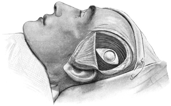
Fig. 45. Intermusculo-temporal Cerebral Decompression. First stage.
The scalp and temporal fascia have been turned down as separate flaps. The temporal
muscle is divided in the direction of its fibres, and the exposed bone trephined.
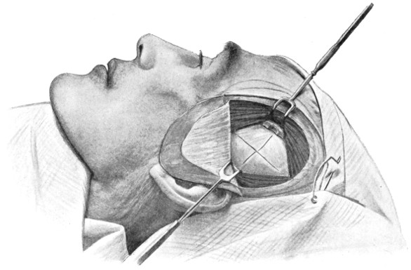
Fig. 46. Intermusculo-temporal Cerebral Decompression. Second Stage.
The temporal muscle is retracted on either side, the bone has been freely cut away,
and the bulging dura mater crucially incised.
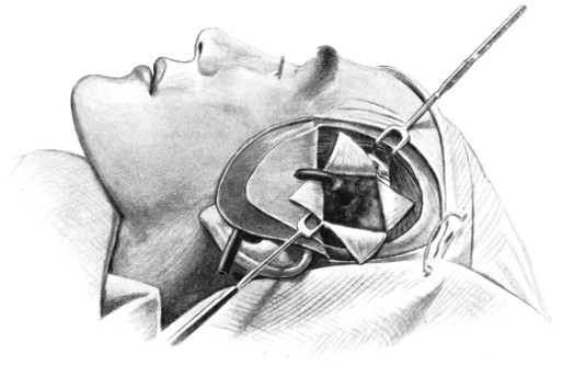
Fig. 47. Intermusculo-temporal Cerebral Decompression. Third Stage.
The dural flaps are turned aside, exposing the lacerated temporo-sphenoidal lobe.
A rubber drainage-tube has been inserted beneath the lacerated brain, lying on the
floor of the middle fossa of the skull and brought to the surface through the scalp-flap.
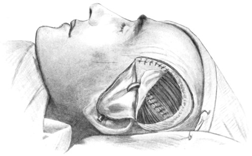
Fig. 48. Intermusculo-temporal Cerebral Decompression. Fourth Stage.
The temporal muscle-fibres have been approximated, and the temporal fascia reunited
in part. The drainage tube is seen to emerge through fascia and scalp.
The trephine is applied to the bone, the surgeon aiming at the
angle between the anterior and posterior branches of the middle
meningeal artery. The disk is removed, the dura separated from the
bone, and the craniectomy forceps called into requisition, the bone
entering into the formation of the temporal fossa being freely cut
away, more especially in the downward, forward, and backward directions—in
other words, in the general line of the temporo-sphenoidal
lobe. In the upward direction the surgeon must be more guarded,
more especially when the operation is being conducted on the left side
of the head. Broca’s area of motor speech must be avoided, for fear
of its inclusion in any hernial protrusion that may ensue.
The bulging and probably discoloured dura mater is freely
incised, preferably in a crucial manner, though the exact line of such
incision is of little importance so long as it is free in character. Needless
to say, all meningeal vessels that cross the lines proposed for
dural section must first be ligatured (by underrunning with a fully
curved needle) on either side of those lines. The four dural flaps are
turned aside and the antero-external aspect of the temporo-sphenoidal
pole exposed. A small drainage—or drain of rubber tissue—can now
be inserted along the floor of the middle fossa skull in the immediate
vicinity of any lacerated brain or blood-clot, the drain lying between
the dura and the brain, and anchored to the dura mater or muscle
by a fine catgut suture.
The four dural flaps are allowed to remain loose over the surface
of the brain, whilst the temporal muscle is sewn across from side to
side with a few catgut sutures, room being allowed at the lower angle
for the emergence of the drain.
The temporal fascia is replaced and carefully united to its upper
cut margin, the drain being brought out through the fascia and
through a puncture hole made at the most convenient part of the
scalp-flap. The scalp-flap is approximated with numerous interrupted
silk or salmon-gut sutures.
The tube is allowed to remain for thirty-six hours or more,
according to the condition of the patient.
The operation may be carried out on one or on both sides of
the skull; if on the one side only, on that side at which laceration
of the brain is probably existent. In connexion with this it is necessary
to state that laceration by contre-coup is more common than
direct brain-injury. Thus, if the blow be inflicted on the right
parieto-occipital region, the operation should be conducted in the
left temporal region.
In the absence of all localizing features the decompression
should be carried out on the right side of the head, in order to avoid
all possibility of including, in the hernial protrusion that may result,
the motor speech area of Broca.
In cases of severe head-injury the surgeon must be prepared for
many disappointments, but, from my own experience, it would appear
that Cushing’s operation frequently brings about the most satisfactory
results.
It should, however, be clearly understood that indiscriminate
decompression operations only bring discredit on the method in
general. They should only be carried out in suitable cases.
It is the general custom to
divide fractures of the vault into two groups—those demanding
operative treatment and those in which an expectant attitude is
advised—and between these two groups a sharp line of demarcation
is drawn. There is, however, no such line of demarcation. In the
event of the general condition of the patient being compatible with
operative interference, such treatment is urgently called for under
the following conditions:—
All cases of punctured fracture.
All cases of depressed fracture, whether diagnosed by palpation or
by inference, whether simple or compound, whether complicated or not.
All cases of fracture complicated by extra-dural or localized subdural
extravasation of blood.
All elevated fractures.
All compound fractures.
In all these instances, from the presence of depressed fragments
of bone, from associated injury to the intracranial contents, or from
other causes, no mere expectant policy should be pursued. The
surgeon has to look into the future, to bear in mind the possibility
of meningeal infection, and the more remote results of head-injuries
in general (see Chapter VI). In other words, early and active
surgical interference is imperatively demanded, for not only is it
necessary to strain every endeavour to save the patient’s life, but
the surgeon should also adopt those procedures which guard most
effectually against the more remote possibilities of the case.
With regard to simple fractures, if it can be determined that the
fracture, whether fissured, stellate, or comminuted, is simple and
uncomplicated by any serious intracranial lesion, no active surgical
treatment is required. The determination of such conditions is,
however, quite another matter, always difficult and sometimes, from
the presence of overlying hæmatomata, quite impossible.
In the general estimation of these cases it should be borne in
mind that simple uncomplicated vault fractures are decidedly rare.
For instance, Dwight, in 145 cases of fractured skull that came to
autopsy, only found six that evidenced a fissured fracture localized
to the vault. It may, of course, be urged that these statistics are
fallacious, insomuch as simple uncomplicated vault fractures would
probably not come to the post-mortem table. Clinical evidence,
however, coincides with Dwight’s statistics, and clinical evidence
shows, furthermore, that a blow sufficing to fracture the vault of the
skull almost invariably results in further injury.
In these doubtful cases the surgeon is greatly aided in his decision
by a general review of the patient’s condition, more especially by
those symptoms which are regarded as exemplifying the clinical
conditions of concussion, irritation, and compression.
When the fracture is associated with mild concussion
it may be
inferred that the brain is practically uninjured, and that operative
treatment is not required.
When the fracture is associated with severe concussion
it may be
inferred that the brain is damaged, to a degree proportionate to the
depth and duration of the stage of unconsciousness. The question
of operation depends to a very large extent on the general condition
of the patient, and more especially on the temperature and temperature
changes (see p. 114). With a persistent subnormal temperature
it may be inferred that the brain-injury is of a very severe nature, and
that operative measures are, for the time being, contra-indicated.
In the event of the temperature rising it may be accepted that the
patient is passing from the state of shock to that of reaction, operative
measures again hinging on the further progress of the case. If the
temperature rises progressively, the patient passing from the stage
of reaction to that of compression, operative measures are indicated.
Under other circumstances an expectant attitude can be adopted.
When the fracture is associated with general cerebral irritation
it may be inferred that the brain is contused or slightly lacerated.
Under these circumstances operation is, for the time being, not
required, and an expectant attitude should be adopted.
When the fracture is associated with compression
it may be inferred
that the condition is dependent on depressed fragments of bone,
or on extra- or intra-dural bleeding. Operative measures are now
urgently indicated.
When the fracture is confined to the internal table
a diagnosis is
only possible when symptoms of localized irritation ensue, either due
to pressure exercised on the cortex by spicules of bone or resulting
from injury to some intracranial vessel, e. g. the middle meningeal
artery. In such cases operation is indicated. Under other circumstances
the surgeon must await the developments of the case.
When the fracture is associated with mild concussion.
During the stage of concussion no active treatment is required, beyond
putting the patient to bed—with the head low—enveloping him in hot
blankets, and applying hot bottles to the extremities. These bottles
are wrapped up in flannel: they should never be placed in contact
with the patient’s skin. As soon as the reaction stage commences,
the bottles and blankets should be removed, the patient covered
with light clothing only, and an ice-bag applied to the head, over
the region at which the injury was inflicted. Calomel, in suitable
doses, should be given by mouth, and a copious enema administered
per rectum. Perhaps the most essential point in the general consideration
of the case is the after-treatment. The patient should be
kept in hospital or home for at least one month, and for the next
three months or more, according to the urgency of the case, he should
abstain from all mental and bodily work. At the end of that period
of time he may return to work, though still kept under observation.
When the fracture is associated with severe concussion.
In the
event of persistence of concussion symptoms of a severe type the
prognosis is decidedly unfavourable. This prolongation of cerebral
shock is undoubtedly dependent in its persistence on severe vaso-motor
depression, and in the majority of cases ending unfavourably
it will be found that the brain is generally œdematous and regionally
contused or lacerated. In cases of this nature the surgeon should
confine his attention, for the time being, to an endeavour at terminating
this vaso-motor depression, raising the blood-pressure and inducing
a definite reaction; the patient being treated after the manner
indicated on p. 118.
When the fracture is associated with general cerebral irritation.
The patient should be put to bed in a quiet and darkened room, under
the continuous attention of a trained nurse. An ice-bag or Leiter’s
tubes may be applied to the head, though the patient seldom tolerates
such attentions. I regard morphia as of the greatest service in this
class of cases. It should be given freely, subcutaneously, either by itself
or in combination with atropin. I generally order 1⁄4 gr. morphia with
1⁄100 gr. atropin every six hours, till the irritative symptoms subside.
Insomuch as all evidence tends to prove that general cerebral
irritation, as resulting from injury, implies a condition of contusion
or superficial laceration, a prolonged period of bodily and mental rest
is essential before the patient is discharged from treatment and
allowed to return to work—a period of not less than six months.
The usual preparatory treatment is carried out, the scalp-tourniquet
applied, a suitable scalp-flap framed and turned down, advantage
being taken of all existent scalp lacerations.
The subsequent details vary according to the circumstances of
the case:—
If, after thorough exposure of the
parts, the operator is satisfied that he has to deal with an uncomplicated
fissured fracture, the scalp-flap is accurately sewn
into position (see p. 16). On the other hand, if the symptoms
point to the presence of an extra-dural hæmorrhage, the bone is
trephined where the fracture crosses the line of the suspected vessel
and the conditions treated as described under middle meningeal
hæmorrhage. In subdural extravasation the trephine is applied over
that part of the brain, in the immediate vicinity of the fracture,
from which the symptoms appear to emanate, and the conditions
treated as described under subdural hæmorrhage.
In a certain proportion of cases the symptoms point to a marked
increase of intracranial pressure without definite localizing features,
a condition pointing to diffuse subdural hæmorrhage, with or without
brain laceration. Under such circumstances much may be done to
relieve the increased intracranial pressure by carrying out the operation
of ‘cerebral decompression’ (see p. 121).
The operative details vary according to
the nature of the osseous lesion:—
(a) If the depressed fragments of bone are so interlocked that
elevation is impossible (e. g. pond and gutter fractures), it will be
necessary to trephine in the immediate vicinity of the depression, in
order to lessen the mutual attachment of the fragments one to another,
so obtaining sufficient purchase for their elevation or removal.
The pericranium is stripped away from the region of the depression
(see Fig. 49), and a small trephine applied in such a manner
that the trephine circle includes the outer part of the depressed area.
Care must be taken to avoid more pressure over the depressed
fragments than is absolutely necessary, for fear of causing further
damage to the underlying structures. This advice is all the more
important when it is realized that the fracture of the internal table
is almost invariably more extensive than that which involves the
visible external table of the skull.
The disk of bone is removed and the conditions investigated with
the dural separator. When the degree of depression and the splintering
of the internal table are not excessive, the parts may be elevated
into position. In most cases, however, the uncertainty that exists
with regard to possible dural injury renders it necessary that the
depressed fragments should be elevated and removed, or so raised
as to allow of adequate inspection of the dura itself. For this purpose
the craniectomy forceps may be required. The dura can now be
examined. If torn, the opening may require enlargement, in order
to determine whether any osseous fragments have been driven into
the brain-substance; all such fragments are carefully removed.
Hæmorrhage is arrested, the dura sewn up, and the scalp-flap
carefully sutured. Drainage should always be avoided, the risk of
infection is too great. As a prophylactic against the development
of meningeal infection, the patient should undergo the routine treatment
with urotropin (see p. 116).
(b) When the depression is of such a nature that the elevation of
the fragments can be carried out without preliminary trephining, the
fragments are raised with the periosteal separator, and the membranes
and brain examined as described above.
In simple comminuted fractures the larger fragments of
bone are to be replaced in the anticipation that they will live entire,
in part, or will act as scaffolding media for the formation of dense
fibrous tissue. After removal from the wound, and during the completion
of the operation, the vitality of all loose fragments may be
preserved by immersion in hot saline solution. All minute fragments
of bone should be discarded.
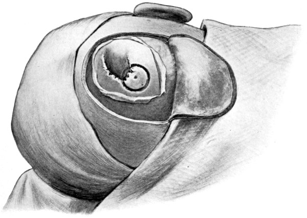
Fig. 49a. The Elevation of a Depressed Fracture. First stage.
The trephine circle includes the outer portion of the depressed area.
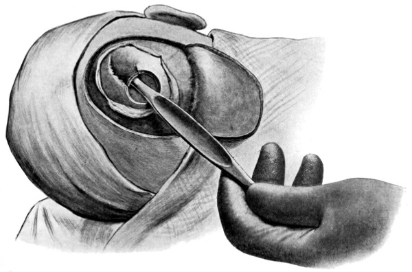
Fig. 49b. The Elevation of a Depressed Fracture. Second stage.
The trephine disk has been removed and the elevator is in position.
With respect to compound fractures, it is necessary that the
bone lesion should be fully exposed, all pockets of scalp-tissue being
slit up to their termination. Previous to any attempt at examination
of the injured bone, further precautions must be taken to avoid
subsequent infection, by reason of the bruised and soiled scalp. Such
tags of scalp-tissue as appear injured beyond repair should be cut
away with the scissors, and in order to avoid or diminish subsequent
wound infection, I have been accustomed to swab over the most
suspicious parts with pure carbolic acid, washing away the same with
saline solution. Since the advent of iodine sterilization, I have often
utilized that solution in preference to the carbolic, swabbing the whole
surface exposed. I think a combination of the two methods is advisable
in more serious cases, utilizing the carbolic for the margins of the
wound and iodine for the general surface. By means of this method
the risk of meningeal infection and scalp suppuration is greatly
reduced. To aid in the prophylaxis, the patient should again be
placed under a course of treatment by urotropin (see p. 116).
With regard to the bone, all loose fragments should be removed,
only those being preserved which retain their pericranial connexions.
Even these are thrown back so as to permit of the maximum inspection
of the dura mater. This membrane, if torn, is either sewn up
at once or opened up more freely in the investigation and removal
of underlying blood-clot or bone-débris. After removing such troubles
the membrane is accurately sutured. The bone-flaps are now replaced
in position. Some surgeons advocate the replacement of the smaller
fragments of bone which have previously been removed. These
fragments may be sterilized by boiling, but such a process destroys
the bone-cells and, in consequence, they become absorbed, merely
acting as scaffolding media for the formation of fibrous tissue. Added
to that, in the event of suppuration, the presence of such fragments
not only leads to the persistence of a purulent discharge, till the fragments
are entirely removed, but also increases considerably the risk
of meningeal infection.
As a summary, therefore, it may be laid down that it is necessary
to remove all loose fragments of bone, the deficiency in the vault
being rectified, if necessary, at a later date by one of the methods
enumerated in Chapter VI.
The scalp-flap is accurately sewn up with interrupted salmon-gut
sutures, a gauze or cigarette drain being inserted at the most convenient
and dependent point, to be removed at the end of forty-eight
hours or more according to the progress of the case.
Here there is a special liability to
dural laceration and in-driving of comminuted fragments of bone.
A full exposure of the parts is therefore absolutely essential. The
trephine can be applied in the immediate vicinity of the puncture,
or, as is often advisable, in such a manner that the punctured area
is included in the trephine circle: this latter method may necessitate
that the trephining should be carried out without the aid of the
guiding fixation-pin, for which process some experience is needed.
After removal of the bone, the craniectomy forceps may be
required, to allow of adequate dural inspection. The dura mater is
opened up with blunt-pointed scissors, and the brain examined for
in-driven fragments of bone. These, when found, are removed.
Deeply situated fragments of bone and foreign bodies may be previously
diagnosed by means of an X-ray picture.
If possible, both membrane and scalp should be sutured without
drainage, but in the event of possible sepsis, a small cigarette drainage-tube
should be inserted so as to lie beneath the dura mater on the
one hand and emerge through the scalp wound at the other.
For this class of
fracture the reader is referred to the section dealing with bullet-wounds
of the skull (see p. 296).
As previously
indicated, the diagnosis of this condition is only practicable when the
depression of the osseous fragments, or the hæmorrhage resulting
from an injured meningeal vessel, so irritate the dura mater and brain
that localizing symptoms ensue. For instance, epileptic fits of a
Jacksonian type may develop shortly after the accident, this condition
demanding a full exposure of the affected region. The operations
required in the treatment of traumatic epilepsy and intracranial
hæmorrhages are discussed in subsequent chapters.
Whatever the nature of the osseous lesion, the mortality is almost
entirely dependent on the question of associated injury to the intracranial
contents, more especially the brain.
In the case of fractures purely or mainly basic, of the cases that
have come under my care or observation, 116 recovered and 89 died—a
mortality of 44 per cent.
Battle, from an examination of 168 cases, puts the mortality at
the low figure of 32 per cent., whilst Crandon and Wilson, from an
exhaustive series of 530 cases, estimates it at 44 per cent.
The mortality varies according to the age of the patient, the
death-rate being at its maximum at the two extremes of life—between
50 and 60 per cent. under the age of 5 years and over 60 per cent.
after the age of 70.
The mortality varies also according to the fossa implicated. Thus,
to each fossa there are certain peculiar dangers—to the anterior
fossa, meningeal infection from the nose, frontal and ethmoidal
sinuses; to the middle fossa, meningeal infection from the naso-pharynx
and ear, and hæmorrhage from the middle meningeal and
internal carotid arteries, and from the cavernous venous sinus; and to
the posterior fossa, laceration of the lateral and sigmoid venous sinuses.
After such consideration of local complications, it would appear
that the middle fossa of the skull presents the greatest dangers.
Another factor must, however, be taken into consideration—injury
to the brain—the commonest cause of death in all head injuries.
The main vital centres are grouped in the region of the fourth ventricle,
and Leonard Hill has proved that any increase in the intracranial
pressure exerts its influence most rapidly and most effectually
when the lesion is situated beneath the tentorium cerebelli.
One may assume, therefore, that fractures of the posterior fossa
are the most dangerous, and that those involving the anterior fossa
hold out the best prognosis.
In the case of pure or primary vault fractures the mortality
may be estimated by reference to the following table. It is composed
from 88 cases admitted into St. Bartholomew’s Hospital between the
years 1900 and 1906.
| Under the |
age of |
5 |
8 |
lived |
and |
2 |
died. |
Mortality, |
20 |
per |
cent. |
| „ |
„ |
10 |
2 |
„ |
„ |
3 |
„ |
„ |
60 |
„ |
| „ |
„ |
15 |
6 |
„ |
„ |
0 |
„ |
„ |
0 |
„ |
| „ |
„ |
20 |
10 |
„ |
„ |
3 |
„ |
„ |
23 |
„ |
| „ |
„ |
30 |
9 |
„ |
„ |
1 |
„ |
„ |
10 |
„ |
| „ |
„ |
40 |
16 |
„ |
„ |
9 |
„ |
„ |
36 |
„ |
| „ |
„ |
50 |
3 |
„ |
„ |
7 |
„ |
„ |
70 |
„ |
| „ |
„ |
60 |
2 |
„ |
„ |
1 |
„ |
„ |
33 |
„ |
| Over the age of |
60 |
3 |
„ |
„ |
3 |
„ |
„ |
50 |
„ |
| |
59 |
lived |
and |
29 |
died. |
Mortality, |
37 |
per |
cent. |
For the remote effects of skull fractures, see Chapter VI.
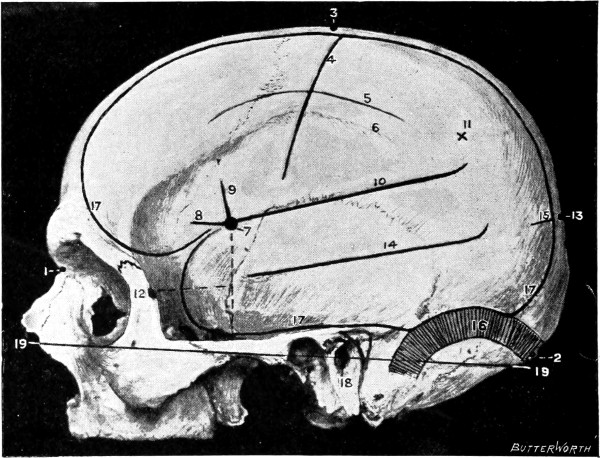
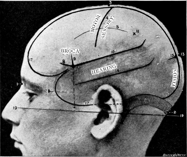
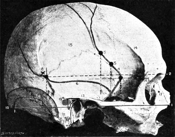
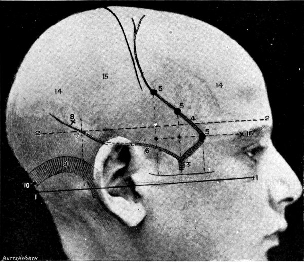
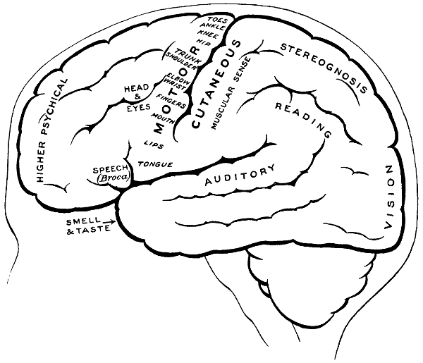
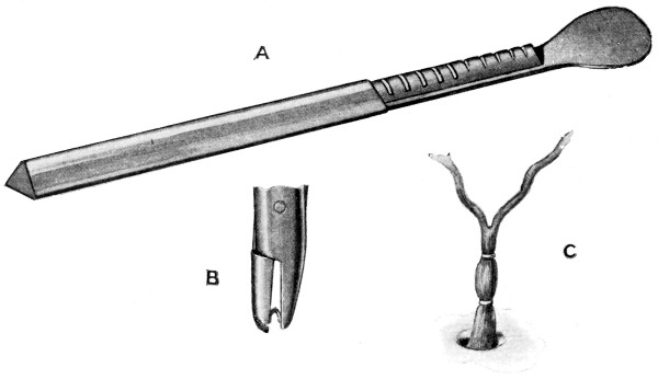
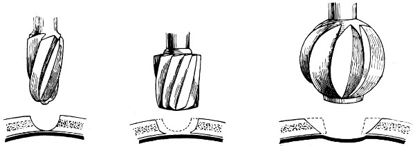
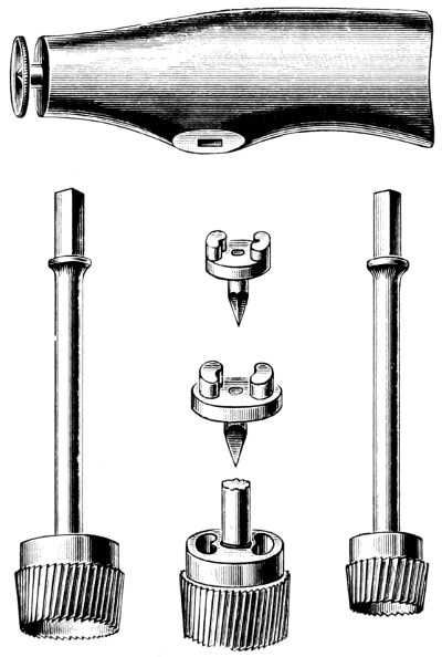
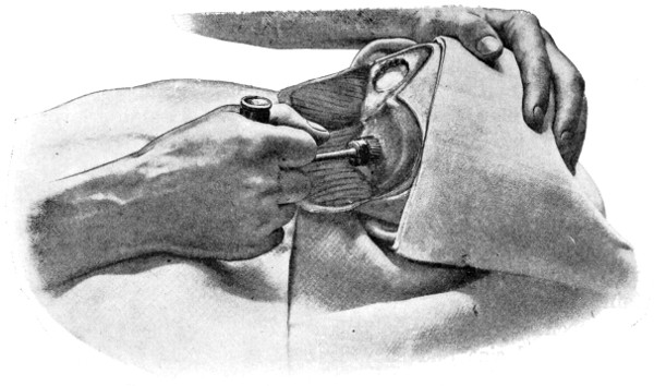
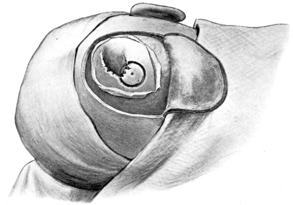

 -shaped
incision is made in such a manner as
to include the area which it is desired to expose. The two vertical
limbs of the incision should converge to such a degree as to allow of
the subsequent ready fracture of the flap along its base. The knife
is entered at one extremity, carried down to the bone, and the three
incisions rapidly made, one after the other. Along the line of each
of the three incisions, the pericranium is stripped away from the bone
so as to allow of adequate exposure. At the anterior and posterior
angles of the flap the tissues are retracted a little more, permitting
the application of a half-inch diameter trephine. Here the two
trephine-holes are bored—with the usual precautions against damage
to the dura mater—and the two disks of bone elevated and removed.