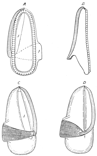
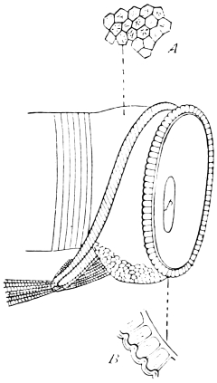
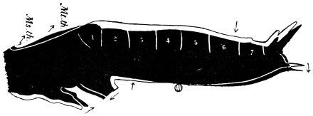
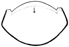
The Outer Skeleton.
SPECIAL REFERENCES.
Krukenberg. Vergleichend-Physiologische Vorträge. IV.—Vergl. Physiologie
der Thierischen Gerüstsubstanzen. (1885.) [Chemical Relations of Chitin.]
Graber. Ueber eine Art fibrilloiden Bindegewebes der Insectenhaut. Arch. f.
mikr. Anat. Bd. X. (1874.) [Minute Structure of Integument.] Also,
Viallanes. Recherches sur l’Histologie des Insectes. Ann. Sci. Nat., Zool.
VIe Série, Tom. XIV. (1882).
Audouin. Recherches anatomiques sur le thorax des Insectes, &c. Ann. Sci. Nat.
Tom. I. (1824.) [Theoretical Composition of Insect Segments.] Also,
Milne-Edwards. Leçons sur la Physiologie et l’Anatomie Comparée. Tom. X.
(1874.)
Savigny. Mémoires sur les animaux sans vertèbres. Partie Ie. Théorie des
organes de la bouche des Crustacées et des Insectes. (1816.) [Comparative
Anatomy of the Mouth-parts.]
Muhr. Ueber die Mundtheile der Orthopteren. Prag. 1877. [Mouth-parts of
Orthoptera.]
When the skin of an Insect is boiled successively in acids,
alkalies, alcohol, and ether, an insoluble residue known as
Chitin (C15H26N2O10) is obtained. It may be recognised and
sufficiently separated by its resistance to boiling liquor potassæ.
Chitin forms less than one-half by weight of the integument, but
it is so coherent and uniformly distributed that when isolated
by chemical reagents, and even when cautiously calcined, it
retains its original organised form. The colour which it frequently
exhibits is not due to any essential ingredient; it may
be diminished or even destroyed by various bleaching processes.
The colouring-matter of the chitin of the Cockroach, which is
amber-yellow in thin sheets and blackish-brown in dense
masses, is particularly stable and difficult of removal. Its composition
does not appear to have been ascertained; it is white
when first secreted, but darkens on exposure to air. Fresh-moulted
Cockroaches are white, but gradually darken in three
or four hours. Lowne30 observes that in the Blow-fly the pigment
is “first to be met with in the fat-bodies of the larvæ.
These are perfectly white, but when cut from the larva, and
exposed to the air, they rapidly assume an inky blackness....
When the perfect insect emerges from the pupa, and
respiration again commences, the integument is nearly white,
or a faint ashy colour prevails. This soon gives place to the
characteristic blue or violet tint, first immediately around those
portions most largely supplied with air vessels.” Professor
Moseley31 tells us that, thinking it just within the limits of
possibility that the brown coloration of the Cockroach might be
due to the presence of silver, he analysed one pound weight of
Blatta. He found no silver, but plenty of iron, and a remarkable
quantity of manganese. That light has some action upon
the colouring matter seems to be indicated by the fact that in a
newly-moulted Cockroach the dorsal surface darkens first.
Chitin is not peculiar to Insects, nor even to Arthropoda.
The pen of cuttle-fishes and the shell of Lingula contain the
same substance,32 which is also proved, or suspected, to occur in
many other animals.
The chemical stability of chitin is so remarkable that we
might well expect it to accumulate like the inorganic constituents
of animal skeletons, and form permanent deposits.
Schlossberger33 has, however, shown that it changes slowly
under the action of water. Chitin kept for a year under water
partially dissolved, turned into a slimy mass, and gave off a
peculiar smell. This looks as if it were liable to putrefaction.
The minute proportion of nitrogen in its composition may
explain the complete disappearance of chitin in nature.
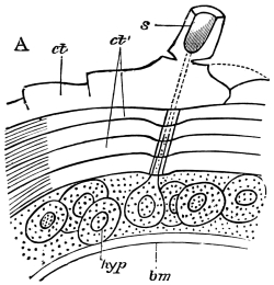
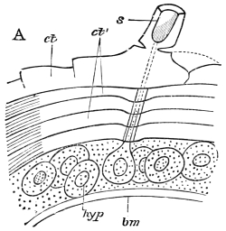
Fig. 8.—Diagram of Insect integument, in section. bm, basement membrane;
hyp, hypodermis, or chitinogenous layer; ct, ct′, chitinous cuticle; s, a seta.
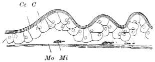
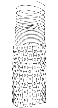
The chitinous exoskeleton is rather an exudation than a true
tissue. It is not made up of cells, but of many superposed
laminæ, secreted by an underlying epithelium, or “chitinogenous
layer.” This consists of a single layer of flattened cells,
resting upon a basement membrane. A cross-section of the
chitinous layer, or “cuticle,” examined with a high power
shows extremely close and fine lines perpendicular to the laminæ.
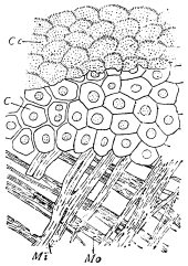
The cells commonly form a mosaic pattern, as if altered in
shape by mutual pressure. The free surface of the integument
of the Cockroach is divided into polygonal, raised spaces. Here
and there an unusually long, flask-shaped, epithelial cell projects
through the cuticle, and forms for itself an elongate chitinous
sheath, commonly articulated at the base; such hollow sheaths
form the hairs or setæ of Insects—structures quite different
histologically from the hairs of Vertebrates.
The polygonal areas of the cuticle correspond each to a
chitinogenous cell. Larger areas, around which the surrounding
ones are radiately grouped, are discerned at intervals, and these
carry hairs, or give attachment to muscular fibres.
Viallanes (loc. cit.) has added some interesting details to what
was previously known of Insect-hairs. There are, he points
out, two kinds of hairs, distinguished by their size and structure.
The smaller spring from the boundary between contiguous
polygonal areas, and have no sensory character. The larger
ones occupy unusually large areas, surmount chitinogenous cells
of corresponding size, and receive a special nervous supply.
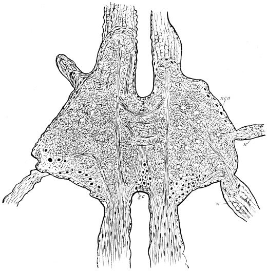
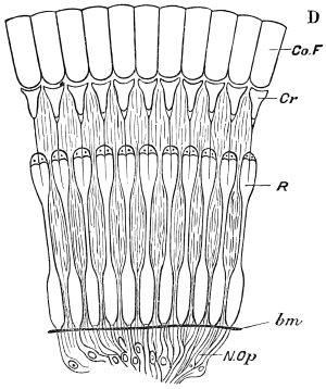
The nerve34 expands at the base of the hair into a spindle-shaped,
nucleated mass (bipolar ganglion-cell), from which issues
a filament which traverses the axis of the hair, piercing the
chitinogenous cell, whose protoplasm surrounds it with a sheath
which is continued to the tip of the hair. Such sensory hairs
are abundant in parts which are endowed with special sensibility.
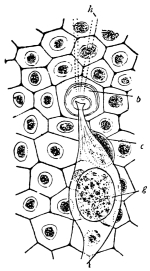
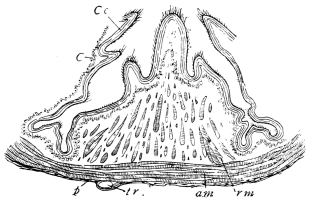
Fig. 9.—Nerve-ending in skin of Stratiomys larva. h, hairs; b, their chitinous
base; c, nucleus of generating cell; g, ganglion cell. × 250. Copied
from Viallanes.
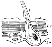
Fig. 10.—Diagram of sensory hair of Insect. Cc, chitinous cuticle; h, hair; c, its
generating cell; g, ganglion cell; bm, basement-membrane.
The chitinous cuticle is often folded in so as to form a deep
pit, which, looked at from the inside of the body, resembles a
lever, or a hook. Such inward-directed processes serve chiefly
for the attachment of muscles, and are termed apodemes (apodemata).
A simple example is afforded by the two glove-tips
which lie in the middle line of the under-surface of the thorax
(p. 58, and fig. 27). In other cases the pit is closed from the
first, and the apodeme is formed in the midst of a group of
chitinogenous cells distant from the superficial layer, though
continuous therewith. Many tendons of insertion are formed
in this way. The two forked processes in the floor of the thorax
(p. 58, and fig. 27) are unusually large and complex structures of
the same kind. In the tentorium of the head (p. 39, and fig. 17)
a pair of apodemes are supposed to unite and form an extensive
platform which supports the brain and gullet.
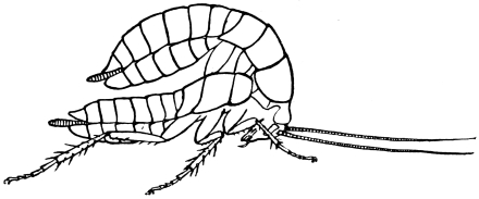
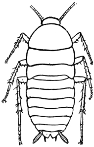
Fig. 11.—Nymph (in last larval stage) escaping from old skin. × 2 1/2.
Like other Arthropoda, Insects shed their chitinous cuticle
from time to time. A new cuticle, at first soft and colourless, is
previously secreted, and from it the old one gradually becomes
detached. The setæ probably serve the same purpose as the
“casting-hairs” described by Braun in the crayfish, and by
Cartier in certain reptiles,35 that is, they mechanically loosen the
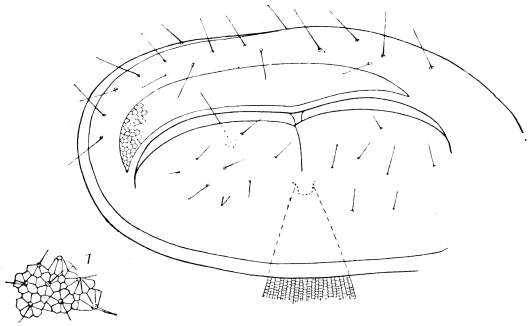
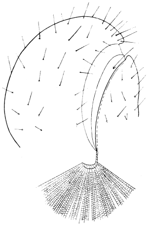
old skin by pushing beneath it. In many soft-bodied nymphs
the new skin can be gathered up into a multitude of fine
wrinkles, which facilitate separation, but we have not found
such wrinkles in the Cockroach, except in the wings. The
integument about to be shed splits along the back of the
Cockroach, from the head to the end of the thorax,36 and the
animal draws its limbs out of their discarded sheaths with much
effort. It is remarkable that the long, tapering, and many-jointed
antennæ are drawn out from an entire sheath. At the
same time the chitinous lining of the tracheal tubes is cast,
while that of the alimentary canal is broken up and passed
through the body.
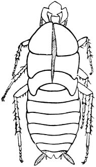
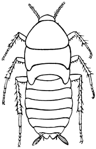
Fig. 12.—Cast skin of older nymph (“pupa”). × 2 1/2.
Prolonged boiling in caustic potash, though it dissolves the
viscera, does not disintegrate the exoskeleton. This shows that
the segments of the integument are not separate chitinous
rings, but thickenings of a continuous chitinous investment.
Nevertheless, their constancy in position and their conformity
in structure often enable us to trace homologies between different
segments and different species as certainly as between corresponding
elements of the osseous vertebrate skeleton.
Audouin’s laborious researches into the exoskeleton of Insects37
resulted in a nomenclature which has been generally adopted.
He divides each somite (segment) into eight pieces, grouped in
pairs—viz., terga (dorsal plates), sterna (ventral plates), epimera
(adjacent to the terga), and episterna (adjacent to the sterna).
While admitting the usefulness of these terms, we must warn
the reader not to suppose that this subdivision is either normal
or primitive. The eight-parted segment exists in no single
larval or adult Arthropod. Lower forms and younger stages
take us further from such a type, instead of nearer to it; and
Audouin’s theoretical conception is most fully realised in the
thorax of an adult Insect with well-developed legs and wings.
The morphologist would derive all the varieties of Arthropod
segments from the very simple and uniform chitinous cuticle
found in Annelids and many Insect-larvæ. This becomes
differentiated by unequal thickening and folding in, and a series
of rings connected by flexible membranes is produced. Locomotive
and respiratory activity commonly lead to the definition
of terga and sterna, which are similarly attached to each other
by flexible membranes. A pair of limbs may next be inserted
between the terga and sterna, and the simple segment thus
composed occurs so extensively in the less modified regions
and in early stages that it may well be considered the typical
Arthropod somite.
Special needs may lead to the division of the sterna into
lateral halves, but this is purely an adaptive change. The
third thoracic sternum of the male Cockroach, and the second
and third of the female are thus divided, as is also the hinder
part of the seventh abdominal sternum of the female.
In an early stage every somite has its tergal region divided
into lateral halves, owing to the late completion of the body on
this side. Traces of this division may survive even in the
imago. There is often a conspicuous dorsal groove in the
thoracic terga, and at the time of moult the terga split along
an accurately median line (see fig. 12).
Additional pieces may be developed between the terga and
sterna, and these have long been termed pleural.38 There may
be, for example, single stigmatic plates, as in the abdomen of
the Cockroach, pieces to support the thoracic legs, and pieces to
support the wings; but the number and position of these plates
depends upon their immediate use, and their homologies become
very uncertain when Insects of different orders are compared.
In general, the pleural elements of the segment are late in
development, variable, and highly adaptive.
The exoskeleton of the Cockroach is divisible into about
seventeen segments, which are grouped into three regions, as
follows:—
| Head |  | Procephalic lobes | 3 |
| Post-oral segments |
| Thorax | 3 | 39 |
| Abdomen | 11 |
| | | — |
| | | 17 |
| | | — |
It is a strong argument in favour of this estimate that many
Insects, at the time when segmentation first appears, possess
seventeen segments.40 The procephalic lobes, from which a great
part of the head, including the antennæ, is developed, are often
counted as an additional segment.41
The limbs, which in less specialised Arthropoda are carried
with great regularity on every segment of the body, are greatly
reduced in Insects. Those borne by the head are converted into
sensory and masticatory organs; those on the abdomen are
either totally suppressed, or extremely modified, and only the
thoracic limbs remain capable of aiding in locomotion.
The primitive structure of the Arthropod limb is adapted to
locomotion in water, and persists, with little modification, in
most Crustacea. Here we find in most of the appendages42 a
basal stalk (protopodite), often two-jointed, an inner terminal
branch (endopodite), and an outer terminal branch (exopodite),
each of the latter commonly consisting of several joints. It
does not appear that the appendages of Insects conform to the
biramous Crustacean type, though the ends of the maxillæ are
often divided into an outer and an inner portion.
We shall now proceed to describe, in some detail, the regions
of the body of the adult Cockroach.
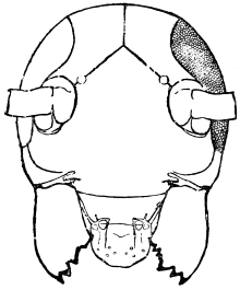
Fig. 13.—Front of Head. × 10.
The head of the Cockroach, as seen from the front, is pear-shaped,
having a semi-circular outline above, and narrowing
downwards. A side-view shows that the front and back are
flattish, while the top and sides are regularly rounded. In the
living animal the face is usually inclined downwards, but it can
be tilted till the lower end projects considerably forward. The
mouth, surrounded by gnathites or jaws, opens below. On the
hinder surface is the occipital foramen, by which the head communicates
with the thorax. A rather long neck allows the
head to be retracted beneath the pronotum (first dorsal shield
of the thorax) or protruded beyond it.
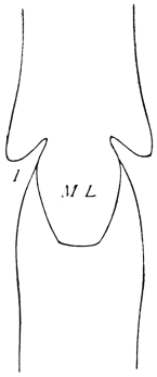
On the front of the head we observe the clypeus, which
occupies a large central tract, extending almost completely
across the widest part of the face. It is divided above by a
sharply bent suture from the two epicranial plates, which form
the top of the head as well as a great part of its back and sides.
The labrum hangs like a flap from its lower edge. A little
above the articulation of the labrum the width of the clypeus is
suddenly reduced, as if a squarish piece had been cut out of
each lower corner. In the re-entrant angle so formed, the
ginglymus, or anterior articulation of the mandible, is situated.
The labrum is narrower than the clypeus, and of squarish
shape, the lower angles being rounded. It hangs downwards,
with a slight inclination backwards towards the mouth, whose
front wall it forms. On each side, about halfway between the
lateral margin and the middle line, the posterior surface of the
labrum is strengthened by a vertical chitinous slip set with
large setæ. Each of these plates passes above into a ring, from
the upper and outer part of which a short lever passes upwards,
and gives attachment to a muscle (levator menti).
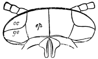
Fig. 14.—Top of Head. ep, epicranial plate; oc, eye; ge, gena. × 10.
The top and back of the head are defended by the two
epicranial plates, which meet along the middle line, but diverge
widely as they descend upon the posterior surface, thus enclosing
a large opening, the occipital foramen. Beyond the foramen,
they pass still further downwards, their inner edges receding in
a sharp curve from the vertical line, and end below in cavities
for the articulation of the mandibular condyles.43
The sides of the head are completed by the eyes and the
genæ. The large compound eye is bounded above by the
epicranium; in front by a narrow band which connects the
epicranium with the clypeus; behind, by the gena. The gena
passes downwards between the eye and the epicranial plate,
then curves forwards beneath the eye, and just appears upon
the front of the face, being loosely connected at this point with
the clypeus. Its lower edge overlaps the base of the mandible,
and encloses the extensor mandibulæ.
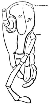
Fig. 15.—Side of Head. oc, eye; ge, gena; mn, mandible. × 10.
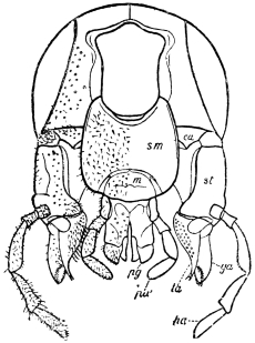
Fig. 16.—Back of Head. ca, cardo; st, stipes; ga, galea; la, lacinia; pa, palp;
sm, submentum; m, mentum; pg, paraglossa. × 10.
The occipital foramen has the form of an heraldic shield. Its
lateral margin is strengthened by a rim continuous with the
tentorium, or internal skeleton of the head. Below, the foramen
is completed by the upper edge of the tentorial plate,
which nearly coincides with the upper edge of the submentum
(basal piece of the second pair of maxillæ); a cleft, however,
divides the two, through which nerve-commissures pass from
the sub-œsophageal to the first thoracic ganglion. Through the
occipital foramen pass the œsophagus, the salivary ducts, the
aorta, and the tracheal tubes for the supply of air to the head.
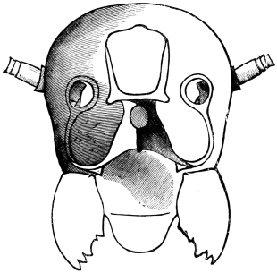
Fig. 17.—Fore-half of Head, with tentorium, seen from behind. × 12.
The internal skeleton of the head consists of a nearly transparent
chitinous septum, named tentorium by Burmeister, which
extends downwards and forwards from the lower border of the
occipital foramen. In front it gives off two long crura, or
props, which pass to the ginglymus, and are reflected thence
upon the inner surface of the clypeus, ascending as high as the
antennary socket, round which they form a kind of rim. Each
crus is twisted, so that the front surface becomes first internal
and then posterior as it passes towards the clypeus. The form
of the tentorium is in other respects readily understood from
the figure (fig. 17). Its lower surface is strengthened by a
median keel which gives attachment to muscles. The œsophagus
passes upwards between its anterior crura, the long flexor of the
mandible lies on each side of the central plate; the supra-œsophageal
ganglion rests on the plate above, and the sub-œsophageal
ganglion lies below it, the nerve-cords which unite
the two passing through the circular aperture. A similar
internal chitinous skeleton occurs in the heads of other Orthoptera,
as well as in Neuroptera and Lepidoptera. Palmén44 thinks
that it represents a pair of stigmata or spiracles, which have
thus become modified for muscular attachment, their respiratory
function being wholly lost. In Ephemera he finds that the
tentorium breaks across the middle when the skin is changed,
and each half is drawn out from the head like the chitinous
lining of a tracheal tube.
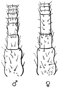
Fig. 18.—Base of Antenna of Male (to left) and Female (to right). × 24.
A pair of antennæ spring from the front of the head. In the
male of the common Cockroach they are a little longer than
the body; in the female rather shorter. From seventy-five
to ninety joints are usually found, and the three basal joints are
larger than the rest. Up to about the thirtieth, the joints are
about twice as wide as long; from this point they become more
elongate. The joints are connected by flexible membranes, and
provided with stiff, forward-directed bristles. The ordinary
position of the antennæ is forwards and outwards.
Each antenna is attached to a relatively large socket (fig. 15),
which lies between the epicranium and clypeus, to the front and
inner side of the compound eyes. A flexible membrane unites
the antenna to the margin of the socket, from the lower part of
which a chitinous pin projects upwards and supports the basal
joint.
It is well known that in many Crustacea two pairs of antennæ
are developed, the foremost pair (antennules) bearing two complete
filaments. Some writers have suggested that both pairs
may be present in Insects, though not simultaneously, the
Crustacean antennule being found in the larva, and the Crustacean
antenna in the adult. This view was supported by the
familiar fact that in many larvæ the antennæ are placed further
forward than in the adult. The three large joints at the base
of Orthopterous antennæ have been taken to correspond with
those of Crustacean antennules, and it has been inferred that in
Insects with incomplete metamorphosis, only antennules or
larval antennæ are developed.45 This reasoning was never very
cogent, and it has been impaired by further inquiry. Weismann
has shown that in Corethra plumicornis, the adult antenna,
though inserted much further back than that of the larva, is
developed within it,46 and Graber has described a still more
striking case of the same thing in a White Butterfly.47 There
is, therefore, no reason to suppose that Insects possess more than
one pair of antennæ, which is probably preoral, not corresponding
with either of the Crustacean pairs.
We have already noticed (p. 26) the superficial points in
which the antenna of the male Cockroach differs from that of
the female.
The eyes of some Crustacea are carried upon jointed appendages,
but this is never the case in Insects, though the eye-bearing
surface may project from the head, as in Diopsis and
Stylops. Professor Huxley48 supposes that the head of an Insect
may contain six somites, the eyes representing one pair of
appendages. The various positions in which the eyes of Arthropoda
may be developed weakens the argument drawn from the
stalk-eyed Crustacea. Claus and Fritz Müller go so far on the
other side as to deny the existence of an eye-segment even in
Crustacea.
Before entering upon a full description of the mouth-parts of
the Cockroach, which present some technical difficulties, the
beginner in Insect anatomy will find it useful to get a few
points of nomenclature fixed in his memory. Unfortunately,
the terms employed by entomologists are at times neither
convenient nor philosophical.
There are three pairs of jaws, disposed behind the labrum, as
in the diagram:—
1st pair of Jaws
(Mandibles).
3rd "
(Labium, or 2nd pair of Maxillæ).
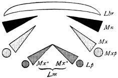
Fig. 19.—Diagram of Cockroach Jaws, in horizontal section.
The mandible is undivided in all, or nearly all, Insects. Each
maxilla may consist of
A palp on the outer side,
A galea (hood),
A lacinia (blade), on the inner side.
The galea (hood) of the 3rd pair of jaws is sometimes called
the paraglossa.
A tongue-like process may be developed from the front wall
of the mouth (epipharynx), or from the back wall (hypopharynx
or lingua).49 Both epipharynx and hypopharynx project into
the mouth, and, in some Diptera, far beyond it.
The tip of the labium is sometimes produced into a long
tongue, called the ligula (strap).
The mouths of Insects may be classed as:—
Biting.—Orthoptera, Neuroptera, Coleoptera (in some
Coleoptera a licking tongue is developed), most
Hymenoptera.
Licking and Sucking.—Some Hymenoptera—e.g., Honey
Bee.
Sucking.—(a) With lancets—Diptera, Hemiptera.
(b) Without lancets—Lepidoptera.
The reference of these to a common plan, and the determination
of the constituent parts, is mainly the work of Savigny.
Mouth-parts were made the basis of the classification of Insects
by Fabricius (1745–1808).
The mandibles of the Cockroach are powerful, single-jointed50
jaws, each of which is articulated by a convex “condyle” to the
lower end of the epicranial plate, and again by a concave
“ginglymus” to the clypeus. The opposable inner edges are
armed with strong tooth-like processes of dense chitin, which
interlock when the mandibles close; those towards the tip of
the mandible are sharp, while others are blunt, as if for crushing.
Each mandible can be moved through an angle of about
30°. A flexible chitinous flap extends from its inner border to
the labrum. The powerful flexor of the mandible arises within
the epicranial vault; its fibres converge to a chitinous tendon,
which passes outside the central plate of the tentorium, and at
a lower level through a fold on the lower border of the clypeus,
being finally inserted near the ginglymus. A short flexor arises
from the crus of the tentorium. The extensor muscle arises
from the side of the head, passes through the fold formed by
the lower end of the gena, and is inserted close to the outer side
of the condyle of the mandible.
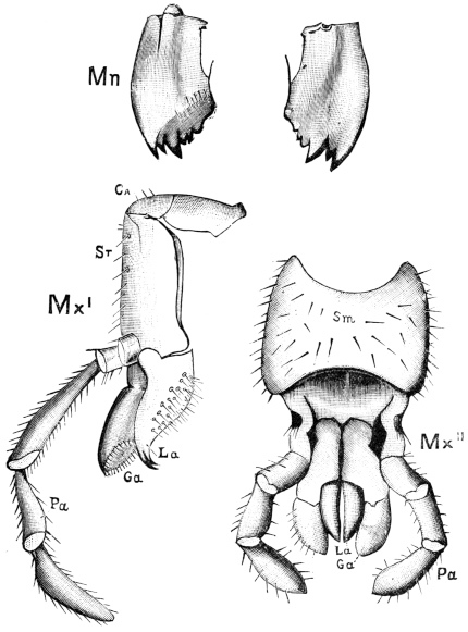
Fig. 20.—The Jaws, separated. Mn, mandible, seen from behind (to left) and front
(to right); Mx' maxilla (first pair); Mx" labium, or second pair of
maxillæ. The other letters as before. × 20.
The anterior maxillæ lie behind the mandibles, and like them
are unconnected with each other. They retain much more of
the primitive structure of a gnathite than the mandibles, in
which parts quite distinct in the maxillæ are condensed or
suppressed. The constituent pieces are seen in fig. 20. There
is a two-jointed basal piece, consisting of the cardo (ca) and the
stipes (st). The cardo is a transverse plate bent upon itself, and
enclosing muscles; it is attached to the outward-directed pedicel
of the occipital frame, and carries the vertical stipes. To the
side and lower end of the stipes is attached the five-jointed
palp (pa), a five-jointed limb used in feeding and in exploration,
while the lacinia (la) and galea (ga) are articulated to its
extremity. The lacinia is internal and posterior to the galea;
it is broad above, but narrows below to a bifid tooth of dense
chitin; its inner surface is beset with a cluster of strong setæ.
The galea is more flexible, and forms an irregular three-cornered
prism with an obliquely truncated end, upon which are many
fine hairs. A flexible and nearly transparent flap connects the
inner edges of the stipes and cardo, and joins both to the
labium. The muscles which move the bases of the maxillæ
spring from the crura, central plate, and keel of the tentorium.
On the posterior surface of the head, below the occipital
foramen, we find a long vertical flap, the labium, which extends
downwards to the opening of the mouth. It represents a
second pair of maxillæ, fused together in their basal half, but
retaining elsewhere sufficient resemblance to the less modified
anterior pair to permit of the identification of their component
parts. The upper edge is applied to the occipital frame, but is
neither continuous with that structure nor articulated thereto.
By stripping off the labium upwards it may be seen that it is
really continuous with the chitinous integument of the neck.
The broad shield-like base is incompletely divided by a transverse
hinge into an upper and larger piece, the submentum, and
a distal piece, the mentum. To the mentum are appended
representatives of the galeæ (here named paraglossæ) and laciniæ,
while a three-jointed palp with an additional basal joint (distinguished
as the palpiger) completes the resemblance to the
maxillæ of the first pair.51 In front of the labium, and lying
in the cavity of the mouth is a chitinous fold of the oral
integument, the lingua, which lies like a tongue in the floor of
the mouth. The common duct of the salivary glands enters the
lingua, and opens on its hinder surface. The lingua is supported
by the chitinous skeleton represented in the figures of the
salivary glands. (Chap. vii., infra.)
The epipharynx, which is a prominent part in Coleoptera and
Diptera, is not recognisable in Orthoptera.
We must now shortly consider the functions of the parts just
described. The antennæ have long been regarded as sense-organs,
and even the casual observer can hardly fail to remark
that they are habitually used by the Insect to gain information
concerning its immediate surroundings. Long antennæ, such
as those of the Cockroach, are certainly organs of touch, but it
has been much disputed whether they may not also be the seat
of some special sense, and if so, what that sense may be.
Several authors have found reason to suppose that in the Insect-antenna
resides the sense of hearing, but no evidence worth the
name is forthcoming in favour of this view. Much better
support can be found for the belief that the antenna is an
olfactory organ,52 and some experiments which seem conclusive
on this point will be cited in a later chapter.
In the Cockroach the mandibles and maxillæ are the only
important instruments of mastication. The labium is indirectly
concerned as completing the mouth behind and supporting the
lingua, which is possibly of importance in the ordinary operations
of feeding. Plateau53 has carefully described the mode of
mastication as observed in a Carabus, and his account seems to
hold good of biting Insects in general. The mandibles and
maxillæ act, as he tells us, alternately, one set closing as the
others part. The maxillæ actually push the morsel into the
buccal cavity. When the mandibles separate, the head is
slightly advanced, so that the whole action has some superficial
resemblance to that of a grazing quadruped.
The palps of the maxillæ and labium have been variously
regarded as sensory and masticatory instruments. Not a few
authors believe that they are useful in both ways. The question
has lately been investigated experimentally by Plateau,54 who
finds that removal of both maxillary and labial palps does not
interfere either with mastication or the choice of food. He
observes that in the various Coleoptera and Orthoptera submitted
to experiment the palps are passive while food is being
passed into the mouth.
Plateau’s experiments are conclusive as to the subordinate
value of the palps in feeding. The observation of live Cockroaches
has satisfied us that the palps are constantly used when
the Insect is active, whether feeding or not, to explore the
surface upon which it moves. We have seen no ground for
attributing to the palps special powers of perceiving odours or
flavours, nor have we observed that they aid directly in filling
the mouth with food.
It is worthy of note that Leydig has described and figured
in the larva of Hydroporus (?), and Hauser in Dytiscus,
Carabus, &c., a peculiar organ, apparently sensory, which is
lodged in the maxillary and labial palps. It consists of whitish
spots, sometimes visible to the naked eye, characterised by
unusual thinness of the chitinous cuticle and by the aggregation
beneath it of a crowd of extremely minute sensory rods. Of
this organ no satisfactory explanation has yet been given.55
The jaws of the Cockroach form an excellent standard of
comparison for those of other Insects, and we shall attempt to
illustrate the chief variations by referring them to this type.56
Mouth-parts are so extensively used in the classification of
Insects that every entomologist ought to have a rational as well
as a technical knowledge of their comparative structure. No
part of Insect anatomy affords more striking examples of
adaptive modification. In form, size, and mode of application
the jaws vary extremely. It would be hard to find feeding-organs
more unlike, at first sight, than the stylets of a Gnat and
the proboscis of a Moth, yet the study of a few well-selected
types will satisfy the observer that both are capable of derivation
from a common plan. Nor is this common plan at all
vague. It is accurately pictured in the jaws of the Cockroach
and other Orthoptera. These correspond so entirely with the
primitive arrangement, inferred by a process of abstraction from
the most dissimilar Insects, as to furnish a strong argument for
the descent of all higher Insects from forms not unlike Orthoptera
in the structure of their mouth-parts.
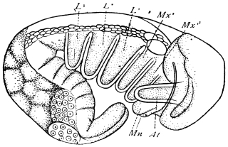
Fig. 21.—Embryo of Aphis. Copied from Mecznikow, Zeits. f. wiss. Zool.,
Bd. XVI., taf. xxx., fig. 30. References in text. × 220.
Though the jaws of the Cockroach are eminently primitive
with respect to those of most other Insects, they are themselves
derived from a far simpler arrangement, which is demonstrable
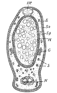
in all embryonic Insects. Fig. 21 shows an Aphis within the
egg. The rudiments of the antennæ (At), mandibles (Mn), and
maxillæ (Mx1, Mx2) form simple blunt projections, similar to
each other and to the future thoracic legs (L1, L2, L3). We
see, therefore, that all the appendages of an Insect are similar
in an early stage of growth; and we may add that a Centipede,
a Scorpion, or a Spider would present very nearly the same
appearance in the same stage. A Crustacean in the egg would
not resemble an Insect or its own parent so closely.57 Aquatic
life favours metamorphosis, and most Crustacea do not begin
life with their full quota of legs, but acquire them as they are
wanted.
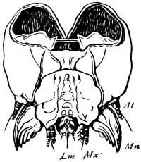
Fig. 22.—Head of larva of Goat Moth, seen from behind.
Copied from Lyonnet.
Paired appendages of perfectly simple form are therefore the
first stage through which all Insect-jaws must pass. Our second
stage is a little more complex, and not nearly so universal as the
first. A caterpillar (fig. 22) has its own special wants, and these
are met by the unequal development of its jaws. The mandibles
are already as complete as those of the Cockroach, which they
closely resemble, but the maxillæ are stunted cylinders formed
mainly of simple rings, and very like the antennæ. They show,
however, the beginnings of three processes (palp, galea, and
lacinia), which are usually conspicuous in well-developed maxillæ.
The second pair of maxillæ (Lm) are coalesced, as usual, and
form the spinneret. The mouth-parts of the Caterpillar do not
therefore in all respects represent a universal stage of development,
but show important adaptive modifications. The mandibles
are rapidly pushed forward, and attain their full
development in the larva; the first pair of maxillæ are temporarily
arrested in their growth, and persist for a long time in a
condition which Orthopterous embryos quickly pass through;
the maxillæ of the second pair are not only arrested in their
growth, but converted to a special use, which seems to stop all
further progress. The labial palps, indeed, which are not at all
developed in the caterpillar, survive, and become important
parts in the moth; but the greater part of the labium disappears
when the time for spinning the cocoon is over.
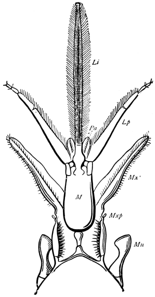
Fig. 23.—Mouth-parts of Honey Bee.
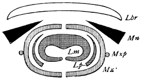
Fig. 23A.—Diagram of Mouth-parts of Honey Bee.
We come next to the Orthopterous mouth, which is well
illustrated by the Cockroach. This is retained with little modification
in all the biting Insects (Coleoptera and Neuroptera).
The mandibles may become long and pointed, as in Staphylinus
and other predatory forms; in some larvæ of strong carnivorous
propensities (Ant-lion, Dytiscus,58 Chrysopa) they are perforate at
the tip, and through them the juices of the prey are sucked into
the mouth, which has no other opening. The labium undergoes
marked adaptive change, without great deviation from the common
plan, in the “mask” of the larva of the Dragon-fly. This
well-known implement has a rough likeness, in the arrangement
and use of its parts, to a man’s fore-limb. The submentum
forms the arm, the mentum the fore-arm. Both these are
simple, straight pieces, connected by an elbow-joint. The hand
is wider, and carries a pair of opposable claws, the paraglossæ.
In some Coleoptera the labium is reduced to a stiff spine, while
in the Stag-beetle it is flexible and hairy, and foreshadows the
licking tongue of the Bee. The maxillæ become long and hairy
in flower-haunting Beetles, and even the mandibles are flexible
and hairy in the Scarabæus-beetles. Fritz Müller has found a
singular resemblance to the proboscis of a Moth in a species of
Nemognatha, where the maxillæ are transformed into two sharp
grooved bristles 12 mm. long, which, when opposed, form a
tube, but are incapable of rolling up.59
In the Honey Bee (fig. 23) nearly all the mouth-parts of the
Cockroach are to be made out, though some are small and others
extremely produced in length. The mandibles (Mn) are not
much altered, and are still used for biting, as well as for kneading
wax and other domestic work. The mandibular teeth have
proved inconvenient, and are gone. The lacinia of the maxilla
(Mx′) forms a broad and flexible blade, used for piercing succulent
tissues, but the galea has disappeared, and there is only a
vestige of the maxillary palp (Mxp). In the second pair of
maxillæ the palp (Lp) is prominent; its base forms a blade,
while the tip is still useful as an organ of touch. The paraglossæ
(Pa) can be made out, but the laciniæ are fused to form
the long, hairy tongue. This ends in a spoon-shaped lobe (not
unlike the “finger” of an elephant’s trunk), which is used both
for licking and for sucking honey.
The proboscis of the Bee is therefore more like a case of
instruments than a single organ. The mandibles form a strong
pair of blunt scissors. The maxillæ are used for piercing, for
stiffening and protecting the base of the tongue, and when
closed they form an imperfect tube outside the tongue, which,
according to Hermann Müller, is probably suctorial. The labial
palps are protective and sensory. Lastly, the central part, or
tongue, is a split tube used for suction; it is very long, so as to
penetrate deep flower-cups, and hairy, so that pollen may stick
to it. When the proboscis is not in use it can be slid into the
mentum (M), while it and the mentum together can be drawn
out of the way downwards and backwards.60
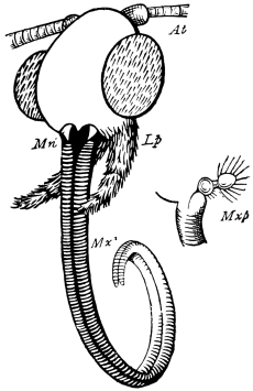
Fig. 24.—Mouth-parts of Burnet Moth.
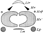
Fig. 24A.—Diagram of Mouth-parts
of Moth.
In the singular suctorial mouth of Moths and Butterflies we
observe, first of all, the great development of the maxillæ.
Each forms a half-tube, which can be accurately applied to its
fellow, so as to form an efficient siphon. In many species the
two halves can be held together by a multitude of minute
hooks.61 At the base of each maxilla is a rudimentary palp
(Mxp). The mandibles (Mn) are also rudimentary and perfectly
useless. The labium, which was so important to the larva as a
spinneret, has disappeared almost completely, but the labial
palps (Lp) are large and evidently important.
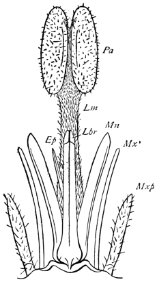
Fig. 25.—Mouth-parts of Gad-fly
(Tabanus).
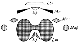
Fig. 25A.—Diagram of Mouth-parts
of Gad-fly.
In Diptera both piercing and sucking parts are usually
present. The Gad-fly (fig. 25) is typical. Here we recognise
the labrum (Lbr), mandible (Mn), and maxilla (Mx′) of the
Cockroach transformed into stylets. The maxillary palp (Mxp)
is still sensory. A pointed process, stiffened by chitinous ribs,
is developed from the back of the labrum. This is the
epipharynx (Ep), a process undeveloped in the Cockroach,
though conspicuous in some Coleoptera. All these parts are
overtopped by the suctorial labium (Lm), which has a two-lobed
expansion at the end. In the more specialised Diptera
this becomes a kind of cupping-glass. The Gad-fly is intermediate
between the Gnat, in which all the mouth-parts are
converted into piercing organs of extraordinary length and
sharpness, and such flies as the House-fly and Blow-fly, where
the sucking labium forms an organ of the most elaborate kind,
the piercing organs undergoing a marked reduction. Except
where the labium is short, it is doubly or trebly hinged, so that
it can be readily tucked away under the chin.
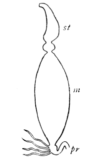
In Hemiptera the long four-jointed labium (Lm) forms a
sheath for the stylets. When not in use the whole apparatus
is drawn up beneath the head and prothorax. The mandibles
(Mn) are sharp at the tip, and close like a pair of forceps, enclosing
the maxillæ (Mx). These are of unequal length, only
one reaching the end of the mandibular case. Both have saw
teeth on the free edge. Palps are entirely wanting.
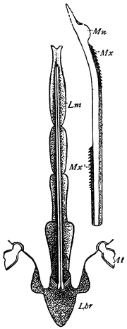
Fig. 26.—Mouth-parts of Bug. Copied from
Landois, Zeits. f. wiss. Zool., Bd. XVIII.,
taf. xi., fig. 3.
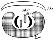
Fig. 26A.—Diagram of Mouth-parts
of Bug.
Comparing the four kinds of suctorial mouths, of which the
Bee, the Moth, the Fly, and the Bug furnish examples, we
observe that the sucking-tube is formed in the Moth out of the
two maxillæ, in the other three out of the labium. Of these
last the Bee has the edges of the labium turned down, so that
the siphon becomes ventral; in the Bug and Fly the edges
are turned up, and the siphon becomes dorsal. The more
specialised flies have the simple arrangement of the Bug complicated
by a system of branching tubes, which are probably a
special modification of the salivary duct. Similar as the
mouth-parts of the four types may be in regard to their mode
of working, they cannot be reduced to any common plan which
differs materially from that presented by the jaws of the
Cockroach.
In all Insects fusion of the primitive elements of the head
begins so early and is carried so far, that it is extremely difficult
to discover the precise way in which they are fitted together.
The following facts have been ascertained respecting the development
of the parts in question. At a very early stage of
embryonic life the body of the Insect becomes divided into a
series of segments, which are at fewest fourteen (in some
Diptera), while they are not known to exceed seventeen.62 Each
segment is normally provided with a pair of appendages. The
foremost segment soon enlarges beyond the rest, and becomes
divided by a median groove into two “procephalic lobes.”63 Of
the appendages the first eight pairs are usually more prominent
than the rest, and of different form; those of the eighth
segment, which may be altogether inconspicuous, never attain
any functional importance. The first four pairs of appendages
are budded off from the future head, while the next three pairs
form the walking legs, and are carried upon the thoracic segments.
All the existing appendages of the fore part of the body are
thus accounted for, but the exact mode of formation of the head
has not yet been made out. The chief part of its walls, including
the clypeus, the compound eyes, and the epicranial plates,
arise from the procephalic lobes, and represent the much altered
segment of which the antennæ are the appendages. The labrum
is a secondary outgrowth from this segment, and, in some cases
at least, it originates as a pair of processes which resemble true
appendages, though it is unlikely that such is their real
character. No means at present exist for identifying the terga
and sterna of the head, nor have the gena, the occipital frame,
and the cervical sclerites (described below) been assigned to
their segments.64 It is worthy of notice that in the stalk-eyed
Crustacea, the head, or what corresponds to the head of Insecta,
consists of either five or six somites, taking into account a
diversity of opinion with respect to the eyestalks, while only
four pairs of appendages can be certainly traced in the head of
the Insect. The mandibles and maxillæ exist to the same
number in both groups, and are homologous organs, so far as is
known; the numerical difference relates therefore to the antennæ,
of which the Crustacean possesses two pairs, the Insect only
one. Whether the pair deficient in the Insect is altogether
undeveloped, or represented by the pair of prominences which
give rise to the labrum,65 is a question of much theoretical
interest and of not a little difficulty.
The following table shows the appendages of the head and
thorax in the two classes. The homologies indicated are, however,
by no means established.66
| Crayfish. | Cockroach. |
| Antennæ. |
| —— | —— |
| Eyestalks. |
| Antennules. |
| Antennæ. |
| Mandibles. | Mandibles. |
| Maxillæ (1). | Maxillæ (1). |
| Maxillæ (2). | Maxillæ (2). |
| —— | —— |
| Maxillipeds (1). | Thoracic Legs (1). |
| Maxillipeds (2). | Thoracic Legs (2). |
| Maxillipeds (3). | Thoracic Legs (3). |
The neck is a narrow cylindrical tube, with a flexible wall
strengthened by eight plates, the cervical sclerites, two of which
are dorsal, two ventral, and four lateral. The dorsal sclerites
lie immediately behind the head (fig. 14); they are triangular,
and closely approximated to the middle line. The inferior
plates (fig. 27) resemble segments of chitinous hoops set transversely,
one behind the other, rather behind the dorsal sclerites,
and close behind the submentum. There are two lateral
sclerites on each side of the neck (fig. 27), a lower squarish
one, which is set diagonally, nearly meeting its fellow across
the ventral surface, and an oblong piece, closely adherent to
the other, which extends forwards and upwards towards the
dorsal side.
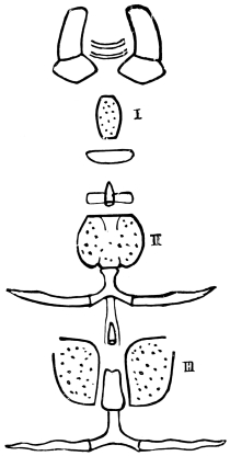
Fig. 27.—Ventral Plates of Neck and Thorax of Male Cockroach.
I, prosternum; II, mesosternum; III, metasternum. × 6.
The elements of the thoracic exoskeleton are simpler in the
Cockroach than in Insects of powerful flight, where adaptive
changes greatly obscure the primitive arrangement. There are
three segments, each defended by a dorsal plate (tergum) and a
ventral plate (sternum). The sterna are often divided into
lateral halves. Of the three terga the first (pronotum) is the
largest; it has a wide free edge on each side, projects forwards
over the neck, and when the head is retracted, covers this also,
its semi-circular fore-edge then forming the apparent head-end
of the animal. The two succeeding terga are of nearly equal
size, and each is much shorter than the pronotum, contrary to
the rule in winged Insects.67
All the terga are dense and opaque in the female; in the
male the middle one (mesonotum) and the hindmost (metanotum)
are thin and semi-transparent, being ordinarily overlaid
by the wing-covers. While the thoracic terga diminish backwards,
the sterna increase in extent and firmness, proportionally
to the size of the attached legs. The prosternum is small and
coffin-shaped; the mesosternum partly divided into lateral
halves in the male, and completely so in the female. The
metasternum is completely divided in both sexes, while a
median piece, carrying the post-furca, intervenes between its
lateral halves in the male. Behind the sterna, especially in the
case of the second and third, the flexible under-surface of the
thorax is inclined, so as to form a nearly vertical step. In the
two hinder of these steps a chitinous prop is fixed; each is
Y-shaped, with long, curved arms for muscular attachment, and
a central notch, which supports the nerve-cord. The hindmost
of these, known as the post-furca, lies immediately behind
the metasternum, and its short basal piece is attached between
the lateral halves of that plate. Behind the mesosternum is a
somewhat slighter prop, the medi-furca. A third piece of similar
nature (the ante-furca), which is well developed in some Insects—e.g.,
in Ants—is apparently wanting in the Cockroach, though
there is a transverse oval plate behind the prosternum, which
may be a rudimentary furca.
Fig. 27 shows two conical processes which lie in the middle
line of the ventral surface of the thorax, one in front of the
metasternum, the other in front of the mesosternum. These
are the thoracic pits, tubular apodemata, serving for the
insertion of muscles. The occurrence of stink-glands in
the thorax of Hemiptera,68 and of so-called poison-glands
in the thorax of Solpuga, led us to look for glands in
connection with these processes, but we have found none.
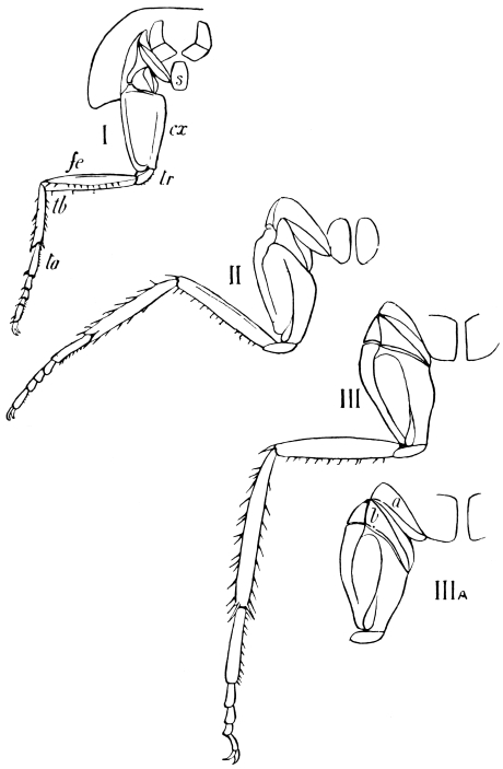
Fig. 28.—The three Thoracic Legs of a Female Cockroach. I, s, sternum; cx, coxa;
tr, trochanter; fe, femur; tb, tibia; ta, tarsus. In IIIA the coxa is
abducted, and the joints a (episternum) and b slightly separated. × 4.
Three pairs of legs are attached to the thoracic segments;
they regularly increase in size from the first to the third, but
hardly differ except in size; the peculiar modifications which
affect the fore pair in predatory and burrowing Orthoptera
(Mantis, Gryllotalpa), and the third pair in leaping Orthoptera
(Grasshoppers, &c.), being absent in the cursorial Blattina. Each
leg is divided into the five segments usual in Insects (see fig. 28).
The coxa is broad and flattened. The trochanter is a small piece
obliquely and almost immovably attached to the proximal end
of the femur, on its inner side. The femur is nearly straight
and narrowed at both ends; along its inner border, in the
position occupied by the stridulating apparatus of the hind leg
of the Grasshoppers, is a shallow longitudinal groove, fringed
by stiff bristles. The tibia is shorter than the femur in the fore
leg, of nearly the same length in the middle leg, and longer in
the hind leg; it is armed with numerous stiff spines directed
towards the free end of the limb. There are usually reckoned
five joints in the tarsus, which regularly diminish in length,
except that the last joint is as long as the second. All the
joints bear numerous fine but stiff hairs upon the walking
surface. The extremity of the fifth joint is segmented off, and
carries a pair of equal and strongly curved claws.69
At the base of each leg are several chitinous plates (fig. 28),
upon which no small labour has been bestowed by different
anatomists. They are arranged so as to form two joints intermediate
between the coxa and the sternum, and these two joints
admit of a hinge-like movement upon each other, while their
other ends are firmly attached to the coxa and sternum respectively.
(Compare III and IIIA, fig. 28.) These parts in the
Cockroach may be taken for two basal leg-joints which have
become adherent to the thorax. In other cases, however, they
plainly belong to the thorax, and not to the leg. In the Mole-cricket,
for instance, similar plates occur; but here they are
firmly united, and form the lateral wall of the thorax. In the
Locust they become vertical, and lie one in front of the other.
Most authors have looked upon them as regular elements of a
typical somite. They regard such a segment as including two
pleural elements—viz., a dorsal plate (epimeron), and a ventral
plate (episternum). We have already (p. 34) given reasons for
doubting the constancy of the pieces so named. It is not
inconvenient, however, to denote by the term episternum the
joint which abuts upon the sternum; for the joint which is
applied to the coxa no convenient term exists, and its occurrence
in Insects is so partial, that the want need not be supplied at
present.70 Both joints are incompletely subdivided. In the first
thoracic segment of the Cockroach they are less firmly connected
than in the other two.
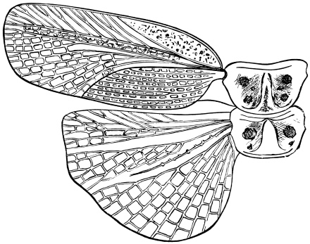
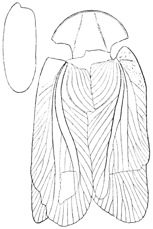
Fig. 29.—Wings and Wing-covers of Male Cockroach. × 4.
Cockroaches of both sexes are provided with wings, which,
however, are only functional in the male. The wing-covers (or
anterior pair of wings) of the male are carried by the second
thoracic segment. As in most Orthoptera genuina, they are
denser than the hind wings, and protect them when at rest.
They reach to the fifth segment of the abdomen, and one
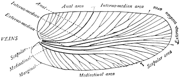
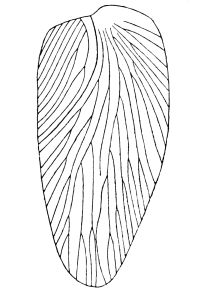
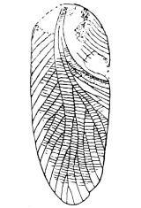
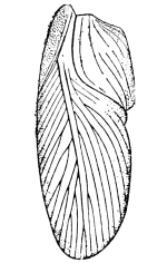
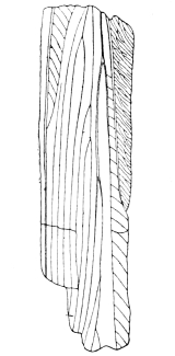
wing-cover overlaps the other. Branching veins or nervures
form a characteristic pattern upon the surface (figs. 4, 29), and
it is mainly by means of this pattern that many of the fossil
species are identified and distinguished. The true or posterior
wings are attached to the metathorax. They are membranous
and flexible, but the fore-edge is stiffened, like that of the wing-covers,
by additional chitinous deposit. When extended, each
wing forms an irregular quadrant of a circle; when at rest, the
radiating furrows of the hinder part close up fan-wise, and the
inner half is folded beneath the outer.71 The wing reaches back
as far as the hinder end of the fourth abdominal segment. The
wing-covers of the female are small, and though movable, seem
never to be voluntarily extended; each covers about one-third
of the width of the mesonotum, and extends backwards to the
middle of the metanotum. A reticulated pattern on the outer
fourth of the metanotum plainly represents the hind wing; it
is clearly rather a degeneration or survival than an anticipation
of an organ tending towards useful completeness.
The rudimentary wing of the female Cockroach illustrates
the homology of the wings of Insects with the free edges of
thoracic terga, and this correspondence is enforced by the study
of the development of the more complete wings and wing-covers
of the male. The hinder edges of the terga become produced
at the later moults preceding the completely winged stage, and
may even assume something of the shape and pattern of true
wings; it is not, however, true, though more than once stated,
that winged nymphs are common. Adults with imperfectly
developed wings have been mistaken for such.
The structure of the wing testifies to its origin as a fold of
the chitinous integument. It is a double lamina, which often
encloses a visible space at its base. The nervures, with their
vessels and tracheal tubes, lie between the two layers, which,
except at the base, are in close contact. Oken termed the wings
of an Insect “aerial gills,” and this rather fanciful designation
is in some degree justified by their resemblance to the tracheal
gills of such aquatic larvæ as those of Ephemeridæ, Perlidæ,
Phryganidæ, &c. In the larva of Chloeon (Ephemera) dipterum
(fig. 30), for example, the second thoracic segment carries a pair
of large expansions, which ultimately are replaced by organs
of aerial flight. The abdominal segments carry similarly
placed respiratory leaflets, the tracheal gills, which by their
vigorous flapping movements bring a rush of water against
their membranous and tracheated surfaces.
Gegenbaur72 has argued from the resemblance of these
appendages to wings, that the wing and the tracheal leaflet are
homologous parts, and this view has been accepted as probable
by so competent an observer as Sir John Lubbock.73
The leaflets placed most advantageously for propulsion seem
to have become exclusively adapted to that end, while the
abdominal gills have retained their respiratory character. At
the time of change from aquatic to terrestrial life, which takes
place in many common Insects when the adult condition is
assumed, and which, according to Gegenbaur, was a normal
event among primitive Insects, the tracheal gill is supposed to
disappear, and in its place, at the next moult, an opening, the
stigma, is formed by the rupture of an air-tube. Gegenbaur
supposes that the primitive Insects were aquatic, and their
tracheal system closed. The tracheal gill he takes to be the
common structure which has yielded organs so unlike as the
wing and the stigma.
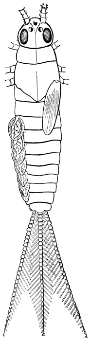
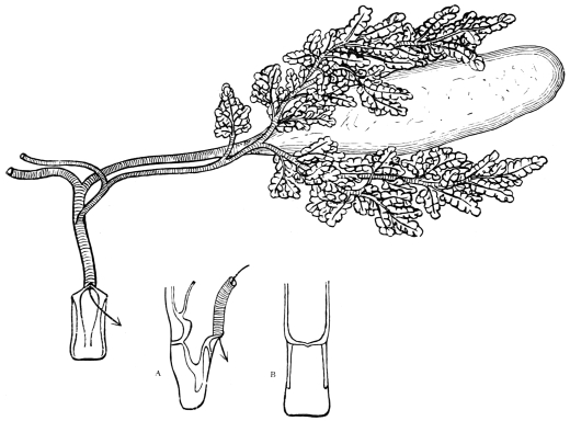
Fig. 30. Chloeon (Chloeopsis) dipterum. Larva in eighth stage, with wings
and respiratory leaflets. × 14. Copied from Vayssière (loc. cit.).
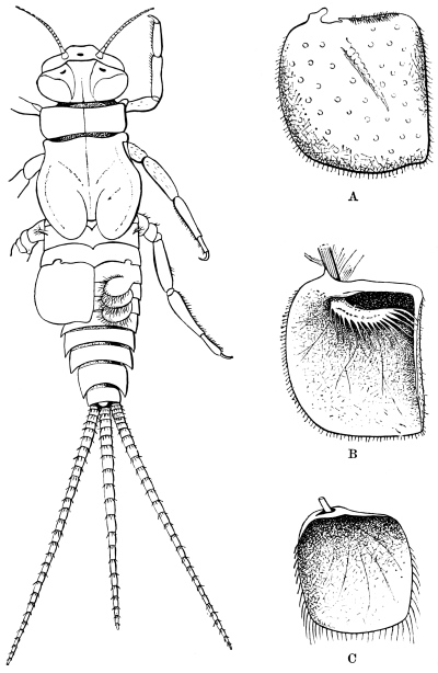
Fig. 31.—Tricorythus. Adult larva, with three functional leaflets. The next
leaflet in front is converted into a protective plate. × 7.
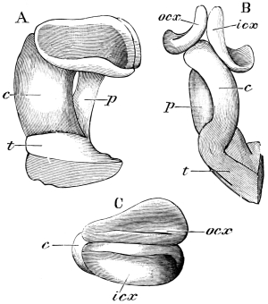
A, protective plate of Tricorythus larva, seen from the outside. × 26.
B, the same from within, showing the attached respiratory appendage.
C, protective plate of Cœnis larva, without respiratory appendage.
All the figures are copied from Vayssière.
The zoological rank of the Insects (Ephemeridæ, Perlidæ,
and Libellulidæ), in which tracheal gills are most usual, is not
unfavourable to such an explanation. Lubbock has given
reasons for regarding Campodea and the Collembola (of the
order Thysanura) as surviving and not very much altered
representatives of the most primitive Insects, and he has shown
that no great amount of modification would be required to
convert the terrestrial Campodea into the aquatic Chloeon-nymph.74
We must not forget, however, that tracheal gills
are by no means restricted to these families of low grade.
Trichoptera, a few Diptera, two Lepidoptera (Nymphula and
Acentropus), and two Coleoptera (Gyrinus and Elmis),75 have
tracheal gills, and a closed tracheal system in the larval condition.
We cannot suppose that these larvæ of higher orders
represent an unbroken succession of aquatic forms, but if we
refuse to adopt this alternative, we must admit that the closed
tracheal system with tracheal gills may be an adaptive modification
of the open system with stigmata.
It is well known76 that in certain Ephemeridæ (e.g., Tricorythus
and Cœnis) a pair of anterior tracheal gills may become transformed
into large plates, which partly protect the gills behind
(fig. 31). A similar modification of the second and third
thoracic gills in Prosopistoma and Bætisca brings all the
functional respiratory organs under cover, and these enlarged
plates resemble stiff and simple wings very closely.
Palmén77 has subjected Gegenbaur’s hypothesis to a very
searching examination. He observes that:—
1. In Campodea, and presumably in other primitive Insects,
the tracheal system is not closed and adapted for aquatic
respiration, but open. Tracheal gills are not by any means
confined to the lowest Insects. (See above, p. 65.)
2. Tracheal gills are not always homodynamous or morphologically
equivalent. In Ephemeridæ, some are dorsal in
position, some ventral (first abdominal pair in Oligoneuria and
Rhithrogena); they may be cephalic, springing from the base of
the maxilla, as in Oligoneuria and Jolia; Jolia has a branchial
tuft at the insertion of each of the fore legs.78 In Perlidæ the
tracheal gills may have a tergal, pleural, sternal, or anal
insertion. In some Libellulidæ also, anal leaflets occur.79
3. Tracheal gills never perfectly agree in position and
number with the stigmata throughout the body. Sometimes
they occur on different rings, sometimes on different parts of
the same ring. Gegenbaur’s statements on this point are
incorrect.
4. Tracheal gills may co-exist with stigmata. In Perlidæ
the tracheal gills persist in the imago, and may be found, dry
and functionless, beneath the stigmata. In Trichoptera they
gradually abort at successive moults, and in some cases remain
after the stigmata have opened.
5. Stigmata do not form by the breaking off of tracheal
appendages, but by the enlargement of rudimentary tracheal
branches, which open into the main longitudinal trunks. In
larvæ with aquatic respiration these branches exist, though they
are not functional.
Palmén’s objections must be satisfactorily disposed of before
Gegenbaur’s explanation, interesting as it is, can be fully
accepted. Palmén has proved, what is on other grounds clear
enough, that stigmata are more ancient than tracheal gills,
aerial tracheate respiration than aquatic. But there is nothing
as yet to contradict the view that the first Insect-wings were
adapted for propulsion in water, and that they were respiratory
organs before they became motor. It is Gegenbaur’s explanation
of the origin of stigmata, and not his explanation of the
origin of wings, which is refuted by Palmén.
In the abdomen of the female Cockroach eight terga (1–7;
10) are externally visible. Two more (8, 9) are readily displayed
by extending the abdomen; they are ordinarily
concealed beneath the seventh tergum. The tenth tergum is
notched in the middle of its posterior margin. A pair of
triangular “podical plates,” which lie on either side of the
anus, and towards the dorsal surface, have been provisionally
regarded by Prof. Huxley as the terga of an eleventh segment.
Seven abdominal sterna (1–7) are externally visible. The first
is quite rudimentary, and consists of a transversely oval plate;
the second is irregular and imperfectly chitinised in front; the
seventh is large, and its hinder part, which is boat-shaped, is
divided into lateral halves, for facilitating the discharge of the
large egg-capsule.
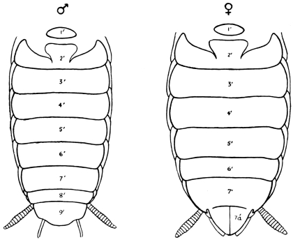
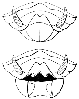
Fig. 32.—Under side of Abdomen of Male and Female Cockroach. × 4.
In the male Cockroach ten abdominal terga are visible
without dissection (fig. 33, p. 70), though the eighth and
ninth are greatly overlapped by the seventh. The tenth
tergum is hardly notched. Nine abdominal sterna are readily
made out, the first being rudimentary, as in the female. The
eighth is narrower than the seventh, the ninth still narrower,
and largely concealed by the eighth; its covered anterior part
is thin and transparent, the exposed part denser. This forms
the extreme end of the body, except that the small sub-anal styles
project beyond it. The podical plates resemble those of the
female.
Pleural elements are developed in the form of narrow
stigmatic plates, with the free edge directed backwards. These
lie between the terga and sterna, and defend the spiracle.80
The modifications of the hindmost abdominal segments will
be more fully considered in connection with the reproductive
organs.
The high number of abdominal segments found in the Cockroach
(ten or eleven) is characteristic of the lower orders of
Insecta. It is never exceeded; though in the more specialised
orders, such as Lepidoptera and Diptera, it may be reduced to
nine, eight, or even seven. The sessile abdomen of the Cockroach
is primitive with respect to the pedunculate abdomen
found in such insects as Hymenoptera, where the constricted
and flexible waist stands in obvious relation to the operations
of stinging and boring, or to peculiar modes of oviposition.
The first abdominal segment, which is especially liable to dislocation
and alteration in Insects, occupies its theoretical
position in the Cockroach, though both tergum and sternum
are reduced in size. The sternum is often altogether wanting,
while the tergum may unite with the metathorax.
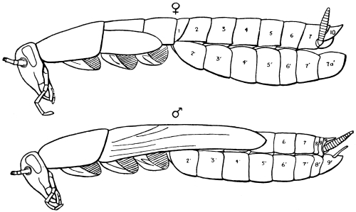
Fig. 33.—Profile of Male and Female Cockroach. × 4.
The externally visible appendages of the abdomen are the
cerci and the styles of the male Cockroach. The cerci are found
in both sexes; they are composed of sixteen rings each, and
project beneath the edge of the tenth tergum. They are
capable of erection by special muscles, and are supplied by large
nerves.81 The sub-anal styles are peculiar in their insertion,
being carried upon the sternum of their segment (the ninth).
The abdominal segments are never furnished with functional
legs in adult Insects, but representatives of the lost appendages
are often met with in larvæ. According to Bütschli,82 all the
abdominal segments are provided with appendages in the
embryo of the Bee, though they disappear completely before
hatching. Some Hymenopterous larvæ have as many as eight
pairs of abdominal appendages, Lepidopterous larvæ at most
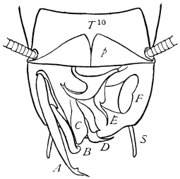
five (3–6; 10).83