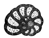
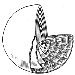
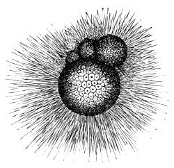
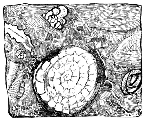
The sub-kingdom Mollusca includes a great variety of soft-bodied
animals which differ from the members of the last division in the fact
that they are never segmented, and in the possession of a thick outer
covering, of a leathery nature, which completely envelops the body, and
which usually secretes a calcareous shell of one or more parts. A
general idea of the extent of the group may be formed when we state that
it contains the Octopus and the Cuttlefish; all Snails and Slugs, and
animals of a similar nature; and all those numerous ‘bivalves’ which are
represented by the well-known Oysters, Mussels, Scallops, &c.
By far the greater number of the molluscs are aquatic in habit; and of
these such a large proportion are marine that the group provides plenty
of occupation for the sea-side naturalist. This being the case, we shall
devote the present chapter to a description of the general
characteristics of these animals, and to the principles of their
classification, illustrating our remarks by a few selections from all
the chief divisions.
Although, as we have already hinted, the body of a mollusc generally
bears but little resemblance to that of the typical elongated and
segmented worm, yet the study of the earliest stages of the former shows
that a certain relationship exists between the two sub-kingdoms, the
newly hatched mollusc being often a minute free-swimming creature with
expanded lobes fringed with cilia, and bearing a resemblance to certain
of the Rotifers, Moss Polyps, and other animals that are included among
the Vermes. But in the adult molluscs this resemblance is lost, these
creatures being generally easily distinguished from all others by
certain well-marked external features, as well as by internal characters
that are peculiar to them and fairly constant throughout the group.
The external shell, where it exists, is usually composed of one or of
two parts, and therefore we speak of univalve and bivalve molluscs; and
no internal skeleton of any kind is to be found except in the division
containing the Cuttlefishes, the ‘bone’ of which is one of the common
objects washed up on our shores by the breakers.
In all the molluscs there is a well-formed digestive tube, and often a
complex arrangement of small teeth which sever the food by a rasp-like
action. There is also a well-formed heart, consisting of two or more
cavities, by means of which the blood is forced through the body; but,
as a rule, blood vessels are either few or absent, the blood being
driven through spaces between the tissues that serve the same purpose.
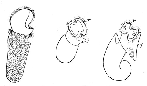
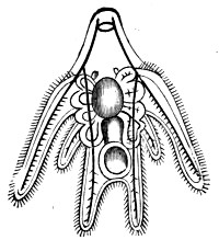
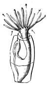
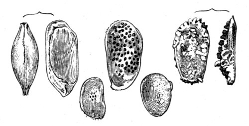
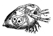
Fig. 126.—Larvæ of Molluscs
v, ciliated ‘velum’; f, rudimental foot
The nervous system consists of a few masses of nerve substance
(ganglia), connected by nerve cords, and sending off fibres to various
parts of the body, the principal ganglion being one situated close to
the mouth, and often surrounding the first portion of the digestive
tube.
The animals of this sub-kingdom are grouped into three principal and
well-marked divisions—the Lamellibranchs, or Plate-gilled
molluscs, the gills of which are composed of plate-like layers, and the
headless bodies enclosed in a bivalve shell; the Cephalophora, or
head-bearing molluscs, protected by a univalve shell; and the
Cephalopoda, or Head-footed molluscs, so called because the mouth is
surrounded by tentacles or arms by which the animal can cling to objects
or seize its prey.
We shall deal with these three divisions in the above order, taking
first the bivalves, the shells of which are found in great variety along
our shores.
The general nature of a lamellibranch is easily made out by the
examination of one of the common species that may be obtained alive on
any part of the coast, such as the Edible Mussel, the Cockle, or the
Oyster, and the reader will do well to secure a few specimens and
examine them with the aid of the following description of the principal
distinguishing features.
The shell is formed of two valves, united by a hinge which is sometimes
of the simplest possible description, but which often exhibits a
beautiful arrangement of interlocking teeth. A ligament of flexible
and elastic substance often holds the two valves together.
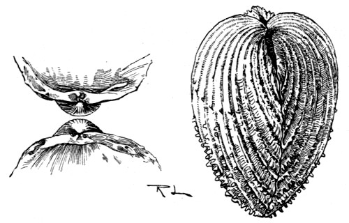
Fig. 127.—Shell of the Prickly Cockle (Cardium
aculeatum) showing Umbo and Hinge; also the interior showing the Teeth
The reader has probably observed that the valves of a dead lamellibranch
usually gape. This is due either to the pull exerted by a ligament that
is attached to the valves outside the hinge, or to the pressure of an
internal cartilage which unites the valves within, and which is
compressed when the shell is closed. When the animal is alive, it has
the power of closing its shell by the contraction of the adductor
muscles, to be presently described, and when the valves are brought
together by this means the external ligament is more or less stretched,
or the cartilage within, which is also an elastic material, is
compressed.
Examining the shell from the exterior we observe that each valve has a
nucleus (the umbo) close to the hinge, round which are usually a
number of more or less distinct concentric lines, extending to the lower
or ventral margin. This nucleus represents the whole shell of the young
mollusc, and the lines are the lines of growth, each one marking the
extreme limit of the valve at a particular period of the animal’s
existence. Further it will be observed that the lines of growth are
often wider apart in some directions than in others, thus denoting the
unequal rate of growth that determined the form of the adult shell.
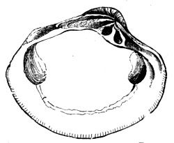
Fig. 128.—Interior of Bivalve Shell, showing
Muscular Scars and Pallial Line
The shell of a bivalve is often made up of two very distinct layers, the
outer one called the prismatic layer because, when examined
microscopically, it is seen to consist of minute vertical prisms of
calcareous matter; and the inner one presenting a beautiful pearly
iridescence, due to the fact that it is made up of a number of extremely
thin and finely waved layers of calcareous substance that have the power
of decomposing light. This latter layer is secreted by the whole surface
of the mantle that lies in contact with it, while the outer, prismatic
portion of the shell is formed only by the free edge of the mantle; and
we often find a distinct line (the pallial line), some little distance
from the ventral margin that marks the junction of the muscle of the
mantle with the shell. The shape of this line is a very important
feature of the shell, since it is of great value in the determination of
relationships.
Further, the inner surface of each valve is marked by the impressions or
scars of other muscles, the number and position of which vary
considerably in different species. They include the adductor muscle or
muscles (one or two in number) that pull the valve together; the muscle
or muscles that withdraw the foot, called the retractor pedis, and the
protractor pedis that pulls the foot out. Not only are these scars
often very distinct in themselves, but we may frequently observe lines
running tangentially from their circumferences towards the umbo, to
which they all converge. These lines enclose the areas previously
occupied by the muscular impressions; in other words, they show the
directions in which the muscles named above shifted their positions as
the animal grew.
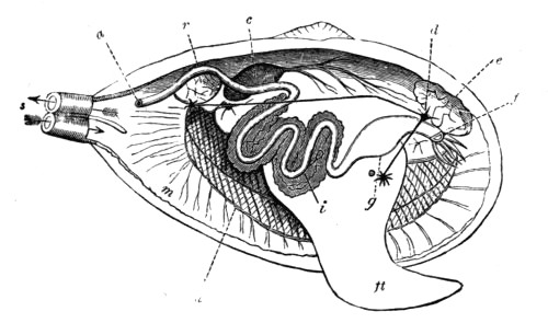
Fig. 129.—Diagram of the Anatomy of a Lamellibranch
f, mouth, with labial palps; g, stomach; i, intestine, surrounded
by the liver; a, anus; r, posterior adductor muscle; e, anterior
adductor muscle; c, heart; d, nerve ganglion; m, mantle (the right
lobe has been removed); s, siphons; h, gills; ft, foot
Now let us obtain a few species of live lamellibranchs, put them in a
vessel of sea water, and observe them after they have been left
undisturbed for a time. The shell will be seen to gape slightly,
exposing the edges of the two lobes of the mantle which lie closely on
the inner surface of the valves, thus completely enveloping the body of
the animal; and at one end, usually the narrower end in the case of
irregular shells, we shall observe two openings—the siphons,
sometimes enclosed within a tube formed by a prolongation of the united
mantle lobes, and protruding from between the valves, and sometimes
formed by the mere contact of the mantle lobes at two adjacent points.
If now we introduce a little carmine or other colouring matter by means
of a glass tube, setting it free near the lower siphon—the one
more remote from the umbo of the shell, we observe that it enters the
body of the mollusc through this opening, and reappears shortly
afterwards through the upper or dorsal siphon. Thus we see that water
currents are incessantly circulating in the body of the animal, entering
by the inhalent or ventral siphon, and leaving by the exhalent or
dorsal siphon. These currents are maintained by the vibratile action
of thousands of minute cilia belonging to cells that line the cavities
of the body, and serve to supply the animal with both air and food; for
lamellibranchs, being gill-breathers, derive the oxygen necessary for
respiration from the air held in solution by the water, and their food
consists entirely of the minute living creatures that always abound in
natural waters.
Again, we shall find that some of our live bivalves have protruded a
thick, conical, fleshy mass—the foot, from the opposite end of
the body. This organ is the means of locomotion in the case of the
burrowing and other free-moving bivalves, but is developed to a less
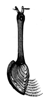
extent in those species that lead a sedentary life. Thus, the common
Edible Mussel secretes a tuft of strong silky fibres (byssus) by means
of which it fixes itself to a rock or other body, and therefore does not
need the assistance of a muscular foot; and an examination of its body
will show that the foot is very small in proportion to the size of the
animal, as compared with that of the wandering and burrowing species.
The same is true of the oyster, which lies fixed on its side, the lower
valve being attached to the surface on which it rests.
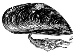
Fig. 130.—Mytilus edulis, with Byssus
We have made use of the terms dorsal and ventral in speaking of the
shell of a bivalve, and it is important that these and a few other
similar terms be well understood by those who are about to read the
descriptions of the animals, or who may desire to describe them
themselves. To do this, take a bivalve in your hand, and hold it before
you in such a position that the hinge is uppermost, and the siphons
turned towards you. The foot of the animal is now pointing in the
direction you are looking, and the mouth, situated at the base of the
foot, is also directed the same way. You have now placed the shell, and,
of course, also the animal, in such a position that its dorsal side is
uppermost, the ventral side below, the anterior end turned from you,
the posterior (often narrower) end towards you, the right valve on
your right, and the left valve on your left. Knowing the exact uses of
these few terms you are in a better position to understand the
descriptions of bivalves, and to locate the exact situations of the
various internal organs named in such descriptions.
A great deal of the internal anatomy of a bivalve mollusc may be made
out by easy dissections, and although the structure of the different
species varies in several details, the general characteristics of the
group are practically the same in all and may be gathered by the
examination of a few specimens.
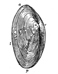
Fig. 131.—A Bivalve Shell
(Tapes virgineana)
a, anterior; p, posterior; l, left valve; r, right valve; u, umbo, on dorsal side
For this purpose the shell should be prised open by means of some
flattened but blunt implement, such as the handle of a scalpel, and
then, after inserting a piece of cork to keep the valves apart, gently
remove the mantle lobe from the valve which is held uppermost with the
same implement, being careful to separate it from the shell without
doing any damage to the soft structures. Separating the mantle from the
shell in this way we meet with one or more hard masses of muscle that
are joined very firmly to the latter. These are the adductor muscles
that pass directly from valve to valve, and on cutting them through
close to the uppermost valve, the latter can be raised so as to expose
the body of the animal, mostly hidden by the overlying mantle lobe.
Before raising the upper mantle lobe we observe the heart, on the dorsal
margin of the body, near the hinge of the shell, situated in a
transparent cavity (the pericardium) containing a colourless fluid. It
consists of at least two cavities—a thick-walled ventricle and a
thin-walled auricle, and its slow pulsations may be watched with or
without the use of a hand lens. On opening the pericardium the heart is
still better seen, and if we carefully cut into the thick-walled
ventricle we find a tube running completely through its cavity. This is
the rectum—the last part of the digestive tube, that commences
at the mouth, and terminates in a cavity at the posterior end
communicating with the exhalent siphon.
After noting the nature and position of the one or two adductor muscles
previously cut through, we turn the upper mantle lobe upwards, laying
it back over the hinge of the shell, cutting it through at the bases of
the siphons if we find it is united with the opposite lobe at those
points; or, if not united, we observe two points at which the lobes
touch each other in order to form the siphonal openings.
Several organs are now exposed to view. The lower mantle lobe is seen in
close contact with the valve below it, and if we touch its edge we shall
probably observe that it is retracted slightly by the contraction of its
own muscular fibres. The tip of the foot is also seen projecting towards
the anterior end, its base being hidden between the two sets of
plate-like gills that extend along the length of the body. On touching
the tip of the foot we find it retract by the contraction of the
muscular fibres of which it is composed, aided, perhaps, by the action
of one or more retractor pedis muscles with which it is supplied. On
raising the upper gill-plates we may observe the dark colour of the
digestive gland (liver) at the base of the foot, and also see two or
more tentacles or labial palpi on the anterior side of the same.
Between the labial palpi is the mouth, which leads into the stomach by a
short, wide tube, and then into a convoluted tube which finally passes
through the heart, and terminates near the exhalent siphon as above
described. The whole length of this tube may be followed by careful
dissection, its direction being determined at short intervals by probing
it with a bristle that has been tipped with a little melted sealing wax.
It will be seen to wind through the base of the foot, surrounded through
the greater part of its course by the digestive gland, from which a
digestive fluid enters it through small ducts.
The diagram on p. 194 shows the general internal anatomy of a
lamellibranch, parts of which have been removed to reveal the underlying
structures. The animal lies in its left valve, the right valve, the
right mantle lobe, and the right set of gill-plates having been
completely dissected away. The whole course of the digestive tube has
also been exposed, and the positions of the three nerve ganglia, with
their connecting nerve cords, constituting the central portion of the
nervous system, are also indicated.
It will be interesting, finally, to learn the direction taken by the
water currents which supply the animal with air and food in their course
through the system. Passing in through the inhalent siphon, the water
immediately enters a large cavity between the mantle lobes. This cavity
(the branchial cavity) contains gills, as we have already seen, and
also extends to the mouth. The water, urged on by the motion of myriads
of minute ciliated cells in the walls of the cavity, passes in part
through the digestive tube, and in part around, between, and through the
gill plates, which are perforated by numerous holes. After thus
completely bathing the gills, and supplying the oxygen necessary for
respiration, this latter current passes into a second cavity above the
gills, and thence into the exhalent siphon, where it mingles with the
fluid from the digestive tube as well as with other excretory matter.
Lamellibranchs are, as a rule, exceedingly prolific, a single individual
of some species discharging more than a million ova in one season. The
larvæ swim freely in the water, and are provided with eyes that enable
them to search for their food, but the eyes always disappear when the
young settle down to a more sedentary life. It is true that adult
bivalves sometimes possess visual organs, often in the form of
conspicuous coloured spots on the edge of the mantle, these, however,
are not the same that existed during the larval stage, but are of a more
recent development.
Lamellibranchs are classified in various ways by different authorities,
the arrangement being based principally on the number and position of
the adductor muscles, or on the nature of the gills. For our present
purpose we shall look upon them as consisting of two main
divisions—the Asiphonida and the Siphonida, the former
including those species which do not possess true tubular siphons, the
inhalent and exhalent openings being formed merely by the touching of
the mantle lobes; and the latter those in which the mantle lobes are
more or less united and tubular siphons formed. Each of these divisions
contains a number of families, most of which have representatives that
inhabit the sea; and we shall now note the principal characteristics by
which the more important families are distinguished, and take a few
examples of each, starting with the Siphonida.
Examining the rocks that are left exposed at low tide we frequently find
them drilled with holes that run vertically from the surface, seldom
communicating with each other within, and varying in diameter from less
than a quarter of an inch to half an inch or more. Some of these holes
are the empty burrows of a boring mollusc, while others still contain
the living animal in situ.
The molluscs in question belong to the family Pholadidæ, which
contains a number of species that exhibit very remarkable features both
as regards structure and habit. The shell is very thin and fragile, but
yet composed of hard material, and its surface is relieved by a series
of prominent concentric ridges that bear a number of little rasp-like
teeth. It gapes at both ends, has neither true hinge nor ligament, and
is often strengthened externally by two or more extra or accessory
valves. The hinge-plate is a very peculiar structure, for it is
reflected over the exterior of the umbones, above which they are
supported by about ten thin shelly plates, the whole thus forming a
series of chambers. The accessory valves are supported by these bridged
structures, and a long, straight, calcareous plate also fills the space
along the dorsal side of the shell in some species. The muscular scars
and the pallial line are distinctly seen on the inner surface, and a
peculiar curved shelly plate projects from under the umbo of each valve.
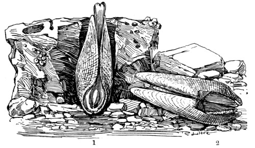
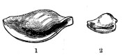
Fig. 132.—Pholas dactylus
1, ventral aspect, with animal; 2, dorsal side of shell showing
accessory valves
The animal inhabiting the shell is somewhat wormlike in general form,
and the mantle lobes are united in front—that is at the lower end
of the shell as it lies in the burrow—except that an opening is
left for the protrusion of the short foot. The siphons are united and
much elongated, so that they protrude beyond the mouth of the burrow
when the animal is active; the gills are narrow, and extend into the
exhalent siphon; and the anterior adductor muscle, being very near the
umbones, serves the double purpose of adductor and ligament.
Such are the general distinguishing features of this family, all the
species of which burrow into stone or other material. Those more
commonly met with on our coasts belong principally to the genus
Pholas, and are popularly known as Piddocks.
It was long a puzzle as to how the fragile piddocks could excavate the
tubular burrows in which they live, and, since their shells are so thin
that it seemed almost impossible for hard stones to be ground away by
them, it was suggested that the rocks were excavated by the action of an
acid secretion. This, however, would not account for the formation of
holes in sandstone and other materials which are insoluble in acids;
and, as a matter of fact, no such acid secretion has ever been
discovered. The boring is undoubtedly done by the mechanical action of
the rasp-like shell, which is rotated backwards and forwards, somewhat
after the manner of a brad-awl, though very slowly, by the muscular
action of the foot of the animal.
Piddocks are found principally in chalk and limestones, though, as
before hinted, they are to be seen in sandstones and other rocks, the
material in any case being, of course, softer than the shell that bores
it. The largest holes and the largest specimens are to be found in chalk
and other soft rocks; while the piddocks that burrow into harder
material are unable to excavate to the same extent and are, as a
consequence, more stunted in their growth. The burrowing is continued as
long as the animal grows, the hole being always kept at such a depth
that the shell is completely enclosed; and not only this, for when the
rock is soft, and the surface is worn down by the sea, the piddock has
to keep pace with this action, as well as to allow for its increase in
size.
As a result of the rasping action of the pholas shell on the surrounding
rock the space hollowed out becomes more or less clogged with débris.
This is ejected at intervals by the sudden contraction of the foot of
the animal, which brings the shell quite to the bottom of the burrow,
thus causing the water with its sediment to shoot upwards,
It is not usually an easy matter to obtain perfect specimens of the
pholas by simply pulling them from their burrows, the shells being so
thin and fragile, and the mouth of the burrow being often narrower than
the widest part of the shell. The best plan is to chip away the rock
with the aid of a mallet and chisel, or to break it into pieces with a
hammer, thus laying open the burrows so that the molluscs fall from
their places.
The Common Piddock (Pholas dactylus) may be identified by the
illustrations, and the other members of the family may be recognised at
once by the similarity in structure and habit. The principal species are
the Little Piddock (P. parva), the shell of which is wider in
proportion to the length, with only one accessory valve; and the White
Piddock (P. candida), also with a single accessory. In all the above
the foot is remarkable for its ice-like transparency.
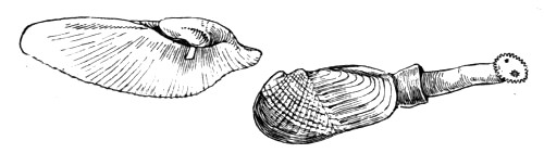
Fig. 133.—Pholas dactylus, interior of Valve; and
Pholadidea with Animal
There is another genus—the Pholadidea—the species of which
are very similar to pholas both in structure and habit. The shells
are, however, more globular in form, and are marked by a transverse
furrow. The gape at the anterior (lower) end is also very wide, and
covered over with a hardened plate in the adult. Also, at the posterior
(upper) end of the shell is a horny cup through which the siphons
protrude, and the latter, which are combined throughout their length,
terminate in a disc that is surrounded by a fringe of little radiating
appendages.
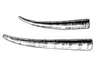
In the same family are the molluscs popularly known as ship worms, which
are so destructive to the woodwork of piers and jetties, or which burrow
into masses of floating timber. Some of these, belonging to the genus
Xylophaga—a word that signifies ‘wood eaters’—have
globular shells with a wide gape in front, and burrow into floating
wood, nearly always in a direction across the grain. The burrows are
about an inch deep, and are lined with a calcareous deposit. The
siphons, combined except at the ends, are slender and retractile; and
the foot, which is thick, is capable of considerable extension.
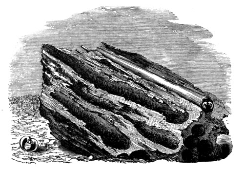
Fig. 134.—The Ship Worm
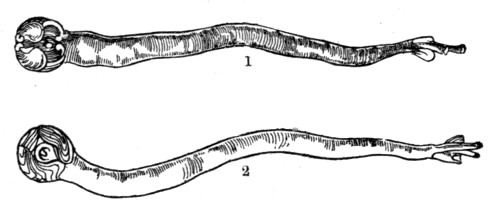
Fig. 135.—1. Teredo navalis. 2. Teredo
norvegica
Other ship worms belong to the genus Teredo, and are very similar in
general characters. The shell is small and globular, with a wide gape at
both ends, and consists of two three-lobed valves with concentric
furrows. It is so small in proportion to the size of the animal that it
encloses but a small portion of the body, and lies at the bottom of the
burrow, which is of considerable length—often from one to two
feet. The animal is very wormlike in form; and although the shell is so
small, yet all the internal organs are enclosed by it. The mantle lobes
are united in front, except where the sucker-like foot passes through
them; the gills are long and narrow, and extend into the siphonal tube;
and the two very long siphons are united almost throughout their length.
It is also interesting to note that in these animals the rectum does not
pass through the heart, as it does in nearly all molluscs, and that a
pair of horny or calcareous ‘styles’ or ‘pallets’ project from the
place where the two siphonal tubes begin to diverge.
Several species of Teredo are to be met with on our coasts, but they
are so similar in general structure that the above brief description
applies almost equally well to all.
Other boring molluscs frequent the British shores, but they belong to
quite a distinct family called the Gastrochænidæ because their shells
gape widely on the ventral side. Their valves are equal in size and very
thin, the hinge has no teeth and the pallial line is sinuated. The
margins of the mantle lobes are thickened and united except where a
small aperture is left for the protrusion of the finger-like foot. The
siphons are very long and retractile, and the gills extend into the
inhalent tube. These animals burrow into mud, shells, or stone, often
dwelling together in such numbers that their galleries cross one another
and form a most intricate network, and the different species are to be
found from low-water mark to a depth of a hundred fathoms or more.
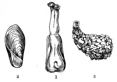
Fig. 136.—Gastrochæna modiolina
1, Animal in shell; 2, shell; 3, cell
The British species belong to two genera—the typical genus
Gastrochæna, and the Saxicava or stone-borers.
The former contains the Common Flask shell (G. modiolina) which
burrows into limestone and shells, in the latter case passing generally
through the shells into the ground below, and completing its home by
cementing together any fragments of hard material that come in its way
into a flask-shaped cell. The opening of the burrow is shaped like an
hour-glass, the two expansions serving for the protrusion of the
siphonal tubes, and the neck of the flask-shaped abode is usually
lined with a calcareous layer that projects slightly to afford further
protection to the extended siphons. Although this species is very common
on some parts of our coast, it is seldom obtained without the aid of a
dredge, for it usually lives at a depth of from five to ten fathoms; and
when found it is generally no easy matter to extricate them from their
holes, to the sides of which they often cement their shells.
The genus Saxicava contains a few species that drill holes, often
several inches deep, in shells and stone, and frequently do great damage
to breakwaters and other artificial structures. The foot is usually
provided with a byssus by which the animal fixes itself to a little
projection on the side of its burrow. The species are to be found from
low-water mark to a depth of one hundred fathoms or more.
The next family, named Anatinidæ, contains a number of molluscs that
burrow in mud or sand or live in seclusion in the crevices of rocks.
Their shells are thin, with a granulated outer surface, and the valves
are united by a thin external ligament. The inner surface is pearly, the
pallial line usually sinuated, and both valves are pitted for the
reception of the somewhat stout internal cartilage. The mantle lobes are
united, as are also the siphons to a greater or lesser extent; and there
is only one gill on each side.
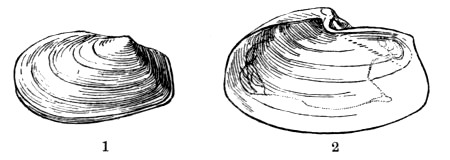
Fig. 137.—1. Thracia phaseolina. 2. Thracia
pubescens, showing Pallial Line
Some of the common species of this family are popularly known as Lantern
shells, and perhaps the most familiar of these is Thracia phaseolina,
the specific name of which is given on account of a fancied resemblance
of the shell to a bean. The shell is very fragile, and although large
numbers may often be seen stranded on sandy beaches, but few of them are
perfect specimens.
The family Myacidæ may be recognised by the thick, strong, opaque
shells, usually gaping at the posterior end; the wrinkled epidermis
which covers the whole or part of the shell; and the united siphons,
which are more or less retractile. The mantle cavity is also closed with
the exception of a small hole left for the protrusion of the small foot.
The pallial line of the shell is sinuated.
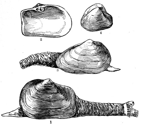
Fig. 138.—1. Mya truncata. 2. Interior of Shell.
3. Mya arenaria. 4. Corbula nucleus
In the above illustration we represent the Common Gaper (Mya
arenaria), which burrows to a considerable depth in the sand or mud,
especially in the estuaries of rivers, from between the tide-marks to a
depth of twenty fathoms or more. It may be readily distinguished, in
common with the other species of the same genus, by the characteristic
wrinkled, membranous tube that encloses its fringed siphons, the
membrane being a continuation of the epidermis that extends over the
shell. Another characteristic feature of the genus is the large, flat
process inside the left valve for the attachment of the internal
cartilage. An allied species, Mya truncata, is often found abundantly
in company with the above, and may be known by the abruptly squared
posterior end.
Other species of the Myacidæ inhabit our shores, including the little
Basket shell (Corbula nucleus), the left valve of which is much
smaller than the right, which overlaps it. The latter, also, is covered
with epidermis, while the former, which is flat, is quite naked.
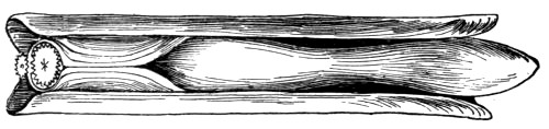
Fig. 139.—Solen siliqua
The valves have been separated and the mantle divided to expose the
large foot
We now come to the interesting family of Razor shells (Solenidæ),
specimens of which are washed up on almost every sandy beach, while the
living molluscs may be dug out of their burrows at low-water mark. The
shells are elongated, gaping at both ends with an external ligament; and
the hinge has usually two teeth in one valve and three in the other. The
foot of the animal is cylindrical, large and powerful; and the siphons
are short and united in the long species, but longer and only partially
united in the shorter ones. The gills are long and narrow, and are
prolonged into the inhalent siphon.
These molluscs lie vertically in their deep burrows at low-water mark,
the opening of the burrow having a form resembling that of a keyhole.
While covered with water they occupy the upper portion of their abode,
but sink to a depth of a foot or more when the tide goes out. As we walk
along the water’s edge at extreme low tide we may observe jets of water
that are shot into the air before us. These are produced by the sudden
retreat of the ‘Razor-fish’ to the bottom of its burrow when alarmed by
the approaching footsteps. Owing to this wariness on the part of the
mollusc, and to the considerable depth of its burrow, specimens cannot
be obtained by digging without much labour; but if a little salt or some
other irritant be dropped into the hole, the animal will soon rise to
eject it, and may then be shut out from the lower part of the burrow by
sharply driving a spade below it. This is undoubtedly the best method of
securing perfect specimens for study or preservation, but fishermen
often obtain large numbers, either for food or for bait, by suddenly
thrusting a long hook down into the gaping shells, and then pulling them
out. This method always does injury to the soft body of the animal, and
often damages the shell, but answers the fisherman’s purpose exactly.
We give illustrations of two shells belonging to the typical genus
(Solen), including one on Plate V.; also a British representative of
each of two other genera of the family—Cerati-solen and
Solecurtus, the latter of which, as the name implies, contains shorter
species.
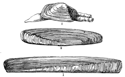
Fig. 140.—1. Solen ensis. 2. Cerati-solen
legumen. 3. Solecurtus candidus
The next family—the Tellinidæ—contains a number of
well-known molluscs that burrow into sand or mud, and are enclosed in
shells that are often very prettily marked; and although the family
includes several genera, all may be recognised by the following general
features. The shell is compressed, composed of two equal valves, with
little or no gape, and the ligament situated on the shortest side. The
central or cardinal teeth never exceed two in number in each valve,
and the adductor impressions are round and polished. The mantle is quite
open at the anterior end, and its margins are fringed; the foot is
flattened and tongue-shaped; and the siphons, which are quite separate,
are generally long and slender.
In the typical genus (Tellina), of which we represent two very common
British species, the ligament is very prominent, and the slender siphons
are often much longer than the shell. The members of this group move
very freely, travelling about by means of a broad, flattened foot.
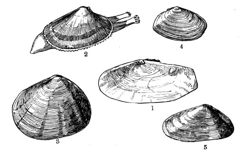
Fig. 141.—Tellinidæ
1. Psammobia ferroensis. 2. Donax anatinus. 3. Tellina crassa. 4.
Tellina tenuis. 5. Donax politus
The shells of the genus Psammobia are popularly known as Sunset
shells, being prettily marked with radiating bands of pink or other
tint, reminding one of the beams of the sun when setting in a cloudy
sky. In these, too, the ligament is very prominent, and the shell gapes
slightly at both ends.
The same family contains the pretty little Wedge shells, which are so
called on account of their triangular form, and constitute the genus
Donax. These shells, which are seldom much over an inch long, are very
common on some of our sandy beaches, being washed up in considerable
numbers after the animals have died, but the specimens are seldom
perfect. The molluscs themselves are burrowers, and live in the sand, at
and just below low-water mark; and, as they usually burrow to a depth of
only a few inches, are easily obtained alive.
The shells are rather thin, closed at both ends, blunt and rounded at
the anterior end, but straight and more pointed at the shorter
posterior end; and the margins of the valves are very finely grooved in
such a manner as to resemble the milling of a coin. Each valve has two
central hinge teeth, with one long lateral tooth on each side; and the
ligament is external and prominent. The lobes of the mantle are fringed;
the siphons are separate and diverging, but shorter and thicker than in
most of the other Tellinidæ, and the foot is comparatively large,
flattened, and pointed.
The genus contains many species, the commonest being, perhaps, D.
anatinus, the colour of which is yellowish, banded with brown, and
marked by a number of radiating white lines. This colour, however, is
due entirely to the thin, shining epidermis that completely covers the
valves; and if this is rubbed off the shell itself will exhibit a pale
pinkish tint. Another common species (D. politus) may be recognised by
the broad patch of white running from the hinge to the margin, on the
posterior side of the middle of each valve.
The family Mactridæ contains some British shells popularly known as
Trough shells, and the family name itself is derived from the word
mactra, which signifies a kneading trough. In this group the shells
are all more or less triangular in form, with the valves equal, and are
either closed or very slightly gaping. The ligament, perhaps more
correctly designated the cartilage, is generally internal, and contained
in a deep triangular hollow; and the shell is covered with epidermis.
The mantle of the animal is open in front, and the siphonal tubes are
united and fringed. The foot is usually large and flattened.
The typical genus, Mactra, contains some common molluscs that bury
themselves just beneath the surface of sandy beaches; and these are so
abundant in some parts of Great Britain that they are used largely for
feeding pigs. Some of the mactras are remarkable for the great power and
extensibility of the foot, which, in some cases, is used so vigorously
that the animal turns itself quickly over, or even leaps on the ground.
Our example of this genus is M. stultorum, which is a very common
object of the shore. Its colour is very variable, usually some shade of
grey or brown, and marked by radiating white lines.
The Otter shells (Lutraria), of which we figure one species, are much
like the Mactræ in structure, and are usually included in the same
family, but in some respects they resemble the Myacidæ or Gapers. The
shell is oblong rather than triangular, and gapes at both ends; and the
animal buries itself deep in sand or mud, principally in the estuaries
of rivers, from low-water mark to a depth of about ten fathoms. The
shells are not very common objects of the shore, for they are found only
in muddy places, and those of the commonest species (L. elliptica) are
too large and heavy to be washed ashore in the sheltered estuaries where
they abound.
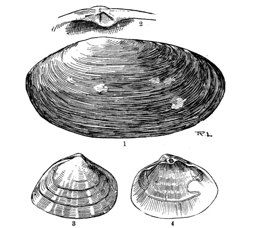
Fig. 142.—1. Lutraria elliptica.
2. Part of the Hinge of Lutraria, showing the Cartilage Pit. 3. Macra
stultorum. 4. Interior of same showing Pallial Line
We now leave the burrowers, to consider a family of molluscs that move
about somewhat freely by means of a flattened tongue-shaped foot, and
which only rarely fix themselves in any way. The shells of the group are
popularly known as Venus shells, probably on account of the beauty of
some of the species, and the family in question as the Veneridæ.
The shells of the various species are usually of a graceful oval or
oblong form, frequently marked by chevron-shaped lines in pretty
colours, and distinctly grooved along the lines of growth. The ligament
is external, the hinge has usually three diverging teeth in each valve,
and the pallial line is sinuated.
The principal genus is Venus, in which the shells are ovate in form,
thick, and smooth, and the margins of the valves are minutely
crenulated. The genus is a very large one, and contains several British
species, two of which we represent in the accompanying illustrations.
Allied to these is the larger but pretty shell Cytherea chione, which
inhabits deep water off the southern coasts, to about one hundred and
fifty fathoms. It is much like the Venus shells in form, but the
margins are not crenulated.
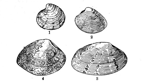
Fig. 143.—Veneridæ
1. Venus fasciata. 2. Venus striatula. 3. Tapes virgineana. 4.
Tapes aurea
The same family (Veneridæ) contains the large genus Tapes, so called
because many of its shells are marked in such a manner as to recall the
patterns of tapestry. The general form of these shells is oblong, and
the margins are quite smooth. They are frequently washed up on the
beach, especially during storms, but the animals may be found alive at
low water, buried in sand, or hiding in the crevices of rocks or among
the roots of the larger sea weeds. The mantle is open at the anterior
end, and the siphons are either quite distinct or only partly united.
Some of the shells are very prettily coloured. One (T. aurea) receives
its name from the yellow ground, which is variously marked by deeper
tints; another (T. decussata) is so called on account of the cross
grooves with which the shell is sculptured; and a third (T.
virgineana), which inhabits the muddy bottoms of deep water, is
prettily marked by radiating bands that run from the umbones to the
ventral margins.
We now come to the family Cyprinidæ, in which the shell is regular in
form, oval or elongated; and the valves, which are equal in size, are
thick and solid, and fit closely. The teeth are beautifully formed, the
central ones numbering from one to three in each valve, and the pallial
line is not sinuated. The mantle lobes are united on the posterior side
by means of a kind of curtain that is pierced by two siphonal openings.
There are two gills on each side, united posteriorly, and the foot is
tongue-shaped and thick.
The typical genus—Cyprina—contains a large mollusc (C.
islandica), which is moderately common round our shores, especially in
the north, but is not often seen above low-water mark, except when
washed up by storms. The shell is oval and thick, with the umbones
prominent and turned towards the posterior side, and the ligament is
strong and prominent. It is entirely covered with a thick epidermis, of
a rich brown colour, often exhibiting a fine silky gloss, especially
near the margins. The interior of the shell is white, and the adductor
impressions oval and polished.
The same family includes some smaller shells that inhabit deep water,
and are therefore not commonly seen on the beach. Among these are two
species of the genus Astarte, one of which is deeply furrowed in a
direction parallel with the margins; also Circe minima, which seldom
exceeds half an inch in length. Although so small compared with
Cyprina, these shells may be identified by their clothing of
epidermis, together with the family characteristics given above.
The Cyprinidæ also contains the interesting Heart Cockle (Isocardia
cor), the form of which is so characteristic that identification is
easy. The heart-shaped shell is thick and strong, and is swollen out in
such a manner that the umbones are wide apart. These latter are also
curved into a spiral form, and the ligament between them is prominent.
The colour of the shell is variable, the epidermis being of any shade
from a yellow to a dark brown. The foot is small and pointed, and the
siphons fringed.
The Heart Cockle burrows in sand by means of its foot, going down just
far enough to bury the whole of its shell, and always leaving its
siphons exposed at the surface. It inhabits deep water, and is not
likely to be obtained without the use of the dredge or trawl.
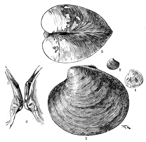
Fig. 144.—Cyprinidæ
1. Cyprina islandica. 2. Teeth of Cyprina. 3. Astarte compressa.
4. Circe minima. 5. Isocardia cor
The molluscs of the family Lucinidæ are found principally in tropical
and sub-tropical seas, ranging from the shore to a very great depth, but
a few are moderately common in our own waters. They are closely allied
to the Cyprinidæ, but the shell is round rather than oval, and is
obliquely grooved inside. The mantle lobes of the animal are not united
on the ventral side, but at the posterior end they are continuous,
except where they form one or two siphonal openings. The foot is long
and of almost the same thickness throughout when extended; and the
gills, numbering either one or two on each side, are large and thick. In
all the members of this family, as in the last, the pallial line of the
shell is simple. None of the shells are really common objects of our
shores, since the animals inhabit deep water, some of them moving about
freely on the bottom, while others moor themselves by means of a byssus.
We shall take only one example of the family—Galeomma
Turtoni—the generic name of which means ‘weasel eye.’ This pretty
little mollusc may be found on our southern coasts, where it often moors
itself to the rocks or weeds by means of its silken byssus; or, having
broken itself away from its temporary place of rest, creeps freely on
the bottom by a long, flattened foot, applied closely to the surface
over which it travels, and used much in the same way as the broad foot
of a snail or whelk, its valves being all the time spread out nearly in
the same plane.
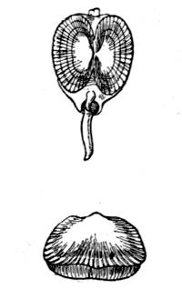
Fig. 145.—Galeomma Turtoni
The shell itself is oval, with central umbones, and is covered with a
thick epidermis. The mantle lobes are united behind, where they form a
single siphonal opening; and the margins are double, with a row of
eye-like spots on the inner edge of each.
The true Cockles, some few species of which are known to almost every
one, constitute the family Cardiadæ, so called on account of the
cordate or heart-shaped form of the shell as viewed from the anterior or
posterior side. The shell is regular, or nearly so, and the valves,
which are equal, are ornamented with prominent rays that run from the
umbones to the margin. The ligament is short, strong and prominent, and
the valves fit closely by the interlocking of their crenulated margins,
or gape slightly on the posterior side. There are two central teeth in
each valve, and a long lateral tooth both on the anterior and posterior
sides. The mantle lobes are open in front, with the margins plaited, and
the siphons, which are usually short, are provided with a number of
little tentacles. The foot is large and powerful, and is usually curved
into the form of a sickle.
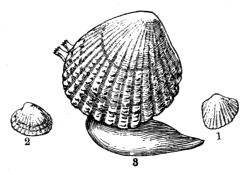
Fig. 146.—1. Cardium pygmæum. 2. Cardium
fasciatum. 3. Cardium rusticum
Although the general nature of the common edible cockle (Cardium
edule) is so well known even to the inhabitants of inland towns that a
description may seem out of place here, yet it is possible that but few
of our readers have ever taken the trouble to place the animal in a
vessel of sea water, either obtained direct from the sea or artificially
prepared, for the purpose of studying its movements or other habits;
and it will be well to remember that this and several other species of
edible molluscs which reach our towns alive may be very conveniently
studied at home, and often at times and seasons when work at the
sea-side is undesirable or impossible.
The edible species referred to lives in banks of sand or mud, buried
just below the surface, and frequently in spots that are exposed for
several hours between the tides. They are usually obtained by means of a
rake similar to that used in our gardens.
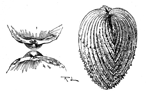
Fig. 147.—Cardium aculeatum
On the coasts of Devon and Cornwall we find a much larger species, also
valued as an article of diet, and known locally as the Prickly Cockle
(C. aculeatum). Its shell is beautifully formed, the rays being very
prominent, each bearing a number of calcareous spines arranged in a
single row. We give an illustration of this species, together with two
sketches to show the nature of the teeth of the shell.
In addition to the two species named, we have the red-footed, C.
rusticum, which can suddenly turn itself over by the action of its
powerful pedal organ; the Banded Cockle (C. fasciatum), a very small
species distinguished by the brown bands of the shell; and a still
smaller one (C. pygmæum), with a triangular shell, occurring on the
Dorset and Devon coasts (fig. 146).
Passing now to the Asiphonida, we deal first with the family Arcadæ.
These include a number of shells which, though very variable in general
form and appearance, may all be recognised by the long row of similar
comb-like teeth that form the hinge. The shells of this group are
regular in form, with equal valves, and are covered with epidermis. The
mantle of the animal is open, the gills are united by a membrane behind,
and the foot is large, curved, and grooved.
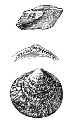
Fig. 148.—Pectunculus glycimeris, with portion of
Valve showing Teeth, and Arca tetragona
One of the prettiest shells in the family is Pectunculus glycimeris,
which reaches a length of about two inches. The shell is grooved in the
direction of the lines of growth, and there are also very delicate
striations running radially from umbones to margin; and the ground
colour of white or pale yellowish is beautifully mottled with reddish
brown. We give a figure of this species, together with a drawing of the
peculiar and characteristic teeth, but a more typical shell of this
family may be seen in the Noah’s Ark (Arca tetragona). This shell is
almost quadrate in form, swollen, and strongly ribbed. The hinge is
straight, with many comb-like teeth—increasing in number with the
age of the shell; and the umbones are separated by a diamond-shaped
ligament. The foot of the animal is heeled—that is, it has a
creeping surface that extends backwards as well as forwards; the mantle
is furnished with minute eyes (ocelli), and the animal has two
distinct hearts. We give a figure of this peculiar shell, and the other
British members of the same genus, though varying more or less in form,
may be recognised at once by the same general characteristics.
In the same family we have the small nutshells (genus Nucula), which
are often dredged up from deep water in large numbers; and the elongated
shells of the genus Leda, also inhabitants of deep water; and, as
before stated, the affinities of all may be readily established by the
characteristic nature of the teeth.
We now pass on to the family of Mussels (Mytilidæ), of which the
common Edible Mussel (Mytilus edulis) is a typical species. In this
interesting group the shell is oval or elongated, with equal valves, and
is covered with a dark-coloured epidermis which is often distinctly
fibrous in structure. The umbones are at the anterior end of the shell,
which end is usually very narrow and pointed, while the posterior is
broad and rounded. The hinge has small teeth or none, and the ligament,
which is long, is internal. The shells of mussels consist of two
distinct layers; on the inner, which is often of a most beautiful pearly
lustre, may be traced the simple pallial line and the impressions of the
small anterior and the large posterior muscles.
The mantle lobes of the animal are united only at a point between the
two siphonal openings. There are two elongated gills on either side, and
the foot is thick and more or less grooved.
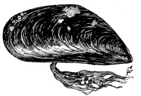
Fig. 149.—Mytilus edulis
Mussels inhabit salt, brackish, and fresh waters, generally attaching
themselves by means of a silken byssus, but sometimes concealing
themselves in ready-made holes, or in burrows of their own; and some
even hide themselves in a nest which they prepare by binding together
fragments of shells or sand.
The edible mussel, which forms such an important article of diet,
especially among the poorer classes in our large towns, may be easily
distinguished from similar species of another genus by the very pointed
umbones, and the coarse and strong fibrous byssus by which it clings to
any solid object. It is found most abundantly on muddy coasts, and on
mud banks in the estuaries of rivers, generally in such situations as
are uncovered at low tide. The fry abound just below low-water level,
and grow so rapidly that they reach their full size in a single year.
It is well known that a diet of mussels occasionally produces very
unpleasant and even dangerous symptoms in the consumer, and this result
has been attributed to the action of a particular organ of the animal
which has not been carefully removed before eating. This, however, is
not the case, as proved by the fact that the eating of these edibles is
usually perfectly safe when no such precautions have been taken. It is
highly probable that the deleterious character referred to is due to a
disease which sometimes attacks the mussels themselves, but the exact
nature of this has not been thoroughly made out.
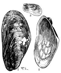
Fig. 150.—1. Modiola modiolus. 2. Modiola
tulipa. 3. Crenella discors
There is another genus (Modiola) containing several species commonly
known as Horse Mussels, and these may be distinguished from Mytilus by
their habit of burrowing, or of constructing a nest by spinning together
various fragments. The shell, also, is more oblong in form, and much
swollen near the anterior end; and the umbones are not so pointed. The
epidermis covering the shell is of fibrous structure, and often extends
beyond the edges of the valves in the form of a fringe.
Several species of Horse Mussels inhabit our shores, from low-water mark
to a depth of fifty fathoms, but none of them is used for food. The
commonest species is Modiola modiolus, which has a particularly strong
byssus, and its fibres generally bind together such a number of stones
&c. that the shell is completely hidden in the entangled mass. Other
British species include M. barbata, so called on account of the
peculiar fringed threads of the epidermis; M. phaseolina, in which
the epidermis threads are not fringed; and M. tulipa, named from the
streaks of crimson or purple that radiate from the umbones of the shell
and remind us of the colouring of the tulip flower.
An allied sub-genus (Crenella) includes a few small British molluscs
the shells of which are crenulated on the dorsal margin behind the
ligament. The shells are short and swollen, and lined by a brilliant
pearly layer. One species (C. discors) is pale green, with radiating
lines from umbo to margin. It is common on many of our shores, but is
not easily found, as it hides at or below low water mark, in a nest
formed by binding together small stones. Other species, one of which is
black, are less abundant, and are not readily obtained except by the use
of the dredge.
Before leaving this family we must refer to the remarkable Dreissena
polymorpha, sometimes called the Chambered Mussel, on account of the
chamber which is formed in the beak of the shell by means of a pearly
plate that stretches across it. This animal is not indigenous to
Britain, but was introduced from the East by trading vessels, either
attached by its silken byssus to timber that had been left floating in
water previous to being shipped, or to the bottoms of the ships. It
seems to thrive almost equally well in salt, brackish, and fresh waters,
and has spread very rapidly since its introduction. It is more commonly
found, however, in docks, canals, and rivers, and is on that account
usually described with the fresh-water species.
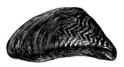
Fig. 151.—Dreissena polymorpha
The form of the shell is very similar to that of Mytilus, but has no
internal pearly layer, and the valves are bluntly keeled. The mantle is
closed, the siphons short, and the foot small.
Our next family—the Aviculidæ—contains those shells that
are distinguished by peculiar flat processes on each side of the
umbones, one of which, the posterior, is generally wing-like in form.
They are popularly known as Wing Shells, and the family includes the
so-called Pearl Oysters. Most of the species are natives of tropical
seas, but several are common on our own shores.
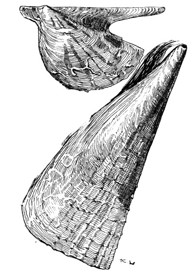
Fig. 152.—Avicula, and Pinna pectinata
One species of the typical genus is sometimes found off the coasts of
Cornwall and Devon. The shell is very oblique, and the valves are
unequal, the right one, on which the animal rests, being somewhat
smaller than the left; and the epidermis is very scanty. The hinge is
long and straight, without teeth, and the cartilage is contained in
grooves. The interior of the shell is pearly. The posterior adductor
impression is large, and not far from the middle of the shell, while the
anterior, which is small, is close to the umbones. The mantle of the
animal is open, and the margins of the lobes fringed; and the small foot
spins a powerful byssus.
Most of the British species of the family belong to the genus Pinna,
so called on account of the fins or wings on the dorsal side of the
shell. In this group the shell is more or less wedge-shaped, with equal
valves, and the umbones are quite at the anterior end, while it is
blunted and gaping at the other end. The hinge has no teeth. The margins
of the mantle are doubly fringed, and the byssus is extremely powerful.
The Common Pinna (P. pectinata) is a very large mollusc, sometimes
measuring a foot in length, and is very abundant off the south-west
coast, where it moors itself vertically at the bottom of the water with
the pointed end buried, and the broad end gaping widely so as to expose
its body. It has been stated that fishes are frequently tempted to
intrude into the open shell for the purpose of devouring the animal
within, and that they are immediately crushed by the sudden closing of
the valves, which are pulled together by two large and powerful
adductors.
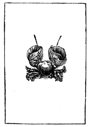
We have already referred to the little Pea Crab that inherits the shell
of the Pinna, living permanently in the mantle cavity of the animal.
The last family of the Lamellibranchs is the Ostreidæ or Oysters, of
which the edible oyster may be taken as a type. In this group the shells
are frequently unequal, and they lie on one side either free or adherent
to the surface below them; the hinge is usually without teeth. The
mantle is quite open, the gills number two on each side, and the foot is
either small or absent.
The Edible Oyster is a type of the typical genus Ostrea, its
scientific name being Ostrea edulis; and as this mollusc may be
readily obtained at any time, it is a convenient species for the study
of the general characteristics of its family. Its shell is irregular in
form, and the animal always rests on its left valve, which is convex,
while the upper or right valve is either flat or concave. The lower
valve is also thicker and laminated in structure, and is attached to the
surface on which it rests. On examining the interior we find that the
shell is somewhat pearly in appearance, and that the edges of the mantle
lobes are finely fringed. The gills, too, are united with each other and
with the mantle on the posterior side, thus forming a distinct branchial
chamber.
Oysters are found on banks at the depth of several fathoms, where they
spawn in early summer, and the fry or spats are collected in large
numbers and transferred to artificial beds or tanks, where they are kept
in very shallow water so as to be easily obtainable when required for
food. It is interesting to note, however, that their growth is slow on
these artificial grounds, the full size being attained in about seven
years, while, in the natural beds, they are full grown in a little more
than half that time.
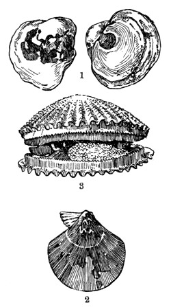
Fig. 153.—1. Anomia ephippium. 2. Pecten
tigris. 3. Pecten, animal in shell
Native oysters—those that are reared on artificial beds—are
of course removed as soon as they are ready for the market, but those
that live on natural banks are often left undisturbed till their shells
are thick with age. The latter, too, are often destroyed in large
numbers by the boring sponge (p. 124), which so completely undermines
the substance of the shell that it finally breaks to pieces.
In the genus Anomia the lower valve is concave, and perforated with a
large oval hole very near the hinge, while the upper one is very convex,
but the shell is very variable in shape, since the animal sometimes
clings permanently to an object, and the shell, during its growth,
accommodates itself to the surface of that object. The use of the hole
is to allow of the protrusion of a set of muscles which proceed from the
upper valve, and give attachment to a plug or button, more or less
calcified, by which the animal clings.
One species (A. ephippium), known as the Saddle Oyster, is common on
some parts of our coast. It is seldom found on the beach at low water,
but the empty shells are often washed up by the waves.
The same family includes the Scallops, which constitute the genus
Pecten. In these the shell is nearly round, with ears on each side of
the umbones, those on the anterior side being generally much more
prominent than the others, and both valves are ornamented by prominent
radiating ribs. The shell is often very prettily coloured, and the
animal rests on the right valve, which may be distinguished from the
left by its greater convexity, and by the presence of a notch under
the anterior ear. The hinge is straight, with a very narrow ligament,
and the internal cartilage is situated in a central pit.
Plate V.
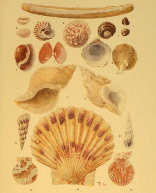
MOLLUSCS
| 1. Solen ensis | 8. Tellina |
| 2. Trivia Europæa | 9. Capulus hungaricus |
| 3. Trochus umbilicatus | 10. Chrysodomus antiquus |
| 4. Trochus magnus | 11. Buccinum undatum |
| 5. Littorina littorea | 12 & 13 Scalaria communis |
| 6. Littorina rudis | 14. Pecten opercularis |
| 7. Haminea (Bulla) hydatis | 15. Pecten varius |
| 16. Pecten maximus |
The mantle of the animal is free, with double margins, the inner of
which forms a finely fringed curtain all round, and on this curtain are
a number of black eyes surrounded by very fine tentacles. The gills are
in the form of very thin crescents, and the foot is shaped like a
finger.
Although the majority of scallops are inhabitants of tropical seas,
several species are to be found off our coasts, where they range from
depths of about four to forty fathoms, and the empty shells, often in
the most perfect condition, are frequently found on the beach.
The Common Scallop (P. maximus) is largely used as food, and is
therefore a common object in the fishmonger’s shop. Its colour is very
variable, and the shell has equal ears and about twenty radiating ribs.
The Quin (P. opercularis) is also an important article of diet in some
parts.
Perhaps the prettiest of the British species is the Variable Scallop
(P. varius), so called on account of the very variable colour of the
shell, the ground tint of which may be almost anything between a very
pale yellow and a dark reddish brown, and this is irregularly patched
with some lighter colour. The chief distinguishing features of the
species are the spiny projections of the numerous ribs, most prominent
near the margin of the valves, and the presence of a permanent byssus,
which, in other species, occurs only in the young. Three of the species
named above are shown on Plate V.
We may also mention the Tiger Scallop (P. tigrinus), the radiating
ribs of which are sometimes slightly formed, and which has only one ear
in each valve; and P. pusio, in which the adult shell is often greatly
altered in form.
It may be noted, in conclusion, that all the species of this genus have
the power of swimming rapidly by flapping their valves—a mode of
locomotion very common among the bivalves especially during an early
stage of their existence.
Before passing on to the univalve molluscs, we must refer briefly to a
group of animals that are enclosed in bivalve shells, and which were
once included with the Mollusca, but are now made to form quite a
distinct group by themselves. We refer to the Brachiopods, at one time
very abundant, as proved by the immense number of fossil shells embedded
in various stratified rocks, but now represented by only a few living
species.
The shells of these animals are commonly known as Lamp Shells, on
account of their resemblance to an antique lamp; and although at first
sight they bear a general likeness to certain bivalve shells of
lamellibranchs, a close examination will show that not only the shell,
but also the animal residing within it, are both of a nature very
different from that of the molluscs with which they were at one time
supposed to be closely related.
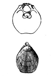
Fig. 154.—Terebratulina. The upper figure
represents the interior of the Dorsal Valve
The valves of the shell are unequal, and are not placed respectively on
the right and left sides of the body of the animal, but rather on the
dorsal and ventral or upper and lower sides. The ventral shell is the
larger, and is produced into a beak which sometimes has a round hole
corresponding in position with the hole for the wick of an antique lamp,
and the dorsal or smaller valve is always imperforate. The hinge is a
perfect one, the junction of the two valves being so well secured by it
that it is impossible to separate them without injury. It is formed by
two curved teeth on the margin of the ventral valve that fit into
corresponding sockets on the dorsal. A few brachiopods, however, have no
hinge, the valves being secured by means of numerous muscles. The hole
in the shell serves for the protrusion of a pedicel or foot by means of
which the animal is enabled to attach itself.
Two long arms, covered with vibratile cilia, and capable of being folded
or coiled, are attached at the sides of the mouth. They are practically
processes of the lips, mounted on muscular stalks, and attached to a
delicate calcareous loop on the dorsal valve; and serve not only to
produce water currents for the conveyance of food to the mouth, but also
answer the purpose of gills.
The digestive system of a brachiopod includes an œsophagus that leads
into a simply formed stomach round which is a large digestive gland. The
heart has only one cavity, but the animal is provided with two smaller
and separate organs that assist in the propulsion of the blood, which
circulates through numerous blood spaces in the bristly mantle.
About two thousand fossil species of brachiopods are known, extending
over a vast range of time; and the living species, numbering less than a
hundred, are found from shallow water to the greatest habitable depths.
Since the reader is hardly likely to form any extensive acquaintance
with the Brachiopods, we shall illustrate our remarks by the
introduction of only one species—the Serpent’s Head Terebratula
(Terebratulina caput-serpentis), which is found in deep water in the
North Sea. The interior of the dorsal valve, showing the calcareous loop
above referred to, is represented in fig. 154, as is also the exterior
of the shell, which is finely striated. The latter represents the dorsal
aspect of the shell in order to show the hole in the upturned beak of
the ventral valve.
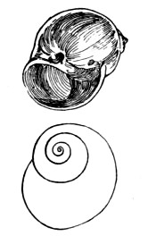
Fig. 155.—Under side of the Shell of Natica
catena, showing the Umbilicus; and outline of the Shell, showing the
Right handed Spiral
We have now to consider the large group of head-bearing molluscs
(Cephalophora), the study of which forms a very important part of the
work of the sea-side naturalist; and while we deal with the general
characteristics of this group, the reader will do well to have before
him a few living typical species in order that he may be able to verify
as many as possible of the descriptions here given by actual
observation. These types may include such creatures as the whelk,
periwinkle, and limpet; or if marine species are not at hand at the
time, the garden snail, fresh-water snail, and slug will serve the
purpose fairly well.
By far the large majority of Cephalopods are enclosed in a single shell,
though a few have a rudimentary shell or none at all.
As is the case with the lamellibranchs, the shell is composed of both
animal and mineral substance, the latter being a calcareous deposit
secreted by the mantle of the animal. The shell is usually spiral in
form, as in the whelk, but sometimes conical (limpet) or tubular.
Spiral shells are nearly always dextral or right-handed; that is, if
we trace the direction of the spiral from the apex to the mouth, we
find that its turns or whorls run in the same direction as the hands of
a watch. A few, however, are sinistral, or left-handed, and
occasionally we meet with left-handed varieties of those species that
are normally of the right-handed type. The cavity of the shell is a
single spiral chamber which winds round a central pillar, and each whorl
of the shell generally overlaps the preceding one, the two being
separated externally by a spiral depression called the suture.
Sometimes the coils of a shell are not close together internally, so
that the central column of the spiral is hollow, and opens to the
exterior at the base of the shell. In this case the shell is said to be
umbilicated, and the opening referred to is the umbilicus. In others
the spiral winds round a solid central pillar which is spoken of as the
columella.
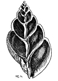
Fig. 156.—Section of the Shell of the Whelk,
showing the Columella
The apex of the shell, sometimes called the nucleus, is the oldest
part, and represents what was once the whole. It is generally directed
backwards as the animal crawls, and in adult shells is often more or
less worn away by constant friction. We speak of the whorls as first,
second, third, &c., taking them in the order of their growth, and it
will generally be found that the last whorl is much larger than the
others, so much so that it contains the greater part of the body of the
animal; hence this one is commonly spoken of as the body-whorl, and
the others make up the spire of the shell.
The mouth of the shell is of different forms in different species, but
in the herbivorous kinds it is usually simple, while in the carnivorous
species it is notched or produced. The edge of the mouth (peristome)
is formed by an outer lip which is usually sharp in young shells and
either thickened, reflected (turned outward), or inflected (turned
inward) in adults; also it may be considerably expanded, or ornamented
by a fringed margin. The inner lip is that side of the peristome
adjacent to the central pillar of the shell.
If we examine the external surface of several different shells, we find
that they are usually more or less distinctly furrowed or sculptured,
and that they are often marked by lines or bands of a colour different
from that of the ground tint. These furrows, lines, or bands sometimes
pass directly from the apex, across the various whorls, to the base of
the shell, in which case they are said to be longitudinal. If they
follow the course of the whorls, they are described as spiral; and if
parallel with the peristome, so that they mark the former positions of
the mouth of the shell, thus denoting the lines of growth, they are
said to be transverse.
Most univalve shells are covered with epidermis, but in some instances
the animal, when extended, surrounds the exterior of the shell with its
mantle, as do the cowries, and then the outside of the shell is always
glazed. Other species keep their shells covered with the mantle, and in
these the shell is always colourless.
The body of the head-bearing mollusc is attached to the shell internally
by one or more muscles, and if we examine the interior surface we are
generally able to distinguish the impressions or scars denoting the
points of attachment.
The reader will have observed that the periwinkle, whelk, and other
univalves close their shells by a kind of lid when they retract their
bodies. This lid is called the operculum, and is constructed of a
horny material, often more or less calcified on the exterior, and is
attached to the hinder part of the foot. It sometimes fits accurately
into the mouth of the shell, but in some species it only partially
closes the aperture. The operculum, like the shell itself, often
exhibits distinct lines of growth which display the manner in which it
was built up. If these lines are concentric we know that the operculum
grew by additions on all sides; but if its nucleus is at one edge, and
the lines of growth widest apart at the opposite side, the growth must
have taken place on one side only. Some, even, are of a spiral form,
denoting that the additions were made continuously at one edge, and such
opercula may be right-handed or left-handed spirals.
It will be noticed that in the above general description of univalve
shells we have introduced a number of technical terms which are printed
in italics, and this we have done advisedly, for the employment of these
terms is a very great convenience when giving descriptions of individual
shells, and we shall use them somewhat liberally in noting the
distinguishing characteristics of the families and genera; but before
entering into this portion of our work we must briefly note the general
features of the bodies of the Cephalophora.
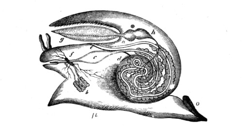
Fig. 157.—Diagram of the Anatomy of the Whelk, the
Shell being removed
c, stomach; e, end of intestine; g, gills; h, ventricle of the
heart; a, auricle; f, nerve ganglia; b, digestive gland; ft,
foot; o, operculum; d, liver
Sometimes these bodies are bilaterally symmetrical, as we have observed
is the case with the worms, but more commonly the organs on one side are
aborted, while the growth proceeds apace on the opposite side. Thus the
animal assumes a spiral form, being coiled towards the aborted side,
with the gills and other organs developed on that side only. As a rule
this curvature is such that the body takes the form of a right-handed or
dextral spiral, as we have already observed in the shells which cover
them, the mouth being thus thrown to the right, but sometimes it takes
the opposite direction.
When one of these animals is extended and creeping, we observe that it
has a distinct head, furnished with a mouth below, and tentacles and
eyes above; also, if an aquatic species, the gills are more or less
prominent. Further, the exposed portion of the body is covered with a
leathery mantle, and the animal creeps on a broad, flattened surface
which is called the foot.
The tentacles or feelers are usually retractile, and, when retracted,
are turned outside-in. Each one is provided with a muscle that runs from
the body internally to the tip; and, by the contraction of this muscle
the tentacle is involuted just in the same way as the finger of a glove
could be by pulling a string attached to the tip inside. In addition to
these tentacles, and the eyes and mouth previously mentioned, the head
is furnished with ear-sacs, which are little cavities, filled with fluid
containing solid particles, with nerve filaments distributed in the
walls.
On the floor of the mouth there is a ribbon, supported on a base of
gristle, and covered with numerous minute teeth arranged regularly in
rows. The gristle is moved backwards and forwards by means of muscles in
such a manner that this ‘lingual ribbon’ acts like a rasp, and is
employed in scraping or tearing away portions of the substance on which
the animal is feeding. By this action the teeth are gradually worn away
in front, but this is of no consequence, for the lingual ribbon is
always growing forwards, the worn material being replaced by new growth
behind.
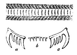
Fig. 158.—A portion of the Lingual Ribbon of the
Whelk, magnified; and a single row of Teeth on a much larger Scale
b, medial teeth; a and c, lateral teeth
The arrangement and form of the teeth are characteristic and important;
and since they afford one of the means by which we may trace the natural
affinities of similar species, they will be frequently referred to when
dealing with the principles of classification. For this reason the
student should be prepared to examine the lingual ribbons of molluscs
with the aid of a compound microscope as occasion requires. As a rule
the ribbon is easily stripped away from the floor of the mouth; and, if
placed in a drop of water and covered with a cover-glass, the teeth are
readily observed. Until a little experience has been gained the
observations may be confined to some of the larger species, in which the
ribbon is both large and easily obtained. In the common whelk, for
example, it often measures more than an inch in length.
It is difficult to understand how the univalve mollusc manages to glide
along so rapidly and gracefully on its expanded foot when we observe it
from above, but the difficulty is cleared away when we see it creeping
on the side of a glass aquarium, or when we place it on a sheet of glass
and observe its movements from the other side. We then see that the foot
is in complete contact with the glass, and that a steady but rapid
undulatory movement is produced by the successive expansions and
contractions of the disc, brought about, of course, by the action of
muscular fibres.
A few of the univalves are viviparous—that is, they produce their
young alive; but the majority lay eggs. The eggs are often enclosed in
horny cases, some of which may be commonly seen washed up on the beach,
or attached to rocks and weeds between the tide-marks. The larvæ are
always enclosed in a shell, though they are sometimes wholly or
partially concealed by the mantle. The shell is usually closed by an
operculum; but as the animal advances in age the shell sometimes
disappears altogether, or is reduced to a mere shelly plate, as is the
case with the land and marine slugs and sea lemons. The young of the
water-breathers always swim about freely by means of a pair of ciliated
lobes or fins, but these remain only for a brief period, after which the
animal settles to the bottom for a more or less sedentary existence.
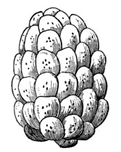
Fig. 159.—Egg Cases of the Whelk
The Cephalophora fall naturally into two fairly well-defined groups,
which we may describe as the air-breathers and the water-breathers. The
former breathe air direct from the atmosphere through an aperture on the
right side of the body, the air passing into a pulmonary organ or lung,
in the walls of which the bloodvessels ramify, and they include all the
land snails and slugs. The latter breathe by gills which are more or
less prominent on the sides of the body, and include all the fresh-water
snails, as well as the marine species which fall within our special
province.
We shall first consider the class Pteropoda or Wing-footed Molluscs,
so called from the wing-like appendages that are attached to the side of
the mouth, or to the upper side of the foot, which is either very small
or altogether wanting.
These Pteropods are in many respects lowly organised as compared with
the higher molluscs; and as they spend the whole of their existence in
the open sea, they can hardly be considered as falling within the scope
of the sea-side naturalist’s work. Yet since their shells are
occasionally drifted on to the shore, and because a knowledge of them is
essential to the student of the mollusca, we shall briefly note their
principal characteristics.
The pteropods are extremely abundant in some seas, occurring in such
vast numbers that they discolour the water for miles. They swim about by
flapping the pair of wings already referred to. They are known to form
an important article of the diet of the whale, and are also devoured in
enormous numbers by various sea birds; and they are themselves
carnivorous, feeding on various smaller creatures that inhabit the open
waters.
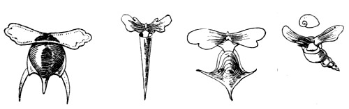
Fig. 160.—Pteropods
In appearance they much resemble the young of higher species of
molluscs. The nervous system consists of a single ganglion situated
below the gullet, and the eyes and tentacles are either rudimentary or
absent. The digestive system includes a muscular gizzard provided with
teeth for the mastication of food, and a digestive gland or liver for
the preparation of a digestive fluid. The heart has two cavities, and
respiration is effected by a surface covered with minute cilia. This
surface is either quite external or is enclosed in a chamber through
which water freely circulates.
The shell is very different from that of a typical head-bearing mollusc,
for it generally consists of two glassy, semitransparent plates,
situated dorsally and ventrally respectively on the body of the animal,
with an opening for the protrusion of the body, and others at the sides
for processes of the mantle; and it terminates behind in one or three
pointed processes. Sometimes, however, its form is conical or spiral,
with or without an operculum. We append illustrations of a few
pteropods, selecting for our purpose species that have been found in the
Atlantic.
It will have been noticed from the above short description that the
pteropod is very unlike the typical Cephalophore as outlined in our
general remarks on the group, especially in the symmetrical form of both
body and shell and in the total or almost total absence of the foot; and
this distinction is so marked that the pteropods are often separated
from all the other Cephalophora into a class by themselves, while all
the remainder are placed in a separate extensive class called the
Gasteropoda, because they creep on the ventral surface of the body,
the term signifying stomach-footed.
These gasteropods are divided into four orders: the Nucleobranchiata,
in which the respiratory and digestive organs form a nucleus on the
posterior part of the back; the Opisthobranchiata, with gills more or
less exposed towards the rear of the body; the Pulmonifera, or
lung-breathing order; and the Prosobranchiata, in which the gills are
situated in advance of the heart. The third order includes all the land
snails and slugs, and does not therefore fall within the scope of our
work; but the remaining three consist either exclusively or principally
of marine species, and will be dealt with in the order in which they are
named.
The Nucleobranchs are not really gasteropods in the strictest sense of
the term, for they do not creep along by means of their foot, but all
swim freely in the open ocean, always at the surface, and sometimes
adhere to floating weed by means of a sucker. In fact, the foot of these
creatures is greatly modified in accordance with their habits, one part
being often expanded into a ventral swimming fin, and provided with a
sucking-disc for adhesion, and another produced into a posterior fin for
locomotion.
Like the pteropods, the nucleobranchs are purely pelagic, so that we can
hardly expect to meet with a specimen on or near the shore; and thus we
shall content ourselves with a brief notice of their general characters.
The shell is very variable in size and form, and sometimes even entirely
absent. Large-bodied species often possess but a very small shell, while
some are able to entirely retract themselves and close the mouth of the
shell by an operculum. These animals are generally provided with a large
cylindrical proboscis, and the tongue has recurved teeth. The body is
usually very transparent, often so much so that the blood may be seen
circulating within it, and the nervous system is much more perfectly
developed than in the pteropods. The eyes, too, are perfectly formed.
The presence of special breathing organs may seem to be superfluous in
such delicate and soft-bodied creatures as these, for it may be supposed
that all the oxygen required could be absorbed directly from the water
through their soft structures, as is really the case with many aquatic
creatures; and as a matter of fact some of the nucleobranchs possess no
gills, but others have these organs fully formed.
Passing now to the true gasteropods, we shall first consider the
Opisthobranchs, which are commonly known as Sea Slugs and Sea Lemons.
Some of these have no shell at all, and even where one exists it is very
rudimentary, usually very small and thin, and concealed within the
mantle. The gills are either branched and tree-like, or are composed of
tufts or bundles of filaments; and, as the name of the order implies,
are situated towards the posterior part of the body. They are also
retractile, and when the animal is alarmed it will conceal its gills,
thus reducing its body to a shapeless, slimy mass, inviting neither to
sight nor to touch.
The sea slugs are principally animal feeders, subsisting on small
crustaceans, other molluscs, &c.; the food being first reduced by the
rasping action of the teeth, and then masticated in a gizzard which is
provided internally with horny spines or hard, shelly plates.
It will not be necessary to enumerate all the different families of this
order, especially as the species are mostly to be found beyond the
tide-marks, and are therefore obtained only with the aid of the dredge;
but we shall describe a few of the British species with a view of
showing the general characteristics of the animals.
They are usually divided into two sections, those with exposed or naked
gills (Nudibranchiata) forming the first, and those in which the gills
are covered either by the shell or the mantle (Tectibranchiata)
comprising the second.
In the Nudibranchs the shell exists only during the embryonic stage, and
the external gills are arranged on the back or along the sides. The
tentacles are not employed as organs of touch, but are probably
connected only with the sensation of smell, being provided with
filaments of the olfactory nerve; the eyes are small dark-coloured spots
embedded in the skin behind the tentacles. Various species are to be
found on all rocky coasts, where they range from low-water mark to a
depth of fifty or sixty fathoms, but a few are pelagic, living on the
surface of floating sea weeds.
It is almost impossible to identify the species of nudibranchs from dead
specimens, for the classification of the section is based largely on the
arrangement of the gills, which are almost always retracted in the dead
animals. This is also the case even with living specimens when disturbed
or removed from the water; hence they should always be examined alive in
sea water, while the animals are extended and moving.
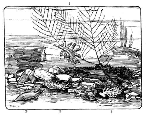
Fig. 161.—Nudibranchs
1. Doto coronata. 2. Elysia viridis. 3. Proctonotus mucroniferus.
4. Embletonia pulchra
It will be understood from the above statements that special methods
will be necessary when it is required to preserve specimens for future
study, the gills being always retracted when the animal is killed for
this purpose by any rapid process. We have found two methods, however,
that are fairly satisfactory in the majority of instances.—Place
the living animals in a suitable vessel of sea water, and leave them
quite undisturbed till they are fully extended, and then either
gradually raise the temperature till they are dead, or introduce into
the water, cautiously, a solution of corrosive sublimate. In the latter
case a much larger proportion of the sublimate will be required than
when used for a similar purpose with freshwater molluscs. When the
animals are dead it will be found that their gills are more or less
extended, sometimes fully so, and they may then be transferred to
diluted spirit or a two per cent. solution of formaldehyde.
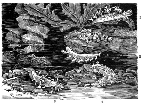
Fig. 162.—Nudibranchs
1. Dendronotus arborescens. 2. Tritonia plebeia. 3. Triopa
claviger. 4. Ægirus punctilucens
In fig. 162 we represent four species. Two of these—Triopa
claviger and Ægirus punctilucens—belong to the family Doridæ,
the members of which are popularly known as Sea Lemons, and are
distinguished by the presence of plume-like gills situated on the middle
of the back. Another family (Tritoniadæ), characterised by the
arrangement of the gills along the sides of the back, and by tentacles
that can be retracted into sheaths, is represented by Tritonia plebeia
and Dendronotus arborescens in the same figure, and by Doto coronata
in fig. 161. The family Æolidæ also have their gills arranged along
the sides of the back, but they differ from the last in that their
tentacles are not retractile. They include the two species numbered 3
and 4 on fig. 161. The remaining one on fig. 161—Elysia
viridis—is a member of the family Phillirhoidæ, characterised
by a pair of tentacles on the dorsal side of the head and by the foot
being either very narrow or absent, the latter feature denoting that the
animals are not adapted for creeping on the bottom. In fact, several of
the species of this family swim freely by means of flattened tails.
The Tectibranchs are similar in general structure, but are very
different in appearance, inasmuch as the gills, so prominent in the last
division, are here covered by the mantle, or by the shell, which is
often well developed. The latter is very variable in form, being of a
globular, twisted, spiral, or other shape, but is sometimes absent in
the adult. In fig. 163 we give a few examples of the shells of British
species; and one (Bulla hydatis) is shown on Plate V.
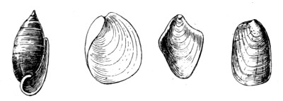
Fig. 163.—Shells of Tectibranchs
We now pass on to the largest and last order of gasteropods—the
Prosobranchiata—so called because the gills are situated in
front of the heart. This group is an important one to the sea-side
naturalist, since it contains nearly all the univalve molluscs that are
common between the tide-marks of our shores, as well as some abundant
species that are protected by a shell of several distinct parts. In
nearly all of them the abdomen is well developed, and the shell is
sufficiently large to cover the whole animal when the latter is
retracted; and the gills, which are either pectinated (comb-shaped) or
plumed, are lodged in the chamber formed over the head of the animal by
the mantle.
The order is often divided into two sections—the Holostomata or
Sea Snails, in which the margin of the aperture of the shell is entire,
and the Siphonostomata, in which the margin of the mantle is prolonged
into a siphon by which water passes into the gill chamber. This division
does not seem to be very satisfactory, as the sections are not separated
by very prominent natural characteristics, but it becomes convenient on
account of the great extent of the order.
In the Holostomata the shell is either spiral, conical, tubular, or
composed of several valves, and the spiral forms are usually closed by
a horny or shelly operculum of the spiral kind. The head is provided
with a proboscis that is generally non-retractile, and the gills usually
extend obliquely across the back, or are attached to the right side
behind the head.
We shall first consider the lower forms, starting with the family
Chitonidæ, the animals of which, as the name implies, are covered with
a shell that resembles a coat of mail.
Some of these creatures are very common on our rocky coasts, and yet
their nature is such that they are liable to be overlooked by those who
are not acquainted with their appearance and habits. The shell is oval
or oblong, often so coloured as to closely resemble the rocks and stones
over which they crawl; and the animal is so inactive when left exposed
by the receding tide, and its flat under surface so closely applied to
that on which it rests, that it looks merely like a little convexity of
the rock. But after a few have been discovered the eye becomes
accustomed to their appearance, and large numbers may be obtained in a
short space of time.
The shell will be seen to consist of eight transverse, curved plates,
overlapping each other at their edges, and all enclosed in a leathery
mantle, which also forms a projecting margin all round. The middle six
plates are different from the first and last in that they are grooved in
such a manner that each one displays a dorsal and two lateral areas.
The animal holds on tightly to the rocks by its large creeping disc-like
foot, but may be removed without injury by forcing a knife-blade under
the margin of its shell. When examined it will be found that it has not
a well-formed head like the majority of the gasteropods, and both eyes
and tentacles are wanting. The gills form a series of lamellæ round the
posterior end of the body, between the edge of the foot and the mantle;
and it is interesting to note that the Chitons further justify the low
position assigned to them among the gasteropods by their possession of a
simple, central, tubular heart, similar to that of worms.
Perhaps the commonest of the British species is Chiton cinereus. Its
colour is a dull grey, but the ground is variously mottled, often in
such a manner as to give it a protective resemblance to its
surroundings. C. ruber is the largest of our species: its shell is
variously mottled with shades of yellow and brown; C. fascicularis is
bristled. Another rather common species (C. lævis) is distinguished by
the glossy appearance of the dorsal portion of the shell.
It will have been observed that the chitons differ from the majority of
gasteropods in that their shells and bodies are both bilaterally
symmetrical, and the same is true of the next family—Dentaliadæ,
which derive their name from the tooth-like form of their conical
shells. They are popularly known as the Tooth Shells, and although they
generally live beyond low-water level, they may sometimes be seen alive
on the beach, and the empty shells are often washed up by the waves.
The shells (fig. 165) are curved, and open at both ends, the narrower
extremity being the posterior. The mouth is circular, and the outer
surface is quite smooth or grooved.
In these animals, too, the head is imperfectly formed, without eyes or
tentacles. The foot is conical and pointed, with two symmetrical side
lobes; and the gills, also two in number, are symmetrically disposed.
The margin of the mouth is fringed, and the animal is attached to the
shell near the posterior end.
The Dentaliadæ are carnivorous, subsisting on minute molluscs,
foraminifera, &c., and generally live on sandy or muddy bottoms, in
which they sometimes bury themselves.
Our next family includes the familiar Limpets, and is designated
Patellidæ on account of the resemblance of the conical shell to a
little dish. In these the apex of the cone is not central, but situated
more or less towards the anterior; and the muscular impression within is
shaped like a horseshoe, with its open end turned to the front.
Unlike the members of the preceding families, the limpets have a
well-formed head furnished with both eyes and tentacles, the former
situated at the bases of the latter. They have a horny upper jaw, and
the tongue, which is very long, is supplied with numerous hooked teeth.
The foot is a very large disc, as large as the shell, and the gills
consist either of one or two branched plumes, or of a series of lamellæ
almost or entirely surrounding the animal between the shell and the
margin of the mantle.
The reader has probably experienced the difficulty of detaching a limpet
from its hold on the rocks. The tenacity of the grip is not due to the
mere adhesive power of the foot itself, but to atmospheric pressure, the
effect of which is complete on account of the total exclusion of air
from under the disc of the foot; and when we remember that this pressure
amounts to fifteen pounds on every square inch of surface, we can
readily understand the force required to raise a large limpet from its
position.
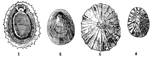
Fig. 166.—Patellidæ
1. Patella vulgata. 2. P. pellucida. 3. P. athletica. 4. Acmæa
testudinalis
The Common Limpet (Patella vulgata) is found on all our rocky coasts
between the tide-marks, often at such a level that it is left exposed to
the air for eight or nine hours at a time. The apex of the shell of this
species is nearly central, and the exterior is sometimes nearly smooth,
but more commonly relieved by radiating ribs.
Although the shell itself is not a particularly pretty object, it is
often rendered very beautiful and interesting by the various animal and
vegetable organisms that settle on it. Those shells that are left dry
for hours together are commonly adorned with clusters of small acorn
barnacles, while the limpets that have found a home in a rock pool and
are perpetually covered with water, often resemble little moving gardens
in which grow beautiful tufts of corallines or other weeds, as well as
polyzoa and other animal forms.
It appears that limpets are not great travellers, the appearance of the
rock from which they have been removed being such as to point to a very
long period of rest. Those on hard rocks are generally situated on a
smooth surface just the size of the shell and generally worn slightly
below the surrounding level by the constant friction of the shell; while
others that have settled on very rugged spots have their cones adapted
to the irregular surface. It has been suggested that the animals make
occasional short excursions from their chosen spot, but return again to
it; and whether or not this is the case, it is evident that they
frequently keep to one small spot for a considerable length of time.
Limpets on chalk and other soft rocks are sometimes in circular pits so
deep that even the apex of the shell is below the general level around;
and though it is possible that the abrasion is produced entirely by the
friction of the shell as the animal turns, yet, in the case of chalk,
the action may be partly due to the carbonic acid gas given off by the
animal as a product of respiration, for it is a well-known chemical fact
that this gas, in solution, has the power of dissolving calcareous
material.
The other British Limpets include P. pellucida, which lives on the
fronds and stalks of the tangle, the form of the shell varying according
to that of the surface on which it rests; also the Horse Limpet (P.
athletica), the bold radiating ribs of which are irregularly notched;
and Acmæa testudinalis—the Tortoiseshell Limpet, with
reddish-brown mottlings on the exterior, and a dark-brown patch at the
apex within. The last-named species lives principally on sea weeds, and
has a single pectinated gill in the cavity between foot and mantle,
which is protruded on the right side when the animal is extended. This
latter feature is interesting since it shows a tendency to that
one-sided development already referred to as characteristic of the
typical gasteropod, resulting in the spiral form of the adult.
In the limpets the lingual ribbon is proportionately long, and is easily
removed for examination. In P. vulgata it may exceed an inch in
length, and the teeth are arranged in rows each of which contains four
central, with laterals on either side, while in Acmæa there are only
three laterals on each side of the central line.
Other so-called limpets belong to separate families. Thus we have the
Cup-and-Saucer Limpet and the Bonnet Limpet in the Calyptræidæ. Both
these differ from Patella in that the apices of their shells show a
tendency to assume a spiral form, thus denoting a somewhat closer
relationship to the more advanced univalves. They have distinct heads,
with prolonged muzzles, and well-formed antennæ and eyes. The teeth of
the lingual ribbon are single, with dentated laterals on either side.
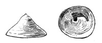
Fig. 167.—Calyptræa sinensis
The Cup-and-saucer Limpet (Calyptræa sinensis) is so called on account
of a curved plate that projects from the interior of the shell, at the
apex; and though this plate takes the form of a half-cup rather than of
a cup, the whole shell has suggested the popular name, while the generic
name is derived from calyptra, which signifies a cap. This mollusc is
occasionally found among stones at low tide, but usually lives beyond
this line, thus necessitating the use of a dredge. The Bonnet Limpet
(Pileopsis hungaricus) is of similar structure and habit, but the
nucleus of the shell is a more decided spiral (see Plate V.). Both these
animals adhere to stones and rocks, and, like the common limpet, seldom
or never move from their selected sites; hence their shells are variable
in form, being adapted to the rock below, and the movements of the shell
often cause a little hollow to be scooped out of the softer materials.
Yet other limpets belong to the next family Fissurellidæ, which is
characterised by a perforation or a notch in the shell. In these, too,
the shell is conical, with a tendency to assume the spiral form, but the
curve of the nucleus, which is always apparent in the young shell,
frequently disappears as the growth proceeds.
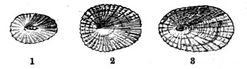
Fig. 168.—Fissurellidæ
1. Puncturella noachina. 2. Emarginula reticulata. 3. Fissurella reticulata
In the Keyhole Limpet (Fissurella reticulata) which is found chiefly
on our southern shores, the perforation is at the summit of the shell;
but as the animal grows the hole increases in size, encroaching on the
curved nucleus until the latter quite disappears. In the genus
Puncturella the perforation is just in front of the recurved apex, and
is surrounded by a rim internally; while in the Notched Limpets (genus
Emarginula) it is represented by a fissure on the anterior margin of
the cone. In all, however, the hole or notch serves the same purpose,
for it is the means by which water enters the siphon.
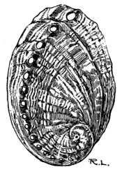
Fig. 169.—Haliotis
It is doubtful whether we ought to claim the beautiful Ear shell
(Haliotis tuberculata) as one of our own, but it is generally included
among the British molluscs on the ground that it is abundant on the
coast of the Channel Islands, where it is called the Omar; and it is
certainly too beautiful an object to be excluded from the British
species without ample cause.
It belongs to the family Haliotidæ, and our illustration will show
that the shell is less elevated than that of limpets, and that the
spire, though not prominent, is a fairly well-formed spiral. All along
the outer lip of the very large aperture is a series of perforations,
occupying the summit of a prominent, spiral ridge, and becoming
gradually smaller and smaller towards the spire. The whole shell is
pearly in structure, and displays a great variety of rich colouring. It
is used largely for inlaying and other ornamental purposes, and for
making the so-called pearl buttons. The animal is used largely as an
article of food in the Channel Islands, but it is of so tough a nature
that it requires a vigorous beating previously to being cooked.
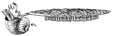
Fig. 170.—Ianthina fragilis
The same family contains the beautiful violet Ianthina, which also is
not a British species, but a free-swimming oceanic snail. It is,
however, occasionally drifted to our shores, though generally in an
imperfect condition. In the Atlantic and the Mediterranean it sometimes
abounds in such multitudes as to distinctly colour the surface of the
sea.
It will be seen that the shell is round, with a well-formed spiral. The
spire is white, but the base is of a deep violet colour. The animal is
very remarkable in some respects. In the first place, though it has
pedicels similar to those on which the eyes of the higher univalves are
placed, yet it has no eyes. Then the foot, which is in itself small,
secretes a float or raft so large that it cannot be retracted into the
shell, with numerous air vesicles to render it light, and the
egg-capsules of the animal are attached to the underside of this. The
animal has no power of sinking, but lives exclusively at the surface;
and, when disturbed, it exudes a violet fluid that colours the
surrounding water. It is apparently the only gasteropod that lives in
the open sea and has a large and well-formed spiral shell.
Passing now to the family Turbinidæ we meet with turbinated or
pyramidal shells that are of a brilliant pearly lustre within, and
frequently without also when the epidermis is removed. The animals
inhabiting them have well-formed heads with a short muzzle, long and
slender tentacles, and eyes mounted on peduncles. The sides are
ornamented with fringed lobes and several tentacle-like filaments, and
the aperture of the shell is closed, when the animal is retracted, by a
spiral operculum. They are all vegetable feeders; and, as is usual with
the plant-eating molluscs, the teeth on the lateral portions of the
lingual ribbon are very numerous.
We have a few common species belonging to this group, mostly members of
the typical genus Trochus and commonly known as Top Shells. In these
the shell is a pyramid formed of numerous flat whorls, with an oblique
and rhomboidal aperture. Of the three species figured (including two on
Plate V.) T. umbilicatus and the Large Top (T. magnus) are
umbilicated, the umbilicus being very large in the latter; and the
former is characterised by the zigzag greyish or reddish markings that
run radially across the whorls. The other (T. zizyphinus) is usually
of a yellowish or pink colour and has no umbilicus.
The same family contains the pretty little Pheasant Shell (Phasianella
pullas), which is richly coloured with red, brown, and yellow on a
light ground; and Adeorbis subcarinatus, shown in the same group.
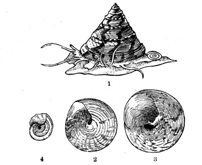
Fig. 171.—1. Trochus zizyphinus. 2. Under side of
Shell. 3. Trochus magnus. 4. Adeorbis subcarinatus
The well-known Periwinkle (Littorina littorea) and the species to the
right of it on Plate V., belong to the family Littorinidæ, the
members of which are similar in structure and habit to Trochus, but
the shell is usually more depressed, and is never pearly. The shell of
the Periwinkle is thick, having but few whorls, and is not umbilicated;
and the lingual ribbon, which is coiled up on the gullet, contains no
less than about five hundred rows of teeth; but only a little more than
twenty of these rows are in action at any one time, the remainder being
a reserve stock to come into active service as the ribbon grows forward.
In the genus Lacuna there is a narrow umbilicus, and the aperture of
the shell is semilunar in form; and the species of Rissoa are very
small, with white or horny shells, much more pointed and having more
whorls than those of the Littorina.
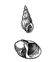
Fig. 172.—Rissoa labiosa and Lacuna pallidula
Our next illustration shows three shells of the family Turritellidæ,
so named from the resemblance of the shells to a tower or spire. The
form indeed is so characteristic that they can hardly be mistaken. It
will be seen that Turritella communis is striated spirally, while the
surface of Scalaria communis (Plate V.) is relieved by strongly marked
transverse ribs. Both these species are very common, and the latter is
peculiar for its power of ejecting a dark purple fluid when molested.
The other representative of the family—Cæcum trachea—has
a shell something like that of Dentalium (p. 238), being cylindrical
and tubular, but it differs in being closed at one end.
In the succeeding shells, of the family Cerithiadæ, the spire is also
considerably produced, so much so that some of the species closely
resemble the Turret shells, but they are distinguished by usually having
an expanded lip, at least in the adult form; and the mouth is channelled
in front, and sometimes also behind. The animals of the group have short
muzzles that are not retractile, the tentacles are wide apart, and the
eyes are mounted on short pedicels. The median teeth are arranged in a
single row, with three laterals on either side of each.
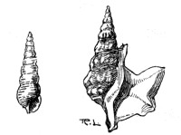
Fig. 175.—Cerithium reticulatum and Aporrhais
pes-pelicani
Cerithium reticulatum receives its generic name from its appearance to
a small horn, and the specific name refers to the netted appearance of
its surface due to the presence of numerous little tubercles arranged in
rows—a feature that serves to distinguish it from the small Turret
shells. It is a common shell, as is also the other representative of the
family illustrated, but the latter is rendered conspicuous by the
enormously expanded lip that has earned for it the popular name of Spout
Shell. Its scientific name is Aporrhais pes-pelicani, and the
application of the specific term will be understood when the shell is
viewed from above, for the expanded lip is drawn out into long
finger-like lobes that suggest the foot of a bird. This is a very solid
shell, sometimes reaching a length of two inches; and the animal
inhabiting it is carnivorous.
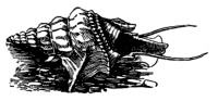
Fig. 176.—Aporrhais pes-pelicani, showing both
shell and animal
We have yet some turreted shells to deal with, belonging to the family
Pyramidellidæ, but they need not be confused with the preceding groups
if carefully examined. In the first place, the aperture of the shell is
very small; and the operculum, instead of being spiral, as in the
turreted shells before mentioned, is imbricated or made up of parallel
layers denoting that the growth took place on one side only. Another
distinguishing feature is seen in the nucleus—that small portion
of the spire that was developed within the egg—which is sinistral
or left-handed. In addition to this, the animal has broad, ear-like
tentacles, a retractile proboscis, and a lingual ribbon without teeth.
The British species of this family belong principally to the genera
Odostomia, characterised by a tooth-like fold of the columella;
Eulima, containing small, white, polished shells with numerous level
whorls; and Aclis, with little polished shells not unlike
Turritella.
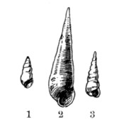
Fig. 177.—1. Odostomia plicata. 2. Eulima
polita. 3. Aclis supranitida
The last family of the Holostomata is the Naticidæ, the shells of
which are almost globular, with only a few whorls, and a small, blunt
spire. The mouth is semilunar in form, and the lip sharp. The proboscis
of the animal is long and retractile, and the foot large; but perhaps
the most characteristic feature is the presence of large mantle lobes
which hide some of the shell when the animal is crawling. In Natica
(fig. 155), the typical genus, the shells are somewhat thick and smooth,
with a large umbilicus. As the animal crawls a large fold of the mantle
is reflected back over the head, completely covering it, and apparently
obstructing its view; but this is not the case, for the creature has no
eyes. Natica is very abundant on some sandy beaches, where it devours
small bivalves and other animals; and it is frequently washed up alive
by the waves. Its shell is also a favourite one with hermit crabs. Its
eggs, all connected together in a spiral band, may often be seen
stranded on sandy coasts. Several species of Natica are found on our
shores. An allied mollusc—Velutina lævigata, so called on
account of the velvety epidermis that clothes the shell, completely
surrounds the shell by its mantle folds when creeping.
The Siphonostomata form a much smaller section than the last, and its
members are distinguished mainly by the presence of a true siphon,
formed by the prolongation of the mantle margin, and serving to convey
water into the gill chamber. In all these the shell is spiral, usually
without an umbilical opening, and the margin of the mouth is prolonged
into a canal or distinctly notched. The operculum is horny, and lamellar
or imbricated. The animal has a retractile proboscis, and the eyes or
eye-pedicels are joined to the tentacles. All the species of this
division are marine.
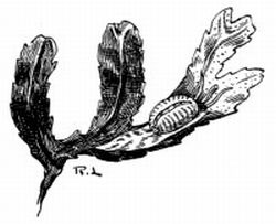
Fig. 178.—Cypræa (Trivia) europæa
We will first take the family Cypræidæ, which contains the familiar
Cowries, these forming the lowest group of the division. An examination
of the shells may at first seem rather puzzling, for the spire is
concealed, and the whole is convoluted in such a manner as to make the
mouth long and narrow, with a channel at either end. The outer lip is
also thickened and bent inward, and there is no operculum.
The animal itself is particularly interesting, for, as it creeps along
on its broad foot, abruptly shortened in the front, the mantle lobes
bend over the top, meeting along the middle line, where they are usually
fringed with little tentacle-like processes; and, as a result, the whole
shell is beautifully enamelled on the outer surface. In all the Cowries
the central teeth are single, and the laterals are arranged either in
twos or threes.
Perhaps the commonest representative of this family is the pretty little
Cypræa (Trivia) europæa (Plate V.), the shells of which are
sometimes washed up in large numbers on sandy beaches. The animal lives
mainly below low-water level, but it may often be found in the larger
rock pools, creeping rapidly over the tangles, and may be easily secured
with the aid of a net.
In the same family we have the little Erato (Marginella) lævis,
the white shell of which is minutely furrowed along the lips; and also
Ovulum patulum (Calpurna patula), so called on account of its
fancied resemblance to a poached egg.
We have also several species of Cone shells (family Conidæ) on our
coasts, readily recognised by their form, which is a cone, with a long,
narrow aperture, partially closed by a minute operculum. As in the last
family, the foot is abruptly shortened in front. The head is very
prominent, with eyes situated on the tentacles. There are two gills, and
the teeth are arranged in pairs.
The Conidæ are principally inhabitants of tropical seas, where some very
large species exist. Two of the British representatives, both common
shells, are shown in fig. 180.
Our next family (Buccinidæ) is so well distributed on our coasts, that
it would be difficult, we imagine, to find a spot quite free from its
familiar forms. It contains all those creatures commonly known as
Whelks, Dog Whelks, and Dog Winkles, ranging from deep water almost to
high-water mark.
In all these the shell is notched in front, or the canal is turned
abruptly upward. The foot of the animal is broad, the eyes are situated
either on the tentacles or at their bases, and there are two gill
plumes.
All the species are carnivorous, and some are said to be very
destructive to mussels and young oysters.
The Common Whelk (Buccinum undatum, Plate V.) lives in deep water,
whence it is dredged up largely for the market. Its clusters of egg
cases are washed up in large numbers on the beach, where they form one
of the commonest materials among the refuse at high-water mark. It is
not uncommon, also, especially after storms, to find the unhatched eggs
stranded by the waves, and these are so transparent that the embryos,
several in each capsule, may be seen within. The hole through which the
young escape may also be seen on the inner side.
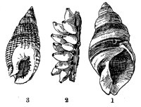
Fig. 181.—1. Purpura lapillus. 2. Egg Cases of
Purpura. 3. Nassa reticulata
The Dog Periwinkle (Purpura lapillus) abounds on all our coasts and is
remarkable for the production of a dull crimson or purple fluid that may
be obtained from it by pressing on the operculum. This fluid turns to a
brighter colour on exposure to air, and is said to have been used
largely in former times as a dye. It will be seen from our figure that
the spire of this shell is shorter in proportion than that of
Buccinum; but both are alike in that the operculum is made up of
layers with a nucleus on the external edge.
The other species figured is Nassa reticulata, popularly known as the
Dog Whelk, and characterised by a tooth-like projection of the inner lip
close to the anterior canal. It is very common near low-water mark,
where it may be seen crawling over the rocks on its broad foot, from
which project two hornlike appendages in front and two narrow tails
behind.
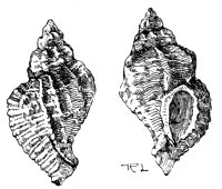
Fig. 182.—Murex erinaceus
From the last family of the gasteropods (the Muricidæ) we select two
common species—Murex erinaceus and Fusus antiquus (Plate V.).
In both these the anterior canal of the shell is straight and the
posterior wanting. The eyes are on the tentacles, and there are two
plumed gills. Both are carnivorous species, feeding on other molluscs;
and the former is said to bore through the shells of its prey with the
prominent beak of its shell.
Murex may be readily distinguished by the prominent longitudinal
ridges of the thick shell, its rounded aperture, and by the partly
closed canal running through the beak. It is known to fishermen as the
Sting Winkle; the other species is called the Red Whelk in some parts,
and in Scotland is known as the Buckie. Like the common whelk, it is
dredged largely for the market, and is said to be far more esteemed than
the former, from which it may be distinguished by the fusiform shape of
the shell and the long straight canal.
We now pass to the last and highest class of the mollusca, called the
Cephalopoda because they have a number of arms attached to the head,
round the mouth. Unlike the majority of molluscs they are bilaterally
symmetrical: and are much more highly organised, in some respects even
making an approach to the vertebrates. Thus they generally have an
internal hard structure, either horny or calcareous in structure,
representing the vertebral column, and the circulatory system consists
of arteries and veins, connected by minute capillaries. The corpuscles
of the blood are also similar in form to those of the vertebrates.
Externally they are all naked, with the exception of the nautilus and
argonaut of the warmer seas.
The arms, so characteristic of the class, are eight or ten in number,
long and muscular, and provided with numerous suckers by which the
animal can cling with remarkable tenacity. These suckers are situated on
the inner surface of the arms, and the disc of each one displays a
series of muscular fibres, all converging from the circumference towards
the centre, which is occupied by a softer structure that works inwards
and outwards like the piston of a pump. Thus the suckers form a system
of exhausting air-pumps by which a vacuum can be produced, and the
tenacity of the grip, maintained by atmospheric pressure, is so great
that the arms, strong as they are, may be torn asunder by attempting to
pull them from their hold; and yet the animal can release its grip with
the greatest of ease by simply releasing the pistons of its pumps.
The cephalopods are further distinguished by their very large, glaring
eyes, situated on the sides of the well-formed head, and by powerful
jaws that work in a vertical plane, like those of the vertebrates, but
somewhat resembling the beaks of certain birds. The tongue is also very
large and fleshy, and in part armed with numerous hooked spines or
teeth.
The class is usually divided into two orders, one characterised by the
possession of two gills, and the other of four; but the British species
belong to the former, known technically as the Dibranchiata. This
order is subdivided into two sections according to the number of arms;
and the divisions are called the Octopoda and Decapoda respectively.
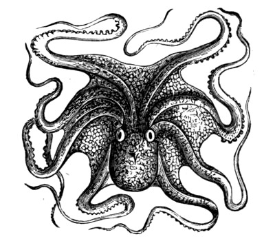
Fig. 183.—Octopus
The former section includes the Octopods, of which some species inhabit
our seas. They all have eight arms, of unequal size, with the suckers
arranged in two rows, and their round or oval bodies seldom have any
fins, locomotion being effected by means of the arms, and by the sudden
expulsion of water from the siphon. The shell is rudimentary, being
represented merely by two short ‘styles’ within the mantle. The species
vary considerably in size, some being only about an inch long when fully
grown, while others measure two feet or more, and are looked upon as
formidable creatures by man. Sometimes they are washed up on our
beaches, but the best way to make their acquaintance is to examine the
contents of the fishermen’s drag nets as they are hauled on the beach.
In the same manner we may secure various species of the Decapods or
Ten-footed Cephalopods, which comprise the Calamaries, Squids, and
Cuttlefishes. These, too, properly speaking, have but eight arms, the
other two appendages being really tentacles, which are usually longer
than the arms, and more or less retractile; they are also expanded at
the ends. The decapods are also to be distinguished from the octopods
by their elongated bodies, and a flattened, fin-like appendage on either
side. Their eyes, also, are capable of being rotated within the orbits,
while those of the octopods are fixed; and the shell consists of one or
more horny ‘pens,’ or of a calcareous ‘bone,’ contained in a cavity so
loosely that it drops out of its place when the cavity is opened.
The Common Calamary (Loligo vulgaris) may be recognised by the
accompanying illustration, from which it will be observed that the body
tapers behind, bearing two rhomboidal fins in the rear. The suckers are
arranged in two rows on the arms, but in fours on the expanded tips of
the tentacles. The animal is a good swimmer, and sometimes crawls, head
downwards, on the disc surrounding the mouth, pulling itself along by
means of its arms. Its shell is a horny pen, lanceolate in form, but it
divides as the age of the animal advances, so that two or more may be
found in the same specimen.
Belonging to the same family we have the Common Squid (Sepiola
atlantica), also a very abundant species. Here the body is shorter and
purse-like, and the fins are dorsal and rounded. It seldom exceeds four
or five inches in length, and, like the Calamary, is used largely as a
bait by fishermen.
Another family—the Sepiadæ—contains the Cuttlefish (Sepia
officinalis), the ‘bone’ of which is such a common object on the beach.
This latter is a broad, curved plate of carbonate of lime, made up of a
number of regular layers, and having a cavity hollowed out at the
posterior end. It is exceedingly light and porous in structure, and at
one time was used largely as an antacid as well as a dentifrice. It is
also proportionately large, being both as long and as broad as the body
of the animal.
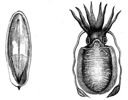
Fig. 186.—Sepia officinalis and its ‘Bone’
Cuttlefishes live principally in the shallow water close to shore, where
they swim backwards by the sudden propulsion of water from their
siphons; and their eggs, which look like clusters of black grapes, are
frequently thrown up on the beach, generally attached to the stems and
fronds of sea weeds.
As a rule the cephalopods swim slowly by the aid of their fins or by a
rhythmic contraction by which water is expelled from their siphons, but
when in danger the muscular contraction is so violent that they dart
through the water with great speed, and even leap into the air to avoid
their enemies. But they have another and much more remarkable way of
escaping from their foes:—They possess a gland, the duct of which
opens into the base of the funnel or siphon, that prepares an inky
fluid; and when the animal is disturbed it suddenly ejects this fluid,
rendering the surrounding water so cloudy that it is often enabled to
retreat unobserved. The ‘ink’ of the Sepia was used for writing in
former times, and is still employed in the preparation of the artist’s
pigment that bears the same name. Fishermen are well acquainted with
this peculiar characteristic of the animal, for they are frequently
bespattered with the contents of the ink bag of the Sepia when the
creature is included in the contents of their draw-nets, and have learnt
to handle it cautiously until the objectionable fluid has been all
discharged.
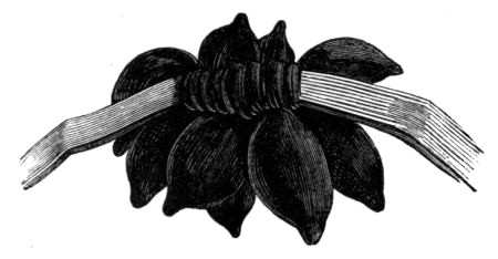
Fig. 187.—Eggs of Sepia
We will conclude this chapter by giving a tabular summary of the
classification of the molluscs which will probably be useful to the
collector of marine objects.
Class LAMELLIBRANCHIATA—Plate-gilled. Headless, usually
enclosed in bivalve shell.
Section SIPHONIDA—Mantle lobes more or less united to form
tubular siphons.
Families—Pholadidæ, Gastrochænidæ, Anatinidæ,
Myacidæ, Solenidæ, Tellinidæ, Mactridæ,
Veneridæ, Cyprinidæ, Lucinidæ, Cardiadæ, &c.
Section ASIPHONIDA—Mantle lobes free or nearly so. No true
siphons.
Families—Arcadæ, Mytilidæ, Aviculidæ,
Ostreidæ, &c.
Class CEPHALOPHORA—Head-bearing. Usually enclosed in a
univalve shell.
Section PTEROPODA—Wing-footed molluscs.
Section GASTEROPODA—Stomach-footed molluscs.
Order Nucleobranchiata—Viscera form a nucleus on the
back.
Order Opisthobranchiata—Shell generally absent. Gills
more or less exposed.
Section Nudibranchiata—Naked gills.
Section Tectibranchiata—Gills covered by shell or mantle.
Order Pulmonifera—Lung-breathers. Terrestrial.
Order Prosobranchiata.
Section Holostomata—Aperture of shell entire (sea snails).
Families—Chitonidæ, Dentaliadæ, Patellidæ,
Calyptræidæ, Fissurellidæ, Haliotidæ,
Turbinidæ, Littorinidæ, Turritellidæ,
Cerithiadæ, Pyramidellidæ, Naticidæ, &c.
Section Siphonostomata—Possess a true siphon. Carnivorous.
Families—Cypræidæ, Conidæ, Buccinidæ,
Muricidæ, &c.
Class CEPHALOPODA—Sucker-bearing arms round the mouth.
Order Dibranchiata—Two gills.
Section Octopoda—Eight arms.
Families—Argonautidæ, Octopodidæ.
Section Decapoda.
Families—Teuthidæ (Calamaries, Squids), Sepiadæ,
&c.
Order Tetrabranchiata—Four gills (containing
Nautilidæ).