THE PATHOLOGY OF INFLUENZA
Transcriber’s Note:
The cover image was created by the transcriber and is placed in the public domain.
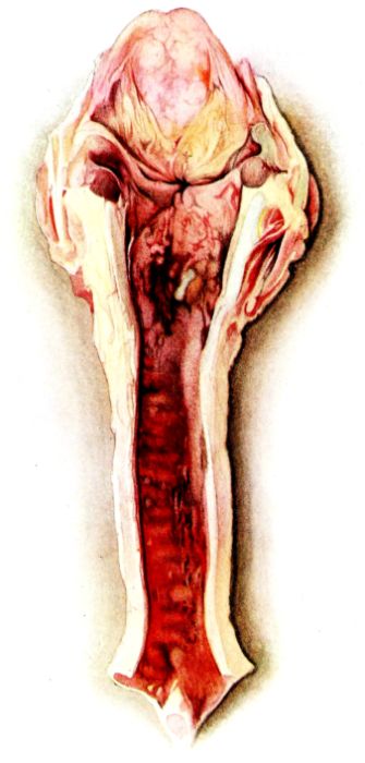
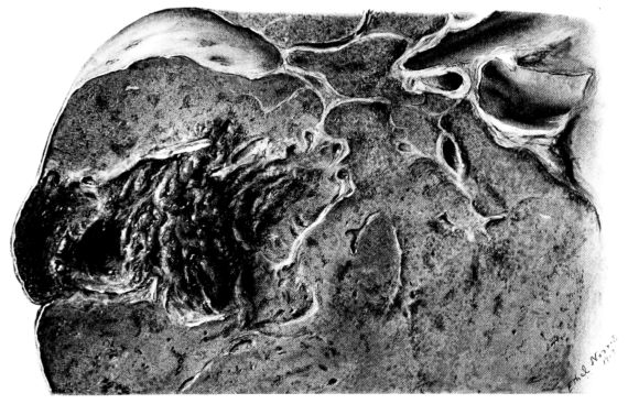
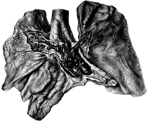
FIG. I. AUTOPSY NO. 99. ACUTE HEMORRHAGIC AND ULCERATIVE LARYNGOTRACHEITIS.

The present volume is the fourth work published by the Yale University Press on the Anthony N. Brady Memorial Foundation, which was established June 15, 1914, by members of the family of the late Anthony N. Brady to enable the University to declare operative the agreement for an alliance between the New Haven Hospital and the Yale School of Medicine. In addition to the pledge of endowment for this purpose, the donors erected for the University on the grounds of the Hospital a clinical and pathological laboratory, and have since, through additional gifts to supplement the income of the Memorial Foundation, made possible the publication of this and other works by members of the faculty of the School of Medicine at Yale.
Our grateful acknowledgment is due the Staff of the New Haven Hospital, especially the members of the Department of Medicine, for their hearty co-operation and for the use of the clinical notes. We also wish to thank the members of the Medical Corps of the United States Army who were stationed at the Yale Army Laboratory School while the work was in progress and who aided in many ways:—Colonel Charles F. Craig, Captain R. A. Lambert, Lieutenants C. A. McKinlay, Frederick Parker, Jr., Ellis Kellert, Henry R. Muller, and J. H. Globus.
TABLE OF CONTENTS
| Page | |||||
|---|---|---|---|---|---|
| Introduction | 9 | ||||
| I. | The Pathology of the Respiratory Tract in Influenza | 13 | |||
| (A) Lesions of the Trachea and Bronchi | 13 | ||||
| (a) Gross Picture | 13 | ||||
| (b) Histological Picture | 14 | ||||
| (c) Summary | 16 | ||||
| (B) Lesions of the Lung | 16 | ||||
| (1) Acute Diffuse Fulminating Type | 18 | ||||
| (a) Gross Picture | 18 | ||||
| (b) Summary | 19 | ||||
| (c) Histological Picture | 20 | ||||
| (d) Summary | 22 | ||||
| (2) Localization and Necrotization of the Pneumonic Process | 22 | ||||
| (a) Gross Picture | 22 | ||||
| (b) Histological Picture | 24 | ||||
| (c) Summary | 26 | ||||
| (3) Organization of the Bronchiolar and Pneumonic Processes | 26 | ||||
| (a) Illustrative Protocols | 27 | ||||
| (b) Summary | 30 | ||||
| II. | Influence of the Respiratory Complications of Influenza upon Tuberculosis of the Lung | 32 | |||
| (a) Illustrative Protocols | 32 | ||||
| (b) Summary | 33 | ||||
| III. | Extrarespiratory Lesions in Influenza | 34 | |||
| (A) Lesions of the Hematopoietic System | 34 | ||||
| (a) Lymphadenoid Tissue | 34 | ||||
| (b) Spleen | 35 | ||||
| (c) Bone Marrow | 35 | ||||
| (B) Lesions of the Blood Vessels and Elsewhere in the Body | 36 | ||||
| (a) Vascular System | 36 | ||||
| (b) Skeletal System | 36 | ||||
| (c) Parenchymatous Organs | 36 | ||||
| (d) Alimentary Canal | 37 | ||||
| (e) Urinary Bladder | 37 | ||||
| (C) Miscellaneous Lesions | 38 | ||||
| (a) Parenchymatous Organs | 38 | ||||
| (b) Jaundice | 39 | ||||
| (c) Central Nervous System | 39 | ||||
| (d) The Pregnant Uterus | 39 | ||||
| (e) Summary | 39 | ||||
| 8IV. | A Comparison between the Respiratory Lesions of Influenza and those Initiated by the Inhalation of Poisonous Gases with Special Reference to:— | 40 | |||
| (A) The Inflammatory Response versus the Systemic Capacity to Compensate | 40 | ||||
| (B) The Primary Injury | 41 | ||||
| (C) The Tendency to Organization of Bronchiolar and Alveolar Exudates with Bronchiolitis and Bronchiolectasis as Sequelæ | 42 | ||||
| (D) The Importance of the Trachea and its Ramifications as a Protective Mechanism against Infection of the Pulmonary Parenchyma | 42 | ||||
| V. | Peculiarities of the Histology of Influenzal Pneumonia | 44 | |||
| (A) The Extent of the Initial Pulmonary Lesion | 44 | ||||
| (B) The Hemorrhagic Exudate—The Relation of Red to Grey Hepatization | 44 | ||||
| (C) The Aplastic Exudate | 45 | ||||
| (D) The Hyaline Necrosis of the Pulmonary Tissue | 46 | ||||
| (E) The Organization Process | 46 | ||||
| VI. | Infection as a Possible Etiological Factor for Malignant New Growths | 48 | |||
| VII. | The Bacteriology of Influenzal Pneumonia | 49 | |||
| (A) Organisms Associated with Influenzal Pneumonia | 49 | ||||
| (B) The Relation of the Type of Organism to Pleural Involvement | 50 | ||||
| (C) The Relation of Different Organisms to the Type of Pneumonia | 50 | ||||
| (D) Summary and Discussion | 54 | ||||
| VIII. | Conclusions | 55 | |||
| IX. | Bibliography | 56 | |||
| X. | Illustrations | 63 | |||
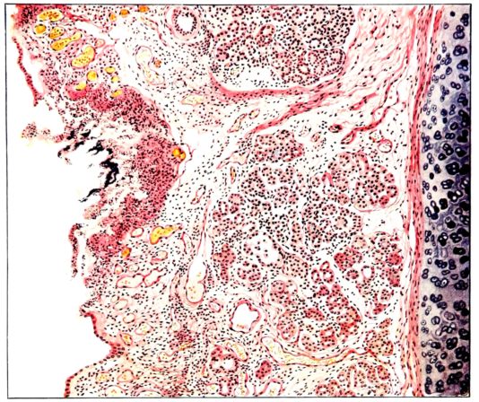
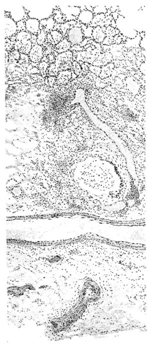
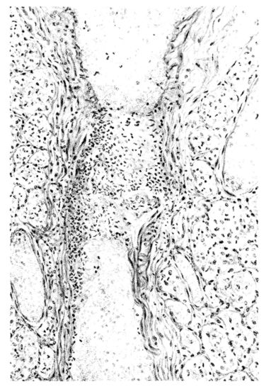
FIG. II. AUTOPSY NO. 98. DRAWING OF A SECTION THROUGH A TRACHEA SHOWING NECROTIZING HEMORRHAGIC INFLAMMATORY PROCESS OF THE MUCOSA.
INTRODUCTION
The epidemic of influenza, prevalent in Europe during the Great War, was watched with interest everywhere, not only because of its military importance, but also because of the danger of its spreading to other continents. The prediction that this would occur, made months before its realization, was verified on an even larger scale than had been anticipated, for in the autumn of 1918, this acute respiratory infection passed over the United States like a huge wave, taking a tremendous toll in human lives; later smaller waves followed leaving in their wake corresponding degrees of devastation. The first cases of the disease appeared on the New England coast, and New Haven was among the cities to be early invaded, though here the epidemic was somewhat less severe than in other cities along the Atlantic Seaboard.
Forewarned and alert to the danger, medical men spared no effort in studying the disease; as a result, no malady, perhaps, has ever been investigated so intensively and from so many different points of attack in an equal length of time. Proof of this appears in the abundant literature issuing from every quarter. Among the various contributions to this subject, many include the anatomical changes associated with the disease. In general, however, these are brief; and although they serve their special purpose well, they have not been elaborated sufficiently to close the chapter.
During a period of about three months beginning with September 18, 1918, while the epidemic raged and waned in New Haven, there were approximately eleven hundred cases of the disease admitted to the New Haven Hospital. As is so often true, only the more critically ill sought hospital care, and few, if any, patients affected by other respiratory infections are included in these statistics. The mortality here, as elsewhere, was very high; of two hundred eighty (280) cases that ended fatally, eighty-two (82) were investigated at the post-mortem table. An attempt was made to make the studies very complete, and this was favored since the headquarters of the Yale Army Laboratory School, under the command of Colonel Charles F. Craig, were located at the Brady Laboratory where a large number of men were being instructed in Pathology and Bacteriology. It was also fortunate that competent illustrators were available who made a splendid series of water-colors and drawings of the characteristic lesions, both gross and microscopic.
The number of autopsied cases at the New Haven Hospital was augmented by a series of acute fatalities from the same disease at the United States General Hospital No. 16, at Allingtown, West Haven, where the anatomical studies were carried on by the same group of men. The latter autopsies offered nothing new, but served to corroborate the conclusions reached at the New Haven Hospital.
The majority of the fatalities occurred in the acute stage of the disease, the anatomical aspects of which have been elaborated more or less completely.[1] Other cases survived for 10a longer period and in these, anatomical changes existed, which, as will appear later, were prognosticated from the acute lesions. Moreover, these findings suggested that certain progressive anatomical changes occur even when the disease runs a much less severe clinical course; for example, in cases where respiratory symptoms persist for a long period before they are brought to a fatal conclusion.
History, too, suggests such a chain of events; namely, in the record of the delayed crop of respiratory disorders that followed the harvest of the epidemic of ’90.
Previous studies of experimental pneumonia in normal and aplastic animals by one of the authors (160) give a background for the interpretation of the histology of some phases of this disease, but more important are the studies of the respiratory inflammatory processes initiated by the inhalation of toxic gases. This subject, introduced into human Pathology with the use of poisonous gases in modern warfare, necessitated elaborate investigations which have just been concluded (159).
The pathology produced by the inhalation of these poisonous vapors is analogous to that found in influenzal pneumonia. This is said with a full comprehension of the criticism that may follow such a statement, and with the knowledge that a similar analogy has been drawn between influenzal and plague pneumonia (Symmers, 141). It is, however, a criticism that is welcomed and which will be met in the body of the paper.
For the reasons just cited, it has seemed desirable to contribute to the Pathology of various phases of influenzal pneumonia and to attempt to correlate this with other types of acute respiratory inflammation, in the hope that the prognostications which suggest themselves may be of aid in the prophylaxis and possibly in the treatment of the more insidious and progressive pulmonary changes that may follow this disease.
A large part of the text is devoted to a description of the gross and more minute pathology of the respiratory tract associated with influenza and its complications, both in the acute and in the subacute or chronic stages. Incidental lesions of less importance in other portions of the body that have occurred in these cases are presented briefly, and emphasis is placed upon a number of special subjects. The order of discussion will be as follows:—
I. The Pathology of the Respiratory Tract in Influenza.
(A) Lesions of the Trachea and Bronchi.
(B) Lesions of the Lung.
(1) Acute Diffuse Fulminating Type.
(2) Localization and Necrotization of the Pneumonic Process.
(3) Organization of the Bronchiolar and Pneumonic Processes.
II. Influence of the Respiratory Complications of Influenza upon Tuberculosis of the Lung.
III. Extrarespiratory Lesions in Influenza.
(A) Lesions of the Hematopoietic System.
(B) Lesions of the Vascular System, Parenchymatous Organs, Alimentary Tract, and in the Walls of Other Hollow Viscera.
(C) Miscellaneous Lesions.
IV. Comparison between the Respiratory Lesions of Influenza and those Initiated by the Inhalation of Poisonous Gases with Special Reference to:—
(A) The Inflammatory Response versus the Systemic Capacity to Compensate.
(B) The Primary Injury.
(C) The Tendency to Organization of Bronchiolar and Alveolar Exudates with Bronchiolitis and Bronchiolectasis as Sequelæ.
(D) The Importance of the Trachea and its Ramifications as a Protective Mechanism against Infection of the Pulmonary Parenchyma.
V. Peculiarities of the Histology of Influenzal Pneumonia.
(A) The Extent of the Initial Pulmonary Lesion.
(B) The Hemorrhagic Exudate—The Relation of Red to Grey Hepatization.
(C) The Aplastic Exudate.
(D) The Hyaline Necrosis of the Pulmonary Tissue.
(E) The Organization Process.
VI. Infection as a Possible Etiological Factor for Malignant New Growths.
VII. The Bacteriology of Influenzal Pneumonia.
(A) Organisms Associated with Influenzal Pneumonia.
(B) The Relation of the Type of Organism to Pleural Involvement.
(C) The Relation of Different Organisms to the Type of Pneumonia.
(D) Summary and Discussion.
VIII. Conclusions.
IX. Bibliography.
X. Illustrations.
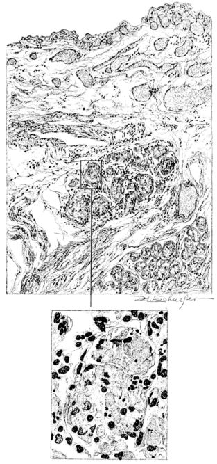
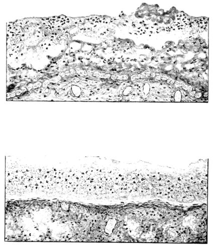
FIG. III. AUTOPSY NO. 90. DRAWING FROM A LESION OF THE TRACHEA (SOMEWHAT OLDER THAN THAT ILLUSTRATED IN FIGURE II). THE MUCOSA IS ENTIRELY LACKING. CONGESTION AND EDEMA ARE THE STRIKING FEATURES IN THE SUBMUCOSA. THE NECROTIZING PROCESS HAS EXTENDED INTO THE MUCUS GLANDS. THIS IS SHOWN IN THE LOWER PICTURE.
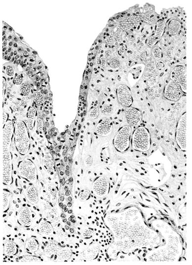
FIG. IV. AUTOPSY NO. 205. CONGESTION AND EDEMA OF THE SUBMUCOSA AND REGENERATION OF THE TRACHEAL EPITHELIUM.
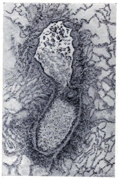
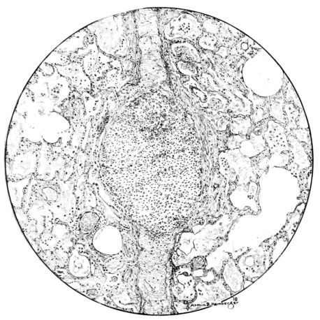
FIG. VII. AUTOPSY NO. 94. A NECROTIZING PROCESS LIKE THAT OF THE TRACHEA ILLUSTRATED IN FIGURE II. HERE IT IS SHOWN TO INVOLVE THE WALL OF THE BRONCHIOLE.
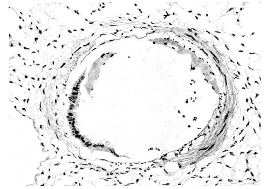
FIG. V. AUTOPSY NO. 95. AN EARLY LESION OF THE BRONCHIOLE CHARACTERIZED BY HYALINIZATION OF THE EPITHELIUM AND SEROUS EXUDATE IN THE LUMEN.
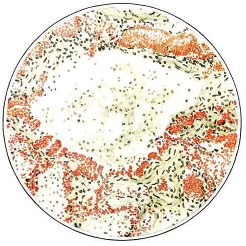
FIG. VI. AUTOPSY NO. 103. ILLUSTRATES ANOTHER EARLY BRONCHIOLAR LESION. THE EPITHELIUM IS LACKING, THE CONGESTED VESSELS OF THE SUBMUCOSA PROTRUDE INTO THE LUMEN WHICH CONTAINS DESQUAMATED EPITHELIUM, MUCUS, AND RED BLOOD CELLS.
THE PATHOLOGY OF THE RESPIRATORY TRACT IN INFLUENZA — A. LESIONS OF THE TRACHEA AND BRONCHI — Gross Picture. — Histological Picture. — Summary. — B. LESIONS OF THE LUNG — (1) ACUTE DIFFUSE FULMINATING TYPE OF PNEUMONIA — Gross Picture. — Summary. — Histological Picture. — Summary. — (2) LOCALIZATION AND NECROTIZATION OF THE PNEUMONIC PROCESS — Gross Picture. — Histological Picture. — Summary. — (3) ORGANIZATION OF BRONCHIOLAR AND PNEUMONIC PROCESSES. — Summary.
If the atrium of an infection and its specific etiological agent are undetermined, the narrator of the pathology of a specific disease is confronted immediately with serious obstacles in the elaboration of a complete picture. Some writers assume that the respiratory tract is the portal of entry in influenza (162), though the specific agent is still unknown.[2] Whatever the agent, unquestionably it attacks the respiratory tract at a very early stage in the disease and produces a lesion which becomes responsible for the most serious aspect of influenza, whether this phase be primary or only a complication.
Among the lesions which will be considered, therefore, those of the respiratory tract chiefly will be emphasized. They include the changes in the large air passages, as well as the pulmonary, alveolar, and interstitial involvement. Unquestionably, a very close association exists between the lesions of the larger air passages and those of the alveoli, but probably it is equally true that the former may occur alone; in many instances also they are the forerunners of the latter lesions. Consequently, it seems logical to begin with an exposition of the lesions in the trachea and its ramifications, including the bronchioles.
Early in the disease the congestion and the hemorrhages that have been described in the mucous membrane of the nasopharynx (14 and 94) are also conspicuous features in the lining of the trachea and bronchi (Fig. I). This membrane is swollen, turgid, red, and covered by a copious, mucous exudate which may be clear, but much more frequently is blood-stained or opaque and yellowish in color. The blood, variable in amount, may be fresh and red; and after the mucous exudate on the surface is removed, more intense red foci stand out on the congested base (47, 90, 157). Frequently, as the bronchi are approached, the red color of the mucosa becomes more intense and may have a garnet tinge. Membranes such as are encountered in the more usual necrotizing inflammatory processes, like diphtheria, have not occurred in the trachea and larger bronchi in this series (108, 128, 157).[3] The exudate peels off readily, and as indicated above, leaves a velvety red surface, 14dotted here and there with darker or more intensely red foci. Small ulcerations of the mucosa occur, but are inconspicuous (82, 156). As the finer ramifications of the bronchi are approached, the accumulation of the exudate in their lumina becomes more and more marked, and on cross section of the lung, they often stand out conspicuously on account of their increased size and projecting, seromucous, blood-stained content (101, 149, 162).
It is remarkable how long this picture in the trachea and bronchi may persist without showing any marked variation. It is encountered, not only in the most acute and fulminating types of the disease (that have been examined), but a similar picture may be present in cases which end fatally only after a period of weeks of severe illness. In the latter cases, however, the exudate, particularly in the bronchioles, assumes a more purulent character and after this accumulation is wiped away from the surface of the tube, the intensity of the dark red color of its lining membrane presents an even more striking contrast on account of the opaque, yellowish-green exudate in the lumen. At this stage, too, the bronchioles are more distended with pus, which oozes from each one when the lung is sectioned (1, 108, 110). In the cases still more chronic, the terminal bronchioles may be sharply outlined with a thick grey wall which surrounds the dilated opening from which the accumulated yellowish exudate oozes as soon as the pressure is relieved (Figs. XXXIX and XL).
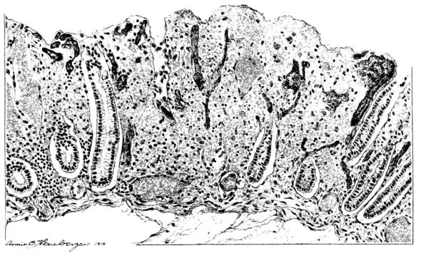
The changes are less marked, perhaps, in the trachea than in its finer ramifications. The mucosa is constantly more or less destroyed and large areas, usually focal, are entirely devoid of their epithelial covering. This is replaced by a sparse exudate, composed largely of red blood cells, mucus, a small amount of fibrin, and nuclear fragments (Fig. II). It may dip into the submucosa for a short distance, but usually these indentures are associated with the ducts of the mucous glands into which the inflammatory reaction extends. A more striking feature than the exudate, however, is the edema and the congestion of the submucosa. The loose areolar tissue of the submucosa is spread widely apart, and throughout it distended blood vessels are very conspicuous. Occasionally such a vessel is broken and actual hemorrhage appears in the submucosa. Occasionally, too, the inflammation extends down the duct to the mucous gland itself, and here, also, aplastic inflammatory reaction is evident, inasmuch as the acini now stain intensely red with the cells undifferentiated from each other and specked here and there by broken remains of the dead nuclei (Fig. III). After the disease has continued for a short period, even at the end of five or six days, some regeneration of the epithelial lining may be seen (3) (Fig. IV). But despite this, the acute picture persists, and there goes on, side by side, an attempted repair characterized by epithelial regeneration and the same evidence of acute change. Since the lesion is essentially a superficial one, scars or contractures of any extent are not encountered in the trachea, even in examples of the disease that have ended fatally only after many weeks.[4]
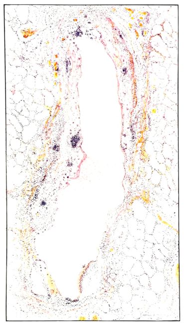
FIG. VIII. AUTOPSY NO. 97. ALTHOUGH THE EPITHELIUM IS STILL VISIBLE AS A HYALINE BAND LIFTED FROM THE UNDERLYING MUCOSA, BOTH MUCOSA AND SUBMUCOSA ARE INVOLVED IN A NECROTIZING PROCESS. BACTERIA ARE ABUNDANT IN THE DEAD TISSUE.
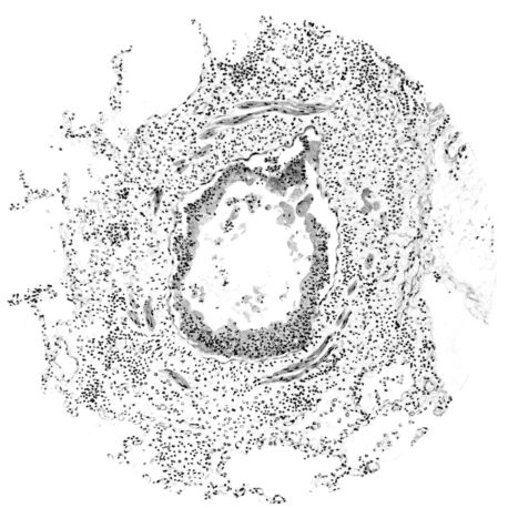
FIG. IX. AUTOPSY NO. 105. THE NECROTIC EPITHELIUM IS INVADED BY POLYMORPHONUCLEAR LEUCOCYTES AND THESE, AS WELL AS ROUND CELLS, CAUSE A THICKENING OF THE BRONCHIOLAR WALL.
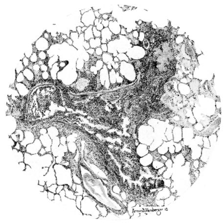
FIG. X. AUTOPSY NO. 106. THE SMALL BRONCHIOLE IS FILLED WITH PUS CELLS, WHICH, IN SOME PLACES, EXTEND THROUGH ITS WALL.
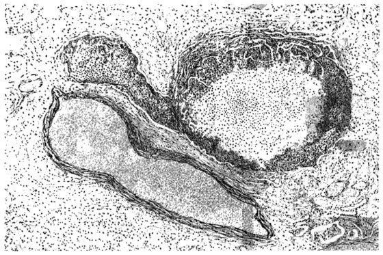
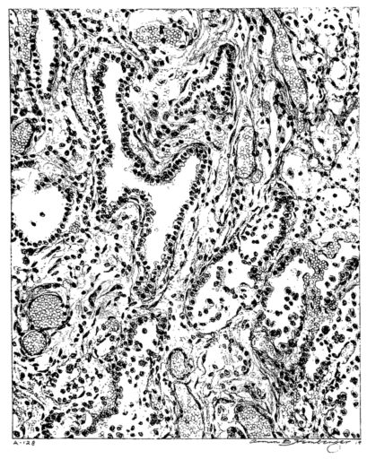
15There is considerable evidence to support the view that the disease spreads from bronchus to bronchus, and in keeping with this view, various stages in the inflammatory processes are more readily determined in these smaller structures than in the trachea. Furthermore, it must be emphasized that even the mildest and the most extreme of these stages are not infrequently encountered in the same lung. The earliest lesion is manifested by an increased homogeneity of the protoplasm of the epithelial lining of the bronchus. The cell protoplasm loses its normal granulation and the nucleus, somewhat darker than usual, becomes conspicuous on a red base (Fig. V). In the lumen of such a tube a serous exudate, perhaps mixed with mucus, is encountered, and there is some spreading apart of the surrounding muscular tissue with engorgement of the vessels. This picture merges gradually into one where the epithelium appears as a homogeneous, red-staining ribbon, devoid of nuclei, often exfoliated, in part at least, from the underlying submucosa (92). The change is traceable through the larger bronchi, even to the ducti alveolares, and not infrequently, bacteria, either as a diffuse, minute dotting or in the form of circumscribed, colony-like formations, are spread through the red, ribbon-like strand (Fig. XVI). With the exfoliation of the epithelial lining, the submucous vessels become more and more conspicuous and may bulge into the lumen of the tube (Fig. VI). That they actually weep into the lumen is proved by the presence of red blood cells in the exudate, now rich in mucus, broken-down nuclei, and desquamated cells. The necrotizing process may not extend deeper than the epithelial lining as is the status described above (140, 162), but it also frequently involves the underlying submucous and muscular layers, so that these lose their identity and stand out as homogeneous masses, in which fragmented nuclei and bacterial accumulations are prominent. Such deeper necrotizing areas may be focal (Fig. VII), or may involve the entire circumference of the tube (Fig. VIII). Occasionally, the epithelium, now dead and staining homogeneously, is lifted from the underlying submucosa in the form of a blister (66), and has very much the same appearance as the well known, early reaction which follows the application of croton oil to the rabbit’s ear. Where this occurs, the submucosa is less involved, as though the necrotizing agent had not penetrated to the same depth and the serous reaction beneath were actually a beneficent exudate. These blisters are in contrast with the deeper areas where the fibrinous mass, mixed with the dead tissue, forms an intensely staining ring or band, which extends through the bronchiolar wall even to the surrounding alveoli.
In the early stage of this process one of the most outstanding features is the absence of polynuclear leucocytes in the reactionary process, but gradually as the dead tissue sloughs away, these cells wander into the exudate and form a purulent ring, more intense in the lumen, but extending for a variable distance through the still viable wall of the structure (47) (Fig. IX). Later mononuclear cells accumulate in this wall and occur either as a diffuse mottling or as circumscribed foci in the muscle and submucous layer of the bronchiole, just as they do in the trachea. Occasionally, a striking change is found in a small bronchiole within a portion of the lung which is otherwise uninvolved by an inflammatory process. Perhaps the alveoli were the seat of a change which has subsided, but, whatever the history, the purulent mass in the bronchiole and involving its wall, stands out effectively (Fig. X).
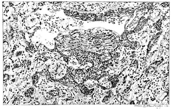
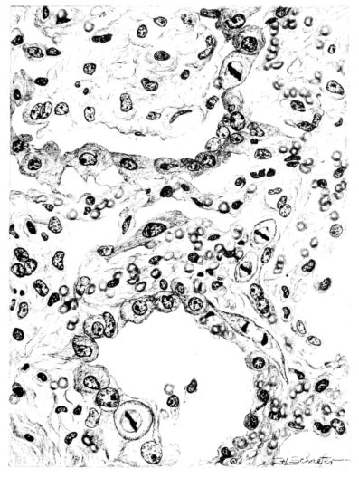
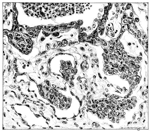
Sooner or later, with the subsidence of the irritating agent, repair begins in the bronchus or bronchiole. If its walls have been destroyed and the lesion has extended into the surrounding alveoli to form an abscess of greater or lesser extent, or if the necrotizing process has been superficial and confined to the epithelium in large part, the reparative process is very much the same. Mitotic figures in the fibroblasts and in the endothelial cells of the capillaries abound in the young granulations (Fig. XI). However, the granulation tissue does not have an unrestricted path of growth, for if a remnant of epithelium remains, this is stimulated to grow probably in this disease as in no other. Mitotic figures are common 16and the young epithelial cells stretch across the denuded submucosa or granulation (Fig. XLVIII) and extend downward into the surrounding alveoli, not only as strands, but also as solid nests of cells (47) (Fig. XLIX). The bronchioles, therefore, show changes dependent upon the extent of the damage suffered by their walls. The vast majority, in all probability, will be restored; but if the wall has actually been necrotized, the bronchioles may be converted into small, saccular, bronchiectatic cavities (48, 110, 162) (Figs. XII and L), or obliterating bronchiolitis may result from the organization of the exudate within their lumina (82) (Fig. XI). The importance of the epithelial proliferation cannot be ignored; in many cases, it invades the surrounding lung tissue and a typical, histological picture results—an infiltrating, malignant, epithelial neoplasm (Figs. XLVIII and XLIX).
The lesions of the trachea and of the larger bronchi, even though they persist in their acute form for a period of weeks, are superficial and do not lead to extensive or deep scarring. In contrast to the larger respiratory passages, that portion of the bronchial tree which is not supported by cartilaginous rings becomes more and more intensely involved, not only on its surface, but in its deeper structures, and the changes in the bronchioles and the neighboring air sacs are among the more characteristic anatomical manifestations in this disease. Therefore, while it will be unnecessary to refer again to the larger air passages, further consideration of the smaller ones, which are constantly associated with the lesions of the pulmonary parenchyma, will be included in the subsequent discussion of the pulmonic involvement.
In the prosecution of the scientific study of any disease, the great temptation is to differentiate its various manifestations with the intention of elaborating a classification. While there may be more or less distinct types of pulmonary involvement in influenza, so many intermediary forms appear in an extensive series of cases, that any classification must necessarily be arbitrary. Here, such an attempt at classification is beset with unusual difficulty, because, in the vast majority of cases which come to anatomical observation, the disease is fulminating. The lesions are more uniform in character than in an experimental study where the material is arranged so as to include intermediary and chronic stages, which in man are only encountered when accident causes death. These gaps in the study cannot be supplemented with experimental observations, because attempts to reproduce this disease have failed, even in human subjects. From the literature it appears also that variations in the extent, and perhaps in the maturation, of the anatomical involvement may be represented in different proportions for different localities (1, 2, 17, 55, 92, 162). Therefore, no sharp differentiation may be drawn between the more or less definite stages which are seen at the post-mortem table; still, for convenience of description, certain of the lesions which occur more frequently and are more widely differentiated may be considered separately.
The disease, as has been indicated, may be confined to the bronchial tree. In these circumstances, it is almost necessary to suppose that the larger bronchi alone are involved. When the delicate bronchioles are affected, there is always a more or less extensive involvement of the pulmonary parenchyma (124), for the bronchioles have a more direct communication with the alveoli, and their walls present a less formidable barrier to the extension of the inflammation to the surrounding air sacs.
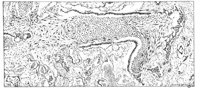
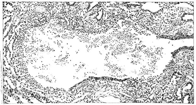
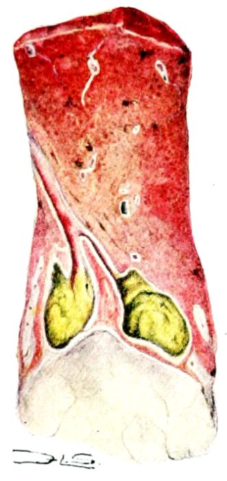
FIG. XII. AUTOPSY NO. 209. A SMALL BRONCHIECTATIC CAVITY FILLED WITH PUS. THE GROSS APPEARANCE IS ILLUSTRATED IN FIGURE L.
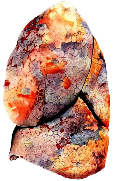
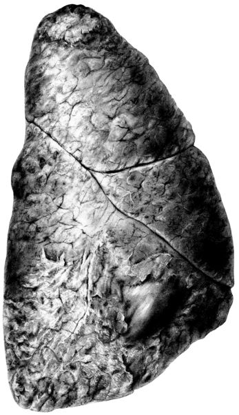
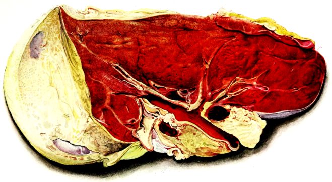
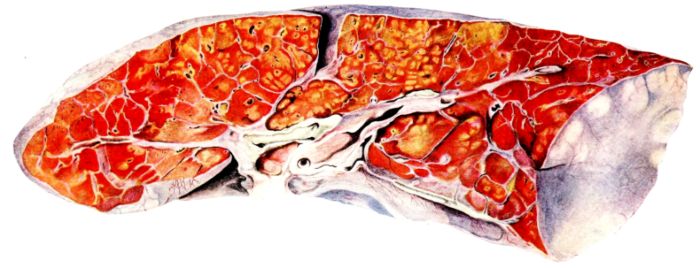
FIG. XIII. AUTOPSY NO. 96. RIGHT LUNG. A WATER COLOR DRAWING OF A GROSS LUNG IN THE ACUTE STAGE. NOTE THE SIZE OF THE LUNG, THE HEMORRHAGES ON THE PLEURAL SURFACE, AND THE BLUE AREAS OF CONSOLIDATION.
17Clinically, there is reason to believe that the disease may begin with a period of general malaise, during which the respiratory symptoms may be more or less severe. In the fulminating cases, the malaise may be associated from the beginning, not only with a tracheobronchitis, but also with pulmonary involvement. In less severe types the malaise may be accompanied only by tracheobronchitis, and may present no symptoms referable to the pulmonary parenchyma; there may follow or not, a definite period of clinical improvement, after which pneumonic involvement becomes evident. However, even where a fulminating type of the disease is not associated with clinical evidence of pulmonary involvement, the post-mortem examination may show extensive change in the lung parenchyma (110).
Whether this grouping of the disease is correct or is based upon a fallacious deduction from more or less satisfactory clinical histories, is open to question; and a decision may be reached from comparison of the opinions based upon carefully observed cases treated in different institutions. From the report of the epidemic at the Johns Hopkins Hospital (Bloomfield and Harrop (14)), where two hundred sixty-eight cases were studied in which the complication of pneumonia was uncommon as compared with cases in other hospitals—the New Haven Hospital, for instance—and where the percentage of the deaths was low, the conclusion was reached that the disease is primarily a general one and that the pulmonary involvement is secondary, just as in an exanthem, like measles. By way of comparison, at the New Haven Hospital, where more than eleven hundred patients with influenza were observed, the type of disease described by Bloomfield and Harrop was relatively rare. Of course, there were cases of that type where general malaise, with or without respiratory symptoms, was followed by a period—usually of from two to three days—of definite improvement in the symptoms; and later extensive and serious pulmonary complications ensued. However, in another group, largely composed of individuals who entered the hospital seriously ill, the histories indicate an acute onset resembling that of lobar pneumonia and with early manifestations of pulmonary involvement (2, 17, 52, 145). This discrepancy, probably, may be explained in part by variations in the sensitivity to minor indisposition on the part of the different individuals.
The preceding review should aid in the correlation of the clinical types of the disease with the respiratory lesions. All pulmonary lesions, from the least to the most localized, may be explained either by a subsidence of a less acute initial and diffuse involvement of the parenchyma, or by a less rapid and progressive spread of the necrotizing and inflammatory process from the upper respiratory tract through the bronchioles to the alveoli. This conception does not take into account the significance of the period of malaise, interpreted by the clinician as the period of invasion, but attempts to correlate the respiratory symptoms with the pulmonary lesions and their etiology.
Our own experience, like that of other observers (26, 104, 162), is that all fatal cases of this disease show pulmonary involvement in the form of pneumonia. The lesion varies greatly in intensity and in the amount of pulmonary tissue affected. In the descriptions which follow, the more diffuse and intense processes will be discussed first, and later those in which the inflammation localized and terminated in pseudolobar, lobar, lobular, or peribronchial pneumonia.
The majority of influenzal deaths have been examples of this acute type. Out of ninety-five cases, the total number studied, forty-four belong to this group in which the average duration of illness was nine days. They form the basis of the description which follows.
A striking feature of the external examination of the body is the intense rigor which involves all the muscles and is broken only with difficulty (78). This is associated with a rapid settling of the unclotted blood in the dependent parts which gives them an intense blue or bluish-purple color. The erythema of the skin which has been described clinically is not recognizable at the post-mortem table. There is, however, cyanosis of the face which reaches an intensity explained by the fact that this disease so often affects healthy, well nourished, muscular individuals. In a few instances the cyanosis is more extensive and gives a plum color to the entire body. All the mucous membranes share in the intense congestion and discoloration of the face. Slight jaundice is common, but marked variations in intensity occur. The external nares and the lips are almost invariably covered with blood-stained crusts. Even in the decubitus position, a thin, sanguinous fluid tends to escape in large quantities from the nose and mouth. The large veins of the neck are usually prominent, and the chest voluminous.
Distinct splanchnic engorgement is evident as soon as the peritoneal cavity is opened. The liver extends below the costal margin and is dark in color, but otherwise the abdominal cavity presents nothing characteristic of the disease. The diaphragm does not extend as high as usual, and the pleural cavity almost invariably contains an excess of fluid. Usually the quantity is small, but, on the other hand, it may be considerable, and in twenty-one of the forty-four cases the fluid exceeded one hundred cubic centimeters. The turgidity of the mediastinal tissues varies somewhat in degree (27). Generally the pericardial sac is smooth and glistening on both visceral and parietal surfaces; the pericardial fluid is not materially changed. Frequently (seventy per cent) there is dilatation of the right side of the heart (138, 141, 157), but aside from an occasional small endocardial or subepicardial hemorrhage (90, 108, 156), there are no lesions of consequence in the heart attributable to this disease[5] (162).
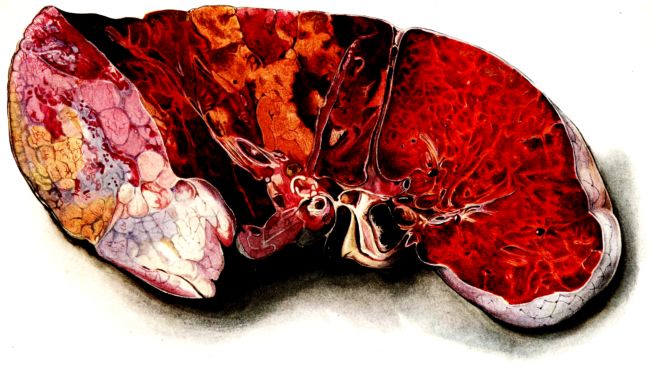
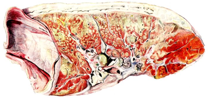
FIG. XIV. AUTOPSY NO. 96. LEFT LUNG. NOTE ITS SIZE AND THE PATCHY CONSOLIDATION.
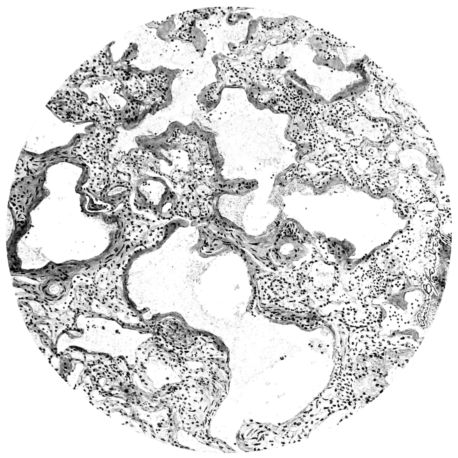
FIG. XV. AUTOPSY NO. 149. ILLUSTRATES ONE OF THE MOST STRIKING EARLY PULMONARY LESIONS; THE DILATATION OF THE TERMINAL BRONCHIOLES AND THE HYALINIZATION OF THEIR EPITHELIUM. COMPARE FIGURE XVI.
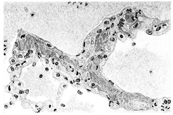
19The lungs are extremely voluminous (12, 17) due in part to an accumulation of liquid within them. This finds its way into the trachea and completely fills the latter structure with blood-stained, syrupy fluid, with purulent material, or with a mixture of these (2, 90, 107, 157, 162). At first the pleural surface is smooth and often quite even, but on closer inspection, a minute granulation is suggested. In many cases even close examination does not allow the conclusion that an exudation of anything but serum has occurred through this membrane, except in localized foci. These foci more frequently involve the interlobar pleura and that of the lower lobes (112, 143). The volume of the lungs, often great enough to obliterate the pericardial area, is one of the two most characteristic features of the external examination. The other feature is their color. Small, bright red hemorrhages may occur anywhere. The larger patches are the most striking. Violet, purple, or dark brown areas, irregular in shape and distribution, are more frequently found on that portion of the pleura over the lower two-thirds of the lung. Between the deeply colored zones, there are pale pink areas which involve the lowermost edge to the least degree, the anterior margin somewhat more, and the apex of the lung most of all. The darker portions just referred to may project above the surface and may be circumscribed, resembling huge, fresh hemorrhagic infarcts (41, 108). The alveolar walls are not seen through the pleural surfaces in these darker zones. The pale pink areas, usually at the level of the more intensely colored zones, may be elevated and the dilated air sacs are distinctly made out through the pleura (Fig. XIII). At the hilum, the lymph glands are large and soft. When cut, fluid escapes and is often blood-stained. The cross section may present a distinct, diffuse, hemorrhagic appearance (162). At the hilus, too, the lymphatics, distended here and there over the surface of the pleura, are most affected. The congested bronchial mucous membrane and the exudate in these structures has been described.
After removal, the lung retains its shape, but is more flaccid than the consolidated lung of lobar pneumonia. It cuts with very little resistance and immediately a large amount of a syrupy, pink fluid escapes and obscures the entire area. With the fluid scraped away, the variations in the consistency of the lung become visible. The pale areas around the borders and chiefly at the apex in which the air sacs are discernible with the naked eye, sink slightly below the remainder of the surface, and the pleural edge inverts. The individual lobules of the lung in these areas are more conspicuous than normal, because the interstitial tissue bearing the lymphatics and vessels, as well as that around the bronchi and larger blood vessels, does not lose its edematous appearance as quickly as the alveoli (40, 92, 110, 164), and, consequently, these grey lines and points stand up somewhat more prominently.[6] In contrast with the paler areas which are prone to slight collapse, the remainder of the cross section retains its more smooth and even surface. The alveolar walls are not distinctly made out, but the terminal bronchioles often make themselves evident by the nature of the material which is within and by their distinct dilatation (1, 67, 110, 149, 162). The more firm areas stand out, too, on account of their difference in color. The scheme is not unlike that seen on the pleural surface, and while dark, almost black, infarct-like areas occur on the cut surface, the solid areas are more likely to be translucent, dull, light red, brown or even grey. They have a surface similar to a very fresh, tuberculous, gelatinous pneumonia, but the color differs from the cloudy grey of the latter on account of the admixture of blood in the exudate and the great congestion of the vessels (Fig. XIV).
The well developed post-mortem muscular rigidity, the lividity of the dependent parts, of the face with its mucous membranes, and often of the trunk, the jaundice variable in extent, the crusts of blood on the nares and mouth, and the splanchnic dilatation are features which prepare for the gross picture presented by the thoracic organs. The increased moisture within the pleural cavities associated with the even, translucent pleural surface, whose dilated lymphatics become more and more prominent towards the hilum, the large succulent lymph glands, and the exudate in the bronchial tree, are all striking, but more characteristic of the gross picture, is the great increase in volume of the lung itself, mottled 20with brilliant colors. The lung, too, is very wet and on section, after the syrupy, blood-stained fluid escapes from the less definitely consolidated zones, the latter appear, not as the usual granular, firm areas of hepatization, but have more the consistency of a gel, and also its translucence. Characteristic of this disease as these changes may be, the specificity of the fundamental lesion in the respiratory tract, becomes more emphatic after study of its histology (92, 162).
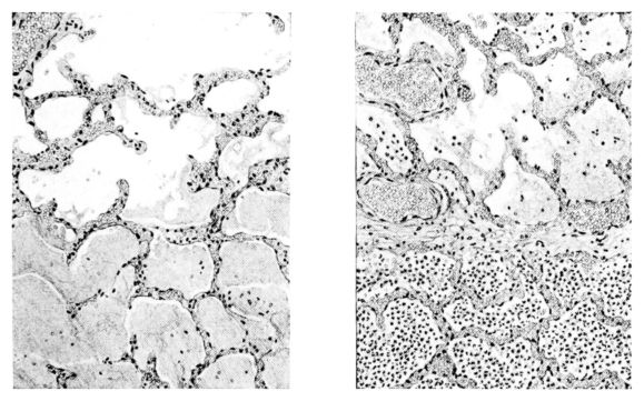
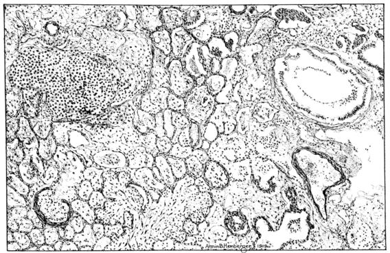
No matter what the portion of the lung from which the sections are derived, the fundamental changes found are the same. The subpleural sheets are spread wide apart, now by empty spaces, now by coagulated fluid. The process extends from the surface through the interlobular septa (Fig. XX), and is accentuated where the connective tissue is more prominent around vessels and bronchi. The nature of the infiltrate in the subpleural and interstitial tissues becomes more evident in the alveoli, which likewise are filled. The material varies somewhat in appearance, probably dependent upon its proteid content. Not infrequently the alveoli contain a homogeneous, pink-staining mass, which resembles the colloid of the thyroid gland. Again, it may be simply a coarse granular precipitate (Fig. XXIII), and in still other instances, small sticks and strands form the bulk of the alveolar content (47, 92, 140, 156). This subpleural, interstitial, perivascular, peribronchial, and alveolar edema, which is a term applicable to this collection of fluid, is very prominent, and although its intensity varies in different portions of the lung; and although it may be replaced in some areas by other types of exudate, unquestionably, this is the dominating expression of the inflammatory process in the early stage of the disease.
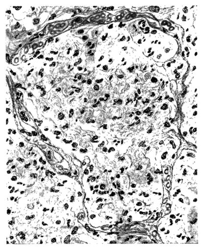
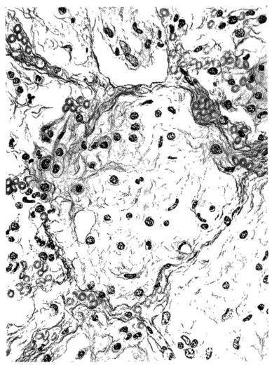
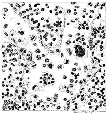
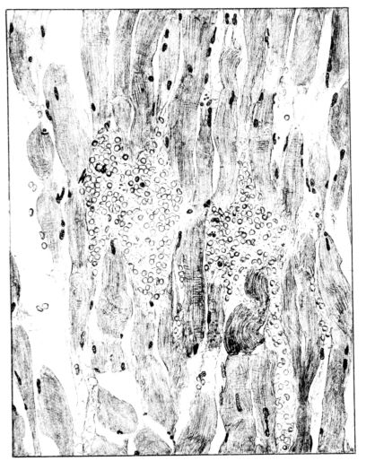
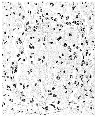
As might be expected from the gross appearance, the alveoli vary in size. At times slightly collapsed and at other times overdistended, their lumina are still the seat of the inflammatory exudate, although the mechanical change may allow of some variation in the appearance of their walls. As a rule, however, the alveolar wall is prominent and owes its conspicuousness to the tortuous, engorged vessels within. These vessels contain red blood cells almost exclusively, and on account of the partial, occasionally complete, loss of the lining epithelium, the alveolar wall appears as a huge, dilated arteriole (101) separating the lakes of coagulated material in the spaces (Fig. XXIII). There are areas, as indicated above, where the alveolar content may be more definitely arranged in the form of beaded or homogeneous strands of different caliber; the smallest resemble delicate threads. They tend to converge toward the alveolar wall like wheat in a sheaf, and often pass through this wall by way of the so-called pores of Cohn; as soon as the body of the neighboring alveolus is reached, they again present a fan-like expansion into innumerable, fine strands (Fig. XXII). Where the exudate is more fibrinous, the alveolar wall is less likely to be distended, its vessels are not so prominent, and their content of red blood cells is definitely decreased. Still this is not the most extreme type of alveolar exudate met with at this stage. Perhaps, the most striking, although not the most frequent, exudate has a superficial resemblance to a huge, red blood clot, and it may be difficult to make out the alveolar walls separating the masses of well preserved red blood cells that fill the alveolar spaces. These areas are indistinguishable from infarcts and may be associated with thrombotic arteritis in near-by pulmonary vessels (47) (Figs. XXIV and XXV). Among the red blood cells an occasional strand of fibrin, a desquamated alveolar epithelial cell, and rarely a polymorphonuclear leucocyte may be encountered. The alveolar wall itself varies in the definition of its outline. When its vessels are greatly distended, when its alveolar epithelium is gone, and when its content consists largely of red blood cells, it is difficult to distinguish from the exudate which it encloses. However, when it is more compressed or when its epithelial lining cells are still more or less intact, it may be seen as a blue-staining strand under the low power of the microscope, for the well preserved nuclei lend it prominence.
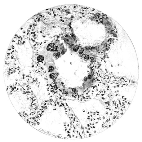
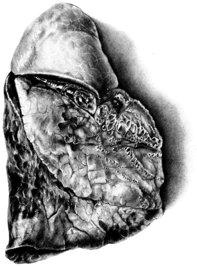
FIG. XVIII. AUTOPSY NO. 155. ILLUSTRATES A MILD FORM OF PULMONARY INTERSTITIAL EMPHYSEMA.
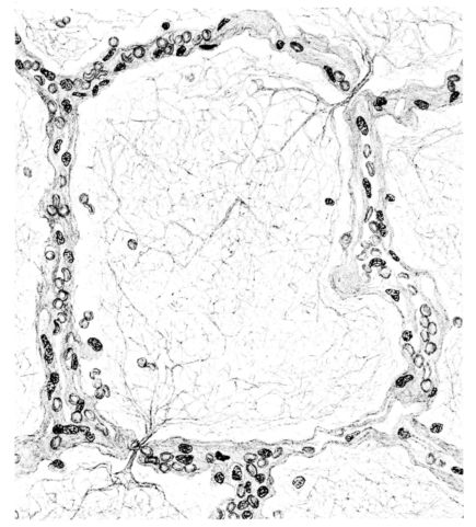
FIG. XXII. AUTOPSY NO. 175. AN ALMOST PURE FIBRINOUS EXUDATE. THE ALVEOLAR WALLS ARE SLIGHTLY HYALINIZED AND THEIR EPITHELIUM IS ALMOST ENTIRELY LACKING.
21There are, of course, variations in the extent of the serum, the fibrin, and the hemorrhage in the exudate of the alveoli, and while these different types may occur as pure forms, often they are associated. In still other areas and varying in prominence, one finds as characteristic an exudate, not only of serum, strands of fibrin, and red blood cells, but also a diffuse dotting of the exudate with bacteria, singly, in pairs, clumps, and chains (92, 164) (Fig. XXI). This type of reaction is uncommon in pulmonary disease. It resembles more closely a streptococcus cellulitis such as is encountered frequently in the subcutaneous tissues, for example, a woody phlegmon, or a sero-hemorrhagic exudate like the avirile response to a rapidly fatal hemolytic streptococcus serositis. A similar reaction has been reproduced experimentally in animals which have been rendered aplastic with benzol previous to pulmonary insufflation, and it is conceivable that the lack of polymorphonuclear response in the inflammatory exudate may be associated with some such general destruction or temporary suspension of leucocytic formation (160).
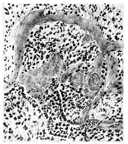
A more striking picture, however, even than this aplastic alveolar exudate appears in the terminal bronchioles. In many instances, these are conspicuous on account of their size, for they are dilated to form prominent, often irregular, sacs (Fig. XV). The distention of these terminal bronchioles may be so great that the surrounding alveoli are compressed. What makes them even more conspicuous is their lining, once epithelium, but now a swollen, thick, homogeneously staining material, with complete loss of architecture; the material forms (with hematoxylin and eosin) a red band limiting the lung tissue and sharply demarcating it from the exudate within the bronchioles (48, 92). However, this ribbon of red, often thickened by fibrin deposition, is not always pure, for bacteria thrive in the dead tissue. They occur singly, paired, in chains, and also as circumscribed, dense masses which in size and position, simulate nuclei (162) (Fig. XVI). This same hyalinization of the epithelium, it will be recalled, occurs in the larger bronchi (Fig. V), and there, too, bacteria frequently develop in the dead tissue (Fig. VIII). In the smallest bronchiolar ramifications, acute epithelial necrosis is not infrequently encountered, even when the surrounding lung tissue is relatively normal (Fig. XVI). That the process does not stop with the epithelium, but, as in the larger bronchi, may extend through the entire structure of the bronchioles, is manifest. Even the alveolar walls may be involved and frequently homogeneous pink or red bands, now the phantom of the former viable lung tissue, mark the presence of the old wall of the alveolus (Fig. XVII). Occasionally, some architecture remains in this pink ribbon and then the involvement seems to be primarily in the vessels of the wall. Not all the vessels are involved, and next to a hyaline thrombus in one, there may be fresh blood, usually red blood, in its neighbor. The alveolar epithelium is usually denuded and thus accentuates the intensity of the change.
The acute death which involves the tracheal, bronchial, and bronchiolar epithelium and which may extend beyond the epithelium into the walls of these structures and kill en masse the walls of the alveoli, is a lesion which does not occur in other types of acute pulmonary infection. However, in influenza, as after exposure to pulmonary irritating gases, it is the lesion of characterization (158,159). The effects of this change, of course, where it involves 22the alveolar wall, will vary with the extent of the process; but given an absolutely necrotic wall, as yet unstrengthened by inflammatory reaction, an expected result would be its rupture with respiratory movement. The point of rupture is important, but where so many alveoli are involved, disturbance of continuity will occur, occasionally in such position that the result will be the escape of air into the interstitial tissues.[7] There is ample evidence that this happens. Indeed, among the clinical manifestations of the disease, interstitial emphysema of the lung spreading through the fascial planes to the subcutaneous tissues of neck and thorax is well known; the phenomenon is more frequent and extensive in influenza than in any other disease (8, 17, 52, 143).
Interstitial emphysema is very striking at the post-mortem table. The escaped air appears as beads along the interlobular septa, but on account of their size they are always most conspicuous between the lobes and along the vessels toward the hilum (162) (Fig. XVIII). Histologically, a small bubble of escaped air confined to the interlobular septum compresses the surrounding tissue with almost complete atelectasis of many neighboring alveoli (Fig. XIX).
The diffuse involvement of all the lung tissue, chiefly with a serous exudate in the subpleural, interstitial, perivascular, and peribronchial tissues, as well as in the alveoli, is associated with other elements which occur in aplastic reactions; red blood cells, fibrin, and bacteria. Added to the aplastic exudate is an acute necrosis of bronchial and alveolar epithelium involving at times the walls of these structures; consequently, the histology of this disease is almost as specific as that of any biological reaction.
In the preceding description, the gross and microscopic anatomical changes in the lung have been discussed minutely. The picture presented persists, even though it becomes less intense, and forms a background upon which later variations may be superimposed. There is no justification for the opinion that the changes described are necessarily the most acute, but it is presumably correct to suppose that an aplastic, inflammatory reaction will terminate fatally more quickly than a cellular reaction (160), and upon this basis the sequential description in this narrative is arranged.
In the group of fatal cases of influenza, now to be discussed, the lesions of the pulmonary parenchyma are characterized by more definite lung consolidation. Thirty-nine examples presenting an average illness of ten days are included in the following description.
The external examination of the body includes nothing not described in the previous group.
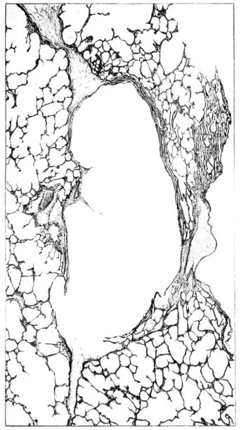
FIG. XIX. AUTOPSY NO. 123. A SMALL AIR BUBBLE IN THE INTERSTITIAL TISSUE. COMPARE FIGURE XVIII.
HELIOTYPE CO. BOSTON
23The fluid of the pleural cavities varies volumetrically as described in the preceding section. It is, however, usually not a clear fluid, but varies from a slightly turbid, blood-stained material to a typical purulent exudate. The cloudiness may be associated with minute flecks of suspended material, but in no instance has this fluid been of the thick inspissated type which formerly would have been designated as empyema.[8] (This is mentioned with the knowledge that the term empyema is being applied now to less viscid, purulent, pleural exudates). The turgidity of the mediastinal tissue also persists, but it is very rare indeed to find anything more than a small amount of clear fluid in the pericardial sac. Only once was there a typical, fibrinous pericarditis with effusion, and this occurred where a most extensive pleural exudate was also present.[9] Where such complications have been described in serous membranes, the bronchial lymph glands, particularly at the hilum of the lung, are more involved and show, not only an increase in size and a red color on cross section, but frequently also focal areas of necrosis at the periphery, which appear as yellow patches and subsequently undergo suppurative disintegration (2, 47).
The lung remains increased in volume and its surface is mottled with vivid colors. Often these are an indication of deeper parenchymatous change. The pale pink zones, through the pleural surface of which distended alveoli are discernible, are still prominent in the upper lobe, around the margins, and on the anterior surface of the lung. The darker purple, slightly elevated, often circumscribed, infarct-like areas (25, 34, 108) may occur anywhere, but are more frequent in the lower lobes. Small, maroon, slightly depressed areas of atelectasis may also involve the borders of the lung, usually the posterior borders; or they may occur between larger and more elevated areas on either lobe. Besides the purple, firm, projecting foci, paler pink or grey nodules of similar consistence may be present and show no structure when viewed through the pleura. The distribution of the different types of change is variable, and, aside from the fact that they involve the middle and lower lobes more frequently than the upper, no general statement is possible. In a few instances, one lobe, almost always the lower, may be more voluminous than the others, and although its pleura often suggests lobular involvement, the masses tend to be confluent and suggest a pseudolobar change. Sometimes, though rarely, this approaches a true lobar type of consolidation. (Compare Figs. XIII and XXVII.) Occasionally, the changes in the lung, except its increase in size, are obscured by pleural exudate which may form a thick, buttery, rather sticky mass on the surface (12, 19, 157) (Fig. XXXVII). Such pleural exudates are rare, and likewise it is uncommon to find so little pleural granulation as in the previous group. The roughening, as a rule, is not uniform, but is more prominent over the lower lobes and in the interlobar spaces than elsewhere. It may occur when there is no definite increase in the fluid content of the pleural sac.
The lung, now sectioned, presents a surface in accord with the changes suggested from the description of its external appearance. As compared with the first stage the amount of syrupy, blood-stained exudate may be definitely decreased, especially in the upper lobe or in those portions of the lung where the solidification is less marked. Its character, too, may be more cloudy, and more ropy, or viscid; it bathes the surface and is scraped off in abundance with the blade of a knife from the underlying consolidated foci (108, 156). The bronchi and bronchioles, however, may be prominent, irrespective of the change in the parenchyma itself. From their lumina, thick, yellow pus wells forth and their mucous membrane is intensely congested. Where such involvement occurs in unconsolidated portions of the 24lung, the bronchioles are even more striking than in the hepatized areas in which the more widespread changes obscure the process. The dilatation of the bronchioles, especially in their smaller ramifications, is still conspicuous.
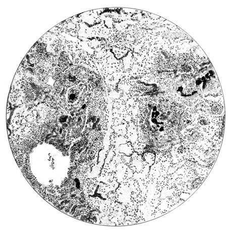
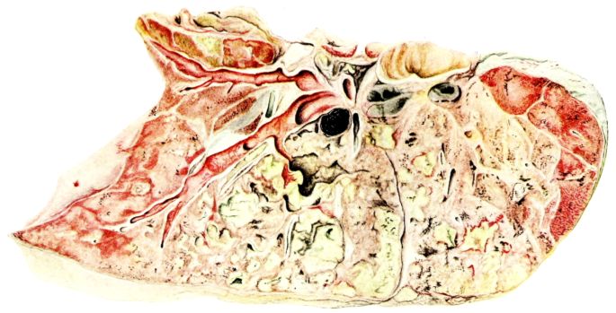
The consolidated areas vary greatly in size and number;[10] often they are small and involve only single lobules, which now stand out as granular, generally elevated patches on the surrounding congested plane. Their color, as on the pleura, varies. They may be dark, almost hemorrhagic, fading through the reds, pinks, and greys. They may be firm, or, at the other extreme, honeycombed by small, often narrow, cavities, from which a material similar to that described on the surface wells forth. The latter change is more frequent if the consolidated area is large. It has occurred most often in the pseudolobar and in the lobar types of the process. The pseudolobar change is differentiated, not only by the confluence of more or less definite lobular patches and by its involvement of portions of contiguous lobes rather than a single lobe, but also by variations in the color and consistence of the different lobular foci. This is in contrast with lobar involvement where the entire lobe is affected by a uniform process usually at the same stage of development. Although the consistence may vary in different portions, usually the same color is present throughout. (Compare Figs. XIV and XXVIII.) In one instance where a solid, yellow lobe was found, its center contained an irregular, fresh blood clot (Fig. XVIII), which would be sufficient to differentiate this type of consolidation from that of respiratory disease in which the initial lesion is less destructive. Sometimes the softening in a hepatized lobule or group of lobules is much more evident, and the zone becomes divided by irregular channels filled with viscid, grey or brown material (108, 149, 162). When such a condition lies just beneath the pleural surface, it may be distinctly seen from without (Fig. XXXIII). The pleura bulges, the normal topography of the local zone is lost, and it appears as a dull, somewhat projecting, circumscribed patch, two or three or more centimeters in diameter, the surface of which has a more or less characteristic brown or brownish black opacity. As soon as this is sectioned there pours from the cavity the liquefied exudate in which the destroyed pulmonary parenchyma is mixed (Fig. XXXIV). Occasionally, strands of tissue still traverse the cavity, but, as a rule, it empties itself completely, and leaves a brownish black wall. The delicate, sweet but persistent and penetrating odor is not so marked as with typical gangrene.
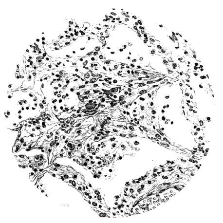
Sections from the least involved areas of the lung show a subsidence of the alveolar exudate and the walls are no longer intensely engorged. Perhaps the most prominent feature within the alveoli is the desquamated cells, presumably alveolar cells with broken or pyknotic nuclei. Despite the fact that so many cells of this type occupy the lumen of the alveolus, its wall has a prominent lining of cubical epithelium. Often mitotic figures abound in this new alveolar epithelial lining (Fig. XLVII), an evidence of rapid regeneration in that portion of the lung where the initial irritative process has subsided and where the destruction has not been as deep as elsewhere (79). This picture may be taken as positive evidence of an initial, diffuse, and general pulmonary involvement, which, with the subsidence of the primary reaction, is followed by localization resulting in the different types of consolidation now encountered.
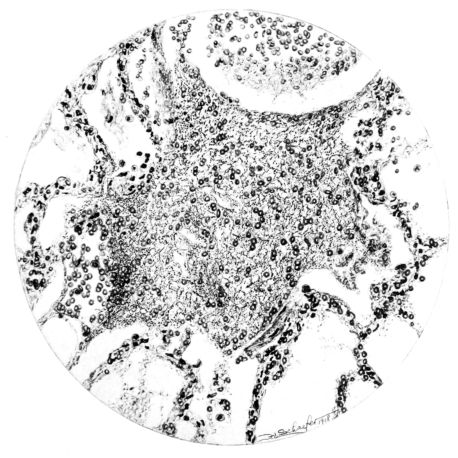
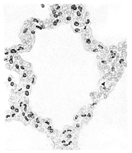
FIG. XXIII. AUTOPSY NO. 175. NOTE THE ABSENCE OF ALVEOLAR EPITHELIUM, THE ENGORGEMENT OF THE VESSELS OF THE ALVEOLAR WALLS, AND THE SEROUS EXUDATE.
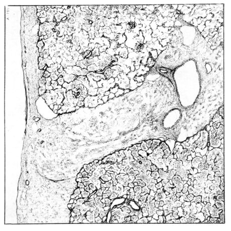
25However, in such areas of slight change, the bronchi and bronchioles may be distended and filled with polymorphonuclear leucocytes, exfoliated epithelium, and bacteria (Fig. X). It is, of course, possible that infection of the parenchyma may recur from these sources. Here the extent of involvement of the bronchiolar wall is variable and analogous to those described previously. Occasionally, too, where the alveolar change has subsided, the interstitial tissue, particularly that which divides groups of neighboring lobules, may retain its increased size with fibrous tissue framework spread apart by exudate and punctuated with an occasional circumscribed purulent mass (92, 95, 110, 156) (Fig. XXXVI). Such a miliary abscess within the lymphatics of the interstitial tissue may compress the neighboring air sacs, themselves entirely free of inflammatory involvement. That these strands of interstitial tissue, the conduits for the lymphatics, are important barriers against the spread of an inflammatory process from lobule to lobule by direct extension, is evidenced by the extreme variation in the amount and type of involvement in neighboring lobules (Fig. XXX). This variation is repeatedly encountered and the band of interstitial tissue, often prominent on account of its edema, separates these lobules the more clearly. In all probability, the sharp demarcation of the lobular consolidation as described in the gross picture depends upon the change in the interstitial tissue which tends to localize the infection (93). This fact suggests that the process within the pulmonary parenchyma spreads along the bronchial tree rather than from lobule to lobule.
Sections from those areas of the lung where the involvement is more marked may show a histological picture not unlike that described for the aplastic stage, but, in addition, there are groups of lobules where the exudate is typically purulent and pus cells not only form the greater part of the exudate in the lumen, but are prominent in the distended vessels of the alveolar wall (Fig. XXIX). Often these leucocytes are multilobed and frequently their protoplasm is granulated with phagocytized bacteria. The bacteria are also encountered free in the alveolus along with other elements; namely, red blood cells, strands of fibrin, or precipitated albumin (Fig. XXI). The bacteria, however, are not particularly conspicuous, for generally they are either single, in pairs, or in chains; and it is only when they become clumped to form large masses, often larger than any normal tissue cell, that they attract attention. When this appearance is encountered, the alveolar wall is no longer distinct and well preserved. Although the wall may still be made out, it often stains rather homogeneously and much of the finer architecture is lost in the thrombo-necrotizing process that has been instituted (Fig. XVII).
From this intermediary stage the picture of actual abscess with mortification of bronchiolar and alveolar tissue, as well as of the exudate itself, is readily approached (25, 48, 110, 140) (Fig. XXXI). In the necrotic mass that forms the center of such a focus, the most prominent feature is the bacteria. With hematoxylin they stain intensely as black, 26irregular masses, and their prominence is accentuated by the homogeneous staining qualities (with eosin) of the dead tissue, whether lung or exudate (Fig. XXXII). These abscesses may have central cavities which represent a discharge of their contents and may indicate the position of a bronchiole (Fig. XXXI). The necrosis of the alveolar walls, focal in its distribution as previously described, suggests itself as a forerunner of the more extensive necrosis encountered at this stage.
The most extreme form of mortification is seen in the wall of a gangrenous cavity, and several layers can be distinguished there. Beginning with that portion of the lung the least involved, the lesion may be limited to congestion of the alveolar wall with a serofibrinous exudate in the lumen, but this stage passes rather rapidly into another where cellular exudate, chiefly of polymorphonuclear leucocytes, predominates. Moreover, the leucocytes form not only the bulk of the alveolar content, but also distend the vessels and accumulate in the interstitial tissue around blood vessels and lymphatics. Passing toward the center of the gangrenous cavity, the lung rapidly changes in appearance. The blue zone of leucocytic infiltration makes more conspicuous the inner area of necrosis—where nuclei no longer stain and the alveolar wall is a homogeneous pink.
Gradually this phantom architecture, spotted only here and there with disintegrating polymorphonuclear leucocytes, ends in a ragged compressed border of a shaggy pink material which has no identifying qualities (Fig. XXXV). In the inner zone of pink an occasional vessel or, at times, a bronchiole more resistant to the process remains; frequently it is accentuated by the presence of partially destroyed polymorphonuclear leucocytes at its periphery. Probably these cells invade the necrotic areas along the sheath of the bronchus or vessel and not across the dead area. The thrombotic process described in the previous stage (Fig. LII) associated with an acute arteriolitis, may be associated with these gangrenous areas as well as with infarcts (82), but more likely gangrene is preceded by the acute diffuse necrosis of the alveolar wall which occurs in the fulminating cases. Furthermore, this is suggested where a typical grey hepatization is associated with marked thinning, but not actual disappearance, of the alveolar wall. Before concluding the description of this stage of the disease, mention should be made of the granular nodules of fibrin superimposed upon the swollen pleural cells and also of the older pleural exudate, either typically fibrinopurulent or more homogeneous with broken nuclear fragments (Fig. XXXVIII).
In this stage of the disease the respiratory change is characterized by a localization of the inflammatory process with cellular invasion of the exudate. Pneumonia results, varying in extent from peribronchial to lobar, a pneumonia in which one of the most frequent complications is necrosis of the lung. Consequently, abscesses, even gangrene, are found.
The processes described for the preceding group which involve not only the alveoli, but also the bronchi and the bronchioles, were predicted from the extensive hyalinization of these structures—a characteristic change in the more fulminating and acute phases of the disease. Likewise, it can be predicted that where death does not terminate the process such lesions will be followed by organization, which, by converting the exudate into scar, will produce deformities and offer serious mechanical interference with the ingress and egress of alveolar air and similar interference with the flow of blood through the pulmonary circulation (82).
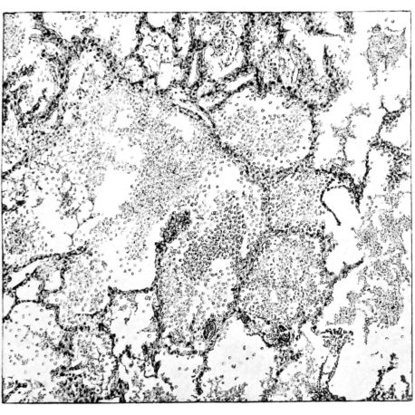
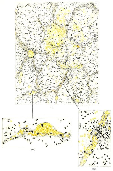
FIG. XXVI. AUTOPSY NO. 103. THE LARGER ILLUSTRATION OF AN HEMORRHAGIC ALVEOLAR EXUDATE IS ELABORATED BY TWO HIGHER POWER DRAWINGS. THESE SHOW (1) AN ANEURSYMAL DILATATION OF A CAPILLARY IN THE ALVEOLAR WALL AND (2a) A RUPTURE OF THE CAPILLARY WALL WITH THE ESCAPE OF RED BLOOD CELLS INTO THE ALVEOLUS; (2b) NECROSIS WITH EARLY THROMBOSIS OF THE CAPILLARY IN THE ALVEOLAR WALL.
27It is probable that chronic processes, not sufficiently severe to terminate fatally in a few weeks or months, may occur, but few examples of that kind have been observed. A total of twelve cases of our series in which organization of the bronchiolar (47), or alveolar exudate (156) was found, include three of the lobar, three of the pseudolobar, two of the lobular, and four of the peribronchial types. Such a differentiation, it will be understood, is purely arbitrary. The majority of the cases show, not only a reparative process, but also a continuance of the acute change, and, indeed, both gross and microscopic pictures of the lung may be complicated. It will be impossible to give an inclusive description of these changes, and therefore a few of the most diverse and characteristic will be presented in the form of case abstracts.
Autopsy No. 140.
A white female, aged 19 years, entered the New Haven Hospital after five days of fever, prostration, and cough. She was moderately cyanotic and dyspnœic, but examination of the lungs was negative except for a few râles at the right base. She was delivered of a six-months’ fœtus two days later. On the eleventh day of her illness, definite signs of consolidation had developed in the midback on both sides and spread gradually to include the left base and all of the right back to the level of the 4th dorsal spine. The temperature varied irregularly between 100°F. and 105°F. The pulse followed the temperature, but averaged 102 per minute, while the respirations remained about 40 per minute.
The white blood count on admission was 5,800, but rose gradually to 28,320 cells per cubic millimeter, with 90% of polymorphonuclear leucocytes. The patient died on the twenty-second day of the disease.
Post-mortem examination showed little of interest aside from the thorax. The left pleural cavity contained 75 cubic centimeters of slightly cloudy fluid. The right was almost completely obliterated by an organizing fibrinopurulent exudate which bound together the visceral and parietal layers. The left lung was partially collapsed and covered by a thickened pleura, bluish purple in color, which at the base and in the interlobar area was finely granular. Crepitation was present at the apex, the anterior surface, and the borders of the lung; elsewhere the consistency was increased. On section there was a frothy exudate from the apex and extreme base. These were deep red in color, while the intervening surface of the lung was paler, but broken by many small, white, elevated nodules which at first glance resembled tubercles (Fig. XXXIX). On close examination each of these was seen to have a small, depressed center from which pus could be expressed, and on dissection this was seen to be the wall of a bronchiole. The right lung was more voluminous and covered by a thick, grey, fibrinopurulent exudate except on the upper anterior border where there were a few small areas of interstitial emphysema. On section the pleural exudate was seen to be sharply demarcated from the lung parenchyma by a fine red line. The pseudotubercles of the other lung were here even more marked and from each exuded a yellow pus which partially obscured the pinkish-grey translucence of the surface (Fig. XL). The lumina of the larger bronchi were distended. The hilic and bronchial glands were enlarged and grey.
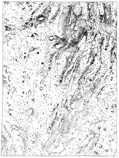
Microscopic examination of sections from the lung showed an extensive bronchopneumonia. The alveoli of an occasional group of lobules were filled with serum or red blood cells, while still other areas showed foci of necrotizing pneumonia, actual miliary abscesses in which there were large clumps of bacteria (Fig. XLI). Throughout the sections, however, the striking change was an organization of the exudate, which varied from a few fibroblasts to a well defined connective tissue almost obliterating the normal architecture (Figs. XLI and XLIV). The alveolar walls in some 28areas showed edema only, in others they were almost replaced by a thin line of fibrous tissue, and in still others, by a hyperplasia of the epithelium which almost filled their lumina. The bronchi exhibited similar changes, their lumina were filled with an exudate of desquamated epithelium and leucocytes, which in some places was organizing (Fig. XI), and there was a regeneration of epithelium evidenced by a piling up of the cells and the presence of mitotic figures in them. The interstitial tissue showed some edema. The tracheal epithelium was intact, but a few leucocytes and lymphocytes were scattered through the submucosa.
Pneumococcus Type II was recovered from the blood, pleural fluid, and lung. B. influenzæ was also demonstrated in the lung by smears and cultures.
In contrast to this example of a very diffuse, organizing pneumonia, associated with a marked peribronchial organization where the illness lasted for three weeks with hardly a remission throughout its course, the following example of necrotizing and organizing lobar pneumonia may be considered.
Autopsy No. 183.
A white male, aged 46 years, was admitted to the New Haven Hospital on January 9, 1919, complaining of “pneumonia.” The family history was unimportant. He stated that he was in the hospital twelve years ago with typhoid fever and again five years ago with acute cholecystitis.
His present illness began two weeks previous to admission with chills, fever, anorexia, nausea, vomiting, and a slightly productive cough. He was prostrated and drowsy, but could not sleep. On admission his temperature was 101.5°F., the pulse 124, and the respirations 34 per minute. He was cyanotic and dyspnœic. The right chest showed signs of consolidation, and fluid below the 3rd interspace. The white blood count was 8,200 cells per cubic millimeter, 89% being polymorphonuclear leucocytes. The patient died eighteen hours after admission.
The autopsy was held four hours after death and the essential findings were as follows:—
Twelve hundred cubic centimeters of fibrinopurulent fluid were found in the right pleural cavity and the visceral and parietal pleura had a thick, yellow coat of fibrin. The right lung was voluminous, retained its shape on removal, and weighed 1,800 grams. The lower two lobes were consolidated and the upper lobe was atelectatic. On section the latter was slightly congested, but not consolidated. The lower two lobes were fairly smooth and grey, mixed with red areas, and exuded thick, sanguinous pus. They also showed several necrotic areas in the central portion and, in some instances, cavities 1 centimeter in diameter filled with sanguinous pus had formed. The bronchi contained the same material, and on its removal a deep red mucosa was exposed. The left lung showed some fibrous pleurisy over its lateral, posterior, and diaphragmatic surfaces. There was a firm, puckered scar at the apex. The lung crepitated throughout, and on section was essentially normal except for moderately intense injection of the bronchi. The hilic nodes were enlarged, soft, succulent, moderately injected, and pigmented. The trachea was pale, but was covered by a mucopurulent exudate. The right side of the heart was moderately dilated. The spleen was not enlarged, but was softened and congested. The liver was pale, slightly decreased in consistency, and congested. The kidneys and adrenals showed cloudy swelling. The gall-bladder contained several stones, had a thickened wall, and was bound to the pylorus by firm, fibrous adhesions.
Microscopic examination of the lung showed the alveoli filled with an acute inflammatory exudate in many stages of degeneration and hyalinization (Fig. XLII). Abscesses were frequent, but were for the most part small. A similar necrotic mass was contained in the bronchi. There were, however, features of the microscopic picture that outweighed those already described. The alveolar and bronchiolar exudates were everywhere being invaded by a young granulation tissue, rich in fibroblasts and capillaries. Mononuclear cells abounded in the new tissue. Even more striking than the mesodermal new growth was the epithelial proliferation which could be seen in many areas. It not only attempted to cover the denuded bronchial surfaces, but stretched over masses of exudate and granulation in the lumina and extended in tongue-like projections for a considerable distance into the surrounding lung tissue (Fig. XLVIII).
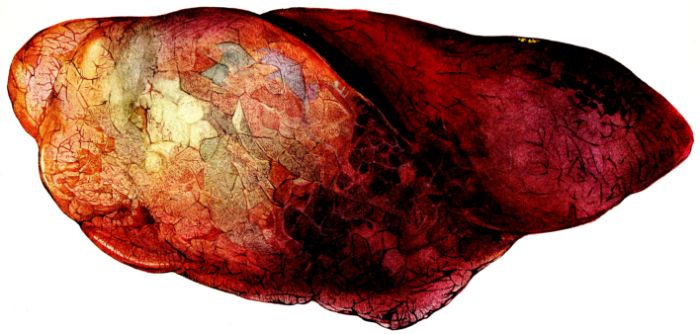
FIG. XXVII. AUTOPSY NO. 115. HERE THE HEMORRHAGIC CONSOLIDATION INVOLVES THE DEPENDENT PORTION OF THE LUNG; CONTRAST THIS WITH THE EDEMATOUS AND EMPHYSEMATOUS UPPER LOBE.
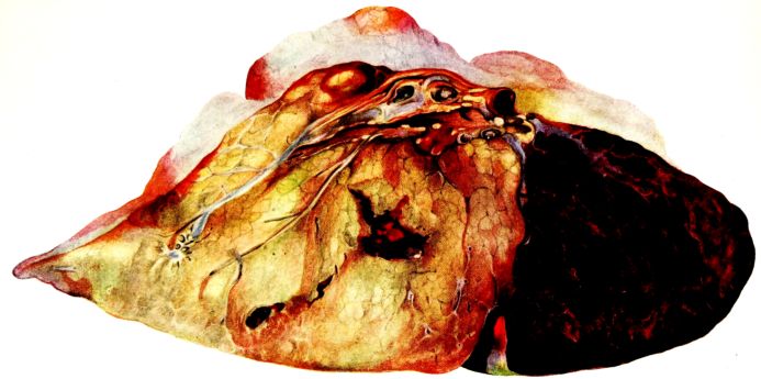
FIG. XXVIII. AUTOPSY NO. 118. THE CONSOLIDATION IS GREY IN COLOR AND LOBAR IN TYPE. IN THE CENTER OF THE HEPATIZED LOBE THERE IS A LARGE HEMORRHAGE. THE UPPER LOBE IS CONGESTED AND EDEMATOUS.
29Cultures of the blood, lung, and pleural fluid showed gram-positive, bile insoluble diplococci which formed chains, and morphologically and culturally were Streptococcus mucosus capsulatus.
These two examples differ widely in the distribution of the pulmonary involvement. They show the acute inflammatory process persisting and complicating the attempt at repair, which manifests itself, not only by the formation of granulation tissue, but also by extensive epithelial proliferation.
Still a third type of chronic lesion is manifested in Autopsy No. 209, where the necrotizing and organizing process in the pulmonary parenchyma is associated with typical saccular bronchiectatic cavities.
Autopsy No. 209.
A white woman, aged 55 years, was admitted to the New Haven Hospital complaining of weakness and ill health following pleurisy. For the past seventeen years she had suffered from a gradually progressing arthritis, which had resulted in marked deformity and disability. Five weeks before admission she became ill with chills, fever, cough, and pain on both sides of the chest. Thereafter, her general condition had gradually grown worse.
Physical examination showed a markedly emaciated white woman with a high degree of arthritis deformans. Dullness and fine râles were present at the left apex. The temperature was but slightly elevated until three days before death, when it rose to 101°F. and slight dullness developed at the right base with bronchial breathing and fine râles. The white blood count, which on admission had been 9,200 cells per cubic millimeter, rose to 15,000 per cubic millimeter. She died after a seven-weeks’ illness.
At necropsy, with the exception of the joints, little of interest was found outside the thoracic cavity. On the posterior and diaphragmatic surfaces of both lungs fibrous adhesions were present binding the visceral to the parietal pleura. The right lung was moderately voluminous and grey in color over the upper lobe, but had darker red areas over the surface of the lower lobes. Patches of increased consistency were found in the lower part of the upper lobe, in the middle lobe, and at the base. On section there was a slight, red, serous exudate. The surface over the more solid areas was somewhat translucent, grey or light red, and was firm and not friable. The bronchi of the lower lobes were conspicuous, and at the extreme base so dilated as to give almost a honeycombed appearance. From them yellow pus exuded. The left lung was less voluminous and showed some increase in consistency throughout. In the lower lobe this was more uniform, but the upper had a shotty feeling. On section the upper lobe showed many hard or caseous nodules, with occasional patches of grey, gelatinous pneumonia. One small cavity was present about 3 centimeters from the apex. The lower lobe showed no gross evidence of any tuberculous process. Many pseudotubercles projected from the red surface, but from each pus could be expressed exposing the slightly congested wall of a bronchiole. At the extreme base two small cavities filled with yellow pus were present (Fig. L).
Sections from the left upper lobe showed, microscopically, both a chronic and acute tuberculous process. Tubercles with a definite wall were present, but there were also large areas of caseation. The tissue between showed interstitial organization with mononuclear infiltration and occasional groups of leucocytes. The bronchial epithelium exhibited no marked change, but the bronchi were filled with pus cells.
Sections from the left lower lobe and from the right lung showed both necrotization and organization. The alveoli contained an exudate, fibrinous, serous, and hemorrhagic, but predominantly leucocytic. There was slight necrotization of alveolar walls in some areas with infiltration of leucocytes 30in them and in the interstitial tissue. Several sections showed a marked degree of interstitial organization (Fig. XLVI). The bronchi were filled with a purulent exudate which sometimes involved the walls to form a peribronchial abscess. The walls of other bronchi showed great thickening with infiltration of both mononuclear cells and leucocytes, and many saccular dilatations were found (Fig. XII).
The post-mortem blood culture showed a small, gram-negative, hemoglobinophilic bacillus (B. influenzæ). Cultures from lungs and bronchi yielded hemolytic streptococci, and Staphylococcus aureus was also present in the lung.
Of the ninety-five cases included in this report, twelve showed the reparative process in a more or less marked degree. Furthermore, the twelve showing organization had an illness averaging twenty-seven days in duration, whereas for the eighty-three cases in which there was no organization, the period of illness averaged nine days. Thus, it appears that the early fatal termination of the disease in the latter group is responsible for the absence of organization.
With one exception, when organization was encountered, the disease had run a relatively acute course without marked remission until death. In this instance, the fatal outcome was the result of an accident at a time when all the clinical evidence pointed toward a subsidence of the acute disease. The history of this patient is abstracted below, for it may throw light upon possible relapse and upon late, chronic, respiratory changes.
Autopsy No. 163.
A white male, aged 35 years, was admitted to the New Haven Hospital on December 5, 1918, complaining of “cough, pain in the chest, and headache.” For six days he had had fever, chills, and severe frontal headache, and for three days pains in the chest. He had also had “coryza” and epistaxis. The family and past histories were unimportant.
The physical examination showed a well developed and well nourished male, who was slightly jaundiced and markedly prostrated. The conjunctivæ were injected. The heart, abdomen, extremities, and right lung were negative. In a small area outside the left nipple and extending into the axilla, tubular breathing and subcrepitant râles were heard, but no definite signs of consolidation were made out. The physical signs remained the same during the eight-day stay in the hospital. At entrance his temperature was 102.8°F. In twelve hours it fell to 100°F., then rose to 103°F., where it stayed for twenty-four hours, falling by lysis and remaining normal for the three last days. On his eighth day in the hospital, the patient suddenly collapsed and died within fifteen minutes.
The autopsy was held one hour after death and the essential findings were as follows:—
The peritoneal cavity contained 100 cubic centimeters of clear, straw-colored fluid and the viscera, particularly the liver, were acutely congested. The right side of the heart was greatly dilated; and on opening the pulmonary artery in situ considerable blood gushed forth under pressure and a huge, tortuous embolus completely filled the vessel and its branches (Fig. LIII). The veins of the vesicoprostatic plexus on the right, and the right internal and external iliac veins contained thrombi, and it was evidently from this region that the embolus had been set free. The pleura of the lower third of the left lung over an oval area about 13 × 5 centimeters was bound to the parietal pleura by pinkish-grey, translucent adhesions which were evidently of comparatively recent origin. Here the lung was firmly consolidated, but it was apparently normal elsewhere. On section the consolidated area was silvery grey, with a pink tinge, comparatively dry and smooth. In some areas, especially about the bronchi, delicate, silvery grey, translucent strands could be seen sweeping out into the surrounding lung, which contained no air. The remainder of the lung was air-containing. The trachea showed moderate congestion most marked near the bifurcation. The hilic lymph nodes were enlarged, soft, congested, and succulent, as well as anthracotic.
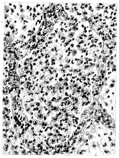
31Microscopic examination of the viscera showed acute congestion and some degree of cloudy swelling in the liver and kidneys. The sections of the lung through the consolidated area showed the pleura replaced by a thick layer composed of large and small mononuclear cells, some red blood cells, a rare polymorphonuclear cell, and great numbers of fibroblasts and budding capillaries. Some of the alveoli contained many red blood cells, a few polymorphonuclear and mononuclear cells, some fibrin, and in many areas fibroblasts were organizing this exudate (Fig. XLV). Organization was also present around the bronchi and these contained mucus, pus, and desquamated epithelium. Frequently strands of fibroblasts were seen sweeping through the bronchiolar exudates, and in a few instances they had completely filled the lumina.