PREFACE — CONTENTS — ILLUSTRATIONS — CHAPTER I
HENRICO ARTHURO ADEANE MALLET
ET
HENRICO ANTONIO PATRICIO DISNEY
ALTERI MARI AERE ALTERI
UTRIQUE FIDELISSIME
PATRIAM TUTANTI
The contents of this little book hardly justify
its title. There are whole ranges of ‘Minor
Horrors of War’ left untouched in the following
chapters. The minor poets, the pamphlets
of the professors, the people who write to the
papers about ‘Kultur’ and think that this is
the German for Matthew Arnold’s over-worked
word ‘Culture,’ the half-hysterical ladies who
offer white feathers to youths whose hearts are
breaking because medical officer after medical
officer has refused them the desire of their
young lives to serve their country. Surely,
as Carlyle taught us, ‘There is no animal so
strange as man!’
These ‘Minor Horrors of War,’ and many
besides, have for the moment been neglected
in favour of certain others which attack
the bodies, the food, or the accoutrements of
the men who are giving all that they have to
give, even unto their lives, for their homes and
for their country.
I deal with certain little Invertebrata:
animals which work in darkness and in
stealth, little animals which in times of
Peace we politely ignore, yet little animals
which in times of War may make or unmake
an army corps. As that wise old Greek,
Aristotle, wrote—and he knew quite a lot about
them—‘One should not be childishly contemptuous
of the study of the most insignificant
animal. For there is something marvellous in
all natural objects.’
We are shy of mentioning these organisms
in times of Peace; but all of them
are within the cognisance of every medical
officer of health and of every police-court
missionary. These gentlemen do not talk
about them in general society: the subject
is as a rule ‘taboo.’ Yet if we face these
troubles with courage and frankness, they can
be overcome. As ‘Emigration Jane’ says:
‘Well, there’s nothink lower than Nature, an’
She Goes as ’Igh as ’Eaven.’
I confess that these articles have been
written in a certain spirit of gaiety. This is the
reflex of the spirit of those who have gone to the
Front and of my fellow countrymen in general.
For more years than I care to remember,
the spirit of Great Britain and of Ireland had
been sombre, self-distrusting—we were till
half a year ago far too ‘conscious of each
other’s infirmities’; but with the outbreak of
the War everything changed. Our nearest relatives,
our dearest friends, are dead, or dying,
or wounded, or prisoners; but we at home
at once caught the spirit of those who have
died or have suffered for us abroad, and
we have kept and still keep a high heart.
As Mrs. Aberdeen, the immortal ‘bedmaker’
at King’s College, Cambridge, said: But surely,
Miss, the world being what it is, the longer one
is able to laugh in it, the better.’ Mrs. Aberdeen
spoke in times of Peace; but I feel that that
indomitable old lady would have said the same
in times of War.
These chapters first appeared in the
columns of the British Medical Journal. I
very gratefully thank the editor and the
proprietors of that Journal for their permission
to reprint them.
A. E. SHIPLEY.
Christ’s College Lodge,
Cambridge.
February 14, 1915.
| CHAPTER |
|
PAGE |
| I. |
The Louse (Pediculus) |
1 |
| II. |
The Bed-Bug (Cimex lectularius) |
23 |
| III. |
The Flea (Pulex irritans) |
35 |
| IV. |
The Flour-Moth (Ephestia kühniella) |
46 |
| V. |
Flies: The House-Fly (Musca domestica) |
57 |
| VI. |
Flies: The Blue-Bottle (Calliphora erythrocephala) AND OTHERS |
74 |
| VII. |
Mites: The Harvest-Mite (Trombidium) |
87 |
| VIII. |
Mites: Endo-Parasitic Mites (Demodex, Sarcoptes) |
97 |
| IX. |
Ticks: Argasidae, Ixodidae |
112 |
| X. |
Leeches: The Medicinal Leech (Hirudo medicinalis) |
123 |
| XI. |
Leeches: The Medicinal Leech (continued) |
136 |
| XII. |
Leeches: Limnatis nilotica, Haemadipsa zeylanica |
149 |
| |
Index |
163 |
| FIG. |
|
PAGE |
| |
Photograph of enlarged model of the house-fly (Musca domestica) |
Frontispiece |
| 1. |
Pediculus vestimenti |
2 |
| 2. |
Pediculus vestimenti (dorsal and ventral views) |
6 |
| 3. |
Cimex lectularius (male) |
24 |
| 4. |
Egg of Cimex lectularius |
28 |
| 5. |
Newly hatched young of Cimex lectularius |
29 |
| 6. |
Pulex irritans (female) |
36 |
| 7. |
Larva of Pulex irritans |
39 |
| 8. |
Pupa of flea |
41 |
| 9. |
Ceratophyllus gallinulae (male and female) |
44 |
| 10. |
Ephestia kühniella. Moth-infested biscuit |
47 |
| 11. |
Ephestia kühniella |
49 |
| 12. |
Ephestia kühniella (larva and pupa) |
50 |
| 13. |
Corcyra cephalonica. Moth-infested biscuit |
51 |
| 14. |
Eggs of Musca domestica |
59 |
| 15. |
Eggs of M. domestica |
60 |
| 16. |
Abdomen of female house-fly, showing the extended ovipositor |
61 |
| 17. |
Mature larva of M. domestica |
62 |
| 18. |
‘Nymph’ of M. domestica dissected out of pupal-case about thirty hours after pupation |
63 |
| 19. |
Pupal-case or puparium of M. domestica from which the imago has emerged |
64 |
| 20.[xvi] |
M. domestica in the act of regurgitating food |
65 |
| 21. |
Foot of a fly, showing hairs bearing bacteria |
69 |
| 22. |
Chart illustrating the relation of the numerical abundance of house-flies to summer diarrhoea in the city of Manchester in 1904 |
71 |
| 23. |
Latrine-fly (Fannia scalaris) |
75 |
| 24. |
Larva of F. canicularis |
76 |
| 25. |
Blow-fly or blue-bottle (Calliphora erythrocephala) |
77 |
| 26. |
Green-bottle (Lucilia caesar) |
79 |
| 27. |
Flesh-fly (Sarcophaga carnaria) |
80 |
| 28. |
Side view of blow-fly (Calliphora erythrocephala) |
81 |
| 29. |
Trombidium holosericeum (female) |
89 |
| 30. |
Leptus autumnalis = larva of Trombidium holosericeum |
90 |
| 31. |
Leptus autumnalis, with the so-called proboscis |
92 |
| 32. |
Leptus autumnalis |
93 |
| 33. |
Pediculoides ventricosus (male and female) |
96 |
| 34. |
Demodex in hair-follicle of dog. Demodex folliculorum |
98 |
| 35. |
Sarcoptes scabiei (female) |
100 |
| 36. |
Sarcoptes scabiei (male) |
101 |
| 37. |
One of the legs of Sarcoptes scabiei showing the stalked sucker and the curious ‘cross-gartering.’ |
102 |
| 38. |
A diagrammatic view of the tunnel made by the female of Sarcoptes scabiei, with the eggs she has laid behind her as she burrows deeper and deeper |
104 |
| 39. |
A female Sarcoptes scabiei, with four eggs in different stages of development |
105 |
| 40. |
Nephrophages sanguinarius (male and female) |
110 |
| 41. |
Evolution of Argas persicus |
113 |
| 42. |
Ixodes ricinus (mouth-parts of the female) |
114 |
| 43. |
Argas reflexus (female) |
115 |
| 44. |
Ornithodorus moubata (an unfed female) |
116 |
| 45. |
Ornithodorus moubata (female) |
117 |
| 46.[xvii] |
Ixodes ricinus (male and female) |
118 |
| 47. |
Ixodiphagus caucurtei laying eggs in the nymph of Ixodes ricinus |
120 |
| 48. |
Hirudo medicinalis |
124 |
| 49. |
View of the internal organs of Hirudo medicinalis |
126 |
| 50. |
Head of a leech (Hirudo medicinalis) |
130 |
| 51. |
Hirudo medicinalis |
133 |
| 52. |
Cocoon of the medicinal leech |
142 |
| 53. |
A Nephelis forming its cocoon and withdrawing from it |
143 |
| 54. |
Cocoons of Nephelis |
144 |
| 55. |
A leech-farm in the south of France |
145 |
| 56. |
Glossosiphonia heteroclita, with eggs and emerging embryos |
146 |
| 57. |
Helobdella stagnalis, with adhering young |
147 |
| 58. |
Limnatis nilotica |
150 |
| 59. |
Anterior sucker of Hirudo medicinalis |
152 |
| 60. |
The Japanese variety of Haemadipsa zeylanica |
156 |
| 61. |
Haemadipsa zeylanica (from above) |
157 |
| 62. |
Haemadipsa zeylanica (head) |
158 |
| 63. |
Haemadipsa zeylanica (land-leeches), on the earth |
159 |
THE LOUSE (Pediculus)
Care’ll kill a cat, up-tailles all and a louse for the hangman!
(B. Jonson, Every Man in his Humour.)
Lice form a small group of insects known
as the Anoplura, interesting to the entomologist because they
are now entirely wingless, though it is believed that their ancestry
were winged. They are all parasites on vertebrates. In quite recent
books the Anoplura are described as ‘lice or disgusting insects,
about which little is known’; but lately, owing to researches carried
on at Cambridge, we have found out something about their habits. As
lice play a large part in the minor discomforts of an army, it is worth
while considering for a moment what we know about them.
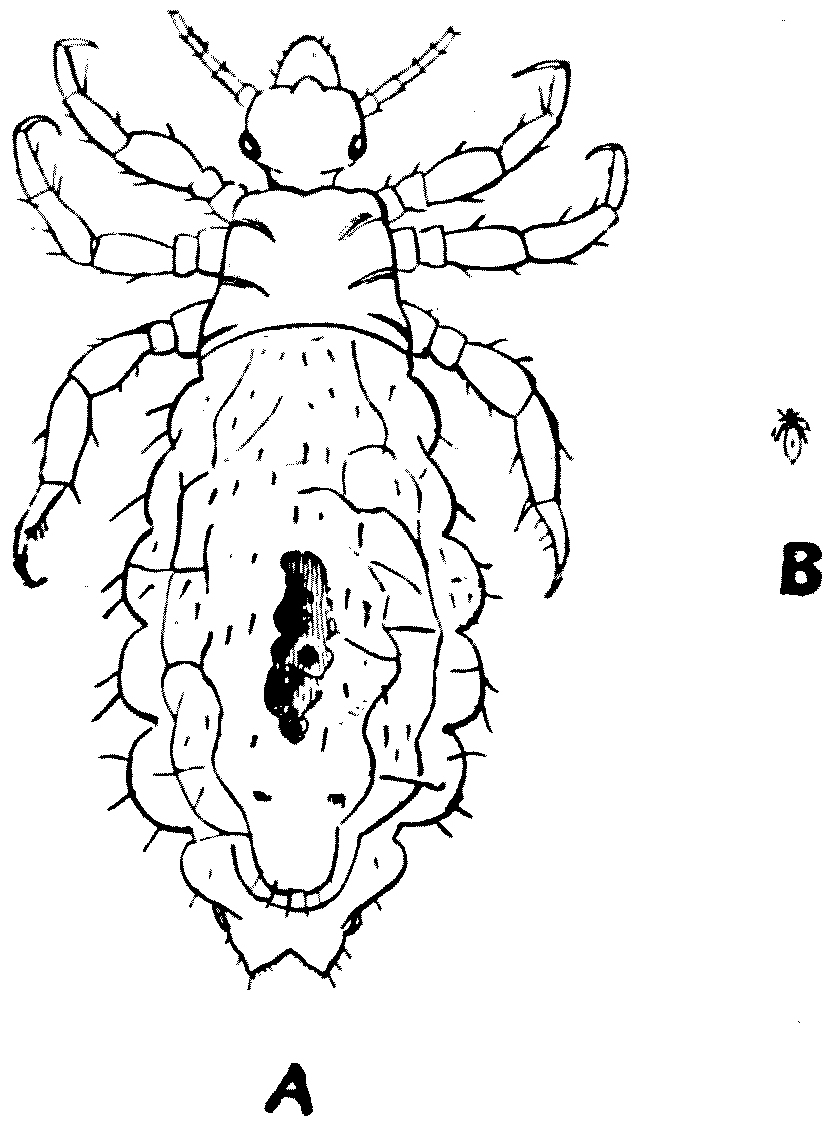
Fig. 1.—Pediculus vestimenti (Nitzsch).
A, Magnified 20 times; B,
natural size.
Fig. 1.—Pediculus vestimenti (Nitzsch).
A, Magnified 20 times; B,
natural size.
Recently, the group has been split up into a large number of genera,
but of these only two
have any relation to the human body. I do not propose, in the present
chapter, to consider one of these two genera—Phthirius—which
frequents the hairs about the pubic region of man and is conveyed from
one human being to another by personal contact. We will confine our
attention to the second genus, Pediculus, which contains two
species parasitic upon man—(Pediculus capitis) the hair-louse
and (Pediculus vestimenti) the body-louse. Both of these are
extremely difficult to rear in captivity, though in their natural state
they abound and multiply to an amazing degree.
Wherever human beings are gathered together in large numbers,
with infrequent opportunities of changing their clothes, P.
vestimenti is sure to spread. It does not arise, as the uninformed think, from dirt,
though it flourishes best in dirty surroundings. No specimen of P.
vestimenti exists which is not the direct product of an egg laid
by a mother-louse and fertilised by a father-louse. In considerable
collections of men drawn from the poorer classes, some unhappy being or
other—often through no fault of his own—will turn up in the community
with lice on him, and these swiftly spread to others in a manner that
will be indicated later in this chapter.
Like almost all animals lower than the mammals, the male of the
body-louse is smaller and feebler than the female. The former attains
a length of about 3 mm., and is about 1 mm. broad. The female is about
3·3 mm. long and about 1·4 mm. broad. It is rather bigger than the
hair-louse, and its antennae are slightly longer. It so far flatters
its host as to imitate the colour of the skin upon which it lives; and
Andrew Murray gives a series of gradations between the black louse
of the West African and Australian native, the dark and smoky louse
of the Hindu, the orange of the Africander and of the Hottentot, the
yellowish-brown of the Japanese and Chinese, the dark-brown of the
North and South American Indians, and the paler-brown of the Esquimo,
which approaches the light
dirty-grey colour of the European parasites.
As plump an’ grey as onie grozet,
as Burns has it.
The latter were the forms dealt with in
the recent observations undertaken by Mr.
C. Warburton in the Quick Laboratory at
Cambridge, at the request of the Local Government
Board, the authorities of which were
anxious to find out whether the flock used
in making cheap bedding was instrumental in
distributing vermin. Mr. Warburton at once
appreciated the fact that he must know the
life-history of the insect before he could successfully
attack the problem put before him. At
an early stage of his investigations, he found
that P. vestimenti survives longer under adverse
conditions than P. capitis, the head-louse.
The habitat of the body-louse is that side
of the under-clothing which is in contact with
the body. The louse, which sucks the blood
of its host at least twice a day, is when feeding
always anchored to the inside of the under-clothing
of its host by the claws of one or
more of its six legs. Free lice are rarely
found on the skin in western Europeans;
but doctors who have recently returned from
Serbia report dark-brown patches, as big as
half-crowns, on the skins of the wounded
natives, which on touching begin to move—a
clotted scab of lice! But the under-side of
a stripped shirt is often alive with them.
After a great many experiments, Mr.
Warburton succeeded in rearing these delicate
insects, but only under certain circumscribed
conditions: one of which was
their anchorage in some sort of flannel or
cloth, and the second was proximity to the
human skin. He anchored his specimens on
small pieces of cloth which he interned in
small test-tubes plugged with cotton-wool,
which did not let the lice out, but did let air
and the emanations of the human body in.
For fear of breakage the glass tube was enclosed
in an outer metal tube, and the whole was
kept both night and day near the body. Two
meals a day were necessary to keep the lice
alive. When feeding, the pieces of cloth, which
the lice would never let go of, were placed on the
back of the hand, hence the danger of escape
was practically nil, and once given access to the
skin the lice fed immediately and greedily.
His success in keeping lice alive was but the
final result of many experiments, the majority
of which had failed. Lice are very difficult to
rear. When you want them to live they die;
and when you want them to die they live, and
multiply exceedingly.
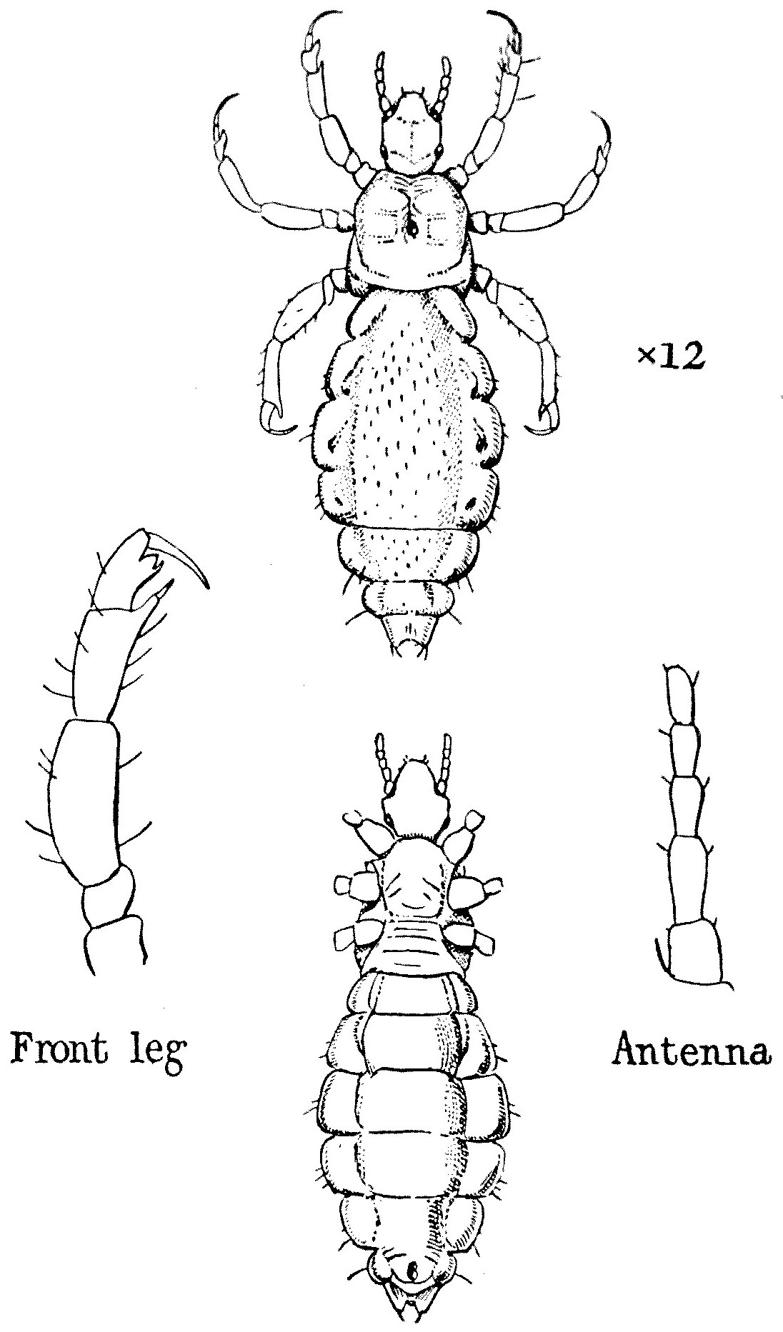
Fig. 2.—Pediculus vestimenti. Dorsal and ventral views.
Fig. 2.—Pediculus vestimenti. Dorsal and ventral views.
A single female but
recently matured was placed in a test-tube,
and a male admitted to her on the second day.
The two paired on the sixth day and afterwards
at frequent intervals. Very soon after pairing
an egg was laid, and during the remaining
twenty-five days of her life the female laid
an average of five eggs every twenty-four
hours. The male died on the seventeenth day,
and a second male was then introduced, who
again paired with the female. The latter,
however, died on the thirtieth day, but the
second male survived.
The difficulty of keeping the male and
female alive was simple compared with the
difficulty of rearing the eggs. Very few
hatched out. The strands of cloth upon which
they were laid had been carefully removed and
placed in separate tubes, at the same time being
subjected to different temperatures. It was
not, however, until the eggs were left alone
undisturbed in the position where they had
been laid and placed under the same conditions
that the mother lived in that eight, and only
eight, of the twenty-four eggs laid on the
cloth hatched out after an incubation period
of eight days. The remaining sixteen eggs were
apparently dead. But the tube in which they
were was then subjected to normal temperature
of the room at night (on occasions this fell
below freezing-point), and after an incubation
period of upwards of a month six more hatched
out. Hence it is obvious that, as in the case
of many other insects, temperature plays a
large part in the rate of development, and it
becomes clear that the eggs or nits of P.
vestimenti are capable of hatching out up to
a period of at least from thirty-five to forty
days after they are laid.
Difficult as it was to keep the adults alive,
and more difficult as it was to hatch out the
eggs, it was most difficult to rear the larvae.
Their small size made them difficult to observe,
and, like most young animals, they are intolerant
of control, apt to wander and explore, and
less given to clinging to the cloth than their
more sedentary parents. Naturally, they want
to scatter, spread themselves, and pair.
Like young chickens, the larvae feed immediately
on emerging from the egg. They
apparently moult three times, at intervals
of about four days, and on the eleventh day
attain their mature form, though they do not
pair until four or five days later.
Mr. Warburton summarises the life-cycle
of the insects, as indicated by his experiments,
as follows:—
Incubation period: eight days to five weeks.
From larva to imago: eleven days.
Non-functional mature condition: four days.
Adult life: male, three weeks; female, four weeks.
But we must not forget that these figures
are based upon laboratory experiments, and
that under the normal conditions the rate
may be accelerated. From Mr. Warburton’s
experience it is perfectly obvious that, unless
regularly fed, body-lice very quickly die. Of
all the verminous clothing sent to the Quick
Laboratory, very little contained live vermin.
The newly hatched larvae perish in a day and
a half unless they can obtain food.
With regard to the head-louse:—
Ye ugly, creepin’, blastit wonner,
Detested, shunn’d by saunt an’ sinner,
it is smaller than the body-louse, and is of a
cindery grey colour. The female measures
1·8 mm. in length and 0·7 in breadth. Like
the body-louse, it varies its colour somewhat
with the colour of the hair on the different
branches of the human race. It lives amongst
the hair of the head of people who neglect
their heads; it is also, but more rarely, found
amongst the eyelashes and in the beard. The
egg, which has a certain beauty of symmetry,
is cemented to the hair, and at the
end of six days the larvae emerge, which,
after a certain number of moults, become
mature on the eighteenth day. The methods
adopted by many natives of plastering their
hair with coloured clay, or of anointing it
with ointments, probably guards against the
presence of these parasites. The Spartan
youths, who used to oil their long locks before[10]
going into battle, may have feared this parasite.
Some German soldiers, before going to war,
shave their heads: thus they afford no nidus
for P. capitis. The wigs worn in the late
seventeenth and at the beginning of the
eighteenth centuries undoubtedly owed something
to the difficulty of keeping this particular
kind of vermin down. The later
powdering of the hair may have been due to
the same cause.
This book, however, attempts to deal more
with the troubles of the camp, and P. capitis
is in war time less important than P. vestimenti.
The former certainly causes a certain
skin trouble, but the latter not only affords
constant irritation, but, like most biting
insects, from time to time conveys most
serious diseases. P. vestimenti is said to be
the carrier of typhus. This was, I believe,
first demonstrated in Algeria, but was amply
confirmed last year in Ireland, when a serious
outbreak of this fever took place, though
little was heard of it in England. Possibly,
P. capitis also conveys typhus, but undoubtedly
both convey certain forms of relapsing or
recurrent fever. The irritation due to the
body-louse weakens the host and prevents
sleep, besides which there is a certain psychic
disgust which causes many officers to fear lice
more than they fear bullets. Lice are the
constant accompaniment of all armies; and in
the South African War as soon as a regiment
halted they stripped to the skin, turned their
clothes inside out, and picked the Anoplura
off. As a private said to me: ‘We strips and
we picks ’em off and places ’em in the sun,
and it kind o’ breaks the little beggars’ ’earts!’
In conjunction with the Quick Professor
of Biology at Cambridge, I have drawn up
the following rules. None of them will be
possible at all times, but some of them may
be possible at some time in the campaign.
At any rate, by acting on these rules, a relative
of mine who took part in the South African
War was able to escape the presence of lice
on his body, and the General commanding
his brigade told me on his return that he
was the only officer—and in fact the only man—in
the brigade who had so escaped.
How the Soldier may Guard Himself
against Infestation with Lice
In times of war, when men are aggregated
in large numbers and personal cleanliness—but
especially an adequate change of clothing—cannot
be secured, infestation with lice
commonly takes place. The prevalence of
lice in troops in the South African War was a
source of serious trouble in that their attacks
caused much irritation to the skin and disturbed
men’s sleep.
Lice occur chiefly on the body (Pediculus
vestimenti) and head (P. capitis). They are
small greyish-white insects. The female lays
about sixty eggs during two weeks; the eggs
hatch after nine to ten days. The lice are small
at first; they undergo several moults and grow
in size, sucking blood every few hours, and attain
sexual maturity in about two weeks. The
eggs will not develop unless maintained at a
temperature of 22° C. or over—such as prevails
in clothing worn on the human body or in
the hair of the head. This is why, when
clothing is worn continuously, men are more
prone to become infested with lice derived from
habitually unclean persons, their clothing,
bedding, &c. P. capitis lives between the
hair in the head, and the eggs, called ‘nits,’
are attached to the hairs. P. vestimenti lives
in the clothing, to which it usually remains
attached when feeding on man; it lays its
eggs in the clothing, and usually retreats
into the seams and permanent folds therein.
This is of importance in considering the means
of destroying lice.
To avoid these pests the following rules
should be observed:—
1. Search your person as often as possible
for signs of the presence of lice—that is, their
bites. As soon as these are found, lose no
time in taking the measures noted under
paragraph 5.
2. Try not to sleep where others, especially
the unclean, have slept before. Consider this
in choosing a camping-ground.
3. Change your clothing as often as
practicable. After clothes have been discarded
for a week the lice are usually dead of
starvation. Change clothes at night if possible,
and place your clothing away from that of
others. Jolting of carts in transport aids in
spreading the lice, which also become disseminated
by crawling about from one kit to
another. Infested clothing and blankets, until
dealt with, should be kept apart as far as
possible.
4. Verminous clothes for which there is no
further use should be burnt, buried, or sunk
in water.
5. If lice are found on the person, they
may be readily destroyed by the application
of either petrol, paraffin oil, turpentine, xylol,
or benzine. Apply these to the head in the case
of P. capitis. Remember that these fluids are
all highly inflammable. When possible, soap
and wash the head twenty-four hours after the
last application of petrol, &c. The application
may be repeated on two or more days if the
infestation is heavy. Fine combs are useful
in detecting and removing vermin from the
head. Tobacco extract has been advocated
failing other available remedies. In the case
of P. vestimenti, the lice can be killed as
follows: Under-clothes may be scalded—say,
once in ten days. Turn coats, waistcoats,
trousers, &c., inside out; examine beneath the
folds at the seams and expose these places
to as much heat as can be borne before a
fire, against a boiler, or allow a jet of steam
from a kettle or boiler to travel along the
seams. The clothing will soon dry. If available,
a hot flat-iron, or any piece of heated
metal, may be used to kill vermin in clothing.
Petrol or paraffin will also kill nits and lice in
clothing. If no other means are available,
turn the clothing inside out, beat it vigorously,
remove and kill the vermin by hand—this
will, at any rate, mitigate the evil.
6. As far as possible avoid scratching the
irritated part.
7. Privates would benefit by instruction
in these matters.
8. Apart from the physical discomfort and
loss of sleep caused by the attacks of lice, it
should be noted that they have been shown
to be the carriers of typhus and relapsing fever
from infected to healthy persons. Typhus,
especially, has played havoc in the past, and
has been a dread accompaniment of war.
Dr. R. F. Drummond has drawn my attention
to a common folklore belief emplanted
in the minds of our poorer people. Incredible
as it seems, these uneducated and ignorant
folk believe that lice on the person is a
sign of productivity, and that should they
be removed their hosts will become barren
or sterile. They transfer, by a process of
sympathetic magic, the productivity of the
lice to the lousy. As Dr. Drummond writes,
these ignorant mothers and aunts believe
that the nits and the lice arise spontaneously,
and are ‘an outward and visible
sign of an inward and invisible fertility.’
Those who try to cleanse the heads and the
bodies of our primary schoolchildren are
‘up against’ the superstitions of the little
ones’ guardians, and the guardians unfortunately
often prove the stronger. Similar views
are held widely by the various peoples of
India and the East—people we call heathen—and,
apart from the connexion thought to
be established between fertility and lice, the
presence of the latter is considered both
at home and abroad to be a sign of robust
health.
The rather obscure connexion of the louse
and the pike (Esox lucius) is probably due
to the fact that the Latin name for the pike
is Lucius. The poor pun in ‘The Merry
Wives of Windsor’ on the Lucy family is due
to a similar resemblance in sound.
The Editor of the Morning Post has given
me leave to quote the following paragraphs
from an article by his able Correspondent
at Petrograd.
All armies, after a few weeks’ campaigning, whatever
other hardships may come their way, are sure
of one—namely, certain parasites. Even officers
under most favourable conditions are unable to keep
clear of this scourge. Silk under-clothing is some
palliative, but no real preventative. Various
measures have been proposed to relieve the intense
annoyance caused by millions of parasites of at least
two species. Flowers of sulphur, worn in bags
round the neck, were supposed to be a preventative,
but proved fallacious.[1] What seems likely to prove
perfect prophylactery is recommended by M. Agronom,
who writes from Bokhara, where he has noted the
habits of the Sarts and their preventative measures.
The Sarts never wash, and hardly ever in lifetime
change their clothes; therefore their condition
would be impossible without some preventative
measures. They take a small quantity of mercury,
which they bray into an amalgam with a plant used
in the East for dyeing the hair and nails—probably
henna. This paste is evenly laid on strands of flax or
other fibres. One string thus prepared is worn round
the neck and the other round the waist next the skin,
the heat of the body producing exhalations which kill
parasites. The string lasts quite a long time.
M. Agronom has made experiments with the
ordinary mercurial ointment prepared with any kind
of fat, and finds the effect precisely the same. He[17]
asserts that such a minute quantity of mercury as
is required to produce the desired result is perfectly
harmless to the system. A half-crown’s worth of
mercury brayed in a mortar with lard or other fat
will suffice to treat enough threads for several hundred
soldiers. The threads should be of ten or a dozen
strands or some very loosely twisted material like
worsted, and should be wrapped in parchment paper
before boxing for dispatch to the soldiers. This
is effective and lasting for body parasites. Others
are easily dealt with by rubbing in petroleum, which
must be done twice at a week’s interval.
It should also be noted that no ordinary washing
methods will clear the parasites from body-linen even
when dipped in boiling water; but if a couple of
spoonfuls of petroleum are added to every gallon of
water, perfect success is assured even without boiling.
I confess I think he is a little bit too
dogmatic about the habits of the Sarts. I
am told the better-class Sarts do occasionally
bathe, or why are there public baths at Khiva?
After all, in our oldest and most cultured
University, only a year ago, the venerable Head
of a House exclaimed with some acerbity, when
a junior Fellow suggested putting up hot-water
baths for the undergraduates: ‘Baths! why
the young men are only up eight weeks!’
And, again, though the clothes of the Sarts
are doubtless flowing, unless they are
elastic, they must get bigger as babyhood
passes to boyhood and boyhood passes to
manhood.
Preparations of mercury are also used in
India: not only against human lice, but against
the Mallophaga or biting-lice which infest the
Indian birds used in falconry. It is difficult for
a zoologist to believe the last paragraph of the
Morning Post correspondent. The temperature
of boiling water coagulates animal protoplasm
as it does that of the white-of-egg; and what
would the lice do then, poor things?
Early in the year, Mr. C. P. Lounsbury,
the well-known Government Entomologist in
South Africa, wrote that they were supplying
the troops there with sulphur-bags which were
supposed to keep the lice away. The sulphur
is put in small bags of thin calico, and several
of these are secured on the under-clothing,
next to the skin. The bags are about two
inches square, and I am told that it is customary
to have one worn on the trunk of the body
and one against each of the nether limbs.
Whether this is effective will probably be known
soon; but that flowers of sulphur do play
an effective part in keeping down these
troubles is shown by a letter of Dr. Harding
H. Tomkins:—
Over thirty years ago, when house-surgeon at
the Children’s Infirmary, Liverpool, I used this with
absolute success in all cases of plaster-of-Paris jackets
who formerly had been much distressed by vermin
getting under the jacket. The sulphur was rubbed
well into the under-clothes.
But still more interesting evidence is given
by Dr. N. Bishop Harman:—
When I was serving in the South African War,
and attached to No. 2 General Hospital at Pretoria,
I was detailed to take medical charge of the camp
of released prisoners that was established a few
miles out of the town on the Delagoa Bay railway
line. I moved into the camp the night they came
in. Next day an inspection was held. I do not
think I ever saw such a sorry sight. The men were
in the most nondescript garments, and they were
flabby from the effects of the food the Boers had
given them—mealy pap, for the most part. They
had had no washing facilities, and they were dirty
in the extreme. Amongst them were a number
of men of the D.C.O. Yeomanry, many of them
Cambridge men, and when these came to me for
special examination, unwarily I invited them into
my tent to strip, and their clothes were laid on the
only available support—my bed. The next day or
two was spent in cleaning up the men and refitting
them. By the end of the week I noticed in the
evening an unpleasant itch about the lower part of
the trunk: a sub-acute sort of itch, it did not seem
like a flea, and I could find nothing. But after a
most diligent search with all the candles I could
borrow, I found, to my horror, a louse. It was a
genuine body-louse. Then I remembered my folly
in inviting strangers into my tent. Water was
scarce, the morning tub was only the splash from
a can. Laundry was impossible. But after some
trouble I managed to get a can of hot water and get
some sort of a hot wash. My man did the best
he could with my shirt and pants. What to do with[20]
the bedding—dark brown blankets—I did not know,
except to expose them to the hot sunshine. I rode
into the town, but insect-powder could not be got.
It came into my mind that I had read or heard that
people who took sulphur-tablets smelled of H2S,
so on the chance that an outside application might
be of some service I got a supply of flowers of sulphur.
This I liberally sprinkled all over my clothes, bedding,
and rubbed into the seams of my tunic and riding-breeches.
The itching was stopped in a day, and
it never came again. But I soon noticed another
circumstance: all the bright brass buttons of my
tunic, although freshly polished by my man every
morning, were tarnished before evening, even in
the clean, dry atmosphere of the dry veld. Also
my silver watch-case went black. There was no
doubt that the sulphur was acted upon by the
secretions of the skin and H2S was produced, and
this I had no doubt killed off any lice that could not
be got at by washing. Subsequently, I always
used it when I was in likely places. And some
places were very likely! In Cape Town, I had to
inspect all the soldiers’ lodgings in view of the spread
of the plague. And, again, I had charge of a Boer
prison-ship, and never once did I catch so much as
a hopper. The prison-ship was literally alive with
cockroaches of all sizes; our cabins swarmed with
them, but they avoided my clothes and kit like a
plague, and there was never a nibble-mark to be
found. I gave the hint to many men and they
confirmed my experience. I have since met other
men who hit on the same device with equal success.
In this war I have told the tip to many friends, and
some relatives, who have gone out, and so far they
have been free from the plague. You will note[21]
that I used all the other measures I could, but my
bedding and uniform were not washed, and the
lice must have come through the bedding; there
was no other possible means I could trace. Yet
the flowers of sulphur killed off all that might be
therein.
A very effective method for exterminating
vermin in infected troops was carried out by Dr.
S. Monckton Copeman, F.R.S., at Crowborough.
To put the matter briefly, I append a copy
of his able and concise memorandum which
was distributed to all the medical officers of
the Division; but further details may be
obtained by referring to the British Medical
Journal, or the Lancet of February 6, 1915.
To the Medical Officer....
Treatment for Destruction of Vermin.
Arrangements should be made for the bathing
of affected individuals and other inmates of infected
tents.
After drying themselves, men to lather their
bodies with cresol-soap solution (water 10 galls.,
Jeyes’ fluid 1½ oz., soft soap 1½ lb.), especially over
hairy parts, and to allow the lather to dry on.
Shirts to be washed in cresol-soap solution made
with boiling water.
Tunics and trousers to be turned inside out, and
rubbed with same lather, especially along the seams.
Lather to be allowed to dry on the garment.
The materials can be obtained from the A.S.C. on
indent authorised by A.D.M.S. in the form attached.
Infected blankets were at first treated by soaking
them in cresol-soap solution, after which they were
sent to a neighbouring laundry to be washed—a
small contract rate having previously arranged.
In the first week in November, however, a portable
Thresh’s steam disinfecting apparatus was supplied
to the Division, through the Second Army, since
when no difficulty has been experienced in the
disinfection both of clothing and blankets.
As a matter of fact the simple and inexpensive
method which has been employed by us over a
period of several months has proved so successful
that no necessity has arisen for a trial of any other
means of treatment.
Professor Lefroy, of the Royal College of
Science and Technology, recommends two
effective remedies, known respectively as
‘Vermijelli’ and ‘Vermin Westropol.’[2] Lieut.-Colonel
E. J. Cross has successfully treated the
clothes and bedding of his men with a powder
consisting of three parts of black hellebore root
and one of borax, and many similar powders are
produced by the manufacturers of insecticides.
Let us end up this chapter cheerfully!
The importance of lice is equalled by their
unpopularity. A lady, driven to extremes by—well
let us call it—the want of gallantry of
Dr. Johnson, called him ‘a louse.’ The great
lexicographer retorted, ‘People always talk of
things that run in their heads!’
CHAPTER II
THE BED-BUG (Cimex lectularius)
In ‘x’ finita tria sunt animalia dira;
Sunt pulices fortes, cimices, culicumque cohortes;
Sed pulices saltu fugiunt, culicesque volatu,
Et cimices pravi nequeunt foetore necari.
(Anon.)
Among the numerous disagreeable features
of the bed-bug is the fact that it has at least
two scientific names—Cimex (under which
name it was known to the classical writers)
and Acanthia. The latter name is favoured
by French and some German authorities, but
Cimex was the name adopted by Linnaeus, and
is mostly used by British writers, and will be
used throughout this article. One cannot do
better than take the advice of that wise old
entomologist, Dr. David Sharp, and allow the
name ‘Acanthia to fall into disuse.’
The species which is the best known in
England is C. lectularius; but there is a second
species which is much commoner in warm
climates, C. rotundatus. As regards carrying
disease, this latter species is even more
dangerous than its more temperate relative.
Other species, which rarely if ever attack man,
are found in pigeon-houses and dove-cotes,
martins’ nests, poultry-houses, and the homes
of bats.
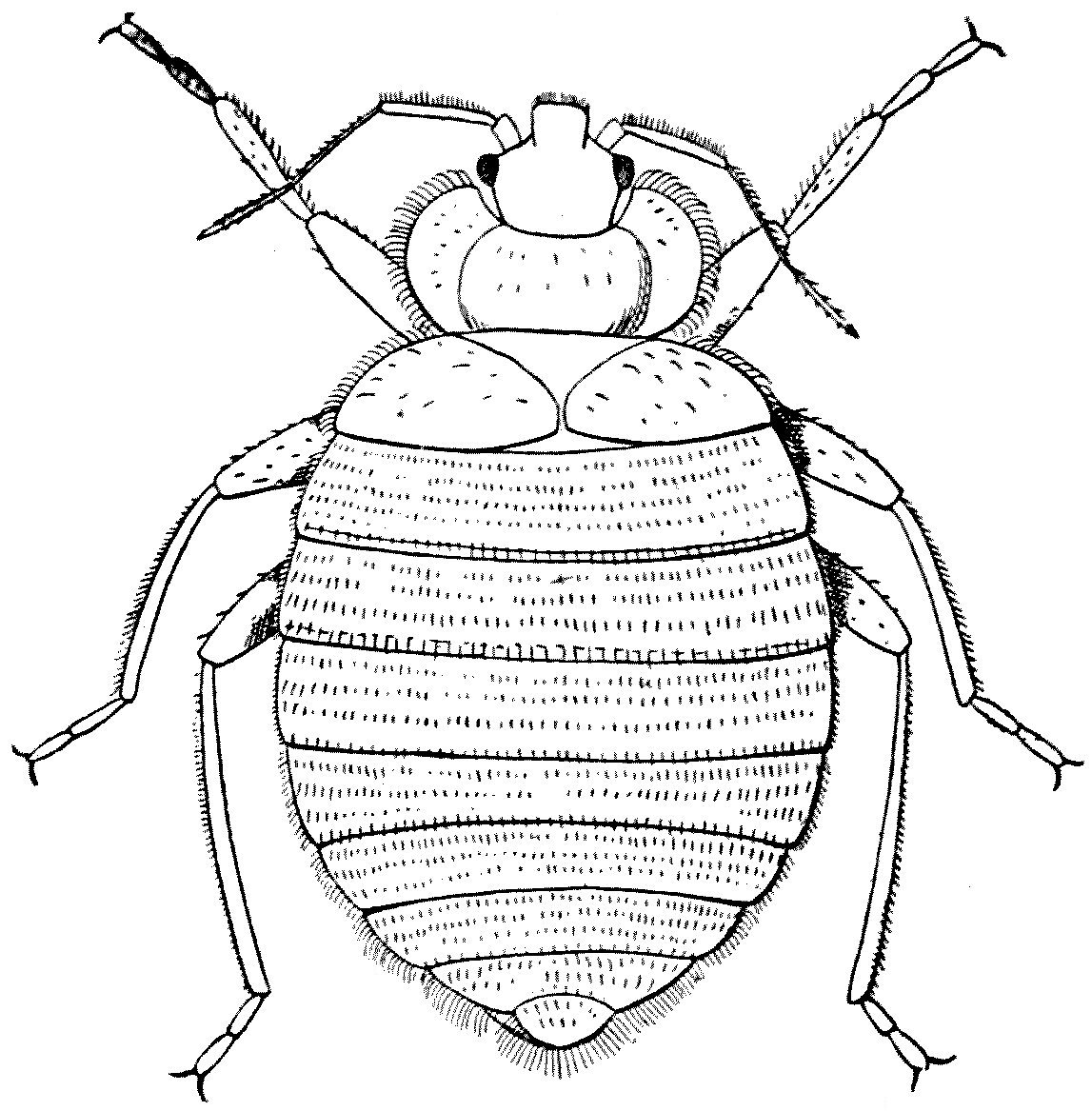
Fig. 3.—Cimex lectularius, male. × 15. (From Brumpt.)
Fig. 3.—Cimex lectularius, male. × 15. (From Brumpt.)
The common bed-bug seems to have arrived
in England about the same time as the cockroach—that
is, over four hundred years ago,
early in King Henry VIII’s reign. Apparently,
it came from the East, and was for many years
confined to seaports and harbours. It seems
to have been first mentioned by playwriters
towards the beginning of the seventeenth century.
The sixteenth-century dramatists could
never have resisted mentioning the bug had it
been in their time a common household pest. It
would have appealed to their sense of humour.
How the insect got the name of ‘bug’
is unknown. It has been suggested that the
Old English word ‘bug,’ meaning a ghost or
phantom which walked by night, has been
transferred to Cimex. This may be so, but
the ‘Oxford English Dictionary’ tells us that
proof is lacking.
The insect is some 5 mm. in length and
about 3 mm. in breadth, and is of a reddish- or
brownish-rusty colour, fading into black.
Its body is extraordinarily flattened, so that
it can readily pass into chinks or between splits
in furniture and boarding, and this it does
whenever daylight appears, for the bug loves
darkness rather than light. The head is large,
and ends in a long, piercing, four-jointed proboscis,
which forms a tube with four piercing
stylets in it. As a rule the proboscis is folded
back into a groove, which reaches to the first
pair of legs on the under surface of the thorax.
This folding back of the proboscis gives the
insect a demure and even a devout expression:
it appears to be engaged in prayer, but a bug
never prays. The head bears two black eyes
and two four-jointed antennae. Each of the
six legs is provided with two claws, and all
the body is covered with fairly numerous hairs.
The abdomen shows seven visible segments
and a terminal piece.
The bug has no fixed period of the year
for breeding; as long as the temperature is
favourable and the food abundant, generation
will succeed generation without pause.
Should, however, the weather turn cold the
insects become numbed and their vitality
and power of reproduction are interrupted
until a sufficient degree of warmth returns.
Like the cockroach, the bed-bug is a
frequenter of human habitations, but only of
such as have reached a certain stage of comfort.
It is said to be comparatively rare in the
homes of savages, but it is only too common
in the poorer quarters of our great cities. Its
presence does not necessarily indicate neglect
or want of cleanliness. It is apt to get into
trunks and luggage, and in this way may be
conveyed even into the best-kept homes. It
is also very migratory and will pass readily
from one house to another, and when an
infested dwelling is vacated these insects usually
leave it for better company and better quarters.
Their food-supply being withdrawn, they make
their way along gutters, water-pipes, &c.,
into adjoining and inhabited houses. Cimex
is particularly common in ships—especially
emigrant ships—and, although unknown to
the aboriginal Indians of North America, it
probably entered that continent with the ‘best
families’ in the Mayflower.
Perhaps the most disagreeable feature of
the bed-bug is that it produces an oily fluid
which has a quite intolerable odour; the glands
secreting this fluid are situated in various
parts of the body. The presence of such
glands in free-living Hemipterous insects
is undoubtedly a protection—birds will not
touch them. One, however, fails to see the
use of this property in the bed-bug. At any
rate, it does not deter cockroaches and ants,
as well as other insects, from devouring the
Cimex. There is a small black ant in Portugal
which is said to clear a house of these pests
in a few days, but one cannot always command
the services of this small black ant.
Another remarkable feature is that the insect
has no wings, although in all probability its
ancestors possessed these useful appendages.
As the American poet says:—
The Lightning-bug has wings of gold,
The June-bug wings of flame,
The Bed-bug has no wings at all,
But it gets there all the same!
The power of ‘getting there’ is truly remarkable.
Man, their chief victim, has always
warred against bugs, yet, like the poor, bugs
‘are always with us.’ I heard it stated, when
I was living in southern Italy, that if you
submerged the legs of your bed in metal
saucers full of water and placed the bed in
the centre of the room, the bugs will crawl
up the wall, walk along the ceiling and drop on
to the bed and on to you. Anyhow, whether
this be so or not, there is no doubt
that these insects have a certain
success in the struggle for life,
and only the most systematic and
rigorous measures are capable of
ridding a dwelling of their presence.
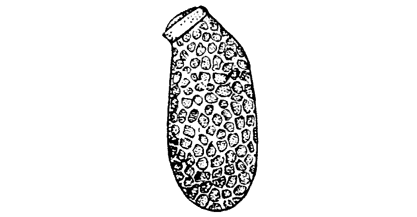
Fig. 4.—Egg of Cimex lectularius. Enlarged. (After Marlatt.)
Fig. 4.—Egg of Cimex lectularius. Enlarged. (After Marlatt.)
The eggs of the bed-bug are
pearly white, oval objects, perhaps
1 mm. in length. At one end
there is a small cap surrounded
by a projecting rim, and it is by
pushing off this cap, and through
the orifice thus opened, that the young bug
makes its way into the outer world after
an incubation period of a week or ten days.
There is no metamorphosis—no caterpillar and
no chrysalis stages. The young hatch out,
in structure miniatures of their parents, but
in colour they are yellowish-white and nearly
transparent. The young feed readily, and
feeding takes place between each moult, and
the moults are five in number, before the
adult imago emerges. This it does about
the eleventh or twelfth week after hatching.
These time-limits depend, however, upon the
temperature after hatching, and the rate of
growth depends not only upon the temperature
but also upon the amount of food.
When bred artificially and under good conditions,
the rate of progress can be ‘speeded
up’ so that the eggs hatch out in eight days,
and every following moult takes place at
intervals of eight days, so that the period from
egg to adult can be run through in as short a
time as seven weeks.
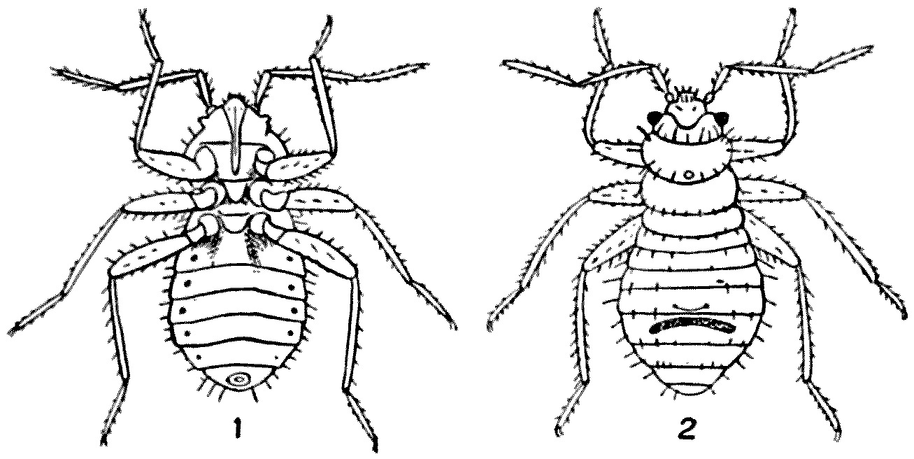
Fig. 5.—Newly hatched young of Cimex lectularius. 1, Ventral
view; 2, dorsal view. Enlarged. (After Marlatt.)
Fig. 5.—Newly hatched young of Cimex lectularius. 1, Ventral
view; 2, dorsal view. Enlarged. (After Marlatt.)
Unless fed after each moult, the following
moult is indefinitely postponed. Hence it
follows that in the preliminary stages bugs
must bite their hosts five times before the
adult form emerges, and the adult must,
further, have a meal before it lays its eggs.
The eggs are deposited in batches of from five
to fifty in cracks and crevices, into which the
insects have retired for concealment.
Bugs can, however, live a very long time
without a meal. Cases are recorded in which
they have been kept alive for more than a year
incarcerated in a pill-box. When the pill-box
was ultimately opened, the bugs appeared to be
as thin as oiled paper and almost so transparent
that you could read The Times[3] through them;
but even under these conditions they had
managed to produce offspring. De Geer kept
several alive in a sealed bottle for more than
a year. This power of existing without food
may explain the fact that vacated houses
occasionally swarm with bugs even when
there have been no human beings in the
neighbourhood for many months.
The effect of their bite varies in different
people. As a rule, the actual bite lasts for
two or three minutes before the insect is
gorged, and at first it is painless. But very
soon the bitten area begins to swell and to
become red, and at times a regular eruption
ensues. The irritation may be allayed by
washing with menthol or ammonia. Some
people seem immune to the irritation; and
I know friends who, in the West Indian Islands,
have slept through the attacks of thousands
of bugs, and only awoke to their presence
when in the morning they found their night-clothing
and their sheets red with blood,
expressed from the bodies of their tormentors
as the victims turned from side to side.
As a rule, the uncovered parts of the body—the
face, the neck, and the hands—are
said to be more bitten than the parts which
are covered by the bedclothes. This is not,
however, my experience.
The bug has been accused of conveying
many diseases—typhus, tuberculosis, plague,
and a form of recurrent fever produced by
a spirochaete (Spirochaeta obermeieri); but a
critical examination throws some doubt upon
the justice of the accusation, and Professor
C. J. Martin writes as follows:—
There is really no evidence to incriminate the bed-bug
in the case of either typhus or relapsing fever. It
is possible to transmit plague experimentally by
means of bugs, but there is no epidemiological reason
for supposing this takes place to any extent in nature.
There are two differences in the habits of bugs
and those of fleas and lice which may possess epidemiological
significance. The first concerns the customary
intervals between their meals. Bugs show no disposition
to feed for a day or two after a full meal,
whereas fleas and lice will suck blood several times
during the twenty-four hours. The second is in
respect to the time the insects retain a meal and[32]
the extent to which it is digested before being excreted.
Fleas and lice, if constantly fed, freely
empty their alimentary canals, and the nature of
their faeces indicates that the blood has undergone
but little digestion.
Both these insects evacuate such undigested
or half-digested blood per rectum during the act of
feeding, and the remnants of the previous meal
are thus deposited in the immediate vicinity of a
fresh puncture. It is not unlikely that, should
the alimentary canal of the insect be infected with
plague bacilli, spirochaete, or the organism responsible
for typhus fever, these may be inoculated by
rubbing or scratching. Bugs have not this habit;
and in all the cases I have examined their dejections
were fully digested, almost free from protein, and
consisted mostly of alkaline haematin.
Whether bugs be guilty of these crimes
or not, they are the cause of an intense inconvenience
and disgust, and should, if possible,
be dealt with drastically. At the present
time[4] there are rumours that some of our
largest camps are infested with these insects,
and there seems no doubt that some of the
prisoners and refugees to this country have
brought their fauna with them, and this
fauna is very capable of spreading in concentration
camps. The erection of wooden
huts—no doubt a pressing necessity—will
afford convenient quarters for these pests.
Among the measures which have been
most successful in the past has been fumigating
houses with hydrocyanic-acid gas; but
this is a process involving considerable danger,
and should only be carried out by competent
people under the most rigorous conditions.
In all fumigating experiments every crack
and cranny of a house should be shut, windows
closed, keyholes blocked, and so on. A second
method of fumigation is that of burning
sulphur. Four ounces of brimstone are set
alight in a saucer, this in its turn is placed
in a larger vessel, which protects the floor of
the room from a possible overflow of the
burning material. After all apertures have
been successfully plugged, four or five hours
of the sulphurous fumes are said to be sufficient
to kill the bugs, but to ensure complete success
a longer time is needed. This is not only
a much less expensive but a much less dangerous
operation than using hydrocyanic-acid
gas. Two pounds of sulphur will suffice for
each thousand cubic feet of space, but it
is well to leave the building closed for
some twenty-four hours after the fumigation.
Another more localised method of destroying
these pests is the liberal application of benzine,
kerosene, or any other petroleum oil.
These must be introduced into all crevices
or cracks by small brushes or feathers,
or injected with syringes. In the same way oil
of turpentine or corrosive-sublimate has proved
effective. Boiling water is also very fatal
when it can be used; and recently in the poorer
quarters of London the ‘flares’ which painters
use in burning off paint have proved of great
use in ridding matchboarding, or wainscoting,
from the harbouring bugs. Passed quickly
along, the flame of the ‘flare’ does not burn
the wood, but it produces a temperature which
is fatal to the bug and to its young and to
its eggs. And thus:—
‘This painted child of dirt, that stinks
and stings’[5] is destroyed.
CHAPTER III
THE FLEA (Pulex irritans)
Marke but this flea, and marke in this,
How little that which thou denyst me is;
It sucked me first, and now sucks thee,
And in this flea our two bloods mingled bee.
(Dr. Donne.)
The fact, now fully established, that the
bubonic plague is conveyed to man from
infected rats, or from infected men to healthy
men, by fleas has taken that wingless insect
out of the category of those animals which it
is indelicate to discuss.
No doubt, as Mr. Dombey says, ‘Nature
is on the whole a very respectable institution’;
but there are times when she presents
herself in a form not to be talked about, and
until a few years ago the flea was such a
form. Hence, few but specialists have any
clear idea either of the structure or of the
life-history or of the habits—save one—of
the flea.
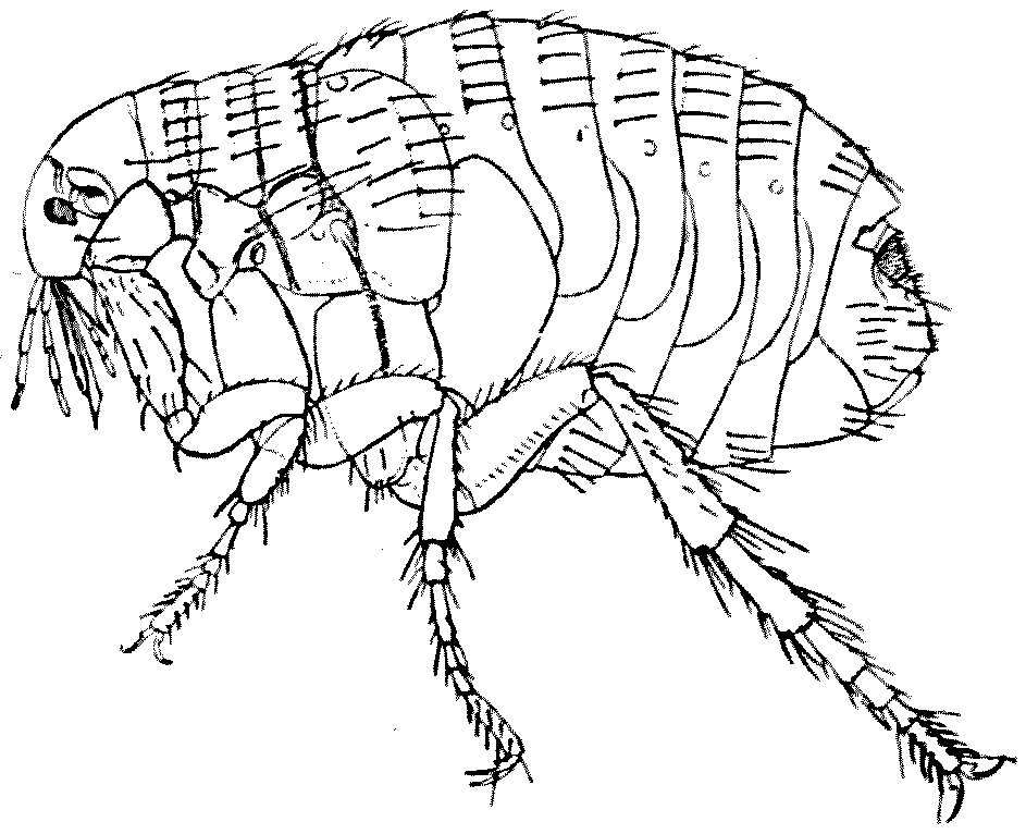
Fig. 6.—Pulex irritans, female. The legs of the left side only are
shown. Enlarged. (After a drawing by A. Dampf.)
Fig. 6.—Pulex irritans, female. The legs of the left side only are
shown. Enlarged. (After a drawing by A. Dampf.)
Fleas are temporarily parasitic on many
mammals and birds, but some mammals and
some birds are much freer from fleas than
others. As the flea is only on its host for
part of the time, it has to put in the rest
of its existence in some other place, and
this, in the case of the human flea, is usually
the floor, and in the case of bird-fleas the
nest; from these habitats they can easily
regain their hosts when the latter retire to
rest. But large numbers of Ungulates—deer,
cattle, antelopes, goats, wild boars—sleep in
different places each recurrent night, and to
this is probably due the fact that, with the
exception of two rare species—one taken
in Northern China and the other in Transcaucasia—the
Ungulates have furnished descriptive
science with no fleas at all. Both of
these Ungulate fleas are allied to the burrowing-fleas
or ‘chigoes.’
I know none of my readers will believe me
when I say that the same is true of monkeys;
but I do this on the undoubted authority
of Mr. Harold Russell, who has recently published
a charming little monograph on these
lively little creatures. Monkeys in nature
are cleanly in their habits; and although
in confinement occasionally a human flea
attacks them, and although occasionally a
chigo bores into the toes of a gorilla or
chimpanzee, ‘speaking generally, it may be
said that no fleas have been found truly
parasitic on monkeys.’ Whatever the monkeys
are looking for, it is not fleas. What they
seek and find is in effect little scabs of scurf
which are made palatable to their taste by
a certain sour sweat.
As a rule, each host has its own species
of flea; but though for the most part Pulex
irritans is confined to man it is occasionally
found on cats and dogs, whilst conversely
the cat- and dog-fleas (Ctenocephalus felis and
Ct. canis) from time to time attack man.
The bite of the flea is accompanied by the
injection of the secretions of the so-called
salivary glands of the insect, and this secretion
retards the coagulation of the victim’s blood,
stimulates the blood-flow, and sets up the
irritation we have all felt.
It is only a few years ago that the spread of
bubonic plague was associated first with rats,
and then with rat-fleas; and at once it became
of enormous importance to know which of the
numerous species of rat-flea would attack
human beings. The Hon. Charles Rothschild,
who has accumulated a most splendid collection
of preserved fleas in the museum at Tring, had
some years ago differentiated from an undifferentiated
assemblage of fleas a species
first collected in Egypt, but now known to
be the commonest rat-flea in all tropical and
sub-tropical countries. This species Xenopsylla
cheopis—and to a lesser extent Ceratophyllus
fasciatus—unfortunately infests and bites
man. If they should have fed upon
a plague-infected rat and subsequently bite
man, their bites communicate bubonic plague
to human beings. Plague—the Old English
‘Black Death’—is a real peril in our armies
now operating in Asia and in certain parts of
Africa.
Just as some fleas attack one species of
mammal or bird and avoid closely allied species,
so the human flea has its favourites and its
aversions. There is a Turkish proverb which
says ‘an Englishman will burn a bed to catch
a flea,’ and those who suffer severely from fleabites
would certainly do so. The courage of
the Turk in facing the flea, and even worse
dangers, may be, as the schoolboy wrote,
‘explained by the fact that a man
with more than one wife is more
willing to face death than if he
had only one.’ But there are
persons even a flea will not bite.
Mr. Russell has reminded us in his
Preface of the distinguished French
lady who remarked, ‘Quant à moi
ce n’est pas la morsure, c’est la
promenade!’
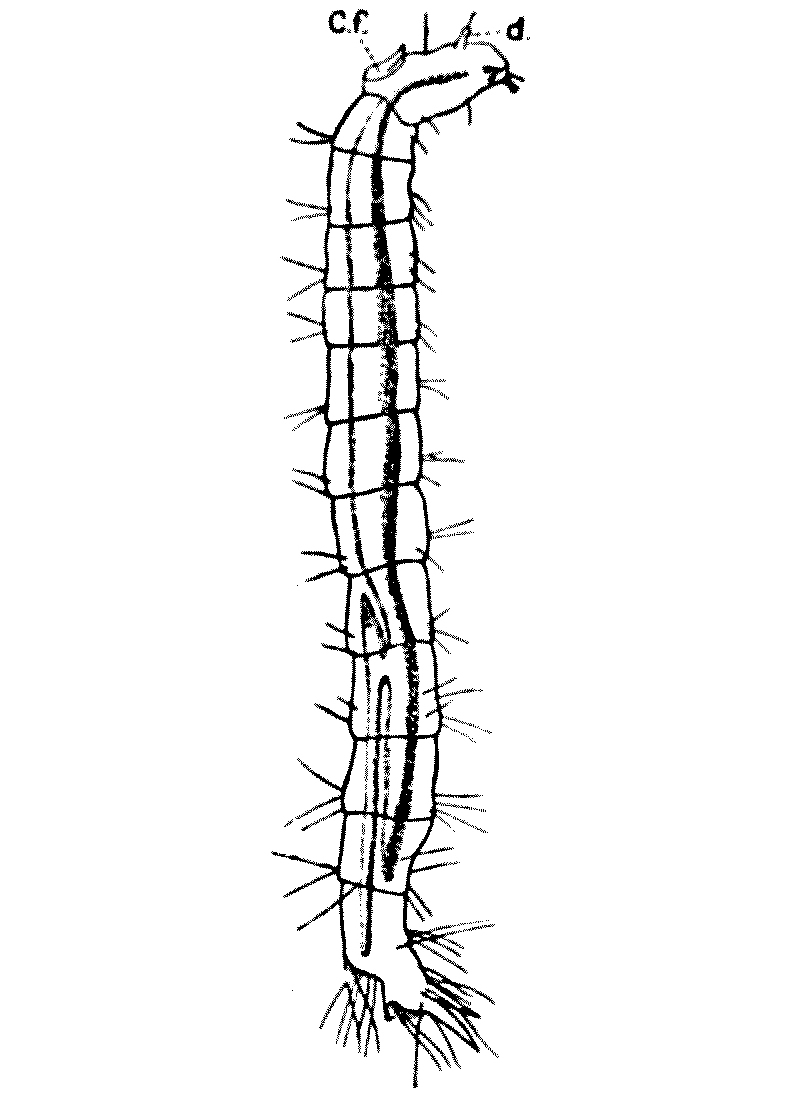
Fig. 7.—Larva of Pulex
irritans. C.f. frontal horn; d, antenna. Enlarged. (After Brumpt.)
Fig. 7.—Larva of Pulex
irritans. C.f. frontal horn; d, antenna. Enlarged. (After Brumpt.)
There are one or two structural
features in a flea which are peculiar:
the most remarkable being that,
unlike most other insects, it is much
taller than it is broad. As a rule,
insects—such as a cockroach, the
bed-bug, or a stag-beetle—are like
skates, broader than they are thick,
but the flea has a laterally compressed
shape, like a mackerel or
a herring. Then, again, the three
segments or rings which come after
the head are not fused into a solid cuirass or
thorax as they are in the fly or the bee, but
they are movable one on the other. Finally, it
is usual in insects for the first joint of the leg
to be pressed up against and fused with those
segments of the body that bear them; but in
the flea not only is this joint quite free, but
the body-segment gives off a projection which
stretches out to bear the leg. Thus the legs
seem, unless carefully studied, to have an extra
joint and to be—as indeed it is—of unusual
length. They certainly possess unusual powers
of jumping—as Gascoigne, a sixteenth-century
poet (1540–78) writes, ‘The hungry fleas which
frisk so fresh.’
The male, as is so often the case amongst
the Invertebrata, is much smaller than the
female. The latter lays at a time from one
to five minute, sticky, white eggs, one-fortieth
of an inch long by one-sixtieth broad. They
are not laid on the host, but in crevices between
boards, on the floor, between cracks in the
wainscoting, or at the bottom of a dog-kennel
or in birds’ nests. Mr. Butler recalls
the case of a gentleman who collected on four
successive mornings sixty-two, seventy-eight,
sixty-seven, and seventy-seven cat-fleas’ eggs
from the cloth his cat had slept upon.
Altogether 284 eggs in four nights! The date
of hatching varies very much with the
temperature. Pulex irritans takes half as long
again—six weeks instead of four—to become
an adult imago in winter than it does in summer.
But in India the dog-flea will complete its cycle
in a fortnight.
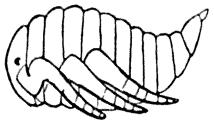
Fig. 8.—Pupa of
flea. (After Westwood.)
Fig. 8.—Pupa of
flea. (After Westwood.)
When it does emerge from the egg the
larva is seen to be a whitish segmented little
grub without any limbs, but with plenty of
bristles which help it to move about; this it
does very actively. There are two small
antennae and a pair of powerful jaws, for the
larva does not take liquid food, but eats any
scraps of solid organic matter which it comes
across: dead flies and gnats are readily
devoured. The larva casts its
skin several times, though
exactly how often it moults
seems still uncertain.
After about twelve days of
larval existence it spins itself
a little cocoon in some sheltered
crevice, and turns into a whitish inert chrysalis
or pupa. During its pupal existence it takes,
of course, no food, but it grows gradually
darker, and after undergoing a tremendous
internal change, breaking down its old tissues
and building up new ones, the chrysalis-case
cracks and the adult flea jumps out into
the world.
There are many superstitions about fleas.
March 1st is in some way connected with them,
and in the south of England the house-doors
are in some villages closed on that day under
the belief that this will render the building
immune for the following twelve months.
The most successful insecticide is said to be
prepared from Pyrethrum, which is grown
in the Near East in large quantities for this
purpose. But the Austrians, the Serbians, and
the Montenegrins are fighting over the chief
world-supply of this plant—possibly without
knowing what they are doing—and ‘Insektenpulver’
is bound to go up in price. Wormwood
(Artemisia) is also recommended.
While wormwood hath seed, get a handfull or twaine,
To save against March, to make flea to refraine;
When chambere is swept and wormwood is strowne,
No flea for his life dare abide to be known.
(Tusser.)
The author of ‘A Thousand Notable Things’
suggests the following plan, but, so far, I
have not met anyone who has tried it: ‘If
you mark where your right foot doth stand
at the first time that you do hear the cuckow,
and then grave or take up the earth under
the same; wheresoever the same is sprinkled
about, there will no fleas breed. I know
it hath proved true.’
Plastering a floor with cow-dung is a
common practice in South Africa, and seems
to be an efficacious means of keeping down
fleas. Dr. R. J. Drummond tells me that
all natives of India and Ceylon spread an
emulsion of cow-dung in hot-water over the
floors and the walls of their dwellings to keep
out fleas. This has been done from immemorial
times, and is effective. The efficacy of the
emulsion in keeping fleas away has been
doubted, and so I am glad to quote a few lines
from a kind letter sent me by Dr. P. A. Nightingale
of Victoria, Southern Rhodesia, which put
the matter in a happy light:—
I think the correct facts are these: the floors of
certain houses, huts, &c., throughout the South African
veld are made of ant-heap earth, moistened and
beaten hard and flat with sticks. This floor is then
smeared at regular intervals—say, every ten days—with
fresh cow-dung, when the room becomes fresh
and sweet (!) and free from insects.
However, before the smearing can be done it is
necessary to turn all the furniture out of the room
and to sweep it thoroughly; after the smearing, the
doors and windows are left open for drying purposes.
Hence, I think that the absence of fleas in such
quarters is really due to general cleanliness, sunlight,
and fresh air, and not to any special virtue in the
cow-dung.
I am, however, sure that the smearing of the floor
at frequent intervals does keep many pests down by
filling up, and temporarily sealing, the numerous
cracks in the floor where fleas, &c., reside and breed in
vast numbers.
Huts—especially unused ones—not smeared for
many weeks contain (approximately) several thousands
of fleas, white ants, centipedes, and scorpions to
the square inch, when the only treatment is to cleanse
[44]the walls and floor with cyanide solution, or burn the
whole place down.
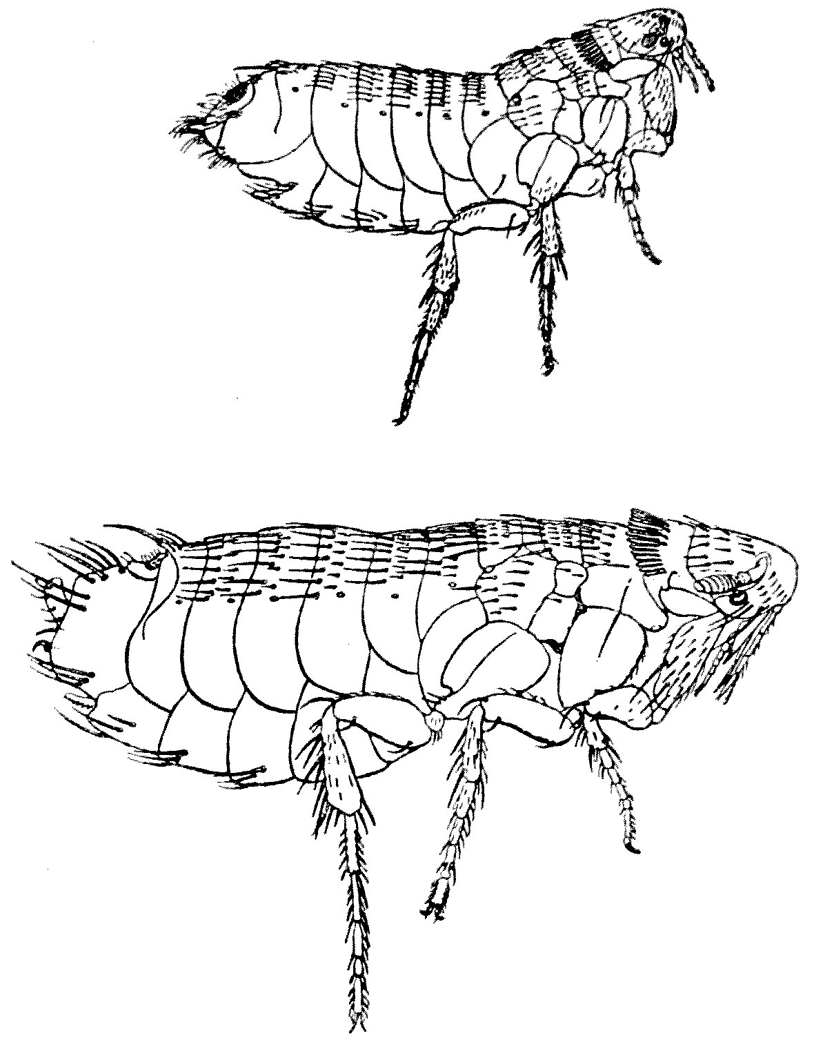
Fig. 9.—Ceratophyllus gallinulae. Male (above) and
female (below). Drawn to scale and both highly magnified. These specimens,
taken from a grouse, are of the same genus as one of the plague-conveying
fleas.
Fig. 9.—Ceratophyllus gallinulae. Male (above) and
female (below). Drawn to scale and both highly magnified. These specimens,
taken from a grouse, are of the same genus as one of the plague-conveying
fleas.
From long experience, I am very nearly insect
proof; but cannot stand the myriads of fleas I occasionally
have to sleep with in a hut of the above
description—especially just before the rains set in,[45]
when additional veld pests come into the huts for
shelter.
We must, in the long run, treat fleas
seriously. Although the Pulex irritans is
a very common insect, the greatest living
authority on fleas tells me it has never been
accurately drawn. We have Blake’s ‘ghost
of a flea’; but what did Blake know of
entomology? In distinguishing one flea from
another—fleas which may attack man and
fleas which have hitherto declined to do so—every
hair, every bristle, counts. Hence,
I illustrate this article with accurate outlines
of certain fleas found on the grouse, and for
whose accuracy I can vouch (Fig. 9).
As I have said above, a certain rat-flea
(Xenopsylla cheopis) and another (Ceratophyllus
fasciatus) undoubtedly convey the bacillus of
plague from rats and other Murinae to man
and vice versa. The Bacillus pestis is unlikely
to establish itself in the present war in Europe,
but Quién sabe? The Black Death of 1349–51
was conveyed by fleas, and so was Pepys’s
Plague of 1665. Plague—flea-borne, we must
remember—is still endemic in places as near
Europe as Tripoli, and in numerous centres
in Asia. Not a disease altogether to be
neglected, since the spread of war to the Near
East, but still not very threatening in Europe
in the twentieth century.
CHAPTER IV
THE FLOUR-MOTH (Ephestia kühniella) IN SOLDIERS’
BISCUITS
Where moth ... doth corrupt. (Matt. vi. 19.)
It is not only those insects that destroy the
continuity of our soldiers’ integument which
play a part in war. It has been well said that
an army marches on its stomach; and the
admirable commissariat arrangements which
have been so distinctive a feature of the British
Expeditionary Force during the present war
are the result of much patient care and attention
during times of peace. I am in no position to
discriminate, but I do believe that the admirable
service of the A.S.C. and the R.A.M.C.
is at least equal to the splendid record of
those in the fighting-line.
Every one knows that recruits are frequently
rejected for some defect in their teeth. A
soldier, indeed, requires strong teeth, for his
farinaceous food in the field is largely supplied to
him in the form of biscuits—not that ‘moist
and jovial sort of viand,’ as Charles Dickens
described the Captain biscuit, but ‘hard-tack’
which challenges the stoutest molars.
During the summer of 1913 the authorities
of the British Museum at South Kensington
arranged a very interesting but somewhat
gruesome exhibit in their Central Hall. The
exhibit consisted mainly of Army biscuits
eaten through and through by the larva of a
small moth and covered by horrible webs or
unwholesome-looking skeins of silky threads.
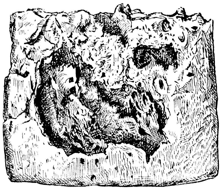
Fig. 10.—Ephestia kühniella. Moth-infested
biscuit.
Fig. 10.—Ephestia kühniella. Moth-infested
biscuit.
Together with these derelict biscuits were
certain long metallic coils and other apparatus
used in investigating certain phases of the life-history
of the moth and the manufacture
of the biscuit. The exhibit illustrates an
article which had recently appeared on the
Baking of Army Biscuits, by Mr. Durrant and
Lieut.-Colonel Beveridge, on the ‘biscuit-moth’
(Ephestia kühniella), a member of the family
Pyralidae. The article recorded their efforts
to arrive at a means of checking this very
serious pest to service stores.[6]
The biscuit-moth (E. kühniella) was described
two years before its larva had been
noted damaging flour at Halle. There has
always been a certain amount of international
courtesy in attributing the provenance of
insect pests to other countries; and when
E. kühniella began, about ten years later,
to attract attention in England it was believed
to have been introduced from the
United States, via the Mediterranean ports,
in American meal. The American origin was,
however, denied by Professor Riley, who,
in a letter to Miss Ormerod, states, ‘I think
I can safely say that this species does not
occur in the United States.’ At the moment
of writing these words Professor Riley was
in the act of packing-up to leave Washington
for Paris. Possibly he was excited, certainly
he was inaccurate, for the species was then
known to be prevalent in Alabama, North Carolina,
and other States. In fact, to-day it is
recorded throughout Central America and the
Southern States, and in most of the temperate
regions of the New World.
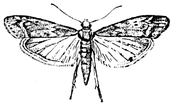
Fig. 11.—Ephestia kühniella. × 2.
Fig. 11.—Ephestia kühniella. × 2.
The moth itself is a rather insignificant, small insect, of a
slatey-grey colour. Its eggs, rather irregular ovoids, are laid upon
the biscuit into which the issuing larvae bore. These latter are soft
and like most creatures which live in the dark, whitish, though with a
tinge of pink; the head, however, is brown and hardened. The larva is
constantly spinning silken webs or tissues, which in the most untidy
way envelop the biscuit. It finally entombs itself in a whitish silken
cocoon, and herein it ultimately turns into a chrysalis or pupa.
Another Pyralid moth—Corcyra cephalonica—makes
similar unpleasant webs all over biscuits,
rice, or almost any farinaceous food; but,
since its larvae are unable to live unless there
be a certain degree of moisture in its food, it
is less injurious to baked food than the Ephestia,
for whose larvae nothing can be too dry. Corcyra
seems originally to be a pest of rice, and to
have been introduced into Europe with Rangoon
rice; but it readily alters its diet in new
surroundings, and will live on almost any
starchy stuff, if not too desiccated.
The problem that Lieut.-Colonel Beveridge
and Mr. Durrant, of the British Museum,
set out to solve was at what stage in the
manufacture of the Army biscuits does our
soldiers’ food become infested, and whether
any steps could be taken to avoid or minimise
such infestation.
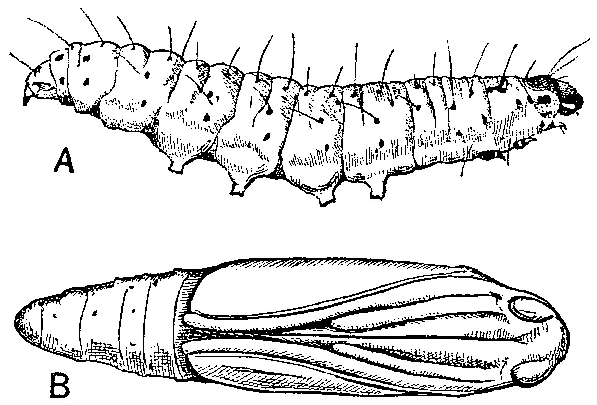
Fig. 12.—Ephestia kühniella. A, Larva; B, pupa.
Greatly magnified.
Fig. 12.—Ephestia kühniella. A, Larva; B, pupa.
Greatly magnified.
First, as to infestation. The biscuit must
become infested either (1) at home before
packing, (2) during transit, or (3) in the
country where they are stored. The biscuits
are packed in tins, hermetically sealed, and
enclosed in wooden cases to prevent injury;
it was therefore obvious that if insects could
be found within intact tins it would be demonstrated
at once that infestment must
have taken place in the factories, and not
subsequently.
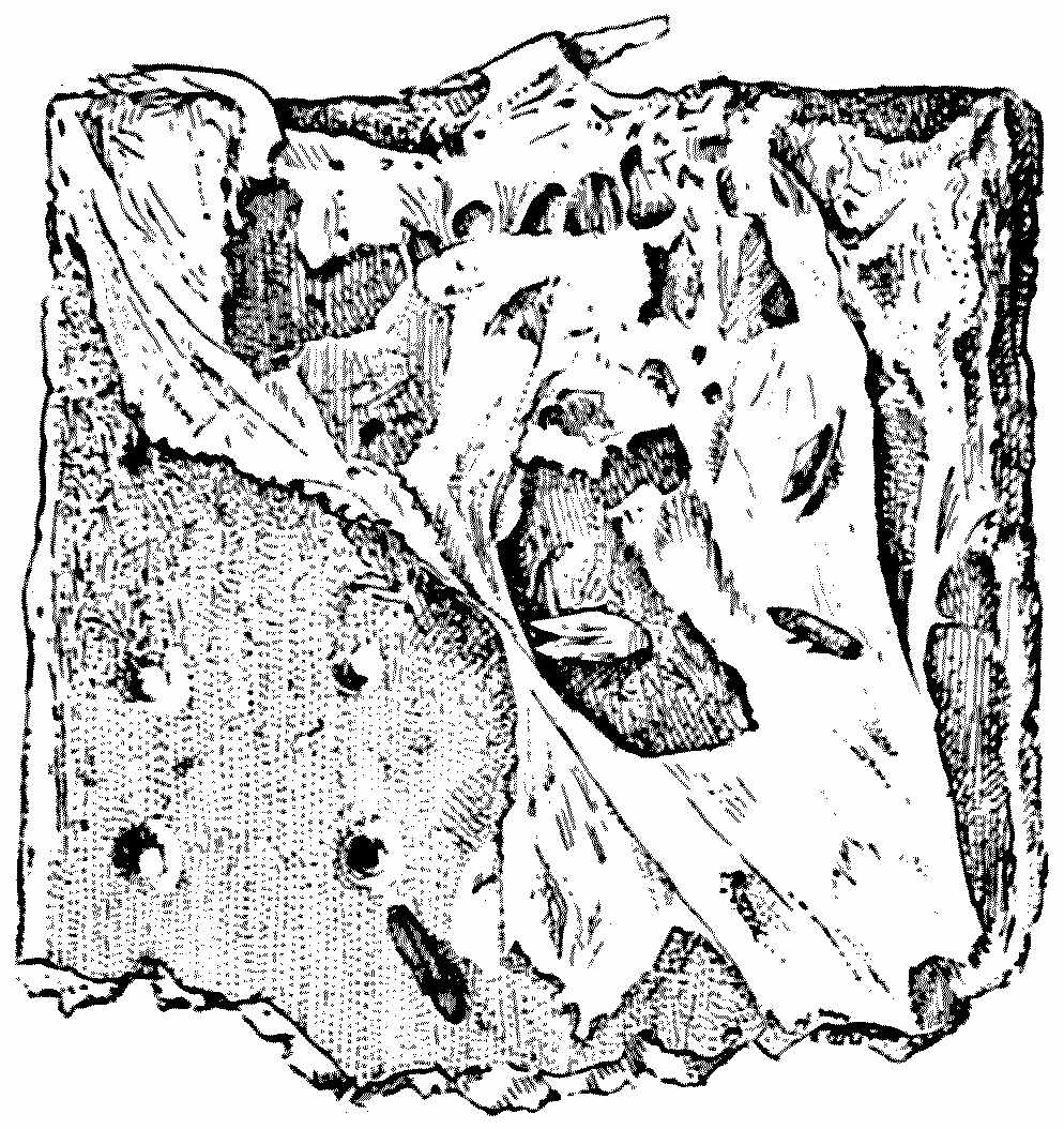
Fig. 13.—Corcyra cephalonica. Moth-infested
biscuit.
Fig. 13.—Corcyra cephalonica. Moth-infested
biscuit.
With a view to determine
the origin of infestation sample tins were
withdrawn from stocks at various stations
abroad, and inspected by experts at Woolwich;
and tins which, after careful examination,
had been pronounced intact, were found to
contain Ephestia kühniella and Corcyra cephalonica
in various stages of development, thus
proving conclusively that infestation had
taken place in the factories before the tins
were soldered, and indicating that preventive
or remedial measures must be undertaken
within the biscuit-making factories themselves.
It is obvious either that the heat to which
the biscuit is subjected in the process of
baking is insufficient to destroy any of the
insect eggs present in the moist dough or
that the moths and beetles deposit their eggs
in or on the biscuits after baking, and during
the process of cooling and of packing into the
tins. Cooling before packing is necessary in
order to allow the moisture in the centre of
the biscuit to become evenly distributed
throughout the ‘tissue’ of the biscuit. And
it is during the time occupied in cooling
and packing that the biscuit is exposed to
the greatest risk of infestation; any risk
occasioned by subsequent injury must be exceptional,
and is probably negligible.
By a series of most ingenious experiments,
the two investigators were able to
determine the temperature in the centre of
the biscuits during the various stages of
its baking and cooling. Army biscuits are
made from dough which contains about 25
per cent. of water. When stamped out they
are placed in rows on the revolving floor of
an oven, and are submitted to a high temperature
for twenty minutes whilst they travel
over a space of 40 feet. The dough at
first contains, as we have said above, 25 per
cent. of water, but during baking this is reduced
to about 10 per cent., and the moisture now
collects in the centre of the mass of the
biscuit in consequence of the external hardening
or ‘caramelisation,’ as it is called. The holes
which are pricked in so many biscuits of course
help to equalise the spread of the moisture
throughout the biscuit.
Too little attention has been paid to the
internal temperature of edibles which are
being cooked. Very few people, for instance,
have any conception of what is going on
in the centre of a joint of meat whilst it is
being roasted or boiled. After two hours’
boiling the temperature in the centre of a
large ham has only risen to 35° C.; after
six hours’ boiling to 65° C., and it is only
after ten hours’ continuous boiling that 85° C.
is reached. I have, I am sorry to say, no
conception as to how long a ham ought to
be boiled, but it is obvious that to be really
effective against such parasites as Trichinella—the
causa causans of trichinosis—the
cooking of pork and ham should be more
prolonged and thorough than seems to be
customary. But that is another story.
However, to return to our biscuits. The
Colonel and Mr. Durrant devised an ingenious
instrument which determined the rising temperature
at the centre of our Army biscuits
whilst baking. When the tip of their recording
apparatus lay within the moist area of the
biscuit, the temperature registered was only a
little over 100° C.; but when the tip of the
instrument rested on the hard ‘caramelised’
portion much higher temperatures were observed—even
as high as 125° C. Colonel Beveridge
and Mr. Durrant were thus able to establish
the fact that the temperatures of the biscuit
were, during baking, such as to rule out the
idea that the eggs of the biscuit-moth—which
do not survive a temperature of 69° C. for
twelve minutes—were deposited in the biscuit
before cooking.
After the baking is completed the biscuits
are cooled, and it is at this period that
they are most exposed to risk of infestation
by Ephestia kühniella. This insect is a
well-known nuisance in Flour-mills. So persistent
and numerous are these moths at times
that they clog the rollers with their cocoons,
and sometimes completely stop them. The
webbing of the elevators in the mills gets
covered with them and with their silky skeins,
and then the elevators stop working. They
mat together the flour and meal with their
silken excreta, and so uniform is the temperature
of the Mill, and so favourable to the life
of the insect, that they complete their life-cycle
in this country in two months, and
in the warmer parts of America even more
rapidly. In well-heated mills the proceeding
is continuous, so that six generations at
least may be produced each year.
The most efficient method of getting rid of
this pest of the Army biscuit is a complete
and thorough fumigation of the infested
premises with carbon bisulphide. But, as this
substance is not only poisonous but inflammable,
it is well to get a chemist to undertake
the proceeding, and also to notify the Insurance
Company. Fumigation by sulphur ruins the
flour. Another remedial measure is that of
turning the steam from the boilers on to all
the infected machinery and walls.
That this destruction of the Army biscuit is
a matter of considerable importance is shown
by the fact that biscuit-rations exported to the
colonies in hermetically sealed tins have become
quite unfit for consumption, and this destruction
has been noted in places as far distant from
each other as Gibraltar, the Sudan, Mauritius,
Ceylon, South Africa, and Malta. That it is also
an old trouble is shown by the following quotation
from the diary which Sergeant Daniel
Nicol, of the 92nd (the Gordon Highlanders),
kept during the expedition to Egypt in 1801:—
Some vessels were dispatched to Macri Bay for
bullocks, and others to Smyrna and Aleppo for
bread which was furnished us by the Turks—a kind
of hard dry husk. We were glad to get this, as we
were then put on full rations, and our biscuits were
bad and full of worms; many of our men could only
eat them in the dark.[7]
With regard to the actual baking of the
biscuit, Colonel Beveridge and Mr. Durrant
suggest that the temperature conditions during
the process of cooling should be made as unfavourable
as possible for the moths by introducing
screened cool air, which can be forced in
at one end of the cooling-chamber and sucked
out at the other. Could such a scheme be
adopted it would be difficult, if not impossible,
for the moths to lay their eggs, and the
biscuit would thus be more rapidly cooled.
In any case it should not be difficult to ensure
that the cooling takes place in some chambers
which are practically free from these destructive
moths.
CHAPTER V
FLIES
Part I
THE HOUSE-FLY (Musca domestica)
Musca est meus pater, nil potest clam illum haberi;
Nec sacrum nec tam profanum quidquam est, quin Ibi ilico adsit.
(Plautus, Mercator.)
‘The common house-fly [says Ruskin] is the most
perfectly free and republican of creatures. There
is no courtesy in him; he does not care whether it
is a king or clown whom he teases, and in every step
of his swift mechanical march and in every pause
of his resolute observation there is one and the
same perfect expression of perfect egotism, perfect
independence and self-confidence and conviction of
the world having been made for flies. Your fly
free in the air, free in the chamber, a black incarnation
of caprice, wandering, investigating, fleeting,
flitting, feasting at his will with rich variety of feast
from the heaped sweets in the grocer’s window to
those of the butcher’s back yard, and from the galled
place on your horse’s neck to the brown spot on the
[58]
road from which, as the hoof disturbs him, he rises
with angry republican buzz; what freedom is like
his?’
The house-fly is all that Ruskin describes
it to be, but it is more. It is the most cosmopolitan
of insects. Wherever man is there is
the fly. It is found—
From Greenland’s icy mountains
To India’s coral strand.
But it is naturally more frequent in warm
climates than in cold, as the rate of its development
depends very largely upon an average
high temperature.
Unlike the lice and the bed-bug, the fly like
the flea, passes through a complete metamorphosis—egg,
larva, pupa, and imago. It will
breed in almost any rotten matter, whether
vegetable or animal, and it breeds most
successfully, as Gordon Hewitt has pointed
out, when certain processes of organic fermentation
are taking place in its breeding-place.
Probably the fermentation has a
favourable effect upon the food of the larvae.
Undoubtedly the place most readily selected
by the female for laying her eggs is stable-manure.
A few years ago there was a remarkable
reduction in the number of house-flies in
London, and Lord Montagu of Beaulieu attributed
this reduction to the refreshing and
insecticidal petrol vapour with which the
streets of that town were then bathed.
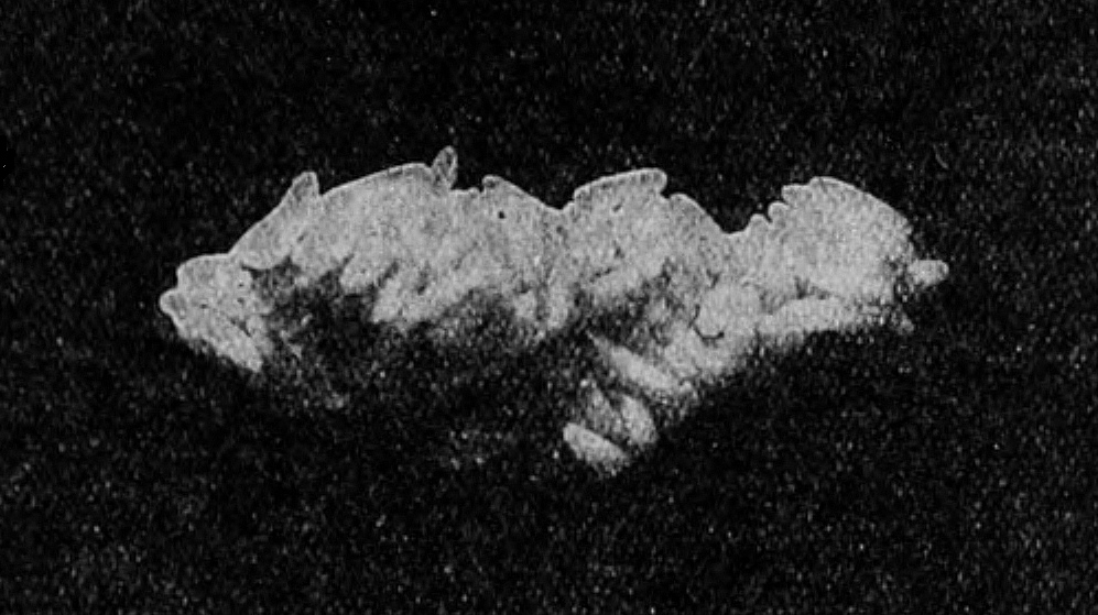
Fig. 14.—Mass of eggs of M. domestica.
(From Gordon Hewitt.)
Fig. 14.—Mass of eggs of M. domestica.
(From Gordon Hewitt.)
I do not know what experiments Lord Montagu
had made on the subject of the insecticidal
value of petrol vapour, but the ordinary man
in the street attributed—and I think more
correctly—the diminution of the plague of
flies to the absence of the nidus in which the
female fly lays her eggs. Stable-yards had been
turned into garages. But flies will, indeed,
breed in almost any kind of dejecta—including
the human—and in rotten straw, rotten wool,
cotton garments, decaying vegetables and fruits,
bad meat, rotten grain, and even in spittoons,
but they prefer horse-manure.
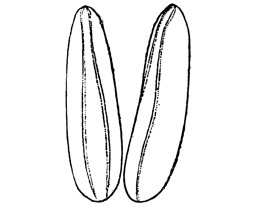
Fig. 15.—Eggs of M. domestica,
× 40. (From Gordon Hewitt).
Fig. 15.—Eggs of M. domestica,
× 40. (From Gordon Hewitt).
In our country house-flies usually begin to
breed in June and July, continuing well on into
October if the weather be but warm. Their
greatest activity is, however, in the hotter
month of August and the beginning of September.
But in warm stables,
restaurants, and kitchens flies
are able to reproduce the whole
year round. A single fly will
deposit at one time 100 to 150
eggs, and in the course of her
summer life may produce five,
or even six, batches of ova of
this size. The eggs are pearly
white, elongated structures,
with two converging lines, along
which the egg-case will ultimately
split to give exit to the
larva. The eggs are laid, by means of a long
ovipositor, a little way beneath the surface of
the dung-heap in a position where they will not
readily be dried up. In favourable conditions
the eggs hatch in from eight to twenty-four
hours.
The first larva is legless, tapering towards the head, which bears
a pair of breathing-holes, or spiracles; the body is much stouter
towards
the hinder end. On the whole it is a white, unpleasant-looking maggot,
called by freshwater-fishermen a ‘gentle.’ By contracting and expanding
its body it pushes its way through the moist, semi-liquid surroundings.
The skin is usually moulted some twenty-four hours after birth, but
all these time-limits depend much upon the temperature and favourable
conditions. With normally high temperatures—say, with 30° C. to 35°
C.—the larva will become fully grown in five or six days. The third
and final larval stage, after the second moult or ecdysis, lasts three
days, and when fully grown the maggot is now about half an inch in
length. Externally, twelve segments are visible, but the internal
anatomy shows that thirteen are really present, though one is almost
‘masked.’
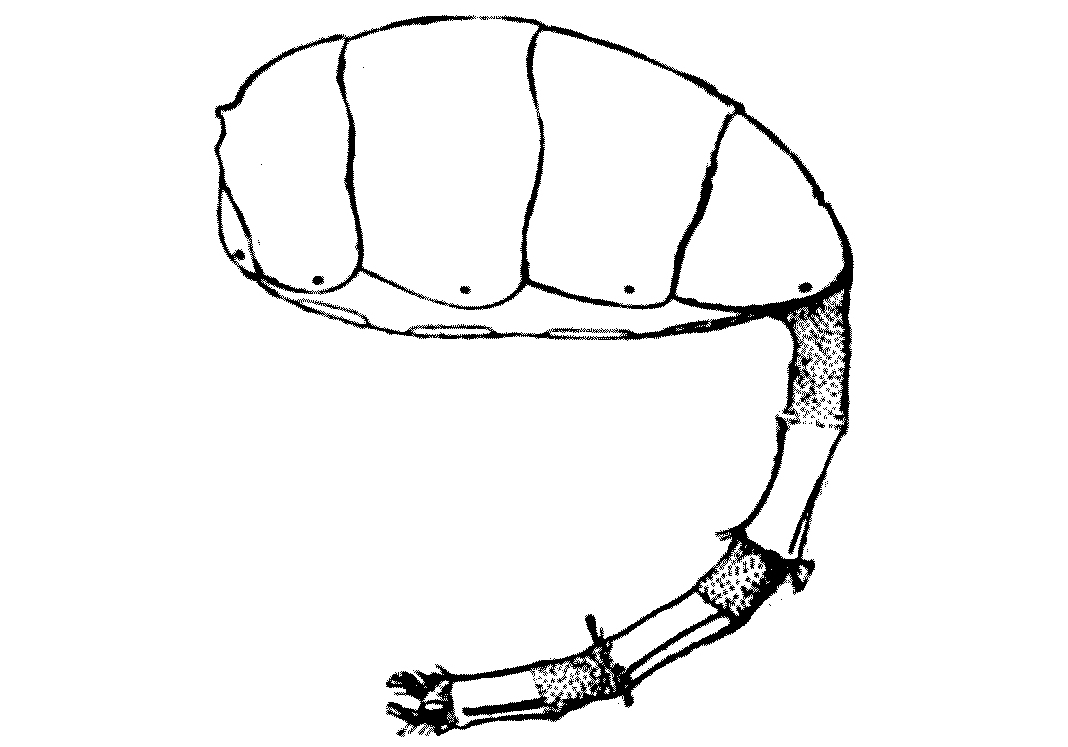
Fig. 16.—Abdomen of female
house-fly, showing the extended ovipositor. (From Gordon Hewitt.)
Fig. 16.—Abdomen of female
house-fly, showing the extended ovipositor. (From Gordon Hewitt.)
It is only during these larval stages that
the insect grows, and it is never more bulky
than in the third larval stage. Now it leaves
the moist situation, in which it has flourished,
and, crawling through the manure, seeks some
dry or sheltered corner. For a time it rests,
and then after an hour or two’s quiescence it
retracts its anterior end and assumes a barrel-shaped
outline, its creamy white colour slowly
changing to a mahogany brown. The larval
skin forms the pupa-case, and within this
pupa-case the body of the larva undergoes a
wonderful change, far greater than even human
beings undergo at the time of puberty. Many
of its organs are disintegrated and re-formed,
and in the course of three or four days the
white, legless, repellent maggot, who ‘loves
darkness rather than light,’ is changed into
a lively, flying insect, seeking ‘a place in the
sun’ and the companionship of man. As
the Frenchman said of the pig which goes
into one end of the machine in the Chicago
meat-factory as live pig and comes out at the
other end in the form of sausages, ‘Il est
diablement changé en route.’
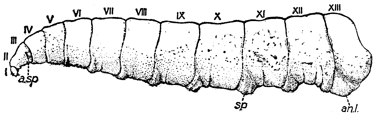
Fig. 17.—Mature larva of M. domestica. a.sp, Anterior
spiracular process; an.l, anal lobe; sp, spiniferous pad. I-XIII,
Body segments. (From Gordon Hewitt.)
Fig. 17.—Mature larva of M. domestica. a.sp, Anterior
spiracular process; an.l, anal lobe; sp, spiniferous pad. I-XIII,
Body segments. (From Gordon Hewitt.)
In a very short time after leaving the pupa-case the adult fly has
stretched her wings, the chitin of her body has hardened, and she flies
away ‘on her several occasions.’
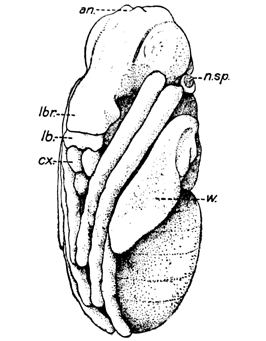
Fig. 18.—‘Nymph’ of
M. domestica dissected out of pupal-case about thirty hours
after pupation. an, Swellings of nymphal sheath marking bases
of antennae; cx, coxa of leg; lb, labial portion of
proboscis sheath; lbr, labral portion of same; n.sp,
spiracular process of nymph; w, wing in nymphal alar sheath.
(From Gordon Hewitt.)
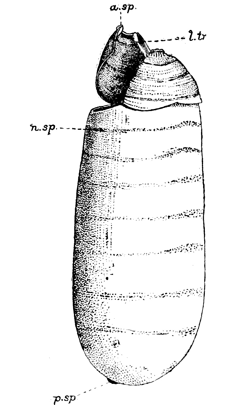
Fig. 19.—Pupal-case or puparium
of M. domestica from which the imago has emerged, thus lifting
off the anterior end or ‘cap’ of the pupa; ventro-lateral aspect.
a.sp, Remains of the anterior spiracular process of larva; l.tr,
remains of the larval lateral tracheal trunk; n.sp, temporary
spiracular process of nymph; p.sp, remains of the posterior
spiracles of larva. (From Gordon Hewitt.)
Flies become sexually mature in a week or ten days after emerging
from the chrysalis-case, and are capable of depositing their eggs four
days after mating, so that if the conditions be indeed favourable the
whole development from the egg to the perfect fly may be accomplished
in nine or ten days, and the second generations are able to lay their
eggs ten days later. The appalling fecundity of such an insect explains
the fact that in the hotter parts of the world nearly every edible
thing seems to be covered with them.
The proboscis of a fly can only suck up
liquid food; and when we see it feeding on solid substances, such as
sugar, it has really dissolved the sugar by depositing some saliva
on it, and is sucking up the sugary solution so produced. It not
infrequently regurgitates its food in a spherical drop, which it
generally re-absorbs.
As we have seen, flies are very susceptible to temperature, and with
the approach of cold weather they seem to die. We used to think that
some, in a state suspended animation, ‘carried on’ through the winter
months. This is, however, ‘non-proven.’ Many of them undoubtedly die in
the autumn, as bees die, of old age. They are literally worn out.
But a great number fall victims to a parasitic fungus called
Empusa. Flies killed by this fungus are frequently to be seen
in autumn, hanging dead on windows, &c., surrounded by a little
whitish powdery ring of spores formed by the fungus.
Flies, like many other insects, are extremely
difficult to keep alive in captivity, and few have
succeeded in rearing them for more than a
month or two. At one time, as we have said,
it was thought that those flies which survive
the winter were fertilised females of the younger
broods, and that during the winter they
subsisted on their ‘fat bodies.’
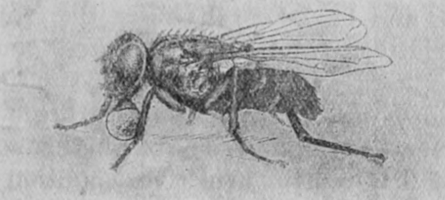
Fig. 20.—M. domestica in the act of regurgitating
food. × 4½. (From Gordon Hewitt.)
Fig. 20.—M. domestica in the act of regurgitating
food. × 4½. (From Gordon Hewitt.)
Doubt has recently been thrown on this
theory, and in a recent report[8] of the Local
Government Board Dr. Newsholme sets forth
the results of the researches of Dr. Monckton
Copeman and Mr. E. E. Austen in the following
words:—
Until recently there was general agreement that
a certain number of flies managed to survive the
winter and spring by hibernating in dark nooks and
crannies in dwelling-houses, or, as contended by
Dr. Laver,[9] in various sheltered situations outside
dwellings—such as the under-surface of the thatch
of farmyard stacks. The researches of Mr. Jepson
and others have shown that, during the period
extending from late autumn to early summer, flies
may be found occasionally in all active conditions
in warmed houses, and especially in such places
as kitchens and bake-houses, where the temperature
is kept relatively high; and further, that under
these conditions, and in presence of sufficient food
material they may even continue to breed. Doubt
has, however, been expressed as to whether a sufficient
number of flies remain in active condition in these
localities to perpetuate the species and to start the
rapidly multiplying generations of the following
summer. As to whether flies can persist through
the winter in other than adult form practically
nothing is known.
In view of the importance of obtaining further
information on these points, some inquiries were
undertaken into the hibernation of flies, the results
of which were set out in a communication by Dr.
Copeman published in the sixth report of this series.
Arrangements were made with a working naturalist
for the collection of any flies that could be found
in situations like those which Dr. Laver and other
observers had found to be favourite winter quarters[67]
of hibernating flies. In view of the need, pointed out
by Howard, for expert identification of the species
of all flies captured in a dormant condition during
the winter months, the co-operation of Mr. Austen of
the British Museum (Natural History) was obtained,
and to him all the flies collected were submitted
for examination. The one specially interesting and
unexpected point emerging from this inquiry was
that not a single specimen of the house-fly (Musca
domestica) was met with among the considerable
number of hibernating flies caught in situations
which have hitherto been regarded as the special
habit of this fly. Under these circumstances it was
felt that further detailed investigation of the matter
was needed; and, accordingly, inquiry on a more
extended scale, and covering—as it proved—an
extensive area, was initiated and carried through
during the past winter.
* * * * *
Once more, the results obtained afford no support
to the belief that house-flies hibernate, in this country,
in the adult state; and the problem as to the manner
in which the interval between one fly-season and the
next is bridged over still remains unsolved.
Gordon Hewitt, Copeman, Howlett, Merriman,[10]
and others, have made experiments
as to how far a fly can travel. Marked flies
have been taken within forty-eight hours at
distances ranging from 300 yards to a mile.
Apparently the direction of the wind plays a
considerable part in the distance they travel.
The importance of the house-fly as a carrier
of disease, especially bacterial disease, has
recently been recognised especially in times
of war. Moses was as great as a Principal
Medical Officer as he was as a Director of
Supplies; and this is shown in Deuteronomy,
chapter xxiii, where he deals with the need
of strict hygiene in the camp.
In the middle of the last century already
attention was being drawn to the fact that the
house-fly and the blow-fly transmitted various
diseases. But it was during the Spanish-American
War and the South African War which
followed shortly afterwards that the part
played by these pests in conveying enteric
became definitely established. Flies coming
straight from the latrines, with their legs and
their wings and their proboscides soiled with
typhoid bacilli, would enter the camp and the
tents of the soldiers and settle on their food-supplies—crawling
over their jam, floating in
their milk. Thirty per cent. of the deaths in
our South African War were due to typhoid
fever. The bacillus, as is well known, is
capable of existing for a long time and of
persisting alive in the alimentary canal of the
insect. Dr. Graham-Smith has shown that the
bacilli may remain active for six days after
feeding, and that the feet of flies which have
the bacillus on them are capable of infecting
surfaces upon which they walk for at least two
days after first coming in contact with the
germs that cause ‘enteric.’
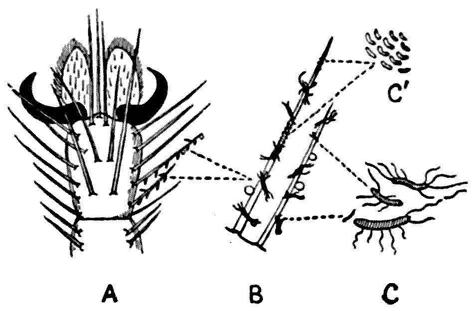
Fig. 21.—A, Foot of a fly, showing hairs
bearing bacteria; B, a single hair more highly
magnified; C and C´, bacteria. Diagrammatic.
Fig. 21.—A, Foot of a fly, showing hairs
bearing bacteria; B, a single hair more highly
magnified; C and C´, bacteria. Diagrammatic.
Faichne reared maggots in dejecta infected
with typhoid bacilli, and he was able to show
that the flies into which these maggots turned
contained virulent typhoid germs in their
intestines. There is absolutely no doubt that
typhoid is largely conveyed by the agency of
these insects; and as flies are perfectly controllable,
if ‘the people will but have it so,’ it is
one of the disgraces of our civilisation that this
disease should be so prevalent.
The protective inoculation against enteric is
now almost perfect, and its value is shown by
quotations from a leaflet issued by the Research
Defence Society:—
Sir William Leishman, in a letter published during
the present war, August 22, 1914, says: ‘The
benefits of inoculation are so well recognised in the
regular forces that we find little difficulty, in foreign
stations, in securing volunteers for inoculation: for
instance, about 93 per cent. of the British garrison
of India have been protected by inoculation; and
typhoid fever, which used to cost us from 300 to
600 deaths annually, was last year responsible for
less than 20 deaths. Inoculation was made compulsory
in the American army in 1911, and has
practically abolished the disease; in 1913 there
were only 3 cases, and no deaths in the entire army
of over 90,000 men.’
In Avignon, in the south of France, during the
summer of 1912, typhoid fever broke out in the
barracks. Of 2053 men, 1366 were protected and
687 were not. The non-protected had 155 cases of
typhoid, of whom 21 died; the protected had not
one case. In the winter of 1913 the French Senate
resolved that the protective treatment should be
made compulsory throughout the French army; and,
in special circumstances, among the reservists.
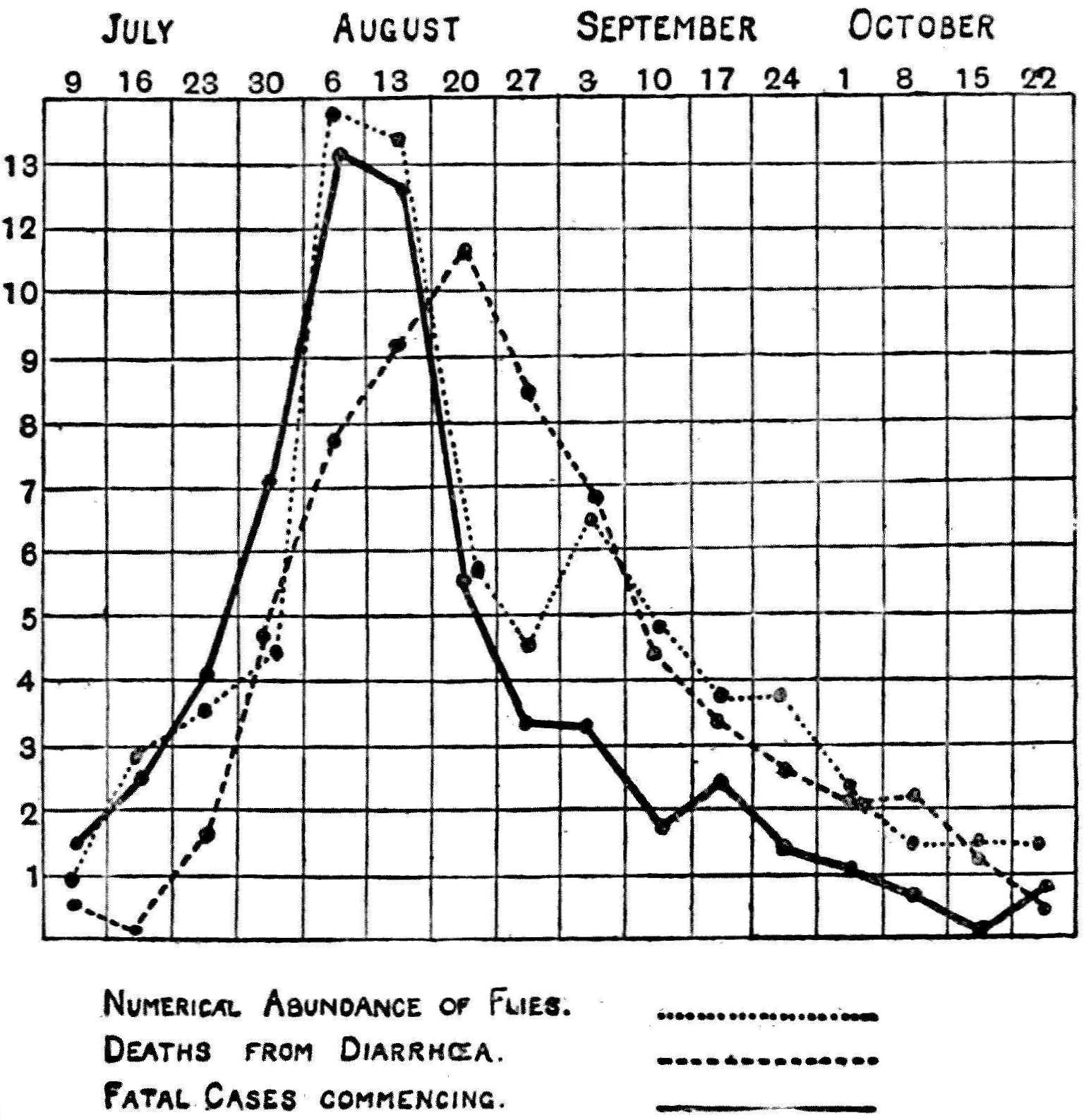
Fig. 22.—Chart illustrating the relation of the numerical abundance
of house-flies to summer diarrhoea in the city of Manchester in 1904.
Prepared from statistics and chart given by Niven. (From Gordon
Hewitt.)
Fig. 22.—Chart illustrating the relation of the numerical abundance
of house-flies to summer diarrhoea in the city of Manchester in 1904.
Prepared from statistics and chart given by Niven. (From Gordon
Hewitt.)
Infantile diarrhoea, which so afflicts the
crowded, poorer quarters of our cities in the
summer, is another disease intimately associated
with Musca domestica. But that is hardly
a disease likely to trouble the soldiers. The
tubercle bacillus is another germ conveyed by
flies. House-flies are particularly fond of feeding
on saliva; and Hayward, Lord, and Graham-Smith
have obtained virulent bacilli from the
intestines and dejecta of flies which had been
fed on tubes containing tuberculous sputum.
These experiments have been amply confirmed
by other workers. Anyone who has ever been
in Egypt will remember the terrible sight of
the flies attacking little children suffering from
ophthalmia and it is believed that the wide
prevalence of this most pitiful trouble is
attributable to the abundance of flies—the
flies of Egypt, a plague even in the times of the
Pharaohs. Things do not alter much in Egypt,
and the Biblical plagues are wont to recur.
Another disease—anthrax, or wool-sorter’s
disease—may be conveyed by the same carriers
from infected cattle to man, and there is a
good deal of epidemiological and bacteriological
evidence available to show that flies play an
important part in the spread of cholera,
which is now threatening the soldiers in the
eastern seat of the war, and possibly in disseminating
the organisms which cause yaws
and tropical sore.
It will be noticed that the fly is not a
necessary second host for any of these germs.
They are conveyed, as if by an inoculating
needle, by contact with the proboscis or the legs
or some other tainted organ of the fly. The
bacilli, however, pass through the alimentary
canal apparently unchanged and unharmed,
and are deposited either with the regurgitated
food from the fly’s stomach (Fig. 20), or with the
dejecta of the insect. There is no subcutaneous
inoculation—such as takes place in the case of
the mosquito when it conveys malaria, or in
the case of the tsetse-fly when it conveys
sleeping sickness—where the disease-causing
organism is injected into the human body.
The action of the fly is mechanical, but none
the less efficient. The poisoning of the soldiers’
food-supply is its chief rôle in war.
CHAPTER VI
FLIES
Part II
THE BLUE-BOTTLE (Calliphora erythrocephala),
AND OTHERS
Who fills our butchers’ shops with large blue flies?
(Rejected Addresses.)
But there are other flies: first amongst which
may be mentioned Fannia canicularis and F.
scalaris. These belong to the family known
as Anthomyidae, and are distinguished from
the house-fly by being smaller in size, and by
many other small details in the imago stage
hardly to be appreciated except by trained
dipterologists. For a short time at the beginning
of the summer, during part of May
and June, specimens of F. canicularis are more
abundant than M. domestica, and, when seen
on the window-panes of our living-rooms, are
apt to be thought, by the uninformed, to be
young specimens of the latter. But, as has
been said, flies, when they are once flies,[75]
do not grow; all the growing they do is done
in the larval stage.
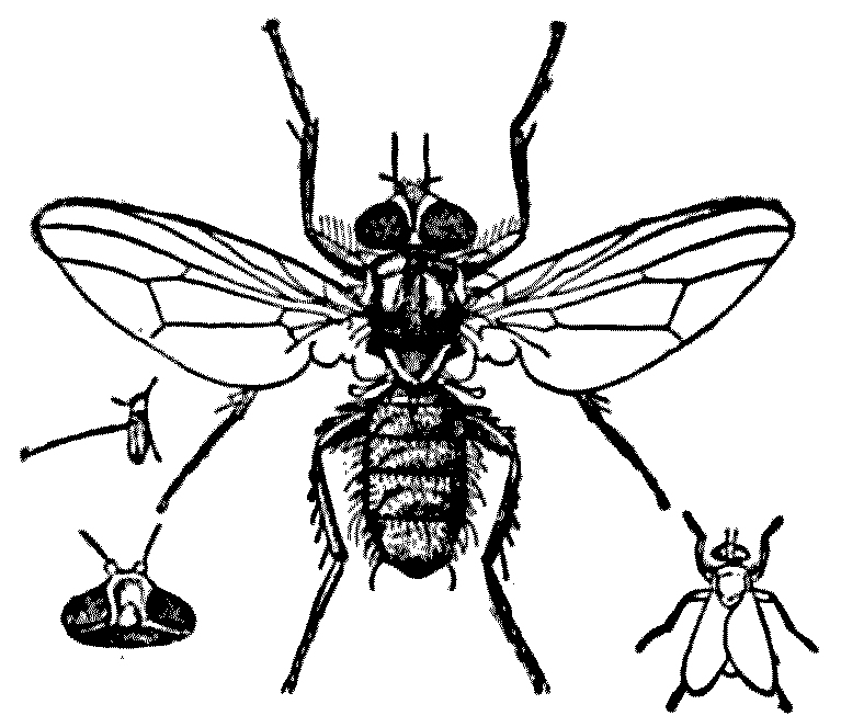
Fig. 23.—Latrine-fly, Fannia
scalaris, male (× 3). Antenna. Head of female, dorsal view. Natural
size, resting position. (From Graham-Smith.)
Fig. 23.—Latrine-fly, Fannia
scalaris, male (× 3). Antenna. Head of female, dorsal view. Natural
size, resting position. (From Graham-Smith.)
As the days lengthen the common house-fly becomes vastly more common
than F. canicularis, the ‘lesser house-fly,’ and the latter now
tend to aggregate in those rooms of our houses not devoted to cooking,
and may frequently be noticed flying in a jerky and disconcerting
manner around the chandeliers or bedposts in unfrequented living- or
bed-rooms. The relative proportion of these two genera in full summer
varies in different localities. Roughly speaking, out of 100 flies
collected in a house there is something between 90 and 99 per cent. of
M. domestica, but the numbers not only vary with locality, but
with temperature.
On the other hand, there is a curious disproportion between the
number of sexes found ‘at home’ in the lesser house-fly. For every 100
F. canicularis taken indoors seventy to seventy-five are males,
the numbers being evened by an equal preponderance of females
who have remained out of doors.
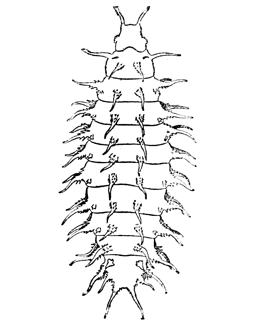
Fig. 24.—Larva of F. canicularis. (From
Gordon Hewitt’s Report to Local Government Board, 1912.) Magnified.
Fig. 24.—Larva of F. canicularis. (From
Gordon Hewitt’s Report to Local Government Board, 1912.) Magnified.
The larva of Fannia is a flattened-looking grub with distinct
segments, decorated by numerous feathery processes. It lives amongst
decaying vegetation and fruit, and also amongst fermenting animal
matter and dejecta. Sometimes it is found in rotting grass. As we shall
see later, it frequently passes into the human alimentary canal. F.
scalaris, usually known as the ‘latrine-fly,’ is even commoner than
its congener, and the external structural differences are minute. As
its name indicates, it is found as a rule breeding in human dejecta,
and is, therefore, as a typhoid carrier, much more dangerous than F.
canicularis. Its larva is also more commonly found in the human
intestine.
Then there are two species of large flies known as blue-bottles or
blow-flies—Calliphora erythrocephala and C. vomitoria.
The former of these is the more common. The sides of its face are
golden yellow, set with black hair; whereas in C. vomitoria
the sides of the face
are black, but the hair is golden. Both are handsome, sturdy-looking
diptera, with bluish-black thoraces, and abdomens of a dark metallic
gun-metal sort of colour.
Blow-flies deposit their eggs on fresh or decaying flesh, and this
is one of the great sources of trouble to the officers of the Army
Service Corps. But they are not content with killed flesh. They will
lay their eggs on any living flesh which is exposed, or in sores or
tumours, and here their larvae will thrive. Dr. Graham-Smith tells us
he once found the exposed muscles of the broken leg of a living rabbit
seething with a mass of small blow-fly larvae, which were nourishing
themselves upon the living tissues.
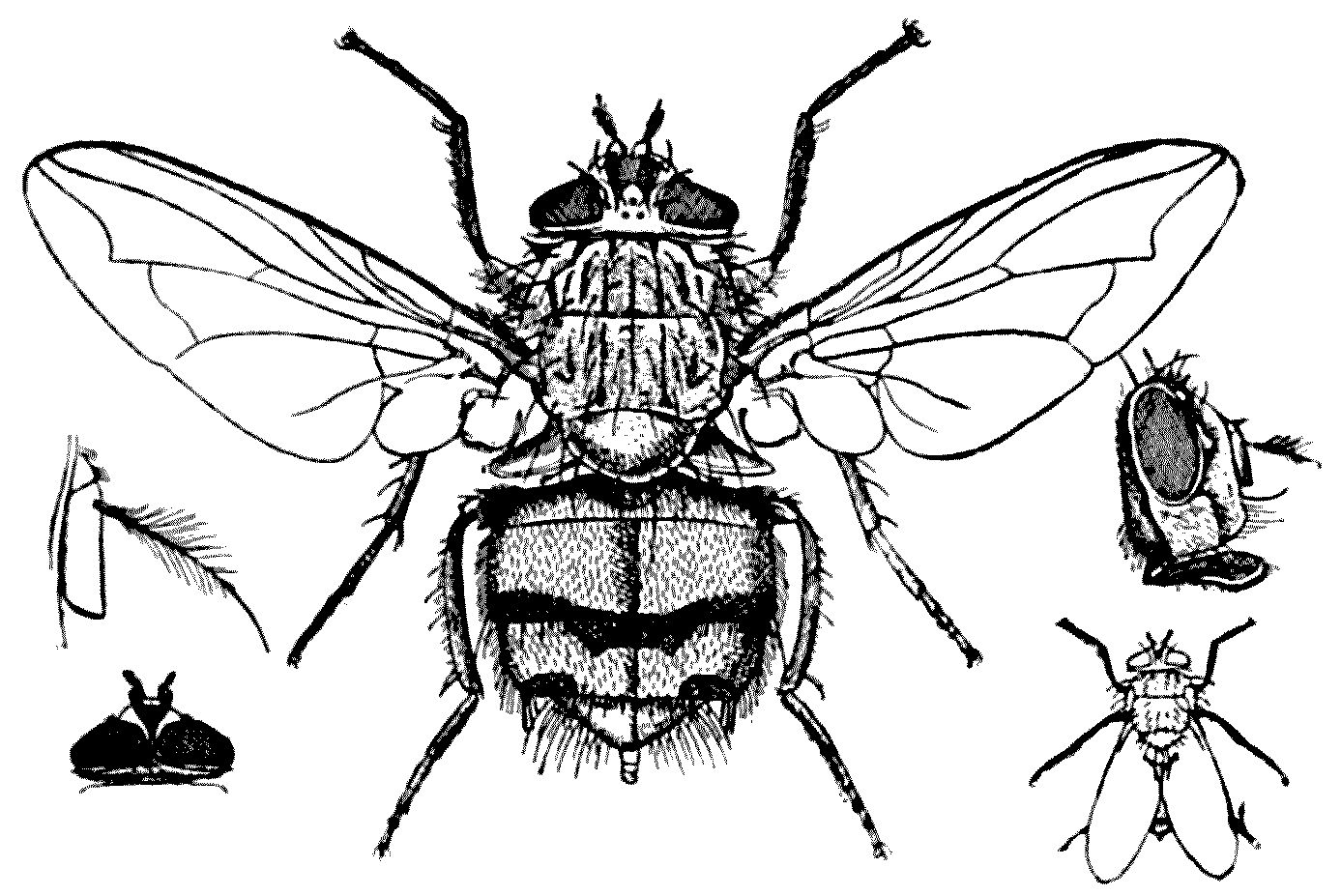
Fig. 25.—Blow-fly or blue-bottle, Calliphora erythrocephala, female
(× 3). Antenna. Male head, dorsal view. Side view of head.
Natural size, resting position. (From Graham-Smith.)
Fig. 25.—Blow-fly or blue-bottle, Calliphora erythrocephala, female
(× 3). Antenna. Male head, dorsal view. Side view of head.
Natural size, resting position. (From Graham-Smith.)
The eggs of the blow-fly hatch out in from
ten to twenty hours in normal British
temperatures; the larval life, in its three
stages, lasts from seven to eight and a half
days; the pupa state lasts a fortnight, so that
the total development extends a day or two
over three weeks. The maggots are unusually
voracious; and Linnaeus used to say that the
progeny of three blow-flies will dispose of a
dead horse as quickly as three lions.
C. erythrocephala is essentially an outdoor
fly and enters houses only in search of a nidus
on which to deposit its eggs. C. vomitoria
resembles its congener in size and habits, but
it is not so abundant. Occasionally its eggs
have been known to be deposited in the nostrils
of animals and men.
But there are:—
All species of resplendent flies,
Some with green bodies and green eyes,
Pricking like pins’ heads from their holes
Like tiny incandescent coals.
(Anon.)
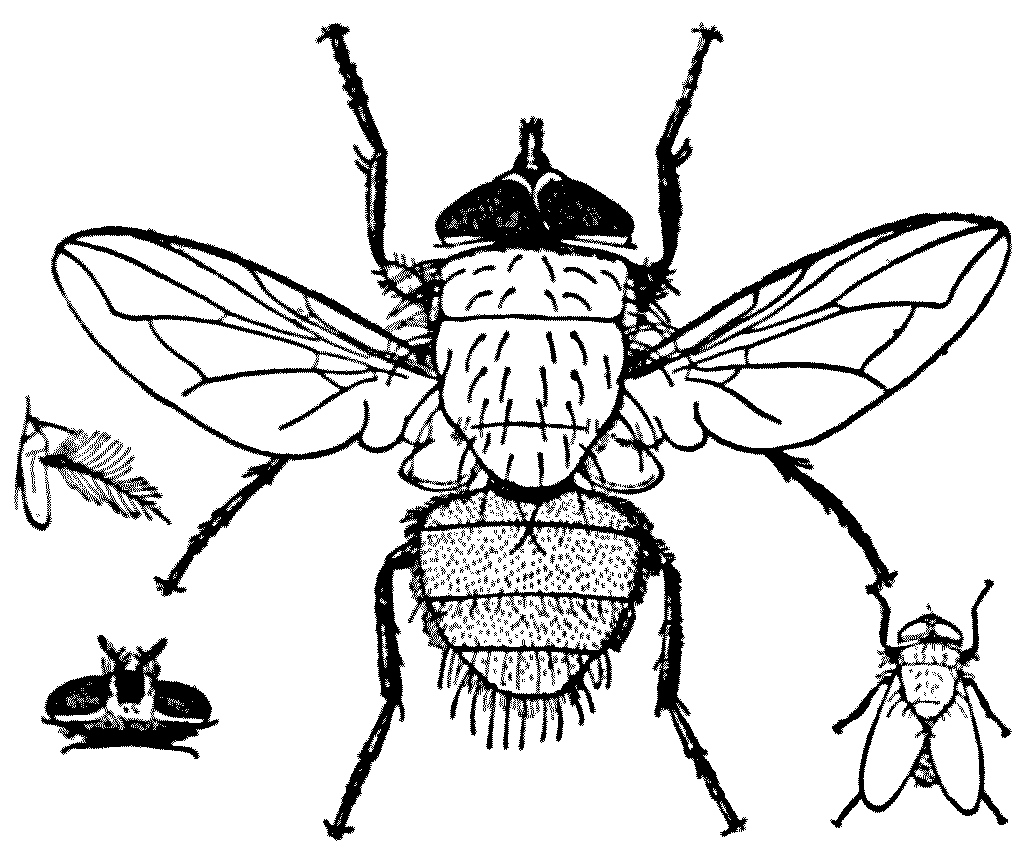
Fig. 26.—Green-bottle, Lucilia caesar,
male (× 3). Antenna. Female head, dorsal view. Natural size, resting position.
(From Graham-Smith.)
Fig. 26.—Green-bottle, Lucilia caesar,
male (× 3). Antenna. Female head, dorsal view. Natural size, resting position.
(From Graham-Smith.)
One of these, Lucilia caesar, is a marked
nuisance to those responsible for victualling
a camp. This green-bottle fly, like the
Calliphora and the house-fly, belongs to the
family Muscidae, and its larvae are said to be
indistinguishable from those of blue-bottles.
Some species of Lucilia deposit their eggs
in great quantities amongst the wool of sheep
when the sheep are ill-kept, and they do
much damage. But as far as war is concerned
the harm that Lucilia does is laying
its eggs upon dead animals. It does this
on all sorts of meat-stores; but in times of
peace it especially infests stale fish, which
the issuing larva very soon eat clean to the
bone. When feeding upon a dead fish lying
upon a beach they burrow down in the
sand below their food. They descend some
two to six inches, and for the most part remain
deep in the sand during the daytime, coming
up to feed at night. They also have a habit
of migrating from one fish to another. This
fly has also been known to lay its eggs in
the neglected wounds of human beings.
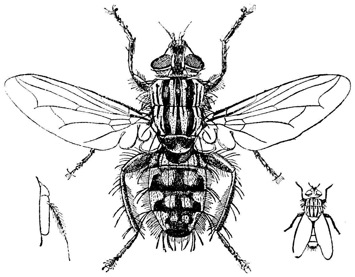
Fig. 27.—Flesh-fly (Sarcophaga carnaria), female (× 3).
Antenna. Natural size, resting position. (From Graham-Smith.)
Fig. 27.—Flesh-fly (Sarcophaga carnaria), female (× 3).
Antenna. Natural size, resting position. (From Graham-Smith.)
Sarcophaga carnaria is another species which
occasionally infests human sores, and which
enters houses in search of filth or carrion
on which to lay its eggs; it is viviparous
and produces not eggs but live larvae. One
female can give birth to 20,000 young; and
Redi states that the larvae of these flesh-flies
will in twenty-four hours devour so
much food and grow so quickly that they
increase their weight two-hundredfold.
Finally, there is a group of flies whose
larvae penetrate under the skin of human
beings and give rise to definite subcutaneous
troubles. But, fortunately, these are, with few
exceptions, confined to the warmer regions
of the earth, and there is very little risk of
their causing real trouble in Northern or
Central Europe.
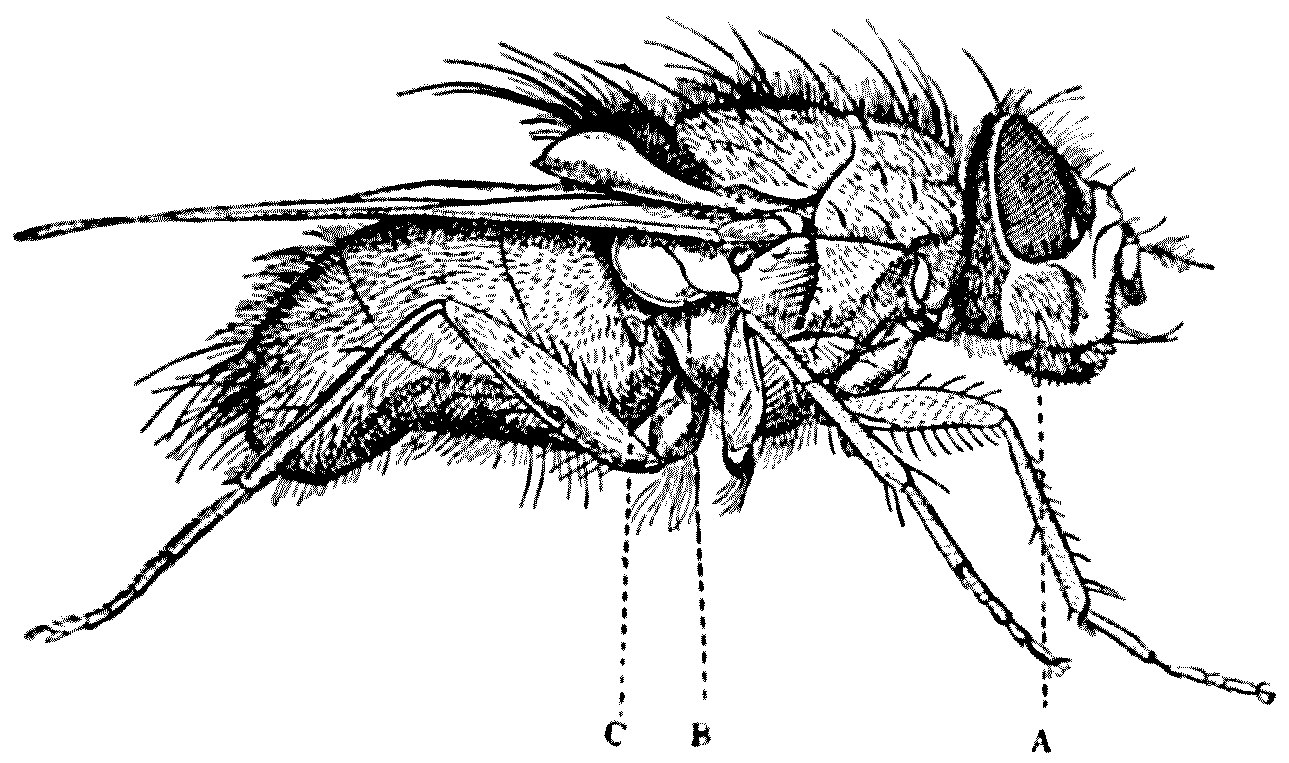
Fig. 28.—Side view of blow-fly (Calliphora erythrocephala)
(× 5). A, Cheek (jowl); B, squama;
C, halter. (From Graham-Smith.)
Fig. 28.—Side view of blow-fly (Calliphora erythrocephala)
(× 5). A, Cheek (jowl); B, squama;
C, halter. (From Graham-Smith.)
The troubles or diseases caused by the
presence of fly larvae in the body are grouped
in medical language under the term ‘myiasis,’
which Graham-Smith defines as follows:—
‘The term myiasis signifies the presence
of dipterous larvae in the living body (whether
of man or animals), as well as the disorders
(whether accompanied or not by the destruction
of tissue) caused thereby. Though not strictly
coming within this definition, the sucking of
blood by larvae through punctures of the
skin, which they themselves produce, may
be included for the sake of convenience in
classification.
Myiasis in man may be produced by
dipterous larvae:—
(a) Sucking blood through punctures in
the skin (Auchmeromyia luteola).
(b) Deposited in natural cavities of the
body (Chrysomyia, Lucilia, Sarcophaga,
Calliphora, Oestrus).
(c) Deposited in neglected wounds (Chrysomyia,
Lucilia, Sarcophaga, Calliphora).
(d) Living in subcutaneous tissue (Cordylobia,
Dermatobia, Bengalia (?), Hypoderma).
(e) Passing through the alimentary canal
(Fannia, Musca, Eristalis, Syrphus,
Gastrophilus).
In the above list, only the more common genera producing myiasis
are mentioned. In England, Type (e) is fairly common, and Types
(b) and (c) are occasionally observed.’
We may now consider in detail, but very
shortly, the categories set forth by Dr. Graham-Smith:—
(a) The very peculiar blood-sucking maggot
known as the Congo-floor-maggot—the larva
of Auchmeromyia luteola—fortunately does not
spread beyond tropical and sub-tropical Africa.
It chiefly affects the natives who sleep on
mats.
(b) The flies which deposit their ova and
larvae in the cavities of the body are again
mostly foreign. The worst of all is the screw-worm
(Chrysomyia macellaria) of the Southern
States, Central and South America. Although
it extends to Canada it is not troublesome
north of Texas.
Occasionally, blow-flies in Great Britain
deposit their ova in the human nose or ear.
They very rapidly hatch and cause great
inflammation and necrosis until they can be
discharged or removed. They have even been
found in the anterior chamber of the eye;
and I have some microscopic sections showing
the presence of these larvae in that chamber,
whither they had probably proceeded from
the nasal sinuses. But on the whole, cases
of this sort are comparatively rare, and cause
but little trouble.
(c) The real difficulty, and one which late
last summer proved a serious trouble to our army
in the field, are the cases in which maggots
were found in neglected wounds. Here, however,
we may take some comfort in the fact
that the trouble is fortunately much greater
in the tropical and sub-tropical regions than
in more temperate climates, and diminishes
as the cold weather draws on. Still, during
the hot weeks of last August there were cases
of wounded soldiers left lying on the fields for
two or three days who were found to be suffering
in this way. One almost hesitates to offer
suggestions to our heroes in such cruel conditions;
but whenever and wherever it can be
done wounds should as far as possible be kept
covered.
Not only are neglected wounds affected,
but tumours and ulcers are often attacked.
But, as I have said, the danger is much greater
in warmer climates. We know that Herod
Agrippa ‘was eaten of worms, and gave up
the ghost’: a fact which recalls the translation
given by an undergraduate in difficulties
with the Acts of the Apostles in the ‘Little-Go’
who rendered ‘καì γενóμενος σκωληκóβρωτος ἐξέψυξεν’
‘He became a Skolekobrote, and died
in the enjoyment of that office.’
(d) Flies burrowing in the subcutaneous
tissues are again very much commoner in
tropical climates than in Northern Europe,
and the cases quoted in our country are
comparatively rare.
(e) The presence of larvae in the alimentary
canal of man is by no means uncommon.
Both the larvae of Musca and
Fannia are not infrequently found; and over
a thousand of the latter have been passed
by a highly infected individual at one time.
They probably make their way into the body
with over-ripe fruit. In some cases they
give rise to no symptoms, but in others violent
pains are felt and a certain dizziness, and the
digestive functions are interfered with. The
presence of these larvae in the urinary passages
is even more difficult to explain, but they
undoubtedly are at times found in these
channels.
A few years ago an elaborate investigation
was carried on by Mr. W. Nicol, for the
Local Government Board, on the part played
by flies in the dispersal of the eggs of parasitic
worms. He showed quite definitely that the
ova of certain human parasites are taken
into the fly and pass through its body undigested.
Should these be deposited on the
food of man, there is great risk of his becoming
infected. As I have said before, flies take
only liquid food, and it is only when the
ova of the parasites are very small that they
can pass into their alimentary canals. Some
eggs are too large for the fly to swallow. Eggs
of parasitic worms have also been shown
to be carried on the legs and proboscides
of flies, and these are deposited on the spot
where the fly next cleans itself. Probably,
however, in the end little harm is really done
by flies in disseminating parasitic worms,
but it is a possibility which must not be
altogether disregarded.
The remedial measures for the control of
flies are fully dealt with in Graham-Smith’s
admirable book, ‘Flies in Relation to Disease,’
from which I have ventured to borrow many
figures; and again by Dr. Gordon Hewitt,
in his work on ‘House-flies,’ which has had
such a wonderful success in stimulating our
North-American cousins to decrease the numbers
of one of the gravest enemies to mankind.
It has been shown over and over again
that we can control the mosquito: the building
of the Panama Canal alone proves this. We
could equally control the ‘Infinite Torment
of Flies.’ The Canadians and Americans are
doing their best; but are we? The knowledgeable
world has at least discovered the reason
why Beelzebub was called the ‘Lord of flies.’
CHAPTER VII
MITES
Part I
THE HARVEST-MITE (Trombidium)
Natura in minimis maxime miranda.
(Linnaeus.)
We do not know what life is, but we can at
any rate record its manifestations; and we
know that it is always associated with
an extremely complex substance called by
Purkinje ‘protoplasm.’ This substance
Huxley described as ‘the physical basis of
life.’ Protoplasm, though we know of what
elements it is composed, defies accurate
analysis, and, indeed, is never the same
for two minutes together. It is constantly
changing, it is in a state of flux and is, in effect,
a stream into which matter is continuously
entering and continuously leaving.
Protoplasm may be living, or it may be
dead; and when dead it soon undergoes dissolution;
but there is no life without protoplasm.
Somewhere or other Dr. David Sharp has
stated that of the total amount of protoplasm
‘in being’ in the world, the active volume of
the life-material of our globe, at least one-half
is wrapped up in the body of insects. But
insects only form one sub-group out of the
several which make up the great group Arthropoda,
or those animals which are distinguished
from others by possessing externally jointed
legs—that is, jointed appendages. This group
includes also the Crustacea, the multi-segmented
Centipedes, and the Arachnids or
spider-like animals.
Insects, like aeroplanes, dominate the air;
Crustacea, like submarines, inhabit the water;
the poet has passionately asked:—
Ah! who has seen the mailèd lobster rise,
Clap her broad wings and soaring claim the skies?
But the answer, in the language of those
curious mammals the politicians, is ‘in the
negative.’ Crustaceans are essentially aquatic.
On the other hand, centipedes and spiders are
earth-loving animals but some have unhappily
developed parasitic or pseudo-parasitic habits.
The last-named sub-group, the Arachnids,
comprise many subdivisions. There are the
spiders, the harvest-men, the scorpions, the
king-crabs, and so on. But one of the most
numerous of the subdivisions of the group are
the mites and ticks (Acarina). I have for
years been trying to find some organ or
structure shared by insects and mites and
ticks, and not found in any other group of
arthropods. If I could do this I would invent
a long polysyllabic
word—with lots of
Greek in it—which
would really be a
short way of designating
those
arthropods which
convey disease to
man.
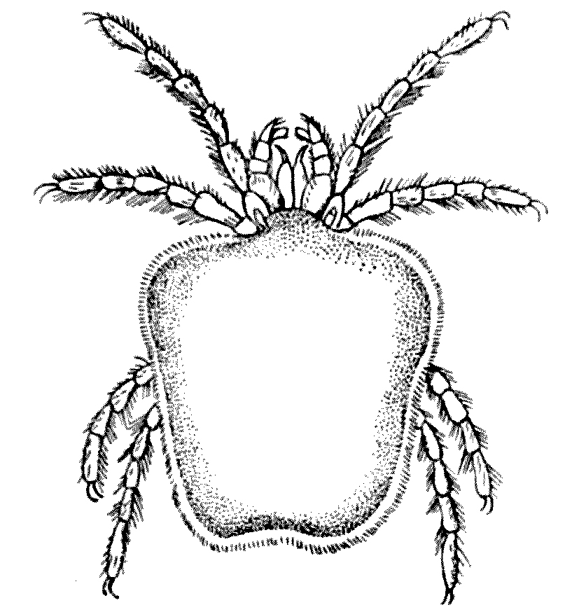
Fig. 29—Trombidium
holosericeum. Female, dorsal view. × 20. (After Railliet.)
Fig. 29—Trombidium
holosericeum. Female, dorsal view. × 20. (After Railliet.)
The acarines are for the most part small, and they differ from
spiders in having no waist. In fact, the three divisions into which the
body of an arthropod is normally divided—head, thorax, and abdomen—are
indistinguishable in mites, the body forming an unconstricted whole. As
a rule, these little creatures breathe, as do insects, by tracheae, or,
if these be absent, by the general surface of the body. They live for
the most part on vegetable and animal juices,
and their mouth-parts are, as a rule, piercing and suctorial; but in
some species the appendages of the mouth are capable of biting as well
as piercing. The adults have typically eight legs. The larval stages
are very numerous, and at times six distinct moults of the skin are
recognisable. With few exceptions the larva emerges from the egg as a
six-legged creature. In fact mites undergo a metamorphosis which varies
in complexity and in completeness in different groups, and it is often
one of the larval stages which causes the greatest trouble to man.
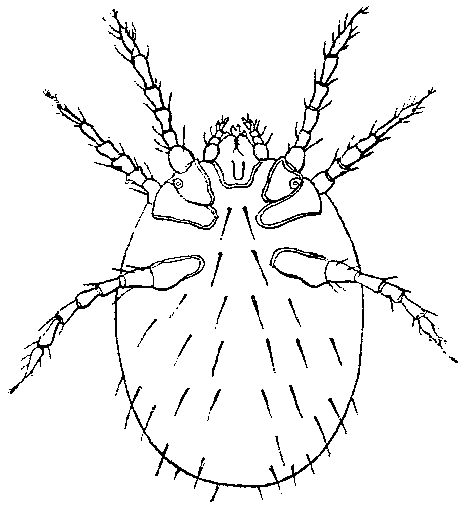
Fig. 30.—Leptus autumnalis = larva of
Trombidium holosericeum. Ventral view. × 100. (After Railliet.)
Fig. 30.—Leptus autumnalis = larva of
Trombidium holosericeum. Ventral view. × 100. (After Railliet.)
One of these six-legged larvae has been
long known as the harvest-mite, under the name
of Leptus autumnalis. But this is not a real
species, and there is still considerable confusion
as to what the exact status of Leptus autumnalis,
the harvest-mite, is. Probably the larvae of
several species are involved, but it seems
pretty certain that in many cases the larvae
will grow up into specimens of the genus Trombidium
holosericeum, though a certain and at
present unknown percentage of the larvae will
grow up into Trombidium something-or-other-else.
They are minute bright-scarlet little creatures—the
Cardinals of the Mite world—of
a beautiful satiny red, decorated here and
there with blackish spots. The body of the
adult is somewhat square, tapering slightly
to the hinder end. Both legs and body are
covered with red hairs. The eyes are borne
on little stalks—like lighthouses. The legs
have six joints and end in two little claws.
The male is usually smaller and more feeble
than the female, the latter reaching a length
of 3 mm. to 4 mm. The adults are commonly
met with in the spring or commencing summer.
Apparently, they nourish themselves on
vegetable sap. The larval form of this species[11]
is undoubtedly one of the forms confused under
the now discarded name of Leptus autumnalis.
When starving, the body is orbicular in outline,
but it becomes oblong when it is fed, and
in this case it may attain a length of ½ mm.
Its colour is of a deep orange.
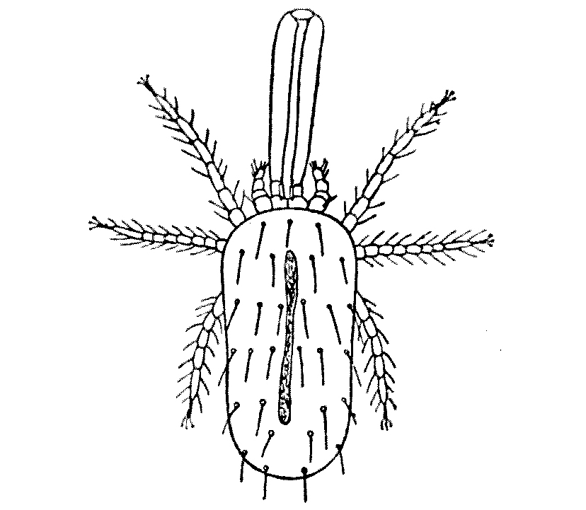
Fig. 31.—Leptus autumnalis,
with the so-called proboscis. Magnified. (After Gudden.)
Fig. 31.—Leptus autumnalis,
with the so-called proboscis. Magnified. (After Gudden.)
This harvest mite, or, as it is called in
France le rouget, is most troublesome at the
end of summer or at the beginning of autumn,
when it is found in enormous
numbers in grass
and amongst many other
plants—gooseberries,
raspberries, currants,
haricot-beans, sorrel, and
elderberries. From these
plants it passes on to any
warm-blooded animals:
particularly it attacks
small mammals. Hares,
rabbits, and moles are
often covered with them,
but they leave their
victim, should it be shot, as soon as the body
chills. They are particularly common in
Great Britain and in the centre and west of
France, and in certain parts of Germany.
These irritating little semi-parasites may be
dislodged by the application of petrol or benzine—both
very inflammable—and the itching
they cause allayed by the application of acid
or alcoholic lotions.
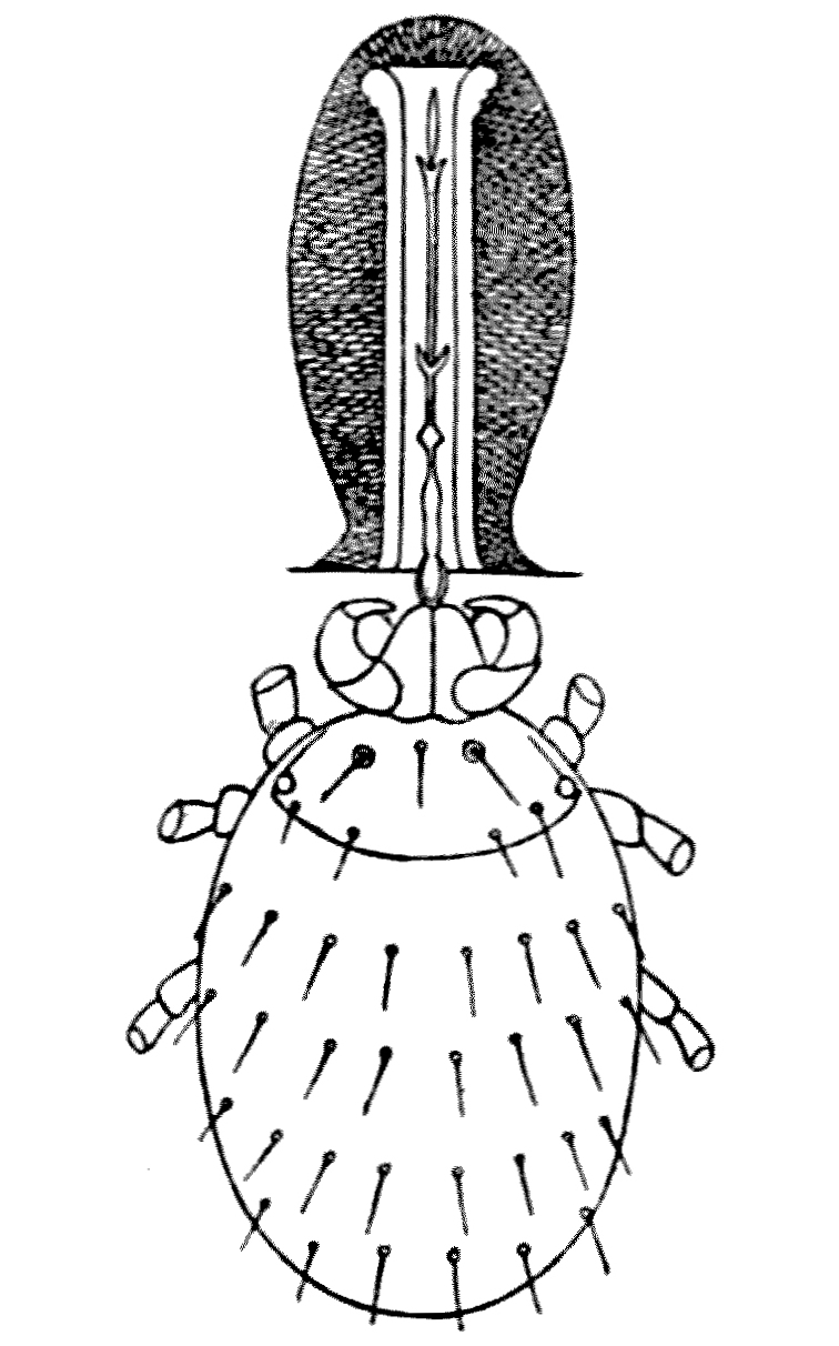
Fig. 32.—Leptus autumnalis
(× 100). The so-called proboscis is formed around the
hypo-pharynx sunk into the skin. (After Trouessart).
Fig. 32.—Leptus autumnalis
(× 100). The so-called proboscis is formed around the
hypo-pharynx sunk into the skin. (After Trouessart).
Men working in the fields are frequently attacked. During September
1914, the soldiers of the Sixth Division, stationed in and about
Cambridge, and living in tents, suffered severely from their ‘bites.’
They mostly attacked the ankles, the wrists, and the neck, but they
rapidly extend over the body. If they be checked by the presence of
any stricture, such as a garter or wrist-band, they accumulate behind
it, and the irritation is accentuated. The presence of their hypo-pharynx
in the skin causes the surrounding tissues to harden and form a
cylindrical tube—the so-called proboscis.
The amount of trouble they cause varies very greatly in different
people. Children and women with soft skins suffer, as a rule,
most; but, as happens in the case of other biting insects, certain
individuals seem to be almost
immune, whilst others suffer very considerably.
The trouble is caused by the mite implanting its mouth-parts in the
skin—preferably in the hair-follicles or the sweat-glands. When it is
once fixed it rarely moves. The body remains, of course, on the surface
of the skin as a little reddish-orange point, scarcely perceptible
unless many of them are congregated in the same position. The effect of
their presence is to produce a swelling in the skin, which may be as
large as a split pea, accompanied by an intense itching and a smarting
which banishes sleep. This leads to the patient scratching, and this
scratching is the departure-point of many troubles. Scoriated papules
appear and eczematous patches, and when the mites are very numerous
an erythema, named by Rubies Erythema autumnale, supervenes.
The skin near the point of puncture swells, becomes red, sometimes
almost purple, and irregular patches, which when confluent, appear a
centimetre in diameter.
These skin troubles, which may end in a kind of generalised
eruption, are accompanied by a rise of temperature and a
certain—sometimes a high—degree of fever. Besides men, dogs and cats
suffer from these pests; and in these domestic pets the parasites give
rise to a miliary eruption. Domestic cattle—sheep and horses—are also
attacked. And, according to some authorities, poultry
are not only attacked but killed by these
parasites. The larvae apparently only lives a few days in the skin of
the victim.
As far as is known at present the larvae
of Trombidium convey no protozoal disease;
but there is a terrifying little creature, known
as the Kedana mite, which in some districts
of Japan causes a serious illness, with a mortality
of some 70 per cent. Apparently, it
does not act as an inoculating agent itself,
but the papule, surrounded by the red area
which forms as a result of its bite, changes to a
pustule, and this lesion becomes the point of entrance
of bacteria which produce the so-called
‘river’ or ‘flood’ fever. If these mites be
carefully removed the patient suffers no harm.
Another species of mite, Pediculoides ventricosus,
lives in stalks of cereals, and is very
apt to attack labourers who are dealing with
grain. Their bites cause severe irritation,
local swellings, reddening of the epidermis,
and fever. In this particular species the
female before she is fertilised has an elongated
form 0·2 mm. in length and 0·07 mm. in
breadth; but when fertilised the ovaries increase
to such an extent that the posterior
end of the body becomes spherical. In this
respect it resembles that remarkable flea,
the chigo or jigger. The larvae are exceptional
in being born with four legs instead of the
usual three, and they pair almost immediately
after emerging from the egg-shell.
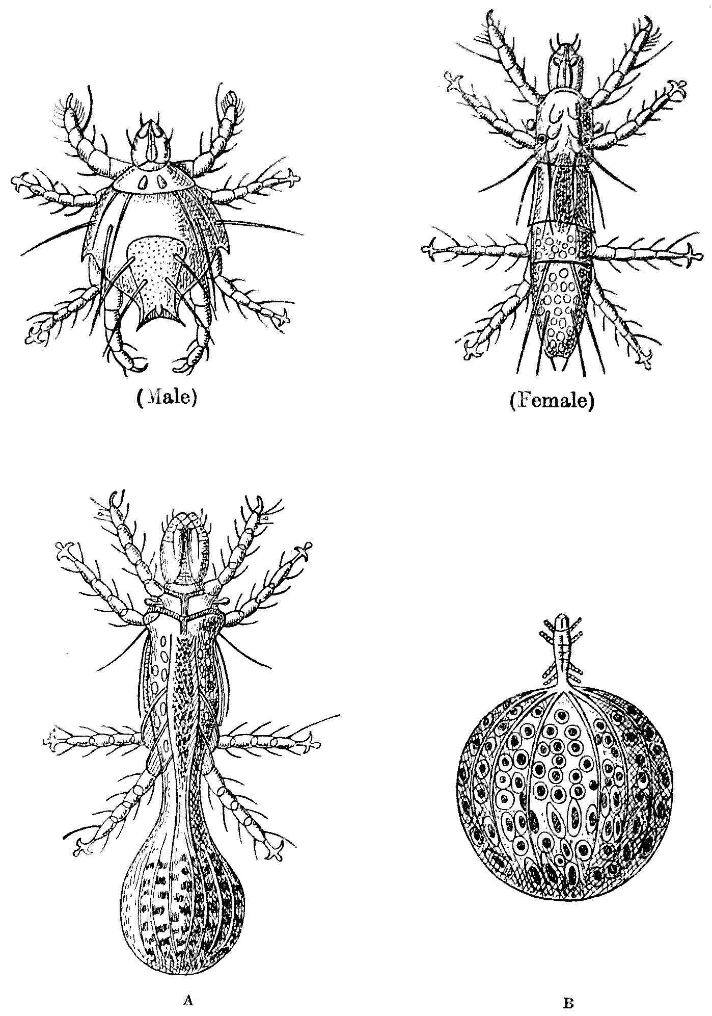
Fig. 33.—Pediculoides ventricosus. Male, ventral view (× 250).
Female, before fertilisation (× 225). A, after fertilisation; the
abdomen has begun to swell (× 250). B, with abdomen fully swollen
(× 40). (After Laboulbène and Mégnin.)
Fig. 33.—Pediculoides ventricosus. Male, ventral view (× 250).
Female, before fertilisation (× 225). A, after fertilisation; the
abdomen has begun to swell (× 250). B, with abdomen fully swollen
(× 40). (After Laboulbène and Mégnin.)
CHAPTER VIII
MITES
Part II
ENDO-PARASITIC MITES (Demodex, Sarcoptes)
Say what the use, were finer optics giv’n,
T’ inspect a mite, not comprehend the heav’n.
(Pope, Essay on Man.)
Demodex
We have seen that harvest mites are wont to
insert their heads—or rather their mouth-parts—into
the skin of human beings, but other
mites show less restraint, and insert their
whole bodies. One of these, the well-known
Demodex folliculorum, is, according to Guiart
and Grimbert, ‘Le plus commun des parasites
de l’homme et nous en sommes presque tous
porteurs.’ Without taking quite so gloomy
a view, Demodex is undoubtedly widely distributed
in the skin of mankind and of other
mammals.
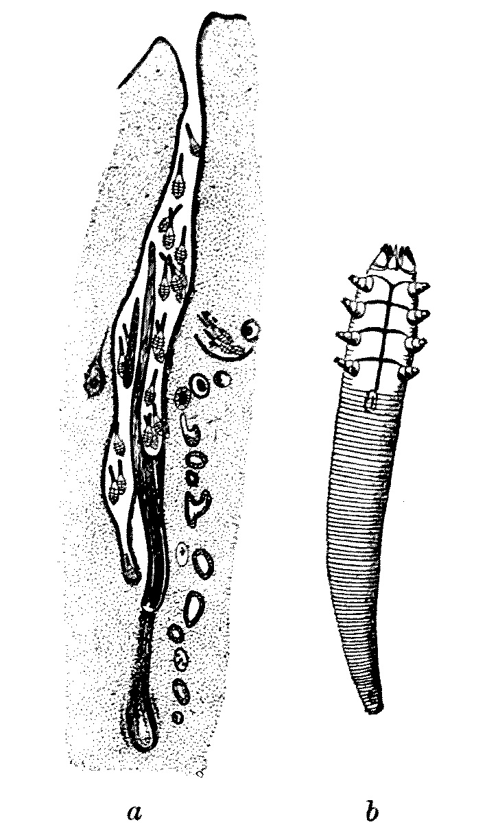
Fig. 34.—(a) Demodex in hair-follicle
of dog; magnified. (After Neumaun.) (b) Demodex folliculorum;
highly magnified. (After Railliet.)
Fig. 34.—(a) Demodex in hair-follicle
of dog; magnified. (After Neumaun.) (b) Demodex folliculorum;
highly magnified. (After Railliet.)
There are differences of opinion as to whether this form should be
split up into numerous species, or subspecies, according to the genus
of the mammals upon which it lives. We, at any rate, will confine
our attention to the human kind and so avoid losing ourselves in the
tortuous maze of synonymy and the arid discussion of a meticulous
classification so dear to the analytical German mind. To us a
Demodex shall be a Demodex, and we will leave it at
that.
Unlike the majority of mites, Demodex is a good deal longer
than it is broad. But even for a mite it is very small, and shows signs
of bodily degradation associated with its parasitic habit of life. Its
shape is adapted to its habitat, which is the sebaceous glands of the
skin. The long abdomen appears to be segmented, but the annulations
are not true segments. The legs are reduced
to conical stumps. The male is 300 µ[12] long
and 40 µ broad across the cephalothorax. The female is, as usual,
larger, measuring 380 µ in length by 45 µ in breadth. The minute larvae
have, as is so often the case with mites, but three pairs of legs, and
are 60 µ to 100 µ in length.
This parasite, which lives on all parts of the skin of the human
body, is perhaps most commonly seen on the nose and in the passages
leading into the ear. It can be expressed by firmly pressing over
the black spot which indicates its presence in the skin of the nose
or elsewhere any small cylindrical tube, such as a watch-key. When
expressed it is not always easy to see, as coming away with it is a
mass of sebaceous matter which can best be dissolved off with oil on
the microscopic slide. Whether this particular parasite causes much
disease is not known. But in some cases it is certainly associated
with acne and other skin disorders; and as it is also found in
hair-follicles, it may possibly destroy the hair. It is apparently
spread by personal contact.
The Itch-mite
A much more serious trouble is due to
Sarcoptes scabiei—often called the Acarus—which
gives rise to the disease known in
England as the ‘itch’ and in France as the
‘gale.’
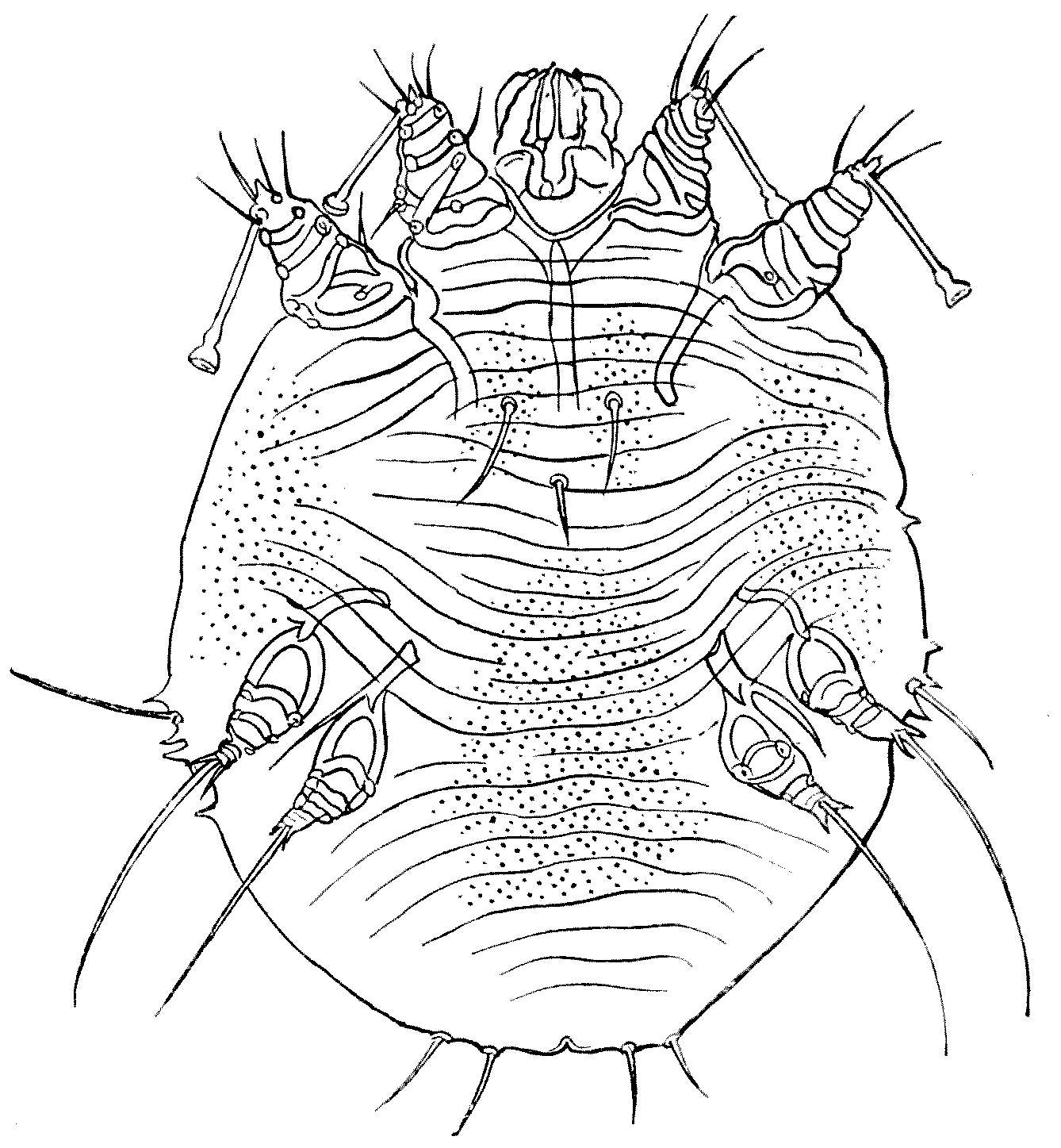
Fig. 35.—Sarcoptes scabiei. Female.
× 180. Ventral view. (From Bourguignon.)
Fig. 35.—Sarcoptes scabiei. Female.
× 180. Ventral view.
(From Bourguignon.)
Sarcoptes scabiei in both sexes is
but little longer than broad. The female
is, as usual, larger than the male. These
mites are shaped very much like microscopic
tortoises, of a pearly grey colour, passing at
parts into a rusty brown. Of the four pairs
of legs two run forward close to the head,
and two point backwards. The integument
is semi-transparent and strengthened by
parallel folds, and bears many little bilaterally
symmetrical protuberances and scales. There
are also certain hairs which have some systematic
value.
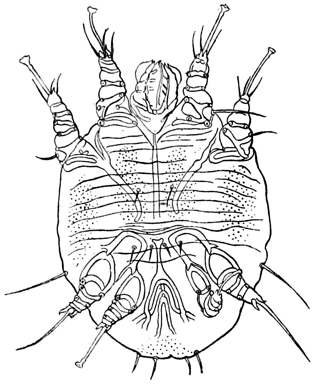
Fig. 36.—Sarcoptes scabiei. Male. × 300. Ventral
view. The sucker on the fourth leg on the right
is accidentally folded over the third leg. (From
Bourguignon.)
Fig. 36.—Sarcoptes scabiei. Male. × 300. Ventral
view. The sucker on the fourth leg on the right
is accidentally folded over the third leg. (From
Bourguignon.)
The male is usually recognised
by the fact that its third pair of legs terminates
in a long hair, whilst the other legs end
in pedunculated suckers.
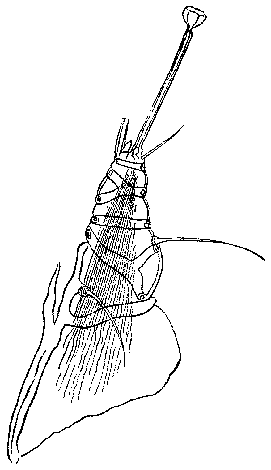
Fig. 37.—One of the legs of Sarcoptes scabiei
(× about 450), showing the stalked sucker and the curious
‘cross-gartering.’ (After Bourguignon.)
Fig. 37.—One of the legs of Sarcoptes scabiei
(× about 450), showing the stalked sucker and the curious
‘cross-gartering.’ (After Bourguignon.)
The male measures
200 µ to 235 µ in length, by 145 µ to 190 µ in
breadth. By preference, he lives under the
scales which the presence of the parasite produce
on the human host. The female is markedly
larger than the male, measuring 330 µ to 450 µ
in length by 250 µ to 350 µ in breadth. Her two
anterior legs end in stalked suckers, whilst
the two posterior end in hairs. The legs, like
Malvolio’s, are curiously ‘cross-gartered’ with
chitinous bars and rings.
At first she promenades about with the
male on the surface of the human skin, but when
they have paired the female begins to tunnel
in the epidermis. The poor male, having
been used, dies. As the mother-mite tunnels
she begins to lay eggs, leaving them one by
one behind her as she burrows deeper and
deeper into the epidermis. Hence those that
are nearer the entrance of the tunnel are always
more advanced in age and development than
those farther in. She always works head
forward, and as her tunnel is but slightly bigger
than the breadth of her body, she cannot turn
round, and she is prevented from retreating
by the backward hairs or spines of her body.
Hence she burrows always forward, until she
has dug her own grave at the far end of her
excavation.
She is said to live two or three months
and to lay one or two eggs a day. Thus one
female is, in time, enough to infect seriously a
single host. The egg is, relatively to the size
of the mother, enormous: its length being
150 µ and its width 100 µ. The egg is hatched
out after three to six days, and the young
larva is hexapodous—that is, as is so usual
in Acarines, six-legged. It escapes from the
burrow on to the skin and soon tunnels into
the epidermis of its host, where it moults and
transforms, about the ninth day, into a four-legged
nymph. At the end of another six days
the mites moult again, and at this period one
can distinguish nymphs of two sizes: the larger
female, and the smaller male.
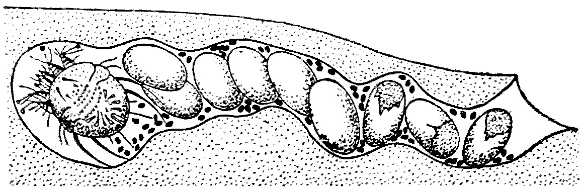
Fig. 38.—A diagrammatic view of the tunnel made by
the female of Sarcoptes scabiei, with the eggs she has laid
behind her as she burrows deeper and deeper. The black
dots represent the excrement. (After Guiart and Grimbert.)
Fig. 38.—A diagrammatic view of the tunnel made by
the female of Sarcoptes scabiei, with the eggs she has laid
behind her as she burrows deeper and deeper. The black
dots represent the excrement. (After Guiart and Grimbert.)
Within a month after hatching the Sarcoptes
has become adult, and the sexes are occupied in
seeking each other on the surface of the skin,
and it is in this stage that they are easily
passed by personal contact from one human
being to another.
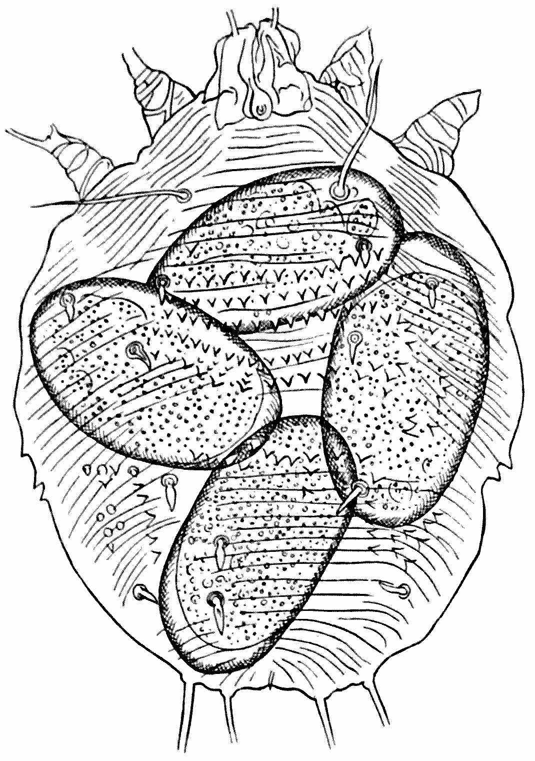
Fig. 39.—A female Sarcoptes scabiei, with four eggs
in different stages of development; × about 180. (After Bourguignon.)
Fig. 39.—A female Sarcoptes scabiei, with four eggs
in different stages of development; × about 180. (After Bourguignon.)
Many animals suffer from Sarcoptes; and
the fact that this genus can be transferred
to man from the horse, the ox, the sheep,
the goat, the dog, the cat, the camel, the
lion, &c., is a slight argument in favour of
their being one species. There is another
undoubtedly distinct species which causes
serious epidemics, especially in Norway; but
that is hardly likely to enter into the scope
of this book.
Sarcoptes scabiei, the itch-mite, is, however,
a cause of serious trouble in an army
‘in being.’ The tunnel or gallery in which
the female mite burrows is the only lesion
produced directly by the parasite. To the
naked eye it presents a little whitish or
greyish line, varying in length from some
millimetres to one or even three centimetres,
the longer ones occurring most frequently
on the hands or wrists. It is of course open
at one end, and ends in a cul-de-sac, which
is slightly swollen, and here it is the female
has taken up her abode. She is visible as
a small white, brilliant spot. Besides the
wrist, and the inner faces of the fingers—the
interdigital areas—the palms of the hands
are most commonly affected.
If there is any doubt as to the cause of
the existence of these tunnels, a diagnosis
can easily be verified by extracting the mite.
With the point of a needle, held almost parallel
to the skin, the tunnel can be slit open,
and when the point has reached the inner
end the mite is very apt to seize it with its
suckers, and can be so withdrawn, and, if
not, it can easily be picked out. It can then
be examined in a drop of diluted glycerine
under a microscope.
I am no doctor, hence I venture to refer
my readers to the article on Scabies in the
‘Encyclopaedia Medica,’ by Dr. G. Pernet, and
to quote the following paragraphs from Dr.
H. Radcliffe Crocker’s ‘Diseases of the Skin,’
third edition, vol. ii.:—
Symptoms of Pathology.—The clinical picture of
scabies is made up of two elements: the burrows,
or cuniculi, and the attendant inflammation excited
directly by the Acarus scabiei[13]; and, indirectly, the
lesions produced by scratching, and the modifying
influences of pressure, friction, &c. The result
is a great multiformity of lesions, which, combined
with their distribution, is in itself suggestive of the
nature of the disease, and enables a practised eye
to detect a well-marked case at a glance.
When the skin is first penetrated by the acarus,
inflammation is often set up, and a papule, vesicle,
or pustule is the consequence. These papules or
small vesicles, individually indistinguishable from
eczema vesicles, are the most common form of eruption;
but the inflammatory symptoms are absent
in many burrows. The tract extends and forms
a sinuous, irregular, or rarely straight line, which in
very clean people is white, but, as a rule, is brownish
or blackish from dirt being entangled in the slightly
roughened epidermis; the length of these burrows
is generally from an eighth to half an inch, but
occasionally much longer—Hebra having noticed one
four inches long. When a pustule is formed, part
of the burrow lies in the roof, but the acarus is always[108]
well beyond the pustule or vesicle; or, if there is none,
lies at the far end, and with a lens may often be
discerned as a white speck in the epidermis. The
degree and number of inflammatory lesions vary
much; there may be no inflammation at all about
many burrows, or the whole hand—especially in
children—may be covered by pustules, vesicles, or
papules; and, indeed, a pustular eruption on the
hands is always strongly suggestive of scabies;
there is, however, no grouping or arrangement
of any of the eruptions, as in eczema, the lesions
being scattered about irregularly. It must be
remembered that burrows are not always present,
from various causes. If the disease is recent it
may not have got beyond the papular or vesicular
stage; while in washerwomen, bricklayers, or others
whose hands are constantly soaked in water or
alkaline fluids, or who have to scrub their hands
violently, the burrows become destroyed. The eruptions
due to scratching have already been described
in the descriptions of the ‘scratched skin,’ and
comprise excoriations, erythema in parallel lines,
eczema, impetiginous or so-called ecthymatous eruptions
and wheals, and the inflammatory scab-topped
papules often left after the subsidence of the wheals—especially
in children. In carmen, cobblers, tailors,
and others who sit on hard boards for hours together,
pustular and scabbed eruptions, situated over the
ischial tuberosities, are so abundant and constant
as to be practically diagnostic of scabies in such
people. Similar eruptions may be seen where there
is friction from trusses, belts, &c.
Treatment.—I use in private practice, after the
preliminary soaking and scrubbing, naphthol 15[109]
parts, cret. prep. 10 parts, sap. mollis 50 parts,
adipis 100 parts, as recommended by Kaposi, well
rubbed in. For infants it can be used half-strength,
and I omit the soft-soap. I can speak of it in
the highest praise. It is effectual, has no smell, and
is not liable to irritate the skin, as sulphur does.
It is, however, too expensive for public practice.
Nephritis has occurred from its over-use, but I have
never seen any bad symptoms. Another remedy
less likely to irritate the skin than sulphur is balsam
of Peru, of which the vapour alone is said to be fatal
to the acari. The balsam is rubbed in for twenty
minutes every night; a night-shirt impregnated
with the drug is worn, and in the morning an
ordinary soap-and-water bath is taken.
Colonel Allcock says that the best treatment
for the itch ‘consists in the free use
of soap and hot water and the liberal application
of sulphur ointment, continued for several
days. Some prefer baths of potassa sulphurata
(1 ounce of the salt to 4 gallons of water).
Clothing and bedding should be fumigated
with sulphur or baked.’
Endo-parasitic Mites
Certain little mites whose appearance is as
repellent as their name—for they are known
as Nephrophages sanguinarius—were recorded
by two Japanese observers twenty years ago
as coming away in the urine of a Japanese
patient who was suffering from various bladder
troubles. As the mites were in all cases
dead, the Japanese doctors thought that they
must have been endo-parasites of the kidney.
They were found day after day for a week
or more, and they were found also in the
water with which the bladder had been washed
out, but always dead. It is, of course,
possible that the Japanese doctors were right
in their surmise, but the best that can be
said for the case is that it is ‘not proven.’
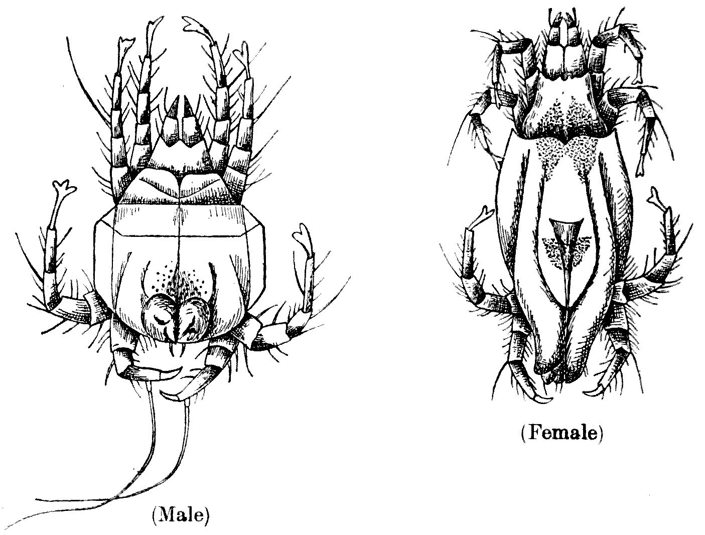
Fig. 40.—Nephrophages sanguinarius (enlarged).
Male, ventral surface. Female, dorsal aspect. (After Miyake and Seriba.)
Fig. 40.—Nephrophages sanguinarius (enlarged).
Male, ventral surface. Female, dorsal aspect. (After Miyake and Seriba.)
These awful-looking little mites are said
to have two large eyes, and legs of five
segments and of equal length. Their colour
is greenish to brownish yellow. Undoubtedly
there are many mites which live as endo-parasites;
certain members of the group
Analgesinae, such as Laminosioptes gallinarum,
live in the intramuscular and subcutaneous
tissue of fowls, and Cytoleichus sarcoptioides
in their air-sacs. I have myself found one
of these species in the pigeon, so that it is
by no means beyond the bounds of human
possibility that Nephrophages sanguinarius
really lived in the tissues of the Japanese.
Very strange things live in the tissues of
some Japanese.
CHAPTER IX
TICKS
A waterleche or a tyke hath neuer ynow, tyl it brestyth.
(Jacob’s Well, 1440.)
Ticks
are mites ‘writ large,’ and until about
the beginning of this century they were
regarded with what one might call mild disgust
and regret. Now, however, that they have
been proved to play a part—and a very
important part—in the dissemination of disease,
we have come to regard them, as Calverley
said we should regard the Decalogue, ‘with
feelings of reverence mingled with awe.’
The body of a tick is covered with a tough,
smooth or crinkled skin, capable almost of any
amount of extension. Until they have fed they
are flattened in shape, but after a meal of blood
they very soon lose the outlines of a Don
Quixote and attain those of a Sancho Panza.
In the adult, the legs are eight in number and
have six joints ending in two claws and sometimes
in suckers. Some have eyes and some
have no eyes.
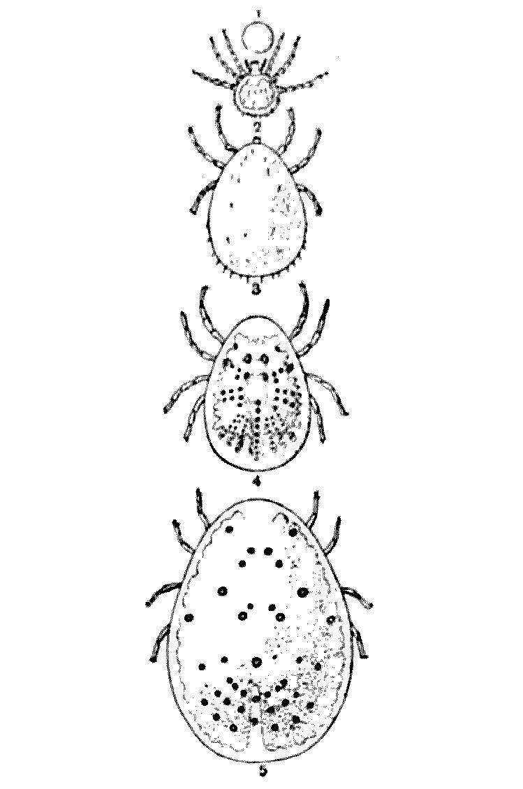
Fig. 41.—Evolution
of Argas persicus. 1, the egg; 2, the six-legged larva; 3,
the same gorged; 4, an unfed nymph; 5, nymph gorged. (After Brumpt.)
Fig. 41.—Evolution
of Argas persicus. 1, the egg; 2, the six-legged larva; 3,
the same gorged; 4, an unfed nymph; 5, nymph gorged. (After Brumpt.)
The most formidable part of
their armour is, however, the mouth-parts,
consisting of the tactile pedipalps, and the
piercing-probe which they stick into our bodies.
This probe consists of a dorsal
membranous sheath and a
ventral hypostome armed with
recurved teeth, forming together
a tube within which play two
cutting and tearing chelicerae.
When these have cut a way
into the flesh they are withdrawn,
and the tube is inserted
into the wound and blood is
pumped up it by the sucking-pharynx.
It is the teeth on
the hypostome, and not the
chelicerae, which anchor the
ticks to their prey.
Ticks, as they affect the
soldier, may be divided into two
families. The first of these,
the Argasidae, are usually associated
with human dwellings,
fowl-houses, dove-cotes, and so
on, and are more commonly
parasitic on fowls than on
cattle or human beings. The members of this
group hide away in crevices and corners during
the day, and come out at night to feed, for
‘their deeds are evil.’
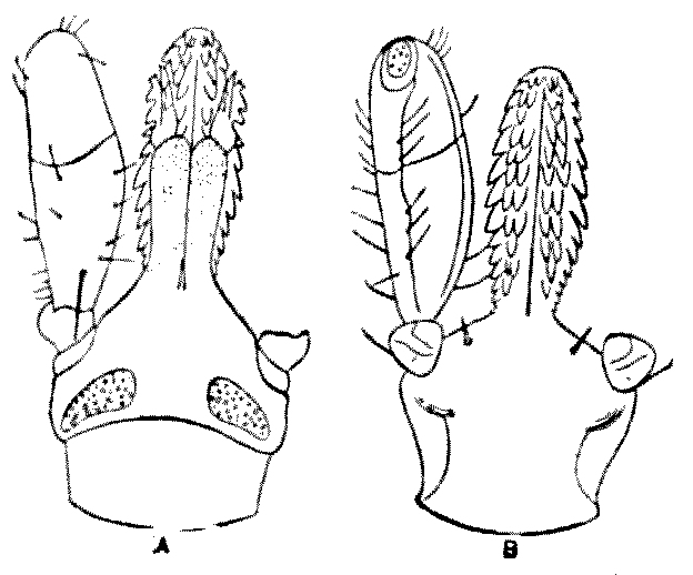
Fig. 42.—Ixodes
ricinus. Mouth-parts of the female: A,
seen from the dorsal, B, from the
ventral surface. The median, dotted, portion of the left-hand figure
is the sheath; the toothed portion the hypostome. The lateral process
is the pedipalp shown only on one side. × 35. (After Nuttall and
Warburton.)
Argas persicus, known to travellers as the
‘teigne de miana,’ is of an oval form, of
brownish-red colour. The male measures 4 mm.
to 5 mm. in length by 3 mm. in breadth; the
female 7 mm. to 10 mm. in length by 5 mm. to
6 mm. in breadth. This creature frequents the
northern parts of Persia, and occurs in many
other warm countries.
In South Africa it is
known as the ‘tampan’
and ‘wandlius,’ where
it is mainly a fowl-parasite.
In Persia it
is very much dreaded,
though probably the
effects of its bite are
due to the unsuitable
treatment the punctured
skin receives and the
consequent invasion of
the tissues by septic
bacteria. In South
Africa it is frequently
fatal to fowls, especially to chickens; but
the death is there believed to be due to the
loss of blood. It is definitely proved to convey
Spirochaetosis.
We have not yet explained that ticks pass
through several stages as they advance from
the egg to the adult. The larval stage of A. persicus
will remain on its host for five days.
It then leaves, and moults in retirement.
After the moulting it visits its host by night
and remains on it for about an hour. This
second stage, known as the ‘nymph’ stage,
moults twice, and the female in each stage
becomes much distended with blood—‘gorged,’
as the saying is. With each moult it becomes
larger, but otherwise does not alter much in
appearance. The adult female also, like the
nymph, visits the host from time to time, and
between these visits deposits eggs in great
quantities in sheltered crevices—some 50 to
100 being deposited at once.
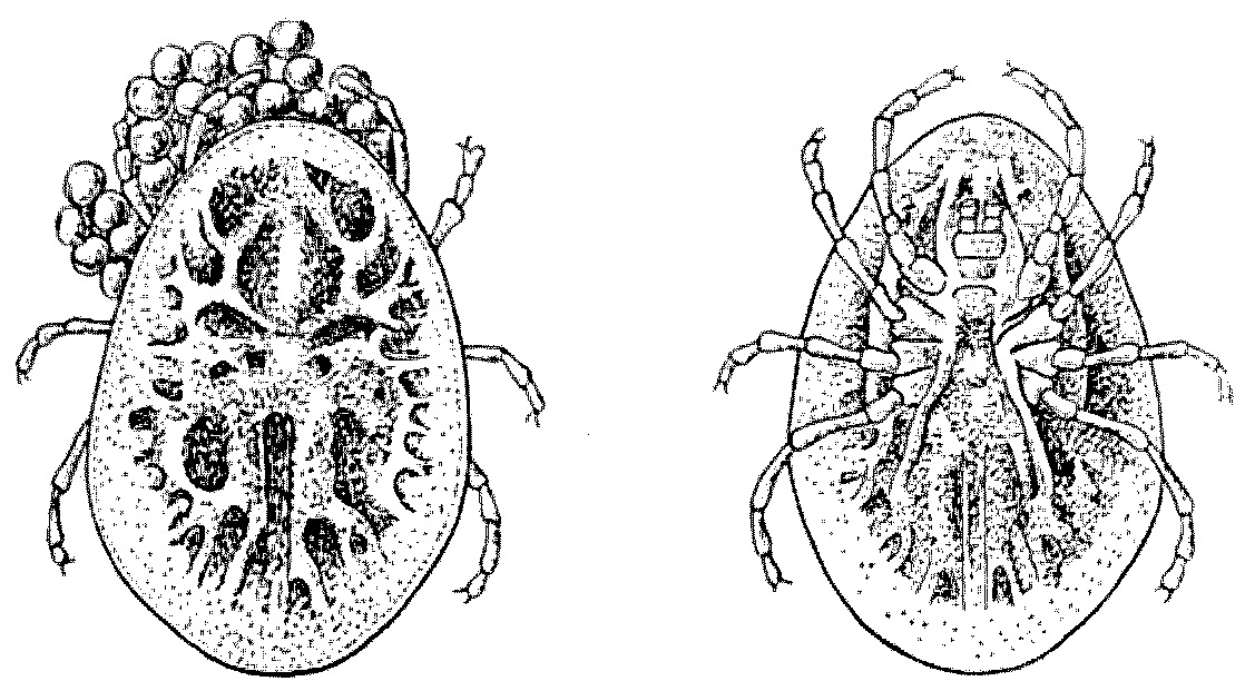
Fig. 43.—Argas reflexus, female. On the left the
dorsal view of a specimen laying eggs; on the right a
ventral view of the same. (After Brumpt.)
Fig. 43.—Argas reflexus, female. On the left the
dorsal view of a specimen laying eggs; on the right a
ventral view of the same. (After Brumpt.)
Argas reflexus,
the ‘marginated tick,’ is yellow and white—the
Papal colours. It is common near dove-cotes
and pigeon-houses, and often attacks
people sleeping in their neighbourhood. Its
bite causes much irritation, and sometimes
leads to vesicles and ulcers. At one time it
was very common in Canterbury Cathedral, and
so worried the worshippers that it took all the
eloquence of the ‘Very Reverend the Dean’ to
overcome its repellent powers.
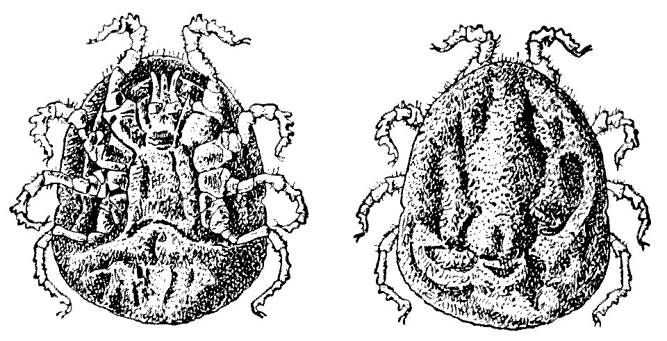
Fig. 44.—Ornithodorus moubata,
an unfed female. To the left a ventral, to the right a dorsal view, showing the
crinkled skin. (After Brumpt.)
Fig. 44.—Ornithodorus moubata,
an unfed female. To the left a ventral, to the right a dorsal view, showing the
crinkled skin. (After Brumpt.)
Ornithodorus moubata, sometimes known
as the ‘tampan,’ occurs pretty often in
South Africa, and was a cause of considerable
trouble to our troops during the South
African War. It lives normally in the
shade of vegetation, but frequently invades
the native huts. It is catholic in its taste
and attacks most mammals, and it has a
decided preference for men. In Uganda
the natives frequently die from its bites—dying
of so-called ‘tick-fever.’
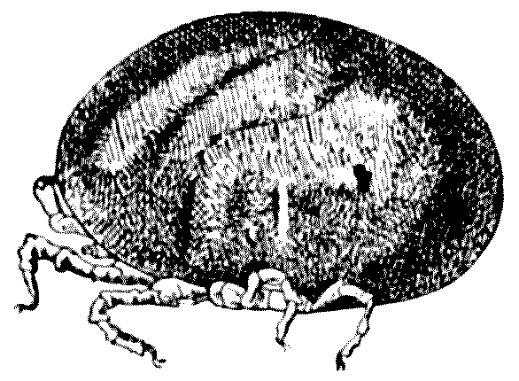
Fig. 45.—Ornithodorus
moubata. Female, gorged, seen in profile. (After Brumpt.)
Fig. 45.—Ornithodorus
moubata. Female, gorged, seen in profile. (After Brumpt.)
I myself once assisted in identifying two ticks, in the nymph
stage, taken in Cambridge from the ear of an
American visitor to this country, who had been
camping out in Arizona shortly before his
arrival. This tick turned out to be a species
of Ornithodorus megnini, which, as a rule,
attacks the horse, the ass, and the ox about
the ears. But it frequently attacks man, and
is well known in the United States, infesting
the ears of children. An
allied species, O. turicata,
proves fatal to fowls in
the Southern States and
in Mexico, and is very
harmful to human beings.
The chief harm that these
ticks do is to transmit
protozoal diseases to
man and other animals.
A very few ticks are said to be parthenogenetic,
but by far the greater part lay
fertilised eggs, and lay them in considerable
numbers; and the eggs are agglutinated
together in solid little masses, by the sticky
secretion of a cephalic gland, which opens
below the rostrum. The eggs are small and
elliptical, and are laid to the number of many
thousands. The young tick, which is usually
born with but three pairs of legs, hatches
out in a few days if the weather be warm, or
a few weeks should it prove cold. A certain
amount of moisture must be present, or the
eggs are apt to dry up. These masses of eggs
are laid on the ground under herbs or grass,
or on leaves.
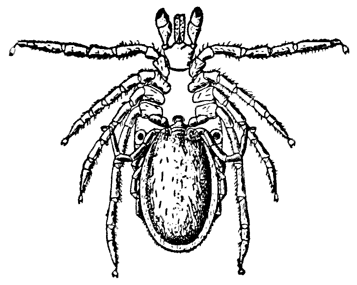
Fig. 46.—Ixodes ricinus. The
male is inserting its rostrum in the female genital duct before depositing
its spermatophore. × 6. (From Brumpt.)
Fig. 46.—Ixodes ricinus. The
male is inserting its rostrum in the female genital duct before depositing
its spermatophore. × 6. (From Brumpt.)
The issuing six-legged larvae, like the
young of other animals, are very agile, climbing
on to leaves and herbage. They passionately
wait with their front
legs eagerly stretching
out for the passage
of the host
upon which they
desire to settle. Of
course, but one in
ten thousand succeeds,
and it is
terrible to think of
the amount of unsatisfied
desire
which must be going
on in the tick world! The rest perish
miserably. Those that do succeed attach
themselves to the skin of the host, and thrust
their rostrum and sucking-tube into the hole
already prepared by the cutting chelicerae.
They suck the blood, and when gorged fall
to the earth, or in some cases remain on
the host in a state of inertia or apparent
syncope.
Soon, however, the gorged larva moults,
and gives rise to the first nymph—an eight-legged
creature. This affixes itself anew upon
a host—either upon the same or another
one—again gorges itself, and in all points
resembles the adult, except from the fact that
the sexual orifice has not yet appeared. After
some days the first nymph moults, and then
again remains either on the host or it falls to the
ground. In some cases there are two successive
nymph forms; but as a rule the first nymph
gives rise by a second moult to the adult
form, which again for the third time regains
a host. The adults are now ripe for pairing,
and the male having enlarged the orifice of
the oviduct by inserting its rostrum, deposits
therein a spermatophore or capsule full of
spermatozoa. The female is often successively
fertilised by several males.
In many cases the male dies after fertilisation.
The female swells enormously when
gorged, sometimes becoming as large as a
filbert, or even a small walnut. These ticks
are seldom parasites of one particular host, but
attack many mammals indifferently. They
have many natural enemies: amongst the most
important of which are certain hemipterous
insects whose female attacks the nymph of
the Ixodes, and lays within the body of the
tick a number of eggs which develop inside
the nymph until they reach the adult stage,
when they make their escape through an
orifice, generally at the hind end, leaving
behind them the dead body of their host.
Three species of such Hemipterous insects are
known to be parasitic on ticks: of these
Ixodiphagus caucurtei is ubiquitous. It attacks
all kinds of ticks, but
especially Dermacentor
venustus.
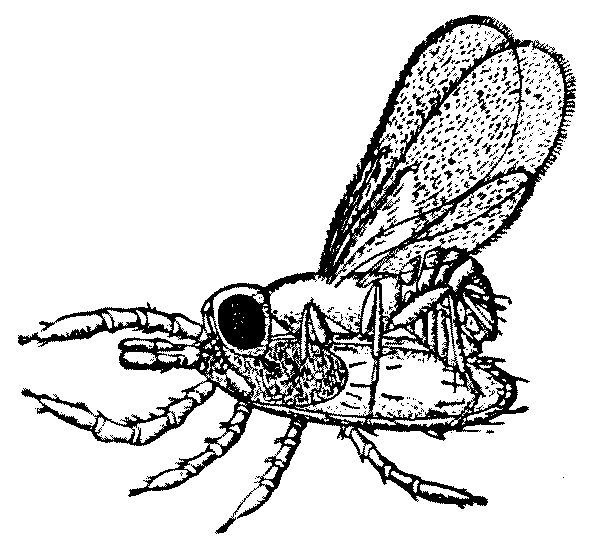
Fig. 47.—Ixodiphagus caucurtei
laying eggs in the nymph of Ixodes ricinus. × 20. (After Brumpt.)
Fig. 47.—Ixodiphagus caucurtei
laying eggs in the nymph of Ixodes ricinus. × 20. (After Brumpt.)
Ixodes ricinus, of a
brownish colour in the
male, is very common
in England and, indeed,
almost everywhere. The
female is yellow and flattened,
somewhat resembling
a grain of rice.
It is the well-known dog-tick,
but it attacks oxen, goats, deer, horses,
and man. It also attacks the grouse, and is
particularly common in some parts of Great
Britain. It is impossible to rid certain areas
of these troublesome guests. In some cases
they produce tumours and introduce bacteria,
and in cattle it introduces an organism known
as Babesia bovis, which is the cause of
haematuria in oxen. Dermacentor venustus
transmits Rocky Mountain fever, which is
common in certain parts of the States. The
fever is accompanied with pains in the joints and
in the muscles and an eruption on the surface
of the skin, appearing first on the wrists and
forehead, and invading in time all parts of
the body, followed by a scaling of the skin
during a period of convalescence. In Montana
the mortality caused by this disease is very
high, varying in different years from 33 to
75 per cent. In Idaho the mortality is far
less, only about 4 per cent.
Ornithodorus moubata inoculates man with
a spirochaete (Spirochaeta duttoni), which is
the agent of the African tick-fever or relapsing
fever. One of the curiosities about the
organisms transmitted by ticks is that they
live through the whole cycle of the tick’s life.
If they are taken in by the larva they are only
transmissible by the following larval stage.
If they are taken in by the nymph they are
only transmissible when again the nymph
stage is met with, and the same is true of the
adult. Think what such a protozoon must
have seen! The fertilisation of the egg by
the spermatozoon, the fusion of their nuclei,
the extrusion of the polar-bodies, the breaking
up of the egg into segments, the gradual
building up of the tissues of the larva, the
sudden inrush of the host’s blood when
the larva is safely fixed, the moulting, the
changes in the nymph, the development of the
generative organs, the formation of the eggs!
What a text-book of embryology and anatomy
it could write if only it had descriptive powers!
If I may paraphrase Kipling:—
Think where ’e’s been,
Think what ’e’s seen,
Think of his future,
And Gawd save the Queen!
CHAPTER X
LEECHES
Part I
THE MEDICINAL LEECH (Hirudo medicinalis)
Hardly anything real in the shop but the leeches and they’re
second-hand. (Bob Sawyer, The Pickwick Papers.)
As Mr. W. A. Harding has pointed out, eleven
species of fresh-water leeches occur in these
islands. But one of these, the Hirudo medicinalis,
seems to be vanishing, and yet it is
just the one we should cherish and preserve.
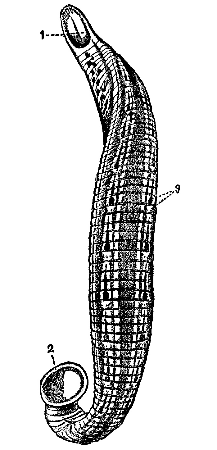
Fig. 48.—Hirudo
medicinalis; about life size. 1, Mouth; 2, posterior sucker; 3, sensory
papillae on the anterior annulus of each segment. The remaining four annuli
which make up each true segment are indicated by the markings
on the dorsal surface.
Fig. 48.—Hirudo
medicinalis; about life size. 1, Mouth; 2, posterior sucker; 3, sensory
papillae on the anterior annulus of each segment. The remaining four annuli
which make up each true segment are indicated by the markings
on the dorsal surface.
There are people who do not like leeches.
This is shown by the agitation amongst the
travellers in an omnibus, as depicted in
Punch by Leech, years and years ago, when
an old gentleman had upset a bottle of them
in their midst. But the medicinal leech, which
is our theme, is really the friend of man and
of the soldier, and is a beneficial and not a
harmful animal. There are, of course, other
leeches in our rivers and in our seas, but
of the latter our knowledge is scanty and
it is difficult to increase it at
present—at any rate, in the
Channel or in the North Sea.
In any case the marine leeches
in our island-waters have no
human interest except the
influence they exercise on our
fish-food supply, and this is
practically negligible.
Zoologically speaking,
leeches are undoubtedly degenerate
earth worms (Oligochaeta);
and some very interesting
‘Zwischenformen’—like
Mr. Vincent Crummles, I am
‘not a Prussian’; but in spite
of the war, we may as well
employ a useful term captured
from the enemy—have been
found in Russia and Siberia:
forms which combine many
of the characters of the
Oligochaeta and the Hirudinea.
Possibly the degeneracy which
leeches are said to exhibit
is associated with a semi-parasitic
habit of life. But
a semi-parasitic habit does
not apply to all leeches—in fact, it applies
but to few genera; there are many others,
equally degenerate—if degenerate they be—who
have no trace of semi-parasitism.
A curious thing about leeches is that
all the varying genera have the same number
of somites or segments; and though some
of these segments or somites are masked and
fused, when analysed by the number of segments
in the embryo, by the number of the
nerve ganglia, and so on, leeches seem always
to have thirty-four such segments. These
do not correspond with the rings or annulations
so visible on the outside; but a certain
number of these annulations, varying in each
species, ‘go’ to each somite, and so constant
are these numbers that it would not be very
difficult to represent any given species of
leech by a mathematical formula.
The known species readily fall into two
sub-orders: (1) The Rhynchobdellae, which are
marine and fresh-water leeches with colourless
blood, with no jaws, and with an extensile
proboscis; and (2) the Arhynchobdellae, which
are all fresh-water or terrestrial, with red
blood, and generally with jaws. There is no
extensile proboscis, and the anterior sucker has
a ventral aspect, and is in no way distinct from
the body. There are always in this group seventeen
pairs of nephridia or kidneys. We shall
have mostly to do with the latter sub-order.
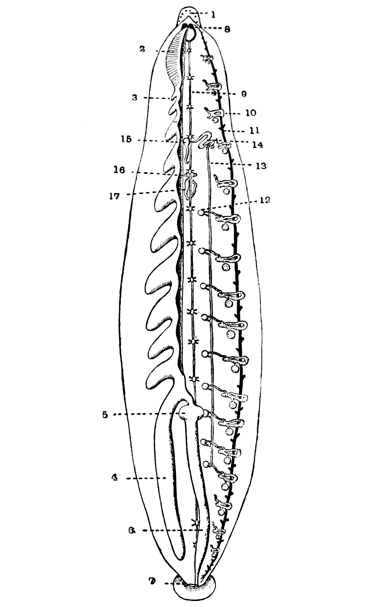
Fig. 49.—View of the internal organs of Hirudo medicinalis. On
the left side the alimentary canal is shown, but the right half of this
organ has been removed to show the excretory and reproductive organs.
1, Head, with eye-spots; 2, muscular pharynx; 3, first diverticulum
of the crop; 4, eleventh diverticulum of the crop; 5, stomach; 6,
rectum; 7, anus; 8, cerebral ganglia; 9, ventral nerve-cord; 10,
nephridium; 11, lateral blood-vessel; 12, testis; 13, vas deferens; 14,
prostate; 15, penis; 16, ovary; 17, uterus, a dilatation formed by the
conjoined oviducts.
Hirudo medicinalis, the medicinal leech,
is found in stagnant waters throughout Europe
and the western parts of Asia. It is rather
commoner in the southern parts of Europe
than in the north. It used to be common
enough in England, where at one time, it
was bred; but already a hundred years ago its
numbers were diminishing.
In a treatise on the Medicinal Leech,
published by J. R. Johnson in the year 1816,
he records: ‘Formerly the species was very
abundant in our island; but from their present
scarcity, owing to their being more in request
among medical men, and to the rapid improvements
which have of late years taken
place in agriculture—particularly in the
draining and cultivation of waste lands—we
are obliged to receive a supply from the
Continent, chiefly from Bordeaux and Lisbon.’
In his time he considered that for every
native leech employed at least a hundred
foreigners were used.
The same scarcity was very apparent to
the poet Wordsworth, whose insatiate curiosity
is recorded in the following lines in 1802—Wordsworth
was always asking rather fatuous
questions:—
My question eagerly did I renew,
‘How is it that you live, and what is it you do?’
[128]
He with a smile did then his words repeat:
And said that, gathering leeches, far and wide
He travelled; stirring thus about his feet
The waters of the pools where they abide.
‘Once I could meet with them on every side;
But they have dwindled long by slow decay;
Yet still I persevere, and find them where I may.’
In Europe, where the leech was once
very abundant, it is now chiefly confined
to the south and east, and in Germany it
is still found in the island of Borkum and
in Thuringia—but just now we need not
trouble ourselves very much about their
distribution in Germany.
In 1842, leeches were occasionally found
in the neighbourhood of Norwich, and there
are villagers still living in Heacham in Norfolk
who remember the artificial leech-ponds. In
the middle of the last century the medicinal
leeches ‘of late years ... have become scarce.’
At about the same time, it is also recorded
that they were becoming scarce, though still to
be found, in Ireland. Apparently this species
is now almost extinct in England, although
I know of a naturalist who can still find
them in the New Forest, but he will not tell
where. If they were getting scarce in the
beginning of the nineteenth century they are
far scarcer now[14]—for there is no leech in
London—at least, there are only a dozen or
two, and they, like those of the firm ‘Sawyer
late Nockemorf,’ are second-hand and I have
heard that there is a similar shortage in
North America. And yet leeches are wanted
by doctors!
Harding tells us that:—
Hirudo medicinalis is not the only leech which
has been used in phlebotomy. Hirudo troctina
(Johnson, 1816), occurring in North Africa and in
Southern Europe, where it is perhaps an introduced
species, was largely imported at one time for medical
uses....
Several other species have been used for blood-letting
in different countries. Limnatis (Poecilobdella)
granulosa in India, Liostoma officinalis in
Mexico, Hirudo nipponia in Japan (Whitman),
and Macrobdella decora in the United States (Verrill),
are or have been used in phlebotomy.
‘Our chief hope seems to lie in India.’ These
words I wrote in October 1914, and my hopes
were justified. Owing to the energy of Dr.
Annandale of the Indian Museum, and the
anxious care of the authorities of the P. & O.
Company, I was able to land, early in the
present year, a consignment of many hundred
Limnatis granulosa—in sound health, good
spirits, and obviously anxious to do their duty.
Leeches are still used much more than
the public are aware. One pharmaceutical
chemist in the West End of London tells
me he sells between one and two thousand
a year; and as they are bought wholesale at
about one penny each and sold retail at
about sixpence, there is some small profit.
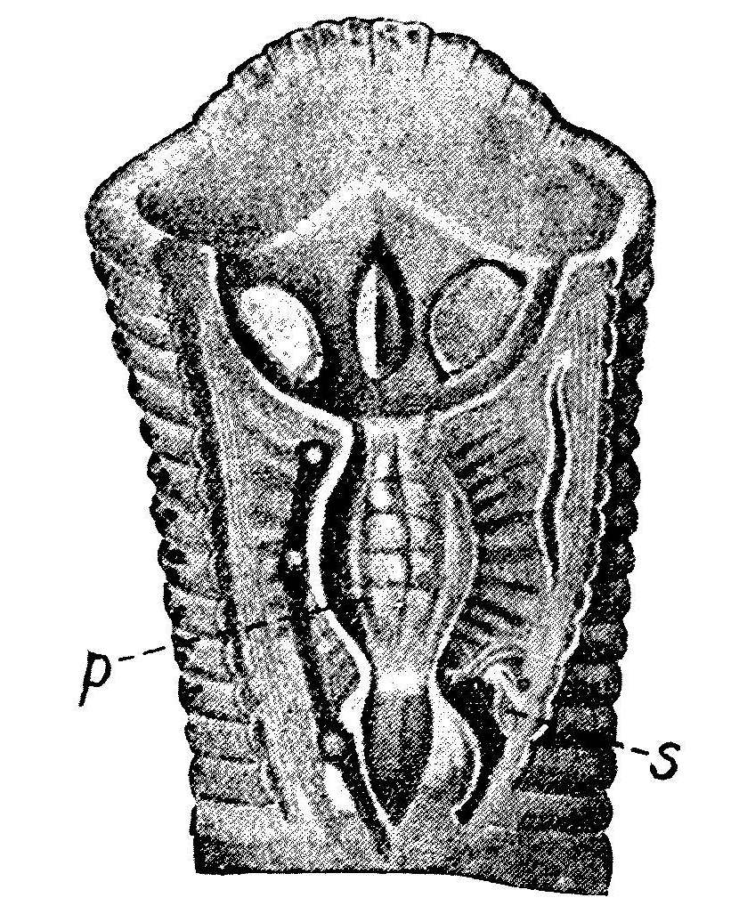
Fig. 50.—Head of a leech, Hirudo
medicinalis, opened ventrally to show the three teeth and the pharynx p,
with its muscles; s, a nephridium.
Fig. 50.—Head of a leech, Hirudo
medicinalis, opened ventrally to show the three teeth and the pharynx p,
with its muscles; s, a nephridium.
Leeches were well known to the ancients, and it would be easy to
quote case after case from the classical medical authorities of their
use in fevers and headaches and for many ill-defined swellings. They
were frequently used for blood-letting where a cupping-glass was out
of the question. With his curious uncritical instinct, Pliny records
that the ashes of a leech sprinkled over a hirsute area or formed into
a paste with vinegar and applied to the part will remove hair from
any region of the body. Leeches were also used by the Greek and Roman
physicians in angina—especially when accompanied by dyspnoea.
Probably the traffic in leeches reached its
height in the first half of the nineteenth century. Harding reminds us
that in the year 1832 Ébrard records that 57,500,000 of these annelids
were imported into France, and by this time the artificial cultivation
of leeches had become a very profitable industry. Although in a small
way leeches may have been cultivated in special ponds in Great Britain,
the English never undertook the industry on a large scale. In Ireland
the natives used to gather the leeches in Lough Mask, and other inland
lakes, by sitting on the edge of the pool dangling their legs in the
water until the leeches had fastened on them. But the native supply
was totally inadequate, and the great majority of leeches used in this
country were then imported. In 1842 Brightwell mentions a dealer in
Norwich who always kept a stock of 5000 of these annelids in two large
tanks. The traffic, as we have seen, was very considerable.
The French leech-merchants recognised
five classes, as follows:—
1. Les filets ou petites Sangsues, qui ont
de un à cinq ans;
2. Les petites moyennes, qui ont de cinq
à huit ans;
3. Les grosses moyennes, qui ont de huit
à douze ans;
4. Les mères Sangsues ou les grosses, qui
sont tout à fait adultes;
5. Les Sangsues vaches, dont la taille est
énorme.
They also recognised many colour-varieties,
of which we need only mention the speckled,
or German leech—‘Sangsues grise medicinalis,’
with a greenish-yellow ventral surface spotted
with black, and the green Hungarian leech
with olive-green spotted ventral surface. Both
are merely colour-varieties of Hirudo medicinalis—a
species which shows great variation
in colour, and often forms colour-races when
bred artificially.
The varying sizes of the five categories
mentioned above may be seen by the fact that
one thousand of ‘les filets’ weigh from 325
to 500 grammes, one thousand of ‘les petites
moyennes’ weigh 500 to 700 grammes, one
thousand of the ‘grosses moyennes’ weigh 700
to 1300 grammes, and one thousand of the
‘grosses’ 1300 to 2500 or even to 3000 grammes.
Whereas one thousand of ‘les vaches’ weigh
up to 10 kilograms, and sometimes even more.
To increase their weight the dishonest dealer
sometimes gives them a heavy meal just before
selling them.
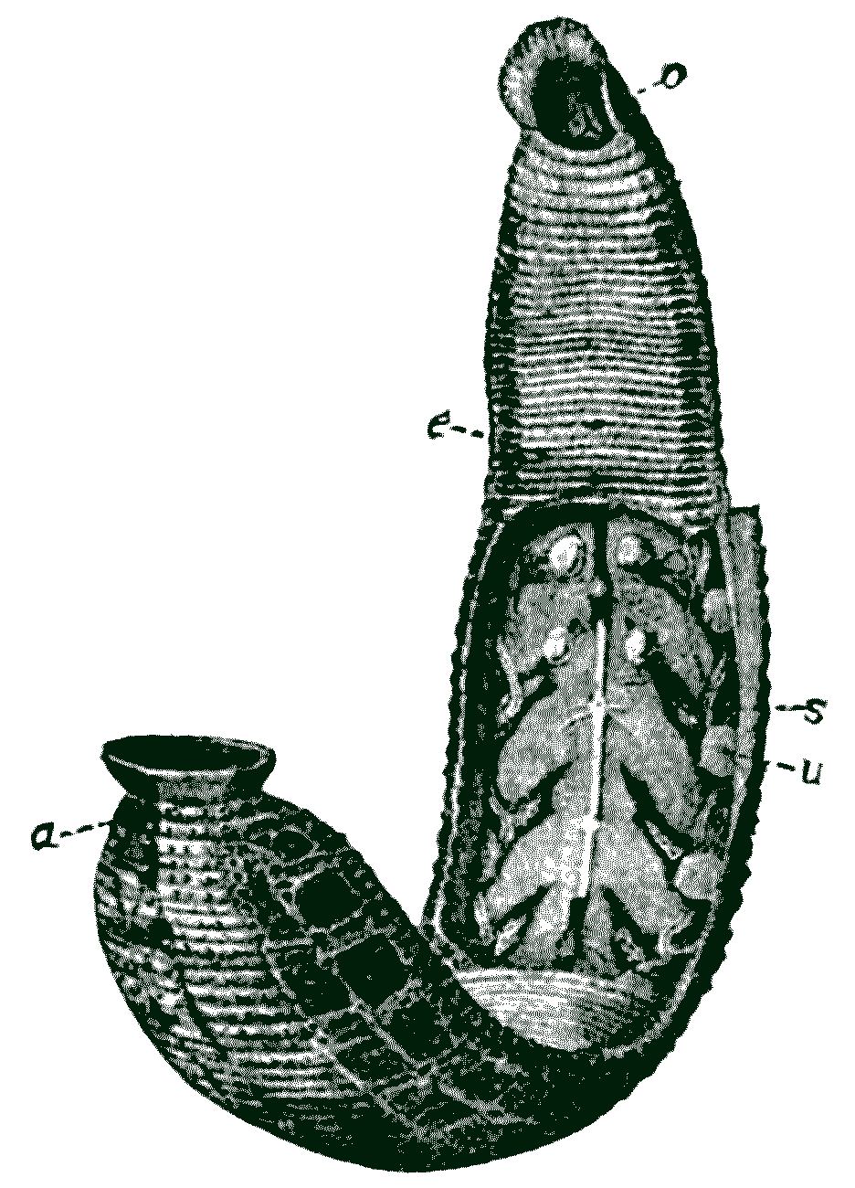
Fig. 51.—Hirudo medicinalis. o,
Anterior sucker covering triradiate mouth; e points to an
annulus midway between the male and female
openings, s to a nephridium, u to the bladder of
the latter; a, anus. Four testes and four lateral
diverticula of the crop are also shown.
Fig. 51.—Hirudo medicinalis. o,
Anterior sucker covering triradiate mouth; e points to an
annulus midway between the male and female
openings, s to a nephridium, u to the bladder of
the latter; a, anus. Four testes and four lateral
diverticula of the crop are also shown.
They were transported from place to place
in casks half filled with clay and water, or
in stone vases full of water. Sometimes
they travelled in sacks of strong linen, or even
of leather, and these had to be watered from
time to time. Another mode of conveying
them was to place them in baskets full of
moss or grass soaked in water, but care had
to be taken lest they should escape. These
baskets, again, could not be packed one upon
another, or the leeches were crushed. In the
old days each sack often weighed 20 to 25
kilograms; and travelling thus, suspended
in a kind of hammock, dans une voiture ou
fourgon, from Palota near Pesth, they reached
Paris in from twelve to fifteen days.
They generally travelled via Vienna to
Strassburg, where twelve great reservoirs,
appropriately placed near the hospital, received
them, and here they rested for awhile.
Others collected in Syria and Egypt came by
ship to Trieste, whence they are sent to Bologna,
to Milan, and to Turin, or by water to
Marseilles. Marseilles also received directly
by sea the leeches from the Levant and Africa,
and expedited them to Montpellier, Toulouse,
and many another town in the south.
The best time of year for their journey
was found to be the spring and autumn.
They were more difficult to manage in the
summer, and they were all the better for
having a rest every now and then, as they
used to do at Strassburg. There were times
when consignments of from 60,000 to 80,000
a day used to leave Strassburg for Paris. In
1806 a thousand leeches in France fetched 12 to
15 francs; but in 1821 the price had risen to 150
to 200 and even 283 francs. In the latter year
they were retailed at 20 to 50 for 4 to 10 sous.
As in England, however, for the most
part the artificial cultivation of leeches is
diminishing in France, though half a century
ago leech-farms were common in Finistère and
in the marshes in the neighbourhood of Nantes.
There were some years when, if the season was
favourable, the peasants carried to market
60,000 a day. Spain and Portugal also furnished
leeches for a long time; but by the
middle of last century the Peninsula had become
almost depleted. But some leeches were
still at that period being received from Tuscany
and Piedmont. Perhaps the richest fields
which still exist are the marshy regions in
Hungary.
It will be observed that, probably without
their knowing anything at all about it, General
Joffre, General von Kluck, Field-Marshal
French, the Grand Duke Nicholas, General
von Hindenburg are fighting on some of the
best leech-areas in Europe—a point to which
we shall return when dealing with the leeches
of the Orient.
One wonders what the leeches think of it all!
CHAPTER XI
LEECHES
Part II
THE MEDICINAL LEECH (Hirudo medicinalis)—continued
Non missura cutem, nisi plena cruoris, hirudo.
(Horace.)
There
is no doubt that the medicinal leech
is one of the most beautiful of animals. Many
of its cousins are uniform and dull in colour—‘self-coloured,’
as the drapers would call
them; but the coloration of the medicinal
leech could not be improved upon. It is a
delicious harmony of reddish-browns and
greens and blacks and yellows, a beautiful soft
symphony of velvety orange and green and
black, the markings being repeated on each
segment, but not to the extent of a tedious
repetition. So beautiful are they that the
fastidious ladies who adorned the salons at the
height of the leech mania, during the beginning
of the eighteenth century, used to deck their
dresses with embroidered leeches, and by
repeating the design one after the other
constructed a chain of leeches which, as a
ribbon, was inserted around the confines of
their vesture.
Harding tells us that the dorsal surface
of H. medicinalis is ‘usually of a green, richly
variegated colour, with orange and black spots,
exhibiting an extremely variable pattern, based
generally upon three pairs of reddish-brown
or yellowish, more of less, longitudinal stripes,
often interrupted by black or sessile spots
occurring on the rim of each somite. The
ventral surface is more or less green, more
or less spotted with black, with a pair of
black marginal stripes.’
The shape of the medicinal leech, and
indeed of other leeches, is difficult to put
into figures, as their bodies are as extensile
as the conscience of a politician and as flexible
as that of a candidate for parliamentary
honours. The length of H. medicinalis in extreme
extension is said to range from some 100
mm. to 125 mm.; in extreme constriction from
30 mm. to 35 mm. The width in the former
state would be 8 mm. to 10 mm., and in the
latter 15 mm. to 18 mm.
The movements of the medicinal leech
are as graceful as its colour is tasteful. When
in the water they move like looper-caterpillars
(Geometrids), stretching out their anterior
sucker, attaching it to some object,
and then releasing the posterior sucker they
draw the body up towards the mouth. Or,
casting loose from all attachment, the leech
elongates and at the same time flattens its
body until it assumes the shape of a band
or short piece of red tape, and by a series of
the most seductive undulations swims through
the water. Kept in an aquarium they are
rather apt at times to leave the water and
take up a position on the sides of their home an
inch or two above the aqueous surface. When
outside the water they keep their bodies
moist by the excretion of their nephridia or
kidneys. This fluid plays the same part on
the skin of a leech as the coelomic fluid of an
earthworm, which escapes by the earthworm’s
dorsal pores. There is very little doubt that
both these fluids contain some bactericidal toxin
which prevents epizootic protozoa and bacteria
from settling on their skins. Such external parasites
settle on many fresh-water crustacea—such
as Cyclops, which is a floating aquarium
of Ciliata. In fact, leeches, like earthworms,
have a self-respecting, well-groomed external
appearance. Like our dear soldiers, they are,
so to speak, always clean shaven.
There has been a very widely spread
tradition that in their comings and goings
in and out of the water, leeches act as weather
prophets. The poet Cowper, who throughout
his chequered career ever showed but an
imperfect sympathy with science, tells us
that ‘leeches in point of the earliest intelligences
are worth all the barometers in the
world’; and Dr. J. Foster mentions that
leeches, ‘confined in a glass of water, by their
motions foretell rain and wind, before which
they seem much agitated, particularly before
thunder and lightning.’ Modern opinion,
however, prefers the barometer.
The great Chancellor, Lord Erskine, kept
a couple of tame leeches and Sir Samuel
Romilly records the fact in one of his decorous
letters:—
He told us how that he had got two favourite
leeches. He had been blooded by them last autumn
when he had been taken dangerously ill at Portsmouth;
they had saved his life, and he had brought
them with him to town, had ever since kept them in
a glass, had himself every day given them fresh
water, and had formed a friendship with them. He
said he was sure they both knew him, and were
grateful to him. He had given them different
names, Home and Cline (the names of two celebrated
surgeons), their dispositions being quite different.
After a good deal of conversation about them, he
went himself, brought them out of his library, and
placed them in their glass upon the table. It is
impossible, however, without the vivacity, the[140]
tones, the details, and the gestures of Lord Erskine,
to give an adequate idea of this singular scene. He
would produce his leeches at consultation under the
name of ‘bottle conjurers,’ and argue the result
of the cause according to the manner in which they
swam or crawled.[15]
The medicinal leech lives on the blood
of vertebrates and invertebrates. Mr. H. O.
Latter records that ‘cattle, birds, frogs and
tadpoles, snails, insects, small soft-bodied crustacea,
and worms are all attacked by various
species’ of leech; but the true food of Hirudo
medicinalis is the blood of vertebrates. The
three teeth, which cause the well-known triradiate
mark on the skin, are serrated and sharp.
The strong sucking-pharynx has its wall
attached by numerous muscles to the underside
of the skin of the leech. By the contraction
of these muscles its lumen is enlarged,
and by thus creating a vacuum the blood of
the host flows in.
In the walls of the pharynx and the neighbouring
parts are numerous large unicellular
glands which secrete an anti-coaguline fluid
which prevents the blood of the host clotting,
so that even when the leech moves its mouth
to another point the triradiate puncture continues
to ooze. The same anti-coaguline
secretion no doubt prevents the blood coagulating
in the enormous crop of the leech in
which this meal of blood is stored. Opportunities
for a meal presumably occur but
seldom in nature, and the leech is the
‘boa-constrictor’ of the invertebrate world.
Its interior economy is laid out on the
basis of a large and capacious storage and
of a very restricted and very slow digestion.
The blood sucked into the sucking-pharynx
passes on to the thin-walled crop, which
occupies almost all of the space in the animal.
This crop is sacculated, having eleven large
lateral diverticula on each side. In a fed
leech the whole of this crop is swollen with
blood, which, as we have said above, does
not coagulate. The actual area where the
digestion takes place is ludicrously small, as
shown at 5, Fig. 49, p. 126. The rectum, which
runs from the real seat of assimilation to the
opening of the posterior sucker, transmits the
undigested food—but there is not much of it.
An active medicinal leech will draw from
one to two drams of blood, and as much more
will flow from the wound when the leech
moves, because the coagulation of the blood
has been put out of action. No scab or clot is
formed. If necessary, the flow of blood can be
stimulated by hot fomentations. Sometimes
the bleeding is so great that artificial means
have to be taken to check it. When leeches
are applied to the human integument they
are generally first dried in a cloth, and if they
will not bite the part required, the part should
be moistened with sweetened milk or a drop
of blood. To remove leeches when replete,
salt, sugar, or snuff sprinkled over the back
is used. They may then be made to disgorge
by placing them in a salt solution of 16 parts
salt and 100 of water at 100° F. A full meal
is said to last leeches nine months.
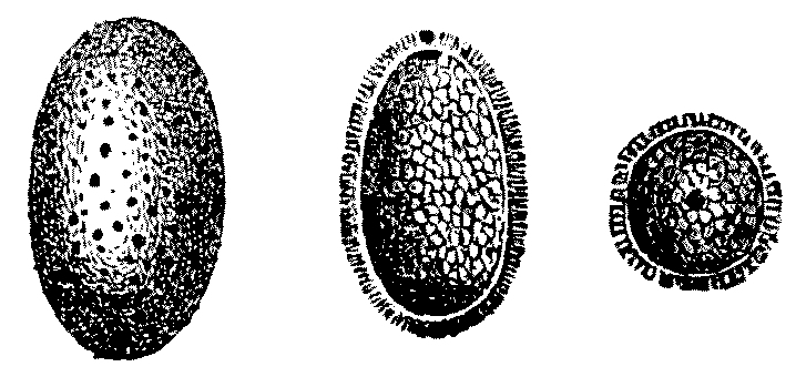
Fig. 52.—Cocoon of the medicinal
leech, and longitudinal and transverse views of the same cut open.
Fig. 52.—Cocoon of the medicinal
leech, and longitudinal and transverse views of the same cut open.
Leeches are hermaphrodite;
and in
some genera the
acting male inserts
spermatophores, or
little cases containing
spermatozoa, anywhere
in the skin of
the leech that is being
fertilised, and the spermatozoa then make
their way through the tissues of the body
of the potential female till they arrive at the
ovary and there fuse with the ova. In the
medicinal leech the mating is said to be encouraged
by adding fresh water to the vessels
in which the leeches are living.
The eggs are laid in capsules or cocoons
attached to some water-plant or buried in
the mud, about twenty-four hours after the
leeches have mated. The cocoon is formed,
as it is in an earthworm, by certain glands
in the skin which form a secretion that
hardens and takes the form of a broad ring,
as it were, round the body of the leech.
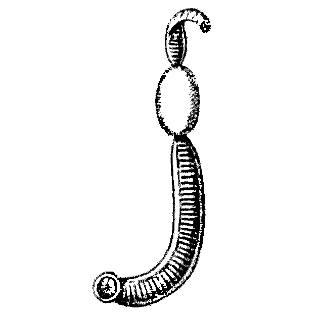
Fig. 53.—A Nephelis
forming its cocoon and withdrawing from it.
Fig. 53.—A Nephelis
forming its cocoon and withdrawing from it.
Through this broad ring the body of the leech is withdrawn and the
fertilised eggs are deposited in it. The two ends close up, but not
entirely, for the young leeches eventually make their way into the
outer water through one of the remaining pores. Within the cocoon are
six to twenty ova, and these gradually mature and the young hatch
out. When they leave the cocoon they are minute, and of the thickness
of pack-thread. More than one cocoon is deposited by each leech, but
unless the cocoons are anchored to some submerged object they often
rise to the surface of the water and float half submerged, and are then
apt to be destroyed by water-rats, voles, and other enemies of leeches.
At times the leeches themselves destroy their cocoons.
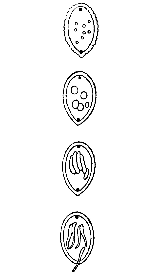
Fig. 54.—Cocoons of
Nephelis, showing the growth of the eggs and the issuing larvae,
which in the lower figure are leaving the cocoons.
Fig. 54.—Cocoons of
Nephelis, showing the growth of the eggs and the issuing larvae,
which in the lower figure are leaving the cocoons.
The exact time of the emergence from
the cocoon does not seem to be very definitely
known, but leeches are long-lived annelids.
It is not till their third year that they are
of any use for medicinal purposes, and they
are said not to pair until they are six or seven
years old. They certainly live twelve or fifteen
years. But, if we adopt an optimistic
view—and in this little book we
do—the fact that they grow up so
slowly and live so long shows that
it will be difficult to replace the
shortage of leeches in Great Britain
and Ireland during the present war.
This could hardly be done by home
culture, for even if the war lasts
three or four years we have lost
the cocoons of the summer of 1914,
even if we ever had them.
Leeches have many enemies:— water-rats,
voles, the larvae of the
Dytiscus beetle, the larvae of
Hydrophylus, the Nepa or water-scorpion,
the larvae of the dragonfly,
and the adult Dytiscus—all feed
upon them. Many birds also eat
leeches; and it is recorded that at
one artificial leech-farm, where there
were 20,000 leeches, they were all
eaten up in twenty-four hours by
an invasion of ducks. Frogs and
newts also devour them, and they
are not above eating their own brothers.
Aulostoma will devour its own species as
readily as it will an earthworm.
Those artificially reared, as is usually the
case with animals reared in captivity—probably
against their will—are peculiarly liable to
disease of various sorts. They not only
become diseased themselves, but they act as
carriers of disease and play the same part
to fish which biting insects play to man and
other terrestrial animals. They convey to
fishes protozoal diseases similar to those that
insects convey to man and other warm-blooded
vertebrates.
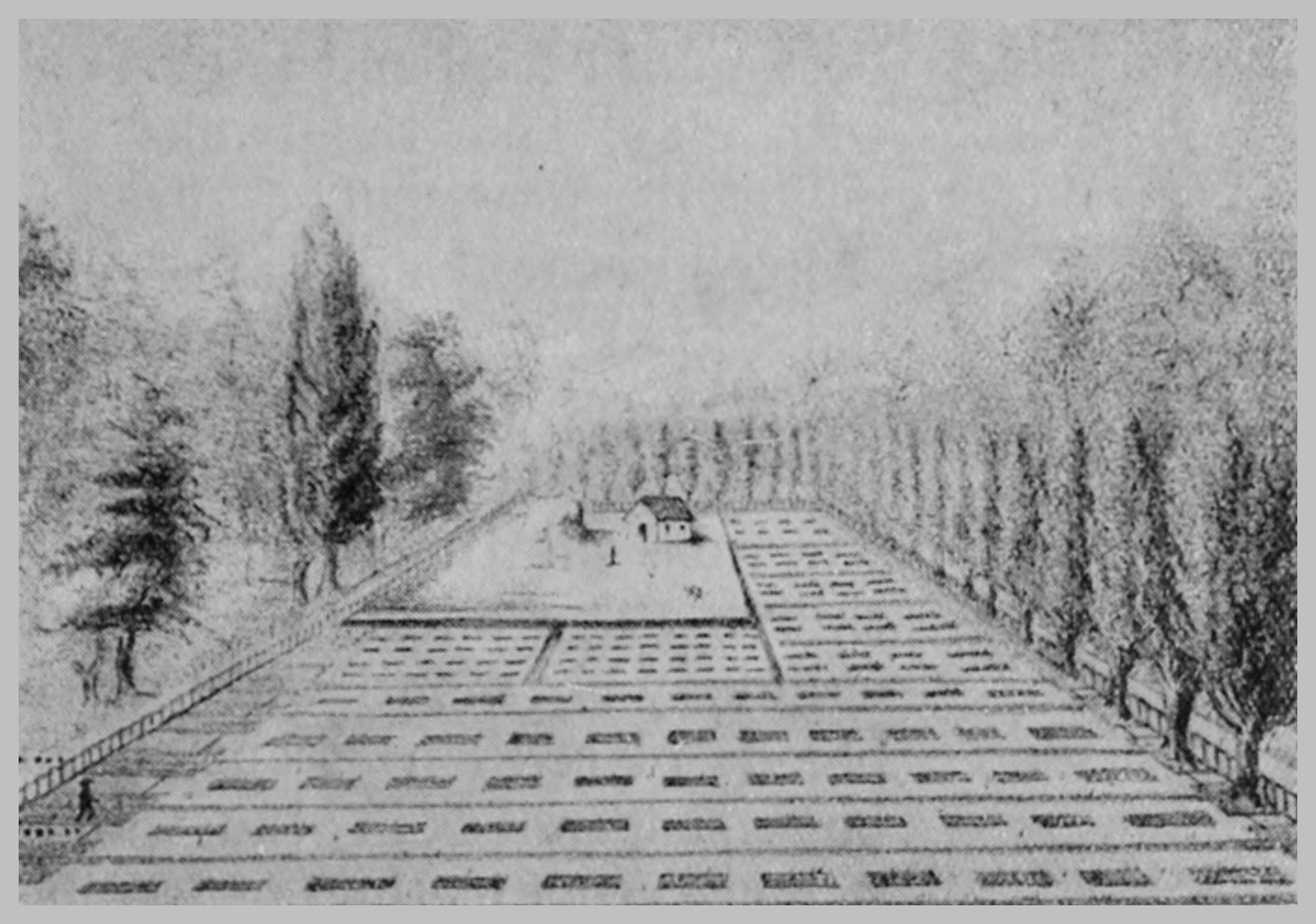
Fig. 55.—A leech-farm in the south of France.
Fig. 55.—A leech-farm in the south of France.
Leech-farming used to be a profitable undertaking,
but now it has fallen into desuetude
in these islands. Leeches are, however, still
cultivated in some parts of the world; and
in America, Latter describes a farm, situated
at Newton in Long Island, where there is,
or was, a leech-farm some thirteen acres
in extent. The farm consists of oblong ponds
of about one and a half acres,
each three feet deep. The
bottom of each pond is covered
with clay, and the banks are
made of peat. The French
writers recommend, as a rule,
the use of clay for the banks.
The ‘eggs’ (cocoons) are deposited
in the peat from June
onwards, till the weather gets
chilly. The adult leeches are
fed every six months with fresh
blood placed in stout linen
bags suspended in the water.
A more cruel method of feeding
these domesticated leeches is
that of driving horses, asses,
or cattle into the ponds—and
this was the custom in France.
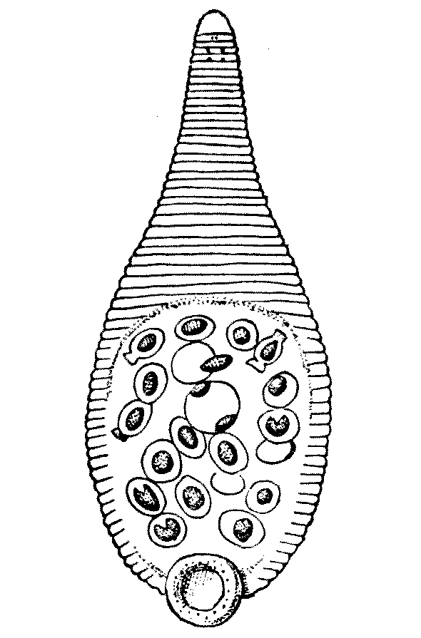
Fig. 56.—Glossosiphonia
heteroclita, with eggs and emerging embryos. Ventral view. × 4.
(From Harding.)
Fig. 56.—Glossosiphonia
heteroclita, with eggs and emerging embryos. Ventral view. × 4.
(From Harding.)
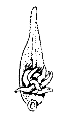
Fig. 57.—Helobdella stagnalis,
with adhering young. Ventral view, magnified. (From Harding.)
Fig. 57.—Helobdella stagnalis,
with adhering young. Ventral view, magnified. (From Harding.)
Some leeches show a considerable amount
of maternal affection. Glossosiphonia heteroclita,
for instance, carries its eggs about with
it, and Helobdella stagnalis has its little young
larvae attached by their tiny suckers to the
mother’s body, which they are loath to leave.
Aulostoma gulo, the horse-leech, is notoriously
a very ferocious feeder. Exactly
why this species is called a horse-leech is a
matter of speculation; but ‘horse’ used as
an adjective seems to imply something large
and something rather coarse—for instance,
horse-chestnuts, horse-play, horse-sense,
and horse-laugh.
The rapacity of the daughters
of the ‘horse-leach’ is dwelt on in
the Bible.[16] I am not an authority
on exegesis, but I have never felt
quite sure whether these two ladies
were not the offspring of the local
veterinary surgeon. But Aulostoma
does occur in Palestine, and its
voracity may very well have been
known to the Hebrews. I entirely
reject the idea that the word indicates
some ghost or phantom:
that explanation is due to the craven policy
of taking refuge in the unknown.
I conclude this chapter with a couple of
sentences taken from Dr. Phillips’s ‘Materia
Medica’ on the present use of leeches:—
The special value of leeching is shown in the
early stage of local congestion and inflammations:
such as arise from injuries, and in orchitis, laryngitis,
haemorrhoids, and inflammations of the ear[148]
and eye, cerebral congestions, and congestive fixed
headache.
Leeches are also of service, in a manner less
easy to understand, in inflammations of deep-seated
parts without direct vascular connexion with the
surface—for example, in hepatitis, pleuritis, and
pericarditis, as well as in pneumonia, peritonitis,
and, according to some observers, in meningitis.
In all these disorders, however, they are very much
less used than formerly—in the larger hospitals,
for instance, when at one time they cost many
hundred pounds annually, a few dozens in the year
would represent the total employed.[17]
CHAPTER XII
LEECHES
Part III
EXOTIC LEECHES
(Limnatis nilotica and Haemadipsa zeylanica).
Rulers that neither see nor feel nor know,
But leech-like to their fainting country cling,
Till they drop, blind in blood, without a blow.
(Shelley,
England in 1819.)
The extension of war into the Near and Far
East has brought into action two genera of
leeches which were and still are the cause
of extreme inconvenience and even of real
danger to troops operating in these areas. The
enemies of our Allies will still insist on fighting
on richly stocked leech-grounds. For in the
new war area, in southern Europe, Asia Minor,
Syria, Palestine, Egypt, and parts of India and
the real East, two genera of leeches—which are
indeed not the friend but the enemy of man,
especially of the soldier—abound.
The first of these two is Limnatis nilotica
(Sav.), and it is from Savigny that I have stolen
the picture of this species.
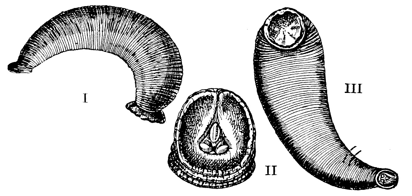
Fig. 58.—I. Limnatis nilotica, side view. II. Oral sucker,
showing the characteristic median dorsal slit and the three teeth;
III. ventral view. (From Savigny.)
Fig. 58.—I. Limnatis nilotica, side view. II. Oral sucker,
showing the characteristic median dorsal slit and the three teeth;
III. ventral view. (From Savigny.)
It is a leech of
considerable size, attaining a length of 8 cm.
to 10 cm., and its outline rather slopes inward
at the anterior end. The dorsal surface is
brownish-green with six longitudinal stripes,
and the ventral surface is dark. It is a fresh-water
leech, and it occurs from the Atlantic
Islands, the Azores, and the Canaries—its
western limit—all along the northern edge of
Africa until it reaches Egypt, Palestine, Syria,
Armenia, and Turkestan, where it achieves its
uttermost eastern boundary. This leech lives
in stagnant water; especially does it congregate
in drinking-wells—the wells so often
mentioned in the New Testament. In the
Talmud (Abōdāh Zārāh, 17b) an especial
warning is given against drinking water from
the rivers or wells or pools for fear of swallowing
leeches. Doubtless the New Testament Jew
knew in his day almost as much as we know now
about these leeches. They were the cause of
endless trouble to Napoleon’s soldiers in his
Egyptian campaign, and are still a real pest in
the Near East.
I cannot recall that Napoleon talked much
about spreading ‘Kultur,’[18] but he certainly
did it. He took with his army into Egypt
a score of the ablest men of science he could
gather together in France. He established in
Cairo an ‘Institut’ modelled on that of Paris;
and his scientific ‘corps’ produced a series of
monographs on Egyptian antiquities and on
the natural history of Egypt that has not yet
been equalled by any other invading force.
Napoleon freed the serfs in Germany, he
codified the laws of France, and these laws were
adopted by large parts of Europe; he extended
the use of the decimal system. Napoleon
had a constructive policy, and was never a
consistent apostle of wanton destruction. If
he destroyed it was to build up again, and in
many instances he ‘builded better than he
knew.’ He seldom so mistook his enemies as
to destroy, to terrify; the ‘frightfulness,’
though bad enough in his times, had limits.
Napoleon had at least in him the elements of
a sane and common-sense psychology. He
knew that what was ‘frightful’ to the French
was not necessarily ‘frightful’ to the Russian.
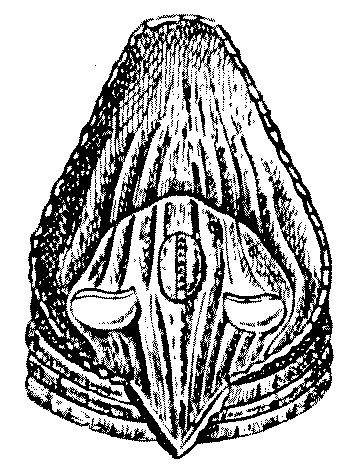
Fig. 59.—Anterior sucker
of Hirudo medicinalis. This is to compare with the anterior
sucker of Limnatis nilotica, which has a characteristic dorsal
median slit. See preceeding figure. (From Savigny.)
Fig. 59.—Anterior sucker
of Hirudo medicinalis. This is to compare with the anterior
sucker of Limnatis nilotica, which has a characteristic dorsal
median slit. See preceding figure. (From Savigny.)
Amongst the wonderful series of books
and monographs on Egypt which described
the varying activities of the
savants he took in his train,
and who, at the confines of the
eighteenth and nineteenth centuries
invaded the country of the
Pharaohs, none is more remarkable
than Savigny’s monograph
on the ‘Natural History’ of that
country. And in this folio the
leech (Limnatis nilotica) was for
the first time fully described and
depicted.
This particular leech is
swallowed by man, by domestic
cattle, and doubtlessly by wild
animals, with their drinking-water.
Amongst the medical writers of the Eastern
world in classical times who mention leeches
there was always, as there was amongst the
authors of the Talmud, a great and haunting
fear of leeches being swallowed, and these
writers mostly wrote from the area where
Limnatis nilotica still abounds.
According to Masterman, who has had,
as a medical officer in Palestine, a first-hand
opportunity of studying this leech, the pest
attaches itself to the mouth or throat or larynx
during the process of swallowing, and he is
convinced that if it be once really swallowed
and reaches the stomach it is killed and digested.
Limnatis nilotica, unlike Hirudo medicinalis
the medicinal leech, is unable to bite through
the outer integument of man and is only able
to feed when it has access to the softer mucous
membrane of the mouth or of the pharynx
or of the larynx, and of the other thinner and
more vascular internal mucous linings.
In Palestine these pests are particularly
common in the region of Galilee and in the
district of Lebanon. They are, in these and
other districts, so plentiful in the autumn that
almost every mule and almost every horse the
tourist comes across is bleeding from its mouth
or from its nose, for this species of leech is by
no means only a human parasite. The natives,
who know quite a lot about these pests,
generally strain them out of their drinking-water
by running the water through a piece
of muslin or some such sieve when they fill
their pitchers at the common well. In certain
districts these leeches in the local pools or
reservoirs are kept in check by a fish—a species
of carp (Capöeta fratercula).
In the cases which recently came under
Mr. Masterman’s observation, the leeches were
attached to the epiglottis, the nasal cavities,
and perhaps most commonly of all to the
larynx of their host. When they have been
attached to the anterior part of the mouth,
or any other easily accessible position, their
host or their host’s friends naturally remove
them, and such cases do not come to the
hospital for treatment.
The effect of the presence of this leech
(L. nilotica) on the human being is to produce
constant small haemorrhages from the mouth
or nose. This haemorrhage, when the leech
is ensconced far within the buccal, the nasal,
or the pharyngeal passages of the host, may
be prolonged, serious, and even fatal. Masterman
records two cases under his own observation
which ended in death: one of a man and
the other of a young girl, both of whom died
of anaemia produced by these leeches.
The average patients certainly suffer.
They show marked distress, usually accompanied
by a complete or partial loss of voice;
but all the symptoms disappear, and at once,
on the removal of the semi-parasite. Sometimes
the leeches are attached so closely to
the vocal cords that their bodies flop in
and out of the vocal aperture with each
act of expiration and inspiration. The hosts
of leeches so situated usually suffer from
dyspnoea, and at times were hardly able to
breathe.
The native treatment is to remove the
leech, when accessible, by transfixing it with
a sharp thorn; or they dislodge it by touching
it with the so-called ‘nicotine’ which
accumulates in tobacco-pipes. But nicotine is
destroyed at the temperature of a lighted pipe,
so whatever the really efficient juice is, it is
not nicotine. Still, as long as the fluid proves
efficient, the native is hardly likely to worry
about its chemical composition.
Masterman says that the two means he
has found most effective were: (1) Seizing the
leech, when accessible, with suitable forceps;
or (2) paralysing the leech with cocaine. In
the former case the surgeon is materially
assisted by spraying the leech with cocaine,
which partially paralyses it and puts it out of
action. In the latter case, if the spraying of
cocaine is not sufficient, Masterman recommends
the application of a small piece of
cotton-wool dipped in 30 per cent. cocaine
solution, which must be brought into actual
contact with the leech’s body. The effect of
the cocaine in contact with the skin of the
leech is to paralyse it and to cause it at once
to relax its hold. In such a case the leech is
occasionally swallowed, but it is more often
coughed up and out. Headaches and a
tendency to vomit are symptoms associated
with the presence of this creature in the human
body; the removal of the leech or leeches
coincides with the cessation of these symptoms.
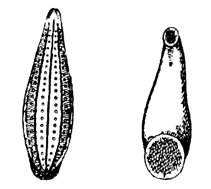
Fig. 60.—The Japanese
variety of Haemadipsa zeylanica. × 1. (From Whitman.)
Fig. 60.—The Japanese
variety of Haemadipsa zeylanica. × 1. (From Whitman.)
In the East, where many of our Territorial
regiments are now stationed, we come across
another species of leech even more injurious to
mankind than Limnatis nilotica.
This Asiatic leech is known as
Haemadipsa zeylanica, and is
one of a considerable number
of leeches which have left the
water, their natural habitat,
and have taken to live on land.
From India and Ceylon,
throughout Burma, Cochin
China, Formosa to Japan, the
Philippines, and the Sunda Island, this terrible,
and at certain elevations ubiquitous, pest is
spread. It lives upon damp and moist earth.
The family to which it belongs is essentially
a family which dwells in the uplands and
shuns the hot, low-lying plains. Its members
do not occur on the hot, dry, sandy flats.
Tennant has described the intolerable nuisance
they are in Ceylon. In fact of the many visible
plagues of tropical Asia and its eastern islands
they are perhaps the worst. Yet few have
recorded their dread doings, and those few
have escaped credence.
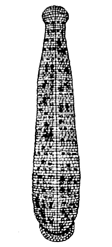
Fig. 61.—Haemadipsa
zeylanica, seen from above. × 2. (From Blanchard.)
Fig. 61.—Haemadipsa
zeylanica, seen from above. × 2. (From Blanchard.)
Each specimen of Haemadipsa zeylanica is
of a clear brown colour with a yellow stripe
on each side and with a greenish dorsal stripe.
There are five pairs of eyes, of which the first
four occupy contiguous rings; but
between the fifth and seventh
ring there are two eyeless rings
interposed. As in the medicinal
leech there are three teeth, each
serrated like a saw.
In dry weather they miraculously
disappear, and nobody
seems to know quite what becomes
of them; but with returning
showers they are found again
on the soil and on the lower
vegetation in enormous profusion.
Each leech is about one inch in
length and is about as thick as
a knitting-needle. But they contract
until they attain the
diameter of a quill pen, or extend their
bodies until they have doubled their normal
length. They are the most insinuating of
creatures, and can force their way through
the interstices of the tightest laced boot, or
between the folds of the most closely wound
puttee. Making their tortuous way towards
the human skin, they wriggle about under
the under-clothing until they attain almost
any position on the body they wish to take
up. Their bite is absolutely painless, and
it is usual for the human sufferer to become
aware that he has been bitten by these silent
and tireless leeches when he notices sundry
streams of blood running down his body
when he at last has the
opportunity of undressing.
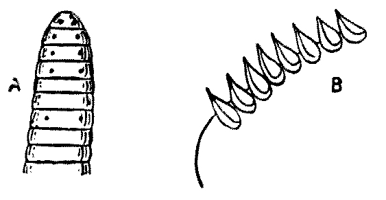
Fig. 62.—Haemadipsa
zeylanica. Head, showing the eyes and the serrations of the jaw.
Highly magnified. (From Tennant.)
Fig. 62.—Haemadipsa
zeylanica. Head, showing the eyes and the serrations of the jaw.
Highly magnified. (From Tennant.)
Sometimes, as Tennant’s
figure shows, these
land-leeches (H. zeylanica)
rest upon the ground.
At other times they ascend
the leaves of herbs and
grasses, and especially the
twigs of the forest undergrowth. Perched upon
the ends of growing shoots, leaves, and
twigs, stretching their quivering bodies into
the void, they eagerly watch and wait the
approach of some travelling mammal. They
easily ‘scent’ their prey, and on its approach
advance upon it with surprising rapidity in
semicircular loops. A whole and vast colony
of land-leeches is set in motion without a
moment’s delay, and thus it comes about
that the last of a travelling or prospecting
party in a land-leech area invariably fares
the worst, as these land-leeches mobilise
and congregate with extraordinary rapidity
when once they are warned of the approach
of a possible host, but not always in time
to engage in numbers the advanced guard.
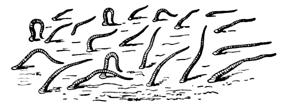
Fig. 63.—Haemadipsa zeylanica (land-leeches), on
the earth. (From Tennant.)
Fig. 63.—Haemadipsa zeylanica (land-leeches), on
the earth. (From Tennant.)
Horses are driven wild by them, and have
poor means of reprisal. They stamp their
hooves violently on the ground in the hope
of ridding their fetlocks of these tangled
masses of bloody tassels. The bare legs of
the natives, who carry palanquins, are particularly
subject to the bites of these bloodthirsty
brutes, as the palanquin-bearer has
no free hand to pick them off. Tennant
writes that he has actually seen the blood
welling over the boots of a European from
the innumerable bites of these land-leeches;
and it is on record that during the march
of the troops in Ceylon, when the Kandyans
were in rebellion, many of the Madras sepoys,
and their coolies, perished from their innumerable
and united attacks. It is also
certain that men falling asleep over-night
in a Cingalese forest have, so to speak, ‘woke
up dead’ next morning. These sleepers have
succumbed during the night to the repeated
attacks of these intolerable and insatiable pests.
Dr. Charles Hose, for many years Resident
at Sarawak, has told me that on approaching
the edges of woods in Borneo you can hear
every leaf rustling, and this is due to the
fact that the eager leech, perched on its
posterior sucker on the edge of each leaf
in the undergrowth, is swaying its body up
and down, yearning with an ‘unutterable
yearning,’ to get at the integument of man
or some other mammal.
Landor, who wrote, I think, the best book
about our adventure into Thibet some ten
years ago, entitled ‘Lhassa’ (London, 1905),
says of Sikkim:—
The game here is very scanty: the reason is
not uninteresting. For dormant or active, visible
or invisible, the curse of Sikkim waits for its warm-blooded
visitor. The leeches of these lovely valleys
have been described again and again by travellers.
Unfortunately the description, however true in
every particular, has, as a rule, but wrecked the
reputation of the chronicler. Englishmen cannot
understand these pests of the mountain-side, which
appear in March, and exist, like black threads
fringing every leaf, till September kills them in
myriad millions.
To remove them a bowl of warm milk at the[161]
cow’s nose, a little slip-knot, and a quick hand are
all that is required. Fourteen or fifteen successively
have been thus taken from the nostrils of one
unfortunate heifer.
When fully fed, a process which takes
some time with Haemadipsa zeylanica, the
individual leeches drop off; and they can be
made to loosen their hold by the application
of a solution of salt or of weak acid. Attempts
to pull them off should be avoided, as parts
of the biting apparatus are then often left
in the wound, and these may cause inflammation
and suppuration. Dr. R. J. Drummond,
who has had experience of these land-leeches
in Ceylon, has told me that the bite is often
septic and that it often leads to a serious
abscess which is long in healing. He recommends
pushing a match, which has been dipped
into carbolic acid, well home into the sinus
made by the leech’s head.
When winter approaches the leeches die
down with extraordinary rapidity, and the
species ‘carry on’ over the cold-weather
period in the form of eggs laid in cocoons on
the ground, under leaves, or other débris.
Hence no land-leech ever sees its offspring,
and no land-leech has ever known a mother’s
care.
INDEX
- Acanthia, 23
- Acarina, 89
- Acarus (Sarcoptes scabiei), 99
- Acne, 99
- African tick-fever, 116, 121
- Agronom, M., 16, 17
- Allcock, Colonel, 109
- Analgesinae, 111
- Annandale, Dr., 129
- Anoplura, 1, 11
- Antelopes, 36
- Antennae, of lice, 3;
- of bed-bug, 25
- Anthomyidae, 74
- Anthrax, 72
- Ants, 27
- Arachnids, 88
- Argas persicus, characteristics of, 114;
- breeding habits, 115
- Argasidae, 113
- Arhynchobdellae, 125. See Rhynchobdellae
- Arthropoda, 88
- Auchmeromyia luteola, 82, 83
- Aulostoma, 144;
- A. gulo, 147
- Austen, Mr. E. E., 65
- Babesia bovis, 120
- Bacillus pestis, 45
- Bed-bug (Cimex lectularius), 23;
- antennae of, 25;
- characteristics, 25–26
- Benzine, 13, 33
- Beveridge, Lieut.-Colonel, 48, 50
- Biscuits, infestation of Army, 50
- Biscuit-moth, 48
- Black Death, 38, 45
- Blake, 45
- Blow-flies, 83
- Blue-bottle (Calliphora erythrocephala), 74, 76
- Boars, 36
- Body-louse, 3, 10, 20
- Boer prison-ship, 20
- Brightwell, 131
- British Expeditionary Force, 46
- British Medical Journal, 21
- British Museum, 50
- Bubonic plague, 35, 38
- Bug. See Bed-bug
- Burns, 4
- Butler, Mr., 40
- Calliphora erythrocephala (blue-bottle), 76;
- C. vomitoria, 76
- Carp, 153
- Centipedes, 88
- Ceratophyllus fasciatus, 45
- Chigo (burrowing-flea), 37
- Chrysomyia macellaria, 83
- Ciliata, 138
- Cimex, 23, 25, 26, 27;
- C. lectularius (bed-bug), 23;
- C. rotundatus, 23
- Cockroach, 21, 24, 26, 27, 39
- Cocoons, 161
- Congo-floor-maggot, 82
- Copeman, Dr. S. Monckton, 21, 65
- Corcyra, 49;
- C. cephalonica, 49
- Cow-dung, 42
- [164]
- Cresol-soap solution, 21, 22
- Crustacea, 88
- Ctenocephalus canis, 37;
- Ct. felis, 37
- Cyclops, 138
- Cytoleichus sarcoptioides, 111
- Deer, 36
- De Geer, 30
- Demodex, 97 ff
- Demodex folliculorum, 97
- Dermacentor vernustus, 120
- Diarrhoea, 70
- Dizziness, 85
- Dog-flea, 40
- Dog-tick, 120
- Drummond, Dr. R. J., 15, 42
- Durrant, Mr., 48, 50
- Dytiscus, 144
- Ébrard, 131
- Empusa, 65
- Enteric, 69
- Entomologist, 18
- Ephestia kühniella, 48
- Erskine, Lord, 139
- Erythema, 94
- Erythema autumnale, 94
- Esox lucius, 15
- Faichne, 69
- Fannia, 76, 84;
- F. canicularis, 74, 75;
- F. scalaris, 74, 76
- Flea (Pulex irritans), 35;
- extermination of, 42
- Flies (Musca domestica), 57;
- breeding habits, 59;
- powers of travel, 67
- Flour-moth, the (Ephestia kühniella), in soldiers’ biscuits, 46
- Flowers of sulphur, 16
- Folklore, 15
- French, Field-Marshal, 135
- Frogs, 144
- Fumigation, method of, 33
- Gascoigne, 40
- Goats, 36
- Graham-Smith, Dr., 68, 81
- Grimbert, 97
- Guiart, 97
- Haemadipsa zeylanica, 156, 157
- Harding, Mr. W. A., 123, 129, 137
- Hair-follicles, 94
- Hair-louse, 3
- Harman, Dr. N. Bishop, 19
- Harvest-mite (Trombidium), 87, 91, 92, 97
- Head-louse, 4, 9
- Hemipterous insects, 27
- Henna, 16
- Herod Agrippa, 84
- Hewitt, Dr. C. Gordon, 58
- Hirudinea, 124
- Hirudo medicinalis, 123, 127, 129, 136, 140, 153
- Horse-leech, 147
- Hose, Dr. Charles, 160
- Huxley, 87
- Hydrocyanic-acid gas, 33
- Hydrophylus, 144
- Itch-mite, 99
- Ixodes, 119;
- I. ricinus, 120
- Ixodiphagus caucurtei, 120
- Jeyes’ fluid, 22
- Jigger (chigo), 95
- Joffre, General, 135
- Khiva, 17
- Kerosene, 33
- Laminosioptes gallinarum, 111
- Lancet, 21
- Landor, 160
- Larva, 8;
- of flea, 41
- Latter, Mr. H. O., 140
- Leech, 123;
- the medicinal (Hirudo medicinalis), 123;
- breeding habits, 142, 143;
- coloration of, 137;
- enemies of, 144;
- exotic (Limnatis nilotica and Haemadipsa zeylanica), 149;
- farming of, 145;
- fear of swallowing, 152;
- modes of conveying, 133;
- movements of, 137;
- suffering caused by, 154;
- Asiatic, 156;
- characteristics of, 157, 161;
- disappearance of, 157;
- method of attack, 158;
- size of, 157
- to remove, 142;
- traffic in, 131;
- uses of, 130
- Leech-ponds, 128
- Lefroy, Professor, 22
- Leishman, Sir William, 70
- Leptus autumnalis, 91
- Lice (Pediculus), 1;
- rules for avoidance of, 12
- Limnatis granulosa, 129;
- L. nilotica, 149, 152, 153, 154, 156
- Linnaeus, 87
- [165]
- Lounsbury, Mr. C. P., 18
- Lucius, 15
- Maggot, 82
- Mallophaga, 18
- Martin, Professor C. J., 31
- Masterman, Dr., 154, 155
- Mercury, 17
- Mites, 87;
- bites of, symptoms and treatment, 107–9;
- breeding habits of, 103;
- characteristics, 103, 104;
- Demodex, Sarcoptes, 97;
- epidemics caused by, 105;
- endo-parasitic, 109;
- nourishment of, 91
- Montagu, Lord, 59
- Murinae, 45
- Murray, Andrew, 3
- Musca, 84;
- M. domestica (house-fly), 57, 70, 74, 75
- Myiasis, 81
- Napoleon, 151
- Necrosis, 83
- Nepa, 144
- Nephrophages sanguinarius, 109, 111
- Newsholme, Dr., 65
- Nicholas, Grand Duke, 135
- Nicol, Sergeant Daniel, 55
- Nicol, W., 85
- Nicotine, 155
- Nightingale, Dr. P. A., 43
- Nits, 12
- Nymph, 117
- Oligochaeta, 124
- Ophthalmia, 72
- Ormerod, Miss, 48
- Ornithodorus megnini, 117;
- O. moubata (‘tampan’), 116, 121;
- O. turicata, 117
- Panama Canal, 86
- Paraffin, 13, 14
- Pediculoides ventricosus, 95
- Pediculus (lice), 2;
- P. capitis, 2, 4, 10, 12, 13;
- P. vestimenti, 2, 4, 8, 10, 12, 14
- Pernet, Dr. G., 107
- Petrol, 13, 14
- Petroleum, 17
- Pharynx, 140;
- sucking, 141
- Phillips, Dr., 147
- Phthirius, 2
- Pike, 15
- Plague, 31;
- Pepys’s, 45
- Proboscis, 25
- Protoplasm, 87
- Protozoal diseases transmitted by ticks, 117
- Pulex irritans (flea), 37, 40, 45
- Purkinje, 87
- Quick Laboratory, 9;
- Quick Professor of Biology, 11
- Radcliffe, Dr. H., 107
- Rat-fleas, 38
- Redi, 80
- Relapsing fever, 14
- Rhynchobdellae, 125. See Arhynchobdellae
- Rice, 49
- Riley, Professor, 48
- Rocky Mountain Fever, 120
- Romilly, Sir Samuel, 139
- Rothschild, Hon. Charles, 38
- [166]
- Rubies, 94
- Ruskin, 57
- Russell, Mr., 39
- Sarcophaga carnaria, 80
- Sarcoptes, 104
- Sarcoptes scabiei, 106
- Sarts, 16, 17
- Savigny, 149, 152
- Scabies, 107
- Sharp, Dr. David, 88
- South African War, 11, 19
- Spirochaete, 31;
- S. obermeieri, 31
- Spirochaetosis, conveyed by A. persicus, 114
- Stable-yards, 59
- Stag-beetle, 39
- Sulphur, 33;
- bags, 18;
- flowers of, 18
- Sweat-glands, 94
- Talmud, 150
- ‘Tampan,’ 114, 116
- Tennant, Mr., 159
- Ticks, characteristics of, 112, 113, 117;
- breeding habits, 119;
- habits, 114, 115
- Tomkins, Dr. H. H., 18
- Trichinella, 53
- Trombidium (harvest-mite), 95;
- T. holosericeum, 91
- Tuberculosis, 31
- Tunnels, burrowed by mite, 106
- Turpentine, 13;
- oil of, 34
- Typhoid, 69;
- bacilli, 68
- Typhus, 10, 14, 31
- ‘Vermijelli,’ 22
- Von Hindenburg, General, 135
- Von Kluck, General, 135
- Voles, 143
- Warburton, Mr. C., 4, 5, 8, 9
- Water-rats, 143
- Wordsworth, 127
- Xenopsylla cheopis, 45
- Xylol, 13
- Yeomanry, D.C.O., 19
PRINTED BY
SPOTTISWOODE AND CO. LTD., COLCHESTER
LONDON AND ETON
The six images that follow are the remaining pages of the book.
Click on any image below to see the plain text of that page.
Searchable text versions of those six images.
With Portraits. Demy 8vo. 10s. 6d. net.
“J” A Memoir of John Willis Clark
Registrary of the University of Cambridge,
and sometime Fellow of Trinity College.
By A. E. SHIPLEY, Master of Christ’s College.
In the Cambridge Review, the Provost of King’s says:—“A book of permanent
value to Cambridge men and of vivid immediate interest to the generations who
knew ‘J.’ Dr. Shipley has told his story with a combination of true appreciation
and perceptive humour.”
Times:—“The affectionate and sympathetic portrait of ‘J’ in his private life
as he was known and loved by hundreds. He was above all remarkable for his
power of winning and retaining the affection of generation after generation of young
men.”
Evening Standard:—“It ought to be generally read, being one of the most cheerful
biographies we have ever come on. A vivid picture of a remarkable character.”
A GIFT FOR NATURALISTS AND SPORTSMEN.
Royal 8vo. 12s. 6d. net. In 1 Volume. With Coloured and other
Illustrations. NEW AND CHEAPER EDITION.
The Grouse in Health and in Disease.
[Illustration of a grouse]
Edited by A. S. LESLIE and A. E. SHIPLEY, F.R.S.
(Master of Christ’s College, Cambridge).
With an Introduction by LORD LOVAT.
Times:—“The editors have fully
fulfilled their expressed intention of
making the book a practical guide to
sportsmen and naturalists.”
Scotsman:—“A book which, for
many years to come, is likely to remain
the standard authority on the grouse.”
London: Smith, Elder & Co., 15 Waterloo Place, S.W.
Smith, Elder & Co.’s Publications
The Minor Horrors of War.
By Dr. A. E.
SHIPLEY, F.R.S., Master of Christ’s College, Cambridge.
Crown 8vo. With 64 Illustrations. 1s. 6d.
net in Paper Cover, or 2s. net in Cloth.
The Early Diagnosis of Heart Failure and
other Essays on the Heart and Circulation.
By T. STACEY WILSON, M.D.Edin., F.R.C.P.Lond.,
Senior Physician to the General Hospital, Birmingham; Lecturer on
Medicine in the Dental School of the University of Birmingham.
With over 130 original Diagrams showing the results
of auscultation, and of percussion of the heart and liver, in cases of
cardiac inefficiency, and 30 Figures showing cardiac tracings. Demy
8vo., 12s. 6d. net.
Medical World.—“The importance of this volume cannot be over-estimated. We
doubt if anything so valuable has ever been written on the subject of cardiac disease.”
An Index of Symptoms with Diagnostic Methods.
By RALPH WINNINGTON LEFTWICH, M.D., late Assistant
Physician to the East London Children’s Hospital; Author of
“Tabular Diagnosis,” “A Pocket-Book of Treatment,” “Syphonage
in the Large Intestine,” &c.
Fifth and enlarged edition. Crown 8vo., 10s. 6d. net.
Medical Review.—“Wholly admirable and unique.”
Lancet.—“A most useful compilation.”
British Medical Journal.—“We can strongly recommend the book.”
Medical Press.—“It fills a genuine gap in medical literature.”
With Illustrations and Diagrams. Demy 8vo, 3s. 6d. net.
How to Diagnose Small Pox. A Guide for
General Practitioners, Post Graduate Students, and Others.
By W. McC. WANKLYN, B.A.Cantab., M.R.C.S.,
L.R.C.P., D.P.H. Assistant Medical Officer of the London County
Council, and formerly Medical Superintendent of the River Ambulance
Service (Small Pox) of the Metropolitan Asylums Board.
Universal Medical Record.—“Dr. Wanklyn is well able to lay down golden
rules for the guidance of the general medical practitioner and the post-graduate
student. Dr. Wanklyn, by his original and chatty style, brings out every point of
importance with great clearness.”
A Junior Course of Practical Zoology.
By the late A. MILNES MARSHALL, M.D., D.Sc., M.A.,
F.R.S., and the late C. HERBERT HURST, Ph.D. Revised by
F. W. GAMBLE, D.Sc., Lecturer in Zoology in the University of
Birmingham.
Seventh Edition. With Illustrations. Crown 8vo.,
10s. 6d.
Animal Life.
By F. W. GAMBLE, D.Sc., F.R.S.,
Lecturer in Zoology in the University of Birmingham.
With numerous Half-tone and Line Illustrations.
Crown 8vo., 6s. net.
London: Smith, Elder & Co., 15 Waterloo Place, S.W.
Works by Sir FREDERICK TREVES, Bart., G.C.V.O.,
&c. Serjeant Surgeon to H.M. The King.
With a Coloured Frontispiece, 40 pages of Half-Tone Illustrations,
and a Map.
The Cradle of the Deep:
AN ACCOUNT OF A VISIT TO THE WEST INDIES.
Times.—“A very attractive book ... fresh, instructive, humorous, and
inspiring.”
Standard.—“The best book that has been written about the West Indies since
Kingsley and Froude laid down their brilliant pens.”
Daily Telegraph.—“Rich in touches of genuine wit and humour, and richest
of all in those flashes of humanity and personal sympathy which are the very life-blood
of literature ... the traveller’s note-book in excelsis.”
Daily News.—“A very attractive book, so attractive that it will probably be
the most popular West Indian guide book for many years to come.”
With 72 Illustrations from Photographs by the Author, and a Map.
Uganda for a Holiday.
Times.—“ ... Sir Frederick possesses a talent for observation, and, what is
more, the gift of describing what he observes in terse and vivid sentences.”
Morning Post.—“The distinguished surgeon writes for the benefit not of big
game hunters, but of tourists like himself, who desire, for a change, to get beyond
the beaten track.”
Standard.—“ ... Another agreeable record of unconventional travel....
The book is written with imagination and humour.”
Daily Mirror.—“From start to finish the book brims over with good things.”
With Illustrations from Photographs by the Author, and a Map.
The Land that is Desolate:
AN ACCOUNT OF A TOUR IN PALESTINE.
Daily News.—“It sets down in entertaining and well-turned sentences those
things which are likely to interest the eye and the mind of a cultured and humane
holiday maker.”
Glasgow Herald.—“Admirable illustrations reach the very highest standard
of amateur work. The author’s pen is as remorselessly faithful as his camera. The
caustic humour which enlivens the book helps to make it most interesting.”
Globe.—“Much of the charm that characterises Sir Frederick Treves’ book
lies in that ironic humour which every now and then breaks forth in his descriptions
of cities and men.”
London: SMITH, ELDER & CO., 15 Waterloo Place, S.W.
Recent Publications
Small demy 8vo. 7s. 6d. net.
Canadian Nights. Being Sketches and
Reminiscences of Life and Sport in the Rockies,
and the Canadian Woods.
By the Right Honble. The Earl of Dunraven, K.P., C.M.G.,
Under-Secretary to the Colonies, 1885–1887.
Author of ‘The Great Divide,’ &c.
Sunday Times.—‘Short stories, bright, racy and fascinating. The author’s
style is excellently adapted to reproduce the atmosphere of the mountains and the
woods and will attract many readers.’
With 26 Illustrations and a Sketch Map.
Small royal 8vo. 10s. 6d. net.
Hunting and Hunted in the Belgian Congo.
By R. Davey Cooper.
Edited by R. Keith Johnston.
Court Journal.—‘A wonderful series of adventures. As thrilling a travel book
as has been published for many a long day.’
Small demy 8vo. 7s. 6d. net.
With Portraits, one being that of Lord Kitchener, G.C.M.G., K.P., from
the Painting by the Hon. John Collier.
Egypt in Transition.
By Sidney Low.
Author of ‘A Vision of India.’ &c. With an Introduction by the
Right Hon. The Earl of Cromer, O.M., P.C., G.C.B.
Times.—‘The best picture of the more accessible parts of the Sudan which has
yet been published. Mr. Low is a deft craftsman; he can make you a picture of
the desert, of feathery palm-trees, of the shining waters of the Nile, in a few telling
phrases.’
Country Life.—‘The book has a story to tell of infinitely greater interest than
that in any novel.’
With Illustrations and a Map. Large post 8vo. 5s. net.
Travel and Politics in Armenia.
By Noel Buxton, M.P., and the Rev. Harold Buxton.
With an Introduction by the Right Hon. Viscount Bryce, and a Contribution
on Armenian History and Culture by Aram Raffi.
Scotsman.—‘An excellent purpose will be achieved by the present book if it
serves to focus attention on Armenian affairs. A pleasant blend of travel talks and
political discussions.’
London: Smith, Elder & Co., 15 Waterloo Place, S.W.
Recent Publications
Crown 8vo. 1s. net.
Principles of Property.
By J. Boyd Kinnear,
Author of ‘Principles of Civil Government,’ &c.
Scotsman.—‘An able, interesting and well-reasoned statement of some cardinal
principles of modern sociology.’
Belfast Northern Whig.—‘The publication is in every way useful and timely,
and it is written with a fulness of knowledge and a clearness and restraint of expression
that add immensely to its value.’
Large medium 8vo. With Portraits. 6s. net.
Memories of John Westlake.
Contributed by some of the many friends of the distinguished international lawyer.
Times.—‘A scholar, thinker, and a man of science. His influence was deep and
lasting. A few men are the teachers of their age, and among them was John Westlake.’
Small demy 8vo. With 16 page Illustrations. 7s. 6d. net.
St. Margaret’s, Westminster.
By H. F. Westlake, M.A.,
Custodian and Minor Canon of Westminster Abbey.
Pall Mall Gazette.—‘We are indebted to Canon Westlake for one of the most
interesting monographs that the still unexhausted field of London history has produced.
Deeply interesting and abundantly informing.’
Large post 8vo. 6s. net.
Shakespeare Personally.
By the late Professor Masson. Edited by Rosaline Masson.
Scotsman.—‘Lectures edited and arranged by his daughter with a skill that
makes them read like a new treatise. Their appearance is an event not without its
own importance in the history of contemporary Shakespearean criticism.’
2 vols. Demy 8vo. 15s. net.
American and English Studies.
By the late Whitelaw Reid. With an Introduction by Royal Cortissoz.
Times.—‘Few men’s speeches show on every page so lofty a standard of civic
ethics, so much courage, and so wide and intimate an acquaintance with the literature
and history of other countries besides those of their own.’
Demy 8vo. 10s. 6d. net.
Malcolm MacColl.
Memoirs and Correspondence. With a Portrait.
By the Right Honble. George W. E. Russell,
Author of ‘Collections and Recollections.’
Daily Chronicle.—‘A correspondence of inordinate importance. An excellent
memorial to Canon MacColl, the most effective of modern pamphleteers. A book of
abiding interest.’
London: Smith, Elder & Co., 15 Waterloo Place, S.W.
SMITH, ELDER & CO.’S NEW SHILLING NET SERIES.
| 1. |
Deeds that Won the Empire. |
Dr. W. H. Fitchett. |
| 2. |
The Cruise of the “Cachalot.” |
Frank T. Bullen. |
| 3. |
Fights for the Flag. |
Dr. W. H. Fitchett. |
| 4. |
The Log of a Sea Waif. |
Frank T. Bullen. |
| 5. |
The Gamekeeper at Home. |
Richard Jefferies. |
| 6. |
A Londoner’s Log Book. |
Rt. Hon. G. W. E. Russell. |
| 7. |
The Sowers. |
H. S. Merriman. |
| 8. |
Jess. |
H. Rider Haggard. |
| 9. |
Vice Versâ. |
F. Anstey. |
| 10. |
Woodland, Moor, and Stream. |
J. A. Owen. |
| 11. |
The Tale of the Great Mutiny. |
Dr. W. H. Fitchett. |
| 12. |
Sixty Years in the Wilderness. |
Sir Henry W. Lucy. |
| 13. |
A Vision of India. |
Sidney Low. |
| 14. |
The Defence of Plevna. |
Capt. F. W. von Herbert. |
| 15. |
The Memoirs of Sherlock Holmes. |
A. Conan Doyle. |
| 16. |
Nelson and His Captains. |
Dr. W. H. Fitchett. |
| 17. |
With Edged Tools. |
Henry Seton Merriman. |
| 18. |
The Adventures of Sherlock Holmes. |
A. Conan Doyle. |
| 19. |
The Exploits of Brigadier Gerard. |
A. Conan Doyle. |
| 20. |
Wellington’s Men. |
Dr. W. H. Fitchett. |
| 21. |
The Red Hand of Ulster. |
George A. Birmingham. |
| 22. |
The Honourable Molly. |
Katharine Tynan. |
| 23. |
A Life’s Morning. |
George Gissing. |
| 24. |
Court Royal. |
S. Baring-Gould. |
London: SMITH, ELDER & CO., 15 Waterloo Place, S.W.
Transcriber’s Note (continued)
Minor typographical errors have been corrected
in this transcription. Other errors and unusual or variable spelling and
hyphenation have been left unchanged except as noted below.
Page xvi — “Sargophaga” changed to “Sarcophaga” (Sarcophaga carnaria)
Page 27 — “features” changed to “feature” (Perhaps the most disagreeable feature)
Page 38 — “bubonic-plague” changed to “bubonic plague” (bubonic plague was associated first with rats)
Page 38 — “subtropical” changed to “sub-tropical” (in all tropical and sub-tropical countries)
Page 49 — “inself” changed to “itself” (The moth itself is a rather insignificant)
Page 65 — “Austin” changed to “Austen” (Dr. Monckton Copeman and Mr. E. E. Austen)
Page 75 — “housefly” changed to “house-fly” (F. canicularis, the ‘lesser house-fly,’)
Page 93 — “proboscis” changed to “hypo-pharynx” (presence of their hypo-pharynx in the skin)
Page 123 — “gentlemen” changed to “gentleman” (when an old gentleman had upset)
Page 126 — “has removed been” changed to “has been removed” (the right half of this organ has been removed to show)
Page 135 — “von Hinderberg” changed to “von Hindenburg” (General von Hindenburg)
Page 150 — “freshwater” changed to “fresh-water” (It is a fresh-water leech)
Page 152 — “preceeding” changed to “preceding” (See preceding figure.)
Page 158 — “underclothing” changed to “under-clothing” (they wriggle about under the under-clothing)
Page 163 — “Austin” changed to “Austen” (Austen, Mr. E. E.)
Page 163 — “lectularis” changed to “lectularius” (Bed-bug (Cimex lectularius))
Page 164 — “Ctenophalus” changed to “Ctenocephalus” (Ctenocephalus canis)
Page 164 — “Ebrard” changed to “Ébrard” (Ébrard, 131)
Page 164 — “Hirudi” changed to “Hirudo” (Hirudo medicinalis)
Page 164 — “Lemna” changed to “Henna” (Henna, 16)
Page 164 — “Limnalis” changed to “Limnatis” (Limnatis nilotica)
Page 164 — “vernustus” changed to “venustus” (Dermacentor venustus)
Page 166 — “Trombidum” changed to “Trombidium” (Trombidium (harvest-mite))
Page 166 — “Von Hinderberg” changed to “Von Hindenburg” (Von Hindenburg, General)
Footnotes have been re-indexed using numbers and placed before the Index.
Back to top