THE LONDON MEDICAL GAZETTE, — PATHOLOGICAL ESSAYS ON SOME DISEASES OF THE HEART; — ESSAY II. — ESSAY III. — DILATED ŒSOPHAGUS. — HYDROPHOBIA. — CASE OF LONG PROTRACTED CONSTIPATION. — DISLOCATION OF THE PATELLA. — VACCINATION. — SINGULAR BICEPS MUSCLE. — STETHOSCOPE. — MECHANISM OF VOMITING. — CHARACTER OF THE LANCET. — DURATION OF HUMAN LIFE IN RUSSIA.
TRANSCRIBER'S NOTE
The 'Apothecary System' of measurement was used in the original text.
The etext uses the following Unicode symbols:
℞ Prescription symbol
ʒ Dram
℥ Ounce
ℳScruple
Roman numerals, and 'ss.' for 'half', are retained, so for example
'℥iss.' indicates 'one and a half ounces'.
The 'Apothecary System' of measurement was used in the original text.
The etext uses the following glyphs taken from the text images:
 Prescription symbol
 Dram
 Ounce
 Scruple
Roman numerals, and 'ss.' for 'half', are retained, so for example
'iss.' indicates 'one and a half ounces'.
Obvious typographical errors and punctuation errors have been corrected after careful comparison with other occurrences within the text and consultation of external sources.
More detail can be found at the end of the book.
BEING A
WEEKLY JOURNAL
OF
Medicine and the Collateral Sciences.
SATURDAY, DECEMBER 27, 1828.
Being the Substance of Lectures delivered before the College of Physicians,
By P. Mere Latham, M.D.
Physician to St. Bartholomew's Hospital.
[Continued from p. 7.]
Morbid Anatomy of the Internal Lining Membrane of the Heart.
The membrane which lines the cavities of the heart is very liable to disease, but not equally so in every part. Where it is thin and transparent, and admits the colour and character of the muscular structure upon which it is spread to be seen through it, it is seldom found diseased; but where it is of a denser texture, either in itself or from an admixture of other structures, whether cellular or fibrous, with its own, it is frequently, and often exclusively diseased. This latter character of a denser texture belongs to it where it forms the tough white circles which surround the apertures of communication between the auricles and ventricles; also where it is reflected upon itself, and forms the loose duplicatures of membrane, which are given off, as it were, from the internal surface of the heart, either at the fibrous circles intermediate between the auricles and ventricles, constituting the tricuspid and the mitral valves, or at the commencement of the pulmonary artery and aorta, constituting the semilunar valves.
It is remarkable how curiously disease is apt to limit itself to the spaces just pointed out. Of the fibrous circle between the auricle and ventricle, of the valves which originate from it, and of the tendinous cords which connect the valves with the carneæ columnæ, there will not be the smallest space free from disease; but the disease will abruptly stop where the tendinous cords cease and the carneæ columnæ begin[1].
The membrane, however, where it covers the fleshy columns of the heart, is not exempt from the possibility of disease: but when disease actually affects it, it has seldom originated there, but has generally spread from other parts of the same membrane, although (as we have just remarked) it is apt to stop short before it reaches this.
Of the two sides of the heart, the membrane which lines the left is unquestionably the more liable to disease. But my own observation would never have led me to conclude that the membrane of the right side was so far exempt as it is commonly thought to be. Speaking from the best recollection I have of the specimens which have fallen under my examination, I should say that, in one-third of the cases where disease has been found on the left side, it has existed on the right side also, and been essentially of the same character. But there has been a remarkable difference in the extent to which it has proceeded on each side respectively: while on the left it has gone so far as to be the undoubted cause of death, on the right, although essentially of the same character, it has been only just beginning.
It very seldom happens that disease appertains to the lining of the right cavities of the heart exclusively; and, where it affects both, the disease in the right cavities is very seldom found in advance of that in the left.
It should seem, indeed, according to the ordinary course of things, that disease does not begin in the lining of the right cavities of the heart, until it has already advanced to an extreme degree in the left.
The internal lining of the heart, as well as of the arteries, is often found to have become of a red colour. This redness, so well known to all who are accustomed to examine dead bodies, used to be regarded as a mere stain imparted to it by the colouring matter of the blood after death. Yet a due consideration of various circumstances connected with it will hardly warrant this conclusion.
It has been found whether the heart or artery be full or empty of blood; and if blood be present it has been found, whether it is liquid or clotted; and if it be clotted, whether it does or does not retain its colouring matter. External temperature, and length of time between the death of the patient and the dissection of his body, have not made any difference in the frequency with which this peculiar appearance occurs. Lastly, no artificial methods, such as washing of any kind, can get rid of it; nor will inclosing blood within an artery for any period produce it[2]. This simple redness is sometimes seen universally in both sides of the heart and throughout the whole arterial system, and sometimes in patches only, of greater or less extent, whether in the arteries or in the heart.
Now, when all these circumstances are considered, although in some instances it may be a mere stain imparted by the colouring matter of the blood after death, it is plainly impossible that it should be of that nature in any large proportion of the numerous instances in which it is found.
But if the appearance in question implies (as I believe it generally does) a morbid condition, of what kind is that condition? There are the same objections to considering mere redness as equivalent to inflammation here as in the pericardium, or in any other part of the body: here, as elsewhere, in one case it may be the condition out of which inflammation is to spring; while in another it may not be destined to give origin to any change in the structure of the part beyond itself, and may itself constitute the whole disease.
It appears to me, that this mere redness of the internal lining of the heart and arteries has become a matter of undue perplexity to pathologists, because they have laboured to infer from it more than the simple fact itself will authorize. All I wish to establish concerning it is, first, that it is not always (probably very seldom) a mere stain imparted by the colouring matter of the blood after death; secondly, that it alone does not constitute inflammation.
It may not be improper to mention the circumstances under which it has occurred to myself to find it. I have met with it most frequently, and to the largest extent, in subjects whose previous disease has produced a constant and habitual impediment to the transmission of blood through the heart and through the lungs, and that impediment has gone on increasing to the hour of their death; also in those, whatever might have been the nature of their disease, whose dissolution (I mean the actual process of dying) has been tardy and agonizing, and marked by great labour of respiration; in the apoplectic, for example, in whom, after sense and consciousness were extinct, life had been protracted, with stertorous breathing, for many days.
In such subjects the countenance, the lips, and the whole skin, give evidence during life of blood pushed beyond the natural sphere of the circulation, and detained in the extreme blood-vessels. Hence it is obvious that the causes which have loaded and distended the capillaries in every part of the body have had a like influence upon the vasa vasorum.
I do not mean to say that I never met with this peculiar condition of the heart and arteries under other circumstances, or that other causes may not produce it; but that I am not acquainted with it under any other with which, from frequent coincidence, it has seemed to have a natural connexion, or which have afforded a reasonable explanation of the phenomenon.
This condition of the heart and arteries, considered as inflammation, has been assigned by some as the cause of fevers of the more malignant kind. The frequency with which it has been found in some particular epidemic, must have led to the conclusion. But, however this may be, from my own observation, not restricting myself to the fever of any particular season, but taking into account all complaints called febrile, and belonging to all seasons, also from the result of inquiry among medical men who have had large acquaintance with morbid dissections, and from the experience of those who have made this particular point a subject of investigation (Laënnec and Andral) I venture to conclude that it has no essential connexion with fevers of any kind, either as cause or as effect.
On some occasions the internal membrane of the heart and arteries, wherever it exhibits the appearance described, will allow itself to be peeled off from the subjacent structure with the least possible force; this facility of separation ceasing entirely beyond the boundary of the red tinge.
Here unquestionably is further evidence of a diseased condition: but of what nature? Most pathologists would consider this to be of the nature of inflammation—and I believe justly.
There is indeed much difficulty in pronouncing upon the nature of minuter changes of structure detected in the internal parts of the body after death. We are obliged to arrive at conclusions by help of analogies drawn from morbid processes, which we have watched in their progress during life, upon the external surfaces; for during life we have the functions and sensibilities of the part to aid us in forming a right judgment concerning its disease. When, during life, one tissue is separated from another, as the periosteum from the bone, or the cuticle from the skin, or the mutual cohesion between different tissues is sensibly weakened, we find it to be owing to the intervention of serous fluid which does not belong to their healthy state; and this, together with increased vascularity, or redness and heat, and pain, is enough to bespeak the presence of inflammation. All these conditions cannot remain after death. Hence, if we desire to form positive opinions concerning much which is unfolded by dissection, we must supply the defect by analogy. Thus, whenever, in any part of the heart or arteries, the cohesion between the internal membrane and the subjacent structure is manifestly lessened, and the membrane is unusually red at that part, we may regard these appearances as the vestiges of inflammation, without thinking that we go too far in so regarding them.
The internal lining of the heart and arteries is often found red solely in the neighbourhood of ulcerated spaces, when there can be no doubt concerning the existence of inflammatory action.
But the internal lining of the heart and arteries gives the most unequivocal evidence of its inflammation when it is found of a deep red colour, with coagulable lymph adhering to its surface. This condition is represented, as it was found in the aorta, in one of the beautiful plates, illustrative of the diseases of arteries, by Mr. Hodgson[3]. And the same condition, in the heart, I have seen in a preparation of Dr. Farre's, where lymph is deposited, upon the circular zone, which forms the aperture of communication between the left auricle and ventricle. These appearances denote the most acute inflammation: they are, I suspect, very rarely met with. In the few instances in which I have heard of them, they have been found where death has taken place after short and severe suffering, and with symptoms which characterize inflammation.
But the specimens of disease most frequently met with in the internal lining of the heart, consist in an entire change of its natural structure, and in the formation of new products upon it or within it. Many of these, from the analogy of morbid actions in other parts of the body, must be considered to result from chronic inflammation.
In any of those situations which have been stated as especially liable to disease, the membrane will become thick, tough, inelastic, puckered and shrivelled; and cartilage or gristle, and bone, will enter into its structure; excrescences will sprout out from it, resembling warts and fungus; and it will become ruptured and ulcerated.
Cartilaginous depositions are often found beneath the membrane where it is single; or between its folds where it is double, in the situation of the valves; and thus they seem rather to belong to some structure contiguous to the membrane than to the membrane itself. Such depositions will proceed to a considerable extent, while the membrane still remains free from disease. From a valve, which has been thick, opaque, and cartilaginous, I have seen the membrane separated on both sides, and transparent; the opaque and cartilaginous matter being left behind. Where, in cartilaginous depositions, the lining of the heart has become puckered and uneven on its surface, and the valves shortened and altered in their shape, the membrane itself participates in the disease, and is generally incapable of being separated from the subjacent structure. But great thickening may take place in the situation of the valves, from deposition of cartilage, without any unevenness of their surface or alteration of their shape; and under these circumstances the membrane itself you may expect to find hitherto exempt from disease[4].
Osseous depositions are always, I believe, originally formed beneath, or exterior to, the membrane, both in the heart and in the arteries. There are two circumstances especially worthy of remark in this process of ossification: sometimes it is a pure and unmixed process: bone is formed, and nothing else. It is deposited in minute granules, or little brittle scales, or in plates of a larger size; and the intermediate spaces, whether in the heart or arteries, preserve their natural and healthy appearance. At first, these granules or scales, or plates of pure bone, are covered by a delicate pellicle, which is in fact the internal membrane of the heart or artery, separating them from the immediate contact of the circulating blood. But in process of time, as they increase in size, and become rough and unequal on their surface, they cause a rupture of the internal membrane, and have now nothing to separate them from the immediate contact of the blood[5].
Sometimes ossification is a mixed process, or rather, I suspect, the result of another morbid process preceding it. With the cartilaginous depositions already described there is an admixture of bone. The quantity of bone generally bears a very small proportion to the cartilage when they both occur together, as if the bone proceeded from the cartilage, and not the cartilage from the bone. It is sometimes seen growing from the surface of the cartilage, and is sometimes deposited in its substance, and only detected by the knife.
Simple ossification, as it occurs in the heart and arteries, has been classed among the natural changes which the parts in question are liable to undergo after a certain period of life. Of persons above the age of sixty years the proportion is that of seven in ten according to Bichât, in which ossification is discovered in some part of the arterial system. It very rarely happens that simple ossification is found before the period of old age: still it is difficult not to regard it as a morbid process.
But ossification, when it is a mixed process, is unquestionably the result of disease. It is met with at all periods of life; and probably constitutes one of the terminations of inflammation. The kind of morbid structures with which the bone is united leads to this belief.
Fungous, and wart-like excrescences, are found in all those parts of the internal lining of the heart, which have been already mentioned as most subject to disease. They seem to be the result of a new morbid action set up in parts already disorganized; for I have not met with them where the membrane has been otherwise healthy, but only where it has been thickened or cartilaginous, or ossified, ruptured, or ulcerated. They grow either from the surface of the membrane, or from its ruptured or ulcerated edges, and are always in immediate contact with the circulating blood[6].
According to my observation, when a fungous or warty excrescence has grown from the ruptured edges of the membrane, it has been from them exclusively, and from no other part of the lining of the same heart. And as rupture of the membrane seldom occurs in more than one situation at a time, the heart of the same individual seldom presents more than one excrescence of this kind: and as the aortic valves are the parts most liable to rupture, it is there that this single excrescence is most frequently found; not that rupture may not take place elsewhere. I once saw a single chorda tendinea ruptured; and a single fungous excrescence of considerable size hanging from it into the cavity of the left ventricle.
Now, this morbid growth is evidently connected with the lacerated state of the membrane in the conditions of its production, whereby it is limited to a small space.
It is probable that the membrane is first ruptured; that its lacerated edges inflame, and then throw out unhealthy lymph, or unhealthy granulations, in the shape of these fungous or wart-like excrescences. When they have sprung from a ruptured membrane, they have, in the specimens which I have examined, been larger than when they have arisen under other conditions.
But when such excrescences grow from the surface of the membrane which is thickened and cartilaginous only, but not lacerated, they are more apt to occur in many parts of it at the same time. I have seen the valvular apparatus between the auricles and ventricles on both sides, as well as the aortic valves of the same heart, studded with them. They were all about the size of hemp seeds: they adhered to the membrane with different degrees of tenacity, and wherever they were capable of being detached, they left a rough surface. The lining of the arteries has been known to give origin to morbid growths of the same kind, which have obstructed the passage of blood, and given occasion to the formation of a coagulum, which has obliterated the pulse.
The internal lining of the heart is liable to ulceration, not as a common consequence of simple inflammation, but as an occasional consequence of some of those diseased conditions which have been described.
It is most commonly found around scales and spiculæ of bone, and under such circumstances as to leave no doubt that the bone itself has furnished the source of irritation from which it springs. The ulceration commences from the very border of the bony scale, as if it was a process of nature for detaching it, and to a considerable distance around the ulceration the membrane is reddened, and easily detached from the subjacent structure. Where there are several distinct scales of bone, it is not uncommon to find a circle of ulceration around each of them.
Ulceration is also met with where there is a thickened and cartilaginous state of the membrane without ossification. Under these circumstances, as far as I know, it affects no definite form. It is often a very destructive process of disease, obliterating large portions of the valvular structure, and penetrating deep into the muscular substance of the heart.
Rupture of the internal lining of the heart is not easily distinguished from ulceration. In collections of morbid anatomy, many unquestionable specimens of ulceration are described as specimens of rupture. Rupture must always be looked for in the valvular apparatus of the heart, i. e. in the valves themselves, or in the chordæ tendineæ which are their appendages. It is probably incapable of taking place elsewhere, except as a part of a rupture, which involves the whole organ.
That solution of continuity which is evidently without loss of substance; that of which the separated edges when they are brought together are completely adapted to each other; also that which is unaccompanied by any thickening or other morbid condition of the valve; the solution of continuity which is found under these circumstances, may safely be considered to proceed from rupture, and not from ulceration[7].
But it is probable that these characteristic conditions do not long remain after the occurrence of the rupture. Complete specimens of them are very rare; but specimens are numerous where the solution of continuity, by its form and direction, bespeaks rupture, while its rounded edges and the general thickening of the valve denote ulceration. These, it may be fairly conjectured, do in fact exhibit a compound of both. The membrane was originally ruptured, but disease has subsequently arisen and obscured the character of the mechanical injury.
The greater number of those concretions, which were regarded by the older anatomists as polypi of the heart, were unquestionably portions of mere blood, which had undergone coagulation after death. The blood remaining in the heart after death discharges itself of its colouring matter as it coagulates, and, giving off processes between the muscular fasciculi, assumes a shape which has suggested the name of polypus[8].
I have often found (I presume, therefore, that it is not an uncommon occurrence) coagula of an irregularly laminated texture having their colouring matter not entirely discharged, but unequally distributed through them, which have been most intimately adherent to some part of the lining of the heart. Of these some have admitted of separation, while the surface of the membrane and the surface of the clot were left rough at the place of contact, and others were incapable of being detached without the membrane being detached along with them. The appendix of the left auricle is a situation in which they are apt to occur, and they are generally accompanied by an extensively diseased condition of the lining membrane. These coagula, from their laminated texture, and from the intimacy of their union with the internal lining, seem to be essentially different from polypi of the heart. They appear to be connected with a process of disease in the membrane, and to have obtained their union with it long before the death of the patient.
I once saw two separate tumors, entirely resembling what are called polypi, between the carneæ columnæ of the left ventricle, and firmly adherent to the heart; and in the centre of each a distinct formation of pus. The heart was otherwise healthy[9].
There are three preparations in the museum of the College of Surgeons, put up by Mr. Hunter himself, and noted by him as exhibiting "Tumors on the inner surface of the right ventricle, seemingly composed of layers of coagulable lymph one upon another, the central part having the appearance of glary mucus." This last appearance (the mucus) no longer remains in any of the three, but in its stead there is a cavity. The tumors which I have mentioned as originally containing in their centre a distinct formation of pus, also now present in its stead a cavity. The preparation preserved at St. Bartholomew's, and those at the College of Surgeons, are probably specimens of the same disease.
Here what in the recent parts seemed to be pus or glary mucus, and the cavities still remaining in the preparations, must suggest the suspicion, that these tumors, whether they grew from the surface or were deposited from the blood, had become organized, and afterwards inflamed; and that the inflammation had terminated in the formation of an abscess.
Morbid Anatomy of the Muscular Substance of the Heart.
Dr. Baillie, speaking of inflammation of the substance of the heart, says, "When the pericardium covering its surface is inflamed, the inflammation sometimes passes a little way into the substance of the heart:" and then he gives these as the characteristic marks of such inflammation—that "it (the substance of the heart) becomes much more crowded with small vessels than in its natural state, and there are sometimes to be seen a few spots of extravasated blood." Now, this undue vascularity, and these few extravasated spots, where inflammation unquestionably belongs to a contiguous structure, may be deemed sufficient evidence of the muscular substance of the heart participating, or beginning to participate, in the same disease; but, absolutely and exclusively, it may be doubted whether more is not required to give a certain assurance of its inflammation. Here, as elsewhere, we must look for some of the proper products of inflammation; or for some of the permanent changes of texture which naturally result from it, according to its several modes of disorganizing.
It is a very rare event to find pus among the products of inflammation of the substance of the heart: yet this event has occurred twice to my observation. In one instance the whole heart was deeply tinged with dark-coloured blood, and its substance softened; and here and there, upon the section of both ventricles, innumerable small points of pus oozed from among the muscular fibres. This was the result of a most rapid and acute inflammation, in which death took place after an illness of only two days. In another instance, after death, which terminated an illness of long duration and characterized by symptoms referable to the heart, a distinct abscess was found in the substance of the left ventricle, closed externally by a portion of adherent pericardium, and connected internally with an ossified portion of the lining membrane.
This diffusion of pus, or rather its formation in innumerable separate points, throughout the muscular structure of the heart, by a rapid and acute inflammation, is a singular occurrence. No such case has ever fallen within the knowledge of the most experienced in morbid anatomy, of whom I have made inquiry; neither has any such (as far as I can learn) been recorded in books[10].
A single collection of pus[11], constituting an ulcer or abscess of the heart, and resulting from chronic inflammation, is also a rare occurrence, yet not so rare but that a specimen of it may be found in most collections of morbid anatomy[12].
But independent of the formation of pus, softening and attenuation, as well as induration and thickening of the muscular substance of the heart, do, in different cases, according to the circumstances under which they are found, both bespeak inflammation.
Together with the unequivocal evidence of inflammation in other parts of the heart, the muscular fibres have at the same time been found very soft and loose, and easily torn; and with this looseness of texture the heart has sometimes presented a dark and almost black appearance, and sometimes it has been almost blanched and colourless. The deep dark tinge shews that the muscular substance is unnaturally loaded with blood; whereas the absence of colour shews that it is destitute of its natural quantity. These different appearances do, in fact, belong to different stages of the same disease. The first indicates inflammation of the muscular substance in its present state of activity; the second, an irreparable disorganization of the muscular substance left by inflammation, when it has been unarrested in its earliest stages. The first is found when death takes place in a few days after the accession of the disease; the second, when the patient survives the first attack, and dies at a remoter period.
The inflammation, which produces softening and attenuation of the muscular structure, is, I believe, always of an acute kind.
But induration and thickening of the muscular substance of the heart is also the result of inflammation. This must be distinguished from hypertrophy, or mere augmentation of bulk, of which we shall speak hereafter. In this induration, which proceeds from inflammation, there is, besides increase of bulk and firmness, a manifest alteration of texture. A substance[13] is produced offering a peculiar resistance to the knife. This condition unquestionably must result from an interstitial deposition of new matter among the muscular fibres. From concomitant circumstances, as well as from its own character, I presume that it proceeds from chronic, not from acute inflammation. I confess that I never saw a specimen of what I now allude to. It is described with some minuteness by Corvisart; but it may be presumed to be very rare, since Laënnec admits that it never fell under his observation.
These opposite states of softening and attenuation in one case, and induration and thickening in another, are known to those conversant with morbid dissection as the evidences of inflammation in other parts of the body besides the muscular substance of the heart.
But there are changes of structure incident to the muscular substance of the heart which are independent upon inflammation, or, perhaps, upon any process which can properly be called morbid. They consist of simple augmentation and simple diminution of bulk and consistence. This simple augmentation of bulk and consistence is owing exclusively to a more ample development of natural structure. The muscular substance is more red than natural, its carneæ columnæ are increased in thickness, and its proper fibrous texture is every where more strikingly manifest; but there is no interstitial deposition of matter new in its kind. It has been called hypertrophia of the heart, and the name conveys a tolerably just idea of its actual condition.
The simple diminution of bulk and consistence is a condition the exact opposite of the former. The muscular substance is less red than natural; its proper fibrous texture less distinguishable; but there is still the appearance of muscle shrunk and withered, as if from an insufficient supply of nourishment. It may be called atrophia of the heart.
Conjoined with augmentation, as well as with diminution of strength and bulk in the walls of the heart, there is almost always an increase in the capacity of its cavities; and in whichsoever part of the heart the walls are thus augmented or diminished, it is the cavity appertaining to the same part which undergoes the increase of capacity. Hence it appears either that one of these conditions is the immediate and necessary consequence of the other, or that both are the simultaneous effects of the same causes.
Dilatation of any cavity of the heart, with thickening of its walls, is called active dilatation; and dilatation of any cavity, with attenuation, is called passive[14].
Active dilatation may appertain to every cavity of the heart simultaneously; and so may passive dilatation. But such occurrences are very rare: for one cavity being naturally more liable to this species of dilatation, and another to that, it most frequently happens that specimens of both conditions are found in the different cavities of the same heart.
The left ventricle is much more liable to active dilatation than the right; and the right ventricle more so than either of the auricles; and of the auricles unquestionably the left. Upon the whole, perhaps, the fact may be truly stated thus—that the left ventricle commonly gains an increase of bulk and strength, with an increase of capacity; and that all the other cavities, at the time that they expand, are rather apt to become attenuated.
It sometimes (though rarely) happens, that, with an increase of strength and bulk in its muscular structure, the left ventricle suffers a diminution of capacity in its cavity. In a case reported by Laënnec, the left ventricle was an inch and a half thick at its broadest, and an inch thick at its thinnest part; and yet its cavity was only capable of containing an unblanched almond. I have seen the same condition in a less degree.—(Laënnec, vol. ii. 698.)
The circumstances under which these opposite conditions, this hypertrophy and atrophy of the heart, with the enlargement or diminution of its cavities, arise, must be explained hereafter. At present I would only remark that they are often found conjoined with actual disease in other structures of the heart, such as osseous or cartilaginous thickening of internal lining, adhesion of the pericardium, and ossification of the coronary arteries, to which they owe their origin; and, moreover, that they are often found where the heart is otherwise perfectly sound in texture, and where their cause must be sought in other parts of the body.
The heart is liable to undergo a simple dilatation of its cavities without either thickening or attenuation of its muscular substance. This simple dilatation is sometimes of the whole organ, sometimes of one side, and sometimes only of one auricle or ventricle. When it is of one cavity only, it may be complicated with active or passive dilatation of another, or of all the rest.
There is reason to believe that the heart sometimes undergoes a temporary dilatation, and again returns to its natural capacity; but that the dilatation can only subsist for a short time without becoming permanent.
The power of thus enlarging its cavities, and restoring them to their natural condition, belongs more especially to the right side of the heart.
A large accumulation of fat is sometimes met with about the heart.
The healthy heart is always more or less marked upon its external surface with streaks of white, and this appearance results from the deposition of fat in the cellular texture, which unites the serous covering with the subjacent muscular structure. It is found principally where the venæ cavæ unite to form the right auricle; also at the base of the ventricles, and along the line which marks the boundary between the two, and around the origin of the great blood-vessels as they emerge from the heart. But when fat is deposited in more than these situations, and in more than the natural quantity, it is not so much added to the healthy substance of the heart, as existing at its expense and detriment, and the muscular structure is that which especially suffers. The fibre of the fat heart is pale and wasted, like that of a paralytic limb.—Mus. of the Coll. of Surg. 327.
A rupture of the heart is sometimes met with; but all the cases of reputed rupture are not such in reality. An aperture in the walls of the heart, through which blood escapes into the cavity of the pericardium, may result from ulceration as well as from rupture, or from a mixed process of one and the other; but whatever be the precise nature of the process by which this perforation of the heart is effected, it is undoubtedly of rare occurrence.
Where it has been found, there has generally been at the same time some peculiar condition of the organ, which might be presumed favourable to its production. Its muscular substance has been so soft and loose of texture, that it could be pierced through by the weight of a probe[15]; or it has been converted into, or greatly intermixed with fat[16]; or its muscular fibres have been absolutely defective, leaving a certain transparent space, where the internal lining and the pericardium have been in contact, and served to maintain the completeness of the cavity, until the rupture has taken place[17].
Not that a rupture has not been found where the texture of the parts has seemed to offer no natural facilities to its occurrence—as in the case related by Harvey himself, who found a lacerated aperture in the left ventricle, capable of admitting his finger, through which blood had escaped into the pericardium, the walls of the ventricle being increased in thickness and strength, while an obstacle existed at the entrance of the aorta[18]. Here the heart must have torn itself asunder by the simple violence of its contraction, in contending against the impediment to the egress of blood from its cavity. This is an effect which would hardly be thought capable of being thus produced; but I can well believe it possible after having seen one of the recti muscles of the abdomen literally torn in twain, in a man who died of tetanus[19].
[To be continued.]
To the Editor of the London Medical Gazette.
Sir,
Permit me to send the following case
to you for insertion in the Gazette.
Mary Blores, æt. 33, was admitted into the Middlesex Hospital on the 16th of November. She was in a state of extreme feebleness and emaciation. Those who brought her said that during the preceding month she had appeared to swallow nothing: what she took as food seemed to her to stop in the gullet, and after a few minutes returned. I found, however, that a large œsophagus-bougie passed readily into the stomach, meeting with no obstruction beyond a spasmodic resistance in the pharynx. A draught of milk and water was given her—she swallowed it without much effort, but it quickly returned. In the course of the afternoon a pint of beef tea was injected through an elastic tube into the stomach—it was thrown up directly.
I now learnt from her that she could swallow liquids much more readily than solid food; that when she took a small quantity, it seemed to her not to reach the stomach, and in three or four minutes was invariably thrown up; that on taking a large draught she had an impression that it reached the stomach—in this case vomiting did not follow so soon, and some part of the draught was permanently retained; that she craved food and drink, and was literally dying of hunger and thirst; that the vomiting which took place was not preceded by nausea, although in its progress it had all the appearance of ordinary retching; that the matter vomited was not thrown up at once, but by successive efforts; it consisted of the food she had last taken, mixed with colourless mucus. The belly was so shrunk that the umbilicus was not more than an inch distant from the spine, upon which the pulsations of the aorta were readily felt: there was no enlargement or hardness about the stomach, no particular tenderness on pressing the epigastrium, no sensation of pain or heat now or formerly.
The complaint had began ten years ago, during pregnancy, since when she had never been free from it, although at times her sufferings had been less, and she had been able to retain some portion of her meals. She had borne in this period three children; the vomiting had lasted during the whole period of her pregnancies, and during her confinement. The principal remedies which had been used for her relief were a succession of blisters on the pit of the stomach—but they had produced no benefit. Latterly her symptoms had been greatly aggravated.
This patient was attended by Dr. Watson and myself. But the means which we tried to enable her to retain her food, and to support her expiring strength, had scarcely a temporary effect. She died on the 2d Dec. sixteen days after her admission. The body was carefully examined, at the expressed wish of her relatives.
The unusual appearances found in the abdomen were—1. the smallness of the first part of the duodenum, which was but half the ordinary size of the ileum; 2. the capaciousness and fulness of the gall bladder,—from which, however, on compressing it, the bile flowed readily into the intestine; 3. a contraction of the middle of the stomach of the length of two inches, for which extent the peritoneal coat was thickened and opaque, and the inner membranes folded in deep longitudinal rugæ, the mucous surface of which was partially suffused with circular spots of red. The breadth of the contracted part of the stomach, as it lay collapsed, was an inch and a half.
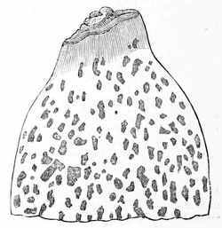
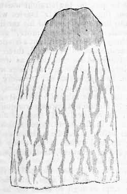
But it was in the chest that the most remarkable circumstance presented itself. The œsophagus gradually enlarged from the pharynx, which was perhaps rather narrower than usual, to an extraordinary degree of dilatation; the greatest breadth which it attained was situated about four inches above the cardia: the tube then contracted more abruptly, so as to render the termination of the œsophagus, like its commencement, of nearly the usual dimensions. The structure likewise of the cardiac extremity for about an inch, and of the pharyngeal end for about half an inch, was healthy. The intermediate part presented, when inverted, the following curious appearance:—The inner membrane was thickened and opaque, and had the appearance of having partially yielded from dilatation; at the upper part the furrows or thinner parts of the membrane followed in some degree a longitudinal direction; at the lower part the surface was pitted with shallow depressions of various figures. I have represented in the adjoined sketches these appearances, which on the middle of the œsophagus passed from one into the other; at the furrows or depressions the membrane seemed of the natural thickness and colour; the intermediate raised and thickened part was opaque and whitish. The muscular fibres of the œsophagus were of the natural colour and thickness.
I remain, Mr. Editor,
Your obedient servant,
Herbert Mayo.
19, George-street, Hanover-square,
Dec. 6, 1828.
To the Editor of the London Medical Gazette.
Sir,
As the following case may throw some
additional light on the pathology of
hydrophobia, I have sent it you for
publication.
I am, Sir,
Your obedient servant,
F. Godrich.
Grove House, Little Chelsea,
Nov. 29, 1828.
On Thursday morning, the 25th ult.
I was called up about seven o'clock to
see a man who I understood was exceedingly
ill, and waiting in the surgery
very impatiently for my arrival. I
found my patient (Mr. Barham), a fine
looking old man, about 60, labouring
at intervals of about five minutes under
strong spasmodic paroxysms, affecting
the muscles concerned in breathing
and deglutition. There was a wildness
and an impatience depicted in his
countenance, totally different from any
thing I had ever observed in other spasmodic
affections. His bowels were open,
tongue clean, skin moist, pulse full and
a little accelerated. I took away twenty
ounces of blood, and prescribed a mixture
containing
ʒss.ss.
of laudanum for a
dose every hour until I should see him
again. He walked home half a mile
from my house (Gloucester Road, Old
Brompton), and left me ruminating on
the possible cause of so much mischief
occurring suddenly in a fine healthy
subject. At ten a message was sent,
saying that he was much worse, and
requesting me to call as soon as possible.
He received me tranquilly, and
said he was very glad that I had come
to see him, for he was very ill. His
symptoms were now more distressing
than when I first saw him: he looked
wildly and suspiciously at every one entering
his apartment, and his breathing
was accompanied by a short convulsive
sobbing. On looking at his medicine
I perceived he had taken none, and expressing
my surprise, he assured me it
was impossible for him to swallow a
single drop, as the attempt had been
followed by violent spasms, and produced
so much distress, that he had desisted.
At this period no one had the
slightest idea of the origin of his
malady. I poured out some medicine
into a tea-cup, the very act of which
produced much excitement and alarm.
My first impression as to the true nature
of his disease arose at this period, from
the circumstance of his requiring a tea-spoon,
with which he endeavoured to
take some of the medicine. The attempt
produced much excitement and
alarm, and after two or three painful
efforts at deglutition, with one desperate
effort he swallowed a tea-spoonful, threw
away the spoon, and begged, unless I
wished to destroy him, that he might
have nothing more to swallow. I now
left his room, and inquired of a bystander
whether any thing particular
had occurred to him within the last
few weeks. On recollection she said,
"About a month since, late at night, a
strange dog came into the premises and
fought with his own dog; he got out of
bed to separate them, and the strange
dog bit him in two places, on the left
arm and hand; and bit a puppy, which
died about a fortnight after in a strange
way, which was thought to be some
kind of a fit." To ascertain if this occurrence
had produced any effect on his
mind, while again bleeding him I said,
"You have been in the wars, Sir, and
had your hand and arm torn; how did
it occur?"—"Oh!" said he, carelessly,
"that was done by a dog a long time
ago, but it healed." The circumstance
was never again mentioned to him, and
he died in total ignorance of the cause
of his malady. The wounds were perfectly
cicatrised, and there was not the
least action going on indicative of recent
absorption. He bore the bleeding
pretty quietly:
40℥.40.
were removed,
which on cooling presented strong
marks of inflammation.
Ordered
ℳiv.iv.
Acid. Hydrocyan. omni horâ, in a little water.
Twelve o'clock.—With much difficulty
he has taken two doses of the
acid; pulse full and hard, 110.
30℥.30.
more blood were removed.
Three o'clock.—Has taken two more doses; complains of a dreadful sense of suffocation, and implores that nothing more may be given him. Pulse full, and beating at 120 to 130. Continue the acid.
Eight o'clock.—Pulse full and hard.
Has taken in all
24ℳ.24.
of the acid, but
so painfully distressing has the deglutition
now become, that all attempts at
repeating his medicine are discontinued.
V. S. ad
℥xxx.xxx.
During the bleeding he looked wildly at the basin, and begged that no more might be spilt (a drop or two had fallen), repeating frequently, in great agitation, as the blood was running, "Take care! take care!"
Between two and three o'clock next
morning my assistant (Mr. Davies) visited
him. He found him tolerably
passive, but observing every movement
with intense anxiety. Pulse full and
hard, face flushed, eyes denoting cerebral
irritation. He had been at times
outrageous. On its being intimated
that bleeding was again necessary, a
paroxysm came on more intense than
any preceding,—and with great effort he
submitted. As the blood flowed he became
more and more alarmed, till at
length he got quite unmanageable; he
raged violently at his nephew, who was
holding the basin, and ordered it peremptorily
to be removed. 30 or
40℥.40.
were taken away. It was found necessary
to put on the straight waistcoat.
About four o'clock Mr. Davies wished
him to take some more of his medicine.
He said, "I can take no more," and on
reaching the bottle to put out a few drops,
he became violently agitated, threw himself
from side to side, and, as well as
the incessant spasmodic sobbings would
allow, he begged that not one more drop
of any thing might be offered him, and
that the bottle might be taken from his
sight. He did not become tranquillized
until its removal. He lingered on till
ten A.M. in the same state, a few minutes
before which he insisted on getting
up, and walked a short way down
his garden, returned, laid down on his
bed, and died.
Mr. Frederick Salmon, of Old Broad Street, and Mr. Wilson, of Chelsea, were kind enough to assist me in conducting the post mortem examination. On opening the chest, the heart was free from disease, with rather more water in the pericardium than natural; the lungs were completely gorged with grumous blood, and the pleura adherent on the right side. On removing the cranium, which was remarkably thin, and cutting the substance of the brain, numerous red spots presented themselves in the medullary portion; about a table-spoonful of water in each ventricle; the plexus choroides was turgid; the corpora, striata, thalami, and basis of the brain every where preternaturally injected; the cerebellum, crura cerebri, and cerebelli, in a high state of inflammation. On removing the spinous process of the vertebra, the whole cord was considerably inflamed; and opposite the two last cervical and dorsal vertebræ the cellular substance was studded with dark patches of coagulated blood, the theca vertebralis thickened, and the cord in an active state of inflammation. The larynx and pharynx bore not the slightest vestige of disease. The preparation of the cord is deposited in the museum of the London University.
The post mortem examination of this case tends to prove the correctness of Professor Thompson's theory of the proximate cause and seat of this afflicting malady; and the plate accompanying a case recorded by him, in the 13th volume of the Med. Chir. Society, gives a faithful delineation of the state in which the spinal cord was found in this case.
To the Editors of the London Medical Gazette.
Gentlemen,
Perhaps the following case may interest
some of your readers, and if you
think so, its insertion in your Journal
will oblige
Your obedient and
humble servant,
S. D. Broughton.
12, Great Marlborough Street,
Dec. 3, 1828.
On Tuesday, Nov. 25, I was called upon at the St. George's and St. James's Dispensary to advise for an infant at the breast, of five months and fourteen days of age, that had not passed any fæces during six weeks up to the day before I saw it. After its birth it had no passage till the fifth day, and this had apparently been effected by the introduction of a bougie, and which was subsequently continued at intervals for a short time. The infant was otherwise healthy and strong at its birth, and remained so until it had gone about three weeks without any evacuation, when it was observed to be weak in the lower extremities; and at the period of my seeing it the legs were much fallen away in flesh, hanging flaccid and inert. Many aperient enemas and medicines had been given, and, as a last resource, one drop of the croton oil had been prescribed the preceding evening. The infant appeared to suffer from distention, and to be disposed to coma, and the croton oil had distressed it much some hours after it was given. The fæces could be felt blocking up the intestine, but the irritation of the finger produced no attempt at evacuation; very small pieces only having been brought away. Hot baths also had frequently been used some time ago.
Considering the case as one of paralysis in the parts naturally employed to relieve the bowels, I recommended the suspension of all purgative remedies by the mouth, and a table-spoonful of the following injection to be substituted, and to be thrown up with three table-spoonfuls of thin gruel occasionally during the day, by means of a syringe.
℞ Spir. Terebenth. ʒiij.
Træ Zinzib. ʒj.
Aquæ, ℥iss. M.
Spir. Terebenth.
iij.
Træ Zinzib. j.
Aquæ, iss. M.
The following liniment was also directed to be rubbed in frequently along the course of the spine.
℞ Linimenti Saponis, ℥iss.
Liquor. Ammon. Comp. ℥ss. M.
Linimenti Saponis,
iss.
Liquor. Ammon. Comp. ss. M.
The result justified my expectations; for, during the night after this prescription, two injections having been given, some large and hard ash-coloured lumps came away, and these were shortly followed by a forcible spouting out of a continued stream of feculent dark liquid; a brisk diarrhœa having been kept up till the Saturday following. The infant's appetite is become voracious, its appearance lively and healthy, and its legs as firm and strong as is usual at such an age. Cold water is recommended to be dashed upon the loins, the liniment to be continued occasionally, moderate doses of magnesia and rhubarb to be given when required, and the turpentine injection to be used if any return of the complaint should be threatened.
To the Editor of the London Medical Gazette.
Sir,
Having seen a case of dislocation of the
patella, reported by Mr. Mayo, in your
Journal a few weeks since, in which
there was experienced some difficulty in
the reduction of the bone, I beg leave
to send you the following, in case you
may think it worth insertion.
On Saturday last, I was called to a young woman who had dislocated the patella of the left knee joint outwards; the inner edge rested upon the outer surface of the external condyle, and was immoveably fixed, and caused great pain when touched. She says it was occasioned by her foot slipping backwards from one stair to another, and she immediately fell down.
I commenced the reduction, as stated by Mr. Mayo, but I could only produce semiflexion, and that gave her great pain; insomuch that I was obliged to desist, and the patella remained in statu quo. But when the knee was thus semiflexed, I desired my assistant to extend the leg suddenly; and having my fingers on the patella at the time, and pressing it inwards, it immediately returned to its natural position.
I must confess, sir, that I should have been afraid to have continued the flexion as described in Mr. Mayo's case, "by bending the knee to the utmost," fearing that the remedy might be worse than the disease, and the result proved that in this case it was not necessary.
I remain, Sir,
Your obedient servant,
P. M. Hoskings.
168, Fleet-Street,
Dec. 3d, 1828.
To the Editor of the London Medical Gazette.
Sir,
The mistake of the printer in substituting
the word "every" for "even,"
in the paper on Vaccination (p. 40), so
materially affects the sense of the passage,
that I must request the favour of
the following insertion:—
"3d. That while the vaccine cicatrix retains even one distinct fovea or depression, the individual continues unsusceptible both of small pox and of re-vaccination; but that," &c.
I am, Sir,
Your obedient servant,
Thos. H. Burder.
2, Brunswick Square, Dec. 15, 1828.
To the Editor of the London Medical Gazette.
Sir,
The valuable paper communicated to
your Journal, by Mr. Stanley, last
week, has brought to my recollection a
singular appearance of the parts in front
of the shoulder-joint which I dissected
about two years ago. It is a fact
worth recording; and perhaps you will
be kind enough to insert it in your next
number.
The circumstance was this—that in the right arm, instead of there being a biceps muscle having two origins, the one the shorter from the corocoid process, the other the longer from the upper part of the glenoid cavity of the scapula, there was simply a one-headed muscle arising from the corocoid process. I could find no tendon passing through the joint answering to the long head of the biceps, neither was there any appearance of a biceptical groove; the capsular ligament was, perhaps, thicker than it is found in ordinary cases.
I knew nothing of this man during life, and am therefore unable to give any account of the use which he made of this arm.
I am, your obedient servant,
C. M. Burnett.
House Surgeon's Apartments,
St. Bartholomew's Hospital, Dec. 8th.
To the Editors of the London Medical Gazette.
Gentlemen,
As you did me the honour, on a former
occasion, to publish some remarks in
defence of auscultation, against its
enemies and detractors, I trust you will
permit me to say a few words in reference
to a communication in a late
number, from a gentleman who appears
to be a friend of that method of diagnosis,
but whose ignorance of the subject
is likely to be more injurious to the
cause than either open enmity or secret
detraction. The case in question (page
780, vol. ii.) appears, from the dissection,
to have been clearly emphysema of the
lungs, as there stated; but I appeal to
every one practically acquainted with
the physical signs of diseases of the
lungs, whether the results of the exploration
with the stethoscope, as narrated
in the case, were those pathognomonic
of that disease? I say they were
not only not pathognomonic of this
affection, but they were actually incompatible
with its existence. The same
ignorance of the subject is shewn in relation
to the disease of the heart, which,
I boldly maintain, no one was justified
in pronouncing to be hypertrophy of the
left ventricle from the stethoscopic indications
stated in the case.
It is just possible that the writer of the case may have made further explorations on which his diagnosis was founded; but if he had such in his case-book, it is equally unfortunate for the credit of the stethoscope and his own, that he suppressed them in his printed case.
I have the honour to be,
Gentlemen,
Your obedient servant,
A Stethoscoper.
Nov. 25, 1828.
To the Editor of the London Medical Gazette.
Glasgow University, Nov. 26, 1828.
Sir,
Having lately perused, in the 35th
Number of the Medical Gazette, some
observations "on the mechanism of
the act of vomiting," by Dr. Marshall
Hall, in which, after controverting the
different opinions which have been entertained
by physiologists on this
question, he endeavours to prove that
vomiting is an effort, not of inspiration,
but of expiration, and advances this
opinion as having originated in himself,
I beg, through the medium of your
Journal, to refer that gentleman to
Richerand's Physiology, where, on the
subject of vomiting, page 70, he says,
"Sometimes the action of the muscular
fibres of the stomach is quite inverted;
they contract from the pylorus towards
the cardia; and this antiperistaltic motion,
in which the contractions act
with more force and rapidity, and in a
manner decidedly convulsive, produces
vomiting. The action of the abdominal
muscles then connects itself with that
of the stomach; the viscera are propelled
upwards and backwards, by the
contraction of the large muscles of the
abdomen; the diaphragm ascends towards
the chest; if it descended, in contracting,
the œsophagus that passes between
its two crura would be compressed,
and the expulsion of food
through the cardia could not be effected.
It is also observed, that during expiration
only, any thing can pass from the
stomach into the œsophagus." Again, on
respiration, page 147—"In every effort
of expiration, as coughing and vomiting,"
&c. Here, then, we see that Dr.
Hall's views, as far as regards vomiting
being an effort of expiration, have
long since been advanced by Richerand.
The profession are certainly much indebted to Dr. Hall for his corroboration of these views; at the same time he should not have taken to himself the merit of having first suggested them.
Sir,
Your most obedient servant,
A Subscriber.
To the Editor of the London Medical Gazette.
Dec. 19, 1828.
Sir,
The events disclosed during the late trial in the Court of King's Bench, must fill every well-disposed mind with astonishment and horror. The moral aspect of the case is marked by every feature capable of exciting a sense of shame for our profession, and of sorrow for our kind. Can it be that a publication conducted in the spirit of the extracts produced at the late trial of Cooper against Wakley should be fostered and encouraged by a large proportion of the medical public? Is it indeed true that no feeling of loathing and disgust should have induced the purchasers of the Lancet to protest against its glaring injustice, its undisguised ferocity and malignity?
I am not a reader of that work myself, and but for the disclosures recently made in our courts of justice, together with what I have learned from your own pages, I should know little about it. Some of its early numbers convinced me that it was a production not deserving of support, whatever ability there might be connected with it; subsequent events have amply justified this decision, and I now address these few hasty lines to you, to express my humble hope that all the respectable medical men throughout the kingdom will arise, and with one voice oppose this moral pestilence, which at once corrupts and degrades their profession.
The odium medicum, bitter and vindictive as it unfortunately has been, never, I believe, appeared in a form so repulsive. Are we living in the nineteenth century? Have arts and sciences, which are asserted of old to soften the manners and temper the violence of our nature, produced no such fruits amongst us? Is serious, and perhaps irremediable injury, to be inflicted on reputation; and is the sufferer thereafter to be dared to mortal combat, and this too by the wrong-doer? Is it thus that professional justice is to be awarded? Is it thus that knowledge is to be advanced, the dignity of our profession to be maintained, its benefits enlarged, and its usefulness transmitted? Is it thus that the young men are to be taught how to acquire an unworthy and discreditable notoriety; to disregard all the decencies, charities, and higher virtues of life; and to bring into the exercise of our art the insidious cunning of the knave, the vulgar dishonesty of the pilferer, and the audacious bearing of the bravo?
I know not how others may feel on the present occasion: I am entirely unacquainted with the parties who have been at issue in the late suit—I have no bias of any kind, but what must arise in the mind of every man who has any regard for the profession to which he belongs, or who loves truth and fair dealing—I am sure that it is not less for the interests of society at large than for the advancement of medical science, that just principles should regulate all our dealings, both with the public and with each other. It is manifest that of late we have signally and woefully departed from them; and it is most distressing to know that this departure has been sanctioned by so many members of the profession. I now entertain a confident hope that all who reflect on the character of the late libel will feel that their own character is at stake if they do not mark their disapprobation of it in every possible way. Sure I am that, if the principles which guide the Lancet become generally acceptable, it will mark an æra of moral degradation which no surgical dexterity, no medical acquirements, however eminent, could possibly redeem.
Veritas.
Cases of longevity are not only much more common, but also more extraordinary in respect to a greater duration, in Russia than in any other part of Europe; thus, from the report of the holy synod, published in 1827, it appears that there were living in 1825, among those who professed the Greco-Russian religion throughout the empire, not fewer than 848 males who were 100 and more years old; among whom, 32 had passed the age of 120, 4 were between 125 and 130, and 4 others between 130 and 135 years of age. Out of 606,881 males who died in 1826, 2785 had passed the age of 90 years; 1432 that of 95; and 818 that of 100. Among the latter, 38 were more than 115 years of age; 24 more than 120; 7 more than 125; and one was 160 years old at his death.