In Gryllus assimilis, with Special Reference to the Nebenkern.
(Thesis for the degree of Master of Arts.)
BY W. J. BAUMGARTNER.
With Plates II and III.
It is the object of these studies to follow carefully the spermatid
transformations, and, if possible, to get a complete series
of changes occurring therein. I do not pretend that I have as
yet found all the stages, but in the present paper will publish
observations made on Gryllus assimilis pertaining chiefly to the
nebenkern, of which I have found some stages not previously
described, as far as I know. I shall first describe my own findings,
and then compare them with the results of others.
While other fixing agents were used, Flemming’s chromo-acetic-osmic
mixture (strong) proved the best. Its fixation was satisfactory in
all but a few stages, where the chemical changes going on in the cell
are such that it seems difficult, probably impossible, to secure
good, clear results; at least, neither Gilson’s nor Zenker’s mixture
did any better on cells in these stages.
Heidenhain’s iron-hæmatoxylin stain gave the best results, although
Flemming’s three-color method was very satisfactory.
Most of the observations are made from sectioned material, but smear
preparations were used for advanced spermatozoa.
The first observations were made in January of the present year on
some material prepared by Professor McClung, to whom I wish here
to make most grateful acknowledgment for proposing the line of
investigation, and for many helpful suggestions during the progress
of the same. The testes were those of adult insects, and showed
mostly only mature or almost mature spermatozoa. Enough spermatids
were seen to indicate that their transformation into spermatozoa was
somewhat peculiar.
The material for the drawings was taken from specimens collected
under stones and boards on and near the University campus, at
Lawrence, Kan. Young nymphs of Gryllus were caught as early as
March 1, but the cells of the testes were all in the spermatogonia
and spermatocyte stages. During the early part of May, after the
nymphs had passed their third molt, crickets were secured whose
testes showed all stages of development and transformation.
Gryllus assimilis, the common black field cricket, has paired
testes lying in the anterior dorsal part of the abdomen. These have
a whitish transparent appearance, which becomes duller in the adult,
showing sometimes a slight yellowish tinge. The shape of each is
that of a somewhat conically rounded body, not unlike a flattened
strawberry. Each testis consists of a central rachis about 3 mm.
long, from which extend a large number of curved follicles varying in
length from 1 to 3 mm.
The follicles are larger toward the blind end and taper toward
the rachis. Each follicle is divided into cysts, but more often
transversely than longitudinally, for frequently one cyst occupies
the whole follicle in cross-section. The cysts toward the rachis end
of the follicle are much longer and narrower than those of the blind
end. The cells are not very large and contain twenty-four chromosomes
in the spermatogonial generations; eleven and twelve were most
frequently found after the reduction.
The follicles of the cricket testis show the different cell
generations and the same relative arrangement of them as McClung
(’00) found in Hippiscus, and Sutton (’00) in Brachystola.
The spermatogonia are nearest the blind end of the follicles,
spermatocytes next, and the spermatids following the latter. The
cells of a cyst are not, as in Anasa (Paulmier, ’99), “in the
same stage of development,” but only approximately so, for some
cysts show cells in the metaphase, while others have reached the
telophase. Successive cysts, as in Brachystola, do not show
successive stages; for, frequently, growing spermatocytes and late
spermatids, or even young spermatozoa, were observed side by side.
The individual cells in division pass through prophases, metaphases,
anaphases, and telophases—these terms being used according to their
usually accepted meanings. (See McClung, ’00.) In this paper I shall
begin with the cell when it has reached the telophase of the second
spermatocyte division; that is, just after the chromosomes have
reached the poles of the spindle.
The chromatin in Gryllus behaves very much as Henking (’91) has
described for Pyrrhocoris, and Paulmier (’99) for Anasa. At the
end of the anaphase the chromosomes are crowded together at the
poles of the spindle. (Fig. 1.) A clear space begins to
be formed around them, especially on the equatorial side. (Fig. 2.)
They then separate and are scattered somewhat evenly over the nuclear membrane
when it is formed, as seen in figs. 4, 5, and 6. Soon
protuberances appear on the surface of the individual chromosomes, making their
outlines irregular. At about this time they begin to show a granular
structure. The chromosomes break up more and more, and soon the
chromatin appears in irregularly arranged patches or blotches of
granules of various sizes. (Fig. 8.)
Meanwhile the nucleus has slowly increased in size. It reaches its
maximum, which is probably about treble its original diameter, before
the cell begins to elongate. The granules have been breaking up into
finer and finer pieces, until, a little after the cell has reached
its maximum, the chromatin has largely disappeared, or, to be more
exact, has lost its affinity for stains. (Fig. 16.) As
the cell begins to elongate, the nucleus goes to one end, namely, that toward
the periphery of the cyst. (Figs. 14 and 15.) The
chromatin again frequently appears in patches as the cell decreases in size.
(Figs. 33 and 36.) In many cells the patches never disappear.
When it has diminished to less than half its maximum diameter, the nucleus
begins to elongate, becoming elliptical, and finally forms the long,
tube-like head of the spermatozoon. (Figs. 43 and 45.)
I found here that the nucleus was hollow, or, better, that it
showed a clear space within. For a long time it contains the many
chromatin granules, as seen in figs. 39, 40, and 41; but
at last the walls seem to attract all these granular masses, and the center is
entirely clear, as in fig. 45. In fig. 44
are shown cross-sections of spermatozoa heads, of the stage of fig. 43.
In fig. 45 we have a mature spermatozoon head.
The accessory chromosome in Gryllus lags behind during the
spermatogonial divisions, just as in Xiphidium (McClung, ’99),
and retains its identity all through the growth period, in many
cases becoming quite large. At first it cannot be distinguished
in the spermatid, but soon its stronger stain and exemption from
disintegration make it apparent. (Fig. 4.) It is
flattened against the nuclear wall and, unlike Paulmier’s “small chromosome,”
it does not break up but remains intact, as Henking and McClung have
described. (Figs. 14, 18, 32, 37,
and others.) It gets larger and then smaller, and is finally lost
in the rest of the chromatin, as the latter condenses to form the
spermatozoon head. I was not able to see that it occupied one side of
the elongating head, as McClung describes it in Xiphidium. The darker
stain and regular contour, and sometimes its greater size, made it in
most stages quite evident.
It is in the cytoplasmic structures that Gryllus shows some things
that have not been described, so far as I know; yet I almost hesitate
to enter the discussion concerning the nebenkern, the mitosome, the
idiozome, the archoplasm, the attraction sphere, the acrosome, the
“mitochondrion körper” and other bodies which have been described in
the extra-nuclear parts of the germ-cell. But because I have been
unable to find in any of the papers a description of a structure
like the one in Gryllus, I shall proceed to describe the nebenkern
as I find it. I shall interpret the appearances as far as possible,
leaving to others, or to later efforts, the complete harmonizing of
this element with the structures of other species.
Before I proceed, I would add my protest against the name
“nebenkern,” as voiced by Calkins (’95), Erlanger (’96), Meves (’00),
and others. It is inapt and not at all descriptive or distinctive.
But since we have the word, and investigators will use it to mean
something, I think it would be best to restrict the word, as Calkins
and Erlanger suggest, to the remaining spindle fibers and connective
fibers which go to each spermatid and which have an important part
in forming the tail membrane. I shall retain the name for a further
reason, which St. George (’97) gives for first using it, “weil
sie die Frage nach der Herkunft dieses Gebildes offen lässt.” A
comparison of the literature will convince any one that the origin of
this element is still a much-disputed question.
1.—Nebenkern.
There are, in Gryllus spermatids, two quite permanent and prominent
extra-nuclear bodies. The larger of these I shall call nebenkern,
because I believe it originates from the spindle remains, and goes to
form the tail covering. Its behavior is as follows: At the beginning
of the telophase the fibers which had connected the centrosomes with
the chromosomes bulge out like barrel staves, as Henking (’91) has
described. These break loose from the chromosomes and centrosomes
as the clear space begins to form around the chromatin mass (fig. 2);
but they are not separated into two rings, as in Anasa
and Pyrrhocoris. As the dividing cell membrane is formed, the
middle portions of the fibers are drawn together and the so-called
“mid-body” is formed. Some of the fibers unite, producing thicker
ones, as seen in figs. 1, 2, and 3.
In the last-named figure some of the central fibers seem to be still
united with those of the companion spermatids.
As the daughter-cell shortens and the nucleus takes a more central
position these fibers unite still more, shorten, bend together
(figs. 4, 5, and 6), and finally assume the
shape shown in fig. 7. I shall
call it for the present the “striated condition,” or simply “striated
nebenkern.” The shape resembles that of an egg with rather sharply
drawn-out ends. The surface is occupied at intervals with deeply
staining lines which look much like hoops. In most cases the long
axis of the nebenkern is at right angles to the axis of the spindle
from which it was formed, while the striæ, or dark lines, are in
planes parallel to it, as is seen in figs. 7 and 8a.
In a few cases the lines are parallel to the long axis of the nebenkern, as shown in
fig. 8b. Fig. 9 exhibits
a partial end view of the nebenkern, while fig. 26
represents a polar view in all planes.
The fibers in figs. 4 and 5 are not all in the same
plane, giving frequently, with changing focus, the impression that the lines are
the edges of plates. Even in figs. 7 and 8, the appearances
shown made me hesitate for some time before I dared say that the lines
represent striæ on the surface and not plates extending through the
body. But the end views (figs. 9 and 26) and the two
drawings in fig. 19 show clearly that we are dealing with
encircling hoops and not dividing plates. Just how or why the fibers bend and assume
the shape of figs. 7 and 8 I am not able to say, but I am
sure it is not an artefact, as might be supposed, because I have found them
occupying whole cysts of several testes, and nearly all of them were as regular
and definite as fig. 7.[A]
I have found the nebenkern in its perfect condition (fig. 7)
only in material fixed in Flemming; although Zenker’s fixation showed some of
the breaking-up stages. This may be due partly to poorer fixation in
other mixtures and partly to chance, as not so many testes fixed in
other agents were examined, and the appearance evidently represents
a very brief stage. These fibers now begin to break up, and we get
figures that remind one of Meves’s “mitochondrion körper”; that is, a
darker staining center and radiating lines to the outer ring. (Fig.11.)
Sometimes stages a little later recall Paulmier’s “blackberry
stage.” (Figs. 12 and 25.) Fig. 19a
is a surface view and fig. 19b an optical section of a
nebenkern that is in the process of breaking up. Figs. 20 and 21
have a part of one end cut off, while fig. 23 is a polar view.
Fig. 22 is probably a forming blackberry stage.
All the above are brief stages, and we soon get the appearance of
figs. 13-16. Here we have the darkly staining central ball
surrounded by a clear space, enclosed by a ring, as Meves (’00) found it in
Pygæra. The central ball sometimes shows vacuoles (fig. 16),
but most frequently stains as one mass. In some cysts with cells
resembling fig. 16, it is difficult to distinguish between
nucleus and nebenkern, yet the latter’s stain is never as intense as that of
chromatin. The stages shown in figs. 13 and 14 persist
for a long time, and it is probable that this is the end of the telophase and
the beginning of the spermatid transformations. I have treated the
above more in detail and shown more drawings than its importance may
deserve, but it is in these stages that Gryllus seems to differ
from other species, and I have tried to describe and illustrate the
appearances and changes fully.
As the cell elongates, the axial filament
grows out, the nebenkern approaches it, and a junction is effected
in such a way that the axial filament runs over the surface of the
nebenkern. (Figs. 18, 27, 28, and 30.)
At first the nebenkern is still almost round, but it begins to elongate and the
dark inner ball sends out a protrusion to the nucleus, and sometimes back
to the point of separation between the external envelope and the
axial filament. The outside ring disappears and the dark mass moves
down the axial filament. (Fig. 31.) In many cases it
breaks up into several small drops, which appear at intervals on the axial
filaments. (Figs. 34 and 36.) In fig. 38 is seen such
a hanging drop. In this way the nebenkern material is distributed over the axial
filament and forms a sheath around it.
2.—The Acrosome.
The second extra-nuclear body I shall call the acrosome, as it
forms the point of the spermatozoon head. I could not trace its
origin, because the fixation was not definite in some of the early
stages. Fig. 4 shows a small, darkly staining body,
which may be a centrosome, but more likely is the beginning of the acrosome,
of which figs. 7, 9 and 10 show developing stages,
and figs. 11, 13 and 15 more advanced ones. Sometimes
the central portion stains darkest (figs. 11,
32, and 36), but more frequently there is a small
clearer space in the center surrounded by a ring or band of darker material.
(Figs. 10, 13, 27, and 30.)
This ring usually stains darker on one side, very often on the side toward the nucleus.
The position, which is, with few exceptions, in the angle between the
nucleus and the nebenkern, induces me to consider the differently
staining bodies as the same organ of the cell. For some time after
the nebenkern has disappeared the acrosome keeps its position;
then it approaches the nuclear wall, flattens against it, and
later wanders to the apical end, where it forms the tip of the
spermatozoon. (See figs. 32-36 and 39-42.)
Fig. 16 shows the acrosome back of the
nebenkern. It is not an isolated example, still, I think, it is an
abnormality. Fig. 37 shows an apparent division of
the acrosome. I did not see enough instances to consider it a regular occurrence.
3.—The Axial Filament.
As the cell is elongating, the axial filament is seen apparently
growing out of the nucleus. I do not mean to say that it grows out
of the nuclear substance, but in almost every cell observed, where
the axial filament was incomplete, there was rather a large mass of
chromatin gathered at the place where the axial filament was attached
to the nucleus. It may be that the smaller one of the extra-nuclear
bodies in figs. 4, 7 and 9 is a centrosome which is
passing to the equatorial region of the nucleus, where it later develops the
axial filament. As already noted, the axial filament does not pass through
the nebenkern, but only over its surface. Fig. 29, a, b, and c,
shows different cross-sections through the elongating cell. In b
the axial filament was cut at an angle.
4.—The Centrosome.
I have not followed the centrosome through its migrations. In fig. 2
one of the two bodies is the centrosome, but I did not trace its changes farther.
5.—Cell Body.
The cells, up to the stage of figs. 13 or 14, have been
scattered promiscuously through the cyst; but as the cell begins to elongate,
the one end becomes the anterior-nuclear end, and it shifts to the
periphery of the cyst. The central part of the cyst now shows the
so-called central lumen. As the lengthening goes on the heads of
the forming spermatozoa are all turned toward the distal end of the
cyst. The cyst becomes very narrow and long, apparently preparing
to contain the long, slender spermatozoa. Near the rachis the
spermatozoa seem much twisted, and I surmise that they turn so as to
have the head foremost when set free into the vas deferens.
Fig. 42 may indicate some such behavior.
The mature spermatozoon consists: (1) of a sharp, spear-like
point; (2) a tube-like head about .02 mm. long, composed of a
densely staining outer wall and a clearer central cavity; and (3)
a filamentous tail about .5 mm. long. The point comes from the
acrosome, the head from the nucleus, and the tail from the axial
filament and nebenkern. While I do not deny the presence of a middle
piece, my observations do not warrant me in describing one, although
fig. 40 would suggest it.
Dr. R. von Erlanger (’97, 1, and ’97, 2) and Dr. F. Meves (’00) have
given excellent discussions of the literature on the nebenkern. I
cannot do better than direct the attention of other investigators
to them. Hence, I shall discuss only such authors and such points
as have special bearing on my own results. The spermatid changes in
the Gryllidæ have been studied, so far as I know, only by St. George
(’67) and by vom Rath (’92).
St. George, in his early paper, described the
transformation process in the “Hausgrille” along with other insects.
He found the “Nebenkörper,” and saw that it took a part in forming
the envelope of the axial filament, a small particle remaining near
the nucleus and other drops of it appearing at intervals on the
tail. In his paper on Blatta (’86, 1) he accepted Bütchli’s name,
“nebenkern,” and traced its origin to the spindle remains. In his
paper on Phratora (’86, 2) he takes up the description of the
nebenkern in the spermatocytes and spermatogonia. In the spermatid,
he describes the blackberry stage in these words: “Während das andere
(not nucleus) Körperchen weniger lebhaft gefärbt als ein kleiner
Fadenknäuel mit unregelmässigem Contour erscheint.”
There can be no true nebenkern in the spermatogonia and
spermatocytes, as St. George first used the word, and as I have
limited its application. But St. George found the object, traced its
origin and fate, and described several of the stages as they appear
in Gryllus.
Vom Rath (’92) found Gryllotalpa very poor
material for following the spermatid changes. However, he described
an oval body which he found varied in appearance, depending on the
method of fixation. He calls it the “nebenkern” and thinks it goes to
help form the tail.
The description is too incomplete for comparison. Judging from
Gryllus, I question its being very poor material.
Bütschli (’71) studied the spermatid
transformations in Acrididæ and Locustidæ along with that of other
animals. He saw the object formerly described by St. George and named
it “nebenkern.” He saw it divide into halves, elongate, and form the
spermatozoon tail.
The dividing of the nebenkern into halves seems to be an appearance
quite common in insect spermatogenesis. I have myself
seen it in several genera of Acrididæ—Hippiscus, Arphia,
Melanoplus, and Brachystola; besides Bütschli, St. George,
Henneguy, Platner, Paulmier and others have described it.
Platner in his studies has given special attention to the
nebenkern. In his first paper on “Pulmonates” (’85) he did not trace
the origin of the nebenkern, but described it as consisting of four
to six rods of different lengths and irregularly bent. These were
connected, forming an irregular polygon. In its later stages he saw
it with a mass of protoplasm pass down along the primary tail—an
early protrusion of protoplasm. Finally it is lost.
In his succeeding paper (’86, 1) he studied the “nebenkern”
spindle remains—in the spermatogonia and spermatocytes of pulmonates.
In the spermatids, as in the former generations of cells, the
nebenkern grows out of the nucleus, where it, with the chromatin,
had formed the spireme. It appears as a loop, which becomes larger,
twisted, and entangled, and finally breaks loose from the nucleus.
Later it goes to form the spiral covering of the primary tail,
changing it to the axial filament and true tail.
In his next paper (’86, 2) he describes the changes when the
dividing of the protoplasm lags behind in the spermatocyte divisions.
His description agrees in so many points with my own, that I shall
quote his exact words:
“Die Spindelfasern hingegen contrahiren sich mehr und mehr
nach dem Equator hin, wobei sie mit ein ander verschmelzen und
merkwürdiger Weise je weiter dieser Verdichtungsprocess fort
schreitet um so mehr an Tinctions fähigkeit speciell gegenüber
dem Hämatoxylin gewinnen. Sie stellen jetzt zwei dreieckige oder
hakenförmige Gebilde dar, die mit der Spitze noch im Equator
zusammenhängen mit der breiten Seite sind sie den zugehörigen
Zellen zugewendet. Hier sind ihre Grenzen undeutlicher, verwaschen
und zeigen hier auch noch häufig eine streifige Beschaffenheit,
welche auf ihren Ursprung hin weist. Zuweilen lassen sich einzelne
Fäden noch eine beträchtliche Strecke weit in das Protoplasma
hinein verfolgen, welches zwischen ihnen und den sich ausbildenden
Zellkernen liegt.”
The “häkenformige Gebilde” moves away from the periphery, its
sides elongate, break, and unite at the nuclear end, thus form the
polygonal nebenkern. “Derselbe geht also in diesem Falle direckt aus
den Spindelfasern hervor.” When the protoplasm does divide a similar
process takes place. The spindle remains divide at an equatorial line
and each half forms a nebenkern, and, as he says: “Also auch hier
geht der Nebenkern direckt aus den Spindelfasern hervor. Vielleicht
geht in den Spermatiden der Nebenkern überhaupt immer aus den
Spindelfasern hervor, in dem die langfädige Verbindüngsbrücke, die
ihn oft mit dem Kern verbindet, sich wohl als ein noch einige zeit
persisttirender Rest der esteren deuten lässt.”
In his paper on Limax (’89, 1) he followed the nebenkern through
all the divisions and thinks it a constant organ of the cell. In the
second part of the paper on Helix and Paludina the nebenkern
was considered as formed from the remains of the spindlepole and the
centrosome. Later Platner (’89, 2) found the nebenkern in the
pancreas cells. In reports upon Pygæra and Sphinx he changed
the name nebenkern to mitosome. The centrosome lies in front of the
nucleus and forms the point of the head. This he calls the nebenkern.
From the spindle remains arise two bodies. A large, fibrous one from
the equatorial end has a clear space around it and the axial filament
passes through it. It soon elongates and forms the tail. This is the
large mitosome. The other is much smaller and arises from the polar
end of the spindle fibers. It takes its place in the angle between
the large mitosome and the nucleus. Here it persists till the nucleus
begins to elongate, when it lengthens and surrounds the basal end of
the spermatozoon tail. This is the small mitosome.
Platner (’85) saw a true nebenkern. I have already criticized the
use of the word “nebenkern,” as the name for the spindle remains in
the spermatocytes. Platner himself later denied the nuclear origin
of the nebenkern. His results (’86, 2) agree with mine concerning
the uniting, converging, staining and bending of the spindle-fiber
remains and their passing to the tail. Again, Platner (’89, 1)
probably followed the centrosome and attraction sphere, as well
as the nebenkern, in the second part of the paper on Helix and
Paludina. In the pancreas the body is a result of secretion, and
is not a nebenkern. In Pygæra Platner’s large mitosome is the real
nebenkern, as shown by its fibrous structure, its surrounding clear
space and destiny. His small mitosome is what I have described as the
acrosome, and he is mistaken as to its final use. The body he called
centrosome in the nucleus is the persisting accessory chromosome.
Such would be my interpretation of his figures.
Henking (’91) has followed the spermatid
changes in Pyrrhocoris quite carefully. He finds that the fibers
between the chromatin masses are separated into peripheral fibers and
central spindle fibers. The first, a part of the second and the yolk
mass, forms the nebenkern, which passes down over the axial filament.
The rest of the central fibers form the mitosome. This takes its
position at the angle between the nebenkern and the nucleus. On the
surface of the nucleus it passes to the anterior end, then back to
its original position. A piece now is constricted off and disappears,
while the rest, increasing its affinity for stains, again wanders to
the anterior pole of the nucleus, and becomes the acrosome.
The large amount of yolk substance is a disturbing element in
Pyrrhocoris, but the nebenkern agrees with that of Gryllus
in having the same origin and destiny, as does also the acrosome in fate
and position. Besides, there is much similarity in the stages, as
seen by comparing Henking’s fig. 63 with my fig. 25,
and his fig. 85 with my fig. 32.
Wilcox (’95 and ’96) described the spermatid
metamorphosis in Caloptenus. The interzonal fibers, a long,
striated body composed very plainly of distinct fibers, contracts
longitudinally, and the corners round themselves off, and this forms
the nebenkern. It loses its fibrous structure, comes close to the
nucleus, and then grows out into the axial filament. The centrosome
moves half way around the nucleus and lies between the latter and the
nebenkern. Later the centrosome becomes the middle piece.
This description does not give much detail nor do the drawings
show the stages clearly. As far as given, the formation
of the nebenkern is the same as in Gryllus; but in Gryllus the
axial filament is not formed from the nebenkern, as can be
plainly seen from fig. 17.
Erlanger (’96), in a short paper, discusses the use of the term
“nebenkern,” and suggests limiting it as is done in this paper. He
opposes St. George’s opinion, that the nebenkern comes from the
cytomicrosomes. In Blatta the cytomicrosomes are preserved during
the whole process of mitosis, and have no connection with the spindle
fibers, but during the telophase they collect in reduced numbers
around the daughter nuclei.
In 1897 he called the collection of granules around the
centrosome, the centrodeutoplasm. He considers them to be identical
with St. George’s cytomicrosomes and the archoplasm (or attraction
sphere) of other writers. In order to harmonize results, he suggests
that, since the centrosome sometimes wanders around the nucleus,
the centrodeutoplasm (or sphere) may unite with the spindle remains
in some cases to form one body, as shown by the descriptions of
Henking, Henneguy, Meves, and others. He later (’97, 2) describes
the so-called “sphere,” and distinguishes between it and the true
nebenkern.
I would strongly commend his excellent discussion of literature and
his careful comparison of the results of investigators. He has shown
clearly that the nebenkern comes from the spindle remains.
Calkins (’96) finds that the nebenkern comes from the spindle fibers
and is useless in the cell. But Lumbricus is peculiar in having
the nebenkern simply disintegrate, for, in many cases, he admits
that the nebenkern has an important function. I do not have access
to Henneguy’s or Bolles-Lee’s or Toyama’s works, yet I should judge
from Erlanger’s and Meves’s criticism that all of these have the
nebenkern originate from the spindle remains, and Henneguy describes
it as having a “fibrillar appearance,” and Bolles-Lee as “fibrillar
structure.” Accordingly, I think that each of these has discovered
the correct origin of the structure, and I do not doubt that there
is, at least in the first two, a more or less direct change from the
spindle remains to the nebenkern.
Paulmier (’99) finds that in Anasa the
nebenkern comes from the yolk mass and remains of the spindle fibers.
A part of this mass separates off, while the whole is still in a
confused condition, and forms the acrosome. The nebenkern forms the
tail sheath, while the acrosome forms the point to the head.
A comparison of Paulmier’s fig. 42 with my fig. 4
suggests the thought that they are the same stage, and his fibers are remains
of the spindle. His fig. 43, of course, agrees with my fig. 25;
and fig. 43 may correspond to figs. 26 or 14,
only that his stain is weaker.
Meves’s investigations are the most extensive of the recent ones on
the nebenkern. He has used Paludina and Pygæra as his objects.
The consideration of the mitochondrion in the spermatocytes I shall
pass over, as it is not within the bounds of this paper; yet I hope
to study the earlier generations of my material, and shall then
compare the results. In Meves’s description of Paludina, I find but
one point in which it agrees with Gryllus. The head of the young
spermatozoon in Paludina has a clear space in the center filled
with nuclear fluid, which remains till the head begins to stretch. In
Gryllus it is hollow until maturity.
In Paludina the nebenkern in one kind of
spermatids is formed from threads made up of mitochondria—small,
round bodies identical with St. George’s cytomicrosomes and
Erlanger’s centrodeutoplasm. These threads change to vesicles, which,
reduced to four, closely surround the centrosome as it lengthens
into the middle piece. At first they form a four-cleft cylinder, but
finally a single sheath. An idiozome and spindle remains are seen
in the spermatid. They persist for awhile, and the former changes
into the acrosome. In Pygæra Meves finds two kinds of spermatids,
distinguished by a small difference in size. The larger forms the
typical spermatozoon. The spindle remains form a “Spindelrest
körper,” which is soon lost. The ends of the mitochondrion chains
form a ring of dark mass surrounding a clear space. The ring is
broken by radiating clear spaces. These spaces collect and unite into
larger vacuoles, which surround the darker center. Finally there is
only a dark ball with a surrounding clear space shut in by a ring.
The centrosome with attached axial filament fastens itself to the
nucleus; then both begin to grow longer. The axial filament passes
over the surface of the ring surrounding the ball—the “mitochondrion
körper” or nebenkern. This body elongates a great deal then the
darker mass puts forth numerous threads which surround the axial
filament.
As to Pygæra, the peculiarity is the complete agreement of his
“mitochondrion körper” and my nebenkern in appearance and behavior
for a part of the transformation and their complete disagreement in
the other part. The question with me is, “Do the two bodies whose
final stages are so similar originate so differently, or has one of
us mistaken the origin of the body?”
Since reading Meves’s paper I have carefully reexamined my material,
and I am positive that I am right as to the origin of the nebenkern;
but, on the other hand, I would not say that Meves is wrong in his
observations, as in doing so I should fall into the same error which
I think Meves himself has made. In his discussion of the literature
he has forced every description to agree with his ideas, or has
declared that the author has described some extra-nuclear organ as
a nebenkern which is not a nebenkern. Thus by implication, if not
by direct statement, he says that a nebenkern never comes from the
spindle remains. With due respect to his ability and long experience
as an investigator, I must say that Meves is mistaken in this. St.
George himself traced the cytomicrosomes back to the spindle remains.
Besides, the many investigators whom I have cited above cannot be
mistaken as to the origin of the nebenkern. In my own material I am
positive that there is a direct passing of the interzonal fibers over
into the earlier stages of the nebenkern.
That the body which forms the tail covering does not come from the
spindle remains in all cases, I am willing to admit. Meves has cited
many investigators, especially on vertebrates, whose results favor
such an opinion. The spindle remains do not change into a nebenkern
even in all Arthropods, as Blackman (’01) finds no nebenkern, nor
anything in anywise resembling it, in Scolopendra.
From my study of the results of other investigators, it is evident
to me that there are at least two general methods for the formation
of the covering for the spermatozoon tail. One of these plans will
harmonize Meves’s mitochondrion körper, Erlanger’s centrodeutoplasm,
Heidenhain’s pseudo-chromosomes, and other similar structures. The
other will show that Platner’s large mitosome, Paulmier’s blackberry
stage and my striated nebenkern are only different stages of the
spindle remains changing into the tail covering.
1. The chromosomes of the second spermatocytes break up and the
chromatin becomes diffused all through the nucleus. Later the
chromatin collects in granules again and finally forms the walls of
the tube-like spermatozoon head.
2. The spindle fibers break loose as the clear space is formed around
the chromatin mass. They unite and contract, becoming fewer, thicker,
and shorter. These bend and form the “striated nebenkern.” The fibers
break up and sometimes show a blackberry appearance. Soon there is a
collection of darker material at the center, surrounded by a clear
space, which is shut off from the cytoplasm by a darkly staining
membrane which in sections appears as a ring. This stage persists
for some time; then the nebenkern moves against the axial filament,
elongates, loses the ring, and the dark mass passes down along the
axial filament. Often it appears in several small drops.
3. The axial filament does not come from the nebenkern nor from the
acrosome. It comes apparently from the nucleus; probably from the
centrosome closely attached to the nucleus. It never passes through
the nebenkern—only over its surface.
4. The acrosome occupies a position in the angle between the
nebenkern and the nucleus. It shows a central clear space surrounded
by a darker mass which stains more intensely on one side. Later
it passes to the front of the nucleus and forms the point of the
spermatozoon head.
5. The mature spermatozoon consists of a sharp point, a slender,
tube-like head filled with a clear fluid, and a long, thread-like tail.
Laboratory of Zoology and Histology,
University of Kansas.
September 27, 1901.
1. Blackman, M. W., ’01: Spermatogenesis of the Myriapods. Kans.
Univ. Quart., vol. 10.
2. Bütschli, O., ’71, 1: Vorläufige Mittheilung über Bau und
Entwickelung, der Inseckten und Crustaceen. Zeitschr. f. wiss. Zool.,
Bd. 21.
3. —— ’71, 2: Nähere Mittheilung über die Entwickelung und den Bau
der Samenfäden der Inseckten. Zeitschr. f. wiss. Zool., Bd. 21.
4. Calkins, G. N., ’95: The Spermatogenesis of Lumbricus. Journ. of
Morph., vol. 11.
5. Erlanger, R., ’96, 1: Ueber den sogenannten Nebenkern in den
männlichen Geschlechtszellen der Inseckten. Zool. Anz., Bd. 19.
6. —— ’96, 2: Die Entwickelung der männlichen Geschlechtszellen.
Zool. Centralbl., Jahrg. 3.
7. —— ’97, 1: Ueber Spindelreste und den echten Nebenkern in den
Hodenzellen. Zool. Centralbl., Jahrg. 4.
8. —— ’97, 2: Ueber die sogenannten Sphäre in den männlichen
Geschlechtszellen. Zool. Centralbl., Jahrg. 4.
9. Heidenhain, M., ’00: Ueber die Centralkapseln und
Pseudochromosomen in den Samenzellen von Proteus, sowie über ihr
verhältniss zu den Idiozomen, Chondromiten und Archoplasmaschleifen.
Anat. Anz., Bd. 18.
10. Henking, H., ’91: Ueber Spermatogenese und deren Beziehung zur
Entwickelung bei Pyrrhocoris apterus. Zeitschr. f. wiss. Zool., Bd.
51.
11. McClung, C. E., ’99: A Peculiar Nuclear Element in the Male
Reproductive Cells of Insects. Zool. Bull., vol. 2.
12. —— ’00: The Spermatocyte Divisions of the Acrididæ. Kans. Univ.
Quart., vol. 9.
13. Meves, F., ’00: Ueber den von v. la Valette St. George endeckten
Nebenkern (Mitochondrion Körper) der Samenzellen. Arch. f. mikr.
Anat., Bd. 56.
14. Paulmier, F. C., ’99: The Spermatogenesis of Anasa tristis.
Journ. of Morph., vol. 15, supplement.
15. Platner, G., ’85: Ueber die Spermatogenese bei den Pulmonaten.
Arch. f. mikr. Anat., Bd. 25.
16. —— ’86, 1: Ueber die Enstehung des Nebenkerns und seine
Beziehung zur Kerntheilung. Arch. f. mikr. Anat., Bd. 26.
17. —— ’86, 2: Zur Bildung der Geschlechtsproduckte bei den
Pulmonaten. Arch. f. mikr. Anat., Bd. 26.
18. —— ’89, 1: Beiträge zur Kenntniss der Zelle und ihrer
Theilungserscheinungen, I und II. Arch. f. mikr. Anat., Bd. 33.
19. —— ’89, 2: Beiträge zur Kenntniss der Zelle und ihrer Theilung,
IV und V. Arch. f. mikr. Anat., Bd. 33.
20. Vom Rath, O., ’92: Zur Kenntniss der Spermatogenese von
Gryllotalpa vulgaris. Arch. f. mikr. Anat., Bd. 42.
21. St. George, v. la Valette, ’67: Ueber die Genese der Samen
Körper. Arch. f. mikr. Anat., Bd. 3.
22. —— ’86, 1: Spermatologische Beiträge, Zweite Mittheilung. Arch.
f. mikr. Anat., Bd. 27.
23. —— ’86, 2: Spermatologische Beiträge, Vierte Mittheilung. Arch.
f. mikr. Anat., Bd. 28.
24. —— ’87: Zelltheilung und Samenbildung bei Forficula
auricularia. Festschrift f. A. v. Kölliker.
25. —— ’97: Zur Samen- und Eibildung beim Seidenspinner (Bombyx
mori). Arch. f. mikr. Anat., Bd. 50.
[B]
26. Sutton, W. S., ’00: Spermatogonial Divisions in Brachystola
magna. Kans. Univ. Quart., vol. 9.
27. Wilcox, E. V., ’95: Spermatogenesis of Caloptenus femur-rubrum
and Cicada tibicen. Bull. of Mus. of Comp. Zool. Harvard., vol. 27.
28. —— ’96: Further Studies on the Spermatogenesis of Caloptenus
femur-rubrum. Bull. of Mus. of Comp. Zool. Harvard., vol. 29.
All drawings were made by the author with the aid of a camera
lucida. All figures except 2, 3, 4 and 45 were made with a B. & L.
1⁄16 objective and one inch eye-piece, producing a magnification
of 1340 diameters. Figs. 2, 3, 4 and 45 were drawn with a Leitz
1⁄16 objective and a Zeiss compensating ocular No. 12, giving a
magnification of 2325 diameters. The drawings were not reduced in the
photo-mechanical reproduction.
Fig. 1. An early telophase, in which the fibers appear like
barrel staves. Some of the fibers have thickened.
Fig. 2. A later stage, in which the fibers are uniting, the
clear space is forming, and the chromosomes are beginning to separate.
Fig. 3. A daughter-cell, showing the chromosomes separating
and the fibers thickening.
Fig. 4. The chromosomes are scattered over the nuclear
membrane; the fibers appear as a few thick rods. The beginning of the
acrosome is seen. The accessory chromosome can be distinguished.
Fig. 5. Same as fig. 4, with some of the fibers curved and
no acrosome.
Fig. 6. Same as fig. 5, but with the acrosome visible.
Fig. 7. The chromosomes are granular; the fibers have bent
and formed the round “striated nebenkern.” The acrosome is present.
Fig. 8a. Same as fig. 7, but the acrosome is not present.
Fig. 8b. The fibers run parallel with the long axis of the
nebenkern.
Fig. 9. Here is shown a partial end view of the nebenkern.
The accessory chromosome and the acrosome are both prominent.
Fig. 10. The nebenkern shows some of the fibers on the under
side. It is beginning to break up.
Fig. 11. The nebenkern shows the beginning of the dark
center; the fibers extend from it to the ring. The acrosome has a
peculiar appearance.
Fig. 12. The fibers have broken up and the whole has assumed
a vesicular appearance, resembling a blackberry. The acrosome is present.
Fig. 13. This shows a persisting spermatid stage. The
chromatin appears in patches. The nebenkern is in the ball-and-ring
stage. The acrosome shows its characteristic clear center surrounded
by a ring darker on one side. The two dark bodies on the nuclear
surface are probably artefacts.
Fig. 14. A stage a little later than 13. The dark central
ball shows some vacuoles. The cell is beginning to elongate.
Fig. 15. Same as fig. 14, with the ball denser.
Fig. 16. The nucleus has become almost clear. The acrosome
is behind the nebenkern.
Fig. 17. This shows the axial filament apparently growing
out from the nucleus. There is an aggregation of chromatin at its base.
Fig. 18. The axial filament passes over the nebenkern, which
has slightly elongated. The accessory chromosome is very plain.
Fig. 19. a shows a surface view, and b an optical
section, of a nebenkern which is in process of breaking up.
Figs. 20 and 21 have part of the surface fibers cut off and
show the dark mass forming within.
Fig. 22. Shows a nebenkern passing from the blackberry stage
into the ring stage.
Fig. 23. This is an end view of the stage shown in figs. 20 and 21.
Fig. 24. The fainter lines are the fibers on the under side
of the nebenkern.
Fig. 25. The blackberry stage, showing the mass of vesicles.
Fig. 26. This represents a nebenkern of the stage shown in
fig. 7, drawn from a polar view, with adjusting focus.
PLATE II.
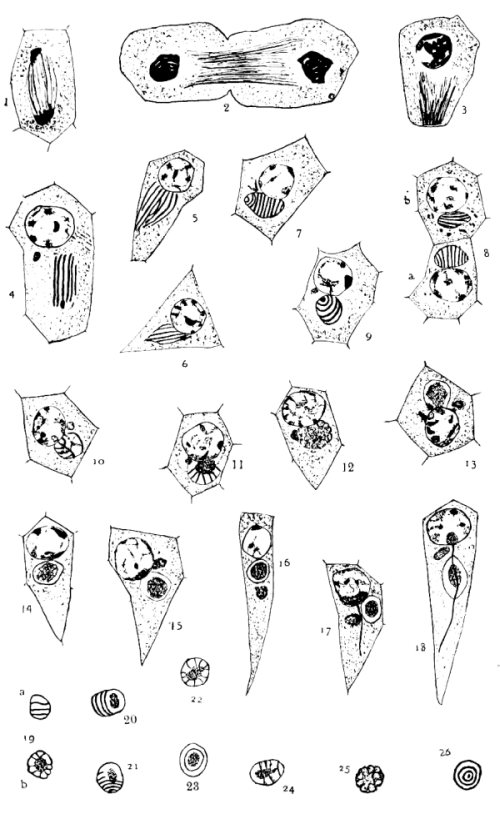
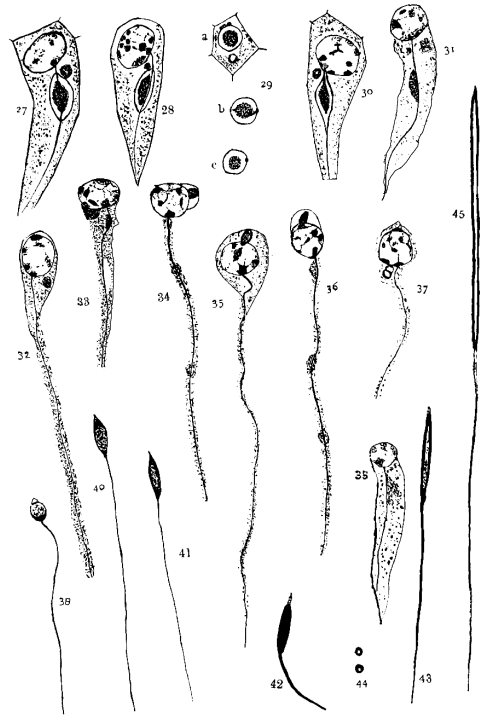
Fig. 27. This shows the nebenkern elongating. The axial
filament passes over its surface.
Fig. 28. A little later than fig. 27.
Fig. 29. Shows cross-sections of elongating nebenkern;
a shows axial filament, nebenkern and acrosome; in b the axial
filament is cut at an angle.
Fig. 30. A little later than fig. 28; the axial filament not
so plainly shown.
Fig. 31. The ring has disappeared, and the dark mass is
passing down along the filament. The acrosome is indistinct.
Fig. 32. The nebenkern has disappeared, but the acrosome has
kept its position.
Fig. 33. The acrosome has flattened against the nucleus.
Some remains of the nebenkern are seen.
Fig. 34. The acrosome has moved to the side of the nucleus.
Some nebenkern remains are shown.
Fig. 35. The acrosome is in front of the nucleus.
Fig. 36. A little later stage than fig. 35.
Fig. 37. Shows a divided acrosome. It is not a frequent
appearance.
Fig. 38. The nebenkern appears as a hanging drop on the side
of the axial filament.
Fig. 39. The nucleus has condensed, the walls have
thickened, and the chromatin appears in granules. The acrosome is pointed.
Fig. 40. A little later stage than 39.
Fig. 41. A little later stage than 40.
Fig. 42. The head is stained intensely, while the acrosome
is lighter. Probably a middle piece indicated.
Fig. 43. The head shows the clear space.
Fig. 44. Cross-sections of the head, of the stage shown in
fig. 43.
Fig. 45. The head and part of the tail of a mature
spermatozoon.
PLATE III.
Transcriber's Notes:
The cover image was created by the transcriber, and is in the public domain.
Typographical errors have been silently corrected but other variations
in spelling and punctuation remain unaltered.
The quotations in German appear to have some errors, but these quotations have
been reproduced exactly as in the original paper.