THE SPERMATOCYTE DIVISIONS OF THE LOCUSTIDÆ. — I. INTRODUCTION. — II. TECHNICAL METHODS. — III. NOMENCLATURE. — IV. OBSERVATIONS. — ( a ) General Form and Structure of the Testes. — ( b ) The Spermatogonia. — ( c ) The First Spermatocytes. — ( d ) The Second Spermatocytes. — ( e ) Number of Chromosomes. — ( f ) Spermatids. — V. COMPARISONS AND CONCLUSIONS. — ( a ) Nomenclature. — ( b ) The Spermatocytes of the Locustidæ and Acrididæ. — ( c ) Formation of the Tetrads. — ( d ) The Spermatocyte Divisions. — ( e ) The Accessory Chromosome. — ( f ) Individuality of the Chromosomes. — ( g ) Nucleoli. — ( h ) Rest Stage. — VI. SUMMARY. — VII. BIBLIOGRAPHY. — DESCRIPTION OF FIGURES.
185
BY C. E. M’CLUNG.
With Plates VII, VIII, IX, and X.
| I. |
Introduction. |
| II. |
Methods. |
| III. |
Nomenclature. |
| IV. |
Observations. |
| |
(a) |
General Structure and Form of the Testes. |
| |
(b) |
The Spermatogonia. |
| |
(c) |
The First Spermatocytes. |
| |
(d) |
The Second Spermatocytes. |
| |
(e) |
The Spermatids. |
| V. |
Comparisons and Conclusions. |
| |
(a) |
Nomenclature. |
| |
(b) |
The Spermatocytes of the Acrididæ and the Locustidæ. |
| |
(c) |
Formation of the Tetrads. |
| |
(d) |
The Spermatocyte Divisions. |
| |
(e) |
The Accessory Chromosome. |
| |
(f) |
Individuality of the Chromosomes. |
| |
(g) |
Nucleoli. |
| |
(h) |
Rest Stage. |
| VI. |
Summary. |
| VII. |
Bibliography. |
Under the title, “A Peculiar Nuclear Element in the Male
Reproductive Cells of Insects” (16), I published a preliminary
account of the process characterizing the maturation divisions
of the Locustidæ. This was of a general character and served
merely as a basis for a description of the accessory chromosome
in these cells. It is my present intention to give a detailed history
of the spermatocyte divisions occurring in this family,
after the manner followed previously in considering corresponding
stages in the Acrididæ (17). Besides giving this account
of processes, however, I shall be able to draw some comparisons
between the two families. Eventually I hope to complete
such a comparative study of all the Orthopteran families. Material
for this larger investigation is now partially on hand,
and is being added to as circumstances permit, so that it may
be possible to carry through a study of the maturation stages
in this order of insects within a few years.
186The value of comparative cytological study was urged by
Vom Rath (25), and its importance in relation to the accessory
chromosome and the maturation mitoses received recognition
in both my earlier papers (16, 17). Recently Montgomery (15)
has added his influence to the movement.
The observations upon which the present paper are based
were originally made upon Xiphidium. The cells in this genus
are, unfortunately, small in size, and much difficulty was experienced
during the early work in getting clear images. This
embarrassment was further increased by the large number of
chromosomes and their habit of compact arrangement. Later
it was found that species of Anabrus, Orchesticus, Microcentra and
Scudderia have cells much better adapted for study, and because
of this they have been largely utilized. The account which follows
is therefore based upon a study of all these genera, and is
considered representative of the family. The accompanying
figures are principally from Orchesticus, since the number of
stages represented exceeded those in material derived from
other genera. I am indebted to a friend and former student,
Mr. W. S. Sutton, now of Columbia University, for a generous
supply of Orchesticus and Anabrus testes from his collections.
For the fixation of material used in these studies, it has been
found that the osmic acid mixtures of Flemming and Hermann
are the most generally applicable and are productive of the best
results. In connection with these, however, Gilson’s acetonitric-sublimate
mixture has been tried, and frequently affords
an excellent fixation. Extensive shrinkage in the melted paraffin
is sure to follow the use of sublimate mixtures unless
celloidin is used to support the soft tissue. This double infiltration
of celloidin, followed by paraffin, has been found the
best method of securing clear and accurate figures, for, because
of the lessened shrinkage, the elements are not crowded together
and rendered indistinct. This circumstance is particularly fortunate
in the case of the Locustid cells, where the nuclear elements
are so numerous and crowded.
The stains employed are the iron-hæmatoxylin of Heidenhain
and the safranin-gentian violet-orange combination of Flemming.
For general purposes, nothing excels the hæmatoxylin
187stain, but it is frequently advantageous to trace the chemical
changes undergone by the different cell elements in the process
of mitosis, and the aniline stain above mentioned serves excellently
for this. Kernschwarz has also been found a valuable
stain for some purposes.
The terminology as outlined in a former paper (17) will be
followed in the present one.
The testes of the Locustidæ are paired structures lying in the
anterior dorsal portion of the abdomen. Each organ is made up
of numerous short follicles, which are bound together by a connective
tissue investment. In adult animals the testes are a
bright yellow color, while in nymphs the color varies from
white in the youngest to yellow in the oldest. The pigment
is lodged in the connective tissue sheath about the testis, and is
seen in sections as irregularly rounded masses in the cytoplasm.
No further discussion of the spermatogonia will be given
here than is necessary for an understanding of the derivation
of the first spermatocytes. As appears to be universally the
case, the second spermatogonia, in their last generation at
least, are much reduced in size as compared with the primary
spermatogonia that preceded them and with the first spermatocytes
that arise from them. The entire cell stains dark with
almost all stains and, as the nucleus occupies nearly the whole
cell body, the chromatin appears relatively large in amount.
A cyst of spermatogonia, therefore, looks as if composed almost
entirely of chromatin aggregated into rounded masses—the
nuclei.
The chromosomes are of the rod type, and divide longitudinally
in each mitosis. The number of chromosomes is large
and could not be determined with absolute certainty, but a
number of careful enumerations makes it evident that there are
most probably thirty-three. In most species of Locustids, one
chromosome is easily distinguished from the others by its larger
size and tardy division in the act of metakinesis. This is the
188element as described for Xiphidium, which passes into the first
spermatocyte as a formed chromosome, while its fellows break
up into the spireme.
In the anaphase the chromosomes are drawn away from the
equator, and extend lengthwise of the spindle as long rods.
During the telophase the disintegration of the chromosomes
takes place rapidly, and, for a time, the individual chromosomes
may be distinguished in the loose masses of chromomeres.
This distinction, however, is soon lost, and the nuclear vesicle
becomes covered with fine and apparently unrelated chromomeres.
It is at this point that the transformation of the cells
from second spermatogonia to first spermatocytes takes place.
So long as the chromosomes are present in the somatic number,
we have to deal with spermatogonia, but when the disintegrating
process comes upon them and they are lost to view as
distinct entities, then is reached the end of destructive spermatogonial
changes, and upon their reconstruction they are
chromosomes of the spermatocytes.
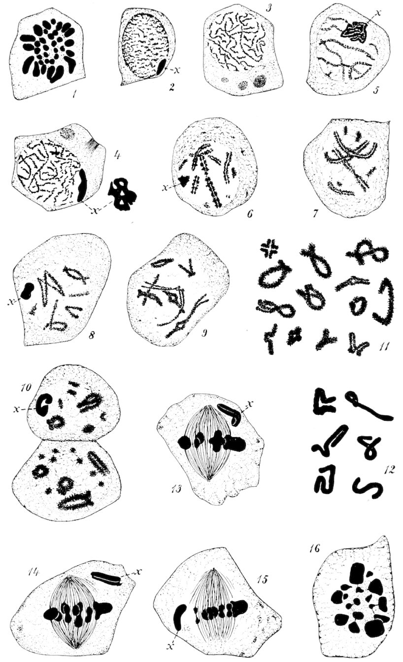
The main features characterizing the next steps in the process
are the rapid increase in size of the cell and nucleus, and
the arrangement of the chromomeres into a fine thread or
threads (figs. 2–4). This is well called the growth stage, for
all parts of the cell engage in the work of regaining the ground
lost during the period of multiplication in the secondary spermatogonia.
As a result of this metabolic activity, the first
spermatocytes at the end of the prophase have reached a volume
often as much as ten times that possessed by the last generation
of the secondary spermatogonia from which they were
derived. Nucleus and cytoplasm, in about an equal degree,
participate in this enlargement, and, at the end of the period,
present an appearance much different from that of the spermatogonia.
This consists most strikingly in the greater clearness
of all the parts, due to the increased amount of hyaloplasm
which separates by greater distances the more solid structures
of the cell.
In the nucleus, for instance, the chromatin aggregates are
now definitely apparent, and each stands free and clear except
for connecting threads of linin. The cytoplasm, likewise, instead
of showing a coarsely granular aspect, exhibits a clearly
189reticular structure, with such large intervening hyaloplasmic
areas as to suggest an almost alveolar structure, especially in
the later stages (figs. 3–9). This increased amount of fluid becomes
evident by an examination of sections under even a low
power of the microscope, principally by the lessened density of
the general stain in the cell.
A peculiarity of the archoplasm in these early prophases is
the persistence manifested by the spindle fibers of the previous
generations. Often connecting fibers may be seen, joining cell
to cell, as has been described by many writers, but, in addition
to this, the spindle remains of more remote ancestral mitoses
show themselves. In figure 3 is represented a cross-section
through three persisting spindles of as many generations.
Their age is suggested by size and intensity of stain, both factors
being least marked in the oldest structure.
Centrosomes and astral radiation do not present themselves
with the prominence and frequency of such structures in corresponding
cells in Hippiscus.
The main interest of these studies, however, attaches to the
movements of the chromatin granules. As was suggested in
an earlier paper (17), it is only by an understanding of the
constructive processes in the prophase that we can appreciate
the structure and changes of the chromosomes in the metaphase.
It is to this period in the history of the chromosomes
that I have given the most attention and to which I will devote
the most space in the record of observations.
Apparently the chromomeres resulting from the disintegration
of the spermatogonial chromosomes are loosely scattered
through the nucleus, so that no formed structure is to be seen.
With the increase in size of the cell, however, a linear arrangement
of the elements becomes apparent, so that it seems as if
a thread is formed. Whether this is continuous or segmented
it is not possible to determine. The large amount of chromatin
and the tortuous course of the filaments put a solution of the
problem beyond the range of assured observation. It is with
much regret that this fact is recognized, for one of the most
important questions connected with the maturation mitoses
hinges upon the method by which the chromosomes, as such,
are derived from those of the spermatogonia. Upon this point
the evidence of the ordinary chromosomes of these cells would,
190if anything, tend to confirm the view that there is a possibility
of complete rearrangement of the chromomeres in the different
chromosomes. Concerning this, however, the accessory chromosome
is much more conclusive and convincing, as will be
shown later.
Disregarding the relations of the chromosomes of the two
generations, it is evident that from the material of the spermatogonial
elements there is formed the thread of the spermatocyte
prophase. As indicated in figures 3 and 4, this is at first
composed of a single series of chromomeres. But in a slightly
later stage, represented by figure 5, it becomes plain that the
thread is wider and at the same time double. A careful investigation
will show that the halves of the thread are exact duplicates
of each other, each granule of the one having its mate in
the other. There is but one conclusion to be derived from the
appearances just described, which is that the double thread is
formed by a longitudinal division, granule by granule, of the
original filament. The evidence afforded, not only by the
Locustids, but by all the Orthoptera, is unequivocal on this
point. The cleavage of the thread is not exaggerated in the
accompanying figures, and is distinctly in evidence even under
ordinary conditions of illumination and magnification.
Much controversy has recently arisen among both botanists
and zoologists concerning an appearance of the chromatin in
the prophase, which has received the common designation
“synapsis,” by which is meant, usually, a one-sided contraction
of the chromatin in the nuclear vesicle. No such stage in
the nucleus could be found in Hippiscus, and it is likewise absent
in the Locustid cells. I therefore repeat the assertion
made in the previous paper (17), that in properly fixed material
derived from Orthopteran sources the first spermatocyte
prophase shows no unilateral massing of the chromatin.
Shortly after the formation of the double spireme, it is to be
seen that the thread is no longer—even if it was previously—continuous,
but is composed of segments (figs. 5–10). So early
as this it is possible to observe that the segments are of very
unequal lengths. The extent of this inequality may be gathered
by consulting figures 6 and 7. Even in this early stage
the real structure of the segments may be determined, and in
191those favorably situated the quadripartite nature of the future
chromosomes manifests itself very distinctly.
This important stage in the history of the first spermatocyte
chromosomes first received attention at the hands of Paulmier
in his studies upon Anasa. Almost at the same time I found
structures in the Orthopteran spermatocytes so nearly identical
that it would be impossible to distinguish any marked difference
between them. The Locustid material, equally with the
Acridian, permits an exact determination of the chromosome
structures, which later become so masked as to be indeterminate.
The interest attaching to the construction of the spermatocyte
chromosomes is so great as to warrant an account of the
process, although, in general, it is largely a repetition of what
has been given for Anasa and Hippiscus. As early as the stage
represented in figure 6, it becomes noticeable that the chromatids
near the middle of the thread tend to diverge from each
other, leaving a diamond-shaped space. This becomes more
pronounced, and it is soon seen that each half of the thread is
broken across at the same level, resulting in the production
of a chromosome of four parts. Still retaining their general
shape, these segments shorten and broaden until they are almost
the size of the metaphase chromosome.
All variations conceivable upon the wider separation of the
halves along the longitudinal split, the movement of the parts
upon the line of separation at right angles to the original cleft,
or of approximation and rotation of the free segmented ends
are found. Thus do we get the cross-shaped, the double-V, the
figure-of-S, the Y-shaped and ring figures, in figure 11. Many
of the rings give the impression, upon superficial examination,
of loops with their free ends crossed. A careful examination
will always reveal the fact, however, that what appears to be
the crossed ends is really the middle portion of the segment,
with the chromatids drawn out along the plane of the cross-division.
In segments that are favorably placed, there is never
any difficulty in correlating the structures with the typical one
of a cross-split lengthwise of each arm.
The quadripartite nature of the chromatin segments may be
determined, as already indicated, almost as soon as the longitudinal
split occurs. From this time on until the chromosomes
192are divided in the metaphase, it is possible to trace the formation
of the tetrad chromosomes and to be sure of the relation
existing between the longitudinal and cross planes of separation.
As evidence of the existence of a longitudinal division of
the chromatin thread and of the sequence of the two divisions,
I do not see how more could be asked of any material. In the
early prophase the greatly elongated and granular thread becomes
twice split, once along its length and once across it. As
the cell ages, a continuously closer approximation of the chromomeres
occurs, without obliterating the lines of separation
between the four parts of the segment; accompanying this, the
segment becomes shorter and thicker, and the previously existing
linear arrangement of the chromomeres is superseded.
When the segments have reached approximately the size of the
definitive chromosomes of the metaphase, the nuclear membrane
disappears and distinction between cytosome and nucleus
is lost. As a coincident step, the formerly granular
segments become homogeneous in structure by the disappearance
of the chromomeres as individual structures; all lines of
separation between parts are lost to view, so that an examination
of the formed element would betray no indication of composite
structure. But, having traced the formation of the
chromosomes in this way, one is at no loss to identify each part
of the preexisting quadripartite chromatin segment. This is
possible because, while all trace of internal structure is gone,
the general outline is retained and the crosses and rings of the
early stages are still, even up to the metaphase, crosses and
rings.
Having traced the formation of the ordinary chromosomes
through the various stages of the prophase, I should like to return
to the beginning again and bring up to a like degree of
development the aberrant element which I have called the accessory
chromosome. This has already been given in general
outline in my first paper upon Xiphidium (16), but a number
of important observations since made render a general discussion
desirable.
I have not yet found it possible to make a detailed study of
the spermatogonia of the Locustids, as was done for the Acrididæ
by Sutton in this laboratory, but sufficient observations
have been made to be assured that the accessory chromosome
193participates normally in the mitoses of the secondary spermatogonia.
It is here distinctly visible because of its large size,
which causes it to extend down to the equatorial plate, while
the other chromosomes are in a late anaphase.
At the close of the spermatogonial divisions, when the disruptive
processes reduce the other chromosomes to masses of
chromomeres in which chromosome identities are not apparent,
the accessory chromosome, with apparently more cohesive vigor
than the others, retains its general form and is at all times distinguishable.
It is marked off from the others, not only by
persistence of form, but also by the difference in staining reaction,
this being such as is usually exhibited by chromatin when
concentrated into homogeneous masses. While studying the
cells of Xiphidium, I noticed that, at one stage, this color reaction
changed somewhat and more nearly approached that of
the diffused chromatin. At this time the accessory chromosome
had the form of a flattened, apparently fenestrated, plate.
I have been fortunate enough, in preparations of Orchesticus, to
discover that the accessory is really at this time in the form of
a long, coiled thread (fig. 5). It is thus seen that, even in respect
to the spireme stage, the accessory chromosome is comparable
to the others, the only difference being that the diffusion
of the chromomeres is less, and the independence of the element
greater, than is the case with the other chromosomes.
As the chromatin segments shorten and thicken, the thread
of the accessory likewise increases in diameter at the expense
of its length, and is finally observable in various degrees of
contortion, as shown in figure 12. By the time the chromosomes
are ready for division, the accessory has assumed a form
very similar to that it shows in the spermatogonia. With the
establishment of the equatorial plate, the accessory moves to one
pole of the spindle and there remains undivided during the first
spermatocyte mitosis. It is accordingly a member of only one
second spermatocyte resulting from the division of each first
spermatocyte.
Returning to the group of chromosomes preparing for metakinesis,
we find that in their earlier stages they lie so that their
longer diameter is in the equatorial plate, while attached to the
enlargement in the center of each, representing the point of
separation laid out for the second spermatocyte division, are
194the mantle fibers running to the centrosomes. The changes
now ensuing are easily decipherable, because the chromosomes
do not all undergo division at the same time. Since the main
differences at present existing between insect spermatologists
relate to the sequence of the divisions in the spermatocyte
mitoses, I shall again describe the process, although it is identical
with that already given for Hippiscus.
The necessity for a thorough understanding of the chromosome
construction here becomes evident. Knowing how the
chromatids were associated in the chromosomes, one can follow
understandingly their movements during metakinesis.
It is first to be noted that the chromosomes lie with their
longer axis in the equatorial plate. This, as we have seen, is
the plane along which the longitudinal cleft occurred, so that
a separation in this way means the longitudinal division of the
chromosomes in the first spermatocyte. This is, in reality,
what occurs. The contracting mantle fibers attached to the
middle of the segments drag the adhering chromatids apart
without at any time exposing a separating space. It is in this
way that in the beginning the longer axes are at right angles
to the spindle axis and at the end parallel with it, while during
intermediate periods crosses with arms of varying length
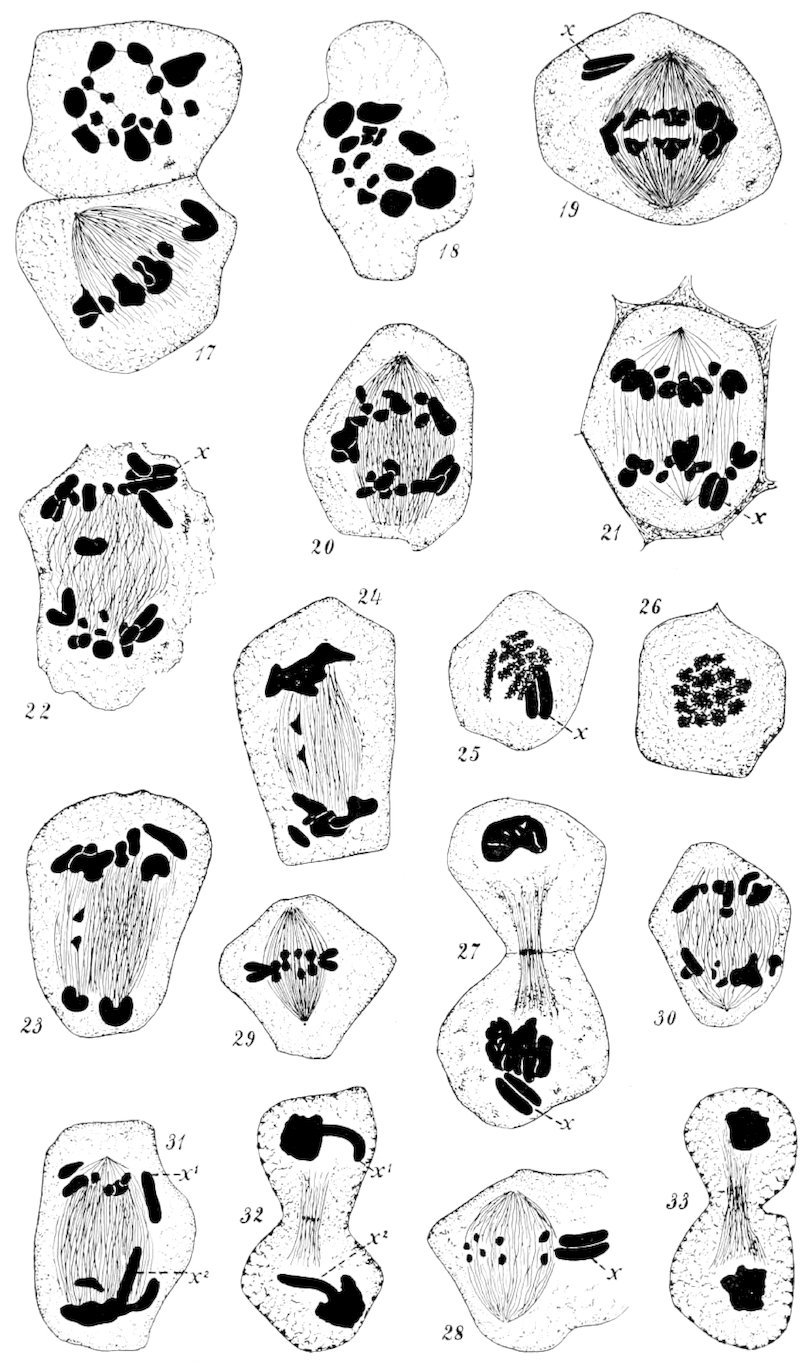
exist (figs. 13, 14).
The previously disguised lines of separation become at once
visible in the daughter chromosomes, for, instead of remaining
closely apposed, as formerly, the chromatids spring apart at
the free ends and the chromosomes pass through the anaphase
as V-shaped bodies instead of as simple rods. The space thus
disclosed represents that which separates what would be the
ancestral spermatogonial chromosomes, assuming that the reduced
number occurs by the end-to-end union of chromosomes
of the secondary spermatogonia. As already stated, the accessory
chromosome does not divide at this time.
At the end of the anaphase we find the ordinary chromosomes
massed at the poles of the cell, and, in addition, at one
the undivided accessory chromosome. The second spermatocytes
are therefore of two kinds, one possessing the accessory
chromosome and the other not. One additional feature of interest
that becomes apparent during the migration of the daughter
chromosomes to the poles is the retarded division of one
195of the elements (figs. 22–24). Some cysts contain cells that
almost invariably exhibit this peculiarity. The lagging chromosome
is always one of the small ones, but whether the same
in each case could not be determined.
In the telophase, the main interest is centered in the question
as to whether there is a loss of identity of the chromosomes
or not. The evidence afforded by the Locustid cells is
strongly in favor of the conception of persisting elements. As
is usually the case, I believe, the chromosomes, when not under
the active influence of the archoplasm, loosen up, and their
homogeneous structure gives way to the granular appearance
noticeable in the prophase. Although the chromosomes become
closely massed and granular, their outlines can usually
be distinguished (figs. 23–27). The accessory chromosome does
not change its form and structure at this time (figs. 25, 27).
The telophase ends with the ingrowth of the dividing cell-wall,
and the second spermatocyte mitotic figure is established without
any real prophase. Between the two generations it is evident
that there exists no such thing as a “rest stage.”
In the metaphase of the second spermatocyte are formed exact
duplicates of the chromosomes seen in the anaphase of the first
spermatocyte. These arrange themselves radially in the equatorial
plate, one chromatid immediately above the other, so
that the plane separating the halves is at right angles to the
spindle axis. Mantle fibers attach to the inner ends of the
chromatids at the point at which, in all probability, the fibers
of the first spermatocyte were connected. I am inclined to regard
this as true because the opposite ends, during the anaphase,
seemed to be mutually repulsive.
The spindle itself is small and weak as compared with that
of the first spermatocyte, and does not long survive the anaphase
condition. The material composing it, however, persists
as the nebenkern of the spermatid.
A marked difference between the second spermatocytes that
contain the accessory chromosome and those which do not is
observable. In the metaphase, the element, already longitudinally
split in the prophase of the first spermatocyte, projects
from the equatorial plate for some distance into the cytoplasm.
196It is very much larger than most of the other chromosomes, as
may be seen in figure 28. It divides readily in metakinesis,
and its chromatids travel to the poles with those of the other
chromosomes, but, on account of their greater length, project
downward from the mass (fig. 31). Here, as always, the accessory
stubbornly maintains its independence, and can be seen
extending out from the mass of other chromosomes at each end
of the mother cell (fig. 32).
The division of the other class of second spermatocytes is,
of course, unaccompanied by modifications due to the presence
of the accessory chromosome. Aside from this, no difference
between cells of the two classes is noticeable.
To summarize, we may say, that resulting from the division
of each first spermatocyte are two second spermatocytes, one of
which contains an accessory chromosome while the other does
not. The second spermatocyte containing the accessory divides,
and with it the accessory, so that each of the spermatids
derived from it contains a chromatid from the accessory. The
other second spermatocyte, not containing the accessory, also
divides, producing two spermatids in which the accessory is
absent. Thus half of the spermatids contain accessory chromosomes
while the other half does not.
The enumeration of the chromatic elements, while a very important
part of any study upon the nucleus, is unsatisfactory at
the best. If there is any great number of chromosomes in the
cell, it is impossible to secure a determination of it in a lateral
view of the metaphase, because the elements overlie one another
so as to render their distinction very uncertain. A polar view
is much more desirable, but even here one is never certain that
all the elements are represented, or that only entire chromosomes
of one cell are present. The first of these contingencies
arises from the fact that, in the event of a cell being cut in two,
some of the chromosomes may drop out and not appear in the
sections; or, if still on the slide, and in a small group, they
may lie so close to a mass of chromosomes in another cell as to
be confused with them. An excess in number may be found if
a portion of the chromosomes have already divided in the equatorial
plate, while the remainder are still united (cf. fig. 19),
197or if one or two from the fragment of another cell are in the
neighborhood. All these embarrassments are increased when
an independent structure like the accessory chromosome is
present. These difficulties exist when the conditions are most
favorable, i. e., when the chromosomes are arranged in the
equatorial plate; they become practically insurmountable during
any other stage of mitosis by the intertwining of the chromatic
segments or by fusion of chromosomes in later stages.
Because of these considerations, I do not put implicit confidence
in conclusions drawn from numerical relations when they
involve the question of whether or not there is a difference of
one chromosome between two cells. What I have to say, therefore,
concerning the numbers of chromosomes in the different
cell generations of the Locustid testis, I must state as my best
judgment in the matter, based upon the most careful observations
I could make upon cells showing the elements with the
greatest clearness. While I regard them as in all probability
correct, I do not rely so thoroughly upon them as I do upon
observations of structural details, and have therefore based no
conclusions upon numerical relations alone.
As is stated elsewhere, the number of chromosomes in the
spermatogonia appears to be thirty-three. This was ascertained
by selecting the clearest possible cases of the metaphase
that could be found and drawing them under the camera lucida.
Subsequent countings were made, and in most of the cells
thirty-three chromosomes were found. An inspection of figure 1
will show that there is a characteristic arrangement of the chromatin
bodies, the larger ones being on the outside of the group,
the smaller within. Amongst the large ones, it was impossible to
distinguish the accessory chromosome, but a lateral view of the
anaphase shows it clearly. From the fact that it was a single
element in the spermatogonia, it was to be expected that an
uneven number of chromosomes would appear in this cell generation.
In the spermatocytes, as in the spermatogonia, the polar view
of the metaphase was the stage selected for use in counting the
chromatin elements. A large number of cases showed that sixteen
and seventeen were the prevailing numbers. The smaller
of these is easily accounted for when it is recalled that the accessory
chromosome is at one pole of the spindle, and would
198very often lie in another section, where it would not be possible
to be sure of its relations. I am convinced from these counts
that seventeen is the reduced number in the first spermatocyte,
sixteen of the elements being ordinary chromosomes, the other
one being the accessory chromosome which has come over unaltered
from the spermatogonia. This coincides with the theoretically
expected number, deduced from the independently
determined number of spermatogonial elements.
In view of the divergences found in insect spermatogenesis,
the established theory that the reduced number of chromosomes
is exactly half the normal or somatic number is not a
strictly accurate one, for in this case the reduction is from
thirty-three to seventeen. Similar instances may be found in
the forms investigated by Montgomery and de Sinéty.
When we come to consider the second spermatocytes, spermatids,
and spermatozoa, it is necessary to divide them into two
classes, because of the unequal apportionment of the accessory
chromosome consequent upon its remaining undivided in the
first spermatocyte mitosis. There are formed, accordingly, two
numerically equal classes of second spermatocytes—those containing
sixteen chromosomes plus the accessory chromosome,
and those with merely the sixteen chromosomes. The members
of each of these classes divide and double their kind, forming
spermatids marked as were the second spermatocytes—one
class with seventeen chromatic elements, and the other with
sixteen. From these, by the usual transformations, are derived
the mature male elements, which are thus of two distinct kinds.
The limits set to this paper preclude anything more than
passing mention of the spermatids. As stated above, cells at
this stage of development are of two classes, depending upon
the presence or absence of the accessory chromosome. The distinction
thus set up continues to exist visibly far through the
transformation stages of the spermatid, by reason of the persisting
independence of the accessory chromosome. Of the
dual nature of the spermatids I was very early convinced, because
the accessory chromosome is so strikingly displayed by
the nuclei in which it exists that it is impossible to overlook
its absence in a large proportion of the cells. As to the
199certainty of this partial distribution in the transforming spermatozoa,
I am rendered positive by the most careful and painstaking
study. This is valuable corroboration of the observed
fact that the accessory chromosome remains undivided in one
of the spermatocyte mitoses.
The literature relating to the spermatocytes of insects was
reviewed at some length in my previous paper upon the history
of these cells in the Acrididæ (17). It is not my purpose to
go over this same ground again except in so far as increased
knowledge makes it necessary. More recent papers by Montgomery,
Wilcox and others will, however, be discussed in detail.
The policy previously announced, of restricting comparisons to
results derived from insects, will again be adhered to. I believe
that the main features of the maturation divisions are
essentially the same in all insects, and I desire to see this belief
either well established or overthrown. If it can be demonstrated
that so large a class as the insects are characterized by
a common process, it will be a firm basis upon which to conduct
further comparative studies into more comprehensive
groups. On the contrary, if it is shown that there is no type,
even in the class, then it is useless to seek agreements between
widely removed species.
A necessary basis for any comparative work is a common
terminology. Confusion inevitably follows the loose application
of names to the structures compared. This is perhaps
unavoidable in the early stages of an investigation, but should
be overcome as soon as possible. There is surely no reason for
continuing uncertainty after terms have received general acceptance.
Believing this, I feel called upon to repeat my criticisms
of Montgomery’s application of the well-accepted terms
“prophase,” “metaphase,” “anaphase,” and “telophase.”
In reply to my previous objection directed against this part
of his work, Montgomery acknowledges the validity of the criticism
so far as it relates to the metaphase, but denies the application
to the other phases, particularly to the anaphase. He
alleges in support of his position that the introduction of an
unusual condition, the “synapsis,” makes it impossible to correlate
200strictly the stages of the germ-cells with those of ordinary
divisions. Upon this point I must again disagree with him.
It is impossible for any known modification of the prophase to
change the essential character of the anaphase, so as to make
it precede instead of follow the metaphase. This stage marks
the movements of the chromosomes from the equatorial plate
to the poles, and terminates when they are massed around the
centrosomes. How can the “synapsis” in the least affect the
duration or character of this process? It is apparent enough,
I think, that Montgomery’s subphases of the “anaphase” do
not belong to this portion of the mitotic cycle at all, but are
really portions of the telophase of the spermatogonia and prophase
of the first spermatocyte. Further, it may be noted that,
even were these subphases properly included in the anaphase,
they would belong to the spermatogonia and not to the spermatocytes.
Montgomery himself seems to be rather uncertain of the position
of his “anaphase.” In the first paper, upon Euchistus (12),
it was put down as the anaphase of the first spermatocyte; in
his later paper (14), upon Peripatus, it is recorded as the anaphase
of the spermatogonia. Still more confusing is his use of
the “telophases,” for in the article upon Peripatus (14) it is,
in the “Contents,” placed as a substage of the spermatogonial
anaphase, and in the body of the work, page 307, as the telophase
of the spermatocyte! Neither the anaphase nor the telophase
can, by any possible construction of their proper meanings,
be made to apply to the “growth period” of the germ cycle, as
Montgomery insists; they are the last stages of the “division
period,” in reality. The prophase of the first spermatocyte is
the initial stage in the constructive process marking the growth
period.
Montgomery’s translocation of the terms makes the “synapsis”
occur in the anaphase. This is manifestly an impossible
condition of the chromatin at this time, and his figures show
definitely enough that it is a prophase, or, at the earliest, a
spermatogonial telophase, that witnesses the contraction of the
chromatin. The objection urged in my earlier paper (17) to
the use of the term as a designation for the mere contracted condition
of the chromatin cannot apply to Montgomery’s latest
use of it; for he here recognizes the justice of my contention
201that it was primarily designed to indicate the fusion of the
spermatogonial chromosome to produce the chromosomes of the
spermatocyte. He states this clearly in the following words:
“Moore (1895) first gave the name ‘synaptic phase’ to that
stage in the growth period of Elasmobranchs when the reduction
in the number of chromosomes takes place. Accordingly,
the criterion of the synapsis stage is, first of all, the combination
of univalent chromosomes to form bivalent ones; whether
the chromosomes are then densely grouped or not is of secondary
importance.”
The formation of the first spermatocyte chromosome gives
us an insight into the later changes undergone by these elements
such as cannot be obtained in any other way. The great
importance attaching to this part of the spermatogonial process
renders it desirable to exhaust every effort in obtaining a knowledge
of the actual changes here taking place. This thought
has been held constantly in mind during the progress of these
investigations, and every point of resemblance or of difference
between the various species studied has received careful attention.
Despite variations in details, however, I must state that
the essential features of the maturation divisions are the same
in all species of the Orthoptera examined. It is true that as
yet only two families, the Acrididæ and the Locustidæ, have
been worked out in a detailed way, but the close agreement between
these raises a strong presumption in favor of the general
prevalence of the type. The processes of the two families have
already been described in detail, but it will perhaps be well to
call particular attention to some points worthy of mention.
The general appearance of the material derived from the two
families is quite different in sections. Even the hastiest observation
will show this. The spermatocytes of the Locustid testis
are much smaller, denser and more deeply staining than those
of the Acrididæ. The relative quantity of chromatin is greater,
so that it is possible by microscopical examination of a section
to tell whether it was prepared from Locustid or Acridian material.
The transformation from the telophase of the last spermatogonial
division to the prophase of the first spermatocyte is
marked by practically the same changes in both families. It
202is to be observed, however, that the derivation of the spireme
from the disintegrating chromosomes of the previous generation
is not so clearly indicated in the Locustid cells, and it was
for this reason that in the examination of Xiphidium I was not
able to determine certainly that the accessory chromosome came
over from the spermatogonia into the spermatocytes as a formed
element. Upon this point, as upon others, my later material
is clearer, and I was able to reconcile the appearances in the
two families. In both, unfortunately, it has been found impossible
to determine the exact origin of the first spermatocyte
chromosomes.
In connection with the transformation of the chromatin from
the spermatogonial condition to that of the spermatocyte, we
must take notice of that stage which is commonly denominated
the “synapsis.” The evidence afforded by the Orthopteran
cells is entirely negative regarding this. In properly fixed material
there is no distortion of the chromatin in the nucleus at
any time. It would, if present, be particularly easy to observe,
as was stated in my previous paper, for during the entire winter
the spermatocytes exist in the spireme stage, and in a longitudinal
section of a follicle all stages may be discerned. On
the other hand, in poorly fixed or hastily prepared material the
synapsis is present, and always in such a form as to indicate
its artificial character. What is here said regarding the synapsis
refer to the appearance commonly thus designated, but,
as has already been stated, such an application of the term does
not meet the spirit of the definition as intended by Moore (20).
A fusion of the spermatogonial chromosomes of some sort must
certainly occur, but that it is always marked by a unilateral
massing of chromosomes, I deny.
During the prophase the chromatin segments in the cells of
Orchesticus and other species of the Locustids are heavier, more
granular and denser than they are in Hippiscus. It is to be
observed, also, that there is a greater variation in the size of
the elements. This fact is observable from the earliest appearance
of definite segments down through both the spermatocyte
mitoses. This disproportion may be such that one chromosome
will exceed another in the same cell by twenty or thirty times
its volume. We have here, as is pointed out in another place,
a strong proof concerning the individuality of the chromosomes,
203for in some species it is possible to distinguish a particular
chromosome in all the spermatocytes. This is strikingly the
case in Anabrus, where there is always one chromosome very
much larger than any of the others. It exceeds in size even
the accessory chromosome, and might be mistaken for it were
it not for the difference in form. It is, however, typically a
tetrad, and shows the four chromatids, while the accessory
chromosome exhibits the usual spermatogonial condition.
As was indicated under the head of “Observations,” the prophase
tetrad characteristic of Anasa and Hippiscus is again exemplified
in the Locustid cells. So close is the resemblance of
the maturation chromosomes of these various insect cells in
their early stages, that I now regard it as practically established
that they are commonly present in all insect spermatocytes.
No more important evidence regarding chromosome structure
and behavior can be obtained than that afforded by these elements.
Particularly are the ring figures of value in the determination
of the sequence of the longitudinal and cross divisions,
and upon this point the material from the two families is
equally convincing and positive in demonstrating that the first
spermatocyte mitosis witnesses a separation along the longitudinal
cleft of the spireme thread.
I should like to emphasize the fact that the chromosomes in
both the Orthopteran families studied have been carefully
traced from their earlier appearance down to the time of their
dissolution in the spermatid through such a gradual series of
changes that there can be no reasonable doubt of the accuracy
of the conclusion set forth in these papers. The Orthopteran
material possesses one distinct advantage over the Hemipteran,
in that the point of cross-division is always marked by the same
sort of a protuberance as is to be distinguished in the early
chromatin segments. When the two free ends of the element
are brought around to form a closed ring, the last particle of
doubt regarding the position of the planes of separation marked
out for the two spermatocyte divisions is dispelled.
This diagnostic character seems to be lacking in the chromosomes
of the Hemiptera, and Paulmier, in his work on Anasa,
depends for his criteria of orientation upon the relative lengths
of the chromosome axes. Such a feature would be valueless in
Orthopteran cells, because, as has been shown, the chromatids
204move upon each other in such a way as to exactly reverse the
preexisting relation between the axes. How applicable this
observation may be to conditions in the Hemipteran cells, I do
not know; but, judging from the great resemblance of the elements
in the prophase, it would seem most reasonable to expect
a similarity of the divisions.
Paulmier (22) advances the suggestion that in the double-V
figures we may find a structure that will serve to reconcile the
divergent accounts concerning the longitudinal and cross divisions
of the tetrads. The only way in which this might be accomplished
would be to suppose that each of the interspaces
represents a longitudinal cleavage of the thread, the first being
at right angles to the second. I have given this suggestion
careful consideration, and find no evidence to support it. The
double Vs are only of rare occurrence, the common element
being a straight rod, in the center of which is a diamond-shaped
clear spot representing the two planes of division laid out for
the spermatocyte mitoses. If two longitudinal divisions occur,
one must precede the other considerably and the resulting
halves become mutually repulsive, so that they move apart and
lie in one plane with only a slight connection at the point of
final separation. Moreover, the second cleavage must begin at
the opposite end of the segment and proceed in a reverse direction
from the first. Not only this, but the first spermatocyte
mitosis divides the elements along what is generally conceded
to be the longitudinal split, and this must necessarily succeed
the supposititious first longitudinal cleavage by some time.
Without going into a consideration of these points, I may say
that they suggest such deviation from normal processes that
only extensive and accurate observations would make Paulmier’s
suggestion worthy of further consideration.
In my former paper I reviewed the results obtained by Montgomery
upon the Hemiptera, but further notice of his work
will now be necessary, since on almost every important point
relating to chromosome structure he has changed his opinion.
His late extensive comparative study upon the Hemipteran
cells, as well as that upon Peripatus, will at the same time receive
consideration.
It appears from Montgomery’s account that at the point
205where the Orthoptera are least valuable in demonstrating chromosomal
relations the Hemiptera and Peripatus are most convincing.
I refer here to the derivation of the first spermatocyte
chromosomes from the chromatin of the spermatogonia. He
claims to have observed the union by pairs of the secondary
spermatogonial chromosomes during the anaphase (his synapsis)
so clearly as to be positive of this fusion. I hope this may
be verified, for it offers a logical explanation of the process of
reduction, and is a confirmation of what has previously been
assumed true without sufficient basis in observed fact, as was
suggested in my paper on Hippiscus. This, if established,
would also be a strong support of the theory relating to the
constancy of the chromosomes. If this true synapsis is accomplished
at this time, however, it must be noted that it occurs
during the last phase of the final spermatogonial mitosis, and
is not an act of the spermatocyte prophase. But as to the exact
location of this point no contention need be made, for it is conceivable
that the time of its occurrence might vary considerably
without affecting the essential nature of the process.
With regard to such an origin of the first spermatocyte chromosomes,
there is an important difference to be noted between
the earlier and later work of Montgomery, and one which he
fails to mention. In his paper (12) upon Euchistus he states
the matter as follows: “But in the post synapsis we do not
find seven chromosomes, the definitive number present in the
spermatocyte divisions, but a smaller number; hence, in the
synapsis the true (i. e., exactly half) reduction of the chromosomes
does not take place, but the number is reduced to less
than one-half.” This statement is based, he says, upon a most
careful and painstaking enumeration of the chromatic segments
in a number of nuclei, and is unhesitatingly declared correct.
In his later paper, on the contrary, he is just as positive that
the definitive reduction is here accomplished, for he says:
“Since then I have been able to demonstrate that this numerical
reduction is effected in the synapsis by the union into seven
pairs of the fourteen chromosomes, each of the seven bivalent
chromosomes (pairs) being composed of two univalent chromosomes
joined end to end.” This statement is made without
adducing any specific proof, as was formerly done. By what
means we are to reconcile these diametrically opposite statements
206Montgomery does not say. He, however, insists that he
has always known that the fusion by pairs takes place. How
this was to be brought about under his previous assumption
that one of the fourteen spermatogonial chromosomes became
removed from participation in the usual processes of the cell to
form a “chromatin nucleolus,” he fails to state. Until the confusion
is cleared up by corroborative evidence on one side or
the other, a most important part of Montgomery’s work must
still be regarded as uncertain.
Despite his recognition of the fusion of the chromosomes in
the synapsis as the essential feature of this stage, Montgomery
is insistent upon the concentration of the chromatin as its distinguishing
characteristic. Regarding this he says: “McClung
considers the appearance of the synapsis stage as artefacts. It
is hardly necessary to reply to this criticism, since in all Metazoa
where the spermatogenesis has been carefully examined, with
the exception of certain Amphibia, the dense massing of the
chromosomes (?) in the synapsis stage has been shown to be a
perfectly normal phenomenon.”
Concerning two points in this statement I wish to take exception.
First, as was suggested in my previous paper (17), the
term synapsis is usually applied to a condition of the prophase
in which the apparently unsegmented spireme exists. It must
be remembered that most investigators consider that the reduction
of the chromosomal number takes place by the segmentation
of a spireme into half the usual number of segments. In
the second place, I must resent the implication that the work
done in this laboratory is not “carefully” conducted. Many
“Metazoa” have been examined “carefully,” and in none has
the “synapsis” occurred when the material was well fixed and
prepared. It has, moreover, been found possible to produce
the appearance at will. One case of this kind is sufficient to
raise the presumption that it may not be normal even when
constantly found in certain preparations. I have not, however,
absolutely denied the possibility of such an occurrence, because
it is conceivable that from the telophase of the preceding division
the massing of the chromosomes may persist during their
elongation. My contention is that the appearance is not a constant
or necessary condition in “all the Metazoa,” and this I
have proven.
207In rather striking contrast to the work of Montgomery, in
which an effort is made to formulate a typical process for the
entire Metazoa from the study of a single order, is that of Wilcox,
wherein a general denial of any apparent system in the
maturation divisions of animals is based practically upon the
study of a single species. As was stated in my former paper,
I regard Wilcox entirely in error upon the vital point of his
theory of tetrad formation, not by “forced interpretation” of
his own views, but by an actual examination of the object upon
which he worked. There is no point upon which Orthopteran
material affords more indisputable evidence than upon the occurrence
of the longitudinal division of the chromatin thread in
the early prophase. My statement regarding Wilcox’s position
on this subject was in no sense “misdirected criticism,” but
an actual statement of fact; it was not an attempt to explain
away “abundant and evident cases which cannot be made to fit
into the scheme,” but simply the presentation of proof that one
case was wrongly interpreted.
Wilcox claims the distinction of being the first and only investigator
to doubt the hypothesis that longitudinal and cross
divisions of the chromatic thread produce chromosomes of a
different character. It is perhaps well that this is so, in view
of the reasoning by which such a distinction is secured. Upon
his own unconfirmed and disputed statement that there is no
longitudinal division of the spireme, Wilcox presumes to disparage
the accepted view of practically all cytologists. The
constructive thought of the last two decades is summarily disposed
of by this author in the following language: “The whole
question, therefore, whether a certain division is longitudinal
or transverse loses its practical significance, since the theoretical
interpretation which has long been placed upon these divisions
is shown to be impossible and absurd!” The showing
alluded to consists in the statement that the chromosomes consist
of an indefinite number of granules, which cannot be
expected to arrange themselves in any order, and which, therefore,
may be divided in any way without affecting the results.
Laying aside for a moment the question as to the occurrence
of a longitudinal division, we may well inquire whether the
belief that, “In view of this manner of the formation of the
chromosomes (by the aggregation of the chromomeres), it
208seems absurd to assume that the separation of an individual
chromosome by one plane could be quantitative while the separation
by another plane was qualitative,” is well founded.
At the basis of such an assumption lies the implication that
any definite arrangement of chromomeres is impossible; for if
any definite order were possible, then the supposed argument
against the longitudinal disposition of the chromomeres would
be invalid.
The argument of Wilcox is therefore directed against order
in general, and not against order in any one particular, as he
would have it appear. For it must be admitted that if it is
possible for the scattered chromatic granules of the early prophase
to arrange themselves at all (and this even Wilcox does
not deny), it is equally possible for them to come together in a
definite order. That they do this is amply evidenced by the fact
that later they appear in definite groups or chromosomes. It is
to be noted, moreover, that the later investigations tend to suggest
that the apparently unorganized chromatic granules in the
first spermatocyte prophase are really bound together and represent
merely a diffuse condition of the spermatogonial chromosomes.
Wilcox’s chief error, however, is not to be sought in speculative
theories, but rather in his faulty observations. He
repeatedly denies the occurrence of any longitudinal split in
the chromatic thread of the first spermatocyte prophase. That
he is mistaken here I am thoroughly convinced, both from a
study of his own object and from investigations upon many
other species of the same family. At the present time, also,
practically every spermatologist is aligned in support of the
view denounced by Wilcox. For a while Wilcox had some
backing, but most of those who advocated only cross divisions
of the thread have later been able to demonstrate the longitudinal
cleavage in better prepared material.
There is general acceptance of the opinion that the chromomeres
of the last secondary spermatogonia appear in a linear
arrangement to form what is commonly known as the “spireme.”
Wilcox declared that while in a very fine condition
this thread breaks across into segments, which unite by pairs
to form the chromosomes of the first spermatocyte. The great
majority of other investigators are unanimous in the opinion
209that this fine thread, made up of granules, becomes double by
the division of each granule individually, thus producing a
double thread. Thus it is that the two halves of a longitudinally
divided chromosome are made equivalent, not by the
sifting apart of preexisting granules, but by the division of
these after they are arranged in a linear series. It need hardly
be mentioned that the formation of the thread has here a reason
for existence which is entirely lacking according to Wilcox’s
scheme.
This much space has been devoted to Wilcox’s statements,
not because they present any arguments against the generally
accepted views of his fellow workers, but because he represents
a rapidly lessening minority which is content to work in a very
limited field and to resort for the explanation of diverse results
to the very convenient theory that great differences may be
expected in the normal processes of even closely related forms.
One needs only to glance at the work of all insect spermatologists
to see how closely the agreement now is upon the important
points of the process. This accordance of results Wilcox
notes, but interprets in his own way, which may be regarded
as not exactly complimentary to the skill and judgment of his
colaborers. “It is only necessary,” he says, “to refer to any
recent publication on the subject to find examples of this attempt
to force the divergent processes in different species to fit
the same formula.” This is certainly a very easy and convenient
way to dispose of the accumulated observations of the
many careful investigators who have come to an agreement
upon the important questions under discussion, but I venture
to think will hardly satisfy any one except its sponsor.
After handing in this article for publication, I fortunately
secured a copy of the paper by R. de Sinéty (37) in which the
spermatogenesis of various Orthopteran species is described. I
regret that the available time is so short that I shall not be
able to bestow upon this contribution to insect spermatogenesis
the attention it deserves, but I shall try at least to consider
the principal points wherein a difference exists between the results
of de Sinéty and of myself.
It is unfortunate that we have here a further complication of
the problem concerning the character of the two maturation
divisions in insects. At this time it had begun to appear as if
210there was every possibility of insect spermatologists coming to
an agreement with regard to the maturation processes. Indeed,
with the exception of Wilcox, who occupies a unique and
solitary position in the field, workers upon the subject are
committed to a belief in the occurrence of a cross and a longitudinal
division of the chromosomes in the spermatocyte mitoses.
The sole difference of opinion relates to the sequence of
the divisions. We have now to consider in connection with
insects the remaining possibility in tetrad formation—that
of two longitudinal divisions—which finds an advocate in de
Sinéty.
Because of a thorough acquaintance with the forms upon
which this author has worked, I do not hesitate to say that he
is entirely mistaken with regard to the character of the second
spermatocyte division. I am convinced of this because of the
fact that in the early period of my work upon Orthopteran
spermatogenesis I was inclined to place just such an interpretation
upon the phenomena encountered in the spermatocytes
of the Acrididæ as does de Sinéty. I soon became convinced,
however, that I was proceeding upon a wrong assumption, and
abandoned it in favor of the one which more extended observation
taught me is correct. I hope to demonstrate here the
ground for my plain statement that de Sinéty is in error upon
the question of a double longitudinal division of the chromatin
thread during the formation of the tetrads in insect spermatocytes.
It is fortunate that our author has properly appreciated the
value of the early prophase in the determination of the structure
of the first spermatocyte chromosomes, for we are here
upon common ground, and need only compare like stages in
order to reach our conclusions. As will be recalled, the statement
is made elsewhere in this paper that the typical chromosome of
the first spermatocyte is an approximately straight rod, split
longitudinally, and again cleft in its middle by a second fissure
at right angles to the first. Such an element is represented in
figures 15a, 17, D and E of my paper upon the Acrididæ, and
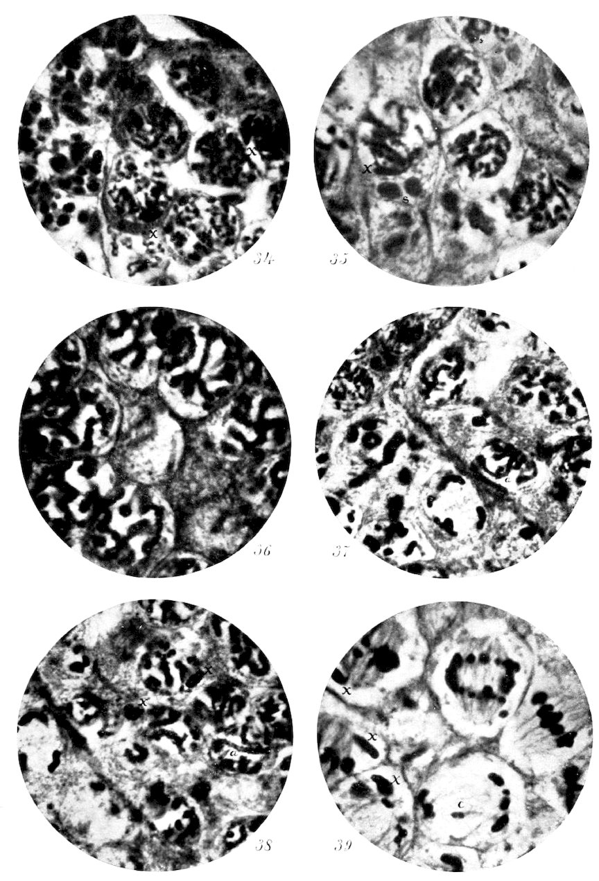
in figures 7, 9, 11 and 38 of the present one. Although this is
extremely common, and, as the photomicrographs show, undeniably
present, de Sinéty does not figure it at all. The nearest
approach to such a structure is found in figure 123c, where a
211cross with two nearly equal arms is represented. My interpretation
of this figure, based upon a great number of careful observations,
is that this represents merely an extension of the
shorter arms at the expense of the longer ones. In support of
this, I have stated that all intermediate stages between a rod
with a mere enlargement at the center and a cross with equal
arms could be found. How, according to de Sinéty’s conception
of overlying free elements, could these structures be explained?
It is not necessary, however, to have these gradations in
order to disprove the theory under discussion. One needs only
to carefully examine one of these crosses to be convinced that
the two arms lie in one plane where they intersect, and are not
superimposed one upon the other as de Sinéty shows in his
figure 123. Our author clearly realizes the importance of the
cross, as may be judged by the following quotation:
“La croix est de toutes ces figures celle dont la genèse peut le plus facilement
donner lieu à des interprétations en sens contraire.—C’est précisément pour
cette raison que nous croyons devoir l’étudier spécialement au point de vue
critique, persuadé que, cette figure une fois rattachée à une théorie, les autres
doivent en suivre le sort.”
It is unfortunate, therefore, that he was not able to trace the
formation of the element in its very early stages and through
the various modifications which connect it with the typical rod
already described.
As the simplest modification of this basic form, we find the
one where it is evident that the change consists merely in a
flexure of the rod at the weak spot in its center. Such forms
are shown in figure 14 of my former paper (17) and in figures
8, 9 and 11 of this one, but are not illustrated by de Sinéty. It
occasionally happens that in chromosomes of this character the
halves diverge widely at the center, producing the double-Vs of
Paulmier, as is represented in figure 14 of my paper upon the
Acrididæ (17) and in figure 8 of the present one. These structures
are not shown by de Sinéty and would be difficult to explain
in agreement with his conception of the tetrad.
I have consistently placed great reliance upon the frequent
ring-shaped chromosomes in determining the structure of the
first spermatocyte elements, and have no occasion to change
my opinion of them since examining the work of de Sinéty.
212This investigator joins issue with me upon my interpretation
of these structures, and states his attitude in the following
language:
“McClung fait grand fond, pour appuyer son interprétation, sur une forme
spéciale, la forme en anneau, qui pour lui dérive du bâtonnet a′ b′
a′ b′’, supposé placé
transversalement sur le fuseau, inséré par son milieu et incurvé en dehors jusqu’
à rapprochement et soudure de ses extrémités.
“Le chromosome en anneau est en effet très fréquent chez les acridiens;
mais il nous a été possible d’en reconstituer l’histoire, grâce à des détails qui ne
semblent pas s’être rencontrés dans les figures de McClung. On se souvient que
nous avons établi les deux points suivants en complet désaccord avec la théorie
de l’auteur américain:
“1. Les deux moitiés de l’anneau proviennent de la première division longitudinale.
“2. L’insertion est terminale.”
With equal emphasis, I must deny that the enclosed space in
the ring represents any plane of division in the chromatin
thread; and that the insertion of the spindle fibers is at any
place except at the center of what would be the typical rod-shaped
chromosome were the ring straightened out. We encounter
in de Sinéty’s interpretation of these rings the very
error against which I was careful to caution elsewhere in this
paper, i. e., of regarding the points where the fibers are attached as
the crossed ends of a simple segment. This mistake de Sinéty has
made, and has thereby vitiated all his conclusions concerning
the structure of the tetrads. It is not necessary to repeat here
the proof which I have brought forward in support of my views.
No one, I am sure, will find difficulty in reducing the various
forms of chromosomes found in the first spermatocytes to the
type of a doubly split rod, in which one plane of division is
parallel to the long axis and the other at right angles to it.
The explanation offered by de Sinéty requires us to conceive a
doubly split rod in which one separating space may vary indefinitely
while the other is constant. There is here no common
type, but an infinitely variable one, which differs with every
modification of the interspace between the first pair of chromatids
in each chromosome.
As a constructive basis for the foundation of his theory of a
double longitudinal division, de Sinéty uses particularly the
chromosomes of Œdipoda (Hippiscus) miniata, represented in
figures 129 and 130, concerning which he says:
“Survient le phénomène exceptionnellement important de la seconde division
213longitudinale; nous regardons comme un point capital dans notre travail d’en
mettre l’existence hors de doute et pour cela nous désirons ne faire appel qu’à
des images extrêmement claires. Nous considérons comme telles les fig. 129 et
130 rapprochées l’une de l’autre.
“Il est de toute évidence que le chromosome a, fig. 130, n’est que le chromosome
de même désignation, fig. 129, dont les deux anses jumelles se sont
clivées. De même, le chromosome en forme de boucle, c, fig. 129, dont les
deux branches représentent, comme nous l’avons fait remarquer, deux anses
jumelles, se retrouve avec un clivage très évident en d, fig. 123. On pourrait
faire les mêmes rapprochements entre b, fig. 105, et a, fig. 107; ici, le clivage
est moins avancé, mais les granules sont nettement divisés.”
I am obliged to confess that I have never seen in other species
of this genus any appearances that would incline me to
place an interpretation upon them such as does our author upon
these. I would venture to suggest, on the contrary, that the
chromosomes represented in figure 129 have not as yet demonstrated
any division, but show merely irregular spaces between
chromosomes. At even an earlier stage (figs. 5, 37, and 38),
I have shown the formation of the tetrads by means of simultaneous
cross and longitudinal divisions so clearly that presumed
successive divisions, as represented by de Sinéty, cannot
be regarded as occurring.
Finally, I would emphasize the fact mentioned in connection
with the discussion of the cross-shaped chromosomes, that
where the elements of one of these compound chromosomes intersect
they lie in one plane, and are not superimposed upon each
other, as de Sinéty’s theory demands and as his figures represent.
This was shown clearly in Paulmier’s figures as well as in my
own, and is even more clearly demonstrated, if possible, in
the very long, slender chromosomes of the myriapods, which I
have observed in Mr. Blackman’s preparations. This, and the
continuity of the chromatin in contiguous arms of the cross, is
alone sufficient to disprove de Sinéty’s theory, and, fortunately,
is easily demonstrated. This same fault of de Sinéty’s is encountered,
in another form, in his discussion of the ring figures.
He asserts that the halves of the rings are pulled past each
other while they lie in the plane of the spindle axis. Herein
my observations fail entirely to agree with his. The rings lie
in the plane of the equator, and no elements of the mitotic
figure show a lateral displacement of the separating halves
equal to the width of the chromosome when viewed in this
plane.
214
I approach a discussion of Montgomery’s conclusions regarding
the form of the chromosomes in the first spermatocyte, and
the sequence of their divisions, with considerable hesitation,
because of the difficulty I experience in appreciating his exact
position. This is due, not to any lack of positive statements on
his part, but to the partial contradictions that result from his
frequent changes of opinion. The most important statement
in his first paper upon Euchistus reads as follows: “From the
resting stage of the first spermatocyte to the formation of the
spermatid, there is absolutely no longitudinal division of the
chromosomes. I have studied hundreds of nuclei in these
stages, and at the first with a hope of finding a trace of such a
process, but observation shows that all divisions of the chromatin
elements are transverse divisions.”
This would certainly seem to be as strong a stand as one
could take upon the subject, but in later papers Montgomery
assumes with equal assurance the opposing position, which
holds for a longitudinal division. Regarding this he says:
“During the synapsis stage the chromosomes become split
longitudinally, as was first shown by Paulmier (1898, 1899) for
Anasa—a process that I had overlooked (!) in my former
paper (1898).” Throughout his later investigations this hypothesis
serves as the basis of all his theories, and the careful
longitudinal division of the thread is assigned an important role
in the maturation process. So far as positive assertions to the
contrary are concerned, a general acceptance of the theoretical
importance attaching to this act is to be supposed.
Notwithstanding this, I find nowhere in his later writings
any statement that he abandons the conception formerly entertained
regarding the non-importance of the longitudinal cleavage.
This attitude is indicated in the following language:
“If it can be proved that the mode of division of a chromosome,
i. e., the axis of the line of division, is merely a function
of its chromomeres, then it would be of no theoretical value
whether the division be longitudinal (equation) or transverse
(reduction). But it happens that the postulated difference
forms one of the main premises of Weismann’s theoretical
superstructure. On account of the differences observed in different
objects in regard to the modes of division of the chromosomes,
215it would appear that the differences have no theoretical
value, but that the halving of the mass of chromatin is the
process of importance—the standpoint taken by Hertwig.
“In the two reduction divisions the chromosomes may split
by two longitudinal divisions, by two transverse divisions, by
one longitudinal and one transverse division, or by one division
(longitudinal or transverse) preceded or followed by an elimination
division. The facts show already that there is no general
uniformity in the mode of division of the chromosomes in
the reduction mitoses. The long line of observations on different
objects show this to be the case, and demonstrates that the
expected uniformity does not occur.”
Herein lies the essential conclusion of the work upon Pentatoma,
which, so far as a specific retraction is concerned, stands
yet. If this be abandoned, then the first work upon the chromatin
structure of Pentatoma is practically discredited, for Montgomery
has definitely retreated from his positions concerning
the absence of the “chromatin nucleolus” in the spermatogonia,
the non-occurrence of a longitudinal cleft in the spireme
thread, the lack of an equational division of the chromatin in
the spermatocyte, the origin of the “chromatin nucleolus,”
and the fragmentation of the “chromatin nucleolus.” In addition
to these specifically acknowledged errors, we may infer
that Montgomery (12) considers himself at fault in his views
upon the production of chromosomes from the “three to six
chromatin loops” by breaking apart in the prophase, and upon
the occurrence of both longitudinal and cross divisions of ordinary
chromosomes in the same mitosis. The observations
recorded in his last paper (15) upon the production of the spermatocyte
chromosomes by the end-to-end union of those in the
last spermatogonial division warrant this assumption.
It follows from all this that we may practically disregard
Montgomery’s earlier work upon chromosomal structure and
take his views as expressed in the later papers (14, 15) as representing
his opinions upon the subject. These later theories are
largely the result of his investigations upon Peripatus, but they
seem to be carried over and applied to the Hemiptera without
essential modifications, and we may regard this concept as applicable
to the forms studied by him.
I called attention in my previous paper to the fact that, by
216many investigators, the definitive form of the chromosome is
used as the basis for determining the direction and sequence of
the chromosome divisions. This fact and the danger attending
the practice was partly realized by Montgomery in his work
upon Euchistus (12), for he devotes considerable space to a consideration
of the prophase segments, but in determining the
character of the second spermatocyte division he regards only
the formed element. With respect to this he says: “And now
a fact may be determined which is of the greatest importance
in estimating the morphological value of the second division of
the chromosomes. While the latter are still parallel to the axis
of the spindle, there may be clearly seen in some cases a transverse
constriction on some of the chromosomes, so that they
already acquire a dumb-bell shape.” This constriction is not
correlated with any similar one on the prophase elements, and
is here observed for the first time.
In his paper upon Peripatus, however, he definitely supports
the contention that it is only in the prophase of the first spermatocyte
that we can learn the construction of the chromosomes,
for he says: “The early stages in the prophase are of
the greatest importance in determining the exact constitution
of the chromosomes of the first maturation division....
Since, then, as has been shown in another section of the present
paper, the split of the univalent chromosome of the second
spermatocyte is a true longitudinal split, corresponding perfectly
in position with the longitudinal split of the early prophase,
it follows that the univalent chromosome does not
become turned upon its axis to take its place on the equator of
the spindle.” Orientation is in both spermatocytes based, accordingly,
upon planes determined in the prophase. Upon this
point Paulmier and Montgomery, as students of Hemipteran
spermatogenesis, are now agreed, and their results correspond
with observations made upon Orthopteran cells.
It is upon the sequence of divisions in the spermatocyte that
differences now exist between these investigators and myself.
In my previous paper I took occasion to elaborate the proof in
support of my position regarding the early occurrences of the
longitudinal division in the Orthopteran spermatocytes. Montgomery
follows Paulmier in ascribing the reduction division to
the first spermatocyte, and takes no account of my results upon
217Hippiscus. The objections that I previously urged against Paulmier’s
conclusions apply equally well to Montgomery’s. Until
the chromosomes are traced in a more detailed way through
the prophase to the metaphase, I shall consider the presumption
against the occurrence of the cross-division in the first
spermatocyte mitosis. In this I believe that I am justified by
the definite proof of my position brought forward in the work
upon Hippiscus. Here, it may be recalled, I observed and photographed
in the same mitosis all stages of movement by the
chromatids along the plane of the longitudinal split. In addition,
I was able to locate definitely the position of the future
cross-division in the ring figures, so that it is impossible to
mistake the character of the first division in them. These two
proofs I consider incontrovertible so far as they apply to the
Orthopteran families studied.
Paulmier judged the planes of the division by the relative
lengths of the chromosome axes, but, as I pointed out, this is
not conclusive unless it can be shown that they have not
shifted, as it is possible for them to do, during the prophase.
The value of the ring figure, which is formed at such an early
stage that it would be impossible for the shifting of the axis to
occur, is here evident.
Montgomery finds these rings in Peripatus, and realizes the
importance of their evidence in determining the planes of division,
but places his conclusions upon a much more insecure
footing than those founded upon the Orthopteran cells, because
of the criterion used in determining which point represents the
junction of the paired chromosomes. The diagnostic feature
he uses is the linin connection persisting between the “central
ends” of the chromosome, which holds them together until the
“distal fibers” connect with the centrosomes and cause the
rupture of the “central” fiber. Since the whole of his elaborate
theory regarding the continuance of the linin spireme is
practically a theoretical conception with little basis in observed
fact, the value of such proof cannot compare with that furnished
by the definitely formed chromosomes themselves in the
Orthopteran cells.
In view of all these facts, I think it must still be held an open
question as to which is the reduction and which the equation
division in the Hemipteran spermatocytes, although it is not to
218be doubted that the probability of the first spermatocyte being
witness of the reduction division is much increased when thus
interpreted by two independent observers.
I have already, in another paper (19), taken up a comparative
study of the accessory chromosome in different insect spermatocytes,
and shall not be obliged, for that reason, to enter
into a very lengthy discussion of the subject here. The great
interest attaching to this structure, however, compels me to
consider the work that has been done since the manuscript of
the earlier article was sent in for publication. This review will
concern, very largely, the investigations of Montgomery upon a
considerable number of Hemipteran species, which are set
forth in his paper under the pretentious title “A Study of the
Chromosomes in the Germ Cells of Metazoa.”
In his first work upon Euchistus, Montgomery describes a cell
element under the name “chromatin nucleolus” which corresponded
so closely to my accessory chromosome that I concluded
the two structures were identical. These similarities
were, the origin from a spermatogonial chromosome, the integrity
and constancy of staining power and position during
the spermatocyte prophase, and participation in the division
act during metakinesis of a spermatocyte.
Among the numerous changes of opinion recorded by Montgomery
in his latest work, there are several relating to his
“chromatin nucleolus” that materially alter the aspect of the
question. Perhaps the most important of these concerns the
origin of the element. I was some time in determining that
the accessory chromosome is a spermatogonial chromosome
which divides in the spermatogonia with the other chromatin
elements and comes over into the first spermatocyte as a formed
structure. The work of Sutton upon the early history of the
element in Brachystola, however, was convincing in this respect
and confirmed me in the opinion I had already formed. I
therefore gave Montgomery the credit for this discovery, and
set it down as strong confirmation of the assumption that we
were dealing with similar structures in the two orders of insects.
Upon this point Montgomery now completely reverses himself,
and declares that his “chromatin nucleolus” is not a
spermatogonial chromosome, but may be noted in the earlier
219generations as a nucleolar structure, which, however, divides
in metakinesis. The most important feature to be noted in
this connection is the fact that the structure does not exist
as a simple element, but is observed as a number of granules,
and that this number varies considerably in different species.
These granules fuse during the “synapsis stage,” as do
the chromosomes, to produce in the spermatocyte half the
number of “chromatin nucleoli” that were present in the spermatogonia.
In this respect the “chromatin nucleolus” differs
radically from the accessory chromosome, which has the same
valence in both cell generations. The indefinite number and
insignificant size of Montgomery’s structures are other characters
that point to extensive differences between them and the
accessory chromosome.
In his work upon Peripatus, Montgomery states that in restudying
his preparations of Euchistus he observes a continuous
linin spireme which involves the “chromatin nucleolus” as
well as the chromosomes. Here, again, there is a difference
between the Hemipteran element and the accessory chromosome;
for the latter is entirely free from linin connections in
the prophase and is usually surrounded by a hyaloplasmic investment.
According to Montgomery, also, his “chromatin nucleolus”
usually takes part in both spermatocyte mitoses. In this respect
there exists an essential difference between his element
and that found in the Orthoptera, for, after extended and most
critical studies, I have become convinced that only one division
takes place in the spermatocytes. In those cases where Montgomery
admits but a single division, it is stated to occur in the
first spermatocyte, while in the Orthoptera the accessory chromosome
remains undivided here and is halved in the second
spermatocyte.
If, therefore, Montgomery’s recent observations are correct, it
must follow, I think, that his “chromatin nucleolus” and the
accessory chromosome are different structures. I am free to
admit, however, that his statements are far from convincing.
So much dependence is placed upon the numerical relationships
of elements that are admittedly very minute, and so little
corroborative proof is given, that I entertain serious doubts as
to the accuracy of the observations. In this connection I would
220suggest a comparison between the figures of the “chromatin
nucleolus” in the first paper upon Euchistus (figs. 55–68) (12)
and those in the last one (figs. 1–17) (15). The showing here
made would alone be sufficient to raise a question as to the
nature of the “chromatin nucleolus,” and until further evidence
is forthcoming the character of the peculiarly modified
chromosomes in the spermatocyte of the Hemiptera must remain
in doubt.
Aside from definite retractions that Montgomery has made regarding
his earlier views on the character of the “chromatin
nucleolus,” there are noticeable different attitudes toward it in
his earlier and later works. Thus, in his lecture at Woods
Holl (13a), we find the following: “These remarkable ‘nucleolar’
structures which stain like chromatin have been observed
by numerous writers, but as yet no satisfactory description has
been given of their mode of origin. They have been observed
by me in spermatocytes of various insects, in hypodermal and
other cells of Carpocopsa, and in follicle cells of the testicles of
Plethodon and Mus.” At this early stage of Montgomery’s investigations
it is apparent that he views his “chromatin nucleolus”
primarily as a nucleolus with chromatic origin and
characters, but the fact is equally apparent that he now regards
it primarily as a “chromosome” with nucleolar attributes.
This is made evident in his recent definition, which
reads: “The chromatin nucleoli are morphologically chromosomes,
undergoing division in mitosis like the other chromosomes,
but differing from them in the rest stage by preserving
a definite (usually rounded) form.”
What has here been said regarding the “chromatin nucleolus”
applies to those structures in Euchistus and other Hemiptera
to which Montgomery has given the name without
qualification. According to his definition, however, there is
present in the cells of Protenor and other species another form,
the “chromosome x.” Not only by inference is this classification
operative, but by direct statement we learn that Montgomery
regards this element as a member of the class of bodies
which he calls “chromatin nucleoli.” In speaking of Protenor
chromosomes, he says: “This is the only case in the Hemiptera
where one chromosome becomes differentiated into a
221‘chromatin nucleolus’ for the first time in the spermatocyte
generation.”
The noteworthy thing about this “chromosome x” is the fact
that in every essential detail it corresponds to the accessory
chromosome of the Orthoptera. It is a spermatogonial chromosome
that comes over intact into the spermatocyte; it retains
its form and staining power unchanged through the
prophase of the spermatocyte; it divides in only one of the
spermatocyte mitoses; and is a large and conspicuous element
of the cell at all times.
This “chromosome x” agrees just as closely in its description
to the accessory chromosome as do the ordinary ones of
the two orders, and, if Montgomery’s account is correct, there
would seem to be no reason for doubting their identity. In
two respects, however, there are differences between these
structures. First, it is to be noted that the “chromosome x”
divides in the first spermatocyte, while the accessory chromosome
undergoes separation in the second spermatocyte. Should
Montgomery’s observations prove correct, it would yet indicate
no fundamental difference in the character of the element, for
the result is the same whether division takes place in the first
or second mitosis. In either event, one-half the spermatozoa
are provided with the odd chromosome while the remaining
half are not.
The second point of difference would seem to be a more serious
one. Montgomery states that during the spermatogonial
mitosis the “chromosome x” regularly divides as do all the
other chromosomes, i. e., longitudinally. In the spermatocyte
mitosis, however, the element is broken across, and the longitudinal
split, which is apparent in the early stages, disappears
and is not utilized in division. We have here the remarkable
occurrence of a chromosome entirely unchanged in its structure,
but merely differing in its surroundings, which, instead of dividing
along the plane marked out for it, as it has done in all
preceding mitoses, breaks across after it is a formed element.
An occurrence of this kind, so different from the usual method
of division, would require strong proof to establish it, and this,
in my opinion, Montgomery has not brought forward.
A criticism of the degeneration theory as advocated by Paulmier
and Montgomery has already been given (17), so that it
222would not be necessary to consider it here except in so far as it
has been modified since its promulgation. As a rule, Montgomery
refers to his “chromatin nucleoli” throughout his late
paper (15) as degenerating chromosomes, but in discussing
their function specifically he makes important changes in this
conception. These are stated as follows: “When we find, accordingly,
the mutual apposition of them (true nucleoli) to
chromatin nucleoli, it would be permissible to conclude that
the chromatin nucleoli are chromosomes which are especially
concerned with nucleolar metabolism. And this, I think, would
be the correct interpretation. The chromatin nucleoli are in
that sense degenerate that they no longer behave like the other
chromosomes in the rest stages, but they would be specialized
for a metabolic function; and from this point of view they
would certainly seem to be much more than degenerate organs.”
It is difficult to comment upon a contradictory statement like
this; but, fortunately, it is not necessary to do so, since it carries
with it its own refutation. The conception of a chromosome
specialized in the direction of increased metabolic activity as
being in the process of disappearing from the species can hardly
be regarded seriously.
Taking everything into consideration, it may be said that
Montgomery’s work upon the Hemiptera has left the subject in
a very disturbed condition, and any prospect of a complete
agreement between the accessory chromosome of the Orthoptera
and the “chromatin nucleolus” of the Hemiptera is made
more remote than was previously the case. This, I think, is
largely due to the inferior character of the Hemipteran material,
which has lead to misconception of phenomena that are
clearly marked in Orthopteran cells.
It is gratifying to note that the recent work of de Sinéty (37)
practically corroborates the conclusions herein set forth regarding
the history of the accessory chromosome. Aside from failure
to observe the important spireme condition of this element
in the first spermatocyte prophase, de Sinéty describes the same
series of processes with scarcely an exception. His summary
contains the following account of the accessory chromosome:
“Le ‘chromosome accessoire,’ découvert par McClung chez Xiphidium
fasciatum, se retrouve chez les locustiens que nous avons étudiés. Chez Orphania,
il se divise dans les spermatogonies en deux masses volumineuses et
223allongés, que l’on reconnait dans les nucléoles, également volumineux et allongés,
des spermatocytes de premier ordre en prophase. A la métaphase de la
première cinèse, on le trouve situé excentriquement et plus près de l’un des pôles;
il va tout entier a l’une des cellules-filles. Dans celle-ci, il se divise comme
un chromosome ordinaire, d’où il suit que sur quatre spermatides formant la
descendance d’un spermatocyte, deux se trouvent privilegiees. Par ce
partage inégal, non réalise dans Xiphidium fasciatum, d’après McClung, le
chromosome spécial d’Orphania rappelle celui des hémiptères.”
A like series of processes is recognized in the Phasmids.
As is elsewhere explained in this paper, the occurrence of two
divisions of the accessory chromosome in Xiphidium, which was
mentioned as a possible occurrence in my preliminary paper, is
shown not to take place. While it is much more difficult to
demonstrate the undivided condition of the accessory chromosome
in one of the spermatocyte mitoses of Xiphidium than it is
in the cells of Orchesticus, Anabrus, Scudderia, and Microcentrum,
I am convinced that it does not differ from the other Locustids
in this respect.
We may therefore feel assured that our knowledge of the
morphological character of the accessory chromosome in the
Orthoptera is fairly well established. This gives us a good
base from which to conduct further comparative studies into
other groups, and it is to be hoped that our knowledge of this
element will rapidly increase.
Unfortunately, de Sinéty has chosen to add another name to
the already overburdened list of synonyms, and “chromosome
spécial” now takes its place in the literature of insect spermatogenesis.
The reason for adding this name—
“Il reçu successivement leg noms de ‘accessory chromosome’ (McClung), ‘small
chromosome’ (Paulmier), ‘chromatin nucleolus’ (?), ‘chromosome x’ (Montgomery).
Nous avons préféré éviter ces appellations, qui semblent toutes
supposer une signification qui n’a jamais été définie ou s’appuyer sur des caractères
plus ou moins secondaires, pour adopter un nom indifférent, celui de
‘chromosome spécial,’ nous conformant à l’idée de Wilson, pour qui c’est un
‘extra chromosome,’”
would seem to be at least insufficient, since “accessory chromosome”
can scarcely be regarded as implying any more primary
or secondary function than can “chromosome spécial.”
In each of my preceding papers I took the opportunity to
point out the fact that, even were the accessory chromosome
224of no other value, it would certainly be worthy of study for the
light it throws upon the question of the individuality of the
chromosomes. On this point Montgomery has much to say in
his late paper (15). I think it cannot be questioned that we
have here indisputable proof that at least one chromosome may
be identified through all the cell generations of the testis.
While this does not prove that chromosomes are persisting and
independent structures, it does evidence the fact that they may
be, and greatly strengthens the hypothesis that they are.
In addition to the evidence here offered by the accessory
chromosome, there must be noted that derived from a study of
spermatocytes in which there is always present one ordinary
chromosome that greatly exceeds the others in size. Such a
condition is found in the cells of Anabrus. The disproportion
in size of the elements is here so striking that it would be impossible
to fail in distinguishing the giant chromosome. In each
of the spermatocytes of Anabrus there are therefore two chromosomes
which are plainly recognizable. It may be observed
further that the remaining chromosomes are quite different in
size, and it may be possible within reasonable limits of certainty
to pick out one or more other chromosomes in each cell.
Unless this could be done for each element, however, it would
not definitely prove that all the chromosomes are distinct and
recognizable structures. The actual recognition of two elements
in each cell of the same generation and its ancestors or
descendants in other generations goes far, however, to render
probable the individuality of each chromosome.
Beyond this point studies upon the Orthopteran cells will not
permit me to go; but Montgomery has been fortunate enough
to find in Peripatus an object in which he considers it possible
to demonstrate the continuity of the chromosomes from one
generation to another, and their fusion by pairs in the early history
of the spermatocyte to bring about the reduced number.
This is, in the main, a logical conclusion to my own work, and
I am therefore bound to regard his results as probably correct.
While doing this, however, I recognize that the absolute proof
he brings forward in support of his hypothesis is very slight.
I consider any deductions based upon observations of linin
structures as very insecure, and it is upon these that Montgomery
principally relies to demonstrate his theory. Further
225observations upon the behavior of the chromosomes between
the spermatogonia and the spermatocytes in objects favorable
for study will be awaited with interest. In the meantime it
must be conceded that the work upon insect spermatogenesis
has at least lent strong support to the theory of the individuality
of the chromosomes in general and has definitely shown
that there is such a thing in some instances.
Considerable importance is attached by some investigators to
the nuclear structures, properly called plasmasomes, that occur
in the spermatocytes. It is probable that there are marked
differences between the cells of various species in regard to the
occurrence of these bodies, for in the Orthoptera they either do
not appear at all, or, if present, they are minute and inconspicuous.
This fact would tend to disprove any theory which
would attach a fundamental importance to these structures,
such as is conceived for the chromatin. The Orthopteran cells
do not allow any observations which would add to our positive
knowledge of the nucleoli, and I include this brief statement
merely for the negative value it may possess.
In his first paper upon Euchistus, Montgomery assigns an important
and conspicuous place to the “rest stage” among his
numerous subphases preceding the first spermatocyte mitosis.
As a result of his later comparative work upon the Hemiptera,
however, we learn that in certain families no trace of such a
condition of diffusion on the part of the chromatin is observable,
from which we conclude that “accordingly such a stage
would appear to have no broad significance.” It has already
been announced that nothing like a rest stage intervenes between
the spermatogonia and spermatocytes of the Orthoptera,
and the work of most investigators would tend to indicate that
it is the exception rather than the rule. In those cases where
such a condition of the nucleus exists, it would seem to be true
that nothing more unusual than an excessive diffusion of the
spermatogonial chromosomes occurs, and this is of hardly sufficient
importance to receive a special designation.
The existence of a rest stage between the first and second
spermatocytes is also negatived by the conditions found in the
226Orthopteran cells. The formation of chromosomes in the prophase
of the first spermatocyte that are already prepared for
two divisions would a priori render improbable the intervention
of a rest stage here; and the actual observed persistence of
the chromosomes, as such, through the telophase of the first
spermatocyte and through the modified prophase of the second
spermatocyte gives actual proof in support of the view that commonly
prevails regarding the suppression of the second spermatocyte
rest stage.
Observations upon numerous species tend to show that the
behavior of the chromatin during the period between the two
spermatocyte mitoses varies considerably with the species and
even within the species itself. The amount of diffusion would,
in some measure, seem to be related to the form of the chromosomes
and to vary correspondingly in those individuals where
the chromosomes are of diverse forms. Thus, where the elements
of the second spermatocyte metaphase appear as short
double rods, the amount of diffusion is slight, and the individual
chromosomes may be distinguished throughout the telophase
of the first spermatocyte; but in those cases where the
members of the mitotic figure are much elongated the diffusion
is more extensive and the distinction between elements is made
difficult or impossible. Since these two conditions may prevail
in the same testis, it is probably only a question as to the extent
of elongation on the part of each chromosome. In those
cases where the elements become very much extended the appearance
of the resting condition would be simulated closely,
while, on the contrary, chromosomes consisting of spherical or
short cylindrical chromatids would never give a suggestion of
such a stage. In this we may find, I think, an explanation for
those cases in which a rest stage is described as occurring between
the spermatocyte generations.
1. The secondary spermatogonia are much reduced in size at
the end of their divisions and the cytoplasm is very small in
amount. The rod-shaped chromosomes number thirty-three,
and, of these, one is to be distinguished from its fellows by
greater size and slower division.
2. From the substance of the disintegrated spermatogonial
227chromosomes, the tetrads of the first spermatocytes are formed.
It was impossible to determine the relation of the elements of
the two generations, but the changes are rapid and there is no
intervening resting condition of the nucleus.
3. It could not be determined whether or not the spireme
is continuous. A longitudinal split appears very early, and
shortly after the chromatin segments may be seen. These
soon betray at their centers an indication of the cross-division,
producing crosses with arms that may vary considerably in
relative lengths. No reason was found for considering both
divisions longitudinal.
4. The typical element is granular and more or less rod-shaped,
with the longitudinal division merely indicated by a
narrow line, and with but slight elongation of the chromatids
along the plane of the cross-division. Various modifications of
this occur, by which the longitudinal cleft is much increased in
width at the center, the cross-arms are greatly extended, or approximation
of the ends of the rod brought about, producing a
ring.
5. The definitive chromosomes of the metaphase are produced
by a concentration of the prophase elements, whereby
they become shorter, heavier, and entirely homogeneous in
structure. Distinct lines of division between the chromatids
are not visible, but the tetrad character of the elements is
readily established by observing the steps in their formation.
6. The accessory chromosome early becomes distinguishable
because of its peripheral position and strong tendency to stain
with safranin, while the remaining chromatin takes the gentian
violet by Flemming’s three-color method. At first it appears
as a homogeneous plate, but later this is seen to be a closely
coiled thread. As the chromatin segments shorten and broaden
to form the chromosomes of the mitotic figure, this thread also
grows shorter and heavier until it forms an element of essentially
the same character as that of the spermatogonial chromosome
from which it was derived.
7. Upon the establishment of the mitotic figure, the chromosomes
arrange themselves in the equatorial plate with their
longer axis perpendicular to the spindle axis. Division of the
elements is not synchronous, so that all stages of the chromatid
movements may be observed in one nucleus. By this means it
228is possible to determine that separation of the chromosomes
takes place along the plane which marked the longitudinal division
of the prophase thread in such a way that the chromatids
show no clear interspaces. The individual chromosome near the
end of its division has the same form as that with which it
started, except for the difference that the chromatids are now
in contact for the greater part of their length along the plane
of their cross-division. As the daughter chromosomes separate,
this line of division comes into evidence through the springing
apart of the two chromatids now composing each chromosome.
The result is the formation of two V-shaped chromosomes with
mantle fibers attached to their apices. The accessory chromosome
does not participate in this division, but passes unchanged
to one pole of the spindle.
8. By reason of the action of the accessory chromosome in
the first spermatocyte mitosis, there are produced two numerically
equal classes of second spermatocytes—(a) those containing
sixteen dyad chromosomes and an undivided accessory
chromosome, and (b) those with merely the sixteen dyad elements.
In both cases the mitotic figure quickly reforms without
an intervening rest stage in which the chromosomes lose
their identity. There is a loosening up of the chromomeres in
all the elements except the accessory chromosome, so that they
have a structure and staining reaction similar to that of the
first spermatocyte chromosomes just before they enter the metaphase.
The dyads of the first spermatocyte telophase, and of
the succeeding and greatly abbreviated second spermatocyte
prophase, are quite as definite structures as are the chromosomes
of the first spermatocyte prophase.
9. All the chromosomes of the second spermatocyte are
paired structures and divide in a similar way. The spindle is
small and weak as compared with that of the first spermatocyte,
and the chromosomes arrange themselves radially on its
periphery in such a way that the pairs lie in the plane of the
spindle axis with their joined ends inward. The space between
the chromatids represents the line of cross-division observable
in the prophase segments of the first spermatocyte, and their
separation accordingly represents a reduction division. The
accessory chromosome, on the contrary, divides along the plane
marking the longitudinal cleft of the spermatogonial spireme.
22910. From each first spermatocyte there are formed, by two
divisions, four spermatids, of which two are distinguished from
the remaining pair by the possession of an extra chromosome
in addition to the number—sixteen—common to them all.
Both classes undergo a like series of transformations by which
they become mature spermatozoa. These are necessarily of
two kinds; and it is believed that those containing the accessory
chromosome, in the act of fertilizing the egg, determine
that the germ-cells of the embryo shall be sexually male, or like
themselves, while those from which it is absent are unable to
impress their sex upon the egg and assist in producing female
embryos.
1. Atkinson, Geo. F., 1899. Studies on Reduction in Plants,
Bot. Gaz., vol. 28.
2. Baumgartner, W. J., 1902. The Spermatid Transformations
of Gryllus assimilis, etc., Kans. Univ. Sci. Bull., vol. 1,
No. 1.
3. Brauer, A., 1893. Zur Kenntniss der Spermatogenese von
Ascaris megalocephala, Arch. mikr. Anat., vol 42.
4. Carnoy, 1895. La Cytodiérese chez les Arthropodes, La
Cellule, vol. 1.
5. Cholodkovsky, N., 1894. Zur Frage über die Anfangsstadien
der Spermatogenese bei den Insecten, Zool. Anz., vol. 17.
6. Griffin, B. B., 1899. Studies on Maturation, Fertilization
and Cleavage of Thalassema and Zirphæa, Jr. Morph., vol. 15.
7. Häcker, Valetin, 1897. Ueber weitere Uebereinstimmungen
zwischen den Fortpflianzungsvorgängen der Tiere und
Pflanzen, Biol. Cent., vol. 17.
8. Henking, H., 1890. Untersuchungen über die ersten
Entwicklungsvorgänge in den Eiern der Insecten. 2. Ueber
Spermatogenese und deren Beziehung zur Eientwicklung bei
Pyrrhocoris apterus M., Z. wiss. Zool., vol. 51.
9. Henking, H., 1892, idem. 3. Specielles und Allgemeines,
ibid., vol. 54.
10. Ischikawa, 1891. Studies of Reproductive Elements.
Spermatogenesis, Ovogenesis and Fertilization in Diaptomus sp.,
J. Coll. Sc. Imp. Univ. Japan, vol. 5.
11. Kingsbury, B. F., 1902. The Spermatogenesis of Desmognathus
fusca, Am. Jour. Anat., vol. 1, No. 2.
23012. Montgomery, T. H., jr., 1898. The Spermatogenesis in
Pentatoma up to the Formation of the Spermatid, Zool. Jahrb.,
vol. 12.
13. Montgomery, T. H., jr., 1898. Chromatin Reduction in
the Hemiptera: a Correction, Zool. Anz., vol. 22.
13a. Montgomery, T. H., jr., 1898. Observations on Various
Nucleolar Structures of the Cell, Biol. Lect. of the Woods
Holl Laboratory, 1898.
14. Montgomery, T. H., jr., 1900. The Spermatogenesis of
Peripatus balfouri up to the Formation of the Spermatid, Zool.
Jahrb., vol. 14.
15. Montgomery, T. H., jr., 1901. A Study of the Germ
Cells of Metazoa, Trans. Amer. Phil. Soc., vol. 20.
16. McClung, C. E., 1899. A Peculiar Nuclear Element in
the Male Reproductive Cells of Insects, Zool. Bull., vol. 2.
17. McClung, C. E., 1900. The Spermatocyte Divisions of
the Acrididæ, Kans. Univ. Quart., vol. 9, No. 1.
18. McClung, C. E., 1901. Notes on the Accessory Chromosome,
Anat. Anz., Bd. 20, Nos. 8, 9.
19. McClung, C. E., 1902. The Accessory Chromosome—Sex
Determinant? Biol. Bull., vol. 3, Nos. 1, 2.
20. Moore, J. E. S., 1895. On the Structural Changes in
the Reproductive Cells during the Spermatogenesis of Elasmobranchs,
Quart. J. Micr. Sc., vol. 38.
21. Paulmier, F. C., 1898. Chromatin Reduction in the
Hemiptera, Anat. Anz., vol. 14.
22. Paulmier, F. C., 1899. The Spermatogenesis of Anasa
tristis, Jr. Morph., vol. 15.
23. Platner, G., 1886. Die Karyokinese bei den Lepidopteran
als Grundlage für eine Theorie der Zelltheilung, Internat.
Monatsschr. Anat. Histol., vol. 3.
24. Rath, O. vom, 1891. Ueber die Reduction der Chromatischen
Elemente in der Samenbildung von Gryllotalpa, Ber.
Naturf. Ges. Freiburg, vol. 6.
25. Rath, O. vom, 1892. Zur Kenntniss der Spermatogenese
von Gryllotalpa vulgaris Latr., Arch. Mikr. Anat., vol. 40.
26. Rath, O. vom, 1895. Neue Beiträge zur Frage der Chromatinreduction
in der Samen und Eireife, ibid., vol. 4.
27. Sabatier, A., 1890. De la Spermatogénese chez les Locustides,
Comptes Rend., vol. 111.
23128. Sutton, W. S., 1900. The Spermatogonial Divisions of
Brachystola magna, Kans. Univ. Quart., vol. 9, No. 2.
29. Toyama, K., 1894. On the Spermatogenesis of the Silkworm,
Bull. Coll. Agric. Imp. Univ. Japan, vol. 2.
30. V. la Valette St. George, 1897. Zur Samen-und Eibildung
beim Seidenspinner (Bombyx mori), Arch. mikr. Anat.,
vol. 50.
31. Wagner, J., 1896. Beiträge zur Kenntniss der Spermatogenese
bei den Spinnen, Arb. Nat. Ges. St. Petersburg, vol. 26.
32. Wilcox, E. V., 1895. Spermatogenesis of Caloptenus
femur-rubrum and Cicada tibicen, Bull. Mus. Comp. Zool. Harvard
Univ., vol. 27.
33. Wilcox, E. V., 1896. Further Studies on the Spermatogenesis
of Caloptenus femur-rubrum, ibid., vol. 29.
34. Wilcox, E. V., 1897. Chromatic Tetrads, Anat. Anz.,
vol. 14.
35. Wilcox, E. V., 1901. Longitudinal and Transverse Divisions
of Chromosomes, Anat. Anz., Bd. 19, No. 13.
36. Wilson, E. B., 1900. The Cell in Development and Inheritance.
New York; 2d ed.
37. de Sinéty, R., 1901. Recherches sur la Biologie et
l’Anatomie des Phasmes, La Cellule, Tome 19, 1er fascicule.
Drawings were made with a camera lucida, the optical combination
being a 1–16 B. & L. objective and a Watson “Holoscopic”
ocular No. 7. Details were studied with a Zeiss 2-mm.
apochromat, N. A. 1.30. As reduced in reproduction, an enlargement
of 1500 diameters exists. Photomicrographs, excepting
those of figures 37 and 38, were made by the use of the arc
light and horizontal camera. The exceptions represent illumination
by ordinary diffuse daylight. In all cases the lenses
used were the Zeiss 2 mm., N. A. 1.30 objective and projection
oculars. A Watson “Parachromatic” oil-immersion condenser
of 1.30 N. A. was employed to illuminate the objects. In use
it was stopped down to between .75 N. A. and 1.0 N. A.
Explanation of Plate VII.
Fig. 1. Pole view of spermatogonial metaphase, showing the thirty-three chromosomes.
It will be observed that the chromosomes are of unequal sizes, and
that the large ones arrange themselves in a circle on the outside of the figure.
Fig. 2. Very young spermatocyte. The chromatin derived from the breaking
down of the spermatogonial chromosomes in a diffuse condition, with no trace
of a linear arrangement. The accessory chromosome x on the periphery of the
nucleus, darkly staining and homogeneous.
Fig. 3. Early stage in the formation of the spireme. In the cytoplasm the
remains of the spermatogonial spindle. The cell has entered upon the growth
period.
Fig. 4. A later stage in the spireme formation. The accessory chromosome
larger and more flattened. A surface view shows it as an apparently fenestrated
plate. The remains of the two spermatogonial spindles still persisting.
Fig. 5. First appearance of definite chromosomes. One shown entire with
longitudinal and cross-divisions marked. The accessory chromosome is here seen
to be in a spireme condition.
Fig. 6. Condition of the chromosomes after further contraction of the early
segments. As here shown, they are more granular than is usually the case.
Fig. 7. Common types of the prophase chromosomes.
Fig. 8. A cell in which one of the chromosomes has its halves widely separated
along the longitudinal division, forming Paulmier’s double-V figure.
Fig. 9. In this cell may be seen the variation in form and size of the early
spermatocyte chromosomes.
Fig. 10. Two cells of the late prophase, with the chromosomes at almost the
extreme degree of concentration.
Fig. 11. Chromosomes of cells in the stage shown in figure 10. These represent
the different types of rings, crosses, etc., commonly observed in first spermatocytes
just before the formation of the mitotic figure.
Fig. 12. Different forms assumed by the accessory chromosome in the prophase
of the first spermatocytes of Xiphidium.
Fig. 13. Metaphase of the first spermatocyte. The accessory chromosome is
seen at one pole of the spindle, to which it has moved before the separation of
the chromatids of the remaining chromosomes.
Fig. 14. Another cell in about the same stage as that represented in the preceding
figure.
Fig. 15. A first spermatocyte metaphase in which the accessory chromosome
has not as yet moved to the pole of the spindle. This is uncommon in Orchesticus,
but frequent in Anabrus.
Fig. 16. Pole view of a first spermatocyte metaphase, showing seventeen chromosomes.
The variation in size of the elements, so marked in the spermatogonia,
is even more pronounced here. This is a cell similar to that of figure 15, in which
the accessory chromosome lies in the equatorial plate.
Explanation of Plate VIII.
Fig. 17. Two cells in metaphase—a pole view of one and an oblique view of
the other. The accessory chromosome does not show in the former, the cell
being such a one as is represented in figures 14 and 15.
Fig. 18. Pole view of another cell, showing but sixteen chromosomes.
Fig. 19. Early anaphase of the first spermatocyte, with the accessory chromosome
already at one pole.
Fig. 20. Mid-anaphase, with the giant chromosome still undivided.
Fig. 21. Later anaphase, in which the accessory chromosome is seen at the
lower pole. This figure shows, also, the character and extent of the intercellular
material.
Fig. 22. Later anaphase. The accessory chromosome at the upper pole. An
undivided chromosome lying between the groups of daughter chromosomes.
Fig. 23. About the stage of figure 22, but the lagging chromosome has divided.
Fig. 24. Very late anaphase. Here, again, the lagging chromosome is divided.
Fig. 25. Pole view of first spermatocyte telophase, showing the accessory
chromosome at one side of the daughter chromosomes.
Fig. 26. Pole view of a cell in the same stage as that represented in figure 25.
Here, however, the accessory chromosome is not present.
Fig. 27. Lateral view of telophase, with the accessory chromosome in the
lower daughter-cell.
Fig. 28. Fragment of second spermatocyte, showing the chromosomes in
metaphase. The relative sizes of the accessory chromosome and the remaining
chromosomes is well shown.
Fig. 29. Metaphase of a second spermatocyte, in which the accessory chromosome
is not present.
Fig. 30. Anaphase of second spermatocyte, in which there is no accessory
chromosome.
Fig. 31. Anaphase of second spermatocyte, where the accessory chromosome
is present—x1 and x2.
Fig. 32. Telophase of the same class of second spermatocytes. The accessory
chromosome extends out from the mass of chromosomes at each pole—x1
and x2.
Fig. 33. Telophase of the class of second spermatocytes from which the accessory
chromosome is absent.
Fig. 34. Photomicrograph of early spireme stage of first spermatocyte, showing
peripheral position of the accessory chromosome x. At the left, secondary
spermatogonia, last generation. × 1300.
Fig. 35. A late prophase, showing accessory chromosome x, and spindle remains
s (cf. figs. 3 and 4). × 1300.
Fig. 36. Coarse spireme of first spermatocyte. × 1300.
Fig. 37. Prophase, with chromosomes in the form of long segments. At a,
the cell drawn in figure 9. In the cyst at the left are spermatocytes in a later
stage, with the chromosomes homogeneous. × 1000.
Fig. 38. Prophase with segments divided longitudinally and across. At a
is one shown en face. Accessory chromosome at x. × 1000.
Fig. 39. Metaphase and anaphase of first spermatocyte. The accessory chromosome
x at one pole of the spindle. Lagging chromosome at c. × 1300.
Fig. 40. Anaphase of first spermatocyte. Accessory chromosome x at one
pole. The form of chromosome in the anaphase well shown. The lagging chromosome
c seen in two cells. × 1300.
Fig. 41. Anaphase of the first spermatocyte, showing the longitudinally divided
condition of the accessory chromosome x in the cell near the center. Compare
with the accessory chromosome in the metaphase of second spermatocyte,
figure 43. × 1300.
Fig. 42. Second spermatocyte in metaphase. In most of the cells the focus
is upon the ends of the chromosomes, but in one a side view is obtainable. Compare
with the chromosome of the upper cell in figure 40. No accessory chromosome
in most of the cells in focus. × 1300.
Fig. 43. Second spermatocyte metaphase and spermatids. Note the relative
sizes of the accessory chromosome and the other chromosomes. In the spermatids
the accessory chromosome has taken its place on the periphery of the
nucleus in the same way that it does in the prophase of the first spermatocyte.
× 1300.
Fig. 44. Anaphase of the second spermatocyte, showing the accessory chromosome
x separated. Other cells in metaphase. × 1300.
Fig. 45. Telophase of the second spermatocyte. Two daughter-cells with
persisting spindle between, showing the accessory chromosome x in each. Other
nuclei in focus show no accessory chromosomes. × 1300.
- Silently corrected obvious typographical errors and variations in spelling.
- Retained archaic, non-standard, and uncertain spellings as printed.