The text of this e-book has been preserved as in the original,
except for the occasional silent insertion of missing punctuation and
correction of a few typographic flaws (recen → recent, antea → ante).
Footnotes have been numbered sequentially and relocated to the end of the book
By the Same Author
SCLERO-CORNEAL TREPHINING IN
THE OPERATIVE TREATMENT OF
GLAUCOMA, 1913. Second Edition,
1914. George Pulman and Sons, London.
GLAUCOMA: DIGEST OF THE YEAR’S
LITERATURE. The Ophthalmic Yearbook,
1913–1916. Herrick Book and Stationery
Company, Denver, Colorado, U.S.A.
GLAUCOMA: A HANDBOOK FOR THE
GENERAL PRACTITIONER, 1917.
H. K. Lewis and Co. Ltd., London.
GLAUCOMA: A TEXTBOOK FOR THE
STUDENT OF OPHTHALMOLOGY.
(Now in the press.) H. K. Lewis and Co.
Ltd., London.
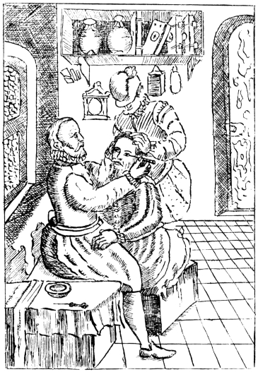
PLATE I.
Copied by C. A. R. H.
Fig. 1.—The positions of the patient, operator, and
assistant, as depicted by Bartisch (Albertotti).
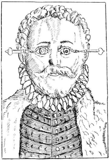
Copied by C. A. R. H.
Fig. 2.—To illustrate the method of introducing the needle
in Bartisch’s operation (Albertotti). The cataract is in situ in the right eye,
and couched in the left eye.
INCORPORATING
THE HUNTERIAN LECTURES
DELIVERED BEFORE THE ROYAL COLLEGE OF SURGEONS OF ENGLAND ON
FEBRUARY 19 AND 21, 1917
LATE SUPERINTENDENT OF THE GOVERNMENT OPHTHALMIC HOSPITAL, MADRAS;
LATE PROFESSOR OF OPHTHALMOLOGY, MEDICAL COLLEGE, MADRAS; AND LATE FELLOW
OF THE UNIVERSITY OF MADRAS;
HONORARY FELLOW OF THE AMERICAN ACADEMY OF OPHTHALMOLOGY AND
OTOLARYNGOLOGY.
WITH 45 ILLUSTRATIONS
PAUL B. HOEBER
67 & 69 EAST 59TH STREET
NEW YORK
1918
Printed in England
To
E. C. I. E.
To
E. TREACHER COLLINS, Esq., f.r.c.s. eng.,
IN GRATEFUL ACKNOWLEDGMENT OF THE ASSISTANCE
WHICH HIS WORK ON THE PATHOLOGY OF THE EYE
HAS BEEN TO ALL WHO DESIRE TO ADVANCE THE
SCIENCE OF OPHTHALMOLOGY
PREFACE
It is a quarter of a century since I first landed in India. In
common with very many other surgeons, my attention was
early attracted to the operation of couching as performed by
its Indian exponents, and I was horrified to see how bad the
majority of the results were. It appeared to me that the
outstanding need was for carefully compiled statistics, in
order that a fair judgment might be formed on the subject.
I divested my mind of partisanship and bias, and sought every
opportunity to discuss the method and its results with anyone
and everyone whose knowledge was likely to be of use to me
in my quest, whether they were laymen or surgeons, Europeans
or Indians, officials or non-officials. The office of
Superintendent of the Government Ophthalmic Hospital,
Madras, afforded an unrivalled field for work, and the staff
of the hospital co-operated with me in a manner which I find
it impossible to acknowledge suitably. My thanks are
especially due to Lieutenant H. C. Craggs, Assistant-Surgeon
C. Taylor, and Dr. Ekambaram, for the valuable help they
gave me. After I left India, Captain W. C. Gray acted for
me as Superintendent, and later Major H. Kirkpatrick succeeded
me permanently. Both of these officers most generously
placed the material of the hospital still at my disposal,
and rendered me very valuable service in the study of my
subject. The great majority of the microscopic slides were
very beautifully prepared for me by Mr. W. Chesterman. I
am also indebted to Mr. S. Stephenson and Mr. A. C. Hudson
for very kindly sectioning some of the globes for me. The
photographs, both macroscopic and microscopic, were taken
by Mrs. Elliot, without whose help I could not have written
the book. Mr. E. Treacher Collins has generously given me
advice and assistance of the greatest value in the study of
a number of the preparations, and I have acted freely on
the suggestions he has been good enough to make. I desire
to express to the Council of the Royal College of Surgeons of
England my acknowledgment of the honour they conferred
on me by electing me to a Hunterian Professorship of the
College. By the kindness of the authorities concerned, I
have been enabled to include in this book articles which have
appeared in the Lancet, in the Ophthalmic Record, and in the
Proceedings of the Ophthalmological Society of the United
Kingdom.
The collection of fifty-four eyeballs, on which the work for
my Hunterian Lectures was founded, I have had the honour to
present to the Museum of the Royal College of Surgeons.
I think I am justified in claiming that it is unrivalled, and
likely to remain so. At the suggestion of Sir John Bland-Sutton,
I have endeavoured so to write the chapter on pathology
that it may furnish a guide to any who care to study the
subject within the walls of that museum. At the same time,
I have striven to make it readable and of interest to those
who have no such opportunity. In this the photographs
have greatly helped me.
As I have already said, I have given nearly a quarter of
a century’s intermittent work to elucidate these problems,
which I felt were of wide interest, not merely to India or to
the East alone, but to the whole civilized world. During the
last two years I have devoted a very large proportion of my
spare time continuously to the subject, but it is so immense
and so far-reaching that I feel I have left much unfinished.
On every hand fresh problems open up, till there seems no
limit to what might be done, given time and opportunity.
This operation of couching, so old that its origin is lost in the
dim mists of antiquity, has still much that is new to be learnt
for the seeking. There are many of the younger surgeons
in the East who could carry the work much farther if they
would, and who would be a hundred-fold repaid if they did.
Will they?
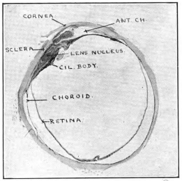
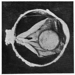
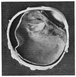
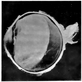
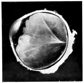
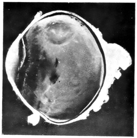
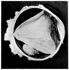
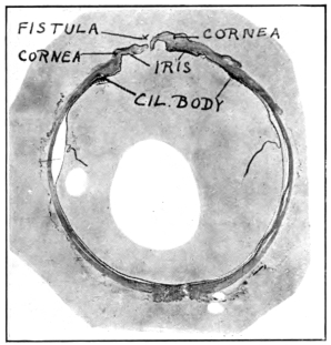
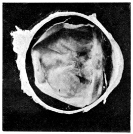
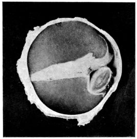
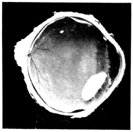
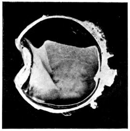
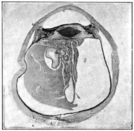
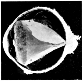
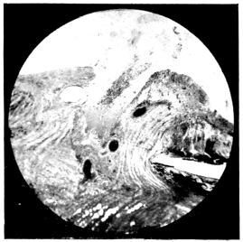
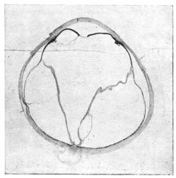
WHOLE-SECTION OF FIG. 34, SHOWING L’IRIS BOMBÉ AND RETINAL CYST
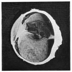
45.
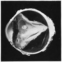
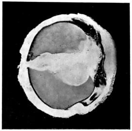
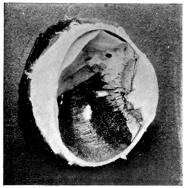
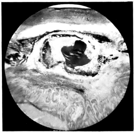
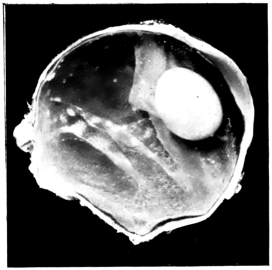
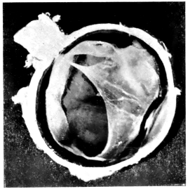
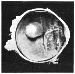
ADHERENT LENS PRESSING ON IRIS BASE
CHAPTER I THE HISTORY OF COUCHING
COUCHING FOR CATARACT
The operation of couching for cataract is one of the most
ancient procedures known to surgery, the earliest description
of the method being that given by Celsus, a contemporary
of Christ’s. The first historical mention of ophthalmic surgeons
was in Alexandria, at the time when medicine and
surgery underwent separation from each other in that great
and flourishing city, nearly three centuries before the dawn
of the Christian era, and Galen states that some of these surgeons
devoted themselves exclusively to operating on cataracts.
Celsus speaks of the writings of a famous Alexandrian surgeon,
named Philoxenes, who lived 270 years before Christ, and
from whom he apparently derived much of his lore. These
writings have unfortunately been lost, thus yielding to
Celsus the proud position of being the first author whose
description of the operation has come down to modern times.
Sprengel is of the opinion that couching was not only known
long before the time of Celsus, but also that the technique
of the operation, even at that distant era, varied widely in
the hands of its different exponents. Of the correctness of
this view there can be little doubt. Sir John Bland-Sutton
has recently published a most interesting memoir on the
recovery of the sight of Tobit at the hands of his son Tobias,
as described in the Apocrypha, and has included in it a copy
of Rembrandt’s picture of the famous operation. Whether
the displacement of the lens was due to the rubbing employed
or to more definite operative measures must be left to speculation,
but, in considering this point, it is worth remembering
that the Eastern coucher of to-day hides the fact that he is
performing an operation under the cloak of the application
of a medicinal paste. Nor must we forget that the anointing
of the eyes of the blind with clay played a leading part in one
at least of the New Testament miracles, and is suggested in
a second. It is to be remembered that the Founder of Christianity
took His examples from, and moulded His teachings
by the aid of things familiar to the people in their everyday
life. The influence of the Oriental on the introduction of
couching to the Western surgeon is shown by the repeated
references in the history of the subject to Eastern exponents
of the procedure. Thus, Razes speaks of the work of an
Indian named Tabri, and Avicenna, himself an Arabian,
describes at length the instruments and technique of the Arab
cataract operators. Abu El Kasim’s name proclaims his
Arab parentage, despite the fact that he is spoken of as a
Spanish surgeon, and the conviction is deepened by the fact
that he spoke of the Arabs in Spain as confining themselves
to couching in the treatment of cataract, showing he was in
intimate touch with them. Nor must we forget to mention
the work of Haly Abbas, and of his distinguished son Jesu
Haly.
When we endeavour to ascertain the probable date of the
first invention of the operation in the East, the fog of uncertainty
closes down over us, obliterates all trace of our quest,
and drives us to fall back on inference. Those who have
spent their lives in an Eastern land know the unbending force
of tradition, the hereditary character of occupations, and the
intense conservatism of Oriental peoples. All these influences
are against change of any kind, and greatly retard the spread
of new ideas. When we consider an operation like couching,
which is well known over the whole of the East, and which
meets in the simplest manner an age-long need, felt in every
village of a tropical or subtropical country, it is not difficult
to believe that the procedure may have been one of the early
fruits of advancing civilisation, far away back in Babylon the
Great, or even earlier still in the home of the Pyramids, tens
of centuries before the dawn of the Christian era. From
these attractive speculations we must return to weigh the
literature of our subject, of which the foundation was so well
and truly laid by the great Celsus. His description of the
technique he employed is as follows:
“Before the operation the patient must use a spare diet. . . .
After this preparation he must sit in a light place, in a seat
facing the light, and the physician must sit opposite the
patient on a seat a little higher; an assistant behind taking
hold of the patient’s head, and keeping it immovable, for the
sight may be lost for ever by a slight motion. Moreover,
the eye itself must be rendered more fixed by laying wool
upon the other and tying it on. The operation must be
performed on the left eye by the right hand, and on the right
by the left hand. Then the needle, sharp-pointed, but by
no means too slender, is to be applied and must be thrust
in, but in a straight direction, through the two coats, in the
middle part betwixt the black of the eye and the external
angle opposite to the middle of the cataract. . . . The needle
must be turned upon the cataract and gently moved up and
down there, and by degrees work the cataract downward
below the pupil; when it has passed the pupil, it must be
pressed down with a considerable force that it may settle in
the inferior part.”
Further details follow. To put the matter shortly in
modern terminology, Celsus introduced a needle through the
sclera and choroid into the vitreous chamber, and depressed
the lens from behind, after first rupturing its posterior capsule
by vertical strokes made with the point of the instrument.
As already mentioned, Galen (born A.D. 131) states that there
were both in Alexandria and in Rome surgeons who confined
themselves to operating upon cataract. Apparently
he also described his own procedure, for some five centuries
later Paulus Ægineta (circaA.D. 630), in detailing his technique,
gave Galen the credit for it. There is practically no
difference between the method they both employed and that
originally laid down by Celsus. A point of real interest in
this connection is that the description of the operation
given by Paulus is practically the only one extant from
the pen of a Greek author, though not a few of them mention
couching and advocate it.
For the next landmark in the study of the subject we have
to pass over three and a half centuries, till we come to the
writings of Avicenna (circaA.D. 980), in which we find introduced
a new feature in the technique; for he mentions
that the Arab surgeons used two instruments for couching—viz.,
a two-edged lancet with which they made a corneal
incision, and a needle with which they depressed the cataract,
after introducing it through the incision thus made. In
this needle there was an eye near the point, through which
a thread was inserted. According to Avicenna, the object
of this was to help depress the lens, but it seems at least
possible that the thread passed through this eye was wound
round the instrument, and so served as a stop, similar to
that used by the Indian coucher to-day. In any case the
description is of great interest, linking as it does the
Mahomedan operator of the twentieth century with his predecessor
of the tenth. The famous Spanish surgeon Abu El
Kasim adopted exactly the same technique for couching as
that we have just described. This, as has already been
suggested, is not in the least remarkable, for his name bespeaks
his Arab descent.
The next description of the operation, which claims our
interest, is that by Benvenuto (Benevenutus Hyerosolimitamus),
who flourished in the twelfth century. The quaint
blending of religion and science, which it reveals, makes it
very attractive reading: “Towards the third hour, the patient
having fasted, thou shouldst make him sit astride of an ordinary
chair, and thou shouldst sit before him in the same
way. Keep the good eye of the patient shut, and begin to
operate on the bad eye, in the name of Jesus Christ. With
one hand raise the upper lid, and with the other hold the
silver needle, and place it in the part where the small angle
of the eye is. Perforate the same covering of the eye, turning
the instrument round and round between the fingers, till thou
hast touched with the point of the needle that putrid water
which the Arabs and Saracens called Mesoret, and which we
call cataract. Then beginning from the upper part, remove
it from the place where it is before the pupil, and make it
come down in front, and then hold it for as long as it takes
to say four or five paternosters. After, remove the needle
gently from the top part. If it happens that the cataract reascends,
reduce it towards the lower angle, and when you have introduced
the needle into the eye, do not draw it out unless the
cataract be situated in the place described above; then gently
extract the needle in the same way as you put it in, turning
it about between the fingers. The needle being extracted,
keep the eye closed and make the patient lie flat on a bed,
keeping him in the dark with his eyes shut, so that he does
not see the light or move for eight days, during which time
put white of egg on twice a day and twice during the night.”
Passing over four centuries, we come to an even more
interesting description of the operation from the pen of
Bartisch of Dresden: “The day being decided upon, on which
the operation is to be performed, the doctor who is obliged
to, or who wishes to do it, must abstain from wine for two days
beforehand. The patient must also fast the same day, and
must neither eat much nor little till an hour after the operation.
Given the aforesaid conditions, try and procure a
well-lighted room, in which the patient may have everything
necessary for going to bed and remaining there, as he ought
not to be taken to any place far off; the nearer to bed the
better. Set thyself on a bench in the light and turn thy
back to the window. The patient may be seated on a chair,
a stool, or on a box, before thee and near to thee; in any case
he is to be seated lower than thyself. His legs between thine
and his hands on thy thighs. A servant stands behind to
hold the patient’s head. The servant should bend a little,
so that the patient may rest his head against him (Fig. 1).
When the patient is blind of one eye only, the other eye should
be bandaged with a cloth and a pad so that he cannot see.
Then take the instrument or the needle in one hand, so that
the right hand will be for the left eye, and vice versa. With
the other hand separate with great care the upper lid from
the lower, using the thumb and the first finger, so that thou
canst see how to direct the needle into the eye. When thou
wishest to introduce the needle, the eye must be turned
towards the light and looking straight at thee; also, I should
make the patient turn his eye a little towards his nose, so
that thou canst use the instrument better and that thou wilt
not injure the small veins of the eye, but respect them. Direct
the needle straight and with attention over the membrane
called the conjunctiva, straight towards the pupil and uvea,
at the distance of two blades of a knife from the membrane
called cornea or from the grey that is in the eye. Hold the
needle quite straight, hold it steady so that it will not deviate
or slip. Hold the needle and press it, and turn it with the
fingers in the eye with great gentleness, according to the
instructions you may gather from the figure, which shows
an eye in which the cataract has been taken away, while the
other eye has not been touched (Fig. 2). Hold the needle
firmly while turning it round, and be careful always to have
the point towards the middle of the eye, that it almost touches
the pupil and the uvea; and not to oscillate by any chance
towards one side. When thou feelest that the needle has
penetrated into the eye, that it almost touches the pupil and
the uvea, and when thou hast proved to be really in the eye,
hold the needle securely and move it, letting it slip backwards
and forwards towards the pupil till thou art certain
of being in the substance of the cataract, which thou
canst easily be sure of by the movement of the cataract
material. When thou hast remarked that, lower it carefully
and gently and slowly, so as not to disturb the cataract; but
try and free the matter entirely from the pupil and from the
uvea with care, and keep it intact. Press the said matter
with the needle under it, with the greatest care, and when
thou perceivest that it is altogether free and loose, draw and
direct the needle, with the matter behind it, upwards, and
then pass it well downwards, behind the thin retina and the
aranea of the eye; and take care that it remains there. . . .
This is the recognised instruction, research, and indication
of the means of operating, of pricking the cataract, or of the
manner in which such an operation ought to be initiated and
conducted. But no one ought to undertake such an operation
unless he has learnt much and seen much, and unless he
is fundamentally taught by intelligent doctors. Unless he
is so, it is not well to operate. And it is not wise to trust to
any of the brotherhood who happen to be dressed in velvet or
silk, and who boast of being great oculists, and are capable of
curing the blind from cataract. Certainly these can make
holes in the eyes, but I do not know how they can succeed.”
The knowledge which the Greeks and the Arabs possessed,
before and after the dawn of the Christian era, on the subject
of the pathology and treatment of cataract, appears to have
been largely forgotten during the Middle Ages. It would
seem that both couching and extraction fell into disuse, and
that the surgical treatment of cataract was left for centuries
in the hands of wandering charlatans, whose ways brought
much discredit upon it. Towards the close of the seventeenth
century, Pierre Brisseau, a doctor of Tournay, revived the
operation, inventing a needle of his own for the purpose.
His advocacy of the method aroused bitter controversy, but
it was undoubtedly the best operation in the field until the
famous French surgeon Daviel performed his first extraction
in 1745, and thus sounded the death-knell of a procedure
which had held the pride of place in European surgery for over
seventeen centuries. It was, however, many years before
couching was definitely abandoned in favour of extraction.
Indeed, the author has recently had the privilege of discussing
this subject with a distinguished surgeon, who can remember
the time when depression was still a recognised method of
operating in London. It is a great mistake to suppose that
Daviel was the first to endeavour to extract a cataract, for
both extraction and suction of cataracts have their roots far
back in history. Indeed, Antyllus described his method of
extraction at the close of the first century of the Christian era,
and there are numerous other references to it in early literature.
What Daviel did was to adopt a technique which gave a reasonable
prospect of success.
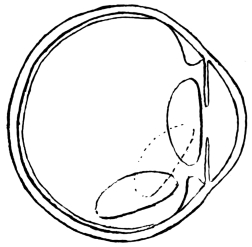
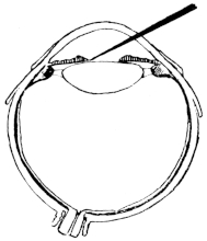
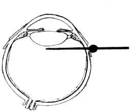
Fig. 3.—Depression.
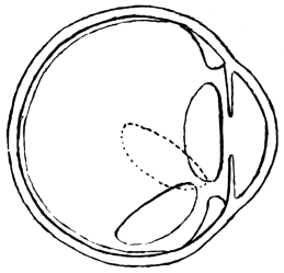
Fig. 4.—Reclination.
The above two figures illustrate the path taken by the cataract during
the operation. (Mackenzie.)
The introduction of reclination, as opposed to depression,
by Willburg in a Nuremberg thesis, dated 1785, gave a fresh
lease of life to couching in its dying struggle with the operation
which was destined to supersede it. England, France,
Sweden, Germany, and other countries, joined vigorously in
the discussion, and amongst the powerful advocates of couching
were ranked Percival Pott and William Hay of London,
Cusson of Montpellier, and Scarpa of Pavia, whilst Benjamin
Bell practised both couching and extraction. The admirable
treatise by James Ware on cataract (1812) was all but a
death-blow for Celsus’s operation. The newer procedure
was then well in the ascendant, and only needed time to completely
strangle its rival. Notwithstanding this, it was left
to Mackenzie, so late as 1854 (fourth edition), to give the most
complete and interesting description of couching to be found in
literature. He distinguishes sharply between the operations
of depression and reclination. In depression, the lens is pushed
directly below the level of the pupil, being made to follow the
curvature of the eye, to sweep over the corpus ciliare, until
it comes to rest on the lower curve of the eyeball, with its
anterior surface directed forward and downward (Fig. 3).
In reclination, the lens is made to turn over towards the bottom
of the vitreous chamber in such a way that what was formerly
its anterior surface now comes to look upward, and what was
its upper edge is turned to the rear. The whole lens is swung
backward as if on a hinge, composed of the lower fibres of its
suspensory ligament, which still remain unbroken (Fig. 4).
Fig. 5.
He divides the operation of couching into four stages, in only
the last of which reclination differs from depression. These are:
(1) the pushing of a special needle (Fig. 5) through the coats
of the eye at a distance of 1/6 inch behind the temporal edge of
the cornea, and to a depth of 1/5 inch; (2) the laceration of the
posterior capsule of the lens by vertical movements of the point
of the needle, to prepare an aperture for the passage of the
lens; (3) the passing of the needle into the anterior chamber
around the edge of the lens, and the laceration of the anterior
capsule by vertical strokes; (4a) to depress the lens, the point
of the needle is carried over its upper edge, and the handle is
raised a little above the horizontal, thereby correspondingly
lowering the point, which forces the cataract downward out
of sight behind the pupil: the needle is then withdrawn by
rotation; (4b) to effect reclination, the needle-point is raised
not more than 1/10 inch above the transverse diameter of the
lens: its concave surface is pressed against the cataract,
which is reclined by moving the handle of the instrument
upward and forward, thereby causing its point to pass downward
and backward. The cataract is thus made to fall over
into the vitreous humour, and is then pressed downward,
backward, and a little outward. Mackenzie adds many interesting
details as to the modifications of the operation, according
to the variety of the cataract to be dealt with, and as to the
after-treatment and complications met with.
We come now to a very interesting phase in the study of
the operation of couching. We have shown reason to believe
that, like many another valued heritage of the West, it was
brought there originally by Wise Men of the East. For more
than eighteen centuries it remained a treasured possession of
surgery, only to yield its ground before the fierce competition
of a method better able to survive the stern test of experience.
Slowly but surely its decadence banished it from modern
scientific European literature, and then, strangely enough,
the advent of Listerism fanned the dying flame of interest
in the method; but this time in the East, and not in the West.
From the East it had sprung to find a home in the West, and
in the East, at the hand of Western surgeons, its last, and by
no means least, interesting chapter is in the course of being
written. A review of the more recent literature on the subject
will establish this contention, and will show how large a
share the officers of the Indian Medical Service have taken in
the settlement of a question which, apart from its scientific
value, has important social and even political bearings.
After a brief visit to India, Hirschberg, in 1894, published
an article on couching, in the course of which he spoke favourably
of the results of the operation. He was, unfortunately,
handicapped by his ignorance of the natives of India and
of their ways and customs, with the result that his views on
the subject are of comparatively little interest to us. In
the following year Captain H. E. Drake-Brockman described the
operation of couching as explained to him by one of its Indian
exponents. The latter pierced the sclerotic with a small
lancet in the lower outer quadrant close to the cornea, and
then introduced a copper needle; “a series of motions of the
hand are made from the position on first introduction of the
needle to a point corresponding to it in the upper section of
the outer diameter of the eyeball.” The depression of the
lens appears to have taken place next, but the description is
throughout somewhat vague. Presumably the operation
was the same as that described by Ekambaram, but the
coucher does not seem to have been able to make the steps of
the procedure as clear as that surgeon has done.
Henry Power, in the British Medical Journal (October,
1901), entered a plea for the occasional performance of the
operation of depression in cases of cataract. His experience
went far enough back to enable him to remember the time,
not only when he had seen surgeons of repute employ this
method, but when he had himself imitated the example thus
set. His own practice had been to attack the cataract, via
the sclerotic, through the posterior capsule. He framed a
number of indications which to his mind justified the occasional
performance of couching. It is safe to say that very few of
these would be seriously entertained by surgeons to-day. The
most interesting point he made was in connection with Himly,
in whose work, published in 1843, the statement occurred
that “severe inflammation rarely followed reclination, and
when it did it often cleared up without leaving any bad consequences.”
A doubt as to the reliability of Himly’s statements
is suggested by his claim that he had only two failures
in fifty cases, one of these not being attributable to the operation.
This is so much at variance with the experience of
others as to make one sceptical about accepting any of his
assertions without some reservation.
The next paper of value that we come to is by Maynard
(1903). In this he analysed sixty-three cases of couching,
which he had met with in Indian practice, and recorded the
anatomical examination by Parsons of a couched eye sent home
for the purpose. The same year saw the appearance of a
paper by Albertotti of Medina, in which that writer somewhat
fanatically and unconvincingly advocated a return to
couching, with the use of a corneal puncture and with the employment
of special instruments for the purpose. A year
later he was followed along the same lines by Basso of Genoa,
whilst Quartillera published a paper whose recommendations
were very similar to those made by Henry Power. In 1905,
Major Henry Smith of Jullundur, in a very outspoken article
in the Indian Medical Gazette, expressed the opinion “that
lens couching at the present time is an operation which should
not be practised outside the ranks of charlatans,” and added
that “it is no easy matter to completely dislocate the lens,
and in my observation the partial dislocation is more frequent
than the complete in the hands of adepts of the art.” In
reply to this paper, Maynard reaffirmed his belief that couching
is “justifiable under certain conditions.” The editor of the
Indian Medical Gazette invited further discussion of the subject,
and in accordance with this request the writer published
his statistics based on 125 cases of couching, carefully recorded
on printed schedules. In the course of that paper he voiced
his strong opposition to the adoption of the Indian operation,
or of any modification of it, in the hands of surgeons who enjoy
the unique opportunity of obtaining manipulative skill granted
to those who work in India. A former pupil of his, Dr. Ekambaram,
studied the ways of the Indian coucher at first-hand,
and gave the results of his experience in one of the most valuable
contributions to the subject yet made. This was in 1910.
Two years later the writer was able to review the statistics
of 550 consecutive cases of couching, all of which had been
carefully noted. Still more cases accumulated before he left
India, and by the kindness of Major Kirkpatrick, the total
under review has now reached 780.
The examination by J. H. Parsons of a couched eye has
already been mentioned. In 1913, A. C. Hudson sectioned and
described a similar specimen sent him from India by the
writer. The only previous published records of the same kind
are from the pen of E. Treacher Collins, and refer to four specimens
of couched eyes in the Museum of the Royal London
Ophthalmic Hospital. Major H. Kirkpatrick has recently
examined several more cases in Madras, and has kindly communicated
some of the more interesting of his findings to the
writer. Communications, that have been made from time to
time before meetings of ophthalmologists, show that British
surgeons of the first rank are still in favour of performing
couching under certain special conditions. A marked instance
of this is to be found in the discussion which took place before
the Ophthalmological Society of the United Kingdom on
February 8, 1906, following the presentation of a case by
Holmes Spicer. On that occasion Rockliffe and Treacher
Collins stated that, like Spicer, they had performed the operation
in exceptional cases, and Devereux Marshall and G. W.
Roll accorded it a modified support under such conditions.
The writer has also learnt from personal communications
that other leading surgeons have taken a similar line. There
for the present we must leave the history of this operation,
whose origin is lost in the dim mists of antiquity, and whose
chequered career forms one of the most interesting pages in
the literature of medicine.
BIBLIOGRAPHY
Albertotti, Giuseppe: Benevenuti Grassi, de oculis eorumque ægritudinibus
(réédition de l’incunable de Ferrare, 1498); Paris, 1897.
Albertotti, Giuseppe: Depression of Cataract, La Clinica Oculistica, June,
1903.
American Encyclopædia and Dictionary of Ophthalmology, Casey Wood;
Chicago, 1916.
Basso: La Clinica Oculistica, January, 1904.
Bland-Sutton, Sir John: On an Apocryphal Miracle, Middlesex Hospital
Journal, vol. xx., No. 1.
Brisseau: Traité de la Cataracte et du Glaucome; Tournay, 1706.
Celsus, A. Cornelius: Of Medicine, trans. by James Grieve; printed by
D. Wilson and T. Durham, Strand, London, 1756.
Collins, E. Treacher: R.L.O.H. Rep., 1893, vol. xiii., p. 308.
Cusson, M. P.: Remarques sur la Cataracte, à l’Académie des Sciences de
Montpellier, 1779.
Drake-Brockman, Surg.-Capt. H. E.: The Indian Oculist and his Equipment,
Trans. of the O.S. of the U.K., vol. xv., 1895.
Drake-Brockman, Lieut.-Col. E. F.: The Indian Oculist and his Equipment,
Trans. of the O.S. of the U.K., vol. xv., 1895.
Ekambaram, R.: Couchers and their Methods, Ind. Med. Gaz., 1910.
Elliot, R. H.: Couching of the Lens, Ind. Med. Gaz., August, 1906.
Elliot, R. H.: The Operation of Couching as practised in Southern India:
a Review of 550 Cases, Proc. of S. Ind. Branch of B.M.A., 1912, and
Ophthalmic Review, vol. xxxi., 1912.
Encyclopædia Britannica.
Galen: De partib. art. med.
Hay-Williams: Practical Observations on Surgery; London, 1803.
Hirschberg: Centralblatt für Praktische Augenheilkunde, February 1, 1894.
Hirschberg: Centralblatt für Praktische Augenheilkunde, 1908, vol. xxxii.,
p. 2.
Histoire de la Médecine, par Kurt Sprengel, trad. par A. J. L. Jourdan;
Paris, 1815.
Histoire de l’Ophthalmologie à l’École de Montpellier, par H. Truc et
P. Pansier; Paris, A. Moloine, 1907.
Hudson, H. C.: R.L.O.H. Rep., vol. xviii., part ii.
Mackenzie, W.: On the Diseases of the Eye, 4th edit., London, 1854.
Maynard, F. P.: Ophthalmic Review, April, 1903.
Maynard, F. P.: Ind. Med. Gaz., May, 1905.
Paulus Ægineta, vol. ii., Sydenham Society, 1845–46.
Pott, Percival: Remarks on Cataract.
Power, H.: Depression in Cases of Cataract, Brit. Med. Journ., October, 1901.
Quartillera, Castilley: Arch. de Oftal. Hispano-Americanos, October,
1904.
Smith, H.: Cataract Couching, Ind. Med. Gaz., May, 1905; and Trans. of
the O.S. of the U.K., 1904, p. 264.
Ware, James: The Cataract and Gutta Serena, 3rd edit., London, 1812.
Wilkinson, Miss K. E.: The Manuscripts of Naples and the Vatican, etc.,
trans. from Albertotti, Ind. Med. Gaz., October, 1904.
CHAPTER II THE TECHNIQUE OF THE OPERATION
The writer has never seen a native coucher at work, and
consequently all his information on the subject has had to be
gathered from those who have been more fortunate than himself
in this respect. There would appear to be two distinct
modes of operating, which for convenience’ sake may be
spoken of as the anterior and the posterior, using the terms
relatively to the plane of the ciliary body and iris (Figs. 6 and
7). We shall take them in turn.
Fig. 6.—Anterior Operation.
Fig. 7.—Posterior Operation.
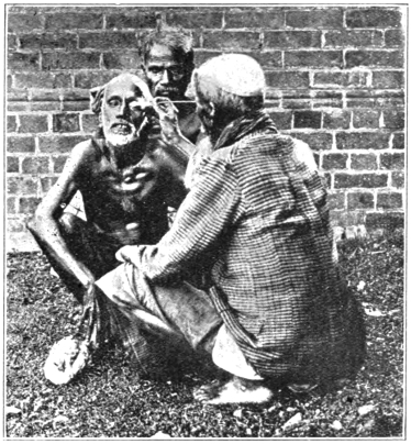
Fig. 8.—The Operation of Couching.
The Anterior Operation.—The patient and operator sit
facing each other in a good light; both squat on their hams
in accordance with the immemorial custom of the East (Fig. 8).
The patient is frequently, if not usually, told that no operation
is to be performed, and that it is merely a question of putting
medicine into the eye. He is directed to look downward,
and the coucher raises the upper lid with one hand whilst in
the other he conceals either a needle or a sharp thorn. It
is said that the long needle-like thorn of the babul-tree is
usually selected for the purpose. Many of the patients have
mentioned that their heads were steadied by a friend from
behind. In the majority of cases, at least, it would appear
that no form of local anæsthesia is attempted. The operators
appear to rely largely on manual dexterity, and to aim at completing
the procedure in a minimum of time. The needle
or thorn is thrust suddenly through the cornea, and on through
the pupil or iris, into or on to the periphery of the lens. The
next movement, which appears to follow the first so rapidly
as practically to melt into it, is that of depression or reclination.
In this, the spot where the cornea grasps the shaft
of the needle serves as a fulcrum. The operator raises his end
of the instrument, and the opposite one, which lies either on
the surface of the lens or imbedded in it, is consequently
depressed, thus carrying the cataract with it downwards, or
downwards and backwards, and so clearing the pupil. In
the course of speaking to a very large number of patients thus
operated on, it has struck the writer as most remarkable that
they made as little complaint as they usually did of the pain
inflicted on them during the operation. They described the
sensation of a sudden prick, but it was obvious that they
had no acute recollection of agonising suffering. This point
is emphasised by the fact that in nearly every case the operator
tested his patient’s vision immediately after the operation
by holding up fingers, coloured cloths, necklaces, or other
common objects, for triumphant identification. Very great
stress is laid on this part of the ritual, and the onlookers are
not allowed to lose sight of the wonderful results achieved
by the operation. There seems reason to believe that an
effort is made to enter the point of the instrument through the
pupil, and to pass it between the iris and the lens. This cannot
fail to be a difficult thing to do, as is evidenced by the frequency
with which we were able to discover scars in the iris,
which had obviously resulted from tears at the time of the
operation. The point of perforation of the cornea could frequently
be discovered, especially if a loupe were used for the
purpose. The relative positions of the scars in the cornea
and iris were frequently of great value to us from the diagnostic
point of view. The eye is bandaged for at least twenty-four
hours. By the end of that time the operator has frequently
placed a safe distance between himself and his patients of the
day before, and is seeking fresh dupes in another village.
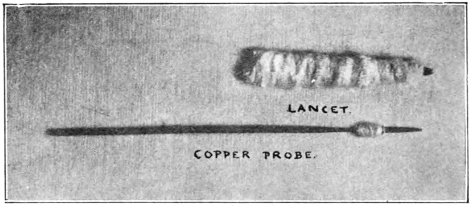
Fig. 9.—Instruments used in Couching.
The Posterior Operation.—Much that has been written on
the preceding method applies with equal force to this. It
is, however, possible to describe the technique much more
accurately, as it has been carefully studied at first-hand
by Dr. Ekambaram, who for many years worked under the
writer in the Government Ophthalmic Hospital, Madras.
His original description of the method will well repay a careful
perusal. He speaks of the operators as being ambidextrous
and very skilful. Their surgical equipment (Fig. 9) for the
operation consists of a small lancet-shaped knife, guarded to
within a few millimetres of its tip by a roll of cotton-wool,
wrapped round it for the purpose, and of a copper probe
4 inches long and about 1 1/2 mm. in diameter. A cotton
thread twisted round this probe at a spot 12 mm. from its
point serves the same purpose as the stop in the Bowman’s
needle. From the point to this stop the instrument is triangular
in section. The patient is directed to look well towards
the nose, and the surgeon then gently marks out the
selected spot by pressing with his thumbnail on the conjunctiva
covering the sclera, about 8 mm. out from the cornea, and
about 2 mm. below the horizontal meridian. In some cases
the operator steadies the eye by firm digital pressure exerted
through the partly everted lower lid. He next takes his
lancet in his hand, and it will be observed from the illustration
(Fig. 9) that it might easily pass for a roll of cotton-wool;
this, indeed, is what the patient is led to believe it really is.
To heighten such an impression, the point is covered with a
sandalwood paste, prepared beforehand coram publico, with
a good deal of ostentation. The patient is informed that this
“cataract-cleansing drug is about to be applied to the eye,”
and under cover of the suggestion the operator plunges the
lancet through the tunic of the globe at the spot already
selected. The alarm thus occasioned is allayed by the assurance
that the “medicinal application” is over. The copper
probe is next produced, and is inserted through the wound up
to its stop, being held between the thumb and two fingers;
a circular movement is given to its point, the stop resting
against the puncture, and serving as a pivot for the movement.
According to Ekambaram, the object is to tear through the
suspensory ligament from behind. Immediately following
this step, a downward stroke of the point is made in order to
depress the now loosened lens. Ekambaram graphically
describes the care taken by these operators to impress, alike
on the patient and on the friends, the magical effects of the
procedure. The former is shown a number of objects, and is
bidden to name them in turn, and to state their colour. The
crowning point is reached when the surgeon removes a thread
from his garment, and the patient not merely recognizes it
as such, but triumphantly tells its hue. The Western ophthalmic
surgeon, with his wide incision and his anxiety for
the safety of the vitreous, can never savour such dramatic
moments as these. They carry us back to the descriptions
of the early Christian miracles, with all the mental and spiritual
associations, which enwrap such stories as those of Bartimæus,
and of the pools of Siloam and Bethesda. Alas that life’s
“hereafters” should so often be fraught with disillusionment,
disappointment, and suffering! Palestine and its storied
past rise before us as we read how the vaidyan called for a
white cloth and for water, how he dipped the cloth in the fluid
and washed out the sufferer’s eye therewith, how he made a
paste and smeared it over the skin around the brow, how he
closed the eye with “clean white linen,” and then sent the
erstwhile blind man rejoicing away. Over the abyss of nearly
twenty centuries, the East stretches out her unfaltering hand
to the past of the nearer East, whilst the West looks on in
wonder, not unmixed with admiration, for a spirit which the
corroding passage of time seems unable either to fret or to
change.
There is a step of the procedure which has been purposely
left to the last, as its interest is psychologic, and not surgical.
It is common to both methods of operation. I refer to the
anointing of the eye with the blood of a freshly killed fowl.
It is a measure in which superstition, cunning, self-preservation,
and greed, overwhelm and mask a faint and feeble therapeutic
design. The sacrificial element is present, and a hazy idea that
the death of the votive bird may turn evil from the patient
looms in the background. Next comes the need to mask the
shedding of the patient’s blood, since he is often told that no
operation is to be performed, but that a mere “medicinal
application” is to be made; the blood of the outraged bird
covers the guilt of the vaidyan’s falsehood. Largest of all
towers the fact that the curry-pot even of a worker of surgical
marvels needs constant replenishing, and that fowl is an
excellent substitute for mutton on such occasions. Lastly,
these men seem to believe that the coagulation of the fowl’s
blood helps to close the puncture. In view of the dirty condition
of the instruments which they introduce into the interior
of the eye, this last factor may practically be neglected.
CHAPTER III THE INDIAN COUCHER AND HIS HABITS
The coucher goes by different names in different parts of India.
In Bengal and in the United and Central Provinces he is known
as the “suttya” or “mal,” and in the Punjab as the “rawal.”
Ekambaram, who came into intimate contact with these
men in the Madras Presidency, always describes them as
“vaidyans,” the term signifying surgeons. In the north they
are Hindus, of the Kayasth caste, a class well known for its
astuteness and educational qualifications. Drake-Brockman
states that in the north Mahomedan couchers are rare, whilst
in the Southern Presidency it appears to be the exception to
find a Hindu doing such work. Like every other occupation,
couching in India is hereditary, the principles of the craft
being handed down from father to son by word of mouth and
by practical instruction. It has been stated that there is no
literature on the subject. This, however, would appear to be a
mistake, for Ekambaram learnt that there are “some old
texts written on palmyra leaves laying down the method.”
A literal translation of one of these runs: “Removing the lancet
after making a puncture, insert the copper probe; and holding
it with three fingers, depress the lens with the three-sided
edge.”
By tradition and ancestral habit, the coucher is a wanderer
on the face of the earth, and like a gipsy he carries his wares,
such as they are, to the very doors of the people’s homes;
but it is probable that in each of the large provinces of India
these men have a headquarters of their own. This in the
Madras Presidency is known as “Kannadiputhur,” which
signifies the “village of eye operations.” During part of the
year these men are agriculturists and fishermen; but when
the dry season robs them of their occupations, they wander
forth to practise the art, with which their ancestors have been
identified from time immemorial. They do not, however,
confine themselves to eye operations, but practise as well a
crude form of general surgery. Like many other disciples
of Æsculapius, their fee is a very elastic one, and, in common
with other artists, they learn to know both the smiles and the
frowns of fortune. Luxury rarely comes their way, whilst
hardship and toil are their constant lot. Their spare evening
hours are filled in with such arduous and monotonous occupations
as net-weaving; and full many a night they go hungry to
sleep, with the sun-baked earth for their only bed.
All, who have seen them at work, agree that their methods
are dirty and septic to a degree, and the oft-expressed wonder
has ever been, not that their results are so bad, but that they
are ever good. Their surgical equipment is carried in a
bag or in a box, which would be considered dirty alongside
of the tool-chest or work-basket of any English artisan. The
filth alike of their clothes, their hands, and their person, stagger
description from a surgical point of view. The exact patterns
of the instruments used vary in different parts of India,
and so also do some of the couchers’ customs. Allusion has
already been made to the slaughter of a fowl and the use of
its blood in Southern India. This is readily understood, as
the Mahomedan couchers are flesh-eaters. In the north,
where these experts are Hindus, the fowl plays no part, but a
very subtle form of deception is described by Drake-Brockman.
Each suttiah carries in a little bag a store of pieces of dried
membrane. One of these is dropped into water before the
operation commences, and is produced at the psychologic
moment as evidence that the Indian surgeon can, and does,
remove the cataract from the eye, just as much as his Western
brother. This tribute to our science is as subtle as it is nefarious.
The pieces carried are of various tints, in order that the colour
of the cataract, as seen before operation, may be matched as
closely as possible.
Right through the ages the shadow of charlatanism has
lain over the operation of couching. We are told that it did
so in Alexandria and in Rome at the dawn of the Christian
era, and from that time up to the present we find numerous
traces of it in literature. Not the least interesting of such
comes from the Dark Ages, and, despite its pathos, has a
distinctly amusing side. The operator and his assistant took
the patient alone into a darkened room; a candle was lighted
and kept carefully behind the victim’s back by one of the
knaves, while the other in front asked if he could see the flame.
A sham operation was then performed, and the process was
again repeated, but this time with the light in front; naturally
the blind man could now see it, and, on being assured that
“the change” was due to what had been done, his gratitude
was likely to rise to the production of the necessary fee. If
it did so, the impostors speedily made off. It seems hard
to believe that even the Dark Ages were dark enough for so
transparent a trick to be tried often in one town.
In a recent personal communication, Dr. Ekambaram has
very kindly furnished the writer with some additional information,
as new as it is interesting. He divides the Indian
couchers, with whom he has come into contact, into two classes,
the Mahomedan couchers of the south, whose work we
mainly meet with in Madras, and the Punjabis (people of the
Punjab) from the north. The same method of operation is
adopted by both, but there would appear to be a great difference
between the status and the attainments of the two classes.
The Mahomedans are much the lower type; their practice
is confined to the villages through which they roam, and they
very rarely visit big towns. Their length of stay is limited
to one or two days, and they make haste to escape soon after
having performed an operation, “for fear of being clubbed for
their stupid action.” They do not use any form of anæsthetic.
On the other hand, the Punjabi couchers are described as intelligent,
respectable, decently clad men, who confine their
work to the towns, and stay in each place four or five months,
amassing considerable wealth thereby. Before couching,
they drop into the eye a fine yellow powder, which Ekambaram
believes to be stained cocaine. They were, however, extremely
secretive on this subject, and refused to part with even a grain
of the drug at any price he could offer. Its efficiency is
testified to by the fact that the patients remained absolutely
quiet and collected during the whole of the operation. The
cases are kept under observation for from a week to a month
after operation, putting in a daily attendance. Their results
are much better than those obtained by the Mahomedans.
Some of the fees they obtain are relatively very large. An
idea of their social status may be gathered from the fact that
carriages are sent for them by their better-class patients; but,
in Ekambaram’s opinion, the aristocracy of Indian intelligence
is learning to keep aloof from these men, owing to the
influence of the Western surgeons, whose method of extraction
is steadily establishing itself in the esteem of the people
at large. He gives credit to the Punjabis for a more efficient
technique of operation than that practised by the Mahomedans.
In this connection it is interesting to record some of the
opinions of Western surgeons, who have come in contact
with the work of these northern men. Captain H. E. Drake-Brockman
had nothing too bad to say of them, and his uncle
Lieut.-Colonel E. F. Drake-Brockman (formerly of the Madras
Eye Hospital), in presenting his nephew’s paper before the
Ophthalmological Society of the United Kingdom, estimated
the coucher’s successes at not more than 10 per cent. Lieut.-Colonel
Henry Smith went so far as to declare that even the
best cases, if followed long enough, ended in absolute blindness.
Lieut.-Colonel F. P. Maynard formed a much less unfavourable
estimate of the coucher’s results (46 per cent. of
successes). The writer’s own statistics will be given later.
Not the least interesting of Drake-Brockman’s contributions
to the subject is his statement that vaccinators and compounders
in the pay of the Indian Government are not infrequently
couchers in disguise. The full significance of this
observation can only be appreciated by one who knows the
East intimately. Scientifically, Western medicine is educating
the Indian medical man out into the light from pagan depths
of darkness. Even to-day there are those of them who practise
Eastern and Western medicine side by side. It would
seem strange to a British surgeon to learn that it is possible
for a medical man (whose qualifications, from an educational
point of view, compare favourably with those of our home
Universities) to lay stress before his patients on the right
quarter of the moon, and on the correct aspect of the ground,
for the gathering of a particular drug; yet the writer has
known this happen, and that, too, in the case of a medical man
who held an important scientific appointment under Government.
The very fact of a man holding a post under the British
Rāj would add to his prestige and increase the confidence of the
people in him. That such an one should advocate and practise
couching would cause no surprise to his own people, whose
faith in methods based on tradition is firmly founded. Some
idea of the complexity and confusion of the Indian mind to-day
may be gathered from a knowledge of the strange blend
of dissimilar lines of thought in those who have adopted
Christianity. There are many such who keep a foot in both
camps, in a way that would have been thought impossible
by anyone who had not been brought into intimate contact
with such people. Under these circumstances, it is hardly
strange that the same kind of thing should be found in evidence
in other lines of life; and yet it is no time to blame these
men. Their need—I speak now from the scientific stand-point—is
for “more light.” We ourselves retain to-day the
taint of superstitions that come down from our witch-burning
ancestors. Shall we not, then, be lenient to those who have
never had our advantages? As we flood India with the
daylight of true scientific knowledge, the vermin of ignorance
and superstition will scurry away to hide. That the coucher
believes in himself is indisputable; that he resents the invasion
of our Western operation is easily understood; and that he must
disappear, washed out by the advancing flood of better methods,
is as certain as the fate of the furrows left on the sand by
last night’s tide. Meanwhile he has stopped a gap and filled
a place in the life of a nation, and it behoves us, in our estimate
of him, to remember that the highest earthly honour ever
awarded was given to a woman whom the world despised and
blamed, in the immortal words: “She hath done what she
could.”
It has been said of these men that they have no diagnostic
powers, and it is quite certain that they sometimes couch the
lens in cases of optic atrophy, of glaucoma, and of certain
other diseases, in which such a step is useless and worse.
It is fair to add that the number of cases of this kind which
the writer saw was not large. This observation is the more
significant, since he was keenly on the lookout for any instances
of the kind, and that, too, during many years of Indian
experience in an exceptionally large cataract practice. Ekambaram
speaks of a case of operable cataract in which he had
dilated the pupil, and on which he (purely experimentally,
and with no intention of allowing it to be done) asked couchers
if they would operate. As soon as they found the pupil was
motionless and dilated, they declined to undertake any operation.
He comments favourably on the acumen they thus
displayed.
A word may not be out of place on the subject of the
covering of the head of the patient and surgeon with a cloth,
as adopted by some few couchers during the operation. Such
a procedure is, from the point of view of lost light, a handicap
to the operator, but it has, from his way of looking at it, certain
advantages. It shuts the patient off from the distractions
of his surroundings, which are otherwise very public;
it hides the actual operative procedure from prying eyes;
and it conceals the facial and other evidence of pain. The
greatest factor of all, in an Eastern land, is the air of mystery
with which it shrouds the proceeding. The element of “jadu”
(magic) so introduced is paramount in its psychologic interest.
CHAPTER IV STATISTICAL
In the Indian Medical Gazette of August, 1906, the writer published
a review of 125 cases of couching of the lens by Indian
vaidyans. On March 13, 1912, the total had swollen to 550,
and an analysis of all the cases was conducted on the same
lines as those followed in the earlier communication. Subsequent
to this, Major H. Kirkpatrick, the succeeding Superintendent
of the Madras Eye Hospital, analysed 350 cases of
the Mahomedan operation (the first 120 of which overlapped
the series already dealt with by the writer), and in forwarding
them made the comment that “these statistics are remarkably
similar to those of your series.” In view of the consistency
of the results obtained and of the large figures dealt with
it seems safe to assume that reliable deductions can now be
drawn.
The main headings of these statistics will next be taken up.
Before commencing to do so, it is of special interest to note
that the methods observed by Colonel Drake-Brockman in
other parts of India tally closely with those described by
Ekambaram in Southern India. This is the more readily
understood when we remember that many of the operators in the
South of India come from the north. This observation puts
our cases on all fours with those published from other parts of
India.
It is perhaps difficult to form an accurate opinion as to
how far the figures before us represent the best results attained
by the coucher. His apologists might urge that only his failures
would come to English hospitals. On the other hand, there
can be little doubt that quite a considerable number of eyes
are lost, after couching, from panophthalmitis and from
shrinkage of the eye following iridocyclitis. Patients in such
conditions will often stay away from hospital owing to their
very hopelessness; and even if they do resort to English treatment,
they will stoutly deny that their eyes have been interfered
with. Thus in both ways the records of such failures
are lost, making the net result appear better than it really
is. In collecting our statistics, we were constantly on the
lookout for all cases of couched lens, and have notes of a large
number of patients who did not come to hospital for the eye
in which reclination had been performed.
Considerable interest attaches to the study of the column
showing the periods that elapsed between the Mahomedan
operation and the date at which the patient came under observation.
Only 6·82 per cent. were seen within the first
month, and but 17·88 per cent. within the first six months.
The following six months added only 5·65 per cent. The great
mass of the eyes had been operated on from one to ten years
previous to being seen. This in itself would indicate that the
cases, from which our observations were made, were drawn
from the mass of the people rather than from the coucher’s
failures alone.
An argument in support of the reliability of our figures
may be drawn from the extraordinarily widespread opinion,
amongst both European and Indian practitioners, that the
vaidyans’ results are appalling. The writer had the opportunity
of talking on the subject with a large number of men who
practised in the parts where couching was rifest, and their
testimony was unanimous. Ekambaram, who has already
been freely quoted, has made these men’s methods a special
study, and, impressed by the wholesale destruction of eyes
he had witnessed, made the request that the matter should
be brought to the notice of Government through the Surgeon-General,
which was accordingly done. Apropos of the figures
now under discussion, Colonel Drake-Brockman wrote: “I
have seen quite enough to convince me of the truth of Major
Elliot’s statement, and that his percentage of actual loss of
eyes from this cause alone is by no means exaggerated.”
Major Smith has given his experience of the disastrous results
of couching, and has gone so far as to say that even the best
cases, if followed long enough, end in absolute blindness.
With this last observation we cannot wholly agree, nor do our
statistics in the south bear out his opinion that “partial
dislocation is more frequent than the complete dislocation
in the hands of adepts in the art.” He is satisfied that a great
majority of cases go bad immediately, either from suppuration
or from iridocyclitis, and that a large proportion of the remainder
are but imperfectly couched. Other surgeons, too,
have testified to the disastrous nature of the vaidyans’ results.
Only 4·94 per cent. of the total number of cases lie below
the age of 36, and nearly 69 per cent. lie between the ages of
40 and 60. This is in accordance with what we know of senile
cataract in India. Of the fourteen cases which show an age
of 30 or below, eight may be excluded as having probably
given their ages too low; one was lost by suppuration after
couching, and no deduction can be made as to the condition
before operation; in two it seems probable that the cataract
was secondary to syphilitic iritis: they were two eyes of different
persons; in one patient the operation failed on both sides:
the eyes were congenitally imperfect, and in addition iritis
was present as a complication in one of them. In the sixth
the operator evidently burst the capsule and let out its semi-fluid
contents; but the nucleus remained in situ and blocked
the pupil. It is obvious that couching is an extremely unsuitable
operation for this class of case; it requires for its
easy performance a firm lenticular mass, but as will be seen
when we come to discuss the morbid anatomy of the subject,
it is quite possible to dislocate one of these fluid lenses intact.
There is at least a presumption that in such cases the posterior
operation has been performed, though, in view of the toughness
of the capsules of not a few Morgagnian cataracts, one cannot
say with certainty in all such cases that the anterior
operation has not been done.
No table is more interesting than that which gives the state
of vision when the patients came under observation. In
only 10·59 per cent. was the vision 1/3 and upward. In another
11·05 per cent. the vision was 1/4 to 1/10, in 9·64 per cent. it was
1/10 to 1/50, and in 7·05 per cent. it was a finger-count at 2 feet
or less. The figures given refer in each case to the vision
corrected with lenses. If every case that got a vision of 1/10
and upward be considered a success, the coucher can claim
21·64 per cent. Again, if anything from 1/10 vision to the
ability to count fingers close to the face be counted as partial
success, the figure for this class is 16·69 cent. This is very
much more liberal treatment than would be accorded to the
cataract statistics of any modern surgeon.
A further light is thrown on the above figures by a study of
the table showing the duration of vision after couching. Of
the 45 successful cases, 23 of them, or more than 50 per cent.,
had been couched less than two years before; 9 more had been
couched between two and three years, and 11 from three to
ten years. In two this detail was unmarked. The great preponderance
of short histories in the cases of successful operation
is significant.
Against the vaidyans’ figures we may place the statistics of
the Madras Hospital, even so long ago as 1903, and before
a rigid system of antisepsis had been introduced. Recoveries
numbered 96 per cent., poor results 2 per cent., and failures
2 per cent. These figures would indicate that the coucher
was losing 60 per cent. more eyes than the hospital did even
then. If the vast number of eyes submitted to couching be
taken into account, this 60 per cent. of avoidable loss totals
up to a staggering figure. Nor must we disregard the fact
that, even amongst the successes, the average vision obtained
is greatly in favour of the Western surgeon.
The table showing the causes of failure will repay a careful
study. The figure for iritis and iridocyclitis comes to 35·76
per cent. of the total number of cases; glaucoma accounts for
11·05 per cent., imperfect dislocation of the lens for 8·94 per
cent., retinal detachment for 3·53 per cent., optic atrophy
(including one case of optic neuritis supervening as a septic
complication of the operation) for 2·59 per cent., retinitis
pigmentosa and retinitis punctata albescens for 0·49 per cent.,
retinochoroiditis for 1·41 per cent., vitreous opacities (admittedly
a very vague term) for 1·18 per cent., and failure
due to operation on a congenitally imperfect eye for 0·23 per
cent.; 3·53 per cent. are, unfortunately, unaccounted for owing
to deficiencies in the notes.
In the great majority of cases ruined by iridocyclitis the
inflammation made its appearance within a few days after
operation; but there were instances in which this complication
was delayed for a long period. Our notes show three cases
in which it came on from one and a half to three years after
operation, one case after seven years, and one after ten years.
There are also a few doubtful cases in which a history of three
or four months of useful vision preceded the inflammatory
attack. In one case at least, sympathetic ophthalmia would appear
to have destroyed the other eye two years after operation.
Similarly, it was found in most cases of glaucoma that the
access of high tension came on within a few days of operation.
There were six exceptions to this rule, three commencing from
two to ten months after the couching, one five years, one six
years, and one fourteen years after. From a clinical point of
view, the cause of the onset of glaucoma in these cases is obscure.
Many of them appear to be associated with iridocyclitis,
but we must leave this matter for the present. We
shall have occasion to deal with it much more fully under the
heading of pathology.
Imperfect dislocation of the lens accounted for failure in
8·94 per cent. of all cases operated on. In such cases the
suspensory ligament appeared to have been incompletely torn,
with the result that the lens swung, as it were, on a hinge.
Sometimes this hinge lies above, and the cataract falls quite
out of the line of sight when the patient is recumbent, but
flaps back to block the pupil when the erect attitude is assumed.
In other cases, even when the hinge is laterally placed, the
same thing may happen, but much more rarely.
From a clinical point of view, detachment of the retina
figures in only 3·53 per cent. of the total cases; but it is unlikely
that this represents the true figure. In a number of instances
an ophthalmoscopic examination was quite impossible, either
because the pupil was blocked, or because no fundus reflex
could be obtained. Our pathological material has shown
that in many such cases the retina was totally detached,
whereas, in arriving at the figure above given, we were dealing
only with those instances in which the diagnosis was
established by the aid of the ophthalmoscope.
Ten of the cases in which failure was ascribed to optic
atrophy showed no improvement in vision after operation.
Their histories indicate that the atrophic condition was present
before operation, and there seems to be a fair presumption
that the coucher mistook the condition for cataract, or at
least failed to recognise its true nature. In one case acute
optic neuritis appears to have supervened as a septic complication
of the operation. This throws an interesting light
on those pathological specimens in which a cone of exudate
is to be seen passing from an inflamed optic nerve to the
ciliary body.
In six cases there was evidence of choroido-retinitis with
secondary optic atrophy. Four of them showed no improvement
after operation, whilst two were improved thereby;
subsequently even these two lost their vision by the progress
of the retinitis. In the four cases the retinitic condition was
evidently antecedent to the operation, and was either mistaken
for cataract or at least was not recognised. It is impossible
to say positively, from the history of the other two, whether
it existed prior to operation, but it possibly did.
In one case of retinitis pigmentosa, in one of retinitis
punctata albescens, and in eleven of glaucoma, the vaidyan
appears to have mistaken the condition present for cataract.
At least, the vision was not bettered even temporarily by the
operation in any of these patients.
Of the five cases shown under vitreous opacities, three
were obviously due to the inflammation of the uveal tract
posterior to the iris; two others were due to hæmorrhage into
the vitreous. Many more cases would undoubtedly have
shown vitreous opacities had the pupils been patent. Moreover,
our pathological data show that not a few of the cases
in which the fundus reflex was absent presented dense exudates
into the vitreous cavity. This subject will be dealt with at
length under pathology. The genesis of hæmorrhage into the
vitreous is obvious, and it is more than probable that if all
the cases were seen at an early stage the figure for this complication
would be much higher.
From the foregoing notes, it is clear that the native coucher
undertakes a certain number of what we should recognise as
inoperable cases. It is possible that in many of them a
secondary cataract is present; but it is clear that his diagnostic
powers are low. He is a standing menace to the safety of the
public.
It has from time to time been suggested that the presence of
the lens in the vitreous chamber brings about retinal changes.
The author is not, however, aware of any reliable evidence
either ophthalmoscopic or pathological to support this view.
As far as possible, all cases seen in Madras were submitted
to ophthalmoscopic examination, whether the couching had
resulted in success or failure. We were unable to discover
any characteristic change which could be attributed to the
couching. A large percentage of the fundi examined appeared
to be absolutely normal. The most frequent departure
from normal was an undue distinctness of the choroidal vessels,
which was evidently due to the absorption of the pigment
of the pigmentary layer of the retina. It is probably this
phenomenon which has misled some into the belief that couching
is followed by changes in the retina allied to those in retinitis
pigmentosa sine pigmento. This absorption of retinal
pigment is, however, well known to occur in other conditions,
as, for instance, in high myopia; moreover, in the case of
couched eyes, it is not accompanied by the changes in the
disc and vessels characteristic of retinitis pigmentosa, or by
the equally characteristic night blindness. In searching for
the explanation of this phenomenon, four solutions at once
present themselves for consideration: (1) It might be, as has
been suggested, a result of couching; (2) it might be due to
the alterations in the refractive conditions under which the
fundus is seen; (3) it might be a physiological abnormality;
and (4) it might be an accompaniment of, and a direct result
of pathological changes in the eye accompanying the development
of cataract. The third suggestion is thrown out by our
experience of normal native eyes. An important light has
been thrown on the whole question by the observation that a
similar change is found in quite a number of eyes which have
been submitted to cataract extraction. This disposes of the
first idea, that the presence of the lens in the vitreous would
account for the phenomenon. Neither experience nor theory
support the view that an alteration in refraction is responsible
for the appearance. We are thus narrowed down to the conclusion
that the pigmentary change is an accompaniment of
the development of cataract in a certain percentage of eyes,
and is independent of the method of operation resorted to for
the relief of that condition.
In this connection, two interesting observations deserve
record, as they possibly throw an important sidelight on the
question at issue: (1) Lenses extracted in India differ from
those met with in European practice, in the amount of colouring
matter they contain. A very large percentage of them
are stained with pigment, which is frequently of a deep tint.
Many of them are dark brown, and a few are almost black.
(2) Cyanopsia is of extraordinarily frequent occurrence as
a sequela during convalescence after cataract extraction in
Madras. Over 50 per cent. of the patients complain of it,
whilst only 2·8 per cent. suffer from erythropsia, and 1·2 per
cent. from yellow or green vision.
We thus find two very striking differences between Western
and Eastern cataract experience, and there is, to say the least
of it, a strong suggestion that the phenomena are closely connected
with each other—in other words, that the cyanopsia
is a result of the retina becoming tired out for the perception
of yellow by long exposure to a tropical light filtering through
a brown or yellow lens. There is also a strong presumption
that the coloration of the lenses is due to a migration of pigment,
which takes place during the development of cataract
in the East, a migration which is directed from the pigmentary
layer of the retina, and probably from other parts as well,
towards and into the developing cataracts. If the above
hypothesis is correct, we might assume that the retina is more
likely to be functionally affected in an adverse sense when
deprived of the protection ordinarily afforded by its pigmentary
layer. In order to test this, the author some years
ago made a systematic examination of a large number of eyes
from which cataracts had recently been removed, with the
object of ascertaining whether cyanopsia was complained of,
principally or only, in those cases in which the choroidal
vessels were seen to stand out with unusual distinctness under
ophthalmoscopic examination. The depth of discoloration
of the lenses was at the same time noted in each case. The
results obtained appear to favour the views we have above
enunciated, but they were not sufficiently conclusive to
justify the formation of a decisive opinion. It must be remembered
that, whilst a tinge of colour runs through most
of the cataractous lenses removed in the East, there are very
wide variations, not only in the depth of the pigmentation,
but also in the actual coloration present. Some of them are
yellow, some reddish-brown, some almost coal black, with every
intermediate shade between. It is possible that our investigations
failed for want of competent assistance with the
spectroscopic analysis of the lenses. An interesting field for
research is here presented. That deep-seated metabolic
changes accompany the development of a cataract has been
shown by J. Burdon Cooper, and it seems not unlikely that the
apparent prevalence of lenticular opacities in tropical countries
may be closely bound up with the metabolic changes
we have described. It is probable that the retinal pigment
layer is not the only source of the deep discoloration of the
lenses met with by surgeons in India. A point in favour of
the argument we have been elaborating is that some years
ago McHardy published the analysis by MacMunn of the
spectrum of the pigment obtained from a black cataract.
This was found to be quite distinct from blood-pigment, and
to be allied to the cell-pigment, which gives coloration to
ectodermal structures in animals (Trans. of the O.S. of the
U.K., 1882).
To collect the 780 cases now under review has taken over
twelve years, and the writer is deeply indebted to Major
Kirkpatrick for his great generosity in allowing his 230 cases,
the later ones of the series, to be made use of in this paper.
Throughout all these years one definite purpose has been kept
in the forefront—viz., to ascertain the real value of lens
couching. After making every possible allowance for the
vaidyan, the fact remains that he is a standing menace to
society, and that he should be suppressed. His methods
are crude, filthy, and dangerous; his results are so appalling
that anyone unacquainted with the ignorance and credulity
of the Indian ryot would think it impossible for him to continue
to exist. His impudent lying includes not merely a grossly
exaggerated statement of his own successes, but extends
to the most barefaced falsehoods as to the nature of the results
obtained in European hospitals. It may be permissible to
quote one instance—unfortunately, far from a solitary one in
Madras experience. Some years ago a peasant, who had had
a cataract removed in the Government Ophthalmic Hospital,
and whose recollections of his treatment there were most
kindly, returned for operation on the second eye. On the
steps of a temple, within a hundred yards from our operating
theatre, this man, who had travelled several hundred miles
for aid, was induced, by a tissue of impudent lies, to sit down
and submit to couching. A few days later he presented himself
at the out-patient room with panophthalmitis. There is
no branch of ophthalmic disease and treatment in India
which so profoundly impresses the Western surgeon’s imagination
as this one. Remember that cataract strikes a man down
in his maturity, at a period of his life when he has begun to
reap the benefits of his earlier years of toilsome industry.
His pay and his home expenses are both alike at their maximum.
He is treading the higher rungs of the official or business
ladder, and is endeavouring to afford his children the best
education in his power. Few pictures are more pitiful than
that of such a man passing hopefully down an avenue of credulity
and ignorance to a fate to which death itself is often preferred,
the horror of a great and lifelong darkness. On the
other hand, Government, the protector of the poor, stands
by powerless to interfere, and supinely watches this catastrophic
waste of human energy. The cords that bind the individuals
are woven a million-fold together to tie the hands of
the rulers more securely still. Every civilized nation of to-day
recognises it as a first principle, that it is its duty to
protect its people from avoidable harm, and that to deal with
preventable blindness is one of its primary duties. That men
and women, who ought to be burden-bearers, should be thrown
instead as a burden on their relatives or on the State, is a
social evil of no small magnitude. One does not presume to
blame either the State or the people. It would obviously be
idle and wrong to do so. The plain indication is to arouse the
medical conscience of the country, to start men thinking of
the evils which are so rife in the land; and so to introduce a
ferment, as it were, into the medical mind of India, and then
to leave it to do its work. It is not suggested that the country
is ripe for legislation on the subject. The people are not
ready for it. There are, however, two distinct avenues along
which an advance may safely be made—viz., (1) the systematic
dissemination of knowledge through Government
agencies amongst the people; (2) the improvement of ophthalmic
medical education. A movement in these two directions
is already on foot, and in time it will bear much fruit.
CHAPTER V THE PATHOLOGICAL ANATOMY OF COUCHED EYES — The Various Directions in which Dislocation of the Lens is found to have taken place. — Accidental Injuries to Other Structures than the Lens during Couching. — The Chambers of the Eye. — The Retina. — The Choroid. — Glaucoma.
Being the Hunterian Lectures delivered before the
Royal College of Surgeons of England on
February 19 and 21, 1917
The material at our disposal consists of 54 globes, the great
majority of which were removed in the Madras Ophthalmic
Hospital in the period from 1911 to 1915, though some are of
much older date. They were placed in 5 per cent. formalin
immediately on removal, and were subsequently frozen and
bisected. In a number of instances one half of the eye was
submitted to microscopic examination after suitable sectioning.
Each of the half-globes and a number of microscopic specimens
have been photographed for purposes of illustration. It will
be convenient to classify our observations under a number of
separate headings.
Before considering this subject in the light of the pathological
specimens before us, it is necessary to make certain
preliminary statements:
1. Inasmuch as all our material is derived from blinded
eyes, it is obvious that we are dealing with the coucher’s
failures alone, and are excluding his successes. In a very
large percentage of the latter the lens is seen, during life, to
be floating freely in the vitreous, apparently untrammelled
by adhesions.
2. The position in which we find the lens on bisection of
the eyeball is not necessarily that into which it was thrust at
the time of operation, for the changes which occur in the eye
as a result of inflammatory action may profoundly alter the
position into which the lens was originally forced by the
coucher. Nor must we forget that in those globes, in which
the cataract is not tightly tethered by adhesions, gravity
plays a part.
Having thus cleared the ground, we may start with the
statement that, though the lens may be displaced in any direction
within the sclero-corneal coat, backward dislocations
are by far the most common, whilst forward ones were only
found 4 times in the whole series of 54 globes. None the less,
each of these latter cases possesses considerable interest.
Forward Dislocations.—(1) In No. 81 the couching instrument
passed through the limbus, and the track of the
wound can be plainly followed in microscopic sections. The
ciliary body was pushed bodily away from the sclera, and the
lens nucleus was forcibly thrust into the space formed by this
cyclodialysis (Pl. II., Fig. 10); it is to be seen imbedded in a
mass of inflammatory exudate, whilst its capsule, with some
of the cortex, lies in the normal situation.
(2) In No. 44 the capsule and the nucleus of a Morgagnian
cataract are seen floating in the vitreous chamber (Pl. II.,
Fig. 11). During life the nucleus frequently passed backwards
and forwards between the aqueous and vitreous cavities. The
same phenomenon, though rare, has been observed in other
couched eyes.
(3) No. 61 is probably an instance of the same kind of thing
having happened at an earlier period (Pl. II., Fig. 12). Now,
however, the small dark Morgagnian nucleus is seen fixed in
the lower part of the anterior chamber, into which it doubtless
gravitated by its own weight, and there set up inflammatory
mischief, which led to its adhesion to the surrounding parts,
and to its becoming fixed in situ by the formation of organising
exudate.
(4) In No. 108 the only evidence of lens material present
was the capsule of a Morgagnian cataract, which lay impacted
in the lower part of the anterior chamber (Pl. II., Fig. 13). On
section, Morgagnian fluid escaped, and no trace of a nucleus
could be detected. It is of interest to record that the writer
has, on a number of occasions, operated on Morgagnian cataracts
in which the lens nucleus had been reduced to the thickness
of a lamellar disc, or in which no trace of a nucleus could be
detected. In this case no adhesions had formed, and during
the transit of the specimen to England the capsule fell from its
position to the bottom of the bottle.
PLATE II
Fig. 10: Specimen No. 8, Whole-Section.—The lens can be seen dislocated
between the ciliary body and the sclera; the pectinate ligament had been
ruptured at the operation.
Fig. 11: Specimen No. 44.—The small Morgagnian nucleus lies in the anterior
part of the vitreous chamber; it passed freely between this and the
aqueous chambers, sometimes appearing in the one, and sometimes in
the other. The scar of the operation wound can be seen in the lower
part of the illustration, close to the ciliary body. Notice the folds into
which the detached retina has been dragged by the shrinking inflammatory
exudate. A tongue, consisting of the posterior surface of the iris,
was stripped back at the time of operation, and can be seen attached
posteriorly to the front of the hyaloid body.
Fig. 12: Specimen No. 61.—A small dark Morgagnian nucleus lies impacted
in the lower angle of the anterior chamber, which is largely filled by a
flocculent coagulated exudate. Notice the coagulated subretinal exudate,
and the cyst in the outer wall of the lower part of the retina.
Fig. 13: Specimen No. 108.—The capsule of a Morgagnian lens lies impacted
in the angle of the anterior chamber; it had contracted no adhesions.
The vitreous body is represented by a fine cone of exudate, which stretched
forward from its apex at the optic nerve to a broad base at the ora
serrata.
Fig. 14: Specimen No. 50.—A large Morgagnian cataract floated free in the
vitreous chamber. The retina is extensively detached.
Fig. 15: Specimen No. 136.—A large hard laminated lens lies lightly imprisoned
in the inflammatory exudate which formed in the vitreous
body. A long curved scar can be seen over the ora serrata at the temporal
side of the specimen.
PLATE II.
Fig. 10 (No. 8).—Lens dislocated between
cil. body and sclera.
Fig. 11 (No. 44).—Left eye, lower half.
Fig. 12 (No. 61).—Left eye, temporal half.
Fig. 13 (No. 108).—Right eye, nasal half.
Fig. 14 (No. 50).—Right eye, lower half.
Fig. 15 (No. 136).—Left eye, upper half.
Backward Dislocations.—Dislocations backward are the
rule, and very wide variations are found both in the completeness
and in the direction of the displacement.
Those in which the lenses, or their nuclei, have been completely
dislocated into the vitreous, and there lie floating more or
less freely (Pl. II., Fig. 14), are 9 in number. In 7 of them
the cataracts were Morgagnian, and in the 2 others there
was a bulky nucleus with a thin covering of stiff cortex. In
7 the tension of the globe was high; in 6 the retina was completely
or nearly completely detached, and in 2 of them it was
so much folded as to limit the movements of the lens.
From a consideration of the lenses found floating in the
vitreous, we turn to that of those which were entangled in a
more or less consistent inflammatory exudate occupying the
vitreous chamber (Pl. II., Fig. 15). During life such lenses were
reported to be fixed, or nearly so. In the specimens they
are seen to be nested in a mass of exudate, which holds them
imprisoned against the ciliary body and the back of the iris.
Usually this exudate is limited in quantity and is confined to
the anterior portion of the eye, and principally to the neighbourhood
of the dislocated lens. More rarely it is very abundant,
and occupies a large part or even the whole of the vitreous
chamber (Pl. III., Fig. 16). We shall deal with this exudate
more fully at a later stage; for the present it suffices to state
that it is inflammatory in origin, and that it contains a large
number of cells. Of the 6 cases which form this group, 3 were
Morgagnian cataracts; 5 were certainly dislocated in their
capsule, the sixth is hidden in such dense exudate that it
cannot be clearly seen. It is desirable to make it clear that
intermediate forms are found between this group and the
previous one. In other words, there is no hard-and-fast
line between the cases in which the lenses float freely in the
vitreous and those in which they are, to a greater or less degree,
tethered by the pathological thickening of the hyaloid body.
We have next to consider a group of 10 eyeballs, in each of
which the dislocated cataract was firmly fixed to the ciliary body
and to the back of the iris by definitely organised fibrous tissue
(Pl. III., Fig. 17). These globes present certain well-marked
features of some interest: (1) The percentage of Morgagnian
cataract is much lower than that in the preceding groups, and
corresponds closely with the normal frequency of this form
of cataract in Indian practice. (2) The cataract was dislocated
in its capsule in no less than 8 of the 10 cases.
(3) The retina was totally detached in 2 and very extensively
so in one; in every one of the remaining 7 this membrane
showed the presence of white dots, apparently on its surface.
(4) The time which had elapsed since operation in the cases
falling under this group is remarkable. In one it is given as
seven months; in 2 others there is no history; in the
remaining 7 the duration was from two to twenty years, with
an average of well over seven years. The association of the
presence of white dots with these long histories is remarkable,
and will be taken up in a later section.
No. 99 (Pl. III., Fig. 18) is a specimen of special interest for
two reasons—viz., (1) the cataract is fixed to the globe unusually
far back, being attached to the retina a little behind
the equator of the eye; (2) the dislocation has taken place in an
upward direction, and therefore against the action of gravity.
From time to time we meet clinically with a couched lens
whose suspensory ligament, though torn through over a wide
circumference, has been spared at one part, which acts as a
hinge. The loosened lens flaps backwards and forwards
with the movements of the eye, at times obstructing the
pupil, and at others being lost to sight. If the hinge is above,
the cataract usually blocks the pupil when the head is erect;
but one meets with cases in which the lens floats up out of the
way unless the face is thrown forward into the horizontal
plane; this is apparently due to a check-ligament action of
the remaining suspensory fibres of the lens, acting on a lens
which is very nearly of the same specific gravity as the
vitreous in which it lies. Should inflammation be set up in
such an eye and the lens become involved in the exudate, it
may become fixed, as in this case, in the upper segment of
the globe.
PLATE III
Fig. 16: Specimen No. 197.—The exudate into the vitreous cavity is abundant
and opaque, concealing the dislocated cataract. (Time since
operation, one month.)
Fig. 17: Specimen No. 37.—The lens is tied to the back of the iris and
ciliary body by firm organised exudate, which is continuous with and
part of the cone of inflammatory material representing the shrunken
vitreous. Notice the advanced organisation evident in the apex of the
cone near the optic nerve, also the white line apparently representing
the hyaloid canal. Some large dots and many small ones are to be seen
on the retina.
Fig. 18: Specimen No. 99.—A large lens in its capsule is dislocated upward
and inward, and is adherent to the retina by inflammatory bands. The
retina shows very numerous white dots. There is a tendency to equatorial
scleral staphyloma.
Fig. 19: Specimen No. 171.—The retina is totally detached and rolled up
tight; cysts both false and true are to be seen in it. The lens is imbedded
in a mass of inflammatory exudate, matted to the iris and
ciliary body in front, and to the retina behind; the ciliary body is detached.
The coagulated subretinal exudate gives the specimen the appearance
of a half-marble. The sclera is folded owing to the shrinking of the eyeball.
Fig. 20: Specimen No. 119.—From before backwards can be seen the iris,
the remains of the lens capsule, and the thickened anterior layer of the
hyaloid. The lens is dislocated backward between the second and
third of these, and is wedging them apart. The retina is detached over
nearly half the globe; this is in large part determined by the pull of the
shrinking thickened anterior hyaloid layer.
Fig. 21: Specimen No. 46.—The hard dark nuclear cataract had been depressed;
it lies in front of the unruptured anterior hyaloid membrane,
and therefore outside the vitreous cavity.
PLATE III.
Fig. 16 (No. 197).—Left eye, lower half.
Fig. 17 (No. 37).—Left eye, lower half.
Fig. 18 (No. 99).—Left eye, upper half.
Fig. 19 (No. 171).—Left eye, upper half.
Fig. 20 (No. 119).—Right eye, upper half.
Fig. 21 (No. 46).—Left eye, lower half.
When we come to speak of the changes found in the vitreous,
we shall have occasion to refer to the frequency with which
the hyaloid body is represented by a shrunken cone with its
apex at the optic nerve and its base in the neighbourhood of
the ora serrata. This form, which is well known to pathologists,
is due to the anatomical attachments of the vitreous
body, and to the fact that the latter undergoes shrinkage
after being thickened and opacified by the presence of inflammatory
exudate. In studying the present collection, one
cannot fail to be struck with the fact that the exudate, which
fixes, or helps to fix, the lens in its pathologic position, is one
with, and part of, this cone-shaped new formation. Before
leaving the consideration of this group, we must once again
point out that no hard-and-fast line separates it from the preceding
one, and that intermediate links between the two can
easily be pointed to.
In 11 globes, dislocated cataracts were found matted
between the iris and ciliary body in front and the completely
detached retina behind. It is very difficult to say what the
nature of the original cataracts was, since all that one can
now find is a nucleus, usually rather dark-coloured, imbedded
in a mass of inflammatory tissue (Pl. III., Fig. 19). These
nuclei are undergoing steady reduction in bulk as the result of
phagocytic action. In 7 of the 11, the lens remnants lie
either within the complete capsule or in its near neighbourhood.
The interior of the capsule is usually found to have been
invaded by the mass of inflammatory and organising tissue
which mats together all the structures (i.e., the iris, the ciliary
body, the remains of the lens, and the detached retina), and
which occludes the angle of the anterior chamber. The completeness
of the dislocation varies greatly. In some cases the
lens is hardly moved from its usual position, and lies in front
of the anterior hyaloid membrane, whilst in others it is displaced
into the vitreous cavity. In one instance the detachment of
the retina and the inflammatory changes are sharply limited
to the lateral half of the eye towards which the cataract was
dislocated, but this case belongs more to the next group than
to the one we are now discussing.
There are three outstanding and very important features
common to these cases: (1) In the great majority of them
there is evidence that the operation was followed by severe
iridocyclitis; (2) 9 of the 11 were shrinking eyes with
low tension; and (3) the time which had intervened between the
couching and the enucleation was between one and two years
in every case save one, in which it is probable that the furnished
statement of three months was inaccurate. It will be noticed
that the histories are much shorter than those in the previous
group. This, together with the other two points mentioned,
indicates that we have to deal with a condition widely different
from that in any of the previous groups. Here the inflammatory
process had been induced by a septic infection of the eyes
of a decidedly more virulent character, though it fell short
of that acme of infectivity, which leads in so many cases of
the Indian operation to panophthalmitis and destruction of
the globe within a few weeks.
We come next to a group of 5 cases, which have one
feature in common—viz., that the cataract, though dislocated
backwards, lies distinctly in front of the anterior hyaloid membrane,
and therefore outside the vitreous cavity (Pl. III., Figs. 20
and 21). In 3 of them the solid parts of the lenses have
been pushed back from their original position in such a way
that they act like wedges, forcibly keeping the anterior hyaloid
membrane in a plane posterior to that which it would normally
occupy. Out of these 5 cataracts 4 were cortico-nuclear;
the fifth was too much altered for it to be possible to state what
its nature was. In certainly 4 out of the 5 moderately
severe iridocyclitis had followed the couching, but the exudative
process was a much less severe one than that which
characterised the specimens of the previous group. The consequence
was that there was no such matting of all the parts
concerned as is there seen. In every case the detachment
of the retina was complete or nearly so, but in not one was
the lens enwrapped in its folds. This we may attribute to
two causes: (1) a merely contributory one, that the vitreous
cavity was not invaded; and (2) that the infection was less
virulent, and the inflammation consequently less severe, than
in the members of the previous group.
There is a small group of 3 cases in which the remains of
the lens lie in situ in the periphery of the capsule, whilst the
central portion has disappeared. These resemble the peripheral
after-cataracts not infrequently seen following the
extraction of a not fully mature lens.
In conclusion we have to mention 2 specimens in which
the condition found was so unusual that it would scarcely
have been possible to have anticipated its occurrence.
The first of these was one of the earliest globes sectioned.
A Morgagnian lens, entire in its capsule, was found thrust
behind the retina. It lay against the scleral coat close to the
ciliary body; it had detached the retina over a large area in
the neighbourhood of the ora serrata, and had led to a complete
separation of it on that (the nasal) half of the eye. The
edge of the detached retina had contracted adhesions to the
front of the lens capsule, and was much puckered in that
neighbourhood, doubtless as the result of cicatrisation.
The second specimen (Pl. IV., Fig. 22) shows many features
in common with the last. The lens is entire in its capsule, and
is almost certainly Morgagnian; the tear in the retina through
which it was thrust has now cicatrised up, leaving a puckered
scar. The retina is totally detached, and on section the cataract
lay as far forward as the separation of that membrane would
permit. The pupil was blocked by exudate, and atrophic
scars in the iris showed that there had been extensive laceration
of that membrane. The globe was removed a year after
operation.
The sequence of events in these two cases was possibly
as follows: The posterior operation may have been adopted
and the incision placed far back; a wide tear in the retina
resulted; the lens, completely separated from its attachments,
was kept entire by the toughness of the Morgagnian
capsule, whilst the fluidity of its contents made its insinuation
through the retinal tear an easy matter. The fact that a
case has recently been recorded in which, in a boy of seventeen,
the lens spontaneously escaped through a 2 mm. trephine
hole throws a sidelight on such cases as these.
Though the primary object of the Indian cataract coucher
is to depress the lens, he may accidentally injure any or all
of the other structures of the eye. Evidence of such damage
is obtained both clinically and pathologically.
The Cornea.—Opaque scars on the cornea are quite frequently
seen in the out-patient room in eyes which have been
subjected to the anterior operation, but are rendered invisible
in formalin-mounted specimens owing to the opacification of
the membrane. Other evidence of corneal injury is, however,
available.
In No. 9 a corneal fistula is present, lying to the inner side of
the centre of the eye (Pl. IV., Fig. 23). The lamellæ immediately
surrounding it are largely replaced by connective tissue; the
whole thickness of the membrane is markedly reduced, and the
lining epithelium is irregular and vacuolated. The iris is very
closely adherent to the back of the cornea near the fistula, but
more loosely attached farther out. There has evidently been
some ulceration of the cornea and the formation of a limited
staphyloma, which burst at a later date, leaving the fistula now
seen. It is probable that the point of fistulisation was determined
by the use of a septic instrument at the time of operation,
and that septic keratitis followed, leading to early perforation
with entanglement of the iris. On the other hand,
it is possible that the enclavement of the iris occurred as the
instrument was withdrawn. In either case, the later sequence
of events included a secondary rise in tension, the formation
of a staphyloma, and a fresh perforation at the weakest point,
resulting in the production of a permanent fistula.
In No. 45 the lens capsule is adherent to the back of the
cornea, the iris being widely torn, and being probably also
involved in the synechia (Pl. IV., Fig. 24). All that remains
of the lens is a brown nucleus; the cataract was evidently
Morgagnian. It is probable that, after the escape of the fluid
it contained, the lax capsule prolapsed into the wound, either
with the gush of fluid which accompanied the withdrawal of
the instrument or at a later date.
PLATE IV
Fig. 22: Specimen No. 138.—A large Morgagnian cataract in its capsule lies
dislocated behind the totally detached retina; the tear in the front part
of the lower half of the retina, through which the lens was thrust, is now
represented by a wide puckered scar.
Fig. 23: Specimen No. 9.—A whole-section showing a persistent fistula of
the cornea.
Fig. 24: Specimen No. 45.—A brown nucleus dislocated downward in its
capsule lies tightly adherent to the back of the iris and ciliary body;
it is fixed there by organised exudate, the bands from which radiate out
into the retina and are determining the detachment of that membrane.
The iris is torn on the nasal side, and through the tear there passes a
capsulo-corneal synechia.
Fig. 25: Specimen No. 116.—The retina is totally detached, and rolled up
like a closed umbrella. There is a retino-corneal synechia. The lens
has been reclined; it probably lay outside the vitreous cavity. The
subretinal exudate, coagulated by preparation, gives the eyeball the
appearance of a cut marble. During life the pressure of the lens, which
had been wedged backward, thrust the retina and the parts adherent
to it backward below, thus displacing the subretinal exudate there, and
causing it to bulge in the upper half of the eyeball; this bulge effectually
obliterated the upper part of the anterior chamber, whilst the direct
pressure of the lens obliterated the chamber below.
Fig. 26: Specimen No. 306.—There is a pigmented scar running through
the thickness of the sclera, just behind the level of the ciliary processes.
The optic disc is deeply cupped, and the anterior chamber is very shallow.
Fig. 27: Specimen No. 306.—Low-power magnification of the previous
specimen shows a persistent fistula running through the substance of
the sclera; the ciliary body is impacted in its deeper part, and there is a
filtering scar on its surface.
In No. 116 it is the retina which is impacted in the corneal
wound (Pl. IV., Fig. 25). It seems likely that in this instance
the sequence of events was as follows: A severe plastic inflammation
resulted from the couching, and involved among other
structures a capsular synechia, which had formed at the time
of operation or soon after. The vitreous became heavily infected,
and the consequent exudate became adherent on the
one hand to the retina, which thereby underwent total detachment,
and on the other to the capsule and its synechia. The
progressive contraction of the scar-tissue then drew the
retina into the wound. This would appear to be the most
likely explanation, but it is not impossible, in dealing with
such an operation as couching, that the retinal detachment
was very extensive, and that the injury inflicted provided a
path along which a direct prolapse of the retina may have
occurred.
The Sclera.—A very large number of Indian cataract
couchers perform the posterior operation, and therefore make
their preliminary incision in the sclera outside the limbus.
Dr. Ekambaram, who has watched these men at work, believes
that they deliberately endeavour to avoid the ciliary body, and
it also looks as if some of them purposely place their incision
below the external rectus muscle. Like his Western confrère,
the Indian surgeon does not always succeed in placing his
incision just where he wishes to; this is not surprising, as
many of these men work without any local anæsthetic, and not
a few of their patients are nervous and unruly to the last
degree. Moreover, it is more than probable that there are
different opinions amongst couchers as to the best site for
the preliminary cut. These considerations will serve to explain
the variety of location of the scars, as found in the specimens
before us; indeed, some such explanation is called for,
since the cicatrices may be found as far forward as the limbus,
and as far back as the equator of the globe; what is more, they
may be seen in the present collection, not only in their common
situation, on or near the horizontal meridian, but in any of the
quadrants of the eye.
As a rule, the evidence of injury to the sclera is to be inferred
from the interference with the parts beneath that coat,
and such instances will be taken up when we come to consider
the lesions of the ciliary body and choroid; but occasionally
we have been fortunate enough to hit off the scleral scar either
in the original division of the globe or during the course of sectioning
of part of it for the purpose of microscopic examination.
In No. 306 the track of the original wound can be seen as a
pigmented scar in the sclera immediately behind the line of the
ciliary processes (Pl. IV., Fig. 26). Microscopic sections show—(1)
that the pigmentation of the deeper part of the scar is due
to the impaction of uveal tissue in its depth; (2) that there is a
fistulous scar running right through the thickness of the sclera;
and (3) that the subconjunctival tissue in the neighbourhood
of the wound is permeated by large open spaces lined with
endothelium (Pl. IV., Fig. 27). It is clear that a limited
measure of filtration had been established, but this apparently
proved insufficient to keep the tension of the eye from rising,
as is shown by the deep glaucomatous cupping and by the
obliteration of the anterior chamber.
No. 43 shows a scar a little farther back, in the neighbourhood
of the ora serrata; but in this case the wound appears
to have healed solidly. The pigment of the underlying uveal
tissue shows a marked disturbance, whilst before the specimen
was cut it was observed that the sclera was pigmented in the
neighbourhood of the cicatrix.
In No. 8 the wound lay in the limbus, and the solidifying
scar can be traced right through the thickness of the ocular
tunic and down to the mass of inflammatory exudate which
surrounds the dislocated lens, and fills the angle of the anterior
chamber. Here, again, the pigment can be traced some
distance up into the scar, in which the uveal tissue is
distinctly entangled.
The Uveal Tract.—In quite a large number of couched eyes
one can see, during life, evidence of past injury to the iris in
the form of more or less extensive scars, many of which probably
also involve the ciliary body. Moreover, in other cases,
one can infer the presence of injury to the ciliary body and the
choroid from the existence of pigmented cicatrices in the
sclera. Anatomically, the present series of eyeballs affords
additional information on this head. Iris scars are fairly
common. In one case, already referred to, the coucher had
effected a cyclodialysis; in 3 more the wounds lie across
the front parts of the ciliary processes; in 6 they involved
the region of the orbiculus ciliaris, and in one of these the
scar lies as much on the choroid as it does on the ciliary body
(Pl. II., Fig. 15); lastly, in 4 the wounds lie well behind the
ciliary body, being placed in 2 of them just in front of the
equator, and in 2 more well behind it. Taking them as a
whole, the wounds tend to be grouped in the outer quadrant
of the eye, above or below the horizontal meridian. It has
already been pointed out that this is in accordance with
Ekambaram’s evidence as to the site of selection for the
incision in the posterior operation. Far the best method of
examining these scars is by transillumination with a bright
light from behind. Some points of interest remain for consideration.
In No. 44 the wound lay behind the ciliary processes (Pl. II.,
Fig. 11), the instrument, most probably at its point, tore off
a tongue-shaped process from the posterior surface of the
iris, thus thinning that membrane over this area; the torn
portion contracted an adhesion to the subjacent hyaloid membrane,
which was itself infiltrated with inflammatory exudate;
the appearance presented is curious and interesting.
In several of the globes scar-tissue radiates from the
wound area into the surrounding tissues, and is then a strong
contributory factor in the production of retinal detachment.
In one globe (No. 130) two scars are to be seen, one of which
was evidently placed too far back by mistake (Pl. V., Fig. 28);
the eye also furnishes contributory evidence that things did not
go well during the operation, for the iris is very widely lacerated.
It seems probable that the patient was refractory or the surgeon
unskilful. In any case, it is clear that the instrument
was introduced a second time.
In No. 148 a caseating mass in the eyeball (Pl. V., Fig. 29),
lying behind the equator, was found to contain a fragment of
metal; the latter was most unfortunately lost at the time the
section was cut, but it was presumably the tip of the couching
instrument, and its presence, taken with the facts that the
wound was placed very far back and that dislocation of the lens
was not effected by the operation, would seem to indicate that
the patient moved violently and that the operator failed in his
purpose. The strong but strictly localised inflammation
excited suggests that the metallic fragment was of copper,
and this is in accordance with the known facts of the case,
since the probes used by these men to displace the lens are made
of that metal.
No. 72 is also a specimen of special interest. Here, too,
the puncture lay behind the equator, and there seems to
have been some difficulty in penetrating the choroidal and
retinal coats, which were carried in front of the instrument,
the result being a wide separation of these two tunics from
their scleral bed (Pl. V., Fig. 30).
No. 297, removed six weeks after the operation, is an eyeball
which had undergone panophthalmitis, and had burst
through a point in the sclera on the horizontal meridian
somewhere in front of the equator. It is probable that a
septic wound of entrance determined the site of the bursting.
The lecturer has seen suppurating globes in which the sclera
at one point had completely sloughed, the intense inflammation
present bearing witness to the violence of the infective process
excited.
Uveitis.—The type of inflammation of the uvea found in
these specimens was plastic, and was mostly confined to the
iris and ciliary body. The intensity of the inflammation
varied very greatly. In a number of specimens the evidence
of inflammatory action was either absent or only to be detected
on very careful examination. On the other hand, a large
number of cases present themselves at Indian hospitals in
which suppurative panophthalmitis has followed the operation
of couching. In Madras such globes were eviscerated, as it
was considered dangerous to enucleate them, and much interesting
material has thus been lost. All the intermediate
stages between the very slight and the very severe inflammations
can be traced in the specimens before us. This is in
accordance with what we should have expected in what was
practically a series of inoculations of healthy globes with
pathological materials, which varied enormously in their
nature and in the quantity introduced. Nor must we forget
the great differences in the ages and in the conditions of health
of the patients. The plastic mass poured out from the ciliary
body and iris had in many cases enveloped the remains of
the lenses (Pl. V., Fig. 31; also Pl. III., Fig. 19), which can be
seen in process of disintegration under the action of phagocytosis
(Pl. V., Figs. 32 and 33) or of fluid absorption.
Evidence of calcification of the lens was obtained in at least
one specimen (Pl. VI., Fig. 35), and the same process was also
found at work in the uveal coat of several others. The
rupture of the lens capsule often provides a ready path of
ingress for the inflammatory exudate, which can then be seen
filling the cavity of the capsule as well as surrounding it. The
curly remains, both of the anterior and of the posterior
portions of the capsule, can be clearly traced in many
of the specimens, imbedded in dense masses of organising
inflammatory exudate. In several such, the absence of the
capsule opposite or to one side of the pupillary area, and the
curled-up ends of the elastic membrane, mark the spot where
rupture was effected at the time of operation.
PLATE V
Fig. 28: Specimen No. 130.—The iris shows a deep jagged tear. There are
two scars made at the operation, one over the posterior part of the
ciliary body, the other near the equator of the eye. Numerous white
dots are seen on the choroid and iris.
Fig. 29: Specimen No. 148.—There is a localised patch of inflammation
within the globe behind the equator; in this was found a foreign body,
probably the tip of the copper probe used in the operation. It lay in
the vitreous cavity within the retina, which is totally detached.
Fig. 30: Specimen No. 72.—The operation scar can be seen on the temporal
side of the sclera behind the equator. The choroid and retina are extensively
detached on this side, having evidently been pushed before the
instrument before it succeeded in penetrating them. To the nasal
side in the anterior part of the vitreous chamber lies a cone of exudate,
the apex of which (posteriorly) is adherent to the retina, and has raised
it from its bed in the form of a shallow bleb. The cornea fell in during
preparation; it was ulcerated. The anterior chamber was full of pus
and blood. What is left of the lens lies buried at the base of the cone
of exudate already referred to, being bound thereby to the ciliary body
and to the back of the iris.
Fig. 31: Specimen No. 171.—A whole-section of the eye shown in Fig. 19.
For details of description refer to that figure.
Fig. 32: Specimen No. 171.—Low-power magnification of a portion of the
specimen shown in the previous figure. To the right is seen the inflamed
and matted iris; beneath this lies a mass of inflammatory exudate in
which the curled remains of the lens capsule can be traced. In this mass
of exudate the lens nucleus lies imbedded, its margins being surrounded
by large phagocytes.
Fig. 33: Specimen No. 171.—High-power magnification of portion of the
previous specimen, showing some of the phagocytes much enlarged.
Notice their processes invading the lens substance.
Fig. 33 (No. 171).—Microscopic section,
high power.
In only one instance has any evidence of proliferative
uveitis come to light, and in this one the nodule in the iris
consists of mononuclear lymphocytes; epithelioid and giant
cells are conspicuous by their absence. The interest of this
observation centres in the fact that a large number of these
globes were removed with the object of guarding against the
occurrence of sympathetic ophthalmia, or of making safer the
performance of an extraction in the opposite eye. So far as the
first indication is concerned, it would appear that the danger of
sympathetic mischief in the second eye after couching is not
great. The deduction thus drawn from pathological data is
confirmed by the author’s clinical experience, for, as far as
his observations go, it is extremely rare to see the second eye
lost by sympathetic ophthalmia after this operation.
The Anterior Chamber.—This chamber showed departures
from the normal in different directions. These will be dealt
with under separate headings.
1. Scantiness of Contents.—One chamber was quite empty,
due to the presence of a corneal fistula (Pl. IV., Fig. 23). In a
number of cases the chamber was greatly shallowed, owing
to the encroachment of the vitreous body upon it. In these
the filtering angle was sealed by adhesion over a wide area.
In a few cases l’iris bombé was responsible for the shallowing
of the chamber (Pl. VI., Fig. 34). In yet others a severe plastic
inflammation had involved the structures, lying in and posterior
to the aqueous chamber, and had matted them to the posterior
surface of the cornea, thus almost obliterating the cavity.
This union had been so strong that in the process of hardening
the membrane of Descemet remained adherent to the organised
exudate beneath it, and a spurious chamber was thus formed
lying in the substance of the cornea (Pl. VI., Figs. 35 and 36).
2. Hypopyon was present in 6 specimens, and in one the
pus was mixed with blood. The length of histories in these
cases varied from a matter of months up to twenty years.
3. Hyphæma.—Blood was present in the anterior chamber
in 9 specimens. In some of them it was fresh, whilst in
others it was altered and decolorised. The long histories
given by a number of these cases suggest either that there
had been some recent cause for hæmorrhage, or else that a
leakage of blood had been constantly occurring. In 2 of the
eyes the iris had been torn; in every one of the others there
was evidence that severe iritis had been present.
4. Vitreous in the Anterior Chamber.—In 4 eyeballs the
aqueous and vitreous cavities appear to have been in free
communication with each other, and filaments of the vitreous
body can be traced into the anterior chamber. In 2 of
these the angle was widely open, and in the other 2 it was
closed by irido-corneal adhesions.
5. Lens Matter in the Chamber (Pl. II., Figs. 10, 12, and 13).—In
4 eyes lens matter was found in the anterior chamber.
In one the history showed that a nucleus had passed freely
backwards and forwards between the two chambers (Pl. II.,
Fig. 11). In another a Morgagnian cataract was wedged in
the angle of the chamber, but had contracted no adhesions;
in the remaining 2 the nuclear masses were firmly fixed
in position by an abundant quantity of exudate.
6. Albuminous Exudates in the Anterior Chamber.—These
were found in 3 cases; few or no structural elements
were present. During life the contents of the chamber were
fluid, but they had coagulated under the influence of the
formalin preparation of the specimens; they were probably
derived from the iris and ciliary body.
PLATE VI
Fig. 34: Specimen No. 111.—The anterior chamber is much shallowed by
the bulging forward of the iris (l’iris bombé); the pupil is blocked, and
its edges are adherent to a layer composed of the lens capsule and the
anterior layer of the hyaloid, which are inflamed and matted together.
The retina is totally detached, and the choroid partly so. A large cyst
can be seen in what was the central region of the retina. It is cut across
in the section. (See also Fig. 44.)
Fig. 35: Specimen No. 74.—From before backward we see (1) the cornea;
(2) a space which might be mistaken for the anterior chamber, but which
is really an artificial tear in the substance of the cornea (see Fig. 36);
(3) the remains of the iris and ciliary body matted in a mass of exudate
in which is imbedded (4) what is left of the lens. Large areas of this
structure have undergone calcification; the wavy capsule can be seen
surrounding the lens remnants. Behind this and continuous with it is
a further mass of exudate, which tightly mats the folds of the detached
retina to the structures already mentioned.
Fig. 36: Specimen No. 74.—A higher magnification of a portion of the preceding
section. From above downward in the figure we see (1) the cornea;
(2) the artificial space; (3) the papillary margins of the iris imbedded in
dense exudate. Lining the lower boundary of the space is seen the greatly
convoluted membrane of Descemet, which has been torn away from
its corneal attachments, having clung more tightly to the exudate, in
which the iris is imbedded, than to its normal attachments.
Fig. 37: Specimen No. 250.—A large Morgagnian cataract lies imbedded in
a fine inflammatory exudate into the vitreous cavity; in this exudate
are many white inflammatory foci. The retina also shows many white
dots, the sclera is staphylomatous, and the anterior chamber is extremely
shallow.
Fig. 38: Specimen No. 72.—The inflamed optic nerve head shows a mass of
exudate occupying the physiological cup and bulging into the vitreous
cavity. This mass is undergoing organisation, and new-formed vessels
are to be seen in it at numerous points; it was the apical end of a conical
mass which represented the infiltrated and shrunken vitreous body.
Notice the pull of the roots of the mass on the edges of the physiological
cup.
Fig. 39: Specimen No. 199.—The conical mass occupying the vitreous cavity
has here undergone a high degree of organisation, especially towards
its apex at the optic nerve and in the neighbourhood of the ora serrata.
The canal of Stilling is probably indicated by the white irregular lines
seen in the centre of the cone. The pupil is blocked with dense exudate
which fuses with that of the cone; the contraction of the latter has completely
detached the retina from its bed.
The Vitreous Chamber.—In the great majority of the
eyeballs under examination, it was observed that the vitreous
body had become detached and shrunken, and that its remnants
showed distinct signs of infiltration, and often of organisation.
The appearances observed in the various cases fall
naturally under a number of headings: (1) Very slight evidence
of vitreous structure is discernible (Pl. II., Fig. 14). (2) Filmy
masses are present in the chamber (Pl. II., Fig. 15). These
either (a) are confined to the anterior portion of the chamber,
or (b) take the form of a cone with its apex at the nerve head,
and its base in the neighbourhood of the ora serrata and ciliary
body. (3) Masses are present which give the impression of
being freely infiltrated with inflammatory material, either
throughout their substance (Pl. III., Fig. 16) or in isolated foci
(Pl. VI., Fig. 37); these may be divided into the same subgroups,
(a) and (b), as those under the previous heading. (4) A distinct
fibrous organisation is noticeable in the conical masses, which
represent the detached and shrunken vitreous (Pl. III., Fig. 17).
(5) No detail is discernible (Pl. III., Fig. 19), owing to the fact
that the retina has become detached and inextricably matted
with the one-time vitreous contents, and with the iris and
ciliary body.
There are certain preliminary points which we must first
settle:
1. There is no essential difference between the cases in
which there is a definite cone of filmy or infiltrated membrane
reaching from the optic nerve head to the ciliary body, and
those in which deposits of a similar nature are found confined
to the anterior portion of the vitreous chamber. The grounds
for this statement are as follows: (a) An examination of the
more complete specimens of conical exudate shows that the
membrane becomes very slender as the nerve head is approached,
and it is obvious that very little violence would be required
to break this delicate thread across, and so to allow the whole
membrane to be gathered up by its own elasticity towards its
large and strong anterior attachments. (b) There is strong
evidence in a number of the specimens of this series to show
that the contraction of the shrinking inflammatory material
within the globes takes place with such force as might easily
suffice to break across the slender nerve attachment of some
of these cones. (c) It is obvious that in not a few cases the
tapering apex of a conical exudate is likely to be cut across
during section of the globe, or broken across during subsequent
manipulations. (d) Specimens in which the exudates presented
a definite conical shape, when they were first cut in
India, have arrived in this country transformed during the
voyage into the similitude of those in which the exudate is
loosely gathered into the fore part of the vitreous chamber;
the apex of the cone had been broken off at the nerve head,
and the exudate had moved forward by virtue of its own elasticity
toward the anterior attachments of the mass. Taking
all these points into consideration, we may conclude that
in all the eyeballs which present the appearance of shrunken
vitreous the structure was originally conical in form, and
that departures from that shape are merely artefacts. Stress
is laid on this point, because in a number of the globes the
appearances present suggest that the exudate is poured out
from the ciliary body, and is confined to the neighbourhood
of that structure. We believe such an interpretation to be
quite erroneous, and to be founded on the observation of
artefacts.
2. Is this appearance of a shrunken vitreous body definitely
pathological? The answer is in the affirmative, for
the following reasons: (a) All these globes were treated
alike, being dropped into 5 per cent. formalin solution on
removal, and kept there till frozen and cut. (b) Normal
eyeballs treated in this way present no such evidence of definite
vitreous structure. (c) Every grade can be traced
in the series before us, between the appearance of delicate
filmy membranes in the vitreous and the presence of firmly
organised structures. (d) Though it is very difficult to examine
these exudates satisfactorily under the microscope,
there are a large number of specimens which definitely show
evidence of an inflammatory exudation, strengthening and
thickening the vitreous body. In not a few of these the
anterior hyaloid membrane (the term is used in a non-committal
sense) is definitely thickened and infiltrated with inflammatory
material (Pl. III., Fig. 20). The conclusion arrived
at is that these widely varying evidences of structural alteration
in the vitreous body are to be interpreted as due to the penetration
of the hyaloid by inflammatory material of chemotaxic
origin, and to the subsequent organisation of that infiltration.
(e) There still remains the clinical evidence. The author
was increasingly reluctant, as his Indian experience accumulated,
to remove lenses which had been couched. It was not
that vitreous escaped, but that the results of operation were
usually poor, and that the blame of the lost sight was then apt
to be most unfairly ascribed to the extraction operation.
In view of the frequent occurrence of vitreous changes, this
failure to help the patients is easily explained. Major Kirkpatrick
has taken a different, and possibly a more generous,
view of the position, and has removed a number of these
couched lenses. His evidence is of extreme interest in the light
of our recent pathological findings; he has been “struck by
the rarity of vitreous escape even after a fairly extensive
investigation with a spoon” in extracting couched lenses.
He adds:
“I have noticed that the vitreous body becomes shrunken
and extraordinarily tough, so much so that when an eye is
excised (either for glaucoma or for iridocyclitis following
Mahomedan operation), the whole globe can be held up by a
strabismus hook transfixing the vitreous, though the latter
appears perfectly clear. The vitreous undoubtedly does
undergo shrinkage, and leaves a large space, which is occupied
by aqueous.”
Straub2 has described some experiments which he made on
animals, in the course of which he injected pathogenic organisms
either into the vitreous or into the ciliary body. The result
varied according to the part infected, and the poisoned area
attracted leucocytes by chemotaxis to itself. Particular
interest attaches to the following of his findings: (1) The optic
nerve head was swollen and filled up with leucocytes; (2) there
was evidence “that the lymph current of the vitreous goes by
the optic nerve, and that chemotaxic substances are brought
by this current from the granuloma (the artificial infection)
to the nerve”; (3) the leucocytes found in the vitreous showed
the way from the granuloma to the cup of the optic nerve—they
were seated on thin membranes, and most of them were
heaped together in small dots; (4) aggregations were found
on the walls of the cavities of the eye (on the cornea, on the
retina, on the lens capsule, etc.), attracted there, in Straub’s
opinion, by a chemotaxic action.
In the experiments above considered, which were very
limited in number, an effort was made to localise the infection
to one or other part of the eye, and to work with a virus
(tubercle bacilli) which was comparatively constant in its
toxicity.
What Straub did with a few eyes has been done in the
series now before us, by the Indian coucher, in fifty-four.
The interest of the experiments is heightened by the fact that,
whereas in the Dutch experiments the toxicity of the virus
was kept as constant as possible, in the Indian ones it varied
from that of organisms which took many years to destroy the
sight to that of one which at once produced a fulminating
panophthalmitis. One point more: The Indian operator
made no effort to confine his procedure (and with that procedure
went infection) to any one structure; sometimes he attacked
the lens from in front, and in doing so he often primarily
injured the iris, but may have spared the vitreous chamber;
sometimes he entered through the ciliary body or through
the choroid, opening up the hyaloid cavity in doing so. His
want of skill and the slenderness of his anatomical knowledge
made him catholic in the damage he inflicted, but running
through his work is the trail of septic infection of the eyeball
by penetrating wounds. The result is that he has provided
us with a large material of extraordinary interest in the study
of the problem which Straub started on.
The Filmy Masses in the Vitreous Chamber.—As has already
been stated, the contents of the vitreous chamber in these
specimens vary from thin gauzy films, which can only be detected
by careful search, up to thick masses which strike the
eye as soon as the specimen is looked at.
It is not easy to obtain a view of these exudates in section,
but nevertheless they appear in a considerable number of
the microscopic specimens, and their character is always the
same; they consist of more or less structureless masses with
blood-cells and leucocytes imbedded in their substance. In
fact, they would seem to be identical with the membrane
spoken of by Straub as harbouring the leucocytes which gave
rise to dust-like and other opacities of the vitreous in his experimental
cases. At a later stage, or perhaps in cases where the
inflammation has been of a more plastic character, a distinct
fibrillation of the exudate can be seen, and there may even be
evidence of a definite fibrous tissue formation. If we confine
our attention for the moment to those eyeballs in which the
exuded mass is devoid of structure in the anterior portion of
the cone, we shall find that, on tracing it back toward the apex
by which it is attached to the optic nerve, it becomes more highly
organised and more richly cellular, whilst fibrillation and fibrous
tissue formation make their appearance. The same thing,
though in a lesser degree, may be observed in the neighbourhood
of the ciliary body, doubtless due to the presence of a plastic
exudate derived from that structure.
An examination (Pl. VI., Fig. 38) of the optic nerve and of the
exudate attached to it reveals the following features: (1) The
nerve head is congested, and its vessels stand out in prominent
relief. (2) There is a considerable effusion of leucocytes in
the neighbourhood of these vessels. (3) A mass of exudate
fills up and projects from the optic nerve cup, whether this
latter is physiological or glaucomatous. This mass is clearly
contracting, and thereby pulling on the tissue which lines the
edges of the cup. (4) Along the centre of the projecting
exudate are to be seen (a) an abundance of mononuclear
cells; (b) the commencement of a fibrous tissue formation; and
(c) a new formation of bloodvessels.
The appearances above enumerated would indicate that
we have to do with an inflammation of the optic nerve, which
had been induced by chemotaxic substances brought thither
along Stilling’s canal. Such an idea is not a new one. It was
suggested by Straub in order to explain the optic neuritis he
found in his two cases of experimental inoculations of the
ciliary body, and it also enjoys provisionally the support of
Fuchs’s3 authority. The idea that part of the lymph of the
eye passes backward along a passage corresponding to the
central hyaloid canal is not generally accepted, and rests
largely on inference from the observation of pathological
specimens. It would be difficult, however, for anyone who
has carefully studied this series to doubt that such a flow exists;
it is, of course, not suggested that any large percentage of the
lymph travels in this direction.
A confirmation of these views is obtained if we refer to
the three eyeballs in which the organisation of the cone of
exudate into the vitreous has attained the highest development.
We notice in these how extremely far this process of organisation
has been carried in the apex of the cone, where it is represented
as a well-defined opaque cord (Pl. VI., Fig. 39;
and Pl. III., Fig. 17). In one of the three a fibrous band,
presumably the remains of the canal of Stilling, is clearly
seen, whilst in the two others the existence of this structure
is at least indicated. It is possible that in the first-named
a congenital peculiarity exaggerated the distinctness of the
appearance.
Detachment of the retina occurred in 38 of the 54 globes
(70·38 per cent.), and was absent in 16 (29·62 per cent.).
It was partial and ill-marked in 5 (9·25 per cent.), extensive
in 9 (16·68 per cent.), and complete in 24 (44·44 per cent.).
The ocular tension was above normal in 11 out of the 16,
which presented no detachment, but in only 6 out of the
remaining 38; it was below the normal in 13 of the 24 globes
with complete detachment, and above it in 3 of them. In
the very great majority of the eyes the detachment of the
retina was undoubtedly due to traction from within. The
sequence of events is plain from a study of the whole series.
At the operation there was an infection of the coats of the
eye, and also of the vitreous chamber from without; this led
to the formation of inflammatory material within the vitreous
chamber; adhesions took place between these new-formed
membranes and the retina; finally the shrinkage of the organising
inflammatory material tore the retina from its bed. Every
step of the process can be traced either in microscopic sections or
in the naked-eye specimens. The earliest possible stage is seen
under the microscope in sections of an eyeball (No. 37), where
in the neighbourhood of the ora serrata the shrinkage of the
exudate within the vitreous chamber has just begun to lift
the retina from its bed. The individual points of attachment
between the inflammatory membrane and the retina are
beautifully illustrated. The ultimate stage of the process is
to be found in those cases in which the retina is not merely
totally detached, but has shrunk posteriorly into a stick-like
form (Pl. III., Fig. 19, and Pl. IV., Fig. 25), whilst it
opens out anteriorly into a mass in which the iris, the
ciliary body, the lens, the remains of the vitreous, and the
retina are inextricably matted and tangled. When sections
of such specimens are examined under the microscope, their
leading feature is the evidence of severe plastic iridocyclitis,
with the formation of abundant cicatricial tissue, which
mats all the parts together and severely distorts the normal
anatomical arrangement. The retina is dragged forward from
the neighbourhood of the ora serrata over the ciliary body,
whilst elsewhere it is thrown into abundant folds and completely
separated from its normal attachments. A pseudo-cystic
condition is thus produced, the apparent cysts being
formed by the elaborate folding of the membrane (Pl. III.,
Fig. 19, and Pl. V., Fig. 31). These may be small and slit-like,
or may be large and round, so simulating the appearance
of true cysts. In front of the retinal mass, lens fragments and
capsule are seen in a number of the specimens entangled in
the scar-tissue. As has already been said, all grades can be
traced, from the slightest detachments up to those we have
just been describing. The greater or less degree of separation
of the retina met with in the different globes is doubtless
in part a question of time, but it is also, and probably to a
much larger extent, one of the character and grade of the
inflammatory process excited in the eyeball. The more
plastic the type of inflammation and the more intense the
process is, the greater will be the measure of ultimate cicatrisation,
always provided that the inflammation is not intense
enough to result in suppuration.
There are several different ways in which the exudate
which forms within the vitreous chamber may be placed
in a favourable position for the production of retinal detachment.
1. The first of these is illustrated by each of those globes
(Nos. 44 and 72) in which the site of a wound of the retina
forms the point of connection between that membrane and
the inflammatory exudate lying in the vitreous cavity (Pl. V.,
Fig. 30). The traumatic infection of the retina served to attach
the vitreous exudate to its walls, and thus paved the way for
the separation of the membrane. In one of these cases (No. 44)
a longitudinal fold was detached, whilst in the other (No. 72)
the detachment was broad and shallow.
2. In the second method also, it is necessary to postulate
an infection of the retina before that membrane could have
contracted adhesions, either localised or widespread, to the
neighbouring vitreous exudate. Once, however, the virus
was planted within the hyaloid chamber, it probably diffused
itself widely, and by means of chemotaxis set up an inflammation
of the retina; evidences of such a retinitis abound
in many of the specimens. Attachments between the vitreous
exudate and the retina having been thus formed, the contraction
of the former would naturally lead to the separation
of the latter from its choroidal bed.
3. In a few of the globes the contracting membrane is
merely an infiltration and thickening of the anterior layer of
the hyaloid. It is well known that the vitreous body is,
under normal conditions, more firmly attached to the retina
in the neighbourhood of the ora serrata than it is elsewhere;
it is therefore obvious that an inflammatory contracting
membrane in the anterior part of the vitreous will pull throughout
its whole circumference on the retina in its neighbourhood,
effecting a detachment over a very wide area (Pl. III.,
Fig. 20). This is just what we see happening in the globes
we are now discussing.
4. In a number of the specimens it can be clearly seen
that the bands, which drag upon the retina, radiate from the
remains of lens masses, which are themselves encased in
inflammatory tissue, and are bound thereby to the iris and
ciliary body in their neighbourhood. Such bands appear in
some cases to lie in the substance of the retina itself (Pl. IV.,
Fig. 24); in others they are situate in the vitreous and present
the form of membranous sheets, separated from the subjacent
retina only by narrow spaces, and finding attachment to it in
the neighbourhood of the equator (Nos. 117 and 170). The
characteristic of these cases would appear to be that the dislocated
lens is in them the principal focus of sepsis within the
eye. The point is of interest, since some of them, at least,
represent ruptured Morgagnian cataracts; for there is reason
to believe, on clinical grounds, that the liberation of Morgagnian
fluid within the eye is, sometimes at least, productive of
considerable irritation to the surrounding parts.
There are two globes in the collection in which the exudate
within the hyaloid cavity, converted into organised fibrous
tissue, is obviously tearing the retina from its bed in the
course of its contraction. A very interesting feature of
these eyeballs is that in each of them an opaque band which
strongly suggests Stilling’s canal can be traced forward from
the optic nerve head (Pl. VI., Fig. 39).
In many of the specimens an abundant subretinal exudate
is present. In the long-standing ones, with complete detachment
of the retina, this effusion fills up the whole of the space
between the retina and the choroid. When the latter membrane
is also detached, a further exudate of similar appearance
is seen between it and the sclera. Owing to the action of the
formalin, the very firm coagulation of the long-standing effusions
gives the eyes a solid and very characteristic appearance
(Pl. III., Fig. 19); the half-globes look like sections of marbles
made of fissured and clouded glass. In earlier cases the effused
mass is much less firm, but is whiter and more opaque, with a
tendency to present a flocculent appearance. The question
that naturally presents itself is, whether these effusions were
the cause or the result of the retinal detachment. The presence
of the inflammatory exudate within the vitreous, with
which we have already dealt, provides such a satisfactory explanation
of the detachments of the retina throughout this
series, that it seems unlikely that the effusions in question,
whether subretinal or subchoroidal, play any causative part
whatever.
We must place in quite a different category the cases,
four in number, in which the effused fluid consisted of blood.
The source of the hæmorrhage in these cases is different from
that which is met with when the pressure within an eye is
suddenly reduced by the operative opening of the globe.
In the latter case it is the large choroidal vessels which give
way, and the hæmorrhage is subchoroidal, whereas in the four
cases under review the bleeding was subretinal in one (No. 157),
into the vitreous chamber alone in one (Pl. VII., Fig. 40), and
into both the vitreous chamber and the subretinal space in two.
The hæmorrhage into the vitreous chamber was probably due
to injury to the retinal vessels by the coucher’s instrument,
though it is possible that blood may have found its way through
the retinal cut from choroidal vessels divided at the time. The
subretinal probably escaped from the severed
branches of the smaller choroidal vessels. The fact that in
no case was a large subchoroidal hæmorrhage present would
indicate that the large choroidal vessels were tough enough
to escape injury, being probably pushed aside by the comparatively
blunt instrument the coucher used. In one eyeball
(No. 157) large cholesterine crystals were seen shining
on the cut surface of the sanguineous mass. A similar phenomenon
was observed in the case of one of the albuminous
effusions above spoken of.
It remains to deal with a rare cause of detachment of the
retina or of the retina and choroid—viz., the application of
direct violence at the time of operation. This is best exemplified
in the two globes in which the cataract was thrust
through and behind the retina, by the coucher’s instrument,
at the time of operation (Pl. IV., Fig. 22). It is also beautifully
illustrated by specimen No. 72, in which the retina and
choroid were carried in front of the coucher’s instrument
before the latter succeeded in perforating them (Pl. V.,
Fig. 30). The dislocation thus produced proved permanent.
Dots on the Retina.—A striking feature of the series of
specimens before us is the presence of numerous dots on the
retina. These are to be seen in 16 cases, and doubtfully in
a seventeenth. In at least one other, similar dots are present
on the choroid and on the posterior surface of the iris (Pl. V.,
Fig. 28). We therefore find this peculiar appearance in one
case in every three; but this is far from representing what is
probably its real relative frequency, for in 24 of the globes
the retina was totally detached, and it was therefore impossible
to say whether there were dots present on it or not.
If we put these 24 to one side, we find that the dots were
certainly present in 16 out of 30—that is, in well over 50 per
cent. If we include the other 2 cases above alluded to, the
figure rises to 60 per cent.
In some of the specimens the dots are so large that they
could scarcely be missed under a careful naked-eye examination
(Pl. III., Fig. 18), whilst in others they were only discovered
when highly magnified photographs of the eyeballs
were thrown on a screen (Pl. III., Fig. 17). They could,
however, be found easily with a loupe once their presence
was known. The variation in different specimens was not
confined to size; some of the dots were white, others were
a pale grey, and a few were bright and shiny. Again,
some of them appeared much more sharply defined than
others.
It was at first thought that manifestations so distinct
under slight magnification would yield very definite appearances
under the microscope; but, on the contrary, much difficulty
has been experienced in deciding the nature of the changes
which have given rise to this phenomenon.
One of the first points noticed was that the dots were found
almost exclusively in long-standing cases. This of itself
would appear to indicate that their cause was to be sought
in some degenerative process; but a closer analysis of the
histories revealed a probable fallacy in such an argument,
since a number of the eyes had had good vision for a long
period after operation, and had eventually succumbed to a
fresh inflammatory invasion, or possibly to a more severe
recrudescence of a septic condition implanted at the time of
operation.
On examination of a number of specimens, three distinct
appearances have been found, any one of which might
presumably account for the dots seen with the naked
eye.
1. In some of the globes a proliferative retinitis can be
found along certain of the vessels (Pl. VII., Fig. 41). These
consist in section of masses of mononuclear leucocytes surrounding
the vessel wall, and tending to make their way to
the inner surface of the retina. It might be thought that such
a change would produce lines rather than dots, and that those
lines would run along the course of the vessels; but there are
two features which make this doubtful: (a) Even under the
same field some of the vessels appear quite healthy on section,
whilst others show distinct masses of proliferation; and (b) along
the course of a vessel cut obliquely one may find the proliferative
exudate confined to one part of its course, the rest being
comparatively free.
2. In the neighbourhood of some of the inflamed retinal
vessels above spoken of, one finds on the surface of the retina
what appear to be free collections of mononuclear cells (Pl. VII.,
Fig. 42). These are apparently of the same nature as the dots
described by Straub on the posterior surface of the cornea and
in the vitreous body. It will be remembered that he attributed
them to chemotaxic action. It would appear not improbable
that the same explanation holds for these retinal dots. It
is of interest that, though they occur in cases of long standing,
the history of a subsequent inflammation, destructive to vision
is of a much later, and indeed, of a comparatively recent
date. The presence of such exudative masses would then be
easily explained.
3. The grouping of these dots varies considerably in
different specimens, but does not lend much colour to the idea
that they are vascular in origin, for in some at least of the eyes
they certainly do not follow the course of the vessels. On the
other hand, in a few of the eyeballs there is a massing of these
dots in the neighbourhood of the ora serrata, which is in itself
suggestive of a degenerative process, since this is the area of
lowest circulatory activity, inasmuch as this region is supplied
by the ultimate twigs of the retinal vessels. This observation
gathers interest from the fact that in quite a number of these
specimens it is possible to demonstrate the presence of small
cysts in the walls of the retina (Pl. VII., Fig. 43). These cysts
are produced by the coalescence of œdematous spaces in
degenerative areas. All stages of the process can be traced
in different specimens of the series before us. Such cysts are
only likely to be met with in long-standing cases in which the
degenerative processes have had time for full play.
Inasmuch as these retinal dots are found in the cases in
which the retina is still in its normal position, it would seem
probable that a careful clinical search should reveal their
presence in living eyes now that their existence is established
pathologically. It is a point which should repay the study of
surgeons who are practising where couching is commonly
resorted to, and especially in India.
PLATE VII
Fig. 40: Specimen No. 240.—The original cavity of the vitreous is represented
by a mass of blood-clot, surrounded by the walls of the totally
detached retina. The subretinal exudate is firm and abundant. The
iris and ciliary body, the lens remnants, and the anterior part of the
retina are matted together in a dense mass of cicatricial tissue.
Fig. 41: Specimen No. 175.—This shows a proliferative dot in the retina.
R, retina; SCL, sclera; CH, choroid; D, mass of leucocytes surrounding
vessel wall.
Fig. 42: Specimen No. 37.—A collection of leucocytes lying on the inner
surface of the retina, superficial to its limiting membrane, and projecting
freely into the vitreous. In the substance of the retina can be seen
the section of a vessel surrounded by a mass of leucocytes.
Fig. 43: Specimen No. 111.—Small cysts in the retina, which would probably
have coalesced before long to form a larger one.
Fig. 44: Specimen No. 111.—A whole-section of the eye already shown in
Fig. 34. The condition of l’iris bombé is well marked, the pupillary
edges being adherent to an inflammatory mass formed of the capsule
and the anterior layers of the hyaloid matted together. Notice the large
cysts occupying the central area of the detached retina, with the macula
lutea showing in its inner wall.
Fig. 45: Specimen No. 131.—A large Morgagnian lens in its capsule was
adherent to the iris base, the ciliary body, and the neighbouring retina
over a wide area. The capsule ruptured during the transit of the specimen
from India. Note the thickened white dots upon it; they are
characteristic of Morgagnian cataract. The large brown nucleus, which
escaped when the capsule burst, now lies free in the cavity of the eye;
notice the “bite” out of its edge. The optic disc was deeply cupped,
and the angle of the anterior chamber was widely obliterated, the chamber
itself being very shallow.
Fig. 42 (No. 37).—Microscopic section,
high power.
Fig. 43 (No. 111).—Cysts in retina. Microscopic
section, high power.
Fig. 44 (No. 111).—Left eye, whole section.
Fig. 45 (No. 131).—Left eye, lower half.
Macroscopic Cysts of the Retina.—It remains to speak of
larger cysts of the retina which can be recognised by the naked
eye. It has already been mentioned that, in those cases in
which this membrane has been found to be tightly folded on
itself, a pseudo-cystic condition is thereby produced; the
cavities of these false cysts are merely shut-off portions of the
original vitreous chamber. Of a quite different nature are
the true cysts of the retina, three examples of which are to be
found in this collection. In one (Pl. II., Fig. 12) a narrow
slit-like cyst is seen in the outer layers of the detached retina
at its lower part. In the second, a whole-section of the globe
shows a large cyst occupying the central region, the macular
area forming a portion of its wall (Pl. VII., Fig. 44). Lastly, in
the third a large round cyst can be seen to the temporal
side. A point of interest in connection with this specimen is
that it shows both true and false retinal cysts (Pl. III.,
Fig. 19, and Pl. V., Fig. 31).
It remains to add a few words to what has already been
said about this membrane. The great majority of the changes
we have found in it are, clearly, to be attributed to the effects
either of hypertony or of hypotony of the eye; they do not,
therefore, differ from similar appearances found under the same
conditions generally.
Compared with detachments of the retina, those of the
choroid are rare in this series. In one instance, already
mentioned, the two membranes had been displaced together
by the instrument used in couching (Pl. V., Fig. 30).
In another, in which severe inflammation had occurred, the
retina and choroid had adhered to each other, and had been
detached as one sheet by the contraction of an exudate, which
lay in the neighbourhood of the iris and ciliary body and in
the front part of the vitreous cavity. In a number of other
globes, in which the tension was low, the ciliary body and the
anterior portion of the choroid were found separated from the
sclera as far back as the line where the anterior segment of
the latter coat was drawn over the posterior in the manner
pointed out by Treacher Collins in his work on Hypotony
(Trans. of the O.S. of the U.K., 1917).
In the previous chapter we have spoken of an appearance
commonly seen in successfully couched eyes—viz., an unusual
distinctness of the large vessels of the choroid in the
ophthalmoscopic picture. It is necessary to insist that in
such cases the vessels are not sclerosed; they are seen with unusual
distinctness simply because the pigment which usually
hides them from view has disappeared. What is more, a
careful study of a number of these cases has created a strong
impression that the deficiency in pigmentation affects both
the pigmentary layer of the retina and also the pigment of
the choroid. The absence of the former lays bare what lies
behind it; the absence of the latter is inferred from the general
appearance of pseudo-albinism. These findings are the more
noteworthy by reason of their contrast to the usual deep pigmentation
of the Indian eye. Some of our specimens throw
light on this phenomenon, for we observe in them two changes:
(1) The pigmentary layer of the retina is irregularly thinned
and altered, and at some points its pigment can be seen migrating
into the choroid; (2) the choroidal pigment itself is
extensively altered in an irregular manner, being heaped up
in some areas and thinned in others. It is necessary to remember
that, inasmuch as our specimens are wholly obtained
from the coucher’s failures, whilst the interesting appearance
we are discussing is best seen in his successes, we cannot
expect very definite results from our pathological material,
since the changes we desire to study are there overlaid and
obscured by those of pathological processes, such as hypertony,
hypotony, and inflammation.
It has long been known that couching is frequently followed
by secondary glaucoma. In the present series of 54 globes,
19 of them showed evidence of high intra-ocular pressure.
This figure must not, however, be taken as a reliable
index of the numerical frequency of glaucoma as a complication
of the operation. On the one hand, we must remember
that the present series deals with the failures only, and that
a large number of eyes are met with clinically in which the lens
is floating free in the vitreous chamber without any sign that
the intra-ocular tension is raised. Again, the cases which go
on to suppuration, and which are very numerous, are excluded
from the present series owing to the fact that all such were
eviscerated in order to avoid the risk of intracranial sepsis.
This obviously diminishes the total number of globes under
consideration, and thereby raises the apparent percentage
of other conditions, such as glaucoma. On the other hand,
it would be a mistake to suppose that out of these 54 globes
only 19 had suffered from secondary glaucoma, for in
24 of them a complete detachment of the retina had
covered up any evidence which may at some time have
existed of the presence of increased intra-ocular pressure,
though the conditions still found in some of them make it more
than probable that the globes were formerly glaucomatous.
In any case, it leaves us with the fact that, out of 30 eyeballs
which were available for accurate examination, no less
than 19 were glaucomatous. In 17 of the 19 the
angle of the anterior chamber was extensively closed, and
in 3 of these the chamber was so shallow as almost to
be reduced to a potential slit. The remaining 2 are thus
accounted for: In one the angle was open save for a small
marginal adhesion, and there was free communication between
the aqueous and vitreous chambers; unfortunately,
the specimen was almost spoilt in sectioning it for the microscope;
in the second, a Morgagnian lens was impacted in and
had blocked the angle of the anterior chamber.
Returning to the 17 cases in which the angle of the
chamber was closed, and to certain other eyeballs in which
it seemed probable that glaucoma had at some time been
present, we found that in every one of them one or other of
the accepted causes of secondary glaucoma was revealed when
looked for; in some, more than one such cause was discoverable.
We shall content ourselves with enumerating these factors.
A corneal fistula, with evidence of past anterior staphyloma,
was present in one; here the cause of the glaucoma
was evidently the closure of the filtering angle, which resulted
from the anterior synechia; in one there was a capsulo-corneal
synechia (Pl. IV., Fig. 24), and in another a retino-corneal
synechia (Pl. IV., Fig. 25); in 5 the ciliary body was involved
in the scar; in 6 the dislocated lens pressed extensively on the
iris base (Pl. VII., Fig. 45); in 3 the lenses, tilted at right
angles to their normal position, pressed the anterior hyaloid
membrane severely back on the side of the dislocation, whilst
causing the vitreous to bulge the iris forward into the anterior
chamber on the opposite side; in 5 the pupil was blocked, and
in 3 of these l’iris bombé was present; in 2 the anterior layers of
the hyaloid were so thickened by inflammatory exudate as to
suggest that there was an abnormal obstruction to the passage
of fluid across the membrane; in one a marked thickening of
the lens capsule in the form of an after-cataract may possibly
have provided an obstruction to the forward passage of fluid
from the vitreous; lastly, there is one globe in which glaucoma
had probably been present before the operation, if one may
judge from the history of the case and from the violent hæmorrhage
which followed the couching.
It has been suggested that one of the causes of glaucoma
after this operation is an advance of the front part of the
vitreous body owing to a rupture of the anterior layers of the
hyaloid during the operation. Without in any way denying
that the suggestion may be a valid one in certain cases, the
impression gained from a study of this series is that we need
look no farther than the well-recognised causes of secondary
glaucoma. We have only to remember that the trauma inflicted
is extensive and various, and that a greater or less degree
of sepsis accompanies every couching in the hands of its Indian
exponents.
CHAPTER VI DIAGNOSIS
The diagnosis of a case of couched cataract presents the
surgeon with three distinct problems: (1) To ascertain whether
a couching has been done or not; (2) to discover the new
position of the lens and its condition; and (3) to decide whether
it is advisable to operate. Only those who work in lands,
where the couching of cataracts is an everyday occurrence,
will take a deep concern in such questions; but the subject has
a scientific interest which cannot fail to appeal to any one who
devotes his serious attention to the large questions of ophthalmology.
It might be thought that the simple and obvious way to
ascertain whether a couching had been done would be to ask
the patient or his relatives. In a large number of cases this
is of course sufficient; but in India, at all events, there are
many who will deny the operation they have undergone.
This is due to the fact that it is widely known among the people
that the British surgeons view the coucher and all his methods
with extreme disfavour. Patients are therefore reluctant
to admit having consulted him, and they also are afraid lest
treatment should be refused them, once their true history is
known; for it is common knowledge amongst them that the
Western practitioner is extremely reluctant to interfere with
an eye which a coucher has spoilt. It is well, therefore, to
consider carefully the grounds on which the physical diagnosis
of a couched lens should rest.
We will first consider the case of those eyes in which the
cataract has been definitely removed from the neighbourhood
of the pupil. These present certain well-marked signs: (1) The
pupil is brilliantly black, and (2) the plane of the iris is flat.
It may seem strange to insist upon these points, but to the
trained eye they are so obvious that a diagnosis can often
be made, as soon as the patient takes his seat in the out-patient
room, in front of the surgeon. The quality of the blackness
of the pupil is difficult to put into words, but it arrests the
attention by its contrast to the ordinary appearance of the
pupil in people so advanced in life as the subjects of cataract
usually are. The phenomenon is due to the whole cataract,
capsule and all, being thrust away from the pupillary area, and
it can be equally well seen in cases which have undergone
the intra-capsular operation. Then, with regard to the flattening
of the iris, the trained eye is used to the appearance presented
by the slight forward convexity of that membrane as
a result of the presence of the lens behind it; whereas the complete
removal, not merely of the lens, but also of the support
of the suspensory ligament, makes the iris flatten out in its
own plane.
On close inspection we notice other signs. (3) The iris,
deprived of the support of the lens, is often tremulous. This
can best be observed if the patient is bidden to move his eye
sharply in different directions. (4) Scars may be seen on the
iris. These are the result of tears of the membrane during the
operation. In some cases they are associated with an irregularity
of the pupil, which may be extreme, or with a limitation
or absence of pupillary movements. In other cases the immobility
of the pupil, which may be absolute, is associated
with (5) an atrophic condition of the inner free margin of the iris.
Such a condition is only met with in very long-standing cases.
Transillumination of the eye will sometimes show up the
scars, or the atrophic condition just referred to, as light spaces
against the rest of the dark background of the iris. (6) A careful
study of the cornea, or of the sclera in its neighbourhood,
will often reveal evidence of the wound made by the instrument
during couching. In the cornea these take the form of small
nebulæ or leucomata, lying just within the limbus, and usually
in the temporal quadrant. In one case a persistent fistula was
met with, as the result, presumably, of the bursting of a staphyloma
along the original track of a septic wound. Scars in the
sclera are much more difficult to distinguish, but they can
sometimes be detected by the pigmentation which overlies
them; such pigmentation may be due to the inclusion of uveal
pigment in the track of the wound, as has been shown by our
pathological specimens: but this is not the only possible
explanation of the discoloration, for in dark-skinned races a
certain amount of pigmentation is not uncommon after injuries
of the conjunctiva. In one of the eyes we examined, there
was a filtering scar over a fistula which had formed along the
track of a scleral wound. (7) We come now to the leading
feature in the diagnosis of these cases—viz., the recognition of
the displaced cataract in its new position within the eye. In
the rare event of a lens being dislocated into the anterior
chamber and fixed there, its presence can be easily recognised.
Again, in a large number of the cases which present
themselves in the out-patient room, the cataract can be seen
floating freely in the vitreous, and bobbing up and down with
the movements of the eye. In the case of the milky Morgagnian
cataracts, or of those cortico-nuclear cataracts which present
a glistening and pearly-sectored appearance, it would be
difficult even for a beginner to fail to see the lens, which usually
lies at the lowest part of the eye. As the patient sits in front
of the surgeon, the gleam of the white cataract can be caught
each time he looks downward, even though a distance of two
or three feet may separate him from the observer. In the case
of darker cataracts, such as the pigmented nuclear ones, frequently
met with in Indian practice, a closer examination is
required.
The patient must be brought nearer to the observer, and
facing a good source of illumination. The surgeon then
focusses the light on the pupil by the aid of a lens, bidding the
patient at the same time to look downward. If this fails,
the patient is instructed to bend his head forward, holding
the face horizontal; the surgeon then places one closed fist
on the back of his head, and gives a number of sharp raps on
it with his other fist; when this is done, it is often found that
the lens has floated forward on to the pupil. If the patient’s
head be now quietly raised, the lens can be seen dropping
gently away from the pupil, which turns from white or brown
(according to the nature of the cataract) to black as it does so.
The experiment can be repeated again and again. Sometimes
the lens falls away from the pupil so quickly that the surgeon
must stoop down and look up at the eye in order to see it.
If even after this test he fails to see the cataract, it is safe to
assume that it is tied down in its new position by inflammatory
adhesions excited by the septic matter introduced at the time
of operation. Such adhesions may consist merely of delicate
fibrils of exudate, which slightly increase the consistency of
the vitreous body, and so to a small extent limit the excursions
of the lens; or they may be represented by firm and highly
organised fibrous tissue, which mats the lens in its new position,
and which may be so strong that even a post-mortem dissection
would fail to disengage the cataract from its adventitious
position. This subject has been dealt with much more fully
in the chapter on pathology. The dilatation of the pupil by a
mydriatic will often make it quite easy to discover the whereabouts
of the cataract, especially if a strong light, whether
natural or artificial, is focussed on the eye by means of a lens.
Natural light is preferable to artificial if possible, especially
in a country like India, where powerful daylight can be counted
on during a large part of the year. The advantage of the
white light is especially marked when dealing with brown or
dark-coloured cataracts. An examination with the ophthalmoscope
or with a transilluminator may sometimes be of value,
but in the class of cases we are now discussing, these are seldom
of much use, if the examination just described fails to reveal
the whereabouts of the cataract.
There remain a few points of interest which deserve mention.
Though in the great majority of couched eyes the cataract
lies in the lowest part of the globe, it may be found either to
the inner or to the outer side, or even in the upper half of the
eye. Sometimes it flaps backward and forward with the movements
of the globe, swinging on a hinge, which evidently consists
of the remaining fibres of the suspensory ligament, and
which may be situate in any possible direction, though most
often it lies below. It will be readily understood that if this
hinge is situate below, or to the inner or outer sides, the lens
will flap away from the pupil downward, or to the hinged side,
as the case may be. It is not inconceivable that, in repeatedly
doing so, it may inflict some measure of injury on the neighbouring
part of the ciliary body and retina, and may thus excite
a local inflammation which will tend in time to tie the
cataract permanently in the new position towards which it
flaps, away from the pupil. In the event of the hinge being in
one of the three directions now under discussion, the cataract
will tend to fall forward on to the pupil only when the patient
stoops forward, so as to bring his face into the horizontal plane.
When the hinge is situate above, the latter is one of the few
positions of the face in which the pupil clears itself in ordinary
cases; but one meets with instances in which, despite the
hinge being above, the pupil remains clear except when the
face is horizontal, the lens lying most of the time in the upper
segment of the eye. There is another factor, and probably a
more frequent one, than that of the local injury inflicted by
the lens during its movement, which tends to tether it in situ.
This is the increasing consistence of the vitreous, due to the
deposit within it of inflammatory matter, a point which has
already been alluded to.
Our next consideration is that of the cases in which the
cataract still lies behind the pupil. It then seldom, if ever,
is in absolutely normal position, and it very frequently is found
to have been moved bodily downwards or to one side, or obliquely.
Again, but more rarely, it may be tilted backward
at an angle with the plane of its normal position. In a great
number of cases the history will help one, and, even when
the patient denies couching, he will very often admit to having
had “medicine applied to his eye by a native practitioner.”
Should no such evidence be forthcoming, there may still remain
that of the lesions to the cornea, the sclera, or the iris,
to which attention has already been directed in the previous
section.
From these cases we pass on to consider those in which the
lens cannot be seen at all, owing to the occlusion of the pupil.
Here our difficulties are greater still, and, if the history fails
us, we must fall back on a careful search for signs of wound
scars in the cornea or sclera, or of tears in the iris. A point
which is always suggestive is the existence of a cataract in
the opposite eye. In such cases as these, the contents of the
chamber may be found to consist of pus or of blood.
Our next group is a still more difficult one, for in it no fundus
reflex can be obtained. It embraces a number of conditions
which may be shortly dealt with in turn: (1) Those in which
the vitreous body has been converted into a more or less highly
organised inflammatory exudate, which is impenetrable to
the light of the ophthalmoscope. (2) Those in which this
vitreous exudate has contracted inflammatory adhesions to
the retina, and by its shrinkage has determined the total detachment
of that membrane. (3) Those in which the vitreous
chamber has become filled with blood. It is obvious that in
all such cases our main dependence must be upon the history,
though the other indications already outlined may help us
in some of them.
Lastly there are the cases in which the eye is undergoing
shrinkage, and those in which phthisis bulbi is following panophthalmitis.
The history of the signs and symptoms of
severe iridocyclitis or of suppuration will, in India at least,
always excite a suspicion of couching having been performed,
unless the patient has a definite story to tell of some other form
of injury. Strangely enough, the inventive faculty of the
Indian patient does not rise to the height of vamping a narrative
of the kind. If he has had an injury, he tells of it readily.
If he has been couched, he stolidly denies that anything
occurred to cause his trouble, which he states “simply came
of itself.”
It will be observed that in the preceding remarks we have
dealt with two of the problems which confront us in diagnosis,
for the simple reason that it is very difficult to separate them;
to do so would mean needless repetition. The discovery
of the new position of the lens, and of the degree of fixation,
if any, it has undergone, can hardly be divorced from the question
of whether a couching has or has not been performed.
Our third problem was to decide whether it is advisable to
operate in any cases, and if so, in which. The Baron de Wenzel,
in his treatise on cataract,4 records two cases in which his
father successfully extracted couched lenses. A number of
Anglo-Indian surgeons have had similar experiences, but most
of them are reluctant to interfere with these cases oftener than
they can help, because, should the operation fail, it is extremely
likely that they will unjustly incur the odium for the loss of
the patient’s vision. On this subject Maynard wrote (Ophthalmic
Review, April, 1903): “It may be justifiable to attempt
the removal of a recently couched lens. If not recent, and more
especially if the lens is fixed, it is wiser to leave it alone, even
if the sight is failing.” To the writer’s mind, the one crying
indication for removal of a couched lens is that it flaps across
and obstructs the pupil. He agrees strongly with Maynard, that
if the lens is fixed it is better left alone; but he is doubtful
whether the time element is of very great importance, in comparison
with the mobility of the cataract; for a study of
the fifty-four globes already dealt with has shown him that
the fixation or otherwise of the lens is a question of the amount
of septic action set up by the operation. If this is small,
the lens may continue mobile, even for a very long period;
if it is more severe, the latter will soon be tethered. Dealing
with this subject five years ago (Proc. of S. Indian Branch of
B.M.A., March 13, 1912), the author wrote as follows:
“We are extremely reluctant in Madras to undertake
further operative procedures on an eye in which couching has
been performed. Removal of a lens dislocated into the posterior
chamber obviously means a wide opening up of the
vitreous; and even if the immediate result appears good,
there is little guarantee that the benefit will continue. Of
eighteen cases in which the lens was removed, twelve obtained
better vision at the time, four remained in statu quo, and two
were rendered worse. I cannot but think that these statistics
would suffer if the cases were followed for some years. On
five occasions we undertook the laceration of an after-cataract
which blocked the line of sight after couching. In two cases
there was considerable benefit, whilst in three vision remained
in statu quo ante. In four cases an iridectomy was performed
for optical purposes. In two vision improved slightly, whilst
in the two others it remained as before.
“Personally I have a strong and growing objection to
undertake any operative procedure on a couched eye. Firstly,
there is the risk of lighting up a septic explosion, for which the
real responsibility lies with the coucher’s original operation;
and, secondly, there is the danger of being saddled with the
discredit which is justly due to another man’s failure.”
Reviewing these paragraphs in the light of the much better
knowledge of the pathology of couching which we possess
to-day, he would urge that only freely movable cataracts should
be touched, since want of mobility is associated with profound
changes in the vitreous due to septic action. The object that
a surgeon sets before him, work where he may, is ever the same—“The
greatest good of the greatest number.” Failure in
such cases as these may play into the hands of the coucher,
and enable him to extend his sphere of influence at the expense
of the beneficent work of the Western hospitals. The problem
in India is a difficult one, in which surgical considerations
do not stand alone, but are interwoven with social, moral, and
even political questions. Each surgeon must decide for himself
what line he will take, and follow it fearlessly.
In this connection, Major Kirkpatrick’s experience5 in
Madras is of considerable value, for he has removed a
number of couched lenses, and has been “struck by the
rarity of vitreous escape, even after fairly extensive investigation
with a spoon,” during this operation. He adds: “I
have noticed that the vitreous body becomes shrunken and
extraordinarily tough, so much so that, when an eye is excised
(either for glaucoma or for iridocyclitis following Mahomedan
operation), the whole globe can be held up by a strabismus
hook transfixing the vitreous, though the latter appears perfectly
clear. The vitreous undoubtedly does undergo shrinkage,
and leaves a large space, which is occupied by aqueous.”
It is plain that he is referring here to cases in which the vitreous
body has undergone some measure of inflammatory organisation,
which might be expected to limit the mobility of the lens,
and it will be of great interest to learn whether the conclusions
of so reliable and experienced an observer ultimately coincide
with the author’s, that interference should be confined to those
cases in which the movements of the cataract within the
vitreous body are noted to be free. Once again, let it be
emphasised that there are two distinct questions at issue—one
the benefit of the individual patient, and the other the good
name of Western surgery. Each man must be guided according
to the dictates of his own personality and of his environment.
CHAPTER VII CLINICAL
There are, in connection with cases of couched cataract,
some points of clinical interest which will repay closer attention.
These will now be dealt with in turn.
Pain.—The pain which follows the operation of couching
has attracted the attention of surgeons from very early times,
and there has been much speculation as to its cause. When,
after Daviel’s discovery, extraction came into serious competition
with the older operation, surgeons discussed at much length
the relative merit of the two procedures, and it was strongly
urged by the extractionists that their operation gave rise to
less pain than that of couching. The subject is a very difficult
one, for no surgeon, who has had a large cataract practice,
can fail to have been struck by the extraordinary difference
in the statements of patients as to the amount of suffering
they have endured during the first twenty-four hours after
the removal of a lens. The majority of them confess to a
good deal of pain; this comes on as the effect of the anæsthetic
wears off, reaches a maximum, and then slowly dies away,
leaving them at the end of twelve hours, and often even at
the end of six hours, comparatively comfortable. Occasionally,
but rarely, the report is that the pain has been negligible
throughout. On the other hand, a bitter complaint of very
severe pain is sometimes encountered; fortunately such an
occurrence is infrequent. In the case sheets on which the
writer’s 780 cases were taken, a special heading was provided
for notes on the pain inflicted at or after operation. A striking
feature of the replies given was the extraordinary difference
between them; this is the more astonishing because in a
large number of the operations it is probable that no anæsthetic
of any kind was used, and yet it was by no means uncommon
to meet with patients, who made little or nothing of the pain
either at the operation or after it. On the other hand, some
complained of terrible suffering, commencing as soon as the
needle was inserted, and lasting for long periods thereafter.
The majority admitted to some pain, but neither belittled
nor exaggerated it. It will thus be seen that, so far as pain is
concerned, the experiences of the coucher and of the extractor
are much alike; it is unfair to judge either operation on exceptional
cases.
Looking at the subject from the anatomical side, the writer
is bound to confess that an argument a priori would have led
him to expect extraction to have been by far the more painful
of the two procedures, both at the time and during the early
hours of convalescence, although much has been made by early
writers of injury to the retina and to the sensitive ciliary
body, and of the extensive damage done by the needle during
a couching. We know, however, that injury to the retina
does not produce pain; and we are also well aware of the extreme
susceptibility of the cornea to pain. Surely the extent
of damage inflicted on sensitive structures is, as a rule, much
greater in an extraction than it is in a couching. How then
are we to explain the dread with which surgeons, in the days
of couching, looked forward to the suffering and vomiting
which sometimes followed the operation? In answer to this
question the following suggestions are put forward. The pain
of the first twelve hours is to be sharply differentiated from
that which begins on the second or third day, always remembering,
however, that, though the causes are different, the one
may run into the other. The early pain may be ascribed
(1) to injury to the nerves of the ciliary body and iris, especially
when the laceration of those structures is considerable, as our
clinical experience and our specimens alike show it sometimes
is; (2) to hæmorrhage from the vessels of the ciliary body,
the iris, the choroid or the retina: such hæmorrhage may
produce pain in two ways—(a) by dissecting up sensitive
structures, and (b) by increasing the tension of the eye; and
(3) to the rapid production of early glaucoma. There are
several ways in which we may conceive that such a glaucoma
might be produced. We have already mentioned the possibility
of hæmorrhage. Then we have to remember that,
in the course of this operation, the anterior portion of the
vitreous body is often extensively interfered with. It is
conceivable that a forward movement of this part may close
the angle of the chamber, and so interfere with excretion.
Again, when the lens is forced back on the vitreous body,
and the anterior hyaloid layer remains unbroken, we sometimes
find it acting as a wedge, pushing the base of the iris forward,
and thrusting the hyaloid membrane backward. The latter
action must press on the vitreous body, and so make it bulge
at other parts of the circumference of the eye, thus tending to
close the angle of filtration over such areas. It is obvious that
the pressure of the lens on the iris base will directly close the
sinus locally to a greater or less degree. A point that we must
never lose sight of is that the very great majority of these
patients are in the glaucoma period of life, and with a certain
number of them very little alteration of the status quo is required
to precipitate an attack of pathological high tension.
If we take all these factors into account, and especially if we
bear in mind the great variability that different patients
present in their sensibility to pain, we shall have little difficulty
in understanding that couching may sometimes be followed
within the first few hours by great suffering attended with
vomiting.
When we come to discuss the later pain, all difficulty
vanishes. The frequency with which iridocyclitis and glaucoma
dog the footsteps of couching in India, explains at once
the bitter and oft-repeated history of pain coming on within
the first two or three days, and lasting for months or even
for years.
Some Rare Accidents following Couching.—In a large series
of cases such as we now have under review, it was to be expected
that some unusual incidents would be met with. A
few of these will be dealt with.
1. The Dislocation of the Lens into the Anterior Chamber.—This
accident was known to the early writers. It may occur
either at the time of operation or subsequently. In the former
case the nucleus alone may be dislocated, or the whole lens may
be driven forward in its capsule. It may lie loose and freely
movable in the chamber, or may become mechanically impacted
there, or, lastly, may be firmly fixed in situ as a result of inflammatory
action. In the cases of late dislocation it is usually
the nucleus alone that passes forward, the cortex having
either become absorbed or the cataract having originally been
of the Morgagnian variety. Such are the cases which give
rise to the interesting clinical phenomenon of a nucleus which
passes backwards and forwards between the two chambers.
In some cases alterations in the position of the patient’s head
suffice to make the lens travel in one direction or the other.
2. The Dislocation of the Lens between the Ciliary Body and
the Sclera through the ruptured pectinate ligament occurred in
one case. Such an accident must be very rare under any
possible conditions of eye injury. J. B. Lawford, in the Reports
of the Royal London Ophthalmic Hospital (1886–87,
p. 334), recorded a similar happening which followed a blow
on the eye by a clasp-knife thrown at a woman. Nettleship
(Ophth. Soc. Trans., vol. i.) also published a case in which
an opaque lens disappeared into a pouch between the choroid
and sclera when the patient lay down, and reappeared in the
anterior chamber when he stood up. The condition followed
a blow on the eye inflicted some years previously.
3. Dislocation of the Lens behind the Retina.—This accident
was known to the early writers, and Mackenzie expressly
gives the warning that, if in effecting depression “the handle
of the needle is raised much higher than the horizontal position,
the cataract is apt to be pressed through the retina, and vision
extinguished.” Daviel had met with the same condition in
eyes which he dissected after death, and which had been
couched by other surgeons. We, too, encountered it twice in
our pathological material. In one eye the retina had been
torn away from the ora serrata, and the lens pushed behind
it; in the other, an extensive hole had been torn in the retina,
and through this the cataract had been thrust. In both, the
cataracts were Morgagnian and had been dislocated in their
capsules, and in both, the retinæ had undergone total detachment,
in this latter respect testifying to the value of Mackenzie’s
warning.
A Comparison of Depression and Reclination.—There can
be no question that the operation of reclination breaks up the
vitreous body to a considerably greater extent than mere
depression of the cataract does. On the other hand, the
claim is made that after its performance the lens is much less
likely to undergo reascension; indeed, it was for this reason
that Willburg introduced the method; and, if we may judge
from the evidence of later writers, he was justified by results.
So far as the Indian coucher is concerned, it seems a little
doubtful whether he has any clear conception of the difference
between the two procedures; indeed, so long as he gets
the lens out of the way of the pupil, it is probable that he
neither knows nor cares which method he has succeeded in
adopting, though he probably much more often reclines
than depresses. This may be explained by the experience of
a number of European surgeons, that it is more easy to effect
reclination than depression. An interesting point is that
in quite a number of our pathological specimens the lens lay
in front of the anterior hyaloid membrane, and therefore
outside the vitreous body; most of these were cases of depression,
but in a few of them the lens is turned backwards at an
angle with its ordinary plane, clearly showing that a partial
reclination had been effected.
Reascension of Couched Cataract.—This subject has been
partly dealt with in the preceding paragraphs. Naturally,
it was a topic which attracted a good deal of attention from
the early writers, who commented on its relative frequency
immediately after the operation, and gave elaborate instructions
to prevent its occurrence. Some of them went so far as
to suggest the frequent repetition of the operation, if necessary.
In their experience it would appear that, if reascension failed
to occur within the first fortnight, the prospect of the lens
remaining down was good. Nevertheless, they were familiar
with the fact that, even after years of a happy result, the
cataract might suddenly be found to have returned to its old
position opposite the pupil. In some cases this misfortune
followed a blow or fall on the head, or a severe jar of the whole
body. Similar stories may be heard in an Indian out-patient
room to-day. This is a point which must be taken well into
consideration by any who think fit to resort to couching in
selected cases.
A Comparison of the Corneal and Scleral Routes for Operation.—It
has been already shown that the Indian operators
of the present day vary in their technique, some attacking
the lens from in front through the cornea, and others from
behind through the sclera. It is interesting to note that there
is a similar difference in practice amongst those of the modern
surgeons who have adopted couching in special cases. There
can, however, be no doubt that the weight of opinion among
the old writers was all in favour of the posterior operation; and
Mackenzie, whose practical experience was, we may hope,
vastly greater than that of any modern surgeon, summed up
the position in these words: “In this way (i.e., through the
cornea) neither operation (depression or reclination) can be
satisfactorily performed.” We may close this subject with
another word of warning to any who are inclined to favour
the couching of lenses in selected cases. From the time of
Celsus onwards, surgeons who have had large experience in
couching have warned their disciples that it is an operation
much more easy to undertake than to carry to a successful
technical issue, and have cautioned them against venturing
on it until they have seen it performed many times at the
hands of an expert. One cannot conclude better than by a
quotation from the writings of Lieut.-Colonel Henry Smith,
who has had very large opportunities of observing the results
of cataract couching. He is known to be a very skilful operator,
and one, therefore, who is little likely to exaggerate the difficulties
of any ophthalmic procedure, yet he writes: “It is no
easy matter to completely dislocate the lens, and, in my observation,
the partial dislocation is more frequent than the complete
in the hands of adepts of the art.”
The Dislocation of Morgagnian Cataracts.—A curious
error is to be found pervading many of the early writings on
couching—viz., that a Morgagnian cataract could not be
couched in its capsule. Our hospital experience in India
proved that this idea was erroneous, and the examination of
our pathological specimens has shown the correctness of our
clinical deductions. The Morgagnian lens may be dislocated
forward into the anterior chamber, backward into the vitreous,
and even through the coats of the retina, without rupture of its
capsule. This is far from being a surprise to any surgeon
who has operated on a number of these cases, for the Morgagnian
capsule is usually very much tougher than that of any
other form of cataract. It has already been mentioned that,
if the capsule bursts, the nucleus may escape, and may then
sometimes be found either floating freely in one of the chambers
of the eye, or fixed in one position by inflammatory adhesions,
or, still more rarely, passing from chamber to chamber at intervals.
The writer has observed that in some cases the escape
of Morgagnian fluid into an eye appears to cause great irritation.
The same fact has been observed by some of the early
writers on couching.
H. K. LEWIS & CO. LTD., GOWER STREET, LONDON, W.C.
1
The whole series of specimens has been presented to the Royal College
of Surgeons of England. The present chapter forms a descriptive catalogue
of the most instructive of them. The original Madras numbers within the
bottles have been retained, and are here quoted for ready reference.
2 “The Pathology of Dust-like Bodies in the Vitreous,” etc., Trans. of the
O.S. of the U.K., 1912, xxxii. 60.
3 Fuchs: “Textbook of Ophthalmology,” Duane, fourth edition. 1911,
p. 16.
4 Translation by James Ware, surgeon, F.R.S.; London, 1812.