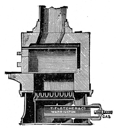
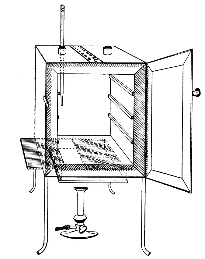
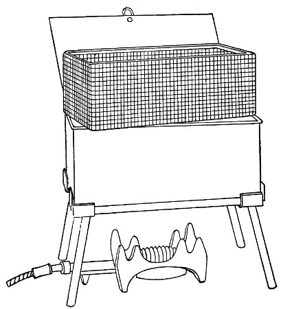
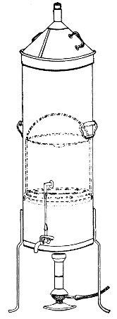
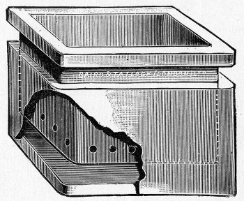
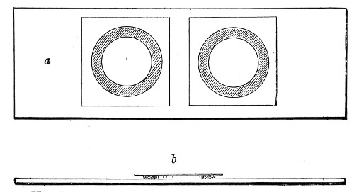
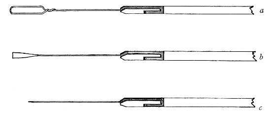
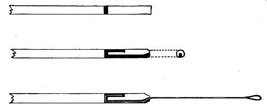
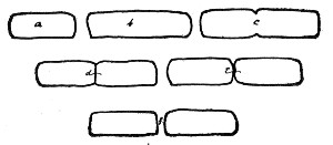
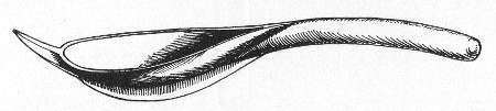
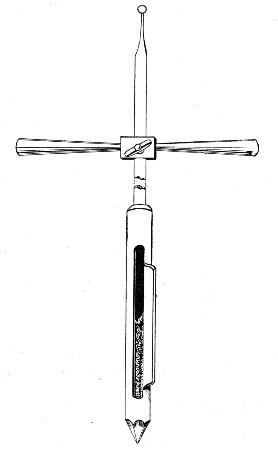
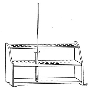
In order to identify an organism after isolation, tube, plate, and other
cultivations must be prepared, incubated under suitable conditions as to
temperature and environment, and examined from time to time (a)
macroscopically, (b) by microscopical methods, (c) by chemical methods,
(d) by physical methods, (e) by inoculation methods, and the results of
these examinations duly recorded.
It must be stated definitely that no micro-organism can be identified by
any one character or property, whether microscopical, biological or
chemical, but that on the contrary its entire life history must be
carefully studied and then its identity established from a consideration
of the sum total of these observations.
In order to give to the recorded results their maximum value it is
essential that they should be exact and systematic, therefore some such
scheme as the following should be adhered to; and especially is this
necessary in describing an organism not previously isolated and studied.
Designation:
Originally isolated by (observer's name) in (date), from (source of
organism).
1. Cultural Characters.—(Vide Macroscopical Examination
of Cultivation, page 261.)
| Gelatine plates, | } |
| Gelatine streak, | } at 20°C. |
| Gelatine stab, | } |
| Gelatine shake, | } |
| |
|
| Agar plates, | } |
| Agar streak or smear, | } |
| Agar stab, | } |
| Inspissated blood-serum, | } at 20° C. and 37°C. |
| Bouillon, | } |
| Litmus milk, | } |
| Potato, | } |
Special media for the purpose of demonstrating
characteristic appearances.
2. Morphology.—(Vide Microscopical Examination of
Cultivations, page 272.)
Vegetative forms:
Shape.
Size.
Motility.
Flagella (if present).
Capsule (if present).
Involution forms.
Pleomorphism (if observed).
Sporing forms (if observed). Of which class?
Staining reactions.
3. Chemical Products of Growth.—(Vide Chemical
Examination of Cultivations, page 276.)
Chromogenesis.
Photogenesis.
Enzyme formation.
Fermentation of carbohydrates:
Acid formation.
Alkali formation.
Indol formation.
Phenol formation.
Reducing and oxidising substances.
Gas formation.
4. Biology.—(Vide Physical Examination of Cultures, page
295.)
Atmosphere.
Temperature.
Reaction of nutrient media.
Resistance to lethal agents:
Physical:
Desiccation.
Light.
Colours.
Chemical germicides.
Vitality.
5. Pathogenicity:
Susceptible animals, subsequently arranged in order of susceptibility.
Immune animals.
Experimental inoculation, symptoms of disease.
Post-mortem appearances.
Virulence:
Length of time maintained.
Optimum medium?
Minimal lethal dose.
Exaltation and attenuation of virulence?
Toxin formation.
In describing the naked-eye and low-power appearances of the bacterial
growth the descriptive terms introduced by Chester (and included in the
following scheme) should be employed.
Solid Media.
Plate Cultures.—
Gelatine.—Note the presence or absence of liquefaction of the
surrounding medium. If liquefaction is present, note shape and character
(vide page 269, "stab" cultures).
Agar.—No liquefaction takes place in this medium. The liquid found on
the surface of the agar (or at the bottom of the tube in agar tube
cultures) is merely water which has been expressed during the rapid
solidification of the medium and has subsequently condensed.
Gelatine and Agar.—Examine the colonies at intervals of twenty-four
hours.
(a) With the naked eye.
(b) With a hand lens or watchmaker's glass.
(c) Under a low power (1 inch) of the microscope, or by means of a small
dissecting microscope.
Distinguish superficial from deep colonies and note the characters of
the individual colonies.
(A) Size.—The diameter in millimetres, at the various ages.
(B) Shape.—
Punctiform: Dimensions too slight for defining form by naked eye;
minute, raised, hemispherical.
Round: Of a more or less circular outline.
Elliptical: Of a more or less oval outline.
Irregular: Outlines not conforming to any recognised shape.
Fusiform: Spindle-shaped, tapering at each end.
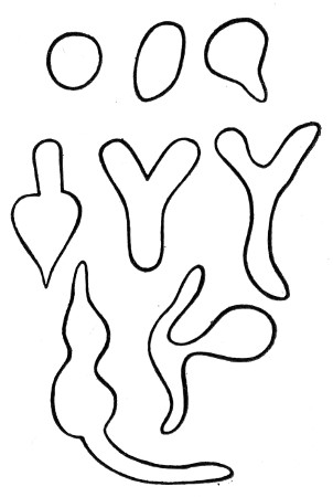
Cochleate: Spiral or twisted like a snail shell (Fig. 141, a).
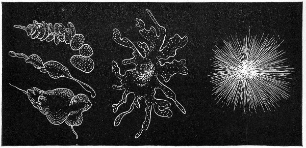 Fig. 141.—Types of colonies: a, Cochleate; b,
amœboid; c, mycelioid.
Fig. 141.—Types of colonies: a, Cochleate; b,
amœboid; c, mycelioid.
Amœboid: Very irregular, streaming (Fig. 141, b).
Mycelioid: A filamentous colony, with the radiate character of a mould
(Fig. 141, c).
Filamentous: An irregular mass of loosely woven filaments (Fig. 142,
a).
Floccose: Of a dense woolly structure.
Rhizoid: Of an irregular, branched, root-like character (Fig. 142, b).
Conglomerate: An aggregate of colonies of similar size and form (Fig.
142, c).
Toruloid: An aggregate of colonies, like the budding of the yeast plant
(Fig. 142, d).
Rosulate: Shaped like a rosette.
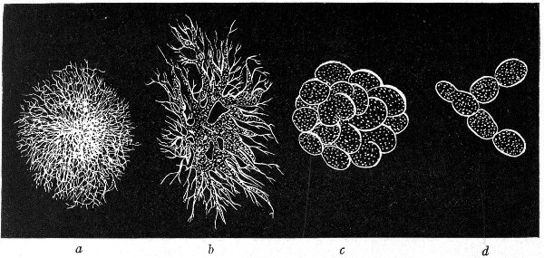 Fig. 142.—Types of colonies: a, Filamentous; b,
rhizoid; c, conglomerate; d, toruloid.
Fig. 142.—Types of colonies: a, Filamentous; b,
rhizoid; c, conglomerate; d, toruloid.
(C) Surface Elevation.—
1. General Character of Surface as a Whole:
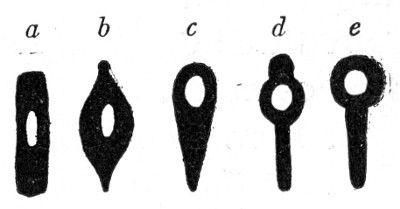
Flat: Thin, leafy, spreading over the surface (Fig. 143, a).
Effused: Spread over the surface as a thin, veily layer, more delicate
than the preceding.
Raised: Growth thick, with abrupt terraced edges (Fig. 143, b).
Convex: Surface the segment of a circle, but very flatly convex (Fig.
143, c).
Pulvinate: Surface the segment of a circle, but decidedly convex (Fig.
143, d).
Capitate: Surface hemispherical (Fig. 143, e).
Umbilicate: Having a central pit or depression (Fig. 143, f).
Conical: Cone with rounded apex (Fig. 143, g).
Umbonate: Having a central convex nipple-like elevation (Fig. 143, h).
2. Detailed Characters of Surface:
Smooth: Surface even, without any of the following distinctive
characters.
Alveolate: Marked by depressions separated by thin walls so as to
resemble a honeycomb (Fig. 144).
Punctate: Dotted with punctures like pin-pricks.
Bullate: Like a blistered surface, rising in convex prominences, rather
coarse.
Vesicular: More or less covered with minute vesicles due to gas
formation; more minute than bullate.
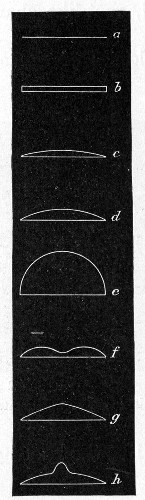 Fig. 143.—Surface elevation of colonies: a, Flat; b,
raised; c, convex; d, pulvinate; e, capitate; f, umbilicate;
g, conical; h, umbonate.
Fig. 143.—Surface elevation of colonies: a, Flat; b,
raised; c, convex; d, pulvinate; e, capitate; f, umbilicate;
g, conical; h, umbonate.
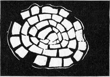 Fig. 144.—Types of colonies—alveolate.
Verrucose: Wart-like, bearing wart-like prominences.
Squamose: Scaly, covered with scales.
Echinate: Beset with pointed prominences.
Papillate: Beset with nipple or mamma-like processes.
Rugose: Short irregular folds, due to shrinkage of surface growth.
Corrugated: In long folds, due to shrinkage.
Contoured: An irregular but smoothly undulating surface, resembling the
surface of a relief map.
Rimose: Abounding in chinks, clefts, or cracks.
(D) Internal Structure of Colony (Microscopical).—
Refraction Weak: Outline and surface of relief not strongly defined.
Refraction Strong: Outline and surface of relief strongly defined;
dense, not filamentous colonies.
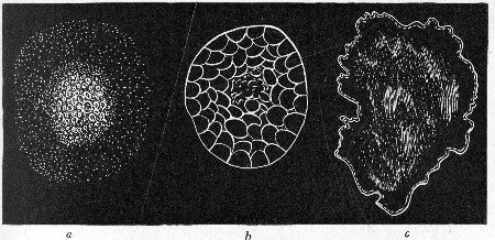 Fig. 145.—Types of colonies: a, Grumose; b,
moruloid; c, clouded.
Fig. 145.—Types of colonies: a, Grumose; b,
moruloid; c, clouded.
1. General:
Amorphous: Without any definite structure, such as is specified below.
Hyaline: Clear and colourless.
Homogeneous: Structure uniform throughout all parts of the colony.
Homochromous: Colour uniform throughout.
2. Granulations or Blotchings:
Finely granular.
Coarsely granular.
Grumose: Coarser than the preceding, with a clotted appearance, and
particles in clustered grains (Fig. 145, a).
Moruloid: Having the character of a mulberry, segmented, by which the
colony is divided in more or less regular segments (Fig. 145, b).
Clouded: Having a pale ground, with ill-defined patches of a deeper tint
(Fig. 145, c).
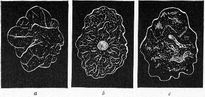 Fig. 146.—Types of colonies: a, Reticulate; b,
gyrose; c, marmorated.
Fig. 146.—Types of colonies: a, Reticulate; b,
gyrose; c, marmorated.
3. Colony Marking or Striping:
Reticulate: In the form of a network, like the veins of a leaf (Fig.
146, a).
Areolate: Divided into rather irregular, or angular, spaces by more or
less definite boundaries.
Gyrose: Marked by wavy lines, indefinitely placed (Fig. 146, b).
Marmorated: Showing faint, irregular stripes, or traversed by vein-like
markings, as in marble (Fig. 146, c).
Rivulose: Marked by lines like the rivers of a map.
Rimose: Showing chinks, cracks, or clefts.
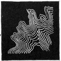 Fig. 147.—Types of colonies—curled.
4. Filamentous Colonies:
Filamentous: As already defined.
Floccose: Composed of filaments, densely placed.
Curled: Filaments in parallel strands, like locks or ringlets (Fig.
147).
(E) Edges of Colonies.—
Entire: Without toothing or division (Fig. 148, a).
Undulate: Wavy (Fig. 148, b).
Repand: Like the border of an open umbrella (Fig. 148, c).
Erose: As if gnawed, irregularly toothed (Fig. 148, d).
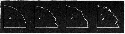 Fig. 148.—Edges of colonies: a, Entire; b, undulate;
c, repand; d, erose.
Fig. 148.—Edges of colonies: a, Entire; b, undulate;
c, repand; d, erose.
Lobate.
Lobulate: Minutely lobate (Fig. 149, e).
Auriculate: With ear-like lobes (Fig. 149, f).
Lacerate: Irregularly cleft, as if torn (Fig. 149, g).
Fimbriate: Fringed (Fig. 149, h).
Ciliate: Hair-like extensions, radiately placed (Fig. 149, j).
Tufted.
Filamentous: As already defined.
Curled: As already defined.
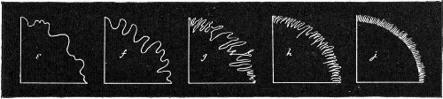 Fig. 149.—Edges of colonies: e, Lobar-lobulate; f,
auriculate; g, lacerate; h, fimbriate; i, ciliate.
Fig. 149.—Edges of colonies: e, Lobar-lobulate; f,
auriculate; g, lacerate; h, fimbriate; i, ciliate.
(F) Optical Characters (after Shuttleworth).—
1. General Characters:
Transparent: Transmitting light.
Vitreous: Transparent and colourless.
Oleaginous: Transparent and yellow; olive to linseed-oil coloured.
Resinous: Transparent and brown, varnish or resin-coloured.
Translucent: Faintly transparent.
Porcelaneous: Translucent and white.
Opalescent: Translucent; greyish-white by reflected light.
Nacreous: Translucent, greyish-white, with pearly lustre.
Sebaceous: Translucent, yellowish or greyish-white.
Butyrous: Translucent and yellow.
Ceraceous: Translucent and wax-coloured.
Opaque.
Cretaceous: Opaque and white, chalky.
Dull: Without lustre.
Glistening: Shining.
Fluorescent.
Iridescent.
2. Chromogenicity:
Colour of pigment.
Pigment restricted to colonies.
Pigment restricted to medium surrounding colonies.
Pigment present in colonies and in medium.
Streak or Smear Cultures.—
Gelatine and Agar.—Note general points as indicated under plate
cultivations.
Inspissated Blood-serum.—Note the presence or absence of liquefaction
of the medium. (The presence of condensation water at the bottom of the
tube must not be confounded with liquefaction of the medium.)
All Oblique Tube Cultures.—
1. Colonies Discrete: Size, shape, etc., as for plate cultivations
(vide page 261).
2. Colonies Confluent: Surface elevation and character of edge, as for
plate cultivations (vide page 263).
Chromogenicity: As for plate cultures.
Gelatine Stab Cultures.—
(A) Surface Growth.—As for individual colonies in plate cultures
(vide page 261).
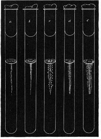 Fig. 150.—Stab cultivations—types of growth: a,
Filiform; b, beaded; c, echinate; d, villous; e, arborescent.
Fig. 150.—Stab cultivations—types of growth: a,
Filiform; b, beaded; c, echinate; d, villous; e, arborescent.
(B) Line of Puncture.—
Filiform: Uniform growth, without special characters (Fig. 150, a).
Nodose: Consisting of closely aggregated colonies.
Beaded: Consisting of loosely placed or disjointed colonies (Fig. 150,
b).
Papillate: Beset with papillate extensions.
Echinate: Beset with acicular extensions (Fig. 150, c).
Villous: Beset with short, undivided, hair-like extensions (Fig. 150,
d).
Plumose: A delicate feathery growth.
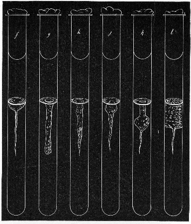 Fig. 151.—Stab cultivations—types of growth: f,
Crateriform; g, saccate; h, infundibuliform; j, napiform; k,
fusiform; l, stratiform.
Fig. 151.—Stab cultivations—types of growth: f,
Crateriform; g, saccate; h, infundibuliform; j, napiform; k,
fusiform; l, stratiform.
Arborescent: Branched or tree-like, beset with branched hair-like
extensions (Fig. 150, e).
(C) Area of Liquefaction (if present).—
Crateriform: A saucer-shaped liquefaction of the gelatine (Fig. 151,
f).
Saccate: Shape of an elongated sack, tubular cylindrical (Fig. 151,
g).
Infundibuliform: Shape of a funnel, conical (Fig. 151, h).
Napiform: Shape of a turnip (Fig. 151, j).
Fusiform: Outline of a parsnip, narrow at either end, broadest below the
surface (Fig. 151, k).
Stratiform: Liquefaction extending to the walls of the tube and downward
horizontally (Fig. 151, l).
(D) Character of the Liquefied Gelatine.—
1. Pellicle on surface.
2. Uniformly turbid.
3. Granular.
4. Mainly clear, but containing flocculi.
5. Deposit at apex of liquefied portion.
(E) Production of Gas Bubbles.
Shake Cultures.—
1. Presence or absence of liquefaction.
2. Production of gas bubbles.
3. Bulk of growth at the surface—aerobic.
4. Bulk of growth in depths—anaerobic.
Fluid Media.
1. Surface of the Liquid.—
Presence or absence of froth due to gas bubbles.
Presence or absence of pellicle formation.
Character of pellicle.
2. Body of the Liquid.—
Uniformly turbid.
Flocculi in suspension.
Granules in suspension.
Clear, with precipitate at bottom of tube.
Colouration of fluid, presence or absence of.
3. Precipitate.—
Character.
Amount.
Colour.
Carbohydrate Media.—
Growth.
Reaction.
Gas formation.
Coagulation or not of serum albumen (when serum water media are
employed).
Litmus Milk Cultivations.—
| {Unaltered. |
| 1. Reaction: | {Acid. |
| {Alkaline. |
| 2. Odour. |
| 3. Formation of gas. |
| {Unaltered. |
| 4. Consistency: | {Peptonised (character of solution). |
| {Coagulated. |
| {hard: solid. |
| 5. Clot: Character | {soft: floculent. |
| {ragged and broken up by gas bubbles. |
(a) Coagulum undissolved.
(b) Coagulum finally peptonised, completely: incompletely.
Resulting solution, clear: turbid.
| {Abundant. |
| {Scanty. |
| 6. Whey: | {Clear. |
| {Turbid. |
| {Coagulated by boiling, or not. |
As a council of perfection preparations must be made from pure
cultivations 4, 6, 8, 12, 18, and 24 hours; and subsequently at
intervals of, say, twenty-four hours, during the entire period they are
under observation, and examined—
(A) Living.—1. In hanging drop, to determine motility or
non-motility.
In this connection it must be remembered that under certain conditions
as to environment (e. g., when examined in an unsuitable medium,
atmosphere, temperature, etc.) motile bacilli may fail to exhibit
activity. No organism, therefore, should be recorded as non-motile from
one observation only; a series of observations at different ages and
under varying conditions should form the basis of an opinion as to the
absence of true locomotion.
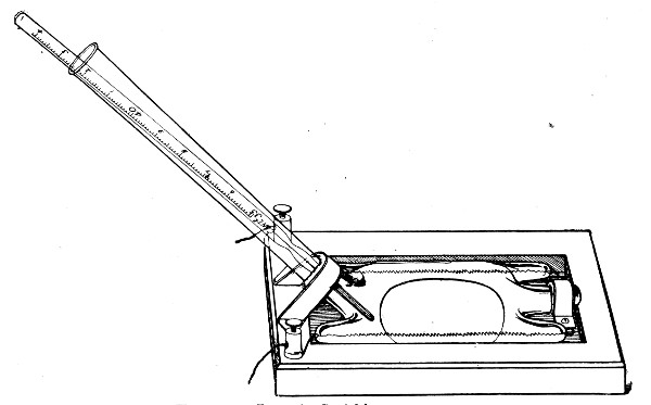
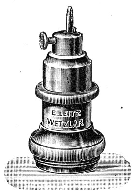
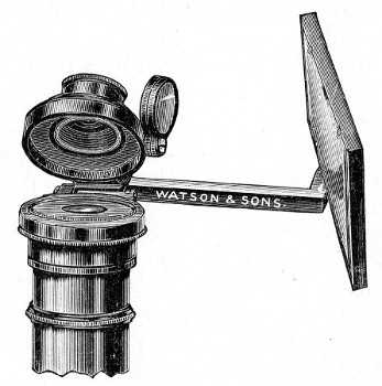
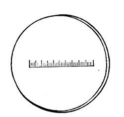
Size.—In the case of non-motile or sluggishly motile organisms,
endeavour to measure several individuals in each hanging drop by means
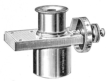
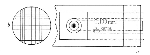
of the eyepiece micrometer or the eikonometer (vide page 63), and
average the results.
If the organism is one which forms spores, observe—
(a) Spore Formation.—Prepare hanging-drop cultivations (vide page
78) from vegetative forms of the organism, adding a trace of magenta
solution (0.5 per cent.) or other intra vitam stain (see page 77) to the
drop, on the point of the platinum needle, to facilitate the observation
of the phenomenon by rendering the bacilli more distinct.
Place the preparation on the stage of the microscope; if necessary,
using a warm stage.
Arrange illumination, etc., and select a solitary bacillus for
observation, by the help of the 1/6-inch lens.
Substitute the 1/12-inch oil-immersion lens for the sixth, and observe
the formation of the spore; if possible, measure any alteration in size
which may occur by means of the Ramsden micrometer.
(b) Spore Germination.—Prepare hanging-drop cultivations from old
cultivations in which no living vegetative forms are present, and
observe the process of germination in a similar manner.
The comfort of the microscopist is largely enhanced in those cases where
the period of observation is at all lengthy, by use of some form of eye
screen before the unemployed eye, such as is figured on page 58 (Fig.
49).
If it is impossible to carry out the method suggested above, proceed as
follows:
(a) Spore Formation.—Plant the organism in broth and incubate under
optimum conditions.
At regular intervals, say every thirty minutes, remove a loopful of the
cultivation and prepare a cover-slip film preparation.
Fix, while still wet, in the corrosive sublimate fixing solution.
Stain with aniline gentian violet, and partially decolourise with 2 per
cent. acetic acid.
Mount and number consecutively; then examine.
(b) Spore Germination.—Expose a thick emulsion of the spores to a
temperature of 80° C. for ten minutes in the differential steriliser
(vide page 257).
Transfer the emulsion to a tube of sterile nutrient broth and incubate.
Remove specimens from the tube culture at intervals of, say, five
minutes.
Fix, stain, etc., wet, as under (a), and examine.
(B) Fixed.—2. In stained preparations.
(a) To determine points in morphology:
Shape (vide classification, page 131).
Size:
(a) Prepare cover-slip film preparations at the various ages, and fix
by exposure to a temperature of 115° C. for twenty minutes in hot-air
oven.
(b) Stain the preparations by Gram's method (if applicable) or with
dilute carbol-fuchsin, and mount in the usual way.
(c) Measure (vide page 66) some twenty-five individuals in each film
by means of the Ramsden's or the stage micrometer and average the
result.
Pleomorphism; If noted, record—
The predominant character of the variant forms.
On what medium or media they are observed.
At what period of development.
(b) To demonstrate details of structure:
Flagella: If noted, record—
Method of staining (vide page 101).
Position and arrangement (vide page 136).
Number.
Spores: If noted, record—
Method of staining.
Shape.
Size.
Position within the parent cell.
Condition, as to shape, of the parent cell (vide page 139).
Optimum medium and temperature.
Age of cultivation.
Conditions of environment as to temperature, atmosphere.
Method of germination (vide page 140).
Involution Forms: If noted, record—
Method of staining.
Character (e. g., if living or dead).
Shape.
On what medium they are observed.
Age of medium.
Environment.
Metachromatic Granules: If noted, record—
Method of staining.
Character of granules.
Number of granules.
Colour of granules.
3. Staining Reactions.—
1. Gram's Method.—Positive or negative.
2. Neisser's Method.—If granules are noted, record—
3. Ziehl-Neelsen's Method.—Acid-fast or decolourised.
4. Simple Aniline Dyes.—(Noting those giving the best results, with
details of staining processes.)
| Methylene-blue | } |
| Fuchsin | } and their modifications. |
| Gentian violet | } |
| Thionine blue | } |
Test cultivations of the organism for the presence of—
Soluble enzymes—proteolytic, diastatic, invertase.
Organic acids—(a) quantitatively—i. e., estimate the total acid
production; (b) qualitatively for formic, acetic, propionic, butyric,
lactic.
Ammonia.
Neutral volatile substances—ethyl alcohol, aldehyde, acetone.
Aromatic products—indol, phenol.
Soluble pigments.
Test the power of reducing (a) colouring matters, (b) nitrates to
nitrites.
Investigate the gas production—H2S, CO2, H2. Estimate the
ratio between the last two gases.
Prepare all cultivations for these methods of examination under
optimum conditions, previously determined for each of the organisms it
is intended to investigate, as to
(a) Reaction of medium;
(b) Incubation temperature;
(c) Atmospheric environment;
and keep careful records of these points, and also of the age of the
cultivation used in the final examination.
Examine the cultivations for the various products of bacterial
metabolism after forty-eight hours' growth, and never omit to examine
"control" (uninoculated) tube or flask of medium from the same batch,
kept for a similar period under identical conditions.
If the results are negative, test further cultivations at three days,
five days, and ten days.
1. Enzyme Production.—
(A) Proteolytic Enzymes.—(Convert proteins into proteose, peptone
and further products of hydrolysis; e. g., B. pyocyaneus.)
Media Required:
Blood-serum and milk-serum which have been carefully
filtered through a porcelain candle.
Reagents Required:
Ammonium sulphate.
Thirty per cent. caustic soda solution.
Copper sulphate, 0.5 per cent. aqueous solution.
One per cent. acetic acid solution.
Millon's reagent.
Glyoxylic acid solution.
Concentrated sulphuric acid.
Method.—
1. Prepare cultivations in bulk (50 c.c.) in a flask and incubate.
2. Make the liquid faintly acid with acetic acid, then boil. (This
precipitates the unaltered proteins.)
3. Filter.
4. Take 10 c.c. of the filtrate in a test-tube and add 1 c.c. of the
caustic soda, then add the copper sulphate drop by drop.
Pink colour which becomes violet with more copper sulphate =
proteose and peptone.
5. Saturate the rest of the filtrate with ammonium sulphate.
Precipitate = proteose.
6. Filter and divide the filtrate into three parts a, b and c.
a. Repeat the copper sulphate test, using excess of caustic soda to
displace the ammonia from the ammonium sulphate.
Pink colour = peptone.
b. Boil with Millon's reagent.
Red colour = tyrosine.
c. Add glyoxylic acid solution and run in concentrated sulphuric acid.
Violet ring at upper level of acid = tryptophane.
Both the tyrosine and tryptophane may be either in the free state or in
combination as polypeptid or peptone.
(B) Diastase.—(Converts starch into sugar; e. g., B. subtilis.)
Medium Required:
Inosite-free bouillon.
Reagents Required:
Starch.
Thymol.
Fehling's solution.
Method.—
1. Prepare tube cultivation and incubate.
2. Prepare a thin starch paste and add 2 per cent. thymol to it.
3. Mix equal parts of the cultivation to be tested and the starch paste,
and place in the incubator at 37°C. for six to eight hours.
4. Filter.
Test the filtrate for sugar.
Boil some of the Fehling's solution in a test-tube.
Add the filtrate drop by drop until, if necessary, a quantity has been
added equal in amount to the Fehling's solution employed, keeping the
mixture at the boiling-point during the process.
Yellow or orange precipitate = sugar.
(C) Invertase.—(Convert saccharose into a mixture of dextrose and
lævulose e. g., B. fluorescens liquefaciens.)
Medium Required:
Inosite-free bouillon.
Reagents Required:
Cane sugar, 2 per cent. aqueous solution.
Carbolic acid.
Method.—
1. Prepare tube cultivations and incubate.
2. Add 2 per cent. of carbolic acid to the sugar solution.
3. Mix equal quantities of the carbolised sugar solution and the
cultivation in a test-tube; allow the mixture to stand for several
hours.
4. Filter.
Test the filtrate for reducing sugar as in the preceding section.
(D) Rennin and "Lab" Enzymes.—(Coagulate milk independently of the
action of acids; e. g., B. prodigiosus.)
Media Required:
Inosite-free bouillon.
Litmus milk.
Method.—
1. Prepare tube cultivations and incubate.
2. After incubation heat the cultivation to 55° C. for half an hour, to
sterilise.
3. By means of a sterile pipette run 5 c.c. of the cultivation into each
of three tubes of litmus milk.
4. Place in the cold incubator at 22° C. and examine each day for ten
days.
Absence of coagulation at the end of that period will indicate absence
of rennin ferment formation.
Fermentation Reactions.
As tested upon carbohydrate substances and organic salts.
Media Required:
Peptone water containing various percentages (generally 2 per cent.) of
each of the substances referred to under "sugar" media (page 177), also
tubes of peptone water containing 1 per cent. respectively of each of
the following:
Organic salts: Sodium citrate, formate, lactate, malate,
tartrate.
Method.—
1. Prepare tube cultivations in each of the above media.
2. Observe from day to day up to the expiration of ten days if
necessary.
3. Note growth, reaction, gas production.
2. Acid Production.
(a) Quantitative.—
Medium Required:
Sugar (glucose) bouillon of known "optimum" reaction.
Apparatus and Reagents Required:
As for estimating reaction of media (vide page 150).
Method.—
1. Prepare cultivation in bulk (100 c.c.) in a flask; also "control"
flask of medium from same batch.
2. After suitable incubation, heat both flasks in the steamer at 100° C.
for thirty minutes to sterilise.
3. Determine the titre of the medium in "inoculated" and "control"
flasks as described in the preparation of nutrient media (vide page
151).
4. The difference between the titre of the medium in the two flasks
gives the total acid production of the bacterium under observation in
terms of normal NaOH.
Note.—If the growth is very heavy it may be a difficult
matter to determine the end-point. The cultivation should
then be filtered through a Berkefeld filter candle previous
to step 2, and the filtrate employed in the titration.
(b) Qualitative (of all the organic acids present).—
Medium Required:
Sugar (glucose or lactose) bouillon as in quantitative examination.
Reagents Required:
Hydrochloric acid, concentrated.
Hydrochloric acid, 25 per cent.
Sulphuric acid, concentrated (pure).
Phosphoric acid, concentrated solution.
Ammonia.
Ammonium sulphate.
Baryta water.
Sodium carbonate, saturated aqueous solution.
Absolute alcohol.
Ether.
Calcium chloride.
Calcium chloride solution.
Zinc carbonate.
Copper sulphate saturated aqueous solution.
Alcoholic thiophene solution (0.15 c.c. in 100 c.c.).
Animal charcoal.
Five per cent. sodium nitroprusside solution.
Potassium bichromate.
Schiff's reagent.
Arsenious oxide.
Ferric chloride, 4 per cent. aqueous solution.
Silver nitrate, 1 per cent. aqueous solution.
Lugol's iodine.
Ten per cent. caustic soda solution.
Hard paraffin wax (melting-point about 52° C.).
Method.—
1. Prepare cultivation in bulk (500 c.c.) in a litre flask and add
sterilised precipitated chalk, 10 grammes. Incubate at the optimum
temperature.
2. After incubation throw a piece of paraffin wax (about a centimetre
cube) into the cultivation and connect up the flask with a condenser.
The paraffin, which liquefies and forms a thin layer on the surface of
the fluid, is necessary to prevent the cultivation frothing up and
running unaltered through the condenser during the subsequent process of
distillation.
3. Distill over 200 to 300 c.c.
Use a rose-top burner to minimise the danger of cracking the flask; and
to the same end, well agitate the contents of the flask to prevent the
chalk settling.
The distillate "A" will contain alcohol, etc. (vide page 285); the
residue "a" will contain the volatile and fixed acids.
4. Disconnect the flask and filter. The residue "a" then = filtrate B
and residue b.
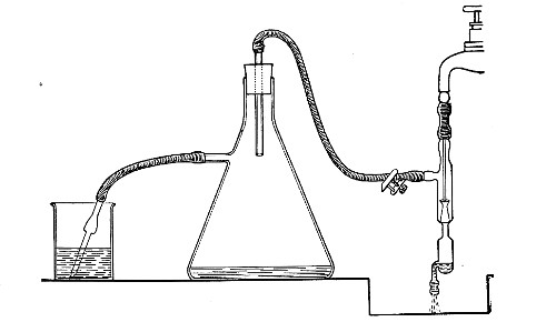
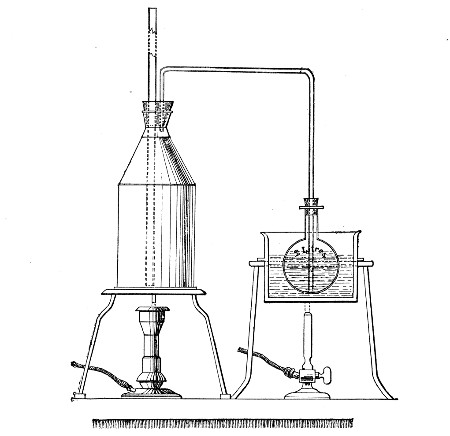
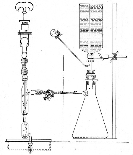
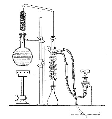 Fig. 152.—Arrangement of distillation apparatus for
acids, etc.
Fig. 152.—Arrangement of distillation apparatus for
acids, etc.
5. Residue b. Wash the residue from the filter paper, dissolve by
heating with dilute hydrochloric acid, and add calcium chloride solution
and ammonia until alkaline.
White precipitate insoluble in acetic acid = oxalic acid.
6. Make up filtrate B to 500 c.c. with distilled water and divide into
two parts.
7. Acidify 250 c.c. with 20 c.c. concentrated phosphoric acid (this
liberates the volatile acids) and distil to small bulk.
The distillate "B" may contain formic, acetic, propionic, butyric and
benzoic acids.
                              DISTILLATE "B."
                              (Volatile Acids.)
                                      ¦
                                      ¦
                   1. Add baryta water till alkaline,
                      and evaporate to dryness.

                   2. Add 50 c.c. absolute alcohol and allow
                      to stand, with frequent stirring, for
                      two to three hours.

                   3. Filter and wash with alcohol.
                                      ¦
                                      ¦
                  ¦---------------------------------------¦
                  ¦                                       ¦
                  ¦                                       ¦
               FILTRATE                                RESIDUE
                  ¦                                       ¦
                  ¦                                       ¦
    may contain barium propionate,         may contain barium acetate,
    barium butyrate.                       barium formate, barium benzoate.
                  ¦                                       ¦
                  ¦                                       ¦
    1. Evaporate to dryness.               1. Evaporate off alcohol and
                                           dissolve up the residue on
    2. Dissolve residue in 150             the filter in hot water and
    c.c. water.                            neutralise.

    3. Acidify with phosphoric             2. Divide the solution into
    acid and distil.                       four portions:

    4. Saturate distillate with            (a) Add ferric chloride solution.
    calcium chloride and distill
    over a few c.c.                            Brown colour = acetic or
                                               formic acids.
    5. Test distillate for butyric
    acid:                                      Buff ppt. = benzoic acid
                                               (see ether soluble acids).
    Add 3 c.c. alcohol and 4 drops
    concentrated sulphuric acid.           (b) Add silver nitrate
                                           solution; then add one drop
        Smell of pineapple = butyric       ammonia water, and boil.
        acid.
                                              Black precipitate of metallic
        Propionic acid in small                silver = formic acid.
        quantities cannot be
        distinguished from butyric         (c) Evaporate to dryness; mix
        acid by tests within the           with equal quantity of
        scope of the bacteriological       arsenious oxide and heat
        laboratory.                        on platinum foil.

                                               Unpleasant smell of cacodyl
                                               = acetic acid.

                                           (d) Add a few drops of
                                           mercuric chloride solution
                                           in test-tube, and heat to
                                           70° C.

                                               Precipitate of mercurous
                                               chloride which is slowly
                                               reduced to mercury =
                                               formic acid.
8. If the distillation of "B" is continued as long as acid comes over
(distilled water being occasionally added to the distilling flask) the
distillate can be measured and 50 c.c. used for titration. This will
give the amount of volatile acid formation.
9. The second part of the filtrate "B" (see page 282) should be examined
for lactic, oxalic, succinic, benzoic, salicylic, gallic and tannic
acids, as follows:
Ether Soluble Acids.—
1. Evaporate to a thin syrup, acidify strongly with phosphoric acid.
2. Extract with five times its volume of ether by agitation in a
separatory funnel.
3. Evaporate the ethereal extract to a thin syrup.
4. Add 100 c.c. water and mix thoroughly.
5. To a small portion of this solution add slight excess of sodium
carbonate, evaporate to dryness on the water-bath, dissolve in 5-10 c.c.
pure sulphuric acid, add 2 drops saturated copper sulphate solution,
place in a test-tube and heat in a boiling water-bath for 2 minutes,
cool, add 2 or 3 drops of the alcoholic thiophene and warm gently.
Cherry red colour = lactic acid.
If a brown colour is produced on the addition of sulphuric acid, another
sample should be taken and boiled with animal charcoal before
evaporating.
6. If lactic acid is definitely present, prepare zinc lactate by boiling
part of the solution of the ether extract with excess of zinc carbonate,
filtering and evaporating to crystallise. The crystals so obtained have
a characteristic form, and if dried at 110° C, should contain 26.87 per
cent. of zinc.
7. Test a portion of the rest of the solution of the ether extract for
oxalic acid (page 282, step 5). Carefully neutralise the remainder and
add ferric chloride solution.
Red brown gelatinous precipitate = succinic acid.
Buff precipitate = benzoic acid, and other acids related to benzoic
acid.
Violet colour = salicylic acid.
Inky black colour or precipitate = gallic acid or tannic acid.
For further identification the melting-points of the crystalline acids,
and the percentage of silver in their silver salts should be determined.
3. Ammonia Production.—
Medium Required:
Nutrient bouillon.
Reagent Required:
Nessler reagent.
Method.—
1. Prepare cultivation in bulk (100 c.c.) in a 250 c.c. flask and
incubate together with a control flask.
Test the cultivation and the control for ammonia in the following
manner:
2. To each flask add 2 grammes of calcined magnesia, then connect up
with condensers and distil.
3. Collect 50 c.c. distillate, from each, in a Nessler glass.
4. Add 1 c.c. Nessler reagent to each glass by means of a clean pipette.
Yellow colour = ammonia.
The depth of colour is proportionate to the amount present.
4. Alcohol, etc., Production.—Divide the distillate "A" obtained in the
course of a previous experiment (vide page 282, step 3) into four
portions and test for the production of alcohol, acetaldehyde, acetone.
1. Add Lugol's iodine, then a little NaOH solution, and stir with a
glass rod till the colour of the iodine disappears.
Pale-yellow crystalline precipitate of iodoform, with its characteristic
smell, appearing in the cold, indicates acetaldehyde, or acetone;
appearing only on warming indicates alcohol.
The precipitate may be absent even when the odour is pronounced.
2. Add Schiff's reagent.
Violet or red colour = aldehyde.
3. To 10 c.c. of solution add 2.5 c.c., 25 per cent. sulphuric acid, and
a crystal or two of potassium bichromate and distil. Reduction of the
bichromate to a green colour and a distillate, which smells of
acetaldehyde and reacts with Schiff's reagent, shows the presence of
alcohol in the original liquid.
4. Add a few drops of sodium nitroprusside solution, make alkaline with
ammonia, then saturate with ammonium sulphate crystals. Acetone gives
little colour on the addition of ammonia, but after the addition of
ammonium sulphate a deep permanganate colour, which takes ten minutes to
reach its full intensity. Aldehyde gives a carmine red unaltered by
ammonium sulphate.
5. Indol Production.—
Media Required:
Inosite-free bouillon (vide page 183).
Or peptone water (vide page 177).
Reagents Required:
Potassium persulphate, saturated aqueous solution.
Paradimethylamino-benzaldehyde solution. This is prepared by mixing:
Paradimethylamino-benzaldehyde 4 grammes
Absolute alcohol 380 c.c.
Hydrochloric acid, concentrated 80 c.c.
Method.—
Prepare several test-tube cultivations of the organism to be tested, and
incubate.
Test for indol by means of the Rosindol reaction in the following
manner. (If the culture has been incubated at 37°C., it must be allowed
to cool to the room temperature before applying the test.)
1. Remove 2 c.c. of the cultivation by means of a sterile pipette and
transfer to a clean tube, then,
2. Add 2 c.c. paradimethylamino-benzaldehyde solution.
3. Add 2 c.c. potassium persulphate solution.
The presence of indol is indicated by the appearance of a delicate
rose-pink colour throughout the mixture which deepens slightly on
standing.
Indol is tested for in many laboratories by the ordinary
nitrosoindol reaction which, however, is not so delicate a
method as that above described. The test is carried out as
follows:
1. Remove the cotton-wool plug from the tube, and run in 1
c.c. pure concentrated sulphuric acid down the side of the
tube by means of a sterile pipette. Place the tube upright
in a rack, and allow it to stand, if necessary, for ten
minutes.
A rose-pink or red colour at the junction of the two liquids
= indol (plus a nitrite).
2. If the colour of the medium remains unaltered, add 2 c.c.
of a 0.01 per cent. aqueous solution sodium nitrite, and
again allow the culture to stand for ten minutes.
Red colouration = indol.
Note.—In place of performing the test in two stages as
given above, 2 c.c. concentrated commercial sulphuric,
hydrochloric, or nitric acid (all of which hold a trace of
nitrite in solution), may be run into the cultivation. The
development of a red colour within twenty minutes will
indicate the presence of indol.
5a. Phenol Production.—
Medium Required:
Nutrient bouillon.
Reagents Required:
Hydrochloric acid, concentrated.
Millon's reagent.
Ferric chloride, 1 per cent. aqueous solution.
Method.—
1. Prepare cultivation in a Bohemian flask containing at least 50 c.c.
of medium, and incubate.
Test for phenol in the following manner:
2. Add 5 c.c., 25 per cent. sulphuric acid to the cultivation and
connect up the flask with a condenser.
3. Distil over 15 to 20 c.c. Divide the distillate into three portions
a, b and c.
4. Add to (a) 0.5 c.c. Millon's reagent and boil.
Red colour = phenol.
5. Add to (b) about 0.5 c.c. ferric chloride solution. Violet colour =
phenol.
(If the distillate be acid the reaction will be negative.)
6. Add to (c) bromine water. Crystalline white ppt. of tribromo-phenol
= phenol.
Note.—If both indol and phenol appear to be present in
cultivations of the same organism, it is well to separate
them before testing. This may be done in the following
manner:
1. Prepare inosite-free bouillon cultivation, say 200 or 300 c.c., in a
flask as before.
2. Render definitely acid by the addition of acetic acid and connect up
the flask with a condenser.
3. Distil over 50 to 70 c.c.
Distillate will contain both indol and phenol.
4. Render the distillate strongly alkaline with caustic potash and
redistil.
Distillate will contain indol; residue will contain phenol.
5. Test the distillate for indol (vide ante).
6. Saturate the residue, when cold, with carbon dioxide and redistil.
7. Test this distillate for phenol (vide ante).
6. Pigment Production.—
1. Prepare tube cultivations upon the various media and incubate under
varying conditions as to temperature (at 37° C. and at 20°C.),
atmosphere (aerobic and anaerobic), and light (exposure to and
protection from).
Note the conditions most favorable to pigment formation.
2. Note the solubility of the pigment in various solvents, such as water
(hot and cold), alcohol, ether, chloroform, benzol, carbon bisulphide.
3. Note the effect of acids and alkalies respectively upon the pigmented
cultivation, or upon solutions of the pigment.
4. Note spectroscopic reactions.
7. Reducing Agent Formation.—
(a) Colour Destruction.—
1. Prepare tube cultivations in nutrient bouillon tinted with litmus,
rosolic acid, neutral red, and incubate.
2. Examine the cultures each day and note whether any colour change
occurs.
(b) Nitrates to Nitrites.—
Medium Required:
Nitrate bouillon (vide page 185).
Or nitrate peptone solution (vide page 186).
Reagents Required:
Sulphuric acid (25 per cent.).
Metaphenylene diamine, 5 per cent. aqueous solution.
Method.—
1. Prepare tube cultivations and incubate together with control tubes
(i. e., uninoculated tubes of the same medium, placed under identical
conditions as to environment).
This precaution is necessary as the medium is liable to take up nitrites
from the atmosphere, and an opinion as to the absence of nitrites in the
cultivation is often based upon an equal colouration of the medium in
the control tube.
Test both the culture tube and the control tube for the presence of
nitrites.
2. Add a few drops of sulphuric acid to the medium in each of the tubes.
3. Then run in 2 or 3 c.c. metaphenylene diamine into each tube.
Brownish-red colour = nitrites.
The depth of colour is proportionate to the amount present.
8. Gas Production.—
(A) Carbon Dioxide and Hydrogen.—
Apparatus Required:
Fermentation tubes (vide page 161) containing sugar
bouillon (glucose, lactose, etc.). The medium should be
prepared from inosite-free bouillon (vide page 183).
Reagent Required:
n/2 caustic soda.
Method.—
1. Inoculate the surface of the medium in the bulb of a fermentation
tube and incubate.
2. Mark the level of the fluid in the closed branch of the fermentation
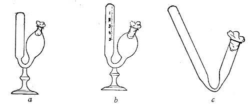
tube, at intervals of twenty-four hours, and when the evolution of gas
has ceased, measure the length of the column of gas with the millimetre
scale.
Express this column of gas as a percentage of the entire length of the
closed branch.
3. To analyse the gas and to determine roughly the relative proportions
of CO2 and H2, proceed as follows:
Fill the bulb of the fermentation tube with caustic soda solution.
Close the mouth of the bulb with a rubber stopper.
Alternately invert and revert the tube six or eight times, to bring the
soda solution into intimate contact with the gas.
Return the residual gas to the end of the closed branch, and measure.
The loss in volume of gas = carbon dioxide.
The residual gas = hydrogen.
Transfer gas to the bulb of the tube, and explode it by applying a
lighted taper.
(B) Sulphuretted Hydrogen.—
Media Required:
Iron peptone solution (vide page 185).
Lead peptone solution.
1. Inoculate tubes of media, and incubate together with control tubes.
2. Examine from day to day, at intervals of twenty-four hours.
The liberation of the H2S will cause the yellowish-white precipitate
to darken to a brownish-black, or jet black, the depth of the colour
being proportionate to the amount of sulphuretted hydrogen present.
Quantitative: For exact quantitative analyses of the gases produced by
bacteria from certain media of definite composition, the methods devised
by Pakes must be employed, as follows:
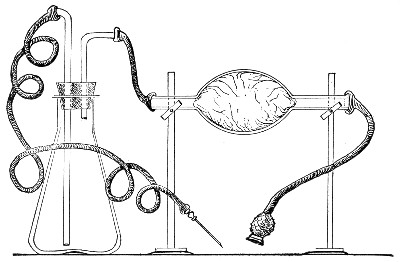
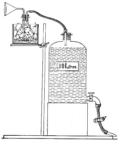
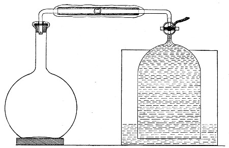 Fig. 153.—Gas-collecting apparatus.
Apparatus Required:
Bohemian flask (300 to 1500 c.c. capacity) containing from
100 to 400 c.c. of the medium. The mouth of the flask is
fitted with a perforated rubber stopper, carrying an
L-shaped piece of glass tubing (the short arm passing just
through the stopper). To the long arm of the tube is
attached a piece of pressure tubing some 8 cm. in length,
plugged at its free end with a piece of cotton-wool. Measure
accurately the total capacity of the flask and exit tube,
also the amount of medium contained. Note the difference.
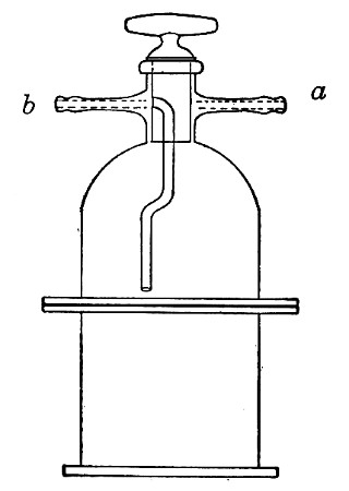
Gas receiver. This is a bell jar of stout glass, 14 cm. high
and 9 cm. in diameter. At its apex a glass tube is fused in.
This rises vertically 5 cm., and is then bent at right
angles, the horizontal arm being 10 cm. in length. A
three-way tap is let horizontally into the vertical tube
just above its junction with the bell jar.
An iron cylinder just large enough to contain the bell jar.
About 15 kilos of metallic mercury.
Melted paraffin.
An Orsat-Lunge working with mercury instead of water, provided with two
gas tubes of extra length (capacity 120 and 60 c.c. respectively and
graduated throughout, both being water-jacketed) or other gas analysis
apparatus, capable of dealing with CO2, O2, H2, and N2.
Method.—
1. Inoculate the medium in the flask in the usual manner, by means of a
platinum needle, taking care that the neck of the flask and the rubber
stopper are thoroughly flamed before and after the operation.
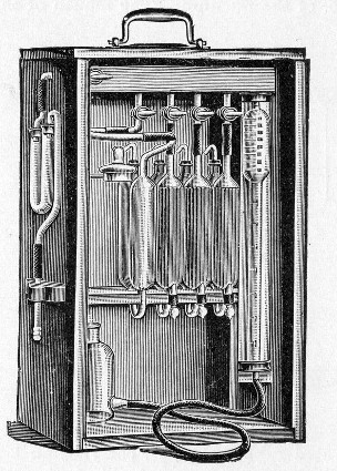 Fig. 154.—Orsat-Lunge gas analysis apparatus.
2. Fill the iron cylinder with mercury.
3. Place the bell jar mouth downward in the mercury—first seeing that
there is free communication between the interior of the jar and the
external air—and suck up the mercury into the tap; then shut off the
tap.
4. Plug the open end of the three-way tap with melted wax.
5. Connect up the horizontal arm of the culture flask with that of the
gas receiver by means of the pressure tubing (after removing the
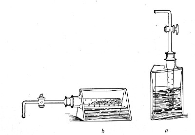
cotton-wool plug from the rubber tube), as shown in Fig. 153.
6. Give the three-way tap half turn to open communication between flask
and receiver, and seal all joints by coating with a film of melted
wax. When the tap is turned, the mercury in the receiver will naturally
fall.
7. Place the entire apparatus in the incubator. (Two hours later, by
which time the temperature of the apparatus is that of the incubator,
mark the height of the mercury on the receiver.)
8. Examine the apparatus from day to day and mark the level of the
mercury in the receiver at intervals of twenty-four hours.
9. When the evolution of gas has ceased, remove the apparatus from the
incubator; clear out the wax from the nozzle of the three-way tap (first
adjusting the tap so that no escape of gas shall take place) and connect
it with the Orsat.
10. Remove, say, 100 c.c. of gas from the receiver, reverse the tap and
force it into the culture flask. Remove 100 c.c. of mixed gases from the
culture flask and replace in the receiver.
Repeat these processes three or four times to ensure thorough admixture
of the contents of flask and receiver.
11. Now withdraw a sample of the mixed gases into the Orsat and analyse.
In calculating the results be careful to allow for the volume of air
contained in the flask at the commencement of the experiment.
For the collection of gases formed under anaerobic conditions a slightly
different procedure is adopted:
1. Fix a culture flask (500 c.c. capacity) with a perforated rubber
stopper carrying an L-shaped piece of manometer tubing, each arm 5 cm.
in length.
2. Prepare a second L-shaped piece of tubing, the short arm 5 cm. and
the long arm 20 cm., and connect its short arm to the horizontal arm of
the tube in the culture flask by means of a length of pressure tubing,
provided with a screw clamp.
3. Fill the culture flask completely with boiling medium and pass the
long piece of tubing through the plug of an Erlenmeyer flask (150 c.c.
capacity) which contains 100 c.c. of the same medium.
4. Sterilise these coupled flasks by the discontinuous method, in the
usual manner.
Immediately the last sterilisation is completed, screw up the clamp on
the pressure tubing which connects them, and allow them to cool.
As the fluid cools and contracts it leaves a vacuum in the neck of the
flask below the rubber stopper.
5. To inoculate the culture flask, withdraw the long arm of the bent
tube from the Erlenmeyer flask and pass it to the bottom of a test-tube
containing a young cultivation (in a fluid medium similar to that
contained in the culture flask) of the organism it is desired to
investigate.
6. Slightly release the clamp on the pressure tubing to allow 4 or 5
c.c. of the culture to enter the flask.
7. Clamp the rubber tube tightly; remove the bent glass tube from the
culture tube and plunge it into a flask containing recently boiled and
quickly cooled distilled water.
8. Release the clamp again and wash in the remains of the cultivation
until the culture flask and tubing are completely filled with water.
9. Clamp the rubber tubing tightly and take away the long-armed glass
tubing.
10. Prepare the gas receiver as in the previous method (in this case,
however, the mercury should be warmed slightly) and fill the horizontal
arm of the receiver with hot water.
11. Connect up the culture flask with the horizontal arm of the gas
receiver.
12. Remove the screw clamp from the rubber tubing, adjust the three-way
tap, seal all joints with melted wax, and incubate.
13. Complete the investigation as described for the previous method.
Examine cultivations of the organism with reference to its growth and
development under the following headings:
Atmosphere:
(a) In the presence of oxygen.
(b) In the absence of oxygen.
(c) In the presence of gases other than oxygen.
Temperature:
(a) Range.
(b) Optimum.
(c) Thermal death-point:
Moist: Vegetative forms.
Spores.
Dry: Vegetative forms.
Spores.
Reaction of medium.
Resistance to lethal agents:
(a) Desiccation.
(b) Light: Diffuse.
Direct.
Primary colours.
(c) Heat.
(d) Chemical antiseptics and disinfectants.
Vitality in artificial cultures.
I. Atmosphere.—The question as to whether the organism under
observation is (a) an obligate aerobe, (b) a facultative anaerobe, or
(c) an obligate anaerobe is roughly decided by the appearance of
cultivations in the fermentation tubes. Obvious growth in the closed
branch as well as in the bulb or in the inverted gas tube as well as in
the bulk of the medium will indicate that it is a facultative anaerobe;
whilst growth only occurring in the bulb or in the closed branch shows
that it is an obligate aerobe or anaerobe respectively. This method,
however, is not sufficiently accurate for the present purpose, and the
examination of an organism with respect to its behaviour in the absence
of oxygen is carried out as follows:
Apparatus Required:
Buchner's tubes.
Bulloch's apparatus.
Exhaust pump.
Pyrogallic acid.
Dekanormal caustic soda.
Media Required:
Glucose formate agar.
Glucose formate gelatine.
Glucose formate bouillon.
Method.—
1. Prepare four sets of cultivations:
(A) Sloped glucose formate agar, and incubate aerobically at 37° C.
Sloped glucose formate gelatine, and incubate aerobically at 20° C.
(B) Sloped glucose agar to incubate anaerobically at 37° C.
Sloped glucose formate gelatine to incubate anaerobically at 20° C.
(C) Sloped glucose formate agar to incubate anaerobically at 37° C.
Glucose formate bouillon to incubate anaerobically at 37° C.
(D) Sloped glucose formate gelatine to incubate anaerobically at 20° C.
Glucose formate bouillon to incubate anaerobically at 20° C.
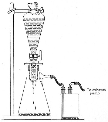
2. Seal the cultures forming set B in Buchner's tubes (vide page 239).
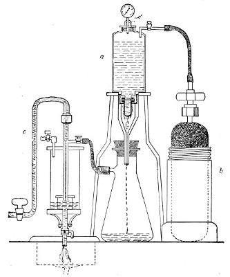
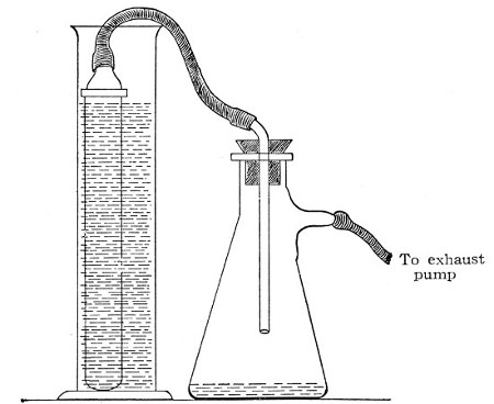
3. Seal the cultures forming set C in Bulloch's apparatus; exhaust the
air by means of a vacuum pump, and provide for the absorption of any
residual oxygen by the introduction of pyrogallic acid and caustic soda
in solution (vide page 245). Treat set D in the same way.
4. Observe the cultivations macroscopically and microscopically at
intervals of twenty-four hours until the completion, if necessary, of
seven days' incubation.
5. Control these results.
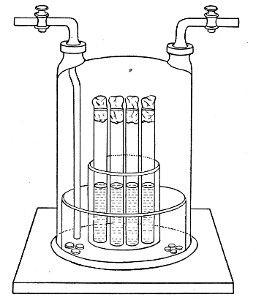
Gases Other than Oxygen.—
Apparatus Required:
Bulloch's apparatus.
Sterile gas filter (vide page 40).
Gasometer containing the gas it is desired to test (SO2, N2O, NO,
CO2, etc.) or gas generator for its production.
Method.—
1. Prepare at least seven tube cultivations upon solid media and deposit
them in Bulloch's apparatus.
2. Connect up the inlet tube of the Bulloch's jar with the sterile gas
filter, and this again with the delivery tube of the gasometer or gas
generator.
3. Open both stop-cocks of the Bulloch's apparatus and pass the gas
through until it has completely replaced the air in the bell jar as
shown by the result of analyses of samples collected from the exit tube.
4. Incubate under optimum conditions as to temperature.
5. Examine the cultivations at intervals of twenty-four hours, until the
completion of seven days.
6. Remove one tube from the interior of the apparatus each day. If no
growth is visible, incubate the tube under optimum conditions as to
temperature and atmosphere, and in this way determine the length of
exposure to the action of the gas necessary to kill the organisms under
observation.
7. Control these results.
II. Temperature.—
(A) Range.—
1. Prepare a series of ten tube cultivations, in fluid media, of optimum
reaction.
2. Arrange a series of incubators at fixed temperatures, varying 5° C.
and including temperatures between 5° C. and 50° C.
(In the absence of a sufficient number of incubators utilise the
water-bath employed in testing the thermal death-point of vegetative
forms.)
3. Incubate one tube cultivation of the organism aerobically or
anaerobically, as may be necessary, in each incubator, and examine at
half-hour intervals for from five to eighteen hours.
4. Note that temperature at which growth is first observed
macroscopically (Optimum temperature).
5. Continue the incubation until the completion of seven days. Note the
extremes of temperature at which growth takes place (Range of
temperature).
6. Control these results—if considered necessary arranging the series
of incubators to include each degree centigrade for five degrees beyond
each of the extremes previously noted.
(B) Optimum.—
1. Prepare a second series of ten tube cultivations under similar
conditions as to reaction of medium.
2. Incubate in a series of incubators in which the temperature is
regulated at intervals of 1° C. for five degrees on either side of
optimum temperature observed in the previous experiment (A, step 4).
3. Observe again at half-hour intervals and note that temperature at
which growth is first visible to the naked eye = Optimum temperature.
(C) Thermal Death-point (t. d. p.)—
Moist—Vegetative Forms:
The t. d. p. here is that temperature which with certainty kills a
watery suspension of the organisms in question after an exposure of 10
minutes.
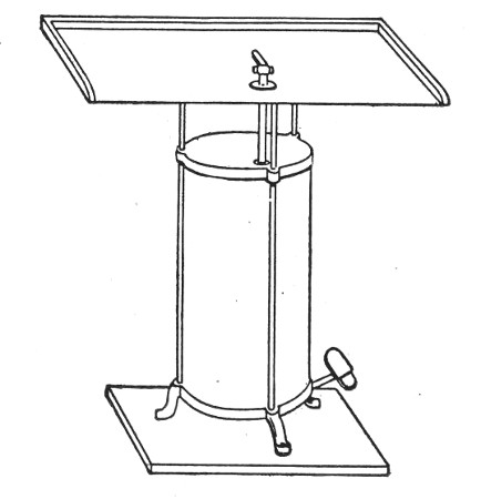
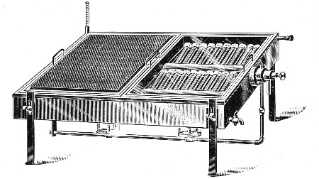
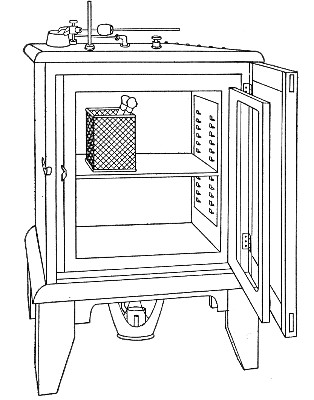
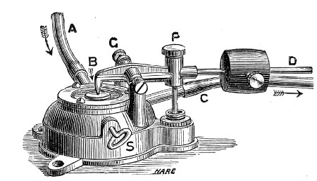
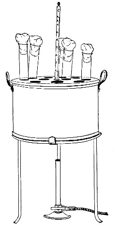
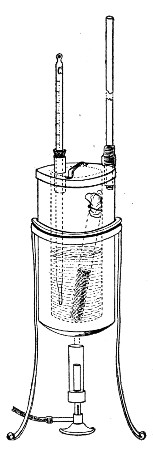
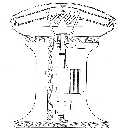
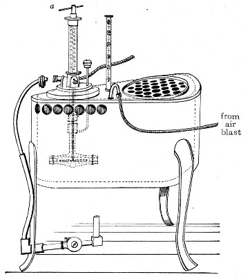 Fig. 155.—Hearson's water-bath.
Apparatus Required:
Water-bath. For the purpose of observing the thermal
death-point a special water-bath is necessary. The
temperature of this piece of apparatus is controlled by
means of a capsule regulator that can be adjusted for
intervals of half a degree centigrade through a range of
30°, from 50° C. to 80° C. by means of a spring, actuated by
the handle a, which increases the pressure in the interior
of the capsule. A hole is provided for the reception of the
nozzle of a blast pump, so that a current of air may be
blown through the water while the bath is in use, and thus
ensure a uniform temperature of its contents. Through a
second hole is suspended a certified centigrade thermometer,
the bulb of which although completely immersed in the water
is raised at least 2 cm. above the floor of the bath.
Sterile glass capsules.
Flask containing 250 c.c. sterile normal saline solution.
Case of sterile pipettes, 10 c.c. (in tenths of a cubic
centimetre).
Special platinum loop.
Test-tubes, 18 by 1.5 cm., of thin German glass.
Case of sterile petri dishes.
Tubes of agar or gelatine.
Method.—
1. Prepare tube cultivations on solid media of optimum reaction;
incubate forty-eight hours under optimum conditions as to temperature
and atmosphere.
2. Examine preparations from the cultivation microscopically to
determine the absence of spores.
3. Pipette 5 c.c. salt solution into each of twelve capsules.
4. Suspend three loopfuls of the surface growth (using a special
platinum loop, vide page 316) in the normal saline solution by
emulcifying evenly against the moist walls of each capsule.
5. Transfer emulsion from each capsule to sterile 250 c.c. flask, and
mix.
6. Pipette 5 c.c. emulsion into each of twelve sterile test-tubes
numbered consecutively.
7. Adjust the first tube in the water-bath, regulated at 40° C, by means
of two rubber rings around the tube, one above and the other below the
perforated top of the bath, so that the upper level of the fluid in the
tube is about 4 cm. below the surface of the water in the bath, and the
bottom of the tube is a similar distance above the bottom of the bath.
8. Arrange a control test-tube containing 5 c.c. sterile saline solution
under similar conditions. Plug the tube with cotton-wool and pass a
thermometer through the plug so that its bulb is immersed in the water.
9. Close the unoccupied perforations in the lid of the water-bath by
means of glass balls.
10. Watch the thermometer in the test-tube until it records a
temperature of 40° C. Note the time. Ten minutes later remove the tube
containing the suspension, and cool rapidly by immersing its lower end
in a stream of running water.
11. Pour three gelatine (or agar) plates containing respectively 0.2,
0.3, and 0.5 c.c. of the suspension, and incubate.
12. Pipette the remaining 4 c.c. of the suspension into a culture flask
containing 250 c.c. of nutrient bouillon, and incubate.
13. Observe these cultivations from day to day. "No growth" must not be
recorded as final until after the completion of seven days' incubation.
14. Extend these observations to the remaining tubes of the series, but
varying the conditions so that each tube is exposed to a temperature 2°
C. higher than the immediately preceding one—i. e., 42° C., 44° C.,
46° C., and so on.
15. Note that temperature, after exposure to which no growth takes place
up to the end of seven days' incubation, = the thermal death-point.
16. If greater accuracy is desired, a second series of tubes may be
prepared and exposed for ten minutes to fixed temperatures varying only
0.5° C., through a range of 5° C. on either side of the previously
observed death-point.
Moist—Spores: The thermal death-point in the case of spores is that
time exposure to a fixed temperature of 100° C. necessary to effect the
death of all the spores present in a suspension.
Note.—If it is desired to retain the time constant 10
minutes and investigate the temperature necessary to destroy
the spores, varying amounts of calcium chloride must be
added to the water in the bath, when the boiling-point will
be raised above 100° C. according to the percentage of
calcium in solution. In such case use the bath figured on
page 227; the bath figured on page 299 can only be used if
the capsule is first removed.
It is determined in the following manner
Apparatus Required:
Steam-can fitted with a delivery tube and a large bore
safety-valve tube.
Water-bath at 100° C.
Erlenmeyer flask, 500 c.c. capacity, containing 140 c.c.
sterile normal saline solution and fitted with rubber
stopper perforated with four holes.
The rubber stopper is fitted as follows:
(a) Thermometer to 120° C., its bulb immersed in the normal
saline.
(b) Straight entry tube, reaching to the bottom of the
flask, the upper end plugged with cotton-wool.
(c) Bent syphon tube, with pipette nozzle attached by means
of rubber tubing and fitted with pinch-cock.
The nozzle is protected from accidental contamination by
passing it through the cotton-wool plug of a small
test-tube.
(d) A sickle-shaped piece of glass tubing passing just
through the stopper, plugged with cotton-wool, to act as a
vent for the steam.
Sterile plates.
Sterile pipettes.
Sterile test-tubes graduated to contain 5 c.c.
Media Required:
Gelatine or agar.
Culture flasks containing 200 c.c. nutrient bouillon.
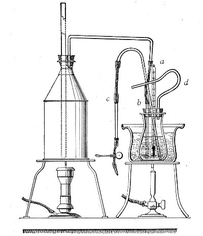 Fig. 156.—Apparatus arranged for the determination of
the death-point of spores.
Fig. 156.—Apparatus arranged for the determination of
the death-point of spores.
Method.—
1. Prepare twelve tube cultivations upon the surface (or two cultures in
large flat culture bottles—vide page 5) of nutrient agar and
incubate under the optimum conditions (previously determined), for the
formation of spores.
Examine preparations from the cultures microscopically to determine the
presence of spores.
2. Pipette 5 c.c. sterile normal saline into each culture tube or 30
c.c. into each bottle and by means of a sterile platinum spatula
emulsify the entire surface growth with the solution.
3. Add the 60 c.c. emulsion to 140 c.c. normal saline contained in the
fitted Erlenmeyer flask.
4. Place the flask in the water-bath of boiling water.
5. Connect up the straight tube, after removing the cotton-wool plug,
with the delivery tube of the steam can; remove the plug from the vent
tube.
6. When the thermometer reaches 100° C., open the spring clip on the
syphon, discard the first cubic centimeter of suspension that syphons
over (i. e., the contents of the syphon tube); collect the next 5 c.c.
of the suspension in the sterile graduated test-tube and pour plates and
prepare flask cultures therefrom as in the previous experiments.
7. Repeat this process at intervals of twenty-five minutes' steaming.
8. Observe the inoculated plates and flasks up to the completion, if
necessary, of seven days' incubation.
9. Control these experiments, but in this instance syphon off portions
of the suspension at intervals of one-half to one minute during the five
or ten minutes preceding the previously determined death-point.
Thermal Death-point.—
Dry—Vegetative Forms: The thermal death-point in this case is that
temperature which with certainty kills a thin film of the organism in
question after a time exposure of ten minutes.
Apparatus Required:
Hot-air oven, provided with thermo-regulator.
Sterile cover-slips.
Flask containing 250 c.c. sterile normal saline solution.
Case of sterile pipettes, 10 c.c. (in tenths of a cubic
centimetre).
Case of sterile capsules.
Crucible tongs.
Method.—
1. Prepare an emulsion with three loopfuls from an optimum cultivation
in 5 c.c. normal saline in a sterile capsule and examine microscopically
to determine the absence of spore forms.
2. Make twelve cover-slip films on sterile cover-slips; place each in a
sterile capsule to dry.
3. Expose each capsule in turn in the hot-air oven for ten minutes to a
different fixed temperature, varying 5° C. between 60° C. and 120° C.
4. Remove each capsule from the oven with crucible tongs immediately
after the ten minutes are completed; remove the cover-glass from its
interior with a sterile pair of forceps.
5. Deposit the film in a flask containing 200 c.c. nutrient bouillon.
6. Prepare subcultivations from such flasks as show evidence of growth,
to determine that no accidental contamination has taken place but that
the organism originally spread on the film is responsible for the
growth.
7. Control the result of these experiments.
Dry—Spores: The thermal death-point in this case is that temperature
which with certainty kills the spores of the organism in question when
present in a thin film after a time exposure of 10 minutes.
Apparatus Required:
As for vegetative forms.
Method.—
1. Prepare a sloped agar tube cultivation and incubate under optimum
conditions as to spore formations.
2. Pipette 5 c.c. sterile normal saline into the culture tube and
emulsify the entire surface growth in it. Examine microscopically to
determine the presence of spores in large numbers.
3. Spread thin even films on twelve sterile cover-slips and place each
cover-slip in a separate sterile capsule.
4. Expose each capsule in turn for ten minutes to a different fixed
temperature, varying 5°C, between 100° C. and 160°C.
5. Complete the examination as for vegetative forms.
III. Reaction of Medium.
(A) Range.—
1. Prepare a bouillon culture of the organism and incubate, under
optimum conditions as to temperature and atmosphere, for twenty-four
hours.
2. Pipette 0.1 c.c. of the cultivation into a sterile capsule; add 9.9
c.c. sterile bouillon and mix thoroughly.
3. Prepare a series of tubes of nutrient bouillon of varying reactions,
from +25 to -30 (vide page 155), viz.: +25, +20, +15, +10, +5,
neutral, -5, -10, -15, -20, -25, -30.
4. Inoculate each of the bouillon tubes with 0.1 c.c. of the diluted
cultivation by means of a sterile graduated pipette and incubate under
optimum conditions.
5. Observe the cultures at half-hourly intervals from the third to the
twelfth hours. Note the reaction of the tube or tubes in which growth is
first visible macroscopically (probably optimum reaction).
6. Continue the incubation until the completion, if necessary, of seven
days. Note the extremes of acidity and alkalinity in which macroscopical
growth has developed (Range of reaction).
7. Control the result of these observations.
(B) Optimum Reaction.—The optimum reaction has already been
roughly determined whilst observing the range. It can be fixed within
narrower limits by inoculating in a similar manner a series of tubes of
bouillon which represent smaller variations in reaction than those
previously employed (say, 1 instead of 5) for five points on either side
of the previously observed optimum. For example, the optimum reaction
observed in the set of experiments to determine the range was +10. Now
plant tubes having reactions of +15, +14, +13, +12, +11, +10, +9, +8,
+7, + 6, +5, and observe as before.
IV. Resistance to Lethal Agents.—
(A) Desiccation.—
Apparatus Required:
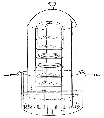
Mueller's desiccator. This consists of a bell glass fitted
with an exhaust tube and stop-cock (d), which can be
secured to a plate-glass base (c) by means of wax or
grease. It contains a cylindrical vessel of porous clay
(a) into the top of which pure sulphuric acid is poured
whilst the material to be dried is placed within its walls
on a glass shelf (b). The air is exhausted from the
interior and the acid rapidly converts the clay vessel into
a large absorbing surface (Fig. 157).
Exhaust pump.
Pure concentrated sulphuric acid.
Sterile cover-slips.
Sterile forceps.
Culture flask containing 200 c.c. nutrient bouillon.
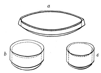
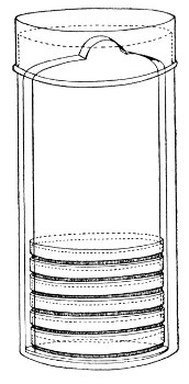
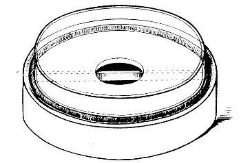
Sterile ventilated Petri dish. This is prepared by bending
three short pieces of aluminium wire into V shape and
hanging these on the edge of the lower dish and resting the
lid upon them (Fig. 158).
Method.—
1. Prepare a surface cultivation on nutrient agar in a culture bottle
and incubate under optimum conditions for forty-eight hours.
2. Examine preparations from the cultivation, microscopically, to
determine the absence of spores.
3. Pipette 5 c.c. sterile normal saline solution into the flask and
suspend the entire growth in it.
4. Spread the suspension in thin, even films on sterile cover-slips and
deposit inside sterile "plates" to dry.
5. As soon as dry, transfer the cover-slip films to the ventilated Petri
dish by means of sterile forceps.
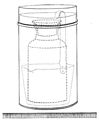
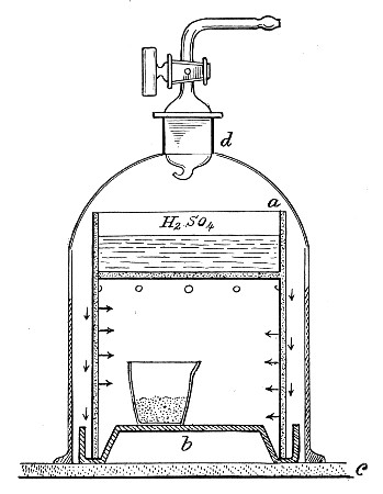 Fig. 157.—Mueller's desiccator.
6. Place the Petri dish inside the Mueller's desiccator; fill the upper
chamber with pure sulphuric acid, cover with the bell jar, and exhaust
the air from its interior. Ten minutes later connect up the desiccator
to a sulphuric acid wash-bottle interposing an air filter so that only
dry sterile air enters.
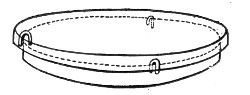 Fig. 158.—Petri dish for drying cultivations.
7. At intervals of five hours open the apparatus, remove one of the
cover-slip films from the Petri dish, and transfer it to the interior of
a culture flask, with every precaution against contamination. Reseal the
desiccator and again exhaust, and subsequently admit dry sterile air as
before.
8. Incubate the culture flask under optimum conditions until the
completion of seven days, if necessary; and determine the time exposure
at which death occurs.
9. Pour plates from those culture flasks which grow, to determine the
absence of contamination.
10. Repeat these observations at hourly intervals for the five hours
preceding and succeeding the death time, as determined in the first set
of experiments.
(B) Light.—
(a) Diffuse Daylight:
1. Prepare a tube cultivation in nutrient bouillon, and incubate under
optimum conditions, for forty-eight hours.
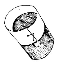
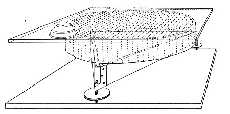
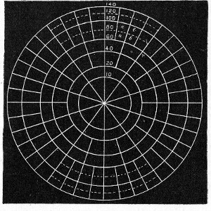
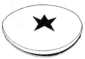 Fig. 159.—Plate with star for testing effect of light.
2. Pour twenty plate cultivations, ten of nutrient gelatine and ten of
nutrient agar, each containing 0.1 c.c. of the bouillon culture.
3. Place one agar plate and one gelatine plate into the hot and cold
incubators, respectively, as controls.
4. Fasten a piece of black paper, cut the shape of a cross or star, on
the centre of the cover of each of the remaining plates (Fig. 159).
5. Expose these plates to the action of diffuse daylight (not direct
sunlight) in the laboratory for one, two, three, four, five, six, eight,
ten, twelve hours.
6. After exposure to light, incubate under optimum conditions.
7. Examine the plate cultivations after twenty-four and forty-eight
hours' incubation, and compare with the two controls. Record results. If
growth is absent from that portion of the plate unprotected by the black
paper, continue the incubation and daily observation until the end of
seven days.
8. Control the results.
(b) Direct Sunlight:
1. Prepare plate cultivations precisely as in the former experiments and
place the two controls in the incubators.
2. Arrange the remaining plates upon a platform in the direct rays of
the sun.
3. On the top of each plate stand a small glass dish 14 cm. in diameter
and 5 cm. deep.
4. Fill a solution of potash alum (2 per cent. in distilled water) into
each dish to the depth of 2 cm. to absorb the heat of the sun's rays and
so eliminate possible effects of temperature on the cultivations.
5. After exposures for periods similar to those employed in the
preceding experiment, incubate and complete the observation as above.
(c) Primary Colours: Each colour—violet, blue, green and red—must be
tested separately.
1. Prepare plate cultivations, as in the previous "light" experiments,
and incubate controls.
2. Fasten a strip of black paper, 3 cm. wide, across one diameter of the
cover of each plate.
3. Coat the remainder of the surface of the cover with a film of pure
photographic collodion which contains 2 per cent. of either of the
following aniline dyes, as may be necessary:
Chrysoidin (for red).
Malachite green (for green).
Eosin, bluish (for blue).
Methyl violet (for violet).
4. Expose the plates, thus prepared, to bright daylight (but not direct
sunlight) for varying periods, and complete the observations as in the
preceding experiments. The bactericidal action of light appears to
depend upon the more refrangible rays of the violet end of the spectrum
and is noted whether the red yellow rays are transmitted or not.
5. Control the results.
Note.—The ultra-violet rays obtained from a quartz mercury
vapour lamp destroy bacterial life with great rapidity under
laboratory conditions.
(C) Heat.—(Vide Thermal Death-point, page 298.)
(D) Antiseptics and Disinfectants.—The resistance exhibited by any
given bacterium toward any specified disinfectant or germicide should be
investigated with reference to the following points:
(A) Inhibition coefficient—i. e., that percentage of the
disinfectant present in the nutrient medium which is sufficient to
prevent the growth and multiplication of the bacterium.
(B) Inferior lethal coefficient—i. e., the time exposure necessary
to kill vegetative forms of the bacterium suspended in water at 20° to
25° C, in which the disinfectant is present in medium concentration
(concentration insufficient to cause plasmolysis). And if the bacterium
is one which forms spores,
(C) Superior lethal coefficient—i. e., the time exposure necessary
to kill the spores of the bacterium under conditions similar to those
obtaining in B.
The example here detailed only specifically refers to certain of the
disinfectants:
viz:—Bichloride of mercury;
Formaldehyde;
Carbolic acid;
investigated with regard to B. anthracis, but the technique is
practically similar for all other chemical disinfectants.
Inhibition Coefficient.—
Apparatus Required:
Case of sterile pipettes, 10 c.c. (in tenths).
Case of sterile pipettes, 1 c.c. (in tenths).
Sterile tubes or capsules for dilutions.
Tubes of nutrient bouillon each containing a measured 10
c.c. of medium.
Twenty-four-hour-old agar culture of a recently isolated B.
Anthracis.
Germicides:
1. Five per cent. aqueous solution of carbolic acid.
2. One per cent. aqueous solution of perchloride of mercury.
3. One-tenth per cent. aqueous solution of formaldehyde.
Method.—
1. Number six bouillon tubes consecutively 1 to 6. Inoculate each from
the stock cultivation of B. anthracis and at once add varying
quantities[10] of the carbolic acid solution, viz.:
To tube 1 add 2.0 c.c. (= 1:100)
To tube 2 add 1.0 c.c. (= 1:200)
To tube 3 add 0.6 c.c. (= 1:300)
To tube 4 add 0.5 c.c. (= 1:400)
To tube 5 add 0.4 c.c. (= 1:500)
To tube 6 add 0.2 c.c. (= 1:1,000)
2. Prepare a similar series of tube cultivations numbered consecutively
7 to 12 and add varying quantities of the mercuric perchloride solution,
viz.:
To tube 7 add 0.1 (= 1:1,000)
To tube 8 add 0.05 (= 1:2,000)
To tube 9 add 0.03 (= 1:3,000)
To tube 10 add 0.025 (= 1:4,000)
To tube 11 add 0.02 (= 1:5,000)
To tube 12 add 0.01 (= 1:10,000)
3. Prepare a similar series of tube cultivations numbered consecutively
13 to 18 and add varying quantities of the formaldehyde solution, viz.:
To tube No. 13 add 1.0 c.c. (= 1:1,000)
To tube No. 14 add 0.4 c.c. (= 1:2,500)
To tube No. 15 add 0.2 c.c. (= 1:5,000)
To tube No. 16 add 0.1 c.c. (= 1:10,000)
To tube No. 17 add 0.075 c.c. (= 1:15,000)
To tube No. 18 add 0.05 c.c. (= 1:20,000)
4. Incubate all three sets of cultivations under optimum conditions as
to temperature and atmosphere.
5. Examine each of the culture tubes from day to day, until the
completion of seven days, and note those tubes, if any, in which growth
takes place.
6. From such tubes as show growth prepare subcultivations upon suitable
media, and ascertain that the organism causing the growth is the one
originally employed in the test and not an accidental contamination.
Inferior Lethal Coefficient.—
Apparatus Required:
Highly concentrated solutions of the disinfectants.
Sterile test-tubes in which to make dilutions from the
concentrated solutions of the disinfectants.
Hanging-drop slides.
Cover-slips.
Erlenmeyer flask containing 100 c.c. sterile distilled
water.
Case of sterile pipettes, 10 c.c. (in tenths of a cubic
centimetre).
Case of sterile pipettes, 1 c.c. (in tenths of a cubic
centimetre).
Method.—
1. Prepare a surface cultivation of the "test" organism B. anthracis
upon nutrient agar in a culture bottle and incubate under optimum
conditions for twenty-four hours; then examine the cultivation
microscopically to determine the absence of spores.
2. Prepare solutions of different percentages of each disinfectant.
3. Make a series of hanging-drop preparations from the agar culture,
using a loopful of disinfectant solution of the different percentages to
prepare the emulsion on each cover-slip.
4. Examine microscopically and note the strongest solution which does
not cause plasmolysis and the weakest solution which does plasmolyse the
organism.
5. Make control preparations of these two solutions and determine the
percentage to be tested.
6. Pipette 10 c.c. sterile water into the culture bottle and suspend the
entire surface growth in it.
7. Transfer the suspension to the Erlenmeyer flask and mix it with the
90 c.c. of sterile water remaining in the flask.
8. Pipette 10 c.c. of the diluted suspension into each of ten sterile
test-tubes.
9. Label one of the tubes "Control" and place it in the incubator at 18°
C.
10. Add to each of the remaining tubes a sufficient quantity[11] of a
concentrated solution of the disinfectant to produce the percentage
previously determined upon (vide step 5).
11. Incubate the tubes at 18° C. to 20° C.
12. At hourly intervals remove the control tube and one of the tubes
with added disinfectant from the incubator.
13. Make a subcultivation from both the control and the test suspension,
upon the surface of nutrient agar; incubate under optimum conditions.
14. Observe these culture tubes from day to day until the completion of
seven days, and determine the shortest exposure necessary to cause the
death of vegetative forms.
Superior Lethal Coefficient.—
1. Prepare surface cultivations of the "test" organisms upon nutrient
agar in a culture bottle, and incubate under optimum conditions, for
three days, for the formation of their spores.
2. Transfer the emulsion to a sterile test-tube and heat in the
differential steriliser for ten minutes at 80° C. to destroy all
vegetative forms.
3. Employing that percentage solution of the disinfectant determined in
the previous experiment, and complete the investigations as detailed
therein, steps 7 to 14, increasing the interval between planting the
subcultivations to two, three, or five hours if considered advisable.
Note.—Where it is necessary to leave the organisms in
contact with a strong solution of the disinfectant for
lengthy periods, some means must be adopted to remove every
trace of the disinfectant from the bacteria before
transferring them to fresh culture media; otherwise,
although not actually killed, the presence of the
disinfectant may prevent their development, and so give rise
to an erroneous conclusion. Consequently it is essential in
all germicidal experiments to determine first of all the
inhibition coefficient of the germicide employed. Under the
circumstances referred to above it is usually sufficient to
prepare the subcultures in such a volume of fluid nutrient
medium as would suffice to reduce the concentration of the
germicide to about one hundredth of the inhibition
percentage, assuming that the entire bulk of inoculum was
made up of that strength of germicide employed in the test.
In some cases it is a simple matter to neutralise the
germicide and render it inert by washing the organisms in
some non-germicidal solution (such for example as ammonium
sulphide when using mercurial salts as the germicide). When,
however, it is desired to remove the last traces of
germicide proceed as follows:
1. Transfer the suspension of bacteria to sterile
centrifugal tubes; add the required amount of disinfectant,
and allow it to remain in contact with the bacteria for the
necessary period.
2. Centrifugalise thoroughly, pipette off the supernatant
fluid; fill the tube with sterile water and distribute the
deposit evenly throughout the fluid.
3. Centrifugalise again, pipette off the supernatant fluid;
fill the tube with sterile water; distribute the deposit
evenly throughout the fluid, and transfer the suspension to
a litre flask.
4. Make up to a litre by the addition of sterile water;
filter the suspension through a sterile porcelain candle.
5. Emulsify the bacterial residue with 5 c.c. sterile
bouillon.
6. Prepare the necessary subcultivations from this emulsion.
Living Bacteria.—
(a) Psychrophilic Bacteria: When the organism will only grow at or below
18° to 20° C.,
1. Prepare cultivations in nutrient broth and incubate under optimum
conditions.
2. After seven days' incubation inject that amount of the culture
corresponding to 1 per cent. of the body-weight of a healthy frog, into
the reptile's dorsal lymph sac.
3. Observe until death takes place, or, in the event of a negative
result, until the completion of twenty-eight days (vide Chapter
XVIII).
4. If, and when, death occurs, make a careful post-mortem examination
(vide Chapter XIX).
(b) Mesophilic Bacteria: When the organism grows at 35° to 37° C.,
1. Prepare cultivations in nutrient broth and incubate under optimum
conditions for forty-eight hours.
2. Select two white mice, as nearly as possible of the same age, size,
and weight.
3. Inoculate the first mouse, subcutaneously at the root of the tail,
with an amount of cultivation equivalent to 1 per cent. of its
body-weight.
4. Inoculate the second mouse intraperitoneally with a similar dose.
5. Observe carefully until death occurs, or until the lapse of
twenty-eight days.
6. If the inoculated animals succumb, make complete post-mortem
examination.
If death follows shortly after the injection of cultivations of
bacteria, the inoculation experiments should be repeated two or three
times. Then, if the organism under observation invariably exhibits
pathogenic effects, steps should be taken to ascertain, if possible, the
minimal lethal dose (vide infra) of the growth upon solid media for
the frog or white mouse respectively. Other experimental animals—e.
g., the white rat, guinea-pig, and rabbit—should next be tested in a
similar manner.
7. If the inoculated mice are unaffected, test the action of the
organism in question upon white rats, guinea-pigs, rabbits, etc.
Minimal Lethal Dose (m. l. d.); If the purpose of the inoculation is
to determine the minimal lethal dose, a slightly different procedure
must be followed. For this and other exact experiments a special
platinum loop is manufactured, some 2.5 mm. by 0.75 mm., with parallel
sides, and calibrated by careful weighing, to determine approximately
the amount of moist bacterial growth, the loop will hold when filled.
1. The cultivation must be prepared on a solid medium of the optimum
reaction, incubated at the optimum temperature, and injected at the
period of greatest activity and vigour, of the particular organism it is
desired to test.
2. Arrange four sterile capsules in a row and label them I, II, III, and
IV. Into the first deliver 10 c.c. sterile bouillon by means of a
sterile graduated pipette; and into each of the remaining three, 9.9
c.c.
3. Remove one loopful of the bacterial growth from the surface of the
medium in the culture tube, observing the usual precautions against
contamination, and emulsify it evenly with the bouillon in the first
capsule. Each cubic centimetre of the emulsion will now contain
one-tenth of the organisms contained in the original loopful (written
shortly 0.1 loop).
4. Remove 0.1 c.c. of the emulsion in the first capsule by means of a
sterile graduated pipette and transfer it to the second capsule and mix
thoroughly. Drop the infected pipette into a jar of lysol solution. This
makes up the bulk of the fluid in the second capsule to 10 c.c., and
therefore every cubic centimetre of bouillon in capsule II contains
0.001 loop.
5. Similarly, 0.1 c.c. of the mixture is transferred from capsule II to
capsule III (1 c.c. of bouillon in capsule III contains 0.00001 loop),
and then from capsule III to capsule IV (1 c.c. of bouillon in capsule
IV contains 0.0000001 loop).
The dilutions thus prepared may be summarised in a table;
Capsule I = 1 loopful + 10 c.c. water ∴ 1 c.c.=0.1 loop.
Capsule II = 0.1 c.c. capsule I + 9.9 c.c. water ∴ 1 c.c.=0.001 loop.
Capsule III = 0.1 c.c. capsule II + 9.9 c.c. water ∴ 1 c.c.=0.00001 loop.
Capsule IV = 0.1 c.c. capsule III + 9.9 c.c. water ∴ 1 c.c. = 0.0000001 loop.
6. With sterile graduated pipettes remove the necessary quantity of
bouillon corresponding to the various divisors of ten of the loop from
the respective capsules, and transfer each "dose" to a separate sterile
capsule and label; and to such doses as are small in bulk, add the
necessary quantity of sterile bouillon to make up to 1 c.c.
7. Multiples of the loop are prepared by emulsifying 1, 2, 5, or 10
loops each with 1 c.c. sterile bouillon in separate sterile capsules.
8. Inoculate a series of animals with these measured doses, filling the
syringe first from that capsule containing the smallest dose, then from
the capsule containing the next smallest, and so on. If care is taken,
it will not be found necessary to sterilise the syringe during the
series of inoculations.
9. Plant tubes of gelatine or agar, liquefied by heat, from each of the
higher dilutions, say from 0.0000001 loop to 0.01 loop; pour plates and
incubate. When growth is visible enumerate the number of organisms
present in each, average up and calculate the number of bacteria present
in one loopful of the inoculum.
10. The smallest dose which causes the infection and death of the
inoculated animal is noted as the minimal lethal dose.
Toxins.—
Prepare flask cultivations of the organism under observation in glucose
formate broth, and incubate for fourteen days under optimum conditions.
(a) Intracellular or Insoluble Toxins:
1. Heat the fluid culture in a water-bath at 60° C. for thirty minutes.
(The resulting sterile, turbid fluid is often spoken of as "killed"
culture,)
2. Inoculate a tube of sterile bouillon with a similar quantity, and
incubate under optimum conditions. This "control" then serves to
demonstrate the freedom of the toxin from living bacteria.
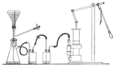 Fig. 160.—Apparatus arrange for toxin filtration.
3. Inject intraveneously that amount of the cultivation corresponding to
1 per cent. of the body-weight of the selected animal, usually one of
the small rodents.
4. Observe during life or until the completion of twenty-eight days, and
in the event of death occurring during that period, make a complete
post-mortem examination.
5. Repeat the experiment at least once. In the event of a positive
result estimate the minimal lethal dose of "killed" culture for each of
the species of animals experimented upon.
(b) Extracellular or Soluble Toxins:
1. Filter the cultivation through a porcelain filter candle (Berkefeld)
into a sterile filter flask, arranging the apparatus as in the
accompanying figure (Fig. 160).
2. Inoculate mice, rats, guinea-pigs, and rabbits subcutaneously with
that quantity of toxin corresponding to 1 per cent. of the body-weight
of each respectively, and observe, if necessary, until the completion of
one month.
3. Inoculate a "control" tube of bouillon with a similar quantity and
incubate, to determine the freedom of the filtered toxin from living
bacteria.
4. In the event of a fatal termination make complete and careful
post-mortem examinations.
5. Repeat the experiments and, if the results are positive, ascertain
the minimal lethal dose of toxin for each of the susceptible animals.
The estimation of the m. l. d. of a toxin is carried out on lines
similar to those laid down for living bacteria (vide page 316) merely
substituting 1 c.c. of toxin as the unit in place of the unit "loopful"
of living culture.
It frequently happens, during the course of casual investigations that a
bouillon-tube culture is available for a toxin test whilst a flask
cultivation is not. In such cases, Martin's small filter candle and tube
(Fig. 161) specially designed for the filtration of small quantities of
fluid, is invaluable. This consists of a narrow filter flask just large
enough to accommodate an ordinary 18 × 2 cm. test-tube. The mouth of the
tubular Chamberland candle 15 × 1.5 cm. is closed by a perforated rubber
cork into which fits the end of the stem of a thistle headed funnel,
whilst immediately below the butt of the funnel is situated a rubber
cork to close the mouth of the filter flask. When the apparatus is fixed
in position and connected to an exhaust pump, the cultivation is poured
into the head of the funnel and owing to the relatively large filtering
surface the germ free filtrate is rapidly drawn through into the
test-tube receiver.
Raising the Virulence of an Organism.—If it is desired to raise or
"exalt" the virulence of a feebly pathogenic organism, special methods
of inoculation are necessary, carefully adjusted to the exigencies of
each individual case. Among the most important are the following:
1. Passage of Virus.—The inoculation of pure cultivations of the
organism into highly susceptible animals, and passing it as rapidly as
possible from animal to animal, always selecting that method of
inoculation-e. g., intraperitoneal—which places the organism under
the most favorable conditions for its growth and multiplication.
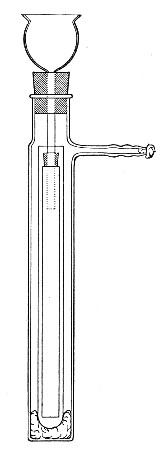 Fig. 161—Martin's filtering apparatus for small
quantities of fluid.
Fig. 161—Martin's filtering apparatus for small
quantities of fluid.
2. Virus Plus Virulent Organisms.—The inoculation of pure
cultivations of the organism together with pure cultivations of some
other microbe which in itself is sufficiently virulent to ensure the
death of the experimental animal, either into the same situation or into
some other part of the body. By this association the organism of low
virulence will frequently acquire a higher degree of virulence, which
may be still further raised by means of "passages" (vide supra).
3. Virus Plus Toxins.—The inoculation of pure cultivations of the
organism into some selected situation, together with the subcutaneous,
intraperitoneal, or intravenous injection of a toxin—e. g., one of
those elaborated by the proteus group—either simultaneously with,
before, or immediately after, the injection of the feeble virus. By
this means the natural resistance of the animal is lowered, and the
organism inoculated is enabled to multiply and produce its pathogenic
effect, its virulence being subsequently exalted by means of "passages."
Attenuating the Virulence of an Organism.—Attenuating or lowering the
virulence of a pathogenic microbe is usually attained with much less
difficulty than the exaltation of its virulence, and is generally
effected by varying the environment of the cultivations, as for example:
1. Cultivating in such media as are unsuitable by reason of their (a)
composition or (b) reaction.
2. Cultivating in suitable media, but at an unsuitable temperature.
3. Cultivating in suitable media, but in an unsuitable atmosphere.
4. Cultivation in suitable media, but under unfavorable conditions as to
light, motion, etc.
Attenuation of the virus can also be secured by
5. Passage through naturally resistant animals.
6. Exposure to desiccation.
7. Exposure to gaseous disinfectants.
8. By a combination of two or more of the above methods.
The further study of the pathogenetic powers of any particular bacterium
involves the active immunisation of one or more previously normal
animals. This end may be attained by various means; but it must be
remembered that immunisation is not carried out by any hard and fast
rule or by one method alone, but usually by a combination of methods
adapted to the exigencies of each particular case. The ordinary methods
include:
A. Active Immunisation.
I. By inoculation with dead bacteria (i. e., bacteria
killed by heat; the action of ultra-violet rays, of chemical
germicides, or by autolysis).
II. By the inoculation of attenuated strains of bacteria.
III. By the inoculation of living virulent bacteria (exalted
in virulence if necessary).
B. Combined Active and Passive Immunisation:
IV. By the inoculation of toxin-antitoxin mixtures.
The immunisation of the rabbit against the Diplococcus pneumoniæ may be
instanced as an example of the general methods of immunisation of
laboratory animals.
1. Take a full grown rabbit weighing not less than 1200 to 1500 grammes
(large rabbits of 2000 grammes and over are the most suitable for
immunising experiments). Observe weight and temperature carefully during
the few days occupied in the following steps.
2. Inoculate a small rabbit intraperitoneally with one or two loopfuls
of a twenty-four-hour-old blood agar cultivation of a virulent strain
of Diplococcus pneumoniæ.
Death should follow within twenty-four hours, and in any case will not
be delayed beyond forty-eight hours.
3. Under aseptic precautions, at the post-mortem, transfer a loopful of
heart blood to an Erlenmeyer flask containing 50 c.c. sterile nutrient
broth. Incubate at 37° C. for twenty-four hours.
4. Prepare also several blood agar cultures from the heart blood of the
rabbit, label them all O.C. (original culture). After twenty-four hours
incubation at 37° C. place an india-rubber cap over the plugged mouth of
the tube of all but one of these cultures and paint the cap with Canada
balsam or shellac varnish, dry, and replace in the hot incubator.
This will prevent evaporation, and cultures thus sealed will remain
unaltered in virulence for a considerable time.
5. Make a fresh subcultivation on blood agar from the uncapped O.C.
cultivation and after twenty-four hours incubation at 37° C. determine
the minimal lethal dose of this strain upon a series of mice (see page
316).
6. Suspend the flask containing the twenty-four-hour-old broth culture
(step 3) in the water-bath at 60° C. for one hour. Cool the flask
rapidly under a stream of cold water.
7. Determine the sterility of this (?) killed cultivation by
transferring one cubic centimetre to each of several tubes of nutrient
broth, and incubate at 37° C. for twenty-four hours. If growth of
Diplococcus pneumoniæ occurs, again heat culture in water-bath at 60° C.
for one hour and again test for sterility.
8. Inject the selected rabbit intravenously (see page 363) with 2 c.c.
of the killed cultivation, and inject a further 10 c.c. into the
peritoneal cavity.
During the next few days the animal will lose some weight and perhaps
show a certain amount of pyrexia.
9. When the temperature and weight have again returned to
normal—generally about seven days after the inoculation—again inject
killed cultivation, this time giving a dose of 5 c.c. intravenously and
20 c.c. intraperitoneally. A temperature and weight reaction similar to,
but less marked than that following the first injection will probably be
observed, but after about a week's interval the animal will be ready for
the next injection.
10. When ready to give the third injection prepare a fresh blood agar
subculture from another O.C. tube and after twenty-four hours incubation
prepare a minimal lethal dose (as determined in 5) and inject it
subcutaneously into the rabbit's abdominal wall.
A slight local reaction will probably be observed as well as the weight
and temperature reactions.
11. A week to ten days later inject a similar minimal lethal dose into
the peritoneal cavity.
12. Observe the weight and temperature of the rabbit very carefully, and
regulating the dates of inoculation by the animal's general condition,
continue to inject living cultivations of the pneumococcus into the
peritoneal cavity, gradually increasing the dose by multiples of ten.
13. At intervals of two months samples of blood may be collected from
the posterior auricular vein and the serum tested for specific
antibodies.
14. Under favourable conditions it will be found after some six months
steady work that the rabbit may be injected intraperitoneally with an
entire blood agar cultivation without any ill effects being apparent;
and this characteristic—resistance to the lethal effects of large doses
of the virus—is the sole criterion of immunity. Further, the serum
separated from blood withdrawn from the animal about a week after an
injection, if used in doses of .01 c.c., will protect a mouse against
the lethal effects of at least ten minimal lethal doses of living
pneumococci.
In the foregoing illustration it has been assumed that complete acquired
active immunity has been conferred upon the experimental rabbit in
consequence of the formation of antibody, specific to the diplococcus
pneumoniac, sufficient in amount to ensure the destruction of enormous
doses of the living cocci—the antigen (that is the substance injected
in response to which antibody has been elaborated) in this particular
case being the bacterial protoplasm of the pneumococcus with its
endo-toxins.
But provided death does not immediately follow the injection of the
antigen, specific antibody is always formed in greater or lesser amount;
and in experimental work a sufficient amount of any required antibody
can often be obtained without carrying the process of immunisation to
its logical termination.
For instance, if the immunisation of a rabbit toward Bacillus typhosus
is commenced on the lines already set out it will often be found, after
a few injections of "killed" cultivation that the blood serum of the
animal (even when diluted with several hundred times its volume of
normal saline) contains specific agglutinin for B. typhosus—and if the
sole object of the experiment has been the preparation of agglutinin the
inoculations may well be stopped at this point, although the animal is
not yet immune in the strict meaning of the word.
Again, antibodies may be formed in response to antigens other than
infective particles—thus the injection into suitable animals of foreign
proteins such as egg albumin, heterologous blood sera or red blood discs
from a different species of animal, will result in the formation of
specific antibodies possessing definite affinities for their respective
antigens.
The most important antibody of this latter type is Hæmolysin, a
substance that makes its appearance in the blood serum of an animal
previously injected with washed blood cells from an animal of a
different species. The serum from such an animal possesses the power of
disintegrating red blood discs of the variety employed as antigen and
causing the discharge of their contained hæmoglobin, and is specific in
its action to the extent of failing to exert any injurious effect upon
the red blood cells of any other species of animal.
The action of this serum is due to the presence of two distinct bodies,
complement and hæmolysin.
Complement (or alexine) is a thermo-labile readily oxidised body
present in variable but unalterable amount in the normal serum of every
animal. It is a substance which exerts a lytic effect upon all foreign
matter introduced into the blood or tissues; but by itself is a
comparatively inert body, and is only capable of exerting its maximum
lytic effect in the presence of and in combination with a specific
antibody, or immune body.
Complement is obtained (unmixed with antibody) by collecting fresh blood
serum from any healthy normal (that is uninoculated) animal.
Guinea-pigs' serum is that most frequently employed for experimental
work.
Hæmolysin (immune body, copula, sensitising body, amboceptor) is a
thermostable antibody formed in response to the injection of red cells
which although in itself inert is capable of linking up complement
present in the normal serum to the red cells of the variety used as
antigen—a combination resulting in hæmolysis.
Hæmolysin is obtained by collecting fresh blood serum from a suitably
inoculated animal and exposing it to a temperature of 56° C. (to destroy
the thermo-labile complement) for 15 to 30 minutes before use. It is
then referred to as inactivated, and is reactivated by the addition
of fresh normal serum—that is serum containing complement.
Hæmolysin is of importance academically owing to the fact that many of
the problems of immunity have been elucidated by its aid; but its
present practical importance lies in the application of the hæmolytic
system (that is hæmolysin, corresponding erythrocyte solution and
complement) to certain laboratory methods having for their object either
the identification of the infective entity or the diagnosis of the
existence of infection.
For use in these laboratory methods of diagnosis it is most convenient
to prepare hæmolytic serum specific for human blood—whether the
laboratory is isolated or attached to a large hospital. Ox blood, sheep
blood or goat blood if readily obtainable, may however be used instead,
and although the following method is directed to the preparation of
human hæmolysin the same procedure serves in all cases.
Apparatus Required:
Small centrifuge, preferably electrically driven, with two receptacles
for tubes, and enclosed in a safety shield (Fig. 162).
Sterile centrifuge tubes (10 c.c. capacity), Fig. 163.
Sterile pipettes (10 c.c. graduated) in case.
Sterile glass capsules (in case).
Sterile test-tubes.
Sterile all glass syringe (5 c.c. or 10 c.c. capacity)
and needle.
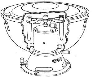 Fig. 162.—Small electrical centrifuge.
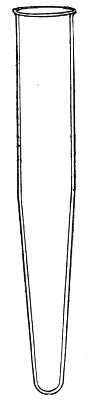 Fig. 163.—Centrifuge tube.
Reagents Required:
Normal saline solution.
10 per cent. sodium citrate solution in normal saline.
Human blood (vide infra).
Method.—
1. Select a healthy full-grown rabbit of not less than 2500 grammes
weight in accordance with the directions already given (page 322) and
prepare it for intraperitoneal inoculation.
2. Measure out 2 c.c. citrated human blood (collected at a surgical
operation or a venesection, or withdrawn by venipuncture from the median
basilic or median cephalic vein of a normal adult) into a centrifuge
tube and centrifugalise thoroughly.
3. Wash with three changes of normal saline (vide also page 388).
4. Transfer the washed cells to a sterile capsule by means of a sterile
pipette. Add 5 c.c. of normal saline and mix thoroughly.
5. Take up the mixture of cells and saline in the all-glass syringe and
inject into the peritoneal cavity of the rabbit.
6. Seven days later inject intraperitoneally the washed cells from 5
c.c. human blood mixed with 5 c.c. normal saline.
7. Seven days later inject the washed cells from 10 c.c. human blood
mixed with 5 c.c. normal saline.
8. After a further interval of seven days repeat the injection of washed
cells from 10 c.c. human blood mixed with 5 c.c. normal saline.
Note.—Better results are obtained if the second and
subsequent injections are made intravenously, even when
smaller quantities of washed red cells are employed. If,
however, the intravenous route is selected exceeding great
care must be exercised to avoid the introduction of air into
the vein—an accident which is followed, within a few
minutes, by the death of the rabbit from pulmonary embolism.
9. Allow five days to elapse, then collect a preliminary sample of
blood, say about 2 c.c., from the rabbit's ear. Allow it to clot,
separate off the serum and transfer to a sterile test-tube. Place the
test-tube in a water-bath at 56° C. for fifteen minutes (to inactivate)
and test the serum quantitatively for hæmolytic properties in the
following manner:
Apparatus Required:
Electrical centrifuge.
Sterile centrifuge tubes.
Water-bath regulated at 56°C.
Sterilised pipettes 10 c.c. graduated in tenths.
Sterilised pipettes 1 c.c. graduated in tenths.
Sterile test-tubes, 16 × 2 cm.
Small sterile test-tubes, 9 × 1 cm.
Small test-tube rack, or roll of plasticine.
Capillary teat pipettes.
Stout rubber band or length of small rubber tubing.
Reagents Required and Method of Preparation:
1. Normal saline solution.
2. Hæmolytic serum inactivated by preliminary heating to 56°
C. for 15 minutes (vide supra) in test-tube labelled H. S.
3. Complement. Fresh guinea-pig serum in test-tube labelled
C.
Kill a normal guinea-pig with chloroform vapour.
Open the thorax with all aseptic precautions, and collect as
much blood as possible from the heart with a sterile Pasteur
pipette.
Transfer it to a sterile centrifuge tube and place the tube
in the incubator at 37° C. Two hours later separate the clot
from the sides of the tube, and centrifugalise thoroughly.
Pipette off the clear serum to a clean sterilised test-tube.
4. Erythrocyte solution, in test-tube labelled E.
Collect and wash human red blood cells (see page 388, 1-8).
Measure the volume of red cells available and prepare a 2
per cent. suspension in normal saline solution.
Method.—
1. Take two test-tubes and number them 1 and 2, and pipette into each 9
c.c. of normal saline solution.
2. Add 1 c.c. of hæmolytic rabbit serum to tube No. 1 and mix
thoroughly: take up 1 c.c. of the mixture and add it to tube No. 2; mix
thoroughly.
3. Set up ten small test-tubes in test-tube rack or in roll of
plasticine, and number 1 to 10.
| 4. Pipette into tube No. 1 0.5 c.c. = 0.5 c.c. hæmolytic serum} | From tube H. S. |
| Pipette into tube No. 2 0.1 c.c. = 0.1 c.c. hæmolytic serum} |
| |
| Pipette into tube No. 3 0.5 c.c. = 0.05 c.c. hæmolytic serum} | From tube 1. |
| Pipette into tube No. 4 0.3 c.c. = 0.03 c.c. hæmolytic serum} |
| Pipette into tube No. 5 0.2 c.c. = 0.02 c.c. hæmolytic serum} |
| pipette into tube No. 6 0.1 c.c. = 0.01 c.c. hæmolytic serum} |
|
| |
| Pipette into tube No. 7 0.5 c.c. = 0.005 c.c. hæmolytic serum} | From tube 2. |
| Pipette into tube No. 8 0.3 c.c. = 0.003 c.c. hæmolytic serum} |
| Pipette into tube No. 9 0.2 c.c. = 0.002 c.c. hæmolytic serum} |
| Pipette into tube No. 10 0.1 c.c. = 0.001 c.c. hæmolytic serum} |
5. To each tube add 1 c.c. of erythrocyte solution.
6. When necessary (that is to say in tubes 2, 4, 5, 6, 8, 9 and 10) add
normal saline solution to the mixture in the test-tubes till the column
of fluid in each reaches to the same level.
7. Shake each tube in turn, so as to thoroughly mix its contents. Plug
the mouth of each tube with cotton wool, and place entire set in the
incubator at 37°C. for one hour.
8. Remove the tubes from the incubator and into each tube pipette 0.1
c.c. complement (guinea-pig's serum) and replace tubes in incubator at
37° C. for further period of one hour.
9. Remove the tubes from the incubator, and if complete hæmolysis has
not taken place in every tube, stand on one side, preferably in the ice
chest, for an hour.
10. Then examine the tubes.
Complete hæmolysis is indicated by a clear red solution,
with no deposit of red cells at the bottom of the test-tube.
Absence of hæmolysis is indicated by a clear or turbid
colourless fluid, with a deposit of red cells at the bottom
of the test-tubes.
The smallest amount of hæmolytic serum that has caused complete
hæmolysis is known as the minimal hæmolytic dose (M. H. D.) and if
hæmolysis has occurred in all the tubes down to No. 7—the m. h. d. of
this particular serum is .005 c.c. = 200 minimal hæmolytic doses per
cubic centimetre. Such a serum is strong enough for experimental work;
indeed, for many purposes, complete hæmolysis down to tube 6 will
indicate a serum sufficiently strong(= 100 m. h. d. per cubic
centimetre). If, however, only the first one or two tubes are completely
hæmolysed, this is an indication that the rabbit should receive further
injections in order to raise the hæmolytic power to a sufficiently high
level.
STORAGE OF HÆMOLYSIN.
If, and when the hæmolysin content of the rabbit's serum is found to be
sufficient, destroy the animal by chloroform vapour.
Remove as much of its blood as possible from the heart under aseptic
precautions into sterilized centrifuge tubes.
Transfer the tubes of blood to the incubator at 37° C. for two
hours—then centrifugalize thoroughly.
Pipette off the clear serum, and fill in quantities of 1 c.c., into
small glass ampoules or pipettes, and hermetically seal in the blowpipe
flame, care being taken to avoid scorching the serum.
Place the ampoules when filled with serum and sealed, in a water-bath at
56° C. for 30 minutes. This destroys the complement, i. e.,
inactivates the serum, and at the same time, provided the various
operations have been carried out under aseptic precautions, ensures its
sterility. A longer exposure reduces the hæmolytic power.
Place the ampoules in a closed metal box and store in the ice chest for
future use.