In Medusæ it is a common thing to find that in certain definite
places of the gastro-vascular system two endodermal surfaces that were
primarily separated by a space have come together and fused into a
single lamella or plate. Such a structure is called indifferently a
cathammal plate, an endodermal lamella, or a vascular lamella. In the
adult animal the vascular lamellæ are by virtue of their very nature
formations “with a past.” They are scaffolding left in the completed
structure, giving us clues as to the way in which that structure was
brought about; and in the Cubomedusæ, whose development is as yet
unknown, they therefore afford an unusually interesting subject for
special consideration.
The vascular lamellæ that are found in Charybdea and Tripedalia
may for convenience be described as forming two systems, the internal
and the marginal. The former comprises the endodermal fusions that
separate the stomach from the stomach pockets (except for the spaces of
communication left free, the gastric ostia) and those that separate the
stomach pockets from one another. The marginal system consists of
the lamella that connects endoderm of the gastro-vascular system with
ectoderm of the surface in a ring all around the bell margin, and with it
also the vascular lamella of the sensory niche, which has already been
referred to in the general description of Charybdea. The lamellæ of the
internal system have been described by previous writers, and especially
by Claus in his paper on Charybdea, but they are still in need of comprehensive
and clear treatment. The lamellæ of the margin and of the
sensory niche have also been described by Claus, but not thoroughly or
with entire accuracy, nor did he recognize the vascular lamellæ of the
sensory niche as originally a part of the lamellæ of the margin. This last
was first determined by H. V. Wilson upon specimens of Chiropsalmus
quadrumanus obtained at Beaufort, North Carolina. Professor Wilson’s
unpublished notes on Chiropsalmus were very kindly placed in my
hands, and so far as the vascular lamellæ are concerned my own work is
only a confirmation and amplification of his, since Charybdea and
Tripedalia in this respect agree with Chiropsalmus.
The vascular lamellæ of the internal system are the most prominent
and morphologically the most important. They comprise the four
vertical strips of fusion that separate the four stomach pockets in the
interradii (ivl in the figures of the series of cross-sections of Charybdea
and Tripedalia, Nos. 6-15 and 21-29), and four curved horizontal cross-pieces
at the top of these which separate the stomach from the stomach
pockets, and would make the separation complete did they not leave in
each perradius a free space between their ends, which makes possible the
gastric ostia.
The arrangement of this internal system of vascular lamellæ is
simple. What they amount to is a certain definite number of linear
adhesions between the two walls of an originally undivided gastro-vascular
space, by which that space is divided up into a central stomach
and a peripheral portion, and the peripheral portion thus further divided
into the four stomach pockets. Perhaps the idea may be conveyed by
likening the whole medusa to a couple of bowls fitting closely one within
another and plastered together at the margins. The exumbrella then
would correspond to the outer bowl, the subumbrella to the smaller inner
bowl, and the original undivided gastro-vascular space to the space
between the two. If now the walls of the space be cemented together in
four horizontal curved lines just in the plane where the bottoms are
bending round to become the sides of the bowls, leaving four interspaces
between the ends of the lines, we should have the original space divided
into a central horizontal somewhat lens-shaped region between the
bottoms of the two bowls that would correspond to the central stomach,
and a peripheral vertical portion between the sides of the bowls that
would correspond to the peripheral gastro-vascular system; central and
peripheral portions would communicate by the four interspaces between
the lines of fusion, which would correspond to the four gastric ostia. If,
further, the vertical peripheral portion be subdivided by four more lines
of fusion running vertically at equal distances apart, each connecting
above with the middle point of the corresponding horizontal line of
fusion, we should have the simple peripheral portion divided into four
parts, corresponding to the stomach pockets, by four vertical lines of
fusion, corresponding to the four interradial vascular lamellæ, the ivl of
the figures.
These mutual relations of stomach, stomach pockets and lamellæ
will perhaps be made clearer if a comparison is drawn between them
and the similar structures of a Hydromedusa. Liriope, one of the
Trachomedusæ, is a good form to take for such a comparison, since by
reason of its direct development from the egg it is free from the complications
of hydroid medusæ. The young medusa has at first a simple,
undivided gastro-vascular cavity which later is divided up into the
central stomach and the typical radial to circular canals of the Hydromedusæ
by means of fusions between the two endodermal surfaces. Diagrams
a, b and c of Fig. 35 represent very schematically this process of
division into stomach and canals. In a we have a projection upon a
plane surface of the primary, undivided gastro-vascular cavity, as seen
from above; b shows the first four points of fusion in the interradii;
c represents those four points expanded by growth in all directions into
broad cathammal plates in such a way as to leave the stomach in the
centre, the radial canals in the perradii, and the circular canal in the
periphery as all that remains open of the primary simple cavity. These
broad plates of vascular lamella, separating the narrow radial canals,
persist in the adult Liriope to tell the tale of the formation of the definitive
gastro-vascular system. It seems to me that we are justified by
analogy in drawing a similar conclusion for the Cubomedusæ. In d of
Fig. 35 is represented a projection of a Cubomedusa, in which the
homology of the stomach pockets with the radial canals of the Hydromedusa,
and of the narrow strips of fusion with the broad cathammal
plates, is shown at a glance. To make the comparison more perfect we
have only to remember that in the Cubomedusæ there exists below each
interradial vascular lamella a connecting canal (Figs. 16, 29 and 35 d, cc)
uniting the two separate adjacent pockets. This, as has been pointed
out by other writers, is the representative of the circular canal of the
Hydromedusæ. Practically the only difference between the structure of
the gastro-vascular system of the Cubomedusæ and that of a form such
as Liriope, is that in the latter the fused areas have broadened out at the
expense of the radial canals, while in the Cubomedusæ on the contrary
they have become long and narrow.
One is strongly tempted by the foregoing comparison to speculate a
little as to whether the reproductive organs of the Cubomedusæ, which
lie in the stomach pockets and are generally supposed to be endodermal,
may not bear some closer relation to those of the Trachomedusæ, which
lie “in the course of” the radial canals (Lang’s Text-book) and by common
consent are ectodermal. And while we are being led by facts such as
those just mentioned above to wonder just a little whether after all the
position of the Cubomedusæ among the Acraspeda is so firmly assured—doubting
some, yet in the frame of mind of one who “fears a doubt as
wrong”—the velarium suggests itself as another point in question.
Haeckel does not hesitate to state emphatically that the velarium of the
Cubomedusæ and the velum of the Craspedote medusæ are only analogous,
but the reasons that he gives (sie sind unabhängig von einander
entstanden, und ihre Structur ist zwar ähnlich, aber keineswegs identisch;
namentlich das Verhalten zum Nervenring ist wesentlich verschieden:
System, p. 426) somehow do not produce so much impression
upon one as the very velum-like appearance of the velarium itself. The
origin from the fusion of marginal lobes is not as yet a matter of observation,
and the relation to the nerve ring is not essentially different from
that of the velum to the lower (i. e. inner) nerve ring in the Craspedotæ.
The four frenula and the diverticula from the gastro-vascular system
seem to be the chief differences in structure after all, and these Haeckel
evidently did not think worth mentioning. This speculation, as to the
possible relation of the Cubomedusæ to such forms of the veiled medusæ
as Liriope, though it may be very tempting, is scarcely fruitful enough
to repay much effort on the part of either reader or writer. The whole
subject must remain uncertain until the facts of the development of the
Cubomedusæ are known.
If the structure of the vascular lamellæ of the internal system has
been made clear, the appearances of the vertical and horizontal components
in the figures will be understood without much further explanation.
The four vertical strips in the interradii (ivl) have been already
referred to in the figures of the cross-sections of both Charybdea and
Tripedalia. In the longitudinal sections of the two jelly-fish through the
interradii, the vertical lamellæ are cut throughout their entire length
from stomach to connecting canals (Figs. 5-20, ivl). The horizontal
cross-pieces at the tops of the vertical lamellæ also appear in several of
the figures. Fig. 36 represents the appearance that would be given by a
longitudinal section taken through any portion of the upper part of the
bell except in the interradii, or in the perradii, through the gastric ostia.
The horizontal vascular lamella (hvl) is shown connecting the endoderm
of the stomach (ens) with that of the stomach pocket (enp). In a longitudinal
section directly through an interradius (Fig. 5 or 20) the horizontal
lamella is cut just at the point where it joins the vertical, so that
the two are not differentiated. In a section through the region of a perradius
(Fig. 4 or 19) the horizontal lamella is of course not cut, since the
section passes through the gastric ostium, whose existence is conditional
upon fusion not having taken place between the endodermal surfaces.
The first figure in each of the series of cross-sections (Figs. 6 and 21)
also shows the horizontal vascular lamella, cut across slantingly twice in
each quadrant as it passes between the gelatine of the ex- and of the subumbrella
to connect the epithelium of the stomach with that of the
stomach pocket. The fact that more of the lamella does not appear in
such a cross-section only shows that its course is not perfectly horizontal.
The region in which the same lamella lies is indicated in the surface
view of the top of the bell of Charybdea (Fig. 2) by the bent line hvl in
each quadrant. The figure manifests the appropriateness of Claus’s
name for the horizontal lamella—“bogenförmige Verwachsungs-Streifen.”
Haeckel calls the same structures “Pylorus-Klappen,” and in
his account of Charybdea Murrayana in the Challenger Report, speaking
of the three divisions of the stomach (buccal, central and basal) which he
traces upwards from the stalked forms of Scyphomedusæ, he says: “The
central stomach in this Charybdea, as in most Charybdea, is joined to
the basal stomach, as the pyloric stricture between the two is not developed
and only faintly indicated by the slightly projecting pyloric valves.”
Again, in speaking of the valves of the gastric ostia, he says: “These
four perradial ‘pouch valves’ alternate with the interradial pyloric
valves.” It is difficult to understand, however, how the “bogenförmige
Verwachsungs-Streifen” of Claus, which are undoubtedly the same structures
as those which I have called the horizontal lamellæ, and are only
strips of endodermal fusion, can be “projecting pyloric valves,” or indeed
can properly be spoken of as valves at all. Possibly Haeckel was not
quite able to understand Claus’s description, and in his desire to find
something in the stomach of Charybdea which would serve to set off a
central from a basal part, such as is found in the Lucernaridæ, hit upon
Claus’s “Verwachsungs-Streifen.” I have elsewhere given it as my
opinion that in such of the Cubomedusæ as I have studied there is no
structure in evidence that would properly serve to mark a limit between
a basal and a central portion of the stomach.
We have next to describe the marginal system. The vascular
lamellæ mentioned above in every case connected endoderm of one
cavity with endoderm of another; those of the margin have the noteworthy
difference that they run from endoderms of some part of the
gastro-vascular system to ectoderm of the surface. The outermost cells of
the endodermal lamellæ make direct connection with the ectodermal
cells, without the usual intervention of a layer of gelatine.
The marginal lamella of Charybdea lies, as the name implies, just on
the bell margin where the edge is curving round into the velarium. All
around the whole circumference of the bell it is found (in Charybdea) at
this same horizontal bend, except in the eight principal radii, where the
tentacles and the sensory clubs have brought about modifications. In
any place except these a vertical section through the margin will show
the marginal lamella connecting the endoderm of the marginal pocket
with the ectoderms of the surface, as represented by vlm in Fig. 38, which
is a vertical section through the sensory niche a little to one side of the
perradial axis.
In the interradii the marginal lamella undergoes modifications due
to the fact that the bases of the pedalia are situated a little upwards
from the exact margin, and that the lamella follows the outline of the
bases. Fig. 1 shows one of the interradial corners of the bell margin
looked at directly from the surface, so that the curved outline of the
junction of the base of the pedalium with the exumbrella is seen. The
trace made by the lamella where it meets the surface ectoderm follows
this outline. The lamella is also shown in the vertical section through
the interradius (Fig. 5 or 20, vlm), where it is seen running from the
connecting canals (cc), which joins the two adjacent marginal pockets,
upwards and outwards to meet the surface ectoderm. Its course from
canal to surface is not in a direct line, but curved with the concavity
upwards. Hence, in cross-sections at certain levels through the interradial
corner it is met more than once and gives rise to appearances that
seem at first sight too complicated for it to be just the same structure as
the simple marginal lamella described above. That it is the same, and
that the complication is only due to the insertion of the pedalia above
the margin, can be determined by following through a series of cross-sections,
the essential ones of which, as I hope, are given in Figs. 40-43.
The levels of these are shown on Fig. 5 by the letters w, x, y and z,
respectively. Fig. 40 shows the lamella cut but once, just below its highest
part. The section is above the level of the connecting canal and hence
still shows the vertical interradial lamella ivl. Fig. 41, at the next lower
level (x), shows the same portion of the lamella intersected a little nearer
the interior, while the junction with the endoderm of the connecting
canal is shown still further inside. Fig. 42 is at level y, just through the
bend of the loop, so that in part of its course the lamella is cut almost
horizontally, i. e. in its own plane. Fig. 43 finally shows the lamella as
it appears below the level of the connecting canal, cut twice, each portion
joining endoderm of marginal pocket with ectoderm of surface. It thus
bears exactly the same relations that it had when we first met it in Fig.
38 (vlm), except that here in Fig. 43 one finds that a cross-section cuts it at
right angles instead of a vertical as in Fig. 38, as a result of its being
pushed upwards from its former position on the margin by the insertion
of the pedalium above the margin.
The vascular lamella of the sensory niche has already been alluded to
as part of the marginal system, and brief reference has been made to it
in the section on the sensory clubs. Like the rest of the marginal
lamella, it connects endoderm with ectoderm. The line that its fusion
with the ectoderm traces on the surface frames in a shield-shaped area
at the bottom of the sensory niche, which is seen in the drawing of the
outlines of the niche, Fig. 44 (vls). This lamella was observed by Claus,
and was figured by him both in surface view and in cross-section
through the niche. Apparently, however, he omitted vertical sections
through the niche, so that he supposed that the outline traced by the
lamella was not continuous above, i. e. over the stalk of the sensory club
(’78, Fig. 41; text, p. 28). That the outline is closed above, though
masked in surface view by the roof of the sensory niche, is seen at once
in vertical sections, such as Figs. 37 and 38, one of which is directly
through the perradius, the other a little to one side. Both show the vascular
lamella of the sensory niche (vls) intersected twice, above and below
the sensory club, and completely cutting off the exumbrella from any
share in the bottom (or inner wall) of the sensory niche. Fig. 39, which
is a cross-section through the upper part of the niche, and is essentially
like the similar figure of Claus, shows in like manner that the bottom of
the sensory niche belongs to the subumbrella. H. V. Wilson was the
first to point out, in his unpublished notes, that the lamella of the niche
is complete all round.
In the adult structure of Charybdea and Tripedalia the lamella of
the niche is connected with that of the margin by a vertical strip of
endodermal fusion that does not come to the surface like the rest of the
marginal system, but remains just internal to the gelatine of the exumbrella,
connecting the two adjacent marginal pockets. In the cross-sections
of Charybdea it is seen in Fig. 16 (vlc); in those of Tripedalia it
is seen in Figs. 28 and 29. In vertical section it is found in Figs. 4, 19
and 37. In Fig. 44, which represents the bell margin and velarium of
Tripedalia arranged as if the velarium were vertical and pendant from
the margin (instead of suspended by the frenulum so as to be at right
angles to the vertical plane), the connecting lamella is shown as a dotted
line (vlc)—dotted because it does not come to the surface—joining the
lamella of the niche with that of the margin (vlm).
The same figure (No. 44) shows a characteristic difference between
the marginal lamella of Tripedalia and that of Charybdea. While in
Charybdea, as Claus points out, the marginal lamella keeps at one level,
just a little above the bell margin, all the way round (except where disturbed
by the special modifications of the tentacles and the sensory
clubs), and never descends into the velarium, in Tripedalia on the other
hand it describes a sinuous course, following the outlines of the marginal
pockets, as is indicated in the figure by the light parallel line vlm.
The course as it would be seen in a surface view is obscured just at each
side of the interradius by the overhanging of the bases of the two lateral
pedalia. This is why the lamella is not indicated at these points in the
diagram. The course is seen to lie almost wholly on the velarium, that
is, in the figure below the line which represents the bell margin proper,
the line at which the angle comes when the velarium is in its normal
position, horizontal to the vertical side of the bell.
In this sinuous course of the marginal lamella we have another point
of resemblance between Tripedalia and the Chirodropidæ. H. V. Wilson
worked it out in his sections of Chiropsalmus, and the reconstruction
which I have given in the figure under discussion is in all essentials
similar to his for Chiropsalmus. The differences lie only in the fact that
Chiropsalmus has more velar canals, and that the chief marginal pocket
in each quadrant is not forked peripherally, as is that of Tripedalia (mp),
but presents its distal margin parallel to the edge of the velarium. The
two smaller marginal pockets in the perradii (mp´) are on identically the
same plan in both.
Tripedalia, having three tentacles joining the umbrella in each interradius,
shows a disturbance of the course of the marginal lamella in
these regions by just so much the more complicated than in Charybdea.
The plan, however, is exactly the same. The lamella is pushed upwards
from the margin by each of the bases of the three pedalia just as is done
by the base of the single pedalium of Charybdea. Fig. 29 shows the
lamella in the same relation to the canal of the central tentacle (ct) that
it has in the similar sections of Charybdea (Figs. 16 and 43); and in
addition the first appearances (as the series is traced downwards) of the
arches of the lamella over the two lateral tentacles (ct´), which are
inserted a little lower down than the middle one of the group. As concerns
these lateral tentacles, the relations of the vascular lamella at this
level are the same as that in the level of Fig. 40 for Charybdea.
It has been stated more than once already that the vascular lamella
of the sensory niche is a part of the lamella that runs round the margin,
and so far the only evidence given has been the strip of endodermal
fusion running from the marginal lamella to that of the niche. This
strip, however, as has been described, does not come to the surface and
consequently seems at first sight to be a different structure from the
lamella of the margin. That it is not, however, I found very prettily
shown in a series of sections of one of my youngest Tripedalia. In this
the lamella of the niche as it was traced in successive sections downwards,
was found not to form a closed ring at the bottom of the niche, but each
side was continued directly and separately downwards to the margin,
where it passed into the corresponding part of the marginal lamella. A
reconstruction of the condition, similar to that of Fig. 44, is given in Fig.
45, and I think explains itself at a glance. Evidently the vascular
lamellæ that connect the lamella of the sensory niche with that of the
margin at first come to the surface, like the rest of the marginal system,
but as the animal grows older come to lie within the gelatine. In this
way the condition found in cross-sections just through the margin of my
very small Tripedalia, and represented in Fig. 46, becomes that of the
adult seen in the corresponding portion of Fig. 29. It is as complete a
demonstration as could be required that the lamella of the sensory niche
is at first only a loop of the marginal lamella, a conclusion that had been
already reached by H. V. Wilson on theoretical considerations, based
upon the facts of the adult structure as he found them in Chiropsalmus.
As Wilson pointed out in his notes, these facts have a close bearing
upon the question of the origin of the velarium. Sixteen marginal
pockets are found in both Chiropsalmus and Tripedalia, and all of them
extend into the velarium. It is not unnatural to suppose that these
belong to sixteen marginal lobes, and that these lobes have fused together
to form the velarium. In the Chirodropus figured by Haeckel (Taf.
XXVI) in his “System” gelatinous lobe-like thickenings are shown in
the velarium, corresponding to the sixteen marginal pockets. In Tripedalia
no special gelatinous thickenings are found, but the arrangement
of the marginal pockets is the same as that of the Chirodropidæ, and
perhaps I ought, when treating of the systematic relations of Tripedalia
(p. 5, Fam. III), to have recognized the analogy to the extent of saying
that marginal lobes may not be completely absent from the velarium of
Tripedalia. At any rate the gelatinous lobes in the case of Chirodropus on
the one hand, and on the other hand the sinuous outline of the margin
still mapped out by the lamella in Chirodropus, Chiropsalmus and Tripedalia,
are certainly very suggestive of an ancestral Cubomedusa in which
there was no velarium, but sixteen free marginal lobes instead. Two
more indications favor slightly the same view. In both Charybdea and
Tripedalia a small notch is seen in the edge of the velarium in the perradius
(Fig. 44). Its constancy suggests that it may not be a chance or
meaningless feature. The second point is the small size of the two
marginal pockets adjoining the perradius. These are in the position of
the ephyra lobes of the Discomedusæ, which always lie on either side of
each sensory club, and which do not keep pace with the other marginal
lobes in development. In the Rhizostome jelly-fish especially they are
found much smaller than the other lobes, as will be seen by a glance at
such figures as Haeckel’s for Lychnorhiza (System, Taf. XXXIV Fig. 2),
or for Archirhiza (Taf. XXXVI, Fig. 5), or Hesse’s figure of the margin
of Rhizostoma Cuvieri (’95, Taf. XXII, Fig. 22). The resemblance between
such margins and that of Tripedalia (Fig. 44), with its simple, unbranched
velar canals, is very suggestive. On the other hand it must be remembered
that in considering the vascular lamellæ of the internal system we
found the indication pointing rather more to Hydromedusan affinities
than to any other. Charybdea throws no light on the question, since it
has no marginal lobes on the velarium and the marginal pockets end
strictly at the margin, so that the only diverticula of the gastro-vascular
system in the velarium are the velar canals.
Before leaving the subject of marginal lobes and pockets I must
answer a possible objection that may occur to some careful reader. It
may seem that I am wrong in holding that there are two marginal
pockets in each octant instead of three, that just as there is one velar
canal from each of the smaller perradial pockets (mp´, Fig. 44), so each
prong of the forked larger pocket (mp), since it is continued into a velar
canal, ought to be called a marginal pocket likewise, the whole number
of marginal pockets then being twenty-four instead of sixteen. Such a
revision of the terminology would not be without some reason in its favor,
and perhaps a study of more forms would show it to be correct. But for
the present, at any rate, it seemed to me best to abide by the analogy of
Chiropsalmus, in which the peripheral edge of the larger marginal pocket
in each octant is not bow-shaped, but runs parallel to the edge of the
velarium. A revision of the terminology of the marginal pockets such
as implied in the suggestion above would also give rise to complications
when applied to Charybdea, since the latter has no marginal pockets in
the velarium.
As to the functions of the vascular lamellæ, there is too little known
to say much. It is rather improbable that structures retained so definitely
should be mere scaffolding left over from a previous stage of
usefulness. Claus has found in Chrysaora that the lamellæ form a kind
of capillary network in communication with the gastro-vascular system,
and he with others supports the view that they perform an accessory
function in the nutrition of the tissues they penetrate. Upon this point
I have no observations of my own to add.
The marginal vascular lamella is regarded by Claus as perhaps the
vestige of a circular canal around the bell margin. On this subject, too,
I have nothing to add. A lamella of endoderm that connects directly
with the ectoderm of the surface along its whole course is a structure
whose meaning I am wholly unable to understand or even to guess at.
A similar lamella is described by Hesse (’95, p. 430) as occurring in the
ephyra lobes of his Rhizostoma, and he mentions Eimer as the first to
discover this structure, probably meaning the first to discover it in the
Discomedusæ. Whether the lamella is found all around the margin is
not stated. Hesse refers it to the ephyra, and remarks that the investigation
of it in the ephyra would undoubtedly give interesting results.
I will close this part upon the vascular lamellæ with a very pertinent
suggestion made by Professor Brooks to the effect that the usual way of
speaking of the sensory clubs as having moved up from the margin is
looking at the matter in the wrong way. The level of the sensory clubs
undoubtedly represents the original margin, which elsewhere has grown
down and away from its former level, leaving the sensory clubs like
floatage stranded at high-tide mark. Only in this way can the lamella
of the sensory niche have any meaning.
The nervous system of the Cubomedusæ is the most highly developed
that is found in any of the jelly-fishes. If the position of the group
among the Acraspeda is established, it alone is ample to prove that the
Hertwigs had not sufficient evidence when they stated in their monograph
on the nervous system of the Medusæ (’78) that the Acraspeda
show a much lower nervous organization than the Craspedota.
The system naturally groups itself under three heads, the nerve
ring, the sensory clubs, and the motor plexus of fibres and ganglia that
underlies the epithelium of the subumbrella. The general relations of
the nerve ring and of the sensory clubs have been given before in the
description of Charybdea Xaymacana, so that we may pass at once to the
consideration of the finer details of the nervous tissues.
In the structure of the nerve ring I have found myself unable to
come to the same results as those given by Claus, who so far as I know
is the only one that has studied the nerve with special reference to its
histology. Our difference amounts to this, that he finds two distinct
types of cells in the epithelium of the nerve, sensory and supporting,
which would make it a receiving as well as transmitting organ, while I
have not been able to demonstrate satisfactorily the sensory cells, and,
therefore, so far as my own observation is concerned, I am disposed to
attribute to the nerve simply the function of conducting impulses. I do
not know just how much weight to assign to my inability to find
evidence in my sections of the sensory type of cells. Eimer (mentioned by
Hesse, ’95, p. 420), the Hertwigs (’78) and Claus (’78) have independently discovered
the two types in one medusa or another, and the Hertwigs, at least,
have demonstrated them by macerated preparations. So far as Charybdea
is concerned, however, Claus had only preserved material and had to rely
upon sections, as have I, since the material which I had preserved with
especial reference to maceration did not turn out well. The results that
we get from sections vary enough for me to believe that Claus interpreted
his sections very much by analogy with other forms—as indeed,
is suggested by his own words (’78, p. 22): “Da es mir nicht geglückt ist
die durch die längere Conservirung in Weingeist fest vereinigten
Elemente zu isoliren, habe ich das muthmassliche Verhältniss beider
Elemente nach Analogie der mir für die Acalephen bekannt gewordenen
Verhältnisse, welche O. und R. Hertwig so schön auch am Nervenring
der Carmarina zur Darstellung gebracht haben, zu ergänzen versucht.”
There can be no doubt of our having the same structures to deal with,
for C. Xaymacana is so much like C. marsupialis as to be perhaps more
worthy of being called a variety of the latter than a distinct species.
The structure of the nerve as I conceive it is given in Figs. 47 and
48. The former represents a cross-section, and shows, as others have
pointed out, that the layer of circular muscle fibres (cm) is interrupted by
the nerve. It is evident that the tissues which elsewhere on the subumbrella
were differentiated into muscle epithelium and muscle fibre have
here become nerve epithelium and nerve fibre, a point that has not been
remarked upon before, so far as I remember, and that may be of interest
in connection with the neuro-muscular theory. The epithelium of the
nerve (scn) is seen to be made up of cells whose inner ends narrow down
into a kind of stalk or process that runs to the gelatine of the supporting
lamella (gs) and there joins a little cone of the gelatine that juts out to
meet it. The cells are smaller in general than those that overlie the
muscle layer, especially on the two lateral margins of the nerve, where
they are more crowded together and overarch the nerve-fibres. The
fibres are seen in cross-section between the processes of the cells. They
apparently must lie imbedded in some clear, watery fluid that does not
show in the preserved material. The processes of the epithelial cells
give the fibres the appearance of lying in alveoli, or being divided into
strands, and one of these strands (ax) is always discernible among the
others by reason of its more numerous or finer or more compactly
massed fibres. This is the “axis” of Claus. Here and there in its course
appear ganglion cells having their long axis in the longitudinal direction
of the nerve. Elsewhere, in the nerve as well, and usually nearer to the
surface, are found other ganglion cells, mostly bipolar, some multipolar,
which are readily distinguishable from those of the axis by the fact that
their long axis lies across the nerve. One of these cells is shown in the
figure (gc). Here and there in the epithelium alongside the nerve are
found mucous cells (mc), distinguished by their clear contents and by the
small exhausted-appearing nucleus at the base with a few threads of
protoplasm.
In Fig. 48 I have tried to represent the structure of the nerve by
means of a series of five different views such as would be given by
focusing at five successive levels. In the first (1) we have the epithelium
of the nerve (scn in Fig. 47) in surface view, the cells appearing
polygonal in outline, with here and there a mucous cell. In (2) we find a
very slight layer of ganglion cells and fibres having a transverse direction
(gc and fp in Fig. 47). These are continuous with the plexus of fibres and
ganglion cells which lie above the muscle layer all over the subumbrella,
and which represent the motor part of the nervous system. This connection
with the nerve shows how co-ordination is effected. At the same
level are found fibres of the axis also having a longitudinal direction.
In (3) is seen the main body of fibres, divided in the osmic preparation
from which the drawing was made into irregular wavy strands which
are in all probability largely the result of preservation, but are in part
also due to the separation by processes of the epithelial cells, as was seen
in Fig. 47. The axis is seen with one of its longitudinally directed bipolar
ganglion cells; and at the sides the fibres of the circular muscle of the
subumbrella. These show a slanting direction to the nerve, due to the
fact that the nerve, as mentioned before, has a sinuous course from the
margin in interradius to the level of sensory club in perradius. At the
next focus (4) we come to the gelatine of the subumbrella (gs in Fig. 47),
and below this (5) to the larger polygonal outlines of the endodermal
cells of the stomach pocket (enp, Fig. 47), which like the ectoderm show
mucous cells at irregular intervals.
A comparison, now, with Claus’s figures (’78, Taf. II, Figs. 19-21) will
show that, except for the rather unimportant matter of the mucous cells,
which he finds regularly and thickly disposed on each side of the nerve
(’78, Fig. 21), our only essential difference lies in the matter of sensory
cells in the epithelium. His figures show a multitude of spindle-shaped
sensory cells whose central ends are continued in processes that bend
around into the mass of fibres of the nerve. In his Fig. 20 a relatively
small number of nuclei, just one-third as many, are seen attached nearer
to the surface, which represent the supporting cells. The plan of structure
(as shown in his Fig. 20) is an alternation of (1) supporting cells
offering a broad peripheral end to the surface and having the central end
continued as a supporting fibre to the gelatinous lamella, and (2) spindle-shaped
sensory cells with nuclei at a lower level, which send their
peripheral process up between the supporting cells to the surface, while
the central process becomes continuous with the nerve fibres, often
branching into two processes. In my sections I have not been able to
see either a regular alternation of nuclei at different levels, or central
processes which unmistakably bend round into the nerve fibres. In
every case in which I could trace the central process of a cell clearly it
ran to the supporting lamella, and this whether the nucleus of the cell
lay near the surface of the nerve or deeper down, as in the somewhat
spindle-shaped cell seen on the left of the centre of the nerve in Fig. 47.
Of course in many cases the central process could not be traced in a
section, and this leaves room for the supposition that such were always
the sensory cells. From my inability to demonstrate sensory cells in the
nerves of Charybdea, I by no means wish to deny their existence; for
that remains to be proved, or disproved, by macerations. At any rate,
they cannot be so numerous as has been supposed. The position of the
nuclei shows that.
The epithelium of the nerve is said by Claus to be ciliated. It has
been suggested by Schewiakoff that probably in such cases the sensory
cells bear one long cilium, while the supporting cells have many smaller
cilia. Unfortunately, I made no observations upon the ciliation of the
nervous structures of the living animal, and the traces of cilia that are
shown in preparations of preserved material are a poor basis to speculate
much on. Claus considers the sensory cells of the epithelium of the
nerve a special seat of tactile sensation.
The way in which the nerve reaches the sensory clubs is interesting.
Under the topic of the vascular lamellæ it was explained that the sensory
clubs and the bottom of the sensory niche from which they spring are
parts of the subumbrella. Fig. 37 reminds at a glance better than any
other one drawing how the bottom or inner wall of the niche is completely
cut off from the exumbrella by vascular lamellæ above and below
the stalk of the club. From this figure, now, it will readily be understood
that the nerve in order to pass to the base of the stalk has simply
to traverse the gelatine of the subumbrella. This fact, which seems surprising
enough at first sight in view of the position of the clubs on the
external surface of the umbrella, was correctly pointed out and explained
by Claus, but one or two figures will serve perhaps to give a clearer
idea of it.
Fig. 49 is a diagram of the nervous structures in the region of the
sensory niche, as they would be seen on the surface of the subumbrella
turned toward the bell cavity. The outline of the sensory niche as it is
seen through the tissue of the animal is represented by the line osn.
The sensory club (scl), and its stalk with a conical basal portion are given
by the lightly dotted outline and are also imagined as seen through the
animal. The nerve (n), being on the surface of the subumbrella, is shown
as a heavy line describing an arch over the outline of the niche. In the
middle point of the arch is a slight thickening of the nervous tissue (rg)
which shows in section a large increase in the number of ganglion cells,
and is the radial ganglion of Claus. The same is seen, exaggerated in
size, in Fig. 12. From it there extends upward a slender strand of
nervous tissue (rn), the radial nerve of Claus. In Charybdea this can
be traced but a very short distance. In Tripedalia it is much more
distinct and traceable for a longer distance, and I might say in passing
that this and the sensory organs in the proboscis are the only differences
I have noted between the nervous systems of Tripedalia and
Charybdea.
Nerve ring, radial ganglion and radial nerve all lie on the bell cavity
surface of the subumbrella. The way, now, in which the nerve ring
reaches the base of the stalk is simply by sending two roots through the
gelatine of the subumbrella to the conical base of the stalk. These roots
are seen in the diagram at rns. After passing through the gelatine the
roots come together on the inner side of the base—that is, the side turned
toward the bell cavity—and then pass downwards (nst) on the inner side
of the stalk of the club to the mass of nervous tissue at its end.
This passage of nervous tissue through the gelatine in order to reach
the sensory club is a little hard to grasp at the first, and I have tried to
render it more intelligible by a couple of drawings of sections. Fig. 50 is
a transverse section through the upper part of the region of the sensory
niche, not quite horizontal (i. e. parallel with the bell margin), but slanting
so as to lie on the plane of the reference arrow x-y in Fig. 49. The
plane passes just through the top of the niche, and in two areas has cut
through the roof with its epithelium of ectoderm (ece, ecs) so that the
space of the sensory niche (sn) appears. The vascular lamella of the
sensory niche (vls) is shown, as in Figs. 13 and 14, running on each side
from the endoderm that lines the canal of the sensory club (enc) to the
endoderm of the adjacent stomach pocket (enp). By it the gelatine
of the exumbrella is separated from that of the subumbrella, and
one sees that it is only through the latter that the nerve has to pass in
order to reach the base of the sensory club. It is also seen that one part
of the roof of the niche which is cut through lies outside of the ring of
lamella and is therefore lined with ectoderm of the exumbrella (ece)
while the other lies within the ring and is lined with ectoderm of the
subumbrella (ecs). Owing to the slanting direction of the cut only the
root on one side is cut through. The other is indicated, however, on the
right side of the drawing. In this method of passage of nerve fibres,
together with the accompanying ganglion cells, directly through the gelatine
to the stalk of the sensory club my work is only confirmation and
explanation of Claus.
Fig. 51 is a vertical section through the base of the stalk in the
plane of the reference arrow w-z in Fig. 49, and therefore passing
through one of the roots of the nerve of the stalk. Here again the
region is seen to be cut off from the exumbrella by the vascular lamella of
the sensory niche (vls), and the nerve is seen passing through the gelatine
of the subumbrella from the surface of the bell cavity (sc) to the base of
the stalk hanging in the sensory niche (sn). One of the ganglion cells
(gc) that accompany the nerve is seen to have two nuclei, a not infrequent
occurrence which has been pointed out by others.
The same figure shows that the axis (ax) of the nerve has penetrated
the gelatine with the other fibres. Here at the base of the stalk it takes
a horizontal course and becomes directly continuous with the similar
structure of the other root, as Wilson, I believe, first pointed out. This
part of the nervous tract which runs horizontally along the base of the
stalk between the two roots (Fig. 49, rns) has been considered by Claus
the representative in Charybdea of the upper nerve ring of the Craspedota,
which therefore exists in Charybdea in four separate portions. Seeing,
however, that the region in which it is found belongs to the subumbrella,
the homology seems very doubtful. Moreover, the fact that the axis of
the nerve ring runs through this outer portion, instead of remaining on
the inner surface of the subumbrella and passing to the radial ganglion,
rather indicates that the outer portion is part of the original course of
the nerve ring, while the portion that remains on the inner surface is
perhaps a later formation.
A very interesting feature of the nervous system occurs in the same
region in the form of a tract of fibres underlying the endoderm, and
separated from the other fibres by the gelatine of the supporting lamella.
It is seen in vertical section in Fig. 52 (enf), which is a section through
the base of the stalk in just about its median plane, and, therefore, to one
side of the arrow w-z in Fig. 49 and the corresponding drawing, Fig. 51.
In cross-section it is represented also in Fig. 50 (enf). It varies in size
and prominence very much in different specimens. Fig. 52 is a camera
drawing of it in the case that showed it most developed. Ganglion cells
are found in it, but comparatively infrequently. In some cases the tract
itself can hardly be found with certainty. Hesse has described in a
Rhizostome a much more highly developed tract in a corresponding
position on the base of the marginal body. Fibres from the “outer
sensory pit” pass through the gelatine to the sub-endodermal tract,
which is described as surrounding the epithelium of the canal of the
marginal body like a collar and is most thickly developed on the under
surface of the canal, at the place that just corresponds with the point
where, and where only, I find the tract in Charybdea. Hesse thinks that
fibres then pass from this region to the nervous epithelium of the “inner
sensory pit” lying underneath the base of the marginal body, which
contains a rich supply of ganglion cells and is considered by him to be
the centre of the nervous system of the medusa. A close comparison
cannot be drawn with Charybdea in this matter, however, since Charybdea
has nothing to correspond with the “outer” and “inner” sensory pits.
Moreover, the endodermal tract is not found encircling the canal of the
sensory club, nor could I trace fibres passing from it through the
supporting lamella into the fibres of the nerves.
Claus has figured (’78, Taf. V, Fig. 45, Fb) a small bundle of fibres in
the stock of the sensory club lying between the endoderm cells of the
canal and the supporting lamella. The same bundle is found in both
Charybdea and Tripedalia and can be traced in cross-sections up the
stalk to a point which must correspond with that at which the endodermal
tract is seen in Fig. 52. Downwards it can be traced only as far as
the entrance of the stalk into the knob of the club where it invariably
becomes lost to view. According to Hesse (’95, p. 427) Schäfer found
under the endoderm cells of the whole stalk of the marginal body a
fibrous layer like that under the endoderm cells which he refers to
slender processes from the cells of the crystalline sac. Although Hesse,
as we have seen, finds the layer more limited in extent than Schäfer
gives it, and does not trace it to the same source, the observation of
Schäfer seems to me worthy of mention here, inasmuch as the trend of
the fibrous bundle under the endoderm cells of the stalk in Charybdea
and Tripedalia suggests quite strongly that the fibres come from the
crystalline sac, as Schäfer thought to be the case in his medusa.
Besides the radial ganglion situated in the course of the nerve ring
at its four perradial points there are four other similar ganglia on the
subumbrella. These lie in the interradii, at the four lowermost points of
the nerve’s course, and undoubtedly send off nerves into the pedalia at
whose bases they are situated. F. Müller (’59), whose work was not
accessible to me, is quoted by Claus as recording two ganglia opposite
the base of each pedalium which gave off a great number of nerves
partly into the velarium, partly into the tentacles. Claus observed
nothing of the kind in Charybdea and states that even the interradial
ganglia do not exist.
That they do, however, is shown without doubt in sections of both
C. Xaymacana and Tripedalia, but nerves to the velarium or to the tentacles
I was unable to find.
On the two sides of each frenulum and of each suspensorium are
found subepithelial ganglion cells in greater numbers than elsewhere on
the subumbrella, and I am inclined to ascribe to them also the importance
of special ganglia controlling the musculature of the frenula and
suspensoria. Certainly such ganglia would not be out of place.
It has been mentioned that the greater prominence of the radial
nerve and the possession of special sensory organs in the proboscis were
the only points of difference I had noted between the nervous systems of
Charybdea and Tripedalia. These sensory organs remain to be described.
They are simple ciliated cysts containing a concretionary mass, and are
situated in the gelatine of the proboscis, irregularly disposed of at any
level, from the lips to the beginning of the stomach, and in any radius.
In one series of the adult animal fifteen were counted, of which seven
were situated about interradially, four perradially, two adradially and
two subradially. In another, twenty-one were counted, twelve in the
perradii and nine situated between the sub-and perradii. The one
shown in Fig. 24 is in the perradial position, often seen. In the sections of
the very young Tripedalia in which the vascular lamella had not reached
the adult condition the sensory organs of the proboscis were not found,
although the sensory clubs showed practically no difference from the
adult. Their structure is very simple—merely a round or oval sac lined
with ciliated cells which bear up and keep in constant motion an irregular
coarsely granular concretion. Fig. 53 is a sketch made in Jamaica
from the living specimen. Sections were somewhat disappointing in
that they added but little. Fig. 55 was drawn to show that now and
then a mucous cell (mc) is found among the other cells of the sensory
epithelium. An irregular-shaped mass (rc) was always found inside the
cysts as the organic remains of the concretion. It gave no trace of
cellular structure and offered no evidence whether the concretion was
the product of one or few or of all the cells of the cyst. The latter would
be unique among the medusæ. Even if the otocyst is the result of the
activity of only one or a few cells, it is, so far as I know, the only case
known for the jelly-fish of a free, unsuspended concretion.
As to whether the cysts are of ectodermal or endodermal origin
could not be determined, but there was some evidence in favor of the
latter. Fig. 56 is a drawing of one seen in optical section in a whole
mount of part of a proboscis, and shows a definite connection with the
endoderm of the proboscis. This was the only case when such connection
was satisfactorily established, but in sections it was not uncommon
to find what seemed to be the remains of the broken stalk, as in Fig. 54
(rs?). No connection could be traced between the cysts and any other
part of the nervous system. As to function, the idea that they serve to
give perception of space relations suggests itself as readily as any other
hypothesis.
We come now to the consideration of the terminal knob of the clubs,
the sensory portion proper. A complete and detailed account of the
complex structure of these organs would fill many pages and involve
much useless repetition. Claus (’78) has described them with accuracy,
but not in great detail, and since then Schewiakoff (’89) has given a
careful general description and has supplemented Claus’s work by
observations upon the finer structure made with the aid of more recent
technique. It seems in place for me, therefore, to give in the briefest
possible way a general idea of their structure, and to pass then at once to
the points in which my work has led me to different conclusions from
those of Claus and Schewiakoff. In brief, then, the knob of the sensory
club consists of a thick, complex mass of nerve fibres, more or less
imbedded in which lie the special sensory organs, surrounding the
ampulla-like terminal enlargement of the canal. The surface between
the special organs is covered with less specialized sensory epithelium.
The sensory organs are seven in number. Of these, four are simple
invaginations of the surface epithelium arranged in two pairs symmetrically
to the median line in the proximal end of the knob (the end where
the stalk enters) and having pigment developed in the cells so invaginated,
while the space of the invagination is filled with a gelatinous refracting
secretion. These are considered simple eyes. Two more of the organs
are complex eyes situated on the median line of the inner surface of the
knob, the upper one smaller than the lower, but having almost exactly
the same structure. Each has a cellular lens over which extends a superficial,
corneal layer of cells; below the lens a refractive “vitreous body”;
and below this a retina with pigmented cells. The seventh organ is the
crystalline sac, which lies almost at the end of the knob opposite to the
stalk and contains a large concretion. In view of the fact that the
sensory clubs in toto have been abundantly figured by Claus and
Schewiakoff, it is my intention to give but one simple figure of the
general relations, and I justify that one in that it was made from the fresh
material. Fig. 57 is a camera sketch of the outlines given by a sensory
club seen in optical section from the side. The smaller upper and the
larger lower complex eyes which are situated on the mid-line, are seen in
profile, while the two small simple eyes give the outlines that they would
in a surface view of their side of the knob. Of course it is understood
that two similar ones would appear on the other side, since the four
simple eyes are symmetrically paired on either side of the mid-line. The
sketch seems to show at least this much, that even in the living state the
lens of the larger eye projects out beyond the other contours of the
surface, so that the marked convexity ascribed to it in descriptions is not
to be attributed to the preservation.
It is in reference to the structure of the retina and vitreous body of
the complex eyes that I have found myself unable to come to the same
conclusions as Claus and Schewiakoff. Since the work of the latter goes
much further into the detail of the subject than does Claus’s paper, it
will be sufficient for me to compare my results simply with those of
Schewiakoff.
The latter finds that the retina is composed of two kinds of cells,
corresponding to the supporting and sensory cells referred to in the
description of the nerve ring. These he figures (’89, Taf. II, Figs. 12 and
13) as alternating regularly. The two kinds of cells differ as follows:
(1) Shape. The supporting cells like those referred to before, are
cone-shaped, having a proximal fibrous process that runs into the underlying
stratum of nerve fibres, and on the surface of the retina a broad
distal pigmented termination. The sensory cells are spindle-shaped, the
proximal processes becoming continuous with fibres of the underlying
nervous mass, while the distal process runs up to the surface of the
retina (the part toward the lens) in between the ends of the supporting
cell. The two kinds of cells are accordingly designated as pigment and
visual.
(2) Position of nucleus. This comes in as a corollary of the shape.
The nuclei of the visual cells lie in the enlarged central part of the
spindle-shape, and, therefore, at a lower level than the nuclei of the
alternating pigment cells.
(3) Processes in the vitreous body. The distal processes of the spindle-shaped
visual cells are continued through the vitreous body to the cells
of the lens as rod-like visual fibres which lie in canals in the (supposedly)
homogeneous vitreous body. The pigment cells on the other hand have
no fibres passing from them through the vitreous body, but in the latter
are situated cone-shaped masses of pigment whose bases rest upon the
broad ends of the pigment cells without, however, being a part of the
cell.
(4) Pigment. The distal ends of the pigment cells in the retina are
strongly pigmented, as the name implies. The processes of the visual
cells, which alternate with these, are pigmented likewise, but the pigment
is not so abundant and lies in the periphery of the cell body, leaving free
a highly refracting central axis.
If the relation of these cells to each other has been made sufficiently
clear, it will be understood that, in accordance with Schewiakoff’s scheme
of the structure, sections that cut the retinal cells transversely give very
different appearances at different levels. A section through the very
tops of the retinal cells, that is, the last section of the retina before
striking the vitreous body, would show large polygonal areas of heavy
pigment (the ends of the pigment cells), in between which would lie the
much smaller, less pigmented, highly refracting ends of the visual cells
(’89, Taf. II, Fig. 19). A section lower down in the retina, that is, more
toward the centre of the club, would strike the low-lying enlarged central
portion of the visual cells with their contained nuclei, and the smaller,
proximal ends of the pigment cells. It would, therefore, give the reverse
appearance from the preceding section, namely, that of large unpigmented
(or but slightly pigmented) areas (the swollen bodies and nuclei
of the spindle-shaped cells), and in between them smaller pigmented
areas, the ends of the proximally tapering pigment cells (’89, Taf. II, Fig.
20). A section on the other side of the one first described, that is, one of
the first through the vitreous body, would show pigment areas of the
same size as the large ends of the pigment cells (the cone-shaped streaks
of pigment in the vitreous body which according to Schewiakoff are
associated with the pigment cell), and in between them the cross-sections
of the rod-like processes from the visual cells, lying in canals in the clear
homogeneous ground-substance of the vitreous body (’89, Taf. II, Fig. 18).
Let me give a resumé of Schewiakoff’s conception of the structure
of the retina.
a. There is an alternation of pigment and visual cells, the nuclei of
the spindle-shaped visual cells lying at a lower level than those of the
cone-shaped pigment cells.
b. From the visual cells extend rod-like processes into the vitreous
body, lying in canals in the latter.
c. In the vitreous body a cone-shaped streak of pigment overlies
each pigment cell of the retina, which is not a part of that cell.
d. Apart from these pigment streaks and the rod-like processes of
the visual cells the vitreous body is structureless, probably a secretion of
the pigment cells.
My own work, now, has led me to a different conception, so that my
conclusions on the same points would be as follows:
a. There is not good evidence of an alternation of cone-shaped pigment
cells and spindle-shaped visual cells, with the nuclei of the latter at
a lower level than those of the former.
b. From some of the retinal cells otherwise not distinguished, there
extend rod-like processes into the vitreous body, such as described by
Schewiakoff.
c. The cone-shaped streaks of pigment in the vitreous body belong
to the underlying pigment cells, in fact are direct continuations of them,
and at their distal ends they are prolonged into fibrous processes lying
in canals of the vitreous body exactly like the visual fibres of Schewiakoff.
d. The vitreous body is not a homogeneous secretion, but is composed
of prisms of refracting substance, each with a denser central fibre.
Let us go over these four points in detail.
(a) As to the first, the question whether there is an alternation of
pigment and visual cells, I am not prepared as yet to make a positive
statement, since my not seeing both kinds as they are described has
little evidential value against the fact that Claus and Schewiakoff both
claim to have seen them. Perhaps proof could be obtained one way or
the other by maceration of fresh or of specially prepared material,
which none of us had. My evidence for not confirming alternation rests
wholly upon sections. Fig. 58 represents a radial section through part
of the larger eye of Charybdea, made from an osmic preparation which
in this case showed two advantages over the material fixed in corrosive-acetic
(usually by all odds the best), namely, that the vitreous body (vb)
was not shrunken away from the retinal cells, as almost invariably
happens, and that the retinal cells were contracted apart from one
another in some places in such a way as to be almost equal to a macerated
preparation. Now, in the figure it is seen that there is an apparent
alternation of two kinds of cells, more regular than I usually find, but
the ones that are undoubtedly the pigment cells of Schewiakoff are the
ones that show the fibrous processes like his visual cells, and the pigment
streaks in the vitreous body are seen to be integral parts of the cells, not
cone-shaped masses lying in the vitreous body, merely associated with
the pigment cells. If these are the pigment cells of Schewiakoff, the
shorter cells in between must be his visual cells, yet they can by no
means be said to conform to a spindle-shaped type, nor are their nuclei
always at a lower level than (that is, internal to) those of the pigment
cells. If the long cells with the fibres are, on the other hand, considered
the visual cells of Schewiakoff, then again we find nonconformity to a
spindle-shaped type, and nuclei not always at a lower level. The matter
of alternation of nuclei at different levels seems to me any way too slight
a distinction upon which to base a difference in function. It is a necessary
mechanical consequence of the crowding together of many cells on
one surface. And in many cases in perfectly radial sections through the
retina I find the nuclei fewer in number and arranged in very nearly a
single level. The retina of the smaller eye represented in Fig. 69 shows
this. In sections further along in the same series the nuclei are found
at different levels, due without doubt to the slanting cut.
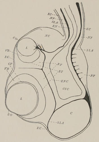
[Dr. Conant did not complete Fig. 72, and the accompanying outline of Fig. 7 of
Schewiakoff’s memoir (Beiträge zur Kenntnis des Acalephenauges, Morph. Jahrb.,
Bd. XV, H. 1) has been substituted.—Editor.]
Explanation of Letters in Text Figure.—C—concretion cavity; CO—cornea;
CP—capsule of lens; CSC—cavity of sensory club; EC—ectoderm; EN—endoderm;
ENC—endoderm of sensory club; L—lens; NC—network cells; NF—nerve fibres;
RT—retina; SLA—supporting lamella; VF—vitreous body.
Fig. 72 is a horizontal section through the large eye, and shows that
here, too, when the sections pass through the eye just radially, the
nuclei are not found at different levels sufficiently definite to suggest two
kinds of cells.
In the inner corner of the retina in the same figure (69) are seen
cells without pigment which show nuclei undoubtedly at different levels.
These cells in this position are a regular feature in the retina of the
smaller eye. Schewiakoff considers them purely visual, because of the
lack of pigment. In so doing it seems to me he forgets his own standard
for discriminating between pigment and visual cells. The pigment cells
of the retina, according to him, are the same thing as the cone-shaped
supporting cells found elsewhere in the nervous epithelium, and are,
therefore, distinguished from the visual cells primarily by shape and by
position of nucleus, secondarily by the greater development of pigment.
When on the ground of pigmentation alone he calls the cells in the
corner of the retina visual, he judges them by only the second test, and
in so doing virtually admits, as it seems to me, that shape of cell and
position of nucleus are matters of no great moment. His own standards
place him in a dilemma. If on the other hand he judges by the lack of
pigment, the cells are visual; if by shape of cell and position of nucleus,
they are both visual and pigment cells without the pigment or supporting
cells. What use there would be for simple unpigmented cells in
one limited region of the retina is hard to see, so he naturally takes the
other horn of the dilemma and calls them visual because they have little
or no pigment.
The distinction, then, between pigment and visual cells is brought
down to one of pigmentation only. Schewiakoff’s test for this is that in
the visual cells “Das Pigment durchsetzt aber nicht das ganze Protoplasma
des centralen Zellenabschnittes, sondern ist auf seine Oberfläche
beschrankt (Fig. 19, sz), so dass der innere, axiale, stark lichtbrechende
Theil vollkommen frei von demselben ist.” (’89, p. 37.) That is, in a
section through the ends of the retinal cells each pigment cell will
appear as a uniformly pigmented area, while each visual cell will appear
as a light, strongly refracting spot with a ring of pigment around its
periphery. This is the arrangement given in his Fig. 19.
An arrangement so definite ought to be easily made out in sections,
yet I have not been able to find it so. My sections show considerable
difference in the amount of pigmentation even in material preserved
with the same killing agent. If the retina is heavily pigmented the
ends of the cells have the appearance shown in Fig. 62, which represents
a portion of a cross-section. The ends are seen as clearly defined
polygonal areas differing among themselves in size, but not showing two
types of size, or two kinds of pigmentation, the one uniform, the other a
ring of pigment around a highly refracting central portion. If the
retina is but slightly pigmented—and some were so light as to make
depigmentation unnecessary—a difference is seen in the pigment, as
shown in Fig. 63, but in no case were areas found that showed a highly
refracting centre surrounded by a ring of pigment. (The unexplained
structures in Fig. 63 will be referred to a little later.)
Figures 59-62 are a series of four successive sections drawn with the
camera lucida for comparison with Schewiakoff’s Figs. 20 and 19, and
to show that the presence of two types of cells plainly marked within the
retina by the position of the nuclei at different levels is at least not clearly
demonstrated. Only the nuclei are drawn, since the cell bodies are not
easily distinguished from the surrounding fibres. The eye is the same as
that from which Fig. 72 was made. Fig. 59 shows a relatively small
number of nuclei of slightly larger size than usual. These I take for two
reasons to be nuclei of the ganglion cells that are found in the fibres at
the base of the retinal cells (Figs. 58, gc, 69 and 72). They are the first
nuclei struck in tracing sections toward the retina, and in the series from
which Fig. 58 was taken similar nuclei appeared in both transverse and
radial cuts through the retina stained brightly and clearly with hæmatoxylin,
whereas the nuclei of the retinal cells proper were stained a
diffuse brownish-yellow from pigment that had evidently gone into
solution. Fig. 60 shows the closely aggregated, smaller nuclei of the
retinal cells surrounded by the nuclei of the outlying ganglion cells.
Schewiakoff’s corresponding drawing (’89, Fig. 20) shows at this level a
definite alternation of the bodies and nuclei of unpigmented visual cells,
with the smaller, pigmented, proximal processes of the pigment cells. In
the next section (Fig. 61) the pigmented ends of a few of the cells have
been struck, and the following section (Fig. 62) shows that, in this
heavily pigmented specimen at least, there is no good evidence within
the retina itself of two kinds of cells, so that it is apparent that at any
rate we cannot accept Schewiakoff’s conception of the structure.
(b) Yet the fibres that Schewiakoff observed and associated with
special visual cells occur beyond question. Fig. 64 is a drawing of the
first cut through the vitreous body of Charybdea, and in among the
sections of the pigment streaks are seen sections of processes lying
within clear spaces exactly as Schewiakoff figures his visual fibres (’89,
Taf. II, Fig. 18). That the fibres occur is indisputable, but as to the cells
to which they belong I can say nothing except that from such evidence
as I have given in the preceding paragraph I conclude that they come
from pigmented retinal cells of not very different type within the retina
from the others, if different at all.
(c) On the third point, that the pigment streaks in the vitreous
body belong to underlying cells and are continued distally into fibrous
processes like the visual fibres of Schewiakoff, the evidence is decisive.
Fig. 58 has already shown it, and if this were not enough, a case of
unusual stoutness of the fibres drawn in Fig. 67 is conclusive. The
preparation from which the section is taken was one preserved with
corrosive-acetic, and I have drawn the outlines with the camera in order
to avoid exaggeration of the fibres as far as possible, and also to show
the shrinkage of the vitreous body (vb). It is the shrinkage of the
vitreous body that makes it so difficult to determine the exact relation of
structures seen in the vitreous body to the retina. The fibrous processes
run through the vitreous body to the “capsule” of the lens (cp) (see
also Fig. 72), a layer of homogeneous substance much resembling that of
the vitreous body, which is classed as a part of the vitreous body, but
usually in the shrinking adheres to the lens. The capsule is therefore
regarded by Schewiakoff as a secretion of the lens cells. Some fibres
were found by him to have the appearance of branching upon reaching
the surface of the capsule, others of passing through it and of seemingly
ending among the cells of the lens. The same appearances were given
in my sections. It is altogether impossible in the distal portion of the
vitreous body to distinguish between the fibres of Schewiakoff and those
that come from the long pigment cells. (Figs. 64-66 represent the
appearance of the vitreous body at successive levels, and are from the
same series of sections as Figs. 59-62 and 72.) In Fig. 64 the sections of
the processes that Schewiakoff calls visual are easily distinguished from
the sections of the long pigment cells. In Fig. 65, which is two or three
sections nearer the lens, the pigment cells are shown by their cross-sections
to be tapering down, and in Fig. 66, nearer still to the lens, the
two kinds of processes are no longer to be distinguished from each other.
In a few cases I have found pigment in a fibre which but for this would
be called one of the visual fibres of Schewiakoff. Such considerations as
these, the similar appearance in cross-section, the finding of pigment in
a few cases, and the inability to trace to any readily distinguished special
type of retinal cell, make me wonder whether the visual fibres of
Schewiakoff are anything more than the distal processes of pigment
cells, into which the pigment granules happened not to be produced at
the moment of fixation.
Fig. 63, however, where the retina was only slightly pigmented,
rather speaks against this view, for the number of darkly pigmented
areas seen here (which are shown beyond question by radial sections to
belong to the long pigment cells) is not great enough to account for the
number of both pigment areas and visual fibres of Schewiakoff seen in
such a section as Fig. 64. This would throw the visual fibres of
Schewiakoff back upon some of the slightly pigmented cells of Fig. 63,
otherwise not distinguished. I think the question cannot be settled
without the maceration of fresh material, and experiments upon eyes
killed in the light and in the dark.
In such cases as that of Fig. 63 it would seem conclusively shown
that the long pigment cells must belong to a different type from the
short, but as I have already said I can find no regularity in either their
shape or in the position of their nuclei. And on the other hand Fig. 58
shows that the reverse relation may obtain and the long cells be less
deeply pigmented on the edge of the retina than their shorter neighbors,
so that it looks as if all the short cells had to do was to project half their
pigment out into the vitreous body in order to become exactly like the
long ones. This they could do if, as is possibly the case, they are prolonged
into “visual fibres” of Schewiakoff that have escaped observation
and so do not appear in the drawing.
Fig. 58 shows one more thing that is worthy of remark in passing.
In the preparation in which the vitreous body (at this point at any rate)
was not shrunken away from the retina, the fibre from each long
pigment cell does not lie in a clearly defined space or “canal,” such as is
usually described as a constant structure of the vitreous body. Very
likely these canals are formed only by shrinkage around the fibres, and
the irregular shape of the spaces around the three fibres in Fig. 67 rather
bears out the same supposition.
As to the structure of the vitreous body, apart from the fibres and
pigment streaks already mentioned, I find it to be made up of prisms
extending from retina to capsule of lens, each containing a central axis
or fibre. Fig. 64 shows that the space around the pigment areas and
“visual fibres,” instead of being homogeneous, is wholly filled with the
polygonal cross-sections of these prisms. In Charybdea they are generally
more difficult to perceive than in my best material of Tripedalia
which was killed in acetic acid. In this the polygonal areas stood apart
from each other more plainly. Curiously enough I have been unable to
demonstrate in Tripedalia the “visual fibres” of Schewiakoff. Here and
there were found spaces that at first sight reminded of them (Fig. 69, sh),
but they contained no central fibre, and were probably due to shrinkage.
The polygonal areas themselves, however, often contained a clear
spot in the centre, at one side of which would be found the cross-section
of the fibre, as is shown in many cases in Fig. 68. The clear spot is here
undoubtedly due to shrinkage of the gelatinous substance of the prism.
I think that these prisms and fibres are the direct continuations of
retinal cells. In a section such as that drawn in Fig. 63, which takes
just the very tops of the cells of a slightly pigmented retina, in the
centre of the section just grazing the space that lies between the retina
and the shrunken vitreous body, most of the cells toward the middle
(where especially the extreme tips are taken) show in their centres a dot
exactly corresponding to the dots in the polygonal areas of the vitreous
body. In the exact middle of the section, where only the cell walls
appear, slightly indicated, a dot is seen in each case. The size and shape
of the ends of the cells correspond with those of the polygonal areas in
the vitreous body, and I do not doubt that the latter are continuations of
the former. The vitreous body, then, instead of being homogeneous, is
composed of the clear highly refracting outer ends of retinal cells. The
assumption lies near that these are the true visual rods, but of course it
is assumption only.
To give a brief review, the points in which my conclusions differ
from those of Schewiakoff are as follows: I find (1) that the long pigment
streaks are parts of retinal cells continued into processes like his
visual rods; (2) that the vitreous body is composed of prisms with central
fibres proceeding from retinal cells; (3) that I am unable to get satisfactory
evidence of two types of cell distinguishable within the retina, and
at any rate find considerable evidence against the two types he distinguishes.
These results are not wholly satisfactory, for they leave us with
three kinds of fibrous processes in the vitreous body which for the
present we are unable to trace to three, or even two distinguishable
types of cell in the retina. It would be more pleasing if we could
confirm Schewiakoff’s simple conception of the structure, with its one set
of visual rods in the vitreous body referable to a clearly marked type of
sensory cells in the retina, but I think the evidence that has been brought
up justifies the conclusion that in some respects he saw too much, in
other respects too little. This is not to be wondered at, since his material,
to judge from a single statement, consisted of but twelve marginal
bodies, and, moreover, the work on Charybdea forms but one portion
of a paper that is excellent for the clearness of its descriptions and illustrations.
Before leaving the subject I must mention that Wilson suggested
from his observations on Chiropsalmus that the vitreous body had a
prismatic structure, but he was probably mistaken when he thought he
found evidence of nuclei in it. Claus says that the retina is composed of
pigment and rod cells alternating, and Wilson agrees with him, but
under a sketch of a sense cell from the nerve he makes the express statement
“not very well preserved.” It seems very probable, therefore, that
he followed Claus’s interpretation rather than independent observations,
and Claus interpreted his results very much by analogy of what had been
found in other forms.
The smaller complex eye which is represented in Fig. 69 agrees in
structure very closely with the larger. The chief differences are that
sections do not show pigment extending into the vitreous body, that
there is no “capsule” to the lens, and that the lens seems to be supported
by a kind of stalk formed by a thickening of gelatine of the supporting
lamella (sl). The gelatinous thickening lies between the lens and an outgrowth
of endodermal cells (en) from the canal of the club. This
outgrowth is a constant feature, figured by Claus and Schewiakoff for
Charybdea, and by Wilson for Chiropsalmus, and found in Tripedalia
also. The regularity of its appearance in all three genera leads one to
suspect that it may have some significance not yet understood.
Just above the smaller eye there lies a mass of cells of peculiar
structure (Fig. 69, nc). They are of a rounded polygonal contour, with a
comparatively small circular nucleus in the centre, and are found in
this region only. In and amongst them bundles of fibrous tissue are
found in the sections, which pass from the surface cells to the supporting
lamella. Claus describes the contents of these cells as coarsely granular
protoplasm and says they cannot be taken for ganglion cells. He is
inclined to believe that they play the part of a special supporting tissue.
Schewiakoff, on the other hand, is convinced that they are ganglion cells,
and finds processes passing out from them (’89, Taf. II, Fig. 22). I find,
however, that the cell contours are perfectly regular and clearly without
processes, and it is incomprehensible to me how, if his material was at all
well preserved, he could for a moment have taken them for the same
thing as the big multipolar ganglion cells with large nucleus and
nucleolus which lie in about the same region and were correctly described
and figured by Claus but are not specially mentioned by Schewiakoff. I
cannot agree with Claus, however, that their contents are composed of
coarsely granular protoplasm. That which appears such by low magnification
shows itself under high powers to be a beautiful network with
thickenings at the nodes of the meshes, which is brought out very plainly
by a cytoplasmic stain such as Lyons blue. Around the nucleus is seen
a more or less well-defined clear zone. What the function of the cell is
remains as unknown to me as to Claus and Schewiakoff.
There is left one more point in reference to the nervous system upon
which I wish to say a word. Claus and Schewiakoff both describe the
wall of the crystalline sac as structureless, formed by the bare supporting
lamella. The credit is due to H. V. Wilson of finding in Chiropsalmus
that it has a special lining of epithelial cells, which he figures as a continuous,
flattened layer. In both Charybdea and Tripedalia I find traces
of the same in nuclei here and there, but whether they are the remains
of a once continuous layer or not the sections do not show satisfactorily.
This ends the account of what it seemed worth while to say at
present upon the nervous system. In concluding, the writer wishes to
express his thanks for the help afforded by Dr. Wilson’s notes, in particular
on the subject of the vascular lamellæ, and desires to make
especial acknowledgment of his indebtedness to Professor Brooks, whose
suggestions, based upon many years of experience with the Medusæ,
have been most welcome and helpful, and whose evidences of unfailing
kindliness, both in Jamaica at the time the material was obtained and in
Baltimore when it was being studied in the laboratory, take a most
honored part in the pleasant memories associated with the work.