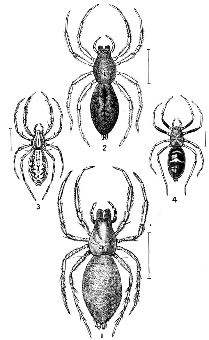
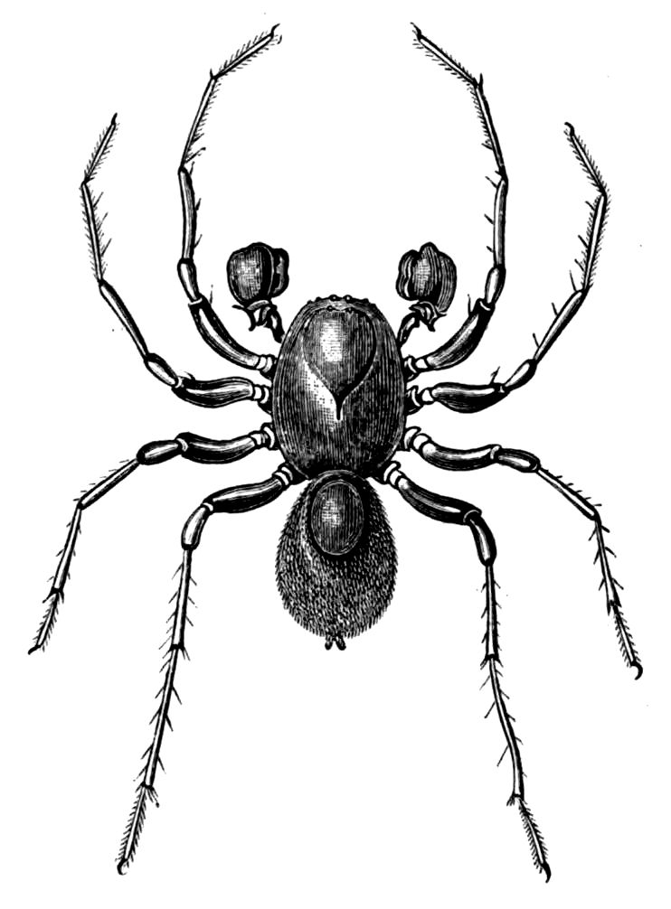
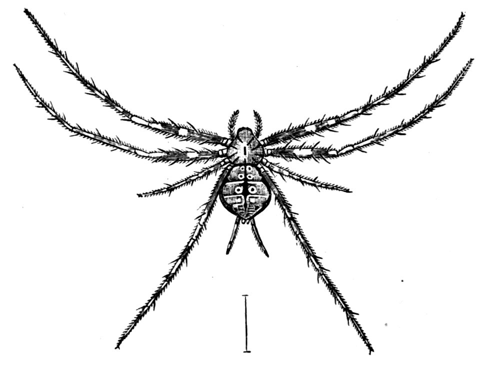
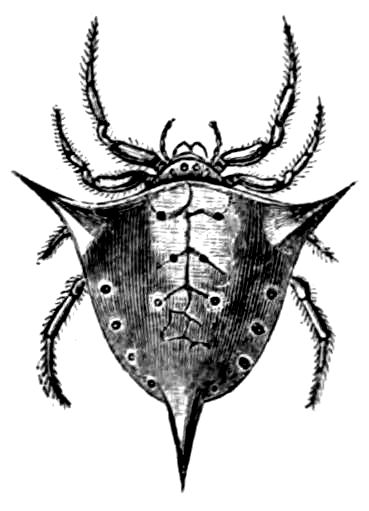
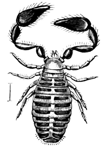
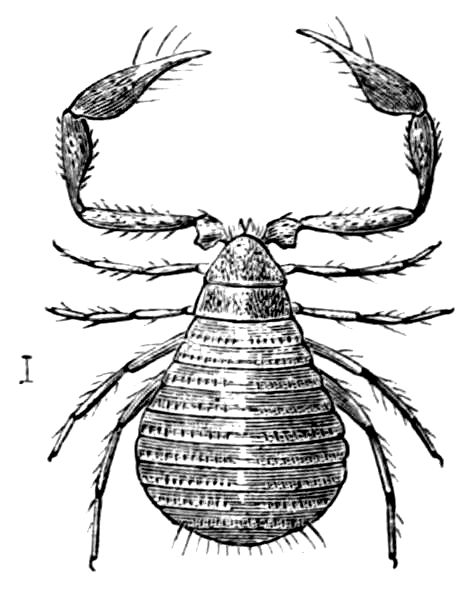
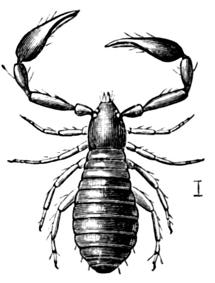
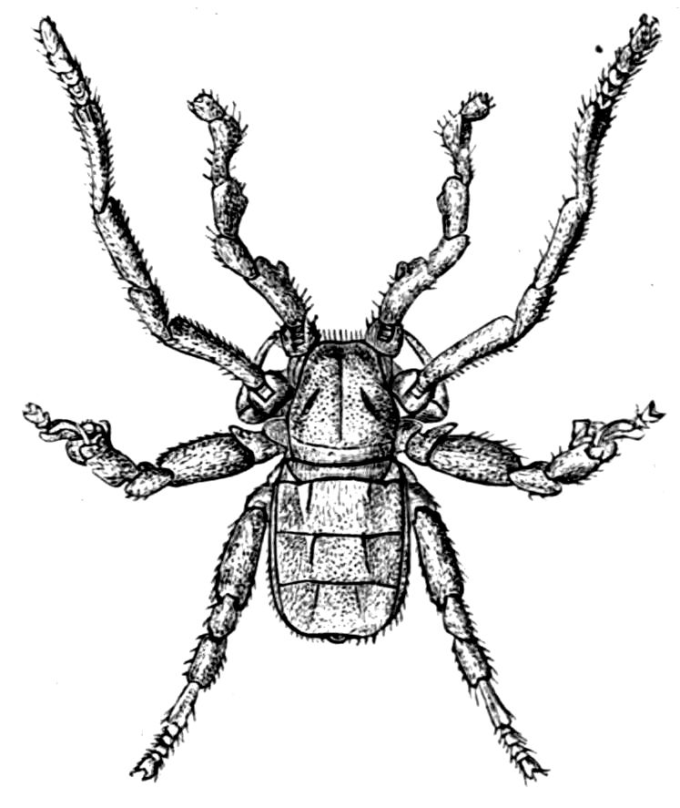
New original cover art included with this eBook is granted to the public domain.
EDITED BY
S. F. HARMER, Sc.D., F.R.S., Fellow of King’s College, Cambridge;
Keeper of the Department of Zoology in the British Museum
(Natural History)
AND
A. E. SHIPLEY, M.A., Fellow and Tutor of Christ’s College,
Cambridge; Reader in Zoology in the University
VOLUME IV
MACMILLAN AND CO., Limited
LONDON · BOMBAY · CALCUTTA
MELBOURNE
THE MACMILLAN COMPANY
NEW YORK · BOSTON · CHICAGO
ATLANTA · SAN FRANCISCO
THE MACMILLAN CO. OF CANADA, Ltd.
TORONTO
CRUSTACEA
By Geoffrey Smith, M.A. (Oxon.), Fellow of New College, Oxford;
and the late W. F. R. Weldon, M.A. (D.Sc., Oxon.), formerly Fellow
of St. John’s College, Cambridge, and Linacre Professor of Human
and Comparative Anatomy, Oxford
TRILOBITES
By Henry Woods, M.A., St. John’s College, Cambridge; University
Lecturer in Palaeozoology
INTRODUCTION TO ARACHNIDA, AND
KING-CRABS
By A. E. Shipley, M.A., F.R.S., Fellow and Tutor of Christ’s College,
Cambridge; Reader in Zoology
EURYPTERIDA
By Henry Woods, M.A., St. John’s College, Cambridge; University
Lecturer in Palaeozoology
SCORPIONS, SPIDERS, MITES, TICKS, ETC.
By Cecil Warburton, M.A., Christ’s College, Cambridge; Zoologist
to the Royal Agricultural Society
TARDIGRADA (WATER-BEARS)
By A. E. Shipley, M.A., F.R.S., Fellow and Tutor of Christ’s College,
Cambridge; Reader in Zoology
PENTASTOMIDA
By A. E. Shipley, M.A., F.R.S., Fellow and Tutor of Christ’s College,
Cambridge; Reader in Zoology
PYCNOGONIDA
By D’Arcy W. Thompson, C.B., M.A., Trinity College, Cambridge;
Professor of Natural History in University College, Dundee
MACMILLAN AND CO., LIMITED
ST. MARTIN’S STREET, LONDON
1909
All the ingenious men, and all the scientific men, and all
the fanciful men, in the world, with all the old German bogypainters
into the bargain, could never invent ... anything
so curious, and so ridiculous, as a lobster.
Charles Kingsley, The Water-Babies.
For, Spider, thou art like the poet poor,
Whom thou hast help’d in song.
Both busily, our needful food to win,
We work, as Nature taught, with ceaseless pains,
Thy bowels thou dost spin,
I spin my brains.
Southey, To a Spider.
Last o’er the field the Mite enormous swims,
Swells his red heart, and writhes his giant limbs.
Erasmus Darwin, The Temple of Nature.
PREFACE
The Editors feel that they owe an apology and some explanation
to the readers of The Cambridge Natural History for the delay
which has occurred in the issue of this, the fourth in proper
order, but the last to appear of the ten volumes which compose
the work. The delay has been due principally to the untimely
death of Professor W. F. E. Weldon, who had undertaken to
write the Section on the Crustacea. The Chapter on the
Branchiopoda is all he actually left ready for publication, but it
gives an indication of the thorough way in which he had intended
to treat his subject. He had, however, superintended the
preparation of a number of beautiful illustrations, which show
that he had determined to use, in the main, first-hand knowledge.
Many of these figures have been incorporated in the article by
Mr. Geoffrey Smith, to whom the Editors wish to express their
thanks for taking up, almost at a moment’s notice, the task which
had dropped from his teacher’s hand.
A further apology is due to the other contributors to this
volume. Their contributions have been in type for many years,
and owing to the inevitable delays indicated above they have been
called upon to make old articles new, ever an ungrateful labour.
The appearance of this volume completes the work the Editors
embarked on some sixteen years ago. It coincides with the
cessation of an almost daily intercourse since the time when they
“came up” to Cambridge as freshmen in 1880.
S. F. Harmer.
A. E. Shipley.
March 1909.
CONTENTS
vii
PAGE
Scheme of the Classification adopted in this Volume
CRUSTACEA — CHAPTER I CRUSTACEA—GENERAL ORGANISATION — CHAPTER II CRUSTACEA ( CONTINUED ): ENTOMOSTRACA—BRANCHIOPODA—PHYLLOPODA—CLADOCERA—WATER-FLEAS — SUB-CLASS I.—ENTOMOSTRACA. — CHAPTER III CRUSTACEA ( CONTINUED ): COPEPODA — Order II. Copepoda. — CHAPTER IV CRUSTACEA ( CONTINUED ): CIRRIPEDIA—PHENOMENA OF GROWTH AND SEX—OSTRACODA — Order III. Cirripedia. — Order IV. Ostracoda. — CHAPTER V CRUSTACEA ( CONTINUED ): MALACOSTRACA: LEPTOSTRACA—PHYLLOCARIDA: EUMALACOSTRACA: SYNCARIDA—ANASPIDACEA: PERACARIDA—MYSIDACEA—CUMACEA—ISOPODA—AMPHIPODA: HOPLOCARIDA—STOMATOPODA — SUB-CLASS II.—MALACOSTRACA. — DIVISION 1. SYNCARIDA. — DIVISION 2. PERACARIDA. — DIVISION 3. HOPLOCARIDA. — CHAPTER VI CRUSTACEA ( CONTINUED )—EUMALACOSTRACA ( CONTINUED ): EUCARIDA—EUPHAUSIACEA—COMPOUND EYES—DECAPODA — DIVISION 4. EUCARIDA. — CHAPTER VII REMARKS ON THE DISTRIBUTION OF MARINE AND FRESH-WATER CRUSTACEA — A. Marine. — B. Fresh-Water. [164]
1
CHAPTERS I and III-VII
BY
GEOFFREY SMITH, M.A. (Oxon.)
Fellow of New College, Oxford
CHAPTER II
BY
The Late W. F. R. WELDON, M.A. (D.Sc. Oxon.)
Formerly Fellow of St. John’s College, Cambridge, and Linacre Professor of Human and Comparative Anatomy, Oxford
3
The Crustacea are almost exclusively aquatic animals, and they
play a part in the waters of the world closely parallel to that
which insects play on land. The majority are free-living, and
gain their sustenance either as vegetable-feeders or by preying
upon other animals, but a great number are scavengers, picking
clean the carcasses and refuse that litter the ocean, just as
maggots and other insects rid the land of its dead cumber.
Similar to insects also is the great abundance of individuals
which represent many of the species, especially in the colder
seas, and the naturalist in the Arctic or Antarctic oceans
has learnt to hang the carcasses of bears and seals over the side
of the boat for a few days in order to have them picked
absolutely clean by shoals of small Amphipods. It is said that
these creatures, when crowded sufficiently, will even attack
living fishes, and by sheer press of numbers impede their escape
and devour them alive. Equally surprising are the shoals of
minute Copepods which may discolour the ocean for many miles,
an appearance well known to fishermen, who take profitable toll
of the fishes that follow in their wake. Despite this massing
together we look in vain for any elaborate social economy, or for
the development of complex instincts among Crustacea, such as
excite our admiration in many insects, and though many a crab
or lobster is sufficiently uncanny in appearance to suggest
unearthly wisdom, he keeps his intelligence rigidly to himself,
encased in the impenetrable reserve of his armour and vindicated
by the most powerful of pincers. It is chiefly in the variety
of structure and in the multifarious phases of life-history that
4the interest of the Crustacea lies. Before entering into an
examination of these matters, it will be well to take a general
survey of Crustacean organisation, to consider the plan on which
these animals are built, and the probable relation of this plan
to others met with in the animal kingdom.
The Crustacea, to begin with, are a Class of the enormous
Phylum Arthropoda, animals with metamerically segmented
bodies and usually with externally jointed limbs. Their bodies
are thus composed of a series of repeated segments, which are on
the whole similar to one another, though particular segments
may be differentiated in various respects for the performance of
different functions. This segmentation is apparent externally,
the surface of a Crustacean being divided typically into a
number of hard chitinous rings, some of which may be fused
rigidly together, as in the carapace of the crabs, or else
articulated loosely.
Each segment bears typically a pair of jointed limbs, and
though they vary greatly in accordance with the special
functions for which they are employed, and may even be absent
from certain segments, they may yet be reduced to a common
plan and were, no doubt, originally present on all the segments.
Passing from the exterior to the interior of the body we find,
generally speaking, that the chief system of organs which exhibits
a similar repetition, or metameric segmentation, is the nervous
system. This system is composed ideally of a nervous ganglion
situated in each segment and giving off peripheral nerves, the
several ganglia being connected together by a longitudinal cord.
This ideal arrangement, though apparent during the embryonic
development, becomes obscured to some extent in the adult
owing to the concentration or fusion of ganglia in various parts
of the body. The other internal organs do not show any clear
signs of segmentation, either in the embryo or in the adult;
the alimentary canal and its various diverticula lie in an
unsegmented body-cavity, and are bathed in the blood which
courses through a system of narrow canals and irregular spaces
which surround all the organs of the body. A single pair, or
at most two pairs of kidneys are present.
The type of segmentation exhibited by the Crustacea is thus
of a limited character, concerning merely the external skin with
its appendages, and the nervous system, and not touching any
5of the other internal organs.[1] In this respect the Crustacea agree
with all the other Arthropods, in the adults of which the
segmentation is confined to the exterior and to the nervous
system, and does not extend to the body-cavity and its contained
organs; and for the same reason they differ essentially from all
other metamerically segmented animals, e.g. Annelids, in which
the segmentation not only affects the exterior and the nervous
system, but especially applies to the body-cavity, the musculature,
the renal, and often the generative organs. The Crustacea also
resemble the other Arthropoda in the fact that the body-cavity
contains blood, and is therefore a “haemocoel,” while in the
Annelids and Vertebrates the segmented body-cavity is distinct
from the vascular system, and constitutes a true “coelom.”
To this important distinction, and to its especial application to
the Crustacea, we will return, but first we may consider more
narrowly the segmentation of the Crustacea and its main types
of variation within the group. In order to determine the
number of segments which compose any particular Crustacean
we have clearly two criteria: first, the rings or somites of which
the body is composed, and to each of which a pair of
limbs must be originally ascribed; and, second, the nervous
ganglia.
Around and behind the region of the mouth there is very
little difficulty in determining the segments of the body, if we
allow embryology to assist anatomy, but in front of the mouth
the matter is not so easy.
In the Crustacea the moot point is whether we consider the
paired eyes and first pair of antennae as true appendages belonging
to two true segments, or whether they are structures sui
generis, not homologous to the other limbs. With regard to the
first antennae we are probably safe in assigning them to a true
body-segment, since in some of the Entomostraca, e.g.Apus,
the nerves which supply them spring, not from the brain as in
more highly specialised forms, but from the commissures which
pass round the oesophagus to connect the dorsally lying brain
to the ventral nerve-cord. The paired eyes are always innervated
from the brain, but the brain, or at least part of it, is very
6probably formed of paired trunk-ganglia which have fused into
a common cerebral mass; and the fact that under certain circumstances
the stalked eye of Decapods when excised with its
peripheral ganglion[2] can regenerate in the form of an antenna,
is perhaps evidence that the lateral eyes are borne on what were
once a pair of true appendages.
Now, with regard to the segmentation of the body, the
Crustacea fall into three categories: the Entomostraca, in which
the number of segments is indefinite; the Malacostraca, in
which we may count nineteen segments, exclusive of the terminal
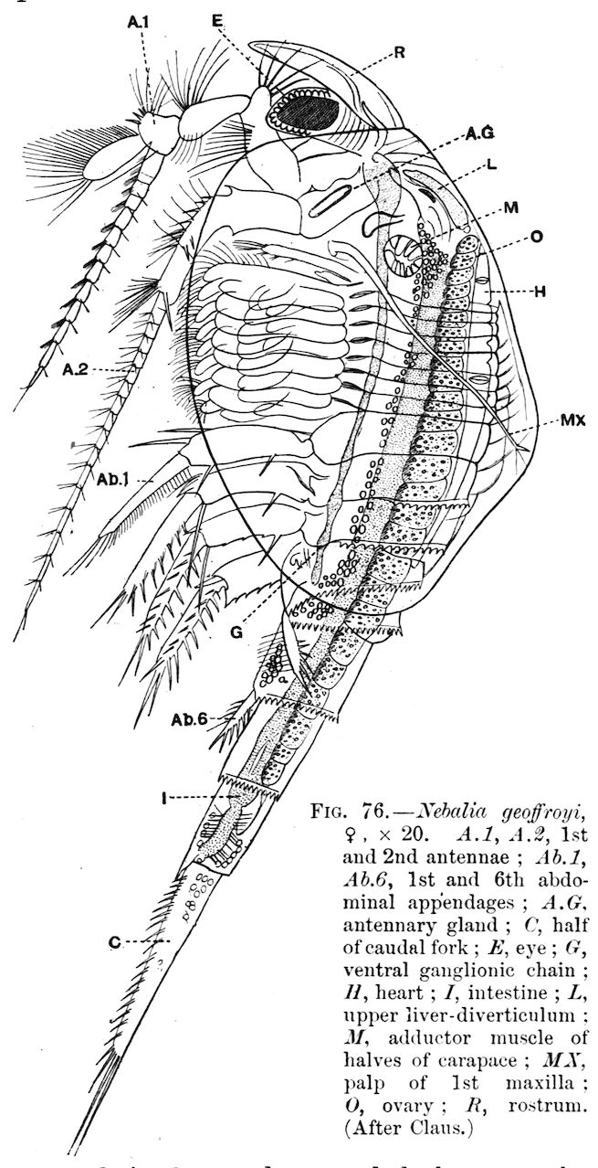
piece or telson and omitting the lateral eyes; and the Leptostraca,
including the single recent genus Nebalia, in which the segmentation
of head and thorax agrees exactly with that of the
Malacostraca, but in the abdomen there are two additional
segments.
It has been usually held that the indefinite number of
segments characteristic of the Entomostraca, and especially the
indefinitely large number of segments characteristic of such
Phyllopods as Apus, preserves the ancestral condition from
which the definite number found in the Malacostraca has been
derived; but recently it has been clearly pointed out by Professor
Carpenter[3] that the number of segments found in the Malacostraca
and Leptostraca corresponds with extraordinary exactitude to
the number determined as typical in all the other orders of
Arthropoda. This remarkable correspondence (it can hardly
be coincidence) seems to point to a common Arthropodan plan
of segmentation, lying at the very root of the phyletic tree;
and if this is so, we are forced to the conclusion that the
Malacostraca have retained the primitive type of segmentation
in far greater perfection than the Entomostraca, in some of
which many segments have been added, e.g. Phyllopoda, while
in others segments have been suppressed, e.g. Cladocera,
Ostracoda. It may be objected to this view of the primitive
condition of segmentation in the Crustacea that the Trilobites,
which for various reasons are regarded as related to the ancestral
Crustaceans, exhibit an indefinite and often very high number
of segments; but, as Professor Carpenter has pointed out, the
oldest and most primitive of Trilobites, such as Olenellus, possessed
7few segments which increase as we pass from Cambrian to
Carboniferous genera.
The following table shows the segmentation of the body in
the Malacostraca, as compared with that of Limulus (cf. p. 263),
Insecta, the primitive Myriapod Scolopendrella, and Peripatus.
It will be seen that the correspondence, though not exact, is
very close, especially in the first four columns, the number
of segments in Peripatus being very variable in the different
species.
Table showing the Segmentation of various Arthropods
The appendages of the Crustacea exhibit a wonderful variety
8of structure, but these variations can be reduced to at most
two, and possibly to one fundamental plan. In a typical
Crustacean, besides the paired eyes, which may be borne on
stalks, possibly homologous to highly modified limbs, there are
present, first, two pairs of rod-like or filamentous antennae,
which in the adult are usually specialised for sensory purposes,
but frequently retain their primitive function as locomotory
limbs even in the adult, e.g. Ostracoda; while in the Nauplius
larva, found in almost all the chief subdivisions of the Crustacea,
the two pairs of antennae invariably aid in locomotion, and
the base of the second antennae is usually furnished with sharp
biting spines which assist mastication. Following the antennae
is a pair of mandibles which are fashioned for biting the food
or for piercing the prey, and posterior to these are two pairs
of maxillae, biting organs more slightly built than the
mandibles, whose function it is to lacerate the food and prepare
it for the more drastic action of the mandibles. So far, with
comparatively few exceptions, the order of specialisation is
invariable; but behind the maxillae the trunk-appendages vary
greatly both in structure and function in the different groups.
As a general rule, the first or first few thoracic limbs are
turned forwards toward the mouth, and are subsidiary to
mastication; they are then called maxillipedes; this happens
usually in the Malacostraca, but to a much less extent in the
Entomostraca; and in any case these appendages immediately
behind the maxillae never depart to any great extent from a
limb-like structure, and they may graduate insensibly into the
ordinary trunk-appendages. The latter show great diversity in
the different Crustacean groups, according as the animals lead
a natatory, creeping, or parasitic method of life; they may
be foliaceous, as in the Branchiopoda, or biramous, as in the
swimming thoracic and abdominal appendages of the Mysidae,
or simply uniramous, as in the walking legs of the higher
Decapoda, and the clinging legs of various parasitic forms.
Without going into detailed deviations of structure, many
of which will be described under the headings of special groups,
it is clear from the foregoing description and from Fig. 1 (p. 10),
that three main types of appendage can be distinguished: first,
the foliaceous or multiramous; second, the biramous; and, third,
the uniramous.
9We may dismiss the uniramous type with a few words: it
is obviously secondarily derived from the biramous type; this
can be proved in detail in nearly every case. Thus, the uniramous
second antennae of some adult forms are during the Nauplius
stage invariably biramous, a condition which is retained in the
adult Cladocera. Similarly the uniramous walking legs of many
Decapoda pass through a biramous stage during development,
the outer branches or exopodites of the limbs being suppressed
subsequently, while the primitively biramous condition of the
thoracic limbs is retained in the adults of the Schizopoda, which
doubtless own a common ancestry with the Decapoda. The only
Crustacean limb which appears to be constantly uniramous both
in larval and adult life is the first pair of antennae.
We are reduced, therefore, to two types—the foliaceous and
biramous. Sir E. Ray Lankester,[6] in one of his most incisive
morphological essays, has explained how these two types are
really fundamentally the same. He compares, for instance, the
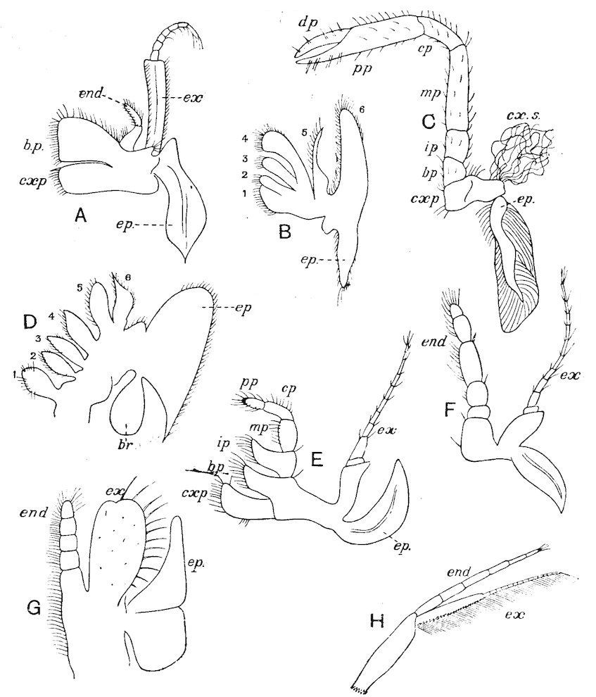
foliaceous first maxillipede (Fig. 1, A), or the second maxilla
(Fig. 1, B) of a Decapod, e.g.Astacus, with the foliaceous thoracic
limb of Branchipus (Fig. 1, D), and with the typically biramous
first maxillipede of a Schizopod (Fig. 1, F).
In each case there is present, on the outer edge of the limb,
one or more projections or epipodites which are generally
specialised for respiratory purposes, and may carry the gills.
The 6th and 5th “endites” in the foliaceous limb (Fig. 1, D)
are compared with the exopodite and endopodite respectively
of the biramous limb, while the endites 4–1 of the foliaceous
limb are found in the basal joints of the biramous limb.
Lankester presumes that the biramous type of limb throughout
has been derived from the foliaceous type by the suppression
of the endites 1–4, as discrete rami, and the exaggerated
development of the endites 5 and 6, as above indicated.
10
Fig. 1.—Appendages of Crustacea (A-G) and Trilobita (H). A, First maxillipede of Astacus; B, second maxilla of Astacus; C, second walking leg of Astacus; D, thoracic limb of Branchipus; E, first maxillipede of Mysis; F, first maxillipede of Gnathophausia; G, thoracic limb of Nebalia; H, thoracic limb of Triarthrus. bp, basipodite; br, bract; cp, carpopodite; cxp, coxopodite; cx.s, coxopoditic setae; dp, dactylopodite; end, endopodite; ep, epipodite; ex, exopodite; ip, ischiopodite; mp, meropodite; pp, propodite; 1–6, the six endites.
The essential fact that the two types of limb are built on the
same plan may be considered as established; but it may be
urged that the biramous type represents this common plan more
nearly than the foliaceous. It is, at any rate, certain that in
the maxillipedes of the Decapoda we witness the conversion
of the biramous type into the foliaceous by the expansion of
the basal joints concomitantly with the assumption by the
maxillipedes of masticatory functions. Thus in the Decapoda
the first maxillipede is decidedly foliaceous owing to the expanded
“gnathobases” (Fig. 1, A, bp, cxp), and the second maxillipedes
are flattened, with their basal joints somewhat expanded
and furnished with biting hairs; but in the “Schizopoda”
11(e.g.Mysis) the first maxillipede is a typical biramous limb,
though the expanded gnathobases in some forms are beginning
to project (Fig. 1, E), while the limb following, which corresponds
to the second maxillipede of Decapods, is simply a biramous
swimming leg. Besides this obvious conversion of a biramous
into a foliaceous limb, further evidence of the fundamental
character of the biramous type is found, first, in its invariable
occurrence in the Nauplius stage, which does not necessarily
mean that the ancestors of the Crustacea possessed this type
of limb in the adult, but which does imply that this type of
limb was possessed at some period of life by the common
ancestral Crustacean; and, second, the limbs of the Trilobita,
a group which probably stands near the origin of the Crustacea,
have been shown by Beecher to conform to the biramous
type (Fig. 1, H). Furthermore, the thoracic limbs of Nebalia,
an animal which combines many of the characteristics of
Entomostraca and Malacostraca, and is therefore considered as
a primitive type, despite their flattened character, are really built
upon a biramous plan (Fig. 1, G).
In conclusion, we may point out that this view of the
Crustacean limb, as essentially a biramous structure, agrees with
the conclusion derived from our consideration of the segmentation
of the body, and points less to the Branchiopoda as
primitive Crustacea and more to some generalised Malacostracan
type.
So far we have shortly dealt with those systems of organs
which are clearly affected by the metameric segmentation of the
body; we must now expose the condition of the body-cavity to
a similar scrutiny. If we remove the external integument of a
Crustacean, we find that the internal organs do not lie in a
spacious and discrete body-cavity, as is the case in the Annelids
and Vertebrates, but that they are packed together in an irregular
system of spaces (“haemocoel”) in communication with the
vascular system and containing blood. In the Entomostraca and
smaller forms generally, a definite vascular system hardly exists,
though a central heart and artery may serve to propel the blood
through the irregular lacunae of the body-cavity; but in the
larger Malacostraca a complicated system of arteries may be
present which pour the blood into fairly definitely arranged
spaces surrounding the chief organs. These spaces return the
12blood to the pericardium, and so to the heart again through the
apertures or ostia which pierce its walls.
This condition of the body-cavity or haemocoel is reproduced
in the adults of all Arthropods, but in some of them by following
the development we can trace the steps by which the true coelom
is replaced by the haemocoel. In the embryos of all Arthropods
except the Crustacea, a true closed metamerically segmented
coelom is formed as a split in the mesodermal embryonic layer
of cells, distinct from the vascular system. During the course
of development the segmented coelomic spaces and their walls
give rise to the reproductive organs and to certain renal organs
in Peripatus, Myriapoda, and Arachnida (nephridia and coxal
glands), but the general body-cavity is formed as an extension
of the vascular system, which is laid down outside the coelom
by a canaliculisation of the extra-coelomic mesoderm. In the
embryos of the Crustacea, however, there is never at any time
a closed segmented coelom, and in this respect the Crustacea
differ from all other Arthropods. The only clear instance in
which metamerically repeated mesodermal cavities have been
seen in the embryo Crustacean is that of Astacus; here Reichenbach[7]
states that in the abdomen segmental cavities are formed
which subsequently break down; but even in this instance no
connexion has been shown to subsist between these embryonic
cavities and the reproductive and excretory organs of the adult.
Since the connexion between the coelom and the excretory
organs is always a very close one throughout the animal
kingdom, interest naturally centres upon the renal organs in
Crustacea, and it has been suggested that these organs in
Crustacea represent the sole remains, with the possible exception
of the gonads, of the coelom. Since, at any rate, a part of the
kidneys appears to be developed as a closed sac in the mesoderm,
and since they possess a possible segmental value, this suggestion
is plausible; but, on the other hand, since there are never more
than two pairs of kidneys, and since they are totally unconnected
with the gonads or with any other indication of a segmented
coelom, the suggestion remains purely hypothetical.
The renal organs of the Crustacea, excluding the Malpighian
tubes present in some Amphipods which open into the alimentary
canal, and resemble the Malpighian tubes of Insects, consist of
13two pairs—the antennary gland, opening at the base of the
second antenna, and the maxillary gland, opening on the second
maxilla. These two pairs of glands rarely subsist together in
the adult condition, though this is said to be the case in Nebalia
and possibly Mysis; the antennary glands are characteristic of
adult Malacostraca[8] and the larvae of the Entomostraca, while the
maxillary glands (“shell-glands”) are present in adult Entomostraca
and larval Malacostraca, that is to say, the one pair replaces
the other in the two great subdivisions of the Crustacea. The shell-gland
of the Entomostraca is a simple structure consisting of a
coiled tube opening to the exterior on the external branch of the
second maxilla, and ending blindly in a dilated vesicle, the end-sac.
The antennary gland of the Malacostraca is usually more
complicated: these complications have been studied especially by
Weldon,[9] Allen, and Marchal[10] in the Decapoda. In a number
of forms we have a tube opening to the exterior at the base of
the second antenna, and expanding within to form a spacious
bladder into which the coiled tubular part of the kidney opens,
while at the extremity of this coiled portion is the vesicle called
the end-sac. This arrangement may be modified; thus in
Palaemon Weldon described the two glands as fusing together
above and below the oesophagus, the dorsal commissure expanding
into a huge sac stretching dorsally down the length of the
body. This closed sac with excretory functions thus comes to
resemble a coelomic cavity, and the view that it is really coelomic
has indeed been upheld.
A modified form of this view is that of Vejdovský, who
describes a funnel-apparatus leading from the coiled tube into
the end-sac of the antennary gland of Amphipods; he regards
the end-sac alone as representing the coelom, while the funnel
and coiled tube represent the kidney opening into it.
Not very much is known of the development of these various
structures. Some authors have considered that both antennary
and maxillary glands are developed in the embryo from ectodermal
inpushings, but the more recent observations of Waite[11]
on Homarus americanus indicate that the antennary gland at
14any rate is a composite structure, formed by an ectodermal
ingrowth which meets a mesodermal strand, and from the latter
are produced the end-sac and perhaps the tubular excretory
portions of the gland with their derivatives.
With regard to the possible metameric repetition of the
renal organs, it is of interest to note that by feeding Mysis and
Nebalia on carmine, excretory glands of a simple character were
observed by Metschnikoff situated at the bases of the thoracic
limbs.
The alimentary canal of the Crustacea is a straight tube
composed of three parts—a mid-gut derived from the endoderm
of the embryo, and a fore- and hind-gut formed by ectodermal
invaginations in the embryo which push into and fuse with the
endodermal canal. The regions of the fore- and hind-gut can
be recognised in the adult by the fact of their being lined with
the chitinous investment which is continued over the external
surface of the body forming the hard exoskeleton, while the
mid-gut is naked. The chitinous lining of fore- and hind-gut
is shed whenever the animal moults. In the Malacostraca, in
which a complicated “gastric mill” may be present, the chitinous
lining of this part of the gut is thrown into ridges bearing
teeth, and this stomach in the crabs and lobsters reaches a high
degree of complication and materially assists the mastication of
the food. The gut is furnished with a number of secretory and
metabolic glands; the so-called liver, which is probably a hepatopancreas,
opening into the anterior end of the mid-gut, is directed
forwards in most Entomostraca and backwards in the Malacostraca,
in the Decapoda developing into a complicated branching organ
which fills a large part of the thorax. In the Decapoda peculiar
vermiform caeca of doubtful function are present, a pair of which
open into the gut anteriorly where fore-passes into mid-gut,
and a single asymmetrically placed caecum opens posteriorly into
the alimentary tract where mid- passes into hind-gut.
The disposition of these caeca, marking as they do the
morphological position of fore-, mid-, and hind-gut, is of peculiar
interest owing to the variations exhibited. From some unpublished
drawings of Mr. E. H. Schuster, which he kindly lent
me, it appears that in certain Decapods, e.g.Callianassa subterranea,
the length of the mid-gut between the anterior and
posterior caeca is very long; in Carcinus maenas it is considerable;
15in Maia squinado it is greatly reduced, the caeca being
closely approximated; while in Galathea strigosa the caeca are
greatly reduced, and the mid-gut as a separate entity has almost
disappeared. The relation of these variations to the habits of
the different crabs and to their modes of development is unknown.
The reproductive organs usually make their appearance as
a small paired group of mesodermal cells in the thorax comparatively
late in life; and neither in their early development nor
in the adult condition do they show any clear signs of segmentation
or any connexion with a coelomic cavity. The sexes are
usually separate, but hermaphroditism occurs sporadically in
many forms, and as a normal condition in some parasitic groups
(see pp. 105–107). The adult gonads are generally simple paired
tubes, from the walls of which the germ-cells are produced, and
as these grow and come to maturity they fill up the cavities of
the tubes; special nutrient cells are rarely differentiated, though
in some cases (e.g. Cladocera) a few ova nourish themselves by
devouring their sister-cells (see p. 44). The oviducts and vasa
deferentia are formed as simple outgrowths from the gonadial
tubes, which acquire an opening to the exterior; they are usually
poorly supplied with accessory glands, the epithelium of the
canals often supplying albuminous secretions for cementing the
eggs together, while the lining of the vasa deferentia may be
instrumental in the formation of spermatophores for transferring
large packets of spermatozoa to the female. In the vast
majority of Crustacea copulation takes place, the male passing
spermatophores or free spermatozoa into special receptacles
(spermathecae), or into the oviducts of the female. The spermatophores
are hollow chitinous structures in which the spermatozoa
are packed; they are often very large and assume characteristic
shapes, especially in the Decapoda.
The spermatozoa show a great variety of structure, but they
conform to two chief types—the filiform, which are provided
with a long whip-like flagellum; and the amoeboid, which are
furnished with radiating pseudopodia, and are much slower in
their movements. The amoeboid spermatozoa of some of the
Decapoda contain in the cell-body a peculiar chitinous capsule,
and Koltzoff[12] has observed that when the spermatozoon has
16settled upon the surface of the egg the chitinous capsule
becomes suddenly exceedingly hygroscopic, swells up, and explodes,
driving the head of the spermatozoon into the egg. We cannot
enter here into a description of the embryological changes by
which the egg is converted into the adult form. Crustacean
eggs as a whole contain a large quantity of yolk, but in some
forms total segmentation occurs in the early stages, which is
converted later into the pyramidal type, i.e. the blastomeres are
arranged round the edge, and the yolk in the centre is only partly
segmented to correspond with them. The eggs during the early
stages of development are in almost all cases (except Branchiura,
p. 77, and Anaspides, p. 116) carried about by the female either in
a brood-pouch (Branchiopoda, Ostracoda, Cirripedia, Phyllocarida,
Peracarida), or agglutinated to the hind legs or some other part
of the body (Copepoda, Eucarida), or in a chamber formed from
the maxillipedes (Stomatopoda). Development may be direct,
without a complicated metamorphosis, or indirect, the larva
hatching out in a form totally different to the adult state, and
attaining the latter by a series of transformations and moults.
The various larval forms will be described under the headings
of the several orders.
The respiratory organs are typically branchiae, i.e.
branched filamentous or foliaceous processes of the body-surface
through which the blood circulates, and is brought into
close relation with the oxygen dissolved in the water. In
most of the smaller Entomostraca no special branchiae are
present, the interchange of gases taking place over the whole
body-surface; but in the Malacostraca the gills may reach
a high degree of specialisation. They are usually attached to
the bases of the thoracic limbs (“podobranchiae”), to the body-wall
at the bases of these limbs, often in two series (“arthrobranchiae”),
and to the body-wall some way above the limb-articulations
(“pleurobranchiae”). In an ideal scheme each
thoracic appendage beginning with the first maxillipede would
possess a podobranch, two arthrobranchs, and a pleurobranch,
but the full complement of gills is never present, various
members of the series being suppressed in the various orders,
and thus giving rise to “branchial formulae” typical of the
different groups.
After this brief survey of Crustacean organisation we
17may be able to form an opinion upon the position of the
Crustacea relative to other Arthropoda, and upon the question
debated some time ago in the pages of Natural Science[13] whether
the Arthropoda constitute a natural group. The Crustacea
plainly agree with all the other Arthropoda in the possession of
a rigid exoskeleton segmented into a number of somites, in the
possession of jointed appendages metamerically repeated, some
of which are modified to act as jaws; they further agree in
the general correspondence of the number of segments of which
the body is primitively composed; the condition of the body-cavity
or haemocoel is also similar in the adult state. An
apparently fundamental difference is found in the entire absence
during development of a segmented coelom, but since this
organ breaks down and is much reduced in all adult Arthropods,
it is not difficult to believe that its actual formation in the
embryo as a distinct structure might have been secondarily
suppressed in Crustacea.
The method of breathing by gills is paralleled by the
respiratory structures found in Limulus and Scorpions; the
transition, if it occurred, from branchiae to tracheae cannot, it
is true, be traced, but the separation of Arthropods into
phyletically distinct groups of Tracheata and Branchiata on this
single characteristic is inadmissible. On the whole the Crustacea
may be considered as Arthropods whose progenitors are to be
sought for among the Trilobita, from whose near relations also
probably sprang Limulus and the Arachnids.
18
The Entomostraca are mostly small Crustacea in which the
segmentation of the body behind the head is very variable, both
in regard to the number of segments and the kind of differentiation
exhibited by those segments and their appendages. An
unpaired simple eye, known as the Nauplius eye from its
universal presence in that larval form, often persists in the
adult, and though lateral compound eyes may be present they
are rarely borne on movable stalks. In the adult the excretory
gland (“shell-gland”) opens on the second maxillary segment,
but in the larval state or early stages of development a second
antennary gland may also be present, which disappears in the
adult. The liver usually points forwards, and is simple and
saccular in structure, and the stomach is not complicated by the
formation of a gastric mill. With the exception of most Cladocera
and Ostracoda the young hatch out in the Nauplius state.
The Branchiopods are of small or moderate size, with flattened
and lobate post-cephalic limbs, and with functional gnathobases.
Median and lateral eyes are nearly always present. The labrum is
large, and the second maxillae are small or absent in the adult.
Branchiopods are found in every part of the world; a few are
marine, but the great majority are confined to inland lakes and
ponds, or to slowly-moving streams. The fresh waters, from the
19smallest pools to the largest lakes, often swarm with them, as do
those streams which flow so slowly that the creatures can obtain
occasional shelter among vegetation along the sides and bottom
without being swept away, while even rivers of considerable swiftness
contain some Cladocera. Several Branchiopods are found in
the brackish waters of estuaries, and some occur in lakes and
pools so salt that no other Crustacea, and few other animals of
any kind, can live in them. The great majority swim about with
the back downwards, collecting food in the ventral groove between
their post-oral limbs, and driving it forwards, towards the mouth,
by movements of the gnathobases (p. 10). The food collected
in this way consists largely of suspended organic mud, together
with Diatoms and other Algae, and Infusoria; the larger kinds,
however, are capable of gnawing objects of considerable size, Apus
being said to nibble the softer insect larvae, and even tadpoles.
Many Cladocera (e.g. Daphnia, Simocephalus) may be seen to sink
to the bottom of an aquarium, with the ventral surface downwards,
and to collect mud, or even to devour the dead bodies of
their fellows, while Leptodora is said to feed upon living Copepods,
which it catches by means of its antennae.
The Branchiopoda fall naturally into two Sub-orders, the
Phyllopoda including a series of long-bodied forms, with at least
ten pairs of post-cephalic limbs, and the Cladocera with shorter
bodies and not more than six pairs of post-cephalic limbs.
Sub-Order 1. Phyllopoda.
The Phyllopoda include a series of genera which differ
greatly in appearance, owing to differences in the development
of the carapace, which are curiously correlated with differences
in the position of the eyes. Except in these points, the three
families which the sub-order contains are so much alike that they
may conveniently be described together.
In the Branchipodidae the carapace is practically absent,
being represented only by the slight backward projection on each
side of the head which contains the kidney (Fig. 2); the paired
eyes are supported on mobile stalks, and project freely, one on
either side of the head.
In the Apodidae[15] the head is broad and depressed, the ventral
20side being nearly flat, the dorsal surface convex; the hinder
margin of the head is indicated dorsally by a transverse cervical
ridge, bounded by two grooves, behind which the carapace projects
backwards as a great shield, covering at least half the body, but
attached only to the back of the head. In Lepidurus productus
the head and carapace together form an oval expansion,
deeply emarginate at the hinder, narrower end, the sides of
the emargination being toothed. The carapace has a strong
median keel. The kidneys project into the space between the
folds of skin which form the carapace, and their coils can be
seen on each side, the terminal part of each kidney-tube entering
the head to open at the base of the second maxilla. In all
Branchiopoda with a well-developed carapace the kidney is enclosed
in it in this way, whence the older anatomists speak of it as the
“shell-gland.”
Associated with the development of the carapace, in this and
in the next family, is a remarkable condition of the lateral eyes,
which are sessile on the dorsal surface of the head, and near the
middle line, the median eye being slightly in front of them.
During embryonic life a fold of skin grows over all three eyes, so
that a chamber is formed over them, which communicates with
the exterior by a small pore in front.
In the Limnadiidae the body is laterally compressed, and
the carapace is so large that at least the post-cephalic part
of the body, and generally the head also, can be enclosed
within it.
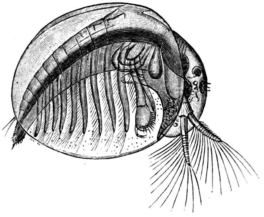
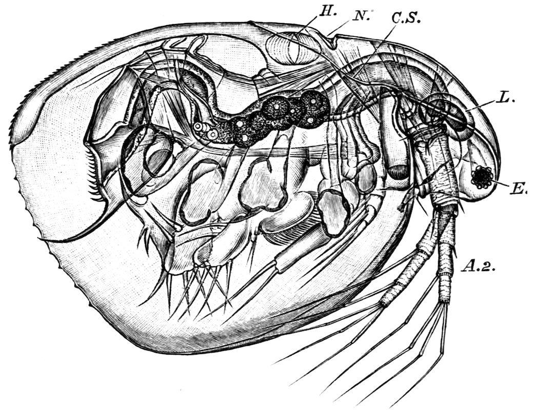
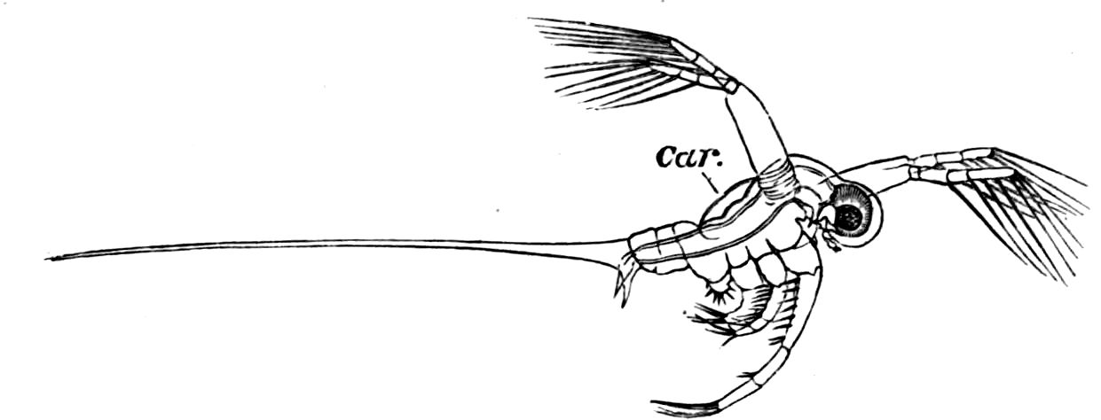
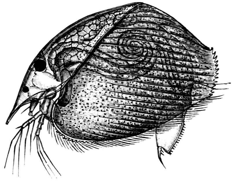
Fig. 3.—Limnetis brachyura, × 15. (After G. O. Sars.)
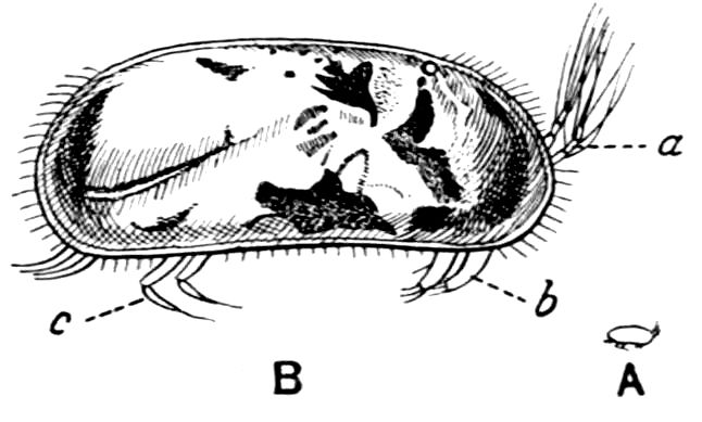
In Limnetis (Fig. 3) the dorsal surface of the head is bent
downwards and is much compressed, the carapace being attached
21to it only for a short distance near the dorsal middle line. The
sides of the carapace are bent downwards, and their margins can
be pulled together by a transverse adductor muscle, so that the
whole structure forms an ovoid or spheroidal case, from which
the head projects in front,
while the rest of the body
is entirely contained within
it. When the adductor
muscle is relaxed the
edges of the carapace gape
slightly, like the valves of
a Lamellibranch shell, and
food-particles are drawn
through the opening thus
formed into the ventral
groove by the movements
of the thoracic feet, locomotion
being chiefly effected
by the rowing action of the second antennae, as in the Cladocera,
to which all the Limnadiidae present strong resemblances in their
method of locomotion, in the condition of the carapace, and in
the form of the telson.
In Limnadia and Estheria the carapace projects not only
backwards from the point of attachment to the head, but also
forwards, so that the head can be enclosed by it, together with
the rest of the body.
In all these genera the carapace is flexible along the middle
dorsal line; in Estheria especially the softening of the dorsal
cuticle goes so far that a definite hinge-line is formed, and this,
together with the deposition of the lateral cuticle in lines concentrically
arranged round a projecting umbo, gives the carapace
a strong superficial likeness to a Lamellibranch shell, for which it
is said to be frequently mistaken by collectors.
The eyes of the Limnadiidae are enclosed in a chamber formed
by a growth of skin over them, as in Apodidae, but the pore by which
this chamber communicates with the exterior is even more minute
than in Apus. The paired eyes are so close together that they
may touch (Limnadia, Estheria) or fuse (Limnetis); they are
farther back than in the Apodidae, while the ventral curvature
of the head causes the median eye to lie below them. In all
22these points the eyes of the Limnadiidae are intermediate between
those of Apus and those of the Cladocera.
Dorsal Organ.—A structure very characteristic of adult
Phyllopods is the “dorsal organ” (Figs. 2, 5, D.O), whose function
is in many cases obscure. It is always a patch of modified
cephalic ectoderm, supplied by a nerve from the anterior ventral
lobe of the brain on each side; but its characters, and apparent
function, differ in different forms. In the Branchipodidae the
dorsal organ is a circular patch, far forward on the surface of
the head (Figs. 2, 5, D.O). Its cells are arranged in groups,
which remind one of the retinulae in a compound eye; each cell
contains a solid concretion, and the concretions of a group may be
so placed as to look like a badly-formed rhabdom. Claus,[16] who
first called attention to this structure in the Branchipodidae,
regarded it as a sense-organ. In Apodidae the dorsal organ is an
oval patch of columnar ectoderm, immediately behind the eyes;
it is slightly raised above the surrounding skin, and is covered
by a very delicate cuticle (with an opening to the exterior?), and
below it is a mass of connective tissue permeated by blood; Bernard
has suggested that it is an excretory organ.
Most Limnadiidae resemble the Cladocera in the possession
of a “dorsal organ” quite distinct from the above; in Limnetis
and Estheria it has the form of a small pit, lined by an apparently
glandular ectoderm, and this is its condition in many Cladocera;
in Limnadia lenticularis it is a patch of glandular epithelium on
a raised papilla. Limnadia has been observed to anchor itself
to foreign objects by pressing its dorsal organ against them, and
many Cladocera do the same thing; Sida crystallina, for example,
will remain for hours attached by its dorsal organ to a waterweed
or to the side of an aquarium. Structures resembling a
dorsal organ occur in the larvae of many other Crustacea, but the
presence of this organ in the adult is confined to Branchiopods,
and indeed in many Cladocera it disappears before maturity.
It is certain that the sensory and adhesive types of dorsal organ
are not homologous, especially as rudimentary sense-organs may
exist on the head of Cladocera together with the adhesive organ.
The telson differs considerably in the different genera. In
the Branchipodidae[17] the anus opens directly backwards; and
23the telson carries two flattened backwardly directed plates,
one on each side of the anus, the margins of each plate being
fringed with plumose setae. In Artemia the anal plates are
rarely as large as in Branchipus, and never have their margins
completely fringed with setae; in A. salina from Western
Europe, and in A. fertilis (Fig. 4, A) from the Great
Salt Lake of Utah, there is a variable number of setae round
the apical half of each lobe, but in specimens of A. salina from
Western Siberia the number of setae may be very small, or they
may be absent; in the closely allied A. urmiana from Persia the
anal lobes are well developed in the male, each lobe bearing a
single terminal hair, but they are altogether absent in the female.
Schmankewitch and Bateson have shown that there is a certain
relation between the salinity of the water in which Artemia salina
occurs and the condition of the anal lobes, specimens from denser
waters having on the whole fewer setae; the relation is, however,
evidently very complex, and further evidence is wanted before
any more definite statements can be made.
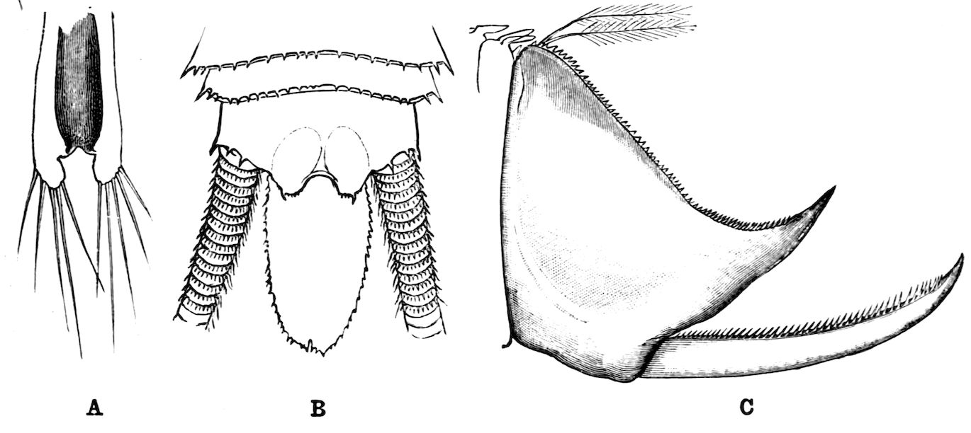
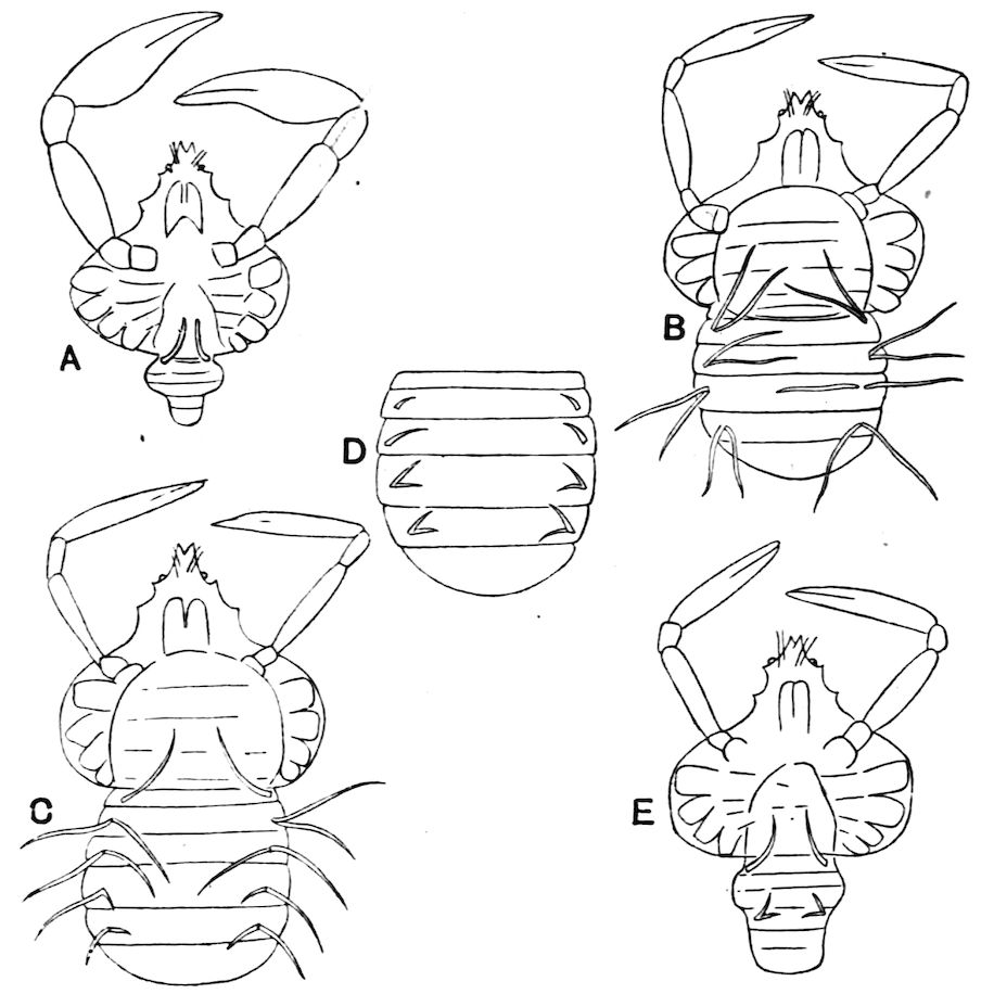
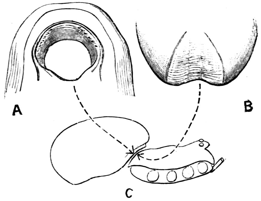
Fig. 4.—A, Ventral view of the anal region in Artemia fertilis, from the Great Salt Lake; B, ventral view of the telson and neighbouring parts of Lepidurus productus; C, side view of the telson and left anal lobe of Estheria (sp.?).
In the Apodidae the anal lobes have the form of two-jointed
cirri, often of considerable length; in Apus the anus is terminal,
but in Lepidurus (Fig. 4, B) the dorsal part of the telson is
prolonged backwards, so as to form a plate, on the ventral face
of which the anus opens, much as in the Malacostraca.
In the Limnadiidae (Fig. 4, C) the telson is laterally compressed
24and produced, on each side of the anus, into a flattened,
upwardly curved process, sharply pointed posteriorly, and often
serrate; the anal lobes are represented by two stout curved spines,
while in place of the dorsal prolongation of Lepidurus we find two
long plumose setae above the anus. In the characters of the telson
and anal lobes, as in those of the head, the Limnadiidae approximate
to the Cladocera. In Limnetis brachyura the ventral face
of the telson is produced into a plate projecting backwards below
the anus, in a manner which has no exact parallel among other
Crustacea.
The appendages of the Phyllopoda are fairly uniform in
character, except those affected by the sexual dimorphism, which
is usually great.
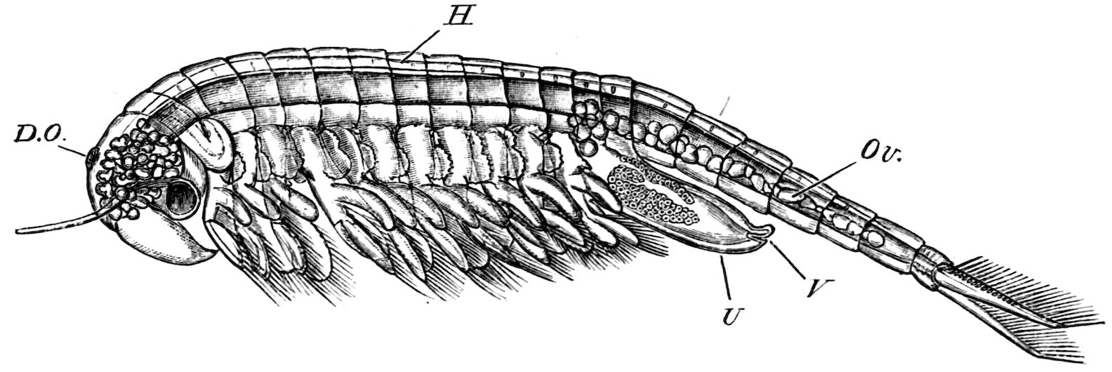
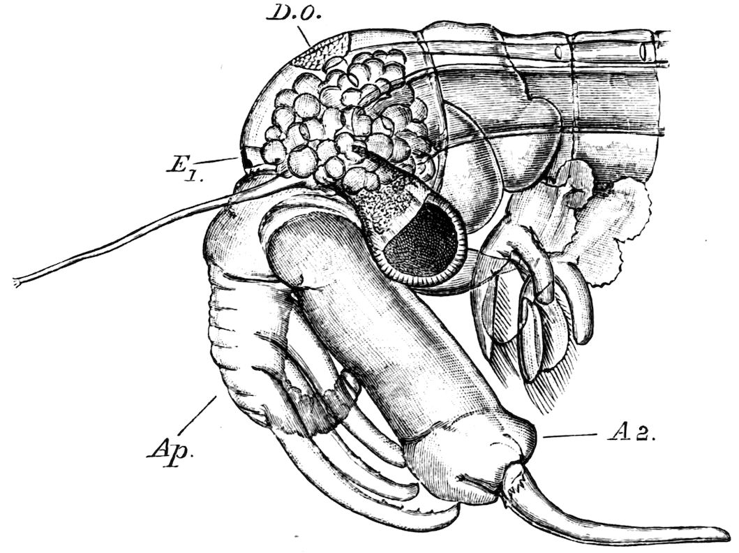
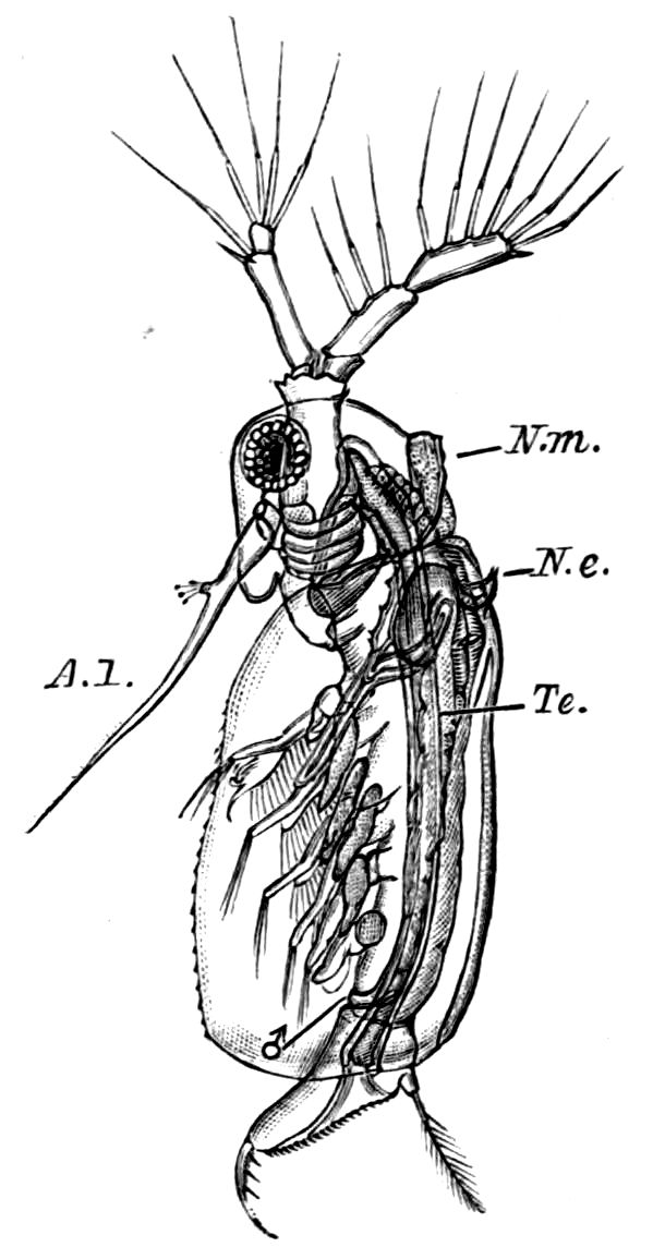
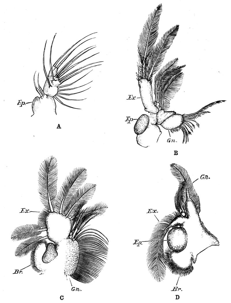
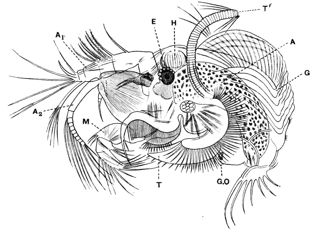
Fig. 5.—Chirocephalus diaphanus, male. Side view of head, showing the large second antenna, A2, with its appendage Ap, above which is seen the filiform first antenna; D.O, dorsal organ; E1, median eye.
25
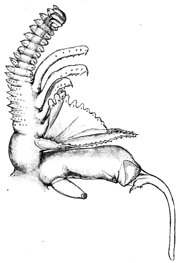
Fig. 6.—Chirocephalus diaphanus. Second antenna of male, uncoiled.
Of the cephalic appendages, the first antennae are generally
small, and are never biramous; in Branchipus and its allies they
are simple unjointed rods, in some species of Artemia they are
three-jointed, in Apus they are feebly divided into two joints,
while in Estheria they are many-jointed. The second antennae
are the principal organs of locomotion in the Limnadiidae, where
they are large and biramous; in all other Phyllopoda they
are uniramous in the female, being either unjointed triangular
plates as in Chirocephalus (Fig. 2), or minute vestigial filaments
as in Apus, in which genus Zaddach, Huxley, and Claus
have all failed to find any trace of a second antenna in some
females. In the male Branchipodidae the second antennae are
modified to form claspers, by which the female is seized, the
various degrees of complication which these claspers exhibit
affording convenient generic characters. In Branchinecta each
second antenna is a thick, three-jointed rod, the last joint
forming a claw, while the
second joint is serrate on its
inner margin; in Branchipus
the base is much thickened,
and bears on its inner side
a large filament (perhaps
represented by the proximal
tubercle of Branchinecta
and Artemia), which looks
like an extra antenna. In
Streptocephalus the terminal
joint of the antenna is bifid,
and there is a basal filament
like that of Branchipus;
in Chirocephalus diaphanus
(Figs. 5, 6) the main branch
of the antenna consists of
two large joints, the terminal
joint being a strong claw with
a serrated process at its base,
while the proximal joint
bears two appendages on its
inner side; one of these is a small, subconical tubercle, the second
is more complicated, consisting of a main stem and five outgrowths.
The main stem is many-jointed and flexible, its basal joint being
longer than the others, and bearing on its outer side a large,
triangular, membranous appendage, and four soft cylindrical
appendages, the main stem and its appendages being beset with
curious tubercles, ending in short spines, whose structure is not
understood. Except during the act of copulation this remarkable
apparatus is coiled on the inner side of the antennary claw, the
jointed stem being so coiled that it is often compared to the
26coiled proboscis of a butterfly, and the triangular membrane folded
like a fan beside it, so that much of the organ is concealed, and
the general appearance of the head is that shown in Fig. 5.
During copulation, the whole structure is widely extended.
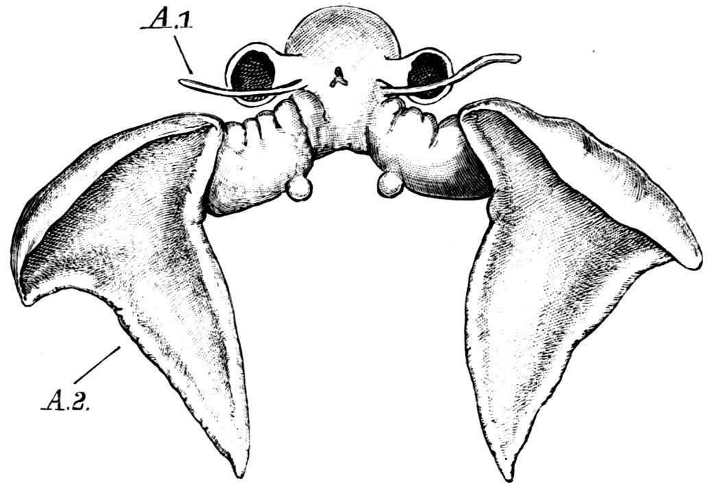
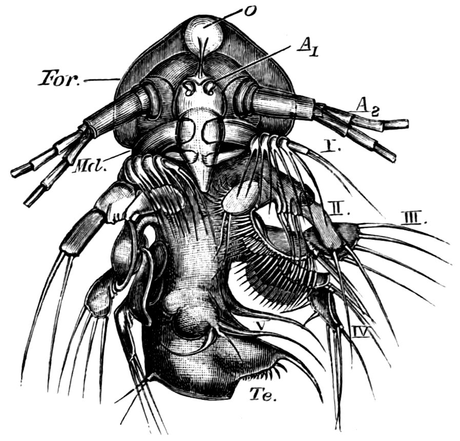
Fig. 7.—Artemia fertilis. Front view of the head of a male, showing the large second antennae, A.2; A.1, first antennae.
The males of Artemia (Fig. 7) have the second antenna two-jointed,
the basal joint bearing an inner tubercle, the terminal joint
being flattened and
bluntly pointed, its
outer margin provided
with a membranous
outgrowth. In A.
fertilis the breadth
of the second joint
varies greatly, the
narrower forms presenting
a certain
remote resemblance to
Branchinecta. In the
males of Polyartemia
the second antennae
have a remarkable branched form not easily comparable with
that found in other Branchipodidae.
The cephalic jaws are fairly uniform throughout the order.
The mandibles have an undivided molar surface, and no palp;
the first maxilla is very generally a triangular plate, with a
setose biting edge; mandibles and maxillae are covered by the
labrum. The second maxilla generally lies outside the chamber
formed by the labrum, and is a simple oval plate, with or
without a special process for the duct of the kidney.
The thoracic limbs, in front of the genital segments, are not
as a rule differentiated into anterior maxillipedes and posterior
locomotive appendages, as in higher forms; we have seen,
however, that all these limbs take part in the prehension of food,
and except in the Limnadiidae they all assist in locomotion. One
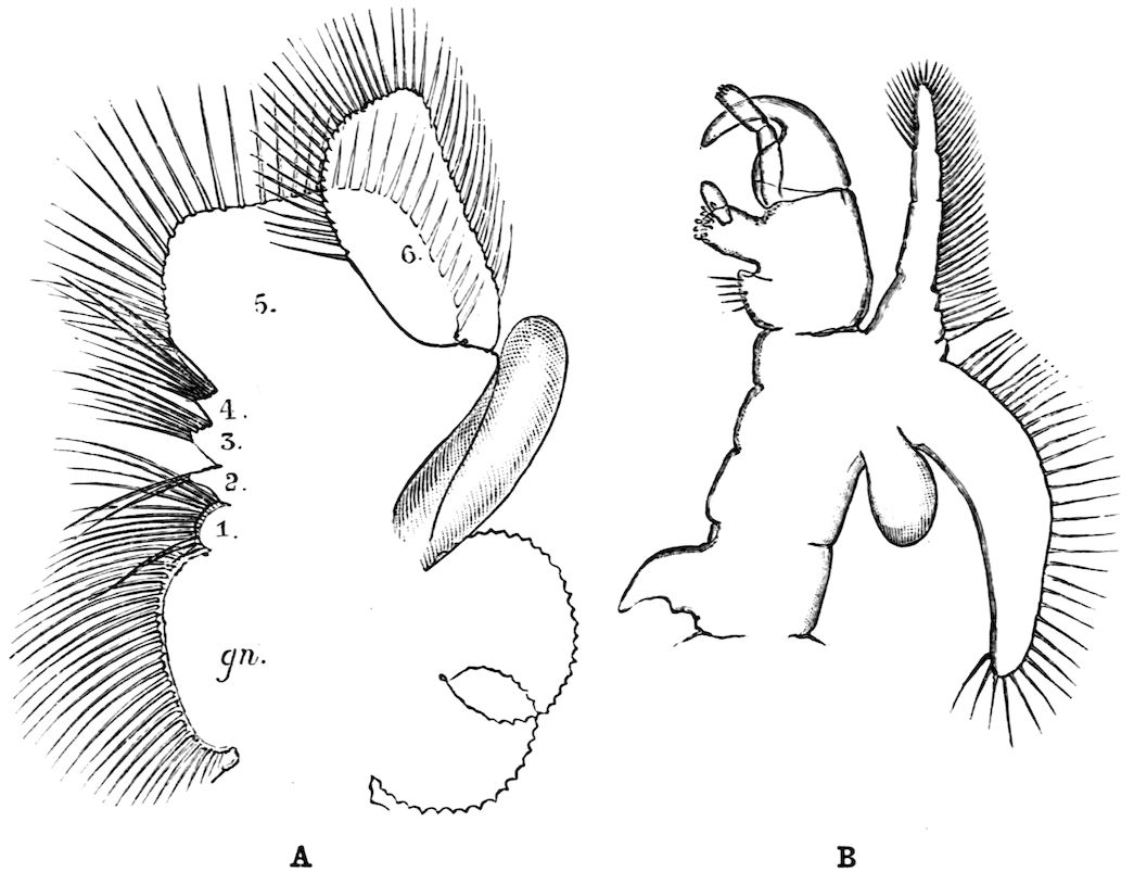
of the middle thoracic legs of Artemia (Fig. 8, A) has a
flattened stem, with seven processes on its inner, and two
on its outer margin. The gnathobase (gn) is large, and
fringed with long plumose setae, each of which is jointed; this
is followed by four smaller “endites” (or processes on the median
side), and then by two larger ones, the terminal endite (the sixth,
27excluding the gnathobase) being very mobile and attached to the
main stem by a definite joint. On the outer side are two processes;
a proximal “bract,” a flat plate with crenate edges, partly
divided by a constriction into two, and a distal process, cylindrical
and vascular, called by Sars and others the “epipodite.” In
other Branchipodidae we have essentially the same condition,
except that the fifth endite often becomes much larger than in
Artemia, throwing the terminal endite well over to the outer
edge of the limb; such a shift as this, continued farther, might
well lead to the condition found in the Limnadiidae, or Apodidae,
where the lobe which seems to represent the terminal endite of
Artemia is entirely on the outer border of the limb, forming
what most writers have called the exopodite (Lankester’s
“flabellum”).[18] In the two last-named families the basal exite
or bract of the Branchipodidae does not appear to be represented.
Fig. 8.—A, Thoracic limb of Chirocephalus diaphanus; B, prehensile thoracic limb of male Estheria. gn, Gnathobase; 1–6, the more distal endites.
The limbs of the Apodidae are remarkable in two ways;
those in front of the genital opening (very constantly ten pairs)
28are not so nearly alike as in most genera of the sub-order, the
first two pairs especially having the axis definitely jointed, while
the endites are elongated and antenniform; further, while the
first eleven segments bear each a single pair of limbs, as is usual
among Crustacea, many of the post-genital segments bear several
pairs; thus in Apus cancriformis there are thirty-two post-cephalic
segments in front of the telson, the first eleven having
each one pair of limbs, while the next seventeen have fifty-two
pairs between them, the last four segments having none.
In all the Phyllopoda some of the post-cephalic limbs are
modified for reproductive purposes; in the Branchipodidae the
last two pairs (the 12th and 13th generally, the 20th and 21st
in Polyartemia) are so modified in both sexes. In the female
these appendages fuse at an early period of larval life, and
surround the median opening of the generative duct (Fig. 2);
in the male the two pairs also fuse, but traces of the limbs are
left as eversible processes round the paired openings of the vasa
deferentia.
In the other families, one or more limbs of the female are
adapted for carrying or supporting the eggs. In the Apodidae
the appendages of the eleventh segment have the exopodite in
the form of a rounded, watchglass-shaped plate, fitting over a
similarly shaped process of the axis of the limb, so that a lens-shaped
box is formed, into which the eggs pass from the oviduct.
In Limnadiidae the eggs are carried in masses between the body
and the carapace, and are kept in position by special elongations of
the exopodites of two or three legs, either those near the middle
of the thorax (Estheria, Limnadia), or at its posterior end
(Limnetis). In female Limnetis the last thoracic segments bear
two remarkable lateral plates, which apparently also help to
support the eggs. In the male Limnadiidae, the first (Limnetis)
or the first two thoracic feet (Limnadia, Estheria) are prehensile
(Fig. 8, B).
Alimentary Canal.—The mouth of the Phyllopoda is
overhung by the large labrum, so that a kind of atrium is
formed, outside the mouth itself, in which mastication is performed;
numerous unicellular glands, opening on the oral face of
the labrum, pour their secretion into the atrial chamber, and
may be called salivary, though the nature of their secretion is
not known. The mouth has commonly two swollen and setose
29lips, running longitudinally forwards from the bases of the first
maxillae, and often wrapping round the blades of the mandibles.
It leads into a vertical oesophagus, which opens into a
small globular stomach, lying entirely within the head; the
terminal part of the oesophagus is slightly invaginated into the
stomach, so that a valvular ring is formed at the junction of
the two. The stomach opens widely behind into a straight
intestine, which runs backwards to about the level of the telson,
where it joins a short rectum, leading to the terminal or ventral
anus. The stomach and intestine are lined by a columnar
epithelium, and covered by a thin network of circularly arranged
muscle-fibres; the rectum has a flatter epithelium, and radial
muscles pass from it to the body-wall, so that it can be dilated.
The only special digestive glands are two branched glandular
tubes, situated entirely within the head, which open into the
stomach by large ducts, one on each side. In Chirocephalus
the gastric glands are fairly small and simple; in the Apodidae
their branches are more complex and form a considerable mass,
filling all that portion of the head which is not occupied by the
nervous system and the muscles. Backwardly directed gastric
glands, like those of the higher Crustacea, are not found in
Branchiopods; both forms occur together in the genus Nebalia,
but with this exception the forwardly-directed glands are peculiar
to Branchiopods.
Heart.—In Branchipus and its allies, and in Artemia, the
heart extends from the first thoracic segment to the penultimate
segment of the body, and is provided with eighteen pairs of
lateral openings, one pair in every segment through which it
passes except the last; it is widely open at its hinder end, and
is prolonged in front for a short distance as a cephalic aorta,
the rest of the blood-spaces being lacunar.
In most, at least, of the other Branchiopods, the heart is
closed behind and is shortened; in Apus and Lepidurus it only
extends through the first eleven post-cephalic segments, while in
the Limnadiidae it is shorter still, the heart of Limnetis passing
through four segments only. In all cases there is a pair of
lateral openings in every segment traversed by the heart.
The blood of the Branchipodidae and Apodidae contains
dissolved haemoglobin, the quantity present being so small as to
give but a faint colour to the blood in Branchipus, while
30Artemia has rather more, and the blood of Apus is very red.
The only other Crustacea in which the blood contains haemoglobin
are the Copepods of the genus Lernanthropus,[19] so that the
appearance of this substance is as irregular and inexplicable in
Crustacea as in Chaetopods and Molluscs.
The nervous system of Branchipus may be described as an
illustration of the condition prevailing in the group. The brain
consists of two closely united ganglia, in each of which three
main regions may be distinguished; a ventral anterior lobe, a
dorsal anterior lobe, and a posterior lobe. The ventral anterior
lobes give off nerves to the median eye, to the dorsal organ, and
to a pair of curious sense-organs, comparable with the larval
sense-knobs of many higher forms, situated one on each side
of the median eye; in late larvae Claus describes the
terminal apparatus of each frontal sense-organ as a single
large hypodermic cell; W. K. Spencer[20] has lately described
several terminal cells, containing peculiar chitinous bodies, in
the adult. The homologous sense-organs of Limnetis are apparently
olfactory. The dorsal anterior lobes give off the large
nerves to the lateral eyes, while the posterior lobes supply the
first antennae. The oesophageal connectives have a coating of
ganglion-cells, and some of these form the ganglion of the
second antenna, the nerve to this appendage leaving the connective
just behind the brain. The post-oral nerve-cords are
widely separate, each of them dilating into a ganglion opposite
every appendage, the two ganglia being connected by two
transverse commissures. The ganglia of the three cephalic
jaws, so often fused in the higher Crustacea, are here perfectly
distinct. Closely connected with each thoracic ganglion is a remarkable
unicellular gland, opening to the exterior near the
middle ventral line; it is conceivable that these cells may be
properly compared with the larval nephridia of a Chaetopod,[21]
but no evidence in support of such a comparison has yet been
adduced.
Behind the genital segments, where there are no limbs, the
nerve-cords run backwards without dilating into segmental
ganglia, except in the anterior two abdominal segments where
31small ganglionic enlargements occur. In Apodidae, on the other
hand, those segments which carry more than one pair of
appendages have as many pairs of ganglia, united by transverse
commissures, as they have limbs.
A stomatogastric nervous system exists in Apus, where a
nerve arises on each side from the first post-oral commissure,
and runs forward to join its fellow of the opposite side on the
anterior wall of the oesophagus. From the loop so formed a
larger median and a series of smaller lateral nerves pass to the
wall of the alimentary canal. A second nerve to the oesophagus
is given off from the mandibular ganglion of each side.
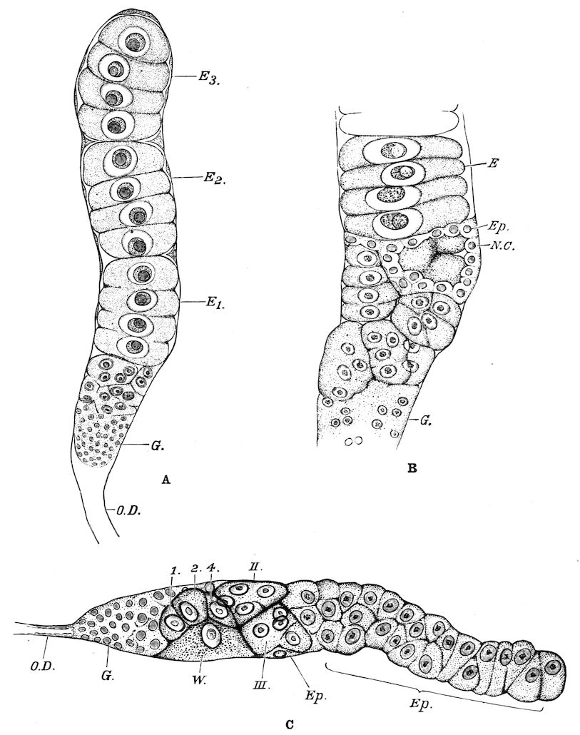
Reproductive Organs.—In Chirocephalus the ovaries (Fig.
2, Ov) are hollow epithelial tubes, lying one on each side of the
alimentary canal, and extending from the sixth abdominal
segment forwards to the level of the genital opening; at this point
the two ovaries are continuous with ducts, which bend sharply
downwards and open into the single uterus contained within
the projecting egg-pouch and opening to the exterior at the
apex of that organ. Short diverticula of the walls of the uterus
receive the ducts of groups of unicellular glands, the bodies of
which contain a peculiar opaque secretion, said to form the eggshells.
In Apodidae the ovaries are similar in structure, but
they are much larger and branch in a complex manner, while
each ovary opens to the exterior independently of the other in
the eleventh post-cephalic segment; nothing like the median
uterus of the Branchipodidae being formed. The epithelium of
the ovarian tubes proliferates, and groups of cells are formed;
one becoming an ovum, the others being nutrient cells like those
which will be more fully described in the Cladocera.
In Chirocephalus the testes are tubes similar in shape and
position to the ovaries, each communicating in front with a
short vas deferens, which dilates into a vesicula seminalis on its
way to the eversible penis; an essentially similar arrangement
is found in all Branchipodidae, but in Apodidae and Limnadiidae
there is no penis.
All the Branchiopoda are dioecious,[22] and many are parthenogenetic.
Among Branchipodidae Artemia is the only genus
known to be parthenogenetic, but parthenogenesis is common in
32all Apodidae, while the males of several species of Limnadia are
still unknown, although the females are sometimes exceedingly
common. In Artemia, generations in which the males are about
as numerous as the females seem to alternate fairly quickly with
others which contain only parthenogenetic females; in Apus
males are rarely abundant, and often absent for long periods;
during five consecutive years von Siebold failed to discover a
male in a locality in Bavaria, though he examined many thousands
of individuals; near Breslau he found on one occasion about 11
per cent of males (114 in 1026), but in a subsequent year he
found less than 1 per cent; the greatest recorded percentage of
males is that observed by Lubbock in 1863, when he found 33
males among 72 individuals taken near Rouen.
The eggs of most genera can resist prolonged periods of
desiccation, and indeed it seems necessary for the development
of many species that the eggs should be first dried and afterwards
placed in water. Many eggs (e.g. of Chirocephalus diaphanus
and Branchipus stagnalis) float when placed in water after desiccation,
the development taking place at the surface of the
water.
Habitat.—All the Phyllopoda, except Artemia, are confined
to stagnant shallow waters, especially to such ponds as are formed
during spring rains, and dry up during the summer. In waters
of this kind the species of Branchipus, Apus, etc., develop rapidly,
and produce great numbers of eggs, which are left in the dried
mud at the bottom after evaporation of the water, where they
remain quiescent until a fresh rainy season. The mud from the
beds of such temporary pools often contains large numbers of
eggs, which may be carried by wind, on the legs of birds, and by
other means, to considerable distances. Many exotic species have
been made known to European naturalists by their power of
hatching out when mud brought home by travellers is placed in
water. The water of stagnant pools quickly dissolves a certain
quantity of solid matter from the soil, and often receives dissolved
solids through surface drainage from the neighbouring land; such
salts may remain as the water evaporates, so that the water which
remains after evaporation has proceeded for some time may be
very sensibly denser than that in which the Branchiopods were
hatched; these creatures must therefore be able to endure a considerable
increase in the salinity of the surrounding waters during
33the course of their lives. My friend Mr. W. W. Fisher points
out that the plants present in such a pond would often precipitate
the carbonate of lime, so that this might be removed as evaporation
went on, but that chlorides would probably remain in solution;
from analyses which Mr. Fisher has been kind enough to
make for me, it is seen that this happened in a small aquarium in
my laboratory, in which Chirocephalus diaphanus lived for four
months. In April, mud from the dry bed of a pond, known to
contain eggs of Chirocephalus, was placed in this aquarium in
Oxford, and water was added from the tap. Oxford tap-water
contains about 0·3 grm. salts per litre, the chlorine being equivalent
to 0·023 grm. NaCl. Water was added from time to time
during May and June, but in July evaporation was allowed to
proceed unchecked. At the end of July there was about half the
original volume of water, the Chirocephalus being still active;
the residue contained 0·96 grm. dissolved solids per litre, with
chlorine equal to 0·19 grm. NaCl, so that the percentage of
chlorides was about eight times the initial percentage, but there
were only three and a fifth times the original amount of
total solid matter in solution, the carbonate of lime having precipitated
as a visible film.
Some species of Branchipus (e.g. B. spinosus, M. Edw.) and
of Estheria (E. macgillivrayi, Baird, E. gubernator, Klutzinger)
occur in salt pools, but Artemia flourishes in waters beside
whose salinity that endured by any other Branchiopod is insignificant.
In the South of Europe, Artemia salina may be
found in swarms, as it used to be found in Dorsetshire, in the
shallow brine-pans from which salt is commercially prepared;
Rathke quotes an analysis showing that a pool in the Crimea
contained living Artemia when the salts in solution were 271
grms. per litre, and the water was said to have the colour and
consistency of beer.
The behaviour of the animals in the water differs a little; in
normal feeding all the species swim with the back downwards, as
has already been said; the Branchipodidae rarely settle on the
ground, or on foreign objects, but the Apodidae occasionally
wriggle along the bottom on their ventral surface, and Estheria
burrows in mud.
The greater number of species are found in pools in flat, low-lying
regions, and many appear to be especially abundant near
34the sea; Apus cancriformis has, however, been found in Armenia
at 10,000 feet above sea level.
Wells and underground waters do not generally contain
Phyllopods; but a species of Branchipus and one of Limnetis,
both blind, have been described from the caves of Carniola.
One of the many puzzles presented by these creatures is the
erratic way in which they are scattered through the regions they
inhabit; a single small pond, a few yards or less in diameter,
may be the only place within many miles in which a given species
can be found; in this pond it may, however, appear regularly
season after season for some time, and then suddenly vanish.
Geographically, the Phyllopoda are cosmopolitan, representatives
of every family and of some genera (e.g. Streptocephalus,
Lepidurus, Estheria) being found in every one of the great zoological
regions, though a few aberrant genera are of limited range,
thus Polyartemia is known only from the northern Palaearctic
and Nearctic regions, Thamnocephalus only from the Central
United States. The genus Artemia is not at present known in
Australia.[23] The only recorded British species are Chirocephalus
diaphanus, Artemia salina, and Apus cancriformis,[24] but other
continental islands, for example the West Indian group, are
better supplied. The distribution of the species is very imperfectly
known, but on the whole every main zoological region
seems to have its own peculiar species, which do not pass beyond
its boundaries. Branchinecta paludosa and Lepidurus glacialis are
circumpolar, both occurring in Norway, in Lapland, in Greenland,
and in Arctic North America; but with these exceptions the
Palaearctic and Nearctic species seem to be distinct. The European
species Apus cancriformis occurs in Algiers, but the relations
between the species of Northern Africa as a whole and those of
Southern Europe on the one hand, or of Central and Southern
Africa on the other, have yet to be worked out.
The soft-bodied Branchipodidae are not known in the fossil
condition;[25] an Apus, closely related to the modern A. cancriformis,
has been found in the Trias, but the most numerous remains have
been left, as might be expected, by the hard-shelled Limnadiidae;
35carapaces, closely resembling those of the modern Estheria, are
known in beds of all ages from the Devonian period to recent
times; these carapaces are in several cases associated with fossils
of an apparently marine type. None of the fossil species differ
in any important characters from those now living, so that the
Phyllopoda have existed in practically their present form for
an enormously long period; this fact, and the evidence that
species of existing genera were at one time marine, explain the
wide distribution of animals at present restricted to a remarkably
limited range of environmental conditions.
Summary of the Characters of the Genera.
Sub-Order Phyllopoda.—Branchiopoda with an elongated body, provided
with at least ten pairs of post-cephalic limbs, the heart extending
through four or more thoracic segments, and having at least four pairs of ostia.
Fam. 1. Branchipodidae.[26]—Carapace rudimentary, eyes stalked; the
second antennae flat and unjointed in the female, jointed and prehensile in
the male; female generative opening single; telson not laterally compressed,
bearing two flattened lobes, or none. The heart extending through the
thorax and the greater part of the abdomen.
A. Eleven pairs of praegenital ambulatory limbs.
a. Abdomen of six well-formed segments and a telson; anal lobes
well formed, their margins setose.
Branchinecta, Verrill—Second antennae of ♂ without lateral
appendages; ovisac of ♀ elongated. B. paludosa, O. F.
Müll.—Circumpolar.
Branchiopodopsis, G. O. Sars[27]—Second antennae of ♂ as in
Branchinecta; ovisac of ♀ short. B. hodgsoni, G. O. Sars—Cape
of Good Hope.
Branchipus, Schaeffer—Second antennae of ♂ with simple
internal filamentous appendage. B. stagnalis, Linn.—Central
Europe.
Streptocephalus, Baird—Second antennae of ♂ 3–jointed, the
last joint bifid; an external filamentous appendage. S.
torvicornis, Wagn., Poland.
Chirocephalus, Prévost—Second antennae of ♂ 3–jointed, with a
jointed internal appendage, which bears secondary processes,
four cylindrical and one lamellar. C. diaphanus, Prévost
(Fig. 2, p. 20).—Britain, Central Europe.
b. Abdominal segments five or fewer, and a telson. Anal lobes
small or 0, sparsely or not at all setose.
Artemia, Leach—Second antennae of ♂ without filamentous
36appendage, 2–jointed, the second joint lamellar. A. salina,
Linn.—Brine pools of the Palaearctic region.
c. Hinder abdominal segments united with telson to form a fin; anal
lobes absent.
Thamnocephalus, Packard—Head with a branched median process
of unknown nature. Only species T. platyurus, Packard—Kansas,
U.S.A.
B. Nineteen pairs of praegenital ambulatory limbs.
Polyartemia, Fischer—Second antennae of ♂ forcipate; ovisac
of ♀ very short. Only species P. forcipata, Fisch.
Fam. 2. Apodidae.[28]—Carapace well developed as a depressed shield,
covering at least half the body. Eyes sessile, covered; no male clasping
organs; anal lobes long, jointed cirri.
Apus, Scopoli—Telson not produced backwards over the anus;
endites of first thoracic limb very long. A. cancriformis,
Schaeffer—Britain, Europe, Algiers, Tunis. A. australiensis,
Central Australia.
Lepidurus, Leach—Telson produced backwards to form a plate
above the anus; endites of first thoracic limb short. L. productus,
Bosc.—Central Europe. L. viridis, Southern
Australia, New Zealand, L. patagonicus, Bergh, Argentines.
Fam. 3. Limnadiidae.—Body compressed; carapace in the form of a
bivalve shell, the two halves capable of adduction by means of a strong
transverse muscle; second antennae biramous, alike in both sexes; in the
male, the first or the first and second thoracic limbs prehensile; telson
laterally compressed.
A. Only the first thoracic limbs prehensile in the male; the carapace
spheroidal, without lines of growth; head not included within
the carapace-chamber.
Limnetis, Lovén—Compound eyes fused; anal spines absent;
ambulatory limbs 10–12. L. brachyura, O. F. Müll (Fig. 3,
p. 21).—Norway, Central Europe.
B. The first and second thoracic limbs prehensile in the male; carapace
distinctly bivalve, enclosing the head, with concentric lines of
growth round a more or less prominent umbo.
Eulimnadia, Packard—Carapace narrowly ovate, with few (4–5)
lines of growth. E. mauritani, Guérin—Mauritius. E.
texana, Packard—Texas, Kansas.
Limnadia, Brongniart—Carapace broadly ovate, with numerous
lines of growth, without distinct umbones; L. lenticularis,
Linn.—Northern and Central Europe.
Estheria, Rüppell—Carapace with well-marked umbones and
numerous lines of growth, oval; E. tetraceros, Kryneki—Central
Europe.
Leptestheria,[29] G. O. Sars—Carapace compressed, oblong. Rostrum
37with a movable spine; thoracic limbs with accessory
lappet on the exopodite. L. siliqua, G. O. Sars—Cape Town.
Cyclestheria,[30] G. O. Sars. C. hislopi, Baird—Queensland, India,
East Africa, Brazil.
Sub-Order 2. Cladocera.
The Cladocera are short-bodied Branchiopods, with not more
than six pairs of thoracic limbs. The second antennae are
important organs of locomotion, and are nearly always biramous;
the first antennae are small, at least in the female; the second
maxillae are absent in the adult. The carapace may extend
backwards so as to enclose the whole post-cephalic portion of the
body, or may be reduced to a small dorsal brood-pouch, leaving
the body uncovered.
The Cladocera or “Water-fleas” are never of great size;
Leptodora hyalina, the largest, is only about 15 mm. long, while
many Lynceidae are not more than 0·1 or 0·2 mm. in length.
The head is bent downwards in all the Cladocera, so that
parts which are morphologically anterior, such as the median
eye and the first antennae, lie ventral to or even behind the compound
eyes and the second antennae (cf. Fig. 10).
The compound lateral eyes fuse at an early period of
embryonic life, so that they form a single median mass in the
adult, over which a fold of ectoderm grows, to make a chamber
over the eye, like that found in the Limnadiidae, except that it is
completely closed. The fused eyes are generally large and conspicuous;
in some deep-water forms the retinular elements of
the dorsal portion are larger than those of the ventral (e.g.
Bythotrephes, Fig. 13). In one or two species which live at
very great depths, or in caves, the eyes are altogether absent.
The appendages of the head are fairly uniform, the most
variable being the first antennae. In the females of many
genera the first antennae are short and immovable, consisting of
a single joint, with a terminal bunch of sensory hairs, and often
a long lateral hair, as in Simocephalus (Figs. 9, 10), Daphnia, etc.
In the female Moina (Fig. 16) they are movable, as they are
in Ceriodaphnia and some others; in Bosmina (Fig. 22) and
many Lyncodaphniidae they are elongated and imperfectly divided
38into joints by rings of spines, while in Macrothrix they are
flattened plates. In the males the first antennae are elongated
and mobile (cf. Figs. 11, 19).
Fig. 9.—Simocephalus vetulus, female. Ventral view, without the carapace; A1, A2, first and second antennae; For, head; Md, mandible; Te, telson; I-IV, first to fourth thoracic appendages.
The second antennae, the chief organs of locomotion, are
biramous in all genera except Holopedium; the number of
joints in each ramus, and the number of the long plumose hairs
with which they are provided, are remarkably constant in whole
series of genera, and are therefore useful for purposes of classification.
The creatures row themselves by quick strokes of
these appendages, the movement being slow and irregular in the
rounder forms, such as
Simocephalus or Daphnia,
rapid and well directed in
such elongated lacustrine
forms as Bythotrephes or
Leptodora.
The mandibles have no
palp; the first maxillae are
very small, and the second
maxillae are absent (Fig. 9).
The carapace varies very
much. In most genera (the
Calyptomera of Sars) it is
a large, backwardly-projecting
fold of skin, bent
downwards at the sides so
as to form a bivalve shell,
enclosing the whole post-cephalic
portion of the body, as in Simocephalus (Fig. 10). The
eggs are laid into the space between the carapace and the
dorsal part of the thorax, both the carapace and the thorax itself
being often modified for their protection and nutrition. In a few
forms, the Gymnomera of Sars, the carapace serves only as a
brood-pouch, which is distended when eggs are laid, but collapses
to an inconspicuous appendage at the back of the head when it is
empty (e.g. Leptodora, Fig. 24, Bythotrephes, Fig. 13). In the
Calyptomera the surface of the carapace is frequently provided
with a series of ridges, which may be parallel, rarely branching, as
in Simocephalus; or in two sets which cross nearly at right angles,
as in Daphnia; or so arranged as to form a hexagonal pattern, as
39in Ceriodaphnia. In a few forms the whole surface is irregularly
covered with spines or scales. The hinder edge of the carapace
is often produced into a median dorsal spine (Daphnia, Fig. 19),
or more rarely there are two spines, one at each ventro-lateral
corner (Scapholeberis, Fig. 20).
Fig. 10.—Simocephalus vetulus, × 30. Side view of female, showing the arrangement of the principal organs. A.2, Second antenna; C.S, cervical suture; E, fused compound eyes; H, heart; L, forwardly-directed gastric caeca; N, dorsal organ.
The cuticle of the carapace is often separated from that of
the head by a cervical suture, as in Simocephalus (Fig. 10, C.S.)
and near the line of demarcation many forms exhibit patches of
glandular ectoderm which seem to be homologous with the
dorsal adhesive organs of the Limnadiidae. The commonest
condition is that of a median dorsal pit (Fig. 10, N.) by means
of which the animal can fix itself to foreign objects. Certain
forms may remain for long periods of time attached by the
dorsal organ to plants, or to the sides of an aquarium, the only
movement being a slow vibration of the feet, by which a current
of water, sufficiently rapid for respiratory purposes, is established
round it.[31] In Sida crystallina (Fig. 11) the dorsal organ is
represented by three structures; in front there is a median raised
40patch (N.m) of columnar ectoderm, containing concretions like
those described in the Branchipodidae, and behind this is a pair
of cup-shaped organs (N.e), with raised margins.
Fig. 11.—Sida crystallina, male, × 27. Oxford. A.1, Elongated first antenna; N.e, paired element of dorsal organ; N.m, median element of dorsal organ; Te, testes; ♂, opening of vas deferens.
The fold of skin which forms the carapace contains the coils
of the single pair of kidneys, and it forms an important organ
of respiration, partly from the great size of the blood-vessels it
contains, and partly from the presence
of red, blue, or brown respiratory pigments
in the tissue of the skin itself.
In most Cladocera the cuticle of the
carapace is cast at every ecdysis, with
that of other parts of the body; but in
Iliocryptus and a few others it remains
after each moult, giving the carapace
an appearance of “lines of growth,”
like that seen in many Limnadiidae.
The segmentation of the body
behind the head is obscure, but we
can generally recognise (1) a thorax, of
as many segments as there are pairs of
limbs; (2) an abdomen of three segments;
and (3) a telson.
The thoracic limbs of the Calyptomera
are flattened, and resemble those
of the Phyllopoda; as a type we may
examine the third thoracic limb of
Simocephalus (Fig. 12, C), in which
the axis bears a large setose gnathobase
(Gn) on its inner edge, followed by two
small endites; the terminal process, or exopodite (Ex) is a large
flattened plate, with six long plumose hairs on its edge. The
outer margin of the axis bears a bract (Br) and an epipodite.
In Simocephalus, as in the other Daphniidae, there are five
pairs of thoracic limbs, of which the third and fourth are alike;
in the female each limb of the first pair consists of a jointed axis,
with strong biting hairs on the inner border, and a rudimentary
epipodite (Fig. 12, A), the second limb being more like the
third, but with a more prominent gnathobase and a narrower
exopodite (B), while the limbs of the fifth pair have the gnathobase
and the exopodite filamentous (D).
41In the Sididae there are six pairs of thoracic limbs, which are
nearly alike in the female; in the Bosminidae there are six pairs,
the first two modified for prehension, the last much reduced.
Fig. 12.—Thoracic limbs of female Simocephalus vetulus. A, The first; B, the second; C, the third; D, the fifth. Br, Bract; Ep, epipodite; Ex, exopodite; Gn, gnathobase.
In the male, the first thoracic limb is usually provided with
a long sensory process and a prehensible hook (Figs. 11, 19).
In the Gymnomera the limbs are cylindrical, jointed rods,
42with a gnathobase on the inner side in the Polyphemidae,
but not in Leptodora. The number varies from four to six
pairs.
The abdomen bears no appendages. The telson is compressed
in the Calyptomera, and is produced into two flattened plates,
one on each side of the anal opening. The backwardly directed
margins of these plates are commonly serrated, and the lower
corner of each is produced into a curved spine, which carries
secondary teeth. The number and arrangement of these teeth,
though often extremely variable in the same species, are used
extensively as specific characters. Above the anus the telson
commonly bears two long plumose hairs, which are directed
backwards.
Fig. 13.—Bythotrephes cederströmii, female, × 20, North Wales, from a specimen found by A. D. Darbishire. Car, carapace.
In the Gymnomera the telson is not bilaterally compressed,
and it may be produced into a long spine, dorsal to the anus (e.g.
Bythotrephes, Fig. 13).
The alimentary canal is extremely simple. The labrum is
large, and forms a chamber above the mouth, into which
food is driven by the limbs, as in the Phyllopoda, food being
taken while the animal swims or lies on its back. The
oesophagus runs vertically to join a small stomach, which bends
sharply backwards and passes gradually into an intestine. In
the last segment of the abdomen the intestine joins a short,
thin-walled rectum, provided with radial muscles, by means of
which it can be dilated. The dilatation of the rectum leads to
an inhalation of water through the anus, which may possibly
serve as a means of respiration. In the Daphniidae and
Bosminidae there are two forwardly-directed digestive glands
which open into the stomach, and in Eurycercus there is a large
caecum at the junction of the rectum with the intestine. The
43intestine is usually straight, but in Lynceidae and in some
Lyncodaphniidae it is coiled (e.g. Peracantha, Fig. 14).
In Leptodora the alimentary canal is altogether remarkable;
the oesophagus is a long and very narrow tube, which runs back
through the whole length of the thorax and joins the mid-gut in
the third abdominal segment. The mid-gut is not differentiated
into stomach and intestine; it has no diverticula of any kind,
and runs straight backwards to join the short rectum a little in
front of the anus.
The heart is always short, and never has more than a single
pair of lateral openings; it is longest in the Sididae, which show some
approximation to the Phyllopods in this, as in the
slight degree of difference between
their anterior and
posterior thoracic limbs. The
pericardium lies in the one or
two anterior thoracic segments,
dorsal to the gut. From the
heart the blood runs forwards
to the dorsal part of the head,
and passes backwards by three
main channels, one entering each
side of the carapace, while the
third runs down the body,
beneath the alimentary canal
to dilate into a large sinus round the rectum. This ventral
blood-channel gives a branch to each limb, which forms a considerable
dilatation in the epipodite, the blood from the limb
returning to the pericardium by a lateral sinus. From the
rectum a large sinus runs forwards to the pericardium along
the dorsal wall of the body. The blood which enters each half
of the carapace is collected in a median vessel and returned
through this to the pericardium.
Those spaces between the viscera which are not filled with
blood are occupied by a peculiar connective tissue, consisting of
rounded or polyhedral cells, charged with drops of a fatty material
which is often brightly coloured.
The reproductive organs are interesting because of the
peculiar phenomena connected with the nutrition of the two
kinds of eggs. The ovaries or testes are epithelial sacs, one on
44each side of the body, each continuous with a duct which opens
to the exterior behind the last thoracic limb. In the female,
the opening is dorsal (Fig. 10), in the male it is ventral
(Fig. 11). The external opening is usually simple; but in the
male there is sometimes a penis-like process, on which the vas
deferens opens (Daphnella).
The eggs are of two kinds, the so-called “summer-eggs,” with
relatively little yolk, which develop rapidly without fertilisation,
and the so-called “winter-eggs,” containing much yolk, which
require to be fertilised and then develop slowly.
At one end of the ovary, generally that nearest to the
oviduct, there is a mass of protoplasm, containing nuclei which
actively divide; this is the germarium (Fig. 15, A, B, C). As
a result of proliferation in the germarium, nucleated masses are
thrown off into the cavity of the ovary; each such mass contains
four nuclei, and its protoplasm soon becomes divided into
four portions, one round each nucleus, so that four cells are
produced. In the simpler ovaries, such as that of Leptodora
(Fig. 15, A), these sets of four cells are arranged in a linear
series within the tube of ovarian epithelium; in other cases, as
in Daphnia, the arrangement is more irregular. In the normal
development of parthenogenetic eggs, one cell out of each set of
four becomes an ovum, the other three feeding it with yolk and
then dying. Weismann[32] has shown that the ovum is always
formed from the third cell of each set, counting from the
germarial end, so that in the ovary of Leptodora drawn in Fig.
15, A, the ova will be formed from the cells marked E1, E2, E3.
At certain times, one or two sets of germinal cells fail to produce
ova; the epithelial wall of the ovary thickens round these cells,
so that they become incompletely separated from the rest in a
so-called “nutrient chamber” (Fig. 15, B, N.C). Germ-cells
enclosed in a nutrient chamber degenerate and are ultimately
devoured by the ovarian epithelium. The significance of these
nutrient chambers is unknown.
45
Fig. 15.—A, Ovary of a parthenogenetic Leptodora hyalina; B, base of another ovary of the same species, showing a so-called “nutrient chamber”; C, ovary of a female Daphnia, showing the formation of a winter-egg. E, E1–E3, Parthenogenetic egg; Ep, ovarian epithelium; G, germarium; N.C, nutrient chamber; O.D, oviduct; W, winter-egg; 1, 2, 4, the other three cells of the same group; II, III, two other groups of germ-cells.
The production of a winter-egg is a more complicated process.
The epithelium of the ovarian tube swells up, so that the lumen
is nearly obliterated, and several sets of four germ-cells pass from
the germarium to lie among the swollen epithelial cells. All
these groups of germ-cells, except one, disintegrate and are
46devoured by the ovarian epithelium, one cell of the remaining
group enlarging to form a winter-egg, fed during its growth not
only by the three cells of its own set but also by the epithelial
cells of the ovarian tube, which have devoured the germ-cells of
other sets. An ovary never contains more than a single winter-egg
at the same time, the number of germ-cells which are
devoured during its formation varying in the different species;
the Daphnia drawn in Fig. 15, C, has produced three groups of
germ-cells, of which two (II, III), will die, while the cell W
from the remaining group will develop into an ovum; in Moina,
Weismann finds that as many as a dozen cell-groups may be
thrown into the ovary before the production of a winter-egg, so
that only one out of forty-eight germ-cells survives as an ovum.
Fig. 16.—Sketch of a parthenogenetic Moina rectirostris, × 45, the brood-pouch being emptied and the side of the carapace removed, showing the dome of thickened epithelium on the thorax, by which nutrient material is thrown into the brood-pouch, and the ridge which fits against the carapace in the natural condition so as to close the brood-pouch.
Fig. 17.—Moina rectirostris, ♀, × 40, showing the ephippial thickening of the carapace which precedes the laying of a winter-egg.
The summer-eggs are always carried until they are hatched
by the parthenogenetic female which produces them. The
brood-pouch is the space between the dorsal wall of the thorax
and the carapace. This space is always more or less perfectly
closed at the sides by the pressure of the carapace against the
body, and behind by vascular processes from the abdominal
segments (Figs. 10, 16, etc.). The presence of a large blood-sinus
47beneath the dorsal wall of the thorax and in the middle line of
the carapace suggests the possibility that some special nutrient
substances may pass from the body of the parent into the brood-chamber,
and in some species the thoracic ectoderm is specially
modified as a placenta. In Moina (Fig. 16) the dorsal wall
of the thorax is produced into a dome, covered by a columnar
ectoderm, which contains a dilatation of the dorsal blood-sinus;
and in this form it has been shown that the fluid in the brood-pouch
contains dissolved proteids. Associated with the apparatus
for supplying the brood-pouch
with nutriment
is a special apparatus
for closing it, in the
form of a raised ridge,
which projects from the
back and sides of the
thorax and fits into a
groove of the carapace.
A somewhat similar
nutrient apparatus exists
in the Polyphemidae,
where the edges of the
small carapace are fused
with the thorax, so that
the brood-pouch is completely closed, and the young can only
escape when the parent casts her cuticle. In some genera of
this family (e.g. Evadne) the young remain in the parental brood-pouch
until they are themselves mature, so that when they are
set free they may already bear parthenogenetic embryos in their
own brood-pouches.
Fig. 18.—Newly-cast ephippium of Daphnia, containing two winter-eggs.
The winter-eggs are fertilised in the same part of the carapace
of the female in which the parthenogenetic eggs develop,
but after fertilisation they are thrown off from the body of the
mother, either with or without a protective envelope formed
from the cuticle of the carapace. The eggs of Sida are surrounded
by a thin layer of a sticky substance, and when cast
out of the maternal carapace they adhere to foreign objects, such
as water-weeds; those of Polyphemus have a thick, gelatinous
coat; in Leptodora and Bythotrephes the egg secretes a two-layered
chitinous shell. In these forms the cuticle of the
48parent is not used as a protection for the winter-eggs, although
it is generally, if not invariably, thrown off when the eggs are
laid. In the Lynceidae the cuticle is moulted in such a way
that the winter-eggs remain within it, at least for a time; the
cuticle is occasionally modified before it is thrown off; thus in
Camptocercus macrurus the cuticle of the carapace, in the
region of the brood-pouch, becomes thickened and darkly
coloured, forming a fairly strong case round the eggs. The
modification of the cuticle round the brood-pouch is much more
pronounced in the Daphniidae, where it leads to the formation of
a saddle-shaped cuticular box, the “ephippium,” in which the
winter-eggs are enclosed. The ripening of a winter-egg in the
ovary of a Daphnia is accompanied by a great thickening of
the cuticle of the carapace (cf. Fig. 18), so that a strong case is
formed in the position of
the brood-pouch. The
winter-eggs are laid between
the two valves of
this case, and shortly
afterwards the parent
moults. The eggs are
retained within the
ephippium, from which the rest of the cuticle breaks away (Fig.
18). After separation, the ephippium, which contains a single
egg (Moina rectirostris) or usually two (Daphnia, etc.), either
sinks to the bottom, as in Moina, or floats.
The winter-eggs usually go through the early stages of
segmentation within a short time after they are laid, but after
this a longer or shorter period of quiescence occurs, during
which the eggs may be dried or frozen without injury. The
sides and floor of a dried-up pond are often crowded with
ephippia, containing winter-eggs which develop quickly when
replaced in water; and the resting-stage of winter-eggs produced
in aquaria can often be materially shortened by drying
the ephippia which contain them, though such desiccation
does not appear to be necessary for development. Under
normal conditions large numbers of winter-eggs remain
quiescent through the winter and hatch in the following
spring.
The individual developed from a sexually fertilised winter-egg
49is invariably a parthenogenetic female: the characters of the
succeeding generations differ in different cases.
In a few forms, of which Moina is the best known, the
parthenogenetic female, produced from a winter-egg, may give
rise to males, to sexual females, and to parthenogenetic females,
so that the cycle of forms which intervene between one winter-egg
and the next is short. A sexual female produces one or
two winter-eggs, and if these are fertilised they are enclosed
in an ephippium and cast off; if, however, the eggs when ripe
are not fertilised, they atrophy, and the female produces parthenogenetic
eggs, being thenceforward incapable of forming sexual
“winter” eggs. An accidental absence of males may thus lead
to the occurrence of parthenogenesis in the whole of the second
generation. The regular production of sexual individuals in the
second generation from the winter-egg appears to depend on a
variety of circumstances not yet understood. Mr. G. H.
Grosvenor tells me that Moina from the neighbourhood of
Oxford may give rise to several successive generations of
parthenogenetic individuals, when grown in small aquaria.
In the greater number of Daphniidae, the parthenogenetic
female, produced from a winter-egg, gives rise only to
parthenogenetic forms, and it is not until after half a dozen
parthenogenetic generations have been produced that a few sexual
forms appear, mixed with the others. Such sexual forms are fairly
common in April or May in this country; they produce
“winter” eggs and then die, the generations which succeed them
through the summer being entirely parthenogenetic. In late
autumn sexual individuals are again produced, giving rise to a
plentiful crop of winter-eggs, but many parthenogenetic females
are still found, and some of these appear to live and to reproduce
through the winter.
In Sida, in the Polyphemidae and Leptodoridae, and in most
of the Lynceidae, sexual individuals are produced only once in
every year, while in a few forms which inhabit great lakes the
sexual condition occurs so rarely that it is still unknown.
Weismann[33] has pointed out that the sexual forms, with their
property of producing eggs which can endure desiccation, recur
most frequently in species such as Moina, which inhabit small
pools liable to be dried up at frequent intervals, while the
50species which produce sexual forms only once a year are all
inhabitants either of great lakes which are never dry, or of the sea.
Many suggestions have been made as to the environmental
stimulus which induces the production of sexual individuals, but
nothing is definitely known upon the subject.
We have said that even in those generations which contain
sexual males and females there are always some parthenogenetic
individuals; there is therefore nothing in the behaviour of
Daphniidae, either under natural conditions or when observed in
aquaria, to suggest that there is any natural or necessary limit
to the number of generations which may be parthenogenetically
produced.
The parthenogenetic Daphniidae are extremely sensitive to
changes in their surroundings; small variations in the character
and amount of substances dissolved in the water are often
followed by changes in the length of the posterior spine, in the
shape and size of crests on the head, and in other characters
affecting the appearance of the creatures, so that the determination
of species is often a matter of great difficulty. It is
remarkable that the green light which has passed through the
leaves of water-plants appears to have a prejudicial effect upon
some species. Warren has shown that Daphnia magna reproduces
more slowly when exposed to green light, and that individuals
grown in this way are more readily susceptible to
injury from the presence of small quantities of salt (sodium
chloride) in the water than individuals which have been exposed
to white light.
The majority of the Cladocera belong to the floating fauna
of the fresh waters and seas; a few are littoral in their habits,
clinging to water-weeds near the shore, a very few live near the
bottom at considerable depths, but the majority belong to that
floating fauna to which Haeckel gave the name of “plankton.”
The Crustacea are an important element in the plankton,
whether in fresh waters or in the sea, the two great groups
which contribute most largely to it being the Cladocera and the
Copepoda. For this reason it will be more convenient to discuss
the habits and distribution of individual Cladocera and Copepoda
together in a chapter specially devoted to the characters of pelagic
faunas (cf. Chap. VII.). We will only add to the present chapter
a table of the families with a diagnosis of the British genera.
Tribe I. Calyptomera, Sars.—The post-cephalic portion of the body
enveloped in a free fold or carapace.
A. Six pairs of thoracic feet, the first pair not prehensile (Ctenopoda).
Fam. 1. Sididae: second antennae biramous in both sexes. Sida,
Straus (Fig. 11): second antenna with three joints in the dorsal
ramus, two in the ventral; the rostrum large, the teeth on the
telson many. Latona, Straus: second antenna with two joints in
the dorsal ramus, three in the ventral, the proximal joint of the dorsal
ramus provided with a setose appendage. Daphnella, Baird: second
antenna with the joints as in Latona, but with no setose appendage.
Fam. 2. Holopediidae: second antennae not biramous in the female; a
rudimentary second ramus in the male. Holopedium, Zaddach.
B. Four to five or six pairs of thoracic feet, the anterior pair prehensile
(Anomopoda).
A. Ventral ramus of second antenna with three joints, the dorsal
ramus with four.
Fam. 3. Daphniidae: five pairs of thoracic feet, with a gap between
the fourth and fifth pairs. The stomach with two forwardly-directed
diverticula.
Fig. 19.—Daphnia obtusa, male, × about 50. Oxford. A.1, First antenna; Th.1, first thoracic appendage.
i. First antennae of female short.
α A median dorsal spine on posterior margin of carapace.
Daphnia, O. F. Müller (Fig. 19): first antennae of female
not mobile. The head separated from the thorax only by
a slight constriction or not at all. Cuticle with a quadrate
rhomboid pattern. Ceriodaphnia, Dana: first antennae of
52female mobile. The head separated by a deep depression
from the thorax. Cuticle with a polygonal pattern.
β A pair of ventral spines on posterior margin of carapace.
Scapholeberis, Schoedler (Fig. 20).
ii. First antennae of female long, mobile. Moina, Baird (Figs.
16, 17, 21): median eye absent. Posterior margin of carapace
without a spine.
Fig. 22.—Bosmina sp., female, × about 80. Lake Constance.
Fig. 23.—Acroperus leucocephalus, × about 35. Oxford.
53Fam. 4. Bosminidae: feet equidistant, five or six pairs; the first
antennae of the female immobile, with sense-hairs arranged in rings,
not forming an apical tuft. The intestine uncoiled; no caeca.
Bosmina, Baird (Fig. 22).
Fam. 5. Lyncodaphniidae: four, five, or six pairs of equidistant thoracic
limbs; the first two pairs prehensile. First antennae of female
mobile, with apical sense-hairs. Intestine coiled or straight.
i. Four pairs of thoracic limbs. Lathonura, Lilljeborg.
ii. Five pairs of thoracic limbs.
a. The four-jointed ramus of the second antenna with four
swimming hairs. Macrothrix, Baird: the first antennae
of the female flattened, curved. The intestine simple,
straight. Streblocerus, Sars: first antennae of the female
very little flattened, curved backwards and outwards. The
intestine coiled, the stomach with two forwardly-directed
caeca.
b. The four-jointed ramus of the second antenna with only
three swimming hairs. Drepanothrix, Sars.
iii. Six pairs of thoracic limbs; the labrum provided with an
appendage. Acantholeberis, Lilljeborg: appendage of labrum
long, pointed, and setose. Intestine without caecum.
Ilyocryptus, Sars: appendage of the labrum short, truncated.
Intestine with a caecum.
B. Both rami of second antenna three-jointed.
Fam. 6. Lynceidae[35]: five or six equidistant pairs of thoracic feet.
Intestine coiled.
i. Six pairs of thoracic limbs. Head and thorax separated by a
deep depression. Intestine with one caecum, stomach with
two. Female carries many summer-eggs. Eurycercus,
Baird.
ii. Five pairs of thoracic limbs. Head and thorax separated by a
slight groove or not at all. Anterior digestive caeca absent.
Female carries only one or two summer-eggs.
A. Body elongate, oval.
a. Head carinate, the eye far from the anterior cephalic margin.
Camptocercus, Baird: body laterally compressed. Second
antennae with seven swimming hairs. Telson more than half
as long as the shell. Acroperus, Baird (Fig. 23): body
compressed. Second antennae with eight swimming hairs, of
which one is very small. Telson less than half as long as
the shell.
b. Head not carinate, the eye near the anterior cephalic margin.
Alonopsis, Sars: terminal claws of telson with three accessory
teeth. Alona, Baird: terminal claws of telson with one
accessory tooth (includes sub-genera Leydigia, Alona, Harporhynchus,
Graptoleberis). Peracantha, Baird (Fig. 14): terminal
54claws of telson with two accessory teeth (includes sub-genera
Alonella, Pleuroxus, Peracantha).
B. Body small, spheroidal; the head depressed. Chydorus,
Leach: compound eye present. Monopsilus, Sars:
compound eye absent.
Tribe II. Gymnomera, Sars.—The carapace forms a closed brood-pouch,
which does not cover the body; all the thoracic limbs prehensile.
Fam. 7. Polyphemidae: four pairs of thoracic limbs, provided with a
gnathobase.
Fresh-water genera.—Polyphemus, Müller, with no rudimentary
exites on first three thoracic limbs. Bythotrephes, Leydig (Fig.
13), with no trace of processes on the outer sides of the limbs.
Marine genera.—Evadne, Lovén, the head not separated by a
constriction from the thorax. Podon, Lovén, with deep
cervical constriction.
Fig. 24.—Leptodora hyalina, × 6. Lake Bassenthwaite. A.1, First antenna; Car, carapace; I, VI, first and sixth thoracic appendages.
Fam. 8. Leptodoridae: six pairs of thoracic limbs, with no gnathobase.
Only genus, Leptodora, Lilljeborg (Fig. 24), from fresh water.
Note.—For extra-European Cladocera consult Daday, “Microskopische
Süsswassertiere aus Patagonien und Chili,” Termés Füzetek, xxv., 1902, p.
201; for Paraguay, Bibliotheca Zoologica, Heft 44; for Ceylon, Termés Füzetek,
xxi., 1898; and for Australia, Sars, Christiania Vidensk. Forhand. 1885,
No. 8, and 1888, No. 7; and Arch. f. Math. og Naturvid. xviii., 1896, No.
3, and xix., 1897, No. 1.—G. W. S.
55
The Copepods are small Crustacea, composed typically of about
sixteen segments, in which the biramous type of limb predominates.
They are devoid of a carapace. Development
proceeds gradually by the addition posteriorly of segments to a
Nauplius larval form. Paired compound eyes are absent, except
in Branchiura, the adult retaining the simple eye of the
Nauplius.
In a typical Copepod, such as Calanus hyperboreus (Fig. 25),
we can distinguish the following segments with their appendages:
a cephalothorax, carrying a pair of uniramous first antennae
(1st Ant.); a pair of biramous second antennae (2nd Ant.);
mandibles (Md.) with biting gnathobases and a palp, and a pair
of foliaceous first maxillae (Mx.1). Two pairs of appendages
follow, which were looked upon as the two branches of the
second maxillae, but it is now certain that they represent
two pairs of appendages, which may be called second maxillae
(Mx.2) and maxillipedes (Mxp.) respectively. Behind these are
five pairs of biramous swimming feet, the first pair (Th.1)
attached to the cephalothorax, the succeeding four pairs to four
distinct thoracic somites. Behind the thorax is a clearly
delimited abdomen composed of five segments, the first of which
(Abd.1) carries the genital opening, and the last a caudal furca.
The Copepods exhibit a great variety of structure, and
their classification is attended with great difficulties. Claus[36]
based his attempt at a natural classification on the character of
56the mouth and its appendages, dividing the free-living and
semi-parasitic forms as Gnathostomata from the true parasites or
Siphonostomata. This division, although convenient, breaks
down in many places, and it is clear that the parasitic mode of
life has been acquired more than once in the history of Copepod
evolution, while the free-living groups do not constitute a natural
assemblage.
57Giesbrecht has more recently[37] founded a classification of the
free-living pelagic Copepods upon the segmentation of the
body and certain secondary sexual characters, and he has hinted[38]
that this scheme of classification applies to the semi-parasitic
and parasitic forms. Although much detail remains to be
worked out and the position of some families is doubtful,
Giesbrecht’s scheme is the most satisfactory that has hitherto
been suggested, and will be adopted in this chapter.
The peculiarity in structure of the Argulidae, a small group of
ectoparasites on fresh water fish, necessitates their separation
from the rest of the Copepods (Eucopepoda) as a separate Branch,
Branchiura.
BRANCH I. EUCOPEPODA.
Sub-Order 1. Gymnoplea.
The division between the front and hind part of the body
falls immediately in front of the genital openings and behind
the fifth thoracic feet. The latter in the male are modified into
an asymmetrical copulatory organ.
TRIBE I. AMPHASCANDRIA.
The first antennae of the male are symmetrical, with highly-developed
sensory hairs.
Fam. Calanidae.—The Calanidae are exclusively marine
Crustacea, and form a common feature of the pelagic plankton
in all parts of the world. Some species of the genus Calanus
often occur in vast shoals, making the sea appear blood-red, and
they furnish a most important article of fish food. These
swarms appear to consist chiefly of females, the males being
taken rarely, and only at certain seasons of the year. Some of
the Calanidae are animals of delicate and curious form, owing
to the development of plumed iridescent hairs from various parts
of their body, which may often exhibit a marked asymmetry, as
58in the species figured, Calocalanus plumulosus (Fig. 26), from the
Mediterranean.
Sars makes a curious observation[39] with regard to the
distribution of certain Calanidae. He reports that along the whole
route of the “Fram,”
species such as Calanus
hyperboreus and Euchaeta
norwegica were
taken at the surface,
which, in the Norwegian
fjords, only
occur at depths of over
100 fathoms. He
suggests that the Norwegian
individuals,
instead of migrating
northwards as the
warmer climate supervened,
have sought
boreal conditions of
temperature by sinking
into the deeper waters.
TRIBE II. HETERARTHRANDRIA.
The first antennae
of the male are asymmetrical,
one, usually the right, being used as a clasping
organ.
The males of the Centropagidae, Candacidae and Pontellidae,
besides possessing the asymmetrically modified thoracic limbs of
the fifth pair also exhibit a modification of one of the first
antennae, which is generally thickened in the middle, and has
a peculiar joint in it, or geniculation, which enables it to be
flexed and so used as a clasping organ for holding the female.
Fam. 1.—Centropagidae.—These Copepods are very common
in the pelagic plankton, and some of the species vie with the
59Calanidae in plumed ornaments, e.g. Augaptilus filigerus, figured
by Giesbrecht in his monograph. The use of these ornaments,
which are possessed by so many pelagic Copepods, is entirely
obscure.[40] Certain of the Centropagidae live in fresh water. Thus
Diaptomus is an exclusively fresh-water genus, and forms a most
important constituent of lake-plankton; various species of
Heterocope occur in the great continental lakes, and certain
Eurytemora go up the estuaries of rivers into brackish water.
An excellent work on the fresh-water Copepods of Germany
has been written by Schmeil,[41] who gives analytical tables for
distinguishing various genera and species. The three fresh-water
families are the Centropagidae, Cyclopidae, and Harpacticidae
(see p. 62). The Centropagidae may be sharply distinguished
from the other fresh-water families by the following characters:—The
cephalothorax is distinctly separated from the abdomen; the
first antennae are long and composed of 24–25 segments, in the
male only a single antenna (generally the right) being geniculated
and used as a clasping organ. The fifth pair of limbs are not
rudimentary; a heart is present, and only one egg-sac is found
in the female. The second antennae are distinctly biramous.
Diaptomus.—The furcal processes are short, at most three times as
long as broad; endopodite of the first swimming appendage
2–jointed, endopodites of succeeding legs 3–jointed.
Heterocope.—The furcal processes are short, at most twice as long as
broad; endopodites of all swimming legs 1–jointed.
Eurytemora.—The furcal processes are long, at least three and a half
times as long as broad; the endopodite of the first pair of legs
1–jointed, those of the other pairs 2–jointed.
It has been known for a long time that some of the
marine Copepods are phosphorescent, and, indeed, owing to
their numbers in the plankton, contribute very largely to
bring about that liquid illumination which will always excite
the admiration of seafarers. In northern seas the chief
phosphorescent Copepods belong to Metridia, a genus of the
Centropagidae; but in the Bay of Naples Giesbrecht[42] states
that the phosphorescent species are the following Centropagids:
Pleuromma abdominale and P. gracile, Leuckartia flavicornis and
60Heterochaeta papilligera; Oncaea conifera is also phosphorescent.
It is often stated that Sapphirina (p. 69) is phosphorescent, but
its wonderful iridescent blue colour is purely due to interference
colours, and has nothing to do with
phosphorescence. Giesbrecht has
observed that the phosphorescence
is due to a substance secreted in
special skin-glands, which is jerked
into the water, and on coming into
contact with it emits a phosphorescent
glow. This substance can be
dried up completely in a desiccated
specimen and yet preserve its phosphorescent
properties, the essential
condition for the actual emission of
light being contact with water.
Similarly, specimens preserved in
glycerine for a long period will
phosphoresce when compressed in
distilled water. From this last
experiment Giesbrecht concludes
that the phosphorescence can hardly
be due to an oxidation process, but
the nature of the chemical reaction
remains obscure.
Fam. 2. Candacidae.—This
family comprises the single genus
Candace, with numerous species
distributed in the plankton of all
seas. Some species, e.g. C. pectinata,
Brady, have a practically world-wide
distribution, this species being
recorded from the Shetlands and
from the Philippines.
Fam. 3. Pontellidae.—This is
a larger family also comprising
widely distributed species found
in the marine plankton. Anomalocera
pattersoni (Fig. 27) is one of the commonest elements
in the plankton of the North Sea.
61
Sub-Order 2. Podoplea.
The boundary between the fore and hind part of the body falls
in front of the fifth thoracic segment. The appendages of the
fifth thoracic pair in the male are never modified as copulatory
organs.
TRIBE I. AMPHARTHRANDRIA.
The first antennae in the male differ greatly from those in
the female, being often geniculated and acting as prehensile organs.
Fig. 29.—First antenna of Euterpe acutifrons, ♂. (After Giesbrecht.)
Fams. 1–2.Cyclopidae and Harpacticidae, and other
allied families, are purely free-living forms; they are not usually
pelagic in habit, but prefer creeping among algae in the littoral
zone or on the sea-bottom, or especially in tidal pools. Some
genera are, nevertheless, pelagic; e.g. Oithona among Cyclopidae;
Setella, Clytemnestra, and Aegisthus among Harpacticidae.
The sketch (Fig. 28) of Euterpe acutifrons ♀, a species widely
62distributed in the Mediterranean and northern seas, exhibits the
structure of a typical Harpacticid, while Fig. 29 shows the form
of the first antenna in the male.
Several fresh-water representatives of these free-living families
occur. The genus Cyclops (Cyclopidae) is exclusively fresh-water,
while many Harpacticidae go up into brackish waters: for
example on the Norfolk Broads, Mr. Robert Gurney has taken
Tachidius brevicornis, Müller, and T. littoralis, Poppe; Ophiocamptus
brevipes, Sars; Mesochra lilljeborgi, Boeck; Laophonte
littorale, T. and A. Scott; L. mohammed, Blanchard and
Richard; and Dactylopus tisboides, Claus.
Schmeil[43] gives the following scheme for identifying the
fresh-water Cyclopidae and Harpacticidae (see diagnosis of
Centropagidae on p. 59):—
Fam. 1. Cyclopidae.—The cephalothorax is clearly separated
from the abdomen. The first antennae of the female when bent
back do not stretch beyond the cephalothorax; in the male both
of them are clasping organs. The second antennae are without
an exopodite. The fifth pair of limbs are rudimentary, there is
no heart, and the female carries two egg-sacs.
Cyclops.—Numerous species, split up according to segmentation of
rudimentary fifth pair of legs, number of joints in antennae, etc.
Fam. 2. Harpacticidae.—The cephalothorax is not clearly
separated from the abdomen. The first antennae are short in
both sexes, both being clasping organs in the male. The second
antennae have a rudimentary exopodite. The fifth pair of limbs
are rudimentary and plate-shaped; a heart is absent, and the
egg-sacs of the female may be one or two in number.
1. Ophiocamptus (Moraria).—Body worm-shaped; first antennae of
female 7–jointed, rostrum forming a broad plate.
2. Body not worm-shaped; first antennae of female 8–jointed, rostrum
short and sharp.
(a) Endopodites of all thoracic limbs 3–jointed. The first
antennae in female distinctly bent after the second joint.
Nitocra.
(b) Endopodite of at least the fourth limb 2–jointed; first
antennae in female not bent. Canthocamptus.
3. Ectinosoma.—Body as in 2, but first antennae are very short, and
the maxillipede does not carry a terminal hooked seta as in 1 and 2.
63Fam. 3. Peltiidae.[44]—This is an interesting family, allied to
the Harpacticidae, and includes species with flattened bodies
somewhat resembling Isopods, and a similar habit of rolling
themselves up into balls. No parasitic forms are known, though
Sunaristes paguri on the French and Scottish coasts is said to
live commensally with hermit-crabs.
We have now enumerated the chief families of free-living
Copepods; the rest are either true parasites or else spend a part
of their lives as such. A number of the semi-parasitic and
parasitic Copepods can be placed in the tribe Ampharthrandria
owing to the characters of their antennae; but it must be
remembered that many parasitic forms have given up using
the antennae as clasping organs; however, the sexual differences
in the antennae, and the fact that many of the species which
have lost the prehensile antennae in the male have near relations
which preserve it, enable us to proceed with some certainty.
The adoption of this classification necessitates our separating
many families which superficially may seem to resemble one
another, e.g. the semi-parasitic families Lichomolgidae and Ascidicolidae,
and the Dichelestiidae from the other fish-parasites; it
also necessitates our treating the presence of a sucking mouth as
of secondary importance. This characteristic must certainly, however,
have been acquired more than once in the history of the
Copepods, for instance in the Asterocheridae and in the fish-parasites,
while it sometimes happens that genera belonging to
a typically Siphonostomatous group possess a gnathostome, or
biting mouth, e.g.Ratania among the Asterocheridae. Again, it
is impossible even if we use the character of the mouth as a
criterion to place together all the true parasites on fishes in one
natural group, because the Bomolochidae and Chondracanthidae,
which are otherwise closely similar to the rest of the fish-parasites,
possess no siphon. It seems plain, therefore, that the
parasitic habit has been acquired several times separately by
diverging stocks of free-swimming Copepods, and that it has
resulted in the formation of convergent structures.
64
Fig. 30.—Haemocera danae, × 40. A, Side view ♀; B, ventral view ♂. Ant.1, 1st antenna; e, eye; ov, ovary; ovd, oviduct; St, stomach; Th.1, 1st thoracic appendage; Th.5, 5th thoracic segment; vd, vas deferens. (After Malaquin.)
Fig. 31.—Free-swimming Nauplius larva of Haemocera danae; Ant.1, Ant.2, 1st and 2nd antennae; e, remains of eye; Md, mandible. (After Malaquin.)
Fam. 4. Monstrillidae.[45]—These are closely related to the
Harpacticidae. The members of this curious family are parasitic
during larval life and actively free-swimming when adult. There
are three genera, Monstrilla, Haemocera, and Thaumaleus. The
best known type is Haemocera danae (often described as Monstrilla
danae). In the adult state (Fig. 30) there are no mouth-parts; the
mouth is exceedingly small and leads into a very small stomach,
which ends blindly, while the whole body contains reserve food-material
in the form of brown oil-drops. The sole appendages on
the head are the first antennae;
but on the thorax
biramous feet are present by
means of which the animal
can swim with great rapidity.
This anomalous organisation
receives an explanation from
the remarkable development
through which the larva
passes. The larva is liberated
from the parent as a Nauplius with the structure shown in
Fig. 31; it does not possess an alimentary canal. It makes
its way to a specimen of the Serpulid worm, Salmacina dysteri,
into the epidermis of which it penetrates by movements of the
antennae, hanging on all the time by means of the hooks on
the mandibles. From the epidermis it passes through the
muscles into the coelom of the worm, and thence into the
blood-vessels, usually coming to rest in the ventral blood-vessel.
65As the Nauplius migrates, apparently by amoeboid
movements of the whole body, it loses all its appendages, the eye
degenerates, and the body is reduced to a minute ovoid mass of
cells, representing ectoderm and endo-mesoderm, surrounded by a
chitinous membrane (Fig. 32, A). Arrived in the ventral blood-vessel
it begins to grow, and the first organ formed is a pair of
fleshy outgrowths representing the second antennae (Fig. 32, B),
which act as a nutrient organ intermediary between host and
parasite. The adult organs now begin to be differentiated, as
shown in Fig. 32, C, from the undifferentiated cellular elements of
the Nauplius, the future adult organism being enclosed in a
spiny coat from which it escapes. At this stage it occupies a
large part of its host’s body, lying in the distended ventral blood-vessel,
and it escapes to the outside world by rupturing the body-wall
of the worm, leaving behind it the second antennae, which
have performed their function as a kind of placenta. Malaquin,
to whom we owe this account, makes the remarkable statement
that if two or three Monstrillid Nauplii develop together in the
same host they are always males, if only one it may be either
male or female. The only parallel to this extraordinary life-history
is found in the Rhizocephala (see pp. 96–99).
Fig. 32.—Later stages in the development of Haemocera danae. Abd, Abdomen; Ant.1, Ant.2, 1st and 2nd antennae; ch, chitinous investment; e, eye; Ect, ectoderm; En, endoderm; Mes, mesoderm; Mes & en, mesoderm and endoderm; R, rostrum; St, mouth and stomach; Th, thoracic appendages. (After Malaquin.)
Fam. 5. Ascidicolidae.[46]—Although the members of this
family, which live semiparasitically in the branchial sac or the gut
of Ascidians, betray their Ampharthrandrian
nature by the
sexual differences of their first
antennae, only two genera, Notodelphys
and Agnathaner, possess
true prehensile antennae. According
as the parasitism is more
or less complete, the buccal
appendages either retain their
masticatory structure or else
become reduced to mere organs
of fixation. In Notodelphys both
sexes can swim actively and retain
normal mouth-parts; they live
parasitically, or perhaps commensally, in the branchial cavities of
Simple or Compound Ascidians, feeding on the particles swept
into the respiratory chamber of the host. They leave their host
at will in search of a new home, and are frequently taken in the
plankton.
Doropygus (Fig. 33), a genus widely distributed in the North
Sea and Mediterranean, also inhabiting the branchial sac of
Ascidians, is more completely parasitic, and the female cannot
swim actively. Forms still more degraded by a parasitic habit
are Ascidicola rosea (especially abundant in the stomach of
67Ascidiella scabra at Concarneau), in which the female has lost
its segmentation, the mouth-parts and thoracic legs being purely
prehensile, and various species of Enterocola, parasitic in the
stomach of Compound Ascidians, in which the female is a mere
sac incapable of free motion, while the male preserves its swimming
powers and a general Cyclops-form (Fig. 34). We
have here the first instance of the remarkable parallelism between
the degree of parasitism and the degree of sexual dimorphism, a
parallelism which holds with great regularity among the Copepoda,
and can be also extended to other classes of parasitic animals.
Fig. 34.—Enterocola fulgens.A, Ventral view of ♀, × 35; B, side view of ♂, × 106. Abd.1, 1st abdominal segment; Ant.1, Ant.2, 1st and 2nd antennae; c.m, gland-cells; n, ventral nerve-cord; og, oviducal gland; ov, ovary; po, vagina; Th.1, 1st thoracic appendage; Th.4, Th.5, 4th and 5th thoracic segments. (After Canu.)
Fig. 35.—Asterocheres violaceus, ♀, with egg-sacs, × 57. (After Giesbrecht.)
Fig. 36.—Diagrammatic transverse section through the distal part of the siphon of Rhynchomyzon purpurocinctum (Asterocheridae). Md, mandible. (After Giesbrecht.)
Fam. 6. Asterocheridae.[47]—These forms retain the power of
swimming actively, and are very little modified in outward
appearance by their parasitic mode of life (Fig. 35), though they
68possess a true siphon in which the styliform mandibles work. The
siphon is formed by the upper and lower lips, which are produced
into a tube with three longitudinal ridges; in the outer grooves
are the mandibles, while the inner groove forms the sucking siphon
(see transverse section, Fig. 36). In Ratania, however, there is
no siphon. The first antennae possess a great number of joints,
and may be geniculated in the male (Cancerilla). The members
of this family live as ectoparasites on various
species of Echinoderms, Sponges, and Ascidians,
but they frequently change their
hosts, and it appears that one and the same
species may indifferently suck the juices of
very various animals, and even of Algae.
Cancerilla tubulata, however, appears to live
only on the Brittle Starfish, Amphiura
squamata.
Fam. 7. Dichelestiidae.—The males and
females are similarly parasitic, and the body
in both is highly deformed, the segmentation being suppressed
and the thoracic limbs being produced into formless fleshy lobes;
they are placed among the Ampharthrandria owing to sexual
differences in the form of the first antennae. There is a well-developed
siphon in which the mandibular stylets work, except
in Lamproglena, parasitic on the gills of Cyprinoid fishes; the
succeeding mouth-parts are prehensile.
The majority of the species are parasitic on the gills of various
fish (Dichelestium on the Sturgeon, Lernanthropus[48] on Labrax
lupus, Serranus scriba, etc.), but Steuer[49] has recently described a
Dichelestiid (Mytilicola) from the gut of Mytilus galloprovincialis
off Trieste. This animal and Lernanthropus are unique among
Crustacea through the possession of a completely closed blood-vascular
system which contains a red fluid; the older observers
believed this fluid to contain haemoglobin, but Steuer, as the
result of careful analysis, denies this. The parasite on the gills
of the Lobster, Nicothoe astaci, possibly belongs here.
The inclusion of Nicothoe and the Dichelestiidae among the
Ampharthrandria rests on a somewhat slender basis; this basis
is afforded by the fact that none of the parasitic Isokerandria
have more than seven joints in the first antennae, whereas
69Nicothoe and some of the Dichelestiidae[50] have more numerous
joints. In most of the Dichelestiidae, however, the number of
joints is less than seven and practically equal in the two sexes.
TRIBE II. ISOKERANDRIA.
The first antennae are short, similar in the two sexes, and
are never used by the male as clasping organs. This function
may be subserved by the second maxillae.
The families Oncaeidae and Corycaeidae contain pelagic forms of
flattened shape and great swimming powers, but the structure of the
mouth-parts in the Corycaeidae points to a semi-parasitic habit.
Fam. 1. Oncaeidae.—This family, including the genera
Oncaea, Pachysoma, etc., does not possess the elaborate eyes of
the next family, nor is the sexual dimorphism so marked.
Fam. 2. Corycaeidae.—These are distinguished from the
Oncaeidae, not only by their greater beauty, but also by the
possession of very elaborate eyes, which are furnished with two
lenses, one at each end of a fairly long tube. The females of
Sapphirina are occasionally found in the branchial cavity of
Salps, and their alimentary canal never contains solid particles,
but is filled with a fluid substance perhaps derived by suction
from their prey. S. opalina may occur in large shoals, when the
wonderful iridescent blue colour of the males makes the water
sparkle as it were with a sort of diurnal phosphorescence. The
animal, however, despite the opinion of the older observers, is
not truly phosphorescent. It may be that the ornamental
nature of some of the males is correlated with the presence of
the curious visual organs, which are on the whole better developed
in the females than in the males. As in so many pelagic
Copepods, the body and limbs may bear plumed setae of great
elaboration and beautiful colour, e.g.Copilia vitrea (Fig. 37).
We now pass on to the rest of the parasitic Copepods,[51] which
70probably belong to the tribe Isokerandria, and we meet with the
same variety of degrees of parasitism as in the Ampharthrandria,
often leading to very similar results.
In the first seven families mentioned below there is no
siphon. The Lichomolgidae and Ergasilidae have not much
departed from the free-living forms just considered, retaining
their segmentation, though in the Ergasilidae the body may be
somewhat distorted (Fig. 39). In both families the thoracic
swimming feet are of normal constitution.
Fam. 3. Lichomolgidae.[52]—These are semi-parasitic in a
number of animals living on the sea-bottom, such as Actinians,
71Echinoderms, Annelids, Molluscs, and Tunicates. Lichomolgus
agilis (Fig. 38) occurs in the North Sea, Atlantic, and Mediterranean,
on the gills of large species of
the Nudibranch, Doris, while L. albeus
is found in the peribranchial cavity and
cloaca of various Ascidians. Sabelliphilus
may infect the gills of Annelids
such as Sabella, and is common at
Liverpool.
Fam. 4. Ergasilidae.—Thersites
(Fig. 39) is parasitic on the gills of
various fishes, e.g.T. gasterostei, which
is common on Gasterosteus aculeatus
on the French and North Sea coasts,
and may even be found on specimens
of the fish that have run up the River
Forth into fresh water. The animal
possesses claw-like second antennae by
which it clings to its host.
Similarly characterised by the
absence of a siphon are three other families of fish-parasites, the
Bomolochidae, Chondracanthidae, and Philichthyidae.
Fam. 5. Bomolochidae.—Bomolochus (Fig. 40), parasitic on
the skin of the Sole (Solea) and in the nostrils of Cod (Gadus),
is held to be related to the Ergasilidae. The first thoracic limb
is remarkably modified. Were it not for the absence of a siphon,
it would be hard to separate this family from the Caligidae.
Fig. 42.—Dwarf male of Lernentoma cornuta (Chondracanthidae), × 10. Ant.1, Ant.2, 1st and 2nd antennae; Th.1, 1st thoracic segment. (After Gerstaecker.)
Fam. 6. Chondracanthidae.—These Copepods infest the gills
and even the mouth of various marine fish, such as the Gurnard,
Plaice, Skate, Sole, and many others.
The sexual dimorphism is very marked,
the female being large, indistinctly
segmented, and with irregular paired
processes protruding from the sides of
the body, giving the animal a monstrous
form (Fig. 41); while the male (Fig. 42) is very small, has a
73completely segmented thorax, and lives clinging on to the female
by its prehensile second antennae—Chondracanthus, Lernentoma.
Fam. 7. Philichthyidae.—These parasites, which are hardly
known to occur in British waters,[53] are mucus-feeders and infest
the skin of Teleosts, e.g. the Sole; often taking up a position in
the lateral line or in a slime canal. They show a similar sexual
dimorphism to the foregoing family, the adult female being
extraordinarily drawn out into finger-like processes (e.g.Philichthys)[54]
or else long, slender, and Nematode-like, with much
reduced appendages (Lernaeascus), while the male retains a more
normal structure. As in all the foregoing forms there is no siphon.
We now return to two semi-parasitic families, Fam. 8, Nereicolidae,
and Fam. 9, Hersiliidae, in which there is certainly no
well-developed siphon, but the upper and under lips protrude,
forming a hollow between them in which the mouth-parts work.
Both families are ectoparasites which frequently leave their
hosts, and they retain their segmentation and powers of swimming.
Perhaps the best-known form is the Hersiliid, Giardella
callianassae, which lives in the adult state in the galleries excavated
in the sand by Callianassa subterranea, gaining its
nourishment as an ectoparasite on the Decapod. The larvae are
pelagic, and are said by Thomson[55] to occur in Liverpool Bay.
List[56] describes Gastrodelphys, a parasite of doubtful position,
from the gills of tubicolous worms, such as Myxicola and Sabella,
which possesses a perfectly siphonostomatous mouth.
The remaining families to be dealt with are those containing
all the fish-parasites which possess a true siphonostome, as well
as the siphonostomatous family Choniostomatidae, which is parasitic
on other Crustacea. In all these forms the mouth is prolonged
into a tube in which the styliform mandibles work.
Fam. 10. Caligidae.—Ectoparasites on fish, lodging most
frequently in the gill-chamber. In most of the genera the
segmentation and power of swimming are retained in both sexes,
the sexual dimorphism not being very well marked, though the
males are smaller than the females, and were in some cases
originally described as belonging to a special genus Nogagus.
74The females carry two long egg-sacs; the general structure may
be made out from the ventral view of Caligus nanus (Fig. 43).
Some of the Caligidae are distinguished by the terga of the
thoracic segments being expanded to form large chitinous elytra,
e.g. Cecrops, found parasitic on the gills of the Tunny and on the
Sun-fish (Orthagoriscus mola). Caligus rapax is parasitic on the
skin and in the gills of Sea-Trout, Pollan, etc.; and C. lacustris is
common in fresh-water lakes and streams on Pike and Carp.
Fig. 44.—Lernaea branchialis from the Haddock, ♀, × 1. Ceph, cephalothorax; e.s, egg-sacs. (After Scott.)
Fam. 11. Lernaeidae.—These parasites burrow with their
heads deep into the skin, or even into the blood-vessels or body-cavity,
of various marine fish. The body of the adult female
Lernaea is extraordinarily deformed, consisting of a mere shapeless
sac with irregular branched processes on the head, and two
egg-sacs attached behind (Fig. 44). Pennella sagitta[57] bores so
deeply into the flesh of its host, Chironectes marmoratus, that
only the egg-sacs and some remarkable branchial processes
attached to its abdomen protrude outside the host to the
75exterior. Peroderma cylindricum bores similarly into the flesh
of the Sardine, and where it is common, inflicts considerable
damage. The males of these curious animals are of more
normal structure (Fig. 45). Claus[58] states that fertilisation
takes place when both sexes are free-swimming, and of a more
or less similar structure, and that subsequently the female
becomes fixed to her host and degenerates into the shapeless
mass shown in Fig. 44.
Fig. 45.—Lernaea branchialis, ♂, × 10. Ant.1, Ant.2, 1st and 2nd antennae; Br, brain; e, eye; g, stomach; t, testis; vd, vas deferens; ves. sem, vesicula seminalis. (After Claus.)
Fam. 12. Lernaeopodidae.—This family may be illustrated
by the common gill-parasite of Perch and Trout, known as
Achtheres percarum. The female (Fig. 46), which is much larger
than the male, and is not clearly segmented, is attached to the
host by means of the maxillipedes, which are fused distally into
a pad armed with chitinous hooks. In the male the maxillipedes
76are prehensile, but are not so fused. Besides Achtheres there are
other fresh-water forms, e.g.Lernaeopoda salmonea on Salmon,
and a number of marine genera. It appears that the larvae fix
themselves to their hosts by means of a long glandular thread,
which proceeds from the middle of the forehead.[59]
Fig. 47.—Ventral view of Stenochotheres egregius (Choniostomatidae), ♂. A, A′, 1st and 2nd antennae; M, mouth; Mx, 2nd maxilla; T, 1st thoracic leg. (After Hansen.)
Fam. 13. Choniostomatidae.[60]—The members of this family
are all parasitic on other Crustacea. The majority live parasitically
in the marsupial pouches of female Amphipods, Isopods,
Mysidae, and Cumacea, e.g.Sphaeronella and Stenochotheres in the
marsupia of Gammarids; but Choniostoma
occurs in the branchial cavity
of Hippolyte, Homoeoscelis is common
in the branchial cavity of Diastylis
and Iphinoe, and Aspidoecia on the
outside of the body of the Mysid
Erythrops. The males and females
live together in the same marsupium,
but the adult males retain the power
of roving about, and do not feed so
much as the females, though their
mouth-parts are similarly constructed
(Fig. 47). Representatives
occur all over the world, but the
majority of species known at present
are from the North Sea, the
most abundant being Stenochotheres
egregius, parasitic on the Gammarid
Metopa bruzelii, Goës.
The male bears a median glandular
thread on the forehead by which it attaches itself to the
females or to the host. Hansen considers that the family is
most closely allied to the Lernaeopodidae.
77
Fig. 48.—Argulus foliaceus, young ♂, × 15. a1, a2, First and second antennae; ab, abdomen; E, compound eye; l, liver; m, mandibles and first maxillae; mx, second maxilla (the median eye is seen between the two second maxillae); mxp, maxillipede; s.g, shell-gland; sp, spine; t, testis; 1, 4, first and fourth swimming appendages. (After Claus.)
BRANCH II. BRANCHIURA.
Fam. Argulidae.[61]—We have yet to mention this group of
fish-parasites, related to the Copepoda, but occupying an isolated
position. They are ectoparasites upon various species of fish,
Argulus foliaceus being common in the fresh waters of Europe,
infesting the branchial chamber or the skin of fresh-water fish,
but being frequently taken swimming freely in the water.
Both males and females can swim with great agility, and they
leave their hosts regularly at the breeding season in spring and
autumn; fertilisation is internal, and the female deposits the
eggs on stones and other objects. After leaving its host, an
Argulus, if it cannot find a fish of the same species, can live
on almost any other species, and may even attack Frog tadpoles;
while the kinds that infest migratory fish can change with their
78hosts from salt to fresh water, or the reverse. America appears
to be the home of the Argulidae.[62]
The structure of an Argulid is exhibited in Fig. 48. In
front of the siphon, within which the styliform mandibles and
first maxillae work, there is a poison-spine (sp); the appendages
which correspond to the second maxillae (mx) are modified into
sucking discs, but in the genus Dolops they terminate in normal
claws. The next pair of appendages, usually spoken of as maxillipedes
(mxp), are clasping organs, and behind follow four pairs of
thoracic swimming feet (1–4). The body is foliaceous, and they
always apply themselves to their hosts with the long axis pointing
forwards and parallel to that of the host, while on various parts
of the under surface of the body are spines pointing backward
which prevent the parasite being brushed off by the passage of
the host through the water. These animals, alone among the
Copepoda, possess compound eyes.
A short sketch has now been given of the variations in
Copepod organisation, but we cannot leave the subject without
pointing out the rich field which still remains for the
morphologist, especially in determining the true relationships
of the parasitic families.
79
The Cirripedes are medium-sized Crustacea, with the body consisting
of few segments, and enveloped in a mantle formed as a fold
of the external integument, which may be strongly protected by
calcified plates. The abdomen is greatly reduced. The larva,
after hatching out as a Nauplius, and passing through a Cypris
stage, when it resembles an Ostracod, fixes itself to a foreign
object by means of the first antennae, and becomes a pupa, which
after profound changes gives rise to the adult.
All the Cirripedes, when adult, live either a fixed or parasitic
existence, and as so frequently happens with animals of this
kind, they have departed widely from the ordinary structure
of the class to which they belong. Their anomalous appearance
and the mystery surrounding their propagation gave rise,
probably, to the old legend that the Barnacles (Lepadidae),
which live attached to pieces of floating timber, hatched out
into Barnacle geese[63]; and even so late as 1678, in the Royal
80Society’s Transactions, Sir Robert Moray describes what he takes
to be little birds enclosed in Barnacle shells, washed ashore on
the coast of Scotland: “The little Bill like that of a Goose, the
Eyes marked, the Head, Neck, Breast, Wings, Tail, and Feet
formed, the Feathers everywhere perfectly shaped and blackish
coloured, and the Feet like those of other Water-fowl, to my
best remembrance.” Cuvier in his classification of the animal
kingdom included them in the Mollusca; and it was not until
1830 that J. V. Thompson described their larval stages, and
showed conclusively that they belonged to the Crustacea. Since
the work of this naturalist a number of observers have securely
founded our knowledge of the group, but we may especially
mention the epoch-making works of Darwin,[64] Hoek,[65] and latterly
of Gruvel.[66]
The young Cirripede is hatched out from the maternal
mantle-cavity as a free-swimming Nauplius, a larval form
common to most of the Entomostraca and to some Malacostraca;
the Cirripede Nauplius (Fig. 49) is characterised by the presence
of well-developed frontal horns, and usually by the long spiny
processes which spring from various parts of the body. As
an introduction to the study of the group, it will be well to
follow the transformations of this larva in Lepas up to the
period when it begins its sessile existence. The liberated
Nauplii swim freely near the surface of the sea, and remaining
in this condition for several days are dispersed widely from their
birthplace; they are then transformed by the process of moulting
into the second larval stage, known as the Cypris (Fig. 50),
from its resemblance to a bivalve Ostracod. The Cypris larva
continues to swim about by means of the six pairs of biramous
thoracic legs until it finds a suitable place on which to fix;
in the case of Lepas fixation usually takes place on loose floating
logs; the Cypris fixes itself by means of the first antennae, at
the bases of which a large cement-gland secretes an adhesive
substance. The biramous swimming legs are cast off, and
six pairs of biramous cirri characteristic of the adult take their
81place; at this stage the body has the appearance shown in
Fig. 51. The region of the head at the base of the antennae
now becomes greatly swollen and elongated to form the peduncle
or stalk of the adult; the larval bivalve carapace is cast off
and on the external surface of the mantle the calcifications begin
which will give rise to the exoskeletal plates of the adult. This
region is known as the “capitulum” of the adult, as opposed to the
“peduncle.” The young Cirripede is now known as a pupa, and
from this stage the adult form is reached by a gradual transition.
Fig. 49.—Nauplius larva of Lepas fascicularis, × 12. A1, A2, 1st and 2nd antennae; B, brain; E, eye; H, fronto-lateral horn; M, mandible; S, stomach. (After Groom.)
82
Fig. 50.—Cypris stage in the development of Lepas australis, × 15. A, Peduncle; A.M, adductor muscle; C, caecum of oesophagus; C.g, cement-glands; Cr, cirri (thoracic appendages); E, compound eye; E1, simple eye; G, ventral ganglia; I, intestine; M, mouth; M.C, mantle-cavity; O, ovary; S, stomach. (After Hoek.)
Fig. 51.—Pupa of Lepas pectinata, × 8. A, Antenna; C, carina; M, adductor muscle; S, scutum; T, tergum. (After Gruvel.)
The body of the adult Lepas is retracted into the mantle,
and lies free in the mantle-cavity, but is continuous anteriorly
with the tissues of the peduncle, into which
the mantle does not extend. The thorax,
with its six pairs of legs, can be protruded
from the mantle-cavity through the slit-like
opening which separates the two valves
of the mantle along the ventral middle line;
and when the animal is feeding, the thoracic
legs are so protruded, and by their concerted
waving action they drive the food-particles
in the water along the channel between
them, until the particles reach the oral
cone, where they are masticated by the
mandibles and two pairs of maxillae, and
so passed into the alimentary canal. When
the animal is disturbed it rapidly retracts
its limbs, the valves of the mantle are
closed by means of a strong adductor muscle
in the head, and the animal is protected
from all external influences. In the acorn-barnacles (Operculata),
83which live in great numbers attached to rocks and other objects
between tide-marks, the body is constructed on a similar plan, save
that there is no stalk, and the body is completely enclosed in a hard
calcareous box formed from the mantle, which, when the valves
are closed, as they always are during low tide, completely protect
the animal inside from desiccation or danger of any kind. Besides
the cement-glands situated in the peduncle, we can distinguish
the generative organs, consisting of a pair of ovaries and testes, the
majority of Cirripedes being hermaphrodite. The testes open at the
end of an elongated median penis behind the thoracic limbs,
while the ovaries, situated in the peduncle, have paired openings
into the mantle-cavity on either side of the head. A pair of
maxillary glands or kidneys are present, and the alimentary
canal is provided with various digestive glands. Special
branchial organs are not present in the Pedunculate Cirripedes,
but in the Operculate genera two branchiae are formed from
the plications of the internal surface of the mantle. There
is no contractile heart, and the circulatory system is poorly
developed. The Cirripedes are badly furnished with sensory
organs; the remains of a simple Nauplius eye may persist,
situated on the upper part of the stomach, but the chief sense-organs
are the sensory hairs upon the limbs.
Fig. 52.—A, Dwarf male of Scalpellum vulgare, × 27; B, diagram of Stalked Barnacle. a, Peduncle; al, alimentary canal; b, brain; c, carina; e, remains of Nauplius eye; gl, cement-gland; m, mantle-cavity; o, its opening; ov, ovary; p, penis; s, scutum; t, testis; tm, tergum, seen in A as the shaded body above the reference-line of e and to the right of the carina, on the left of the figure.
The recent Cirripedes fall into six clearly defined Sub-orders.
84
Sub-Order 1. Pedunculata.
In this division, sometimes combined with the Operculata
as Thoracica, owing to the extremely reduced state of the
abdomen, the body is borne on a distinct stalk, and the bivalve
arrangement of the mantle is clearly retained. The mantle is
protected externally by a number of calcareous plates, the
arrangement of which is typical of the various genera. It
appears that in the most primitive and geologically oldest
Cirripedes, the probable ancestors of the Pedunculate and Operculate
sub-orders, the arrangement of the plates was somewhat
irregular, and they were far more numerous than in the modern
forms, so that passing from these older types to modern times
we witness a reduction in the number and a greater precision
in the arrangement of the skeletal parts.
One of the most ancient Cirripedes known is Turrilepas, which
occurs in the Silurian deposits of England, but it is also known
from earlier deposits, while undoubted
Cirripedes have been found in the Cambrian
of North America. The body of
Turrilepas is enclosed in imbricating
plates, as shown in Fig. 53, A.
In Archaeolepas of the Upper Jurassic
(Lithographic slates of Bavaria) the arrangement
of scutes typical of the Lepadidae
is foreshadowed, but the whole
of the peduncle is protected by rows
of plates (Fig. 53, B), as in Turrilepas.
The above-mentioned genera did not
survive into the Cretaceous period, their
places being taken by the genera Pollicipes
and Scalpellum, which first appeared in the Silurian and persist to
the present time, the older and more primitive Pollicipes being
represented by about half a dozen living species, while the species
of Scalpellum are exceedingly numerous.
Fam. 1. Polyaspidae.—This family includes the three genera,
Pollicipes, Scalpellum, and Lithotrya.
Pollicipes is not only very ancient geologically (being found from
the Ordovician upward), but it preserves the primitive characteristic
85of numerous skeletal plates, the peduncle being frequently
covered with small calcareous pieces, which graduate into the
larger more regularly placed scutes on the capitulum (Fig. 54).
The species of this genus, many of which
are among the largest Cirripedes, are widely
distributed in the temperate and tropical
seas, living for the most part attached to
rocks and often in deep water. P. cornucopia
occurs off the English and Scottish
coasts.
The members of the genus Scalpellum,
which is represented by exceedingly numerous
species in the Cretaceous period, also
possess a large number of plates on the
capitulum, and often on the peduncle as
well, but never so many as in Pollicipes.
Although the arrangement of the plates
varies much in the different species, we may
describe a fairly typical case, that of the
common Scalpellum vulgare (Fig. 55, B).
The valves of the capitulum are held together by the median
dorsal piece called the “carina”; the other unpaired skeletal
piece is the “rostrum,” in front, just below the place where the
valves gape to allow the protrusion of the limbs. The paired
pieces receive the names “scutum,” “tergum,” and “laterals,”
and the peduncle is covered with rows of small plates.
The genus Scalpellum is a very large one, and is widely
distributed, though at the time at which Darwin wrote only six
species were known. The reason for this is to be found in the
fact that the great majority of the species live at great depths,
so that they remained unknown until the expeditions of the
Challenger and other deep-sea expeditions brought them to light.
They may affix themselves, generally in considerable numbers
together, on branching organisms, such as Corals, Polyzoa, and
Hydroids, but often also on empty shells, rocks, and other foreign
bodies. The body is colourless or of a pale flesh colour, but
a colony of these animals, expanded and drooping in various
attitudes from a piece of coral, gives the appearance of some
graceful exotic flower.
86
Fig. 55.—A, Complemental male of Scalpellum peronii, × 20; B, hermaphrodite individual of S. vulgare, × 2. a, Complemental males, in situ; b, rostrum. (A, after Gruvel; B, after Darwin.)
Perhaps the most interesting feature of the genus is the
remarkable variation in the sexual constitution of some of the
species. The great majority of the Pedunculata and all the
Operculata are hermaphrodites, which habitually cross-fertilise
one another, and this they are well fitted to do, since they all
live gregariously and are provided with a long exsertile penis
for transferring the spermatozoa from one to the other. In
Pollicipes, however, the individuals of which often live solitarily,
it appears that self-fertilisation may occur. In Scalpellum
three different kinds of sexual constitution may occur: (1)
According to Hoek in S. balanoides, taken by the Challenger, the
individuals are ordinary cross-fertilising hermaphrodites. (2) In
the great majority of species, including the common S. vulgare,
as originally described by Darwin, and since confirmed by Hoek
and Gruvel,[67] the individuals are hermaphrodite, but there are
present affixed to the adult hermaphrodites, just inside the
opening of the valves in a pocket of the mantle, a varying
number of exceedingly minute males, called by Darwin “complemental
males.” These tiny organisms are really little more
87than bags of spermatozoa, but they possess to varying degrees
the ordinary organs of the adult in a reduced condition. The
male of S. peronii (Fig. 55, A) retains the shape and skeletal plates
of the ordinary form, and differs chiefly in its reduced size; but
the more common condition is exhibited by the male of S.
vulgare (Fig. 52, A), where the scutes are reduced to vestiges
round the mantle-opening, and almost the whole of the body is
occupied by the greatly developed generative organs. (3) In
a few species, e.g. S. velutinum and S. ornatum, the individuals
are purely dioecious, being either females of the ordinary
structure resembling the hermaphrodites of the other Lepadidae,
or dwarfed males resembling closely the complemental males
described above for S. vulgare.
The nature and derivation of these various conditions will be
discussed when the parallel cases found in
Ibla and among the Rhizocephala have
been described.
The remaining genus of the Polyaspidae,
also characterised by the presence of
numerous skeletal plates on the capitulum,
is Lithotrya (Fig. 56), which bores into
rocks and shells, and is an inhabitant of
the warm and tropical seas.
The peduncle of the full-grown animal
is completely imbedded in the rock or shell
to which it is attached, and at the basal
end of the peduncle is situated a cup composed
of large irregular calcified pieces.
This cup is, however, not formed until the
animal has ceased to burrow. The excavation
of the substratum is effected by means
of a number of small rasping plates which
cover the peduncle, the whole being set in
motion by the peduncular muscles.
Fam. 2. Pentaspidae.—In this family
are placed a number of genera, and among them the common
Lepas, the species of which possess typically five skeletal plates,
viz., a carina and a pair of scuta and of terga, the peduncle being
naked. These forms are a later development of Cirripede evolution,
and did not come into existence till Tertiary times. Some
88of them, e.g. Oxynaspis, live at considerable depths attached to
corals, etc., but large numbers float on the
surface of the sea, fixed often on logs and
wreckage of various kinds. Dichelaspis
is found attached to the shells of large
Crustacea.
Conchoderma is an interesting genus,
the species of which live affixed to various
floating objects, the keels of ships, etc.;
the mantle is often brilliantly coloured,
as in C. virgata, and the skeletal plates
are reduced to the merest vestiges, leaving
the greater part of the body fleshy.
Fig. 59.—Ibla cumingii, dwarf male, × 32. A, Antennae; B, part of male imbedded in the female, to which the torn membrane M belongs; E, eye; Th, thoracic appendages or cirri. (After Darwin.)
Fam. 3. Tetraspidae.—This family
includes the single genus Ibla (Fig. 58),
which possesses only four skeletal plates,
a pair of terga and of scuta, coloured
blue, while the peduncle is covered with
brown spines. There are only two very
similar species known, I. cumingii, which is found attached to the
peduncle of Pollicipes
mitella, and I. quadrivalvis,
living on
masses of the Siphonophore
Galeolaria
decumbens. These two
species are quite different
in the partition
of the sexes. In I.
cumingii the large
individuals of normal
structure are females,
inside the mantle-cavities
of which are
attached dwarf males
of the form shown in
Fig. 59.
These organisms
have the peduncle
buried completely in the substance of the female’s mantle, inside
89which they live; they exhibit a degenerate structure, but still
retain two pairs of cirri. The large individuals of I. quadrivalvis,
on the other hand, are hermaphrodites, but they harbour within
their mantles minute complemental males similar to those of
I. cumingii, though they are rather larger.
Fig. 60.—Diagram of the shell of an Operculate Cirripede. a “Ala,” or overlapped portion of a “compartment”; B, basis; C, carina; C.L, carino-lateral; L, lateral; R, rostrum; r, “radius,” or overlapping portion of a compartment; R.L, rostro-lateral. (After Darwin.)
Fam. 4. Anaspidae.—This includes the remaining pedunculate
genera, characterised by the fleshy nature of the
mantle and peduncle, which are both entirely devoid of calcifications.
The species of Alepas live upon Echinoderms and
various other animals; Chaetolepas upon Sertularia, and
Gymnolepas upon Medusae. Anelasma squalicola is an interesting
form, living parasitically upon the Elasmobranch fishes, Selache
maxima and Spinax niger in the North Sea. The peduncle is
deeply buried in the flesh of the host, so that only a portion of
the dark blue capitulum protrudes to the surface. From the
whole surface of the peduncle a system of branching processes is
given off, which ramify far into the tissues of the fish, and
communicate inside the peduncle with the lacunar tissue, which
is packed round all the organs of the Cirripede. There can be
small doubt that the Anelasma derives its nutriment parasitically
through this root-system, since the cirri are mere fleshy lobes unadapted
to securing food, and the alimentary canal is always
empty. This animal has a suggestive
bearing on the Rhizocephala,
which, as will be shown,
derive their nutriment from a
system of roots penetrating the
host and growing out from what
corresponds morphologically to the
peduncle.
Sub-Order 2. Operculata.
Fig. 61.—Balanus tintinnabulum, with the right half of the shell and of the operculum removed, seen from the right side. A, Antennae, the size of which is exaggerated; A.M, adductor muscle; B, basis; C, carina; Cr, cirri or thoracic appendages; D, oviduct; G, ovary; L, lateral compartment; Lb, labrum or upper lip; M, M, depressor muscles of scutum and tergum; M.C, mantle-cavity; O, orifice of excretory organ; O.M, opercular membrane; R, rostrum; S, scutum; St, region of stomach; T, tergum. (After Darwin.)
The “acorn-barnacles” appear
later in geological time than the
earlier stalked forms. Verruca and
Chthamalus are found in the Chalk,
and survive down to the present
day, but Balanus does not occur until middle Tertiary times.
Representatives of the last-named genus are familiar to every one,
90as the hard sharp objects which cover rocks and piles near high-water
mark on every sea-coast. If we examine the hard skeleton
of one of these animals, we find that, unlike the Pedunculata, they
possess no stalk, the capitulum being fused on to the surface of
attachment by a broad basal disc. Typically, there may be
considered to be eight skeletal pieces forming the outer ring which
invests the soft parts of the animal, an unpaired rostrum and
carina, and laterally a
pair of rostro-lateral,
lateral, and carino-lateral
“compartments,” as shown
in Figs. 60, 63.
The skeletal ring is
roofed over by a pair of
terga at the carinal end
and a pair of scuta at
the rostral end; these
four plates make up the
operculum by which the
animal can shut itself
completely up in its shell,
or between the valves of
which it can protrude its
limbs for obtaining food.
Fig. 62.—Diagrammatic section of the growing shell of Balanus porcatus. C, Canals; Ct, cuticle; H, hypodermis (= epidermis); H′, part of shell secreted by the hypodermis; Hl, hypodermal lamina; M, part of shell secreted by the mantle. (After Gruvel.)
The relation of the
animal to its shell is
shown in Fig. 61. The
shell in the Operculata is
not merely secreted as a
dead structure on the external surface of the epidermis, but represents
a living calciferous tissue interpenetrated by living laminae
91(Fig. 62, Hl) derived partly from the external hypodermis and
partly from the lining of the mantle. The hard parts of the
shell usually also contain spaces and canals (C).
Fig. 63.—Diagrams of shells of Operculata. A, Catophragmus (Octomeridae); B, Balanus, Coronula, etc. (Hexameridae); C, Tetraclita (Tetrameridae). C, carina; C.L, carino-lateral; L, lateral; R, rostrum; R.L, rostro-lateral.
The various forms of Acorn-barnacle may be classified according
to the number of
pieces that go to make
up the skeleton; thus
starting with the typical
number eight (Fig.
63, A), we find that
in various degrees a
fusion between neighbouring
pieces has
taken place in the
different families.
Fam. 1. Verrucidae.—The
ancient genus Verruca, which is still widely distributed
in all seas, and is found fixed upon foreign objects on
the sea-bottom at various depths, is interesting on account of
the asymmetry of its shell, which bears a different aspect according
to which side one regards it from. This asymmetry is
brought about by the skeletal pieces (carina, rostrum, and paired
terga and scuta) shifting their positions after fixation has taken
place.
Fam. 2. Octomeridae.—In this family the eight plates composing
the shell are separate and unfused (Fig. 63, A). The
majority of the species come from the Southern hemisphere, e.g.
the members of the genera Catophragmus and Octomeris, but
Pachylasma giganteum occurs in deep water in the Mediterranean,
where it has been found fixed upon Millepore corals.
Fam. 3. Hexameridae.—This family includes by far the
greater number of the Acorn-barnacles, in which only six plates
are present, the laterals having fused with the carino-laterals
(Fig. 63, B). The very large genus Balanus belongs here, the
common B. tintinnabulum of our coasts being found all over the
world, and occurring under a number of inconstant varietal forms.
Especial interest attaches to certain other genera, from their
habit of living parasitically on soft-bodied animals, whose flesh
they penetrate.
Coronula diadema and Tubicinella trachealis live embedded
92in the skin of whales, the shell of the first-named being of a
highly complicated structure with hollow triangular compartments
into which the mantle is drawn out.
Xenobalanus globicipitis lives attached to various Cetacea,
and is remarkable for the rudimentary condition of its skeleton,
the six plates of which form a mere disc of attachment from
which the greatly elongated naked body rises, resembling one of
the naked Stalked Barnacles.
Fam. 4 Tetrameridae.—In this family only four skeletal
plates are present (Fig. 63, C). This family is chiefly confined
to tropical seas or those of the Southern hemisphere. The chief
genera are Tetraclita and Pyrgoma, found in British seas.
Sub-Order 3. Acrothoracica.
Gruvel includes in this sub-order four genera (Alcippe,
Cryptophialus, Kochlorine, and Lithoglyptes) the species of
which live in cavities excavated in the shells of molluscs or in
the hard parts of corals.
Fig. 64.—Alcippe lampas.A, ♀, × about 10, seen from the right side, with part of the right half of the animal removed; B, dwarf male, × about 30. A.M, adductor muscle; An, antenna; C, 1st pair of cirri; Cr, posterior thoracic appendages; E, eye; G, testis; M.C, mantle-cavity; O, ovary; P, penis; T, penultimate thoracic segment; V. vesicula seminalis. (After Darwin.)
Darwin discovered and described Cryptophialus minutus, and
placed it in a sub-order Abdominalia, believing that it was
93distinguished from all the foregoing Cirripedes by the presence
of a well-developed abdomen. Since the discovery of other
allied genera, it has been decided that the abdomen is equally
reduced in these forms, and that the terminal appendages do not
belong to this region, but to the thorax.
The sexes are separate. The body of the female (Fig. 64, A)
is enclosed in a chitinous mantle, armed with teeth by which
the excavation is effected, and is attached to the cavity in the
host by means of a horny disc. Upon this disc the dwarf
males (B) are found.
Alcippe lampas inhabits holes on the inner surface of dead
Fusus and Buccinum shells; Cryptophialus minutus the shells
of Concholepas peruviana; C. striatus[68] the plates of Chiton;
Kochlorine hamata the shells of Haliotis; and Lithoglyptes
varians shells and corals from the Indian Ocean.
Sub-Order 4. Ascothoracica.
These are small hermaphrodite animals completely enveloped
in a soft mantle, which live attached to and partly buried in
various organisms, such as the branching Black Corals (Gerardia).
They retain the thoracic appendages in a modified state, and the
body is segmented into a number of somites, the last of which
probably represents an abdomen.
Laura gerardiae, described by Lacaze Duthiers,[69] is parasitic
on the stem of the “Black Coral,” Gerardia (vol. i. p. 406); it
has the shape of a broad bean, the body being entirely enclosed in
a soft mantle, with the orifice in the position corresponding to
the hilum of a bean. The body lying in the mantle is composed
of eleven segments, and is curved into an S-shape. Its internal
anatomy is entirely on the plan of an ordinary Cirripede.
Petrarca bathyactidis, G. H. Fowler,[70] was found in the
mesenteric chambers of the coral Bathyactis, dredged by the
Challenger from 4000 metres. The body is nearly spherical,
and the mantle-opening forms a long slit on the ventral surface.
The mantle is soft, but is furnished on the ventral surface with
short spines.
The antennae, which form the organs of fixation, remain
94very much in the state characteristic of the Cypris larvae of other
Cirripedes, being furnished with two terminal hooks by which
attachment is effected. The thoracic appendages, of which there
are the normal number six, are reduced flabellate structures, and
the abdomen forms an indefinitely segmented lobe of considerable
size.
The animal appears to be in an arrested state of development,
and so retains some of the characteristics of the Cypris larvae, but
it is very doubtful how far these characters can be considered
primitive.
Other forms are Dendrogaster astericola on Echinoderms,
and Synagoga mira on the “Black Coral,” Parantipathes larix, at
Naples.
Sub-Order 5. Apoda.
Fig. 65.—Proteolepas bivincta, × 26. A, Antennae; a, b, 1st and 2nd abdominal segments; O, ovary; P, penis; T, telson; 1–8, thoracic segments. (After Darwin.)
Darwin described a small hermaphrodite parasite in the mantle
chamber of Alepas cornuta
from Saint Vincent,
West Indies,
which he named Proteolepas
bivincta.
The body (Fig.
65) is distinctly segmented
into eleven
somites, the last three
of which are supposed
to belong to the abdomen;
there are no
appendages except the
antennae by which
fixation is effected.
The mouth-parts are of
normal constitution.
This animal has not been found again since Darwin’s discovery,
but Hansen[71] describes a number of peculiar Nauplius
larvae taken in the plankton of various regions, which he
argues probably belong to members of this group. A wide field
of work is offered in attempting to find the adults into which
various larvae grow.
These remarkable animals are Cirripedes which have taken
to living parasitically on various kinds of Crustacea; the
majority infest species of Decapoda, e.g. Peltogaster on Hermit-Crabs,
Sacculina on a number of Brachyura, Sylon on Shrimps,
Lernaeodiscus on Galathea; but one genus, Duplorbis, has been
found in the marsupium of the Isopod Calathura brachiata from
Greenland. Most of the species are solitary, but a few, e.g.
Peltogaster sulcatus, are social. In the adult state the body
consists of two portions: a soft bag-like structure, external to the
host, carrying the reproductive, nervous, and muscular organs,
and attached to some part of the host’s abdomen by means of a
chitinous ring; and a system of branching roots inside the host’s
body, which spring from the ring of attachment and supply the
external body with nourishment.
Fig. 66.—Nearly median longitudinal section (diagrammatic) of Peltogaster. gn, Brain; m, mantle; mc, mantle-cavity; mes, mesentery; op, mantle-opening; ov, ovary; ovd, oviduct; ring, ring of attachment; t, testis; vd, vas deferens.
Fig. 67.—Diagrammatic median longitudinal section through a normal Cirripede, gn, Brain; op, mantle-opening; ovd, oviduct; vd, vas deferens.
The structure of the external bag-like portion is very simple,
and varies only in details, chiefly of symmetry, in the different
genera. In Peltogaster,
which preserves the
simplest symmetrical
arrangement of the
organs, a diagrammatic
section through the
long axis of the body
(Fig. 66) shows that it
consists of a muscular
mantle (m) surrounding
a visceral mass,
and enclosing a mantle-cavity
(mc) or brood-pouch,
which stretches
everywhere between
mantle and visceral mass, except along the surface by which
the parasite is attached to its host, where a mesentery (mes)
is formed. The ring of attachment is situated in the middle
of this mesentery; the mantle-cavity, which is completely
96lined externally and internally with chitin, opens anteriorly
by means of a circular aperture (op) guarded by a sphincter
muscle. The visceral mass is composed chiefly of the two
ovaries (ov), which open on either side of the mesentery by
means of a pair of oviducts (ovd); the paired testes (t) are
small tubes lying posteriorly in the mesentery, and the nervous
ganglion (gn) lies in the mesentery between oviducts and
mantle-opening. A comparison with the condition of a normal
Cirripede (Fig. 67) shows us that the mesenterial surface of
the parasite by which it is fixed corresponds to the dorsal
surface of an ordinary Pedunculate Cirripede, and that the
ring of attachment corresponds with the stalk or peduncle
of a Lepas. The
root-system passes out
through the ring of
attachment into the
body of the host, and
ramifies round the
organs of the crab;
the roots are covered
externally with a thin
chitinous investment,
and consist of an epithelium
and an internal
mass of branching
cells continuous with the lacunar tissue in the visceral
mass.
97
Fig. 68.—Development of Sacculina neglecta. A, Nauplius stage, × about 70; B, Cypris stage, × about 70. A1, A2, 1st and 2nd antennae of Nauplius; Ab, abdomen; Ant, antenna of Cypris; E, undifferentiated cells; F, frontal horn; G, glands of Cypris; H, tendon of Cypris; M, mandible; T, tentacles.
The developmental history of the Rhizocephala is one of the
most remarkable that embryology has hitherto revealed. It has
been most accurately followed in the case of Sacculina. The
young are hatched out in great numbers from the maternal
mantle-cavity as small Nauplii (Fig. 68, A) of a typical Cirripede
nature, but without any alimentary canal. They swim near the
surface of the sea, and become transformed into Cypris larvae of
a typical character (Fig. 68, B). The Cypris larva, after a certain
period of free existence, seeks out a crab and fixes itself by means
of the hooks on its antennae to a hair on any part of the crab’s
body. Various races of Sacculina are known which infest about
fifty different species of crabs in various seas; the best known
are S. carcini parasitic on Carcinus maenas at Plymouth and
Roscoff, and S. neglecta on Inachus mauritanicus at Naples. The
antenna, by which the Cypris is fixed, penetrates the base of the
hair; the appendages are thrown away, and a small mass of
undifferentiated cells is passed down the antenna into the body-cavity
of the crab. Arrived in the body-cavity it appears that
this small mass of cells is carried about in the blood-stream
until it reaches the spaces round the intestine in the thorax.
Here it becomes applied to the intestine, usually at its upper
part, immediately beneath the stomach of the crab (Fig. 69),
and from this point it proceeds to throw out roots in all
directions, and as it grows to extend its main bulk, called the
central tumour (c.t), towards the lower part of the intestine.
As the posterior border of the central tumour grows down
towards the hind-gut, the future organs of the adult Sacculina
become differentiated in its substance; the mantle-cavity being
excavated and surrounding the rudiment of the visceral mass,
while as the central tumour grows downwards it leaves behind
it an ever extending system of roots. When the central tumour
in process of differentiation has reached the unpaired diverticulum
98of the crab’s intestine, at the junction between thorax and
abdomen, all the adult organs are laid down in miniature, and
the whole structure is surrounded by an additional sac formed
by invagination known as the perivisceral space (Fig. 70).
The young “Sacculina interna” remains in this position for
some time, and being applied to the ventral abdominal tissues
of the crab just at the point where thorax and abdomen join, or
a little below it, it causes the crab’s epithelium to degenerate, so
that when the crab moults, a little hole is left in this region of
the same size as the body of the Sacculina, owing to the failure
of the epithelium to form chitin here; and thus the little
parasite is pushed through this hole and comes to the exterior
as the adolescent “Sacculina externa.” From this point onwards
the crab, being inhibited in its growth through the action of the
parasite, never moults again; so that the Sacculina occupies a
safe position protruding from the crab’s abdomen, which laps over
it and protects it. The remarkable features of this development
are, firstly, the difficulty of understanding how the developing
embryo is directed in its complicated wanderings so as always to
reach the same spot where it is destined to come to the exterior;
and, secondly, the loss after the Cypris stage of all the organs
and the resumption of an embryonic undifferentiated state from
which the adult is newly evolved. A certain parallel to this history
is found in that of the Monstrillidae, described on pp. 64–66.
Fig. 69.—The mid-gut of Inachus mauritanicus with a young Sacculina overlying it, × 2. c.t, “Central tumour” of the parasite; d.i, d.s, inferior and superior diverticula of alimentary canal of host; n, “nucleus,” or body-rudiment of Sacculina; r, its roots; x, definitive position of the parasite.
Fig. 70.—Later stage in the development of the “Sacculina interna,” × 2. b, Body of Sacculina; c.t, “central tumour”; d.i, d.s, inferior and superior diverticula of alimentary canal of host; o, opening of perivisceral cavity of Sacculina; r, its roots.
99
Fig. 71.—Fourteen Cypris larvae fixed round the mantle-opening (o) of a young Sacculina externa, × 20.
The Rhizocephala are hermaphrodite with the possible
exception of Sylon, which appears to be female and perhaps
parthenogenetic, no male having been seen; but unlike most other
hermaphrodite Cirripedes, they reproduce by a continual round
of self-fertilisation. This is the more remarkable in that the
vestiges of what appears to be a male sex are still found in
Sacculina and Peltogaster; certain
of the Cypris larvae in these
genera, instead of fixing on and
inoculating other crabs, become
attached round the mantle-openings
of young parasites of the
same species as themselves, which
have recently attained to the exterior
of their hosts (Fig. 71).
These larvae, which remind us of
the complemental males in Scalpellum,
etc., never produce spermatozoa,
but rapidly degenerate
where they are fixed, and appear
never to play any rôle in the reproduction
of their species. The nature
of this remarkable phenomenon, together with the sexual condition
of the Cirripedes in general, will be discussed in the next section.
Much remains to be elucidated in the life-histories of these
curious animals, and it seems probable that intermediate stages
may exist, showing us how the extreme discontinuity of development
has been reached. Suggestive in this respect is the newly
discovered parasite of the Isopod, Calathura, which the author
has named Duplorbis calathurae.[73] This animal does not appear
100to possess a root-system, but is attached to its host by a tube
which passes right through the mesentery and opens into the
mantle-cavity of the parasite. It may be suggested that this
tube corresponds to the stalk of the normal Cirripede, but its
exact mode of formation would certainly throw much light on
the question of Rhizocephalan development.
Phenomena of Growth and Sex in the Crustacea.
In the foregoing account of the Cirripedia we have met with
certain peculiar sexual relations in which closely allied species
exhibit marked differences in regard to the distribution of the
qualities of sex among their individuals; we have seen that the
majority of species are hermaphrodite, unlike most Crustacea
which, with the other exception of the parasitic Isopoda, are
normally dioecious; and that in some species complemental
males exist side by side with the hermaphrodites, while, in yet
others, the individuals are either females or dwarf males.
Before examining the causes of these conditions, it will be
opportune to consider a number of facts which throw light on
the question of sex and hermaphroditism in general. We may
then return to the discussion of the hermaphroditism found in
particular in the Cirripedia and Isopoda.
Parasitic Castration.—Giard[74] was the first to observe that
a number of parasites exert a remarkable influence on the sexual
characters of their host, such that the generative glands become
reduced, or may completely degenerate, while the secondary
sexual characters become materially altered. This was proved
to occur in the most widely different hosts, affected by the
most widely different parasites (e.g. Crustacea, Insecta, Worms).
Moreover, it was apparent that the affection does not consist
in the parasite merely destroying the generative organs,
with which it often does not come into contact, but rather in the
general disturbance of the metabolism set up by its presence.
The most completely studied cases of parasitic castration are
those of the Rhizocephalous Sacculina neglecta, parasitic on the
spider-crab, Inachus mauritanicus,[75] and of Peltogaster curvatus101on the Hermit-crab, Eupagurus excavatus, var. meticulosa.[76] The
ordinary males of I. mauritanicus have the appearance shown
in Fig. 72, A. The abdomen is small and bears a pair of
copulatory styles, while the chelipedes are long and swollen. In
the female (B) the abdomen is much larger and trough-shaped,
and carries four pairs of ovigerous appendages; the chelae are
small and narrow.
Fig. 72.—Illustrating the effect of parasitic Sacculina neglecta on Inachus mauritanicus, nat. size. A, Normal male; Inachus; B, normal female; C, male infested by Sacculina (final stage); D, abdomen of infested female; E, infested male in an early stage of its modification.
Now it is found that in about 70 per cent of males infected
with Sacculina the body takes on to varying degrees the female
characters, the abdomen becoming broad as in the female, with a
tendency to develop the ovigerous appendages, while the chelae
become reduced (Fig. 72, C). This assumption of the female
characteristics by the male under the influence of the parasite
may be so perfect that the abdomen and chelae become typically
female in dimensions, while the abdomen develops not only the
102copulatory styles typical of the male, but also the four pairs of
ovigerous appendages typical of the female. The parasitised
females, on the other hand, though they may show a degenerate
condition of the ovigerous appendages (Fig. 72, D), never develop
a single positively male characteristic. On dissecting crabs of
these various categories it is found that the generative organs
are in varying conditions of degeneration and disintegration.
The most remarkable fact in this history is the subsequent
behaviour of males which have assumed perfect female external
characters, if the Sacculina drops off and the crabs recover from
the disease. It is found that under these circumstances these
males may regenerate from the remains of their gonads a perfect
hermaphrodite gland, capable of producing mature ova and
spermatozoa. The females appear quite incapable, on the other
hand, of producing the male primary elements of sex on recovery,
any more than they can produce the secondary. Exactly
analogous facts have been observed in the case of the hermit-crabs
parasitised by Peltogaster, but here the affected males
produce small ova in their testes before the parasite is got rid
of. Here, too, the females seem incapable of assuming male
characters under the influence of the parasite.
To summarise shortly the conclusions to be deduced from
these facts—certain animals react to the presence of parasites
by altering their sexual condition. This alteration consists in
the female sex in an arrest of reproductive activity, in the male
sex in the arrest of reproductive activity coupled with the
assumption of all the external characters proper to the female.
But in these males it is not merely the external characters that
have been altered; their capacity for subsequently developing
hermaphrodite glands shows that their whole organisation has
been converted towards the female state. That this alteration
consists in a reorganisation of the metabolic activities of the
body is clearly suggested, and in the succeeding paragraph we
furnish some further evidence in support of this view.
103
Fig. 73.—Inachus mauritanicus, × 1. A, Low male; B, middle male; C, high male; the great chela of the right side is the only appendage represented.
Partial and Temporary Hermaphroditism. High and Low Dimorphism.
The reproductive phases of animals are frequently rhythmic,
periods of growth alternating with periods of reproduction.
This is well exemplified in the case of the ordinary males of
Inachus mauritanicus, of some other Oxyrhynchous crabs, and
of the Crayfish Cambarus.[77] During the breeding season the males
of I. mauritanicus fall into three chief categories: Small males
with swollen chelae (Fig. 73, A), middle-sized males with flattened
chelae (B), and large males with enormously swollen chelae (C).
On dissecting specimens of the first and third categories it is
found that the testes occupy a large part of the thoracic cavity
and are full of spermatozoa, while in the middle-sized males
with female-like chelae the testes appear shrivelled and contain
few spermatozoa. These non-breeding crabs are, in fact, undergoing
a period of active growth and sexual suppression before
attaining the final state of development exhibited by the large
breeding males. This phenomenon is obviously parallel to the
“high and low dimorphism”[78] so common in Lamellicorn beetles,
where the males of many species are divided into two chief
categories, viz. “low males” of small size in which the
secondary sexual characters are poorly developed, and “high
males” of large size in which these characters are proportionately
104much more highly developed than in the low males.
The only difference between the two cases is that whereas in
the beetles growth ceases on the attainment of maturity in the
low degree, in the Crustacea the low male passes through a
period of growth and sexual suppression to reach the high
degree of development.
The condition of the middle-sized males may be looked upon
as one of partial hermaphroditism, indications of the female
state being found in the flattened chelae and in the reduced
state of the testes. This interpretation is greatly strengthened
by the state of affairs observed in the life-history of the male
Sand-hoppers, Amphipods of the genus Orchestia.[79] In the young
males of several species of this genus, at the time of year when
they are not actively breeding, small ova are developed in the
upper part of the testes of more than half of the male individuals,
these ova being broken down and reabsorbed as the breeding
season reaches its height. Nor is this phenomenon confined to
this genus; in the males of a number of widely different
Crustacea these small ova are found in the testes at certain
periods of the life-history (e.g.Astacus[80]), when the animal is not
breeding.
The foregoing facts indicate unmistakably that the males of
a number of Crustacea under certain metabolic conditions, i.e.
when a stage of active growth as opposed to a stage of reproductive
activity is initiated, alter their sexual constitution in
such a way that the latent female characteristics are developed,
and the organism appears as a partial hermaphrodite. In the
preceding paragraph we saw that the males of a number of
animals, especially Crustacea, react to the metabolic disturbance
set up by the presence of a parasite in exactly the same way,
i.e. by developing into partial or total hermaphrodites. From
these two converging bodies of facts we may conclude, firstly,
that sex and metabolism are two closely connected phenomena;
and, secondly, that the male sex is especially liable to assume
hermaphrodite characters whenever its metabolic requirements are
conservative, assimilatory, or in a preponderating degree anabolic,
as when a phase of active growth is initiated, or the drain on
the system, due to the presence of a parasite, is to be made good.
105
Normal Hermaphroditism in Cirripedia and Isopoda Epicarida.
The above-mentioned groups contain the only normally
hermaphrodite Crustacea, and since they are in most respects
highly specialised, we may be certain that they have been
secondarily derived from dioecious ancestors. They both lead
a sessile or parasitic life, and it is noteworthy that this habit is
often associated with hermaphroditism, e.g. in Tunicates. A
sessile or parasitic mode of life is one in which the metabolic
functions are vegetative and assimilatory rather than actively
kinetic or metabolic. It is in this state that we have seen the
males of a number of Crustacea taking on a temporary or partial
hermaphroditism. We may, therefore, inquire, whether in these
cases of normal hermaphroditism there is any evidence to show
that here too the hermaphroditism has been acquired by the
male sex as a response to the change in the metabolic conditions.
In the parasitic Isopoda Epicarida (see pp. 129–136) the hermaphroditism
is of a very simple kind; all the individuals are at
first males, whose function it is to fix on and fertilise the adult
parasites. These subsequently develop into females which are in
their turn cross-fertilised by the young larvae derived from
a previous generation. All the individuals being alike, it seems
probable that they have been derived from one sex, and the
general nature of hermaphroditism deduced above may lead us
to suppose that that sex was originally male, the female having
been suppressed. In certain Cirripedia, e.g. most species of
Scalpellum, there exist, besides the hermaphrodite individuals,
complemental males, so that here a superficial conclusion might
be drawn that the hermaphrodites represent the female sex.
But if we can suggest that the complemental males are in
reality similar in derivation to the hermaphrodite individuals, we
shall be in a position to claim that the hermaphrodite
Cirripedes are similar to the Isopoda Epicarida, and have
probably also been derived from the male sex. There is decided
evidence pointing to this conclusion. In the first place, the
complemental males of at least one species of Scalpellum, S.
peronii, do show an incipient hermaphroditism[81] in the presence
106of small ova in their generative glands, which, however, never
come to maturity.
The condition of the degenerate males in the Rhizocephala
may also be interpreted in the same manner. These never pass
beyond the Cypris stage of development, in which they resemble
in detail the Cypris larvae of the ordinary hermaphrodite
individuals, and they are quite useless in the propagation of
their species.
It is more reasonable to suppose that these Cypris larvae,
which fix on the mantle-openings of adult parasites, are in
reality identical with the ordinary Cypris which infest crabs and
develop into the hermaphrodites, than that they represent
a whole male sex doomed beforehand to uselessness and degeneration.
If we suppose that the Cirripedes have passed through
a state of protandric hermaphroditism similar to that of the
Isopoda Epicarida, it is plain that all the larvae must have
originally possessed the instinct of first fixing on the adult
parasites, and we may suppose that this instinct has been retained
in the Rhizocephala, but is now only actually fulfilled by
a certain proportion of the larvae, which, under existing
circumstances, are useless and fail to develop further; while the
rest of the larvae, not finding an adult parasite to fix upon, go
straight on to infect their hosts and develop into the adult
hermaphrodites.
The same explanation would apply to the complemental
males in Scalpellum, etc., these individuals being also potential
hermaphrodites, which are arrested in development, though not
so completely as in the Rhizocephala, owing to the position they
have taken up.
This theory throws light on another dark feature of Cirripede
life-history, namely, the gregarious instinct. The associations
of Cirripedes are not formed by a number of Cypris larvae
fixing together on the same spot, but rather by the Cypris larvae
seeking out adolescent individuals of their own species and
fixing on or near them. Now, if we suppose that the Cirripedes
have passed through a condition of protandric hermaphroditism
similar to that of the Isopoda Epicarida, it is clear that a slight
modification of the sexual instinct of the larvae would lead to
the gregarious habit, while its retention in some individuals in
its original form accounts for their finding their way to the
107mantles of adult individuals and developing into the so-called
complemental males.
Certain Cirripedes, viz. certain species of Scalpellum and
Ibla and all the Acrothoracica, are dioecious. It is impossible
to decide at present whether these species retain the primitive
dioecious condition of the ancestral Cirripedes, or whether they
too have been derived from an hermaphrodite state, but in the
present state of knowledge they hardly affect the validity of
the theory that has been proposed to account for the nature of
the complemental males and the hermaphrodite individuals.
The Ostracoda are small Crustacea, the body consisting of
very few—about eight—segments, and being completely enclosed
in a carapace, which has the form of a bivalve shell. Development
is direct, without a Nauplius stage.
The Ostracoda[82] are marine and fresh-water animals that
can be divided into several families, differing slightly in habits
and in structures correlated with those habits.
Fig. 74.—Candona reptans.A, Natural size; B, X 15. a, 1st antennae; b, 2nd antennae; c, walking legs. (After Baird.)
The Cypridae and Cytheridae include all the fresh-water
and a vast majority of marine genera, adapted for a sluggish
life among water-plants, though some can swim with considerable
activity. The common Cypris
and Candona of our ponds and streams
are familiar instances. The movements
of these animals are effected
by means of the two pairs of uniramous
pediform antennae which move
together and in a vertical straight line.
In the Cypridae (Fig. 74) there are,
besides the mandibles, two pairs of
maxillae, a pair of walking legs, and,
lastly, a pair of appendages, which are doubled up into the
carapace, and are used for cleaning purposes. In the marine
Cytheridae there is only one maxilla, the last three appendages
108being pediform and used in walking. The telson in the
Cytheridae is rudimentary, but is well developed in the Cypridae.
The heart is altogether absent.
In many of the fresh-water forms, e.g. common species of
Candona and Cypris, males are never found, and parthenogenetic
reproduction by the females appears to proceed uninterruptedly.
Weismann[83] kept females of Cypris reptans breeding parthenogenetically
for eight years. He also remarks on the fact that
these, and indeed all parthenogenetic female Ostracoda, retain the
receptaculum seminis, used normally for storing the spermatozoa
derived from the male, unimpaired.
Some of the Cytheridae occur in deep water. Thus Cythere
dictyon was frequently taken by the Challenger in depths of
over 1000 fathoms, but the majority prefer shallow water.
Fig. 75.—Asterope oblonga, ♀, removed from its carapace, × 25. A, Alimentary canal; A1, A2, 1st and 2nd antennae; E, eye; G, gills; G.O, generative opening; H, heart; M, mandible; T, 6th appendage; T′, last appendage (cleaning foot). (After Claus.)
The Halocypridae and Cypridinidae comprise marine genera
of a pelagic habit. The first antennae are chiefly sensory, but
the second antennae are biramous, and they do not merely move
up and down, as in the preceding families, but sideways like
109oars, the valves of the shells being excavated to admit of free
movements. There are two pairs of maxillae; the succeeding
limbs differ in the two families. In the Cypridinidae, e.g.Asterope
(Fig. 75), the first leg (T) is lamelliform and is used as an
accessory maxilla, while the second leg (T’) is turned upwards
into the shell as a cleaning organ. In the Halocypridae the first
leg is pediform, and differs in the two sexes, while the second
leg is rudimentary and points backwards. In Asterope peculiar
branchial organs (G) are present on the back. Both families
possess a heart; the Halocypridae are blind, while the Cypridinidae
possess eyes.
The Polycopidae and Cytherellidae are curious marine
families of a pelagic habit, with biramous second antennae well
adapted for swimming, and very broad. The first maxilla in the
Polycopidae is also employed in swimming, while the second is
modified into a branchial organ; the maxillae of the Cytherellidae
are more normal in structure, but both carry branchial lamellae.
The posterior limbs are altogether absent in Polycopidae, and in
the Cytherellidae are only represented by the copulatory organs
of the male.
110
The Malacostraca are generally large Crustacea, and they are
characterised by the presence of a definite and constant number
of segments composing the body. In addition to the paired eyes
we can distinguish two pairs of antennae, a mandibular segment,
and two maxillary segments composing the head region proper;
there then follow eight thoracic segments, the limbs belonging
to the anterior thoracic segments being often turned forwards
towards the mouth, and modified in structure to act as maxillipedes,
while at any rate the last four are used in locomotion and
are termed “pereiopods.”[84] The abdomen is composed of six
segments, which typically carry as many pairs of biramous
“pleopods,” and the body terminates in a telson. Not counting
the paired eyes or the telson, there are present nineteen segments.
The excretory organs in the adult open at the bases of the second
antennae, and are known as “green glands,” but in the larva
maxillary glands may be present homologous to those which persist
in the adult Entomostraca. This is the typical arrangement,
but sometimes the maxillary glands persist in adult
Malacostraca, e.g.Nebalia, Anaspides, and some Isopods.
The hepato-pancreatic diverticula are directed posteriorly, and
not anteriorly as in most Entomostraca, and the stomach is often
furnished with chitinous teeth and ridges forming an elaborate
gastric mill, especially in the larger Decapods.
111
SERIES 1. LEPTOSTRACA.
Division. Phyllocarida.
Fig. 76.—Nebalia geoffroyi, ♀, × 20. A.1, A.2, 1st and 2nd antennae; Ab.1, Ab.6, 1st and 6th abdominal appendages; A.G. antennary gland; C, half of caudal fork; E, eye; G, ventral ganglionic chain; H, heart; I, intestine; L, upper liver-diverticulum; M, adductor muscle of halves of carapace; MX, palp of 1st maxilla; O, ovary; R, rostrum. (After Claus.)
The small shrimp-like Crustacean Nebalia, which is found
burrowing in the
superficial layers
of sand in the
littoral and sometimes
the deeper
regions of most
seas, has been regarded,
ever since
its anatomy was
made out by Claus,[85]
as a connecting
link between Entomostraca
and
Malacostraca, and
has been placed in
a separate group
Leptostraca.
The segmentation
of the body
is Malacostracan,
save that two extra
segments are present
in the abdomen,
and the paired
compound eyes are
borne upon stalks.
The eight thoracic
limbs are all very
similar; they are
built on the typical
biramous plan,
and each carries a
bract; they have
been compared, owing to their flattened, expanded shape, to the
112foliaceous limbs of the Phyllopods. The abdominal appendages
are also biramous. The heart is greatly elongated, stretching
through thorax and abdomen; there are present both the antennary
excretory glands characteristic of adult Malacostraca and
the maxillary glands characteristic of adult Entomostraca, and
both the posterior and anterior livers characteristic of the two
Orders respectively are present. This combination of characters
justifies the belief that Nebalia represents a primitive form,
standing to some extent in an intermediate position between
Entomostraca and Malacostraca, but it may be doubted if the
special relationship to the Phyllopoda, claimed on the strength of
the foliaceous appearance of the thoracic limbs, can be legitimately
pressed.
Nebalia shows the clearest signs of relationship to the other
primitive Malacostraca, and especially to the Mysidae, which it
resembles not only in general form and in the essentially
biramous character of its appendages, but also in many embryological
points and in the similarity in development of the brood-pouch.[86]
A large number of very ancient palaeozoic fossils are known
which are placed provisionally with Nebalia in the Division
Phyllocarida, and some of these are no doubt closely related to
the existing isolated genus. Hymenocaris from the Cambrian.
SERIES 2. EUMALACOSTRACA.
Before entering on a description of the members of this
Series it is necessary to introduce and justify a new scheme of
classification which has been proposed by Dr. W. T. Calman.
This scheme necessitates the abandonment of the old Order
Schizopoda, and also ignores the distinction which used to be
considered fundamental between the sessile-eyed Crustacea
(Edriophthalmata) and the stalk-eyed forms (Podophthalmata).
The old group of Schizopoda, to which Nebalia and the isolated
form Anaspides, to be considered later, are undoubtedly related,
represent very clearly the stem-forms from which the various
branches of the Malacostracan stock diverge. No doubt they
are themselves specialised in many directions, since they are a
dominant group in present day seas, but their organisation is
113fundamentally of a primitive type. We see this especially in
the comparative absence of fusion or reduction of the segments
of the body externally and of the nervous system internally, and
in the simple undifferentiated character of the trunk-limbs, all
of which conform to the primitive biramous type. The most
anterior thoracic limbs of the Schizopods are of particular
interest. In the higher Malacostraca three of these limbs are
usually turned forwards towards the mouth to act as maxillipedes,
and the most anterior of all, the first maxillipede, is apt,
especially in the Decapoda, to take on a flattened foliaceous
form owing to the expansion of the basal segments to act as
gnathobases (see Fig. 1, A, p. 10). Now this appendage in the
Schizopods preserves its typical biramous character, and resembles
the succeeding thoracic limbs, but in many of the species the basal
joints show a tendency to be produced into biting blades (Fig.
1, E, p. 10), thus indicating the first step in the evolution of
the foliaceous first maxillipede of the Decapoda. The primitive
character of the Schizopods is also indicated by the fact that
most of the Decapoda with uniramous limbs on the five hinder
thoracic segments pass through what is known as the “Mysis
stage” in development, when these limbs are biramous, the exopodites
being subsequently lost in most cases.
The “Schizopoda” include a very large number of pelagic
Crustacea of moderate size, which superficially appear to resemble
one another very closely. The slender, elongated body, the
presence of biramous limbs on all the thoracic and abdominal
segments, and the possession of a single row of gills at the bases
of the thoracic limbs, are, generally speaking, typical of the
families Mysidae, Lophogastridae, Eucopiidae, and Euphausiidae,
which go to make up the old Order Schizopoda.
It has, however, been pointed out first by Boas,[87] and subsequently
by Hansen and Calman,[88] that the Euphausiidae are
in many respects distinct from the other three families, and
agree with the Decapoda, while the Eucopiidae, Lophogastridae,
and Mysidae agree with the Cumacea, Isopoda, and Amphipoda.
It has, therefore, been suggested by these authors that the
classification of the Malacostraca should be revised, and Calman
(loc. cit.) has brought forward the following scheme:—
114The division Peracarida, including the Eucopiidae, Lophogastridae,
and Mysidae (= Mysidacea), the Cumacea, Isopoda,
and Amphipoda, is characterised by the fact that when a carapace
is present it leaves at least four of the thoracic segments free
and uncoalesced: by the presence of a brood-pouch formed from
the oostegites on the thoracic limbs of the female: by the
elongated heart: by the few and simple hepatic caeca: by the
filiform spermatozoa: and by the direct method of development
without a complicated larval metamorphosis. The biting face of
the mandible has a movable joint, the “lacinia mobilis.”[89]
The division Eucarida, on the other hand, including the
Euphausiidae and the Decapoda, shows the converse of these
characters. The carapace coalesces with all the thoracic segments,
there is never a brood-pouch formed from oostegites, the
hepatic caeca are much ramified, the heart is short, the spermatozoa
are spherical with radiating pseudopodia, the development is
indirect with a complicated metamorphosis, and the mandible is
without a lacinia mobilis.
Corresponding divisions are made by Calman to receive the
other Malacostraca, namely, the Phyllocarida for Nebalia, the
Syncarida for Anaspides, and the Hoplocarida for the
Stomatopoda or Squillidae.
The important array of characters which separates the
Euphausiidae from the other Schizopods and unites them with
the Decapoda can no longer be neglected, and the consideration of
Anaspides and its allies will further emphasise the extreme
difficulty of retaining the Schizopoda as a natural group. In
the sequel Calman’s proposed scheme will be adopted.
There is no carapace, and all the eight thoracic segments may
be free and distinct. Eyes may be pedunculate or sessile. The
mandible is without a lacinia mobilis. There is no brood-pouch,
the eggs being deposited and hidden after fertilisation. The
spermatozoa are filiform, the hepatic caeca very numerous, and
the heart tubular and elongated, with ostia only in one place in
115the anterior thoracic region. The auditory organ is at the base
of the first antennae.
Order. Anaspidacea.
Fam. 1. Anaspididae.—The mountain-shrimp of Tasmania,
Anaspides tasmaniae, was first described by Thomson[90] in 1893
from specimens taken in a little pool near the summit of Mount
Wellington; it was redescribed by Calman,[91] who drew attention
to its remarkable resemblance to certain Carboniferous fossils of
Europe and N. America (Gampsonyx, Palaeocaris, etc.).
The creature appears to be confined to the deep pools of the
rivers and tarns on the mountains of the southern and western
portions of Tasmania.[92] The waters in which it occurs are always
cold and absolutely clear, and there is no record of its living at
altitudes much below 2000 feet, while it frequently occurs at
4000 feet. The body may attain upwards of two inches in
length; it is deeply pigmented with black chromatophores, and
it is held perfectly horizontal without any flexure. The animal
rarely swims unless disturbed, usually walking about on stones
and water-plants at the bottom of deep pools. In walking the
endopodites of the thoracic limbs are chiefly instrumental, but
they are assisted by the exopodites of the abdominal limbs.
When frightened the shrimp can dart rapidly forwards
or sideways by the strokes of its powerful tail-fan, but it never
jumps backwards as do the other Malacostraca. It appears to
browse upon the algal slime covering the rocks and on the
submerged liver-worts and mosses, but it does not refuse animal
food, even feeding on the dead bodies of members of its own
species. The thoracic limbs, which are all biramous except the
last pair, carry a double series of remarkable plate-like gills on
their coxopodites. The slender and setose exopodites of the
thoracic limbs are respiratory in function, being kept in continual
motion even when the animal is at rest, and serving to keep up a
current of fresh water round the gills.
Anaspides shows a remarkable combination of structural
characters, some of which are peculiar, while others are possessed
in common with the Peracarida or Eucarida. The chief peculiar
116characters are the entire absence of a carapace, and the freedom
of the eight thoracic segments, with eight free thoracic ganglia in
the nerve-cord; the peculiar double series of plate-like gills; the
structure of the alimentary canal; and the fact that the eggs, instead
of being carried in a brood-pouch, or affixed to the abdominal
limbs, are deposited under stones and among water-plants.[93]
Fig. 77.—Anaspides tasmaniae in natural position for walking, × 1. The last two pereiopods point backwards and are overlapped by the first two pleopods.
The Peracaridan features, uniting it especially with the
Mysidacea, are the structure of the elongated heart, the filiform
spermatozoa, and the fact that no complicated metamorphosis is
passed through, the young hatching out in a condition similar
to, though possibly not identical with, the adult form.
The Eucaridan, especially Decapodan, features are the
presence of an auditory sac on the basal joint of the antennules,
117and the modification of the endopodites of the first two abdominal
appendages in the male to form a copulatory organ.
A type of a new genus of this family was found by me in the
littoral zone of the Great Lake of Tasmania at an elevation of
3700 feet, and named Paranaspides lacustris.
This little shrimp (Fig. 78), which does not appear to grow
to more than an inch in length, is totally different in appearance
from Anaspides, being pale green and transparent, with a very
marked dorsal hump as in Mysis, to which it bears a very
striking superficial resemblance. It leads a more active swimming
life than Anaspides, and with this habit is correlated the
flexure of the body and the greater size of the tail-fan and the
scale of the second antenna. The mandible is peculiar in being
furnished with a four-jointed biramous palp, while that of Anaspides
is three-jointed and uniramous, and the first thoracic
appendage is provided with a setose biting lobe on the antepenultimate
joint, thus more resembling a maxillipede. In other
respects it agrees essentially in structure with Anaspides.
Fig. 78.—Paranaspides lacustris, × 4. a1, a2, First and second antennae; Ab.1, first abdominal segment; ep, epipodites or gills on the thoracic legs; md, mandible; Pl.1, first pleopod; T, telson; Th.8, eighth free thoracic segment; U, uropod, or sixth pleopod.
Fam. 2. Koonungidae.—The sole representative of this
family, Koonunga cursor, has been recently described by Mr.
O. A. Sayce,[94] of Melbourne University, from a small stream some
118miles to the west of Melbourne. Although plainly belonging to
the Anaspidacea, this interesting little animal, which only
measures a few millimetres in length, and follows a similar habit
to Anaspides, running about with its body unflexed, differs from
all the other members of the Division in possessing sessile
instead of stalked eyes, in the first thoracic segment being fixed
to the head, and in a number of minor anatomical points.
It is impossible at present to assign the Carboniferous forms
(Gampsonyx, Palaeocaris, etc.) to their exact position in the
Division, but it seems that they agreed more closely with
Anaspides than with the other two genera. From the position
in which the fossils are preserved, it would appear that they
followed a similar walking habit to Anaspides, and that the body
was unflexed.
The carapace, when present, leaves at least four of the
thoracic somites distinct; the first thoracic segment is always
fused with the head. The eyes are pedunculate or sessile.
The mandible possesses a lacinia mobilis. A brood-pouch is
formed in the female from oostegites attached to the thoracic
limbs. The hepatic caeca are few and simple; the heart is
elongated and tubular; the spermatozoa are filiform, and
development takes place without a complicated metamorphosis.
Order I. Mysidacea.
The Mysidacea, although pelagic, are not very often met with
in the true plankton on the surface; they generally swim some
way below the surface, going down in many cases into the
abysses. For this reason they thrive excellently in aquaria, and
the common Mysis vulgaris is often present in such numbers in
the tanks at the Zoological station at Naples as to damage the
other inmates by the mere press of numbers. The Mysidacea,
like the majority of the Peracarida, undergo a direct development,
and hatch out with the structure of the adult fully formed.
Many of the Mysidacea bear auditory sacs upon the sixth pair
of pleopods, a characteristic not found in the Euphausiacea.
Fam. 1. Eucopiidae.—The curious form Eucopia australis119(Fig. 79) described by Sars,[95] may be chosen as an example of
the Mysidacea.
The peculiarity of this form consists chiefly in the immense
elongation of the endopodites of the fifth, sixth, and seventh thoracic
appendages. Characteristic of the Mysidacea is the freedom of
the hinder thoracic segments from fusion with the carapace, otherwise
this animal is seen closely to resemble the Euphausia figured
(Fig. 102). Eucopia australis, like so many of the Mysidacea, is a
deep-sea animal, being brought up with the dredge from over 1000
fathoms; it is very widely distributed over the Atlantic Ocean.
Fig. 79.—Eucopia australis, young female, × 3. A, 1st antenna; Ab.1, 1st abdominal segment; Ab.6, 6th abdominal appendage; E, eye; T, telson; Th, 5th thoracic appendage. (After Sars.)
Fam. 2. Lophogastridae.—The members of this family
(Lophogaster, Gnathophausia) agree with the Eucopiidae in the
possession of branched gills on some of the thoracic limbs, in the
absence of auditory sacs on the sixth pair of pleopods, in the
presence of normally developed pleopods in both the male and
female, and in the brood-lamellae being developed on all seven
of the thoracic limbs. The endopodites of the posterior thoracic
limbs are, however, of a normal size.
Fig. 80.—Dorsal view of male Diastylis stygia, × 12. A, 2nd antenna; Ab.6, 6th abdominal appendage. (After Sars.)
Fam. 3. Mysidae.—These differ from both the foregoing
families in the absence of gills, in the presence of an auditory
sac on the sixth pleopods, in the reduction of the other pleopods
in the female, and in the brood-lamellae being developed only on
the more posterior pairs of thoracic limbs. A number of closely
120related genera compose this family, of which Mysis, Boreomysis,
and Siriella may be mentioned. Mysis
oculata, var. relicta, is a fresh-water
form from the lakes of northern and
central Europe.
The Cumacea are a group of small
marine animals rarely attaining an
inch in length, which agree with the
Mysidacea in the characters noted
above as diagnostic of the Division
Peracarida; they possess, however,
in addition a number of peculiar
properties, and Sars believes them
to be of a primitive nature showing
relationship to Nebalia, and possibly
to an ancestral Zoaea-like form.
They follow a habit similar to that
of the Mysidacea, being caught either
in the surface-plankton or in great
depths, many of the deep-sea forms
being blind. They are, however, not
true plankton forms, and they appear
to attain a greater development both
in point of variety and size in the
seas of the northern hemisphere. The
thoracic limbs may be biramous, but
there is a tendency among many of
the genera to lose the exopodites of
some of the thoracic legs, an exopodite
never being present on the last few
thoracic limbs of the female and on
the last in the male. In the Cumidae
the four posterior pairs in both sexes
have no exopodites. The first three
thoracic appendages following the maxillae are distinguished as
maxillipedes; they are uniramous, and the first pair carries an
121epipodite and a large gill upon the basal joints. Pleopods are
only developed in the male sex.
The flagellum of the second antennae in the male may be
enormously elongated, as in the Atlantic deep-sea species shown
in Fig. 80, so as to exceed in length the rest of the body.
Fam. 1. Cumidae.—No sharp demarcation between thorax
and abdomen. Four posterior pairs of legs in both sexes without
exopodites. Male with five well-developed pleopods in addition
to the uropods. Telson wanting. Cuma, Cyclaspis, etc.
Fam. 2. Lampropidae.—Body-form resembles that of
Cumidae. All the thoracic limbs except the last have exopodites.
The male has three pairs of pleopods. Telson present. Lamprops,
Platyaspis, etc.
Fam. 3. Leuconidae.—Body-form similar to above. Male
has only two pairs of pleopods. Mouth-parts peculiar, much less
setose than in other families. Telson absent. Leucon, Eudorella.
Fam. 4. Diastylidae.—Anterior part of thorax sharply
marked off from posterior part. Male has two pairs of pleopods.
Telson present. Diastylis (Fig. 80). D. goodsiri from the Arctic
ocean measures over an inch in length.
Fam. 5. Pseudocumidae.—Rather similar to Diastylidae,
but differ in reduced size of telson and presence of exopodites
on third and fourth thoracic legs of female. This family is
represented by three very similar marine forms of the genus
Pseudocuma; but, as Sars has shown,[97] the Caspian Sea contains
thirteen peculiar species, only one of which can be referred to the
genus Pseudocuma, while the rest may be partitioned among four
genera, Pterocuma, Stenocuma, Caspiocuma, Schizorhynchus.
Order III. Isopoda.
The Isopoda and the Amphipoda are frequently classed together
as Arthrostraca or Edriophthalmata, owing to a number of features
which they share in common, as, for instance, the sessile eyes
which distinguish them from the podophthalmatous Schizopoda
and Decapoda, the absence of a carapace, and the thoracic limbs
which are uniramous throughout their whole existence. For the
rest, in the presence of brood-plates and the other diagnostic
122characters, they are plainly allied to the other Peracarida, and
an easy transition is effected from the Mysidacea to the Isopoda
through the Chelifera or Anisopoda. Only one thoracic segment
is usually fused with the head, the appendage of this segment
being the maxillipede; in the Chelifera among Isopoda, and the
Caprellidae among Amphipoda, two thoracic segments are fused
with the head.
The Isopoda are distinguished from the Amphipoda by the
dorso-ventral flattening of the body, as opposed to the lateral
flattening in the Amphipoda, by the posterior position of the
heart, and by the branchial organs being situated on the
abdominal instead of on the thoracic limbs.
The Isopoda, following Sars’[98] classification, fall into six sub-orders—the
Chelifera, Flabellifera, Valvifera, Asellota, Oniscoida,
and Epicarida,—to which must be added the Phreatoicidea.
Sub-Order 1. Chelifera.
The Chelifera, including the families (1) Apseudidae and (2)
Tanaidae, are interesting in that they afford a transition between
the ordinary Isopods and the Mysidacea. The important features
in which they resemble the Mysidacea are, first, the fusion of the
first two thoracic segments with the head, with the coincident
formation of a kind of carapace in which the respiratory functions
are discharged by a pair of branchial lamellae attached to the
maxillipedes; and, second, the presence of very small exopodites
on the first two thoracic appendages of the Apseudidae.
The second pair of thoracic limbs, i.e. the pair behind the
maxillipedes, are developed both in the Apseudidae and Tanaidae
into a pair of powerful chelae, and these frequently show marked
sexual differences, being much more highly developed in the
males than in the females. The biramous and flattened pleopods
are purely natatory in function, and the uropods or pleopods of
the sixth pair are terminal in position and slender.
Both families, of which the Apseudidae contain the larger
forms, sometimes attaining to an inch in length, are littoral in
habit, or occur in sand and ooze at considerable depths, many of
the genera being blind. Many Tanaids (e.g.Leptochelia, Tanais,
Heterotanais, etc.) live in the algal growths of the littoral zone,
and being highly heliotropic they are
easy to collect if a basinful of algae is
placed in a strong light. The females
carry the eggs about with them in a
brood-pouch formed, as is usual in the
Peracarida, by lamellae produced from
the bases of the thoracic limbs. The
males on coming to maturity do not
appear to grow any more, or to take
food, their mouth-parts frequently
degenerating and the alimentary
canal being devoid of food. They
are thus in the position of insects
which do not moult after coming to
maturity; and, as in Insects, the
males are apt to show a kind of
high and low dimorphism—certain of
the males being small with secondary
sexual characters little different from
those of the females, while others are
large with these characters highly
developed. Fritz Müller, in his
Facts for Darwin, observes that in a
Brazilian species of Leptochelia, apparently
identical with the European
L. dubia, the males occur under two
totally distinct forms—one in which
the chelae are greatly developed, and
another in which the chelae resemble
those of the female, but the antennae
in this form are provided with far
longer and more numerous sensory
hairs than in the first form. Müller
suggested that these two varieties
were produced by natural selection,
the characters of the one form compensating
for the absence of the characters of the other. A
general consideration of the sexual dimorphism in the Tanaidae[99]124lends some support to this view, since the smaller species with
feeble chelae do appear to be compensated by a greater development
of sensory hairs on the antennae, but the specific differences
are so difficult to appreciate in the Tanaidae that it is possible
that the two forms of the male in Müller’s supposed single
species really belonged to two separate species.
Sub-Order 2. Flabellifera.
The Flabellifera include a number of rather heterogeneous
families which resemble one another, however, in the uropods
being lateral and not terminal, and being expanded together with
the telson to form a caudal fan for swimming. The pleopods are
sometimes natatory and sometimes branchial in function. Some
of the families are parasitic or semi-parasitic in habit.
Fam. 1. Anthuridae.—These are elongated cylindrical
creatures found in mud and among weeds upon the sea-bottom;
their mouth-parts are evidently intended for piercing and sucking,
but whether they are parasitic at certain periods on other animals
is not exactly known. Anthura, Paranthura, Cruregens.
Fam. 2. Gnathiidae.[100]—These forms appear to be related to the
Anthuridae; they are ectoparasitic on various kinds of fish during
larval life, but on assuming the adult state they do not feed any
more, subsisting merely on the nourishment amassed during the
larval periods. The larvae themselves are continually leaving
their hosts, and can be taken in great numbers living freely among
weeds on the sea-bottom. The larvae, together with the adults
of Gnathia maxillaris, are extremely abundant among the roots
of the sea-weed Poseidonia cavolinii in the Bay of Naples. The
young larvae hatch out from the body of the female in the state
shown in Fig. 82, A. This minute larva fixes upon a fish,
and after a time it is transformed into the so-called Praniza
larva (B), in which the gut is so distended with the fluid
sucked from the host that the segmentation in the hind part
of the thorax is entirely lost. When this larva moults it may,
however, reacquire temporarily its segmentation. After a
certain period of this parasitic mode of life the Praniza finally
abandons its host, and becomes transformed into the adult male
or female. This may take place at very different stages in the
125growth of the larva, the range of variation in size of the adults
being 1–8 mm., and it must be remembered that when once
the adult condition is assumed growth entirely ceases. What
it is that determines the stage of growth in each individual
when it shall be
transformed into
the adult is not
known. The males
and females differ
from one another
so extraordinarily
that it was for
long denied that
they were both
derived from the
Praniza larvae.
This is nevertheless
the case. The
change from the
Praniza to the
female (Fig. 82, C)
is not very great.
The ovary absorbs
all the nourishment
in the gut
and comes to
occupy the whole
of the body, all
the other organs
degenerating, including
the alimentary
canal and
mouth-parts. Indeed,
only the limbs with their muscles and the nervous system
remain. The change to the male (D) is more radical. The food
is here stored in the liver, which increases in the male just as
the ovary does in the female. The segmentation is reacquired,
and the massive square head is formed from the hinder part of
the head in the Praniza, the anterior portion with its stylet-like
appendages being thrown away. The powerful nippers of the
male are not formed inside the cases of the old styliform mandibles,
but are independent and possibly not homologous organs. The
meaning of the marked sexual dimorphism and the use of the
males’ nippers are not in the least known, though the animals are
easy to keep under observation. In captivity the males never
take the slightest notice of either larval or adult females.
126Fam. 3. Cymothoidae.[101]—This is a group of parasites more
completely parasitic than the foregoing, but their outer organisation
does not differ greatly from an ordinary Isopodan form. A
great many very similar species are known which infest the gill-chambers,
mouths, and skin of various fishes. The chief interest
that attaches to them is found in the fact that a number of them,
and perhaps all, are hermaphrodite, each individual acting as a
male when free-swimming and young, and then subsequently
settling down and becoming female. This condition is exactly
the same as that occurring universally in the great group of
parasitic Isopoda, the Epicarida, to be considered later. There
is no evidence that the Cymothoidae are phyletically related to
the Epicarida, so that the similar sexual organisation appears
to be due to convergence resulting from similar conditions of life.
The general question of hermaphroditism in the Crustacea has
been shortly discussed on pp. 105–106. Cymothoa.
Fam. 4. Cirolanidae.—In this family is placed the largest
Isopod known—the deep-sea Bathynomus giganteus, found in
the Gulf of Mexico and the Indian Ocean, sometimes measuring
a foot long by four inches broad. A common small littoral form
is Cirolana.
Fam. 5. Serolidae.[102]—The genus Serolis comprises flattened
forms bearing a curious resemblance to Trilobites, which Milne
Edwards considered more than superficial. The genus is confined
to the littoral and deep waters of the southern hemisphere.
Fam. 6. Sphaeromidae.[103]—These are flattened, broad-bodied
forms, most commonly met with in the Mediterranean and warmer
seas. Without being actually parasitic, they are frequently
found as scavengers in decaying material, and they show some
relationship to the parasitic Cymothoidae. In some of the genera,
e.g.Cymodoce, the ovigerous female shows a degenerate condition
127of the mouth-parts, while the maxillipedes undergo an enlargement,
and are used for causing a current through the brood-chamber.
The Valvifera, illustrated by the Idotheidae and Arcturidae,
are characterised by the uropods being turned back and expanded
to form folding doors covering up the delicate pleopods, which are
mostly respiratory
in function, though
the anterior pairs
may serve as swimming
organs. Arcturus
is a typically
deep sea genus,
many species, remarkably
furnished
with spiny processes,
having been taken
by the Challenger in
the southern hemisphere.
The Idotheidae
are more littoral
forms, several
species of Idothea
being commonly
met with off the
British coasts, occasionally
penetrating
into brackish or
even fresh water.
Sub-Order 4. Asellota.
In this group
the abdominal segments are fused dorsally to form a shield-like
caudal region; the pleopods are respiratory in function and
reduced in numbers, the first pair being often expanded and
produced backwards to form an operculum covering the rest.
Several of the Asellota are fresh-water, Asellus aquaticus128(Asellidae) being extremely abundant all over Europe in weed-grown
ditches, the mud of slowly-moving streams, and even on
the shores of large lakes. They are mostly sluggish in habit,
but the marine Munnopsidae (Fig. 83, Munnopsis) are expert
swimmers, the swimming organs being fashioned by the expansion
and elongation of the thoracic legs.
Sub-Order 5. Oniscoida.
The Oniscoida[104] are terrestrial forms in which the abdomen
is fully segmented, the pleopods are respiratory, their endopodites
being delicate branchiae, while their exopodites are plate-like and
form protective opercula for the gills, and the uropods are
biramous and not expanded. The epimera of the segments
are greatly produced. The terrestrial Isopods, although air-breathers,[105]
are dependent on moisture, and are only found in
damp situations. It seems probable that they have been
derived from marine Isopods, since the more generalised of
them, e.g., Ligia (Fig. 84), common on the English coasts, are
only found in damp caves and crannies in the rocks.
Fig. 84.—Ligia oceanica, ventral and dorsal views, × 1. (From original drawings prepared for Professor Weldon.)
129The related Ligidium is found far inland, but always in the
neighbourhood of water. These two genera may be distinguished
by the numerous joints in the flagellum of the second
antennae, the flagellum being in all cases the portion of the
antenna succeeding the long fifth joint. Philoscia muscorum
occurs usually near the coast, but it is also found inland in
England under trees in damp moss. This genus and the
common Oniscus, found in woods, are distinguished by the
presence of three joints in the flagellum of the second antenna.
Philoscia can be distinguished from Oniscus by its narrower
body and the pretty marbled appearance of its back. The
genus Trichoniscus has four joints in the flagellum; various
species are found in woods. In Porcellio and Armadillidium
there are only two joints in the flagellum, while Armadillidium,
the common garden wood-louse, can be distinguished from all
others by the flattened shape of the uropods, and the habit of
rolling up into a ball like an Armadillo.
There is also a very peculiar species, Platyarthrus hoffmannseggii,
which occurs in England and Northern Europe, and
always lives in ants’ nests. It is supposed that they serve as
scavengers for the ants, which tend them carefully, and evidently
treat them as domestic animals of some kind. The small creature
is quite white and blind, and has exceedingly short antennae.
Sub-Order 6. Epicarida.
The Epicarida include an immense number of Isopods, parasitic
upon other Crustacea. In the adult state they become greatly
deformed, and offer very few characters of classificatory value, but
they all pass through certain highly characteristic larval stages
which are essentially similar in the different families. All the
species are protandric hermaphrodites, each individual being male
while in a larval state, and then losing its male organisation and
becoming female as the parasitic habit is assumed.
Two series of families are recognised according to the larval
stages passed through, the Cryptoniscina, in which the adult
male organisation is assumed in the Cryptoniscus stage, and the
female condition is imposed directly upon this form, and the
Bopyrina, in which the Cryptoniscus passes into a further
larval stage, the Bopyrus, which performs the function of the
130male, and upon which the female organisation is imposed as the
parasitic habit is assumed.
The following is a list of the Epicarida with the Crustacea
which serve as their hosts[106]:—
Cryptoniscina
Microniscidae
on Copepoda.
Cryptoniscidae
on Ostracoda.
Liriopsidae
on Rhizocephala.
Hemioniscidae
on Cirripedia.
Cabiropsidae
on Isopoda.
Podasconidae
on Amphipoda.
Asconiscidae
on Schizopoda.
Bopyrina
Dajidae
on Decapoda
Phryxidae
Bopyridae
Entoniscidae
Fig. 85.—Epicaridian larva, probably belonging to one of the Cryptoniscina. A, 2nd antenna; Ab, abdominal appendages; T, thoracic appendages. (From Bonnier, after Hansen.)
In all cases the first larval form which hatches out from the
maternal brood-pouch is called the
Epicaridian larva (Fig. 85).
This little larva has two pairs
of antennae, a pair of curious frontal
processes, and a pair of mandibles.
The other mouth-parts are missing;
there are only six thoracic limbs,
but the full complement of six
biramous pleopods are present, and
at the end of the body there may
be a long tube of unknown function.
As a type of the Cryptoniscina
we may take the Liriopsidae,[107]
parasitic on the Rhizocephala,
which are, of course, themselves
parasitic on the Decapoda, the whole
association forming a very remarkable study in Carcinology.
Almost every species of the Rhizocephala is subject to the
attacks of Liriopsids, the latter fixing either on the Rhizocephala
themselves, or else on the Decapod host at a point near the
fixation of the Rhizocephalous parasite. An exceedingly common
Liriopsid is Danalia curvata, parasitic on Sacculina neglecta,
131which is itself parasitic on the spider-crab, Inachus mauritanicus,
at Naples. The adult Danalia is a mere curved bag full of eggs
or developing embryos, and without any other recognisable organs
except two pairs of spermathecae
upon the ventral
surface where the spermatozoa
derived from the
larval males are stored.
Fig. 86.—Inachus mauritanicus, ♀, × 1, carrying two Sacculina neglecta (a, b), and a Danalia curvata (c), the latter bearing two dwarf males.
In Fig. 86 is represented
a female of Inachus
mauritanicus which carried
upon it two Sacculinae and
a Danalia curvata, and
upon the latter are seen
two minute larval males
in the act of fertilising the
adult Danalia. The eggs develop into the Epicaridian stage,
after which the larva passes into the Cryptoniscus stage (Fig. 87).
In this larval form the segments are clearly delimited; the only
mouth-parts present are the mandibles, but there are seven pairs
of thoracic limbs and the full number of
pleopods. This Cryptoniscus stage is found in
all the Epicarida, and only differs in detail in
the various families.
Fig. 87.—Ventral view of Cryptoniscus larva of Danalia curvata, ♂, × 25.
In the Cryptoniscina the Cryptoniscus larva
is the male, and at this stage possesses a pair
of large testes in the thorax. The ovaries are
also present at this stage as very small bodies
applied to the anterior ends of the testes. The
larval males in this state seek out adult fixed
Danaliae and fertilise them; and, when this is
accomplished, they themselves become fixed to the
host and begin to develop into the adult
female condition. The limbs are all lost, and
out of the mouth grows a long proboscis (Fig.
88, P), which penetrates the tissues of the
host. The ovaries begin to grow, and a remarkable
process of absorption in the testes takes place. These
organs, when fixation occurs, are never empty of spermatozoa,
and are frequently crammed with them. After fixation some
132large cells at the interior borders of the testes begin to feed
upon the remains of these organs and to grow enormously in
size and to multiply by
amitosis. These phagocytes,
as they really are,
attain an enormous size,
but they are doomed to
degeneration, the chromatin
becoming dispersed
through the cytoplasm,
and the nuclei dividing
first by amitosis and then
breaking up and disappearing.
As the parasite
grows, the heart at
the posterior end of the
body ceases to beat; the
ovaries increase enormously at the expense of the alimentary
canal, and on the ventral
surface two pairs of spermathecae
are invaginated
ready to receive the spermatozoa
of a larval male.
In the adult condition, after
fertilisation has taken place
and the ovaries occupy
almost the whole of the
body, the remains of the
phagocytic cells can be
seen on the dorsal surface
in a degenerate state. They
evidently are not used as
food, and their sole function
is to make away with the
male organisation when it
has become useless.[108]
Fig. 88.—Side view of Danalia curvata, × 15, shortly after fixation and loss of larval appendages. A, Alimentary canal; E, eye; H, heart; N, phagocytic cells; O, ovary; P, proboscis.
Fig. 89.—Optical section (dorsal view) of Danalia curvata, in the same stage as Fig. 88. A, Alimentary canal; Ec, ectoderm; H, heart; N, phagocytic cells; O, ovaries; P, proboscis.
In the series Bopyrina,
after the free-living Epicaridian
133and Cryptoniscus stages, a further larval state is assumed,
called the Bopyrus, which is the functional male, and, after performing
this function, passes on to the adult female condition.
The family Bopyridae is parasitic in the branchial chamber
of Decapoda, especially Macrura and Anomura. When one of
these Decapods is infested with an adult Bopyrid the gill-chamber
in which it is situated is greatly swollen, as shown in Fig. 90.
A very common Bopyrid is Bopyrus fougerouxi, parasitic in the
gill-chambers of Palaemon serratus. The Bopyrus larva or
functional male has the appearance shown in Fig. 91. It
differs from the Cryptoniscus stage in possessing a rudimentary
pair of anterior thoracic limbs and seven pairs normally
developed, while the abdominal limbs are plate-like and
branchial in function. The male can often be found attached
to the female beneath the last pair of incubatory lamellae.
Fig. 90.—Galathea intermedia, with a Pleurocrypta microbranchiata under its left branchiostegite (B), × 1. (After Sars.)
Fig. 91.—Ventral view of male Bopyrus fougerouxi, × 30. A, 1st and 2nd antennae; T, 8th (last) thoracic appendage. (After Bonnier.)
The adult female condition, which is assumed after the
Bopyrid stage is passed through, is illustrated in Fig. 92.
The body acquires a remarkable asymmetry, due to the unequal
pressure exerted by the walls of the gill-chamber. The
antennae and mandibles (Fig. 92, B) are entirely covered up by
the largely expanded maxillipedes; maxillae are, as usual, entirely
134absent. Very large lamellae grow out from the bases of the
thoracic limbs to form a brood-pouch, and in this manner the
adult condition is attained.
The final complication in the life-histories of these Isopoda
is reached by the family Entoniscidae, which are parasitic when
adult inside the thoracic cavity of Brachyura and Paguridae.
The cephalothorax of a Carcinus maenas, which contains an adult
Portunion maenadis (P), is shown in Fig. 93. The parasite is
of a reddish colour when alive.
Fig. 92.—Bopyrus fougerouxi.A, Ventral view of female carrying a male (M) between her abdominal appendages, × 8; B, ventral view of part of head of female, the maxillipedes and the left mandible having been removed. A.1, A.2, 1st and 2nd antennae; M, male; Mn, right mandible; Mx, left maxillipede; O, oostegite; T, left 4th thoracic appendage or 3rd leg. (After Bonnier.)
Fig. 93.—Cephalothorax of Carcinus maenas, seen from the ventral side, containing a parasitic Portunion maenadis (P), × ½. (After Bonnier.)
The Entoniscidae pass through a free living Epicaridian
and Cryptoniscus stage, and become adult males in the Bopyrus
stage. It is stated, however, by Giard and Bonnier[109] that these
individuals, which actually function as males, never grow up
135into adult females, though all the adult females have passed
through a male stage in which the male genital ducts are not
formed. The hermaphroditism, therefore,
in these animals at any rate is
absolutely useless from a reproductive
point of view, and this justifies our
looking for some other explanation of
it, such as was suggested on p. 105.
Fig. 94.—Portunion maenadis, ♀:—A, Young, × 10; B, older, × 5; C, adult, before the eggs are laid, × 3. A, 2nd antenna; Ab, abdomen; B, anterior lobe of brood-pouch; B′, its lateral lobe; H, head; 1, 2, 1st and 2nd incubatory lamellae (oostegites). (After Giard and Bonnier.)
The Bopyrus fixes in the gill-chamber
of the host and becomes converted
into the adult female by a series
of transformations. As these changes
take place it invaginates the wall of
the gill-chamber and pushes its way
into the thoracic cavity of the crab,
though it lies all the time enveloped
in the invaginated wall of the gill-chamber,
and not free in the body-cavity of the crab. The
transformations which it undergoes are shown in Fig. 94. The
body first assumes a grub-like appearance (A), and two pairs of
incubatory lamellae (1, 2) grow out from the first and second
thoracic segments. In the next stage (B) these lamellae assume
gigantic proportions, and four pairs of branchiae grow out from
136the abdominal segments (Ab). In the final stage (C) the incubatory
lamellae have further increased in size, and constitute
the main bulk of the body; the enormous mass of eggs is passed
into the incubatory pouch, and all that remains of the rest of the
body is the small head (H) and the abdomen (Ab), furnished
with its branchiae. Communication with the external world is
kept up through an aperture which leads from the brood-pouch
into the gill-chamber of the host, and through this aperture the
young are hatched out when they are developed sufficiently.
The presence of these parasites, although they are never in
actual contact with the internal organs of the crab, calls forth
the same phenomenon of parasitic castration as was observed in
the Rhizocephala. A remarkable association is also found to
exist between the Entoniscidae and Rhizocephala, of such a kind
that, on the whole, a crab infested with a Rhizocephalan is more
likely to harbour an Entoniscid than one without. The explanation
of this association is probably that a crab with a Sacculina
inside it is prevented from moulting as often as an uninfected
crab, and, in consequence, the larval stages of the Entoniscid in
the crab’s gill-chamber are more safely passed through.
The members of this sub-order, although agreeing with the
Isopoda in the essentials of their anatomy, resemble the Amphipoda
in being rather laterally compressed, and in having the
hand of the first free thoracic limb enlarged and subchelate.
The abdomen is greatly produced laterally by expansions of the
segments. In fact, the shape of the body and of the limbs is
very Amphipodan.—Phreatoicus from New Zealand, Southern
Australia, and Tasmania. Phreatoicopsis,[111] a very large form from
Gippsland, Victoria. Only one family exists, Phreatoicidae.
Order IV. Amphipoda.
In this order the body is flattened laterally, the heart is
anterior in position, and the branchial organs are attached to
the thoracic limbs.
There are three well defined sub-orders, (i.) the Crevettina, including
137a vast assemblage of very similar animals, of which the
common Gammarus and Orchestia may serve as examples; (ii.)
the Laemodipoda or Caprellids, and (iii.) the Hyperina.
We cannot do more than touch on the organisation of these
sub-orders.
Sub-Order 1. Crevettina.
In this sub-order only one thoracic segment is fused with the
head; the basal joints of the thoracic limbs are expanded to form
broad lateral plates, and the abdomen is well developed, with six
pairs of pleopods, the last three pairs being always turned backwards,
and stiffened to act as uropods.
This group has numerous fresh-water representatives, e.g.Gammarus of several species, the blind well-shrimp Niphargus, and
the S. American Hyalella; but the vast majority of the species
are marine, and are found especially in the littoral zone wherever
the rocks are covered with a rich growth of algae, Polyzoa, etc.
The Talitridae or “Sand-hoppers” have deserted the waters and
live entirely in the sand and under rocks on the shore, and one
common European species, Orchestia gammarellus, penetrates far
inland, and may be found in gardens where the soil is moist
many miles from the sea.
The Rev. T. R. R. Stebbing, in his standard work[112] on this
group, recognises forty-one families, and more than 1000 species,
so that we can only mention a few of the families, many of
which, indeed, differ from one another in small characters.
Fam. Lysianassidae.—The first joint of the first antenna is
short, with an accessory flagellum. Mandible with a palp, and
with an almost smooth cutting edge. The third joint of the
second gnathopod is elongated. This family is entirely marine,
comprising forty-eight genera, with species distributed in all seas.
One genus, Pseudalibrotus, inhabits the brackish water of the
Caspian Sea. Lysianassa has several common British and
Mediterranean species.
Fam. Haustoriidae.—The members of this family are
specially adapted for burrowing, the joints of the hinder thoracic
limbs being expanded, and furnished with spines for digging.
Some of the species are common on the British coasts, e.g.Haustorius arenarius. Pontoporeia has an interesting distribution,
138one species, P. femorata, being entirely marine, in the
Arctic and North Atlantic, P. affinis inhabiting the Atlantic,
and also fresh-water lakes in Europe and North America,
P. microphthalma being confined to the Caspian Sea, and P. loyi
to Lakes Superior and Michigan.
Fam. Gammaridae.—Includes fifty-two genera. The first
antennae are slender, with the accessory flagellum very variable.
The mandibles have a dentate cutting edge, spine-row, and molar
surface, and a three-jointed palp. The first two thoracic limbs
are subchelate. This family includes a few marine, but mostly
brackish and fresh-water species. Crangonyx is entirely subterranean
in habitat, as is Niphargus, N. forelii occurring, however,
in the deep waters of Lake Geneva. Both these genera are blind.
Gammarus has thirty species, G. locusta being the common species
on the North Atlantic coasts, and G. pulex the common fresh-water
species of streams and lakes in Europe. A number of Gammaridae
inhabit the Caspian Sea, e.g.Boeckia, Gmelina, Niphargoides, etc.,
while the enormous Gammarid fauna of Lake Baikal, constituting
numerous genera, showing a great variety of structure, some
of them being blind, belong to this family, e.g.Macrohectopus
(Constantia), Acanthogammarus, Heterogammarus, etc.
Fig. 95.—Gammarus locusta, ♂ (above) and ♀ (below), × 4. Abd.1, First abdominal segment; T, telson; Th, seventh free thoracic segment (= 8th thoracic segment); U, third uropod. (After Della Valle.)
139Fam. Talitridae.—This family may be distinguished by the
absence of a palp on the mandible, and by one ramus of the
uropods being very small or wanting. The various kinds of
“Sand-hoppers” belong here, familiar creatures on every sandy
coast between tide-marks. The genera Talitrus and Talorchestia
always frequent sand, while Orchestia is generally found under
stones and among weed. Some species of Orchestia, e.g. O. gammarellus,
live inland in moist places at some distance from the
sea; one species of Talitrus (T. sylvaticus) occurs at great elevations
in forests in Southern Australia.
Hyale is a coastal genus, and is also found on floating objects in
the Sargasso Sea. Hyalella is confined to Lake Titicaca and the
fresh waters of South America. Chiltonia from S. Australasia.
Fam. Corophiidae.—The members of this family have a
rather flattened body and small abdomen, and the side-plates on
the thorax are small. The uropods are also small and weak.
Some species of the genus Corophium are characteristic of the
Caspian Sea.
Fam. 1. Caprellidae[113] are also chiefly littoral forms, swarming
among rocks covered by algae, though they are by no means
so easy to detect as the Gammaridae and Tanaidae which haunt
similar situations. In a basinful of algae or Polyzoa taken from
the rocks fringing the Bay of Naples, the latter are easily collected,
the Tanaidae always crawling out of the weeds in the direction
of the light, while the Gammarids dart about in all directions;
but the Caprellidae, with their branching stick-like forms,
140harmonise so well with their surroundings that it requires an
experienced eye to detect them. The body is elongated and thin,
resembling that of a stick-insect. The first two thoracic segments
are more or less completely fused with the head; the second
and third thoracic limbs end in claws; the two following thoracic
limbs are normal in the genus Proto, rudimentary in Protella, and
absent in the remaining genera, though their gills remain as conspicuous
flabellate structures. The three hind legs are normal,
and the abdomen is reduced to a tiny wart at the hind end
of the greatly elongated thorax.
P. Mayer has described cases of external hermaphroditism as
being fairly common in certain species, e.g.Caprella acutifrons,
and this is interesting if we take into consideration the frequent
partial hermaphroditism exhibited by the gonad of Orchestia at
certain times of year (see p. 104).
Fam. 2. Cyamidae.—These are closely related to the Caprellidae
in the form of the limbs and the reduced state of the abdomen.
Cyamus ceti, which lives ectoparasitically on the skin of
whales, has the body expanded laterally instead of being elongated,
as in the Caprellids.
Sub-Order 3. Hyperina.
Fig. 97.—Phronima sedentaria, ♀, in a Pyrosoma colony, × 1. (After Claus, from Gerstaecker and Ortmann.)
These are an equally distinct and curious group of Amphipods,
characterised by the large size of the head and the transparency
of the body. Instead of haunting the
littoral zone they are pelagic in habit,
and many of them live inside transparent
pelagic Molluscs, Tunicates, or
Jellyfish. A well known form is
Phronima sedentaria, which inhabits
the glassy barrel-like cases of the
Tunicate Pyrosoma in the Mediterranean.
The female is often taken
in the plankton together with her
brood in one of these curious glass
houses; the zooids of the Pyrosoma colony are completely eaten
away and the external surface of the case, instead of being rough
with the tentacles of the zooids, is worn to a smooth, glass-like
surface. It has been observed that the female actively navigates
her house upon the surface of the sea; she clings on with her
141thoracic legs inside, while the abdomen is pushed out through
an opening of the Pyrosoma case behind, and by its alternate
flexion and extension drives the boat forwards, the water being
thus made to enter at the front aperture and supply the female
and her brood with nourishment.
The carapace leaves at least four of the thoracic somites
distinct. The eyes are pedunculate. The mandibles are without
a lacinia mobilis; there are no oostegites, the eggs being carried
in a chamber formed by the maxillipedes. The hepatic caeca
are much ramified, the heart is greatly elongated, stretching
through thorax and abdomen, with a pair of ostia in each
segment. The spermatozoa are spherical, and there is a complicated
and peculiar metamorphosis.
Order. Stomatopoda.
Fig. 98.—Lateral view of Squilla sp., × 1. A.1, A.2, 1st and 2nd antennae; Ab.1, 1st abdominal segment; Ab.6, 6th abdominal appendage; C, cephalothorax, consisting of the head fused with the first five thoracic segments; E, eye; M, 2nd maxillipede; T, telson. (After Gerstaecker and Ortmann.)
The Stomatopoda are rather large animals, occasionally reaching
a foot in length, all of which exhibit a very similar structure;
Squilla mantis and S. desmaresti are found on the south coast of
England not very frequently; but they are very common in the
Mediterranean, living in holes or in the sand within the littoral
zone of shallow water. They differ from all the other Malacostraca
142by a combination of characters, and Calman proposes
the term Hoplocarida for a division equivalent to the Peracarida,
Eucarida, etc.
The abdomen is very broad and well developed, ending in a
widely expanded telson. There is a carapace which covers the
four anterior thoracic segments, leaving the four posterior segments
free. The portion of the head carrying the stalked eyes
constitutes an apparently separate segment articulated to the
head. The antennae, mandibles, and maxillae are normal; there
then follow five pairs of uniramous thoracic limbs turned forwards
as maxillipedes and ending in claws; the second pair of these is
modified into a huge raptorial arm, exactly resembling that of a
Praying Mantis (cf. vol. v. p. 242), by means of which the
Squilla seizes its prey. The last three thoracic limbs are
small and biramous. The pleopods are powerful, flattened,
biramous swimming organs with small hooks or “retinaculae”
upon their endopodites, which link together each member of a
pair in the middle, and with large branching gills upon the
exopodites.
The internal anatomy exhibits several primitive features. The
nervous system is not at all concentrated, there being a separate
ganglion for each segment; and the heart stretches right through
thorax and abdomen, with a pair of ostia in each segment.
There are also ten hepatic diverticula given off segmentally from
the intestine.
The female has the curious habit of carrying the developing
eggs in a chamber improvised by the apposition of the maxillipedes,
so that it looks rather as if she were in the act of
devouring her own brood.
The metamorphosis of the larvae, despite the work of Claus[114]
and Brooks,[115] is not very accurately known, especially uncertain
being the identification of the different larvae with their adult
forms. The chief interest consists in the fact that certain of
the anterior thoracic limbs develop in their normal order and
degenerate, to be reformed later, just as in the Phyllosoma larva
of the Loricata (see pp. 165, 166).
In one series of larvae, probably not of Squilla itself, but of
related genera, the young hatch out as “Erichthoidina” (Fig. 99),
143with the thoracic appendages developed as biramous organs as far
as the fifth pair, and with a single abdominal pair of limbs.
Fig. 99.—Erichthoidina larva of a Stomatopod, with five pairs of maxillipedes, and the first pair of abdominal appendages, × 10. (From Balfour, after Claus.)
The abdominal series of limbs is next completed; the second
thoracic limb assumes its
adult raptorial structure,
but the succeeding three
limbs become greatly reduced
and may entirely
degenerate, leaving the
posterior six thoracic
segments without limbs.
Usually the anterior
three pairs are only reduced, and then redevelop side by side
with the small posterior limbs as they appear. This larva is
then termed the “Erichthus” (Fig. 100); but when they completely
disappear the larva is called a “Pseudozoaea,” owing to
its resemblance to the Zoaea stage of the Decapoda, which is
also characterised by the suppressed development of the thoracic
segments.
Fig. 100.—Older Erichthus larva, with six pairs of abdominal appendages, × 15. (From Balfour, after Claus.)
The so-called “Alima” larva of Squilla is also a Pseudozoaea,
but it is apparently arrived at directly without the previous
formation and degeneration of the anterior thoracic limbs, the
larva hatching out from the egg in the Pseudozoaeal stage.
Fam. Squillidae.—Of the six known genera none extend
into the cold subarctic seas; the majority are characteristic of
the warm or tropical seas (Gonodactylus), some of the species
having very wide ranges, e.g.G. chiragra, which is completely
circumtropical, and appears to have entered the Mediterranean at
some period, though it is very rare there.
144
The carapace fuses with all the thoracic segments. The eyes
are pedunculate. The mandible is without a lacinia mobilis.
There are no oostegites, the eggs being attached to the endopodites
of the pleopods. The hepatic caeca are much ramified,
the heart is abbreviated and saccular, the spermatozoa are
spherical with radiating pseudopodia, and development is typically
attended by a complicated larval metamorphosis.
Order I. Euphausiacea.
Fig. 101.—Calyptopis larva of Euphausia pellucida, × about 20. A.1, 1st antenna; Ab.6, 6th abdominal segment; E, eye; M, maxillipede. (After Sars.)
The Euphausiidae[116] agree with the Decapoda in passing
through a complicated larval metamorphosis. The young hatch
out as Nauplii, with
uniramous first antennae
and biramous
second antennae
and mandibles. In
the next stage, or
“Calyptopis” (Fig.
101), which corresponds
exactly to
the Zoaea of the
Decapoda, two pairs
of maxillae and a
pair of biramous maxillipedes are added; the hinder thoracic
segments are undifferentiated, but the abdomen is fully segmented,
145and the rudiments of the sixth pair of pleopods are already
visible.
In the next stage (“Furcilia”) the other abdominal pleopods
are added, the whole series being completed before the thoracic
appendages number more than two or three. This stage
corresponds to the Metazoaea of the Decapoda, and the interference
in the orderly differentiation of the segments with their
appendages from before backwards is a phenomenon which we
shall meet again when we treat of Decapod metamorphosis. It
is evidently a secondary modification, furnishing the larva precociously
with its most important swimming organs so as to enable
it to lead a pelagic existence. The frequent violation of the law
of metameric segmentation, that the most anterior segments being
the first formed should be the first to be fully differentiated, leads
us to suppose that the larval stages of the Eucarida at any rate
do not represent phylogenetic adult stages through which the
Malacostraca have passed. Nor do they, perhaps, even represent
primitive larval stages, but have been secondarily acquired from
an embryonic condition which used to be passed through within
the egg-membranes, as in Nebalia and the Mysidacea, when the
order of differentiation of the segments was normal. The case is
a little different with the Nauplius larva. This larval form, in
an identical condition, is found both in the Entomostraca as a
general rule, and again in certain Malacostraca, viz. the Euphausiidae
and the Peneidea. Whatever its phylogenetic meaning may
be, we may be quite certain that the ancestor of the two great
divisions of the Crustacea had a free-swimming Nauplius larva,
and this conclusion is confirmed by the probable presence of a
Nauplius larva in Trilobites.
The Euphausiidae, in contradistinction to the Mysidae, are
frequently met with in the surface-plankton. Euphausia pellucida
(Fig. 102) is of universal distribution, and is frequently
taken at the surface as well as at considerable depths.
Many noteworthy features in Euphausiid organisation are
brought out in Fig. 102. The shrimp-like appearance of the
carapace and antennae indicate the special Decapodan affinities of
the family; noteworthy, also, are the single series of gills and the
biramous thoracic and abdominal limbs, similar to those of the
Mysidacea. The Euphausiidae also possess phosphorescent
organs of a highly developed kind, and these are usually situated,
146as in the type figured, upon the outer margins of the stalked
eyes, on the bases of the second and seventh thoracic limbs, and
on the ventral median line on the first four abdominal segments.
These organs are lantern-like structures provided with a lens, a
reflector, and a light-producing tissue, and they are under the
control of the nervous system. Their exact use is not known,
any more than is the use of phosphorescence in the majority of
organisms which produce it; but in certain cases it appears that
the Euphausiids make use of their phosphorescent organs as
bull’s eye lanterns for illuminating the dark regions into
which they penetrate or in which some of them permanently
dwell. At any rate, associated with the presence of these organs
in some deep-sea Euphausiids are remarkable modifications of
the eyes; and we may perhaps here fittingly introduce a short
discussion of these visual modifications in deep-sea Crustacea,
and the conditions which call them forth.
Fig. 102.—Euphausia pellucida, female, × 5. G, Last gill; L, luminous organ of first leg; L′, luminous organ of 2nd abdominal segment; T, biramous thoracic appendages. (After Sars.)
Fig. 103.—A, Sections (diagrammatic) of Crustacean compound eye, A, with pigment in light-position for mosaic vision; B, with pigment in dark-position for refractive vision. c, Corneal lens; c.g, corneagen cells; cr, crystalline cone; f, basal membrane, or membrana fenestrata; ip, irido-pigment; n, nerve; r, retinula; rh, rhabdom; rp, retino-pigment; v, vitrella.
The compound eyes of Crustacea resemble those of Insects
in that they are composed of a very large number of similar
elements or “ommatidia,” more or less isolated from one another
by pigment. Each ommatidium consists typically of a corneal lens
(Fig. 103, c), secreted by flat corneagen cells (c.g) below; beneath
the corneal lens is a transparent refractive body called the “crystalline
cone” (cr), which is produced by a number of cells surrounding
it called the “vitrellae” (v). Below the crystalline cone
comes the “rhabdom” (rh), produced and nourished by “retinulacells”
147(r). The rhabdom is a transversely striated rod, constituting
the true sensory part of each ommatidium, and is in connexion at
its lower end with
a nerve-fibre (n),
passing to the
optic ganglion.
The rhabdoms rest
upon a membrane
(f) called the
“membrana fenestrata.”
Each ommatidium
is isolated
from its
fellows which surround
it by a
complete cylinder
of pigment, part
of which is especially
crowded round
the crystalline
cone, and is known
as “irido-pigment” (ip), while the part which surrounds the
rhabdom is called “retino-pigment” (rp).
When the pigment is arranged in this way, as in Fig. A,
only those rays of light which strike an ommatidium approximately
at right angles to the corneal surface can be perceived,
since only these can reach the top of the rhabdom; the others
pass through the crystalline cones obliquely, and are absorbed by
the cylinder of pigment surrounding each ommatidium, so that
they neither reach the rhabdom of the ommatidium which
they originally entered, nor can they penetrate to the rhabdom
of neighbouring ommatidia. This gives rise to what is known
as “mosaic vision,” that is to say, each ommatidium only
perceives the rays of light which are parallel to its long axis,
and in this way an image is built up of which the various
points are perceived side by side by means of separate eye-elements.
The distinctness and efficiency of this mode of vision
depends chiefly upon the number of ommatidia present, and the
completeness with which they are isolated from one another by
the pigment. Now this form of vision, depending as it does
148upon the absorption of a great number of the light-rays by
pigment, and the transmission of only a limited number to the
sensory surface, is only possible when there is a strong light,
and there is no need for economising the light-rays. The most
important discovery was made by Exner,[117] that the majority of
animals with compound eyes had the power of so arranging the
pigment in their eyes as to enable them to see in two ways.
In bright light the pigment is situated as in Fig. 103, A, so as
completely to isolate the rhabdoms from one another (day-position);
but in the dusk the pigment actively migrates, the
irido-pigment passing to the surface (B) near the tops of
the crystalline cones, and the retino-pigment passing interiorly
to rest on the membrana fenestrata at the bases of the rhabdoms
(night-position). When this happens the rays of light which
strike the ommatidia at all sorts of angles, instead of being
largely absorbed by the pigment, are refracted by the crystalline
cones and distributed over the tops of the rhabdoms, passing
freely from one ommatidium to another. In this way the eye
acts on this occasion, not by mosaic vision, but on the principle
of refraction, as in the Vertebrate eye. Of course the distinctness
of vision is lost, but an immense economy in the use of
light-rays is effected, and the creature can perceive objects and
movements dimly in the dusk which by mosaic vision it could
not see at all. The pigment is contained in living cells or
chromatophores, and it is carried about by the active amoeboid
movements of these cells with great rapidity.
Now, besides the active adaptability to different degrees of
light brought about in the individual by these means, we find
Crustacea living under special conditions in which the eyes are
permanently modified for seeing in the dusk, and this naturally
occurs in many deep-sea forms.
Doflein[118] has examined the eyes of a great number of deep-sea
Brachyura dredged by the Valdivia Expedition, and as the
result of this investigation he states that the eyes of deep-sea
Brachyura are never composed of so many ommatidia, nor are
they so deeply pigmented as those of littoral or shallow water
forms. At the same time an immense range of variation occurs
among deep-sea forms which are apparently subjected to similar
149conditions of darkness, a variation stretching from almost normal
eyes to their complete degeneration and the fusion of the eye-stalks
with the carapace; and this variation is very difficult to
account for. A very frequent condition for crabs living at about
100 fathoms, and even more, is for either the irido-pigment or the
retino-pigment to be absent, for the number of ommatidia to be
reduced, and for the corneal lenses to be greatly arched. There
can be little doubt that these crabs use their eyes, not for mosaic
vision, but to obtain the superposition-image characteristic of the
Vertebrate eye. In deeper waters, where no daylight penetrates
at all, this type of eye is also met with, and also further stages
in degeneration where all pigment is absent, and the ommatidia
show further signs of reduction and degeneration, e.g. Cyclodorippe
dromioides. In a few forms, e.g. Cymonomus granulatus
among Brachyura, and numerous Macrura, the ommatidia may
entirely disappear, and the eye-stalks may become fused with the
carapace or converted into tactile organs.
Progressive stages in degeneration, correlated with the depth
in which the animals are found, are afforded by closely related
species, or even by individuals of apparently the same species.
Thus in the large Serolidae of Antarctic seas, Serolis schytei occurs
in 7–128 metres, and has well-developed eyes; S. bronleyana, from
730 to 3600 metres, has small and semi-degenerate eyes; while S.
antarctica in 730–2920 metres is completely blind. Lispognathus
thompsoni is a deep-water spider-crab, and the individuals taken
at various depths are said to exhibit progressive stages in degeneration
according to the depth from which they come.
At the same time many anomalies occur which are difficult
to explain. In the middle depths, i.e. at about 100 fathoms,
side by side with species which have semi-degenerate or, at any
rate, poorly pigmented eyes, occur species with intensely pigmented
eyes composed of very numerous ommatidia, e.g. the
Galatheid Munidopsis and several shrimps, while in the true
abysses many of the species have quite normal pigmented eyes.
This is especially the case with the deep-sea Pagurids, of which
Alcock describes only one species, Parapylocheles scorpio, as
having poorly pigmented eyes. An attempt to account for this
was made by Milne Edwards and Bouvier,[119] who pointed out that
the truly deep-sea forms with well-developed eyes were always
150Crustacea of a roving habit, which were perhaps capable of
penetrating into better lit regions, and to whom well-developed
eyes might be useful, while the degenerate forms were sluggish.
This explanation cannot be held to account for the phenomenon,
as too many deep-sea forms with fairly normal eyes are known
which are never taken outside deep waters. Doflein (loc. cit.)
points out that in the Brachyura of the deep sea there is a
remarkable correlation between the degree of degeneration of
the eye and the size of the eggs—the large-egged forms having
unpigmented and degenerate eyes, while the species with small
eggs have pigmented eyes. He supposes that the species with
large eggs undergo a direct development without pelagic free-swimming
larvae, and that since they never reach the surface
their eyes never meet with the necessary stimulus of light for the
development of pigment; whereas the small-egged species undergo
a pelagic larval existence when this stimulus is present and gives
the necessary initiative for the development of the pigment.
Another factor enters into the question of eye-degeneration
in the Crustacea. The great majority of deep-sea animals, including
many deep-sea Crustacea, are phosphorescent, and it is
certain that although daylight never penetrates into the abysses
of the ocean, yet there is considerable illumination derived from
the phosphorescence of the inhabitants of these regions.
Alcock[120] points out in this connexion that the Pagurids, which
are conspicuous in the great depths as animals with normally
developed eyes, carry about anemones with them, and these
organisms are very frequently phosphorescent to a high degree.
It may well be, therefore, that the Pagurids are enabled to use
their eyes in the normal manner owing to the phosphorescent
light which they carry about with them, and this use of phosphorescent
light may apply to a number of deep-sea Crustacea
whose eyes are not at all or only partially degenerate.
An extremely interesting case of the use of phosphorescent
light is given by Chun.[121] In a number of Euphausiids occurring
in deep waters each compound eye is divided into two parts—a
frontal and ventro-lateral—which differ from one another very
greatly in the nature and disposition of their ommatidia.
Fig. 104.—Section of eye of Stylocheiron mastigophorum. A, Frontal portion; B, ventro-lateral portion; C, phosphorescent organ; D, entrance of optic nerve; c, corneal lens; cr, crystalline cone; pg, pigment; ret, retinula; rh, rhabdom. (After Chun.)
In the frontal portion (Fig. 104, A) the ommatidia are few in
151number and long, the corneal lenses are highly arched, and the
pigment is reduced to a few clumps in the iris. This part of the
eye is evidently adapted for forming a vague superposition-image
in the dusk. The ventro-lateral part (B), on the other hand, is
composed of numerous small ommatidia, the crystalline cones of
which can be completely isolated
from one another by
the irido-pigment. Immediately
below this part of the
eye is a phosphorescent organ
(C) provided with a lens and
tapetum. Chun suggests that
the ventro-lateral part of the
eye is used for obtaining a
clear mosaic image of objects
illuminated by the phosphorescent
organ, while the
frontal part of the eye is
used for obtaining general
visual impressions in dimly
lit regions. This curious
differentiation of the eye
into two parts apparently
only occurs in predaceous
animals, which capture their prey alive upon the bottom, and to
whom a clear vision of moving organisms is a necessity.
Another instance of Crustaceans making use of their own
light is given by Alcock,[122] who found two deep-sea prawns, Heterocarpus
alphonsi and Aristaeus coruscans, at about 500 fathoms in
the Indian Ocean. These animals produce a highly phosphorescent
substance which they eject from the antennary glands, and
they possess very large, deeply-pigmented eyes.
The whole subject of the modification of the pigment and
structure of Crustacean eyes is an interesting one, because it
presents us with one of those cases in which the direct response
to a stimulus acting within the lifetime of the individual seems
to run parallel to the fixed adaptations of a whole species, which
have become hereditary and apparently independent of the
external stimulus of light or of the absence of light. As far
152as is known, however, the direct response of the individual to
the absence of light is limited to the reduction or disappearance
of the pigment, and does not extend to those structural changes
in the ommatidia which are characteristic of so many deep-sea
forms.
The Decapoda, together with the Euphausiidae, make up the
Division Eucarida, the members of which differ from the Orders
hitherto described in a number of characters, e.g. the presence of a
carapace covering the whole of the thorax, the absence of a brood-pouch
formed of oostegites, the presence of a short heart, of spermatozoa
with radiating pseudopodia, and of a complicated larval
metamorphosis, of which the Zoaea stages are most prominent.
The Decapoda differ from the Euphausiidae chiefly in the
anterior three thoracic limbs being turned forwards towards the
mouth to act as maxillipedes, and in the five succeeding thoracic
limbs being nearly always uniramous and ambulatory or chelate;
there are typically present three serial rows of gills attached
to the thoracic segments, an upper series (“pleurobranchiae”)
attached to the body-wall above the articulation of the limbs,
a middle series (“arthrobranchiae”) attached at the articulation
of the limbs, and a lower series (“podobranchiae”) attached to
the basal joints of the limbs. These gills are enclosed in
a special branchial chamber on each side of the thorax, formed
by lateral wings of the carapace known as “branchiostegites.”
The gills of each series are never all present in the same animal,
the anterior and posterior members showing a special tendency
to be reduced and to disappear. In this manner “branchial
formulae” can be constructed for the various kinds of Decapods,
which differ from the ideal formula in a manner distinctive
of each kind. The second maxilla is always provided with an
oar-like appendage on its outer margin (exopodite), known as
the “scaphognathite,” which, by its rhythmical movement,
keeps up a constant current of water through the gill-chamber.
A complicated auditory organ is present on the basal joint of
the first antennae; this is a sac communicating with the
exterior and lined internally with sensory hairs. The animal is
153said to place small pieces of sand, etc., in its ears to act as
otoliths. Anaspides (see p. 116) is the only other Crustacean
which has an auditory organ in this position.
The larval histories of the Decapods[124] are of great interest,
and will be given under the headings of the various groups.
The first discoverer of the metamorphosis of the Decapoda was
the Irish naturalist J. V. Thompson, certainly one of the ablest
of British zoologists. In 1828, in his Zoological Researches, he
describes certain Zoaeas of the Brachyura and proves that these
animals are not an adult genus, as supposed, but larval forms.
But Rathke, in 1829, described the direct development of the
Crayfish; and Westwood, after describing the direct development
of Gecarcinus, utterly denied Thompson’s assertions concerning
metamorphosis. Thompson replied in the Royal Society Transactions
for 1835, and described the Megalopa stage of Cancer
pagurus. Rathke,[125] although previously an opponent of Thompson,
subsequently made confirmatory observations upon the larvae of
the Anomura; and Spence Bate clinched the matter by describing
Brachyuran metamorphosis with great accuracy in the Philosophical
Transactions for 1859. Since then a mass of work has been done
on the subject, though much detail still remains to be elucidated.
The Decapoda fall into three sub-orders, which graduate into
one another—(i.) the Macrura, including the Lobsters, Crayfishes,
Shrimps, and Prawns; (ii.) the Anomura, including the Hermit-lobsters
and Hermit-crabs; and (iii.) the Brachyura or true Crabs.
Sub-Order 1. Macrura.
This sub-order[126] is characterised by the large abdomen,
furnished with five pairs of biramous pleopods, and ending in
a powerful tail-fan composed of the telson and the greatly
expanded sixth pair of pleopods, the whole apparatus being
locomotory. The second antennae are furnished with very
large external scales, representing the exopodites of those
appendages. Some of the Shrimps and Prawns closely resemble
the “Schizopods,” but the pereiopods are nearly always uniramous.[127]
Several subdivisions of the Macrura are recognised.
154
Tribe 1. Nephropsidea.
This tribe includes the Lobsters and Crayfishes, animals well
known from their serviceableness to man. There are three
families, which will be treated separately.
Fam. 1. Nephropsidae. The podobranchs are not united
with the epipodites, and the last thoracic segment is fixed and
fused to the carapace. The chelae are generally asymmetrical.
The most important Lobsters are the European and the American
species—Homarus vulgaris (= Astacus gammarus) and H. americanus
respectively; these animals engage a large number of people
in the fisheries. It is estimated that in America about £150,000
are spent every year on Lobsters.
The genus Nephrops contains the small Norwegian lobster
and other forms.
Herrick[128] gives some interesting particulars with regard to
the life-history of the American species. The largest recorded
specimen weighed about twenty-five pounds, and measured twenty
inches from rostrum to tail; similar European specimens have
been recorded, but, on the average, they are not so large as the
American forms.
The Lobster, like all Crustacea, undergoes a series of moults
as the result of increase in size, shedding the whole of the
external integument in one piece. This is accomplished by
a split taking place on the dorsal surface at the junction of
thorax and abdomen; through the slit so formed the Lobster
retracts first his thorax with all the limbs, and then his abdomen.
When first issuing from the old shell the animal’s integument is
soft and pulpy, but the increase in size of the body is already
manifest; this increase per moult, which is approximately the
same in young and adult animals, varies from 13 to 15 per cent of
the animal’s length. According to this computation, a Lobster
2 inches long has moulted fourteen times, 5 inches twenty times,
and 10 inches twenty-five times, and it may be roughly estimated
that a 10–inch Lobster is four years old. Young Lobsters
probably moult twice a year, and so do adult males, but females
only moult once a year soon after the young are hatched out.
The process of moulting or ecdysis is an exceedingly
155dangerous one to the Lobster and to Crustacea in general, and is
very frequently fatal. There is, first of all, the danger of the act
not being accomplished skilfully, when death always ensues.
The Lobster remains soft and unprotected for about six weeks
after the ecdysis, and is very apt to fall a prey to the predaceous
fish, such as Sharks, Skates, Cod, etc., which feed upon it.
There are, however, some peculiar adaptations connected with the
process which are of interest. In order to facilitate the ecdysis,
areas of absorption are formed upon the dorsal and ventral
surfaces of the carapace, on the narrower parts of the chelipedes,
and at other places; in these areas the calcium carbonate is
absorbed, and the old shell becomes elastic and thin, so as to
allow a more easy escape for the moulting Lobster. It has been
noticed that while this is taking place large concretions of
calcium carbonate are formed at the sides of the stomach, known
as “gastroliths,” which perhaps represent the waste lime that
has been abstracted from the areas of absorption. After
moulting the Lobster is in great need of lime for stiffening his
shell, and it has been noticed that on these occasions he is very
greedy of this substance, even devouring his own cast-off skin.
The male Lobster is especially prized on account of his
larger chelae, but in both sexes the chelipedes are differentiated
into a smaller cutting pincer and a larger crushing one. Lobsters
may be right or left handed, with the large crushing claw on the
right or left hand, and sometimes specimens occur with the
smaller cutting pincers on both chelipedes, and very rarely,
indeed, with crushing claws on both sides. Crustacea very
commonly have the power of casting off a limb if they are
seized by it or if it is injured, and of regenerating a new one.
In the Lobster a so-called breaking joint is situated on each leg
at the suture between the fused second and third segments;
a membrane being pushed inwards from the skin, which not
only serves to form a weak joint where rupture may easily take
place, but also to stop excessive bleeding after rupture. In the
newly-hatched larvae there is a normal joint between the second
and third segments; and autotomy, or the voluntary throwing away
of a limb, never occurs until the fourth larval stage, when the
breaking joint is formed. Autotomy is a reflex act under the
control of the segmental ganglion; if a Crab or Lobster be
anæsthetised, and then a limb be injured or broken off below the
156breaking joint, the animal forgets to throw the injured leg or
stump off at the breaking joint, a proceeding which always
occurs under normal conditions. The regeneration of a limb
starts from a papilla which grows out of the breaking joint, and
after a number of moults acquires the specific form of the limb
that has been lost. A number of interesting observations have
been made upon the regeneration of the limbs in Crustacea. It
was in the Hermit-crab that Morgan[129] proved that regeneration
and the liability to injury do not always run parallel, as
Weismann held they should, since the rudimentary posterior
thoracic limbs, which are never injured in nature, can regenerate
when artificially removed as easily as any others. Przibram[130]
has shown that in the shrimp Alpheus, whose chelipedes are
highly asymmetrical, if the large one be cut off, the small one
immediately begins to grow and to take on the form of the
large one, while the regenerated limb is formed as the small
variety. This remarkable inversion in the symmetry of the
animal clearly ensures that, if the large chela is injured and
thrown away, the least amount of time is wasted in providing
the shrimp with a new large claw.
To return to the Lobster; for the majority of the individuals
there is a definite breeding season, viz. July and August, but a
certain proportion breed earlier or later. A female begins to
“berry” at about eight inches in length, and to produce more
and more eggs up to about eighteen inches, when as many as
160,000 eggs are produced at a time; after this there is a
decline in numbers. A female normally breeds only once in two
years. Strict laws are enforced forbidding the sale of Lobsters
and Crabs “in berry” in both England and America. The
period of incubation, during which the developing eggs are
attached to the swimmerets of the female, lasts about ten or eleven
months, so that the larvae are hatched out approximately in the
following June. On hatching, the larva, which measures about
one-third of an inch, and is in the Mysis stage (i.e. it possesses all
the thoracic limbs in a biramous condition, but is without the
abdominal limbs), swims at first on the surface. After five or six
months of this life, during which the abdominal pleopods are
added from before backwards, it sinks to the bottom, loses the
157exopodites of the thoracic limbs, and is converted into the young
Lobster, measuring about half an inch in length. The little
Lobster starts in deepish water, and gradually crawls towards
the shore; here it passes its adolescence, but on coming to
maturity it migrates out again into the deep water.
Fam. 2. Astacidae.—In this family, which includes all the
European and North American Crayfishes, Astacus (Potamobius)
and Cambarus, the podobranchs are united with the epipodites,
the last thoracic segment is free, there is only one pleurobranch
or none at all, the gills have a central lamina, but the filaments
are without terminal hooks, and the endopodites of the first
two pairs of abdominal appendages in the male serve as
copulatory organs. For the distribution, etc., of these forms
see p. 213.
Fam. 3. Parastacidae.—This family includes the Crayfishes
of the Southern Hemisphere, viz. Parastacus from South America,
Astacopsis and Engaeus from Australia, Paranephrops from New
Zealand, and Astacoides from Madagascar. These genera agree
with the Potamobiidae in the union of the podobranchs with the
epipodites, and in the free condition of the last thoracic segment,
but there are generally four pleurobranchs, the gills are without
a lamina, the filaments have terminal hooks, and there are no
sexual appendages in the male. For distribution, etc., see
also p. 213.
The larval development in the Crayfishes is still more abbreviated
than in the Lobsters, the Mysis stage being passed through
within the egg-membranes. The young, when they hatch out,
are furnished with hooks upon the chelipedes, by which they
anchor themselves to the pleopods of the mother.
Tribe 2. Eryonidea.
Fig. 105.—Willemoesia inornata, × ⅓. (From a figure prepared for Professor Weldon.)
These are remarkably archaic animals of great rarity, though
they were common enough in Triassic seas, and have come down
to us as fossils from those times, being thus among the oldest
Decapoda known. They only survive now as deep sea species, and
the genus discovered by the Challenger,[131]Willemoesia (Fig. 105),
confirmed the expectations of the Challenger naturalists that the
abysses of the ocean would contain relics from older periods which
158had managed to survive where the competition was not so keen.
The genus Willemoesia is very widely distributed, being dredged
up from below a thousand fathoms
in the Indian Ocean, the Mediterranean,
North and South Atlantic,
and the Pacific oceans. All the
walking legs are chelate, and the
animal is quite blind, as are all
the Eryonidea, the eye-stalks being
fused with the carapace.
Only a single family Eryonidae
is recognised.
Tribe 3. Peneidea.—Tribe 4. Caridea.
We will now consider the
Shrimps and Prawns, since in
them occurs the most complete
metamorphosis found in the Decapoda.
The Peneidea are distinguished
from the ordinary
Prawns and Shrimps (Caridea)
by having the first three instead
of the first two pereiopods chelate.
The genus Peneus affords several
species which are of commercial
value as objects of food; the
edible Prawns of the Mediterranean
belong to this genus, while
in the North Sea two of the
Caridea, viz. the Shrimp, Crangon
vulgaris, and the Prawn, Palaemon serratus, are the forms very
commonly eaten. Both subdivisions are well represented in the
deep sea fauna from all parts of the world. Glyphocrangon
spinulosa (Fig. 110, p. 164) is a deep sea Shrimp with eyes that
have lost their pigment, and with the body covered with spines,
while the last abdominal segment is fused with the telson
to form a sharp bayonet-like process at the hind end of the
body. Some of the deep-sea Prawns of the Indian Ocean
159are described by Alcock[132] as possessing peculiar secondary sexual
characters. Thus Parapeneus rectacutus ♂ has one lash of the
first pair of antennae peculiarly bent to form a clasping organ,
while Aristaeus crassipes has a hook on the end of the third
maxillipede. In the latter the females have much longer rostra
than the males, and are in general more powerfully built, so that
they seem to have usurped the proper functions of the male, and
probably engage in combats with one another over his person.
Fig. 106.—Nauplius larva of Peneus, sp. × 25. (From Balfour, after F. Müller).
As a general rule the Shrimps and Prawns occur in large
shoals in the shallow waters of the littoral zone, and they have
a remarkable power of adapting their colours to the surroundings
in which they happen to be at any particular moment.[133] This is
brought about by the variously coloured chromatophores, which
contract and expand
in obedience to a
stimulus transmitted
through the eyes of the
animal. A number of
the Palaemonidae go
up rivers into fresh
water, while one
family, the Atyidae,
live in the completely
fresh water of rivers
and inland lakes. The
Peneidea undergo a
very complete metamorphosis
which is
primitive in respect
to the order of formation
of the segments
from before backwards.
The larva hatches out
as a Nauplius (Fig. 106), which by the orderly addition of segments
160behind is converted into the Protozoaea (Fig. 107), possessing two
pairs of biramous maxillipedes. It should be noted that the
maxillae, which are foliaceous in the adult, are laid down in this
condition in the larva, and this principle
holds good throughout Crustacean
metamorphosis, viz. that when
a limb is foliaceous in the adult it
is foliaceous in the larva, and when
biramous in the adult it is biramous in the larva. Whilst the
rest of the thoracic limbs are still rudimentary, the sixth pair of
pleopods are being precociously developed (Fig. 108), being the
only precociously formed limbs in the Peneidea, though the abdominal
segments are fully marked off before the thoracic segments,
and so must be considered as precocious in development. When
the biramous thoracic limbs are completed the abdominal biramous
pleopods are added, beginning from in front backwards. Thus
the Mysis stage (Fig. 109) is reached, which resembles in all
particulars the adult condition of the Schizopoda. The adult
Prawn develops from this stage by the loss of some or all of the
exopodites on the thoracic pereiopods.
Fig. 107.—Protozoea larva of Peneus, sp. × 25. (From Balfour, after F. Müller.)
Fig. 108.—Zoaea larva of Peneus, sp. × 25. A, A′, 1st and 2nd antennae; Ab.6, 6th abdominal appendage; Mxp, 2nd maxillipede; T, 4th–8th thoracic appendages (future walking legs). (After F. Müller.)
161Some of the Peneid larvae take on very peculiar forms, e.g.
the Zoaeae of the Sergestidae,[134] which often develop the most
wonderful spines all over the body.
Fig. 109.—Mysis stage in the development of Peneus, sp. A.2, 2nd antenna; Ab.6, 6th abdominal appendage; T, telson; Th, the biramous thoracic appendages. (After Claus.)
The Caridea have a greatly abbreviated metamorphosis, the
larva hatching out at a late Zoaea stage with all three pairs of
maxillipedes fully formed and with a fully segmented abdomen.
The succeeding thoracic limbs are added in order from before backwards,
though the sixth pair of pleopods appear precociously as in
the Peneidea. The other swimmerets do not begin to develop
until the thoracic limbs are complete. Some Caridea show a yet
more abbreviated metamorphosis, e.g. the fresh-water Palaemonetes
varians of S. Europe, which hatches out at the Mysis stage.
We see, therefore, in the metamorphosis of the Macrura
several apparently primitive features. In the first place, a free
swimming Nauplius stage is preserved in certain forms, identical
162in all respects with the Nauplius of the Entomostraca. Secondly,
the thoracic limbs when they are first developed are biramous,
thus giving rise to the characteristic Mysis stage which links
the Macrura on to the “Schizopoda.” Thirdly, the order of differentiation
of the segments is typically from in front backwards, the
only precociously developed appendage being the sixth abdominal.
None of these characters are reproduced in the higher Decapoda
in which there is never a free-living Nauplius, the first larval
stage being the Zoaea; a number of the thoracic pereiopods, and
usually all of them, are uniramous from the start; and the whole
of the abdominal segments with their limbs tend to be precociously
developed before the hinder thoracic segments make a distinct
appearance.
The third legs are chelate except in genera in which the legs
are much reduced. The third maxillipedes are seven-jointed, the
second maxillipedes have normal end-joints, and the first maxillipedes
are without a lobe on the base of the exopodite. The
pleura of the first abdominal segment are not overlapped by those
of the second. The abdomen is without a sharp bend. The
branchiae are usually not phyllobranchs.
Fam 1. Peneidae.—The last two pairs of legs are well
developed, and there is a nearly complete series of gills. Cerataspis,[136]
a pelagic form. Parapeneus, Peneus, Aristaeus, etc.
Fam. 2. Sergestidae.—The last or last two pairs of legs are
reduced or lost. The gill-series is incomplete or wanting.
Sergestes possesses gills, and the front end of the thorax is not
greatly elongated. Lucifer has no gills, and the front of the
thorax is greatly elongated, giving a very anomalous appearance to
the animal. All the members of this family are pelagic in habit.
Fam. 3. Stenopodidae.—One or both legs of the third pair
are longer and much stouter than those of the first two pairs. On
a number of small anatomical points this family, including the
littoral genus Stenopus from the Mediterranean and other warmer
seas and Spongicola commensal with Hexactinellid sponges from
Japan, is separated by some authors in a Tribe by itself.
163
Tribe 4. Caridea.
The third legs are not chelate. The third maxillipedes are
4–6 jointed, the end-joint of the second maxillipede nearly always
lies as a strip along the end of the joint before it, and the first
maxillipedes have a lobe on the base of the exopodites. The pleura
of the second abdominal segment overlap those of the first. The
abdomen has a sharp bend; the branchiae are phyllobranchs.
Fam. 1. Pasiphaeidae.—In this family the end-joint of the
second maxillipedes is normally formed, and exopodites are usually
present on all the thoracic limbs. Rostrum small or wanting.
Rather numerous genera are known, most of which inhabit the
deep sea, though a few come into the littoral zone. Pasiphaea
chiefly in the deep sea, Leptochela in the tropical littoral zone.
Fam. 2. Acanthephyridae.—The end-joint of the second
maxillipede is modified as in other Caridea, and the rostrum is
very strong and serrate, but in the presence of exopodites, and in
the form of the mouth-parts, this family agrees with the preceding.
It is also a characteristic deep-sea family. Acanthephyra,
Hymenodora, Nematocarcinus, etc.
Fam. 3. Atyidae.—This is an entirely fresh-water family,
especially characteristic of the rivers and lakes of the tropics,
some of the forms being exceedingly large and taking the place
of the Crayfishes in these waters. Characteristic of this family
is the fact that the fingers of the chelae are spoon-shaped, and
carry peculiar tufts of bristles. Exopodites are present on the
thoracic limbs of some of the genera (Troglocaris, Xiphocaris from
Australia and the Malay Islands, Atyephyra from S. and W.
Europe), but are absent in others. Caridina, widely spread and
common in Indo-Malay and Africa; Atya from West Indies, West
Africa, and Pacific Islands.
Fam. 4. Alpheidae.[137]—The exopodites are absent, and the
rostrum is absent or very feeble. The chelae are powerful, and
usually very asymmetrically developed. Alpheus has an enormous
number of species which live chiefly in the tropical seas, where
they haunt especially the coral-reefs, making their homes among
the coral or in sponges, etc. Although occurring in the Mediterranean
they penetrate very rarely into colder seas.
164Fam. 5 Psalidopodidae.—This family, characterised by the
absence of chelae on the second thoracic limbs, which carry
instead a terminal brush of hairs, and by the rudimentary condition
of the eyes, is represented by the genus Psalidopus from
the deep waters of the Indian Ocean.
Fam. 6. Pandalidae.—The first thoracic limb is without
chelae, only six-jointed. The rostrum is large and toothed. The
genus Pandalus has numerous representatives in the northern
littoral, P. annulicornis being one of the prawns most commonly
met with in the fish-markets.
Fam. 7. Hippolytidae.—The first and second thoracic limbs
bear chelae, the carpus of the second being divided into two or
more segments. The first pair of chelae are not distinctly
stronger than the second. Virbius has many species in the
littoral zone of all seas, and one species, V. acuminatus, is
pelagic. Hippolyte also has numerous littoral forms distributed
all over the world, but chiefly in the arctic or subarctic seas. H.
varians, common on the English coasts, shows interesting colour-reactions
to its surroundings.[138]
Fig. 110.—Glyphocrangon spinulosa, from the right side, × 1. (From an original drawing prepared for Professor Weldon.)
Fam. 8. Palaemonidae.—The
first two pairs of legs are chelate,
the carpus of the second not being
subdivided. Palaemon serratus, a
very common prawn in the British
littoral. Palaemonetes in the brackish
and fresh waters of Europe and
N. America.
Fam. 9. Glyphocrangonidae.—The
first pair of legs are subchelate,
the carpus of the second
pair is subdivided, and the rostrum
is long. Glyphocrangon (Fig. 110)
with numerous species entirely confined
to deep water.
Fam. 10. Crangonidae.—The
first pair of legs are subchelate, the
carpus of the second pair is not subdivided, and the rostrum is short.
Crangon vulgaris is the common Shrimp of the North Sea.
165
Tribe 5. Loricata.
Fig. 111.—Dorsal view of Scyllarus arctus, × ½. (From an original figure prepared for Professor Weldon.)
Fig. 112.—Embryonic area of developing Palinurus quadricornis.Ab.1, 1st abdominal segment; E, compound eye; E′, median simple eye; L, upper lip; L′, lower lip; M, mandible; Mx.1, Mx.2, 1st and 2nd maxillae; Mxp.1, 1st maxillipede; T, 6th (antepenultimate) thoracic appendage. (After Claus.)
The Loricata include the Langouste (Palinurus) of the Mediterranean
coasts, which replaces there the Lobster of the North Sea
as an article of food, and the peculiarly shaped Scyllarus arctus
(Fig. 111), which is also prized in the Mediterranean as a delicacy.
The bright red “Crayfishes,” Panulirus and Iasus, of the Australian
coasts are also largely used as food. Besides its peculiarity in
shape, S. arctus has remarkable scales on the second antennae in
place of flagella. The larva hatches out as the so-called Phyllosoma,
166which must be regarded as a greatly flattened and modified[139]
Mysis stage.
In the embryo of Palinurus just before hatching (Fig. 112)
we can recognise the limbs of the head and thorax normally
developed in order. There are present three thoracic limbs,
besides the maxillipedes.
When the Phyllosoma
hatches out the first
maxillipedes have become
quite rudimentary,
and the second much reduced,
while the second
antennae and second
maxillae are also reduced
in size. The
metamorphosis is completed
by the re-development
of the limbs and
segments that have
been secondarily suppressed
during larval
life, and by the appearance of the pleopods.
This process is again met with in the Squillidae (p. 143),
but it resembles the suppression, in so many Decapodan metamorphoses,
of anterior limbs and the precocious development of
segments and limbs lying posteriorly. In the ordinary Decapoda,
however, the suppressed limbs are merely not formed till later;
while in the Loricata the limbs develop in the correct order, and
subsequently degenerate. It is natural to wonder whether the
condition of affairs in the Loricata represents the primitive
process, and whether the precocious development of segments in
the other Decapoda owes its origin to these animals having once
had the direct mode of development when the segments were
formed in the proper order, and to their having subsequently
acquired the larval stages first of all by the degeneration, and
then by the suppression of certain segments which were not of
use during larval life. The complete metamorphosis, however, of
the Peneidea, in which the segments and limbs appear in the
167right order, rather goes to show that this is the primitive mode of
development in the Decapoda, and that the disarrangement in the
order of appearance of the segments, both in the Squillidae and in
the Loricata and other Decapods, has been independently acquired
in the two cases to meet the needs of the larval existence.
Fam. 1. Palinuridae.—The cephalothorax is subcylindrical,
the eyes are not enclosed in separate orbits formed by the edge
of the carapace, and the second antennae possess flagella.
Palinurus, with P. elephas, the European Rock Lobster or
Langouste. Iasus with two species in the Antarctic littoral;
Panulirus in the tropical littoral.
Fam. 2. Scyllaridae.—The cephalothorax is depressed, the
eyes are enclosed in separate orbits formed by the edge of the carapace,
and the second antennae have flat scales in the place of flagella.
Scyllarus (Fig. 111), with the European S. arctus; Ibacus in rather
deep water with several species, chiefly found in the southern
hemisphere.
Tribe 6. Thalassinidea.
This tribe is included by some authors in the Anomura, and
held to be closely related to the Galatheidea, but the unreduced
abdomen is carried straight and unflexed, and gives a very
Macrurous appearance to the animal. The Anomurous characters
are the frequent reduction or absence of the antennal
scale, the fact that only the first two pairs of pereiopods are ever
chelate, and the reduced series of gills. The body is symmetrical,
but the first pair of chelae is always highly asymmetrical. The
posterior pairs of pereiopods, although small, are not characteristically
reduced as in the Anomura. The animals belonging to
this Tribe attain two or three inches in length, and generally
burrow in sand or mud either in the littoral zone or in deeper
waters; at the same time they can swim with considerable
activity by means of the pleopods.
Fam. Callianassidae.—Callianassa subterranea is common
at Naples, Gebia littoralis in the North Sea.
Sub-Order 2. Anomura
In this division are included the so-called Hermit-lobsters and
Hermit-crabs, in which the condition of the abdomen is roughly
intermediate between that of the Macrura and that of the Brachyura.
168It is not much reduced in size, and the pleopods of the sixth pair
are fairly well developed, but it is usually carried flexed towards
the thorax, and is never a powerful locomotory organ as in the
Macrura. The antennal scale, if present at all, is a mere spine,
not the large leaf-like structure of the Macrura; and there is never
a partition between the two first antennae as in the Brachyura.
The last or last two pairs of pereiopods are reduced, and are
turned on to the dorsal surface or carried inside the branchial
chamber; but this curious character is met with again in certain
Brachyura (Dromiacea and Oxystomata).
Fig. 114.—Dorsal view of Munidopsis hamata, × ½. (From an original figure prepared for Professor Weldon.)
These are symmetrical crabs with a long carapace; the
abdomen, which is as broad
as the carapace, is always
carried flexed under the
thorax, and the sixth pair
of pleopods are expanded to
form with the telson a fan-like
tail. The most anterior
pereiopods are always much
elongated and chelate; while
the last pair are much reduced,
and either turned up
on to the dorsal surface, or
else carried in the branchial
chamber. The exact meaning
of this last characteristic
in these forms is doubtful;
some of the species are said
to carry shells temporarily
upon their backs, a proceeding
probably assisted by the
last pair of thoracic limbs,
while in others their limbs
may be used for cleaning
out the branchial chamber.
Most of the Galatheidea, for instance, the common Porcellana and
169Galathea, are littoral animals, and may be
found hiding under stones and in crevices
on the shore; but a number occur in deep
water, e.g. Munida and Munidopsis.
Fig. 115.—Zoaea of Porcellana, × 20. T, Telson. (After Claus.)
The shallow-water species have ordinarily
developed eyes; the various species of
Munida, which occur in fairly deep but by
no means abyssal regions, have usually very
large and highly pigmented eyes; while in
Munidopsis, which is characteristic of very
deep water, the eyes are degenerate and
colourless, as shown in Fig. 114.
The Zoaeae, or young larval stages of
the Galatheidea, are characterised by the
immense length of the spines upon the
carapace (Fig. 115). The young Zoaea
which hatches out from the egg resembles
in other respects that of the Brachyura.
The Metazoaea, however, differs from that
of the Brachyura in the fact that the third
maxillipede is first present as a biramous
swimming organ, and at its first appearance
is not developed in its definitive form.
The other thoracic limbs are not schizopodous
when they appear, and indeed in
nearly all respects the development proceeds
as in the Brachyura.
Fam. 1. Aegleidae.—The gills are trichobranchiae,
and there are eight arthrobranchs.
There are no limbs on the
second abdominal segment of the male.
The abdomen is not carried folded on to
the thorax. The first two characteristics
separate this family from all the other
Galatheidea. Aeglea laevis, a fresh-water
species from the rivers of temperate S.
America, is the sole representative.
Fam. 2. Galatheidae.—The abdomen is
not folded against the thorax. The members
of this family are often littoral in
170habit (Galathea, Fig. 116), but often go down into great depths
(Munidopsis, Fig. 114).
Fig. 116.—Dorsal view of Galathea strigosa, × ½. (From an original figure prepared for Professor Weldon.)
Fam. 3. Porcellanidae.—The
abdomen is
folded against the thorax,
and the body has a crab-like
form. These are
always littoral in habit,
never descending into the
depths. Pachycheles in
the tropics, Porcellana
with numerous species in
all seas, P. platycheles
being a common British
species.
Tribe 2. Hippidea.
The Mole-crabs have
the habit of burrowing in
sand, and their limbs are
peculiarly modified into
digging organs for this
purpose (see Fig. 117). In other respects they are seen to be
closely related to the Galatheidea by the form of the carapace,
the condition of the abdomen, and the reduced last thoracic limbs.
In Albunea, which is found in the Mediterranean, the first
antennae[141] are greatly lengthened and apposed to one another, and
by means of a system of interlocking hairs they form a tube
down which the water is sucked for respiration. The object
of this arrangement is to ensure a supply of clear water, filtered
from particles of sand, when the crab is buried beneath the
surface, on these occasions the tip of the antennal tube being
protruded above the surface of the sand. An exactly similar
tube is used by the true Crab Corystes cassivelaunus, which has
similar burrowing habits, but here the tube is formed from the
second antennae and not from the first, so that the tubes in the
two cases afford beautiful instances of analogous or homoplastic
structures between which there is no homology (see p. 189).
171Fam. 1. Albuneidae.—The first legs are subchelate; the
carapace is flattened, without expansions covering the legs.
Albunea with several species in the Mediterranean, West Indies,
and Indo-Pacific.
Fig. 117.—Remipes scutellatus, dorsal and ventral views, × 1. (From original drawings prepared for Professor Weldon.)
Fam. 2. Hippidae.—The first legs are simple, the carapace is
subcylindrical with expansions covering the legs. Remipes (Fig.
117) and Hippa in tropical or sub-tropical seas.
The ordinary Hermit-crabs, common on the English as on
every coast, are characterised by the fleshy asymmetrical
abdomen from which all the hard matter has disappeared, and
which is carried tucked away in an empty Gasteropod shell. The
abdomen is spirally wound in accordance with the shape of the
shell, and a firm attachment is effected by means of the sixth pair
of pleopods, especially that of the left side, which is fashioned into
the form of a hook and is curled round the columella of the shell;
this attachment is so secure that in trying to pull a Hermit-crab
out of its shell the body is torn apart before the hold gives
way. The other pleopods are in a much reduced condition, being
generally altogether absent from the right side of the abdomen,
172and often greatly reduced on the left side, especially in the male,
though in the female they are still used for the attachment of
the eggs.
The last two pereiopods are much reduced and are concealed
inside the shell, which they help to carry. The great chelae are
usually asymmetrically developed, that on the right side being
much larger than that on the left, and often serving the purpose
of shutting the entrance to the shell when the crab is withdrawn
inside.
The constant association of a large group of animals like the
Hermit-crabs with the appropriated empty houses of another
group is sufficiently curious, but it does not stop there. In almost
every case there are present one or more Sea-anemones growing
on the outside of the shell, and each kind of Hermit-crab
generally carries a special kind of Anemone. Thus at Plymouth,
Eupagurus bernhardus is generally symbiotic with Sagartia parasitica,
or else with a colony of Hydractinia echinata, while E.
prideauxii is usually associated with Adamsia palliata. In the
latter case the shell is frequently absorbed, so that the
Anemone comes to envelop the crab like a blanket. Instead
of Anemones carried turret-like and imposing aloft, or
enveloping the inmate of the shell like a blanket, some of the
Hermits have Sponges, an unexpected association; and it is a
common sight at Naples to find the little red round Sponge,
Suberites, running around animated by its Hermit within. It is
held that Anemone and crab mutually assist one another, that
the Anemone stings the crab’s enemies, and that the Hermit-crab
carries the Anemone to new feeding-grounds. It is also said
that when a crab grows too big for its shell, and is forced to
seek another, it persuades the Anemone to loosen its attachment
to the deserted shell and to be transplanted to the new one, and
that there is something mesmeric in its power, because nobody
else can pull an Anemone off a shell without either cutting it
off at the base or tearing it to pieces. Other animals as well
sometimes enter into this partnership. At Plymouth a Polychaet
worm, Nereis fucata, frequently inhabits the Whelk’s shell,
together with Eupagurus bernhardus, and puts out its head for
a share of each meal; and at Naples the Amphipod Lysianax
punctatus is almost always present in the shells of Eupagurus
prideauxii.
173
Fig. 118.—Pylocheles miersii, × 1. A, End view of a piece of mangrove or bamboo, the opening of which is closed by the great chelae (c) of the Pagurid; B, the animal removed from its house. (After Alcock.)
Besides the ordinary twisted Pagurids which inhabit Gasteropod
shells, there are a few which preserve the symmetry of
the body. The interesting Pylocheles miersii[143] (Fig. 118),
taken by the Investigator in the Andaman Sea at 185 fathoms,
inhabits pieces of bamboo; it is perfectly symmetrical, with
well-developed pleopods and
symmetrical chelae, which,
when the animal is withdrawn,
completely shut up the entrance
to its house (Fig. 118, A).
It is doubtful whether this
animal ever inhabited a spiral
shell or not in its past history;
but there is no doubt that
a number of peculiar crabs,
which caused the older systematists
much trouble, are
Pagurids, derived from asymmetrical
shell-haunting ancestors
that have secondarily
taken to a different mode of
life, and lost, or partially lost
those characteristics of ordinary
Hermit-crabs which are associated
with life in a spiral shell.
These are the Lithodidae and
the “Robber-crab,” Birgus
latro, of tropical coral islands.
Although the Robber-crab
and the Lithodidae bear a
certain superficial resemblance
to one another in that they
lead a free existence, and have reacquired to a great extent their
symmetry, yet it is clear that they have been independently
derived from different groups of asymmetrical Hermit-crabs, and
that their resemblance to one another is due to convergence.
Birgus latro (Fig. 119), a gigantic crab, frequently over a
foot in length, lives on land, and inhabits the coasts of coral
islands in the Indian and Pacific Oceans where cocoa-nut trees
174grow. It feeds on the pulp of the cocoa-nut, which it extracts
by hammering with its heavy chela on the “eye-hole” until room
is made for the small chela to enter and extract the pulp.
There is not the slightest doubt that the animal often ascends
the cocoa-nut trees for the purpose of picking the nuts, a fact
illustrated by a fine photograph by Dr. Andrews, exhibited in the
Crustacean Gallery in the Natural History Departments of the
British Museum. It uses the husk of the nut to line its
burrow, and it is said to have the habit of putting its abdomen
into the nut-shell for protection and carrying it about with it.
Owing to its terrestrial mode of life, the branchial chamber is
highly modified, being divided into two portions—a dorsal space,
the lining of which is thrown into vascular ridges and folds for
aerial respiration, and a lower portion where the rudimentary
branchiae are situated. Although the Robber-crab lives ordinarily
on land, it must be supposed that these branchiae are of some
service; the young are hatched out as ordinary Zoaeas in the
sea, and go through a pelagic existence before seeking the
land. At the present time the Robber-crab is confined to the
Pacific and the islands of the Indian Ocean, wherever the cocoa-nut
grows. It seems, however, that its association with the
cocoa-nut is a comparatively modern one. Mr. C. Hedley, of
Sydney, who has had great experience of the Pacific Islands,
informs me that the cocoa-nut is not, as is usually supposed, a
native of these coral islands, but has been introduced, probably
from Mexico, by the Polynesian mariners before the discovery of
America by Columbus. Before the introduction of the cocoa-nut
the Robber-crab must have fed on some other tree, possibly
the Screw Pine, Pandanus.
The abdomen is full of oil, and is much prized as a delicacy
by the natives, who tell many strange legends about the
creature, but the philosopher may well find its structure more
strange than fiction, and the consideration of its morphology an
intellectual feast.
The appearance of the thorax and of the thoracic limbs is
thoroughly Pagurid; the structure of the abdomen is highly
peculiar.
From the ventral surface (Fig. 119) we can see at the tip of
the tail three small calcified plates, which represent the fifth and
sixth terga and the telson. Attached to the sixth segment are
175the much reduced and rudimentary pleopods of that segment,
and on the left hand side of the body in the female are three
well-developed pleopods of the first, second, and third segments,
which are used for carrying the eggs. The extraordinary
asymmetry of these limbs compared with the complete symmetry
of the abdomen itself is only explicable on the hypothesis that
these animals are descended from Hermit-crabs which had lost
the pleopods on the right side.
Fig. 119.—Birgus latro, ♀, × ⅙, ventral view. Ab, First pleopod; T, last pereiopod.
These appendages are entirely absent in the male. The
ventral surface of the abdomen is curiously warty and rugose,
and is very soft and pulpy owing to the immense store of oil
which it contains.
Fig. 120.—Dorsal view of abdomen, A, of Cenobita, sp.; B, of Birgus latro. T, Telson; 1–6, 1st–6th abdominal segments.
If we look at the dorsal surface of the abdomen we find that,
unlike that of the Hermit-crabs, it is completely protected by a
number of hard plates (Fig. 120, B). Beneath the carapace can
be seen a number of small plates belonging to the last thoracic
176segment; following these there are four large plates (1–4)
representing the terga of the first four abdominal segments; the
fifth, sixth, and the telson are, as has been stated, carried on the
under side of the abdomen,
but they are represented
diagrammatically
(5, 6, T) in the
dorsal view. Besides
the large terga, there
are a number of small
plates laterally, usually
two to each segment,
but they show a tendency
to subdivide and
increase in the largest
specimens. This condition
of affairs is very
different to that in the
naked fleshy abdomen
of an ordinary Pagurid, but it can easily be deduced from
that of the genus Cenobita, ordinary Hermit-crabs found in the
Indo-Pacific Oceans, from which the Robber-crab has evidently
descended. In Cenobita (Fig. 120, A) we see the same system
of plates upon the dorsal surface of the abdomen, but they are
much smaller, and the lateral plates are not so numerous;
indeed, the greater part of the abdomen remains fleshy and
uncalcified. The under surface of the abdomen shows the same
rugosity as is found in Birgus, and from a number of other
anatomical characters it is evident that the Robber-crab is a
highly modified Cenobita that has deserted its shell and developed
a symmetrical abdomen protected by expanded and hardened
plates which represent those found in a reduced condition in
Cenobita. The species of Cenobita although they inhabit shells
and have normal branchiae, live on the shore, and have not been
seen to descend actually into the sea.
The Lithodidae, which are found in temperate seas, especially
on the Northern Pacific coasts (though Lithodes maia occurs in
the North Sea, and certain species inhabit deep water in the
Indian Ocean), have a deceptively Brachyuran appearance, the
thorax being much shortened and the abdomen being much
177reduced and carried tightly flexed on to the ventral surface of
the thorax. They live a free, unprotected existence, and are
highly calcified. They are, however, certainly Pagurids, as is
evidenced by a number of anatomical characters, but most
clearly by the asymmetry of the abdomen, especially in the
female, which is not only markedly asymmetrical in the arrangement
of its dorsal plates (Fig. 121), but also in the presence of
three pleopods upon the left side only, as in Birgus. The male
is without these appendages, and the sixth pair of pleopods is
absent in both sexes. The remarkable calcified plates upon the
abdomen bear a superficial resemblance to those in Birgus, but
their evolution is traced, not from a Cenobite, but from an
Eupagurine stock.[144]
Fig. 121.—Lithodes maia, ♀, in ventral view, × ¼. The abdomen is flexed on the thorax, so that its dorsal surface is seen. l.3, Lateral plates of third abdominal segment; l.5, left lateral plate of fifth abdominal segment; m, marginal plate; T, brush-like last pereiopod; Te.6, telson and sixth abdominal segment.
In some of the Eupagurinae, e.g. Pylopagurus, feebly calcified
plates are present upon the segments of the abdomen
(Fig. 122, A).
In the most primitive of the Lithodidae we witness the
178reduction (Fig. 122, B) and disappearance (C) of these original
plates, their place being taken first by a number of irregularly
situated small spines and warts, which, however, subsequently fuse
up to form definite segmental plates. In Lithodes maia, ♂ (D),
there are a series of lateral and marginal plates, while in
Acantholithus (E) a number of median plates appear, presumably
by the fusion of the small spines present in the median
line in Lithodes maia; finally, a fusion of the marginal and
lateral plates may take place, so that each abdominal segment
is covered by a median and two paired lateral plates.
Fig. 122.—Diagrams of abdomen: A, of Pylopagurus, sp.; B, of Hapalogaster cavicauda; C, of Dermaturus hispidus; D, of Lithodes maia, ♂; E, of Acantholithus hystrix. c, Central plates; l, lateral plates; m, marginal plates; T, telson; 1–6, 1st–6th abdominal segments. (After Bouvier.)
It is to be noted that the males and females of the various
species do not follow a parallel course of development, the plates
in the male being symmetrical, while those of the female are
often highly asymmetrical (compare Figs. 122, D, and 121), thus
giving the strongest evidence of a Pagurid ancestry.
Birgus and the Lithodidae, then, are Pagurids which have
given up living in shells, and have become adapted to a free
existence, protecting their soft parts by the development of
179hard plates, and re-acquiring, to a greater or less degree, a
secondary symmetry of form. But the story of Pagurid evolution
does not apparently stop here. The genus Paralomis, from
the West Coast of America, superficially resembles Porcellana, and
is held to be descended from such forms as Pylocheles, while isolated
species are known (though not well known), such as Tylaspis,
described in the Challenger Reports,[145] which appear to be
Pagurids that have deserted their shells.
Fig. 123.—Four stages in the development of Eupagurus longicarpus or E. annulipes, × 20. A, Ventral view of Zoaea; B, lateral view of Metazoaea; C, dorsal view of Glaucothoe; D, dorsal view of adolescent stage. Ab.6, 6th abdominal appendage; Mxp.1, Mxp.3, 1st and 3rd maxillipedes. (After M. T. Thompson.)
The metamorphosis of the Hermit-crabs has recently been
studied by M. T. Thompson.[146]
The Zoaea (Fig. 123, A) differs from that of the Galatheidea
mainly in the absence of the long spines. It possesses the usual
180appendages characteristic of the Zoaea, namely, the first and
second antennae, mandibles, first and second maxillae, and two
pairs of biramous swimming maxillipedes and small third maxillipedes.
In the Metazoaea (B), as in the Anomura generally,
the third maxillipedes develop into biramous swimming organs,
a thing they never do in the Brachyura, and the rudiments of
the thoracic segments put in a first appearance. The abdominal
segments are already fully formed in the Zoaea stage, so that
here as in all other Zoaeas, the order of development from in front
backwards is disturbed by the precocious differentiation of the
abdominal segments. The next stage is the “Glaucothoe” (Fig.
123, C), which corresponds to the Megalopa of Brachyura (Fig.
125, p. 183). It differs from the adult Hermit-crab in the perfect
symmetry of its body, the segmented abdomen, and the presence
of five pairs of normal biramous pleopods. At this stage, which
lasts four or five days, it resembles closely a little Galatheid.
The asymmetry of the adult (Fig. 123, D) is now imposed upon
this larva by the migration of the liver, gonads, and green glands
into the abdomen, and by the shifting of the posterior lobes of
the liver on to the left side of the intestine, which is displaced
dorsally and to the right. The gonad lies entirely on the left
side. The pleopods of the right side now degenerate, more
completely in the male than in the female, and this degeneration
is not completed until the little crab has found a shell and
lived in it for some time. If a shell is withheld from it, the
degeneration of the pleopods is much retarded, so that although
the Hermit-crab assumes its asymmetry without the stimulus of
the spiral shell, yet this stimulus is necessary for the normal
completion of the later stages.
Fam. 1. Pylochelidae.—The abdomen is macrurous and
symmetrical, with all the limbs present. Pylocheles (Fig. 118,
p. 173).
Fam. 2. Paguridae.—The abdomen is asymmetrical, with
some of the limbs lost. The antennal scale is well developed,
and the flagella of the first antennae end in a filament.
Sub-Fam. 1. Eupagurinae.—The third maxillipedes are wide
apart at the base, and the right chelipedes are much larger than
the left. Parapagurus from deep-sea, Eupagurus from temperate,
especially north temperate seas. Pylopagurus.
Sub-Fam. 2. Pagurinae.—The third maxillipedes are approximated
181at the base; the chelipedes are equal or subequal, or the
left is much larger. Chiefly in the warm and tropical seas, but
Clibanarius and Diogenes also in the Mediterranean.
Fam. 3. Cenobitidae.—The abdomen is as in Paguridae.
The antennal scale is reduced, the flagella of the first antennae
end bluntly. The members of this family are characteristic of
tropical beaches, where they live on the land. Cenobita, with
about six species, in the West Indies and Indo-Pacific, living in
Mollusc shells; Birgus (Fig. 119) on Indo-Pacific coral islands.
Fam. 4. Lithodidae.—The abdomen is bent under the
thorax, and the body is crab-like and calcified. The rostrum is
spiniform, and the sixth abdominal appendages are lost.
Sub-Fam. 1. Hapalogasterinae.—Abdomen not fully calcified,
and without complicated plates. Hapalogaster and Dermaturus
in the North Pacific littoral.
Sub-Fam. 2. Lithodinae.—Abdomen fully calcified, with a
complicated arrangement of plates. Lithodes (Fig. 121) practically
universal distribution, littoral and deep sea. Acantholithus,
deep littoral of Japan; Paralomis, west coast of America. This
last genus should probably be placed in a separate family.
The abdomen is much reduced, especially in the male, and is
carried completely flexed on to the ventral face of the thorax so
as to be invisible from the dorsal surface. The pleopods in the
male are only present on the two anterior segments, and are
highly modified as copulatory organs; the pleopods in the female
are four in number and are used simply for carrying the eggs;
the pleopods of the sixth pair are always absent in both sexes.
The first antennae and the stalked eyes can be retracted into
special pits excavated in the carapace.
182
Fig. 124.—A, Zoaea, × 24, and B, Metazoaea, × 13, of Corystes cassivelaunus. Ab, 3rd abdominal segment; An, 1st antenna; E, eye; G, gills; M, 1st maxillipede; T.8, last thoracic appendage. (After Gurney.)
Fig. 125.—Later stage (Megalopa) in the development of Corystes cassivelaunus, × 10. A, Antenna; Ab, 3rd abdominal segment; C, great chela; T.8, last thoracic appendage. (After Gurney.)
The larva hatches out as a Zoaea[148] (Fig. 124, A) very similar
to that of the Anomura; it is furnished with an anterior and
posterior spine on the carapace. It is characteristic of the
Brachyuran Zoaea that the third maxillipede is fashioned from
the beginning in its definitive expanded form, and is never a
biramous swimming organ as in the Anomura. The only exception
to this rule is found in the Dromiacea, the most primitive
of the Brachyura, to be soon considered, in which not only the
third maxillipede, but also the first pair of pereiopods may be
developed as biramous oars, a condition taking one back to the
Mysis stage of the Macrura. The Metazoaea (Fig. 124, B) has the
rudiments of the thoracic limbs developed and crowded together
at the back of the carapace; they are all laid down in their
definitive forms, and the abdomen has the pleopods precociously
developed. These Zoaeal stages are of course pelagic, but the Metazoaea
next passes into the Megalopa stage (Fig. 125), in which
the little crab forsakes its pelagic life and assumes the ground-habits
of the adult; the Megalopa, which corresponds exactly to
183the Glaucothoe of the Pagurids, resembles a small Galathea
or Porcellana, the abdomen being still large and unflexed
and furnished with normal
pleopods. From this stage
the adult structure is soon
achieved, though, owing to
the continued growth of the
Crustacea even after maturity
is reached, there is often a
slight progressive change in
structure, especially in the
male, at each successive moult
of the individual. The Megalopa
of Corystes cassivelaunus
is peculiar in the immense
production of the second antennae,
which act as a respiratory
tube (Fig. 125).
The Brachyura must be
considered under the following
subdivisions:—
Tribe 1. Dromiacea.
All authorities are agreed
that these[149] are the most
primitive of the Brachyura.
In them the abdomen is much
less reduced in both sexes
than in other Brachyura;
there is a common orbitoantennary
fossa, into which
eyes and antennae are withdrawn, instead of a separate one on
each side for each organ; the carapace is often much elongated as
in the Macrura and Anomura, and a number of other anatomical
characters might be mentioned which characterise the Dromiacea
as intermediate between the true Brachyura and the lower forms.
There are, however, two views as to the relationship of the
Dromiacea; Claus held that they proceeded from a Galatheid
184stock, and hence that the development of the Brachyura ran
through an Anomurous strain; but Huxley, and latterly Bouvier,[150]
adopt the view that the Dromiacea are descended, not from the
Galatheidae, but direct from the Macrura, and especially from
the Nephropsidea. Special resemblances are found between the
Jurassic Nephropsidae and certain present day Dromiacea, e.g.Homolodromia paradoxa, the detailed form of the carapace in the
two cases being very similar. It is, however, a little strange
that in the Dromiacea we meet with the same reduction and
dorsal position of the last, or last two pairs of thoracic limbs
which we saw to be such a characteristic feature of the Anomura,
especially of the Galatheidae. In the Dromiacea these limbs may
be chelate, and they are used for attaching shells and other
bodies temporarily to the back. Must we suppose that this
resemblance to the Anomura is due to convergence, or that the
Nephropsidae, which gave rise to perhaps both Galatheidae and
Dromiacea, had this character, and that it has been subsequently
lost in the Macruran stock? We have already mentioned that
the Metazoaea of Dromia has not only a well-developed swimming
third maxillipede, but also a biramous first pereiopod, a
character which speaks strongly for Macruran affinities.
Fig. 126.—Dromia vulgaris, × 1. (After Milne Edwards and Bouvier.)
Fam. 1. Dromiidae.—The eyes and antennules are retractile
into orbits. The last two
pairs of thoracic limbs are
small, and held dorsally.
The sixth pair of pleopods
are rudimentary or absent.
Homolodromia from West
Indies, deep-sea. Dromia,
widely dispersed. D. vulgaris
(Fig. 126) occurs on the
English coasts.
Fam. 2. Dynomenidae.—Similar to the preceding family,
but only the last pair of thoracic limbs is small, and held dorsally.
The sixth pair of pleopods are reduced, but always present.
Dynomene in the Indo-Pacific.
Fam. 3. Homolidae.—The eyes and antennules are not
retractile into orbits. Only the last pair of thoracic limbs are
reduced, the sixth pair of pleopods altogether absent. Homola185and Latreillia, widely distributed, occur in the Mediterranean.
Latreillopsis from the Pacific. L. petterdi,[151] a magnificent species,
with the carapace nearly a foot long, and with very long legs
like a Spider-crab, has been dredged from 800 fathoms east of
Sydney, New South Wales.
Tribe 2. Oxystomata.
This group comprises Crabs whose carapace is more or less
circular, while the mouth, instead of being square as in the
remaining Brachyura, is triangular with the apex pointing forward,
and the third maxillipedes are not expanded into the
flattened, lid-like structures found in other Crabs. There is the
same tendency in some of the genera for the posterior thoracic
limbs to be reduced and carried dorsally, as in the Galatheidae
and Dromiacea. The well-known Dorippe from the Mediterranean
has this feature, and frequently carries an empty shell upon its
back, and Cymonomus[152] presents the same peculiarity.
Fig. 127.—Cymonomus granulatus, × 1. A.1, A.2, 1st and 2nd antennae; E, eye-stalk; S, extra-orbital spine of carapace. (After Lankester.)
Cymonomus granulatus (Fig. 127) is an abyssal form that has
been dredged from the Mediterranean and North Atlantic, in which
the eye-stalks are curiously tuberculated, and the ommatidia of
186the eye are entirely unpigmented and degenerate, though a few
corneal facets are still recognisable. This species is replaced by
C. quadratus in the Caribbean Sea and by C. normani on the
East African coast, in which the alteration of the eye-stalks into
thorny, beak-like projections becomes progressively marked, and
all traces even of the corneal facets disappear. This remarkable
genus was mentioned in the excursus on Crustacean eyes on p. 149.
Fig. 128.—Calappa granulata, from in front, × ½. C, Hand of chelipede; T, walking legs. (After Garstang.)
The Oxystomata, like the Cyclometopa, to be considered
later, live in sandy and gravelly regions, and burrow to a greater
or less extent, and we find in both groups admirable adaptations
for securing a pure stream of water, uncontaminated by particles
of sand, for flushing the gills. Perhaps the most remarkable of
these adaptations is afforded by Calappa.[153] This animal has the
chelipedes wonderfully modified in structure, and when it is
reposing in the sand
it holds them apposed
to the front
of the carapace, as
shown in Fig. 128,
so that the spines
upon their edges,
together with the
hairy margin of
the carapace, form
a most efficient
filter for straining
off sand and grit from the stream of water which is sucked
down between the closely-fitting chelipedes and carapace, to enter
the branchial chambers at their sides. The exhaled current of
water passes out anteriorly through a tube formed by a prolongation
of the endopodites of the first maxillipedes. The exhalant
aperture is shown in Fig. 128 by the two black cavities below the
snout in the middle line.
A similar method is pursued by the related Matuta banksii[153]
(Fig. 129), a swimming and fossorial Crab found in the Indo-Pacific.
In this Crab the chelipedes also fit against the carapace to
form a strainer, and their function is assisted by the enlargement
of the posterior spine, which acts as a kind of elbow-rest to keep
187the chelipedes properly in position. The inhalant openings are situated
just in front of the chelipedes. It is a most remarkable fact
that among the Cyclometopa, Lupa hastata (Fig. 131) has an exactly
similar arrangement. Apparently we have here another instance
of convergence, similar to that of Corystes and Albunea, but the
case is complicated by the fact that some of the Oxystomata, and
among them Matuta, show a certain amount of relationship to
the Cyclometopous Portunids, so that it is just conceivable that
the resemblances in the respiratory arrangement are due to a
common descent and not to convergence.
Fig. 129.—Dorsal view of Matuta banksii, × 1. (From an original drawing prepared for Professor Weldon.)
In the Leucosiidae, of which the Mediterranean Ilia nucleus
(Fig. 130) is an example, the inhalant aperture is situated between
the orbits, and leads into gutters excavated in the “pterygostomial
plates” flanking the mouth, which are furnished with
filtering hairs and are converted into closed canals by expansions
of the exopodites of the third maxillipedes. Thus these Crabs
possess a filtering apparatus independent of the chelipedes and of
the margin of the carapace.
Fam. 1. Calappidae.—Cephalothorax rounded and crab-like.
The abdomen is hidden under the thorax, the antennae are
small, and the legs normal in position. The afferent openings
to the gill-chambers lie in front of the chelipedes. Male openings
on coxae of last pair of legs. Calappa (Fig. 128) circumtropical,
188and extending into the warmer temperate seas. Matuta
(Fig. 129) from the Indo-Pacific.
Fig. 130.—Dorsal view of Ilia nucleus, × 1. (From an original drawing prepared for Professor Weldon.)
Fam. 2. Leucosiidae.—Similar to the above, but the afferent
openings to the gill-chambers lie
at the bases of the third maxillipedes.
Male openings on the
sternum. This family contains
a great number of forms, with
headquarters in the tropical
littoral, but extending into the
temperate seas. Ilia in the
European seas. I. nucleus (Fig.
130) common in the Mediterranean.
Ebalia in the Atlantic,
North Sea, and Indo-Pacific.
Leucosia in Indo-Pacific.
Fam. 3. Dorippidae.—Cephalothorax
short and square. The
abdomen is not hidden under the
thorax; the antennae are large, and the last two pairs of legs are
held dorsally, and have terminal hooked claws. Dorippe, littoral
in Mediterranean and Indo-Pacific. Cymonomus (Fig. 127) from
deep-sea of Atlantic and Mediterranean.
Fam. 4. Raninidae.—Similar to Dorippidae, but the cephalothorax
is elongated, and the legs usually have the last two
joints very broad. Several genera, chiefly in the deeper littoral
zone. Ranina dentata in the Indo-Pacific.
Tribe 3. Cyclometopa.
In these Crabs the carapace is circular rather than square;
its frontal and lateral margins are produced into spines and there
is no pointed rostrum. The mouth is square, and the third
maxillipedes are greatly flattened and form a lid-like expansion
over the other oral appendages. This group includes the
common Shore-crab of our coasts (Carcinus maenas), the swimming
Crabs with expanded pereiopods (Portunus, Lupa, etc.), the
Edible Crab (Cancer pagurus), and many others.
Corystes cassivelaunus is a Crab of doubtful affinities. It is
sometimes placed among the Oxyrhyncha, but, as Gurney[154] has
189pointed out, the Megalopa shows Portunid characters, and the resemblance
to the Oxystomata in the front of the carapace and in the
mouth may be secondary. The respiratory arrangement of this
Crab has already been mentioned in comparing its structure with
that of the Mole-crab Albunea. The form of the antennal tube can
be gathered from the figure of the Megalopa stage (Fig. 125, p. 183).
It should be noted that when the Crab is buried in the sand
with only the tip of the antennal tube projecting, the water is
sucked down and enters the branchial cavities anteriorly, the
antennal tube being continued by a tube formed from the third
maxillipedes and the forehead; the water is exhaled at the sides
of the branchial cavities beneath the branchiostegites. Thus in
Corystes the normal direction of the current is reversed, but when
the Crab is not buried, and is moving over the surface, it breathes
in the usual manner, taking in the water at the sides of the
branchiostegites and exhaling it anteriorly by the tube. The
related Atelecyclus, found like Corystes very commonly at Plymouth,
uses two methods of breathing: when it is in the
surface-layers of sand it makes use of its antennal tube, which
is, however, much shorter than in Corystes; but when it burrows
deeper, where the antennal tube is no use, it folds its chelipedes
and also its other legs, which are densely covered with bristles,
so as to form a reservoir of pure water underneath it free from
sand, which it passes through the gill-chambers in the usual
manner (see Garstang, loc. cit. p. 186).
The respiratory adaptations in Lupa hastata and their convergence
towards those of the Oxystomatous Matuta have been
already touched upon (pp. 186, 187).
In this connexion must be mentioned the interesting experiments
of W. F. R. Weldon[155] upon the respiratory functions of
Carcinus maenas at Plymouth, since these were the first noteworthy
observations directed towards the exact measurement of
the action of natural selection upon any animal, a field of
observation in which Weldon will always be looked upon as a
pioneer. An extended series of measurements by Weldon and
Thompson on male specimens of Carcinus maenas of various
sizes between the years 1893 and 1898 showed a steady decrease
in the ratio of carapace breadth to length; the Crabs appeared
to be becoming steadily narrower across the frontal margin, and
190the same thing, though not to the same extent, was happening
in female Crabs. Weldon supposed that this change might be
correlated with the silting up of Plymouth Sound and the
consequent fouling of the water. To test this hypothesis he
kept a very large number of male Crabs in water to which fine
porcelain clay was added and kept in continual motion. In the
course of the experiments the survivors and the dead were
measured, and it was found that the mean carapace breadth of
the survivors was less than that of those that succumbed. The
experiment was repeated with the fine sand that is deposited
and left at low water upon the stones on Plymouth beach, and
the same result was observed. It was also noticed that the
individuals which died had their gills clogged with the sand,
while those that survived had not. As a further confirmation,
a great many young male Crabs were isolated and kept in pure
filtered water, and they were measured before and after moulting;
these measurements, when compared with measurements of the
frontal breadth in Crabs of the same size taken at random upon
the beach, were found to show a greater breadth than the wild
Crabs, thus indicating that a selection of narrow Crabs was
taking place in Nature which did not take place when the
Crabs were protected from the effects of fine sand in the
water.
The whole chain of evidence goes to show that the carapace
breadth in Carcinus maenas in Plymouth Sound is being influenced
by the rapid change of conditions occurring in the locality.
Various objections have been urged against this conclusion, but,
though they merit further investigation, they do not appear very
weighty.
The fresh-water Crab, Thelphusa fluviatilis, common in the
South of Europe and on the North coast of Africa, belongs to
the Cyclometopa, and is interesting from its direct mode of
development without metamorphosis.
Fam. 1. Corystidae.—The orbits are formed, but, unlike all
the other families of the Cyclometopa, are incomplete. The
body is elongate and oval, and the rostrum and front edge of the
mouth rather as in the Oxyrhyncha, in which Tribe they are
sometimes included. Corystes, with a few species in European
seas. C. cassivelaunus at Plymouth.
Fam. 2. Atelecyclidae.—Perhaps related to the foregoing.
191The carapace is sub-circular, and the rostrum short and toothed.
Atelecyclus, European seas.
Fam. 3. Cancridae.—The carapace is broadly oval or hexagonal,
and the flagella of the second antennae are short and not
hairy as in the foregoing. The first antennae fold lengthwise.
Carcinus maenas on English and North European coasts. This
crab has become naturalised in some unexplained manner in
Port Phillip, Melbourne. Cancer in North Atlantic, North
Pacific, and along the west coast of America into the Antarctic
regions. C. pagurus is the British Edible Crab.
Fig. 131.—Dorsal view of Lupa hastata, × 1. (From an original drawing prepared for Professor Weldon.)
Fam. 4. Portunidae.—The legs are flattened and adapted for
swimming. The first
antennae fold back
transversely. Portunus,
Atlantic and
Mediterranean. Neptunus,
Indo-Pacific.
Callinectes, C. sapidus,
the edible blue Crab
of the Atlantic coasts
of America. Lupa
(Fig. 131).
Fam. 5. Xanthidae.—The
first antennae
fold transversely,
but the legs
are not adapted for swimming; the body is usually transversely oval.
This family is especially characteristic of the tropical littoral, where
it is very widely represented. Xantho, Actaea, Chlorodius, Pilumnus,
Eriphia, with E. spinifrons, common in the Mediterranean.
Fam. 6. Thelphusidae (Potamonidae).—Fresh-water crabs,
with the branchial region very much swollen. Thelphusa (or
Potamon) has nearly a hundred species distributed from North
Australia, through Asia, Japan, the Mediterranean region, and
throughout Africa. Potamocarcinus in tropical America.
Tribe 4. Oxyrhyncha.
This section includes the Spider-crabs and related genera,
in which the carapace is triangular, with the apex in front
192formed by a sharply-pointed rostrum. There are two chief series,
the one comprising the Spider-crabs, with much elongated
walking legs, e.g. the huge Maia squinado of European seas, the
yet more enormous Macrocheira kämpferi from Japan, supposed to
be the largest Crustacean in existence, and sometimes spanning
from outstretched chela to chela as much as eleven feet, and the
smaller forms, such as Inachus, Hyas, and Stenorhynchus, which
are so common in moderate depths off the English coasts. The
other series is represented by genera like Lambrus (Fig. 133), in
which the legs are not much elongated, but the chelipedes are
enormous.
The Spider-crabs do not burrow, and their respiratory
mechanism is simple; but since they are forms that clamber
about among weeds, etc., upon the sea-bottom, they often show
remarkable protective resemblances to their surroundings, which
are not found in the burrowing Cyclometopa. Alcock[156] gives a
good account and figure of Parthenope investigatoris, one of the
short-legged Oxyrhyncha, the whole of whose dorsal surface is
wonderfully sculptured to resemble a piece of the old corroded
coral among which it lives.
But besides this, the long-legged forms, such as Inachus,
Hyas, etc., have the habit of planting out Zoophytes, Sponges,
and Algae upon their spiny carapaces, so that they literally
become part and parcel of the organic surroundings among which
they live. It may, perhaps, be wondered what are the enemies
which these armoured Crustacea fear. Predaceous fish, such as
the Cod, devour large quantities of Crabs, which are often found
in their stomachs; and Octopuses of all sorts live specially upon
Crabs, which they first of all paralyse by injecting them with
the secretion of poison-glands situated in their mouth. The
poison has been recently found by Dr. Martin Henze at Naples
to be an alkaloid, minute quantities of which, when injected into
a Crab, completely paralyse it. When the Crab is rendered
helpless the Octopus cuts out a hole in the carapace with its
beak, and sucks all the internal organs, and then leaves the
empty shell.
Many of the Oxyrhyncha are found in the abysses; among
them are Encephaloides armstrongi (Fig. 132), dredged by Alcock
from below the 100–fathom line in the Indian Ocean, which has
193the gill-chambers (G) greatly swollen and enlarged to make up
for the scarcity of oxygen in these
deep regions.
Fig. 132.—Encephaloides armstrongi, × 1. The long walking legs are omitted. C, Great chela; G, one of the greatly swollen gill-chambers. (After Alcock.)
Fam. 1. Maiidae.—The chelipedes
are not much larger than the other
legs, but are very mobile. Orbits
incomplete. A very large family,
including all the true Spider-crabs,
very common in the Atlantic and
Mediterranean littoral. Inachus, Pisa,
Hyas, Stenorhynchus, Maia, Encephaloides
(Fig. 132).
Fam. 2. Parthenopidae.—The
chelipedes are much larger than
the other legs. Orbits complete.
Lambrus (Fig. 133), Parthenope.
Fig. 133.—Lambrus miersi, × 1. (After Milne Edwards and Bouvier.)
Fam. 3. Hymenosomatidae. The
carapace is thin and flat; the chelipedes
are neither very long nor especially mobile. There are no
orbits, and the male openings
are on the sternum. Characteristic
of the Antarctic seas.
Hymenosoma, Trigonoplax.
Tribe 5. Catometopa.
These Crabs resemble the
Cyclometopa in general appearance,
but the carapace is
very square in outline, and
its margins are never so well
provided with spines as in the Cyclometopa. The position of
the male genital openings is peculiar, since they lie upon the
sternum, and are connected with the copulatory appendages
upon the abdomen by means of furrows excavated in the
sternum. The Catometopa are either littoral or shallow water
forms, or else they live entirely on land. The Grapsidae are
marine Crabs, Pachygrapsus marmoratus (Fig. 134) at Naples
being exceedingly common on rocks at high-water mark, over
which it scuttles at a great rate; in the Mediterranean it takes
194the place of our common Garcinus maenas, which is not found
there.
Fig. 134.—Dorsal view of Pachygrapsus marmoratus, × ⅓. (From an original drawing prepared for Professor Weldon.)
Among the
land genera are
Ocypoda, Gelasimus,
and Gecarcinus
of tropical
lagoons and coastal
swamps. Ocypoda
often occurs in
vast crowds in
these regions, and
digs burrows in
the sand.
Gelasimus (Fig.
135) is remarkable
for the enormous
size of one of the chelipedes, generally the right, in the male,
which may actually exceed in size the rest
of the body. It is not known what
purpose this organ serves in the various
species. In Gelasimus it is supposed that
the male stops up the mouth of the burrow
with it when he and the female are safely
inside. It is also used as a weapon in sexual
combats with other males; but Alcock, from
observations made in the Indian Ocean, believes
that the males use it for exciting the
admiration of the females in courtship, as
the huge chela is bright red in colour,
and the males brandish it about before the
females as if displaying its florid beauty.
The species of Ocypoda are exclusively
terrestrial, and cannot live for a day in
water. The gills have entirely disappeared,
and the branchial chambers are converted
into air-breathing lungs with highly vascular
walls, the entrances into which are situated
as round holes between the bases of the third and fourth pairs
of walking legs. As their name implies, they can run with
195astonishing rapidity, and they seem to be always on the alert,
directing their eyes, which are placed on exceedingly long stalks,
in all directions.
Some of the Grapsidae, e.g.Aratus pisonii, are partially
adapted for life on land. Fritz Müller, in his Facts for Darwin,
alludes to this creature as “a charming lively crab which
ascends mangrove bushes and gnaws their leaves.” The carapace
can be elevated and depressed posteriorly, apparently by means
of a membranous sac, which can be inflated by the body-fluids.
This Crab retains its gills and can breathe under water in the
ordinary way.
A great many other Catometopa are land-crabs; but we may
specially mention the genus Gecarcinus, related to the marine
Grapsidae, which has representatives in the West Indies and
West Africa. The Crabs of this genus may live in sheltered
situations several miles from the sea, but in spring the whole
adult population rushes down in immense troops to the shore,
where breeding and spawning take place; and when this is
completed they migrate back again to the land. The young pass
through the normal larval stages in the sea and then migrate
inland.[157]
Fam. 1. Carcinoplacidae.—The carapace is rounded and
broader than long, usually with toothed front margin. The
orbits and eyes are normal, and not much enlarged. Geryon, in
the deep littoral of the northern hemisphere. Euryplax, Panoplax,
etc., in the American coastal waters. Typhlocarcinus, etc.,
in the Indo-Pacific.
Fam. 2. Gonoplacidae.—The carapace is square, with the
antero-lateral corners produced into spines. The orbits are
transversely widened, and the eye-stalks long. Gonoplax, widely
distributed in the littoral zone. G. rhomboides in British and
European seas.
Fam. 3. Pinnotheridae.—Carapace round, with indistinct
frontal margin. Orbits and eyes very small, often rudimentary.
The members of this family live symbiotically or parasitically
in the shells of living Bivalve Molluscs, corals, and wormtubes
in all seas except the Arctic. Pinnotheres pisum is fairly
196commonly met with off the English coasts in the mantle-cavity
of Cardium norwegicum.
Fam. 4. Grapsidae.[158]—Carapace square, the lateral margins
either strictly parallel or slightly arched. The orbits and eyes
are moderately large, but the eye-stalks are not much lengthened.
Littoral, fresh-water, and land. Pachygrapsus marmoratus
(Fig. 134), the common shore-crab of the Mediterranean.
Sesarma, with fresh-water and land representatives in the tropics
of both hemispheres. Cyclograpsus, marine in the tropical
littoral.
Fam. 5. Gecarcinidae.—Carapace square, but much swollen
in the branchial region. Orbits and eyes moderately large.
Typically land forms, which only occasionally visit the sea or
fresh water. Cardisoma is a completely circumtropical genus,
with species in tropical America, West and East Africa, and
throughout the Indo-Pacific. Gecarcinus in West Indies and
West Africa.
Fam. 6. Ocypodidae.—Carapace square or rounded, generally
without teeth on the lateral margins. The orbits transversely
lengthened, eye-stalks usually very long. The members of this
family generally inhabit the mud-flats and sands of tropical
coasts; in the southern hemisphere they extend far into the
temperate regions. Macrophthalmus, with numerous species, in
Indo-Pacific. Gelasimus (Fig. 135), in the tropics of both
hemispheres. Ocypoda, with similar distribution.
197
The great majority of the Crustacea are inhabitants of the
sea. From a Zoogeographical point of view we divide the sea
into three chief regions, each of which is characterised by
a special kind of fauna—the littoral, the pelagic, and the
abyssal regions.
The littoral region, which comprises all the shallow coastal
waters down to about 100 fathoms, varies very greatly in its
physical character according to the nature of the coast, its
geological constitution, latitude, etc., but, on the whole, it is
characterised by variability of temperature and salinity, by the
presence of sunlight, and by the continuous motion of its waves.
On the shores of the large oceans this region is also greatly
affected by the tides. It is inhabited by a vast assemblage of
Crustacea, all of which are dependent upon a solid substratum,
either of rock or sand, or of vegetable or animal growth, upon
which they may wander in search of food, or in which they may
hide themselves. In consequence, the character of the Crustacea
on any shore is largely determined by its geological nature.
Although a certain number of Entomostraca such as Copepoda
(Harpacticidae and Cyclopidae), Ostracoda (Cypridae and
Cytheridae), and a few Operculata are littoral in habit, it is the
Malacostraca, from their larger size and variety of form, which
give the character to coastal waters.
On rocky coasts, especially those affected by tides, a great
many kinds of Shore-crab are found, which hide at low tide in
198the rock-pools and under stones. Carcinus maenas is characteristic
of the rocky coasts of the North Sea, while it is replaced
in warmer seas and all round the tropics by Crabs of the family
Grapsidae, which are typical rock-livers, and exceedingly agile in
clambering over tide-washed rocks. Porcellanidae are also very
common under stones at low tide on rocky beaches. Such
typical Shore-crabs as these are remarkably resistant to
desiccation, and can live out of water for an astonishing time;
nor do they require a change of water provided they have
access to the air. The edible crab (Cancer pagurus) and the
lobsters (Homarus and Palinurus) are dependent on rocks, but they
rarely come close in-shore, preferring depths of a few fathoms.
Sandy coasts are preferred by Shrimps and Prawns, which
haunt the shallow coastal waters in shoals; and in the sand are
found all the Crabs whose respiratory mechanism is specially
adapted for life in these regions, e.g. Hippidea or Mole-crabs,
Corystes, Matuta, Calappa, etc.
Characteristic of sandy bottoms are also the Thalassinidea,
such as Callianassa, which excavate galleries in the sand.
On tropical sandy shores various species of Ocypoda and Gelasimus
are conspicuous, which have deserted the sea, and live in
burrows which they excavate on the shore. Gelasimus is
especially abundant in the muddy sand of tropical mangrove
swamps.
Besides the rocky and sandy coasts we must distinguish the
muddy shores and bottoms which support a large amount of
vegetable and animal growth. These, besides harbouring the
greater number of Amphipods and Isopods, are also the natural
home of the Dromiacea and Oxyrhyncha, or Spider-crabs, among
which the habit is common of decking themselves out with
pieces of weed or animal growth in order to harmonise better
with their surroundings. Pagurids are also especially abundant
in the deeper waters of these coasts.
Coral-reefs support a characteristic Crustacean fauna. In
the growing coral at the reef-edge a number of small Cyclometopa
are found, e.g. Chlorodius, Actaea, Xantho, which are finely
sculptured and often coloured so as to harmonise with the coral.
Alpheidae also, Shrimp-like Macrura with highly asymmetrical
claws, which can emit a sharp cracking sound with the larger
claw, are commonly found in pools on the reef. In the coralshingle
199formed by abrasion from the reef-edge at a few fathoms
depth, Leucosiidae are found, in which, again, respiratory
mechanisms for filtering sand from the gills are present.
Besides the geological nature of the coast, latitude has a
very important bearing upon the distribution of littoral
Crustacea. Indeed, the present distribution of littoral Crustacea
appears to be far more determined by the temperature of the
coastal waters than by the presence of any land-barriers,
however formidable. We may distinguish an Arctic, Antarctic,
and Circumtropical zone.
The Arctic zone includes the true Arctic seas, and stretches
right down through boreal regions towards the sub-tropical seas.
Almost all the truly Arctic forms penetrate fairly far south, the
Arctic seas being characterised more by the absence of temperate
forms than by the presence of forms peculiar to itself. At the
same time it must be noted that the individuals from the
coldest regions often grow to an enormous size, a characteristic
which is physiologically unexplained.
A great many of the Crustacea characteristic of this region
are circumpolar, i.e. they are not restricted in range to either the
Atlantic or Pacific. This is especially true of the extremely
northern types, e.g. Crangonidae and Hippolytidae, but it is also
true of a number of Crustacea which do not now occur as far
north as Greenland or Bering Strait, so that there is no
longer any free communication for them between Pacific and
Atlantic. This gives rise to a discontinuous distribution in the
two oceans, exemplified in the common Shrimp, Crangon vulgaris,
which is found on the temperate European coasts and on the
Pacific coasts of Japan and Eastern America. The same is true
of Eupagurus pubescens and E. bernhardus.
At the same time the boreal Atlantic and Pacific have their
peculiar forms. Thus the European and American Lobsters are
confined to the Atlantic, while the North Pacific possesses a very
rich array of Lithodinae, which cannot be paralleled in the
Atlantic.
We may explain the community of many littoral forms to
both the North Atlantic and Pacific coasts by the continuous
coast-line uniting them, which in former times possibly did not
lie so far north, or else was not subjected to so rigorous a climate
as now.
200In the Antarctic zone we are presented with very different
relations, since the great continents are drawn out to points
towards the south, and are isolated by vast tracts of intervening
deep sea. Nevertheless, certain littoral forms are circumpolar,
e.g. the Palinurid Iasus and the Crabs Cyclograpsus and Hymenosoma.
The genus Dromidia is common to Australia and South
Africa, though it is apparently absent from South America.
The Isopod genus Serolis is confined to Antarctic seas. The
majority are littoral species, and they are distributed round the
coasts of Patagonia, Australia, and Kerguelen in a manner that
certainly suggests a closer connection between these shores in the
past. These facts are, on the whole, evidence in favour of the
former existence of an Antarctic continent stretching farther
north and connecting Australia, Africa, and S. America—a
supposition that has been put forward to account for the distribution
of the Penguins, Struthious birds, Oligochaets, Crayfishes,
etc., in these regions (see pp. 215–217).
In considering the Arctic and Antarctic faunas the supposed
phenomenon of bipolarity must be mentioned, i.e. the occurrence
of particular species in Arctic and Antarctic seas, but not in the
intermediate regions. This discontinuous type of distribution
was upheld for a variety of marine animals by Pfeffer, Murray,
and others, but it has been very adversely criticised by
Ortmann.[159] As far as the Arctic and Antarctic Decapod fauna
in general are concerned, the north polar forms are quite distinct
from the south polar. Typical of the former are Hippolyte,
Sclerocrangon, Hyas, Homarus, etc.; of the latter, Hymenosoma,
Dromidia, Iasus. It appears, however, that in certain special
cases, bipolarity of distribution may be produced owing to the
operation of peculiar causes. Two such cases seem to be fairly
well established. Crangon antarcticus occurs at the two poles,
and apparently not in the intermediate regions; but, as Ortmann
points out, it is represented right down the West American
coast by a very closely related form, C. franciscorum. The
waters on the tropical western coasts both of Africa and America
are exceedingly cool, and it appears that in this way the Crangon
may have migrated across the tropical belt, leaving a slightly
modified race to represent it in this intermediate region. The
other case of bipolarity is afforded by the “Schizopod,” Boreomysis
201scyphops, which occurs at both poles, but is not known from
the tropics. This is a pelagic species, and we know that the
Mysidae often descend to considerable depths. We also know
that the Mysidae are dependent on cold water, only occurring in
boreal or temperate waters. We may safely suppose, therefore,
that the migration of this species has taken place by their forsaking
the surface-waters as the tropics were approached, and
passing down into the depths where the temperature is constantly
low even in the tropics.
The dependence of Crustacea upon the temperature of the
water is also illustrated by the distribution of the Lithodinae.
The headquarters of this family are in the boreal Pacific, with a
few scattered representatives in the boreal Atlantic. The cool
currents on the western coasts of America, however, have permitted
certain forms to migrate as far south as Patagonia, where
they still have a littoral habit. In the tropical Indo-Pacific,
where a few species occur, they are only found in deep waters.
Thus at these various latitudes, by following cool currents or
migrating into deep water, they are always subjected to similar
conditions of temperature. The same kind of thing is observed
in Arctic seas, where deep-sea forms are apt to take on secondarily
a littoral habit owing to the temperature of the depths and of
the shore being the same.
Despite the impassable barriers of land which now sever the
tropical oceans, we can yet speak of a circumtropical zone
possessing many species common to its most widely separated
parts. Such circumtropical species, occurring on both the Atlantic
and Pacific coasts of tropical America, on the West African coast,
and in the Indo-Pacific, are various Grapsidae, Calappa granulata
and its allies, and certain Albunea. The most striking instance
of all is that of the Land-crabs. Of Ocypoda, the greater number
of species occur in the Indo-Pacific, but representatives are also
found on the tropical Eastern and Western American coasts and
on the West African coast, and the same is true of Gelasimus.
The genus Cardisoma, belonging to a different group of Land-crabs,
is also typically circumtropical.
For this community of the circumtropical species we may
certainly advance in explanation the comparatively recent formation
of the Isthmus of Panama. Besides the resemblance of the
Crustacea on the east and west coasts of the isthmus, we have an
202actual identity of species in several cases, e.g. Pachycheles panamensis
and Hippa emerita, and the same thing has been observed
for the marine fish.
Another connexion, at any rate during early tertiary times,
which probably existed between now isolated tropical coasts, was
across the Atlantic from the West Indies to the Mediterranean
and West African coasts. Numerous facts speak for this
connexion. Species of Palinurus and Dromia occur in the West
Indies and the Mediterranean, which only differ from one another
in detail, and a connexion between these two regions has been
urged from the minute resemblances of the late Cretaceous Corals
of the West Indies with those of the Gosau beds of S. Europe,
and also of the Miocene land-molluscs of S. Europe with those at
the present time found in the West Indies.
To account, then, for the present distribution of littoral
Crustacea we must imagine that great changes have taken place
during comparatively recent times in the coast-lines of the ocean,
but the guiding principle in both the past and present has been
temperature, and this factor enables us, despite the immense
changes in the configuration of the globe that must have taken
place, to divide the coasts latitudinally into Arctic, Antarctic,
and Circumtropical zones.
Pelagic Crustacea belong chiefly to the Copepoda (Calanidae,
Centropagidae, Candacidae, Pontellidae, Corycaeidae), a few Ostracoda
(Halocypridae and Cypridinae), and among Malacostraca a
few Amphipoda (Hyperina), some “Schizopoda,” and among
Decapoda only the Sergestidae, if we except the few special
forms which live on the floating weeds of the Sargasso Sea, e.g.
the Prawns Virbius acuminatus and Latreutes ensiferus, and the
Brachyura Neptunus sayi and Planes minutus. Besides these
Crustacea which are pelagic as adults, there is an enormous host
of larval forms, both among Entomostraca and Malacostraca,
which are taken in the surface-plankton.
In dealing with the Copepoda we have already mentioned the
vast pelagic shoals of these organisms which occur at particular
times of the year, and have an important influence on fishing
industries. Anomalocera pattersoni (Fig. 27, p. 60) is a good
instance of this. It is a large Heterarthrandrian, about 3 mm. long,
with the body of a fine bluish green colour; it has a remarkable
power of springing out of the water, so that a shoal has the
203appearance of fine rain upon the surface of the sea. It occurs
in the open Atlantic and Mediterranean, but comes into the
coasts during violent storms; the Norwegian fishermen hail its
presence in the fjords as the sign of the approach of the summer
herring.
It was Haeckel[160] who first clearly distinguished between
“neritic” plankton, the species of which have their centres of
distribution in shallow coastal waters and die out gradually as
the open ocean is approached, and “oceanic” plankton which
is habitually found in the open sea, and though it may invade
the coasts is not dependent on the sea-bottom in any way. It
appears that although these two kinds of plankton may get
mixed up by currents and storms, they are always recruited by
new generations from the neritic or oceanic stations proper to
each kind.
Common oceanic species, found chiefly in the open Atlantic
and in the North Sea, are Anomalocera pattersoni, Calanus finmarchicus,
Centropages typicus, Metridia lucens, Oithona plumifera,
etc. Common neritic species in the Channel and other coastal
waters are Centropages hamatus, Euterpe acutifrons, Oithona nana,
Temora longicornis, etc. It was found by Gough[161] that although
the true oceanic species invade the Channel from the open
Atlantic to the west, they become rarer and rarer as they advance
up the Channel. Thus the plankton midway between the Lizard
and Ushant at all times of year is about 70 per cent. oceanic,
while at the line drawn from Portland to the Cap de la Hague it
is about 35 per cent. Seasonal changes in the salinity of the
Channel water, chiefly due to the influx of oceanic water from
the Atlantic, as observed by Matthews,[162] do not clearly influence
the distribution of oceanic and neritic forms. The influx of
highly saline water from the Atlantic was most marked during
the winter months up to February. From February to May the
highly saline water receded, and during the summer months at
the line drawn between Portland and the Cap de la Hague the
salinity was rather low. This was increased in November by a
patch of oceanic water being cut off from the main mass and
passing up Channel, and it is noteworthy that during this month
204the highest percentage of oceanic forms was taken in the plankton
of this region.
Calanus finmarchicus affords a clear instance of the way in
which the plankton may be carried about for great distances by
means of currents. This species has its home in the subarctic
seas, but is carried down in the spring by the East Icelandic
Polar stream to its spawning-place south of Iceland; the
enormous shoals produced here are carried back, continually
multiplying, along the coasts of Norway during the summer
and autumn.
Besides these great migrations, the plankton organisms perform
daily movements, the majority of the Crustacea avoiding the
surface during the day, and often going down to as much as
seventy fathoms or more, and only coming up to the surface at
night. Others, however, e.g. Calanus finmarchicus, behave in
the converse manner, preferring the sunlit surface to swim in.
Owing to their dispersal by means of oceanic currents the
pelagic Crustacea do not offer any very striking features in regard
to their distribution, and the possibility of always finding congenial
temperatures by passing into the upper or under strata of
water enables them to live in almost all seas. The tropical
species of Sergestidae are mostly circumtropical, i.e. unhindered
by the present barriers of land.
The Abyssal regions of the sea contain many of the most
interesting Crustacea. Families entirely confined to the abyss
are the Eryonidae, Pylochelidae, and certain Caridean Prawns
(Psalidopodidae, etc.), but there are a great number of normally
littoral genera which have representatives in deep water. If
we draw the limit between the littoral and abyssal regions at
about 200 metres, we can characterise the latter as absolutely
dark except for the presence of phosphorescent organisms,
with the temperature at a little above zero, and with a
comparative lack of dissolved oxygen in the water. These
conditions bring about remarkable modifications in the structure
and life-histories of the inhabitants of the deep sea; we have
already touched on the modifications of the visual organs and on
the presence of phosphorescence in many of the animals; other
points to be noticed are the usually uniform yellowish or bright red
coloration, the frequent delicacy of the tissues without much calcification,
variations in the structure of the breathing organs, e.g. in
205Bathynomus giganteus and Encephaloides armstrongi, and the
loss of the larval development. Owing to the similarity of
conditions in the deep sea all over the globe most of its inhabitants
are universally distributed. It is also a striking fact that
species are found in the deep sea of the tropics whose nearest
allies occur, not in the littoral seas of the tropics, but in those of
the temperate region. This fact has already been alluded to
in dealing with the distribution of the Lithodinae. Alcock[163]
remarks that between 50–500 fathoms in the Indian Ocean are
found Crabs such as Maia, Latreillia, and Homola, regarded as
characteristic of the north temperate seas; the lobster Nephrops
andamanica, taken at 150–400 fathoms, is closely allied to the
Norwegian N. norwegica; and nine species of “Schizopoda,” which
are certainly temperate forms, occur in the Indian Ocean at
depths of 500–1750 fathoms.
If we except the Crayfishes and River-crabs, the Crustacean
fauna of running water is exceedingly poor, but in all standing
fresh-water, from the smallest pond to the large lakes and inland
seas, Crustacea, especially Entomostraca, are abundant and characteristic,
and form an important item in the food of fresh-water
fishes. In small ponds a vast assemblage of Cladocera is met with;
these animals multiply with great rapidity by parthenogenesis,
especially during spring and summer, but on the advent of
untoward conditions sexual individuals are produced, which lay
fertilised winter-eggs which lie dormant until favourable conditions
again arise. As Weismann first pointed out, the frequency
with which sexual individuals are produced in the various species
is closely correlated with the liability of the water in which
they live to dry up; so that the Cladocera which inhabit small
ponds usually have at least two “epidemics” of sexual individuals,
one during early summer and the other before the onset of winter.
Besides Cladocera, the Phyllopoda (e.g. Apus, Artemia, etc.)
206inhabit small pools; and also a great number of Cyclopidae. Of
the other fresh-water families of Copepoda, viz. Centropagidae
and Harpacticidae, inhabitants of small pieces of water are
Diaptomus castor, as opposed to the other species of Diaptomus
which are pelagic, and a number of Harpacticidae (Canthocamptus),
the members of this family living in the weed or mud
of either small ponds or else on the shores of the larger lakes.
The greater number of Ostracoda are found in similar situations.
A district like the Broads of Norfolk, which consists partly
of slowly-moving streams and partly of extensive stretches of
shallow water, supports a Crustacean fauna intermediate in
character between that found in small ponds and the truly
pelagic fauna characteristic of deep lakes. A very complete list
of the Crustacea of the Norfolk Broads, with an interesting
commentary on their distribution, is given by Mr. Robert Gurney.[165]
We miss here the pelagic Cladocera, such as Leptodora, Bythotrephes,
Holopedium, etc., which form so characteristic a feature
of large lakes; at the same time, besides a rich development of
the Cladocera, Cyclopidae, and Harpacticidae, which haunt the
weeds and mud of shallow waters, we find such species as Polyphemus
pediculus and Bosmina longirostris among Cladocera,
which are otherwise confined to large bodies of water, and a few
pelagic Diaptomus, e.g. D. gracilis. The fauna is also complicated
in this district by the proximity to the sea and the frequently
high salinity of the water, which allows a number of typically
marine Copepods to pass up the estuaries and intermingle with
typically fresh-water species; such are Eurytemora affinis among
the Centropagidae, and several species of Harpacticidae (see p. 62).
The large lakes of the world, such as the continental lakes of
Europe and America, or of our own Lake District, reproduce on a
small scale the varied conditions which appertain to the ocean—as
in the ocean, we can recognise in these lakes a littoral, a
pelagic, and an abyssal region. Our knowledge of the physiography
of lakes is largely due to the classical work of Forel,[166] and
the following account of the physical conditions in the various
regions is condensed from his book.
The littoral region is sharply marked off from the others by
the relative instability of its physical conditions, owing to the
207agitation of its waters, the affluence of streams and drainage,
and the constant changes of temperature. The water in this
region generally contains a good deal of solid matter in suspension,
while the shelving banks of the lake support a wealth
of vegetable growth, both of Algae and of Phanerogams, down
to about 20–25 metres. At this depth the daylight does not
penetrate sufficiently to admit of the growth of green plants, so
that this region marks the limit, both physical and biological,
between the littoral and the abyssal zones. In this littoral
region there flourish a great quantity of Entomostraca, most of
which are also found in small ponds where similar conditions
of life prevail—the pelagic species only penetrating rarely, and
by accident, into its waters. At the beginning of July Mr. H.
O. S. Gibson and myself found that the weedy littoral region of
Grasmere contained almost entirely large quantities of the
Cladoceran Eurycercus lamellatus, and a number of Cyclops
fuscus and C. strenuus. In the littoral zone of large lakes,
Amphipods, Isopods, and fresh-water shrimps may also be met
with, but this applies more to the lakes of the Tropics and of
the Southern Hemisphere.
The pelagic[167] region is distinguished from the littoral by the
greater purity and transparency of its waters, and by the
relative stability of the temperature, the annual range, even at
the surface, in Geneva being from 4°–20° C., while at 100 metres
the water has a uniform temperature of 4° or 5° C. The upper
strata are, of course, brightly illuminated, but at 20 metres
there is hardly sufficient light for green plants to grow, and at
100 metres it is completely dark. The inhabitants of this
region, known collectively as plankton, spend their whole life
swimming freely in the water, sometimes at the surface and
sometimes in the deeper strata. They consist chiefly of
Diatoms, Protozoa, Rotifera, and Crustacea. The pelagic
Crustacea, especially the Cladocera, are often the most curiously
and delicately built creatures. Leptodora hyalina, which is
quite transparent, is the largest of them, attaining to three-quarters
of an inch in length, though Bythotrephes longimanus
is nearly as large if we include the immense spine
which terminates the body. Holopedium gibberum, which is
208the commonest of all in Grasmere lake, but not so frequently
met with in the other English lakes, is peculiar in that its
body is enveloped in a spherical mass of transparent jelly, sometimes
a quarter of an inch in diameter, so that the contents
of a tow-net jar full of Holopedium have something of the consistency
of boiled sago. The enormous quantities in which
these animals often occur during summer is very astonishing;
but to be truly appreciated tow-nettings should be taken at the
surface of the lake either during night-time when there is not
much moonlight, or else on a dark still day when there is
a quiet drizzle falling on the surface of the water. In bright
sunshine the plankton passes below the surface into the lower
strata, and can be usually taken by sinking the tow-net some
10–20 feet, or to even greater depths in the water. The exact
reason of these periodic migrations out of the light, and their
dependence on other physical conditions, such as temperature
and the agitation of the water, is not clearly understood. It
appears, however, that when the water is rough, plankton always
passes into the deeper regions. Besides the species mentioned,
the minute Bosminidae, whose trunked heads are suggestive
under the microscope of elephants, and Polyphemus pediculus
are among the commonest pelagic Cladocera, though neither
Polyphemus nor Bythotrephes ever form shoals. The above-mentioned
genera are characteristic of the larger lakes in the
Northern Hemisphere. Our knowledge of the Crustacean
plankton of tropical lakes and of those of the Southern Hemisphere
is limited (but see p. 216).
A very important constituent of lake-plankton is furnished by
the Copepoda, especially of the genus Diaptomus. With the exception
of Holopedium, by far the commonest Crustacean in Grasmere
during July was found by Mr. Gibson and myself to be D. caeruleus.
At the same time a number of Cyclopidae, e.g. Cyclops strenuus,
may occur in the pelagic region in considerable quantities,
though they were never found by us in such numbers as Diaptomus.
The life-cycle of the pelagic Entomostraca has been studied in
both the Cladocera and the Copepoda. In some of the Cladocera
Weismann at first supposed that males had altogether disappeared,
and that reproduction was entirely parthenogenetic.
It appears, however, that all the pelagic species have at least
one sexual period, namely, in the autumn, when resting eggs are
209produced which lie dormant during the winter. The pelagic
Copepods may either produce resting eggs for the winter
(Diaptomus), or else the winter is passed through in the
Nauplius stage, the larvae hibernating in the mud until the
spring (Cyclopidae).
We have so far only dealt with fresh-water Entomostraca.
There are, in addition, a number of Malacostraca which inhabit
fresh water, and some of these are found in the abyssal region
of the great lakes, which must now be considered.
The physical conditions of the abyssal region are still more
stable than those of the pelagic region, since the water is never
disturbed, the bottom is always composed of a fine mud, the
temperature is constant at 4°–5° C., and there is a total absence
of light. It was hardly expected that animals would inhabit
this region, until Forel discovered Asellus aquaticus in a depth of
forty metres in the Lake of Geneva, and subsequently showed
that quite a number of animals, including a Hydra, several
worms, Molluscs, Crustacea, and larval Insects, may be found in
these or even much greater depths.
The Crustacea of the abyssal region are two in number, and
these have been found in a number of European lakes; Niphargus
puteanus, a blind Amphipod closely allied to Gammarus; and
Asellus forelii, allied to A. aquaticus and A. cavaticus, which
may be either quite blind or else retain the rudiments of eyes.
These two Crustacea, under a practically identical form, are
also found in the subterranean waters of Europe, and Forel
considers that they have arrived in the abysses of the lakes
from the subterranean channels, and are not derivatives of the
littoral fauna.[168]
Having completed our short review of lacustrine Crustacea,
we may deal with the subterranean and cave Crustacea,[169] which,
as far as light and temperature are concerned, are subjected to
very similar conditions to those dwelling at the bottom of deep
lakes. The inhabitants of the subterranean waters have been
chiefly brought to light in Artesian wells, etc., while the cavedwellers
210have been investigated especially at Carniola and in the
American caves.
A number of species of Cyclopidae and Cypridae, many of
which are blind and colourless, have been brought up in well-water.
The Amphipod Niphargus puteanus has long been
known from a similar source in England[170] and all over Europe,
and several other blind Gammarids inhabit the subterranean
waters and caves in various parts of the world. Among
Isopods, Asellus cavaticus is recorded from wells and caves in
various parts of Europe, Caecidotea stygia and C. nickajackensis
from the Mammoth and Nickajack Caves in America, and two
very remarkable blind Isopods are described by Chilton from
the subterranean waters of New Zealand, viz. Cruregens fontanus,
whose nearest allies are the marine Anthuridae, and the Isopods
Phreatoicus typicus and P. assimilis, which bear an extraordinary
resemblance superficially to Amphipods. Besides these, a small
number of subterranean Decapoda are known which retain the
eye-stalks but are without functional ommatidia. These are
Troglocaris schmidtii, in Hungary, related to the fresh-water
Atyid Xiphocaris of East Indian and East Asiatic fresh waters
rather than to the South European Atyephyra; Palaemonetes
antrorum, from artesian wells in Texas; and several species of
Cambarus from the Eastern United States. A blind species of
Cambarus, C. stygius, has been described from the caves of
Carniola, and if this determination is correct, is the sole Cambarus
occurring outside America.
It will be seen from the above account that the subterranean
Crustacea are an exceedingly interesting and, in many
respects, archaic group, many of which have survived in these
isolated and probably uncompetitive districts, while many
secular changes were going on in the quick world overhead.
The remaining fresh water Malacostraca may be mentioned
under the headings of the groups to which they belong.
Only one “Schizopod,” apart from Paranaspides, is known
from fresh-water lakes, viz. Mysis relicta, which was discovered in
1861 by Lovén in the Scandinavian lakes, and has since been
found in the Finnish lakes, the Caspian Sea, Lake Michigan, and
other localities in N. America, and Lough Erne in Ireland. This
species is closely related to Mysis oculata of Greenlandic seas.
211In the Southern Hemisphere we have a species of Anaspides,
A. tasmaniae, occurring in mountain streams and tarns in
Tasmania, a related form which haunts the littoral zone of the
Great Lake in Tasmania, and a small species, Koonunga cursor,
occurs in a little stream near Melbourne.
Of the Isopoda certain genera, viz. Asellus and Monolistra,
are confined to fresh water, others, such as Sphaeroma, Idothea,
Alitropus, and Cymothoa, have occasional fresh-water representatives.
Packard[171] describes a remarkable blind Isopod,
Caecidotea, from the Mammoth Cave of Kentucky, which occupies
a very isolated position, and in the same work gives a very
complete exposition of the cave-fauna of North America and
Europe.
The Phreatoicidae are a curious family of Isopods confined to
the fresh waters of Australia and New Zealand, which bear a
remarkable resemblance to Amphipods, being laterally compressed
and possessing a subchelate hand on the anterior thoracic
leg. These Isopods are exceedingly common in small mountain
pools and in streams in Tasmania, and in the Great Lake in
that country I have recently found a number of species which,
together with some species of Amphipods, make up the dominant
feature in the Crustacean fauna. One of these species may grow
to fully an inch in length. The family is confined to the
temperate regions, and is usually found on mountains. A
number of species are known from the mainland of Australia,
one coming from a high elevation on Mount Kosciusko, and
another (Phreatoicopsis) from the forests of Gippsland attaining
a great size, and living among damp leaves, etc.
The fresh-water Amphipoda all belong to the families
Talitridae, Gammaridae, and Haustoriidae (see p. 137).
Among the Talitridae, or Sand-hoppers, Orchestia and Talitrus
have marine as well as fresh-water and land representatives, while
the American Hyalella is entirely from fresh water, most of the
species being peculiar to Lake Titicaca. Many of these animals
are partly emancipated from an aquatic life. Thus Orchestia
gammarellus, which is common on the sea-shore of the Mediterranean,
frequently penetrates far inland, and was found in large
numbers by Kotschy near a spring 4000 feet up on Mount
Olympus.
212Talitrus sylvaticus is very common among fallen leaves and
decaying timber in Tasmania and Southern Australia, many
miles from the sea, and often at an elevation of several thousand
feet.
Among the Gammaridae, certain genera, e.g. Macrohectopus
(Constantia), from Lake Baikal, are purely fresh-water. An
enormous development of Gammaridae was discovered by Dybowsky
in Lake Baikal, comprising 116 species, and lately a number more
have been found by Korotneff.[172] The majority of these were
originally placed in the genus Gammarus, but Stebbing has rightly
created a number of peculiar genera for them. Certain species
are, however, placed in more widely distributed genera, e.g. Gammarus
and Carinogammarus, which is also represented in the
Caspian Sea. Korotneff found some remarkable transparent pelagic
forms (Constantia) swimming in the abyssal regions at about
600 metres depth, the majority of them being blind, but some
possessing rudimentary eyes, often on one side only.
Besides various species of Gammarus, a number of other
Gammaridae are frequently found in brackish water. Among
Haustoriidae Pontoporeia has representatives in both the oceans
and inland lakes of the northern hemispheres (see p. 137).
Of the Decapoda, seven families are typically fresh-water in
habitat—the Aegleidae, containing the single species Aeglea
laevis, related to the Galatheidae, which inhabits streams in
temperate S. America; the Atyidae, a family of Prawns from
the tropical rivers and lakes of the New and Old World, and in
the Mediterranean region. A number of Palaemonidae are
found in fresh water, e.g. Palaemonetes varians in Europe and
N. America, while several species of Palaemon occur in lakes,
streams, and estuaries of the tropical Old and New World.
The expeditions of Moore and Cunnington to Lake Tanganyika
brought back an exceedingly rich collection of Prawns,
comprising twelve species, all of which are peculiar to the lake,[173]
and this is all the more surprising since Lakes Nyasa and
Victoria Nyanza are only known to contain one species, Caridina
nilotica, which ranges all over Africa and into Queensland and
New Caledonia. The Tanganyika species, however, all belong to
purely fresh-water genera, and do not afford any suggestion
213that they are part of a relict marine fauna. It would appear
that they have been differentiated in the lake itself during
a long period of isolation.
Two groups of Brachyura, viz. the Thelphusidae and the
Sesarminae (a sub-family of the Grapsidae), are fresh-water.
Thelphusa fluviatilis is an inhabitant of North Africa, and
penetrates into the temperate regions of the Mediterranean, and
is said to be exceedingly common in the Alban Lake near Rome.
Both these families have representatives on land, e.g. Potamocarcinus
in Central and South America, and certain species of
Sesarma, and the closely related Gecarcinidae of the West Indies.
The remaining families to be dealt with are the two Crayfish
families—the Astacidae and the Parastacidae—which
live in rapidly moving rivers and streams, and occasionally in
lakes. A few species of both families have taken to a
subterranean mode of life, and excavate burrows in the earth,
e.g. the Tasmanian Crayfish, Engaeus fossor. The distribution of
the Crayfishes has long engaged the attention of naturalists.
It was first seriously studied by Huxley,[174] and has subsequently
been followed up, especially in North America, by Faxon[175]
and Ortmann,[176] but our knowledge of the South American and
Australian forms is still very incomplete. The Astacidae inhabit
the northern temperate hemisphere, the Parastacidae the
southern temperate hemisphere, the tropical belt being practically
destitute of Crayfishes. Of the Astacidae the genus
Astacus (Potamobius), including the common Crayfishes of
Europe, occurs in Europe and in North America west of the
Rockies. The genus Cambaroides, which in certain respects
approaches Cambarus, is found in Japan and Eastern Asia. The
very large genus Cambarus, on the other hand, only occurs in
North America east of the Rockies, so that Cambaroides occupies
a very isolated position. The occurrence of a Cambarus, C.
stygius, in the caves of Carniola, has been recorded by Joseph, so
that it would appear that this genus had a much wider range
formerly than now.
Of the southern temperate Parastacidae, Australia and
Tasmania have the genera Astacopsis and Engaeus; New Zealand
214has Paranephrops, South America Parastacus, and Madagascar
Astacoides. The last named genus is rather isolated in its
characters, possessing a truncated rostrum and a highly modified
branchial system, but it agrees with all the other Parastacine
genera, and differs from the Astacidae in the absence of
sexual appendages on the first abdominal segment, and in the
absence of a distinct lamina on the podobranchiae. The largest
crayfish in the world is Astacopsis franklinii, found in quite
small streams on the north and west coast of Tasmania.
Specimens have been caught weighing eight or nine pounds,
and rivalling the European Lobster in size. Crayfishes appear
to be entirely absent from Africa.
It seems reasonable to suppose that the two families of
Crayfishes characteristic respectively of the northern and southern
hemispheres have been independently derived from marine
ancestors, which have subsequently become extinct. Their complete
absence in the tropics is striking, and Huxley drew attention
to the fact that it is exactly in those regions where the Crayfishes
are absent that the other large fresh-water Malacostraca are
particularly well developed, and vice versa. Thus the large fresh-water
Prawns are typically circumtropical in distribution, while
the South African rivers abound with River-crabs, which, in
general, are found wherever Crayfishes do not occur.
A few of the more interesting features in connection with
the distribution of fresh-water Crustacea have now been touched
upon. With regard to the origin of this fauna, we can see
that a number of the species are comparatively recent immigrants
from the sea, working their way up the estuaries of
rivers, a proceeding which can be observed to be taking place
to-day in a district like the Broads of Norfolk. Others, again,
but these are few, appear to be true relict marine animals left
stranded in arms of the sea that have been cut off from the
main ocean, and have been gradually converted into fresh-water
lakes and seas. Such are, perhaps, Mysis relicta and the rich
Gammarid fauna of Lake Baikal, a lake that, in the presence of
Seals, Sponges, and other marine forms, has clearly retained
some of the characters of the ocean from which it was derived.
The majority of the fresh-water species, however, have probably
been evolved in situ, and their origin from marine ancestors is
lost in an obscure past. The Crustacean fauna of the Caspian
215Sea[177] shows us in an interesting manner the effects of isolation
and changes in salinity, etc., on the inhabitants of a basin which
once formed part of the ocean. The waters of the Caspian Sea
are not fresh, but they are on the average about one-third as
salt as that of the open ocean. The Crustacea, described by
Sars, belong to undoubtedly marine groups, e.g. the Mysidae,
Cumacea, and Amphipoda Crevettina, but the remarkable feature
of these Caspian Crustacea is the great variety of peculiar species
representing marine genera which are very poorly represented in
the sea, thus indicating that the variety of the fauna is not due
to a great variety of species having been shut up in the Caspian
Sea to begin with, but rather that, after the separation from the
sea, the isolated species began to vary and branch out in the
most luxuriant way—whether from lack of competition or owing
to the changing conditions of salinity it is difficult to say. As
an example, the Cumacea of the Caspian Sea are ten in number,
all belonging to peculiar genera related to Pseudocuma, except
one species which is included in that genus. These Caspian
forms make up the Family Pseudocumidae, which contains in
addition only two marine forms of the genus Pseudocuma (see p.
121). A very similar condition is found in the numerous
Amphipods of the Caspian Sea. Considering the enormous changes
that must have taken place in the distribution of land and
water even during Tertiary times, it is astonishing that the
fresh waters of the world do not contain more species in
common with the ocean, but it must be considered that the
limited area and comparatively uniform conditions of fresh-water
lakes and streams would only permit a limited number of
these forms to survive which could most easily adapt themselves
to the changed conditions. And these would in all probability
be the littoral species that were in the habit of passing up into
the brackish waters of estuaries and lagoons, so that the uniform
and limited nature of the fresh-water fauna can be accounted for
to a certain extent by this hypothesis.
We have seen in dealing with the marine Crustacea of the
littoral zone that the chief condition determining their distribution
is temperature, and that the world may be divided into three chief
216areas of distribution for these animals, viz. the north temperate
hemisphere, the tropics, and the south temperate hemisphere. It
seems that the same division holds good for fresh-water Crustacea.
We have already seen that the Crayfishes follow this rule, being
practically absent from the tropics, and represented in the two
temperate hemispheres by two distinct families, the Astacidae in
the north and the Parastacidae in the south. Characteristic
of the tropical belt are the absence of Crayfishes and the great
development of Prawns and River-crabs. In the case of Entomostraca
the great majority of the genera are cosmopolitan,
especially those which live in small bodies of water liable to dry
up, because these forms always have special means of dissemination
in the shape of resting eggs which can be transported in a dry
state by water-birds and other agencies to great distances; but
those genera which inhabit large lakes are more confined in their
distribution. The Copepod genus Diaptomus, characteristic of
lake-plankton, ranges all over the northern hemisphere and into
the tropics, but it is almost entirely replaced in the southern
hemisphere by the related but distinct genus Boeckella,[178] which
occurs in temperate South America, New Zealand, and southern
Australia, and was found by the author to be the chief inhabitant
in the highland lakes and tarns of Tasmania, Diaptomus
being entirely absent. Of the Cladocera there are a number of
pelagic genera (e.g. Leptodora, Holopedium, Bythotrephes) entirely
confined to the lakes of the northern hemisphere. The distribution
of Bosmina is interesting. This genus is distributed all
over the north temperate hemisphere in lakes and ponds of considerable
size, not liable to desiccation; in the New World it
passes right through the tropics into Patagonia,[179] the chain of the
Andes doubtless permitting its migration. In the tropics of the
Old World it is unknown, but it turns up again, as the author
recently found, as a common constituent in the plankton of the
Tasmanian lakes. There is another instance of a group of
Crustacea, characteristic of the north temperate hemisphere,
being entirely absent from the tropics, at any rate of the Old
World, but reappearing in the temperate regions of Australasia.
The commonest fresh-water Amphipods in this region belong to
the genus Neoniphargus, intermediate in its characters between the
217northern Niphargus and Gammarus, but grading almost completely
into the latter. Both Niphargus and Gammarus are absolutely
unknown from the tropics, but whether, like Bosmina, they occur
in the Andes and temperate South America is not known; it
seems, however, probable that they have reached Southern
Australia by way of South America rather than through the
tropics of Asia and Australia, where there is no range of
mountains to permit the migration of a group of animals
apparently dependent on a temperate climate. The other
common fresh-water Amphipod in temperate Australia and New
Zealand is Chiltonia, whose nearest ally is Hyalella from Lake
Titicaca on the Andes, and temperate South America.
The Anaspidacea and Phreatoicidae, which are so characteristic
of temperate Australia, and are generally of an Alpine habit,
have never been found in South America, but the Anaspidacea
are represented by numerous marine forms in the Permian and
Carboniferous strata of the northern hemisphere, so that it is
probable that this group reached the southern hemisphere from
the north through America.
The distribution of the fresh-water Crustacea, therefore, in the
temperate southern hemisphere affords strong evidence in favour
of the view that the chief land-masses of this hemisphere, which
are at present separated by such vast stretches of deep ocean,
were at no very remote epoch connected in such a way as to
permit of an intermixture of the temperate fauna of New Zealand,
Australia, and South America. While this connexion existed,
a certain number of forms characteristic of the northern hemisphere,
which had worked through the tropics by means of the
Andes, were enabled to reach temperate Australia and New
Zealand. The existence of a coast-line connecting the various
isolated parts of the southern hemisphere would, of course, also
account for the community which exists between their littoral
marine fauna. It is impossible to enter here into the nature of
this land-connexion which is becoming more and more a necessary
hypothesis for the student of geographical distribution, whatever
group of animals he may choose, but it may be remarked that the
connexion was probably by means of rays of land passing up from
an Antarctic continent to join the southernmost projections of
Tierra del Fuego, Tasmania, and New Zealand.
TRILOBITA — CHAPTER VIII TRILOBITA
219
BY
HENRY WOODS, M.A.
St. John’s College, Cambridge, University Lecturer in Palaeozoology
221
Among the many interesting groups of fossils found in the
Palaeozoic deposits there is none which has attracted more
attention than the Trilobites. As early as 1698, Edward
Lhwyd, Curator of the Ashmolean Museum in Oxford, recorded
in the Philosophical Transactions the discovery of Trilobites in
the neighbourhood of Llandeilo in South Wales; and of one of
his specimens he remarked that “it must be the Sceleton of a
flat Fish.” In the following year the same writer gave in his
Lithophylacii Britannici Ichnographia descriptions and figures of
two Trilobites which are evidently examples of the species now
known as Ogygia buchi and Trinucleus fimbriatus.
Although Trilobites differ so much from living Arthropods
that it was difficult to determine even whether they belonged to
the Crustacea or the Arachnida, yet one of the earliest writers,
Dr. Cromwell Mortimer, Secretary of the Royal Society (1753),
recognised their resemblance to Apus (see pp. 19–36). This view
of their affinities was adopted by Linnaeus, and has been supported
by many later writers. Another early author, Emanuel Mendez
da Costa, thought that the Trilobites were related to the Isopods,
an opinion which has been held by some few zoologists of more
recent times.
The Trilobites form the only known Order of the Crustacea
which has no living representatives. They are found in the oldest
known fossiliferous deposits—the Lower Cambrian or Olenellus
beds, where they are represented by 19 genera belonging to the
families Agnostidae, Paradoxidae, Olenidae, and Conocephalidae.
From the variety of forms found and the state of development
which they have reached, it is evident that even at that remote
222period the group must have been of considerable antiquity; but
of its pre-Cambrian ancestors nothing is yet known; consequently
there is no direct evidence of the origin of the group.
Trilobites form an important part of all the faunas of the
Cambrian system; they attain their greatest development in the
Ordovician period, after which they become less numerous; their
decline is very marked in the Devonian, in which nearly all the
genera are but survivals from the Silurian period; in the
Carboniferous, evidence of approaching extinction is seen in
the small number of genera represented, all of which belong to
one family—the Proëtidae, in the relatively few species in each
genus and in the small size of the individuals of those species.
In Europe no representatives of the group appear to have
survived the Carboniferous period, but in America one form has
been recorded from deposits of Permian age.
Trilobites seem to have been exclusively marine, since they
are found only in association with the remains of marine
animals. Their range in depth was evidently considerable, for
they occur in many different kinds of sediment, and were
apparently able to live regardless of the nature of the sea-floor—whether
muddy, sandy, calcareous, or rocky. In some cases
they occur in deposits containing reef-building corals and other
shallow water animals; in others they are associated with
organisms which lived at greater depths. The group appears to
have had a world-wide distribution, for the remains of Trilobites
are found in the Palaeozoic rocks of all countries. Their range
in size is considerable; for whilst a large proportion of the
species are about two or three inches in length, some, like
Agnostus, are only a quarter of an inch long, others are
from ten to twenty inches long, the largest forms including
species of Paradoxides, Asaphus, Megalaspis, Lichas, and
Homalonotus.
The feature in a Trilobite which first attracts attention is
the marked division of the dorso-ventrally flattened body into a
median or axial part, and a lateral or pleural part on each side.
It was this character that led Walch, in 1771, to give the name
by which the group is now known. The axial part of the body
contained the alimentary canal, as is shown by the position of
the mouth and anus, as well as by casts in mud of the canal
which are found in some specimens. The trilobation of the
223body is quite distinct in the majority of Trilobites, but in a few
genera belonging to the Asaphidae and Calymenidae (Fig. 136)
it becomes more or less completely
obsolete.
In most cases the only part of
the Trilobite which is preserved is
the exoskeleton which covered the
dorsal surface of the body. That
skeleton consists largely of calcareous
material, and shows in sections a
finely perforated structure. Generally
it is arched above, but in some
cases is only slightly convex; in
outline it is more or less oval.
Three regions can always be distinguished
in the body of a Trilobite—the
head, the thorax, and the
abdomen or pygidium.
The carapace which covers the
head is known as the cephalic shield
(Fig. 137, A, 1), and is commonly
more or less semicircular in outline,
but varies considerably in different
genera. Only in a few cases, as in
some species of Agnostus (Fig. 146),
is its length greater than its breadth.
The axial part of the cephalic shield,
called the “glabella” (Fig. 137, A, a), is usually more convex
than the lateral parts (“cheeks” or “genae”), and is separated
from them by longitudinal or axial furrows (b). The shape of
the glabella varies greatly; it may be oblong, circular, semi-cylindrical,
pyriform, spherical, etc. Its relative size likewise
varies; thus in Phacops cephalotes it expands in front and forms
the larger part of the head, whilst in Arethusina (Fig. 151, B)
it is narrow and short, being only about one-half of the length
of the head.
224
Fig. 137.—Calymene tuberculata, Brünn. × 1. Silurian, Dudley. A, Dorsal surface: 1, head; 2, thorax; 3, pygidium or abdomen. a, Glabella; b, axial furrow; c, glabella-furrow; d, neck-furrow; e, fixed cheek; f, free cheek; g, facial suture; h, eye; i, genal angle; k, axis of thorax; l, pleura. B, Ventral surface of head (after Barrande): a, hypostome; b, doublure; c, c′, facial sutures; d, rostral suture; e, rostral plate. C, One segment of the thorax: a, ring of axis; b, groove; c, articular portion; d, axial furrow; d-f, pleura; d-e, internal part of pleura; e-f, external part of pleura; e, fulcrum; g, groove. D, Coiled specimen: a, glabella; b, eye; c, facial suture; d, pygidium; e, rostral suture; f, continuation of facial suture.
The segmentation of the head is indicated by transverse
furrows on the glabella (Fig. 137, A, c, d). In some cases
these furrows extend quite across the glabella (Fig. 147), but
commonly they are found on the sides only and divide the
glabella into lateral lobes. Only the posterior or “neck-furrow”
(Fig. 137, A, d) is continued on to the cheeks, and the segment
which it limits anteriorly on the glabella[180] is known as
the occipital or neck-ring. In front of the neck-furrow there
may be three other furrows, so that altogether five cephalic
segments are indicated by the furrows of the glabella. Commonly
all the furrows are distinct in the primitive types; but in the
more modified forms some, especially the anterior, become either
reduced in size or obsolete. The actual number of furrows present
consequently varies in different genera, and may even differ in
different species of the same genus. In a few genera all the
furrows are either indistinct or absent, as for example in Ellipsocephalus
(Fig. 150, B). In some cases four furrows are present
in addition to the neck-furrow; this is due to the division of the
225anterior lobe of the glabella by fulcra which are developed for
the attachment of muscles.
When the glabella reaches the front border of the head the
two cheeks are separated (Fig. 150, I); but in other cases they
unite in front of the glabella (Fig. 150, C). The outer posterior
angle of the cheeks or genae (“genal angle,” Fig. 137, A, i) may
be rounded, pointed, or produced into backwardly directed spines
(Fig. 140). The marginal part of the cephalic shield is often
flattened or concave; this border may be quite a narrow rim as
in Calymene (Fig. 137, A), but in some genera (e.g. Trinucleus,
Fig. 140, B; Harpes, Fig. 150, A; Asaphus) it attains a great
development. Each cheek is usually divided by a suture—the
“facial suture” (Fig. 137, A, g)—into an inner and an outer
part; the former is the “fixed cheek” (e), and the latter the
“free cheek” (f). The course of the facial suture varies in
different genera: on the posterior part of the head it begins
either at the posterior margin (Fig. 150, C) or at the posterior
part of the lateral margin (Fig. 151, C, D); at first it is
directed inwards, and then bends forward, forming an angle.
In front it may (a) end at the front margin (Fig. 147),
or (b) be united beneath the front margin by a rostral suture
(Fig. 137, B, d, D, e), or (c) unite with the other suture on
the dorsal surface in front of the glabella (Fig. 151, C). In
the last case the free cheeks also unite in front of the
glabella.
The facial suture is one of the distinguishing features of the
Trilobites, and may have been of some use in ecdysis. In only
a few forms is it absent, as for example in Agnostus (Fig. 146)
and Microdiscus. In the former, however, Beecher states that a
suture is really present, but, unlike that of most other Trilobites,
it is situated at the margin of the cephalic shield, and consequently
the free cheek, if present, must be on the ventral
surface. Lindström and Holm, after a re-examination of well-preserved
specimens, deny the existence of a suture in Agnostus.
By most authors Olenellus is said to be without a suture, but
Beecher maintains that although the fixed and free cheeks have
coalesced, yet a raised line passing from the eye-lobe to the
posterior margin marks the position of the suture; this view is
not accepted by Lindström.
The existence of a facial suture in Trinucleus has likewise
226been disputed. But Emmerich, Salter, and M‘Coy[181] have maintained
that a suture is present in a normal position on the dorsal
surface, extending from the posterior margin just within the
genal angle to the eye (when present), and from thence bending
forward and ending on the front margin near the glabella. It
must be admitted that no indications of the suture are seen in
the majority of specimens, perhaps owing to the fact that most
examples of Trinucleus are in the form of internal casts; perhaps
also to the more or less complete coalescence of the fixed and
free cheeks, since in no specimen has the free cheek been found
separated from the rest of the head, as occurs not uncommonly
in many other Trilobites. The probability of the existence of a
suture receives some support from the fact that one is found in
the allied genera Orometopus and Ampyx (Fig. 140). Barrande
and Oehlert deny its existence in Trinucleus. There is, however,
in that genus a suture running close to the margin of the
cephalic border,[182] and joining the genal angle so as to cut off the
genal spine. Lovén and Oehlert claim that this suture represents
the facial suture, but in an abnormal position; this view, however,
is not accepted by Beyrich. In this connection it should
be noted that in Acidaspis, whilst the majority of the species
possess a facial suture, there are two in which it has disappeared
owing to the fusion of the fixed and free cheeks. Such being
the case, it seems not improbable that the curved line passing
backwards from the eye in Harpes may mark the position of the
suture; but it is stated that the only suture present in that
form runs at the margin of the cephalic border, and is similar to
that of Trinucleus. This matter will be referred to again when
discussing the nature of the eyes in Trinucleus and Harpes.
The relative sizes of the fixed and free cheeks obviously
depend on the position of the facial suture; when this starts on
the lateral margin of the cephalic shield and passes forward to
the outer part of the front margin, the free cheek will be a
narrow strip; when, on the other hand, the suture starts from
the posterior margin and runs close to the glabella, the free
cheek will be relatively large and the fixed cheek narrow. The
227fixed cheek is small in Phacops, Cheirurus, and Illaenus;
relatively large in Remopleurides, Phillipsia, and Stygina. It
was suggested by M‘Coy[183] that the free cheek represents the
pleura of an anterior segment which has not become fused with
the other cephalic pleurae. The fixed cheek appears to be
formed of the coalesced pleurae of the other cephalic segments,
but of those pleurae the only indication seen in adult specimens
is in the neck-ring; in young specimens of Olenellus, however,
the presence of other pleurae is indicated by furrows on the
cheeks in front of the neck-furrow.
A pair of compound eyes are present in the majority of
Trilobites. Each eye is situated on the free cheek, at that part
of its inner margin where the facial suture bends to form an
angle (Figs. 137, A, h, 138). The position of the eye is consequently
determined by the position of
the facial suture; it may be near the
glabella or near the lateral margin of
the head, and either as far forward as the
first segment of the glabella or nearly as
far back as the neck-furrow. In many
Trilobites the eye is more or less conical,
with its summit truncated or rounded, but
in some genera it is ovoid, or crescentic.
In Aeglina (Fig. 150, H) the eye is
flattened and scarcely raised above the general level of the cheek.
The eye of a Trilobite is oriented so that its longer axis is
parallel or nearly parallel to the axis of the body (Fig. 150, G);
but in one case (Encrinurus intercostatus) it is placed at right
angles to this axis. The size of the eye varies considerably; it
is largest in Aeglina, in which it covers nearly the whole of the
free cheek; it is small in Acidaspis and Encrinurus.
Though the eye is always entirely on the free cheek, the
adjoining part of the fixed cheek is raised to form a buttress on
which the eye rests; this buttress, which is known as the
“palpebral lobe,” is seen clearly when the fixed cheek is removed.
The eyes of Trilobites are always sessile; for although in some
species, such as Asaphus cornigerus, A. kowalewskii, and Encrinurus
punctatus, they are on the summits of prominent stalks,
yet those stalks are immovable.
228Three types of compound eye have been recognised in
Trilobites[184]—holochroal, prismatic, and schizochroal.
Fig. 139.—Eyes of Trilobites. (After Lindström.) A, B, Sphaerophthalmus alatus, Ang. Upper Cambrian. Vertical and horizontal sections, × 100. C, Asaphus fallax, Dalm. Horizontal section, × 60. D, Nileus armadillo, Dalm. Vertical section, × 60, a, prismatic lenses; b, cuticle; c, part of free cheek. E, Dalmanites vulgaris, Salt. Part of eye, × 30. F, Dalmanites imbricatulus, Ang. Vertical section of eye, with a part of the free cheek on the left, × 60. G, H, Harpes vittatus, Barr. G, The two lenses of one eye, × 8; H, vertical section of the same, × 60.
1. In the holochroal eye (Fig. 139, A, B) the lenses are
globular or biconvex and close together, so that the cornea is
continuous over the entire eye. Examples of this are seen in
Bronteus and Sphaerophthalmus.
2292. In the prismatic type (Fig. 139, C, D) the lenses are
prismatic and plano-convex, and the entire surface of the eye is
covered by a smooth cuticle. The lenses are close together and
usually hexagonal, but occasionally rhombic or square. Near the
margin of the eye the lenses may become irregular, giving rise
to a border in which the prismatic structure is more or less
indistinct. The prismatic type of eye is found in the genera
Asaphus, Nileus, Illaenus, etc.
3. The schizochroal eye occurs only in the family Phacopidae
(Fig. 139, E, F). The lenses are biconvex and are separated by
portions of the cephalic shield, so that each lens appears to rest
in a separate socket, and the cornea is not continuous for the
entire eye. The lenses are circular in outline, but owing to the
upward and inward growth of the interstitial test they may
appear, on the surface, to be hexagonal. The diameter of a
lens may be as much as 0·5 mm. The crystalline cones have
not been preserved. In specimens of Phacops rana, in which
the inner face of the lens is more convex than the outer,
J. M. Clarke[185] has obtained evidence of a posterior spheroidal
cavity in addition to the anterior corneal cavity. The complete
separation of the lenses in this type of eye has led to the
suggestion that the schizochroal eye is an aggregate rather than a
compound eye. But the difference between this and the holochroal
eye is probably less than appears at first sight if the statement
made by Clarke is confirmed, namely, that in young specimens
of Calymene senaria the lenses are relatively large and similar to
those of Phacops, whereas in the adult the eye is holochroal.
These three types of eye, according to Lindström, have
appeared successively in chronological order: the prismatic
occurring first in the Olenus beds (Upper Cambrian), the holochroal
first in the Ceratopyge Limestone (Uppermost Cambrian),
and the schizochroal first in the Ordovician. The number of
lenses in the eye varies greatly. For example, in Trimerocephalus
volborthi there are 14 only, whilst in Remopleurides radians
there are as many as 15,000. Even in different species of the
same genus there may be considerable differences. Thus Bronteus
brongniarti possesses 1000, B. palifer 4000, lenses in each eye.
The number increases from the young up to the adult, but
decreases in old age. The lenses are usually arranged in
230alternating rows. In Trilobites with a conical eye the outer
segment of the cone bears the visual surface. It has been stated
that the eyes of Trilobites resemble those of Isopods,[186] but close
comparison is difficult to make, since in Trilobites no part of the
eye beneath the lenses is preserved. In some genera a threadlike
ridge, called the “eye-line,” passes from the glabella,
generally from the front segment, to the eye, where it often ends
in the palpebral lobe; this eye-line is found in nearly all
genera which are confined to the Cambrian period, and persists
in a few of later date, as for example in Triarthrus, Euloma,
and some species of Calymene from the Ordovician; in Arethusina
and Acidaspis from the Silurian; and in Harpes from the
Devonian (Fig. 150, A).
Fig. 140.—Trinucleidae. A, Orometopus elatifrons, Ang. × 5. Restoration based on specimens from the Upper Cambrian (Tremadoc) of Shineton, Shropshire. B, Trinucleus bucklandi, Barr. Ordovician, Bohemia. A complete but not fully-grown individual showing eyes. Natural size. (After Barrande.) C, Ampyx rouaulti, Barr. × 3. Ordovician, Bohemia. (After Barrande.)
In Harpes and in some species of Trinucleus eyes are present,
but have been stated to
be of a different type.
They are described as
simple eyes, and have
been compared with ocelli;
they are never found in
Trilobites which possess
the compound eyes described
above. In Harpes
(Fig. 150, A) the eye is
near the middle of the
cheek, in the position
where compound eyes
occur in other genera;
it appears to consist of
two or three granules or
tubercles which are really
lenses, and is connected
with the front of the
glabella by an eye-line.
No facial suture can be
seen, consequently the whole of the cheek is stated to be the
fixed cheek.[187] In Trinucleus (Fig. 140, B) a single tubercle is
231found on the middle of the cheek in the young of some species,
and is sometimes connected with the glabella by an eye-line; the
latter disappears before the adult state is reached, and in some
species the tubercle also disappears, but in others (such as
T. seticornis, T. bucklandi) it persists in the adult individuals.
From the lateral position of these eyes they can hardly be
compared with the median simple eye of other Crustacea. In
Harpes it is more probable that, as suggested by J. M. Clarke,
they are schizochroal eyes imperfectly developed. Their structure
(Fig. 139, G, H) is somewhat similar to that of schizochroal
eyes, and moreover, in one species, H. macrocephalus,[188] there are, in
addition to the three main tubercles, other smaller tubercles in
regular rows. Further, the eye-line occupies the same position
as in other Trilobites which have undoubted compound eyes. The
absence of a facial suture cannot be taken as evidence against
these eyes being of the ordinary type, since in some species of
Acidaspis (e.g. A. verneuili, A. vesiculosa) which possess compound
eyes there is, in consequence of the coalescence of the
fixed and free cheeks, no suture.
In some species of Trinucleus (Fig. 140, B) the simple eye is
found in the same position as the eye in Harpes, and if, as some
writers have maintained, there is evidence of the existence of a
suture in that genus, then there is no reason for regarding the
eye as other than a degenerate form of compound eye. The
probability of its being such is supported by the existence of a
compound eye in a similar position in the allied form Orometopus
(Fig. 140, A) which possesses a facial suture.
In some species of Trinucleus (Fig. 140, B) and Ampyx there
is a small median tubercle on the front part of the glabella, which
from its position may be a simple unpaired eye, but its structure
appears to be unknown.
Some Trilobites possess no eyes. Well-known examples of
such are Agnostus, Microdiscus, Ampyx, Conocoryphe, and some
species of Illaenus and Trinucleus; such blind Trilobites are
almost confined to the Cambrian and Ordovician periods. All
the forms of later periods, with the exception of a species of
Ampyx, and possibly one or two other species, possess eyes. In
addition to those undoubtedly blind forms Lindström considers
that most of the Olenidae and Paradoxidae were without eyes.
232Many of the members of these families possess a lobe closely
resembling a palpebral lobe, and a corresponding excavation in
the free cheek; such forms have been generally regarded as
possessing eyes; and the absence of any indication of lenses in
those cases, on which Lindström lays stress, has been explained
by the comparatively imperfect preservation of these early
Trilobites. The development of the supposed eye-lobe in some
of the Paradoxidae and Olenidae differs from that of the eyes in
other families of Trilobites. In the latter the eye appears first
at the margin of the head and always in connexion with the
facial suture. But in Olenellus, in which there is said to be no
facial suture, development shows that the crescentic eye-like lobe
(Fig. 145, E) is really of the nature of a pleura coming off from
the base of the first segment of the glabella. In Paradoxides,
which resembles Olenellus in many respects, a facial suture is
present and forms the outer boundary of the eye-like lobe, but it
is developed subsequently to the appearance of the latter, which
seems to be similar to that of Olenellus. In some genera of the
Olenidae the eye-line, which comes off from the first segment of
the glabella, ends in some cases in a swelling or knob which has
hitherto been regarded as a palpebral lobe, but according to
Lindström’s view no trace of an eye has been found in connexion
with that lobe, nor is there any space between the lobe and the
free cheek in which the eye could have occurred. If this view
is correct it follows that the majority of the Cambrian Trilobites
were blind. The earliest genus with eyes would then be Eurycare
found in the Olenus beds of the Upper Cambrian. Sphaerophthalmus
and Ctenopyge, found in the higher beds of the Cambrian,
also possessed eyes, but Olenus and Parabolina were probably
blind.
On the ventral surface of the head there is a flat rim around
the margin (Fig. 137, B, b); this rim or “doublure” is the
reflexed border of the cephalic shield. In many Trilobites its
median part in front is cut off by sutures so as to form a separate
plate (e); such is the case when the two facial sutures (c, c′) cut
the anterior margin of the cephalic shield and are continued
across the doublure, where they are joined by a transverse or
rostral suture (d) just below the margin. When, however, as
in Phacops and Remopleurides, the two facial sutures unite on
the dorsal surface, in front of the glabella, the median part of
233the doublure is not separated from the lateral parts, or from the
dorsal part of the cephalic shield.
Fig. 141.—A, Hypostome of Bronteus polyactin, Ang. showing maculae, × 4. B, Left macula of Bronteus irradians, Lindst. × 12. (After Lindström.)
The “labrum” or “hypostome” is attached to the doublure in
front (Fig. 137, B, a); it is commonly an oval or shield-shaped
plate, but is occasionally nearly square. Its surface is sometimes
divided into two or three areas by shallow transverse grooves
(Fig. 141, A), Just behind the middle of the hypostome, or
when transverse grooves are present either in or near the anterior
groove, there are often found a pair of small patches or “maculae”
which are more or less oval or elliptical in outline (Fig. 141).
The maculae may be (1) surrounded by a raised border, or (2) in
the form of pits, or (3) raised like tubercles. In some cases the
entire surface of a macula is smooth and glossy; in others either
the whole or a part is covered with granules, and in the latter case
the granules may be limited to the internal third (Fig. 141, B) or to
the central portion. Sections of a macula show that the granules
are really globular lenses similar to those of the compound eyes
on the dorsal surface of the head. Some of the maculae which
are without lenses show no structure, but in others there is a
spongy or irregularly polyhedric structure with prisms, resembling
the marginal zone of the prismatic eyes of some genera. There
seems no doubt that the maculae with lenses are visual organs,
and those without are degenerate eyes. They occur in some
genera which, according to Lindström, are without eyes on the
dorsal surface. Maculae do not appear to be present in other
Crustacea, but they have been compared with a median organ,
found just in front of the hypostome in Branchipus.[189] Maculae,
234have so far been found in 136 species of Trilobites belonging to
39 genera ranging from Lower Cambrian to Carboniferous.
A “metastoma” or lower lip plate (Fig. 142, Ep) is found just
behind the hypostome in Triarthrus, but has not been noticed
in any other genus. Between the hypostome and the metastoma
lies the mouth.
The segments of the thorax are free, and their number varies
from two in Agnostus (Fig. 146) to twenty-six in Harpes (Fig.
150, A). In the Trilobites confined to the Cambrian period the
number (except in the Agnostidae) is usually larger than in the
genera found in the Ordovician and later periods. Owing to the
free thoracic segments many Trilobites were able to curl up somewhat
after the manner of a Wood-louse (Figs. 137, D, 138).
The axial part of each thoracic segment is more or less considerably
arched. Usually it consists of three parts: (i.) the
largest part (Fig. 137, C, a), called the ring, is band-like in
form, and is always visible whether the Trilobite is extended or
coiled up; (ii.) in front of the ring is a depressed, groove-like
part (Fig. 137, C, b) separating it from (iii.) the articular portion
(c) which is convex in front and extends beneath the ring of the
preceding segment; this part is only visible when the Trilobite
is coiled up or when the segments are separated. In some few
genera the axial part consists of a simple arched band without
either a groove or a specially modified articular portion. The
pleurae (Fig. 137, A, l, C, d-f) are fixed firmly to the axis, and
have the form of narrow bands with the ends rounded, obtuse,
pointed, or spinose. In a few cases the pleurae have a plain
surface; but usually they possess either a ridge or a groove
(Fig. 137, C, g); the former is generally parallel to the margins
of the pleura, the latter is generally oblique, being inclined
backwards from the axis. Sometimes in front of the ridge
there is a small groove. On the ventral surface each pleura
shows, at its outer extremity, a reflexed margin or doublure.
At some distance from the axis the pleurae are bent downwards
and backwards. The point where this bend occurs is called the
“fulcrum” (e); it divides the pleura into an internal and an
external part: the internal part (d-e) is flat or slightly convex,
and just touches the front and back margins of the adjacent
pleurae; the external part (e-f) may be (i.) narrower than the
internal part, so that it is separated from the previous and
235succeeding pleurae; such occurs principally in pleurae with
ridges, as in Cheirurus and Bronteus; or (ii.) it may be in the
form of a long cylindrical process, as in many species of Acidaspis;
or (iii.) the external part may be of the same width, either
throughout or in part, as the internal part, and may overlap the
next pleura behind; this type is found principally in pleurae
with a groove such as in Phacops, Calymene, Sao, Asaphus,
Ellipsocephalus.
In some Trilobites there is beyond the fulcrum a smooth,
flat, triangular part at the front margin of the pleura; this part
is known as the “facet,” and forms a surface articulating with
the preceding segment which overlaps it.
In the remarkable form Deiphon (Fig. 151, E) the pleurae are
separate throughout their entire length.
In some Trilobites broad and narrow forms of the same species
occur—the difference being seen especially in the axis. The
former are regarded as females, the latter as males.[190]
The segments of the abdomen or pygidium (Fig. 137, A, 3)
are similar to those of the thorax, except that they are fused
together. In a few forms, such as Illaenus (Fig. 150, F)
and Bumastus, the fusion is so complete that no trace of
segmentation can be seen on the dorsal surface. Usually,
however, the segments are easily distinguishable; the number
seen on the axis is commonly greater than on the lateral
parts of the pygidium; this difference is particularly well
shown in Encrinurus. In Trilobites which have grooved
pleurae the conspicuous grooves seen on the lateral parts
of the pygidium are the grooves of the pleurae, the sutures
between the pleurae being less distinct. The shape of the
pygidium may be semicircular, a segment of a circle, trapezoidal,
triangular, semi-parabolic, etc.; its size varies considerably; in
the Cambrian forms it is usually small, but in the Trilobites of
later periods it becomes relatively larger. The number of segments
in the pygidium varies from two to twenty-eight. The
axis of the pygidium tapers more rapidly than that of the thorax;
sometimes it reaches quite to the posterior end of the body, but
is commonly shorter than the pygidium; in Bronteus it is
extremely short, and the grooves on the lateral parts of the
236pygidium radiate from it in a fan-like manner. Occasionally, as
in Bumastus, the axis cannot be distinguished from the lateral
parts. In a few early Trilobites (Olenellus, Holmia, Fig. 148,
Paradoxides, Fig. 147) the lateral parts of the pygidium are
very small. In some genera, such as Asaphus, the marginal part
of the pygidium forms a flattened or concave border. The
margin may be entire or produced into spines, and sometimes
(Fig. 151, C) a caudal spine comes off from the end of the axis.
On the ventral surface of the pygidium there is a marginal rim
similar to the doublure of the cephalic shield. The anus is on
the ventral surface of the last segment of the pygidium.
Although Trilobites are often found in abundance and in an
excellent state of preservation, it is only in very rare cases that
anything is seen of the ventral surface except the hypostome
and the reflexed borders of the cephalic shield, of the thoracic
segments, and of the pygidium. The usual absence of appendages
is probably due to their tenuity. Billings, in 1870, first obtained
clear evidence of the presence of pairs of appendages, in Asaphus
platycephalus. Soon afterwards Walcott[191] showed their existence
in American specimens of Asaphus megistos, Calymene senaria,
and Cheirurus pleurexacanthus. In the two latter species the
appendages were found by cutting sections of curled-up specimens
obtained from the Trenton Limestone; 2200 examples were
sliced, of which 270 showed evidence of the existence of
appendages. They were seen to be present on the head, thorax,
and pygidium; a ventral uncalcified cuticle with transverse
arches was also found. By means of sections of curled-up
specimens it was difficult to determine satisfactorily the form
and position of the appendages. Subsequently extended specimens
of Triarthrus (Fig. 142) and Trinucleus, showing the ventral
surface and appendages clearly, were discovered in the Utica
Slate (Ordovician) near Rome, New York. A full account of
the appendages in those specimens has been given by Beecher.[192]
Fig. 142.—Triarthrus becki, Green, × 2½. Utica Slate (Ordovician), near Rome, New York. A, Ventral surface with appendages; Ep, metastome; Hy, hypostome. B, second thoracic appendage; en, endopodite; ex, exopodite, × 12. (After Beecher.)
In Triarthrus each segment, except the anal, bears a pair of
appendages, all of which, except the first, are biramous. There
are five pairs of cephalic appendages; the first pair are attached
at each side of the hypostome, and have the structure of antennae,
237each consisting of a single flagellum formed of short conical joints.
The other cephalic appendages
increase in
size successively. At
present the second and
third pairs are not
satisfactorily known,
but appear to have
been similar to the
fourth and fifth pairs.
The second pair is
attached at the level
of the posterior end
of the hypostome. The
fourth and fifth pairs
have large, triangular
coxopodites which
served as gnathobases,
their inner edges being
denticulate; the endopodites
consist of stout
joints; the exopodites
are slender, and bear
setae which are often
arranged in a fan-like
manner.
The first pair of
appendages appear to
be antennules, whilst
the second pair probably
represent the antennae,
the third pair
the mandibles, and the
fourth and fifth pairs
the maxillae of other
Crustacea. The appendages
of the thorax and
pygidium do not differ
essentially from the two
posterior cephalic appendages. Those on the anterior part of
238the thorax are the longest; the others gradually decrease in size
in passing posteriorly. Each thoracic leg (Fig. 142, B) consists
of a short coxopodite with an inward cylindrical prolongation
forming a gnathobase which is best developed on the anterior
legs; the endopodite and exopodite are long and nearly equal;
the former consists of six joints tapering gradually to the end;
the latter, of a long proximal joint with a denticulate edge and
a distal part of ten or more joints, and it bears a row of setae
along the whole of the posterior edge.
The anterior appendages of the pygidium differ but little
from the posterior thoracic legs; but the phyllopodous character,
which appears in the latter, becomes more distinct in the
appendages of the pygidium, especially those near its posterior
end, and is due to the broad, flat, laminar joints of the endopodite.
The more striking features of the appendages of Triarthrus
are the small amount of differentiation which they show in
different parts of the body, especially the want of specialisation
in the cephalic region; the distinctly biramous character of all
except the first pair; and the presence of one pair of functional
antennae only, and the occurrence of thoracic gnathobases.
In Trinucleus the appendages are not so well known, but
they are considerably shorter than in Triarthrus.
In the Palaeozoic rocks of Bohemia, where Trilobites are
very perfectly preserved, Barrande[193] discovered the larval forms
of several species, and in some cases was able to trace out the
development very completely, but in others the earliest stages
were not found. In the strata in which Trilobites occur Barrande
found minute spheroidal bodies, usually of a black colour, and
only a little smaller than the youngest larval stages; those
bodies are probably the eggs of Trilobites. Since the publication
of Barrande’s work the development of some species found in
North America has been studied by Ford, Matthew, Walcott,
and Beecher. But even now the development is known in only
a very small proportion of the total number of genera of Trilobites.
The early larval form (Fig. 143, A) is similar in general
character in the various species in which it has been found. It
is circular or ovoid in outline, with a length of from 0·4 to 1 mm.,
and consists of a large cephalic and a small pygidial portion;
the axis is distinct and usually shows more or less clear
239indications of five cephalic segments; the eyes, when present,
are found at or near the front margin, and the free cheeks, if
visible at all on the dorsal surface, are narrow. For this early
larval form Beecher has proposed the name “protaspis”; he
regards it as the representative of the Nauplius of other
Crustacea, but that view is not accepted by Professor J. S.
Kingsley.[194]
The general changes which occur in the course of development
are: modifications in the shape and relative size of the
glabella, and of the number and depth of the glabella-furrows;
the growth of the free cheeks and the consequent inward movement
of the facial sutures and eyes; the introduction of and
gradual increase in number of the thoracic segments, and the
relative decrease in size of the head.
Fig. 143.—Development of Sao hirsuta, Barr. Cambrian. A, Protaspis; B-F, later stages; G, adult. The small outlines below each figure show the actual size of each specimen. (After Barrande.)
Sao hirsuta is a species found in the Cambrian, the development
of which was fully described by Barrande. Its earliest
protaspis stage (Fig. 143, A) is circular in outline; the glabella
expands in front and
reaches the anterior
margin; the pygidial
region is not distinctly
separated
from the cephalic
region; segmentation
is indicated in
the former, and the
neck-ring is present
in the latter; the
eye-line is seen on
each side of the
glabella near the
anterior margin. In a later stage (Fig. 143, C) the segmentation
of the glabella becomes more distinct, indicating the existence of
five cephalic segments, and the facial suture appears near the
margin limiting a very narrow free cheek. Subsequently (Fig.
143, D-F) the thoracic segments develop, and increase in
number until the adult stage (G) is reached; also the eyes appear
at the margin of the cephalic shield, and gradually move inwards,
and the glabella becomes narrower and rounded in front, and ceases
240to reach the anterior margin. In this species the eye-line is
present in the adult.
Fig. 144.—Triarthrus becki, Green. Ordovician. A, B, Two successive stages of the protaspis, × 45. (After Beecher.)
In the protaspis of Triarthrus (Fig. 144), found in the
Ordovician, the glabella does not reach the front margin nor
expand in front as it does in
Sao; an eye-line is present,
but disappears before the adult
stage is reached.
Fig. 145.—Larval stages of Trilobites. A-D, Dalmanites socialis, Barr. Ordovician, Bohemia. The small figures below show the natural size of each specimen. (After Barrande.) E, Mesonacis asaphoides, Emmons, × 10. Lower Cambrian, North America. (After Walcott.) F, Acidaspis tuberculata, Conrad, × 20. Lower Helderberg Group (Lower Devonian or Upper Silurian), Albany County. (After Beecher.)
Dalmanites (Fig. 151, C)
is a more advanced type than
Sao and Triarthrus, and is
found in later deposits. In
the earliest stage (Fig. 145, A)
the head and pygidium are
quite distinct, and there is
no eye-line present at this or
any stage in development, but large ovoid eyes are found on
the front margin, and have their long axes placed transversely
to the axis of the body; the glabella is strongly segmented and
is rounded in front. In later stages (C, D) the pygidium
increases in size relatively, and the thoracic segments are
successively introduced; the facial sutures and free cheeks appear
241on the dorsal surface, and as the free cheeks grow the eyes
move inwards and backwards, and gradually swing round
until their long axes become parallel with the axis of the
body.
The larval form of Acidaspis (Fig. 145, F) is of interest since
even in the earliest stage it shows the spiny character which
forms such a striking feature of the adult (Fig. 151, F).
Before the discovery of the ventral surface of Trilobites
it was thought by some zoologists that their affinities were
with the Xiphosura rather than with the Crustacea. But the
presence of antennae, and of five pairs of cephalic appendages;
the biramous thoracic and pygidial appendages, the hypostome,
and the character of the larval form, as well as the absence of
a genital operculum, separate the Trilobites from the Xiphosura
and connect them with the Crustacea.
The position of the Trilobites in the Crustacea is, however,
difficult to determine. Already in the Cambrian period, at least
five main groups of the Crustacea were clearly differentiated,
namely, the Phyllopoda, Ostracoda, Cirripedia, Trilobita, and
Leptostraca (Phyllocarida), and probably also the Copepoda, but
of the last no remains have been preserved as fossils. Palaeontology,
therefore, furnishes no connecting links between any two
of these orders.
The Crustacea to which the Trilobites show some resemblance
are the families Apodidae and Branchipodidae of the Order
Phyllopoda (see pp. 19–36). The Trilobita agree with those
families in having a large but variable number of trunk-segments,
in the possession of a large labrum (hypostome), and
in the occurrence of gnathobases on the thoracic appendages;
also the foliation of some of the trunk-appendages is somewhat
similar. The points of difference, however, are considerable;
thus the cephalic appendages are much more specialised in the
Apodidae and Branchipodidae than in the Trilobita; in the
latter all, with the exception of the antennae, are distinctly
biramous, and whilst the basal joints were masticatory the
distal parts appear to have been locomotor organs. The
appendages of the trunk also differ considerably; in the Trilobita
all are clearly biramous, those of the thorax having a
schizopodal form. In the possession of a single pair of
antennae the Trilobita differ from other Crustacea; but in
242some forms of Apus the second pair of antennae may be rudimentary
or even absent.
There are still other features which characterise the Trilobita:
thus the eyes are borne on free cheeks, and differ in
structure from those of Phyllopods. The broad pygidium formed
of fused segments and without terminal fulcra is quite unlike
the slender-jointed abdomen of Apus and Branchipus; and whilst
in the Trilobites all the segments bear appendages, in the
Phyllopods some, at any rate, of the posterior segments are
devoid of appendages. The distinct division of the body into
an axial and pleural region is not seen in Phyllopods, and is
probably a character of some importance, since it occurs in the
great majority of Trilobites, including all the early forms.
The existence of some relationship between the Trilobita and
the Leptostraca (Phyllocarida) has been maintained by Professor
G. H. Carpenter.[195] He points out that some of the
earliest Trilobites, such as Holmia kjerulfi (Fig. 148), possess
nearly the same number of segments as Nebalia (Fig. 76, p. 111),
and that in the latter genus the cephalic appendages, especially
the mandibles and maxillae, are less specialised than in Apus,
and consequently differ less from those of Trilobites than do the
appendages of the Apodidae. Further, in another genus of the
Leptostraca, Paranebalia, the biramous thoracic legs, in which
both endopodite and exopodite are elongate, approach those of
Trilobites more nearly than do the thoracic legs of Apus.
The view[196] that some connexion may exist between the
Isopoda and the Trilobita seems to have been based on the
similar dorso-ventral flattening of the body, its division into
three regions—head, thorax, and abdomen—and the presence of
sessile eyes. Beyond this it is difficult to find any resemblance;
whilst the differences, such as the variable number of thoracic
segments and their biramous appendages in Trilobites, are
important.
At present, then, we can only conclude that the Trilobita
are more primitive than any other Crustacea, and that their
resemblance to some of the Phyllopoda is sufficient to make
243it probable that they had some ancestral connexion;[197] the
possibility of such a relationship receives some support from
the presence in the Lower Cambrian rocks of Protocaris, a
genus of the Phyllopoda which resembles Apus.[198] The primitive
characters of Trilobites are the variable and often large number
of segments in the thorax and pygidium; the presence of a pair
of appendages on every segment except the anal; the biramous
form of all except the first pair of appendages; and the lack
of specialisation shown by the appendages, especially those of
the head.
The classification of Trilobites is due largely to the work of
Barrande and Salter, and the families defined by those authors
have been, in the main, generally adopted. But the phylogenetic
relationship of the families has still, to a large extent, to be
established. Salter[199] arranged the families in four groups, but
did not claim that that classification was entirely natural. His
groups with the families included in each are:—
1. Agnostini. Without eyes or facial suture. Agnostidae.
2. Ampycini. Facial sutures obscure, or submarginal, or
absent. Eyes often absent. Trinucleidae.
3. Asaphini. Facial sutures ending on the posterior margin.
Acidaspidae, Lichadidae, Harpedidae, Calymenidae, Paradoxidae,
Conocephalidae, Olenidae, Asaphidae, Bronteidae, and Proëtidae.
4. Phacopini. Facial sutures ending on the lateral margins.
Eyes well developed. Phacopidae, Cheiruridae, and Encrinuridae.
A modification of Salter’s classification has been brought
forward by Beecher[200] who divides the Trilobita into three main
groups:—
1. Hypoparia. Facial sutures at or near the margin, or
ventral. Compound eyes absent. This is equivalent to Salter’s
Agnostini and Ampycini with the addition of the Harpedidae.
2442. Opisthoparia. Facial sutures extending from the posterior
margin to the front margin, but occasionally uniting in front
of the glabella. Eyes holochroal or prismatic, but sometimes
absent. This comprises the same families as Salter’s Asaphini
with the exclusion of the Harpedidae and Calymenidae.
3. Proparia. Facial sutures extending from the lateral
margins, and either cutting the anterior margin or uniting in front
of the glabella. Eyes holochroal or schizochroal; occasionally
absent. This is equivalent to Salter’s Phacopini with the
addition of the Calymenidae.
In each of the groups proposed Beecher regards as the more
primitive forms those which possess characters similar to those
of the early larval stages, such as narrow free cheeks, the absence
of compound eyes, and a glabella which is broad in front and
reaches the anterior margin of the head.
The modifications introduced by Beecher can scarcely be regarded
as making Salter’s classification more natural. For instance,
the Agnostidae differ so much from all other families that, at
present, there is no evidence to show that they have any close
phylogenetic relationship with the Trinucleidae and Harpedidae.
Further, the Calymenidae, which Salter recognised as related to
the Olenidae, have been shown by the careful work of Professor
Pompeckj[201] to have descended from the latter family, and to have
no genetic connexion with the Phacopidae with which they are
grouped by Beecher. Then in the Trinucleidae the earliest genus,
Orometopus[202] (Fig. 140, A), possesses compound eyes and facial
sutures which begin at the posterior margin and unite in front
of the glabella; so that, according to Beecher’s classification, that
genus would belong to the Opisthoparia, whereas the later genera
(Trinucleus, etc.) of the same family would be placed in the
Hypoparia. At present, therefore, the only classification of
Trilobites which can be adopted is a division into families, of
which a short account is given below.
Fam. 1. Agnostidae (Fig. 146).—Small Trilobites, in which
the head and pygidium are of nearly the same size and shape.
The thorax is shorter than the head or pygidium, and consists of
from two to four segments with grooved pleurae. The length
and width of the head are commonly nearly equal, but sometimes
245the length is greater. Eyes are absent. Facial sutures appear
to be absent, but are stated by Beecher to be at the margin of
the cephalic shield. From the absence of eyes,
the probable absence of facial sutures, the few or
indistinct furrows on the glabella, and the smaller
number of thoracic segments, the Agnostidae appear
to be degenerate forms. Microdiscus is apparently
less modified than Agnostus, on account of the larger
number of thoracic segments, the more distinct segmentation
of the pygidium, and, in some species, the
larger number of furrows on the glabella. Cambrian
and Ordovician. Genera: Agnostus, Microdiscus.
Fam. 2. Shumardiidae.—The body is very small
and oval. The cephalic shield is nearly semicircular
and very convex, with a broad glabella which expands in front,
and in which the furrows, except the neck-furrow, are indistinct.
The facial suture is marginal and eyes are absent. There are six
thoracic segments with ridged pleurae; the axis is broader than
the pleurae. The pygidium is large, and is formed of about four
segments similar to those of the thorax. Upper Cambrian and
Ordovician. Genus: Shumardia.
Fam. 3. Trinucleidae (Fig. 140).—The head is large and
has a flat border (except in Ampyx), and long genal spines. In
the earliest genus (Orometopus) the facial sutures start from the
posterior margin (near the genal angle) and pass obliquely
inwards to the compound eye, from whence they continue
forward and unite in front of the glabella. In Ampyx the
suture starts from just within the genal angle and passes to
the front border, cutting off a narrow free cheek; eyes are
absent. In most specimens of Trinucleus no sutures are seen,
but some examples show indications of what may be a facial
suture (see p. 226), and a suture is sometimes found at the
margin of the cephalic border; eyes may occur (see p. 230).
The thorax consists of from five to eight segments, with grooved
pleurae. The pygidium is triangular. Principally Ordovician.
Genera: Orometopus (Upper Cambrian), Ampyx, Trinucleus,
Dionide.
Fam. 4. Harpedidae (Figs. 139, G, H; 150, A).—The head
is large and has a broad, flat border which is finely punctate,
and extends backwards on each side in the form of a horn-like
246projection nearly as far as the posterior end of the thorax. The
glabella is convex and does not reach the front margin. The
cheeks are less convex than the glabella, and bear eyes which
usually consist of two or three lenses. An eye-line connects
the eye with the anterior part of the glabella. A suture is stated
to occur at the external margin of the flat border. The thorax
consists of from twenty-five to twenty-nine segments; its axis is
narrow, and the pleurae are long and grooved. The pygidium is
very small, and consists of three or four segments. Ordovician
to Devonian. Genus: Harpes.
Fam. 5. Paradoxidae (Figs. 147, 148, 149).—The cephalic
shield is large, and bears long genal spines. The glabella is more
or less swollen in front. The facial sutures appear to be absent in
some genera, and when present extend from the posterior to the
anterior margin. The palpebral lobes are long, and often more
or less semicircular or kidney-shaped. The thorax is long, and
consists of from sixteen to twenty segments with their pleurae
produced into spines. The pygidium is very small, and plate-like,
247or sometimes in the form of a long spine. Cambrian. Genera:
Olenellus, Holmia, Mesonacis, Olenelloides, Paradoxides, Zacanthoides,
Centropleura (Anopolenus). Remopleurides
(Fig. 150, D) from the Ordovician
is usually included in the Paradoxidae, but
probably belongs to a separate family.
Fam. 6. Conocephalidae (Conocoryphidae)
(Fig. 150, E).—The cephalic shield
is semicircular, and larger than the pygidium.
The glabella narrows in front. The facial
suture passes from near the genal angle on
the posterior border to the antero-lateral
margin, and limits a large fixed cheek and
a narrow free cheek. Eyes are absent or
rudimentary, but an eye-line is usually
present. The thorax consists of from fourteen
to seventeen segments with grooved pleurae,
which may be pointed, but are not usually produced into spines.
The pygidium is small, and formed of few segments. Cambrian.
Genera: Conocoryphe, Atops, Ctenocephalus, Bathynotus.
Fam. 7. Olenidae (Figs. 142, 143; 150, B, C).—The cephalic
shield is larger than the pygidium. The glabella is either rectangular
or parabolic. The facial suture passes from the posterior to
the anterior margin. The palpebral lobes are of moderate or rather
large size, and are connected by an eye-line with the front part
of the glabella. The thorax includes from eleven (occasionally
fewer) to eighteen segments with grooved pleurae. The pygidium
is usually small, with from two to eight segments. Principally
Cambrian. Genera: Ptychoparia, Angelina, Solenopleura, Sao,
Agraulos (Arionellus), Ellipsocephalus, Protolenus, Olenus, Peltura,
Acerocare, Eurycare, Ctenopyge, Leptoplastus, Triarthrus, Parabolina,
Sphaerophthalmus, Parabolinella, Ceratopyge (position
doubtful). Dikelocephalus is usually placed in the Olenidae, but
perhaps belongs to a distinct family.
Fam. 8. Calymenidae (Figs. 136, 137).—The glabella is
broadest behind. The facial suture starts at or near the genal
angle—sometimes on the posterior border just inside the angle,
sometimes on the lateral border just in front of the angle; the
suture may be continuous with the other suture in front of the
glabella, or may cut the anterior margin, beneath which it is
248connected with the other suture by means of a transverse suture
(Fig. 137, B, D). The eyes are rather small. The thorax consists
of thirteen segments with grooved pleurae; the pygidium
of from six to fourteen segments. Ordovician to Devonian.
Genera: Calymene, Synhomalonotus, Homalonotus.
249Fam. 9. Asaphidae (Fig. 150, F).—The body is oval and
commonly rather large. The cephalic shield is large, with
its glabella often indistinctly limited and the glabella-furrows
often obscure. The facial suture starts from the posterior margin
and usually cuts the anterior margin, but is sometimes continued
in front of the glabella. The relative size of the fixed and free
cheeks varies greatly. The eyes are of variable size. The thorax
consists of eight or ten (sometimes fewer) segments; the pleurae are
generally grooved, but sometimes plane. The pygidium is large,
often being similar in form and size to the head; it consists of numerous
segments which, however, may be indistinctly shown; the axis
in some forms is obsolete. Upper Cambrian (Tremadoc) to Silurian;
common in the Ordovician. Genera: Asaphus (sub-genera, Megalaspis,
Asaphellus, Symphysurus, etc.), Ogygia, Barrandia, Niobe,
Nileus, Illaenus, Bumastus, Stygina. Aeglina (Fig. 150, H) is usually
placed in this family, but its systematic position is doubtful.
Fam. 10. Bronteidae.—The general form is similar to that
of the Asaphidae. The glabella broadens rapidly in front, and is
marked with furrows on each side, which are usually short, and
may be indistinct. The facial suture passes from the posterior
margin to the crescentic eye which is situated rather near the
posterior border, and from thence to the anterior margin. There
are ten thoracic segments with ridged pleurae. The pygidium is
longer than the head, and has a very short axis, from which the
furrows on the pleural part radiate. Ordovician to Devonian.
Genus: Bronteus.
Fam. 11. Phacopidae (Figs. 138; 150, I; 151, C).—The
head and pygidium are of about the same size. The glabella is
distinctly limited, and wider in front than behind, with a neck-furrow
and three other furrows, of which some of the anterior
may be indistinct or obsolete. The eyes are schizochroal and
usually large. The facial suture begins at the lateral margin and
unites with the suture of the other side in front of the glabella.
There are eleven thoracic segments with grooved pleurae. The
pygidium is usually large, with a distinct axis and many segments.
Ordovician to Devonian. Genera: Phacops, Trimerocephalus,
Acaste, Pterygometopus, Chasmops, Dalmanites, Cryphaeus.
Fam. 12. Cheiruridae (Fig. 151, D, E).—The glabella is
convex or inflated, and distinctly defined. The facial suture
passes from the lateral to the front margin. The free cheeks
251are small, and the eyes usually rather small. There are from
nine to eighteen (usually eleven) thoracic segments; the pleurae
have ridges or grooves and free ends. The pygidium is small,
consisting of from three to five segments often produced into
spines. Upper Cambrian to Devonian. Genera: Cheirurus,
Deiphon, Placoparia, Sphaerexochus, Amphion, Staurocephalus.
Fam. 13. Proëtidae (Figs. 150, G; 151, A, B).—The body is
rather small, and the head forms about a third of its entire length.
The glabella is sharply defined, and its furrows are sometimes
indistinct; the posterior furrow curves backward to the neck-furrow,
thus limiting a basal lobe on each side of the glabella.
The eyes are often large (Fig. 150, G); but in Arethusina (Fig.
151, B), in which an eye-line is present, they are small. The
facial sutures pass from the posterior to the anterior margin.
The free cheeks are large. There are from eight to twenty-two
thoracic segments with grooved pleurae. The pygidium is usually
formed of numerous segments, and its margin is usually entire.
Ordovician to Permian. Genera: Proëtus, Arethusina, Cyphaspis,
Phillipsia, Griffithides, Brachymetopus, Dechenella.[203]
Fam. 14. Encrinuridae.—The cephalic shield is ornamented
with tubercles. The free cheeks are narrow, and the eyes very
small. The facial suture extends from the lateral margin (or
from the genal angle) to the anterior margin. There are from
ten to twelve thoracic segments with ridged pleurae. On the axis
of the pygidium numerous segments are seen, but usually fewer
are indicated on the lateral parts. Ordovician and Silurian.
Genera: Encrinurus, Cybele, Dindymene.
Fam. 15. Acidaspidae (Fig. 151, F).—The cephalic shield is
broad, with a spinose margin, genal spines, and sometimes spines
on the neck-ring. The glabella has a longitudinal furrow on
each side, due to the backward bending of the lateral furrows.
The facial suture passes from the posterior border (near the genal
angle) to the anterior border. The free cheeks are large; the
eyes small. There are from eight to ten thoracic segments with
ridged pleurae, which are produced into long backwardly directed
spines. The pygidium is short, and is formed of two or three segments
with long spines at the margin. Ordovician to Devonian.
Genus: Acidaspis.
252Fam. 16. Lichadidae.—The body is broad, with a granular
dorsal surface. The cephalic shield is small and short, with
spinose genal angles. The glabella is broad, and its anterior
furrows are directed backwards, limiting a convex median lobe
and some lateral lobes. The facial suture extends from the
posterior to the anterior margin. There are nine or ten thoracic
segments with grooved pleurae, which have pointed ends. The
pygidium is large and triangular, with a short axis and a toothed
margin. Ordovician to Devonian. Genus: Lichas (sub-genera,
Arges, Dicranogmus, Conolichas, Ceratolichas).
INTRODUCTION TO ARACHNIDA, AND XIPHOSURA — CHAPTER IX ARACHNIDA—INTRODUCTION — CHAPTER X ARACHNIDA ( CONTINUED )—DELOBRANCHIATA = MEROSTOMATA—XIPHOSURA — SUB-CLASS I.—DELOBRANCHIATA = MEROSTOMATA.
253
BY
A. E. SHIPLEY, M.A., F.R.S.
Fellow of Christ’s College, Cambridge, and Reader in Zoology in the University
255
The Arachnida, together with the Crustacea, Insecta, Myriapoda,
and Peripatus, make up the great phylum Arthropoda, a phylum
which, from the point of view of numbers of species and of
individuals, is the dominant one on this planet, and from the
point of view of intelligence and power of co-operating in
the formation of social communities is surpassed but by the
Vertebrata. The Arachnida form a more diverse class than
the Insecta; they differ, perhaps, as much inter se as do the
Crustacea, and in structure as in size and habit they cover a
wide range.
Lankester in his article upon the Arthropoda, in the tenth
edition of the Encyclopaedia Britannica, dwells upon the fact
that whereas the adult Peripatus has but one persisting segment
in front of the head, and its mouth is between the
second persisting appendages, in Arachnids the mouth has receded
and lies between the bases of the appendages (pedipalpi) of the
third persisting segment, while two of the persisting segments,
those of the eyes and chelicerae, have passed in front of the
mouth. This process has continued in the Crustacea and in the
Insecta; in both of these classes there are three embryonic
segments in front of the adult mouth, which lies between the
appendages of the fourth segment.
In the larger and more complex Arachnida the number of
segments is fixed and constant, and though possibly no adult
member of the group, owing to the suppression of one or more
segments during the ontogeny, ever shows the full number at any
one time, the body can be analysed into twenty-one segments. It
is interesting to note that the same number of segments occurs in
256Insecta and in the higher Crustacea.[204] The significance of this
fact is not perhaps apparent, but it seems to indicate “a sort of
general oneness, if I may be allowed to use so strong an expression,”
as Mr. Curdle said when discussing the unities of the
drama with Nicholas Nickleby.
These segments are arranged in higher categories or “tagmata,”
of which we can recognise three: (i.) the prosoma, (ii.) the
mesosoma, and (iii.) the metasoma. The prosoma, sometimes
termed the “cephalothorax,” includes all the segments in front
of the genital pore. According to this definition the prosoma
includes the segment which bears the chilaria in Limulus (the
King-crab) and the pregenital but evanescent segment in
Scorpions. The mesosoma begins with the segment bearing the
genital pore, and ends with the last segment which bears free
appendages, six segments in all. The metasoma also consists of
six segments which have no appendages; together with the
mesosoma it forms the abdomen of some writers. The anus lies
posteriorly on the last segment, and behind it comes in the
higher forms a post-anal “telson,” taking in Scorpions the form
of the sting, in King-crabs that of the spine.
As we have seen, it is only in the more typical and perhaps
higher forms that we can find our twenty-one segments, and
then they are never present all at once. In many groups of
Arachnids the number is reduced at the hinder end, and obscured
by the fusion of neighbouring segments. Also segments are
dropped as a stitch is dropped when knitting; for instance, in
the rostral segment which has a neuromere, and in the Spider
Trochosa vestigial antennae, or in Scorpions the pregenital
segment.
Primitive Arachnids appear to have lived in the sea and to
have breathed by gill-books borne on appendages; when their
descendants took to living on land and to breathing air instead
of water, the gill-books sank into the body and became lung-books,
to which the air was admitted by slit-like stigmata. In
other terrestrial forms the lung-books are replaced by tracheae
which in their structure and arrangement resemble those of
Peripatus rather than those of the Insecta. The circulation, as
257is usual in Arthropods, is largely lacunar, but in Scorpions and
Limulus the arteries form definite channels, and are in fact better
developed than in any other Arthropod.
As a rule the alimentary canal in Arachnids is no longer
than the distance between the mouth and the anus; but in the
King-crab, where the mouth is pushed back almost to the centre
of the body, there is a flexure in the median vertical plane.
Paired glands, usually called the liver, open into the mesenteron;
food passes into the lumen of these glands, and is probably
digested there. In many Arachnids these glands extend into
the limbs. In those members of the group that have become
terrestrial the nitrogenous excreta are separated out by Malpighian
tubules which open into the proctodaeum; but coxal
glands, homologous with the green gland and shell-glands of
Crustacea, may coexist, and in the aquatic Limulus these alone
are found. They usually open on the base of one or more pairs
of walking legs.
The endosternite, or internal skeletal plate to which muscles
are attached, reaches a higher development in the Arachnida than
in the Crustacea. In Scorpions it forms a kind of diaphragm
incompletely separating the cavities of the pro- and meso-soma.
The supra-oesophageal ganglion supplies the two existing
segments which have slipped before the mouth, i.e. those of the
eyes and of the chelicerae. The post-oral ganglia in the Acarina,
the Pedipalpi, the Solifugae, and the Araneae have fused into a
central nerve-mass, from which nerves radiate; but in Limulus
the prosomatic appendages are all supplied from the nerve-ring.
The chief sense-organs are eyes of the characteristic Arthropod
type, and sensory hairs of a great variety of complexity.
Scorpions and Spiders have stridulating organs, and we may
assume that they have also some auditory apparatus; perhaps
some of the hairs just mentioned act as hearing organs.
Arachnids are male and female; they do not reproduce
asexually, and there is no satisfactory proof that they ever reproduce
parthenogenetically. As a rule there is little external
difference between the two sexes, except in Spiders, where the
male is as a rule smaller than the female, and when adult has
the pedipalpi modified for use in depositing the spermatophores.
The ovaries and testes are annular, and with their ducts encircle
the alimentary canal in Mites and Phalangids; in Scorpions and
258King-crabs they have become retiform. Mites, Scorpions, and
Pedipalps are viviparous, their eggs developing in the ovary or
in a uterus. Other Arachnids lay eggs, and many Spiders and
Pseudoscorpions carry their eggs about with them. As a rule
the young is but a miniature of the parent, and the Arachnid,
unlike the Crustacean or Insect, undergoes little or no metamorphosis.
A certain number of Mites are parasitic in plants and in
animals, and a few, together with a few Spiders, have resumed
the aquatic life of their remote ancestors. The members of
some Orders, such as the Solifugae and Opiliones, are nocturnal,
and many are provided with silk-glands and weave webs which
reach their highest pitch of perfection amongst the Spiders. At
times—especially is this the case with the Mites—enormous
numbers of individuals live together, but they never show the
least adaptation to communal life, and no individuals are ever
specialised to perform certain functions, as is the rule in communities
of social Insects.
With the two exceptions that we regard the Trilobites as
more nearly allied to the Crustacea, and have therefore considered
them apart, and have treated the Pycnogonida independently of
the Arachnida, we have followed Lankester in his classification,
though not always in his nomenclature:—
In his recent classification of the Arachnida, Lankester[207] has
grouped the Xiphosura or King-crabs with the extinct Eurypterids
or Gigantostraca under the name of Delobranchiata, better
known under the name Merostomata[208] of Dana. The chief
character of this group, and one which differentiates it from all
the animals placed together by Lankester in the group Embolobranchiata,
is that they have gills patent and exposed. The
Xiphosura are, in fact, with the exception of a few marine Mites,
the only Arachnids which now live in the sea as did their allies
the Eurypterids in Palaeozoic times. With a few fresh-water
exceptions, all other Arachnids have taken to life on land, and
with a change from water-breathing to air-breathing came a
change in the respiratory system, the gills becoming “lung-books,”
or possibly tracheae, or disappearing altogether.
260A few years ago Pocock re-classified the Xiphosura, and his
classification will be found on pp. 276, 277. It will be noticed
that in his classification the generic name Limulus has disappeared.
I have, however, retained it in this article, partly because I regard
the name as so well established that every one knows what it
denotes, and partly because in a group which contains confessedly
very few species, differing inter se comparatively slightly, it seems
unnecessary to complicate matters with sub-families and new
names.
Looked at from above a Limulus presents a horse-shoe-like
outline, from the posterior end of which projects a long spine.
It is often called in America the Horsefoot-crab, but its common
or vulgar name is the King-crab. Across the middle of the body
is a joint, and this joint separates the prosoma from the meso-
and metasoma which are in King-crabs fused together. The
prosoma comprises all the segments up to and including the
segment which carries the chilaria;[209] the mesosoma begins with
the segment bearing the genital pores, and ends with the last
segment which bears appendages; the metasoma comprises all the
segments posterior to the last segment which carries appendages.
The prosoma corresponds with the “cephalothorax” of some
authors, and the meso-plus the metasoma are equivalent to
their “abdomen.”
Dorsally, then, the prosoma is a vaulted structure with a
smooth, horse-shoe-shaped anterior and lateral margin. Its
posterior edge, the line where the meso-plus the metasoma are
hinged, is a re-entrant bay with three sides. The meso- and
metasoma are in the King-crabs fused together and form a
hexagon. Three sides of this hexagonal double region form the
hinge, two form the lateral margins and slope inwards; these
bear six fused and six-jointed spines which have a segmental
value. The sixth or posterior side is indented, and its concavity
forms the area to which the large post-anal, unsegmented tail or
spine is hinged.
The whole body is covered by a smooth chitinous sheath
varying from sage-green to black in colour, and it is kept very
clean, probably by some excretion which hinders various sessile
animals attaching themselves to it as they do, for instance, on
261many Copepods. Burrowing animals like Limulus are usually
free from these messmates. King-crabs have a self-respecting,
well-groomed appearance. On the rounded dorsal surface the
chitinous covering is produced into a certain number of spines
arranged in a median and two lateral rows. The anterior median
spine overhangs the median eyes, and the anterior lateral spine
on each side overshadows the large lateral eyes.
Fig. 152.—Dorsal view of the King-crab, Limulus polyphemus, × ½. From Shipley and MacBride. 1, Carapace covering prosoma; 2, meso- and metasoma; 3, telson; 4, median eye; 5, lateral eye.
The vaulted carapace is turned in on the under side, where
there is a flat rim which widens anteriorly, and on the inner
edge this rim borders a sunken area, into the concavity of which
262the numerous appendages project. Thus, although when viewed
from above a Limulus looks as though it had a solid body
shaped something like half a pear, when viewed from below,
especially if the appendages be removed, it will be found that the
body is thin and hollowed, and almost leaf-like, as if most of the
edible part of the half-pear had been scooped out. Within the
hollow thus formed the appendages lie, and here they move
about, seldom or never protruding beyond the edge of the
carapace,—in fact, all except the pedipalps and ambulatory legs
are too short to project beyond this limit.
Fig. 153.—Ventral view of the King-crab, Limulus polyphemus, × ½. From Shipley and MacBride. 1, Carapace covering prosoma; 2, meso- and metasoma; 3, telson; 4, chelicera; 5, pedipalp; 6, 7, 8, 9, 3rd to 6th appendages, ambulatory limbs; 10, genital operculum turned forward to show the genital apertures; 11, 12, 13, 14, 15, appendages bearing gill-books; 16, anus; 17, mouth; 18, chilaria.
263The body of a King-crab can be analysed into twenty-one
segments, but these do not all persist to the adult stage. They are
grouped together in higher aggregates, or “tagmata” as Lankester
calls them, and most of the segments bear paired appendages.
The segments with their respective appendages and their
grouping into tagmata are shown in the following scheme:—
Appendages.
I.
Segment
Median eyes
Preoral
Prosoma
II.
„
Rostrum
„
III.
„
Chelicerae
„
IV.
„
Pedipalpi
Lateral to mouth
V.
„
1st Walking Legs
Postoral
VI.
„
2nd Walking Legs
„
VII.
„
3rd Walking Legs
„
VIII.
„
4th Walking Legs
„
IX.
„
Chilaria
„
X.
„
Genital operculum
„
Mesosoma
XI.
„
1st Gill-books
„
XII.
„
2nd Gill-books
„
XIII.
„
3rd Gill-books
„
XIV.
„
4th Gill-books
„
XV.
„
5th Gill-books
„
XVI.
„
No appendages
„
Metasoma
XVII.
„
„
„
XVIII.
„
„
„
XIX.
„
„
„
XX.
„
„
„
XXI.
„
„
„
We have followed Carpenter[210] in inserting the rostral segment.
This corresponds with the segment that in Insects and Crustacea
bears the antennae or first antennae respectively, the absence of
these organs being one of the characteristic but negative features
of all Arachnids. The evidence for the existence of this evanescent
segment rests partly upon the observation of von Jaworowski[211]
on the vestigial feelers in an embryo Spider, Trochosa,
and perhaps more securely on the fact that, according to Korschelt
and Heider, there is a distinct neuromere for this segment,
between the proto-cerebral neuromere which supplies the eyes
and the trito-cerebral neuromere which supplies the chelicerae.
According to Brauer[212] the chelicerae of Scorpions are also supplied
by the third neuromere.
The bases of the chelicerae do not limit the mouth, but
between and behind them is a ridge or tubercle which has the
264same relationship to the mouth of Limulus that the labrum has
in Insects and some Crustacea. Posteriorly the mouth is
bounded by the “promesosternite,” a large median plate which
lies between the bases of the ambulatory limbs. The pedipalps
and all the ambulatory limbs have their bases directed towards
the mouth, their gnathobases or sterno-coxal processes are
cushion-like structures covered with spines—all pointing inwards—and
with crushing teeth. They form a very efficient manducatory
apparatus. The boundary of the mouth is finally
completed by the chilaria.
Certain of the appendages which persist will be described
with the functions they subserve, the eyes with the sense-organs,
the genital operculum with the generative organs, the gill-books
with the respiratory system, but the chelicerae, pedipalpi, and
walking limbs, which have retained the functions of prehension
and locomotion usual to limbs, merit a little attention.[213] The
chelicerae are short and composed of but three joints. They
are, like the succeeding segments, chelate, and the chelae of all are
fine and delicate like a pair of forceps rather than like a Lobster’s
claw. In the female L. polyphemus the pedipalp is remarkably
like the three ambulatory legs which succeed it, and all four are
chelate, but in the adult male the penultimate joint of the pedipalp
is not prolonged to form one limb of the chela, which is
therefore absent, and the appendage is thicker and heavier than
in the other sex. In L. longispina and L. moluccanus the first
walking leg, as well as the pedipalp, ends in a claw and not in
a chela; the immature males resemble the females. The first
three walking legs in both sexes of L. polyphemus resemble the
pedipalpi of the female, and like them have six joints. The fourth
and last pair of ambulatory appendages is not chelate, but its
distal joints carry a number of somewhat flattened structures, which
are capable of being alternately divaricated and approximated or
bunched together. This enables them to act as organs for clearing
away sand or mud from beneath the carapace as the creature lies
prone on the bottom of the sea. To quote Mr. Lloyd,[214] the “two
limbs are, sometimes alternately and sometimes simultaneously,
thrust backward below the carapace, quite beyond the hinder edge
265of the shell; and in the act of thrusting, the lobes or plates on
each leg encounter the sand, the resistance or pressure of which
causes them to open and fill with sand, a load of which at every
thrusting operation is pushed away from under the king-crab,
and deposited outside the carapace. The four plates then close
and are withdrawn closed, previous to being opened and charged
with another load of sand; and at the deposit of every load
the whole animal sinks deeper into its bed, till it is hidden all
except the eyes.” There seems little doubt that the action of
these appendages in removing the sand from under the carapace
is reinforced by the fanning action of the respiratory appendages,
which set up a current that helps to wash the particles away.
But the posterior walking legs are not the only organs used in
burrowing. The Rev. Dr. Lockwood,[215] who observed the habits of
L. polyphemus off the New Jersey coast, says, “The king-crab
delights in moderately deep water, say from two to six fathoms.
It is emphatically a burrowing animal, living literally in the
mud, into which it scoops or gouges its way with great facility.
In the burrowing operation the forward edge of the anterior
shield is pressed downward and shoved forward, the two shields
being inflected, and the sharp point of the tail presenting the
fulcrum as it pierces the mud, whilst underneath the feet are
incessantly active scratching up and pushing out the earth on
both sides. There is a singular economy of force in this excavating
action; for the doubling up or inflecting and straightening
out of the two carapaces, with the pushing purchase exerted
by the tail, accomplish both digging and subterranean progression.”
At night-time Limulus is apt to leave the sand and progress
by a series of short swimming hops, the respiratory appendages
giving the necessary impetus, whilst between each two short flights
the animal balances itself for a moment on the tip of its tail.
During this method of progressing the carapace is slanting,
forming an angle of about 45° with the ground. The unsegmented
tail is also used when a King-crab falls on its back.
“The spine is then bent, i.e. its point is planted in the sand so
that it makes an acute angle with the carapace, which is then so
far raised that some of the feet are enabled to grasp a projecting
surface, either longitudinal or vertical, or at some combination of
the two; and the crab then turns over.”
266
Fig. 154.—A sagittal section of Limulus, seen from the right side, somewhat smaller than natural size. After Patten and Redenbaugh.
All the prosomatic appendages, except the chelicera (4) and chilarium (33) of the right side, are omitted. The genital operculum (32) and the five gills (28) are represented.
The muscles are omitted except the fibres running from the occipital ring to the posterior side of the oesophagus, the chilarial muscles, the sphincter ani (27), and the levator ani (24).
The endosternite (34), with the occipital ring and the capsuliginous bar, is seen from the side, and the positions of the abdominal endochondrites (31) are indicated.
The mouth (1) leads into the oesophagus, which passes through the brain to the proventriculus (12). A constriction, which marks the position of the pyloric valve, separates the proventriculus from the intestine (23) which passes posteriorly to the anus (26). A pair of hepatic ducts (15) enter the intestine opposite the endocranium.
The heart (16) surrounded by the pericardial sinus lies above the intestine. The pericardium is shown between the heart and the intestine. The ostia (17) of the heart and the origins of the four lateral arteries (19) are indicated; the frontal artery (13) and the aortic arches (14) curving down to the brain, arise from the anterior end of the heart; the superior abdominal artery and the opening of the collateral artery into it are shown.
The brain surrounding the oesophagus is seen in side view upon the neural side of the endosternite (34). The ventral cord (35) passes through the occipital ring into the abdominal region. The anterior commissure (3), with the three rostral nerves (2) innervating the rostrum, or labrum, and four of the post-oral commissures, are represented.
The cheliceral nerve with the small external pedal branch is shown entire, but the next five neural nerves are cut off. The chilarial nerve, the opercular nerve, and the five branchial nerves, enter their respective appendages, the two former passing through the occipital ring.
From the fore-brain the three olfactory nerves (5) pass anteriorly to the olfactory organ; the median eye-nerve (10) passes to the right of the proventriculus (12) to the median eyes (11); the lateral eye-nerve (7) passes forward and is represented as cut off opposite the proventriculus. The lateral nerve (9) or first haemal nerve is also cut267 off just beyond the point where it fuses with the second haemal nerve (8). The stomodaeal nerve (6) ramifies over the oesophagus and proventriculus.
The second haemal nerve (8) passes to the anterior extremity of the carapace; its haemal branch is cut off opposite the proventriculus. An intestinal branch arises from near its base and disappears behind the anterior cornu of the endosternite.
The next three haemal nerves (36) are cut off close to the brain, and the following nine haemal nerves are cut off beyond the cardiac branches. The fifteenth haemal nerve (29) is cut off beyond its branch to the telson muscles. Both branches of the haemal nerve are represented extending into the telson (25).
The intestinal nerves are shown arising from the haemal nerves and entering the intestine. Those from the sixth and seventh neuromeres pass through foramina in the endosternite, and communicate with a plexus in the longitudinal abdominal muscles before entering the intestine. The eighth passes just posterior to the endosternite and joins the same plexus. Those from the first four branchial neuromeres arise very near the abdominal ganglia, and are double in their origins, the anterior branches joining the above-mentioned plexus, and the posterior branches entering the intestine. The fifteenth extends far back towards the rectum and anastomoses with the sixteenth, which arises from the caudal branch of the sixteenth haemal nerve, and innervates the rectum and anal muscles.
The segmental cardiac nerves (18) arise from the haemal nerves of the sixth to the thirteenth neuromeres respectively. The most anterior one passes to the inter-tergal muscles and the epidermis in the median line, but the connections with the cardiac plexus have not been made out. The next two (18) fuse to form a large nerve, which passes to the inter-tergal muscles and epidermis, but has not been observed to connect directly with the cardiac plexus. It, however, sends posteriorly a branch, the pericardial nerve (20), which in turn gives a branch to each of the cardiac nerves of the branchial neuromeres, and then continues onward to the posterior margin of the abdomen. This nerve lies in the epidermis. The median and lateral cardiac nerves (22 and 21) are seen upon the walls of the heart. The five cardiac nerves from the branchial neuromeres pass, in the epidermis, to the median line, and dip down to the median nerve (22) of the heart opposite the last five pairs of ostia (17). They communicate with the pericardial nerve (20) and also with the lateral sympathetic nerve (30).
Two post-cardiac nerves pass from the first and second post-branchial nerves to the epidermis posterior to the heart.
The last cardiac nerve and the two post-cardiac nerves give off branches which anastomose with each other and innervate the extensors of the telson.
The lateral sympathetic nerve (30) receives branches from all the neuromeres from the eighth to the fourteenth, either through the cardiac nerves or the haemal nerves, and innervates the branchio-thoracic muscles, extending with these far into the cephalothorax.
1, Mouth; 2, rostral nerve in labrum; 3, anterior commissure; 4, chelicera; 5, olfactory nerves; 6, stomodaeal nerve; 7, lateral eye-nerve; 8, 2nd haemal nerve; 9, lateral nerve; 10, median eye-nerve; 11, median eye; 12, proventriculus; 13, frontal artery; 14, aortic arch; 15, anterior hepatic duct of liver; 16, heart; 17, 2nd ostium; 18, 7th and 8th segmental cardiac nerves; 19, one of the lateral arteries; 20, pericardial nerve; 21, lateral cardiac nerve; 22, median cardiac nerve; 23, intestine; 24, levator ani muscle; 25, telson; 26, anus; 27, sphincter ani muscle; 28, last branchial appendage; 29, 15th haemal nerve; 30, lateral sympathetic nerve; 31, 8th abdominal endochondrite; 32, genital operculum; 33, chilarium; 34, endosternite; 35, ventral nerve-cord; 36, 6th haemal nerve; 37, origin of 6th neural nerve.
Limulus feeds partly on bivalves, but mainly on worms, especially
Nereids, which it catches with its chelate limbs as it burrows
through the sand. The food is held immediately under the mouth
by the chelicerae, aided at times by the succeeding appendages;
it is thus brought within range of the gnathobases of the
268walking legs, and these by an alternate motion “card” the food
into fragments, which when sufficiently comminuted pass into the
mouth. At times its appendages are caught between the valves
of Venus mercenaria, a burrowing bivalve known in America as
the “quahog” or “round clam.” The Limulus has seized with
its chelate claws the protruding siphon of this mollusc, which,
being rapidly drawn in, drags with it the limb of the king-crab,
and the valves of the clam are swiftly snapped to.
As a rule in Arachnids the alimentary canal is no longer
than the body, and runs straight from mouth to anus, but in
Limulus, the mouth being pushed far backward, there is a median
loop, and the narrow oesophagus which leads from the mouth,
having traversed the nerve-ring, passes forward towards the
anterior end of the carapace. Here it enters into a somewhat
⸧ shaped and spacious proventriculus; posteriorly the proventriculus
opens by a funnel-shaped valve into the anterior end of the
narrow intestine. All these structures are derived from the
stomodaeum, are lined with chitin and are provided with very
muscular walls whose internal surface is thrown into longitudinal
ridges. The intestine runs straight backward, diminishing in its
diameter, and ends in a short, chitin-lined, and muscular rectum
which is derived from the proctodaeum; the anus is a longitudinal
slit. A large gland, usually called the liver, consisting of innumerable
tubules, pours its secretions into the broader anterior
end of the intestine by two ducts upon each side; it extends
into the meso- and metasoma, and, together with the reproductive
organs, forms a “packing” in which the other organs are
embedded. The contents of the alimentary canal are described as
“pulpy and scanty,” and probably much of the actual digestion
goes on inside the lumen of the above-mentioned gland.
The vascular system of Limulus, like that of the Scorpions, is
more completely developed than is usually the case in Arthropods.
For the most part the blood runs in definite arteries, and
when it passes as it does into venous lacunae these are more
definite in position and in their retaining walls than in other
members of the phylum.
The heart lies in a pericardial space with which it communicates
by eight[216] pairs of ostia. Eight paired bands of connective
tissue, the “alary muscles” of authors, sling the heart to the
269pericardial membrane. Posteriorly the pericardial chamber
receives five paired veins on each side coming from the gills
and returning the purified blood to the heart.
Eleven arteries arise from the heart. These are (i.) a median
frontal artery which, passing forward, divides into a right and
left marginal artery. These run round the edge of the carapace
to its posterior angle, where each receives a branch of the collateral
artery mentioned below. (ii.) and (iii.) are the aortic arches
(Fig. 154), paired vessels running round and supplying the proventriculus
and oesophagus. These unite ventrally in a vascular ring
which encloses the nerve-ring, and is continued along the ventral
nerve-cord as the ventral artery and along some of the chief nerves.
This vascular ring supplies the lateral eyes and all the appendages
mentioned on p. 263 up to and including the genital operculum.
The ventral artery supplies the respiratory appendages,
and gives branches to the rectum, caudal spine, etc. Two of its
branches encircle the rectum, and uniting open into the superior
abdominal artery. iv.–xi. are paired lateral arteries which leave
the heart beneath the anterior four ostia, and soon enter a longitudinal
pair of collateral arteries which unite behind in the just
mentioned superior abdominal artery; they also give off branches
to the muscles and to the intestine, and a stout branch mentioned
above which passes into the marginal artery posteriorly. The
venous system is lacunar, and the blood is collected from the
irregular spaces between the various organs into a pair of longitudinal
sinuses, whence it passes into the operculum and the five
pairs of gills. A large branchio-cardiac canal returns the blood
from each gill to the cavity of the pericardium, and so through
the ostia to the heart. Eight veno-pericardiac muscles run from
the under surface of the pericardium to be inserted into the
upper surface of the longitudinal sinus; they occur opposite the
ostia, and play an important part in the mechanism of the
circulation. The blood is coloured blue by haemocyanin;
amoeboid corpuscles float in the plasma.
The respiratory organs are external gills borne on the
posterior face of the exopodite of the lamella-like posterior five
mesosomatic limbs. Each gill consists of a series of leaves like
the leaves of a book, and some 150–200 in number. Within
the substance of each leaf the blood flows, while without the
oxygen-carrying water circulates between the leaves. These gillbearing
270appendages can be flapped to and fro, and they seem to
be at times held apart by the flabellum, a spatulate process which
Patten and Redenbaugh regard
as a development of the median
sensory knob on the outer side
of the coxopodite of the last
pair of walking limbs.
Fig. 155.—Diagram of the first gill of Limulus, from the posterior side, showing the distribution of the gill-nerve to the gill-book (about natural size). After Patten and Redenbaugh. 1, Inner lobe of the appendage; 2, outer lobe of appendage; 3, median lobe of appendage; 4, gill-book; 5, neural nerve of the ninth neuromere; 6, internal branchial nerve; 7, gill-nerve; 8, median branchial nerve; 9, external branchial nerve.
Limulus has no trace of
Malpighian tubules, structures
which seem often to develop
only when animals cease to
live in water and come to live
in air. The Xiphosura have
retained as organs of nitrogenous
excretion the more primitive
nephridia, or coxal glands
as they are called, in the
Arachnida. They are redbrick
in colour, and consist of
a longitudinal portion on each
side of the body, which gives
off a lobe opposite the base of
the pedipalps and each of the
first three walking legs—in
the embryo also of the chelicerae
and last walking legs, but
these latter disappear during
development. A duct leads
from the interior of the gland and opens upon the posterior
face of the last pair of walking legs but one.
The nervous system has been very fully described by Patten
and Redenbaugh, and its complex nature plays a large part in the
ingenious speculations of Dr. Gaskell as to the origin of Vertebrates.
It consists of a stout ring surrounding the oesophagus
and a ventral nerve-cord, composed—if we omit the so-called
fore-brain—of sixteen neuromeres. The fore-brain supplies
the median and the lateral eyes, and gives off a median nerve
which runs to an organ, described as olfactory by Patten, situated
in front of the chelicerae on the ventral face of the carapace.
Patten distinguishes behind the fore-brain a mid-brain, which
271consists solely of the cheliceral neuromere, a hind-brain which
supplies the pedipalps and four pair of walking legs, and an
accessory brain which supplies the chilaria and the genital
operculum. This is continued backward into a ventral nerve-cord
which bears five paired ganglia supplying the five pairs of gills
and three pairs of post-branchial ganglia; the latter are ill-defined
and closely fused together. As was mentioned above, the whole
of the central nervous system is bathed in the blood of the
ventral sinus.
The sense-organs consist of the olfactory organ of Patten, the
median and lateral eyes, and possibly of certain gustatory hairs
upon the gnathobases. The lateral eyes in their histology are
not so differentiated as the median eyes, but both fall well
within the limits of Arachnid eye-structure, and their minute
anatomy has been advanced as one piece of evidence amongst
many which tend to demonstrate that Limulus is an Arachnid.
Both ovaries and testes take the form of a tubular network
which is almost inextricably entangled with the liver. From
each side a duct collects the reproductive cells which are formed
from cells lining the walls of the tubes, and discharges them by a
pore one on each side of the hinder surface of the genital operculum.
As is frequently the case in Arachnids the males are
smaller than the females, and after their last ecdysis the pedipalps
and first two pairs of walking legs, or some of these appendages,
end in slightly bent claws and not in chelae. Off the New Jersey
coast the king-crabs (L. polyphemus) spawn during the months of
May, June, and July, Lockwood states at the periods of highest
tides, but Kingsley[217] was never “able to notice any connexion
between the hours when they frequent the shore and the state
of the tide.” “When first seen they come from the deeper
water, the male, which is almost always the smaller, grasping
the hinder half of the carapace of the female with the modified
pincer of the second pair of feet. Thus fastened together the
male rides to shallow water. The couples will stop at intervals
and then move on. Usually a nest of eggs can be found at each
of the stopping-places, and as each nest is usually buried from
one to two inches beneath the surface of the sand, it appears
probable that the female thrusts the genital plate into the sand,
while at the same time the male discharges the milt into the
272water. I have not been able to watch the process more closely
because the animals lie so close to the sand, and all the appendages
are concealed beneath the carapace. If touched during the
oviposition, they cease the operation and
wander to another spot or separate and return
to deep water. I have never seen the
couples come entirely out of the water,
although they frequently come so close to
the shore that portions of the carapace are
uncovered.”[218]
Fig. 156.—A view of the nervous system of Limulus from below. (About natural size.) After Patten and Redenbaugh.
273The carapace is represented as transparent. The appendages have been removed, but the outlines of the left entocoxites (6) have been sketched in. The positions of the abdominal appendages are indicated by the external branchial muscles (17), the branchial cartilages (19), the tendinous stigmata (18), and the abdominal endochondrites (21). In the cephalothorax (1) all the tergo-coxal and plastro-coxal muscles have been dissected away, leaving the endosternite (11) with the occipital ring exposed. One of the left tergo-proplastral muscles (4) and the left branchio-thoracic muscles (16) are represented. The longitudinal abdominal muscles are also seen. All the muscles of the right side have been omitted except the haemo-neural muscles (23), of which the last two are represented upon the left side also. At the base of the telson the flexors (29) and extensors (27) of the caudal spine are represented as cut off near their insertions. The sphincter ani (26), levator ani, and occludor ani (25), and their relations to the anus (28), are shown.
The oesophagus runs forward to the proventriculus (3). From this the intestine (20) passes posteriorly.
The brain lies upon the neural side of the endosternite, and the ventral cord (22) passes back through the occipital ring. The neural nerves are cut off, but the left haemal nerves and those from the fore-brain (12) are represented entire.
The first pair of neural nerves go to the chelicerae. The second to sixth pairs go to the next five cephalothoracic appendages, which are represented by the entocoxites (6). The seventh pair of neural nerves go to the chilaria, and the eighth pair to the operculum. The neural nerves from the ninth to the thirteenth arise from the abdominal ganglia and innervate the five pairs of gills.
From the fore-brain a median olfactory nerve (9) and two lateral ones (8) pass forward to the olfactory organ; a median eye-nerve (2) passes anteriorly and haemally upon the right of the proventriculus (3) to the median eyes; and a pair of lateral eye-nerves pass to the lateral eyes (15).
The first haemal nerve, or lateral nerve, follows the general course of the lateral eye-nerve, but continues posteriorly far back on to the neural side of the abdomen.
The haemal nerves of the hind-brain radiate from the brain to the margins of the carapace, and each one passes anterior to the appendage of its own metamere. The integumentary portions divide into haemal and neural branches, of which the haemal branches (5) are cut off. Each haemal branch gives off a small nerve which turns back toward the median line upon the haemal side of the body.
The haemal nerves of the accessory brain pass through the occipital ring to the sides of the body between the operculum and the sixth cephalothoracic appendage. The seventh innervates the posterior angles of the cephalothorax, the eighth the opercular portion of the abdomen. The next five haemal nerves arise from the five branchial neuromeres, pass out anterior to the gills to the sides of the abdominal carapace, and innervate the first five spines upon the sides of the abdomen.
The first post-branchial nerve innervates the last abdominal spine; the second post-branchial nerve and one branch of the third post-branchial innervate the posterior angles of the abdomen and the muscles of the telson; and the caudal branch of the third post-branchial nerve innervates the telson.
Intestinal branches arise from all the haemal nerves from the sixth to the sixteenth, and pass to the longitudinal abdominal muscles and to the intestine.
Cardiac nerves arise from all the haemal nerves from the sixth to the thirteenth. Six of the cardiac nerves communicate with the lateral sympathetic nerve (24), which innervates the branchio-thoracic muscles (16).
Two post-cardiac nerves arise from the first two post-branchial nerves, and passing to the haemal side anastomose with a branch from the last cardiac nerve, and innervate the extensors (27) of the telson and the epidermis behind the heart.
Fig. 157.—The markings on the sand made by the female Limulus when depositing eggs. Towards the lower end the round “nests” cease to be apparent, the king-crab being apparently exhausted. (From Kishinouye.) About natural size.
The developing ova and young larvae are
very hardy, and in a little sea-water, or still
better packed in sea-weed, will survive long
journeys. In this way they have been
transported from the Atlantic to the Pacific
coasts of the United States, and for a time
at any rate flourished in the western waters.
Three barrels full of them consigned from
Woods Holl to Sir E. Ray Lankester arrived
in England with a large proportion of larvae
alive and apparently well.
According to Kishinouye, L. longispina
spawns chiefly in August and between tide-marks.
“The female excavates a hole about
15 cm. deep, and deposits eggs in it while the
male fertilises them. The female afterwards
buries them, and begins to excavate the next
hole.”[219] A line of nests (Fig. 157) is thus
established which is always at right angles
to the shore-line. After a certain number
of nests have been formed the female tires,
and the heaped up sand is not so prominent.
In each “nest” there are about a thousand eggs, placed first to
the left side of the nest and then to the right, from which Kishinouye
concludes that the left ovary deposits its ova first and then
the right. Limulus rotundicauda and L. moluccanus do not bury
their eggs, but carry them about attached to their swimmerets.
The egg is covered by a leathery egg-shell which bursts after
a certain time, and leaves the larva surrounded only by the
275blastodermic cuticle; when ripe it emerges in the condition
known as the “Trilobite larva” (Fig. 158), so-called from a
superficial and misleading resemblance to a Trilobite. They
are active little larvae, burrowing in the sand like their parents,
and swimming vigorously about by aid of their leaf-like posterior
limbs. Sometimes they are taken in tow-nets. After the first
moult the segments of the meso- and metasoma, which at first
had been free, showing affinities with Prestwichia and Belinurus
of Palaeozoic times, become more solidified, while the post-anal
tail-spine—absent in the Trilobite larva—makes its first
appearance. This increases in size with successive moults. We
have already noted the late appearance of the external sexual
characters, the chelate walking appendages only being replaced
by hooks at the last moult.
Fig. 158.—Dorsal and ventral view of the last larval stage (the so-called Trilobite stage) of Limulus polyphemus before the appearance of the telson. 1, Liver; 2, median eye; 3, lateral eye; 4, last walking leg; 5, chilaria. (From Kingsley and Takano.)
Limulus casts its cuticle several times during the first year—Lockwood
estimates five or six times between hatching out in
June and the onset of the cold weather. The cuticle splits along
a “thin narrow rim” which “runs round the under side of the
anterior portion of the cephalic shield.”[220] This extends until it
reaches that level where the animal is widest. Through this slit
the body of the king-crab emerges, coming out, not as that of a
beetle anteriorly and dorsally, but anteriorly and ventrally, in
276such a way as to induce the unobservant to exclaim “it is
spewing itself out of its mouth.” In one nearly full-sized animal
the increase in the shorter diameter of the cephalic shield after
a moult was from 8 inches to 9½ inches, which is an indication
of very rapid growth. If after their first year they moult annually
Lockwood estimates it would take them eight years to attain
their full size.
The only economic use I know to which Limulus is put is
that of feeding both poultry and pigs. The females are preferred
on account of the eggs, of which half-a-pint may be crowded into
the cephalic shield. The king-crab is opened by running a
knife round the thin line mentioned on p. 275. There is a
belief in New Jersey that this diet makes the poultry lay;
undoubtedly it fattens both fowls and pigs, but it gives a
“shocking” flavour to the flesh of both.
CLASSIFICATION.
But five species of existing King-crabs are known, and these
are grouped by Pocock into two sub-families: (i.) the Xiphosurinae,
and (ii.) the Tachypleinae. These together make up the
single family Xiphosuridae which is co-extensive with the Order.
The following is Pocock’s classification.[221] The names used in this
article are printed in italic capitals.
Order Xiphosura.
Family 1. Xiphosuridae.
Sub-Fam. 1. Xiphosurinae.
This includes the single species Xiphosura polyphemus (Linn.)
(= Limulus polyphemus, Latreille), “which is said to range
from the coast of Maine to Yucatan.”
Sub-Fam. 2. Tachypleinae.
Genus A. Tachypleus includes three species: (i.) T. gigas,
Müll. (= Limulus gigas, Müll., and L. moluccanus, Latreille),
widely distributed in Malaysia; (ii.) T. tridentatus, Leach
(= L. tridentatus, Leach, and L. longispina, Van der Hoeven),
extending from British North Borneo to China and Southern
277Japan; and (iii.) T. hoeveni, Pocock (= L. moluccanus, Van
der Hoeven), found in the Moluccas.
Genus B. Carcinoscorpius with one species, C. rotundicauda
(Latreille) (= L. rotundicauda, Latreille). It occupies a more
westerly area than T. gigas or than T. tridentatus, having been
recorded from India and Bengal, the Gulf of Siam, Penang, the
Moluccas, and the Philippines.
With regard to the affinities of the group it is now almost
universally accepted that they are Arachnids. The chief features
in which they differ from other Arachnids are the presence of
gills and the absence of Malpighian tubules, both being features
associated with aquatic life. As long ago as 1829 Straus-Dürckheim
emphasised the points of resemblance between the
two groups, and although the view was during the middle of the
last century by no means universally accepted, towards the end
of that epoch the painstaking researches of Lankester and his
pupils, who compared the King-crab and the Scorpion, segment
with segment, organ with organ, tissue with tissue, almost cell
with cell, established the connexion beyond doubt. Lankester
would put the Trilobites in the same phylum, but in this we do
not follow him. With regard to the brilliant but, to our mind,
unconvincing speculations as to the connexion of some Limulus-like
ancestor with the Vertebrates, we must refer the reader to
the ingenious writings of Dr. Gaskell,[222] recently summarised in
his volume on “The Origin of Vertebrates,” and to those of
Dr. Patten in his article “On the Origin of Vertebrates from
Arachnids.”[223]
Limulus is an example of a persistent type. It appears first
in deposits of Triassic age, and is found again in the Jurassic,
Cretaceous, and Oligocene. In the lithographic limestone of
Solenhofen in Bavaria, which is of Upper Jurassic age, Limulus is
common and is represented by several species. One species is
known from the Chalk of Lebanon, and another occurs in
the Oligocene of Saxony. No other genus of the Xiphosura
278appears to be represented in the Mesozoic and Tertiary deposits,
but in the Palaeozoic formations (principally in the Upper
Silurian, the Old Red Sandstone, and the Coal Measures)
several genera have
been found, most
of which differ from
Limulus in having
some or all of the
segments of the abdomen
free; in this
respect they resemble
the Eurypterida,
but differ
from them in the
number of segments.
In Hemiaspis (Fig.
159, A), from the
Silurian, the segments
of the abdomen
are divisible into two groups (mesosoma and metasoma)
in the same way that they are in Eurypterids;
the first six segments have broad, short terga,
the lateral margins of the sixth being divided
into two lobes, probably indicating the presence
of two fused segments; the last three segments
are narrower and longer than the preceding,
and at the end is a pointed tail-spine. In
Belinurus (Fig. 160) from the Carboniferous,
the two regions of the abdomen are much less
distinct; there are eight segments, the last
three of which are fused together, and a long
tail-spine. In Neolimulus, from the Silurian,
there seems to be no division of the abdomen
into two regions, and apparently all the segments were free.
On the other hand, in Prestwichia (Carboniferous), all the
segments of the abdomen, of which there appear to be seven
only, were fused together (Fig. 159, B).
In the Palaeozoic genera the median or axial part of the
dorsal surface is raised and distinctly limited on each side, so
presenting a trilobed appearance similar to that of Trilobites.
279In Neolimulus, Belinurus, and Prestwichia, lateral eyes are
present on the sides of the axial parts of the carapace, and near
its front margin median eyes have been found in the two last-named
genera.
In nearly all the specimens of Palaeozoic Xiphosura[225] which
have been found nothing is seen but the dorsal surface of the
body; in only a very few cases have any traces of the appendages
been seen,[226] but, so far as known, they appear to have the
same general character as in Limulus.
Aglaspis, found in the Upper Cambrian of Wisconsin, has
been regarded as a Xiphosuran. If that view of its position is
correct, then Aglaspis will be the earliest representative of the
group at present known. Other genera of Palaeozoic Xiphosura
are Bunodes, Bunodella, and Pseudoniscus in the Silurian; Protolimulus
in the Upper Devonian; and Prolimulus in the
Permian.
EURYPTERIDA — CHAPTER XI ARACHNIDA ( CONTINUED )—DELOBRANCHIATA = MEROSTOMATA ( CONTINUED )—EURYPTERIDA — Order II. Eurypterida.
281
BY
HENRY WOODS, M.A.
St. John’s College, Cambridge, University Lecturer in Palaeozoology.
283
The Eurypterida or Gigantostraca are found only in the
Palaeozoic formations. Some species of Pterygotus, Slimonia,
and Stylonurus have a length of from five to six feet, and are
not only the largest Invertebrates which have been found fossil
but do not seem to be surpassed in size at the present day except
by some of the Dibranchiate Cephalopods. All the Eurypterids
were aquatic, and, with the possible exception of forms found in
the Coal Measures, all were marine. The earliest examples
occur in the Cambrian deposits, and the latest in the Permian;
but although the Eurypterids have thus a considerable geological
range, yet it is mainly in the Silurian and the Old Red Sandstone
that they are found, the principal genera represented in
those deposits being Eurypterus, Stylonurus, Slimonia, Pterygotus,
Hughmilleria, Dolichopterus, and Eusarcus. From the Cambrian
rocks the only form recorded is Strabops;[227] in the Ordovician
the imperfectly known Echinognathus[228] and some indeterminable
fragments have alone been found. In the Carboniferous deposits
Eurypterus and Glyptoscorpius occur, and the former survived
into the Permian.[229]
284
Fig. 161.—Eurypterus fischeri, Eichw. Upper Silurian, Rootziküll, Oesel. Dorsal surface. a, Ocellus; b, lateral eye; 2–6, appendages of prosoma; 7–12, segments of mesosoma; 13–18, segments of metasoma; 19, tail-spine. (After Holm.)
The Eurypterid which is best known is Eurypterus fischeri
(Figs. 161, 162), which is found in the Upper Silurian rocks at
Rootziküll in the Island of Oesel (Gulf of Riga). In the
Eurypterids from other deposits the chitinous exoskeleton has
been altered into a carbonaceous substance, but in the specimens
from Oesel the chitin is perfectly preserved in its original
285condition; and since these specimens are found in a dolomitic
rock which is soluble in acid, it has been possible to separate
the fossil completely from the rock in which it is embedded,
with the result that the structure can be studied more easily
and more thoroughly than in the case of specimens from other
localities. Consequently Eurypterus fischeri[230] may, with advantage,
be taken as a type of the Eurypterida.
The general form of the body (Fig. 161) is somewhat like
that of a Scorpion, but is relatively broader and shorter. On the
surface of many parts of the exoskeleton numerous scale-like
markings are found (Figs. 162, 163).[231] The prosoma or cephalothorax
consists of six fused segments covered by a quadrate
carapace with its front angles rounded. This bears on its dorsal
surface two pairs of eyes—large kidney-shaped lateral eyes and
median ocelli (Fig. 161, b, a). The margin of the dorsal part
of the carapace is bent underneath to form a rim which joins the
ventral part of the carapace.
On the ventral surface of the prosoma (Fig. 162) six pairs of
appendages are seen, of which only the first pair (the chelicerae)
are in front of the mouth. The chelicerae are small, and each
consists of a basal joint and a chela, the latter being found parallel
to the axis of the body; they closely resemble the chelicerae of
Limulus. The remaining five pairs of appendages are found at the
sides of the elongate mouth, and in all these the gnathobases
of the coxae are provided with teeth at their inner margins and
were able to function in mastication, whilst the distal part of
each appendage served as an organ of locomotion. The posterior
part of each coxa is plate-like and is covered (except in the case
of the sixth appendage) by the coxa of the next appendage
286behind. A small process or “epicoxite” is found at the posterior
end of the toothed part of the coxae of the second, third, fourth,
and fifth pairs of appendages. The second appendage consists of
seven joints, whilst the remaining four consist of eight joints;
none of these appendages end in chelae. The second, third,[232] and
fourth pairs of appendages are similar to one another in structure,
but become successively larger from before backwards. These
three pairs are directed radially outwards; each consists of short
joints tapering to the end of the limb, and bearing spines at
the sides and on the under surface, and also a spine at the end
of the last joint.
Fig. 162.—Eurypterus fischeri, Eichw. Upper Silurian, Rootziküll, Oesel. Restoration of ventral surface; 1–6, appendages of prosoma; m, metastoma. Immediately posterior to the metastoma is the “median process” of the genital operculum. (After Holm.)
287The fifth appendage is longer than the fourth and is directed
backwards; its second and third joints are short and ring-like;
the others (fourth to eighth) are long and similar to one another,
each being of uniform width throughout; the last joint is
produced into a spine on each side, and between these two is the
movable end-spine; the other joints do not bear long spines as
is the case in the three preceding pairs of appendages.
The sixth appendage is much larger and stronger than the
others, and like the fifth, is without long spines. The coxa is
large and quadrate; the second and third joints are short, like
those of the fifth appendage; the fourth, fifth, and sixth joints
are longer and more or less bell-shaped; the seventh and eighth
joints are much larger than the others and are flattened.
The metastoma (Fig. 162, m) is an oval plate immediately
behind the mouth; it covers the inner parts of the coxae of the
sixth pair of appendages, and represents the chilaria of Limulus.
But, unlike the latter, it is not a paired structure; nevertheless
the presence of a longitudinal groove on its anterior part renders
probable the view that it is derived from a paired organ.[233] The
front margin of the metastoma is indented and toothed. On its
inner side in front is a transverse plate, the endostoma, which
is not seen from the exterior, since the front margin of the
metastoma extends a little beyond it.
Behind the prosoma are twelve free segments, of which the
288first six form the mesosoma (Fig. 161, 7–12). The tergum on
the dorsal surface of each segment is broad and short, the middle
part being slightly convex and the lateral parts slightly concave;
the external margin is bent under, thus forming a narrow rim
on the ventral surface. The tergum of each segment overlaps
the one next behind. The segments increase in breadth slightly
up to the fourth segment, posterior to which they gradually
become narrower.
On the ventral surface the segments of the mesosoma bear
pairs of plate-like appendages, each of which overlaps the one
behind like the tiles on a roof. On the posterior (or inner)
surfaces of these appendages are found the lamellar branchiae,
which are oval in outline (Fig. 165, d). Between the two
appendages of the first pair is a median process which is genital
in function; this pair are larger than the other appendages, and
cover both first and second segments, the latter being without
any appendages, and they represent the genital operculum of
Limulus (Fig. 153, 10). The form of the operculum, more particularly
of the median process, differs in the male and female.
In that which is believed to be the female (Fig. 162) the median
process is long, and extends beyond the posterior margin of the
operculum; it is formed of two small five-sided parts at the base
which are united at the sides to the two plates of the operculum;
behind this is a long, unpaired part, which is pointed in front;
this, together with the remaining parts, is not joined to the
side-plates of the operculum, so that the latter are here separated
from one another. The third part of the median process is
shorter than the second, and bears at its end a pair of small
pointed and diverging plates, the tips of which reach to the
middle of the third plate-like appendages. On the inner side of
the operculum there are, in the female, a pair of curved, tubular
organs, attached to the anterior end of the median process, where
they open, the free ends being closed; the function of these
organs is not known, but was probably sexual.
In the male (Fig. 163, A, a) the median process is formed of
two parts only, and is very short, so that the two plates of the
operculum unite behind the process.
In the female a median process (Fig. 163, B) is also present
between the second pair of appendages (belonging to the third
segment of the mesosoma); it consists of a basal unpaired part,
289and of a pair of long pointed pieces which project on to the next
segment. Just as in the case of the genital operculum the basal
part is united in front to the appendages, the remainder being
free, and separating the greater part of the two plate-like appendages.
In the complete animal the median process of this segment
is covered by the median process of the genital operculum. The
remaining appendages of the female, and all the appendages
behind the operculum in the male, are without any median
process, and the plates of each pair unite by a suture in the
middle line.
Fig. 163.—Eurypterus fischeri, Eichw. Upper Silurian. (After Holm.) A, Genital operculum of male; a, median process. B, Middle part of second appendage of the mesosoma in the female, showing the median process.
The metasoma (Fig. 161, 13–18) consists of six segments
which become longer and narrower from before backwards. Each
segment is covered by a ring-like sheath or sclerite, and bears no
appendages. The posterior end of the last segment is produced
into a lobe on each side, and between these lobes the long, narrow
tail-spine arises (Fig. 161, 19).
The other genera of the Eurypterida do not differ in any
important morphological respects from the form just described,
290All the genera, of which about thirteen have been recognised,
are placed in one family.
Fig. 164.—Pterygotus osiliensis, Schmidt, Upper Silurian, Rootziküll, Oesel. Ventral surface. Reduced. (After Schmidt.) 1–6, Appendages of the prosoma; 7–12, mesosoma; 7, 8, genital operculum; 13–18, metasoma; 19, tail-plate; a, epistome; b, metastoma; c, coxae of sixth pair of appendages.
Fam. Eurypteridae.—The carapace varies somewhat in outline;
in Slimonia it is more distinctly quadrate than in
Eurypterus, whilst in
Pterygotus (Fig. 164)
and Hughmilleria[234] it is
semi-ovoid. The lateral
eyes are at the margin
of the carapace in Pterygotus,
Slimonia (Fig. 165,
a), and Hughmilleria, but
in the other genera, including
the earliest form,
Strabops,[235] they are on the
dorsal surface at a greater
or less distance from the
margin.
The pre-oral appendages
of Pterygotus (Fig.
164, 1) differ from those
of other genera in their
much greater length and
in the large size of the
chelae; they probably
consist of a proximal
joint and chelae only,
although, commonly, they
are represented as having
a larger number of joints.
Unlike Eurypterus and
Pterygotus, the second
pair of appendages in
Slimonia (Fig. 165, 2)
differ from the third, fourth, and fifth pairs in being distinctly
smaller and more slender, and it is probable that they were
tactile. Whilst in Eurypterus the fifth pair of appendages are
larger than the three preceding pairs, and also differ from them in
291structure, in the genus Pterygotus (Fig. 164, 5) they agree closely
with the second, third, and fourth pairs,
and in Slimonia (Fig. 165, 5) they are
nearly the same as the third and fourth
pairs. The sixth pair of appendages are
much larger and more powerful than the
fifth pair in nearly all genera; in Stylonurus
(Fig. 166), however, the sixth pair
are similar to the fifth, both being greatly
elongated and slender; also in Eusarcus
(Drepanopterus) the sixth pair differ less
from the preceding pair of appendages
than is usually the case.
Fig. 165.—Slimonia acuminata, Salter. Upper Silurian. Restoration of ventral surface, × ⅑. 1–6, Appendages of prosoma; 7, 8, genital operculum; 7–12, mesosoma; 13–18, segments of metasoma; 19, tail-spine; a, lateral eye; b, metastoma, covering the inner parts of the coxae of the last pair of appendages; c, median process of genital operculum; d, branchial lamellae seen through the plate-like appendages. (After Laurie.)
In Pterygotus there is a well-developed
epistome (Fig. 164, a) between the mouth
and the front margin of the carapace,
thus occupying the same position as the
hypostome of Trilobites (p. 233). The
metastoma is always well developed and
forms one of the distinguishing features
of the Eurypterids; in form it varies from
oval in Eurypterus, to cordate in Slimonia,
and lyrate in Dolichopterus.
The principal modifications seen in
the genital operculum are in the form
of its median process; in Slimonia this
either ends in three sharp points posteriorly
(Fig. 165, c), or has the form of a truncated
cone; its form in Eurypterus has
already been described. Glyptoscorpius
differs from other Eurypterids in the
possession of comb-like organs closely
resembling the pectines of Scorpions.
Slimonia apparently differs from other
genera in that the plate-like appendages
on the posterior three segments of the
mesosoma do not meet in the middle line
(Fig. 165, 10–12). In some forms, such
as Pterygotus (Fig. 164), there is a nearly
gradual decrease in the width of the segments in passing
292from the mesosoma to the metasoma; but in some others,
which in this respect are less primitive, such as Slimonia
(Fig. 165), the posterior five segments of the body (like those
of Scorpions) are distinctly narrower and longer than the preceding
segments. The long tail-spine of Eurypterus is represented
in Slimonia by an oval plate produced into a spine at the end
(Fig. 165, 19); whilst in some species of Pterygotus the plate
is bi-lobed at the posterior end (Fig. 164, 19). In Hughmilleria
the tail-spine is lanceolate.
The Eurypterids resemble the Xiphosura in many respects. In
both groups the prosoma consists of at least six fused segments,
and bears two pairs of eyes—one pair simple, the other grouped
eyes—on the dorsal surface of the carapace. The number and
position of the appendages of the prosoma in Eurypterids agree
with those of Limulus. The chelicerae are closely similar in both
cases. The coxae of all five pairs of legs in Eurypterids are
toothed and function in mastication; similarly in Limulus all are
spiny except the coxae of the last pair of legs. In both a similar
epicoxite is present on the coxae. The number of joints in the
legs is somewhat greater in the Eurypterids than in Limulus,
and in the former none of the legs end in chelae, whereas in the
latter all the walking legs, except the last, and also the first in
the male, may be chelate. The metastoma of Eurypterids differs
in being a large unpaired plate, but is represented in Limulus by
the pair of relatively small chilaria. On the mesosoma the
genital operculum and plate-like appendages with branchial
lamellae are similar in both groups, but in the Eurypterids
the genital operculum shows a greater development and covers
the second segment, which is without plate-like appendages. A
striking difference between the two groups is seen in the segments
of the mesosoma and metasoma; in Eurypterids these are
all free, whilst in Limulus they are fused together, but this
difference is bridged over by some of the Palaeozoic Xiphosura
(Fig. 159, A) in which those segments are free.
293
Fig. 166.—Stylonurus lacoanus, Claypole. Upper Devonian, Pennsylvania. Restoration of dorsal surface. Length nearly five feet. (After Beecher.)
The Eurypterids present a striking resemblance to Scorpions.
In both groups the segments in the three regions of the body are
the same in number, and the appendages of the prosoma also
agree in number and position. The pre-oral appendages are
chelate in both, but the second pair of appendages are chelate in
the Scorpions only. In Eurypterids the coxae of the five pairs
294of legs are toothed and meet in the middle line, but in the
Scorpions the coxae of the last two pairs do not meet; this
difference, however, appears to be bridged over in the earliest
known Scorpion—Palaeophonus,[236] from the Silurian rocks. The
Eurypterids are distinguished from the Scorpions by the much
greater development of the last pair of legs. The large metastoma
of the former is homologous with the sternum of the
Scorpion. The genital operculum is much smaller in Scorpions
than in Eurypterids, and in this respect the latter agree with
Thelyphonus (one of the Pedipalpi) more than with the Scorpions.
The pectines are absent in the Eurypterids except in Glyptoscorpius.
Instead of the lung-books of the Scorpions the
Eurypterids possess branchial lamellae on the plate-like appendages;
but this difference between the two groups appears to be
bridged over by Palaeophonus, which was marine, and may have
possessed branchial lamellae since stigmata seem to be absent.
Glyptoscorpius,[237] which is found in the Lower Carboniferous of
the south of Scotland, is a form of considerable interest. It
is about a foot in length, and agrees in many respects with
Eurypterida, but it may be necessary to separate it from that
group since it possesses pectines, and the legs end in a double
claw; it cannot, however, be regarded as a link between Eurypterids
and Scorpions, but must rather be considered as an offshoot
from the former, since the latter group was already in existence
at a much earlier period.
ARACHNIDA EMBOLOBRANCHIATA (SCORPIONS, SPIDERS, MITES, ETC.) — CHAPTER XII ARACHNIDA ( CONTINUED )—EMBOLOBRANCHIATA—SCORPIONIDEA—PEDIPALPI — SUB-CLASS II.—EMBOLOBRANCHIATA. [238] — CHAPTER XIII ARACHNIDA EMBOLOBRANCHIATA ( CONTINUED )—ARANEAE—EXTERNAL STRUCTURE—INTERNAL STRUCTURE. — Order III. Araneae. — CHAPTER XIV ARACHNIDA EMBOLOBRANCHIATA ( CONTINUED )—ARANEAE ( CONTINUED ) — EARLY LIFE OF SPIDERS. — CHAPTER XV ARACHNIDA EMBOLOBRANCHIATA ( CONTINUED )—ARANEAE ( CONTINUED )—CLASSIFICATION — CHAPTER XVI ARACHNIDA EMBOLOBRANCHIATA ( CONTINUED )—PALPIGRADI—SOLIFUGAE = SOLPUGAE—CHERNETIDEA = PSEUDOSCORPIONES — Order IV. Palpigradi. — Order V. Solifugae (Solpugae). — Order VI. Chernetidea. — CHAPTER XVII ARACHNIDA EMBOLOBRANCHIATA ( CONTINUED )—PODOGONA—PHALANGIDEA = OPILIONES—HABITS—STRUCTURE—CLASSIFICATION — Order VII. Podogona (Ricinulei). — Order VIII. Phalangidea (Opiliones). — CHAPTER XVIII ARACHNIDA EMBOLOBRANCHIATA ( CONTINUED )—ACARINA—HARVEST-BUGS—PARASITIC MITES—TICKS—SPINNING MITES—STRUCTURE—METAMORPHOSIS—CLASSIFICATION — Order IX. Acarina (Acari, Acaridea). — CHAPTER XIX TARDIGRADA — OCCURRENCE—ECDYSIS—STRUCTURE—DEVELOPMENT—AFFINITIES—BIOLOGY—DESICCATION—PARASITES—SYSTEMATIC — CHAPTER XX PENTASTOMIDA [383] — OCCURRENCE—ECONOMIC IMPORTANCE—STRUCTURE—DEVELOPMENT AND LIFE-HISTORY—SYSTEMATIC
295
BY
CECIL WARBURTON, M.A.
Christ’s College, Cambridge; Zoologist to the Royal Agricultural Society
297
Order I. Scorpionidea.
Segmented Arachnids with chelate chelicerae and pedipalpi.
The abdomen, which is broadly attached to the cephalothorax or
prosoma, is divided into two regions, a six-jointed mesosoma and a
six-jointed tail-like metasoma, ending in a poison-sting. There
are four pairs of lung-books, and the second mesosomatic segment
bears a pair of comb-like organs, the pectines.
The Scorpions include the largest tracheate Arachnid forms,
and show in some respects a high grade of organisation. It is
impossible, however, to arrange the Arachnida satisfactorily in an
ascending series, for certain primitive characteristics are often
most marked in those Orders which on other grounds would seem
entitled to rank at the head of the group. Such a primitive
characteristic is the very complete segmentation exhibited by the
Scorpions. They are nocturnal animals of rapacious habit. In
size they range from scarcely more than half an inch to eight
inches in length. In the northern hemisphere they are not
found above the fortieth parallel of latitude in the Old World,
though in the New World they extend as high as the forty-fifth.
A corresponding southward limit would practically include
all the land in the southern hemisphere, and here the Order is
universally represented except in New Zealand, South Patagonia,
and the Antarctic islands.
298Fossil scorpions are rarely found. The earliest examples
known occur in the Silurian rocks, and belong to the genus
Palaeophonus. In the Carboniferous Eoscorpius is found, and
in the Oligocene Tityus.
Much remains to be discovered with regard to the habits of
scorpions, and most of the isolated observations which have been
recorded lose much of their value through the uncertainty as to
the species concerned. The brief accounts given by Lankester
and by Pocock,[239] and the more recent and elaborate studies of
Fabre,[240] are free from this defect and contain almost the only trustworthy
information we possess.
All are viviparous, and the females carry the newly-hatched
young on their backs. They are predaceous, feeding for the most
part on insects and spiders. These are seized by the chelate
pedipalps, and, if small, are simply picked to pieces by the chelicerae
and devoured, but if large the tail-sting is brought into play and
the victim quickly paralysed. The process of eating is a slow
one, and a Cape scorpion in captivity took two hours to devour a
cockroach.
In walking, scorpions carry their pedipalps horizontally in
front, using them partly as feelers and partly as raptorial organs.
As regards the body the attitude varies considerably. In some
cases (Parabuthus, Prionurus, etc.) it is raised high upon the legs,
and the “tail” or metasoma is curved forward over the back, but in
others (Euscorpius) the body is held low, and the “tail” is dragged
along behind, the end only being slightly curled. In the daytime
they hide away under wood or stone, or in pits which they
dig in the sand. Parabuthus capensis was observed to dig a
shallow pit by means of its second and third ambulatory legs,
resting on its first and fourth legs aided by the chelae and the
metasoma. Those that hide under wood are usually found
clinging to their shelter ventral side uppermost. In captivity
the creatures, though supplied with water, were never observed to
drink; indeed, they are characteristic inhabitants of arid steppes
and parched wastes. Like most Arachnids they can endure
prolonged abstinence from food.
The only sense well developed seems to be that of touch.
Notwithstanding the possession of several eyes their sight is
299poor. A moving object within the range of a few inches is
certainly perceived, but it has to be touched before its nature is
recognised. Some writers have attributed to scorpions a keen
sense of hearing, and so-called “auditory hairs” are described on
the tibia of the pedipalp, but Pocock came to the conclusion
that Parabuthus capensis and Euscorpius carpathicus were
entirely deaf, and Lankester could obtain no indication of
auditory powers in the case of Prionurus. The sense of touch
is extremely delicate, and seems to reside in the hairs with
which the body and appendages are more or less thickly clothed.
The pectines are special tactile organs. That they are in some
way related to sex seems probable from the fact that they are
larger in the male and sometimes curiously modified in the
female, but they appear to be of use also in determining the
nature of the ground traversed by the animal, being long in
such species as raise the body high on the legs, and short in
those that adopt a more grovelling posture. Pocock noticed
that a scorpion which had walked over a portion of a cockroach
far enough for the pectines to come in contact with it immediately
backed and ate it.
Fig. 167.—Buthus occitanus in the mating period. (After Fabre.)
As is the case with most poisonous animals, their ferocity
has been much exaggerated; they never sting unless molested,
and their chief anxiety is to slink off unobserved. The fables
that they kill their young, and that when hard pressed they
commit suicide by stinging
themselves to death, perhaps
hardly deserve serious consideration.
The latter accusation
is disproved by the fact
that a scorpion’s poison has
no effect upon itself, or even
upon a closely allied species.
Some writers think that in
the frantic waving of the
“tail,” which is generally
induced by strong excitement,
a scorpion may sometimes
inadvertently wound itself with the sharp point of its telson.
Fabre gives a fascinating account of the habits of Buthus
occitanus, which occurs in the south of France. He found
300these scorpions plentifully in arid, stony spots exposed to the
sun. They were always solitary, and if two were found under
the same stone, one was engaged in eating the other. Their
sight is so poor that they do not recognise each other without
absolute contact.
Fabre established colonies in his garden and study, providing
them with suitable soil and sheltering stones. They dug holes
by reducing the earth to powder by means of the three anterior
pairs of legs—never using their pedipalpi in the operation—and
sweeping away the débris with the tail. From October to
March they ate nothing, rejecting all food offered to them,
though always awake and ready to resent disturbance. In April
appetite seemed to awaken, though a very trifling amount of food
seemed to suffice. At that time, too, they began to wander, and
apparently without any intention of returning, and they continued
daily to escape from the garden enclosure until the most
stringent measures were taken to keep them in. Not till they
were surrounded by glass and the framework of their cages covered
with varnished paper were their attempts to climb out of their
prison frustrated. Fabre came to the conclusion that they took
at least five years to attain their full size.
Fig. 168.—The “promenade à deux” of Buthus occitanus. (After Fabre.)
His most interesting observations were concerned with their
mating habits, in connection with which he noted some extraordinary
phenomena. After some very curious antics, in which
the animals stood face
to face (Fig. 167) with
raised tails, which they
intertwined—evidently
with no hostile intention—they
always indulged
in what Fabre
calls a “promenade à
deux,” hand in hand,
so to speak, the male
seizing the chelae of the female with its own, and walking
backwards, while the female followed, usually without any
reluctance. This promenade occupied an hour or more, during
which the animals turned several times. At length, if in the
neighbourhood of a suitable stone, the male would dig a hole,
without for a moment entirely quitting its hold of the female,
301and presently both would disappear into the newly-formed
retreat.
After mating, the male was often devoured by the female.
Moreover, after any combat with an enemy, such as a Lycosa or a
Scolopendra, it appeared to be de rigueur to eat the vanquished,
and on such occasions only was any considerable amount of food
consumed.
The scorpions were not, however, anxious to fight, greatly
preferring to retire if possible; but when incited to combat, their
sting was quickly fatal to any mature insect, to spiders and to
centipedes. Curiously enough, however, insect larvae, though
badly wounded, did not succumb to the poison. Newly-hatched
scorpions mounted on the mother’s back, where they remained
motionless for a week, entirely unfed. They then underwent
a moult, after which they were able to forage for themselves.
External Structure.
The chitinous plates of the prosoma are fused to form a
carapace. Six segments are clearly indicated by the six pairs of
appendages, but, though the development of Scorpio affords little
direct evidence of the fact, there is reason to believe that there
once existed a pre-cheliceral segment,[241] as has been clearly proved
in the case of the spiders. An embryonic pregenital segment
has also been recognised. The six prosomatic appendages are
those proper to the Arachnida, being the chelicerae, pedipalpi,
and four pairs of ambulatory legs. The mesosoma, which is
broadly attached to the prosoma, comprises six segments, indicated
ventrally by the genital operculum, the pectines, and the
four pairs of pulmonary stigmata. The last of the broad abdominal
segments, which tapers abruptly, belongs to the metasoma,
which also comprises six segments, and is succeeded by the
post-anal spine or sting.
Prosoma.—Near the middle of the carapace are two median
eyes, and on its antero-lateral borders are usually to be found
groups of smaller eyes, numbering from two to five. All the eyes
are simple. There is a difference, however, in their development,
the median eyes being diplostichous, or involving two layers of
hypoderm, while the lateral eyes are monostichous, and pass
through a stage strikingly like the permanent condition of the
302eyes of Limulus. The arrangement of various slight longitudinal
ridges on the dorsal surface of the carapace is of systematic
importance. On the ventral surface, just in front of the genital
operculum, is a sternum, never large, and sometimes barely
visible. Its shape and size constitute one of the principal
family characteristics.
Mesosoma.—The dorsal plates or terga are distinct, and are
connected by soft chitin with their corresponding sterna.
Beneath the second abdominal segments are borne the
“pectines” or comb-like organs. In their structure four
portions are distinguishable, an anterior lamella or shaft attaching
them to the body, a middle lamella, the teeth, and the
fulcra, a series of small chitinous pieces intercalated between the
bases of the movable teeth.
303Beneath the third, fourth, fifth, and sixth segments are the
paired openings of the lung-sacs.
Metasoma.—The first segment is usually and the remainder
are invariably enclosed in complete chitinous rings and show
considerable variations in their comparative size and shape, and
in the arrangement of the ridges and keels with which they are
usually furnished. The post-anal segment is more or less
globular at its base, constituting a “vesicle,” and terminates in
a fine curved point, the “aculeus,” perforated for the passage of
the delicate poison-duct. With the abdomen fully extended the
point is directed downward, but in the attitude of attack or
defence, when the “tail” is carried horizontally over the back,
the sting points forward in the neighbourhood of the animal’s
head.
Appendages.—The three-jointed chelicerae are powerful and
chelate. The first joint is small, but the second is strongly
developed and bears at its anterior end on the inner side a projection
which forms the immovable finger of the chela. The
third joint, or movable finger, is articulated on the outer side,
and both fingers are armed with teeth whose arrangement is
useful in distinguishing the species. The pedipalpi consist of
six joints. The coxa is small and has an inwardly directed
lamella which assists in feeding. The trochanter is also a small
joint, bearing, normally at right angles to the longitudinal axis,
the powerful humerus or femur. Then follows the brachium
or tibia, again directed forward, and the last two joints form the
chela or “hand,” the terminal joint or movable finger being on
the outer side as in the chelicerae. In systematic determination
special attention is given to the “hand.” In some forms the
upper surface is uniformly rounded, while in others a “finger-keel”
divides it into two flattish surfaces almost at right angles.
The biting edges of the fingers are usually furnished with rows
of minute teeth arranged characteristically in the different
genera. The ambulatory legs are seven-jointed, though, unfortunately,
authors are not agreed upon the nomenclature of the
joints. Kraepelin[242] names them coxa, trochanter, femur, tibia, and
three-jointed tarsus, and Simon[243] agrees with him. Pocock’s names[244]304are coxa, trochanter, femur, patella, tibia, protarsus, and tarsus, and
it is certainly convenient that each joint should have a separate
name, but it must be borne in mind that the tibia of different
authors is not always the
same joint. Special attention
must be directed to
the three terminal joints,
which furnish highly
important characteristics.
The tibia (in Pocock’s
sense) is sometimes provided
with a “tibial spur”
at its lower distal extremity.
From the soft
arthrodial membrane between
the protarsus and
tarsus may proceed one
or more dark-tipped claw-like
spurs, the “pedal
spurs.” The terminal
joint (tarsus of Pocock)
is variously furnished
with hairs and teeth, and always ends in a pair of well-developed
movable claws beneath which a much reduced and
sometimes almost obsolete third claw is distinguishable. The
tarsus generally projects in a “claw-lobe” over the base of the
superior claws, and sometimes lateral lobes are present. The
first and second coxae have triangular maxillary lobes directed
towards the mouth. The third and fourth coxae are fused
together on each side, and those on one side are separated
from those on the other by the sternum. In other respects
the four pairs of legs are usually similar.
Internal Anatomy.
The alimentary canal is a fairly uniform tube, nowhere
greatly dilated. The very small mouth leads into a small
suctorial chamber, and this is connected by a narrow oesophagus,
which pierces the cerebral nerve-mass, with a slightly dilated
portion which receives the ducts of the first pair of gastric
305glands, often called salivary glands. The succeeding portion in
the prosoma receives four or five more pairs of ducts from the
well-developed gastric glands. In the rapidly narrowing first
metasomatic segment the intestine receives one or two pairs of
Malpighian tubes, and thence proceeds to the anus, situated
ventrally in the last segment.
The vascular system is of the usual Arachnid type, the
heart being a seven-chambered dorsal longitudinal vessel lying in
a pericardium, with which it communicates by seven pairs of
valvular ostia. Lankester[245] has demonstrated several pairs of
superficial lateral veins connecting two deep-seated ventral
venous trunks with the pericardium. The lung-books are, so to
speak, pushed in to dilatations of these trunks, so that some of
the lateral veins carry blood newly aerated by the lung-books
directly to the pericardium.
The nervous system is not greatly concentrated except in
the prosoma, where there is a single ganglionic mass which
innervates not only the whole prosoma but the mesosoma as far
as the first and sometimes the second pair of lung-books. There
are two mesosomatic ganglia, variously situated in different
genera, and each metasomatic segment has its ganglion.
The generative organs are more or less embedded in the
gastric glands. There are two testes, each composed of a pair
of intercommunicating tubules, and connected by a common vas
deferens with the generative aperture, which is furnished with
a double protrusible intromittent organ. A pair of vesiculae
seminales and a pair of accessory glands are also present. The
female possesses a single ovary, consisting of a median and two
lateral tubules, all connected by cross branches.
In addition to the external sclerites a free internal skeletal
plate, situated in the prosoma between the alimentary canal and
the nerve-cord, furnishes convenient fulcra for muscular attachment.
It is known as the “endosternite.”
Brauer[246] has made the most complete study of the development
of Scorpio, and two of the most interesting of his conclusions
may be mentioned here. He has shown the lung-books to be
derived from gills borne on mesosomatic appendages. Moreover
he found in the embryo five pairs of segmental ducts—in
306segments 3–6 and 8—and demonstrated that those of segment
5 persisted, though without external aperture, as coxal glands,
and those of segment 8 as the genital ducts.
Classification.
More than 350 species of scorpions have been described,
but many of these are “doubtful,” and probably the number
of known forms may be put at about 300. These are divided
by Kraepelin[247] into six families and fifty-six genera. The best
indications of the family of a scorpion are to be found in the
shape of the sternum, the armature of the tarsi, and the number
of the lateral eyes, while assistance is also to be derived from the
shape of the stigmata and of the pectines, and from the absence
or presence of a spine beneath the aculeus.
The six families are: Buthidae, Scorpionidae, Chaerilidae,
Chactidae, Vejovidae, and Bothriuridae.
Fam. 1. Buthidae.—Sternum small and generally triangular.
Tibial spurs in the third and fourth legs. Generally a spur
beneath the aculeus. Lateral eyes three to five in number.
There are two sub-families: Buthinae and Centrurinae.
The Buthinae, which possess a tibial spur, comprise fourteen
genera, most of them Old World forms. The principal genera
are Buthus, which contains about 25 species, and Archisometrus
with 20 species. One genus only, Ananteris, is South American,
and it includes only a single species. The genus Uroplectes, with
16 species, is almost entirely African.
The Centrurinae, without tibial spur, are New World
scorpions, though Isometrus europaeus (maculatus) is cosmopolitan.
The principal genera are Tityus with 30 species, Centrurus with
13, and Isometrus with 6.
Fam. 2. Scorpionidae.—Sternum broad and pentagonal, with
sides approximately parallel. No tibial spur, but a single pedal
spur. Generally three lateral eyes.
Nearly a hundred species of Scorpionidae have been described,
distributed among fifteen genera. The following sub-families are
recognised: Diplocentrinae, Urodacinae, Scorpioninae, Hemiscorpioninae,
and Ischnurinae.
307The Diplocentrinae have a spur under the aculeus. They
form a small group of only eight species. The principal genus,
Diplocentrus, is entirely Neotropical, but Nebo has a single Old
World representative in Syria.
The Urodacinae, with the single genus Urodacus, are
Australian scorpions. As in the next sub-family, there are
rounded lobes on the tarsi, but there is only a single keel on the
“tail,” and the lateral eyes are two in number. Six good and
three doubtful species are recognised.
The Scorpioninae are Asiatic and African forms, and are
recognised by the tarsi having a large lobe on each side, by the
convex upper surface of the “hand,” by the presence of two
median keels on the “tail,” and by the possession of three lateral
eyes. Palamnaeus (Heterometrus) has sixteen species in the
Indian region. There are about thirty species of Opisthophthalmus,
all natives of South Africa. Pandinus includes about ten
species, but there are only two species of the type genus Scorpio,
S. maurus and S. boehmei.
The sub-family Hemiscorpioninae was formed for the
reception of the single Arabian species Hemiscorpion lepturus.
Its most striking characteristic is the cylindrical vesicle of the
tail in the male.
The Ischnurinae differ from the Scorpioninae chiefly in the
absence of the tarsal lobes, the presence of a well-marked finger-keel,
and the generally more depressed form of the body and
hand. In the opinion of some authors they should be separated
from the Scorpionidae as a distinct family, the Ischnuridae.
There are more than twenty species, divided among six genera.
The type genus Ischnurus has only the single species I. ochropus.
There are eight species of Opisthacanthus, which has representatives
in Africa and America.
Fam. 3. Chaerilidae.—Sternum pentagonal with median
depression or “sulcus” rounded posteriorly. Two pedal spurs.
Stigmata circular. Two lateral eyes with a yellow spot behind
the second. Pectines very short.
This small family has the single genus Chaerilus with but
seven species, natives of the Oriental region.
Fam. 4. Chactidae.—Two pedal spurs. Two lateral eyes
(or, rarely, no eyes) but without yellow spot. Characteristic
dentition on movable finger of “hand.”
308There are three sub-families, Megacorminae, Euscorpiinae,
and Chactinae.
The Megacorminae include but a single Mexican form,
Megacormus granosus. There is a single toothed keel under
the “tail,” and all the under surface is spiny. There is a row of
long bristles under the tarsus.
In the Euscorpiinae the upper surface of the hand is divided
into two surfaces almost at right angles by a strong finger-keel.
This is a small group of about six species found in the Mediterranean
region. The two genera are Euscorpius and Belisarius.
The Chactinae are without any marked keel on the hand.
The scorpions of this sub-family are found in equatorial South
America and the West Indies, where there are more than
twenty species divided about equally between the four genera
Chactas, Broteas, Broteochactas, and Teuthraustes.
Fam. 5. Vejovidae.—No tibial, but two pedal spurs. A single
row of hairs or papillae under the tarsus. Sternum generally
broader than long. Elongate stigmata, and three lateral eyes.
Seven of the eight genera of this family include only
American forms, the principal genus being Vejovis, with about
ten species. The genus Scorpiops, however, belongs to the
Indian region and numbers more than ten species.[248]
Fam. 6. Bothriuridae.—Sternum much reduced and sometimes
hardly visible, consisting of two slight, nearly transverse bars.
Of the seven genera of this family one, Cercophonius, is
Australian. The other six genera include some dozen South
American forms, Bothriurus having four species.
Order II. Pedipalpi.
Arachnids with non-chelate, two-jointed chelicerae, powerful
pedipalpi, and four pairs of legs, of which only the last three are
ambulatory, the first being used as tactile organs. The cephalothorax
is usually covered by an undivided carapace, but the pedunculated
abdomen is segmented. Respiration is by lung-books.
The Pedipalpi are a little-known group of animals of nocturnal
habits. Though rarely seen they are widely distributed, being
found in India, Arabia, the greater part of Africa, and Central
309and South America. They are of ancient origin, a fossil genus,
Graeophonus, of the Tarantulidae (Phrynidae, see p. 312), occurring
in the Carboniferous strata in North America. They live under
stones and bark, and in caves, where, when disturbed, they seek
safety in crannies in the rock.
Little is known of their habits, but they are believed to feed
chiefly upon insects. The female Tarantula carries the developing
eggs, somewhat after the manner of the Chernetidea (see p. 434),
in a bag beneath the abdomen, the under surface of which
becomes concave and dome-like during the period of gestation.[249]
External Structure.—The external features which the
members of this Order have in common are the segmented
pediculate abdomen (9 to 12 segments),
the two-jointed non-chelate chelicerae,
the antenniform first pair of legs, and
the presence of two pairs of lung-book
stigmata beneath the abdomen. The
constituent families differ so much in
outward form that they must be dealt
with separately.
The Thelyphonidae or “Whip Scorpions”
(see p. 312) have a long-oval carapace
bearing well-developed eyes, two in
front, and a group of three or five on
either side some distance behind. The
pedipalpi are chelate, and have their basal
joints fused beneath the mouth, being thus
incapable of any masticating motion.
The first legs are six-jointed, and
have multi-articulate tarsi; the others
are seven-jointed, and their tarsi, in some
species at least, are tri-articulate. The
abdomen consists of two portions, a wide
nine-jointed pre-abdomen and a short
narrow three-jointed post-abdomen, to
which a filiform tail is articulated. Beneath the cephalothorax,
between the coxae of the legs, is a distinct sternal plate in two
portions (Fig. 171). The first abdominal ventral plate is largely
developed, and covers two segments. Behind it are the median
310genital opening and two pulmonary stigmata, while the other
stigmata are behind the second ventral plate, which corresponds
to the third abdominal segment. On the last abdominal
segment there are often two or four light-coloured spots called
“ommatoids,” and considered by some authors to be organs of
sight. Laurie, however (vide infra), thinks it more probable
that they are olfactory in function.
The Schizonotidae (see p. 312) have a two-jointed carapace, and
do not possess more than two eyes. There is a short unjointed
tail-piece.
In the Tarantulidae (Phrynidae) the whole body is much
flattened and extended laterally, the undivided carapace being
reniform, and broader than long. The long non-chelate pedipalps
have their basal joints free and movable, and there are several
sternal plates. There are nine abdominal tergal plates, the last
three diminishing rapidly in size, and the last plate covering
a button-like terminal portion of the abdomen. The first
abdominal ventral plate is largely developed, as in the Thelyphonidae,
and the genital orifice and pulmonary stigmata are
in the same situation as in that group. The Tarantulidae have
glutinous glands in the first abdominal segment which are capable
of spinning a few irregular threads.
In the whole group paired circular depressions are conspicuous
dorsally on all the abdominal segments. These indicate the
points of attachment of the dorso-ventral muscles.
Internal Structure.—The anatomy of the Pedipalpi has been
very inadequately studied. Disconnected notes on various points
of structure have been published by various morphologists, but
no complete investigation has yet been made of the internal
organs. This is largely due to the difficulty of obtaining
material, and the bad state of preservation of the internal parts
of such specimens as have been available for dissection.
The following points have been made out in the anatomy of
Thelyphonus.[250]
The alimentary canal commences after the mouth with a
pharynx which, though not dilated, is furnished with sucking
muscles. It then narrows into an oesophagus which passes
through the nerve-mass, and afterwards dilates to form the mid-gut,
which immediately gives off two large lateral diverticula
311which extend backwards, each having five lobes. There are also
two median diverticula which proceed from the ventral surface
and pass through the endosternite. The abdominal portion of
the canal is entirely concealed by the great “liver” mass which
communicates with it by four paired ducts in the anterior part
of the abdomen. Behind the fourth abdominal segment the gut
is narrow till it expands in the seventh segment into an hour-glass-shaped
stercoral pocket which, according to Laurie, is a
portion of the mesenteron.
The excretory organs are the Malpighian tubes and the coxal
glands. The former are generally described as entering the
anterior portion of the stercoral pocket, but according to Laurie
they pass along its ventral surface, attached to it by connective
tissue, and really enter at the posterior end. The coxal glands
are well developed, and lie beneath the endosternite, opening
near the first coxae.
The nervous system is much concentrated and of the usual
Arachnid type. The median abdominal nerve has a ganglion
towards its extremity, supplying, according to Bernard,[251] the
muscles which move the tail. The heart is extremely long, and
varies little in width. It has nine pairs of ostia[252]—two in the
thorax and seven in the abdomen. The generative glands are
paired, and in the male there are large seminal vesicles. In the
most ventral portion of the abdominal cavity lies a remarkable
asymmetrically-situated gland, the “stink-gland.” It consists of
a number of secretory tubules communicating with two elongated
sacs, one of which lies beneath the nerve-cord, and therefore
medially, while the other lies far to the left. Their ducts
proceed to the anus or its vicinity.
The caudal organs, or white spots which, as already mentioned,
are usually found on the last of the three post-abdominal segments
of Thelyphonus, are of doubtful function. They have
been variously explained as the stink-gland orifices, and as organs
sensitive to light (“ommatoids”). Laurie[253] was unable to find
any pore in this region, nor was there any of the pigment so
characteristic of organs of sight. The histological structure
indicated a sense-organ rather than a gland, but the use of these
organs is entirely conjectural.
312Classification.—The order Pedipalpi is divided into three
families—Thelyphonidae, Schizonotidae and Tarantulidae. The
first two are considered by some authors to form a sub-order,
Uropygi, or tailed Pedipalpi, while the Tarantulidae constitute
the remaining sub-order Amblypygi, the members of which are
tailless.
Fam. 1. Thelyphonidae.[254]—This family comprises nine or
more genera, differing chiefly in the position of the eyes, the
structure of the genital operculum, the armature of the pedipalps,
and the presence or absence of “ommatoids” in the anal
segment.
The three following genera are among those most likely to be
met with. Two ommatoids are present in each.
Thelyphonus has a spine on the second ventral plate, and a
deep median impression on the male genital operculum, which is,
however, absent from that of the female. There are about fifteen
known species of this genus, inhabiting Southern Asia and the
East Indies.
Typopeltis has ridges running forward from the lateral eyes.
The middle third of the female operculum is raised and deeply
impressed in the middle. This genus is represented in China
and Japan. Mastigoproctus has a short and stout coxal apophysis
of the pedipalp, without a tooth on its inner side. It is found
in Mexico, Brazil, and the West Indies. Other genera are
Thelyphonellus (Demerara), Labochirus (Ceylon), Hypoctonus
(Burma), Mimoscorpius (Philippines), Uroproctus (Assam), Abalius
(New Guinea), without ommatoids, and Tetrabalius (Borneo),
with two pairs of ommatoids.
Fam. 2. Schizonotidae (= Tartaridae).—This family contains
only two genera, Schizonotus (= Nyctalops, Pickard-Cambridge,
nom. preocc. Aves) and Trithyreus[255] (= Tripeltis,
Thorell, nom. preocc. Reptilia). They are very small, pale-coloured
forms (about 5 mm. in length), found in Burma and Ceylon.
Fam. 3. Tarantulidae, better known as Phrynidae.
Pocock has shown that Fabricius established the genus
Tarantula from the species T. reniformis in 1793, while there
is no earlier record of Olivier’s Phrynus, established for the same
species, than Lamarck’s citation of it in 1801. The family is
313divided into three sub-families, Tarantulinae, Phrynichinae, and
Charontinae.
(i.) The Tarantulinae are new-world forms, represented by
three genera, Tarantula, Acanthophrynus (Phrynopsis), and
Admetus (Heterophrynus), in Central and South America and
the West Indies.
(ii.) The Phrynichinae belong to the Old World, being found
in Africa, India, and Ceylon. Phrynichus, Titanodamon and
Nanodamon are genera of this sub-family.
(iii.) The Charontinae are natives of South-East Asia and
the Pacific Islands. There are five genera and eight species.
Arachnida breathing by tracheae and “lung-books.” Cephalothorax
and pedicellate abdomen, the latter usually soft, and only
very rarely showing any traces of segmentation. Two-jointed non-chelate
chelicerae, the distal joint bearing the orifice of a poison-gland.
The tarsal joint of the male pedipalp develops a sexual
organ. The abdomen is furnished with spinning mammillae.
The true Spiders can readily be distinguished from allied Arachnid
groups, with which they are often popularly confounded, by the
presence of a narrow constriction or “waist” between the
cephalothorax and abdomen, and of a group of “spinnerets” or
external spinning organs beneath the hind portion of the body.
Thus the so-called “Harvest-spider” or “Harvestman” is clearly
not a Spider, for there is no constriction of its body into two
parts, nor does it possess any spinnerets. It belongs to the
Phalangidea. The same considerations will exclude the “Red
Spider” of popular nomenclature, which must be referred to the
Acarina or Mites.
The Araneae, even as at present known, form a very extensive
and widely-distributed order of animals. Compared with certain
insect orders, they have received little attention from the collector,
315and the number of known forms is certain to be very largely
increased. They form an extremely compact and natural group,
for though, within the order, there is an infinite variety of
detail, their uniformity in essential points of structure is remarkable,
and they are sharply marked off from the neighbouring
groups of Arachnida.
It is perhaps unfortunate that the obtrusiveness of particularly
unattractive specimens of the race has always caused spiders to
be regarded with more or less aversion.
This prejudice can hardly fail to be
modified by a wider acquaintance with
these animals. There are certainly
few groups which present points of
greater interest in respect to their
adaptation to special modes of life and
the ingenuity displayed in the construction
of their nests and the ensnaring
of their prey.
Spiders are wingless, yet they may
often be observed travelling through
the air. They are air-breathing, yet
many are amphibious in their habits,
and one species at least spends the
greater part of its existence beneath the surface of the water.
On land they may be found in all imaginable localities which
admit of the existence of that insect life on which they depend
for food.
External Structure.—The spider’s body consists of two
portions, the cephalothorax and the abdomen.
Cephalothorax.—Looked at dorsally (Fig. 173), the cephalothorax
is generally seen to have a depression near the middle, the
“median fovea,” and from this certain lines, the “radial striae,”
radiate towards the sides. These depressions indicate the attachment
of internal muscles.
The head region or “caput” lies in front of the foremost of
the radial striae, and is often clearly marked off from the thorax,
and different from it in elevation. It bears the eyes, which, in
the great majority of spiders, are eight in number. Many, however,
are six-eyed, while in rare cases the number is reduced to
four (Tetrablemma, see p. 404), or even to two (Nops, see p. 395).
316The number, relative size, and particular arrangement of these
eyes are of considerable systematic importance. Their disposition
varies very greatly, but it is generally possible to regard them as
forming two transverse rows, an anterior and
a posterior, each possessing a pair of median
and a pair of lateral eyes.
Fig. 173.—Diagrammatic dorsal view of a Spider. ch, Chelicera; f, median fovea; n, normal marking; o, ocular area; p, pedipalp; st, stria. (The dotted line should reach the radial marking on the cephalothorax.)
In many spiders all the eyes have a
dorsal aspect, but in some groups (Attidae,
Lycosidae) the prevailing arrangement is to
have the anterior eyes directed forwards and
the posterior upwards. In other spiders,
again, a dorsal view may only show the eyes
in profile, all having their axes directed
forwards or sideways, or they may be
mounted on turrets, and thus command a
wide range of view. The rows are described
as straight, “procurved” (with the convexity
backwards), or “recurved” (with the convexity
forwards). Thus, in Fig. 177, the
anterior row is slightly, and the posterior
row considerably “recurved.”
Sometimes there is a marked difference
in the colour of the eyes, two or more being
black, while the remainder are pearly white.
In other cases they are homogeneous, either
of the black or the white type. Simon considers the black eyes
to be diurnal and the white nocturnal, but the evidence for this is
indirect and not altogether satisfactory. The portion of the caput
occupied by the eyes is often alluded to as the “ocular area.” The
space between the ocular area and the chelicerae, well shown in Fig.
177, is known as the “clypeus.” It is usually more or less vertical,
but in the Aviculariidae (see p. 386) it is horizontal and dorsal.
The under surface of the cephalothorax is protected by the
“sternum” or “plastron,” a large plate of variable shape, usually
notched at either side for the reception of the legs, and having in
front a small plate, generally hinged, but sometimes soldered to
it, known as the “labium.” This has no homology with the
labium of insects, but is a true sternite, more correctly described
as “pars labialis sterni.”
The labium and the maxillary lobes of the palpi more or less
317conceal the under surface of the caput. The shape of the
sternum and of the labium, and the contour and degree of
inclination towards one another of the maxillae,
are important considerations in the taxonomy
of Spiders.
Fig. 174.—Diagrammatic ventral view of a Spider. Cephalothorax—l, Labium; m, maxilla; p, paturon of chelicera; st, sternum; u, unguis of chelicera. Abdomen—a.t, Anal tubercle; c, colulus; ep, epigyne; s, stigma; sp, spinnerets; tr, tracheal opening.
The appendages of the cephalothorax, which
are the chelicerae or jaws, the pedipalpi or feelers,
and the four pairs of ambulatory legs, will be
treated separately.
Pedicle.—The chitinous investment of the
narrow stalk which unites the thorax with the
abdomen is for the most part thin and flexible,
with only slight indurations of various patterns
on the dorsal surface, where it is in most cases
more or less protected by the forwardly-projecting
abdomen. Beneath, it is usually quite membranous,
guarded only by a sort of collar formed
by the raised border of the anterior portion of
the abdomen at the point of insertion. In
some Spiders, however (Dysderidae), there is a
posterior sternal plate, the “plagula,” closely
corresponding with the labium in front, which
partly embraces the pedicle. In Hermippus
(Zodariidae) the plagula is detached from the
sternum, and is succeeded posteriorly by two
smaller paired plates.
Abdomen.—The abdomen differs remarkably
in shape in the different groups of Spiders. In some families
the prevailing shape is more or less globular, and in others
cylindrical, while it may be diversified to almost any extent by
prominences or spines. Ordinarily no sign of segmentation is
observable, but in Liphistius it is covered dorsally by seven
well-marked chitinous plates.
In most Spiders the integument of the abdomen is uniformly
soft and flexible all over, but it is not rare to find portions of it
thickened and hardened to form “scuta.” In the Gasteracanthinae
and the Phoroncidinae there is a great dorsal scutum
armed with spines, while in several families there are species
characterised by the possession of a smooth dorsal scutum; and
in some a ventral scutum is present.
That these scuta are sometimes indicative of an obsolete
segmentation would seem likely from the study of the remarkable
species, Tetrablemma medioculatum
(Fig. 176), described by
Pickard-Cambridge, from
Ceylon. In addition to large
dorsal and ventral scuta, the
sides and posterior extremity
are guarded by smaller scuta,
the disposition of which is
well seen in the figure.
Fig. 176.—Tetrablemma medioculatum, much enlarged. A, Posterior view; B, profile, showing the scuta. (After Cambridge.)
The normal smooth abdomen
presents dorsally no
very striking features. In
species of variegated coloration
there is very generally
noticeable a median dentated
band (Fig. 173), the “normal
marking” of some writers, which would appear to have some
correlation with the underlying dorsal vessel. Beneath the
abdomen are to be seen the orifices of the breathing and genital
organs, the spinnerets, and
the anal aperture upon its
tubercle.
The breathing organs are,
as will be explained later, of
two kinds, lung-books and
tracheae. The great majority
of Spiders possess only two lung-books, and their transverse, slit-like
openings (“stigmata” or “spiracles”) may be seen on either
side of the anterior part of the abdomen. Where, as in the
Theraphosae, there are four lung-books, the second pair open by
similar slits a short distance behind the first. According to
Bertkau, pulmonary sacs are entirely lacking in the genus Nops.
The tracheae generally debouch by a single median stigma
towards the posterior end of the abdomen, just in front of the
spinnerets. This opening clearly results from the fusion of two
stigmata, which in some species retain their paired arrangement.
On a level with the openings of the anterior lung-books or
pulmonary sacs there is usually observable a slight transverse
319ridge, the epigastric fold (Fig. 174), and in the centre of this is
the genital opening. This is never visible until after the last
moult, and in the male is always a simple inconspicuous aperture.
This is also the case with the females of some groups (Theraphosae,
Filistatidae, Dysderidae, etc.), but in most cases there is a more
or less complicated armature, the “epigyne,” the special design
of which is of great specific value. In its simplest form it is
merely a plate, usually of dark colour, with one or two apertures
(Fig. 174, ep), but in some families, notably the Epeiridae, it is
more complicated, and is furnished with a hooked median projection,
the “ovipositor” (“clavus” of Menge), which is often
absurdly like a petrified elephant’s trunk in miniature.
The abdomen also presents on its under surface, usually towards
the posterior end or apex, a group of finger-like mammillae
or spinnerets. They are normally six in number, two superior
(or posterior), two median, and two inferior (or anterior). The
number is reduced, in most of the Theraphosae, to four, while a
few spiders possess only a single pair of spinnerets. These organs
are described more fully on p. 325.
A small papilla, the “colulus” (Fig. 174, c), is often observable,
projecting between the anterior spinnerets. The “anal tubercle”
(Fig. 174, a.t), on which the vent is situated, terminates the
abdomen, and is generally in close juxtaposition with the posterior
spinnerets.
Appendages.—The cephalothoracic appendages are the chelicerae,
the pedipalpi, and the four pairs of ambulatory legs. Those
of the abdomen are the mammillae or spinnerets.
Chelicerae.—These are two-jointed appendages, articulated
immediately below or in front of the clypeus. They are the
“mandibles” of many authors, but there is good reason for believing
that they are not homologous with the mandibles of
Insects. There is little agreement, moreover, with regard to the
names given to the two joints of which they consist. The term
“falx,” often applied to the basal joint, is much more appropriate
to the sickle-like distal joint. Base and fang are tolerably
satisfactory, or we may avoid ambiguity by adopting the terms
“paturon” and “unguis” suggested by Lyonnet.[257]
The paturon is a stout joint of more or less cylindrical or
conical shape. The unguis (the “crochet” of Simon) is hook-like,
320and can generally be folded back upon the paturon, which often
presents a groove for its reception. The Theraphosid spiders are
distinguished from all others by the fact that the plane of action
of the chelicerae is vertical and longitudinal. The paturon projects
forward in a line parallel with the axis of the body, and its
distal end can be raised or depressed, but not moved laterally;
while the unguis in action has the point directed downwards,
and, at rest, is applied to the under surface of the paturon.
In other spiders the patura hang more or less vertically,
and while to some extent mobile in all directions, their principal
motion is lateral, and the ungues have their points directed towards
each other in action, and are applied to the inner surfaces
of the patura in repose. The plane of action in this case is also
more or less vertical, but transverse.
Fig. 177.—Front view of Textrix denticulata. × about 10. 1, Caput; 2, eyes; 3, paturon; and 4, unguis of chelicera.
The paturon is always extremely hard and strong. In Theraphosae
of burrowing habits the distal end is furnished with a
group of powerful teeth, the “rastellus.” The
groove for the reception of the unguis is often
guarded on one side or on both by rows of teeth,
the arrangement of which is frequently an important
specific character. The inner anterior
border is also often furnished with a group of
stiff hairs or bristles. This powerful joint is
of use in crushing and expressing the fluids of
insects pierced by the ungues.
The crescent-shaped unguis is tapering and
smooth, except for the presence, on the posterior
surface, of one or two feebly dentated ridges.
Near its free extremity there is a small orifice
leading to the poison reservoir and gland.
In the genus Pholcus (see p. 401) the chelicerae
may almost be regarded as chelate, the unguis being met by a
spiny projection from the inner anterior border of the paturon.
Rostrum.—On examining a spider, even under a dissecting
microscope, it will not be easy at first to discover the mouth.
Indeed, Lyonnet had almost come to the conclusion that Spiders,
like some Myrmelionid larvae, imbibed the juices of their prey
by way of the mandibles, before he found the orifice and gave
a remarkably accurate description of the adjacent parts.
If a specimen be placed on its back, and the labium raised
321while the chelicerae are pushed forward, no orifice is visible, but
on careful examination it will be found that what appears to be
a thick and fleshy labium is, in reality, two organs. The labium
is thin and flat, and closely opposed to its upper surface is a
somewhat flattened cone. This is the “rostrum,” and when it
is separated from the labium the buccal orifice is disclosed.
In a few spiders (Archeidae) in which the chelicerae are far
removed from the mouth, the rostrum is tolerably conspicuous,
but in most it is so hidden as to have escaped the observation of
the great majority of observers. Schimkewitsch considers it
homologous with the labrum of insects, but Simon thinks that
it represents all the insect mouth-parts reduced to an exceedingly
simple form. It is more probable that a beak consisting
of a simple labrum and labium was a primitive Arachnid characteristic.
If the rostrum be removed and its inner (or posterior)
surface examined, a lance-shaped chitinous plate, the “palate,”
becomes visible. It is furrowed down the middle by a narrow
groove, which is converted into a tube for the passage of fluids
when the rostrum is opposed to the labium.
Pedipalpi.—The pedipalpi are extremely leg-like feelers, and
are six-jointed, the metatarsal joint of the ambulatory legs being
absent. The joints, therefore,
are the coxa, trochanter,
femur, patella, tibia, and
tarsus (Fig. 178).[258]
In the Theraphosae the
coxa resembles that of the
ambulatory leg, but in other
spiders it is furnished, on
the inner side, with a blade-like
projection, the “maxilla”
(Fig. 178). The shape of the
maxillae and the degree of
their inclination towards the
labium are of considerable
taxonomic importance. The
inner border of the maxilla is furnished with a tuft of hairs,
322which assist in retaining the juices expressed by the chelicerae,
and its anterior border presents a cutting edge with a finely
dentated ridge called the “serrula.”
In the female, and in the immature male, the remaining
joints differ little from those of the legs, except that the tarsal
joint is either clawless or has a single claw, which is generally
smooth, and is never much dentated.
At the last moult but one the male pedipalp appears tumid
at the end, and after the last moult the tarsus is seen to have
developed a remarkable copulatory apparatus, the “palpal organ,”
comparatively simple in some families, but in others presenting
an extraordinary complexity of structure.
Palpal Organs.—Externally the essential parts of the palpal
organ are three, the “haematodocha,” the “bulb,” and the “style.”
The spines and projections, or “apophyses,” which often accompany
the palpal organ proper, are of secondary importance, and
in many spiders are entirely absent; nor is their function when
present at all clear; but
the infinite variety of
design which they exhibit,
and their singular
uniformity in all the
males of a species, render
them of the utmost value
as specific characteristics.
The “haematodocha”
is the portion of the
palpal organ attached to
the tarsus, and often received
into an excavation,
the “alveolus,” on its
under surface. It is a
fibro-elastic bag, in its
normal collapsed state usually somewhat spirally disposed round
the base of the following portion, the “bulb.”
The bulb is generally the most conspicuous portion of the
organ, and is a sub-globular sac with firm, though often semi-transparent,
integument. Its base rests upon the haematodocha,
and its apex is produced, often spirally, to a point which bears
the seminal orifice. This external opening leads into a coiled
323tube within the bulb, ending in a blind sac, the “receptaculum
seminis,” which projects into the haematodocha; and it is the
aperture by which the sperm both enters and leaves the organ.
How the sperm is conveyed to the receptaculum was long a
matter for speculation, after the belief in a direct communication
between the generative glands and the pedipalpi had
been abandoned. The process has been actually observed in the
case of a few spiders, which have been seen to deposit their
sperm on a small web woven for the purpose, and then, inserting
the styles of their palpal organs into the fluid, to suck it up into
the receptacula seminis. This is probably the usual method of
procedure, though it may be true, as some have asserted, that the
palp is sometimes applied directly to the genital orifice.
The receptaculum and its tube being thus charged with sperm,
it is the function of the haematodocha to eject it by exerting
pressure on its base. For this purpose the haematodocha is in
communication with the cavity of the tarsus, from which, in
copulation, it receives a great flow of blood, and becomes greatly
distended. Bertkau believes that he has detected very minute
pores (meatus sanguinis) communicating between the haematodocha
and the receptaculum, and allowing some of the blood-plasma
from the former to mingle with the semen, but this
appears to be very doubtful.
The Legs are uniformly eight in number, and are seven-jointed,
the joints, counting from the body, being the coxa,
trochanter, femur, patella, tibia, metatarsus, and tarsus.[259] In a
few cases, through the presence of false articulations, i.e. rings of
softer chitin, this number appears to be exceeded. Some of
the Palpimanidae (see p. 398) were at first thought to have
only six joints on their anterior legs, but the tarsus is present,
though very small.
In the case of most spiders, the legs take a general fore and
aft direction, the first pair being directed forwards, the second
forwards or laterally, and the third and fourth backwards. In
the large group of “Crab-spiders” (Thomisidae), and in many of
the Sparassinae, all the legs have a more or less lateral direction,
and the spider moves with equal ease forwards, backwards, or
sideways. The legs are usually more or less thickly clothed
324with hairs, but in some genera the clothing is so sparse that
they appear glossy, while in others they have a positively shaggy
appearance. Stouter hairs or “bristles” are often present, and
some of the joints are also often furnished with “spines,” which
in many cases are erectile.
The tarsi of all spiders are furnished with terminal claws,
usually three in number, though in some families (Drassidae,
Thomisidae, etc.) there are only two. The two principal claws
are paired and usually dentated, though the number of their
teeth may be unequal. The third claw, when present, is always
smaller, median, and inferior.
Fig. 180.—Spider tarsi. 1, Tarsus of Epeira showing three claws and supplemental serrate hairs (a); 2, tarsus of a Thomisid Spider, with two claws; 3, 3a, lateral and dorsal view of tarsus of an Attid Spider, showing scopula at b.
In many spiders of climbing habits the place of the third
claw is taken by a remarkable tuft of club-like hairs termed a
“scopula” (Fig. 180, b),
by means of which they
are able to cling to
smooth surfaces where
claws would be able to
obtain no hold. In some
species there is a special
false articulation—the
“onychium”—at the
end of the tarsus to
bear the claws.
In the Cribellatae the
metatarsus is always furnished
with a comb-like
organ, the “calamistrum,”
correlated with an extra
spinning apparatus, the
“cribellum,” but this
will be dealt with when
we reach the systematic
portion of the subject.
The general direction
taken by the legs, the
comparative length of
the different joints, their armature of hairs, bristles, and spines,
and the number and conformation of the tarsal claws, are points
of great importance in the classification of Spiders.
325Under considerable magnification the legs of all Spiders
exhibit a number of minute organs, arranged with absolute
uniformity throughout the Araneae, and known as the “lyriform
organs.” They consist of little parallel ridges of thickened
chitin, the slit between them being covered by thinner chitin.
They are eleven on each leg, and are distributed near the distal
extremities of each of the first six joints. Their function is
unknown, though some authors consider them to be organs of
hearing.
Fig. 181.—Spinnerets of Epeira diademata. A, Ventral view of Epeira; B, spinnerets magnified; C, profile.
The Spinnerets are normally six in number, and, except in
rare instances, are placed beneath the abdomen, near its apex and
immediately in front of the anal tubercle.
Their arrangement varies greatly, but
they can generally be recognised as
comprising three pairs, a posterior (or
superior) pair, a median pair, and an
anterior (or inferior) pair.
In nearly all the Theraphosae the
anterior pair are absent, while the
posterior spinnerets are largely developed.
In the Palpimanidae only
the anterior spinnerets are present.
When all six are found, the usual
arrangement is in the form of a rosette,
the median spinnerets being hidden by
the others in repose, but this disposition
is widely departed from. In Hahnia
(Agelenidae), for instance, they are
ranged in a transverse row at the end
of the abdomen, the posterior spinnerets
occupying the extremities of the row,
and the median ones the centre.
These spinnerets are highly mobile
appendages, and additional play is given
to their action by the presence of articulations,
much resembling the “false” joints
sometimes found on the legs, on the posterior and anterior pairs.
They are always at least bi-articulate, and sometimes present
three or four joints. They are movable turrets on which are
mounted the “fusulae” or projections where the tubes from the
326spinning glands open. These are often very numerous, especially
in the orb-weaving spiders, where the spinning powers are most
highly developed. They consist of two portions, a cylindrical or
conical basal part, succeeded by a very fine, generally tapering
tube.
In some spiders the fusulae are all much alike, but usually a
few very much larger than the rest are noticeable under the
microscope, and these are often alluded to as “spigots.” The
smaller ones are also divisible into two kinds, a few short conical
fusulae being noticeable amongst the much more numerous
cylindrical tubes. We shall treat of the functions of the various
fusulae later (see pp. 335 and 349).
Simon remarks that though the battery of fusulae is most
complicated in those spiders which possess the greatest spinning
powers, it is by no means among them that extremely long
spinnerets are developed. The posterior spinnerets of some of the
Hersiliidae are of great length, but these spiders spin very little
except in forming their egg-cocoons.
Fig. 182.—A, Spinnerets of Amaurobius similis ♀. Much enlarged. a, Anus; cr, cribellum; i.s, inferior spinneret; m.s, median spinneret; s.s, superior spinneret. B, Part of the 4th leg of the same Spider, showing the calamistrum (ca) on the metatarsus.
In addition to the six spinnerets, and just in front of them,
there is to be found in some spiders an extra spinning organ in
the form of a
double sieve-like
plate, the “cribellum.”
This
is always correlated
with a
comb of curved
bristles on the
metatarsi of the
fourth pair of
legs, the “calamistrum.”
Such
importance is
assigned to these
organs by Simon,
that the Araneae
Veraeare divided
by him according to whether they are present or absent,
into Cribellatae and Ecribellatae. This is probably an
exaggerated view of the importance of these organs, and the
327spiders possessing them certainly do not seem to form a
natural group.
Stridulating Organs.—When Arthropod animals are capable
of producing a sound, the result is nearly always obtained by
“stridulation,” that is, by the friction of two rough surfaces
against each other. The surfaces which are modified for this
purpose form what is called a “stridulating organ.” Such
organs have been found in three very distinct Spider
families, the Theridiidae, the Sicariidae, and the Aviculariidae.
Hitherto they have only been observed in three positions—either
between the thorax and abdomen, or between the
chelicerae and the pedipalpi, or between the pedipalpi and
the first legs.
In the Sicariidae and the Aviculariidae, the sounds have
been distinctly heard and described. Those produced by the
Theridiidae would appear to be inaudible to human ears.
Fig. 183.—Stridulating apparatus of Steatoda bipunctata, ♂. Much enlarged. A, Ridged and toothed abdominal socket; B, striated area on the cephalothorax; C, profile of the Spider, × 5.
Westring[260] was the first to discover (1843) a stridulating
organ in the small Theridiid spider Asagena phalerata. The
abdomen, where the pedicle
enters it, gives off a chitinous
collar, which projects over
the cephalothorax, and has
the inner surface of the
dorsal part finely toothed.
When the abdomen is raised
and depressed, these teeth
scrape against a number of
fine striae on the back of
the posterior part of the
cephalothorax. A similar
organ has been since found
in various allied spiders, of
which the commonest English
species is Steatoda bipunctata. In this group it is generally
possessed by the male alone, being merely rudimentary, if present
at all, in the female.
In 1880 Campbell[261] observed that in some of the Theridiid
Spiders of the genus Lephthyphantes, the outer surface of the
328chelicera and the inner surface of the femur of the pedipalp were
finely striated at the point, where they were rubbed together when
the palps were agitated, but though the appropriate motion was
frequently given, he could
hear no sound.
Fig. 184.—Chilobrachys stridulans in stridulating attitude. After Wood-Mason. Natural size.
Meanwhile the noise
produced by a large Theraphosid
spider in Assam
(Chilobrachys stridulans)
had attracted attention,
and its stridulating apparatus
was described in 1875
by Wood-Mason.[262] The
sound resembled that obtained
by “drawing the
back of a knife along
the edge of a strong
comb.”
Subsequently certain
Sicariid spiders of a genus
confined to the southern hemisphere were heard to produce
a sound like the buzzing of a bee by the agitation of their
palps, and both sexes were found to possess a very perfect
stridulating organ, consisting of a row of short teeth on the
femur of the pedipalp, and a striated area on the paturon of
the chelicera.
Pocock has recently discovered that all the large kinds of
Theraphosidae in the countries between India and New Zealand
are, like Chilobrachys, provided with a stridulating organ. In
these spiders also it is between the palp and the chelicera, and
consists of a row of teeth or spines constituting a “pecten,” and
a series of vibratile spines or “lyra,” but whereas in Chilobrachys
and its near relations the lyra is on the palp and the pecten on
the paturon, in other spiders the positions are reversed. The
lyra is a very remarkable organ, consisting of club-shaped, often
feathery bristles or spines, which lie parallel to the surface to
which they are attached, and which is slightly excavated for
their reception.
Lastly, many African Theraphosids possess a similar organ,
329not between the palp and the chelicera, but between the palp and
the first leg.
Various suggestions have been hazarded as to the use of these
organs, but they partake largely of the nature of conjecture,
especially in connexion with the doubt as to the possession of a
true auditory organ by the Araneae. They may be summarised
as follows. The Theridiid spiders are among those which show
most indication of auditory powers, and the stridulating organs,
being practically confined to the male, may have a sexual significance.
Chilobrachys stridulates when attacked, assuming at the
same time a “terrifying attitude,” and its stridulating organ may
serve the purpose attributed to the rattle of the rattlesnake, and
warn its enemies that it is best let alone. If this be the case,
there is no need that it should itself hear the sound, and, indeed,
there is no evidence that the Aviculariidae possess the power of
hearing. In the inoffensive stridulating Sicariid spiders the sounds
could hardly serve this purpose, and the presence of the organ in
both sexes, and in immature examples, precludes the idea that its
function is to utter a sexual call. Instead of trying to escape
when disturbed, the spider starts stridulating, and Pocock suggests
that the similarity of the sound produced to the buzzing of a bee
may be calculated to induce its enemies to leave it in peace.
Internal Anatomy.
Alimentary System.—The alimentary canal of the Spider is
divided into three regions, the “stomodaeum,” the mid-gut or
“mesenteron,” and the hind-gut or “proctodaeum.”
The Stomodaeum consists of the pharynx, the oesophagus, and
the sucking stomach. As we have said, the mouth is to be
found between the rostrum and the labium. It opens into the
pharynx, the anterior wall of which is formed by a chitinous plate
on the inner surface of the rostrum, sometimes called the palate.
As the inner surfaces of the rostrum and labium are practically
flat, the cavity of the pharynx would be obliterated when they
are pressed together, were it not for a groove running down the
centre of the palate, which the apposed labium converts into a
tube, up which the fluids of the prey are sucked. In the Theraphosidae
there is a corresponding groove on the inner surface of
the labium.
330At the top of the pharynx, which is nearly perpendicular, the
canal continues backwards and upwards as a narrow tube, the
oesophagus, passing right through the nerve-mass, which embraces
it closely on all sides, to the sucking stomach. At the commencement
of the oesophagus is the opening of a gland, probably
salivary, which is situated in the rostrum.
Fig. 185.—Diagram showing the anatomy of the cephalothorax of a Spider. The right alimentary diverticulum has been removed. a, Aorta; c, left diverticulum with secondary caeca; e, endosternite; oes, oesophagus, descending to the mouth; s, sucking stomach; sh, dorsal shield of sucking stomach.
We now reach the sucking stomach, which occupies the centre of
the cephalothorax. It is placed directly over a skeletal plate, the
“endosternite” (Fig. 185, e), to which its lower surface is connected
by powerful muscles, while its upper
wall is protected by a hard plate
or “buckler,” which is similarly
attached to the roof of the cephalothorax
in the region of the “fovea
media.” The walls of the stomach
are not themselves muscular, but
by the contraction of the muscles
above mentioned its cavity is enlarged,
and fluids from the pharynx
are pumped up into it.
The canal thus far is lined by
chitin, like the exterior of the
body, and forms a sort of complicated
mouth-apparatus.
The Mesenteron lies partly in
the cephalothorax and partly in
the abdomen. The thoracic portion,
shortly behind the sucking stomach,
sends forward on either side a large
branch or “diverticulum,” from each of which five secondary
branches or “caeca” are given off (Fig. 185). Of these the
anterior pair sometimes join, thus forming a complete ring; but
usually, though adjacent, they remain distinct. The other four
pairs of caeca curve downwards, protruding into the coxae of the
legs, where they often terminate, but sometimes (Epeira) they continue
their curve until they meet, though they never fuse, under the
nerve-mass. Behind the origin of the diverticula the mesenteron
continues as a widish tube, and shortly passes through the pedicle
and enters the abdomen, where, curving slightly upwards, it proceeds
along the middle line till it ends in the proctodaeum.
331In the abdomen it is surrounded by a large gland, the so-called
liver, and is dilated at one spot (Fig. 186) to receive the
ducts from this gland. The fluid elaborated by this large
abdominal gland has been shown to have more affinity with
pancreatic juice than with bile.
The Proctodaeum consists of a short rectum, from the dorsal
side of which protrudes a large sac, the “stercoral pocket.” At
its origin, the rectum receives the openings of two lateral tubes
which reach it after ramifying in the substance of the liver.
These have been called “Malpighian tubules,” but their function
is unknown. Loman[263] has shown that they open into the mid-gut
and not into the rectum, and there is reason to believe that
true Malpighian tubules homologous to those of Insecta are
absent in Arachnida, where their place seems to be taken by the
coxal glands, which are considered to be the true excretory
organs. In most spiders they open near the third coxae. Like
the stomodaeum, the proctodaeum has a chitinous lining.
Vascular System.—The earlier investigations on the circulation
of the blood in Spiders were made by direct observations of
the movements of the blood corpuscles through the more or less
transparent integuments of the newly-hatched young. Claparède’s[264]
results were arrived at by this method. It is invaluable for
demonstrating roughly the course taken by the blood, but in these
immature spiders the blood-system has not attained its full complexity,
and other methods of research have shown the spider to
possess a much more elaborate vascular system than was at first
suspected.
The tubular heart lies along the middle line in the anterior
two-thirds of the abdomen, sometimes close up against the dorsal
wall, but occasionally at some little distance from it, buried in
the substance of the liver. It is a muscular tube with three
pairs of lateral openings or “ostia,” each furnished with a simple
valve which allows the entrance, but prevents the exit, of the
blood. It is contained in a bag, the “pericardium,” into which
the ostia open. Both heart and pericardium are kept in place
by a complicated system of connective tissue strands, by which
they are anchored to the dorsal wall of the abdomen. Eight
332arteries leave the heart, the principal one, or “aorta,” plunging
downward and passing through the pedicle to supply the cephalothorax.
Besides this, there is a caudal artery at the posterior
end, and three pairs of abdominal arteries, which proceed from
the under surface of the heart, and the ramifications of which
supply, in a very complete manner, the various organs of the
abdomen. The heart is not divided up into compartments. The
anterior aorta passes through the pedicle, above the intestine, and
presently forks into two main branches, which run along either
side of the sucking stomach, near the front of which they bend
suddenly downwards and end in a “patte d’oie,” as Causard[265]
expresses it—a bundle of arteries which proceed to the limbs
(Fig. 185). Where the downward curve begins, a considerable
artery, the mandibulo-cephalic, runs forward to supply the chelicerae
and the head region. We have omitted certain minor
branches from the main trunks which supply the thoracic
muscles. The nerve-mass receives fine vessels from the “patte
d’oie.”
Fig. 186.—Diagram of a Spider, Epeira diademata, showing the arrangement of the internal organs, × about 8. 1, Mouth; 2, sucking stomach; 3, ducts of liver; 4, so-called Malpighian tubules; 5, stercoral pocket; 6, anus; 7, dorsal muscle of sucking stomach; 8, caecal prolongation of stomach; 9, cerebral ganglion giving off nerves to eyes; 10, sub-oesophageal ganglionic mass; 11, heart with three lateral openings or ostia; 12, lung-sac; 13, ovary; 14, acinate and pyriform silk-glands; 15, tubuliform silk-gland; 16, ampulliform silk-gland; 17, aggregate or dendriform silk-glands; 18, spinnerets or mammillae; 19, distal joint of chelicera; 20, poison-gland; 21, eye; 22, pericardium; 23, vessel bringing blood from lung-sac to pericardium; 24, artery.
There are no capillaries, but the blood is delivered into the
tissues and finds its way, by irregular spaces or “lacunae,” into
certain main venous channels or “sinuses.” There are three such
333in the cephalothorax, one median and the others lateral, considerably
dilated in front, in the region of the eyes, and connected
by transverse passages. By these the blood is brought back
through the pedicle to the lung-books. In the abdomen also
there are three main sinuses, two parallel to one another near the
lower surface, and one beneath the pericardium. These likewise
bring the blood to the lung-books, whence it is conducted finally
by pulmonary veins (Fig. 186) back to the pericardial chamber,
and thus, by the ostia, to the heart.
The Spider’s blood is colourless, and the majority of the
corpuscles are “amoeboid,” or capable of changing their shape.
Generative System.—The internal generative organs present
no great complexity, consisting, in the male, of a pair of testes
lying beneath the liver, and connected by convoluted tubes, the
“vasa deferentia,” with a simple aperture under the abdomen,
between the anterior stigmata.
The ovaries are hollow sacs with short oviducts which presently
dilate to form chambers called “spermathecae,” which open to the
exterior by distinct ducts, thus forming a double orifice, fortified
by an external structure already alluded to as the “epigyne.”
The eggs project from the outer surface of the ovary like beads,
connected with the gland by narrow stalks, and it was not at
first clear how they found their way into the interior cavity, but
it has been ascertained that, when ripe, they pass through these
stalks, the empty capsules never presenting any external rupture.
The palpal organs have already been described. The spermatozoa,
when received by them, are not perfectly elaborated, but
are contained in little globular packets known as “spermatophores.”
Nervous System.—The Spider’s central nervous system is
entirely concentrated in the cephalothorax, near its floor, and
presents the appearance of a single mass, penetrated by the
oesophagus. It may, however, be divided into a pre-oesophageal
portion or brain, and a post-oesophageal or thoracic portion.
The brain supplies nerves to the eyes and chelicerae, while
from the thoracic mass nerves proceed to the other appendages,
and through the pedicle to the abdomen. The walls of the
oesophagus are closely invested on all sides by the nerve-sheath
or neurilemma.
Sense-Organs.—Spiders possess the senses of sight, smell, and
334touch. Whether or not they have a true auditory sense is still a
matter of doubt. Since sounds are conveyed by vibrations of the
air, it is never very easy to determine whether responses to sounds
produced near the animal experimented upon are proofs of the
existence of an auditory organ, or whether they are only perceived
through the ordinary channels of touch. In any case, the
organs of hearing and of smell have not yet been located in the
Spider. M‘Cook considers various hairs scattered over the body
of the spider to be olfactory, but from Gaskell’s researches upon
allied Arachnid groups it would seem that the true smelling organ
is to be sought for in the rostrum.
Eyes.—Spiders possess from two to eight simple eyes, the
external appearance and arrangement of which have already been
briefly explained. They are sessile and immovable, though often
so placed as to command a view in several directions. In structure
they are essentially like the ocelli of Insects. Externally there is
a lens, succeeded by a mass of transparent cells, behind which is
a layer of pigment. Then come the rods and cones of the retina,
to which the optic nerve is distributed. A comparison of this
with the arrangement in the Vertebrate eye will show a reversal
of the positions of the retina and the pigment-layer. The lens
is part of the outside covering of the animal, and is cast at the
time of moulting, when the spider is temporarily blind. It is
stated, however, that the eyes do not all moult simultaneously.
There is often a considerable difference between the various eyes
of the same spider, especially with regard to the convexity of the
lens and the number of rods and cones.
Though most spiders possess eight eyes, the number is sometimes
smaller, and in some groups of eight-eyed spiders two of
the eyes are sometimes so reduced and degenerate as to be practically
rudimentary. As might be expected, Cave-spiders (e.g.
Anthrobia mammouthia) may be entirely sightless.
Touch.—The sense of touch would appear to be extremely
well developed in some spiders, and there is reason for believing
that the Orb-weavers, at all events, depend far more upon it than
upon that of sight.
Among the hairs which are distributed over the spider’s body
and limbs, several different forms may be distinguished, and some
of them are undoubtedly very delicate sense-organs of probably
tactile function.
335Spinning Glands.—Spiders vary greatly in their spinning
powers. Some only use their silk for spinning a cocoon to protect
their eggs, while others employ it to make snares and retreats,
to bind up their prey, and to anchor themselves to spots to
which they may wish to return, and whence they “drag at each
remove a lengthening chain.”
All these functions are performed by the silk-glands of the Orb-weavers,
and hence it is with them that the organs have attained
their greatest perfection. We may conveniently take the case of
the common large Garden-spider, Epeira diademata. The glands
occupy the entire floor of the abdomen. They have been very
thoroughly investigated by Apstein,[266] and may be divided into
five kinds.
Fig. 187.—Spinning glands. A, Aciniform; B, tubuliform; C, piriform gland.
On either side of the abdomen there are two large “ampullaceal”
glands debouching on “spigots,” one on the anterior, and
one on the middle spinneret; there are three large “aggregate”
glands which all terminate on spigots on
the posterior spinneret; and three “tubuliform”
glands, two of which have their
orifices on the posterior, and one on the
middle spinneret. Thus, in the entire
abdomen there are sixteen large glands,
terminating in the large fusulae known as
spigots. In addition to this there are
about 200 “piriform” glands whose openings
are on the short conical fusulae of
the posterior and anterior spinnerets, and
about 400 “aciniform” glands which
debouch, by cylindrical fusulae, on the
middle and posterior spinnerets. Thus
there are, in all, about 600 glands with their separate fusulae
in the case of Epeira diademata.
The great number of orifices from which silk may be emitted
has given rise to the widespread belief that, fine as the Spider’s
line is, it is woven of hundreds of strands. This is an entire
misconception, as we shall have occasion to show when we deal
with the various spinning operations.
A few families are, as has already been stated, characterised
by the possession of an extra spinning organ, the cribellum, and
336the orifices on this sieve-like plate lead to a large number of
small glands, the “cribellum glands.”
Respiratory Organs.—Spiders possess two kinds of breathing
organs, very different in form, though essentially much
alike. They are called respectively “lung-books” and “tracheae.”
The Theraphosae (and Hypochilus) have four lung-books, while all
other spiders, except Nops, have two. Tracheae appear to be
present almost universally, but they have not been found in the
Pholcidae.
The pulmonary stigmata lead into chambers which extend
forwards, and which are practically filled with horizontal shelves,
so to speak, attached at the front and sides, but having their
posterior edges free. These shelves are the leaves of the lung-book.
Each leaf is hollow, and its cavity is continuous, anteriorly
and laterally, with the blood-sinus into which the blood from the
various parts of the Spider’s body is poured.
The minute structure of the leaf is curious. Its under surface
is covered with smooth chitin, but from its upper surface
rise vast numbers of minute chitinous points whose summits are
connected to form a kind of trellis-work. The roof and floor of
the flattened chamber within are connected at intervals by
columns. The pulmonary chamber usually contains from fifteen
to twenty of these leaves, and the two chambers are always
connected internally between the stigmata.
The tracheae are either two or four (Dysderidae, Oonopidae,
Filistatidae) in number, and their stigmata may be separate or
fused in the middle line. Each consists of a large trunk, projecting
forwards, and giving off tufts of small tubes which lose
themselves among the organs of the abdomen, but do not ramify.
In the tracheae of Argyroneta[267] a lateral tuft is given off immediately
after leaving the stigma, and another tuft proceeds
from the anterior end. Histologically the main trunk of the
trachea is precisely like the general chamber of the pulmonary
sac, and differs greatly from the trachea of an insect.
Cephalothoracic Glands.—In addition to the generative
glands and the so-called “liver” which occupy so large a portion
of the abdomen, there are, in Spiders, certain glandular organs
situated in the cephalothorax which call for some notice. These
are the coxal glands and the poison-glands.
337The COXAL GLANDS are two elongated brownish-yellow bodies,
situated beneath the lateral diverticula of the stomach, and
between it and the endosternite. They present four slight protuberances
which project a short distance into the coxae of the
legs. The glands appear to be ductless, but their function is
thought to be excretory. They were first observed in the
Theraphosae.
All Spiders possess a pair of POISON-GLANDS, connected by a
narrow duct with a small opening near the extremity of the fang
of the chelicerae. The glands are sac-like bodies, usually situated
in the cephalothorax, but sometimes partially (Clubiona) or even
entirely (Mygale) in the patura, or basal joints of the chelicerae.
Each sac has a thin outer layer of spirally-arranged muscular
and connective tissue fibres, and a deep inner epithelial layer of
glandular cells. The cavity of the gland acts as a reservoir for
the fluid it secretes. The virulence of the poison secreted by
these glands has been the subject of much discussion, and the
most diverse opinions have been held with regard to it. The
matter is again referred to on p. 360.
338
HABITS—ECDYSIS—TREATMENT OF YOUNG—MIGRATION—WEBS—NESTS—EGG-COCOONS—POISON—FERTILITY—ENEMIES—PROTECTIVE
COLORATION—MIMICRY—SENSES—INTELLIGENCE—MATING
HABITS—FOSSIL SPIDERS
Ecdysis or Moulting.—Spiders undergo no metamorphosis—that
is to say, no marked change of form takes place, as is so
often the case among Insects, in the period subsequent to the
hatching of the egg. This fact, by the by, is a great trouble to
collectors, as it is generally extremely difficult, and sometimes
quite impossible, to identify immature specimens with certainty.
But although unmistakably a spider as soon as it leaves the
egg, the animal is, at first, in many respects incomplete, and it is
only after a series of moults, usually about nine in number, that
it attains its full perfection of form.
Until the occurrence of its first moult it is incapable of feeding
or spinning, mouth and spinning tubes being clogged by the
membrane it then throws off. It is at first pale-coloured and
less thickly clothed with hairs and spines than it eventually
becomes, and the general proportions of the body and the arrangement
of the eyes are by no means those of the adult in miniature,
but will be greatly modified by unequal growth in various directions.
It speedily, however, attains its characteristic shape and
markings, and after one or two ecdyses little alteration is to be
noticed, except increase in size, until the final moult, when the
spider at length becomes sexually mature.
339The first moult takes place while the newly-hatched spider is
still with the rest of the brood either in or close to the “cocoon”
or egg-bag. M‘Cook[268] thus describes the conclusion of the operation
in the case of Agelena naevia:—
“While it held on to the flossy nest with the two front and
third pairs of legs, the hind pair was drawn up and forward, and
the feet grasped the upper margin of the sac-like shell, which,
when first seen, was about half-way removed from the abdomen.
The feet pushed downwards, and at the same time the abdomen
appeared to be pulled upward until the white pouch was gradually
worked off.”
The later moults are generally accomplished by the spider
collecting all its legs together and attaching them with silk to
the web above, while the body, also attached, hangs below. The
old skin then splits along the sides of the body, and the animal,
by a series of violent efforts, wriggles itself free, leaving a complete
cast of itself, including the legs, suspended above it. For a
day or two before the operation the spider eats nothing, and immediately
upon its completion it hangs in a limp and helpless
condition for a quarter of an hour or so, until the new integument
has had time to harden. It is not unlikely that the reader
has mistaken these casts for the shrivelled forms of unlucky
spiders, and has had his sympathies aroused, or has experienced
a grim satisfaction, in consequence—an expenditure of emotion
which this account may enable him to economise in future.
Limbs which the animal has accidentally lost are renewed at
the time of moulting, though their substitutes are at first smaller
than those they replace. On the other hand, the struggle to get
rid of the old skin sometimes results in the loss of a limb, and the
spider is doomed to remain short-handed until the next ecdysis.
Until the last moult the generative apertures, which are
situated under the anterior part of the abdomen, are completely
sealed up. Their disclosure is accompanied, in the case of the
male, by a remarkable development of the last joint of each
pedipalp, which becomes swollen and often extremely complicated
with bulbs, spines, and bristles. A mature male spider
may at once be distinguished by the consequent knobbed appearance
of its palps; and the particular form they assume is highly
characteristic of the species to which the spider belongs.
340The number of moults, and the intervals at which they occur,
no doubt vary with different species. In the case of Argiope
aurelia, Pollock[269] has found that the female moults nine times
after leaving the cocoon, the first ecdysis occurring after an
interval of from one to two months, according to the abundance
or scarcity of food. The subsequent intervals gradually increase
from about a fortnight to something over three weeks.
Behaviour of the Newly-hatched Spider.—The mode of life
of a spider just freed from the cocoon will of course vary greatly
according to the Family to which it belongs.
The Epeiridae are the builders of the familiar wheel or orb-web.
Spiders of this Family usually remain together on friendly
terms for a week or more after leaving the nest. Most of the
time they are congregated in a ball-like mass, perhaps for the
sake of warmth, but upon being touched or shaken they immediately
disperse along the multitudinous fine lines which they
have spun in all directions, to reassemble as soon as the panic
has subsided. Such a ball of the yellow and black offspring of
the large Garden-spider, Epeira diademata, is no uncommon
sight in the early autumn, and the shower of “golden rain” that
results from their disturbance is not likely to be forgotten if it
has ever been observed by the reader. This harmonious family
life only continues as long as the young spiders are unable to
feed—a period which, in some of the larger species, is said to
extend to ten days or a fortnight.
Individual life then commences, and each member of the dispersed
group sets up housekeeping on its own account, constructing
at the first attempt a snare in all respects similar,
except in size, to those of its parent.
Of course the young Spiders have not migrated far, and a
bush may frequently be seen covered by the often contiguous
nets of the members of a single brood. This, as Dr. M‘Cook
thinks, is the true explanation of some of the cases of “gregarious
spiders” which Darwin[270] and other naturalists have occasionally
described, though social spiders exist (see Uloborus, p. 411).
Very similar habits obtain among the Theridiidae, or line-weaving
spiders, a familiar example of which is the pretty little
Theridion sisyphium, whose highly-irregular snare may be found
on any holly bush during the summer months.
341
Fig. 188.—A, Pardosa sp. ♀, with young on the abdomen; B, young Pardosa detached; C, outline of the Spider with young removed. (From the living specimen.)
The Lycosidae, or Wolf-spiders, which chase their prey instead
of lying quietly in ambush to ensnare it, are exceedingly interesting
in their treatment of their
young. The cocoon, or bag of
eggs, is carried about on all
their expeditions, attached beneath
the abdomen, or held by
the jaws, and the young spiders,
on escaping from it, mount on
the mother’s back, and indulge
vicariously in the pleasures of
the chase from this point of
vantage. The empty egg-bag
is soon discarded, but the brood
continues to ride on the mother’s
back for about a week, dismounting
only to follow her as
she enters her little silk-lined
retreat in the ground.
During this time they appear
to require no food, but they at
length begin to disperse, the
mother gently but firmly removing
such individuals as are disposed to trespass upon her
maternal solicitude longer than she considers desirable.
Many young spiders of various Families proceed immediately
to seek new hunting-grounds by the aid of the wind, and become
for the time being diminutive aeronauts. This habit was observed
by the earliest British araneologist, Martin Lister,[271] as long ago
as 1670, and has been alluded to by many writers since his
time.
The topmost bar of an iron railing in spring or early autumn
will generally be found peopled with minute spiders, and if the
day be fair and the wind light, the patient observer may be
rewarded by a curious and interesting sight.
The spider seeks the highest spot available, faces the wind,
and straightens its legs and body, standing, so to speak, upon its
toes, its abdomen with its spinning tubes being elevated as much
as possible. Streamers of silk presently appear from the spinnerets
342and float gently to leeward on the light current of air.
The spider has no power to shoot out a thread of silk to a
distance, but it accomplishes the same result
indirectly by spinning a little sheet or flocculent
mass which is borne away by the breeze.
Fig. 189.—Young Spider preparing for an aerial voyage. (After Emerton.)
When the streaming threads pull with
sufficient force the animal casts off, seizes them
with its legs, and entrusts itself to the air,
whose currents determine the height to which
it is carried and the direction of its journey.
The duration, however, is not quite beyond the
spider’s control, at all events in calm weather,
for it can furl its sail at will, hauling in the
threads “hand-over-hand,” and rolling them up
into a ball with jaws and palps.
This curious ballooning habit of young
Spiders is independent of the particular family
to which they belong, and it is remarkable that newly-hatched
Lycosidae and Aviculariidae, whose adult existence is spent
entirely on or under the ground, should manifest a disposition
to climb any elevated object which is at hand.
The “Gossamer,” which so puzzled our forefathers, is probably
no mystery to the reader. It is, of course, entirely the product
of Spider industry, though not altogether attributable to the
habit of ballooning above described. Only a small proportion of
gossamer flakes are found to contain spiders, though minute
insects are constantly to be seen entangled in them. They are
not formed in the air, as was supposed long after their true
origin was known, but the threads emitted by multitudes of
spiders in their various spinning operations have been intermingled
and carried away by light currents of air, and on a still,
warm day in spring or autumn, when the newly-hatched spider-broods
swarm, the atmosphere is often full of them.
They rise to great heights, and may be carried to immense
distances. Martin Lister relates how he one day ascended to the
highest accessible point of York Minster, when the October air
teemed with gossamer flakes, and “could thence discern them yet
exceeding high” above him. Gilbert White describes a shower,
at least eight miles in length, in which “on every side, as the
observer turned his eyes, he might behold a continual succession
343of fresh flakes falling into his sight, and twinkling like stars as
they turned their sides toward the sun.” The ascent of a hill
300 feet in height did not in the least enable him to escape the
shower, which showed no sign of diminution.
The mortality among very young spiders must be exceedingly
great; indeed, this is indicated by the large number of eggs laid
by many species, an unfailing sign of a small proportion of
ultimate survivors. We shall have, by and by, to speak of some
of their natural enemies, but apart from these their numbers are
sadly reduced by the rigours of the weather, and appreciably also
by their tendency to cannibalism. A thunderstorm will often
destroy a whole brood, or they may perish from hunger in the
absence of an adequate supply of insects minute enough for their
small snares and feeble jaws. In the latter case they sometimes
feed for a time on one another, and it is even said that two or
three of a brood may be reared on no other food than their
unfortunate companions.
The large and handsome Garden-spider, Epeira diademata,
has been known, when well fed, to construct six cocoons, each
containing some hundreds of eggs, and some species are even
more fertile, while their adult representatives remain stationary,
or even diminish in number.
Spider-Webs.—Some account has already been given of the
external and internal spinning organs of Spiders. Within the
body of the animal the silk is in the form of a gummy fluid; and
this, being emitted in exceedingly fine streams, solidifies as it
meets the air. It cannot be shot out to any distance, but the
animal usually draws it out by its hind legs, or attaches it to a
spot, and moves away by walking or allowing itself to drop. It
has some power of checking the output, and can stop at will at
any point of its descent; but the sphincter muscles of the
apertures are but weak, and by steady winding the writer has
reeled out a hundred yards of the silk, the flow of which was
only then interrupted by the spider rubbing its spinnerets
together and breaking the thread.
There is, of course, no true spinning or interweaving of
threads in the process, but parallel silken lines are produced,
varying in number according to the special purpose for which
they are designed, and sometimes adhering more or less to one
another on account of their viscidity and closeness.
344The silk is utilised in many ways, serving for the construction
of snares, nests, and cocoons, as well as for enwrapping the
captured prey, and for anchoring the spider to a spot to which
it may wish to return.
Spiders may be roughly distinguished as sedentary or vagabond,
the former constructing snares, and the latter chasing their prey
in the open. We will first consider the various forms of snare,
beginning with that characteristic of the Epeiridae.
The Circular Snare.—This familiar object, sometimes spoken
of as the orb-web or wheel-web, is always the work of some
spider of the Family Epeiridae.
The accuracy and regularity of form exhibited by these snares
has caused their architects to be sometimes called the geometric
spiders. The ingenuity displayed by them has always excited
the admiration of the naturalist, and this is increased on closer
observation, for the snares are in reality even more complex than
they appear at first sight.
The first care of the spider is to lay down the foundation
threads which are to form the boundary lines of its net. If the
animal can reach the necessary points of attachment by walking
along intervening surfaces the matter is comparatively simple.
The spinnerets are separated and rubbed against one of the points
selected, and the spider walks away, trailing behind it a thread
which it keeps free from neighbouring objects by the action of
one of its hind legs. On reaching another desirable point of
attachment the line is made taut and fixed by again rubbing
the spinnerets against it. By a repetition of this proceeding a
framework is presently constructed, within which the wheel or
orb will ultimately be formed.
The process of fixing and drawing out a line can be conveniently
watched in the case of a Spider imprisoned in a glass
vessel, and it will be seen, by the aid of a lens, that a large
number of very fine lines starting from the point of attachment
seem to merge into a single line as the Spider moves away. This
has given rise to the prevalent and very natural idea that the
ordinary spider’s line is formed or “woven” of many strands.
This, however, is not the case,[272] for the fine attachment-lines are
not continued into the main thread, but only serve to anchor it
to the starting-point.
345As has been said, the spider can throw into play a varying
number of spinning tubes at will, and in point of fact those
used in laying down these foundation-lines are either two or
four in number. The spider, however, often finds it necessary to
strengthen such a line by going over it afresh.
Every one must have noticed that orb-webs frequently bridge
over gulfs that are clearly quite impassable to the spider in the
ordinary way. They often span streams—and Epeirid spiders
cannot swim—or they are stretched between objects unattainable
from each other on foot except by a very long and roundabout
journey. When this is the case, the animal has had recourse to
the aid of the wind. A spider of this family placed on a stick
standing upright in a vessel of water is helpless to escape if the
experiment be tried in a room free from draughts. With air-currents
to aid it, silken streamers will at length find their way
across the water and become accidentally entangled in some
neighbouring object. When this has happened, the spider hauls
the new line taut, and tests its strength by gently pulling at it,
and if the result is satisfactory, it proceeds to walk across, hand-over-hand,
in an inverted position, carrying with it a second line
to strengthen the first. This is exactly what happens in nature
when a snare is constructed across chasms otherwise impassable,
and it may be imagined that the spider regards as very valuable
landed property the foundation lines of such a web, for, if destroyed,
the direction or absence of the wind might prevent their
renewal for days. They are accordingly made strong by repeated
journeys, and are used as the framework of successive snares, till
accident at length destroys them.
A single line which finds anchorage in this way is sufficient
for the purposes of the spider. It has only to cross over to the
new object, attach a thread to some other point of it, and carry
it back across the bridge to fix it at a convenient spot on the
surface which formed the base of its operations. Between two
such bridge-lines the circular snare is constructed in a manner to
be presently described. Sometimes the tentative threads emitted
by the spider travel far before finding attachment. In the case
of the English Epeira diademata the writer has measured bridge-lines
of eleven feet in length; and with the great Orb-weavers of
tropical countries they frequently span streams several yards in
width.
346Two stout bridge-lines thus constructed will form the upper
and lower boundaries of the net. The lateral limits are easily
formed by cross lines between them at a convenient distance
apart. The spider chooses a point, say, on the upper bridge-line,
fixes its thread there, and carries it round to the lower line, where
it is hauled taut and firmly attached. Two such cross lines give,
with the bridge-lines, an irregular four-sided figure within which
to stretch the snare, and now the work is perfectly straightforward,
and can proceed without interruption.
Attention is first paid to the radii of the circular web. The
first radii are formed by drawing cross lines within the framework
in the same manner as before, but the spider carefully
attaches these where they intersect by a small flossy mass of silk,
and this central point or hub becomes the basis of its subsequent
operations. It is a simple matter to add new radial lines by
walking from the centre along one of those already formed and
fixing the thread to some new point of the circumference.
They are not laid down in any invariable order, but with a kind
of alternation which has the general effect of keeping the strain
on every side fairly equal. Almost every time the spider reaches
the centre it slowly revolves, uniting the radii afresh at their
point of junction, and increasing the strength and complexity of
the hub. It also occasionally digresses so far as to stretch the
whole structure by bracing the framework at additional points, so
that it loses its four-sided form and becomes polygonal. We
have now a number of spokes connecting a central hub with an
irregular circumference.
The hub is next surrounded by what Dr. M‘Cook calls a
“notched zone,” consisting of a few turns of spiral thread which
serve to bind more firmly the spokes of the wheel. The most
important part of the work is still to be performed. The lines
hitherto laid down are perfectly dry and free from viscidity, so
that an entangled insect would easily be able to free itself. A
viscid spiral line remains to be spun, and the snare will be complete.
The precise method of laying this down will vary somewhat
according to the species, but, to refer again to the large
Garden-spider, the proceeding is as follows:—Commencing at a
point somewhat outside the notched zone, the creature rapidly
works in a spiral thread of ordinary silk with the successive turns
rather far apart. This forms a kind of scaffolding, by clinging
347to which the spider can put in the viscid spiral, which it commences
at the circumference.
Its action now becomes exceedingly careful and deliberate,
though by no means slow, and so great is its absorption in the
work that it may be observed quite closely with a hand-lens
without fear of interrupting it. The proceeding consists in
drawing out from its spinnerets with one (or both) of its hind
legs successive lengths of a highly elastic line, which it stretches
just at the moment of fixing it to a radius, and then lets go
with a snap. There is no hesitation or pause for consideration,
but there is a peculiar deliberateness in drawing out each length
of the thread which, together with stretching and sudden release,
require explanation. Now, it has already been mentioned
that the framework and radii of the snare are not at all moist or
adhesive. This, however, is not the case with the spiral, upon
which the spider chiefly relies in capturing its prey. A close
examination of it—even with the naked eye—will show it to
be beaded over with little viscid globules which, under a low
magnifying power, are seen to be arranged with remarkable
regularity.
Fig. 190.—A, B, C, D, Stages in the formation of the viscid globules of the web.
A very convenient method of investigation is to carry off a
newly-constructed web—or, better still, one not quite finished—on
a piece of plate glass, to which it will adhere by reason of
the viscid spiral, and on which it may be examined at leisure.
Immersion in a staining fluid will colour the viscid spiral, and
show its structure in a striking manner. It will appear to consist
of a thread strung with beads of two sizes, occurring with
pretty uniform alternation, though two of the larger beads are
often separated by two or more of the smaller.
Until recently it was supposed that the deposition of these
beads upon the spiral line was a subsequent operation, and, in view
348of their vast numbers and regularity, the circumstance naturally
excited great wonder and admiration. Blackwall[273] estimated that,
in a fourteen-inch net of Epeira cornuta, there were at least
120,000 viscid globules, and yet he found that its construction
occupied only about forty minutes! The feat, from his point of
view, must be allowed to be rather startling.
As a matter of fact, the thread, on being slowly drawn out,
is uniformly coated with viscid matter which afterwards arranges
itself into beads, the change being assisted by the sudden liberation
of the stretched line.
Boys[274] has shown their formation to be quite mechanical, and
has obtained an exact imitation of them by smearing with oil a
fine thread ingeniously drawn out from molten quartz. The oil
arranged itself into globules exactly resembling the viscid
“beads” on the spider’s line. If the web be carried bodily away
on a sheet of glass, as above described, while the spider is engaged
upon the spiral line, the experimenter will have permanent
evidence of the manner in which the globules are formed. The
last part of the line will be quite free from them, but uniformly
viscid. Tracing it backwards, however, the beads are soon found,
at first irregularly, but soon with their usual uniformity. The
thread which the spider has thus “limed” for the capture of
prey is really two-stranded—the strands not being twisted, but
lying side by side, and glued together by their viscid envelope.
The snare is now practically complete, and the proprietor
takes up her position either in the centre thereof, or in some
retreat close at hand, and connected with the hub by special
lines diverging somewhat from the plane of the web. Notwithstanding
the possession of eight eyes—which, in sedentary
spiders, are by no means sharp-sighted—it is mainly by the sense
of touch that the spider presently becomes aware that an insect is
struggling in the net. She immediately rushes to the spot, and
suits her action to the emergency.
If the intruder is small, it is at once seized, enveloped in
a band of silken threads drawn out from the spinnerets, and
carried off to the retreat, to be feasted on at leisure. If it seems
formidable it is approached carefully—especially if armed with a
sting—and silk is deftly thrown over it from a safe distance till
it is thoroughly entangled, and can be seized in safety by the
349venomous jaws of its captor. Sometimes the insect is so powerful,
or the spider so sated with food, that the latter hastens to
set free the intruder by biting away the threads which entangle
it before much havoc has been wrought with the net.
The viscid matter on the spiral line dries up after some
hours, so that, even if the web has not been destroyed by insects
and stress of weather, this portion of it must be frequently
renewed. Commencing a new web is, as has been seen, a
troublesome matter, and it will readily be understood that the
spider prefers, where practicable, to patch up the old one. This
is done by biting away torn and ragged portions and inserting
new lines in their place.
The part played by the various spinning glands in the construction
of the orb-web may be briefly stated.[275] The ampullaceal
glands furnish the silk for the foundation lines and radii. The
spiral has a double ground-line proceeding from the middle
spinnerets, but it is not quite certain whether it proceeds from
the ampullaceal or the tubuliform glands. The chief function of
the latter, in the female, is to furnish silk for the egg-cocoon.
The viscid globules are the products of the aggregate glands.
The aciniform and piriform glands provide the multitudinous
threads by which the spider anchors its various lines and enwraps
its prey.
Some Orb-weavers always decorate their snares with patches
or tufts of flossy silk. In the snare of the North American
Argiope cophinaria the hub is sheeted, and from it extends downwards
a zigzag ribbon of silk stretched between two consecutive
radii. Vinson[276] discovered a remarkable use for similar zigzag
bands in the web of the Mauritian spider, Epeira mauritia. It
furnished a reserve supply of silk for enveloping partly entangled
insects whose struggles were too vigorous to succumb to the
rather feeble threads which the spider was able to emit at the
moment of capture. The spider was able to overcome a grasshopper
much more powerful than itself by dexterously throwing
over it with one of its hind legs a portion of the ribbon of silk
which it had thus stored up for emergencies.
Fig. 191.—A, Snare of Hyptiotes cavatus; B, enlarged view of the Spider, showing the “slack” of the hauled-in line. (After Emerton.)
Many orb-webs are defective, a sector of the circle being
uniformly omitted in the structure. The genus Hyptiotes does
350not belong to the Epeiridae but to the cribellate Uloboridae, but
its defective orb-web is so curious that it deserves a special
mention. A single foundation-line is laid down, and from it
four radii are drawn
and are connected
with cross lines, the
snare constituting
about one-sixth of
a circle. From the
centre of the incomplete
circle a thread
proceeds to some
more or less distant
object, and on this
the spider takes up
its position, inverted,
and hauls in the line
till the snare is taut.
When the trembling
of the line shows the
spider that an insect has struck the net, it lets go with its fore-legs,
and the web, springing back to its normal position, entangles
the intruder more thoroughly by its vibrations. When large
insects are in question the spider has been observed to “spring”
the net several times in succession. H. cavatus is common in
the pine woods of Pennsylvania, but the only English species,
H. paradoxus, is extremely rare.
A remarkable spider has been discovered in Texas by
M‘Cook, which, after building a horizontal orb-web, converts it
subsequently into a dome (Fig. 192) of exceedingly perfect form.
It is named Epeira basilica, and has been the object of careful study
by Dr. Marx, who observed the whole process of web-construction.
Threads are attached at various points on the upper surface of
the horizontal wheel, the central portion of which is gradually
pulled up until the height of the dome is nearly equal to the
diameter of its base. But the snare of this spider does not
consist of the dome alone. A sheet of irregular lines is stretched
below, while above there is a maze of threads in the form of a
pyramid. Several other Orb-weavers, as, for instance, E. labyrinthea
and E. triaranea, supplement their typical webs by an
351irregular structure of silk, and thus form connecting links, as
regards habit, between the group of which we have been speaking
and the Theridiidae or
Line-weavers, which may
now briefly be dealt with.
Fig. 192.—Snare of Epeira basilica. (After M‘Cook.)
The Irregular Snare.—The
great majority of
British Spiders belong to
the family of the Theridiidae,
or Line-weavers.
Some of these are among
the handsomest of our
native species, and are in
other respects highly interesting,
but their snares
lack the definiteness of
structure exhibited by the
orb-web, and little need
be said about them.
For the most part they
consist of fine irregular
lines running in all directions
between the twigs of bushes or among the stems of grass
and herbage. One large and important genus, Linyphia, always
constructs a horizontal sheet of irregular threads with a maze
of silk above it. Such snares may be seen in myriads in the
wayside hedges during the summer, and they are especially
notable objects when heavily laden with dew. Insects impeded
in their flight by the maze of threads drop into the underlying
sheet, and are soon completely entangled. The spider usually
runs beneath the sheet in an inverted position.
The sheet or hammock of silk is absent in the case of most
of the other genera of this family, their snares being innocent of
any definite method in their structure. They are frequently
quite contiguous, and it is no uncommon thing to find a holly
bush completely covered with a continuous network of threads,
the work of a whole colony of the pretty little spider Theridion
sisyphium.
As might be imagined from the simplicity or absence of
design in the structure of the net, there appears to be very little
352complexity in the nature of the silk used. It is interesting,
however, to find that viscid globules, not unlike those which stud
the “spiral line” of the Epeiridae, are sometimes present in the
snares of the Line-weavers,[277] and in these, too, aggregate glands are
present. There is a large spider of this family, Theridion tepidariorum,
which may be found to a certainty in almost any hothouse
in this country. In its snare, which is of the ordinary
irregular type, F. Pickard-Cambridge has observed little patches
of flocculent silk, calculated to render more certain the entanglement
of prey, and he has further described a curious comb-like
structure on the hind leg of the animal which is probably used
in the production of this phenomenon. It is by no means unlikely
that a more careful study of these apparently simple snares will
lead to the discovery of further complexity of structure.
Fig. 193.—Snare of Uloborus sp., some of the lines being thickened with threads from the cribellum. (After M‘Cook.)
Uloborus, a cribellate genus which has an Epeirid-like,
orbicular snare, decorates some of the lines with the produce
of the cribellate glands,
but viscid globules are
absent.
Sheet-Webs.—The
webs which are such
familiar—and, by association,
unpleasant—objects
in unused rooms
and outhouses are usually
the work of spiders
belonging to the Agelenidae
and the Dictynidae. To the first belongs the common
House-spider, Tegenaria civilis, and its larger congener, T.
parietina. These spiders are not attractive in appearance, and
the last-named species especially, with the four-inch span of
its outstretched legs, is a formidable object, and a terror to
domestic servants. An obscure tradition connecting it with
Cardinal Wolsey and Hampton Court has caused it to be known
as the Cardinal Spider. An out-door example of the Agelenidae
is the very abundant Agelena labyrinthica, whose sheet-web, with
its tubular retreat, is to be sought on the banks of ditches, or in
the hedges of our country lanes.
353The snares of these spiders are exceedingly closely woven of
very fine silk, and take a long time to complete. The process of
their construction may be watched by keeping an Agelena
labyrinthica confined in a box with a glass front, and the web,
kept free from dust, is a beautiful object, as its fine texture
gradually becomes visible as a delicate transparent film which
develops by imperceptible stages into an opaque white sheet.
The excessive fineness of the silk makes it difficult at first to see
what is taking place. The animal is seen to be busily moving
about, but the result of its labours only gradually becomes visible.
A few delicate foundation-lines are first stretched across the
compartment in which it is confined, and upon these the spider
walks to and fro incessantly with a serpentine motion, and by
and by a muslin-like floor of silk comes into view.
An examination of the spinnerets throws some light upon
the operation. The posterior pair are very long and mobile, and
the hair-like spinning-tubes are distributed on their under
surface. The cephalothorax and abdomen are far more rigidly
connected in Agelena than in the Orb-weavers, but its length of
leg and the length and mobility of its posterior spinnerets
enable it to give a wide lateral sweep as it walks along, strewing
fine silken threads upon the foundation-lines already laid down.
Some hours elapse before even a moderately stout web is constructed,
and for long afterwards the spider devotes odd
moments of leisure in going over the ground again and strewing
new silk upon the gradually thickening web. At one corner a
silken tube of similar structure is formed, and in this the spider
awaits the advent of any insect which may alight upon the sheet,
when it immediately rushes forth and seizes it.
The webs of the Dictynidae are very similar in general
appearance to those of the Agelenidae, consisting of a closely-woven
sheet with a tubular nest. They are to be found, moreover,
in similar situations, stretching across the angles of walls
in cellars or outhouses, though some species prefer an out-door
existence. Crannies in rock form convenient sites for such
snares, but the family is not without its representatives in still
more open situations. The web, though so similar to that of
Agelena, is, however, constructed in a different manner. In the
Dictynidae neither the legs nor the spinnerets are unusually long,
and they do not strew the foundation-lines by a swinging motion
354of the body, but the operation is effected by a special apparatus.
These spiders are cribellate, and in front of the six ordinary
spinnerets there are a pair of perforated plates connected with a
large number of additional minute spinning glands (see Fig. 182,
p. 326). In conjunction with this, the female possesses on the last
joint but one of each hind leg a curious comb-like arrangement
of spines, the “calamistrum.” The animal constructs a sort of
skeleton web by means of its ordinary spinnerets, and when this
is completed it combs out silk from the cribellum by means of
the calamistrum, using each hind leg alternately, and distributes
it with a curling motion upon the scaffolding prepared for it,
a nearly opaque web being the result. The silk from the
cribellum is of an adhesive nature, and renders escape from
the web very difficult.
Spiders’ Nests and Retreats.—All Spiders construct some
description of nest, and often display great ingenuity in building
them. Perhaps none are more curious than those of the
burrowing Aviculariidae, a family which includes the interesting
“Trap-door Spiders.” They are nocturnal in their habits, about
which, consequently, little is known, but their nests have been
carefully studied, especially by Moggridge, who found them in
considerable abundance in various districts of the Riviera.
The jaws of these spiders are especially adapted for digging,
and with them a hole is excavated in the ground to the depth
of several inches, and wide enough to allow the animal to turn.
This is carefully lined with silk which the spider throws against
the sides from its long and upturned posterior spinnerets. But
the chef d’œuvre of the whole structure is a lid or door which
protects the entrance to the tube. There are two types of door
which find favour with different species—the wafer and the
cork type, as Moggridge has named them. The former consists
of a thin circular or oval sheet of silk which flaps down loosely
over the tube-entrance, with which it is connected by a hinge-like
attachment. A trap-door of the cork type is a more complicated
structure, being of considerable thickness and having a
bevelled edge, so that it fits into the tube like a plug. Like the
wafer door, it possesses a silken hinge.
To form the wafer door, the spider covers the entrance to the
tube with a closely-woven layer of silk, which it afterwards bites
away at the edge, except at the point where the hinge is to be.
355Doors of the cork type consist of alternate layers of silk and
earth. After weaving a covering of silk, the creature brings earth
in its jaws and lays it on the top, binding it down with a second
layer of silk, and the process is repeated until the requisite
thickness is attained.
The nests are exceedingly difficult to detect, as the spiders
take the precaution of attaching leaves, moss, or small twigs to
the outer surface of the doors. This does not appear to be the
result of intelligence, but a mere instinctive habit; for if a door
be removed and the surrounding earth denuded of moss, the
spider will render the new door conspicuous by bringing moss
from a distance, and thus making a green spot in the bare patch
of earth.[278]
The cork doors fit with great exactness, and there is always
to be found on their under surface a notch by which they are
held down by the fore-legs of the spider against any attempt to
open them from without.
Many nests with trap-doors of the wafer type are found to
have a second and more solid door within the tube. This serves
to shut off the lower part of the nest as a still more secure
retreat. This second door opens downwards, and the Spider,
getting beneath it, is effectually shielded from an enemy which
may have mastered the secret of the outer barrier. The nests
of some species present still further complications in the way of
lateral branches from the main tube. In one case (Nemesia
congener) the burrow becomes Y-shaped, and the second door
hangs at the fork of the Y in such a manner as to connect the
bottom chamber either with the entrance or with the branch,
which does not reach the surface, but ends blindly.
Trap-door Spiders are greatly attached to their tubes, which
they enlarge and repair at need. They begin burrowing very
early in life, and their tiny tubes resemble in all respects those
of their parents. Their habits are nocturnal, and little is known
of them; an observation, however, on a species inhabiting the
island of Tinos in the Grecian Archipelago (Cteniza ariana), by
Erber,[279] must not be omitted. This spider leaves its tube at
night and spins a web near at hand and close to the ground.
It carries captured insects into its tube, and in the morning
356clears away the net, adding the material of it, M. Erber believes,
to the trap-door.
No true trap-door Spider has as yet been found in this
country, but the allied Atypidae are represented by at least one
species, Atypus affinis, which has been discovered in colonies in
some localities in the south of England, notably near Ventnor in
the Isle of Wight, and on Bloxworth Heath in Dorsetshire. This
spider, like its continental cousins, excavates a hole in the earth,
generally near the edge of a heathery bank, and lines it with a
tube of silk of such firm texture that it may be removed intact
from the earth in which it is embedded. The silken tube
projects some two inches above the ground, either erect among
the roots of the heather, or lying loosely upon the surface.
Its extremity is always found to be closed, whether from its
own elasticity or by the deliberate act of the proprietor is
uncertain, and it seems probable that the animal spends almost
the whole of its existence in the tube. Simon believes that it
feeds almost entirely upon earth-worms which burrow into its
vicinity, and which it, therefore, need not leave its nest to catch;
but the remains of beetles and earwigs have been found in the
tubes at Ventnor.
Fig. 194.—Funnel of Cyrtauchenius elongatus. (After M‘Cook.)
This description of nest seems
common to all species of the genus
Atypus. The American “Purse-web
Spider,” A. abboti, burrows at the foot
of a tree, against the trunk of which it
rears the projecting portion of its silken
tube. At the bottom of the nest the
cavity is enlarged, and blind processes
project in different directions.
Another burrowing spider, Cyrtauchenius
elongatus, surmounts its silk-lined
burrow by a funnel-shaped structure
of pure white silk, about three
inches in height and two or three
inches in width. There is no attempt
at concealment, and the white funnels
are conspicuous among the thin grass, presenting the appearance
of fungi.
The burrowing habit is also common to the Wolf-spiders or
357Lycosidae, but beyond a very slight lining of silk there is usually
little spinning work about their nests. Occasionally there is a
certain amount of superstructure in the shape of a silken funnel
(Lycosa tigrina, M‘Cook),
or of an agglomeration of
twigs and pebbles, as in the
case of the “Turret-spider”
(Lycosa arenicola, Scudder).
Fig. 195.—Turret of Lycosa carolinensis. (After M‘Cook.)
A colony of our handsome
species, Lycosa picta,
is an interesting sight to
watch. Their favourite
habitat is a sandy soil,
variegated with many-coloured
patches of moss and lichen, among which their own
markings are calculated to render them inconspicuous. The
observer, by lying perfectly still, may see them silently stealing
forth from their burrows in the bright sunshine, and hunting
diligently in the neighbourhood, ready to dart back on the
faintest alarm, or if the sun should be temporarily obscured by
a passing cloud. So closely do they resemble their surroundings,
that it is only when in motion that they can readily be
detected. It is very curious to see them popping out their heads
to ascertain that the coast is clear before venturing forth, and
the utter silence of their operations adds to the eeriness of the
effect. The tubes of these spiders, though without a trap-door, and
only slightly lined with silk, are Y-shaped like those of Nemesia
congener, the main tunnel giving off a blind branch about half-way
down.
The nest of the Water-spider, Argyroneta aquatica, must not
be passed over without mention. This spider, though strictly
an air-breathing animal, spends almost the whole of its existence
beneath the water. That it can live in such a medium is due
to the fact that the long hairs which clothe its abdomen retain
a bubble of air as it swims beneath the water, so that it carries
with it its own atmosphere. The air-bubble which invests its
body gives it a strong resemblance to a globule of quicksilver,
and renders it a pretty object in an aquarium as it swims about
in search of food or in prosecution of its spinning operations.
Of these the most interesting is the building of its nest.
358Working upon a water plant some distance below the surface, it
forms a silken dome of closely-woven threads, which it next
proceeds to fill with air. To do this the spider rises in the
water, raises its abdomen above the surface, and jerks it down
again quickly, so as to carry with it a bubble of air which it
helps to retain with its hind legs. With this it swims back
to its tent, into which it allows the imprisoned air-globules to
escape. By degrees the dome or bell is filled, and the creature has
a dry and snug retreat beneath the water. In this it passes the
winter in a torpid condition. The young of this species appear
to be fond of utilising the empty shells of water-snails, which
they float by filling them with air, and thus save themselves the
trouble of nest-construction.
Fig. 196.—Egg-cocoons. A, Epeira diademata, nat. size. B, Theridion pallens × 4, attached to a leaf. C, Agroeca brunnea, nat. size, attached to a weed, and not yet coated with mud. D, Ero furcata × 4, attached to a log.
Cocoon.—The last important spinning operation which remains
to be described is the building of the so-called cocoon. This must
be distinguished from the cocoon of insects, which is a protective
covering of silk within
which the larva assumes
the pupa form. In the
case of the Spider, the
term is applied to the
structure which serves to protect
and conceal the eggs.
It is often of considerable
complexity, and is highly
characteristic of the particular
species which constructs
it.
All egg-bags are commenced
in very much the
same way. A small sheet
of silk is woven, and against
this, sometimes upon the
upper and sometimes on the
under surface, the eggs are
deposited, and then covered in with a second silken layer. The
compact silk-covered ball of eggs is then, in many cases, enclosed
in a small compartment which the spider builds with infinite care
and unfailing uniformity, after the pattern peculiar to its kind.
A considerable number of the Orb-weavers are content with a
359simple silken case closely investing the eggs, and by its thickness
and the non-conducting quality of the material, sufficient protection
is afforded against inclement weather.
The egg-bag of the large Garden-spider (E. diademata) may
be recognised by its great size and its yellow colour, which is
deepened by the still more yellow tint of the eggs within.
Those of Zilla x-notata and of many other English Epeirids are
of similar structure, but of white silk. The mother generally
avails herself of some natural shelter, hiding her cocoon beneath
loose bark, in the crannies of masonry, or under the copings of
walls.
Many species, on the contrary, boldly expose their cocoons in
their snares, sometimes as many as fourteen being constructed
in succession and strung in a chain. The American species
Epeira caudata and E. bifurca are good examples of this habit,
stringing a chain of characteristic cocoons upon the line connecting
the retreat with the web.
The sedentary Theridiid spiders usually suspend their cocoons
in the neighbourhood of their irregular snares. The green cocoon
of Theridion sisyphium is generally more or less concealed by an
accumulation of débris. The minute species T. pallens constructs
a cocoon of peculiar shape on the under surface of a leaf (Fig.
196, B). It is a conical structure of white silk, considerably
larger than the spider itself, attached at its broad end,[280] and
having several curious lateral projections near the middle.
Among the Lycosidae or “Wolf-spiders” the prevailing habit
of the mother is to carry the egg-bag attached beneath her
abdomen upon all her hunting excursions. It is spheroidal in
shape, made up of an upper and a lower half, with a seam-like
junction at the equator, so to speak. The lower half is first
woven, and the eggs are deposited within it. The upper hemisphere
is then spun, and the edges gathered in and finished off, the
seam or suture being always discernible. The bag is now attached
by silken threads to the spinnerets, and bumps merrily over
the ground as the animal hurries along in search of prey. If
deprived of it she evinces the greatest distress, and frequently
will not try to escape without it.
Attempts to utilise Spider Silk.—It is long since the web
360of the House-spider, taken internally, was considered a specific
for the ague, though its value as a styptic has been recognised
in quite recent times. It is, however, with other uses of Spider
silk that we are here concerned.
Spider silk has been extensively used in the micrometer eyepieces
of telescopes where very fine intersecting lines are required.
For this purpose the radial or scaffolding lines of the circular
snare were selected, the spiral being unsuited on account of its
row of viscid beads. Professor C. V. Boys has, however, discovered
in his quartz fibres a material better adapted for this
purpose.
Several attempts have been made to weave the silk of Spiders
as a substitute for that of the silk-worm. Web silk is, of course,
far too fine to furnish a durable material, but the cocoons are
usually formed of coarser silk, and it is with them that the
experiment has been tried. About the beginning of the eighteenth
century certain stockings and mittens made of Spider silk from
the cocoons of Epeira diademata, by M. Bon of Languedoc,
attracted so much attention that the Academy desired M. Réaumur
to investigate the matter. His report was unfavourable to the
commercial utility of Spider silk. The cocoon threads, though
eighteen times stronger than those of the web, were but one-fifth
of the strength of those obtained from the silk-worm, and the
lustre was inferior. A still more fatal objection, however, was
founded upon the cannibalistic habits of the spider, and the
difficulty of furnishing it with acceptable food.
M. Vinson has recorded that some of the spiders of Madagascar,
especially Epeira madagascarensis, are far better adapted than
any of our English species to a commercial use. They furnish
silk of a beautiful clear yellow colour; they are accustomed
to live harmoniously together in families; and the range of
climate in which they can thrive is very considerable. The
Creole ladies of this island, under the administration of General
Decaen, wove a magnificent pair of gloves from spider silk, with
their own hands, for presentation to the French Empress.
Poison of Spiders.—All spiders possess poison-glands,
which have their openings on the fangs of the chelicerae. The
action of the chelicera in striking does not express the venom,
but the poison-bag itself is covered with a muscular coat by
which the contained fluid is expelled. It is highly probable,
361therefore, that the venom is under the control of the animal’s
will, and is economised when the simple wound is sufficient for
the purpose—a supposition which may partially explain the
very divergent opinions held with regard to the effect of the
spider’s bite. The reputation of the “Tarantula” Spider is well
known, but what particular species, if any, was intended by the
name is quite uncertain. The name is derived from the town
Tarentum, and was certainly applied to a Lycosid spider. Probably
the common south European species, Lycosa narbonensis,
has as good a claim to the honour as any. The confusion has
been increased by extending the name to spiders of quite a
different family. Eurypelma hentzii, one of the Aviculariidae, is
commonly known as the Tarantula in America.
The superstition of the tarantula dance is well known. The
bite of the spider was supposed to induce a species of madness
which found its expression—and its cure—in frantic and extravagant
contortions of the body. If the dance was not sufficiently
frenzied, death ensued. In the case of survivors, the symptoms
were said to recur on the anniversary of the bite. Particular
descriptions of music were supposed to incite the patient to the
excessive exertion necessary for his relief; hence the “Tarantella.”
In the Middle Ages epidemics of “tarantism” were of frequent
occurrence, and spread with alarming rapidity. They were
seizures of an hysterical character, analogous to the ancient
Bacchic dances, and quite unconnected with the venom of the
spider from which they took their name. The condition of
exaltation and frenzy was contagious, and would run through
whole districts, with its subsequent relapse to a state of utter
prostration and exhaustion. The evil reputation of the Tarantula
appears to have exceedingly little basis in fact.
Baglivi relates how the country people capture the Tarantula
by imitating the buzzing of an insect at the mouth of its hole.
“Quo audito, ferox exit Tarentula ut muscas, quorum murmur
esse putat, captet; captatur tamen a rustico insidiatore.”
Fabre[281] acted the part of the “insidious rustic” with slight
success; but by other stratagems he enticed the creatures from
their holes, and made some interesting observations upon the
effects of their bite. He found that bees and wasps were instantaneously
killed by them. This immediately fatal effect he
362found to be due to the fact that the spider invariably struck the
insect in a particular spot, at the junction of the head with the
thorax. Bees must often wander into Tarantula’s holes, and a
prolonged contest, though it might end in the death of the
insect, would be certain also to result fatally for the spider. It
has, therefore, acquired the habit of striking its foe in the one
spot which causes instant death. When Fabre presented a bee
to a Tarantula in such a manner that it was bitten in some other
region, the insect survived several hours.
A young sparrow, just ready to leave the nest, was bitten in
the leg. The wound became inflamed, and the limb appeared to
be paralysed, but the victim did not at first suffer in general
health, and fed heartily; death resulted, however, on the third
day. A mole died in thirty-six hours after the bite.
From these experiments, Fabre came to the conclusion that
the venom of the Tarantula was at all events too powerful to be
entirely negligible by man.
Trifling causes may have a fatal effect upon a man in ill
health, and it is quite possible that death has sometimes resulted
from the Tarantula’s bite. Its effect upon a healthy subject,
however, is certainly not serious. Goldsmith,
in his Animated Nature, entirely discredits the
current stories about this animal, saying that
the Italian peasants impose upon credulous
travellers by allowing themselves, for money,
to be bitten by the Tarantula, and then feigning
all the symptoms which are traditionally supposed
to ensue.
There is a genus of the Theridiidae, by name
Latrodectus, whose poisonous reputation almost
rivals that of the Tarantula. It is remarkable,
moreover, that it is regarded as particularly
dangerous in such widely separated portions of
the world as Madagascar, New Zealand, Algeria,
the West Indies, and North America. These
spiders, strangely enough, are by no means
particularly large or formidable in appearance.
There are two species in Madagascar, known to the natives
by the names of Mena-vodi and Vancoho. Vinson[282] describes the
363terror which is locally inspired by the first-named species, whose
bite is believed to be fatal unless measures are promptly taken
to counteract the poison. They sometimes cauterise the wound,
but the usual treatment consists in inducing profuse perspiration—a
method of cure which recalls the Tarantula dance of Southern
Europe. Flacourt[283] mentions the Vancoho as the most dangerous
animal of Madagascar, and more formidable than the scorpion.
He relates cases among his own negroes where the bite was
followed by a condition of syncope which lasted two days.
A New Zealand species is known by the natives as the Katipo.
It is of about the size of a pea, and almost black in colour.
Mr. Meek of Waiwera gives a most circumstantial account of
the effect of its bite upon his son.[284] During the four days which
followed the bite he suffered excruciating pain, which spread
from his leg to the spine, arms, and chest, and he lost twelve
pounds in weight. Relief was obtained by frequent doses of
brandy and the use of a liniment.
The natives of New Zealand have a great horror of this
spider, but hold the curious belief that its death will ensure the
cure of any one it may have bitten. If unable to find it, they
will burn the house down rather than allow it to escape. Their
dread, however, is confined to a variety which lives among the
sedge of the sea-beach, and they carefully avoid sleeping in such
places.
Two of the best authenticated cases of serious results ensuing
from the bite of a spider of this genus come from North Carolina.[285]
A farm labourer in the employ of Mr. John Dick of Greensborough
was bitten by Latrodectus mactans about half-past eight
in the morning, and died between ten and eleven o’clock at night.
Small pimples were raised in the neighbourhood of the bite, but
no puncture was discernible. Intermittent pains and spasms
ended in a comatose condition from which he did not rally.
The man appeared previously to be in perfect health.
Another man on Mr. Dick’s farm was bitten by the same
species of spider. He resumed work, but a spasm of pain caused
him to mount his horse and endeavour to ride home, but he fell
off, and lay in a state of unconsciousness. He was found in this
condition by a fellow-workman, and taken home. Large quantities
364of whisky were administered without any intoxicating effect, and
this afforded some relief from the constantly-recurring spasms.
The paroxysms continued for three weeks, and two months elapsed
before he was able to resume work. On the ankle where he was
bitten pimples appeared as in the previous case, and these broke
out again, long after the occurrence, whenever he became overheated
in his work.
These accounts are sufficiently circumstantial and well authenticated,
but the fact of the actual bite depends upon the statement
of the victims alone, and they may possibly have mistaken
the cause of their trouble.
Southern Europe possesses a congener of this spider in
Latrodectus 13–guttatus, the well-known “Malmignatte,” which
is also considered extremely poisonous. The Royal Academy of
Medicine and Surgery at Barcelona appointed Dr. Graells, in
1833, to inquire into the effects of the bite of this spider, cases
of which had become exceedingly frequent. He found a curious
correspondence between the frequency of these cases and the
advent of migratory locusts, which the spider successfully attacked.
In his report[286] he details the symptoms in certain unquestionably
authentic cases. There was a double puncture, surrounded by
red circles, the region of the wound afterwards swelling greatly.
The pain and swelling extended over the whole limb, and often to
the body, and convulsions occurred, followed by great prostration
and collapse. All the patients eventually recovered, their cure
being heralded by profuse perspiration.
It must be mentioned, however, that the eminent Arachnologist
M. Lucas states that he has several times allowed himself to be
bitten by this identical spider without any ill effects.
The testimony is thus conflicting in this case also. It is
impossible, however, to believe that there is no basis in fact for
the poisonous reputation of a comparatively insignificant-looking
spider in so many widely separated parts of the world, supported
as it is by certain well-substantiated cases. The variable effects
of its bite may find a partial explanation in a variation in the
strength of its venom at different seasons, and it has already been
mentioned that the injection of poison into its victim is a voluntary
act, and does not necessarily accompany its bite. Among the
365species regarded as especially venomous must be mentioned
Phidippus morsitans, one of the larger of the Attidae.
It is exceedingly likely that the bite of the large tropical
Aviculariidae is really formidable. They appear, however, more
anxious to escape than to show fight, and we possess little reliable
information with regard to them. Doleschall shut up small
birds with two West Indian species, and death followed their
bite almost immediately. Ten days’ starvation appeared to
weaken the venom, for a bird bitten by a spider fasting for that
period recovered after an indisposition of six hours.
Most Arachnologists have recorded experiments with regard to
the venom of the commoner European species, with equally conflicting
results. Blackwall came to the conclusion that loss of
blood, and not poison, caused the death of spider-bitten insects.
He could not himself distinguish a spider bite from the prick of
a needle inflicted upon his hand at the same time. Bees, wasps,
and grasshoppers survived the bite about as long as other insects
of the same species outlived a needle-prick in the same part of
the body. Walckenaer’s experience was of the same nature.
Bertkau, however, when bitten in the hand, felt clear indications
of an irritant poison in the wound. The hairs of some of the
large hairy species of the Aviculariidae possess poisonous
properties. They are readily parted with, and when the animal
is touched by the hand considerable irritation is set up.
Fertility of Spiders.—Spiders vary greatly in the average
number of eggs laid by different species, and within the limits of
each species there is a very considerable variation in fertility.
As a rule it appears that the large and vigorous spiders are more
prolific than the smaller and weaker members of the order.
Were all the facts before us, however, we should no doubt find
that the number of eggs laid bore a direct proportion, not to the
size of the species, but to the dangers to which the young of
that species are exposed. Where the total numerical strength of
a species is fairly stationary, such a proportion must of course
exist. Some species, no doubt, are tending to become extinct,
while others are increasing in numerical importance. As a
general rule, however, it is safe to infer that, if a species is
especially prolific, special dangers attend the rearing of the young.
The largest of North American Epeirids, Argiope cophinaria,[287]366constructs a cocoon containing, on an average, 1150 eggs. As
many as 2200 have been counted in exceptional cases. Even
this number is exceeded in the case of some of the great
Aviculariidae. Theraphosa leblondi deposits as many as 3000
eggs. The large European Epeirids, E. quadrata and E. diademata,
lay about 600 eggs, those of Lycosa narbonensis reaching about
the same number. Those American spiders which have been
described as stringing up a series of cocoons in their webs usually
attain about the same aggregate, the eggs being less numerous
in each cocoon.
These are examples of fairly large and fertile spiders. In the
case of other species the number of eggs laid is exceedingly small.
Ero furcata makes a single cocoon containing six eggs. Synageles
picata, an ant-like Attid, lays only three. Oonops pulcher constructs
several cocoons, but each contains only two eggs. The
eggs of Cave-spiders, and such as live in dark and damp places,
are generally few in number. Anthrobia mammouthia, for
example, an inhabitant of the great American caves, deposits only
from two to five eggs.
Our knowledge of the special perils which beset particular
species is so incomplete that we are often at a loss for the
reason of this great inequality in fertility. For instance, how
does Synageles picata maintain its numerical strength by laying
only three eggs, when, as M‘Cook points out, its resemblance to
the ant, though advantageous to the adult spider, affords no protection
to the egg? Our knowledge must be greatly extended
before we are able to account for particular cases. Many
influences hostile to spiders as a group are, however, well known,
and we may here enumerate them.
Natural Enemies.—The precautions taken by the mother in
constructing the cocoon render the inclemency of the weather
very much less destructive to the eggs than to the newly-hatched
young. Nevertheless, among spiders inhabiting swampy regions
great havoc is wrought by the occasional wholesale swamping of
the cocoons by floods. Professor Wilder considers the great
fertility of Nephila plumipes necessary to counterbalance the
immense destruction worked by the heavy rains upon their
cocoons, which are washed in great numbers from the trees, to
the leaves of which they are attached. But such exposed situations
are avoided by many species, and their eggs, enclosed in
367their silken envelope, are well protected against the severities of
the weather.
A more universal enemy to the egg is found in Ichneumon
flies. On examining the cocoons of almost any species of
spider, a large proportion are almost certain to be found to
contain Ichneumon larvae. Mr. F. Smith, in the Transactions
of the Entomological Society for 1860, describes two species,
Hemeteles fasciatus and H. formosus, which are parasitic on the
eggs of Agelena brunnea. They are figured in Mr. Blackwall’s
book on British Spiders. Pezomachus gracilis attacks the cocoons
of many kinds of American spiders, appearing to have no special
preference for any particular species, while Acoloides saitidis
seems to pay special attention to the eggs of certain of the
Jumping-spiders, and particularly of Saitis pulex.
The Ichneumons which thus regard the Spider’s eggs as convenient
food for their own larvae are probably very numerous.
Nor are they themselves always free from parasites. Occasionally
the larvae of minute Hymenopterous insects are found to be
parasitic upon the eggs of an Ichneumon which have been laid in
a Spider’s cocoon.
It sometimes happens that the development of the young
spiders has so far advanced at the time of the Ichneumon’s
intrusion that the latter’s intention is frustrated, and its offspring,
instead of devouring, are themselves devoured. Again, some few
of the eggs in an infested cocoon occasionally escape the general
destruction and reach the adult condition, but there can be no
doubt that Ichneumons are largely instrumental in keeping down
the numbers of most species of spiders. The perils which attend
the Spider after leaving the cocoon are no less formidable, and
much more numerous. The whole newly-hatched brood may be
destroyed by a heavy rain-storm. If there is not a sufficient
supply of food suitable to their feeble digestive powers they
perish of inanition, or eat one another. This cannibalistic propensity
is a considerable factor in the mortality among young
spiders, and the adult animals frequently prey upon one another.
Argyrodes piraticum, in California, invades the webs of larger
spiders of the family Epeiridae, which it seizes and devours. A.
trigonum, common in the eastern states of North America, has the
same habit.[288] Hentz found in Alabama a spider, which he named
368Mimetus interfector, of still more ferocious and piratical habits.
Its special quarry is Theridion tepidariorum. Sometimes the
Theridion overcomes the invader, and one case was observed in
which a second Mimetus was devouring a Theridion beside the
dead body of its predecessor, who had come off the worse in the
combat.
The eggs of Theridion tepidariorum are also sometimes
devoured by this spider, and a similar propensity has been
observed in some English species, for Staveley[289] states that it is
common to see certain spiders of the genus Clubiona feeding upon
the eggs which have been laid by their neighbours. The larvae
of some Hymenopterous insects are parasitic upon Spiders themselves,
and not upon their eggs. Blackwall found this to be the
case with the larvae of Polysphincta carbonaria, an Ichneumon
which selects spiders of the genera Epeira and Linyphia on
which to deposit its eggs.[290] The spider thus infested does not
moult, and is soon destroyed by the parasite which it is unable
to dislodge from its back. Menge, in his Preussische Spinnen,
enumerates several cases of parasitism in which the larva, as
soon as it has developed from the egg, enters the spider’s body,
there to continue its growth. Spiders are also subject to the
attack of a parasitic worm, Gordius (cf. vol. ii. p. 173).
Some of the most deadly foes of Spiders are the Solitary
Wasps. There are many species of Pompilus (vol. vi. p. 101),
which, having excavated holes in clay banks, store them with
spiders or other creatures which they have paralysed by their sting.
They then deposit an egg in the hole, and immediately seal up the
orifice. This habit is found to characterise the solitary wasps of all
parts of the world. Belt[291] relates the capture of a large Australian
spider by a wasp. While dragging its victim along, it was much
annoyed by the persistent presence of two minute flies, which it
repeatedly left its prey to attempt to drive away. When
the burrow was reached and the spider dragged into it, the two
flies took up a position on either side of the entrance, doubtless
with the intention of descending and laying their own eggs as soon
as the wasp went away in search of a new victim. Fabre[292] gives
an interesting account of one of the largest European Pompilidae,
369Calicurgus annulatus, which he observed dragging a “Tarentula”
to a hole in a wall. Having with great difficulty introduced its
burden into the cavity, the wasp deposited an egg, sealed up the
orifice, and flew away. Fabre opened the cell and removed the
spider, which, though completely paralysed, lived for seven
weeks.
The same indefatigable observer describes the method adopted
by the comparatively small Pompilus apicalis in attacking the
formidable Wall-spider, Segestria perfida. The combatants are
well matched, and the issue of the battle would be doubtful if
the wasp did not have recourse to stratagem. Its whole energies
are directed towards forcing the spider away from its web. At
home, it is confident and dangerous; when once dislodged, it
appears bewildered and demoralised. The wasp darts suddenly
towards the spider and seizes it by a leg, with a rapid effort to
jerk it forth, releasing its hold before the enemy has had time to
retaliate. The spider, however, as well as being anchored by
a thread from its spinnerets, is clinging to its web with its
hind legs, and if the jerk is not sufficiently energetic, it hastily
scrambles back and resumes its defensive position. Before
renewing the attack the wasp gives the spider time to recover
from the excitement of the first onset, seeking, meanwhile, the
retreats of other victims. Returning, it succeeds, by a more
skilful effort, in drawing the spider from its retreat and hurling
it to the ground, where, terrified and helpless, it falls an easy
prey. Should the insect bungle in its first attack and become
entangled in the web, it would itself become the victim. Certain
wasps thus appear to seek out particular species of spiders as
food for their larvae. Others are less discriminate in their
tastes. Again, some, as in the cases cited above, store their
egg-nest with a single spider, while others collect many for the
purpose.
The American “blue digger wasp” (Chlorion caeruleum)
excavates its nest in the ground, and inserts a single large spider
of any species.[293] Another wasp, of the genus Elis, selects the Wolf-spiders,
and especially Lycosa tigrina, for the use of its larvae,
while Priocnemus pomilius shows a preference for the Crab-spiders,
or Thomisidae.
One of the most remarkable instances is that of Pepsis
370formosa, which preys upon the gigantic spider Eurypelma hentzii,
wrongly styled in America the “tarantula,” but really belonging
to an entirely different family, the Aviculariidae.
Fabre’s most interesting researches have established the fact
that the instinct of the wasp leads it to sting the spider in a
particular spot, so as to pierce the nerve-ganglion in the thorax.
The precision with which this is effected is absolutely necessary
for the purpose of the insect. If stung elsewhere, the spider is
either incompletely paralysed, or it is killed outright, and thus
rendered useless as food for the future larvae of the wasp. On
the one hand, therefore, the Tarantula has acquired the habit of
striking the wasp in the only point where its blow is instantaneously
fatal, while on the other the wasp, with a different
object in view, has been led to select the precise spot where its
sting will disable without immediately destroying the spider.
The latter case is, if anything, the more extraordinary, as the
insect can hardly have any recollection of its larval tastes, and
yet it stores up for progeny, which it will never see, food which
is entirely abhorrent to itself in its imago state.
Spiders taken from the egg-nests of wasps by M‘Cook survived,
on the average, about a fortnight, during which period they
remained entirely motionless, and would retain any attitude in
which they were placed.
There are many animals which either habitually or occasionally
feed upon spiders. They are the staple food of some hummingbirds,
and many other birds appear to find in them a pleasing variation
on their customary insect diet. These creatures, moreover,
are destructive to spiders in another way, by stealing the material
of their webs, and especially the more closely textured silk of their
egg-cocoons, to aid in the construction of their nests. M‘Cook
has observed this habit in the case of Vireo noveborocensis, and he
states, on the authority of others, that the “Plover” and the “Wren”
are addicted to it. The smaller species of monkeys are extremely
fond of spiders, and devour large numbers of them. They are
said, moreover, to take a mischievous delight in pulling them in
pieces. Armadillos, ant-eaters, snakes, lizards, and indeed all
animals of insectivorous habit, draw no distinction between
Insecta and Arachnida, but feed upon both indiscriminately. The
army ants, so destructive to insect life in tropical countries,
include spiders among their victims. These formidable insects
371march along in vast hordes, swarming over and tearing in pieces
any small animal which lies in their path. They climb over
intervening obstacles, searching every cranny, and stripping them
bare of animal life. Insects which attempt to save themselves
by flight are preyed upon by the birds, which are always to be
seen hovering above the advancing army. The spider’s only
resource is to hang from its thread in mid-air beneath the branch
over which the ants are swarming, for the spider line is impracticable
to the ant. Belt[294] has observed a spider escape the general
destruction by this means.
Protective Coloration.—Examples are numerous in which
the spider relies upon the inconspicuousness not of its nest, but
of itself, to escape its natural foes. Its general hues and
markings are either such as to render it not readily distinguishable
among its ordinary surroundings, or the principle has been
carried still further, and a special object has been “mimicked”
with more or less fidelity.
This country is not rich in the more striking mimetic forms,
but the observer cannot fail to notice a very general correspondence
in hue between the spiders of various habits of life and their
environment. Those which run on the ground are usually dull-coloured;
tree-living species affect grey and green tints, and those
which hunt their food amongst sand and stones are frequently so
mottled with yellow, red, and grey, that they can scarcely be
recognised except when in motion.
A few of our indigenous species may be mentioned as especially
protected by their colour and conformation. Tibellus oblongus
is a straw-coloured spider with an elongated body, which lives
among dry grass and rushes. When alarmed it clings closely to
a dry stem, remains motionless, and escapes observation by its
peculiarity of colour and shape. Misumena vatia, another of
the Thomisidae or Crab-spiders, approximates in colour to the
flowers in which it is accustomed to lurk on the watch for prey.
It is of a variable hue, generally yellow or pink, and some
observers believe that they have seen it gently waving its anterior
legs in a way which made them easily mistaken for the stamens
of the flower stirred by the breeze. Its purpose appears to be to
deceive, not its enemies, but its victims. It seems to be partial
to the blooms of the great mullein (Verbascum thapsus), and
372Pickard-Cambridge has more than once seen it seize and overcome
a bee which had visited the flower in search of honey. He
has also observed it in the blossoms of rose and furze bushes.[295]
An Epeirid (Tetragnatha extensa) resembles Tibellus in its
method of concealing itself when alarmed. It also possesses an
elongated abdomen, of a grey-green tint, which it closely applies
to one of the twigs among which it has stretched its net, at the
same time extending its four long anterior legs straight before it,
and in this position it lies perdu, and is very easily overlooked.
Another Orb-weaver, Epeira cucurbitina, is of an apple-green
colour, which is admirably calculated to conceal it among the
leaves which surround its snare.
Most of our English Attidae, or Jumping-spiders, imitate
closely the prevailing tone of the surfaces on which they are
accustomed to hunt. This will be recognised in the familiar
striped Wall-spider, Salticus scenicus, and we may also mention
the grey Attus pubescens, which affects stone walls, and the
speckled Attus saltator, which is hardly distinguishable from the
sand which it searches for food.
Examples may also be found among the Lycosidae or Wolf-spiders.
Of the prettily variegated Lycosa picta, Pickard-Cambridge
says: “Much variation exists in the extent of the different
portions of the pattern and in their depth of colouring, these
often taking their prevailing tint from the colour of the soil in
which the spider is found. The best marked, richest coloured,
and largest examples are found on sandy and gravelly heaths,
where there is considerable depth and variety of colouring....
But on the uniformly tinted greyish-yellow sandhills between
Poole and Christchurch I have found a dwarf, pale yellow-brown
variety, with scarcely any dark markings on it at all, the legs
being of a uniform hue, and wholly destitute of dark annuli.”[296]
Mimicry.—In the island of Portland, a locality remarkable
for the number of species peculiar to itself, there is found a spider,
Micaria scintillans, very closely resembling a large blackish ant
which frequents the same neighbourhood. Its movements, moreover,
are exceedingly ant-like, as it hurries along in a zigzag
course, frequently running up and down grass stems after the
manner of those insects. It is a great lover of sunshine, and
disappears as soon as the sun is obscured by a passing cloud.
373Such resemblances, obvious enough in nature, and heightened
by the behaviour of the mimetic form, are often by no means
striking in the cabinet. In some American species of spiders,
however, imitation of the ant has passed beyond the stage of a
general resemblance as regards size and colour and method of progression.
The head of the ant is well marked off from the body,
and the thorax is frequently divided into distinct regions. These
peculiarities are imitated by constrictions in the cephalothorax
of mimetic spiders. The resemblance, moreover, is much increased
by their habit of using but six legs for locomotion, and carrying the
second pair as ants do their antennae. The best known examples
of these spiders are Synageles picata and Synemosyna formica (see
Fig. 215, C, p. 420), and even more striking resemblances have been
observed among some undescribed South American species.
The object of such mimicry seems to vary in different cases.
Sometimes the spider preys upon the ant which it resembles.
Sometimes, again, by its disguise, it escapes the notice of the ant
which would otherwise feed upon it. More often spider and ant
are neutral as regards each other, but, under cover of its resemblance,
the Arachnid is enabled to approach an unsuspecting victim to
which the ant is not a terror. Again, the unpleasantly acid taste
of ants is unpalatable to most birds, though not to all, and the
increased danger from specially ant-eating birds may be more than
counterbalanced by the immunity they acquire from other birds.
There is quite a large class of Spiders of nocturnal habits, whose
only precaution by day is to sit perfectly still and be mistaken
for something else. We have referred to the adaptation in
colour of our English species, Misumena vatia, to the flowers in
which it lies in wait for prey. Bates[297] mentions exotic examples
of the same family which mimic flower-buds in the axils of
leaves. Herbert Smith says of a spider which sits upon a leaf
waiting for prey: “The pink three-lobed body appears just like
a withered flower that might have fallen from the tree above;
to the flies, no doubt, the deception is increased by the strong
sweet odour, like jasmine.”
Trimen[298] describes a Cape Town species which is of the exact
rose-red of the flower of the oleander. “To more effectually
conceal it, the palpi, top of the cephalothorax, and four lateral
374stripes on the abdomen are white, according remarkably with the
irregular white markings so frequent on the petals of Nerium.”
The same observer, approaching a bush of the yellow-flowered
Senecio pubigera, noticed that two of the numerous butterflies
settled upon it did not fly away with their companions. Each
of these he found to be in the clutches of a spider, whose remarkable
resemblance to the flower lay not only in its colour, but in
the attitude it assumed. “Holding on to the flower-stalk by the
two hinder pairs of legs, it extended the two long front pairs
upward and laterally. In this position it was scarcely possible
to believe that it was not a flower seen in profile, the rounded
abdomen representing the central mass of florets, and the extended
legs the ray florets; while, to complete the illusion, the femora of
the front pair of legs, adpressed to the thorax, have each a longitudinal
red stripe which represents the ferruginous stripe on the
sepals of the flower.”
Cambridge found in Palestine some species of Thomisidae
which, when at rest, were indistinguishable from bits of coarse
fleecy wool, or the rough seeds of some plant.
There is perhaps no more curious case of mimicry than that
of a spider, Phrynarachne (= Ornithoscatoides) decipiens, which
Forbes discovered in Java while butterfly-hunting. It appears
that butterflies of the Family Hesperidae have a custom of settling,
for reasons best known to themselves, upon the excreta of birds,
dropped upon a leaf. Forbes noticed one in this position. Creeping
up, he seized the butterfly, but found it mysteriously glued by
the feet. On further investigation the “excreta” proved to be a
spider. So accurate was the mimicry that he was again completely
deceived by the same species in Sumatra. Its habit is to weave
upon a leaf a small white patch of web, of a shape which greatly
assists the deception, and in the midst of this it lies on its back,
holding on by the spines with which its legs are furnished. It
then folds its legs over its thorax, and waits for some insect to
settle upon it.
In rare cases spiders have come to resemble their enemies the
Ichneumon flies. A frequent habit of these insects is to deposit
their eggs in the newly-formed cocoon of the spider. The
Ichneumon eggs are the first to hatch, and the larvae have a
convenient food-supply at hand. Sometimes, however, they adopt
another method, and insert their eggs into the body of the spider
375itself. It is probably in order to avoid this unpleasant contingency
that the spider has evinced towards the Ichneumon the sincerest
form of flattery.
The Senses of Spiders.
Sight.—Though, as has been shown, spiders are well provided
with eyes, their power of vision, in most cases, is by no means
remarkable. As might be expected, it is less developed in those
of sedentary than in those of nomadic habit.
It is noticeable that, in most spiders, some of the eyes are of
a pearly grey colour, and others of a much darker hue. Simon
designates the former nocturnal and the latter diurnal eyes,
according to the special use which he believes them to subserve.
This view of the matter cannot be regarded as at all established,
and has not found general acceptation. Moreover, Pillai[299] has
shown that certain Attid spiders can change the colour of their
eyes by a movement of the internal mechanism. The Epeiridae,
spinners of the round web, are certainly, as a rule, very dim-sighted
creatures. A fly may be held within an inch of them, but,
unless it buzz, it will excite no notice whatever. A careful observation
of the performances of the large Garden-spider in securing
her prey will soon convince the onlooker that she is guided almost
entirely by appeals to her sense of touch communicated along the
tremulous lines of her snare. Interpreting these too hastily, she
will sometimes rush straight past the entangled fly, and wait for
it to renew its struggles before making sure of its whereabouts.
Keen sight would be of little utility to such spiders, as they
are concerned with nothing beyond the limits of their snare, and
within its range they are furnished with the equivalent of complete
telegraphic communication.
That most of the vagabond spiders can see well within the
range of several inches there is no doubt, though some observers
have been misled by the result of certain experiments on the
Lycosidae, or “Wolf-spiders.” It will be remembered that the
female Lycosid carries her egg-bag about with her, attached
usually to her spinnerets. If it be removed and placed close at
hand, the spider experiences the greatest difficulty in finding it
again. Lubbock attributed this to defective sight, whereas it
merely arises from unfamiliarity with the appearance of the
376egg-bag, which, since its construction, has been so situated as to
be out of the view of the spider. Peckham found that spiders
of the genus Theridion, accustomed to the sight of their
cocoons, readily recognised them by that sense when removed
to a distance.
The most keen-sighted of the spider tribe are undoubtedly the
Attidae, or Leaping-spiders. The little black and white striped
Wall-spider, Salticus scenicus, is probably a familiar object to most
of our readers, and a very little observation of its movements, like
those of a cat stalking a bird, will convince the observer that its
visual powers are wonderfully keen and accurate. Its attitude of
“attention” on sighting its prey, its stealthy manœuvring to
approach it unobserved, and the unerring certainty of its final
leap, are very interesting to witness.
It is somewhat noticeable that both in the Epeiridae and in
the Attidae the two portions of the body, cephalothorax and
abdomen, have more than the usual freedom of independent
motion. In the Orb-weavers this gives play to the spinnerets in
binding up a captured insect, but in the Leaping-spiders it allows
of the rapid directing of the large anterior eyes towards the
quarry, as it continually alters its position.
Professor and Mrs. Peckham of Wisconsin[300] performed some
interesting experiments to ascertain the sensitiveness of the
spider’s eye to colour. Freely communicating compartments of
differently coloured glass were constructed, and spiders were confined
in them, when it was found that red was the most and blue
the least attractive hue. This agrees well with what Lubbock
found to be the case with ants, but those insects displayed a greater
antipathy for blue and not so marked a preference for red.
Hearing.—Most of our knowledge about the auditory sense
of spiders is due to experiments performed by C. V. Boys,[301] and
repeated by Professor and Mrs. Peckham.
The spider usually responds to the stimulus in one of two
ways; it either raises its front legs, extending them in the direction
of the sound, or it allows itself to drop suddenly, as though
in alarm. It was only in the case of the Epeiridae that any
results were obtained, and these spiders were more sensitive to
low than to high notes. Now, as M‘Cook points out, it is
377exceedingly strange that the nomadic and hunting spiders, to
which the sense of hearing might be expected to be extremely
useful, should be deficient in this faculty, while the sedentary
spiders, to which it would appear comparatively unimportant,
should possess it in a tolerably developed form. That writer may
possibly be correct in supposing that the sense, as possessed by
spiders, is hardly differentiated from that of ordinary touch, and
that the web-making species are only aware of sounds by the
vibrations communicated to their feet by the medium of the
web. However this may be, we must reluctantly but sternly
reject the numerous and seemingly authentic stories, often connected
with historic personages, which credit the spider with a
cultivated taste for music.
We have seen that among the spiders which possess a stridulating
apparatus it is confined, in certain groups, to the male, or
if present in the female it exists only in a rudimentary form.
If in these cases stridulation has been rightly interpreted as a
sexual call, the power of hearing, at least in the female, is of
course connoted. The spiders in question are members of the
Theridiidae, a family closely allied to the Epeiridae, and therefore
more likely than most groups to possess the power of hearing.
Theraphosid spiders show no response to the stimulus of
sound, and among them stridulation is not confined to one sex.
If, as is generally believed, the organ is used to warn off enemies,
it is not necessary that the sound produced should be audible to
the spider itself. If there be any true hearing organ in spiders
its location is quite uncertain. Some have supposed the so-called
lyriform organs in the legs to have an auditory function, while
others have supposed the power of hearing to reside in certain
hairs, of which there are several different types distributed over
the body and limbs of the animal.
Spider Intelligence.—The experiments performed by the
Peckhams clearly proved that spiders have short memories—a
sure indication of a low state of intelligence. Members of the
Lycosid or “Wolf-spider” group, when deprived of their cocoons,
recognised them again after a few hours, but in most instances
they refused to resume them after a lapse of twenty-four hours,
and in every case an absence of two days sufficed to prevent any
sign of recognition on their restoration. Moreover, when, after a
shorter interval, the cocoons of other spiders, even of different
378genera, were offered to them, they appeared equally satisfied, and
attached them in the orthodox manner, beneath the abdomen.
The same treatment was even accorded to pith balls, which, if of
the right size, seemed to be a perfectly satisfactory substitute.
The contents of one cocoon were replaced by a shot three or four
times their weight, but the spider accepted it with alacrity,
spending half an hour in refixing it, when its weight caused it
to fall from its attachment.
The habit of “feigning death,” which seems to be especially
characteristic of the Epeiridae or orb-weaving spiders, probably
arises from no desire to deceive its adversary as to its condition,
but from an instinct to remain motionless, and therefore inconspicuous.
Where a nomadic spider seeks safety in flight, a
sedentary species finds a greater chance of escape in dropping a
certain distance, and, while still attached by its silken line, giving
as little evidence of its whereabouts as possible—trusting, in
many cases, to its protective colouring. This method, moreover,
has the advantage of facilitating its return to the web when the
danger is past—a feat of which it would be quite incapable were
it once to relinquish its clue.
All the remarkable and apparently intelligent actions of these
creatures seem to be done in obedience to a blind instinct, which
is obeyed even when there is no longer any object to be served.
We have seen how the Trap-door spiders decorate the lids of
their nests with moss even when the surrounding ground is bare,
and Agelena labyrinthica has been observed to go through the
whole lengthy and laborious operation of constructing its egg-cocoon
though all its eggs were removed immediately on being
laid.[302]
Mating Habits.—The sex of a mature spider can readily be
recognised by the palpus which, as we have seen, is furnished in
the male with a “palpal organ.” After the last moult but one
the palp appears tumid, but it is only at the last moult that the
organ is fully formed, and that the genital orifice is visible under
the anterior part of the abdomen.
No alteration takes place in the female palp at maturity, but
it is only after the last moult that the “epigyne” is distinguishable.
That the palpal organs are used in the fertilisation of the
female has long been established. How they came to contain
379the sperm matured in the abdomen was a problem which has
only been solved comparatively recently. No direct connection
could be found by way of the palpus with the abdominal organs,
which, indeed, were seen to have an
orifice between the lung-sacs. It is
now known that some spiders at all
events spin a slight web upon which
they deposit a drop of spermatic fluid,
which they afterwards absorb into their
palpal organs for transference to the
female. Secondary sexual differences
are often very marked, the male being
almost invariably the smaller in body,
though its legs are frequently longer
and more powerful than those of the
female.
Among some of the sedentary spiders
the disparity in size is excessive. The
most striking examples are furnished
by the Epeirid genera Argiope and
Nephila, the male in some instances
not attaining more than the thousandth part of the mass of the
female. The coloration of the sexes is frequently quite dissimilar,
the male being usually the darker, though in the Attidae he is
in many cases the more strikingly ornamented.
In the minute Theridiid spiders of the group Erigoninae (see
p. 404), the male cephalothorax often presents remarkable and
characteristic excrescences not observable in the female. Some
curious examples of this phenomenon may be seen in Fig. 209.
To the ordinary observer male spiders will appear to be comparatively
rare, and to be greatly outnumbered by the females.
This is probably to some degree true, but the unsettled habits of
the males and the shorter duration of their life are calculated to
give an exaggerated impression of their rarity. They only appear
in considerable numbers at the mating season, shortly after which
the males, in the case of many species, may be sought for in vain,
as, after performing their functions, they quickly die. The snares
they spin are often rudimentary, their capabilities in this direction
appearing to deteriorate after the adult form is attained. Young
spiders of indistinguishable sex make perfect snares on a small
380scale, while such as eventually develop male organs will often
thereafter be content with a few straggling lines made with very
slight regard to symmetry. They become nomadic in their
habits, wandering off in search of the females, and pitching a
hasty tent by the way.
The relations between the sexes in the Spider tribe present
points of extreme interest, but in this connexion the various
groups must be separately treated on account of their very
different habits of life.
In no group are these relations more curious than in the
Epeiridae, the constructors of the familiar wheel-like web. Love-making
is no trifling matter here. If the female is not in the
mood for the advances of the male she will probably regard him
as a desirable addition to her larder. Even if his wooing is
accepted, he has to beat a precipitate retreat after effecting his
purpose, or he may fall a victim to his partner’s hunger.
This strange peril braved by the male in courting the female,
which has, as far as is known, no parallel in any other department
of the animal kingdom, is frequently mentioned as universal
among spiders. It unquestionably exists, and may be verified by
any patient observer in the case of the large Garden-spider Epeira
diademata, but it has only been observed among certain species
of the Epeiridae and Attidae. It will be remembered that in
the Epeiridae the males are sometimes absurdly small in comparison
with the females, and this diminution of size is thought
to have a direct connection with the danger undergone at the
mating season. Small active males stand a better chance of
escape from ferocious females, so that natural selection has acted
in the direction of reducing their size as far as is compatible
with the performance of their functions.
Pickard-Cambridge[303] cites an extreme case. He says: “The
female of Nephila chrysogaster, Walck. (an almost universally
distributed tropical Epeirid), measures 2 inches in the length
of its body, while that of the male scarcely exceeds ⅒th of an
inch, and is less than ¹⁄₁₃₀₀th part of her weight.”
During the mating season the males may be looked for on
the borders of the snares of the females. Their action is hesitating
and irresolute, as it well may be, and for hours they will
linger on the confines of the web, feeling it cautiously with their
381legs, and apparently trying to ascertain the nature of the welcome
likely to be extended to them. If accepted, they accomplish
their purpose by applying their palps alternately to the epigyne
of their mate. If repulsed, they do their best to make their
escape, and wait for a more auspicious moment. Emerton[304] says:
“In these encounters the males are often injured; they frequently
lose some of their legs; and I have seen one, that had only four
out of his eight left, still standing up to his work.”
Among the other groups of sedentary spiders the relations
between the sexes seem to be more pacific, and there is even
some approach to domesticity. Males and females of Linyphia
may be found during the mating season living happily together
in their irregular snares. The same harmony seems to exist
among the Tube-weavers, and Agelena labyrinthica lingers for
days unmolested about the web of the female, though it is perhaps
hardly correct to say that they have their home in common.
Among the wandering spiders the male usually seeks out the
female and leaps on her back, from which position his sperm-laden
palps can reach their destination. This is the habit of
the Thomisidae or Crab-spiders, and of the quick-running Wolf-spiders,
or Lycosidae.
Fig. 199.—Male Astia vittata dancing before the female. (After Peckham.)
The sexual relations of the Leaping-spiders, or Attidae, are so
remarkable as to deserve a longer notice. This Family includes
the most beautiful and highly ornamented examples of spider
life. Their headquarters are the
tropics, and their brilliant colouring
led Wallace to speak of those
he saw in the Malay Archipelago
as “perfect gems of beauty.”
Now among these spiders the
male is almost always more highly
decorated than the female, and
Peckham’s observations would lead
to the conclusion that the female
is influenced by the display of these
decorations in the selection of her
mate.
The so-called “love-dances” of
certain tropical birds are known to all readers of natural history,
382but it was hardly to be expected that their counterpart would
exist among spiders. Yet the antics by which male Attidae
endeavour to attract the attention of the females afford an
almost exact parallel.
The following extract from the account of Professor and Mrs.
Peckham[305] of their observations on Saitis pulex will make this
abundantly clear: “When some four inches from her he stood
still, and then began the most remarkable performances that an
amorous male could offer to an admiring female. She eyed him
eagerly, changing her position from time to time, so that he
might be always in view. He, raising his whole body on one
side by straightening out the legs, and lowering it on the other
by folding the first two pairs of legs up and under, leaned so far
over as to be in danger of losing his balance, which he only
maintained by sidling rapidly towards the lowered side....
Again and again he circles from side to side, she gazing towards
him in a softer mood, evidently admiring the grace of his antics.
This is repeated until we have counted a hundred and eleven
circles made by the ardent little male. Now he approaches nearer
and nearer, and when almost within reach whirls madly around and
around her, she joining with him in a giddy maze. Again he falls
back and resumes his semicircular motions, with his body tilted
over; she, all excitement, lowers her head and raises her body so that
it is almost vertical; both draw nearer; she moves slowly under
him, he crawling over her head, and the mating is accomplished.”
Fig. 200.—Dancing attitude of male Icius mitratus. (After Peckham.)
A similar but not exactly identical performance was gone
through by the male of several different species, but it was noteworthy
that the particular
attitudes he adopted were
always such as to display to
the best advantage his special
beauties, whether they consisted
in crested head, fringed
palpi and fore-legs, or iridescent
abdomen. Sometimes
even such exertions failed to
captivate the female, and she would savagely attack the male,
occasionally with fatal effect.
383In the case of some species, when the male had won the consent
of his mate, he would weave a small nuptial tent or web,
into which he would partly lead and partly drive the female,
who no longer offered serious resistance.
Fossil Spiders.
About 250 species of fossil spiders have been discovered. Of
these about 180 are embedded in amber, a fossil resinous substance
which exuded from ancient coniferous trees, and quantities
of which are annually washed up from the Baltic upon the shores
of northern Prussia.
The most ancient fossil spider known was obtained from the
argillaceous slate of Kattowitz in Silesia, and belongs, therefore,
to the Carboniferous strata of the Palaeozoic epoch. It has been
named Protolycosa anthrocophila. There is some doubt as to
the affinities of this spider. Roemer, who described it, placed
it among the Citigradae, while others have thought it to belong
rather to the Territelariae. Thorell, on account of its agreement
in certain important points with the very curious recent Malay
spider Liphistius, has placed them both in a separate sub-family,
Liphistioidae. To the same epoch belongs the American fossil
spider Arthrolycosa antiqua, which was found in the Coal measures
of Illinois.
The other localities from which fossil spiders have been
obtained are the Swiss Miocene at Oeningen, the Oligocene deposits
at Aix, the Oligocene of Florissant, Colorado, Green River,
Wyoming, and Quesnel, British Columbia.
Many of the spiders from the rocks are so fragmentary that
it is impossible to decide with certainty on their systematic
position, but a considerable number of them—more than half—have
been assigned to recent genera.
The amber spiders are mostly well preserved, and can be
classified with more certainty. Many of them are surprisingly
like existing forms, though others, like Archaea paradoxa, differ
greatly from most spiders now extant, though they show some
affinities with one or two remarkable and aberrant forms.
384
The systematic study of Spiders has hitherto presented very great
difficulties. There is an extensive literature on the subject, but
the more important works are costly, not commonly to be found
in libraries, and written in diverse languages. Moreover, the
nomenclature is only now emerging from a condition of chaos.
Able and diligent Arachnologists have done admirable work in
studying and describing the Spider fauna of their various countries,
and occasional tentative suggestions have been put forth with a
view to reducing to some sort of order the vast mass of heterogeneous
material thus collected. Most schemes of classification,
based chiefly upon a knowledge of European forms, have proved
quite inadequate for the reception of the vast numbers of strange
exotic species with which recent years have made us acquainted.
The number of described species is very large, and is rapidly increasing;
but though we are very far indeed from anything like
an exhaustive knowledge of existing forms, it may now be said
that almost every considerable area of the earth’s surface is at
least partially represented in the cabinets of collectors, and it is
possible to take a comprehensive view of the whole Spider fauna,
and to suggest a scheme of classification very much less likely
than heretofore to be fundamentally deranged by new discoveries.
The first to apply the Linnaean nomenclature to Spiders was
Clerck, in his Araneae Suecicae (1757), which gives an account
of seventy spiders, some of which are varieties of the same
species. A few new species were added by Linnaeus, De Geer,
Scopoli, Fabricius, etc., but the next work of real importance was
that of Westring (1861), who, under the same title, described
385308 species, divided among six families. Blackwall’s beautiful
work, the Spiders of Great Britain and Ireland, was published
by the Ray Society in 1864. He divides spiders into three
tribes, Octonoculina, Senoculina, and Binoculina, according to the
number of the eyes, and describes 304 British species, distributed
among eleven families.
His successor in this country has been Pickard-Cambridge,
whose work, under the modest title of The Spiders of Dorset
(1879–81), is indispensable to British collectors.
Blackwall’s division of the order into tribes was evidently
artificial, and has not been followed by later Arachnologists.
Dufour (1820) founded two sub-orders, Dipneumones and Tetrapneumones,
based on the presence of two or four pulmonary sacs.
Latreille (1825) established, and many Arachnologists adopted, a
division into tribes based upon habits, Orbitelariae, Retitelariae,
Citigradae, Latigradae, etc., and this method of classification was
followed in the important work of Menge, entitled Preussische
Spinnen, which was published between 1866 and 1874.
Since 1870 determined efforts have been made to grapple
with the difficult subject of Spider classification, notably by
Thorell and Simon. The latter, undoubtedly the foremost living
Arachnologist, writes with especial authority, and it is inevitable
that he should be largely followed by students of Arachnology,
who cannot pretend to anything like the same width of outlook.
It is indicative of the transition stage through which the
subject is passing that Simon in his two most important works,[306]
propounds somewhat different schemes of classification, while in
the Histoire naturelle, where his latest views are to be found,
he introduces in the course of the work quite considerable
modifications of the scheme set forth in the first volume.
In that work the order is divided into two sub-orders,
Araneae theraphosae and Araneae verae, the first sub-order
containing Liphistius and the Mygalidae or Theraphosidae of
other authors, while all other spiders fall under the second sub-order.
The Araneae verae are subdivided into Cribellatae and
Ecribellatae, according to the presence or absence of “cribellum”
and “calamistrum” (see p. 326) in the female. Important as
these organs doubtless are, the Cribellatae do not appear to form
386a natural group, some of the families having apparently much
closer affinities with certain of the Ecribellatae than with one
another. This is especially evident in the case of the cribellate
Oecobiidae and the ecribellate Urocteidae (see p. 392), which
most authors unite in a single family.
After all, the larger divisions of the order are not of great
importance, and in the present chapter Simon’s linear arrangement
of families will in the main be followed, except for the distribution
of the eight families which constitute his Cribellatae[307]
to the positions which a more general view of their structure
would seem to indicate.
Fam. 1. Liphistiidae.—Spiders with segmented abdomen, as
shown by the presence of a series of tergal plates. Eight spinnerets
in the middle of the ventral surface of the abdomen, far
removed from the anal tubercle. Sternum long and narrow.
Eight compact eyes on a small eminence. Four pulmonary stigmata.
Fig. 201.—Profile (nat. size) and ocular area (enlarged) of Liphistius desultor.
This Family includes a single genus and two species of large
spiders (about two inches in length), one from Penang and one
from Sumatra. Very few examples have been found, and these
are more or less defective and in bad condition. In some respects,
especially the distinct segmentation of the abdomen, this genus
much more nearly approaches the Pedipalpi than do any others
of the order. No other spider possesses more than six spinning
mammillae, but it is possible that eight was the more primitive
number, and that the “cribellum” (see p. 326) of the so-called
Cribellate spiders is derived from
the pair now possessed by Liphistius
alone.
Some Arachnologists consider the
genus Liphistius so different from
all other spiders as to constitute in
itself a sub-order, for which, on
account of the position of its spinnerets,
the name MESOTHELAE has been suggested, all other
forms falling into the sub-order OPISTHOTHELAE.
Fam. 2. Aviculariidae. (Mygalidae).[308]—Spiders with independent
387chelicerae, the paturon directed forward and the unguis or
fang articulating in a vertical plane. The eyes are eight (except
Masteria, six), usually compact, and situated on an eminence.
Pedipalpi very leg-like, and palpal organs of male simple. No
maxillae. Four pulmonary stigmata. Spinnerets normally four.
No colulus.
The Aviculariidae inhabit the warmer portions of the world,
and are entirely unrepresented in this country. The monster
spiders which excite wonder in zoological collections belong to
this group, as do the moderate-sized “Trap-door” Spiders which
are found abundantly in the Mediterranean region.
The Family has been divided into about a hundred and fifty
genera, nearly half of which, however, contain only a single
species.
They have been grouped by Simon[309] into seven sub-families,
Paratropidinae, Actinopodinae, Miginae, Ctenizinae, Barychelinae,
Aviculariinae, and Diplurinae, of which the first
three may be dealt with very briefly.
(i.) The Paratropidinae include only two American species,
Paratropis scrupea from the Amazon, and Anisaspis bacillifera
from St. Vincent. They have thick, rugose integuments, and the
internal angle of the coxa of the pedipalp is produced. The
labium is fused with the sternum, which is very broad. Nothing
is known of their habits, but as they do not possess a “rastellus”
(see p. 320) they are probably not burrowing spiders.
(ii.) The Actinopodinae comprise three genera, Stasinopus
represented by a single South African species, S. caffrus; Eriodon,
of which about ten species inhabit Australia; and Actinopus, of
which about ten species are found in Central and South America.
They have the coxae of the pedipalps very short and broad, and
somewhat produced at the internal angle. The eyes are not in
the usual compact group, but are somewhat extended across the
caput. Actinopus burrows a deep cylindrical hole lined with
silk, and furnished with a round, bevelled trap-door.
(iii.) The sub-family Miginae is established for the reception
of three genera, Moggridgea (South Africa), Migas (Australia and
South-West Africa), and Myrtale, whose single species, M. perroti,
inhabits Madagascar. They are chiefly characterised by their
very short and downwardly-directed chelicerae. They are not
388terricolous, but inhabit trees, either boring holes in the bark,
or constructing a sort of silken retreat fortified by particles
of wood.
(iv.) The Ctenizinae form a large group, including some
forty genera. All the “Trap-door” Spiders of the Continent fall
under this sub-family, which, moreover, has representatives in all
the tropical and sub-tropical regions of the world. A rastellus is
always present, and the eyes form a compact group on an eminence.
The coxae of the pedipalps are longer than in the groups
previously mentioned, and there is no production of the internal
angle. The labium is generally free.
The commonest European genus is Nemesia, of which about
thirty species inhabit the Mediterranean region. The cephalothorax
is rather flat, and the central fovea is recurved (◠).
The burrow is sometimes simple and sometimes branched, and
the trap-door may be either thin, or thick with bevelled edges.
Allied genera are Hermacha and Rachias in South America,
Spiroctenus in South Africa, Genysa in Madagascar, Scalidognathus
in Ceylon, and Arbanitis in New Zealand. The genus Cteniza
(fovea procurved ◡) possesses only a single species (C. sauvagei),
found in South-East France and Italy.
Pachylomerus is a widely-distributed genus, being represented
in North and South America, Japan, and North Africa. The
tibiae of the third pair of legs are marked above by a deep
impression near the base. A closely allied genus, Conothele.
inhabits Southern Asia and New Guinea.
The widely-distributed genus Acanthodon, which has representatives
in all the sub-tropical countries of the world, together
with the South American genera Idiops and Pseudidiops, and the
Indian genus Heligmonerus, present a peculiar arrangement of the
eyes, one pair being situated close together in the middle of the
front of the caput, while the remaining six form a more or less
compact group some distance behind them.
Among the many other genera of the Ctenizinae may be
mentioned Cyrtauchenius, of which many species inhabit North-West
Africa, and its close ally Amblyocarenum, represented on both
shores of the Mediterranean, and in North and South America.
They differ from Cteniza chiefly in the possession of strong scopulae
on the tarsi and metatarsi of the first pair of legs, and in the
double row of teeth with which the tarsal claws are furnished.
389Their burrows are often surmounted by a sort of turret raised
above the level of the ground.
(v.) The Barychelinae are burrowing forms which resemble
Nemesia, but have only two tarsal claws. Leptopelma is the
only European genus, and has close affinities with certain South
American genera (Psalistops, Euthycoelus, etc.). Pisenor inhabits
tropical Africa, and Diplothele, unique in possessing only two
spinning mammillae, is an inhabitant of India.
(vi.) The Aviculariinae include all the large hairy spiders
which are commonly called Mygale. The genus Phlogius, which
inhabits Southern Asia, forms a lidless burrow, though it has no
rastellus, but practically all the other members of the group are
non-terricolous, living under stones or in holes in trees, where
they weave a slight web. They are nocturnal in their habits.
They all possess two tarsal claws, and the labium is free and
spined at the tip. Of the four spinnerets the posterior pair are
long and three-jointed, while the anterior are short and not very
close together.
The particular form of the tarsi and the nature of the
scopulae,[310] “claw-tufts,” and spines upon them are of great importance
in distinguishing the members of this group.
The Aviculariinae comprise about sixty genera from all the
tropical and sub-tropical regions of the world.
The genus Ischnocolus extends into the Mediterranean region,
having representatives besides in Southern Asia and in Central and
South America. All the tarsi have their scopulae divided longitudinally
by a band of hairs. Chaetopelma inhabits Egypt, Syria, and
Arabia, and Cyclosternum is found in West Africa as well as in
Central and South America. In these genera the scopulae of the
last two pairs of legs are alone divided. The largest known
spider is Theraphosa leblondi, which is a native of Guiana. It
measures 9 cm. (about three and a half inches) in length.
Eurypelma is a genus of large spiders entirely confined to
the New World, where it possesses many species. The genus
Avicularia is also American, and includes a number of large
long-haired spiders with short and very strong legs, on which
390the scopulae and claw-tufts are well developed. Its nearest allies
in the Old World are the Indian genus Poecilotheria, and the
West African genus Scodra. The stridulating spider figured on
p. 328 belongs to this group, Chilobrachys being a genus from
Ceylon.
(vii.) The Diplurinae are a very aberrant group, including
some twenty genera of Aviculariidae, usually of medium size, and
possessed, as a rule, of very long posterior
spinnerets. They do not burrow or live
in holes or under stones, but weave webs
of close texture, much resembling those
characteristic of the Agelenidae (see p.
415). The tarsal claws are three in
number, and there are never any claw-tufts.
The rastellus, of course, is absent.
Two genera have representatives in
Europe, Brachythele inhabiting the East
Mediterranean region (as well as many
other parts of the world), while Macrothele
is found in Spain as well as in the Malay Peninsula and New
Zealand. Ischnothele dumicola is a native of Western India.
Diplura is a South American genus. Trechona venosa, a large
species remarkable for the orange bands which decorate its
abdomen, is also a native of South America. The New Zealand
genus Hexathele, and the genus Scotinoecus from Chili, possess
six spinnerets. Masteria (Ovalan Island) and Accola (Philippines
and South America) differ from the rest of the family in having
only six eyes.
Fam. 3. Atypidae.—Spiders with anteriorly projecting and
vertically articulating chelicerae, but with no trough on the paturon
for the reception of the unguis, which is guarded when closed
by a single row of teeth. The spinnerets are normally six, and
the anal tubercle is above, and well removed from the posterior
spinnerets.
The Atypidae are a small family of six genera, rather closely
related to the Aviculariidae, and by some Arachnologists incorporated
with them. They may be regarded as the representatives
of that family in sub-tropical and temperate regions. In form
they are strongly built, with smooth integuments, and their legs
are short and powerful. Of the twenty-four species hitherto
391described almost all belong to the northern hemisphere. Five
are natives of Europe, and two are included in the English fauna.
The best known is Atypus affinis, which has been found in several
localities in the south of England, and
which has occurred on the Devil’s Dyke,
near Cambridge. The female measures
about half an inch in length, the male
being smaller. It burrows a deep cylindrical
hole at the edge of a grassy or
heathery bank and lines it with a loose
tube of silk, which extends considerably
beyond the orifice of the burrow, either
lying flat on the ground, or raised up
and attached to the neighbouring herbage.
There is no lid, but the upper end of the
tube is always found closed, whether by its elasticity or by the
deliberate operation of the spider is not known. The animal
is nocturnal in its habits. Another species, A. beckii, occurs very
rarely in the south of England.
The genus Atypus has representatives in Central and South
Europe, North Africa, Japan, Java, and North America. Of the
other genera, Calommata inhabits Central and South-East Asia
and Japan, Brachybothrium, Atypoides, and Hexura are peculiar
to North America, while Mecicobothrium comprises a single species
(M. thorelli) native to the Argentine.[311]
Fam. 4. Filistatidae.—Cribellate Spiders of moderate size,
usually brown or yellow in colour, with smooth integuments and
somewhat long tapering legs. The eight eyes are compactly
arranged, and the palpal organs of the male are of simple structure.
The six spinnerets are short, the anterior pair being thick
and separated. Two pulmonary sacs, with two minute tracheal
stigmata close behind them and widely separate.
There is but one genus, Filistata, in this family. About
fifteen species have been described, five of which inhabit the
Mediterranean region. Three are found in America, and others
inhabit Central Asia, the Philippines, and Australia. The genus
392is not represented in this country, but one species, F. testacea, has
an extremely wide distribution in the Old World, while F. capitata
extends throughout the American continent.
The calamistrum of the female is short, only occupying a
portion of the metatarsus of the fourth leg. The cribellum is
divided. These spiders weave a web of close texture, of an
irregular tubular form.
Fam. 5. Oecobiidae (Urocteidae).—Two very remarkable
genera constitute this family, Oecobius and Uroctea.
The species of Oecobius, about fifteen in number, are small
spiders, inhabiting sub-tropical countries—and especially desert
regions—and spinning a slight web under stones, or in holes in
walls. The female possesses a small transverse cribellum, the two
halves of which are widely separated. The calamistrum is but
feebly developed. No example has occurred in this country, but
nine species have been described in the Mediterranean region.
The three species of Uroctea are rather large spiders, two being
native to Africa, while the third inhabits China and Japan. They
are ecribellate. These two genera very closely resemble each
other, not only superficially, but in certain structural details—notably
the remarkably developed and two-jointed anal tubercle—and
their close affinity supplies the strongest argument against
separating the spiders which possess cribellum and calamistrum
into a group by themselves. In both genera the cephalothorax
is very broad and rounded at the sides. The eight eyes are
compactly arranged. The sternum is broad and heart-shaped.
393The legs are nearly of equal length, and the posterior spinnerets
have very long terminal joints.
Fam. 6. Sicariidae (Scytodidae).—The Sicariidae are a small
group of six-eyed spiders, usually with weak legs and slow halting
movements; they live under stones or in outhouses. The
cephalothorax is generally smooth and devoid of the median fovea,
and the palpal organs of the male are extremely simple. The
best known genus is Scytodes, one species of which (S. thoracica)
has on rare occasions been found in outhouses in the south of
England, in Dorsetshire, and Kent. This is a remarkable spider,
about one-third of an inch long, with a pale yellow ground-colour,
marked with black spots and patches. The cephalothorax
is smooth and dome-shaped, and highest near the posterior end.
All the other members of the family are exotic. Loxosceles is
found in the Mediterranean region and all over America, as well
as in Japan. The median fovea is present in this genus. Sicarius
is a native of America and South Africa. It is of stouter build
than Scytodes, and the legs are stronger. Drymusa belongs to
South Africa. The peculiar New Zealand species Periegops hirsutus
is placed by Simon in this family, as is also the North American
genus Plectreurys, notwithstanding its possession of eight eyes.
Fam. 7. Hypochilidae.—Two species only are included in
this family, Hypochilus thorelli of North America, and Ectatosticta
davidi, a native of China. They have four pulmonary sacs,
though they possess little else in common with the “Theraphosae.”
The pedipalpus of the male is very remarkable, the
tarsus being almost unmodified, and the very small palpal organ
being inserted at its extremity. These spiders are cribellate.
Fam. 8. Leptonetidae.—The Leptonetidae are small spiders
with smooth and usually dull-coloured integuments. Most of
them are cave-living, but some are found amidst vegetable débris
in damp spots in forests. The eyes are six in number, and the
legs are generally long and thin. There are five genera. Leptoneta
has about ten species living in caves in the Pyrenees. The
single species of Telema (T. tenella) has the same habitat. Ochyrocera
has representatives in tropical Asia and America, and is
somewhat more ornate than most members of the group. Usofila
has a single species, inhabiting North America, while Theotina is
found in caves in the Philippines and in Venezuela.
Fam. 9. Oonopidae.—The Oonopidae are very small spiders,
394seldom exceeding 2 mm. in length (the largest 4 mm.), living
among vegetable débris. Oonops pulcher, the only English representative
of the family, is not rare under stones or in the débris
at the bottom of hedges. It is a small brick-red spider, easily
recognised by its six comparatively large oval eyes, which are
pale-coloured, and occupy the whole of the caput.
The minute spiders of this family were until recently overlooked
by collectors in foreign countries, but now more than a
hundred species have been described, belonging to some eighteen
genera. Thirteen species inhabit the Mediterranean region,
occurring especially on the African side. In several genera there
is a “scutum” or hard plate on the abdomen. This is the case
with Dysderina, which has a wide distribution, as have also
Ischnyothyreus and Opopaea, and the non-scutate genus Orchestina.
Fam. 10. Hadrotarsidae.—This family contains only two
species, Hadrotarsus babirusa from New Guinea, and Gmogala
scarabeus from Sydney. In general appearance they resemble the
scutate Oonopidae, but they have eight eyes, curiously arranged,
two large, somewhat triangular eyes being situated near the
middle of the cephalothorax, and two groups of three small eyes
on either side of the front part of the caput. These spiders are
very minute.
Fam. 11. Dysderidae.—Six-eyed spiders, with long free
labium, and long maxillae provided with a well-developed scopula.
The cephalothorax is rather flat, and the abdomen is oval or
cylindrical, the integument being smooth and usually rather soft.
The palpal organ of the male is of simple structure.
The Dysderidae are divided into two sub-families, Dysderinae
and Segestriinae, for the most part confined to temperate
regions.
(i.) The Dysderinae are easily recognised by a peculiarity of
the sternum. Instead of being merely excavated along its border
for the reception of the legs, its edge is folded round the coxae
to meet the carapace, and thus forms a series of collars or sockets
in which the limbs are articulated in perfect isolation from each
other. These spiders vary considerably in size, and are generally
of a somewhat uniform coloration, never marked with vivid
patterns. There are eight genera of this sub-family, two of
which are represented in England.
Dysdera cambridgii is not a rare spider under stones in rocky
395localities, such as the Isle of Portland, and occurs, though less
commonly, all over the country in similar situations, and under
the loose bark of trees. It is half an inch in length, with a
chestnut-coloured cephalothorax and legs, and dull yellow abdomen.
A closely allied species, D. crocota, also occurs more rarely.
Harpactes hombergii is common in vegetable débris and under
decaying bark. It is about a quarter of an inch in length, of
slender form, with black-brown cephalothorax and clay-coloured
abdomen. The legs are yellowish and annulated. More than
forty exotic species of Dysdera and twenty-four of Harpactes
have been described. Another genus of the Dysderinae is Stalita,
which comprises three species, inhabiting the caves of Dalmatia
and Carniola.
(ii.) The Segestriinae include two genera, Segestria and
Ariadna.
Segestria senoculata occurs in England in similar localities
to those where Dysdera cambridgii is found. It is not much
smaller than that spider, and has a dark brown cephalothorax
and legs and a dull yellow abdomen, with a series of adder-like
diamond-shaped black markings along the middle. Two other
species have occurred on rare occasions in England, and twelve
more are recorded from the various temperate regions of the
world.
Ariadna is the only Dysderid genus which invades the
tropical regions. It includes about twenty species.
Fam. 12. Caponiidae.—This is a small family of three genera
and about twelve species, remarkable in having no pulmonary
sacs but five tracheal stigmata,[312] and in the peculiar arrangement
of their six spinnerets, those which are ordinarily median being
in the same transverse line with the anterior ones.
The single species of Caponia (C. natalensis) inhabits South
Africa, while Caponina has two species in South America. These
spiders are eight-eyed, but the two median posterior eyes are
much the largest, and these alone are present in the remarkable
genus Nops, of which several species inhabit South America and
adjacent islands.
Fam. 13. Prodidomidae.—This small family includes about
396twenty species of minute spiders from sub-tropical regions. They
are eight-eyed, with short smooth legs, terminated by two claws
not dentated. The spinnerets are especially characteristic.
Prodidomus (Miltia) includes fifteen species from the Mediterranean
region, Africa, and America. Zimris is an Asiatic genus.
The single species of Eleleis (E. crinita) is from the Cape.
Fam. 14. Drassidae.—Elongate spiders with low cephalothorax.
Legs usually rather long, strong, and tapering, terminated
by two pectinate claws,
armed with spines, and
scopulate. The body is
smooth or short haired
and frequently unicolorous
and sombre-coloured,
seldom ornate. The eyes,
normally eight, are in
two transverse rows. The
mouth-parts (labium and
maxillae) are long. Spinnerets
as a rule terminal,
and visible from above.
This important family
includes a large number
of species from all parts
of the world, fifty-six
being natives of the
British Isles. There are
familiar examples in the
brown or mouse-coloured
spiders which scurry
away when stones are
raised, or when loose
bark is pulled off a tree.
The family may be
divided into seven sub-families,
of which four, Drassinae, Clubioninae, Liocraninae,
and Micariinae, are represented in this country.
(i.) The Drassinae include more than twenty genera, some
of which possess numerous species and have a wide distribution.
The following may be mentioned:—
397Drassus contains twelve British species. The commonest is
D. lapidosus, a large dull brown spider, more than half an inch
in length, which lives beneath stones in all parts of the country.
At least a hundred species of this genus have been described.
Melanophora (= Prosthesima)[313] includes a large number of
species. They are dark-coloured active spiders, many of them
jet black and glossy. Seven are recorded from the British Isles,
the average size being about a quarter of an inch. They are
found under stones. A closely allied genus is Phaeocedus, whose
single species (P. braccatus) has occurred, though very rarely, in
the south of England. Gnaphosa has fifty-five species, of which
twenty-eight are European, and four are British.
(ii.) The Clubioninae have the anterior spinnerets closer
together, and the eyes more extended across the caput than in the
foregoing sub-family. Nearly thirty genera have been established,
of which three claim special attention. Clubiona includes more
than 100 species, chiefly inhabiting temperate regions. Fifteen
are included in the British list. They are mostly unicolorous,
and yellow or brown in colour, but a few (C. corticalis, C. compta,
etc.) have a distinct pattern on the abdomen. Cheiracanthium is
a large and widely spread genus, counting three English species.
There are more than a hundred species of the genus Anyphaena,
of which one only (A. accentuata) occurs in this country, where
it is common upon bushes and trees in the south.
(iii.) The Liocraninae include about twenty-four genera, of
which Zora, Liocranum, Agroeca, and Micariosoma are sparingly
represented in this country.
(iv.) The Micariinae are a remarkable group of Spiders containing
numerous ant-like mimetic forms. Two species of Micaria
alone are English, but that genus is abundantly represented on the
Continent, where the species mount up to forty. They are mostly
small, dark, shining spiders, which, though not particularly ant-like
in form, recall those insects both by their appearance and
movements. Some of the exotic genera, and particularly the
South American genus Myrmecium, possess remarkable instances
of mimetic resemblance to ants. Micaria pulicaria is a very
398pretty little spider, about a sixth of an inch in length, black,
with iridescent hairs, and some white marks on the abdomen.
It runs about in a very active ant-like fashion and does not
object to the sunshine. It is fairly abundant in England.
Fam. 15. Palpimanidae.—This family includes a few genera
of exotic spiders. They are especially characterised by the
great development of their anterior legs, which are not much
used for locomotion, but are frequently raised as the spider moves
along, generally somewhat slowly, by means of the other three
pairs. The best known genera are Metronax and Stenochilus
from India, Huttonia from New Zealand, and Palpimanus from
the Mediterranean region, Africa, and South Asia.
Fam. 16. Eresidae.—The Eresidae are a small family of
cribellate spiders whose systematic position has been the subject
of much discussion. In general appearance they resemble the
Attidae (vide infra), but this resemblance is quite superficial.
On the whole they seem more nearly allied to the following
family than to any other. They are stoutly built, with thick,
strong legs, and live either in the ground or on bushes, where
they weave a close-textured web. One species, Eresus cinnaberinus,
has occurred on rare occasions in the south of England,
and the male, which is a third of an inch in length, is perhaps
the most striking member of our Spider fauna, the abdomen
being scarlet, with four (or sometimes six) black spots edged
with white hairs. The cephalothorax is black, with red on the
postero-lateral borders. The abdomen of the female is black.
Fam. 17. Dictynidae.—Cribellate spiders, with oval cephalothorax
and broad convex caput, with the eyes, normally eight,
ranged across it in two straight or slightly curved transverse rows.
Basal joints of chelicerae long and strong, often bowed. Legs
rather strong. Tarsi three-clawed and devoid of scopula.
The Dictynidae are sedentary spiders which weave a web of
irregular strands, covered by the close weft which is the product
of the cribellum. Some live under stones or in holes in walls,
while others spin their webs in bushes or herbage. There are
about sixteen genera, of which Dictyna and Amaurobius are the
most important.
Nearly a hundred species of Dictyna have been described.
They are small spiders, usually living in grass and herbage.
Thirty species inhabit Europe and the neighbouring coast of
399Africa, and eight of these are natives of Britain. D. arundinacea
is very abundant, especially in heather. It is about an eighth of
an inch in length. D. uncinata is also often met with.
Amaurobius, of which about eighty species are known, includes
some species of much larger size. Three species are native to
this country, A. ferox, A. similis, and A. fenestralis. A. ferox is
a large and rather formidable-looking spider, more than half an
inch in length, with powerful chelicerae. It is found under
stones and bark, and in cellars and outhouses. A. similis is the
commonest species in England, though A. fenestralis somewhat
replaces it in the north. They are smaller than A. ferox, but
are found in similar situations.
Fam. 18. Psechridae.—This is a small family of cribellate
spiders, consisting only of two genera, Psechrus and Fecenia, and
some eight species, all natives of Southern Asia and the adjacent
islands. The two species of Psechrus are large spiders. They
make large domed webs, which they stretch between trees or
rocks, and beneath which they hang in an inverted position.
The calamistrum of these spiders is short, about half the
length of the fourth metatarsus.
Fam. 19. Zodariidae (Enyoidae).—In this family are included
a number of remarkable exotic spiders, most of them
somewhat Drassid-like in appearance, but generally with three-clawed
tarsi. The group appears to be a somewhat heterogeneous
one, the twenty genera of which it consists presenting rather a
wide range of characteristics.
Cydrela is an African genus of moderate sized spiders, containing
five species of very curious habits. They scramble about and
burrow in the sand, in which, according to Simon,[314] they appear
to swim, and their chief burrowing implements are their pedipalpi,
which are specially modified, the tarsi in the female bristling
with spines, and being armed with one or more terminal claws.
Laches (Lachesis) includes some larger pale-coloured spiders found
in Egypt and Syria, under stones in very hot and dry localities.
Storena has representatives in all the tropical and sub-tropical
parts of the world, and numbers about fifty species. They are
of moderate size, with integuments smooth and glossy or finely
shagreened, usually dark-coloured, with white or yellow spots on
the abdomen. Hermippus (Fig. 206) is also African. Zodarion400(Enyo) includes about thirty-five species of rather small, generally
unicolorous spiders, very active and fond of the sunshine. They
spin no web, but have a retreat
under a stone. Their chief prey
appear to be ants. Most of the
species are native to the Mediterranean
region, the others belonging
to Central and Southern Asia.
Simon includes in this family
the remarkable genus Cryptothele,
found in Ceylon, Malacca, New
Guinea, and various Oceanic
islands. They are moderate sized
brownish spiders, with hard integuments
rugged with tubercles
and projections. Their most curious
characteristic is their power of
retracting their spinnerets within
a sort of sheath, so that they
become entirely invisible.
Fam. 20. Hersiliidae.—This is a very distinct family of
spiders, with broad cephalothorax, with well-marked fovea and
striae, and small, well defined caput. The eyes, usually eight, are
black except the median anterior pair. The legs are long and
thin, and the tarsi three-clawed.
The abdomen
is oval or sub-globular,
short haired, and generally
of greyish coloration.
The spinnerets
supply the chief characteristic,
the posterior pair
being long—often excessively
long—and two-jointed,
the terminal joint
tapering and flexible.
The colulus is large.
They are very active
spiders, living on tree trunks or walls, or under stones, but
spreading no snare. Some of them are of considerable size.
401Hersilia includes nine species native to Africa and Asia. Tama
is the only genus represented in the New World, two of its species
being found in South America, while others inhabit Africa, Asia,
and Australia. Another genus, Hersiliola, is principally African,
but extends into Spain.
Fam. 21. Pholcidae.—This is another very well-marked
family. The most striking peculiarity of its members is the
possession of extremely long and thin legs, the metatarsi being
especially elongated, and the tarsi furnished with several false
articulations.
The eyes are also very characteristic. They are usually eight
in number, the two anterior median eyes being black, while the
other six are white, and arranged in lateral groups of three, sometimes
on prominences or stalks. The abdomen is sometimes nearly
globular, but more often long and cylindrical. Most of the
genera, which, including several new genera lately established by
Simon, number more than twenty, are poor in species, but enjoy
a very wide distribution. This is explained by the fact that
many of them live in cellars and outhouses. This is the case
with the genus Pholcus, of which the sole English species Ph.
phalangioides is a perfect nuisance in buildings in the most
southern parts of the country, “spinning large sheets of irregular
webs in the corners and angles, and adding to them year by
year.”[315] Other genera are Artema (Africa, South Asia, Polynesia,
America), which includes the largest examples, and Spermophora,
a six-eyed genus whose few species are widely distributed.
Fam. 22. Theridiidae.—Sedentary spiders, usually with
feeble chelicerae and relatively large abdomen. Snare irregular.
The Theridiidae, as here understood, are a very extensive
family, and more than half the British spiders (about 270
species) are included within it. This family and the next present
unusual difficulties of treatment, and there is great divergence
of opinion as to the most satisfactory way of dealing with them.
This is chiefly due to the fact that, notwithstanding an infinite
variation of facies, important points of structure are wonderfully
uniform throughout both the two groups, while any differences
that do occur are bridged over by intermediate forms which merge
into each other.
Simon[316] has become so impressed with the difficulty of drawing
402any clear line between certain groups which he previously
classed under the Theridiidae and the spiders commonly known
as Epeiridae, that he has recently removed them from the
Theridiidae and united them with the orb-weaving spiders to
form the Family Argiopidae, the family name Epeiridae being discarded.
The groups which, in his view, belong to the Argiopidae
will be indicated below. This view has not met with universal
acceptance, and notwithstanding the undoubted difficulty of
clearly distinguishing between the two families, it is more convenient
in the present work to maintain as a separate family a
group of spiders nearly all of whose members possess the easily
recognised characteristic of spinning a circular snare.
The Theridiidae and the Epeiridae form the great bulk of the
sedentary spiders. They do not wander in search of prey, but
sit in snares of various structure and wait for their victims to
entangle themselves. The spinnerets, organs whose peculiarities
are often strongly marked in other families, are here wonderfully
constant in their arrangement and general appearance, forming
a compact rosette-like group beneath the abdomen. Their eyes,
normally eight in number, present an infinite variety of arrangement.
Their chelicerae and mouth-parts vary considerably, but
no abruptness of variation is distinguishable. This is unsatisfactory
from a systematic point of view, and the necessary result
is that certain groups might with equal propriety be classed
with the Theridiidae or the Epeiridae. The latter family will
here be taken as including all the orb-weaving spiders and a few
groups which appear inseparable from them.
We shall consider the Theridiidae as comprising the seven
sub-families, Argyrodinae, Episininae, Theridioninae, Phoroncidiinae,
Erigoninae, Formicinae, and Linyphiinae, and shall
briefly deal with them in this order.
(i.) The Argyrodinae are very curious spiders with very long
and often flexible abdomen. They are commonly parasitic on
the circular snares of Epeirid spiders, between the rays of which
they spin their own irregular webs. There are three genera,
Argyrodes, Ariamnes, and Rhomphaea, which are distributed in
the tropical and sub-tropical regions all over the world.
(ii.) The Episininae hardly conform to the character of
sedentary spiders, being frequently found outside their webs. In
most species the abdomen is narrow in front and broader behind,
403where it is abruptly truncated or bluntly pointed. The genus
Episinus is widely distributed, and one species, E. truncatus, is
one of our most peculiar English spiders. It occurs occasionally
under ledges of grassy or heathery banks. The genus Tomoxena
is an inhabitant of tropical Asia. Janulus is found in the same
regions, and in tropical America.
(iii.) The Theridioninae are a large group of spiders, often
very ornate, and spinning snares of irregular threads running in all
directions. The abdomen is usually more or less globular. The
chelicerae are small and weak, and the paturon is transversely
(not obliquely) truncated for the reception of the small unguis or
fang. The somewhat long thin legs are almost or entirely
destitute of spines.
We may consider certain genera as typical of the various
groups into which this sub-family naturally falls. Theridion is
the richest genus of the entire order, numbering some 320
species, of which seventeen inhabit the British Isles. During
the summer months nearly every bush is studded with the
irregular webs of these little spiders, generally prettily coloured,
and with globular abdomen. The commonest is T. sisyphium,
which swarms on hollies and other bushes all over the country.
One of the handsomest is T. formosum, a rather local species,
about a sixth of an inch in length, with the abdomen beautifully
marked with oblique lines of white, yellow, red, and black.
T. tepidariorum, common in conservatories, is like a large and
plainer edition of T. formosum. T. riparium is remarkable for
the curious earth-encrusted tube which it forms for the reception
of its egg-cocoon. T. bimaculatum may often be seen
among coarse herbage, holding on to its ridiculously large egg-cocoon;
it is a small spider, and the sexes are more than usually
unlike.
Latrodectus and Dipoena are associated exotic genera, including
some of the largest species of the group. Latrodectus is
peculiarly interesting on account of the great reputation for
especially poisonous properties which some of its species have
acquired. The New Zealand “Katipo” is L. scelio, while
L. 13–guttatus enjoys an almost equally evil reputation as the
“malmignatte” in Corsica. The American species L. mactans
(Fig. 197, p. 362) is also considered highly venomous. These
spiders form their irregular webs on low bushes, and it is curious
404that they are usually marked with red or yellow spots on the
abdomen. They have been referred to in the section on the
venom of spiders (see p. 362).
The genus Steatoda possesses one English species (S. bipunctata)
which is extremely common in buildings and in the angles of
walls, and is a rather striking spider, with dark cephalothorax,
and livid brown abdomen with a broken white stripe down the
middle. Several closely allied genera are also sparingly represented
in this country, among which may be mentioned Crustulina
(two species), Asagena (one species), Teutana (two species),
Lithyphantes (one species), Laseola (five species), and Euryopis
(two species). In some of these the male is provided with a
stridulating organ between the thorax and abdomen (Fig. 183,
p. 327). The remarkable genus Tetrablemma (see p. 318) is
considered by Simon to have affinities with this group, though
Pickard-Cambridge, who first described it, is inclined to rank it
among the Dysderidae.
(iv.) The Phoroncidiinae are a remarkable group of spiny
Theridiids whose superficial resemblance to the Gasteracanthinae
of the Epeiridae (see p. 409) has often deceived
Arachnologists as to their true affinities. There
are eight genera, all exotic, inhabiting hot
countries, and spinning a Theridion-like web
on bushes. Phoroncidia has twelve species in
South Asia and Madagascar. Trithena (Fig.
208) is its American representative, five species
being found in South America. Ulesanis has
about twenty species, and extends from South
America to Australia.
(v.) The Erigoninae are an immense group
of minute, sober-coloured spiders, which include
the “Money-spinners” of popular nomenclature, and are largely
responsible for the gossamer which fills the air and covers every
tuft of grass in the autumn. The number of species described
is very large and constantly increasing, and more than a hundred
are recognised as British.
Desperate efforts have been made of late years to grapple with
this almost unmanageable group, but the multitude of genera
which have been proposed can hardly as yet be considered to be
finally established. The small size of these spiders, which
405renders the aid of a microscope necessary to make out their
structural peculiarities, robs them of their attractiveness to any
but the ardent Arachnologist, but they number among them some
of our most remarkable English forms, and many of them well
repay examination. The smallest English species, Panamomops
diceros, measures about 1 mm. (about ¹⁄₂₅ inch) in length.
Many of the groups are jet black, some with dull and others
with shiny integuments. They are never greatly variegated in
hue, but the glossy black of the cephalothorax, combined with
red-brown or yellow legs, gives to some species a rather rich
coloration.
It is impossible here to deal with this sub-family in detail.
Some of its members must be familiar enough to everybody, and
the reader is recommended to spend an hour of a warm autumn
day in watching them depart on the ballooning excursions, of
which a description has been given (see p. 341), from the knobs
which surmount iron railings in a sunny
spot. Among them he is pretty sure to
find the genus Erigone—containing some
of the largest members of the group—strongly
represented.
In some species the male presents a
remarkable difference from the female in
the structure of its cephalothorax, which
has the head region produced into
eminences sometimes of the oddest conformation.
An extreme example is seen
in Walckenaera acuminata, a fine species
in which the male caput is produced into
a sort of spire, bearing the eyes, and
nearly as high as the cephalothorax is
long (Fig. 209, 3).
(vi.) The Formicinae include only
two genera, Formicina (South Europe) and Solenysa (Japan).
They are somewhat ant-like in appearance.
(vii.) The Linyphiinae are closely allied to the Erigoninae, but
the legs are usually armed with spines, and very commonly the
female has a dentated claw at the end of the pedipalp.
We include here about thirty genera of spiders of moderate or
small size, living for the most part on bushes or herbage. The
406characteristic Linyphian web is a horizontal sheet of irregular
strands, anchored to neighbouring twigs or leaves by cross
threads in all directions, and the spider generally lurks beneath
the web in an inverted position. Some of the larger species are
very familiar objects, Linyphia triangularis being one of the
most abundant English spiders, filling furze and other bushes
with its extensive spinning work.
The sub-family may be roughly divided into three groups, of
which the first is small, consisting of only three exotic genera of
one species each. Donachochara may be taken as the type genus.
They are moderate-sized spiders with rather short legs, found in
France and Holland.
The second group consists of a number of genera of small
spiders, sober-coloured, and generally more or less unicolorous in
brown, yellow, or black, living in herbage. The sexes are much
alike, the males never exhibiting the excrescences on the caput
so often met with in the Erigoninae. The genus Tmeticus may
be considered the type. It includes about forty species, of which
about half are British. They are mostly dull yellow or brown
spiders, averaging perhaps the eighth of an inch in length.
Allied genera which are represented in England are Porhomma
(twelve species), Microneta (twelve species), Sintula (twelve
species). The American cave-genus Anthrobia comes here.
The third and last group is that including Linyphia and
allied genera. They are moderate-sized or small spiders with
long spiny legs and particularly long tarsi. The abdomen is
generally decorated. The caput is frequently rather prominent
and crowned with hairs.
Of the large number of spiders which have been described
under the generic name of Linyphia, Simon[317] only admits about
fifty species. Ten are included in the British list. L. triangularis
has already been mentioned, but there are other common
species, as L. montana, L. marginata, and L. clathrata. The
members of most of the associated genera are rather small in size.
We may briefly mention Bolyphantes, Bathyphantes, Lephthyphantes,
and Labulla, all of which include English species.[318]
Fam. 23. Epeiridae.—This family includes all the spiders
407which spin circular or wheel-like snares, the highest form of
spider industry, together with a few forms so closely allied in
structure to orb-weaving species as to be systematically inseparable
from them. It is practically co-extensive with the Argiopinae,
Tetragnathinae, and Nephilinae of Simon’s Argiopidae in
the Histoire naturelle des araignées.[319]
No one is unfamiliar with the orbicular snares, the structure
of which has already been described with some minuteness (see
p. 344), and some of the spiders which construct them are
among the best known members of the order.
It is impossible here to deal with the multitudinous forms
embraced by this family. We must mention those genera richest
in species, and some others of special interest. It will be convenient
to indicate eight sub-families or groups, which include
most of the forms likely to be met with. These are the Theridiosomatinae,
Tetragnathinae, Argiopinae, Nephilinae,
Epeirinae, Gasteracanthinae, Poltyinae, and Arcyinae.
(i.) The Theridiosomatinae are a small group which might
with equal propriety be classed with the Theridiidae or the
Epeiridae. Theridiosoma argenteolum is a rare spider in Dorsetshire.
It is a minute spider, one-twelfth of an inch in length,
with silvery white globular abdomen variegated with reddish
brown, and yellow cephalothorax with darker caput. Some
allied spiders spin a roughly circular snare.
(ii.) The Tetragnathinae consist chiefly of two genera, Pachygnatha
and Tetragnatha. The first consists of spiders which are
not orb-weavers, but live in herbage, especially in swampy places.
Two species, Pachygnatha clerckii and P. degeerii, are common in
England, and a third, P. listeri, is sometimes met with. They
are rather striking, prettily marked spiders, with strongly
developed chelicerae.
The species of Tetragnatha are true orb-weavers, and may easily
be recognised by their cylindrical bodies, elongated chelicerae, and
long legs, stretched fore and aft along the rays of their webs. Five
species have been recorded from England, and the genus contains
at least a hundred species in all; almost every country in the
world, regardless of its latitude, supplying examples.
408Simon associates with these spiders the genus Meta, which
includes perhaps our commonest Epeirid, Meta segmentata, a
smallish and not very striking Orb-weaver, with a rather elongated
or subcylindrical abdomen. Every garden is pretty sure
to abound in it.
(iii.) The Argiopinae include many large and very striking
members of the Epeiridae. There are about a hundred species
of Argiope (Fig. 198, p. 379) spread over the tropical and sub-tropical
countries of the world. They rarely invade the temperate
regions, but A. bruennichi is found in South Europe, and A.
trifasciata in Canada. The large spiders with transverse bars
of yellow or orange on their abdomen, and often with a silvery
sheen, belong to this genus. The species of the allied genus Gea
are generally much smaller, and their abdomen more elongated.
Both genera are found in tropical and sub-tropical regions all
over the world. Argiope always sits in the middle of its circular
web. There are invariably some flossy zigzag bands of silk
stretched between two of the rays, and the web is generally
accompanied by an irregular net on its border, where the much
smaller male may be found.
(iv.) Among the Nephilinae are to be found the largest
Epeirids. Indeed, the largest yield in size only to the Aviculariidae.
Nephila is a tropical genus, numbering about sixty
species. The abdomen is generally elongated and somewhat
cylindrical, and is strikingly variegated. It is in this group
that the disparity in size between the sexes is most marked
(see p. 379).
(v.) The Epeirinae[320] include the bulk of the Orb-weavers,
and form a very extensive group. Five genera and twenty-eight
species are in the British list.
409No spider is more familiar than Epeira diademata (Fig. 181,
p. 325), the Garden-spider, par excellence, which attains its greatest
size and spreads its largest snares in the autumn. The smaller
and much less conspicuous Zilla x-notata is sure to be found
abundantly in the same locality. Several other Epeirids are to
be found in this country, especially in the south, by sweeping
heather or bushes with a net, or shaking the boughs of trees over
an umbrella or other receptacle. The little apple-green species is
Epeira cucurbitina. E. cornuta is extremely common in marshy
places all over the country. In furze bushes, and often among
sedge in swampy places, will frequently be found E. quadrata,
one of the largest and handsomest species we possess. The
ground-colour may vary from orange-red to green, and there
are four conspicuous white spots on the abdomen. The tent-like
retreat which this spider makes near its snare often catches
the eye.
E. umbratica is a dark flat, somewhat toad-like Epeirid of
retiring habits, which stretches its snare usually on wooden
palings, between the timbers of which
it squeezes its flat body, and waits for
insects to entangle themselves.
Two of our finest Epeiras, E. pyramidata
and E. angulata (Fig. 210), are
seldom met with, and only in the south.
Our only Cyclosa (C. conica) is easily
recognised by the peculiar form of its
abdomen, which is greatly prolonged
beyond the spinnerets. It is a small,
rather dark species, which constructs a
particularly perfect snare.
Five British Epeirids belong to the
genus Singa. They are small creatures,
not exceeding a sixth of an inch in length. They live in heathery
and marshy localities.
(vi.) The Gasteracanthinae are a remarkable group of
Epeirids, characterised by the hard and coriaceous integument
covering the abdomen, which is usually furnished with a number
of more or less formidable thorn-like spines, calculated to render
these spiders by no means pleasant eating for insectivorous birds.
An even more constant characteristic is the presence on the back
410of the abdomen of a number of “sigilla,” or somewhat seal-like
impressions arranged symmetrically, four forming a trapezium
in the middle, while the others are distributed
round the border.
There are about 200 species of Gasteracantha,
all natives of tropical countries.
The spiders of the genus Micrathena
(Acrosoma) have a more elongate cephalothorax,
and sometimes the spines are exceedingly
long, far exceeding the length
of the body proper. Among the less
spiny members of this group are some
remarkable mimetic ant-like forms.
(vii.) The Poltyinae include some
remarkable spiders, found in Africa and
South Asia for the most part, though
sparingly represented in America and Oceania. They are generally
largish spiders, often with a very odd conformation of the abdomen,
which is generally much raised. The type genus is Poltys.
(viii.) The Arcyinae, which are more characteristic of
Australia and the neighbouring islands, are a small group of
spiders, usually yellow with black markings, and with the
somewhat square-shaped cephalothorax usually prominent at
the angles. The type genus is Arcys.
Fam. 24. Uloboridae.—The Uloboridae are cribellate spiders,
with rather elongate cephalothorax, devoid of median fovea. The
cribellum is transverse and generally undivided. The first pair
of legs are usually much the longest. The metatarsi of the fourth
legs, in addition to the calamistrum, bear a number of generally
regularly arranged spines. The eyes are often situated on
tubercles. Three sub-families are recognised, Dinopinae,
Uloborinae, and Miagrammopinae.
(i.) The Dinopinae are a small group comprising only two
genera, Dinopis and Menneus. The calamistrum is short, occupying
not more than half of the metatarsus. Twenty species of
Dinopis and six of Menneus are scattered over the tropical
regions of the world.
(ii.) The Uloborinae include a number of spiders which have
been described under several generic names, but are now considered
to fall into two genera, Sybota and Uloborus. Sybota has only two
411species, one in the Mediterranean region and one in Chili. There
are about sixty species of Uloborus, some of which have a wide
distribution, while many (e.g.U. republicanus, of Venezuela) are
social. The type species, U. walckenaerius, is a very rare spider
in England.
(iii.) The Miagrammopinae include two genera containing some
very interesting forms. The genus Miagrammopes, of which
twenty species have been described, though the number is
probably far greater, is characterised by a very long cylindrical
abdomen, and by the apparent possession of only four eyes, in
a transverse row. These are really the posterior eyes; and the
anterior eyes, or some of them, are present in a very reduced
condition. Little is known of the habits of these spiders.
The other genus, Hyptiotes, though only boasting three species,
possesses a special interest on account of the remarkable snare
constructed by the spiders which belong to it.
This has already been described in the section
upon defective orb-webs (see p. 349).
The type species, H. paradoxus, is very rare
in England, and though small and inconspicuous,
it is certainly one of the most curious members
of our Spider fauna.
Fam. 25. Archeidae.—This small family
includes certain remarkable fossil spiders from
Baltic amber, and two rare recent forms, Archea
(Eriauchenus) workmani from Madagascar, and
Mecysmauchenius segmentatus from America.
The chelicerae, which are extraordinarily long, are articulated far
away from the mouth-parts. The caput is clearly marked off
from the thorax, and is much raised. In several other respects
these spiders are very distinct from all other members of the
order.
Fam. 26. Mimetidae.—The Mimetidae form a small group
in general appearance recalling the Theridiidae, with which family
they were for a long time incorporated. The chief genera are
Ero, Mimetus, and Gelanor. Ero furcata (= thoracica) is a
pretty little spider, not rare among grass in England. The
upper side of its very convex abdomen is marked with red,
yellow, and black, and bears two little protuberances or humps
near the middle. It is only about an eighth of an inch long.
412Its interesting egg-cocoon has already been alluded to (see p.
358). E. tuberculata has been found on rare occasions in this
country. There are about ten other species of Ero, all small
spiders, and living in temperate regions. The genus Mimetus
(in which is merged Blackwall’s Ctenophora) includes a number
of larger, more strongly-built spiders, living for the most part
in tropical countries.
The genus Gelanor (Galena) is the American representative of
the group, its three species being rather large spiders, inhabiting
Central and South America. The males of this genus have
remarkably long and slender pedipalpi, much longer than the
whole body.
Fam. 27. Thomisidae.—The Thomisidae are the Latigrade
spiders of Latreille, and the “Crab-spiders” of popular nomenclature.
Their legs are extended more or less laterally instead
of in the normal fore and aft directions, and their progression is
frequently strikingly crab-like. They form a very large group
of more than 140 genera, including spiders of every size, and
they are to be found in every quarter of the world. Forty-three
species are British. Many strange forms are included in this
group, and several of the sub-families into which it has been
divided contain only one or two genera. The bulk of its members
fall into the sub-families Thomisinae, Philodrominae, and
Sparassinae.
(i.) The Thomisinae (Misumeninae of Simon’s Hist. Nat.)
include what may be called the more normal members of the family,
distributed among more than sixty genera. Six of these genera
are represented in the British Isles. Our commonest Crab-spider
is probably Xysticus cristatus, abundant everywhere in grass and
herbage. Young specimens may often be seen upon iron railings
in the autumn. Twelve other species of that genus are on the
British list. They are of small or moderate size, rarely exceeding
a quarter of an inch in length. A closely allied genus is Oxyptila,
of which we have seven species. The more striking members of
this sub-family to be found in England are our single representatives
of the genera Misumena, Diaea, and Thomisus. Misumena
vatia is a handsome species, the female measuring sometimes
more than a third of an inch, and having its large yellow or
green abdomen marked, in many specimens, with a pair of bright
red bands, which, however, are not always present. The males
413are much smaller and darker. It is common in some parts of
England, especially in the south, where it is to be sought for in
bushes and trees.
Diaea dorsata is one of our prettiest British species, with light
green legs and cephalothorax,
and a yellow abdomen with a
red-brown central marking.
It is common in the New
Forest and other southern
localities. The female attains
a quarter of an inch in
length.
Thomisus onustus, a rare
spider among heather, is recognisable
by the shape of its
abdomen, which is broadest
behind and abruptly truncated.
When adult the abdomen is a
pale yellow, but the young are
suffused with a pink hue closely
corresponding with that of the
heather blossom in which they
are frequently found sitting.
(ii.) The Philodrominae
have the cephalothorax more
rounded in front, and the
legs, especially the second pair,
usually longer than in the
Thomisinae. There are ten
genera, of which the most
important is Philodromus, which numbers about a hundred species.
They are active spiders, living upon bushes and trees, and most
of them are inhabitants of temperate regions. We have about
twelve species in the British Isles. The commonest is Ph. aureolus,
which is abundant on bushes in most parts of the country.
Some species are very prettily marked, and one, Ph. margaritatus
(Fig. 213, C) presents a very good example of protective coloration,
being almost indistinguishable on the blue-grey lichen on
tree trunks, where it lies in wait for insects.
Another important genus, including some fifty species, is
414Thanatus, extending from tropical to arctic regions, but very
sparingly represented in England. Th. striatus (= hirsutus) occurs
occasionally, and one example of the fine species Th. formicinus
has been taken in the New Forest. The members of this genus
as a rule affect dry and sandy habitats.
The genus Tibellus includes few species, but has a wide distribution.
The type species T. oblongus (Fig. 213, D) is found in
the temperate regions all over the world, and is common in England.
It is a pale straw-coloured spider with a much elongated
abdomen. It closely resembles the stems of dry grass in hue, and
when alarmed it remains perfectly still with its legs embracing
the stem and its abdomen closely applied to it.
(iii.) The Sparassinae[321] include most of the large Latigrade
forms, and number about forty genera.
Heteropoda venatoria is a cosmopolitan species, and though
proper to warm countries, is often introduced here on hothouse
plants, and has been known to establish itself in the open air in
botanical gardens. Our only indigenous member of this sub-family
is Micrommata virescens (Fig. 213, A). This striking spider
is found, though rarely, in the south of England. The female is
half an inch in length and of a vivid green hue, while the more
cylindrical abdomen of the male is yellow with three longitudinal
scarlet lines. Other genera are Sparassus, Torania, and Delena.
(iv.) The Aphantochilinae include two curious genera which
are exclusively American. The labium is much reduced and the
sternum is shortened, terminating between the third pair of legs.
The species of Aphantochilus are largish, glossy-black spiders,
sometimes spotted with white. Some of them mimic ants of the
genus Cryptocerus. The other genus is Bucranium.
(v.) The Stephanopsinae include about sixteen genera, of
which the best known are Stephanopsis and Regillus. There are
about fifty species of Stephanopsis, most of them Australian, while
the eight species of Regillus belong to Africa and South Asia.
The mimetic form Phrynarachne decipiens has already been
alluded to (see p. 374).
(vi.) The Selenopinae consist of a single genus, Selenops, of
415which ten or twelve species are known, some of which are very
widely distributed, though confined to hot regions. These spiders,
which are all large, are easily recognised by their extremely flat
bodies and the peculiar arrangement of their eyes, all eight of
them being placed more or less in a single transverse line.
Fam. 28. Zoropsidae.—The Zoropsidae are cribellate spiders
of large size, with well-developed scopulae on tarsi and metatarsi.
The cribellum is divided, and the calamistrum, which is very
short, is not well developed. Most are inhabitants of hot regions,
where they live under stones or bark. Zoropsis has six species,
chiefly inhabitants of North Africa, though representatives occur
on the European side of the Mediterranean. Acanthoctenus has
two species in South and Central America.
Fam. 29. Platoridae.—The Platoridae are Thomisid-like,
medium-sized spiders, generally with a uniform yellow or brown
coloration. The spinnerets are their most characteristic features.
The median pair present a large flat surface studded with two
parallel rows of large fusulae, while the anterior pair are situated
outside them, and are thus widely separated. There are only three
genera, and very few species of this family. Plator insolens
is a Chinese species. Doliomalus and Vectius belong to South
America.
Fam. 30. Agelenidae.—Sedentary spiders with slight sexual
dimorphism; with three tarsal claws and devoid of scopulae.
The Agelenidae spin a more or less extensive web of fine
texture, usually accompanied by a tubular retreat. Our commonest
cellar spiders belong to this group, which may be
divided into three sub-families, Cybaeinae, Ageleninae, and
Hahniinae.
(i.) The Cybaeinae include some sixteen genera, of which
two deserve special mention on account of the peculiar habits of
the spiders belonging to them.
Desis is a genus of marine spiders, said to live on coral reefs
below high-water mark, and to remain in holes in the rock during
high tide, enclosed in cocoons impermeable to the sea-water. At
low tide it is stated that they come forth and prey upon small
crustaceans. Argyroneta has only one species, A. aquatica, spread
throughout Europe and North and Central Asia. It is the well-known
“Water-spider,” which is so often an object of interest in
aquaria.
416(ii.) The Ageleninae also contain sixteen genera, but it is a
much larger group, some of the genera being rich in species.
They are mostly moderate or large-sized hairy spiders, living in
temperate or cold climates. There are about fifty species of
Tegenaria, seven of which have been recorded as British.
Our commonest Cellar-spider is T. derhamii, but the very
large long-legged species found in houses in the southern counties
of England is T. parietina (= guyonii = domestica). There are
not many species of Agelena, but one, A. labyrinthica, is a common
object in this country, with its large, close-textured web and
accompanying tube spread on grassy banks by the wayside.
Coelotes atropos is a formidable-looking spider, found occasionally
under stones in England and Wales. Another genus, Cryphoeca,
has three British representatives.
(iii.) The Hahniinae are recognised at once by their spinnerets,
which are arranged in a single transverse line, the posterior pair
being on the outside, and generally much the longest. Hahnia
contains several species of very small spiders, of which four or
five are British, usually occurring among moss or herbage. The
aberrant form Nicodamus (Centropelma), usually placed among
the Theridiidae, is removed by Simon to the Agelenidae, forming
by itself the sub-family (iv.) Nicodaminae.
Fam. 31. Pisauridae.—The Pisauridae are hairy, long-legged
spiders, intermediate, both in structure and in habits, between the
Agelenidae and the Lycosidae. Many new genera have recently
been added to the group, but many of them only include one or
two species.
Pisaura is spread throughout the temperate regions of the
Old World, and P. (Ocyale) mirabilis is common in England,
being found abundantly in woods and on commons. It is a
striking spider, more than half an inch in length, and its elongate
abdomen is marked on either side with a sinuous longitudinal
white band.
There are some thirty species of Dolomedes scattered over the
temperate regions of the world. D. fimbriatus is a rare species in
marshy spots in the south of England, and is one of the largest
British spiders. The ground-colour is deep brown, with two
longitudinal yellowish stripes both on cephalothorax and abdomen.
The genus Dolomedes is replaced by Thaumasia in South
America.
417Fam. 32. Lycosidae.—These are what are popularly known
as “Wolf-spiders.” They are vagabond hunting spiders, spinning
no snare, but chasing their prey along the ground, and in the
breeding season carrying their egg-bags with them, attached beneath
the abdomen. Some of them burrow in the loose earth or
sand, but others seem to have nothing in the way of a habitation.
The arrangement of the eyes is very characteristic. They are
in three rows. The front row consists of four small eyes above
the insertion of the chelicerae, and directed forwards. Two comparatively
very large eyes form the next row, and occupy the
upper angles of the facies, being also directed forwards. The
third row consists of two medium-sized eyes placed dorso-laterally
on the caput, some distance behind the rest, and looking upwards.
The tarsi are three-clawed. The so-called “Tarantula” spiders
belong to this group, though the name has been so abused in
popular usage, and
has passed through so
many vicissitudes in
scientific nomenclature,
that it is difficult
to tell what
creature is intended
by it. In America
the Aviculariidae are
commonly called
Tarantulas.
The two chief
genera of this extensive
family are Lycosa
and Pardosa.
The genus Lycosa
includes about 400
species. It has been
broken up from time
to time into various
genera (Trochosa,
Pirata, Tarentula, etc.), but these glide into each other by imperceptible
degrees, and are now discarded. They are large or
moderate-sized spiders, found in every part of the world. About
twenty species are British, some of them being fine and handsomely
418marked. One of the prettiest is Lycosa picta, common
on the sandhills in some localities.
Some exotic species are very large, Lycosa ingens, from Madeira,
measuring sometimes more than an inch and a half in length.
Pardosa (Fig. 188, p. 341) is not so rich in species, but the
individuals of some species are wonderfully numerous. Hundreds
of P. lugubris, for example, may be seen scampering over the
dead leaves of a wood in the autumn. These spiders are generally
sombrely coloured and well covered with hair. Perhaps the commonest
and most widely spread species in this country is P.
amentata.
Fam. 33. Ctenidae.—The Ctenidae are Lycosa-like spiders,
having in certain points of structure close affinities with the
Pisauridae and the Sparassinae of the Thomisidae. The limits of
the family are not well defined, and many arachnologists place
in it some of the genera allotted above to the Pisauridae, while
others do not consider the group sufficiently marked off to constitute
a separate family at all. As here understood they are
equivalent to the Cteninae of the Clubionidae in Simon’s Histoire
naturelle. The eyes are arranged in the Lycosa fashion, but the
tarsi have only two terminal claws and well-developed “claw-tufts,”
frequently accompanied by a scopula. There are strong,
regularly-arranged spines under the tibiae and tarsi.
There are about fifteen genera. Uliodon numbers six species
of large hairy spiders in Australia. Ctenus is rich in species,
having about sixty, found in all hot countries, but especially in
America and Africa. They are also of large size and usually of
yellowish coloration, often diversified by a pattern on the abdomen.
The fifteen species of Leptoctenus are proper to tropical Asia.
Acantheis from South Asia and Enoplectenus from Brazil are
more slender, elongate forms, recalling Tetragnatha. Caloctenus
includes a number of Pardosa-like spiders found at a high
elevation in South America.
The Ctenidae have the habits of the Lycosidae, and are
wandering spiders, some forming a burrow in the ground.
Fam. 34. Senoculidae.—The South American genus Senoculus
(Labdacus) alone constitutes this family. The species are
probably numerous, but ten only have been described. They are
moderate-sized spiders, spinning no web, but running with
astonishing speed over the leaves and stems of plants. The
419generic name is really inapplicable, as there are eight eyes, but
the anterior laterals are much reduced. The abdomen is long,
and the legs are long and unequal, the first pair much the
longest and the third much the shortest.
Fam. 35. Oxyopidae.—The Oxyopidae form a well-marked
group, with oval cephalothorax somewhat narrowed in front, and
lanceolate abdomen. The eight black eyes have a characteristic
arrangement, and the anterior medians are always very small.
The legs are long and tapering, and not very unequal, and are
furnished with particularly long spines, which give these spiders
a very characteristic appearance. There are eight genera, of
which the most important are Pucetia and Oxyopes.
Pucetia contains a number of rather large spiders, generally
bright green, often variegated with red. They affect particular
plants. For instance, P. viridis, which occurs in Spain, is always
found on Ononis hispanica. There are about thirty species of
this genus distributed over the tropical and sub-tropical regions
of the world. Oxyopes numbers many species, certainly more
than fifty, and has a similar distribution, but some of its members
invade colder regions. They are of rather small size. O. lineatus
is a very rare spider in the south of England.
The Oxyopidae are diurnal spiders, running over plants in
search of prey, and often leaping, after the fashion of members of
the following family.
Fam. 36. Attidae (Salticidae).—Wandering spiders with
cephalothorax broad anteriorly, and bearing eight homogeneous eyes
in three rows. Four eyes, largely developed, are directed forward;
the remaining four eyes are placed dorsally in two rows, the first
pair being much reduced in size.
The Attidae or Jumping-spiders form the most extensive
family of the whole order, the known species amounting to
something like four thousand. It is only of late years that
their vast numbers have begun to be realised, for their vagabond
habits and great activity enabled them to a great extent to elude
the earlier collectors, whose methods were not as thorough as
those now in vogue. Their real home is in the tropical regions,
temperate fauna being comparatively poor in Attid species.
France boasts nearly 150, but only 37 are recorded for the
British Isles, and 2 at least of these are recent introductions.
Some of the tropical forms are most brilliantly coloured,
420glowing with vivid colours and metallic hues, and they have frequently
excited the admiration of travellers. The coloration is
nearly always due to the hairs and scales with which the spiders
are clothed, and is, unfortunately, almost incapable of preservation
in the collector’s cabinet.
These spiders are all wanderers, spinning no snares, though
they form a sort of silken cell or retreat, in which the female
lays her eggs. Their habits are diurnal, and they delight in
sunshine. They stalk their prey and leap upon it with wonderful
accuracy. They invariably attach a thread at intervals in
their course, and on the rare occasions when they miss their aim
while hunting on a perpendicular surface, they are saved from a
fall by the silken line proceeding from the spot whence the leap
was made.
The movements of these spiders are sufficient to indicate their
systematic position without entering upon structural details, but
their eyes deserve a special
mention. They are all
dark-coloured and very
unequal in size, and they
occupy the whole area of
the caput, usually forming
a large quadrilateral figure.
Four large eyes occupy the
facies or “forehead,” the
medians being especially
large. Next come two very
small eyes, behind
the anterior laterals, and
lastly two of medium size
at the posterior corners of
the caput.
This vast family does
not lend itself easily to
division into sub-families,
and it will be impossible
here to do more
than indicate a very few of the multitudinous forms.
The most familiar British example is Salticus scenicus (Epiblemum
scenicum), the little black and white striped spider to be
421seen hunting on walls and fences during the summer. Marpissa
muscosa is the largest English species, measuring about half an
inch. It has a brownish-yellow coloration, and is found, though
not commonly, in similar situations. Attus pubescens affects grey
stone walls, on which it is nearly invisible except when moving.
The other British species are mostly to be found on trees and
shrubs or among herbage, or hunting over bare sandy spots in
the sunshine. A few (Marpissa pomatia, Hyctia nivoyi) are fen
species. Hasarius falcatus is a handsome spider, common in
woods in some localities.
The species differ much in their jumping powers; the Marpissas,
for example, are not great leapers, but the little Attus
saltator, found on sandhills, jumps like a flea, and the North
American species Saitis pulex has a suggestive specific name.
Again, in this family there are mimetic forms resembling
ants. Myrmarachne formicaria (Salticus formicarius) is found
very rarely in England, but is not uncommon on the Continent.
Synageles and Synemosyna are allied genera. Phidippus is a
genus well represented in America, and Ph. morsitans has already
been mentioned (p. 365) in connexion with its poisonous reputation.
Astia and Icius have American representatives (see
pp. 381, 382), though the type species belongs to the Old World.
422
Minute Arachnids with three-jointed chelate chelicerae, and with
the last two joints of the cephalothorax free. The abdomen consists
of eleven segments with a fifteen-jointed flagellum.
In 1885 Grassi discovered, at Catania, a minute Arachnid which
did not fall into any of the established orders of Arachnida.
He named it Koenenia mirabilis. In 1893 Hansen collected
several specimens in Calabria, near Palmi and Scilla, and carefully
redescribed the species in conjunction with Sörensen.[322] It
has been studied still more minutely by Börner.[323]
There is a “head” portion, covered by a carapace, and bearing
the chelicerae, pedipalpi, and two pairs of legs. The two free
thoracic segments bear the third and fourth pairs of legs, recalling
the Schizonotidae (see p. 312), where the portion of the thorax bearing
these legs is separate, though covered by a single dorsal plate.
There are no eyes, but two hair-structures, believed to be sensory,
are present on the cephalothorax, and Börner has observed openings
in the second joint of the first pair of legs which have all the
appearance of “lyriform” organs, as found in Spiders (see p. 325).
The last three abdominal segments narrow rapidly, the last
bearing the anus. A fifteen-jointed caudal flagellum is carried,
Scorpion-like, above the animal’s back. The body and tail are
each about a millimetre in length, and the animal is of a translucent
white colour.
The mouth is extremely simple, being merely a slit upon a
slight eminence. There are two sternal plates beneath the
“head,” and one beneath each free thoracic segment. The
423genital operculum is complicated, and is situated beneath the
second abdominal segment.
Fig. 216.—Koenenia mirabilis, much enlarged. (After Hansen.)
Since 1885 several other species have been discovered in
various parts of the world. Two American
forms possess three pairs of lung-sacs on
segments 4, 5, and 6 of the abdomen.
Rucker[324] has suggested for them the
generic name of Prokoenenia, including
P. wheeleri, Rucker, from Texas, and P.
chilensis, Hansen, from Chili. The others,
styled by that author Eukoenenia, have
no lung-sacs. There are about ten
species, mostly from the Mediterranean
region, but E. augusta, Hansen, is found
in Siam, E. florenciae, Rucker, in Texas,
and E. grassii, Hansen, in Paraguay.
Tracheate Arachnids, with the last three
segments of the cephalothorax free and the
abdomen segmented. The chelicerae are
largely developed and chelate, and the
pedipalpi are leg-like, possessing terminal
sense-organs.
The Solifugae are, in some respects,
the most primitive of the tracheate
Arachnida. Their general appearance is
very spider-like, and by the old writers they are uniformly
alluded to as spiders. The segmented body and the absence of
spinning organs, however, make them readily distinguishable on
careful inspection. They are for the most part nocturnal
creatures, though some seem to rove about by day, and are even
called “Sun-spiders” by the Spaniards. The night-loving species
are attracted by light. They are, as a rule, exceedingly hairy.
Some are extremely active, while the short-legged forms (e.g.
Rhagodes, see p. 429) move slowly. They are capable of producing
a hissing sound by the rubbing together of their chelicerae.
Only the last three pairs of legs are true ambulatory organs, the
424first being carried aloft like the pedipalps, and used for feeling
and manipulating the prey.
There has been much controversy as to the poisonous properties
with which these creatures have been very widely credited
by both ancient and modern writers. The people of Baku on
the Caspian consider them especially poisonous after their winter
sleep. The Russians of that region much dread the “Falangas,”
as they call them, and keep a Falanga preserved in oil as an
antidote to the bite. The Somalis, on the other hand, have no
fear of them, and, though familiar with these animals, have not
thought them worthy of the dignity of a name.
Several investigators have allowed themselves to be bitten
without any special result. Some zoologists have found and
described what they have taken to be poison-glands, but these
appear to be the coxal glands, which have an excretory function.
Bernard[325] suggests that, if the bite be poisonous, the virus may
exude from the numerous setal pores which are found on the
extremities of the chelicerae. The cutting powers of the immensely-developed
chelicerae are usually sufficient to ensure fatal
results on small animals without the agency of poison. Distant,[326]
indeed, thinks they cannot be poisonous, for when birds attack
them they flee before their assailants.
The Solifugae require a tolerably warm climate. In Europe
they are only found in Spain, Greece, and Southern Russia.
They abound throughout Africa, and are found in South-Western
Asia, the southern United States, and the north of South
America. They appear to be absent from Australia, nor have
any been found in Madagascar. Their usual food appears to be
insects, though they devour lizards with avidity. Some interesting
observations on their habits are recorded by Captain Hutton,[327]
who kept specimens in captivity in India. An imprisoned
female made a burrow in the earth with which her cage was
provided, and laid fifty eggs, which hatched in a fortnight,
but the young remained motionless for three weeks longer, when
they underwent their first moult, and became active.
A sparrow and musk rats were at different times placed in the
cage, and were speedily killed, but not eaten. Two specimens
placed in the same cage tried to avoid each other, but, on coming
425into contact, fought desperately, the one ultimately devouring the
other. It was noteworthy that the one which was first fairly seized
immediately resigned itself to its fate without a struggle. As is the
case with some spiders, the female is said occasionally to kill and
devour the male. A Mashonaland species, Solpuga sericea, feeds
on termites,[328] while a South Californian Galeodes kills bees,[329] entering
the hives in search of them. They are fairly good climbers.
In Egypt Galeodes arabs climbs on to tables to catch flies, and
some species have been observed to climb trees.
That their pedipalps, in addition to their sensory function
(see p. 426), possess a sucking apparatus, is clear from an observation
of Lönnberg,[330] who
kept specimens of
Galeodes araneoides imprisoned
in rectangular
glass boxes, up the perpendicular
sides of which
they were able to climb
for some distance by their
palps, but, being able to
obtain no hold by their
legs, they soon tired.
External Anatomy.—The
body of Galeodes
consists of a cephalothorax
and an abdomen,
both portions being distinctly
segmented. The
cephalothorax consists of
six segments, the first
thoracic segment being
fused with the two
cephalic segments to
form a sort of head,
while the last three thoracic segments are free, and there is
almost as much freedom of movement between the last two
thoracic segments as between the thorax and the abdomen. The
“cephalic lobes,” which give the appearance of a head, have been
426shown by Bernard[331] to be due to the enormous development of the
chelicerae, by the muscles of which they are entirely occupied.
The floor of the cephalothorax is for the most part formed
by the coxae of the appendages, and the sternum is hardly recognisable
in many species. In Solpuga, however (see p. 429), it
exists in the form of a long narrow plate of three segments, ending
anteriorly in a lancet-shaped labium.
A pair of large simple eyes are borne on a prominence in the
middle of the anterior portion of the cephalothorax, and there are
often one or two pairs of vestigial lateral eyes.
The first pair of tracheal stigmata are to be found behind the
coxae of the second legs.
The mouth-parts take the form of a characteristic beak, consisting
of a labrum and a labium entirely fused along their sides.
The mouth is at the extremity of the beak, and is furnished with
a straining apparatus of complicated hairs.
The abdomen possesses ten free segments, marked off by dorsal
and ventral plates, with a wide membranous lateral interval.
The ventral plates are paired, the first pair forming the genital
opercula, while behind the second and third are two pairs of stigmata.
Some species have a single median stigma on the fourth
segment, but this is in some cases permanently closed, and in the
genus Rhagodes entirely absent, so that it would seem to be a
disappearing structure.
The appendages are the six pairs common to all Arachnids—chelicerae,
pedipalpi, and four pairs of legs. The chelicerae,
which are enormously developed, are two-jointed and chelate, the
distal joint being articulated beneath the produced basal joint.
In the male there is nearly always present, on the basal joint, a
remarkable structure of modified hairs called the “flagellum,” and
believed to be sensory. It differs in the different genera, and is
only absent in the Eremobatinae (see p. 429). The pedipalpi are
strong, six-jointed, leg-like appendages, without terminal claw.
They end in a knob-like joint, sometimes movable, sometimes
fixed, which contains a very remarkable eversible sense-organ,
which is probably olfactory. It is concealed by a lid-like structure,
and when protruded is seen to be furnished, on its under
surface, with a pile of velvet-like sensory hairs.
The legs differ in the number of their joints, as the third and
427fourth pairs have the femora divided, and the tarsus jointed.
The first pair has only a very small terminal claw, but two well-developed
claws are borne by the tarsi of the other legs. Each
of the last legs bears, on its under surface, five “racket organs,”
believed to be sensory.
Internal Structure.—The alimentary canal possesses a sucking
chamber within the beak, after which it narrows to pass
through the nerve-mass, and after an S-shaped fold, joins the mid-gut.
This gives off four pairs of thin diverticula towards the legs,
the last two entering the coxae of the third and fourth pairs.
At the constriction between the cephalothorax and the
abdomen there is no true pedicle, but there is a transverse
septum or “diaphragm,” through which the blood-vessel, tracheal
nerves, and alimentary canal pass. The gut narrows here, and,
on entering the abdomen, proceeds straight to a stercoral pocket
at the hind end of the animal, but gives off, at the commencement,
two long lateral diverticula, which run backwards parallel
with the main trunk. These are furnished with innumerable
secondary tube-like diverticula, which proceed in all directions
and fill every available portion of the abdomen. The caeca,
which are so characteristic of the Arachnidan mid-gut, here reach
their extreme development. A pair of Malpighian tubules enter
the main trunk in the fourth abdominal segment.
Other excretory organs are the coxal glands, which form many
coils behind the nerve-mass, and open between the coxae of the
third and fourth legs. They have been taken for poison-glands.
There is a small endosternite in the hinder portion of the
cephalothorax under the alimentary canal.
The vascular system is not completely understood. The heart
is a very long, narrow, dorsal tube, extending almost the entire
length of the animal, and possessing eight pairs of ostia, two in
the cephalothorax and six in the abdomen. It gives off an
anterior and a posterior vessel, the latter apparently a vein, as it
is guarded at its entrance by a valve. The blood seems to be
delivered by the anterior artery on to the nerve-mass, and, after
percolating the muscles and viscera, to divide into two streams—the
one returning to the heart by the thoracic ostia, the other
passing through the diaphragm and bathing the abdominal
organs, finally to reach the heart either by the abdominal ostia
or by the posterior vein.
428
Fig. 218.—Nervous system of Galeodes. abd.g, Abdominal ganglion; ch, cheliceral nerve; ch.f, chitinous fold; ch.r, chitinous rod; g.n, generative nerve; l, labial nerve; st, position of stigma. (After Bernard.)
The nervous system, notwithstanding the fact that the three
last thoracic segments are free, is chiefly concentrated into a
mass surrounding the oesophagus. Nerves
are given off in front to the eyes, the
labrum, and the chelicerae, while double
nerves radiate to the pedipalps and to
the legs. From behind the nerve-mass
three nerves emerge, and pass through
the diaphragm to enter the abdomen. The
median nerve swells into an “abdominal
ganglion” just behind the diaphragm, and
is then distributed to the diverticula of
the alimentary canal. The lateral nerves
innervate the generative organs.
The respiratory system consists of a
connected network of tracheae communicating
with the exterior by the stigmata,
whose position has already been described.
There are two main lateral trunks extending
nearly the whole length of the body, and
giving off numerous ramifications, the most
important of which are in the cephalothorax,
and supply the muscles of the
chelicerae and of the other appendages.
The generative glands do not essentially
differ from the usual Arachnid type, though the paired
ovaries do not fuse to form a ring. There are no external
organs, and the sexes can only be distinguished by secondary
characteristics, such as the “flagellum” already mentioned.
Classification.—There are about a hundred and seventy
species of Solifugae inhabiting the warm regions of the earth.
No member of the group is found in England, or in any except
the most southern portions of Europe.
Kraepelin[332] has divided the group into three families—Galeodidae,
Solpugidae, and Hexisopodidae.
Fam. 1. Galeodidae.—The Galeodidae have a lancet-shaped
flagellum, directed backwards. There is a characteristic five-toothed
plate or comb covering the abdominal stigmata. The tarsus of the
fourth leg is three-jointed, and the terminal claws are hairy.
429
Fig. 219.—Chelicera and flagellum of Galeodes. (After Kraepelin.)
There are two genera, Galeodes, with about twelve species, and
Paragaleodes, with six species, scattered over the
hot regions of the Old World.
Fam. 2. Solpugidae.—The Solpugidae comprise
twenty-four genera, distributed under five
sub-families. The toothed stigmatic plate is
absent, and the tarsal claws are smooth. The
ocular eminence is furnished with irregular hairs.
The “flagellum” is very variable.
Fig. 220.—Chelicerae and flagella of A, Rhagodes; B, Solpuga; and C, Daesia. (After Kraepelin.)
(i.) The Rhagodinae include the two genera, Rhagodes (Rhax)
and Dinorhax. The first has twenty-two species, which inhabit
Africa and Asia. The single
species of Dinorhax belongs to East
Asia. These creatures are short-legged
and sluggish.
(ii.) The Solpuginae contain two genera—Solpuga
with about fifty species, and
Zeriana with three. They are all inhabitants
of Africa, and some occur on the
African shore of the Mediterranean.
(iii.) The Daesiinae number about
forty species, divided among several genera,
among which the principal are Daesia,
Gluvia, and Gnosippus. They are found
in tropical regions of both the Old and
the New World.
(iv.) The Eremobatinae are North
American forms, the single genus Eremobates
numbering about twenty species. The flagellum is here
entirely absent.
Fig. 221.—Chelicera and flagellum of Hexisopus. (After Kraepelin.)
(v.) The Karshiinae include the five genera
Ceroma, Gylippus, Barrus, Eusimonia, and Karshia.
They are universally distributed.
Fam. 3. Hexisopodidae.—This family is
formed for the reception of a single aberrant
African genus, Hexisopus, of which five species
have been described.
There are no claws on the tarsus of the fourth leg, which is
beset with short spine-like hairs, and in other respects the
genus is peculiar.
430
(CHERNETES, PSEUDOSCORPIONES.)
Tracheate Arachnids, with the abdomen united to the cephalothorax
by its whole breadth. Eyeless, or with two or four simple
eyes placed laterally. Abdomen segmented, with four stigmata.
Chelicerae chelate, bearing the openings of the spinning organs.
Pedipalpi large, six-jointed, and chelate. Sternum absent or
rudimentary.
The Chernetidea or “False-scorpions” constitute the most
compact and natural order of the Arachnida. There are no
extreme variations within the group as at present known, while
all its members differ so markedly from those of other Arachnidan
orders that their true affinities are by no means easy to
determine.
The superficial resemblance to Scorpions which has won these
animals their popular name is almost entirely due to the comparative
size and shape of their pedipalpi, but it is probable that
they are structurally much more closely allied to the Solifugae.
Chernetidea are not creatures which obtrude themselves on
the general notice, and it is highly probable that many readers
have never seen a living specimen. This is largely due to their
minute size. Garypus littoralis, a Corsican species, nearly a
quarter of an inch in length of body, is a veritable giant of the
tribe, while no British species boasts a length of more than one-sixth
of an inch.
Moreover, their habits are retiring. They are to be sought for
under stones, under the bark of trees, and among moss and débris.
One species, probably cosmopolitan, certainly lives habitually in
houses, and is occasionally noticed and recognised as the “book-scorpion,”
and one or two other species sometimes make themselves
conspicuous by the remarkable habit of seizing hold of the legs
of flies and being carried about with them in their flight. With
these exceptions, the Chernetidea are not likely to be seen unless
specially sought for, or unless casually met with in the search for
small beetles or other creatures of similar habitat. Nevertheless
they are very widely distributed, and though more numerous
in hot countries, are yet to be found in quite cold regions.
Though comparatively little attention has been paid to them
431in this country, twenty British species have been recorded, and
the known European species number about seventy.
As might be expected from their small size and retiring
habits, little is known of their mode of life. They are
carnivorous, feeding apparently upon any young insects which
are too feeble to withstand their attacks. The writer has on
two or three occasions observed them preying upon Homopterous
larvae. As a rule they are sober-coloured, their livery consisting
of various shades of yellow and brown. Some species
walk slowly, with their relatively enormous pedipalps extended
in front and gently waving, but all can run swiftly backwards
and sideways, and in some forms the motion is almost
exclusively retrograde and very rapid. A certain power of
leaping is said to be practised by some of the more active species.
The Chernetidea possess spinning organs, opening on the movable
digit of the chelicera. They do not, however, spin snares like
the Spiders, nor do they anchor themselves by lines, the sole use
of the spinning apparatus being, apparently, to form a silken
retreat at the time of egg-laying or of hibernation.
External Structure.—The Chernetid body consists of a
cephalothorax, and an abdomen composed of twelve segments.
The segmentation of the abdomen is emphasised by the presence
of chitinous plates dorsally and ventrally, but the last two dorsal
plates and the last four ventral plates are fused, so that ordinarily
only eleven segments can be counted above and nine below.
The cephalothorax presents no trace of segmentation in the
Obisiinae (see p. 437), but in the other groups it is marked
dorsally with one or two transverse striae. The eyes, when
present, are either two or four in number, and are placed near
the lateral borders of the carapace towards its anterior end.
They are whitish and only very slightly convex, and are never
situated on prominences. Except in Garypus there is no trace
of a sternum, the coxae of the legs and pedipalps forming the
ventral floor of the cephalothorax.
In the Obisiinae a little triangular projection in front of the
cephalothorax is regarded by Simon[333] as an epistome. It is absent
in the other sub-orders.
The abdomen is armed, dorsally and ventrally, with a series of
chitinous plates with membranous intervals. The dorsal plates
432are eleven in number (except in Chiridium, which has only ten),
and are frequently bisected by a median dorsal membranous line.
There are nine ventral plates. There is a membranous interval
down each side between the dorsal
and ventral series of plates.
Fig. 222.—A, Chernes sp., diagrammatic ventral view, × about 12. a, Anus; ch, chelicera; g, generative opening; p, pedipalp; 1, 2, 3, 4, legs. (The stigmata are at the postero-lateral margins of the 1st and 2nd abdominal segments.) B, Tarsus, with claws and sucking disc.
The chitinous membrane between
the plates is very extensible, rendering
measurements of the body in
these animals of little value. In
a female full of eggs the dorsal plates
may be separated by a considerable
interval, while after egg-laying they
may actually overlap. The four
stigmata are not situated on the
plates, but ventro-laterally, at the
level of the hinder borders of the
first and second abdominal plates.
The first ventral abdominal plate
bears the genital orifice. In the
same plate there are two other orifices,
an anterior and a posterior, which
belong to the “abdominal glands.”
They were taken by some authors
for the spinning organs, but their
function is probably to supply
material for the capsule by which
the eggs are suspended from the body of the mother (see p. 434).
The Chernetidea possess chelicerae, pedipalpi, and four pairs
of ambulatory legs, all articulated to
the cephalothorax.
The chelicerae are two-jointed, the
upper portion of the first joint being
produced forward into a claw, curving
downward. The second joint is articulated
beneath the first, and curves
upward to a point, the appendage being
thus chelate. This second joint, or
movable digit, bears, near its extremity,
the opening of the spinning organ, and is furnished, at all events
in the Garypinae and Cheliferinae (see p. 437), with a pectinate
433projection, the “galea,” arising at its base, and extending beyond
the joint in front. In the Obisiinae it is only represented by a
slight prominence.
Two other organs characterise the chelicerae of all the
Chernetidea; these are the “serrula” and the “flagellum.”
Their minute size and transparency make them very difficult of
observation, and for a long time they escaped notice. The
serrula is a comb-like structure attached to the inner side of the
distal joint. The flagellum is attached to the outer side of the
basal joint, and recalls the antenna of a Lamellicorn beetle, or the
“pectines” of scorpions, a resemblance which gave rise to the
supposition that they are olfactory organs. It is more likely,
however, that they are of use in manipulating the silk.
The pedipalpi are six-jointed and are very large, giving these
animals a superficial resemblance to scorpions. According to
Simon,[334] the patella is absent, and the joints are coxa, trochanter,
femur, tibia, tarsus, with an apophysis forming the fixed digit of
the chela, while the sixth joint is the movable digit, and is
articulated behind the tarsus. These joints, especially the
tarsus, are often much thickened, but however strongly developed,
they are always narrow and pediculate at the base.
The coxae of the pedipalps are closely approximated, and are enlarged
and flattened. They probably assist in mastication, but
there is no true maxillary plate articulated to the coxa as in
some Arachnid groups.
The legs are usually short and feeble, and the number of their
articulations varies from five to eight, so that it is not easy to
be certain of the homologies of the individual joints to those of
other Arachnids. The coxae are large, and form the floor of the
cephalothorax. They are succeeded by a short trochanter, which
may be followed by another short joint, the “trochantin.” Then
come the femur and tibia, elongated joints without any interposed
patella, and finally the tarsus of one or two joints, terminated
by two smooth curved claws, beneath which is situated
a trumpet-shaped membranous sucker.
Internal Structure.[335]—The internal structure of the Chernetidea,
as far as their small size has permitted it to be made out,
bears a considerable resemblance to that of the Phalangidea.
434The alimentary canal dilates into a small sucking pharynx before
passing through the nerve-mass into the large many-lobed stomach,
but the narrow intestine which terminates the canal is convoluted
or looped, and possesses a feebly-developed stercoral pocket.
Above the stomach are situated the spinning glands, the
products of which pass, by seven or more tubules, to the orifice
already mentioned on the distal joint of the chelicerae. The
abdominal or cement-glands are in the anterior ventral portion
of the abdomen. No Malpighian tubes have been found.
The tracheae from the anterior stigmata are directed forward;
those from the posterior stigmata backward. Bernard[336] has found
rudimentary stigmata on the remaining abdominal segments.
The heart is the usual dorsal tube, situated rather far forward,
and probably possessing only one pair of ostia. The nerve-cord
is a double series of ventral masses, united by transverse commissures.
These undergo great concentration in the last stages
of development, but in the newly-hatched Chernetid a cerebral
mass and five pairs of post-oesophageal ganglia can be distinguished.
There are two peculiar eversible “ram’s-horn organs,” opening
near the genital opening. They are said to be present only in
the male, and have been taken for the male organs, though other
writers consider them to be tracheal in function.
Development.—Some points of peculiar interest are presented
by the embryology of these animals, the most striking
facts being, first, that the whole of the egg is, in some cases at
all events, involved in the segmentation; and, secondly, that there
is a true metamorphosis, though the larva is not free-living, but
remains enclosed with others in a sac attached to the mother.
At the beginning of winter the female immures herself in a
silken retreat, her body distended with eggs and accumulated
nourishment. About February the egg-laying commences, thirty
eggs, perhaps, being extruded. They are not, however, separated
from the mother, but remain enclosed in a sac attached to the
genital aperture, and able, therefore, to receive the nutritive fluids
which she continues to supply throughout the whole period of
development.
The eggs, which line the periphery of the sac, develop into
embryos which presently become larvae, that is to say, instead
of further development at the expense of yolk-cells contained
435within themselves, they develop a temporary stomach and a
large sucking organ, and become for a time independent sucking
animals, imbibing the fluids in the common sac, and arranged
around its circumference with their mouths directed towards the
centre. Subsequently a second embryonic stage is entered upon,
the sucking organ being discarded, and the albuminous matter
which the larva has imbibed being treated anew like the original
yolk of the egg.
It is an interesting fact that in this second embryonic stage a
well-marked “tail” or post-abdomen is formed, and the ganglionic
nerve-masses increase in number, a cerebral mass being followed
by eight pairs of ganglia in the body and eight in the tail.
Subsequently a great concentration takes place till, besides the
cerebral mass, only five closely-applied pairs of ganglia remain,
corresponding to the pedipalpi and the four pairs of legs. Moreover,
the first pair advances, so as to lie on the sides of, and not
behind, the oesophagus.
Fig. 224.—Three stages in the development of Chelifer. A, Segmenting ovum; B, embryo, with post-abdomen, maximum number of ganglia, and developing sucking apparatus; C, larva. (After Barrois.)
There are two ecdyses or moults during development, a partial
moult, concerning only the ventral surface of the “pro-embryo”
as it assumes the larval form, and a complete moult at the final
stage, before emergence from the incubating sac.
At the end of winter the mother cuts a hole in the silken
web, and the young brood issues forth.[337]
436Classification.—The order Chernetidea consists of a single
family, Cheliferidae. Nine genera are recognised by most
authors, but their grouping has been the subject of a good deal
of difference of opinion, largely dependent on the different
systemic value allowed by various arachnologists to the absence
or presence of eyes, and to their number when present. Simon
takes the extreme view that the eyes are only of specific value,
and he is thus led to suppress two ordinarily accepted genera,
Chernes and Roncus, which are separated chiefly by eye-characters
from Chelifer and Obisium respectively. He relies rather on
such characters as the presence or absence of galea, epistome, and
trochantin, and establishes three sub-families as follows:—
(i.) Cheliferinae.—Galea. No epistome. Trochantin on
all legs. Eyes two or none. Sole genus, Chelifer (Chelifer
+ Chernes).
(ii.) Garypinae.—Galea. No epistome. Trochantin on legs
3 and 4 only. Eyes four or none. Genera Chiridium, Olpium,
and Garypus.
(iii.) Obisiinae.—No galea. An epistome. No trochantin.
Genera Chthonius and Obisium (which includes Roncus).
Whatever be the value of the eyes in the classification of this
group—and Simon adduces strong arguments for his view—there
can be no doubt of their convenience in practical identification.
Moreover, as Pickard-Cambridge[338] points out, a grouping
of the genera according to the eyes results, as regards British
species, in pretty much the same linear arrangement as Simon’s
classification, and it may therefore be convenient to mention that,
of the six genera represented in this country, Chthonius and
Obisium are four-eyed, Roncus and Chelifer two-eyed, while
Chernes and Chiridium are eyeless.
Sub-Fam. 1. Cheliferinae.—These Chernetidea have the
cephalothorax slightly narrowed in front, and generally marked
dorsally with two transverse striae, while the abdominal plates
are bisected by a dorsal longitudinal line. With the exception of
Chelifer cancroides, which is always found in houses, all the
species are to be sought under bark, though occasionally they are
discovered under stones.
The two genera of this sub-family are Chelifer and Chernes,
the species of Chelifer being two-eyed, and those of Chernes blind.
437As already stated, Simon does not consider the possession of
the two—often very feebly developed—eyes of generic importance,
and admits only the genus Chelifer.
Five species of Chelifer (including Ch. cancroides) and five
of Chernes have been recorded in England.
There are three genera, Chiridium, Olpium, and Garypus.
Chiridium is eyeless, and appears to have only ten segments in
the abdomen, the tergal plates of which are
bisected. C. museorum is found in England,
and is the only Chernetid except Chelifer
cancroides which habitually lives in houses.
C. ferum is found under bark in the south
of France.
Neither Olpium nor Garypus, which both
possess four eyes and eleven abdominal segments,
have as yet been found in this country.
Garypus, like Chiridium, has the dorsal abdominal
plates bisected. There is a transverse
stria on the cephalothorax, and the eyes are
far from the anterior border. In Olpium the dorsal plates are
undivided and the eyes more anterior.
Sub-Fam. 3. Obisiinae.—The cephalothorax of the Obisiinae
does not narrow—and is, indeed, sometimes broadest—anteriorly.
The chelicerae are notably large, and the dorsal abdominal plates
undivided. They are the most active of the Chernetidea, ordinarily
438running backwards or sideways with their pedipalpi closely
folded up against the body. Four genera usually admitted fall
within this group:—Obisium, Roncus, Blothrus, and Chthonius.
Obisium has four eyes, parallel-sided cephalothorax, and
curved chelae on the palps. Roncus is like Obisium except in
having only two eyes, and is therefore disallowed by Simon, who
also considers Blothrus to comprise merely eyeless species of
Obisium. In Chthonius the cephalothorax is broadest in front,
and the digits of the chelae are straight.
The Obisiinae are found in moss and débris, and under stones.
Three species of Obisium, two of Roncus, and four of Chthonius
are recorded in England.
The subjoined list of British species of Chernetidea is taken
from the monograph of the Rev. O. P. Cambridge, cited above:—
Group I.—Four eyes.
Chthonius orthodactylus, Leach.
„ rayi, L. Koch.
„ tetrachelatus, Preyssler.
„ tenuis, L. Koch.
Obisium muscorum, Leach.
„ sylvaticum, C. L. Koch.
„ maritimum, Leach.
Group II.—Two eyes.
Roncus cambridgii, L. Koch.
„ lubricus, L. Koch.
Chelifer hermannii, Leach.
„ cancroides, Linn.
„ meridianus, L. Koch.
„ subruber, Simon.
„ latreillii, Leach.
Group III.—No eyes.
Chernes nodosus, Schr.
„ insuetus, Camb.
„ cimicoides, Fabr.
„ dubius, Camb.
„ phaleratus, Simon.
Chiridium museorum, Leach.
439
Tracheate Arachnids with two-jointed chelate chelicerae and
prehensile pedipalpi. The tarsus of the third leg of the male bears
a copulatory organ.
Fig. 228.—Cryptocellus simonis, × 4. (After Hansen and Sörensen.)
In 1838 Guérin-Méneville[339] described an Arachnid from
West Africa which he named Cryptostemma westermannii. At
rare intervals occasional specimens of allied forms have been
taken in the same region until
six species of Cryptostemma have
been established. In South
America, also, two unique examples
of very similar creatures
are the only known representatives
of the two species of the
allied genus Cryptocellus. All
the examples hitherto found are
of fair size (between ⅕ inch and
½ inch in length), and bear some
general, though superficial, resemblance
to the Trogulidae, which
has led to their being placed
among the Phalangidea by almost
all the Arachnologists who have
noticed them. Their claim to this systematic position, however,
is extremely doubtful, and Hansen and Sörensen, who have had
the opportunity of studying the group much more minutely
440than previous writers, are of the opinion that they ought to
constitute a separate order of Arachnids, more nearly allied to
the Pedipalpi than to the Phalangidea. In this place it is
only possible to indicate some of their peculiar characteristics.
Their integuments are particularly hard and coriaceous. The
cephalothorax is united to the abdomen by a rather broad
pedicle, but there is also a remarkable coupling apparatus which
makes the constriction between cephalothorax and abdomen appear
very slight. There is a movable anterior projection of the
cephalothorax, the “cucullus.” The two-jointed chelicerae
terminate in minute chelae, as also do the five-jointed pedipalps.
There are no spiracles on the abdomen, but two are situated on
the thorax above the coxae of the third pair of legs. Perhaps
the most remarkable fact is that, as in the Araneae, a modified
limb is used by the male for the fertilisation of the female; but in
this case it is not the tarsus of the pedipalp, but of the third leg of
the male, which is specially developed as an intromittent organ.
Ordinal rank is not universally accorded to the group, but
whatever its true position, the known forms fall under a single
family Cryptostemmatidae, including the two genera Cryptostemma
and Cryptocellus.
Tracheate Arachnids, with abdomen united to the cephalothorax
by its whole breadth. They are oviparous, and undergo
no metamorphosis. Abdomen always segmented. A pair of
odoriferous glands opening on the thorax. Two simple eyes;
three-jointed chelate chelicerae; pedipalpi not chelate. Spinning
organs absent.
“Harvesters,” “Harvestmen,” or “Harvest-spiders,” as these
animals are popularly called, need never be confounded with true
Spiders if the absence of a constriction between the cephalothorax
and abdomen be noted. They are more difficult to distinguish
from Mites, members of which group have sometimes
been described as Phalangids. The Phalangid is, however,
generally recognisable by its segmented abdomen, and as a
further point of distinction, it may be noted that, whereas the
anal orifice is always transverse or circular in Phalangids, it is
uniformly longitudinal in the Acarines.
Members of this group vary considerably in habit. The best
known forms are exceedingly active, and trust to their speed in
endeavouring to escape from danger, at the same time emitting
an odorous fluid from two apertures situated just above the coxae
of the first pair of legs. These active Harvestmen are only
found in the mature
state at certain seasons
of the year, and are
believed, therefore, to
live only for a single
season. Slow-moving
forms, like the Nemastomatidae
and the Trogulidae,
which live amidst
grass and herbage, have
a much longer duration
of life. In danger they
remain perfectly still,
and trust to their
earthy appearance to
escape observation.
They are stated to
be extremely thirsty
animals, and have been
observed drinking from
the dewdrops on herbage.
It is probably
on this account that
they are sometimes seen
attacking juicy vegetable
matter, for without
doubt they are essentially carnivorous. The larvae of
insects, young spiders, mites, and myriapods are their customary
food. It is not requisite that the prey should be alive, but they
will not touch anything mouldy.
Notwithstanding their apparently weak mouth-parts, they do
not merely suck the juices of their victims, but masticate and
swallow solid particles. Cannibalism is frequently observed
among them.
The males fight fiercely with one another at the breeding
442time. The females, with their long extrusible ovipositors, place
groups of twenty to forty eggs in small holes in the ground or
under stones or bark, unprotected by any form of cocoon. The
eggs hatch into fully-formed Phalangids, which are at first white,
but attain their coloration after the first moult. They subsequently
moult from five to nine times.
The distribution of this group is world-wide, and some of the
exotic species are very remarkable in form. Only twenty-four
species have as yet been recorded in this country.
External Structure.—In the Phalangidea there is no constriction
between the cephalothorax and the abdomen, and in
the Ischyropsalidae alone is the distinction between them readily
observable. This is due to the partial or complete fusion of the
first five segments of the abdomen with the carapace or cephalothoracic
shield in most species, these segments being indicated, if
at all, merely by faint striae or successive transverse rows of
spines or tubercles. In the forms possessing hard integuments
(Gonyleptidae, Nemastomatidae, Trogulidae) this fusion results in
a dorsal “scutum,” the component parts of which cannot easily be
distinguished.
The cephalothorax is often surmounted by a turret—usually
grooved dorsally, and beset on its edges with a spiny armature—on
the sides of which are the two simple eyes. The position
and shape of this turret and the arrangement of its spines are of
importance in the classification of the group.
In the Trogulidae the base of the turret gives rise to a remarkable,
forwardly-directed, bifurcate structure, furnished with
numerous strong tubular bristles. This
is called the “hood,” and its hollowed-out
under surface forms a chamber, the
“camerostome,” in which lie the basal joints
of the pedipalpi.
In most European Phalangids the
under surface of the cephalothorax is
almost entirely concealed by the forwardly-projecting
portion of the abdomen bearing
the generative opening, and by the gnathobases,
not only of the pedipalpi, but of the first and sometimes
of the second legs. As in Spiders, however, there is always
present a “sternum” and generally a “labium.” The sternum
443is long and narrow in the Mecostethi, and Cyphophthalmi, but
in the Plagiostethi, which include most of the forms found in
temperate regions, it is very short
and transverse, and is hidden by
the abdominal prolongation before
mentioned.
Fig. 231.—Mouth-parts of Phalangium. A, B, C, Gnathobases of pedipalp and first and second legs; ch, chelicera; ep, epistome; lab, labium; m, mouth; ped, pedipalp; pre.ep, pre-epistome; st, sternum, shown by the removal of the anterior part of the genital process, which extends to the dotted line; 1, 2, 3, 4, legs.
The anterior wall of the mouth
is formed by a beak-like plate, the
“epistome,” the basal portion of
which is covered externally by a
second plate, for which Simon[340] proposes
the name “pre-epistome.” In
some Phalangids there are three little
chitinous plates, one median and two
lateral, on the clypeus, between the
anterior border of the carapace and
the insertion of the chelicerae. They
are best seen in Nemastoma.
The abdomen always presents
evidences of segmentation, though
there is a difference of opinion as to the number of segments
of which it is composed. This is due to the already mentioned
partial or complete fusion of the anterior segments with the
cephalothorax. From the admirable researches of Hansen and
Sörensen[341] it seems likely that the normal number of abdominal
segments is ten. Ventrally, the abdomen is produced forward
into a “sternal process” which is capped by a genital plate,
hardly distinguishable in the Phalangidae, but readily visible
in the other families, which surrounds and masks the unpaired
genital orifice. Two stigmata or breathing pores are situated on
the sides of the first ventral plate, which these authors consider
to be composed of two fused sternites.
As in other Arachnids there are six pairs of appendages
articulated to the cephalothorax. They are the chelicerae, the
pedipalpi, and the four pairs of ambulatory legs.
The chelicerae are three-jointed and chelate, the second joint
having its inner portion produced into an apophysis to which
the final joint is apposed. In certain forms (Gonyleptidae,
444Ischyropsalis) the chelicerae are remarkably long, and may
considerably exceed the total length of the trunk.
The pedipalpi are six-jointed, possessing coxa, trochanter,
femur, patella, tibia, and tarsus. They are leg-like and are never
chelate, but in some forms terminate in a single movable claw.
The coxal joints are provided with maxillary plates.
The legs are normally seven-jointed, as in Spiders, the
penultimate joint being the metatarsus. The tarsus is always
multi-articulate, the number of its joints being variable. It
bears terminally one or two simple claws. “False articulations”
(where the parts are not inserted one into the other, but are
only marked off by a membranous ring) are of frequent occurrence
in the legs of these creatures. The first legs, like the
pedipalps, bear maxillary plates, as do also the second in most
Phalangids. The maxillae of the second legs are, however,
entirely absent in Nemastoma, and rudimentary in the Gonyleptidae
and the Ischyropsalidae. The coxae of the legs are all
largely developed, but are not capable of free motion, being
soldered to, and practically forming part of, the cephalothoracic
floor. In some forms they are only separated from one another
by slight grooves. The extreme length of the legs, and their
hard and brittle nature, are characteristic features of the
Phalangids, though in some species (Trogulidae) they are comparatively
short. The first pair of legs are always the shortest,
and the second the longest.
The sexual organs of Phalangids are ordinarily concealed, and
the sexes can only be distinguished by certain very variable
secondary characters, the males being usually smaller of body
and longer of leg than the females, besides being more
distinctly coloured and being armed with more numerous
and longer spines. Sometimes the male chelicerae are highly
characteristic.
Phalangids are usually destitute of spinning organs, but such
have been discovered, in a rudimentary state, in the Cyphophthalmi,
which are said to spin slight webs.
Internal Structure.—In Phalangium the mouth leads upwards
into a membranous pharynx, wider than that of Spiders,
but narrowing into an oesophagus which passes between the
cerebral and thoracic ganglionic nerve-masses. It then turns
backwards over the thoracic ganglion, being slightly dilated at
445that point. Immediately afterwards it dilates into a flask-like
gastric sac which occupies almost the whole width of the abdomen,
and proceeds straight to the anus. Viewed from above, the shape
of this sac is entirely concealed by the large number of caeca
(thirty) to which it gives rise dorsally and laterally. The two
largest of these caeca extend, parallel to each other, over the
whole of the abdominal portion of the gastric sac, and are flanked
by four lateral pairs of smaller caeca, while there is a cluster of
small caeca covering the anterior and narrower portion of the
flask-like stomach.
The large hepatic mass so conspicuous on opening dorsally
the abdomen of a Spider is here entirely absent, but its functions
are believed to be performed by certain wrinkled, tubular, longitudinally
parallel bodies, about seven in number, closely applied
to the under surface of the flask.
The masticating portions of the maxillae of the pedipalpi and
the first pair of legs are hollow distensible sacs, often seen in a
swollen condition in specimens kept in spirits. They are
furnished, on the inner surface, with a horny ridge.
Owing to the fixity of the coxae of the legs, their maxillary
plates are incapable of much lateral motion, but are rubbed
against each other vertically.
Beyond the fact that the heart is a dorsal tube lying along
the anterior two-thirds of the alimentary canal, and divided by
constrictions into three well-marked and equal portions, little is
known of the blood-system of these animals. It is probably
essentially like that of Spiders, but the presence of a pericardial
sac has not yet been established, nor has the course of the blood-vessels
been described in detail.
As in other Arachnids, the principal ganglionic nerve-masses
closely embrace the oesophagus. Immediately anterior to it,
forming a conical mass with its base on the oesophagus, is the
cerebral ganglion, while just behind it is the transverse portion
of the large thoracic nerve-centre. In Phalangium opilio, according
to Tulk,[342] a median nerve is given off from the apex of the
cerebral mass (the paired nature of which is apparent) and bifurcates
to the two eyes. Two lateral nerves proceed to certain
organs near the origin of the second pair of legs, which were
thought by the old writers to be lateral eyes, but which are now
446known to be glands for the manufacture of the odorous fluid
which these animals can exude.
Fig. 232.—Nervous and respiratory systems of a Phalangid. Nerves black, tracheae white. c.g, Cerebral ganglion; g′, g″, g‴, ganglia supplying viscera; m.n, median abdominal nerve; oe, passage for oesophagus; st, stigma; th.g, thoracic ganglion; tr, main trunk of tracheae.
The thoracic ganglion expands, on either side of the oesophagus,
into a mass which extends nearly as far forward
as the apex of the cerebral
ganglion. These lateral masses
give off nerves to the appendages.
From the back of the transverse
portion proceed three nerves.
The median nerve passes above
the generative organs, and soon
branches into two nerves which
presently swell out to form
ganglia of considerable size,
beyond which they soon join
again and give off an anastomosing
network of nerve-fibres.
The lateral nerves immediately
branch. The outer branch
dilates into a ganglion which
supplies the external part of
the generative organ. The
inner branch, which is longer,
also forms a ganglion the nerves
from which are chiefly distributed
to the under surface of the alimentary canal.
The respiratory organs consist of two large tracheal tubes
with numerous branches, having their external openings or
“stigmata” near the base of the fourth pair of legs. The two
main tubes are directed forwards, and are mainly concerned with
supplying the largely developed muscles of the legs. The distribution
of branches to the abdomen is comparatively feeble.
The particular arrangement of tubes in P. opilio, according to
Tulk, may be seen in the accompanying figure. There are a
pair of coxal glands, of excretory function, opening in the
neighbourhood of the coxae of the third pair of legs.
The Phalangidea are remarkable among Arachnids in the
possession of large protrusible external organs of generation.
The ovipositor of the female may be as long as the whole body
of the animal, and the intromittent organ of the male is of
447almost equal length. The pedipalpi take no part in the fertilisation
of the female, which is accomplished directly.
The protrusible organs are concealed under the forwardly-projecting
anterior segment of the abdomen beneath, the genital
orifice being thus in many cases quite near the head region.
The internal sexual organs are not very complex. The ovary
re-enters upon itself, forming a ring, and from the point of
re-entry a tube proceeds towards the centre of the ring, dilating
to form an ovisac. It then narrows, turns forward, dilates once
more into a second ovisac, from which the oviduct proceeds to
the base of the ovipositor. This is a flattened organ, grooved on
its upper surface and bifid at its extremity. The testis of the
male is a single sac-like gland, from either end of which proceeds
a vas deferens, which, after several convolutions, unite into a
sperm-sac which opens at the base of the penis.
Partial hermaphroditism is a very frequent phenomenon among
the Phalangids, the testis often producing ova as well as
spermatozoa.
Though the males fight fiercely at the breeding time, the
animals for the most part live peacefully together. Henking[343]
found that the eggs of Liobunum, which were about half a
millimetre in diameter, were laid during October and hatched
out in the following April.
Classification.—The Order Phalangidea is divided into three
Sub-orders: 1, Cyphophthalmi; 2, Mecostethi; 3, Plagiostethi.
Sub-Order 1. Cyphophthalmi
Phalangids with dorsal and ventral scutum, only the last
abdominal segment remaining free. Eyes two or absent.
Maxillary lobe on coxae of first pair of legs rudimentary.
Sternum long and narrow. Anterior segment of abdomen not
projecting ventrally beyond the coxae of the fourth pair.
Odoriferous glands open on prominences.
In 1875 Stecker published a description of a remarkable
creature which he said he had found in Bohemia, and which he
named Gibocellum sudeticum. Among other points it possessed
four eyes and four spinning mammillae, and it differed so much
from other Cyphophthalmi as to necessitate the foundation of a
448family, Gibocellidae, for its reception. No one else appears to
have seen the animal, or any of Stecker’s preparations of it,
and Hansen and Sörensen[344] adduce grave reasons
for believing that it never existed at all. If
this species is to be disallowed, the Cyphophthalmi
all fall into a single family.
Fam. Sironidae.—These somewhat Mite-like
Phalangids are rarely met with, partly,
no doubt, because of their retiring habits
and small size, the known forms ranging
from 6 mm. to less than 2 mm. in length.
Of the seven genera which have been established,
Stylocellus numbers eight species from
Borneo and Sumatra, and Pettalus two species from Ceylon.
Ogovia, Miopsalis, and Purcellia have one species each, from
South Africa, Further India, and the Cape, respectively. The
only European forms are the two species of Siro (France and
Austria), and Parasiro corsicus. No species has yet been found
in England.
Sternum long and narrow. Dorsal scutum leaving at least the
last three segments free. Openings of odoriferous glands not on
prominences. The fourth pair of legs usually long and powerful.
One terminal claw on each of the first two pairs of legs; two on
the last two pairs.
The Mecostethi are essentially tropical forms, though a few
representatives are found in the caves of Southern Europe. One
family (Phalangodidae) has its headquarters in the hot regions
of the Old World, while the other two (Cosmetidae, Gonyleptidae)
are confined to Central and South America.
Fam. 1. Phalangodidae.—Body piriform or triangular,
broadest behind. Last ventral segment of abdomen much the
largest. Very narrow sternum. Eye-turret near anterior border
of cephalothorax. Chelicerae narrow at base. Pedipalpi long and
strong. Maxillary plates on first pair of legs rudimentary. No stigmata
visible.
The only European forms of this family belong to the genus
449Phalangodes. They all avoid the light, and are usually found in caves.
Simon[346] records six species found in France. A North American
species, P. armata, is entirely destitute of eyes.
The family has representatives in Australia
and in tropical Africa and Asia. Mermerus,
Epidanus, Maracaudus, and Sitalces are some of
the exotic genera.
The other two families of this Sub-order—Fam.
2, Cosmetidae; Fam. 3, Gonyleptidae—include
a large number of species, some of considerable
size (up to an inch in length of body),
found in Central and South America.
First abdominal segment produced forward
ventrally to the level of the first pair of legs,
bringing the mouth and the genital opening very near together.
Sternum consequently much reduced. Pedipalpi thin, with
terminal claw absent or rudimentary. Terminal claws of the
legs single.
The Plagiostethi include most of the Harvestmen of temperate
regions, the most familiar examples of these creatures belonging
to the large family Phalangidae, and being much more in
evidence than the slow-moving and ground-living forms included
in the other families.
Fam. 1. Phalangiidae.—Eye-turret always far removed from
anterior border of cephalothorax. Second pair of legs with well-marked
maxillary lobes. Legs similar, without the false joint
called “trochantin.” Multiarticulate tarsi. Simple pedipalpi,
with tarsus much longer than tibia, and possessing terminal
claw. Some have soft, some coriaceous integuments.
The Phalangidae fall naturally into two groups or sub-families,
named by Simon Sclerosomatinae and Phalangiinae. The
first group consists of more or less coriaceous forms living among
moss and herbage. They are not very numerous, there being only
about twelve known European species divided among the three
genera, Sclerosoma, Mastobunus, and Astrobunus.
Two species of Sclerosoma are found in England, S. quadridentatum
occurring not uncommonly among moss or under
stones in various parts of the
country. Its back is studded
with wart-like tubercles, which
give it a characteristic appearance.
The Phalangiinae are soft-bodied
Harvestmen, always with
long legs, which in the genus
Liobunum attain an inordinate
length. There are nine European
genera, Liobunum, Prosalpia,
Gyas, Oligolophus, Acantholophus,
Phalangium, Dasylobus,
Platybunus, and Megabunus,
comprising in all about fifty
species. Five of these genera are represented in England.
The familiar Phalangids, with small, almost spherical bodies
and ridiculously long legs, belong to the genus Liobunum, L.
rotundum being the common species. It is mature in autumn,
when it may be seen scampering at a great pace among the
herbage. It very readily parts with its limbs, and Pickard-Cambridge[348]
relates that he once “saw one running with
very fair speed and facility, having lost all but two legs,
an anterior one on one side and a posterior one on the
other.”
The Harvestmen so frequently seen on walls belong, as a rule,
to the genus Phalangium. The best known example is Phalangium
opilio (the P. cornutum of Linnaeus), the male of which possesses
a remarkable development of the chelicerae.
The genus Oligolophus is well represented in this country, nine
species having been recorded. They do not differ greatly from
Phalangium, but have, as a rule, more massive bodies, and rather
stout, though tolerably long legs. The largest English Harvestman,
not rare under stones at Cambridge, is O. spinosus, whose
body measures half an inch in length. O. agrestis is perhaps the
commonest British Phalangid, and is abundant in woods and
among herbage, and on low trees.
Platybunus has two, and Megabunus one British representative.
They are of small size, and are to be sought for among
heather or dead leaves
in spring or early
summer.
Fam. 2. Ischyropsalidae.—Coriaceous
Phalangids, with eye-turret
far removed from
anterior border of
cephalothorax. Maxillary
lobes of second
pair of legs rudimentary,
in the form
of tubercles. Legs
similar, without “trochantin.”
Multiarticulate
tarsi. Tarsus of
pedipalp without claw,
and shorter than metatarsus.
Pedipalps long
and horizontal.
This family includes
a small number of
large or moderate-sized
Phalangids, which are
found occasionally in
thick moss, or in caves,
in mountainous regions of the south of Europe, and belong
to the genera Ischyropsalis and Sabacon. There is a North
American genus, Taracus.
Fam. 3. Nemastomatidae.—Coriaceous Phalangids, with
cephalothorax fused with the first five segments of the abdomen,
forming a scutum. Eye-turret near anterior border. No maxillary
lobe on second coxae. Similar legs, without “trochantin.” Multiarticulate
tarsi. Tarsus of pedipalp without claw, and shorter
than metatarsus.
There is but one genus, Nemastoma, in this family, and the
members of it are, as a rule, rather small and dark Phalangids,
which live under stones or in moss or débris, and are found in
452the mature state at all seasons of the year. There are about
twenty European species, but only two of these, N. lugubre and
N. chrysomelas, have as yet been found
in Britain. N. lugubre is a very common
animal, and though it does not obtrude
itself upon public notice, its little black
body with two pearly white spots must
be a familiar object to all insect collectors
who have occasion to search under stones
or among moss in damp places. Its
legs are short and stout, but those of
N. chrysomelas, which is a brighter
coloured Harvestman with spots of dull
gold colour, are long and slender.
Fam. 4. Trogulidae.—Coriaceous
and very hard integument. Anterior part of cephalothorax produced
into a bifurcate “hood.” Often a “trochantin.”
The Trogulidae are very slow-moving Phalangids of moderate
or large size (a sixth to half an inch in body), found under stones
or in damp moss and débris. They are Mite-like
in general appearance, and may readily
be distinguished from all other Harvestmen by
the presence of the “hood” (Fig. 230, p. 442),
the hollowed-out under surface of which forms
a chamber, called by Simon the “camerostome,”
in which lie the basal portions of the pedipalps.
Only a single immature specimen has
been found in England, belonging probably to
the species Trogulus tricarinatus. It was
found in Dorsetshire. Some members of the
family are not uncommon in various regions
of the Continent. There are four genera,
Dicranolasma, Anelasmocephalus, Calathocratus,
and Trogulus. Two other genera, Amopaum and
Metopoctea, have been established, but the former
is probably the young of Dicranolasma and the latter of Trogulus.
According to the monograph on the British Phalangidea by
the Rev. O. Pickard-Cambridge, cited above, the following species
have been recorded in this country. They all fall under the
sub-order Plagiostethi:—
453BRITISH PHALANGIDEA.
Phalangiidae.
Sclerosoma quadridentatum, Cuvier.
„ romanum, L. Koch.
Liobunum rotundum, Latr.
„ blackwallii, Meade.
Phalangium opilio, Linn.
„ parietinum, De Geer.
„ saxatile, C. L. Koch.
„ minutum, Meade.
Platybunus corniger, Meade.
„ triangularis, Herbst.
Megabunus insignis, Meade.
Oligolophus morio, Fabr.
„ alpinus, Herbst.
„ cinerascens, C. L. Koch.
„ agrestis, Meade.
„ tridens, C. L. Koch.
„ palpinalis, Herbst.
„ ephippiatus, C. L. Koch.
„ spinosus, Bosc.
Nemastomatidae.
Nemastoma lugubre, Müller.
„ chrysomelas, Hermann.
Trogulidae.
Anelasmocephalus cambridgii, Westwood.
Trogulus tricarinatus, Linn.
454
Arachnids with unsegmented,[349] non-pediculated abdomen.
Respiration by tracheae, or by the general surface of the body.
Month parts suctorial, but frequently capable of biting or piercing.
Metamorphosis always observable.
The Acarina or Mites are remarkable not so much for the
number of their species, which is very considerable, as for the
vast multitude of individuals of the Order, which is far in excess
of that of any other Arachnid group. This fact is correlated
with their minute size. Very few Mites exceed half an inch
in length, while very many are microscopic creatures, often
measuring less than the hundredth of an inch. Taken all
round, a millimetre may be considered a large size for a Mite.
There is much variety of habit within the Order. All Mites
live principally on fluid nutriment, but it may be obtained from
living animals or plants or from decaying organic matter. Some
are entirely parasitic upon plants or animals; others attach
themselves to animals in their larval stage, but are free when
adults; while others, again, live an entirely independent and
predaceous life.
The greater number of the Mites are too small to strike the eye.
Some of them have, however, contrived to attract attention, in no
very agreeable manner. Every one knows the Mite popularly called
the “Harvest-bug,” but to this day there is some uncertainty as to
455its identity. It was described as a separate species under the
name of Leptus autumnalis, and Mégnin was the first to show
that it was the larval form of one of the Trombidiidae (see p.
472). Most authors have considered it the larva of Trombidium
holosericeum, but Murray referred it to the genus Tetranychus.
The difficulty is that the minute creature cannot be removed
from its victim without such injury as to prevent it from being
bred out and the mature form determined. Brucker[350] has recently
compared a large number of “Harvest-bugs” taken from human
beings with the figures and descriptions of the larvae of certain
Trombidiidae given by Henking and Berlese, and he determined
them as the larvae of T. gynopterorum. Quite possibly, however,
more than one genus is concerned in the production of this pest.
That certain skin-diseases are due to Mites (Demodicidae,
Sarcoptidae) is a fact which is widely known. The fruit-grower,
too, has to take cognisance of the Order, for his trees may suffer
from “Red-spider” (Tetranychus telarius), and his black-currant
bushes fail under the attack of the “Gall-mite” (Eriophyes or
Phytoptus ribis). The curious swellings or galls which disfigure
the leaves of many trees are sometimes of insect origin, but they
are often due to Mites.
Domestic pets suffer greatly from Acarine parasites. A large
number of species confine their attention exclusively to the
feathers of birds (Analges, etc., see p. 466). One curious genus,
Syringophilus, is parasitic within the feathers, feeding upon the
pith of the quill. Heller of Kiel discovered them in 1879, but
the researches of Trouessart first showed their frequent presence
and very wide distribution. He found that they entered by
the superior umbilicus of the feather, and disappeared by the
inferior umbilicus when the feathers moulted or the infested
bird died.
It is probable that the comparatively large Mites of the
group Ixodoidea (see p. 468), commonly called “Ticks,” are the
most widely known of the order. They attack wild and domestic
animals and man, and are nearly always acquired from vegetables,
such as brush or herbage. It would seem likely that many of
these creatures can never have the chance of attaching themselves
to animals, and it has been suggested that animal juices are a
luxury but not a necessity to them, and that they can live, if
456need be, on vegetable sap, but further investigations have quite
dispelled this view.
The suspected connection between the North American Tick,
Boophilus annulatus, and the cattle disease known as Texas fever
or “red water,” since clearly proved by the researches of Smith
and Kilborne, led to the careful investigation of the life-history
of that creature, and this was undertaken by Curtice.[351]
The female Ticks laid eggs a few days after dropping off the
cattle, egg-laying lasting a week or more. The eggs took from
three to five weeks to hatch, and the larvae attached themselves
to cattle, on which they remained a fortnight, becoming mature
and fertilised before they again sought the ground. The whole
cycle occupied a time varying from six to ten weeks, a period
apparently much exceeded by some members of the family.
Lounsbury[352] has recently made out the life-history of the
South African “Bont” tick, Amblyomma hebraeum.
The eggs are deposited in the soil, ten to twenty thousand
eggs in all being laid by one female. The larvae climb neighbouring
plants and seize passing animals. After the third day
of attachment they begin to distend, and they generally fall off,
fully distended, on the sixth day, immediately seeking a place of
concealment, where they become torpid. Under natural conditions
the nymph does not emerge for at least eleven weeks,
and then it behaves in the same way as the larva, again attaching
itself to an animal for six days. A new time of torpidity
and concealment ensues, again of at least eleven weeks’ duration,
when the final moult takes place and the mature tick emerges.
The males at once attach themselves to animals, but the females
hesitate to fix themselves, except close by a male. For four
days after fixation the male appears to exercise no attraction
for the female, but after that period he shows great excitement
at her approach. She, however, does all the courting, the male
remaining fixed in the skin of the host. After pairing, the
female distends greatly, attaining her maximum size (nearly one
inch in length) in about a week, when she lets go and descends
to the earth to lay eggs. If unmated, she detaches herself
within a week, and seeks another host. Oviposition lasts from
457three to nine weeks, and the development of the egg from eleven
weeks to six months. At least a year is occupied in the whole
cycle. These ticks, and many others, communicate disease[353] by
inoculation, conveying it from one animal to another.
No poison-glands have been demonstrated in the Acari, the
function of the salivary glands of the Ticks being probably to
prevent the coagulation of the blood of their victims.
It is an important point in the mode of life of the Ticks that
they can live for a long time without food. Mégnin[354] states
that he kept an Argas alive for four years, entirely without
nutriment.
In the Tetranychinae (see p. 472), glands apparently homologous
with the salivary glands of the Ticks have taken on the
function of spinning organs. According to Donnadieu,[355] these
glands, which resemble bunches of grapes, and are possessed by
both sexes, open into the buccal cavity at the base of the
chelicerae. The gummy fluid exudes from the mouth, and is
combed into threads by the pedipalps. The legs of these mites
are furnished terminally with curious hairs ending in a round
knob, which are supposed to have some relation to their spinning
habits.
The males are the busiest spinners, the time of the females
being largely occupied in laying eggs among the excessively fine
threads of silk with which the Mites cover the under surface of
leaves. In the Eriophyidae (see p. 464) corresponding glands
are thought to furnish an irritant fluid which causes abnormal
growths or galls upon vegetable tissues.
External Structure.—It is often stated, but erroneously,
that there is no distinction between cephalothorax and abdomen
in the Mites. Certainly no such division can be made out in
the Hydrachnidae (see p. 472) or in some other forms, but in
the majority of Acari the cephalothorax is clearly marked off by
a transverse groove or suture. In some cases the anterior
portion of the cephalothorax is movably articulated with the
rest, and forms a sort of false head called a “capitulum.” In
most Mites the chitinous integument is soft and non-resistant,
but it is otherwise with the Oribatidae or “Beetle-mites” (see
458p. 467), which are nearly all covered by an extremely hard and
coriaceous armature.
Eyes are sometimes absent, sometimes present in varying
numbers. They seem here to be of remarkably little systematic
importance, as otherwise closely allied species may be either
eyed or eyeless.
Normally Mites possess the usual Arachnid appendages,
chelicerae, pedipalpi, and four pairs of ambulatory legs. The
anterior appendages are, however, subject to a very great degree
of modification, while in one Family, the Eriophyidae (Phytoptidae),
the legs are apparently reduced to two pairs.
The chelicerae are sometimes chelate, in which case they are
two-jointed, the distal joint or movable finger being always
articulated below the immovable finger. Sometimes they terminate
in a single claw or blade, the movable joint being obsolete.
In the Ticks they exist as two long styles or piercing weapons,
serrate on the outer edge.
The pedipalpi vary very much in structure, according to the
habits of the particular form to which they belong. In the
Sarcoptidae (see p. 466) they are hardly recognisable owing to
the extent to which they have coalesced with the maxillary
plate. In many of the free-living forms they are leg-like feeling
organs, but in others they are raptorial, being not precisely
chelate, but terminating in a “finger-and-thumb” arrangement
which is of use in holding prey. The extreme development of
the raptorial palp is found in Cheyletus (see p. 473), in which
the whole appendage is remarkably thick and strong, and the
“finger” is a powerful chitinous claw, while the “thumb” is
replaced by movable pectinated spines of chitin. The Water-mites
have a palpus adapted for anchoring themselves to water-weeds,
the last joint being articulated terminally with the penultimate
joint, and bending down upon it. Finally, in the “Snouted mites”
(Bdellidae, see p. 471) the palpi are tactile or antenniform,
often strongly recalling the antennae of weevils.
The maxillary plates which arise from the basal joints of the
pedipalps are always more or less fused, in the Mites, to form a
single median transverse plate, constituting the lower lip or
“labium” of some authors. In some of the Oribatidae the
fusion of the maxillae is only complete at the base, and the free
points are still of some use as masticating organs. In those free
459living Mites which have undergone no great modification of the
mouth-parts two other portions can be distinguished, the upper
lip or “epipharynx,” and the “lingua,” which forms the floor of the
mouth, and is for the most part concealed by the maxillary plate.
The legs are usually six- or seven-jointed, and are subject to
great variation, especially as regards the tarsus or terminal joint.
This may bear claws (1–3) or sucking disks, or a combination of
the two, or may simply take the form of a long bristle or hair.
The Cheese-mite has a claw surrounded by a sucker—like
Captain Cuttle’s hook within his sleeve. The claws of those
species which are parasitic on the hairs of animals are sometimes
most remarkably modified.
Fig. 239.—Diagram of the viscera of an Oribatid Mite, greatly enlarged. C, C, Lateral caeca of stomach; g, cerebral ganglion; od, od, oviducts; oe, oesophagus; pr.g, pro-ventricular gland; ps, pseudo-stigmatic organ; st, stomach; tr, tr, tracheae. (Partly after Michael.)
Internal Structure.—The minute size of most Mites has
rendered research upon their internal structure a matter of great
difficulty, and there are still many obscurities to be removed.
Those forms which have been subjected
to examination present a
tolerable uniformity in the structure
of the principal organs, but the brief
description here given will not, of
course, apply to aberrant groups
like the Vermiformia. A marked
concentration is noticeable throughout
the Order, and is best exemplified
by the nervous system.
The mouth leads into a sucking
pharynx, which narrows to form the
oesophagus. This passes through
the nerve-mass in the usual
Arachnid fashion, and widens to
form the ventriculus or stomach.
The oesophagus varies considerably
in width in the various groups,
being very narrow in those Mites
which merely suck blood, but wider in vegetable-feeders like
the Oribatidae.
The stomach is always provided with caeca, but these are not
nearly so numerous as in some other Orders of Arachnida.
There are always two large caeca directed backwards, and there
may be others. They are most numerous in the Gamasidae (see
460p. 470), which sometimes possess eight, some being prolonged into
the coxae of the legs, as in Spiders. At the sides of the anterior
part of the stomach there are usually two glandular bodies, the
pro-ventricular glands. In those Mites in which the alimentary
canal is most differentiated (e.g. Oribatidae) three parts are distinguishable
behind the stomach, a small intestine, a colon, and
a rectum, but in most groups the small intestine is practically
absent. The Malpighian tubes, very variable in length, enter
at the constriction between colon and rectum.
In some of the Trombidiidae there appears to be a doubt as
to the existence of a hind-gut at all. A body having the
appearance of the hind-gut, and occupying its usual position, is
found to contain, not faecal matter, but a white excretory substance,
and all efforts to discover any passage into it from the
stomach have been unsuccessful. Both Croneberg[356] and
Henking[357] came to the conclusion that the stomach ended
blindly, and that the apparent hind-gut was an excretory organ.
Michael,[358] in his research upon a Water-mite, Thyas petrophilus,
met with precisely the same difficulty, and was led to the belief
that what was originally hind-gut had become principally or
entirely an excretory organ.
The nervous system chiefly consists of a central ganglionic
mass, usually transversely oval, and presenting little or no
indication of the parts which have coalesced in its formation.
Nerves proceed from it in a radiate manner, but no double nerve-cord
passes towards the posterior end of the body. As above
stated, it is perforated by the oesophagus.
The vascular system is little understood. In 1876 Kramer[359]
wrote that he was able to perceive an actively pulsating heart in
the posterior third of the abdomen in specimens of Gamasus
which had recently moulted, and were therefore moderately
transparent. No other investigator has been equally fortunate,
though many capable workers have sought diligently for any
trace of a dorsal vessel in various Acarine groups.
It would appear that the blood-flow in most Mites is lacunar
and indefinite, aided incidentally by the movements of the
muscles, and perhaps by a certain rhythmic motion of the
461alimentary canal, which has been observed to be most marked
during the more quiescent stages of the life-history.
The internal reproductive organs have the ringed arrangement
generally observed in the Arachnida. The two testes, which are
sometimes bi-lobed, are connected by a median structure which
may serve as a vesicula seminalis, and there are two vasa deferentia
which proceed to the intromittent organ, which is sometimes situated
quite in the anterior part of the ventral surface, but at others
towards its centre. The male Mite is often provided with a pair
of suckers towards the posterior end of the abdomen, and sometimes
accessory clasping organs are present.
In some Mites there is no intromittent organ, and Michael[360]
has described some remarkable cases in which the chelicerae are
used in the fertilisation of the female, a spermatophore, or bag
containing spermatozoa, being removed by them from the male
opening and deposited in that of the female. The most remarkable
instance is that of Gamasus terribilis, the movable joint of
whose chelicera is perforated by a foramen through which the
spermatophore is, so to speak, blown and carried as a bi-lobed
bag, united by the narrow stalk which passes through the
foramen, to the female aperture.
The ovaries are fused in the middle line, and connected by
oviducts with the tube (vagina or uterus) which passes to the
exterior. There is often an ovipositor.
Professor Gené of Turin[361] described, in 1844, some remarkable
phenomena in connection with the reproduction of Ticks.
The male Ixodes introduced his rostrum into the female aperture,
two small white fusiform bodies emerging right and left from
the labium at the moment of introduction. On retraction they
had disappeared. When the female laid eggs, a bi-lobed vesicle
was protruded from beneath the anterior border of the scutum
and grasped the egg delivered to it by the ovipositor, appearing to
manipulate it for some minutes. Then the vesicle was withdrawn,
and the egg was left on the rostrum, and deposited by it in front
of the animal. When the vesicle was punctured, and so rendered
useless, the unmanipulated eggs quickly shrivelled and dried up.
Lounsbury[362] has recently confirmed Professor Gené’s observation
462as to oviposition in the case of a South African Tick, Amblyomma
hebraeum.
The respiratory organs, if present, are always in the form of
tracheae. These are usually long and convoluted, but not
branching. The spiral structure is difficult to make out in these
animals, and in the Oribatidae at least, instead of the external
sheath being fortified with a spiral filament of chitin, there is a
very delicate enveloping membrane with an apparently unbroken
chitinous lining, which can, however, by suitable treatment, be
resolved into a ribbon-like spiral band.[363] The position of the
stigmata is very variable, and is utilised to indicate the main
groups into which the Mites have been divided.
The Oribatidae possess two curious cephalothoracic organs
which were for a long time considered respiratory. These are in the
form of two bodies, like modified hairs, which protrude from sockets
on the dorsal surface of the cephalothoracic shield. Michael[364] has
shown that these have no connection with the tracheae, and he
regards them as sensory organs—possibly olfactory. They are
generally referred to as the “pseudo-stigmatic” organs.
In the Oribatidae, at all events, well-developed coxal glands
are present. In many Mites, especially the Ixodoidea or Ticks,
the salivary glands are large and conspicuous.
Metamorphosis.—All Mites undergo a metamorphosis, varying
in completeness in the different groups. Altogether six
stages can be recognised, though they are seldom or never all
exhibited in the development of a single species. These are
ovum, deutovum, larva, nymph, hypopial stage, and imago.
The Ovum.—All Mites lay eggs. It is frequently stated
that the Oribatidae are viviparous exceptions, but though some
of them are perhaps ovoviviparous, most deposit eggs like the
rest of the Order. A phenomenon which has probably helped
to foster this erroneous view is the occasional emergence from
the dead body of the mother of fully-formed larvae. Towards
winter it is not unusual for the mother to die at a time when
her abdomen contains a few ripe eggs, and these are able to
complete their development internally.
The Deutovum.[365]—In a few cases (Atax, Damaeus) a stage has
463been observed in which the outer envelope of the egg becomes
brown and hard, and splits longitudinally, so as to allow the
thin inner membrane to become visible through the fissure.
More room is thus obtained for the developing larva, which is,
moreover, protected, over most of its surface, by a hard shell.
The deutovum stage may occur either within the body of the
mother, or after the egg has been laid.
The Larva.—Omitting, for the moment, the very aberrant
Vermiformia (see p. 464), it is the almost universal rule for the
egg to hatch out as a hexapod larva. The larvae of the genus
Pteroptus are said to be eight-legged. Winkler has stated that
the early embryo of Gamasus possesses eight legs, of which the
last pair subsequently atrophy, but this observation requires
confirmation.
The Nymph.—The nymph-stage commences on the acquisition
of eight legs, and lasts until the final ecdysis which produces the
imago. This is the most important period of Acarine life, and is
divided into a prolonged active period, during which the animal
feeds and grows, and an inert period, sometimes prolonged, but
at others very short, and differing little from the quiescence
observable at an ordinary moult, during which the imago is
elaborated. In many species the nymph is strikingly different
from the imago; in others there is a close resemblance between
them. It would appear, from the cases which have been most
thoroughly investigated, that the imago is not developed, part
for part, from the nymph, but that there is an “histolysis” and
“histogenesis” similar to that which occurs among certain insects
(see vol. v. p. 165). There may be more than one nymphal stage.
The hypopial stage occurs in the Tyroglyphinae, the “Cheese-mite”
sub-family. Here some of the young nymphs assume an
entirely different form, so different that it was for a long time
considered to constitute a separate genus, and was named Hypopus.
The animal acquires a hard dorsal covering. The mouth-parts
are in the form of a flat blade with two terminal bristles, but
with no discernible orifice. The legs are single-clawed, and all
more or less directed forward, and they are articulated near the
middle line of the ventral surface. Suckers are always present
under the hind part of the abdomen.
It appears that these remarkably modified nymphs are
entrusted with the wider distribution of the species, and that
464they are analogous to the winged individuals which occur in the
parthenogenetic generations of the Aphidae. The ordinary
Tyroglyphus is soft-bodied, and requires a moist environment, and
exposure to the sun or prolonged passage through the air would
be fatal to it. The hypopial form is much more independent of
external conditions, and its habit is to attach itself by its suckers
to various insects, and by this means to seek a new locality, when
it resumes the ordinary nymph-form and proceeds with its
development.
Classification.—There is no generally accepted classification of
the Acarina, though several eminent Arachnologists have attempted
of late years to reduce the group to order. Widely different
views are held concerning the affinities of certain groups, and
there is no agreement as to the value to be accorded to the
characters which all recognise. Thus Canestrini[366] allows thirty-four
families, while according to Trouessart[367] there are only ten.
Trouessart’s scheme of classification is in the main followed
in the present chapter.
Sub-Order 1. Vermiformia.
This Sub-order includes the lowest and most aberrant forms
of the Mites. They are entirely parasitic, and of very small size.
The abdomen is much elongated, and is transversely striated.
There are two families, Eriophyidae[368] (Phytoptidae) and Demodicidae.
Fam. 1. Eriophyidae (Phytoptidae).—These are the so-called
Gall-mites. The curious excrescences and abnormal growths which
occur on the leaves and buds of plants are familiar to every one.
Various creatures are responsible for these deformities, many being
the work of insects, especially the Cynipidae among the Hymenoptera,
and the Cecidomyiidae among the Diptera. Others, again,
are due to Eriophyid Mites.
Though the galls originated by Mites are often outwardly
extremely similar to those of insect origin, they can be at once
distinguished on close examination. Mite-galls contain a single
chamber, communicating with the exterior by a pore, usually
465guarded with hairs, and the Mites live gregariously within it,
apparently feeding upon the hairs which grow abundantly on its
inner surface. In Insect-galls each
insect larva lives in a separate closed
chamber.
The Eriophyidae are unique among
Mites in possessing only two pairs of
legs, situated quite at the anterior
part of the body. The mouth-parts
are very simple.
There are three genera, Eriophyes
(Phyptoptus) with about one hundred
and fifty known species, Monochetus
with a single species, and Phyllocoptes
with about fifty species.
Among the best known examples
are Eriophyes tiliae, which produces
the “nail-galls” on lime-leaves, and
E. ribis, the “black-currant Gall-mite,”
which feeds between the folded
leaves of the leaf-buds, and gives rise
to swelling and distortion.
Fam. 2. Demodicidae.—The
single genus Demodex which constitutes this family consists of a
few species of microscopic Mites which inhabit the hair-follicles
of mammals, and are the cause of what is known as “follicular
mange,” some other forms of mange being due to members of the
succeeding family. Demodex possesses eight short, three-jointed
legs, each terminated by two claws. The abdomen is much
produced, and is transversely striated. About ten species have
been described, but of these five are probably varieties of
D. folliculorum (Fig. 240, A), which infests Man.
Sub-Order 2. Astigmata.
The Astigmata are Mites of more or less globular form, with
chelate chelicerae and five-jointed legs. All members of the group
are eyeless. Their habits are very various, some feeding on vegetable
matter and others on carrion, while a large number are parasitic
on animals. Tracheae are absent. There is only one family.
466
Fig. 241.—A, Leg of a fowl infested with “leg-scab”; B, female of Sarcoptes mutans, greatly magnified. (After Neumann.)
Fam. 1. Sarcoptidae.—No tracheae or stigmata. Apical
rostrum. Oviparous or ovoviviparous. The seventy genera and
530 odd species of this family are divided into a number of
sub-families, of which the principal are the
Sarcoptinae, the Analgesinae, and the Tyroglyphinae.
(i.) The Sarcoptinae are the so-called “Itch-mites.”
They are minute animals, with bodies
transversely wrinkled and legs terminating in
suckers or bristles. The genus Sarcoptes,
which includes about fifteen species, lives in
tunnels which it burrows in the skin of
mammals.
(ii.) The Analgesinae are the “Birds’-feather
Mites.” The principal genera are
Pterolichus (120 species), Pteronyssus (33
species), Analges (23 species), Megninia (42
species), and Alloptes (33 species).
(iii.) The Tyroglyphinae[369] have received the
popular name of “Cheese-mites,” from the
best known example of the group. They
are smooth-bodied, soft-skinned white Mites,
with legs usually terminating in a single claw,
sometimes accompanied by a sucker. They
are for the most part carrion-feeders, living
upon decaying animal or vegetable matter,
but a few are parasitic on mammals, insects,
and worms.
There are sixteen genera, including about
fifty species. Tyroglyphus siro and T. longior
are common Cheese-mites. Other species live in decaying
vegetables and food-stuffs. Some of the genus Glycyphagus
(G. palmifer, G. plumiger) are very remarkable for the palmate
or plumose hairs which decorate their bodies. The remarkable
hypopial stage in the development of Tyroglyphus has been
mentioned on p. 463. The Tyroglyphinae are the lowest of the
free-living Acarine forms.
467
Sub-Order 3. Metastigmata.
The four families which constitute this sub-order comprise
a large number of Mites in which the tracheae open near the
articulation of the legs, and consequently in a somewhat posterior
situation. The families are Oribatidae, Argasidae, Ixodidae, and
Gamasidae.
Fam. 1. Oribatidae.—The Oribatidae or “Beetle-mites” are
free-living Acari, with tracheae of which the stigmata are concealed
by the articulation of the legs. The cephalothorax is
distinctly marked off from the abdomen, and bears dorsally two
“pseudo-stigmatic” organs. The rostrum is inserted below the
cephalothorax. These Mites gain their popular name from the
beetle-like hardness of their integuments. They are oviparous or
ovoviviparous. Eyes are always absent.
Fig. 242.—Oribatid Mites. A, Cepheus ocellatus, × 24; B, ventral view of Hoploderma magnum, closed, × 20. (After Michael.)
These are small creatures, seldom attaining the twentieth of
an inch in length. They are vegetable-feeders (except, perhaps,
Pelops), and are to be found in dead wood or vegetable débris,
under bark, or among moss and lichen. In winter they often
take refuge under stones. It is impossible at present to estimate
the number of existing species, for only a few localities have
been systematically worked for them, and their small size has
prevented their inclusion, in any numbers, in the collections of
scientific expeditions. Our knowledge of the group is likely,
however, to be largely extended, for it has been found that they
reach England alive and
in good condition from the
most remote regions if
moss or other material in
which they live is collected
when not too dry, and hermetically
sealed up in tin
cases.
About twenty genera
and more than 220 species
are at present known.
Pelops has much elongated
chelicerae, with very small chelae at the end. There are ten
species, found in moss and on bushes. Oribata numbers about
fifty species, found in moss and on trees. Notaspis, in which the
468last three legs are inserted at the margin of the body, has about
thirty species, found among moss and dead leaves. Nothrus is
a short-legged genus with flat or concave dorsal plate, often
produced into very remarkable spiny processes. There are
twenty-two species found under bark and among moss and
lichen. Hoploderma (Hoplophora) is remarkable for its power of
shutting down its rostrum and withdrawing its legs in a manner
which leaves it as unassailable as a tortoise or an armadillo.
Though the Oribatidae are all eyeless, they are distinctly
sensitive to light, not wandering aimlessly till they reach a
shadow, but apparently making straight for a dark spot when
subjected to strong illumination. Some species have a curious
habit of collecting dirt and débris on their backs, so as entirely
to obscure the often very remarkable disposition of the spines and
processes with which they are furnished.
Fig. 243.—Capitulum of Boophilus australis; ventral view. p1, p2, p3, p4, The four articles of the palp; m, the mandible or chelicera; d, its digit; n, the hypostome.
The next two families include the animals commonly known
as Ticks, the largest and most familiar of the Mite tribe. Of
recent years they have attracted much attention as the conveyers,
to man and domestic animals, of certain diseases due to blood-parasites
(see p. 457, n.), and our knowledge of their structure and
habits has greatly increased in consequence.
Hitherto they have generally
been considered to constitute a single
family, the Ixodidae, but a section of
them so differ from the rest as to require
their removal to another family, the Argasidae,
so that it is necessary to employ a
super-family name—Ixodoidea—to
embrace the whole group.
Ticks are parasitic on mammals, birds,
and reptiles, some shewing a marked
partiality for a particular host, others
being much more catholic in their tastes.
Both sexes in the Argasidae, but the
females only of the Ixodidae, are capable of great distension,
but when unfed they are all somewhat flat animals with laterally
extended legs and rather crab-like movement.
All Ticks possess a small, movable “false head” or capitulum
bearing mouth-parts which are exceedingly characteristic of the
469group. The chelicerae are cutting instruments with their distal
ends serrated outwardly, and there is always present a hypostome
beset with recurved teeth which serve to maintain a firm hold
on the tissues into which it is thrust. On either side of the
chelicerae are the four-jointed palps, leg-like in the Argasidae,
but more rigid and rod-like in the Ixodidae, where their inner
margin is often hollowed so as to enclose the chelicerae and
hypostome when the palps are apposed. There is a conspicuous
pair of spiracles near the coxae of the fourth pair of legs.
Fig. 244.—Ornithodoros talaje, under surface, × 5. (After Canestrini.)
Fam. 2. Argasidae.—The Argasidae are leathery Ticks
without a shield or scutum, and with free, leg-like palps. The
capitulum is never more than partially visible when the adult
animal is viewed dorsally. Their hosts
are always warm-blooded animals. Two
genera are usually recognised, Argas and
Ornithodoros, though recent discoveries
of new forms have tended towards their
fusion. Argas reflexus and A. persicus
have been proved to convey a Spirochaete
disease to fowls, and the latter, under
the name of the “Mianeh Bug” has long
possessed an evil reputation for the
“poisonous” effect of its bite on human
beings. In Mexico the “Turicata”
(Ornithodoros turicata) and the “Garapata”
(O. megnini) are greatly dreaded,
while human “tick fever” on the Congo has been traced to the
instrumentality of O. moubata.
Fam. 3. Ixodidae.—These are the
more familiar Ticks, possessing a scutum
or shield, which covers the whole back
of the male, which is capable, therefore,
of little distension, whereas it
forms only a small patch on the front
part of the body of the distended female.
There are ten genera, Ixodes, Haemaphysalis,
Dermacentor, Rhipicentor,
Rhipicephalus, Boophilus, Margaropus,
Hyalomma, Amblyomma, and Aponomma.
Ixodes ricinus is the common English sheep-tick. Species
470of Boophilus are parasitic on cattle the world over, and B. annulatus
is the transmitter of Texas fever. Rhipicephalus and
Amblyomma are large genera which include several species
of economic importance. For example, R. sanguineus conveys
canine piroplasmosis, and A. hebraeum causes “heart-water” in
South African cattle. The genus Aponomma confines its attention
to reptiles, and some of its species are exceedingly ornate.
Neglecting Margaropus and Rhipicentor, which include only
a very few aberrant forms, the following entirely artificial key
will serve to differentiate the genera of the Ixodidae:—
1.
A pair of eyes on the lateral borders of the scutum
2
No eyes
6
2.
Capitulum long, much longer than broad
3
Capitulum short
4
3.
Unicolorous, ♂ with chitinous plates near anus
Hyalomma
Generally ornate, ♂ without anal plates
Amblyomma
4.
Generally ornate, ♂ without anal plates, but with enlarged 4th coxae
Dermacentor
Unicolorous, ♂ with anal plates and normal coxae
5
5.
Palpi very short, spiracle circular
Boophilus
Palpi medium, spiracle comma-shaped
Rhipicephalus
6.
Capitulum short; 2nd article of palp projecting laterally
Haemaphysalis
Capitulum long
7
7.
Unicolorous, elongate, on birds or mammals
Ixodes
Generally ornate, broad-oval, on reptiles
Aponomma
Neumann has recently revised the Ixodoidea in a series of
papers published in the Mémoires de la Société zoologique de
France,[370] but the work is not obtainable as a whole. A monograph,
by Nuttall, Warburton, Cooper, and Robinson, is now in
course of publication at the Cambridge University Press.[371]
Fam. 4. Gamasidae.—The Gamasidae are carnivorous Mites,
either free-living or parasitic on animals. The chelicerae are
chelate, and the palps are free. The tarsi have two claws,
accompanied by a “caruncle” or sucking disc. They are mostly
pale-coloured Mites, with a smooth, more or less scutate covering.
The three principal sub-families are Gamasinae, Uropodinae, and
Dermanyssinae.
Of the Gamasinae, Gamasus coleoptratorum is the well-known
Beetle-parasite so frequently seen on Geotrupes. It is often confounded
with another species of similar habits, G. crassipes.
471The curious Beetle-parasites attached to their victim by a
thread belong to the genus Uropoda of the Uropodinae. The
connecting filament, which the Mite can sever at will, for a long
time puzzled observers. It was variously construed as a silken
cord of attachment, and as a sort of umbilical cord, through
which the Mite drew nourishment from the Beetle. On more
careful investigation it proved to be connected with the anus of
the Mite, and to consist of its consolidated excrement.
The Dermanyssinae are all parasitic on warm-blooded animals,
principally birds and bats. Dermanyssus avium is the common
parasite infesting fowls and cage-birds.
Sub-Order 4. Heterostigmata.
Fam. Tarsonemidae.—This is the sole family of the sub-order.
It comprises a number of minute vegetable-feeding Mites
which have been little studied, though they are probably the
cause of considerable injury to the leaves and buds of plants.
Sub-Order 5. Prostigmata.
In these Mites the stigmata are situated anteriorly, in the
rostrum or the thorax. In the Water-mites the tracheae have
atrophied, but these creatures are clearly
Trombidiidae which have taken to an
aquatic life.
Fig. 246.—Bdella lignicola, x about 50. (After Canestrini.)
Fam. 1. Bdellidae.—The Bdellidae are
sometimes known as the “Snouted Mites”
on account of the very prominent forwardly-directed
“capitulum” or false head. They
have chelate chelicerae and tactile palps,
which are often “elbowed,” like the antennae
of weevils. Eyes may be present or absent.
They are usually of a bright red colour,
and are free-living and predaceous, though
in their larval stages they may often be
found attached to the limbs of insects and
spiders.
The minute active scarlet Mites of the
genus Eupodes and its allies perhaps come within this family.
Their legs are six-jointed.
472The remaining families of the Prostigmata (Halacaridae,
Hydrachnidae, and Trombidiidae) all have raptorial palps, and
clawed or piercing chelicerae.
Fam. 2. Halacaridae.—This is a small group of marine
Mites. In their usually prominent capitulum they resemble the
Bdellidae. In some respects they recall the Oribatidae, having
hard integuments, and their legs being articulated near the
margin of the body. They do not swim, but crawl upon weeds
and zoophytes, or burrow in the mud.
Fig. 247.—Atax alticola, x 16. (After Canestrini.)
Fam. 3. Hydrachnidae.—The Hydrachnidae are the Fresh-water
Mites. Their legs are provided with long close-set hairs,
and thus adapted for swimming.
They are predaceous, and in their
young stages are often parasitic on
water insects. A familiar example
is Atax bonzi, which lives within
the shell of the fresh-water mussel.
Fam. 4. Trombidiidae.—The
predaceous palps of the Trombidiidae
are generally of the “finger-and-thumb”
type. The tarsi are
two-clawed, without caruncle. This
group may be divided into six sub-families.
Fig. 248.—Tetranychus gibbosus, x 50. (After Canestrini.)
(i.) The Limnocharinae or “Mud-mites” connect the Hydrachnidae
with the typical Trombidiidae. They are usually velvety
and of a red colour. They do not swim,
but creep. The larva of Limnocharis
aquaticus is parasitic on Gerris lacustris.
(ii.) The Caeculinae bear a strong
general resemblance to the Harvestmen
or Phalangidae. Caeculus is so similar
to the Phalangid genus Trogulus that
it was considered by Dufour to belong
to the same order.
(iii.) The Tetranychinae or “Spinning mites”
are phytophagous, and do
much injury to plants, sucking the sap
from the leaves and giving them a blistered appearance.
Tetranychus telarius is the “Red-spider” of popular nomenclature.
473(iv.) The Cheyletinae are remarkable Mites with fleshy, semi-transparent
body, and enormously developed raptorial pedipalpi,
which are extremely formidable weapons of attack. They do
not creep or run like most Mites but proceed by a series of short
leaps. Cheyletus is the principal genus.
The curious genus Syringophilus, which is parasitic in the
interior of birds’ feathers, appears to be a degenerate Cheyletine.
(v.) The Erythraeinae are minute, active Mites, usually red
in colour, free-living and predaceous.
(vi.) The Trombidiinae include most of the moderate-sized,
velvety red Mites which are commonly known as “Harvest-mites,”
and their larvae, the so-called Harvest-bugs, frequently attack
man. Trombidium holosericeum is a well-known example.
This sub-order has been established for the reception of the
curious genus Opilioacarus.
Fam. Opilioacaridae.—Mites with segmented abdomen, leg-like
palps, chelate chelicerae, and two pairs of eyes. There
are four dorsal abdominal stigmata. Four species of the sole
genus Opilioacarus have been recorded, O. segmentatus from
Algeria, O. italicus from Italy, O. arabicus from Arabia, and
O. platensis[373] from South America.
475
APPENDICES TO ARACHNIDA
I. and II.
TARDIGRADA AND PENTASTOMIDA
BY
ARTHUR E. SHIPLEY, M.A., F.R.S.
Fellow and Tutor of Christ’s College, Cambridge, and Reader in Zoology in the University
477
The animals dealt with in this chapter lead obscure lives, remote
from the world, and few but the specialist have any first-hand
acquaintance with them. Structurally they are thought to show
affinities with the Arachnida, but their connexion with this
Phylum is at best a remote one.
Tardigrades are amongst the most minute multicellular
animals which exist, and their small size—averaging from
⅓ to 1 mm. in length—and retiring habits render them very
inconspicuous, so that as a rule they are overlooked; yet Max
Schultze[374] asserts that without any doubt they are the most
widely distributed of all segmented animals. They are found
amongst moss, etc., growing in gutters, on roofs, trees or in
ditches, and in such numbers that Schultze states that almost any
piece of moss the size of a pea will, if closely examined, yield
some members of this group, but they are very difficult to see.
The genus Macrobiotus especially affects the roots of moss growing
on stones and old walls. M. macronyx lives entirely in fresh
water, and Lydella dujardini and Echiniscoides sigismundi are
marine; all other species are practically terrestrial, though inhabiting
very damp places.
In searching amongst the heather of the Scotch moors for
the ova and embryos of the Nematodes which infest the alimentary
canal of the grouse, I have recently adopted a method
not, as far as I am aware, in use before, and one which in every
478case has yielded a good supply of Tardigrades otherwise so
difficult to find. The method is to soak the heather in water
for some hours and then thoroughly shake it, or to shake it
gently in a rocking machine for some hours. The sediment is
allowed to settle, and is then removed with a pipette and placed
in a centrifugaliser. A few turns of the handle are sufficient to
concentrate at the bottom of the test-tubes a perfectly amazing
amount of cryptozoic animal life, and amongst other forms I
have never failed to find Tardigrades.
Fig. 249.—Dorsal view of Echiniscus testudo, C. Sch., × 200, showing the four segments 1, 2, 3, 4. (From Doyère.)
Fig. 250.—Cast-off cuticle of Macrobiotus tetradactylus, Gr., × about 150, containing four eggs in which the boring apparatus of the embryo can be distinguished. (From R. Greeff.)
Many Tardigrades are very transparent; their cells are
large, and arranged in a beautifully symmetrical manner; and
since those of them that live in moss, and at times undergo
desiccation, are readily thrown into a perfectly motionless state,
during which they may be examined at leisure, it is not surprising
that these little creatures have been a favourite object
479for histological research. One way to produce the above-mentioned
stillness is partly to asphyxiate the animals by placing
them in water which has been boiled, and covering the surface of
the water with a film of oil.
Fig. 251.—Echiniscus spinulosus, C. Sch., × about 200, seen from the side. (From Doyère.)
The whole body is enclosed in a thin transparent cuticle,
which must be pierced by a needle if it be desired to stain the
tissues of the interior. As a rule the cuticle is of the same
thickness all over the
body, but in the genus
Echiniscus the cuticle
of the dorsal surface is
arranged in thickened
plates, and these plates
are finely granulated.
From time to time the
cuticle is cast, and this
is a lengthy process, so
that it is not unusual to find a Tardigrade ensheathed in two
cuticles, the outer of which is being rubbed off. The Macrobioti
lay their eggs in their cast cuticle (Fig. 250). The end of
each of the eight legs bears forked claws of cuticular origin.
The legs are not jointed except in the genus Lydella, where two
divisions are apparent.
Within the cuticle is the epidermis, a single layer of cells
arranged in regular longitudinal and transverse rows along the
upper and under surface, where the cells are as uniformly
arranged and as rectangular as bricks. The cells on the sides of
the body are polygonal, and not in such definite rows. The nuclei
show the same diagrammatic symmetry as the cells which contain
them, and lie in the same relative position in neighbouring
cells. In a few places, such as the end of each limb and around
the mouth and arms, the cells of the epidermis are heaped up
and form a clump or ridge. In some genera a deposit of pigment
in the epidermis, which increases as the animal grows old,
obscures the internal structures. It is generally brown, black,
or red in colour.
Fig. 252.—Macrobiotus schultzei, Gr., × 150. (Modified from Greeff.) a, The six inner papillae of the mouth; b, the chitin-lined oesophagus; c, calcareous spicule; d, muscle which moves the spicule; e, muscular pharynx with masticating plates; f, salivary glands; g, stomach; h, ovary; i, median dorsal accessory gland; k, diverticula of rectum.
The cuticle and epidermis enclose a space in which the various
internal organs lie. This space is traversed by numerous
symmetrically disposed muscle-fibres, and contains a clear fluid—the
blood—which everywhere bathes these organs. This fluid
480evaporates when desiccation takes place, and is soon replaced after
rain; it forms no coagulum when reagents are added to it, and
it probably differs but
little from water. Floating
in it are numerous
corpuscles, whose number
increases with age. In
well-fed Tardigrades the
corpuscles are packed
with food-reserves, often
of the same colour—green
or brown—as the contents
of the stomach,
which soon disappear
when the little creatures
are starved.
The alimentary canal
begins with an oral cavity,
which is in many species
surrounded by chitinous
rings. The number of
these rings and their
general arrangement are
of systematic importance.
The oral cavity opens
behind into a fine tube
lined with chitin, very
characteristic of the
Tardigrada, which has
been termed the mouth-tube.
By its side, converging
anteriorly, lie the
two chitinous teeth, which
may open ventrally into
the mouth-tube, as in
Macrobiotus hufelandi
and Doyeria simplex, or may open directly into the oral cavity,
as in Echiniscus, Milnesium, and some species of Macrobiotus. In
some of the last named the tips of the teeth are hardened by a
calcareous deposit. The hinder end of each stylet or tooth is
481supported by a second chitinous tooth-bearer,[375] and the movement
of each is controlled by three muscles, one of which, running
forwards to the mouth, helps to protrude the tooth, whilst the
other two running upwards and downwards to the sheath of
the pharynx, direct in what plane the tooth shall be moved.
The mouth-tube passes suddenly into the muscular sucking
pharynx, which is pierced by a continuation of its chitinous tube.
Roughly speaking, the pharynx is spherical; the great thickness of
its walls is due to radially arranged muscles which run from the
chitinous tube to a surrounding membrane. When the muscles
contract, the lumen of the tube is enlarged, and food, for
the most part liquid, is sucked in. Two large glands, composed
of cells with conspicuous nuclei, but with ill-defined cell outlines,
pour their contents into the mouth in close proximity to
the exit of the teeth. The secretion of the glands—often termed
salivary glands—is said in many cases to be poisonous.
The pharynx may be followed by a distinct oesophagus,
or it may pass almost immediately into the stomach, which consists
of a layer of six-sided cells arranged in very definite rows.
In fully-fed specimens these cells project into the lumen with a
well-rounded contour. Posteriorly the stomach contracts and
passes into the narrow rectum, which receives anteriorly the
products of the excretory canals and the reproductive organs, and
thus forms a cloaca. Its transversely placed orifice lies between
the last pair of legs. The food of Tardigrades is mainly the sap
of mosses and other humble plants, the cell-walls of which are
pierced by the teeth of the little creatures.
The organs to which an excretory function has been attributed
are a pair of lateral caeca, which vary much in size according as
the possessor is well or ill nourished. They recall the Malpighian
tubules of such Mites as Tyroglyphus. Nothing comparable in
structure to nephridia or to coxal glands has been found.
The muscles show a beautiful symmetry. There are ventral,
dorsal, and lateral bundles, and others that move the limbs and
teeth, but the reader must be referred to the works of Basse,
Doyère,[376] and Plate[377] for the details of their arrangement. The
muscle-fibres are smooth.
482
Fig. 253.—Brain of Macrobiotus hufelandi, C. Sch., × about 350. (From Plate.) Seen from the side. ap, Lobe of brain bearing the eye; ce, supra-oesophageal ganglion; d, tooth; Ga, first ventral ganglion; ga’, sub-oesophageal ganglion; k, thickening of the epidermis round the mouth; oc, eye-spot; oe, oesophagus; op, nerve running from the ocular lobe of the brain to the first ventral ganglion; ph, pharynx.
The nervous system consists of a brain or supra-oesophageal
ganglion, whose structure was first elucidated by Plate, and a
ventral chain of four ganglia. Anteriorly the brain is rounded,
and gives off a nerve to the skin;
posteriorly each half divides into
two lobes, an inner and an outer.
The latter bears the eye-spot when
this is present. Just below this
eye a slender nerve passes straight
to the first ventral ganglion. The
brain is continued round the oral
cavity as a thick nerve-ring, the
ventral part of which forms the
sub-oesophageal ganglion, united
by two longitudinal commissures
to the first ventral ganglion.
Thus the brain has two channels
of communication between it and
the ventral nerve-cord on each
side, one by means of the slender
nerve above mentioned, and one
through the sub-oesophageal
ganglion. The ventral chain is composed of four ganglia connected
together by widely divaricated commissures. Each
ganglion gives off three pairs of nerves, two to the ventral musculature,
and one to the dorsal. The terminations of these
nerves in the muscles are very clearly seen in these transparent
little creatures, though there is still much dispute as to their
exact nature.
The older writers considered the Tardigrada as hermaphrodites,
but Plate and others have conclusively shown that they are
bisexual, at any rate in the genus Macrobiotus. The males are,
however, much rarer than the females. The reproductive organs
of both sexes are alike. Both ovary and testis are unpaired
structures opening into the intestine, and each is provided with
a dorsal accessory gland placed near its orifice. In the ovary
many of the eggs are not destined to be fertilised, but serve as
nourishment for the more successful ova which survive.
No special circulatory or respiratory organs exist, and, as in
many other simple organisms, there is no connective tissue.
483
Fig. 254.—Male reproductive organs of Macrobiotus hufelandi, C. Sch., × about 350. (From Plate.) a.ep, Epidermal thickening round anus; cl, cloaca; gl.d, accessory gland; gl.l, Malpighian gland; st, stomach; te, testis; x, mother-cells of spermatozoa.
The segmentation of the egg in M. macronyx is total and
equal, according to the observations of von Erlanger.[378] A blastula,
followed by a gastrula, is formed. The blastopore closes, but
later the anus appears at the
same spot. There are four pairs
of mesodermic diverticula which
give rise to the coelom and the
chief muscles. The reproductive
organs arise as an unpaired
diverticulum of the alimentary
canal, which also gives origin
to the Malpighian tubules. The
development is thus very primitive
and simple, and affords no
evidence of degeneration.
With regard to their position
in the animal kingdom, writers
on the Tardigrada are by no
means agreed. O. F. Müller
placed them with the Mites;
Schultze and Ehrenberg near the
Crustacea; Dujardin and Doyère
with the Rotifers near the
Annelids; and von Graff with the Myzostomidae and the
Pentastomida. Plate regards them as the lowest of all air-breathing
Arthropods, but he carefully guards himself against
the view that they retain the structure of the original Tracheates
from which later forms have been derived. He looks upon
Tardigrades as a side twig of the great Tracheate branch, but
a twig which arises nearer the base of the branch than any
other existing forms. These animals seem certainly to belong
to the Arthropod phylum, inasmuch as they are segmented,
have feet ending in claws, Malpighian tubules, and an entire
absence of cilia. The second and third of these features indicate
a relationship with the Tracheate groups; on the other hand
there is an absence of paired sensory appendages, and of
mouth-parts. Von Erlanger has pointed out that the Malpighian
tubules, arising as they do from the mid-gut, are not
homologous with the Malpighian tubules of most Tracheates,
484and he is inclined to place this group at the base or near the
base of the whole Arthropod phylum. They, however, show
little resemblance to any of the more primitive Crustacea.
The matter must remain to a large extent a matter of opinion,
but there can be no doubt that the Tardigrades show more marked
affinities to the Arthropods than to any other group of the
animal kingdom.
Biology.—Spallanzani, who published in the year 1776
his Opuscules de physique animale et végétale, was the first
satisfactorily to describe the phenomena of the desiccation of
Tardigrades, though the subject of the desiccation of Rotifers,
Nematodes, and Infusoria had attracted much notice, since
Leeuwenhoek had first drawn attention to it at the very beginning
of the century. In its natural state and in a damp atmosphere
Tardigrades live and move and have their being like other
animals, but if the surroundings dry up, or if one be isolated on a
microscopic slide and slowly allowed to dry, its movements cease,
its body shrinks, its skin becomes wrinkled, and at length it takes
on the appearance of a much weathered grain of sand in which
no parts are distinguishable. In this state, in which it may
remain for years, its only vital action must be respiration, and
this must be reduced to a minimum. When water is added it
slowly revives, the body swells, fills out, the legs project, and
gradually it assumes its former plump appearance. For a time
it remains still, and is then in a very favourable condition for
observation, but soon it begins to move and resumes its ordinary
life which has been so curiously interrupted.
All Tardigrades have not this peculiar power of revivification—anabiosis,
Preyer calls it—it is confined to those species which
live amongst moss, and the process of desiccation must be slow
and, according to Lance,[379] the animal must be protected as much
as possible from direct contact with the air.
According to Plate, the Tardigrada are free from parasitic
Metazoa, which indeed could hardly find room in their minute
bodies. They are, however, freely attacked by Bacteria and other
lowly vegetable organisms, and these seem to flourish in the blood
without apparently producing any deleterious effects on the host.
Plate also records the occurrence of certain enigmatical spherical
bodies which were found in the blood or more usually in the cells
485of the stomach. These bodies generally appeared when the
Tardigrades were kept in the same unchanged water for some
weeks. Nothing certain is known as to their nature or origin.
Systematic.—A good deal of work has recently been done
by Mr. James Murray on the Polar Tardigrades and on the
Tardigrades of Scotland, many of which have been collected by
the staff of the Lake Survey.[380] Over forty species have been
described from North Britain.
The following table of Classification is based on that drawn
up by Plate:—
Table of Genera.
I. The claws of the legs are simple, without a second hook. If there are
several on the same foot they are alike in structure and size.
A. The legs are short and broad, each with at least two claws.
B. Six short processes or palps surround the mouth, and two others
are placed a little farther back.
Gen. 6. MILNESIUM, Doy.
1. Genus ECHINISCUS (= EURYDIUM, Doy.).—The
dorsal cuticle is thick, and divided into a varying number of shields,
which bear thread- or spike-like projections. The anterior end
forms a proboscis-like extension of the body. Two red eye-spots.
There are many species, and the number has increased so rapidly
in the last few years that specialists are talking of splitting up the
486genus. E. arctomys, Ehrb.; E. mutabilis, Murray; E. islandicus,
Richters; E. gladiator, Murray; E. wendti, Richters; E. reticulatus,
Murray; E. oihonnae, Richters; E. granulatus, Doy.; E. spitzbergensis,
Scourfield;[381]E. quadrispinosus, Richters; and E. muscicola,
Plate, are all British. More than one-half of these species are
also Arctic, and E. arctomys is in
addition Antarctic. In fact, the group
is a very cosmopolitan one. The genus
is also widely distributed vertically,
specimens being found in cities on the
sea level and on mountains up to a
height of over 11,000 feet.
Fig. 255.—Diphascon chilenense, Plate, × about 100. (From Plate.) ce, Brain; k, thickening of the epidermis above the mouth; o, egg; oe, oesophagus; p,?salivary glands; ph, pharynx; sa, blood corpuscles; st, stomach.
1a. Sub-genus ECHINISCOIDES
differs from the preceding in the number
of the claws, the want of definition
in the dorsal plates, and in being marine.
The single species E. sigismundi, M.
Sch., is found amongst algae in the
North Sea (Ostend and Heligoland).
2. Genus LYDELLA.[382]—The long,
thin legs of this genus have two segments,
and in other respects approach
the Arthropod limb. Marine. Plate
suggests the name L. dujardini for the
single species known.
3. Genus MACROBIOTUS has a
pigmented epidermis, but eye-spots may
be present or absent. The eggs are laid
one at a time, or many leave the body
at once. They are either quite free
or enclosed in a cast-off cuticle. The
genus is divided into many species
and shows signs of disruption. They mostly live amongst moss;
but M. macronyx, Doy., is said to live in fresh water. The
following species are recorded from North Britain: M. oberhäuseri,
Doy.; M. hufelandi, Schultze; M. zetlandicus, Murray;
M. intermedius, Plate; M. angusti, Murray; M. annulatus, Murray;
487M. tuberculatus, Plate; M. sattleri, Richters; M. papillifer, Murray;
M. coronifer, Richters; M. crenulatus, Richters; M. harmsworthi,
Murray; M. orcadensis, Murray; M. islandicus, Richters; M. dispar,
Murray; M. ambiguus, Murray; M. pullari, Murray; M. hastatus,
Murray; M. dubius, Murray; M. echinogenitus, Richters; M. ornatus,
Richters; M. macronyx?, Doy.
4. Genus DOYERIA.—The teeth of this genus have no
support, and the large salivary glands of the foregoing genus are
absent; in other respects Doyeria, with the single species Doyeria
simplex, Plate, resembles Macrobiotus, and is usually to be found
in consort with M. hufelandi, C. Sch.
5. Genus DIPHASCON resembles M. oberhäuseri, Doy., but
an oesophagus separates the mouth-tube from the sucking pharynx,
and the oral armature is weak. The following species are
British, the first named being very cosmopolitan, being found
at both Poles, in Chili, Europe, and Asia: D. chilenense, Plate;
D. scoticum, Murray; D. bullatum, Murray; D. angustatum,
Murray; D. oculatum, Murray; D. alpinum, Murray; D. spitzbergense,
Murray.
6. Genus MILNESIUM has a soft oral armature, and the
teeth open straight into the mouth. A lens can usually be
distinguished in the eyes. Two species have been described,
M. tardigradum, Doy., British, and M. alpigenum, Ehrb. Bruce
and Richters consider that these two species are identical.
488
Pentastomids are unpleasant-looking, fluke-like or worm-like
animals, which pass their adult lives in the nasal cavities,
frontal sinuses, and lungs of flesh-eating animals, such as the
Carnivora, Crocodiles, and Snakes; more rarely in Lizards, Birds,
or Fishes. From these retreats their eggs or larvae are sneezed
out or coughed up, or in some other way expelled from the
body of their primary host, and then if they are eaten, as
they may well be if they fall on grass, by some vegetable-feeding
or omnivorous animal, they undergo a further development. If
uneaten the eggs die. When once in the stomach of the second
host, the egg-shell is dissolved and a larva emerges (Fig. 260,
p. 494), which bores through the stomach-wall and comes to rest in
a cyst in some of the neighbouring viscera. Here, with occasional
wanderings which may prove fatal to the host, it matures, and
should the second host be eaten by one of the first, the encysted
form escapes, makes its way to the nasal chambers or lungs, and
attaching itself by means of its two pairs of hooks, comes to rest
on some surface capable of affording nutriment. Having once
taken up its position the female seldom moves, but the males,
489which are smaller than the females, are more active. They move
about in search of a mate. Further, should the host die, both
sexes, after the manner of parasites, attempt to leave the body.
Like most animals who live entirely in the dark they develop
no pigment, and have a whitish, blanched appearance.
The only species of Pentastomid which has any economic
importance is Linguatula taenioides of Lamarck, which is found
in the nose of the dog, and much more rarely in the same
position in the horse, mule, goat, sheep, and man. It is a comparatively
rare parasite, but occurred in about 10 per cent of the
630 dogs in which it was sought at the laboratory of Alfort, near
Paris, and in 5 out of 60 dogs examined at Toulouse. The
symptoms caused by the presence of these parasites are not
usually very severe, though cases have been recorded where they
have caused asphyxia. The larval stages occur in the rabbit,
sheep, ox, deer, guinea-pig, hare, rat, horse, camel, and man, and
by their wandering through the tissues may set up peritonitis
and other troubles.
As in the Cestoda, which they so closely resemble in their
life-history, the nomenclature of the Pentastomids has been complicated
by their double life. For long the larval form of L.
taenioides was known by different names in different hosts, e.g.Pentastoma denticulatum, Rud., when found in the goat, P.
serratum, Fröhlich, when found in the hare, P. emarginatum
when found in the guinea-pig, and so on. In the systematic
section of this article some of the species mentioned are known
in the adult state, some in the larval, and in only a few has the
life-history been fully worked out.
Structure.[384]—The body of a Pentastomid is usually white,
though in the living condition it may be tinged red by the
colour of the blood upon which it lives. The anterior end,
which bears the mouth and the hooks (Fig. 256), has no rings;
this has been termed the cephalothorax. The rest of the body,
sometimes called the abdomen, is ringed, and each annulus is
divided into an anterior half dotted with the pores of certain
epidermal glands and a hinder part of the ring in which these
are absent.
On the ventral surface of the cephalothorax, in the middle
490line, lies the mouth, elevated on an oral papilla, and on each side
of the mouth are a pair of hooks whose bases are sunk in pits.
The hooks can be protruded from the pits, and serve as organs
of attachment. Their shape has
some systematic value.
Fig. 256.—Porocephalus annulatus, Baird. A, Ventral view of head, × 6; B, ventral view of animal, × 2.
There are a pair of peculiar
papillae which bear the openings
of the “hook-glands,” lying just
in front of the pairs of hooks,
and other smaller papillae are
arranged in pairs on the cephalothorax
and anterior annuli. The
entire body is covered by a cuticle
which is tucked in at the several
orifices. This is secreted by a
continuous layer of ectoderm cells.
Some of these subcuticular cells are
aggregated together to form very
definite glands opening through
the cuticle by pores which have
somewhat unfortunately received
the name of stigmata. Spencer
attributes to these glands a general
excretory function. There is, however,
a very special pair of glands,
the hook-glands, which extend
almost from one end to the other
of the body; anteriorly these two
lateral glands unite and form the head-gland (Fig. 257). From
this on each side three ducts pass, one of which opens to
the surface on the primary papilla; the other two ducts open
at the base of the two hooks which lie on each side of the mouth.
Leuckart has suggested that these important glands secrete some
fluid like the irritating saliva of a Mosquito which induces an
increased flow of blood to the place where it is of use to the
parasite. Spencer, however, regards the secretion as having, like
the secretion of the so-called salivary cells of the Leech, a retarding
action on the coagulation of the blood of the host.
The muscles of Pentastomids are striated. There is a circular
layer within the subcuticular cells, and within this a longitudinal
491layer and an oblique layer which runs across the body-cavity
from the dorso-lateral surface to the mid-ventral line, a primitive
arrangement which recalls the similar division of the body-cavity
into three chambers in Peripatus and in many Chaetopods.
Besides these there are certain muscles which move the hooks
and other structures.
The mouth opens into a pharynx which runs upwards and
then backwards to open into the oesophagus (Fig. 257). Certain
muscles attached to these parts enlarge their cavities, and thus
give rise to a sucking action by whose force the blood of the
host is taken into the alimentary canal. The oesophagus opens
by a funnel-shaped valve into the capacious stomach or mid-gut,
which stretches through the body to end in a short rectum or
hind-gut. The anus is terminal.
Fig. 257.—Diagrammatic representation of the alimentary, secretory, nervous, and reproductive systems of a male Porocephalus teretiusculus, seen from the side. The nerves are represented by solid black lines. (From W. Baldwin Spencer.)
There appears to be no trace of circulatory or respiratory
organs, whilst the function usually exercised by the nephridia
or Malpighian tubules or by coxal glands, of removing waste
nitrogenous matter, seems, according to Spencer, to be transferred
to the skin-glands.
The nervous system is aggregated into a large ventral ganglion
which lies behind the oesophagus. It gives off a narrow band
devoid of ganglion-cells, which encircles that tube. It also
gives off eight nerves supplying various parts, and is continued
backward as a ninth pair of prolongations which, running along
the ventral surface, reach almost to the end of the body (Fig. 257).
The only sense-organs known are certain paired papillae on the
head, which is the portion that most closely comes in contact
with the tissues of the host.
492Pentastomids are bisexual. The males are as a rule much
less numerous and considerably smaller than the females, although
the number of annuli may be greater.
The ovary consists of a single tube closed behind. This is
supported by a median mesentery. Anteriorly the ovary passes
into a right and left oviduct, which, traversing the large hook-gland,
encircle the alimentary canal and the two posterior nerves
(Fig. 258). They then unite, and at their point of union they
receive the ducts of the two spermathecae, usually found packed
with spermatozoa. Having received the orifices of the spermatheca,
the united oviducts are continued backward as the uterus,
a highly-coiled tube in which the fertilised eggs are stored.
These are very numerous; Leuckart estimated that a single female
may contain half a million eggs. The uterus opens to the
exterior in the mid-ventral line a short distance—in P. teretiusculus
on the last ring but seven—in front of the terminal anus.
In L. taenioides the eggs begin to be laid in the mucus of the
nose some six months after the parasite has taken up its
position.
Fig. 258.—Diagrammatic representation of the alimentary, secretory, nervous, and reproductive systems of a female Porocephalus teretiusculus, seen from the side. The nerves are represented by solid black lines. (From W. Baldwin Spencer.)
The testis is a single tube occupying in the male a position
similar to that of the ovary in the female. Anteriorly it opens
into two vesiculae seminales, which, like the oviducts, pierce the
hook-glands and encircle the alimentary canal (Fig. 257). Each
vesicula passes into a vas deferens with a cuticular lining.
Each vas deferens also receives the orifice of a muscular caecal
ejaculatory duct, which, crowded with mature spermatozoa,
stretches back through the body. Anteriorly the vas deferens
passes into a cirrus-bulb, which is joined by a cirrus-sac on one
side and a dilator-rod sac on the other, structures containing
493organs that assist in introducing the spermatozoa into the
female. The two tubes then unite, and having received a
dorsally-placed accessory gland, open to the exterior by a median
aperture placed ventrally a little way behind the mouth.
Life-history.—The egg undergoes a large portion of its
development within the body of the mother. In Linguatula
taenioides, which lives in the nasal cavities of the dog, the eggs
pass away with the nasal excretions. If these, scattered about
in the grass, etc., be eaten by a rabbit, the egg-shell is dissolved
in the stomach of the second host and a small larva is set free.
In Porocephalus proboscideus and others, which inhabit the lungs
of snakes, the eggs pass along the alimentary canal and leave the
body with the faeces. They also must be eaten by a second host
if development is to proceed.
Fig. 259.—A late larval stage of Porocephalus proboscideus, seen from the side. Highly magnified. (From Stiles.) 1, primordium of first pair of chitinous processes; 2, primordium of second pair of chitinous processes; 3, mouth; 4, ventral ganglion; 5, receptaculum seminis; 6, oviduct; 7, ovary; 8, anus; 9, vagina.
The larva which emerges when the egg-shell is dissolved has
a rounded body provided with two pairs of hooked appendages,
and a tail which is more
or less prominent in
different species (Figs.
259, 260). Each appendage
bears a claw,
and is strengthened by
a supporting rod or
skeleton. Anteriorly the
head bears a boring apparatus
of several chitinous
stylets. The various
internal organs are in
this stage already formed,
though in a somewhat
rudimentary state, and it
is doubtful if the anus
has yet appeared.
Fig. 260.—Larva of Porocephalus proboscideus, seen from below. Highly magnified. (From Stiles.) 1, Boring, anterior end; 2, first pair of chitinous processes seen between the forks of the second pair; 3, ventral nerve-ganglion; 4, alimentary canal; 5, mouth; 6 and 7, gland-cells.
By means of its boring
apparatus, and aided by
its hooked limbs, the larva now works its way through the
stomach-walls of its second host, and comes to rest in the liver
or in some other viscus. Its presence in the tissues of its second
host causes the formation of a cyst, and within this the larva
rests and develops. In man, at least, the cysts often undergo a
494calcareous degeneration, and Virchow states “dass beim Menschen
das Pentastomum am häufigsten von allen Entozoen zu Verwechselungen
mit echten Tuberkeln Veranlassungen giebt.” The larva
moults several times,
and loses its limbs,
which seem to have
no connexion with
the paired hooks in
the adult (Fig. 256).
The internal organs
slowly assume the
form they possess in
the adult. The larva
is at first quite
smooth, but as it
grows the annulations
make their appearance,
arising in
the middle and
spreading forward
and backward (Fig.
259). In this encysted
condition the
larva remains coiled
up for some months,
according to Leuckart;
six in the case of L.
taenioides, and a somewhat shorter period, according to Stiles,[385]
in the case of P. proboscideus.
Fig. 261.—Encysted form of Porocephalus protelis, × 1, lying in the mesentery of its host. (From Hoyle.)
The frequency of what used to be called Pentastoma denticulatum
(= the larval form of L. taenioides) in the body of man
depends on the familiarity of man with dogs. Klebs and Zaeslin
found one larva in 900 and two in 1914 autopsies. Laenger[386]
found the larva fifteen times in about 400 dissections, once in the
mesentery, seven times in the liver, and seven times in the wall
of the intestine. After remaining encysted for some time it may
495escape, and begins wandering through the tissues, aided by its
hooks and annulations, a proceeding not unaccompanied by danger
to its host. Should the latter be eaten by some carnivorous
animal, the larva makes its way into the nasal
cavities or sinuses, or into the lungs of the
flesh-eating creature, and there after another
ecdysis it becomes adult. If, however, the second
host escapes this fate, the larvae re-encyst themselves,
and then if swallowed they are said to
bore through the intestine of the flesh-eater, and
so make their way to their adult abode.
Systematic.[387]—The Pentastomida are a group
much modified by parasitism, which has so deeply
moulded their structure as to obscure to a great
extent their origin and affinities. The larva, with its clawed
limbs, recalls the Tardigrades and certain Mites, e.g. Phytoptus,
where only two pairs of limbs persist, and where the abdomen
is elongated and forms a large proportion of the body. The
resemblances to a single and somewhat aberrant genus must
not, however, be pressed too far. The striated muscles, the
ring-like nature of the reproductive organs and their ducts,
perhaps even the disproportion both in size and number of
the females to the males, are also characters common to many
Arachnids.
The Pentastomida include three genera, Linguatula, Fröhlich,
Porocephalus, Humboldt, and Reighardia, Ward.[388] The first two
were regarded by Leuckart as but sub-genera, but Railliet[389] and
Hoyle[390] have raised them to the rank of genera. They are
characterised as follows:—
Linguatula, body flattened, but dorsal surface arched; the edges
of the fluke-like body crenelated; the body-cavity extends as
diverticula into the edges of the body.
Porocephalus, body cylindrical, with no diverticula of the body-cavity.
Reighardia, devoid of annulations, transparent, with poorly
developed hooks and a mouth-armature.
496The following is a list of the species with their primary and
secondary or larval hosts:—
i. Linguatula pusilla, Diesing, found in the intestine of the fresh-water
fish Acara, a South American genus of the Cichlidae. This is
possibly the immature form of L. subtriquetra.
ii. L. recurvata, Diesing, found in the frontal sinuses and the trachea of
Felis onca.
iii. L. subtriquetra, Diesing, found in the throat of Caiman latirostris and
C. sclerops, perhaps the mature form of L. pusilla.
iv. L. taenioides, Lamarck, found in the frontal sinuses and nasal chambers
of the dog and ounce, and in the nasal cavities of the wolf, fox, goat,
horse, mule, sheep, and man, and in the trachea of the ounce. The
immature form has been found in or on the liver of the cat, guinea-pig,
and horse; in the lungs of the ox, cat, guinea-pig, porcupine,
hare, and rabbit; in the liver and connective tissue of the small
intestine of man; and in the mesenteric glands of the ox, camel,
goat, sheep, antelope, fallow-deer, and mouse.
v. Porocephalus annulatus, Baird, found in the lungs of the Egyptian
cobra, Naja haje; the immature form is thought to live encapsuled
in a species of Porphyrio[391] and in the Numidian Crane.
vi. P. aonycis, Macalister, from the lungs of an Indian otter taken in the
Indus.
vii. P. armillatus, Wyman, found in the adult state in the lungs of certain
African pythons, and in the lion; in the larval form it occurs
encysted in the abdomen of the Aard-wolf, the mandril, and man—usually
in negroes. Its migrations in the body of its second host
sometimes cause fatal results.
viii. P. bifurcatus, Diesing, found in the body-cavity of certain snakes, and
in the lungs of boa-constrictors and the legless lizard, Amphisbaena
alba. Possibly an immature form.
ix. P. clavatus, Lohrmann, found in the lungs of the Monitor lizard.
x. P. crocidura, Parona, found in the peritoneum of the “musk-rat” Crocidura
in Burmah. Probably a larval form.
xi. P. crotali, Humboldt, found in the lungs, body-cavity, kidneys, spleen,
and mesentery of many snakes and lizards, and of the lion and leopard.
The immature forms occur in the liver and abdominal cavity of
species of opossum, armadillo, mouse, raccoon, bat, and marmoset.
xii. P. geckonis, Dujardin, found in the lungs of a Siamese gecko.
xiii. P. gracilis, Diesing, found free in the body-cavity or encapsuled on the
viscera and mesenteries of South American fishes, snakes, and lizards,
xiv. P. heterodontis; Leuckart, found encapsuled in the abdominal muscles
and mesentery of a species of Heterodon.
xv. P. indicus,[392] v. Linst., found in the trachea and lungs of Gavialis
gangeticus.
xvi. P. lari, Mégnin, found in the air-sacs of the Burgomaster or Glaucous
gull, Larus glaucus of the Polar seas.
497xvii. P. megacephalus, Baird, found embedded in the flesh of the head of an
Indian crocodile, C. palustris, the “Mugger.” Probably a larval
form.
xviii. P. megastomus, Diesing, found in the lungs of a fresh-water tortoise,
Hydraspis geoffroyana.
xix. P. moniliformis, Diesing, found in the lungs of pythons.
xx. P. najae sputatricis, Leuckart, found encapsuled in the abdominal
muscles and peritoneum of the cobra, Naja tripudians. Probably a
larval form.
xxi. P. oxycephalus, Diesing, found in the lungs of crocodiles and
alligators.
xxii. P. platycephalus, Lohrmann, habitat unknown.
xxiii. P. subuliferus, Leuckart, in the lungs of the cobra Naja haje.
xxiv. P. teretiusculus, Baird, found in the lungs and mouth of certain
Australian snakes.
xxv. P. tortus, Shipley, found in the body-cavity of a snake, Dipsadomorphus
irregularis, taken in New Britain.
xxvi. Reighardia, sp., Ward, found in the air-sacs of Bonaparte’s gull and the
common North American tern.
PYCNOGONIDA — CHAPTER XXI PYCNOGONIDA [393] — CLASS PYCNOGONIDA. [429]
499
BY
D’ARCY W. THOMPSON, C.B., M.A. Trinity College
Professor of Natural History in University College, Dundee
501
Remote, so far as we at present see, from all other Arthropods,
while yet manifesting the most patent features of the Arthropod
type, the Pycnogons constitute a little group, easily recognised
and characterised, abundant and omnipresent in the sea. The
student of the foreshore finds few species and seldom many
individuals, but the dredger in deep waters meets at times
with prodigious numbers,
lending a character to
the fauna over great
areas.
The commonest of our
native species, or that at
least which we find the
oftenest, is Pycnogonum
littorale (Phalangium littorale,
Ström, 1762).
We find it under stones
near low water, or often
clinging louse-like to a
large Anemone. The
squat segmented trunk
carries, on four pairs of
strong lateral processes,
as many legs, long, robust, eight-jointed, furnished each with
a sharp terminal claw. In front the trunk bears a long, stout,
502tubular proboscis, at the apex of which is the mouth, suctorial,
devoid of jaws; the body terminates in a narrow, limbless,
unsegmented process, the so-called “abdomen,” at the end of
which is the anal orifice. The body-ring to which is attached
the first pair of legs, bears a tubercle carrying four eye-spots;
and below, it carries, in the male sex, a pair of small limbs,
whose function is to grasp and hold the eggs, of which the
male animal assumes the burden, carrying them beneath his
body in a flattened coherent mass. In either sex a pair of
sexual apertures open on the second joints of the last pair
of legs. The integument of body and limbs is very strongly
chitinised, brown in colour, and raised into strong bosses or
tubercles along the middle line of the back, over the lateral
processes, and from joint to joint of the limbs. The whole
animal has a singular likeness to the Whale-louse, Cyamus
mysticeti (well described by Fr. Martins in 1675), that clings to
the skin of the Greenland Whale as does Pycnogonum to the
Anemone, a resemblance close enough to mislead some of the
older naturalists, and so close that Linnaeus, though in no way
misled thereby, named it Phalangium balaenarum. The substance
of the above account, and the perplexity attending the
classification of the animal, are all included in Linnaeus’s short
description:[394] “Simillimus Onisco Ceti, sed pedes omnes pluribus
articulis, omnes perfecti, nec plures quam octo. Dorsum rubrum,
pluribus segmentis; singulis tribus mucronibus. Cauda cylindrica,
brevissima, truncata. Rostrum membranaceum, subsubulatum,
longitudine pedum. Genus dubium, facie Onisci
ceti; rostro a reliquis diversum. Cum solo rostro absque
maxillis sit forte aptius Acaris aut proprio generi subjiciendum....
Habitat in mari norvegico sub lapidibus.”[395]
503
Fig. 263.—Dorsal view of Nymphon brevirostre, Hodge, × 6. Britain.
The common Pycnogonum is, by reason of the suppression of
certain limbs, rather an outlying member than a typical representative
of the Order, whose common characters are more
strikingly and more perfectly shown in species, for instance, of
Nymphon. Of this multiform genus we have many British
species, some of the smaller being common below tide-marks,
creeping among weeds
or clinging like Caprellae
with skeleton
limbs to the branches
of Zoophytes, where
their slender forms are
not easily seen. In
contrast to the stouter
body and limbs of
Pycnogonum, the whole
fabric of Nymphon
tends to elongation;
the body is drawn out
so that the successive
lateral processes stand
far apart, and a slender
neck intervenes between
the oculiferous
tubercle and the proboscis;
the legs are
produced to an amazing
length and an extreme
degree of attenuation:
“mirum tam parvum
corpus regere tam
magnos pedes,” says
Linnaeus. Above the
base of the proboscis are a pair of three-jointed appendages,
the two terminal joints of which compose a forcipate claw;
below and behind these come a pair of delicate, palp-like
504limbs of five joints; and lastly, on the ventral side, some
little way behind these, we find the ovigerous legs that we have
already seen in the male Pycnogonum, but which are present in
both sexes in the case of Nymphon. At the base of the claw
which terminates each of the eight long ambulatory legs stands
a pair of smaller accessory or “auxiliary” claws. The generative
orifices are on the second joint of the legs as in Pycnogonum,
but as a rule they are present on all the eight legs in the female
sex, and on the two hindmost pairs in the male. One of the
Antarctic Nymphonidae (Pentanymphon) and one other Antarctic
genus less closely related (Decolopoda) have an extra pair of legs.
No other Pycnogon, save these, exhibits a greater number of
appendages than Nymphon nor a less number than Pycnogonum,
nor are any other conspicuous organs to be discovered in other
genera that are not represented in these two: within so narrow
limits lie the varying characters of the group.
Fig. 264.—Nymphon brevirostre, Hodge. Head, from below, showing chelophores, palps, and ovigerous leg.
In framing a terminology for the parts and members of the
body, we encounter an initial difficulty due to the ease with
which terms seem applicable, that are used of more or less
analogous parts in the Insect or the Crustacean, without warrant
of homology. Thus the first two pairs
of appendages in Nymphon have been
commonly called, since Latreille’s time,
the mandibles and the palps (Linnaeus
had called them the palps and the
antennae), though the comparison that
Latreille intended to denote is long
abandoned; or, by those who leaned,
with Kröyer and Milne-Edwards, to
the Crustacean analogy, mandibles and
maxillae. Dohrn eludes the difficulty
by denominating the appendages by
simple numbers, I., II., III., ... VII.,
and this method has its own advantages;
but it is better to frame, as Sars has
done, a new nomenclature. With him we shall speak of the
Pycnogon’s body as constituted of a trunk, whose first (composite)
segment is the cephalic segment or head, better perhaps the
cephalothorax, and which terminates in a caudal segment or
abdomen; the “head” bears the proboscis, the first appendages
505or “chelophores,” the second or “palps,” the third, the false or
“ovigerous” legs, and the first of the four pairs of “ambulatory”
legs. The chelophores bear their chela, or “hand,” on a stalk
or scape; the ambulatory legs are constituted of three coxal joints,
a femur, two tibial joints, a tarsus, and a propodus, with its claws,
and with or without auxiliary claws.
The Body.—The trunk with its lateral processes may be still
more compact than in Pycnogonum, still more attenuated than
in Nymphon.
In a few forms (e.g. Pallene, Ammothea, Tanystylum, Colossendeis)
the last two, or even more, segments of the trunk are
more or less coalescent. In Rhynchothorax the cephalic segment
is produced into a sharp-pointed rostrum that juts forward over
the base of the proboscis. The whole body and limbs may be
smooth, tuberculated, furnished with scattered hairs, or sometimes
densely hispid.
Fig. 265.—A, Colossendeis proboscidea, Sabine, Britain; B, Ammothea echinata, Hodge, Britain; C, Phoxichilus spinosus, Mont., Arctic Ocean. (The legs omitted.)
The proboscis varies much in shape and size. It may be
much longer or much shorter than the body, cylindrical or
tumid, blunt or pointed, straight or (e.g. Decolopoda) decurved;
usually firmly affixed to the head and pointing straight forwards;
sometimes (Eurycide, Ascorhynchus) articulated on a mobile stalk
and borne deflexed beneath the body.
Chelophores.—The first pair of appendages or chelophores
are wanting in the adult Pycnogonum, Phoxichilus, Rhynchothorax,
and Colossendeis.[396]
506In Ammothea and its allies they are extremely rudimentary
in the adult, being reduced to tiny knobs in Tanystylum and
Trygaeus, and present as small two-jointed appendages in Ammothea;
in this last, if not in the others also, they are present in
complete chelate form in the later larval stages.
Fig. 266.—A, B, Chelophores of Ascorhynchus abyssi, G.O.S. A, Young; B, adult. (After Sars.) C, Anterior portion of Ammothea hispida, Hodge, Jersey: late larval stage (= Achelia longipes, Hodge), showing complete chelae. D, Chela of Eurycide hispida, Kr.
In Eurycide, Ascorhynchus, and Barana they are usually less
atrophied, but yet comparatively small and with imperfect chelae,
while in some Ascorhynchi (A. minutus, Hoek) they are reduced
to stumps.
Fig. 267.—Chelae of species of Nymphonidae: A, Nymphon brevirostre, Hodge; B, Boreonymphon robustum, Bell; C, Chaetonymphon macronyx, G.O.S.; D, Nymphon elegans, Hansen.
Fig. 268.—Proboscis and chelophores of Cordylochele longicollis, G.O.S. (After Sars.)
In Pallenopsis the scape of the chelophore consists of two
joints, as also in Decolopoda and some Ascorhynchus: in Nymphon,
Phoxichilidium, Pallene, and Cordylochele of one only; in all
507these the terminal portion or “hand” forms a forcipate “chela,”
of which the ultimate joint forms the “movable finger.” In
some species of Nymphon the chela is greatly
produced and attenuated, and armed with
formidable serrate teeth on its opposing edges;
in others it is shortened, with blunter teeth;
in Boreonymphon robustum the claws are
greatly curved, with a wide gap between.
In this last, and in Phoxichilidium, the opposing
edges are smooth and toothless. In Cordylochele
the hand is almost globular, the movable
finger being shortened down, and half enclosed
by the other.
Fig. 269.—Eurycide hispida, Kr., showing stalked proboscis and zigzag palps.
Palpi.—The second pair of appendages, or palps, are absent,
or all but absent, in the adult Pycnogonum, Phoxichilus, Phoxichilidium,
Pallene, and their allies. In certain of these cases,
e.g.Phoxichilidium, a knob remains to mark their place; in
others, e.g.Pallenopsis, a single joint remains; in a few Pallenidae
a sexual difference is manifested, reduction of the
appendage being carried further in the female than
in the male. The composition of the palps varies
in the genera that possess them. In Nymphon
there are five joints, and their relative lengths
(especially of the terminal ones) are much used
by Sars in defining the many species of the genus.
The recently described Paranymphon, Caullery, has
palps of six or seven joints. In the Ammotheidae
the number of joints ranges from five or six in
Tanystylum to nine (as a rule) in Ammothea and
Oorhynchus, or ten, according to Dohrn, in certain
species of Ammothea. Colossendeis and the Eurycididae
have a ten-jointed palp, which in this last
family is very long and bent in zigzag fashion, as it is, by the way,
also in Ammothea. The terminal joints of the palp are in all cases
more or less setose, and their function is conjecturally tactile.
Ovigerous Legs.—Custom sanctions for these organs an
inappropriate name, inasmuch as it is only in the males that
they perform the function which the name connotes.[397] They
508probably also take some part, as Hodgson suggests, in the act of
feeding.
Fig. 270.—Ovigerous legs of A, Phoxichilus spinosus, Mont.; B, Phoxichilidium femoratum, Rathke; C, Anoplodactylus petiolatus, Kr.; D, Colossendeis proboscideus, Sab.
Fig. 271.—Terminal joints of ovigerous leg of Rhynchothorax mediterraneus, Costa.
Fig. 272.—Nymphon brevirostre, Hodge. Terminal joints of ovigerous leg, with magnified “tooth.”
In Pycnogonum, Phoxichilus, Phoxichilidium, and their immediate
allies they are absent in the female; in all the rest
they are alike present in both sexes, though often somewhat
smaller in the female than in the male. They are always turned
towards the lower side of the body,
and in many cases even their point
of origin is wholly ventral. The
number of joints varies: in Phoxichilidium
five, Anoplodactylus six,
Phoxichilus seven; in Paranymphon
eight; in Pycnogonum nine, with,
in addition, a terminal claw; in the
Ammotheidae from seven (Trygaeus) to ten, without a claw;
in Pallenidae ten, with or without a claw;
in Rhynchothorax, Colossendeis, Eurycide,
Ascorhynchus, Nymphon, ten and a claw.
The appendage, especially when long, is apt
to be wound towards its extremity into a
spiral, and its last four joints usually possess
a peculiar armature. In Rhynchothorax this
takes the form of a stout toothed tubercle
on each joint; in Colossendeis of several
rows of small imbricated denticles; in
Nymphon and Pallene of a single row of curious serrate and
pointed spines, each set in a little membranous socket.
509
Fig. 273.—Nymphon strömii, Kr. Male carrying egg-masses on his ovigerous legs.
Fig. 274.—Terminal joints (tarsus and propodus) of legs. 1, Chaetonymphon hirtum, Fabr.; 2, N. strömii, Kr.; 3, Nymphon brevirostre, Hodge; 4, Ammothea echinata, Hodge; 5, Ascorhynchus abyssi, G.O.S. (All after Sars.)
Legs.—The four pairs of ambulatory legs are composed, in
all cases without exception, of eight joints if we exclude, or nine
if we include, the terminal claw. They vary from a length about
equal to that of the body (Pycnogonum, Rhynchothorax, Ammothea)
to six or seven times as much, perhaps more, in Nymphon and
Colossendeis, the fourth, fifth, and sixth joints being those that
suffer the greatest elongation. The seventh joint, or tarsus, is
usually short, but in some Nymphonidae is much elongated;
the eighth, or propodus, is usually somewhat curved, and usually
possesses a special armature of simple or serrate spines. The
510auxiliary claws, sometimes large, sometimes small, lie at the base
of the terminal claw in Ammotheidae, Phoxichilidae, in Phoxichilidium,
in most Pallenidae, in nearly all Nymphonidae. Their
presence or absence is often used as a generic character, helping
to separate, e.g., Pallene from Pseudopallene and Pallenopsis,
and Phoxichilidium from Anoplodactylus; nevertheless they may
often be detected in a rudimentary state when apparently absent.
The legs are smooth or hirsute as the body may happen
to be.
Fig. 275.—Legs of A, Pallene brevirostris, Johnston; B, Anoplodactylus petiolatus, Kr.; C, Phoxichilus spinosus, Mont.; D, Colossendeis proboscidea, Sabine; E, Ammothea echinata, Hodge, ♂.
511
Fig. 276.—Boreonymphon robustum, Bell. Male with young, slightly enlarged. Faeroe Channel.
Glands.—In some or all of the appendages of the Pycnogonida
may be found special glands with varying and sometimes obscure
functions. The glands of the chelophores (Fig. 280, p. 522) are
present in the larval stages only. They consist of a number of
flask-shaped cells[398] lying within the basal joint of the appendage,
and generally opening at the extremity of a long, conspicuous,
often mobile, spine (e.g. Ammothea (Dohrn), Pallene, Tanystylum
(Morgan), Nymphon brevicollum and N. gracile (Hoek)). They
secrete a sticky thread, by means of which the larvae attach
512themselves to one another and to the ovigerous legs of the male
parent. In Nymphon hamatum, Hoek, the several filaments
secreted by the separate sacculi of the gland issue separately.
In Pycnogonum the spine on which the gland opens is itself
prolonged into a long fine filament, and here, according to
Hoek, the gland is in all probability functionless and rudimentary.
Hoek has failed to find the gland in Ascorhynchus,
and also in certain Nymphonidae (e.g. Boreonymphon robustum,
Bell), in which the young are more than usually advanced at
the time of hatching. The gland has also been described by
Lendenfeld and others in Phoxichilidium, whose larvae do not
cling together but live a parasitic life; in this genus the long
spine or tubercle is absent on which the orifice is usually
situated, and, according to Lendenfeld, the secretion issues
from many small orifices set along the opposing edges of the
chela. Of the two species described by Dohrn as Barana castelli
and B. arenicola, the former has the spine of inordinate length,
more than twice as long as the whole body, chelophore and all;
while in the latter (which species rather resembles Ascorhynchus)
the spine is altogether absent.
In the palps and ovigerous legs of the adult are found
glandular bodies of a hollow vesicular form with a simple lining
of cells, the vesicle being divided within by a septum with a
central orifice, the outer and smaller half opening to the exterior.
These glands are probably of general occurrence, but they have
been but little investigated. They lie usually in the fourth and
fifth joints of the palp, and the third and fourth joints of the
ovigerous leg. Hoek describes them in Discoarachne (Tanystylum)
as lying within the elongated third joint of the palp, and opening
by a sieve-plate at the end of the second joint. In Ammothea
(Dohrn) and Ascorhynchus (Hoek) they open on a small tubercle
situated on the fifth joint of the palp. In Nymphon, Hoek
describes them as opening by a small pore on the fourth joint
of the ovigerous leg. Dohrn failed to find them in Pycnogonum,
but in Phoxichilus, Phoxichilidium and Pallene he discovered
the glands appertaining to the palps, though the palps themselves
have disappeared in those genera; he has found the glands
also in Ammothea, in larvae that have not yet attained their full
complement of legs.
The males in nearly all cases are known to possess glands in
513the fourth joints or thighs of all the ambulatory legs, and these
glands without doubt act as cement-glands, emitting, like the
chelophoral glands of the larvae, a sticky thread or threads by
which the eggs and young are anchored to the ovigerous legs.
In some species of Nymphon and of Colossendeis Hoek could
not find these, and he conjectures them to be conspicuous only
in the breeding season. While in most cases these glands open
by a single orifice or by a few pores grouped closely together,
in Barana, according to Dohrn, and especially in B. arenicola,
the pores are distributed over a wide area of the femoral joint.[399]
In Discoarachne (Loman) and Trygaeus they open into a wide
chitinised sac with tubular orifice. While the function of these
last glands and of the larval glands seems plain enough, that of
those which occur in the palps and ovigerous legs of both sexes
remains doubtful.
In their morphological nature the two groups of glands are
likewise in contrast, the former being unicellular glands, such as
occur in various parts of the integument of the body and limbs
of many Crustacea; while the latter are segmentally arranged
and doubtless mesoblastic in origin, like the many other
segmental excretory organs (or coelomoducts) of various
Arthropods.
By adding colouring matters (acid-fuchsin, etc.) to the water
in which the animals were living, Kowalevsky demonstrated
the presence of what he believed to be excretory organs in
Phoxichilus, Ammothea, and Pallene. These are small groups of
cells, lying symmetrically near the posterior borders of the first
three body-segments, and also near the bases of the first joints of
the legs, dorsal to the alimentary canal.[400]
Fig. 277.—Longitudinal section through one “antimere” of the proboscis in Phoxichilus charybdaeus. G, g′, Principal and secondary ganglia; h, sieve-hairs; L, lip; mt, oral tooth; N, N′, inner and outer nerve-cords; t, proboscis-teeth. (After Dohrn.)
Alimentary System.—The proboscis is a very complicated
organ, and has been elaborately described by Dohrn.[401] It is a
prolongation of the oral cavity, containing a highly developed
stomodaeum, but showing no sign of being built up of limbs or
514gnathites. The mouth, situated at its apex, is a three-sided
orifice, formed by a dorsal[402] and two lateral lobes; and hence the
proboscis has been assumed by some, on
no competent evidence, to be constituted
of a degenerate pair of appendages and
a labrum or upper lip. Each of the
three lobes which bounds the mouth
shows the following structures: firstly, a
lappet of external chitinised integument,
overlapping, as the finger-nail overlaps
the finger, a cushion-like lip, ridged after
the fashion of a fine-cut file in some
species, hairy in others, on the inner surface
where the three lips meet to close the orifice
of the mouth. Below this again is a prominent
tooth (Fig. 277, mt), supported, as
are the lips, by a system of chitinous rods,
which are but little developed in the genus
here figured, though conspicuous and complicated
in others. Transverse ridges run
across the angles where adjacent lips meet,
and the whole mechanism constitutes an
efficient valve, preventing the escape of
swallowed food. The greater portion of the
proboscis is occupied by a masticating or
triturating apparatus, the oesophageal cavity
expanding somewhat and having its walls
densely covered, in three bands corresponding
to the antimeres, with innumerable
minute spines (h) or needles, sometimes
supplemented by large teeth (t) that point forwards somewhat
obliquely to the axis of the proboscis.[403]
In the curious East Indian genus Pipetta (Loman) the sucking
and sifting mechanism is low down in the proboscis, and the organ
is prolonged into a very fine tube, the lips growing together till they
leave an aperture of only ·007 mm. for the absorption of liquids.
515In some cases, where the proboscis itself is short, as in
Pallene, this mechanism is carried backwards into the fore-part
of the body; and, in the latter genus, the narrow oesophagus
which succeeds the masticatory apparatus is likewise provided
with extrinsic muscles.
Fig. 278.—Transverse sections through the proboscis of Ph. charybdaeus. A, Anterior, through the principal ganglionic mass (G); B, posterior, at the level of the sieve-hairs (h). Coec, Intestinal caeca; Dil. M, dilator muscles; N, inner nerve-ganglion, with circular commissure; N′, outer nerve; or, chitinous lining of oral cavity; R M, Ret.M, retractor muscles. (After Dohrn.)
Fig. 279.—Transverse section through the basal joint of the third leg in Phoxichilus charybdaeus, ♀. Cut, Cuticle; Hyp, hypodermis; Int, intestinal caecum; N, nerve-cord; Ov, ovary; Sept, septum. (After Dohrn.)
The oesophagus is followed by a
long gastric cavity, which sends forth
caecal diverticula into the chelophores
(when these are present),
and four immensely long ones into
the ambulatory legs. The caeca are
attached to the walls of the limb
cavities, especially at their extremities
in the tarsi, by suspensory threads
of connective tissue, and the whole
gut, central and diverticular, is further
supported by a horizontal septal membrane,
running through body and legs,
which separates the dorsal blood-vessel
and sinus from the gut, the nervous
system and the ventral sinus, giving support also to the reproductive
glands. A short and simple rectum follows the gastric cavity.
In Phoxichilus, which lacks the three anterior appendages in
the female and the two anterior in the male, two pairs of caeca run
from the gut into the cavity of the proboscis (Fig. 278, B, coec.).[404]
516Circulatory System.—The heart has been especially studied
by Dohrn in Phoxichilus. It consists of a median vessel running
from the level of the eyes to the abdomen, furnished with two
pairs of lateral valvular openings, and sometimes, though not
always, with an unpaired one at the posterior end. The walls
are muscular, but with this peculiarity that the muscular walls
do not extend around the heart dorsally, in which region its
lumen is only covered by the hypodermis and cuticle of the back.
The blood-spaces of the body are separated into dorsal and ventral
halves by the septal membrane already referred to, which is perforated
in the region of the lateral processes by slits placing the
two cavities in communication; this septal membrane runs through
the limbs to their tips, and far into the proboscis, where it is
attached to the edge of the superior antimere. The blood is a
colourless plasma with several kinds of corpuscles, of which the
most remarkable are amoeboid, actively mobile, often coalescing
into plasmodia. The course of the circulation is on the whole
outwards in the inferior or ventral sinus, inwards towards the
heart in the superior, save in the proboscis, where the systole of
the heart drives the blood forwards in the dorsal channel. The
beat is rapid, two or three times in a second, according to Loman,
in Phoxichilidium. Especially in the species with small body
and exaggerated legs, the movement of the circulatory fluid is
actuated more by the movements of the limbs and the contractions
of the intestinal caeca than by the direct impulse of the
heart.
Nervous System.—The nerve-chain consists of a fused pair
of supra-oesophageal ganglia, which innervate (at least in the
adult) the chelophores, and of ventral ganglia, whence proceed
the nerves to the other limbs. The ganglia of the second and
third appendages are fused with one another, sometimes also
with the ganglia of the first ambulatory legs; the ganglia of the
three posterior pairs of legs are always independent (though the
development of their longitudinal commissures varies with the
body-form), and they are succeeded by one or two pairs of
ganglia, much reduced in size, situated in the abdomen, of which
the posterior one innervates the muscles of the abdomen and of
the anal orifice. Each lateral nerve divides into two main
branches, which supply the parts above and below the septal
membrane. The nerve-supply of the proboscis is very complicated.
517Its upper antimere is supplied from the pre-oral, its
two lateral antimeres from the first post-oral, ganglion, and each
of these three nerves divides into two branches, of which the
inner bears six to eight or more small ganglia, which annular
commissures passing round the pharynx connect one to another.
Of these ganglia and commissures the anterior are the largest, and
with these the outer lateral nerve-branches of the proboscis
merge. The immediate origin of the nerves to the chelophores
is from the median nerve that springs from the under side of
the supra-oesophageal ganglion to run forward into the proboscis,
but it is noteworthy that the chelophores receive twigs also from
the lateral nerves of the proboscis which arise from the post-oral
ganglia.
Eyes.—Eyes are the only organs of special sense known in
the Pycnogons. The deep-water Pycnogons, in general those
inhabiting depths below four or five hundred fathoms, have in
most cases imperfect organs, destitute of lens and of pigment,
so imperfect in many cases as to be described as wanting. It is
rare for the eyes to be lacking in shallow-water species, as they
are, for instance, in Ascorhynchus minutus, Hoek, dredged by the
Challenger in 38 fathoms, but, on the other hand, it is no small
minority of deep-water species that possess them of normal
character and size, even to depths of about 2000 fathoms.
In all cases where eyes are present, they are simple or
“monomeniscous” eyes, four in number, and are situated in two
pairs on an “oculiferous tubercle,” sometimes blunt and low,
sometimes high and pointed, placed on the so-called cephalothorax,
or first, compound, segment of the body. The anterior
pair are frequently a little larger, sometimes, as in Phoxichilidium
mollissimum, Hoek, very much larger, than the posterior. The
minute structure of the eye has been investigated by Dohrn,
Grenacher, Hoek, and Morgan. The following account is drawn
in the first instance from Morgan’s descriptions.[405]
The eye of a Pycnogon (Phoxichilidium) is composed of three
layers, an outer layer of specialised ectoderm cells (hypodermis)
that secrete the cuticular lens, a middle layer of visual or
retinal elements, and an inner layer of pigment-cells. The
elements of the middle layer consist of much elongated cells,
whose branching outer ends are connected with nerve-fibrils and
518interwoven in a protoplasmic syncytium, whose middle parts are
occupied by the nuclei and whose inwardly directed ends form
the retinal rods or bacilli. The pigment-cells of the inner layer
are of various forms, those towards the middle of the eye being
small and flattened, those at the sides being, for the most part,
long and attenuated, so seeming, as Morgan remarks, to approximate
in character to the retinal elements. The pigment-layer
is easily dispersed and reveals beneath it a median vertical
raphe, caused by the convergence of the cells of the middle layer
from either side, and along the line of this raphe the optic nerve
joins the eye, though its subsequent course to its connection
with the retinal elements is obscure. It is at least clear that
the retina is an “inverted” retina, with the nerve-connected
bases of its cells lying outwards and their bacillar extremities
directed inwards.
In a longitudinal vertical section of the eye of a larva
(Tanystylum), at a stage when three pairs of walking legs are
present, Morgan shows us the pigment-layer apparently continuous
with the hypodermis just below the eye, and in close
connection with the middle layer at the upper part of the eye.
From this we are permitted to infer a development by invagination,
in which the long invaginated sac is bent and pushed
upwards till it comes into secondary contact with the hypoderm,
so giving us the three layers of the developed eye. This manner
of formation is precisely akin to that described by Parker, Patten,
Locy, and others for the median eyes of Scorpions and of Spiders,
and the organ is structurally comparable to the Nauplius- or
median eye of Crustacea. But neither in these cases nor in
that of the Pycnogon is the whole process clear, in consequence
chiefly of the obscurity that attends the course of the optic
nerve in both embryo and adult. For various discussions and
accounts, frequently contradictory, of these phenomena, the reader
is referred to the authors quoted, or to Korschelt and Heider’s
judicious summary.[406]
There seems to be a small structure, of some sort or other,
between the ocelli on either side. Dohrn thought it might be
auditory, Loman that it might be secretory, but its use is
unknown.
Integument.—The chitinised integument is perforated by
519many little cavities, some of them conical and tapering to a
minute external pore, the others more regularly tubular. Sometimes,
but according to Hoek rarely, the tubular pore-canals
communicate with, or arise from, the conical cavities. The pore-canals
transmit a nerve for the supply of sensory hairs, often
forked, which arise from the orifice of the canal in little groups
of two or more, sometimes in rosettes of eight or nine. These
setae are small or rudimentary in Ascorhynchus and totally
wanting in Colossendeis; they appear to be extremely large
and stellate in Paranymphon. The conical cavities contain
proliferated epithelial cells, blood corpuscles, and cells of more
doubtful nature that are perhaps glandular. According to Dohrn,
glands exist in connection with both kinds of integumentary
perforations, and he suspects that they secrete a poisonous fluid
in response to stimuli affecting the sensory hairs; Hoek, on the
other hand, is inclined to ascribe a respiratory function to the
cavities; but indeed, as yet, we must confess that their use is
undetermined.
Reproductive Organs.—In each sex the generative organs
consist of a pair of ovaries or testes lying above the gut on
either side of the heart; in the adult they are fused together
posteriorly at the base of the abdomen, and send long diverticula
into the ambulatory legs. In the female Phoxichilidium, at
least, as Loman has lately shown, the fusion is complete, and the
ovary forms a thin broad plate, spreading through the body and
giving off its lateral diverticula. The diverticula of the testes
reach to the third joint of the legs, those of the ovaries to the
fourth, or sometimes farther. The ova ripen within the lateral
diverticula, chiefly, and sometimes (Pallene) exclusively, in the
femora or fourth joints of the legs,[407] which, in many forms, are
greatly swollen to accommodate them; the spermatozoa, on the
other hand, are said to develop both within the legs and within
the thoracic portions of the testis. The genital diverticula may
end blindly within the leg, or communicate through a duct with
the exterior by a valvular aperture placed on the second coxal
joint. Such apertures occur, as a rule, on all the legs in the
females, in Rhynchothorax and Pycnogonum on the last only. In
the males an aperture is present on all the legs in Decolopoda and
Phoxichilidium; on the last three in Nymphon and Phoxichilus;
520in most genera on the last two; in Pycnogonum and Rhynchothorax
on the last only.
Very commonly the female individuals are somewhat larger
than the males, and in some species (Ammothea, Trygaeus) the
latter are distinguished by a greater development of spines or
tubercles on the body and basal joints of the legs (Dohrn).
The act of fecundation has been observed by Cole[408] in
Anoplodactylus. The animal reproduces towards the end of
August. Consorting on their Eudendrium (Hydroid) colony,
the male climbs upon the female and crawls over her head to
lie beneath her, head to tail; and then, fertilisation taking
place the while, the hooked ovigerous legs of the male fasten
into the extruding egg-masses and tear them away. The whole
process is over in five minutes. The fresh egg-masses are more
or less irregular in shape, and white in colour like little tufts
of cotton.
Each ball of eggs that the male carries represents the entire
brood of one female, and in Phoxichilidium Loman has seen a
male carrying as many as fourteen balls. Fertilisation is
external, taking place while the eggs are being laid. The
spermatozoa have small rounded heads and long tails, and are
thus unlike the spermatozoa of most Crustacea.
Development.—Until the hatching of the embryo, the eggs
of the Pycnogons are carried about, agglutinated by cement-substance
into coherent packets, on the ovigerous legs of the
males. They are larger or smaller according to the amount of
yolk-substance present, very small in Phoxichilidium and
Tanystylum (Morgan), where they measure only ·05 mm. in
diameter; larger in Pallene (·25 mm.); larger still (·5–·7 mm.) in
Nymphon. In Pallene each egg-mass commonly contains only
two eggs; in the other genera they are much more numerous,
rising to a hundred or more in Ammothea (Dohrn). The egg-masses
may be one or more on each ovigerous leg, sometimes
(Phoxichilidium angulatum, Dohrn) a single egg-mass is held
by both legs; they are extremely numerous in Phoxichilus, and
in Pycnogonum they coalesce to form a broad pad beneath the
body. The fact that it is the male and not the female that
carries the eggs was only announced in 1877 by Cavanna;[409]521before, and by some even after his time, the two sexes were
constantly confused.[410]
Fig. 280.—Young larva (nat. size ·1 mm.) of Ammothea fibulifera, Dohrn. C.G, Brain; gl, gld, gland and duct of chelophore; pr, proboscis; I, II, III, IV, appendages. (After Dohrn.)
Segmentation is complete, symmetrical in the forms with
smaller eggs, unequal in those burdened with a preponderance
of yolk (Morgan). In Pallene, as in the Spider’s egg, what is
described as at first a total segmentation passes into a superficial
or centrolecithal one by the migration outwards of the nuclei
and the breaking down of the inner ends of the wedge-shaped
segmentation-cells. The blastoderm so formed becomes concentrated
at the germinal pole of the egg. A thickened portion
of the blastoderm (which Morgan compares to the “cumulus
primitivus” of the Spider’s egg) forms an apparently blastoporal
invagination (though Morgan calls it the stomodaeum), and from
its sides are budded off the mesodermal bands. Meisenheimer
has recently given a minute account of the early development of
Ammothea, a form with small yolkless eggs. Here certain cells
of the uniform and almost solid blastosphere grow inwards till
their nuclei arrange themselves in an inner layer of what (so far
as they are concerned) is a typical gastrula, but without any
central cavity. The inner layer subsequently, but slowly, differentiates
into the mid-gut, and into dorsal and lateral offshoots,
the sources of the heart and of the muscles and connective tissues
respectively. The further development of the egg takes place,
as is usual in Arthropods, by the appearance, in a longitudinal
strip or germ-band which enwraps the yolk, of paired thickenings
which represent the cerebral and post-oral ganglia, and of others
from which arise the limbs. Of these latter, the chelophores are
the first to appear, on either side of the mouth; in Pallene the
fourth pair appears next in order, followed by the fifth and sixth,
and by the third and seventh just before the hatching out of the
embryo; the second is lacking in this particular genus. Thus
in Pallene (Dohrn, Morgan), and in some others, e.g. Nymphon
brevicollum (Hoek), the free larva is from the first provided with its
full complement of limbs. Certain other species of Nymphon hatch
out in possession of four or five pairs of limbs, but in the great
522majority of cases studied the larval Pycnogon is at first provided
with three pairs only, the three anterior pairs of the typical
adult.[411] Numerical coincidence, and that alone, has often led this
“Protonymphon” larva to be compared with the Crustacean
Nauplius. In the annexed figure of a young larval Ammothea
(Achelia), we see the unsegmented body, the already chelate
chelophores (furnished with
the provisional cement-glands
already described), the other
two pairs of appendages each
with a curious spine at its
base, the gut beginning to
send out diverticula (of which
the first pair approach the
chelophores) but still destitute
of the anus (which is
only to be formed after the
development of the abdomen),
the proboscis, and one pair of
eyes situated close over the
pre-oral ganglia. The subsequent
changes are in this
genus extremely protracted,
and terminate with the loss
of the chelae, a process which
occurs so late in life that the
chelate individuals were long
looked upon as belonging to a separate genus, the original
Ammothea of Hodge, until Hoek proved their identity with
the clawless Achelia.
The developmental history of Phoxichilidium and Anoplodactylus
is peculiar. The young larvae have the claws of the
second and third appendages hypertrophied to form enormous stiff
tendril-like organs, with which they affix themselves to the bodies
of Hydroid Zoophytes (Coryne, Eudendrium, Tubularia, Hydractinia,
523etc.), feeding as the adults do: afterwards losing these
elongated tendrils in a moult, they pass into the gastral cavity
of the Hydroid; in our native species the larva issues from the
Hydroid and begins its independent life at a stage when three
pairs of ambulatory legs are present and the fourth is in bud.[412]
The Phoxichilidium larvae were first noticed by Gegenbaur in
Eudendrium,[413] again by Allman in Coryne eximia.[414] George
Hodge made detailed and important observations,[415] and showed,
in opposition to Gegenbaur, that it was the larva which entered
the Hydroid and not the egg that was laid therein.[416]
Fig. 281.—Larva of Phoxichilidium sp., showing tendril-like appendages of the larval palps and ovigerous legs. (After Dohrn.)
Moseley has the following interesting note in his Challenger
Report:[417] “The most interesting parasite observed was a form
found in the gastric cavities of the gastrozoids of Pliobothrus
symmetricus (West Indies, 450 f.), contained in small capsules.
These capsules were badly preserved, but there seemed little
524doubt that they contained the remains of larvae of a Pycnogonid,
so that the deep-sea Pycnogonids, which are so abundant, very
possibly pass through their early stages in deep-sea Stylasteridae....
The gastrozoids containing the larvae were partly aborted.”
A Pycnogon larva, doubtfully ascribed to Nymphon, has been
found living in abundance ectoparasitically on Tethys in the Bay
of Naples.[418]
Habits.—Of the intimate habits of the Pycnogons we can
say little. Pycnogonum we often find clinging, as has been said,
close appressed to some large Anemone (Tealia, Bolocera, etc.),
whose living juices it very probably imbibes. The more slender
species we find climbing over sea-weeds and Zoophytes, where
sometimes similarity of colour as well as delicacy of form helps
to conceal them; thus Phoxichilidium femoratum (Orithyia
coccinea, Johnston) is red like the Corallines among which we
often find it, P. virescens green like the filamentous Ulvae, the
Nymphons yellowish like the Hydrallmania and other Zoophytes
which they affect. On the New England coast, according to Cole,
the dark purple Anoplodactylus lentus, Wilson (Phoxichilidium
maxillare, Stimpson), is especially abundant on colonies of
Eudendrium, whose colour matches its own, the yellowish Tanystylum
orbiculare frequents a certain yellowish Hydroid, and of
these two species neither is ever found on the Hydroid affected
by the other; while, on the other hand, Pallene brevirostris, whose
whitish, almost transparent body is difficult to see, is more
generally distributed.[419] The deep-sea Pycnogons (Colossendeis,
Nymphon) are generally (if not universally) of a deep orange-scarlet
colour, a common dress of many deep-sea Crustacea.
The movements of the Pycnogons are singularly slow and
deliberate; they are manifestly not adapted to capture or to kill
a living prey. Linnaeus accepted from J. C. König the singular
statement that they enter and feed upon bivalve shells, “Mytilorum
testes penetrat et exhaurit”; but the statement has never
been reaffirmed.[420]
525Loman describes Phoxichilidium as feeding greedily on
Tubularia larynx, and especially on the gonophores. It grasps
them with its claws, sucks them in bit by bit till the proboscis
is filled as far as the sieve, whereupon that part of the proboscis
squeezes and kneads the mass, letting only juices and fine particles
pass through into the alimentary canal. The lateral caeca and
the rectum are separated by sphincter muscles from the stomach;
the former are in turn filled with food and again emptied; the
contents of the alimentary canal are in constant rolling movement,
and the faeces are eliminated by the action of a pair of
levatores ani, in round pellets.
The Pycnogons, or some of them, can swim by “treading
water,” and Pallene is said by Cole to swim especially well; they
more often progress half by swimming, half by kicking on the
bottom. They move promptly towards the light, unless they have
Hydroids to cling to, and Cole points out that when they crawl
with all their legs on the bottom they move forwards towards
the light,[421] but backwards when they swim in part or whole.
The legs move mostly in a vertical plane, horizontal movements
taking place chiefly between the first and second joints. Tanystylum
is uncommonly sluggish and inert; it sinks to the
bottom, draws its legs over its back and remains quiet, while
Pallene, by vigorous kicks, remains suspended.
The long legs of the Pycnogons are easily injured or lost, and
easily repaired or regenerated. This observation, often repeated,
is as old as Fabricius: “Mutilatur etiam in libertate sua, redintegrandum
tamen; vidi enim in quo pedes brevissimi juxta
longiores enascentes, velut in asteriis cancris aliisque redintegratis.”
In such cases of redintegration of a leg, the reproductive
organ, the genital orifice, and the cement-gland are not
restored until the next moult.[422]
Systematic Position.—To bring this little group into closer
accord with one or other of the greater groups of Arthropods is a
problem seemingly simple but really full of difficulty.
The larval Pycnogon, with its three pairs of appendages,
resembles the Crustacean Nauplius in no single feature save
526this unimportant numerical coincidence; nor is there any significance
in the apparent outward resemblance to isolated forms (e.g.
Cyamus) that induced some of the older writers, from Fabricius
downwards and including Kröyer and the elder Milne-Edwards,
to connect the Pycnogons with the Crustacea. To refer them,
or to approximate them to the Arachnids, has been a stronger
and a more lasting tendency.[423] Linnaeus (1767) included the
two species of which he was cognisant in the genus Phalangium,
together with P. opilio. Lamarck, who first formulated the
group Arachnida (1802), let it embrace the Pycnogons; and
Latreille (1804, 1810), who immediately followed him, defined
more clearly the Pycnogonida as a subdivision of the greater
group, side by side with the subdivision that corresponds to our
modern Arachnida (“Arachnides acères”), and together with a
medley of lower Crustacea, Myriapoda, Thysanura, and Parasitic
Insects; he was so cautious as to add “j’observerai seulement,
que je ne connais pas encore bien la place naturelle des Pycnogonides
et des Parasites,” and Cuvier, setting them in a similar
position, adds a similar qualification.[424]
Leach (1814), whose great service it was to dissociate the
Edriophthalmata and the Myriapoda from the Latreillian medley,
left the group Arachnida as we still have it (save for the inclusion
of the Dipterous Insect Nycteribia), and divided the group (with
the same exception) into four Orders of which the Podosomata, i.e.
the Pycnogonida, are one. Savigny (1816), less philosophical in
this case than was his wont, assumed the Crustacean type to pass
to the Arachnidan by a loss of several anterior pairs of appendages,
and appears to set the Pycnogons in an intermediate grade,
marking the pathway of the change. He considered the seven
pairs of limbs of the Pycnogons to represent thoracic limbs of a
Malacostracan, and, like so many of his contemporaries, was much
biased by the apparent resemblance of Cyamus to Pycnogonum.
The reader may find in Dohrn’s Monograph a guide to many
other opinions and judgments, some of them of no small morphological
interest and historical value[425]; but it behoves us to pass
527them by, and to inspect, in brief, the case as it stands at present.
The obvious features in which a Pycnogon resembles a Spider or
other typical Arachnid, are the possession of four pairs of walking
legs, and the pre-oral position and chelate form of the first pair
of appendages; we may perhaps also add, as a more general
feature of resemblance, the imperfect subservience of limbs to the
mouth as compared with any of the Crustacea. The resemblance
would still be striking, in spite of the presence of an additional
pair of legs in a few Pycnogons, were it not for the presence of
the third pair of appendages or ovigerous legs of the Pycnogon,
whose intercalation spoils the apparent harmony. We are
neither at liberty to suppose, with Claus, that these members,
so important in the larva, have been interpolated, as it were,
anew in the Pycnogon; nor that they have arisen by subdivision
of the second pair, as Schimkewitsch is inclined to suppose; nor
that they have dropped out of the series in the Arachnid, whose
body presents no trace of them in embryo or adult. In a word,
their presence precludes us from assuming a direct homology
between the apparently similar limbs of the two groups,[426] and at
best leaves it only open to us to compare the last legs of the
Pycnogon with the first abdominal, or genital, appendages of the
Scorpion and the Spider. On the other hand, if we admit the
seventh (as we must admit the occasional eighth) pair of
appendages of Pycnogons to be unrepresented in the prosoma of
the Arachnids, then, in the cephalothorax of the former, with
its four pairs of appendages, we may find the homologue of the
more or less free and separate part of the cephalothorax in
Koenenia, Galeodes, and the Tartaridae. There is a resemblance
between the two groups in the presence of intestinal diverticula
that run towards or into the limbs, as in Spiders and some Mites,
and there are certain histological and embryological resemblances
that have been in part referred to above; but these, such as they
are, are not adequate guides to morphological classification. We
must bear in mind that such resemblances as the Pycnogons
528seem to show are not with the lower Arachnids but with the
higher; they are either degenerates from very advanced and
specialised Arachnida, or they are lower than the lowest. Confronted
with such an issue, we cannot but conclude to let the
Pycnogons stand apart, an independent group of Arthropods[427];
and I am inclined to think that they conserve primitive features
in the usual presence of generative apertures on several pairs of
limbs, and probably also in the non-development of any special
respiratory organs. But inasmuch as the weight of evidence goes
to show that subservience of limbs to mouth is a primitive
Arthropodan character, the fact that the basal elements of the
anterior appendages have here (as in Koenenia) no such relation
to the mouth must be taken as evidence, not of antiquity, but
of specialisation. In like manner the suctorial proboscis cannot
be deemed a primitive character, and the much reduced abdomen
also is obviously secondary and not primitive.
Classification.—No single genus more than another shows
signs of affinity with other groups, and no single organ gives us,
within the group, a clear picture of advancing stages of complexity.
On the contrary, the differences between one genus and
another depend very much on degrees of degeneration of the
anterior appendages, and we have no reason to suppose that these
stages of degeneration form a single continuous series, but have
rather reason to believe that degeneration has set in independently
in various ways and at various points in the series. But while
we are unable at present to form a natural classification[428] of the
Pycnogons, yet at the same time a purely arbitrary or artificial
classification, conveniently based on the presence or absence of
certain limbs, would run counter to such natural relationships
as we can already discern.
529The classification here adopted is a compromise between a
natural system, so far as we can detect it, and an artificial
one.
Two forms, separated from one another by many differences,
show a minimum of degeneration, namely Decolopoda on the one
hand, and the Nymphonidae on the other. The former genus
has five pairs of legs, and this peculiarity is shared by Pentanymphon.
In both groups the three anterior limbs are all
present and well formed, save only that the ovigerous legs, which
have ten joints in Decolopoda, are reduced to five joints in the
Nymphons, and their denticulate spines, of which several rows
are present in the former, are reduced to one row in the latter;
on the other hand, a greater or a less degeneration of these limbs
marks each and all of the other families.
Decolopoda is very probably the most primitive form known,
though it has characters which seem to be the reverse of
primitive in the dwarfish size of its chelophores and the crowded
coalescent segmentation of the trunk. Colossendeis, in spite of
its vanished chelophores, is probably closely allied: the shape
and segmentation of the body and the several rows of smooth
denticles on the ovigerous legs are points in common. The
Eurycydidae are closely allied to Colossendeidae; they agree
with Decolopoda in the two-jointed scape of the chelophore, and
with Ammotheidae in the deflexed mobile proboscis. The true
position of Rhynchothorax is very doubtful.
The Nymphonidae and Pallenidae are closely allied, and the
Phoxichilidiidae have points of resemblance, especially with the
latter. Nymphon compares with Decolopoda in the completeness
of its parts, and is more typical in its long well-segmented body,
and in its highly-developed chelae; but it already shows reduction
in the scape of the chelophore, in the palps, and in the
armature of the ovigerous legs.
The Phoxichilidae and Pycnogonidae (Agnathonia, Leach;
Achelata, Sars), though differing greatly in aspect, are not improbably
allied to one another; and whether this be so or not, the
complete absence of chelophores and of palps affords an arbitrary
character by which they are conveniently separated from all the
rest.
The following table epitomises the chief characters of the
several families:—
530
Pycnogonida.
Proboscis.
Chelophores.
Palps.
Ovigerous legs.
Teeth on do.
Legs.
Trunk-segments.
Genital Openings.
♂
♀
(Cryptochelata, Sars)—
Decolopodidae
Fixed, decurved
Complete, small, scape 2–jointed
10 joints
10 joints ♂, ♀
Four rows, simple
5
Condensed, coalescent
1, 2, 3, 4, 5,
1, 2, 3, 4, 5
Colossendeidae
Somewhat mobile, sometimes decurved
0
10
10 ♂, ♀
Many rows, simple
4
Coalescent
1, 2, 3, 4
1, 2, 3, 4
Eurycididae
Mobile, stalked, deflexed
Scape 2–jointed, chelae rudimentary
10
10 ♂, ♀
More than one row, serrate
4
Well segmented
3, 4
1, 2, 3, 4
Hannonia
„
Rudimentary
0
10 ♂, ♀
Scattered spines
4
„
„
„
Ammotheidae
Mobile, deflexed
„
4–9
10 (or less) ♂, ♀
Few, scattered, serrate or smooth
4
Condensed, segmented
„
„
? Rhynchothoracidae
Large, fixed, aberrant
0
8 (5)
10 ♂, ♀
Toothed tubercles
4
„
4
4
(Euchelata, Sars)—
Nymphonidae
Large, fixed
Large, scape 1–jointed
5 (7)
8–10 ♂, ♀
One row, serrate
4–5
Well segmented
2, 3, 4 (5)
1, 2, 3, 4 (5)
Pallenidae
„
„
0 or rudimentary
10 ♂, ♀
„
4
„
(1, 2), 3, 4
„
Phoxichilidiidae
„
„
0
5–6 ♂
One row, simple
4
„
1, 2, 3, 4
„
(Achelata, Sars)—
Phoxichilidae
Large, fixed
0
0
7 ♂
Scattered, simple
4
Well segmented
2, 3, 4
1, 2, 3, 4
Pycnogonidae
„
0
0
9 ♂
Small, irregular
4
Segmented, condensed
4
4
531
Marine Arthropoda, with typically seven (and very exceptionally
eight) pairs of appendages, of which none have their basal
joints subservient to mastication, the first three are subject to
suppression, the first (when present) are chelate, the second
palpiform, the third ovigerous, and the rest form ambulatory
limbs, usually very slender and long; with a suctorial proboscis, a
limbless, unsegmented abdomen, and no manifest respiratory organs.
Fig. 282.—Decolopoda australis, Eights. A, × 1: from a specimen obtained at the South Shetlands by the Scotia Expedition. B, First appendage, or chelophore. (A, original; B, after Hodgson.)
Fam. 1. Decolopodidae.—Appendage I. dwarfed, but complete
and chelate, scape with two joints; II. 9–10–jointed; III.
well developed in both sexes, 10–jointed, the terminal joints with
532about four rows of teeth; five pairs of legs, destitute of accessory
claws; genital apertures on all the legs (Bouvier).
Decolopoda australis, Eights[430] (1834), a remarkable form from
the South Shetlands, recently re-discovered by the Scotia expedition.
The animal is large, seven inches or more in total span,
in colour scarlet; it was found in abundance in shallow water
and cast upon the shore. The body is greatly condensed, the
proboscis is “clavate, arcuated downwards,” and beset with
small spines. A second Antarctic species, D. antarctica, has been
described by Bouvier. The presence of a fifth pair of legs
distinguishes Decolopoda from all known Pycnogons, except
Pentanymphon. Stebbing would ally Decolopoda with, or even
include it in, the Nymphonidae; but the presence of a second
joint in the chelophoral scape, the number of joints in, and the
armature on, the ovigerous legs, and the deflexed proboscis, are
all characters either agreeing with or tending towards those of
the Eurycididae; while the Colossendeidae would be very like
Decolopoda were it not for the complete suppression of the
chelophores. It seems convenient to constitute a new family
for this remarkable form.
Fam. 2. Colossendeidae (Pasithoidae, Sars).—Appendage I.
absent in adult; appendage II. very long, 10–jointed; appendage
III. 10–jointed, clawed, with many rows of teeth; auxiliary claws
absent; segments of trunk fused; proboscis very large, somewhat
mobile; genital apertures, in at least some cases, on all the legs.
Pasithoe, Goodsir (1842), which Sars assumes as the type of the
family, is here relegated to Ammothea.[431]Colossendeis, Jarszynsky
(1870) (Anomorhynchus, Miers (1881), Rhopalorhynchus, Wood-Mason
(1873)), remains as the only genus commonly accepted:
large, more or less slender short-necked forms; world-wide,
principally Arctic, Antarctic, and deep-sea; about twenty-five
species.[432] The largest species, C. gigas, Hoek, from great depths
533in the Southern Ocean, has a span of about two feet. The North
Atlantic C. proboscidea and Antarctic C. australis are very closely
related to one another. Carpenter would retain the genus
Rhopalorhynchus for R. kröyeri, W.-M. (Andamans), R. clavipes,
Carp. (Torres Straits), and R. tenuissimus, Haswell (Australia),
all more or less shallow-water species, excessively attenuated,
with the second and third body-segments elongated, the caudal
segment excessively reduced, the club-shaped proboscis on a
slender stalk, and other common characters. Pipetta weberi,
Loman (1904), is a large and remarkable form from the Banda
Sea, apparently referable, in spite of certain abnormal features,
to this family; the proboscis is extraordinarily long and slender;
the palps have eight joints, the ovigerous legs eleven.
Fam. 3. Eurycididae (Ascorhynchidae, Meinert).—Appendage
I. more or less reduced; appendage II. 10–jointed (absent
in Hannonia); appendage III. 10–jointed, clawed, with more
than one tow of serrated teeth; proboscis movably articulated
and more or less bent under the body; auxiliary claws absent.
Eurycide, Schiödte (1857) (Zetes, Kröyer, 1845): Appendage
I. with two-jointed scape, without chelae in adult; one species (E.
hispida, (Kr.)), from the North Atlantic and
Arctic, and two others from the East Indies,
recently described by Loman. Barana
arenicola, Dohrn (1881), is nearly allied.
Ascorhynchus, G. O. Sars (1876) (Gnamptorhynchus,
Böhm, 1879; Scaeorhynchus,
Wilson, 1881), very similar to Eurycide,
with which, according to Schimkewitsch, it should be merged,
includes large, smooth, elongated forms, with long neck and
expanded frontal region, and a long proboscis lacking the long
scape that supports the proboscis in Eurycide; about twelve
species, world-wide, mostly deep-water. Barana castelli, Dohrn,
from Naples is akin to the foregoing genera, but seems to deserve
generic separation from B. arenicola. Ammothea longicollis,
Haswell, from Australia, is, as Schimkewitsch has already
remarked, almost certainly a Eurycide, as is also, probably,
Parazetes auchenicus, Slater, from Japan.
Hannonia typica, Hoek (1880), from Cape Town, is a
remarkable form, lately redescribed by Loman. The chelophores
are much reduced, the palps are absent; the ovigerous legs are
53410–jointed, and clawed; the terminal joints of the latter bear
long straight spines, scattered over their whole surface; the
proboscis is borne on a narrow stalk, and sharply deflexed. The
eggs form a single flattened mass, as in Pycnogonum. While the
lack of palps would set this genus among the Pallenidae, the
remarkable proboscis seems to be better evidence of affinity with
Ascorhynchus and Eurycide.[433]
Nymphopsis, Haswell (1881), is a genus of doubtful affinities,
placed here by Schimkewitsch. The first appendage is well-developed
and chelate; the palps are 9–jointed, the ovigerous
legs are 7–jointed, none of the joints being provided with the
compound spines seen in Nymphon and Pallene. It is perhaps
an immature form. Schimkewitsch has described another species,
N. korotnevi, and Loman a third, N. muscosus, both from the
East Indies.
Fam. 4. Ammotheidae.—Akin to Eurycididae in having
the proboscis more or less movably jointed to the cephalic
segment, and appendage I. reduced, non-chelate in the adult;
the body is compact and more or less imperfectly segmented;
appendage II. 4–9–jointed; appendage III. clawless, and the
number of joints sometimes diminished, with a sparse row of
serrated spines; auxiliary claws usually present.
Ammothea, Leach (1815) (including Achelia, Hodge (1864) =
the old non-chelate individuals): appendage I. very small, 2–jointed;
appendage II. 8–9–jointed; caudal segment fused with last body-segment;
about eighteen species, four from the South Seas, two
or three from the East Indies, the rest mostly Mediterranean
and North Atlantic, in need of revision. Ammothea longipes,
Hodge, is the young of Achelia hispida, Hodge; and Ammothea
magnirostris, Dohrn, is apparently the same species. A. fibulifera,
Dohrn, seems identical with Achelia echinata, Hodge (of
which A. brevipes, Hodge, is the young), and so probably is A.
achelioides, Wilson; Endeis didactyla, Philippi (1843), is very
probably the same species. A. uniunguiculata, Dohrn (? Pariboea
spinipalpis, Philippi (1843)), has no auxiliary claws. Leionymphon,
Möbius (1902), contains nine Antarctic forms, allied to
Ammothea (including A. grandis, Pfeffer, and Colossendeis gibbosa,
Möb., which two are probably identical), with characteristic
535transverse ridges on the body, a large proboscis, a 9–jointed
palp, and somewhat peculiar ovigerous legs. Cilunculus,
Fragilia, and Scipiolus are new genera more or less allied to
Leionymphon, described by Loman (1908) from the Siboga
Expedition.[434]Tanystylum, Miers (1879) (including Clotenia,
Dohrn (1881), and Discoarachne, Hoek (1880)), has appendage
I. reduced to a single joint or a small tubercle, and
appendage II. 4–6–jointed; world-wide; about eight species.
Austrodecus glacialis and Austroraptus polaris are two allied
Antarctic species, described by Hodgson (1907), the former a
curious little form with a pointed, weevil-like proboscis, no
chelophores, and 6–jointed palp. Trygaeus communis, Dohrn
(1881), from Naples, has a 7–jointed, and Oorhynchus aucklandiae,
Hoek (1881), a 9–jointed palp; the former has only
seven joints in the ovigerous leg. Lecythorhynchus armatus,
Böhm (1879), with rudimentary 2–jointed chelophores, and L.
(Corniger) hilgendorfi, Böhm, with small tubercles in their place,
both from Japan, have also 9–jointed palps: the former, at least,
is apparently an Ammothea. Several insufficiently described
genera, Phanodemus, Costa (1836), Platychelus, Costa (1861),
Oiceobathes, Hesse (1867), and Böhmia, Hoek (1880), seem to
be referable to this group; all have chelate mandibles, and may
possibly be based on immature forms.
Goodsir’s Pasithoe vesiculosa[435] is, in my opinion, undoubtedly
Ammothea hispida, Hodge, and so also, I believe, is his Pephredo
hirsuta; P. umbonata, Gould[436] (Long Island Sound), is, with as
little doubt, Tanystylum orbiculare, Wilson.
Fam. 5. Rhynchothoracidae.—The animal identified by
Dohrn as Rhynchothorax mediterraneus, Costa (1861), is a
minute and very remarkable form, without chelophores, with
large 8–jointed palps, reduced by fusion to five joints, and
10–jointed, clawed ovigerous legs, which last are provided on
the last five joints with peculiar toothed tubercles. The general
aspect of the body is somewhat like that of an Ammothea,
which genus it resembles in the ventral insertion of the ovigerous
legs and the somewhat imperfect segmentation of the body. It
536differs from Ammotheidae in the possession of a claw on appendage
III. It is highly peculiar in the structure of the mouth,
in having a long forward extension of the oculiferous tubercle
jutting out over the proboscis, in the extreme shortness of the
intestinal caeca and ovaries which scarcely extend into the legs,
and in the absence of cement-glands from the fourth joint of the
legs; these last are present only in the third joint of the penultimate
legs. A single pair of generative orifices are found on
the last legs. A second species, R. australis, Hodgson, comes
from the Antarctic.
Fig. 284.—Rhynchothorax mediterraneus, Costa. A, Body and bases of legs; B, terminal joints of palp. (After Dohrn.)
Fam. 6. Nymphonidae.—Appendage I. well-developed,
chelate; II. well-developed, usually 5–jointed; III. well-developed
in both sexes, usually 10–jointed, the terminal joints
with one row of denticulated spines.
Nymphon, Fabr. (1794), about forty-five recognised species,
of which some are but narrowly defined. Closely allied are
Chaetonymphon, G. O. Sars (1888), including thick-set, hairy
species, about eight in number, from the North Atlantic, Arctic,
and Antarctic; and Boreonymphon, G. O. Sars (1888), with one
species (B. robustum, Bell, Fig. 276), also northern, in which the
auxiliary claws are almost absent. Nymphon brevicaudatum,
537Miers (= N. horridum, Böhm), an extraordinary hispid form
from Kerguelen,[437] is also peculiar. Pentanymphon, Hodgson
(1904), from the Antarctic (circumpolar), differs in no respect
save in the presence of a fifth pair of legs; one species.
The only other genus is Paranymphon, Caullery (1896)
(one species, Gulf of Gascony, West of Ireland, Greenland), in
which the palp is (6–)7–jointed, the ovigerous leg 8–jointed, and
the auxiliary claws are absent.
Fam. 7. Pallenidae.—As in Nymphon, but appendage II.
absent or rudimentary.
Pallene, Johnston (1837): about ten species (Mediterranean,
North Atlantic, Arctic, Australia). P. languida, Hoek, Australia,
lacks auxiliary claws, and is otherwise distinct;
but P. novaezealandiae, G. M. Thomson, is typical.
Pseudopallene, Wilson (1878):[438] appendage III.
clawed; auxiliary claws absent; four (or more)
species (North Atlantic, Arctic, Antarctic). P.
(Phoxichilus) pygmaea, Costa (1836), and P.
spinosa, Quatref., seem to belong to this genus or
to Pallene. Cordylochele, G. O. Sars (1888): closely
allied, but with front of cephalic segment much
expanded and chelae remarkably swollen, includes
three very smooth, elongated, northern species, to
which Bouvier has added one from the Antarctic;
Pallene laevis, Hoek, from Bass’s Straits, is
somewhat similar. Neopallene, Dohrn (1881): as in Pallene,
but with a rudimentary second appendage in the female, and no
generative aperture on the last leg in the male (one species,
Mediterranean). Parapallene, Carpenter (1892): as in Pallene,
but without auxiliary claws, and with the two last segments of
the trunk (which in Pallene are coalesced) independent (about
538ten species, East Indies and Australia); Pallene grubii, Hoek
(Phoxichilidium sp., Grube, 1869), is probably congeneric.
Pallenopsis, Wilson (1881): appendage I. 2–jointed; appendage
II. rudimentary, 1–jointed; appendage III. clawless; auxiliary
claws present; slender forms, including some formerly referred
to Phoxichilidium; about fifteen species, world-wide. Pallene
dimorpha, Hoek, from Kerguelen, with 4–jointed palps, deserves
a new generic appellation. P. longiceps, Böhm, from Japan, with
rudimentary 2–jointed palps in the male, is also peculiar.
Fig. 286.—Phoxichilidium femoratum, Rathke, Britain. A, The animal with its legs removed; B, leg and chela.
Fam. 8. Phoxichilidiidae.—Appendage I. well-developed;
II. absent; III. present only in the male, having a few simple
spines in a single row. The last character is conveniently
diagnostic, but nevertheless the Phoxichilidiidae come very near
to the Pallenidae, with which, according to Schimkewitsch and
others, they should be merged; the two families resemble one
another in the single row of spines on the ovigerous legs and in the
extension of the cephalic segment over the base of the proboscis.
Phoxichilidium, M.-E. (1840): appendage III. 5–jointed; five
or six species (Mediterranean, North Atlantic, Arctic, Australia,
Japan). Anoplodactylus, Wilson (1878): appendage III.
6–jointed; auxiliary claws absent or very rudimentary; about
twelve species, cosmopolitan, of which many were first
539referred to Phoxichilidium. A. neglectus, Hoek, comes from
1600 fathoms off the Crozets. Oomerus stigmatophorus, Hesse
(1874), from Brest, seems to belong to one or other genus, but
is unrecognisable. Anaphia, Say (1821), is in all probability
identical with Anoplodactylus, and if so the name should have
priority. Halosoma, Cole (1904), is an allied genus from
California.
Fig. 287.—Anoplodactylus petiolatus, Kr., Britain. A, Dorsal view; B, side view.
Fam. 9. Phoxichilidae.[439]—Appendage I. and II. absent;
appendage III. present only in the males, 7–jointed, with minute
scattered spines; auxiliary claws well-developed; body and legs
slender. The only genus is Phoxichilus (auctt., non Latreille,
Chilophoxus, Stebbing, 1902); the type is P. spinosus, Mont.
(non Quatrefages), from the N. Atlantic, and P. vulgaris, Dohrn,
P. charybdaeus, Dohrn, and P. laevis, Grube, are all very similar.
Endeis gracilis, Philippi (1843), is probably identical with
P. spinosus, or one of its close allies. There are also known
P. meridionalis, Böhm, P. mollis, Carp., and P. procerus, Loman,
from the East Indies; P. australis, Hodgson, from the Antarctic;
P. böhmii, Schimk., of unknown locality; and forms ascribed to
P. charybdaeus by Haswell and by Schimkewitsch from Australia
and Brazil.
Fam. 10. Pycnogonidae.—Appendages I. and II. absent;
appendage III. present only in the male, 9–jointed, with small,
simple spines; auxiliary claws absent or rudimentary; body and
legs short, thick-set.
The only genus is Pycnogonum, Brünnich (1764) (Polygonopus,
540Pallas, 1766); the type is P. littorale, Ström, of the N. Atlantic
(0–430 fathoms), to which species have also been ascribed forms
from various remote localities, e.g. Japan, Chile, and Kerguelen.
P. crassirostre, G. O. Sars, a northern and more or less deep-sea
form, is distinct, and so also are P. nodulosum and P. pusillum,
Dohrn, from Naples. P. stearnsi, Ives, from California, is like
P. littorale, except for the rostrum, which resembles that of
P. crassirostre. P. magellanicum, Hoek, P. magnirostre, Möbius,
both from the Southern Ocean; P. microps, Loman, from Natal,
and four others described by Loman from the East Indies, are
the other authenticated species. Of P. philippinense, Semper,
I know only the bare record; and P. australe, Grube, is described
only from a larval form with three pairs of legs.
P. orientale, Dana (first described as Astridium, n.g.), is also
described from an immature specimen, and more resembles a
Phoxichilus.
The British Pycnogons.
Dr. George Johnston,[440] the naturalist-physician of Berwick-on-Tweed,
Harry Goodsir,[441] brother of the great anatomist, who
perished with Sir John Franklin, and George Hodge[442] of Seaham
Harbour, a young naturalist of singular promise, dead ere his prime,
were in former days the chief students of the British Pycnogons.
Of late, Carpenter[443] has studied the Irish species; and the cruises
of the Porcupine, Triton, and Knight Errant have given us a
number of deep-water species from the verge of the British area.
In compiling the following list, I have had the indispensable
advantage of access to Canon Norman’s collection, and the still
greater benefit of his own stores of endless information.[444]
Pseudopallene circularis, Goodsir: Firth of Forth.
Phoxichilidium femoratum, Rathke (P. globosum, Goodsir; Orithyia
coccinea, Johnston) (Figs. 270, B; 286): East and West coasts, Shetland, Ireland.
Anoplodactylus virescens, Hodge (? Phoxichilidium olivaceum, Gosse):
South coast.
541A. petiolatus, Kr. (Figs. 270, C; 275, B; 287) (Pallene attenuata and
pygmaea, Hodge; Phoxichilidium exiguum and longicolle, Dohrn): Plymouth,
Firth of Forth, Cumbrae, Irish coasts.
Ammothea (Achelia) echinata, Hodge (Fig. 265, B; 274, 4; 275, E):
Plymouth, Channel Islands, Isle of Man, Cumbrae, Durham (Hodge), West
of Ireland. We have not found it on the East of Scotland. A. brevipes,
Hodge, is presumed to be the young. Two of Dohrn’s Neapolitan species,
A. fibulifera and A. franciscana, are in my opinion not to be distinguished
from one another, nor from the present species.
A. hispida, Hodge (Fig. 266, C) (A. longipes, Hodge (juv); A. magnirostris,
Dohrn;? Pasithoe vesiculosa, Goodsir;? Pephredo hirsuta, Goodsir): Cornwall
and Devon (Hodge and Norman), Jersey. The form common on the
East of Scotland would seem to be this species. The Mediterranean A.
magnirostris, Dohrn, appears to be identical.
A. laevis, Hodge: Cornwall (Hodge), Devon (Norman), Jersey (Sinel).
Tanystylum orbiculare, Wilson (Clotenia conirostre, Dohrn): Donegal
(Carpenter).
Phoxichilus spinosus, Mont. (Fig. 265, C; 270, A; 275, C): South Coast,
Moray Firth, Firth of Clyde, Ireland. A smaller and less spiny form occurs,
which Carpenter records as P. laevis, Grube, but Norman unites the two
under the name of Endeis spinosus (Mont.).
Pycnogonum littorale, Ström (Fig. 262): on all coasts, and to considerable
depths (150 fathoms, West of Ireland).
Nymphon brevirostre, Hodge (N. gracile, Sars) (Figs. 263, 264, 267, A;
272, 274, 3): common on the East Coast; Herm (Hodge), Dublin, Queenstown
(Carpenter). Our smallest species of Nymphon.
N. rubrum, Hodge (N. gracile, Johnston; N. rubrum, G. O. Sars):
common on the East Coast; Oban (Norman), Ireland (Carpenter).
N. grossipes, O. Fabr., Johnston (N. johnstoni, Goodsir): Northumberland,
East of Scotland, Orkney, etc., not uncommon.
N. gracile, Leach (N. gallicum, Hoek; ♂ N. femoratum, Leach): South
of England, West of Scotland, and Ireland.
N. strömii, Kr. (N. giganteum, Goodsir) (Figs. 273, 274, 2): East Coast,
from Holy Island to Shetland.
Chaetonymphon hirtum, Fabr. (Fig. 274, 1): Northumberland (Hodge),
Margate (Hoek), East of Scotland, and Ireland, not uncommon. There
seems to be no doubt that British specimens agree with this species as figured
and identified by Sars. N. spinosum, Goodsir (East of Scotland, Goodsir;
Belfast, W. Thompson), is, according to Norman, the same species. Sars’
Norwegian specimens figured under the latter name are not identical, and
have been renamed by Norman C. spinosissimum, but are said by Meinert
and Möbius to be identical with C. hirtipes, Bell.
Hodge (1864) records Nymphon mixtum, Kr., and N. longitarse, Kr., from
the Durham coast. His full list of the recorded species of other authors also
includes the following doubtful or unrecognised species: N. pellucidum,
N. simile, and N. minutum, all of Goodsir.
Pallene brevirostris, Johnston (P. empusa, Wilson;? P. emaciata, Dohrn)
(Figs. 275, A; 285): all coasts. Examples differ considerably in size and
proportions, as do Dohrn’s Neapolitan species one from another. We have
specimens from the Sound of Mull that come very near, and perhaps agree
542with, Sars’ P. producta, a species that scarcely differs from P. brevirostris,
save in its greater attenuation; the same species has also been recorded from
Millport and from Port Erin.
P. spectrum, Dohrn: Plymouth (A. H. Norman).
Besides the above, all of which are littoral or more or less
shallow-water species, we have another series of forms, or, to
speak more correctly, we have two other series of forms, from the
deep Atlantic waters within the British area. In the cold area
of the Faeroe Channel we have Boreonymphon robustum, Bell;
Nymphon elegans, Hansen; N. sluiteri, Hoek; N. stenocheir,
Norman; Colossendeis proboscidea, Sabine; C. angusta, Sars. In
the warm waters south and west of the Wyville-Thomson ridge
we have Chaetonymphon spinosissimum, Norman; Nymphon
gracilipes, Heller (non Fabr.); N. hirtipes, Bell; N. longitarse,
Kr.; N. macrum, Wilson; Pallenopsis tritonis, Hoek (= P. holti,
Carpenter); Anoplodactylus oculatus, Carpenter, and A. typhlops,
G. O. Sars; and to the list under this section Canon Norman
has lately made the very interesting addition of Paranymphon
spinosum, Caullery, from the Porcupine Station XVII., S.S.E. of
Rockall, in 1230 fathoms. Lastly, and less clearly related to
temperature, we have Chaetonymphon tenellum, Sars; N. gracilipes,
Fabr.; N. leptocheles, Sars; N. macronyx, Sars; N. serratum, Sars;
and Cordylochele malleolata, Sars.
Of the species recorded in the above list as a whole, Anoplodactylus
virescens, Nymphon gracile, and Pallene spectrum reach
their northern limit in the southern parts of our own area;
Ammothea echinata, Anoplodactylus petiolatus, Pallene brevirostris,
and Phoxichilus spinosus (or very closely related forms) range from
the Mediterranean to Norway, the last three also to the other
side of the Atlantic; Nymphon brevirostre and N. rubrum range
from Britain, where they are in the main East Coast species, to
Norway. Of the Atlantic species, other than the Arctic ones,
the majority are known to extend to the New England coast.
INDEX
543
Every reference is to the page: words in italics are names of genera or species; figures
in italics indicate that the reference relates to systematic position; figures in thick
type refer to an illustration; f. = and in following page or pages; n. = note.
1. The muscles are to a certain extent segmented in correspondence with the
limbs; and the heart, in Phyllopoda and Stomatopoda, may have segmentally
arranged ostia.
2. Herbst, Arch. Entwick. Mech. ii., 1905, p. 544.
17. I do not understand Packard’s account of the telson in Thamnocephalus.
18. The nomenclature here adopted is not that of Lankester.
19. [The red pigment in Lernanthropus, see p. 68, has been shown to be not
haemoglobin, so that the presence of this substance in Phyllopod blood becomes
doubtful.—G.S.]
21. Cf. Gaskell, Journ. Anat. Physiol. x., 1876, p. 153.
22. Bernard’s statement that Apus is hermaphrodite seems based on insufficient
evidence.
23. Sayce has since described it, Proc. Roy. Soc. Victoria, xv., 1903, p. 229.
24. A. cancriformis had been supposed to have disappeared from the British fauna
for many years, but it was found in Scotland in 1907. See R. Gurney, Nature,
lxxvi., 1907, p. 589.
25. Branchipodides has been described by H. Woodward, from Tertiary strata.
26. Consult Baird, “Monograph of the Branchiopodidae,” Proc. Zool. Soc. 1852,
p. 18. Packard, 12th Ann. Rep. U.S. Geol. Survey, part i., 1879.
27. Arch. f. Math. og Naturvidensk. xx., 1898, Nos. 4 and 6. Thiele, Zool. Jahrb.
System. xiii., 1900, p. 563.
28. Bernard, loc. cit. p. 19; Baird, Proc. Zool. Soc. 1852, p. 1; Sayce, Proc. Roy.
Soc. Victoria, xv., 1903, p. 224.
29. Sars, Arch. f. Math. og Naturvidensk. xx., 1898, Nos. 4 and 6.
30. Sars, Christiania Vidensk. Forhand. 1887. For Australian Phyllopods, see
Sars, Arch. f. Math. og Naturvid. xvii., 1895, No. 7, and Sayce, loc. cit. p. 36.
31. Simocephalus vetulus anchors itself to weeds, etc., by a modified seta on the
exopodite of the second antenna. It does not employ a dorsal organ for purposes
of fixation. [G. S.]
53. The Cambridge Museum possesses two specimens of Philichthys xiphiae, from
the frontal bones of a Swordfish (Xiphias gladius) taken off Lowestoft in 1892.
54. Claus, Arb. Zool. Inst. Wien, vii., 1888, p. 281.
61. Claus, Zeitschr. wiss. Zool. xxv., 1875, p. 217.
62. C. B. Wilson, Proc. U.S. Nat. Museum, xxv., 1902, p. 635.
63. Max Müller (Science of Language, 2nd series, p. 534) gives references to a
number of old authors who vouch for the truth of this legend, going back as far as
Giraldus Cambrensis in the twelfth century. The legend appears to be of Scotch or
Irish origin. Giraldus complains of the clergy in Ireland eating Barnacle geese
at the time of fasting under the pretext that they are not flesh, but born of fish
living in the sea. The form of the legend varies, certain authors alleging that the
geese are produced from the fruits of a tree which drop into the water, others that
they grow in shells (Barnacles) attached to floating logs. Aldrovandus (De Avibus,
T. iii., 1603, p. 174) ingeniously combines both versions in a woodcut representing
undoubted Barnacles growing on a tree with luxuriant foliage at the water’s edge,
below which a number of liberated geese are swimming. Müller ascribes an etymological
origin to the legend, the Barnacle goose (deriv. Hibernicula, bernicula = Irish
goose) being confounded with pernacula, bernacula, a little shell.
64. “A Monograph of the Cirripedia,” vols. i. and ii., Ray Society, 1851, 1853.
65. “Rep. on the Cirripedia, H.M.S. ‘Challenger,’” vols. viii. and x., 1883.
66. “Monographie des Cirrhipèdes,” Paris, 1905, in which will be found full
references to literature.
81. Gruvel, Monographie des Cirrhipèdes, 1905, p. 152.
82. Claus, Untersuchungen zur Erforschung des Crustaceensystems, Wien,
1876. Brady and Norman, “Monograph of the Marine and Fresh-Water
Ostracoda of the N. Atlantic,” Trans. R. Dublin Soc. (2) iv., 1889, p. 63.
Müller, Fauna und Flora G. von Neapel, Monogr. xxi., 1894; “Deutschlands
Süsswasser-Ostracoden,” Chun’s Zoologica, xii., 1900.
83. “The Germ Plasm,” Contemp. Science Series, 1893, p. 345.
84. The term pereiopod is applied to those thoracic limbs which are used in
locomotion, and are not specially differentiated for any other purpose.
103. Hansen, Quart. J. Micr. Sci. xlix., 1906, p. 69.
104. A useful little book on British Woodlice by Webb and Sillem (1906) may be
profitably consulted. Budde Lund’s Isopoda Terrestria, 1900, is useful to the
specialist.
105. The pleopods are traversed by a system of minute tubes called pseudotracheae,
somewhat resembling the tracheae of Insects.
133. Keeble and Gamble, Phil. Trans., Ser. B, cxcvi., 1904, p. 295. The chromatophores
are also directly responsive to light, but the lasting adaptations to colour-backgrounds
are brought about indirectly, the stimulus being transmitted through
the eyes and nervous system. The influence of light may also affect the metabolism
of the animal, the chromatophores being accompanied by a ramifying fatty tissue,
which disappears if the animal is kept in the dark.
135. Borradaile’s useful paper on the classification of the Decapoda (Ann. Mag. Nat.
Hist. (7), xix., 1907, p. 457) should be consulted for this and other Decapod
groups. Also Alcock’s Cat. of the Indian Mus., “Decapod Crustacea.”
136. Giard and Bonnier, Compt. Rend. Soc. Biol. 1892.
137. Coutière, Fauna and Geogr. Maldive and Laccadive Archipelagos, ii., 1905, p. 852.
138. Keeble and Gamble, Phil. Trans. Ser. B., cxcvi., 1904, p. 295. In the young
a constant and very simple chromatophore-system is present, but in the adult a
barred, lined, or monochrome colour-pattern may be present, which is ultimately
induced by the nature of the environment, and does not subsequently change. In
other species of Hippolyte, and in Palaemon and Crangon, only one adult colour-pattern
occurs. Thus H. varians, besides reacting to light by its chromatophores,
possesses a permanent colour-pattern, which is also determined by environment.
139. Claus, Unt. z. Erforschung d. genealog. Grundlage d. Crustaceensystems.
Vienna, 1876.
140. Milne Edwards and Bouvier, Ann. Sci. Nat. (7), xvi., 1894, p. 91.
141. Garstang, Quart. J. Micr. Sci. xl., 1897, p. 211.
142. Milne Edwards and Bouvier, Bull. Soc. Philomath. Paris (8), ii., 1889; and
Expédition du Talisman, “Crustacés Décapodes,” 1900.
143. Alcock, loc. cit.; Borradaile, op. cit. p. 162; i. p. 64.
144. Brandt, Bull. Phys. Math. Acad. St. Pétersbourg, i. p. 171, and viii. p. 54;
Boas, K. Dansk. Vidensk. Selskab. Skrift. Naturvid. og Math. Afd. 6, Bd. 2, 1880;
Bouvier, Ann. Sci. Nat. (Zool.) (7) xviii. p. 157.
146. Proc. Boston Soc. Nat. Hist., xxxi., 1904, p. 147.
147. For general literature consult Ortmann in Bronn’s Tier-Reich, v. 2, 1901, p. 778.
See also Reports of Challenger, Valdivia, and Talisman Expeditions, etc.
148. Gurney, Quart. J. Micr. Sci. xlvi., 1902, p. 461.
157. There appears to be some doubt on this point, as Westwood (see p. 153)
described direct development in a Gecarcinus. Possibly different species behave
variously.
158. Kingsley, Proc. Acad. Nat. Sci. Philadelphia, 1880, p. 187.
164. Scourfield, J. Quekett Micr. Club, 1903–4, gives a useful list of British Fresh-water
Entomostraca. For the identification of fresh-water Cladocera, Lilljeborg’s
“Cladocera Sueciae,” Nov. Act. Reg. Soc. Upsalensis, 1901; for Copepoda, Schmeil’s
“Süsswasser Copepoden,” in Bibliotheca Zoologica, iv., v., and viii., 1892, 1893,
and 1895 are recommended.
167. Consult Apstein, “Das Süsswasserplankton,” Kiel and Leipzig, 1896; and
Arch. f. Hydrobiologie u. Planktonkunde, numerous papers.
168. Mr. C. H. Martin points out to me that in the Scottish lochs, which from their
geological nature are evidently not connected with subterranean waters, none of
them nor similar forms occur; nor do they in the Tasmanian lakes which are on
igneous diabase, so that Forel’s conclusion would seem to be of wide application.
169. See Chilton, Trans. Linn. Soc. (2) vi., 1894, p. 163, with review of literature.
170. S. F. Harmer, Trans. Norfolk and Norwich Nat. Soc. ii., 1899, p. 489.
171. Mem. Nat. Acad. Washington, iii., 1886, p. 1.
176. Proc. Amer. Phil. Soc. xli., 1902, p. 267, and xliv., 1905, p. 91.
177. G. O. Sars, “Crustacea Caspia,” Bull. Acad. Imp. Sc. St. Pétersbourg (4),
xxxvi., 1893–4, pp. 51 and 297; (5) i., 1894, pp. 179 and 243; also Crustacea of
Norway, vol. ii. Isopoda, 1900, p. 73.
178. Daday, Termés Füzetek, xxv., 1902, pp. 101 and 436.
180. On the cheek the furrow represents a pleural groove, and does not form the
limit of the posterior cephalic segment.
181. M‘Coy, Synop. Sil. Foss. Ireland, 1846, p. 56, and Brit. Pal. Foss., 1851,
p. 146, pl. 1 E, fig. 16; Salter, Quart. Journ. Geol. Soc. iii., 1847, p. 251.
182. Figures showing this suture are given by Oehlert, Bull. Soc. géol. de France
(3), xxiii., 1895, pl. 1, figs. 9, 12, 15.
184. Lindström, “Visual Organs of Trilobites,” Svenska Vet. Akad. Handl. xxxiv.,
1903. Exner, Physiol. d. facett. Augen v. Krebsen u. Insecten, 1891, p. 34, pl. ii.
figs. 18, 19.
195. Proc. R. Irish Acad. xxiv., 1903, p. 332, and Quart. Journ. Micr. Sci. xlix.,
1906, p. 469.
196. This has received some support from H. Milne Edwards, Ann. Sci. Nat. Zool.
(6), xii., 1881, p. 33; H. Woodward, Quart. Journ. Geol. Soc. xxvi., 1870, p. 487,
and vol. 1., 1894, p. 433; Bernard, ibid. vol. 1. p. 432.
197. Kingsley does not admit this relationship, and regards the Trilobita as a group
quite distinct from all other Crustacea. See American Naturalist, xxviii., 1894,
p. 118, and American Geologist, xx., 1897, p. 33.
198. Zittel states that Apus appears first in the Trias.
200. “A Natural Classification of Trilobites,” Amer. Jour. Sci. (4), iii., 1897,
pp. 89–106, 181–207. Reprinted in Beecher’s Studies in Evolution, 1901, p. 109.
A classification based on the character of the pygidium has been proposed by
Gürich, Centralbl. für Min. Geol. u. Pal. 1907, p. 129. A classification based on
the minute structure of the test has been given by Lorenz, Zeitschr. d. deutsch.
geol. Gesellsch. lviii., 1906, p. 56.
201. Neues Jahrb. für Min. Geol. u. Pal. 1898, i. p. 187.
203. The British Carboniferous Proëtidae are described by H. Woodward, Monogr.
Brit. Carb. Trilobites, Palaeont. Soc. 1883–84.
204. This can be maintained in the Crustacea by counting the seventh abdominal
segment, which appears in Gnathophausia; but this is not universally regarded
as a true segment. See also Nebalia (p. 111).
205. This and the following Sub-class correspond with Lankester’s Sub-class
Euarachnida. The Delobranchiata have gills patent and exposed, and adapted for
breathing oxygen dissolved in water. The Embolobranchiata have either the gill-books
(now termed lung-books) sunk into their body, or the gill-books are wholly
or partially replaced by tracheae. In either case the members of this Sub-class
breathe atmospheric oxygen.
206. Woodward, “On some Points in the Structure of the Xiphosura, having
reference to their relationship with the Eurypteridae,” Quart. J. Geol. Soc. xxiii.,
1867, p. 28, and xxviii., 1871, p. 46. Milne Edwards, A., “Recherches sur l’anat.
des Limules,” Ann. Sci. Nat. (5), xvii., 1873, Art. 4. Lankester, E. R., “Limulus
an Arachnid,” Quart. J. Micr. Sci. xxi., 1881, p. 504. Kingsley, J. S., “The
Embryology of Limulus,” Journ. Morph. vii. p. 35, and viii. p. 195, 1892–3.
Kishinouye, “On the Development of Limulus longispina,” Journ. Coll. Sci.
Japan, v., 1892, p. 53. Patten, W., and Redenbaugh, W. A., “Studies on Limulus,”
Journ. Morph. xvi., 1900, pp. 1, 91.
224. I am indebted to Mr. Henry Woods for these paragraphs on fossil Xiphosura.
225. The British fossil forms of this group are described and figured by H. Woodward,
“Monograph of the Merostomata,” Palaeontogr. Soc. 1866–78, and Geol.
Mag. 1907, p. 539.
226. Packard, “Carb. Xiphos. N. America,” Mem. Nat. Acad. Sci. Washington,
iii., 1885, p. 146, pl. vi. fig. 1a, pl. v. fig. 3a (restoration). Williams, Amer.
Journ. Sci. (3), xxx., 1885, p. 45. Fritsch, Fauna d. Gaskohle, iv., 1901, p. 64,
pl. 155, figs. 1–3, and text-figures, 369, 370.
227. Walcott has described, under the generic name Beltina, imperfect specimens
from the Algonkian (pre-Cambrian) of Montana, which he thinks may be the
remains of Eurypterids (Bull. Geol. Soc. America, x., 1899, p. 238).
228. Walcott, Amer. Jour. Sci. (3), xxiii., 1882, p. 213.
229. Descriptions and figures of British Eurypterids are given in the following
works:—Huxley and Salter, “Pterygotus,” Mem. Geol. Survey, Brit. Org. Remains,
i., 1859; H. Woodward, “Monograph of the Merostomata,” Palaeont. Soc.
1866–78, and Geol. Mag. 1879, p. 196; 1887, p. 481; 1888, p. 419; 1907, p. 277;
Peach, Trans. Roy. Soc. Edinb. xxx., 1882, p. 511; Laurie, ibid. xxxvii., 1892,
p. 151; xxxvii., 1893, p. 509; and xxxix., 1899, p. 575.
230. A detailed account of Eurypterus fischeri has been given by G. Holm, Mém.
Acad. Impér. Sci. St. Pétersbourg (8), viii. 2, 1898. See also F. Schmidt, ibid. (7),
xxxi. 5, 1883. Descriptions of American forms of Eurypterus are given by Hall,
“Nat. Hist. New York,” Palaeont. iii., 1859, p. 395; ibid. vii., 1888, p, 156; and
Second Geol. Survey Pennsylvania, “Report of Progress,” PPP., 1884; Whiteaves,
Geol. and Nat. Hist. Surv. Canada, “Palaeozoic Foss.,” iii., 1884, p. 42.
231. It was this ornamentation found on fragments of Pterygotus anglicus which
led the Scotch quarrymen to apply the name “Seraphim” to that Eurypterid.
On this subject Hugh Miller writes: “The workmen in the quarries in which
they occur, finding form without body, and struck by the resemblance which the
delicately waved scales bear to the sculptured markings on the wings of cherubs—of
all subjects of the chisel the most common—fancifully termed them ‘Seraphim’”
(The Old Red Sandstone, ed. 6, 1855, p. 180).
232. The third leg in the male possesses on the fifth joint a curved appendage
which extends backwards to the proximal end of the second joint. This structure
may have been a clasping organ.
233. It has been suggested that the metastoma really belongs to a pregenital
segment of the mesosoma which is absent in the adult, but has been found in the
embryo of Scorpions.
234. Sarle, New York State Museum, Bulletin 69, Palaeont. 9, 1903, p. 1087.
254. See Pocock, Ann. Nat. Hist. (6), xiv., 1894, p. 120.
255. Kraepelin, Das Tierreich, Berlin, 8. Lief., 1899, p. 234.
256. The term mostly in use is Araneida, which should mean Araneus-like animals.
This is clearly not allowable, unless there is a genus Araneus or Aranea. For
many years there has been no such genus recognised, but Simon now attempts to
re-establish it, inadmissibly, as it appears to us. (See note, p. 408).
258. Pickard-Cambridge (Spiders of Dorset, 1879–1881) omits the coxal joint, which,
with its lobe, he calls the maxilla, and therefore gives only five joints, which
he names axillary, humeral, cubital, radial, and digital.
259. Pickard-Cambridge, in his Spiders of Dorset, names them exinguinal, coxal,
femoral, genual, tibial, metatarsal, and tarsal.
308. The Spider genus Mygale was established by Walckenaer in 1802, but the
name was preoccupied, having been used by Cuvier (Mammalia) in 1800.
309. Hist. Nat. des Araignées (2nd ed.), i., 1892, p. 76.
310. The “scopula” is the pad of close-set thick hairs which covers the under
surface of the tarsus and often of the metatarsus. The “claw-tufts” are groups of
longer hairs, often extending beyond the claws, and giving the foot a bifid appearance.
311. The three families mentioned above constitute the “Araneae Theraphosae” of
Simon, the remaining families being distinguished as “Araneae Verae.” The
Aviculariidae and the Atypidae are united by some authors to form the Theraphosidae.
312. According to Bertkau (in a letter to Simon, cited in Hist. Nat. des Ar. i.
p. 327), two pairs of linear stigmata under the anterior part of the abdomen lead,
to pulmonary sacs, but to tracheae.
313. L. Koch replaced Melanophora by Prosthesima, believing the former to be preoccupied,
but according to Simon (Hist. Nat. des Ar. i. p. 341) C. Koch’s use
of Melanophora for an Arachnid was antecedent (1833) to Meigen’s employment of
it for Diptera, 1838.
318. The Erigoninae, Formicinae, and Linyphiinae, together with the Epeiridae,
form Simon’s family of Argiopidae.
319. I.e. as developed in the course of the work, not as set forth on p. 594 of vol.
i., where five sub-families are established (Theridiosomatinae, Arciinae, Eurycorminae,
Amazulinae, Poltyinae), which are afterwards merged in the Argiopinae.
320. Simon’s treatment of this group in his Hist. Nat. Ar. does not appear to us
satisfactory. He revives the name Araneus as a generic term, a proceeding to
which there are very valid objections, and merges in it, in whole or in part, about
twenty-five generally received genera, including 800 species. He then proceeds to
break up the genus Araneus into six entirely artificial “series,” according to the
eyes. However unsatisfactory the merged genera may be, nothing seems to be
gained by this proceeding. The facts about “Araneus” are these. Clerck and
Linnaeus used the name “Araneus” for every member of the order. Latreille, in subdividing
the order, retained the name for A. (Epeira) diademata (1804), but later
(1827) transferred it to A. (Tegenaria) domestica. Walckenaer, seeing the impropriety
of using Araneus as a generic term, discarded it, establishing Epeira, which
has since obtained universal recognition.
321. Simon, in his Histoire naturelle des araignées, removes the Sparassinae and
the Selenopinae to the Clubionidae, considering that, notwithstanding the direction
of their legs, they have a greater affinity with that group than with the other
Thomisidae.
335. See Bernard, J. Linn. Soc. xxiv. (Zool.), 1893, p. 410.
336. See Bernard, J. Linn. Soc. xxiv. (Zool.), 1893, p. 422.
337. For the embryology of Chernetidea, see J. Barrois, “Mém. sur le développement
des Chélifers,” Rev. Suisse de Zool. iii., 1896. Metschnikoff, Zeitschr. wiss. Zool.
xxi., 1876, p. 514; and Vejdovský, Congrès zool. international de Moscou, 1892,
p. 120, may also be consulted.
338. Monograph of the British Species of Chernetidea, Dorchester, 1892.
339. Revue Zoologique par la Société Cuvierienne, p. 10.
380. Tr. R. Soc. Edinb. xlv., 1908, p. 641. This contains a Bibliography of recent
literature. See also Richters, Zool. Anz. xxx., 1906, p. 125, and Heinis, Zool. Anz.
xxxiii., 1908, p. 69.
382. Hay, in P. Biol. Soc. Washington, xix., 1906, p. 46, states that the name
Lydella, Dujardin, is preoccupied, and suggests as a substitute Microlyda.
383. The animals included in this group are usually called Linguatulidae or
Pentastomidae after the two genera or sub-genera Linguatula and Pentastoma.
But the animal which Rudolphi in 1819 (Synopsis Entozoorum) named Pentastoma
had been described, figured, and named Porocephalus by Humboldt (Recueil
d’observations de zoologie et anatomie comparee, i. p. 298, pl. xxvi.) in 1811. The
familiar name Pentastoma may, however, be preserved by incorporating it in the
designation of the group.
384. This description is mainly based on the account of P. teretiusculus given by
Spencer, Quart. J. Micr. Sci. xxxiv., 1893, p. 1.
385. Zeitschr. wiss. Zool. lii., 1891, p. 85. This contains a very full bibliography,
of 143 entries.
386. Centrbl. Bakter. xl., 1906, p. 368; v. also Thiroux, C. R. Soc. Biol. lix., 1905,
p. 78.
387. Shipley, Arch. parasit. i., 1898, p. 52. This contains lists of synonyms and
of memoirs published since Stiles’ paper, etc.
Brünnich’s description (“Entomologia,” 1764), is still more accurate, and is
worthy of transcription as an excellent example of early work.
“Fig. iv. Novum genus, a R[ev.] D[on.] Ström inter phalangiis
relatum, Söndm. Tom. i. p. 209, t. 1, f. 17. Exemplar hujus
insecti, quod munificentia R. Autoris possideo, ita describo;
Caput cum thorace unitum, tubo b excavato cylindrico, antice
angustiore, postice in thoracem recepto, prominens; Oculi iv.
dorsales, a, in gibbositate thoracis positi; c, Antennae 2 tubo
breviores moniliformes, subtus in segmento thoracis, cui oculi
insident, radicatae; segmenta corporis, excepto tubo, iv., cum
tuberculo e medio singuli segmenti prominulo. Pedes viii., singuli ex articulis vii.
brevissimis compositi, ungue valido terminati. Ex descriptione patet insectum
hoc a generibus antea notis omnino differre, ideoque novum genus, quod e crebris
articulationibus Pycnogonum dico, constituit.” The confusion between Cyamus
and Pycnogonum seems to have arisen with Job Baster, 1765; cf. Stebbing, Knowledge,
February 1902, and Challenger Reports, “Amphipods,” 1888, pp. 28, 30, etc.
396. Hoek, Chall. Rep. p. 15, mentions a specimen of Colossendeis gracilis, Hoek,
“furnished with a pair of distinctly three-jointed mandibles; and the specimen
was the largest of the three obtained.”
397. As a rare exception, Hoek has found the eggs carried on the ovigerous legs in
a single female of Nymphon brevicaudatum, Miers.
398. Meisenheimer (Zeitsch. wiss. Zool. lxxii., 1902, p. 235) compares these with
certain glands described in Branchipus by Spangenberg and by Claus.
399. Ortmann, who would unite Barana with Ascorhynchus, observes: “Bei dieser
Gattung [Ascorhynchus] konnte ich die Kittdrüsen beobachten, die bei A. ramipes
mit dem von Barana castelnaudi [castelli] Dohrn, bei A. cryptopygius mit Barana
arenicola übereinstimmen und also die primitivsten Formen der Ausbildung zeigen.”—Zool.
Jahrb. Syst. v., 1891, p. 159.
403. For a very detailed account of this mechanism, here epitomised in the merest
outline, and for an account of its modifications in diverse forms, the student must
consult Dohrn’s Monograph (t. cit. pp. 46–53).
405. Biol. Stud. Johns Hopkins Univ. v., 1891, p. 49.
406. Vergl. Entwickl. d. wirbellosen Tiere, Jena, 1893, p. 664.
407. In the second joint in Ascorhynchus abyssi, Sars, and A. tridens, Meinert.
408. Biol. Bulletin Woods Holl, vol. ii., Feb. 1901, p. 196.
409. Studi e ricerche sui Picnogonidi, Firenze, 1876.
410. Semper came near to discovering the fact when he saw, at Heligoland, ripe
eggs in a Phoxichilidium that was, nevertheless, totally destitute of ovigerous
legs. The animal, he says, was adult and sexually mature: “Trotzdem fehlen
dem Tiere die Eierträger vollständig; es muss sich also das Tier noch mindestens
ein Mal häuten vor der Eierablag, und dabei müssen die Eierträger gebildet
werden.” (Arb. Inst. Würzburg, 1874, p. 273).
411. The correspondence is not universally admitted. Meinert (Ingolf Expedition,
1899) believes that the second and third appendages of the larva disappear, and
that the palps and ovigerous legs are new developments; so giving to the normal
Pycnogon nine instead of seven appendages. See also Carpenter “On the Relationship
between the Classes of the Arthropoda,” Proc. R. Irish Acad. xxiv., 1903,
pp. 320–360. The latest observer (Loman) inclines to the older view.
412. A slightly different account is given of the Australian P. plumulariae by
v. Lendenfeld (Zeitschr. wiss. Zool. xxxviii., 1883, pp. 323–329).
413. Zur Lehre vom Generationswechsel und Fortpflanzung bei Medusen und
Polypen, 1854.
414. Rep. Brit. Ass. 1859; cf. “Gymnoblastic Hydroids,” Ray Soc. pl. vi. fig. 6.
415. Trans. Tyneside Field Club, v. (1862–3), 1864, pp. 124–136, pls. vi., vii.; Ann.
Mag. Nat. Hist. (3), ix., 1862, p. 33.
416. See also Hallez, Arch. Zool. Exp. (4), v., 1905, p. 3; Loman, Tijdschr. Ned.
Dierk. Ver. (2), x., 1906, p. 271, etc.
418. Hugo Mertens, Mitth. Zool. Stat. Neapel, xviii., 1906, pp. 136–141.
419. One is tempted to explain such cases as the above of harmonious or identical
coloration by the simple passage of pigments unchanged from the food.
420. Fabricius says of his Pycnogonum (Nymphon) grossipes, “Vescitur insectis et
vermibus marinis minutis; quod autem testas mytilorum exhauriat mihi ignotum
est, dum nunquam intra testam mytili illud inveni, licet sit verisimile satis,”
Fauna Groenlandica, p. 231.
421. Loeb (Arch. Entw. Mech. v. 2, 1897, p. 250) also says that the Pycnogons are
positively heliotropic.
422. See also P. Gaubert, “Autotomie chez les Pycnogonides,” Bull. Soc. Zool. Fr.
xvii., 1892, p. 224.
423. Cf. Carpenter, Proc. R. Irish Acad. xxiv., 1903, p. 320; Lankester, Quart.
J. Micr. Sci. xlviii., 1904, p. 223; Bouvier, Exp. Antarct. Fr., “Pycnogonides,”
1907, p. 7, etc.
424. “Nous ne les plaçons ici qu’avec doute,” Règne Anim. éd. 3, tom. vi.
p. 298.
425. Cf. also J. E. W. Ihle, “Phylogenie und systematische Stellung der Pantopoden,”
Biol. Centralbl., Bd. xviii., 1898, pp. 603–609; Meisenheimer, Verh. zool.-bot.
Ges. Wien, xii., 1902, pp. 57–64; also Stebbing, in Knowledge, 1902.
426. The chelate form of the foremost appendages is of little moment. A chela
consists merely of a more or less mobile terminal joint flexing on a more or less
protuberant penultimate one, and in the Scorpions, in Limulus, throughout the
Crustacea, and even in Insects (cf. vol. vi. p. 554), we see such a structure arising
independently on very diverse appendages.
427. Cf. Oudemans, Tijdschr. d. Ned. Dierk. Ver. (2), i., 1886, p. 41: “Jedermann
weiss nun, dass diese Tiere eine ganz besondere Urgruppe bilden, ohne alle
Verwandschaft mit irgend einer anderen Arthropodengruppe.”
428. Cole (Ann. Mag. Nat. Hist. (7), xv., 1905, pp. 405–415) has attempted such
a phylogenetic classification, starting with Decolopoda, and leading in two
divergent lines, through Nymphon and Pallene to the Pycnogonidae, and through
Eurycide and Ammothea to Colossendeis. This hint is in part adopted in the
subjoined classification. Bouvier, in his recent Report on the Pycnogons of the
French Antarctic Expedition (t. cit.), gives reasons for separating the Decolopodidae
and Colossendeidae from all the rest. Loman, in Die Pantopoden der Siboga Expedition,
1908, has recently suggested another, and in many respects novel,
classification of the whole group.
429. See (inter alia) Dohrn, l.c.; E. B. Wilson, Rep. U.S. Fish. Comm. (1878), 1880;
Hoek, Chall. Report, 1881; G. O. Sars, Norw. N. Atl. Exp. 1891; Meinert, Ingolf
Exped. 1899; Möbius, Fauna Arctica, 1901, Valdivia Exped. 1902; Cole, Harriman
Alaska Exped. 1904; Hodgson, Discovery Exped. 1907; Bouvier, Exp.
Antarct. Fr. 1907.
430. Boston Journ. Nat. Hist. i., 1834, p. 203; Cf. Hodgson, Pr. R. Phys. Soc.
Edinburgh, xvi., 1905, p. 35; Zool. Anz. xxv., 1905, p. 254; Discovery Exp.,
“Pycnogonida,” 1907; Bouvier, Exp. Antarct. Fr. 1907.
431. See pp. 535, 541. Cf. Dohrn (t. cit.), p. 228.
432. The first known species was described as Phoxichilus proboscideus, Sabine,
from the shores of the North Georgian Islands (1821).
433. Pocock (Encycl. Brit., 10th ed., Art. “Arachnida”) makes Hannonia the
solitary type of a family. Cf. Loman, Zool. Jahrb., Syst., xx., 1904, p. 385.
434. Loman conjoins all these genera, and also Lecythorhynchus, with Nymphopsis,
as a sub-family Nymphopsinae of Ammotheidae.
435. Edinb. New Phil. Journal, Oct. 1842, p. 367 (P. capillata on Plate).
436. Proc. Boston Nat. Hist. Society, vol. i., 1841–44, p. 92.
437. Found by Sir John Ross’s expedition in 1840, and subsequently by the
Challenger expedition and other visitors.
438. Stebbing has recently shown (Knowledge, Aug. 1902, p. 157) that the genus
Phoxichilus was instituted by Latreille (Nouv. Dict. d’hist. nat. 1804) for the
Pycnogonum spinipes of Fabricius, now Pseudopallene spinipes, auctt. Hence he
changes Pseudopallene to Phoxichilus, Latr., and Phoxichilidae and Phoxichilus,
auctt., to Chilophoxidae, etc.; it also follows that the family known to all
naturalists as Pallenidae should, according to the letter of the law of priority,
be henceforth known as the Phoxichilidae. In my opinion this is a case where
strict adherence to priority would serve no good end, but would only lead to great
and lasting confusion (cf. Norman, J. Linn. Soc. xxx., 1908, p. 231).
440. Mag. Nat. Hist. vi., 1838, p. 42; Mag. Zool. and Bot. i., 1837, p. 368.
441. Edinb. New Phil. Journ. xxxii., 1842, p. 136; xxxiii., 1842, p. 367; Ann. Mag.
Nat. Hist. (1), xiv., 1844, p. 4.
442. Ann. Mag. Nat. Hist. (3), xiii., 1864, p. 113.
443. Proc. R. Dublin Soc. (N.S.), viii., 1893, p. 195; Fisheries, Ireland, Sci. Invest.
1904, No. iv. (1905).
444. Cf. A. M. Norman, J. Linn. Soc. xxx., 1908, pp. 198–238.
THE CAMBRIDGE NATURAL HISTORY
Edited by S. F. Harmer, Sc.D., F.R.S., and A. E. Shipley, M.A., F.R.S.
In Ten Volumes. Fully Illustrated. Medium 8vo.
17s. net each.
FIELD.—“The Cambridge Natural History series of volumes is one of
very great value to all students of biological science. The books are not
intended for popular reading, but for utilisation by those who are desirous
of making themselves thoroughly acquainted with the branches of zoology of
which they treat.”
VOLUME I.
Protozoa, by Marcus Hartog, M.A., D.Sc.; Porifera (Sponges), by Igerna
B. J. Sollas, B.Sc.; Coelenterata and Ctenophora, by S. J. Hickson,
M.A., F.R.S.; Echinodermata, by E. W. MacBride, M.A., F.R.S.
FIELD.—“The book can be in the strongest manner recommended to those for
whose benefit it has been written. We know of no work from which a more truly
scientific account of the Protozoa, Echinodermata, and other lower forms of animal
life could be gained.”
VOLUME II.
Flatworms and Mesozoa, by F. W. Gamble, D.Sc.; Nemertines, by Miss
L. Sheldon; Threadworms and Sagitta, by A. E. Shipley, M.A.,
F.R.S.; Rotifers, by Marcus Hartog, M.A., D.Sc.; Polychaet Worms,
by W. Blaxland Benham, D.Sc., M.A.; Earthworms and Leeches,
by F. E. Beddard, M.A., F.R.S.; Gephyrea and Phoronis, by A. E.
Shipley, M.A., F.R.S.; Polyzoa, by S. F. Harmer, Sc.D., F.R.S.
CAMBRIDGE REVIEW.—“Most of the articles are of a very high order of
merit—taken as a whole, it may be said that they are by far the best which have
as yet been published.... We may say with confidence that the same amount of
information, within the same compass, is to be had in no other zoological work.”
VOLUME III.
Molluscs, by the Rev. A. H. Cooke, M.A.; Brachiopods (Recent), by A. E.
Shipley, M.A., F.R.S.; Brachiopods (Fossil), by F. R. C. Reed, M.A.
TIMES.—“There are very many, not only among educated people who take an
interest in science, but even among specialists, who will welcome a work of reasonable
compass and handy form containing a trustworthy treatment of the various departments
of Natural History by men who are familiar with, and competent to deal with,
the latest results of scientific research. Altogether, to judge from this first volume,
the Cambridge Natural History promises to fulfil all the expectations that its
prospectus holds out.”
VOLUME IV.
Crustacea, by Geoffrey W. Smith, M.A., and the late W. F. R. Weldon,
M.A.; Trilobites, by Henry Woods, M.A.; Introduction to
Arachnida, and King-Crabs, by A. E. Shipley, M.A., F.R.S.;
Eurypterida, by Henry Woods, M.A.; Scorpions, Spiders, Mites,
Ticks, etc., by Cecil Warburton, M.A.; Tardigrada (Water-Bears),
by A. E. Shipley, M.A., F.R.S.; Pentastomida, by A. E. Shipley, M.A.,
F.R.S.; Pycnogonida, by D’Arcy W. Thompson, C.B., M.A.
VOLUME V.
Peripatus, by Adam Sedgwick, M.A., F.R.S.; Myriapods, by F. G.
Sinclair, M.A.; Insects, Part I., Introduction, Aptera, Orthoptera,
Neuroptera, and a portion of Hymenoptera (Sessiliventres and
Parasitica), by David Sharp, M.A., M.B., F.R.S.
Prof. RAPHAEL MELDOLA, F.R.S., F.C.S., in his Presidential Address to the
Entomological Society of London, said:—“The authors of this volume are certainly
to be congratulated upon having furnished such a valuable contribution to our
literature. When its successor appears, and I will venture to express the hope that
this will be at no very distant period, we shall be in possession of a treatise on the
natural history of insects which, from the point of view of the general reader, will
compare most favourably with any similar work that has been published in the
English language.”
VOLUME VI.
Hymenoptera (continued) (Tubulifera and Aculeata), Coleoptera,
Strepsiptera, Lepidoptera, Diptera, Aphaniptera, Thysanoptera,
Hemiptera, Anoplura, by David Sharp, M.A., M.B., F.R.S.
SATURDAY REVIEW.—“Dr. Sharp’s treatment is altogether worthy of the
series and of his own high scientific reputation.... Certainly this is a book that
should be in every entomologist’s library.”
VOLUME VII.
Hemichordata, by S. F. Harmer, Sc.D., F.R.S.; Ascidians and Amphioxus,
by W. A. Herdman, D.Sc., F.R.S.; Fishes (exclusive of the Systematic
Account of Teleostei), by T. W. Bridge, Sc.D., F.R.S.; Fishes
(Systematic Account of Teleostei), by G. A. Boulenger, F.R.S.
ATHENÆUM.—“All who take a serious interest in the advance of ichthyology
will find this a fascinating book.”
VOLUME VIII.
Amphibia and Reptiles, by Hans Gadow, M.A., F.R.S.
NATURE.—“In concluding the review we would express the opinion that by
this handsome volume a very important addition to science has been made; that
the beautiful illustrations, together with the clear and charming accounts of the
life-histories which it contains, will do much to popularise the study of a rather
neglected section of zoology; and that lovers of Reptiles, of which there are more
than one generally thinks, will feel that the new knowledge imparted to them
emanates from one who is thoroughly in sympathy with their enthusiasm.”
VOLUME IX.
Birds, by A. H. Evans, M.A.
IBIS.—“Mr. Evans has produced a book full of concentrated essence of
information on birds, especially as regards their outer structure and habits, and
one that we can cordially recommend as a work of reference to all students
of ornithology.”
VOLUME X.
Mammalia, by Frank Evers Beddard, M.A., F.R.S.
NATURE.—“Cannot fail to be of very high value to all students of the
Mammalia, especially from the standpoints of morphology and palæontology.”
LONDON: MACMILLAN AND CO., Ltd.
TRANSCRIBER’S NOTES
Silently corrected obvious typographical errors and variations in spelling.
Retained archaic, non-standard, and uncertain spellings as printed.