The Canal.—The prospect presented by a widely open mouth is
too familiar to need description, but a few details may be pointed
out. The teeth are, or should be, thirty-two in number. Starting
from the middle line of either jaw, the two first are incisors, with
chisel-shaped cutting edges. If they meet, as they ought to do, their
edges are ground flat. The third tooth is the canine, with a more or
less pyramidal crown. Then two premolars, or “milk-molars,” as they
are often termed, because they are the only grinding teeth of the
first dentition. Twenty is the full complement of teeth in a child.
Lastly, three strong grinders—the molar teeth. The third molar, or
wisdom-tooth, is evidently disappearing in the human race. In civilized
people, whose brains are large and jaws small, it does not appear
until about the twentieth year. Sometimes it tries to squeeze through
the gum of a jaw not large enough to carry it, and causes trouble by
becoming “impacted” beneath the ascending ramus. Not infrequently it
fails to appear. It may be truly said that the increasing wisdom of
the human race is responsible for the postponement of its development,
although this is hardly the circumstance to which it owes its name. A
fold of mucous membrane—the frenulum linguæ—connects the under
side of the tongue with the floor of the mouth. On either side of this may
be seen the opening of a duct common to the submaxillary and sublingual
salivary glands. The opening of the duct of the parotid gland is not so
easy to find. It pierces the mucous membrane of the cheek opposite to
the base of the second molar tooth of the upper jaw. The parotid gland
lies just below the ear, behind the jaw. The saliva which it secretes
is a watery fluid containing little beside salt and a weak ferment. It
serves to moisten the food as it is being crushed by the molar teeth.
The submaxillary and sublingual secretions contain, in addition to the
ferment, ptyalin, mucus which the tongue mixes with the masticated food
as it forms it into a bolus suitable for swallowing. The dorsal surface
of the tongue is covered by papillæ, which rasp the food against the
palate. Of these the greater number are pointed, or filiform. The
remainder are flat-topped, or fungiform. The two varieties may be
distinguished with a lens, especially on the sides of the tongue.
Usually the fungiform papillæ are the redder. In fever, when the tongue
is densely furred, they stand out as bright red spots. The back of
the tongue is crossed by a V-shaped row of papillæ of larger
size, each surrounded by a slight fossa and a vallum, and hence termed
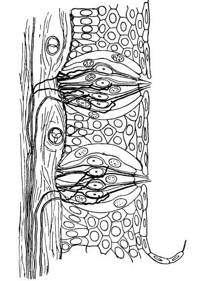
“circumvallate.” Very minute organs of sense—taste-bulbs—stud
the mucous membrane which lines the fosse.
The hard palate ends in a muscular curtain—the soft palate—the
central portion of which—the uvula—depends lower than the rest.
On either side the soft palate splits into two folds; the anterior,
continued to the side of the tongue; the posterior, to the pharynx.
These folds, since they bound the gateway into the pharynx, which is
known as the “fauces,” are termed the “pillars of the fauces.” The
tonsil lies between the anterior and posterior pillars of the fauces,
but does not appear as a prominence unless inflamed or enlarged.
The pharynx hangs as a bag from the base of the skull. It, like all
the rest of the alimentary tract, is lined by mucous membrane. “Mucous
membrane” is not a happy term. It does not denote that the epithelium
secretes mucus. It may or may not possess this property. Nor does it
imply that it has a different origin from the skin—that it arises from
hypoblast, the inner layer of the rudiment from which the embryo grows.
The term is applied to all internal, and therefore moist, surfaces,
whether they arise from hypoblast, as in the case of the lining of the
greater part of the alimentary tract, or whether they are involutions
of epiblast as in the case of the mouth and also of the extreme lower
end of the alimentary tract. Almost the whole of the alimentary canal
is, in the first instance, a tubular cavity in the interior of the
embryo, lined by hypoblast. This cavity communicates with the
yolk-sac, but has no openings on the exterior until it joins up with
two epiblastic pits—one the stomodæum, or mouth-cavity, at the
anterior end; and the other the proctodæum, at the posterior end of
the body. The distinction between the middle closed portion of the
alimentary canal and its two secondary openings suggests morphological
speculations, into which we have not space to enter, as to the ancestry
of the vertebrates. The majority of anatomists believe that the
primitive canal is represented in the middle portion, and that, in
prevertebrate animals, it opened to the exterior in a different way.
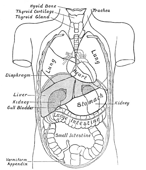
The pharynx is 4½ inches long. It is enclosed by three thin muscles,
which overlap from below upwards—the constrictors of the pharynx.
The anterior attachment of the superior constrictor is to the jaw; of
the middle constrictor to the hyoid bone; of the inferior constrictor
to the thyroid cartilage. Above the soft palate the nasal chambers
communicate with the pharynx by the posterior nares. Below the hyoid
bone, which is easily felt in the neck as a bony arch just above the
thyroid cartilage (Adam’s apple), the windpipe, or trachea, joins the
pharynx by a single pear-shaped orifice—the rima glottidis. When we
consider the mechanism of swallowing, we shall study the arrangements
which prevent food, passed through the fauces, from entering either the
nasal chambers above or the windpipe below and in front. At the level
of the lower border of the thyroid cartilage the pharynx becomes the
relatively narrow œsophagus. This tube, which lies behind the trachea,
and slightly to its left side, passes with a straight course to the
abdomen. It traverses the chest, lying behind the heart, pierces the
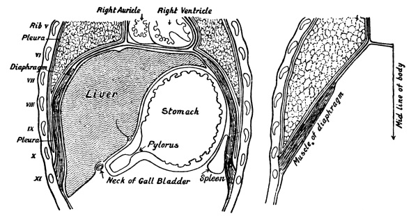
diaphragm, and just beneath it joins the stomach. Its length is about 9
inches. The stomach is a sickle-shaped bag. It has two apertures—the
cardiac orifice, or junction with the œsophagus; and the pyloric
orifice, or junction with the small intestine. It is so folded on
itself that these two apertures are not more than 4 inches apart. Its
outline may be drawn on the body-wall with a piece of charcoal from a
point an inch below and an inch to the left side of the lower end of
the breast-bone, the position of the cardiac orifice, to a point about
4 inches below the end of the breast-bone, and an inch or two to the
right side of the mid-line of the body, the position of the pyloric
orifice, with a slight curvature to represent the upper border; to
represent the lower border the same two points are joined by a bold
curve, bulging upwards to the nipple, outwards to the side of the body,
and downwards some distance on the abdomen (cf. Fig. 2).
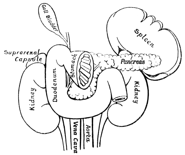
Fig. 6.
The stomach has been cut across a short distance from
the pyloric valve, and removed, to show the viscera which lie behind it. The
descending aorta and the vena cava rest upon the vertebral column. They
are crossed by the pancreas and the transverse portion of the duodenum.
The head of the pancreas is enclosed by the curvatures of the duodenum.
The ducts of the liver and pancreas are seen entering the descending
duodenum side by side.
Such an outline represents the form and position of the stomach when
distended; but it is to be understood that its dimensions depend upon
the amount of its contents. It is capable of holding about 7 pints.
The junction of œsophagus and stomach is closed by a muscular ring, or
sphincter muscle—the cardiac sphincter; the junction of stomach and
intestine is guarded by a much stronger pyloric sphincter. The average
diameter of the small intestine is about 1½ inches. It is wide enough,
therefore, to admit two fingers. The length of the tube is about 22
feet. Its first part is termed the “duodenum,” because its length
equals the breadth of twelve fingers—i.e., about 9 inches. The
remainder is divided arbitrarily into jejunum and ileum. The duodenum
makes three sharp curves. First it inclines upwards and to the right,
then vertically downwards, then horizontally to the left, and finally
forwards. The ducts of the liver and pancreas open by a common orifice
into the descending portion. Its horizontal portion is bound firmly to
the vertebral column. After this the whole of the small intestine is
supported by the mesentery, a double fold of peritoneum which allows it
to hang freely in the abdominal cavity. The mesentery is attached to
the back of the body-wall. Commencing on the left side of the second
lumbar vertebra, its line of attachment inclines obliquely downwards
and to the right, across the vertebral column, for about 6 inches.
Measured from its attached edge to the edge which bears the intestine,
it has a width of about 8 inches. Its free border has, as already
said, a length of 22 feet. Its measurements being as just stated, it
is clear that it must be folded backwards and forwards upon itself,
like a goffered frill. In the right groin the small intestine joins the
large intestine, or colon. It does not, as might have been expected,
simply dilate into the large intestine, but enters it on its mesial
side, its orifice being guarded by the ileo-colic valve. In other
words, the large intestine projects downwards beyond this orifice,
as the cæcum coli. In many animals the cæcum is of great length and
capacity. In the human embryo it begins to assume a similar form; but
a very small portion only (the so-called “cæcum” of human anatomy)
dilates to the calibre of the colon. The real cæcum retains throughout
life its embryonic calibre. It has a length of about 3½ inches, and a
diameter of not more than ¼ inch. This is the “vermiform appendix,” of
ill fame, which must be looked upon as one of Nature’s misfits. Its
great liability to become inflamed is commonly explained as due to the
tendency of such articles of food as pips, the fibre of ginger, flakes
from the inside of enamelled saucepans, etc., to become lodged in its
cavity. But whether this explanation be correct or no—and there are
reasons for thinking it somewhat fanciful—it is much to be wished
that the process of evolution would hasten the disappearance of this
functionless vestige of a cæcum. As there is no tendency towards the
inheritance of characters due to mutilation, and since the surgeon’s
knife now prevents this death-trap from claiming its toll of possible
parents, we must look upon the rudimentary cæcum, with its liability
to inflammation, as a permanent burden on the human race. In justice
to the appendix, however, it must be pointed out that it has acquired
its criminal reputation during the past twenty years. The frequency of
appendicitis has increased so enormously during this period that it
ought to be possible to correlate its prevalence with the introduction
of the cause upon which it chiefly depends.
The colon has a length of about 5 feet. Its greatest width, about
3 inches, is at its commencement, but it is everywhere much wider
than the small intestine. Whereas the wall of the small intestine is
smooth externally, the wall of the colon is sacculated. Three muscular
bands constrict it longitudinally; circular bands at intervals of
about 1 inch or 1½ inch throw it into pouches. It ascends on the
right side, lying far back against the body-wall, to which it is
bound by peritoneum, which in this part of its course covers only its
anterior surface. Having touched the under side of the liver, it loops
forwards and to the left side, crossing the middle line just above the
umbilicus. On the extreme left side it touches the spleen, getting
very near to the back of the abdominal cavity. It then descends on the
left side, again bound to the body-wall by peritoneum, although not
so closely as on the right side, until it reaches the inner lip of
the crest of the hip-bone. From here onwards the fold of peritoneum
which attaches it allows it a free movement. This portion of the large
intestine, the sigmoid flexure, may even fall over into the right
groin. Lastly it curls backwards into the pelvis, as the rectum.
Movement of the contents of the alimentary canal may be favoured by
judicious pressure, or massage. From the description of the situation
of its several parts given above, it will be understood that if the
right hand be placed on the abdomen immediately beneath the ribs, with
the fingers well round to the left side, the stomach will be covered.
Pressure from left to right will tend to drive its contents towards the
pyloric valve. The small intestine is so irregular in its course as to
preclude the possibility of following it with the hand. Pressure first
on one side and then on the other, with a general tendency to work from
above downwards, tends to press forward its contents; but, owing to its
circular form and strong muscular walls, it is not in much need of
help. Very different is the position of the large intestine in this
respect. Its calibre is much greater, its wall is sacculated, its
contents comparatively firm. If the palm of the hand be placed above
the right groin and pressure directed upwards, the cæcum coli and
ascending colon are emptied. If pressure be directed from the extreme
right side just below the ribs, across the middle line to the left
side, the transverse colon is emptied. The descending colon needs
pressure from above downwards on the left side; the sigmoid flexure,
pressure above the left groin, downwards, and towards the middle line.
The inner wall of the œsophagus is smooth, save for the wrinkles into
which it is thrown when not distended; but from the cardiac orifice
of the stomach onwards the mucous membrane of the alimentary canal
exhibits folds and other projections which serve many purposes.
They serve to delay the food, keeping it longer in contact with the
secreting surface. They increase the area pitted with tubular glands;
they increase also the area through which absorption of the products
of digestion occurs. On the inner surface of the stomach the folds
produce a reticulated pattern. In the upper portion of the small
intestine, especially the duodenum, there are prominent transverse
shelves (valvulæ conniventes). No definite folds occur below the
upper three-fourths of the small intestine, with the exception of the
constrictions of the transverse colon already referred to, which affect
the whole thickness of its wall. Throughout the whole of the small
intestine the mucous membrane projects in finger-like processes, or
villi, which give it a characteristic velvety appearance. The villi are
longest in the duodenum.
Lymph-follicles occur at intervals in the intestine. In the ileum they
are collected into patches (Peyer’s patches), on the side opposite to
the line of attachment of the mesentery. They serve both for the supply
of phagocytes, which hunt any germs that have penetrated the mucous
membrane, and also as stations to which germ-laden phagocytes retreat.
The wall of the intestine is composed of mucous membrane, submucous
tissue, and muscle. The mucous membrane is everywhere pitted with
tubular glands, termed in the stomach “gastric glands,” and in the
intestines, both small and large, “crypts of Lieberkühn.” Their
relation to the wall might be exemplified by taking a block of dough
about 6 inches thick and pushing a pencil vertically into it almost
down to the table on which it rests. The holes should be made as close
together as possible, since, especially in the stomach, extremely
little tissue intervenes between the tubes of gland-cells. If the piece
of dough were placed upon a folded cloth, the cloth would represent
the muscularis mucosæ, a layer properly regarded as a constituent of
the mucous membrane. The fibres of this coat are disposed in two or
three sheets, the fibres of one sheet crossing those of the next. By
their contractions they squeeze the ends of the crypts, and probably
wobble them about, expelling their secretion. Beneath the muscularis
mucosæ is a layer of connective tissue, the submucosa, which contains
abundant lymphatic channels, bloodvessels, and nerves. At the pyloric
end of the stomach, the tubes of gland-cells tend to pierce the
muscularis mucosæ. In the first part of the duodenum, certain tubes,
having pierced this layer, branch in the submucosa. A layer of racemose
glands is thus formed—the glands of Brunner. Outside the submucosa
is the muscular coat proper, composed of plain muscle-fibres, except
in the upper part of the œsophagus, where the fibres are striated. It
consists of an inner and an outer sheet, the fibres being disposed
circularly in the inner, longitudinally in the outer sheet, with a
slight departure from this regular arrangement in the wall of the
stomach. On its outside the canal is invested by peritoneum, a layer
of flattened epithelial cells supported by connective tissue. The
abdominal wall also is lined with peritoneum. The smooth moist surface
of the peritoneum covering the intestines glides on the peritoneum
lining the abdominal wall. Between the two is a “potential” space. In
dropsy, fluid accumulates within this space. In a healthy condition the
apposed surfaces are merely moist.
The movements of the intestines are of two kinds. At all times they
exhibit swaying movements, in the production of which the longitudinal
fibres play the chief part, although the circular fibres also contract.
The object of this undulation is to thoroughly mix the contents of
the gut with its secretions. If pills of subnitrate of bismuth are
administered, and their progress observed by the aid of Röntgen rays,
they are seen to oscillate backwards and forwards on their way down
the canal. The slower vermicular movement which squeezes the contents
forwards is called “peristalsis.” It resembles the progressive
contraction of an elastic tube which may be effected by drawing it
through a ring, but is rather more complicated. At the point at which
it is occurring the circular coat is sharply contracted. Above this
it is also somewhat contracted; below it is relaxed. The longitudinal
fibres, using the constricted portion as a point d’appui, pull up
the segment of the intestine which lies immediately below it, drawing it
off the contents of the tube as a glove from a finger.
When food is swallowed, it falls down the œsophagus, aided by slight
peristalsis. As soon as sufficient has accumulated on the upper surface
of the cardiac valve of the stomach, the valve relaxes; at the same
time a stronger peristalsis of the lower portion of the œsophagus
squeezes its contents into the stomach. Food remains in the stomach
until it has reached a certain stage of digestion, the chief object
of which is its subdivision into small particles. Until this stage
is reached, the pyloric valve is firmly closed. The contractions of
the wall of the stomach drive its contents round and round—down the
greater and up the lesser curvature—mixing them thoroughly with the
gastric juice (cf. p. 124). As the acidity of the
mixture increases, the peristaltic contractions of the stomach become more vigorous,
until, the pyloric valve relaxing, the food is little by little driven
into the duodenum.
The alimentary canal has an abundant supply of nerves from the vagus
and the sympathetic systems. It contains also within its own wall an
enormous quantity of nerve-fibres and nerve-cells. They are disposed
as two plexuses, one in the submucosa, the other between the circular
and longitudinal muscular coats. In a specimen successfully stained
with methylene-blue, they are so abundant as to give the impression
that every plain muscle-cell may have its own separate nerve-twig.
Nevertheless, the contraction of the muscle-cells may take place
independently of all nerve-influence—independently, even, of the local
mechanism, the plexus referred to above. Nicotin applied to the wall
of the intestine paralyses the local nerves; yet rhythmic contractions
still occur. They are, however, no longer progressive. They do not drive
the contents of the intestine forwards. Co-ordinated contraction is
observed so long as the local mechanism is intact, even though all
external nerves have been cut. The intestines have their own nerve
cells and fibres, which, acting as a linked system of reflex centres,
provide for the harmonious contraction of their walls. External nerves,
sympathetic and splanchnic, convey impulses which either intensify the
movements or inhibit them, as need may be.
In the matter of its nerve-supply, the alimentary canal stands apart
from the other organs of the body. It may be supposed that it presents
a more primitive condition. Its muscular fibres have the power of
contracting spontaneously. The pressure of the contents of the tube
acts as a stimulus. When the fibres are stretched, they contract.
When the tube is dilated, its muscles endeavour to restore it to its
normal calibre. Such direct action would not, however, provide for the
forward passage of its contents. To bring about peristalsis, a nervous
mechanism is needed, as abundant and complicated as that which ensures
the progress of a slug or a worm. To deal satisfactorily with the
various contents of the tube—liquid, solid, gaseous—the mechanism
must be capable of complicated adjustments. The dilated portions of
the tube—stomach, cæcum coli, rectum—require special arrangements of
muscle and nerve. Nor is the canal altogether independent of the rest
of the body. To a large extent its work is carried on without regard
to the activities of other organs, yet it is not wholly free from the
control of the central nervous system. It is regulated by means of both
afferent and efferent nerves of the vagus and sympathetic. Even the
brain has something to say with regard to the way in which it shall
contract. It is a matter of common experience that emotional influences
may affect the movements of the stomach and intestines—“His bowels
yearned.”
Normally, vomiting is due to irritation of the endings of the vagus
nerve in the stomach, although the afferent impulses may have other
sources. Touching the upper surface of the epiglottis with the finger
will provoke the reflex. So also will stimulation of the olfactory
nerves by a foul smell. In this latter case the emotion of disgust to
which the odour gives rise brings about the reflex action. A flow of
saliva precedes the act of vomiting. A deep inspiration is then taken,
in order that for a time the lungs may be independent of a fresh supply
of air. The glottis is closed, the diaphragm fixed. Contraction of the
abdominal wall presses the stomach against the diaphragm; its cardiac
sphincter relaxes, and its contents are squirted into the œsophagus,
which undergoes a forcible retrogressive peristalsis.
It is interesting to note the difference between carnivora and
herbivora in regard to vomiting. Carnivora swallow fur and other
indigestible materials, as well as many unwholesome things which they
need to be able to return. A dog can, apparently, vomit at will.
Never, while in a state of nature, do herbivora need to return the
contents of the stomach. No provision is made for vomiting. A heifer
which has strayed into a dewy clover-field is not unlikely to die
from the effects of distension of its paunch, if relief be not given
by opening it with a knife. In a horse the cardiac sphincter is
strong, the pyloric weak. Pressure on the stomach tends to drive its
contents through the pyloric valve into the duodenum, not backwards
into the œsophagus. The stomach is not so placed as to allow of its
being compressed between the wall of the abdomen and the diaphragm.
Horses cannot vomit. It is a mistake to suppose that they suffer from
sea-sickness. In rough weather they sweat, their limbs tremble, they
go off their feed; but these symptoms are probably due to the fatigue
which results from excessive anxiety to maintain their balance, and
to fear. We can never know their feelings, but there is no reason for
supposing that they experience the sensation of nausea.
Vomiting is a frequent symptom of cerebral disturbance. The
fluctuations of pressure which the brain experiences as it rocks about
on its “water-bed” within the skull is the cause of sea-sickness. Yet
the motion of a ship may produce violent headache without nausea, the
brain only, not the stomach, appearing to be troubled by the motion.
Not that headache is a pain “inside the head.” Nor is it properly
described as a pain in the scalp, although the messages which are felt
in consciousness as headache originate in the endings of the nerves
of the skin which covers the skull. The excessive sensitiveness of
these nerves is due to vaso-motor conditions, usually the dilation,
occasionally the constriction, of the bloodvessels of the scalp. But
the vaso-motor condition is sympathetic with the disturbance of the
brain; and the special urgency or efficiency of the messages from the
skin results from their being delivered into excited brain-tissue.
Nausea and headache are equally symptoms of the irritability of the
brain caused by the motion of the ship. In one case messages from the
stomach, in the other case messages from the scalp, acquire undue
importance, owing to the agitated condition of the brain-tissue
through which they pass. Not uncommonly the voyager, who wakes in
the morning reconciled to the changes of pressure which he has
experienced while recumbent, finds, when he stands upright, that the
base of his brain is as sensitive as ever. Visual sensations also
contribute to the brain-disturbance. So, too, do the movements of
endolymph in the semicircular canals (cf. p. 335).
It is, indeed, possible that this last factor is more important than the variations
in pressure on the surface of the brain. Probably it accounts for the
after-image of rolling which almost everyone experiences for at least
a day after leaving the ship. Its cause being cerebral, the tendency
to sea-sickness can be controlled by drugs which, like the bromides,
chloral, alcohol, etc., deaden the brain.
Salivary Glands.—The secretion which accumulates in the mouth
is the combined product of the sublingual, submaxillary, and parotid
glands. It is a very thin, watery solution containing not more than 0·5
per cent. of solid substance. If red litmus-paper is moistened with
saliva, it becomes blue, showing that the secretion is alkaline. It
contains a ferment, ptyalin, which digests starch. The action of this
ferment can be demonstrated by holding in the mouth for half a minute
some warm starch mucilage—boiled arrowroot, for example. It quickly
loses its viscidity owing to the conversion of starch into sugar.
Chemically this change may be demonstrated by adding iodine-water to
a specimen of the starch before and after action. Before the starch
is taken into the mouth the iodine turns it blue (a characteristic
reaction for starch). After it has been exposed to the digestive action
of the saliva, iodine fails to colour the mixture, which now contains
no starch. All the starch has been converted into dextrin and sugar. If
unboiled arrowroot is placed in the mouth, some sugar is produced, but
the process of conversion is very slow. It is almost impossible to
digest raw starch in the mouth sufficiently to render it insusceptible
to the colouring action of iodine. The sugar produced by the action of
ptyalin is of the same nature as that which appears during the malting
of barley. It is therefore termed “maltose.” It closely resembles
grape-sugar, but is not identical with it.
The Secretion of Saliva.—The accessibility of the salivary
glands, and especially of the submaxillary, has led to their being used
for a very large number of experiments. They have been studied with
the aim of coming to an understanding of the mechanism of secretion
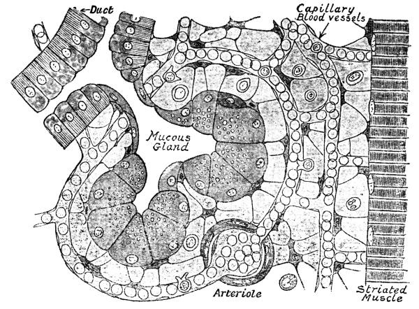
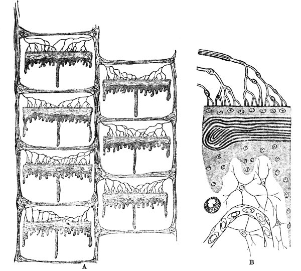
in general. The glands consist of tubes of gland-cells, each tube
suspended in a basket of connective tissue, in a bath of lymph
(cf. Fig. 3). Innumerable capillary bloodvessels
traverse the lymph-bath. The arteries which carry blood to the gland are supplied
with nerves, which regulate their calibre, and therefore determine the amount of
blood which passes through the capillaries into which they break
up. The glands also are supplied with nerves which influence their
functional activity. Nutrient substances and oxygen pass out of the
blood into the lymph. Carbonic acid passes into the blood from the
lymph. Waste products are either carried away in the lymph-stream, or
make their way through the walls of the capillaries into the blood.
Many problems present themselves for solution. How does the amount of
work done by the gland affect its supply of blood? Does the quantity of
saliva secreted vary directly with the pressure of lymph in the spaces
by which the gland is surrounded? Is this pressure wholly dependent
upon the pressure of the blood? Are the substances secreted by the
gland supplied as such by the blood, or does the gland make the ptyalin
and mucus which it secretes? If it makes its secernable products, what
materials does it abstract from the blood for the purpose of their
manufacture? Does it use the whole of these materials, whatever they
may be, or does it use part only and return the residue to the lymph?
Does it make its products only when it is actively secreting, or is it
always making them, and storing them in its cells in order that it may
have a supply to discharge when called upon by the stimulation which
results from the presence of food in the mouth? Is their discharge
merely a washing out due to the rush of fluid which occurs when the
bloodvessels are dilated, or can the gland-cells expel their products
in response to nervous action? In what way do the nerves of the
gland influence secretion? Do they call for increased production, or
increased output, or both? These are some of the problems which the
exposed situation of the submaxillary gland allows physiologists to
tackle.
By means of a very simple operation, the ducts of one or both parotid
or submaxillary glands can be brought to the skin, and made to pour
their secretions on to the surface instead of into the mouth. The flow
under various circumstances can be watched. The saliva can be collected
and measured.
The nerves of the submaxillary gland are easily isolated. A nerve
leaves the seventh (or facial), crosses the drum of the ear, comes
out through a minute crevice in the skull, and runs for some little
distance as a separate nerve before it applies itself to the lingual
branch of the fifth, which runs along the side of the tongue. Owing to
its passage across the tympanic cavity (drum of the ear), it is termed
“chorda tympani.” As its fibres are very small, they can be recognized
wherever they form a part of the lingual nerve. They leave the lingual
to go to a ganglion, the submaxillary ganglion, from which the gland
is supplied. The gland also receives branches from the sympathetic
nerve which ascends the neck. The last-named branches accompany the
facial artery. Stimulation of either of these nerves causes the gland
to secrete. The flow of saliva which follows stimulation of the chorda
tympani is much more copious than that which follows stimulation of
the sympathetic, and as a rule it contains far less organic matter,
although about the same amount of mineral salts. Under normal
conditions the activity of the chorda tympani is brought into play in
a reflex manner by impulses which travel up the nerves of taste (the
lingual and glosso-pharyngeal) to the cerebro-spinal axis; but almost
any other nerve will serve as an afferent path. The gland may also, as
we shall presently explain, be called into activity by the cortex of
the brain.
It is certain that in the case of the submaxillary gland secretion is
not the direct result of increased blood-pressure. It is not a case of
filtration from the blood through certain membranes and cells into the
salivary duct. Atropin (belladonna) dilates the bloodvessels,
increasing blood-pressure, but it stops secretion. After
belladonna-poisoning, the mouth, like the skin, is hot and dry. Other
drugs there are which provoke a certain amount of secretion, even after
the bloodvessels going to the gland have been tied. It is possible, by
stimulating the chorda tympani, to obtain a pressure in the fluid in
the duct very much greater than that in the bloodvessels which supply
the gland. Here we have clear proof that secretion is not filtration.
Filtration is the passage of fluid through a filter-bed from a higher
to a lower pressure. In filtration, moreover, soluble diffusible
salts accompany the water. The saliva contains only half as much of
these diffusible salts as the blood. Therefore the gland tissue stops
half the salts. Secretion is an active process carried out by the
gland-cells, under the influence of nerves, in opposition to the laws
of filtration. The gland-cells determine how much water shall pass
through them and what percentage of salts shall accompany the water.
How does a gland-cell make the substance which it secretes? There is no
reason for supposing that the ptyalin or the mucus which the salivary
glands secrete is present in the blood, either ready formed, or, as it
were, half formed, in combinations which can be easily broken up. All
the evidence obtainable points to the conclusion that the gland-cells
take out of the lymph proteid materials from which they manufacture
the peculiar substances which they secrete. During rest, granules
accumulate in the cells. During activity they disappear. It has been
shown in the case of the gastric glands that these granules consist
of the special ferment which the gland secretes, in an inactive form.
It may be that it is combined with a substance which prevents it from
exerting its digestive action on the cells within which it is made;
damped, as gunpowder is damped during transit. Or it may be that it is
not a finished ferment; it may need a further addition to its molecule.
During activity, while the granules disappear, proteins accumulate at
the bases of the cells, giving to a tube of gland-cells the appearance
of a peripheral non-granular zone. This proteid substance must have
come from the lymph, and the inference seems inevitable that the cells
have taken into their protoplasm a supply of material which will
serve for the manufacture of additional granules. Each gland-cell is
therefore an independent unit. By its own activity it takes up
materials from the lymph, out of which it manufactures its own special
products. It stores its products until they are wanted. Then by its
own activity it extrudes them into the lumen of the gland-tube. It
has, indeed, been shown that, when the nerve going to a salivary
gland is stimulated, the gland shrinks, notwithstanding the great
dilation of its bloodvessels. Under the influence of the stimulation
the granules in the gland-cells imbibe water, swell up, and escape
from the cells. The cells discharge their accumulated stores, in the
first instance, more rapidly than they take up materials (even fluid)
from the blood. For its knowledge (if the term may pass) of what is
wanted the gland-cell is dependent upon messages which reach it through
the nervous system. These messages take origin in the endings of the
sensory nerves of the mouth, pass up to the brain, and are reflected
down the nerves to the gland. So accurate is the information conveyed
to the glands, that when a horse transfers the work of mastication from
one side of its mouth to the other, as it is in the habit of doing
about every quarter of an hour, the flow of saliva from the parotid
gland on the masticating side is increased; on the other side it is
diminished. Two or three times as much saliva is poured out on the one
side as on the other.
Not only is the amount of saliva poured out in response to stimulation
proportional to the needs of mastication, but the kind of saliva is
adapted to the nature of the food. In a dog—and this is an observation
which can be made only on an animal which lives on a mixed diet—it is
possible to determine the amount of the two kinds of saliva secreted
and the relation of flow to food. When meat is given to the animal, the
submaxillary gland yields its secretion; when it is fed on biscuit,
abundance of the watery parotid saliva is poured forth. A mouthful of
sand also causes the parotid saliva to flow, in order that the sand may
be washed out of the mouth.
More remarkable than the response to direct stimulation is the effect
produced by the sight and smell of food. When meat is shown to a dog,
submaxillary saliva begins to flow; when it is offered bread, parotid
saliva is secreted. And the activity of the glands is not merely a
nervous reflex independent of the animal’s mind. The moment the dog
realizes that it is being played with—that there is no intention of
giving it the coveted food—the flow of saliva ceases. An emotion
may check secretion when every physiological condition is demanding
it. This is the explanation of the Rice Ordeal. Dry rice provokes a
flow of saliva in the mouth of all save the guilty man. Response to
mental impressions is a matter of the greatest consequence in the
physiology of digestion. It holds good in the case of the secretion
of gastric juice equally with that of saliva. The sight and smell of
food sets the juice flowing into the stomach, and the more desirable
the food, the more attractive its appearance, the more stimulating
its smell, the more rapidly does the secretion flow. Here we touch
upon a theme which hardly needs exhaustive treatment. It is not
the stoutest people who eat the most, although an impartial survey
of one’s well nourished friends will show them to be persons who
“take kindly to their victuals.” A small quantity of food perfectly
digested is more nourishing than much food which the digestive organs
do not efficiently prepare for assimilation. Good digestion waits on
appetite; and appetite, in civilized man, is something more than a mere
physical need of food. The hunger which leads to the bolting of food
without pleasurable anticipation, without mastication, without any
consideration of the quality of the viands, is a harmful craving which
ends in imperfect assimilation. It is more profitable to toy with a
hors d’œuvre than to engulf, unthinking, a plateful of beef. But
we have said enough to suggest reflections to those who take no thought
as to what they shall eat or what they shall drink; and few who take
thought need to be convinced.
The Stomach.—The sight and smell of food, its presence in
the mouth, and the performance of mastication, which induces a secretion
of saliva, gives rise at the same time to a flow of gastric juice.
It is psychic stimulation and the act of eating which cause gastric
juice to ooze from the gland-tubes of the stomach at the commencement
of digestion, not the stimulation of nerve-endings by food which has
passed down the œsophagus. As a consequence of gunshot wounds, or
as the result of operations performed for the purpose of relieving
patients whose œsophagus has become blocked, numerous cases have been
recorded in which a fistulous opening into the stomach has made it
possible to study the interior of this organ. Such cases present an
opportunity of watching the digestion of various foods introduced
through the opening, and of collecting gastric juice for purposes
of analysis. A similar condition has been established in animals by
operative means. The œsophagus having been cut, and the cut end sutured
to the margins of an aperture in the skin, food taken by the mouth
escaped by this opening instead of passing into the stomach. A similar
opening was made into the stomach for the insertion of food, and for
the purpose of studying the effects of reflex stimulation of the
gastric glands. As soon as food was introduced into the mouth, gastric
juice began to flow. The advantage of this experimental method lies in
the fact that the juice secreted was a pure juice—not mixed with food,
as in all the earlier experiments in which, the stomach being opened
without diversion of the œsophagus, the presence of food within it was
the stimulus which led to secretion. No juice flowed in the absence of
stimulation; nor was the secretion normal in composition when provoked
by a mechanical stimulus, such as the tickling of the gastric mucous
membrane by a feather.
My lord the stomach! He is not the only, nor is he the chief, agent
in digestion; but with him rests the decision as to whether the food
offered to the alimentary tract is suitable in quality and quantity. He
is offended if it be not offered with all the circumstance and ceremony
which becomes his rank. As an intimation that he is about to receive
food, he accepts the news from the mouth that its nerve-endings are
subject to mechanical stimulation. But the chewing of indiarubber would
produce a like effect. The stomach, therefore, confers with the organs
of taste and smell. If their report is favourable, he argues that the
substance which the teeth are crushing will justify an outflow of
gastric juice. He responds most generously when prolonged mastication
assures him that he may trust to receiving the food in a sufficiently
subdivided state. At our peril we neglect to propitiate my lord. Not
always debonair when treated with consideration, he is morose or
petulant when slighted. Never content with lip-service, he exacts the
labour of teeth and tongue and palate. The tribute we offer may be of
the best—savoury, wholesome, well cooked, well chewed—but if it
be not tendered with some degree of love, if thoughts are concentrated on
other things, if no attention is devoted to the meal, if no sense
of liking accompanies our offering, my lord the stomach on his part
affords the viands an indifferent reception. In consulting our own
tastes we are to a large extent consulting the needs of the stomach.
Ravenous and excessive feeding is not an exhibition of taste; it is a
return to the instinct of the savage, who was never sure that he would
get his full share, and was afraid to trust that another meal would be
obtainable when nature declared it due. Some degree of epicureanism
is favourable to digestion. The flow of gastric juice in the stomach
occurs reflexly in response to the emotion of appetite, to stimulation
of the nerves of taste and smell, to the obscure sensations which
accompany the activity of the muscles of mastication.
The gastric juice secreted in a day amounts probably to about 8 or 9
pints. To this we must add, when considering the quantity of fluid
which passes through the stomach, the saliva, which certainly reaches
as much as 2 pints, and the beverages taken with food.
Gastric juice collected in the manner described above is a clear,
colourless, inodorous fluid. It is very acid, and so powerfully peptic
as to digest its own weight of coagulated white of egg. Its solid
constituents amount to 0·5 per cent. They consist of the two ferments
pepsin and rennin, with traces of proteins and mucin, and various
inorganic salts. Its acidity is due to free hydrochloric acid to the
amount of 0·2 per cent. This acid is more or less in combination with
the pepsin. In pure gastric juice hydrochloric acid is the only acid
present; but when mixed with food the juice contains other acids also,
especially lactic.
When food first reaches the stomach, the alkaline saliva which
accompanies it neutralizes the acidity of the gastric juice. For some
time, probably about half an hour, the conversion of starch into sugar
is still carried on by the ptyalin of the saliva, owing chiefly to the
difficulty which the gastric juice encounters in permeating the masses
of masticated food. The Bacillus acidi lactici is always present in
the stomach. It converts some of the sugar into lactic acid; of this a
small quantity is further changed into butyric and acetic acids, with
the formation of carbonic acid and hydrogen gas. After a while the
lactic acid is absorbed, and hydrochloric acid alone remains.
The secretion by the gastric glands of so powerful a mineral acid
as hydrochloric has always aroused interest. How is it possible for
the gland-cells to produce it without injury to them selves, or for
the stomach to contain it without self-digestion? Many chemical and
physical theories have been advanced in the belief that they rendered
the process of its production less difficult to understand. All such
theories are, however, inadequate to explain the secretion as a
discontinuous process, which occurs only as a response to demand. That
the source of the acid is the sodic chloride which the gland-cells
take from the blood does not need assertion, but we cannot picture
the process by which this exceedingly stable compound is decomposed
otherwise than on the assumption that weaker acids, or, rather, acid
salts, are also absorbed by the cells, and that, in accordance with the
laws which govern the composition of salts in solution, an exchange
of acids occurs. If sodic chloride and any acid salt—acid phosphate
of sodium, for example—are in solution in water, the salts do not
retain their form as we know them when isolated by crystallization.
The mixture contains “free” hydrochloric as well as “free” phosphoric
acid. It may be assumed that within secreting cells a similar exchange
of acids takes place. By a process which we term “vital,” the acids are
kept apart, and the hydrochloric acid is extruded by the cells. In the
present state of knowledge this vital action is mysterious; but it is
no more mysterious than the isolation of pepsin, or any other metabolic
event which occurs within a cell.
The proteolytic ferment pepsin is active only in an acid medium. Yet
apart from its digestive function as an ally of pepsin, hydrochloric
acid by itself also exerts a valuable disintegrating action on certain
constituents of the food. Possibly the most important results of the
presence of free hydrochloric acid in the great chamber into which food
is first received are due to its disinfective property. It destroys
all the putrefactive germs which accompany the food, and many germs
which, if introduced into the blood, would give rise to disease. It
also destroys the germs which multiply in the stomach towards the end
of each interval between two meals. When withdrawn from the body,
gastric juice will keep an indefinite time, if evaporation of the acid
be prevented.
Pancreas.—In structure the pancreas presents a marked
resemblance to the salivary glands. Probably this resemblance is
merely superficial. Minute examination reveals points, apparently
of great morphological importance, in which they differ. In the
gland-tubes of the salivary glands, and, indeed, in all glands with
the exception of the pancreas, secreting cells project into the lumen.
The secreting cells of the pancreas are invested internally by a layer
of flattened scales (intra-acinar cells). They lie, therefore, between
the basement membrane which invests them externally and this second
layer of flattened cells which separates them from the lumen of the
tube. At a very early date in embryonic life the gland-cells of the
pancreas are filled with highly refracting granules. As this occurs
long before any digestive action is called for, it may be taken as
indicating that the pancreas has functions which other glands—the
salivary, for example—do not possess. These granules do not, however,
appear in all parts of the tubes. Certain portions of the tubes
remain undeveloped—fail, that is to say, to acquire a secreting
function—even in adult life. Such patches of cells, not disposed in
gland-tubes, are known as islands of Langerhans. When the pancreas is
over-stimulated by artificial means, leading to its extreme exhaustion,
large portions of its glandular substance return to this primitive
condition. The gland-cells not only discharge their stores of granules,
but they lose the greater part of their cell-protoplasm. It would seem
that, in their effort to meet the demand for ferments, they use up
their own cell-substance in their manufacture. Having exhausted their
coal, they stoke the furnace with the looms and furniture of the mill.
It may be that other glands would do the same if it were possible to
stimulate them as strongly as the pancreas can be stimulated. The
result is probably due to the extreme susceptibility of the pancreas to
the action of secretin, a substance made in the intestine. Secretin can
be isolated and injected into the blood. We shall refer again to this
chemical stimulation of the pancreas when tracing the progress of food
through the alimentary canal.
The secretion of the pancreas is a clear, colourless, alkaline liquid
of syrupy consistence. The quantity of juice secreted is relatively
small, but the organic substances which it contains are in a
concentrated form. They constitute as much as 10 per cent. of the
pancreatic juice. Proteins are present, if the juice be fresh. If it
has stood for any length of time, they are found as peptones. The
digestive ferments of pancreatic juice are the most powerful which are
secreted into the alimentary canal.
Bile.—In its most important functions the liver has no relation
to digestion. It is a storehouse of absorbed food. This organ will
therefore be treated in a separate chapter. The bile which the liver
secretes into the alimentary canal has no chemical action on any of
the constituents of food, with the exception of a feeble tendency to
digest starch. Yet it is in some degree accessory to digestion. Poured
into the second portion of the duodenum through an orifice common to
the liver and the pancreas, it mingles with the semi-digested food,
or “chyme,” which, about two hours after a meal, passes through the
pyloric valve. Gastric digestion has converted the greater part of the
proteid constituents of the food into peptones or intermediate stages.
The proteoses or propeptones—a name is needed for the intermediate
products of proteid digestion which does not commit us to any theory
as to their chemical constitution—are quickly peptonized by the
pancreatic juice. But portions of the proteins have escaped the action
of gastric juice, or have at most been affected by its acid only;
these are precipitated by the bile-salts on the mucous membrane of
the small intestine, which is raised into projecting flanges for the
purpose of delaying the passage of the chyme, in order that it may
be thoroughly submitted to the digestive action of pancreatic juice.
Bile-salts also favour the digestion of fat, and its passage through
the intestinal wall. The action of bile-salts in spreading fats is well
known to artists. Ox-gall is smeared upon glass when it is desired
to apply oil-paints to its surface. When mixed with oil, it causes
its emulsification, or breaking up into microscopic globules. In the
absence of bile, but little fat passes into the lymph-vessels which
convey digested food from the intestine to the thoracic duct, and so to
the great veins of the neck. Its action is mechanical. It favours the
digestion of fats by rendering them easily amenable to hydrolysis by
pancreatic juice.
Bile as secreted by the liver is a clear, limpid fluid of low specific
gravity; but during its stay in the gall-bladder it is concentrated by
absorption of water, and mucin is added to it. It contains “bile-salts”
of complex constitution. These salts favour the solution of certain
by-products of cell-metabolism, cholesterin and lecithin; substances
which are formed in many cells, both in animals and plants. Cholesterin
occurs most abundantly in nerve-tissue and in blood-corpuscles.
Lecithin also is a by-product of the metabolism of nerve-tissue.
Protoplasm appears to be incapable of oxidizing these substances, as
it does other products of metabolism. Other substances of equally
complex constitution are reduced to urea if they contain nitrogen;
to water and carbonic acid if nitrogen be absent. Cholesterin and
lecithin have to be eliminated without further change. Some of the
cholesterin is excreted by the sebaceous glands of the skin. It is the
chief constituent of “lanoline” prepared from sheep’s wool; an unguent
which owes its valuable properties to the resistance which cholesterin
offers to cell action, and therefore to the action of living ferments.
Bacteria cannot turn it rancid. The sebaceous glands have the power
of directing metabolism into a channel in which cholesterin is the
chief product, but apparently all cells make it in small quantity. The
bile-salts carry cholesterin and lecithin into the alimentary canal,
from which they are not reabsorbed. Some of the bile-salts are lost to
the body, but the remainder re-enter the circulation, and recommence
their work as vehicles for these inoxidizable and insoluble substances.
In the gall-bladder cholesterin is apt to separate out from the bile
in the form of gall-stones; but whether this is due to an excess of
cholesterin in the bile, or to an abnormal, inflammatory condition of
the lining membrane of the gall-bladder, is still an open question.
Bile also contains bile-pigments. Their colour varies in different
animals, and changes according as the bile is exposed to the air, or
subject to the action of reducing agents. If oxidized, the colour
is green (biliverdin); if reduced, brownish-yellow (bilirubin).
Bile-pigment is formed from hæmoglobin, the colouring matter of the
blood, after the removal of its iron. Worn-out red blood-corpuscles are
destroyed in the spleen, in the manner already described, but it is
uncertain whether the conversion of the hæmoglobin thus set free into
bilirubin occurs in the spleen, or whether this chemical change is
reserved for the liver. Physiologists incline to the view that the
liver is the seat of the change.
Intestinal Juice.—The mucous membrane of the alimentary tract,
as far down as the middle of the rectum, is, as previously stated (p. 102),
studded with tubular glands. They secrete a light-yellow fluid,
alkaline in reaction, and opalescent. Its most important property is
due to a ferment which converts cane-sugar into a mixture of dextrose
and levulose, and changes maltose—the sugar produced by the action on
starch of saliva and pancreatic juice—into dextrose. It is in the form
of dextrose that sugar is carried about the body and assimilated by the tissues.
Intestinal juice also contains a ferment, erepsin, which shakes to
pieces the heavy molecules of peptones and partly formed peptones.
Under its influence they break up into comparatively simple bodies
containing the radicle of ammonia. Substances containing an NH₂
group—one H of NH₃ (ammonia) having been given up, in order that
the group may have a “free arm” with which to link on to the other
component parts of the molecule—are termed “amides.” The amides
which are most characteristic of the action of erepsin are leucin,
an amidated fatty acid; and tyrosin, an amidated aromatic acid. The
tendency of proteins to break up along these two lines—the fatty acid
line and the aromatic acid line—is of considerable interest. The one
line is represented by acetic acid, CH₃COOH; the other contains the
hexone radicle, C₆H₆. Benzoic acid, C₆H₅COOH, is representative of the
latter. It used to be thought that proteins which were shaken into
simple bodies such as amides were lost to the economy. Their downward
career was a foregone conclusion. There could be no arresting it
before they brought up at the bottom—as urea, CO(NH₂)₂—the diamide
of carbonic acid. It was even supposed that this disintegration of
proteins was a provision for getting rid of the surplus animal food
which we consume. Physiological chemists now take quite a different
view. They believe that the epithelial wall of the intestine through
which these substances are absorbed, or the liver, to which they are
carried by the portal blood-stream, has the power of recombining these
fragments into the complex protein edifice. It is even thought that
disintegration is a necessary preliminary to the rearrangement of the
sub-groups. A large variety of proteins is ingested as food. Many of
them, especially the vegetable proteins, are quite foreign to the body.
By the activity of pancreatic juice and erepsin, they are broken into
small and relatively stable groups of atoms, which are again fitted
together into the particular forms of protein which are of use to the
economy.
The Story of a Meal.—The chemistry of digestion will be
understood most readily if the constituents of a meal are traced from
their entrance into the mouth to their absorption through the wall of
the alimentary canal, or abandonment as indigestible.
We may describe as a typical meal one consisting of bread, vegetables,
cane-sugar, meat, milk, fat, and cheese. In the mouth the various foods
are crushed and mixed with the alkaline secretions of the salivary
glands. A certain amount of the cooked starch contained in the bread
is changed into maltose. In the stomach the digestion of starch is
continued for a time, but a large part even of the cooked starch awaits
the action of pancreatic juice. A certain amount of cane-sugar is
converted into dextrose and levulose, which are rapidly absorbed into
the blood; but this action is due to hydrochloric acid, and probably
affects a comparatively small part of the cane-sugar swallowed. Fat is
quite unaltered in the stomach. All proteins are attacked by pepsin,
but some yield to digestion more readily than others. Gluten of bread,
like all vegetable proteins, is comparatively resistant; but since
it is presented to the action of pepsin in small quantities and in a
spongy form—very suitable for digestion—it is probable that most
of it is peptonized in the stomach. Chemists experimenting with gastric
juice taken from the stomach, and reproducing the conditions as to
temperature, removal of products of action, etc., as closely as it is
possible to reproduce them in the laboratory, find that the various
foods take different times to digest. The proteins of meat are more
quickly peptonized when raw than after coagulation by heat. The same
is true of white of egg. Amongst different varieties of cooked flesh,
beef is more quickly peptonized than fish. The casein of milk is more
quickly peptonized than any other protein; and it also is no exception
to the rule that digestibility is diminished by cooking. Similar data
may be obtained for all foods. They are no doubt useful indications of
the course of action which we may expect to occur within the stomach,
but we can never be sure that my lord will obey the ruling of the
chemist. Practice with a captive golf-ball is a useful preparation
for the game; but there are conditions on the links which cannot be
reproduced on the lawn. In an artificial stomach the clean fibre of
raw fish digests more slowly than raw beef. Even when the beef is
roasted and the fish fried or boiled in the ordinary way, the beef
disappears through the dialyser (the bag of membrane suspended in a
vessel of warm water in which experimental digestion is carried out)
more quickly than the fish. Nevertheless, the living stomach is better
disposed towards a mixed meal containing a certain weight of fish than
towards a meal in which, the other constituents remaining the same,
beef takes the place of fish. Important conclusions may, no doubt,
be drawn from observations of the time occupied in the peptonization
of pure food—i.e., fibrin, white of egg, clean meat, etc.—under
conditions simulating those which are present in the stomach; but
they must be accepted with many reservations. In the stomach it is
not pure substances, but mixtures, that the gastric juice has to deal
with. And here a most important factor comes into play, to which
further reference will be made later on. The amount and quality of the
secretion of the gastric glands depends upon the nature of the food.
Hence a food, or a combination of foods, which digest readily in the
laboratory may take a long time to disappear from the stomach, and
vice versâ. Digestibility depends upon the nature of the food. It
depends also upon its physical state. To take simple illustrations:
Cheese contains coagulated casein, one of the most easily digestible
of proteins, but the casein is intimately mixed with fat, upon which
gastric juice can make no impression. Even when finely divided, the
particles of casein are protected from the action of the juice by
fat. In the same way the meat of pork is as digestible as mutton, but
the fat of pork is quickly melted and very liquid. In the process of
cooking the muscle-fibres become saturated with fat.
It is not the function of the stomach to complete digestion. Its
business is to initiate it. Food which reaches the stomach in fragments
is reduced to a condition in which its digestion will be readily
completed by pancreatic juice. Gastric digestion produces a much
larger proportion of intermediate products, proteoses or propeptones,
than does digestion in the duodenum. Such intermediate products are
quickly dealt with by pancreatic juice. Artificial tests of relative
digestibility do not, as a rule, take the amount of propeptones formed
in a given time into account. When considering the digestion of a
typical meal, we must bear in mind that it is not the duty of the
stomach to pass as much sugar, peptone, and fat as possible into the
blood. In fact, very few of the products of digestion are absorbed
by the bloodvessels of the stomach. The impermeability of its mucous
membrane is shown by the fact that hardly any of the water swallowed
passes through the stomach-wall. Practically all the water ingested
leaves the stomach through the pyloric valve. Various salts, some
sugar, and peptones are taken up by the vessels of the stomach; but the
bulk of all the different kinds of food passes into the duodenum in a
semi-digested state. The function of the stomach is to carry digestion
through a preliminary stage. The process will be completed in the small
intestine. It is to be noted that, although water is not absorbed by
the stomach-wall, alcohol passes through it with great rapidity. The
same is true of the various crystalline nitrogenous bodies found in
meat-extracts, and also of the essential principles of tea and coffee,
which chemically belong to the same class. All these substances are
degradation products of proteins produced by oxidation, far advanced
along the road to urea. In this selective absorption we see proof of
the activity of the cells of the mucous membrane. They take up the
substances which it is desirable to remove from the contents of the
stomach. Some may be wanted by the body for its immediate use; others
are better out of the way, because they are prejudicial to the progress
of digestion.
When contemplating the activity of the cells of the gastric mucous
membrane, we feel the need of an adjective which shall express our
recognition of the fact that they have a power which we cannot confer
upon our clumsy mechanical imitation stomach. They can discriminate.
“Vital” is the only term available, though much abused. Using it without
prejudice, as lawyers say, we speak of the “vital activity” of the
cells when we wish to imply that things happen in a living stomach for
which we cannot make provision in a model. Of the many substances which
make their appearance as digestion proceeds, some are absorbed, others
left in the mixture.
The mucous membrane shows its power of controlling digestion in yet
another way. In the neighbourhood of the pylorus its structure is
unlike that which it presents elsewhere. The gastric glands are short,
and tend to branch. Their lining cells are all of the same kind. Over
the greater part of the inner wall of the stomach the tubes are long.
They do not branch. The cells which line them are of two kinds: small
cubical cells (the term refers to their form as seen in section),
similar to those of the pyloric glands; large oval cells, placed with
their longest axes in the same direction as the axis of the gland-tube.
These oval cells do not project into the bore or lumen of the tube, but
are displaced from it by the cubical cells. They rest on the investing,
or basement, membrane. All parts of the gastric mucous membrane secrete
pepsin, although the pyloric portion produces very little; the area
which contains oval cells alone secretes hydrochloric acid. If a
short time after a meal an extract is made from some of the mucous
membrane near the pylorus, by pounding it with salt-solution and sand
to break up its cells, this extract, when filtered and injected into
the blood, stimulates the glands of the cardiac end of the stomach.
Under its influence they pour out both pepsin and hydrochloric acid.
The extract contains a substance which acts as a chemical messenger.
It is a representative of a class of bodies which play a most
important part in co-ordinating the activities of the various organs.
Hitherto physiologists have concerned themselves with the visible or
“external” secretions of glands. They have shown how the production
of these secretions is controlled by the nervous system. Recently
they have discovered that another set of influences has to be taken
into consideration. Glands, and possibly all other tissues, take from
the blood the materials out of which they make their characteristic
secretions, or, if they do not discharge secretions, the substances
which they require for the building of their own structures, and return
to the blood “internal secretions” which act as stimuli to other tissues
with which they are linked in harmonious co-operation. The active
principles of internal secretions have been termed “hormones”—from
ὁρμάω, I announce. The glands of the pyloric mucous membrane secrete a
hormone which calls upon the rest of the membrane to pour out gastric
juice (cf. p. 89).
What induces the cells of the pyloric mucous membrane to produce the
gastric hormone? Their activity in this respect evidently depends upon
the presence in the stomach of partially digested proteid substances.
The cells judge, as it were, when these substances come into contact
with them, that there is more work for the great bag of the stomach to
do. They call upon the part which is most active in secreting gastric
juice to pour it out quickly and get the business of digestion over.
Meat-extracts, which contain the products of protein disintegration,
have a similar influence in promoting the formation of the hormone.
Hence, no doubt, the general custom, found from experience to be
beneficial, of commencing dinner with soup; although it must be
remembered that the rapid absorption of meat-extracts makes them
peculiarly valuable as restoratives. They afford very little energy,
but what they have to give is quickly placed at the disposal of the
economy. Persons whose stomachs are unduly irritable are advised to
avoid soup. It leads to undesirable activity on the part of the gastric
glands, and especially of the acid-secreting cells. Well chewed bread
also encourages the production of the hormone.
Here it may be well to call attention to the evident division of the
stomach into two parts—the large bag, or cardiac portion, which hangs
down; and the smaller, funnel-shaped pyloric end, which is almost
vertical. The distinction between these two parts is faintly visible in
the resting stomach, but even opening the abdomen tends to obliterate
it. That it is much more evident during active digestion has been shown
by adding subnitrate of bismuth to the food, and throwing the shadow
of the stomach on a screen with Röntgen rays. When this is done, it is
seen that the two parts work in different ways. Food is churned round
and round in the cardiac portion, and pressed towards the pylorus. Its
fluid products, mixed with the abundant secretion of the gastric mucous
membrane, are wrung out of it by the pyloric funnel. They are squeezed
towards the pylorus, which opens at intervals to let them through. If
lumps of solid matter reach it, the pyloric valve closes tightly, until
the undigested food has fallen back into the dependent bag. Dyspeptics
are sometimes unpleasantly conscious of the contractions of the
pyloric funnel. In fact, putting aside pain due to gastritis, all the
discomfort of dyspepsia is felt on the right side. Flatus accumulates
beneath the pyloric valve. The valve will not open to let it pass. The
pyloric portion of the stomach contracts strongly. Notwithstanding
the general trend of movement in the opposite direction, the gases
are squeezed back into the larger bag, and escape through the cardiac
orifice.
Tables have been prepared showing the length of time which various
articles of food take to digest. They are based in part upon
observations made upon the living stomach in cases in which it has
been possible to examine its contents through a fistulous opening;
in part upon the results of artificial digestions carried out in the
laboratory. It is hardly too much to say that such observations are
absolutely without value as tests of the relative digestibility of the
several articles of diet consumed as parts of an ordinary meal. The
fact that the commencement of the flow of gastric juice depends upon
mental stimuli, and its continuance upon hormones, shows how difficult
it must be to reproduce the conditions which obtain in a healthy living
body. The most wholesome of foods taken by itself may be longer in
digesting, or may produce more irritation, than many less desirable
things taken in judicious combination. Crushed chicken, hastily
swallowed, sometimes proves more difficult of digestion than meat so
cooked and served as to stimulate appetite and to demand mastication.
Returning to the story of a meal, vegetables pass almost unaltered
through the stomach. Some of the scanty proteins which they contain
are peptonized, but unless they are very well masticated or cooked
until they are soft, and therefore easily pulped by the churning action
of the stomach, the gastric juice has to reach the proteins through
cell-walls. None of the digestive juices are able to dissolve the
cellulose of vegetable cell-walls. Blocks of vegetable tissue pass down
the whole length of the alimentary canal in the form in which they were
left by the teeth. Hence the extreme indigestibility of ill-chewed
cucumber or apple. The pyloric valve of the stomach is forbidden to
allow any lumps of food to pass until the very last stage of gastric
digestion. Pieces of ill-masticated vegetable tissue lie for a long
time in the stomach, irritating the ends of the gastric nerves, until
at last the time comes for them to be shot through the pylorus into the
duodenum. Many salts which vegetables contain, especially the earthy
carbonates and phosphates, are dissolved by the acid of the gastric
juice.
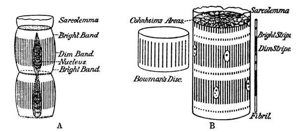
Meat consists of muscle-fibres supported by connective tissue. In the
stomach the gelatiniferous connective tissue is dissolved, setting
the fibres free. Further, the fibres being surrounded by a membrane
of the same nature—sarcolemma—this is removed; and although
it may be hardly justifiable to speak of “Krause’s membranes”
(cf. Fig. 10) as gelatiniferous septa, the fibres are
certainly composed of segments—Bowman’s discs, sarcous elements—into which
they break up under the action of gastric juice. As a result, meat-fibre is reduced
to a finely divided granular condition. The capacity of gastric juice
for dissolving collagen (the substance of which connective tissue
is composed) may be regarded as its most characteristic, as it is
one of its most valuable, properties. Collagen, when boiled or acted
on by acids, takes water into its molecule, becoming gelatin. Under
the influence of gastric juice gelatin is rapidly hydrolysed into
diffusible gelatin-peptone. Pancreatic juice is unable to act upon
collagen, unless it has been previously boiled, or swollen by the
action of dilute acids.
Fat is composed of vesicles of oil supported by connective tissue.
Gastric juice, by dissolving the connective tissue and the collagenous
walls of the vesicles, sets the oil free. The oil, even though it be as
firm as suet when cold, is liquid, or almost liquid, at the temperature
of the body.
Thus, with the exception of raw vegetables, the hard fibre of cooked
vegetables, elastic tissue of meat, and a few other indigestible
substances, the meal is reduced in the stomach to a cream-coloured,
fatty, strongly acid “chyme.” In this condition it enters the duodenum,
where it at once comes into contact with an alkaline secretion. The
passage of acid chyme down this portion of the canal provokes the
discharge of gushes of bile and pancreatic juice. By precipitating
partially digested proteins and “acid-albumin” bile renders the mixture
thicker and sticky. It colours it yellowish-brown. Under the influence
of pancreatic juice the remaining proteins and proteoses are rapidly
converted into peptones, some of which are shaken down by the violent
action of erepsin into simpler bodies, such as leucin and tyrosin, etc.
The chyme becomes alkaline, grey, and thin. All undigested starch is
changed into maltose, and this into dextrose. Cane-sugar is converted
into dextrose and levulose. These sugars are absorbed into the blood.
Milk-sugar, if not converted into lactic acid, remains as lactose
(C₁₂H₂₂O₁₁), in which condition it is absorbed without “inversion.”
Fats are split by a ferment of the pancreatic juice into fatty acid and
glycerin; some of the fatty acid combines with alkali to form soap, but
of this we shall have more to say later on.
The duct common to the liver and the pancreas opens into the second
part of the duodenum. The organs which produce bile and pancreatic
juice are comparatively remote from the place where their secretions
come into contact with the food. By what mechanism are they thrown into
activity when the assistance of their secretions is required? As in
the case of the stomach, the agent is a hormone, a chemical messenger.
The hormone, termed “secretin,” is formed by the cells of the mucous
membrane of the duodenum when acid comes in contact with them. It is
absorbed by the blood, which carries it to the pancreas and the liver.
When it reaches the pancreas, it acts as a most powerful stimulant
to the discharge of accumulated ferments, and to the production of
an additional supply. It stimulates the liver to pour forth bile. At
present we are in ignorance as to the chemical nature of this hormone.
It is not a proteid substance, nor is it a ferment. If scrapings
from the mucous membrane of the duodenum be crushed with sand and
hydrochloric acid, the mixture boiled, neutralized with carbonate of
soda, and filtered, the clear, colourless liquid which results has
a powerful effect upon the pancreas, when injected, in even small
quantities, into the blood. Apparently, the cells of the duodenal
mucous membrane are constantly producing and accumulating a substance
which is converted into secretin when acted on by acid. It is not
necessary for the acid to stimulate the living cells. If the mucous
membrane is ground up with sand and salt-solution, the filtrate is
inactive but an active extract is obtained by treating the crushed
cells with HCl. It changes some substance which they contain
(provisionally termed “prosecretin”) into the efficient hormone.
In the lower portion of the small intestine any maltose that remains
is converted into diffusible dextrose. A very large amount of water
has been poured into the canal in the various digestive juices. This,
together with water drunk, is absorbed in the large intestine. At the
lower end of the alimentary canal nothing remains but indigestible
substances taken with food, chiefly cellulose, and the pigments and
other bodies which, as already said, are eliminated in bile.
The absorption of water is checked by the ingestion of extremely
soluble salts, such as sulphate of magnesia, the heavy molecule of
which diffuses with difficulty. We attribute the fact that sulphate of
magnesia remains in the intestine, and prevents water from diffusing
out of it, to its slowness in passing through a membrane, because this
is what would happen in dialysis;[2]
but we must remember that the living wall of the intestine is not a membrane.
The cells which line the intestine take up substances far less
easily diffusible than the sulphate of magnesia which they refuse.
Nevertheless, speaking generally, it is the less diffusible salts
which act as aperients, the more diffusible which are absorbed. The
forward passage of the contents of the alimentary canal is hastened
by castor-oil. The peristalsis of the intestines is stimulated by
certain drugs, such as jalap or the burnt products of tobacco. Another
class of drugs, of which aloes is an example, increases the secretion
of the intestines, small or large. Certain purgatives, such as
calomel, podophyllin, etc., used to be regarded as cholagogues. It was
supposed that they increased the flow of bile. This is an error. Their
action is complicated, but it affects chiefly the peristalsis of the
intestine. The poor misunderstood liver still suffers from the libels
of primitive medical science. It is the most innocent of organs, in
no way responsible for derangements of digestion. It carries out its
functions without haste and without delay. With the possible exception
of salicylate of soda, no drug is known which can stimulate it to a
more rapid output of bile.
Absorption.—All the cells which line the alimentary canal are
capable of absorbing food, if it is presented to them in a suitable
form. In a suitable form means, speaking generally, in a diffusible
condition, although it must not be supposed that the epithelial cells
are incapable, under certain circumstances, of taking up non-diffusible
substances, just as a unicellular organism—an amœba—can take in food.
If soluble proteins, such as white of egg or acid-albumin, are injected
into the large intestine, a very considerable proportion of the
substance so injected is absorbed. It is possible, indeed, to supply in
this way the whole of the nitrogenous food needed by the system, none
entering by the mouth. If milk is injected, a certain amount of the fat
also is retained. It can be shown that such absorption takes place when
no digestion of the food occurs in the colon. The food is taken up by
the epithelial cells in the form in which it is injected.
The organs specially devoted to absorption are the villi, which project
into the contents of the small intestine. Each is a conical process
about 0·5 millimetre long. The villi are longest in the upper half of
the small intestine. Below this level they decrease in number and size.
A villus is completely covered with epithelial cells of short, columnar
form. The free border of each cell is slightly hardened, forming a
disc or cap which appears striated in optical section—an indication,
as some think, that it is traversed by pores. Others hold that the
appearance of striation is due to minute cilia-like projections which
beset the free border of each cell. In worms and other invertebrates
the cells carry motile projections of not inconsiderable size, which
no doubt free their surfaces from the unassimilable matter which tends
to accumulate upon them. Possibly they help to fix particles which are
suitable for absorption. In mammals the presence of cilia has not been
demonstrated. The extreme minuteness of the striæ seems to point to
their being merely indications that the border is permeable to fluids,
including droplets of fat.
The so-called basement membrane upon which the epithelial cells rest
must not be regarded as a membrane in the physical sense. Rather is
it a basket-work which supports the cells, without in any degree
limiting their power of disgorging into the lymph-spaces of the villi
the substances which they have absorbed. Within the villus, connective
tissue forms a sponge-work, the spaces of which are filled with lymph,
in which considerable number of leucocytes roam, on the look-out, no
doubt, for any germs which may make their way between the epithelial
cells. In the centre of the villus is a lymphatic radicle—i.e.,
a fusiform cul-de-sac—which is the dilated end of a lymph-vessel.
It, like all other lymph-vessels, is walled by flattened endothelial
scales. It communicates with the lymph-plexus beneath the mucous
membrane, which, again, communicates with a coarser plexus outside the
muscular coat. From the peri-intestinal plexus vessels lying in the
mesentery converge to the receptaculum chyli, the bulbous commencement
of the thoracic duct, which lies at the back of the abdomen in front
of the bodies of the vertebræ. The thoracic duct runs up the front
of the vertebral column, through the thorax, and then hooks over to
pour the fluid which it conveys into the great veins shortly before
they join the heart. After a meal containing fat the fluid in the
lymphatic vessels of the mesentery, the lacteals, has, as already
stated (p. 43), the appearance of milk. The fat absorbed
by the epithelium covering a villus is passed on into its lymph-space. From
this into the central lacteal receptacle, thence to the submucous
and peri-intestinal plexuses, the lacteal vessels of the mesentery,
the thoracic duct. Absorbed fat does not pass through the liver, but
is carried into the heart; thence through the lungs, and back to the
heart, which pumps it to all parts of the body. In addition to lacteal
radicle; the villus contains long capillary bloodvessels, and the
arteriole and venule in which they commence and end. These traverse
the lymph-spaces of the connective tissue, which contains, not only
the fat which the epithelial cells have passed into it, but the other
products of digestion also. None of the fat traverses the walls of the
bloodvessels; but the other products diffuse from the lymph, through
the walls of the vessels, into the blood. Many nerve-fibres are found
in the core of the villus on their way to epithelial cells, or to one
or two plain muscle-fibres which are disposed in the direction of its
long axis. For each villus is a little pump. By the contraction of the
muscle-fibres it is shortened, and the fluid in its lacteal radicle is
forced into the submucous vessels.
Two problems have to be considered: First, in what form and by what
mechanism are the several kinds of food absorbed? Secondly, what
becomes of them after they have been absorbed?
Clearly, the epithelial cell is the absorbing mechanism. It is not
a membrane governed by the laws which regulate diffusion of fluids
through membranes, but a living cell. There is hardly any limit to its
power of selecting the food which it ingests. It could, and very
possibly it does, ingest albumin and fats as such. Still, the elaborate
provision which is made for converting albumin into diffusible peptone,
and cane-sugar and maltose into easily diffusible dextrose, suggests
that substances which will pass through membranes are more readily
absorbed than substances which will not. We are justified in looking
upon absorption as a physical problem up to a certain point. But we
must not dwell too much on the physical aspects of the problem. If the
absorption of food were merely a process of diffusion, an enormous
quantity of water would be required to carry the diffusible products
of digestion into the villi. The passage of the foods is aided by the
selective activity of the epithelial cells. Peptonization greatly
facilitates the work of the epithelial cells, but it is not a condition
essential to absorption, so far as soluble proteins are concerned. It
is, however, essential that the proteins should be presented to the
epithelial cells in a soluble form. They could do nothing with the
solid fibres of meat, however much they might have been disintegrated
by mastication and by the action of hydrochloric acid. It is only
after digestion by pepsin and by trypsin that all the proteins of food
are brought into solution. Digestion is needed to reduce them to a
condition in which the epithelial cells can take them up.
Much thought has been devoted to the question of the form in which fat
is absorbed. Fat in the chemical sense—a pure fat, that is to say—is
a compound of a fatty acid and glycerin. Suet, lard, butter, vegetable
oils, etc., are mixtures of several fats. All consist of glycerin
united with fatty acids. The acids are stearic acid, palmitic acid,
oleic acid, and others of less importance. Fats are insoluble in water;
so also are the fatty acids. A fatty acid combined with an alkali (in
place of glycerin) is a soap. Soaps are soluble in water. If milk is
examined under the microscope, it is found to contain droplets of fat,
varying in size, but all minute. The larger droplets tend to rise to
the surface as cream, but the smaller droplets do not run together. If
milk from which the cream has been skimmed is sterilized, it retains
its normal appearance for an indefinite time. Its fat remains in
droplets. In technical language, milk is an emulsion. Theoretically oil
and water would make an emulsion, if the droplets of oil were rendered
sufficiently minute. Such a condition has been almost obtained by
agitating oil and water with powdered glass. But the more viscous the
medium through which oil globules are distributed, the greater is
the resistance to their fusion. If oil which has become rancid—in
which a certain quantity of fatty acid has been liberated from the
glycerin with which, in a neutral fat, it is combined—is shaken with
water containing carbonate of soda, an emulsion is easily formed.
The carbonate of soda and the fatty acids form soaps. A solution
of soap is sufficiently viscous to keep the droplets of oil apart.
Emulsification of fats occurs in the intestine. It might be assumed
that the epithelial cells ingest fat in this finely divided state.
But it must be remembered that, however minute the droplets, they
are enormously large as compared with the molecules of peptones and
sugar which the epithelial cells absorb. It is unlikely that fat is
absorbed in a manner so widely different from that in which other
foods enter the epithelial cells. Nor is it necessary to make any such
assumption. Pancreatic juice contains a ferment which rapidly splits
fats into their constituent fatty acids and glycerin. In the presence
of an alkali the fatty acids are converted into soaps. In this soluble
condition of soap and glycerin the fats are probably absorbed. As soon
as they have entered the cell, the fatty acids and glycerin reunite
to form fats, setting the alkali free. The alkali is returned to the
intestine, where it is available as a solvent of further droplets of
fat. The droplets of fat accumulate in the epithelial cells. During
active digestion they are also to be seen in the connective-tissue
cells, in the leucocytes, and in the lymph inside the lacteal vessel.
The epithelial cells extrude the oil droplets, backwards, much in the
same way as the cells of the mammary glands extrude globules of milk.
In herbivora, and in Man also so far as we can judge, the contents of
the small intestine are alkaline. Conditions are therefore favourable
for the formation of soap. But in carnivora the contents are acid
throughout the greater part of the canal. Acid, it need hardly be
stated, prevents saponification. Yet carnivorous animals have an
immense capacity for absorbing fat. Fatty acids are soluble to a
moderate extent in bile. It is possible that, fats having been split
into fatty acids and glycerin, the fatty acids are carried into
the cells in solution in bile. But if in carnivora bile actively
participates in the absorption of fat, there is no reason opposed to
its having the same function in Man; and, indeed, all observations
which have been made upon patients in whom the bile was, for some
reason, diverted from the intestine, and in animals in which a fistula
of the gall-bladder has been artificially produced, show that in the
absence of bile the absorption of fat is considerably decreased. Yet
there is no reason for thinking that bile is secreted for the purpose
of facilitating the absorption of fat. Just as much bile is poured
into the intestine of a cow which is feeding upon grass as into the
intestine of a pig or a dog when the animal is consuming a very large
quantity of fat. Nevertheless, it appears to be certain that, not
in carnivorous animals only, but also in herbivorous animals, the
assistance of bile is necessary for the satisfactory absorption of
fat. Doubtless the co-operation of bile and pancreatic juice is more
important to carnivora than it is to herbivorous animals, in which,
owing to the alkalinity of the contents of the intestine, all fatty
acids liberated by the action of pancreatic juice might be converted
into soluble soaps.
The problem of the form in which foods enter the absorbing cells is
intimately associated with the further problem of the form in which
they leave them. In the villus, and even within the epithelial cells,
fat appears abundantly as such. If, as we have reason for believing
to be the case, it enters in the form of soap and glycerin, the
re-formation of fat is an illustration of the synthetic power of the
tissues. For the purposes of the economy it is needed as fat, and not
as the constituents of fat. There is no reason for thinking that at
any stage in its future progress it is again split into fatty acid and
glycerin.
We cannot see absorbed proteins with the microscope, as we can see fat,
nor can we apply chemical tests which will distinguish between the
proteins which the cells contained before digestion commenced, and the
proteins which they have received as its result. Nevertheless, it is
certain that peptones are reconverted into proteins as soon as they are
absorbed. They are not to be found in blood or lymph. If the peptones
absorbed after a proteid meal remained as such after they passed
through the wall of the alimentary canal, they would produce various
undesirable results.
There is some difficulty in following droplets of fat across the space
which intervenes between the epithelium of a villus and its lacteal
radicle. It has been asserted that leucocytes act as carriers, catching
the droplets as they are extruded by the epithelial cells, and bearing
them into the radicle, where they set them free. Undoubtedly, many
leucocytes are present in the lymph-spaces of a villus. After a meal
they are found charged with fat. But it is hardly in accord with what
we know of the character of a leucocyte to suppose that it will let go
fat which it has once ingested into its own body-substance. A leucocyte
is not a disinterested organism. If fat droplets are floating across
from the epithelium to the lacteal, leucocytes are pretty certain to
steal some of them. But we know of no other case in which they give up
what they have stolen, unless it be something which is injurious to
their own health. Even then they usually cling to it, whether it be a
germ or a particle of soot, until their own dissolution sets it free.
Neither proteins nor sugar reach the lacteal radicle. Both these
substance are absorbed from the lymph in the tissue-spaces of the
villus by the blood-capillaries and venules which traverse them. The
veins of the intestine unite to form the portal vein, up which proteins
and sugar are carried to the liver, where they are stored, to be doled
out into the blood-stream as the tissues need them.
Bacteria of the Alimentary Canal.—The enzymes (ferments) of
the several digestive juices are not the only agents which modify the
constitution of the foods within the alimentary canal. Throughout the
whole of the tract conditions are in many respects favourable for the
growth of putrefactive organisms. Mouth, stomach, small and large
intestine, has each its special bacterial flora. It is doubtful whether
any of these organisms, with the single exception of the bacteria which
in herbivorous animals break up cellulose, are favourable to digestion.
That they are not necessary has been shown by an ingenious experiment
on new-born animals. Guinea-pigs born in an aseptic chamber, through
which filtered air was drawn, and fed every hour on sterilized milk,
throve and put on weight. When killed at the end of eight days, no germs
were present in their alimentary tracts. Yet in all animals under
ordinary conditions bacteria are present in great numbers, at any rate,
after the nursing period, and, for good or ill, produce important
fermentations. Only a single bacillus (B. bifidus), and that a
friendly germ, is, it is asserted, present in the intestines of an
infant at the breast; whereas a bottle-fed baby houses a variety of
parasites.
In the stomach, sugars are changed by the Bacterium acidi lactici
into lactic acid, which is further split into butyric acid, carbonic
acid gas, and hydrogen. Succinic acid and other substances are also
formed. This occurs in the first stage of gastric digestion. When a
considerable quantity of hydrochloric acid has been poured out, lactic
fermentation is stopped. The small amount of gaseous products formed
normally is of little consequence; but flatulence is a most annoying
symptom of indigestion. “Put your trust in Providence, and you will
feel more cheerful after luncheon,” Dr. Jowett is alleged to have
remarked to a despondent friend. The presence of food stimulates the
stomach to contraction. Accumulated gases are expelled. Hydrochloric
acid is secreted, and puts a stop to fermentation for a time. But if
the meal be too heavy or the mucous membrane in an irritable condition,
the contents of the stomach become unduly acid in the later stages of
digestion. Other bacteria then develop, leading to fresh trouble; more
gases accumulate, and the dyspeptic’s distress is greater than it was
before. Unfortunately, antiseptics, such as creosote, and carminatives,
such as oil of lavender, oil of peppermint, or alcohol, which for the
moment give relief, increase irritability, and consequently in the
long-run make matters worse. It is the fermentation of the later stages
of digestion which causes most annoyance. Admirable as was the Master
of Balliol’s advice, it hardly took account of the fact that bacteria
which cause flatulence, with its resultant feeling of oppression, are
derived for the most part from the imperfectly digested, and therefore
actively fermenting, remnants of food which were present in the stomach
when the meal was taken. It would be far beyond the scope of this
book to consider the pathology of dyspepsia; but the study of normal
conditions reveals the fact that some amount of fermentation invariably
occurs. The Bacterium acidi lactici is always present in the stomach.
Normally its activity is arrested by the hydrochloric acid of the
gastric juice about twenty minutes after a meal. After this no further
multiplication of bacteria should occur. The presence of bacteria which
grow in a strongly acid medium usually indicates that the stomach was
not completely emptied before fresh food reached it. It may be that
the last meal was too large or the interval too short. If the mucous
membrane is in an unhealthy condition, its own secretions afford
material on which bacteria thrive. Nothing short of washing it out with
a stomach-pump will clean it up. The presence, at the time of feeding,
of food left over from the previous meal is likely to perpetuate the
unsatisfactory state of affairs. All the glands of the alimentary
tract exhibit a tendency to periodicity. Their efficiency is greatest
when activity follows a period of rest. If the stomach is not able to
expel its contents, it has not the opportunity of preparing for fresh
duties. Fat undergoes a certain amount of rancid fermentation in the
stomach. Proteins are not attacked by bacteria in the stomach unless
the condition of the organ is very unsatisfactory. The odour of the
products of their decomposition is then recognizable in the breath.
Bacteric fermentations in the small intestine are unimportant under
normal conditions, with the exception of the fermentation of cellulose.
Cellulose has the same empirical formula as starch. It is completely
insoluble, and is not affected by any of the digestive juices. The
greater part of the cellulose consumed by herbivora is, however, broken
up by bacteria into acetic and butyric acids, carbonic acid, and
marsh-gas. In Man also a small quantity is similarly destroyed.
In the large intestine the bacteric fermentations are not unlike those
which occur in the stomach, with, in addition, the destruction of
proteins, or of products of proteid digestion. The greater the quantity
of undigested food which reaches the large intestine, the greater is
the development of bacteria. When the stomach is dilated, the ascending
colon, and especially its cæcum, is usually dilated also. Bacteric
fermentation in the large intestine, with resulting flatulence, is
evidence of imperfect digestion, due either to an excess of food or to
weakness of the alimentary organs, or, as is more commonly the case,
to the combination of these two factors. The relation of fermentation
to alimentation can be shown by counting the microbes in a specimen of
the contents of the large intestine. In a particular case it fell from
65,000 per milligramme upon a mixed diet to 2,000 per milligramme upon
a diet of milk.
In the world at large bacteria perform many offices of the utmost
usefulness to other living things. They fix nitrogen in the soil,
sweeten polluted rivers, reduce animal and vegetable matter to a
condition in which it is available as plant-food. Their presence
within the alimentary canal is inevitable; but it is somewhat doubtful
whether, with the exception of the fermentation of cellulose, they do
the economy any service with which it could not dispense. As parasites
of the alimentary canal, some kinds are less desirable than others.
Recently a method of limiting their variety has been introduced
and advocated with much enthusiasm, as favourable to the hygiene
of the digestive tract. In countries in which the cows are driven,
in summer, to mountain pastures, the peasants of the plains live
during their absence largely upon milk brought down at intervals, and
allowed to turn sour. Sour milk, in Bulgaria, develops a bacterium of
extraordinary vigour. It can live in a medium containing as much as
10 per cent. of lactic acid, a concentration fatal to other forms of
Bacterium acidi lactici. It is easily cultivated, and when ingested
continues to multiply in the alimentary canal. So peculiarly lusty is
this bacterium that it makes life impossible for other germs. As it
dies out after two or three months, it seems unlikely that a man who
swallows the Bulgarian milk-germ runs a risk of inviting a repetition
of the tragedy which followed the acclimatization of the mongoose
in Jamaica. Its supremacy has been attributed to its capacity of
developing a concentration of lactic acid too high for the well-being
of other bacteria; but it is improbable that it has the opportunity of
doing this in the alimentary canal of a person living on a mixed diet.
The extinction of other bacteria (if they are extinguished) is more
likely to be due to an antagonism of a more subtle kind, at present
inexplicable, but not without parallel. The purifying influence of the
water of the Ganges has for ages been an article of faith. Pilgrims
from fever-stricken districts bathe in it, foul it, drink it, with the
corpses of their fellows floating down the stream. Recently it has been
shown that this belief is not without foundation. The water of the
Ganges at Benares contains bacteria which are as tigers among lesser
vermin. The germs of cholera and typhoid fever disappear from cultures
into which these overbearing microbes are introduced.
Conditions Requisite for Normal Digestion.—When M. Chevreul,
Professor of Chemistry at the Jardins des Plantes of Paris, attained
his hundredth year, an interviewer very naturally inquired of him,
“Have you always had a good digestion?” To this the still vigorous
Professor answered: “I really cannot say, for I have never noticed.”
So long as it is well used, the stomach is an unobtrusive organ. It is
tyrannical when it deems itself the victim of inconsiderate treatment.
A study of its physiology serves to show that it will work contentedly
only upon certain clearly defined terms, of which the following are
perhaps the most important: The stomach exacts due warning that its
services are wanted. The nerves of smell and taste must announce the
approach of food and guarantee its quality. “What may I eat?” asked
a large-framed, strenuous, eager, overworked barrister of a great
physician. “Eat, sir? You may eat whatever you like. But be quite
sure that you do like it.” Wise advice. The human race would not have
developed its strong preferences for certain kinds of food if all foods
were equally suitable to satisfy its needs. Taste is not a matter of
fashion. It is the expression of the experience of mankind. Fanciful
as civilization has made us, and easily as appetite is perverted, if
we are sure that we really like, and want, a food, we may trust that
our liking will guide us as safely as it guides a buffalo or a deer.
“Eat what you like.” Eating with liking carries with it the idea
of obtaining the maximum of satisfaction from the exercise of this
necessary function. Most things which are reckoned unwholesome are full
in flavour or rich in consistency. They satisfy the palate when spread
out very thin. It is poor economy to help oneself to caviare with a
table-spoon. In the second place, the stomach must be assured that the
teeth are doing their proper share of work. Among the many half-truths
which every year are exalted to the level of a revelation or a rule of
conduct is the doctrine of the “chewers”—persons who take no meals,
but industriously and almost continuously masticate nuts and biscuits.
Thirdly, the meal must not be so large that the stomach cannot deal
with it “at a sitting.” In from two to three hours the last of the food
should have passed through the pylorus, allowing the stomach to rest
before it is called into activity again. As proteins are practically
the only foods which are digested in the stomach, the work required of
this organ depends upon the quantity of proteins present amongst the
constituents of a meal. Meat is the food richest in proteins, although
bread, vegetables, milk, cheese also yield them. Some people can digest
three meat meals every day; but others, probably the majority, find
that it is unwise to take any considerable quantity of meat more than
once in twenty-four hours. It is only when the cells of the gastric
glands have accumulated a store of pepsinogen-granules that proteid
digestion is vigorously carried on. Fourthly, the food must be in a
form in which it does not irritate the stomach, provoking an outflow of
acid out of proportion to the pepsin which accompanies it. Experience
alone can teach the foods which are to be avoided on this account.
But speaking generally, it may be said that the stomach resents the
presence of substances which cannot be amalgamated into chyme. Its
task is the reduction of the mixture of foods which compose a meal to
the consistence of a smooth cream. Hot buttered toast or pie-crust are
made of wholesome constituents enough, but, fat being melted into the
starch, the fragments are impermeable to the gastric juice. They act
mechanically as irritants of the mucous membrane. Again, it may be said
that “pure” foods are apt to provoke acidity. Nothing could be more
wholesome than eggs or pounded meat or custard pudding; but taken by
themselves these articles of diet over-stimulate the mucous membrane.
They need to be diluted with starch-foods, or even with cellulose.
And this calls attention to the dietetic value of vegetables.
Vegetables, which consist chiefly of innutritious cellulose, distribute
the digestible constituents of a meal and increase its bulk, greatly
favouring its progress through the alimentary canal. Especially in
herbivora is it important that the bulk and looseness of the food
should be well maintained. Rabbits thrive on sugar, starch, and
albumin, mixed with such an absolutely indigestible substance as
horn-shavings. If the inert substance be omitted, they die of
intestinal inflammation, although fed on the same mixture of pure
foods. Other rules which govern digestion might be mentioned; and it
is needless to point out that, when the mechanism is deranged, steps
adapted to the particular malady must be taken to bring it back to a
normal condition. There is, however, one precaution upon which, in a
certain number of cases, it is impossible to lay too much stress. The
digestion of proteins is seldom carried out satisfactorily when much
sugar, and especially much cane-sugar, has been eaten at the same meal.
Excessive lactic fermentation prevents the proper peptonization of
meat. The chemistry of digestion is not sufficiently well understood
to enable the physiologist to say what is amiss; but probably
by-products of peptic digestion are produced. To many people this is of
little consequence; but to those who exhibit a gouty tendency it is,
unfortunately, a most serious matter. Civilized races are particularly
subject to the uric acid diathesis. In the course of nitrogenous
metabolism uric acid is formed in place of fully oxidized and easily
soluble neutral urea. Although the chemical sequence has not been
discovered as yet, there is no question but that imperfect gastric
digestion means the formation of uric acid, with all its lugubrious
results: malaise, neck-ache, emotional depression. Birds and reptiles
form uric acid as the end-product of nitrogenous metabolism, not urea.
So also do city-fathers, butchers, and others whose diet consists too
largely of meat. Many nervous, ill-nourished men and women tend to
do the same, however abstemious their meals. It is useless to tell
such persons to reduce the amount of proteins in their diet. Their
attempts at increasing the starch, sugar, and fat at the expense of
nitrogenous foods lead to dyspepsia, which makes matters worse. They
often find, however, that if they are careful to restrict to the
narrowest limits the amount of carbohydrates (especially sugar) which
they take in conjunction with meat, fish, eggs, or other proteid foods,
the formation of uric acid ceases. Sugar, bread, fruit, and other
carbohydrates, may be taken in abundance, and with great advantage, at
breakfast and lunch, without proteid food, if dinner consists of broth,
fish, meat, cheese, vegetables, with a minimum of bread.
The History of the Foods after Absorption.—All foods, with the
exception of inorganic salts and salts of various vegetable acids,
fall into three classes: (1) Proteins—substances of complex chemical
constitution, containing nitrogen; (2) carbohydrates—so called because
hydrogen and oxygen, in the proportions in which they enter into the
formation of water, are united with carbon; (3) fats. Proteins of
various kinds are consumed as food. The peptones produced from them by
digestion also vary. Yet very little is known as to the differences in
physiological value which distinguish the various kinds of protein when
absorbed into the fluids of the body (cf. p. 134).
All carbohydrates after digestion and absorption appear as dextrose. The various
fats preserve their individuality until they are taken up by the tissues.
When fixed in the tissues, they assume, except under somewhat abnormal
conditions, the composition characteristic of the fat of the animal
which has eaten them. If a dog which has been severely starved is fed
upon mutton-fat, it puts on in the first instance fat which resembles
that of a sheep rather than the normal fat of a dog. As soon, however,
as it is well nourished (which would never occur unless some protein
and carbohydrate were added to the mutton-fat), its fat assumes the
usual form.
For practical purposes we are obliged to speak of the three classes
of food—proteid, carbohydrate, and fatty—as if there were but
one member in each class. And we have abundant evidence that such a simple
classification is fully justified. The body has so large a power of
altering chemically the nature of the food which it absorbs that it
makes little difference in the further history of the food whether the
protein supplied to it be an albumin or a globulin; the fat, stearin,
palmitin, or olein; the carbohydrate, starch or sugar.
In earlier days it was customary to regard the body as the receiver of
a variety of foods which it could break down into simpler substances
by oxidation, but could not reconstruct. Plants were regarded as the
manufacturers of organic compounds, animals as the destroyers of the
complex substances made by plants. The union of molecules, synthesis,
was looked upon as the function of the vegetable kingdom. Animals built
into their tissues the products elaborated by plants; some of these
products they shook to pieces for the purpose of setting their energy
free; others slowly disintegrated as the result of tissue “wear and
tear.” Gradually it was realized that many chemical changes occur
in the body which cannot be viewed as merely exhibitions of its
analytical capacity. The tissues were recognized as laboratories
in which reactions occur which consist in something more than the
splitting of complex into simpler molecules. The instances earliest
understood were connected with the history of carbohydrates and fats.
In the disease diabetes an enormous quantity of sugar is excreted,
amounting in extreme cases to between 1 and 2 pounds per diem. When
carbohydrates are present in the food, the amount of sugar excreted in
diabetes is greater than it is when they are withheld; on an almost
exclusively proteid diet the amount of sugar excreted far exceeds the
amount of carbohydrates in the food. Another illustration of the power
of making sugar possessed by the animal economy is afforded by a dog
fed upon lean meat, and nothing else. Sugar is found in its blood, and
a store of carbohydrate (glycogen) in its liver. The formation of fat
is an instance of constructive metabolism. There is abundant evidence
that the quantity of fat produced may greatly exceed the quantity
contained in the food. Animals are fattened for the market on a diet
which contains less fat than that which accumulates in their bodies.
When nursing her young, an animal may secrete in her milk much more fat
than she obtains as such in food. It was a great mistake to suppose
that the body is dependent upon its tradesmen for fat and sugar. It
can make either of these substances out of a mixed diet in which it
is relatively deficient. It must, however, be a mixed diet. An animal
cannot live exclusively on fat or exclusively on carbohydrate. It is
impossible, therefore, for us to determine whether, if given the one
alone, it can turn it into the other. Chemists were very unwilling to
credit the body with the power of performing even the simpler of these
transformations—the conversion of carbohydrate into fat. Proteins
are essential constituents of a fattening diet. Their immensely
complex molecule has always afforded a tempting field for arithmetical
ingenuity. It is easy to remove from it the atoms needed for the
composition of fat, and yet to leave such groups of atoms as might
reasonably be supposed to constitute its “nitrogenous moiety.” The
hypothesis that the metabolic capacity of the body is limited to
analytical processes justified the supposition that, when more fat
is laid on than the food contains, the balance comes from proteid
substances, which split into nitrogenous and fatty moieties. It has
been shown, however, that an animal during fattening may put on
more fat than is contained as such in the food, or obtainable from
its diet, even though all the atoms of carbon and hydrogen in its
proteid food were devoted to its formation. The balance must come
from carbohydrates. Perhaps a still more striking illustration of
constructive capacity is the power of making glycerin. If a dog receive
fatty acids in its diet, it accumulates normal fats. The glycerin
which, united with fatty acids, constitutes the fat, was not contained
in its food. Starch and sugar are sources of fat. As yet there is no
evidence that fat can be converted into sugar.
The chemistry of the nitrogen-containing compounds appears to present
more difficult problems. Plants build up proteins. Is the animal’s
relation to these substances limited to their disintegration? Do
proteins inevitably descend from step to step until they reach
urea? There are reasons for thinking that, even when dealing with
nitrogenous substances, the metabolic power of the body is not
exclusively analytical. The liver can make urea from ammonia-salts,
such as lactate, or even carbonate, of ammonia—substances more stable,
and therefore in the chemical sense simpler, than urea. This is an
indication, though a faint one, that the body has a constructive
capacity, a power of producing more complex from simpler substances,
even in the case of nitrogenous compounds. Beef-tea, mutton broth,
meat-extracts have long been regarded as foods of value when the
power of assimilation is low. Chemists point out that the nitrogenous
substances which these decoctions contain are so near the bottom of the
ladder that the energy set free by their further oxidation to urea is
scarcely worth consideration. They admit that their ready availability
renders them useful as restoratives, but they deny them the status of
foods, on the assumption that their further progress must be downward.
As was stated when the conversion of peptones into leucin and tyrosin
was described, evidence is beginning to accumulate which shows that
within certain limits, at present impossible to define, the system can
reconstruct its proteins from amides and other simple products of their
degradation.
The animal economy receives, and after due digestive preparation
absorbs, three classes of food—nitrogenous, fatty, and carbohydrate.
If either of the two latter kinds be deficient in the diet, the body
can to a certain extent produce it from the other two. What is the
special value of each kind of food? What use is made of it? Before
attempting to answer these questions, we must endeavour to trace the
further history of the foods after they have traversed the wall of the
alimentary canal.
After leaving the stomach and intestines, the foods follow two
different routes. Proteins and carbohydrates are carried by the portal
vein to the liver. Fats are carried by the thoracic duct to the general
circulation. An excess of fat is found in the blood in all parts of the
body after a meal rich in fat. The eventual destination and fate of
fatty foods is unknown. Under certain circumstances they are added to
the fatty deposits in connective tissue; but if no additional fat is
being laid down, they go to other tissues, in which they are oxidized
into carbonic acid and water. When the amount absorbed is excessive,
a certain quantity of fat may be stored in the liver. In the cells of
this organ it is housed for a time, in order that it may be distributed
to the tissues after they have used up the supplies which first reach
them through the general blood-stream.
Proteins are completely lost to sight after they are absorbed into the
blood. They take part, of course, in the formation of growing tissue,
blood-corpuscles, skin, hair, nails. It is also common to speak of them
as making good the wear and tear of active tissues, although it is
very doubtful whether we can legitimately speak of the wear and tear
of tissues. The protoplasm which does the work of the body is not worn
out in the same way as the materials of which a machine is made. There
is no friction to rub it down. Proteins, like other foods, are used up
as sources of muscular energy and heat. Eventually they are reduced to
urea, carbonic acid, and water. Chemists naturally seek for substances
intermediate in constitution between proteins and urea. They assume
that the degradation of proteins will occur in regular steps; complex,
partially oxidized, nitrogenous compounds being formed first—in
the muscles, for example—to be further oxidized in the glands. The
existence in all organs of nitrogenous “extractives,” which can be
separated out when the organ is subjected to chemical analysis, seems
to justify the search for stages; but hitherto this search has been
singularly unsuccessful. Urea is the final product. It is not found
in muscle, nor, indeed, in any tissue other than the liver, which, as
already said, has the power of making it, even from salts of ammonia.
It is therefore clear that if proteins are destroyed in muscle and
other tissues, and if all urea is made by the liver, the antecedents
of urea must be carried from the muscles to this organ. The substance
which is most characteristic of muscular metabolism is lactic acid.
It is not impossible that all the nitrogenous portion of the complex
proteid molecule is reduced to ammonia (NH₃), which may be regarded as
the simplest of all nitrogenous compounds, and that this, combined with
lactic acid (C₃H₆O₃) as lactate of ammonia (NH₄C₃H₅O₃), is carried by
the general circulation to the liver, where it is converted into urea.
A considerable amount of lactate of ammonia may be injected into a vein
without any of it overflowing through the kidneys. It is all reduced
to the condition of urea, water, and carbonic acid. If the liver is
so diseased as to be functionless, or if by operative measures it is
thrown out of action, salts of ammonia are excreted by the kidneys
instead of urea. In birds and reptiles uric acid takes the place of
urea. Their livers yield uric acid on analysis. If lactate of ammonia
be injected into their blood, it is converted into uric acid, so long
as the liver is intact.
We know nothing of the forms assumed by the proteins absorbed into the
blood, of the organs in which they are stored, or of the higher terms
of the series of substances through which they pass before they are
finally excreted as urea, water, and carbonic acid. No nitrogenous
compounds are found in lymph or blood which can be pointed out with
confidence as the products of tissue wear and tear. When considering
the sources of muscular energy, we shall have something more to say
regarding the part that proteins play in the economy.
If there is great difficulty in following fats and proteins after their
absorption, it is quite otherwise when we come to deal with sugar.
Carbohydrates are the great sources of energy. Muscular work may be
generated by the oxidation of either of the three classes of foods,
but undoubtedly the carbohydrate glycogen is its most constant source.
Provision is therefore made for the storing of glycogen in the liver,
and the distribution to the muscles of a regular supply. After a meal
the portal blood, on its way from the intestines to the liver, contains
a higher percentage of sugar than the blood in the hepatic vein or
in any other vessel. If sections of liver be examined after feeding,
and compared with those obtained after a period of starvation, it is
found that the cells of the well fed liver contain glancing masses
of a substance which takes a port-wine colour with iodine. This is
glycogen, or animal starch. It has the same empirical formula as starch
(C₆H₁₀O₅)ₙ. In the dry state it is a greyish powder, which, unlike
starch, forms an opalescent solution in cold water. Like starch, it is
non-diffusible. In the animal kingdom it stands to sugar in the same
relation as starch to sugar in plants. If a sheep be killed while it is
feeding in the paddock, and its liver removed and weighed, it will be
found that it is from one-third to one-half heavier than the liver of
a sheep of the same weight obtained from a butcher; for butchers have
the stupid practice of starving animals before they kill them. It was
long ago discovered that it is unnecessary to feed an animal for a day
or two before it is killed, and this option has been elevated into a
prohibition. A tradition has grown up that it is undesirable to give
food for some time before killing. Not only will the liver of a sheep
killed during active digestion be found to be heavier than that of a
starved sheep, but it will also prove more succulent; for it is loaded
with sugar (into which glycogen is rapidly converted after death), as
well as with proteins and fats, which are withdrawn from it when the
animal fasts. It appears that the liver cannot secure the whole of the
sugar which is absorbed after a full meal. Some of it passes into the
general circulation, and is stored in the muscles; but the liver always
maintains a considerable reserve. Even after prolonged deprivation
of food, it holds on to a certain quantity, especially in carnivora.
Glycogen is found in the liver of a dog after a long period of
starvation. The muscles lose during activity the glycogen which they
contain when at rest.
It has already been pointed out that the body is not entirely dependent
upon external agencies for the production of the sugar which it needs.
When the supply is inadequate, it manufactures glycogen for itself
out of the other constituents of the diet. It can, indeed, make it at
the expense of its own proteins. If a dog which has been caused to do
muscular work, without a sufficiency of carbohydrate food, until (as
judged from a control experiment) all glycogen has disappeared from its
liver, be placed under the influence of a narcotic drug, which arrests
the activity of its muscles, glycogen reappears.
Dietetics.—Even those who are most ignorant of the science
of physiology flatter themselves that they have one piece of information:
“The whole of the body is renewed once in every seven years.” I cannot
trace the origin of this sapient apothegm, which for generations
has passed current. If seven weeks or seventy years were the period
allowed for the renewal of the tissues, the statement would be equally
near the truth. Judging from the rate at which they are destroyed,
it is unlikely that blood-corpuscles live for more than five or six
weeks. Hairs are shed about two years after they first appear above
the surface. On attaining this age a hair drops off and a new one
takes its place. The superficial cells of the skin are shed in great
numbers every day, and their place taken by younger cells which come
up from the deeper layers. The cells of many glands would seem to
have a comparatively short term of life. On the other hand, some
tissue-elements are far more permanent. By the time a child is a year
old all its nerve-cells are in position. They last as long as the
individual lives. When the statement with regard to the renewal of the
tissues is understood as meaning, not that the cells are destroyed and
replaced by new ones, but that within a period of seven years all the
molecules which enter into their protoplasm are extruded from the body
and replaced by molecules received as food, the assertion verges on the
transcendental. It is unlikely that we shall ever obtain data against
which it can be checked.
The essential part of every living cell is its sponge-work of
protoplasm. “Bioplasm” is perhaps a better term to use when we are
speaking of protoplasm as a structure, since it does not suggest any
prejudice with regard to its chemical constitution. Within the meshes
of the bioplasm are nutrient materials, as yet unused, and worked up
products in various stages. It has always been taken for granted that
when treating of nutrition, we have to consider the repair of the
bioplasm, as well as the provision of raw material which it can convert
into the specific products of the cell. Suppose that the cell belongs
to the class of supporting tissues; let it be a cell of cartilage,
for example. The bioplasm manufactures a collagenous substance which
remains in and around its meshwork. If it be an epidermal cell, it
forms horny substance. If a secreting cell, it accumulates secernable
products. If a muscle-cell, it develops a large quantity of material,
which by a change in form produces movement. In this last case we
suppose that the energy set free as muscular force is due to oxidation.
More stable bodies take the place of a less stable substance. After
contraction the relatively complex contractile material is renewed from
the foods stored in the muscle-cell; or if it be not, in the ordinary
sense of the word, destroyed, if it has merely parted with certain
oxidizable constituents, it obtains a fresh supply of such constituents
from the foods which the muscle-cell contains. Even in the case of
cartilage or epidermis, we imagine that, since the matrix is “alive,”
it is always undergoing molecular change, and consequently always
requiring food. The fact that every tissue, however inert, dies when,
owing to the blocking of the bloodvessels which irrigate the part, its
supply of nutriment is cut off, justifies this belief that all living
tissue is undergoing change.
When we make up a balance-sheet of the body as a whole, placing to
the debit side the food which it receives, and to its credit side the
work done in external movement and in the production of heat, we again
find reason for believing that every part of every cell is constantly
undergoing change.
The balance-sheet of the body can be drawn out in either of two ways.
We can estimate the quantities of nitrogen, carbon, hydrogen, and
oxygen supplied to it in the several foods, and compare them with the
amounts of each of these four elements given off in urea, carbonic
acid, and water, making, of course, a note of the body’s balance in
hand at the beginning and at the end of the period of observation. Or,
we may estimate the amount of potential energy contained in the food,
and ascertain the use to which this energy is put in doing external
work, in maintaining the temperature of the body, and in warming the
breath and other excreta.
If we are making up the balance-sheet of a fully-grown man, we may
take for granted that he is not making fresh tissue. During the
period throughout which he is under observation, care is taken to
avoid altering the conditions of his life in such a manner as to
lead him to develop additional muscle. If he gains in weight while
under observation, he is putting on fat. If he loses in weight, he is
sacrificing fat.
The whole of the nitrogen taken in leaves the body in urea, unless,
as we have said, growth of tissue is taking place. The body has not
the same temptation to store nitrogen as it has to store carbon.
Consequently, it is very sensitive to any deficiency of nitrogen in
the diet. If food does not contain as much protein as is needed,
the deficit is made up at the expense of the tissues. It does not
necessarily follow that under these circumstances a man loses in
weight. He may be putting on fat, although losing in strength owing to
waste of muscle. For observations upon the income and expenditure of
the body to be of any value, a condition of “nitrogenous equilibrium”
must be established. The nitrogen taken in must equal in amount the
nitrogen given out.
Very exact determinations of income and expenditure may be made by
placing an animal, or even a man, in a box through which air is drawn.
A record is made of the volume of air drawn through the box. The
percentages of water vapour and carbonic acid which the air contains
are estimated before it enters and after it leaves. The solid food
consumed and the urea excreted are also measured.
If it is desired to measure the amount of heat given off, an animal may
be placed in a calorimeter.
Even when most passive, the subject under examination, whether an
animal or a man, is expending energy in keeping the body warm, in
movements of respiration, and in shifting position. If it is desired
to ascertain the relation of oxidation to external work, it is easy to
devise a form of resistance, such as the turning of a wheel, or the
lifting of a weight which can be measured.
In testing diets, it suffices to make sure that nitrogenous equilibrium
is maintained, and then to estimate the gain or loss in weight and the
output of energy in external work.
The Relative Value of Foods.—Dried proteins contain about
15 per cent. nitrogen, 54 per cent. carbon, 7 per cent. hydrogen, 22 per
cent. oxygen, a little sulphur, and frequently some phosphorus. A large
proportion of their carbon and hydrogen is available for combustion.
Fats contain 75 per cent. of carbon, and a considerable quantity of
hydrogen available for combustion; carbohydrates, 40 per cent. of
carbon, with hydrogen and oxygen in the proportions in which they
occur in water. If 1 gramme of protein is oxidized to the condition
of urea, carbonic acid, and water, sufficient heat is liberated to
raise the temperature of 4,100 grammes of water 1 degree centigrade.
Its calorific value is therefore expressed as 4,100 calories, the unit
of measurement—a calorie—being the amount of heat needed to raise
1 gramme of water 1°. The calorific value of 1 gramme of fat is 9,300
calories; of 1 gramme of starch, 4,100 calories. Thus, the energy
potential in protein and in starch is the same; that in fat more than
twice as great as that in either of the other foods.
A Normal Diet.—Nitrogenous equilibrium and body-weight can
be maintained and work done on diets which vary widely in percentage
composition. This is a question which we shall consider at greater
length later on. In the meantime, for the sake of illustration, it is
necessary to formulate a diet which is fairly representative of the
selection of foods made by a man of average weight—say 70 kilogrammes
(145 pounds)—who desires to do a moderate day’s work in comfort. It
has been found to amount to about 100 grammes of protein, 100 grammes
of fat, 240 grammes of carbohydrate, all measured dry and as pure
foods. If the several elements of such a diet be multiplied by the
figures which represent their calorific value, it will be found that
the man is supplied with 2,324,000 calories. The illustration that we
have chosen is the diet of a professional man who is not engaged in
hard physical work. The pure foods would be found to the amounts stated
in 17 ounces lean meat, 4 ounces butter, and 17 ounces bread. The day’s
diet would, of course, be much more varied than this, but it is simpler
to express it in these terms.
Such a diet would hardly answer the requirements of a man doing hard
muscular work. Experience shows that he would expect to receive a more
liberal supply of energy, and that to obtain it he would increase
slightly his allowance of proteins, and very considerably increase
the quantity of carbohydrates that he consumed. The diet of European
workmen is remarkably constant in the relative amounts of its several
constituents, no matter what their nationality or the exact form of
their work may be: Proteins, about 135 grammes; fats, 80 grammes;
carbohydrates, 500 to 700 grammes—giving a supply of energy equal to
3,500 to 4,000 kilo-calories.
Speaking generally, carbohydrates are the source of muscular force,
and fats of heat. In warm climates men work on carbohydrates. The
’rickshaw men of Japan are said to eat only rice on working days, and
to reserve fish for days of leisure. The Japanese, as is well known,
consume extremely little fat. The Esquimaux and other inhabitants of
high latitudes eat immense quantities of fat. Proteins constitute the
luxurious element of a diet. Not only are they more attractive to
most palates, and therefore preferred by persons whose dietary is not
severely regulated by price, but the body prefers them. It works with
greater alacrity when supplied with more protein than, in a strictly
physiological sense, it needs.
The supply of food must exceed the apparent demand. The most efficient
of motors cannot convert more than 15 per cent. of the energy potential
in its fuel into work. If a man endeavours to obtain a better result
than this from his muscular system, if he tries to make his machine do
more than 15 units of work for every 100 units of energy with which he
supplies it, he does it at the expense of his own tissues. First he
loses in weight, owing to the consumption of fat; then the excess of
nitrogen discharged over nitrogen consumed shows that he is burning
up the proteins of his own tissues. It is needless to add that the
weakness which results puts a stop to excessive work. Muscles, as we
shall find when we consider the relation of their output of work to the
energy supplied to them, can produce a much better result than the best
of engines; but we are speaking of the body as a whole, which wastes
energy in the movements of respiration, masticating food, shifting
position, maintaining the body temperature, etc.
Health may be maintained and work done on diets which depart widely
from the one which we have selected as a standard. Darwin found the
Gauchos of South America living exclusively on meat. Nansen and
Johannsen, when seeking the North Pole, lived for months on meat and
blubber. Millions of the inhabitants of India abstain from meat and
meat-fat, their diet consisting of rice, buttermilk, and a little
fruit. In the case of all persons with whom the price of food is an
important consideration, carbohydrates are preferred to proteins and
fats. Oatmeal is very much cheaper per unit of energy than meat. A
man may be a meat-eater or a vegetarian, although he is probably
unwise in overlooking the obvious teaching of his teeth and digestive
organs, which are those of an omnivorous animal. His prehistoric human
ancestors lived chiefly on the harvest of their spears and tomahawks.
If we insist upon looking back still farther, we discern a cleavage of
the race into the arboreal fruit-eaters, which still retain pre-human
characters, and the more enterprising and energetic troglodyte hunters
from whom the human race was evolved.
A man may vary his diet within wide limits. Innumerable considerations
lead certain individuals to desire to depart from the diet which we
have termed “normal”—i.e., typical of inhabitants of the temperate
zone. One man rebels against the expense of living; he would fain
reduce the quantity and the cost of food. Another, having to traverse
regions in which food is scarce, wishes to ascertain the lightest, and
therefore the most portable, combination of its essential elements.
A third—and he belongs to a much larger class—tormented with
indigestion or harassed by gout, asks, “Why must I consume things which
give the stomach trouble, or produce disagreeable and incapacitating
after-effects?” Many circumstances prompt to experiments in diet.
Much latitude is undoubtedly allowed. But there are limits within
which alone health can be maintained and work done. It is of great
interest to ascertain exactly how wide these limits are; and especially
important is it to find out the lower limit, the minimum of food, and
the minimum of each particular kind of food, which will enable the
human machine to work. The problems involved are somewhat complicated.
If it were possible to live on a single food, it would be as easy to
ascertain the irreducible minimum as it is to find out with how much
coal or with how much petrol an engine can be made to turn a wheel. But
to support the body several different kinds of food are indispensable.
It is therefore necessary to determine, not only the minimum quantity
of the combined foods, but also the minimum amount of each kind of
food, and the effect upon the total of variations in the relative
amount of each of its several factors. The problem is complicated, but
certain limits are impassably defined. In the first place, with regard
to the total amount, the work which the body does cannot under any
circumstances be reduced below a certain level. The food consumed must
provide a supply of energy equal, at the least, to the performance of
the minimum of work. The body must receive each day food of due caloric
value. Then with regard to the amount of each several constituent.
Many considerations lead us to wish to increase one of them or to
diminish another. Some food is cheap, and other food is dear. Economic
reasons are in favour of the cheaper food. Even ethical considerations
are not without weight. We have, perhaps, a prejudice against
sacrificing life to supply the pot. We have doubts as to whether our
system can properly digest, metabolize, and excrete meat. We need an
unambiguous answer to the question, To what extent can nitrogen-foods
be replaced by carbon-foods, and vice versa? A cell, as already
said, consists of a framework of bioplasm bathed in cell-juice which
contains nutrient substances and manufactured products. The bioplasm
is alive; the proteins, carbohydrates, and fats of the cell-juice are
the materials with which it is nourished, and upon which it works.
Some physiologists incline to the view that non-living substances must
enter into the bioplasm before they undergo metabolism. They consider
that the molecules of the non-living substance must at the time when
they undergo a chemical change be physically and chemically a part of
the living substance. Others take the opposite view: that the living
substance does not undergo change, but brings about changes in the
non-living substance which is in contact with it, enclosed within its
meshes. This is a problem which is not likely to be solved, nor is its
solution of great importance in relation to the question which we
are discussing. Whichever of the two views be justified, we have to
distinguish between the bioplasm of the cell—the machine—and its
raw materials and manufactured products. The question to which we want an
answer is the following: Must the bioplasm undergo change? There seems
to be no reason in the nature of things why it should. It is not, as we
have already pointed out, subject to wear and tear. A perfect machine
would in the absence of friction, which rubs down its steel and brass,
continue to turn out its products so long as it was supplied with raw
materials and the energy needed to manufacture them. We could imagine
the bioplasm as indestructible, receiving energy from a portion of the
foods, and expending this energy in the production of chemical change
in the remainder. We could imagine that when once the tissues had
attained their full growth they would require no more protein for their
own nutrition; they would be occupied in producing heat and motion from
the non-nitrogenous foods. But observation shows clearly that this is
not the case. The force which energizes the bioplasm, enabling it to
evoke metabolism in non-living substance, is obtained at the cost of
its own destruction. The bioplasm wastes unless constantly supplied
with proteid food.
Under ordinary circumstances the amount of urea excreted varies
directly as the quantity of nitrogen contained in the food. Since
urea contains 45 per cent. of nitrogen, and protein 15 per cent.,
every gramme of urea excreted represents 3 grammes of dry protein
consumed; or, in terms of nitrogen, every gramme of nitrogen excreted
represents 6·25 grammes of protein consumed. If all food is withheld,
the excretion of nitrogen falls, but it never reaches zero. Many
observations have been made on fasting men. On the second day of
fasting the nitrogen excreted falls to about 13 grammes, representing
80 grammes of protein used up. It is generally thought that by the
second day all “floating proteins” are exhausted, and that therefore
nitrogenous metabolism is reduced, as it were, to a business basis. So
long as the supply of food is abundant, the body has a luxurious habit
of using proteins in preference to non-nitrogenous food. But after a
day’s starvation there is no longer any fancy metabolism, no
consumption of proteins as fuel when cheaper fats and sugar would
answer equally well. In the case of Succi, who fasted for thirty days,
the nitrogen excreted fell to 6·7 grammes on the tenth day, to 4·3
grammes on the twentieth, and to 3·2 grammes on the last day. Clearly,
we have to make a distinction, when all food is cut off, between
the oxidation of the protein which, failing all other material, is
withdrawn from the tissues for the purpose of supplying the force
absolutely necessary to maintain respiration and such other movements
as are inevitable, and to keep up the temperature of the body—force
which under other circumstances might be supplied by non-nitrogenous
food—and the oxidation to which bioplasm is inevitably subject,
so long as it is alive. The oxidation of bioplasm under ordinary
circumstances of course supplies force; but it does not follow that
this is sufficient to maintain the respiratory movements and the
contraction of the heart. When a herbivorous animal is starved, it not
infrequently excretes more urea at the commencement of the starvation
period than it was excreting when well fed. Its activities did not come
to a standstill when carbohydrate food was cut off. For a time they
were maintained at the expense of its own tissues. On the other hand,
the results obtained from the observation of the man who went without
food for thirty days show that Nature is able to economize force by
reducing the metabolism of living substance below the normal. It might
be supposed that the irreducible metabolism could be ascertained by
giving a nitrogen-starved animal non-nitrogenous food, but it is found
that this scarcely affects the tissue-waste. Becoming more active,
the tissues, while saved from the necessity of supplying fuel for the
production of heat and motion, suffer more waste. Again, it might be
expected that if to an animal which had been starved for a few days,
until its urea had fallen to the starvation limit, exactly sufficient
protein were given to supply this amount, the tissues would be saved.
It is found, on the contrary, that nearly twice as much urea is
excreted as before. If the quantity of protein be steadily increased,
equilibrium is at last established, but not until the amount of
nitrogen in the protein given is two and a half times as great as the
amount excreted during the starvation period. Additional food at once
gives rise to additional waste. The tissues which during the period of
scarcity had reduced their oxidation to a minimum become more active at
the first hint of returning plenty.
This last experiment illustrates a general law. An increase of
proteid food within certain limits increases the metabolic activity
of the tissues—provokes them to extravagance. It is possible, by
adding protein to a mixed diet which sufficed for the maintenance of
body-weight and nitrogenous equilibrium, to bring about a nitrogen
deficit and to reduce the body-weight. Or, if the body is gaining in
weight, owing to the accumulation of fat, the substitution of protein
for carbohydrate (weight for weight, since their caloric values are
the same) will lead to its reduction. It is difficult to avoid the
use of fanciful language in accounting for these results. The animal
economy is like an over-careful housekeeper, who, when meat is scarce,
doles out porridge also with a thrifty hand. When meat is plentiful
she is prodigal with every article of diet. Protein is the most costly
of foods. Any indication that it is scarce leads to a shutting-down
of activity. On the other hand, no other food is so readily absorbed
(unless the digestive organs be protein-sick); none is so quickly
incorporated in the bioplasm; none is so easy to decompose. When fed
with protein the machinery hums. The insatiable appetite for beef and
eggs which overtakes a man of sedentary habits after a long morning in
a boat or on a bicycle does not indicate that his muscular tissue is
suffering from wear and tear. It does not prove that he is setting free
energy by oxidizing proteid food. It shows that he is asking certain
tissues which are accustomed to a quiet life to exhibit prodigious
energy. They will not shake off their customary sloth unless he
stimulates them with sumptuous fare. At the end of a week he finds
that proteins are not the best fuel for steady work. If he consumes
sufficient to supply all the energy needed by his muscles, he is
hampered by a quantity of nitrogenous residues which have to be reduced
to urea and eliminated by the kidneys. He goes back approximately to
his old regimen, so far as proteins are concerned, and consumes more
carbohydrates for the supply of the force which his increased muscular
activity demands.
It is possible to live on meat alone, but the quantity required is very
great, involving the digestive organs, the liver, and the kidneys in an
excessive amount of work. On the other hand, it is possible to reduce
the consumption of proteins to a minimum by substituting for them fats
and carbohydrates. But, again, after the proper balance is disturbed,
the substitution ceases to be a simple problem in arithmetic. The
carbon-food has to be increased out of all proportion to the protein
which it replaces. If a dog which is being fed on a diet natural
to it—chiefly meat—is in a condition of nitrogenous equilibrium,
carbohydrate may be substituted for some of the meat. But from the very
beginning it is found that, if nitrogenous equilibrium is not to be
disturbed (if the dog is not to be induced to consume its own tissues),
a weight of carbohydrate must be given considerably greater than the
weight of the protein withdrawn. The disproportion increases as the
experiment proceeds, until perhaps 12 to 15 grammes of carbohydrate
have to be substituted for every gramme of protein. The proteid food
has now come down to 1·5 gramme per kilogramme of the animal’s weight.
Owing to the increase of carbohydrate, the caloric value of the total
food, nitrogenous and non-nitrogenous, is several times as great as
the animal requires. The surplus is oxidized without any equivalent in
work. At about this point the experiment is brought to an end, owing to
the failure of the digestive organs to deal with so large a mass of food.
The value of gelatin as an article of diet is of interest in this
connection. Gelatin is not, strictly speaking, a protein, and it cannot
be built up into the tissues. It does not prevent, nor even delay,
starvation. Yet up to a certain point it can be used as a substitute
for proteid food. In the observation just referred to, protein might
be withdrawn at any stage, without disturbing nitrogenous equilibrium,
by substituting about 2 grammes of gelatin for every gramme of protein
withdrawn. It spares protein, although it does not take its place.
It is said that the minimum of protein necessary for the maintenance
of nitrogenous equilibrium may be reduced to about one-half by the
substitution of gelatin. This has been interpreted as indicating that
when we have reduced the oxidation of nitrogenous substance to its
smallest amount the nitrogen comes from two sources in about equal
proportions—(a) the bioplasm; (b) the food-proteins in
contact with it. It is inferred that gelatin, although it cannot be built
up into bioplasm, may take the place of proteins present in the
cell-juice. It appears to be impossible to starve the cell until it
consists of a bioplasm framework bathed in nitrogen-free cell-juice.
As the non-living proteins of cell-juice are removed, they are, if no
nitrogenous food be given, renewed by the breaking down of bioplasm.
When gelatin is absorbed, it takes its place in the cell-juice, and
the breaking down of bioplasm is no longer necessary. When digestion
is impaired, or vitality lowered, decoctions of meat which contain
extractives of low calorific value, useless, without synthesis
(cf. p. 144), for the purposes of tissue-repair,
may to a certain extent save tissue-waste. In the same way, gelatin, which is
very rapidly digested in the stomach, may cover the consumption of proteins,
although it cannot take their place.
To sum up: The requisite daily income of energy must come from both
nitrogenous and non-nitrogenous food. It is impossible to reduce the
nitrogenous factor below a certain minimum. From this minimum upwards,
until a certain level is reached, every additional unit of nitrogenous
food enables the system to dispense with more than its equivalent of
non-nitrogenous food. When the proper balance of foods is attained,
there is no waste either of labour involved in digestion, or of labour
involved in metabolism and excretion.
The Liver.—The liver weighs from 3 to 3½ pounds. It lies
beneath the diaphragm, more on the right side than on the left. Its
posterior border, which rests against the last three ribs (separated
from them by the diaphragm), is about 3 inches thick. Its anterior
border is thin, and keeps close along the line of the ribs. If the
organ is neither unduly enlarged nor squeezed out of its place owing
to the use of a tight corset, it does not project below the ribs, save
where it crosses the space between the rib-cartilages below the end of
the breast-bone.
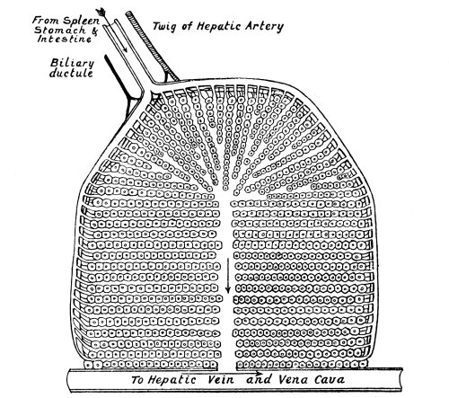
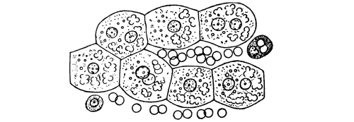
Fig. 7.—Diagram of
a Lobule of the Liver
divided vertically through its Axis.
In its centre is a space, the intralobular vein, through which the
blood falls into a branch of the hepatic vein, on its way to the heart.
An interlobular branch of the portal vein, which brings the blood
from the digestive organs, pours it by many smaller vessels over the
surface of the lobule. It filters into the lobule through innumerable
pseudo-capillary vessels, or spaces, between the radiating columns of
liver-cells. Arterial blood is brought to the lobule by a twig of the
hepatic artery. Bile is drained away from it by an affluent of the
hepatic duct. In the lower part of the diagram seven liver-cells are
shown, forming a divided column, magnified about 300 diameters. The
cells are loaded with glycogen, and contain minute globules of fat. Red
blood-corpuscles and two leucocytes are seen between the columns of
liver-cells. One of the leucocytes has ingested two blood-corpuscles.
The liver is supplied with blood by the hepatic artery. This vessel is
small for so large an organ. Although responsible for the nutrition
of the liver, it does not bring it the materials which are stored in
its cells. A much larger supply of blood is derived from the portal
vein, which breaks into capillaries, or, to speak more accurately,
into sinuses, or pseudo-capillaries, in the liver. The blood, whether
conveyed to the organ by the hepatic artery or by the portal vein, is
drained away by the hepatic veins. The plan of structure of the liver
is best understood when viewed with reference to the hepatic veins.
These, if traced backwards, are found to break up into fairly straight
vessels, each of which has a large number of lateral branches. Each
of the lateral branches is in the centre of a mass of cells, which
are packed round it in radiating columns. These masses, which have a
diameter of from 1 to 2 millimetres, are termed “lobules.” By mutual
pressure the lobules are squeezed into a pentagonal or hexagonal form.
The vein in the centre of the lobule is the intralobular vein. Turn now
to the portal vein; this is seen to break up into branches which run
between the lobules, and are therefore termed “interlobular veins.”
The branches of the hepatic artery also run between the lobules, as do
the radicles of the bile-duct. Each lobule is a liver in miniature.
The blood of the portal vein, which has come from the spleen, in
which red blood-corpuscles are destroyed, and from the stomach and
intestines, from which it has absorbed the products of digestion, is
poured over the surface of the lobule, to be filtered through into
its central intralobular vein. In its passage from the interlobular
veins (and branches of the hepatic artery) to the intralobular vein
the blood is confined to radiating capillary channels; but since these
merely prevent the escape of red blood-corpuscles without imposing any
restrictions upon the exudation of blood-plasma, the portal blood is to
all intents and purposes filtered through the columns of liver-cells.
The body-substance of the liver-cells is soft, destitute of envelope,
and capable, when free on the (warmed) stage of a microscope, of
changing in form, somewhat after the manner of a leucocyte. Such
cells have a great capacity for taking up the products of digestion.
Possibly they take up and store fats and proteins, but undoubtedly it
is their chief business to absorb sugar which accumulates as glycogen
in their substance. The glycogen is handed out to the hepatic blood
as required. The pigment which results from the disintegration of red
blood-corpuscles in the spleen is secreted, along with the bile-salts,
into minute channels, or canaliculi, which groove the flat surfaces of
adjacent liver-cells. These canaliculi converge to the bile-ducts. The
liver is therefore at the same time the storehouse of sugar which it
takes up from the blood when it is in excess, and passes out to the
blood when it is deficient, and an excretory organ which eliminates the
refuse of hæmoglobin. The iron derived from hæmoglobin it stores, and
returns to the blood.
Another function of the liver has been referred to already. It is the
organ, and, as far as we know, the only organ, in which urea is made
in mammals, and uric acid in birds. If the liver of a freshly killed
animal be excised and a stream of blood passed through it, the blood
which leaves the organ contains urea. If a salt of ammonia, even the
carbonate, be added to the blood, it is converted by the liver into
urea. When a bird’s liver is made the subject of the same experiment,
uric acid appears instead of urea. The liver can convert many
nitrogenous substances into urea, but it seems probable that, normally,
the salt with which it has chiefly to deal is lactate of ammonia
(cf. p. 146).
A few words must be added with regard to the functions of the liver
during prenatal life, obscure though these functions are. The liver
develops very early, and attains a relatively enormous size. At the
third month it weighs as much as the whole of the rest of the body
(cf. p. 34). Yet it cannot, one must suppose, have to
do much of the work which falls to its share in postnatal life. Food is reaching
the embryo in a constant stream, and not as the result of intermittent
meals. The embryo has no need to store glycogen; nor does its liver,
on analysis, yield much of this substance. In the embryo glycogen is
widely distributed throughout the tissues, not specially accumulated
in the liver. No digestion is occurring in the alimentary canal. Bile
is not needed to aid the hydrolysis and absorption of fats. A small
quantity of cholesterin and less lecithin is being eliminated, but not
much bile is needed to facilitate this process.
A process which is proceeding at a great rate in the embryo, in
various situations, is the formation of red blood-corpuscles. In
this the liver takes part. But its duty in regard to blood-formation
is not sufficiently onerous to account for its size. The formation
of blood-corpuscles in the liver is observed with difficulty in
microscopic sections. It is therefore impossible to speak with
certainty as to the extent to which it is going on, but it may be
safely asserted that this function by itself cannot be held to account
for the great size of the organ in embryonic life. What other office it
fills at this period is a question which still awaits an answer.
There is no more curious chapter in medical history than the story of
the views held at various periods with regard to the functions of the
liver. From being a mere mass of “parenchyma” serving as packing for
the abdominal viscera, it was elevated to the rank of Grand Purifier
of the “humours” of the body. Next, its excessive activity became
the cause of that form of dyspepsia known as “biliousness.” Still
later its want of activity was its chief vice. A “sluggish” liver was
held responsible for mental perversity and moral dulness. Calomel,
podophyllin, and other drugs were used as whips to stir it up; and the
increased secretions of the alimentary canal were mistaken for bile.
Poor patient organ! It is the still-room of the body, in which the
day’s supplies are stored, and from which they are served out, without
haste and without delay. And it makes urea. What else it does we have
yet to find out; and it is not impossible that when physiologists
have quite shaken themselves free from the explanations based upon
conjecture, which their predecessors have handed down, they may
discover that it has other duties which are not obvious, but of great
importance.