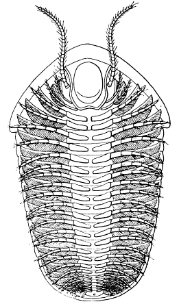
INTERNAL ORGANS AND MUSCLES.
Granting that the trilobite is a simple, generalized, ancient crustacean, it appears justifiable
to attribute to it such internal organs as seem, from a study of comparative anatomy,
to be primitive.
The alimentary canal would be expected to be straight and simple, curving downward
to the mouth, and should be composed of three portions, stomodæum, mesenteron, and proctodæum,
the first and last with chitinous lining. In modern Crustacea, muscle-bands run from
the gut to part of the adjacent body wall, so that scars of attachment of these muscles
may be sought. At the anterior end of the stomodæum, they are usually especially strong.
From the mesenteron there might be pouch-like or tubular outgrowths.
The heart would probably be long and tubular, with a pair of ostia for each somite.
In modern Crustacea, the chief organs of renal excretion are two pairs of glands in the
head, one lying at the base of the antennæ and one at the base of the maxillæ. Only one
pair is functional at a time, but these are supposed to be survivors of a series of segmentally
arranged organs, so that there might be a pair to each somite of a trilobite.
The nervous system might be expected to consist of a supracesophageal "brain," comprising
at least two pairs of ganglionic centers, and a double ventral chain of ganglia with
a ladder-like arrangement.
Besides these organs, a variety of glands of special function might be predicted.
Reproductive organs probably should occur in pairs, and more than one pair is to be
expected. There is little to indicate the probable location of the genital openings, but they
may have been located all along the body back of the cephalon.
It may be profitable to summarize present knowledge of such traces of these organs
as have been found in the fossils, if only to point out what should be sought.
ALIMENTARY CANAL.
Beyrich (1846, p. 30) first called attention to the alimentary canal of a trilobite, (Cryptolithus
goldfussi,) and Barrande (1852, p. 229) confirmed his observations. A number
of specimens of this species have been found which show a straight cylindrical tube or
its filling, extending from the glabella back nearly to the posterior end of the pygidium. It
lies directly under the median line of the axial lobe, and less than its own diameter beneath
the dorsal test. At the anterior end it apparently enlarges to occupy the greater part of the
space between the glabella and the hypostoma, but was said by the early observers to extend
only a little over halfway to the front. Beyrich thought the position of the median tubercle
indicated the location of the anterior end.
Walcott (1881, p. 200) stated that in his experience in cutting sections of trilobites it
was a very rare occurrence to find traces of the alimentary canal. The visceral cavity was
usually filled with crystalline calcite and all vestiges of organs obliterated. There were,
however, some slices which showed a dark spot under the axial lobe, which probably represented
the canal. In his restoration he showed it as of practically uniform diameter
throughout, and extending but slightly in front of the mouth.
Jaekel (1901, p. 168, fig. 28) has produced a very different restoration. His discussion
of this point seems so good, and has been so completely overlooked, that I will append
a slightly abridged version of a translation made some years ago for Professor Beecher.
The idea was, however, not original with Jaekel, as it was suggested by Bernard (1894, p.
417), but not worked out in detail.
While considering the problem as to what organ could have lain beneath the glabella of the trilobite, and
while studying the organization of living Crustacea for the purpose of comparison, I found in the collections
of the Geological Institute preparations of Limulus which seemed to me to directly solve the entire question.
From the mouth, which lies at about the middle of the head shield, the œsophagus bends forward, swells
out at the frontal margin of the animal at a sharp upward bend in order to take a straight course backward
after the formation of an enlarged stomach. Still within the head shield there branch out from each' side
of the canal two small vessels which pass over into the richly branched mass of liver lying under the broad
lateral parts of the head shield. After seeing this specimen, I no longer had the least doubt that the head
shield of the trilobites is to be interpreted in a similar manner. The position of the hypostoma and
gnathopods makes it necessary to assume that the position of the mouth of the trilobite lay pretty far back.
If, therefore, this depends upon the secondary ventral deflection of the oral region, as seems to be the case,
then it is a priori probable that the anterior part of the canal has also shared in this ventral inflection.
The posterior part of the canal in the region of the segmented thorax and pygidium is comparatively
narrow, as shown long ago by Beyrich; he represents only a thin tube which shows no swellings whatever,
and such are usually missing in Arthropoda.
As the glabella of most trilobites is regularly convex, there must lie beneath it an organ running from
front to back, which presses the bases of the cephalic legs away from each other and down from the dorsal
test. An organ so extensive and unpaired, running thus from front to back, can, among the Arthropoda, be
regarded only as an alimentary canal, for the swellings of the cephalic ganglia and the heart are by far too
small to produce such striking elevations on the front and upper surface of the glabella. The canal might
then have consisted of a gizzard belonging to the oesophagus, and a stomach proper or main digestive canal.
… Among the trilobites there are two pairs of vessels on both sides of the glabella which have
precisely the same position with reference to the supposed course of the alimentary canal as the ducts of
the hepatic lobes in Limulus. One observes in numerous trilobites, although in different degrees of clearness
and under various modifications, a dendritic marking of the inner surface of the cheeks which takes its
rise at the lateral margins of the glabella and spreads thence like a bush over the entire surface of the
cheeks. Exactly the same position is taken by the richly branched hepatic lobes of Limulus on the lower
surface of the head shield; a fact of special weight in favor of the homology and similar significance of the
two phenomena, is that in the trilobites also, the anterior of the two main ducts is the larger, the posterior
the smaller. The striking similarity of the two structures is shown by a comparison of the head shield of
Eurycare [Elyx] from the Cambrian of Sweden, in which the course of the canals is shown with remarkable
clearness [with those of Limulus].
I have been able to convince myself that the existence of the two canals on each side is also the rule in
other genera, even though the posterior pair is frequently but feebly developed or completely obscured by
the anterior pair. In Dionide formosa, for example, I find only the anterior pair, which is very large and
divided into two principal branches. From all these considerations it seems to me no longer doubtful that
the median elevation was caused by the stomach and gizzard, and that the cheeks have principally served to
cover the hepatic appendages of the alimentary canal.
The cause of the incomplete development of the glabellar lobes lies, hence, in the intrusion of the
alimentary canal, and it makes naturally the most effect where the gizzard spreads out and bends into the
stomach. This spot lies behind the frontal lobe, which is hence increased in size according as the stomach
increases in size; in this way not only the foremost segments of the glabella become enlarged, but also the
following ones more or less pressed aside. This process is easily followed phylogenetically and ontogenetically.
From the latter point of view, the development of Paradoxides is very instructive. In a head shield
2.5 mm. long the whole anterior part of the glabella is broadened, but the five pairs of lateral impressions
are clearly marked and the six segments of the head bounded by them are all of about the same size. In a
head shield about 13 mm. long, the foremost segment is very much increased in size, the jaw lobes pressed
still further apart; in adult forms both anterior segments are combined into the frontal swellings of the
glabella. In other groups this process proceeds phylogenetically still further, so that among the Phacopidæ
and in Trinucleus, behind the frontal swelling of the glabella only the last cephalic segment retains a certain
independence. The frontal lobe is thus no definite part, although it is as a rule composed of the mesotergites
of the first two cranidial segments.
This idea of an enlarged mesenteron certainly has much to commend it, and such actual
evidence as exists seems in favor of rather than against it. The strongest, firmest, best-protected
place in the whole body of the trilobite is the cavity between the vaulted glabella
and the hypostoma. As Jaekel has said, it is far too large a cavity for the brain, larger
than would seem to be required for a heart, and what else could be there but a stomach?
As has already been pointed out, Beyrich and Barrande found a pear-shaped enlargement of
the alimentary canal under the glabella of Cryptolithus. Longitudinal sections through
the glabella of Calymene and Ceraurus practically always show the cavity there filled with
clear crystalline calcite. One actual specimen of Ceraurus (Walcott 1881, pl. 4, fig. 1)
shows the cavity between the glabella and hypostoma entirely empty. The vacant spaces in
these two classes of specimens do not, however, necessarily mean anything more than imperfect
preservation.
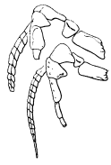
 |
 |
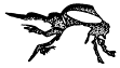 |
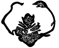
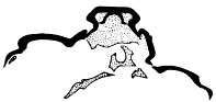
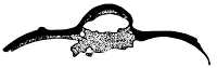
Fig. 21.—Transverse slice through Ceraurus pleurexanthemus, to show the
dorsal sheath above the abdominal cavity. Specimen 118. Traced from a
photographic enlargement. × 4.
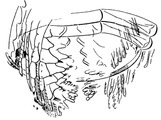
|
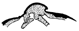
Fig. 22.—Transverse section through the cephalon of Ceraurus
pleurexanthemus, showing the abdominal sheath and the large mud-filled
alimentary canal (clear white). Traced from a photographic enlargement.
Specimen 97. × 3.3.
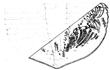
|
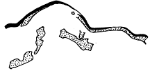
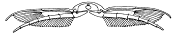
Fig. 23.—Transverse section of the thorax of Calymene senaria, showing
the large size of the mud-filled alimentary canal (clear white). Traced
from a photographic enlargement One appendifer (also clear white) is
shown. Specimen 153. × 3.3.
|
Ceraurus pleurexanthemus.
This species is taken up first, as it is the one shown in Walcott's often-copied figure
(1881, pl. 4, fig. 6). It is to be feared that too many have looked at this figure without
reading the accompanying explanation, and have taken it for a copy of an actual specimen
and not a mere diagram, which it admittedly is. The evidence on which it is based is comprised
in eight transverse slices, one through the glabella and seven through the thorax.
Three of these have been figured by Walcott: No. 27, 1881, pl. 3, fig. 7; No. 13, 1881, pl. 2,
fig. 3, 1918, pl. 26, fig. 14; No. 202, 1918, pl. 27, fig. 8. In all, as can be seen by reference to
the figures, the canal is partially collapsed, and is much larger than is indicated in Walcott's
restoration. The other sections bear out the testimony of those figured. One of these figured
specimens (No. 27) and another figured herewith (No. 118, see fig. 21) show an exceedingly
interesting structure which has previously escaped notice. The body cavity seems to have
had, in this region at least, a chitinous sheath on the dorsal side. As shown especially in
figure 21, this sheath impinges dorsally and laterally against the axial lobe and thus furnishes
a special protection for the soft organs beneath, probably protecting them from the
strain of the dorsal muscles.
While there is no way in which the location of these sections in the thorax can be positively
determined, it is probable that they came from the anterior end. In sections further
back, supposed to be in the posterior region of the mesenteron, no sheath is shown, but the
canal is nearly if not quite as large in relation to the size of the axial lobe.
The single section through the glabella (specimen 97) is of course important and fortunately
well preserved (fig. 22). It shows the dorsal sheath pressed against the inner surface
of the axial lobe along its middle portion, but diverging from it at the sides. The
section of the canal is oval, nearly twice as wide as high, but it is obviously somewhat depressed.
The original canal evidently filled nearly the whole of the dorsal part of the glabella
in this particular region. Unfortunately, the connection with the mouth is not shown, and
the form of the hypostoma indicates that the section cut the glabella diagonally, either in
the anterior or posterior part, probably the latter. In all these cases it should be remembered
that the specimens were found lying on their backs, and the canal has fallen in (dorsally)
since death.
The sections show that in Ceraurus pleurexanthemus the anterior part of the alimentary
canal was large, filling the part of the glabella below the heart; that the body cavity was
provided with a chitinous dorsal sheath extending back into the thorax; and that the posterior
portion of the mesenteron was likewise large and oval in section. Since the alimentary
canal must be connected with the mouth and anus, some such restoration as that of
Jaekel is indicated. No chitinous lining of the stomodæum or proctodæum was found, but
it is not certain that any of the sections cut either of those regions.
Calymene senaria.
Ten transverse sections and one longitudinal slice show the form of the alimentary canal
in Calymene. One of these has been figured by Walcott (1881, pl. 1, fig. 9) but without
showing the organ in question.
The only section cutting the cephalon which shows any trace of the canal is a longitudinal
one (No. 141), which is not very satisfactory. It has a large, nearly circular,
opaque spot under the anterior part of the glabella which may or may not represent a section
across the anterior end of the mesenteron. Three sections (No. 9, 115, 143) show
the dorsal sheath, the latter having the mud-filled canal beneath it. The sheath arches
across the axial lobe as in Ceraurus, leaving room for the dorsal muscles at the sides and
above it. In this region the canal is large and oval in section. Six slices cut the mesenteron
behind the abdominal sheath (Nos. 39, 117, 148, 153, 62, 65) (see fig. 23). In the
first four of these it is oval in section and large, but not so large as in No. 143. In the
last two, it is small and circular in section, from which it is inferred that the canal tapers
posteriorly.
Cryptolithus goldfussi (Barrande).
Illustrated: Beyrich, Untersuch. über Trilobiten, Berlin, 1846, pl. 4, fig. 1c.—Barrande, Syst. Sil. Bohême,
vol. 1 1852, pl. 30, figs. 38, 39.
Both Beyrich and Barrande have shown that from the posterior end of the axial lobe
to the neck-ring on the cephalon, the alimentary canal in Cryptolithus has a nearly uniform
diameter of less than half the width of the axial lobe. In front of the neck-ring, it enlarges,
and while its original describers state that it extends only about halfway to the front of
the glabella, Barrande's figure 39 shows it extending quite to the front, and his figure 38 shows
it fully two thirds of the distance to the anterior end, as does Beyrich's figure of 1846.
The Museum of Comparative Zoology contains a single specimen of this species from
Wesela, Bohemia, which shows the course of the canal from the middle of the pygidium to
the anterior part of the glabella. The enlargement appears to begin about halfway to the
front of the glabella and to be greatest at the anterior end. At the anterior end of the
glabella, the anterior end of the thorax, and the posterior end of the pygidium, the canal is
still packed full of a material somewhat darker in appearance than the matrix, while the remainder
of it is open. A well defined constriction is present under the middle of the next
to the last thoracic segment, but whether this is accidental or whether it indicates the point
where the mesenteron discharges into the proctodæum can not be determined. The inside
of the canal has somewhat of a lustre and there are three conical projections into it on the
median ventral line, a very small one in front of the neck furrow, a larger one under the
anterior part of the second segment, and a third between the fourth and fifth segments.
Summary.
The specimens of Cryptolithus from Bohemia and of Ceraurus and Calymene from
New York seem to substantiate the claim of Bernard and Jaekel that at the anterior end
of the canal there was an enlarged organ which occupied the greater part of the cavity of
the glabella. It appears that it extended into the thorax, and that above it and the heart
was a chitinous dorsal sheath. Behind the enlarged portion, the mesenteron appears to have
been of practically uniform diameter in Cryptolithus, but to have tapered posteriorly in
Ceraurus and Calymene. The proctodæum can not yet be differentiated from the mesenteron,
and only in Cryptolithus has the posterior portion of the alimentary canal been seen.
It is, there, merely a continuation of the mesenteron. The stomodæum likewise has not been
identified, but was probably a short gullet leading up from the mouth into the enlarged
digestive cavity.
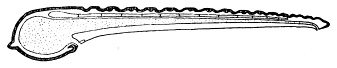
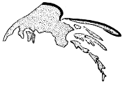
Fig. 24. Longitudinal section of Ceraurus pleurexanthemus, showing
the probable outline of the alimentary canal and the heart above
it. A restoration based on the slices described above.
The principle of the enlargement of the latter and its influence on the dorsal shell once
established, the significance of different types of glabellæ becomes apparent. It will be remembered
that the glabella of the protaspis of most trilobites is narrow, and that the same
is true of the glabellæ of most ancient and all primitive trilobites. The free-swimming larvæ
and the free-swimming ancestors of the trilobites were probably strictly carnivorous, lived
on concentrated food, and needed but a small digestive tract. As the animals "discovered
the ocean bottom" and began to be omnivorous or herbivorous, larger stomachs were required,
and so in the later and more specialized trilobites the glabella became expanded latterally
or dorsally, or both, to meet the requirement for more space, until, in such Devonian
genera as Phacops, the cephalon was nearly all glabella.
GASTRIC GLANDS.
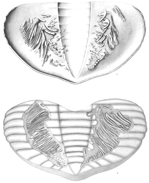
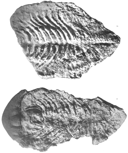
Jaekel's suggestion, quoted above, that the so called "nervures" seen on the under surfaces
of the heads of some trilobites are really glands for the secretion of digestive juices,
is at least worthy of consideration. Moberg, however (1902, p. 299), suggested that these
markings probably had something to do with the eyes rather than the stomach. He says in
part (translation):
In general we can now say that such features are common to all the eyeless Conocoryphidæ. With the
conocoryphs I include Elyx and consider Harpides as at least closely related. Similar impressions are also
found in forms with eyes, as, for instance, in the Olenidæ, but here such radiate partly from the border
of the eye, partly from the front end of the glabella, partly from the [visual surface of the] eye, and sometimes
from the angle between the occipital ring and the glabella. They therefore go out from such different
points that they can not possibly be branches of the liver. It would also be very remarkable if such an
important organ should have been developed in a few eyeless forms, but have failed to leave the least
trace in the rest of the trilobites.
Lindstroem (1901, pp. 18, 19, 33; pl. 5. figs. 29, 31; pl. 6, figs. 43-45) has discussed
these markings and given beautiful figures showing their appearance in Olenus, Parabolina,
Elyx, Conocoryphe, and Solenopleura. He decided that they were to be explained as branches
of the circulatory system, comparing them with the veins and arteries of Limulus. He
pointed out that there was a coincidence between these markings and the position of the eyes,
and suggested a causal connection with the latter.
Beecher (1895 B, p. 309), also from a comparison with Limulus, suggested that the
eye-lines of Cryptolithus, Harpes, Conocoryphe, Olenus, Ptychoparia, Arethusina, etc., probably
represented the optic nerves, and since the eye-lines are usually the main trunks of the
dendritic markings, it is fair to assume that he considered the whole as due to branches of
nerves.
Reed has recently (1916, pp. 122, 173) discussed these lines as developed in the Trinucleidæ,
and seems to accept Beecher's explanation.
Three explanations of the "nervures" are thus current, and the authors of all of them
refer us to Limulus as proving their claims! So far as general appearance goes, the markings
on the trilobites more closely resemble the veins of a Limulus than either the nerves or
"liver" of that animal. The veins, however, are not in contact with the dorsal shell, but
are buried in the liver and muscles, while the arrangement of the arteries, which are dorsal
in position, is quite unlike what is seen in the trilobites.
The term nervures, as applied to these markings, is not only misleading, but an incorrect
use of one of Barrande's words, for by nervures he meant delicate surface markings.
Until the real function of the organs which made these markings is definitely established, it
may be well to call them genal cæca, for they obviously were open tunnels ending blindly,
whatever they contained.
The question of the function of the genal cæca can not, in any case, be settled by an
appeal to Limulus, and it is doubtful if it can be settled at all at the present time. Certain
things tend to show that Jacket's explanation is the most plausible, and these may be
briefly set forth.
Walcott (1912 A, pp. 176, 179, pls. 27, 28) has described specimens of Naraoia and
Burgessia in which similar markings are well shown, and where they are obviously connected
with the alimentary canal just at the anterior end of the mesenteron. In Burgessia,
which seems to be a notostracan branchiopod, the trunk sinuses are very wide, and the appearance
is on the whole unlike that of any known trilobite. In Naraoia, however, the
markings are much finer and directly comparable with those of Elyx. If my contention that
Naraoia is a trilobite should be sustained, it might almost settle the question of the "nervures."
In Burgessia these lateral trunks enter the main canal behind the fifth pair of appendages.
In the trilobites they debouch much further forward.
The principal argument in favor of the interpretation of these markings as nerves lies
in their connection with the eyes. There is considerable evidence to indicate that the eye-lines
and the genal cæca are two distinct structures, but because both originate from the
sides of the anterior lobe of the glabella, and both extend outward at nearly right angles
to the axis, or obliquely backward, they are, when both present, coincident. Genal cæca
occur on blind trilobites, on trilobites with simple eyes, and on trilobites with compound eyes.
Eye-lines occur on trilobites with both simple and compound eyes, and genal cæca may or
may not be present in both cases. The morphology of the ridge forming the eye-line in
trilobites with compound eyes is well known. It is abundantly proved by ontogeny that it
is the continuation of the palpebral lobe, and a development of the pleura of the first dorsal
segment of the cephalon. Lake, Swinnerton, and Reed have tried to show that the eye-lines
of the Harpedidæ and Trinucleidæ are homologous with the eye-lines of the trilobites
with compound eyes, and that the ocelli on the cheeks are therefore degenerate compound
eyes.
The simplest form of the genal cæcum is seen in the blind Elyx (Lindstroem 1901, pl.
6, fig. 43). The main trunk is at nearly right angles to the axis, the increase in its width
is gradual in approaching the glabella, and an equal number of branches diverge from both
sides.
Ptychoparia striata (Barrande 1852, pl. 14, figs. 1, 3) is an excellent example of a trilobite
with compound eyes and genal cæca. It will be noted that the main trunk and the eye-line
are coincident, and that both on the free and fixed cheeks the branches are all on
the anterior side of the eye-line. Compare this with the condition in Conocoryphe
(Barrande, pl. 14, fig. 8; Lindstroem, pl. 6, fig. 44), and one sees there a main branch
having the same direction as in Ptychoparia and likewise with all the branches on the anterior
side. At first sight this would seem to support the contention that these lines do lead out
to the eyes, since Conocoryphe is blind, and the main trunk leads practically to the margin.
But although Conocoryphe is blind, it has free cheeks, and the main trunk does not lead to
the point on those free cheeks where eyes are to be expected, but back into the genal angles.
And this direction holds in such diverse genera (as to eyes and free cheeks) as Harpes, Cryptolithus,
Dionide, and Endymionia. In all these the genal cæca fade out in the genal angles, and
in none of them would compound eyes be expected in that region. The coincidence of the
eye-lines with the trunks of the genal cæca in Ptychoparia seems to be merely a coincidence.
That the markings which radiate from the eyes of Ptychoparia and Solenopleura are not impressions
made by nerves is obvious. That they are of the same nature as the similar markings
in the eyeless trilobites is equally obvious. Ergo, they can not be nerves in either case,
and that they have anything to do with the eyes is highly improbable. The eye was merely
superimposed upon these structures.
The relation of the genal cæca to the ocelli on the cheeks is best shown in the Trinucleidæ.
In all species of Tretaspis simple eyes are present, and in most of them there are
very narrow eye-lines. The latter are occasionally continued beyond the ocular tubercle back
to the genal angle. A similar course is seen in Harpes. If the simple eye is the homologue
of the compound eye, and the eye-line here the homologue of the eye-line in Ptychoparia,
why does it continue beyond the eye? In any case, it can not be interpreted as a nerve.
Cryptolithus tessellatus, when the cephalon is 0.45 mm. to 0.65 mm. long, shows short eye-lines
and a small simple eye on each cheek. In some half-grown specimens, traces of the ocelli
can be seen, but the eye-lines are absent. In the adult, both the eye-lines and the ocelli are
entirely wanting. Reed states that "nervures" are also absent, and so they are from most
specimens, but well preserved casts of the interior from the Upper Trenton opposite Cincinnati
show them, and one cheek is here figured (fig. 25). As apparent from the figure, the
main trunk is very short and gives rise to two principal branches, the first of which in
its turn sends off lines from the anterior side. It was a specimen showing these lines which
Ruedemann (1916, p. 147) figured as showing facial sutures. The interest lies in the fact
that while the ocelli and eye-lines were lost in development, the genal cæca are present
in the adult, showing that they are different structures.
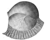
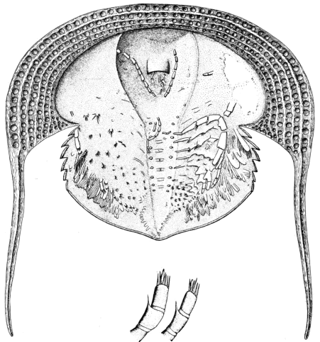
Fig. 25.—Cryptolithus tessellatus Green. Side view of the cheek of a
specimen from the top of the Trenton opposite Cincinnati, Ohio, to
show the branching genal cæca. These are the "facial sutures" of
Ruedemann.
Harpides is another genus in which genal cæca are strikingly shown, and in this case
they completely cover the huge cheeks, radiating from two main trunks to the front and
sides. I have seen no good specimens, but it would appear from Angelin's figure (1854,
pl. 41, fig. 7) that the rather large, simple eyes are not situated exactly on the vascular trunks.
In the Harpides from Bohemia, the main trunks extend out with many branches beyond the
simple eyes. It should be stated that the courses of the genal cæca are not correctly figured
by Barrande (Supplement, 1872, pl. 1, fig. 11), as shown by casts of the original specimen
in the Museum of Comparative Zoology. From Barrande's figure, one would suppose that
the eye-lines and their continuation beyond the "ocelli" were superimposed upon the genal
cæca without having any definite connection with them, but as a matter of fact the radial
markings really diverge from the main trunks as in Elyx and similar forms.
Summary.
As Reed has said, these lines are not mere ornamentation, but rather represent traces
of structures of some functional importance. They probably can not be explained as traces
of nerves and more likely represent either traces of the gastric cæca or of the circulatory
system. While they are known chiefly in Cambrian and Lower Ordovician trilobites, there
is no evidence that the organs represented were not present in later forms, even if the shell
may not have been affected by them. While they indicate very fine, thread-like canals, the
present evidence seems to be in favor of assigning to them the function of lodging the glands
which secreted the principal digestive fluids.
HEART.
Illænus.
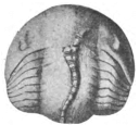
Volborth (1863, pl. 1, fig. 12 = our fig. 26) has described the only organ in a trilobite
which suggests a heart. A Russian specimen of Illænus with the shell removed shows a
somewhat flattened, tubular, chambered organ extending from under the posterior end of
the cephalon to the anterior end of the pygidium. The posterior nine chambers were each
1.5 mm. long and 1.5 mm. wide, while the two anterior chambers were respectively 2.5 mm.
and 3 mm. wide. These were all under the thorax, and at least two more chambers are
shown under the cephalon, but rather obscurely. The species of the Illænus is not stated,
but since no Illænus has more than ten segments in the thorax, and this tube has at least
thirteen chambers, it is evident that its constrictions are inherent in it, and are not due to
the segmentation of the thorax. Beecher has made a passing allusion to this organ as an
alimentary canal. This was the original opinion of Volborth. Pander, however, suggested
to him that it might be a heart. The alimentary canal of Cryptolithus does not show any
constrictions, while the heart of Apus (see fig. 27) and other branchiopods does show them.
It should be noted, further, that while this heart enlarges toward the front, it is everywhere
very small as compared with the width of the axial lobe, and much narrower than sections
of Ceraurus and Calymene would lead one to expect the alimentary canal of Illænus to be.
Where the heart is 1.5 mm. to 3 mm. wide, the axial lobe is 11 mm. wide.
 |
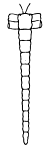 |
Fig. 26. Copy of
Volborth's figure of the heart of Illænus. |
Fig. 27. Heart of
Apus. Copied from Gerstäcker. |
While this may be merely a cast of the alimentary canal it is sufficiently like a heart to
deserve consideration as such an organ.
Ceraurus and Calymene.
Nothing suggesting a heart has been seen in the sections of Ceraurus and Calymene.
The mesenteron and its sheath crowd so closely against the dorsal test in the anterior part
of the thorax that there seems to be no room for the heart, but it must have been located
beneath the sheath and above the alimentary canal. If the latter were filled with mud, and
the animals lay on their backs, as most of them did at death, the canal would drop down
into the axial lobe and the soft heart would naturally disappear and leave 110 trace of its
presence in the fossils.
The Median "Ocellus" or "Dorsal Organ."
Many trilobites, otherwise smooth, bear on the glabella a median pustule which is usually
referred to as a simple eye or median ocellus, but whose function can not be said to have
been certainly demonstrated. Ruedemann (1916, p. 127), who has recently made a careful
study of this problem, lists about thirty genera, members of ten families, Agnostidæ, Eodiscidæ
Trinucleidæ, Harpedidæ, Remopleuridæ, Asaphidæ Illænidæ, Goldiidæ, Cheiruridæ,
and Phacopidæ, in which this tubercle is present, and had he wished he might have cited
more, for it is of almost universal occurrence in Ordovician trilobites.
I have not especially searched the literature for references to this median tubercle. It
is often mentioned by writers in descriptions of species, but apparently few have tried to
explain it. Beyrich (1846, p. 30) suggested that it indicated the beginning of the alimentary
canal. Barrande mentioned it, but if he gave any explanation, it has escaped me. McCoy
(Syn. Pal. Foss. 1856, p. 146) called it an ocular (?) tubercle, and that seems to have been
the interpretation which most writers on trilobites have assigned to it, if they suggested any
function at all. Beecher (1895 B, p. 309) concurred in this opinion.
Bernard (1894, p. 422) ascribed to this tubercle, as well as to the median tubercle on
the nuchal segment, an excretory function, comparing it with the "dorsal organ" in Apus.
Reed (1916, p. 174) states that it may be either the representative of the "dorsal"
organ of the branchiopods, or a median unpaired ocellus.
Ruedemann (1916) has made the only real investigation of the subject. He came to
the conclusion that it was a parietal eye, without a crystalline lens, but corresponding to the
"parietal eye of other crustaceans, and especially of the phyllopods, which is a lens-shaped
or pear-shaped sac, usually filled with sea water." He found that above the "ocellus" the
test was usually thin or even absent, and in a few cases a dark line beneath seemed to outline
the original form of the sac. His summary follows:
It is claimed that most, if not all, trilobites possessed a median or parietal eye on the glabella. [In
proof of this assertion the following facts are stated:]
1. A great number of species, belonging to more than thirty genera, possess a distinct tubercle on the
glabella. This tubercle occurs alone in many genera, otherwise smooth, as in the Asaphidæ, and is hence of
functional importance.
2. In certain cases, as in Cryptolithus tessellatus, distinct lenticular bodies [not lenses] were recognized;
in others, as in Asaphus expansus, only a thinner, probably transparent test. Many other species show a
distinct pit in interior casts of the tubercle, indicating a lens-like thickening of the top of the tubercle. The
median eye therefore probably possessed all the different stages of development seen in other crustaceans.
3. As in the parietal eyes of the crustaceans and the eurypterids, the tubercles are most prominent and
distinct in the earlier growth-stages, notably so in Isotelus gigas.
4. The tubercle is especially well developed in the so called blind forms where the lateral eyes are
abortive, as in Cryptolithus (Trinucleus), Dionide, Ampyx.
5. The tubercles always appear on the apex on the highest part of the glabella, where their visual
function would be most useful.
6. The tubercle is generally situated between the lateral eyes, like the parietal eye in crustaceans and
eurypterids, on account of its close connection with the brain.
7. Frequently it forms the posterior termination of a short crest, also as in certain eurypterids (Stylonurus),
indicating the direction of the nerve.
8. The median eye is borne on a tubercle or mound in the Ordovician and Silurian trilobites, while the
tubercle is rarely noticed in the Devonian and in few Cambrian forms. In the Devonian forms, similarly
as in many crustaceans and in later growth-stages of some asaphids, the strong development of the lateral
eyes may have led to a loss of the parietal eyes. In the Cambrian genera evidence is present to suggest that
the parietal eyes consisted only of transparent spots or lens-like thickenings of the exoskeleton, hardly
noticeable from the outside.
9. It is a priori to be inferred that the trilobites should, as primitive crustaceans, have possessed median
or parietal eyes.
As a student, I accepted Professor Beecher's dictum that this tubercle represented a
median ocellus, but more recently a number of things have led me to the view that it is
the point of attachment of the ligament by which the heart is supported.
The chief arguments against its interpretation as a parietal eye seem to be that its
structure is not absolute proof, being capable of other explanation; its position is variable,
in front, between, or back of the eyes; it is exactly like other tubercles on the median line,
especially the nuchal spine or tubercle, and the similar ones along the axial lobe of the
thorax; and it is not present in the protaspis or very young trilobites.
1. The structure disclosed by Ruedemann's sections, a sort of sac-like cavity beneath a
thinned test, can be explained as a gland, a ligamentary attachment, or a vestigial spine, as
well as an eye. In a section of Asaphus expansus, which I made some years ago when trying
to get some light on this problem, there is a similar cavity under the pustule, but a
secondary layer of shell lay beneath it and apparently cut it off from the glabellar region,
thus indicating that it had lost its function in the adult of this animal. Sections through
the tubercles of the glabella of Ceraurus show all of them hollow, with very thin upper
covering or none at all, and their structure is not unlike that of the tubercle of Cryptolithus.
In fact, sections can be seen in Doctor Walcott's slices which are practically identical with
the one Ruedemann obtained from Cryptolithus. Since it is obvious that not all of the
pustules of a Ceraurus could have been eyes, the evidence from structure is rather against
than for the interpretation of the median pustule as such an organ.
2. The position of the tubercle varies greatly in different genera. Where furthest forward
(Tretaspis, Goldius), it is just back of the frontal lobe, while in some species of asaphids
it is in the neck furrow. In species with compound eyes it is frequently between the
eyes, but more often back of them. If its history be traced in a single family, it is generally
found farthest forward in the more ancient species and moves backward in the more
recent ones. The eyes do this same thing, but the median tubercle goes back further than
the eyes. This can be seen, for example, in the American Asaphidæ, where the pustule is
up between the eyes of Hemigyraspis and Symphysurus of the Beekmantown and back of the
eyes of the Isotelus of the Trenton. Turning now to the under side of the head, it appears
that the tubercle bears a rather definite relation to the hypostoma. If the hypostoma is short,
the tubercle is well forward. If long, it is far back on the head. It seems in many cases
to be just back of the posterior tip of the hypostoma, or just behind the position of the
mouth, while in others it is not as far back as the tip of the hypostoma.
The median tubercle is in many cases developed into a long spine. This is usually in
an ancient member of a tubercle-bearing family, and suggests that in most cases the tubercle
is a vestigial organ. An example of this occurs in Trinucleoides, the most ancient
of the Trinucleidæ. Trinucleoides reussi (Barrande) (Supplement, 1872, pl. 5, figs. 17, 18)
has a very long slender spine in this position. It could be explained as an elevated median
eye, but it also very strongly suggests the zoæal spine of modern brachyuran Crustacea.
Gurney (Quart. Jour. Mic. Sci., vol. 46, 1902, p. 462) supports Weldon in the conclusion
that the long spines of the zoæa are directive, and states that the animal swims in the direction
of the long axis of the spine. He also suggests that, since the period of their presence corresponds
to the period before the development of the "auditory" organs, the spines may
perform the functions of balancing and orientation. It is generally admitted that the spine
of the zoæa is also protective, and the obvious function, first pointed out by Spence Bate
in 1859, is that it contains a ligament which helps suspend the heart, which lies beneath the
spine. This latter function may have been that of the median tubercle in the trilobite. Such
an explanation would account for the backward migration mentioned above, for as the
stomach enlarged and the mouth moved backward on the ventral side, the heart may have
been pushed backward on the upper side.
There is also a curious parallelism between the ontogenetic history of the zoæal spine
and the phylogenetic history of the Trinucleidæ or Cheiruridæ (Nieszkowskia is the ancient
member of this family in which the spine replaces the tubercle). When first hatched, the
larval crab shows no trace of the spine, but very quickly it evaginates, lying dorsally on the
median line, pointing forward (Faxon, Bull. Mus. Comp. Zool., vol. 6, 1880, pl. 2). With
the splitting of the original envelope, the spine becomes erect, but persists only a short time,
and is reduced to a vestigial tubercle toward the end of the zoæal stages, its disappearance being,
as pointed out by Gurney, coincident with the development of the balancing organs. This
manner of suspension of the heart by a long tendon certainly does suggest that Gurney is
right in his interpretation of the function. Briefly, the zoæal spine served for a short time
a function later taken over by other organs. It was not present in the youngest stages, it
became prominent at a very early stage, was soon vestigial, and then lost.
Take now the trilobites. There is no trace of the median pustule in the protaspis of
any form, and in many primitive trilobites it is absent. It appears first as a long spine in
certain families, and later becomes vestigial and disappears. Very few trilobites of Silurian
and later times show it at all.
In the particular case of the Trinucleidæ, which were burrowers, the spine is present
on only the oldest and most primitive of the group, a form which has only a most rudimentary
fringe. It is obvious from the large size of the pygidium in the larval trinucleid that
this family is derived from a group of free swimmers. Trinucleoides reussi was perhaps in
the transitional stage, just leaving the swimming mode of life, and belonged to a group
which had not developed any other "statocyst" than the median spine. Among the later
Trinucleidæ the spine became a vestigial tubercle, and in some cases entirely disappeared. A
similar history can be traced in the Cheiruridæ, starting from some such forms as the American
Lower Ordovician Nieszkowskia (N. perforator p. ex.).
Another example of a median spine instead of a tubercle is in Goldius rhinoceros (Barrande).
Since this species is not from the oldest Goldius-bearing rocks, but from the Lower
Devonian, it does not follow what seems to be the general rule, but makes an interesting exception.
Goldius rhinoceros (Barrande) (Supplement, 1872, pl. 9, figs. 12, 13) has the
median tubercle elevated into a stubby, recurved spine very suggestive of the horn of a
rhinoceros. Since the eyes of this species are very well developed, there seems no especial
reason for the elevation of a parietal eye, and the example certainly does not support that
interpretation.
3. This tubercle is essentially similar to other tubercles on the median line of cephalon,
thorax, and even pygidium. This has been discussed sufficiently under section 1 above, but
it may perhaps be justifiable to point out that in some of the Agnostidæ there is a median
tubercle on both shields, and since it has not yet been demonstrated beyond question which
shield is the cephalon, to say which one is a parietal eye and which one is a tubercle is impossible.
In other words, the parietal eye can not be differentiated from any other tubercle
except by its position.
4. One of the as yet unexplained features of the protaspis of trilobites is the absence
of the "nauplius eye." Beecher (1897 B, p. 40) explained this on the ground of the
extremely small size of the protaspis and the imperfection of the preservation. If the median
tubercle were really a median eye, it should be present in the protaspis and the earlier
stages of the ontogeny, even if not in the adult, and should certainly appear before the compound
eyes. (In Limulus, however, the compound eyes appear first.) The median eye has
not so far been seen in any young trilobite in any stage previous to that in which compound
eyes are present. The full ontogeny is not known of any species with compound eyes in
which the median tubercle is present in the adult, but theoretically the median eye should be
most prominent in the young of just those primitive trilobites about whose development most
is known.
NERVOUS SYSTEM.
There has been a rather general impression among students of trilobites that the eye-lines,
which should be differentiated from the genal cæca, denote the course of the optic
nerves, but no other evidence of the nervous system has been found, save the so called
nervures which have been discussed above. In Apus the nerves leading to the eyes come
off from the anterior ganglion or "brain" and run directly to the eyes. If conditions were
similar in the trilobites, the "brain" was beneath the anterior glabellar lobe, provided, of
course, that the eye-lines do indicate the course of the optic nerve.
The ontogenetic history of the eye-lines of trilobites with compound eyes is instructive,
and has already been discussed by Lindstroem (1901, pp. 12-25), but he did not cite the
case of Ptychoparia, which is particularly interesting, because in this genus both eye-lines
and "nervures" are present. Beecher (1895 C, p. 171, pl. 8, figs. 5-7) has shown that in
Ptychoparia kingi the eye-lines of a specimen in the metaprotaspis stage run forward at a
low angle with the glabella, while in the adult their course is nearly at right angles to it. They
have therefore swung through an arc of at least 60 and in so doing have had ample opportunity
to become coincident with the primary trunks of the genal cæca. Once that was accomplished,
it is quite likely that the one fold in the shell would continue to house both
structures. In other trilobites, there is a similar backward progression of the eye-lines.
As would be expected, the ventral ganglia and the longitudinal cords left no trace in
the test. Since each segment has appendages, there was probably a continuous chain of
ganglia back to the posterior end of the pygidium.
VARIOUS GLANDS.
Dermal glands.—The surface of many trilobites is "ornamented" with pustules and
spines which on sectioning are nearly always found to be hollow, and in many cases have a
fine opening at the tip. While it is generally believed that the purpose of these spines was
protective, yet it is possible that many of them were merely outgrowths which increased
the area through which the respiratory function could be carried on. It will be recalled
that most of the smooth trilobites are punctate, some of them very conspicuously so, and
the spines and pustules of ornamented trilobites may merely subserve the same function as
the pores of smooth ones.
If the spines were protective, it would not be surprising if some of them, hollow and
open at the top, were poisonous also, and had glands at the base. These are, however,
purely matters of speculation so far.
Renal excretory organs.—Nothing has been seen of any such organs, unless the genal
cæca may possibly be of that nature. The main trunks of these always lead to the sides
of the anterior glabellar lobe, which is not the point of attachment of either antennæ or
biramous limbs, so that there seems little chance that they will bear this interpretation.
Reproductive organs.—Nothing is yet positively known about the reproductive organs
or the position of their external openings. If the "exites" of Neolenus could be interpreted
as brood-pouches, which does not seem probable, then the genital openings were located near
the base of some pair of anterior thoracic appendages.
The Panderian Organs: Internal Gills or Poison Glands?
At a meeting of the Mineralogical Society at St. Petersburg, Volborth (1857) announced
that Doctor Pander had two years before discovered certain organs on the lower side of
the doublure of the pleural lobes of the thorax of a specimen of Asaphus expansus. These
organs were oval openings in the doublure, one near the posterior margin of the cephalon,
and one on each thoracic segment of the half-specimen figured by Volborth in 1863. They
were explained by Volborth and by Eichwald (1860, 1863) as the points of attachment of
appendages. Billings (1870) described and figured the "Panderian organs" of "Asaphus
platycephalus" and stated that he had seen them in Asaphus [Ogygites] canadensis and
A. megistos [Isotelus maximus] as well. He thought some sort of organ was attached to
them, but could not suggest its function. Woodward (1870) thought that the openings were
"only the fulcral points on which the pleuræ move." Their position outside the fulcra shows
that this explanation is impossible.
So far as I am aware, the Panderian organs have been seen only in the Asaphidæ.
Barrande figured them in "Ogygia" [Hemigyraspis] desiderata (1872) and Schmidt in two
species of Pseudasaphus. They seem to occupy the same position in Bohemian, Russian,
and American specimens. There is always one pair of openings on each thoracic segment,
and one pair in line with them on the posterior margin of the cephalon. They occur near
the anterior margin of the segment, and near the inner end of the doublure. In some cases
they are surrounded by a ventrally projecting rim, while in others they have a thin edge.
There seem to be no markings on the interior of the shell which are connected with them.
While thinking over the trilobites in connection with the origin of insects, it occurred
to me that these hitherto unexplained Panderian organs might possibly be openings to internal
gills and that the Asaphidæ might have been tending toward an amphibious existence. On
mentioning this to Doctor R. V. Chamberlin of the Museum of Comparative Zoology, he
called my attention to the possibility that they might be openings similar to those of the
repugnatorial glands of Diplopoda. While no definite decision as to the function can be
made, the explanation offered by Doctor Chamberlain seems more plausible than my own,
and has suggested still a third, namely, that they might be the openings of poison glands.
If one were to argue that these apertures are really connected with respiration, it might
be pointed out that they are ventral in position, while the foramina repugnatoria are always
dorsal or lateral, even in diplopods with broad lateral expansions. If offensive secretions
were poured out beneath a concave shell like that of a trilobite, they would be so confined
as to be but slightly effective against an enemy. This would indicate that if these openings
were the outlets of glands, the substance secreted might be a poison used to render prey
helpless. On the other hand, openings to gills are normally ventral in position, and if the
pleural lobes were folded down against the body, they would be brought very close to the
bases of the legs.
A further curious circumstance is that so far no traces of exopodites have been found
on Isotelus. The endopodites of both Isotelus latus and I. maximus are fairly well preserved
in the single known specimen of each, yet no authentic traces of exopodites have
been found with them. Moreover, Walcott sliced specimens of Isotelus from Trenton Falls
and found only endopodites. It may also be recalled that the finding of the specimen of
Isotelus arenicola at Britannia and the tracks which I attributed to it, suggested to me that
it was a shore-loving animal (1910). It offers a field for further inquiry, whether the
Asaphidæ may not have had internal gills, and whether some primitive member of the family
may not have given rise to tracheate arthropods.

Fig. 28. Side view of a specimen of Isotelus gigas Dekay, from which the
test of the pleural lobes has been broken to show the position of the
Panderian organs. Natural size. Specimen in the Museum of Comparative
Zoology.
The explanation of the Panderian organs as openings of poison glands is less radical
than the one just set forth, and so possibly lies nearer the truth. One would expect poison
glands to lie at the bases of fangs, and so they do in specialized animals like chilopods and
scorpions, but the trilobites may have had the less effective method of pouring out the poison
from numerous glands. The purpose may have been merely to paralyze the brachiopod or
pelecypod which was incautious enough to open its shell in proximity to the asaphid.
MUSCULATURE.
This is a field which is rather one for investigation than for exposition. Very little
has been done, though probably much could be. The chief obstacle to a clearer understanding
of the muscular system lies in the difficulty of getting at the inner surface of the test
without obscuring the faint impressions in the process.
There exist in the literature a number of references to scars of attachment of muscles,
and any study of the subject should of course begin by the collection of such data. I shall
at this time refer to only a few observations on the subject.
The structure and known habits of trilobites make it obvious that strong flexor and
extensor muscles must have been present, and some trace of them and of their points of
attachment should be found. It is likely that their proximal ends were tough tendons. The
muscles holding up the heart and alimentary canal would be less likely to reveal their presence
by scars, but there must have been at least one pair of strong muscles extending from
the under side of the head across to the hypostoma. Judging from the method of attachment,
the muscles moving the limbs were short ones, chiefly within the segments of the legs
themselves.
Flexor Muscles.
Since the majority of trilobites had the power of enrollment, and seem also to have used
the pygidia in swimming, the flexors must have been important muscles. Beecher (1902,
p. 170) appears to have been the only writer to point out any tangible evidence of their
former presence. Walcott (1881, p. 199) had shown that the ventral membrane was
reinforced in each segment by a slightly thickened transverse arch. Beecher showed that
on this thickened arch in Triarthrus, Isotelus, Ptychoparia, and Calymene, there are low longitudinal
internal ridges or folds. One of these is central, and there is a pair of diagonal
ridges on either side. Beecher interpreted these ridges as separating the strands of the
flexor muscles, and believed that a line of median ridges divided a pair of longitudinal
muscles, while the outer ridges showed the place of attachment of the pair of strands which
was set off to each segment. He did not discuss the question as to where the anterior and
posterior ends were attached. In trilobites with short pygidia, the attachment would probably
have been near the posterior end, and it is possible that the two scars beneath the doublure
and back of the last appendifers in Ceraurus may indicate the point of attachment in
that genus.
There is as yet no satisfactory evidence as to where the anterior ends of the flexors
were attached. In Apus these muscles unite in an entosternal sinewy mass above the mouth,
but no evidence of any similar mass has been found in the trilobites and it is likely that
the muscles were anchored somewhere on the test of the head.
Extensor Muscles.
The exact position of these muscles has not been previously discussed. The interior of
the dorsal test shows no such apodemes as are found on the mesosternites, but, as I have
shown in the discussion of the alimentary canal of Calymene and Ceraurus, there is an
opening on either side of the axial lobe between the dorsal test and the abdominal sheath,
and it is entirely probable that an extensor muscle passed through each of these. The abdominal
sheath extends only along the posterior region of the glabella and the anterior
part of the thorax, and probably served to protect the soft organs from the strain of the
heavy muscles. The extensors (see fig. 29) probably lay along the top of the axial lobe
on either side of the median line of the thorax to the pygidium, where they appear to
have been attached chiefly on the under side of the anterior ring of the axial lobe, although
strands probably continued further back. This is above and slightly in front of the fulcral
points on the pleura, and meets the mechanical requirements. Ceraurus (Walcott, 1875, and
1881, p. 222, pl. 4, fig. 5) shows a pair of very distinct scars on the under side of the first
ring of the pygidium, and in many other trilobites (Illænus, Goldius, etc.) distinct traces of
muscular attachment can be seen in this region, even from the exterior. The anterior ends
were probably attached by numerous small strands to the top of the glabella, and, principally,
to the neck-ring.
On enrolling, the sternites of all segments are pulled forward and the tergites backward.
In straightening out, the reverse process takes place. The areas available for muscular attachment
are so disposed as to indicate longitudinal flexor and extensor muscles rather than
short muscles extending from segment to segment. Indeed, the tenuity of the ventral membrane
is such as to preclude the possibility of enrollment by the use of muscles of that sort,
while powerful longitudinal flexors could have been anchored to cephalon and pygidium. The
strongly marked character of the neck-ring of trilobites is probably to be explained as due
to the attachment of the extensor muscles, rather than to its recent incorporation in the
cephalon. The same is true of the anterior ring on the pygidium.
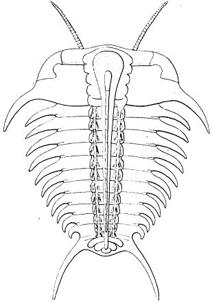
Fig. 29. Restoration of a part of the internal organs of
Ceraurus pleurexanthemus as seen from above. At the sides
are the extensor muscles, and in the middle, the heart overlying
the alimentary canal. Drawn by Doctor Elvira Wood, under
the supervision of the author.
Possible preservations of extensors and flexors in Ceraurus.—Among Doctor Walcott's
sections are four slices which I should not like to use in proving the presence of longitudinal
muscles, but which may be admitted as corroborative evidence. Two of these transverse
sections (Nos. 114 and 199) show a dorsal and a ventral pair of dark spots in positions
which suggest that they represent the location of the dorsal and ventral muscles, while two
others (Nos. 131 and 140) show only the upper pair of spots. The chief objection to this
interpretation is that it is difficult to imagine how the muscles could be so replaced that they
happen to show in the section. Both the sections showing all four spots are evidently from
the anterior part of the thorax, as they show traces of the abdominal sheath, which seems
to be squeezed against the inside of the axial lobe, with the muscles (?) forced out to the
sides. The ventral pair lie just inside the appendifers, but even if they are sections of muscles,
all four are probably somewhat out of place.
Hypostomial Muscles.
The hypostoma fits tightly against the epistoma, or the doublure when the epistoma is
absent, but in no trilobite has it ever been seen ankylosed to the dorsal test, and its rather
frail connection therewith is evidenced by the relative rarity of specimens found with it
in position. That the hypostoma was movable seems very probable, and that it was held in
place by muscles, certain. The maculæ were always believed to be muscle scars until Lindstroem
(1901, p. 8) announced the discovery by Liljevall of small granules on those of
Goldius polyactin (Angelin). These were interpreted as lenses of eyes by Lindstroem, who
tried to show that the maculæ of all trilobites were functional or degenerate eyes. Most palæontologists
have not accepted this explanation, and since the so called eyes cover only a
part of the surface of the maculæ, it is still possible to consider the latter as chiefly muscle-scars.
In Lindstroem's summary (1901, pp. 71, 72) it is admitted that the globular lenses
are found only in Bronteus (Goldius) (three Swedish species only) and Cheirurus spinulosus
Nieszkowski, while the prismatic structure supposed to represent degenerate eyes was
found in eleven genera (Asaphidæ, Illænidæ, Lichadidæ). All of these are forms with well
developed eyes, and Lindstroem himself points out that the appearance of actual lenses in
the hypostoma was a late development, long after the necessity for them would appear to
have passed.
The use of the hypostoma has been discussed by Bernard (1892, p. 240) and extracts
from his remarks are quoted:
The earliest crustacean-annelids possessed large labra or prostomia projecting backward, still retained
in the Apodidæ and trilobites. This labrum almost necessitated a very deliberate manner of browsing. The
animal would creep along, and would have to run some way over its food before it could get it into its
mouth, the whole process, it seems to us, necessitating a number of small movements backwards and forwards.
Small living prey would very often escape, owing to the fact that the animal's mouth and jaws were not
ready in position for them when first perceived. The labrum necessitates the animal passing forwards over
its prey, then darting backward to follow it with its jaws. We here see how useful the gnathobases of
Apus must be in catching and holding prey which had been thus passed over. Indeed the whole arrangement
of the limbs of Apus with the sensory endites forms an excellent trap to catch prey over which the labrum
has passed.
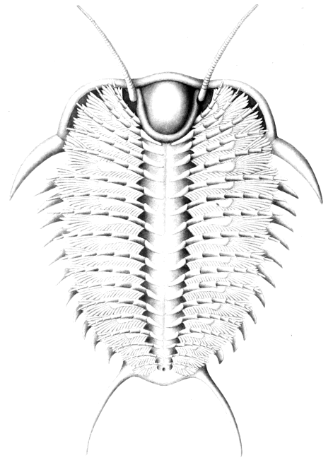
In alcoholic specimens of Apus the labrum is not in a horizontal plane, as it is in most
well preserved trilobites, but is tipped down at an angle of from 30 to 45, and the big
mandibles lie under it. It has considerable freedom of motion and is held in place by muscles
which run forward and join the under side of the head near its posterior margin. It seems
entirely possible that the hypostoma of the trilobite had as much mobility as the labrum of
Apus, and that by opening downward it brought the mouth lower and nearer the food. It
will be recalled that the hypostomata of practically all trilobites are pointed at the posterior
margin, there being either a central point or a pair of prongs. By dropping down the hypostoma
until the point or prongs rested on or in the substratum, and sending food forward
to the mouth by means of the appendages, a trilobite could make of itself a most excellent
trap, and if the animal could dart backward as well as forward, the hypostoma would be still
more useful. There is no reason to suppose that they could not move backward, and the
"pygidial antennæ" of Neolenus indicate that animals of that genus at least did so. This
habit of dropping down the hypostoma would also permit the use of those anterior gnathobases
which seem too far ahead of the mouth in the trilobites with a long hypostoma.
For actual evidence on this point, it is necessary to have recourse once more to Doctor
Walcott's exceedingly valuable slices. From such sections of Ceraurus as his Nos. 100, 106,
108, 170, and 173, it is evident that the hypostoma of that form could be dropped considerably
without disrupting the ventral membrane (fig. 30). Sections of Calymene already
published (Walcott 1881, pl. 5, figs. 1, 2) show the hypostoma turned somewhat downward,
and the slices themselves show sections of the anterior pair of gnathobases beneath the
hypostoma. When the hypostoma was horizontal, these gnathobases were crowded out at
the sides.
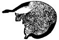 |
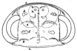 |
Fig. 30.—Longitudinal section of cephalon of Ceraurus
pleurexanthemus, to show position of the mouth and folds of the ventral
membrane between the glabella and the hypostoma. The test is in solid
black and the part within the ventral membrane dotted. From a
photographic enlargement. Specimen 169. × 3.9.
|
Fig. 31.—A copy of Doctor Moberg's figure of
Nileus armadillo, showing the position of the muscle scars.
|
If the hypostoma were used in the manner indicated, the muscles must have been more
efficient than those of the labrum of Apus, and it is probable that they crossed to the dorsal
test. Just where they were attached is an unsolved problem. Barrande (1852, pl. 1, fig. 1)
has indicated an anterior pair of scars and a single median one on the frontal lobe of
Dalmanites that may be considered in this connection, and also three pairs of scars on the
last two lobes of the glabella of Proëtus (1852, pl. 1, fig. 7). Moberg (1902, p. 295, pl. 3,
figs. 2, 3, text fig. 1) has described in some detail the muscle-scars of a rather remarkable
specimen of Nileus armadillo Dalman. While, as I shall point out, I do not agree wholly
with Professor Moberg's interpretation, I give here a translation (made for Professor
Beecher) of his description, with a copy of his text figure:
The well preserved surface of the shell permits one to note not only the tubercle (t) but a number of
symmetrically arranged glabellar impressions. And because of their resemblance to the muscular insertions
of recent crustaceans, I must interpret them as such. They appear partly as rounded hollows (k and i), also
as elongate straight or curved areas (a, b, c, e, g, h) made up of shallow impressions or furrows about
1 mm. long, sub-parallel, and standing at an angle to the trend of the areas. Impression e is especially well
marked, inasmuch as the perpendicular furrows are arranged in a shallow crescentic depression; and impression
d shows besides the obscure furrows a number of irregularly rounded depressions. Larger similar ones
occur at f, and in part extend over toward g.
The meaning of these impressions, or their myologic significance, could be discussed, although such
discussion might rather be termed guessing.
Inner organs, such as the heart and stomach, might have been attached to the shell along impressions a
and b. Also along or behind c and h, which both continue into the free cheeks, ligaments or muscular
fibers may have been inserted. From d, e, f, and g, muscles have very likely gone out to the cephalic
appendages. Against this it may be urged that impression d is too far forward to have belonged to the
first pair of feet. Again, the impression h may in reality represent two confluent muscular insertions, from
the first of which, in that case, arose the muscles of the fourth pair of cephalic feet. Were this the case, the
muscles of the first pair of cheek feet should be attached at e. And d in turn may be explained as the
attachment of the muscles of the antennæ, k those of the hypostoma, and from i possibly those of the epistoma.
That k is here named as the starting point of the hypostomial muscles and not those of the antennæ, depends
partly on the analogous position of i and partly on the fact that the hypostoma of Nileus armadillo (text
figure, in which the outline of the hypostoma is dotted), by reason of it? wing-like border, could not have
permitted the antennæ to reach forward, but rather only outward or backward.
My own explanation would be that impressions e, f, and g correspond to the glabellar
furrows, h the neck furrow, and all four show the places of attachment of the appendifers.
Those at d may possibly be connected with the antennæ, although I should expect those
organs to be attached under the dorsal furrows at the sides of the hypostoma. It will
be noted that either b, k, or i correspond well with the maculæ of the hypostoma and some
or all of them may be the points of attachment of hypostomial muscles. They correspond
also with the anterior scars of Dalmanites.
EYES.
While I have nothing to add to what has been written about the eyes of trilobites, this
sketch of the anatomy would be incomplete without some reference to the little which has
been done on the structure of these organs.
Quenstedt (1837, p. 339) appears to have been the first to compare the eyes of trilobites
with those of other Crustacea. Johannes Müller had pointed out in 1829 (Meckel's
Archiv) that two kinds of eyes were found in the latter group, compound eyes with a smooth
cornea, and compound eyes with a facetted coat. Quenstedt cited Trilobites esmarkii Schlotheim
(=Illænus crassicauda Dalman) as an example of the first group, and Calymene macrophthalma
Brongniart (=Phacops latifrons Bronn) for the second. Misreading the somewhat
careless style of Quenstedt, Barrande (1852, p. 133) reverses these, one of the few
slips to be found in the voluminous writings of that remarkable savant.
Burmeister (1843; 1846, p. 19) considered the two kinds of eyes as essentially the
same, and accounted for the conspicuous lenses of Phacops on the supposition that the cornea
was thinner in that genus than in the trilobites with smooth eyes.
Barrande (1852, p. 135) recognized three types of eyes in trilobites, adding to Quenstedt's
smooth and facetted compound eyes the groups of simple eyes found in Harpes. In
his sections of 1852, pl. 3, figs. 15-25, which are evidently diagrammatic, he shows separated
biconvex lenses in both types of compound eyes, Phacops and Dalmanites on one hand,
and Asaphus, Goldius, Acidaspis, and Cyclopyge on the other. Clarke ( 1888), Exner ( 1891 )
and especially Lindstroem (1901) have since published much more accurate figures and
descriptions. The first person to study the eye in thin section seems to have been Packard
(1880), who published some very sketchy figures of specimens loaned him by Walcott. He
studied the eyes of Isotelus gigas, Bathyurus longispinus, Calymene, and Phacops, and decided
that the two types of eyes were fundamentally the same. He also compared them with the
eyes of Limulus.
Clarke (1888), in a careful study of the eye of Phacops rana, found that the lenses
were unequally biconvex, the curvature greater on the inner surface. The lens had a circular
opening on the inner side, leading into a small pear-shaped cavity. The individual
lenses were quite distinct from one another, and separated by a continuation of the test of
the cheek.
Exner (1891, p. 34), in a comparison of the eyes of Phacops and Limulus, came to
the opinion that they were very unlike, and that the former were really aggregates of
simple eyes.
Lindstroem (1901, pp. 27-31) came to the conclusion that besides the blind trilobites
there were trilobites with two kinds of compound eyes, trilobites with aggregate eyes, and
trilobites with stemmata and ocelli. His views may be briefly summarized.
I. Compound eyes.
1. Eyes with prismatic, plano-convex lenses.
"A pellucid, smooth and glossy integument, a direct continuation of the common test of the body, covers
the corneal lenses, quite as is the case in so many of the recent Crustacea. The lenses are closely packed,
minute, usually hexagonal in outline, flat on the outer and convex on the inner surface. Such eyes are best
developed in Asaphus, Illænus, Nileus, Bumastus, Proëtus, etc."
2. Eyes with biconvex lenses.
The surface of the eye is a mass of contiguous lenses, covered by a thin membrane which is frequently
absent from the specimens, due to poor preservation. The lenses are biconvex, and being in contact with one
another, are usually hexagonal, although in some cases they nearly retain their globular shape. Such eyes
are found in Bury care, Peltura, Sphæropthalmus, Ctenopyge, Goldius, Cheirurus, and probably others.
II. Aggregate eyes.
The individual lenses are comparatively large, distinct from one another, each lying in its own socket.
There is, however, a thin membrane, which covers all those in any one aggregate, and is a continuation of
the general integument of the body. This membrane is continued as a thickened infolding which forms the
sockets of the lenses.
Such eyes are known in the Phacopidæ only.
III. Stemmata and ocelli.
The stemmata are present only in Harpes, where there may be on the summit of the cheek two or three
ocelli lying near one another. Each, viewed from above, is nearly circular in outline, almost hemispheric,
glossy and shining. In section they prove to be convex above and flat or slightly concave beneath. The
test covers and separates them, as in the case of the aggregate eyes.
The ocelli of the Trinucleidæ and Eoharpes are smaller, and the detailed structure not yet investigated.
Lindstroem concludes that so far as its facets or lenses are concerned, the eye of the trilobite shows the
greatest analogy with the Isopoda, and the least with Limulus.
SUMMARY.
The simplest eyes found among the Trilobita are the ocelli. These consist of a Simple
thickening of the test to form a convex surface capable of concentrating light. The similarity
in position of the paired ocelli of trilobites and the simple eyes of copepods has perhaps
a significance.
The schizochroal eyes may well be compared with the aggregate eyes of the chilopods
and scorpions. The mere presence of a common external covering is not sufficient to prove
this a true compound eye, especially as the covering is merely a continuation of the general
test.
The holochroal eyes are of two kinds, one with plano-convex and one with biconvex
lenses. The latter would seem to be mechanically the more perfect of the two, and it is
worthy of note that the trilobites possessing the biconvex lenses have, in general, much smaller
eyes than those with the other type.
If, as some investigators claim, the parietal eye of Crustacea originates by the fusion of
two lateral ocelli, trilobites show a primitive condition in lacking this eye, which may have
originated through the migration toward the median line of ocelli like those of the Trinucleidæ.
SEX.
That the sexes were separate in the Trilobita there can be very little doubt, but the
study of the appendages has as yet revealed nothing in the way of sexual differences. One
of the most important points still to be determined is the location of the genital openings.
In many modern Crustacea, the antennæ or antennules are modified as claspers, and it
is barely possible that the curious double curvature of the antennules of Triarthrus indicates
a function of this sort. The antennules of many specimens have the rather formal
double curvature, turning inward at the outer ends, and retain this position of the frontal
appendages, no matter what may be the condition of those on the body. Other specimens
have the antennules variously displaced, indicating that they are quite flexible. It is conceivable
that the individuals with rigid antennules are males, the others females.
It is interesting to note that the antennules of Ptychoparia permulta Walcott (1918, pl.
21, fig. 1) have the same recurved form. All the specimens of Neolenus, however, show very
flexible antennas.
Barrande and Salter laid great stress upon the "forme longue" and "forme large"
as indicating male and female. This was based upon the supposition that the female of
any animal would probably have a broader test than the male, a hypothesis which seems to
be very little supported by fact. In practical application it was found that the apparent difference
was so often due to the state of preservation or the confusion of two or more
species, that for many years little reference has been made to this supposed sex difference.
EGGS.
In his classic work on the trilobites of Bohemia, Barrande described three kinds of spherical
and one of capsule-shaped bodies which he considered to be the eggs of trilobites. After
a review of the literature and a study of specimens in the collections of the Museum of
Comparative Zoology, it can be said that none of these fossils has proved to be a trilobite
egg, but that they may be plants. A full account of them will be published elsewhere.
Walcott (1881) and Billings (1870) have described similar bodies within the tests of
Calymene and Ceraurus, but without showing positive evidence as to their nature.
Methods of Life.
This is a subject upon which much can be inferred, but little proved. Without trying
to cover all possibilities, it may be profitable to see what can be deduced from what is known
of the structure of the external test, the internal anatomy, and the appendages. This can,
to a certain extent, be controlled by what is inferred from the strata in which the specimens
are found, the state of preservation, and the associated animals. (For other details, see
the discussion of "Function of the Appendages" in Part I.)
HABITS OF LOCOMOTION.
The methods of locomotion may be deduced with some safety from a study of the appendages,
and, as has repeatedly been pointed out, all trilobites could probably swim by their
use. This swimming was evidently done with the head directed forward, and could probably
be accomplished indifferently well with either the dorsal (gastronectic, Dollo) or the
ventral (notonectic) side up. If food were sought on the bottom by means of sight, the animal
would probably swim dorsal side up, for by canting from side to side it could see the bottom
just as easily as though it were ventral side up, and at the same time it would be in position
to drop quickly on the prey. In collecting food at the surface, it might swim ventral side up.
All trilobites could probably crawl by the use of the appendages, and, as has already
been pointed out, there are great differences in the adjustment of the appendages to different
methods of crawling. Some crawled on their "toes," some by means of the entire endopodites,
and some apparently used the coxopodites to push themselves along. That the normal
direction of crawling was forward is indicated by the position of the eyes and sensory antennules.
There is no evidence that their mechanism was irreversible, however, and the position
of the mouth and the shape of the hypostoma indicate that they usually backed into feeding
position. The caudal rami of Neolenus were evidently sensory, and the animal was prepared
to go in either direction.
The use of the pygidium as a swimming organ, suggested by Spencer (1903, p. 492) on
theoretical grounds, developed by Staff and Reck (1911, p. 141) from a mechanical standpoint,
and elaborated in the present paper by evidence from the ontogeny, phylogeny, and
musculature, provided the animal with a swifter means of locomotion. By a sudden flap
of this large fin, a backward darting motion could be obtained, which would be invaluable
as a means of escape from enemies. Staff and Reck seem to think that in this movement
the two shields were clapped together, and that the animal was projected along with the hinge-like
thorax forward. This might be a very plausible explanation in the case of the bivalve-like
Agnostidæ, and it is one I had suggested tentatively for that family before I read Staff
and Reck's paper. In the case of the large trilobites with more segments, however, it would
be more natural to think of a mode of progression in which there was an undulatory movement
of the body and the pygidium, up-and-down strokes being produced by alternately
contracting the dorsal and ventral muscles. Bending the pygidium down would tend to pull
the animal backward, while bringing it back into position would push it forward. It follows,
therefore, that one of these movements must have been accomplished very quickly, the
other slowly. If the muscle scars have been interpreted properly, the ventral muscles were
probably the more powerful, an indication that the animal swam backward, using the cephalon
and antennules as rudders.
The chief objection to the theory of swimming by clapping the valves together is that
where the thorax consists of several segments it no longer acts like the hinge of a bivalve,
and a sudden downward flap of the pygidium would impart a rotary motion to the animal.
Take, for example, such nearly spherical animals as the Illænidæ, and it will readily be seen
that there is nothing to give direction to the motion if the pygidium be brought suddenly
against the lower surface of the cephalon. A lobster, it is true, progresses very well by
this method, but it depends upon its great claws and long antennæ to direct its motions.
The whole shape of the trilobite is of course awkward for a rapidly swimming animal. It
could keep afloat with the minimum of effort and paddle itself about with ease, but it was
not built on the correct lines for speed.
Dollo (1910, p. 406), and quickly following his lead, Staff and Reck (1911, p. 130), have
published extremely suggestive papers, showing that by the use of the principle of correlation
of parts, much can be inferred about the mode of life of the trilobites merely from the
structure of the test.
Dollo studied the connection between the shape of the pygidium and the position and
character of the eyes. As applied by him, and later by Clarke and Ruedemann, to the eurypterids,
this method seems most satisfactory. He pointed out that in Eurypterida like Stylonurus
and Eurypterus, where there is a long spine-like telson, the eyes are back from the
margin, so that a Limulus-like habit of pushing the head into the sand by means of the limbs
and telson was possible. Erettopterus and Pterygotus, on the other hand, have the eyes on
the margin, so that the head could not be pushed into the mud without damage, and have
a fin-like telson, suggesting a swimming mode of life.
In carrying this principle over to the trilobites, Dollo was quite successful, but Staff
and Reck have pointed out some modifications of his results. The conclusions reached
in both these papers are suggestive rather than final, for not all possible factors have been
considered. The following are given as examples of interesting speculations along this line.
Homalonotus delphinocephalus, according to Dollo, was a crawling animal adapted to
benthonic life in the euphotic region, and an occasional burrower in mud. This is shown by
well developed eyes in the middle of the cephalon, a pointed pygidium, and the plow-like
profile of the head. This was as far as Dollo went. From the very broad axial lobe of
Homalonotus it is fair to infer that, like Isotelus, it had very long, strong coxopodites which
it probably vised in locomotion, and also very well-developed longitudinal muscles, to be used
in swimming. From the phylogeny of the group, it is known that the oldest homalonotids
had broad unpointed pygidia of the swimming type, and that the later species of the genus
(Devonian) are almost all found in sandstone and shale, and all have wider axial lobes
than the Ordovician forms. It is also known that the epistoma is narrower and more
firmly fused into the doublure in later than in earlier species. These lines of evidence tend
to confirm Dollo's conclusion, but also indicate that the animals retained the ability to swim
well.
On the same grounds, Olenellus thompsoni and Dalmanites limulurus were assigned the
same habitat and habits. Both were considered to have used the terminal spine as does
Limulus.
Olenellus thompsoni is generally considered to be unique among trilobites in having a
Limulus-like telson in place of a pygidium. This "telson" has exactly the position and
characteristics of the spine on the fifteenth segment of Mesonacis, and so long ago as 1896,
Marr (Brit. Assoc. Adv. Sci., Rept. 66th Meeting, page 764) wrote:
The posterior segments of the remarkable trilobite Mesonacis vermontana are of a much more delicate
character than the anterior ones, and the resemblance of the spine on the fifteenth "body segment" of this
species to the terminal spine of Olenellus proper, suggests that in the latter subgenus posterior segments of a
purely membranous character may have existed devoid of hard parts.
This prophecy was fulfilled by the discovery of the specimens which Walcott described
as Pædeumias transitans, a species which is said by its author to be a "form otherwise
identical with O. thompsoni, [but] has rudimentary thoracic segments and a Holmia-like
pygidium posterior to the fifteenth spine-bearing segment of the thorax." A good specimen
of this form was found at Georgia, Vermont, associated with the ordinary specimens
of Olenellus thompsoni, and I believe that it is merely a complete specimen of that species.
Olenellus gilberti, which was formerly supposed to have a limuloid telson, has now been
shown by Walcott (Smithson. Misc. Coll., vol. 64, 1916, p. 406, pl. 45, fig. 3) to be a
Mesonacis and to have seven or eight thoracic segments and a small plate-like pygidium
back of the spine-bearing fifteenth segment. All indications are that the spine was not in
any sense a pygidium. Walcott states that Olenellus resulted from the resorption of the
rudimentary segments of forms such as Mesonacis and Pædeumias, leaving the spine to
function as a pygidium. This would mean the cutting off of the anus and the posterior
part of the alimentary canal, and developing a new anal opening on the spine of one of
the thoracic segments!
If the spine of the fifteenth segment is not a pygidium, could it be used, as Dollo
postulates, as a pushing organ? Presumably not, for though in entire specimens of Olenellus
(Pædeumias) it extends back beyond the pygidium, it probably was borne erect, like the
similar spines in Elliptocephala, and not in the horizontal plane in which it is found in
crushed specimens.
While this removes some of the force of Dollo's argument, his conclusion that Olenellus
was a crawling, burrowing animal living in well lighted shallow waters was very likely correct.
The long, annelid-like body indicates numerous crawling legs, there is no swimming
pygidium, and the fusion of the cheeks in the head makes a stiff cephalon well adapted for
burrowing.
Staff and Reck have pointed out that Dalmanites limulurus was not entirely a crawler,
but, as shown by the large pygidium, a swimmer as well. This kind of trilobite probably
represents the normal development of the group in Ordovician and later times. The Phacopidæ,
Proëtidæ, Calymenidæ, and other trilobites of their structure could probably crawl
or swim equally well, and could escape enemies by darting away or by "digging themselves
in."
Cryptolithus tessellatus (Trinucleus concentricus) is cited by Dollo as an example of
an adaptation to life in the aphotic benthos, permanently buried in the mud. In this case
he appealed to Beecher's interpretation of the appendages, and pointed out that while the
adult is blind, the young have simple eyes and probably passed part of their life in the
lighted zone. It needs only a glance at the very young to convince one that the embryos
had swimming habits, so that in this form one sees the adaptation of the individual during
its history to all modes of life open to a trilobite. The habits of the Harpedidæ may have
been similar to those of the Trinucleidæ, but the members of this family are supplied with
broad flat genal spines. It has been suggested that these served like pontoons, runners, or
snow-shoes, to enable the animal to progress over soft mud without sinking into it. Some
such explanation might also be applied to the similar development in the wholly unrelated
Bathyuridæ. The absence of compound eyes and the poor development of ocelli in the Harpedidæ
suggest that they were burrowers, and from these two families, Trinucleidæ and
Harpedidæ, it becomes evident that a pygidial point or spine is not a necessary part of the
equipment of a burrowing trilobite. In fact, from the habits of Limulus it is known that
the appendages are relied upon for digging, and that the telson is a useful but not indispensable
pushing organ.
Deiphon is an interesting trilobite from many points of view. Its pleural lobes are
reduced to a series of spines on either side of the body, and its pygidium is a mere spinose
vestige. Dollo considered this animal a swimmer in the euphotic zone, because its eyes
are on the anterior margin, its body depressed, its glabella globose, and its pygidium flat
and spinose. That such a method of life was secondary in a cheirurid was indicated to
him by the fact that the more primitive members of the family seemed adapted for crawling.
Staff and Reck have gone further and shown that the pygidium is only the vestige of
a swimming pygidium, and that the great development of spines suggests a floating rather
than a swimming mode of life. They therefore argue for a planktonic habitat. A similar
explanation is suggested for Acidaspis and other highly spinose species.
The Aeglinidæ, or Cyclopygidæ as they are more properly called, present the most remarkable
development of eyes among the trilobites. In this, Dollo saw, as indeed earlier
writers have, an adaptation to a region of scanty light. The cephalon is not at all adapted
to burrowing, but the pygidium is a good swimming organ, and one is apt to agree that this
animal was normally an inhabitant of the ill lighted dysphotic region, but also a nocturnal
prowler, making trips to the surface at night. Similar habits and habitat are certainly indicated
for Telephus and the Remopleuridæ, but whether Nileus and the large-eyed Bumastus
are capable of the same explanation is doubtful.
Finch (1904, p. 181) makes the suggestion that "Nileus" (Vogdesia) vigilans, an
abundant trilobite in the calcareous shale of the Maquoketa, was in the habit of burying itself,
posterior end first. He found a slab containing fifteen entire specimens, all of which had
the cephalon extended horizontally near the surface of the stratum, and the thorax and
pygidium projecting downward. The rock showed no evidence that they were in burrows,
and the fact that all were in the same position indicates that the attitude was voluntarily
assumed. They appear to have entrenched themselves by the use of the pygidia, which are
incurved plates readily adapted for such use, and, buried up to the eyes, awaited the coming
of prey, but were, apparently, smothered by a sudden influx of mud. The form of the eye
in Vogdesia vigilans bears out this supposition of Finch's. Not only are the eyes unusually
tall, but the palpebral lobe is much reduced, so that many of the lenses look upward and
inward, as well as outward, forward and backward. The particular food required by V. vigilans
must have been very plentiful in the Maquoketa seas of Illinois and Iowa, for the species
was very abundant, but that its habits were self-destructive is also shown by the great number
of complete enrolled specimens of all ages now found there. The soft mud was apparently
fatal to the species before the end of the Maquoketa, for specimens are seen but very
rarely in the higher beds.
Vogdesia vigilans is shaped much like Bumastus, Illænus, Asaphus, Onchometopus, and
Brachyaspis, and it may be that these trilobites with incurved pygidia had all adopted the
habit of digging in backward. As noted above, their pygidia are not very well adapted
for swimming, and most of them have large or tall eyes.
Dollo's comparison of the Cyclopygidæ to the huge-eyed modern amphipod Cystosoma
is instructive. This latter crustacean, which has the greater part of the dorsal surface of the
carapace transformed into eyes, is said to live in the dysphotic zone, at depths of from 40
to 100 fathoms, and to come to the surface at night. It swims ventral side down.
The kinds of sediments in which trilobites are entombed have so far afforded little evidence
as to their habitat. Frech (Lethæa palæozoica, 1897-1902, p. 67 et seq.) who has
collected such evidence as is available on this subject, places as deeper water Ordovician
deposits the "Trinucleus-Schiefer" of the upper Ordovician of northern Europe and Bohemia,
the "Triarthrus-Schiefer" of America, the "Asaphus-Schiefer" of Scandinavia, Bohemia,
Portugal, and France, and the Dalmania quartzite of Bohemia. .
Cryptolithus and Triarthrus, although not confined to such deposits, are apt to occur
chiefly in very fine-grained shales, in company with graptolites. These latter are distributed
by currents over great distances within short periods. It is somewhat curious that the nearly
blind burrowing Trinucleidæ, the dysphotic, large-eyed Remopleuridæ and Telephus, the blind
nektonic Agnostidæ and Dionide, and the planktonic graptolites should go together and make
up almost the entire fauna of certain formations. Yet, when the life history of each type
is studied, a logical explanation is readily at hand, for all have free-swimming larvæ.
A list of the methods of life noted above is given by way of summary, with examples.
| Pelagic |
 |
Planktonic |
|
Primarily
Secondarily |
|
Earliest protaspis of all trilobites
Deiphon, Odontopleura, etc. |
| Nektonic |
 |
Primarily |
|
Later protaspis of all trilobites. |
| Secondarily |
|
Probably many thin-shelled
trilobites with large pygidia
(only partially nektonic) |
| Cyclopygidæ |
 |
(nektonic dysphotic) |
| Remopleuridæ |
| Benthonic |
|
Crawlers and slow swimmers |
|
Most trilobites with small pygidia. Triarthrus, Paradoxides, etc. |
| Crawlers and slow swimmers |
|
Most trilobites with large pygidia. Isotelus, Dalmanites, etc. |
| Crawlers and slow swimmers |
|
Trilnuceidæ, Harpedidæ, some Mesonacidæ, etc. |
FOOD AND FEEDING METHODS.
This subject has been less discussed than the methods of locomotion. The study of
the appendages has shown that while the mouth parts were not especially powerful, they were
at least numerous, and sufficiently armed with spines to shred up such animal and vegetable
substances as they were liable to encounter. It having been ascertained that the shape of the
glabella and axial lobe furnishes an indication of the degree of development of the alimentary
canal it is possible to infer something of the kind of food used by various trilobites.
The narrow glabellæ and axial lobes of the oldest trilobites would seem to indicate a
carnivorous habit, while the swollen glabellæ and wider lobes of later ones probably denote an
adaptation to a mixed or even a vegetable diet. This can not be relied upon too strictly,
of course, for the swollen glabellæ of such genera as Deiphon or Sphærexochus may be due
merely to the shortening up of the cephalon.
Walcott (1918, p. 125) suggests that the trilobites lived largely upon worms and conceives
of them as working down into the mud and prowling around in it in search of such
prey. While there can be no doubt that many trilobites had the power of burying themselves
in loose sand or mud, a common habit with modern crustaceans, most of them were
of a very awkward shape for habitual burrowers, and how an annelid could be successfully
pursued through such a medium by an animal of this sort is difficult to understand. In
fact, the presence of the large hypostoma and the position of the mouth were the great
handicaps of the trilobite as a procurer of live animal food, and coupled with the relatively
slow means of locomotion, almost compel the conclusion that errant animals of any
size were fairly safe from it. This restricts the range of animal food to small inactive
creatures and the remains of such larger forms as died from natural causes. The modern
Crustacea are effective scavengers, and it is probable that their early Palæozoic ancestors
were equally so. It is a common saying that in the present stressful stage of the world's
history, very few wild animals die a natural death. In Cambrian times, competition for
animal food was less keen, and with the exception of a few cephalopods, a few large annelids,
and a few Crustacea like Sidneyia, there seem to have been no aggressive carnivores.
In consequence, millions of animals must have daily died a natural death, and had there been
no way of disposing of their remains, the sea bottom would soon have become so foul that
no life could have existed. For the work of removal of this decaying matter, the carnivorous
annelids and the Crustacea, mostly trilobites, were the only organisms, and it is probable
that the latter did their full share. After prowling about and locating a carcass, the
trilobite established himself over it, the cephalon and hypostoma on one end and the
pygidium on the other enclosing and protecting the prey, which was shredded off and passed
to the mouth at leisure by means of the spinose endobases.
Even in Middle Cambrian times some trilobites (e. g., Paradoxides) seem to have enlarged
the capacity of the stomach and taken vegetable matter, but later, in the Upper
Cambrian and Ordovician, when the development of cephalopods and fishes caused great
competition for all animal food, dead or alive, most trilobites seem to have become omnivorous.
This is of course shown by the swollen glabella, with reduced lateral furrows, and,
in the case of a few species (Calymene, Ceraurus), the known enlargement of the stomach.
Cryptolithus is the only trilobite which has furnished any actual evidence as to its food.
From the fact that the alimentary tract is found stuffed from end to end with fine mud,
and because it is known to have been a burrower, it has been suggested by several that it
was a mud feeder, passing the mud through the digestive tract for the sake of what organic
matter it contained. Spencer (1903, p. 491) has suggested a modification of this view:
The phyllopods appear to feed by turning over whilst swimming and seizing with their more posterior
appendages a little mud which swarms with infusoria, etc. This mud is then pushed along the ventral
groove to the mouth. Casts, of the intestine of trilobites are still found filled with the mud.
Ceraurus and Calymene also must have occasionally swallowed mud in quantity, otherwise
the form of the alimentary canal could not have been preserved as it is in a few of
Doctor Walcott's specimens.
TRACKS AND TRAILS OF TRILOBITES.
Tracks and trails of various sorts have been ascribed by authors to trilobites since these
problematic markings first began to attract attention, but as the appendages were until recently
quite unknown, all the earlier references were purely speculative. The subject is a
difficult one, and proof that any particular track or trail could have been made in only one
way is not easily obtained. Since the appendages have actually been described, comparatively
little has been done, Walcott's work on Protichnites (1912 B, p. 275) being the most
important. Since the first description of Protichnites by Owen (Quart. Jour. Geol. Soc.,
London, 1852, vol. 8, p. 247), it has been thought that these trails were made by crustaceans,
and the only known contemporaneous crustaceans being trilobites, these animals were naturally
suggested. Dawson (Canadian Nat. Geol., vol. 7, 1862, p. 276) ascribed them, with
reserve, to Paradoxides, and Billings (1870, p. 484) suggested Dikelocephalus or Aglaspis.
Walcott secured well preserved specimens which showed trifid tracks, and these were readily
explained when he found the legs of Neolenus, which terminated with three large spines.
Similar trifid terminations had already been described by Beecher, and clearly pictured in his
restoration of Triarthrus, but the spines and the tracks had somehow not previously been
connected in the mind of any observer. Walcott concluded that the tracks had been made
by a species of Dikelocephalus, possibly by D. hartti, which occurs both north and south of the
Adirondacks. In a recent paper, Burling (Amer. Jour. Sci., ser. 4, vol. 44, 1917, p. 387)
has argued that Protichnites was not the trail of a trilobite, but of a "short, low-lying, more
or less heavy set, approximately 12-legged, crab-like animal," which had an oval shape, toed
in, and was either extremely flexible or else short and more or less flexible in outline.
This seems to describe a trilobite.
Climactichnites, the most discussed single trail of all, has also been ascribed to trilobites,—by
Dana (Manual of Geology, 1863, p. 185), Billings (1870, p. 485), and Packard
(Proc. Amer. Acad. Arts and Sci., vol. 36, 1900, p. 64),—though less frequently than
to other animals. The latest opinion (see paper by Burling cited above) seems to be against
this theory.
Miller (1880, p. 217) described under the generic name Asaphoidichnus two kinds of
tracks which were such as he supposed might be made by an Asaphus (Isotelus). In referring
to the second of the species, he says: "Some of the toe-tracks are more or less
fringed, which I attribute to the action of water, though Mr. Dyer is impressed with the
idea that it may indicate hairy or spinous feet." The type of this species, A. dyeri, is in the
Museum of Comparative Zoology, and while it may be the trail of a trilobite, it would be
difficult to explain how it was produced.
Ringueberg (1886, p. 228) has described very briefly tracks found in the upper part
of the Medina at Lockport, New York. These consisted of a regularly succeeding series of
ten paired divergent indentations arranged in two diverging rows, with the trail of the pygidium
showing between each series. The ten pairs of indentations he considered could have
been made by ten pairs of legs like those shown by the specimen of Isotelus described by
Mickleborough, and the intermittent appearance of the impression of the pygidium suggested
to him that the trilobite proceeded by a series of leaps.
Walcott (1918, pp. 174-175, pl. 37-42) has recently figured a number of interesting
trails as those of trilobites, and has pointed out that a large field remains open to anyone
who has the patience to develop this side of the subject.