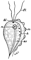
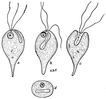
Man is one of those organisms in on on which a whole host of parasites find
conditions suitable for their existence: Protozoa, Platyhelminthes, Nematoda,
Acanthocephala, Hirudinea, and a large number of Arthropoda (Arachnida as well
as Insects) all include members which are parasites of man. These animals either
live on the external surface of the body or within the intestine and its appendages.
Other organs and systems are not quite free from foreign organisms—we are
acquainted with parasites in the skeletal system, in the circulatory system, in the
brain, in the muscles, in the excretory and genital organs, and even in the organs of
sense.
It is possible, and perhaps might be advantageous, to arrange and describe the
parasites of man according to the situations in which they are found (parasites of the
skin, intestinal parasites, etc.). Their description in the various stages of development
would, however, be disturbed when, as is generally the case, the different stages
are passed in different organs, and a work which treats more fully of the natural
history of the parasites than of the local disorders to which they give rise would
suffer thereby. It is, therefore, preferable to describe the parasites of man in their
systematic order, and to mention their different situations in man in describing each
species.
All those animal organisms which throughout their entire life never rise above
the unicellular stage, or merely form simple, loose colonies of similar unicellular
animals, are grouped under the term Protozoa (Goldfuss, 1820), as the simplest types
of animal life. All the vital functions of these, the lowest forms of animals, are
carried out by their body substance, the protoplasm (sarcode). Often particular
parts possess special functions, but the limits of a cell are never over-stepped
thereby. These special parts of the cell are called “cell-organs”; recently they
have been termed “organellæ.”
The living protoplasm has the appearance of a finely granular, viscid substance
which, as a rule, when not surrounded by dense investing membranes or skeletons,
exhibits a distinct kind of movement, which has been termed amœboid. According
to the species, processes of different forms and varying numbers called pseudopodia
are protruded and withdrawn, and with their assistance these tiny organisms glide
along—it might almost be said flow along—over the surface. In most Protozoa two
layers of cytoplasm may be recognised, and distinguished by their appearance and
structure, namely, the superficially situated, viscid, and quite hyaline ectosarc or
ectoplasm, and the more fluid and always granular endosarc or endoplasm, which
is entirely enveloped by the ectoplasm. The two layers have different functions; the
movements originate from the ectoplasm, which also undoubtedly fulfils the functions
of breathing, introduction of food and excretion. The endoplasm, which in some forms
(Radiolaria) is separated from the ectoplasm by a membrane, undertakes the digestion
of the food. To this distribution of functions between the various layers of cytoplasm
is due the development of particular cellular organs, such as the appearance
of cilia, flagella, suctorial tubules (in the Suctoria) and the myophan striations, which
are contractile parts of the ectoplasm in Infusoria and Gregarines. In many cases
(Flagellata, Ciliata), an area is differentiated for the ingestion of food (oral part,
cytostome) to which there is often added a straight or curved opening (cytopharynx),
through which the food reaches the endoplasm. The indigestible residue is either cast
off through the oral part or excreted by a special anal part (cytopyge). In rare
cases, structures sensitive to light, the so-called pigment or eye spots are developed,
e.g., Euglena. In the case of Infusoria the endoplasm circulates slowly, and
agglomerations of fluids (food vacuoles) sometimes appear around each bolus of
food; in these vacuoles the food is digested under the action of certain materials
(ferments). Even in the lowliest Protozoa fluids to be excreted are, as a rule,
gathered into one, or, more rarely, several contractile vacuoles, which regularly
discharge their contents. This action, however, is to a certain extent governed
by the temperature of the surrounding medium. In some Infusoria a tube-like
channel in the cytoplasm is joined to the contractile vacuole which usually occupies
a certain position; this forms a sort of excretory duct, and there are also supply-canals
leading to these organellæ.
Very frequently various substances are deposited in the endoplasm, such as fatty
granules, drops of oil, pigment granules, bubbles of gas or crystals. More solid
skeletal substances are secreted in or on the ectoplasm. To the latter belong the
cuticle of the Sporozoa and Infusoria, the chalky shells containing one or several
chambers of the Foraminifera, the siliceous and very ornamental framework of the
Radiolaria, and the chitinous coat of many Flagellata, Infusoria, etc. Some forms
make use of foreign bodies found in their surroundings, such as grains of sand, to
construct their protective coverings.
The food often consists of small animal or vegetable organisms and of organic
waste; it is usually introduced in toto into the endoplasm. On the other hand, the
Suctoria extract nourishment from their prey by means of their tentacles. Many
parasitic species also ingest solid food, others feed by endosmosis.
In all cases one nucleus at least is present. It is true that the existence of non-nucleated
Protozoa, the so-called Monera, is still insisted upon, but some of these
have already proved to be nucleated, and the presence of nuclei in the others will no
doubt be established. Very often the number of nuclei increases considerably, but
these multinucleate stages are always preceded by uninucleate stages. In the
Infusoria, in addition to the larger or principal nucleus (macronucleus) there is
usually a smaller reproductive nucleus (micronucleus). This dualism of the nuclear
apparatus is considered by some to be general, and usually to appear first at the
onset of reproduction.
The form and structure of the nucleus vary greatly in different species. There
are elongate, kidney-shaped, or even branched nuclei as well as spherical or oval
ones. In addition to vesicular nuclei with a distinct karyosome and incidentally
also with a nuclear membrane, homogeneous and more solid formations are
frequently encountered. The nuclei are always differentiated from the protoplasm
by their reactions, particularly in regard to certain stains.
In many Protozoa an extra-nuclear mass, sometimes compact, sometimes diffuse,
arises from or near the nucleus. This mass, whose staining reactions resemble those
of the nucleus, is termed the chromidial apparatus. On the dualistic hypothesis, two
varieties of chromidia occur, one originating from the vegetative nucleus (macronucleus),
being chromidia in the restricted sense, the other derived from the
reproductive or micronucleus being termed sporetia. Chromidia consist of altered
(? katabolic) nuclear material.
The nucleus plays the same part in the life of the single celled organisms as it
does in the cells of the Metazoa and Metaphyta. It appears to influence in a certain
manner all, or at least most, of the processes of life, such as motility, regeneration,
growth, and generally also digestion. Its principal influence, however, is exercised
in the propagation of the cells, as this is always brought about by the nucleus.
The PROPAGATION of the Protozoa is effected either by division or by means of
direct budding. In division, which is preceded by direct or indirect (mitotic)
division of the nucleus, the body separates into two, several, or even a great many
segments. In this process the entire substance of the body is involved, or a small
residual fragment may be left, which does not undergo further division and finally
perishes. In the budding method of multiplication a large number of buds are
formed, either on the surface or in the interior of the organism. Where divisions or
buddings follow one another rapidly, without the segments separating immediately
after their production, numerous forms develop, which are often unlike the parental
forms, and these are termed swarm spores or spores. Divisions imperfectly
accomplished lead to the formation of protozoal colonies.
Sometimes encystment9 takes place previous to division. Frequently, also,
sexual processes appear, such as the union of two similar (isogamous) or dissimilar
(anisogamous) individuals. In the latter case sexual dimorphism occurs, with the
formation of males (microgametes) and of females (macrogametes). The union may
be permanent (copulation), the process being comparable with the fertilisation of the
ovum by a spermatozoon. On the other hand, attachment may be transient (conjugation)
when, after the exchange of portions of the nucleus, the couple separate,
to multiply independently of each other. Sometimes there is an ALTERNATION OF
GENERATIONS, as there may be several methods of propagation combined in the
same species, either direct multiplication, conjugation, or copulation being practised;
the different generations may thus, in certain cases, be unlike morphologically.
Protozoa inhabit salt water as well as fresh water; they are also found on land
in very damp places, and invade animals as parasites.
Classification of the Protozoa.
Class I.—Sarcodina (Rhizopoda). Protozoa, the body substance of which forms
pseudopodia; many of them are capable of developing chitinous, chalky, or siliceous
coverings or skeletal structures, which, however, permit the protrusion of the
pseudopodia either over the entire periphery or at certain points. They possess one
nucleus or several.
Order 1.—Amœbina (Lobosa) naked or with a simple shell, sometimes formed
of a foreign substance; the pseudopodia may be lobose or finger-shaped;
there may be a contractile vacuole; generally only one nucleus. They
live in fresh or salt water, in the soil, and also parasitically.
Order 2.—Foraminifera (Reticularia). Mostly provided with a calcareous
shell, usually consisting of several chambers, and allowing the protrusion
of the pseudopodia either at the periphery or only at the opening. The
pseudopodia are filamentous and frequently anastomosed; there is no
contractile vacuole; there are usually several nuclei. Mostly marine.
Order 3.—Heliozoa. Naked, or with a chitinous or simple radial siliceous
skeleton; the pseudopodia are filamentous, and are frequently supported
by firmer axes, which exhibit no tendency to anastomosis; there is a
contractile vacuole; one or several nuclei. Live in fresh water.
Order 4.—Radiolaria. The body has radially-disposed filamentous pseudopodia,
and the nucleus is hidden in the central capsule; there is almost
always a siliceous framework, consisting of pieces arranged radially,
tangentially, or lattice-like; there is no contractile vacuole, but fluid-containing
hydrostatic vacuoles are present in the peripheral protoplasm.
Marine.
Class II.—Mastigophora (Flagellata). Protozoa with one or several long
flagella used for locomotion and for acquiring food; in stationary forms their only
function is to take in food. Cytostome and contractile vacuole may be present.
May be either naked or provided with protective coverings; one or more nuclei.
They live either in fresh or salt water, or may be parasitic.
This class is again divided into several sub-classes and orders, of which only the
Euflagellata, with the Protomonadina and Polymastigoda are of interest here.
Class III.—Sporozoa. Protozoa that only live parasitically in the cells, tissues,
or organs of other animals. They ingest liquid food by osmosis; the surface of the
body is covered with an ectoplasmic layer, or cuticle; they have no cilia in the adult
state, but may form pseudopodia. Flagella occur, but only on the male propagating
individuals. There may be one or numerous nuclei, but no contractile vacuole.
Propagation by means of spores, mostly provided with sporocysts, is characteristic.
Sub-class 1.—Telosporidia. These are usually of constant form, rarely amœboid;
they are uninucleate in the mature state; they live within host cells in the
first stage. Spore-formation occurs at the end of the life-cycle.
Order 1.—Gregarinida. Body of a constant, usually elongate form, surrounded
by a cuticle. In the early stage they lead an intracellular existence; in
the mature stage they live within the intestine or body cavity of invertebrate
animals, especially the Arthropoda, and, like intestinal parasites,
are provided with clinging organs. Copulation usually isogamous;
the spores have coats (chlamydospores) and usually contain several
minute germs (sporozoites).
Order 2.—Coccidiidea. Body of uniform spherical or oval shape: they lead
an intracellular life, but are not freely motile in cavities of the body.
Fertilization is anisogamous; the spores have coats or shells (sporocysts),
and usually contain several sporozoites. Exhibit alternation of generations.
Order 3.—Hæmosporidia. Parasites of the blood corpuscles of vertebrate
animals; they exhibit amœboid movement; fertilization is anisogamous;
many present alternation of generations and hosts; spores naked.
Sub-class 2.—Neosporidia. They are multinucleate when adult, and the form of
the body varies exceedingly (often amœboid); spore-formation commences
before the completion of growth.
Order 1.—Myxosporidia. The spores have valvular coats, with or without
caudal appendages, with two, rarely four, polar capsules. They live free
in such organs as the gall or urinary bladder, but are chiefly found in
connective tissue. They occur especially in fishes.
Order 2.—Microsporidia. Spores with coats or sporocysts; no caudal
appendage, with one polar capsule. They usually live in the tissues of
Arthropoda.
Order 3.—Sarcosporidia. Elongate parasites of the muscular fibres of
amniotic vertebrates, on rare occasions they occur also in the connective
tissue; the spores, which are kidney or sickle-shaped, are naked and
apparently have no obvious polar capsule.
Order 4.—Haplosporidia. Simple organisms, forming simple spores; they
occur in Rotifers, Polychætes, Fish and Man.
Class IV.—Infusoria (Ciliata). The body is generally uniform in shape, with
cilia and contractile vacuole, frequently also with cytostome; usually has macro-
and micro-nucleus; live free in water and also parasitically.
The orders Holotricha, Heterotricha, Oligotricha, Hypotricha and Peritricha
are classified according to the arrangement of the cilia.
Class V.—Suctoria. Bodies with suctorial tubes, contractile vacuoles, macro-
and micro-nucleus, no cytostome. They generally invade aquatic animals as cavity
parasites, yet also attack plants; early stage ciliated. Live sometimes as parasites
on Infusoria. [The Suctoria are frequently regarded as a sub-class of the Infusoria.]
The Protozoa and Protophyta are sometimes united under the term Protista
(Haeckel, 1866). The Spirochætes are Protists (see pp. 114–128).
The first record of the occurrence of amœba-like organisms in the human intestine,
that is, in intestinal evacuations, was that of Lambl (1859); nevertheless, the case
was not quite conclusive, as the occurrence of testaceous amœbæ of fresh water
(Arcella, Difflugia) was also reported. In 1870 Lewis found amœbæ associated with
disorders of the large intestine in patients in Calcutta. A year later Cunningham
reported from the same locality that he had observed on eighteen occasions, in one
hundred examinations of dejecta from cholera patients, colourless bodies with amœboid
movements, which became encysted and multiplied by fission. The daughter forms
were said to be capable of dividing again, but they might also remain in contact.
Contractile vacuoles were not noticed. The same bodies were observed also in
simple diarrhœa (twenty-eight cases out of one hundred.)
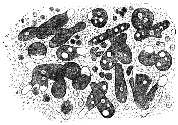
Fig. 1.—Amœba coli, Lösch, in the intestinal mucus. (After Lösch.)
The case reported by Lösch in 1875 attracted more attention. It was that of
a peasant, aged 24, who came from the province of Archangel. He was admitted
into Eichwald’s clinic at Petrograd with symptoms of dysentery. In the discharges
containing blood and pus, Lösch found amœbæ in large numbers. When
at rest these amœbæ measured from 20 µ to 35 µ; in a state of movement their length
might extend up to 60 µ (fig. 1). The pseudopodia appeared only singly, and,
since they were hyaline (ectoplasmic), were thus distinguished from the markedly
granular endoplasm that enclosed a spherical nucleus of from 5 µ to 7 µ in diameter.
One or more non-contractile vacuoles were present. Quinine enemata had the
effect of making the amœbæ disappear from the fæces and thus causing the diarrhœa
to abate. Four months after admission the patient died from the results of
intercurrent pneumonia. At the autopsy ulceration of the large intestine was found,
especially in the lower parts. Lösch connected the amœbæ with the ulcerations by
experiments made on four dogs by injecting them with recently passed stools
(per os et anum). Eight days after the last injection numerous amœbæ were found
in the fæces of one of these dogs; eighteen days after the injection the animal
was killed. The mucosa of the rectum was inflamed, covered with blood-stained
mucus and ulcerated in three places. Numbers of amœbæ were found both in
the pus of the ulcers and in the mucus. The three other dogs remained healthy.
From these observations Lösch concluded that the species of amœba described by him
as Amœba coli could not be regarded as the primary cause of the disease, but that it
was certainly capable of increasing a lesion of the large intestine already present,
or at least of preventing its healing.
B. Grassi (1879) found in the stools of healthy as well as in those of diarrhœic
patients from various localities in Northern Italy, amœbæ similar to those discovered
by Lösch. As this was of frequent occurrence, the pathogenicity could not be
definitely established. Normand, formerly naval surgeon at Hong-Kong, observed
numerous amœbæ in the dejecta of two patients suffering from colitis.
Many further investigations, which cannot be quoted in detail, showed not only
that intestinal amœbæ were widely distributed in man, but indicated with greater
certainty their rôle as agents of dysentery. The Commission sent out by the German
Government in the year 1883 to investigate cholera in India and Egypt—whose
members discovered the cholera bacillus—also collected information with regard to
dysentery. In five cases of dysentery examined post mortem at Alexandria, with
the exception of one case in which ulceration of the colon had already cicatrized or
was approaching cicatrization, R. Koch found amœbæ as well as bacteria in sections
from the base of the ulcers, although such had previously escaped notice in examination
of the dejecta. Encouraged by these results, Kartulis (1885), who had discovered
amœba-like bodies in the stools of patients suffering from intestinal complaints at
Alexandria, continued his investigations. The results, obtained from more than 500
cases, gave rise to the theory that typical dysentery was caused by amœbæ as were
also the liver-abscesses that often accompany it. Kartulis supported his theory not
only by the regular occurrence of amœbæ in the stools of dysenteric patients and
their absence in other diseases, and by the occurrence of the parasites in ulcers of the
large intestine and in the pus from liver-abscesses, but also by experiments which
he performed on cats. These were infected by injection per anum of stool material
rich in amœbæ from subjects of dysentery. The infection took place also when
amœba-containing, but bacteria-free, pus from liver-abscesses was used. It has been
objected that the infection of man with Amœba coli, as the dysenteric amœbæ were
then generally designated, does not take place per anum but per os. This difficulty,
however, diminished in proportion as the encysted states of amœbæ (fig. 2), long
known in the case of other Protozoa, became understood. The infection of man
(Calandruccio, 1890) and of cats (Quincke and Roos) succeeded solely when material
containing such stages was used. Amœbæ introduced into the intestine multiply
there by fission (Harris, 1894). However, this theory, to which various other authors
gave support on the grounds of their own observations, encountered opposition. Thus
it was established that amœbæ were not found in patients in every place where
dysentery was endemic, or else they were much rarer than was expected. Further,
amœbæ were present in the most varied kinds of intestinal diseases, both of infective
and non-infective characters. Also they were present in quite healthy persons.
Moreover, for various reasons, infection experiments on animals failed to supply
proof, and finally a bacterium was discovered (Shiga, 1898) to be the excitant of one
form of dysentery. Agglutination attested the specific part played by this organism,
as it was produced by the blood serum of a person suffering from or recovered from
dysentery, but not by the serum of one who was uninfected. Bacillary dysentery
consequently was a distinct entity. The final step to be taken was to decide whether
there was a specific amœbic enteritis (amœbic dysentery or amœbiasis, according to
Musgrave).

Fig. 2.—Encysted intestinal amœbæ showing nuclear multiplication.
(After B. Grassi.)
This question should decidedly be regarded from the positive point of view.
It is intimately connected with another, namely, whether there are not several species
of intestinal amœbæ. The possibility of this had already been recognized. In
addition to the Amœba coli Lösch, R. Blanchard distinguished yet another, Amœba
intestinalis, and designated thereby the large amœbæ described in the first communication
made by Kartulis; later on he stated the distinction between the species.
Councilman and Lafleur10 (1891) considered the amœba of dysentery to be Amœba coli
Lösch and so re-named the species Amœba dysenteriæ. Kruse and Pasquale (1893)
employed the same nomenclature, but retained the old name Amœba coli Lösch for
the non-infectious species. Quincke and Roos (1893) set forth three species: a
smaller species (25 µ) finely granular, pathogenic for men and cats (Amœba coli
Lösch); a larger species (40 µ) coarsely granular, pathogenic for men but not for
cats (A. coli mitis); and a similar species non-pathogenic either for man or cat
(A. intestini vulgaris). Celli and Fiocca (1894–6) went still further, they distinguished:
(1) Amœba lobosa variety guttula (= A. guttula Duj), variety oblonga (= A. oblonga
Schm.) and variety coli (= A. coli Lösch).
(2) Amœba spinosa n. sp. occurring in the vagina as well as in the intestine of
human patients suffering from diarrhœa and dysentery.
(3) Amœba diaphana n. sp. found in the human intestine in cases of dysentery.
(4) Amœba vermicularis Weisse, present in the vagina and in dysentery; and
(5) Amœba reticularis n. sp. in dysentery.
Shiga distinguished two species; a larger pathogenic species with a somewhat
active movement, and a small harmless species with a somewhat sluggish movement.
Bowman mentions two varieties, Strong and Musgrave (1900) two species—the pathogenic
Amœba dysenteriæ and the non-pathogenic Amœba coli; Jäger (1902) and
Jürgens (1902) mention at least two species. In the following year (1903) a work by
Schaudinn was published which marked a real advance. This, in conjunction with
the establishing of a special genus (Endamœba or Entamœba) for human intestinal
amœbæ first by Leidy11 and then by Casagrandi and Barbagallo,12 for the time cleared
up the confused nomenclature, the old name Amœba coli being retained for the
harmless intestinal amœbæ of man, whereas the pathogenic species was designated
Entamœba histolytica. The history of more recent work is incorporated in the
accounts of the entamœbæ given below.
Entamœba coli, Lösch, 1875, emend. Schaudinn, 1903.
Syn.: Amœba coli, Lösch, 1875. Entamœba hominis, Casagr. et Barbag. 1897.
The amœboid trophozoite, according to Lösch, measures 26 µ to
30 µ and upwards; according to Grassi 8 µ to 22 µ; according to
Schuberg 12 µ to 26 µ. A separation of the body substance into
ectoplasm and endoplasm is only perceived during movement. The
pseudopodia, which are generally only protruded singly, are broad
and rounded at the end (lobopodia) and are hyaline, while the
remainder of the body is granular. The ectoplasm is less refractile
than the rest of the cytoplasm; it also stains less intensely (fig. 1),
and is best seen on protrusion of a pseudopodium. Red blood
corpuscles are rarely, if ever, found ingested in the cytoplasm.
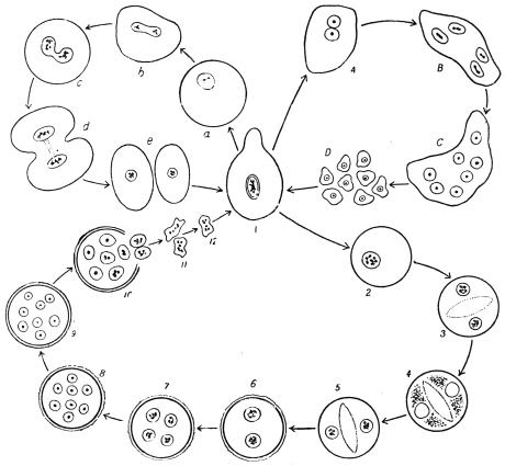
Fig. 3.—Entamœba coli: life-cycle, a-e, stages in binary fission; A-D, schizogony, with
formation of eight merozoites; 2–10, cyst formation or sporogony, with formation of eight
nucleate cysts. (After Castellani and Chalmers)
The nucleus is vesicular, and is spherical when inactive, measuring
5 µ to 7 µ, with a thick nuclear membrane. In the centre of the
nucleus is a chromatinic body or karyosome or sometimes several
small nuclear bodies formed of plastin and chromatin; the remaining
chromatin is arranged on the achromatic network in the form of fine
granules, especially thickly deposited on the nuclear membrane.
Entamœba coli lives as a commensal in the upper portion of the
large intestine, where the fæces still possess a pulpy consistency.
With their concentration and change in reaction lower in the
bowel, the parasites either die or else if they are at a suitable stage of
development form resistant cysts. These cysts (fig. 2) can be found
in great abundance in normal fæces, as Grassi first observed. Slight
laxantia or intestinal diseases of any kind producing increased
peristalsis, however, show amœbæ even in the unencysted condition,
provided that the person harbours intestinal amœbæ generally. The
intensity of infection varies according to the locality; thus Schaudinn
found that 50 per cent. of the persons examined were infected with
harmless amœbæ in East Prussia, 20 per cent. in Berlin and about 66
per cent. on the Austrian littoral.
The life-history (fig. 3) of the parasite exhibits two phases:
(a) asexual multiplication in the intestine, either by binary fission or
by schizogony with formation of eight merozoites, and (b) sporogony
leading to the production of eight-nucleate cysts. Infection results
from ingestion of cysts. Only cysts with eight nuclei are infective.
The diameter of such cysts is about 15 µ to 20 µ.
There are varying accounts of the details of the life-cycle of Entamœba coli in its
different stages. Thus, regarding schizogony or multiple fission it was formerly
stated that the nucleus of the parent amœba divided into eight portions, which after
dissolution of the nuclear membrane, passed outwards into the cytoplasm, which
segregated around each. Eight merozoites were thus produced. More recently the
process of schizogony has been considered to consist in the repeated division of the
nucleus into two, four, and finally eight nuclei (fig. 3, A-D), and the formation of
eight merozoites or amœbulæ.
The process of encystment is initiated by the extrusion of all liquid and foreign
bodies from the protoplasm, which assumes a spherical form (fig. 4, A). The rounded
uninucleate amœba then secretes a soft gelatinous coat, which finally differentiates
into a double contoured cyst wall in older cysts. According to Casagrandi and
Barbagallo, the size of the cyst varies from 8 µ to 30 µ, and averages about 15 µ.
According to Schaudinn (1903) the cytological changes during cyst formation are as
follows. The nucleus of a rounded uninucleate form divides into two (fig. 4, B).
Each of these nuclei fragments into chromidia (fig. 4, C), some of which are absorbed,
while others reunite so that the cell becomes binucleate again. Each of these
nuclei, by a twice repeated division, produces three nuclei (fig. 4, D), the smaller
two of which degenerate and were regarded as reduction nuclei. There is a clear
zone or vacuole in the middle of the cyst during these maturation processes, dividing
the cyst into two halves. After the nuclear reduction the clear space disappears,
and each nucleus (termed by some a gamete nucleus) divides into two pronuclei
(fig. 4, E). The pronuclei of the pairs were said by Schaudinn to differ slightly.
Copulation occurs between pairs of unlike pronuclei, and is an example of autogamy
(fig. 4, F). When complete, each of the fusion nuclei (synkarya) divides twice, giving
rise first to four and finally to eight nuclei. Eight amœbulæ are thus formed within
the cyst.
According to Hartmann and Whitmore (1911)13, however, autogamy does not
occur within the cysts of E. coli. They consider that eight small amœbulæ are
formed (fig. 3, 2-10) which escape from the cyst and then conjugate in pairs
(fig. 3, 10-12), afterwards growing into a new generation of trophozoites.
Only some 10 to 20 per cent. of the cysts evacuated with the fæces undergo the
full course of development, the majority perish previously. In old dry fæces, only
cysts with eight nuclei are found, and it is these alone that cause the infection.
Entamœba williamsi, E. bütschlii, E. hartmanni and E. poleki (Prowazek) are
probably only varieties of E. coli.
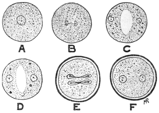
Fig. 4.—So-called autogamy of Entamœba coli. A, rounded amœba; B, nucleus dividing;
C, the two daughter-nuclei giving off chromidia; D, each nucleus has formed two reduction
nuclei; E, cyst membrane formed, and gamete nuclei are dividing; F, cyst with two synkarya.
The principal feature distinguishing Entamœba coli from E.
histolytica is the formation of eight-nucleate cysts by the former as
contrasted with the tetra-nucleate cysts of the latter. The cyst-wall of
E. coli is thicker than that of E. histolytica (tetragena). Further, E. coli
does not usually ingest red blood corpuscles, nor are “chromidial
blocks” present inside its cyst (see p. 40).
According to Chatton and Lalung-Bonnaire14 (1912) the entamœbæ
of vertebrates should be placed in a separate genus Löschia, as they
differ in their life-history from E. blattæ, the type species of Entamœba.
Leidy (1879), however, named the genus Endamœba, but further
researches are necessary on biological variation among these
organisms.
Entamœba histolytica, Schaudinn, 1903.
Syn.: Amœba coli, autt. p. p. Amœba dysenteriæ, autt. p. p.
The average size of the amœboid trophozoite is 25 µ to 30 µ.
In fæces diluted with salt solution the amœbæ swell to 40 µ and more.
There is sometimes separation of the body substance into a strongly
refractile vitreous ectoplasm and a corneous endoplasm, pronounced
even in repose, although the former is not equally thick at all parts of
the periphery. In the endoplasm generally there are numerous foreign
bodies (bacteria, epithelial cells, colourless and red blood corpuscles
(fig. 6), and occasionally living flagellates of the intestine). The
nucleus is 4 µ to 6 µ in diameter, and may be difficult to recognize
because it is sometimes weakly refractile and poor in chromatin. Its
shape is slightly variable; it is usually excentric, sometimes wholly
peripheral at the limit of the two parts of the body. Vacuoles are not
present in quite fresh specimens, but appear later. In the study of
E. histolytica, the morphological characters of the trophozoite or
vegetative stage of the organism formerly separated as E. tetragena
(figs. 5, 6, 8a) must be considered (see p. 38).
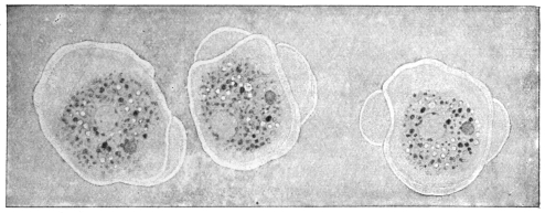
Fig. 5.—Entamœba histolytica (tetragena form), showing three successive changes of form
due to movement. × 1100. (After Hartmann.)
The history of the development of these species, which give rise to amœbic
enteritis as distinguished from bacillary dysentery, was formerly not so well known
as that of E. coli. Upon being introduced into cats (per anum) dysenteric amœbæ
provoke symptoms similar to those in man. In the
latter, besides metastatic liver abscesses, abscesses
of the lungs, and, according to Kartulis, cerebral
abscesses are occasionally produced. Marchoux
(1899) states that when the disease has lasted for
some time liver abscesses are produced in cats also.
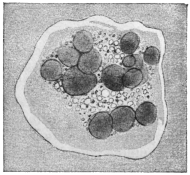
Fig. 6.—Entamœba histolytica
which has ingested many red blood
corpuscles. × 1100. (After Hartmann.)
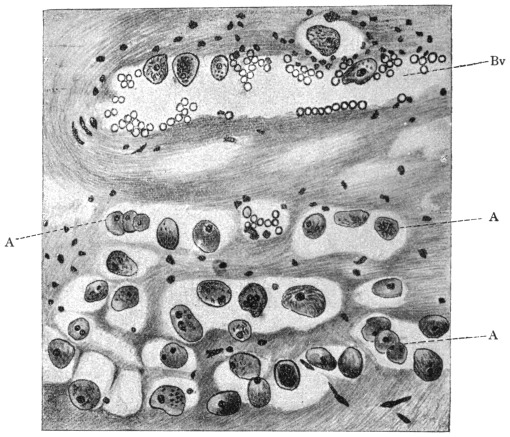
Fig. 7.—Section through wall of large intestine (of a man) close under an ulcer caused by
Entamœba histolytica. A, amœbæ that have penetrated partly in blood-vessels (Bv), partly
in tissue of submucosa to the muscularis. Magnified. (After Harris.)
In the large intestine of infected cats the
amœbæ creep over the epithelium, and here and
there they force the epithelial cells apart, as well
as removing them or pushing them in front of
them; the amœbæ thus insert themselves into the
narrowest fissures. They penetrate also into the
glands through the epithelium, and thence into
the connective tissue of the mucosa. Intestinal
and glandular epithelia perish under the influence
of these parasites: the cells are pushed aside,
fall to pieces or are absorbed by the amœbæ. In the connective tissue of the
mucosa the amœbæ migrate further, and often accumulate above the muscles.
Finally they rupture this and force their way into the submucosa. In cats,
apparently, the penetration is not so great as in men, according to Kruse and
Pasquale. During their migration the parasites also gain access to the lymph-follicles
of the wall of the intestine, which become swollen and commence
to suppurate; follicular abscesses arise and after their rupture follicular ulcers.
The diseased patches in the mucosa are markedly hyperæmic and numerous hæmorrhages
are set up. Roos and Harris state that the amœbæ also penetrate into
the blood-vessels (fig. 7) and this explains the occurrence of metastatic abscesses.15
The whole submucosa is severely swollen at the diseased spot and undergoes small-celled
infiltration in the neighbourhood of the colonies of amœbæ. From these
findings Jürgens (1902) draws the conclusion16 which is followed here, that the amœbæ
are causative agents of the enteritis of cats, which disease is well defined, both
pathologically and anatomically. Subsequent researches confirm the experience of
earlier authors; great precautions were taken to exclude errors, hence, as with
Gross and Harris, no exception can be taken to their results. The inoculation
material was derived from soldiers who suffered from amœbic enteritis in China
and who were admitted into the garrison hospital at Berlin. In order to be
independent of the patients themselves, transmission experiments from cat to cat
were performed, after the first experiments on cats yielded positive results. This
was also effected by rectal feeding as employed by earlier workers. Such appeared
necessary in order to prevent the evacuation of the inoculation material per anum,
as well as to avoid the employment of morphia and ether narcosis. Forty-six
cats were used for the experiments. Ten cats received tested stools containing
motile amœbæ from soldiers suffering from amœbic enteritis contracted in China.
Sixteen other cats received stools from cats infected by inoculation. All the animals
sickened and suffered from the disease. Five cats received dejecta from human
amœbic enteritis in which, however, no motile amœbæ were present. Thirteen cats
received stools from soldiers who suffered from bacillary dysentery. None of the
latter cats took the complaint and none showed changes in the large intestine upon
sectioning. The injection of various bacteria, obtained from a stool of amœbic
enteritis pathogenic to cats, remained without result in both the cats employed for
this experiment. Lastly, two cats, which had been kept with those artificially
infected, were taken ill spontaneously and suffered from the disease. In the opinion
of Harris, who ascertained the harmless nature of bacteria derived from the
intestinal flora containing dysenteric amœbæ, young dogs are capable of being
infected.
Within the large intestine an active increase of Entamœba histolytica must occur.
Nevertheless, Jürgens did not definitely find changes that might be interpreted in this
sense. Schaudinn (1903) observed division and gemmation in vivo. Both processes,
in which the nucleus divides by amitosis, can only be distinguished by the fact that
the daughter individuals are similar in binary fission but dissimilar in gemmation,
whether they make their appearance singly or in greater numbers. Schizogony,
resulting in the formation of eight individuals, which is so characteristic for Entamœba
coli, was not observed. (But schizogony, into four merozoites, is now known
to occur. Gemmation processes are apparently degenerative.)
Resistant stages, which serve for transmission to other hosts, are according to
Schaudinn17 first formed when the diseased portions commence to heal, or more
accurately, the recovery commences when the vegetative increase of the amœbæ in the
intestine discontinues. The so-called spores of E. histolytica were distinguished very
definitely from those of E. coli; they were said to consist of spheres of only 3 to
7 µ in diameter, which were surrounded by a double membrane, at first colourless, but
becoming a light brownish yellow colour after a few hours, and possessing a protoplasmic
content containing chromidia. They were said to arise by fragments of chromatin
passing outwards from the nucleus of the amœba into the surrounding cytoplasm
(fig. 9, a) and undergoing so marked an increase that finally the whole cytoplasm
became filled with chromidia. The remainder of the nucleus underwent degeneration
and became extruded. On the surface of the cytoplasm there then arose small
protuberances containing chromidia. These processes had been observed in the
living organisms. They gradually divided and separated from membranes which later
became yellow. The remainder of the amœba perished. Craig18 had also seen phases
of this process of development. It must be remarked that, according to recent
researches, these processes of exogenous sporulation are degenerative in character (see
p. 41). The small spores may be fungi. The “sporulation” processes are only mentioned
here as a warning. They are now only of historic interest. By means of an
experiment made on a cat, Schaudinn ascertained that ingestion of permanent cysts,
which resist desiccation, is the cause of the infection. The animal took food containing
dry fæces with amœba cysts; these fæces came from a patient suffering from amœbic
enteritis in China. On the evening of the third day the cat evacuated blood-stained
mucous fæces which contained large numbers of typical Entamœba histolytica. On
the fourth day after the infection the animal experimented upon died, and the large
intestine showed the changes previously stated.
E. histolytica also is found in the large intestine. This was originally shown
to be the case by Kartulis, and the fact has recently been confirmed from many
quarters. It is also present in the metastatic abscesses of which it is the cause
(cf. among other authors, Rogers, Brit. Med. Journ., 1902, ii, No. 2,177, p. 844; and
1903, i, No. 2,214, p. 1315).
It should lastly be pointed out in this connection that mixed infections also take
place. For instance, in addition to E. histolytica, E. coli, and, under certain circumstances,
flagellates may be found together. In the same way E. coli may come
under observation even in bacillary dysentery. On the other hand, Schaudinn
stated that in cases of dysentery endemic in Istria, Entamœba coli, if it had hitherto
been present, disappeared, to return again after recovery from the illness.
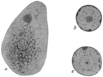
Fig. 8.—Entamœba histolytica. a, trophozoite (tetragena type) containing red blood
corpuscles, × 1,300; b and c, two isolated nuclei showing different appearances of karyosome,
centriole and nuclear membrane, × 2,600. (After Hartmann.)
(Entamœba tetragena, Viereck, 1907.)
This amœba must now be considered to be a part of the lifecycle
of Entamœba histolytica, in fact a very important part of that
cycle, especially in its tetranucleate cystic stages.
This organism, the so-called Entamœba tetragena, may occur in
the human intestine in cases of amœbic dysentery, especially in
mild or chronic cases. It was discovered by Viereck in 1907 in
patients suffering from dysentery contracted in Africa. Soon afterwards
an independent description was published by Hartmann, who
called the amœba E. africana. It was also studied by Bensen and
Werner. Recently (1912–13) much work has been published on
this amœba by Darling and others; in this way its relationship to
Schaudinn’s E. histolytica has been made known.
In general morphology it somewhat resembles Entamœba coli, and
its discoverer at first mistook it for a variety of that species. According
to Hartmann, a distinct ectoplasm is only clearly visible when
a pseudopodium is protruded (fig. 5). The granular endoplasm may
contain ingested red blood corpuscles (fig. 6). The large, round
nucleus is visible in the fresh state (fig. 8, a). So-called chromidial
masses (? crystalloidal substances) may occur in the cytoplasm.
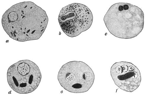
Fig. 9.—Entamœba histolytica (tetragena form). a, emission of chromatin from nucleus;
b, nuclear division; c, degenerating form with two nuclei; d, e, f, cysts containing one, two
and four nuclei respectively, and showing chromidial blocks. × 2,000. (After Hartmann.)
Some investigators, as Hartmann,19 lay stress on the internal
structure of the nucleus (fig. 8, b, c), best seen in preparations fixed wet
and stained with iron-hæmatoxylin. The nucleus is limited by a well-marked
nuclear membrane, on the inside of which granules or nodules
of chromatin may occur. There is a karyosome, which, in successfully
stained specimens, shows, at times, a central dot called a
centriole. (The nucleus of Entamœba coli does not contain such a
centriole.) However, the structure of the nucleus varies at different
periods during the life-cycle.
The diameter of the trophozoites or vegetative forms (fig. 8, a) is
variously given as from 20 µ to 40 µ. Multiplication proceeds by
binary fission and also by schizogony into four merozoites.20
Reproduction takes place by endogenous encystment (fig. 9, d-f),
which is preceded by nuclear division into two, reduction and then
autogamy. The interpretation of the latter phenomenon as autogamy
is disputed by some authors. The round cysts, which may measure
12 µ to 15 µ in diameter, contain four nuclei, together with darkly
staining masses of various shapes, the so-called “chromidial blocks”
(fig. 9, f). The cyst-wall of E. histolytica (tetragena) is thinner than
that of E. coli, and the diameter of the cyst is rather less. E. histolytica
has not yet been cultivated.
Infection in man occurs by way of the mouth by the ingestion
of cysts. A patient showing acute symptoms of dysentery is not
usually infective, for he is merely harbouring the large trophozoites,
which, by experiment, have been shown not to be infective to animals
(kittens) when administered by the mouth. The stools of recovered
patients may still contain cysts, and they may thus act as cyst-carriers
or reservoirs of disease by infecting water and soil. The stools of such
cyst-carriers are often solid, and so cysts of E. histolytica (tetragena)
are easily overlooked. Mathis (1913)21 points out that healthy carriers
of E. histolytica may be found; 8 per cent. of the natives of Tonkin
examined by him were healthy carriers of cysts.
In return cases, or prolonged untreated cases of entamœbic
dysentery, a generation of smaller trophozoites is associated with, or
replaces the larger ones. In stools they are frequently refractile and
consequently stain slowly intra vitam. These trophozoites are the
“smaller, senile, or pre-cyst generation” of Darling. This pre-cyst
generation is characterized by the presence of blocks of crystalloidal
substance in the cytoplasm, and by the possession of a prominent,
densely stainable karyosome. Darling believes this generation to
be the same as that described by Elmassian as Entamœba minuta.22
Walker,23 Darling,24 Wenyon25 and others believe that Entamœba
histolytica, which was only seen by Schaudinn in a single case, that of
a Chinaman, is really E. tetragena. Darling states that if the published
illustrations of E. histolytica and of E. tetragena are collected from the
literature and compared, it will be seen that the writers have been
calling E. histolytica the large trophozoites seen in dysenteric stools.
These large trophozoites frequently display no karyosome, but they
can be demonstrated as E. tetragena by animal inoculation, or by the
history of the case. On the other hand, the illustrations of E. tetragena
show that the authors have been dealing with the small generation or
reduced forms (“E. minuta”), which are the direct descendants of the
large trophozoites. If kittens are inoculated rectally with dysenteric
material containing large trophozoites, the strain may be carried in
successive kittens for four to six transfers. If, on the other hand,
kittens are inoculated rectally with small trophozoites of the pre-cyst
generation, the transmission cannot be carried through more than one
or two kittens. Wenyon has succeeded in maintaining E. tetragena in
kittens for several generations.
In some of the preparations from the last remove, pathological
forms of the trophozoites may be seen. These show abnormal forms
of budding, especially peripherally, such as have been described by
Schaudinn and by Craig as characteristic of E. histolytica. Schaudinn’s
small peripheral, exogenous buds and cysts are thus explained. Craig
has latterly changed his views.
Further, Darling states that tetragena cysts fed by the mouth to
kittens produce bowel lesions in which trophozoites having the characters
of E. tetragena, E. histolytica and E. nipponica (Koidzumi) occur.
In view of the work of recent observers, the peculiar exogenous
encystment which Schaudinn made characteristic of Entamœba
histolytica has been shown to be due to degenerative changes in senile
races of the amœba. E. histolytica and E. tetragena are one and the
same species, and its trophozoite is subject to variation. According
to some observers the histolytica type of nucleus—described by
Schaudinn as being poor in chromatin and not easily seen in the
fresh state—occurs frequently in patients with severe symptoms of
dysentery; on the other hand, the tetragena type of nucleus—round
and easily seen in the fresh state—may occur in cases presenting slight
dysenteric symptoms. Intermediate types of nuclei are seen. The
name of this species, the principal pathogenic amœba of man, must
then be E. histolytica by priority. The cystic stages of E. histolytica are
those first recorded by Viereck and formerly described as E. tetragena.
The geographical distribution of E. histolytica is wide.
Noc’s Entamœba (1909).
A species of Entamœba was cultivated by Noc26 in 1909 from cysts
derived from liver abscesses, from dysenteric stools and from the
water supply of Saigon, Cochin China. He cultivated it in association
with bacteria. It is pathogenic. It has been considered allied
to E. histolytica, and shows internal segmentation or schizogony. It
exhibits polymorphism. This amœba has been found by Greig and
Wells (1911) in cases of dysentery in India. It is an important
organism and requires further investigation.
Certain other Entamœbæ27 have been described at various times from
the intestinal tract of man. Probably most, if not all, of these are not
good species and in some cases much more information is needed.
Entamœba tropicalis (Lesage, 1908). This parasite is said to be
non-pathogenic, and to occur in the intestine of man in the tropics.
It has a general resemblance to E. coli, but forms small cysts (6 µ to
10 µ in diameter). The nucleus of the cyst is said to break up into a
variable number of daughter nuclei, from three to thirteen having
been noted. Lesage states that it is culturable in symbiosis with
bacteria. It is probably a variety of E. coli, if not a cultural amœba.
Entamœba hominis (Walker, 1908) has a diameter of 6 µ to 15 µ. A
contractile vacuole is present. Encystment is total, and small cysts are
formed. It is culturable. The original strain, now lost, was obtained
from an autopsy in Boston Hospital. This organism is probably
a cultural amœba.
Entamœba phagocytoides (Gauducheau, 1908). This parasite was
discovered in a case of dysentery at Hanoi, Indo-China. The amœba
is small, 2 µ to 15 µ in diameter. It is active. It ingests bacteria and
red blood corpuscles, while peculiar spirilla-like bodies are found in
its cytoplasm. It multiplies by binary and multiple fission. It can
be cultivated. More recently (1912) the author appears to consider
the amœba to be a stage of a Trichomonas, but abandons the view
later (1914). Further researches on this organism are needed.
Entamœba minuta (Elmassian, 1909)28 was found, in association
with E. coli, in a case of chronic dysentery in Paraguay. It resembles
E. tetragena but is smaller, rarely exceeding 14 µ in diameter. Schizogony
occurs, four merozoites being produced. The encystment is
total and endogenous, giving rise to cysts containing four nuclei.
This amœba is considered by Darling and others to be the pre-cyst
trophozoite stage of E. histolytica (tetragena).
Entamœba nipponica (Koidzumi, 1909) was found in the motions
of Japanese suffering from dysentery or from diarrhœa, in the former
case in company with Entamœba histolytica. Its diameter is 15 µ to
30 µ. The endoplasm is phagocytic for red blood corpuscles. The
nucleus is well defined, resembling that of E. coli and of E. tetragena.
Multiplication occurs by binary fission and by schizogony. Encystment
is total, but has not been completely followed. Darling and
others consider that this is an abnormal form of E. histolytica,
while Akashi (1913) doubts if it is an amœba at all, but rather is
to be regarded as shed epithelial cells.
General Remark.—It is now considered by some workers that
true Entamœbæ cannot be cultivated on artificial media. Quite
recently Williams and Calkins (1913)29 have somewhat doubted this
opinion, and state that certain cultural amœbæ, originally obtained
from Musgrave in Manila, exhibit the various morphological variations
associated with true entamœbæ of the human digestive tract.
Entamœba buccalis, Prowazek, 1904.
The size varies from 6 µ to 32 µ. Ectoplasm is always present;
the endoplasm contains numerous food-vacuoles. The nucleus is
vesicular, with a greenish tinted membrane which is poor in
chromatin. The size of the nucleus is from 1·5 µ to 4·5 µ. A contractile
vacuole is not visible. The pseudopodium is broad. It was
discovered in the mouths of persons with dental caries at Rovigno and
also at Trieste, being most easily found in dense masses of leucocytes,
also among leptothrix and spirochæte clusters. It can be easily distinguished
from leucocytes by more intense staining with neutral red.
Multiplication proceeds by fission. Transmission may take place
through the small spherical cysts. This species (fig. 10) has since been
observed in Berlin, and is also occasionally found in carcinoma of
various regions of the oral cavity. (Leyden and Löwenthal, 1905).
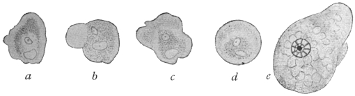
Fig. 10.—Entamœba buccalis, Prow. a-d, the same specimen observed during five minutes.
× 1,000. e, amœba fixed and stained with iron-hæmatoxylin. × 1,500. (After Leyden
and Löwenthal.)
Entamœba buccalis, Prow., is said to be allied to a protozoön which
A. Tietze has found either encysted or free in the lumen of the orifice
of the parotid gland of an infant aged 4 months. The gland had
undergone pathological change, and had therefore been extirpated.
The organisms, which were roundish and three to four times the size
of the normal epithelial cells of the gland, were without a membrane
and possessed a nucleus in which the chromatic substance appeared
to be contained in a karyosome. Bass and John’s30 (Feb. 1915) and
Smith, Middleton and Barrett (1914) state that E. buccalis is the cause
of pyorrhœa alveolaris.
Entamœba undulans, Aldo Castellani, 1905.
Under this name a protozoön is described which A. Castellani found in addition
to Entamœba histolytica and Trichomonas intestinalis in the fæces of an European
planter living in Ceylon, who had suffered from amœbic enteritis and liver abscess.
The shape of the body was roundish or oval, 25 µ to 30 µ in the greatest diameter.
It was without a flagellum, but with an undulating membrane, and capable of protruding
a long pseudopodium from different parts of its body at short intervals. The
nucleus could not always be recognized in life; it was, however, always demonstrable
by staining. One or two contractile vacuoles were present. The protoplasm was
finely granular, showing no differentiation into ecto- and endo-plasm. According to
Braun, in spite of the author declaring himself expressly against the flagellate nature
of the parasite, such a nature may be assumed to be tolerably certain in view of the
description and illustration.
It is now considered that Entamœba undulans is a portion of a flagellate,
namely, Trichomonas.
Entamœba kartulisi, Doflein, 1901.
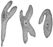
Fig. 11.—Entamœba kartulisi,
Dofl., from the pus of an
abscess in the lower jaw, showing
different stages of movement.
(After Kartulis.)
Doflein gave this name to amœbæ, from 30 µ to 38 µ in diameter,
which Kartulis (1893) found on examining the pus of an abscess in the
right lower jaw of an Arab, aged 43, and in a portion of bone that had been
extracted. The movements of the amœbæ
(fig. 11) were more active than those of
“dysenteric amœbæ.” Their coarsely
granular cytoplasm contained blood and
pus corpuscles, and a nucleus was generally
only recognizable after staining. Vacuoles
were not seen with certainty. Flexner reported
upon a similar case, and Kartulis
published five additional cases. As in these
cases dental caries was present the infection
is likely to have proceeded from the oral
cavity as a result of the carious teeth.
Craig31 (1911) considers that this parasite is
probably identical with Entamœba histolytica.
In the literature the following species have been reported as
occurring in the oral cavity of man:—
Amœba gingivalis, Gros, 1849. [? identical with Entamœba buccalis.]
Amœba buccalis, Sternberg, 1862.
Amœba dentalis, Grassi, 1879.
Far too little, however, is known concerning these to regard them as definite
species, that is, independent organisms; Grassi thinks it even possible there may
have been a confusion in their case with salivary corpuscles. If they really are
amœbæ they are all of them probably identical with Entamœba buccalis.
Genus Paramœba, Schaudinn, 1896.
Schaudinn established the genus Paramœba for a marine rhizopod which multiplied
by division, became encysted at the end of its vegetative life and then
segmented into swarm bodies with two flagella. These multiplied by longitudinal
fission, and finally passed into the condition of Amœbæ. Whether the human parasite
described by C. F. Craig (1906) as
Paramœba hominis.
belonged to this genus was for a time uncertain. It is now placed in a new genus
Craigia, Calkins, 1912, since it possesses only one flagellum.32
In the amœbic stage it is 15 µ to 25 µ in diameter; ecto- and endo-plasm during
rest are indistinguishable. The body substance is granular, with a spherical, sharply
contoured nucleus and an accessory nuclear body. No vacuoles are present, but
occasionally the endoplasm contains red blood corpuscles. The pseudopodia are
hyaline, finger- or lobe-shaped, and are protruded either singly or in twos.
Multiplication is by binary fission and by the formation of spherical cysts (15 µ to
20 µ in diameter) in which occurs successive division of the nuclei, ultimately forming
ten to twelve roundish bodies each of which soon develops a flagellum. The flagellate
stages have similarly a spherical shape and attain a diameter of 10 µ to 15 µ.
They also occasionally contain red blood corpuscles and pass either directly or after
longitudinal division into the amœboid phase.
Craig found these Amœbæ and the flagellate stage belonging to them in six
patients in the military hospital at Manila (Philippine Islands), five of whom were
suffering from simple diarrhœa whilst the sixth exhibited an amœbic enteritis and
contained also Paramœba hominis, with Entamœba histolytica, Schaudinn. In one
of the other cases, Trichomonas intestinalis was present.
B. Amœbæ from other Organs.
Entamœba pulmonalis, Artault, 1898.
Artault33 discovered a few amœboid forms with nucleus and
vacuole in the contents of a lung cavity. In the fresh condition they
were distinguishable from leucocytes by their remarkable capacity of
light refraction. They were also much slower than the latter in
staining with methylene blue or fuchsine. Their movements became
more lively in a strong light. Water and other reagents killed them,
and then, even when stained, they could not be distinguished from
leucocytes. They have also been seen by Brumpt. R. Blanchard found
amœbæ which may belong here in the lungs of sheep. A. pulmonalis
is perhaps the same as Entamœba buccalis. Smith and Weidman34
(1910, 1914) described an entamœba, E. mortinatalium, from the
lungs and other organs of infants in America.
Amœba urogenitalis, Baelz, 1883.
This species was found in masses in the sanguineous urine as well
as in the vagina of a patient in Japan, aged 23. Shortly before the
death of the patient, which was caused by pulmonary tuberculosis,
hæmaturia with severe tenesmus of the bladder had set in. The
amœba, which showed great motility, and had a diameter of about
50 µ when quiescent, exhibited a granular cytoplasm and a vesicular
nucleus. Baelz is of opinion that these parasites were introduced into
the vulva with the water used for washing the parts, and thence had
penetrated into the bladder and vagina. Doflein places the organism
in the genus Entamœba, and it is perhaps identical with E. histolytica.
Similar cases are also reported (1892–3) by other authors: Jürgens, Kartulis, Posner,
and Wijnhoff. Jürgens found small mucous cysts, filled with amœboid bodies, in the
bladder of an old woman suffering from chronic cystitis; they were also found
in the vagina. The amœba observed by Kartulis in the sanguineous urine of
a woman, aged 58, suffering from a tumour of the bladder, measured 12 µ to 20 µ,
and exhibited slow movements by protruding short pseudopodia. The vacuoles and
nucleus became visible only after staining with methylene blue.
Posner’s case related to a man, aged 37, who had hitherto been quite healthy and
had never been out of Berlin. Suddenly, after a rigor, he passed urine tinged with
blood. This contained, besides red and white blood corpuscles and hyaline and
granular casts, large granular bodies (about 50 µ in length and 28 µ in breadth), which
slowly altered their shape, and contained red blood corpuscles in addition to other
foreign matter. These bodies exhibited one or several nuclei and some vacuoles.
From the course of the disease, which extended over a year, and during which
similar attacks recurred, Posner came to the conclusion that the amœbæ which had
originally invaded the bladder had penetrated into the pelvis of the kidney, where
they probably had settled in a cyst, and thence induced the repeated attacks.
Wijnhoff observed four cases of amœburia in Utrecht.
Amœba miurai, Ijima, 1898.
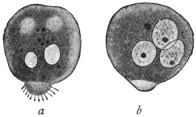
Fig. 12.—Amœba miurai, Ij.
× 500. a, fresh; b, after treatment
with dilute acetic acid.
(After Ijima.)
Under this term the author describes protoplasmic bodies which Miura, in
Tokyo, found in the serous fluid of a woman, aged 26, who had died from pleuritis
and peritonitis endotheliomatosa. Two days before death these same forms had
also appeared in the hæmorrhagic fæces of the
patient. The bodies were usually spherical or
ellipsoidal, and at one pole carried a small protuberance
(fig. 12) beset with filamentous short
“pseudopodia” (really a pseudopodium covered
with cilia). Their size varied between 15 µ and
38 µ. The cytoplasm was finely granular, and
no difference was observable in the ecto- and
endo-plasm, only the villous appendage
was clearer. The cytoplasm contained vacuoles
more or less numerous, none of which was
contractile. After the addition of acetic acid one
to three nuclei could be distinguished, 8 µ to 15 µ in size. Actual movements were
not observed. Taking everything into consideration, the independent nature of these
bodies is, to say the least, doubtful, although it cannot be denied that they possess a
certain similarity to the marine Amœba fluida, Grüber or Greeff, and to a few other
species. (It is likely that cells present in serous exudation were mistaken for amœbæ.)
Appendix.
“Rhizopods in Poliomyelitis acuta.”
In three cases of poliomyelitis acuta which were investigated by Ellermann, the
spinal fluid obtained by puncture of the cord contained bodies, from 10 µ to 15 µ in
size, which had amœboid movements and exhibited variously shaped pseudopodia in
large numbers. After staining, a usually excentric nucleus, about 1·5 µ in size, was
demonstrated in them.
Order. Foraminifera, d’Orbigny.
The order is divided by Max Schultze into Monothalamia and Polythalamia. Only
a few of the former can be considered here.
Sub-Order. Monothalamia. (Testaceous Amœbæ).
These forms occur frequently in fresh water, rarely in sea water.
They possess a shell which is either pseudo-chitinous in character, or
consists of foreign particles, or in a few cases is composed of siliceous
lamellæ. There is usually an orifice for the protrusion of pseudopodia.
The only representative of the order of interest here is:—
Genus. Chlamydophrys, Cienkowski, 1876.
The genus is based on a form which A. Schneider carefully investigated and
considered to be the Difflugia enchelys of Ehrenberg. L. Cienkowski rediscovered
this same form and created for it the genus Chlamydophrys. We agree
with this view, but not with the renaming of the organism (so common at the time).
If the parasite in dung, Chlamydophrys stercorea Cienk. is identical with Difflugia
enchelys of Ehrenberg, the old specific name should be retained.
The genus is characterized by the possession of a hyaline, structureless,
slightly flexible shell which is ovoid or reniform. At the more
pointed pole there is an orifice situated terminally or somewhat laterally,
serving for the emergence of the filiform pseudopodia (fig. 13, a).
The protoplasm does not entirely fill the interior of the shell. An
equatorial zone bearing excretory granules divides the shell internally
into two almost equal portions. The anterior portion is rich in
vacuoles and serves for the reception of nutriment and for digestion.
The posterior part is vitreous, and contains the nucleus. One to three
contractile vacuoles are situated in the equatorial zone.
Chlamydophrys enchelys, Ehrbg.
Syn.: Chlamydophrys stercorea, L. Cienkowski.
This species (fig. 13) is found in the fæces of various animals (cattle,
rabbits, mice, and lizards), and also in quite fresh human fæces.
According to Schaudinn, the parasite occurs so frequently in the
human fæces that it must be considered of wide distribution. The
species must traverse the intestine of man and animals during one
stage of its life cycle, as Schaudinn showed by experiments on himself
and on mice. He infected himself with cysts (fig. 14) by swallowing
them, and evacuated the first Chlamydophrys as early as the following
day. After the evacuation of numerous specimens on one of the
following days the infection ceased.
The nucleus of a living specimen is surrounded by a hyaline,
strongly refractile chromidial mass, arranged in the form of a ring.
Chromatin stains colour it darkly.
Asexual multiplication (fig. 13, b), which takes place in fæces, follows
a similar course to that of allied forms (e.g., Euglypha, Centropyxis). It
commences by the cytoplasm issuing from the orifice of the shell and
assuming the shape characteristic of the mother organism, but in
a reverse position. The nucleus then divides by mitosis, when the
daughter nuclei move apart from one another. The chromidial ring
also divides into two portions by a process of dumb-bell like constriction.
The one daughter nucleus remains in the mother organism,
the other moves towards the daughter individual, which then separates
from the parent.
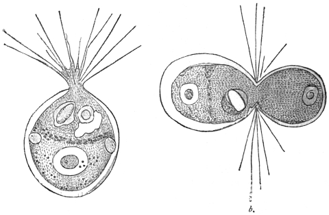
Fig. 13.—Chlamydophrys enchelys. a, free, motile form, showing nucleus, equatorial
granules, vacuoles and pseudopodia; b, dividing organism. × 760. (After Cienkowski.)
In this species plasmogamic union of two or more individuals (up to twenty) is
frequently observed. Such colonies may similarly divide, and in this way monstrosities
frequently arise. When drying of the fæces, or deficiency of food occurs,
encystment takes place apparently spontaneously. The whole body, as stated by
Cienkowski, issues from the shell, assumes a spherical shape (probably with discharge
of water) and becomes surrounded with a thick membrane (fig. 14). After the addition
of water and the escape of the encysted Chlamydophrys, a new shell must be formed.
Schaudinn, who has not given a more detailed description of the process of encystment
in this species, but refers to Cienkowski and to similar observations made on
Centropyxis, states of the latter that the encystment takes place within the shell.
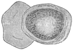
Fig. 14.—Chlamydophrys
enchelys, encysted;
on the left the old capsule.
× 760. (After Cienkowski.)
The sexual multiplication is accompanied by shedding of all the
foreign bodies and of the degenerating nucleus. The protoplasm,
now contracting into a sphere, remains behind in the shell with the
chromidial mass. From the latter several new nuclei arise (sexual
nuclei) often eight in number. The cytoplasmic sphere then segregates
into as many spherical portions as there are nuclei present.
When they have assumed an oval form, two flagella develop at one
pole and the flagellispores swarm out of the shell.35 The biflagellate
swarm-spores, or gametes, copulate in pairs and apparently the individuals
of the pairs of gametes arise from different mother organisms.
The zygote secretes a thick covering which soon
becomes brown and rough. These zygote cysts
or resistant spores must now pass from the intestine
of an animal in order to complete their
development. The escape of the cyst contents
does not always take place in the intestine;
often it does not occur until after defæcation.
These shell-less individuals (amœbulæ) soon
become invested with a shell. But in the
alkaline intestinal contents, shell formation may
proceed even while the organism is in the intestine,
and multiplication may take place.
Schaudinn’s further communication was of special interest; it was
to the effect that Chlamydophrys was related to
Leydenia gemmipara, Schaudinn, 1896.
In the fluid removed by puncture from two patients suffering
from ascites in the first medical clinic in Berlin, cellular bodies
with spontaneous movement were found, which Leyden and
Schaudinn regard as distinct organisms. They remained alive
without the use of the warm stage for four or five hours, the
external temperature being 24° to 25° C. In a quiescent condition
they were of a spherical or irregular polygonal form. Their surface was
rarely smooth, being beset with protuberances and excrescences (fig. 15).
The substance of the body was thickly permeated with light refractile
granules with a yellowish shimmer. The hyaline ectoplasm was
rarely seen distinctly. All sizes from 3 µ to 36 µ in diameter were
observed. The movements were rather sluggish, the ectoplasm in
the meantime appearing in the form of one or several lamellæ, in
which also strings of the granular endoplasm occurred, and frequently
protruded over the border of the hyaline pseudopodia. The tendency
for the joining of several individuals by means of their pseudopodia
was so marked that associations ensued similar to those known in
free-living Rhizopoda.
The cytoplasm enclosed blood corpuscles as well as numerous
vacuoles, one of which pulsated slowly about every quarter of an
hour. A vesicular nucleus the diameter of which was about equal
to one-fifth of the body was present.
Multiplication took place by means of division and budding (fig. 15, c),
after previous direct division of the nucleus. The buds were supposed
to divide repeatedly soon after their appearance, thus giving rise to
minute forms of 3 µ.
There was a suspicion in both cases that the ascites was associated
with malignant neoplasms in the abdomen, and autopsy confirmed
this view in one case.
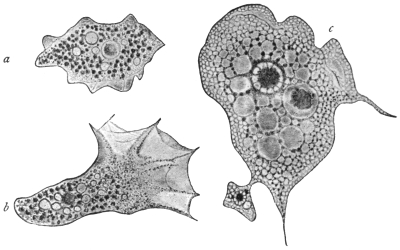
Fig. 15.—Leydenia gemmipara, Schaud. a, in a quiescent condition, × 1000; b, in the act of
moving, × 1000; c, from a fixed preparation, showing a bud, × 1500.
The parasite, which has seldom been observed, has been variously
interpreted; for example, it has been regarded merely as altered tissue
cells. It is now known, from Schaudinn’s researches, that Leydenia
gemmipara is connected with abnormal conditions of Chlamydophrys,
occasionally occurring as a commensal in the ascitic fluid. The form
is produced when pathological conditions of the large intestine create
an alkaline reaction of its whole contents. The formation of shells
then often ceases, and these naked Chlamydophrys are enabled to
multiply atypically by division and gemmation. Such stages, which
are no longer capable of a normal development, are the Leydenia, as
Schaudinn has demonstrated.
Sub-Class. FLAGELLATA, Cohn emend. Bütschli.
During the motile part of their life the Flagellata possess one or more flagella
which serve for locomotion, and in many cases also for the capture of food. A few
groups (Euglenoidinæ, Choanoflagellata) have only one flagellum, others two or
several of about equal length (Isomastigoda), or of various lengths (Monadina,
Heteromastigoda, Dinoflagellata). The long flagellum is the principal one; the
smaller ones on the same organism are accessory flagella. The flagella directed
backwards, which occur in the Heteromastigoda and are used for clinging, are
termed trailing flagella or tractella. At the base of the flagellum, which is almost
always at the anterior end, a Choanoflagellate possesses a cytoplasmic funnel-shaped
neck or collar. In the parasitic forms an undulating membrane is often present.
The body of the Flagellata is usually small, generally elongate and of unchangeable
form. It is frequently covered by a distinct cuticle, and, in certain groups,
by a hard envelope, or it may be more or less loosely enveloped by a gelatinous or
membranous covering. An ectoplasmic layer is thin and not always obvious. The
granular cytoplasm contains a varying number of vacuoles, one of which may be
contractile, and is generally situated near the area from which the flagella arise,
that is, at the anterior extremity. The cytoplasm, moreover, contains the nucleus,
which is nearly always single; and in many species there are also yellow, brown,
or green chromatophores of various shapes, such as occur in plants. Some
species feed after the manner of green plants (holophytic), or of plants devoid of
chlorophyll (saprophytic); others, again, ingest solid food, and for this purpose
usually possess a cytostome; the latter, however, in a few forms is not used for its
original function, but is connected with the contractile vacuole. Many parasitic
forms feed by endosmosis. A few species possess eye-spots with or without light-refracting
bodies.
Variation in the form of the nuclear apparatus occurs. One nucleus only, which
may be compact or vesicular, is known in many species. This nucleus is situated
either centrally or sometimes near the flagellar end of the body, but its position is
subject to variation. The flagella may arise near the nucleus. Other structures,
such as an axial filament and a rhizoplast, may be present. Some flagellates are
binucleate, the two nuclei—which often differ in size and shape—being separated
from each other. One of these nuclei is the principal, vegetative or trophic nucleus;
the other is an accessory nucleus, frequently termed the blepharoplast, flagellar
or kinetic nucleus. One or more small basal granules are often present at or very
near the origin of the flagella.
Multiplication is by fission, usually longitudinal, which may occur in either the
free or encysted forms. Division is initiated by that of the nucleus or nuclei
(especially the kinetic nucleus). The basal granule divides also. Collars and
chromatophores, if present, likewise separate into two. Variation in the method of
doubling the original number of flagella occurs. In most organisms, especially
uniflagellate forms, the flagellum splits lengthwise, after division of the basal granule,
blepharoplast and nucleus. The daughter flagella may be of the same or different
lengths and thicknesses. Other flagellates at division are said to produce new
flagella in the neighbourhood of the original ones. The daughter organisms in such
cases are provided with one or more parental flagella in addition to newly formed
ones. It has been stated that in certain cases the parent flagellate retains all its
flagella, while new ones arise ab initio in the cytoplasm of the daughter forms.
Multiplication by longitudinal fission may be interrupted sooner or later by the
production of gametes, which form zygotes, from which new generations of individuals
arise. In many flagellates gamete formation and sporogony are unknown,
and asexual reproduction by fission alone prevails.
Incomplete division results in the formation of colonies of individuals. These
colonies must not be confused with the aggregation rosettes of flagellates found
among the parasitic Mastigophora. The individuals of aggregation rosettes are
capable of immediate separation from the rosette at will.
A number of parasitic Flagellata produce non-flagellate stages which are very
resistant to external conditions, the assumption of which forms serves to protect the
organisms during their transference from one host to another. Such non-flagellate
forms possess one or more nuclei, are usually of an oval or rounded contour, and
are capable of developing into the full flagellate on the return of more favourable
conditions. These forms are often known as the post-flagellate stage of the
organism. When ingested by a new host, the post-flagellate coat becomes more
flexible, and the phase of the organism which now recommences growth is known as
the pre-flagellate stage; it gradually develops into the typical flagellate organism.
Many Flagellata live free in fresh and salt water. They prefer stagnant water,
rich in organic products of decomposition, such as puddles, swamps and pools.
Those forms developing shells and colonies are, as a rule, adherent. A number of
species are parasitic in man and animals, living mostly within the intestine or in the
blood.
It is usual to classify the Flagellata in four orders: Euflagellata, Dinoflagellata,
Choanoflagellata, and Cystoflagellata, of which only the Euflagellata are of interest
to us. This is a group comprising numerous species, for the further classification
of which the number and position of the flagella are utilised.
The Euflagellata observed in man belong to the Protomonadina as well as to the
Polymastigina. The former possess either only one or two similar flagella, or one
principal and one or two accessory flagella. The Polymastigina possess at least
three flagella of equal size, or four to eight of unequal size, inserted at different
points. An undulating membrane may be present in members of both groups.
It must also be pointed out that unicellular organisms with one or several
flagella are not always classified with flagellates, for such forms occur in Rhizopods
as well as temporarily in the lower plants. In addition, the examination of the
flagellates, especially the parasitic species, is very difficult on account of their
diminutive size and great activity; thus it happens that certain forms cannot with
certainty be included in the group because their description is insufficient.
Order. Polymastigina, Blochmann.
The Polymastigina contains flagellates with three to eight flagella.
Some of the Flagellata parasitic in man belong to the Polymastigina,
and to two or three genera that are easily distinguishable.
Genus. Trichomonas, Donné, 1837.
The body is generally pyriform, the anterior part usually rounded, the posterior
part pointed. There are at the anterior extremity three (? four) equally long flagella
that are sometimes matted together. A blepharoplast (kinetic nucleus) and basal
granule are present, together with a supporting structure known as an axial
filament or axostyle. In addition there is an undulating membrane, bordered by a
trailing flagellum, that commences at the anterior extremity and proceeds obliquely
backwards. The nucleus, which is vesicular, is situated near the anterior extremity,
and behind it are one or more vacuoles, none of which seems to be contractile.
These flagellates are parasitic in vertebrate animals, and live chiefly in the intestine.
Trichomonas vaginalis, Donné.
The form of the body is very variable, and is elongate, fusiform
or pear-shaped, also amœboid. The length varies between 15 µ and
25 µ, and the breadth between 7 µ and 12 µ. The posterior extremity
is drawn out to a point and is about half the length of the remainder
of the body. The cuticle is very thin and the body substance finely
granular. At the anterior extremity there are three—some say four36—flagella
of equal length which are frequently united together, at least
at the base, and are easily detached.
There is an undulating membrane (fig. 16)
which runs spirally across the body, arising
from the place of insertion of the flagella, and
terminating at the base of the caudal process.
A cytostome seldom is recognizable in fresh
specimens, but is apparently present. The
nucleus is vesicular, elliptical and situated near
the anterior extremity.37
Multiplication takes place by division
(Marchand). Encysted forms are almost unknown.
Trichomonas vaginalis lives in the vaginal mucus of
women of various ages, not in normal mucus, but in
mucus of acid reaction. It is found in menstruating
females as well as in females who have passed the
menopause. It occurs in pregnant and non-pregnant
women, even in very young girls, provided always that
they have a vaginal catarrh with acid reaction of the
secretion. Should the acid reaction change, as, for
instance, during menstruation, the parasites disappear,
as they do likewise on injection of any alkaline fluid into
the vagina. A low temperature (below +15° C.) is also
fatal to the parasites. These flagellates can pass from
the vagina through the urethra into the bladder, and
produce severe catarrh, and are not easily removed.
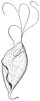
Fig. 16.—Trichomonas
vaginalis, Donné. × 2,000
approx. (After Künstler.)
Four flagella are represented,
but usually only
three are present.
T. vaginalis appeared to be a parasite specific to the female organs
and not transmissible to man. However, several observations have
since been made that confirm the occurrence of this species in the
urethra of the male. The infection apparently takes place through
coitus when changes are present in the urethral mucous membrane.
At any rate, three cases observed point to this circumstance.
Attempts at experimental transmission to rabbits, guinea-pigs and
dogs failed (Blochmann, Dock). So far, the manner in which women
become infected is unknown.
Trichomonas intestinalis, R. Leuckart, 1879 = Trichomonas
hominis, Davaine, 1854.
Some authors believe that a second trichomonad inhabiting man,
Trichomonas intestinalis, R. Lkt., is identical with Trichomonas vaginalis,
Donné. Leuckart’s species was based on the discoveries of Marchand
(1875) and Zunker (1878), who stated that according to all appearances,
and in their opinion, it was the same as Cercomonas intestinalis, Lambl,
1875 (nec 1859), which they found in
the fæces of patients suffering from
intestinal disorders. The organism
is described by them as being pear-shaped
and 10 µ to 15 µ in length
and 3 µ to 4 µ in breadth. The
posterior extremity terminated in a
point (fig. 17).
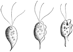
Fig. 17.—Trichomonas intestinalis, Lkt.
(After Grassi.)
A row of twelve or more cilia was said to
commence at the anterior end and extend
over the body. Leuckart stated that this
parasite, placed by the two authors in the
genus Cercomonas, was a Trichomonas, and
that they mistook the undulating membrane for cilia, and overlooked the flagella.
Notwithstanding its striking similarity with T. vaginalis, it was said to be distinguishable
from that species by differences in the undulating membrane. Lambl’s
C. intestinalis38 (of 1875) which corresponds with C. hominis, Davaine39 (1854), is
regarded by Leuckart as a true Cercomonad (characterized by a flagellum and the
absence of an undulating membrane, see p. 61), and is thus generically distinct
from Trichomonas.
The correctness of Leuckart’s judgment in regard to Marchand-Zunker’s flagellate
was demonstrated by Grassi’s researches, accounts of which were published soon
after. In about 100 cases of bowel complaints in North Italy and Sicily, Grassi
found Flagellata in the stools, which he first named Monocercomonas and Cimænomonas,
but later termed Trichomonas. However, in opposition to Leuckart, Grassi
has also classified Davaine’s C. hominis (= C. intestinalis, Lambl, 1875) as
Trichomonas, and most authors have followed his example. Hence arose the use
of the name Trichomonas hominis. It was through Janowski (1896) that the
former view was again taken up. After a review of the literature, the occurrence
of Cercomonads in the intestine of human beings in addition to Trichomonads
was considered by the author to have been proved, and he added a
description of the Trichomonads. According to this, all morphological distinction
between T. vaginalis, Donné, and T. intestinalis, Leuckart, disappeared. On the
other hand, it is worthy of note that the smaller size, the more pear-shaped form, and
the longer flagella differentiate T. intestinalis (= T. hominis) from T. vaginalis.40
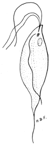
Fig. 18.—Trichomonas
intestinalis
from man,
showing anterior
flagella, cytostomic
depression anteriorly,
undulating
membrane, nuclei,
and axostyle.
×2,500. Original.
The easily deformed pear-shaped body has three free flagella
anteriorly, and an undulating membrane with its flagellar border
terminating in a short free flagellum posteriorly (figs.
17, 18). The undulating membrane may coil itself
spirally round the body. A supporting rod or axostyle
projects as a posterior spine. It appears to
begin near the nucleus and blepharoplast, which are
situated near the more rounded, anterior end of the
body. There may be a chromatoid basal supporting
line along the body for the undulating membrane.
Rows of chromatoid granules are sometimes situated
along one side of the axostyle. A cytostome may
sometimes be seen. In mice, Wenyon (1907) found
these parasites to vary in length from 3 µ to 20 µ.
They occur in the cæcum and intestine of mice,
where their internal structure seems more obvious
than in man. The flagellates divide by longitudinal
fission.
T. intestinalis, R. Leuckart, appears to be capable
of settling in all parts of the human intestine in which
the contents have an alkaline reaction. Trichomonads
have been cited as occurring in the oral cavity by
Steinberg, Zunker, Rappin and Prowazek; in the
œsophagus by Cohnheim, and in the stomach by
Strube, Cohnheim, Zabel, Hensen and Rosenfeld.
The normal situation seems to be the small intestine.
The parasites then appear in the dejecta, especially in various intestinal
diseases the course of which is connected with an increased peristalsis.
They are also found in healthy persons, from whom they are obtained
after the administration of laxatives. They have been regarded by some
workers as commensals, which, however, have the power of accelerating
the onset of intestinal complaints, or at least of adding to them. They
have been found in cases of carcinoma of the stomach, and in other
diseases of that organ in which the acid reaction ceased.
Naturally, whether all the reports relate to the same species of Trichomonas
must remain undecided. Certain authors (Steinberg, Cohnheim, van Emden)
accept several species. Prowazek speaks of a variety of T. intestinalis inhabiting
the oral cavity. This was distinguished by a posterior process exceeding the
length of the body fourfold, and by a somewhat unusual course of the undulating
membrane. The food of this form, which was found in the whitish deposit present,
especially in the cavities of carious teeth, consisted almost exclusively of micrococci.
Schmidt and St. Artault named the Trichomonads found in pathological products
(e.g., gangrene, putrid bronchitis, phthisis) of the lungs of man, as Trichomonas
pulmonalis. Trichomonads have also been found by Wieting in lobular pneumonia
in the lungs of pigs.
It is still uncertain in what way the infection takes place. Experiments in the
transmission of free trichomonads to mammals (per os), in which the same or allied
species occur (guinea-pigs, rats, apes), have been without result. Probably encystment
is necessary. Such conditions are mentioned by May, Künstler, Roos,
Schurmayer, van Emden, Prowazek, Galli-Valerio and Schaudinn. According to
Prowazek, intestinal trichomonads of rats become encysted for conjugation. In the
cyst an accumulation of reserve food material occurs, causing distension. The nuclei
of the conjugants each give off a reduction body and, after fusion, produce the
nuclei for the daughter individuals. According to Schaudinn the intestinal trichomonads
lose their flagella before conjugation, become amœboid and encyst in twos,
the formation of a large agglomeration of reserve substance accompanying this.
Galli-Valerio found double-contoured cysts in the fæces of trichomonad-infected
guinea-pigs, after the fæces had been kept for a month in a damp chamber. When
exposed to heat small flagellates escaped from them. Administration of such material
containing cysts resulted in severe infection with trichomonads, and death of the
experimental guinea-pigs followed. The cyst wall is clearly a protection against the
deleterious acid reaction of the stomach contents. Alexeieff (1911) and Brumpt (1912)
think that the trichomonad cysts of man are really fungi, while other workers also
doubt encystment among trichomonads. Wenyon (1907) states that T. intestinalis in
mice produces spherical contracted forms which escape from the body in the fæces.
Air, water, and under certain circumstances even food may be regarded as
vectors for the trichomonads. The occurrence of the organisms in the oral cavity,
and still more so in the lungs, is in favour of the air being the transmitting agent.
An observation made by Epstein supports the idea of water transmission. Multiplication
of the trichomonads, once they have gained access to the body, is effected by
longitudinal division commencing at the anterior end (Künstler). “Cercomonads”
with several flagella and an undulating membrane, as well as trichomonads, have
been observed by Ross in some cases of cutaneous ulcers.
Mello-Leitao (1913)41 has described flagellate dysentery in children
in Rio de Janeiro. He states that it is due to T. intestinalis
and Lamblia intestinalis either separately or together. Flagellate
dysentery, he thinks, is benign and is the most frequent form of
dysentery in infants. The flagellates are pathogenic to infants under
three years of age. Escomel (1913)42 found 152 cases of dysentery in
Peru due solely to Trichomonas. Such cases are probably widespread.
Genus. Tetramitus, Perty, 1852.
Tetramitus mesnili, Wenyon, 1910.
Syn.: Macrostoma mesnili, Chilomastix mesnili, Fanapapea intestinalis.
The genus Tetramitus differs from Trichomonas in possessing an
undulating membrane inserted in a deep groove or cytostome. There
are three anterior flagella. The pear-shaped organism measures 14 µ
by 7 µ, but smaller examples occur. T. mesnili occurs in the human
intestine, having been described by Wenyon43 (1910) from a man from
the Bahamas in the Seamen’s Hospital, London. Its occurrence
is widespread. Alexeieff considers that Macrostoma and Tetramitus
are synonymous. The parasite is the same as Fanapapea intestinalis,
Prowazek, 1911, from Samoa. Brumpt (1912) found T. mesnili to be
the causal agent of colitis in a Frenchwoman. Nattan-Larrier (1912)
considers it of little pathological importance.
Gäbel44 (1914) described an interesting case of seasonal diarrhœa
acquired in Tunis, in which a new Tetramitid was the causal agent.
The organism was pear-shaped, without an undulating membrane,
and measured 6·5 µ to 8 µ by 5 µ to 6 µ. The cytostome was large,
and there was no skeletal support. Encystment occurred. Gäbel
named the organism Difämus tunensis and considered that it was
pathogenic.
Genus. Lamblia, R. Blanchard, 1888.
Syn., Dimorphus, Grassi, 1879, nec Haller, 1878; Megastoma, Grassi, 1881, nec
de Blainville.
The body is pear-shaped, with a hollow on the under surface anteriorly. It has
four pairs of flagella directed backwards, of which three pairs lie on the borders of
the hollow disc, and the fourth arises from the pointed posterior extremity.
Lamblia intestinalis, Lambl, 1859.
Syn.: Cercomonas intestinalis, Lambl, 1859 (nec 1875); Hexamitus duodenalis,
Davaine, 1875; Dimorphus muris, Grassi, 1879; Megastoma entericum, Grassi,
1881; Megastoma intestinale, R. Blanch., 1886; Lamblia duodenalis, Stiles, 1902.
The organism is pear-shaped and bilaterally symmetrical. It is
from 10 µ to 21 µ long and 5 µ to 12 µ broad and possesses a thin
cuticle. Anteriorly an oblique depression is present, which functions
as a sucking disc (fig. 19, s). Its edges are raised above the general
surface and are contractile. It corresponds to a peristome and acts as
an adhesive organ (fig. 20, b, c). No true cytostome is present. A
double longitudinal ridge, representing axostyles, extends from the
sucking disc to the tapering posterior extremity, which is prolonged
as two flagella from 9 µ to 14 µ long.
Lamblia intestinalis possesses eight flagella (fig. 19). The first pair
of flagella, which cross one another, arise in a groove formed by the
anterior edge of the sucking disc. Two pairs of flagella (lateral and
median) are inserted on the posterior edge of the disc, while the
posterior flagella occur at the tapering posterior extremity of the body.
Basal granules are found at the bases of the flagella. The median
flagella are most active in movement, the anterior and lateral flagella
being less motile, as they are partially united to the body for part of
their length.
The nuclear apparatus is situated in the thin, anterior, hollowed
part of the body. It is at first dumb-bell shaped, the “handle” of the
dumb-bell being formed by a very slight connecting strand, which
eventually separates, so that the flagellate becomes binucleate, and thus
completes the general bisymmetry of the organism.
There is a karyosome in each nucleus. Other bodies of unknown
function, and possibly composed of chromatin, occur on or near
the axostyles.
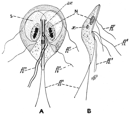
Fig. 19.—Lamblia intestinalis. A, ventral view; B, side view; N, one of the two nuclei;
ax., axostyles; fl1, fl2, fl3, fl4, the four pairs of flagella; s, sucker-like depressed area on the
ventral surface; x, bodies of unknown function. (After Wenyon.)
Division has not been observed in the flagellate stages of the
Lamblia, but it occurs within the cysts. The resistant cysts (fig. 20, e)
are oval and are surrounded by a fairly thick, hyaline cyst wall.
They measure 10 µ to 15 µ by 7 µ to 9 µ, and may be tetranucleate.
According to Schaudinn, the cysts arise from the conjugation of two
individuals, and nuclear rearrangement occurs.
L. intestinalis occurs in its flagellate stage in the duodenum and
jejunum, and rarely as such in the other parts of the intestine.
Normally it is found in the large intestine as cysts, which are voided
with the fæces. The hosts of Lamblia include Mus musculus, M.
rattus, M. decumanus, M. silvestris, Arvicola arvensis and A. amphibius,
the dog and cat, rabbit, sheep and man. Cysts voided with the fæces
of infected animals reach plants or drinking water, and thence are
transferred to man.
The flagellate in these different hosts exhibits some variation in
size and in the problematic chromatic bodies. Bensen has suggested
the species L. intestinalis from man, L. muris from the mouse and
L. cuniculi from the rabbit. It is not certain whether these different
species are necessary, as the variation may be due to differences of
environment.
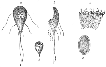
Fig. 20.—Lamblia intestinalis. a, from the surface; b, from the side; c, on intestinal
epithelium cells; d, dead and e, encysted. (After Grassi and Schewiakoff.)
Like Trichomonas, Lamblia can multiply under inflammatory
conditions of the alimentary tract. Thus they are found in cases
of diarrhœa, carcinoma of the stomach, etc. The parasites attach
themselves by their sucking discs to the epithelial cells of the gut
(fig. 20, c), and though their numbers may be very great, their direct
pathological significance is not fully known. Their occurrence in
cases of diarrhœa has been explained as being due to the increased
peristalsis, which has detached the parasites from the epithelium.
Free flagellate forms perish in stools if kept, more especially if the
temperature falls below 0° C. or rises above 40° C. Lamblia has
often been found in dysenteric diseases, especially in the East, and
is said to be the causal agent of certain diarrhœas in India. Mathis
(1914)45 found Lamblia in cases of diarrhœa with dysenteriform stools
in Tonkin. He also discovered healthy carriers of Lamblia cysts.
The parasite under discussion was first observed by Lambl (1859) in the mucous
evacuations of children. He regarded the parasite as a Cercomonad and termed
it Cercomonas intestinalis, which name as a rule is applied to Cercomonas hominis,
Davaine, although Stein had already pointed out the difference between the two species.
Grassi (1879) observed this species first in mice (calling it Dimorphus muris), and
subsequently in human beings in Upper Italy and named it Megastoma entericum.
Bütschli and Blanchard then laid stress on the identity of this species with Lambl’s
C. intestinalis (1859), and consequently called it Megastoma intestinale. Later,
Blanchard drew attention to the circumstance that the generic name Megastoma
chosen by Grassi had already been used four times for various kinds of animals,
and established the genus Lamblia. Accordingly, L. intestinalis is the valid name,
and should be generally adopted.
In Upper Italy the parasite in the encysted condition has also been seen by
Perroncito in man. At the same time, Grassi and Schewiakoff began a new
investigation of specimens from mice and rats. In Germany, L. intestinalis
was found by Moritz and Hölzl, Roos, Schuberg and Salomon. Moritz and Hölzl
confirmed the relative frequency of the species. In Königsberg, Prussia, a student
found encysted Lamblia in his fæces. One case was reported from Finland by
Sievers, another case from Scandinavia by Müller. Frshezjesski and Ucke reported
cases from Russia. Jaksch announced the occurrence of the parasite in Austria;
Piccardi mentioned their presence again in Italy. They were reported from Egypt
by Kruse and Pasquale, and from North America (Baltimore) by Stiles. Noc stated
that 50 per cent. of the population of Tonkin harboured Lamblia. Finally, the
structure of L. intestinalis has been described by Metzner (1901), and by Wenyon46
(1907) in mice.
In all these cases L. intestinalis has been observed in the small
intestine, or in the evacuations of patients with intestinal diseases.
It has also been found in the intestine of healthy subjects. Just as
Trichomonas intestinalis may be found inhabiting the stomach in
diseases of that organ, in which an alkaline reaction is present
(carcinoma), so has L. intestinalis been found to occur under
similar circumstances (Cohnheim, Zabel). However, in Schmidt’s
case, 1 per cent. hydrochloric acid was certainly stated to be present.
Infection takes place by the ingestion of cysts (fig. 20, e), as was
established by Grassi, experimentally on himself. Cereal food-stuffs,
contaminated with Lamblia cysts from vermin of the locality, such as
rats and mice, serve to convey the infection to man. Such cysts may
probably be found in street-dust, etc. Stiles induced infection in
guinea-pigs, and Perroncito in mice and rabbits, by means of cysts
of Lamblia from human beings. Stiles suspected that flies could
transport Lamblia cysts. Mathis (1914) found that L. intestinalis
was not amenable to emetine, at any rate in its cystic stage.
Order. Protomonadina, Blochmann.
The smallness of the Protomonadines and their less superficial
situation than the Polymastigines, may be the cause that so far as
the species occurring in man are concerned, they were formerly less
well known. As regards parasitic species, this group may be divided
as follows, according to the number of flagella and the presence or
absence of an undulating membrane:—
(1) Cercomonadidæ, with one flagellum at the anterior extremity,
without an undulating membrane.
(2) Bodonidæ, with two flagella, without an undulating membrane,
except in Trypanoplasma.
(3) Trypanosomidæ, with one flagellum, and an undulating membrane
along the length of the body in some genera.
Family. Cercomonadidæ, Kent emend. Bütschli.
Small uniflagellate forms, without cytostome.
Genus. Cercomonas, Dujardin emend. Bütschli.
Oval or rounded organisms, with the aflagellar end often drawn
out into a tail-like process.
Cercomonas hominis, Davaine, 1854.
Davaine found flagellates in the dejecta of cholera patients. They
had pear-shaped bodies, lengthening to a point posteriorly. Their
length was from 10 µ to 12 µ, and a flagellum about twice as long as
the body projected from one extremity (fig. 21). A nucleus was
hardly recognizable. Occasionally a somewhat long structure (cytostome?)
appeared at the anterior extremity. The animals moved with
remarkable activity. They also attached themselves by means of their
posterior extremities and swung about around the point of attachment.
Davaine found a smaller variety, only about 8 µ long, in the dejecta
of a typhoid patient (fig. 21, b).
Fig. 21.—Cercomonas hominis, Dav. a, larger,
b, smaller variety. Enlarged. (After Davaine.)
Fig. 22.—Cercomonas hominis,
Dav. From an Echinococcus
cyst. (After Lambl.)
The Flagellata observed by Ekeckrantz (1869) in the intestine of man belong to
this form—at least to the larger variety—and Tham (1870) reported fresh cases soon
after. Lambl’s publication of 1875, which was written in Russian, and became known
through Leuckart’s work on parasites, also alludes to apparently typical Cercomonads,
which, however, were discovered, not in the intestine, but in an Echinococcus cyst in
the liver (fig. 22). The elliptical, fusiform, rarely pear-shaped or cylindrical bodies
of the parasites measured 5 µ to 14 µ in length, and were provided with a flagellum
at one end, while the other extremity usually terminated in a long point. An oral
aperture occurred at the base of the flagellum, and there were one or two vacuoles
near the posterior extremity. Longitudinal division was also observed (fig. 22).
As already mentioned, this form, which Lambl termed Cercomonas
intestinalis, differs considerably from the form found by the same
author in 1859, which received the same designation (cf. Lamblia intestinalis,
p. 60), but it corresponds with Cercomonas hominis, Davaine.
The latter, as well as C. intestinalis, Lambl, 1875, is usually classed
with the Trichomonads, but, as has already been remarked (cf.
Trichomonas intestinalis, p. 54), this cannot be considered correct, as
only one flagellum is present.
Cercomonas vaginalis (Castellani and Chalmers, 1909) was found
in the vagina of native women in Ceylon.
Other species of Cercomonas have, at various times, been recorded
from man. However, the parasitic species of the genus Cercomonas
require further investigation.
According to Janowski (1896–7), typical Cercomonads have also been observed in
the intestine of man by Escherich, also by Cahen, Massiutin, Fenoglio, Councilman and
Lafleur, Dock, Kruse and Pasquale, Zunker, Quincke and Roos, and others. However,
it is an open question whether the Flagellata observed by Roos in one of his cases
belonged to Davaine’s species, the size showing some deviation (14 µ to 16 µ). In
his, as in many other cases, doubts have been raised as to whether the flagellates
found in the stools had actually lived in the intestine, or had subsequently appeared
in the fæces: for this a surprisingly short time only is necessary. Salomon also
appears to have observed Cercomonads (Berl. klin. Wochenschr., 1899, No. 46).
As with T. intestinalis so with C. hominis, it appears
that the parasite settles not only in the intestine but also in
the air-passages. This is demonstrated by the statements of
Kannenberg and Streng of the occurrence of Monads and
Cercomonads in the sputum and putrid expectoration in
gangrene of the lungs, which no doubt apply to C. hominis
(cf. also Artault). Possibly also the Flagellata observed in the
pleural exudation by Litten and Roos may be included here;
this is the more probable in Roos’s case as the process ensued
in the pleura after the breaking through of a vomica.
Perroncito and Piccardi have described encysted stages of
Cercomonads.
Fig. 23.—Monas
pyophila, R. Blanch.
(After Grimm.)
Monas pyophila, R. Blanch., 1895.
R. Blanchard thus designates a Flagellate that Grimm
found in the sputum, as well as in the pus of a pulmonary and
hepatic abscess, in the case of a Japanese woman living in
Sapporo. The parasites resemble large spermatozoa (fig. 23).
The body, 30 µ to 60 µ, has the shape of a heart or a myrtle leaf, and is surrounded by a
thick cuticle which is supposed to extend into the interior of the body, dividing it into
three parts. A long appendix at the rounded pole is covered for the greater part of
its length by the cuticle; the extremity, however, is free and resembles a flagellum.
The parasites were very active, frequently changed their shape, and were able to
retract the long appendix within the body, which then assumed a round form.
[This organism requires further investigation.]
Family. Bodonidæ, Bütschli.
Protomonadina which are either free-living or parasitic, with two
dissimilar flagella, while the possession of an undulating membrane
and of a kinetic nucleus or blepharoplast is variable.
There are three genera:—
1. Bodo, Stein, 1878, without a kinetic nucleus and undulating
membrane.
2. Prowazekia, Hartmann and Chagas, 1910, with a kinetic
nucleus and without an undulating membrane.
3. Trypanoplasma, Laveran and Mesnil, 1901, with a kinetic
nucleus and undulating membrane.
Of these genera Prowazekia must be discussed. Bodo does not
occur in man. Species of Trypanoplasma occur in the blood and in
the gut of various fishes, in the seminal receptacle of certain snails,
in the gut and genitalia of a flatworm (Dendrocœlum lacteum) and in
the vagina of a leech. Closely allied to Trypanoplasma is the genus
Trypanophis, parasitic in the cœlenteric cavity of Siphonophores.
Genus. Prowazekia, Hartmann and Chagas, 1910.
The genus was founded for a flagellate parasite, Prowazekia cruzi,
discovered in a culture of human fæces in Brazil. Various other
species have been referred thereto. The genus is separated from
Bodo by the possession of a second nucleus, the so-called kinetonucleus
or blepharoplast. It differs from Trypanoplasma in the absence of an
undulating membrane. It is heteromastigote, that is, it possesses two
dissimilar flagella, one anteriorly directed and the other lateral and
trailing.
The principal species are:
Prowazekia urinaria, Hassall, 1859.
Syn.: Bodo urinarius, Hassall, 1859; Trichomonas irregularis, Salisbury, 1868;
Cystomonas urinaria, Blanchard, 1885; Plagiomonas urinaria, Braun, 1895.
Hassall47 in 1859 first found Bodo-like flagellates in human urine.
He examined fifty samples of urine from patients suffering from
albuminuria and from cholera. The reaction of the urine was alkaline
or sometimes only feebly acid. The flagellates were only seen after
the urine had been standing for several days. Hassall named the
organism Bodo urinarius, and gave a very good description of it with
illustrations. The flagellate, which was round or oval, measured 14 µ
by 8 µ. The organism had “one, usually two, and sometimes three
lashes or cilia.” In 1868 Salisbury described a similar flagellate in
the urine under the name Trichomonas irregularis. Künstler in 1883
described the latter parasite under the name B. urinarius. In 1885
Blanchard, considering Künstler’s organism a different parasite from
Hassall’s, called it Cystomonas urinaria. Braun, in 1895, gave the
name Plagiomonas urinaria. Barrois (1894) considered Künstler’s
and Hassall’s organisms to be identical and not to be true parasites
of man. Sinton,48 in 1912, found the flagellate in the deposit, after
centrifuging, of a 24-hour old specimen of alkaline urine from a
Mexican sailor in the Royal Southern Hospital, Liverpool. Sinton
found a kinetic nucleus or blepharoplast in the organism, and
therefore placed it in the genus Prowazekia.
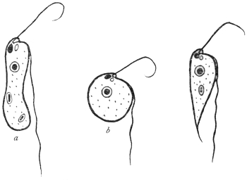
Fig. 24.—Types of Prowazekia urinaria. (a) sausage-shaped; (b) round; (c) carrot-shaped
form. (After Sinton.)
The flagellate stage (fig. 24) of the organism is polymorphic, and may
be either (a) sausage-shaped, 10 µ to 25 µ in length by 2·5 µ to 6 µ in
breadth; (b) round or oval, varying from 4 µ in diameter to oval
forms 15 µ by 10 µ; (c) a carrot-shaped form, of varying size up to
25 µ by 4 µ. The kinetic nucleus is large and pear-shaped. Near it
are basal granules, closely applied to one another, from which the
flagella arise. There is a small cytostome near the roots of the
flagella. There is a well-marked karyosome in the nucleus. The
movement is jerky. The shorter, anterior flagellum may be used in
food-capture. In life, bacteria have been seen to be ingested. Food-vacuoles
tend to accumulate at the posterior (aflagellar) end. A
contractile vacuole may be present, near the base of the cytostome,
and may really be the dilated fundus of the latter. Division occurs by
binary fission. The organism can encyst (fig. 25, a), when the flagella are
lost, and round or oval cysts are found, 5 µ to 7 µ in diameter. After
a time flagella are formed inside the cyst, and the organism emerges
therefrom in its typical flagellate form (fig. 25, b-f).
Sinton’s case is interesting. He obtained the flagellate only twice
from the same patient, a Mexican then in hospital in Liverpool. The
flagellate was not found in the patient’s fæces, nor was it found in
the urine on later occasions when taken aseptically.
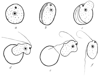
Fig. 25.—Prowazekia urinaria. Flagellate emerging from cyst. (After Sinton.)
In cultures Prowazekia urinaria was always found in association
with bacteria. The cultures died at a temperature of 37° C., but grew
well at 20° C. Various media were useful at the lower temperature,
such as urine, salt agar, nutrient agar, serum agar, blood agar, peptone
salt solution, and diluted blood serum. The flagellate was, then,
considered to be an accidental contamination and not a true parasite
of human urine.
Prowazekia asiatica, Castellani and Chalmers, 1910.
The flagellate was found by the discoverers in the stools of patients
suffering from ankylostomiasis and diarrhœa in Ceylon. It was referred
by them to the genus Bodo, but in 1911 Whitmore49 further studied
it and placed it in the genus Prowazekia. In the stools the flagellate
is found either as a long, slender form measuring 10 µ to 16 µ by 5 µ
to 8 µ or as a rounded form 8 µ to 10 µ in diameter. Its cytoplasm is
alveolar. A rhizoplast connects the basal granules to the kinetic
nucleus. There is multiplication and cyst formation as before. The
organism is easily cultivated, especially in the condensation water of
nutrose agar and maltose agar. The pathogenicity is stated to be nil.
Prowazekia javanensis, Flu, 1912.
Found in agar cultures from the motions of patients at Weltevreden,
Dutch East Indies.50 The flagellates are 12 µ long and 5 µ broad.
The lateral flagellum is stated to be attached to the cell body for a
short distance. Regarding the karyosome in the nucleus, the author
states that the smaller the karyosome the more chromatin is deposited
on the nuclear membrane. Flu mentions that the specific name
javanensis is a temporary one, as in the course of time it may be
shown that there is only one species of Prowazekia.
Prowazekia cruzi, Hartmann and Chagas, 1910.
Found in a culture from human fæces on an agar plate in Brazil,
and considered to be a free-living form.51 The organism is oval or
pear-shaped, 8 µ to 12 µ long and 5 µ to 6 µ broad. In human stools
at Tsingtau, China, a Prowazekia has been found by Martini which
he thinks is the same as Prowazekia cruzi. He considers it to be a
cause of human diarrhœa and intestinal catarrh.
Prowazekia weinbergi, Mathis and Léger, 1910.
This species was found in the fæces of men, both healthy and
diarrhœic, in Tonkin.52 It is pear-shaped, 8 µ to 15 µ long by 4 µ to
6·5 µ broad. The flagella occur at the broad end.
The discoverers think that Prowazekia weinbergi is an intestinal
inhabitant, but non-pathogenic, since it was found to occur in the
fæces even when obtained with aseptic precautions.
Prowazekia parva, Nägler, 1910.
A free-living form found in the slime on the stones at the biological
station at Lunz. Another Prowazekia was found in 1914 in tap-water
in Calcutta.
Family. Trypanosomidæ, Doflein.
The Trypanosomidæ, broadly considered, are uniflagellate
organisms, the flagellum being at the anterior end. The flagellum
arises near the blepharoplast (kinetic nucleus), which lies anterior,
near or posterior to the nucleus.
The following genera will be considered:—
Trypanosoma—with an undulating membrane along the length
of the body.
Crithidia—with a less well-developed undulating membrane
anteriorly (see fig. 49).
Herpetomonas—including the so-called Leptomonas, with anterior
free flagellum only, and no undulating membrane.
Leishmania—non-flagellate forms in mammalian blood, flagellate
herpetomonad stages in culture, probably occurring naturally in Arthropods.
Genus. Trypanosoma, Gruby, 1843.
The members of the genus possess a single flagellum, which
arises posteriorly, adjacent to a blepharoplast or kinetic nucleus. The
flagellum forms a margin to an undulating membrane, and may
or may not be continued beyond the body as a free flagellum.
Many species are parasitic in vertebrate blood and in the digestive
tracts of insects.
Historical.
The history of blood flagellates goes back to the year 1841, in which Valentin
discovered in the blood of a brook-trout (Salmo fario L.) minute bodies, from 7 µ
to 13 µ in length, with active movements and presenting marked changes in form.
Valentin considered the parasite a new species of the old genus Proteus or
Amœba, Ehrbg. This announcement led Gluge (1842) to publish a similar discovery
he had made in frog’s blood. The latter forms were called by Mayer (1843) Amœba
rotatoria, Paramœcium loricatum and P. costatum, while Gruby (1843) called them
Trypanosoma sanguinis.53 Later it was discovered that similar organisms occurred
also in the blood of birds (Wedl (1850), Danilewsky) and of mammals. Gros (1845)
found them in the mouse and mole, Chaussat (1850) in the house rat, Lewis (1879) in
the Indian rat, Wittich (1881) in the hamster. Danilewsky (1886–89) and Chalachnikow
(1888) investigated the structure and division of trypanosomes.
In the case of all these forms, there was no discussion as to a pathogenic
influence on the host. Opinion, however, as to the action of trypanosomes
changed when, in 1880, Evans found flagellates in the blood of horses in India
that suffered from a disease endemic there called “surra,” and associated
the parasites with the disease. Steel and Evans were successful in transmitting
the parasites—first known as Spirochæta evansi, Steel, then as Trichomonas
evansi, Crookshank, and finally as Trypanosoma evansi—to dogs, mules and horses.
They recognized that the above mentioned flagellates in the blood of the experimental
animals were the causal agents of the disease.
From that time there was a considerable increase in the literature, the
contents of which have been summarized by Laveran and Blanchard. In 1894
Rouget discovered trypanosomes in the blood of African horses that suffer
from “stallion’s disease” (dourine). In 1894 Bruce found similar forms (T. brucei) in
the blood of South African mammals suffering from “nagana,” and in consequence
attention was drawn to the part which the much dreaded tsetse-fly played in
the transmission of “nagana.” In 1901 Elmassian discovered trypanosomes in
the blood of horses that were stricken with “mal de caderas,” which is very
common in the Argentine. The disease in cattle named “galziekte” (gall-sickness),
occurring in the Transvaal, was also at one time attributed to
a trypanosome remarkable for its great size, and like some other species, bearing
the name of its discoverer (T. theileri).
The study of the species hitherto known has been carried on partly by the
above mentioned authors and in part by others, e.g., Rabinowitsch and Kempner,
Laveran and Mesnil, Wasiliewski, Senn. It was greatly advanced by the method
of double staining (with alkaline methylene blue and eosin) introduced by
Romanowsky (1891) and elaborated by Ziemann, Leishman, Giemsa and others.
By this means the presence of a terminal flagellum and of an undulating membrane
at the side of the flattened and extended body was demonstrated. Laveran and
Mesnil (1901) discovered allied flagellates in the blood of the fish, Scardinius
erythrophthalmus. These flagellates, now placed in the genus Trypanoplasma, had
a second free flagellum in addition to the one bordering the undulating membrane.
Trypanoplasms have since been found in both freshwater and marine fishes. The
transmission of trypanoplasms of freshwater fishes is effected by leeches. Trypanoplasma
varium from Cobitis is transmitted by Hemiclepsis marginata according to
Léger, while the Trypanoplasmata of Cyprinus carpio and Abramis brama reach
new hosts by the agency of Piscicola according to Keysselitz.
Another ally of the Trypanosomidæ, Trypanophis, lives in the cœlenteric
cavity of Siphonophores. It has also an extra terminal flagellum (Poche,
Keysselitz). [Trypanoplasma and Trypanophis belong to the Bodonidæ, see p. 63].
Finally it was shown that Trypanosomes occurred in human beings. Although
Nepveu’s early report of trypanosomes in the blood of malarial patients may be
doubtful, subsequent researches by Forde and Dutton demonstrated trypanosomes
(fig. 28) in the blood of a European, apparently suffering from malaria, living in the
Gambia. Dutton (1902) called the human trypanosome, T. gambiense. The expedition
despatched by the Liverpool School of Tropical Medicine (1902) to Senegambia
found trypanosome infections in six cases among a thousand inhabitants examined.
About the same time attention was devoted to the disease of West African
negroes known for a century as “sleeping sickness.” Castellani (1903) was the
first to succeed in demonstrating the presence of trypanosomes (at first called
T. ugandense) in centrifugalized cerebro-spinal fluid obtained by puncture from cases
of sleeping sickness in Uganda. Similar discoveries were made by Bruce, who also
found trypanosomes in the blood of those attacked with sleeping sickness. Sambon
regarded a species of Glossina as the transmitter. From consideration of the
geographical distribution of the disease Christy regarded Glossina palpalis as the
transmitter. Brumpt first thought it was G. morsitans, but, later, supported the
view of G. palpalis. Bruce, Nabarro and Greig also named the same insect as
the transmitter, not only for geographical reasons but also because healthy apes
became infected by the bite of certain G. palpalis. The inoculation of cerebro-spinal
fluid from subjects of sleeping sickness into the spinal canal of apes (Macacus)
had the same result.
Just as the discovery of the malarial parasites called forth a whole flood
of research memoirs which were followed by a second series on the relation of
the mosquitoes to malaria, so a similar outpouring occurred after the discovery
of the pathogenic trypanosomes of mammals and men. In both cases the inquiry
was not limited to the stages in man and other vertebrate hosts, but the fate
of the parasites in the intermediate (invertebrate) hosts was investigated, and
allied species were obtained from many different hosts.
Novy and MacNeal (1903) were the first to cultivate trypanosomes in artificial
media (blood-agar).
In 1910 Stephens and Fantham recorded the presence of another human
trypanosome, T. rhodesiense, from a case of sleeping sickness in Rhodesia, where
G. palpalis was absent. Kinghorn has since demonstrated that T. rhodesiense is
transmitted by G. morsitans. Kinghorn and Yorke believe that big game (e.g.,
antelope) is the reservoir of T. rhodesiense.
The output of literature on trypanosomiasis in men and animals is enormous.
To cope with it the Sleeping Sickness Bureau Bulletin was founded in 1908,
and it is now (since November 1912) continued as a section of the Tropical Diseases
Bulletin, wherein current literature is reviewed.
General.
Trypanosomes occur in the blood of representatives of all the
vertebrate classes. Often the trypanosomes occur so scantily in the
blood that they are overlooked on examination. A useful aid in
detecting the flagellates in such cases consists in the use of cultures
of the blood of the host on artificial media. Stimulated by the
medium multiplication occurs, and hence the parasites are more
easily detected. [For the composition of such culture media see
Appendix.]
There is a periodicity in the appearance of the trypanosomes in
the peripheral blood of the host, due to alternating phases of multiplication
and of rest on the part of the parasites. Such periodicity
has been established both by biological and enumerative methods.
Again, a seasonal variation has been observed in the occurrence
of certain trypanosomes in the peripheral circulation of the hosts; for
example, some trypanosomes (e.g., T. noctuæ in birds) are found only
in the summer in the blood, while in the winter they occur in the
internal organs.
Recent cultural researches have established that trypanosomes, e.g.,
T. americanum, may be present in very small numbers in hosts, such as
cattle, which are quite unharmed by them, and in which the presence
of these flagellates formerly was never suspected (“cryptic trypanosomiasis.”)
However, the majority of the trypanosomes occurring in
domestic animals are usually deleterious or even lethal to their hosts.
Many wild animals, such as various species of antelope, harbour
trypanosomes without being injured thereby. In such cases it is
probable that the vertebrate hosts have been so long parasitized in the
past, that they have become tolerant and immune to the effects of the
flagellates. Should such trypanosomes of wild animals be transmitted
to domesticated stock or man, they may re-acquire their initial virulence
and become pathogenic to the new host. As a general statement,
the newer a parasite is to its host the greater is its virulence.
For example, T. gambiense, T. rhodesiense and T. brucei are innocuous
to big game in Africa, but are pathogenic to man and domestic animals
respectively. Pathogenic trypanosomes appear to have a wider range
of hosts, that is, to be less limited to one specific host than non-pathogenic
forms. Thus, T. rhodesiense is pathogenic to man and all
laboratory animals, while it is non-pathogenic to antelopes and their
kind.
Morphology.
The general structure of the various trypanosomes shows much
uniformity, though variations in size and shape occur. Typically the
body is elongate and sinuous. The flagellar end tapers gradually to
a point, the aflagellar extremity usually being rounded or more blunt.
In some trypanosomes there is much diversity in size, the organisms
varying from long, slender forms to short, stumpy ones; in other
species relative constancy of size is maintained. The former are
known as polymorphic trypanosomes, the latter as monomorphic
forms.
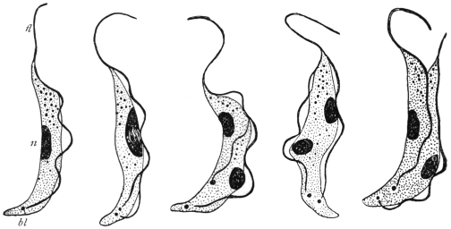
Fig. 26.—Trypanosoma brucei in division. n, nucleus; bl, blepharoplast; fl, flagellum.
× 2,000. (After Laveran and Mesnil.)
Two nuclei are present. The main or principal nucleus, sometimes
termed the trophic nucleus, is often situated towards the centre
of the body; it is frequently of the vesicular type, containing a
karyosome. The blepharoplast or kinetic nucleus is posterior to the
nucleus, and usually is rod-like. The flagellum arises close to the
blepharoplast, and forms an edge to the undulating membrane.
It may or may not extend beyond the limits of the undulating
membrane. If it does so, the unattached part is known as the free
flagellum. Sometimes a small granule is found at the origin of the
flagellum. This is the basal granule, and is considered by some to
function as the centriole of the kinetic nucleus.
The undulating membrane is a lateral extension of the ectoplasm
or periplast, and is the main agent in locomotion. It is edged by the
flagellum, which forms a deeply stainable border to it. Within the
membrane substance, often arranged parallel with its edge, are a
number of fine contractile elements, the myonemes. These contractile
elements may also occur on the body of the trypanosome.
They are easily seen in some large trypanosomes, but are difficult of
demonstration in others, owing to their great fineness.
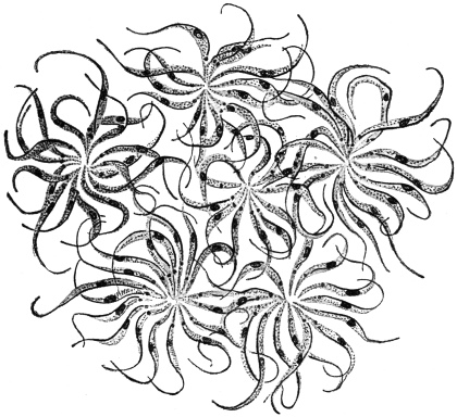
Fig. 27.—Trypanosoma lewisi. Multiplication rosettes. × 1,000. (After Laveran and Mesnil.)
Multiplication of trypanosomes in the blood is brought about by
binary longitudinal fission (fig. 26). Division is initiated by that of
the blepharoplast and nucleus. The division may be equal or subequal,
whereby differences in size of individuals partly arise. Multiple
division by repeated binary fission, without complete separation of
the daughter forms, is known in some trypanosomes (e.g., T. lewisi),
and rosettes of parasites thereby are produced (fig. 27).
The classification of trypanosomes is very difficult. Laveran
(1911)54 has suggested the examination of the relative length of the
flagellum as a diagnostic character, and so arranged these flagellates
in mammals in three groups. The first group included those trypanosomes
always having part of the flagellum free (e.g., T. evansi,
T. vivax); the second group comprised forms without a part of
the flagellum free (e.g., T. congolense), while the third group included
forms some members of which have free flagella, while others have
not (e.g., T. gambiense). Bruce55 (1914) and Yorke and Blacklock56
(1914) have also devised classifications.
Resting stages of some trypanosomes have been found in the
internal organs of their vertebrate hosts. The formation of these
oval, Leishmania-like bodies will be noted in individual cases later.
Similar small oval bodies form an important phase in the life-history
of T. cruzi, which multiplies normally by multiple fission or schizogony
into these oval, daughter elements, and not by binary longitudinal
fission in the circulating blood.
Polymorphism in trypanosomes (e.g., T. gambiense, T. rhodesiense)
is now interpreted as a phenomenon resulting from growth and
division.57 Long, thin forms are those about to divide. Fully mature
forms are shorter and broader. Various intermediate types occur and
represent growth forms. Formerly, polymorphism was interpreted in
terms of sex, thin forms being regarded as males, broad forms as
females, while the intermediate types were termed indifferent. Conjugation
was not observed, and there is no evidence in support of
the sexual interpretation.
The transmission of trypanosomes from one vertebrate host to
another is usually accomplished by the intermediation of some biting
arthropod in the case of terrestrial animals, while leeches are usually
considered to act as transmitters in the case of the trypanosomes
occurring in aquatic animals. Developmental phases of the life-histories
of trypanosomes occur in the invertebrate transmitters, and
will be considered in individual cases.
Trypanosoma gambiense, Dutton, 1902.
Syn.: Trypanosoma hominis, Manson, 1903. Trypanosoma nepveui, Sambon, 1903.
Trypanosoma castellanii, Kruse, 1903. Trypanosoma ugandense, Castellani,
1903. Trypanosoma fordii, Maxwell Adams.
In vertebrate blood Trypanosoma gambiense is polymorphic, for
long, thin forms may be seen in contrast with short, stumpy forms,
as well as intermediate forms (fig. 29, a—c). This polymorphism has
been interpreted in terms of sex, especially by German investigators,
following Schaudinn (see above). However, there is no evidence of
conjugation, and the polymorphic forms are more easily interpreted
in terms of growth and division, for the long thin forms are potential
dividing organisms, and the stumpy or short parasites, with little or
no free flagellum, are the adult individuals.
Morphology of T. gambiense in the Circulating Blood.
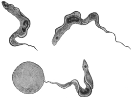
Fig. 28.—Trypanosoma gambiense. × 1,700.
(After Dutton.)
T. gambiense varies from 13 µ to 36 µ in length, its average length
being 24·8 µ, as was determined in 1913 by exact biometrical methods
by Stephens and Fantham.58 Three forms of parasite occur. According
to Miss Robertson,59 the relatively
short forms from 13 µ to 21 µ
long may be regarded as the
mature or “adult” type of parasite
in the blood. They carry on the
cycle in the vertebrate. From
them intermediate forms, which
are longer than the “adult” but
at first have the same breadth,
arise by growth. They possess
a free flagellum. The intermediate
forms grow into long individuals,
which are those about to
divide. The products of division give rise, directly or indirectly, to
the adult forms.
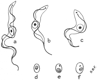
Fig. 29.—Trypanosoma gambiense. Development in vertebrate host. a, long, slender,
b, intermediate and c, short, stumpy forms, found in the blood; d, e, f, non-flagellate, latent
forms from internal organs. × 2,000. (Original. From preparations by Fantham.)
The organism has an elongate body with an anterior or flagellar
end and a blunter posterior or non-flagellar end. The protoplasm
is finely granular, large inclusions being rare. The central nucleus is
oval and large, often containing most of its chromatin concentrated as
a karyosome, with small granules only scattered near or on the fine
nuclear membrane. The blepharoplast is either rounded or rod-shaped.
The undulating membrane is thrown into folds and is
bordered by the flagellum. A small basal granule may be present
near, or at the actual origin of the flagellum.
Multiplication in the vertebrate is brought about by longitudinal
division. According to the recent account of division by Miss
Robertson, the blepharoplast doubles, then the flagellum splits for the
greater part of its length, and the daughter flagella separate, one being
shorter than the parent flagellum. The nucleus often shows two well
marked dark granules on the membrane at opposite poles, and these
appear to act as centrosomes. Nuclear constriction occurs and the
halves gradually separate. Finally the two daughter organisms become
free, the aflagellar end splitting last. The products of division may be
equal or unequal. Repeated division goes on in the general circulation
until the blood swarms with parasites. Then the trypanosomes gradually
disappear, and a period occurs when it is practically impossible to
demonstrate the parasite in the blood. At such a period, trypanosomes
can be obtained by puncture of the enlarged lymphatic glands or of
the spinal canal, or can be found in the internal organs, more particularly
in the spleen, lungs, liver and bone-marrow. In the latter
organs, latent bodies are produced (fig. 29, d—f) which are capable of
again becoming flagellates and entering the general circulation.
Their formation was described by Fantham (1911).60 The parasite
contracts, the blepharoplast migrates towards the nucleus, a very thin
coat differentiates around the two nuclei and a certain amount of cytoplasm,
and the parts exterior to the coat disintegrate, leaving a small,
oval body behind. Fuller details are given in connection with
T. rhodesiense. Laveran (1911)61 considers that latent bodies are
“involution” forms, but acknowledges that they can flagellate and
become infective in fresh blood.
No multiplication of the trypanosomes within the cells of the
lung, liver or spleen of infected monkeys was found by Miss
Robertson in her recent researches.
There appear to be negative periods in infected monkeys, since,
although trypanosomes may occur in their blood at such times, they
are not infective to Glossina.
Development in Glossina palpalis.—The principal accounts are
those by Sir D. Bruce and his colleagues (1911),62 and by Miss
Robertson63 (1912), whose results will be followed. According to the
latter investigator T. gambiense never enters the body cells of the fly
(G. palpalis), nor does it penetrate the gut wall into the body cavity.
Practically no crithidial stage occurs in the fly’s main gut, but a
trypanosome facies is retained therein.
After the trypanosomes are ingested by the fly during a meal of
infected blood, sooner or later multiplication occurs. This development
usually begins in the middle or posterior part of the mid gut,
and trypanosomes of varying sizes are produced. After the tenth
or twelfth day, many long, slender trypanosomes (fig. 30, a) are
found, which gradually move forwards into the proventriculus. Such
long, slender forms represent the limit of development in the lumen
of the main gut. The proventricular type, developed about the
eighth to the eighteenth or twentieth day, is not infective; it may
occur in the crop, but is not to be found permanently there.
Between the tenth and the fifteenth days multinucleate forms of
trypanosomes are found, and may be styled multiple forms (fig. 30, b).
Some of these latter may be degenerative.
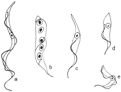
Fig. 30.—Trypanosoma gambiense. Development in the fly, Glossina palpalis. a, slender,
proventricular form; b, multinucleate form; c, d, crithidial forms; e, infective type of
trypanosome found in salivary gland. × 2,500. (After Robertson.)
Invasion of the Salivary Glands of the Fly.—Long, slender trypanosomes
from the proventriculus pass forward into the hypopharynx.
They then pass back along the salivary ducts, about sixteen to thirty
days after the fly’s feed. The trypanosomes reach the salivary glands
as long, slender forms. In the glands they become shorter and
broader, attach themselves to the surrounding structures, and assume
the crithidial facies (fig. 30, c, d). As crithidial forms they remain
attached to the wall and multiply in the glands. These crithidial
stages differentiate into the short, broad trypanosome forms, capable
of swimming freely (fig. 30, e).
Miss Robertson considers the development in the main gut to be
indifferent multiplication, and that salivary fluid seems necessary to
stimulate trypanosomes to the apparently essential reversion to the
crithidial type. The second development in the salivary gland is the
essential feature. The short, stumpy forms of trypanosomes (fig. 30, e)
finally produced in the salivary glands are alone infective. No
conjugation of trypanosomes occurs in the fly. Only about 5 per
cent. of captive tsetse flies fed on trypanosome-infected blood become
infective, but they probably remain infective for the rest of their lives.
J. G. Thomson and Sinton (1912)64 have obtained in cultures the
various trypanosome forms of T. gambiense seen in the fly’s main gut.
Duke (1912)65 found T. gambiense in a species of antelope, the
situtunga (Tragelaphus spekei), on Damba Island in Victoria Nyanza.
Wild G. palpalis could be infected therefrom. The antelope may
then act as a sleeping sickness reservoir in that district, but men are
apparently the chief reservoir.
Trypanosoma nigeriense, Macfie, 1913.66
Macfie has recently (August, 1913) described a human trypanosome
from the Eket district of Southern Nigeria. It is common in
young people. The disease produced does not seem to be of a
virulent type in Nigeria, and does not occur in epidemic form. In
the early stages the glands of the neck are enlarged. In the later
stages—cases of which are rarer—lethargy appears. The parasite
is a polymorphic trypanosome, morphologically almost indistinguishable
from T. gambiense, though it may be slightly shorter. Macfie
recorded the occurrence in his preparations of a few trypanosomes
appearing to have a flagellum free during their whole length.
Some of the parasites, as seen in a sub-inoculated guinea-pig, are very
small (8 µ long). Other trypanosomes have their nuclei displaced
somewhat anteriorly. This parasite may only be a variety of
T. gambiense. The parasite is perhaps spread by Glossina tachinoides.
Trypanosoma rhodesiense, Stephens and Fantham, 1910.
The parasite was found in the blood of a young Englishman who
had contracted sleeping sickness in the Luangwa Valley, North-eastern
Rhodesia, in the autumn of 1909. The patient had never
been in an area infested with Glossina palpalis.
(1) Morphology.—The morphology of the parasite in man and
sub-inoculated rats was studied by Stephens and Fantham in 1910.67
They pointed out a morphological peculiarity in the presence of
certain trypanosomes with posterior nuclei in sub-inoculated animals,
that is, parasites in which the nucleus (trophonucleus) was situated
towards the posterior or aflagellar end, close up to or even beyond the
blepharoplast or kinetic nucleus (fig. 31, 4, 5). When the nucleus was
beside the blepharoplast, the former was seen to be kidney-shaped
(fig. 31, 4). The posterior nuclear forms were of the stout and stumpy
variety, and about 6 per cent. of the stumpy forms were found to have
their nuclei displaced from the centre. The anterior or flagellar end
of these trypanosomes often contained chromatoid granules. T.
rhodesiense varies in length from 12 µ to 39 µ68; short stumpy forms
vary from 13 µ to 21 µ, intermediate forms from 21 µ to 24 µ, and long,
slender forms from 25 µ onwards. The average length is 24·1 µ.
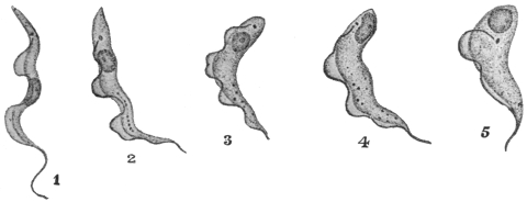
Fig. 31.—Trypanosoma rhodesiense. 1, Long narrow form; 2–4, nucleus passing to
posterior (aflagellar) end; 5, nucleus quite posterior. × 1,800. (After Stephens and Fantham.)
Certain regular periods occur in the course of the trypanosomiasis
when few or no flagellate trypanosomes are found in the peripheral
blood of the patient or of the sub-inoculated animal. These periods
can be explained in terms of morphology, for the trypanosomes are
capable of assuming a non-flagellate form in the internal organs of the
host, particularly in the lungs and in the spleen. Such forms are
known as “latent” or “resting” forms. The term “latent body”
was first used by Moore and Breinl in 190769 in connection with
T. gambiense. Fantham70 (1911) has described the process of formation
of latent from motile forms and the reconversion of the latent
bodies into active flagellates. Fresh preparations of splenic blood
or lung blood containing trypanosomes were made. A trypanosome
gradually withdrew or cast off its flagellum, concentrated its
cytoplasm, and became more or less elongate oval. Nucleus and
blepharoplast approached one another and came to lie more or less
side by side. Then an opaque line often made its appearance around
the nuclear area and differentiated as a slight envelope or covering,
the cytoplasm external to this merely degenerating. The small, oval,
refractile body (fig. 29, d—f) thus formed was a non-flagellate latent
body, 2 µ to 4 µ in diameter, like Leishmania or the non-flagellate,
multiplicative forms of T. cruzi (fig. 34), and remains temporarily
inactive in the internal organs of the host. After this period of
inactivity, the non-flagellate body, recuperated by its rest, begins
to elongate again. The nuclei separate. From a small vacuole-like
portion the flagellum differentiates and forces out the ectoplasm,
which assumes the form of the undulating membrane with its
flagellar border. Subsequent growth results in the production of the
typical trypanosome form, which re-enters the circulating blood and
multiplies by longitudinal binary fission. Division of the parasite
prior to the formation of a latent body may occur and division
of the latent forms themselves is known, though less common.
Consequently latent bodies, like the flagellate forms themselves,
show diversity in size. The blepharoplast of the latent bodies is
sometimes less well marked than in Leishmania (see fig. 29, d-f).
Laveran’s views on these bodies have already been given on p. 74.
(2) Animal Reactions.—The posterior nuclear trypanosomes were
found in all sub-inoculated animals, such as rats, guinea-pigs, dogs,
mice, Macacus, rabbits and horses, but were not seen in the human
patient, as few trypanosomes occurred in his peripheral blood.
R. Ross and D. Thomson71 found a periodic, cyclical variation in the
number of the parasites in the patient’s blood from day to day, the
cyclical period being about a week (fig. 32). Fantham and J. G.
Thomson72 (1911) found a similar periodic, cyclical variation in the trypanosomes
in the blood of sub-inoculated rats, guinea-pigs and rabbits.
On counting the parasites in the blood of similar animals inoculated
with T. gambiense, they established, by enumerative methods, that
T. rhodesiense was more virulent than T. gambiense, while Yorke also
showed this marked virulence of T. rhodesiense in practically all
laboratory animals. In other words the duration of infection in the
case of T. rhodesiense was shorter. It was also found that T. rhodesiense
was resistant to atoxyl. The patient, from whom the original
strain was obtained, died about nine months after the probable date
of infection. Some patients infected with T. rhodesiense have died in
an even shorter period, such as four or five months.
In sheep and goats T. rhodesiense causes an acute disease, marked
by high fever, œdema of the face, and keratitis, as shown by Bevan
and others, death resulting after a relatively short period. T. gambiense
gives rise, in these animals, to no symptoms except fever, which
may be overlooked. T. rhodesiense produces keratitis in dogs.
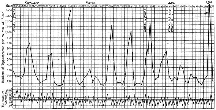
Fig. 32.—Chart showing daily counts of number of trypanosomes per cubic millimetre of peripheral blood from a case of
Rhodesian sleeping sickness. (After R. Ross and D. Thomson.)
Stannus and Yorke (1911) observed T. rhodesiense in animals
inoculated from a case of sleeping sickness in Nyasaland. Sir D. Bruce
and his colleagues73 have shown (1912) that T. rhodesiense is the
parasite usually found in man and in animals sub-inoculated from
cases of sleeping sickness in Nyasaland. It has since been found in
German East Africa and Portuguese East Africa, while Ellacombe
has described a case from North-western Rhodesia.
(3) Serum Reactions.—Interesting experiments on this subject were
performed during 1911 and 1912 by various French investigators.
(a) Action of Immune Serum (Mesnil and Ringenbach)74: (1) A goat
was infected with T. rhodesiense. Twenty-two days later its serum
mixed with T. rhodesiense was injected into a mouse. Result: Protection.
(2) The serum mixed with T. gambiense was injected into
a mouse. Result: Infection.
(b) Action of Baboon Serum.—Contrary to T. gambiense, T. rhodesiense
is very susceptible to human and baboon sera. Mesnil and
Ringenbach75 showed that a dose of 1 c.c. of baboon (Papio anubis)
serum cured mice infected with T. rhodesiense. In the same dose it
acted very feebly on T. gambiense.
(c) Action of Human Serum.—1 c.c. of human serum cured T.
rhodesiense mice in three out of four cases; on T. gambiense mice
there was no appreciable effect.
Laveran and Nattan-Larrier76 have shown the same, namely, that
human sera act on T. rhodesiense, but are quite without action on
T. gambiense.
(d) Trypanolytic Reactions.—Mesnil and Ringenbach77 have also
shown that the sera of animals (man, monkey and guinea-pig)
infected with T. gambiense are trypanolytic for the homologous
trypanosome, that is, T. gambiense, but have no action on the heterologous
trypanosome, that is, T. rhodesiense.
(4) Cross Immunity Experiments.—(a) Mesnil and Ringenbach78
immunized a monkey (Macacus rhesus) against T. gambiense. It was
inoculated with T. rhodesiense on June 7, 1911; on June 27 trypanosomes
appeared, the infection being slight; on July 4 it died. A
control died in ten and a half days.
(b) Laveran79 immunized a goat and mice against T. gambiense.
When they had acquired a solid immunity, they were inoculated with
T. rhodesiense. They became infected like the controls.
(c) Laveran and Nattan-Larrier80 immunized a ram against T. brucei,
it subsequently became infected with T. rhodesiense.
(d) Laveran81 immunized a ram and a sheep against different strains
of T. brucei. Inoculated with T. rhodesiense they both acquired acute
infections and died. Conclusion: T. rhodesiense is not T. brucei.
When the converse set of experiments is tried, namely, immunizing
an animal against T. rhodesiense, and then inoculating with
T. gambiense, the difficulty immediately arises that it is impossible to
immunize an animal against T. rhodesiense, owing to its virulence.
But a partial and transitory immunity to T. rhodesiense can be obtained
by treating the infected animal with drugs, such as arsenophenylglycin.
The results, so far as they go, seem to show that an animal immunized
against T. rhodesiense is immune not only to T. rhodesiense, but also
to T. gambiense, a fact which, according to Mesnil and Léger, does
not invalidate the specificity of T. rhodesiense, but tends to show that
the two trypanosomes are closely related.
(5) Mode of Transmission and Reservoir.—Kinghorn has shown that
T. rhodesiense is transmitted by Glossina morsitans in which it undergoes
development. Kinghorn and Yorke82 found that about 16 per
cent. of the wild game examined in Northern Rhodesia was naturally
infected with T. rhodesiense. The wild game examined included waterbuck,
hartebeest, mpala, bushbuck and warthogs. One native dog near
the Nyasaland border was found infected, but not domestic stock.
Taute doubts whether T. rhodesiense really occurs in wild game.
Approximately 3·5 per cent. of the tsetse flies fed on infected animals
may become permanently infected with T. rhodesiense, and capable
of infecting clean animals. Furthermore, a tsetse fly when once
infective probably remains infective for the rest of its life.
Kinghorn and Yorke, however, have shown that climatic conditions,
namely, those of temperature, also affect the infectivity of the
tsetse fly, as the ratio of flies capable of transmitting T. rhodesiense to
those incapable of transmitting the virus is 1 : 534 in hot valley
districts (e.g., Nawalia, Luangwa Valley, temperature 75° to 85° F.),
while on elevated plateaux (e.g., Ngoa, on the Congo-Zambesi watershed,
temperature 60° to 70° F.) the ratio falls to 1 : 1312.
Mechanical transmission by the tsetse fly does not occur, if
a period of twenty-four hours has elapsed since the infecting meal.
Developmental Cycle in the Fly.—The period which elapses between
the infecting feed of the flies and the date on which they become
infective varies from eleven to twenty-five days in the Luangwa
Valley, according to Kinghorn and Yorke. Attempts carried out at
laboratory temperature on the Congo-Zambesi plateau, during the
cold season, to transmit T. rhodesiense by means of G. morsitans
were always unsuccessful. The developmental cycle of the trypanosome
in the fly is influenced by the temperature to which the flies
are subjected (as stated above). The first portion of the developmental
cycle proceeds at the lower temperatures (60° to 70° F.), but
higher temperatures are necessary for the completion of the development
of the trypanosome. Kinghorn and Yorke found that the
trypanosomes may persist in the fly, at an incomplete stage of their
development, for at least sixty days when the climatic conditions
were unfavourable.
The first portion of the developmental cycle of the trypanosome
takes place in the gut of the fly. Invasion of the salivary glands of
the tsetse is secondary to that of the intestine, but is necessary for the
infectivity of the fly. A relatively high mean temperature, 75° to
85° F., is essential for the passage of the trypanosomes into the
salivary glands and the completion of their development therein.
Kinghorn and Yorke83 state that the predominant type of trypanosome
in the intestine of infected G. morsitans was a large broad form,
quite different from that which is most common in the salivary
glands. The trypanosome in the glands resembles the short form
seen in the blood of the vertebrate host. The authors quoted state
that both the intestinal and salivary gland forms of infective G.
morsitans are virulent when inoculated into healthy animals.
Bruce and colleagues84 have quite recently (June, 1914) published
an account of their investigations of T. rhodesiense in G. morsitans
in Nyasaland. (Incidentally it may be remarked that Bruce considers
T. rhodesiense to be identical with a polymorphic strain of T. brucei—see
pp. 83, 94). The development of T. rhodesiense takes place in the
alimentary canal and salivary glands, not in the proboscis, of the
tsetse fly. In feeding experiments with laboratory bred flies, as
well as with a few wild flies, fed on infected dogs or monkeys, only
8 per cent. of the flies were found to be infected on dissection. Of
such infected flies, however, only some allow of the complete development
of the trypanosomes within them, in other words only about
1 per cent of the flies become infective. The length of time which
elapses before a fly becomes infective varies from fourteen to thirty-one
days, averaging twenty-three days, when kept at 84° F. (29° C.).
The dominant intestinal type of flagellate in the fly is that seen in
the proventriculus, which contains many long, slender trypanosomes.
These proventricular forms find their way to the salivary glands,
wherein crithidial and encysted forms are seen. They change into
“blood forms,” which are short, stumpy trypanosomes and are
infective. “The infective type of trypanosome in the salivary glands—corresponding
to the final stage of the cycle of development—is
similar to the short and stumpy form found in the blood of the vertebrate
host.” The cycle is thus very similar to that of T. gambiense in
G. palpalis (fig. 30).
Culture.—J. G. Thomson (1912),85 and subsequently Thomson
and Sinton, succeeded in cultivating T. rhodesiense in a modified
Novy-MacNeal medium. The development obtained resembled that
of the trypanosome in the intestine of Glossina.
General Note on Trypanosomes with Posterior Nuclei.
Posteriorly placed nuclei have been found to occur not only in
T. rhodesiense by Stephens and Fantham (1910), but also in T. pecaudi
by Wenyon (1912), in T. brucei by Blacklock (1912), and in T. equiperdum
by Yorke and Blacklock (1912).
Recently Stephens and Blacklock (1913)86 have shown that two
trypanosomes, different morphologically, have been confused under
the name T. brucei. One of these is polymorphic (i.e., it exhibits
long and slender as well as short and stumpy forms) and came from
Uganda, while the other is monomorphic and is the original Zululand
strain described by Bruce from cattle suffering from “nagana.” Bruce
(1914) considers that morphological change has occurred in T. brucei
in its passage through laboratory animals, and thus explains the
diversity of views. The posterior nuclear forms described by
Blacklock occurred in the Uganda strain of T. brucei. (See p. 95.)
Similarly, a posterior nuclear form, T. equi, has been separated from
T. equiperdum. (See p. 98.)
Again, Bruce and his colleagues on the Royal Society Commission
investigating sleeping sickness in Nyasaland, have stated (April,
1913) that “evidence is accumulating that T. rhodesiense and T. brucei
(Plimmer and Bradford) are identical.” The exact identity of trypanosomes
showing posterior nuclei is, then, far from settled, although
Laveran by cross immunity tests has declared that T. brucei is distinct
from T. rhodesiense. No one has yet seen posterior nuclei in T. gambiense.
Trypanosoma cruzi, Chagas, 1909.
Syn.: Schizotrypanum cruzi, Chagas, 1909.
The trypanosome was discovered by Chagas87 in the intestine of
the bug, Triatoma (Conorhinus) megista, in Brazil, and then in the
blood of a small monkey bitten by the bug. A little later it was found
in the blood of a child, aged two years, suffering from irregular fever,
extreme anæmia and enlarged glands in the State of Minas Geraes,
Brazil. Chagas found that he was able to infect many of the usual
laboratory animals with the trypanosome, by allowing the bug to bite
them. He was also able to culture the parasite on blood agar.
Chagas found the Reduviid bug, Triatoma megista, in the houses
of the poorer inhabitants of the Brazilian mining State, and that it
attacked the people, more especially the children, at night, biting the
face. On this account the insect is called “barbeiro” by the
inhabitants. The bite is somewhat painful. The disease has since
been found in other parts of Brazil, e.g., Matta de São João in Bahia
province, Goyaz, Matto Grosso and São Paulo provinces, as well as
in Minas Geraes.
Morphology.—The trypanosome has a large blepharoplast or kinetic
nucleus. It is stated to occur both free and in the red blood corpuscles
in the peripheral blood. It is about 20 µ long, on an average.
Two forms of the parasite (fig. 33, 6, 7) are described in the human
blood. In one free form there is a large egg-shaped blepharoplast
and the posterior (aflagellar) end of the parasite is drawn out. The
blepharoplast (kinetic nucleus) may have a chromatin appendage.
The nucleus is oval or band-like, containing a karyosome. The
flagellum, starting close to the blepharoplast or its appendage, has a
free portion of variable length. The other free form in the blood has
a more or less round, terminal blepharoplast, smaller than in the first
form, without a chromatin appendage as a rule. The body of this
second form is decidedly broader than that of the first mentioned.
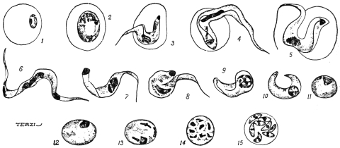
Fig. 33.—Trypanosoma cruzi. Schizogony. 1, merozoite in red blood corpuscle; 2,
parasite totally enclosed in red cell, no flagellum or undulating membrane; 3-5, parasites
partially enclosed in red cell; 6, 7, parasites in human blood; 8-11, parasites in lungs of the
monkey, Callithrix; 12, 13, initial forms of schizogony; 14, 15, schizogony in the lungs of
Callithrix. (After Chagas.)
The dimorphism has been interpreted sexually, the first mentioned
forms being termed males, the second ones females. The correctness
of this interpretation is very doubtful.
No sign of longitudinal division was ever seen in the peripheral
blood or in the internal organs. The “endocorpuscular” forms may
be completely or partially enclosed in the red cell or only attached
thereto (fig. 33, 1-5). At the beginning of infection the endocorpuscular
forms are the more numerous. Some authorities, however,
doubt these stages.
Life-history in the Vertebrate Host.—Chagas found fluctuations in
the number of the parasites in the peripheral blood. He believes the
increase of the parasites to be periodic.
The investigations of Chagas and of Hartmann have revealed two
types of multiplication which take place in the internal organs of the
vertebrate host.
(a) The first type—which possibly belongs to another organism,
Pneumocystis carinii, see p. 90—occurs in the capillaries of the lungs.
The flagellate parasite entering the lung capillaries loses its flagellum
and undulating membrane. Its body becomes curved, and the two
ends fuse, and so an oval mass is formed (fig. 33, 8-11). In some
cases the blepharoplast disappears, in other cases it blends or fuses
with the nucleus. The nucleus of the rounded parasite then divides
into eight by successive divisions (fig. 33, 12-15). Next the body,
which is surrounded by its own periplast, also divides, giving rise
to eight tiny daughter individuals or merozoites (fig. 33, 15). The
merozoites lie inside the periplast, which acts as a sort of “cyst
wall.” The merozoites are said to exhibit dimorphism, and Chagas
has interpreted the dimorphism in terms of sex. The daughter forms,
produced by the parent trypanosomes which kept their blepharoplasts,
themselves have blepharoplasts as well as nuclei, and have been termed
“males” or “microgametes.” The merozoites, arising from parent
trypanosomes which lost their blepharoplasts, have themselves only
nuclei, and have been called “females” or “macrogametes.” In the
case of the so-called “female” forms the single nucleus divides into
two unequal parts, of which the smaller becomes the blepharoplast,
and a flagellum is formed later. The so-called “males” possess early
a rudiment of a flagellum. Both kinds of merozoites escape from the
parent periplast wall, and enter red blood corpuscles. They grow into
flagellates within the corpuscles, and then become
free as adult trypanosomes in the blood-stream.
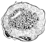
Fig. 34.—Trypanosoma
cruzi. Transverse section
of a striated muscle containing
rounded forms of
the parasite in the central
portion. × 1,000 approx.
(After Vianna.)
(b) The second mode of multiplication
is one of asexual reproduction (schizogony
or agamogony). It was first described by
Hartmann from hypertrophied endothelial cells
of the lungs. It has since been found in the
cardiac muscle, in the neuroglia of the central
nervous system, and in striped muscle (fig. 34).
In laboratory animals it has also been found
in the testicle and suprarenal capsules. In
these tissues the parasite is intracellular, appearing
as a small rounded body with nucleus
and blepharoplast, without flagellum or undulating
membrane. In other words the parasite is Leishmania-like in
the body tissues, and recalls the organism of kala-azar.
Chagas considers this second mode of multiplication to be strictly
asexual. By this means the number of parasites in the vertebrate
host is increased, and symptoms are produced. On the other hand
the first mode of multiplication, seen in the lung capillaries, is
considered by Chagas to be a process of gametogony, in which sexual
forms are differentiated. He finds that (1) the adult trypanosomes
exhibit a dimorphism in human blood rarely seen in artificially
infected guinea-pigs. In these guinea-pigs (infected from guinea-pigs)
the so-called gametogony in the lungs is seldom seen. (2) The
intermediate host, Triatoma (Conorhinus), becomes infective if fed
directly on infected human blood, but very rarely so if fed on guinea-pigs.
Chagas is led to believe that the occurrence of sexual forms
constantly in the blood of man implies a greater resistance to infection
on the part of man than on the part of guinea-pigs or other animals,
assuming the general hypothesis that the formation of gametes
represents a reaction of the Protozoön to unfavourable conditions.
In human infection the number of parasites is always less than in
laboratory animals, and their presence in the blood is transitory,
lasting from fifteen to thirty days in acute cases. In many cases
examination of the tissues at death has shown the presence of parasites
in patients who did not exhibit them in the general circulation.
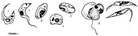
Fig. 35.—Trypanosoma cruzi. Development in Triatoma megista. 1-6, forms found in
the mid gut of Triatoma; 7 flagellate forms found in the posterior part of the gut of Triatoma.
(After Chagas.)
Life History in the Invertebrate Host.—About six hours after the
ingestion of infected blood by the bug (Triatoma megista), the kinetic
nucleus of the trypanosome moves towards the nucleus, and the
flagellum is usually lost (fig. 35, 1-5). The parasite becomes rounded
and Leishmania-like (fig. 35, 3-5), and multiplies rapidly by division.
After a time, multiplication having ceased, the rounded forms become
pear-shaped and develop a flagellum at the more pointed end.
Crithidial forms (fig. 35, 7) are thus produced and pass into the intestine,
where they multiply and may be seen in about twenty-five hours
after the ingestion of blood. The crithidial forms may also be
found in the rectum and fæces. The last stage in the invertebrate
is a small, trypanosome-like type, long and thin with a band-like
nucleus and conspicuous kinetic nucleus. These parasites are found
in the hind gut and in the body cavity. They find their way into
the salivary glands, and are the forms (fig. 36) which are transmissible
to a new vertebrate host. The development in the bug takes about
eight days altogether, after which time the bugs are infective.
There are thus three principal
phases in the development
of T. cruzi in Triatoma
megista: (1) A multiplicative
phase (Leishmania-like) in the
stomach of the bug, (2) a
crithidial phase, which is also
multiplicative, in the hind-gut,
and (3) a trypanosome phase, which is “propagative,” and apparently
passes through the wall of the alimentary canal into the body cavity
and so into the salivary glands.
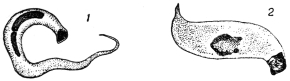
Fig. 36.—Trypanosoma cruzi. Forms found
in the salivary glands of Triatoma megista.
(After Chagas.)
Brumpt found that T. cruzi could live in Cimex lectularius, C. boueti,
and Ornithodorus moubata. The Cimex fæces may be infective.
Blacklock found multiplication of the parasite in C. lectularius.
Culture.—The trypanosome can be cultivated on Novy-MacNeal’s
blood agar, and the cultural forms resemble those described in
the bug.
Possible Reservoir.—Chagas thinks that probably the armadillo or
“tatu” (Dasypus novemcinctus) may be the reservoir of T. cruzi. He
also thinks that Triatoma geniculata is a transmitter; it lives in the
burrows of the armadillo. Other carriers may be Triatoma infestans
and T. sordida.
Clinical Features.—The trypanosomiasis of Brazil, produced by
T. cruzi and spread by Triatoma spp. has received various names,
such as oppilação, canguary, parasitic thyroiditis, and coreotrypanosis.
It is also known as the human trypanosomiasis of Brazil, South
American trypanosomiasis, and Chagas’ disease.
Chagas88 reports two principal forms—acute and chronic. The
acute infection is rare, and is characterized by increase in the volume
of the thyroid gland, pyrexia, a sensation of crackling in the skin,
enlarged lymphatic glands in the neck, axilla, etc., while the liver and
spleen are increased in volume. Sclerosis of the thyroid gland is
found at autopsy and fatty degeneration of the liver. During an
attack of fever, trypanosomes are found in the blood. The acute
form was only observed in children.
In the chronic form Chagas reports several varieties: (a) A
pseudo-myxœdematous form, occurring in most cases, especially up
to the age of 15. There is hypertrophy of the thyroid gland or
at least signs of hypothyroidism, general hypertrophy of glands,
disturbance of heart rhythm, and nervous symptoms. (b) The myxœdematous
form is characterized by similar symptoms, especially by
considerable swelling of the thyroid body, and myxœdema of the
subcutaneous cellular tissue; sometimes there is a true pachydermic
cachexia. (c) In the nervous form there are motor disturbances,
aphasia, disturbances of intelligence or signs of infantilism, athetosis
of the extremities and idiocy. There are also paralytic symptoms of
bulbar origin, disturbances of mastication, phonation and deglutition,
and in some cases convulsive attacks. (d) The cardiac form, characterized
by disturbance of the heart rhythm. In all these forms the
parasite is found at autopsy in the nervous substance, brain, bulb and
heart.
Vianna (1911)89 has studied the histopathology of the disease.
Some of the chief points are: in the heart muscle destruction of
the sarcoplasm, followed by interstitial myocarditis; in the central
nervous system invasion of the neuroglia cells and inflammatory
reaction; in the suprarenal capsule invasion of medulla or cortex;
inflammatory reaction can also be seen in the kidneys, the hypophysis
and thyroid gland.
Recently Chagas states90 that “schizotrypanosomiasis” has been
found in a child 15 to 20 days old, and that Trypanosoma cruzi
has also been found in a fœtus—the mother being infected with
the trypanosome. The trypanosomiasis can, then, be transmitted
hereditarily.
Trypanosoma lewisi, Kent, 1881.
The trypanosome has a nucleus somewhat displaced anteriorly, about
one-third of the way from the anterior (flagellar) end of the body, a
relatively straight edge to the undulating membrane, and a rod-shaped
blepharoplast (fig. 37, A). It averages about 25 µ long and 1·5 µ broad.
Much attention has been devoted in recent years to the elucidation
of the life history of the rat parasite, Trypanosoma lewisi. It is
usually non-pathogenic to its host. It has been shown that the
trypanosome can be transmitted from rat to rat by the rat-flea,
Ceratophyllus fasciatus, and by Ctenocephalus canis (the so-called dog-flea).
(See also p. 92). The flagellate may also persist, but doubtfully
develop, in the rat-louse, Hæmatopinus spinulosus. These researches
may now be summarized.
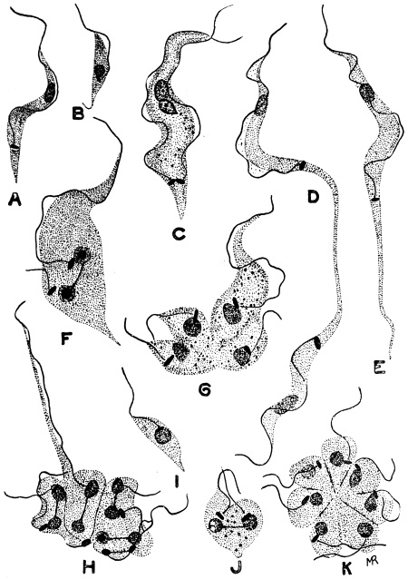
Fig. 37.—Trypanosoma lewisi, from rat’s blood. a, ordinary form; b, small form;
c, d, stages in equal binary fission; e, elongate form (longocaudense type), resulting from
division as seen in d; f, unequal binary fission; g, h, multiple fission into four and eight;
i, small form; j, binary fission of small form; K, division rosette. × 2,000. (After
Minchin and Thomson.)
Life Cycle in the Vertebrate Host.—After infection of a rat, the
trypanosomes usually appear in the animal’s blood in five to seven
days. This incubation period applies either to a natural or an artificial
infection. The trypanosomes first observed in the rat’s blood are
diverse in form (fig. 37), being small, medium and large in size. This
diversity is explained by the rapid multiplication taking place. A
trypanosome may divide by equal longitudinal fission (fig. 37, C, D),
but more commonly multiple fission occurs (fig. 37, G, H), and is
unequal. Rosette forms are produced, in which the parent form can
be recognized by its long flagellum (fig. 37, H) and attached to it are
daughter individuals, smaller in size, from which flagella are growing.
Minchin and J. D. Thomson (1912) find that the daughter forms may
be set free sometimes with a crithidia-like facies (fig. 37, I), the
blepharoplast being anterior but near to the nucleus. The daughter
forms, when set free, may themselves divide by binary or multiple
fission, in the latter case forming rosettes (fig. 37, K). Rosette
forms were described by Moore, Breinl and Hindle in 1908.
Lingard, some years ago, described as a distinct species, T. longocaudense,
certain forms with markedly elongate posterior ends
(fig. 37, E). According to Minchin, “these forms appear to arise by
binary fission” (fig. 37, D). These long drawn-out forms “are of
constant occurrence and very numerous at a certain stage of the
multiplication period.” It is about the eighth or tenth day after
infection that the multiplication of T. lewisi is at its maximum in the
rat’s blood. About the twelfth or thirteenth day the trypanosomes
seen in the blood appear uniform. According to Minchin (1912)91
the rat “gets rid of its infection entirely sooner or later, without
having suffered, apparently, any marked inconvenience from it, and
is then immune against a fresh infection with this species of trypanosome.”
There is, then, a cycle of development in the vertebrate host.
Minchin notes that the records of the pathogenicity of T. lewisi in
rats, causing their death, need further investigation.
T. lewisi inoculated into dormice (Myoxus nitela) and jerboas may
become pathogenic thereto.
Carini found cysts in the lungs of rats infected with T. lewisi.
He thought the cysts were schizogonic stages of the trypanosome,
comparable with those found in the lungs of animals sub-inoculated
with T. cruzi. Delanoë (1912)92 has found, however, that such
cysts, containing eight vermicules, occurred in rats uninfected with
T. lewisi. Delanoë concludes that the pneumocysts are independent
of T. lewisi, and represent a new parasite, Pneumocystis carinii. The
pneumocysts may be allied to the Coccidia, and must be considered
when investigating the life-cycle of a trypanosome in a vertebrate
host. Some of the stages of T. cruzi may possibly be of this nature.
Life-cycle in the Invertebrate Host.—This occurs in fleas, and has
been investigated in considerable detail by Minchin and Thomson
in Ceratophyllus fasciatus, and by Nöller in Ctenocephalus canis and
Ctenopsylla musculi.
When infected rat’s blood is taken up by the flea, the parasites pass
with the ingested blood direct to the mid-gut of the Siphonapteran.
In the flea’s stomach they multiply in a somewhat remarkable manner,
namely, by penetration of the cells of the lining epithelium, and
division inside the epithelial cells. Inside these lining cells the
trypanosomes first grow to a large size and then form large spherical
bodies, within which nuclear multiplication occurs (fig. 38, A-F).
Any one of these large spherical bodies contains at first a number of
nuclei, blepharoplasts and developing flagella, the original flagellum
still remaining attached for a time. The cytoplasm then divides into
daughter trypanosomes which are contained within an envelope,
formed by the periplast of the parent parasite. Inside the periplast
envelope are a number of daughter trypanosomes “wriggling very
actively; the envelope becomes more and more tense, and finally
bursts with explosive suddenness, setting free the flagellates, usually
about eight in number, within the host-cell” (fig. 38, F). The daughter
forms escaping from the host cell into the stomach lumen of the flea
are fully formed, long trypanosomes.
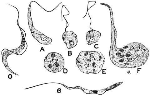
Fig. 38.—Trypanosoma lewisi. Developmental stages from stomach of rat flea. O,
ordinary blood type; A-F, stages occurring in gut-epithelium of flea, when the trypanosome
becomes rounded and undergoes multiplication, forming in F eight daughter trypanosomes;
G, type of trypanosome resulting from such division which passes back to the rectum.
× 2,000. (After Minchin.)
The trypanosomes (fig. 38, G) pass into the flea’s rectum. The
next phase is a crithidial one. The parasites become pear-shaped,
in which the blepharoplast (kinetic nucleus) has travelled anteriorly
past the nucleus towards the flagellum (fig. 39). The crithidial forms
attach themselves to the wall of the rectum, and multiply by binary
fission (fig. 39, D). A stock of parasites is thus formed which,
according to Minchin and Thomson, “persist for a long time in
the flea—probably under favourable conditions, for the whole life
of the insect” (fig. 39, A–I).
From the crithidial forms of the rectum, according to Minchin,
small infective trypanosomes arise by modification morphologically
(fig. 39, J–M). The flagellum grows longer and draws out more the
anterior part of the body, the blepharoplast migrates posteriorly,
behind the nucleus, and carries with it the flagellar origin. These
trypanosomes are small, but broad and stumpy (fig. 39, N), and can
infect a rat. Minchin and Thomson formerly considered that the
small, stumpy, infective trypanosomes pass forwards from the rectum
into the stomach, and “appear to be regurgitated into the rat’s blood
when the flea feeds.” However, the small infective trypanosomes
were previously described by Swellengrebel and Strickland.93 They
may be found in the flea’s fæces. Nöller (1912)94 has found that the
development of T. lewisi proceeds quite well in the dog flea (Ctenocephalus
canis) in Germany. Wenyon confirms this, and states that
the human flea, Pulex irritans, and the Indian rat-flea, Xenopsylla
cheopis, are also able to serve as true hosts for T. lewisi.
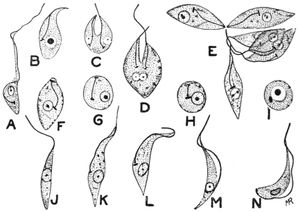
Fig. 39.—Trypanosoma lewisi. Developmental stages from rectum of rat-flea. A, early
rectal form; C, D, division of crithidial form; E, group of crithidial forms; F–I, crithidial
forms without free flagella, some becoming rounded; J–M, transitional forms to trypanosome
type seen in N, which represents the final form in the flea. × 2,000. (After Minchin.)
Nöller stated that rats were not infected with T. lewisi by infective
fleas biting them, but by the rats licking up the fæces passed by the
fleas while feeding. This is not in agreement with Minchin and
Thomson’s earlier views of regurgitation, which, apparently, they
have now abandoned.95 Wenyon (1912) confirms Nöller’s experiments.
He took a dog flea, containing infective trypanosomes in
its fæces, and allowed it to feed on a clean rat. The fæces of the
flea, passed while feeding, were carefully “collected on a cover glass
and taken up in culture fluid with a fine glass pipette.” The contents
of the pipette were discharged into the mouth of a second clean rat.
Injury to the rat’s mouth was carefully avoided. The first rat, on
which the infective flea was fed, did not become infected, while the
second rat, in whose mouth infective flea fæces were placed, became
infected in six days.
When infective forms of T. lewisi have been developed within the
gut of a rat flea, they may enter and infect the vertebrate host
by96 (a) being crushed and eaten by the rodent; (b) the rat may lick
its fur on which an infected flea has just passed infective excrement;
or (c) the rat may lick, and infect with flea excrement, the wound
produced by the bite of the flea.
The time taken for the full development of T. lewisi in the flea
is about six days. The intracellular phase is at its height about
the end of the first day; the crithidial phase, in the flea’s rectum,
begins during the second day; the stumpy, infective trypanosomes
are developed in the rectum about the end of the fifth day.
Wenyon97 writes that, “the fleas, when once infected with T. lewisi,
remain infected for long periods, for though many small infective
trypanosomes are washed out of the gut at each feed, those that
remain behind multiply to re-establish the infection of the hind gut.
Further, the infection is still maintained even if the flea is nourished
on a human being, so that fresh human blood does not appear to be
destructive to the infective forms in the flea.”
The best method of controlling fleas during experiments is that
due to Nöller. He adopted the method of showmen who exhibit
performing fleas, and secure them on very fine silver wire.
Of fleas fed on an infected rat only about 20 per cent. become
infective. About 80 per cent. are immune. If fleas are examined
twenty-four hours after feeding, trypanosomes will be found in all, so
that many of the parasites are destined to degenerate.
It may be of interest to note that Gonder98 (1911) has shown that
a strain of T. lewisi resistant to arsenophenylglycin loses its resistance
after passage through the rat-louse, Hæmatopinus spinulosus. These
experiments suggest that physiological “acquired characters” may
be lost by passage through an invertebrate host.
Trypanosoma brucei, Plimmer and Bradford, 1899.
Trypanosoma brucei was discovered by Sir D. Bruce in 1894 in
cattle in Zululand and was named T. brucei by Plimmer and Bradford
in 1899 in honour of its discoverer. This trypanosome is of considerable
economic importance, as it is responsible for the fatal
tsetse fly disease, or “nagana,” in cattle, horses and dogs. The
disease is widely distributed in Africa and is transmitted from host to
host by the tsetse, Glossina morsitans, and other species of Glossina.
The virus is maintained in nature in certain big game, such as wildebeest,
bushbuck and koodoo, which thus act as living reservoirs of
disease from which the tsetse may become infected. These reservoir
hosts are not injured, apparently, by the presence of the parasites.
T. brucei is rapidly fatal to the small laboratory animals, such as
rats and mice. Horses, asses and dogs practically always succumb
to its attacks, while a very small number of cattle recover from
“nagana.” The disease is characterized by fever, destruction of red
blood corpuscles, severe emaciation and by an infiltration of coagulated
lymph in the subcutaneous tissue of the
neck, abdomen and extremities giving a swollen
appearance thereto. The natural reservoirs in
which T. brucei has been long acclimatized are
unaffected by the trypanosomes, while the newer
hosts, such as imported cattle in Africa, are
rapidly destroyed by their action.
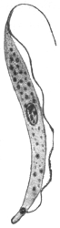
Fig. 40.—Trypanosoma
brucei. × 2,000. (After
Laveran and Mesnil.)
The general morphology and life history in
the vertebrate host is that of a typical trypanosome
(fig. 40). Its length is from 12 µ to 35 µ,
its breadth from 1·5 µ to 4 µ. Multiplication by
longitudinal division proceeds in the peripheral
blood (fig. 26), while latent, leishmaniform
bodies are produced in the internal organs.
Bruce and colleagues99 have quite recently
(June, 1914) described the development of a
Zululand strain of T. brucei in G. morsitans. The tsetse flies were
bred out in Nyasaland. In vertebrate blood the brucei strain
was polymorphic. The development was like that found for
T. gambiense in G. palpalis (fig. 30), and by Bruce and colleagues for
T. rhodesiense in G. morsitans in Nyasaland. Long trypanosomes
were found in the proventriculus of the tsetse. Crithidial, rounded
or encysted, and immature “blood forms” occurred in the salivary
glands; and finally infective, stumpy, “blood forms” were differentiated
in the salivary glands. The period of development of T. brucei
in G. morsitans takes about three weeks, and then the fly becomes
infective. Bruce believes that T. rhodesiense of Nyasaland and
T. brucei of Zululand are the same, their cycles of development
in G. morsitans being “marvellously alike.” (But see Laveran, p. 80.)
T. brucei has been cultivated with difficulty by Novy and MacNeal,
using blood agar. The best treatment for nagana is arsenic in some form.
It is probable that more than one trypanosome has been confused
under the name T. brucei, more especially as the occurrence of
many species of trypanosomes in various animals in Africa was not
suspected until comparatively recent times. It has been shown by
Stephens and Blacklock (1913) that the original Zululand strain of
T. brucei was monomorphic, while the organism sent from Uganda,
and at the time believed by Bruce to be the same as the Zululand
trypanosome, has been found to be polymorphic, with morphological
resemblances to T. rhodesiense. Stephens and Blacklock100 have
suggested the name T. ugandæ for the polymorphic trypanosome,
which, however, has marked resemblances with Trypanosoma
pecaudi, and they are, perhaps, identical. T. pecaudi was the name
given by Laveran101 in 1907 to the causal agent of “baleri” in equines
and sheep in the French Sudan. T. pecaudi, which is dimorphic,
is widely distributed in Africa. An extremely small number of both
T. pecaudi and T. ugandæ have been shown to possess posterior
nuclei. T. pecaudi is transmitted by various species of Glossina,
and is said to develop in the gut and proboscis of the fly.
On the other hand, Bruce and colleagues (1914), examining a
strain sent from Zululand in 1913, state that T. brucei is polymorphic.
Bruce (1914) suggests that passage through laboratory hosts has
influenced and altered the morphology of the parasite.
Trypanosoma evansi, Steel, 1885.
Syn.: Spirochæta evansi, Steel, 1885; Hæmatomonas evansi, Crookshank, 1886;
Trichomonas evansi, Crookshank, 1886.
Trypanosoma evansi, first found by Evans in 1880, in India, is the
causal agent of the disease known as “surra.” The malady affects
more particularly horses, mules, camels and cattle in India and neighbouring
countries, such as Burma and Indo-China. It occurs also in
Java, the Philippines, Mauritius and North Africa. Elephants may
be affected. A serious outbreak among cattle in Mauritius occurred
in 1902, the disease being imported into the island. The symptoms
are fever, emaciation, œdema, great muscular weakness and paralysis
culminating in death.
T. evansi varies from 18 µ to 34 µ in length and 1·5 µ to 2 µ in
breadth. It has a pointed posterior extremity, and, anteriorly, there
is a free portion to the flagellum (fig. 41). It is possibly monomorphic,
but a few broad forms occur. The trypanosome multiplies
by longitudinal fission in the blood. Rounded leishmaniform stages
occur in the spleen of the vertebrate host, which stages Walker102 (1912)
considers to be phases of schizogony.
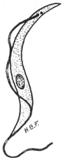
Fig. 41.—Trypanosoma
evansi. × 2,000. (Original.
From preparation by Fantham.)
The parasite is transmitted in nature by various species of Tabanus
and Stomoxys, though at present little is known of the life-history
within these invertebrate hosts.
Dogs are said to contract the disease by feeding on animals
dead of surra.
A variety of T. evansi is the cause of “mbori” in dromedaries
in Africa (Sahara and Sudan). Another possible variety, or closely
allied form, is T. soudanense, the causal agent
of “el debab” in camels and horses in North
Africa, especially Algeria and Egypt.
An extraordinary example of the possible
infection of a human being with an animal
trypanosome is recorded in the case of Professor
Lanfranchi, of the Veterinary School,
Parma. The Professor became infected with
trypanosomes, although only nagana and surra
were maintained in his laboratory, and he
himself had never visited the tropics. He
suffered from irregular attacks of fever and
was œdematous, but his mind remained clear.
The identification of the trypanosome from
Lanfranchi’s blood has been a matter of great
difficulty. Apparently Mesnil and Blanchard
(1914)103 consider the strain found in the patient is almost indistinguishable
in its reactions from T. gambiense, though the parasite is monomorphic.
Lanfranchi considers that he was infected with T. evansi.
Trypanosoma equinum, Voges, 1901.
Syn.: Trypanosoma elmassiani, Lignières.
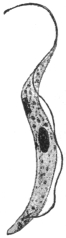
Fig. 42.—Trypanosoma
equinum. × 2,000. (After
Laveran and Mesnil.)
Trypanosoma equinum was found by Elmassian to be the cause of
the fatal disease, “mal de caderas,” of horses
and dogs, in South America (Paraguay, Argentine,
Bolivia). The name refers to the fact that
in the disease, as in other trypanosomiases,
the hind quarters become paralysed. Cattle
are refractory to inoculation.
T. equinum is about 22 µ to 24 µ long
and about 1·5 µ broad (fig. 42). Although this
trypanosome is very active, yet it is characterized
by the blepharoplast (kinetic nucleus)
being very minute or even absent, as the
granule sometimes seen may be the basal
granule of the flagellum.
The mode of transmission of T. equinum
is not known with absolute certainty. Migone
has shown that the parasite causes a fatal disease in the large South
American rodent, the capybara (Hydrochœrus capybara). This animal
appears to be a reservoir of the parasite. Dogs may become infected
by eating diseased capybaras, and it is suggested that the infection is
spread from the dogs to horses by the agency of fleas. Some authorities
consider that T. equinum may be spread by various Tabanidæ and by
Stomoxys. Neiva (1913)104 doubts all these modes of transmission in
Brazil, and suggests Chrysops or Triatoma as vectors.
Trypanosoma equiperdum, Doflein, 1901.
Syn.: Trypanosoma rougeti, Laveran and Mesnil.
The malady of horses known as “dourine” or “mal du coït” is
due to a trypanosome, T. equiperdum, discovered by Rouget in 1894.
“Dourine”—also known as “stallion disease” or “covering disease”—is
found among horses and asses in Europe,
India, North Africa and North America. The
trypanosome is transmitted by coitus, and so
far as is known not by insect agency.
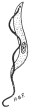
Fig. 43.—Trypanosoma
equiperdum. × 2000 approximately.
(Original. From
preparation by Fantham.)
The progress of the disease may be considered
under three periods. The period of
œdema, when signs of œdema of the genitalia
are seen. The œdema is generally painless
and non-inflammatory. This period lasts about
a month. It is succeeded by the period of
eruption, which sets in about two months
after infection. Circular œdematous areas
(“plaques”), often about the size of a two-shilling
piece, appear under the skin of the sides
and hind quarters, and also, at times, under the
skin of the neck, thighs and shoulders. The
eruption is variable, but usually lasts about a week and leaves the
animal in an enfeebled condition. Gland enlargement and swelling
of the joints and synovia also may occur. The third period of the
disease is described as that of anæmia and paralysis. The animal
becomes very anæmic, emaciation is marked, superficial non-healing
abscesses often form, and conjunctivitis and ulcerative keratitis can
occur. Paralysis ensues, and in from two to eighteen months the
animal dies. In the acute form of the disease the animal may die
after the first period from acute paralysis.
It is difficult to find the trypanosomes in naturally infected
animals, and they are best obtained from the plaques of the
eruption. Apparently the parasite occurs more in the lymph than
in the blood.
Ruminants are said to be refractory to this trypanosome.
T. equiperdum is about 25 µ to 28 µ in length on an average, but
varies from 16 µ to 35 µ. Its cytoplasm is relatively clear, and does not
show chromatic granules (fig. 43). It is stated to be monomorphic.
It has been shown recently by Blacklock and Yorke (1913)105 that
there is another trypanosome giving rise to dourine in horses. This
trypanosome is dimorphic (resembling T. pecaudi and T. ugandæ),
and is named T. equi. Previously T. equiperdum and T. equi had
been confused.
Uhlenhuth, Hübner and Worthe have demonstrated the presence
of endotoxins in T. equiperdum. These endotoxins may be set free
by trypanolysis.
Trypanosoma theileri, Bruce, 1902.
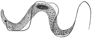
Fig. 44.—Trypanosoma theileri. × 2,000.
(After Laveran and Mesnil.)
This parasite, 60 µ to 70 µ long, and 4 µ to 5 µ broad, is distinguished
for its large size, though it is not so large as T. ingens
from Uganda oxen, whose length may be 72 µ to 122 µ, and breadth
7 µ to 10 µ. The posterior
end of T. theileri is drawn
out. Small forms of the flagellate
are known, 25 µ to 53 µ
in length. Probably other
forms of the parasite have the
nucleus posterior, and these
flagellates were formerly separated
as T. transvaaliense
(Laveran, 1902). Myoneme fibrils may be seen on its body. The
pathogenicity of this organism is doubtful, it was formerly thought
to be the causal agent of “gall-sickness” in cattle in South Africa.
T. theileri also occurs in Togoland, German East Africa, and Transcaucasia.
Allied or identical parasites occur in cattle in India.
Trypanosoma theileri, specific to cattle, is perhaps transmitted by
the fly Hippobosca rufipes in South Africa.
Trypanosoma hippicum, Darling, 1910.
Trypanosoma hippicum causes the disease of mules known as
“murrina.”106 It was found in mules imported to Panama from the
United States. It can live in other equines. The parasite varies from
18 µ to 28 µ in length, and is from 1·5 µ to 3 µ broad. Its undulating
membrane is little folded. The trypanosome has a noticeable
blepharoplast. It can penetrate mucous membranes, and it is thought
that the trypanosome may be transmitted during coitus. It may also
be spread mechanically by species of Musca, Sarcophaga and Compsomyia,
sucking the wounds of infected animals and carrying over the
trypanosomes to wounds on healthy ones.
Endotrypanum schaudinni, Mesnil and Brimont, 1908.
This organism was discovered in the blood of a sloth (Cholœpus
didactylus), in South America (French Guiana).107 It possesses special
interest, in that the best known form of the organism is endoglobular,
inhabiting the erythrocytes of the sloth. A free trypanosome in the
same animal was considered to be different from the endoglobular
form, which was somewhat like a peg-top, and possessed a short
flagellum. Darling108 (November, 1914) has seen the organism in
Panama. He describes free crithidial forms in shed blood, but not
in the blood-stream of the sloth.
Trypanosoma boylei, Lafont, 1912.
This is a parasite of the Reduviid bug, Conorhinus rubrofasciatus.
The insect attacks man in Mauritius, Réunion and other places.
Lafont infected rats and mice by intraperitoneal injection with the
gut-contents of infected bugs. Trypanosomes appeared in the mice.
Other flagellate types were assumed by the parasites in the bug.
Monomorphic Trypanosomes.
A number of trypanosomes, characterized by relative uniformity
in size and structure, may be considered under this heading. They
occur in cattle, sheep, goats and horses in Africa, especially West
Africa. Morphologically, they are characterized by the posterior
(aflagellar) part of the body being swollen, while the anterior part
narrows. The nucleus is central and situated at the commencement
of the narrowing of the body. The blepharoplast is almost
terminal, the undulating membrane is narrow and not markedly
folded, so that the flagellar border lies close to or along the body.
The flagellum may or may not possess a free portion.
Some recent workers have considered that T. brucei (Zululand
strain) and T. evansi are also monomorphic, but they do not exhibit
the general characteristics outlined above. T. brucei and T. evansi
have already been considered separately.
The monomorphic trypanosomes, as defined above, include:—
Trypanosoma vivax, Ziemann, 1905.
Fig. 45.—Trypanosoma
vivax. × 2,000. (Original.
From preparation by Fantham.)
This trypanosome109 occurs in cattle, sheep and goats, and was first
found in the Cameroons. It is fatal to cattle. Equines are also
affected. Antelopes are the possible reservoirs of the trypanosome.
It is probably transmitted by Glossina palpalis and other tsetse flies.
Its movement is very active. It possesses a free flagellum (fig. 45) and
it averages 23 µ to 24 µ in length. T. cazalboui (Laveran, 1906)—the
causal agent of “souma” in bovines and equines in the French Sudan—is
probably synonymous with T. vivax.
Trypanosoma capræ (Kleine, 1910) is allied,
but is somewhat broader and more massive.
It was found in goats in Tanganyika.
Trypanosoma congolense, Broden, 1904.
Probable synonyms.—Trypanosoma dimorphon, Laveran
and Mesnil, 1904; Trypanosoma nanum, Laveran,
1905; Trypanosoma pecorum, Bruce, 1910; Trypanosoma
confusum, Montgomery, 1909.
This trypanosome causes disease among
horses (e.g., Gambia horse sickness), cattle,
sheep, goats, pigs, and dogs. It is widely distributed
in Central Africa (e.g., Gambia, Congo,
Uganda, Nyasaland), the strain probably being
maintained naturally in big game. It is transmitted
by various Glossinæ, and perhaps by Tabanus and Stomoxys. It
is said to develop in the gut and proboscis of Glossina palpalis and
G. morsitans. The trypanosome averages 13 µ to 14 µ in length and
has no free flagellum (fig. 46). It is about 2 µ broad. Formerly
T. nanum and T. pecorum were said to differ in their pathogenicity, the
former being said not to infect the smaller laboratory animals. Yorke
and Blacklock (1913), however, consider
that the virulence varies and that these
trypanosomes are probably the same.
Fig. 46.—Trypanosoma
congolense. × 2,000. (Original.
From preparation by
Fantham.)
Fig. 47.—Trypanosoma
uniforme. × 2,000. (Original.
From preparation by
Fantham.)
The T. dimorphon originally obtained by Dutton and Todd (1903)
in Gambian horse sickness has been shown to be a mixture of T. vivax
and T. congolense.
Trypanosoma simiae (T. ignotum) is like T. congolense. It averages
17·5 µ long. It is virulent to monkeys and pigs.
Trypanosoma uniforme, Bruce, 1910.
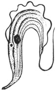
Fig. 48.—Trypanosoma
rotatorium, from blood of
a frog. × 1,400. (After
Laveran and Mesnil.)
This trypanosome was found in oxen in Uganda.110 It can be inoculated
to oxen, goats and sheep, but is refractory to dogs, rats and
guinea-pigs. It has been found in antelopes. It resembles T. vivax,
but is smaller (fig. 47), averaging 16 µ in length. A free flagellum
is present. It is transmitted by Glossinæ.
Many other trypanosomes occur in mammals,
while birds, reptiles, amphibia (fig. 48)
and fish also harbour them. The discussion of
these forms does not come within the scope
of the present work. They are dealt with
in Laveran and Mesnil’s “Trypanosomes et
Trypanosomiases,” 2nd edit., 1912.
General Note on Development of
Trypanosomes in Glossina.
Before concluding the account of trypanosomes,
it may be of interest to remark that
several African trypanosomes develop in various species of Glossina,
and are found in different parts of the alimentary tract and in the
proboscis. Thus (a) T. vivax, T. uniforme and T. capræ develop in the
fly’s proboscis (labial cavity and hypopharynx) only; (b) T. congolense,
T. simiæ and T. pecaudi develop first in the gut of the fly and then
pass forward to its proboscis; and (c) T. gambiense and T. rhodesiense
develop first in the gut and later invade the salivary glands of the
tsetse. The proboscis or the salivary glands in such cases are termed
by Duke111 the anterior station of the trypanosome, wherein it completes
its development.
Adaptation of Trypanosomes.
These flagellates may exhibit power of adaptation to changes of
environment, such as those due to the administration of drugs, change
of host, etc. A few examples of such mutations may be briefly
considered:—
(1) Blepharoplastless Trypanosomes.—T. brucei may become resistant
to pyronin and oxazine. Accompanying this drug resistance is a
change in morphology, namely, the loss of the blepharoplast
(Werbitzki).112 A race or strain of blepharoplastless trypanosomes may
be thus produced which retains its characteristic feature after as many
as 130 passages (Laveran).113 Oxazine is the more powerful drug, and
it acts directly on the blepharoplast. (Compare the natural blepharoplastless
character of T. equinum.)
(2) Reference has been made on p. 93 to the experiments of
Gonder, who showed that a strain of T. lewisi rendered resistant to
arsenophenylglycin lost its resistance after passage through the rat
louse. This is in marked contrast with the retention of drug resistance
during passage by inoculation from rat to rat.
(3) T. lewisi from the blood of a rat when transferred to a snake
seems largely to disappear, as very few flagellates are seen. When
blood from the snake is inoculated into a clean rat, then trypanosomes
reappear in the rat, but they are not all like those originally
inoculated. It seems certain that, in such a case, changes in form
and virulence of the trypanosome have occurred. Similar experiments
were made with T. brucei from rats to adders and other animals and
back to rats. Changes in the form and virulence of T. brucei occurred.
These interesting experiments were performed by Wendelstadt and
Fellmer.114
Genus. Herpetomonas, Saville Kent, 1881.
Herpetomonas is a generic name for certain flagellates possessing
a vermiform or snake-like body, a nucleus placed approximately centrally,
and a blepharoplast (kinetic nucleus) near the flagellar end.
There is no undulating membrane (fig. 49, a). The organisms included
in this genus certainly possess one flagellum, while according
to Prowazek (1904) Herpetomonas muscæ-domesticæ, the type species,
possesses two flagella united by a membrane. Patton,115 Porter116 and
others affirm, however, that the biflagellate character of H. muscæ-domesticæ
(from the gut of the house-fly) is merely due to precocious
division. The matter is further complicated by the generic name
Leptomonas, given by Kent in 1881, to an uniflagellate organism found
by Bütschli in the intestine of the Nematode worm, Trilobus gracilis.
This parasite, Leptomonas bütschlii, has not yet been completely
studied. Until these controversial points relating to the identity or
separation of Herpetomonas and Leptomonas have been satisfactorily
settled, we may retain the better known name Herpetomonas for such
uniflagellate, vermiform organisms. However, the name Leptomonas,
having been used by Kent two pages earlier in his book (“Manual of
the Infusoria”) than Herpetomonas, would have priority if the two
generic names were ultimately shown to be synonymous.
A full discussion of these interesting and important flagellates
hardly comes within the purview of the present work; brief mention
can only be given here to certain species.
The Herpetomonads occur principally in the digestive tracts of
insects, such as Diptera and Hemiptera. They are also known in the
guts of fleas and lice, but are not confined to blood-sucking insects.
One example, H. ctenocephali (Fantham, 1912)117 occurs in the digestive
tracts of dog fleas, Ctenocephalus canis, in England, France, Germany,
Italy, India, Tunis, etc. It is a natural flagellate of the flea, and might
easily be confused with stages of blood parasites in the gut of the dog
flea. Dog fleas are stated by Basile to transmit canine kala-azar, which
is believed to be the same as human infantile kala-azar. Confusion is
further likely to arise since herpetomonads pass through pre-flagellate,
flagellate and post-flagellate or encysted stages; pre- and post-flagellate
stages being oval or rounded and Leishmania-like. The post-flagellate
stages are shed in the fæces, and are the cross-infective stages by
means of which new hosts are infected by the mouth. The possible
presence of such natural flagellates must always be considered when
experimenting with fleas,
lice, mosquitoes, etc., as
possible vectors of pathogenic
flagellates like Leishmania
and Trypanosoma.
H. pediculi (Fantham,
1912) occurs in human
body lice.118 See further
remarks on pp. 107, 112.
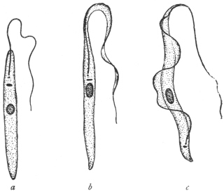
Fig. 49.—a, Herpetomonas; b, Crithidia;
c, Trypanosoma. (After Porter.)
Laveran and Franchini
(1913–14)119 have recently
succeeded in inoculating
Herpetomonas ctenocephali,
from the gut of the dog
flea, intraperitoneally into
white mice, and producing
an experimental leishmaniasis
in the mice. A
dog was also infected. They have also succeeded in infecting mice
with H. pattoni—a natural flagellate of the rat flea—by mixing infected
rat fleas with the food of the mice, and by causing them to
ingest infected fæces of rat fleas. Further, they have shown that
infection with the herpetomonas occurs naturally by this method,
that is, by the rodents eating the fleas and not by the insects inoculating
the flagellates into the vertebrates when sucking blood. These
experiments shed an interesting light on the probable origin of
Leishmania and its cultural herpetomonad stage, which were very
probably once parasitic flagellates in the gut of an insect.
Fantham and Porter120 (1914–15) have shown that young mice may
be inoculated or fed with Herpetomonas jaculum, from the gut of the
Hemipteran, Nepa cinerea (the so-called “water-scorpion”), with fatal
results. The pathogenic effects are like those of kala-azar. They also
showed that the post-flagellate stages of the herpetomonads seemed
most capable of developing in the vertebrate.
A herpetomonad, H. davidi, has been found in the latex of species of the plant-genus
Euphorbia in Mauritius, India, Portugal, etc. It is apparently transmitted to
the plants by Hemiptera. The plants sometimes suffer from “flagellosis.”
Franchini (1913)121 has described a new parasite, Hæmocystozoon
brasiliense, from the blood of a man who had lived in Brazil for many
years. It possesses flagellate and rounded stages, and is closely allied
to the herpetomonads.
Genus. Crithidia, Léger, 1902, emend. Patton, 1908.
Crithidia is the generic name of vermiform flagellates with a
central nucleus, a blepharoplast or kinetic nucleus in the neighbourhood
of the principal nucleus, and a rudimentary undulating membrane
bordered by a flagellum arising from a basal granule, which is the
centrosome of the kinetic nucleus (fig. 49b). The anterior or flagellar
end of the body is attenuated and fades off as the undulating membrane.
Crithidia fasciculata, the type species, was found by Léger in the
alimentary canal of Anopheles maculipennis. Crithidia occur in bugs,
flies, fleas,122 and ticks. Some of them are found in the body-fluid
of the invertebrate host as well as in the gut. Others may be restricted
to the body cavity or intestine respectively. C. melophagia from the
sheep-ked, Melophagus ovinus, and C. hyalommæ from the hæmocœlic
fluid of the tick, Hyalomma ægyptium, pass into the ovaries and eggs
of their hosts, and the young keds or ticks are born infected.
C. fasciculata has been shown by Laveran and Franchini to be
inoculable into white mice, producing a sort of experimental leishmaniasis
therein. In one case cutaneous lesions were produced like
those of Oriental sore.
Crithidia are natural flagellates of Arthropoda, with their own
pre-flagellate, flagellate and post-flagellate stages, and must not be
confused with transitory crithidial stages of trypanosomes.
Genus. Leishmania, Ross, 1903.
With an oval body containing nucleus and blepharoplast (kinetic nucleus)
but no flagellum. An intracellular parasite in the vertebrate host.
Included in the genus Leishmania are three species, namely:—
(1) Leishmania donovani, Laveran and Mesnil, 1903, the parasite
of Indian kala-azar, a generalized systemic disease, usually
fatal, occurring in subjects of all ages.
(2) Leishmania tropica, Wright, 1903, the parasite of Delhi boil,
Oriental sore, Aleppo button—a localized, cutaneous disease,
usually benign.
(3) Leishmania infantum, Nicolle, 1908, the parasite of infantile
kala-azar, occurring in children (and a few adults) around
the shores of the Mediterranean. The disease is perhaps a
form of Indian kala-azar, and the parasite is probably identical
with L. donovani.
These diseases may be termed collectively leishmaniases. The
morphology of the various species is practically identical.
Leishmania donovani, Laveran and Mesnil, 1903.
Syn.: Piroplasma donovani, Laveran and Mesnil.
The parasite of Indian kala-azar was demonstrated in 1900 by
Leishman from a post-mortem examination of a case of “Dum-Dum
fever,” but details were not published till May, 1903. In July, 1903,
Donovan found similar bodies from cases in Madras. Rogers
succeeded in cultivating the parasite in July, 1904.123 The original
centre of the disease was probably Assam; it occurs also in Madras,
Ceylon, Burma, Indo-China, China and Syria. A variety of this leishmaniasis
is found in the Sudan. The patient becomes emaciated,
with a greatly enlarged spleen. There is anæmia and leucopenia.
The parasite, commonly known as the Leishman-Donovan body,
is intracellular (fig. 50, 2, 3). It is found in the endothelial cells of the
capillaries of the liver, spleen, bone-marrow, lymphatic glands and
intestinal mucosa, and in the macrophages of the spleen and bone-marrow.
Some host cells may contain many parasites. It is rather
rare in the circulating blood, but may be found in the blood from
the femoral, portal and hepatic veins. It does not occur in the red
blood corpuscles as was formerly thought. The parasites liberated
from the endothelial cells are taken up by the mononuclear and
polymorphonuclear leucocytes. The Leishman-Donovan body is the
resting stage of a flagellate. As found in man it is a small, oval
organism, about 2·5 µ to 3·5 µ in length by 2 µ in breadth, and containing
two chromatinic bodies, corresponding to the nucleus and
kinetic nucleus (blepharoplast) of a flagellate. The latter element is
the smaller and more deeply staining, and is usually placed at the
periphery, transversely to the longer axis of the oval organism.
There is sometimes a very short, slightly curved filament to be seen,
which may be a rhizoplast. Multiplication takes place by binary or
multiple fission. The presence of the parasite used to be demonstrated
by splenic or hepatic puncture; nowadays it can be demonstrated in
peripheral blood, e.g., of the finger, or by culture of infected blood.
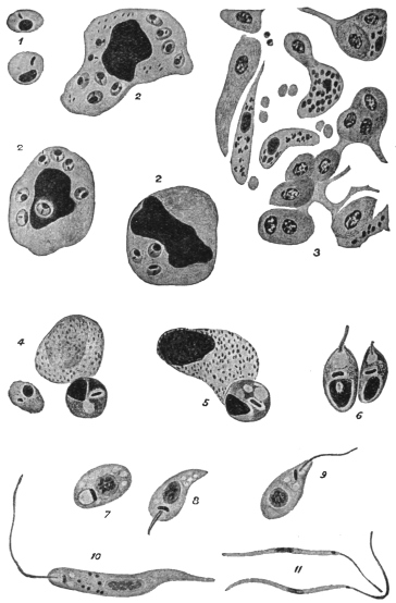
Fig. 50.—Leishmania donovani. 1, Free forms, each with nucleus and rod-shaped blepharoplast
(after Christophers); 2, endothelial cell and leucocytes containing parasites (after
Christophers); 3, capillary in the liver showing endothelial cells containing parasites (after
Christophers); 4, two parasites escaping from a leucocyte in the alimentary canal of the bug
(after Patton); 5, further development in bug (after Patton); 6, young flagellate forms in bug
(after Patton); 7-11, culture forms (after Leishman); 7, 8, 9, show development of flagellum.
L. donovani can be cultivated in citrated splenic blood, under
aerobic conditions, at 22° to 25° C. This was first accomplished
by Rogers (1904). It is not so easily culturable as L. infantum on
the Novy-MacNeal-Nicolle medium.124 L. donovani is inoculable with
some difficulty into experimental animals—in India, white rats, white
mice, dogs and monkeys (Macacus spp.), have been inoculated. The
Sudan variety, somewhat less virulent, is inoculable to monkeys. Row
also produced a local lesion in Macacus sinicus by subcutaneous inoculation
of L. donovani. Parasites taken from such a local lesion were
found to be capable of producing a generalised infection in Macacus
sinicus and white mice.
In cultures the various species of Leishmania all grow into
herpetomonad, uniflagellate organisms (fig. 50, 10), about 12 µ to 20 µ
in body length. On this account Rogers125 and Patton place the
Leishman-Donovan body within the genus Herpetomonas. The
method of culture may be used in diagnosing leishmaniases.
Kala-azar is very probably an insect-borne disease. Patton126 suspects
the bed-bug to be the transmitter and finds (fig. 50, 4-6) that the
Leishman-Donovan body can develop into the flagellate stage in the
digestive tract of the bed-bug. Feeding experiments are unsatisfactory,
since there are very few cases in which the parasites occur in sufficient
numbers in the peripheral blood to make the infection of the insect
possible, or at any rate easy. In examining the alimentary tracts of
insects for possible flagellate stages of Leishmania, it must be remembered
that in many insects natural flagellate parasites, belonging to the
genus Herpetomonas, may occur therein; such natural insect flagellates
may be harmless, and have no connection with the life-cycle of
L. donovani. Natural herpetomonads are known to occur in the
alimentary tracts of flies, mosquitoes, sand-flies, fleas and lice, but
not in bed-bugs. Further, if such flagellates are able to be inoculated
into and live within vertebrate hosts, producing symptoms like those
of leishmaniasis, the origin of kala-azar is indicated (see pp. 104, 112).
Leishmania tropica, Wright, 1903.
Syn.: Helcosoma tropicum, Wright, 1903; L. wrighti, Nicolle, 1908; Ovoplasma
orientale, Marzinowsky and Bogrow.
It is believed by some that the parasite was first described by Cunningham in
1885, and studied by Firth in 1891, being called by him Sporozoon furunculosum.
If these earlier studies were of the parasite, then its correct name is L. furunculosa,
Firth, 1891.
The benign disease produced by this parasite has received many
names, among the best known being Oriental sore, Tropical sore,
Delhi boil and Aleppo button. These names, however, are not happy
ones, as cutaneous leishmaniasis (e.g., on the ear) is now known to
occur in the New World, for example in Mexico, Venezuela, Brazil
and neighbouring States. However, it may be necessary to subdivide
cutaneous leishmaniases later.
In the Old World the disease occurs in India, Persia, Arabia and
Transcaucasia. It is also known in Algeria, Northern Nigeria, Egypt,
Sudan, Crete, Calabria, Sicily and Greece.
The boils often occur on the face, and before ulceration the
parasites may be found in the cells at the margin and floor of the
“button.” In searching for parasites the scab should be removed and
scrapings made from the floor and edges. Where lesions occur
atrophy of the epidermis takes place, and infiltration of mononuclear
cells (e.g., plasma cells, lymphoid and endothelial cells) follows.
The parasites are intracellular, being found inside mononuclear cells.
In non-ulcerating sores, Cardamitis found some free parasites. Non-ulcerating
forms are said to occur in the Sudan. In the Old World
the sores are often limited to exposed surfaces of the body. Infection
of mucous membranes (such as the lip, palate, buccal and nasal
membranes) may occur, especially in South America, and are often
known there as “Espundia.” Christopherson (1914) has recorded a
case in Khartoum.
Leishmania tropica is equally well cultivated on Novy-MacNeal-Nicolle
medium or on citrated blood. The usual temperature for
cultivation is 22° to 28° C., though Marzinowski claims to have
cultivated the parasite at 37° C. L. tropica can be inoculated
into monkeys and dogs, with the production of local lesions.
Material from a human sore or flagellates from a culture may be thus
successfully inoculated. Also infected material may be rubbed
directly into a scarified surface. The incubation period is long,
extending over several months. The duration of the disease may be
from twelve to eighteen months. Recovery from one attack of tropical
sore confers immunity, and the Jews in Bagdad inoculate their children
with the disease on a part of the body which will be covered, and
so secure immunity in adult life.
The mode of transmission of L. tropica is unknown. Wenyon
(1911)127 has found that the parasite develops into the flagellate stage
in the digestive tract of Stegomyia fasciata in Bagdad. Patton (1912)128
has found similar development in the bed-bug in Cambay. The
house-fly, Phlebotomus and Simulium have been suspected as transmitters
in different parts of the world.
An interesting announcement has been made recently (May, 1913),
that Neligan has found that L. tropica occurs in dogs in Teheran,
Persia, producing ulcers on the dogs’ faces (cf. natural occurrence of
L. infantum in dogs—see p. 110). Yakimoff and Schokhor (1914),129
have found the disease in dogs in Tashkent.
Gonder130 (1913) has performed some interesting experiments
showing the relation of infantile kala-azar to Oriental sore. Gonder
infected mice with L. infantum and with L. tropica. He used culture
material and injected intraperitoneally or intravenously. In each a
general infection resulted, with enlargement of the liver and spleen.
Later, however, mice injected with Oriental sore (North African
variety) developed peripheral lesions on the feet, tail and head, and
the lesions contained Leishmania. No such peripheral lesions
developed in the case of the mice infected with the kala-azar virus.
Gonder suggested that Oriental sore, like kala-azar, is really a general
infection overlooked in its earlier stages, and that it is in the later
stages that peripheral lesions on the skin are developed. Row (1914)131
also obtained a general infection in a mouse by the injection of
cultures of L. tropica from Oriental sore of Cambay.
Leishmania infantum, Nicolle, 1908.132
Infantile splenic anæmia has been long known in Italy. It also
occurs in Algeria, Tunis, Tripoli, Syria, Greece, Turkey, Crete, Sicily,
Malta,133 Spain and Portugal. This leishmaniasis is, then, distributed along
the Mediterranean littoral; also in Russia. Cathoire (1904) in Tunis
and Pianese (1905) in Italy were among the first to see the parasite.
Nicolle then found the parasite in patients in Tunis, and further found
spontaneous infection in dogs. The patients are usually children
between the ages of 2 and 5 years. There are a few cases known in
which the infantile type of leishmaniasis occurred in youths and adults
of the ages of 17 to 19, while one patient in Calabria was 38 years old.
The symptoms are like those of Indian kala-azar. Several Italian
investigators and others consider that L. infantum is the same as
L. donovani, and that the latter name should be used for the parasite
of Mediterranean leishmaniasis. This view, as to the identity of
L. donovani and L. infantum, seems coming into general favour.
There are, however, differences between the Indian and infantile
kala-azars, in addition to the ages of the patients affected, thus:
(a) As regards cultures, it is found that L. infantum is readily grown
on the Novy-MacNeal-Nicolle (“N.N.N.”) medium (saline blood-agar),
and that sub-cultures are easily obtained; in citrated blood
L. infantum grows with difficulty. The reverse is the case with
regard to culture media for L. donovani, which grows with difficulty
on the N.N.N. medium, but relatively easily in citrated splenic blood.
(b) Considering inoculability into experimental animals, it is found that
L. donovani is inoculated generally with some difficulty into white rats,
white mice and monkeys, and with greater difficulty into dogs, while
L. infantum can be inoculated into several experimental animals,
especially into dogs and monkeys, with ease. (c) At present L. donovani
is not known to occur spontaneously in animals, but L. infantum is
found naturally in dogs in the Mediterranean region, and the disease in
dogs is often referred to as canine kala-azar. Kittens have occasionally
been found infected. However, these differences must not be emphasized
too much.
The material for cultivation is obtained from punctures of spleen,
liver or bone-marrow of cases infected with L. infantum. It is not
always easy, however, to infect from cultures, as the cultural flagellates
inoculated into the body are often phagocytosed.
Similarly, the material for animal inoculation is obtained from
emulsions of infected spleen, liver or bone-marrow. Dogs and
monkeys are easily inoculated with such material; Nicolle inoculates
into the liver or the peritoneal cavity. Mice, white rats, guinea-pigs
and rabbits only show slight infections after such inoculations.
Dogs infected experimentally with infantile leishmaniasis may
show either acute or chronic symptoms. The acute course occurs
more often in young dogs, and is usually fatal in three to five months.
The chronic course is found more commonly in older dogs, and may
last seventeen to eighteen months. In acute forms there is irregular
fever, progressive wasting, diarrhœa occasionally, motor disturbances
involving the hind quarters, and the animal dies in a comatose condition.
In the chronic form the animal may appear well, except for
loss of weight. The parasites may be found in the internal organs of
these experimental dogs, but are not numerous in the peripheral
blood except at times of high fever. Experimental monkeys live about
three months.
It may be interesting to record the number of dogs found to be
infected naturally with leishmaniasis in various countries. In Tunis,
Nicolle and Yakimoff found about 2 per cent. infected out of about
500 dogs examined. Sergent in Algiers found 9 infected out of 125
dogs examined. In Italy and Sicily, Basile found about 40 per cent.
of the dogs to be infected out of 93 examined at Rome and
Bordonaro. Cardamitis found 15 infected out of 184 examined in
Athens. In Malta, Critien found 3 infected out of 30 dogs examined.
Alvares found 1 infected dog out of 19 examined in Lisbon. Pringault
has recently (December, 1913) found an infected dog in Marseilles.134
Yakimoff and Schokhor found 24 per cent. infected out of 647 dogs
examined in Turkestan.
The distribution of the parasites in the body of the human patient
is much the same as in the case of Indian kala-azar. Critien records
the finding of parasites in the mucous flakes of the stools of a three-year-old
Maltese child.135 Intestinal lesions rarely occur in infantile
leishmaniasis.
Ætiology.—Infantile leishmaniasis is stated to be transmitted by
fleas, especially dog fleas, Ctenocephalus canis (= Pulex serraticeps), and
by Pulex irritans. Children living in contact with infected dogs may be
bitten by infected dog fleas, and so contract the disease. Basile (1910–11)
and Sangiorgi (1910) state that they found L. infantum parasites in the
digestive tract of the dog flea. After searching they found infected
dog fleas on the beds, mattresses, and pillows used by children
suffering from the disease. Franchini (1912) thinks that Anopheles
maculipennis may be concerned in the transmission.
Basile136 tried a number of experiments to show that infantile
leishmaniasis is transmitted by fleas, thus:—
(1) Fleas were taken from a healthy dog. They were placed in
vessels containing infected spleen-pulp and allowed to feed thereon.
The fleas were then killed and dissected, and portions of the gut-contents
examined for parasites. The remainder of the gut was
emulsified and injected into a young puppy, whose bone-marrow had
been shown previously to be uninfected. Basile states that the puppy
became infected. The parasites are said to increase in number in
the flea’s gut.
(2) Two healthy pups, each a month old, and born in the laboratory,
were placed in a disinfected, flea-proof cage. A few days after,
an infected dog was placed in the cage, so that fleas from the
infected dog could pass on to the puppies. A month later the two
pups became infected, parasites being found in them after liver
puncture. A number of control puppies from the same litter
remained uninfected and in good health.
(3) Basile next used other laboratory-born puppies, a month old.
Four of the litter were placed in a disinfected, flea-proof gauze cage
in Rome. The cage was isolated from other dogs. Fleas obtained
from an infected area in Sicily were placed in the cage. The puppies
were examined by hepatic puncture, but were found to be negative
for two months. Then two of the puppies showed infection, and
six days later the remaining two puppies were found to be infected,
and all four died. They showed irregular temperatures, and were
getting thin. Control puppies remained healthy.
From these experiments Basile concludes that fleas transmit
leishmaniasis. However, Basile did not exclude the possible occurrence
of natural herpetomonads in the gut of the fleas.137 Herpetomonas
ctenocephali is known to occur in the gut of Ctenocephalus canis.
A natural Herpetomonas is also known in the gut of Pulex irritans,
as well as a Crithidia (C. pulicis, Porter). These natural flagellates of
the fleas pass through non-flagellate stages, like the Leishman-Donovan
body. In consequence Wenyon and Patton, among others, have
criticized Basile’s results. Further, other investigators, such as
Wenyon and Da Silva (1913), have repeated Basile’s flea experiments
and been unable to confirm them.
In feeding and inoculation experiments the incubation period of
the parasite may be long, and so it is necessary to wait a long time to
see whether the parasite will develop.
Immunity.—Nicolle has tried some experiments with L. infantum
and L. tropica. He finds that in animals recovery from an attack of the
former confers immunity against infection by the latter and vice-versâ.
Laveran138 records that a monkey having an immunity against L.
infantum was also immune to L. donovani.
As mentioned on p. 103, Laveran and Franchini (1913), working in
Paris, have succeeded in inoculating Herpetomonas ctenocephali, a
natural flagellate in the gut of the flea, Ctenocephalus canis, into
white mice. Leishmaniform stages of the flea flagellate were recovered
from the peritoneal exudate, blood and organs of the mice
some weeks after inoculation. The parasites may also be conveyed by
way of the digestive tract of the vertebrate. Similar experiments have
succeeded with H. pattoni. These experiments go to show, together
with those of Fantham and Porter with H. jaculum (see p. 104), that, in
the words of the latter authors, “it may be expected that the various
leishmaniases, occurring in different parts of the world, will prove to
be insect-borne herpetomoniases.”
Genus. Histoplasma, Darling, 1906.
Under the name Histoplasma capsulatum,139 Darling described small
round or oval parasites, enclosed in a refractile capsule, and each containing
a single nucleus. The bodies were found in cases of splenomegaly
in Panama. They occurred in the endothelial cells of the
small blood-vessels of the liver, spleen, lungs, intestine and lymphatic
glands, and also within the leucocytes. A few flagellates were stated to
occur in the lungs. The parasite has usually been placed near Leishmania,
but recently Rocha-Lima has stated that Histoplasma is a yeast.
Genus. Toxoplasma, Nicolle and Manceaux, 1908.
The genus was created for crescentic, oval or reniform parasites, 2·5 µ to 6 µ by
2 µ to 3 µ, possessing a single nucleus and multiplying by binary fission. They occur
in mononuclear and polymorphonuclear cells in the blood, spleen, liver, peritoneum
etc. (fig. 51). The parasites have been found in the gondi, dog, rabbit, mole, mouse,
pigeon and other birds. Although various species names have been given to the
parasites in these hosts, it seems probable, from cross infection experiments, that
there is but one species with several physiological races. Splendore140 (1913) has
described a flagellate stage.
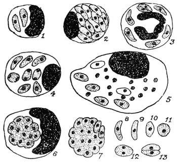
Fig. 51.—Toxoplasma gondii, endocellular or free in the peritoneal exudate of infected
mice. 1, 2, mononuclear leucocytes containing toxoplasms. 3, polynuclear, containing
parasites. 4, 5, 6, endothelial cells containing toxoplasms, agglomerated in 6. 7, agglomeration
forms. 8–11, free forms. 12–13, division stages. × 1,600. (After Laveran and Marullaz.)
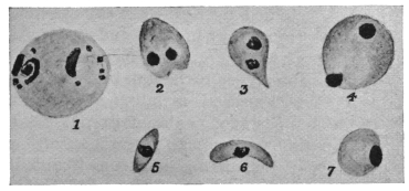
Fig. 52.—Toxoplasma pyrogenes. 1, body found in blood. 2–7, bodies found
in spleen.
[1 is about the size of a red blood corpuscle, as drawn in the figures]. Magnification not
stated. (After Castellani.)]
Castellani (1913–14)141 has described similar parasites from a case of
splenomegaly, with fever of long standing, in a Sinhalese boy. The
bodies were found in the spleen and more rarely in the blood (fig. 52).
Castellani has named them Toxoplasma pyrogenes. Further researches
are needed.
THE SPIROCHÆTES.
The Spirochætes are long, narrow, wavy, thread-like organisms,
with a firm yet flexible outer covering or periplast. There is a diffuse
nucleus internally in the form of bars or rodlets of chromatin
distributed along the body. In some forms there is a membrane or
crista present (fig. 53), which in the past was compared with the
undulating membrane of a trypanosome, but the membrane of a
spirochæte does not undulate. Progression is very rapid, corkscrew-like
and undulatory movements occurring simultaneously.
The genus Spirochæta was founded by
Ehrenberg in 1833 for an organism which
he discovered in stagnant water in Berlin.
Ehrenberg named the organism Spirochæta
plicatilis. According to Zuelzer (1912) S.
plicatilis does not possess a membrane or
crista, but an axial filament. S. gigantea has
been described by Warming from sea-water.
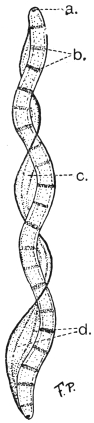
Fig. 53.—Spirochæta balbianii.
a, basal granule or
polar cap. b, chromatin rodlets.
c, membrane (“crista”).
d, myonemes in membrane.
(After Fantham and Porter.)
Spirochætes occur in the crystalline style
and digestive tract of many bivalve molluscs.
The first molluscan spirochæte to be studied
was that of the oyster, named by Certes (1882)
“Trypanosoma” balbianii (fig. 53). Similar
spirochætes, probably belonging to the same
species, occur in various species of Tapes and
in Pecten (the scallop). S. balbianii has rounded
ends (fig. 53). Other spirochætes occur in freshwater
mussels (Anodonta spp). S. anodontæ,
studied by Keysselitz (1906) and by Fantham
(1907), has pointed ends. Gross (1911) suggested
the generic name Cristispira for molluscan
spirochætes, because they possess a
well-marked membrane or “crista,” which
appears to be absent from S. plicatilis, according
to Zuelzer’s researches.
Schaudinn in 1905 founded the genus
Treponema for the parasite of syphilis (T.
pallidum), discovered by him and by Hoffmann.
According to Schaudinn the Treponemata have no membrane or
crista. The pathogenic agent of yaws or frambœsia, discovered by
Castellani, is also placed in the genus Treponema, as T. pertenue.
There remain the blood spirochætes. It is somewhat disputed as
to whether these organisms possess a membrane. The present writer
considers that they have a slight membrane or crista. The name of
the genus in which to place the blood-inhabiting forms is somewhat
uncertain and disputed. Various generic names given to them are
Spirochæta, Treponema, Spiroschaudinnia (Sambon) and Borrelia
(Swellengrebel). Included in this division are the causal agents of
relapsing or recurrent fever. These Protists will be named, for
description, Spirochætes without prejudice as to the ultimate correct
generic name.
It is sometimes made a matter of argument as to whether the
spirochætes are Protozoa or Bacteria. Such arguments are somewhat
unprofitable. Morphologically the spirochætes are like the Bacteria
in possessing a diffuse nucleus. They differ from Spirillum, an
undoubted bacterial genus, in being flexible and not possessing
flagella. Molluscan spirochætes, however, may appear to have
flagella if their membrane becomes frayed or ruptured, when the
myonemes therein (fig. 53), becoming separated, form apparent
threads or flagella (Fantham, 1907–08).142
Again, the mode of division of spirochætes has been used as a
criterion of their bacterial or protozoal affinity. They have been
stated to divide transversely, longitudinally, and by “incurvation,”
or bending on themselves in the form of a U, “a form of transverse
fission.” The present writer believes that they divide both transversely
and longitudinally, and that there is a periodicity in their mode of
division at first longitudinal (when there are few spirochætes in, say,
the blood) and then transversely (when spirochætes are numerous in
the blood).143 Some authors consider that longitudinal division is explained
by “incurvation.”
The spirochætes of relapsing fever show a remarkable periodic
increase and decrease in numbers in the blood. They are transmitted
by ticks or by lice. They react to drugs (e.g., salvarsan or “606”)
rather like trypanosomes, and—like Protozoa, but unlike Bacteria—they
are cultivated with difficulty. These and other criteria have been
used to endeavour to determine whether they are Protozoa or Bacteria.
The present writer believes that they are intermediate in character,
showing morphological affinities with the Bacteria and physiological
and therapeutical affinities with the Protozoa. The group Spirochætacea,
as an appendix to the Protozoa, has been created for them
by the present writer (Jan., 1908). Others have placed them in the
Spirochætoidea of the Bacteria or with the Spirillacea. Doflein (1909)
called them Proflagellata. Further discussion is unnecessary, as they
are undoubtedly Protista (see p. 29).
There is no true conjugation, sex or encystment in spirochætes,
but morphological variation may occur.144 They may agglomerate.
The Spirochætes form an interesting chapter in the evolution of
parasites. There are free living forms, parasitic forms in the guts
of both vertebrates and invertebrates, and blood-inhabiting forms.
These probably represent the order of evolution of parasitism. The
blood-inhabiting forms are pathogenic to warm-blooded hosts.
We must now consider the blood Spirochætes and the Treponemata
(organisms of syphilis and of yaws).
THE SPIROCHÆTES OF THE BLOOD.
There are at least two important human parasites included
hereunder:—
(a) Spirochæta recurrentis (=S. obermeieri), (b) Spirochæta duttoni.
More is known of the life-cycle of Spirochæta duttoni, and it will
be convenient to consider that first.
Spirochæta duttoni, Novy and Knapp, 1906.
The specific name duttoni was also given, independently, to this parasite in 1906
by Breinl and Kinghorn.
S. duttoni is the pathogenic agent of African tick fever in man,
prevalent in the Congo State and other parts of Africa. The full-grown
organism is about 16 µ to 24 µ long, and has pointed ends. It
is 0·25 µ to 0·5 µ broad. P. H. Ross and Nabarro were among the
earliest to see a spirochæte in the blood of patients in Uganda. It is
transmitted by the tick, Ornithodorus moubata.
In the blood of the patient some of the spirochætes may
show, after staining, lighter and darker portions (chromatin dots) and
evidence of the possession of a very narrow membrane (fig. 54).
The mode of division has already been discussed. Periodicity in
the direction of division was first described by Fantham and Porter,145
(1909). Just before the crisis in African tick fever, Breinl has stated
that S. duttoni becomes thinner in the spleen and bone-marrow
and rolls up into skein-like forms, which are surrounded by a thin
“cyst” wall (probably the periplast). Such occur in apyrexial
periods. Inside the cyst the spirochæte breaks up into granules.
Balfour and Sambon have described somewhat similar rolled up
forms, breaking into granules, inside the red blood cells of Sudanese
fowls in the case of S. granulosa (possibly only a variety of S. gallinarum).
The intracorpuscular stage is not definitely established.
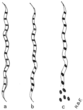
Fig. 54.—Spirochæta duttoni. a, blood
form showing slight membrane; b, granules or
coccoid bodies clearly formed within the organism;
c, beginning of extrusion of coccoid bodies in the
tick. (After Fantham.)
The granule phase, however, is an essential one in the invertebrate
transmitter (fig. 54c). In 1905,146 Dutton and Todd proved experimentally
that O. moubata transmitted S. duttoni. They fed ticks, obtained
from Congo native huts in which infected persons lived, on monkeys
and the latter became infected. Dutton and Todd also found the
offspring of infected ticks to be capable of transmitting the infection
to experimental animals. They concluded that O. moubata was a
true intermediate host.
A little later in 1905, Koch stated that spirochætes from the gut
of the tick penetrated the gut wall and tissues and found their
way into the eggs in the ovary. Koch figured tangled masses of
spirochætes as occurring in the tick eggs. He found ticks infective
to the third generation. He thought that the infection was spread
by the salivary fluid of the tick, in the act of biting. (This is now
known to be incorrect.) Markham Carter (1907) corroborated Koch’s
work on the spirochætes in the
tick eggs, and they have been
seen since by Kleine and Eckard
(1913).
Sir William Leishman,147 in
1909–10, found that at ordinary
temperatures the salivary glands
of infected ticks (O. moubata)
were not themselves infective,
and that the infection occurred
by way of the ticks’ excretion.
The spirochætes (contained in
the ticks’ excrement) found their
way into the vertebrate host
through the wound made by
biting. While feeding, ticks
pass large quantities of clear
fluid from the coxal glands;
in this fluid an anticoagulin
occurs. Some of the ticks also
pass thick, white Malpighian
secretion, that is, excrement,
towards the end of the feed.
Leishman, using experimental
monkeys, showed that if infected ticks were interrupted while
feeding, then no infection resulted in the monkeys. If, however, the
ticks were allowed to finish their feed, and the Malpighian secretions
were passed, then the experimental monkeys became infected.
Fantham148 and Hindle149 (1911), independently, have repeated the
experiments with mice.
Leishman’s methods and results may be summarized thus:
Saline emulsions of the organs of infected ticks were made, after
the organs had been most carefully dissected out. The ticks were
first kept for several days at certain constant temperatures, such as
24° to 25° C. or blood heat, 37° C. The saline emulsions of the
organs were inoculated, separately, into experimental animals, and the
results recorded:—
| At 24° C. | At 37° C. |
| Salivary glands | Negative | Positive |
| Malpighian tubules | Positive | Positive |
| Gut and contents | Positive | Positive |
| Excrement | Positive | Positive |
| Genital organs | Positive | Positive |
Coxal fluid is usually negative; thick, white excrement from
Malpighian tubes is positive.
When the ticks were
incubated at 21° to 24° C.
no spirochætes, as such,
were seen in the organs,
except perhaps in the gut,
where they often disappeared
in a few days.
When the ticks were previously
incubated at 35° to
37° C. for two to three days,
spirochætes, as such, reappear
in the gut, organs
and hæmocœlic fluid.
The infection proceeds,
not from the salivary gland, but from the infective excrement,
that is, from the thick, white material voided by the tick while
feeding, usually towards the end of the meal. This Malpighian
excrement passes into the wound caused by the bite, being greatly
aided by the clear and more limpid coxa fluid, which bathes the under
surface of the tick’s body, and mixes with and carries the infective
excrement into the wound. Ticks remain infective for a long time.
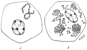
Fig. 55.—Spirochæta duttoni and its coccoid bodies
in the tick (O. moubata).—Mononuclear cells of the tick
(O. moubata) containing (a) Spirochæte breaking up into
coccoid bodies; (b) Similar tick-cell containing coccoid
bodies or granules. Such mononuclear cells occur in
various organs of ticks and in developing Malpighian
tubules. (Original. From preparations by Fantham.)
The spirochætes in the gut of infected ticks divide by a process
of multiple transverse fission into granules, which are composed of
chromatin (fig. 54). These granules—sometimes known as coccoid
bodies—are capable of multiplication. Leishman first found them in
clumps inside the cells of the Malpighian tubules (cf. fig. 55).
To summarize, when spirochætes are ingested by a tick, some of
them pass through the gut-wall into the hæmocœlic (body) fluid.
They then bore their way into the cells of various organs (fig. 55a)
and break up into coccoid bodies. In this manner the granules find
their way into the ovaries and ova, thus explaining how the young
ticks are born infected. Inoculation of these chromatinic granules
usually produces infection. Infective granules are also seen in the
rudiments of the Malpighian tubules of embryo ticks. Bosanquet
and Fantham (1911), independently, have shown that molluscan
spirochætes also break up into similar granules or coccoid bodies.
Gross has also demonstrated multiple transverse fission in molluscan
forms. Marchoux and Couvy (1913) and Wolbach (1914) consider
the granules or coccoid bodies to be degeneration products. This is
unlikely (see below).
Schuberg and Manteufel have found that certain O. moubata,
perhaps 30 per cent. of the specimens of a given neighbourhood, may
acquire a natural active immunity against infection with S. duttoni.
S. duttoni, or a closely allied form (by some termed S. novyi), occurs
in Colombia, and is spread by the tick Ornithodorus turicata. In
Panama a similar spirochæte is probably spread by O. talaje.
Spirochæta gallinarum, Stephens and Christophers, 1905
(= Spirochæta marchouxi, Nuttall, 1905).
This Spirochæte, which occurs in fowls and is pathogenic, is
transmitted by the tick Argas persicus. It is about 10 µ to 20 µ long.
There is a pathogenic spirochæte known to occur in geese, named by
Sakharoff (1891) S. anserina, and found in Caucasia. This may be
the same as S. gallinarum, in which case the name S. anserina will
have priority. These organisms cause fever, diarrhœa, anæmia and
death. The life history of the avian pathogenic spirochætes has been
studied by Balfour, by Hindle150 and by Fantham.151 It is essentially
similar to that of S. duttoni.
Marchoux and Couvy152 (1913) consider that the “fragmentation of
the chromatin” in spirochætes is a process of degeneration. Working
with A. persicus and S. gallinarum, they state that a large number of
the spirochætes ingested by the Argas almost immediately pass
through the wall of the alimentary canal and appear in the hæmocœlic
fluid. Marchoux and Couvy consider that Leishman’s granules
may be found in the Malpighian tubules of various Arachnids. They
found spirochætes in the cephalic glands of infected Argas. They
consider that spirochætes remain as wavy spirochætes within the tick,
if they are to be infective, though the spirochætes may become so thin
as to be invisible! The latter argument is obviously weak, and it
was never asserted that all granules in the Malpighian tubules of
infected ticks were derived from spirochætes. With dark-ground
illumination small, refractile spirochætal granules may be seen to grow
into spirochætes. The granule phase of spirochætes has recently
been discussed by Fantham153 (1914).
Spirochæta recurrentis, Lebert, 1874.
Syn.: Spirochæta obermeieri, Cohn, 1875.
This organism was discovered by Obermeier (1873) in cases of
relapsing fever in Berlin. Short forms 7 µ to 9 µ long, and longer
(probably adult) forms, 16 µ to 19 µ, are found in the blood. The
width is 0·25 µ. Parasites 12 µ or 13 µ long are often observed.
The spirochæte is found in the blood during febrile attacks and
relapses, but not during intervening periods. It can be inoculated
into monkeys, rats and mice. It can live in the bed-bug, Cimex
lectularius, and Nuttall has succeeded in transmitting S. recurrentis
from mouse to mouse by the bites of the same bug. The French
investigators Sergent and Foley (1908–9) in Algeria, and Nicolle,
Blaizot and Conseil (1912) in Tunis, have shown experimentally that
S. recurrentis (var. berbera) is transmitted by lice. The latter workers
also demonstrated the method of infection that commonly occurs,
namely, by the scratching of the skin and crushing of lice containing
spirochætes on the excoriated surface of the body.
Lice as transmitting agents for relapsing fever were indicated by Mackie154 in
1907. An epidemic among Indian school children furnished the materials.155 It was
noted that out of 170 boys, 137 were infected, and the boys were very verminous.
Among the girls, 35 out of 114 suffered, and few lice were found on them. Twenty-four
per cent. of the lice taken from the boys contained spirochætes as compared
with 3 per cent. of those from the girls. As the epidemic died out among the boys,
the lice also became fewer, and an increase in the number of cases among the girls
coincided with an increase in the number of lice. Spirochætes were found in the
gut, Malpighian tubules and genital organs of the lice. Mackie thought that infection
of the patients was brought about by the regurgitation of the spirochætes when
the lice fed, but proof of this was lacking.
In 1912, Nicolle, Blaizot and Conseil,156 working in Tunis and
using chiefly an Algerian strain of relapsing fever spirochætes (sometimes
called S. berbera), showed by direct experiments that infection
by means of the bites of Pediculus vestimenti and P. capitis was
untenable. As many as 4,707 infected lice were fed on one man, and
6,515 on another occasion were allowed to bite a man after they
had fed on a monkey heavily infected with spirochætes, yet no
infection of the man followed. Examination of the lice showed
that the spirochætes left the gut soon after they were ingested, and
passed into the body cavity, which swarmed with spirochætes. The
contents of the alimentary tract and the fæces of the lice alike were
uninfective. The spirochætes did not reappear in the gut till eight
days after an infective feed, but some persisted as late as the nineteenth
day when kept at 28° C.
It was noted that the irritation due to the lice caused scratching,
and that thereby lice became crushed on to the skin. An emulsion
was made of two infected lice and rubbed on to the slightly excoriated
skin of one of the above workers. Infection followed five days
later. A drop of emulsion placed on the conjunctiva of the human
eye produced spirochætosis after an incubation of seven days. The
body contents of such lice, then, produce infection when they reach
the blood by any excoriated or penetrable surface. The stages leading
up to infection in nature briefly are: The irritation due to the louse
bites causes scratching, and the lice are crushed on to the skin. The
slight abrasion is quite sufficient to permit the entry of the parasite.
The louse bite alone is harmless. Infection by way of the eye is
quite probable in Africa, remembering the constant trouble due to
sand, dust, insects, etc., resulting in frequent touching of the eyes.
The spirochætes occur in the body fluid of the lice and can pass
in it to the adjacent organs. Thus they probably find their way into
the genital organs, and into the eggs of the lice. Eggs laid twenty
to thirty days after the parent became infected have retained the
infection, and the larvæ issuing from such eggs must have contained
some form of spirochætes, for an emulsion of either the eggs or
the larvæ produced spirochætosis when inoculated into monkeys.
Further details regarding the spirochætosis in the eggs of the lice and
in the larvæ are needed. Hereditary infection, however, has been
demonstrated, but is not very common. Sergent and Foley (1914)
state that the spirochæte possesses a very small and virulent form
which it assumes during apyrexial periods in man and during a
period following an infecting meal in the louse. Nicolle and Blanc
(1914) find that the organisms are infective in the louse just before
they reappear as spirochætes. Nicolle and Blaizot found that female
lice were more susceptible to spirochætes than males, four times
as many females as males being infected.
Tictin (1897) found S. recurrentis in bugs recently fed on patients, and infected a
monkey with the fluids of crushed bugs. Karlinski (1902) found the spirochætes
in bed-bugs in infected houses. There is some other evidence to show that bugs
may transmit the spirochæte in Nature. Further researches are needed regarding
the relationship of bed-bugs and human spirochætosis.
Multiplication of S. recurrentis is by longitudinal and transverse
division (including so-called “incurvation”), and the organism forms
small, ovoid bodies (“coccoid” bodies) in the same way as S. duttoni.
S. recurrentis is the cause of European relapsing fever, and a
number of possible varieties of it are associated with relapsing fevers
in other parts of the world. Such spirochætes only differ by biological
reactions, such as acquired immunity tests. They include:—
S. rossii, the agent of East African relapsing fever; S. novyi, the
agent of North American relapsing fever; S. carteri, the agent of
Indian relapsing fever; S. berbera, the agent of North African and
Egyptian relapsing fever.
Other Human Spirochætes are:—
S. schaudinni. This organism, according to Prowazek, is the
agent of ulcus tropicum. It varies in length from 10 µ to 20 µ.
S. aboriginalis has been found in cases of granuloma inguinale
in British New Guinea and Western Australia. It also occurs in
dogs, and may not be truly parasitic.
S. vincenti. This spirochæte is 12 µ to 25 µ in length, tapers at
both ends and has few coils. It has been associated with angina
vincenti. It often occurs in company with fusiform bacilli.
S. bronchialis, found by Castellani in 1907 in cases of bronchitis
in Ceylon. The parasites are delicate, but show morphological
variation. This organism is important and has since been found
in the West Indies, India, Philippine Islands and various parts of
Africa, such as the Anglo-Egyptian Sudan, Uganda and West Africa.
It has recently been the subject of research by Chalmers and
O’Farrell, Taylor, and Fantham.
S. phagedenis was found by Noguchi in a ten days old ulcerated
swelling of the labium. The organism shows much variation in size,
being 4 µ to 30 µ in length.
S. refringens (Schaudinn, 1905) occurs in association with
Treponema pallidum in syphilitic lesions, but is non-pathogenic. It
is 20 µ to 35 µ long and 0·5 µ to 0·75 µ broad, being larger than
T. pallidum and more easily stained.
Various spirochætes have also been notified in vomits, chiefly in
Australia; others from the human intestinal tract, e.g., S. eurygyrata;
S. stenogyrata (Werner); S. hachaizæ (Kowalski), in cholera motions;
S. buccalis (Cohn, 1875) and S. dentium occurring in the human
mouth and in carious teeth (S. dentium, Koch, 1877, being the
smaller); S. acuminata and S. obtusa found by Castellani in open
sores in cases of yaws.
Animal spirochætes of economic importance include:—
S. anserina, highly pathogenic to geese.
S. gallinarum (= S. marchouxi) in fowls. (See p. 119.)
S. theileri in cattle and S. ovina in sheep also occur in Africa;
their pathogenicity is not clear.
S. laverani (= S. muris), occurring in the blood of and pathogenic
to mice, is probably the smallest spirochæte from the blood,
being only 3 µ to 6 µ long.
Numerous spirochætes have been recorded from the guts of
various mammals, birds, fishes, amphibia and insects.
Cultivation of Spirochætes.—Cultures of spirochætes have
been made with little success or with great difficulty until comparatively
recently, when Noguchi (1912) devised a means whereby he
has cultivated most of the pathogenic spirochætes as well as some
Treponemata.
Noguchi has now cultivated S. duttoni, S. recurrentis, S. rossii,
S. novyi and S. gallinarum from the blood; S. phagedenis157 from human
phagedænic lesions; S. refringens158 and spirochætes from the teeth.
His method is as follows:—
A piece of fresh, sterile tissue, usually rabbit kidney, is placed in a
sterile test-tube. A few drops of citrated blood from the heart of an
infected animal, e.g., rat or mouse, is added, and about 15 c.c. of sterile
ascitic or hydrocœle fluid is poured quickly into the tube. Some of
the tubes are covered with a layer of sterile paraffin oil, others are
left uncovered. The tubes are incubated at 37° C. The best results
are obtained if the blood is taken from an animal forty-eight to seventy-two
hours after it has been inoculated, that is, before the spirochætes
reach their maximum multiplicative period in the blood. The presence
of some oxygen seems indispensable for these blood spirochætes, and
they fail to develop in vacuo or in an atmosphere of hydrogen.
For subcultures, 0·5 c.c. of a culture is added to the medium
instead of citrated blood, and it is useful to add a little fresh, normal
blood, either human or from an animal, such as a rat.
Noguchi found that the events in cultures were:—
S. duttoni,159 maximum multiplication on the eighth to ninth day;
disintegration beginning on the tenth day, spirochætes disappeared
after about the fifteenth day. No diminution of virulence was found
at the ninth day.
S. rossii (= S. kochi).160 Maximum development on the ninth day,
after which the virulence diminishes. The incubation period is also
prolonged.
S. recurrentis161 (= S. obermeieri). Maximum growth on the seventh day.
S. novyi.162—Maximum development on the seventh day. It is
more difficult to grow than the preceding forms.
All the above spirochætes showed undoubted longitudinal division
and transverse division was observed in part.
S. gallinarum163 can be cultivated as above, but transverse division
was usual here. Maximum growth occurred in the culture about the
fifth day.
Treponemata.
The genus Treponema (Schaudinn, 1905), includes minute, thread-like
organisms, with spirally coiled bodies, the spirals being preformed
or fixed. No membrane or crista is present, according to Schaudinn,
though a slight one is said by Blanchard to be present in the organism
of yaws. The ends of the organisms are tapering and pointed.
Multiplication is by longitudinal and transverse division. The most
important members of the genus are T. pallidum, the agent of syphilis,
and T. pertenue, which is responsible for frambœsia or yaws.
Treponema pallidum, Schaudinn, 1905.
Syn.: Spirochæta pallida.
Treponema pallidum was first described by Schaudinn and Hoffmann
in 1905 under the name of Spirochæta pallida. It has also been
described under the names
of Spironema pallida, Microspironema
pallida and
Trypanosoma luis. Siegel
in 1905 described an organism
which he called Cytorhyctes
luis and considered
to be the agent of syphilis.
Schaudinn reinvestigated
Siegel’s work and found
T. pallidum, which he considered
to be the causal
agent of the disease, and
pronounced against Cytorhyctes
luis. It is probable
now that both workers
were correct, for Balfour
(1911) has seen the emission
of minute granules
or “coccoid” bodies from T. pallidum and these granules probably
correspond to the C. luis of Siegel. Recently E. H. Ross, having
observed a spirochæte stage in the development of Kurloff bodies,
thinks that T. pallidum is a stage in the life-history of a Lymphocytozoon.
MacDonagh has also described a complicated and somewhat
similar cycle, but these observations require further study and
confirmation.
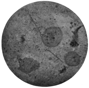
Fig. 56.—Treponema pallidum. (After Bell, from
Castellani and Chalmers.)
T. pallidum varies from 4 µ to 10 µ in length, its average length
being 7 µ, while its width is usually about 0·25 µ. Longer individuals
of 16 µ to 20 µ have been recorded. The body has from eight to ten
spiral turns and forms a tapering process at each end (fig. 56). The
organism is most difficult to stain, and its internal structure is little
known. It is possibly like that of Spirochæta duttoni or S. balbianii,
as the “granule shedding” observed by Balfour is strongly suggestive
of the formation of resistant bodies by those spirochætes. Hoffmann
(1912) has seen the formation of spores in T. pallidum.
The Treponemata occur in the primary
and secondary sores, but are difficult to
find in the tertiary eruptions of syphilis.
Noguchi and Moore (1913) and Mott164
(1913) have demonstrated T. pallidum in
the brain in cases of general paralysis of
the insane. Marie and Levaditi (1914),
however, consider that the treponeme
found in the brain in such cases is
different from T. pallidum.
Cultivation of T. pallidum.—This has
been accomplished successfully by Noguchi,165
using a modification of his
method for spirochæte cultivation, for
T. pallidum is much more difficult to
grow than spirochætes, being a strict
anaerobe.
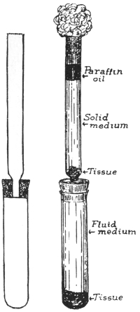
Fig. 57.—Diagram of apparatus
for cultivation of Treponema pallidum
by Noguchi’s method. (After
Noguchi.)
The apparatus consists of two glass
tubes, the upper being connected to the
lower by a narrower tube passing through
a rubber cork (fig. 57). Both tubes are
carefully sterilized.
A piece of fresh, sterile rabbit’s kidney
is placed in the lower tube, which is
filled with ascitic fluid, or ascitic fluid
and bouillon mixture. The tube is inoculated
with syphilitic material and
corked by inserting the upper tube. In the bottom of the upper tube
a piece of sterile rabbit’s kidney is placed and syphilitic material
poured over it. A mixture of one part ascitic fluid and two parts of
slightly alkaline agar is then poured over the tissue and allowed to
solidify. When solid, a layer of sterile paraffin oil is poured on top
of it, and the top plugged with cotton wool (fig. 57). The whole is then
incubated at 37° C. for two or three weeks. The tissue removes traces
of oxygen from the lower levels of the medium and also probably
provides a special form of nourishment. At first T. pallidum grows in
the solid medium, and then when the cultural conditions in the lower
fluid portion become favourable, the organisms migrate thither and
multiply abundantly. At first the culture is impure, but after several
transferences a pure culture is obtained readily.
The syphilitic material for culture is prepared by cutting off
pieces of tissue from the lesions, washing in sterile salt solution
containing 1 per cent. sodium citrate, and then emulsifying the
tissue in a mortar with sodium citrate.
Good cultures show rapid multiplication, which is invariably
by longitudinal division.
In his various cultivation experiments Noguchi166 found morphological
and pathogenic variations in T. pallidum. Three forms of the
organism were found, namely, thicker, average and thinner types.
The lesions caused in the testicle of the rabbit differ according to
the variety inoculated, but more work is necessary on the subject.
Noguchi167 has cultivated a separate organism, T. calligyrum,
from the surface of human genital or anal lesions, either syphilitic
or non-syphilitic. It is apparently non-pathogenic, and is 6 µ to
14 µ long.
Hata (1913)168 has modified the Noguchi technique for the cultivation
of spirochætes and treponemes, with a view to simplification and
convenience. Hata substitutes normal horse serum for ascitic fluid
and the “buffy coat” of the clot of horse blood in place of the small
pieces of rabbit’s kidney. It is unnecessary to place sterile paraffin on
the surface of the medium.
The horse serum is mixed with twice its volume of physiological
saline solution. The mixture is placed in tubes which are heated on a
water-bath at 58° C., the temperature being raised gradually until it
reaches 70° or 71° C. in three hours. The tubes are then heated at
71° C. for half an hour. After cooling, the contents will consist of an
opaque semi-coagulated mass. This semi-coagulated serum and saline
mixture may be substituted for Noguchi’s ascitic fluid.
The buff coagulum is cut into small pieces, about 1 c.c. in volume.
They must be forced with a sterile glass rod to the bottom of the semi-coagulated
serum and saline mixture. The medium is inoculated with
a small quantity of infected blood and kept at 37° C. In the case of
S. recurrentis, growth of spirochætes is observed on the second day,
reaching a maximum in five to seven days. The growth of the
organisms proceeds rather more slowly, they live for a longer period
and maintain their virulence better than in Noguchi’s medium.
Treponema pertenue, Castellani, 1905.
Syn.: Spirochæta pertenuis; S. pallidula, Castellani, 1905.
Castellani discovered the organism in 1905, in scrapings of yaws
pustules. He first described it under the name of Spirochæta pertenuis.
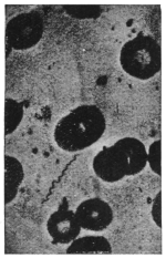
Fig. 58.—Treponema pertenue.
(After Castellani and Chalmers.)
Treponema pertenue (fig. 58), though delicate and slender, shows
great morphological variation both in length and thickness. It
may be short, e.g., 7 µ, but can attain 18 µ to 20 µ in length and may
be even larger. In cultures made by Noguchi, thick, medium and
thin forms were found, each giving rise to a different type of frambœsial
lesion when inoculated into the testicles of rabbits, thus
suggesting the possibility of the occurrence of varieties of T. pertenue.
The organism is difficult to stain, but occasionally deeper
staining granules are found along its body. They may represent
a diffuse nucleus. Granule formation
similar to that of T. pallidum has been
observed by Ranken, using dark-ground
illumination.
Many experiments have been made
with a view to establishing the identity
of the organism of yaws and also of
differentiating between the causative
agents of yaws and syphilis. Both
monkeys and the human subject have
been experimentally inoculated with yaws
material and have developed the disease.
In an early experiment, negroes were
inoculated with the secretion from lesions
of yaws. All of them developed the
disease, nodules appearing, chiefly at the
seat of inoculation, in from twelve to twenty days, followed by the
usual eruption. Similar results were obtained with thirty-two Chinese
prisoners, who were inoculated with yaws, twenty-eight becoming
infected.
A naturally infected yaws patient when inoculated with syphilis,
contracted that infection, thus showing that yaws does not confer
immunity to syphilis. This has also been observed naturally, when
yaws patients have contracted syphilis.
Experiments with monkeys have been successfully performed.
The incubation period varies from sixteen to ninety-two days.
Lesions appear first at the seat of inoculation, and in some
monkeys the eruption is localized to this spot, though the infection is
general, T. pertenue occurring in the spleen, lymphatics, etc. Monkeys
inoculated with splenic blood of a yaws patient, and also sometimes
with blood from the general circulation, have become infected.
Castellani and others have shown that monkeys successfully
inoculated with syphilis do not become immune to yaws, and vice-versâ.
Craig and Ashburn, using the monkey Cynomolgus philippinensis,
found these animals susceptible to yaws but not to syphilis.
The ulcerated lesions of frambœsia are rapidly invaded by
numerous bacteria as well as by different spirochætes, of which
Castellani has described three distinct species. One is identical
with Spirochæta refringens, Schaudinn, the other two are thin and
delicate. One, S. obtusa, has blunt ends; the other S. acuminata, has
pointed ends. T. pertenue is also present.
The reasons for considering T. pertenue to be the specific cause of
frambœsia are:—
(1) T. pertenue is the only organism present in non-ulcerated
papules, in the spleen and in the lymphatics of yaws patients, or
of monkeys artificially infected with the disease. By no method
has any other organism been obtained.
(2) Extract of frambœsia material, free from all organisms other
than T. pertenue, reproduces the disease if inoculated.
(3) Extract of frambœsia material deprived by filtration of
T. pertenue is no longer infective on inoculation.
The method of infection is contaminative, by direct contact.
Women in Ceylon are frequently infected by their children. Any
slight skin abrasion is sufficient to admit the parasite. In some
cases, insects may carry the disease from person to person, and even
in hospitals, when dressings are removed, it has been noticed that
flies greedily suck the secretion from the ulcers. T. pertenue has
been recovered from flies that have fed on yaws, and monkeys have
contracted the disease when flies were placed and retained on them
for a short time, after the insects had fed on yaws material.
Cultivation.—T. pertenue has been cultivated by Noguchi, who
finds three types of parasites in his cultures, as before mentioned. Its
multiplication is by longitudinal division.
Noguchi169 (1912), has cultivated species of Treponema from the
human mouth, e.g., T. macrodentium, T. microdentium and T. mucosum,
the latter from pyorrhea alveolaris. These parasites in the past may
have been confused under the name Spirochæta dentium.
The third group of the Protozoa consists entirely of parasitic
organisms forming the class known as the Sporozoa or spore-producing
animals. The members of this class are characterized
by possessing very great powers of multiplication, coupled with a
capacity for producing forms that serve for the transference of the
organisms to other hosts. These reproductive bodies, whether for
increase of numbers within one host or for transmission to another
host, are called spores. But, strictly, the term spore should be used
only in the latter connection, when a protective or resistant coat
known as a sporocyst envelops the body of the spore.
The Sporozoa are widely distributed, occurring in various tissues
and organs of Annelids, Molluscs, Arthropods, and Vertebrates.
Their food, which is fluid, is absorbed osmotically. The life-cycle
of a Sporozoön may be completed within one host or may be distributed
between two different hosts.
The Sporozoa were divided by Schaudinn into two groups or
sub-classes, called (1) the Telosporidia, and (2) the Neosporidia.
The Telosporidia are Sporozoa in which the reproductive phase
of the parasites is distinct from the growing or trophic phase, and
follows after it. The Neosporidia include Sporozoa in which growth
and spore-formation go on simultaneously. This classification is not
final, for certain exceptions and difficulties are already known with
regard to it. It is possible that the class Sporozoa is not a natural
entity, but should be replaced by two classes of equal rank, corresponding
in most respects with the Telosporidia and Neosporidia.
The Telosporidia comprise the Gregarinida, the Coccidiidea, and
the Hæmosporidia. Doflein combines the two latter orders into one
known as the Coccidiomorpha.
The Neosporidia comprise the Myxosporidia, the Microsporidia,
the Actinomyxidia, the Sarcosporidia, and the Haplosporidia.
Doflein combines the first three orders into one, the Cnidosporidia.
Sub-Class. TELOSPORIDIA, Schaudinn.
Sporozoa in which the reproductive phases follow completion of
growth.
Order. Gregarinida, Aimé Schneider emend. Doflein.
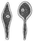
Fig. 59.—Monocystis
agilis from seminal
vesicles of Lumbricus
× 250. (After Stein.)
Knowledge of the Gregarinida probably goes back as far as the year 1684, when
Redi observed gregarines in the crab, Cancer pagurus. Von Cavolini (1787) found
them in Cancer depressus. The name Gregarina was created by L. Dufour (1828),
who observed masses of these organisms in the gut of insects of different orders.
Hammerschmidt (1838) and von Siebold found rich infestations in insects, while
Dujardin (1835) and Henle described various genera from segmented worms.
Henle (1835) also observed cysts containing “navicellæ” in the sperm-sacs of
segmented worms, and attention was drawn to his researches by the discovery by
von Siebold (1839) of “pseudonavicellæ” in the gut of Sciara nitidicollis. Up to this
time many workers considered the gregarines to be worms, but Kölliker (1845)
investigated many of them and maintained their unicellular nature, while Stein’s
work (1848) showed the interrelation of the pseudonavicellæ and the gregarines.
The discovery of amœboid germs in the pseudonavicellæ by Lieberkühn (1855) and
the demonstration of myonemes further aided in the elucidation of their true
systematic position. The entire process of conjugation, of which Dufour had seen
one phase, was followed by Giard under the microscope.
From 1873 onwards Aimé Schneider made important additions to the knowledge
of the morphology, life-history, and systematic position of numerous gregarines.
Bütschli (1881) and L. Léger (1892) also contributed much work on the subject. The
discoveries of Schaudinn with regard to the life-cycle of Coccidia gave a fresh
stimulus to the study of the Gregarines, whereby the life-cycles of numerous forms
and the phases thereof have been elucidated.
Asexual multiplication is not common among the Gregarines, but is known to
occur in the sub-order Schizogregarinea, formerly known as the Amœbosporidia.
Although the Gregarinida are not known to be parasitic in man or other vertebrates,
they are of great interest, inasmuch as they are among the earliest known
Sporozoa, and therefore will be briefly described here.
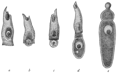
Fig. 60.—Gregarina longa from larva of crane-fly (Tipula). a, in epithelial cell of host;
b, c, gradually leaving host-cell; d, adhering to host-cell; e, fully developed free trophozoite.
Fig. 61.—Xyphorhynchus
firmus with epimerite in
intestinal epithelial cell of
host. (After Léger.)
The Gregarines are usually elongate, somewhat
flattened organisms (figs. 59, 60), whose bodies are
enclosed in an elastic and often thick cuticle. The
enclosed living substance shows a separation into
ectoplasm and endoplasm, as is common among
Protozoa. The cuticle is sometimes regarded as the
outer portion or epicyte of the ectoplasm. A single,
vesicular, spherical, or elliptical, large nucleus, with
its chromatin concentrated to form a spherical
karyosome, is present. The body of some gregarines
may be divided by ingrowing ectoplasmic
partitions or septa, and are then said to be “septate” or
“polycystid” (fig. 61). Other gregarines remain simple and non-septate,
and are termed “monocystid” (fig. 59). The monocystid gregarines
occur especially in the body cavity of Chætopoda and Insecta, more
rarely in Echinodermata, in the parenchyma of Platyhelminthes,
also in the gut of Tunicata and Insecta (fig. 60) and in the seminal
vesicles of Annelida. In the polycystid gregarines a single septum only
is present as a rule, and thus the body presents two portions: (1) an
anterior portion termed the protomerite; (2) a
posterior, larger portion, known as the deutomerite,
which generally contains the nucleus.
The protomerite is often modified anteriorly
to form an organ of attachment, termed the
epimerite (fig. 61), which is developed from the
pointed rostrum of the sporozoite or primary
infecting young gregarine. The structure of
the epimerite may be complicated, being provided
with hooks, spines, knobs, and other
appendages. An extension of the polycystid
condition is seen in Tæniocystis mira Léger
(from the dipteran larva, Ceratopogon solstitialis),
whose body shows a number of partitions,
giving the organism a superficial resemblance
to a tapeworm.
The ectoplasm of a gregarine exhibits three
layers: (1) An epicyte (cuticle) externally of
which the epimerite is composed; (2) a
sarcocyte which forms the septa if present;
(3) the deeper myocyte layer containing contractile
elements in the form of fibrils or threads
termed myonemes (fig. 62).
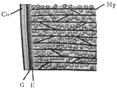
Fig. 62.—Gregarina munieri (from
the beetle, Chrysomela hæmoptera). Section
through surface layers. Cu, cuticle;
E, ectoplasm proper; G, gelatinous
layer; My, myonemes in myocyte layer.
× 1500. (After Schewiakoff.)
The endoplasm is fluid and granular, containing
many enclosures, which are of the nature of reserve food
materials. They consist of fat droplets
or of paraglycogen, and give
the organisms an opaque appearance.
Lithocystis contains crystals
of calcium oxalate in its endoplasm.
Many gregarines are capable of
active movements, though they do not
possess obvious locomotor organs.
The movement is of a smooth,
gliding character and two suggestions
have been put forward to
explain it. According to Schewiakoff,
a gelatinous substance is secreted between
the layers of the ectoplasm.
This is extruded posteriorly and thus
the animal is pushed forward. On the other hand, Crawley considers
that the movements are produced by contractions of the myonemes.
These two explanations are probably correct as far as each goes, and
are to be regarded as supplementary to one another.
Occasionally, temporary associations of gregarines are formed
by a number of individuals adhering to
one another end to end. Such temporary
associations are examples of syzygy. Such
syzygies must not be confused with true
associations which form an essential part of
the life-cycle.
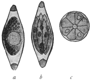
Fig. 63.—Monocystis agilis.
Spores from vesicula seminalis
of the Earthworm. a, Sporoblast
with single nucleus, enclosed in
sporocyst; b, mature spore containing
sporozoites; c, diagrammatic
cross-section of spore,
showing eight sporozoites round
residual protoplasm. (After
Bütschli.)
The life-cycle of a relatively simple gregarine,
such as Monocystis agilis (fig. 59),
parasitic in earthworms, may now be considered.
The gregarines, being members of
the Sporozoa, produce spores at one phase
of the life-cycle. Each gregarine spore
(fig. 63) develops within itself a number of
minute, sickle-shaped or vermicular bodies,
known as sporozoites or primary infecting
germs. Eight sporozoites are often formed
within each spore. When absorbed by a new
host, the spore softens and the sporozoites issue from it. They are
capable of active movement and may or may not enter a cell, such as
one of those of the digestive tract, or, as in Monocystis, a cell lining the
vesicula seminalis which becomes a sperm-cell aggregate (sperm morula).
When the sporozoite has reached the place of its choice in the host
it ceases active movements and proceeds to feed passively on the fluid
substances around it, whether they be those of tissues or body fluids.
This passive, growing and feeding form is known as the trophozoite.
After a trophic existence of longer or shorter duration, the trophozoite
ceases to feed and prepares for reproduction. Two trophozoites
associate together, each of them first becoming somewhat rounded.
The two trophozoites, now known as sporonts or gametocytes, become
invested in a single common envelope or cyst (fig. 64, a). The nucleus
of each gametocyte then divides by a series of binary fissions (fig. 64, b),
and the daughter nuclei thus produced arrange themselves at the
periphery of the parent cells (fig. 64, c). Cytoplasm collects around
each of these nuclei, and thus a number of gametes are formed within
each gametocyte. The gametes for a time exhibit active movements,
and ultimately ripe gametes of different parentage fuse in pairs, that
is, conjugation occurs (fig. 64, d). In this way zygotes are produced,
the nucleus of each zygote being formed by the fusion of two gamete
nuclei.
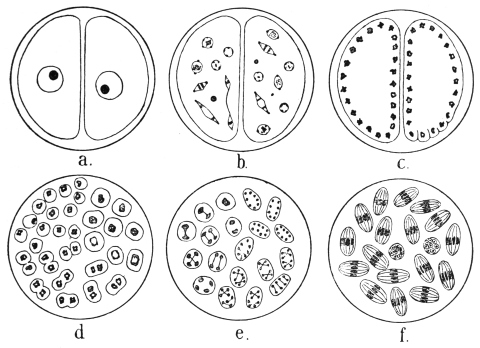
Fig. 64.—Schematic figures of conjugation and spore formation in Gregarines. For details
see text. (After Calkins and Siedlecki, modified.)
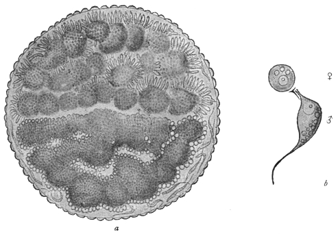
Fig. 65.—Stylorhynchus oblongatus. a, cyst containing two sporonts or gametocytes, each
full of gametes, those in the upper one being male. b, ripe male and female gametes. × 1,600.
(After L. Léger.)
The zygote grows slightly and becomes oval or elongate, and at
this period is often called the sporoblast. It then secretes an external
membrane, the sporocyst. Nuclear division occurs inside the sporocyst
by a series of three binary fissions (fig. 64, e), so that each sporocyst,
now usually referred to as a spore, contains eight nuclei. The
cytoplasm collects around each nucleus and eight vermicular sporozoites
are produced within each spore (fig. 64, f), thus completing
the life-cycle.
It will be noticed that in the above life-cycle no asexual multiplication
occurs. These organisms, such as Monocystis, are known as
the Eugregarines, and include the majority of the gregarines. The
remainder, which have introduced schizogony into their life-cycle, are
known as the Schizogregarines.
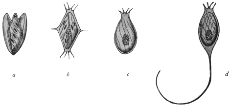
Fig. 66.—Spores of various Gregarines. a, Xiphorhynchus.
b, Ancyrophora. c, Gonospora. d, Ceratospora.
(After Léger.)
There are variations in the morphology and life-cycle of gregarines
besides those that have been mentioned. It is not within the scope
of this book to discuss them in detail, but the following may be
noted:—
Morphological differentiation of gametes may occur as in Stylorhynchus
oblongatus (fig. 65), which differentiation is probably of a
sexual nature.
The sporocyst really consists of two layers, an epispore and an
endospore. Externally the spores of different gregarines show great
variety in shape and markings, and spines, or long processes may
be present (fig. 66).
The resistant spores serve for the transmission of the gregarines
from host to host. The mode of infection is contaminative, the
spores expelled with the dejecta of one host being absorbed with the
food of a new host.
The Gregarinida may be classified as follows:—
Sub-order I.—Eugregarinea, without schizogony.
Tribe 1.—Acephalina.—Without an epimerite and non-septate;
often “cœlomic” (body-cavity) parasites. E.g.: Monocystis, with
several species parasitic in the seminal vesicles of earthworms. Many
other genera parasitic in Echinodermata, Tunicata, Arthropoda, etc.
Tribe 2.—Cephalina.—With an epimerite, either temporarily or
permanently, in the trophic phase. Usually septate (except Doliocystidæ).
Many families, genera and species. Common in the digestive
tracts of insects. E.g.: Gregarina, with several species, Gregarina
ovata in the earwig, Gregarina blattarum in the cockroach, Stylorhynchus
in beetles, Pterocephalus in centipedes, etc.
Sub-order II.—Schizogregarinea, with schizogony.
Tribe 1.—Endoschiza.170—With schizogony occurring in the intracellular
phase, e.g., Selenidium (from Annelida and Gephyrea), Merogregarina
(from an Ascidian).
Tribe 2.—Ectoschiza.—In which the schizont is free, and schizogony
is extracellular, e.g., Ophryocystis (from Blaps, a beetle), and Schizocystis
(from Ceratopogon larva).
Order. Coccidiidea.
Hake (1839) first saw the organisms now termed Coccidia during his investigations
on the so-called coccidial nodules of rabbits. The opinions as to the nature of these
peculiar formations were very diverse. The discoverer considered them to be a sort
of pus corpuscle; Nasse (1843) took them for epithelial cells of the biliary passages,
others for helminthes, especially the ova of trematodes (Dujardin, Küchenmeister,
Gubler, etc). Remak (1845) was the first to draw attention to their relation to the
Psorospermia (Myxosporidia), and this investigator found them also in the small
intestine and vermiform appendix of rabbits. Lieberkühn (1854), who examined not
only the coccidia of rabbits, but found similar forms in the kidneys of frogs, likewise
called them definitely psorosperms. To differentiate Müller’s psorosperms, which are
found in fishes, from those of rabbits, etc., the latter were called egg-shaped psorosperms
(Eimer), until R. Leuckart (1879) named them Coccidia and placed them in
a group of the Sporozoa analogous to that of the Gregarinida, Myxosporidia, etc.
Numerous works confirmed the occurrence of coccidia, not only in all classes of
vertebrate animals, but also in invertebrates (Mollusca, Myriapoda, Annelida, etc.).
A large number of genera and species have in the course of time been described which
inhabit the epithelium of the intestine and its appendages for choice, but are also
found in other organs (kidneys, spleen, ovaries, vas deferens, testicles). Some also
live in the connective tissue of various organs, more particularly of the intestine.
The knowledge of the development of the coccidia was of particular importance
in determining their classification. By means of encysted coccidia from the liver of
rabbits, Kauffmann (1847) first confirmed the fact that the cyst, which was partly or
entirely filled with granular contents, divided into three or four pale bodies (fig. 71)
after a long sojourn in water. Lieberkühn observed the same process in the host in
the case of the coccidia of the kidney of the frog. Stieda (1865) studied more minutely
the changes that occur within the encysted coccidia of the liver of rabbits after the
death of the host. He discovered that the bodies now known as “spores” were
oval formations pointed at one pole, and surrounded by a delicate membrane, which
exhibited a certain thickness at the pointed extremity and enclosed a slightly bent
rodlet, expanding at either end into a strongly light-refracting globule; a finely
granular globule was present in the middle of the spore. Waldenburg (1862) saw the
same phenomenon in coccidia from the epithelium of the villi and Lieberkühn’s
glands of the intestine of the rabbit; but the process in this case took place in a
much shorter time.
According to the discovery of Kloss (1855), the spores of the coccidia of the
urinary organ of the garden snail were formed in far greater numbers: the round
spores also harboured several (five to six) rodlets, which after the bursting of the
spore-envelope became free. Eimer’s researches (1870) afforded information
regarding a Coccidium from the intestine of the mouse, which was transformed in
toto into a “spore,” containing small sickle-shaped bodies. The fact was, moreover,
established that the little bodies left the delicate envelope when in the intestine,
made movements of flexion and extension, and were finally transformed into
amœboid organisms, which apparently penetrated the epithelial cells; at all events,
similar bodies of various sizes were seen in these cells. Taking the immense
number of these parasites into account and the lack of any other cause, Eimer
attributed the sudden death of his mice to the Gregarina falciformis, as the parasite
was then called, just in the same way as a few years previously Reincke ascribed the
acute and fatal intestinal catarrh of rabbits to the invasion of intestinal coccidia.
All that had become known about coccidia up to 1879 was then compiled by
Leuckart, and completed by his own observations on the coccidia of the liver of the
rabbit. Experimental infections had already been conducted by Waldenburg (1862)
with intestinal coccidia of rabbits, and by Rivolta (1869–73) with the coccidia of fowls,
which experiments confirmed the importance of the spores, or bodies enclosed in them,
in the transmission of the parasites to other animals. Accordingly, it was assumed that
after the entry of the spores into the intestine the sporozoites were set free, actively
penetrated into the intestinal cells, where they grew into coccidia, and finally became
encysted. The further development, i.e., the formation of spores, took place outside
the host’s body in these cases; in other cases (Kloss, Eimer) it took place within
the host. Although much regarding the cycle of development was still hypothetical,
the ideas coincided with the observations, and were therefore universally regarded as
established. Further research confirmed this view in numerous new forms.
L. Pfeiffer’s statements (1891) on the part that certain coccidia or their sporozoites
played, or seemed to play, as causes of disease gave a renewed impetus to the
investigation of the coccidia. The ingestion of even very numerous spores did not
appear to account for the mass infection so frequently observed, even after Balbiani
had confirmed the fact that there were two, and not one, sporozoites contained in every
spore of the coccidia of rabbits (fig. 72). The hypothesis was therefore advanced
that the sporozoites or young coccidia were able to divide once again by sporulating.
The question was finally solved quite differently. R. Pfeiffer (1892) first confirmed
the fact that in addition to the well-known method of sporulation in the coccidia of
the rabbit that causes the infection of fresh hosts (“exogenous sporulation”), an
enormous increase may follow in the already infected host in a manner that Eimer
first observed in the coccidia of the intestine of the mouse (“endogenous sporulation”).
It had hitherto been believed that some of the species of coccidia increased
like the rabbit parasite, then known as Coccidium oviforme, and others like Eimeria
falciformis, and this difference had been made the foundation of a classification.
R. Pfeiffer was successful in observing the occurrence of both kinds of development
in the same species, and expressed the opinion that endogenous sporulation (fig. 73),
which takes place within the host, was the cause of the mass-infection that is mostly
accompanied by serious consequences (fig. 74). L. Pfeiffer sought, especially, to
demonstrate the correctness of this view as regards other species of coccidia
and for this purpose he utilized the experiences already published. Coccidia were
known to exist in a number of different hosts, and to propagate in some according
to the Coccidium type, in others according to the Eimeria type. It therefore was
reasoned that in this case it was not a question of two species belonging to different
genera living side by side, with a different mode of development, but of one species,
in the life of which both forms of development occurred alternately.
This interpretation of facts was combated especially by A. Schneider (1892) and by
Labbé, but has, nevertheless, proved true, apart from the circumstance that Schuberg
succeeded in discovering the hitherto unknown Coccidium form in the intestine of
the mouse; and that, moreover, Léger confirmed the fact that there are no Arthropoda
in which Eimeria are not found together with coccidia. The question was
finally settled by experiments made by Léger with the coccidia of Scolopendra
cingulata, by Schaudinn and Siedlecki with those of Lithobius forficatus, and by
Simond with the coccidia of the rabbit. On Simond’s suggestion the sickle-shaped
germs corresponding to the sporozoites, which are formed by endogenous sporulation,
are generally termed merozoites; and in accordance with Schaudinn’s
suggestion, those individuals which form merozoites are termed schizonts, and
those which produce spores are called sporonts. In contradistinction to sporogony
(exogenous sporulation), the term schizogony (endogenous sporulation) is used.
The more minute examination of these processes at last led to the discovery of
sexual dimorphism, of copulation and of alternation of generations in the coccidia.
Schuberg was the first to consider the possibility of copulation in coccidia; in
addition to the formations which now are termed merozoites, he observed very
diminutive bodies (“microsporozoites”) in the coccidia of the intestine of the mouse,
which were able eventually to copulate. Labbé confirmed this observation in some
of the species, and Simond expressed the opinion that bodies termed “chromatozoites”
occurred in all coccidia. Copulation itself was then observed by Schaudinn
and Siedlecki (1897). The copulating bodies were termed gametes. As, however, they
differed considerably one from the other, the males were called microgametes (i.e.,
the microsporozoites of Labbé and the chromatozoites of Simond) and the females
macrogametes. After copulation was completed sporogony took place, and in the
cycle of development of one species this regularly alternated with schizogony (asexual
multiplication). Schaudinn in 1900 described in detail the life-cycle of Eimeria
(Coccidium) schubergi.
The recognition of this unsuspected complicated process was bound to effect
reforms in the classification of the coccidia; and all the forms that had been
regarded as developmental stages (Eimeria, etc.) had to be reconsidered.
Occurrence.—The Coccidiidea in their mature condition usually
live within the epithelial cells of various organs, and by choice inhabit
those of the intestine and of its associated organs. They also occur
frequently in the excretory organs of mammals, birds, amphibia,
molluscs, arthropods, and may also be found in the testes and vas
deferens, but the statement that they live in hen’s eggs, as well as
in the oviducts of fowls, has not been confirmed.171 Some species
inhabit the nuclei of cells, others live in the connective tissue, but
their presence in the latter situation is probably only secondary,
that is, they have only reached it from the epithelium of the affected
organs.
The size of the Coccidiidea, corresponding as a rule to the capacity
of their habitat, is usually small, but there are said to be species
that attain a diameter of 1 mm. Their form172 is globular, oval, or
elliptical. External appendages are lacking, at least during the trophic
or vegetative period of their life, which is spent in epithelial cells,
within which they increase in size. Frequently one only is present
in each cell, but more can occur. The body substance is composed
of a more or less finely granular or distinctly alveolar protoplasm
which exhibits no differentiation into ecto- and endoplasm. All
species possess a nucleus that enlarges with their growth; sometimes
it only shows through the cytoplasm as a lighter spot, or may
even be quite concealed. It is vesicular, and besides containing very
delicate threads of chromatin in the clear nucleoplasm, it contains
generally only one large karyosome.
The infected epithelial cells degenerate sooner or later as the
parasite feeds on them (fig. 67, II-IV). After their form has been
changed by the action of the growing parasite, they finally perish.
The cell membrane then alone surrounds the coccidia, which, at
least in the species sufficiently known, begin to propagate by an
asexual process (schizogony), the parasites themselves becoming
schizonts, as the initial stage is usually called. They differ from later
stages (sporonts or gametocytes), which resemble them in form, by
the absence of granulations in the cytoplasm, as well as by the
vesicular nucleus. The form is not always the same, for in some
cases, at least, many schizonts assume a globular form.
Schizogony (fig. 67, V-VII) commences with a division of the
nucleus, which takes place in different ways in the various species.
This finally leads to the formation of numerous daughter nuclei
which become smaller and smaller, and which collect beneath
the surface of the schizonts. In some species the daughter nuclei
collect only in one half of the schizont. A part of the protoplasm
of the periphery now divides around each daughter nucleus, the
remaining part (residual body) being left in the centre or on one
side. The segments of the divided cytoplasm, each containing
a nucleus, assume a fusiform shape and become merozoites, which
very soon become free (fig. 67, VIII) and leave the residual body.
They are distinguishable from the very similar sporozoites (fig. 67, I),
as the merozoites possess a karyosome.
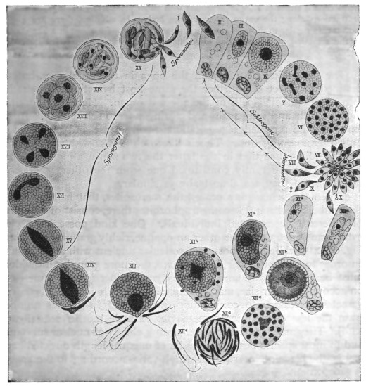
Fig. 67.—Life-cycle of Eimeria (Coccidium) schubergi, Schaud., from the intestine of
Lithobius. (After Schaudinn.) The infection is caused by a cyst (XX), containing spores,
which reaches the intestine of a Lithobius, where it discharges the sporozoites (I). II, A
sporozoite invading an intestinal epithelial cell; III, intestinal epithelial cell with young
trophozoite; IV, intestinal epithelial cell with a globular schizont; V, nuclear segmentation
within the schizont; VI, the daughter nuclei arranging themselves superficially; VII, formation
of the merozoites; VIII, merozoites that have become free, and which, penetrating into
other epithelial cells of the same intestine, repeat the schizogony (II-VIII); IX and X,
merozoites which, likewise invading the epithelial cells of the same intestine, become
sexually differentiated; xia, young macrogametocyte; XIb, older macrogametocyte; XIc,
mature macrogametocyte (discharging particles of chromatin); XIIa, young microgametocyte;
XIIb, older microgametocyte; XIIc, increase of nuclei in the microgametocyte; XIId, the
globular residual body around which numerous microgametes have formed; XIIe, an isolated
microgamete; XIII, the mature macrogamete surrounded by numerous microgametes and
forming a cone of reception or fertilization prominence; XIV, shows the nucleus of a
microgamete that has penetrated and fused with the nucleus of the macrogamete (fertilization)—the
latter forms a membrane and becomes an oöcyst; XV, XVI, XVII, nuclear segmentation
in the oöcyst; XVIII, oöcyst with four sporoblasts; XIX, the sporoblasts transformed into
spores, each containing two sporozoites; XX, the cyst introduced into the intestine and
liberating the sporozoites by bursting.
The merozoites move in a manner similar to that of the sporozoites. The movements
consist either of slow incurvations with subsequent straightenings, or annular
contractions along the entire extent of the body. In addition, there are gliding
movements similar to those of many gregarines, and brought about in a like manner
by the secretion at the posterior extremity of a gelatinous substance that hardens
rapidly.
The merozoites do not gain the open in the usual way, but are
destined to infect still further the same host by actively penetrating
into other epithelial cells of the affected organ. Here they continue
their growth and may again and again undergo schizogony. In the
Infusoria the repeated segmentations finally cease and are renewed
only after a conjugation. This is likewise the case with the Coccidia,
with the difference that in the latter the two conjugating individuals
(gametes) are differently constituted one from the other, whereas in
the Infusoria they are almost always similar.
When the schizogony ceases, the merozoites, that had penetrated
the epithelial cells and become trophozoites there, consist of two kinds
of differently constituted individuals. One kind possesses a clear
cytoplasm (fig. 67, XII), the other an opaque, richly granular cytoplasm
(fig. 67, XI), while both possess a vesicular nucleus with a karyosome.
In order to continue their development, the more granular individuals
must be fertilized, and are therefore termed either female gametes or,
on account of their size, macrogametes. The male individuals (microgametes)
necessary to conjugation, are formed in greater numbers
from the less dense microgametocytes or male mother-cells (fig. 67,
XIId). They are slender bodies consisting chiefly of nuclear substance,
and in most species bear two flagella of unequal length directed backwards,
the place of insertion of which varies according to the species
(fig. 67, XIIe).
While the development of the microgametes is rapidly advancing
a change occurs in the nucleus of the female parent forms or macrogametocytes.
Parts of the karyosome are extruded (fig. 67, XIc), and
the nucleus loses at the same time its vesicular form. One macrogamete
results, after nuclear maturation, from one macrogametocyte.
By this time the macrogametes are capable of conjugation, and
the process takes place within the host, generally, however, outside
the affected and degenerated host cells. The microgametes that
have now become free from the very large residual body, crowd
around the mature macrogametes, which often send out a small
prominence (“cone of reception” or fertilization protuberance) for
their reception (fig. 67, XIII). As soon as a microgamete comes in
contact with this and penetrates into the cytoplasm of the macrogamete,
the latter surrounds itself with a membrane which prevents
the intrusion of other microgametes. The nucleus of the microgamete
that has gained entry unites with the nucleus of the macrogamete,
which latter is afterwards capable of forming the well-known
spores. The parasite is now called an encysted zygote or oöcyst. The
oöcysts of some other members of the Coccidiidea, e.g., Eimeria avium,
can form their walls prior to fertilization. In such cases, a weak spot
is left at one place in the cyst wall, forming a micropyle, that permits of
the entry of the male, immediately after which the micropyle is closed.
The reduced nucleus of the macrogamete elongates on the entry of the microgamete,
and becomes a fertilization spindle to which the male pronucleus (from the
microgamete) becomes attached (fig. 67, XIV and XV). Thereupon the spindle
divides into two daughter nuclei (fig. 67, XVI) which assume a round shape. The
protoplasm at this stage may at once divide, or another segmentation of the daughter
nuclei may first occur. In the former case the two halves divide again, so that
finally four nucleated segments, the sporoblasts, are formed, whereas in the latter
case the four sporoblasts appear simultaneously (fig. 67, XVII). In both cases a
residual body of varying size is separated from the protoplasm of the oöcyst. As
a rule the oöcysts have already been discharged from the body of the host, and in
the manner described above, form the sporoblasts after a longer or shorter period
of incubation.
The sporoblasts are originally naked, but each soon secretes a
homogeneous membrane, the sporocyst, in which it becomes enveloped
(fig. 67, XVIII). After the segmentation of the nucleus the contents
divide into two sickle-shaped sporozoites, in addition to which there
is generally also a residual body (fig. 67, XIX).
This terminates the development. The spores are intended for
the infection of other hosts. If they reach the intestine of suitable
hosts, either free or enclosed in the oöcyst wall, the action of the
intestinal juices causes them to open and permits the escape of the
sporozoites (fig. 67, XX). The latter move exactly like the merozoites
and soon make their way into epithelial cells (fig. 67, I), where they
become schizonts, and thus repeat the life cycle.
Although our knowledge of the development of the coccidia is but of recent date,
yet it already extends to a large number of species, which exhibit various deviations
from the cycle of development described above. For instance, in addition to
differences in the gametocytes, the schizonts of Adelea and Cyclospora also show
differentiation and give rise to macromerozoites and micromerozoites, whilst in
Adelea and Klossia a precocious association of the gametocytes precedes the true
copulation of the ripe gametes.
The classification of the Coccidiidea is based chiefly on the
number of sporozoites found in each spore, and the number of
sporocysts (spores) found in one oöcyst. Léger173 recognises two
great legions, the Eimeridea and the Adeleidea, the former comprising
the greater number of genera, including the genus of most economic
importance, Eimeria. It must be noted that, though a member of
this genus may be frequently referred to as Coccidium, strictly it
should be termed Eimeria, that name having priority. The name
of the disease resulting from the action of such parasites is, however,
established and remains as coccidiosis.
Certain of the more important of the Coccidiidea may now be
considered.
Genus. Eimeria, Aimé Schneider, 1875.
Syn.: Psorospermium, Rivolta, 1878; Cytospermium, Rivolta, 1878; Coccidium,
R. Leuckart, 1879; Pfeifferia, Labbé, 1894; Pfeifferella, Labbé, 1899.
The Eimeria belong to Léger’s old family, the Tetrasporocystidæ,
which comprises forms producing oöcysts with four sporocysts, each
containing two sporozoites. The cysts are spherical or oval, as are
also usually the schizonts. The members of the genus are confined
chiefly to vertebrate hosts, the more important economically occurring
in mammals and birds. From the mammalian hosts very rarely the
parasites may reach man. Eimeria (Coccidium) avium of wild birds
and poultry, and Eimeria stiedæ parasitic in rabbits, may be considered.
There is a general similarity in their life-cycles and each is
of great practical importance.
Eimeria avium, Silvestrini and Rivolta.
Eimeria avium is responsible for fatal epizoötics among game birds such as
grouse, pheasants and partridges, and domestic poultry such as fowls, ducks, pigeons
and turkeys, and can pass from any one of these hosts to any of the others with the
same effect. The organism is parasitic in the alimentary tract of the host, affecting
more especially the small intestine (duodenum) and the cæca, but in some cases
penetrating to the liver and multiplying there (as in turkeys), producing necrotic
cheesy patches, that ultimately become full of oöcysts. The gut is rendered very
frail by the action of the parasites, its mucous membrane is greatly injured, and
is often reduced to an almost structureless pulp, riddled with parasites (fig. 68).
Infection is conveyed from host to host by the ingestion of food or drink contaminated
with the oöcysts voided in the fæces of infected birds. Oval oöcysts from 24 µ to
35 µ long and from 14 µ to 20 µ broad are the means of infection. The oöcysts
develop internally four sporocysts or spores, from each of which two sporozoites are
produced. The life-history174 presents two phases: (1) The asexual multiplicative
phase, schizogony, for the increase in numbers of the parasites within the same host;
(2) the reproductive phase, following the formation of gametes (gametogony), leading
to the production of resistant oöcysts, destined for the transference of the parasite
to new hosts (sporogony).
The oöcysts usually reach the duodenum unharmed, with food or drink. Under
the influence of the powerful digestive juices (especially the pancreatic) now encountered,
the oöcysts soften, as do the sporocysts, and ultimately two sporozoites
emerge from each sporocyst. The sporozoites are from 7 µ to 10 µ long, and each is
vermicular with a uniform nucleus (fig. 69, A). After a short period of active movement
in the gut, each sporozoite penetrates an epithelial cell (figs. 68 spz, 69, B), and
once within, gradually becomes rounded (fig. 69, B, C). It grows rapidly, feeding on
the contents of the host cell and living as a trophozoite (fig. 69, D). When the parasite
is from 10 µ to 12 µ in diameter, usually multiplication by schizogony (fig. 69, E-H)
begins. The nucleus of the parent cell, now called a schizont, divides into a number
of portions that become arranged at the periphery (fig. 69, E). Cytoplasm collects
around each nucleus (fig. 69, E, F) and gradually a group of daughter individuals
(merozoites) is produced (fig. 69, G), the nucleus of each merozoite showing a
karyosome.
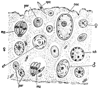
Fig. 68.—Small piece of epithelial lining of gut of heavily infected Grouse chick, showing
various stages in life history of the parasite Eimeria avium; par, parasite (trophozoite);
mz, merozoite; sch, schizont; spz, sporozoite; ooc, oöcyst; ♂, male gametocyte; ♀, female
gametocyte. × 750. (After Fantham.)
The merozoites of Eimeria avium are arranged “en barillet,” like the segments of
an orange (figs. 68 mz, 69, G), therein differing from those of E. schubergi, which are
arranged “en rosace.” They separate from one another (fig. 69, H), penetrate other
epithelial cells, where they may, in turn, become schizonts. Eight to fourteen merozoites
are usually formed by each schizont, twenty have been found, while in cases
of intense infection when space has become limited, the number may be only four.
After a number of generations of merozoites have been formed, a limit is reached
both to the multiplicative capacity of the parasite and to the power of the bird
to provide the invader with food. Consequently, resistant forms of the parasite
are necessary, and the trophozoites begin to show sexual differentiation instead
of forming schizonts, that is, gametogony commences.
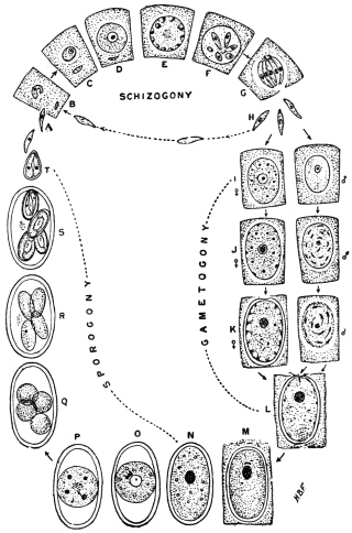
Fig. 69.—Eimeria avium. Diagram of life-cycle. For explanation see text.
(After Fantham.)
Certain trophozoites store food and become large and granular. These are
macrogametocytes (fig. 69, I, ♀). The microgametocytes (fig. 69, I, ♂) are
smaller and far less granular. The macrogametocyte continues to grow, and
becomes loaded with chromatoid and plastinoid granules (fig. 69, J, ♀), while
the microgametocyte has its nucleus divide to form a number of bent, rod-like
portions (fig. 69, J, ♂). The macrogametocyte gives rise to a single macrogamete,
which forms a cyst wall for itself, leaving a thin spot (micropyle) for the entry
of the male (fig 69, K, ♀). The microgametocyte gives rise to numerous small,
biflagellate microgametes (fig. 69, K, ♂) around a large, central residual mass,
from which they ultimately break free, and swim away. When a macrogamete
is reached, the microgamete enters through the micropyle (fig. 69, L)—which
then closes, thus excluding the other males—and applies itself to the female
nucleus (fig. 69, M). Nuclear fusion occurs, the oöcyst (encysted zygote) being thus
produced. Sporogony then ensues. The oöcyst (fig. 69, N) at first has its contents
completely filling it. They then concentrate into a central spherical mass (fig. 69, O)
which gradually becomes tetranucleate (fig. 69, P). Cytoplasm collects around each
nucleus, and four sporoblasts are thus formed (fig. 69, Q). Each sporoblast becomes
oval (fig. 69, R) and produces a sporocyst. Ultimately two sporozoites are formed in
each sporocyst or spore, at first lying tête-bêche (fig. 69, S), but finally twisting
to assume the position most convenient for emergence (fig. 69, T) when they reach a
new host. The period of the life-cycle of Eimeria avium (as well as the details
of the life-cycle) was determined by Fantham to be from eight to ten days, of which
period schizogony occupies four to five days.
The method of infection175 is contaminative, by way of food or drink. Young
birds are especially susceptible to infection. Certain birds, particularly older ones,
may act as reservoirs of oöcysts, being continuously infected themselves, without
showing any marked ill effects from the parasite, but being highly infectious
to other birds. Much moisture retards the development of sporocysts considerably.
The duration of vitality of the infective oöcysts has been determined
experimentally to extend well over two years, and in certain cases longer.
Eimeria avium is the causal agent of “white diarrhœa” or “white scour” in
fowls, and of “blackhead” in turkeys.
Eimeria avium of birds and E. stiedæ of rabbits closely resemble
one another, but are not the same parasite, for E. avium is not infective
to rabbits, nor E. stiedæ to poultry.
Eimeria stiedæ, Lindemann, 1865.
Syn.: Monocystis stiedæ, Lindemann, 1865; Psorospermium cuniculi, Rivolta,
1878; Cytospermium hominis, Rivolta, 1878; Coccidium oviforme, Leuckart, 1879;
Coccidium perforans, Leuckart, 1879; Coccidium cuniculi.
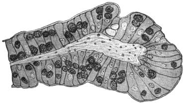
Fig. 70.—Eimeria stiedæ. Section through an infected villus of rabbit’s intestine. × 260.
Eimeria stiedæ is parasitic in the gut epithelium (fig. 70), liver, and
epithelium of the bile ducts of rabbits, and is usually considered to
be the parasite very occasionally found in man. The life-cycle
resembles that of Eimeria avium in its general outlines (see fig. 69)
and therefore will not be detailed in full here. The oöcysts (fig. 71)
are large, elongate-oval, greenish in fresh preparations and vary in
size from 24 µ to 49 µ long and 12·8 µ to 28 µ broad, the gut forms
being usually smaller than those occurring in the liver, owing to the
more confined space in which they are formed. Formerly, the parasites
in the liver were described under the name of Coccidium oviforme,
while those from the intestine were termed Coccidium perforans. This
distinction has now broken down.
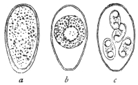
Fig. 71.—Eimeria stiedæ, from
the liver of the rabbit, oöcysts
in various stages of development.
(After Leuckart.)
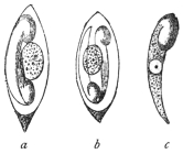
Fig. 72.—a, b, spores of
Eimeria stiedæ (Riv.), with
two sporozoites and residual
bodies; c represents a free
sporozoite. (After Balbiani.)
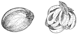
Fig. 73.—So-called swarm cysts (endogenous
sporulation or schizogony) of the Coccidium of
the rabbit. The daughter forms are called
merozoites. (After R. Pfeiffer.)
The oöcysts176 are thick-walled, somewhat flattened at one pole,
where a large micropyle is present. Four egg-shaped spores (sporocysts)
are formed within, each about 12 µ to 15 µ long and 7 µ broad
(fig. 72). The oöcysts are voided with the fæces. Sporogony takes,
in nature, about three days in the excrement. Fæcal contamination
of the food of rabbits results, and coccidian oöcysts are swallowed.
Under the influence of the pancreatic juice of a new host, the
sporozoites (fig. 72, a—c) are liberated from the spores and proceed
to attack the epithelium and multiply within it, as in the case of
Eimeria avium. From the gut,
infection spreads to the liver,
where multiplication of the parasite
goes on actively, resulting
in the formation of the whitish
coccidial nodules, which may be
very conspicuous (fig. 74). Proliferation
of the connective tissue
may occur around the coccidial
nodules, which then contain large
numbers of oöcysts in various
stages of development. It is said that the oöcysts in the older nodules
do not seem to be capable of further development. Schizogony (fig. 73)
and gametogony in all stages can be found in both liver and gut.
Young rabbits often die of intestinal coccidiosis before infection
of the liver occurs. The repeated schizogony of Eimeria stiedæ in the
gut is sufficient to cause death.
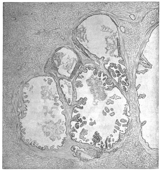
Fig. 74.—Eimeria stiedæ. Section through coccidian nodule in infected rabbit’s liver. × 55.
The disease of cattle popularly known as “red dysentery” is also ascribed to the
action of Eimeria stiedæ. The fæces of infected cattle show blood clots of various
sizes and in severe cases watery diarrhœa is present. Acute cases end fatally in
about two days. Numerous oöcysts, considered to be those of Eimeria stiedæ, occur
in the fæces, and there is a heavy infection of the gut, especially the large intestine
and rectum, all stages of the parasite being found in the epithelium. It is
suspected that cattle contract the disease by feeding on fresh grass contaminated
with oöcysts. The disease is recorded from Switzerland and from East Africa.
As before mentioned, Eimeria stiedæ is considered to be the
organism found in a few cases in man, possibly acquired by eating
the insufficiently cooked livers of diseased rabbits. These cases may
now be described.
(a) Human Hepatic Coccidiosis.
(1) Gubler’s Case. A stone-breaker, aged 45, was admitted to a Paris hospital
suffering from digestive disturbances and severe anæmia. On examination the liver
was found to be enlarged and presented a prominent swelling, which was regarded as
being due to Echinococcus. At the autopsy of the man, who succumbed to intercurrent
peritonitis, twenty cysts were found averaging 2 to 3 cm. in diameter, and one
measuring 12 to 15 cm. The caseous contents consisted of detritus, pus corpuscles,
and oval-shelled formations, which were considered to be Distoma eggs, but which,
in accordance with Leuckart’s conjecture, proved to be Coccidia.177
(2) Dressler’s Case (Prague). Relates to three cysts, varying from the size of
a hemp-seed to that of a pea, and containing Coccidia, found in a man’s liver.178
(3) Sattler’s Case (Vienna). Coccidia were in this case observed in the dilated
biliary duct of a human liver.179
(4) Perls’ Case (Giessen). Perls discovered Coccidia in an old preparation of
Sömmering’s agglomerations.180
(5) Silcock’s Case (London).181 The patient, aged 50, who had fallen ill with
serious symptoms, exhibited fever, enlarged liver and spleen, and had a dry, coated
tongue. At the autopsy numerous caseous centres, mostly immediately beneath the
surface, were found, while the contiguous parts of the liver were inflamed. Microscopical
examination demonstrated numerous Coccidia in the hepatic cells as well
as in the epithelium of the biliary ducts. A deposit of Coccidia was likewise found
in the spleen, which the parasites had probably reached by means of the blood-stream.182
(b) Human Intestinal Coccidiosis.
In two cadavers at the Pathological Institute in Berlin, Eimer183 found the
epithelium of the intestine permeated by Coccidia. Railliet and Lucet’s case may
be traced back to intestinal Coccidia, which were found in the fæces of a woman
and her child, who had both suffered for some time from chronic diarrhœa.184 In
other cases (Grassi, Rivolta), where only the existence of Coccidia in the fæces was
known, it is doubtful whether the parasites originated in the intestine or in the liver.
(c) Doubtful Cases.
To these belong Virchow’s case185 where, in the liver of an elderly woman, a thick
walled tumour measuring 9 to 11 mm. was found. Among the contents of this
tumour there were oval formations 56 µ long, surrounded by two membranes and
enclosing a number of round substances. Virchow considered these foreign bodies
to be eggs of pentastomes in various stages of development, others consider them
to be Coccidia.
The Coccidia which Podwyssotzki claims to have seen in the liver of a man, not
only in the liver cells, but also in the nuclei, are also problematic.186 The parasite
was called Caryophagus hominis.
Again, other explanations can be given to an observation by Thomas, on the
occurrence of Coccidium oviforme in a cerebral tumour of a woman aged 40. The
growth was as large as a pea and surrounded by a bony substance.187
Genus. Isospora, Aimé Schneider, 1881.
Syn.: Diplospora, Labbé, 1893.
Belonging to the section Disporea, that is, forming only two spores,
each with four sporozoites.
Isospora bigemina, Stiles, 1891.
Syn.: “Cytospermium villorum intestinalium canis et felis,” Rivolta, 1874;
“Coccidium Rivolta,” Grassi, 1882; Coccidium bigeminum, Stiles, 1891.
This parasite lives in the intestinal villi of dogs, cats, and the
polecat (Mustela putorius, L.). According to Stiles,188 the oöcyst
divides into two equal ellipsoidal portions or sporoblasts which
become spores and then each forms four sporozoites. The oöcysts
of this species vary from 22 µ to 40 µ in length and from 19 µ to
28 µ in breadth. Each spore is 10 µ to 18 µ long and contains
four sporozoites. The parasites live and multiply, not only in the
gut epithelium, but also in the connective tissue of the intestinal
submucosa. Wasielewski has seen merozoites in the gut of the cat.
Isospora bigemina (fig. 75) appears to occur also in man, for
Virchow published a case which was communicated to him by
Kjellberg, and attributed the illness to this parasite.189 Possibly also it
would be more correct to ascribe the observation of Railliet and
Lucet, which is mentioned under “Human Intestinal Coccidiosis,”
p. 148, to this species, as the Coccidia in that case were distinguished
by their diminutive size (length 15 µ, breadth 10 µ). The case communicated
by Grunow may also possibly refer to Isospora bigemina.190
Roundish or oval structures of 6 µ to 13 µ in diameter occurred in the
mucous membrane of the gut and in the fæces of a case of enteritis.
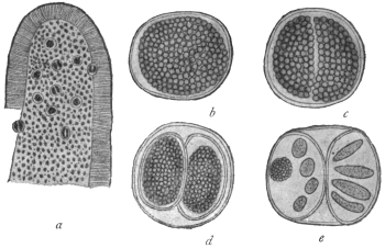
Fig. 75.—Isospora bigemina, Stiles (from the intestine of a dog). a, Piece of an intestinal
villus beset with Isospora, slightly enlarged; b, Isospora bigemina (15 µ in diameter), shortly
before division; c, divided; d, each portion encysted forming two spores; e, four sporozoites
in each part, on the left seen in optical section, together with a residual body—highly magnified.
(After Stiles.)
Doubtful Species.
In literature many other statements are found as to the occurrence of
Coccidia-like organisms in different diseases of man. In some of the cases the
parasites proved to be fungi. This was the case with the parasites of a severe
skin disease of man, formerly called Coccidioides immitis and Coccidioides pyogenes.
Other statements are founded on misapprehensions, or are still much disputed.
If reference is here made to “Eimeria hominis,” R. Blanchard, 1895, this
is done on the authority of the investigator mentioned. The structures in
question are nucleated spindle-shaped bodies of very different lengths (18 µ
to 100 µ), which either occurred isolated or were enclosed in large globular or
oval cysts, alone or with a larger tuberculated body (“residual body”). These
formations were found by J. Künstler and A. Pitres in the pleural exudation removed
from a man by tapping. The man was employed on the ships plying between
Bordeaux and the Senegal River.
Blanchard looks upon the fusiform bodies as merozoites and the cysts as
schizonts of a Coccidium. On the other hand, Moniez declares the spindle bodies
to be the ova and the supposed residual bodies to be “floating ovaries” of an
Echinorhynchus.
Severi’s “monocystid Gregarines,” which were taken from the lung tissue of a
still-born child, are also quite problematical.
No less doubtful are the bodies which Perroncito calls Coccidium jalinum, and
which he found in severe diseases of the intestine in human beings, pigs, and guinea-pigs;
Borini also reported another case.
Order. Hæmosporidia, Danilewsky emend. Schaudinn.
The Hæmosporidia are a group of blood parasites, comprising
forms differing greatly among themselves. Some of the forms need
much further investigation. However, there are certain true Hæmosporidia
which present close affinities with the Coccidia, leading
Doflein to use the term Coccidiomorpha for the two orders conjoined.
The Hæmosporidia present the following general characteristics:—
(1) They are parasites of either red or white blood corpuscles of
vertebrates during one period of their life-history.
(2) They exhibit alternation of generations—asexual phases or
schizogony alternating with sexual phases or sporogony—as do the
Coccidia.
(3) There is also an alternation of hosts in those cases which have
so far been completely investigated. The schizogony occurs in the
blood or internal organs of some vertebrates while the sporogony
occurs in an invertebrate, such as a blood-sucking arthropod or leech.
(4) Unlike the Coccidia, resistant spores in sporocysts are not
generally produced, such protective phases in the life-cycle being
unnecessary, as the Hæmosporidia are contained within either the
vertebrate or invertebrate host during the whole of their life.
The Hæmosporidia may be considered for convenience under
five main types:—
(1) The Plasmodium or Hæmamœba type. This includes the
malarial parasites of man and of birds. The asexual multiplicative
or schizogonic phases occur inside red blood corpuscles and are
amœboid. They produce distinctive, darkish pigment termed melanin
or hæmozoin. Infected blood drawn and cooled on a slide may
exhibit “exflagellation” of the male gametocytes, i.e., the formation
of filamentous male gametes. The invertebrate host is a mosquito.
The malarial parasites of man are discussed at length on p. 155.
Similar pigmented hæmamœboid parasites have been described in
antelopes, dogs, and other mammals, and even reptiles.
(2) The Halteridium type. The trophozoite stage inside the red
blood corpuscle is halter-shaped. Pigment is produced, especially near
the ends of the organism. The parasites occur in the blood of birds.
The invertebrate host of H. columbæ of pigeons in Europe, Africa,
Brazil and India, is a hippoboscid fly, belonging to the genus Lynchia.
Halteridium parasites are common in the blood of passerine birds,
such as pigeons, finches, stone owls, Java sparrows, parrots, etc.
The Halteridium embraces or grows around the nucleus of the host
red cell without displacing the nucleus. Young forms and multiplicative
stages of H. columbæ have been found in leucocytes in the
lungs of the pigeon (fig. 76, 8-12). Male and female forms (gametocytes)
are seen in the blood (fig. 76, 3a, 3b). The cytoplasm of the
male gametocytes is pale-staining and the nucleus is elongate, while
the cytoplasm of the females is darker and the nucleus is smaller
and round. Formation of male gametes from male gametocytes
(the so-called process of “exflagellation”) may occur on a slide
of drawn infected blood, also fertilization, and formation of the
oökinete, as first seen by MacCallum. The correct generic name for
Halteridia is, apparently, Hæmoproteus. Wasielewski (1913), working
on H. danilewskyi (var. falconis), in kestrels, finds that the halteridium
may be pathogenic to nestlings. The cycle of H. noctuæ
described by Schaudinn (1904) lacks confirmation. The account of
the life-cycle of H. columbæ given by Aragão (1908) is illustrated in
fig. 76. It agrees with the work of Sergent (1906–7) and Gonder
(1915). Mrs. Adie (1915) states that the cycle in Lynchia is like that
of a Plasmodium.
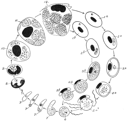
Fig. 76.—Hæmoproteus (Halteridium) columbæ. Life-cycle diagram: 1, 2, stages in red
blood corpuscle of bird; 3, 4, gametocytes (3a ♂, 3b ♀); 5a, formation of microgametes;
6, fertilization (in fly’s gut); 7, oökinete; 8–12, stages in mononuclear leucocytes in lungs.
(After Aragão.)
(3) The Leucocytozoön type. The trophozoites and gametocytes
occur within mononuclear leucocytes and young red cells (erythroblasts)
in the blood of birds. Laveran and França consider that
the Leucocytozoa occur in erythrocytes. The host cells are often
greatly altered by the parasites, becoming hypertrophied and the ends
usually drawn into horn-like processes (fig. 77), though some remain
rounded. Leucocytozoa are limited to birds, and very rarely produce
pigment. Male and female forms (gametocytes) are distinguishable
in the blood (fig. 77), and the formation of male gametes (“exflagellation”)
may occur in drawn blood.
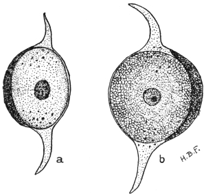
Fig. 77.—Leucocytozoön lovati. a, Male parasite
(microgametocyte), within host cell, whose ends are
drawn out; b, female parasite (macrogametocyte)
from blood of grouse. × 1,800. (After Fantham.)
The Leucocytozoa were first seen by Danilewsky in 1884. They
are usually oval or spherical. It is not easy sometimes to distinguish
the altered host cell from the parasite, as the nucleus of the former
is pushed to one side by the
leucocytozoön. The cytoplasm
of the female parasite
stains deeply, and the nucleus
is rather small, containing a
karyosome. In the male the
cytoplasm stains lightly and
the nucleus is larger, with a
loose, granular structure.
Many species of Leucocytozoa
are recorded, but
schizogony has only been
described by Fantham (1910)191
in L. lovati in the spleen of
the grouse (Lagopus scoticus),
and by Moldovan192 (1913) in
L. ziemanni in the internal
organs of screech-owls.
M. and A. Leger193 (1914)
propose to classify Leucocytozoa,
provisionally, according as the host cells are fusiform or
rounded.
(4) The Hæmogregarina type. Included herein are many parasites
of red blood corpuscles, with a few (the leucocytogregarines) parasitic
in the white cells of certain mammals and a few birds. They are not
amœboid but gregarine-like, vermicular or sausage-shaped (fig. 78).
They do not produce pigment. They are widely distributed among
the vertebrata, but are most numerous in cold-blooded vertebrates
(fishes, amphibia and reptiles). The hæmogregarines of aquatic hosts
are transmitted by leeches, those of terrestrial hosts by arthropods.
The nucleus of hæmogregarines is usually near the middle of the
parasite, but may be situated nearer one end. The body of the
parasite may be lodged in a capsule (“cytocyst”). There is much
variation in size and appearance among hæmogregarines. Some are
small (Lankesterella); some attack the nucleus of the host cell (Karyolysus);
others have full grown vermicules larger than the containing
host corpuscle, and so the hæmogregarines bend on themselves in
the form of U (fig. 78, b). Schizogony often occurs in the internal
organs of the host, sometimes in the circulating blood.
The hæmogregarines occurring in the white cells (mononuclears
or polymorphonuclears) of mammals have been referred to a separate
genus, Leucocytogregarina (Porter) or Hepatozoön (Miller). Such
leucocytogregarines are known in the dog (fig. 79), rat, mouse, palm-squirrel,
rabbit, cat, etc. Schizogony of these forms occurs in the
internal organs, such as the liver, lung and bone-marrow of the hosts.
They are apparently transmitted by ectoparasitic arthropods, such as
ticks, mites, and lice.
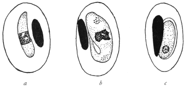
Fig. 78.—Hæmogregarines from lizards, a, H. schaudinni, var. africana, from Lacerta
ocellata; b, H. nobrei from Lacerta muralis; c, H. marceaui in cytocyst, from Lacerta
muralis. (After França.)
A few hæmogregarines are known to be parasitic in the red blood
corpuscles of mammals. Such are H. gerbilli in the Indian field rat,
Gerbillus indicus; H. balfouri (jaculi) in the jerboa, Jaculus jaculus,
and a few species briefly described from marsupials. These parasites
do not form pigment.
Strict leucocytic gregarines have been described from a few birds
by Aragão and by Todd.
The sporogony of hæmogregarines is only known in a few cases,
and in those affinity with the Coccidia is exhibited. In fact, the
Hæmogregarines are now classified by some authors with the
Coccidia.
(5) The Babesia or Piroplasma type. These are small parasites of
red blood corpuscles of mammals. They do not produce pigment.
They are pear-shaped, round or amœboid in Babesia, bacilliform
and oval in other forms referred to this group. Piroplasms are
transmitted by ticks. These parasites are described at length on
p. 172.
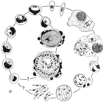
Fig. 79.—Leucocytogregarina canis. Life-cycle diagram. Constructed from drawings by
Christophers. (After Castellani and Chalmers.) Schizogony occurs in the bone-marrow. The
parasite is transmitted from dog to dog by the tick, Rhipicephalus sanguineus, development
in which, so far as known, is shown on the right.
THE MALARIAL PARASITES OF MAN.
Malaria, otherwise known as febris intermittens, chill-fever, ague,
marsh fever, paludism, etc., is the name given to a disease of man,
which begins with fever. It has been known since ancient times and
is distributed over almost all the world, although very unevenly, but
does not occur in waterless deserts and the Polar regions. In many
places, especially in the civilized countries of Central Europe, the
disease is extinct or occurs only sporadically, and large tracts of land
have become free from malaria.
The rhythmical course of the fever is characteristic. It begins
apparently suddenly with chilliness or typical shivering, whilst the
temperature of the body rises, the pulse becomes low and tense and
the number of beats of the pulse increases considerably. After half
to two hours the heat stage begins. The patient himself feels the rise
of his temperature (shown by feeling of heat, dry tongue, headache,
thirst). The temperature may reach 41°C or more. At the same time
there is sensitiveness in the region of the spleen and enlargement of
that organ. After four to six hours an improvement takes place, and
with profuse perspiration the body temperature falls rapidly, not
often below normal. After the attack the patient feels languid, but
otherwise well until certain prodromal symptoms (heaviness in the
body, headache) which were not noticed at first, denote the approach
of another attack of fever, which proceeds in the same way.
The intervals between the attacks are of varying length which
permit of a distinction in the kinds of fever. If the attacks intermit
one day, occurring on the first, third and fifth days of the illness
and always at the same time of day, it is termed febris tertiana; if
two days occur between fever days, it is called febris quartana. In
the case of the fever recurring daily, later writers speak of typical
febris quotidiana. But a quotidian fever may arise when two tertian
fevers differing by about twenty-four hours exist at the same time
(febris tertiana duplex). The patient has a daily attack, but the fever
of the first, third and fifth days differs in some point (hour of
occurrence, height of temperature, duration of cold or hot stage)
from the fever of the second, fourth and sixth days. Similarly,
two or three quartan fevers which differ by about twenty-four hours
each may be observed together (febris quartana duplex or triplex);
in the latter case the result is also a quotidian fever.
Two kinds of tertian fever are differentiated—a milder form
occurring especially in the spring (spring tertian fever), and a more
severe form appearing in the summer and autumn in warmer
districts, especially in the tropics (summer or autumn fever, febris
æstivo-autumnalis, febris tropica, febris perniciosa). The latter often
becomes a quotidian fever.
All the afore-mentioned infections are termed acute. They are
distinguished from the very different chronic malarial infection by the
frequent occurrence of relapses, which finally lead to changes of
some organs and particularly of the blood. The relapses are then
generally marked by an irregular course of fever.
The term masked malaria is used when any disturbance of the state
of health of a periodic character shows itself and disappears after
treatment with quinine.194 Generally it is a question of neuralgia.
That intermittent fever was an infectious disease, although not one which was
transmitted direct from man to man, had been assumed for a long time. Therefore
it was natural, at a time when bacteriology was triumphing, to look for a living agent
causing infection in malaria, which search was, seemingly, successful (Klebs,
Tomasi-Crudeli, 1879). Hence it was not surprising that the discovery of the
real malarial parasites in November, 1880, by the military doctor A. Laveran195 in
Constantine (Algeria), at first met with violent opposition, even after Richard
(1882) had confirmed it and Marchiafava, Celli, Grassi and others, had
further extended it. Not that the existence of structures found in the blood of
malaria patients by Laveran and Richard was denied; on the contrary, the investigations
of the opponents furnished many valuable discoveries, but the organisms were
differently interpreted and considered to be degeneration products of red blood corpuscles.
Only when Marchiafava and Celli (1885) saw movements in the parasites,
which Laveran called Oscillaria malariæ and later Hæmatozoön malariæ, was their
animal nature admitted and the parasites were named Plasmodium malariæ. Shortly
before this, Gerhardt (1884) had stated that the disease could be transmitted by the
injection of the blood of a malarial patient to a healthy person.
This supplied the starting point for further investigations, which were made not
exclusively, but principally, by Italian investigators (Golgi, Marchiafava and Celli,
Bignami and Bastianelli, Grassi and Feletti, Mannaberg, Romanowsky, Osier,
Thayer and others). In 1885 Golgi described the asexual cycle in the blood,
in the case of the quartan parasite. These investigations, after attention had been
drawn by Danilewsky (1890) to the occurrence of similar endoglobular parasites in
birds, were extended to the latter (Grassi and Feletti, Celli and Sanfelice, Kruse,
Labbé and others).
The result was as follows: Malaria in man (and birds) is the result of peculiar
parasites included in the Sporozoa by Metchnikoff, which parasites live in the erythrocytes,
grow in size and finally “sporulate,” that is, separate into a number of “spores”
which leave the erythrocytes and infect other blood corpuscles. Morphologically and
biologically several species (and respectively several varieties) of malarial parasites
may be distinguished, on which the different intermittent forms depend. Transmission
of the blood of patients to healthy people produces a malarial affection which
corresponds in character to the fever of the patient from whom the inoculation was
made. The combined types of fever (tertiana duplex, quartana duplex or triplex)
are explained by the fact that the patient harbours two or three groups of parasites
which differ in their development by about twenty-four hours, whilst the irregular fevers
depend on deviation from the typical course of development of the parasites. In
addition to stages of the parasites which could easily be arranged in a developmental
series concurrent with the course of the disease, other phases of the parasites also
became known, such as spheres, crescents, polymitus forms, which seemed not to be
included in the series and, therefore, were very differently interpreted.
The decision reached at the beginning of the last decade of the last century, which
found expression in comprehensive statements (Mannaberg, Ziemann and others),
only concerned a part of the complete development of the malarial parasites. No one
could with any degree of certainty demonstrate how man became infected, nor were
there reliable hypotheses based on analogy with other parasites concerning the exit
of the excitants of malaria from the infected person and their further behaviour.
Numerous hypotheses had been advanced, but none was able to elucidate the
various observations made from time to time in dealing with malaria. One hypothesis
only seemed to have a better foundation. Manson (1894), who knew from his
own experience the part played by mosquitoes in the development of Filaria from
the blood of man, applied this also to the malarial parasites living in the blood,
whereby at least the way was indicated by which the Hæmosporidia could leave man.
The parasites were said finally to get into water through mosquitoes which had
sucked the blood of malarial patients, and the germ spread thence to men who drank
the water. In some cases the parasites were supposed to reach man by the inhaling of
the dust of dried marshes. On the other hand, Bignami believed that the mosquitoes
were infected in the open air by malarial parasites which occurred there in an unknown
stage and the insects transmitted the germs to man when biting. R. Koch combined
both hypotheses, without, however, producing positive proof. R. Ross, then (1897–8)
an English military doctor in India, was the first to succeed in this. He had been
encouraged by Manson to study the fate of malarial Plasmodia which had entered
the intestine of mosquitoes with malaria-infected blood, especially in the case of the
Plasmodium (Proteosoma) living in the blood of birds. He showed that the Proteosoma
penetrate the intestinal wall of the mosquitoes, grow and develop into large
cysts which produce innumerable rod-like germs, which burst into the body cavity
and penetrate the salivary glands. Ross allowed mosquitoes to suck the blood of
birds affected by malaria, and some nine days later, let the infected mosquitoes
which had been isolated suck healthy birds. After five to nine days Proteosoma
were found to occur in the blood of the birds used. The Proteosoma and Halteridium
of birds were also further investigated by MacCallum (1897–8), Koch and others, and
important results followed.
In any case Ross (1898) had clearly established the importance of mosquitoes in
the spread of malaria among birds. It was now only a question of proving whether,
and how far, mosquitoes were concerned with human malaria. Ross himself worked
to this end. Here the experiments of Italian investigators (Bignami, Bastianelli,
Grassi)196 were of importance. These investigators studied the fate of malarial
parasites in man, produced malaria in men experimentally by the bites of infected
mosquitoes, and established that only mosquitoes belonging to the genus Anopheles
were concerned, and not species of Culex. These latter are only able to transmit
Proteosoma to birds. It is true that Culex can ingest the human malarial parasites,
but the latter do not develop in them. Development only occurs in species of
Anopheles. In Anopheles (and similarly for Proteosoma in Culex) sexual reproduction
takes place; crescents, spheres and polymitus forms are necessary stages
of development in the mosquito.
With these discoveries the campaign against malaria became more definite. It
was directed partly against the transmitters, whose biology and life-cycle were more
thoroughly investigated, instead of merely against the infection of the adult Anopheles.
The latter do not, as was believed for some time, transmit the malarial germs to their
offspring. They always infect themselves from human beings, whereby the relapses
appearing in early summer, and the latent infection, especially of children of natives,
play a principal part (Stephens and Christophers, Koch). Further, the crusade was
directed against the infection of man by the bites of Anopheles. Important results
have been obtained in these directions. Low and Sambon in 1900 lived in a
mosquito-screened hut in a malarial part of the Roman Campagna for three of the
most malarious months and did not contract the disease. In the same year Dr. P. T.
Manson was infected with malaria by infected mosquitoes sent from Italy. The rôle
of mosquitoes having been proved, it may be hoped that ultimately the eradication
of malaria, or at least a considerable restriction of it, will be achieved.
It is of importance to record that, although malarial parasites occur in mammals
(monkeys, bats, etc.) the human ones are not transmissible to mammals, not even to
monkeys. The species, therefore, are specific to the different hosts (Dionisi, Kossel,
Ziemann, Vassall).
An important work dealing with the modern applications of the mosquito-malaria
theory in all parts of the Tropics was published by Sir Ronald Ross in 1911. It is
entitled “The Prevention of Malaria” (John Murray, London, 21s.).
DEVELOPMENT OF THE MALARIAL PARASITES OF MAN.
The commencement of the developmental cycle and of the infection
of man, is the sporozoites (fig. 80, 1) which are passed into the blood
of a person by the bite of an infected mosquito. Prior to this the parasites
collect in the excretory ducts of the salivary glands (fig. 80, 27)
of the Anopheles. The sporozoites are elongate and spindle-shaped,
10 µ to 20 µ long and 1 µ to 2 µ broad, with an oval nucleus situated
in the middle. They are able to glide, perform peristaltic contractions,
or curve laterally. Schaudinn has studied the penetration of the red
blood corpuscles (fig. 80, 2) by the sporozoites in the case of the
living tertian parasite. The process takes forty to sixty minutes in
drawn blood. After its entrance the parasite, which is now called a
trophozoite, contracts, and becomes an active amœbula (fig. 80, 3).
It develops a food vacuole and grows at the expense of the invaded
blood corpuscle (fig. 80, 4), which is shown by the appearance of
pigment granules (transformed hæmoglobin) in it. When the maximum
size is attained, multiplication by schizogony (fig. 80, 5-7) begins
with a division of the nucleus, which is followed by further divisions of
the daughter nuclei, the number of which varies up to 16 or even 32,
depending on the species of the parasite. Then the cytoplasm divides
into as many portions as there are nuclei, the result being a structure
suggestive of the spokes of a wheel or of a daisy, the centre of the
resulting rosette being occupied by dark pigment. Finally, the parts
separate from one another, leaving behind a residual body containing
the pigment, and the daughter forms issue into the blood plasma
as merozoites (fig. 80, 7). They are actively amœboid (fig. 80, 8) and
soon begin to enter other blood corpuscles of their host, for the entry
into which thirty to sixty minutes are necessary, according to
Schaudinn’s observations.197
Here they behave like sporozoites which previously entered and
again produce merozoites. This process is repeated until the number
of parasites is so large that, at the next migration of the merozoites, the
body of the person infected reacts with an attack of fever,198 which is
repeated with the occurrence of the next or following generations.
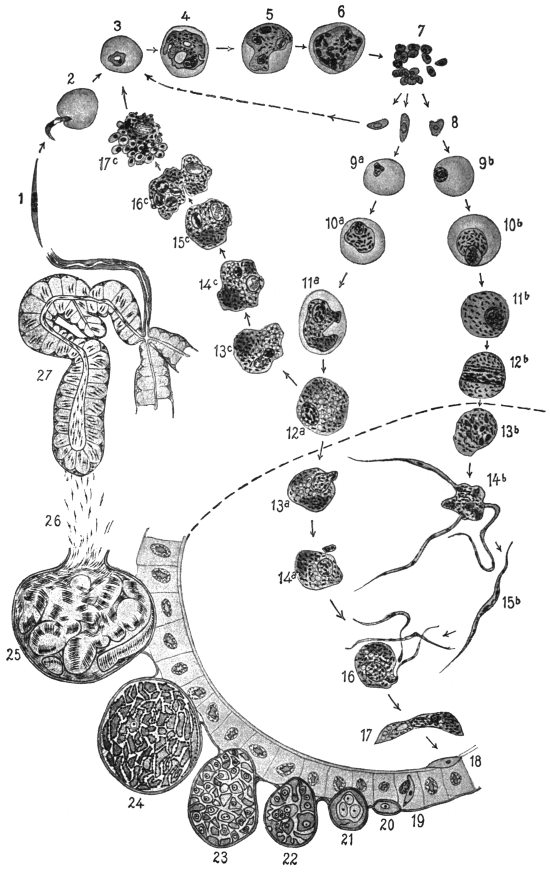
Fig. 80.—Life-cycle of the tertian parasite (Plasmodium vivax). Figs. 1 to 17, × 1,200;
figs. 18 to 27, × 600. (After Lühe, based on figures by Schaudinn and Grassi.) 1, sporozoite;
2, entrance of the sporozoite into a red blood corpuscle; 3, 4, growth of the parasite, now
sometimes called a trophozoite; 5, 6, nuclear division in schizont; 7, free merozoites; 8, the
merozoites which have developed making their way into blood corpuscles, (arrow pointing to the
left) and increase by schizogony (3–7); after some duration of disease the sexual individuals
appear; 9a-12a, macrogametocytes; 9b-12b, microgametocytes, both still in the blood-vessels
of man. If macrogametocytes (12a) do not get into the intestine of Anopheles they may perhaps
increase parthenogenetically according to Schaudinn (12a; 13c-17c). The merozoites which
have arisen (17c) become schizonts 3–7. The phases shown underneath the dotted line
(13–17) proceed in the stomach of Anopheles. 13b and 14b, formation of microgametes; 13a
and 14a, maturation of the macrogametes; 15b, microgamete; 16, fertilization; 17, oökinete;
18, oökinete in the walls of the stomach; 19, penetration of the epithelium of the stomach;
20–25, stages of sporogony on the outer surface of the intestinal wall; 26, migration of the
sporozoites to the salivary gland; 27, salivary gland with sporozoites.
The growth and schizogony last different times, according to the
species of the parasite, about forty-eight hours in the case of the
parasite of febris tertiana or tropica, and seventy-two hours for the
quartan parasite. The various intermittent forms produced by them
depend on this specific difference in the malarial parasites.
The schizogony can, however, only be repeated a certain number
of times, supposing that the disease has not been checked prematurely
by the administration of quinine, which is able to kill the parasites.
It appears that after a number of attacks of fever the conditions of
existence in man are unfavourable for the malarial parasites, and this
brings about the production of other forms which have long been
known, but also long misunderstood (spheres, crescents, polymitus).
The merozoites in this case no longer grow into schizonts, or at least
not all of them, but become sexual individuals called gametocytes
(fig. 80, 9-12), which only start their further development when they
have reached the intestine of Anopheles. This does not take place
in every case, nor with all the gametocytes which exist in the blood of
patients with intermittent fever. Of those parasites which remain
in the human blood the male ones (microgametocytes) soon perish,
the females (macrogametocytes) persist for some long time, and perhaps
at last acquire the capacity of increasing by schizogony. They
might thus form merozoites which behave in the body as if they had
proceeded from ordinary schizonts (fig. 80, 13c-17c). If their
number increases sufficiently, in course of time the patient, who
was apparently recovering, has a new series of fever attacks, or
relapses, without there having been a new infection. This is the
view of Schaudinn, who from researches of his own concluded that
relapses were brought about by a sort of parthenogenetic reproduction
of macrogametocytes. R. Ross, on the contrary, believes that in the
relatively healthy periods the number of parasites in the blood falls
below that necessary to provoke febrile symptoms; relapses then
result merely from increase in the numbers of the parasites present in
the individual. Ross’s view is now generally accepted.
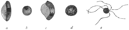
Fig. 81.—Stages of development of pernicious or malignant tertian parasites in the intestine
of Anopheles macultpennis. (After Grassi.) a, macrogametocyte (crescent) still attached to
human blood corpuscles; b, macrogametocyte (sphere) half an hour after ingestion by the
mosquito; c, microgametocyte (crescent) attached to the blood corpuscle; d, microgametocyte
(sphere) half an hour after ingestion; the nucleus has divided several times; e, microgametes
attached to the residual body (polymitus stage).
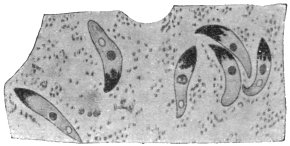
Fig. 82.—Oökinete of the malignant tertian parasite in the stomach of Anopheles maculipennis,
thirty-two hours after ingestion of blood. (After Grassi.)
If the gametocytes, which are globular, or in the pernicious
or malignant tertian parasite crescentic (fig. 81), gain access to the
intestine of an Anopheline,199 they mature. The macrogametocytes
extrude a part of their nuclear substance (fig. 80, 13a, 14a) and
thereby become females or macrogametes. The microgametocytes, on
the other hand, undergo repeated nuclear division, preparation for
this being made apparently whilst in the blood of man. This results
in the formation of threadlike bodies which move like flagella and
finally detach themselves from the residual body (fig. 80, 13b, 14b).
These are the males or microgametes200 (fig. 80, 15b).
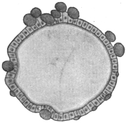
Fig. 83.—Section of the
stomach of an Anopheles, with
cysts (oöcysts) of the malignant
tertian parasite. (After Grassi).
Copulation takes place in the stomach of the Anopheline (fig. 80, 16).
A microgamete penetrates a macrogamete and coalesces with it. The
fertilized females elongate very soon and are called oökinetes or
“vermicules” (figs. 80, 17; 82). They penetrate the walls of the
stomach, pierce the epithelium (fig. 80, 18, 19), and remain lying
between it and the superficial stratum
(tunica elastico-muscularis). Then they
become rounded and gradually develop
into cysts which grow larger and are
finally visible to the naked eye, being
called oöcysts (figs. 80, 20-24; 83). Their
size at the beginning is about 5 µ, the
maximum that they attain is 60 µ, only
exceptionally are they larger.
The sporulation (figs. 80, 21-25; 84),
which now follows, begins with repeated
multiple fission of the nucleus. Long
before the definitive number of nuclei,
which varies with the individual, is attained
the protoplasm, according to Grassi, begins to segment around the
individual large nuclei but without separating completely into cell
areas. According to Schaudinn,
however, there is a condensation
of the outstanding protoplasmic
strands. It is certain that the number
of nuclei increases with simultaneous
decrease in size. They
soon appear on the surface of the
strands or sporoblasts, surround
themselves with some cytoplasm
and then elongate (fig. 84). In
this manner the sporozoites are
formed and break away from the
unused remains of the cytoplasmic
strands of the sporoblasts
(fig. 80, 26). The number of the
sporozoites in an oöcyst varies from
several hundreds to ten thousand.
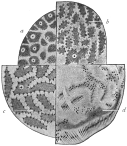
Fig. 84.—Four different sporulation stages
of malarial parasites from Anopheles maculipennis,
much magnified. a-c, of the
malignant tertian parasite; a, four to four
and a half days after sucking; b and c, five
to six days after sucking; d, of the tertian
parasite, eight days after sucking. (After
Grassi.)
The sporulation is influenced in its
duration by the external temperature
(Grassi, Jansci, Schoo). In the tertian
parasite it takes place quickest at a
temperature of 25° to 30° C. and takes
eight to nine days. A temperature a
few degrees lower has a retarding effect (eighteen to nineteen days at 18° to 20° C).
A still lower one has a restraining or even destructive effect. Temperatures over
35° C. also exercise a harmful effect. The malignant tertian parasite seems to need
a somewhat higher temperature and the quartan parasite a lower one.
The sporozoites of the various malarial parasites show no specific
differences. They were stated by Schaudinn to occur in three forms,
and these were described as indifferent (neuter), female and male.
There is, however, little or no evidence for this hypothetical differentiation.
The last were said to perish prematurely, that is, in the
oöcyst. The others after the rupture of the oöcysts enter the body
cavity of the Anophelines, whence they are carried along in the course
of the blood. Finally they penetrate the salivary glands (fig. 80, 27)
probably by their own activity, break through their epithelia and
accumulate in the salivary duct (fig. 80, 27). At the next bite by the
mosquito they are transmitted to the blood-vessels of man.
The Species of Malarial Parasites of Man.
In view of the differences in opinion regarding “species” and “varieties,” the dispute
whether the malarial parasites of man represent one species with several varieties, or several
species is almost superfluous. If necessary two genera may be distinguished.
The parasites of the tertian and quartan fever are alike in that
their gametocytes have a rounded shape (figs. 80, 12, 13), whilst the
corresponding stages of the pernicious or malignant tertian parasites
are crescentic (figs. 81, 88). These differences are used by some writers
as the distinguishing characteristic of two genera: Plasmodium, Marchiafava
and Celli, 1885, for the first mentioned species; Laverania,
Grassi and Feletti, 1889, for the pernicious or malignant tertian parasite.
Whether there is a genuine quotidian fever and accordingly a special
quotidian parasite is still disputed.
These parasites are treated in practical detail in Stephens and Christophers’
“Practical Study of Malaria,” 3rd edition, 1908.
Plasmodium vivax, Grassi and Feletti, 1890.
Syn.: Hæmamœba vivax, Grassi and Feletti, 1890; Plasmodium malariæ var.
tertianæ, Celli and Sanfelice, 1891; Hæmamœba laverani var. tertiana, Labbé,
1894; Hæmosporidium tertianum, Lewkowitz, 1897; Plasmodium malariæ tertianum,
Labbé, 1899: Hæmamœba malariæ var. magna, Laveran, 1900, p.p.; Hæmamœba
malariæ var. tertianæ, Laveran, 1901.
This species, P. vivax,201 is the causal agent of the simple or spring
tertian fever and is, therefore, named directly the tertian or benign
tertian parasite (figs. 80, 3-8; 85). During the afebrile period in the
patient, the young trophozoites or amœbulæ appear on or in the red
blood corpuscles as pale bodies of 1·5 µ to 2 µ diameter which at
first show only slow amœboid movements. Their nucleus is difficult
to recognize in the early stage. Soon the food vacuole is formed and
this grows concomitantly with the trophozoite and the parasite has a
ring-like appearance. Afterwards the vacuole diminishes, and at this
period the first brownish melanin granule appears. From this time
the activity and number of the pigment granules increase with continuous
growth. When the parasite has grown to about one-third
the diameter of the erythrocyte the latter shows characteristic red
Schüffner’s dots or “fine stippling,” after staining with Romanowsky’s
solution. Later, after about twenty-four hours, the blood corpuscles
begin to grow pale, then to increase in size, and after thirty-six hours,
that is, about twelve hours before the next attack of fever, schizogony
of the parasite is initiated by the division of the nucleus. The parasite
at this time occupies half to two-thirds of the enlarged blood corpuscle.
The daughter nuclei continue dividing until sixteen, and
occasionally twenty-four, daughter nuclei are produced. The pigment
which, up till now lies nearer the periphery, moves to the middle,
while the nuclei lie nearer the surface.
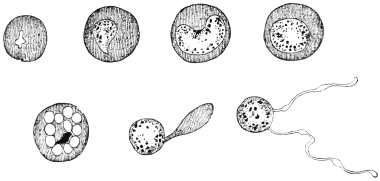
Fig. 85.—Development of the tertian parasite in the red blood corpuscles of man;
on the right a “Polymitus.” (After Mannaberg.) See also fig. 80, 3—7.
Around each nucleus a portion of cytoplasm collects and thus
young merozoites are produced. These separate from each other and
from the little residual masses202 which contain the melanin and pass
from the blood corpuscles, which now can hardly be recognized, to
the blood plasma, where they soon attack new erythrocytes.
The migration of the merozoites initiates a new attack of fever
and two groups of tertian parasites in the blood, differing in development
by about twenty-four hours, are the conditions for febris tertiana
duplex.
After a lengthy duration of fever the gametocytes (figs. 80, 9—12)
appear. They are uninucleate. The microgametocytes are about the
size of fully developed schizonts, the macrogametocytes are somewhat
larger. Their further development takes place in Anophelines.
The chief distinctive characteristics of the simple tertian parasite,
as seen in infected blood, are:—(1) The infected red-cell is usually
enlarged; (2) the presence of fine red granules known as Schüffner’s
dots in the red blood corpuscles, after Romanowsky staining; (3)
the fragile appearance of the parasite compared with other species.
Large forms are pigmented, irregular and “flimsy-looking,” sometimes
appearing to consist of separate parts. Irregularity of contour
is common.
Ahmed Emin203 (1914) has described a small variety of P. vivax.
Plasmodium malariæ, Laveran.
Syn.: Oscillaria malariæ, Laveran, p.p., 1883; Hæmamœba malariæ, Gr. et Fel.,
1890; Plasmodium malariæ var. quartanæ, Celli et Sanfel., 1891; Hæmamœba
laverani var. quartana, Labbé, 1894; Hæmosporidium quartanæ, Lewkowitz, 1897;
Plasmodium malariæ quartanum, Labbé, 1899; Plasmodium golgii, Sambon, 1902;
Laverania malariæ, Jancso, 1905 nec Grassi et Fel. 1890; Hæmomœba malariæ
var. quartanæ; Lav., 1901.
Plasmodium malariæ is the parasite of quartan malaria (fig. 86).
The trophozoites of the quartan parasite differ from the corresponding
stages of the tertian parasite in that their motility is less and soon
ceases. They differ also in their slower growth, by the early
disappearance of the food vacuole, by the more marked formation
of the dark brown pigment, and by the fact that the red blood
corpuscles attacked are not altered either in colour or size.
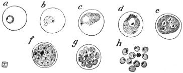
Fig. 86.—Development of the quartan parasite in the red corpuscles of man—asexual
stages. (After Manson.)
When the parasites have grown almost to the size of the erythrocytes
schizogony occurs. The pigment granules arrange themselves in lines
radiating towards the centre and the merozoites are also radially
disposed in groups of 6, 8, 10 or even 12, but are often arranged
less regularly. The whole development, growth and schizogony,
occupies seventy-two hours.
The appearance of quartana duplex or triplex is conditional on the
presence in the blood of the patient of two or three groups of
Plasmodia differing in their development by twenty-four hours.
The chief distinctive characters of the quartan parasite are: (1)
The erythrocyte is unchanged in size; (2) the rings are compact and
show pigment early; in the larger forms the chromatin is dense and
relatively plentiful; (3) the pigment, which is relatively well-marked,
may be arranged at the periphery.
Laverania malariæ, Grassi and Feletti, 1890 = Plasmodium
falciparum, Welch, 1897.
Syn.: Plasmodium malariæ var. quotidianæ, Celli et Sanf., 1891; Hæmamœba
malariæ præcox, Gr. et Fel., 1892 (nec H. præcox, Gr. et Fel., 1890); Hæmamœba
laverani, Labbé, 1894; Hæmatozoön falciparum, Welch, 1897; Hæmosporidium
undecimanæ and H. sedecimanæ and H. vigesimo-tertianæ, Lewkowitz, 1897; Hæmamœba
malariæ parva, Lav., 1900; Plasmodium præcox, Dofl., 1901; Plasmodium
immaculatum, Schaud., 1902; Plasmodium falciparum, Blanch., 1905.
The names most commonly used for the parasite of malignant
tertian malaria are Plasmodium falciparum and Laverania malariæ.
The summer and autumn fever (febris æstivo-autumnalis), also
called malignant tertian or sub-tertian, is caused by a malarial parasite
which is distinguished by the small size of its schizont, while the
gametocytes are crescentic (figs. 81, 88).
Most authors identify this kind of fever or the parasites which cause it (Laverania
malariæ) with the pernicious malaria of the tropics. Ziemann, however, repeatedly
has drawn attention to certain small but definite differences between the usual malignant
tertian or pernicious parasites which occur in the tropics and the tropical parasites
of some malarial districts, particularly of West Africa, and insists that at least two
varieties or sub-species occur. Other investigators distinguish from this or these
forms a quotidian parasite. On the other hand, the assertion is made that there are
no specific differences, but that the malignant or pernicious tertian parasite which
normally needs forty-eight hours for its development in the blood of man, can
also develop in twenty-four hours. The establishment of the duration of the
development is a matter of especial difficulty, because the stages of schizogony
are far less numerous in the peripheral blood than in that of the internal organs. It
is also stated that the tropical parasite very seldom forms crescentic but rather
rounded gametocytes. According to such an observation the organism would belong
to Plasmodium and not to Laverania. The question whether the tropical fevers are
caused by two different parasites does not seem to be definitely settled.
The young trophozoite of the malignant, pernicious tertian, or sub-tertian
parasite (fig. 87) are but slightly active and are very small, even
after the formation of the comparatively large food vacuole, which
makes the body appear annular (“signet ring” stage). Often two and
even more parasites are found in one blood corpuscle.
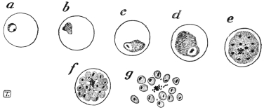
Fig. 87.—The pernicious malignant or sub-tertian parasite in the red corpuscles of man,
asexual stages. (After Manson.)
Fully grown they only attain two-thirds or less of the diameter of
the erythrocytes, which display an inclination to shrink and then appear
darker than the normal (brass-coloured). In the early stage dots or
stippling—sometimes called Maurer’s dots—appear on the blood corpuscles
as in those attacked by the ordinary tertian parasite (Plasmodium
vivax), but the Maurer’s dots are relatively coarse and few, and
are not easily stained. These dots were first described by Stephens
and Christophers in 1900, and subsequently by Maurer in 1902.
About thirty hours after the entrance into the blood corpuscles,
the parasites are rarely found in the peripheral blood, but they are
present in the internal organs, and especially in the spleen. The
schizogony, which now begins in the internal organs, proceeds on the
same lines as that of the quartan parasite, that is, usually with the
merozoites radially arranged around a central agglomeration of dark
brown pigment.
The number of merozoites formed is quoted differently, e.g., 8 to 24,
on an average 12 to 16. However, according to the recent cultural
researches of J. G. and D. Thomson204 (1913) the number of merozoites
of P. falciparum is 32. D. Thomson, from examination of spleen
smears at autopsy, also concludes that the number of merozoites may
reach 32. During their formation the blood corpuscle which is
attacked gets paler and disintegrates.
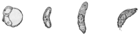
Fig. 88.—The crescents of the malignant tertian parasite. (After Mannaberg.)
See also fig. 81.
The gametocytes which finally appear are attenuated, curved
bodies, rounded at each end and known as crescents (figs. 81, 88), and
are provided with a nucleus and with coarse pigment masses. In the
males the pigment is more scattered than in the females, where it is
around the nucleus. Their length is 9 µ to 14 µ, and their breadth is
2 µ to 3 µ. At first they are still in the pale blood corpuscles, later
they free themselves and are found in numbers in the peripheral
blood in cases of pernicious malaria of Southern Europe and the
tropics, while, on the other hand, they occur much more rarely in
the peripheral blood in West African malignant tertian. Their further
development takes place under the same conditions as in the other
malarial parasites.
D. Thomson (1914),205 from studies of autopsy smears, has shown
that crescents develop chiefly in the bone-marrow and spleen, and
take about ten days to grow into the adult state in the internal organs.
He believes that crescents are produced from ordinary asexual spores.
Quinine, he states, has no direct destructive action on crescents, but
it destroys the asexual source of supply.
The sporozoites of Laverania malariæ (P. falciparum) are represented
in fig. 89.
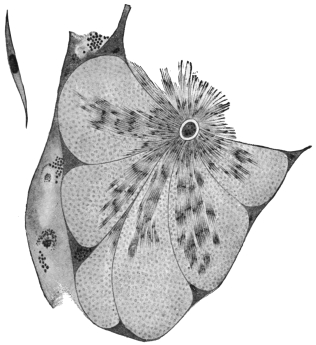
Fig. 89.—Section through a tubule of the salivary gland of an Anopheles with sporozoites
of the malignant tertian parasites; on the left at the top a single sporozoite greatly magnified.
(After Grassi).
The principal distinctive characters of the malignant tertian parasite
are: (1) The ring forms are very small, occasionally bacilliform,
and may be marginal (“accolé” of Laveran); (2) the larger trophozoites
are often ovoid, and about one-third or one-half of the
erythrocyte in size; (3) the infected red cells sometimes show coarse
stippling (Maurer’s dots); (4) the gametocytes, or sexual forms, are
crescentic in shape.
J. W. W. Stephens (1914) has described a new malarial parasite of
man; it is called Plasmodium tenue. It is very amœboid, with scanty
cytoplasm and much chromatin, sometimes rod-like or irregular.
The parasite was described from a blood-smear of an Indian child.
The creation of a new species for this parasite has been criticized by
Balfour and Wenyon, and by Craig.
Plasmodium relictum, Sergent, 1907.
Syn.: Plasmodium præcox, Grassi and Feletti, 1890; Plasmodium danilewskyi,
Gr. et Fel., 1890; Hæmamœba relicta, Gr. et Fel., 1891; Proteosoma grassii,
Labbé, 1894.
Hæmamœboid, pigment-producing, malarial parasites are often found in birds.
Like the human malarial parasites they have been variously named. Labbé created
the genus Proteosoma for them, and this name is still often used as a distinctive one
unofficially. The correct name is stated to be either Plasmodium relictum or
P. præcox, or possibly even P. danilewskyi, assuming that there is only one species.
The nomenclature of the malarial parasites is most confused. The avian malarial
parasites are transmitted by Culicine mosquitoes.
The organism was discovered by Grassi in the blood of birds in Italy, and causes
a fatal disease in partridges in Hungary. Sparrows are affected in India, and it
was this Plasmodium in which Ross first traced the development of a malarial
parasite in a mosquito. The parasite may be transmitted from bird to bird by
blood-inoculation, canaries being very susceptible.
The principal stages of the avian plasmodium closely resemble those of the
malarial parasites of man. In its earliest stage P. relictum is unpigmented,
but soon the trophozoite grows and becomes pigmented, meanwhile displacing
the nucleus of the avian red-blood corpuscle, a characteristic feature, distinguishing
it from Halteridium. Schizonts are formed, each of which gives rise to
about nine merozoites in the circulating blood. Sexual forms or gametocytes also
occur in the blood. These develop in Culex fatigans, C. pipiens and C. nemorosus.
Oökinetes or vermicules are formed in twelve to fifteen hours in the
stomach of the mosquito, and in one to two days well-developed round oöcysts may
be seen. In three to four days sporoblasts have formed within the oöcysts and
young sporozoites begin to develop. In nine to ten days the oöcysts are mature,
being filled with sporozoites. The oöcysts then burst and the sporozoites travel
through the thoracic muscles to the salivary glands of the Culicine.
Neumann, experimenting with canaries, found that Stegomyia fasciata could
transmit the infection, but less efficiently than species of Culex.
The Cultivation of Malarial Parasites.
The successful cultivation of malarial parasites in vitro was first
recorded by C. C. Bass and by Bass and Johns (1912).206 Since then,
J. G. and D. Thomson,207 and McLellan (1912–13), Ziemann208 and others
have repeated the experiments.
DIFFERENTIAL CHARACTERS OF THE HUMAN MALARIAL PARASITES.
| Character | Plasmodium malariæ
(Quartan) | Plasmodium vivax
(Benign tertian) | Laverania malariæ =
Plasmodium falciparum
(Malignant tertian) |
|---|
| Schizogony | Complete in seventy-two hours | Complete in forty-eight hours | Complete in forty-eight hours or less |
| Trophozoite | Smaller than P. vivax, larger than L. malariæ | Young trophozoite large. | Young trophozoite small |
| Pseudopodia not marked or long | Long pseudopodia | |
| Movements | Rather slow in immature forms | Active amœboid movements | Sometimes actively motile |
| Pigment | Coarse granules, peripherally arranged, little movement | Fine granules, with active movement | Granules fine and scanty, movement oscillatory |
| Schizont | Smaller than red corpuscle | Larger than red blood corpuscle | Smaller than red corpuscle |
| Merozoites | 6 to 12 forming rosette | 15 to 20 regularly arranged | 8 to 32 (according to different authors) arranged irregularly |
| Gametocytes | Spherical | Spherical | Crescentic |
| Distribution of parasites in vertebrate host | About equal number in peripheral and visceral blood | Larger numbers in visceral blood | Scanty in peripheral blood compared with the enormous numbers in the internal organs. The latter part of the cycle (schizogony) may occur in the internal organs only |
| Alterations in erythrocytes | Almost normal | Pale and hypertrophied. | Corpuscle may be shrunken and dark, or may be colourless. Maurer’s coarse dots sometimes seen |
| Schüffner’s dots seen in deeply stained specimens |
Essentially the method of cultivation, as used by Thomson,
is as follows: 10 c.c. of infected blood are drawn from a vein and
transferred to a sterile test tube, in which is a thick wire leading to
the bottom of the tube. One-tenth of a cubic centimetre of a
50 per cent. aqueous solution of glucose or dextrose is placed
in the test tube, preferably before adding the blood. The blood
is defibrinated by stirring gently with the wire. When defibrination
is complete the wire and the clot are removed, and the glucose-blood
is transferred, in portions, to several smaller sterile tubes, each containing
a column of blood about one inch in height. The tubes
are plugged and capped and then transferred, standing upright, to an
incubator kept at a temperature of 37° C. to 41° C. The blood corpuscles
soon settle, leaving a column of serum at the top, to the extent
of about half an inch in each tube. The leucocytes need not be
removed by centrifugalization. J. G. Thomson (1913) and his
collaborators did not find it necessary to destroy the complement in
the serum, and they found that the malarial parasites developed at all
levels in the column of corpuscles, and not merely on the surface
layer of the corpuscles as first stated by Bass and Johns.
So far only the asexual generation of the malarial parasites has
been grown in vitro. Thomson rarely observed hæmolysis in the
cultures. Clumping of the malignant tertian parasites occurred.
In cultures of the benign tertian parasite (Plasmodium vivax) clumping
was not observed. J. G. and D. Thomson consider that this
difference as regards clumping explains why only young forms of
malignant tertian are found in peripheral blood, as the clumping
tendency of the larger forms causes them to be arrested in the finer
capillaries of the internal organs. It also explains the tendency to
pernicious symptoms, such as coma, in malignant tertian malaria.
Further it was found from cultures that P. falciparum was capable
of producing thirty-two spores (merozoites) in maximum segmentation,
while P. vivax produced sixteen spores (merozoites) as a rule,
though the number might be greater than sixteen. (Quartan parasites
produce eight spores or merozoites in schizogony.)
It may also be mentioned here that Babesia (Piroplasma) canis has
been successfully cultivated in vitro by Bass’s method. This has been
accomplished by Thomson and Fantham,209 Ziemann, and Toyoda in
1913. J. G. Thomson and Fantham used the simplified Bass technique
recorded above, namely, infected blood and glucose, incubating at
37° C. In one of the B. canis cultures, starting with heart blood of a
dog containing corpuscles infected with one, two, or, exceptionally,
four piroplasmata, Thomson and Fantham succeeded in obtaining a
maximum of thirty-two merozoites in a corpuscle. The cultures are
infective to dogs and sub-cultures have been obtained.
Family. Piroplasmidæ, França.
The parasites included in this provisional family or group belong
to the Hæmosporidia. They are minute organisms, sometimes
amœboid, but usually possessing a definite form. They are endoglobular,
being contained within mammalian red blood corpuscles,
but they produce no pigment. The true Piroplasmata, belonging
to the genus Babesia, destroy the host corpuscles, setting free the
hæmoglobin, which is excreted by the kidneys of the cow, sheep,
horse, dog, etc., acting as host. The disease produced, variously called
piroplasmosis or babesiasis, is consequently characterized by a red
coloration of the urine known as hæmoglobinuria, or popularly as
“red-water.” One of the best known piroplasms is Piroplasma
bigeminum or Babesia bovis (probably the latter name is correct),
which is the causal agent of “Texas fever” or “red-water” in cattle
and is spread by ticks.
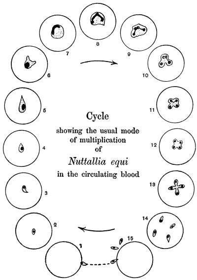
Fig. 90.—Nuttallia equi, life-cycle as seen in red blood corpuscles in stained preparations
of peripheral blood. (After Nuttall and Strickland.)
Of recent years, researches on the morphology of these blood
parasites has led to their separation into various genera and species.
However, our knowledge is still very far from complete. The various
genera recognized by França210 (1909), and placed in a provisional
family, Piroplasmidæ, may be listed, though further research may
lead to emendations:—
(1) Babesia (Starcovici) or Piroplasma (Patton). Pyriform
parasites, dividing by a special form of budding or gemmation with
chromatin forking, as well as by direct binary fission. Parasitic in
oxen, dogs, sheep, horses, etc.
(2) Theileria (Bettencourt, França and Borges). Rod-shaped and
oval parasites occurring in cattle and deer. T. parva is the pathogenic
agent of African East Coast fever in cattle.
(3) Nuttallia (França). Oval or pear-shaped parasites, with multiplication
in the form of a cross. N. equi211 (fig. 90) of equine “piroplasmosis”
(nuttalliosis). N. herpestidis in a mongoose.
(4) Nicollia (Nuttall). Oval or pear-shaped parasites with
characteristic nuclear dimorphism, and with quadruple division at
first fan-like, then like a four-leaved clover. N. quadrigemina from
the gondi.
(5) Smithia (França). Pear-shaped, single forms stretching across
the blood corpuscle. Multiplication into four in the form of a cross.
S. microti from Microtus arvalis, S. talpæ from the mole.
(6) Rossiella (Nuttall). This belongs to the family Piroplasmidæ
of França. It is intracorpuscular and non-pigment forming, occurring
singly, in pairs, or occasionally in fours. It is usually round and
larger than Babesia. The parasite multiplies by binary fission.
R. rossi in the jackal.
The genus Babesia is the best known and most important, and
will be considered next.
Genus. Babesia, Starcovici, 1893.
Syn.: Pyrosoma, Smith and Kilborne, 1893; Apiosoma, Wandolleck, 1895; Piroplasma,
W. H. Patton, 1895; Amœbosporidium, Bonome, 1895.
The organisms belonging to this genus are pyriform, round or
amœboid. The characteristic mode of division is as follows: Just
before division the parasite becomes amœboid and irregular in shape,
(fig. 91, 1–5) with a compact nucleus. The latter gives off a nuclear
bud. This nuclear bud divides into two by forking (fig. 91, 6, 7).
The chromatin forks grow towards the surface of the body of the
rounded parasite, and then two cytoplasmic buds grow out. The
forking nuclear buds, which are Y-shaped, pass into the cytoplasmic
outgrowths212 (fig. 91, 8, 9). The buds gradually increase in size at
the expense of the parent form until they become two pear-shaped
parasites joined at their pointed ends. The connecting strand shrinks
and the two daughter forms separate (fig. 91, 10–14). The pyriform
parasites after having exhausted the blood corpuscle escape from it
(fig. 91, 15), and seek out fresh host corpuscles, entering by the rounded,
blunt end (fig. 91, 1). It is the pyriform phase of the parasite which
penetrates red blood corpuscles, not rounded forms, which die if set
free. The pyriform parasite, however, becomes rounded (fig. 91, 2, 3),
soon after its entry into a fresh host cell. This interesting mode of
division by gemmation and chromatin forking has been made diagnostic
of the genus Babesia by Nuttall.213 Rounded forms of Babesia
divide by binary fission, and this direct method can also be adopted
by the other forms of Babesia.
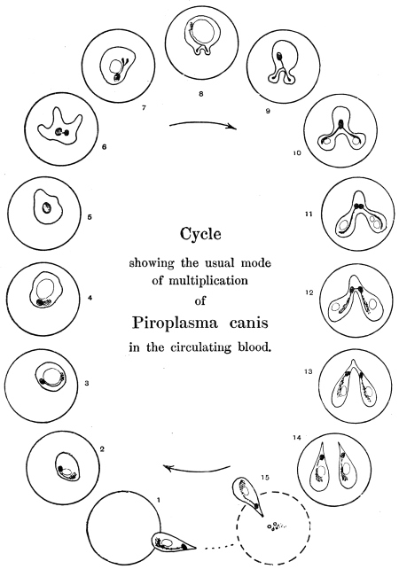
Fig. 91.—Babesia (Piroplasma) canis, life-cycle in stained preparations of infected blood
of dog. (After Nuttall and Graham-Smith.)
The distribution of the chromatin in the pear-shaped Babesia, as seen in B. canis
and B. bovis, is interesting. The main nuclear body consists of a karyosome
surrounded by a clear area. There is also a loose (chromidial) mass of chromatin
representing the remains of the chromatin forks seen during the formation of the
parasite as a daughter form by gemmation. Occasionally there is a small dot or
point, the so-called “blepharoplast” of Schaudinn and Lühe. This minute dot is
not a flagellate blepharoplast, for there is no flagellate stage in the life-history of
Babesia. These nuclear phenomena have been described by Nuttall and Graham-Smith
and Christophers (1907)214 for B. canis, by Fantham (1907)215 for B. bovis, and
by Thomson and Fantham (1913) from glucose-blood cultures of B. canis.
Babesia are tick borne, as was first shown by Smith and Kilborne
(1893). The developmental cycle in the tick is incompletely known.
The best accounts are those of Christophers (1907)216 for B. canis and
Koch (1906) for B. bovis, and these accounts are supplementary.
The principal stages, so far as known, may be summarized thus:—
(1) The piroplasms taken by the tick in feeding on blood pass into the tick’s
stomach. The pyriform parasites, which alone are capable of further development,
are set free from the blood corpuscles. In about twelve to eighteen hours they
become amœboid, sending out long, stiff, slender, pointed pseudopodia. The
nucleus of each parasite divides unequally into two. Similar forms have been
obtained in cultures. These stellate forms may be gametes, and according to Koch
fuse in pairs.
(2) A spherical stage follows, possibly representing the zygote. This grows, and
a uninucleate globular mass results. This form is found in large numbers on the
third day, according to the observations of Koch.
(3) A club-shaped organism is next formed. This may represent an oökinete
stage. The club-shaped bodies are motile and gregarine-like, and are about four
times the size of the blood forms. These club-shaped bodies and subsequent stages
were described by Christophers in the development of B. canis in the dog-tick,
Rhipicephalus sanguineus.
(4) The club-shaped bodies pass from the gut of the tick into the ovary, and so
get into the ova. There they become globular, and later are found in the cells of
the developing tick-embryo. The parasites are, then, transmitted hereditarily.
Similar globular bodies are found in the tissue cells of the body of tick nymphs
which have taken up piroplasms. The globular stage was called the “zygote” by
Christophers, but it may correspond to the oöcyst of Plasmodia.
(5) The globular body divides into a number of “sporoblasts,” which become
scattered through the tissues of the larval or nymphal tick, as the case may be.
(6) The sporoblasts themselves divide into a large number of sporozoites, which
are small uninucleate bodies, somewhat resembling blood piroplasms. The sporozoites
collect in the salivary glands of the tick. They are inoculated into the
vertebrate when the tick next feeds.
The chief species of Babesia and their pathogenic importance
may be listed thus:—
(1) Babesia bovis (Babes) produces infectious hæmoglobinuria
of cattle in Europe and North Africa. It is transmitted by Ixodes
ricinus. A similar parasite also occurs in deer.
(2) Babesia bigemina (Smith and Kilborne) produces Texas
fever, tristeza, or red-water in cattle in North and South America,
South Africa and Australia. It is transmitted by Boöphilus annulatus
in North America, by B. australis in Australia, South America, and
the Philippines, and by B. decoloratus in South Africa.
The parasite is from 2 µ to 4 µ long, and from 1·5 µ to 2 µ broad.
Babesia bigemina may be the same parasite as B. bovis.
(3) Babesia divergens (MacFadyean and Stockman) is a small
parasite. It is found in cattle suffering from red-water in Norway,
Germany, Russia, Hungary, Ireland, Finland, and France, and is
transmitted by Ixodes ricinus.
(4) Babesia canis (Piana and Galli-Valerio) gives rise to malignant
jaundice or infectious icterus in dogs in Southern Europe, India, and
other parts of Asia and North Africa, where it is transmitted by
Rhipicephalus sanguineus. In Africa generally, especially South
Africa, the disease is transmitted by Hæmaphysalis leachi. Babesia
canis varies from 0·7 µ to 5 µ, the size depending partly on the
number of parasites within the corpuscle. It averages about 3 µ. It
has been cultivated in Bass’ medium (glucose and infected blood),
see p. 172.
In India Piroplasma gibsoni (Patton) infects hunt dogs and jackals.
It is annular or oval in shape.
(5) Babesia ovis (Babes) produces “Carceag,” a disease of sheep
in Roumania, the Balkan Peninsula, Italy, and Transcaucasia. It
varies in size from 1 µ to 3 µ. It is transmitted by Rhipicephalus
bursa. The parasite has recently been recorded from Rhodesia.
(6) Babesia caballi (Nuttall and Strickland) causes “biliary
fever” in equines. The parasite occurs in Russia, Roumania, and
Transcaucasia. It varies in size from 1 µ to 2 µ. It is transmitted by
Dermacentor reticulatus.
It should be mentioned that Nuttallia equi also causes “piroplasmosis” in
equines, with symptoms of hæmoglobinuria and jaundice in Italy, Sardinia, many
parts of Africa, Transcaucasia, India, and Brazil. In Africa it is transmitted by
Rhipicephalus evertsi. It has been shown experimentally that a horse recovered
from Babesia caballi was susceptible to the inoculation of Nuttallia equi blood.
(7) Babesia pitheci (P. H. Ross) was found in a monkey,
Cercopithecus sp., in Uganda. The pear-shaped forms measure 1·5 µ
by 2·5 µ.
(8) Babesia muris (Fantham)217 was found in white rats. The
pyriform parasites are 2 µ to 3 µ long and 1 µ to 1·5 µ broad; oval
forms are 0·5 to 1·5 µ diameter.
The usual symptoms of babesiasis (piroplasmosis) are high fever,
loss of appetite, hæmoglobinuria, icterus, anæmia, paralysis, and
death in about a week in acute cases. In chronic cases there is
anæmia, and hæmoglobinuria is less marked. When animals recover,
there are still some piroplasms left in the blood. “Recovered” or
“salted” animals are not susceptible to reinfection, but ticks feeding
on them acquire piroplasms, and are a source of danger to freshly
imported animals.
Treatment.—Trypan-blue is the best drug, as shown by Nuttall and Hadwen218
(1909). It should be administered intravenously in 1 to 1·5 per cent. aqueous solution.
A dose of 5 to 10 c.c. is curative for dogs, one of 100 to 150 c.c. for horses and
cattle. Unfortunately, the tissues are coloured blue by the drug. The “salted”
animals, after trypan-blue treatment, still harbour the parasites in their blood for
years.
Genus. Theileria, Bettencourt, França and Borges, 1907.
The organisms belonging to this genus are rod-like or bacilliform, and coccoid
or round.
The best known of the species of Theileria is T. parva, the pathogenic agent of
East Coast fever or Rhodesian fever in cattle in Africa.
Theileria parva, Theiler, 1903.
Syn.: Piroplasma parvum.
In the blood corpuscles of infected cattle minute rod-like and
oval parasites are seen. Some are comma shaped and others are
clubbed (fig. 92, 1–12). The rod-like forms measure 1 µ to 3 µ in
length by 0·5 µ in breadth; the oval forms are 0·7 µ to 1·5 µ in
diameter. The intracorpuscular parasites are said by R. Gonder
(1910) to be gametocytes, the rod-like forms being thought to
be males, the oval forms to be females. Free parasites are practically
never seen in the blood. It is known that it is impossible to
produce the disease in a healthy animal by blood inoculation, but
only by intraperitoneal transplantation of large pieces of infected
spleen (Meyer). There may be as many as eight parasites in a corpuscle.
The chromatin is usually at one end of the organism. In
some parasites the appearance of the chromatin suggests division, but
such division, if it takes place, must be very slow, as it has not been
actually seen in progress. The red blood corpuscles appear merely to
act as vehicles for the parasites (Nuttall, Fantham, and Porter).219
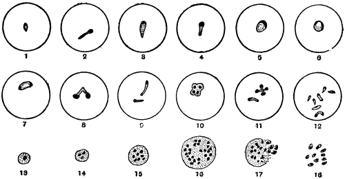
Fig. 92.—Theileria parva. 1–12, intracorpuscular parasites, stained. (After Nuttall and
Fantham); 13–18, Koch’s blue bodies, from stained spleen smear; 17–18, breaking up of
Koch’s body. (After Nuttall.)
In the internal organs, especially the lymphatic glands, spleen and bone-marrow,
are found multinucleate bodies known as Koch’s blue bodies (fig. 92, 13–18). These
are schizonts, according to Gonder.220 The actual Koch’s blue bodies are said to be
extracellular, but similar multinucleate bodies, schizonts, occur in lymphocytes. The
schizonts divide and the merozoites resulting probably invade the red blood corpuscles
in the internal organs. Gonder considers that the sporozoites injected by
the tick collect in the spleen and lymphatic glands, penetrate the lymphocytes and
give rise to the schizonts.
Gonder has studied the cycle of T. parva in the tick. He states that the
gametocytes leave the host corpuscles and give rise to gametes, then conjugation
occurs producing zygotes. The zygotes are then said to become active to form
ookinetes, and to enter the salivary glands of the tick. Multiplication is said to
occur therein, producing a swarm of sporozoites. This work needs confirmation.
T. parva is transmitted by Rhipicephalus appendiculatus, R. simus, R. evertsi,
R. nitens, and R. capensis. The parasites are not hereditarily transmitted in
Rhipicephalus, but when taken by the transmitter at one stage of its development
the tick is infective in its next stage (e.g., if the larva becomes infected, then the
nymph is infective; if the nymph becomes infected, then the adult is infective).
An animal recovered from Theileria parva is incapable of infecting ticks, but few
animals recover from East Coast fever. Animals suffering therefrom do not show
hæmoglobinuria.
Theileria mutans, Theiler, 1907·
Syn.: Piroplasma mutans.
This is transmissible experimentally by blood inoculation. It occurs in cattle in
South Africa and Madagascar and is apparently non-pathogenic. No Koch’s blue
bodies are formed. It is transmitted by ticks.
Theileria annulata (Dschunkowsky and Luhs) occurs in cattle in Transcaucasia.
A Theileria (T. stordii) has been found in a gazelle (França, 1912).
Genus. Anaplasma, Theiler, 1910.
This genus221 may be mentioned here. The organisms included therein are,
according to Theiler, coccus-like, consisting of chromatin, and are devoid of cytoplasm.
They occur in the red blood corpuscles of cattle, causing a disease characterized
by destruction of red cells, fever and anæmia, but with yellow urine. The
disease is tick transmitted. The bodies now called Anaplasma marginale were
formerly described as marginal points. They multiply by simple fission. They are
said by Theiler to cause gall-sickness in cattle in South Africa. Some authors doubt
whether these bodies are organismal.
Genus. Paraplasma, Seidelin, 1911.
Under this generic name Seidelin described certain bodies found
by him in cases of yellow fever in 1909. The type species is
P. flavigenum,222 and is claimed by Seidelin to be the causal agent of
yellow fever.
Paraplasma flavigenum occurs in the early days of the disease as
small chromatin granules with or without a faint trace of cytoplasm.
The bodies are usually intracorpuscular. Also, somewhat larger forms,
with distinct cytoplasm, are seen in small numbers. During the later
days of the disease still larger forms are found, and these occur also
in sections of organs (e.g., kidney) made post-mortem. Some of these
larger forms are perhaps schizonts. In the second period of the
disease possible micro- and macro-gametes may be found, some of
which are extracorpuscular. Some small free bodies have been seen.
Recently schizogony has been stated to occur in the lungs, and it is
said that guinea-pigs can be inoculated with Paraplasma flavigenum,
and show yellow pigment in the spleen.
Seidelin places Paraplasma in the Babesiidæ, with resemblances
more particularly to Theileria. V. Schilling-Torgau and Agramonte
have criticized these findings; the former considers them to be the
resultant of certain blood conditions.
P. subflavigenum was found by Seidelin in 1912 in a man suffering
from an unclassified fever in Mexico.
Further, it is now known that a Paraplasma occurs naturally in
guinea-pigs. More researches are needed on these matters, as some
writers (e.g., Wenyon and Low) claim that the bodies are not
organismal.
Paraplasma flavigenum.—The Yellow Fever Commission (West
Africa) in their third report, dated 1915, have come to the conclusion
that there is no evidence that the bodies termed Paraplasma flavigenum
are of protozoal nature or that they are the causal agents of
yellow fever.
Sub-class. NEOSPORIDIA, Schaudinn.
Sporozoa in which growth and spore formation usually go on
together.
Order. Myxosporidia, Bütschli.
These parasites, which were discovered by Johannes Müller (1841), live principally
in fishes, and occasionally cause destructive epizoötics amongst their hosts. Müller
first observed them in the form of whitish-yellow pustules on the skin or on the gills
of various fishes. These pustules contained masses of small shell-covered bodies with
or without tails (“psorosperms,” see fig. 93). Similar bodies were also found in the
air bladders of certain fish. Creplin (1842) demonstrated the resemblance of the cysts
(“psorosperm tubes”) harbouring the psorosperms to the “pseudonavicella-cysts” of
a gregarine, as described by v. Siebold. Dujardin (1845) considered that there was
possibly some connection between the protoplasmic “psorosperm tubes” and the
spores they contained, and the developmental stages of monocystid gregarines from the
vesiculæ seminales of earth-worms. The relationship of the “fish psorosperms” was
placed on a firmer basis by Leydig (1851) and Lieberkühn. The former found numerous
forms in marine fish, and he discovered in species which live free in the gall bladder
of cartilaginous fishes that the psorosperms originated in a manner similar to the
gregarines. Lieberkühn (1854) studied the Myxosporidia in the bladder of the pike
(fig. 93, a, b, d), and observed their amœboid movements, as well as the formation of
the spores, from each of which a small amœboid body escaped, a discovery that
was confirmed by Balbiani. The same author also found that spiral filaments were
enclosed in the so-called polar body, i.e., the polar capsule of the psorosperm spores,
and that these could be protruded (fig. 93, d, and fig. 95).
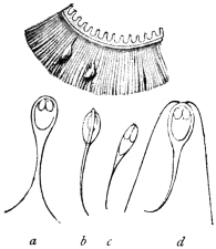
Fig. 93.—Upper figure, part of
a gill of a roach, Leuciscus rutilus
(natural size), with two myxosporidia.
Lower figures, a, b, d, spores
of myxosporidia from a pike, Esox
lucius. c, Spore from Platystoma
fasciatum. (After J. Müller.)

Fig. 94.—The tailless spore of
Myxobolus mülleri, with the polar
bodies and their nuclei and the
sporozoite. (After Bütschli.)
The term Myxosporidia, which at the present day is universally applied to the
“psorosperm tubes,” was introduced by Bütschli in 1881, who studied not only the
structure and development of the spores, but also the protoplasmic body of the
parasites (fig. 96), and confirmed the occurrence of numerous nuclei. Many authors
have made important additions to our knowledge of the Myxosporidia: Perugia,
Thélohan, Mingazzini, L. Pfeiffer, L. Cohn, Doflein, Mercier, Schröder and Auerbach;
while the presence of this parasite outside the class of fishes has become known
through Lutz, Laveran, and others. The species causing disease in fishes have been
described by Ludwig, Railliet, Weltner, L. Pfeiffer, Zschokke, Hofer, Doflein, Gurley,
Plehn, Schuberg, Fantham and Porter. With regard to classification the works of
Thélohan (1895) and Gurley (1894) may be mentioned.
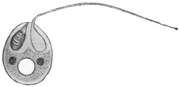
Fig. 95.—Schematic representation of a
spore of Myxobolus. One polar capsule has
protruded its filament; two nuclei and a
“vacuole” in the sporozoite. (After Doflein.)
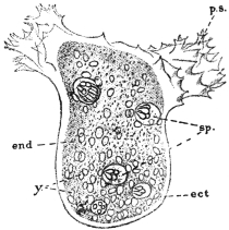
Fig. 96.—Chloromyxum leydigi. Active
trophozoite (parasitic in gall-bladder of skates,
rays, dog-fish). Ect, ectoplasm; ps, pseudopodia;
end, endoplasm; y, yellow globules
in endoplasm; sp, spores, each with four
polar capsules. × 525. (After Thélohan.)
The Myxosporidia live either free on the epithelial surface of hollow organs
(gall or urinary bladder, renal tubules, but never in the intestine), or are enclosed in
the tissues of their host. The gills and muscular system are their favourite
habitat, but other tissues or organs may be attacked. Species of Myxosporidia
are also known from Amphibia, Reptilia, and a few invertebrates.
The free forms, which are often amœboid (fig. 96), move by the aid of variously
shaped pseudopodia, have a constant form, or may exhibit contractions of the body.
The tissue parasites often reach a considerable size, so that the integument of the
host forms protuberances over them. They are of a roundish or irregular shape.
Frequently they are enveloped in a connective tissue covering formed by the host.
The protoplasmic body in the trophic phase (fig. 96) shows a distinct ectoplasm
which is finely granular or sometimes striated, and an endoplasm which is coarsely
granular and contains many nuclei as well as cell inclusions, such as crystals, pigment
grains and fat globules. The nuclei originate by division from the primitive
nucleus of the amœboid germ that issues from the spore. This amœbula may or
may not live intra-cellularly during the early stages of its existence.
The multinucleate trophozoite of a Myxosporidian forms spores in its endoplasm
practically throughout its whole period of growth (fig. 96). Vegetative reproduction
by a process of external budding or plasmotomy may also occur, as in Myxidium
lieberkühni from the urinary bladder of the pike.
The myxosporidian trophozoite may produce two spores within itself, when
it is placed in the sub-order Disporea, or it may produce numerous spores, which
is characteristic of the sub-order, Polysporea. The phenomenon of spore formation
is not simple (fig. 97), and the spore itself is surrounded by a bivalved shell or sporocyst
and contains polar capsules in addition to the amœboid germ (fig. 97, G, H). The
valves of the sporocyst and the polar capsules are really differentiated nucleate cells,
so that each spore is an aggregate of cells rather than one cell, though only a single
amœbula issues from a spore. The accounts of spore formation vary somewhat
according to the different workers.
Spore formation is usually very complicated and there are differences of opinion
as to the interpretation of various stages, particularly as to whether conjugation
occurs therein. The process is initiated by the concentration of cytoplasm around
one of the nuclei of the endoplasm, so that a small spherical mass or initial corpuscle
is produced, the pansporoblast (Gurley) or primitive sphere (Thélohan). Some
authors state that a pansporoblast really results from a conjugation of two initial
corpuscles (fig. 97, A-D). Nuclear multiplication occurs within the pansporoblast
(fig. 97, E), and sooner or later two multinucleate sporoblasts are formed within it
(fig. 97, F). Each sporoblast gives rise to a single spore, which consists of a sporocyst
or envelope composed of two valves each secreted by a cell, two polar capsules
each secreted by a cell, and the sporoplasm or amœbula which becomes binucleate
(fig. 97, G). During the process of spore formation (fig. 97) various vegetative and
reduction nuclei may be produced, in addition to those which are essentially involved
in spore formation, and the sporocyst cells may be developed early.
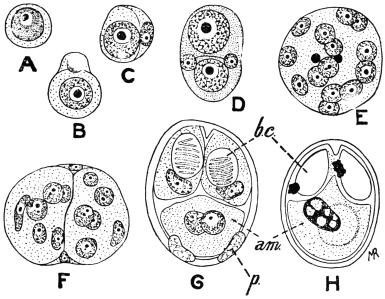
Fig. 97.—Myxobolus pfeifferi. Spore formation. A, reproductive cell from plasmodial
trophozoite; B, cell divided unequally into two; C, smaller cell forming envelope to larger
one; D, pansporoblast formed by union of two forms like C; E, multinucleate pansporoblast,
two of the nuclei being those of the envelope; F, pansporoblast divided into two multinucleate
sporoblasts; G, spore differentiation; p, two parietal cells forming sporocyst; bc, polar capsules;
am, binucleate amœbula; H, ripe spore in which the two nuclei of the amœbula have
fused. (After Keysselitz.)
Each spore contains two (figs. 94, 95) or more polar capsules which are clearly
visible in the fresh condition. Each polar capsule is a hollow, more or less pear-shaped
body, secreted by a cell and having a well defined contour. Within it, a
long, delicate, elastic filament, the polar filament, is formed, and lies spirally coiled
in the polar capsule until just before the emergence of the amœbula from the spore
(fig. 95). The polar filament is ejected, probably under the influence of the digestive
juice, when the spore reaches a new host, and serves to anchor the spore to the
tissue with which it is in contact, and thus allow of the emergence of the amœbula
in a situation suitable for its development. The polar capsule with its contained
polar filament has been compared with the stinging cells or nematocysts of the
Cœlentera, but it has a totally different function.
The spores fulfil the purpose of effecting transmission to other hosts. Infection
occurs by the ingestion of the parasites per os after their escape by some
means from their host. Thélohan and others have demonstrated that the valves of
the spores soon open under the influence of the digestive juices, thus allowing the
young myxosporidia to escape. Their further history is unknown; but it may be
surmised that they either travel direct to the organs usually affected (gall bladder,
urinary bladder), or are distributed in the body by means of the circulatory or
lymphatic systems.
The Myxosporidia that invade tissues are often deadly to their hosts. They may
be present in a state of “diffuse infiltration” when practically every organ of the
body may be infected, as in barbel disease (due to Myxobolus pfeifferi). On the
other hand, the parasites may be concentrated at one spot, when cysts, either large
or small, are produced. Such cysts occur on the gills of many fishes. A few additional
important pathogenic forms are Myxobolus cyprini, the excitant of “pockenkrankheit”
of carp, and Lentospora cerebralis, parasitic in the skeleton of Salmonidæ
and Gadidæ. The skeletons of the tail, fins and skull particularly are seats of infection,
and from the skull the Lentospora can spread to the semicircular canals, resulting
in loss of power to maintain its balance on the part of the fish. On this account
the malady is termed “drehkrankheit.” Young fish are more particularly infected.
Myxobolus neurobius infects the spinal cord and nerves of trout.
Myxosporidia are divided into two sub-orders—Disporea and Polysporea—according
to whether they form only two or several spores during their growth.
The former include two genera limited to fishes, which are easily distinguishable by
the shape of the spores: Leptotheca, Thél., with a rounded spore, and Ceratomyxa,
Thél., with a very elongate spore. The larger number of genera belong to the
Polysporea, which are divided into three families:
| (1) Amœboid germ with a vacuole the contents of which do not stain with iodine. |  | (a) With two polar capsules.—Myxidiidæ. |
| (b) With four polar capsules.—Chloromyxidæ. |
| (2) Amœboid germ with a vacuole stainable with iodine. Spores with two polar capsules.—Myxobolidæ. |
| For further subdivisions the differences in the spores are principally utilized. |
Order. Microsporidia, Balbiani.
These are the organisms discovered in the stickleback by Gluge in 1834, and
in Coccus hesperidum by Leydig in 1853. They have since been found in numerous
other arthropods, especially insects. They acquired particular importance when it
was discovered that they were the cause of the “pébrine” disease (“gattina” of
the Italians) which caused so much destruction amongst silkworms (Bombyx mori).
Pasteur (1867–70) and especially Balbiani (1866) participated in the researches on
Nosema bombycis, and it was the latter who classed the “pébrine bodies” or “psorospermia
of the arthropoda” amongst the Sporozoa as Microsporidia (1882).223 The complete
life cycle of N. bombycis was described in 1909 by Stempell. The Microsporidia
are not confined to insects and arachnoids, they are now known to occur also in
crustacea, worms, bryozoa, fishes, amphibians and reptiles. Certain tumours in
fishes, similar to those formed by many Myxosporidia, are produced by Microsporidia.
Fantham and Porter found that Nosema apis was pathogenic to bees
and other insects, and was the causal agent of the so-called “Isle of Wight”
disease in bees224 in Great Britain.
The Microsporidia, as their name implies, form minute spores which usually are
oval or pear-shaped. Each spore contains a single polar capsule which is not easily
visible in the fresh state (fig. 98, f) and a single amœboid germ issues from the spore
(fig. 99, b).
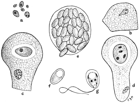
Fig. 98.—Nosema apis. Various stages in life-cycle. a, planonts or amœbulæ from chyle
stomach of bee; b, amœboid planont creeping over surface of gut epithelial cell; c, uninucleate
trophozoite within epithelial cell; d, meront with nucleus divided into four, about to form
four spores; e, epithelial cell crowded with spores; f, young spore; g, spore showing five
nuclei, polar filament ejected, and amœbula, about to issue. × 1,500, a-e; × 2,150, f-g.
(After Fantham and Porter.)
The life cycle of Nosema apis, parasitic in bees, may be taken as an example of
that of a microsporidian. The infection of the host is initiated by the ingestion of
spores of N. apis in food or drink contaminated with the excrement of other infected
bees. Under the influence of the digestive juice of the bee the spore-coat (sporocyst)
softens, the polar filament is ejected and anchors the spore to the gut epithelium,
and the minute amœbula contained in the spore emerges. The amœbula is capable
of active amœboid movements (fig. 98, b) and so is termed the planont or wandering
form (fig. 98, a). After a short time each planont penetrates between or into the cells
of the epithelium of the gut, a few only passing through into the body cavity. Within
the cells the amœbulæ become more or less rounded, lose their power of movement,
and after a period of growth of the trophozoite (fig. 98, c) commence to divide actively,
these dividing forms being known as meronts (fig. 98, d). Various forms of fission
occur, and during this phase, termed merogony, the numbers of the parasite within
the host are greatly increased, with concomitant destruction of the epithelium
(fig. 98, e). After a time sporogony commences. The full-grown meront becomes
successively the pansporoblast and sporoblast. Nuclear multiplication and differentiation
ensue and five nuclei are ultimately produced. At the same time a sporocyst
is secreted, and two vacuoles are produced within. One is the polar capsule, and
within it the polar filament is differentiated; the other forms the posterior vacuole
(fig. 98, g). Between the two vacuoles the body cytoplasm or sporoplasm forms a
girdle-like mass. Of the nuclei, one regulates the polar capsule, two control the
secretion of the sporocyst, and two remain in the sporoplasm. The polar capsule and
polar filament are not usually visible in the fresh condition, but can be demonstrated
by the use of various chemical reagents (fig. 100). The sporoplasm ultimately becomes
the amœbula (fig. 98, g) which issues from the spore after the ejection of the polar
filament.
Fig. 99.—a, section through the abdominal
wall of a silkworm, whose epithelial cells
contain Microsporidia (Nosema bombycis); b,
a spore, the contents of which are escaping.
(After Balbiani.)
Fig. 100.—Nosema bombycis, Naeg.
Spores treated with nitric acid, thus
rendering the polar capsule perceptible,
and the filament has protruded from one
of the spores. (After Thélohan.)
A trophozoite (meront) of N. apis becomes a single pansporoblast which gives
rise to one sporoblast producing one spore, and this procedure is characteristic of
the genus Nosema. In other genera the trophozoite may form more than one pansporoblast
and each pansporoblast may form a variable number of spores in different
cases. Various attempts at classification have been based on these characteristics.
It must suffice here to note that in the cases where the trophozoite becomes one
pansporoblast, the latter can produce four spores in the genus Gurleya, eight spores
in Thélohania and many spores in Pleistophora. In other cases, where the trophozoites
give rise to many pansporoblasts, each of the latter may form many spores,
as in the genus Glugea.
A few pathogenic microsporidian parasites other than N. apis may be mentioned.
N. bombycis, causing pébrine in silkworms, may infect any or all the tissues of the
host (fig. 99). The larvæ of the host, i.e., the “silkworms,” may become infected by
eating food contaminated with spore-containing excrement of already infected silkworms.
In cases of heavy infection the silkworm dies, but should the infection be
less intense the larva becomes a pupa in which the parasite persists, so that the moth
emerges from the cocoon already infected. Not only is the moth parasitized itself,
but the Nosema reaches the generative organs of both sexes and penetrates the
ovaries of the female, with the result that the ova are deposited infected. Such
infected eggs are capable of developing, so that infection may be transmitted
hereditarily as well as by the contaminative method. Infected eggs can be recognized
by microscopic examination, as Pasteur showed, and thus preventive measures
may be adopted.
A microsporidian parasite is known to occur on the roots of the spinal and cranial
nerves of Lophius piscatorius, the angler fish. This parasite is variously referred to
the genera Nosema and Glugea.
Thélohania contejeani, parasitic in the muscles of crayfish, is believed by some
to be the causal agent of recent epizoötics among them, though others believe the
disease to be really due to a bacillus. It may be that the one organism aids in the
entry of the other into the host.
Order. Actinomyxidia, Stolč.
Fig. 101.—Spore of Hexactinomyxon
psammoryctis. At top of figure three polar
capsules, one with polar filament extended.
× 450. (After Stolč.)
A brief mention may be made of the Actinomyxidia (fig. 101), which were first
described by Stolč in 1899 as parasites of Oligochætes. They have also been
investigated by Mrazek, and a detailed study of certain species was made by
Caullery and Mesnil (1905). The trophozoite
is small and amœboid. The spores
are large, and exhibit tri-radiate symmetry.
Spore formation is complicated and sexual
processes occur therein. Many amœbulæ
are set free from each spore.
Order. Sarcosporidia, Balbiani.
The first member of this group was
discovered by Miescher in 1843. This
author found white filaments running
parallel with the direction of the fibres
in the voluntary muscles of mice. They
were visible to the naked eye, and proved
to be cylindrical tubes tapering at each
end. They were as long as the muscular
fibres, were enveloped in a membrane, and
contained innumerable elongate or kidney-shaped
bodies and a smaller number
of little spherical forms. Th. v. Hessling
confirmed (1853) the occurrence of these
“Miescher’s tubes” within the muscular
fibres, this author having discovered the
same structures in the heart muscles of
deer, cattle, and sheep. Both investigators considered them to be pathological transformations
of the muscles. v. Siebold, from his own experiences, regarded them
as fungus-like entophytes.
Rainey (1858) discovered similar structures in the muscular system of pigs, and
considered them to be early stages of Cysticercus cellulosæ, which error Leuckart
rectified, simultaneously emphasizing their relationship with Myxosporidia. Both
these authors found them in the muscular fibres, and both observed that they
possessed a thick striated membrane. Manz (1867) published the results of more
minute investigations on the structure and contents of the cylinders. This observer
also recognized the disease in rabbits and attempted to cultivate the parasites. He
also tried to induce experimental infection in guinea-pigs, rats, and mice, but the
result was negative.
However, domestic and wild mammals are not the only hosts of Sarcosporidia;
these parasites are also harboured by birds. Thus, according to Kühn, they are
found in the domestic fowl; according to Rivolta in Turdus, Corvus, and other
birds; according to Stiles in North American birds; while Fantham found Sarcosporidia
in the African mouse-bird, Colius. Reptiles also are parasitized occasionally.
Bertram found them in the gecko, Lühe in the wall-lizard. It was found also that
the Sarcosporidia could develop not only in the muscles but also in the connective
tissue. This led to the foundation of a new, but provisional, classification by Blanchard,
using the generic name Miescheria for the parasites in the muscles and
Balbiania for those in the connective tissue. Finally, Sarcosporidia have also been
observed in man.
The relation of these parasites to certain diseases of domestic animals has been
studied by veterinary surgeons. Sarcosporidia may cause fatal epizoötics among
sheep.
There is still a wide field open for research in regard to the structure and development
of these parasites, and the manner in which the hosts become infected.
Fig. 102.—Longitudinal section of a
muscle of the domestic pig, with Sarcocystis
miescheriana. × 30. (After
Kühn.)
Fig. 103.—Transverse section of the
muscle of a pig, with Sarcocystis
miescheriana. × 38. (After Kühn.)
The Sarcosporidia usually appear as elongate, cylindrical, or
fusiform bodies, rounded at both extremities and of various lengths
and breadths (fig. 102). In some species they may be from 16 mm.
to 50 mm. long, as in the sheep and roebuck. These bodies are the
so-called sarcocysts or Miescher’s tubes. They lie in transversely
striated muscular fibres which they distend more or less. The forms
found in the connective tissue are apparently parasites which originally
inhabited the muscular fibres, and only on disintegration of the fibres
reached the connective tissue, where they grow to large oval or globular
bodies (fig. 105). The mammalian muscles usually infected are those
of the œsophagus, larynx, diaphragm, body-wall, and the psoas muscles.
The skeletal muscles may be affected in acute cases, as well as those
of the tongue and eye. The heart muscles are sometimes parasitized.
In fresh material cut into thin slices the parasites are frequently recognizable, even
with the naked eye, because of their yellowish-white colour. Under the microscope
they appear to be coarsely granular (fig. 103). Beginners may find some difficulty
in distinguishing them from other foreign bodies, such as dead and calcified encapsuled
Trichinæ, or from Cysticerci that have died and become calcified in the early
stages, more particularly as the Sarcosporidia also occasionally may become calcified.
Fig. 104.—Sarcocystis miescheriana
from pig. Late
stage in which body is divided
into numerous chambers or
alveoli, each containing many
spores. (From Wasielewski,
after Manz.)
The Sarcosporidia are always enveloped in a membrane, which
is probably formed at an early stage. In a few cases it remains thin
and simple, in other cases a radially striated ectoplasmic layer is
present (figs. 104, 108), which has been variously described. From the
inner integument, which may be homogeneous or fibrous, thick or
thin, membranes or trabeculæ pass into the
interior of the body, forming anastomosing
partitions, and so producing a system of
chambers of various sizes that do not communicate
with one another (figs. 104, 108).
These chambers are occupied by sickle- or
bean-shaped bodies (spores or sporozoites),
or various developmental stages of them.
The oldest spores are found in the centre of
the Miescher’s tubes or trophozoites. If they
are not liberated they die there, so that the
central chambers of the tube are empty and
hollow.
In the youngest Sarcosporidia (40 µ in
length) from the muscles of the sheep there
occur, according to Bertram, small roundish
or oval cells (4 µ to 5 µ), the nuclei of which
are half their size, and are embedded in a
granular protoplasmic mass. In somewhat
larger, and therefore older, cylinders, the
investing membrane of which already shows
both layers, the cells have become larger (to
7 µ) and are more sharply outlined from each
other (fig. 106). These uninucleate cells may
be considered as pansporoblasts. In each
pansporoblast division of the nucleus occurs
(fig. 107), and meanwhile the pansporoblasts
become isolated within the chambers, the dividing partitions
of which originate from the granular protoplasm which is present
between the pansporoblasts. The numerous uninucleate daughter
forms produced within the chambers become spores direct (fig. 108).
The process commences in the centre of the cylinders or sarcocysts,
and then progresses towards the extremities, the parasites meanwhile
increasing in size, and new pansporoblasts being continually
formed at the extremities (fig. 107).
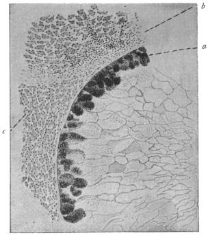
Fig. 105.—Transverse section of Sarcocystis tenella,
Raill. From the œsophagus of the sheep, Ovis aries.
× 38. a, marginal chambers filled with spores; b, connective
tissue of the œsophagus; c, muscles of the
œsophagus.
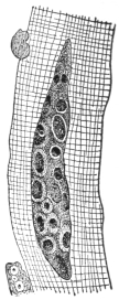
Fig. 106.—Young
Sarcocystis tenella of
the sheep, 47 µ
in length. (After
Bertram.)
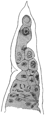
Fig. 107.—End of a
trophozoite of Sarcocystis
miescheriana from the
diaphragm of the pig,
showing division in pansporoblasts. × 800.
(After Bertram.)
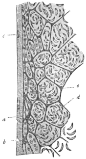
Fig. 108.—Sarcocystis
blanchardi of the ox. Longitudinal
section of sarcocyst or
Miescher’s tube. a, substance
of muscle fibre; b,
envelope of sarcocyst; c, muscle
nuclei; d, spores in chambers;
e, ground substance.
× 400. (From Wasielewski,
after van Eecke.)
The spores (sometimes called Rainey’s corpuscles), vary in shape
according to the species, but are also of different form individually.
They are mostly kidney-, bean- or sickle-shaped (fig. 109), and of
small size, sometimes reaching 14 µ by 3 µ to 5 µ. They are
apparently surrounded by a thin membrane, and at one extremity
(according to the discovery of L. Pfeiffer, confirmed by van Eecke,
Laveran and Mesnil) contain an obliquely striated body (fig. 109) often
homologized with the polar capsule, while the greater part of the spore
is taken up by the nucleate sporozoite. Several authors state that they
have also observed filamentous appendages (polar filaments) at one
end of the spores, and have seen two kinds of spores in the same
Sarcosporidium. Spores of various species of Sarcosporidia may
contain metachromatic granules, often centrally placed (fig. 109).
These granules may be metabolic or possibly may contain toxin
(see below).
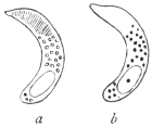
Fig. 109.—Spores of Sarcocystis
tenella, Raill. a, fresh,
showing the polar capsule;
b, stained, showing metachromatic
granules and nucleus.
× 1,000. (After
Laveran and Mesnil.)
The gymnospores of Sarcocystis muris, from the mouse, show active
boring movements when kept in saline solution warmed to 35° or
37° C. S. muris is very deadly to its host. From their structure the
spores do not appear to have great powers of resistance to external
conditions. They measure 12 µ by 3 µ to 4 µ or less.
Laveran and Mesnil (1899) isolated a toxin from S. tenella of
the sheep and called it sarcocystin. This substance is especially
pathogenic to experimental rabbits.
The duration of life of the Sarcosporidia is a comparatively long
one. The affected muscular fibres may remain intact and capable of
performing their functions for a long time,
but at last they perish, if the host lives long
enough. Thus the Sarcosporidia of the
muscles are then enveloped only by sarcolemma,
and finally, when this likewise disappears,
they fall into the intra-muscular
connective tissue. In many cases the Sarcosporidia
die off within their hosts, this,
according to Bertram, being brought about
by a disintegration of the spores in the central
chambers. In other cases the leucocytes
play a part in the destruction of the Sarcosporidia,
and sometimes it happens that lime
salts are deposited in and around the vacant cylinders.
In some places pigs, sheep, mice and rats are infected with
sarcosporidiosis to a remarkable extent, in certain cases almost reaching
100 per cent. Young animals also are infected, and perhaps
infection only takes place during youth.
Although the natural mode of transmission of the Sarcosporidia
remains to be determined, yet various experimental researches on
the problem are of interest and importance. Theobald Smith (1901)
found that mice could be experimentally infected with S. muris
by feeding them with the flesh of other infected mice. The incubation
period was a long one, namely forty to fifty days. Thus, on the
forty-fifth day after feeding young Sarcosporidia were found, and
seventy days after feeding spore formation began. Ripe spores were
found two and a half to three months after the commencement of
these experiments. This mode of infection—a cannibalistic one—hardly
seems likely to be the natural method for the infection of sheep
and ruminants generally. Smith’s researches have been confirmed.
Nègre225 (1910) found that the fæces of mice fed on infected muscular
tissue were infective to other mice when ingested by them. Negri226
infected guinea-pigs with S. muris by feeding them on infected mouse
flesh, and found that the parasite in guinea-pigs showed different
characters from those exhibited by it in mice. Darling227 also succeeded
in infecting guinea-pigs with S. muris, and Erdmann infected mice
with S. tenella (from the sheep).
According to Erdmann228 (1910) the Sarcosporidian spore germinates
in the intestine of the host, which has recently ingested infected
material. The spore liberates its contained toxin—sarcocystin—which
acts upon the adjacent intestinal epithelium, whereby the latter
is shed, and an amœbula creeps out of the spore. The amœbula is
able to penetrate the denuded area and get directly into the lymph-spaces
of the submucous coat of the intestine. The first period of
development, lasting some twenty-eight to thirty days, is said to be
passed in the lymph-spaces of the intestine. Later the amœbula
reaches a muscle fibre. Writing in May, 1914, Erdmann229 records the
appearance of small amœboid and schizogony forms six days after
infection of the host. Crawley230 (1913) controverts some of these
statements and considers that the Sarcosporidian spore, still sickle-shaped,
bores its way into the epithelial cells of the intestine and
comes to rest there. The spore then becomes round or elliptical, and
peripheral masses of chromatin appear within it, suggesting schizogony.
This happens about twelve hours after feeding, and in twenty-four
hours the spores appear to have left the intestine. More recently (May,
1914), Crawley231 considers that there is sexual differentiation among the
Sarcosporidian spores, a few hours after their ingestion by the host.
Interesting discussions have occurred as to the site of the toxic
sarcocystin within the spore. Metachromatic granules occur in the
middle of the Sarcosporidian spore (fig. 109), and the toxin may be
contained in these grains, as they disappear, according to Erdmann,
before the amœbula penetrates the denuded intestinal wall. However,
a polar capsule, containing a polar filament, may be present at one end
of a Sarcosporidian spore. Laveran and Mesnil described a striated
area at the more pointed end of the spore of S. tenella, which
area they consider to represent a polar capsule. Fantham232 (1913)
found a vacuole-like, polar capsule area in the spores of S. colii
from the African mouse-bird. The sarcocystin may be contained
in the polar capsule. The nucleus of the spore is generally at the
opposite, blunter end.
Again, various authors have stated that Sarcosporidian spores may
occur in the blood of the host at times. If so, then an intermediate
host may be concerned in their transmission. Perrin suggested that
Sarcosporidia might be spread by blow-flies and flesh-flies.
The classification of the Sarcosporidia as proposed by R.
Blanchard, which was based on their various habitats, can no longer
hold, because the same species may occur in the muscles as well as
in the connective tissue. For the present, the few species that are
known may be placed in one genus, Sarcocystis, Ray Lankester, 1882.
The following species of Sarcocystis are of interest:—
S. miescheriana, Kühn, 1865, in the pig.
S. bertrami, Doflein, 1901, in the horse.
S. tenella, Railliet, 1886, in sheep. S. tenella bubali in buffaloes in
Ceylon and Egypt.
S. blanchardi, Doflein, 1901, in cattle.
S. muris, Blanchard, 1885, in the mouse, to which it is lethal.
S. hueti, Blanchard, 1885, in the seal.
S. colii, Fantham, 1913, in the African mouse-bird, Colius
erythromelon.
Also various Sarcosporidia from antelopes, monkeys, opossum,
birds, the gecko and wall-lizard are known.
The spores of S. muris, S. bertrami, S. tenella, and S. colii can
multiply by longitudinal fission.
Sarcosporidia observed in Man.
(1) Lindemann233 found on the valves and in the myocardium of
a person who had died of dropsy certain brownish masses, 3 mm. in
length and 1·5 mm. in breadth which he regarded as gregarines. If
these were actually independent animal organisms it may be suggested
that they were Sarcosporidia. Rivolta (1878) named the species
S. lindemanni.
(2) Rosenberg234 found a cyst 5 mm. in length and 2 mm. in breadth
in a papillary muscle of the mitral valve of a woman, aged 40,
who had died from pleuritis and endocarditis. The cyst contained
no scolex nor hooklets of tænia. Numerous small refracting bodies,
round, oval or kidney-shaped, were found in a daughter cyst, as well
as sickle-shaped bodies. The description hardly appears to indicate
Sarcosporidia.
(3) Kartulis235 observed Miescher’s cylinders of various sizes in
the liver (?) and in the muscular system, of a Sudanese who had
succumbed to multiple abscesses of the liver and abdominal muscles.
This may be considered as the first actual case of the occurrence of
Sarcosporidia in man. Koch in 1887 described a case in Egypt.
(4) The case reported by Baraban and St. Remy236 was at once
demonstrated as certain. It related to a man who had been executed,
and in the laryngeal muscles of whom Sarcosporidia were found; the
length of the parasites varied between 150 µ and 1,600 µ, their
breadth between 77 µ and 168 µ. The affected muscular fibres were
distended to four times their normal thickness. This species was
described by Blanchard as “Miescheria” muris, but according to
Vuillemin, it was more probably Sarcocystis tenella of the sheep.
(5) Vuillemin has also described a case of Sarcosporidia found
in the muscles of a man who died from tubercle at Nancy. The
author considered that the parasite corresponded to S. tenella.
(6) Darling237 (1909) found Sarcosporidia in the biceps of a negro
from Barbados.
The Myxosporidia, Microsporidia, Actinomyxidia and possibly the
Sarcosporidia may be included within the section Cnidosporidia
(Doflein), since they possess spores containing polar capsules.
Order. Haplosporidia, Caullery and Mesnil.
The Haplosporidia are a group of organisms having both a simple structure and
life-history. The simplicity may represent a primitive condition or may be due to
degradation resultant on parasitism, and thus it is possible that the group is not
a homogeneous one. The order Haplosporidia was created by Caullery and Mesnil
in 1899, and includes parasites of rotifers, annelids (fig. 110), crustacea, fish, prochordates
and man. They may be present in the body cavity or alimentary tract,
and can also occur in the septum nasi of man, in the nervous system of Cephalodiscus,
and in tumours of fish.
As the name implies, the spores of the Haplosporidia are simple, without polar
capsules, and are uninucleate. In some genera, e.g., Haplosporidium, Urosporidium
(fig. 111) there is a spore-coat or sporocyst which may be elongate or spiny.
The developmental cycle of a Haplosporidian, such as Haplosporidium or Bertramia,
begins with a small, uninucleate cell, often rounded, possessing a cell membrane that
may be prolonged into processes. Growth takes place, coupled with an increase in
the number of nuclei, so that a multinucleate trophozoite is produced. Later, this
multinucleate trophozoite becomes segmented into a number of ovoid or spherical
pansporoblasts, which give rise to few (one to four) spores. Such a spore, when set
free, begins the life cycle over again.
More recently (1905–1907) two important organisms have been described and
included in this group, namely, Neurosporidium cephalodisci238 (Ridewood and
Fantham) from the nervous system of the prochordate, Cephalodiscus nigrescens,
and Rhinosporidium kinealyi (or seeberi) from the septum nasi of man. In the case
of Rhinosporidium and Neurosporidium, after the uninucleate spore has grown into
a multinucleate trophozoite, the latter segments into uninucleate pansporoblasts,
as in the preceding cases. A difference then occurs, for each pansporoblast
enlarges, its nucleus divides and a “spore-morula” is formed. Thus a multinucleate
pansporoblast or spore-morula, divided into many uninucleate sporoblasts (spore
mother cells) is produced, and each sporoblast without further change becomes
a uninucleate spore.
The Haplosporidia have therefore been divided by Ridewood and Fantham (1907)239
into two sections:—
(1) The Polysporulea, wherein the pansporoblast gives rise to a number of
spores (nine or more), e.g., Rhinosporidium, Neurosporidium.
(2) The Oligosporulea, wherein the pansporoblasts give rise each to a few (four)
spores or to only a single spore, e.g., Haplosporidium, Bertramia, Cœlosporidium,
Ichthyosporidium.
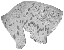
Fig. 110.—Haplosporidium heterocirri.
Section throughout wall of the Polychæte
worm, Heterocirrus viridis, showing various
developmental stages of the Haplosporidium.
× 550. (After Caullery and Mesnil.)
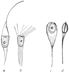
Fig. 111.—Haplosporidian spores.
a, b, Haplosporidium heterocirri. a,
fresh; b, after immersion in sea water;
c, d, Urosporidium fuliginosum.
× 1000. (After Caullery and Mesnil.)
Rhinosporidium kinealyi, Minchin and Fantham, 1905.
Rhinosporidium kinealyi, parasitic in man, must now be considered
in greater detail. This organism was found in nasal polypus
in India, and has since been recorded from the ear as small nodules
in the external auditory meatus. The Indian cases came from the
neighbourhoods of Calcutta and Madras, and the parasite has been
seen in Ceylon. Similar structures have since been described from
the United States and South America.
The Rhinosporidium polypus is said not to be particularly painful,
though nasal forms must interfere with breathing to some extent. The
first nasal polyp reported from India formed a vascular pedunculated
growth on the septum nasi and was about the size of a large pea or
raspberry. It was compared with a raspberry, being red in colour
with a number of small whitish dots upon its surface. When the
tumour was cut, a number of similar whitish dots were seen within.
These were the cysts of Rhinosporidium. According to Minchin
and Fantham240 (1905), they vary considerably in size and measure up
to 200 µ or 250 µ in diameter. Each possesses a cyst wall which varies
in thickness in different cysts. Its outer wall is always firm and
distinct, the inner limit being less definite at times. Each large cyst
is filled with numbers of spherical or oval bodies, showing every
gradation between small ones at the periphery and large ones at the
centre (fig. 112). Roughly, three zones of parasites can be distinguished
in a large cyst, a peripheral set consisting of the youngest
parasites, an intermediate group and a central, oldest zone. A large
cyst may possess a pore for the egress of its contents. Some of the
cysts show polar distribution of the zones.
The youngest forms of Rhinosporidium are difficult to detect.
They are small, granular masses, round, ovoid or irregular and at times
even amœboid in appearance. These are young trophozoites. They
increase in size, but encystment occurs early, the outer layer becoming
firm so that the organisms have a definite contour. Each is soon
multinucleate and the cytoplasm segments around the nuclei. The
cyst thus becomes full of uninucleate pansporoblasts or sporonts,
with a peripheral layer of undifferentiated protoplasm. The pansporoblasts
grow in size. In the larger cysts the formation of
pansporoblasts progresses at the expense of the peripheral layer of
protoplasm, which, however, continues to grow, so that the cyst as
a whole increases in size. The pansporoblasts at first are uninucleate
(fig. 112, a), and then undergo nuclear multiplication. This is well seen
in the intermediate zone of parasites, where the pansporoblasts show
first one, then two, then four or more spores (fig. 112, b), while in the
oldest centrally placed pansporoblasts, about a dozen or sixteen
closely packed spores (fig. 112, c), can be seen. The spore is small
and rounded, and its nucleus is clear and distinct. The fully formed
pansporoblast or spore morula becomes surrounded by a membrane.
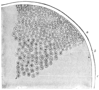
Fig. 112.—Rhinosporidium kinealyi. Portion of ripe
cyst containing pansporoblasts of various ages. × 480.
(After Minchin and Fantham.)
Certain of the cysts have been found in a ruptured condition,
whereby the spores have been liberated into the surrounding tissue.
It is almost certain that the spores serve for the auto-infection of the
host, for though the tumours of Rhinosporidium seemed to have
been removed entirely, it has been found that they recur, some
minute fragment of the parasite having probably been left behind.
The method whereby the parasite reaches new hosts has not yet been
determined, and it would be of interest if its life-history could be
more fully investigated.
The Asiatic specimens of R. kinealyi were first described in detail
by Minchin and Fantham (1905) from material briefly reported to the
Laryngological Society of London in 1903, by O’Kinealy. Material
obtained by Dr. Nair, of Madras, was described by Beattie241 in 1906.
This material came from Cochin. Castellani and Chalmers have
found similar polypi in Ceylon.
Wright242 has described the parasite from Memphis, Tennessee.
Seeber243 in 1896 described nasal polypi in Buenos Ayres, and in 1900
Wernicke named the parasite therein Coccidium seeberi. Seeber’s
parasite is a Rhinosporidium, R. seeberi, and may ultimately be found
to be the same as R. kinealyi. Ingram244 reports Rhinosporidium cysts,
with pores in the cyst walls, in conjunctival polypus and in papilloma
of the penis in India. Zschokke has reported the presence of Rhinosporidium
in horses in South Africa.
The Infusoria (or Heterokaryota, Hickson, or Ciliophora, Doflein)
include the Ciliata and the Suctoria. A few authorities, including
Braun, raise the Suctoria (or Acinetaria) to separate rank as a class,
but this is not widely followed.
The body of the Ciliata usually is bilaterally symmetrical and is
enveloped in a cuticle which has numerous openings for the
protrusion of the cilia. Most kinds have a fixed shape, whilst
changes in the form of others are brought about by the contractions
of the body substance. The latter exhibits hyaline ectoplasm, in
which myonemes, and occasionally also trichocysts (minute spindle-shaped
bodies) appear, and granular endoplasm which may contain
numerous vacuoles. The cilia, on whose various arrangements
the classification is based, are always processes of the ectoplasm.
Their form varies; they may be hair-like, or more rarely thorn-like,
spur-like, or hook-shaped; undulatory membranes also may occur,
which are probably composed of fused cilia.
With the exception of some of the parasitic species, an oral cavity,
peristome or cytostome, is always present. It is frequently beset
with cilia or provided with undulatory membranes, which help to
waft the food inwards; sometimes there is an anal aperture (cytopyge)
generally placed at the opposite pole of the organism. A cytopharynx
fringed with cilia or sometimes with a specialized supporting
apparatus is connected with the peristome. Vacuoles form round
the ingested food, and in many species a constant rotation goes on
in the endoplasm. Often one, and sometimes two contractile vacuoles
are present, the frequency of the pulsations of which depends on the
surrounding temperature. Sometimes special conducting channels
lead to the vacuoles, or there are outlet channels leading to the exterior.
There is in almost every case a large nucleus (macronucleus),
and lying close up to it a small nucleus (micronucleus). The form
of the large nucleus varies according to the species. Numerous
nuclei are not very common, but these occur in Opalina, which lives
in the hind-gut of amphibia, and is also distinguished by the absence
of an oral aperture.
Reproduction is effected by binary fission; less commonly, after
encystment, by multiple division, or by budding. The divisions can
be repeated many times, but finally cease, and then the conjugation
of two specimens brings about a regeneration, particularly of the
nuclei. Numerous examinations (Bütschli, Hertwig, Maupas, Calkins)
have demonstrated that after two individuals have associated by
homologous parts of the body, the micronucleus separates from
the macronucleus, becomes larger and divides twice by mitosis, so
that four micronuclei are present in each one of the two individuals
forming the couple. Three of these nuclei perish and become
absorbed, the fourth gradually passes to the portion of protoplasm
connecting the two conjugants, which has originated by absorption
of the cuticle at the point of contact of the conjugants. After a
further division one micronucleus of each conjugant passes over
into the other conjugant, and fusion ensues between the two micronuclei
of each individual. Complicated changes and divisions may
occur, but only the main principles can be noted here. A new nuclear
body is thus formed in each conjugant, and soon divides into two.
Of the segments thus produced one becomes a micronucleus, and
one or several of the others, as the case may be, form or amalgamate
into a new macronucleus, the old macronucleus usually perishing or
becoming absorbed during the conjugation. Usually, sooner or later,
the two conjugants separate, or may have separated already, and again
multiply independently by fission until a series of divisions by simple
fission is again followed by conjugation. The theoretical significance
of conjugation cannot be dealt with fully here. It may be
remarked, however, that the macronucleus plays no part in it, but
governs entirely the metabolism of the Infusorian, whereas the
micronucleus is essentially a generative nucleus from which macro-
and micro-nuclei are again and again produced.
Encystment amongst the Infusoria is very general, and is essentially
a means of protection when the surrounding medium dries up.
Doubtless these cysts are frequently carried long distances by the wind,
which explains the wide geographical distribution of most species.
Also, multiplication often takes place in the encysted condition.
Some Infusoria live a free life, others are sedentary; the latter
form colonies in fresh as well as in salt water. Numerous species
are parasites of various lower and higher animals,245 and a few also are
parasitic in man.
The Prague zoologist, v. Stein, introduced a classification of the
Infusoria that has been almost universally adopted. It is founded
on the different position of the cilia on the body. Though, no
doubt, artificial, it is a convenient system. Bütschli has compiled a
better one.246 But for our purpose Stein’s system is sufficient:—
Order 1. Holotricha, Infusoria with cilia that are evenly distributed over the
entire body.
Order 2. Heterotricha, ciliated all over like the Holotricha, but having stouter
cilia about the peristome.
Order 3. Hypotricha, only ciliated on the ventral surface.
Order 4. Peritricha, with only a ring of spiral cilia, mostly sedentary.
The Infusoria observed in man belong to the order Heterotricha,
with few exceptions.
Genus. Balantidium, Claparède et Lachmann.
Heterotrichous Infusoria of oval or bag-like form and almost circular on transverse
section; the anterior extremity narrowed, the posterior end broad and
rounded off, or also narrowed; the peristome starting at the anterior end is
broadest there and becomes narrower as it gradually obliquely approaches towards
the posterior extremity. There are coarse cilia along the entire left border and the
anterior part of the right border. Longitudinal striation is distinct and regular.
There are two contractile vacuoles on the right, and occasionally also two or more to
the left. The anus (cytopyge) is terminal. The macronucleus is usually horse-shoe
or kidney-shaped, sometimes oval; the micronucleus contiguous. Reproduction is
by binary fission and conjugation, and encystment occurs. The cysts are spherical
or oval. These ciliates are parasitic in the large intestine of human beings and
pigs, in Amphibia, and in the body cavity of polychæte Annelida.
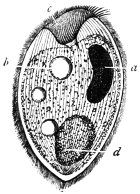
Fig. 113.—Balantidium
coli. a, nucleus;
b, vacuole;
c, peristome; d, bolus
of food. (After
Leuckart.)
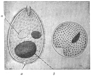
Fig. 114.—Balantidium coli, free and encysted;
a, anus or cytopyge; n, macronucleus; b, bolus of
food. (After Casagrandi and Barbagallo.)
Balantidium coli, Malmsten, 1857.
Syn.: Paramæcium coli, Malmsten, 1857.
The body is oval, 60 µ to 100 µ in length (up to 200 µ according
to Janowski), and 50 µ to 70 µ in breadth. The peristome is funnel-shaped
or contracted, the anterior end being then broadened or
pointed according to the degree of contraction (figs. 113, 114). The
ecto- and endo-plasm are distinct, the latter is granular, containing
drops of fat and mucus, granules of starch, bacteria, and occasionally
also red and white blood corpuscles. There are usually two contractile
vacuoles, seldom more. The anus (cytopyge) opens at the
posterior extremity. The macronucleus is bean- or kidney-shaped,
rarely oval; the micronucleus is spherical.
Balantidium coli lives in the large intestine of man, in the rectum of
the domestic pig, and has been found in monkeys. It propagates by
transverse division, but conjugation and encystment are known to
take place.247 Transmission to other hosts is effected by the cysts of
the parasite (fig. 114).
Balantidium coli, first seen by Leeuwenhoek, was described
by Malmsten in 1857 in a man aged 35 years, who had two
years previously suffered from cholera, and since then had been
subject to diarrhœa. The examination showed an ulcer in the rectum
above the mid sphincter ani, in the sanguineous purulent secretion of
which numerous Balantidia were swimming about. Although the
ulcer was made to heal, the diarrhœa did not cease and the stools
contained numerous Balantidia, the number of which could only be
decreased by extensive enemas of hydrochloric acid.
The second case related to a woman who was suffering from
severe colitis, and who died ten days after admission. The malodorous,
watery evacuations contained innumerable Balantidia, in
addition to pus, and at the autopsy the anterior portion of the large
intestine was found to be infested with them.
Subsequently this parasite has often been observed in human
beings, and various cases have been recorded. These occurred in
Russia, Scandinavia, Finland, Cochin China, Italy, Germany, Serbia,
Sunda Islands, Philippine Islands, China, and in other parts of Asia
and in America. Other cases were reported by Askanazy, Ehrnroth,
Klimenko, Nagel, Koslowsky, Kossler, Waljeff, Strong and Musgrave,
Glaessner248 and others. Sievers found B. coli very common in Finland.
In the majority of the cases described by Sievers from Finland,
and in other cases from Central Europe, the patients suffered from
obstinate intestinal catarrh, which did not always cease even after
the Balantidia had disappeared. On the other hand, Balantidia have
occasionally still been found to persist, though in small numbers, after
the catarrh has been cured. Some authors, nevertheless, do not
regard Balantidia as the primary cause of the various diseases of the
large intestine, which often commence with the development of
ulcers, but they consider that they may aggravate these diseases and
render them obstinate. According to Solowjew, Askanazy, Klimenko
and Strong and Musgrave, however, the parasites penetrate the intestinal
wall, and give rise to ulcerations which may extend deeply into
the submucosa, and even be found in the blood and lymphatic vessels
of the intestinal wall. According to Stokvis, B. coli occurs also in
the lung; at all events this author states that he found one living and
several dead paramæcia (?) in the sputum of a soldier, returned from
the Sunda Islands, who was suffering from a pulmonary abscess.
Sievers has shown that B. coli might occur in persons not suffering
from intestinal complaints, but E. L. Walker249 (1913) states that every
person parasitised with B. coli is liable sooner or later to develop
balantidian dysentery.
Since Leuckart confirmed the frequent presence of B. coli in the
rectum of pigs, and corresponding observations were made in other
countries, the pig is universally considered to be the means of the
transmission of Balantidium to man. The encysted stages only serve
for transmission, because, according to all observations, the free
parasites have a very small power of resistance. They perish when
the fæces have become cool; they cannot live in ordinary, slimy, or
salt water. As they are killed by acids even when much diluted, they
cannot pass through the normal stomach alive except under the most
unusual circumstances. The pigs, in whose intestines the Balantidium
appears to cause little or no disturbance, evacuate numerous encysted
Balantidia with the fæces, and their occasional transference to man
brings about their colonization there, but perhaps only when a disease
of the colon already exists.
Experimental transmission of the free parasites to animals (per os
or per anum) yielded negative results, even in the case of pigs.
Casagrandi and Barbagallo (1896), however, had positive, as well as
negative, results. They employed healthy young cats, or cats in which
catarrhal entero-colitis had been artificially induced (which in other
experiments is apt to cause the death of the animals experimented upon
in about six or seven days), or finally cats that had dilatation of the
rectum with alkaline reaction of the fæces. An attempt to infect three
healthy cats by injecting human fæces containing Balantidium into the
rectum proved negative, in so far as the fæces of the experimental
animals had an acid reaction and contained no Balantidia, but at the
autopsy performed eight days after infection a few encysted parasites
were found in the mucus of the ileum. In the case of four cats
suffering from entero-colitis, into which human fæces containing
Balantidia were introduced per os, Balantidium cysts were found in
the fæces three days after the last ingestion. Great numbers, moreover,
were found in the cæcum and the posterior part of the small
intestine at the autopsy of the animals, which died about eight days
after the commencement of the experiments. Actual colonization,
therefore, was not effected in either series of experiments. Free or
encysted Balantidia of pigs were used for further experiments. The
experiments proved negative when fæces containing cysts were injected
into the rectum of healthy cats (three experiments), or cats (two)
suffering from spontaneous intestinal catarrh, or when such material
was introduced per os into three healthy cats. In the case of two
cats with intestinal catarrh artificially produced, a small number of the
active Balantidia injected into the rectum remained alive. Larger
quantities of fæces containing encysted Balantidia were introduced
into two other cats affected with the same complaint. These, certainly,
did not appear in the fæces, but small numbers, free and alive, were
found in the cæcum. Similarly, encysted Balantidia were introduced
into two cats with dilated rectum, and whose fæces had an alkaline
reaction. In these cases no parasites appeared in the fæces, but three
and five days later, when the two animals were examined, a very small
number were discovered free in the large intestine. Klimenko did
not succeed in infection experiments with B. coli on young dogs,
whose intestines had been artificially affected by disease.
More recent experiments by Brumpt have shown that young
sucking pigs can be infected with Balantidium from infected monkeys
(Macacus cynomolgus) and suffer heavily from the same, whereas the
Balantidium of the pig is rarely harmful to its host. This and previous
experiments may be thought to suggest that there are perhaps
several pathogenic species, and also that harmless strains of Balantidium
may occur. At the same time, it must be remembered that
a large proportion of the cases recorded of Balantidian colitis occur
among swineherds and butchers, that is, among people in frequent
contact with pigs. Morphologically, there are practically no differences
between the Balantidia found in man, monkeys and pigs, and
it is probable that one species only, under slightly different environmental
conditions, may be responsible for the colitis observed. In
any case, efficient prophylactic measures should be taken against
balantidiasis in countries where it may occur, by confining the pigs
and not allowing them to run in yards and dwellings.
E. L. Walker (1913) has given a good summary of work on
balantidiasis. His own researches in the Philippines showed that
monkeys could be infected by Balantidia both from pigs and men.
Parasites may appear in the stools only at infrequent intervals. He
believes that the ciliates are the primary etiologic factor in the
symptoms and lesions of balantidian dysentery.
Behrenroth (1913) has given an interesting account of Balantidium
coli and its pathogenic significance.
Balantidium minutum, Schaudinn, 1899.
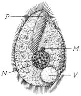
Fig. 115.—Balantidium minutum.
P, peristome; N, nucleus;
M, micronucleus; V, contractile
vacuole. Food vacuoles are represented
in the endoplasm. (After
Schaudinn.)
The body is of oval form, with the anterior extremity pointed, and
posterior extremity broad and rounded (fig. 115). The length is 20 µ
to 32 µ, and the breadth is 14 µ to 20 µ. The peristome, which is
fissure-like, extends to the centre of the body (fig. 115). The right
lateral border of the peristome is beset with cilia the same length as
those of the body, the left side terminates in a thin hyaline membrane
that extends towards the back, and can
pass over to the right side. A row of
longer and stronger cilia (cirri) are on the
left border of the peristome. The cuticle
is refractile, the ectoplasm hyaline and
the endoplasm granular, with numerous
food vacuoles.
A single contractile vacuole lies dorsally
and to one side at the posterior
extremity. The macronucleus, which is
always spherical, is central and is 6 µ to
7 µ in diameter. The micronucleus, close
in front of it, only measures 1 µ (fig. 115).
The cysts are oval.
These parasites were found in numbers
in the evacuations of a man aged 30,
who was born in Germany and had repeatedly travelled between
Hamburg and North America, where he made long stays. The patient
came to the Charité in Berlin to seek advice for constipation alternating
with diarrhœa accompanied by abdominal pain.
A second case (the parasite of which was described as Colpoda
cucullus by Schulz) was observed in a patient in the same institution.
As, in both cases, the parasites only appeared during the diarrhœa,
and disappeared as soon as the fæces had assumed a normal consistency,
or could only be demonstrated in a few encysted specimens,
it may be assumed that the small intestine or the duodenum is
their habitat.
Genus. Nyctotherus, Leidy, 1849.
Flat, heterotrichous Infusoria, kidney- or bean-shaped. The peristome commences
at the anterior pole of the body and extends along the concave side to the
middle, where the oral aperture is situated. The cytopharynx is oblique and is more
or less curved. The cytopyge is at the posterior extremity, where a single contractile
vacuole is also situated. The macronucleus is almost in the centre of the parasite.
The members of this genus live parasitically in the intestine of amphibia, insects
and myriapods, and at least one species is found in man.
Nyctotherus faba, Schaudinn, 1899.
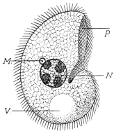
Fig. 116.—Nyctotherus faba. P,
peristome; N, nucleus; M, micronucleus;
V, contractile vacuole.
(After Schaudinn.)
The body is bean-shaped, and a little flattened dorso-ventrally.
It is 26 µ to 28 µ long, 16 µ to 18 µ broad, and 10 µ to 12 µ thick
(fig. 116). The peristome is on the right
border and extends to the middle; at
the left there are large adoral cilia, the
cilia on the right border not being larger
than those on the body. The cytopharynx
is short, slightly curved and
turned backwards. The contractile vacuole
is large, spherical, situated at the
posterior extremity, and its contents are
voided through the anus at its left. The
macronucleus is in the centre of the body;
it is globular (6 µ to 7 µ in size), and
contains four or five chromatin masses.
The micronucleus lies close to it, and is
spherical or somewhat elongate measuring
1 µ to 1·5 µ (fig. 116). The cysts are oval.
This species has hitherto only been seen once in the same patient
in whom Balantidium minutum was discovered.
Nyctotherus giganteus, P. Krause, 1906.
Under the name Balantidium giganteum n. sp., P. Krause described
an Infusorian which was repeatedly observed with Trichomonas intestinalis
in the alkaline evacuations of a typhoid patient in Breslau.
The body is ovoid, narrower and rounded anteriorly and broader
and stunted posteriorly. The peristome lies to one side; the macronucleus
is bean-shaped, the micronucleus small and globular; one
or two vacuoles are present. The anus is at the farther end. The
organism is 90 µ to 400 µ long, 60 µ to 150 µ broad (fig. 117). After a
prolonged stay outside the body, it becomes rounded and encystment
occurs. In the thermostat the Infusoria remain alive at 37° C. for five
weeks.
The species, however, hardly belongs to Balantidium, but to all
appearances is a Nyctotherus and is distinguished from N. faba by the
difference in size.
[Nyctotherus] africanus, Castellani, 1905.
In the fæces of a native of Uganda who suffered from sleeping
sickness and diarrhœa and had in his intestine Ascaris lumbricoides,
Trichocephalus trichiurus and Ancylostoma duodenale, Castellani found
a curiously shaped Infusorian, 40 µ to 50 µ long, and 35 µ to 40 µ
broad, with spherical macro- and micronucleus and a contractile
vacuole (fig. 118). He included the organism in the genus Nyctotherus,
perhaps wrongly, or the parasite may have been deformed. After the
patient’s death the same parasite was found in the intestine and
especially in the cæcum.
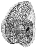
Fig. 117.—Nyctotherus
giganteus.
(After Krause.)
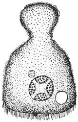
Fig. 118.—Nyctotherus
africanus. (After Castellani.)
G. Lindner, in Cassel, studied certain peritrichal Infusoria (stalkless Vorticella),
and connected them, probably incorrectly, with the most varied diseases of man and
domestic animals, even with Sarcosporidia of pigs. It may be mentioned that
according to a communication by letter from Schaudinn, Vorticella may be found
in freshly evacuated fæces, but always only after the administration of a water
enema. In spite of this, several other investigators mention Vorticellæ as
intestinal parasites of man.
The Chilodon dentatus (Ehrenberg) recorded in 1903 by J. Guiart as a parasite
of man, which may be found in all infusions, can hardly have lived in the man
from whose fæces it was cultivated, but may represent a chance admixture both in
the fæces and the cultivations. C. uncinatus was also found as a chance parasite
of man by Manson and Sambon. According to Doflein250 (1911) certain Chilodon-like
organisms have been found by Selenew in prostate secretions in gonorrhœa.
Other species of the genus Chilodon are known, but only as ectoparasites (e.g.,
Chilodon cyprini, Moroff, 1902, from the skin and gills of diseased carp).
A number of other parasitic Ciliates are known, among which Ichthyophthirius
multifiliis, destructive to fish, is important. It lives in the skin and the layers
immediately below it, forming small whitish pustules which may become confluent.
The pustules are most common on the head and fins, but occur also on the eyes and
gills of the host. The young parasite, which is one of many formed in a cyst, is very
small. At first it is free swimming, but soon attaches itself to the skin of a fish. It
bores inwards and becomes surrounded by the irritated skin. There it attains a
relatively large size, being 500 µ to 750 µ and occasionally more in diameter. The
body has a rounded terminal mouth, short cytopharynx and a number of minute
contractile vacuoles. The macronucleus is large and horseshoe-shaped; the small
micronucleus is only seen in the very young animal. When full grown, the organism
encysts and forces its way to the surface and bursts through, leaving a small, gaping
wound behind. The cyst sinks to the bottom of the water, nuclear multiplication
occurs and a number of young parasites are produced, which leave the cyst and
either attack new hosts or else perish.
Opalina ranarum, parasitic in the rectum and urinary bladder of frogs and toads,
shows great degradation and simplification due to parasitism, possessing no separate
micronuclei, no cytostome, cytopharynx or cytopyge. It has many macronuclei, and
is a large parasite. During summer and autumn nuclear multiplication followed by
division of the body occurs, the process being repeated after the daughter forms
have grown to the size of their parent. In spring, the Opalina divide rapidly, but
do not grow much before dividing again. Finally, tiny forms, containing three to
six nuclei, encyst and pass from the host with the fæces. As these latter are greedily
devoured by tadpoles, the Opalina gain new hosts in which they develop.
THE CHLAMYDOZOA.
The name Chlamydozoa was proposed by Prowazek in 1907 for a
number of minute, problematic organisms (fig. 119) believed to be
the causal agents of certain diseases in man and animals, such as
vaccinia and variola, trachoma, inclusion blenorrhœa in infants,
molluscum contagiosum, and bird epithelioma contagiosum. Other
diseases possibly due to Chlamydozoa251 are hydrophobia, measles,
scarlet fever, foot-and-mouth disease, the “Gelbsucht” disease of silkworms,
and perhaps even typhus (Prowazek, 1913). The subject is
difficult and controversial and can only be briefly discussed here.
It is known that the viruses in all these diseases can pass through
ordinary bacterial filters, that is, they belong to the group of “filterable
viruses.” At such periods the organisms are extracellular or free. It
is also known that in many of these cases the virus produces definite
and characteristic reaction-products or cell-inclusions in the infected
cells, during the intracellular phase of the life-history of the organism.
As the organisms to be considered are problematic, it will be convenient
to summarize their history:—
(1) Cell-inclusions, usually named after their discoverers, have
been found in certain diseases, thus: In vaccinia Guarnieri’s bodies,
in scarlet fever Mallory’s bodies, in hydrophobia Negri’s bodies, in
trachoma Prowazek’s bodies occur.
(2) At first these characteristic cell-inclusions were considered to
be actual parasitic organisms causing the diseases in question. The
bodies received zoological names and attempts were made to work
out their supposed development cycles. The supposed parasites of
vaccinia and variola were referred to a so-called genus Cytoryctes,
those of hydrophobia to Neuroryctes, of scarlet fever to Cyclasterium,
while those of molluscum contagiosum were referred to the Coccidia.
Calkins in 1904 studied in detail the cell-inclusions of vaccinia and
small-pox, calling them Cytoryctes variolæ, Guarnieri. Calkins considered
that in the stratified cells of the epidermis they passed through
two cycles, the one cytoplasmic, the other intranuclear. The first is
the vaccinia cycle, the second the pathogenic (intranuclear) variola
cycle. It is hardly necessary to follow all Calkins’ stages here.
Negri (1909) described a cycle for Neuroryctes hydrophobiæ. Calkins
refers both Cytoryctes variolæ and Neuroryctes hydrophobiæ to the
Rhizopoda.
Siegel (1905) described quite different organisms under the name
Cytorhyctes. He listed several species: C. vacciniæ; of vaccinia and
small-pox, C. scarlatinæ of scarlet fever, C. luis of syphilis (this is
probably the granule stage of Treponema pallidum), and C. aphtharum
of foot-and-mouth disease.
(3) The afore-mentioned views were criticized, and the bodies
were not considered to be living organisms but merely reaction
products or cell-inclusions due to the effects of the virus on the host
cells. Thus Guarnieri’s bodies were stated to consist of extruded
nucleolar or plastin material, having no developmental cycle. It was
further asserted that infection could be produced by lymph in which
Guarnieri’s bodies had been destroyed. Similar assertions have been
made regarding the Negri bodies, and others. The Cytoryctes,
Neuroryctes, etc., are considered, according to these views, to be
degeneration products of the nucleus or to be of a mucoid nature.
(4) More recently a positive belief has gained ground that there
are true parasitic organisms causing these diseases, and that the
parasites are very minute, being termed Chlamydozoa by Prowazek
and Strongyloplasmata by Lipschütz.
The Chlamydozoa are characterized by (a) their very minute size,
smaller than any bacteria, so that they can pass through bacterial
filters; (b) they pass through intracellular stages, in the cytoplasm
or the nucleus of the host cell, producing therein the reaction
products or inclusions in the cell already recorded as characteristic
or diagnostic of the diseases produced; (c) they pass through definite
developmental cycles. Such a cycle consists essentially of growth
and division. The mode of division of the Chlamydozoa resembles
that of the centriole of a cell, by the formation of a dumb-bell-shaped
figure. Two dots are observed connected by a fine line or strand
which becomes drawn out and finally snaps across the middle.
Prowazek and Aragão (1909) working on smallpox in Rio de Janeiro
found that the chlamydozoal granules passed through a Berkefeld
filter and that the filtrate was virulent. But if an “ultra-filter” were
used, i.e., one coated with agar, then the granules were retained and
the filtrate was no longer virulent. The surface of the ultra-filter was
found to contain many granules.
The Chlamydozoa are parasites of epiblastic tissues (e.g., epidermal
cells, nerve cells, conjunctival cells).
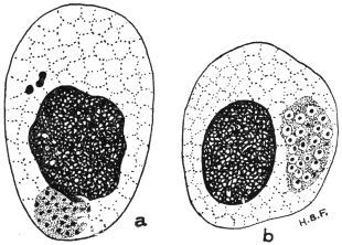
Fig. 119.—Chlamydozoa. Trachoma bodies in infected
epithelial cells of the conjunctiva. (a) initial bodies (above)
and cluster of elementary bodies (touching the nucleus);
(b) cluster of granules surrounded by mantles. × 2,000
approx. (Original. From preparation by Fantham.)
The life-history of a Chlamydozoön (fig. 119), such as that of
vaccinia, is, according to Prowazek, Hartmann and their school, as
follows:—
1. The infection begins with elementary bodies or elementary
corpuscles which live at first extracellularly. An elementary body is
a minute speck of chromatin, apparently devoid of cytoplasm, which
can pass through a bacterial filter. It can enter a host cell, but the
entry is not a process of phagocytosis.
2. Inside the host cell the elementary body grows in size, and
becomes an initial body (fig. 119, a).
3. A reaction on the part of the host cell results, for nucleolar,
plastin substance is extruded from the cell-nucleus and surrounds
the parasitic initial body. The latter is thus enveloped in a mantle
(hence the name Chlamydozoa, from χλαμὑς, a mantle), and the
characteristic cell-inclusion (Guarnieri’s body, Negri’s body, etc.) is
produced. The nucleolar, mantle substance probably represents the
“cytoplasm” of Cytoryctes, described by Calkins.
4. The body next breaks up into a number of smaller bodies
known as initial corpuscles. These, in their turn, divide by simple
division (in the manner already described) into numerous elementary
bodies (fig. 119). Thus, the life-cycle is completed.
The Chlamydozoa are, then, the minute granules inside the body of
the Cytoryctes variolæ or the Neuroryctes hydrophobiæ, so that the whole
body of the Cytoryctes or Neuroryctes corresponds to the mantle and
parasite of the Chlamydozoön. The Cytoryctes group is said to
cause destruction of the host cell. The Cytoöikon group (e.g.,
trachoma bodies) causes proliferation of the host cell.
In September, 1913, Noguchi252 described the cultivation of the
parasite of rabies in an artificial medium, similar to that used by him
for the cultivation of Spirochæta recurrentis. The cultures were stated
to be infective to dogs, rabbits and guinea-pigs. Levaditi, in
December, 1913, stated that he had succeeded in cultivating spinal
ganglia of rabid monkeys in monkey plasma.
Noguchi and Cohen (November, 1913)253 have succeeded in
cultivating the so-called trachoma bodies, or at any rate bodies
very closely resembling them morphologically. The medium
employed was Noguchi’s ascitic fluid and rabbit kidney medium, as
used for spirochætes. The coarser cultural forms stained blue with
Giemsa’s solution, the finer ones stained red. Attempts to infect
monkeys from the culture tubes failed.
From their behaviour on treatment with such reagents as saponin,
bile and sodium taurocholate, Prowazek considers that the Chlamydozoa
approach the Protozoa.
PROTOZOA INCERTÆ SEDIS.
Sergentella hominis, Brumpt, 1910.
Et. and Ed. Sergent in 1908 found vermiform bodies about 40 µ
long by 1 µ to 1·5 µ broad in the blood of an Algerian suffering from
nausea and cold sweats, without other symptoms. The bodies were
pointed at each end, with a somewhat ill-defined nucleus in the
middle. Their systematic position is doubtful.
Note.—An Appendix on Protozoology will be
found on pp. 733–752. This has been prepared in
order to incorporate a number of new additions to
knowledge made since the body of the book was
printed off.
Definition: Bilaterally symmetrical animals without limbs, the form of which
is leaf or tape-like, rarely cylindrical, and whose primary body cavity (segmentation
cavity) is absent, the cavity being filled by a mesenchymatous tissue (parenchyma).
The mouth is either situated at the anterior end of the body, or is shifted more
or less backwards on to the flat ventral surface. The alimentary canal consists of a
short fore-gut, which is frequently provided with a muscular pharynx, and of a simple
forked or branched mid-gut; there is neither a hind-gut nor an anus; in one class,
the Cestodes, the alimentary canal has entirely disappeared except for muscular
remnants in the scolex.
The INTEGUMENT OF THE BODY consists either of a ciliated epithelium of only
one layer (Turbellaria), or of a cuticle and gland-like cells embedded in the
parenchyma, or subcuticular layer (Cestodes, Trematodes). The dermo-muscular
layer consists of annular, longitudinal, and even diagonal fibres, while the
parenchyma is traversed by dorso-ventral fibres.
The central NERVOUS SYSTEM, which is embedded in the parenchyma of the
body, consists of cerebral ganglia, united together in the shape of dumb-bells, and
of two or more longitudinal MEDULLARY FASCICLES, often forming transverse
anastomoses. Organs of sense usually occur only in the free-living species, more
rarely during the free-living stages of a few parasitic species and in a few ectoparasitic
forms.
[In Platyhelminthes simple eye-spots frequently occur, and in a few an auditory
vesicle.]
Blood-vessels and definite RESPIRATORY ORGANS are lacking [except in
Nemertinea]; the EXCRETORY APPARATUS (formerly termed water-vascular system)
is typical of the entire class. It commences in the interstices of the parenchyma,
with peculiar terminal cells (ciliated funnels), which will be described later (p. 219),
the capillary processes of which go on uniting into larger branches, and finally form
two large collecting vessels, which, sometimes separately and sometimes united,
open to the exterior through one, two, or numerous pores.
Nearly all the Platyhelminthes are HERMAPHRODITIC, and in nearly all there are,
in addition to the ovaries producing ova, other glands attached to the female genital
apparatus, namely, the vitellaria or yolk glands, which provide a substance termed
yolk, which serves as nourishment for the embryo. The fully formed eggs have
shells and are “compound,” i.e., composed of the egg or ovarian cell, which is
surrounded by numerous yolk cells or their products of disintegration. The two
sexual openings usually lie close together, frequently in the fundus of a genital
atrium; they are rarely separated from one another. Shell glands also usually
occur (p. 221).
Reproduction is sexual, often, however, combined with asexual methods of
propagation (segmentation, budding). The Platyhelminthes live partly free in fresh
or salt water, exceptionally also on land. The greater part, however, live as
parasites on or in animals.
Classification of the Platyhelminthes.
Class I.—Turbellaria (or Eddy Worms). Flat worms for the most part, free
living, and always covered with a ciliated epithelium.
Order 1.—Rhabdocœlida, gut unbranched.
Order 2.—Tricladida, gut with three main branches.
Order 3.—Polycladida, a central gut with lateral cæca. Development direct
or through metamorphosis. They live in fresh and salt water or on land;
very seldom as parasites.
Class II.—Trematoda (Sucking Worms254). [Usually known as Flukes.—F. V. T.]
Flat worms, living as ecto- or endoparasites, that are only ciliated in the larval
condition, and in their adult state are covered with a cuticle, the matrix cells of
which lie in the parenchyma. They have either one, a few, or several suckers,255
and frequently also possess chitinous fixation and adhesive organs. The intestine
is single, but generally bifurcated, and not uncommonly there are transverse anastomoses
between the forks or diverticula on them. Excretory organs double, with
two orifices at the anterior extremity or a single one at the posterior end.
Development takes place by a metamorphosis or alternation of generations (p. 283).
These worms are almost always hermaphroditic, with two or more female and one
male sexual orifice. They live, almost without exception, as parasites on vertebrate
animals, but the intermediate generations are passed in molluscs.
Class III.—Cestoda (Tapeworms). Endoparasitic flat worms without an alimentary
canal. The larval stages are rarely ciliated, but are usually provided with six
spines; the adult worm is covered with a cuticle, the matrix cells of which are embedded
in the parenchyma. The body consists of a single segment (Cestodaria) or a
chain of segments, in which case it consists of the scolex and the segments containing
the sexual organs (proglottides) (Cestodes s. str.). The scolex is provided with
various adhesive and fixation organs, and there are calcareous corpuscles in the
parenchyma. Excretory organs symmetrical, opening at the posterior end. These
worms are always hermaphroditic, and then possess one or two female and one
male sexual orifice. During development a larval intermediate stage (“measle”)
occurs and almost always in a different host to that in which the adult sexual worm
lives. The adult stage is parasitic in vertebrate animals; but the larval stage may
occur in invertebrates.
These worms are usually leaf- or tongue-shaped, but also barrel-shaped
or conical; they vary from 0·1 mm. to almost 1 m.256 in
length; most of them, however, are small (5 mm. to 15 mm.). The surface
on which the orifice of the uterus and the male sexual opening
are situated is termed the ventral surface; the oral aperture, which
also acts as anus, is always at the anterior end in the sub-order
Prostomata (p. 230), but in the sub-order Gasterostomata it is ventral.
Suckers are always present and occur in varying numbers and
positions at the anterior extremities as well as on the ventral surface,
and occasionally on the lateral margin and on the dorsum; the
beginning of the intestine (mouth) is always surrounded by a sucker
in the Prostomata.
In or near the suckers there may be chitinous hooks, claws or
claspers, or the surface of the body is more or less covered with spines,
scales or prickles; in one genus (Rhopalias) there are projectile tentacles
beset with spines on the sides of the anterior part of the body.
The body of adult Trematodes is covered by a homogeneous layer
of varying thickness, which either lies directly over the external
layer (basement membrane) of the parenchyma, or over the muscles
embedded in the parenchyma. This investing membrane (cuticle)
arises from pear-shaped or spindle-shaped cells arranged singly or in
groups (which lie between or internal to the diagonal muscles), and
is connected with them by processes; these cells one may regard as
epithelial cells which have sunk down, or possibly as parenchymatous
cells. An epithelium of one layer is also found on the body of
young stages, but it disappears during growth, and only occasionally
do its nuclei persist until adult life. In its place we then find the
cuticle, which, moreover, extends into all the body openings more or
less deeply.
It is thus a debatable point whether the “investing layer” of flukes
is a cuticle—that is, consists of modified epithelial cells—or whether it is
a basement membrane, i.e., compressed and modified connective tissue
cells; in this latter case the true epidermis and cuticle have been cast
off. In the former case the epidermal cells are the pear-shaped cells
referred to above. According to recent authors it consists of two
parts, an outer true cuticle and an inner basement membrane. There
are also unicellular cuticular glands, lying isolated or in groups, which
are termed cephalic, abdominal, or dorsal glands according to the
position of their orifice.
The PARENCHYMA is a connective substance, the structure of
which is still a matter of dispute. It consists, according to some
authors, of multipolar cells, the offshoots from which anastomose with
each other so that a network, permeating the entire body and encompassing
all the organs, is produced. There exists also, as part of it, a
homogeneous matrix, in the form of lamellæ and trabeculæ that border
small cavities communicating with each other and filled with fluid.
According to other authors, the parenchyma of the Trematodes consisted
originally of cells, of which, however, only the cell membranes
remain, while the protoplasm has been liquefied except for small residua
around the nucleus. Between these cells an intercellular mass has
appeared. By partial absorption of the walls, adjoining spaces unite,
and the originally flat cell walls become transformed into trabeculæ.
According to this view the cavities filled with fluid are intra-cellular,
according to the former view inter-cellular. Pigment cells occur only
in a few species.
The MUSCULAR SYSTEM of the Trematodes is composed of (1) a
dermo-muscular tube, (2) the dorso-ventral or parenchymal muscles,
(3) the suckers, and (4) the special muscles of certain organs.
The dermo-muscular tube, which lies fairly close to the cuticle,
consists of annular, diagonal, and longitudinal fibres which surround
the entire body in one or several layers, and as a rule are more strongly
developed on the ventral surface as well as in the anterior part of the
body. The MUSCLES OF THE PARENCHYMA are found chiefly in the
lateral parts of the body and pass through the parenchyma in a
dorso-ventral direction; their diverging brush-like ends are inserted on
the inner surface of the cuticle (fig. 120).
Fig. 120.—Half of a transverse section through Fasciola hepatica, L. 25/1. Cu., Cuticle
with scales; under the cuticle are circular muscles, and adjoining them the longitudinal and
diagonal muscles; internal to the latter are the matrix cells of the cuticle; I., gut; the
other similarly contoured cavities are gut diverticula that have been transversely or obliquely
sectioned; F.v.s., vitellaria; Ex.v., excretory vessels; T., testes; Md., median plane; the
fibres passing from the ventral to the dorsal surface are the muscles of the parenchyma. The
parenchyma itself is omitted.
The suckers are specially differentiated parts of the dermo-muscular
tube. Their concave inner surface is lined by the continuation
of the cuticle and their convex external surface is covered
by a more dense tissue that frequently takes the form of a refractive
membrane, thus separating them from the parenchymal muscles.
The principal mass of the suckers consists of muscular fibres which
run in three directions—equatorial, meridional and radial. The
equatorial fibres correspond to the annular muscles, the meridional
fibres to the longitudinal muscles, and the radial fibres to the muscles
of the parenchyma; the radial fibres are always the most strongly
developed. The function of these muscles is evident from their
position; the meridional fibres flatten the suctorial disc and diminish
the depth of its cavity, so that the internal surface may adhere to
the object to be held; if the equatorial fibres now contract, the
sucker rises by elongating longitudinally, and its inner surface is
drawn in by the contraction of the radial muscles. Thus the
sucking disc becomes adherent. Usually also there is a sphincter
at the border of the suckers, which plays its part during the act of
adhesion by constricting in a circular manner that part of the mucous
membrane to which it is attached. The loosening of the fixed sucker
is effected by relaxation chiefly of the radial fibres, by the contraction
of the meridional fibres and certain bundles of muscles situated at
the base and at the periphery of the suckers. The connective and
elastic tissues between the muscles of the suckers probably also take
part in the process.
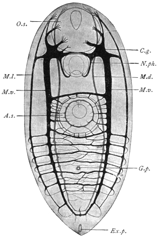
Fig. 121.—Harmostomum leptostomum, Olss., an immature
specimen from Helix hortensis. Nervous system, according to
Bettendorf. A.s., ventral sucker; C.g., cerebral ganglion;
Ex.p., excretory pore; G.p., genital pore; O.s., oral sucker;
M.d., dorsal medullary nerve; M.l., lateral medullary nerve;
N.ph., pharyngeal nerve; M.v., ventral medullary nerve.
Magnified.
Of the muscles of the organs which have developed from the
parenchyma muscles we may briefly mention those bundles that are
attached to certain parts of the genital apparatus, to the suckers,
to the hooks and claws, and also, at all events in Fasciola hepatica,
to the spines. The sheaths used for the projection of the tentacles
of the Rhopaliadæ are also muscular.
The contractile elements consist of fibres of various lengths
that are mostly parallel to one another, and frequently anastomose;
a cortical substance finely fibrillated can usually be distinguished
from an internal homogeneous mass; large nucleated cells of uniform
size are always connected with them; these have been variously
interpreted, but have been proved to be myoblasts, one or more
of their processes constituting the muscular fibres.
The MOVEMENTS of the Trematodes consist in alterations of form
and position of the body, as well as in creeping movements.
In the NERVOUS SYSTEM (fig. 121) can be distinguished a cerebral
portion as well as strands (medullary strands) running from it,
and peripheral nerves. The cerebral portion always consists of
two large ganglia situated in the anterior end of the body which
pass dorsally over the œsophagus and are connected by means
of a broad and thick commissure composed of fibres only. From
each ganglion three nerves run anteriorly—the inner and dorsal
nerve for supplying the anterior dorsal part of the body; the
median and ventral for the oral sucker; and the exterior and lateral
likewise for the supply of the sucker.
In a similar manner three strands run backwards from each
ganglion—one dorsal, one lateral and one ventral. The dorsal and
ventral strands become united and curve backwards; the symmetrical
lateral strands are connected by means of transverse commissures,
the number of which vary according to the species. Such commissures
also exist between the lateral and the two other strands on each
side. There are ganglion cells along the entire course of the posterior
cords, more particularly at the points of origin of the commissures.
There also appears to be in addition a fourth anterior and posterior
pair of nerves, the front pair for the oral sucker and the hind pair
for the pharynx.
The peripheral nerves, which spring from the posterior strands
as well as from the commissures, either pass directly to the muscular
fibres or to the sensory cells that are situated at the level of the
subcuticular cells, or they reach these after the formation of a plexus
situated immediately beneath the dermo-muscular layer; the processes
directed outwards terminate in small vesicles in the cuticle.
As to other ORGANS OF SENSE, simple eyes, two or four in
number, are known in several ectoparasitic species as well as in a
few free-living larval stages (Cercariæ) of endoparasitic forms. In the
adult stage, however, they usually undergo complete atrophy.
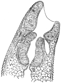
Fig. 122.—Median section through
the anterior part of Fasciola hepatica:
the oral sucker, pharyngeal pouches,
pharynx, œsophagus, cuticle with
spines, and the body parenchyma.
The ALIMENTARY CANAL commences with an oral aperture,
generally terminal or sub-terminal (ventral) at the anterior extremity,
which leads into an oral cavity usually surrounded by a sucker;
the œsophagus, of various lengths, is directed backwards and is
generally surrounded by a muscular pharynx (fig. 122). In some
cases there exists between the sucker and pharynx, pharyngeal pouches
(præpharynx). Sooner or later the
intestine divides into two lateral
branches directed backwards, both of
which end blindly (cæca) at the same
level.257 In many ectoparasites (Monogenea
[p. 222]) a connection exists
between the genital glands and one of
the intestinal branches (ductus vitello-intestinalis
[fig. 123]).
The oral cavity, pharyngeal pouches,
pharynx, and œsophagus are lined with
a continuation of the cuticle of the
body; the gut cæca are lined with tall
cylindrical epithelium (fig. 120). The
œsophagus and intestinal branches
often have also one layer of annular
and longitudinal muscles; the pharynx
has essentially the structure of a sucker
(fig. 122).
The accessory organs of the alimentary
canal consist of groups of unicellular SALIVARY GLANDS that
discharge into the œsophagus in front of or behind the pharynx, or
even into the pharynx itself.
The food of the Trematodes consists of mucus, epithelial cells, the
intestinal contents of the hosts, and often also of blood, and this not
only in those species living in the vascular system, but also in species
living as ectoparasites or in the intestine or biliary passages of their
hosts.
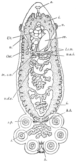
Fig. 123.—Polystomum integerrimum, a
monogenetic fluke from the urinary bladder
of the frog. i., intestine; h., large hooks
of the sucking disc; h.k., smaller hooklets;
l.c.v., longitudinal vitelline ducts; o., oral
orifice; Oot., oötype; ov., ovary; s.p., suckers
of the disc; tr.c.v., transverse vitelline ducts;
Ut., uterus with ova; v., entrance to the
vagina; v.d.e., vas deferens; v.d.i., ductus
vitello-intestinalis; the vitellaria and testes
are not shown. Magnified. (After Zeller.)
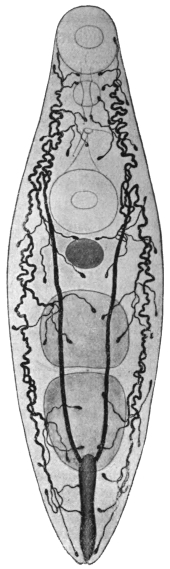
Fig. 124.—Allocreadium isoporum,
Looss. Excretory apparatus.
Of the other organs, the
oral sucker, pharynx, genital
pore, ventral sucker, ovary and
testes are shown; the cylindrical
excretory bladder is in
the posterior end. 38/1. (After
Looss.)
The final products of assimilation dissolved in the fluids of the
body are distributed throughout the parenchyma and are thence
expelled by a definite tubular system (excretory apparatus, proto-nephridia,
formerly also termed the water-vascular system). This
system, which is distributed throughout the entire body (fig. 124), is
symmetrically developed, and, in the ectoparasitic Trematodes, it
opens, right and left, at the anterior end on the dorsal surface; in all
other flukes, however, it opens singly into the excretory pore (foramen
caudale) at the centre of the posterior border; in those cases, however,
where a sucker is present at the posterior end, as in the Amphistomata,
the excretory pore is situated on the dorsal surface close in front of
the sucker.
The EXCRETORY SYSTEM258 consists of several parts: (1) of the more
or less numerous terminal “flame” cells or funnel cells (figs. 124, 125);
(2) of the capillaries ending in them; (3) of
larger vessels receiving the capillaries; and (4)
of the excretory bladder. Terminal cells and
capillaries may be compared to unicellular
glands with long excretory ducts; the cellular
body (fig. 125) is comparatively large, stretched
longitudinally, more rarely transversely, and
provided with numerous processes, that are
lost in the parenchyma; within is a conical
cavity (analogous to the secretory cavity of
unicellular glands) which is continued directly
into the structureless capillary; at its blind
end is a bunch of cilia projecting into the
cavity, and which, during life, shows a flickering
motion (ciliary flame). The nucleus is
situated in the protoplasm of the terminal cell
at its blind end.
Fig. 125.—Terminal flame
cell of the excretory system.
n., nucleus of cell; c., bundle
of cilia forming the “flame”;
p., processes of cell extending
into parenchyma; d., excretory
capillary. (Stephens.)
The entire apparatus thus begins blindly—i.e.,
within the terminal cells, to which must
be ascribed the capacity of taking up from the fluid that permeates
the parenchyma the products which are first collected into their own
cavities and thence excreted by means of the capillaries and vessels.
The vessels possess definite walls, consisting of a membrane and
a nucleated protoplasmic layer. They unite at many points on either
side, and again pass into other canals (COLLECTING TUBES), which
finally, travelling towards the posterior end, discharge into the
excretory bladder (fig. 124).
The form and size of the bladder vary much according to the
different species, but it always possesses its own flattened epithelium,
surrounded by circular and longitudinal muscles, the circular muscles
forming a sphincter around the opening. Frequently also the
structure of the bladder extends to the tubules discharging into
it, which therefore are not to be regarded as separate “vessels,”
but rather as tubular diverticula of the bladder, directed anteriorly.
In some few species the diverticula also branch and the branches
anastomose, so that a network of tubules ensues which receives the
vessels or capillaries. In such cases there are also ciliary tracts in
the tubules.
The contents of the entire apparatus
usually consist of a clear or sometimes
reddish fluid; in some species
there are larger or smaller granules, and
occasionally also concretions occur.
Fig. 126.—Diagram of female genitalia. Ov.,
ovary; ovd., oviduct; L.c., Laurer’s canal;
Rec. sem., receptaculum seminis; Vit. R., vitellarian
reservoir; t.v.d., transverse vitelline duct;
Oo., oötype; Sh. gl., shell gland; Rec. ut.,
receptaculum uterinum; ut., uterus. (The various
parts are not to the same scale.) (Stephens.)
Fig. 127.—Diagram of male and part
of female genitalia. ut., uterus; vag.,
vagina; ♀, opening of vagina; g.s., genital
sinus; g.p., genital pore; ♂, opening
of ejaculatory duct or vas deferens; c.s.,
cirrus sac; c., cirrus; p.p., pars prostatica;
s.v., seminal vesicle; e.j., ejaculatory duct
or vas deferens; v.e., vas efferens; t., testis.
(Stephens.)
Sexual Organs.—Nearly all the Trematodes are hermaphrodites,
and only a few (Schistosomidæ, Koellikeria) are sexually differentiated.
The sexual organs usually lie in the “central field” limited
by the gut cæca; the vitellaria, on the other hand, are, as a rule,
external to the gut cæca in the “lateral fields.”
The male apparatus259 is composed of two variously formed
testes (fig. 127) (globular, oval, indented, lobed, or ramified), which
may lie side by side or one behind the other; from each testicle
a tube (vas efferens) originates; sooner or later, both tubes as a rule
unite to form the ejaculatory duct or vas deferens, which is frequently
enclosed in a muscular CIRRUS SAC, or more rarely passes directly into
the genital pore. The cirrus, which is the thick muscular terminal
portion of the vas deferens, can be everted and protruded from the
cirrus sac and serves as an organ of copulation. The walls of the
muscular portion of the tube (the cirrus) are attached to the walls of
the cirrus sac, and hence when the sac contracts the cirrus cannot
be protruded except by evagination of its lumen. Opening into the
middle portion of the vas deferens, and as a rule enclosed in the cirrus
sac, is found a mass of unicellular glands (prostate), the vesicula
seminalis (which is likewise within, or may also be outside the sac)
being the dilated first portion of the vas.
The female genitalia (fig. 126) consist of an ovary, usually situated
in front of the testes, the form of which varies according to the
species, the usually double vitellaria, the ducts and a number of
auxiliary organs; the short oviduct directed towards the centre
arises from the ovary, and is connected in the median line with
the excretory duct of the vitelline glands. These grape-like glands
possess longitudinal excretory ducts, which assume a transverse
direction behind the ovary, unite together at the median line and
form a single duct, often dilated into a vitelline receptacle, that unites
with the oviduct. Near this point, moreover, there frequently opens
a canal (Laurer’s canal) which begins on the dorsal surface, and
on the inner end of which a vesicle filled with sperm (receptaculum
seminis) usually occurs (fig. 126). Moreover, there are also numerous
radial unicellular glands (shell glands) at or beyond the point of
junction of the oviduct, vitelline ducts and Laurer’s canal. In this
portion of the duct (oötype), which is usually dilated, the ovarian
cells are fertilized, surrounded with yolk cells and shell material,
and as ova with shells they pass into the uterus (a direct continuation
of the oviduct), which, with its many convolutions, occupies a
larger or smaller portion of the central field, and runs either direct
to the genital pore or, forming convolutions, first runs posteriorly
and then bends forward (descending and ascending limbs). In both
cases the terminal part lies beside the cirrus pouch and discharges
beside the male orifice either on the surface of the body or into a
genital atrium. The terminal portion of the uterus, which is often
of a particular structure, serves as a vagina (METRATERM).
The cirrus sac may include (1) the genital atrium (i.e., the
common sinus, into which the vas deferens and vagina may open),
or (2) a variable extent of the vas from cirrus to seminal vesicle. Thus
the latter may be outside the sac. In the absence of a sac, the genital
sinus may be surrounded by a pseudo-sucker, as in Heterophyes (in
some cases the ventral sucker itself, from its close proximity to the
genital pore, serves as an accessory copulatory organ). In other cases
copulatory organs are formed by hooks projecting into the lumen
of the terminal portion of the vas.
The GENITAL PORE, which is the opening from the genital sinus on
to the surface, is generally situated at or near to the median line on
the ventral surface and in the anterior region of the body; in most
of the Distomata it is in front of the ventral sucker, in other cases,
e.g., in the Cryptocotylinæ, it is behind.260
The spermatozoa do not differ essentially in their structure from
those of other animals; the ovarian or egg cells are cells without
integument and contain a large nucleus and a little protoplasm; the
vitellaria also produce nucleated cells, in the plasm of which there
are numerous yellow yolk granules; the yolk cells detach themselves,
like the ovarian cells, from the ovarium, and pass into the oviduct
to surround each ovarian cell in the oötype. They disintegrate sooner
or later in the completely formed egg and are utilized as food by the
developing embryo.
Development of the Trematodes.
(1) Copulation.—Observation has demonstrated that the one or
two vaginæ occurring in the ectoparasitic Trematodes are utilized
as female organs of copulation, and that the copulation is cross;
it is also known that Laurer’s canal, which was formerly generally
regarded as the vagina, has only quite exceptionally, if at all, served
the digenetic Trematodes as such—it appears to be homologous with
the canalis vitello-intestinalis of the Monogenea261—but the terminal
portion of the uterus, termed the metraterm, is used for copulation.
Cross-copulation occurs as well as auto-copulation and auto-fecundation.
The spermatozoa subsequently pass through the entire uterus,
which is still quite short at the time the male organs are matured; the
maturation of which, as usually is the case in hermaphrodites, precedes
that of the female organs. It is only later with the onset of egg formation
that the uterus is fully developed. Copulation, however, takes place
also in the case of fully grown forms with completely developed uteri.
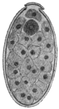
Fig. 128.—Ovum of Fasciola
hepatica, L., cut longitudinally.
The lid has been lifted in the
process. Within the egg are
numerous yolk cells, and at the
lid end there is the still unsegmented
ovum (dark). 240/1.
Fig. 129.—Miracidium of
Fasciola hepatica that has just
hatched from the egg, with a distinct
cuticular ciliated epithelium.
Magnified. (From Leuckart.)
(2) Formation of the Ova.—The ovarian cells arising from the ovary
first become mature after their entry into the oötype by the formation
of three polar bodies, fertilization then taking place. At the same
time as the ovarian cell a number of yolk cells from the vitellarium
and secretion, drop by drop, from the shell gland reach the oötype.262
The shell is then formed during the generally active contractions of
the oötype walls and then passes on into the uterus. In the uterus of
the endoparasitic trematodes the eggs accumulate more and more,
often in large quantities, while in ectoparasitic species generally only
one or some few eggs can be found. The completed ova are of
various forms and sizes. They are mostly oval, at all events in the
digenetic trematodes, and the yellowish or brown shell is provided
with an opening at one pole which is closed by a watch-glass-shaped
lid (operculum). Appendages (filaments) on the shell—at one or both
poles—are uncommon, but are the rule in the ova of the Monogenea
(ectoparasitic species).
(3) Deposition of the Ova.—Soon after their formation, the
Monogenea (ectoparasitic trematodes) deposit round the place of
their attachment on the skin or the gills or other organs of their
hosts, eggs which attach themselves by means of their filaments.
The embryonic development thus takes place outside the parent.
This also holds good for the eggs of many endoparasitic species,
although as a rule in these the eggs are always retained for a
longer time in the uterus. Moreover, they usually here undergo a
part or a whole of their development, and are eventually deposited
in those organs in which the adult forms are parasitic, but this is
not always the case, as the egg, e.g., of F. hepatica appears in bile (and
fæces) quite unchanged. By the natural passages they eventually get
out of the body, and in cases where such do not exist, as in the case
of the blood-vessels, the eggs pass out by means of the kidneys.
(4) The embryonic development, after irregular segmentation of the
ovum into a number of blastomeres, leads to the formation of a solid
blastosphere or morula, which is surrounded by a cellular investing
membrane (yolk envelope), while the principal mass of the cells forms
the embryo, which uses for its nourishment the yolk cells, which have
in the meantime disintegrated (cf. footnote, p. 223). Usually, after
the ova have reached water the embryos hatch out, leaving the yolk
envelope in the egg-shell; in other cases, however, the embryos only
hatch out after having been subjected to the influence of the intestinal
juices, that is to say, in the intestine of an intermediate host which has
ingested with its food the ova that have escaped from the primary host.
(5) The post-embryonic development of the Trematodes is accomplished
in various ways; the process is the most simple in the ectoparasitic
species (Monogenea), the young of which should certainly
be regarded as larvæ, because they possess characteristics (cilia,
simple gut, etc.) that are lacking in the adult worms, but which,
nevertheless, pass into the adult state direct after a relatively simple
metamorphosis. In the Holostomata,263 a group found chiefly in the
intestine of aquatic birds, and which rarely occur in other vertebrates,
the ova develop in water. The young are ciliated all over, and, after
having entered an intermediate host (leeches, molluscs, arthropods,
amphibians, fishes) living in the water, they undergo a metamorphosis
into a second larval stage; they then encyst and await transmission
into the final host, where they become adult Metastatic
trematodes, i.e., trematodes without asexually produced generations
(p. 229).
Fig. 130.—A group of cercariæ of Echinostoma sp. (from
fresh water). 25/1.
In the remaining so-called digenetic trematodes (p. 230) one or two
asexual generations interpose between the miracidium and terminal
stage, so that quite a number of adult worms may originate from
one egg. Usually the young, which are termed MIRACIDIA264 (fig. 129),
hatch in water, where they move with the aid of their cilia. Sooner
or later they penetrate into an intermediate host, which is always
a snail or a mussel, and while certain of their organs disappear,
they grow into a gutless germinal tube (SPOROCYST, fig. 131). These
are simple elongated sacs with a central body cavity. They may or
may not have excretory tubules. In these, according to the species,
the larval stages (CERCARIÆ) that will ultimately become adult worms
are produced, or another intermediate generation is first formed, viz.,
that of the REDIÆ265 (figs. 132, 133), which are always provided with an
intestine, and these then give rise to cercariæ (figs. 130, 134). The
cercariæ, as a rule, leave their host and move about in the water
with the assistance of their rudder-like tails. After a little time, however,
they usually again invade an aquatic animal (worms, molluscs,
arthropods, fishes, amphibians), then they lose their tails and become
encysted (fig. 135); here they wait until they attain, together with
their host, the suitable terminal host, and in this new situation they
establish themselves and reach maturity. Or, again, the cercariæ may
themselves encyst in water or on foreign bodies (plants) and wait until
they are taken up directly by the terminal host, e.g., sheep.
Accordingly the following conditions are necessary for the completion
of the entire development: (1) The terminal host in which
the adult stage lives; (2) an intermediate host into which the
miracidia penetrate and in which they become sporocysts; (3) a
second intermediate host in which the cercariæ become encysted.
In certain species, as in Fasciola hepatica, this second host is omitted,
as the cercariæ spontaneously encyst on plants, or again (in other
species) encystment may occur within the first intermediate host, when,
in fact, the cercariæ (which in this case do not acquire an oar-like tail)
do not swarm out of, but encyst themselves within their sporocysts.
The development, moreover, may be further complicated by rediæ
appearing in addition to the sporocysts, though this occurs in the
first intermediate host and not in a second one.
Animals that harbour adult digenetic Trematodes thus become
infected by ingesting encysted cercariæ, which either occur (1) in
certain animals (second intermediate hosts) on which they feed, or
(2) in water, or (3) on plants, or finally (4) in the first intermediate
host; whereas animals harbouring encysted cercariæ have been directly
infected by the corresponding tailed stage, and animals harbouring
germinal tubes (sporocysts or rediæ) have been infected by the
miracidia.
Fig. 131.—Development of Fasciola hepatica, L. a, the miracidium in optical section
showing cephalic lobe, x-shaped eye-spot resting on the cerebral ganglion, two germ balls;
below each of these a flame cell, and still lower germ cells lying in a cavity (primitive body
cavity). b, young sporocyst with two eye-spots, and germ balls; the cells lining the cavity
are not shown. c, older sporocyst with a young redia. Magnified. (After Leuckart.)
Thus certain species of ducks and geese become infected with
Echinostoma echinatum by devouring certain water-snails (Limnæus,
Paludina) in which the encysted cercariæ occur. Oxen become infected
with Paramphistomum cervi (= Amphistomum conicum) by swallowing
with water, cysts of this species which occur at the bottom of puddles
and pits. Sheep are infected with Fasciola hepatica by eating grass
to which the encysted cercariæ of the liver-fluke are attached; our
song-birds infect themselves or their young with Urogonimus macrostomus
by tearing off pieces containing the corresponding sporocysts
which are full of encysted cercariæ from snails (Succinea amphibia),
which act as the first intermediate hosts, and eating, or offering their
young these pieces.
(1) The MIRACIDIA of the digenetic Trematodes are comparatively
highly organized, and the mode of their formation from the
segmentation cells of the ovum is only imperfectly known. They have
a cuticular epithelium (fig. 129) entirely or partly covered with cilia,
beneath this a dermo-muscular tube composed of circular and longitudinal
muscles; also, a simple gut sac with an œsophagus, occasionally
also with pharynx, salivary glands and boring spine, also
a cerebral ganglion on which, in some species, there are eyes
(fig. 131, a). As to the excretory organs, they are represented by two
symmetrically placed terminal flame cells, with excretory vessels
opening separately; there is a
more or less ample (primary) body
cavity between the parietes of the
body and the gut; from the cellular
parietal lining of this cavity
single cells (germ cells) become
free (fig. 131, a, b), and become
rediæ or cercariæ.
[The germ cells of the miracidium
and the germ balls of the
sporocyst arise, according to some
observers, by further division of
undifferentiated blastomeres; according
to others from the cells of
the lining wall of its body cavity.
It is from these free germ balls
that the redia stage is developed.
[In the germ ball or morula
appears an invagination, giving
rise to the cup-shaped gastrula
stage. This elongates and forms
the REDIA (fig. 131, c).
[In the interior of the redia
cells are budded off and develop
into gastrulæ, as in the case of the
sporocyst. These become a fresh
generation of rediæ or give rise
to the third stage (CERCARIA).]
Fig. 132.—Young
redia of Fasciola hepatica,
with pharynx
and intestine, with a
circular ridge anteriorly
and a pair
of processes posteriorly
and masses of
cells (germ balls) in
the interior. Magnified. (From
Leuckart.)
Fig. 133.—Older
redia of Distoma
echinatum, with rudimentary
intestine
i.; cercariæ, c.; germ
balls, b.; and birth
pore, g. Magnified.
(2) The SPOROCYSTS, on the contrary, which are produced direct
from the miracidia, are very simple, as all the organs of the latter
disappear, even to the muscles and excretory organs, during or after
penetration into the intermediate host, whereas the budded and still
budding cells of the wall of the (primary) body cavity continue to
develop rapidly and form germ balls. The sporocysts when fully
developed have the appearance of tubes or fusiform bodies with
rounded edge; they are frequently of a yellow colour. Their length
rarely exceeds a few millimetres; in some species their size increases
exceedingly through proliferation, and they then occupy a large portion
of the body of the intermediate host.
(3) The REDIÆ (figs. 132, 133), on the other
hand, are more cylindrical and always have a
simple intestine of varying length, provided
with a pharynx; they likewise possess, situated
near the circular ridge, a “birth pore” which
serves for the exit of the cercariæ originating
within them.
Fig. 134.—Cercaria of
Fasciola hepatica; the cutaneous
glands at the side of
the anterior body. Magnified.
(After Leuckart.)
(4) The CERCARIÆ266 are very different; typically
they consist of the anterior body and the
oar-like tail at the posterior end (fig. 134). The
former, even to the genitalia, has the organization
of the adult digenetic Trematodes, and
thus allows the easy recognition of at least the
characters of that large group to which the
species in question belongs. On the other
hand, however, there are also organs that are
lacking in the adult form, such as, in many, the
boring spine in the oral sucker, or the eyes
situated on the cerebral ganglion; moreover,
also, cutaneous glands (fig. 134), the secretion of
which forms the cyst membrane. The oar-like
tail may be long or short (stumpy-tailed cercaria)
or entirely absent; its free end may be
partly split (furcate cercaria), or split to its base
(bucephalus); in various forms also the anterior
end of the tail is hollow, and has enclosed
within it the anterior body, which is otherwise
free. The size also of the cercaria belonging
to the different species is very diverse; in
addition to forms swimming in the water that
have the appearance of minute milky-white
bodies, there are forms which measure as much
as 6 mm. in length.
Fig. 135.—Encysted cercaria
of Fasciola hepatica.
Magnified. (After Leuckart.)
The encysted cercariæ (fig. 135) are globular
or oval, and are surrounded by a homogeneous
membrane, which may be striated or contain granules. The tail is
always cast off when encystment occurs, and organs peculiar to the
cercaria stage (boring papilla, eyes) almost entirely disappear. On
the other hand, the genitalia appear or become more or less highly
developed, in extreme cases to such an extent that they become
functional, and after autocopulation the creatures produce ova within
the cysts.
The cycle of development of the digenetic Trematodes has hitherto
been generally explained as a typical ALTERNATION OF GENERATIONS,
one sexual generation regularly alternating with one or two asexually
reproducing generations. Recent authors, however, regard the cells
in the sporocysts from which rediæ or eventually cercariæ arise as
parthenogenetically developing ova, and the sporocysts as well as the
rediæ as generations propagating parthenogenetically. In this case,
however, it is an alternation of a sexual not with an asexual but with
firstly a parthenogenetic generation (the sporocyst), the central cells
of which are regarded as ova which develop parthenogenetically into
the redia, and this the second parthenogenetic generation finally
produces larvæ (cercariæ) capable of developing into the sexually
mature form.
Other authors, again, regard the development of the Digenea as
only a complicated metamorphosis (p. 283), which is distributed over
several generations before it is concluded.
Biology.
Endoparasitic Trematodes, as fully developed organisms, occur in
vertebrate animals only, with very few exceptions; they inhabit almost
all the organs (with the exception of the nervous and osseous systems
and the male genitalia), but by preference the intestine in all its
extent from the oral cavity to the anus; and, further, certain species
or groups inhabit only quite restricted parts of the intestine. Besides
in the intestine other species live in the liver, or in the bile-ducts,
or in the gall-bladder; other accessory organs of the intestine, such
as the pancreas, bursa Fabricii (of birds), are only infected by a few
species. Many inhabit the lungs, or the air sacs in fowls, a few
the trachea. Trematodes have also been known to occur in the
urinary bladder, the urethra and the kidneys of all classes of
vertebrates; they are also present in the vascular system of a few
tortoises, birds and mammals; in birds they even penetrate from
the cloaca into the oviducts, and are occasionally found enclosed in
the laid eggs; one species is known to occur in the cavum
tympani and in the Eustachian tube of a mammal (Dugong),
another in the frontal sinus of the polecat; several species infest the
conjunctival sac under the membrana nictitans of birds, one species
even lives in cysts in the skin of song-birds. In an analogous manner
the ectoparasitic Trematodes are not entirely confined to the surface of
the body or the trachea of the lower vertebrate animals; a few species
appear exclusively in the urinary bladder, in the œsophagus, and in the
case of sharks in an accessory gland of the rectum.
Trematodes live free and active within the organs attacked,
though they may attach themselves by suction for a longer or shorter
period; in other cases, however, they bore more or less deeply
into the intestinal wall with their anterior end, or lie in cysts
of the intestinal wall which only communicate with the lumen
through a small opening; in those species living in the lungs of
mammals the host likewise produces a cyst, which usually encloses
two specimens; such association of a pair is also observed in
other situations, and, though this is the rule in species sexually
distinct, it is not entirely confined to these.
As regards the AGE attained by endoparasitic Trematodes, there
are but few reliable records, and these differ considerably; the
overwhelming majority of species certainly live about a year, or
perhaps a little longer, but there are some whose term of life
extends to several or many years.
Trematodes are but rarely found encysted in the higher vertebrate
animals; the condition, however, is more frequent in amphibians,
and especially in fishes, as well as in numerous invertebrate
animals.
Classification of the Trematodes of Man.
The following classification, partly artificial, partly natural,
embraces only the flukes found in man:—
Order. Digenea, v. Beneden, 1858.
Anterior sucker single and median, present. Eggs few. The (specialized)
terminal portion of the uterus serves as a vagina. Development indirect, i.e., an
intermediate host is required.
Sub-order. Prostomata, Odhner, 1905.
Mouth surrounded by the anterior sucker.
Group. Amphistomata, Rudolphi, 1801, ep., Nitzsch, 1819.
Gut forked, two suckers, the posterior sucker (acetabulum) terminal or ventro-terminal
behind the genitalia, or at most embraced by the vitellaria. Skin with no
spines. Excretory bladder a simple sac opening dorsally near hind end. Testes in
front of ovary. Genital pore, median in anterior third of body. Thick flukes, almost
circular in cross section.
Family. Paramphistomidæ, Fischoeder, 1901.
Amphistomata: Body not divided into a conical anterior portion and disc-like
caudal portion. Ventral pouch absent.
Sub-family. Paramphistominæ, Fisch., 1901.
Paramphistomidæ: Oral sucker without evaginations. Not in man.
Sub-family. Cladorchiinæ, Fisch., 1901.
Paramphistomidæ: Oral sucker with evaginations; testes, two, deeply cleft
(fig. 137). Genera: Watsonius, Cladorchis, etc.
Family. Gastrodisciidæ, Stiles and Goldberger, 1910.
Amphistomata: With body divided into a conical cephalic and disc-like caudal
portion (fig. 138). Posterior sucker ventro-terminal. Oral sucker with evaginations.
Genera: Gastrodiscus and Homalogaster.
Group. Distomata, Retzius, 1782.
Gut forked, two suckers, the posterior sucker (acetabulum) ventral. It is
always separated from the hind end by at least a part of the genitalia.
Family. Fasciolidæ, Railliet, 1895.
Large flat forms, genital pore in front of ventral sucker, the latter powerful.
Vitellariæ of numerous follicles, united by branching vitellarian ducts, at the sides of
the body meeting posteriorly and extending ventrally and dorsally. Cirrus and
vagina without spines. No crown of strong spines around sucker. Testes much
branched. Uterus not well developed. Excretory bladder much branched. Eggs
large.
Sub-family. Fasciolinæ, Odhner, 1910.
Large or median forms, gut much branched. Body has a shoulder separating
head from body. Receptaculum seminis absent. Ovary branched, ventral sucker
in anterior part of body. Genus: Fasciola.
Sub-family. Fasciolopsinæ, Odhner, 1910.
Shoulder absent. Receptaculum seminis present. Ovary branched, gut takes
a zig-zag course with kinks on it, ventral sucker in anterior part of body.
Genus: Fasciolopsis.
Family. Opisthorchiidæ, Braun, 1901, emend. auctor.
Ovary in front of testes. Small to medium flukes, very transparent, tapering
anteriorly. Vitellaria moderately developed not extending in front of sucker.
Cirrus absent. Seminal vesicle a twisted tube free in parenchyma. Testes near
hind end one behind the other, lobed or branched, but not dendritically. Excretory
bladder Y-shaped, the two limbs short, the stem S-shaped passing between the
testes. Receptaculum seminis well developed. Laurer’s canal present. Uterine
coils transverse, numerous. Eggs small.
Sub-family. Opisthorchiinæ, Looss, 1899, emend. auctor.
Opisthorchiidæ in which the excretory pore is terminal. Excretory bladder long,
dorsal to testes. Uterine coils not overlapping gut forks. Genera: Opisthorchis,
Paropisthorchis, Clonorchis, Amphimerus, etc.
Sub-family. Metorchiinæ, Lühe, 1909.
Opisthorchiidæ in which the excretory pore is ventral. Excretory bladder short,
ventral to testes. Uterine coils partly overlapping gut forks and extend anteriorly
beyond the sucker. Vitellaria compressed on the sides of the body. Genus: Metorchis.
Family. Dicrocœliidæ, Odhner, 1910.
Ovary behind testes. Testes behind the ventral sucker, between it and the ovary.
Body thin and transparent. Cirrus sac encloses the pars prostatica and seminal
vesicle. Skin smooth. Gut forks do not reach posterior end. Receptaculum seminis
and Laurer’s canal present. Vitellaria, moderate, lateral in mid-body slightly overlapping
the gut. Uterus with an ascending and descending branch and numerous
transverse coils extending to hind end. Eggs dark brown, 25 µ to 60 µ. Excretory
bladder tubular in posterior third or half of body. Parasitic in bile-ducts of mammals
and birds. Genus: Dicrocœlium.
Family. Heterophyiidæ, Odhner, 1914.
Ovary in front of testes. Genital pore behind or on a level with ventral sucker.
Genital pore surrounded by a pseudo-sucker (i.e., its muscle is not sharply separated
from but blends with the body muscles). Cirrus sac absent, consequently vesicula
seminalis and pars prostatica lie free. Vagina and ejaculatory duct unite into a
common duct before opening. Small and very small forms. Body covered with
scales. Genera: Heterophyes, Metagonimus, etc.
Family. Troglotremidæ, Odhner, 1914.
More or less flattened Distomes of compact form, 2 to 13 mm. long. Ventral
surface flat or somewhat hollowed, dorsal surface arched. Skin completely covered
with pointed spines. Musculature weakly developed also in the suckers in those
forms that inhabit cysts. Gut with pharynx and a not very long œsophagus and
cæca, which end more or less shortly before the hind end. Excretory bladder
Y-shaped or tubular. Pars prostatica and seminal vesicle always distinct. Testes
elongated, symmetrically placed in or behind the middle of the body. Ovary directly
in front of the testes, right-sided, generally much lobed. Receptaculum seminis
and Laurer’s canal present. Vitellaria generally well developed, exclusively or for
the most part confined to the dorsal surface, leaving only a median band unoccupied.
Uterus either very long, coiling here and there, or shorter and more convoluted.
Eggs in first case small 17 µ to 25 µ, in the second much larger 63 µ to 85 µ or
even 120 µ (?) long. Parasitic in carnivora or birds, generally occurring in pairs
in cyst-like cavities. Genera: Paragonimus, Pholeter, Collyriclum, Troglotrema.
Family. Echinostomidæ, Looss, 1902.
More or less elongated flukes, small or very large, much flattened anteriorly,
less so posteriorly, or even round. Suckers near one another, the anterior small and
weak, the posterior large and powerful directed obliquely backwards. Surrounding
the oral sucker dorsally and laterally but not ventrally is a fold or “collar” bearing
a row or rows of pointed spines which are continued round laterally on to the ventral
corners, the number being constant for each species, the corner spines large or
specialized, skin anteriorly scaled or spiny. Alimentary canal consists of a pharynx,
epithelial “pseudo-œsophagus” and gut cæca reaching to posterior end. Testes behind
one another in hind body. Ovary on right side or median directly in front of the
testes. Vitellaria lateral, usually extending to the hind end and not beyond the
ventral sucker anteriorly. Genital pore just in front of ventral sucker. Uterus in
transverse loops. Genital sinus absent or present. Receptaculum seminis and
Laurer’s canal present. Eggs thin shelled and large, bright yellow, 65 µ to 120 µ long.
Excretory bladder Y-shaped. Parasitic in gut of vertebrates, especially birds.
Sub-family. Echinostominæ, Looss, 1899.
Cirrus sac usually reaching to centre of ventral sucker, but not beyond. Cirrus
long, usually without spines, coiled when retracted. Seminal vesicle tubular, twisted.
On the head a ventral uniting ridge between the angles of the collar. Dorsal circlet
of spines, single or double, not interrupted unless the collar itself is dorsally divided.
Genera: Echinostoma, etc.
Sub-family. Himasthlinæ, Odhner, 1910.
Cirrus sac reaching far beyond ventral sucker. Cirrus armed with strong
rose-thorn-shaped hooks. Vesicula seminalis tubular not coiled. Cervical collar
not continued across ventral aspect. Spines on collar in one row. Body armed
with fine needle-shaped spines.
Family. Schistosomidæ, Looss, 1899.
Sexes separate. Genital pore behind the ventral sucker. Ventral sucker
elevated above the surface. Pharynx absent. Gut forks reunite to form a single
stem. In ♂ four or more testicular follicles. In ♀ a single ovary, just in front
of the union of the gut forks. Vitellaria on either side of the united gut stem.
The Trematodes Observed in Man.
Family. Paramphistomidæ, Stiles and Goldberger, emend. 1910.
Sub-family. Cladorchiinæ, Fisch., 1901.
Genus. Watsonius, Stiles and Goldberger, 1910.
Cladorchinæ.—Body pyriform. Ventral pouch absent. Acetabulum ventral or
(?) ventro-subterminal, very large, margins projecting, aperture small. Genital
pore in front of bifurcation of gut, not surrounded by a sucker; ductus hermaphroditicus
apparently absent. Excretory pore at posterior end of excretory vesicle,
behind Laurer’s canal. Oral sucker with a pair of irregularly globular suctorial
pouches; œsophagus thickened distally; cæca long, not wavy; end in acetabular
region.
Male Organs.—Testes two lobed, smaller than acetabulum; longitudinally,
nearly or quite coinciding; transversely they abut or slightly overlap; preovarial
in equatorial and caudal thirds. Pars musculosa not largely developed; cirrus
pouch absent.
Female Organs.—Ovary and shell gland post-testicular. Vitellaria extend from
gut fork to slightly beyond gut ending; uterus intercæcal, partly post-testicular.
Laurer’s canal in front of excretory vesicle.
Type Species.—Watsonius watsoni, Conyngham, 1904.
Watsonius watsoni, Stiles and Goldberger, 1910.
Syn.: Amphistomum watsoni, Conyngham, 1904; Cladorchis watsoni, Shipley, 1905.
Fig. 136.—Watsonius
watsoni: ventral view.
4/1. (After Shipley.)
Body, 8 to 10 mm. long, by 4 to 5 mm. broad, by 4 mm. thick; tapers
anteriorly to 2·5 mm. Caudal extremity bluntly rounded, venter
surrounded by an elevated ridge, surface with
transverse ridges best defined ventrally. Genital
pore median about one-quarter of body length
from anterior end at level of suctorial pouches.
Acetabulum 1 mm. in diameter, margin projecting,
aperture small. Mouth in a groove with
digitate papillæ. Oral sucker very large, one-fifth
of length of body, with a pair of irregularly
globular pouches. Œsophagus somewhat longer
than sucker. Excretory pore at the level of
the acetabular aperture. The vesicle extends
from the plane of the transverse vitelline ducts
to centre of acetabulum.
Male Organs.—Testes deeply notched adjoining
one another. Vesicula seminalis much coiled
and dilated, pars musculosa not coiled. Pars prostatica (?) dilated,
ejaculatory duct long and narrow, opening on a papilla; genital
atrium papillated.
Female Organs.—Ovary dorso-posterior of posterior testis. Shell
gland dorsal to ovary. Vitellaria ventral and lateral to gut cæca
extending from gut fork to equator of acetabulum. Uterus dorsal
to testes, ductus hermaphroditicus absent. Laurer’s canal opens in
dorso-median line slightly behind anterior border of sucker.
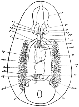
Fig. 137.—Watsonius watsoni: ventral projection composed
from a series of transverse sections. o.s., oral sucker; s.p., suctorial
pouch; ga., genital atrium; d.e., ejaculatory duct;
es., œsophagus; e.g., œsophageal ganglion; p.p., pars prostatica;
p.m., pars musculosa; i., gut; ut., uterus; v.e., vas efferens;
v.e.s., left vas efferens; v.e.d., right vas efferens; v.g., vitellarium;
t., testes; ov., ovary; s.g., shell gland; t.vd., transverse vitelline
duct. (After Stiles and Goldberger.)
Eggs.—123 µ to 133 µ long by 75 µ to 80 µ broad.
Habitat.—Jejunum and duodenum of man, German West Africa.
The parasite has only been found once in man. The patient, a negro
from German West Africa, died at Zola, Northern Nigeria. The
symptoms were persistent watery diarrhœa without blood or mucus.
The parasites were also passed in the stools. It occurs also in
monkeys.
Family. Gastrodisciidæ.
Genus. Gastrodiscus, Lkt., 1877.
Acetabulum small, caudal and ventral margin raised, aperture relatively large.
Genital pore without sucker. Excretory pore post-vesicular, posterior to opening
of Laurer’s canal. Œsophagus with muscular thickening; cæca not wavy, long,
end post-equatorial and post-testicular.
Male Genitalia.—Testes two, branched pre-ovarial.
Female genitalia.—Ovary and shell gland post-testicular. Vitellaria extracæcal;
uterus intercæcal; Laurer’s canal entirely prevesicular.
Type.—Gastrodiscus ægyptiacus, Cobbold, 1876.
Gastrodiscus hominis, Lewis and McConnell, 1876.267
Syn.: Amphistomum hominis, Lew. and McConn.
Body, reddish in the fresh, 5 to 8 mm. long; posteriorly, 3 to
4 mm. broad. The disc has incurved edges which are interrupted in
front where it joins the anterior cylindrical portion and posteriorly
behind the ventral sucker. The disc itself and ventral surface are
covered with a number of (microscopic) papillæ.
Pharynx provided with two diverticula or pouches.
The bifurcation of the gut lies sometimes above,
sometimes below the level of the genital pore. The
gut cæca end about the level of the centre of the
acetabulum.
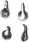
Fig. 138.—Gastrodiscus hominis.
Slightly magnified. (After Lerckart.)
Genital Pore.—About the middle of the conical
anterior portion. (It appears to be surrounded by a
muscular sucker.) Leiper (1913) describes the ducts
as discharging at the tip of a large fleshy papilla, the
surface of which bears cuticular bosses.
Testes much lobed, the anterior is smaller than the posterior and
lies at about the level where the anterior conical portion joins the
disc. The posterior testis just in front of the anterior margin of the
acetabulum separated from it by the ovary. The ovary, somewhat
oval in shape or slightly constricted in the middle, lies slightly to the
right of the median line. Dorsal to it lies the well-developed shell
gland, Laurer’s canal opening in front of the excretory bladder.
The excretory bladder is a long sac with its opening at its posterior
extremity about the level of the middle of the acetabulum. The
vitellaria are restricted in extent. They do not extend forward
beyond the anterior border of the posterior testis. They are best
developed in the area between the acetabulum and the termination
of the gut cæca.
The eggs are oval and measure 150 µ in length by 72 µ in
breadth.
Habitat.—Cæcum and large intestine of man. Also in the pig
(5 per cent.) in Annam.
Distribution.—This parasite has been recorded from Assam (not
uncommon), British Guiana (Indian immigrants), and Cochin China.
Gastrodiscus ægyptiacus, Cobbold, 1876, and G. secundus, Looss,
1907, occur in the horse; G. minor, Leiper, 1913, in the pig in
Nigeria and Uganda.
Family. Fasciolidæ, Raill., 1895.
Sub-family. Fasciolinæ, Odhner, 1910.
Genus. Fasciola, L., 1758.
The ventral sucker is situated at the level of the junction of the cone with the
body, viz., at the level of the “shoulder,” and is large and powerful. The cuticle
is covered with strong spines; the gut cæca run in the mid-line to the hind end,
and are provided with numerous long lateral and fewer and shorter median branches.
The ovary lies on one side in front of the transverse vitelline duct; the testes
lie obliquely one behind the other. The uterus, in the shape of a rosette, lies
in front of the genitalia. Laurer’s canal is present; the vesicula seminalis lies
in the cirrus pouch; the ova are large, not very numerous, and only develop after
they have been deposited. Parasites of the biliary ducts of herbivorous animals.
Fasciola hepatica, L., 1758.
Syn.: Distomum hepaticum, Retz., 1786; Fasciola Humana, Gmel., 1789;
Distomum caviæ, Sons., 1890; Cladocœlium hepaticum, Stoss., 1892.
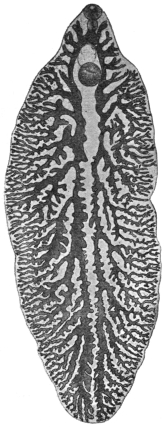
Fig. 139.—Fasciola hepatica,
L. From a specimen
that is not yet mature,
showing the gut and its
branches. 5/1.
Length 20 to 30 mm., breadth 8 to 13 mm., cephalic cone 4 to 5 mm.
in length and sharply differentiated from the body by a shoulder on
each side. Spines in alternating transverse rows and extending on
the ventral surface to the posterior border of the testes, and on the
dorsal surface not quite so far. The spines are smaller on the cephalic
cone than on the posterior part of the body, where they are discernible
with the naked eye. The suckers are hemispherical, and near each
other; the oral sucker is about 1 mm. and the ventral sucker about
1·6 mm. in diameter. The pharynx, which includes almost the entire
œsophagus, measures 0·7 mm. in length and 0·4 mm. in breadth.
The intestine bifurcates at the limit of the cephalic cone and the
branches are even here furnished with diverticula directed outwardly.
The ovary is ramified and situated in front of the transverse vitelline
duct, usually on the right side; the shell gland lies near the ovary in
the median line; posterior to the transverse vitelline ducts are the
greatly ramified testes, which occupy the greater portion of the
posterior part of the body, with the exception
of the lateral and posterior border; the long
vasa efferentia only unite as they enter the
cirrus pouch. The vitellaria occupy the sides
of the posterior part of the body, commencing
at the level of the ventral sucker and uniting
behind the testes. The ova are yellowish-brown,
oval, operculated, 130 µ to 145 µ in length, 70 µ
to 90 µ in breadth (average size 132 µ by 70 µ).
The Liver Fluke inhabits the bile-ducts of
numerous herbivorous mammals (sheep, ox,
goat, horse, ass, rabbit,268 guinea-pig, squirrel,
beaver, deer, roe, antelope, camel, kangaroo,
and others), and is distributed over the whole
of Europe, though not to an equal extent. It
is further known in North Africa, in North and
South America, as well as in Australia; it is
also found in Asia, as it has been reported
from Japan, China, and Tonkin (Gaide, two
cases in man). In some districts of Germany
it is very frequent, and the slaughter-house
statistics of various places show that it is of
daily occurrence. Fasciola magna occurs in
herbivora in America.
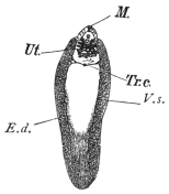
Fig. 140.—Fasciola hepatica. M., mouth;
Ut., uterine rosette; Tr.c., transverse vitelline
ducts uniting to form a vitelline receptacle in
the mid-line; E.d., longitudinal vitelline ducts;
V.s., vitellaria. The clear space in the centre
represents the position of the ramifying testes
and part of the gut. Natural size. (Mull. fluid,
alcohol, creosote, Canada balsam.)
The liver fluke, however, is by no means a
harmless parasite, for it produces in domestic
animals, more especially in sheep, a disease of the liver that appears
epidemically in certain years and districts, and commits great ravages
amongst the flocks.
[The following records show the enormous loss caused in sheep
by this parasite. In 1812, in the Midi, principally in the Departments
of the Rhône, Herault, and Gard, the disease was rampant; 300,000
sheep perished in the Arles territory, and 90,000 in the Arrondissements
of Nîmes and Montpellier. In 1829 and 1830, in the Department
of the Meuse and near localities, not only sheep but oxen died in
enormous numbers; for instance, in the Arrondissement of Verdun
out of 50,000 sheep 20,000 died, and out of 20,000 cattle 2,200 died.
In England, in 1830, 2,000,000 sheep were carried off; whilst in 1862
60 per cent. of the sheep died in Ireland; and in 1879 over 300,000
were lost in England; whilst as late
as 1891 one owner in the same
country lost over 10,000 sheep (Live
Stock Journal, October 30, 1891).—F.
V. T.]
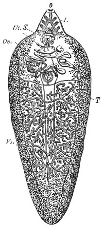
Fig. 141.—Fasciola hepatica, L. I.,
intestine; Vs., vitellaria; Ov., ovary;
O., oral aperture; Ut., uterus; S., ventral
sucker; T., testes. In front of the
testes are seen the transverse vitelline
ducts uniting to form the pyriform vitelline
receptacle. Immediately in front of
this the spherical shell gland. The two
vasa efferentia can also be seen running
up in the mid-line. The branches of the
gut are only shown in the cephalic cone.
(After Claus.)
The disease usually commences
towards the end of summer with an
enlargement of the liver, induced by
the invasion of numerous young
flukes; in the autumn and winter the
animals suffer from the consequences
of disordered biliary secretion; they
become feverish, emaciated, and
anæmic, and lose their appetite.
In consequence of the consecutive
atrophy of the liver, œdema and
ascites set in, and many animals succumb to this “liver rot.” On
examination the liver is found to be shrunken, the bile-ducts are
enormously dilated and in parts saccular and full of flukes. Should
the animals survive this stage, spontaneous recovery ensues in
consequence of the flukes commencing to leave the liver in the
spring, but the liver remains changed and its sale is prohibited269 when
the changes are extensive.270
[The following stages may be noticed in sheep suffering from
fascioliasis. Gerlach recognized four stages, based on the varied
relations that the flukes contract with the liver of their host. These
periods are sometimes very marked, but at others, owing to subsequent
infections, the features become merged and so obliterated. But
when a single infestation occurs they are very marked.
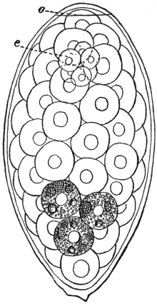
Fig. 142.—Fasciola hepatica: egg from liver
of sheep. o, operculum, e, segmenting ovum.
The rest of the space is occupied by yolk cells,
the granules in three only being shown. × 680.
(After Thomas.)
[The first period is called the
PERIOD OF IMMIGRATION. This
occurs at the fall of the year and
generally passes unperceived, as
the young flukes do little harm
to the liver. It varies from four
to thirteen weeks. Gerlach has
remarked upon cases of death
from apoplexy at this period.
[The second period is the
PERIOD OF ANÆMIA. This occurs
in November and December.
The sheep at first fatten rapidly,
but later the mucous membranes
become pale and of a yellowish
hue, and the sheep become sluggish and cease to feed. The fæces
are normal, but may contain fluke ova.
[The third period is the PERIOD OF WASTING. This corresponds
with the beginning of January—about three months after the
entry of the larvæ. Emaciation now becomes very marked, the
skin and mucous membranes blanched, temperature variable and
marked by an irregular curve; respiration laboured and quick;
appetite regular; abortion frequently occurs in pregnant ewes;
pressure on the back causes the animals to fall; local œdemas
occur, the most perceptible in the submaxillary space, extending
below the larynx and over the cheeks and parotids (called
“bourse,” “boule” in France; “watery poke” or “cockered” in
England). Death usually occurs at this period, but a fourth
stage may occur.
[The fourth period is the PERIOD OF MIGRATION OF THE
FLUKES. This is a period of convalescence and recovery, generally
in May and June.—F. V. T.]
Oxen suffer less in general, but even in these animals “stray”
hepatic flukes are occasionally found in the lungs, enclosed in thick-walled
cysts.
Pathological Anatomy.—The bile-ducts are conspicuous on the
surface of the liver. They are thickened and much dilated and in
parts saccular, and considerable atrophy of the liver cells accompanies
the condition. Histologically there is immense proliferation
of the epithelium of the bile-ducts leading to “adenomata.”
Fig. 143.—Limnæus truncatulus, Müll.,
the intermediate host of Fasciola hepatica.
a., natural size; b., magnified. (From
Leuckart.)
The LIFE-HISTORY of the liver fluke was discovered by
R. Leuckart and P. Thomas. According to these investigators the
elongated miracidium (fig. 131, a) ciliated all over develops from the
eggs a few weeks after the latter (fig. 142) have reached the water, and
after it has become free the embryo penetrates and becomes a sporocyst
(fig. 131, b) in a water-snail (Limnæus truncatulus, Müll. =
L. minutus, Drap.) that is common in fresh water, and can live in the
smallest collection of water as well as in fields that have been flooded.
The sporocyst first of all produces rediæ, which remain in the same
host (and under certain circumstances, e.g. in summer, these develop a
second generation of rediæ), and these finally form cercariæ (fig. 134).
The latter become encysted on blades of grass and are taken up by
the respective hosts with their food; this takes place towards the
end of summer, while the sheep feeding on the pasture land in the
spring spread the eggs of the fluke, and sometimes the fluke itself,
by passing them with their fæces.
In districts where Limnæus truncatulus is absent, analogous
species act as the intermediary hosts, of which one example
according to Lutz is Limnæus oahuensis in the Sandwich Islands.
[The host in Europe is Limnæus truncatulus. This snail
extends from Siberia to Sicily and Algeria, and according to
Captain Hutton is a native of Afghanistan. It also occurs in
Thibet, Amoor, Morocco, Tunis, Canary Islands and the Faroe
Islands. It deposits its eggs or spawn upon the mud around ponds,
ditches and streams. The eggs are laid in batches of thirty to a
hundred, each snail laying as many as 1,500 eggs; they are united
into strips of a gelatinous substance. In about two weeks young
snails appear. It is amphibious, being more frequently met with out
of the water than in it. It occurs in elevated spots as well as in
low-lying districts. Moquin-Tandon found it at 4,000 feet in the
Pyrenees. In the allied species, L. peregra, the fluke will develop
up to a certain stage, but never completes all its varied phases.
[In South America the host is probably Limnæus viator, Orb.,
and in North America Limnæus humilis, Say.—F. V. T.]
In human beings as well as in some of the mammals quoted
above, the liver fluke is only a casual parasite, and hitherto only
twenty-eight cases have been observed in man; the infection was
mostly a mild one and there were no symptoms, or only very
trifling ones; a few isolated cases were only discovered post mortem.
Occasionally, however, even when the infection was inconsiderable,
severe symptoms were set up, which in isolated cases led to death.
The symptoms (enlargement and painfulness of the liver, icterus)
merely pointed to a disease of the liver.
Diagnosis can only be established by finding eggs in the fæces.
Care should be taken not to confuse them with those of Dibothriocephalus
latus.
Halzoun.
Fig. 144.—Young Fasciola
hepatica, soon after
entry into the liver. The
intestinal cæca have lateral
diverticula. Magnified.
(From Leuckart.)
In North Lebanon, the liver fluke is, according to A. Khouri,
a frequent parasite of man, not in the liver, however, but in the
pharynx. The occurrence in this unusual site
is effected by the eating of raw infected livers,
especially those of goats (Capra hircus). The
flukes thus taken in do not all reach the
stomach, where they would be soon killed, but
some of them attach themselves to the pharyngeal
mucosa and to the adjoining parts, and
there cause inflammation and swelling, which
lead to dyspnœa, dysphagia, dysphonia and
congestion of the head, sometimes even to
still more severe symptoms, and even death.
The affection termed “Halzoun” lasts some
hours or several days, and after vomiting
recovery sets in. In other cases man becomes
infected in the usual way by ingesting cysts attached to grass or
the underside of leaves of plants (e.g., Rumex sp.), where they
are overlooked from
their scanty size (0·2 to
0·3 mm.).
As the liver fluke
feeds on blood it is
possible that it also
reaches, particularly
when young, the circulatory
system, and cases
have been known in
which it has been carried
by the blood into organs
far from its original situation.
Such cases also
have been repeatedly
observed in men. Probably
the parasite described
by Treutler,
1793, as Hexathyridium
venarum, which protruded
from the ruptured
anterior tibial vein
of a man, was a young
liver fluke. A few adult
specimens were found
by Duval in the portal
and other veins post
mortem at Rennes (1842)
in a man, aged 49, and
a similar statement is
reported by Vital from
Constantine (1874).
Giesker, in 1850, found
two hepatic flukes in a
swelling on the sole of
the foot of a woman.
Penn Harris states that
he observed six specimens
in Liverpool in a
spontaneously ruptured
abscess of the occiput
of a two months old
infant. Another case
which, like the previous
one, is reported by Lankester,271 relates to a sailor who suffered from
an abscess behind the ear, and from which a liver fluke was expelled.
Finally, Dionis de Carrières reports the case of a man, aged 35, in
whose right hypochondriac region a tumour the size of a pigeon’s egg
had formed, and from which a young liver fluke was extracted.
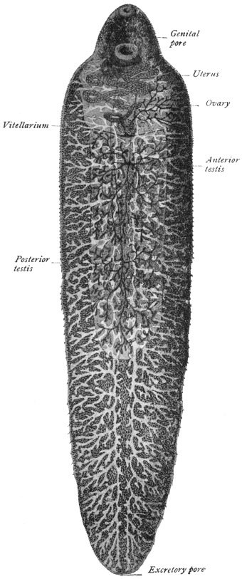
Fig. 145.—Fasciola gigantica. × 6 1/2
(After Looss.)
From such records it is not impossible that Distomum oculi
humani, Ammon, 1833, as well as Monostomum lentis, v. Nordm.,
1832, may have been very young hepatic flukes that had strayed.
Ammon found four specimens (length 0·5 to 1 mm.) of his species
(named Distomum ophthalmobium by Diesing in 1850) between the
opaque lens and the capsule of a five months old child in Dresden,
and von Nordmann discovered his Monostomum lentis to the number
of eight specimens (only 0·3 mm. in length) in the opaque lens of
an old woman. Minute white bodies which Greef found in the cortex
of the lens of a fisherman, aged 55, removed on account of cataract,
were with some reserve regarded as Trematode larvæ. The fact that
Ammon found that the intestinal cæca of the worm discovered by
him had no lateral branches does not negative the above opinion, as
in the liver fluke the intestinal cæca are originally unbranched, and
according to Lutz they only develop lateral ramifications later, between
the twelfth and twenty-second day of infection (fig. 144).
Fasciola gigantica, Cobbold, 1856.
Syn.: Distomum giganteum, Diesing, 1858; Fasciola gigantea, Cobbold, 1858;
Cladocœlium giganteum, Stoss., 1892; Fasciola hepatica var. angusta, Raill., 1895;
Fasciola hepatica var. ægyptiaca, Looss, 1896.
This species is closely allied to Fasciola hepatica, but is distinguished
by its elongated body, short cephalic cone, almost parallel
sides, larger ventral sucker, which is also closer to the oral sucker,
and by its larger eggs. Length up to 75 mm., width up to 12 mm.
Oral sucker 1 to 1·2 mm., ventral sucker up to 1·7 mm. in diameter.
Eggs 150 µ to 190 µ long by 75 µ to 90 µ broad.
Habitat.—Bile-ducts of Camelopardalis giraffa, Bos taurus, Bos
indicus, Bos bubalis, Ovis aries and Capra hircus.
Distribution.—Africa.
This species has once been observed in man by Gouvea, in Rio de
Janeiro, in a French naval officer who became ill with fever, cough
and slight blood-spitting. The lungs were normal except for a
sharply circumscribed spot at the base of the left lung. Twenty
days later during a fit of coughing the patient spat up a fluke 25 mm.
long, characterized by its slender aspect and by the size of its ventral
sucker, and its close proximity to the oral sucker. Considering the
fact that Gouvea’s patient had spent many weeks in July of the same
year in Dakar (Senegambia), where according to Railliet Fasciola
gigantica is common in slaughtered animals, and considering also
the characters of the fluke, Railliet rightly assumes that one had to
do with the African giant fluke and that the patient had infected
himself in Dakar.
Sub-family. Fasciolopsinæ, Odhner, 1910.
Genus. Fasciolopsis, Looss, 1898.
Ventral sucker large, and elongated
posteriorly into a sac. Cirrus pouch
long and cylindrical, its greatest length
being occupied by the sinuous tubular
seminal vesicle, on which exists a peculiar
cæcal appendage. Laurer’s canal present.
Fasciolopsis buski, Lank., 1857.
Syn.: Distomum buski, Lank., 1857;
Dist. crassum, Cobbold, 1860, nec v. Sieb.,
1836.
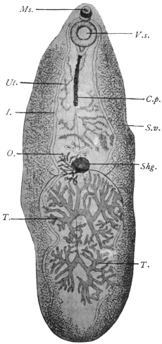
Fig. 146.—Fasciolopsis buski, Lank. V.s.,
ventral sucker; C.p., cirrus pouch; I., intestinal
fork; S.v., vitellaria; T., testes; O.,
ovary; Ms., sucker; Shg., shell gland;
Ut., uterus. Magnified. (After Odhner.)
The length of the body varies;
it may measure 24 to 37 or even
attain 70 mm.; the breadth is from
5·5 to 12 to 14 mm. In the pig the
fresh parasites measure, smallest,
12 to 8 mm.; largest, 35 to 16 mm.
(Mathis and Léger). Skin without
spines, but according to Heanly
always present in man and pig
specimens. The oral sucker
measures 0·5 mm. in diameter;
the ventral sucker is three to four
times as large; the pharynx is
globular, 0·7 mm. in diameter;
the prepharynx is provided with a
sphincter; the intestinal cæca extend
to the posterior border with
two characteristic curves, one at the anterior border of the anterior
testis, the other between the two testes. The genital pore is at the
anterior border of the ventral sucker; the cylindrical cirrus pouch
extends from behind the ventral sucker to half-way to the shell gland.
The seminal vesicle extends forwards within the cirrus pouch as a
convoluted tube. From its anterior portion is given off the cæcal
appendage, which has itself short lateral diverticula.
It runs backwards, ending blindly
about 0·5 mm. from the posterior end of the
cirrus sac. The seminal vesicle is continued as
the pars prostatica (?) 0·5 mm. long, and this
by the very short ejaculatory duct (13 µ), and
finally by the fairly long cirrus, which is beset
with very fine spines except at either extremity.
The ovary and shell gland are situated at about
the middle of the body with the testes behind
them, and the uterus in front. The vitellaria
extend from the ventral sucker to the posterior
border. The eggs measure 120 µ to 130 µ in
length and 77 µ to 80 µ in breadth, and resemble
those of Echinochasma sp. in dogs. The larval
stages are said to occur in shrimps.
Habitat.—Intestine of pig and man.
Distribution.—In man: India, Siam, China,
Assam, Sumatra. It is common in Cochin
China (16 out of 133 Annamites, Noc.), in
Tonkin very rare. Dr. J. Bell has sent me
[J. W. W. S.] human specimens from Hong Kong. In pigs: very
common in South China (Heanly). Common in pigs in Hong Kong.
Sixteen out of 248 pigs (i.e., 6 per cent.) infected in Hanoi.
Fasciolopsis rathouisi, Ward, 1903.
Syn.: Distomum rathouisi, Poirier, 1887.
Fig. 147.—Fasciolopsis
rathouisi, Poir.: the
mouth at the top, and under
it the genital pore and ventral
sucker, behind which
again is the uterus. The
vitellaria are at the sides,
and posteriorly in the central
field the ramified testes; the
ovary is in front of the right
testis. (After Claus.)
Fifteen to 19 mm. long by 8·5 to 10·5 mm. broad by about 3 mm.
thick. Skin with spines (Leiper). Bluntly oval or elliptical with
short cephalic cone which is absent in Fasciolopsis buski. Oral sucker
subterminal, 0·25 to 0·29 mm. broad by 0·2 mm. in antero-posterior
diameter. Distant from ventral sucker by about twice its diameter.
Ventral sucker 1·32 to 1·38 mm. broad by 0·68 to 0·7 mm. in antero-posterior
diameter. Œsophagus extremely short. Cirrus sac not
conspicuous and straight as in Fasciolopsis buski, but is convoluted.
Testes one behind the other (according to Poirier they lie beside one
another), more compactly branched, broader and denser than in
Fasciolopsis buski. Ovary on right side, small, coarsely branched.
Uterus in broad, closely grouped coils, packed with ova anterior to
ovary. Vitellarian acini more numerous and somewhat differently
distributed. Eggs 150 µ by 80 µ, thin shelled. [H. B. Ward, who has
examined this species, and from whose account the above is mainly
taken, considers that it is a good species, although the differences
between it and Fasciolopsis buski are slight, while Odhner, who examined
the original species, is of the opposite opinion.—J. W. W. S.] The
parasite appears to cause diarrhœa, wasting and occasionally jaundice.
Habitat.—Intestine of man.
Distribution.—China, common in some parts (Goddard).
Fasciolopsis goddardi, Ward, 1910.
Twenty-one to 22 mm. long, 9 mm. broad. Skin with spines
(Leiper). Uterus very closely coiled, most striking character is the
large size of the vitelline acini. Imperfectly known.
Distribution.—China (Shanghai).
Fasciolopsis fülleborni, Rodenwaldt, 1909.
The fully extended fluke is tongue-shaped, 50 by 14 mm.; two
contracted specimens measured 40 by 15 mm. and 30 by 16 mm.
respectively. Skin without spines, with according to Leiper cephalic
cone not clearly defined. Oral sucker circular, 0·75 mm. in
diameter, slightly larger than that of Fasciolopsis buski. Ventral sucker
2·6 mm. in diameter (that of Fasciolopsis buski 1·6 to 2 mm.). Length
2·9 mm. (as in Fasciolopsis rathouisi), the excess of length over breadth
being due to the posterior elongated sac-like prolongation of the
sucker. Prepharyngeal sphincter present. Pharynx 0·7 mm. in
diameter. Œsophagus practically absent. Gut cæca similar to those
of Fasciolopsis buski.
Testes—regularly branched, separated by an incurving of the cæca,
the anterior occupying a smaller area than the posterior.
Ovary—very small, as in Fasciolopsis buski, on the right side.
Shell Gland—almond-shaped, 2·3 by 1·2 mm. In Fasciolopsis buski
it is round and smaller, 1 to 1·5 mm. in diameter.
Vitellaria—similar in distribution to those of Fasciolopsis buski, but
the acini are strikingly small.
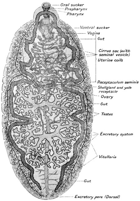
Fig. 148.—Fasciolopsis fülleborni, ventral aspect. (After Fülleborn.)
Cirrus Sac—is the most characteristic feature of this species. It is
a powerfully built, convoluted sac standing out clearly on the body.
It is not a uniform, straight cylinder 0·25 to 0·33 mm. in diameter, as
in Fasciolopsis buski, but even in fully extended flukes is typically
convoluted. It is 1 mm. thick in the middle, but in other parts varies
much from this. The posterior end of the cirrus sac is at two-thirds
or more of the distance from ventral sucker to shell gland. In the
case of Fasciolopsis buski the posterior end of the sac only extends
half-way.
Seminal Vesicle—has a peculiar convoluted, saccular and angular
course, but the cæcal appendage characteristic of the genus appears
to be absent!
Excretory System.—The main stem gives off very regular transverse
branches which are well seen posteriorly.
Eggs.—100 µ by 73 µ. Thin shelled.
Habitat.—Intestine. Mahommedan from Calcutta.
[It is evident that a re-examination of fresh material is required
before the validity of all these species can be accepted.—J. W. W. S.]
Family. Troglotremidæ, Odhner, 1914.
Genus. Paragonimus, Braun, 1899.
Body egg-shaped or somewhat elongated, generally more broadly rounded in
front than behind. Covered all over with spear-shaped spines arranged in groups.
Gut cæca winding with dilatations or constrictions in parts. Ventral sucker in or
in front of the middle of the body. Excretory bladder cylindrical, very long and
broad, reaching in front to the bifurcation of the gut. The lateral excretory canals
join the bladder only a little in front of the excretory pore. Genital pore median
just behind the ventral sucker. Genital sinus duct-like. Cirrus sac absent. Male
terminal organs very small. Ejaculatory duct present. Testes and ovary deeply
lobed, the testes in or just behind the middle, the ovary somewhat laterally placed
just behind the ventral sucker. Uterus forms a coil behind the ventral sucker.
Eggs rather large, thin shelled, the ovarian cell still unsegmented on deposition.
Receptaculum seminis, small.
Parasitic in the lungs of mammals, enclosed in cyst-like cavities, generally in
pairs.
Type Species.—P. westermanii in the tiger.
Paragonimus ringeri, Cobb., 1880.
Syn.: Distoma ringeri, Cobb., 1880; Distoma pulmonale, Baelz, 1883;
Distoma pulmonis, Suga, 1883.
Fig. 149.—Paragonimus
ringeri, Cobb.: to the right,
dorsal aspect; to the left, ventral
aspect. Natural size. (After
Katsurada.)
The body is of a faint reddish-brown colour and plump oval
shape. The ventral surface a little flattened; 7·5 to 12 mm. in length,
4 to 6 mm. in breadth, and 3·5 to 5 mm. thick (in man). The oral
sucker (0·75 mm.) is subterminal; the ventral sucker (0·8 mm.)
somewhat in front of the middle of the body. Pharynx spherical,
0·3 mm. in diameter, or 0·4 by 0·3 mm.; œsophagus, 0·02 mm.;
intestinal cæca convoluted, asymmetrical, the first part having the
same structure as the œsophagus. The
cuticle is covered with spines in groups;
the excretory pore opens at the posterior
end rather on the ventral surface, the excretory
ducts open into the elongated bladder
at the hind end near the pore. Genital
pore behind the ventral sucker and
median. Genital sinus 0·2 mm. long
with thick wall, ejaculatory duct 0·13 mm.,
pars prostatica 0·2 mm., seminal vesicle duct-like of irregular outline.
Behind the sucker the ovary on the left, and the closely packed
uterine coil on the right (though amphitypy of these two organs is
common); the two irregularly lobed testes lie side by side posteriorly.
Vitellaria extensive, leaving only a median dorsal and ventral space
free. Seminal receptacle probably absent; Laurer’s canal present.
The eggs are oval, brownish-yellow, fairly thin shelled, and measure
on an average 81·2 µ by 49·2 µ.
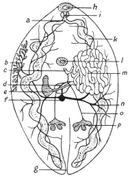
Fig. 150.—Paragonimus ringeri, Cobb.:
diagram of the internal organs. a, œsophagus;
b, vitellaria (a portion only shown);
c, common genital duct; d, shell gland with
oviduct, Laurer’s canal and vitelline duct; e,
ovary; f, vitelline receptacle; g, excretory
pore; h, oral sucker; i, pharynx; k, gut; l,
ventral sucker; m, uterine coils; n, vitellarian
ducts; o, vas efferens; p, testis. (After Kubo.)
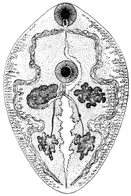
Fig. 150a.—Paragonimus westermanii,
Kerb.: seen from the ventral surface.
Mouth, pharynx, intestinal cæca, at the sides
of which the vitellaria are observed. The
genital pore is behind the ventral sucker, and
next to it, on the left, the ovary; on the right,
the uterus; the two testes posteriorly; the
excretory vessel in the middle, 10/1. (After
Leuckart.)
The following species are also known:—P. westermanii, Kerb.,
1878, in the tiger, and P. kellicotti, Ward, 1908, in the pig, dog, and
cat (N. America). Ward and Hirsch give the following differences
between the spines of the three forms:—
| P. ringeri. | P. westermanii. | P. kellicotti. |
| Shape | Chisel-shaped, mod-
erately heavy. | Lancet-shaped,
very slender. | Chisel-shaped,
heavy. |
| Distribution | Circular rows, in
groups. | Circular rows,
in groups. | Circular rows,
singly. |
Two other species, P. rudis, Diesing, 1850, in a Brazilian otter
(Lutra brasiliensis) and P. compactus, Cobbold, 1859, in the Indian
ichneumon, are but little known.
Habitat.—Lungs, pleuræ, and especially the bronchi of man and
dog. The alleged occurrence (of eggs) in other organs may be due
to confusion with those of Schistosoma japonicum.
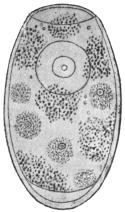
Fig. 151.—Egg of Paragonimus
ringeri, Cobb.,
from the sputum. Showing
the ovarian cell and vitelline
cells and granules.
1,000/1. (After Katsurada.)
Distribution.—China, Korea, and especially in Japan, where, according
to Katsurada, there are no districts that are entirely free from pulmonary
flukes. The mountainous provinces of Okayama, Kumamoto,
Nagano and Tokushima are the principal
centres.
Pathology.—The number present in the
lung varies from two to twenty, about.
Usually one cyst contains one worm, but in
the dog each cyst contains two. The cysts
admit the tip of the finger, and have a fibrous
wall 1 mm. thick. They originate partly from
dilatation of bronchi and bronchioles. Others
arise from the inflammatory reaction of lung
tissue into which the worms have wandered.
The worms and their eggs cause bronchitis
and peribronchitis, catarrhal, hæmorrhagic, or
purulent, and areas of consolidation. Areas
containing eggs in their centre resembling
tubercle nodules are not uncommon, and
extensive cirrhosis of the lung may be found.
As a result of these changes, emphysema and bronchiectasis also
occur.
As to the development, only the following details are known:
that the eggs, which before segmentation of the ovum reach the open
in the sputum and through being swallowed also in the fæces,
develop in water into a miracidium ciliated all over, which hatches
and swims about freely. According to Manson this takes place in
four to six weeks.
Sub-family. Opisthorchiinæ, Looss, 1899.
Genus. Opisthorchis, R. Blanch., 1845.
Opisthorchiinæ with lobed testes. Laurer’s canal present. Parasitic in the bile-ducts
of mammals and birds.
Opisthorchis felineus, Riv., 1885.
Syn.: Distoma conus, Gurlt, 1831 (nec Creplin, 1825); Distoma lanceolatum,
v. Sieb., 1836, v. Tright, 1889 (nec Mehlis, 1825 = Fasciolo lanceolata, Rud., 1803);
Distoma sibiricum, Winogr., 1892; Distoma tenuicolle, Mühl., 1896.
Fig. 152.—Egg
of Opisthorchis
felineus,
Riv.
830/1.
This parasite is yellowish-red in the fresh condition, and almost
transparent. The body is flat, with a conical neck at the level of the
ventral sucker marked by a shallow constriction; this, however,
is only noticeable in fresh and somewhat contracted specimens.
Posteriorly to the ventral sucker the lateral borders run fairly parallel;
the posterior end is either pointed or rounded off. The length and
breadth vary according to the contraction, being usually 8 to 11 mm.
by 1·5 to 2 mm. The suckers are about one-fifth to one-sixth of
the length of the body distant from each other, and of about
equal size (0·23 to 0·25 mm.). The œsophagus is hardly any longer
than the pharynx, which lies close behind the oral sucker; the
intestinal cæca reach almost to the posterior border and are often
filled with blood. The excretory pore is at the posterior extremity,
and the excretory bladder forks in front of the anterior testis. The
testes in the posterior fourth of the body lie obliquely one behind
the other; the anterior one has four lobes, the posterior one five
lobes; the ovary is in the median line transversely, simple or
slightly lobed; behind it lies the large pear- or retort-shaped receptaculum
seminis and Laurer’s canal. The uterus is in the median field.
The vitellaria occupy the fairly broad lateral areas, in about the
central third of the body, beginning behind the ventral sucker and
terminating at about the level of the ovary; the acini are small and
arranged in groups of seven to eight, separated by interstices. The
genital pore is close in front of the ventral sucker. The eggs are
oval with sharply defined operculum at the pointed pole, 30 µ, by 11 µ.
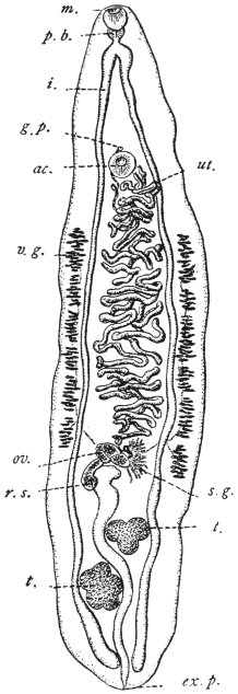
Fig. 153.—Opisthorchis felineus:
from the cat. m., mouth; p.b.,
pharynx; i., gut; g.p., genital pore;
ac., ventral sucker; ut., uterus; v.g.,
vitellarium; ov., ovary; s.g., shell
gland; r.s., receptaculum seminis;
t. testes; ex. p., excretory pore.
(After Stiles and Hassall.)
This species, which is frequently confused with others, inhabits the gall-bladder
and bile-ducts of the domestic cat especially; but is also found in the
dog, in the fox, and in the glutton (Gulo borealis). It has been observed in
France, Holland, North Germany (being particularly frequent in East Prussia), in
Russia, Scandinavia, Siberia, Japan, Tonkin, Hungary, and Italy. The North
American form (from cats and Canis latrans) is a distinct species (Opisthorchis
pseudofelineus).
In man this species was first found by Winogradoff in Tomsk (nine
cases), then by Kholodkowsky in a peasant from the neighbourhood
of Petrograd who had travelled a
great deal in Siberia, and finally by
Askanazy in five persons who were
natives of the East Prussian district of
Heydekrug. In Tomsk, Opisthorchis
felineus is the most frequent parasite
of man that comes under observation
at post mortem (6·45 per cent.), whereas
Tænia saginata has only been found
in 3·2 per cent., Echinococcus in 2·4 per
cent., Ascaris lumbricoides in 1·6 per
cent., and Oxyuris vermicularis in 0·8
per cent. of the autopsies. In the district
of Heydekrug, however, the species in
question is also frequent, as in a few
years five cases came to our knowledge
(of which three were diagnosed by the
discovery of the eggs in the fæces).
In none of Winogradoff’s nine cases
had the death of the patient been caused
direct by the parasites, yet more or less
extensive changes in the liver were
found in all of them; such as dilatation
of the bile-ducts with inflammation and
thickening of their walls, and foci of
inflammation or atrophy in the liver
substance; icterus was present five
times and atrophy of the liver an equal
number of times; ascites
was observed three times,
and in two cases, probably
of recent date, the organ
was enlarged. The number
of parasites found
fluctuated between a few
and several hundreds.
In two of Askanazy’s
cases, which he examined
more closely, carcinoma
which had developed at
the places most invaded by flukes was found at the post-mortem, so
that perhaps there may be grounds for the connection which the author
seeks to establish between cancer of the liver and the changes induced
by the parasites; these changes consist of numerous and even ramified
proliferations of the epithelium of the
biliary duct into the connective
tissue, which is likewise proliferated.
The number of worms found in one
case amounted to over 100; in a
second case, in which the parasites
had also invaded the pancreatic duct,
their number was even larger.
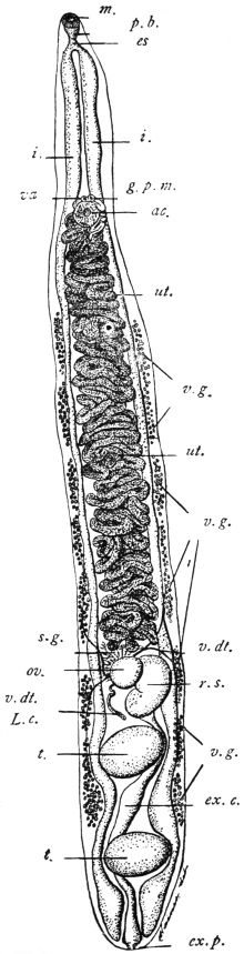
Fig. 154.—Opisthorchis pseudofelineus: from the
bile-duct of the cat (Iowa), m., oral sucker; p.b.,
pharyngeal bulb; es., œsophagus; i., intestine; va.,
vagina; g.p.m., male orifice; ac., ventral sucker;
ut., uterus; v.g. vitellarium; s.g., shell gland; v.dt.,
vitelline duct; ov., ovary; r.s., receptaculum seminis;
L.c., Laurer’s canal; t., testis; ex.c., excretory
bladder; ex.p., excretory pore. (After Stiles.)
Winogradoff as well as Askanazy
found isolated flukes in the intestine
also.
Unfortunately, nothing much is
known of the history of the development
of Opisthorchis felineus; we only
know that when deposited the eggs
already contain a ciliated miracidium,
which, however, according
to my experience, does not hatch out
in water, but only after the entry of
the eggs into the intestine of young
Limnæus stagnalis; no further development,
however, occurs. Winogradoff
states that he has seen the
miracidia hatch after the eggs had
been kept in water for a month at
37° C.; and has even observed free
miracidia in the bile of man and of
a dog respectively. Although the
whole post-embryonal development
of the cat fluke remains yet to be
investigated, Askanazy by a series of
experiments on cats and dogs has discovered
the mode of infection. The
intermediate hosts are fish, and
mainly the ide, in this country called
Tapar (Idus melanotus, H. and Kr.),
and of subsidiary importance the
roach (Leuciscus rutilus). Both species of fish as well as others are
readily eaten raw by man on the Courland lagoon (Baltic). It is, moreover,
significant that those persons whom Askanazy found infected
with the cat fluke were also infected with Dibothriocephalus latus, the
intermediate host of which is also fish (Lota sp., Esox sp., Perca sp.).
In one of his nine cases Winogradoff also saw a small fluke
covered all over with spines, which he conjectured to be the young
stage of Opisthorchis felineus; as, however, according to my experience,
this species, even in smaller specimens, is always without spines,
the above hypothesis cannot be accepted. It is much more probable
that one of the other species that also invade the liver of cats may
accidentally be introduced into man; we know, in fact, that Metorchis
albidus, Braun, and Metorchis truncatus, Rud., are both covered with
spines. As, however, the spines of the first-named species are rather
apt to fall off, and also as it possesses a different shape (spatula-shaped),
it may be assumed that probably Winogradoff had found Metorchis
truncatus, Rud., 1819, in his patient.
Genus. Paropisthorchis, Stephens, 1912.
Structure as in Opisthorchis, except that the ventral sucker and genital pore occur
on the apex of a process or pedicle projecting from the anterior portion of the body.
This process is about 1/2 mm. long, and is retractile.
Paropisthorchis caninus, Barker, 1912.
Syn.: Distoma conjunctum, Lewis and Cunningham, 1872; Opisthorchis noverca,
M. Braun, 1903 (pro parte); Opisthorchis caninus, Barker, 1912 (?).
Length varies from 2·75 to 5·75 mm. in preserved specimens, average
3·6 to 5·2 mm. Body uniformly spinose, though as a rule spines are
not present on the pedicle. Body slightly concavo-convex, the concavity
being ventral. Oral sucker 0·28 mm. Pharynx 0·224 by
0·184 mm. Œsophagus 0·04 mm. Ventral sucker 0·176 mm. in
diameter. Pedicle about 1/2 mm. long, may be completely retracted.
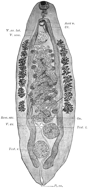
Fig. 155.—Paropisthorchis caninus: from the bile-ducts of the pariah dog, India. Acet. v.,
ventral sucker; Ut., uterus; V. ex. lat., longitudinal excretory duct; V. sem., seminal vesicle;
Sem. rec., seminal receptacle; Ov., ovary; V. ex., excretory bladder; Test. l., left testis;
Test. r., right testis; P. ex., excretory pore. × 40. (After Stephens.)
Genital Pore—opens on the apex of the pedicle in front of the
ventral sucker. Its exact position varies with the state of contraction
of the parts. In certain cases it actually opens within the cuticular
border of the sucker, in other cases it opens externally to the sucker
and anterior to it. The opening is covered with scales. The vas
deferens and uterus run alongside one another until they merge near
the apex of the pedicle into a common sinus.
Vitellaria—consist of eight acini on each side, extending from
slightly behind the base of the pedicle to the anterior border of the
ovary, or as far back as a line separating the posterior border of the
ovary from the anterior border of the anterior testis.
Testes.—Anterior testis 0·496 by 0·44 mm.; posterior testis 0·52 by
0·48 mm., usually ovoid, though both may be regularly lobed. The
anterior testis is usually on the left side.
Ovary—multilobular, the lobes 6 to 8 being irregular in size and
shape.
Shell Gland—extensive and diffuse, occupying an area which approximately
corresponds with the loop of the transverse vitelline ducts.
Seminal Receptacle—globular, to the right of and dorsal to the
posterior lobe of the ovary.
Laurer’s Canal—generally
runs from the end of the receptacle
with a single curve
medially and backwards.
Uterine Coils—form loosely
packed transverse coils terminating
slightly in front of
the level of the first vitelline
acini. From here the uterus
passes forwards into the pedicle
to the left and ventral to the
seminal vesicle.
Seminal Vesicle—commences
about the level of the
first vitelline acini. The coils
displace the uterus ventrally and
to the left. In the pedicle the
vesicle diminishes in extent and
lies in its dorsal (anterior) side.
Habitat.—Liver of pariah
dogs, India. In North-Western
Provinces about 40 per cent.
are infected. This fluke appears
to be different from
Amphimerus (Opisthorchis)
noverca in man, as the latter has
not the pedicle on the summit
of which lie the sucker and
common genital pore.
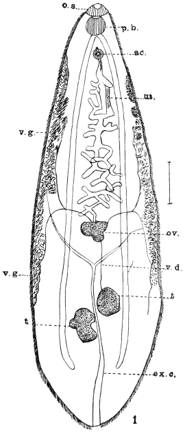
Fig. 156.—Amphimerus noverca, Braun. o.s.,
oral sucker; p.b., pharynx; ac., ventral sucker;
ut., uterus; v.g., vitellarium; ov., ovary; v.d., vas
efferens; ex.c., excretory canal; t., testis. (After
McConnell.)
Genus. Amphimerus, Barker,
1912 (?).
Structure as in Opisthorchis, except
that the vitellaria are separated
into two portions, an ant-ovarial and
a post-ovarial.
Amphimerus noverca, Barker, 1912 (?).
Syn.: Distomum conjunctum, McConnell, 1876 (nec Cobbold, 1859); Opisthorchis
noverca, M. Braun, 1903 pro parte.
At the autopsy of two Mahommedans who died in Calcutta,
McConnell found a large number of Distomata in the thickened and
dilated bile-ducts. The worms were lancet-shaped, covered with
spines, and measured 9·5 to 12·7 mm. in length and 2·5 mm. in breadth.
The two suckers lie very close to one another, the anterior one being
larger than the ventral; the genital pore opens immediately in front
of the ventral sucker; pharynx spherical; intestinal cæca extending far
back. At the commencement of the posterior third of the body the
two testes, somewhat apart, the anterior one roundish, the posterior one
distinctly lobed. The transverse
and slightly lobed ovary in front
of the bifurcation of the Y-shaped
excretory bladder, whence the
uterus, in convolutions barely
spreading beyond the central
field, extends to the pore; the
vitellaria in the lateral areas
commence behind the ventral
sucker and extend to the testes.
Cirrus pouch absent. Eggs oval,
34 µ by 21 µ.
Genus. Clonorchis, Looss, 1907.
Structure as in Opisthorchis, distinguished,
however, by the branched
testes situated one behind the other,
the branches of which ventrally encroach
upon the gut forks; dorsal
to the testes the S-shaped excretory
bladder, the main branches of which,
arising at the level of the bifurcation
of the gut, open into the bladder below
its anterior end. Parasitic in the bile-ducts
of mammals and man.
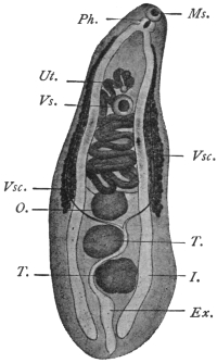
Fig. 157.—Metorchis conjunctus,272 (Syn.:
Distomum conjunctum, Cobb., nec Lew. and
Cunn., nec McConn.): from Canis fulvus.
Vs., ventral sucker; I., intestine; Vsc. vitellaria;
Ex., excretory bladder; T., testes;
O., ovary; Ms., oral sucker; Ph., pharynx;
Ut., uterus. (After Cobbold.)
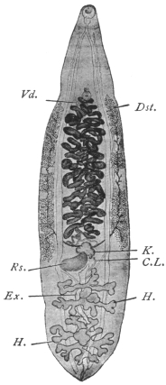
Fig. 158.—Clonorchis sinensis.
C.L., Laurer’s canal; Dst., vitellaria;
Ex., excretory bladder;
H., testes; K., ovary; R.s., receptaculum
seminis; Vd., terminal
section of vas deferens.
Magnified 4 1/2 times. (After
Looss.)
Clonorchis sinensis, Cobbold, 1875.
Syn.: Distoma sinense, Cobbold, 1875; Distoma spathulatum, R. Leuckart, 1876
(nec Rudolphi, 1819); Distoma hepatis innocuum, Baelz, 1883.
In shape resembles Opisthorchis felineus, 13 to 19 mm. long, 3 to
4 mm. broad, at the beginning of sexual maturity 12 to 13 mm. long, 2·5
to 3 mm. broad. Oral sticker 0·58 to 0·62 mm., ventral sucker 0·45 to
0·49 mm. in transverse diameter. In the parenchyma numerous
yellowish or brownish granules, especially behind the oral sucker and
at the posterior end. Testicular branches very long, in the anterior
testis often four, in the posterior testis five branches. Ovary generally
with three large lobes and a smaller lobe. Vitellaria not always symmetrical,
generally extending laterally from the ventral sucker to the
ovary, interrupted in parts.
Eggs 26 µ to 30 µ by 15 µ to 17 µ.
Average 29 µ by 16 µ.
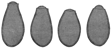
Fig. 159.—Ova of Clonorchis sinensis. The knobs on the ends of
the eggs are not shown. 900/1. (After Looss.)
This (?) species was discovered in 1874
by McConnell, in Calcutta, in the bile-ducts
of a Chinaman who died shortly after being
admitted into hospital.
Habitat.—Bile-ducts of man, dog and cat.
Distribution.—Especially in China, apparently
rare in Japan.
Clonorchis endemicus, Baelz, 1883.
Syn.: Distoma sinense s. spathulatum p.p.;
Distoma hepatis endemicum s. perniciosum, Baelz,
1883; Distoma japonicum, R. Blanchard, 1886.
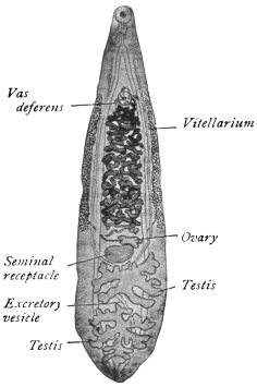
Fig. 160.—Clonorchis endemicus. × 6
about. (After Looss.)
Very similar to the previous species and
consequently generally confused with it.
Length between 6 and 13 mm., width
varying between 1·8 and 2·6 mm. Oral
sucker 0·37 to 0·5 mm., usually 0·43 to
0·45 mm. in transverse diameter; ventral
sucker 0·33 to 0·45 mm., usually 0·37 to
0·40 mm. No pigment in parenchyma;
anterior testis with four, posterior testis with
five branches. Vitellaria continuous, ova
26 µ by 13 µ to 16 µ.
Habitat.—Bile-ducts of man, dog, cat
and pig.
Distribution.—This species occurs very frequently in man, in
certain districts of Japan, especially in the province of Okayama,
Central Japan, in particular localities of which above 60 per cent. of
the population are infected. The worms are sometimes found in
enormous numbers in the liver (upwards of 4,000), also in the
pancreas and rarely in the duodenum.
It is common in Tonkin
and Indo-China. Léger in Tonkin
found 50 per cent. of people apparently
in normal health infected,
so that probably symptoms only
arise when the infection is intense.
[The exact distribution of these two
species is, however, not precisely
defined at present, as commonly no
distinction is made between them.—J.
W. W. S.]
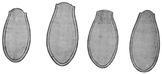
Fig. 161.—Clonorchis endemicus: eggs. The knobs
on the eggs are not shown. × 900. (After Looss.)
Verdun and Bruyant deny, in
opposition to Looss, the possibility
of being able to distinguish within
the genus Clonorchis the two species
described, but they admit the justification
for the new genus. They
also report the occurrence of Opisthorchis
felineus in man in Tonkin
(Compt. Rend. Soc. de Biol., lxii, 1907).
Pathology.—Both species of Clonorchis
give rise to grave symptoms. The liver is generally enlarged,
though when the infection has lasted some time it begins to contract.
The surface of the organ is studded with white vesicles, and on cutting
into it one sees numerous
cavities with thickened
walls (distended
bile-ducts) filled with a
brownish fluid containing
innumerable eggs,
which cause its colour.
Microscopically, the epithelium
of the bile-ducts
is either (1) entirely destroyed,
or (2) actively
proliferates, forming an adenomatous outgrowth. Occasionally this
proliferation is not limited by the wall of the bile-duct but penetrates
it and leads to a growth of numerous new ducts, forming a
malignant biliary adenoma. The bile-ducts have their connective
tissue wall greatly sclerosed. These fuse with one another, forming
areas of sclerosis devoid of liver tissue. As a result of these changes
the liver cells atrophy and undergo fatty pigmentary and granular
degeneration. Besides these changes, due probably to the toxic action
of the flukes, mechanical obstruction due to the actual plugging of the
ducts by the flukes causes retention of bile and icterus, and through
pressure on veins, ascites and hypertrophy of the spleen.
To what extent blood or bile respectively forms the food of the
flukes is uncertain.
Life-history.—(Kobayashi, 1911, Mitteilungen aus dem kaiserlichen
Institut für Infektions-Krankheiten zu Tokio, pp. 58–62.)
It results from the work of Kobayashi in Japan that fresh-water fish
form the second intermediate host for Clonorchis endemicus. He fed
cats with encysted flukes (cercariæ) from various fish and easily
succeeded in infecting them, e.g. a kitten, proved to be uninfected by
repeated examination of its fæces, was fed on infected fish; a month
later innumerable flukes were found in the bile-ducts, gall-bladder,
pancreas and even in the duodenum. The fish infected were Leucogobis
güntheri, Pseudorasbora parva, and to a less extent Acheclognathus
lanceolata, Acheclognathus limbata, Paracheclognathus rhombea, Pseudoperilampus
typus, Abbottina psegma, Biwia zezera and Sarcocheilichthys
variegatus. The cysts occur throughout the muscles and subcutaneous
tissue of the fish. Length 0·13 mm., breadth 0·1 mm. The cercaria
lies folded in the cyst, length 0·5 mm. breadth 0·1 mm. It tapers
posteriorly. Skin at first covered with fine spines, disappearing as they
grow older. Body dotted with fine pigment.
The first intermediate host is still unknown.
Sub-family. Metorchiinæ, Lühe, 1909.
Genus. Metorchis, Looss, 1899, emend. auctor.
Hind end rounded. Gut forks reach extreme end. Testes only slightly lobed,
filling the hind end.
Metorchis truncatus, Rud., 1819.
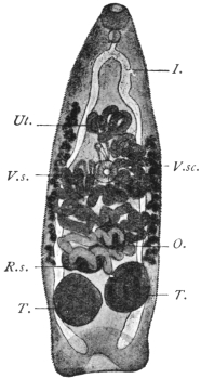
Fig. 162.—Metorchis truncatus,
Rud.: from the biliary ducts of the
domestic cat. V.s., ventral sucker;
I., gut; V.sc., vitellaria; T., testes;
O., ovary; R.s., receptaculum seminis;
Ut., uterus. 25/1.
This species, which attains a length of 2 mm., is slender and conical,
the anterior end is pointed and the posterior truncated, and provided
with a muscular tuberosity that resembles a terminal sucker; for this
reason the discoverer of the species (Rudolphi) classed it with the
Amphistomes. The cuticle in the young, as well as in the adult
specimens, is entirely and closely covered with spines. Suckers about
equal in size (0·134 to 0·172 mm.); the ventral sticker lies somewhat
in front of the middle of the body. The pharynx is small (0·09 mm.),
the œsophagus minute, the intestinal cæca reach to the posterior
extremity. Between them, and in front of
their blind ends, lie the two elliptical testes,
one generally a little in front of the other.
In front of them, either in the median
line or somewhat laterally, the spheroidal
ovary is situated; in front, again, is the
uterus, the coils of which usually extend
beyond the median field. The vitellaria
are at the sides of the central third of the
body, thus commencing in front of the
ventral sucker; cirrus pouch absent; the
genital pore is close in front of the
acetabulum. The excretory pore is terminal
(?). Eggs 29 µ by 11 µ.
Metorchis truncatus lives in the bile-ducts
of the seal, cat, dog, fox, and
glutton (Gulo borealis). The source of
infection is unknown, although one would
suspect fish. Askanazy did not succeed
in getting this fluke in his feeding experiments,
but another species, Metorchis
albidus, not uncommon in cats by feeding
them on roach (Leuciscus rutilus).
Family. Heterophyiidæ, Odhner, 1914.
Genus. Heterophyes, Cobbold, 1866.
Syn.: Cotylogonimus, Lühe, 1899; Cænogonimus, Looss, 1899.
No crown of spines on head. Body divided into a narrow, movable, anterior
part (neck), and a broader, less movable, posterior portion, which contains the
genitalia. The suckers separated from one another by a space equal to half the
length of the body or more; the pharynx is close behind the oral sucker; the
œsophagus is long; the intestinal cæca extend to the posterior border; the genital
pore is placed laterally, and behind the ventral sucker. Genital sucker provided
with a circlet of chitinous rodlets, shaped like stags’ horns. The testes are at the
posterior end, the ovary in a median position in front of them. Laurer’s canal with
receptaculum seminis present; the small vitellaria are at the sides of the posterior
part of the body. Parasitic in the intestine of mammals and birds.
Heterophyes heterophyes, v. Sieb., 1852.
Syn.: Distomum heterophyes, v. Siebold, 1852; Heterophyes ægyptica, Cobbold,
1866; Mesogonimus heterophyes, Railliet, 1890; Cœnogonimus heterophyes, Looss
1900; Cotylogonimus heterophyes, Braun, 1901.
Length up to 2 mm., breadth 0·4 mm.; the neck not sharply defined;
in life it stretches to double the length of the hind body. The scales
are rectangular, 5 µ to 6 µ by 4 µ, their posterior margin serrate with
seven to nine teeth. Cuticular glands are numerous on the ventral
surface, especially in the fore part of the body, and partly discharge
at the anterior border of the oral sucker. The oral sucker is 0·09 mm.,
the ventral sucker 0·23 mm. in diameter; the pharynx measures
0·05 to 0·07 mm. in length;
the œsophagus is about
three times as long; posteriorly
the intestinal cæca
are directed one towards
the other and terminate
beside the excretory
bladder. Close in front
of the posterior ends of
the intestinal branches are
the two elliptical testes,
which are not exactly on
the same level. In the
middle in front of them is
the receptaculum seminis,
and in front of the latter
lies the spherical or elliptical
ovary. The two vasa
efferentia unite to form the
vas deferens, which after
a short course passes over
into the angularly bent
seminal vesicle; after the
entry of the prostatic
glands it becomes united
with the metraterm
(vagina), and the common
duct opens into the genital
sucker. The latter is somewhat
smaller than the ventral
sucker, lateral to and
close (0·15 mm.) behind it, and bears a not entirely closed ring of
from seventy-five to eighty chitinous rods (20 µ in length). The vitellaria
on either side consist of about fourteen acini. The uterus is
spread almost throughout the entire posterior part of the body. The
eggs have thick shells with a knob resembling that of Clonorchis eggs
but not so prominent, and measure 30 µ by 17 µ; they contain a
completely ciliated miracidium with a rudimentary intestinal sac.
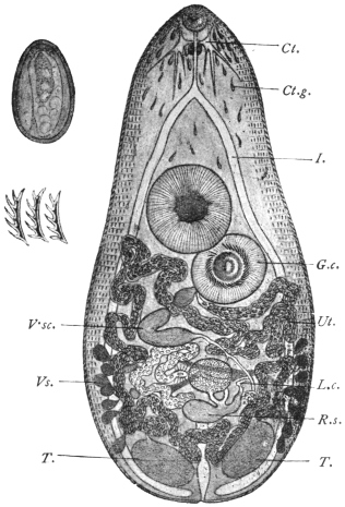
Fig. 163.—Heterophyes heterophyes, v. Sieb.
C., cerebral ganglion; I., intestinal cæca; Ct.g.,
cuticular glands; V.sc., vitellaria; Ut., genital sucker;
T., testes—the excretory bladder between them; L.c.,
Laurer’s canal; R.s., receptaculum seminis, with the
ovary in front of it; G.c., ventral sucker; Vs., vesicula
seminalis, 53/1. On the left side above, an egg, 700/1,
is depicted, and below it three chitinous rodlets from the
genital sucker. 700/1. (After Looss.)
This species was discovered in 1851 by Bilharz in the intestine
of a boy who died in Cairo; a second case was only found in 1891
and published by R. Blanchard, so that it appeared as if the species
were very scarce. According to Looss, this is, however, not the case,
but the species easily escapes notice on account of its small size.
Looss found it in Alexandria twice in nine autopsies, and once in
Cairo, and has recently stated that in man “it is not at all uncommon
to meet with the parasite in cadavers, and the eggs of the worm in
the stools of the patients.” Leiper records one case from Japan
and one from China. The parasites occupy the middle third of the
small intestine, and even when present in large numbers appear to be
harmless.
This small species, according to
Looss, frequently occurs in Egyptian
dogs, less so in cats, and has also been
found in the fox, as well as once in
Milvus parasiticus; Janson also reports
this species from the intestine of the
dog in Japan.
Metagonimus, Katsurada, 1913;
Yokogawa, Leiper, 1913.
Resembles in general structure
Heterophyes. In the arrangement of
its ventral genital suckers resembles
but differs from that of Tocotrema,273
Looss. The ventral and genital suckers
lie laterally and on the right.
Metagonimus yokogawai. Katsurada,
1913.
Syn.: Yokogawa yokogawai, Leiper, 1913.
Fig. 164.—Metagonimus yokogawai,
Katsurada, 1913: the spines are only
shown over a small part of the skin.
(After Leiper.)
One to 1·5 mm. long, seldom 2·5 mm.,
and 0·4 to 0·7 mm. broad; elliptical in
shape. The body is thickly covered
with nail-shaped spines about 10 µ
long. Oral sucker 77 µ, to 85 µ in diameter. Ventral sucker characteristic
and peculiar 0·12 to 0·14 mm. by 0·08 to 1 mm. It is a sac-like
organ placed deeply in the body, but does not open as in other flukes on
the ventral surface. Testes elliptical, not quite symmetrically placed at
the hind end of the body. Vesicula seminalis retort-shaped, situated
transversely, internal to the ventral sucker. Pars prostatica present.
Ejaculatory duct opens with the uterus into a genital sinus, which,
together with the internal opening of the ventral sucker, opens into a
pit at the front of the ventral sucker. The opening of the genital sinus
and that of the ventral sucker are furnished with a complex muscular apparatus.
Ovary spherical, 0·12 to 0·13 mm. in diameter, lies in the middle
of the hind body. Receptaculum seminis and Laurer’s canal present.
Vitellaria in the hind half of the body, consisting of about ten acini on
each side. Shell gland to the left of the
ovary. Uterus forms three to four transverse
coils. Eggs elliptical, double
contoured, yellowish-brown in colour.
There is no shoulder below the operculum
as in the eggs of Cl. sinensis.
At the rounder end there is a thickening
or knob different from the spine-like
or hook-like process seen in Cl.
sinensis. Dimensions 28 µ by 16 µ.
Habitat.—Mainly in upper or
middle portion of jejunum, rarely in
cæcum. They penetrate deep into the
mucosa, but not into the submucosa,
and post mortem appear as a number
of small brown points. They frequently
occur in the solitary glands,
which they destroy. They cause
chronic catarrh of the gut. Parasitic
in man and mammals.
Geographical Distribution.—Japan.
Life-history.—The cercarial stage
occurs in a trout (Plecoglossus altivelis)
and seldom in Crassius sp. and Cyprinus
sp. Infection takes place through
the eating of the fish raw. Seven to
sixteen days later eggs appear in the
fæces (of dog).
Family. Dicrocœliidæ, Odhner, 1910.
Genus. Dicrocœlium, Dujardin.
Dicrocœliidæ, with leaf-shaped bodies,
pointed posteriorly and anteriorly. Greatest
width behind the mid-line. Vitellaria double.
The testes smooth or indented, lying symmetrically or obliquely beside or behind
the ventral sucker. The ovary approaches the median line behind one testis.
Parasitic in the liver and gall-bladder (rarely in the intestine) of members of all
classes of vertebrate animals—by preference in birds and mammals.
Dicrocœlium dendriticum, Rud., 1819.
Syn.: Dicrocœlium lanceatum, Stil. and Hass., 1896; Fasciola lanceolata,
Rud., 1803 (nec Schrank, 1790); Distomum lanceolatum, Mehlis, 1825; Dicrocœlium
lanceolatum, Dujardin, 1845.
Fig. 165.—Dicrocœlium dendriticum,
Rud. V.s., ventral sucker; Cb., cirrus
pouch; I., intestinal cæca; V.sc., vitellaria;
T., testicles; O., ovary; M.s.,
oral sucker; Ut., uterus. 15/1.
Body lancet-shaped, narrowing especially at the anterior extremity;
length 8 to 10 mm., breadth 1·5 to 2·5 mm., the greatest breadth usually
behind the middle of the body. Suckers distant from each other by
about one-fifth the length of the body; oral sucker about 0·5 mm., ventral
sucker about 0·6 mm. Pharynx globular, adjoining the oral sucker;
œsophagus 0·6 mm. in length; intestinal cæca reach to four-fifths of
the body length. Genital pore at the level of the bifurcation of the
intestine; cirrus pouch small and slender. The large, slightly lobed
testes lie obliquely one behind the other behind the ventral sucker;
the ovary, which is considerably smaller, is placed behind the posterior
one; the vitellaria, commencing at the level of the posterior
testis, terminate far before the cæca. The uterus, situated behind the
ovary, extends throughout the posterior end, not confined to the central
field, but overlapping the lateral fields with its transverse coils; at
the posterior edge of the body it turns back again and winds forwards
to the ovary in transverse loops, then between the testes, and finally,
dorsal to the ventral sucker, terminates in the genital pore. The
thick-shelled eggs when young are yellowish, when older dark brown.
They measure 38 µ to 45 µ by 22 µ to 30 µ. They contain an oval or
roundish miracidium, only the anterior part of which is ciliated, and
which possesses a rudimentary intestinal sac with a boring spine.
The miracidia do not hatch out in water spontaneously, but, according
to Leuckart, in the intestines of slugs (Limax, Avion), but they
do not develop either in these (slugs) or in water-snails.
The lancet fluke inhabits the biliary duct of herbivorous and
omnivorous mammals (sheep, ox, goat, ass, horse, deer, hare, rabbit,
pig), and is often found associated with the liver fluke; it is not,
however, so common nor so widely disseminated, nevertheless, it has
been met with outside of Europe, namely, in Algeria, Egypt, Siberia,
Turkestan, and North and South America.
Fig. 166.—Eggs of Dicrocœlium
dendriticum, Rud.
To the left seen flat, to right
lying on one side. 600/1.
Fig. 167.—Miracidia of
Dicrocœlium dendriticum. a,
from the dorsum; b, from the
side. (After Leuckart.)
In man it is still more uncommon than the liver fluke, and has
hitherto only been observed seven times (Germany, Bohemia, Italy,
France, and Egypt); it may, however, have occurred more frequently,
and have been overlooked, as in slight infections it produces no
special symptoms.
The intermediate host is still unknown. Leuckart for some time
held the opinion that small species of Planorbis from fresh water,
which contain encysted Distomata, were to blame, and he supported
his views by a feeding experiment which seemingly yielded positive
results; this, however, is not definitely proved. Piana’s statement
that small land snails are the intermediate hosts has also not been
proved.
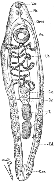
Fig. 168.—Echinostoma ilocanum. Vo.,
oral sucker; Ph., pharynx; Cirre, cirrus;
V.v., ventral sucker; Ut., uterus; G.c.,
ovary; Ov., shell gland; T., testes;
T.d., vitellarium; C.ex., excretory vesicle.
(After Brumpt.)
Family. Echinostomidæ, Looss, 1902.
Sub-family. Echinostominæ, Looss, 1899.
Genus. Echinostoma, Rud. 1809; Dietz, 1910.
Fore-body not bulging. Greatest width at or behind the ventral
sucker. Oral sucker not atrophied. Collar kidney-shaped with a double
dorsally unbroken row of spines, terminating in four to five angle spines.
The border spines of the aboral series not larger than the oral. Skin
spined or smooth. Body elongated. Uterus long with numerous
transverse coils. Ventral sucker in the anterior quarter of body. Cirrus
sac small, almost completely in front of the ventral sucker. Testes round
or oval, smooth incurved or lobed, in the hinder half of body. Ovary
median or lateral in front of testes. Vitellaria from hinder margin of
ventral sucker to end of body. Eggs oval, 84 µ to 126 µ by 48 µ to 82 µ.
The spines placed most ventrally, or those placed most medially
on ventral surface, are from differences of position or form termed
“angle” spines, the rest “border” spines.
Type.—Echinostoma echinatum, Rud.
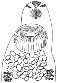
Fig. 169.—Echinostoma ilocanum,
Garrison, 1908: head end showing collar
of spines, ventral view. (After Leiper.)
Echinostoma ilocanum, Garrison, 1908.
Length 4 to 5 mm., breadth 1 to 1·35 mm., thickness 0·5 to
0·6 mm. The circum-oral disc 0·3 mm. broad, separated by a shallow
groove from the body. Crown of forty-nine spines and five to six
angle spines on each side continuous with an irregularly alternating
series of fourteen spines on the dorsum. Largest spines are
34 µ long, 8 µ thick at the base. The remainder of the dorsal spines
are 24 µ by 6 µ. Skin thickly covered with scales on the margins
of the body as far back as the level of the hind testis. Oral sucker,
0·18 mm.; ventral sucker, 0·4 to 0·46 mm. Its anterior border about
0·07 mm. from the anterior end. Pharynx 0·17 mm. long, 0·11 mm.
broad. Testes about mid-line of the body, much lobed; the lobes of
the anterior testis run transversely, while the axis of the posterior testis
is longitudinal, as often occurs in the Echinostomidæ. Cirrus sac
reaches to the centre of the ventral sucker. Ovary transversely oval
in front of the testes. Vitellaria commence about half-way between
the ventral sucker and ovary and extend to the posterior end. Eggs
numerous, 92 µ to 114 µ by 53 µ to 82 µ.
Average.—99·5 µ by 56 µ.
Habitat.—Gut of man (Filipinos),
Philippine Islands.
Echinostoma malayanum,
Leiper, 1911.
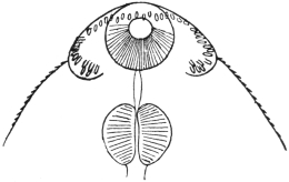
Fig. 170.—Echinostoma malayanum, Leiper,
1912: anterior end showing collar of spines,
ventral view. (After Leiper.)
Twelve millimetres long, 3 mm.
broad, 1·3 mm. thick. Ends bluntly
rounded. At the anterior end a
ventral furrow on either side, one-third
the width of the body,
marking off the circum-oral collar.
Along its edge is a row of forty-three
spines extending across the
middle line dorsally but not ventrally.
The spines vary in size from
0·07 mm. in length (ventrally) to 0·05 to 0·016 mm. (dorsally). Cuticular
spines also exist on the ventral side as far back as posterior end of
body, but dorsally limited to a triangular area ending in front of the
ventral sucker. Oral sucker 0·07 mm. thick, occupying the middle
third of the circum-oral disc; pharynx 0·25 mm. in diameter; œsophagus
0·04 mm. long; gut cæca simple, extending to end of body;
ventral sucker 0·9 mm. long by 0·75 mm. broad by 0·7 mm. deep; wall
about 0·25 mm. thick. The
sucker is inclined at an angle
of 40° to the ventral surface.
Testes lobed, one behind the
other, behind the ventral sucker.
Cirrus pouch well developed,
reaching to the posterior edge
of the sucker. Genital pore in
the angle between neck and
anterior lip of ventral sucker.
Ovary smooth, 0·3 mm. in
diameter, 0·85 mm. behind
ventral sucker. Vitellaria very
numerous, extending from posterior margin of sucker to posterior end
of body, where they intermingle. Eggs few in number, brown and large.
Habitat.—Gut of man (Tamils), Malay States.
Sub-family. Himasthlinæ, Odhner, 1910.
Genus. Artyfechinostomum, Clayton-Lane, 1915.
Crown of thirty-nine spines, continuous over dorsum. Two corner
spines long. Vitellaria extend from posterior margin of sucker to posterior
end of fluke. Eggs without filament. [Although the possession
of strong rose-thorn hooks is given by Odhner as a sub-family characteristic,
yet in this genus assigned to this sub-family they have not
been seen.—J. W. W. S.]
Artyfechinostomum sufrartyfex, Clayton-Lane, 1915.
Spirit specimens: 9 by 2·5 by 0·8 mm. thick. Ventral sucker
conspicuous, 1 mm. in diameter. Cirrus sac 2 mm. long. Testes
lobed, about 1·5 mm. in diameter. Posterior border of posterior
testes 1 mm. from posterior end. Vitellaria meet posteriorly behind
the posterior testis.
Family. Schistosomidæ, Looss, 1899.
Genus. Schistosoma, Weinl, 1858.
Syn.: Gynæcophorus, Dies., 1858; Bilharzia, Cobb., 1859; Thecosoma,
Moq. Tandon, 1860.
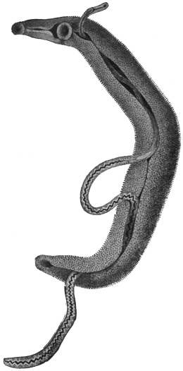
Fig. 171.—Schistosoma hæmatobium, Bil.:
male carrying the female in the canalis gynæcophorus.
12/1. (After Looss.)
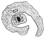
Fig. 172.—Transverse section
through a pair of Schistosoma
hæmatobium in copulâ. In the male
the point of reunion of the intestinal
forks has been cut across. (After
Leuckart.)
The males have bodies that widen out considerably behind the ventral sucker,
the lateral parts of which in-roll ventrally, forming the almost completely closed
canalis gynæcophorus, within which the female is enclosed. There is no cirrus
pouch. The male has five or six testes, the females are filiform; the uterus is long.
There is no Laurer’s canal. The ova almost equally attenuated at either extremity;
they have a small terminal spine, and are not provided with a lid. They contain
a miracidium, ciliated on all sides, which is characterized by the possession of two
large glandular cells, which discharge anteriorly beside the gastric sac. They live
in the vascular system of mammals. (An allied genus [Bilharziella] lives in the
blood-vessels of birds.)
Schistosoma hæmatobium, Bilharz, 1852.
Syn.: Distoma hæmatobium, Bilh.; Distoma capense, Harley, 1864.
The Male is whitish, 12 to 14 mm. in length, but is already mature
when 4 mm. long. The anterior end is 0·6 mm. or a little over in
length. The suckers are near
each other, the oral sucker is infundibular,
and the dorsal lip is
longer than the ventral one. The
ventral sucker is a little larger,
0·28 mm., and is pedunculated.
A little behind the ventral sucker
the body broadens to a width of
1 mm., decreasing, however, in
thickness; the lateral edges in-roll
ventrally, so that the posterior
part of the body appears almost
cylindrical, 0·4 to 0·5 mm. in
diameter; the posterior extremity
is somewhat more attenuated.
The dorsal surface of the posterior
part of the body is covered
with spinous papillæ. There are
delicate spines on the suckers, and
larger ones invest the entire internal
surface of the gynæcophoric canal,
as well as a longitudinal zone at
the edge of that side of the external
surface that is covered by the other
side rolling over it. The œsophagus
is beset with numerous
glandular cells (fig. 173), and presents
two dilatations; the intestinal
bifurcation is close in front of
the ventral sucker, the two
branches uniting sooner or later behind the testes into a median
trunk, which may again divide at short intervals. The excretory
pore is at the posterior end, but placed somewhat dorsally; the
genital pore is at the beginning of the gynæcophoric canal, thus
behind the ventral sucker; into it opens the vas deferens which,
posteriorly, broadens into the seminal vesicle and then continues
as the vasa efferentia of the four or five testes (fig. 173).
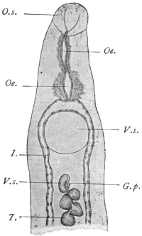
Fig. 173.—Anterior end of the
male Schistosoma hæmatobium, Bilh.
V.s., ventral sucker; I., gut cæca;
G.p., genital pore; T., testes;
O.s., oral sucker; Oe., œsophagus
with glandular cells; V.s., vesicula
seminalis. 40/1. (After Looss.)
The Female—filiform, about 20 mm. in length, pointed at each end,
and measuring 0·25 mm. in diameter in
the middle. Their colour varies according
to the condition of the contents of
the intestine. (Posteriorly they are dark
brown or blackish.) The cuticle is smooth
except in the sucker, where there are very
delicate spines, and at the posterior end,
where there are other larger spines. The
oral sucker is a little larger than the
pedunculated ventral sucker (0·07 and
0·059 mm. respectively). The anterior
part of the body, 0·2 to 0·3 mm. in length;
the œsophagus is as in the male. The
intestinal bifurcation is in front of the
ventral sucker, the two branches uniting
behind the ovary and the trunk running
in a zigzag manner to the posterior border.
There are indications of diverticula at the
flexures. The ovary is median. In young
females it is of an elongated oval shape;
in older females the posterior end becomes
club-shaped, whereas the anterior end
becomes attenuated; the oviduct originates
at the posterior end, but immediately
turns forwards and joins the parallel
vitelline duct in front of the ovary (fig. 174),
where the shell gland cells open; the
common canal becomes dilated to form
the oötype, and then proceeds as the
uterus, with only slight convolutions,
along the central field to the genital pore,
which lies in the middle line immediately
behind the ventral sucker. The single
vitellarium starts behind the ovary and
extends to the posterior end. The acini
are situated at the sides of the excretory
duct, which runs a median course. The eggs are compact spindles,
much dilated in the middle; they have no lid, and are provided with a
terminal spine (rudimentary filament) at the posterior end, measuring
120 µ to 150 µ in length and 40 µ to 60 µ in breadth, but vary in size
and shape (fig. 175).
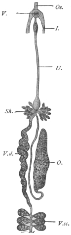
Fig. 174.—Schistosoma
hæmatobium, Bilh.: genitalia
of the female. V.s.,
ventral sucker; I., gut cæca;
V.d., vitelline duct; V.sc.,
vitellarium; O., ovary; Oe.,
œsophagus; Sh., shell
gland; U., uterus. Magnified.
(After Leuckart.)
Distribution.—In order to understand the distribution of the worms
and eggs in the body, it may be well to recall the blood supply of the
abdominal and pelvic organs. It is generally assumed that the early
life (? cercarial stage) of the worms occurs in the liver, and that the
young worms travel from here, where they are invariably found, to
their various sites along the portal vein and its tributaries and so
against the blood stream. The tributaries of the portal vein are:—
(1) Superior mesenteric, the tributaries of which are: (a) the veins
of the small intestine; (b) ileo-colic; (c) right colic; (d) middle colic;
(e) right gastro-epiploic; and (f) inferior pancreatic. By these paths
infection of the small intestine, ascending and transverse colon and
pancreas would occur.
(2) Splenic. (Ova have been recorded by Symmers in the spleen.)
(3) Inferior mesenteric, the tributaries of which are (a) superior
hæmorrhoidal veins from the upper part of the hæmorrhoidal plexus;
(b) sigmoid veins from sigmoid flexure and lower portion of descending
colon; (c) left colic vein draining descending colon.
The superior hæmorrhoidal veins form a rich plexus in the rectum,
and below this level in the upper and middle parts of the anal canal.
The plexus forms two networks, an internal plexus in the submucosa
and an external on the outer surface. The internal plexus opens at
the anal orifice into: (a) branches of the inferior hæmorrhoidal vein
(from the pudic); (b) the external plexus. The external plexus gives
off: (a) inferior hæmorrhoidal opening into internal pudic (of internal
iliac vein); (b) mid-hæmorrhoidal into internal iliac or its branches;
and (c) superior hæmorrhoidal opening into inferior mesenteric.
The external plexus further communicates with the vesico-prostatic
plexus. The vesico-prostatic (vaginal) plexus opens into the vesical
veins, which drain into the interior iliac vein. This plexus also
receives afferents from the pudendal plexus, the chief tributary of
which is the dorsal vein of the penis. The pudendal plexus also
receives branches from the inferior pudic and the anterior surface of
the bladder.
There is thus a communication between the portal vein and the
vena cava by means of these plexuses, viz., through the inferior and
middle hæmorrhoidals, and by the inferior hæmorrhoidals to the
bladder and thence by the vesical veins or the pudic to the caval
system (interior iliac).
It is thus by the inferior mesenteric and its tributaries that the
worms reach the descending colon, rectum, anal canal, and eventually
the bladder, and in some cases the caval system.
Before considering what is actually found post mortem in these
veins and the organs drained by them, we may further recall the fact
that the calibre of “medium” veins is 4 to 8 mm., “small” veins
less than 40 µ in diameter and capillaries 8 µ to 20 µ. Further, the
maximum diameter of the male worm is 1 mm., that of the female
280 µ and eggs in utero 80 µ to 90 µ long by 30 µ to 40 µ.
Liver and Portal Vein.—Here worms are most easily found post
mortem. Often only males are found and these of the same size,
and if females occur only a few worms are found in copulâ. The
worms are frequently not full size and the males may contain no free
spermatozoa in their testes, and as regards the females some may
be fertilized, others not, as shown by the presence or absence of
spermatozoa in the seminal receptacle or uterus. In either case they
may contain eggs—lateral-spined—usually one, less often two, but
there may be as many as five or six. These eggs may also show
some abnormality, which takes the form of: (1) abnormal contents,
viz., disintegrating yolk cells with or without an ovarian cell;
(2) abnormal shape but with normal contents and probably represented
by the collapsed and empty egg-shells which are found in the
tissues.
As to the interpretation of these facts, Looss believes that these
lateral-spined eggs are products of young females whose egg-laying
is not at first properly regulated. The shape that the eggs take, viz.,
with a lateral spine, is determined by an excess of material—ovarian
and yolk cells—being present in the oötype. The shape of eggs
depends upon the position they have in the oötype during their
formation. In young females an excess of cells—yolk cells especially—accumulates,
distending not only the dorsal wall but a portion also
of the short duct joining the oötype to the uterus. The result of this
is that the axis of the oötype and egg is almost transverse to the body,
and the posterior funnel-shaped portion of the oötype, instead of
being terminal, has now a lateral or rather a ventral position, so that
the spine which occupies this portion, instead of being terminal, is
now lateral. It is noteworthy that these lateral-spined eggs are
thicker, owing to the excess of material present, and not uncommonly
have a curved anterior border, due to a projection of the anterior end
into the anterior opening of the oötype.
As these eggs are being laid by females in the portal vein they are
carried back to the liver by the blood stream. The liver is one of
the commonest sites for these eggs; also terminal-spined eggs may be
found here for the same reason.
Hæmorrhoidal Veins.—Mature worms, generally in copulâ, are
usually found here, though young not fully grown females may also
occur. The tissues of the rectal wall (or colon) show, as a rule, large
quantities of lateral-spined eggs, though less often only terminal-spined
eggs may be found.
Vesico-prostatic Plexus.—Worms in copulâ are found in the veins
of the submucosa in the bladder, and the eggs in the mucosa, and
those voided are usually terminal-spined, though lateral-spined eggs
are not so rare as generally thought. The problem next arises as to
how the eggs get to the lumen of the gut or bladder.
The female worm is 280 µ in diameter. Veins in the submucosa
of the rectum less than 178 µ in diameter are not affected with endophlebitis.
It is probable that the female even by stretching could not
penetrate much beyond this. Eggs are probably then laid in the
submucosa as near the muscularis mucosa as possible. Now if the
eggs are laid in a vein of larger calibre than the worm fills, the eggs
would be carried back to the inferior mesenteric vein, so that presumably
the worm must succeed in blocking the vein already narrowed
by endophlebitis, so that by the stasis which ensues the eggs may
escape from the veins. How this occurs is not exactly known; it is
not necessarily due to the spine, as the same escape into the tissues
occurs in spineless eggs, such as those of Schistosoma japonicum. The
eggs, then, pass as foreign bodies through the tissues. Another hypothesis
is that the worms leave the veins in order to lay their eggs,
but the evidence is against this.
Caval System.—Occasionally worms that have passed through the
vesical plexus may be found in the iliac vein, inferior vena cava,
and even the lungs. If the worms are young they contain a lateral-spined
egg; if adult, numerous (50 to 100) terminal-spined eggs.
Lungs.—When the liver is strongly infected with (terminal-spined)
eggs it is possible that by passive movements some may pass into the
intralobular veins, and thence by the inferior vena cava to the lungs.
Gall-bladder.—Similarly terminal-spined eggs pass into the bile-capillaries
and gall-bladder (where they may be abundant), and so into
the fæces.
Detection of Eggs.—Occasionally eggs may be found in various other
parts of the body. They are best detected by macerating pieces of the
tissue in question in about 1/4 per cent. hydrochloric acid at 50 to 60°C.
(Looss).
Pathological changes:—
Rectum.—These have been studied thoroughly by Letulle in the
case of an apparently pure infection of the rectum.274 They take the
form of a chronic diffuse inflammation, which may result in—(1) ulceration,
or (2) hyperplasia of the mucosa, producing adenomata.
Ulcerative Form.—The mucosa is transformed into a mass of
vascular connective tissue. The connective tissue spaces next become
invaded by numerous mononuclear cells. The tissue itself
undergoes diffuse sclerosis, becoming hard and fibroid. Eventually
ulcerative necrosis sets in. During these changes the Lieberkühn
glands are destroyed. The process does not extend to the submucosa,
in this respect differing from that in chronic dysentery.
Hyperplastic Form.—The Lieberkühn glands of the mucosa at first
hypertrophy; then there is an actual hyperplasia resulting in adenomata.
The interstitial tissue of the glands is also greatly hypertrophied,
giving rise to very vascular granulations. These growths
are often hollow and contain worms. Many eggs are found in the
mucosa on their way to the lumen of the gut.
The muscularis mucosa is thickened up to twice or even ten times
the normal. Its vessels are dilated (36 µ to 80 µ), but they do not
allow of the passage of worms.
The submucosa is profoundly changed; rigid and hard instead of
supple. It is here that the greatest number of eggs occur. A remarkable
condition of endophlebitis exists in the veins of the
submucosa, not only in the smaller ones but also in the larger ones
(370 µ by 270 µ). This endophlebitis results in a more or less complete
occlusion of the vessels of the lumen.
The muscular coats are free from change, also their veins.
The Serous Coats.—The veins about 1,900 µ, also show endophlebitis.
Besides the rectum, in extreme cases even the transverse colon, the
cæcum and small intestine may be affected.
Bladder.—In the early stages the mucosa is deep red and swollen
like velvet, or there may be localized patches of hyperæmia or
extravasation. The subsequent changes take two chief forms:—
(1) Sandy Patches.—The mucosa looks as if it were impregnated
with a fine brownish or yellowish powder (myriads of ova). This is
accompanied by a gradual hypertrophy and new formation of connective
tissue, so that dry, hard or plate-like patches with this sandy
appearance arise; the thickening eventually affects all the coats of
the bladder. In the older patches many of the eggs are calcified.
These patches sooner or later break down, ulcerate and necrose.
Phosphatic deposits are abundant and stone is common. These
patches are not found in the rectum.
(2) Papillomata.—Where the inflammatory change produced by
the eggs gives rise to hypertrophy and hyperplasia of the mucosa,
papillomata result, the axis of which is formed by connective tissue
of the submucosa. These are most variable in shape and form and
bleed readily, and sometimes contain cavities of extravasated blood.
As in the rectum, it is in the submucosa that eggs are most
abundant, and worms in copulâ occur in the veins of this layer, but
endophlebitis is not as general as described in the rectum. Malignant
disease of the bladder is not an uncommon sequela of bilharziasis.
Besides the bladder, the ureters and kidneys may in advanced cases
be involved. The prostate and vesiculæ seminales are commonly
diseased. Eggs have been recorded in the semen. The urethra is
frequently attacked; the vagina in the female.
Eggs also occur in the lymphatic glands of the gut.
Geographical Distribution.—East Africa:
Nile Valley, Red Sea Coast, Zanzibar, Portuguese
East Africa, Delagoa Bay, Natal, Port
Elizabeth.
South Africa: Cape Colony, Orange Free
State, Transvaal, Mauritius, Bourbon, Madagascar.
West Africa: Angola, Cameroons, Gold
Coast, Gambia, Senegal, Sierra Leone, Lagos,
Nigeria.
North Africa: Tripoli, Tunis, Algeria, parts
of the Sahara.
Central Africa: Sudan, various portions.
Uganda, Nyasaland.
It occurs with varying frequency in these
regions. It is probably more widely spread
than this list implies, as undoubtedly many
cases are seen which are not recorded.
Isolated cases have been recorded from
Arabia, India,275 Greece, Cyprus.
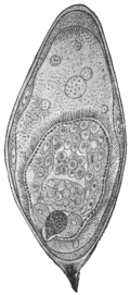
Fig. 175.—Ovum of
Schistosoma hæmatobium,
Bilh., with miracidium,
which has turned its anterior
end towards the
posterior end of the egg.
275/1. (After Looss.)
The means by which infection is brought
about are still uncertain; we only know that
the miracidia (fig. 175) enclosed in the discharged
eggs do not hatch if the eggs remain
in the urine, but after cooling perish. As
soon, however, as the urine is diluted with
water the shell swells, generally bursting lengthways,
and releases the miracidium from its
investing membrane, so that it can swim about
with the aid of its cilia. In its structure it
differs but little from the miracidium of Fasciola
hepatica, as, for instance, in the lack of eyes;
the two large gland cells situated on either side of the intestinal
sac are also present in the miracidia of Fasciola hepatica.
Sarcode Globules.—This is a term applied to certain globules which at
times appear in the miracidium and are later ejected. Some authors
consider them as indicative that the miracidium has developed into
a sporocyst, but Looss considers them to be degeneration products.
The Bilharzia mission, under R. T. Leiper, sent to Egypt by the
War Office early in 1915, reports that cercariæ of bilharzia type were
recognized in four of the commonest fresh-water molluscs around
Cairo.
With material obtained from naturally infected Planorbis boissyi
acute bilharziosis was experimentally produced in rats, mice, and
monkeys. Infection takes place experimentally through the skin and
also through the mucous membrane of the mouth and œsophagus.
The miracidium, after entering the mollusc, develops into a sporocyst.
This gives rise not to rediæ, but to secondary sporocysts, which, in
turn, produce cercariæ. These, like the adult worm, differ from other
distomes in lacking a muscular pharynx.
Schistosoma mansoni, Sambon, 1907.
According to Manson, Sambon and others, the eggs with lateral
spines belong to a species different from Schistosoma hæmatobium.
Infections with this species only are said to
occur in the Congo, Southern States of North
America, West Indies (Guadeloupe) and Brazil
(Bahia). The following characters, according to
Flu, differentiate this species: (1) In the male
the transition from the anterior portion of the
worm to the lateral fields (the infolded portions
which form the gynæcophoric canal) is not a
gradual one as in Schistosoma hæmatobium,
but in this case the lateral fields rise suddenly,
almost at right angles to the anterior portion.
(2) The ovaries have a well-marked convoluted
course as in no other schistosome. (3) The
oötype is symmetrical in reference to the long
axis of the body, its duct being lateral on the
ventral side (Looss’ explanation of this we have
already given). (4) The worms live exclusively
in portal vein and tract. (As lateral-spined eggs
occur also in the bladder, this is not exactly true.)
Schistosoma hæmatobium, Bilharz, 1852.
Male, four or five large testes. Gut forks unite late, so that the
single gut stem is short. Female, ovary in posterior half of body.
Uterus very long, voluminous, with many terminal-spined eggs, some
lying in pairs. Vitellaria in posterior fourth of body. Cercariæ in
Bullinus contortus and Bullinus dybowski (syn.: Physa alexandrina) in
Egypt.
Schistosoma mansoni, Sambon, 1907.
Male, eight small testes. Gut forks unite early, so that the single
gut stem is very long. Females, ovary in anterior half of body. Uterus
very short; usually only one lateral-spined egg at a time in utero.
Vitellaria occupy posterior two-thirds of body. Cercariæ in Planorbis
boissyi in Egypt.
The above morphological descriptions are founded on worms of
each species, derived from experimentally infected mice (Leiper, R. T.,
Brit. Med. Journ., March 18, 1916, p. 411).
Schistosoma japonicum, Katsurada, 1904.
Syn.: S. cattoi, Blanchard, 1905.

Fig. 176.—Schistosoma japonicum:
anterior end with testes;
posterior end with point of union
of cæca. Length of worm about
10 mm. (After Katsurada.)
Male.—Eight to 19 mm., but extreme limits are 5 to 22·5 mm. Consists
of a short fore-body, separated by the ventral sucker from the
hind-body. The ventral sucker is stalked and somewhat larger than
the oral sucker. Both suckers are larger than the corresponding ones
in S. hæmatobium. Body usually smooth, but in the fresh state
numerous fairly evident spines along the margin of the canal. Œsophagus:
two bulbs. The junction of the gut forks more posterior than
in S. hæmatobium, the median united gut
stem occupying a quarter to one-fifth to
one-sixth of the body length. An excretory
canal runs along each side of the
body, opening into the dorsal excretory
pore. Testes irregularly elliptical, six to
eight in number, in the anterior part of
hind-body. The vasa efferentia unite
into a common vas deferens which opens
directly behind the ventral sucker. The
seminal vesicle lies just behind this.
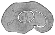
Fig. 177.—Schistosoma japonicum,
male and female in copulâ.
× 60. (After Katsurada.)
Female.—Up to 26 mm., generally
thinner than the male. Surface smooth.
Suckers armed with fine spines. Ventral
sucker larger than oral. Body thicker
behind the region of the ovary. The
gut forks unite immediately behind the
ovary. The united gut much thicker
than in S. hæmatobium. Ovary elliptical,
almost in the mid-body, its hinder portion
dilated. The oviduct arises from
its posterior end and then runs sinuously
forward, where it is joined by the vitellarian
duct; the vitellarium well developed,
extending from behind the ovary
almost but not quite to the posterior
end as in S. hæmatobium. Shell gland
ducts enter at the junction point of
oviduct and vitelline duct. The canal
here forms an oötype and then proceeds
as the uterus to open directly behind
the ventral sucker. The uterus occupies
almost half the hind-body. In S. hæmatobium
this is not so. The uterine canal
is cleft-like, i.e., its dorso-ventral diameter
is much greater than its lateral diameter.
The number of eggs varies from about
50 to 300 from observations made in various hosts.
Eggs.—In utero assume various shapes, as they are soft; the lumen
of the uterus is narrow. Outside they are oval, faint yellow, double
contoured. In fæces the eggs measure 83·5 µ, by 62·5 µ (man); 85 µ
by 61·5 µ (cattle); 98·2 µ by 73·8 µ (dog). The eggs have either
small lateral spines or thickenings, and Looss at the opposite
side has described cap-like thickenings.
The eggs in the tissues undergo various deformities,
and may contain a miracidium, as
also the eggs in fæces do; or the contents
may consist of granular matter or amorphous
masses or they may be calcified. Lymphocytes
and giant cells may also invade the
eggs.
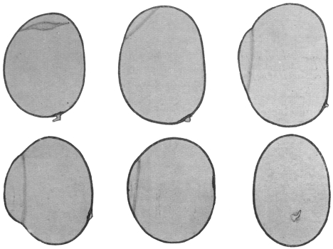
Fig. 178.—Schistosoma japonicum: eggs from human liver, showing “spines” and
“hoods” at opposite pole. (After Looss.)
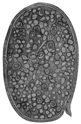
Fig. 179.—Schistosoma japonicum:
from dog. Uterine
egg. × c. 800. (After Katsurada.)
Mode of Infection.—The miracidia hatch
in water in as little as fifteen minutes, but
the majority in one to three hours. They will live in water for about
twenty-four hours. In water they undergo a transformation into
“larvæ,” which then penetrate the skin, as has been shown by
Japanese writers to hold good for man, cattle, dog and cat. The
penetration of the skin is attended with an eruption on the legs,
“Kabure.” The exact route by which the worms reach the portal vein
is uncertain. Infection in Japan takes place from spring to autumn,
especially May to July, when the soil is contaminated with manure
of cattle infected with S. japonicum. They also appear to develop
in molluscs. Leiper and Atkinson found cercariæ (in sporocysts)
in the liver of a mollusc, Katayama nosophora. They infected mice
by immersing them in water containing liver emulsion and so free
cercariæ, thus confirming the similar results of Miyairi and Suzuki.
Habitat.—The worm occurs in Japan, China, and the Philippines.
The normal host is man and mammals. Cattle, dog and cat are
often found naturally infected. Mice can also be experimentally
infected. Their seat of election is the portal vein and its branches,
especially the mesenteric veins. They either
swim free in the blood or remain fixed by
their suckers to the intima of the vessels.
They have also been found in the vena cava
and right heart of a cat, but not so far in the
vesical plexus.
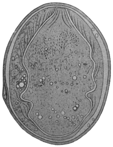
Fig. 180.—Schistosoma japonicum: from
dog. × c. 800. (After Katsurada.)
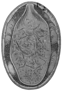
Fig. 181.—Schistosoma japonicum:
from dog. Egg from fæces. × c. 800.
(After Katsurada.)
Eggs are found in the submucosa and
mucosa of the gut, especially the colon, and
at times in the serosa and subserosa of the
small intestine, where they give rise to new
growths. Occasionally eggs are found in
the brain. The life of the worms is at least
two years.
Pathogenic Effects.—Anæmia through loss
of blood due to worms; enlarged spleen,
toxic in origin (?); phlebitis, thrombosis, due
to portal stasis; the eggs, however, cause the
greatest mischief. They are carried by the circulation to various
organs where they produce inflammation, granulation tissue, and later
connective tissue.
Liver.—The eggs reaching this organ give rise to granulomata and
hence enlarged liver, and later, when connective tissue is formed, to
contraction. The surface is rough and irregularly granular, “parasitic
embolic cirrhosis” of Yamagiwa.
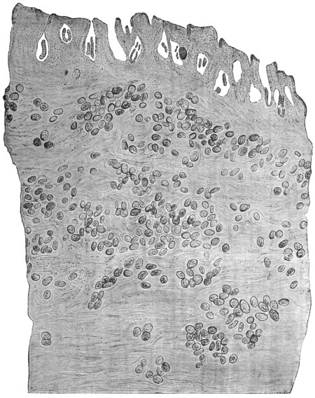
Fig. 182.—Schistosoma japonicum: section through the gut of a Chinaman showing eggs.
× 58. (After Catto.)
Gut.—The eggs in the mucosa and submucosa cause catarrh and
destruction of tissue or new growth. In the small intestine the eggs
are mainly in the serosa and subserosa, where they give rise to polypoid
or branched growths.
Spleen.—Enlarged, at first due to toxin (?) and later due to portal
stasis. Eggs in the spleen are uncommon.
Ascites also arises from the portal stasis, and is generally present
in advanced cases.
Eggs may be found in many other situations: glands (numerous),
mesentery, stomach, pancreas, kidney, etc. The bladder remains free.
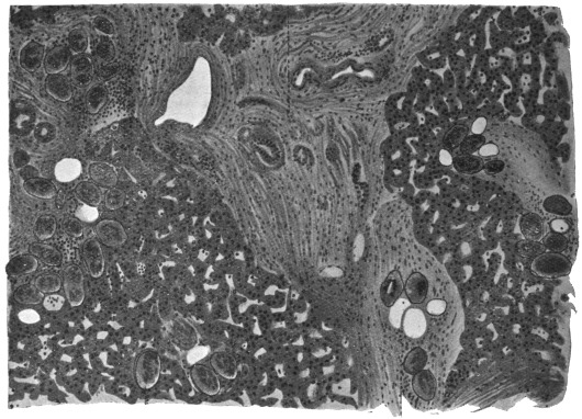
Fig. 183.—Schistosoma japonicum: liver showing eggs in the intra- and interlobular
connective tissue. × c. 80. (After Katsurada.)
Tapeworms have been known from ancient times—at all events, the large
species inhabiting the intestines of man—and there has never been a doubt as
to their animal nature. The large cysticerci of the domestic animals (occasionally
of man also) have been known for an equally long period, but they were generally
regarded as growths, or “hydatids,” until almost simultaneously Redi in Italy, and
Hartmann and Wepfer in Germany, concluded from their movements and organization
that they were of animal nature. From that time the cysticerci have been
included amongst the other intestinal worms, and Zeder (1800) established a special
class (Cystici, Rud., 1808) for the bladder worms. Things remained in this condition
until the middle of the last century, when Küchenmeister, by means of successful
feeding experiments, demonstrated that the cysticerci were definite stages of
development of certain tapeworms. Before Küchenmeister, E. Blanchard, van
Beneden, and v. Siebold had held the same opinion in regard to other asexual
Cestodes.
Since the most remote period another question has again and again occupied
the attention of naturalists, the question of the morphological nature—that of the
INDIVIDUALITY OF THE TAPEWORM. The ancients, who were well acquainted
with the proglottids (Vermes cucurbitani) that are frequently evacuated, were
of the opinion that the tapeworm originated through the union of these separate
proglottids, and this view was maintained until the end of the seventeenth
century. In 1683 Tyson discovered the head with the double circlet of hooks in
a large tapeworm of the dog; Redi (1684) was also acquainted with the head and
the suckers of several Tæniæ. Andry (1700) found the head of Tænia saginata, and
Bonnet (1777) and Gleichen-Rusworm (1779) found the head of Dibothriocephalus
latus. Consequently most authors, on the ground of this discovery, considered the
tapeworm as a single animal, that maintains its hold in the intestine by means of the
head, and likewise feeds itself through it. The fact was recognized that there were
longitudinal canals running through the entire length of the worm, and it was thought
that these originated in the suckers, and that the entire apparatus was an intestine.
As, moreover, the segments form at the neck, and are cast off from the opposite
extremity, the tapeworm was also compared with the polyps, which were formerly
regarded as independent beings.
Steenstrup, in his celebrated work on the alternation of generations (1841), was
the first to give another explanation. This has been elaborated still further by
van Beneden, v. Siebold and Leuckart, and until a few years ago all authorities
adopted his views. According to this view, the tapeworm is composed of numerous
individuals, something like a polyp colony, and, in addition to the proglottids—the
sexual individuals which are usually present in large numbers—there is ONE
individual of different structure, the scolex, which not only fastens the entire colony
to the intestine, but actually produces this colony from itself, and therefore is
present earlier than the proglottids. The scolex is a “nurse,” which, though itself
produced by sexual means, increases asexually like a Scyphistoma polyp; the
tapeworm chain has therefore been termed a strobila. Consequently the development
of the tapeworms was explained by an alternation of generations. In support
of this opinion it was demonstrated not only that the adult sexual creatures, the
proglottids, can separate from the colony and live independently for a time, but
that in certain Tæniæ, and especially in many Cestodes of the shark, the proglottids
detach themselves long before they have attained their ultimate size, and thus
separated continue to develop, grow and finally multiply; the scolex also exhibits a
certain independence in so far as, though not, as a rule, capable of a free life, yet
it in some cases lives as a free being, partly on the surface of the body of marine
fishes and partly in the sea. With the more intimate knowledge of the development
of the cysticerci, the independent nature of the scolex was recognized. It is
formed by a budding of the bladder that has developed from the oncosphere, in
some cases (Cœnurus) in large numbers, in other cases (Echinococcus) only after
the parent cyst has developed several daughter cysts. Released from its mother
cyst and placed in suitable conditions, it goes on living, and gives rise at its
posterior end by budding to the strobila, the proglottids of which eventually become
sexual individuals.
In order to make this clearer we will briefly summarize what takes place in
the jelly-fishes.
By metamorphosis is meant a developmental change in the same individual, while
alternation of generations, or metagenesis, implies a stage in which reproduction of
individuals takes place by a process of budding or fission. This asexual reproductive
stage alternates with the sexual mode of reproduction. Thus in the development
of the Scyphozoa (jelly-fishes) we have:—
(1) The fertilized egg cell divides regularly and forms a morula.
(2) By accumulation of fluid in the interior this becomes a closed sac with a wall
formed of a single layer of cells, forming the blastosphere or blastula.
(3) One end of the sac is invaginated, forming a gastrula.
(4) The gastrula pore or mouth closes, forming again a sac, the walls of which
have two layers, forming a planula.
(5) This becomes fixed to a rock, an invagination forms at one end, a depression—the
stomodæum—communicating with the enteric cavity. Tentacles grow out
and we have a Scyphozoön polype, Scyphistoma or Scyphula. It is to this stage that
Steenstrup gave the name “nurse” (“wet-nurse”), because it nourished or produced
asexually the succeeding forms.
(6) Asexual reproduction by transverse fission occurs in this, forming a pile of
saucer- or pine-cone-like animals which before this time had been considered to
be a distinct animal, which was called strobila from its resemblance to a pine-cone.
This is the alternate generation.
(7) The individuals of the strobila become free and are called Ephyrulæ.
(8) These develop finally into adult sexual jelly-fish, Scyphozoa, so that comparing
a tapeworm with this we have (a) egg, (b) scolex (= Scyphula or “nurse”),
(c) asexual reproduction of the tapeworm chain (= strobila), (d) development of the
individuals of the chain (proglottids) into sexual adults.
Van Beneden’s terminology for these stages is the following: Ciliated embryo
= protoscolex; scyphistoma = deutoscolex (or scolex); free Ephyrula = proglottis.
According to this view, as is the case in many endoparasitic Trematodes, asexual
reproduction by budding occurs at two stages of the whole cycle of development,
viz. (1) in the formation of the scolex by budding from the bladder (“nurse”),
(2) in the formation of the strobila by budding from the scolex (“nurse”).
But in cysticercal larval forms it appears that the scolex does not arise in this
way but is simply a part of the proscolex (hexacanth embryo), becoming invaginated
into it for protection, so that there is no asexual gemmation here. It has been
questioned also whether the strobila also arises by gemmation. If it does, the
tapeworm is a colony of zoöids produced by budding from the asexual scolex; if it
is not produced in this way, then the tapeworm is to be regarded as an individual
in which growth is accompanied by segmentation. Against the “colony” view are
the facts that the muscular, nervous, and excretory systems are continuous throughout
the worm, and that some tapeworms, such as Ligula, are unsegmented.
Finally, if the tapeworm is an individual the question arises which is the head
end. As new segments are formed at the neck, and as this point in annelids is
the antepenultimate segment, the scolex must be the last or posterior segment. The
caudal vesicle or bladder of larval forms is consequently anterior. According to
this view, in tapeworms as among many endoparasitic flukes, an asexual multiplication
occurs at two points of the whole cycle of development, which is as follows:
(1) egg, (2) oncosphere or hexacanth embryo, (3) bladder (cysticercus or hydatid),
(4) (after digestion of the bladder) by budding, the scolex, (5) by budding from the
scolex the sexual proglottids, (6) the egg; (4) and (5) being the two asexual stages.
Anatomy of the Cestoda.
If we except the tapeworms with only one proglottis, the
Cestoidea Monozoa, Lang = Cestodaria, Monticelli, we can always
distinguish in the Cestodes, in the narrower sense, one scolex or head
and a large or small number of segments (proglottids). The SCOLEX
serves the entire tapeworm for fastening it to the internal surface of the
intestinal wall, and therefore carries at its end various organs which
assist in this function, and which are as follows: (i) Suctorial
organs, i.e., the four suckers (acetabula), which are placed crosswise
at the circumference of the thickened end of the scolex; further, the
double or quadruple groove-like suckers (bothridia), which are
diversely shaped in the various genera and families.276 (2) Fixation
organs (hooklets)277 that likewise occur in varying numbers and
different positions; they may be in the suckers, or outside them on the
apex of the scolex; for instance, in many of the Tæniidæ they appear
in a circle around a single protractile organ, the rostellum, or the latter
may be rudimentary, and is then replaced by a terminal sucker.
(3) Proboscis. One family of the Cestodes, the Rhynchobothriidæ,
carries four proboscides, moved by their own muscular apparatus,
on the scolex, and they are beset with the most diverse hooks.
(4) Tentacle-like formations are only known in one genus
(Polypocephalus).
The thickened part of the scolex that carries the suckers is usually
called the head; the following flat (unsegmented) part connecting it
with the proglottids is called the neck, and is sometimes quite small.
In a few cases the entire scolex (or head) disappears, and its function is
then undertaken by the contiguous portion of the chain of proglottids,
which is transformed into a variously shaped PSEUDO-SCOLEX.
The proglottids are joined to the scolex in a longitudinal row,
and are arranged according to age in such a manner that the oldest
proglottis is farthest from the scolex, and the youngest nearest to it.
The number of segments varies, according to the species, from
only a few to several thousands; they are either quadrangular or
rectangular; in the latter case their longitudinal axis falls either
longitudinal or transverse to that of the entire chain, according as
the segments are longer than broad or broader than long. When the
number of segments is very large, the youngest ones are, as a rule,
transversely oblong, the middle ones are squarish, and the mature
ones longitudinally oblong. The posterior border of the segments,
as a rule, carries a longitudinal groove for the reception of the shorter
anterior border of the following proglottis. The two lateral borders
of the segment are rectilinear, but converge more or less towards the
front, or they are bent outwards. In most of the Cestodes the
segments, just as the neck, are very flat; in rare cases their
transverse diameter is equal to their dorso-ventral diameter. As a
rule the segments, singly or several united together, detach themselves
from the posterior end, in many cases only after complete
maturity is attained, and in others much earlier; they then continue
to live near their parent colony, to still call it by that name, in the
same intestine and continue their development. Even when evacuated
from the intestine the proglottids under favourable circumstances
can continue to live and creep about, until sooner or later they
perish.
The first proglottis formed, and which in a complete tapeworm
[i.e., sexually complete] is the most posterior, is as a rule smaller and
of different shape, it also frequently remains sterile, as likewise happens
in the next (younger) segments in a few species; otherwise, however,
sooner or later the generative organs develop in all the segments,
mostly singly, sometimes in pairs; in the latter case they may be quite
distinct from each other or possess some parts in common. The
term “mature” is used for a proglottid that has the sexual organs
fully developed, while “gravid” is used for one containing eggs.
Most of the species combine male and female genitalia in the same
segment, only a few are sexually distinct (Diœcocestus). In the
hermaphrodite species one male and one female sexual orifice are
always present, and, in addition, there may be a second female orifice,
the uterine opening; as a rule, however, this is lacking, and in one
sub-family, the Acoleinæ, to which also the genus Diœcocestus belongs,
the other sexual orifice, the opening of the vagina, is also absent.
The position of these orifices varies; the cirrus and vagina usually
open into a common atrium on one lateral border or on a surface
of the segments; the orifice of the uterus may be on the same
surface or on the opposite one.
The surface on which the uterus opens is termed the VENTRAL
SURFACE; if this orifice is absent, one must depend on the ovary,
which almost always approaches one of the two surfaces; this surface
is then called the ventral.
The length of the Cestodes—independently of their age—depends
on the number and size of the segments, as well as on their
contraction; the smallest species (Davainea proglottina) is 0·5 to
1·0 mm. in length; the largest may attain a length of 10 m., and
even more.
The entire superficial surface of the tapeworms is covered with
a fairly resistant and elastic layer, which exhibits several indistinctly
limited layers and which is usually called a cuticle, which also covers
the suckers, and is reflected inwardly at the sexual orifices. In some
species fine hairs appear, either on the entire body or only in the
region of the neck, on the external surface. In the cuticle there can be
recognized, besides the pores, which no doubt are concerned with
nutrition, spaces in which lie the ends of sensory cells. Close under
the cuticle lies the external layer of the parenchyma (basal membrane),
and below this the circular and longitudinal muscles forming the
dermo-muscular coat. The matrix cells of the cuticle occur as in
the Trematodes, only on the
inner side of the peripheral
muscles in the external zone
of the parenchyma; they are
fusiform cells, forming one
or two layers, but are not
arranged in the manner of
epithelial cells (fig. 184, Sc.c.).
They have fine branching
processes which run between
the dermal muscles, pass
through the basal membrane
and penetrate the internal surface
of the cuticle with small
pistil-like enlargements, expanding
on the internal
surface of the cuticle into
a thin plasma layer.
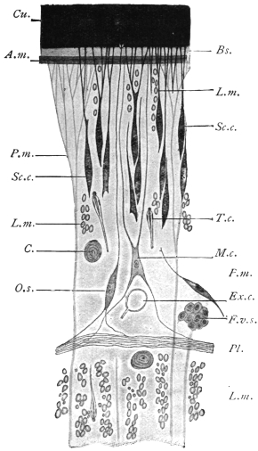
Fig. 184.—Schematic representation of a small
part of a transverse section of Ligula sp. Bs., basal
membrane; Cu., cuticle; at its base are the endplates
of the subcuticular (epithelial) cells; in the
centre a cuticular sense organ, O.s.; F.v.s., vitelline
follicle; Exc., excretory vessel; C., calcareous corpuscle;
L.m., longitudinal muscles; M.c., myoblast;
P.m., parenchymatous or dorso-ventral muscles;
Pl., plexus of nerve fibres; A.m., circular muscles;
Sc.c., subcuticular or matrix cell; T.c., terminal
flame cell. 500/1. (After Blochmann.)
In addition to the above
mentioned, there are other
cuticular formations occurring
on the cuticle of some
Cestodes, such as immobile
hairs and variously formed
hooks, such as are seen principally
on the scolex. Their
development is only roughly
known in a few species; they
are usually already present in
the larval stage, and of the
same arrangement and shape
as in the fully developed
tapeworms; a matter of
importance, because by these
structures larvæ can be recognized
as being those of a certain species of tapeworm.
The CUTICULAR GLANDS in Cestodes are scarce.
The PARENCHYMA forms the chief tissue of the entire body, and in
all essentials its structure is similar to that of the Trematodes.
The same doubt exists here also as to the nature of the parenchyma.
Recent authors consider that it consists of highly branched cells, the
processes of which ramify in all directions. These cells lie in a non-cellular
matrix containing fluid vacuoles. This matrix spreads in
between and so breaks the continuity of the epidermal cells.
In the parenchyma of almost all the Cestodes there are found in
adult specimens, as well as in larvæ, light-refracting concentrically
striated structures, of a spherical or broad elliptical shape, which, on
account of their containing carbonate of lime, are termed CALCAREOUS
CORPUSCLES (fig. 184, C.). Their size, between 3 µ and 30 µ, varies
according to the species; their frequency and distribution in the parenchyma
also varies, but they are chiefly found in the cortical layer.
They are the product of certain parenchymatous cells, in the interior
of which they lie like a fat globule in a fat cell, but according to
others they are intercellular in origin.
The MUSCULAR SYSTEM of the proglottids is composed of—(1) the
subcuticular muscles (figs. 184 and 185), as a rule consisting of a
single layer of annular muscles; (2) longitudinal muscles; (3) dorso-ventral
fibres extending singly from one surface to the other, and
at both ends expanding in a brush-like manner, and inserted into
the basal membrane, consisting of an outer, more numerous, and an
inner, less numerous but more powerful layer (the number of bundles
in this layer being in certain cases of specific importance); (4) transverse
fibres, the elements of which penetrate to the borders of the segments,
thus passing through the longitudinal muscles and reaching
the cuticle. In the region of the septa the transverse and dorso-ventral
muscles form a kind of plate.
Fig. 185.—Half of a transverse section through a proglottis of Tænia crassicollis.
Cu., cuticle; Ex.v., external excretory vessel, to the right of which there is the smaller
internal one; T., testicular vesicles; L.m., longitudinal muscles (outer and inner); M.f., lateral
nerve with the two accessory nerves; Sc.c., subcuticular matrix cells; Sm.f., submedian
nerve; Tr.m., transverse muscles; Ut., the uterus, and the middle of the entire transverse
section. 44/1.
The mass of parenchyma bounded by the transverse muscles is
termed the MEDULLARY layer, while the mass lying outside them
is termed the CORTICAL LAYER.
It was known long ago that the myoblasts adhere to the dorso-ventral
fibres as thickenings, but it is only recently that large star-shaped
cells (fig. 184), separated from but connected with them by
processes, have been recognized as
the myoblasts of other fibres (Blochmann,
Zernecke).
Within the scolex the direction
and course of the muscular layers
change.
Fig. 186.—Dipylidium caninum: from
the cat. In the upper figure the rostellum is
retracted, in the lower protruded, a, sucker;
b, hooks of rostellum; B, enlarged hook;
c, apical aperture on scolex; d, longitudinal
muscles; e, circular muscles. (After
Benham.)
The SUCKERS are parts of the
musculature, locally transformed,
with a powerful development of
the dorso-ventral muscles, now
become radial fibres.
The ROSTELLUM of the armed
Tæniæ, like the proboscis of the
Rhynchobothriidæ, also belongs to
the same category of organs.
In the simplest form, the rostellum,
or top of the head (as
in Dipylidium caninum), appears
as a hollow oval sac, the anterior
part of which, projecting beyond
the upper surface of the head, carries several rows of hooks (fig. 186).
The entire internal space of the sac is occupied by an elastic, slightly
fibrous mass, while the anterior half of the surface of the rostellum is
covered by longitudinal fibres and the posterior half by circular fibres.
On contraction of the latter the entire mass is protruded through the
apical aperture, the surface of the rostellum becomes more arched, and
the position of the hooks is, in consequence, altered. The rostellum
of the large-hooked Tæniidæ, which inhabit the intestine of man and
beasts of prey, is of a far more complicated structure, for, in addition
to the somewhat lens-shaped rostellum carrying the hooks on its outer
surface, there are secondary muscles grouped in a cup-like manner
(fig. 187). Every change in the curvature of the surface of the rostellum
induces an alteration in the position of the hooks. In the hookless
Tæniidæ the muscular system of the rostellum is altered in a very
different manner; in a few forms a typical sucker appears in its place.
Fig. 187.—Longitudinal section of the head
and neck of Tænia crassicollis, showing the
lens-shaped muscular rostellum, with two hooks
lying in the concentric cup-like mass of muscles.
L.m., longitudinal muscles of the neck; L.f., left
lateral nerve; G., ganglion; S.c., subcuticular
layer; W1, external, W2, internal excretory
vessel. 30/1.
The NERVOUS SYSTEM commences in the scolex and runs through
the neck and the entire series of proglottids. Within the proglottids
it consists of a number of longitudinal nerve fibres of which those at
each lateral border are usually the largest. In the Tæniæ the lateral
nerves are accompanied both
dorsally and ventrally by a
thinner nerve (accessory nerve)
(fig. 185); on each surface,
moreover, between the lateral
nerve and the median plane,
there are two somewhat
stronger bundles (sub-median),
so that there is a total of ten
longitudinal nerve bundles.
They lie externally to the transverse
muscle plates, and the
lateral and accessory bundles
lie externally to the principal
excretory vessels, and are everywhere
connected by numerous
anastomoses and secondary
anastomoses; one typical ring
commissure is usually found
at the posterior border of the
segments. In the Bothriocephalidæ
the distribution of
the nerve bundles is different
(for instance, two lie in the
medullary layer), or they are
split up into a larger number
of branches. In the scolex the nerve bundles are connected in a very
remarkable manner by commissures with that which is generally
termed the central part of the entire nervous system. There occurs
normally a commissure between the two lateral nerves; at the same
level, the dorsal and ventral median nerves are also connected at each
surface as well with each other as with the lateral nerves, so that a
hexagonal or octagonal figure is formed. The so-called apical nerves
pass from this commissural system anteriorly, embrace the secondary
muscular system of the rostellum semicircularly, and form an annular
commissure (rostellar ring) at the inner part of the rostellum.
Fig. 188.—Tænia cœnurus, head and part of
neck showing nervous system. Enlarged. (After
Niemiec.)
The peripheral nerves arise from the nerve bundles as well as
from the commissures situated in the scolex; some go direct to the
muscles, while others form a close plexus of nerves external to the
inner longitudinal muscles, which plexus likewise sends out fibres to
the muscles, but principally to numerous fusiform sense organs
(fig. 184, Pl.); they lie internal to the subcuticular cells and, piercing the
cuticle with their peripheral processes, end as projecting “receptor”
hairs. Higher organs of sense are not known.
The EXCRETORY APPARATUS of the Cestodes is similar to that of
other flat worms. The terminal
(flame) cells, which hardly differ
in appearance from those of
the Trematodes, are distributed
throughout the parenchyma, but
are more common in the cortical
than in the medullary layer
(fig. 184, T.c.). Before opening
into a collecting tube, the capillaries
run straight, tortuously,
or in convolutions, anastomosing
frequently with one another
or forming a rete mirabile. The
collecting tubes, which have
their own epithelial and cuticular
wall, and which also
appear to be provided with
muscular fibres, occur typically
as four canals passing through
the entire length of the worm
(fig. 189); they lie side by side,
two (a wider thin-walled ventral,
and a narrower thick-walled
dorsal one) in either lateral field;
in the head the two vessels on
each side unite by means of a loop, at the posterior extremity they
open into a short pyriform or fusiform terminal bladder which discharges
in the middle of the posterior edge of the original terminal
proglottis.
This primitive type (fig. 189) of arrangement of collective tubes is
subject to variation in most Cestodes, in the scolex as well as in the
segments. Indeed, even the lumen of the four longitudinal tubes
does not remain equal, as the dorsal or external tubes are more fully
developed and become thicker, whereas the ventral or internal ones
remain thin, and in some species quite disappear in the older segments
(figs. 185, 187). Moreover, very frequently connections are
established between the right and left longitudinal branches, as in the
head, where a “frontal anastomosis” develops, which in the Tæniidæ
usually takes the form of a ring encircling the rostellum (fig. 190), and
in the segments of a transverse anastomosis at each posterior border,
especially between the larger branches, and more rarely between the
smaller collecting tubes also (fig. 191).
Fig. 189.—Young Acanthobothrium
coronatum, v. Ben., with the excretory
vessels outlined. Slightly enlarged. (After
Pintner.)
Fig. 190.—Scolex of a cysticercoid
from Arion sp., with the excretory
vessels outlined. (After Pintner.)
The so-called “island” formation is another modification, i.e.,
at any spot a vessel may divide and after a longer or shorter course
the two branches reunite, and this may appear in the collecting tubes
themselves as well as in their anastomoses. The above-mentioned ring
in the frontal commissure of the Tæniidæ is such an island; similar
rings also frequently encircle the suckers (fig. 190). In extreme cases
(Triænophorus, Ligula, Dibothriocephalus, etc.) this island formation
extends to all the collecting tubes and their anastomoses. Instead of
two or four longitudinal canals only, connected by transverse anastomoses
at the posterior border of the segments, there is an irregular
network of vessels, situated in the cortical layer, from which the
longitudinal branches, having again subdivided, can only be distinguished
at intervals, and even then not in their usual number.
The opening of the longitudinal branches at the posterior end
requires more accurate investigation; it is true that a single terminal
bladder is mentioned as being present in many species, but this is
also disputed; when the original end proglottis has been cast off, the
longitudinal branches discharge separately. Some species possess the
so-called foramina secundaria, which serve as outlets for the collecting
tubes; they are generally at the neck, but may be situated on the
segments.
The contents of the excretory vessels is a clear fluid, the regurgitation
of which is prevented by the valves present at the points of
origin of the transverse anastomoses. The fluid contains in solution
a substance similar to guanine and xanthine.
Genital Organs.—With the exception of one genus (Diœcocestus,
Fuhrm.), in which the species are sexually differentiated, all the
Cestodes are hermaphroditic; the genitalia develop gradually in
the segments (never in the scolex), the male organs, as is usual
in hermaphroditic animals, forming earlier than the female. The
youngest proglottids generally do not exhibit even traces of genitalia:
these, as a rule, develop first in the older segments, and the development
proceeds onwards from segment to segment. In a few exceptional
cases (Ligula) the sexual organs are already developed in
the larval stage, but are only functional after the entry of the parasite
into the final host.
Fig. 191.—Proglottis of Tænia saginata, Goeze, showing genitalia. C., transverse
excretory canal; N., lateral longitudinal nerve; W., longitudinal excretory
canal; T., testicles scattered throughout the proglottis; Ut., opposite the central
uterine stem (a closed sac); Ss., genital pore leading into the genital sinus; above
the cirrus and coiled vas deferens (V.d.), below the vagina (Vag.), bearing near
its termination a dilatation, the seminal receptacle; Vsc., the triangular vitellarium,
and above it (Shg.) the shell gland; leading from this to the uterus is seen
the short uterine canal, on either side of this the two lobes of the ovary (Ov.). 10/1.
With the exception of the end portions of the vagina, cirrus and
uterus, all the parts of the genital apparatus lie in the medullary layer,
except only the vitellaria, which in many species are in the cortical
layer. The male apparatus consists of the testes, of which, as a rule,
there are a large number,278 and which lie dorsal to the median plane
(fig. 185, T.); a vas efferens arises from each testis, unites with
contiguous vasa, and finally discharges into the muscular vas deferens
that is situated in about the middle of the segment. According to
the position of the genital pore, the vas deferens opens on the lateral
margin or in the middle line in the front of the segment; it is much
convoluted or twisted, and frequently possesses a dilatation termed
the vesicula seminalis. It finally enters the cirrus pouch, which is
usually elongated; within the cirrus pouch lies the protrusible cirrus,
which is not uncommonly provided with hooklets.
Fig. 192.—Dibothriocephalus latus. Upper figure: female genitalia, ventral
view. Lower figure: male genitalia, dorsal view. The central portion only of
the proglottis is shown. a, cirrus sac; b, partly everted cirrus; c, genital atrium
and pore; d, vaginal pore; e, uterus; f, uterine pore; g, vagina; h, ovary;
i, shell gland; j, vitelline duct; k, lateral nerve; l, vitellarium; n, vas deferens
(muscular portion); p, vas deferens; q, seminal vesicle; r and x, vasa efferentia;
s, lateral excretory canal; t, testicular follicles. (After Benham and Sommer
and Landois.)
The male sexual orifice almost always opens with that of the
vagina into a genital atrium, the raised border of which rises above
the edge of the segment and forms the genital papilla (fig. 191).
Fig. 193.—Diagram of genitalia of a Cestode. g.p., genital pore; ♀ ♂, male
and female ducts opening into genital sinus; c.s., cirrus sac; v.d., coiled vas
deferens (“outer seminal vesicle”); vag., vagina; sem. rec., seminal receptacle;
sp. d., spermatic duct; C.c., fertilization canal; vit. d., vitelline duct; sh. g., shell
gland; ut. c., uterine canal; ut., uterus; Ov., ovary; p, pumping organ.
Cf. figs. 191 and 233. (Stephens.)
The vagina, like the vas deferens, usually runs inwardly and
posteriorly, where it forms a spindle-shaped dilatation (receptaculum
seminis); its continuation, the spermatic duct, unites with the
oviduct, the common duct of the ovaries (fig. 191). The ovaries,
usually two in number, are compound tubular glands in the posterior
half of the proglottis, which extend into the medullary layer, but
ventral to the median plane.
At the origin of the oviduct there is frequently a dilatation provided
with circular muscles (suction apparatus), which receives the
ovarian cells and propels them forward. After the oviduct has
received the spermatic duct the canal proceeds as the fertilization
canal, and after a very short course receives the vitelline duct or
ducts, and then the numerous ducts of the shell glands (oötype).
[Although the nomenclature of these parts varies, we may consider
the oviduct as extending from the ovary to the shell gland and
as receiving the spermatic duct and then the vitelline duct and the
ducts of the shell gland. The short piece into which the shell gland
ducts open corresponds to the oötype in the flukes, but in the tapeworms
this portion of the canal is seldom dilated. From this point
the oviduct is continued as a shorter or longer tube, the uterine canal
or true oviduct opening into the uterus proper.—J. W. W. S.]
The vitellarium may be single, but often exhibits its primitive
duplication more or less distinctly, in which case it is situated at the
posterior border of the segments in the medullary layer (fig. 191).
The original position of the double organ is, moreover, the same
as in the Trematodes, i.e., at the sides of the proglottids, and thence
eventually extending more or less on both surfaces (figs. 192 and 194);
the gland is then distinctly grape-like and the follicles lie mostly in
the cortical layer.
Fig. 194.—Part of a transverse section through a proglottis of Dibothriocephalus latus. Ct.,
cuticle; C., cirrus; Vvs., vitelline follicles; L.M., longitudinal muscles; T., testicles;
M., medullary nerve; S.c., subcuticle; T.m., transverse muscles; Ut., uterus. 20/1.
The egg cell that has been fertilized and supplied with yolk cells
receives the shell material at the point of entry of the shell gland ducts,
and, as a complete egg, then moves onward to the uterus. In those
cases in which the uterus in its further course presents a convoluted
canal, and may form a rosette (pseudo-phyllidea), there is an external
opening which is usually separate from the genital pore, and lies on
the same or the opposite surface. In all other cases, however, the
uterus terminates blindly and is represented by a longer or shorter sac
lying in the longitudinal axis (fig. 191), but in many forms transversely.
With the accumulation of eggs it becomes modified in
various ways: (1) it sends out lateral branches (fig. 241), or (2) forms
numerous isolated sacs (PARENCHYMAL CAPSULES) containing single
eggs or groups of eggs (fig. 217); further, (3) in some cases at the
blind end one or more special thick-walled cavities are formed
(PARUTERINE ORGANS or UTERINE CAPSULES), in which all or most of
the eggs are collected, the uterus then undergoing atrophy.
In species in which the uterus lacks an opening, simultaneously
with the growth of this organ an atrophy of the male apparatus, at
least of the testes and their excretory ducts, takes place; this atrophy
also frequently occurs in the female glands, so that the entire mature
segments have besides the uterus only traces of the genitalia left.
In the Acoleïnæ the vagina is more or less extensively atrophied,
and in any case has no external opening.
A number of genera are distinguished by the duplication of
the genitalia in every segment; the genital apparatus in its entirety,
or with the exception of the uterus, is double, or the genital glands
and the uterus are single, but the cirrus, vas deferens and vagina
are double.
On comparing the genitalia of the Trematodes and Cestodes the
parts will be found to agree, but the vagina of the Cestodes
corresponds with the uterus of the Trematodes, and the uterus of the
tapeworms to Laurer’s canal of the Trematodes, which in most of the
Cestodes has lost its external orifice.
Development of the Tapeworms.
Copulation.—As each proglottis possesses its own genital apparatus,
and male as well as female organs are present, the following processes
may occur: (1) self- or auto-fecundation (without immissio cirri);
(2) self- or auto-copulation (with immissio cirri); (3) cross-copulation
between proglottids of the same or different chains (of the same
species); and (4) cross-copulation in the same proglottis in species
with double genital pores. These various modes have actually been
observed.
In those species which lack the vagina (Acoleïnæ) it appears that
the cirri, which are always furnished with hooks, are driven into
the tissues and for the most part reach the receptaculum seminis.
The eggs of all Cestodes are provided with shells, but the shells,
like their contents, vary. In genera that possess a uterine pore the
mature eggs frequently do not differ from those of the Distomata;
they have a brown or yellow shell of oval form provided with an
operculum, and contain a number of yolk cells in addition to the
fertilized ovarian cell (fig. 128), but in other genera (with a uterine
pore) the lid is absent and the egg-shell is very thin, the eggs of these
genera resembling those of Cestodes in which the secretion of the
vitellarium is a light albumin-like substance that contains only a
few granules, and in which the egg-shell is very delicate and without
operculum.
The eggs of Tæniidæ, for example, at first consist of egg-shell
(oötype), ovum and yolk cells. The egg-shell is as a rule soft, colourless
and frequently deciduous, and the yolk is scanty in amount and
contains few granules. The eggs are, moreover, more complicated
than this. They enlarge and change their shape and various envelopes
are developed around the embryo. The egg-shell proper often disappears,
and one or more embryonal envelopes, or protoplasmic
layers, arise, so that eventually it is difficult to say whether the whole
egg is present, and, if not, what the layers that remain really are.
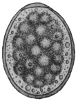
Fig. 195.—Egg of Diplogonoporus
grandis, showing the
morula surrounded by yolk cells
and granules. 440/1. (After
Kurimoto.)
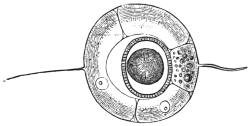
Fig. 196.—Uterine egg of Tænia saginata, G.
Uterine shell with filaments; the oncosphere with
embryonal shell (embryophore) in the centre.
500/1. (After Leuckart.)
The embryonal development in most species takes place during the
stay of the eggs in the uterus; in other species it takes place after the
eggs have been deposited and are in water. Separate cells or a layer of
cells always separate from the segmentation cells, as well as from the
cells of the developing embryo, and form one or more envelopes round
the embryo; usually two such envelopes are formed, the inner one
of which stands in intimate relationship with the embryo itself and
is often erroneously termed the egg-shell, but more correctly the
embryonal shell or embryophore. In some species it carries long cilia,
as in Dibothriocephalus latus, by aid of which the young swim about
when released from the egg-shell; as a general rule, however, there
are no cilia and this envelope is homogeneous, or is composed of
numerous rods and is calcified, as in Tænia spp. (fig. 197). The
second outer envelope (“yolk envelope”) (fig. 207, 3) lies close within
the true (oötype) egg-shell, and remains within it when the embryo
hatches out, and in many species, as in Tænia spp., it perishes at the
end of the embryonal development with the delicate egg-shell which
was formed in the oötype, so that one observes not the entire egg
with egg-shell but only the embryo in its embryonal shell, viz., the
embryophore (fig. 197, a.).
Fig. 197.—a., oncosphere, in its
radially striated embryophore (erroneously
termed egg-shell) of Tænia
africana. Greatly magnified. (After
von Linstow.) b., freed oncosphere of
Dipylidium caninum. (After Grassi
and Rovelli.) Both oncospheres show
six spines.
The embryo (the ONCOSPHERE) enclosed within the embryonal
shell (embryophore) is of spheroidal or ovoid form (fig. 197, b.), and is
distinguished by the possession of three pairs of spines, a few terminal
(flame) cells of the excretory system, and muscles to move the spines.
No further development of the oncosphere takes place, either
in the parent organism or in the open; in fact, in all cases in which
the oncospheres are already formed within the proglottids they do
not become free, but remain in their shell; it is only when the
oncospheres are provided with a ciliated embryophore that they leave
the egg-shell, and they even cast this ciliated envelope after having
swum about in water by its means for a week or so. Sooner or later,
however, all the oncospheres leave the host that harbours the parental
tapeworm and reach the open, either still enclosed in the uterus of
the evacuated proglottids, after the disintegration of which they
then become free, or after being deposited as eggs in the intestine
of the host; they then leave it with the fæces. In the former case
also, the slightest injury to the mature proglottids while still in the
intestine suffices to allow a part of the oncospheres in their embryophores
to be released and mingled with the fæces. Here they are the
generally, but falsely, so-called Tæniæ “eggs.” For, as stated above,
the “yolk” envelope and the true shell deposited in the oötype have
before this disintegrated.
In other cases, e.g., Hymenolepis spp., the uterine (oötype) shell
persists in fæces (fig. 230).
In any case the oncospheres
must be transmitted into suitable
animals to effect their further development;
in only very rare cases
might an active invasion be possible,
as, for instance, takes place with the
miracidia of many Trematodes. The
entry into an animal is, as a rule,
entirely passive, that is to say, the
oncospheres are swallowed with the
food or water. Many animals are
coprophagous and ingest the oncospheres
direct with the fæces; others
swallow them with water, mud, or
food contaminated by such fæces. Infection is easily produced
artificially by feeding suitable animals with mature proglottids of
certain Cestodes or introducing the oncospheres with the food. As
the mature tapeworm frequently finds the conditions suitable for its
development in only one species of host, or in species nearly related,
and perishes when artificially introduced into other hosts, experiment
has taught us that to succeed in cultivating the oncospheres certain
species of animals are necessary. Thus we are aware that the oncospheres
of Tænia solium, which lives in the intestine of man, develop
only in the pig, and only quite exceptionally develop into the stage
characteristic of all Cestodes—the cysticercus in the wide sense of the
word—in a few other mammals. The oncospheres of T. saginata
develop further only in the ox; those of T. marginata (of the dog) in
the pig, goat, and sheep; those of T. serrata (of the dog) in hares and
rabbits; those of Dipylidium caninum (of the dog and cat) in parasitic
insects of the dog and cat, etc. It is not unusual that young animals
only appear to be capable of infection, while older animals of the
same species are not so.
Once introduced into a suitable animal, which is only exceptionally
the same individual or belongs to the same species as the
one which harbours the adult tapeworm, the oncosphere passes
into the larval stage common to all Cestodes, but varying in structure
according to the species. In the simplest case—as, e.g., in Dibothriocephalus—such
a larva resembles the scolex of the corresponding
tapeworm, only that the head, provided with suckers, is retracted
within the fore-part of the neck. Such a larval form is known as a
plerocercoid (πλήερης, full; κέρκος, tail). They differ from the cysticercoids
in being solid larval forms, elongated, tape-like or oval, with the
head invaginated. The conditions appear to be similar in Ligula,
Schistocephalus, Triænophorus, but here the larvæ are very large, indeed
as large in the first-mentioned genera as the tapeworms originating
from them, and the sexual organs are already outlined; doubtless,
however, this stage is preceded by one that corresponds to the scolex
of the genus in question, and which represents the actual larval stage.
In such cases the development of the body of the tapeworm from the
scolex has already begun within the first or intermediate host; in
other cases, except in the single-jointed (monozootic) Cestodes, this
only takes place in the definitive host. The direct metamorphosis
of the oncosphere into the larval forms termed PLEROCERCOID has
hitherto not been investigated, although Ligula, Schistocephalus and
Bothriocephalus are very common parasites, but many circumstances
point to the conclusions arrived at by us and by other observers. In
the larval stages of other tapeworms we can always distinguish the
scolex and a caudal-like appendage, vesicular in the cysticerci (fig. 200),
compact in the cysticercoids (fig. 231). The scolex alone forms the
future tapeworm, the variously formed appendage perishing.
It has now been proved that the appendage, the caudal vesicle,
originates direct from the body of the oncosphere, and therefore is
primary, and that the scolex only subsequently forms through proliferation
on the surface of this appendage. On account of this
origin the scolex is generally regarded as the daughter, and the part
usually designated as the appendage as the mother, originating from
the oncosphere.
Accordingly, two modes of development of the larval stage may
be distinguished; in the one case, plerocerci and plerocercoids, the
oncosphere changes directly into the scolex, thus forming the body
of the tapeworm within the primary host; in the other case, cysticerci
and cysticercoids, the scolex only forms secondarily in the transformed
body of the oncosphere, which later on perishes, the scolex
alone remaining as the originator of the tapeworm colony.
We may summarize briefly what has been said regarding these
larval forms. We have, firstly, solid larval forms without any bladder.
These arise directly from the oncosphere and are of two kinds,
plerocercus and plerocercoid. Plerocercus is a solid globular larva with
the head invaginated into the posterior portion. Plerocercoid (fig. 208)
is a solid elongated larva also with the head invaginated into the
posterior portion, which is sometimes very long. Secondly, we
have larval forms with bladders from which the scolices arise thus
indirectly from the oncosphere. They are of two kinds, cysticercoid
and cysticercus.
Cysticercoid.—The bladder is but slightly developed and is usually
absorbed again. The anterior portion is, moreover, retracted into
the posterior, and in some cases there is a long or a stumpy tail
(figs. 220, 231).
Cysticercus, or true bladder worms. (These may be divided into
(1) cysticercus proper, consisting of a bladder and one scolex;
(2) cœnurus, a bladder and many scolices; (3) echinococcus, a
bladder in which daughter bladders or cysts are developed, and then
in these multiple scolices.)
Fig. 198.—Diagram of a cysticercoid. Cf.
figs. 220, 227. c.v., caudal vesicle or bladder
(small); sec. c., secondary cavity caused by the
growth forward of the hind-body; t., tail bearing
six spines. (Stephens.)
Fig. 199.—Diagram of a cysticercus.
c.v., caudal vesicle or bladder; i., invagination
of wall of bladder. (Stephens.)
In the case of cysticerci a papilliform invagination forms, projecting
into the interior of the bladder (fig. 201). The layer of cells
forming the papilla becomes divided into two laminæ, the outer279 of
which forms a kind of investing membrane (receptaculum capitis) for
the papilla. The head and suckers are now developed on the walls
bounding the axial lumen of the papilla. The papilla eventually
evaginates, so that the receptaculum capitis now forms the inner
surface of the hollow head, which eventually becomes solid.
Our knowledge of the development of cysticerci in the wide
sense of the word is limited almost exclusively to that of a few true
“bladder worms” (cysticerci); in other cases we know either only
the terminal stage, i.e., the complete larva, or, exceptionally, one of
the intermediate stages, but we are not acquainted with a complete
series; the description must therefore be incomplete.
We know from feeding experiments that, after the introduction
of mature proglottids or of the fully developed ova of Tænia
crassicollis (of the cat) into the stomach of mice, the oncospheres
escape from the shell in the middle portion of the small intestine,
and a few hours later penetrate into the intestinal wall by means
of a boring movement; they have been found in this position twenty-seven
to thirty hours after the infection. By means of this migration,
for which purpose they employ their spines, they attain the blood-vessels
of the intestine; indeed, already nine hours after the infection
and later they are found in the blood of the portal vein, and in the
course of the second day after infection they are found in the
capillaries of the liver, which these larvæ do not leave.
Leuckart, in experimental feeding of rabbits with oncospheres
of Tænia serrata (of the dog), found free oncospheres in the stomach
of the experimental animal, but not in the intestine: however,
he came across them again in the blood of the portal vein. The
passage through the blood-vessels to the liver is the normal one
for those species of Tænia the eggs of which become larvae in
mammals; even in those cases in which the oncospheres develop
further in the omentum or in the abdominal cavity (Cysticercus
tenuicollis, C. pisiformis), there are distinct changes observable in
the liver that lead one to the conclusion that there has been a
secondary migration out of the liver into the abdominal cavity.
Indeed, one must not imagine that the young stages of the Cestodes
are absolutely passive; once they have invaded an organ they travel
actively, and leave distinct traces of their passage.
In other cases the oncospheres leave the liver with the circulation,
and are thus distributed further in the body; they may settle and
develop in one or more organs or tissues. Many oncospheres may,
by travelling through the intestinal wall, penetrate through it and
attain the abdominal cavity direct; some, perhaps, pass also into the
lymph stream. Where there are no blood and lymphatic vessels in
the intestinal wall, as in insects, the oncospheres attain the body
cavity or its organs direct; in short, they never remain in the
intestinal lumen itself, and only rarely—as in Hymenolepis murina
of the rat—do they remain in the intestinal wall.
When the infection has been intense, and the body is crowded with numerous
oncospheres, acute feverish symptoms, are induced, to which the infected animals
usually succumb (“acute cestode tuberculosis”); while in other cases the alterations
in the organs attacked—as the liver in mice and the brain in sheep—may cause death.
Sooner or later the oncospheres of tapeworms come to rest,
and are first transformed into a bladder, which may be round or
oval according to the species. The embryonal spines disappear sooner
or later, or remain close together or spread over some part of the
bladder wall (fig. 200). Their discovery by V. Stein in the bladder
worm of the “meal worm” (the larva of a beetle, Tenebrio molitor)
first led to the conclusion that bladder worms (cysticerci) actually
originate from the oncospheres of Tæniidæ.
Fig. 200.—Diagram of development of a cysticercus. 1, solid oncosphere with six spines;
2, bladder formed by liquefaction of contents; 3, invagination of bladder wall; 4, formation
of rostellum (with hooklets) and suckers at the bottom of the invagination; 5, evagination of
head; 6, complete evagination effected by pressure. (Stephens.)
The bladder may remain as a bladder, and then by proliferation
the scolex forms on its wall (fig. 202), or it may divide into an
anterior so-called “cystic” portion and a solid tail-like appendage of
various lengths, on which the embryonal hooks are to be found,
and this is particularly the case in those larval forms (cysticercoids),
e.g., those of Dipylidium caninum, that develop in invertebrate animals,
such as Arthropoda.
As mentioned above one may regard the scolex as an individual
that originates through proliferation of the wall of the parent cyst,
mostly singly, but in those cysticerci that are termed cœnurus
(fig. 201) many scolices occur, whereas in those called echinococcus
the parent cyst originating from the oncosphere of Tænia echinococcus
(of the dog) first produces a number of daughter cysts, which in their
turn form numerous scolices. Echinococcus-like conditions also
occur in cysticercoids, as, for instance, in those peculiar to earthworms;
and similar conditions prevail in a larval form known as
Staphylocystis, found in the wood-louse (Glomeris). Thus it happens in
these cases that finally one tapeworm egg produces not one, but
numerous tapeworms, for, under
favourable conditions, each scolex
can form a tapeworm.
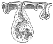
Fig. 201.—Section through a
piece of a Cœnurus cerebralis,
with four cephalic invaginations
in different stages of development.
At the bottom of the
invaginations the rostellum, hooks
and suckers develop. (From a
wax model.)
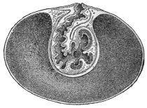
Fig. 202.—Median section through
a cysticercus, with developed scolex
at the bottom of the invagination.
(After Leuckart.)
The rudiment of the scolex appears as a hollow bud, the cephalic invagination
usually directed towards the interior of the bladder cavity; on its invaginated
surface arise the four suckers, and the rostellum with the
hook apparatus is formed in its blind end; we thus get a
Tænia head, but with the position of the parts reversed
(fig. 201). In many cysticerci the head rises up from the
base of the cephalic invagination and is then surrounded by
the latter. A more or less elongated piece of neck also
develops, and even proglottids may appear, as in Cysticercus
fasciolaris (the larva of Tænia crassicollis of the cat) of the
Muridæ, a process somewhat analogous to that of Ligula, etc.
The period that elapses from the time of
infection till the cysticercus is fully developed
varies according to the species; the cysticercus
of Tænia saginata requires twenty-eight weeks,
that of T. marginata seven to eight weeks, that
of T. solium three to four months, and that of
T. echinococcus longer still.
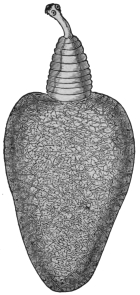
Fig. 203.—Cysticercus
pisiformis in an evaginated
condition, with
neck, fore-body and
bladder, with excretory
network in its wall. 18/1.
With one single exception (Archigetes) the
larvæ do not become sexually mature in the organ
where they have developed; they must enter the
terminal host, a matter that is usually purely
passive, the carriers of the larvæ or infected parts
of them being usually devoured by other animals. In this manner, for
instance, the larvæ (Cysticercus fasciolaris) found in mice and rats reach
the intestine of cats; those of the hare and rabbit (C. pisiformis) reach
the intestine of dogs; those of the pig (C. cellulosæ) are introduced into
man; those of insects are swallowed by insectivorous birds; those of
crustaceans are ingested by ducks and other water fowl; perhaps,
also, the infection of herbivorous mammals is caused by their
accidentally swallowing smaller creatures infected by larvæ. Indeed,
the researches of Grassi and Rovelli have taught us that such an
intermediate host is not always necessary; Hymenolepis murina of
rats and mice in its larval stage lives in the intestinal wall of these
rodents, and as a larva it passes into the intestinal lumen and develops
into a tapeworm in exactly the same way as the larvæ of other species
that reach the intestine of the terminal host by means of an intermediate
carrier. Probably this curtailed manner of transmission also
occurs in many other species. In some cases the larvæ actively quit
the body of the intermediate host, as in the case of Ligula and Schistocephalus,
which travel out of the body cavity of infected fish and
reach the water, where they may be observed in hundreds in summer,
at all events in some localities. The larval stage of Calliobothrium—wrongly
termed Scolex—has been observed swimming free in the sea,
and the scolices of Rhynchobothrium, without their mother cysts, have
been observed free within the tissues of several marine animals. In
any case there is almost always a change of hosts, even in the single-jointed
Cestodes, for the larva of Caryophyllæus, which lives in fishes
of the carp family, is found in limicoline Oligochætes, that of Gyrocotyle
(Chimæra) in shell-fish (Mactra), and different conditions can
hardly be possible for Amphilina. Archigetes alone becomes sexually
mature in the larval stage, but the life-history of this creature is not
well known, so that it is not impossible that the attainment of sexual
maturity as a larva in invertebrates (Oligochætes) is perhaps abnormal,
and somewhat analogous to the maturity of some encysted
Trematodes.
The METAMORPHOSIS OF THE LARVA into the tapeworm is rarely
accomplished in a simple manner; the transformation, however, is
not complex in the single-jointed Cestodes, nor in Ligula and Schistocephalus;
the latter is swallowed by birds (Mergus, Anas, etc.), produces
eggs after only a few days, and very soon quits the intestine of its
terminal host. In all other cases it is the scolex only which, by
proliferating at its posterior extremity, forms the proglottids, after
having invaded as a larva the intestine of a suitable host. The mother
cysts, or what corresponds to them, die, are digested, absorbed, or
perhaps even eliminated; on the contrary, segments found on the
scolex during the larval stage, also in the case of Cysticercus fasciolaris,
are retained. It is not certain whether the larvæ of Dibothriocephalus
lose any part.
The time required by the scolex to complete the entire chain
of proglottids does not depend only on the number it has to
produce, for Tænia echinococcus, which, as a rule, only possesses
three or four segments, takes quite as long a time for their growth
(eleven to twelve weeks) as T. solium with its numerous segments;
T. cœnurus is fully developed in three to four weeks, and
the same holds good for Dibothriocephalus latus, which possesses
many more segments than the above-mentioned Tænia of the dog.
In a number of species it has been possible to determine fairly
accurately the average daily growth; for instance, in Dibothriocephalus
latus the daily growth is 8 cm., in Tænia saginata 7 cm., etc.
The history of the development of the Cestodes demonstrates that
persons and beasts harbouring larval tapeworms have become
infected by having swallowed the oncospheres of the species of
tapeworm to which they belong. In regard to Hymenolepis murina
alone, it is known that the introduction of the oncospheres into those
species of animals which harbour the adult tapeworm leads to the
formation of the latter after the development of a larval stage in the
intestinal wall; nevertheless, only young animals (rats) are capable of
infection, for a previous infection, or the presence of mature tapeworms
in the intestine, appears to produce a kind of immunity.
Biology.
In their adult stage, the tapeworms inhabit almost exclusively
the alimentary canal of vertebrate animals, with but few exceptions
the small intestine, and a few species select definite parts of it. A
small number of Rhynchobothriidæ of marine fishes live apparently
always in the stomach, while in rays and sharks the spiral intestine is
their exclusive site. Bothriocephali generally attach themselves with
their head on to the appendices of the pylorus of fishes; other
species (Hymenolepis diminuta) occasionally fix their head in the
ductus choledochus, and this is more frequent still in the tapeworms
of the rock badger (Hyrax), which occasionally penetrate entirely
into the biliary ducts. Stilesia hepatica, Wolffh., has so far only been
found in the bile-ducts of its host (sheep and goat, East Africa).
In the disease of sheep induced by Cestodes, the worms have
been observed also in the pancreas. Specimens found in the large
intestines were probably being evacuated.
The Cestodes are looked upon as fairly inert creatures, this
opinion having been formed by observing their condition in the cold
cadavers of warm-blooded animals. Actually, however, they are
exceedingly active, and accomplish local movements within the
intestine, for they have been found in the ducts communicating with
the bowel, or in the stomach, and may even make their way forward
into the œsophagus.
They also invade other abdominal organs through abnormal communications,
or through any that may be temporarily open between
the intestine and such organs; they thus reach the abdominal cavity or
the urinary bladder, or they work their way through the peritoneum.
They produce changes in the intestinal mucous membrane at the
place of their attachment, the alterations varying in intensity according
to the structure of the fixation organs. The mucous membrane
is elevated in knob-like areas by the suckers; the epithelial cells
become atrophied or may be entirely obliterated. Dipylidium
caninum bores into the openings of Lieberkühn’s glands with its
rostellum, dilating the lumen to two or three times its normal size,
while the suckers remain fixed between the basal parts of the cells.
Species with powerful armatures penetrate deeper into the submucosa,
and some that are not provided with exceptionally strong armatures,
or are even unarmed, may be actually found with the scolex embedded
in the muscles of the intestinal walls or even protruding beyond
(Tænia tetragona, Mol., in fowls, etc.). Other species, again, even
cause perforation of the walls of the intestine of their hosts.
It is generally assumed that tapeworms, which almost without
exception live in the gut of vertebrates, get their nutriment from the
gut contents, which apparently they absorb through the whole body
surface (cuticular trophopores). In favour of this view is the
existence of fat drops in the proglottids, the identity in colour in
certain forms between that of the fresh worm and the gut contents
and the passage of certain substances derived from medicines (iron
and mercury preparation) into the worms in the gut, etc. Whether
the suckers are concerned in the absorption of nutriment and to
what extent is still questionable.
The length of life of the adult tapeworm certainly varies;
as a rule it appears to last only about a year; in other cases (Ligula)
it averages only a few days, but we are likewise aware that certain
species of Cestodes of man attain an age of several or many years
(thirty-five). The natural death of Cestodes often appears to be
brought about by alterations in the scolex, such as loss of the hooks,
atrophy of the suckers and rostellum, finally the dropping off of the
scolex; it is unknown whether a chain of segments deprived of its
scolex then perishes or whether it first attains maturity. It has already
been mentioned that in a few species the foremost proglottids are
transformed into organs of fixation on the normal loss of the scolex.
Abnormalities and malformations are encountered relatively frequently in the
Cestodes—such as abnormally short or long segments; the so-called triangular
tapeworms, which—if belonging to the Tæniidæ—always possess six suckers; often
also club-shaped segments occur between normal ones, or there may be a defect
in one segment or in the centre of a number following one another (fenestrated
segments); bifurcated chains of segments have likewise been observed, as well as
incomplete or complete union of the proglottids, abnormal increase of the genital
pores, reversion of the genitalia. Besides the above-mentioned increase of the
number of suckers on the scolex (in Tæniæ), there may be a decrease in the
number; in other cases the crown of hooks may be absent, or abnormally shaped
hooks may be formed.
Classification of the Cestoda of Man.
Order. Pseudophyllidea, Carus, 1863.
Scolex without proboscis or rostellum. Head “stalk” absent.
Scolex never with four, generally with two (or one terminal) bothria.280 Vitellaria
numerous. Uterine opening present. Genitalia do not atrophy when uterus is
developed. In large majority of proglottids eggs (or, if formed, their contents) are
at the same stage of development.
Family. Dibothriocephalidæ, Lühe, 1902.
Syn.: Diphyllobothriidæ, Lühe, 1910.
Genitalia repeated in each proglottid (polyzootic Cestodes). Ventral and dorsal
surfaces flat. Cirrus unarmed. Cirrus and vagina if non-marginal open on the
same surface as the uterus. Uterus long, convoluted, often forming a “rosette,”
never dilates into a uterine cavity. Eggs thick shelled, operculated, constantly
being formed in mature proglottids.
Sub-family. Dibothriocephalinæ, Lühe, 1899.
Syn.: Diphyllobothriinæ, Lühe, 1910.
Segmentation distinct. Scolex unarmed, elongated, sharply separated (generally
by a neck) from the first proglottis. Cirrus and vagina open ventrally. Genital
pores non-alternating. Vas deferens surrounded by a muscular bulb. Receptaculum
seminis large, sharply separated from the spermatic duct.
Order. Cyclophyllidea, v. Beneden.
Four suckers always present. Uterine opening absent. Vitellarium single.
Genitalia atrophy when uterus is fully developed.
Family. Dipylidiidæ, Lühe, 1910.
Rostellum if present armed. Suckers unarmed. Uterus breaks up into egg
capsules. Paruterine organs absent.
Family. Hymenolepididæ, Railliet and Henry, 1909.
Segment always broader than long. Genitalia single. Longitudinal muscles in
two layers. Genital pores unilateral. Testes one to four. Uterus persistent, sac-like.
Eggs with three shells.
Family. Davaineidæ, Fuhrmann, 1907.
Rostellum cushion-shaped. Armed with numerous (sixty to several thousand)
hammer-shaped hooks in two (rarely one) rows.
Sub-family. Davaineinæ, Braun, 1900.
Suckers armed. Uterus breaks up into egg capsules. Paruterine organs absent.
Family. Tæniidæ, Ludwig, 1886.
Suckers unarmed. Uterus with median longitudinal stem and lateral branches.
Female genitalia at the hind end of the proglottis. Genital pore irregularly alternating.
Testes numerous in front of female genitalia. Ovary with two lobes (wings).
Vitellarium behind the ovary. Embryophore radially striated.
The Cestodes of Man.
Most of the species to be mentioned live in man in their adult
stage and occupy the small intestine; man is the definite host of these
parasites, but is not the specific host for all the species; some of these
species, as well as others (of mammals), may occur in man also in the
larval stage.
Family. Dibothriocephalidæ.
Sub-family. Dibothriocephalinæ.
Genus. Dibothriocephalus, Lühe, 1899.
Syn.: Diphyllobothrium, Cobbold, 1858; Bothriocephalus, p. p. Rud., 1819;
Dibothrius, p. p. Rud., 1819; Dibothrium, p. p. Dies., 1850.
Scolex egg-shaped; dorsal and ventral bothria elongated, moderately strong,
cutting rather deeply into the head; genitalia single in each proglottis; papillæ in
the vicinity of the genital atrium; the testes and vitellaria are in the lateral fields, the
former in the medullary layer, the latter in the cortical layer on both surfaces, and
occasionally extending to the median line; the ovary ventral, the shell gland dorsal.
The uterus is in the central field, taking a zigzag course, and frequently forms a
rosette.
Dibothriocephalus latus, L., 1748.
Syn.: Tænia lata, L., 1748; Tænia vulgaris, L., 1748; Tænia grisea, Pallas,
1796; Tænia membranacea, Pall., 1781; Tænia tenella, Pall., 1781; Tænia dentata,
Batsch, 1786; Bothriocephalus latus, Bremser, 1819; Dibothrium latum, Dies.,
1850; Bothriocephalus cristatus, Davaine, 1874281; Bothriocephalus balticus,
Kchnmstr., 1855; Bothriocephalus latissimus, Bugn., 1886.
Length 2 to 9 m. or more; colour yellowish-grey; after lying in
water the lateral areas become brownish and the uterine rosette
brown. The head is almond-shaped, 2 to 3 mm. in length, the dorso-ventral
axis is longer than the transverse diameter; the head, therefore,
generally lying flat, conceals the suctorial grooves at the borders;
these suckers are deep and have sharp edges (fig. 205). The neck
varies in length according to the degree of contraction and is very
thin; there are 3,000 to 4,200 proglottids and there may be more;
their breadth is usually greater than their length, but in the posterior
third of the body they are almost square, and the very oldest are not
uncommonly longer than they are broad. There are numerous testes
situated dorsally in the medullary layer of the lateral fields; the vas
deferens (fig. 192) passes dorsally in transverse loops in the central
field anteriorly and forms a seminal vesicle before its entry into the
large cirrus pouch.
The orifice of the vagina is close behind the orifice of the cirrus;
the former passes almost straight along the median line posteriorly,
and widens into a receptaculum seminis shortly before its junction
with the oviduct; the ovary is bilobed, in shape like the wings
of a butterfly, ventrally in the medullary layer; the shell glands lie
in the posterior recess of the ovary; the uterus, forming numerous
transverse convolutions, passes ventral to the vas deferens forwards.
Eggs (fig. 207) large, with brownish shells and small lids, 68 µ to 71 µ
by 45 µ; the ovarian cell, which is already, as a rule, in process of
segmentation, is surrounded by numerous large yolk cells; the
proglottids nearest the posterior extremity are frequently eggless.
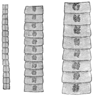
Fig. 204.—Various chains of
segments of Dibothriocephalus
latus, showing the central
uterine rosette. (Natural size.)
Fig. 205.—Transverse section
of the head of Dibothriocephalus
latus. 30/1.
Fig. 206.—Fairly mature proglottis of Dibothriocephalus
latus. The vitellaria are at the sides; the uterus, filled
with eggs, is in the middle, also the vagina (the dark stripe
passing almost straight from the front to the back), and the
vas deferens (almost hidden by the uterus). Above in the
centre is the cirrus sac, and below the shell gland and ovary
are seen. 15/1. (From a stained preparation.)
The eggs, which are deposited in the intestine and evacuated
with the fæces, hatch in water after a fortnight or more; the embryonal
integument (embryophore) of the oncosphere is provided with cilia;
after bursting open the lid of the egg the oncosphere in its embryophore
(fig. 207) reaches the water and swims slowly about; often it
slips out of its ciliated embryophore, sinks to the bottom and is capable
of a creeping motion; sooner or later it dies in the water. The
manner and means of its invasion of an intermediate host are still
unknown; yet we are aware that the larval stage (plerocercoid,
fig. 208), which resembles the scolex and may reach a length of 30 mm.,
lives in the intestine, in the intestinal wall, in the liver, spleen, genital
glands and muscular system (fig. 209) of various fresh-water fish,
the pike (Esox lucius), the miller’s thumb (Lota vulgaris), the perch
(Perea fluviatilis), Salmo umbla, Trutta vulgaris, Tr. lacustris, Thymallis
vulgaris (grayling), Coregonus lavaretus, C. albula (in Europe) and
Onchorhynchus perryi (in Japan). The transmission of the plerocercoids
from these fish to the dog, cat and man (Braun, Parona, Grassi
and Ferrara, Grassi and Rovelli, Ijima, Zschokke, Schroeder) leads to
the development of the broad tapeworm, the growth of which is rapid.
In my experiments on human beings the average number of proglottids
formed per diem averaged thirty-one to thirty-two for five
weeks, with a length of 8 to 9 cm. According to Parona the eggs
appear twenty-four days after man has been infected. Zschokke found
the average growth in the experimental infection of man between 5·2
and 8·2 cm. per diem, and the person experimented upon by Ijima
evacuated a piece of a Dibothriocephalus latus, 22·5 cm. in length,
only twenty-one days after the infection.
Fig. 207.—Dibothriocephalus latus: development of egg. 1, segmentation complete; some
cells of the blastosphere have migrated through the yolk and have flattened to form c, the
yolk envelope; others form a layer of flattened cells (e) forming the embryophore; the
remaining cells (d) of the blastosphere form the hexacanth embryo. 2, embryophore (e) is
becoming thicker. 3, the ciliated embryo has been pressed out of the shell; s′, the operculum;
c, the yolk envelope remaining in the shell (s); y, the yolk consisting of separate cells. 4,
a free-swimming larva much swollen by the water. (After Benham and Schauinsland.)
Fig. 208.—Plerocercoid
of Dibothricephalus
latus. A., with the head
evaginated; B., with
the head invaginated.
From the muscle of
the pike.
The “broad tapeworm” is a frequent parasite
of man in some districts, but it also occurs in the
domestic dog, and on rare occasions is found in
the domestic cat (together with Dibothriocephalus
felis, Crepl.) and fox. French Switzerland and the
Baltic Provinces of Russia are the centres of distribution;
from the former districts the distribution
radiates to France and Italy (Lombardy, Piedmont);
from the Baltic Provinces over Ingermanland
to Petrograd, over Finland to Sweden (on the
shore of the Gulf of Bothnia), in a southerly direction
to Poland, and into the Russian Empire and
across it to Roumania, and towards the west along
the coast of the Baltic Sea to the North Sea, where, however, its
frequency considerably diminishes (Holland, Belgium, and the
North of France).
In Turkestan and Japan the “broad tapeworm” is the most
frequent parasite of man; it has been reported in Africa from the
vicinity of Lake N’gami as well as from Madagascar; cases, in
part at least imported, have also come under observation in North
America.
In Germany Dibothriocephalus latus—apart from the fact that
it is undoubtedly imported from Switzerland, Russia or Italy—is
particularly frequent in East Prussia amongst the inhabitants of
the Courland Lagoon district, on the Baltic; it is, moreover, also
found in the Province and even in the City of Königsberg. In West
Prussia and Pomerania it is very much scarcer.
It is also found in Munich and in the vicinity of the Lake of
Starnberg (Bollinger).
Krabbe found it in 10 per cent. of the sufferers from tapeworms
in Denmark; Szydlowski found the ova of this worm in
Dorpat in 10 per cent. of the fæces examined; Kruse found the
worm in 6 per cent. of post-mortems; Kessler, in Petrograd,
found the eggs in the fæces in 7·8 per cent.; at post-mortems he
found the worms in 1·17 per cent., though Winogradoff only found
it in 0·8 per cent. In Moscow, according to Baranovsky, 8·9
per cent. of the fæces examined contained the ova of Dibothriocephalus.
In the interior and southern provinces of Sweden the
worm, according to Lönnberg, is only found sporadically, but, on
the other hand, in Angermanland about 10 per cent. of the population
is affected; while again in Norbotten the majority of persons
are affected, and in Haparanda the entire population (with the
exception of infants) harbour this parasite. In Switzerland
D. latus is very frequent in close proximity to the lakes of Bieler,
Neuchatel, Morat and Geneva (according to Zaeslin 10 to 15 to
20 per cent. of the population are affected); the parasite is less
frequent in districts one to four hours removed from these lakes.
Of the fish from Swiss lakes examined by Schor those from
Lake Geneva were most commonly infected, and especially Lota sp.
and Perea sp.
The frequency and distribution have, nevertheless, decreased
perceptibly in places; at the commencement of the eighteenth
century the broad tapeworm was very common in Paris, at the
present date it only occurs when imported (Blanchard); in
Geneva, also, according to Zschokke, it has become rarer (formerly
10 per cent., now only 1 per cent.).
The disturbances produced in man by the presence of broad
tapeworms are, as a rule, very trifling; in other cases they produce
partly gastric disorders and partly nervous symptoms; in a number
of cases, again, they set up severe anæmia, apparently caused by
toxins produced by the worms and absorbed by the host. There
is no danger of auto-infection, as the larval stage lives only in
fishes, not in warm-blooded animals. The case reported by
Meschede (ova like those of Dibothriocephalus latus in the brain of
a man who had suffered from epilepsy for six years) must be otherwise
explained.
Fig. 209.—A piece of the body wall of the Burbot, Lota vulgaris. The
tangential section has exposed the muscles of the trunk, with a plerocercoid of
Dibothriocephalus latus. Natural size.
Human beings, like other hosts, can only acquire the broad
tapeworm by ingesting its plerocercoids with the previously mentioned
fresh-water fishes; the opportunity for such infection is
afforded the more readily by the fact that not only do the lower
classes not pay sufficient attention to the cooking of fish, so that
all the larvæ that are present may be killed, but also in certain
localities the custom exists of eating some parts of these fishes in a
raw condition; even the mere handling of the usually severely
infected intermediary hosts may occasionally cause infection. The
plerocercoids are as well known as, but differ materially in appearance
from, the cysticerci (Cysticercus cellulosæ) of pig’s flesh. In
Germany the occurrence of the plerocercoids of Dibothriocephalus
latus has been confirmed in the pike, miller’s thumb and perch of
East Prussia, and more particularly in those taken from the Courland
Lagoon.
The life of D. latus is a very long one (six to fourteen years),
as is deduced from persons who have left D. latus regions after they
have been infected.
According to the experiments of M. Schor, plerocercoids of
D. latus placed in slowly warmed water completely lose their movement
at 54° to 55° C.; they survive the death of their host for several
days; they are killed by low temperatures -3° to +1° C. in two
days; strong acids and salt solutions kill them at once, also high
temperatures, but all the same at least ten minutes is required in
boiling or frying fish in order to kill the plerocercoids with
certainty.
Dibothriocephalus cordatus, R. Lkt.,
1863.
Syn.: Bothriocephalus cordatus, R. Lkt.
Fig. 210.—Cephalic end
of Dibothriocephalus cordatus;
on the left viewed
sideways, on the right from
the dorsal surface, showing
a suctorial groove. (After
Leuckart.)
Length, 80 to 115 cm.; the head is heart-shaped
and measures 2 by 2 mm. The
suctorial grooves are on the flat surface; the
segments commence close behind the head and
increase rapidly in breadth. At only 3 cm.
behind the head they are already mature; the
greatest breadth attained by them averages 7 to 8 mm., the length 3 to
4 mm.; the number of proglottids averages 600; the most posterior ones
are usually square. The uterine rosette is generally formed of six to
eight lateral loops. The eggs are operculated and measure 75 µ by 50 µ.
Dibothriocephalus cordatus is a common parasite of the seal, the
walrus and the dog in Greenland and Iceland, occasionally of man
also. No doubt its larva lives in fishes.
The statement that D. cordatus also occurs in Dorpat in human beings
has been proved erroneous (Zool. Anzeiger, 1882, v, p. 46), as also has the
report that this worm lives in hares in the neighbourhood of Berlin, whither it
was supposed to have been carried by Esquimaux dogs (Rosenkranz in Deutsch.
med. Wochenschr., 1877, iii, p. 620). The parasite stated by the author to be
D. cordatus is Tænia pectinata, Goeze, which has been known since 1766.
Dibothriocephalus parvus, Stephens, 1908.
Largest gravid segments 5 by 3 mm. Uterus forms a central
rosette with four to five loops on each side of median line. In a proglottid
measuring 3·5 by 2·25 mm. the genital atrium is situated 0·4
to 0·5 mm. behind the anterior margin and the uterine opening the
same distance behind the genital atrium. Calcareous corpuscles absent
in the preserved specimens. Eggs operculated, 59·2 µ by 40·7 µ.
Distinguished from Dibothriocephalus latus—(1) by the size of gravid
segments (the minimum width of gravid segments of D. latus is 10
to 12 mm., so that D. parvus is a much smaller worm); (2) quadrate
segments of D. latus measure 6 by 6 mm., those of D. parvus 4 by
4 mm.; (3) by the eggs.
From D. cordatus it is distinguished by—(1) D. cordatus has only
fifty immature segments, D. parvus has at least 200, possibly more;
(2) mature segments of D. cordatus measure 7 to 8 mm., maximum
width of D. parvus is 5 mm.; (3) quadrate segments of D. cordatus
measure 5 to 6 mm.; (4) D. cordatus has six to eight uterine loops;
(5) D. cordatus measures 75 µ to 80 µ by 50 µ.
Habitat.—Intestine of man (Syrian, in Tasmania).
Genus. Diplogonoporus, Lönnbrg., 1892.
Syn.: Krabbea, R. Blanch., 1894.
The scolex is short and has powerful suctorial grooves; no neck;
the proglottids are short and broad; there are two sets of genital
organs side by side in each segment, which in all essentials resemble
the single one of Dibothriocephalus.
Parasitic in whales and seals, occasionally in man.
Diplogonoporus grandis, R. Blanch., 1894.
Syn.: Bothriocephalus sp., Ijima et Kurimoto, 1894; Krabbea grandis, R. Blanch.
Scolex unknown; chain of proglottids over 10 m. in length,
1·5 mm. broad anteriorly, 25 mm. broad posteriorly. The proglottids
are very short (0·45 mm.), but 14 to 16 mm. broad. On either side to
the right and left of the worm, along the entire ventral surface, there
is a longitudinal groove; these grooves are nearer to each other than
to the lateral margin; in them lie the genital pores, and they are in
the same sequence as in Dibothriocephalus; corresponding to the
scanty length (0·45 mm.) of the proglottids, the ovary is only developed
transversely; the uterus only makes a few loops. Eggs (fig. 195)
thick shelled, brown, 63 µ by 48 µ to 50 µ. This parasite has hitherto
been observed twice in Japanese. Similar species are known in Cetacea
and seals.
Fig. 211.—Diplogonoporus grandis, Lühe, 1899: ventral view of a portion of the
strobila, showing two rows of genital pores and partially extruded cirri. (After Ijima and
Kurimoto.)
Fig. 212.—Diplogonoporus grandis: ventral view (diagrammatic) of genitalia
of left side; cir, cirrus; cir.o, cirrus opening; dtg., vitelline duct; ov., ovary;
ovd., oviduct; sb., receptaculum seminis; ut., uterus; ut.o., uterine pore; vag.,
vagina; vag.o., vaginal pore; vd, vas deferens. × 150. (After Ijima and
Kurimoto.)
Sparganum, Diesing, 1854.
The term Sparganum, invented by Diesing, is used as a group name
of larval bothriocephalid Cestodes whose development is not sufficiently
advanced to enable them to be assigned to any particular genus.
Sparganum mansoni, Cobb., 1883.
Syn.: Ligula mansoni, Cobbold, 1883; Bothriocephalus linguloides, R. Lkt.,
1886; Bothriocephalus mansoni, R. Blanch., 1886.
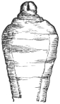
Fig. 213.—Cephalic
end of Sparganum
mansoni, Cobb.
(After Leuckart.)

Fig. 214.—Sparganum
mansoni: on
the right in transverse
section. Natural size.
(After Ijima and
Murata.)
These plerocercoids were discovered in 1882 by P. Manson
during the post-mortem on a Chinaman who had died in Amoy,
twelve specimens being found beneath the peritoneum
and one free in the abdominal cavity.
Cobbold described them as Ligula mansoni, and
Leuckart, who contemporaneously reported a case
in Japan, termed them Bothriocephalus liguloides.
Ijima and Murata reported eight further cases, and
Miyake records nine further cases, seven of which
are recorded in Japanese literature.
The plerocercoid, which hitherto alone is known
to us, attains a length of 30 cm. and a breadth of
3 to 6 to 12 mm. The ribbon-shaped body is
wrinkled, the lateral borders are often somewhat
thickened, so that the transverse section has the
form of a biscuit; the anterior end is usually wider
and has the head provided with two weak suctorial
grooves, either retracted or protracted.
The parasite makes migrations within the body,
and thus may reach the urinary passages; then it is
either evacuated with the urine or has to be removed
from the urethra; not rarely it causes non-inflammatory
tumours on various parts of the skin, which
are at times painful and at times vary in size.
Nothing is known of its development and origin.
Sparganum proliferum, Ijima, 1905.
Syn.: Plerocercoides prolifer, Ijima, 1905; Sparganum
prolifer, Verdun, Manson, 1907.
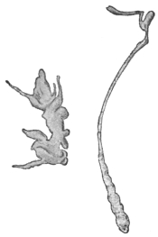
Fig. 215.—Sparganum prolifer:
left with buds, right extended.
× 4. (After Ijima.)
These plerocercoids produce an acne-like condition
of the skin. The condition is really one of
capsules in great abundance in the subcutaneous
tissue and less so in the corium and in the intermuscular
connective tissue. The encapsuled worms
in the corium feel like embedded rice grains and
raise the epidermis, giving rise to an acne-like condition.
Many thousands occur scattered over the
body; in Ijima’s Japanese case there were over
10,000 in the left thigh. The worms when they first
appear in the skin cause itching. The capsules are
ovoid, generally about 1 to 2 mm. in diameter, but
they may be smaller and also much larger. The
larger ones occur in the subcutaneous tissue. The
capsules consist of dense tough connective tissue.
Each capsule, as a rule, contains one worm, but as many as seven
may occur. The skin of areas that have been long infected is swollen
and indurated or adherent, giving a somewhat elephantoid appearance.
The subcutaneous tissue is thick and filled with slimy fluid or in
other parts sclerosed.
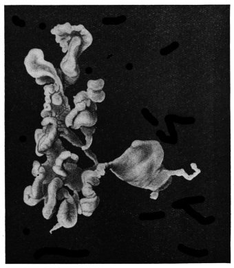
Fig. 216.—Sparganum proliferum. × 10. (After Stiles.)
The Worm.—The chief peculiarity is its
irregular shape and its reproduction in the
larval stage by forming supernumerary heads,
which are supposed to wander about the
body.
The simplest forms are thread-like bodies,
flat or round, 3 mm. long and 0·3 mm. in
diameter, but they may be 12 mm. long by
2·5 mm. broad. The narrow end is the head,
which in life invaginates and evaginates, but
there is no indication of any suckers, except
an inconstant terminal depression. In addition
to these simple forms the most complicated and
irregular forms occur, due to the
formation of buds (heads) at various parts.
The detachment and growth of a head account for the presence of
more than one worm in a cyst. The irregularity in form is also
increased by the presence in the subcuticular tissue of the worm of
reserve food bodies. These bodies are supposed to be of this nature
and are spherical, 100 µ to 300 µ in diameter, but also much elongated.
Calcareous bodies in the Japanese worms were 7·5 µ to 12 µ; in the
Florida worms 8·8 µ to 17·6 µ.
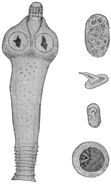
Fig. 217.—Dipylidium caninum: on the
left, the scolex, neck and the first proglottids;
on the right, at the top, a packet of
ova; below, hooks of the rostellum, side
and front views; below, an ovum. Various
magnifications. (After Diamare.)
Fig. 218.—Dipylidium caninum; egg showing
a, egg-shell (vitelline membrane of Moniez);
b, albuminous coat; c, internal shell formed of
or secreted by an outer layer of blastomeres
(Moniez); d, hexacanth embryo. (After Benham
and Moniez.)
Mode of Infection.—Probably from eating uncooked fish.
Distribution.—Japan, Florida.
Family. Dipylidiidæ, Lühe, 1910.
Genus. Dipylidium, R. Lkt., 1863.
Rostellum retractile, with several rings of alternating hooks; the latter with a
disc-like base, having the shape of the thorns of a rose. Genital pores opposite;
genitalia double. Testes very numerous in the central field; ovary with two lobes;
the vitellaria, which are smaller, behind them; the uterus forms a reticulum, in the
network of which the testicular vesicles lie; later on it breaks up into sacs enclosing
one or several eggs. The eggs have a double shell.
Dipylidium caninum, L., 1758.
Syn.: Tænia canina, L., 1758, p. p.; Tænia
moniliformis, Pallas, 1781; Tænia cucumerina,
Bloch, 1782; Tænia elliptica, Batsch, 1786;
Dipylidium cucumerinum, Lkt., 1863.
This worm measures 15 to 35 cm.
in length and 1·5 to 3 mm. in breadth.
The scolex is small, rhomboidal, and
has a club-shaped rostellum on which
there are, in three to four rings, forty-eight to sixty hooks resembling
rose thorns, the size of those in the foremost being 11 µ to 15 µ and those
in the hindmost ring 6 µ. The neck is very short, the most anterior
segments broad and short, the middle as long as they are broad; the
mature segments are longer than wide (6 to 7 mm. by 2 to 3 mm.), fairly
thick, are frequently of a reddish colour, and when cast off resemble
cucumber seeds. The genital pores lie symmetrically at the lateral
margins; the roundish egg sacs, arising from the uterine reticulum,
contain eight to fifteen eggs embedded in a reddish cement substance
(in life). The eggs are globular
(43 µ to 50 µ,); the embryonal
shell (embryophore) is
thin, the oncosphere measures
32 µ to 36 µ. Surrounding the
embryophore is an albuminous
coating, and outside this
the thin vitelline envelope
(fig. 218).
Fig. 219.—Dipylidium caninum: central portion of a proglottis. C.p., cirrus
sac; V.s., vitellaria; Ex.v., excretory vessels; T., testicles lying in the meshes
of the uterine reticulum which laterally forms pouches; O., ovary; U., reticulum
of uterus; V., vagina and seminal receptacle (below ovary). Magnified. (After
Neumann and Railliet.)
Fig. 220.—Dipylidium caninum: development of embryo. 1, solid hexacanth embryo;
2, primitive lacuna (a) in the embryo; 3, elongation of hinder part, rudiments of sucker and
rostellum appearing; 4, “body” and “tail” distinct, (b) and (c) excretory system; 5, fore-body
invaginates into hind-body, excretory bladder has a pore; 6, tail has dropped off; scolex
growing up into secondary cavity formed by fore-body; the primitive cavity has been absorbed
at stage 4. (After Benham, Grassi and Rovelli.)
Dipylidium caninum is a
common intestinal parasite of
dogs, in which it grows larger
(Tænia cucumerina, Bloch)
than in cats (T. elliptica,
Batsch); it has, however, also
been found in jackals, as well
as in human beings, though
in the latter it is of comparatively
rare occurrence (twenty-four
cases), and almost always
affects children, generally of
tender age. One-third of all
the cases in children were
sucklings, about a quarter of
all the cases recorded were
adults, and these occurred
throughout all Europe with
the exception of Spain and
Italy.
The proglottids, which
leave the intestine spontaneously,
are recognizable by the
naked eye on account of their
form and reddish colour, as
well as their two genital pores. As a rule, the presence of this parasite
sets up no marked symptom in the patient.
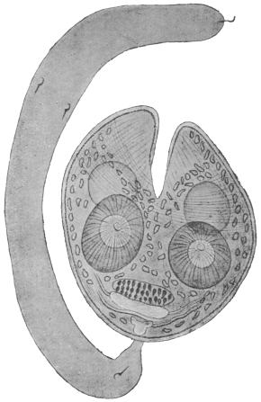
Fig. 221.—Larva (cysticercoid) of Dipylidium
caninum, consisting of body and tail. The latter
is solid and bears on it the embryonal spines. The
bladder, which was only slightly developed, has
disappeared, and the fore-part of the body bearing
the rostellum is now seen invaginated into the
hind portion. The hooklets are shown in front
of the excretory system which has now developed.
At a further stage the tail drops off; the head now
evaginates, but is still enclosed in a double-walled
sac formed by the prolongation upwards on each
side of the topmost parts of the body shown in
the figure. Cf. fig. 220, 6. Enlarged. (After
Grassi and Rovelli.)
The corresponding larval form (cysticercoid) lives in the louse
of the dog (Trichodectes canis), a fact that was first established by
Melnikow and Leuckart; according to Grassi and Rovelli, as well
as Sonsino, it also lives in the flea of the dog (Ctenocephalus canis)
and in the flea of man (Pulex irritans), but not in its larva. The
adult segments, which also leave the rectum of dogs and cats
spontaneously, creep about around the anus and get into the hair,
and are thus partly dried and disintegrated. Part of the segments, or
the oncospheres released by disintegration, are then taken up by lice
and fleas, within which they develop into larvæ (cysticercoids). Dogs
and cats are thus infected by their own skin parasites, which they bite
and swallow whilst gnawing at their fur. The infection of human
beings must occur in an analogous manner, by transmission of the
cysticercoids present on the lips or tongue of dogs when the latter lick
them, or it may be that the vermin of cats and dogs harbouring cysticercoids
are accidentally and directly swallowed by human beings.
Family. Hymenolepididæ, Railliet and Henry, 1909.
Genus. Hymenolepis,282 Weinland, 1858.
Accessory sac (opening into genital atrium) usually absent. Vas deferens with
an external (outside cirrus sac) and an internal (inside cirrus sac) “seminal vesicle.”
Three testes in each proglottis. The eggs are round or oval with two to four distinct
envelopes. In mammals and birds.
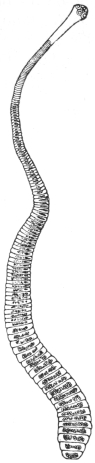
Fig. 222.—Hymenolepis
nana, v.
Sieb. About 12/1.
(After Leuckart.)
Hymenolepis nana, v. Sieb., 1852.
Syn.: Tænia nana, v. Sieb., 1852, nec van Beneden, 1867; Tænia ægyptiaca,
Bil., 1852; Diplacanthus-nanus, Weinld., 1858; Tænia (Hymenolepis) nana, Lkt.,
1863.
The worm is 10 to 45 mm. in length and 0·5 to 0·7 mm. in breadth;
the head is globular, 0·25 to 0·30 mm. in diameter. The rostellum
has a single circlet consisting of twenty-four or twenty-eight to thirty
hooks, which are only 14 µ to 18 µ in length. The neck is moderately
long; the proglottids are very narrow, up to 200 in number,
0·4 to 0·9 mm. in breadth, and 0·014 to 0·030 mm. in length. The
eggs are globular or oval, 30 µ to 37 µ to 48 µ; the oncospheres measure
16 µ to 19 µ in diameter, with two coats, separated by an intervening
semi-fluid substance (fig. 224).
This species was discovered by Bilharz in Cairo in 1851; it was
found by him in great numbers in the intestine of a boy who had
died of meningitis. For several years this was the only case, until
1885, since when numerous cases have come to light. Spooner (1873)
even reported a case from North America, which may, however, have
related to Hymenolepis diminuta. In Europe the worm is particularly
frequent in Sicily, but it has also been repeatedly observed in
North Italy; it has, moreover, been reported from Russia, Servia,
England, France, Germany, North and South America, the Philippines,
Siam and Japan, in all over 100 cases. Notwithstanding its
small size this worm causes considerable disorders in its hosts—mostly
children—as it sets up loss of appetite, diarrhœa, various
nervous disturbances, and even epilepsy; all these symptoms, however,
disappear after the expulsion of the parasites, which are generally
present in large numbers.
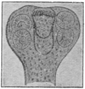
Fig. 223.—Hymenolepis
nana: head. Enlarged.
(After Mertens.)
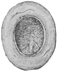
Fig. 224.—Hymenolepis
nana: an egg. Highly magnified.
(After Grassi.)
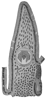
Fig. 225.—Longitudinal
section through the intestinal
villus of a rat, with the
larva (cysticercoid) of Hymenolepis
murina. Magnified.
(After Grassi and
Rovelli.)
Fig. 226.—Hymenolepis nana (murina): cross section of proglottis from a rat. c.p.,
cirrus sac; rec. sem., receptaculum seminis; s.g., shell gland; ov., ovary; t., testis; cort.
par., cortical parenchyma; m.l.n., main lateral nerve; ex. can., excretory canal; y.g.,
vitellarium. (After v. Linstow.)
Fig. 227.—Hymenolepis nana: longitudinal section of an embryo. bl.p.,
anterior opening of secondary cavity; caud., caudal appendage; pr. cav., primary
cavity; sec. cav., secondary cavity. Enlarged. (After Grassi and Rovelli.)
Fig. 228.—Hymenolepis
diminuta:
scolex. Magnified.
(After Zschokke.)
The development as well as the manner of infection is still
unknown; Grassi is of opinion that Hymenolepis nana is indeed
merely a variety of Hymenolepis murina, Duj., which lives in rats.
According to Grassi direct development takes place with omission
of the intermediate host, but with the retention of the larval stage;
that is to say, rats infect themselves directly with Hymenolepis murina,
by ingesting the mature segments or oncospheres of this species, from
which subsequently the small larvæ originate in the intestinal wall
(fig. 225); when fully developed they fall into the intestinal lumen
and become tapeworms. The identity of the two forms has nevertheless
been disputed (Moniez, R. Blanchard, v. Linstow), though their
near relationship cannot be denied. Grassi gave mature segments of
Hymenolepis murina to six persons, but only one person evacuated
a tapeworm. This, however, proves nothing in a district where
Hymenolepis nana frequently occurs in man; it was, moreover, not
possible to infect rats with segments of Hymenolepis nana (of man).
Accordingly this form may represent an independent species, which,
however, like Hymenolepis murina, also omits an intermediate host.
Hymenolepis diminuta, Rud., 1819.
Syn.: Tænia diminuta, Rud., 1819; Tænia leptocephala, Crepl., 1825; Tænia
flavopunctata, Weinld., 1858; Tænia varesina, E. Parona, 1884; Tænia minima,
Grassi, 1886.
This species measures 20 to 60 cm. in length, and up to 3·5 mm.
in breadth; there are from 600 to 1,000 segments. The head is very
small (0·2 to 0·5 mm.), it is club-shaped and has
a rudimentary unarmed rostellum; the neck is short;
the mature segments are 3·5 mm. in breadth, 0·66 mm.
in length; the eggs are round or oval. The outer
egg-shell is yellowish and thickened, with indistinct
radial stripes; the inner embryonal shell (embryophore)
double, thin; the outer layer is somewhat
pointed at the poles; oncosphere 28 µ by 36 µ.
Between the inner and outer shells is a middle
granular layer.
Hymenolepis diminuta lives in the intestine of
rats—Mus decumanus (the sewer rat), Mus rattus (the
black rat), and Mus alexandrinus, rarely in mice; it
is occasionally also found in human beings.
Weinland described it from specimens collected
by Dr. E. Palmer in 1842, in Boston, from a child
aged 19 months, as T. flavopunctata. A second case
relating to a three year old child, from Philadelphia,
was only reported in 1889 by Leidy; a third case
was simultaneously reported of a two year old girl
in Varese (T. varesina); and Grassi
described another case relating to a
twelve year old girl from Catania
(Sicily). Sonsino and Previtera reported
the same species in Italy, Zschokke in France, Lutz and Magalhães
in South America, and Packard in North America: a total of twelve
cases, five from America, the rest from Europe (Ransom).
Fig. 229.—Hymenolepis diminuta: two proglottids
showing testes (3), ovary and vagina.
Slightly enlarged. (After Grassi.)
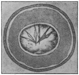
Fig. 230.—Hymenolepis diminuta:
egg from man. (After Bizzozero.)
According to Grassi and Rovelli the larval stage lives in a small
moth (Asopia farinalis), as well as in its larva, in an orthopteron
(Anisolabis annulipes), and in coleoptera (Acis spinosa and Scaurus
striatus). Experimental infections have been successful on rats as
well as on human beings. In America other species of insects may
be the intermediary hosts.
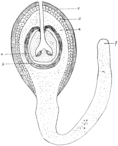
Fig. 231.—Hymenolepis diminuta: cysticercoid from the rat flea (Ceratophyllus fasciatus).
a, remains of primary vesicle; b, fibrous layer; c, radially striated layer resembling cuticle;
d, layer of columnar cells; e, parenchymatous layer of irregularly disposed cells; f, parenchymatous
layer. (Stephens, after Nicoll and Minchin.)
Nicoll and Minchin283 found in the body cavity of 4 per cent.
of rat fleas (Ceratophyllus fasciatus) the cysticercoid of Hymenolepis
diminuta. That it belonged to this species was shown by its unarmed
rostellum and by feeding; 340 fleas were fed to white rats and fourteen
worms obtained, i.e., about 4 per cent., thus corresponding to the infection
of the fleas. The development in the flea probably begins in the
pupal stage, the eggs being ingested by the older flea larvæ. The larva
is 0·31 by 0·25 mm.; tail 0·8 mm., scolex 0·075 by 0·09 mm., suckers,
0·055 mm. in diameter. Microscopically it shows—(1) externally
a radially striated layer resembling cuticle, (2) a layer of columnar cells,
(3) parenchymatous layer continuous with the tail, (4) fibrous layer
around the small caudal vesicle, then the parenchymatous scolex at
the bottom of the secondary cavity.
Nicoll and Minchin (loc. cit.) found a cysticercoid284 in the rat flea
Ceratophyllus fasciatus which was probably that of Hymenolepis murina.
Body 0·16 mm., tail 0·19 mm., scolex 0·096 mm. in diameter. Rostellum
has twenty-three spines in a single row. Length 0·017 mm.,
handle 0·01 mm., guard 0·007 mm., prong 0·007 mm. Sucker 0·042 mm.
Although this cycle, then, for H. murina also exists, it is not probable
that rats (or man in the case of H. nana if this be considered distinct)
infect themselves in this way, as they hardly
ingest all the necessary fleas to account for the
massive infection which frequently exists in
rats (and man), so that Grassi’s cycle holds
good as the predominant method. Xenopsylla
cheopis has also been found by Johnston to
harbour both cysticercoids in Australia.
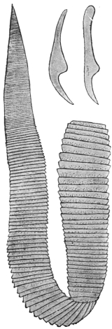
Fig. 232.—Hymenolepis
lanceolata. Natural size.
(After Goeze.) To the
right above, two hooks.
120/1. (After Krabbe.)
Hymenolepis lanceolata, Bloch, 1782.
Syn.: Tænia lanceolata, Bloch, 1782; Drepanidotænia
lanceolata, Railliet, 1892.
The parasite measures 30 to 130 mm. in
length and 5 to 18 mm. in breadth; the
head is globular and very small; the
rostellum is cylindrical, with a circlet composed of eight hooks (31 µ
to 35 µ in length). The neck is very short. The short segments
increase gradually and equally in breadth, but only a little in
length; the female glands lie on the side opposite to that on which
the genital pore is situated; the three elliptical testes are on the
same side as the pores; the cirrus is armed and slender. The
eggs have three envelopes and are oval (50 µ by 35 µ), the external
envelope is thin, the middle intermediate layer or envelope is not
so marked as in H. diminuta, and the internal one is very thin and
sometimes has polar papillæ, as in Hymenolepis diminuta and
H. nana.
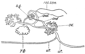
Fig. 233.—Hymenolepis lanceolata: diagram of
female genitalia. ov., ovary; ovd., oviduct; rec. sem.,
receptaculum seminis; s.g., shell gland; ut., uterus;
y.g., vitellarium. (After Wolffhügel.)
It inhabits the intestine of the following birds: Domesticated
ducks and geese, the Muscovy duck (Cairina moschata), white-headed
duck (Erismatura leucocephala), the pochard (Nyroca rufina), and
the flamingo (Phœnicopterus antiquorum). It has been recorded from
Great Britain, France, Denmark, Austria and Germany.
Zschokke reports the receipt of two specimens which a twelve
year old boy in Breslau evacuated spontaneously at two different
times.
The corresponding larva, according to Mrázek, lives in fresh water
Cyclops; according to Dadai it is likewise found in another copepod,
Diaptomus spinosus, but the hooks of Dadai’s larva differed in size.
Family. Davaineidæ, Fuhrmann, 1907.
Sub-family. Davaineinæ, Braun, 1900.
Genus. Davainea, R. Blanch., 1891.
The large scolex is more or less globular, much wider than the rostellum, which is
furnished with two rings of very small and numerous hooks. Neck absent, proglottids
few, genitalia single. Parasitic chiefly in birds.285
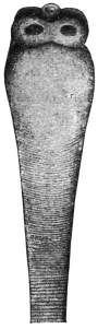
Fig. 234.—Scolex
of Davainea madagascariensis.
The
hooks have fallen
off. 14/1. (After
Blanchard.)
Davainea madagascariensis, Davaine, 1869.
Syn.: Tænia madagascariensis, Dav.; Tænia demerariensis, Daniels, 1895.
This worm measures 25 to 30 cm. in length; the head has four
large round suckers; the rostellum has ninety hooks (18 µ in length);
there are 500 to 700 segments, of which the last 100 are filled
with eggs and form half of the entire worm. The segments, when
mature, measure 2 mm. in length by 1·4 mm. in breadth; genital
pores unilateral; about fifty testes; the uterus consists of a number of
loops, which at each side are rolled up into an almost spherical ball;
when filled with eggs the convolutions unwind, permeate the segment
and then lose their wall; the eggs lying free in the parenchyma
become finally surrounded, one, or several together, by proliferating
parenchymatous cells; this is how the 300 to 400 egg masses, taking
up the entire mature segment, are formed. The globular oncosphere
(8 µ) is surrounded by two perfectly transparent shells, the outer of
which terminates in two pointed processes.
Davainea madagascariensis has hitherto been found in man only
(eight times). Davaine described this species from fragments sent
to him from Mayotta (Comoro Islands), which were found in two
Creole children. Chevreau observed four cases in Port Louis
(Mauritius), likewise in children; Leuckart received the first perfect
specimen—it was obtained from a three year old boy, the son
of a Danish captain, in Bangkok; Daniels, at the post-mortem of
an adult native of George Town, Guiana, found
two specimens (Tænia demerariensis); and finally
Blanchard describes another perfect specimen
which was in Davaine’s collection of helminthes
in Paris, and which was obtained from a little girl
3 years old, of Nossi-Bé (Madagascar). The intermediate
host is unknown.
Davainea (?) asiatica, v. Linst., 1901.
Syn.: Tænia asiatica, v. Linstow.
There exists only one headless specimen of this
species, which is not quite adult, and which is preserved
in the Zoological Museum of the Imperial
Academy of Science in Petrograd. It came
from a human being and was found by Anger in
Aschabad (Asiatic Russia, near the northern frontier
of Persia). The specimen measures 298 mm. in
length. The breadth anteriorly is only 0·16 mm.,
the posterior part measures 1·78 mm. across. The
number of segments is about 750. The genital
pores are unilateral; the testes are globular and
lie in a dorsal and ventral layer in the medullary layer; the cirrus
pouch is pyriform, 0·079 mm. in length and 0·049 mm. in breadth;
the female glands lie in the fore-part of the segments, the ovary
reaching to the excretory vessels; the vitellarium is small and round.
The vagina has a large fusiform receptaculum seminis; the uterus
breaks up into sixty to seventy large, irregularly polyhedric eggsacs.
Family. Tæniidæ, Ludwig, 1886.
Genus. Tænia, L., 1758.286
With the characters of the family. In the genus Cladotænia recognized by
some authors, the testes encroach on the mid field and the uterine stem reaches the
anterior end of the segment.
Tænia solium, L., p. p., 1767.
Syn.: Tænia cucurbitina, Pall., 1781; Tænia pellucida, Goeze, 1782; Tænia
vulgaris, Werner, 1782; Tænia dentata, Gmel., 1790; Halysis solium, Zeder, 1800;
Tænia humana armata, Brera, 1802; Tænia (Cystotænia) solium, Lkt., 1862.
The average length of the entire tapeworm is about 2 to 3 m.,
but may be even more; the head is globular, 0·6 to 0·8 to 1·0 mm.
in diameter. The rostellum is short with a double circlet of hooks,
twenty-two to thirty-two in number, usually twenty-six to twenty-eight;
large and small hooks alternate regularly; the length of the large hooks
is 0·16 to 0·18 mm., of the small ones 0·11 to 0·14 mm. The rostellum
is sometimes pigmented. The suckers are hemispherical, 0·4 to
0·5 mm. in diameter. The neck is fairly thin and long (5 to 10 mm.).
The proglottids, the number of which averages from 800 to 900, increase
in size very gradually; at about 1 m. behind the head they are square
and have the genitalia fully developed. Segments sufficiently mature for
detachment measure 10 to 12 mm. in length by 5 to 6 mm. in breadth.
The genital pores alternate fairly evenly at the lateral margin a little
behind the middle of each segment. The fully developed uterus consists
of a median trunk, with seven to ten lateral branches at either side,
some of which are again ramified. The eggs are oval, the egg-shell
very thin and delicate; the embryonal shell (embryophore) is thick,
with radial stripes; it is of a pale yellowish colour, globular, and
measures 31 µ to 36 µ in diameter; the oncospheres, with six hooks,
are likewise globular, and measure 20 µ in diameter (fig. 238).
Malformations are not so common as in T. saginata; they consist in two or
several proglottids being partly or entirely fused, formation of single club-shaped
segments, fenestration of long or short series of segments and so-called double formation,
in which the head has six suckers and the segments exhibit a Y-shaped transverse
section. The oncospheres occasionally also possess more than six hooklets.
Very slender specimens have led to the description of a particular species or variety
(T. tenella).
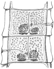
Fig. 235.—Two fairly mature proglottids
of Tænia solium, showing ovaries (one
bi-lobed), vitellaria, central uterine stem,
cirrus and vas deferens (above), vagina
(below), testes (scattered), longitudinal and
transverse excretory vessels.
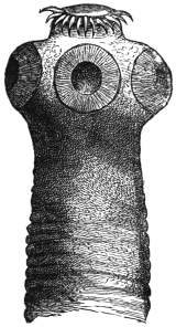
Fig. 236.—Head of Tænia solium.
45/1.
In its fully developed condition T. solium is found exclusively
in man; the head is usually attached in the anterior third of the
small intestine and the chain, in numerous convolutions, extends
backwards; a few mature detached proglottids usually lie at the
most posterior part, and these are usually evacuated during defæcation.
In exceptional cases single proglottids or whole worms may
reach contiguous organs if abnormal communications with them
exist; thus they may reach the abdominal cavity and the urinary
bladder, or they may be found in a so-called worm abscess of the
peritoneum; occasionally, in vomiting, single segments or several
together may be brought up. Exceptionally it induces severe anæmia.
The larval stage (Cysticercus cellulosæ) that gives rise to Tænia
solium lives normally in the intramuscular connective tissue and
other organs of the domestic pig, but it is known to exist also in
a few other mammals, such as the wild boar, the sheep,287 the stag,
dog, cat, brown bear and monkey, as well as in man. The cysticercus
of the pig is an elliptical vesicle with a longitudinal diameter of
6 to 20 mm., and a transverse diameter of 5 to 10 mm.
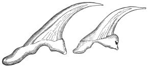
Fig. 237.—Large and small hooks of Tænia solium. 280/1.
(After Leuckart.)
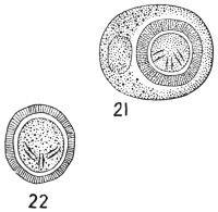
Fig. 238.—Tænia solium. 21, Egg with external membrane;
22, without (embryophore). (After Leuckart.)
Even with the naked eye a white spot may be observed in the
centre of the long equator, this being the invaginated head; it is easy
to make it project by pressing on the vesicle (after tearing off with the
finger-nail the investing connective tissue), and on examining it under
the microscope one can convince oneself that it corresponds with the
head of Tænia solium.
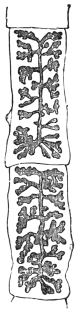
Fig. 239.—Two
mature proglottids
of Tænia solium with
fully developed
uterus. 2/1.
Numerous experiments have proved that the Cysticercus cellulosæ
of the pig, if introduced into the intestine of man, grows to a Tænia
solium (Küchenmeister, 1855; Humbert, 1856; Leuckart, 1856; Hollenbach,
1859; Heller, 1876); the cysticercus has frequently also been
cultivated purposely by feeding pigs with mature proglottids of
T. solium (P. J. van Beneden, 1853; Haubner and Küchenmeister,
1855; Leuckart, 1856; Mosler, 1865; Gerlach, 1870; etc.), but success
did not attend the efforts to make Cysticercus cellulosæ establish themselves
in the intestines of pigs, dogs, guinea-pigs, rabbits and monkeys
(Macacus cynomolgus), and so become adult Tæniæ; the attempts, also,
to infect dogs with cysticerci by means of ova were likewise, as a
rule, abortive.288
The development of Cysticercus cellulosæ takes two and a half to three or four
months; it is not known how long the cysticerci remain alive in animals; not uncommonly
they perish at earlier or later stages, and become calcified or caseated.
Extracted cysticerci die in water at a temperature of 47° to 48° C., in flesh at normal
temperature they remain alive for twenty-nine days or more. On account of the
present rapid means of pickling and smoking meat, the cysticerci as a rule are not
killed, also the effect of cold on them for some time in cold chambers of slaughterhouses
is not lethal, but freezing is fatal (Ostertag).
There is not the least doubt that human beings are almost exclusively
infected with Tænia solium by eating pork containing cysticerci
in a condition that does not endanger the life of the cysticerci. The
infection may likewise be caused in man by eating the infected meat
of other animals subject to this species of bladder worm, mainly, as a
matter of fact, deer and wild boar.
The frequency of cysticerci in pigs’ flesh has considerably decreased since the
introduction of meat inspection; in the Kingdom of Prussia there was on an average
1 infected pig to every 305 slaughtered between 1876 to 1882; from 1886 to 1889, there
was 1 to 551; from 1890 to 1892, there was 1 to 817; in 1896, 1 to 1,470; and in 1899,
1 to 2,102; in the Kingdom of Saxony in 1894 there was 1 infected pig to every 636;
in 1895 there was 1 to every 2,049, and in 1896 only 1 infected pig was found of
5,886 slaughtered. In South Germany pigs with cysticerci are very rare, but are
more frequent in the eastern provinces of Prussia; in 1892 the number of infected
pigs compared with the total slaughtered was as follows:—
| In the district of Marienwerder | 1 : 28 |
| " " Oppeln | 1 : 80 |
| " " Königsberg | 1 : 108 |
| " " Stralsund and Posen | 1 : 187 |
| " " Danzig, Frankfort a. O. and Bromberg | 1 : 250 |
| As compared with the district of Arnsberg | 1 : 865 |
| " " " Coblenz | 1 : 975 |
| " " " Düsseldorf | 1 : 1,070 |
| " " " Münster and Wiesbaden | 1 : 1,900 |
The average for the whole of Prussia in the same year was 1 : 1,290; for the
eastern provinces, on the other hand, 1 : 604. Even more unfavourable are the proportions
in Russian Poland (over 1 per cent. of pigs measly), in Prague (over 3 per
cent.), in Bosnia and Herzegovina (6 to 7 per cent.). The cause for this is most
likely attributable to the manner in which the pigs are kept. When allowed to be
in the farmyards of the small farmers for the whole day, or allowed to wander in the
village streets and pasture lands, they are more liable to take up the oncospheres of
the T. solium than when shut up in good pig-styes.
The geographical distribution of T. solium generally corresponds
with that of the domestic pig and the custom of eating pork in any
form insufficiently cooked or raw. There are, or were, some isolated
districts in Germany, France, Italy and England where the “armed
tapeworm” was frequent (for instance, Thuringia, Brunswick, Saxony,
Hesse, Westphalia, whereas it is and was very scarce in South Germany);
it is thus easily understood why it occurs very rarely in the
East, in Asia and in Africa, in consequence of the Mahommedans,
Jews, etc., not eating pork. In North America, also, T. solium is very
rare; the tapeworm which is known there by this name is generally
T. saginata, Stiles. During the last decade T. solium infection has,
however, very markedly decreased in North and East Germany in
consequence of the precautions exercised by the public in the choice
of pork to avoid trichinosis, especially, however, because measly meat
must be sold as such and must be thoroughly cooked before being
placed on the market; indeed, if badly infected it may not be sold
for food, but can be turned to account for industrial purposes.
The occurrence of Cysticercus cellulosæ in man has been known
since 1558 (Rumler, Obs. med., liii, p. 32); there is hardly an organ
in man in which cysticerci have not been observed at some time;
they are most frequently found in the brain,289 where they grow to a
variety known as Cysticercus racemosus; next in frequency they are
found in the eye, in the muscular system, in the heart, in the subcutaneous
connective tissue, the liver, lungs, abdominal cavity, etc.
The number of cysticerci observed in one man varies between a few
and several thousands. Of the sexes, men are most subject (60 to
66 per cent. of the number attacked). The disturbances caused in
man by cysticerci vary according to the nature or position of the
organs attacked; when situated in the cerebral meninges they have
the same effect as tumours.
During the last decades, however, these cases have also become
less common. In Rudolphi’s time 2 per cent. of post-mortems in
Berlin showed cysticerci; in the ’sixties, according to Virchow, about
the same; in 1875 the number fell to 1·6 per cent.; in 1881 to 0·5 per
cent.; in 1882 to 0·2 per cent.; in 1900 to 0·15 per cent., and in 1903
to 0·16 per cent. Hirschberg between 1869 and 1885 discovered
cysticerci in the eye seventy times in 60,000 ophthalmic cases, but
during the following six years the parasite was only present twice
amongst a total of 46,000 cases of ophthalmic diseases, and since
1895 no ophthalmic case has been met with.
The infection of human beings with the cysticerci can only take
place by the introduction of the oncospheres of Tænia solium into
the stomach with vegetable foods, salads that have been washed
in impure water containing oncospheres, also by drinking contaminated
water; the carriers of T. solium, moreover, infect themselves
still more frequently through uncleanliness in defæcation, the privies
in public localities and many private houses affording striking testimony
of this. The minute oncospheres can thus easily reach the
fingers and thence the mouth (as in twirling the moustache, biting the
nail). More rarely a third manner of transmission or internal auto-infection
may possibly take place, as when, in the act of vomiting,
mature proglottids near the stomach are drawn into it; the oncospheres
or segments there retained are then in the same position as
if they had been introduced through the mouth.
On account of these dangers of internal or external auto-infection, it is therefore
the duty of the medical attendant, after recognizing the presence of tapeworms, to
expel them,290 and in doing so to employ every possible means to prevent vomiting
setting in; it is, however, equally important to take the necessary steps to destroy
the parasites evacuated. It may be incidentally mentioned that in using certain
remedies the scolex not rarely remains in the intestine; the cure in such cases has
not been accomplished, as the scolex again produces new proglottids, and after
about eleven weeks the first formed ones are again mature and appear in the fæces.
Amongst the cysticerci also many malformations appear; thus absence of the
rostellum and the hooks, or double formation with six suckers, or abnormalities of
growth on account of the surroundings, which have had a special name given to
them, viz., Cysticercus racemosus, Zenk. (= C. botryoides, Hell.; C. multilocularis,
Kchnmstr.); these forms are more especially found at the base of the brain, are
irregularly ramified and often without the head.
A certain interest is attached to those forms that have led to the
establishment of a distinct species:—
Cysticercus acanthotrias, Weinld., 1858.
In making the autopsy of a white Virginian woman who had died of phthisis, a
cysticercus was found in the dura mater, and eleven or twelve specimens in the
muscles and subcutaneous tissue. Weinland and Leuckart, who examined the
specimens, found that they resembled Cysticercus cellulosæ in form and size, but that
they carried on the rostellum a triple crown, each consisting of fourteen to sixteen
hooks, which differed from the hooks of C. cellulosæ or of Tænia solium by the
greater length of the posterior root process and the more slender form of the
hooks; the large hooks measured 0·153 to 0·196 mm., the medium-sized hooks,
0·114 to 0·14 mm., and the small ones 0·063 to 0·07 mm.
On account of these differences a distinct species of cysticercus
was established, and this naturally presupposed a corresponding
species of Tænia (T. acanthotrias, Lkt.); this could be done with
justice so long as the case remained isolated, i.e., in America, as
there was the possibility of the corresponding Tænia being found. In
this respect, however, the position has changed; Delore first described
a cysticercus the size of a nut from the biceps muscle of the arm of
a silk-worker in Lyons; according to Bertolis this specimen possessed
hooks of three different sizes, the dimensions of which corresponded
with the figures given by Weinland and Leuckart; the correctness of
the diagnosis could hardly be doubted, as Bertolis was known to be
a very exact observer. A second case has become known through
Cobbold, who regards a specimen of a cysticercus in Dallinger’s
collection as likewise belonging to Cysticercus acanthotrias; this
specimen also came from a man’s brain; finally a third case, also
from France, has been published by Redon. This author, amongst
numerous C. cellulosæ of a man, found one that had forty-one hooks in
three rows, and he was the first to express the opinion that C. acanthotrias
does not represent a distinct species, but is only an abnormality
of C. cellulosæ. This view was also taken by Blanchard and Railliet, and
is probably correct, as the discovery of the large corresponding Tænia
furnished with three rows of hooks is not to be expected in European
beasts of prey, and in Redon’s case C. acanthotrias as well as C. cellulosæ
occurred simultaneously.
The duration of life of C. cellulosæ in man is very long; cysticerci
of the eye have been known to persist for twenty years, and in cysticercus
of the brain ten to nineteen years may elapse from the first
appearance of cerebral symptoms until death. Dead cysticerci may
shrivel up or become calcified, perhaps also undergo fatty degeneration
and then absorption. Finally, it may be mentioned that if particular
proof is required that C. cellulosæ of man belongs to the cycle of
development of the Tænia solium, such proof has been furnished by
Redon.
Note.—Tænia tenella, mentioned on p. 332, was ascribed by Cobbold to
cysticerci of the muscular system of sheep. It has, however, been demonstrated
that these cysticerci belong to the cycle of development of Tænia marginata
(dog) (Cysticercus tenuicollis, from the omentum of sheep); but as already stated
C. cellulosæ also occurs in sheep. Chatin himself swallowed the cysticercus, which
Cobbold termed C. ovis, without causing a Tænia to develop in his intestine. Müller
also vainly sought to induce infection with C. tenuicollis in his own person. On the
other hand, the feeding of a dog with Cysticercus ovis resulted in the production of
Tænia marginata.
Tænia bremneri, Stephens, 1908.
Characterized by the large size of the gravid segments. The
largest was 32 by 9 mm. Smallest 21 by 6 mm. Average 28·6 by
8·5 mm. Mode 21 by 6 mm. Uterine branches twenty-two to
twenty-four in number. Calcareous bodies numerous, 15·2 µ in
diameter. Eggs maximum 45·6 µ by 41·8 µ. Smallest 34·2 µ by
30·4 µ. Mode 38 µ by 30·4 µ.
Tænia marginata, Batsch, 1786.
Syn.: T. e. Cysticerco tenuicolli, Küchenmeister, 1853.
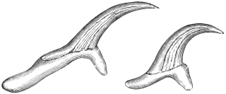
Fig. 240.—Large and small hooklets of Tænia
marginata. 280/1. (After Leuckart.)
This species, which in structure resembles Tænia solium, lives in the intestine
of the dog and the wolf. It attains 1·5 to 4 m. in length, possesses a double crown
of thirty to forty hooks, on
an average thirty-six to thirty-eight
hooks, and in its larval
stage (Cysticercus tenuicollis)
lives in the peritoneal cavity of
ruminants and the pig, occasionally
in the monkey and squirrel.
It is included in this work
because, according to one statement,
C. tenuicollis is supposed
to have been observed in man
in North America; but the case
is not quite certain, as the number of hooks was less than in C. tenuicollis and
coincided with C. cellulosæ, although the size of the cysticercus appeared to point to
C. tenuicollis. A yet earlier statement of Eschricht, that Cysticercus tenuicollis had
been observed in Iceland in the liver of a man, is undoubtedly due to an error.
Tænia serrata, Goeze, 1782.
This parasite attains a length of from 0·5 to 2 m., possesses a double crown of
thirty-four to forty-eight (mostly forty) hooks. It lives exclusively in the intestine
of the dog, the corresponding cysticercus (Cysticercus pisiformis) living in the
mesentery of the hare and rabbit. We mention this species with all reserve
amongst the parasites of man, because Vital states that he has observed it twice in
Constantine (Algeria) in human beings. The data, however, are not sufficient to
characterize the species. It is highly probable that they relate to Tænia solium.
Galli-Valerio even swallowed five specimens of Cysticercus pisiformis, but without
result.
Tænia crassicollis, Rud., 1810.
I only mention this species from the intestine of the domestic cat because
Krabbe regards its occurrence in man as possible. It attains a length of 60 cm.
and is armed; its cysticercus (Cysticercus fasciolaris) lives in the liver of mice and
rats. In Jutland, according to Krabbe, chopped-up mice (spread on bread) are
eaten raw, being a national remedy for retention of urine, and this custom affords
the possibility of the introduction of C. fasciolaris into the intestine of man (Nord.
med. Arkiv, 1880, xii).
Tænia saginata, Goeze, 1782.
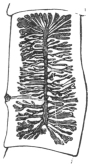
Fig. 241.—Mature
segment of Tænia
saginata, G., with
distended uterus.
2/1.
Syn.: Tænia solium, L., 1767 (pro parte); Tænia cucurbitina, Pallas, 1781 (p.p.);
Tænia inermis, Brera, 1802. Moquin-Tandon, 1860; Tænia dentata, Nicolai, 1830;
Tænia lata, Pruner, 1847; Bothriocephalis tropicus, Schmidtmuller, 1847; Tænia
mediocanellata, Küchenmeister, 1855; Tænia zittavensis, Küchenmeister, 1855; Tænia
tropica, Moquin-Tandon, 1860; Tænia (Cystotænia) mediocanellata, Leuckart, 1863.
Fig. 242.—Cephalic
end of Tænia
saginata in the contracted
condition.
8/1.
The length of the entire tapeworm averages 4 to 8 to 10 m. and
more, even up to 36 m. According to Bérenger-Feraud it attains
a length of 74 m. (?) The head is cubical in shape, 1·5 to 2 mm.
in diameter; the suckers are hemispherical (0·8 mm.) and are
frequently pigmented; there is a sucker-like organ in place of the
rostellum, and this also is frequently pigmented. The neck is
moderately long and about half the breadth of the head; the
proglottids, the number of which averages more than 1,000,
gradually increase in size; the mature detached segments are shaped
exactly like pumpkin-seeds, and are about 16 to 20 mm. in length
and 4 to 7 mm. in breadth. The genital pores alternate irregularly
and are situated somewhat behind the middle of the lateral margin.
There are twenty to thirty-five
lateral branches at each side of the
median trunk of the uterus, and
these again ramify. The eggs are
more or less globular, the egg-shell
frequently remains intact and
carries one or two filaments; the
embryonal shell (embryophore) is thick, radially striated, is transparent
and oval; it is 30 µ to 40 µ in length, and 20 µ to 30 µ in breadth.
Several segments simultaneously are usually passed spontaneously
with defæcation.
Malformations are not uncommon, and resemble those of Tænia solium;
a triangular form has been termed T. capensis by Küchenmeister, and T. lophosoma
by Cobbold, names that naturally possess as little value as does the term
T. fenestrata for fenestrated specimens. Moreover, T. solium, var. abietina, Weinld.,
1858, which was evacuated by an Indian, was probably a T. saginata with somewhat
close uterine branches. It is regarded by Stiles and Goldberger as a doubtful subspecies.
Fig. 243.—Tænia saginata. 19, egg with
external shell. 20, without (embryophore).
(After Leuckart.)
T. saginata in its adult condition lives exclusively in the intestinal
canal of man.291 The corresponding cysticercus is Cysticercus bovis, and
is found almost exclusively in the ox; it is small, 7·5 to 9 mm.
in length and 5·5 mm. in breadth, may easily escape notice, and
requires from three to six months for its development. Numerous
experiments have confirmed the connection of Cysticercus bovis with
Tænia saginata; indeed, the cysticercus was only discovered by feeding
experiments after attention had been called to the ox as the probable
intermediary host of this Tænia.
Fig. 244.—A piece of
the muscle of the ox, with
three specimens of Cysticercus
bovis. Natural size.
(After Ostertag.)
Medical men observed that weakly children who were ordered to eat raw scraped
beef to strengthen them contracted T. saginata. It was found, moreover, that
Jews, who are prohibited from eating pork from religious motives, suffered especially
from T. saginata; when T. solium was found to occur in a Jew he often confessed
to having eaten pork; and finally it was found that certain nations—for
instance, the Abyssinians—frequently harbour T. saginata, and only eat beef—raw
by preference.
These observations led Leuckart, in 1861, to feed
young calves with the proglottids of T. saginata in
order to discover the corresponding cysticercus, which
was then not known. This experiment was successful.
Similar experiments, with similar results, were then conducted
by Mosler (1863), Cobbold and Simonds (1864
and 1872), Röll (1865), Gerlach (1870), Zürn (1872),
Saint Cyr, Jolicœur (1873), Masse and Pourquier
(1876), and Perroncito, in 1876. The attempts to
infect goats, sheep, dogs, pigs, rabbits and monkeys
were unsuccessful. Only Zenker and Heller were able
to infect kids, and Heller infected one sheep, but these
are exceptions.
Artificial infections of human beings with
Cysticercus bovis to obtain the tapeworm were
less numerous, and indeed quite superfluous,
yet this was also done by Oliver (1869) in India,
and Perroncito (1877) in Italy. The experiments
of the latter prove that the extracted
cysticerci of the ox certainly perish in water at
47° to 48° C.
It is a remarkable circumstance that, at least as
regards Central Europe, C. bovis in the ox, after natural
infection, was so seldom found that almost every case
was published as a rarity; whereas the Tænia is very frequent in man. The
reason for this is that in Germany cattle are not severely infected, and that the
small, easily dried-up cysticerci easily escape notice in the large body of the host.
Hertwig, the late director of the town cattle market in Berlin, in 1888, pointed out
that the cysticercus of the ox is found chiefly in the musculi pterygoide externi
and interni, and since that time a far greater number of infected oxen have been
found in Berlin.
Year | Number of oxen
slaughtered | Infected | Proportion |
1888–89 | 141,814 | 113 | 1 : 1,255 |
1889–90 | 154,218 | 390 | 1 : 395 |
1890–91 | 124,593 | 263 | 1 : 474 |
1891–92 | 136,368 | 252 | 1 : 541 |
1892–93 | 142,874 | 214 | 1 : 672 |
Since 1892 an increase has taken place in the number of oxen infected with
cysticercus, but this is probably attributable to the more general and searching
examinations. In the slaughter-houses of Prussia the number of infected beasts was
as follows:—
| 1892 | 567 |
| 1893 | 686 |
| 1894 | 748 |
| 1895 | 1,143 |
| 1896 | 1,981 |
| 1897 | 2,629 |
The condition was most frequent in Neisse (3·2 to 4 per cent.), Eisenach
(1·91 per cent.), Ohlau (1·57 per cent.), Oels i. Schles. (1 per cent.), Marienwerder
(0·34 to 1·2 per cent.). The flesh of oxen only slightly infected (containing not more
than ten living cysticerci) is sold in pieces of not more than 5 lb. to consumers
after having been rendered innocuous by cooking, or by pickling for twenty-one days
in 25 per cent. salt brine, or hanging for twenty-one days in suitable refrigerators;
oxen in which only one cysticercus is found are allowed free commerce, and those
strongly infected (i.e., containing more than ten living cysticerci) may only be used
for industrial purposes.
It is a striking fact that more bulls than cows are infected (according to Reissmann,
in Berlin, from 1895 to 1902, 0·446 per cent. bulls, 0·439 per cent. oxen, and
0·262 per cent. cows), the explanation of which, according to Ostertag, is that most
oxen are killed when young, when also infection most readily takes place, and,
further, that the larva later on in life can be completely atrophied.
The cysticercus of the ox has hitherto been found in man on very
rare occasions. Arndt (Zeitschr. f. Psychiat., xxiv) mentions a case in
the brain, Heller in the eye, and Nabiers and Dubreith also in the
brain (Journ. méd. Bordeaux, 1889–1890, p. 209); but the diagnoses
are not quite certain, as absence of hooks occasionally occurs in
Cysticercus cellulosæ.
Tænia saginata is the most frequent tapeworm of man (with the
exception of Dibothriocephalus latus in a few districts), and the parasite
is widely distributed over the surface of the globe; it has been known
in the East for ages, so far as data are available; it is frequent in
Africa, America, and Europe. Its frequency has perceptibly increased
during the last few years, but a decrease should soon take place in
consequence of the extent and improvement of the official inspection
of meat.
The following table shows the relative frequency of the Cestodes
of man:—
Author | Year | | Number of
cases | T. saginata | T.
solium | Dibr.
latus | Dipyl.
canin. | Unde-
termined |
| Parona (Milan) | 1899 | | 150 | 121 | | 11 | 4 | — | 14 |
| Parona (Italy) | 1868–99 | | 513 | 397 | | 71 | 26 | — | 19 |
| Krabbe (Denmark) | 1869 | | 100 | 37 | | 53 | 9 | 1 | — |
| " " | 1869–86 | | 200 | 153 | | 24 | 16 | 8 | — |
| " " | 1887–95 | | 100 | 89 | | — | 5 | 6 | — |
| " " | 1896–1904 | | 50 | 41 | | 1 | 5 | 3 | — |
| Blanchard (Paris) | 1895 | | ? | 1,000 | | 21 | — | — | — |
| Stiles (United States) | 1895 |  | more than
300 | more than
300 |  | — | 3 | — | — |
| Schoch (Switzerland) | 1869 | | 19 | 16 | | 1 | 2 | — | — |
| Zaeslein (Switzerland) | 1881 | | ? | 180 | | 19 | ? | — | — |
| Kessler (Petrograd) | 1888 | | ? | 22 | | 16 | 47 | — | — |
| Mosler (Greifswald) | 1894 | | 181 | 112 | | 64 | 5 | — | — |
| Bollinger (Munich) | 1885 | | 25 | 16 | | 1 | 8 | — | — |
| Vierordt (Tübingen) | 1885 | | 121 | 113 | | 8 | — | — | — |
| Mangold (Tübingen) | 1885–94 | | 128 | 120 | 6 | 8 | — | — |
Tænia africana, v. Linst., 1900.
Fig. 245.—Mature segment of Tænia africana. The ovary is in the
middle, and behind it are the shell gland and vitellarium; at the sides are
the testicles, and externally the excretory canals; the cirrus pouch, the vas
deferens and the vagina are on the left. Magnified. (After v. Linstow.)
This worm measures over 1·3 m. in length. The segments are
all broader than they are long. The scolex is unarmed and is
provided with an apical sucker (0·16 mm.). The parasite measures
1·38 mm. in breadth, 1·03 mm. in width; the suckers measure
0·63 mm. in diameter. The neck is very short and somewhat broader
than the scolex; number of segments about 600; the hindmost
segments measure 7 mm. in length and 12 to 15 mm. in breadth.
The genital pores alternate irregularly in the middle of the lateral
margin; the testes are very numerous and occupy the entire medullary
layer; the vas deferens is much convoluted; the cirrus pouch
is pyriform and thick walled; the cirrus and vagina are beset with
bristles directed outwards; the receptaculum seminis is fusiform; the
ovary is large and double, and consists of radially placed club-shaped
tubes that do not anastomose and do not branch; the vitellarium is
at the posterior border of the proglottids, the round shell gland in
front of it; the uterus consists of a median trunk and fifteen to
twenty-four non-ramified lateral branches
on each side; the embryonal shell is thick
and has radial stripes—it may be round
(31·2 µ to 33·8 µ) or oval (39 µ, by 33·8 µ);
the spines of the oncospheres measure
7 µ to 8 µ in length (fig. 197).
Fig. 246.—Proglottis of Tænia africana, with uterus. Magnified.
(After v. Linstow.)
Fig. 247.—Head of Tænia africana;
apical surface. Magnified.
(After v. Linstow.)
At present only two specimens are
known; they came from a black soldier
from the vicinity of Lake Nyasa. The
cysticercus is unknown; perhaps it lives
in the zebu, the flesh of which the Askaris
are in the habit of devouring uncooked.
Tænia confusa, Ward, 1896.
Length 8·5 m., breadth about 5 mm. The scolex is unknown;
there is no neck; number of proglottids 700 to 800, always longer
than they are broad; the hindmost measure 35 mm. in length, 4 to
5 mm. in breadth; the genital pores alternate irregularly behind the
middle of the lateral margin; testicles numerous; vas deferens not
much coiled; the cirrus pouch thick walled, elongated and club-shaped,
with globular vesicula seminalis; the cirrus is beset with little
hairs; the receptaculum seminis is globular; ovary small, double;
each half is bean-shaped; vitellarium narrow, triangular; shell
gland globular; uterus with median trunk and fourteen to eighteen
short ramified lateral branches on either side. The embryophores are
oval (39 µ by 30 µ), thick and radially striated.
Fig. 248.—Tænia confusa: mature
segment showing central uterine stem,
bilobed ovary, globular shell gland, triangular
vitellarium, scattered testes, cirrus,
vas deferens, and vagina. 15/1. (After
Guyer.)
Fig. 249.—Tænia confusa:
gravid segment.
25/1. (After Ward.)
Of this species only two specimens have been recorded; they
occurred in human beings and were sent at different times to the
first describer of them by a doctor in Lincoln (Nebr.). Perhaps
Tænia solium, var. abietina, Weinld., which was found in a Chipeway
Indian, is of the same species in spite of the shorter segments.
Tænia echinococcus, v. Sieb., 1853.
Syn.: Tænia nana, v. Ben., 1861 (nec v. Sieb., 1853); Echinococcifer
echinococcus, Weinld., 1861.
Fig. 250.—Tænia echinococcus:
the cirrus sac, the
vagina, uterus, ovary, shell gland
and vitellarium, and the testicles
at the sides are recognizable in
the second proglottis; the posterior
proglottis shows the uterus
partly filled with eggs, as well
as the cirrus sac and the vagina.
50/1.
Tænia echinococcus measures 2·5 to 5 or 6 mm. in length; the head
is 0·3 mm. in breadth, and has a double row of twenty-eight to fifty
hooklets (on an average thirty-six to thirty-eight) on the rostellum.
The size and form of these hooklets vary (the larger ones are
0·040 to 0·045 mm. in length, the smaller ones are 0·030 to
0·038 mm. in length). The suckers measure 0·13 mm. in diameter;
the neck is short; there are only three or four segments, the posterior
segment being about 2 mm. in length and 0·5 mm. in breadth. The
genital pores alternate; there are forty to fifty testicles; the vas deferens
is spirally coiled; the cirrus pouch is pyriform. The ovary is horseshoe-shaped
with the concavity directed backwards; the vitellarium
double, each half almost bean-shaped, at
right angles to the plane of the segment;
the shell gland is round. The median trunk
of the uterus is dilated when filled with eggs
and (instead of lateral branches) has lateral
diverticula. It is not unusual for the eggs
to form local heaps. The embryonal shell
(embryophore) is moderately thin, with radial
striæ, almost globular, 30 µ to 36 µ in
diameter.
When mature this parasite lives in the
small intestine of the domestic dog, the
jackal, and the wolf, and apparently also in
Felis concolor, and is usually present in great
numbers; it can also be transmitted experimentally
to the domestic cat, one successful
result out of seven (Dévé).292 The
larval stage (Echinococcus polymorphus) lives
in various organs—chiefly in the liver and
lungs—of numerous species of mammals
(twenty-seven), especially in sheep, ox and
pig, and it is even not uncommon in man,
though the Tænia itself has never been found
in a human being; accordingly man can only
acquire the echinococcus by ingesting the
eggs of the “dog worm.” The dogs disseminate
the eggs of Tænia echinococcus
wherever they go, or carry them to their
mouths and coats by biting up the
evacuated segments, and are thus able to
transmit them directly to human beings (by
licking them or making use of the same
crockery, etc.). In other cases the oncospheres,
enclosed in the embryophores, must
withstand desiccation for a time and then
(as when the dogs are “kissed” or otherwise
caressed) are transmitted into or on to
man. As echinococcus disease in man is always very dangerous, it
would be a matter of general interest to prevent dogs being infected
by destroying the echinococci,293 and all measures would be justifiable
which would diminish the superfluous number of house-dogs (for
instance, high taxes); measures should also be adopted to limit the
association of men with dogs, particularly in such frequented places
as restaurants, railway carriages and tram-cars.
Echinococcus is very common in slaughtered animals; in Germany, however,
the figures in the reports of the abattoirs present an erroneous view in so far as,
besides the total number of animals slaughtered, only the numbers of those organs
(liver and lungs) are published that were so severely infected with echinococci that,
even when the parasites were “shelled” out, the flesh could not be placed upon the
market and was therefore “condemned.”
In Berlin the following animals were slaughtered:—
Year | 1889–90 | 1890–91 | 1891–92 | 1892–93 | 1896–97 | 1902 |
| Oxen | 154,218 | 124,593 | 136,368 | 142,874 | 146,612 | 153,748 |
| Sheep | 430,362 | 371,943 | 367,933 | 355,949 | 395,769 | 434,155 |
| Pigs | 442,115 | 472,859 | 530,551 | 518,073 | 694,170 | 778,538 |
During the same years the following were condemned in consequence of being
infected with echinococci:—
| Lung | Liver | Lung | Liver | Lung | Liver | Lung | Liver | Lung | Liver | Lung | Liver |
| Oxen | 7,266 | 2,418 | 5,792 | 1,938 | 4,497 | 1,721 | 2,563 | 739 | 3,284 | 1,156 | 2,507 | 791 |
| Sheep | 5,479 | 2,742 | 4,595 | 2,059 | 4,435 | 1,669 | 3,331 | 1,161 | 4,561 | 1,939 | 11,138 | 4,437 |
| Pigs | 6,523 | 5,078 | 5,083 | 3,735 | 6,037 | 4,374 | 6,785 | 4,312 | 7,888 | 5,398 | 9,544 | 9,233 |
Nevertheless there are statistics that give the total number of animals infected
with echinococcus:—
Author | Place | Oxen | Sheep | Pigs |
| Längrich | Rostock i. M. | 26 | ·2 per cent. | 37 | ·0 per cent. | 5 | ·4 per cent. |
| Olt | Stettin | 7 | ·1 " | 25 | ·8 " | 7 | ·3 " |
| Steuding | Gotha | 24 | ·6 " | 35 | ·4 " | 21 | ·4 " |
| Prettner | Prague | 23 | ·2 " | 5 | ·5 " | ? |
In Güstrow, in Mecklenburg, half of the animals slaughtered are said to be
infected with echinococcus; in Wismar 25 per cent. of the oxen, 15 per cent. of the
sheep and 5 per cent. of the pigs are infected; according to Mayer, in Leipzig,
3·79 per cent. native pigs, 24·47 per cent. Hungarian pigs, and 13·09 per cent. of sheep
were infected with echinococcus; at the same time it was stated that in regard to
the native pigs the liver was more frequently affected than the lungs (3·81 per cent.
as compared with 0·26 per cent.); in sheep the lungs were more frequently infected
(12·71 per cent. to 3·73 per cent.), whereas in the Hungarian pigs both organs were
almost equally infected (14·78 per cent. to 12·03 per cent.).
The data of Lichtenheld, in Leipzig, give the frequency with which various
organs were affected, as shown in the following table:—
| Cattle | Pigs | Sheep | Horses |
| | | ♂ | ♀ | | |
| Lungs | 69 | ·3 per cent. | 16 | ·2 per cent. | 21 | ·4 per cent. | 52 | ·2 per cent. | 5 | ·5 per cent. |
| Liver | 27 | ·0 " | 74 | ·2 " | 72 | ·0 " | 44 | ·9 " | 94 | ·5 " |
| Spleen | 2 | ·2 " | 3 | ·2 " | 2 | ·7 " | 2 | ·9 " | — |
| Heart | 0 | ·75 " | 3 | ·2 " | 1 | ·3 " | — | — |
| Kidneys | 0 | ·75 " | 3 | ·2 " | 1 | ·3 " | — | — |
| Subperitoneal tissue | — | — | 1 | ·3 " | — | — |
Structure and Development of Echinococcus (Hydatid).
Fig. 251.—Echinococcus veterinorum: the fibrous sac enclosing the
echinococcus has been opened and laid back in five parts, so that the
surface of the bladder worm may be seen, with the brood capsules, visible
to the naked eye, showing through it. Natural size. (After Leuckart.)
An echinococcus is a spherical or roundish bladder full of a
watery liquid, which originates by liquefaction of the oncosphere, and
in man may attain the size of a child’s head, but remains smaller in
cattle (the size of an orange or apple). The thin wall of the bladder
is composed of an external laminated cuticle (ectocyst) and an internal
germinal or parenchymatous layer (endocyst). The latter again
exhibits two layers: an outer layer of small cells that are less sharply
defined, and an inner layer of larger cells. It contains, in addition,
calcareous corpuscles, muscular fibres and excretory vessels. It is
rich in glycogen.
Fig. 252.
Figs. 252 and 252A.—Diagrams of mode of formation of brood capsule and scolices.
(1) Wall of mother cyst, consisting of ectocyst and endocyst; (2) theoretical stage of invagination
of wall; (3) a brood capsule with the layers of the wall in the reverse position to that in the
mother cyst; (4) evagination of wall; (5) invagination; (6) fusion to form the solid scolex;
(7) invagination of fore-part of scolex into hind-part. (Note.—The size of the scolex is much
out of proportion to the brood capsule.) (Stephens.)
The development in cattle often remains stationary at the bladder
stage, and they are then called “acephalocysts,” or Echinococcus cysticus
sterilis. According to Lichtenheld, sterile cysts occur in 80 per cent.
of cases in cattle, in 20 per cent. in pigs, and in 7·5 per cent. in sheep.
In other cases large numbers of small, hollow BROOD CAPSULES are
formed in the germ layer, but are not arranged in any particular order.
The order of the layers is just the reverse in them to what it is in the
parent cyst, that is to say, they have inside a thin non-laminated cuticle
and the parenchymatous layer on their external surface. These,
theoretically at least, may be regarded as invaginations of the bladder
wall giving rise to a cavity with the cuticle internal and the
parenchymatous layer external. If we suppose the orifice to close,
we should then get an isolated
cavity with cuticle internal and
parenchymatous layer external, as
in the brood capsule (fig. 252).
If we next suppose an evagination of the wall of the brood capsule to
occur at one point we should get a hollow process lined with cuticle;
at the bottom of this we get the scolex and hooklets formed, and a
little higher up the tube the suckers (fig. 252, 4). If this hollow scolex
is now pictured as being invaginated we get a hollow scolex covered
with cuticle and lined by a parenchymatous layer projecting into the
cavity of the brood capsule. The two sides of this hollow scolex now
fuse and we get a solid scolex projecting into the cavity. Finally, if
we imagine once more the rostellum and suckers invaginated into the
posterior part of the scolex we get the condition as frequently found
in the brood capsules, i.e., a scolex covered with cuticle projecting into
the cavity, with the rostellum and suckers invaginated into the posterior
portion of the scolex (fig. 252A, 7).
Fig. 253.—Section through
an invaginated echinococcus
scolex. Cf. fig. 252A, 7. × 300.
(After Dévé.)
A large hydatid may contain many thousands of brood capsules.
Each brood capsule is about as big as a pin’s head, and may contain
ten to thirty or more scolices. The delicate wall of the brood capsules
may rupture, so that the scolices are now free in the mother cyst. These
free scolices and also free brood capsules constitute what is known as
“hydatid sand,” which settles at the bottom of a glass when hydatid
fluid is poured into it. This form occurs chiefly in domesticated
animals and is termed E. veterinorum, Rud., or E. cysticus fertilis.
In man, and only rarely in cattle, the mother cyst first forms
“daughter cysts” (E. hominis, Rud. [fig. 255]), which, though smaller
than the “mother cyst,” resemble
it in the structure of
their walls; thus they are
covered externally by a laminated
cuticle and internally
by the parenchymatous layer.
They originate:
(1) Between the laminæ of the cuticle of the mother cyst from
small, detached portions of the parenchymatous layer; during their
growth they bulge inwardly or outwardly and may separate themselves
entirely from their parent cyst. In the latter case they lie
between the mother cyst and the capsule of connective tissue
formed by the host (E. granulosus or E. hydatidosus exogenus);
when growing inwardly they reach the interior of the mother cyst
(E. hydatidosus endogenus). Their number is very variable and does
not depend on the size of the mother cyst. They are as big as, or
bigger than, gooseberries.
Fig. 254.—A piece of the wall of an Echinococcus
veterinorum stretched out and seen from the internal
surface. A few brood capsules (the outline of which
is only faintly shown), with scolices directed towards
their interior and exterior. 50/1.
(2) According to some authors, endogenous daughter cysts arise also
from a metamorphosis of scolices that have separated off from the brood
capsule. This takes place in the following way: Fluid accumulates
in the interior of the scolex, so that eventually nothing remains except
a sac consisting of cuticle lined by parenchyma. The cuticle gradually
thickens and several layers form (fig. 257).
Fig. 255.—Echinococcus hominis in the liver. The fibrous capsule and
the wall of the echinococcus have been incised, so that the endogenous
daughter cysts may be seen. Reduced. (After Ostertag, from Thoma.)
Fig. 256.—Section through an echinococcus
scolex in process of vesicular metamorphosis,
twenty-six days after insertion in the pleural
cavity. × 250. (After Dévé.)
(3) Transformation of Brood Capsules into Daughter Cysts.—This is
also held to be possible by various observers. New epithelial layers are
deposited between the cuticle
which lines the brood capsule
and the outer parenchymatous
layer. This parenchymatous
layer gradually disappears and a
new parenchymatous layer forms
in the interior from the parenchyma
of the scolex or scolices.
Although it appears strange that
a completely formed scolex with
specifically differentiated tissues
and organs should retrogress
to more primitively organized
matter, and again become a
proliferating bladder, yet we can
hardly doubt that the older observations, regarding such a vesicular
metamorphosis, of Bremser (1819), v. Siebold (1837), Naunyn (1862),
Rasmusser (1866), Leuckart (1881), Alexinsky (1898), Riemann (1899),
Dévé (1901), and Perroncito (1902) are correct.
(4) Further, a fourth method of formation of daughter cysts is
described by Naunyn as occurring in sterile hydatids, i.e., those containing
no brood capsules. In this case a portion of the mother wall
of the hydatid gets constricted off.
Fig. 257.
Figs. 257 and 257a.—Diagram of transformation of a scolex into a daughter cyst (1 to 3):
1, scolex in brood capsule; 2, liquefaction of scolex; 3, daughter cyst; and (4 to 6) of a brood
capsule into a daughter cyst; 4, brood capsule with scolex; 5, deposition of new epithelial
layers on the inner layer of the parenchyma; 6, disappearance of outer parenchyma and formation
of inner parenchyma from the parenchyma of scolex, which has now disappeared. (Note.—The
scolices are out of proportion to the brood capsules and to the daughter cysts. Stephens.)
It has also been established that not only daughter cysts transplanted
into animals develop further (Lebedeff, Andrejew, Stadnitzky, Alexinsky,
Riemann), but that this also holds good if only hydatid scolices from
man or animals are transplanted into animals (rabbits). They develop
into echinococci and can then give rise to brood capsules and scolices.
As Dévé further established, hydatid scolices are not capable of
developing in guinea-pigs, while corresponding experiments with
rabbits are in the large majority of cases successful where the scolices
are introduced subcutaneously or into the pleural or peritoneal cavities.
It is only in the case of daughter cysts that further growth is obtained
in the case of guinea-pigs. Finally it appears, as has been already
stated, that brood capsules can transform themselves into daughter
cysts, but according to Dévé only within the mother cyst, not after
transplantation. Daughter cysts that have been formed in the mother
cyst of man and animals behave themselves just as the mother cyst
does, i.e., they can remain sterile,
or give rise to brood capsules
and scolices, or even again
to fresh cysts—granddaughter
cysts. The mother cyst can also die, so that the daughter cysts then
lie in the cavity of the connective tissue capsule. The number of the
daughter cysts in either case may attain several thousands.
The echinococcus fluid, which originally is formed from the blood of the host, is
light yellow, with a neutral or slightly acid reaction; its specific gravity averages 1009
to 1015. It contains about 1·5 per cent. of inorganic salts, half of which is common
salt; in addition (besides water) it contains sugar, inosite, leucine, tyrosin, succinic
acid (associated with lime or soda) and albumens which are not coagulated by heat;
occasionally also the fluid has been found to contain hæmatoidin and uric acid salts
(in echinococcus of the kidneys), which doubtless demonstrates that the echinococcus
liquid originates from the host. It has been generally assumed that echinococcus
fluid contains a toxic substance the escape of which into the body cavity (at operation
or by bursting of a hydatid cyst) produces more or less severe symptoms (fever,
peritonitis, urticaria), so much so that one speaks of hydatid intoxication. The
investigations of Kobert, Joest, etc., have, however, shown the harmlessness of fresh
undecomposed hydatid and cysticercus fluid for rabbits, mice and guinea-pigs, whether
inoculated intraperitoneally, subcutaneously or intravenously. Contrary data or
clinical experience must accordingly depend on other factors.
According to the researches of Leuckart, the growth of the
echinococcus is very slow; four weeks after infection the average
size is only 0·25 to 0·35 mm., at the age of eight weeks it is 1 to
2·5 mm., and at this period the formation of the central cavity commences;
at the age of five months, and with a size of 15 to 20 mm.,
the first brood capsules with scolices are formed. The consequence
of this gradual increase of size is that the organ attacked
can maintain its functions by vicarious hypertrophy, and that many
echinococci induce no special symptoms and cannot even be
diagnosed, the latter circumstance being due to their hidden position.
The echinococcus cannot be said to be scarce in man, as is
shown by the following table for Central Europe:—
Place | Period | No. of
post-mortems | No. of cases
of echino. | Percentage |
| Rostock | 1861–83 | 1,026 | 25 | 2·43 |
| Greifswald | 1862–93 | | | 1·48 |
| Jena | 1866–87 | 4,998 | 42 | 0·84 |
| Breslau | 1866–76 | 5,128 | 39 | 0·761 |
| Berlin | 1859–68 | 4,770 | 33 | 0·69 |
| Würzburg | — | 2,280 | 11 | 0·48 |
| Göttingen | — | 639 | 3 | 0·469 |
| Dresden | 1852–62 | 1,939 | 7 | 0·36 |
| Münich | 1854–87 | 14,183 | 35 | 0·25 |
| Vienna | 1860 | 1,229 | 3 | 0·24 |
| Prague | — | 1,287 | 3 | 0·23 |
| Kiel | 1872–87 | 3,581 | 7 | 0·19 |
| Zürich, Basle, Berne | — | 7,982 | 11 | 0·13 |
| Erlangen | 1862–73 | 1,755 | 2 | 0·11 |
These, however, are only cases that have become known by post-mortem;
in addition, there are cases that have been treated medically,
of which there are a few statements, at all events relating to the
principal districts of Germany. According to Madelung, one case
of echinococcus occurs in every 1,056 inhabitants in the town of
Rostock, in the district of Rostock one to every 1,283, in Schwerin
one to every 5,887, and in Ludwigsort one to every 23,685; according
to Peiper, in Upper Pomerania one case occurs to every 3,336,
in the district of Greifswald one to every 1,535 inhabitants. The
northern districts of Pomerania are more affected than the southern
ones.
Accordingly, echinococcus is also considerably more frequent in cattle in
Pomerania. On an average in Germany 10·39 per cent. oxen, 9·83 per cent. sheep,
and 6·47 per cent. pigs are infected, whereas in Upper Pomerania 37·73 per cent.
oxen, 27·1 per cent. sheep, and 12·8 per cent. pigs are infected; in Greifswald, indeed,
64·58 per cent. oxen, 51·02 per cent. sheep, but only 4·93 per cent. pigs are infected.
In accordance with these figures Tænia echinococcus must be frequent in dogs in
Pomerania, especially in Upper Pomerania; on the other hand, the conjecture that
the frequency of echinococcus in Mecklenburg is explained by the occurrence of
Tænia echinococcus in foxes has not been confirmed, as the fox does not harbour this
worm in Mecklenburg.
Fig. 258.—Hooklets of echinococcus. a, of
Echinococcus veterinorum; b, of Tænia echinococcus,
three weeks after infection; c, of the
adult Tænia echinococcus; d, the three forms
of hooklets outlined one within the other.
600/1. (After Leuckart.)
Beyond the European continent, echinococcus is frequent in
the inhabitants of Iceland, Argentine, Paraguay and Australia. In
Iceland, according to Finsen, 1 in every 43 inhabitants is affected
with echinococcus; according to Jonassen the proportion is 1 to 63;
this is due to the habits of the people of Iceland or, in fact,
to the frequency of Tænia echinococcus
in dogs, and the prevalence
of the hydatid in cattle. In certain
districts of Australia it is
just as frequent. In Cape Colony,
Egypt and Algeria echinococcus
is not rare, but it is scarce in
America and in Asia, with the
exception of the nomadic tribes
of Lake Baikal.
Echinococcus attacks persons
of every age, though it is rare in
children up to 10 years of age
and in old people. It occurs
most frequently between the ages
of 21 and 40 years. According
to all statistics it preponderates
in women (about two-thirds of the
cases). The liver is its favourite
seat (57·1 per cent. of the cases);
next in order come the lungs
(8 per cent.), kidneys (6 per cent.),
cranial cavity, genitalia, organs of circulation, spleen (3·8 per cent.), etc.
As a rule one organ only is invaded; multiple occurrence may originate
from one infection, or eventually from a later infection (?), or it may
come to pass that from some cause (through the spontaneous rupture of
an echinococcus, or the rupture of one caused by an injury or surgical
operation) daughter cysts, brood capsules or scolices escape into the
abdominal cavity,294 where they settle or become transformed and go
on growing. In the distribution of this secondary echinococcus the
great powers of motility of the free scolices must be taken into
account (Sabrazès, Muratet, and Husnot).
Human echinococci may also die at various stages of development, become
caseous or calcified, or may be absorbed, the cause for this being either disease of
the hydatid itself or inflammation of its connective tissue capsule; the discovery of
the laminated cuticle, which has great powers of resistance, or the finding of the
hooklets of the scolices is sufficient to form a conclusion as to the nature of such
formations.
Siebold (1853) was the first to rear Tænia echinococcus in the dog
by feeding it with the echinococcus of cattle and especially of sheep.
Küchenmeister, van Beneden, Leuckart, Railliet and others obtained
similar results, and Thomas, Naunyn, Krabbe and Finsen succeeded
in rearing T. echinococcus in dogs from the bladder worms of human
beings; these grow comparatively slowly (one to three months295) and
only during the process of growth develop their hooklets in their
definite form (fig. 258). It lies in the nature of things that dogs,
whether experimentally or naturally infected, almost always harbour
T. echinococcus in large quantities. That cats exceptionally harbour
these worms has been already mentioned (Dévé). Finally, Leuckart
infected young pigs by feeding them with mature segments.
Echinococcus multilocularis (alveolar colloid).
In addition to the form of echinococcus already described, and
which is also frequently termed Echinococcus unilocularis, there is a
second form which occurs in man as well as in animals, and which
is termed E. multilocularis, s. alveolaris (alveolar colloid).
It was originally regarded as a tumour; its animal nature was first
established by Zeller and R. Virchow. The parasite, which varies
in size from that of a fist to a child’s head, presents a collection of
numerous cysts, measuring between 0·1 and 3 to 4 mm. to 5 mm.
in diameter, which are embedded at first in a soft, connective tissue
stroma; the cut surface has therefore a honeycomb appearance. The
cysts are surrounded by a pellucid and laminated cuticle, and each
according to its size encloses either a small-celled tissue or a cavity
lined by a parenchymatous layer; the fluid contained in such a cavity
may be transparent, or is rendered opaque by globules of fat, bile-pigment,
hæmatoidin and fat crystals. According to some authors
all or most of these cysts intercommunicate; others state that this is
the case at least as regards the cuticle. The scolices are by no means
found in all the cysts, and when present only a few, rarely half, of the
cysts contain scolices (one or more); it is supposed that at least some
of these scolices are formed in brood capsules, and that the former
are capable of undergoing a cystic metamorphosis.
One circumstance is peculiar to the multilocular echinococcus
of man, namely, the disintegration that sets in at certain stages; in
the centre of the parasite a cavity forms that frequently becomes very
large and is filled with a purulent or brownish or brownish-green
viscid fluid; in this fluid one finds shreds of the wall of the cavity,
calcareous bodies, echinococcus cysts, also scolices and hooklets, as
well as fat globules and crystals of hæmatoidin, margarine and
cholesterin and concretions of lime. Such ulcerative processes,
according to Ostertag, are never present in the multilocular echinococcus
of oxen,296 in which the separate cysts are larger and the
connective tissue integument less powerfully developed.
Fig. 259.—Echinococcus multilocularis in the liver of the ox. Natural size.
(After Ostertag.)
Hardly anything positive is known with regard to the development
of the alveolar echinococcus; its peculiar conformation is attributed
by some to enormous infection of oncospheres, by others to the
abnormal situation of one oncosphere; a few authors ascribe it to
infection of lymphatic vessels, others to infection of the biliary ducts
or to peculiarities of the surrounding hepatic tissue; Leuckart ascribes
it to a grape-like variety of form which continues budding; a few
more recent authors consider multilocular echinococcus to be specifically
different from unilocular echinococcus, and therefore also
different the species of Tænia arising from them. Melnikow-Raswedenkow
is also of this opinion. According to this author the
oncospheres infect the lumen of a branch of the portal vein in
Glisson’s capsule of the liver and grow into an irregularly shaped
formation (chitinous coil), which breaks through the vascular walls
and thus forms the alveoli. So far the data coincide well with
Leuckart’s opinion of the original grape-like form of the Echinococcus
multilocularis; according to Melnikow-Raswedenkow the “granular
protoplasmic substance” (parenchymatous layer) is not only present
in the interior of the loculi but also outside, and, moreover, “ovoid
embryos” are supposed to develop in the chitinous coils, which,
“thanks to their amœboid movements, reach the lumen of a vessel,
where, under favourable circumstances, they begin to develop
further,” that is to say, they become “chitinous cysts with fantastic
outlines,” or also “single-chambered chitinous cysts”; scolices may
develop in both. Dévé, however, considers that these embryos are
only prolongations of the protoplasmic layer which secondarily
cuticularize.
The multilocular echinococcus, which in man produces a severe disease and
almost always leads to premature death, infects most frequently the liver, but may
also be found primarily in the brain, the spleen and the suprarenal capsule; from
the liver by means of metastasis it may reach the most various organs, especially
those of the abdomen, but also the lungs, the heart, etc. Up to 1902, 235 cases have
been described and up to 1906, 265, being 70 from Russia, 56 from Bavaria, 32 from
Switzerland, 30 from the Austrian Alps, 25 from Würtemberg; the remaining cases
are distributed over Central Germany, Baden, Alsace, France, Upper Italy, North
America. In some the origin is doubtful; in any case after Russia, the mountainous
South of Europe is the principal region of distribution. As to the domesticated
animals, the same parasite is found principally in the ox (according to Meyer, in
Leipzig, in 7 per cent. of the oxen affected with echinococcus); it is rarer in the
sheep and very scarce in the pig.
It has already been mentioned above that recently the multilocular
echinococcus has been stated to be specifically different from hydatid
or unilocular echinococcus. To this may be added the fact that
Mangold, who fed a young pig with oncospheres of a Tænia reared
from the multilocular echinococcus, found two growths in the liver
four months later, which he took to be E. multilocularis, and consequently
one has to assume the existence of two different worms.
The chief defender of this view, already put forward by Vogler,
Mangold, and Müller, is Possett. He bases his opinions on (1) the
more restricted distribution of the multilocular hydatid, the former
occurring in districts where only cattle are raised, the latter where
sheep-breeding is established; (2) that those engaged in looking after
sheep are attacked by multilocular, whereas those looking after cattle
are attacked by unilocular hydatid; (3) that among the cases of
unilocular hydatid occurring in the distribution areas of multilocular
hydatid no transitions between the two forms are observed; (4) on
the difference in the hooks both in the hydatid as well as in the Tænia
stage; the hooks of Tænia echinococcus are plump, sharply curved,
and have a short posterior root process the length of which is to that
of the total length as 1 to 4·7, whereas on the contrary the hooks of the
alveolar echinococcus are more slender, slightly bent, and have a
long posterior root process (1 to 2·5); and (5) on the form of the
uterus, which in the alveolar Tænia has the form of a spherically
distended sac anteriorly.
Serum Diagnosis of Echinococcus.
(1) Precipitin Reaction.—Mix equal parts of hydatid fluid (of the
sheep) and serum of patient. Keep at 37° C. The reaction is not
decisive as it may be given by normal sera.
(2) Complement Deviation.—Required: (1) Hydatid fluid of sheep
(antigen), (2) guinea-pig complement, (3) patient’s serum, (4) red
cells of sheep, (5) hæmolytic serum (of rabbit) against sheep’s red
cells, (6) 0·8 per cent. salt solution. Mix the antigen + patient’s
serum (heated) + complement + salt solution at 37° C. for one hour.
Add red cells of sheep + hæmolytic serum. Allow to stand for
half an hour at 37° C. It is imperative to make adequate control
observations. An example will indicate the method. Salt solution
1·3 c.c. + patient’s serum (heated) 0·2 c.c. + hydatid fluid 0·4 c.c. +
complement 0·1 c.c. of serum diluted to a quarter strength + hæmolytic
serum and red cell emulsion 1 c.c. Result: no hæmolysis, i.e.,
the patient’s serum contains specific (echinococcus) antibodies.