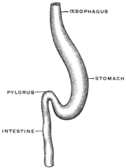
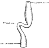
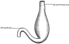
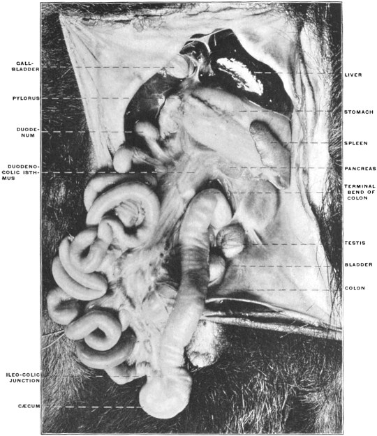
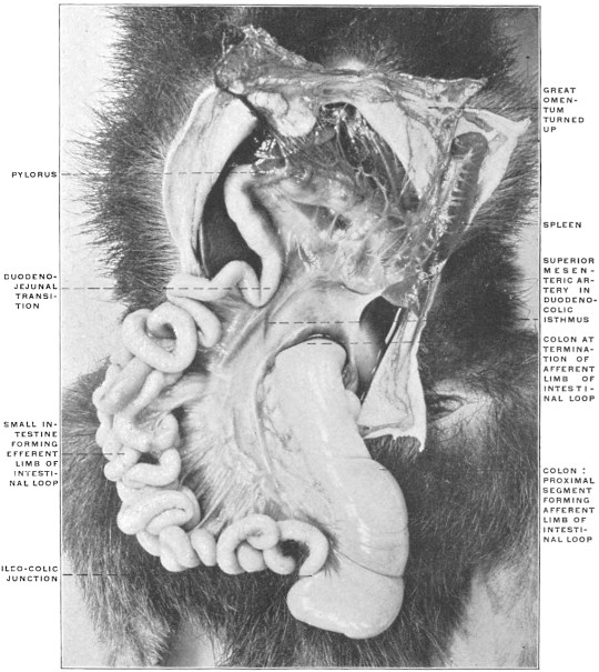
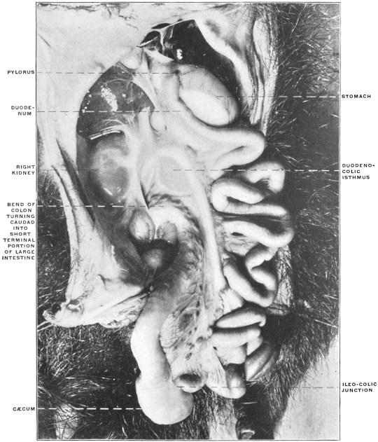
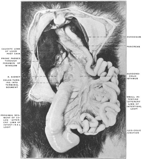
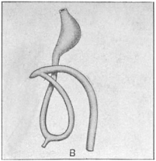
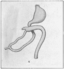
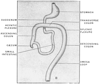
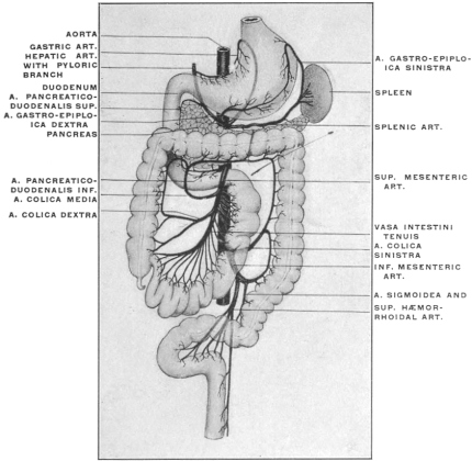
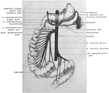
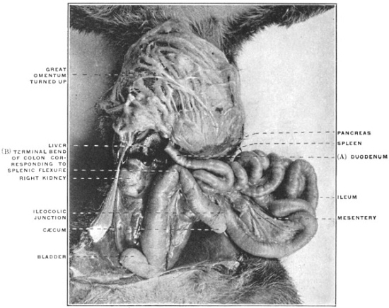
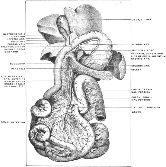
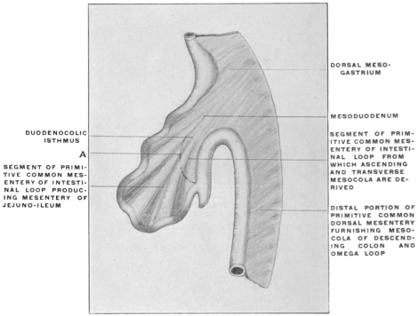
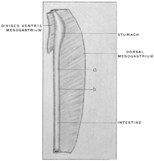
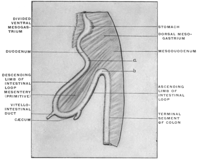
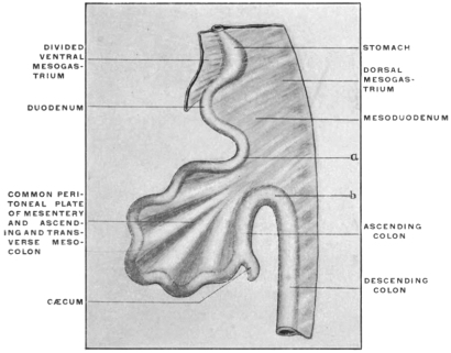
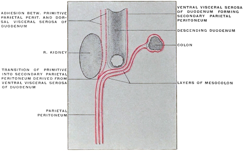
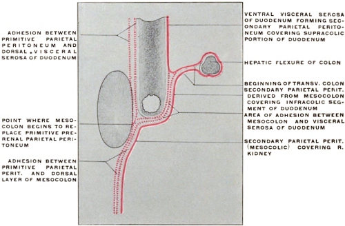
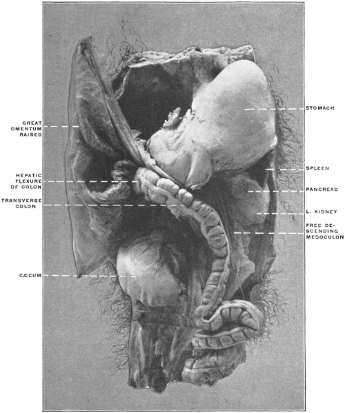
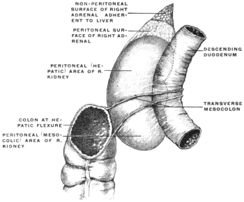
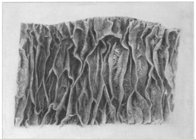
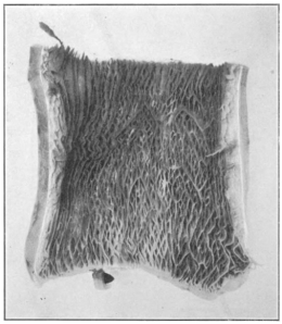
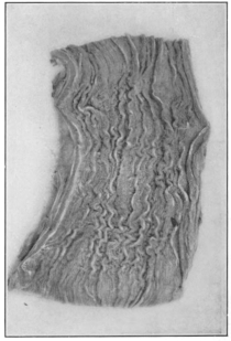
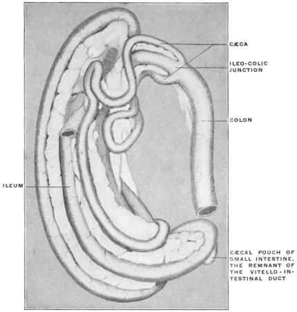
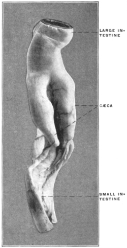
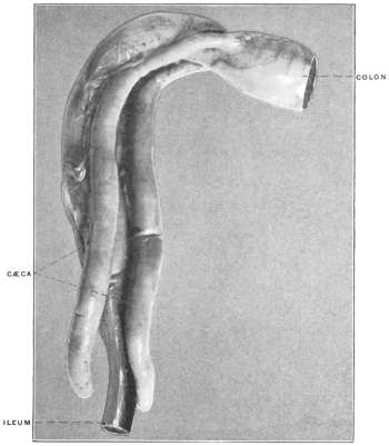
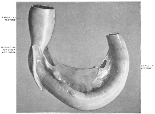
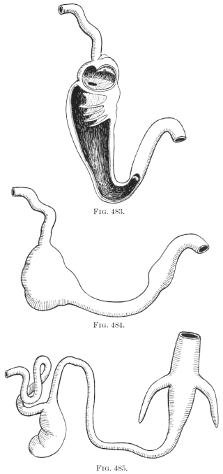
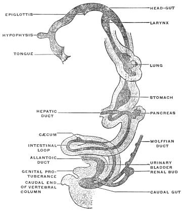
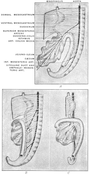
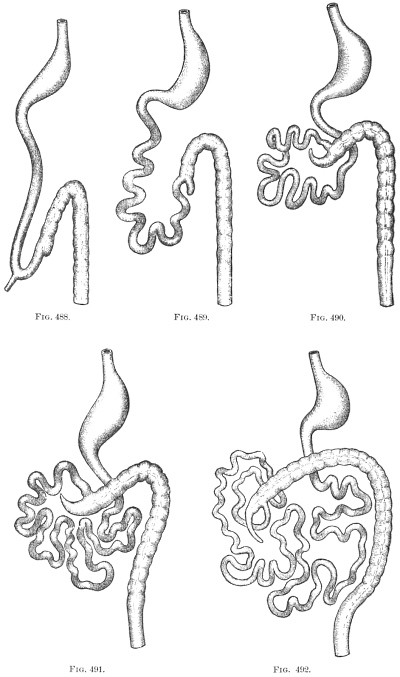
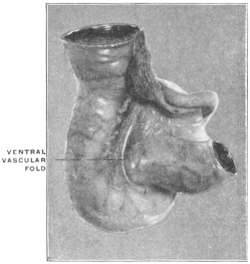
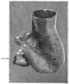
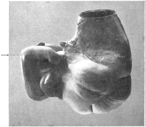
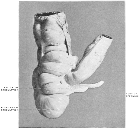
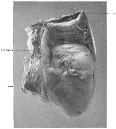
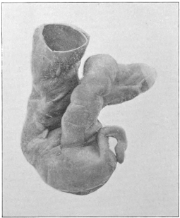
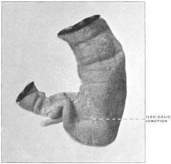
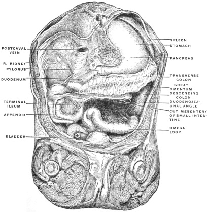
We have already seen that the transverse colon and mesocolon
effect a general division of the adult human abdominal
cavity into a cephalic supra-colic compartment, situated between
the diaphragm and the level of the transverse colon and mesocolon,
comprising in general the hypochondriac and epigastric
regions, and a larger caudal infra-colic space which includes the
entire rest of the abdominal cavity and is continued caudad into the
pelvic cavity. The arrangement of the peritoneum and viscera
in this latter space has just been considered. The fact will be
recalled that the second or descending portion of the duodenum,
passing dorsad of the hepatic colic flexure, forms so to speak the
visceral connection between the portions of the alimentary tube
situated in the supra-colic compartment and those situated in the
infra-colic space. The fixation of this segment of the duodenum
and its consequent secondary retroperitoneal position in the adult
human subject masks this continuity of the alimentary canal to
a certain extent so that it requires more than a superficial examination
in order to trace correctly the course of the duodenum
from the pylorus to the duodeno-jejunal angle, dorsad of the
colon, root of transverse mesocolon and mesentery, and under
cover of the secondary parietal peritoneum.
We have now to turn our attention to the viscera contained in
the cephalic or supra-colic compartment of the abdomen and to
consider the disposition of the serous membrane investing them
and connecting them with each other and with the abdominal
parietes.
The visceral contents of the supra-colic compartment comprise
the liver, pancreas, spleen, stomach and the proximal portion of
the duodenum, including the hepatic angle and the supra-colic
part of the descending duodenum. Less directly the cephalic
portions of the right and left kidney and the corresponding suprarenal
capsules belong to this visceral group.
In this region of the abdomen we meet with the most extensive
modifications of the primitive dorsal peritoneal membrane,
producing conditions which, considered without reference to development
and comparative anatomy, are complex and difficult of
comprehension. These changes lead to the formation of the so-called
“lesser sac,” a term which in some respects is unfortunate
as it implies a more complete degree of separation from the general
peritoneal cavity or “greater sac” than actually exists.
In order to clearly understand the adult arrangement of the
peritoneum in this region it is advisable to consider the subject
in two distinct subdivisions, dealing successively with the two
cardinal facts which contribute to effect the change from the simple
primitive to the complicated adult condition.
These two main elements are:
1. Developmental changes in the position of the stomach, alterations
in the disposition of the proximal part of the primitive
dorsal mesentery attached to the stomach, and the development
of pancreas and spleen in connection with this membrane.
2. The development of the liver and the successive stages in
the production of the final adult vascular and serous relations of
this organ.
1. Stomach and Dorsal Mesogastrium.—We have already considered
the early stages in the differentiation of the stomach from
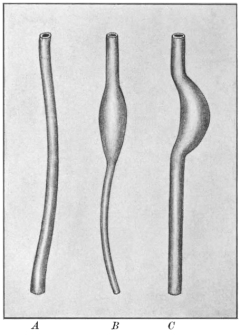
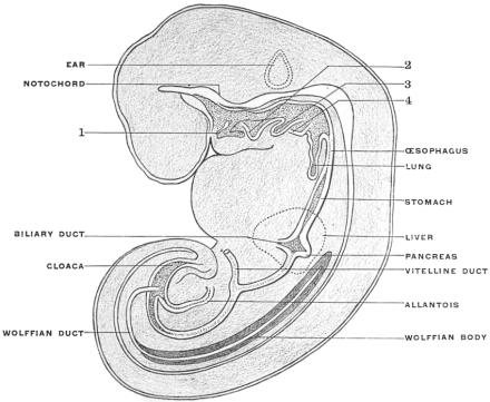
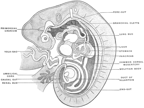
the primitive intestinal tube of uniform caliber (p. 40). It will
be recalled that the stomach at a certain period, while it already
presents the main structural features familiar in the adult organ,
occupies a vertical position in the abdominal cavity, turning its
concave margin (lesser curvature) ventrad, while the convex dorsal
border (greater curvature) is directed toward the vertebral
column, being attached to the same by the layers of the proximal
part of the primitive dorsal mesentery. At this time the stomach
presents right and left surfaces, and the œsophageal entrance is
at the highest or cephalic point of the organ, while the pyloric
transition to the small intestine occupies the distal caudal extremity.
The primitive dorsal mesentery, as already stated, passes as a
thin double-layered membrane between the ventral surface of the
vertebral column and the dorsal border of the stomach, which, as
we will presently see, becomes during the later stages of development
the caudal (lower) margin or greater curvature.
It will be seen that the embryonic differentiation of the intestinal
tract into successive segments justifies the application of a
terminology based on this differentiation to the corresponding
portions of the primitive common dorsal mesentery.
Thus the proximal portion extending between the vertebral
column and the dorsal border or greater curvature of the stomach
becomes the mesogastrium; we differentiate this portion
still further as the “dorsal mesogastrium” to distinguish it
from a “ventral mesogastrium” which we will presently encounter
in considering the development of the liver and the connected
peritoneum.
In the same way the section of the primitive common dorsal
mesentery attached to the duodenal loop becomes the mesoduodenum,
that connected with the mobile part of the small intestine
(jejuno-ileum) the mesentery proper, while the portion passing to
the colon forms the mesocolon, to be subsequently still further
subdivided, after the different segments of the large intestine have
become mapped out, as the ascending, transverse and descending
mesocolon, the mesosigmoidea and the mesorectum.
In tracing the development of the adult human peritoneum it
is well to consider certain stages, which we will find illustrated by
the permanent conditions presented by some of the lower vertebrates:
These stages comprise:
(a) Changes in the position of the stomach.
(b) Changes in the direction and extent of the dorsal mesogastrium.
(c) Development of the pancreas and spleen in connection with
the mesogastrium.
A. Changes in the Position of the Stomach.
The primitive position of the organ above outlined (p. 41) is
changed during the course of further development by a twofold
rotation.
1. The primitive vertical position, in which the œsophageal entrance
occupies the highest cephalic extremity, while the pyloric
opening is at the opposite caudal end, is exchanged for one directed
more transversely, approximating the two gastric orifices to the
same horizontal level. In human embryos of 13.9 mm. the fundus
has already descended, the pylorus moving cephalad and to the
right, while the cardia becomes shifted more to the left. At the
same time the greater growth and prominence of the convex border
or greater curvature becomes marked in comparison with the relatively
short extent of the opposite margin or lesser curvature.
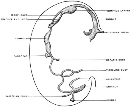
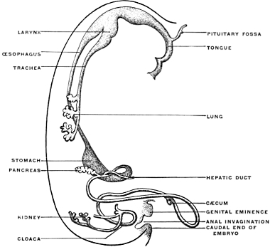
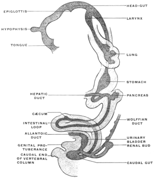
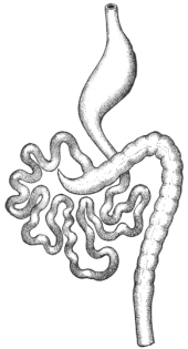
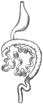
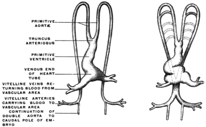
Figs. 169, 170.—Two front views of the entodermal canal. (Minot,
after His.)
|
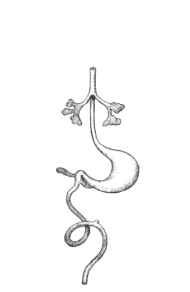 Fig. 169.
—Embryo Sch. 1 of His.
|
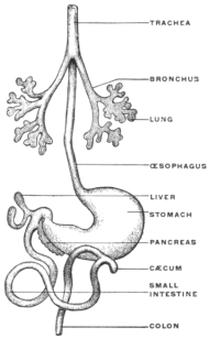 Fig. 170.
—Embryo Sch. 2 of His.
|
2. Coincident with this change in position is a rotation around
the vertical axis, by means of which the original left side of the
stomach is turned ventrad, becoming the ventral or “anterior”
surface, while the original right surface of the organ now looks
dorsad toward the vertebral column, becoming the dorsal or
“posterior” surface of human anatomy. The œsophageal or
cephalic end is placed to the left of the median line, while the
caudal or pyloric end is situated on the right side (Figs. 169
and 170).
The original ventral border, now the “lesser curvature” or
“upper border,” looks cephalad and to the right, toward the
caudal surface of the liver, while the original dorsal border, as the
“greater curvature” or “lower border” is directed in the main
caudad and to the left.
The prominence of this border is still further increased by the
greater development of the stomach to the left of the œsophageal
entrance resulting in the formation of the “fundus” or “great
cul-de-sac.”
This rotation of the stomach explains the asymmetrical position
of the vagus nerve in the adult, the left side of the embryonic
stomach, innervated by the left vagus, becoming the “anterior”
surface of adult descriptive anatomy and vice versa.
It will be readily appreciated that a comparatively flat organ
like the stomach, will, as long as it occupies a sagittal position,
with right and left surfaces, help to divide the upper part of the
abdominal cavity to a certain extent into a right and left half, even
if the peritoneal connections of the organ are left out of consideration.
As soon, however, as the above-described changes in position
take place and the surfaces of the stomach are directed ventrad
and dorsad, the relative arrangement and extent of this
right and left abdominal space becomes altered by the different
disposition of the septum, i. e., the stomach. The original right
side of the organ is now directed dorsad, and the rotation of the
organ has created a space between this dorsal or “posterior”
surface of the stomach and the background of the abdominal
cavity, which is the inception of the “lesser peritoneal cavity”
or retrogastric space. We will find that this space becomes well
defined and circumscribed by the peritoneal connections of the
stomach, but we will realize, even at this stage, that the dorsal
surface of the stomach will form a part of the general ventral
wall of the lesser peritoneal space.
On the other hand, the partial division of the abdomen into a
right and left half, effected by the stomach in its primitive sagittal
position, disappears after rotation of the organ. We now pass
uninterruptedly from left to right across the ventral (original left)
surface of the stomach.
B. Changes in the Direction and Extent of the Dorsal Mesogastrium.
The effects of the altered position of the stomach on the disposition
of the abdominal space have just been considered in relation
to the organ itself, without reference to its natural connections
with the parietes and with adjacent viscera. Their true significance
and their influence on the adult anatomical arrangement
of the abdomen is, however, only appreciated when the
changes in the arrangement of the peritoneal membrane which
they involve, are taken into account.
The dorsal mesogastrium changes more than any other portion
of the peritoneum in the course of development. It not only
becomes displaced and altered in direction by the rotation of the
stomach, but in addition it grows so extensively that it finally
hangs down like an apron over the entire mass of small intestines,
forming the great omentum.
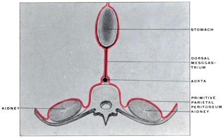 Fig. 171.
—Schematic representation of dorsal mesogastrium before rotation of stomach.
|
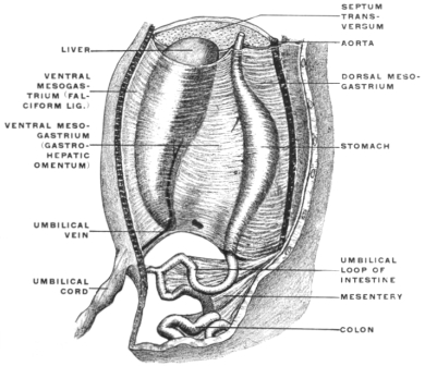 Fig. 172.
—Semi-diagrammatic representation of mesogastrium in
human embryo of the sixth week. (Kollmann.)
|
If we begin with the primitive disposition of the sagittal stomach
and dorsal mesogastrium shown in Fig. 171 it will be observed that
both structures together actually divide the dorsal portion of the
abdominal cavity into symmetrical right and left halves (Fig. 172).
After rotation of the stomach (Fig. 173) the mesogastrium loses
its original sagittal direction. It follows the altered position of
the original dorsal border of the stomach, which has now become
the caudal margin or “greater curvature,” by turning caudad and
to the left, being at the same time considerably elongated. This
occurs during the second month. Hence the dorsal mesogastrium,
after leaving the vertebral column, turns ventrad and to the left
to reach its gastric attachment along the greater curvature. This
is the first indication of the formation of the great omental or
epiploic bursa.
Figs. 173-175.—Schema of dorsal mesogastrium after rotation of stomach.
|
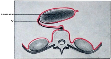 Fig. 173.
—Early stage.
|
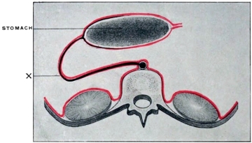 Fig. 174.
—Later stage, extension of mesogastrium beyond stomach
to left, with fundus of blind retrogastric pouch thus created at X.
|
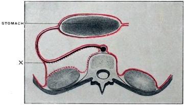 Fig. 175.
—After adhesion over area of dotted line between dorsal
mesogastrium and primitive parietal peritoneum. Secondary line of
transition from dorsal mesogastrium to parietal peritoneum at X.
|
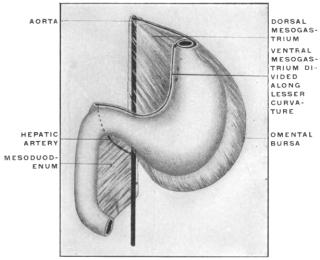 Fig. 176.
—Schematic ventral view of stomach, duodenum,
and dorsal mesogastrium, after rotation of stomach and extension
of omental bursa caudad beyond greater curvature of
stomach. The ventral mesogastrium is detached along the
lesser curvature.
|
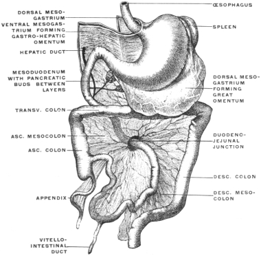 Fig. 177.
—Semi-diagrammatic representation of peritoneal membrane
in human embryo. (After Kollmann.)
|
The stomach is here considered as developing in situ and as influencing
by its growth and change of position the arrangement
and direction of the peritoneal layers with which it is connected.
As a matter of fact it is well to note that the stomach at first lies
above the primitive diaphragm or septum transversum, migrating,
however, at an early period into the subhepatic abdominal position.
This migration produces a corresponding increase in the
length of the œsophagus (Fig. 34) and the stomach, in consequence
of this change in position, acquires its ventral and dorsal
mesogastrium. For the purpose of explaining the adult peritoneal relations of the organ it is, however, more convenient to
regard the stomach as an abdominal organ from the beginning
and to deal with the subsequent changes in position from this
standpoint. The inaccuracy is slight and renders the comprehension
of the succeeding stages easier.
It will be noticed (Fig. 173) that the rudimentary retro-gastric
space or “lesser peritoneal sac” is bounded ventrally by the dorsal
(the primitive right) surface of the stomach, while its dorsal
boundary is furnished by the ventral (originally right) layer of
the dorsal mesogastrium.
In the primitive condition, therefore, dorsal mesogastrium and
stomach form together a straight line sagittal in direction and
placed in the median plane of the body. As the result of the
developmental changes above outlined this straight line becomes
bent at the point where the mesogastrium reaches the stomach
(Fig. 173, x). The two component elements of the line (stomach
and mesogastrium) hinge on each other here, and the angle which
they form opens to the right.
The changes which are to be observed in the later stages depend
principally upon a peculiar feature characteristic of the development
of the dorsal mesogastrium. This feature consists in the
extreme redundancy of the membrane which grows out of proportion
to the requirements of its visceral connections, and to a
certain extent becomes independent of the direct mechanical purpose
of carrying blood vessels to the viscera. Hence in a transverse
section at this period (Figs. 174 and 175) the mesogastrium
no longer passes in a direct line between its points of attachment,
viz. the greater curvature of the stomach and the vertebral column,
but extends beyond the stomach to the left. We will
appreciate the significance of this extensive growth of the mesogastrium
especially in considering the development of the spleen
and pancreas. For the present it will suffice to note (Figs. 174
and 175) that the growth has carried the mesogastrium well to
the left of the stomach, consequently the retrogastric space is now
bounded toward the left by the bend which the original right leaf
of the primitive sagittal mesogastrium takes in order to reach its
gastric attachment. The retrogastric space therefore terminates
toward the left in a blind pocket formed by this reduplication of
the mesogastrium.
One more factor is to be taken into consideration, namely the
tendency, already noted, of peritoneal surfaces to become adherent
to each other. Such adhesion involves the apposed surfaces of
the mesogastrium and of the primitive parietal peritoneum to
the left of the vertebral column. The dorsal (original left) layer
of the mesogastrium adheres to the parietal peritoneum covering
the left side of the abdominal background and the cephalic portion
of the ventral surface of the left kidney up to the end of
the blind pouch which forms the extreme left limit of the retrogastric
space. Hence, after this process of adhesion is completed,
the dorsal wall of the retrogastric space is lined by secondary
parietal peritoneum covering the left kidney (original right leaf
of primitive mesogastrium) (Fig. 175). We obtain (Fig. 175 at
x) an apparent continuity of the parietal peritoneum with that
portion of the mesogastrium which, derived from the original
left layer of the membrane, appears now to extend, as the ventral
one of two layers, between the stomach and the abdominal
parietes near the lateral border of the left kidney. (Primitive
gastro-splenic omentum.)
It should be remembered that the disposition of the peritoneum
just indicated is modified by the development of the pancreas
and spleen, both of which organs are intimately associated with
the mesogastrium. The foregoing statements and diagrams are
therefore merely given for the purpose of affording a general view
of the extent, growth and changes of the dorsal mesogastrium
before proceeding to consider the development of the pancreas
and spleen in and from the membrane itself.
In the view directly from in front the redundancy of the peritoneum
forming the mesogastrium is shown in Figs. 176 and 177.
Just as the membrane extends further to the left than required by
its visceral connection with the stomach, so the downward growth
exceeds the demand made by the rotation of the attached border
(greater curvature) caudad and to the left. The mesogastrium,
forming, as it now does, the great omentum, enlarges in descending
toward the transverse colon (Fig. 177). The bag thus formed
can be distended with air in a fœtus of from 8 to 9 cm. vertex-coccygeal
measure, as shown in the figure. Consequently in sagittal
section the membrane is seen to extend caudad beyond the
level of the greater curvature, and must turn on itself and pass
again cephalad in order to reach the stomach (Fig. 178). By
reason of this excessive growth the limits of the primitive retrogastric
space are enlarged, not only toward the left, but more
especially in the caudal direction. The bend made by the mesogastrium
in returning to the stomach forms the blind extremity
of a pouch which continues the retrogastric space caudad beyond
the stomach, and whose dorsal and ventral walls are formed by
the reduplicated mesogastrium. This pocket or pouch constitutes
the omental or epiploic bursa of the lesser peritoneal cavity,
for the great omentum is the direct product of this redundant
growth of the mesogastrium caudad. It will be observed that
the great omentum is made up of four peritoneal layers, the folding
of the double-layered mesogastrium naturally producing this
result. The first or ventral and the fourth or dorsal layer are
derived from the original left layer of the primitive sagittal mesogastrium;
the intermediate second and third layers, separated
from each other at this stage by the cavity of the omental bursa,
are products of the primitive right leaf of the mesogastrium.
Since the entire retrogastric space with its extensions becomes the
“lesser cavity” of the human adult peritoneum, it will be seen
that its serous membrane is derived from the original right leaf
of the mesogastrium (second and third omental layers). After
the above-described adhesion of the mesogastrium to the parietal
peritoneum overlying the ventral surface of the left kidney, the
membrane would be traced in sagittal section (Fig. 179) from the
dorsal surface of the stomach caudad, lining the interior of the
omental bursa (second layer) to the turn or blind end of the
pouch; thence cephalad as the third omental layer, forming the dorsal
wall of the epiploic bursa, to invest, as secondary parietal peritoneum,
the cephalic segment of the ventral surface of the left kidney.
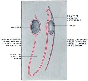 Fig. 178.
—Schematic sagittal section through stomach and dorsal
mesogastrium, after rotation and formation of omental bursa.
|
|
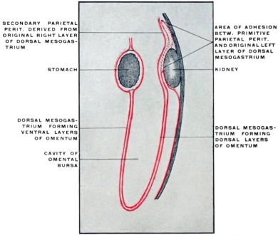 Fig. 179.
—Schematic sagittal section through stomach and dorsal mesogastrium
after adhesion to prerenal parietal peritoneum.
|
C. Development of Spleen and Pancreas in the Dorsal Mesogastrium
and Changes in the Disposition of the Great Omentum.
In order to obtain a correct conception of the adult human
conditions it is finally necessary to consider the development of
the spleen and pancreas in their connection with the dorsal mesogastrium
and to note the changes which are produced by adhesion
of portions of the great omentum to adjacent serous surfaces. It
will be advisable to discuss these subjects at first separately, and
to subsequently combine all the facts in an attempt to gain a correct
impression of their share in determining the disposition of
the adult human peritoneum.
1. Development of Spleen.—The spleen develops from the mesoderm
between the layers of the dorsal mesogastrium, near its point
of accession to the greater curvature, in the region of the subsequent
fundus. It has therefore, like the stomach, originally free
peritoneal surfaces. After rotation of the stomach the organ lies
between the two layers of the membrane at the extreme left end
of the retrogastric space (Fig. 180).
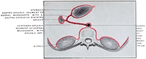 Fig. 180.
—Schematic transverse section of the abdomen, showing early stage of development
of spleen from extreme left end of dorsal mesogastric pouch.
Vascular Connections.—The splenic artery accedes to the mesal surface
of the spleen from the vessel which originally passed directly
to the dorsal border (subsequent greater curvature) of the stomach,
between the layers of the mesogastrium.
With the further growth of the spleen the segment of this vessel
situated between its origin from the cœliac axis and the hilum of
the spleen becomes relatively larger, forming the adult splenic
artery, while the continuation of the original vessel to the greater
curvature of the stomach appears now as a branch of the splenic
artery, viz., the arteria gastro-epiploica sinistra.
Through the development of the spleen the dorsal mesogastrium
has been subdivided into a proximal longer vertebro-splenic, and
a distal shorter gastro-splenic segment. The former, as we have
seen, loses its identity as a free membrane in the human adult, by
fusing with the parietal peritoneum investing the ventral surface
of the left kidney. Hence, after this adhesion has taken place, the
splenic artery courses from the cœliac axis to the spleen behind
peritoneum which functions as part of the general parietal membrane,
but which is derived from the original right leaf of the
proximal vertebro-splenic segment of the primitive mesogastrium
(Fig. 181). On the other hand the distal segment of this membrane,
beyond the spleen, remains free, carrying, as the gastro-splenic
omentum, the left gastro-epiploic artery between its layers
from the splenic artery to the greater curvature of the stomach.
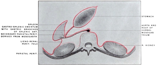 Fig. 181.
—Schematic transverse section of the abdomen, showing later stage of development
of spleen and arrangement of peritoneum after adhesion of dorsal layer of mesogastrium and
primitive prerenal parietal peritoneum.
The lateral limit of the area of adhesion between mesogastrium
and parietal peritoneum is situated along the lateral border of the
left kidney. Hence, in the final condition of the parts, the main
splenic vessels at the hilum are situated between two peritoneal
layers of which the ventral (Fig. 181) appears as the parietal
peritoneum forming the dorsal wall of the retro-gastric space,
while the dorsal layer (Fig. 181) forms a reflection from the mesal
surface of the spleen, along the dorsal margin of the hilum, to the
adjacent lateral border of the left kidney (lieno-renal ligament)
and to the diaphragm. At this point of adhesion subsequently
firmer strands of connective tissue develop in the serous reduplication
forming the ligamentum phrenico-lienale of systematic anatomy.
This process of adhesion takes place during the second
half of intra-uterine life. A connection with the colon, produced
by adhesion of the mesogastrium to the splenic flexure of the
large intestine, forms the adult lig. colico-lienale, while a similar
adhesion between great omentum, transverse mesocolon and
phrenic parietal peritoneum just caudad of the spleen, gives rise to
the colico-phrenic or costo-colic “supporting” ligament of the spleen.
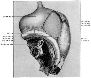 Fig. 182.
—Part of the abdominal viscera of child, two years old, hardened
in situ and
removed from body. The great omentum has been detached along the line of the transverse
colon. (Columbia University, Study Collection.)
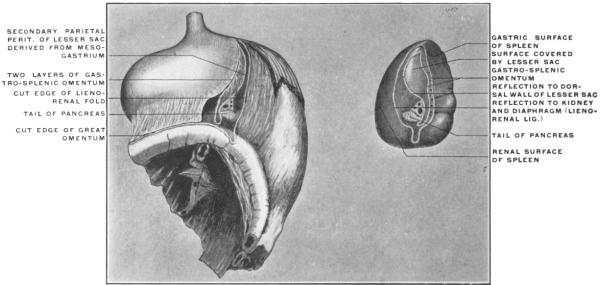 Fig. 183.
—The same preparation with the spleen removed, showing lines of peritoneal reflection
on mesial surface of the organ.
On the other hand, the ventral one of the two layers constituting
the gastro-splenic omentum and including between them the
left gastro-epiploic artery, is formed by the distal part of the
primitive left layer of the mesogastrium, while the dorsal layer
of the same fold is the portion of the primitive right layer beyond
the spleen, which has not been converted into secondary
parietal peritoneum, but forms now part of the ventral wall of
the lesser peritoneal sac between the spleen and the stomach
(Fig. 181) (lig. gastro-lienale). Since, therefore, the gastro-splenic
omentum is a specialized part of the fully-developed dorsal mesogastrium,
and since we have seen that the great omentum is
formed directly by the excessive growth of this membrane caudad,
it is not difficult to understand why in the adult human
subject the ventral layer of the gastro-splenic omentum is directly
continuous with the ventral layer of the great omentum
along the greater curvature of the stomach to which both are
attached. The dorsal layer of the gastro-splenic omentum would,
in the same way, be continuous with the second layer of the great
omentum, lining the ventral wall of the omental bursa, if it were
not for the fact that in the adult adhesions usually obliterate the
cavity of the bursa.
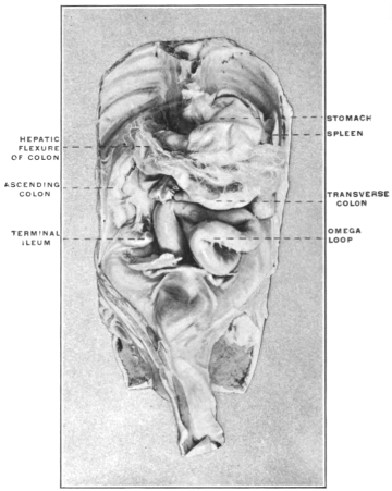
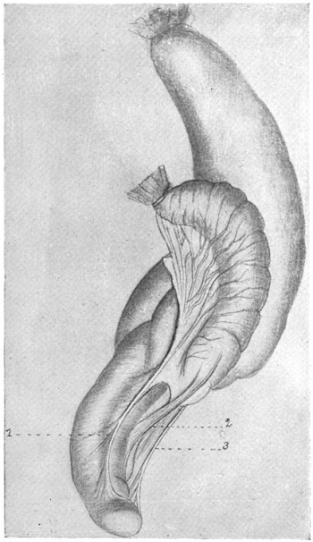
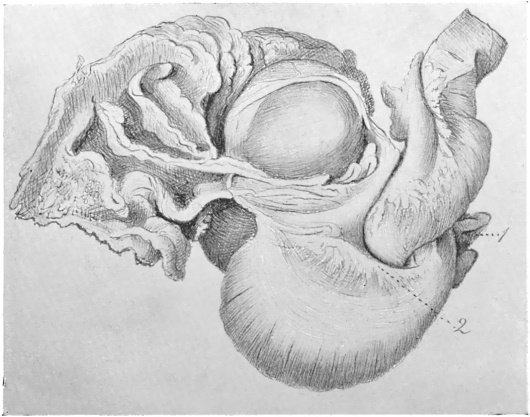
Fig. 182 shows the stomach, left kidney, spleen and splenic
flexure of the colon hardened in situ and removed from the body
of a two-year-old child. The great omentum has been divided
along the line of adherence to the transverse colon.
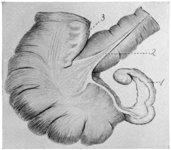
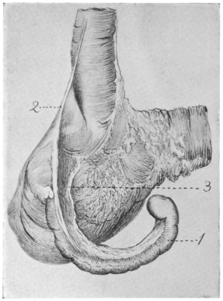
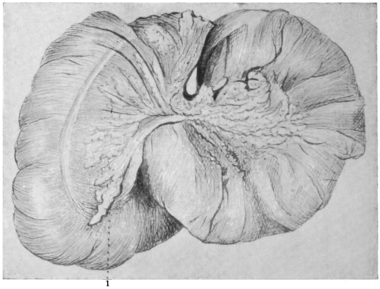
In Fig. 183 the spleen has been removed from the preparation
by division of its peritoneal and vascular connections, and is
shown in its mesal aspect (gastric and renal surfaces, intermediate
margin and hilum). It will be seen that the peritoneal reflections are
arranged in the form of two concentric elliptical lines. The two
ventral lines form the gastro-splenic omentum and correspond to
the reflection of the peritoneum from spleen to left end of stomach
carrying the gastric branches derived from the splenic artery.
The third line from before backwards results from the division of
the secondary parietal peritoneum of the lesser sac, covering
splenic artery, and ventral surface of pancreas and derived from
the dorsal mesogastrium; while the most dorsal fourth line
represents the divided reflection of the peritoneum from the renal
surface of spleen to lateral border of left kidney and diaphragm
(lig. lieno-renale).
Between the second and third lines of peritoneal reflection appears
the portion of the mesal surface of the spleen in contact with
and invested by the extreme left end of the lesser peritoneal sac.
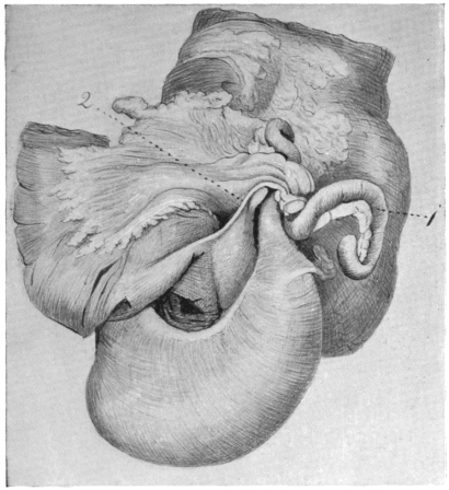
Fig. 184, taken from an adult human subject with the viscera
hardened in situ, shows the left or splenic extension of the lesser
peritoneal cavity.
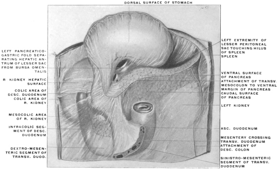 Fig. 184.
—Upper abdominal viscera of adult human subject, hardened
in situ, with liver and
colon removed and stomach turned up. (Columbia University, Study Collection.)
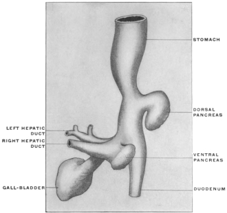 Fig. 185.
—Pancreatic and hepatic buds of human embryo of four weeks. (Kollmann.)
2. Development of the Pancreas.—The pancreatic gland is derived
from the hypoblast of the enteric tube. The secreting epithelium
and that lining the ducts of the adult gland is formed by budding
and proliferation of the intestinal epithelium. The gland
develops primarily from two outgrowths which are at first separate
and distinct from each other.
1. The proximal and dorsal bud grows directly from the hypoblast
lining the duodenum immediately beyond the pyloric junction.
In embryos of 8 mm. (four weeks) (Fig. 185) it appears as a
small spherical outgrowth connected by a slightly narrower stalk
with the epithelial intestinal tube.
2. The distal and ventral outgrowth is separated from the preceding
and is from the beginning closely connected with the
similar embryonic outgrowth from the enteric tube which is to
form the liver. This portion of the pancreas is, strictly speaking,
derived primarily from the epithelium of the primitive
hepatic duct and not directly from the duodenum. This primary
arrangement of the gland, being formed of two main collections
of budding hypoblastic cells, corresponds to the adult
system of the pancreatic excretory ducts. The proximal or dorsal
outgrowth furnishes that portion of the head of the gland
whose excretory system terminates in the secondary pancreatic
duct or duct of Santorini, while the distal (ventral) outgrowth
includes within its area the termination of the principal pancreatic
duct or canal of Wirsung, which is closely connected with
the end of the common bile-duct at the intestinal opening common
to both (Figs. 186-187). The method of union of the two
pancreatic outgrowths and their respective share in building up
the adult gland explains the usual adult arrangement of the
excretory system and its variations.
In the embryo of five weeks (Fig. 186) the two portions have
grown in length. The dorsal or proximal outgrowth, developing
between the layers of the mesoduodenum, is at this time the larger
of the two, composed of a number of glandular vesicles clustered
around the stalk represented by the parent duct.
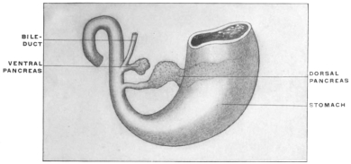 Fig. 186.
—Pancreatic buds of human embryo of five weeks. (Kollmann, after Hamburger.)
|
|
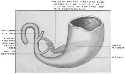 Fig. 187.
—Pancreatic buds of human embryo of six weeks. (Kollmann, after Hamburger.)
|
The distal or ventral pancreatic growth, connected with the
liver duct, is as yet small and presents only a few vesicular appendages.
The duct of this portion empties in common with the
hepatic duct into the duodenum.
In embryos of the sixth to seventh week (Fig. 187), the two
glandular outgrowths have become connected with each other at a
point which corresponds exactly to the divergence of the duct of
Santorini from the main pancreatic duct (canal of Wirsung) in the
adult gland (Fig. 188).
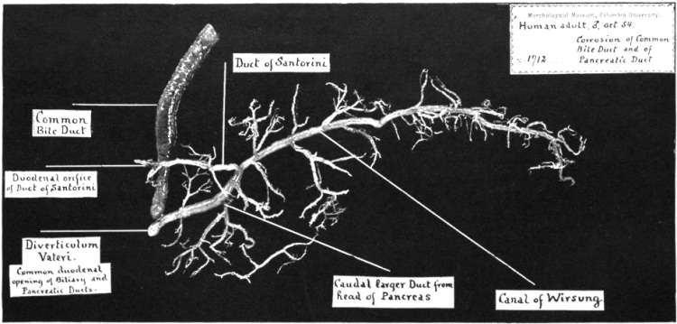 Fig. 188.
—Human adult. Corrosion of pancreatic and common bile-ducts: ventral view. (Columbia University Museum, No. 1712.)
The secondary pancreatic duct (of Santorini) of the adult corresponds
to that section of the proximal or larger embryonic outgrowth
situated between the intestine and the point where the
two glandular diverticula fuse with each other. Hence the canal
of Wirsung in the adult is a compound product. It includes the
duct system developed, in connection with the bile duct, in the
head of the gland, forming the intestinal termination of the main
duct. Its distal body portion on the other hand is derived from
the duct system of the originally larger proximal outgrowth, including
the entire peripheral portion which has become secondarily
added to the duct of the ventral outgrowth to form together with
it the canal of Wirsung. On the other hand the proximal portion
of the duct system of this originally larger part becomes secondarily
differentiated as the duct of Santorini.
Fig. 188 shows the normal adult arrangement of the pancreatic
and biliary ducts in a corrosion preparation of the canal.
The duct of Santorini in this case opened by a separate orifice
into the duodenum above the common opening of the biliary and
pancreatic ducts (cf. p. 113).
Explanation of Adult Arrangement of Human Pancreatic Ducts and Their
Variations Dependent Upon the Embryonic Development.—The smaller
distal embryonic outgrowth is, as we have seen, from its inception
in close connection with the duodenal end of the common bile-duct
(Fig. 185).
The proximal outgrowth, situated nearer to pylorus and derived
directly from the duodenal epithelium, is the larger and
forms the greater part of the bulk of the adult pancreas (Figs.
186, 187).
Figs. 189-192.—Series of schemata showing normal and variant adult types of biliary and
pancreatic ducts.
|
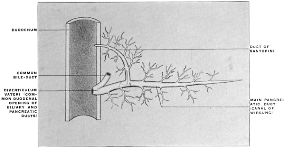 Fig. 189.
—Usual human adult type.
|
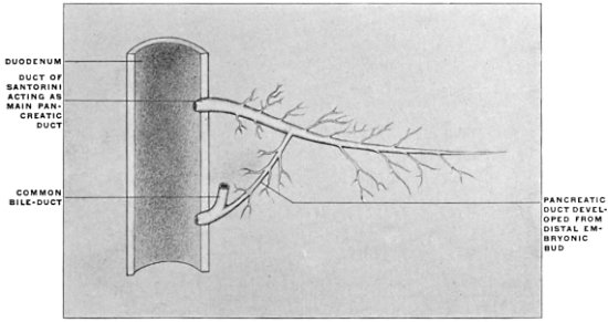 Fig. 190.
—Persistence of early embryonal type.
|
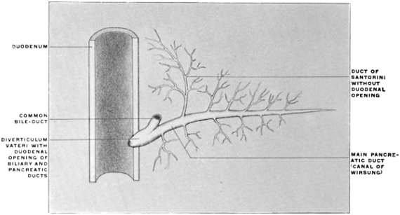 Fig. 191.
—Duct of Santorini has no duodenal orifice.
|
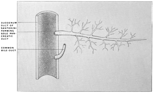 Fig. 192.
—Duct of Santorini forms the only pancreatic duct. Separate duodenal openings of
biliary and pancreatic ducts, resulting from failure of development of distal embryonal pancreatic
bud.
|
If, notwithstanding this primitive arrangement, the distal duct
(canal of Wirsung) appears as the main pancreatic duct in the
adult, while the proximal (duct of Santorini) is secondary, this
depends upon a union of the products of the two outgrowths in
such a manner that the greater part of the duct system of the
proximal and larger portion is transferred to the distal duct to
form the adult canal of Wirsung, while the smaller segment of
the proximal duct, between its opening into the duodenum and
the point of fusion of the two outgrowths, forms the adult secondary
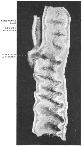
duct of Santorini. This duct opens usually into the duodenum
upon a small papilla situated about 2.5 cm. above the
common duodenal termination of the bile-duct and canal of Wirsung
(papilla Vateri) (Fig. 193). The duct of Santorini usually
tapers toward the duodenal opening from its point of departure
from the main duct, its caliber gradually diminishing in the direction
indicated, so that it is smaller at the duodenal opening
than at the point of confluence with the main duct (Fig. 189).
Hence the secretion from the proximal head portion of the pancreas,
conveyed by this duct and its tributaries, passes usually into
the main pancreatic duct and not directly into the intestine
through the duodenal opening of the duct of Santorini. The
latter is, however, thus enabled to vicariously take upon itself
the conduct of the pancreatic secretion in cases of obstruction or
obliteration of the main duct (calculi, ulcers, cicatrices, etc.). In
these cases of obstruction of the main duct the duct of Santorini
enlarges and performs its functions.
Occasionally, without obstruction of the main duct, the duodenal
opening of the duct of Santorini is large, and the flow of
secretion evidently the reverse of the usual, i. e., directly into
the intestine.
In other cases, also without pathological conditions, the proximal
duct is the larger of the two and serves as the principal
channel of pancreatic secretion, the canal of Wirsung being small.
This is evidently a persistence and further development of the
early embryonic relative condition of the two outgrowths above
described (Fig. 190). On the other hand the duct of Santorini
may not open at all into the duodenum, terminating in small
branches which drain the proximal part of the head of the gland
(Fig. 191).
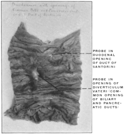 Fig. 193.
—Mucous surface of human duodenum, showing entrance of biliary and
pancreatic ducts and diverticulum Vateri. (Columbia University Museum, No. 1842.)
Schirmer has examined the arrangement of the pancreatic ducts
in 105 specimens. In 56 of these the duct of Santorini passed
from the main duct into the duodenum, opening upon a papilla
situated 2.5 cm. above the common opening of the bile duct and
canal of Wirsung.
In 19 the duct of Santorini was well developed but did not
open into the duodenum.
In but 4 cases the duct of Santorini formed the only pancreatic
duct, the lower opening being occupied by the bile duct alone
(Fig. 192). We may assume in these cases failure of development
of the distal outgrowth connected with the primitive hepatic
bud, leaving only the proximal duodenal outgrowth to form the
entire adult gland.
Figs. 188 and 189 show the normal arrangement of the duodenal
openings of the biliary and pancreatic ducts.
Figs. 190 to 192 show schematically the variations in the relative
development and the adult arrangement of the pancreatic
ducts.
Diverticulum and Papilla Vateri.—From what has been said regarding
the embryonic union of the distal pancreatic outgrowth
with the hepatic bud it will be easy to recognize the corresponding
features in the arrangement of the adult duodenal termination of
the common bile-duct and canal of Wirsung. The dilated interior
of the duodenal papilla (diverticulum Vateri) corresponds to the
embryonic segment between the intestinal opening of the primitive
liver duct and the point when this duct gives off the distal
larger pancreatic outbud (Figs. 186, 187, 188, 193 and 194).
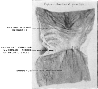
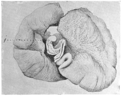
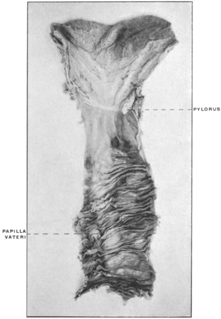 Fig. 194.
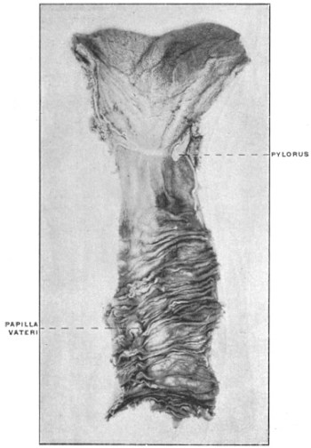
—Adult human subject. Mucous membrane of pyloro-duodenal junction
and of duodenum. (Columbia University Museum, No. 1840.)
|
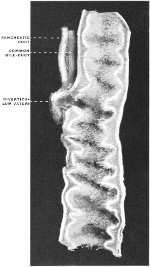 Fig. 195.
—Duodenum, with entrance of pancreatic
and biliary ducts and well-developed diverticulum Vateri
in the cassowary, Casuarius casuarius. (Columbia University
Museum, No. 1821.)
|
The union of the pancreatic and biliary ducts to form the recess
of the diverticulum Vateri, which then opens by a single common
orifice into the duodenum, is better marked in some of the lower
vertebrates than in man.
Fig. 195 shows the proximal portion of the duodenum of the
cassowary (Casuarius casuarius) with the biliary and pancreatic
ducts and the diverticulum at their confluence in section.
The development of these two main digestive glands as diverticula
from the intestinal canal also explains the direct continuity
of the mucous membrane of their ducts with that lining the duodenum,
a fact which is of considerable importance in the pathological
extension of mucous inflammations from the intestine to
the duct system of the glands.
Development of the Pancreas in Lower Vertebrates.—In the embryo
of the sheep two pancreatic buds are found, but the duct of
the dorsal (proximal) outgrowth (duct of Santorini) subsequently
fuses entirely with the main duct.
In the cat there are likewise two pancreatic outgrowths.
In the chick three pancreatic buds are visible about the fourth
day.
Amphibia likewise present three embryonic pancreas buds.
The ventral (distal) outgrowth is double, the two portions proceeding
symmetrically from each side of the hepatic duct. The
single dorsal outgrowth is derived directly from the duodenal
epithelium. Later on all these outgrowths fuse to form the single
adult gland.
Fish also possess several (up to four) embryonic pancreatic outgrowths.
Recently in human embryos of 4.9 mm. cervico-coccygeal measure
three pancreatic outgrowths have been observed, all entirely
distinct from each other, one dorsal, budding from the epithelium
of the primitive duodenum and two ventral, proceeding from the
grooved gutter which represents the primitive ductus choledochus
at this period. In embryos of from 6 to 10 mm. the two ventral
outgrowths have already fused, hence only two buds, a single
ventral and a dorsal, are now encountered.4
These observations place the development of the human pancreas
in line with the triple pancreatic outgrowths, two ventral
and one dorsal characteristic of the majority of the lower vertebrates,
which have been hitherto carefully examined. The ventral
or distal bud is probably double in the majority of vertebrates.
The two segments fuse, however, so early that the derivation of
the pancreas from a double outgrowth, as described above for the
human embryo, practically obtains. In forms in which the
adult gland presents a number of separate openings into the duodenum
(cf. p. 118), the development would probably show multiple
embryonic outgrowths from the intestinal hypoblast.
In any case the dorsal pancreatic bud appears to have developed
in the vertebrate series before the ventral outgrowth and to be
hence phylogenetically the older structure.
COMPARATIVE ANATOMY OF THE PANCREAS.
With the exception of Amphioxus and probably also of the
Cyclostomata, the gland appears to be present in all vertebrates,
varying, however, much in size, shape and relation to the intestinal
tube. Usually it appears as an elongated, flattened, more or
less distinctly lobulated organ, in close apposition to the duodenum
between the layers of the mesoduodenum. In all forms
in which the gland is found it is connected with the post-gastric
intestine and marks the beginning of the midgut. In structure
the gland is usually acinous, resembling the salivary glands. It
is well developed in the selachians, forming a triangular body
connected with the beginning of the midgut (Fig. 202). In
some instances the gland elements do not extend beyond the intestine itself, but remain imbedded in the wall of the midgut, as
in Protopterus. In certain adult teleosts the pancreas is surrounded
by the liver (Fig. 196), in others it does not appear as a
compact gland but is distributed in the form of finely scattered
lobules throughout the mesentery between the two layers of this
membrane. On account of this concealed position of the gland
it was formerly believed that the adult teleosts did not possess a
pancreas. The pyloric cæca (cf. p. 119) found in these forms were
consequently considered to be homologous with the pancreas of
the higher vertebrates.
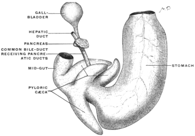 Fig. 196.
—A portion of alimentary canal of Pleuronectes maculatus,
the flounder, with pancreas attached to biliary duct and concealed in
the substance of the liver, which has been removed. (Columbia University
Museum, No. 1491.)
|
|
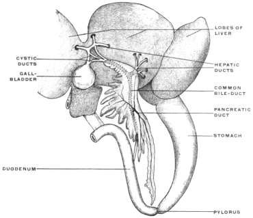 Fig. 197.
—Pancreas and biliary ducts of Rana esculenta, frog. (Wiedersheim,
after Parker; both from Ecker.)
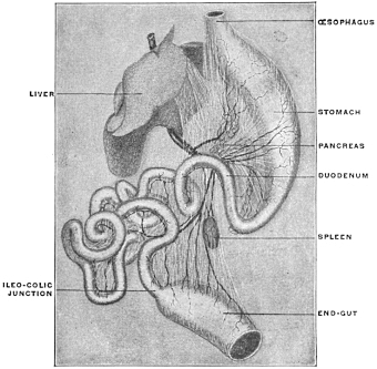
|
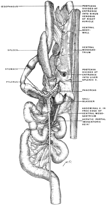 Fig. 198.
— Necturus maculatus, mud puppy. Dissection of intestinal
canal, liver, pancreas, and spleen, with blood-vessels injected. (Columbia
University Museum, No. 1863.)
|
|
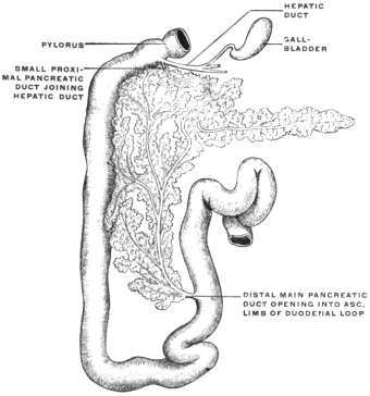 Fig. 199.
—Pancreas and pancreatic ducts of rabbit. (Nuhn.)
|
|
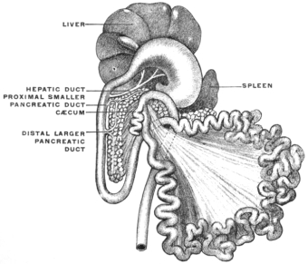 Fig. 200.
—Abdominal viscera of dog, showing arrangement of
pancreatic ducts. (Nuhn.)
|
In Myxinoids a peculiar lobulated glandular organ is found
imbedded in the peritoneal coat of the intestine near the entrance
of the bile-duct, into which its lobules open separately. This
organ possibly corresponds to the higher vertebrate pancreas.
An organ which may represent a dorsal pancreas is also developed
in Ammocœtes (larva of Petromyzon), but its exact homology
is still doubtful. It is possible that a true pancreas has
not yet developed in the cyclostomata. In Amphioxus no trace
of a pancreas is found. In all other vertebrates the gland is
present. In certain amphibians, as the frog, the single pancreatic
duct opens into the common bile duct (Fig. 197).
In lacertilians and in some chelonians a lateral offshoot of the
pancreas is directed transversely and is adherent to the spleen.
Fig. 113 shows the gland in Chelydra serpentaria. While the
gland usually has a single duct, yet two ducts are found in a
number of animals (many mammals, birds, chelonians and
crocodiles). At times three ducts are encountered, as in the
chicken and pigeon.
The arrangement of the pancreatic duct system among mammalia
presents the following variations:
1. Mammals with one pancreatic duct, either connected with
the bile-duct or entering the intestine independently:
Monkeys, most rodents (except the beaver), marsupials, carnivora
(except dog and hyena), many ungulates (pig, peccary,
hyrax, etc.), most ruminating artiodactyla.
(a) The pancreatic duct joins the common bile-duct before entering
the duodenum in the monkeys, marsupials, carnivora, in the
sheep, goat and camel.
The point of entrance of the combined duct into the intestine
varies. In some forms it is near the pylorus, in others at some
distance from the same. The common opening is situated 1½″ to
2″ beyond the pylorus in carnivora, and one foot behind the same
point in the goat and sheep.
(b) The pancreatic duct does not join the bile-duct, but empties
separately into the intestine, in most rodents and in the calf and pig.
In the calf the pancreatic duct opens into the duodenum 15′ beyond the bile-duct and
3′ beyond the pylorus.
In the pig the pancreatic opening is 5″–7″ beyond that of the
bile-duct and 6″–8″ behind the pylorus.
2. Mammals with two pancreatic ducts, of which one usually
joins the bile-duct: perissodactyla (except the ass according to
Meckel), elephant, beaver, several carnivora, dog, hyena, and
according to Bernard the cat. In the perissodactyla the proximal
of the two pancreatic ducts empties, either combined with
the bile-duct, or separate from it, but very close to it, 3″–4″ behind
the pylorus. The second distal duct is smaller and opens
several inches further down.
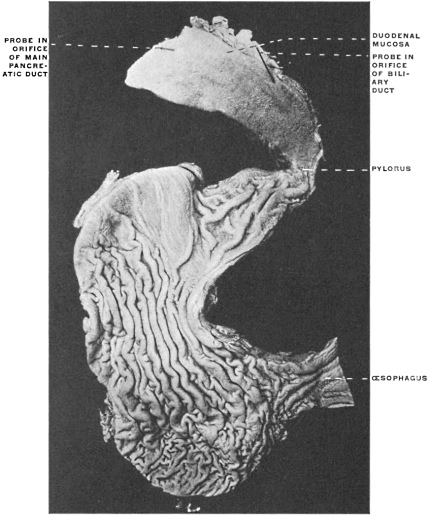 Fig. 201.
—Section of dog’s stomach, and proximal portion of duodenum, with
entrance of biliary and pancreatic ducts. (Columbia University Museum, No. 1822.)
In most rodents the pancreatic entrance is placed at some distance
from the pylorus. Fig. 199 shows the arrangement of the
parts in the rabbit, in which animal the main distal pancreatic
duct empties at a distance of 13″–14″ from the pylorus into the
end of the duodenum, which intestine forms a very long loop,
while the biliary duct, receiving the smaller proximal pancreatic
duct, opens near the pylorus.
In the beaver the smaller proximal duct joins the bile-duct or
even enters the duodenum anterior to the bile-duct, nearer the
pylorus, while the distal larger pancreatic duct opens into the intestine
16″–18″ behind the biliary duct. Of the two ducts found
in the dog (Fig. 200) the smaller proximal either joins the bile-duct
or opens into the intestine close to it, 1″–1½″ beyond the
pylorus. The larger distal duct opens into the duodenum 1″–1½″
behind the biliary duct. Fig. 201 shows the dog’s stomach and
proximal portion of the duodenum in section. The proximal
smaller pancreatic duct here joins the biliary duct, and opens with
it by a single orifice into the duodenum. The distal larger pancreatic
duct opens independently into the intestine further caudad.
The parts in Hyæna present a similar arrangement.
Bernard always found two pancreatic ducts in the cat, one large
principal duct and a second smaller accessory duct. Of these, the
one situated nearest to the pylorus always united with the bile-duct.
The pancreatic duct thus joining the bile-duct was sometimes
the main duct, sometimes the accessory smaller duct.
Since the main function of the pancreatic juice is the conversion
of starch into sugar, the gland appears better developed in
general in herbivora than in carnivora, without, however, disappearing
in the latter. In fact it is of considerable size in the
carnivora, because the secretion also acts on the albuminous food
substances and, though to a lesser degree, on the fats.
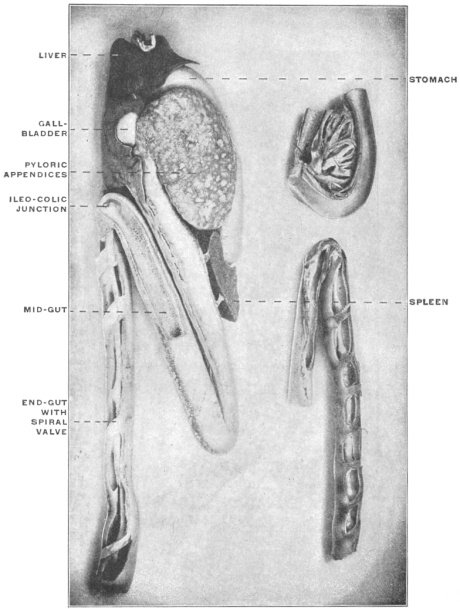
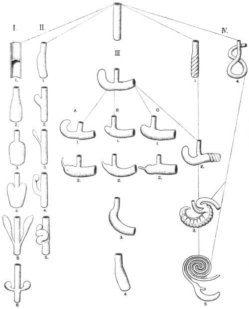
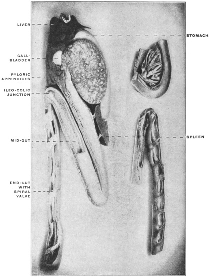
PYLORIC CÆCA OR APPENDICES.
In the Cyclostomata and Selachians the intestinal canal is in the
main free from cæcal appendages, while a large portion of the
tube is provided with a special fold of the mucous membrane
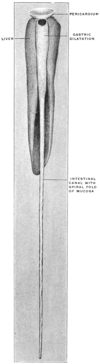
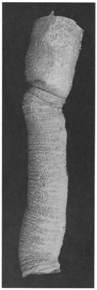
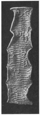
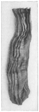
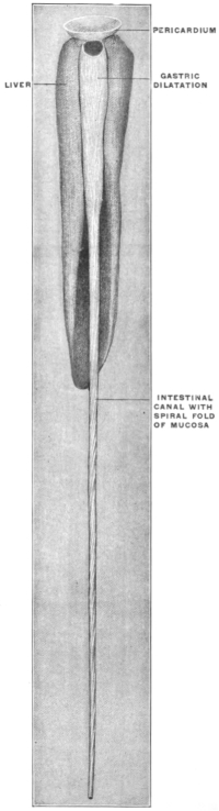
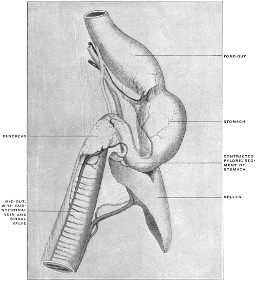
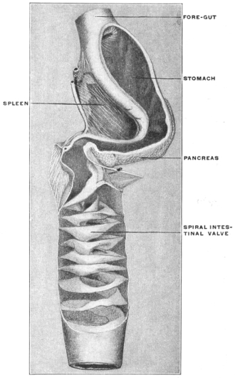
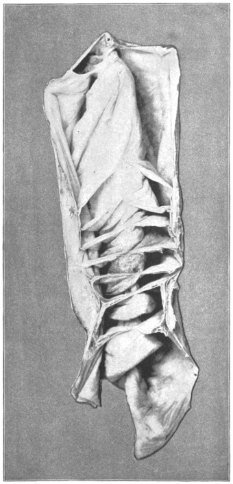
which projects into the lumen of the gut (spiral valve). Fig. 43
shows the straight intestinal tract with the spiral valve of the
longer distal segment in a cyclostome, Petromyzon marinus or
lamprey. In Figs. 202 and 203 the selachian (shark) intestine is
represented in two examples, while the similar spiral valve in a
Dipnœan or lung fish, Ceratodus, is seen in Fig. 204.
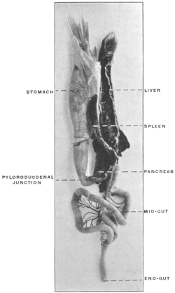
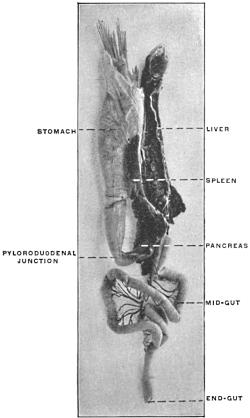
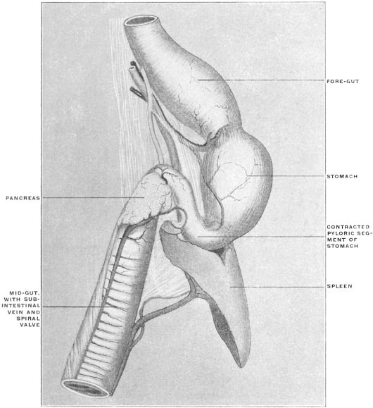 Fig. 202.
—Alimentary tract with spleen and pancreas of
Squalus acanthias, the dog-fish.
(Columbia University Museum, No. 1405.)
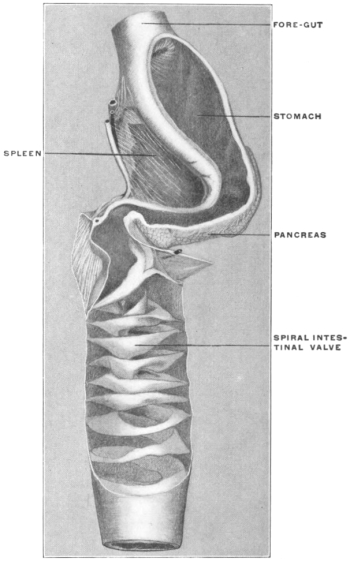 Fig. 203.
—Alimentary canal of Galeus canis, dog-shark, in
section, showing spiral intestinal valve. (Columbia University
Museum, No. 1429.)
|
|
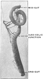
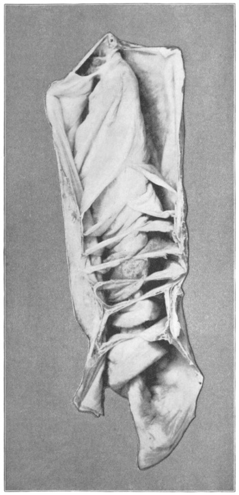 Fig. 204.
—Alimentary canal with spiral valve of Ceratodus
forsteri, the Australian lung-fish (Barramunda). (Columbia University
Museum, No. 1645.)
|
On the other hand in the Ganoids and in many Teleosts longer
or shorter finger-shaped diverticula of the midgut are found immediately
beyond the pylorus in the region of the bile-duct.
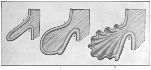
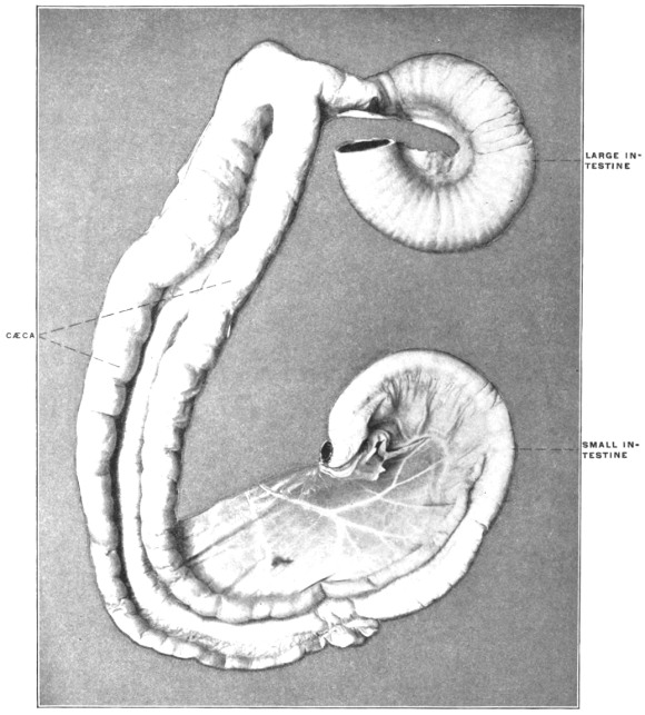
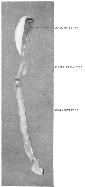
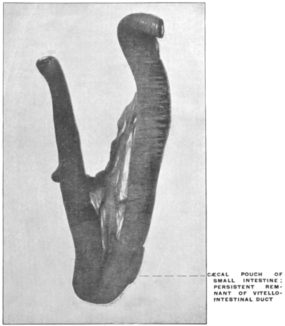
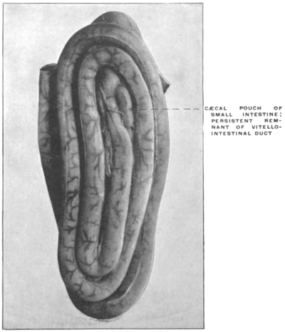
These pouches or diverticula of the intestine form the so-called
pyloric cæca or appendices of these fish. They vary very much
in length, diameter and number in different forms.
Thus but a single diverticulum appears in Polypterus and Ammodytes
(Fig. 205). Rhombus maximus and Echelus conger (Figs. 112 and
206) have two, and the same number appear in Lophius piscatorius
(Fig. 207). Perca has three and the Pleuronectidæ have three to five.
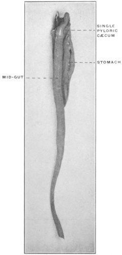 Fig. 205.
—Alimentary canal of Polypterus
bichir. (Columbia University Museum, No.
1823.)
|
|
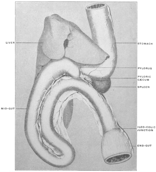 Fig. 206.
—Alimentary tract of Echelus conger, Conger eel. Stomach, mid- and end-gut,
liver, and spleen. (Columbia University Museum, No. 1430.)
|
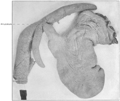 Fig. 207.
—Stomach, duodenum, and pyloric cæca of Lophius piscatorius, angler.
(Columbia University Museum, No. 1824.)
|
|
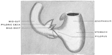 Fig. 208.
— Pleuronectes maculatus, window-pane. Stomach and mid-gut with
pyloric cæca and hepatic duct. (Columbia University Museum, No. 1432.)
|
| |
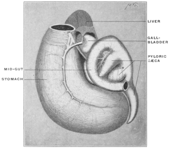 Fig. 210.
— Paralichthys dentatus, summer flounder. Stomach
and mid-gut with pyloric cæca and liver. (Columbia University
Museum, No. 1431.)
|
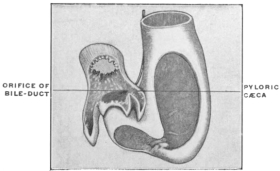 Fig. 209.
— Pleuronectes maculatus, window-pane. Stomach and mid-gut with
pyloric cæca, in section. (Columbia University Museum, No. 1433.)
|
|
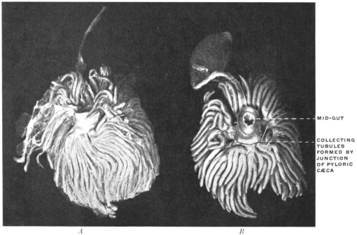 Fig. 211.
—Pyloric cæca of
Gadus callarias, codfish. (Columbia University Museum, No. 1825.)
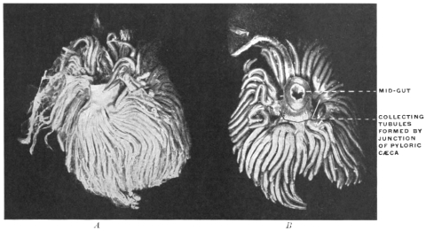
A. Bound together by connective tissue and blood-vessels.
B. Dissected to show confluence of cæca to form a smaller number of terminal tubes of larger
calibre entering the intestine.
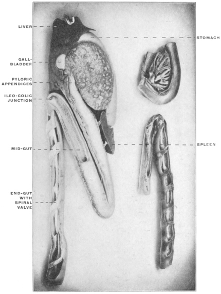 Fig. 212.
—Alimentary canal of
Accipenser sturio, sturgeon. Numerous
pyloric cæca are bound together to form a gland-like organ. (Columbia University
Museum, Nos. 1826, 1827, and 1828.)
The lower left-hand figure shows the mid- and end-gut in section, the
latter provided with a spiral mucous valve.
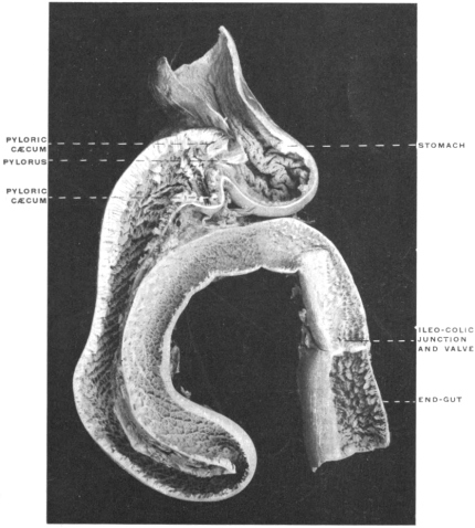
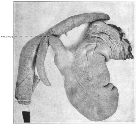
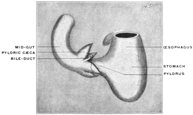
Fig. 208 shows the stomach and the beginning of the midgut
with four pyloric cæca in Pleuronectes maculatus, and Fig. 209
the same parts of this animal in section.
Fig. 210 shows the stomach and midgut of Paralichthys dentatus,
the summer flounder, with three well-developed conical pyloric
cæca. On the other hand in some forms the number of pyloric
appendices is enormously increased, while their caliber diminishes.
Thus 191 cæcal appendages are found surrounding the
beginning of the midgut in Scomber scomber. A well-marked
example of prolific development of the pyloric appendages is
furnished by the common cod, Gadus callarias (Fig. 211). The
appendices are in the natural condition bound together by connective
tissue and blood vessels, so as to form a compact organ, resembling
a gland (Fig. 211, A), and a similar arrangement is found
in Thynnus vulgaris and alalonga, Pelamys and Accipenser (Fig. 212).
In the smaller upper figure on the left the stomach, mid-gut, and pyloric
cæca are seen in section, showing the lumen of the latter and their openings
into the mid-gut.
In some Teleosts (Siluroidea, Labroidea, Cyprinodontia, Plectognathi
and Leptobranchiates) the appendices are entirely wanting.
If there are not more than 8-10 appendices they usually surround
the gut and empty into the same in a circle. In other cases they
are arranged in a single line, or in a double row, opposite to each
other (Fig. 213). Each appendix may open into the intestine
independently, this especially where the number is limited and
the individual pouches large (cf. Figs. 206-210), or several may
unite to form a common duct.
Fig. 211, B, shows the appendices in Gadus callarias, the cod,
freed by dissection from the investing connective and vascular
tissue. It will be noticed that a considerable number of the tubes
unite to form ducts of larger caliber which open into the intestine,
as seen in the section shown in Fig. 214.
The pyloric appendices apparently have the same significance as
the spiral intestinal fold of the Selachians, Cyclostomes and Dipnœans, i. e., the production of an increase in the area of the digestive
and absorbing surfaces of the intestinal mucous membrane.
Hence, as stated, the appendices and the spiral fold are found to
vary in inverse ratio to each other. Thus, for example, Polypterus
(Fig. 205) still has a fairly well developed spiral fold and
only a single pyloric appendix, while Lepidosteus, with but slightly
developed spiral fold, has numerous appendices. It was formerly
held that the pyloric cæca and the pancreas were mutually incompatible
structures, and that where one is found the other will
be wanting.
Hence the appendices were regarded as homologous with the
pancreas of the higher forms. Recent observations have shown
that this view is not strictly and entirely correct, while at the
same time it merits consideration in several respects.
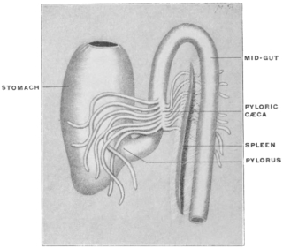 Fig. 213.
— Melanogrammus æglifinus, haddock. Stomach,
mid-gut, and pyloric cæca; spleen. (Columbia University
Museum, No. 1598.)
|
|
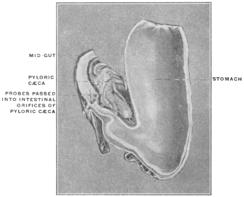 Fig. 214.
—Stomach and mid-gut of Gadus callarias, codfish,
in section, showing intestinal openings of pyloric cæca.
(Columbia University Museum, No. 1830.)
|
It is true that the pancreas in certain teleosts is now known to
be present although concealed from observation in the liver or
scattered in the form of small lobules between the layers of the
mesentery (cf. p. 117), and that in a number of fish, such as Salmo
salar, Clupea harengus, Accipenser sturio, both the appendices and
the pancreas are encountered. Consequently these structures
are not identical or even completely homologous, since they occur
side by side in the same form.
On the other hand Krukenberg has demonstrated that the appendices
pyloricæ may function physiologically as a pancreas by
yielding a secretion which corresponds to the pancreatic juice in
its digestive action. In the majority of forms, however, they apparently
merely increase the intestinal absorbing surface, secreting
only mucus.
These structures are nevertheless very interesting and instructive
since they furnish a perfect gross morphological illustration
of the embryonal stages just considered in connection with the
development of the mammalian pancreas. In the adult ganoid
or teleost these blind diverticula or pouches, varying greatly in
shape, number and size, protrude from the intestine immediately
beyond the pylorus, usually in close connection with the duodenal
entrance of the bile-duct. Two or more of these pouches
may unite to form a common duct or canal opening into the
intestine.
These forms, therefore, offer direct and valuable morphological
illustration of the manner in which the pancreas of the higher
vertebrates develops, i. e., as a set of hollow outgrowths or
diverticula from the hypoblast of the primitive enteric tube.
We can establish a consecutive series, beginning with forms in
which only one or two diverticula are found, and extending to
types in which the number of the little cylindrical pouches
reaches nearly two hundred and in which they are bound together
by connective tissue and blood vessels so as to closely
resemble the structure of a glandular pancreas. This is one of
the most striking instances in which the minute embryological
stages of the higher types are directly illustrated by the permanent
adult conditions found in the lower vertebrates. [The same
statement, as we will see, holds good in reference to the development
of the liver.]
RELATION OF THE PANCREAS TO THE PERITONEUM.
The gland becomes very intimately connected with the serous
layers of the primitive dorsal mesentery. In order to clearly
comprehend the adult serous relations it is necessary to make a
distinction between two divisions or portions of the gland, based
upon the altered relations of the primitive dorsal mesentery
which result from the differentiation of the primitive simple
intestinal tube into stomach and duodenum.
1. The primary outgrowth of the pancreatic tubules from the
duodenum, i. e., the part which is to form the “head” of the
adult gland, is situated between the two layers of that division
of the primitive dorsal mesentery which forms, after differentiation
of stomach and small intestine, the mesoduodenum.
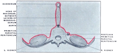
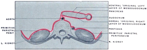
Coincident with the rotation of the stomach, as we have seen,
the duodenum and mesoduodenum exchange their original sagittal
position in the median plane of the body for one to the right
of the median line, balancing, so to speak, the extension of the
stomach to the left (Fig. 218).
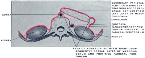
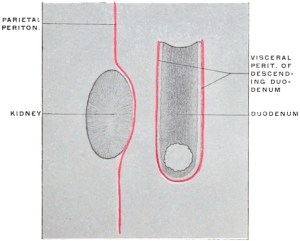
The original right layer of the mesoduodenum and the right
surface of the duodenum now look dorsad and rest in contact
with the parietal peritoneum investing the right abdominal background
and the ventral surface of the right kidney and inferior
vena cava. We have already seen that the descending portion of
the duodenum in man becomes anchored in this position by adhesion
of these apposed peritoneal surfaces. This fixation includes,
of course, the structures situated between the layers of the
mesoduodenum, i. e., the head of the pancreas. Consequently,
after rotation and adhesion, this portion of the gland turns one
surface ventrad, invested by secondary parietal peritoneum, originally
the left leaf of the free mesoduodenum, while the original
right surface of the gland has become the dorsal and has lost its
mesoduodenal investment by adhesion to the primary parietal
peritoneum.
2. In order to understand the way in which the body and tail
of the pancreas obtain their final peritoneal relations it is necessary
to consider the development of the dorsal mesogastrium to
form the omental bag. If we regard the primitive dorsal mesentery
in the profile view from the left side (Fig. 215) it will be seen
that, as already stated, the mesoduodenum is the first part of the
membrane to be invaded by the pancreatic outgrowth from the
intestine. Cephalad of the mesoduodenum the primitive dorsal
mesogastrium (Fig. 215) is seen to protrude to the left and caudad
to form, as already explained, the cavity of the omental
bursa of the retrogastric space (“lesser peritoneal sac”). The
further growth of the pancreas carries the developing gland from
the district of the mesoduodenum into that portion of the dorsal
mesogastrium which now forms the dorsal wall of the omental
bursa (Fig. 216).
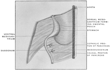 Fig. 215.
—Cephalic segment of primitive mesentery in schematic profile view.
|
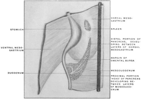 Fig. 216.
—Schematic profile view of primitive mesenteries with formation of omental
bursa and developing spleen and pancreas.
|
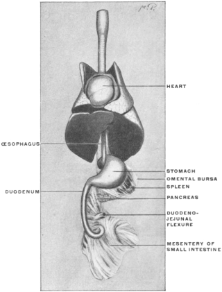 Fig. 217.
— Sos scrofa fœt., fœtal pig. Portions of thoracic and abdominal viscera hardened in
situ. (Columbia University Museum, No. 1449.)
|
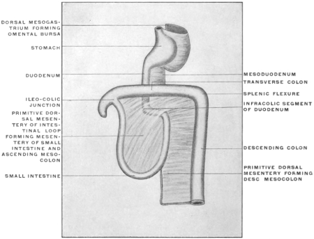 Fig. 218.
—Schematic view of primitive mesentery after intestinal rotation and incipient
formation of omental bursa from dorsal mesogastrium.
|
|
This double relation of the pancreas to the mesoduodenum and
to the mesogastrium forming the omental bursa is well seen in
fœtal pigs between two and three inches in length (Fig. 217).
The head portion of the pancreas is seen developing between
the layers of the mesoduodenum, while the body and tail of the
gland, extending to the left, grows between the two dorsal layers
of the omentum bursa towards the spleen, which organ is found
connected with the left and dorsal extremity of the omental sac
derived from the dorsal mesogastrium.
Before the growth of the great omentum is pronounced the
continuity of the mesoduodenum and dorsal mesogastrium can
be readily appreciated (Fig. 218). But after the redundant
growth of the membrane has carried the great omentum further
caudad, the stomach and the two omental layers attached to
the greater curvature lie in front of the structures included
between the two dorsal layers and conceal them from view (Fig.
177).
Figs. 219, 220.—Schematic transection of dorsal mesogastrium, pancreas, spleen, and stomach.
|
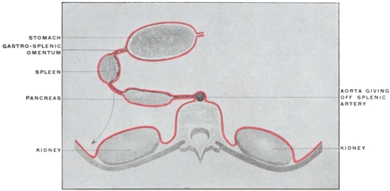 Fig. 219.
—Before adhesion to primitive parietal peritoneum (arrow indicates the direction in
which the adhesion takes place).
|
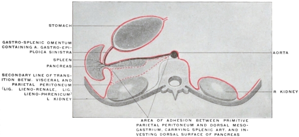 Fig. 220.
—After adhesion and formation of secondary line of transition between mesogastrium
and parietal peritoneum (lieno-renal ligament).
|
In sagittal sections to the left of the median line (Figs. 221 and
222) the pancreas now appears included between the layers of the
great omentum near their point of departure from the vertebral
column. (This point is of course identical with the prevertebral
attachment of the primitive dorsal mesogastrium from which the
omentum is developed.)
Figs. 221, 222.—Schematic sagittal sections through stomach, pancreas, great omentum, and
left kidney.
|
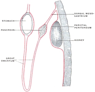 Fig. 221.
—Before adhesion between dorsal and mesogastrium and parietal peritoneum.
|
|
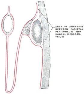 Fig. 222.
—After adhesion.
|
The foregoing considerations will, therefore, lead to the conclusion
that the pancreas presents, in regard to its peritoneal relations,
two distinct segments:
1. The portion adjacent to duodenum (head and neck of
the gland) is developed between the layers of the mesoduodenum.
2. The distal portion of the gland, comprising the body and
tail, develops between the layers of the great omentum (dorsal
segment), derived from the primitive dorsal mesogastrium.
The transections of the dorsal mesogastrium shown in Figs.
180 and 181 will now have to be amplified by the introduction of
the body of the pancreas between the two layers of the vertebro-splenic
segment, in addition to the splenic artery (Figs. 219 and
220).
Hence the following facts will be understood:
1. In the adult the splenic artery supplies a series of small
branches to the pancreas as it courses along the cephalic border of
the gland on its way to the spleen.
2. After the above-described adhesion of the original left leaf
of the dorsal mesogastrium (vertebro-splenic segment) to the
parietal peritoneum (Fig. 220), the dorsal surface of the body
of the pancreas loses its peritoneal investment and becomes
attached by connective tissue to the ventral surface of the left
kidney.
3. The ventral surface of the body of the pancreas is in the
adult lined by peritoneum of the “lesser sac”; in other words
the organ has practically assumed a “retro-peritoneal” position,
its ventral peritoneal covering appearing now as the dorsal parietal
peritoneum of the retro-gastric space.
4. When completely developed the extreme end (tail) of the
pancreas extends to the left, following the splenic artery, until it
touches the mesal aspect of the spleen at the hilus.
5. If we, therefore, leave out of consideration for the moment
the transverse colon and duodenum, which will be taken up
presently, and confine ourselves to the arrangement of the
stomach, pancreas and great omentum, a sagittal section
to the left of the median line would result as shown in
Fig. 222, after the adult condition of adhesion has been established.
The same process of fixation, which resulted in the anchoring of
duodenum and head of pancreas, extends to the body of the gland
and the investing omentum. The peritoneum lining the original
left, now the dorsal surface of the gland, fuses with the primitive
parietal peritoneum covering the diaphragm and the left kidney.
The main body of the pancreas in the adult appears prismatic,
giving a triangular sagittal section. The dorsal surface is adherent
to the ventral surface of the left kidney; the ventral surface is
covered by the secondary parietal peritoneum (original right layer
of mesogastrium) which lines the dorsal wall of the retrogastric
space and omental bursa (lesser peritoneal sac). The great omentum
now appears to take its dorsal point of departure along the
sharp margin which separates this ventral surface of the pancreas
from a third narrower surface directed caudad. This surface,
under the conditions which we are at present examining,
would be lined by the peritoneum continued onto it from the
dorsal layer of the great omentum. This peritoneum merges
along the dorsal margin of this caudal surface of the pancreas
with the general parietal peritoneum covering the left lumbar
region and the caudal part of ventral surface of the left kidney.
We have, therefore, along this line a secondary transition
from visceral to parietal peritoneum, obtained by the
obliteration of the original visceral peritoneum investing the
dorsal surface of the pancreas before adhesion to the parietal
peritoneum.
The pancreas assumes, therefore, in the adult a secondary retro-peritoneal
position, covered on its ventral surface by peritoneum
of the “lesser sac,” while the caudal surface is lined by part of
the general peritoneal membrane of the “greater sac.” The dorsal
surface, denuded of serous covering by obliteration, is adherent
to the crura of the diaphragm, the aorta and the ventral surface
of the left kidney.
It is now proper to compare the conclusions just derived from
the study of the development of the human dorsal mesogastrium
and connected structures (spleen and pancreas) with the conditions
presented by the corresponding parts in one of the lower
mammalia, which illustrate some of the human embryonal
stages. Here again the abdominal cavity of the cat forms an
instructive object of study.
The purpose of the following comparison should be twofold:
I. The mesogastrium, spleen and pancreas in the cat will
clearly illustrate the process of human development above outlined.
II. The abdominal viscera of the cat, if properly arranged,
will enable us to complete the consideration of this region by including
the very important relations which the transverse colon
and third portion of the duodenum bear in man to the great
omentum and pancreas.
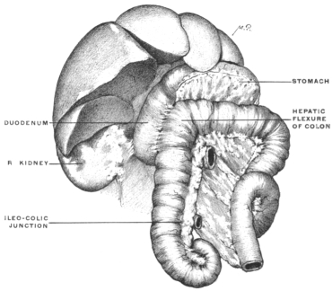
I. SPLEEN, PANCREAS AND GREAT OMENTUM OF CAT.
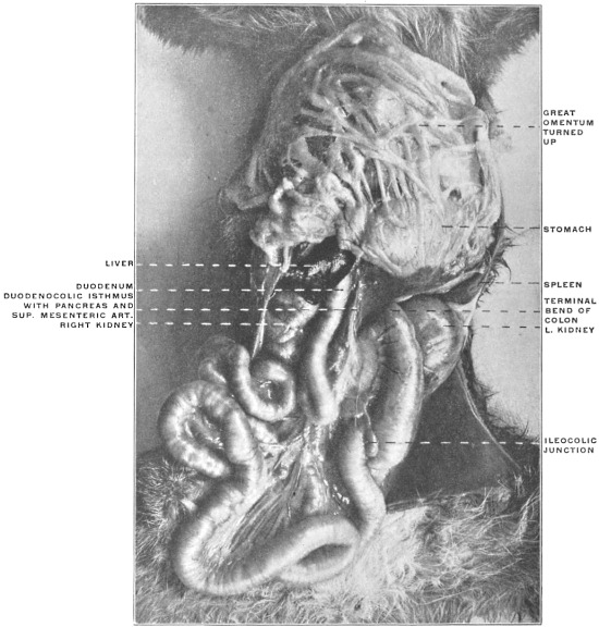
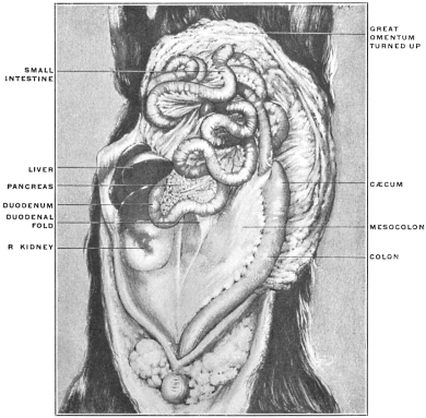
After opening the abdominal cavity it will be seen that the
great omentum can be lifted up, exposing the subjacent coils of
the small and large intestine, to which it adheres at no point.
In other words the entire dorsal surface of that part of the original
mesogastrium which forms the great omentum is free. It will be
remembered that this is not the case in the adult human subject,
because here the dorsal surface of the great omentum adheres to
the transverse colon. Consequently in man only that portion of
the dorsal surface of the omentum can be seen which extends
between the transverse colon and the caudal free edge of the
membrane.
It will be noted that on the left side the spleen is connected by
its mesal surface to the omentum and through it with the stomach
(gastro-splenic omentum). In other words the cat illustrates the
human embryonal stage in which the spleen has appeared between
the layers of the dorsal mesogastrium at the extreme left or blind
end of the retrogastric pouch formed by the rotation of the stomach
and elongation of the mesogastric membrane, but before the adhesion
has taken place between the original left (now dorsal) layer
of the vertebro-splenic segment of the mesogastrium and the
primitive parietal peritoneum apposed to it (Fig. 219). Consequently
the dorsal wall of the “lesser” sac in the cat is still
composed of the two layers of the free vertebro-splenic segment
of the mesogastrium, the primitive right (now ventral) layer not
having been converted, as is the case in man, into secondary
parietal peritoneum by adhesion of the original left (now dorsal)
layer to the primitive prerenal parietal peritoneum.
If we now examine the relation of the pancreas to the peritoneum
we can establish the following facts:
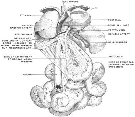 Fig. 223.
—Abdominal viscera of cat, hardened and removed from body, showing relation of
pancreas to mesoduodenum and dorsal mesogastrium, respectively. (Columbia University
Museum, No. 728.)
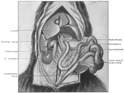
1. The portion of the gland adjacent to the duodenum, corresponding
to the “head” of the human organ, is included between
the two layers of the mesoduodenum. This membrane is free,
so that the dorsal surface of this portion of the pancreas is seen
to be invested by the dorsal layer of the mesoduodenum (Fig.
223). The duodenum and the mesoduodenum, the latter containing
the head of the pancreas between its layers, can be turned
toward the median line, so as to expose the entire ventral surface
of the post-cava and right kidney. To illustrate the arrangement
which is found in the adult human subject the descending
duodenum and pancreas should be allowed to fall over to the
right so as to cover the vena cava and the mesal part of the ventral
surface of right kidney. The adult human condition will now be
produced if we assume that the structures are fixed in this position
by the obliteration of the apposed serous surfaces, viz., the
parietal peritoneum over kidney and vena cava on the one hand
and the right layer of the mesoduodenum and the dorsal visceral
peritoneum of the duodenum on the other.
2. In following out the pancreas of the cat in its entire extent,
proceeding to the left of the pylorus, it will be seen that the
body of the gland has extended between the two dorsal layers
of the great omentum (primitive dorsal mesogastrium) over to
the spleen (Fig. 223). Consequently the arrangement in the
cat corresponds to the stage in the human development shown
in Fig. 219 and Fig. 221 in which adhesion of the dorsal surface
of the pancreas to the parietal peritoneum has not yet taken place.
It will be quite easy to reconstruct from the facts as demonstrated
by the arrangement of the parts in the cat, the stage in
the development of the lesser peritoneal sac in which the dorsal
wall of the space is still formed by the proximal portion of the
free dorsal mesogastrium (great omentum) and the structures included
between its two layers.
It must then become apparent that the entire serous surface
which in the adult human subject we regard as “parietal peritoneum
of the lesser sac” lining the dorsal wall of the retrogastric
space is derived from what originally was the right layer of the
primitive sagittal dorsal mesogastrium.
II. RELATION OF GREAT OMENTUM TO TRANSVERSE COLON,
TRANSVERSE MESOCOLON AND THIRD PART OF DUODENUM.
The second purpose to be accomplished by the study of the
cat’s abdominal cavity at this stage is the correct appreciation of
the adult human conditions which are produced by areas of adhesion
between the transverse colon, transverse mesocolon and
third part of the duodenum on the one hand, and the dorsal
mesogastrium, as great omentum, with the structures contained
between its layers, on the other.
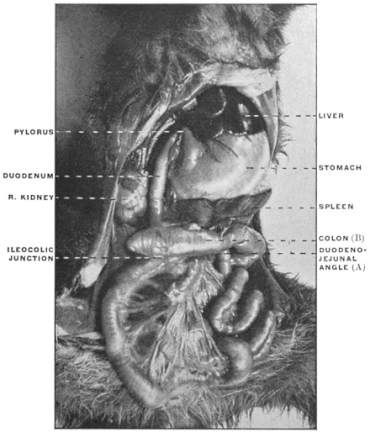
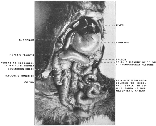
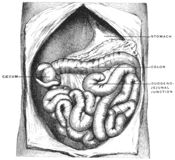
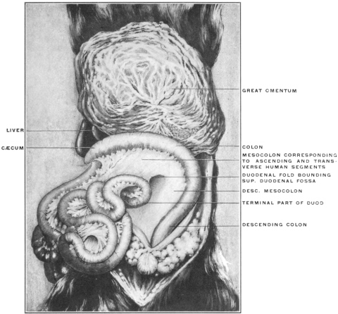
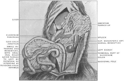
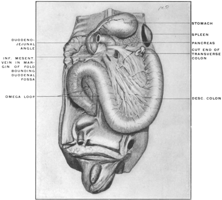
Perform the manipulations of the large and small intestine in
the cat (see p. 67) which are required in order that the tract may
be arranged so that it will correspond in general to the topographical
conditions presented by the adult human subject.
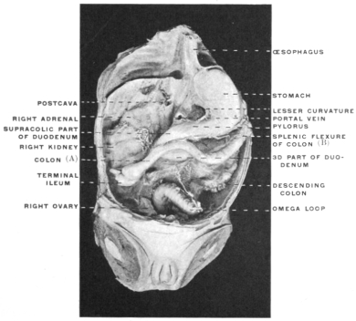
Locate the transverse colon and mesocolon and the third portion
of the duodenum produced by these manipulations in imitation
of the corresponding human structures. Then proceed to plot
the different parts out successively as they would appear in a
sagittal section (Fig. 224).
The following facts are to be noted and indicated on the plan
of the section:
1. The great omentum is free, hanging down from the greater
curvature of the stomach over the coils of intestine. Turning the
omentum up it will be observed that the body of the pancreas is
included between the two dorsal layers of the membrane.
2. The omentum, containing the pancreas, can be lifted up,
exposing the next succeeding structure, viz., the transverse colon
and mesocolon. In the cat the large intestine has been brought
over, by the manipulations above indicated, into a transverse position
so as to represent the human transverse colon and its mesocolon.
It is therefore necessary to remember that in this mammal
the fixation of the transverse mesocolon in the position indicated,
by adhesion of ascending and descending mesocola to the parietal
peritoneum of the abdominal background, has not yet occurred.
Consequently the membrane must be held in the transverse position
in order to represent the human arrangement.
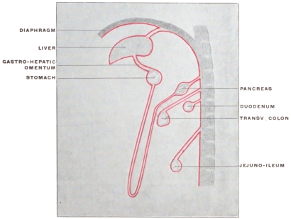 Fig. 224.
—Schematic sagittal section of abdominal viscera of cat, after the
intestines have been rotated to correspond to the adult human disposition, to show
lines of peritoneal reflection before adhesion.
|
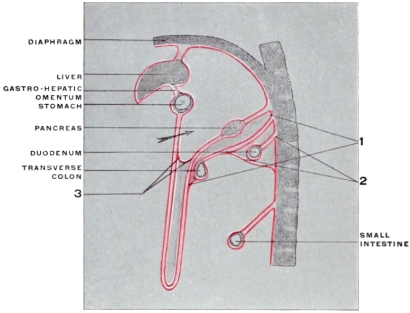 Fig. 225.
—The same figure indicating the areas of adhesion and peritoneal
obliteration (shaded) which produce the arrangement of the adult
human peritoneum.
1. Area of adhesion between opposed surfaces of great omentum and transverse
mesocolon and colon.
2. Area of adhesion between parietal peritoneum, duodenum, and caudal
layer of transverse mesocolon.
3. Adhesion of opposed walls of omental bursa leading to obliteration of
distal portion of pouch and producing “gastro-colic” ligament of adult human
subject.
|
It will of course be observed that both surfaces of the transverse
mesocolon established in this way are free, not adherent to either
omentum or pancreas on the one hand, nor to the transverse duodenum
on the other.
3. The third or transverse portion of the duodenum is seen to
be attached by the distal part of the mesoduodenum, both of the
serous surfaces of the membrane being free. The duodenum having
been brought from right to left transversely across vertebral
column and aorta, underneath the superior mesenteric artery, the
mesoduodenum, in the segment corresponding to the transverse
duodenum, exchanges its original sagittal position for one in a
horizontal plane, with cephalic (primitive left) and caudal (primitive
right) surfaces.
Now compare the above arrangement of the intestines and
peritoneum in the cat at once with the conditions presented in the
adult human subject, reserving certain intermediate stages, as exhibited
by some of the lower monkeys, for subsequent study.
The examination of a similar sagittal section representing schematically
the adult human arrangement of the parts (Fig. 225) will
reveal the following points of difference as compared with the cat:
1. The peritoneum covering the dorsal surface of the pancreas,
derived from the primitive dorsal mesogastrium, has become
adherent to the parietal peritoneum, as previously described.
2. The cephalic surfaces of the transverse colon and mesocolon
fuse with the corresponding area of the dorsal (4th) layer
of the great omentum (dorsal mesogastrium).
In the human fœtus in the 4th month the connection is still
so slight that the omentum can readily be separated from the
transverse colon and mesocolon.
Further dorsad the cephalic layer of the transverse mesocolon
adheres to the serous investment of the caudal surface of the
pancreas, derived, as we have seen, from the same dorsal layer
of the great omentum.
3. The duodenum and mesoduodenum are fixed by adhesion
on the one hand to the parietal peritoneum, on the other to the
caudal layer of the transverse mesocolon near the root of that
membrane.
4. The cavity of the omental bursa is usually obliterated in the
adult caudad of the level of the transverse colon, by adhesion of
the apposed surfaces of the two intermediate omental layers.
We have therefore three general areas of secondary peritoneal
adhesion to deal with (Fig. 225), viz.:
| 1. Dorsal layer of primitive mesogastrium (great omentum) including the
serous investment of the dorsal and caudal surfaces of the pancreas
(Fig. 225, 1). |
| |
to | | |
Parietal peritoneum, cephalic layer of
transverse mesocolon and cephalic surface of transverse colon. |
| | | |
| | | |
| | | |
|
| 2. Transverse duodenum and mesoduodenum (Fig. 225, 2). |
| | to |
| | Parietal peritoneum and caudal layer of
transverse mesocolon. |
| | | |
| | | |
| | | |
|
| 3. Between the apposed serous surfaces of the intermediate omental
layers (Fig. 225, 3). |
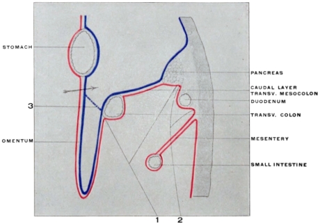 Fig. 226.
—Schematic sagittal section of adult human peritoneum.
These areas of adhesion result naturally in the production of
secondary lines of peritoneal transition as follows:
1. Figs. 225, 1; 226, 1, from the omentum, dorsal layer, to the
caudal surface of transverse colon, caudal layer of transverse mesocolon
and caudal surface of the pancreas.
2. Figs. 225, 2; 226, 2, from the caudal layer of the transverse
mesocolon across the transverse portion of the duodenum
to the parietal peritoneum and mesentery of the jejuno-ileum.
3. Figs. 225, 3; 226, 3, between the intermediate omental layers,
forming the secondary caudal limit of the lesser sac.
These changes consequently result in the rearrangement of the
adult human peritoneum in accordance with the following
schema (Fig. 226):
We trace the peritoneum as the ventral or superficial layer of
the great omentum from the greater curvature of the stomach
caudad around the distal free edge of the omentum and cephalad,
as the dorsal layer, to the ventral border of the transverse colon.
Here apparently this layer is continued across the caudal surface of
the large intestine and beyond as the caudal layer of the transverse
mesocolon. While this condition obtains practically in the adult
it is to be remembered that the adhesion (at 1 in Fig. 225) prevents
us from lifting the omentum away from the colon, and that
consequently the apparent continuity of the dorsal layer of the
great omentum with the caudal layer of the transverse mesocolon
is the result of this peritoneal fusion.
Near the dorsal attachment or “root” of the transverse mesocolon
the caudal layer of the membrane becomes continuous
with the parietal peritoneum investing the transverse portion of
the duodenum on its ventral aspect, which peritoneum in turn
passes into the free mesentery of the jejuno-ileum (Fig. 225, 2).
Comparison with the previous figures will show that we are dealing
here with another area of secondary peritoneal fusion.
If we now open the “lesser peritoneal cavity” by dividing the
two layers of the omentum attached to the greater curvature of
the stomach (Figs. 225 and 226 in direction of arrow) we will
apparently reach the upper or cephalic surface of the transverse
mesocolon. This layer can be followed dorsad to the sharp
border which separates the ventral and caudal surfaces of the
pancreatic body and the membrane can be traced thence over the
ventral surface of the gland to the diaphragm. (The connections
with the liver and stomach shown schematically in the diagram
(Fig. 225) are to be considered in detail subsequently.)
In the adult the peritoneal surface just described appears as the
cephalic layer of the transverse mesocolon and its continuation
dorsad. From the facts previously considered it will be at once
apparent that we are really dealing here with a part of the third
layer of the primitive omentum. We do not see the original
cephalic layer of the transverse mesocolon. This membrane has
become fused with the fourth omental layer, and its free serous
surface obliterated in the stretch between the vertebral column
and the transverse colon. Hence the human adult transverse
mesocolon is apparently composed of two layers; the cephalic of
these layers appears as peritoneum of the “lesser sac,” in conformity
with its derivation from the original third omental layer lining
the interior of the omental bursa. The caudal layer, on the other
hand, is a part of the general or “greater” peritoneal membrane.
The entire adult transverse mesocolon, hence, comprises four peritoneal
layers, of which only two remain as permanently free
serous surfaces. These differ in their derivation, the cephalic
layer being a part of the primitive dorsal mesogastrium (third
omental layer), while the caudal layer is part of the primitive
mesocolon. Between these two layers of the adult transverse
mesocolon are included the two obliterated embryonic membranes,
viz., the fourth omental layer and the original dorsal layer
of the transverse mesocolon.
Caudad the two layers of the adult transverse mesocolon surround
the transverse colon and are continuous along the ventral
margin of the intestine with the layers of the great omentum.
Toward the vertebral column these layers again diverge. The
cephalic layer, lining the “lesser peritoneal cavity” invests the
ventral surface of the pancreas. The caudal layer continues over
the caudal surface of the body of the gland and transverse portion
of the duodenum into the parietal peritoneum and the free mesentery
of the jejuno-ileum. Consequently the returning layers
of the great omentum are said to surround the transverse colon
and unite along the dorsal border of the intestine to form the
transverse mesocolon, which membrane is continued dorsad toward
the vertebral column as two layers. At the “root” of the
transverse mesocolon these layers are then described as diverging,
the cephalic passing up to line the ventral surface of the pancreas,
while the caudal continues over the caudal surface of the pancreas
and third portion of the duodenum into the parietal peritoneum
and mesentery.
Wherever in this discussion of the transverse mesocolon the
transition between the caudal layer of the membrane and the
“parietal” peritoneum is referred to it is necessary to remember
that this “parietal” peritoneum is the secondary investment of
the abdominal background, formed by the surface of the ascending
and descending mesocolon which remains free after the opposite
surface and the vertical segments of the large intestine have
been anchored by adhesion to the primary parietal peritoneum
(cf. p. 81, Fig. 158).
A summary at this point of the course of the dorsal mesogastrium,
in forming the great omentum and its subsequent connections,
would show us that the membrane first enlarges and descends
towards the transverse colon (Fig. 177). The omental
bag is formed by the descending or superficial segment (starting
from the greater curvature of the stomach), turned toward the
observer in the figure, and by the ascending or deep layer which
is attached above to the dorsal abdominal wall, in front of the
vertebral column and aorta along the original line of origin of
the dorsal mesogastrium. Gradually growing and descending
further, the deep segment becomes attached to the transverse
colon. It also becomes connected, especially on the left side, with
the diaphragmatic peritoneum (phrenicocolic lig.), so that its
original starting point is no longer distinct. Finally the development
of the spleen and pancreas between the layers of the dorsal
segment and their subsequent connections obscure the original
conditions.
Fig. 297 shows the primitive condition at a time when the connection
with the transverse colon and mesocolon has not yet
taken place.
The omental bag or bursa epiploica develops in the region of
the dorsal mesogastrium and the viscera included between its
layers, by changes in the position and extent of the membrane
which finally result in placing a part of the right half of the
primitive cœlom cavity behind the stomach. Up to the sixth week
the line of origin of the dorsal mesogastrium is from the mid-dorsal
line of the abdomen. It deviates from this origin to the
left because the great curvature of the stomach to which it is
attached turns in this direction. On this account, and because of
the rapid growth of this portion of the mesogastrium, a bag or
space is formed behind the stomach. The entrance into this space
is situated to the right of the lesser curvature, behind the peritoneal
layers connecting the same with the liver (lesser or gastro-hepatic
omentum and hepato-duodenal ligament). The ventral
wall of this space is formed by the dorsal surface of the stomach
itself, the dorsal wall by the mesogastrium, turning to the left
and presenting its original right surface, now directed ventrad.
The caudal limit of the retro-gastric space is given by the turn of
the mesogastrium to reach its attachment along the greater curvature
of the stomach (rudiment of great omentum).
The stomach, in contributing to produce these changes, passes
from the vertical to the oblique and finally into the transverse
position. The pylorus, formerly directed caudad, passes up and
to the right. The fundus develops and the original left side of
the stomach becomes the ventral, the right side the dorsal. The
original dorsal border, now the greater curvature, moving caudad,
carries the attached dorsal mesogastrium with it into its new
position. The mesogastrium now pouches to form the great
omentum and rapidly enlarges. At first hardly projecting beyond
the greater curvature, it increases in length until it forms
a four-layered apron which hangs down as a loose sac over the
transverse colon and the coils of the small intestine (Fig. 177).
In the fœtus of six months the cavity of the omental bag extends
caudad as far as the lower edge of the omentum. Later
adhesions between the peritoneal surfaces lining the interior of
the bursa limit this extension.
The omental bursa is therefore formed by a ventral lamella,
consisting of two peritoneal layers, which hangs down from the
greater curvature of the stomach and passes around the caudal
free edge of the omentum into the double-layered dorsal lamella,
which ascends, over the transverse colon, to the original starting
point of the dorsal mesogastrium along the front of the vertebral
column and aorta. Hence the “great omentum” is originally
composed of four layers of peritoneum.
The dorsal double lamella becomes adherent over a considerable
area to the parietal peritoneum of the dorsal abdominal wall.
In this way the organs developed between the two layers of the
lamella obtain their final fixed position. The pancreas becomes
anchored and appears in the adult as a “retro-peritoneal” structure,
while the spleen is attached by the “phrenico-lienal ligament”
to the diaphragm.
In addition the dorsal omental lamella adheres in the fourth
month to the cephalic layer of the transverse mesocolon and to
the transverse colon.
Important illustrations of some of the intermediate stages in the
human development of this portion of the peritoneal tract are
afforded by the permanent adult conditions found in the abdominal
cavity of some of the lower primates, notably certain of the
cynomorphous monkeys.
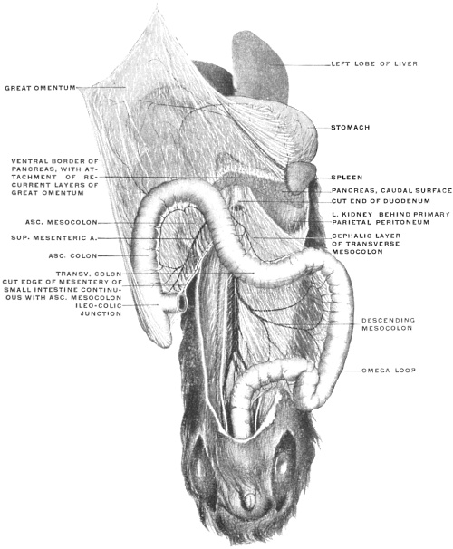 Fig. 227.
—Abdominal cavity of
Macacus rhesus, Rhesus monkey, with the small intestine
removed. (Columbia University Museum, No. 63/1831.)
Fig. 227 shows the abdominal cavity and disposition of the
peritoneum in a macaque monkey (Macacus rhesus, ♂) in the ventral
view, with the coils of small intestines removed and the omentum
lifted up and reflected upon the ventral body wall. The following
important points of difference from the arrangement in the
cat on the one hand, and in man on the other, are to be noted:
1. The large intestine presents the typical primate course, with
an ascending, transverse and descending colon. The ileo-cæcal
junction is situated in the right iliac fossa.
2. The ascending and descending mesocola are still free, not
having become adherent to the parietal peritoneum along the
dorsal abdominal wall. Hence the caudal portions of the ventral
surfaces of the two kidneys are still covered by the primitive
parietal peritoneum.
3. The great omentum is not yet adherent to the transverse
colon and mesocolon except for a short distance on the extreme
right. At this point the dorsal layer of the omentum has begun
to contract adhesions to the hepatic flexure of the colon and ascending
colon, but the rest of the transverse colon is free. Differing
from the human arrangement is a line of adhesion,
uniformly present in these monkeys, between the dorsal surface
of the omentum along its right edge and the ventral surface
and right border of the cæcum and ascending colon, parts which
normally are not adherent to the omentum in man.
4. Hence in tracing the omentum to the left of the limited
adhesion to the hepatic flexure and ascending colon, i. e., nearly
throughout the entire extent of the transverse colon, we find
the membrane passing freely without adhesion over the cephalic
surface of the transverse mesocolon, which preserves its original
free condition, independent of the omentum. This arrangement
is shown in the schematic sagittal section in Fig. 230.
5. Tracing the omentum dorsad beyond the transverse colon
and mesocolon the pancreas is reached. Here we encounter the
first extensive area of omental or mesogastric adhesion. The
omental peritoneum continues over the ventral and caudal surfaces
of the gland, investing the same, but the dorsal surface has
lost its serous covering and is anchored to the ventral surface of
the left kidney. Hence a sagittal section would show the arrangement
of the monkey’s omentum as indicated in the schematic
Figs. 229 and 230. Making now a general comparison of the
peritoneal membrane of this animal with that of man, and of both
with the preceding common embryonal condition, we can draw
the following conclusions, indicated schematically in the five
figures 228-232.
Figs. 228-232.—Schematic sagittal sections of dorsal mesogastrium
and omental bursa, in man, monkey, and cat.
|
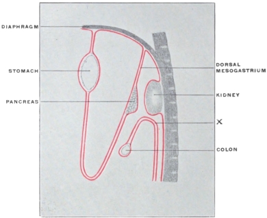 Fig. 228.
—Common embryonal condition, as illustrated by cat, after
rotation and formation of omental bursa.
|
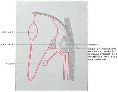 Fig. 229.
—Area of adhesion between dorsal mesogastrium and primitive
parietal peritoneum in Macacus, producing condition shown in Fig. 230.
|
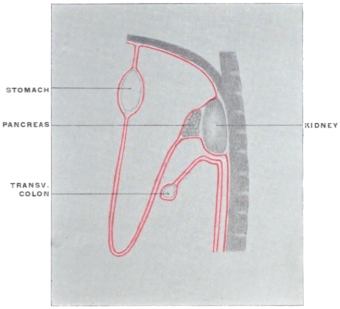 Fig. 230.
—Arrangement of great omentum as found in Macacus
rhesus, shown without reference to areas of peritoneal obliteration.
|
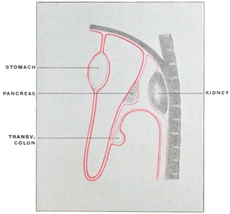 Fig. 231.
—Corresponding section of human adult peritoneum
showing, along dotted lines, area of peritoneal adhesion.
|
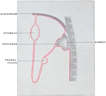 Fig. 232.
—Section showing human adult peritoneum without
reference to area of adhesion.
|
1. The dorsal layer of the monkey’s omentum in its proximal
segment behaves in the same way as in man, i. e., it becomes adherent
to the primitive parietal peritoneum down as far as the
caudal margin of the dorsal surface of the pancreas included between
the primitive mesogastric layers forming by their further
growth the omental apron.
Therefore we find, as in the human subject,
(a) The pancreas adherent to the ventral surface of the left
kidney.
(b) A portion of the ventral surface of the kidney, cephalad of
the pancreas, and the dorsal wall of the retrogastric (lesser peritoneal)
space lined by secondary parietal peritoneum derived from
the third layer of the omentum (original right layer of dorsal
mesogastrium).
2. The monkey differs from adult man in the behavior of the
dorsal omental layer in relation to the cephalic surface of the
transverse mesocolon. The adhesion, which in the human subject
fuses this layer with the transverse colon and mesocolon, does not
occur in the monkey.
Figs. 233-235.—Series of schematic sagittal sections through left
kidney and adrenal, pancreas, and transverse colon, to show development
of adult peritoneal relations.
|
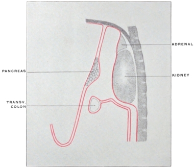 Fig. 233.
—Embryonic condition, as illustrated by cat, after rotation
of intestine. Pancreas free between dorsal layers of great omentum.
Transverse colon and mesocolon free. Kidney behind primitive parietal
peritoneum.
|
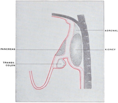 Fig. 234.
—Area of adhesion between: 1. Primitive parietal peritoneum.
2. Mesogastrium forming great omentum. 3. Colon and mesocolon.
|
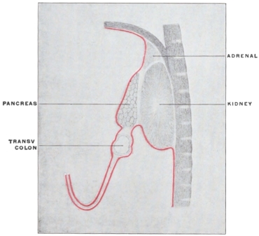 Fig. 235.
—Adult human arrangement, shown without reference
to obliterated areas.
|
Hence we have in this animal the following conditions:
(a) The omentum is non-adherent to the transverse colon and
transverse mesocolon.
(b) The caudal surface of the pancreas is lined by its original
mesogastric peritoneum.
(c) The transverse mesocolon is formed by the original two
layers of the primitive dorsal mesentery; hence its cephalic layer
is not “peritoneum of the lesser sac” as is the case in man.
(d) The caudal part of the ventral surface of the left kidney
below the pancreas, is covered by the original parietal peritoneum.
(e) Only one point or line of secondary peritoneal transition exists,
where the dorsal layer of the omentum in the adult becomes continuous
with the parietal peritoneum covering the caudal surface
of the pancreas and the ventral surface of the left kidney.
Note: In the schematic sections shown in Figs. 228 to 232 the
transverse colon is represented as far removed from the ventral surface
of the left kidney, in order to make the peritoneal lines of the
mesocolon more clear. Actually a sagittal section which would
divide the kidney would cut the transverse colon at its extreme
left end, where it turns close to the ventral surface of the left kidney
and then follows its lateral border to form the splenic flexure
(Fig. 235). The caudal part of the ventral surface of the left
kidney in the adult human subject is covered by the peritoneum
which, as secondary parietal peritoneum, is derived from the
upper part of the right leaf (later ventral leaf) of the descending
mesocolon. Hence it should be remembered that these diagrams
present combinations of sections. A section which will show the
full development of the transverse mesocolon is mesad of the
kidney; while a section through the kidney would be too far laterad
to show the transverse mesocolon.
Figs. 233, 234 and 235 show sagittal sections through the left
kidney with the adult arrangement of the peritoneum and colon
and the embryonic and adhesion stages leading to the same.
It will be observed that in all the schematic sections of the
early embryonic stages the two layers of the transverse mesocolon
are shown without dorsal attachment, as turning with the formation
of a fold (Fig. 228 at x) into two layers descending ventrad
of the parietal peritoneum. This is because the dorsal attachment
of the mesocolon is at this stage still in the median line
and would hence not be encountered by a sagittal section through
the kidney, and because the two layers of the transverse mesocolon,
immediately after rotation of the large intestine, are still
directly continuous with the two layers of the descending mesocolon.
That is to say, the cephalic layer of the transverse mesocolon
is continuous with the dorsal (originally the left) layer of
the descending mesocolon, and the caudal layer of the transverse
mesocolon with the ventral (originally the right) layer of the
descending mesocolon, which is, in the human subject, to assume
subsequently the character of parietal peritoneum after the dorsal
layer and the primitive parietal peritoneum have become obliterated
by adhesion (Fig. 235).
Fig. 236 shows this continuity of the descending and transverse
mesocolon as a permanent adult condition in the macaque. The
fold of transition between the two is seen at x in Fig. 228. It
will be noticed that the ventral surface of the left kidney, caudad
of the adherent pancreas, is covered by the primitive parietal
peritoneum, corresponding to section in Fig. 230.
RELATIONS OF SPLEEN AND OMENTUM IN MACACUS RHESUS.
The spleen in this animal has not contracted any extensive
adhesions to the parietal peritoneum (the phrenico-lienal lig.
of anthropotomy is not developed). It can be turned mesad so
as to expose the lateral border and an adjacent segment of the
ventral surface of the left kidney, as well as the dorsal surface of
the tail of the pancreas at its tip, still covered by mesogastric
peritoneum. Hence in the monkey the adhesion of the original
vertebro-splenic segment of the mesogastrium, including the pancreas,
to the primitive parietal peritoneum is less complete than
in man.
MEDIAN ATTACHMENT OF DESCENDING MESOCOLON AND ITS
RELATION
TO THE MESOCOLON OF THE SIGMOID
FLEXURE IN THE MACAQUE.
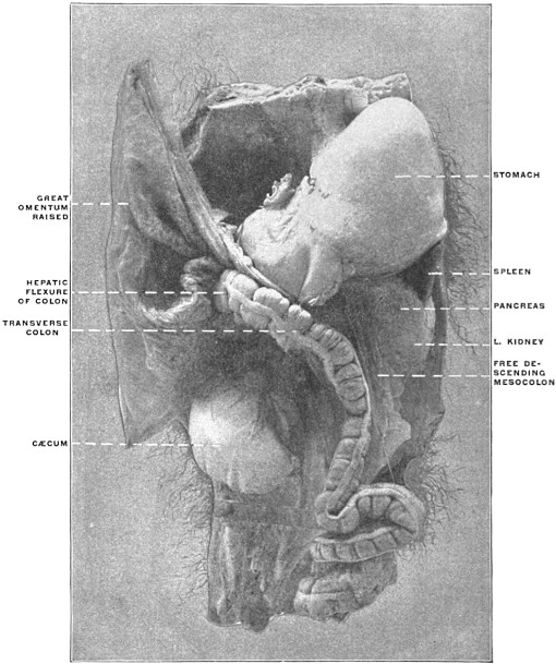 Fig. 236.
—Abdominal viscera of
Macacus cynomolgus, Kra monkey. (Columbia University
Museum, No. 1801.)
Fig. 236 shows the abdominal viscera, hardened in situ, of
Macacus cynomolgus, the Kra monkey, in the ventral view and
from the left side.
The great omentum is lifted up, the pancreas is adherent to the
ventral surface of the left kidney, the caudal portion of which is
covered by the primary parietal peritoneum, which can be exposed
by turning the still free descending mesocolon mesad. The
mesocolon retains its primitive attachment to the median line
ventrad of the large prevertebral blood vessels. It is readily seen
that adhesion between the left leaf of this free descending mesocolon
and the parietal peritoneum down to the level of the
iliac crest would produce the conditions found in the human
adult, with an attached descending colon and a free sigmoid flexure;
also that limited adhesion of the mesocolon of the sigmoid
flexure to the parietal peritoneum would produce, as previously
explained (cf. p. 97), the intersigmoid peritoneal fossa.
2. Ventral Mesogastrium and Liver.—The peritoneal reflections
from the stomach to the liver, and the arrangement of the
membrane in connection with the latter organ, remain for consideration.
Certain complicated adult conditions, encountered in this part
of the abdominal cavity, make it desirable to arrange the subject
for purposes of study under the following subdivisions:
I. The development of the liver and of its vascular system,
and the significance of the adult circulation of the liver and of
the fœtal remnants connected with the organ.
II. The anatomy of the ventral mesogastrium and the changes
produced in the arrangement of the membrane by the development
of the liver.
I. A. Development of the Liver.—The liver, like the pancreas, is
developed from the duodenum as an outgrowth from the hypoblast
lining the enteric tube. As we have previously noted, the
first outgrowth of the hepatic diverticulum is closely associated
with the distal pancreatic outbud; in fact the latter arises as a
derivative from the hepatic duct rather than as a distinct outbud
from the intestinal tube. (This close association of the hepatic
duct with the pancreas is well seen in the arrangement of the
concealed pancreas of some teleosts (cf. p. 117, Fig. 196).)
In point of time the liver is the first accessory structure to
develop by budding from the primitive alimentary canal, the
pancreas and lung following.
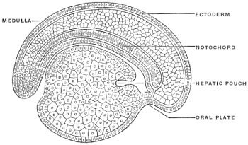 Fig. 237.
—Longitudinal section of an embryo of
Petromyzon
planeri, four days old. (Minot, after Kupffer.)
In the primitive type of development, as seen in Petromyzon
and in the Amphibia, the liver appears very early, as a diverticulum
of the embryonic intestinal tube, near its cephalic extremity,
projecting on the ventral aspect down into the mass of
yolk-cells (Fig. 237). The short stretch of the primitive alimentary
canal cephalad of the hepatic diverticulum corresponds to
the foregut. With the development of the heart the primitive
foregut becomes divided into pharynx and post-pharyngeal segment
(œsophagus and stomach). The hepatic diverticulum then
lies immediately dorsad of the caudal or venous extremity of the
heart. Hence it is probable that the liver is an older organ in
the ancestral history of the vertebrates than the pharynx or even
the heart. The liver diverticulum lies in close connection with
the omphalo-mesenteric veins which return the blood from the
yolk-sac to the heart. In the course of further development, as
will be seen below, the liver comes into very intimate relations
with the venous circulation.
In human embryos of 3.2 mm. the primitive hepatic duct appears
as a wide hollow pouch composed of hypoblast cells, growing
between the two layers of the ventral mesogastrium, which
membrane, extending between the ventral border of the primitive
stomach and the ventral abdominal wall, will be subsequently
considered in detail. The liver, in developing between the layers
of the ventral mesogastrium, approaches very early the septum
transversum or rudimentary diaphragm and becomes connected
with the same. A mass of mesodermal cells, derived from the
mesogastrium and from the primitive mesodermal intestinal
wall surrounding the hypoblastic lining of the tube, covers the
cæcal termination of the primitive hepatic duct, forming the so-called
embryonic hepatic ridge. This mesodermal tissue accompanies
the duct in its further growth and branching, forming the
connective tissue envelope, known in the adult as the capsule of
Glison. The primitive hepatic duct is directed cephalad in the mesogastrium
between the vitelline duct and the stomach (Fig. 101).
In embryos measuring 4.25 mm. the duct is 0.24 mm. long.
Later (in embryos of 8 mm.) the primitive single duct divides
into two secondary branches, indicating, even at an early stage,
the adult arrangement of the duct, as formed by the union of the
right and left hepatic ducts (Fig. 185).
The gall-bladder in embryos of this size (8 mm.) is a well-defined
cæcal diverticulum, branching caudad from the main
hepatic duct.
The vesicular mucous surface is thus derived from the enteric
hypoblast in the same way as the epithelial lining of the bile-ducts
and capillaries. The external muscular and fibrous coats
of the gall-bladder are developed from the mesoderm of the
mesogastrium.
It is to be noted that at an early stage the gall-bladder is derived
from the main duct close to the intestine, the latter duct
being very short. Later on the common duct grows in length,
making the liver more and more a gross anatomical organ distinct
from the intestine. The cystic duct develops as the result
of a similar increase in length of the cystic diverticulum. The
two principal secondary branches of the hepatic duct give origin
to sprouts or buds. These are derivatives of the hypoblastic
cells of the larger ducts and may from the beginning be hollow,
possessing a lumen continuous with that of the parent duct
(Selachians, Amphibians). In warm-blooded animals these
sprouts are at first solid, forming the s. c. hepatic cylinders, and
only subsequently become hollowed out with the further development
of the biliary duct system of the liver. The rapid
growth of the organ leads to a great increase in the number of the
hepatic cylinders. They spread out on all sides, finally coalescing
with adjacent buds so as to form an interlacing network whose
meshes are filled by blood vessels. After the hepatic cylinders
have become canalized they preserve the same arrangement,
hence the resulting biliary capillaries of the adult form an anastomosing
network. Amphioxus and the amphibians have a
single hepatic outgrowth (Fig. 49).
In the Selachians the liver arises as a ventral outgrowth at
the hinder end of the foregut immediately in front of the vitelline
duct, thus bringing the liver from the beginning into close proximity
with the vitelline veins entering the heart. Almost as soon
as formed the outgrowth develops two lateral diverticula, opening
into a median canal. The two diverticula are the rudimentary
lobes of the liver and the median canal uniting them is the
rudiment of the common bile-duct and gall-bladder.
In the Teleosts the liver arises quite late (in the trout about the
25th day) as a solid outgrowth from the intestinal canal close to
the heart. In the Amniota the liver arises in the same position
as in the Anamnia, but, at least in birds and mammals, shows its
bifurcation almost, if not quite, from the start. The two forks
embrace between them the omphalo-mesenteric or vitelline veins
just before they empty into the sinus venosus of the heart.
In the chick the liver appears between the 56th and 60th hour,
the right fork being always of greater length but less diameter
than the left. The hepatic outbud in the rabbit appears during
the 10th day, and during the 11th day begins to send out branches.
In man, as above stated, the bud appears well marked in embryos
of 3 mm.
[Certain adult variations make it appear possible that there are
two human embryonic hepatic buds, a cranial and a caudal, as is
the case in birds.]
I. B. Comparative Anatomy of the Liver.—The liver, phylogenetically
a very old organ, occurs in all vertebrates, for the cæcal
diverticulum of the intestine of amphioxus (Fig. 49) has probably
the significance of a hepatic outbud.
The primitive form of the liver is symmetrically bilobed, a
type which is seen well in the chelonian organ (Fig. 238).
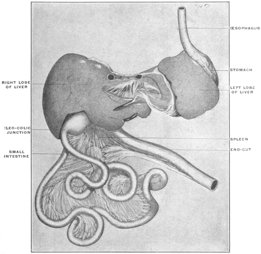 Fig. 238.
— Pseudemys elegans, pond turtle. Alimentary canal. (Columbia University Museum,
No. 1437.)
|
|
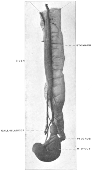 Fig. 239.
—Stomach, mid-gut, pancreas, and liver
of Boa constrictor, boa. (Columbia University Museum,
No. 1832.)
|
In size the liver is subject to great variations. It is usually
larger in animals whose food contains much fat. Hence carnivora
in general have a larger liver than herbivorous animals.
Its shape also varies considerably, depending on the form of the
body cavity and on the amount and disposition of the available
space. Hence in the snakes the organ appears long drawn out,
flattened, almost ribbon-like (Fig. 239), while the relatively very
large coronal diameter of the body cavity in the turtles permits
the liver to expand transversely (Fig. 238).
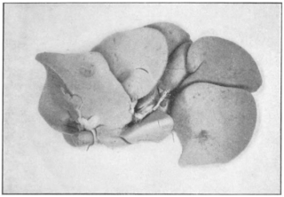 Fig. 240.
—Liver of Macacus cynomolgus, Kra monkey.
(Columbia University Museum, No. 28/1833.)
|
|
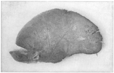 Fig. 241.
—Liver of Pleuronectes maculatus, flounder.
(Columbia University Museum, No. 1679.)
|
In general, when the liver is large and the available space for its
reception limited, it is usually split into several (two to seven)
lobes, which permit, by mutual displacement, the accommodation
of the organ to varying space-conditions of the body cavity (Fig.
240). Under the opposite circumstances, on the other hand,
even the primitive bilobed character may disappear and the liver
is then unlobed (Fig. 241).
The presence or absence of a gall-bladder depends apparently
largely on the character of the food and on the habitual
type of digestion. In many vertebrates digestion is carried on
nearly continuously, without marked interruption, especially in
many ungulates, ruminants and rodents. In such animals the gall-bladder
is absent. It is also absent in several birds (most Parrots,
Doves, Ostrich, Rhea americana, the Cuculidæ, Rhamphastos,
etc.). This variability emphasizes the morphological fact that
the biliary bladder is only a modified portion of the hepatic duct
system, as shown by the development above outlined.
A great variety is observed in the arrangement of the biliary
ducts, through which, at the period of intestinal digestion, bile
passes from the liver and gall-bladder into the intestine, while in
the intervals of digestion the secretion is only carried from the
liver to the bladder. The following main types of the biliary
duct system may be recognized:
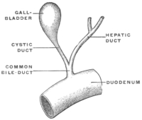 Fig. 242.
—Schema of hepatic and cystic
ducts. (Nuhn.)
|
|
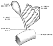 Fig. 243.
—Schema of hepatic and cystic
ducts. (Nuhn.)
|
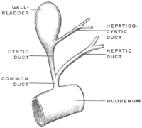 Fig. 244.
—Schema of hepatic and cystic
ducts. (Nuhn.)
|
|
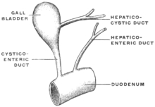 Fig. 245.
—Schema of hepatic and cystic
ducts. (Nuhn.)
|
1. The hepatic duct joins the cystic to form the common bile-duct,
entering the duodenum by passing obliquely through the
intestinal wall (Fig. 242). This form is encountered in man
and in most mammals. It is also found in some birds (Buceros),
many amphibians, and in some fish (Lophius). Instead of one
hepatic duct two may join the cystic duct separately to form the
common bile duct (Phoca litorea), or the number of hepatic ducts
may be further increased. The separate hepatic ducts then
unite successively with the cystic duct. This occurs in many
mammals (as Tarsius, Galeopithecus, monotremes) and in some
fishes (Xiphias, Trigla, Accipenser) (Fig. 243).
2. Of two hepatic ducts only one helps to form with the cystic
duct the common duct, while the other leads from the liver transversely
into the bladder, especially into the neck, forming the
hepatico-cystic duct (Fig. 244). This arrangement is found in
several mammals (calf, sheep, dog).
3. No common bile-duct is formed. The hepatic and cystic
ducts each empty separately into the intestine (hepato-enteric and
cysto-enteric ducts), while a hepato-cystic duct carries the bile
directly from the liver to the gall-bladder (Fig. 245).
Lutra vulgaris among mammalia, the majority of the birds and
several reptilia present this type.
When the gall-bladder is absent a single large hepato-enteric
duct is found, or instead a number of smaller ducts which enter
the intestine successively.
I. C. Development of Vascular System of Liver.—In order to comprehend
the peritoneal relations of the adult liver it is absolutely
necessary to have a clear understanding of the development of
the vascular system in connection with the gland.
For our purpose, in the first place, a serial consideration of the
successive stages, illustrated by schematic diagrams, will prove
most practicable. These diagrams represent the structures in the
dorsal view, i. e., in the position which they would occupy in the
adult liver with the gland resting on its upper or convex surface
and with the ventral sharp margin turned toward the beholder
(see Fig. 259).
The development of the venous system, especially in connection
with the liver, presents a somewhat complicated series of successive
conditions. After having become familiar with the principal
typical embryonal stages, as shown in the following diagrams,
the student is strongly recommended to cement this knowledge
by the comparative examination of the venous system. The permanent
veins of the lower vertebrates, while in many cases not
strictly homologous to those of the higher forms, yet are excellent
objects for study, since they serve to illustrate temporary stages in
the development of the mammalian venous system, and to that
extent are of aid in comprehending one of the most difficult and
important chapters in human anatomy. At the conclusion of the
diagrammatic consideration of the mammalian development a
number of comparative facts will be put together for this
purpose.
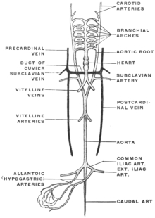 Fig. 246.
—Diagram of embryonic vascular system, without the portal circulation. (Parker,
after Wiedersheim.) The dorsal aorta is formed by the junction of the right and left aortic roots
arising from the confluence of the branchial arterial arches.
|
|
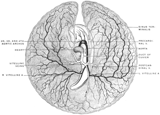 Fig. 247.
—Diagram of the circulation of the yolk-sac at the end of the third day of incubation
in the chick. (After Balfour.) The median portion of the first aortic arch has disappeared;
but its proximal end forms the external, its distal the internal carotid arteries. The whole blastoderm
has been removed from the egg and is viewed from below. Hence the left appears on
the right, and vice versa.
Arteries in black.
Veins in outline.
|
1. Early Stage.—In the earlier developmental stages in mammalian
embryos the primitive dorsal aorta extends caudad along the
ventral aspect of the vertebral axis, giving off paired vitelline or
omphalo-mesenteric arteries to the yolk-sac and allantoic arteries
to the embryonic urinary bladder or allantois (Figs. 246 and 247).
The blood is returned from the vascular area of the yolk-sac by
two vitelline or omphalo-mesenteric veins, which unite near the
heart to form a common trunk, continued as the sinus venosus into
the caudal or auricular extremity (venous end) of the primitive
tubular heart (Figs. 246, 247 and 248).
2. Development of Allantois. Stage of Placental Circulation.—The placental
circulation, replacing the temporary vitelline circulation
of the earliest stages, is inaugurated by the appearance of two
umbilical veins, which pass cephalad, imbedded in the tissue of
the ventral mesogastrium, to empty into the sinus venosus near
the vitelline veins (Fig. 249). The umbilical veins return the
oxygenated blood from the placenta to the embryo. At first the
right umbilical vein is the larger of the two.
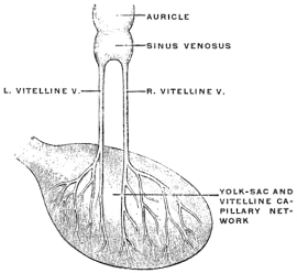 Fig. 248.
—Schema of vitelline veins.
|
|
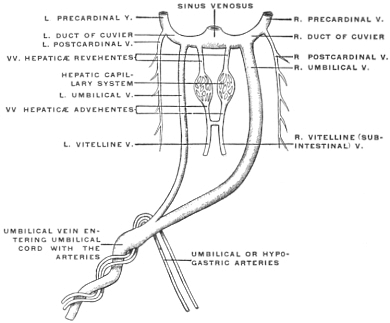 Fig. 249.
—Schema of umbilical veins, early stage.
|
The sinus venosus at this time also receives two large veins,
transversely directed, called the ducts of Cuvier, which are formed
near the heart by the union of the anterior cardinal (primitive
jugular) and posterior cardinal veins, draining respectively the
head end of the embryo, and the body walls and Wolffian bodies.
The vitelline veins are placed on each side of the primitive
small intestine, and become connected with each other by a broad
anastomotic branch (Fig. 249). When the hepatic outgrowth buds
from the duodenum the vitelline veins send out branches which
break up into a wide-meshed capillary network in the mesodermic
tissue enveloping the hepatic cylinders. Hence at this period
the circulation in the vitelline veins is made up of three districts:
(a) Distal segment of veins, coursing along duodenum, and
joined by a transverse anastomosis, before reaching the liver bud
(subintestinal veins).
(b) Middle segment, from which capillary vessels are derived,
ramifying upon and between the developing hepatic cylinders.
(c) Proximal segment, formed by the continuation of the proximal
part of the vitelline veins into the sinus venosus of the heart.
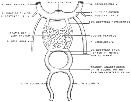 Fig. 250.
—Schema of primitive portal circulation.
3. Formation of Portal Circulation. A.—With the further development
of the liver the direct connection of the distal segment of
the vitelline veins with the sinus venosus becomes lost, the intermediate
segment being entirely broken up into an intrahepatic
network (Fig. 250). Hence all the blood brought to the liver
by the vitelline veins (venæ hepaticæ advehentes) passes through
the hepatic capillary circulation, before it is carried by the proximal
segment of the vitelline veins (venæ hepaticæ revehentes)
into the sinus venosus. The amount of this blood increases with
new connections which the vitelline veins make with the venous
radicles developing in the intestinal tract and its appendages.
In proportion as, with the development of the placenta and reduction
of the yolk-sac, the original significance of the vitelline
veins as nutritive and respiratory vessels disappears, this secondary
connection of the vitelline veins with the veins of the alimentary
tract becomes more and more important, until finally
the original vitelline veins, now properly called omphalo-mesenteric
veins, return the blood from the intestinal tube, pancreas
and spleen to the liver.
The venæ hepaticæ advehentes, becoming connected in this
way with the developing intestine, pancreas and spleen, form the
rudiments of the future portal system, while the venæ hepaticæ
revehentes are prototypes of the hepatic veins of the adult circulation.
B. Development of the Portal Vein.—The distal subintestinal segments
of the vitelline veins are early united by a transverse anastomotic
branch. The section of the veins above this anastomosis
is seen already in Fig. 250 to have assumed an annular shape,
while the veins below the primary anastomosis are approaching
each other to form a second ring-like junction.
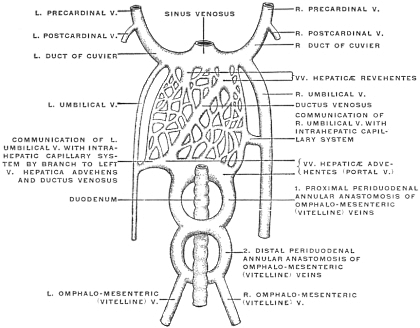 Fig. 251.
—Schema of further development of portal circulation and connection of
same with umbilical veins in early stages.
|
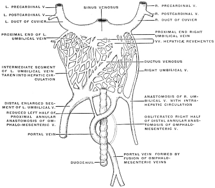 Fig. 252.
—Second stage in development of circulation through portal and umbilical veins.
The proximal segment of the main portal vein is formed by the persistence of the left half of the
distal and right half of the proximal periduodenal vascular ring of the omphalo-mesenteric veins.
The distal segment of the main portal vein is the product of the fusion of the omphalo-mesenteric
veins, and becomes connected with the veins of the intestinal canal, pancreas, and spleen. The
proximal terminal segment of both umbilical veins becomes included in the system of the venæ
hepaticæ revehentes.
|
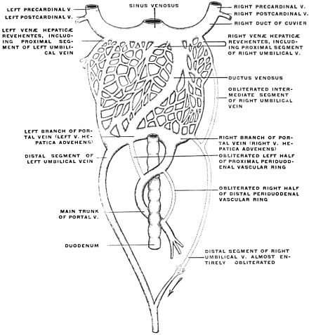 Fig. 253.
—Third stage in development of portal and umbilical veins during the placental period.
|
In Fig. 251 the subintestinal segments of the two vitelline veins
are seen to have communicated with each other by transverse
anastomotic branches around the duodenum, two of these
branches being situated ventrad and one dorsad of the intestinal
tube. These branches, and the portions of the primitive vitelline
veins between their points of derivation, form two vascular loops
or rings, encircling the primitive duodenum (Fig. 251).
The distal portions of the vitelline veins, before reaching the
caudal annular duodenal anastomosis, next fuse into a single longitudinal
vessel which also receives the veins from the stomach,
intestine, spleen, and pancreas, and forms the beginning of the
portal vein.
By atrophy of the right half of the lower, and of the left half
of the upper duodenal venous ring (Figs. 252 and 253), the proximal
portion of the portal vein is formed as a single vessel, taking
a spiral course around the duodenum (Fig. 256). Hence in the
adult the portal vein and its principal branch (the superior mesenteric
vein) crosses over the ventral surface of the duodenum (third
portion), turns along the mesal side of the second portion, and
then continues to the liver along the dorsal aspect of the first
portion (Fig. 254). Note—In comparing Fig. 254 with the schematic
figures it should be noted that the same presents the parts
in the ventral view, while the schemata offer the dorsal aspect.
4. Changes Leading to the Final Arrangement of the Umbilical Veins.—A
very important rearrangement of the umbilical veins takes place.
These veins originally course in the lateral abdominal wall, close
to the fold of the amnion (Fig. 255), and then turn cephalad of
the developing liver along the septum transversum to empty into
the sinus venosus at each end (Figs. 249 and 250). The right
umbilical vein is at first the larger.
This symmetrical arrangement, and the direct connection of the
umbilical veins with the sinus venosus, now becomes lost by the
occurrence of the following changes:
1. At first (Fig. 249) all the blood carried to the liver by the
omphalo-mesenteric veins passes through the hepatic capillary
network before being conducted by the venæ revehentes to the
sinus venosus. Very early, however, a new intrahepatic channel
develops, the ductus venosus (Figs. 250-253), which passes obliquely
between the entrance of the left omphalo-mesenteric vein
into the capillary system (l. v. advehens) and the termination of
the right omphalo-mesenteric vein (r. vena revehens) in the sinus
venosus.
In human embryos of 4 mm. the ductus venosus can already
be distinguished, and in embryos of 5 mm. the vessel has assumed
considerable proportions.
2. A communication is next established on both sides between
the capillary hepatic network in the portion of the liver nearest
to the abdominal wall and the umbilical veins as they ascend
imbedded in the abdominal wall (Fig. 251).
This connection is usually from the start larger on the left side
and connects with the left omphalo-mesenteric vein just at the
point where the same is about to be continued into the ductus
venosus. This connection becomes rapidly larger, so that the
ductus venosus, which at first appeared merely as an anastomotic
channel between the left omphalo-mesenteric vein and the terminal
portion of the right omphalo-mesenteric vein, now forms the
main continuation of the left umbilical vein. This vessel grows
very rapidly up to its connection with the ductus venosus and
soon exceeds the right umbilical vein in size (Fig. 252). Beyond
the ductus venosus on the other hand the proximal segment of
the left umbilical vein diminishes in size, and loses its independent
character by incorporation in the hepatic circulation. Only
its terminal portion, emptying into the sinus venosus, is preserved.
This is surrounded by the growing masses of hepatic
cylinders and is converted into a vena revehens.
The connection of the right umbilical vein with the liver vessels
is at first symmetrical to that on the left side, but less
strongly developed. The effect of this connection is to reduce
in the same way the proximal segment of the right umbilical
vein and to convert its termination into a vena revehens. With
the great development of the left vein, however, the vein on the
right side gradually diminishes and finally loses its connection
with the intrahepatic circulation altogether. The right umbilical
vein is now reduced to a vessel of the ventral abdominal wall,
which carries blood in the reverse of the original direction, i. e.,
from the abdominal wall caudad into the left umbilical vein
(Figs. 253 and 255).
The connection thus established between the umbilical vein
and the portal circulation results in the formation of a single
large (the original left) umbilical vein which, throughout the
remainder of fœtal life, returns all of the placental blood (Fig.
253).
The newly developed hepatic portion of the left umbilical vein
becomes, however, not only connected with the ductus venosus,
but also with the right part of the upper venous ring, derived
from the right omphalo-mesenteric vein (Fig. 253). This connection
forms the left portal vein of the adult, and enlarges
rapidly.
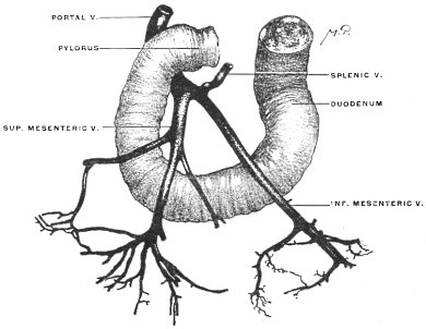 Fig. 254.
—Corrosion preparation showing course of portal vein and tributaries in relation to
duodenum. (Columbia University Museum, No. 1857.)
|
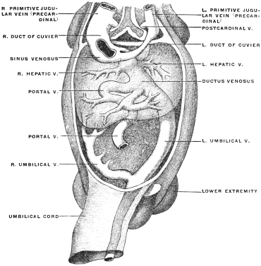 Fig. 255.
—Human embryo of 10 mm. cervico-coccygeal measure. Heart and ventral body-wall
removed to show sinus venosus and entering veins. (Kollmann, after His.)
|
The terminations of the ductus venosus and of the venæ hepaticæ
revehentes undergo a number of secondary changes in
relative position. The left hepatic vein loses its direct connection
with the sinus venosus, and now opens into the termination
of the ductus venosus, into which the right hepatic vein also
empties. This common vessel (v. hepatica communis) subsequently
forms the proximal segment of the postcava when this
vessel develops (Fig. 256).
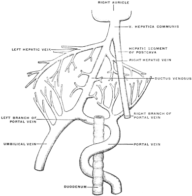 Fig. 256.
—Final stage of development of portal and umbilical veins in the placental period.
|
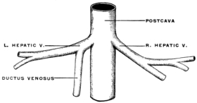 Fig. 257.
—Schema of relation of postcava to hepatic veins and ductus venosus.
|
The blood, therefore, returned to the liver by the left umbilical
vein divides at the transverse fissure into three streams. Two of
these pass through the connection with the portal vein and through
branches developed from the hepatic part of the umbilical vein
into the capillary system of the right and left lobe. The third
continues through the ductus venosus to the common hepatic vein
and sinus venosus (Fig. 256). The ductus venosus thus becomes
the chief vessel returning arterialized placental blood to the heart.
When the postcava develops fully the hepatic segment of this
vessel also joins the terminal part of the ductus venosus (Fig.
256) and gradually replaces the same as the main returning
venous channel, the proximal part of the ductus venosus being
incorporated in the vena cava (Fig. 257). The postcava then
receives the right hepatic veins separately, while the left hepatic
veins and ductus venosus open together into the main vein. This
condition obtains up to the time of birth and the consequent
interruption of the placental circulation.
While at first the ductus venosus communicates throughout its
entire length with the meshwork of the hepatic capillary system,
a separation into two segments, i. e., ductus venosus proper and
intrahepatic segment of umbilical vein, is established after the free
communication with the left umbilical vein takes place. This
condition is exhibited in Fig. 258, which represents the corroded
venous system of the fœtal liver, and in Fig. 259, showing an
injected liver in the fœtus at term.
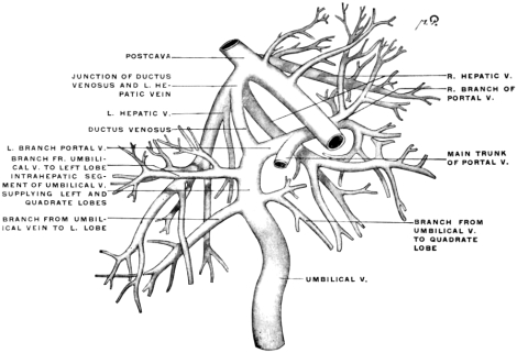 Fig. 258.
—Corrosion preparation of venous system of human liver in fœtus at term. (Columbia
University Museum, No. 1834.)
|
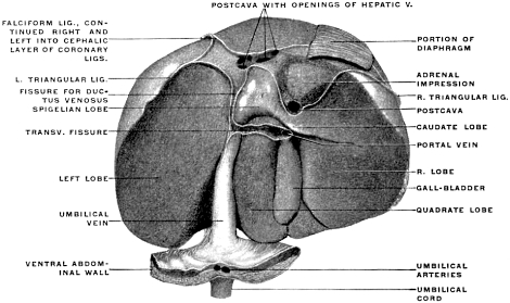 Fig. 259.
—Injected and hardened human liver from fœtus at term. (Columbia University
Museum, No. 1853.)
|
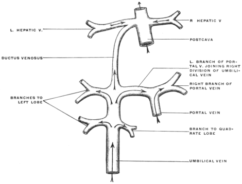 Fig. 260.
—Diagram of intrahepatic fœtal venous circulation.
|
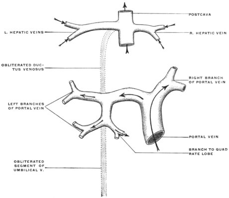 Fig. 261.
—Diagram illustrating the changes in the intrahepatic venous circulation
resulting from the cessation of the placental circulation at birth.
|
It will be observed that the umbilical vein on entering the liver
gives off a large branch to the left lobe, and a smaller branch on
the right side to the quadrate lobe, which act as the main venæ
advehentes of these portions of the liver. Arrived at the transverse
fissure the umbilical vein divides into three branches, at
right angles to each other. The left branch enters the left lobe,
the right branch becomes directly continuous with the left main
division of the portal vein, while the central branch, continuing
the direction of the umbilical vein, passes dorsad, as the ductus
venosus proper, to join the left hepatic vein close to its entrance
into the postcava.
5. Changes Consequent upon the Establishment of Pulmonary Respiration.—After
birth the umbilical vein and its continuation, the ductus
venosus, become obliterated, the former constituting the round
ligament of the liver, the latter the ligament of the ductus venosus,
both structures imbedded in corresponding portions of the sagittal
fissure on the caudal and dorsal surfaces of the adult liver
(Figs. 284 and 286). The lateral branches of the umbilical vein,
however, in its course from the ventral margin of the liver to the
transverse fissure (Fig. 258), remain pervious and are transferred
to the portal circulation.
It will be noticed, in reference to the direction of the blood current,
that at birth a sudden reversal takes place in the right terminal
branch of the umbilical vein at the transverse fissure (Figs.
260 and 261). Before birth the blood current of the umbilical
vein divides into three streams, right, left and central. The latter
enters the ductus venosus. The left enters the liver directly, the
right traverses, from left to right, the segment between the termination
of the umbilical and the bifurcation of the portal vein.
This segment in the adult carries blood from right to left, as left
branch of the portal vein. In the fœtus, however, the blood
traverses this segment from left to right, in passing from the
umbilical to the right branch of the portal vein. The blood
entering the liver through the portal vein passes chiefly into the
right division of that vessel (Fig. 260).
After birth all the venous blood entering the liver passes
through the portal vein. In the right division the direction of
the current is the same as in the fœtus.
On the left side, however, the current is now from right
to left, from the bifurcation of the portal into the channels of
the left lobe formerly connected with the umbilical vein (Fig.
261).
Hence the direction of the current in this segment is reversed
at birth.
SUMMARY OF HEPATIC CIRCULATION.
The foregoing consideration of the development shows us that
the hepatic circulation presents successively three main stages:
1. Omphalo-mesenteric or Vitelline Stage, which results in the
laying down of the primary capillary circulation of the liver and
in the establishment of its connection with the developing veins
of the alimentary tract (primitive portal channels).
2. Umbilical or Placental Stage, in which the greater part of the
blood circulating through the liver is oxygenated blood returned
from the placenta by the umbilical vein, accounting for
the rapid growth and relatively large size of the organ during
fœtal life.
The placental blood uses the preformed capillary channels of
the vitelline or primitive portal system in the liver, and the same
rapidly extend and enlarge with the accelerated growth of the
gland. During this stage venous blood is also returned from the
alimentary tract to the liver by the portal vein, produced by
fusion of the distal segments of the primitive vitelline veins and
their secondary connection with the mesenteric, splenic and pancreatic
veins (omphalo-mesenteric development of primitive vitelline
veins).
3. Adult or Portal Stage.—With the interruption of the placental
circulation the portal vein assumes again its original position as
the only vein carrying blood to the liver. With the establishment
of intestinal digestion and absorption this vessel grows
rapidly in size.
COMPARATIVE ANATOMY OF THE HEPATIC VENOUS
CIRCULATION.
For the purpose of fixing the main facts in connection with the
development of the higher mammalian hepatic circulation, and
in order to obtain a demonstration of the cycle through which the
different veins pass, the student is recommended to examine, preferably
by personal dissection, a limited series of lower vertebrates
which can be readily procured and easily injected. The following
series has been selected, but it will be understood that other
forms can be substituted, according to the local conditions which
govern the supply of the material.
1. Fish. A Selachian, the common skate (Raja ocellata) or dog-fish (Acanthias vulgaris).
2. Amphibian.
(a) Urodele. Necturus maculatus.
(b) Anura. The common frog.
3. Reptile.
Preferably, on account of the ease of injection, one of the larger
lizards, as Iguana tuberculata.
The turtles, although somewhat more difficult objects to prepare,
can be substituted.
4. Bird. The common fowl.
5. Human fœtus at term.
1. Fish.—The venous system can be injected by tying a canula
in the lateral vein, and injecting both cephalad and caudad, or by
injecting cephalad through the caudal vein. The injection of the
systemic veins can also be made caudad through one of the ducts
of Cuvier, combined with an injection cephalad of the caudal
vein.
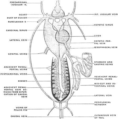 Fig. 262.
—Diagram of the veins of a selachian. (Wiedersheim, after Parker.)
The lateral vein arises from a venous network surrounding the cloaca, receiving one or more
cutaneous veins of the tail, veins of the body-wall, and veins of the pelvic fins.
The caudal vein divides at the posterior end of the kidney into the two renal-portal veins,
from which the advehent veins of the renal-portal system are derived. The revehent renal-portal
veins join to form the posterior cardinal veins, which, after dilating enormously to form the
cardinal sinuses, join with the anterior jugular, subclavian, and lateral veins to form the ducts of
Cuvier. The latter receive the inferior jugular veins, from the deep parts of the head and neck
and the terminations of the hepatic portal system (hepatic sinus).
The hepatic portal vein is formed by the veins of the œsophagus, stomach, and intestines.
After traversing the capillary vessels of the liver, the revehent hepatic veins unite to form an
extensive hepatic sinus before entering the heart.
The following main facts are to be noted in the venous system
of the Selachian (Fig. 262):
1. There are Two Portal Systems. (a) Renal Portal System.—The caudal
vein divides near the vent into two branches which course
along the lateral border of the kidneys, sending afferent or advehent
veins into the organ. The blood traverses the renal capillaries
and is gathered together by the efferent or revehent veins,
which empty into median paired vessels, the posterior cardinals.
(b) Hepatic Portal System.—The veins of the digestive tract and
appendages unite to form a hepatic portal vein. The blood after
traversing the capillary system of the liver is collected by hepatic
veins, which form a dilated hepatic sinus emptying into the sinus
venosus of the heart.
2. The middle segment of the intestine, presenting a spiral
valve in the interior, gives rise to a vein emptying into the portal
vein which corresponds to the subintestinal vitelline vein of the
mammalian embryo (Fig. 202).
3. The posterior cardinal veins, also greatly dilated and forming
the posterior cardinal sinus, join, near the heart, the veins returning
blood from the head, the anterior cardinal or jugular, to form
a transversely directed trunk, the duct of Cuvier, which empties
into the sinus venosus at the auricular extremity of the heart.
Into the duct of Cuvier empties on each side a lateral vein returning
the blood from the body walls. This vein can be considered,
for our present purpose, as representing in general the abdominal
vein of amphibians and reptiles, and the umbilical vein of the
mammalian embryo.
The adult selachian venous system is therefore to be considered
as illustrating the following conditions above encountered in
our study of the embryology of the mammalian venous system.
1. The heart illustrates excellently the stage in the mammalian
development, in which auricular and ventricular segments have
differentiated, but before the division of the cavities into a pulmonary
and systemic portion by the development of the auricular
and ventricular septa and the division of the arterial trunk
into pulmonary artery and aorta.
The sinus venosus still exists, as an ante-chamber to the auricular
cavity proper, receiving on each side the ducts of Cuvier,
which represent the fusion product of the systemic veins, anterior
and posterior cardinal.
2. The hepatic portal circulation corresponds to the mammalian
stage in which the vitelline veins have become omphalo-mesenteric
by joining the intestinal veins.
The spiral vein remains as a portion of the original vitelline
vein corresponding to the subintestinal segment of the mammalian
embryo (cf. Figs. 248 and 249).
The selachian portal vein represents the united vitelline veins,
into which the veins of the digestive tract open.
In the liver we find a simple system of venæ advehentes, derived
from the branching of the portal vein, a hepatic capillary
network, and venæ revehentes, the proximal remnants of the
original vitelline veins which carry the liver blood to the sinus
venosus. The condition of the hepatic circulation corresponds
therefore to the stage shown in Fig. 250 of the mammalian development.
There is as yet no association of the hepatic venous
system with the representative of the umbilical vein (the lateral
vein of the selachian).
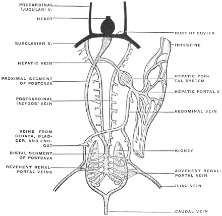 Fig. 263.
—Diagram of the veins of urodele amphibian (
Salamandra maculosa). (Wiedersheim.)
The caudal vein bifurcates at the posterior extremity of the kidneys to form the afferent
trunks of the renal-portal system along the lateral border of the kidneys, from which the advehent
veins of the renal-portal system are derived. The iliac or femoral vein divides into an
anterior and a posterior branch, the latter opening into the afferent renal-portal vein, while the
former, uniting with the one of the opposite side, forms the abdominal vein, and receives vessels
from the bladder, cloaca, and end-gut. The revehent veins of the renal-portal system, emerging
upon the ventral surface of the kidneys, empty into a single median vessel, the distal or renal
section of the postcava or vena cava inferior. Proceeding cephalad, the proximal or hepatic section
of this vessel, after traversing the liver and receiving the revehent hepatic veins of the
hepatic portal system, empties into the sinus venosus of the heart. Previous to entering the liver
the postcava gives off the two posterior cardinal or azygos veins, which continue cephalad, receiving
tributary segmental veins from the body-walls and reach the sinus venosus by joining the
subclavian veins. These latter uniting with the anterior cardinal (jugular) veins form the ducts
of Cuvier (precaval veins).
The abdominal vein continues cephalad in the ventral mesogastrium to the liver, giving off a
number of smaller branches, which enter the hepatic portal circulation by penetrating the ventral
surface of the liver between the layers of the ventral mesogastrium, while the main continuation
of the vessel joins the hepatic portal vein at its point of entrance into the liver.
The hepatic portal vein is formed by tributaries returning the blood from the digestive tract
(intestinal canal, spleen, pancreas). The blood, after traversing the hepatic portal circulation, is
conducted by the hepatic revehent veins to the proximal section of the postcava. A number of
secondary or accessory portal veins pass from the anterior portion of the intestinal canal (œsophagus,
stomach) directly to the liver.
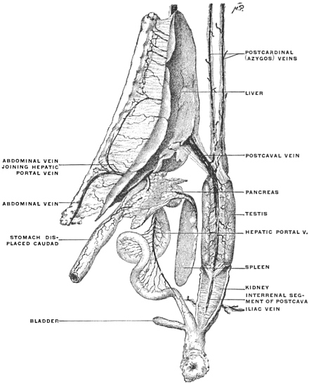 Fig. 264.
—Dissection of veins of
Necturus maculatus, mud-puppy. (Columbia University
Museum, No. 1835.)
The postcava has been divided at the cephalic end of the liver just before entering the sinus
venosus, and the postcardinals have been cut prior to their junction with the subclavian veins.
The stomach has been turned caudad. The abdominal vein has been divided after the common
trunk has been formed by branches from the iliac veins. The latter are seen entering the
afferent renal-portal vein, derived from the bifurcation of the caudal vein, along the lateral
border of the kidneys.
The junction of the main trunk of the abdominal vein with the hepatic portal vein takes
place close to the liver under cover of the pancreas. A series of accessory portal veins continuous
with the abdominal vein enter the ventral surface of the liver between the layers of the ventral
mesogastrium. The inter-renal segment of the postcava receives the revehent renal-portal veins.
The iliac vein enters the advehent renal-portal veins derived from the caudal vein.
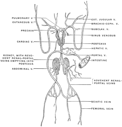 Fig. 265.
—Venous system of
Rana esculenta, frog. (Ecker.)
3. The lateral veins, which we can, as stated, regard for purposes
of illustration, without prejudging their genetic significance,
as representing the mammalian embryonic umbilical veins, still
present the condition corresponding to the early mammalian
embryonal stage shown in Fig. 250. They are veins of the body
walls, emptying cephalad of the liver, directly into the ducts of
Cuvier, and through them into the sinus venosus of the heart.
Fig. 262 shows the arrangement of the venous system in a
typical selachian diagrammatically.
2. Amphibian. (a) Urodele.—The following points are to be noted
in comparison with the preceding form:
1. The two ducts of Cuvier entering into the sinus venosus are
formed by the anterior cardinal and subclavian veins, which
latter, having appeared with the full development of an anterior
extremity, receives the posterior cardinal veins, representing the
mammalian azygos system.
2. The renal portal circulation persists. The caudal vein is,
however, no longer the only afferent vein of this system. With
the full development of a posterior extremity an iliac vein returns
the blood from the same and gives a large branch (afferent
to the portal renal system), while the trunk continues cephalad
as an anterior abdominal vein, corresponding to the lateral selachian
vein, emptying in the hepatic portal vein.
3. The efferent veins of the renal portal system no longer unite
to form the posterior cardinal, as in the Selachian, but empty into
a new median vessel, the inferior vena cava, or postcava, which
has replaced the distal segments of the posterior cardinal veins.
The postcava now carries the blood from the kidneys directly to
the heart. The original posterior cardinal veins still persist in
their proximal segments, as smaller trunks connecting the distal
part of the postcava with the ducts of Cuvier through the subclavian
veins. The ducts of Cuvier represent the precavæ (venæ
cavæ superiores) of mammalia and the postcardinals the mammalian
azygos veins.
4. The hepatic portal system differs in two respects from the
Selachian type.
(a) The blood returned to the liver from the digestive tract by
the portal vein becomes mixed before entering the gland with the
blood returned from the posterior extremities and abdominal
walls by the abdominal vein.
This vein, paired below and continuous with the lateral of the
two branches into which the iliac vein divides, becomes united
into a single trunk above and empties into the portal vein.
The abdominal vein represents the lateral vein of the Selachian
and corresponds to the umbilical vein of the higher vertebrates.
(b) The venæ hepaticæ revehentes do not empty directly into
the sinus venosus, but into the proximal portion of the postcava.
Hence the adult urodele venous system illustrates, in reference
to the mammalian development, these stages:
1. The umbilical (abdominal) vein has lost its direct connection
with the sinus venosus. The proximal segment, cephalad of the
liver, has disappeared, and its blood now passes directly into the
hepatic circulation by its union with the portal vein.
(Cf. stage schema Figs. 251 and 252.)
2. The postcaval vein has made its appearance, largely replacing
the posterior cardinal veins, whose proximal segments became
converted into secondary vessels (azygos) uniting the system of
the postcava with that of the duct of Cuvier (mammalian præcava),
while their distal segments are transformed into the distal
portion of the postcava.
The postcava, therefore, is made up of two districts:
(a) The proximal portion is a new vessel, developed in connection
with the hepatic venous system.
(b) The distal portion is derived from the distal segments of the
original posterior cardinal veins.
The termination of the hepatic veins in the postcava corresponds
to the stage shown in schema Fig. 256.
Fig. 263 gives a schematic representation of the arrangement of
the venous system in a typical urodele amphibian (Salamandra
maculosa).
In Fig. 264 the dissected venous system of Necturus maculatus,
the mud puppy, is shown in an injected preparation.
(b) Anure.—The venous system of Rana esculenta is shown in
Fig. 265. Comparison with venous system of urodele:
1. The abdominal vein, corresponding to the mammalian umbilical
vein, has assumed a greater importance in reference to the
hepatic circulation. It is a large trunk, continuous below with
the pelvic vein, terminating above in two branches, which enter
the liver as afferent veins, being joined just prior to the division
by the hepatic portal vein.
2. A small cardiac vein, coming from the heart, empties into
the angle of bifurcation of the abdominal vein.
3. The postcava is well developed, formed by large efferent
renal veins. It entirely replaces the posterior cardinal veins
which are absent in the adult animal.
4. A right and left præcaval vein is formed by the union of
two jugular trunks with the vein of the anterior extremity and a
large musculo-cutaneous vein.
Comparison with the mammalian development: the venous
system of this amphibian can be used to illustrate the mammalian
embryonal stage shown in schema Fig. 252, in which the abdominal
or umbilical vein has become the most important vessel
in the afferent hepatic venous system.
The communication existing by means of the cardiac vein between
the heart and the hepatic afferent system may suggest, but
purely for illustrative purposes, the direct connection of the umbilical
vein with the heart by the ductus venosus in the mammalian
embryo (cf. schema Figs. 250-256).
3. Reptile.—In Iguana the renal portal system is well developed.
The caudal vein, returning the blood from the tail and the cavernous
tissue of the genital organs, continues for a short distance upon
the fused caudal end of the two kidneys (Fig. 269) and then
divides into two afferent renal veins which ascend on the
ventral surface of the glands, giving branches to the renal capillary
system. About the middle of the kidney each afferent
vein is joined by a large transverse branch from the abdominal
vein (Fig. 266).
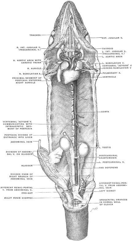 Fig. 266.
—Systemic veins of Iguana tuberculata. The alimentary canal and appendages,
together with the hepatic portal vein and the intrahepatic segment of the postcava, have been
removed. The liver occupies the space between the divided ends of the postcava. The vertebral
vein represents the rudimentary proximal segment of the postcardinal vein corresponding to the
mammalian azygos vein. (Columbia University Museum, No. 1320.)
|
|
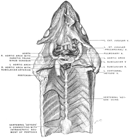 Fig. 267.
—Veins of Iguana tuberculata. Connection of systemic veins with sinus venosus of
heart. The rudimentary system of the vertebral (azygos) veins and their proximal connection
with the subclavian vein are shown. (Columbia University Museum, No. 1859.)
|
| |
 Fig. 268.
—Corrosion preparation of venous system of liver in Iguana tuberculata. The hepatic
portal system and its connection with the abdominal vein, as well as the relation to the postcava,
are shown. The preparation supplements Fig. 266, showing the parts which have been removed
in the latter. (Columbia University Museum, No. 1860.)
|
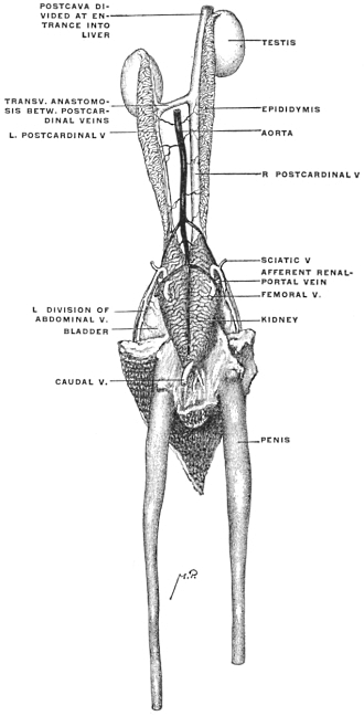 Fig. 269.
—
Iguana tuberculata, ♂. Genito-urinary tract, dorsal view, with renal-portal, postcardinal,
and postcaval veins. (Columbia University Museum, No. 1862.)
The renal efferent system begins by a number of inter-renal
anastomoses which unite along the mesal border of the right kidney
into a large ascending trunk, while the corresponding vessel
of the left side, starting from the same anastomosis, is considerably
smaller (Figs. 266 and 269). Each of these vessels also receives
blood from the testis, epididymis, vas deferens and adrenal
body in the male, and from the ovary and oviduct in the female.
They represent, in fact, the distal functional part of the right and
left embryonic postcardinal vein. Just caudad of the left testis the
vein of the left side crosses obliquely ventrad of the aorta and joins
the right vessel to form the trunk of the postcava, which enters,
immediately beyond the cephalic pole of the right testis, the
prolonged caval lobe of the liver (Figs. 266 and 269). Ascending
in the substance of this gland and receiving the afferent hepatic
veins (Fig. 268), the vena cava emerges from the cephalic surface
of the liver greatly enlarged and proceeds to the right auricle.
The abdominal vein divides below into two branches which
pass caudad on each side of the bladder, receiving tributaries
from the same, to the lateral border of the kidneys (Figs. 266
and 269). Here the vessel is connected by the transverse branch
above described with the afferent renal portal system derived
from the caudal vein. At the same point it receives the sciatic
vein, the principal venous vessel of the posterior extremity.
Above, the main abdominal vein, resulting from the union of the
two branches referred to, ascends on the dorsal surface of the
ventral abdominal wall, receiving a few twigs from the ventral
mesogastrium within whose free caudal edge the vessel runs.
Just before reaching the liver the abdominal vein turns dorsad
on the caudal surface of the gland and joins the hepatic portal
vein (Figs. 268 and 275). Several accessory veins, two or three
in number, belonging to the system of the abdominal vein, pass
above this point from the ventral body wall between the layers
of the ventral mesogastrium, to enter the liver separately on its
convex ventral surface, above the fusion of the main abdominal
vein with the portal vein. These additional branches on entering
the liver join the portal system, forming a set of ventral accessory
portal veins.
The hepatic portal vein derives its principal tributaries from
the splenic, gastric, pancreatic and intestinal veins. One or two
additional branches (accessory vertebral portal veins), as above
stated, connect the system of the segmental and vertebral veins
with the portal circulation, entering the liver separately. In like
manner one or two gastric veins (accessory gastric portal veins)
enter the dorsal aspect of the liver separately, passing from
the stomach to the gland between the layers of the gastro-hepatic
omentum (Fig. 275).
Compared with the development of the mammalian type, the
venous system of Iguana serves to illustrate the stage in the history
of the umbilical vein (represented by the abdominal vein of
the reptile) in which the connection of the vessel with the portal
vein has been formed and transmits the greater part of the blood
returned by the umbilical vein to the liver, while the proximal
segment above this point, originally continued into the sinus
venosus, has begun to disappear, being, however, still represented
by the vessels which, as accessory ventral portal veins, pass in the
ventral mesogastrium, from the body wall to the liver.
It will be noted that all the hepatic portal blood, whether conducted
by the main portal and abdominal vein, or by the accessory
portal branches, traverses the capillary circulation of the
liver before entering the postcava.
The vertebral and segmental venous system, representing the
azygos veins of the mammalia, is very rudimentary (Figs. 266
and 267). The distal portions of the postcardinal veins form the
efferent renal branches and the ascending trunks of the postcava.
The next segment of the vertebral veins appears as a trunk on
the right side which enters the portal circulation. A second vein
higher up is connected with both the gastric portal system and with
the longitudinal chain of the vertebral veins. Finally a proximal
venous branch on each side of the vertebral column, representing
the upper portion of the postcardinal veins, receives the proximal
segmental veins and empties into the subclavian vein (Fig. 267).
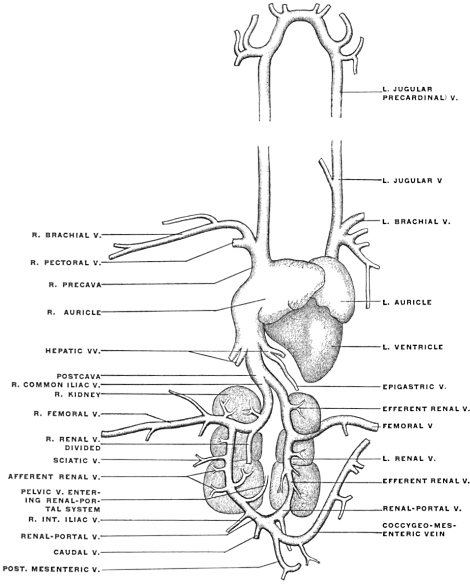 Fig. 270.
—Veins of pigeon,
Columba livia. (Modified from Parker and Haswell.) The renal-portal
vein of the right side is supposed to be dissected to show its passage through the right kidney.
4. Bird.—The characteristic change in the venous system of
the bird, as compared with that of the amphibian and reptile, is
found in the nearly complete abolition of the renal portal system.
The caudal vein bifurcates, sending on each side a large trunk,
which receives the pelvic (int. iliac) veins, to the kidney (renal
afferent portal vein), but only a few small branches enter the substance
of the gland (Fig. 270, afferent renal V). The main vessel
continues cephalad through the kidney and, after receiving
the vein from the posterior extremity (femoral), unites as common
iliac vein with the vessel of the opposite side to form the postcava.
This vessel traverses the liver, receiving the hepatic afferent
veins of the portal system. The portal vein is formed by
tributaries from the intestinal canal, pancreas and spleen, and is
also joined by a large coccygeo-mesenteric vein, which is given
off at the point of bifurcation of the caudal vein and receives
tributaries from the lower part of the alimentary canal. The
abdominal vein of amphibians and reptiles is represented probably
by the epigastric vein, which returns the blood from the
omental mass of fat to the hepatic veins.
Compared with the mammal on the one hand, and with the
lower types on the other, the venous circulation of the bird illustrates
the following points:
1. Extensive reduction of the renal portal system and direct
formation of postcava by the iliac veins, foreshadowing the condition
found in the mammal.
2. Complete separation of the portal and systemic venous circulation
in the adult. Disappearance of the ventral abdominal
vein as a vessel of the body wall.
5. Human Fœtus at Term.—The student is recommended to examine,
by dissection and injection, the venous system of a fœtus
at term, noting the following facts:
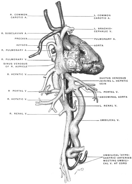 Fig. 271.
—Human fœtus at term. Corrosion preparation of heart and vascular system.
(Columbia University Museum, No. 1858.)
1. Course of umbilical vein in ventral abdominal wall and along
free edge of falciform ligament to liver (Fig. 241), corresponding
to the position of the amphibian and reptilian abdominal
vein (Figs. 264 and 275).
2. Connection of umbilical vein in liver:
(a) With portal system (Figs. 258 and 271).
(α) With portal vein.
(β) With portal system of left and quadrate lobes by
branches derived directly from umbilical vein
while situated in the umbilical fissure (Fig. 258).
(b) With hepatic veins and postcava by the ductus
venosus (Figs. 258 and 271).
3. Connection of the postcaval and precaval systems by the
azygos veins representing the proximal segments of the embryonic
postcardinal veins (Fig. 272).
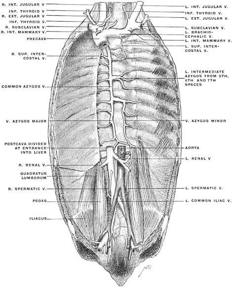 Fig. 272.
—Human fœtus at term. Postcava and azygos veins. (Columbia University
Museum, No. 1861.)
If possible the dissection of an injected fœtus should be combined
with the examination of corrosion preparation of the fœtal
circulation and especially of the venous system of the fœtal liver
(Figs. 258 and 271).
3. The remnants of fœtal structures in the adult liver (round
ligament and ligament of the ductus venosus) should be compared
with the structures from which they are derived in the
fœtus at term (umbilical vein and ductus venosus).
II. THE VENTRAL MESOGASTRIUM.
This membrane has been heretofore mentioned on several occasions.
It now remains for us to carefully consider its arrangement
in detail, both as regards the peritoneal relations of the liver
and in reference to its influence on the abdominal space as a
whole. We can best accomplish this purpose by considering the
membrane in the first place in a purely schematic manner. In
contradistinction to the primitive common dorsal mesentery,
which extends the entire length of the alimentary tube, the ventral
mesentery, or properly the ventral mesogastrium, is confined
to the stomach and proximal portion of the duodenum. We can
represent the membrane as extending between the ventral abdominal
wall and the ventral border (later the lesser curvature)
of the stomach and of the hepatic angle of the duodenum.
Cephalad it is connected with the embryonic septum transversum
(future diaphragm). Caudad its two layers pass into each other
in a free concave edge, including between them the umbilical
vein (free edge of falciform ligament of adult). Consequently a
schematic profile or lateral view of the membrane and its attachments
in the earlier stages would appear as represented in Fig.
273, while the arrangement in transection would be as shown in
Fig. 274. It will be observed that the separation of the cephalic
portion of the abdominal cavity into symmetrical right and left
halves, previously indicated in discussing the primitive stomach
and the dorsal mesogastrium, is actually completed by the ventral
mesogastrium. This complete separation of the lateral
halves of the cœlom cavity ceases at the point where the ventral
mesogastrium terminates in the free concave edge carrying the
umbilical vein. Hence caudad of this falciform edge the two
halves of the cavity communicate freely with each other ventrad
of the intestine and dorsal mesentery.
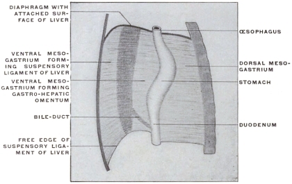 Fig. 273.
—Schematic profile view of ventral mesogastrium with developing liver.
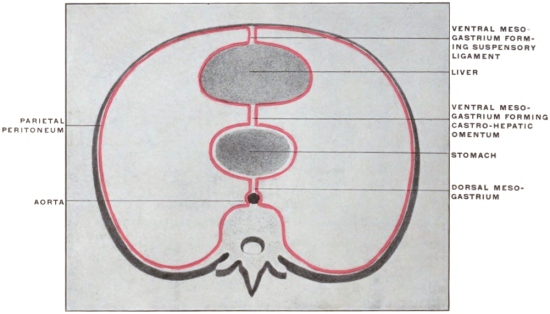 Fig. 274.
—Schematic transection of abdomen in region of ventral mesogastrium.
This difference in the extent of the mesogastria is perhaps best
understood by reference to their relation to the first portion of the
duodenum. We have seen that the duodenum in the early stages
is attached dorsally by a portion of the common dorsal mesentery,
which, after differentiation of the intestinal tract, immediately
follows the dorsal mesogastrium proper, forming the mesoduodenum
(Fig. 172). The proximal portion of the duodenum
(hepatic angle) is still included within the fold of the ventral
mesogastrium which membrane terminates immediately beyond
this point in the free edge surrounding the umbilical vein
(subsequent round ligament) (Fig. 172). The remainder of
the duodenum is devoid of any ventral attachment, being only
connected to the dorsal body wall by the mesoduodenum (Fig.
197).
Subsequently, after the fourth month, while the right surface
of the mesoduodenum and descending duodenum adhere to the
parietal peritoneum, the peritoneal investment of the first portion
or hepatic angle remains free. This peritoneal covering of
the proximal duodenal segment is situated at the point where the
caudal end of the ventral mesogastrium, after surrounding the
first portion of the duodenum, becomes continuous with the dorsal
mesentery forming the mesoduodenum. Obliteration of the
latter membrane by adhesion to the parietal peritoneum leaves
the first portion of the duodenum invested on both surfaces by
the lesser omentum, derived from the ventral mesogastrium. The
ventral surface of the gut is covered by the ventral layer, the
dorsal surface by the dorsal layer of the lesser omentum. These
two layers become continuous around the right free edge of the
lesser omentum (hepato-duodenal ligament) forming the ventral
boundary of the foramen of Winslow (cf. infra, p. 177).
Returning to the schematic consideration of the ventral mesogastrium
above outlined (Figs. 273 and 274) we have to note the
first important change in the arrangement depending upon the
development of the liver. This organ, growing, as we have seen,
from the duodenum, extends between the two layers of the ventral
mesogastrium, receiving a serous investment from the
same. At an early period the liver, developing thus between
the mesogastric layers, reaches the septum transversum and
becomes closely connected with it, laying the foundation for
the subsequent extensive attachment of the gland to the diaphragm.
Extending caudad the liver grows beyond the caudal free edge
of the ventral mesogastrium on each side, carrying the serosa with
it. Consequently the ventral margin of the liver becomes indented
at this point; the umbilical vein and subsequently its
fibrous remnant, the round ligament, are imbedded in a notch
and fissure (umbilical notch and fissure) continued from the ventral
margin dorsad along the caudal surface of the liver (Fig.
259).
This growth of the liver has now effected a division of the primitive
ventral mesogastrium into two segments:
1. Ventral portion, between diaphragm and liver, forms the
broad falciform or suspensory ligament of the liver.
2. The dorsal portion, between liver and stomach, forms the
lesser or gastro-hepatic omentum.
The caudal free edge of the ventral mesogastrium extends
between the umbilicus and the caudal surface of the liver, carrying
the umbilical vein between its layers. The growth of the liver
serves to bury this free edge and the contained vein in a fissure on
the caudal surface of the liver. The same obtains in the case of
the ductus venosus continued from the umbilical vein (umbilical
fissure and fissure of ductus venosus of adult liver). Consequently
the original continuity of the broad ligament and lesser
omentum, as parts of the primitive ventral mesogastrium, is not
readily seen in the adult.
The broad ligament extends across the convex cephalic surface
of the liver uniting it to the ventral abdominal wall and diaphragm,
while its free falciform edge apparently stops at the umbilical
notch in the ventral border of the organ. Actually, however,
the obliterated vein is surrounded in the bottom of the
fissure, by a peritoneal fold which effects the junction between
broad ligament and lesser omentum.
We will see later in what way the permanent adult arrangement
of the lesser omentum is brought about. For the present
we can state, on the hand of the schematic Fig. 273, that the free
caudal edge of the falciform ligament containing the umbilical
vein, and the free edge of the gastro-hepatic omentum form together
originally the caudal free edge of the ventral mesogastrium, which
membrane becomes separated, by the growth of the liver, into
suspensory or broad ligament and lesser or gastro-hepatic
omentum.
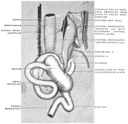 Fig. 275.
—Abdominal viscera of
Iguana tuberculata. (Columbia University
Museum, No. 1313.)
This primitive disposition of the ventral mesogastrium and the
viscera connected with the same, is well shown in some of the
lower vertebrates in whom the development never proceeds beyond
the early mammalian stages. Fig. 275 shows in profile
view from the right side the situs viscerum and peritoneum in
Iguana tuberculata.5 The two dorsal aortic roots are seen to unite
to form the main aorta, which descends between the layers of
the dorsal mesentery, sending branches to the dorsal margin of
œsophagus and stomach. From the opposite border of the stomach
the ventral mesogastrium is derived. Its dorsal segment (gastro-hepatic
omentum) connects liver and stomach, carrying between
its layers the portal vessels, hepatic artery and biliary duct. The
ventral segment of the membrane, forming the suspensory or broad
ligament, extends between abdominal wall and ventral surface of
the liver. Caudad, the lesser omentum and the suspensory ligament
are seen to have a common concave falciform edge.
The ventral abdominal vein ascends between the layers of the
suspensory ligament and near the liver becomes connected by a
large branch with the portal vein. A few smaller branches are
seen passing from the abdominal wall beyond this point. In this
reptile, therefore, the permanent vascular arrangement corresponds
to an early human embryonic stage.
The reptilian ventral abdominal vein is the homologue of the
umbilical vein of the placentalia. The large branch passing to
the portal vein represents the connection established in the human
embryo between the umbilical and portal veins. The small
branches, continuing cephalad between the mesogastric layers,
represent the temporary proximal remnants which in the human
embryo the umbilical veins form in connection with abdominal
walls. The permanent adult arrangement of this part of the
vascular system in this animal corresponds therefore to one of
the stages of development in the human embryo, as previously
indicated (cf. p. 149; Figs. 251 and 252).
PERITONEAL RELATIONS OF LIVER.
It is well to begin the study of the peritoneal connections of the
liver with the consideration of the embryonic stage shown in Fig.
273 schematically.
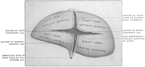 Fig. 276.
—Schematic view of embryonic liver detached from its connections, seen from
behind, with lines of peritoneal reflection.
If we imagine this embryonic liver detached from its connections
in such a manner as to leave the divided peritoneal layers
of the ventral mesogastrium as long as possible, and if we regard
the preparation from behind, the appearance of the parts could
be represented in Fig. 276.6
It will of course be seen that the area of direct adhesion to
the diaphragm, extending transversely, would separate the lesser
omentum from the suspensory ligament.
As is seen in the transection (Fig. 274), the right and left layers
of the suspensory ligament, at its attachment to the liver, turn
into the visceral peritoneum investing the organ on its ventral
and cephalic surfaces. Continuing around the borders of the
liver this visceral peritoneum then invests in like manner the
dorsal or caudal surface directed toward the stomach, until, at the
region of the future portal or transverse fissure, this visceral peritoneum
becomes in turn continuous with the two layers of the
lesser or gastro-hepatic omentum. Consequently in the embryonic
detached liver the lines of peritoneal reflection would be
nearly cruciform, the vertical limb of the cross being formed on
the cephalic surface by the two layers of the suspensory ligament,
while on the caudal surface it is formed by the layers of the lesser
omentum. The horizontal arm of the cross is formed by the
upper and lower limits of the area of diaphragmatic attachment,
along which the parietal diaphragmatic peritoneum turns
into the visceral hepatic investment (forming the two layers of the
primitive coronary ligament). In the liver shown thus schematically
from behind we would overlook the dorsal and adjoining
portions of the cephalic and caudal surfaces of the adult
human liver.
The primitive biliary duct, portal vein and hepatic artery reach
the liver between the layers of the lesser omentum. The venæ
revehentes (hepatic veins) reach the sinus venosus at the attachment
of the liver to the septum transversum (primitive diaphragm).
The first important change, resulting in a rearrangement of
these peritoneal layers, is produced by the connection of the
umbilical with the rudimentary portal vein.
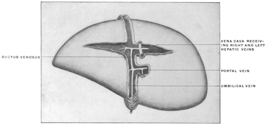 Fig. 277.
—Schematic view of embryonic liver, showing influence of vascular connections on
the arrangement of the lines of peritoneal reflection.
|
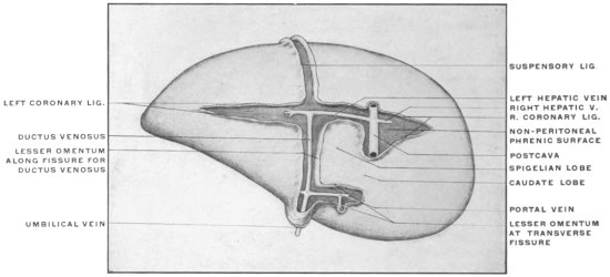 Fig. 278.
—Later stages, showing development of transverse fissure, Spigelian and caudate lobes.
|
This junction occupies a relatively wide area on the caudal
surface of the liver, and the layers of the lesser omentum are
separated somewhat at this point to accommodate the enlarging
vascular structures between them. More especially is this the
case with the right leaf of the primitive gastro-hepatic omentum.
A species of lateral diverticulum is formed by this leaf so as to
include the umbilical vein at its junction with the portal.
The membrane in the region of this diverticulum turns its surfaces
dorsad and ventrad, and its free edge toward the right (Fig. 277).
With the gradual increase in the size of the vessels, and with the
transverse position which the rotation of the stomach imparts to
the opposite border of the lesser omentum attached to the lesser
curvature, this transversely disposed portion gradually exceeds in
length and size the part of the original omentum enclosing the
umbilical vein. This vessel and the investing peritoneum become
lodged in a sagittal depression on the caudal surface of the
liver (rudimentary umbilical fissure), while the transverse portion,
developed as indicated, surrounds the structures connected with
the liver at the future transverse or portal fissure.
Schematically this rearrangement of the hepatic peritoneal lines
of reflection can be shown in Fig. 278.
It will be observed that in this way a small part of the caudal
surface of the right lobe has become partially marked off from the
remainder as a rudimentary Spigelian lobe, bounded ventrally by
the transverse fissure and lesser omentum attached to the same;
to the left by the two layers of the lesser omentum containing
the ductus venosus; while the limit cephalad is afforded by the
reflection of peritoneum from liver to diaphragm, forming part of
caudal layer of right coronary ligament. To the right this rudimentary
Spigelian surface is directly continuous with the rest of
the dorsal and caudal surface of the right lobe (Fig. 277).
Finally a definite right limit is given to the Spigelian lobe by the
increasing size of the postcava and its closer connection with the
liver. This vessel now assumes the position of the main venous
trunk entering the heart from below.
This inclusion of the vena cava in the fissure or fossa of that
name on the dorsal surface of the liver affords, so to speak, the
vertical measure of the non-peritoneal area of the liver attached
directly to the diaphragm. As the vein develops the interval
between the two layers of the right coronary ligament increases,
producing the well-known large non-peritoneal area on the dorsal
surface of the adult liver, which is directly attached to the
diaphragm.
Immediately to the left of the vena cava, however, the original
condition persists. The area of direct diaphragmatic attachment
is narrow and consequently the two layers of the coronary ligament
are close together at this point.7
In this way a species of recess (Spigelian recess or hepatic
antrum of lesser sac) is formed. A portion of the dorsal liver
surface lying just to the left of the vena cava, between it and the
ductus venosus, remains invested by peritoneum which is reflected
from the boundaries of this space to the diaphragm. This forms
the Spigelian lobe (Fig. 278).
The lobe is bounded to the right by the postcava, to the left
by the reflection of the lesser omentum to the stomach along the
fissure for the ductus venosus; cephalad the boundary is formed
by the reflection of the caudal layer of the coronary ligament to
the diaphragm.
The caudal boundary is afforded by the transverse position
which the lesser omentum has assumed in the region of the transverse
or portal fissure.
It will be seen that the original continuity of the Spigelian
lobe with the caudal surface of the right lobe is maintained by
the narrow bridge of liver tissue connecting the caudal right angle
of the rectangular Spigelian lobe with the right lobe. This narrow
isthmus, situated between vena cava dorsad and the free right
edge of lesser omentum ventrad, forms the so-called caudate lobe.
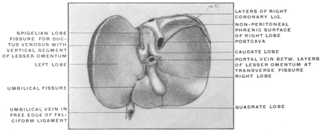 Fig. 279.
—Liver of human fœtus at eighth month. View of caudal and dorsal surfaces.
(Columbia University Museum, No. 1854.)
|
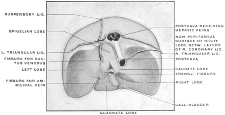 Fig. 280.
—Human fœtal liver at term, showing lines of peritoneal reflection on
cephalic, dorsal, and caudal surfaces. (Columbia University Museum, No. 1855.)
|
Fig. 279 shows a human fœtal liver at the end of the eighth
month in the view from below and behind. The original continuity
of the layers of the lesser omentum, attached along the
fissure for the ductus venosus, with the fold of the falciform ligament
occupying the umbilical fissure can still be made out for a
short distance beyond the left extremity of the transverse fissure.
The section of the lesser omentum which occupies the transverse
fissure and, including the portal vein, hepatic artery and duct
between its layers, terminates in the free right margin, is evidently
derived by a lateral extension from the right layer of the primitive
sagittal lesser omentum, whose original direction is preserved
along the fissure of the ductus venosus.
In Fig. 280 the lines of peritoneal reflection on the cephalic,
dorsal and caudal surfaces of a human fœtal liver at term are
shown.
We can now proceed to trace the reflection of the peritoneum
from the liver to adjacent structures.
Begin with the caudal layer of the coronary ligament on the
extreme right, where fusion with the corresponding cephalic
layer produces the right triangular ligament. The caudal layer
of the coronary ligament proceeds from right to left along the
caudal margin of the non-peritoneal dorsal diaphragmatic surface
of right lobe, being reflected along this line from the liver to the
adjacent portions of the diaphragm and ventral surface of right
kidney and suprarenal capsule (hepato-renal ligament). A small
cephalic part of ventral surface of right suprarenal capsule lies
above this line of reflection, is hence non-peritoneal and firmly
connected with the liver just to the left of entrance of vena
cava into the caval fissure. Continuing, the caudal layer of the
coronary ligament crosses the ventral surface of the vena cava
and turns, immediately to the left of the vein, at a right angle,
ascending to form the left boundary of the Spigelian recess, being
reflected along this line from the left margin of the caval fissure
to the pillars of the diaphragm. Arrived at the opening of the
central tendon permitting passage of vena cava into pericardium,
and at the level of the entrance of the left hepatic vein into the
cava, the peritoneum turns again at a right angle and runs from
right to left, forming the cephalic limit of the Spigelian recess.
Turning caudad along the fissure for the ductus venosus, as right
leaf of that portion of the lesser omentum which is attached to this
fissure and has preserved its sagittal position, the peritoneal line
of reflection reaches the left extremity of the portal or transverse
fissure. It now turns to the right following the fissure as the
dorsal layer of the transverse segment of the lesser omentum, and
becomes continuous, with the formation of a free right edge, with
the ventral layer of the same membrane, passing from right to
left, the two layers including between them the structures entering
and leaving the liver at the transverse fissure (portal vein,
hepatic artery, duct). Arriving at the left extremity of the transverse
fissure the ventral layer of the transverse segment of the
lesser omentum—as we practically trace it in the adult as a free
membrane—turns directly into the left leaf of the sagittal segment
attached along the fissure for the ductus venosus, and becomes
continuous along the dorsal border of the left lobe with the
caudal layer of the left coronary ligament. This direct continuity,
as just stated, exists practically in the adult. From the development
of the membrane, however, it will be seen that the ventral
layer of the transverse lesser omentum, at the left extremity of
the portal fissure, becomes continuous with the right layer of the
primitive mesogastrium enclosing the umbilical vein. After surrounding
this vein it is continued into the left leaf of the same
membrane, which in turn passes into the left layer of the portion
attached along the fissure for the ductus venosus.
This original connection can at times be traced very clearly in
young specimens (Fig. 279), and occasionally is also still evident
in the adult liver.
Usually, however, the round ligament of the adult and its
investing peritoneum is buried so deeply in the umbilical fissure,
or even bridged over in part by liver tissue, that the connection
is not evident. The ventral layer of the transverse omentum then
appears directly continuous with the left layer of the sagittal
omentum attached along the fissure for the ductus venosus.
We can sum up the facts just considered as follows:
1. The rotation of the stomach from the sagittal into the transverse
position, and the development of the umbilical and portal
veins, rearrange the original sagittal plane of the lesser omentum,
dividing it into two districts:
(a) Cephalic portion, remaining in the original sagittal plane,
follows the fissure for the ductus venosus. With the incorporation
of the Spigelian lobe in the adult dorsal or “posterior” surface
of the liver, this segment of the omentum assumes a vertical
direction, forming the left boundary of the Spigelian recess, being
reflected from the fissure for the ductus venosus to the abdominal
portion of the œsophagus and the part of the lesser curvature of
stomach adjacent to the cardia.
(b) Distal caudal portion of the lesser omentum is twisted laterally
and turned to the right by the change in the position of the
stomach and the development of the structures connected with
the liver at the transverse fissure. It is reflected from this fissure
to the distal part of the lesser curvature and to the first portion
of the duodenum. This transverse segment of the lesser omentum
is a secondary derivative from the right leaf of the primitive
membrane, produced by the enlarged area for entrance of umbilical
and portal veins at the transverse fissure. It lies ventrad of
caudal border of Spigelian lobe.
2. The distal segment of the original omentum containing the
umbilical vein (round ligament), continues imbedded in the umbilical
fissure, to the ventral margin of the liver, where it joins the
layers of the suspensory ligament passing over the cephalic surface.
3. The adult lesser omentum at the transverse fissure may be
regarded as a diverticulum of the right leaf of the primitive
embryonal sagittal omentum.
With the reduction of the umbilical vein after birth to form
the round ligament this structure becomes deeply buried in the
umbilical fissure. The ventral and dorsal layers of the lesser
omentum at the transverse fissure thus become continuous with
respectively the left and right layers of the second segment of the
omentum which ascends vertically along the fissure for the ductus
venosus.
4. The cephalic layer of the coronary ligament (Fig. 280) remains
practically in the embryonic condition. The adult convex
cephalic surface of the liver is traversed in the sagittal direction
by the suspensory ligament which connects it with the abdominal
surface of the diaphragm, and thus effects the division into right
and left lobes on the convex surface. Arrived at the dorsal
border of this surface (junction of “superior” and “posterior”
surfaces) the right and left leaves of the falciform ligament turn
at right angles into the cephalic layer of the right and left coronary
ligament, which at each extremity meet the right and left
caudal layers to form the triangular ligaments. It will thus be
seen that the apparent irregularity in the relative arrangement of
the s. c. “upper” and “lower” layers of the coronary ligaments,
produced by the Spigelian recess, is only a difference in the interval
between the two layers, caused by the vertical extent of the
non-peritoneal direct diaphragmatic attachment of the right lobe
to the right of the vena cava.
Comparative Anatomy of Spigelian Lobe and Vena Cava in the Cat.—The
lines of peritoneal reflection in the cat’s liver and the arrangement
of the Spigelian lobe and recess are seen in Fig. 281, taken
from a preparation hardened in situ.
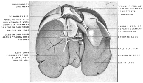 Fig. 281.
—Liver of cat, hardened in situ. (Columbia University Museum, No. 1836.)
|
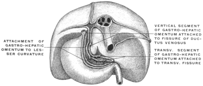 Fig. 282.
—Dorsal view of human liver and stomach in fœtus at term, showing
lines of hepatic and gastric attachment of lesser omentum.
|
Compared with the human liver it will be noted that the area
of diaphragmatic adhesion is much less developed. The dorsal
surface of the right lobe to the right of the postcava is peritoneal,
there being no extension laterad of the right coronary and
triangular ligaments. The postcava enters the liver in a special
prolongation of the liver substance (caval lobe).
The boundaries of the Spigelian recess and the lines of attachment
of the gastro-hepatic omentum correspond to the human
arrangement.
RELATION OF THE HEPATIC PERITONEUM TO THE
“LESSER SAC.”
Foramen of Winslow.—We have previously seen that the rotation
of the stomach and the further growth of the dorsal mesogastrium
lead, in the first instance, to the formation of the “lesser peritoneal
cavity.” This cavity is in fact primarily the retrogastric space
created by the transverse position of the stomach, augmented by
the cavity of the omental bursa developed from the dorsal mesogastrium.
We have now to consider the additional boundaries of this
space contributed by the peritoneal connection of the lesser
curvature with the liver.
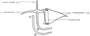 Fig. 283.
—Schema of lines of reflection of peritoneum on dorsal surface of liver and in the
formation of the gastro-hepatic omentum.
A B, transverse section of lesser omentum attached to
transverse fissure of liver and to pyloric section of lesser curvature (
A' B');
B C, vertical section
of lesser omentum passing between fissure of ductus venosus and cardiac section of lesser curvature
of stomach (
B' C');
C D, line of reflection of peritoneum from cephalic border of Spigelian
lobe to diaphragm;
D E, line of reflection of peritoneum from right border of Spigelian lobe to
left margin of postcava and diaphragm.
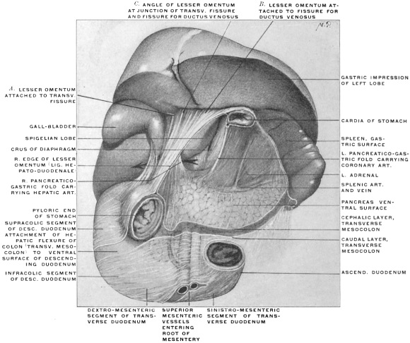 Fig. 284.
—Portion of abdominal viscera of adult human subject, hardened
in situ. (Columbia
University, Study Collection.) The segment of stomach between cardiac and pyloric orifices
has been removed, dividing the lesser omentum to this extent, but leaving the right extremity of
the membrane (lig. hepato-duodenale) intact. Behind this portion the arrow passes through the
foramen of Winslow.
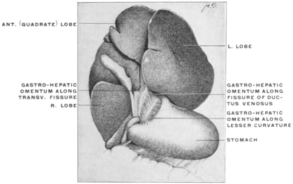 Fig. 285.
—Liver and stomach of
Macacus pileatus. (Columbia University,
Study Collection.)
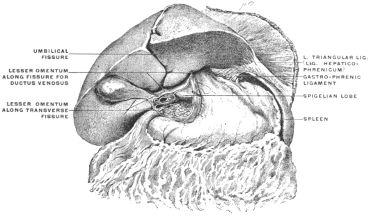 Fig. 286.
—Abdominal viscera of adult human subject, hardened
in situ; with liver lifted up
after incision of the gastro-hepatic omentum. (Columbia University Museum, No. 1845.)
The lesser omentum follows, of course, along its gastric attachment
to the lesser curvature the general direction of the stomach,
passing from the cardia transversely downwards and to the right.
We distinguish the two layers of the adult membrane as ventral
and dorsal, which meet in the free right edge and include between
them the main structures entering and leaving the liver at the
transverse fissure, viz.: the portal vein, hepatic artery and bile-duct.
The lesser omentum therefore prolongs the plane of the stomach
cephalad towards the liver and thus forms the continuation of the
ventral boundary of the lesser peritoneal sac. We can now consider
the line of its hepatic attachment in the light of the facts
previously adduced, and combine the same with the line of gastric
attachment to the lesser curvature. Fig. 282 shows the fœtal
liver and stomach in their relative position in the dorsal view,
and Fig. 283 gives the lines of the peritoneal reflections. The
vertical segment of the omentum, occupying the fissure for the
ductus venosus, passes to the cardiac part of the lesser curvature,
its ventral layer covering the ventral and left side of the œsophagus,
while its dorsal layer passes to the dorsal and right side of
the œsophagus at its entrance into the stomach. The transverse
segment of the omentum, attached on the liver to the portal or
transverse fissure, accedes to the pyloric part of the lesser curvature.
Of course the ventral and dorsal layers of the omentum are
continuous with the serous visceral investment of the ventral and
dorsal surfaces of the stomach.
Fig. 284 shows this right-angled course of the lesser omentum
at the hepatic line of attachment in a preparation of the abdominal
viscera hardened in situ, with the segment of the stomach
between the cardiac and pyloric orifices removed. The arrow is
passed behind the right free edge of the lesser omentum. This
portion of the membrane is still intact, not having been disturbed
by the removal of the body of the stomach, and includes between
its layers the structures connected with the liver at the transverse
fissure (duct, hepatic artery and portal vein). The lesser omentum
is seen to be attached to the liver along the transverse fissure
(Fig. 284, A) and along the fissure for the ductus venosus (Fig.
284, B), constituting the transverse and vertical segments above
referred to, which pass into each other at the angle of junction
between the transverse fissure (left end) and the fissure for the
ductus venosus (Fig. 284, C). The caudal and left border of the
Spigelian lobe is exposed by the division of the omentum, and the
extent of the Spigelian or hepatic recess of the lesser peritoneal
sac is shown. Fig. 285 shows the liver, stomach and lesser omentum
of a Macaque monkey hardened in situ, and demonstrates
still more conclusively that the uniform curve of the omentum
along the lesser curvature of the stomach becomes a broken line
at the hepatic attachment, the angle being placed at the left end
of the transverse fissure at the point where the same encounters
the fissure for the ductus venosus.
In Fig. 286 finally the hardened abdominal viscera of an adult
human subject are shown in the ventral view with the lesser omentum
incised. The cut through the lesser omentum exposes the
hepatic recess of the lesser peritoneal cavity immediately to the
left of the foramen of Winslow. Toward the right free margin of
the omentum the divided portal vein, hepatic artery and duct are
seen between the layers of the omentum imbedded in the pancreas
and coursing behind the first portion of the duodenum on
their way to the transverse fissure.
To the left of these structures the omental tuberosity of the
pancreas projects above the level of the lesser curvature under
cover of the secondary parietal peritoneum forming the dorsal
wall of the lesser sac, while the lower edge of the Spigelian lobe
appears in the upper angle of the incision.
If we remember that the liver is itself welded to the diaphragm
between the layers of the coronary ligament (Fig. 280), it will become apparent that the serous surface of the Spigelian lobe forms
part of the ventral wall of a peritoneal recess situated behind the
lesser omentum, between this membrane and the diaphragm.
Access to this recess, without the division of peritoneal layers, can
only be obtained by passing from right to left, along the caudate
lobe, between the vena cava behind, covered by parietal peritoneum,
and the free right edge of the lesser omentum in front.
(In the reverse direction of the arrow shown in Fig. 284.) This
hepatic or Spigelian recess of the lesser peritoneal cavity has categorically
the following boundaries (Figs. 282 and 283):
Dorsal: Parietal peritoneum, reflected along the line CD, from
the caudal layer of the coronary ligament to the diaphragm.
Ventral: Visceral peritoneum investing the Spigelian lobe and
the gastro-hepatic omentum.
Right: Reflection of peritoneum along the line DE (caval fissure)
to become the parietal peritoneum covering the diaphragm.
Left: Right layer of lesser omentum, reflected along the fissure
for the ductus venosus (CB) to the cardiac portion of the lesser
curvature, continuous with the dorsal layer of the lesser omentum
reflected from the transverse fissure to the pyloric segment of
the lesser curvature (AB).
We will presently see that certain relations of the vessels connected
with the liver at the transverse fissure and of the duodenum
prevent the finger, when passed from right to left behind
the free right edge of the lesser omentum and along the caudate
lobe of the liver, from proceeding downward at this point. A
narrow channel of communication is thus formed between the
Spigelian recess and rest of the lesser sac on the one hand, and the
general greater peritoneal cavity on the other. This channel is
the so-called foramen of Winslow.
Having once passed this narrow space the finger will be in
the Spigelian recess and can palpate its boundaries. Further
progress cephalad and to the right is barred by the diaphragmatic
adhesions of the liver just detailed. But in the direction downward
behind the lesser omentum and along the dorsal surface of
the stomach, as well as to the left toward the spleen the excursion
is limited only by the length of the examining finger.
After opening the abdominal cavity of the human adult, elevating
the liver and depressing the stomach, the hepatic attachment
of the lesser omentum can be traced as already described. It will
then be observed that the gastric attachment of the membrane
lies in one plane following the lesser curvature while the hepatic
attachment forms a broken line, with the angle situated at the
left extremity of the transverse fissure. The vertical segment of
the hepatic attachment, occupying the fissure for the ductus
venosus, turns at this angle into the transverse segment which
follows the transverse fissure to its right extremity where the two
layers pass into each other around the right free omental margin
(hepato-duodenal ligament). Consequently we overlook, in an
abdominal cavity thus exposed, the entire caudal surface of the
liver, including the caudal surfaces of right, left, and quadrate
lobes. The junction of right and caudate lobes can be seen
between vena cava and right edge of the omentum, or rather, it
can be felt at this point. But the Spigelian lobe, turning its surface
dorsad against the parietal peritoneum covering the diaphragm,
forms part of the “posterior” liver surface and is not
visible, although—as just stated, it can be palpated by passing
the finger through the foramen of Winslow. The Spigelian lobe
cannot be overlooked in its entire extent until the liver is removed
from the body and regarded from behind. The caudal edge (continuation
of its right angle into the caudate lobe and papillary
tubercle) can be seen by tearing through the layers of the lesser
omentum and lifting the liver up forcibly (Fig. 286).
Caudal Boundary of Foramen of Winslow.—We have above referred
to the fact that the finger introduced through the foramen
of Winslow meets in this canal with resistance if an attempt is
made to pass downwards. After passing this constricting point
the free excursion into the Spigelian recess and behind the omentum
and stomach and toward the spleen can be performed.
In considering the elements which produce this narrowing of
the communication between the two peritoneal sacs at the foramen
of Winslow we have to deal with two factors, one primary and
constant, the other secondary and inconstant.
1. The first of these is afforded by the arrangement of the
arterial vessel supplying the liver. The hepatic artery is a
branch of the cœliac axis, furnishing arterial blood to the liver
tissues and supplying, in addition, branches to the stomach, duodenum
and pancreas.
This vessel is, of course, placed primarily, like all other arterial
branches supplying the alimentary tract, between the layers of
the primitive dorsal mesentery. Originally the vessel supplies
the distal (pyloric) portion of the stomach along its dorsal attached
border (subsequently the greater curvature) corresponding
to the adult gastro-epiploica dextra of the hepatic (gastro-duodenalis).
It likewise gives branches to the adjacent pyloric portion of the
duodenum and the pancreas, as that gland develops from the intestine,
corresponding to the adult superior pancreatico-duodenal
branch, and to the ventral border (lesser curvature) of stomach,
corresponding to the adult pyloric branch of the hepatic.
With the development of the liver from the duodenum arterial
branches derived from this primitive gastro-duodenal vessel pass
to the sprouting hepatic cylinders by continuing around the duodenum,
beneath its serous investment, to reach the interval between
the two layers of the ventral mesogastrium, in which the
liver develops, near the free margin of this membrane.
After the rotation, which turns the right side of the stomach,
duodenum and mesoduodenum dorsad, the branch which passes
over the dorsal surface of the duodenum to reach the liver becomes
more favorably situated and develops into the main hepatic
artery which reaches the liver at the transverse fissure between
the folds of the lesser omentum. The original right side of the
duodenum, now turned dorsad, adheres to the parietal peritoneum.
The hepatic artery which reached the liver by passing
over this surface of the duodenum, beneath its visceral serous covering,
becomes imbedded in connective tissue by the adhesion of the
visceral duodenal and the primitive parietal peritoneum. Hence
in the adult the hepatic artery courses imbedded in the connective
tissue which binds the duodenum to the abdominal background
to reach the interval between the two omental layers which carry
it to the transverse fissure.
The hepatic artery, therefore, derived from one of the primitive
intestinal branches (gastro-duodenal) is, notwithstanding its
hidden position in the adult, originally situated between the layers
of the free primitive dorsal mesogastrium.
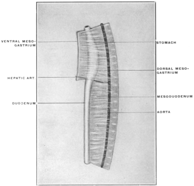 Fig. 287.
—Primitive dorsal and ventral mesogastrium with course of hepatic
artery.
It now becomes necessary to regard the development of the
great omentum from the primitive dorsal mesogastrium in relation
to this course of the hepatic artery. We have seen that
the great omentum and the cavity of the omental bursa is produced
by the extension of the dorsal mesogastrium to the left and
caudad, subsequent to the rotation of the stomach. The splenic
artery and the left gastro-epiploic branch pass from the cœliac
axis to the left between the layers of the mesogastrium, as previously
seen (Figs. 291 and 292).
The hepatic artery, however, is so to speak placed on the border
line between the portion of the primitive mesentery which, as
dorsal mesogastrium, is to turn to the left and caudad to form the
great omentum, and the portion which, as mesoduodenum, turns
to the right and passes to the duodenal loop (Fig. 287).
In the further course of development the dorsal mesogastrium
grows more and more, forming the omental bag, while the mesoduodenum
on the other hand becomes anchored early and obliterated
as a free membrane by adhesion of its original right layer
to the primitive parietal peritoneum. The hepatic artery runs on
the line dividing these two different mesenteric segments. We can
imagine, so to speak, that the redundant growth of the omentum
to the left and caudad, takes place over the hepatic artery as a
resistant support (Figs. 288 and 289). Cephalad of the hepatic
artery is the developing omentum, caudad of the vessel the mesoduodenum.
The artery follows the cephalic limit of the mesoduodenum
and becomes, as stated, adherent to the abdominal
background in the segment between its origin from the cœliac
axis and the point where, after having crossed the dorsal surface
of the duodenum, it enters the right edge of the lesser omentum
on its way to the liver.
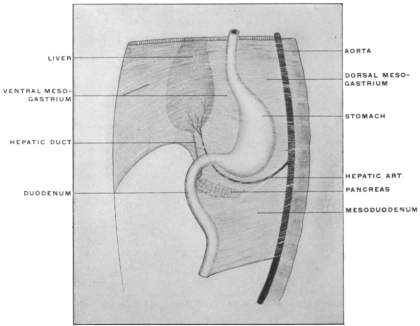 Fig. 288.
—The liver divides the ventral mesogastrium into a dorsal segment,
the gastro-hepatic or lesser omentum, and a ventral segment, the suspensory
or falciform ligament of the liver.
|
|
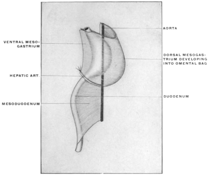 Fig. 289.
—Stages in the development of the dorsal mesogastrium (omental bursa)
and mesoduodenum to show relation of hepatic artery to these two segments of the
primitive common dorsal mesentery.
|
Pancreatico-gastric Folds.—If we open the lesser peritoneal cavity
by dividing the gastro-hepatic omentum and look into the background
of the retro-omental space, we will see a fold of the
secondary lining parietal peritoneum (derived from the mesogastrium),
which can be traced from the cephalic border of the pancreas
to the pyloric extremity of the stomach. This fold carries
the hepatic artery to the lesser omentum behind the first portion
of the duodenum, and is called the right or main pancreatico-gastric
fold. A similar fold, further to the left, carries in a like
manner the coronary artery of the stomach to the cardiac end of
the lesser curvature. This fold forms the left or secondary pancreatico-gastric
fold. Between the two folds the caudal margin
of the Spigelian lobe projects into the lesser cavity.
The appearance of the two pancreatico-gastric folds in the
adult human subject is well seen in Fig. 284.
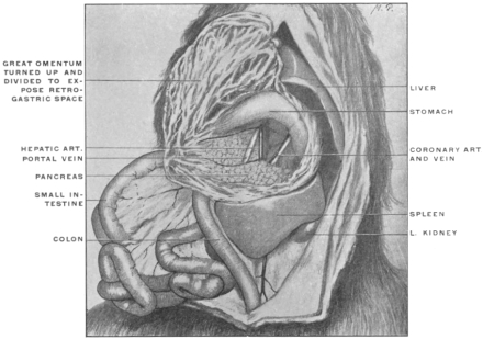 Fig. 290.
—Abdominal viscera of
Nasua rufa, brown coaiti, with stomach turned up
and great omentum divided. (From a fresh dissection.)
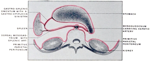 Fig. 291.
—Schematic transection through foramen of Winslow before adhesion of dorsal mesogastrium
and mesoduodenum to parietal peritoneum.
|
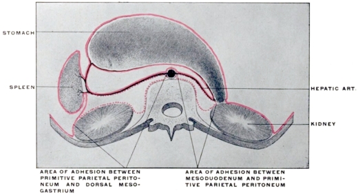 Fig. 292.
—The same section after the adult conditions have been established by adhesion.
|
Fig. 290 shows the abdominal cavity of Nasua rufa, with great
omentum divided to bring into view the vessels passing from
cœliac axis to liver and stomach and elevating the retrogastric
parietal peritoneum to produce the pancreatico-gastric folds.
(The course of the hepatic artery from cœliac axis to liver in
the dorsal view in the cat is seen in Fig. 223.)
Figs. 291 and 292 represent schematically cross-sections directly
through the foramen of Winslow, showing the method by means
of which the hepatic artery reaches the upper border of the duodenum
and the effect of the adhesion of duodenum and mesoduodenum
upon the disposition of the vessel.
The coronary artery, like the splenic, is at first situated between
the layers of the dorsal mesogastrium (vertebro-splenic segment).
Like the splenic the coronary artery becomes anchored to the
abdominal background and placed secondarily behind the parietal
peritoneum of the lesser sac by the adhesion of this mesogastric
segment to the primitive parietal peritoneum. To reach the
lesser curvature at the cardia and to run thence from left to right
along the lesser curvature between the layers of the gastro-hepatic
omentum, the vessel raises the investing parietal peritoneum
(originally the right leaf of the dorsal mesogastrium) into a crescentic
fold, extending between its origin from the cœliac axis at
cephalic margin of pancreas and the beginning of the lesser
curvature of the stomach. Hence this fold is called the left pancreatico-gastric
fold. (Seen well in Fig. 284.)
In the next place it must be borne in mind that the relation of
the primitive hepatic artery to the vascular supply of the stomach,
pancreas and duodenum produces a permanent shortening of the
primitive mesentery at this point. This result is indicated in the
schematic figures 287, 288 and 289.
In the original condition the dorsal mesentery, passing to a
practically straight intestinal tube, is of uniform sagittal measure
(Fig. 287).
As development proceeds, and as the liver grows from the
duodenum, the hepatic artery develops from the primitive pyloric
vessel as above indicated. This vessel, assuming greater importance
with the rapid growth of the liver, is not lengthened out as
happens with the remaining purely intestinal branches which
follow the increase in the length of the intestinal canal. The
hepatic artery, therefore, will mark the point where the original
short sagittal extent of the primitive mesentery will tend to be
preserved. Cephalad of this point the dorsal mesogastrium grows
out into the great omentum (Figs. 288 and 289); caudad of the
same point the membrane, in following the development of the
intestine, becomes drawn out into the permanent mesentery and
mesocolon.
The hepatic artery, in addition, marks the cephalic limit of
the adhesion which anchors the duodenum and mesoduodenum
to the parietal peritoneum. Consequently in the adult the vessel
courses in as direct a manner as possible, taking the shortest course
from the cœliac axis to the liver, passing dorsad of the duodenum
and giving what now appear as secondary branches to
supply the intestine, the stomach and pancreas (pyloric and
gastro-duodenal arteries (pancreatico-duod. superior and gastro-epiploica
dextra)).
Even if no fixation of the duodenum and mesoduodenum
takes place this course of the hepatic artery will produce a constricted
passage between the liver (caudate lobe) cephalad, abdominal
parietes and aorta dorsad, lesser omentum and pyloric duodenum
ventrad, and hepatic artery caudad. This passage leading
from the general peritoneal cavity into the retrogastric space is the
primitive foramen of Winslow. This condition is well represented
in the abdominal cavity of some of the lower mammalia, in
which duodenum and mesoduodenum remain permanently free.
Fig. 293 shows a view of the abdominal cavity from the right
side in a specimen of the ant-eater, Tamandua bivittata.
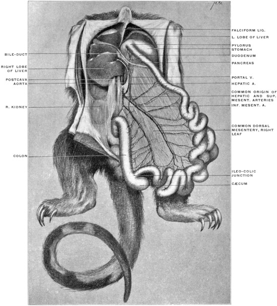 Fig. 293.
—Abdominal viscera of
Tamandua bivittata, the little ant-eater, with the intestines
turned downward and to the left. (From a fresh dissection.)
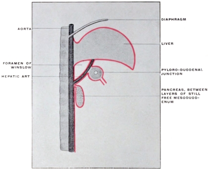 Fig. 294.
—Schematic sagittal section through foramen of Winslow before fixation
of pancreas by adhesion of mesoduodenum.
|
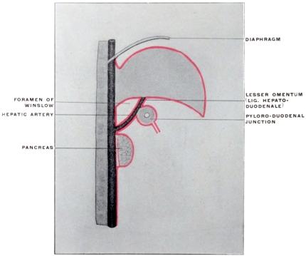 Fig. 295.
—The same section after adhesion of mesoduodenum and pancreas. The
pancreas appears secondarily retroperitoneal, after adhesion of apposed surfaces of mesoduodenum
and primitive parietal peritoneum over dotted area, producing fixation of
dorsal surface of pancreas.
|
The right kidney is seen in the background, covered by the
parietal peritoneum. The duodenum and mesoduodenum are
free and can be turned toward the median line. The opening of
the foramen of Winslow leading into the retrogastric space is seen
between the liver cephalad, kidney and vena cava dorsad, lesser
omentum and pyloric extremity of the stomach ventrad, and a
fold of peritoneum carrying the hepatic artery caudad. Exactly
similar conditions prevail in the cat and in many other mammals.
It will be seen in all these instances that neither portal vein
nor bile-ducts limit the foramen caudad. These structures can be
lifted up and turned toward the median line with the free duodenum
and mesoduodenum. But the hepatic artery must pass
to the liver from the retroperitoneal cœliac axis. In doing this
the vessel traverses the cephalic border of the pancreas, and the
pyloric extremity of the stomach and duodenum, to reach the
lesser omentum which conveys it to the liver.
Consequently there must always be a narrow peritoneal neck
between the liver cephalad, aorta dorsad, hepatic artery caudad,
and pyloric extremity of stomach and duodenum together with
the lesser omentum ventrad. It should be remembered that the
vessel which extends after the development of the liver into the
lesser omentum as the hepatic artery, was originally destined for
the supply of these latter structures. In the adult these primary
embryonic terminal branches to the intestine appear as secondary
branches derived from the hepatic as the main vessel. Their
origin, however, serves to keep the beginning of the small intestine
in comparatively close connection with the hepatic artery
which courses over the dorsal surface of the duodenum to reach
the liver. The narrow space thus left between aorta, hepatic
artery, duodenum, lesser omentum and liver forms the framework
of the foramen of Winslow and appears always as a confined
and narrow channel. This relation is shown in the accompanying
schematic Figs. 294 and 295 which represent a sagittal section
through the foramen. This primitive foramen is thus bounded
cephalad by the liver (caudate lobe, connecting Spigelian and right
lobes), ventrad by the first portion of the duodenum and the
lesser omentum, with hepatic artery behind the intestine and
between the omental layers; dorsad by the abdominal background
and large retroperitoneal vessels, and caudad by the
cœliac axis and beginning of the hepatic artery.
2. In the forms which possess in the adult an adherent duodenum
and mesoduodenum, as in man, the foramen of Winslow
obtains a secondary caudal limit by the agglutination of the
descending duodenum and the parietal prerenal peritoneum.
This is the secondary and inconstant factor referred to above in
the caudal boundary of the foramen. The result of this anchoring
of duodenum and mesoduodenum is to bring the margin of
the foramen further to the right and to bury the hepatic artery
still further from view. Thus in the adult human subject the
structures bounding the foramen at the margin of the entrance
into the narrow channel would be above caudate lobe of liver,
behind postcava, below duodenum adherent to ventral surface
of right kidney, in front first portion of duodenum and
lesser omentum. The hepatic artery will be felt on introducing
the finger through the foramen in its original position, but it will
be seen that the actual boundaries of the foramen have been
moved so to speak a little further to the right by the duodenal
adhesion.
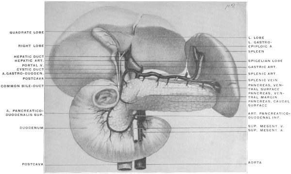 Fig. 296.
—Dissection of adult liver, pancreas, spleen, and duodenum, with vessels, to show
structures concerned in the formation of the foramen of Winslow. (Columbia University, Study
Collection.)
Fig. 296 shows a complete dissection of the adult human viscera
and vessels concerned in the formation of the foramen, hardened
in situ.
The stomach is removed, dividing of course the coronary artery
and vein and the left gastro-epiploic artery. The portal vein,
hepatic artery and bile-duct are seen entering and leaving the
liver at the transverse fissure. Behind them and to the right the
vena cava enters the liver. The hepatic artery distributes its pancreatico-duodenal
branches to the duodenum and pancreas. The
left angle of the Spigelian lobe and the fissure for the ductus
venosus appear to the left of the portal vein and hepatic artery.
The right angle of the Spigelian lobe and its continuation into
the right lobe by means of the caudate lobe is hidden by the
structures occupying the transverse fissure. We would enter the
beginning of the foramen of Winslow by passing between the
vena cava behind, the structures in the transverse fissure (portal
vein, hepatic artery and duct) in front, caudate lobe of liver
above and duodenum below, the latter in the undisturbed condition
of the parts adherent to the right kidney. Continuing to
the left the finger would pass between aorta behind, cœliac axis
and hepatic artery below and in front, and liver above. These
structures bound the permanent and primary narrow channel
of communication between the retrogastric or lesser peritoneal
space and the general peritoneal cavity, which exists even if a free
duodenum and mesoduodenum allow us to lift the intestine away
from vena cava and right kidney.
The main facts pertaining to the structure of the lesser peritoneal
sac and its connection with the greater peritoneal cavity by
means of the foramen of Winslow may be summed up as follows:
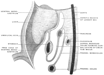 Fig. 297.
—Schematic sagittal section of the ventral and dorsal mesogastria and
epiploic bursa in a human embryo of eight weeks. (Modified from Kollmann.)
|
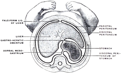 Fig. 298.
—Transection of human embryo of 3 cm., vertex-coccygeal measure.
(Kollmann.)
|
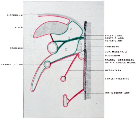 Fig. 299.
—Schematic sagittal section of abdomen to illustrate the intestinal branches
of the abdominal aorta. The gastric and hepatic arteries are shown for the sake of
convenience as arising together from the cœliac axis (
B), hence the left and right gastro-pancreatic
folds carrying these vessels appear fused at their beginning, separating the
hepatic recess of the lesser peritoneal sac (
A) from the cavity of the omental bursa (
C).
The mesogastrium as a whole, expanding originally in the
sagittal plane in a fan-shaped manner between the vertebral
column and the ventral abdominal wall, from the level of the
umbilicus to the septum transversum (diaphragm), divides the
cephalic part of the abdominal cavity into a symmetrical right
and left half.
Figs. 172 and 273 represent the membrane as seen in a profile
view from the left side. We distinguish the segment dorsad of the
stomach as the dorsal mesogastrium, directly continuous with the
remaining segments of the common primitive dorsal mesentery,
while the portion ventrad of the stomach forms the ventral mesogastrium
in which the liver develops. The segment of the ventral
mesogastrium between liver and stomach becomes the lesser
or gastro-hepatic omentum, while that between liver and ventral
abdominal wall forms the falciform or suspensory ligament.
A transection, showing the dorsal and ventral mesogastrium at
the level of the fundus of the stomach, is given in Fig. 298. The
mesogastria are here seen to be short, while in the schematic Figs.
291 and 292 the membrane is, for the sake of distinctness, represented
as being of considerable extent.
The ventral mesogastrium surrounding the liver and stomach
extends caudad to include the first portion of the duodenum.
Beyond this point it terminates in a thickened free edge which
includes the umbilical vein. This vein extends from the umbilicus
to the transverse fissure of the liver (Fig. 297), lying within
the umbilical fissure on the caudal surface of the gland.
At the point where the vein enters the liver the thickened
margin of the ventral mesogastrium is continued, as ligamentum
hepato-duodenale, to the upper part of the duodenum and forms
the ventral boundary of the foramen of Winslow. Between
the layers of the mesogastrium which meet in this margin are
situated the portal vein, biliary duct and hepatic artery, together
with the nerves and lymphatics of the liver.
The mesogastrium originally divided the abdominal cavity
between umbilicus and diaphragm into symmetrical right and
left halves of equal size and extent. This early symmetrical
arrangement becomes disturbed about the seventh week by the
rotation of the stomach and the resulting altered course of the
mesogastrium, which render the two original equal halves of the
abdominal cavity unequal and asymmetrical. The original
right half becomes placed behind the stomach and is converted
into a blind sac with its opening directed to the right.
The communication of the general abdominal cavity with the
retrogastric space by means of this channel is still wide in the
embryo, but gradually becomes narrowed in the course of further
development to form the foramen of Winslow. This opening is
situated between the hepato-duodenal ligament and the parietal
peritoneum covering the vena cava. It is constricted from below
by the curve of the hepatic artery as this vessel passes from the
cœliac axis to reach the liver at the transverse fissure between the
layers of the lesser omentum.
The earlier developmental stages of the higher mammalian
embryos are in general well illustrated by the permanent adult
conditions found in some of the lower vertebrates, in which
development does not proceed beyond the primitive condition.
In reptiles, birds and mammals the epiploic bursa is generally
formed, while in amphibia the dorsal mesogastrium is very short
and connects the stomach directly to the dorsal midline of the
abdominal cavity without forming the sac-like extension of the
great omentum.
The dorsal mesogastrium with the stomach, and the ventral
mesogastrium including the liver between its layers, divides in
these animals the cephalic part of the body cavity into two
halves, corresponding to the earlier embryonic stages in man and
in the higher mammalia.
The foramen of Winslow of the higher forms appears in the
lower vertebrates as the wide-open space leading from below into
the right half of the cœlom cavity. The dorsal mesogastrium
remains short, not forming the pouch-like extension of the great
omentum. The stomach retains more or less its primitive vertical
position without rotation or elevation of the pyloric extremity, and
the intestinal canal is simple, short and comparatively straight.