The Anatomy of the Domestic Fowl
Transcriber’s Note:
New original cover art included with this eBook is granted to the public domain.
FOREWORD
Advanced work in the study of poultry husbandry is now being done in this country, to a greater or less extent, at all the two score and more Agricultural and other Colleges and Experiment Stations. From these institutions comes the demand for a text-book on the Anatomy of the Domestic Fowl. No complete text-book on the subject, up to the present, has existed. It is with the hope of meeting the demand that this book is published.
In supplementing the information gathered from the books and articles listed in the bibliography, the author has spent much time and effort in obtaining the matter here presented. As this, however, is the initial complete text on the subject necessarily much remains to be added and corrected. The author would welcome suggestions and corrections from any one into whose hands the book may come.
CONTENTS
| OSTEOLOGY | ||||
|---|---|---|---|---|
| Page | ||||
| Osseous Structure | 17 | |||
| Classification of Bones | 20 | |||
| Composition of Bone | 21 | |||
| Skeleton of the Fowl | 21 | |||
| Axial Skeleton | 23 | |||
| Cranium and Face | 23 | |||
| Occipital | 25 | |||
| Ethmoid | 27 | |||
| Sphenoid | 28 | |||
| Frontal | 29 | |||
| Parietal | 29 | |||
| Temporal | 29 | |||
| Premaxilla | 31 | |||
| Maxilla | 32 | |||
| Nasal | 32 | |||
| Lacrimal | 32 | |||
| Palatine | 33 | |||
| Pterygoid | 33 | |||
| Zygomatic | 33 | |||
| Vomer | 33 | |||
| Jugal and Quadrato-jugal | 33 | |||
| Quadrate | 34 | |||
| Inferior Maxilla | 34 | |||
| Turbinate Bones | 34 | |||
| Hyoid | 35 | |||
| Vertebral Column | 35 | |||
| Cervical Vertebræ | 35 | |||
| Atlas | 37 | |||
| Axis | 37 | |||
| Dorsal Vertebræ | 39 | |||
| Lumbo-sacral Vertebræ | 39 | |||
| Coccygeal Vertebræ | 41 | |||
| Ribs | 41 | |||
| Sternum | 42 | |||
| Appendicular Skeleton | 43 | |||
| Shoulder Girdle | 43 | |||
| Fore Limb | 44 | |||
| Pelvic Girdle | 48 | |||
| Hind Limb | 50 | |||
| ARTHROLOGY | ||||
| Kinds of Joints | 56 | |||
| Movements of Joints | 56 | |||
| Ligamentous Structure | 57 | |||
| 12Ligaments of: | ||||
| Ear | 58 | |||
| Jaw | 58 | |||
| Vertebræ | 58 | |||
| Ribs | 59 | |||
| Sternum | 59 | |||
| Shoulder-joint | 61 | |||
| Elbow-joint | 61 | |||
| Carpal Joint | 62 | |||
| Finger | 64 | |||
| Pelvis | 65 | |||
| Hip-joint | 65 | |||
| Knee-joint | 65 | |||
| Tibio-metatarsal Joint | 69 | |||
| Toes | 69 | |||
| MYOLOGY | ||||
| Kinds and Structure of Muscles | 70 | |||
| Fascia | 71 | |||
| Muscular Nomenclature | 71 | |||
| True Dermal Muscles | 71 | |||
| Dermo-osseous Muscles | 73 | |||
| Muscles of the Head | 76 | |||
| Muscles of the Tongue | 79 | |||
| Cervical Muscles | 81 | |||
| Muscles of the Air Passages | 86 | |||
| Superior Larynx | 86 | |||
| Inferior Larynx | 86 | |||
| Sternal Muscles | 90 | |||
| Abdominal Muscles | 90 | |||
| Dorsolumbar Muscles | 92 | |||
| Coccygeal Muscles | 93 | |||
| Costal Muscles | 96 | |||
| Anterior Pectoral Muscles | 99 | |||
| Pectoral Muscles | 100 | |||
| Muscles of the Scapular Region | 101 | |||
| Muscles of the Brachial Region | 104 | |||
| Muscles of the Forearm and Hand | 105 | |||
| Digital Muscles | 109 | |||
| Muscles of the Posterior Limb | 111 | |||
| Tibial Group of Muscles | 117 | |||
| Muscles of the Eye | 124 | |||
| Muscles of the Ear | 128 | |||
| Functions of Muscles | 128 | |||
| SPLANCHNOLOGY | ||||
| Digestive Apparatus | 135 | |||
| Mouth | 135 | |||
| Tongue | 139 | |||
| Pharynx | 141 | |||
| Glands Adjacent to Mouth and Pharynx | 141 | |||
| Esophagus | 142 | |||
| Crop | 142 | |||
| Stomach | 143 | |||
| Proventriculus | 143 | |||
| Gizzard | 145 | |||
| Small Intestine | 147 | |||
| 13 | Large Intestine | 150 | ||
| Cæca | 151 | |||
| Cloaca | 151 | |||
| Course of the Food | 152 | |||
| Accessory Organs of Digestion | 152 | |||
| Liver | 153 | |||
| Pancreas | 155 | |||
| Spleen | 155 | |||
| Abdominal and Pelvic Cavities | 156 | |||
| Peritoneum and Mesentery | 156 | |||
| Relations of the Visceral Organs of the Domestic Fowl | 158 | |||
| Relations of the Visceral Organs of the Baby Chick | 165 | |||
| Uro-genital System | 169 | |||
| Urinary Apparatus | 169 | |||
| Kidneys | 169 | |||
| Ureter | 174 | |||
| Male Generative Organs | 175 | |||
| Testicles | 175 | |||
| Vas Deferens | 178 | |||
| Female Generative Organs | 178 | |||
| Egg | 180 | |||
| Oviduct | 182 | |||
| Parts of the Oviduct | 183 | |||
| Ligaments of the Oviduct | 188 | |||
| Ductless Glands | 190 | |||
| Thyroid Gland | 190 | |||
| Thymus Gland | 190 | |||
| Adrenal Gland | 191 | |||
| Respiratory Apparatus | 193 | |||
| Nostrils and Nasal Chambers | 193 | |||
| Pharynx and Superior Larynx | 193 | |||
| Trachea | 194 | |||
| Inferior Larynx | 194 | |||
| Bronchi and the Lungs | 195 | |||
| Air-sacs | 199 | |||
| ANGIOLOGY | ||||
| Circulatory Apparatus | 206 | |||
| Heart | 206 | |||
| Structure of the Heart | 207 | |||
| Blood-vessels | 209 | |||
| Structure of the Capillaries and Arteries | 209 | |||
| Structure of the Veins | 211 | |||
| Arterial Trunks | 211 | |||
| Branches of: | ||||
| Arteria Brachiocephalica | 213 | |||
| Arteria Carotis Cerebralis Interna | 214 | |||
| Arteria Carotis Externa | 214 | |||
| Arteria Carotis Facialis | 214 | |||
| Aorta Posterior | 224 | |||
| Venous Trunks | 233 | |||
| Branches of: | ||||
| Venæ Cavæ Anteriores | 234 | |||
| Vena Jugularis | 237 | |||
| Vena Occipitalis Externa | 239 | |||
| Venous Sinuses of the Head | 239 | |||
| Veins of the Brain Cavity | 241 | |||
| Veins of the Neck | 242 | |||
| 14 | Branches of the Vena Subclavia | 243 | ||
| Veins of the Dorsal Region | 243 | |||
| Veins of the Thorax | 244 | |||
| Veins of the Fore Limb | 245 | |||
| Branches of the Iliaca | 246 | |||
| Branches of the Vena Iliaca Interna | 247 | |||
| Posterior Vena Cava | 247 | |||
| Veins of the Posterior Extremity | 248 | |||
| Branches of the Vena Cava Posterior | 248 | |||
| Veins of the Caudal Region and Pelvic Cavity | 250 | |||
| Veins of the Truncus Vena Iliaca Communis | 252 | |||
| Visceral Veins of the Posterior Vena Cava | 252 | |||
| Lymphatic System | 254 | |||
| Blood and its Functions | 257 | |||
| Fate of the Erythrocyte | 261 | |||
| NEUROLOGY | ||||
| Nervous System | 264 | |||
| Cranial Nerves | 265 | |||
| Olfactorius | 265 | |||
| Opticus | 267 | |||
| Motoris Oculi | 267 | |||
| Patheticus | 268 | |||
| Trifacialis | 268 | |||
| Abducentes | 270 | |||
| Facialis and Acousticus | 271 | |||
| Vagus Group | 272 | |||
| Glosso-pharyngeus | 272 | |||
| Vagus or Pneumogastricus | 273 | |||
| Accessorius Spinalis | 274 | |||
| Hypoglossus | 274 | |||
| Spinal Cord | 275 | |||
| Structure of the Cord | 275 | |||
| Structure of the Nerve Trunks and Ganglia | 277 | |||
| Spinal Nerves | 281 | |||
| Brachial Plexus | 282 | |||
| Lumbo-sacral or Crural Plexus | 285 | |||
| Brain | 288 | |||
| Coverings of the Brain | 288 | |||
| Structure of the Brain | 289 | |||
| Divisions of the Brain | 290 | |||
| Sympathetic Nervous System | 297 | |||
| Functions of the Nervous System | 300 | |||
| ESTHESIOLOGY | ||||
| Sense Organs | 303 | |||
| Sight | 303 | |||
| Hearing | 305 | |||
| Smell | 307 | |||
| Taste | 307 | |||
| Touch | 307 | |||
| Structure of Appendages | 309 | |||
| EMBRYOLOGY | ||||
| Spermatogenesis | 318 | |||
| Oögenesis | 319 | |||
| 15Fertilization | 319 | |||
| Outline for Laboratory Study of the Chick | 330 | |||
| Living Embryo | 330 | |||
| Preparation for Study of Entire Embryos and Sections | 330 | |||
| Points to be Observed in the Study | 332 | |||
| Derivatives of the Germ-layers | 342 | |||
| Preparation of Structures for Study | 343 | |||
| Directions for Dissecting Muscles | 343 | |||
| Directions for Study of the Viscera | 344 | |||
| Directions for Study of the Arteries | 345 | |||
| Study of the Structure of Bones | 346 | |||
| Special Technic for Dissection of Cranial and Spinal Nerves | 347 | |||
| Directions for Study of Soft Structures | 347 | |||
| To Stain Sections of Liver for Study of Kupffer Cells | 349 | |||
| To Prepare Anatomical Specimens for Museum | 349 | |||
| To Make Specimens Transparent | 350 | |||
| Equipment for the Dissection Laboratory | 351 | |||
| Bibliography | 353 | |||
| Index | 355 | |||
OSTEOLOGY — The Axial Skeleton — THE SKULL — THE VERTEBRAL COLUMN — THE THORAX — The Appendicular Skeleton — THE SHOULDER GIRDLE — THE FORE LIMB — THE HIND LIMB (Fig. 4, Nos. 35, 47, 48, 49, 50, 51, 52, and 53; Fig. 12)
Osseous Structure.—Bone is structurally modified connective tissue which has become hard by being impregnated with calcium salts.
Kinds of Bone Tissue.—There are two kinds of bone tissue: substantia compacta, or compact bone tissue; and substantia spongiosa, or spongy, cancellous bone tissue.
Compact Bone Tissue.—The compact bone tissue forms the hard outer layer of all bones. It is thickest in the shaft and becomes thin toward the extremities. Through the compact bone tissue approximately parallel with the longitudinal axis of the bone, run canals called Haversian canals, through which pass blood and lymph vessels for the nourishment of the bone and nerves. The Haversian canals are surrounded by concentric lamellæ. The spaces between the cylinders thus formed are filled with interstitial lamellæ; and both the exterior surface of the bone and the interior surface surrounding the medullary canal, are built up of peripheral, or circumferential lamellæ. Between the lamellæ, somewhat irregularly placed, are minute reservoirs, called lacunæ, which contain bone corpuscles. From the lacunæ radiate minute canals, or canaliculi, which maintain circulation through the bone substance, and which communicate with the Haversian canals. Complex anastomoses exist among the canaliculi. Still other channels for the passage of blood-vessels are Volkmann’s canals which pierce the peripheral lamellæ, thus allowing vessels to pass from the periosteum to the Haversian canals. Similar channels afford communication between the inner Haversian canals and the medullary cavity.
The entire structure composed of an Haversian canal, its surrounding 18lamellæ, lacunæ, and canaliculi, with their contained vessels, is called an Haversian system.
Cancellous Bone Tissue.—The cancellous bone tissue forms the bulk of the short, flat, and irregular bones and of the extremities of the long bones. It consists of delicate bony plates and spicules, which intercross in various directions. The spaces between these plates and spicules, called cancelli, are occupied by marrow except in the bones that are pneumatic. The blood-vessels, lymphatics, and nerves course through this marrow but are not arranged in an Haversian system.
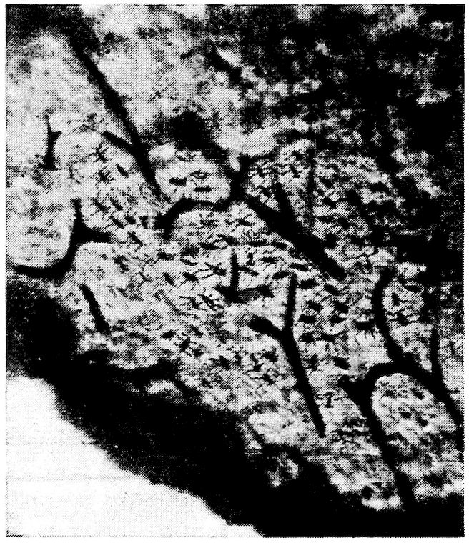
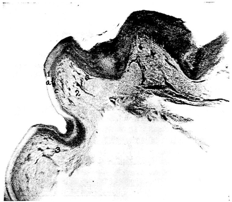
Fig. 1.—Longitudinal section of compact bone of the femur of the hen. 1, Haversian canals. 2, Lacunæ with their canaliculi.
The Periosteum.—Covering the surface of bone, except at the articular surface where it is covered with cartilage, is a membrane, the periosteum, which consists of two layers: an outer, fibrous, protective layer, and an inner, cellular, osteogenic layer. The outer layer consists principally of white fibrous tissue. The inner layer contains many more connective-tissue cells, which gradually become more closely aggregated as we proceed toward the osseous surface; but there is no sharply defined line of demarcation between the two periosteal layers.
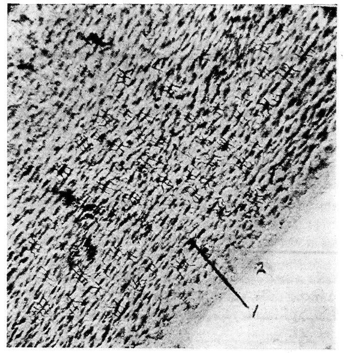
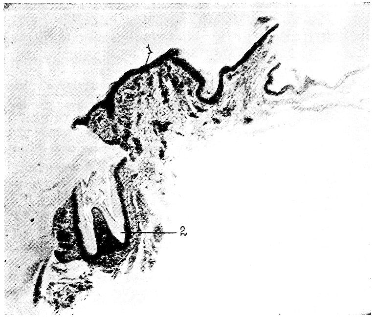
Fig. 2.—Transverse section of compact bone of the femur of the hen. 1, The lacunæ and canaliculi. 2, The periosteum.
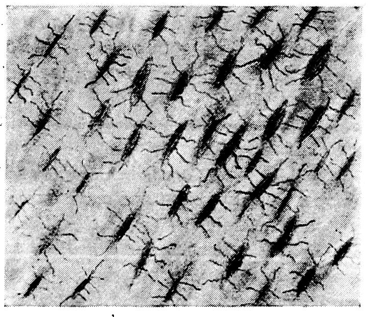
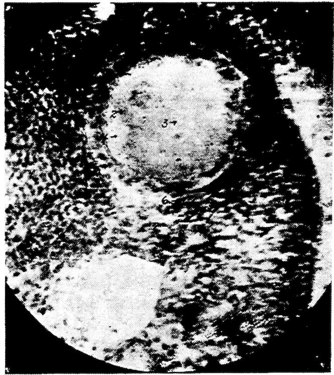
Fig. 3.—Transverse section of compact bone of the femur of the hen showing the lacunæ and canaliculi under high magnification.
20The periosteum is firmly attached to the bone by trabeculæ of fibrous tissue, called the fibers of Sharpey. These fibers of Sharpey penetrate the bone at right angles to its surface and carry blood-vessels.
Marrow.—There are two kinds of marrow: yellow, or medulla ossium flava, and red, or medulla ossium rubra.
The yellow marrow occurs in all bones except the femur and proximal portion of the tibia of adult fowls. It is composed of a network of fibrous tissue carrying blood-vessels, fat cells, and myelocytes, or marrow cells.
The red marrow is found throughout the femur and the proximal portion of the tibia, and in a few of the pelvic bones and vertebræ in the adult fowl, and in certain other bones of the baby chick. Red marrow consists of a delicate network of connective tissue supporting a dense capillary plexus, a small amount of fat, and numerous cells. The cellular elements of red marrow consist of marrow cells which contain large nuclei and possess ameboid movement, red blood cells, giant cells containing one or more nuclei, and various kinds of leucocytes, including eosinophiles, mast cells, and also osteoclasts.
Growth of Bone.—In the baby chick, only the shaft and a portion of the extremities of the long bones are thoroughly ossified, the extreme ends, and of the femur most of the articular head, being cartilaginous. The bones grow in length by an increase in the cartilage, the cartilage gradually becoming ossified. Growth in diameter is accomplished by the constant deposition of new layers of bone beneath the periosteum. During this process the osteoclasts absorb the bone from within. The formation of the marrow cavity is thus effected.
Classification of Bones.—The bones of the fowl are classified as long, short, flat, and irregular.
Long Bones.—The long bones occur in the legs and wings, where they serve as levers to sustain weight and make locomotion possible. A long bone consists of a shaft and two extremities. The superior is called the proximal and the inferior the distal extremity. The expanded articular surfaces in forming joints with adjoining bones afford ample space for the attachment of ligaments. The shaft is cylindrical and hollow.
Short Bones.—Short bones occur in the feet and in the wings. Their structure is similar to that of the long bones.
21Flat Bones.—The flat bones occur where extensive protection is needed, as in the cranial region; or where large surface for muscular attachments is needed, as in the costal and pelvic regions. Flat bones are made up of two thin layers of compact bone with a variable amount of cancellous tissue interposed.
Irregular Bones.—The irregular bones include the vertebræ, the patellæ, and the carpal bones.
Composition of Bone.—Bone consists of organic and inorganic matter. Organic matter gives toughness and elasticity to the bone, and inorganic matter hardness. The organic substance of bone is called ossein. When boiled in water ossein is resolved into gelatin. The following tables[1] give the results of an analysis of the femur, fresh, of a mature hen.
1. Grateful acknowledgment is hereby made to Dan M. McCarty, Chemist, Animal Industry Division, North Carolina Agricultural Experiment Station, for this analysis.
| Fresh femur: | ||
| Water | 18.23 per cent. | |
| Dry matter | 81.77 per cent. | |
| Dry matter: | ||
| Organic matter | 63.09 parts | |
| Inorganic matter | 18.68 parts | |
| Salts in dry matter: | ||
| Calcium | 6.970 per cent. | |
| Magnesium | 0.283 per cent. | |
| Potassium | 0.004 per cent. | |
| Sodium | 0.276 per cent. | |
| Iron | 0.020 per cent. | |
| Phosphorus | 3.210 per cent. | |
| Sulphur | 0.085 per cent. | |
| Chlorine | 0.520 per cent. | |
| Carbon dioxid | 0.550 per cent. | |
The inorganic matter of the femur of the hen consists of 18.68 parts or 22.84 per cent. of dry matter, and the organic matter of 77.16 per cent. Stated in other words the femur, including its contained marrow, consists of organic and inorganic matter in the ratio of 3.4, approximately, to 1.
The Skeleton of the Fowl.—The skeleton of a bird is remarkable for the rapidity of its ossification. It is worthy of note that other parts of the bodies of adult birds also become ossified. Among such parts are the tendons of the muscles of the legs, of the feet, and of the 22neck; the plates of the corneal margin of the sclerotic tunic of the eye; and the stapes of the ear. Ossification in birds at the attachments of the semi-lunar valves of the aorta and of the pulmonary artery has been reported by Owen.
The bony structure is compact, and the bones contain a greater proportion of phosphate of lime than do the osseous structures of mammals. Especially is this the case in those parts of the skeleton which are permeated by air.
The bodies of birds contain many air reservoirs to make them light that flying may be more easy. Many bones have their weight in proportion to size and strength thus greatly reduced. In very young birds the cavities of bones contain, instead of air spaces, loosely arranged red marrow, which is in most bones later absorbed. 23The air reservoirs in bones are most capacious in the best flyers. In the non-flyers more of the bones retain their red marrow.
The bones supplied with air spaces are relatively larger than in mammals, and are provided with small transverse osseous columns which cross in different directions and from side to side. These cross beams give stability to the thin wall of the bone. The membranes lining these cavities are very vascular.
The skull is divided into the cranial and facial portions. In these parts we find present 31 bones: one occipital, two parietal, two frontal, one ethmoid, one sphenoid, and two temporal; all of which constitute the cranial group; two premaxillary, two maxillæ, two nasal, two lacrimal, two palatine, two pterygoid, two zygomatic, one vomeral, the two jugal, and two quadrato-jugal, which constitute the facial group; two quadrati and one inferior maxillary, which constitute the inferior jaw group.
The peculiarities of the skull are the long os incisivum and the single condyle located on the occipital bone just below the foramen magnum. The condyle articulates with the atlas.
The head of the bird is small in proportion to the size of the body, and in front it is conical in shape.
The Cranium
The cranial cavity, or cavum cranii, incloses the brain with its membranes and vessels.
The dorsal wall, or roof, is formed by the frontal and the parietal bones. In the median line of the cerebral portion is the internal parietal crest. The roof of the cerebellar portion is marked centrally by a groove.
The posterior wall of the cerebellum is formed by the occipital bone.
The lateral wall is formed chiefly by the temporal bone. It is marked by a ridge which divides the cavity into the cerebral and cerebellar compartments. The cerebral portion is marked by a depression which receives the optic lobes. A crest divides this cavity from the optic portion. The walls are marked by digitations and vascular grooves.
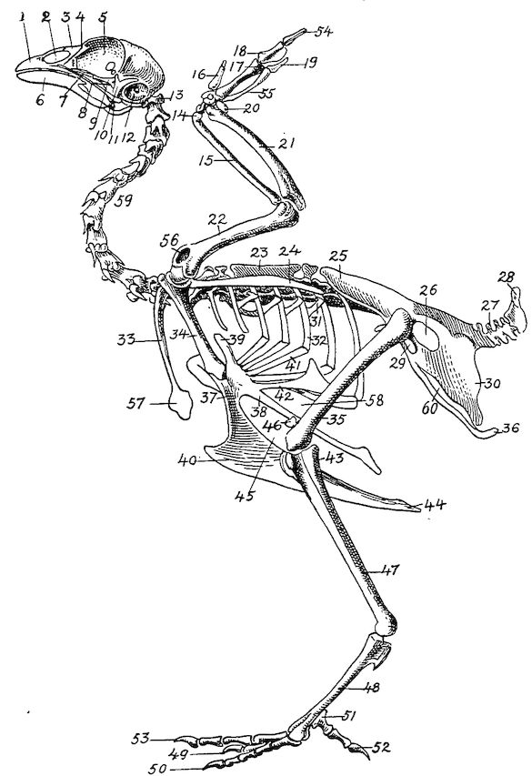
Fig. 4.—The skeleton of the domestic fowl. 1, Os incisivum. 2, External nasal opening. 3, Os nasale. 4, Os lachrymale. 5, Lamina perpendicularis. 6, Os dentale. 7, Os palatine. 8, Os quadrato-jugal. 9. Os pterygoideum. 10, Os quadratum. 11, Os articulare. 12, External auditory canal. 13, Atlas. 14, Os carpi radiale. 15, Radius. 16, First finger. 17, Os metacarpus. 18, Second finger. 19, Third finger. 20, Os carpi ulnare. 21, Ulna. 22, Humerus. 23, Thoracic vertebræ. 24, Scapula. 25, Os ilium. 26, Foramen ischiadicum. 27, Caudal vertebræ. 28, Pygostyle. 29, Foramen obturatum. 30, Os ischium. 31, Processus uncinatus. 32, Vertebral rib. 33, Os claviculum or furculum. 34, Os coracoideum. 35, Os femoris. 36, Os pubis. 37, Body of sternum. 38, Lateral internal process of sternum. 39, Costal process of sternum. 40, Sternal crest, cristi sterni, or keel of breast-bone. 41, Sternal rib. 42, Lateral external process of sternum. 43, Os fibula. 44, Xyphoid process of sternum. 45, Internal notch. 46, Os patella. 47, Os tibia. 48, Os metatarsus. 49, Second toe. 50, Fourth toe. 51, Os metatarsale. 52, First toe. 53, Second toe. 54, Second phalanx of second finger. 55, Os metacarpus. 56, Air opening in humerus. 57, Hypocledium. 58, External notch. 59, Cervical vertebræ. 60, Foramen oblongum.
25The ventral wall, floor, or basis cranii interna, may be regarded as forming five fossæ. These are: one anterior, one middle, one posterior, and two lateral. The anterior fossa supports the frontal and olfactory parts of the cerebrum. It lies at a higher level than the middle fossa. The middle fossa, or fossa cranii media, is circular in outline and extends into the lateral fossæ which lodge the olfactory lobes. Just back of the middle fossa is the sulla turcica, upon which rests the pituitary body. The posterior fossa, or fossa cranii posterior, lodges the medulla oblongata.
Bones of the Cranium (Figs. 4, 6, 7, and 8).—The bones of the cranium fuse early in the chick’s life. The sutures uniting the bones can usually be seen in the fetus or in the baby chick soon after it emerges from the shell. The major portion of the cranial bones become entirely fused. Each cranial and each facial bone ossifies from a distinct center or centers.
The cavity for the cerebrum is much larger than the cavity for the cerebellum. The cranial cavities in birds are relatively larger than in mammals. The bones are designated as in mammals. There are three single bones, the occipital, the ethmoid, and the sphenoid. Those in pairs are the frontal, the parietal, and the temporal.
The Occipital. Location.—The occipital bone or os occipitale, is situated at the posterior part of the cranium, of which it forms the posterior wall. This part is called the base of the cranium. The occipital bone articulates with or touches inferiorly, the sphenoid, laterally, the temporal, and superiorly, the parietal.
Development.—The occipital bone is developed from four centers of ossification; the dorsal, or os occipitale superius, two lateral, or ossa occipitales laterales, and the ventral, or os occipitale inferius, all of which may be seen distinct in the baby chick (Fig. 5, Part II, No. 1).
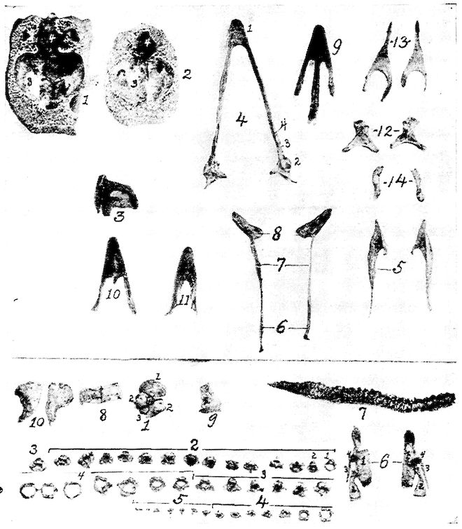
Fig. 5.—Bones of the head and vertebra.
Part I.—1, The cranium. 2, The skull cap. 3, The ethmoid bone. 4, The inferior maxilla. 5, The palatine bone. 6, The quadrato-jugal. 7, The jugal. 8, The superior maxilla. 9, The premaxilla. 10, The horny covering for the premaxilla. 11, The horny covering for the os dentale. 12, The os quadratum. 13, The nasal bone. 14, The pterygoid.
Part II.—1, The os occipitale (1, dorsal; 2, two lateral; 3, ventral portions and 4, foramen magnum). 2, The fourteen cervical vertebræ. (1, atlas; 2, axis). 3, The seven dorsal vertebræ. 4, The fourteen lumbo-sacral vertebræ. 5, The seven coccygeal vertebræ. 6, The pelvis (1, ilium; 2, ischium; 3, pubis; 4, cotyloid cavity or acetabulum). 7, The vertebral column complete from the baby chick. 8, The parietal bone. 9, The temporal bone. 10, The frontal bone.
27Description.—In the center of the occipital bone is the foramen magnum. Through this foramen the spinal cord extends into the cranial cavity and connects with the medulla oblongata. The occipital bone has a single condyle, which is located just below the foramen magnum and articulates with the atlas. At the base of the condyle a small subcondyloid fossa receives the body of the atlas during extreme flexion of the head. In the center of the lateral wing of the occipital bone there is a small foramen through which passes the hypoglossal nerve. Somewhat laterally from this foramen there is an opening through which the vagus, or pneumogastric nerve passes. Laterally from these is the canalis caroticus et jugularis. Between the os occipitale superius, or dorsal portion, and the ossa parietalia is a space to which ligaments are attached, called the fontanel.
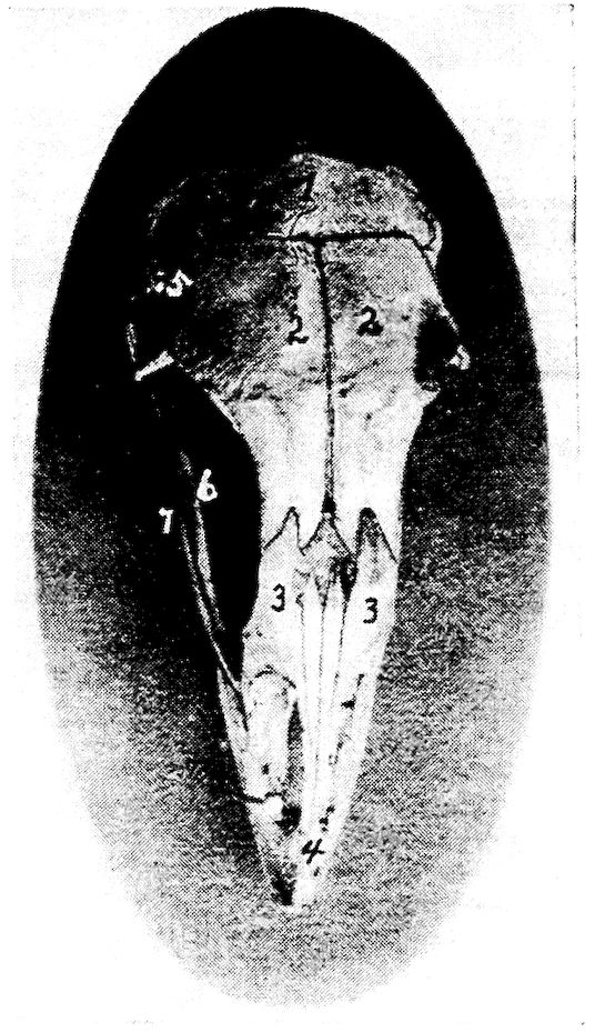
Fig. 6.—View of the frontal region of the skull of a hen. 1, Parietal. 2, Frontal. 3, Nasal. 4, Premaxilla. 5, Temporal. 6, Inferior jaw. 7, Jugal bone.
The Ethmoid. Location.—The ethmoid, or os ethmoidale, forms the anterior wall of the cranium, and the orbital septum. It is related anteriorly with the vomeral, superiorly with the nasal 28and frontal, posteriorly with the temporal, and inferiorly with the sphenoid, and palatine.
Description.—The ethmoid consists of a perpendicular and a horizontal lamina. The perpendicular lamina, located between the orbital cavities, is called the septum interorbitale. On each side of the septum interorbitale and near the superior orbital roof are two foramina for the passage of the olfactory nerves. In the horizontal plate, which forms the anterior cranial wall, are located the optic foramina through which pass the optic nerves.
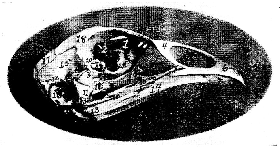
Fig. 7.—Side view of the skull of a hen. 1, Lamina perpendicularis. 2, Foramen for the passage of the nerve of smell. 3, Foramen for the passage of the optic nerve. 4, Nasal bone. 5, External nasal opening. 6, Premaxilla. 7, Os dentale. 8, Superior maxilla. 9, Os jugal. 10, Os quadrato-jugal. 11, Os quadratus. 12, Pterygoideus. 13, Os articulare. 14, Inferior maxilla. 15, Temporal. 16, External auditory canal. 17, Parietal. 18, Frontal. 19, Palatine. 20, Orbital process, posterior to which is the processus zygomaticus. 21, Interorbital foramen and optic foramen for passage of optic nerve. 22, Attachment for inferior oblique; 23, for superior oblique; 24, for levator palpebræ superioris; 25, for internal rectus; 26, for superior rectus; 27, for external rectus; 28, for inferior rectus; 28, for inferior rectus; 29, for depressor palpebræ inferioris; 30, for orbicularis palpebrarum; 31, for tensor tympani; 32, for circumconcha.
The Sphenoid. Location.—The sphenoid, or os sphenoidale, forms the floor of the cranial cavity and articulates with or touches posteriorly the occipital, laterally the temporal, and anteriorly the palatine, and ethmoid.
Description.—The sphenoid bone, the largest part of the cranial floor, is formed by the fusion of the nasal and the cranial parts. It is a three-cornered bone with two, thin, broad wings. These wings are divided into two portions, orbital wings, or alæ orbitales, and 29temporal wings, or alæ temporales. The temporal wing forms a cover for the Eustachian tube trumpet, and for the canal coming from the sella turcica, which latter gives passage to the intracranial carotid artery. The orbital wing forms the lower portion of the posterior wall of the orbital cavity, and lies directly before the os petrosum or temporal bone where the second and third branches of the fifth pair of cranial nerves emerge from the cranial cavity.
Anteriorly the sphenoid has a foramen for the passage of the Eustachian tube, the tuba auditiva, and also a sharp-pointed projection, the nasal portion, called the rostrum.
The Frontal. Location.—The frontal bones, or ossa frontales, two in number, form the forehead, a portion of the nose, and a portion of the roof of the orbital cavities. They are related posteriorly with the parietal, laterally with the temporal and zygomatic, and anteriorly with the nasal and premaxillary. They touch inferiorly the ethmoid.
Description.—Each of these bones has a processus orbitalis which is seen at the outer margin of the posterior and upper orbital wall and just in front of the zygomatic process of the squamous portion of the temporal. The frontal bone forms the anterior portion of the superior wall of the cranial cavity. The two bones are thin, flat, and meet at the median line of the forehead. The external surface is convex. The inner surface has a ridge located longitudinally and in the center the bone becomes narrow anteriorly.
The Parietal. Location.—The parietal bones, or ossa parietalia, two in number, form the posterior part of the roof of the cranial cavity. They meet in the median line and are related posteriorly with the occipital, anteriorly with the frontal, and laterally with the temporal bones.
Description.—The parietal bones are short and very broad (Fig. 5, Part II, No. 8). Each bone is quadrilateral in outline and has two surfaces, four borders and four angles. The external parietal surface is convex and smooth and the internal, cerebral surface is concave.
The Temporal. Location.—The temporal bones, or ossa temporales, two in number, are located at the lateral portion of the cranium, and aid in the formation of the cranial wall. The temporal bone is related posteriorly with the occipital, superiorly with the parietal and frontal, externally with the quadratus, anteriorly with the ethmoid, and inferiorly with the sphenoid.
30Description.—The temporal bones consist of the fused squamous and petrous temporals; they contain the essential organs of hearing.
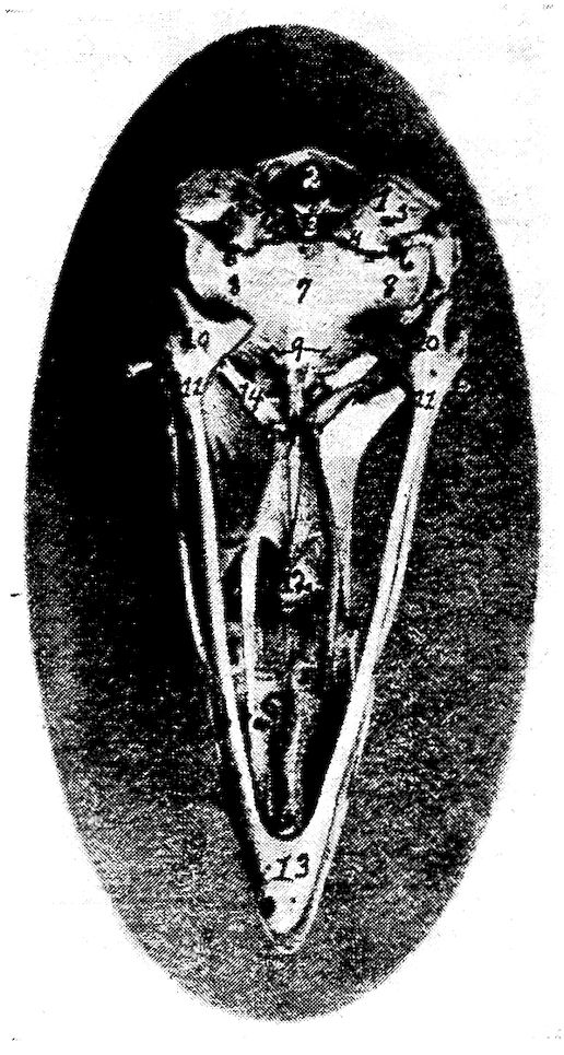
Fig. 8.—Inferior view of the skull of a hen. 1, Occipital. 2, Foramen magnum. 3, Occipital condyle and just below the basi-occipital. 4, Foramen for the passage of the hypoglossal nerve. 5, Foramen for the passage of the vagus. 6, Canalis caroticus and jugulare. 7, Sphenoid. 8, Temporal wings of the sphenoid. 9, Foramen auditiva. 10, Os articulare. 11, Os angulare. 12, Vomer. 13, Os dentale. 14, Pterygoid.
The squamous portion of the temporal bone possesses the long thin zygomatic process sometimes called the posterior orbitalis. It forms a small flattened tongue, directed forward, sometimes free, and at other times united by its superior border to the summit of the orbital process. This is especially true in the turkey. This process is seen near the lower outer portion of the posterior orbital wall. The squamous portion is also provided antero-laterally with an articular facet which articulates with the quadrate bone. The large portion of the temporal bone lies on the side of 31the cranium superior to the ala sphenoida temporale. It extends outward and anteriorly over the rims of the petrosum and ala sphenoida orbitale. The squamous part forms the upper three-fourths of the cochlea, the inner auditory canal, the upper part of the fenestra ovalis, the anterior vertical and the outer semicircular canal, and the lower part of the posterior vertical semicircular canal.
The petrous portion forms the posterior wall of the foramen ovale and the fenestra ovalis in which lies the columella. The fenestra ovalis and the fenestra rotunda are the only two entrances into the labyrinth. In this region may be seen the fusion line between the os petrosum and the os occipitale. Superiorly and posteriorly the petrous portion touches the external parietalia and occipitalis; infero-laterally it unites with the basi-sphenoid. The outer rim of the foramen ovale is broadened by the ala sphenoida and mesially by the basi-sphenoid. This foramen gives exit to the second and third divisions of the fifth pair of cranial nerves.
The lateral surface of the temporal bone presents a short tube, the external acoustic process, or processus acusticus externus, to which is attached the concha of the ear. The process is directed outward. Its lumen, the external acoustic meatus, or meatus acusticus externus, conducts to the cavity of the middle ear in the bare skull, but is separated from it by the tympanic membrane in the natural state.
Bones of the Face
The bones of the face, or ossa faciei, are the premaxillary, or upper mandible, the maxillæ, the nasal, the lacrimal, the palatine, the pterygoid, the zygomatic, the vomeral, the jugal and the quadrato-jugal. The vomer is single, the others are paired. In the inferior maxillary group there are the quadrati, and the inferior maxillary, or lower mandible. The turbinated and hyoid bones are also discussed with the bones of the face.
The beak consists of the anterior portion of the upper and lower mandibles which are covered with a horny structure.
The Premaxilla. Location.—The premaxillæ, or ossa incisiva, or mandibular structures, are located in the extreme anterior facial region.
Description.—The premaxillæ are long and the anterior end is pointed. Each consist of two lateral halves which become fused before the chick is hatched. They partly circumscribe the openings 32into the nose. This bone is the base of the upper portion of the beak and determines its form. It forms the anterior walls of the nasal cavity. It has between the nasal bones, two processes which extend back to the anterior point of the cerebral cavity. The posterior part of the incisivum and nasale are flat, thin, and elastic. The extensions then are the processus maxillaris, the processus palatinus, and the processus frontalis, the first of which forms part of the jaw rim, the second, which aids in forming the gum plate, and the last which reaches to the anterior portion of the cranial wall.
The Maxilla. Location.—The superior maxillaries, or ossa maxillares, two in number, form the floor of the upper beak, a part of the palatine roof and nasal walls.
Each maxilla borders laterally the premaxilla and the nasal; inferiorly, the anterior point of the palatine; and posteriorly, the jugale.
Description.—The maxillæ are thin, flat, bones. They have three borders and three angles. The palatine processes of the two bones do not meet in the median line, which results in a cleft in the median palatine region. The bone-like gums are formed partly by the palatine processes but more largely by lateral wings of the os incisivum. Each maxilla has posteriorly a yoke-like extension, superiorly a small extension, and also a palatine process. The latter articulates by a facet with the vomer.
The Nasal. Location.—The nasal bones, or ossa nasalia, two in number, are located in the lateral facial region. The nasal bone articulates posteriorly with the frontal; laterally, on the inner border, with the processus frontalis of the premaxilla, and on the outer border with the lacrimal; and inferiorly with the premaxilla, maxilla, jugale, and vomer.
Description.—The nasal bone, or os nasale, is broad, flat, and elastic and forms the posterior wall of the nasal opening. Under this bone is located the infraorbital sinus.
There are three extensions: first, the processus intermaxillaris, which forms the upper wall of the nasal cavity; second, the processus maxillaris anterior, directed downward and forward, which fuses with the maxillary bone and forms the posterior rim of the nasal cavity; and third, a posterior processus frontalis, which lies parallel with the ethmoid.
The Lacrimal. Location.—The lacrimal bones, or ossa lacrimalia, 33two in number, are located at the outer border and at the junction of the processus frontalis of the nasal, with the frontal bone.
Description.—The lacrimal bone is small, and rather filiform. They become fused with the nasal and the frontal bones, forming part of the margin of each.
The Palatine.—Location. The palatine bones, or ossa palatina, two in number, enter into the formation of the inner part of the bony gum and hard palate, or roof of the mouth. They form the support for the hard palate. Each palatine articulates posteriorly with the rostrum, or nasal portion of the sphenoid, and with the pterygoid; and anteriorly with the maxilla.
Description.—The palatine bone is curled posteriorly and is thin at the upper portion and thick at the lower border. Anteriorly it has a long rather filiform projection.
The Pterygoid. Location.—The pterygoid bones, or ossa pterygoidea, two in number, are located back of the region of the posterior nares. They extend diagonally outward and backward from the median region of the sphenoid rostrum to the quadrate bone. They articulate anteriorly with the sphenoid rostrum and the palatine, and posteriorly with the quadrate.
Description.—The pterygoid bones are slender and cylindrical, and are expanded at the ends into an articular facet.
The Zygomatic. Location.—The zygomatic, or ossa zygomatica, two in number are situated below the orbital cavity and extend from the maxilla to the quadrate bone.
Description.—The zygomatic is small, slender, rod-shaped, and forms the lateral portion of the upper jaw. The anterior portion of the zygomatic represents the jugal and is fused with the maxilla and with the anterior processus maxillaris of the nasal bone, the maxilla, and the posterior portion, the quadrato-jugal, which articulates with the quadrate bone.
The Vomer. Location.—The vomer is located in the median nasal septum. It articulates with the rostrum of the sphenoid, being connected to it by a mass of ligaments. It touches anteriorly the posterior portion of the maxilla.
Description.—The vomer is a median bone and aids in the formation of the septum nasi. It consists of a thin plate, thickest posteriorly and diminishing toward the anterior edge.
The Jugal and Quadrato-jugal.—The jugal and quadrato-jugal are united forming a long slender cylindrical bone called the zygomatic, 34lying at the outer side of the upper jaw. They have been described under zygomatic, which see.
The Quadrate. Location.—The quadrate bones, or ossa quadrata, two in number, are located antero-laterally to the temporal bones. Each articulates inferiorly with the posterior articular portion, or pars articulare, of the inferior maxilla and infero-laterally with the quadrato-jugalare portion of the zygomatic. It articulates antero-internally with the pterygoid and supero-posteriorly with the temporal.
Description.—The quadrate bone is anvil-like in shape. It has an anterior process, the orbital process, for muscular attachments, and posteriorly it affords attachment to the ear drum.
The Inferior Maxilla. Location.—The inferior maxilla, lower jaw, or os maxillare inferius, also called the mandibular bone, or pars dentis, is analogous to the lower jaw of mammals. It articulates posteriorly with the quadrate bone.
Description.—The inferior maxilla is the largest bone of the face. It is made up of a right and a left limb which are separate in the fetus and which unite subsequently anteriorly, forming the inferior portion of the beak. Each limb of the jaw is developed from five elements: the pars articularis, which forms the jaw-joint and, expanded, articulates with the quadrate bone; the pars angularis, lying just in front of the pars articularis; the pars supra-angularis, a slender bone lying just above the angularis; the splenial, a thin plate of bone, lying along the inner surface of the mandible; and the pars dentalis, which forms the anterior portion of the jaw.
The Turbinate Bones. Location.—The turbinate bones, or ossa turbinata, six in number, are attached to the lateral walls of the nasal cavity (Fig. 26, No. A, 1 and 2). In each nasal cavity there are three turbinate bones, one anterior and two posterior. Of the two posterior the upper one lies supero-posterior to the inferior one. The turbinate bones are attached to the lateral nasal walls, project into the cavity and thus greatly diminishing its extent.
Description.—Each turbinate bone is composed of a very thin lamina, finely cribriform in many places, and in the fresh state, covered on both sides with mucous membrane. These bones are curled and partly membrano-cartilaginous structures which give greater surface in the nasal passage for mucous membrane in which the olfactory nerve terminal filaments are distributed.
35The Nasal Cavity.—The nasal cavity, or cavum nasi, is a longitudinal passage which extends through the upper part of the face. It is divided into right and left halves by a median septum nasi. Its walls are made up of the premaxilla, maxilla, nasal, vomer, and palatine bones.
The Hyoid Bone. Location.—The hyoid bone, or os hyoideum, is situated chiefly between the rami of the mandible, but its upper part extends around the outer margin of the base of the skull. The hyoid bone does not articulate with any bones of the skull, but is firmly attached by strong fibrous structure.
Description.—The hyoid bone consists of the following parts: the body, or basi-hyal, which is subcylindrical and presents in front a trochlear articular surface, convex transversely, and concave vertically for articulation with the ewer-shaped portion of the glosso-hyal. The anterior free portion, or lingual process, is called the glosso-hyal, or entoglossal bone. The lingual process gives support to the muscular and fibrous structures of the tongue. On either side of the basi-hyal there is a limb passing posteriorly along the side of the superior larynx, extending upward along the outer border of the occipital bone. This is the cornu of the os hyoideum and is divided into two elements, first, the basi-branchial which is bone and articulates with the basi-hyal, and the second, the cerato-branchial, cartilaginous in structure. In the center and projecting backward from the body of the os hyoideum is a spur process called the uro-hyal, partly bony and partly cartilaginous, and which rests upon the superior larynx.
The vertebral, or spinal column consists of 42 bones, as shown in the following table:
| Cervical region | 14 |
| Dorsal region | 7 |
| Lumbo-sacral region | 14 |
| Coccygeal region | 7 |
| Total | 42 |
Many of the bones of the dorsal and lumbo-sacral regions do not have free articulations. The cervical and coccygeal alone have free movements.
The Cervical Vertebræ (Fig. 4, No. 59; Fig. 5, Part II, No. 2). Location.—The cervical vertebræ form the neck of the fowl.
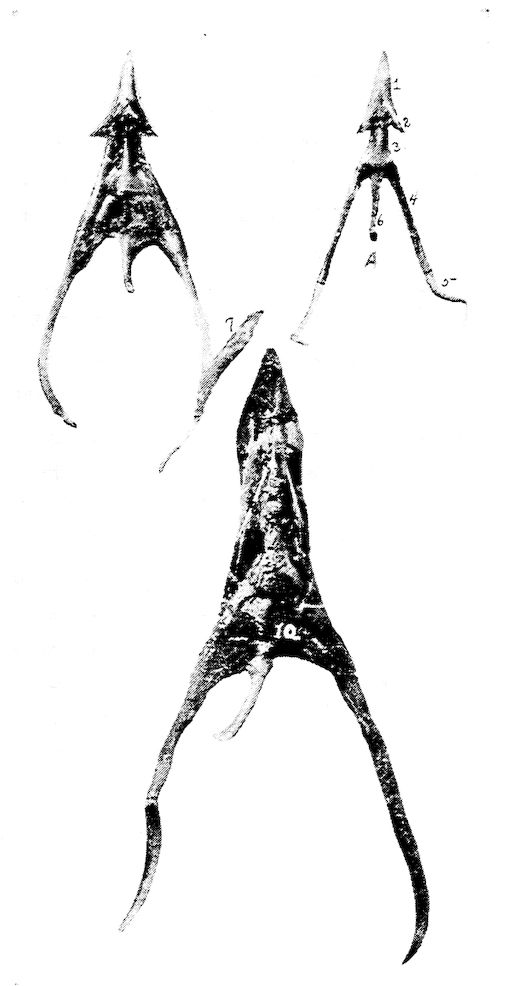
Fig. 9.—Os hyoidum and tongue muscles. 1, Glosso-hyal. 3, Basi-hyal. 4, Basi-branchial element of the cornua. 5, Cerato-branchial element of the cornua. 6, Uro-hyal or spur process. 7, Genio-hyoideus. 8, Cerato-hyoideus showing the slender tendon. 9, Hyoideus transversus. 10, Hyoideus transversus.
37Description.—The long cervical section of the spinal column is S-shaped and is made up of fourteen vertebræ. The anterior segments move freely forward, the middle ones backward and the posterior ones forward, allowing the S-shaped curve of the neck. The neck is flexible so that it is possible for the beak to reach the coccygeal oil gland. The bird is enabled to reach the feathers on all parts of the body to cleanse and oil them.
The Atlas. Location.—The first cervical vertebra is called the atlas. Anteriorly it articulates with the single condyle of the occipital bone. Posteriorly it articulates with the axis, or second cervical vertebra.
Description.—The atlas is the smallest of the cervical vertebræ and is ring-shaped. The anterior articular surface, half-moon in shape, forms a deep articular cavity. The joint, called a ball-and-socket joint, makes possible movements in all directions. The condyle of the occiput also touches an articular end of the odontoid process of the axis, or second cervical vertebra. Posteriorly there projects from the atlas two small lateral wings possessing articular facets which articulate with similar facets on the lateral wings of the axis. Posteriorly the body of the atlas is also provided with an articular surface which articulates with a similar surface on the anterior portion of the body of the axis.
The Axis. Location.—The axis is the second cervical vertebra. It articulates anteriorly with the atlas, and by a facet on the extreme anterior end of the odontoid process, with the condyle of the occipital bone. Posteriorly it articulates with the third cervical vertebra.
Description.—The body of the axis is short. The upper anterior portion of the body of the axis is provided with a tooth-like process called the odontoid process. There are two anterior lateral wings provided with small articular facets which articulate with similar facets of the atlas. The anterior surface of the body of the axis forms a true articulation with the body of the atlas.
The axis is provided with a superior and an inferior spine. There are two posterior articular processes which articulate with the prezygapophyses, or anterior articular processes, of the third cervical vertebra. The posterior part of the body of the axis forms a true articulation with the body of the third cervical vertebra.
Other Cervical Vertebræ.—Beginning with the axis, the body of which is relatively short, the body of each succeeding vertebra 38is longer than that of the preceding. The articulations of each vertebra with adjoining vertebræ are effected by means of diarthrodial facets, convex in one direction and concave in the other.
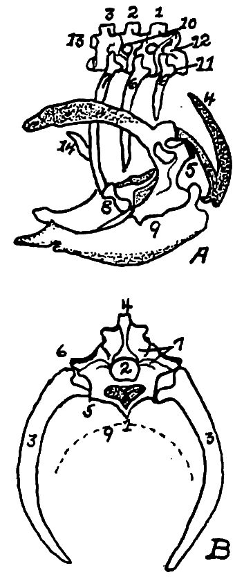
Fig. 10.—A. Diagram of three first dorsal vertebræ and scapular arch-side view. B. Diagram of section through the hemal arch.
A. 1, First dorsal segment. 2, Second. 3, Third. 4 and 5, Hemal arches. 6, The pleurapophyses or floating ribs. 7, The third pleurapophyses or dorsal vertebral rib articulating with the hemapophysis or sternal rib, 8, and this in turn with the wing of the sternum form a hemal arch. 9, The sternum. 10, Intervertebral foramen through which the nerves pass. 11, Articulation with the body of vertebra parapophyses. 12, Articulation with transverse process or diapophyses. 13, Oblique process or zygapophysis. 14, The epipleural appendage.
B. 1, Hemal or inferior spine (hypapophysis). 2, The neural arch. 3, The pleurapophyses or rib. 4, The superior or neural spine or neurapophysis which aids in the formation of the anapophyseal ridge. 5, Parapophyseal surface for head of rib. 6, Tubercle of rib articulating with the diapophysis. 7, Lamina of neurapophysis. 8, Centrum. 9, Hemal arch.
Between the bodies of the vertebræ are pads of fibrous cartilage. Above these bodies and inferior to the neural spines extends throughout the spinal column the large neural canal which is occupied by the spinal cord. Between the vertebral segments the neural canal is exposed superiorly, since here the neural arches do not completely bridge the canal. These spaces are protected by intervertebral ligaments.
In addition to the superior neural spines and the inferior spines, 39from the body of the vertebra project lateral processes, and anterior and posterior oblique processes.
All cervical vertebræ except the atlas and the axis are made up of the following parts: a body, or centrum, a neural canal, a neural arch, a superior dorsal, or neural spine, or neurapophysis, which in most cervical vertebræ is only slightly developed, two oblique processes, or zygapophyses, two transverse processes, or diapophyses (Fig. 10, No. 12) and in some vertebræ two tubercles, or anapophyses above the posterior zygapophyses. The prezygapophyses are directed upward and inward; the postzygapophyses are directed downward and outward. The inferior spines are only well developed in the last two or three cervical vertebræ.
Between vertebral segments, except the central sacral portion, on each side, there is an intervertebral foramen (Fig. 11, No. A, 4), through which pass the spinal nerves. In the central portion of the sacrum where the vertebræ have fused, there are two foramina on each side for each original vertebra, one located above the other, the upper giving passage to the sensory branch, the lower to the motor branch of the nerve.
The Dorsal Vertebræ (Fig. 4, No. 23; Fig. 11, A). Location.—The dorsal, or thoracic vertebræ, or vertebræ thoracicales, aid in forming the roof of the chest cavity.
Description.—The dorsal vertebral section is made up of seven vertebræ, with strong short bodies. The first and sixth dorsal segments articulate as do the cervical, by the bodies and oblique processes. (Fig. 11, No. A, 3, illustrates the oblique processes.) The seventh dorsal is fused with the first lumbo-sacral vertebra. The second to the fifth inclusive of the vertebræ are fused together, and the superior and inferior spinous processes are fused into a prominent plate-like ridge.
The transverse processes of the dorsal vertebræ, from the second to the sixth, are well developed and are bridged over with a thin layer of bone. The ventral spines are partly fused and form a very prominent and continuous ridge. (Fig. 11, No. A, 1, shows the fused superior spines; No. 2, the fused inferior spines; and No. 4, the intervertebral foramina.)
The Lumbo-sacral Vertebræ (Fig. 5, Part II, No. 4). Location.—The fused lumbo-sacral section of the spinal column forms the roof of the pelvic cavity.
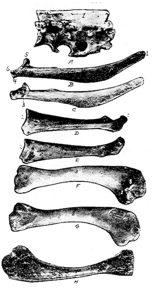
Fig. 11.—Bones from the scapular arch.
A. Dorsal vertebra. 1, Superior spinous ridge, 2, Inferior spinous ridge. 3, Oblique processes. 4, Intervertebral foramina. 5, Articular facette for head of the rib. 6, Articular facette for tubercle of the rib. 7, Articular portion of the body.
B. Outer surface of scapula.
C. Inner surface of scapula. 1, Thin caudal end. 2, Articular head. 4, Processus furcularis. 5, Processus humeralis. 6, Processus coracoideus.
D. Outer surface of coracoid.
E. Inner surface of coracoid. 1, The furcular tuberosity. 2, Articular facette for articulation with the sternum. 3, Articular surface for humerus and scapula.
F. Outer surface of humerus and G the inner surface of the same. 1, Trochanteric fossa. 2, Oval articular head at proximal end. 3, Shaft. 4, Distal end showing articular condyles. 5, Trochanter. 6, Trochlea for ulna. 7, Trochlea for radius. 8, Depression or fossa.
H. Section through the median plane of the humerus showing the delicate cross partitions illustrating provisions made for bones provided with air-sac extensions.
41Description.—The lumbo-sacral region consists of fourteen vertebræ which are distinct in the body of the newly hatched chick, but which become fused soon after hatching. With these vertebral segments there are fused the last dorsal and first coccygeal vertebral segments. This fusion is so complete that the segments are indicated only by the intervertebral foramina on the sides through which the spinal nerves pass, and by transverse markings on the inferior surface of the bodies.
The lumbo-sacral vertebræ, called the sacrum, and the ilia are fused. The dorsal spines of the vertebral segments are indicated only in the anterior portion where they are fused, forming a plate. There are no prominent ventral spines.
The Coccygeal Vertebræ (Fig. 4, No. 27). Location.—The coccygeal or caudal vertebræ, or vertebræ coccygeæ, constitute the bones of the tail.
Description.—There are seven coccygeal vertebræ. The last segment, shaped like a plough share and therefore called the pygostyle, is the largest and is supposed to have been formed by the fusion of several original distinct segments. It supports the coccygeal oil gland and the row of rudder feathers, or rectrices, which are arranged fan-wise.
All of the coccygeal vertebræ except the first are freely movable, thus allowing the tail to be used as a rudder during flight. The lateral spines are long and well developed, and the superior spines are bifurcated, thus giving increased surface for muscular attachment. The first coccygeal segment is fused with the last lumbo-sacral vertebra.
The dorsal vertebræ superiorly, the ribs laterally, and the sternum, or breast-bone inferiorly, form the skeleton of a large cavity called the thorax. The dorsal vertebræ have been described.
The Ribs (Fig. 4, No. 32; Fig. 10, A and B). Location.—The ribs form the lateral bony wall of the thorax, articulating superiorly with the dorsal vertebræ.
Description.—The ribs are arranged in order of length, the ultimate rib being the longest. From anterior to posterior, they approach more nearly a horizontal position. The ribs are divided into the true and the false. The true ribs articulate with the sternum. The false ribs do not touch the sternum; they are floating. The true ribs are composed of two parts, a vertebral, or dorsal, and a sternal, 42or ventral. The vertebral part, or extremitas vertebralis, is provided with an articular head, or caput costæ; a neck, or collum costæ; and an articular tubercle, or tuberculum costæ. The head and tubercle articulate with the dorsal vertebra. Below the head of each rib is a pneumatic foramen.
The distal extremity of the dorsal section of the rib articulates with the proximal end of the sternal section by a diarthrodial articulation.
The articulations of the true ribs with the sternum is diarthrodial and each articulate by two small ridges with a double sternal facet. The first, the second, and the seventh ribs are floating, or false ribs. The first rib articulates with the quadrate part of the last cervical vertebra and also with the first dorsal vertebra. The seventh, the last rib, articulates with the under side of the anterior alar part of the ilium. This rib is situated similarly to the true ribs; but, instead of articulating directly with the sternum, the lower end lies against the sternal segment of the rib just anterior to it.
From the posterior edge of the second, the third, the fourth, and the fifth ribs, and near the middle of the dorsal segment, are flat, uncinate, bony processes which project upward and backward, overlying in each case, the succeeding rib and giving greater surface for muscular attachments and greater stability to the thorax.
The Sternum (Fig. 4, No. 40; Fig. 10, No. A, 9). Location.—The sternum, or breast-bone forms the inferior portion, or floor of the thoracic cavity.
Description.—The sternum is a quadrilateral, curved plate with processes projecting from each angle and from the middle of the anterior and posterior borders. The posterior medial projection, or metasternum, is the longest, and has a tall, plate-like ridge—the sternal crest, crista sterni—running along its ventral surface. The crest serves the important function of increasing the bony area for the attachment of the powerful muscles which move the wings. The anterior medial projection, or rostrum, is short, and pierced at its root by an opening from which extend two elongated, saddle-shaped depressions into which the end of the coracoid bones are received.
The plate-like process of bone, the posterior lateral process, which projects from the caudal angles of the sternum soon divides into two parts. The shorter of these, the oblique process, broadens toward its free end and covers the sternal segments of the last two ribs. The 43sides of the sternum are thus provided with an external and an internal process forming an external and an internal notch. These notches are bridged over by a broad ligament, to which the muscles are attached. In poor flyers, as the domestic fowl, these notches are large. The posterior end of the sternum is called the xiphoid process or processus xiphoideus. Anteriorly the sternum is provided with lateral external processes, the costal processes. The lateral borders of the sternum are pitted by four depressions into which the sternal segments of the ribs are received. The dorsal, or inner surface of the bone is pierced by openings by which the air-sacs communicate with the interior.
The appendicular skeleton consists of the shoulder girdle, the fore limb, the pelvic girdle, or hip bone, and the hind limb. The shoulder girdle consists of the scapula, the coracoid, and the clavicle. The fore limb consists of the arm, forearm, and the hand. The arm consists of the humerus, the forearm of the radius and the ulna; and the hand consists of the carpus, the metacarpus, and the phalanges. The pelvic girdle consists of the ilium, the ischium, and the pubis. The hind limb consists of the thigh, the leg, and the foot. The thigh consists of the femur, and the leg consists of the tibia and the fibula. The foot consists of the metatarsus and the phalanges.
The shoulder girdle consists of the scapula, the clavicle and the coracoid.
The Scapula. Location.—The scapula (Fig. 11, A, B) lies on the outer and superior rib surface, extending parallel with the dorsal vertebræ.
Description.—The scapula is a thin, sword-like bone, becoming thicker as it approaches the shoulder-joint. The scapula expands and becomes thin near the free end, which reaches nearly to the antero-lateral portion of the ilium (Fig. 4, No. 24). The scapula articulates with the coracoid. A pneumatic foramen is located at the base of the acromion process. The anterior part of the scapula is provided with an articular head and is provided with an inner process, called the processus furcularis, which lies near the furcula and coracoid. An outer stronger processus humeralis forms the 44posterior half of the glenoid cavity, or cavitas glenoidalis, in which the humerus articulates and the processus coracoideus.
The Clavicle. Location.—The clavicles, commonly known as the wish bone, are located in the anterior chest region.
Description.—The clavicles are long, slender bones uniting below in the hypocledium, a laterally flattened process. They are joined to the upper end of the coracoid by fibrous cartilage. The hypocledium is joined to the anterior point of the sternum by the claviculosternal ligament. The clavicles, as united by the hypocledium, form a v-shaped structure called the furculum, or, popularly, the wish bone.
The forks play the part of an elastic spring, whose office it is to prevent the wings from coming toward each other during contraction of the depressor muscles. The conformation of this bone is, therefore, like the sternum, related to the extent and power of flight; and for this reason it is that, in swift flyers, the two branches of the furculum are thick, solid, widely separated, and curved like a U; while in those that fly heavily and with difficulty, these branches are thin and weak, and join at an acute angle. The latter formation greatly diminishes its strength, and lessens, in a singular manner, the reactionary power of the bony arch it represents.
The Coracoid. Location.—The coracoid is located just back of the clavicle and at the side of the entrance of the thoracic cavity. It is the strongest bone of the shoulder girdle, extending upward, outward, and forward. It articulates inferiorly with the sternum and superiorly with the humerus and the scapula, and is attached to the superior end of the furcular limb by a fibrous cartilage.
Description.—It is thinnest in the center or shaft and broadens toward the inferior extremity. The upper hook-like part of the coracoid forms the fore part of the glenoid cavity, and together with the scapula and furcula form the foramen triosseum through which passes the tendon of the elevator muscle of the wing. The upper end flattens out into three tuberosities, the tuberositas furcularis which is thick and to which attaches the limb of the furcula, the tuberositas scapularis which unites to the scapula, and the tuberositas humeralis which lies between these and articulates with the humerus.
The bones of the fore limb are humerus, ulna, radius, carpus, metacarpus, and phalanges.
45The Humerus. Location.—The humerus constitutes the arm, which, when at rest, lies parallel to the dorsal vertebræ. It articulates superiorly with the glenoid cavity, a shallow ball-and-socket joint, formed by the scapula and the coracoid; and inferiorly with the ulna and radius.
Description.—The proximal extremity of the humerus is provided with a trochanter (Fig. 11, G, No. 5) and a large oval head or caput humeri, which articulates in the glenoid cavity. The head is an elongate, semi-oval convexity, with the long axis transverse from the radial to the ulnar side and with the ends continued into upper and lower crests. The upper crest of the head of the humerus is on the radial side and the lower crest on the under side. Under this latter crest there is a pneumatic fossa (Fig. 11, F, No. 1), at the upper end of which there is an opening into the bone, the pneumatic foramen, which brings the air-sac into communication with the air space of the bone. The shaft, or corpus humeris, is irregularly cylindrical and slightly S-shaped.
The proximal part of the shaft, which is expanded on the palmar side, is concave across and convex lengthwise. The distal part is slightly flattened. The shaft of the humerus is almost cylindrical.
The distal extremity of the humerus is provided with two articular condyles, one of which articulates with the ulna and the other with the radius. On the radial side of the palmar surface there is a ridge; and on the ulnar side of the same surface there is a second ridge diverging to the opposite tuberosity. The radial surface is a narrow, subelongate convexity, extending from the middle, approximately, of the palmar surface, obliquely to the lower part of the radial tuberosity. The two articular convexities, or trochlea, at the distal end of the humerus are bent toward the palmar aspect, the anconal aspect is the side on which the elbow is situated. The inner convexity is the larger and articulates with the ulna. To the outside is the processus cubitalis humeri. The outer articulates with the radius and is so arranged that the radius makes a greater flexion than the ulna. At the lower end of the humerus there is a depression which receives the anconeus of the ulna during flexion and extension of the forearm. On the shoulder- and elbow-joint are found grooves over which the tendons glide, at which places sheaths are provided. This arrangement also aids in keeping the capsular ligament in place.
46The Ulna. Location.—The ulna, larger than the radius, is bent and articulates with the radius only at the ends. The two bones are bound together by a ligamentous band. It also articulates superiorly with the distal end of the humerus, and inferiorly with the carpus. When the wing is folded the radius is superior and a trifle to the inner side of the ulna.
Description.—The proximal end is most expanded, and is obliquely truncate for the articular excavation adapted to the ulnar tubercle of the humerus. A short angular process behind the cavity represents the olecranon, which is the processus olecranalis coracoideus ulnæ to which the ligamentum capsulare cubiti attaches. On the inner side of the head is the internal tubercle of the ulna and on the external side the external tubercle of the ulna. An excavation is noted on the radial side of the proximal end for the lateral articulation of the head of the radius. On the upper side near the articular part is located the sigmoid cavity.
The shaft, or corpus ulnæ, decreases in size near the distal end. It is slightly curved, flattened laterally with an internal and an external ridge.
The distal end of the ulna is slightly expanded into a trochlea which is sharply convex and articulates with two free carpal bones, the scapho-lunar, or os carpi radiale, and the cuneiform, or os carpi ulnare. The scapho-lunar is placed on the radial side, and the carpiulnare on the ulnar side. The distal extremity of the ulna is provided with a styloid process and on the dorsal side with a tubercle.
The Radius. Location.—The radius lies beside the ulna with which it articulates at each extremity. At the inferior extremity the articulation is rotary. It also articulates superiorly with the humerus and inferiorly with the os carpi radiale.
Description.—The radius, cylindrical in shape, is more slender than the ulna.
The proximal end is expanded, subelliptical, with a concavity for the oblique tubercle and a thickened convex border for articulation with the ulna. This end is provided with the tuberositas radii.
The shaft, or corpus radii, is slender, subcompressed, and has a slight bend upward from the ulna. A nutrient foramen occurs in this shaft.
The distal end is expanded and rather flattened with two grooves on the anconal side for passage of tendons. For articulation with 47the scapho-lunar the radius is provided with a terminal transverse convexity produced palmad, which also articulates with the ulna laterally. There is a tuberosity on the radial side of the expansion and inferiorly the inferior tuberculum ossis carpi radialis and superiorly a superior tuberculum ossis carpi radialis.
The Hand.—The hand is made up of the carpus, the metacarpus, and the phalanges.
The bones of the hand are so arranged as to allow abduction and adduction, or flexion in the ulno-radial plane, movements which are necessary in the outspreading and folding of the wing. Thus the hand of the fowl moves in a state of pronation, without the power of rotation. The carpal bones are so placed between the anterior arm and metacarpus as to reduce the abduction which is necessary to hold and extend the wing, so that the hand or wing be in a fixed position.
The Carpus.—The carpus in the domestic fowl is represented by two bones, ossa carpi, called the scapho-lunar, or os carpi radiale, and the cuneiform, or os carpi ulnare. The scapho-lunar is the smaller and is located between the radius and metacarpus. The cuneiform is the larger and is located between the ulna, the radius, and the metacarpus. The cuneiform is somewhat anvil-like in shape, being provided with a body and two prongs.
The Metacarpus.—The metacarpal bones, or ossa metacarpi, two in number, are separated at their middle portion, and consolidated at their extremities. The upper proximal base of the metacarpus is provided with a tubercle, the tuberculum muscularis, and externally the tuberculum ulnare ossis metacarpi. The distal end of the metacarpal bone is provided with a tuberculum articulare.
The Phalanges.—The first three fingers only are represented, and these are rudimentary.
The first finger called the pollex or thumb, consists of but one joint. It is located on the proximal and outer end of the metacarpal bone. It has on its proximal end a tubercle, the tuberculum articulare.
The second finger, the best developed, consists of two phalanxes. These are the main bones extending from the metacarpus. Each articular end is provided with a tuberculum articulare.
The third finger, small, cylindrical in shape, is located at the distal and inner side of the metacarpal bone.
48THE PELVIC GIRDLE (Fig. 4, No. 25; Fig. 5, Part II, 6)
The pelvic girdle is made up of three bones as follows: the ilium, the ischium, and the pubis, all of which are fused in adult life. They are separate in the baby chick (Fig. 5, Part II, 6). The pelvis together with the lumbo-sacral vertebræ forms a thin, irregular, shell-like structure extending superiorly from the tail to the thoracic region. The sacrum is broad posteriorly and together with the ilium forms the pelvic roof. The ilium and ischium are its lateral walls.
The top surface of the pelvis shows the fovea ilio-lumbalis dorsalis, which is bounded mesially by the crista ilii. Between it and the spina lumbalis there is a broad furrow, the bottom of which is formed by the dorsal surface of the lumbar vertebræ. The sulcus ilio-lumbalis dorsalis is formed by the rims of the ilia, so that a ridge is observable. The cavum ilio-lumbale dorsale is formed by the iliac rims on either side. Anteriorly is the canalis ilio-lumbalis, which is formed by the ilium and the lumbar vertebral spines. It is located longitudinally to the vertebræ. The anterior opening of this canal is the larger. Posterior to the acetabulum is the post-acetabular ridge.
The under part of the pelvis presents three distinct regions. The cavity is divided into the fovea lumbalis, or anterior part, the fovea ischiadica, or mesial part, and the fovea pudendalis, or posterior part. Posterior to these is the region called the planum coccygeum.
The fovea lumbalis contains the anterior lobe of the kidney, and is circumscribed anteriorly by the last rib-carrying vertebra, and posteriorly by the transverse process of the last lumbar vertebra.
In the fovea ischiadica which follows, lie the nerve plexus ischiadicus and the middle lobe of the kidney. Its posterior boundary is the linea arcuata. The linea arcuata is a line drawn from the acetabulum of the one side to the acetabulum of the other side.
The fovea pudendalis contains the posterior lobe of the kidney. The posterior boundary is the anterior border of the first coccygeal vertebra. It gives passage to the nerve plexus pudendo-hemorrhoidalis.
The ilium, the ischium, and the pubis join to form the cotyloid 49cavity, or acetabulum, in which articulates the head of the femur. The floor of the cavity is perforated by a relatively large round foramen.
The Ilium. Location.—The ilium, together with the lumbo-sacral vertebræ, forms the roof of the pelvic cavity. It articulates at its inner border with the lumbo-sacral vertebræ, postero-laterally with the ischium and at the cotyloid cavity with the pubis.
Description.—The ilium is remarkable for its development in the long axis of the vertebral column. It is long and narrow; and mesially, where it is thickest it forms the upper wall of the acetabulum. Anterior to the acetabulum it is outwardly concave, and posterior to the acetabulum it is convex. The ilium fuses with the last dorsal vertebra and with the lumbo-sacral vertebræ and is excavated on its internal face. This surface is irregular and lodges the kidneys. This inner margin of the renal part enters with the square extensions of the posterior excavation passing into the posterior iliac spine. The external margin is the extension of the crista transversa and forms the processus ischiadicus. Posteriorward the renal part of the ilium joins the ischium.
The ilia converge at the summits of the anterior sacral spines forming the ilio-lumbar spines.
The ilium is joined to the square extensions of the last sacral vertebra by the symphysis ilio-sacralis, to the larger part of the lumbar vertebræ by the sutura ilio-lumbalis, and to the transverse processes of the last sacral vertebræ by the symphysis ilio-sacri.
The Ischium. Location.—The ischium is located in the postero-inferior part of the pelvis. It joins superiorly with the ilium, and inferiorly with the pubis.
Description.—The ischium is smaller than the ilium and is a flattened, triangular-shaped bone, thickest where it forms the posterior part of the acetabulum, becoming thinner and broader as it extends backward. Posteriorly it forms the caudal extension. The inferior border is turned slightly outward and is fused with the pubis. Between these bones there is located the large oval ischiadic foramen, through which passes the ischiadic nerve. This bone aids the ilium in the formation of the obturator foramen through which passes the tendon of the internal obturator muscle. The lower part of the ischium which separates the ischiadic foramen from the obturator foramen is called the ramus ascendens ossis ischii.
50The Pubis. Location.—The pubis is located along the inferior margin of the ischium and extends further back than the posterior border of the ischium.
Description.—The pubis is a long, slender, rib-like bone. It forms the lower and front portion of the acetabulum. The ischiadic foramen formed by the ischium and pubis is single and nearly circular in fowls, double in pigeons, and in geese and ducks it is elongate.
The pelvic, or hind, limb supports the body. The bones of each leg are the femur, which constitutes the thigh, the tibia and the fibula, which represents the leg, and the metatarsus and the phalanges, which form the foot.
The Femur (Fig. 12, A and B). Location.—The femur, or os femoris, the first bone of the hind extremity, extends obliquely downward and forward, articulating with the acetabulum above and the tibia, the fibula, and the patella below.
Description.—The femur, one of the largest, thickest, and strongest bones of the body, belongs to the class of cylindrical bones, and presents for examination a shaft and two extremities.
The superior, or proximal extremity, is provided with a head, caput femoris; the neck, collum femoris; and the trochanter. The head is relatively small, and is marked by a depression above for the round ligament which fills the cavity in the acetabular wall. Its axis is nearly at right angles with the shaft. A neck joins it to the body at the proximal end. External to the head of the femur is the trochanter. The trochanter presents an outer convexity over which the tendon of the gluteus maximus extends to become inserted below. The trochanteric ridge, which is opposite the articular head, presents an outer flattened surface which possesses impressions for muscular attachments. The trochanteric fossa, or fossa trochanterica, is shallow.
The shaft, or corpus femoris, is shorter than the tibia, is in general cylindrical, bent forward, and the lower half is flattened and expanded transversely. A nutrient foramen is located in the median portion of the shaft. The shaft shows muscular linear ridges.
The distal extremity is large in both directions and comprises the trochlea in front and two condyles behind, one internal and one external. 51The condyles are separated by the intercondyloid fossa, or fossa intercondyloidea, which is marked with pits for the attachment of ligaments; and above this is the epitrochlear fossa.
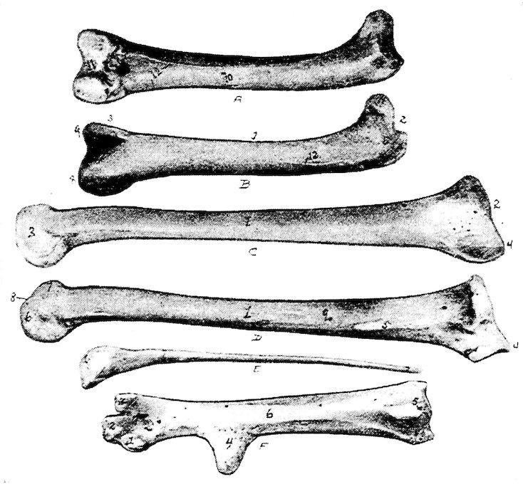
Fig. 12.—Bones of the hind extremity.
A. Posterior view of femur.
B. Anterior view. 1, Shaft. 2, Proximal extremity. 3, Distal extremity. 4, Articular head. 5, Trochanter major. 6, Shallow trochanteric fossa. 7, Convexity over which the tendon of the gluteus maximus glides. 8, External condyle. 9, Internal condyle. 10, Nutrient foramen. 11, Intercondyloid fossa. 12, Muscular linear ridges. 13, Epitrochlear fossa.
C. Internal view of tibia.
D. External view. 1, Shaft. 2, Articular head. 3, Distal end. 4, The rotular process. 5, The fibular ridge. 6, External condyle. 7, Internal condyle. 8, Intercondyloid space. 9, Nutrient foramen.
E. Fibula, lateral view. 1, Articular head. 2, Its attenuated portion.
F. Posterior view of metatarsus. 1, Trochlea for inner or second digit. 2, Trochlea for middle or third toe. 3, Trochlea for outer digit. 4, Bony core for spur. 5, Bony canal for tendons. 6, Groove for tendons. 7, Fossa intercondyloidea.
The inner condyle begins anteriorly as a ridge, and expands into a convexity which attains its greatest breadth posteriorly where it 52becomes more flattened. The inner side of the inner condyle is flattened and is provided with a tuberosity at its mid-part and a second just above the posterior part of the condyle.
The outer condyle is formed in the same manner as the inner condyle. It is indented at its broad, lower end by an angular groove, which, winding divides the posterior part of the condyle into two convexities. The more external convex ridge and the groove dividing it from the outer condyle are adapted to the head of the fibula. There is in this part a fibular ridge and above this ridge a tuberosity.
The Tibia. Location.—The tibia (Fig. 12, D and C) extends obliquely downward and backward from the knee-joint to the hock. It articulates above with the femur and, by its procnemial process with the patella, below with the metatarsus, and laterally with the fibula.
Description.—The tibia is the longest bone in the posterior limb, and possesses a shaft and two extremities. It is largest at the proximal end and presents three faces.
The proximal extremity presents a semi-oval articular surface, not quite at right angles with the shaft, which articulates with the condyles of the femur. The margin is raised toward the anterior of the bone. The head of the tibia, or caput tibiæ, extends into a rotular process which extends transversely, and is truncate. From the anterior of this process there descends two vertical ridges; one near the angle of the rotular process, the procnemial ridge; the other from the outer fibular angle, the ectocnemial ridge. On the outer side of the intercondylar tuberosity there is a surface for the ligamentous union with the head of the fibula; and a short distance below this there is a vertical ridge for the close attachment, almost a fusion, with the fibula, called the fibular ridge.
The shaft, or corpus tibiæ is straight and the upper two-thirds subtrihedral; the lower third oval. A nutrient foramen occurs near the upper postero-internal portion of the middle third of the bone.
The distal extremity is much smaller than the proximal one; it is quadrangular in form. The expanded inferior end of the tibia forms two articular condyles above which posteriorly there is the epicondyloid fossa. The inner condyle, the larger, has a groove near the lower end of the anterior part of the shaft, which deepens toward the intercondyloid space. This intercondyloid fossa in young birds is covered by a strong ligament, which in older birds, becomes 53ossified. On the lateral side of each condyle, there is a depression for the attachment of ligaments.
The Patella. Location.—The patella, or knee-cap, thin and wide, articulates with the procnemial process of the tibia, and with the deep trochlea of the distal end of the femur.
Description.—The patella is irregular in shape with three faces and three borders. The posterior surface is articular. The other two surfaces are rough for tendinous attachment. The patellar ligaments in old birds may become ossified.
In order to turn the foot in and out, the tibia not only turns around the inner condyle of the femur, but also around the patella, so that the posterior surface turns outward and thus turns outward the metatarsus and all the toes.
The Fibula. Location.—The fibula lies at the outer border of the tibia. It articulates superiorly with the outer condyle of the femur, and laterally with the tibia.
Description.—The fibula is rudimentary; it is largest superiorly and tapers to a slender point. The head is compressed laterally, and furnishes an upper and an inner articular surface.
The Tarsus.—There exist, during fetal development of the chick, two rows of tarsal bones which later become fused. The upper row fuses with the tibia and the lower with the metatarsus. Therefore, in the adult, there is no tarsus.
The Metatarsus. Location.—The metatarsal bone extends downward and forward. In birds it consists of one bone, which articulates superiorly with the inferior extremity of the tibia. On the distal end it has a threefold trochlear arrangement which articulates with the three principal digits.
Description.—The proximal extremity posteriorly has a process which may be considered as a consolidation of originally separate metatarsal bones. The process at the supero-posterior part of the metatarsus is called the hypotarsus of the tarso-metatarsus, through which extends a canal called the hypotarsal canal, and which gives passage to flexor tendons.
The proximal end of the metatarsus is ossified from one center of ossification forming an epiphysis which caps the ends of the three original metatarsal bones that coalesce, first with one another, then with the epiphysis, thus forming a single compound bone. Above and just to the inside of the metatarsus there may occur a small bone which is imbedded in ligaments and articulates with the 54inner proximal surface of the metatarsus. This has been called by Gadow a sesamoid bone.
The shaft shows tendonal grooves which are best marked on the posterior surface. The shaft is rounded at the sides and flattened on the anterior and posterior surfaces. At the juncture of the middle and inferior thirds of the metatarsus there is a conical process turned slightly backward, which serves as a base for the spur. The spur is a horny structure.
At the distal extremity occurs the threefold trochlear arrangement mentioned above, which incloses the fossa intercondyloidea. The inner trochlea is the broadest, and the outer the narrowest. The inner trochlea articulates with the proximal end of the second, the middle with the third, and the outer with the fourth toe.
The Phalanges. Location.—Most domestic fowls are provided with four digits, or toes; the Houdan and Dorking are provided with five. In fowls with four toes the three principal toes, the second, the third, and the fourth, are directed forward, and the first, or hallux, is directed backward.
Description.—The last phalanx of each toe, called the ungual phalanx, is slightly curved downward, is pointed anteriorly, like the claw of the cat or dog, and is covered with a horny sheath. The articular ends of the joints of the phalanges are enlarged. The base of the basal phalanx has two enlargements, the superior-inferior tubercles, or tuberculum superius et inferius, between which is located the fossa articularis transversa. Laterally the head has two condyles, condyli laterales, which are divided by the sulcus longitudinalis. The bodies of the phalanges vary in form; they are superiorly rounded, but inferiorly rounded, flat, or even somewhat concave. The distal extremity has an articular trochlea.
The first toe, or digit, called the great toe, or hallux, is composed of three phalanges, or segments. The first segment, or basal phalanx is considered a rudimentary metacarpal bone; it is attached by a fibrous cartilaginous tissue to the inner posterior surface of the inferior extremity of the metacarpal bone just below the spur.
The second toe, likewise composed of three phalanges, is directed forward. It articulates with the inner trochlea located on the inferior metacarpal bone.
The third toe, made up of four phalanges, is the middle of the 55three forward toes. It articulates with the middle trochlea of the inferior extremity of the metacarpal bone.
The fourth toe, is composed of five phalanges; it is, however, of approximately the same length as the third, the segments being shorter. The fourth or outer toe articulates with the outer trochlea of the distal extremity of the metacarpal bone.
ARTHROLOGY
Kinds of Joints.—Joints may be movable, immovable, or mixed. In movable, or true joints the articular surface of each bone is covered by cartilage. The bones are held together by ligaments, the capsular one often surrounding the joint and enclosing the synovial membrane. In some joints there is a pad of fibrous cartilage interposed between the two articular cartilages. Such a pad, called a meniscus, adds to the elasticity and the free movement of the joint. Movable joints form the most numerous class; they are for the most part found in the limbs.
In an immovable joint there is only a thin layer of fibrous or cartilaginous material interposed between the bones. The fibrous layer of the periosteum of both bones unite to cover the connecting material and becomes attached to the same, thus serving as a ligament. If the connecting material is fibrous, the joint is called a suture; if cartilaginous, a synchondrosis. These joints are found in the skull and in the pelvis.
The term mixed is used with reference, not to the motion in joints, but to their structure, which partakes of the nature of both the movable and immovable. The bones are firmly joined by a strong interposed pad of fibrous cartilage to which also is adherent the ligaments of the joint. There are no capsular ligaments; the cartilaginous pad or disc is softer toward its center, where occasionally there may be one, or even two, narrow cavities. Authorities differ as to whether such cavities are lined by synovial membrane or not. Since there are really no frictional surfaces in such a joint, motion depends upon the flexibility of the disc. The joints between the vertebral centra afford the best illustration of the mixed class.
Movement of Joints.—The movements admissible in joints may be divided into four kinds: gliding, angular movement, circumduction, and rotation. These movements are often, however, more or less combined in the various joints. It is seldom that there occurs only one kind of motion in any particular joint.
Gliding movement is the most simple kind of motion that can 57take place in a joint, one surface gliding or moving over another without any angular or rotary movement.
Angular movement occurs only between the long bones. By it the angle between the two bones is increased or diminished. It may take place in four directions: forward and backward, consisting of flexion and extension; or inward and outward from the medial line of the body, consisting of adduction and abduction. Abduction of a limb is movement away from the medial line of the body. Adduction of a limb is movement toward the medial line of the body.
Circumduction is that limited degree of motion which takes place between the head of a bone and its articular cavity, whilst the extremity and sides of the limb are made to circumscribe a conical space, the base of which corresponds with the inferior extremity of the limb, the apex with the articular cavity; this kind of motion is best seen in the shoulder- and hip-joint.
Rotation is the movement of a bone upon an axis, which is the axis of the pivot on which the bone turns, as in the articulation between the atlas and axis, when the odontoid process serves as a pivot around which the atlas turns; or else is the axis of a pivotlike process which turns within a ring, as in the rotation of the radius upon the humerus.
Pronation is a form of rotation in which the inferior extremity of the radius passes before the ulna, and thus causes the hand to execute a kind of rotation from without inward.
Supination is a form of rotation in which the movement of the forearm and hand are carried outward so that the anterior surface of the latter becomes superior.
The Ligamentous Structure.—Ligaments are dense, fibrous, connecting structures. They are made up principally of white fibrous tissue and exist in all true joints.
There are four kinds of ligaments associated with true joints:
The first kind, the capsular ligament which encloses all true joints, is thin and consists of interlaced fibers attached to the bone at the edges of the articular cartilages. It either partly or wholly surrounds the joint, enclosing and protecting a synovial apparatus, which, by secreting a liquid resembling serum, lubricates the joint to prevent friction.
The second kind, the binding or lateral ligaments, consist of flattened or rounded cords or bands of fibrous tissue. Such a ligament extends from one bone to the other, and firmly attached to 58their roughened surfaces, holds the bones in place and at the same time allows the required amount of motion.
The third kind, located between the joints, is called interosseous ligament.
The fourth kind, called the annular ligament binds down and protects the tendons.
Ligaments of the Ear[2].—The concha of the ear is provided with a superior and an inferior ligament.
2. The classification of Gadow is used.
Ligaments of the Jaw.—The articulations of the lower jaw are complex. The freely movable articulation is between the inferior maxilla and the quadrate. Less freely movable articulations are formed by the quadrate with the temporal, the zygomatic and the pterygoid. A lateral ligament of the jaw, the articulo-jugale extends from the posterior border of the inner wing of the os articulare of the inferior maxilla (Fig. 8, No. 10, and Fig. 19, No. 15) to the outer border of the os quadrato jugulare.
A lateral temporo-maxillary ligament extends from the outer surface of the temporal bone to the outer border of the os articulare of the inferior maxilla (Fig. 8, No. 11, and Fig. 19, No. 14).
Ligaments of the Vertebræ.—In each space between the bodies of the vertebræ there is a meniscus intervertebralis. This meniscus is analogous to the annulus fibrosus of mammals (Gadow), which, is possibly formed as a protrusion of the anterior surface of the vertebral body. The meniscus or disc may develop into a ring-shaped structure, the true meniscus, or it may develop to different degrees as an extension of the vertebral body surface, and become, as in the dorsal and lumbar regions, fused with the vertebral segments, in which case it is called the annulus fibrosus.
The vertebral disc which is connected with the atlas and which is fused with that bone, represents the first meniscus, which is called the ligamentum transversum atlantis. This, as well as other ligaments of the spinal column, may become ossified.
The ligament which is located between those vertebral bodies which face each other and is inside the joint cavity, is called the ligamentum suspensorium corporum vertebralium. It passes through the central opening of the meniscus and lies exactly in the long axis of the body of the vertebra.
The first ligament of the neck is the ligamentum suspensorium dentis epistrophei. The ligamentum capsulare atlantico-occipitale 59and the ligamentum capsulare atlantico-epistrophicum odontoideum are two parts of the joint capsule of the vertebral body. Other ligaments of this part are the membranæ obturatoriæ intervertebrales posteriores, which are located between the semicircular rims of the neck vertebræ.
The ligamentum transversum atlantis surrounds the occipital condyle.
The ligamentum nuchæ is a thin, membranous, ribbon-like structure which lies between the muscles of the right and those of the left side of the middle, and the lower part on the posterior of the neck, and ends in attachments to the superior spines of the cervical vertebræ.
The ligamentum elasticum interspinale profundum and the ligamentum elasticum interspinale superficiale and the three last named are the ligaments which keep the neck of the fowl in the s-shape, without the action of the muscles.
The ligamentum capsulare obliquum connects the facets of the oblique processes.
Ligaments of the Ribs.—The thick inferior end of the sternal portion of the true rib has two small articular heads, which articulate with two depressions in the articular surface of the sternum. This articulation is held firm by a capsular ligament.
The upper end of the sternal rib forms an almost perfect right angle with the inferior end of the dorsal rib with which it is connected by a joint provided with a synovial apparatus and a capsular ligament. This arrangement allows free movement outward and inward and is the main joint in respiration.
The upper end of the dorsal portion of the rib articulates with the dorsal vertebra. The joint formed by the articular head with the body of the vertebra is provided with a capsular ligament. The tubercle of the rib articulates with the facet on the transverse process of the dorsal vertebra, and is provided with a transverse ligament, called the ligamentum transversum externum. It is also provided with a capsular ligament.
The ligamentum triangulare connects each processus uncinatus with the succeeding rib. This ligament is in the form of a membranous sheet, or aponeurosis.
Ligaments of the Sternum (Fig. 13).—The external and the internal notches of the sternum are bridged over with a thin membrane 60which gives an extensive surface for muscular attachments. The sternal muscles overlie this portion.
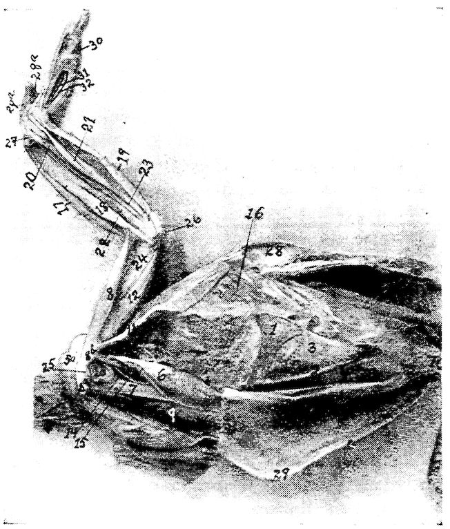
Fig. 13.—Muscles of the fore extremity. Inside view. 1, Lateral external process of sternum. 2, Lateral internal process of same. 3, Ligament of the external notch. 4, Ligament of the internal notch. 5, Pectoralis major. 5a, Its fan-shaped expansion at shoulder-joint laid back. 6, Pectoralis tertius. 7, The coracoid. 8, Biceps. 8a, Its long head. 8b, Its short head. 9, Pectoralis secundus. 10, Rectus abdominis. 11, Teres et infraspinatus. 12, Deltoid. 13, Capsular ligament. 14, Teres minor. 15, Coraco-brachialis. 16, Serratus magnus anticus. 17, Extensor metacarpi radialis longior. 18, Pronator brevis. 19, Flexor carpi ulnaris. 20, Flexor carpi ulnaris brevior. 21, Flexor digitorum profundus. 22, Extensor indicis longus. 23, Extensor ossis metacarpi pollicis. 24, Humerus. 25, Shoulder. 26, Elbow. 27, Carpus. 28, Section through vertebra. 29, Keel of sternum. 28a, Flexor brevis pollicis. 29a, Extensor proprius pollicis. 30, Flexor minimi digiti brevis. 31, Interosseous palmaris. 32, Interosseous palmaris.
The sternum is connected by a fibrous mass with the inferior portion of the clavicle, or hypocledium (Fig. 57), the claviculosternal ligament.
The inferior narrow elongated end of the coracoid forms with the sternum a true articulation, which is provided with a capsular 61ligament, the ligamentum capsulare. Capsular ligaments occur at the articulations between the sternum and ribs.
Ligaments of the Shoulder-joint (Fig. 13).—The shoulder-joint is made up of the scapula, the humerus, and the coracoid. The ends of these three bones form the foramen triosseum (Fig. 15, No. A, 7) through which passes the tendon of the elevator muscle of the wing.
The furcula, independent of the shoulder-joint or girdle, is attached to the supero-internal part of the proximal end of the coracoid by fibrous connective tissue; it is also connected to the other bones of the shoulder-joint by the ligamentum coraco-furculare and the ligamentum furculo-scapulare.
The ligamentum coraco-scapulare extends from the tuberosity of the furcula to the coracoid and to the processus furcularis of the scapula.
The ligamentum coracoido-scapulare externum extends between the external tuberosity of the coracoid, the tuberosity of the scapula, and the humerus.
The ligamentum coracoideo-scapulare inferius extends from the coracoid to the inner tubercle of the scapula.
Another long, broad ligament belongs to the episternal apparatus.
The shoulder-joint is provided with a wide, loose, capsular ligament (Fig. 13, No. 13). Attached to the humerus are four other ligaments, of which three pass from the anterior end of the coracoid and the fourth from the scapula. The latter are as follows: first, the supero-anterior ligamentum humero-coracoideum, which extends from the small tubercle of the humerus to the coracoid bone; second, the antero-inferior ligamentum humero-coracoideum, which extends from the humerus to the coracoid; third, the ligamentum coraco-humerale, which extends from the coracoid to the large tubercle of the humerus; fourth, the ligamentum humero-scapulare which extends between the processus humeralis of the scapula and the head of the humerus.
Ligaments of the Elbow-joint (Fig. 14).—The elbow-joint is made up of the ulna, radius and the humerus. The ligaments of the elbow-joint are as follows: the ligamentum capsulare cubiti, or capsular ligament, which extends from the processus cubitalis of the humerus to the processus olecranalis coracoideus of the ulna and to the tuberositas radii of the radius (Fig. 15, No 5).
The ligamentum laterale cubiti externum connects the outer humeral distal extremity with the head of the radius (Fig. 15, No. 2).
The ligamentum laterale cubiti internum lies between the inner 62distal extremity of the humerus and the tuberculum internum of the ulna (Fig. 14, No. 9).
The ligamentum annulare radii originates on the olecranon, surrounds the head of the radius, and is attached to the tuberculum internum of the ulna (Fig. 15, No. 3).
The ligamentum cubiti teres extends from the head of the radius to the upper end of the ulna (Fig. 15, No. 4).
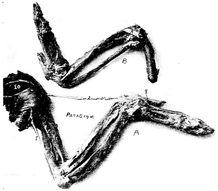
Fig. 14.—Muscles and ligaments of the arm and forearm of a hen.
A. External view. 1, Expansor secundarium. 2, Tensor patagii longus. 3, Tensor patagii brevis. 5, Brachialis anticus. 6, Pronator longus. 7, Supinator brevis. 8, Extensor indicis brevis. 10, Scapulo-humeralis.
B. Internal view. 9, Ligamentum laterale cubiti internum.
The ligamentum transversum spreads out between the head of the ulna and the radius, thus uniting the two bones, and limiting supination (Fig. 15, No. 1).
Ligaments of the Carpal Joint.—The carpal joint is made up of the ulna, radius, the two carpal bones and the metacarpus.
There are two strong ligamenta obliqua carpi ulnaris which extend 63from the processus styloideus of the ulna to the tuberculum posterius carpi ulnaris of the os carpi ulnaris.
The ligamentum posticum ulnare carpi ulnaris extends from the processus styloideus of the ulna to the os carpi ulnaris.
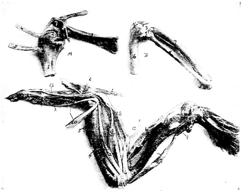
Fig. 15.—Ligaments of the arm and forearm of a hen.
A. The scapulo-coraco-humeral articulation. 1, Proximal end of the humerus. 2, Articular head of humerus. 3, The coracoid. 4, Proximal end of the clavicle. 5, Tendon of pectoralis tertius. 6, Tendon of pectoralis secundus. 7, Opening or foramen triossium through which the tendon of the elevator of the wing passes. (Pectoralis secundus.) 8, Broken end of scapula.
B. 1, Ligamentum transversum. 2, Ligamentum laterale cubiti externum. 3, Ligamentum annulare radii. 4, Ligamentum cubiti teres. 5, A portion of the ligamentum capsulare cubiti. 6, Distal extremity of humerus. 7, Ulna. 8, Radius.
C. Muscles of the outside surface of the arm. 1, Tensor patagii longus. 2, Extensor digitorum communis. 3, Tensor patagii brevis. 4, Anconeus. 5, Flexor carpi ulnaris. 6, Biceps. 7, Triceps. 8. Deltoid. 9, Brachialis anticus. 10, Flexor metacarpi radialis. 11, Extensor ossis metacarpi pollicis. 12, Extensor metacarpi radialis longior. 13, Extensor indicis longus.
The ligamentum ulnare carpi radialis is a short ribbon-like ligament which passes over the above-mentioned ligaments and is attached to the inner surface of the os carpus radialis.
The ligamentum ulnare carpi radialis internum is a strong ligament 64which originates on the inner part of the elbow. It extends to the upper rim of the base of the main digit.
The ligamentum radiale carpi radialis externum originates on the outside of the head of the middle digit and inserts to the superior tuberculum carpi radialis of the radius.
The ligamentum carpi radialis internum, a short ligament, extends from the inside of the head of the main digit to the inner rim of the carpi radialis.
The ligamentum carpi interosseum is located between the carpal bones.
The ligamentum ulnare metacarpi internum is spread out between the processus styloideus of the ulna and the tuberositas muscularis on the upper side of the middle finger base at a point where the second and the third metacarpi separate.
The ligamentum ulnare metacarpi externum extends from the tubercle of the distal extremity of the ulna, on the dorsal side, to the tubercle on the base of the metacarpus.
Ligamentum radiale metacarpi extends from the head of the inner digit to a point near the tuberculum muscularis of the radius.
The ligamentum transversum ossis carpi radialis et metacarpi is located between the inferior tuberculum ossis carpi radialis and the first metacarpal bone.
There is another ligament between the tuberculum superiorus carpi radialis and the first metacarpal bone.
The ligamentum ossis carpi radialis internum et metacarpi extends from the inner surface of the carpus radialis to the tuberositas muscularis of the radius.
The ligamentum ossis carpi ulnaris externum et metacarpi extends from the ulnar carpal bone to the tuberculum of the metacarpal bone.
The ligamentum ossis carpi ulnaris internum et metacarpi extends from the processus uncinatus of the ulnar carpal bone to the tuberositas muscularis of the metacarpal bone.
Ligaments of the Finger.—The ligamentum pollicare connects the thumb with the first metacarpal bone.
The ligamentum anterius ossis metacarpi et primæ phalangis digiti secundi extends from the tuberculum articulare metacarpi, of the proximal end of the metacarpus, to the tuberculum articulare of the first phalanx of the second or large finger. Similarly attached, are also an internal and a posterior ligament, or ligamentum internum and ligamentum posterius.
65The three phalanges are all provided with capsular ligaments, or ligamenta capsularia, which bind together the several phalanges.
The small or third finger is connected to the metacarpus by a capsular ligament, the ligamentum capsulare, and by an interosseous ligament, or ligamentum interosseum digitorum, to the first phalanx of the second finger.
Ligaments of the Pelvis.—The obturator foramen, formed by the ilium and the ischium, is covered by a broad membranous ligament (Fig. 4, No. 29).
The oblong foramen, or foramen oblongum, and the foramen ischiadicum, formed by the ischium and the pubis are covered by broad membranous ligaments (Fig. 4, Nos. 29 and 60).
Poupart’s ligament or ligamentum Poupartii, quite small in fowls, extends from the anterior lower rim of the ilium to the pelvic cavity, and is inserted at the bottom of the acetabulum.
Another broad ligament originates from the posterior rim of the ischium and is attached to the square surface of the first coccygeal vertebra.
The ilio-pubic ligament extends from the pubic spine to the last rudimentary rib.
Ligaments of the Hip-joint (Fig. 25, F).—The hip-joint is made up of the ilium, ischium, pubis and femur and is a deep ball-and-socket joint. The cavity or acetabulum is called a cotyloid cavity.
The hip-joint is provided with three ligaments.
The ligamentum capsulare femoris is attached around the rim of the cotyloid cavity and around the rim of the articular head of the femur (Fig. 25, No. F, 4).
The ligamentum teres, or round ligament, is a very short ligament which closes the hole at the floor of the cotyloid cavity, at which point it is attached. It is also attached to the head of the femur.
The ligamentum ilio-sacrale strengthens the hip-joint capsule. It passes from the lower anterior rim of the os ilii, extending over the capsule to the neck of the femur to which it is attached.
The Ligaments of the Knee-joint (Figs. 16 and 17).—The knee-joint is made up of the femur, patella, tibia and fibula, and is provided with the following ligaments:
The ligamentum extero-laterale genu is a strong ligament extending from the condylus externus femoris, or external femoral condyle to the outer surface of the head of the fibula, or capitulum fibulæ. 66From this head there passes inward, a strong ligament to a point between the femur and fibula (Fig. 25, No. D, 4).
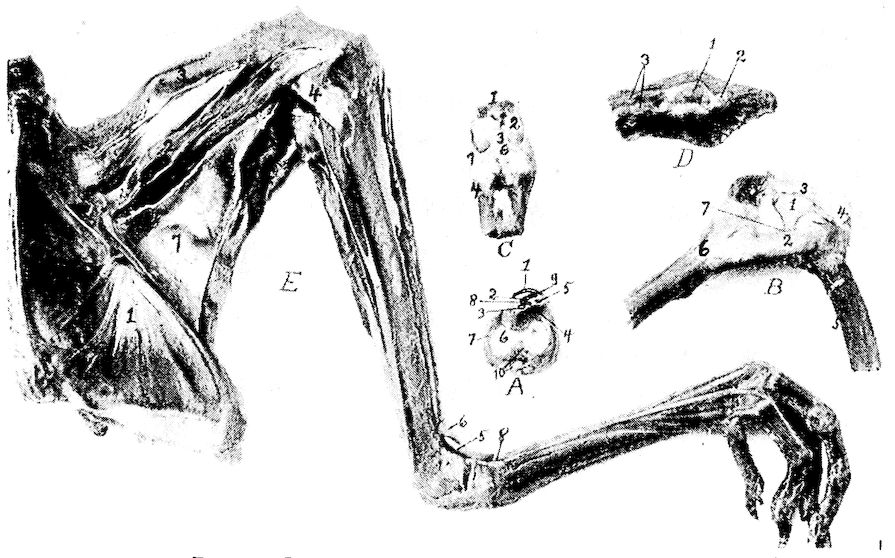
Fig. 16.—Ligaments and muscles of the hind extremity.
A. A transverse section through the tibial or tarsal cartilage, front view. 1, Gastrocnemius tendon. 2, Flexor perforatus annularis primus pedis. 3, Flexor perforans digitorum profundus. 4, Flexor perforatus indicis secundi pedis. 5, Extensor longus hallucis. 6, The articular surface. 7, Ligamentum capsulare ossis tarsi. 8, Flexor perforatus medius secundus pedis. 9, Flexor perforatus indicis secundus pedis. 10, Tendons of the extensor muscles.
B. Knee-joint. Inside view of femoro-tibial articulation. 1, Anterior ligamentum cruciatum. 2, Internal ligamentum laterale genu. 3, Anterior patellar ligament. 4, Patella. 5, Distal end of femur. 6, Proximal end of tibia. 7, Location of meniscus or pad of fibrocartilage.
C. Anterior view of tibio-tarsal articulation. 1, Distal end of tibia. 2, Its articular surface. 3, Ligamentum anticum. 4, Proximal end of metacarpus. 6, Pad of fibrocartilage.
D. The dorsal surface of the coccyx. 1, The bilobate oil gland. 2, Its duct. 3, Levator coccygis.
E. Inside view of pelvis and thigh. 1, Obturator internus. 2, Ambiens. 3, Vastus internus. 4, Internal ligamentum laterale genu. 5, Tibialis anticus. 6, Loop through which the tibialis anticus passes. 7, Adductors of the thigh. 8, Loop for the extensor tendon.
The ligamentum intero-laterale genu extends from the outer surface of the internal condyle of the tibia, or condylus internus tibiæ. It gives off a thin ligamentous slip which enters the joint and is attached to the inner half-moon shaped pad of fibrous cartilage, or meniscus, which it draws backward by the flexion of the knee (Fig. 16, No. B, 2).
67The ligamentum popliteum arises from the fossa poplitea of the distal extremity of the femur and extends downward to the posterior rim of the head of the tibia.
The anterior ligamentum cruciatum genu originates from the fossa poplitea and extends outward antero-laterally to the rim of the head of the tibia (Fig. 25, No. 6).
The posterior ligamentum cruciatum genu is a short, strong ligament which originates from the cavity of the internal condyle of the femur, or condylus internus femoris, and inserts into the internal glenoid cavity of the tibia (Fig. 25, No. 1) or cavitas glenoidalis interna.
The meniscus of the femoro-tibial articulation is divided into four parts, as follows: first, the internal adhesio cornu antici cartilaginis lunatæ; second, the posterior adhesio cornu antici cartilaginis lunatæ; third, the external cornu cartilaginis lunatæ; fourth, the anterior cornu antici cartilaginis lunatæ. The latter originates between the condyles and passes around the ligamentum cruciatum genu posticum (Fig. 16, No. B, 7).
The inner semi-lunar fibrous cartilage is well developed and is joined by two ligaments between the femoral and the tibial surfaces (Fig. 25, No. 1).
The external semi-lunar cartilage lies in a cavity between the external femoral condyle and the head of the fibula (Fig. 25, No. 2).
The fibrous cartilages are bound posteriorly by a ligament, and by a second ligament to the head of the fibula, or capitulum fibulæ. Both pads of fibrous cartilage are connected by a transverse ligament called the ligamentum transversale commune (Fig. 25, No. 5).
The patella is provided with several ligaments, as follows:
The anterior patellar ligament, broad, strong, and irregular in thickness, extends from the inferior margin of the patella to the rim of the second tibial crest (Fig. 16, No. B, 3).
68The ligamentum capsulare capituli fibulæ is spread out between the head of the fibula, or capitulum fibulæ, and the superficies glenoidalis peronea tibiæ.
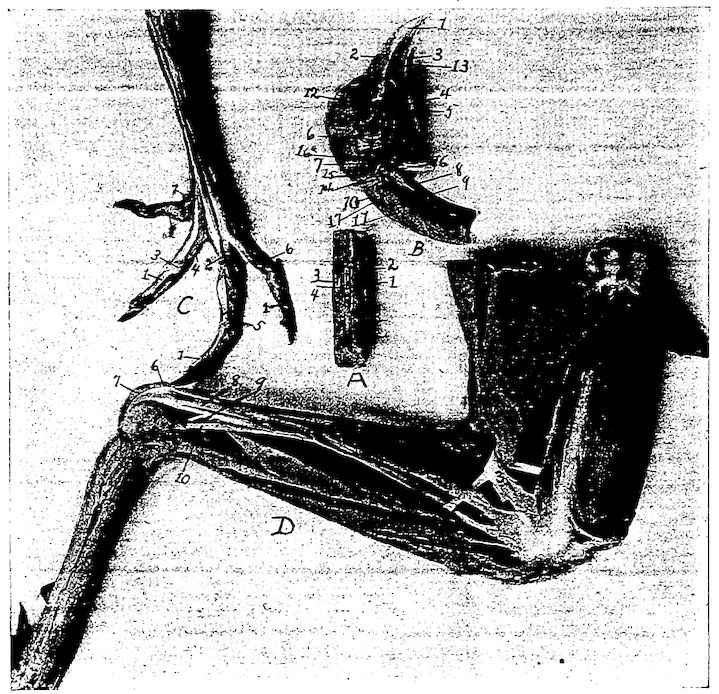
Fig. 17.—Muscles and tendons of the head and posterior extremity.
A. Section of neck with superficial muscles removed. 1, Obliquo-transversales. 2, Interspinales. 3, Intertransversales. 4, Interarticulares.
B. Head showing muscles. 1, Beak. 2, Nostril. 3, Tongue. 4, Sublingual salivary gland. 5, Pterygoideus. 6, Stylo-hyoideus. 7, Temporal. 8, Flexor capitis inferior. 9, Rectus capitis lateralis. 10, Rectus capitis posticus major. 11, Trachelo-mastoideus. 12, Eye. 13, Mylo-hyoideus. 14, Genio-hyoideus. 15, Biventer maxilla. 16, Digastricus. 17, Rectus capitis anterior minor.
C. Foot of hen showing tendons. 1, Flexor perforans digitorum profundus. 2, Flexor perforatus medius primus pedis. 3, Sheath at joint through which tendon passes. 4, Flexor perforatus indicis primus pedis. 5, Flexor perforatus medius secundus pedis. 6, Flexor perforatus annularis primus pedis. 7, Flexor longus hallucis.
D. Outside view of leg of hen. 1, Biceps flexor cruris. 2, Semitendinosus. 3, Loop through which biceps flexor cruris passes. 4, Tibialis anticus. 5, Flexor perforatus indicis secundus pedis. 6, Flexor perforatus medius primus pedis. 7, Gastrocnemius. 8, Flexor perforatus annularis primus pedis. 9, Flexor perforatus medius secundus pedis. 10, Extensor longus hallucis.
The ligamentum tibio-fibulare extends from the head of the fibula, or capitulum fibulæ, to the inner surface of the external crest, or crista externa tibiæ, of the tibia (Fig. 25, No. 3).
69The ligamentum interosseum is a delicate ligament located between the tibia and fibula (Fig. 25, No. 4) which very early becomes ossified.
Ligaments of the Tibio-metatarsal Joint (Fig. 16).—The tibio-tarsal joint is made up of the tibia and metatarsus.
The ligamentum capsulare ossis tibio-metatarsi connects the tibia and the metatarsus and surrounds the joint (Fig. 16, Nos. A, 7 and C, 7).
The ligamentum externum, a long strong ligament, originates on the upper and outer surface of the external condyle, or condylus externus of the tibia and is attached to the upper rim of the os metatarsi (Fig. 23, No. 15).
The ligamentum anticum is spread out between the fossa intercondyloidea of the tibia and the tuberculum ossis metatarsi of the proximal end of the metatarsus (Fig. 16, No. C, 3). Just posterior to this ligament, there is an interosseous ligament, the ligamentum interosseum.
A semi-lunar pad of fibrous cartilage, the cartilago semi-lunaris is located between the outer distal extremity of the tibia and upper articular surface of the metatarsus (Fig. 16, No. C, 6). Its concave portion is directed anteriorly and receives an insertion from the ligamentum externum, or external ligament.
The tendon Achillis (Fig. 16, No. A, 1) may become ossified.
Ligaments of the Toes (Fig. 16).—In addition to the capsular ligaments, which all true joints have, we find the following in connection with the toes:
The ligamentum superius and the ligamentum inferius connect the second toe with the great toe, or hallux.
The ligamentum laterale externum and the ligamentum laterale internum unite the bases of the second, third and fourth toes to the lateral faces of the trochleas of the inferior extremity of the metatarsus.
The bases of the toes are held together by the ligamenta transversa. All the rest of the toes are held together by the ligamenta capsularia digitorum pedis and by two lateral ligaments, namely: the ligamentum laterale externum and the ligamentum laterale internum.
MYOLOGY — The Dermal Muscles — TRUE DERMAL MUSCLES — THE DERMO-OSSEOUS MUSCLES — The Skeletal Muscles — THE MUSCLES OF THE HEAD — THE MUSCLES OF THE TONGUE — THE CERVICAL MUSCLES — THE MUSCLES OF THE AIR PASSAGES — THE STERNAL GROUP — THE ABDOMINAL MUSCLES — THE DORSO-LUMBAR REGION — THE COCCYGEAL MUSCLES — THE COSTAL REGION — THE ANTERIOR PECTORAL GROUP — THE PECTORAL MUSCLES — MUSCLES OF THE SCAPULAR REGION — THE MUSCLES OF THE BRACHIAL REGION — THE MUSCLES OF THE FOREARM AND THE HAND — OTHER MUSCLES OF THE DIGITS — MUSCLES OF THE POSTERIOR LIMB — THE TIBIAL GROUP — THE MUSCLES OF THE EYE — THE MUSCLES OF THE EAR — FUNCTIONS OF MUSCLES
Kinds and Structure of Muscles.—Muscles are highly specialized structures which have the property of contractility when stimulated, and thus produce motion. Muscular tissue is sometimes called flesh. Two kinds of muscles are recognized; muscles of locomotion and visceral muscles.
The muscles of locomotion may be in masses of different shape attached to the skin, the dermal, or to the skin and skeletal structure, the dermo-osseous, or from one bone to another, the skeletal. The visceral muscles form sheets and make up a portion of the wall of many of the hollow organs, such as the intestines, the stomach, the gizzard, the esophagus, and the blood-vessels. A special type of muscle forms the heart.
When classified with reference to structure, muscular tissue is divided into three types, as follows: voluntary-striated, involuntary, and involuntary-striated. The microscopic examination of each of these types of muscle shows it to be made up of fibers, these fibers to be made up of muscle cells, the muscle cell to be inclosed in a delicate tubular sheath, or membrane, called the sarcolemma. This membrane, tough and elastic, isolates each fiber. The bundles of fibers, called fasciculi, are surrounded by a fibrous sheath, which is called the perimysium internum; and the entire muscle has likewise an investing sheath of connective tissue, called the perimysium externum. The muscular cells show a longitudinal striation marking the fibrillæ, and they also show a cross striation. Nuclei are found just beneath the covering, or sarcolemma, in the striated muscle cell (Fig. 77, No. 2). In the involuntary and in the involuntary-striated muscle cell, the nuclei are centrally located.
The voluntary-striated muscles consist of cylindrical fibers and with a few exceptions, are under the control of the will. A muscle of this type, the regular skeletal form, usually has at each extremity a fibrous structure, called a tendon, by means of which it is attached to the bones. The intermediate fleshy portion of the muscle, in case of considerable bulk, is called the belly of the muscle.
The involuntary, non-striated, or smooth muscle, cells are spindle-shaped, long, and pale in color (Fig. 77, No. 1). The cells lie end to end forming fibers. These fibers do not terminate in tendons, but 71are arranged in sheets and aid in forming the walls of the digestive tract, to which they give the power of contraction and expansion.
Involuntary-striated, or heart muscle (Fig. 77, No. 4), occupies an intermediate position between the two muscles just described. It is composed of cells which branch, are somewhat rectangular. They possess both longitudinal and transverse striation. Among the fibers is found a small amount of connective tissue, as in the former types of muscle, which gives support to the blood-vessels and nerves.
Fascia.—The term fascia is applied to membranous expansions, differing materially in strength, texture, and relations. Fascia is composed of loosely arranged white fibrous connective tissue. At least two layers may usually be distinguished, the superficial fascia and the deep fascia.
Below the skin is the superficial fascia, which forms a continuous covering over the whole body and serves to attach the skin to the underlying structure.
The deep fascia more densely constructed, may be attached to the skeleton, ligaments, and tendons.
When the fascia spreads out, becomes denser, and acts as a continuation of a muscle, it is called an aponeurosis.
The Muscular Nomenclature.—In the fowl there are 162 muscles, single or in pairs. These muscles are named from their location, as the lingualis; others from their attachments, as the dermo-temporalis; some from their form, as the rhomboideus; others from their use, as the flexor or extensor; and still others from their direction, as the transversus.
Birds are provided with a system of delicate muscles divided into numerous fasciculi, which harmoniously act upon the feather quills and collectively agitate the plumage. These are the dermal muscles. This group is divided into two subgroups: the true dermal muscles, that is, those that have their origin and insertion to the under surface of the skin; and the dermo-osseous, those that originate on the surface of some bone and insert to the inner surface of the skin.
72Dermo-frontalis.[3] Location.—This muscle is somewhat rudimentary and may be entirely absent. It is located in the frontal region and is about 2 or 3 centimeters long and not so wide.
3. The classification of Shufeldt is used.
Origin and Insertion.—Closely attached to the skin.
Shape.—Flat and rather rectangular.
Relations.—Superiorly with the skin and inferiorly with the frontal bones.
Action.—By contraction the feathers on the top of the head lie flat. Those above the eyes are elevated.
Dermo-dorsalis. Location.—In the median line of the neck and back.
Origin and Insertion.—Adhering closely to the skin, it generally becomes lost at the occiput; it is most highly developed in the mid-cervical region, and it gradually disappears over the caudal region.
Shape.—Delicate and ribbon-shaped.
Relations.—Superiorly it is attached to the skin. Fat sometimes surrounds the muscle.
Action.—By contraction it raises the feathers along the superior part of the neck and dorsal region.
Dermo-tensor Patagii. Location.—Between the root of the neck and top of the shoulder.
Origin and Insertion.—Attached to the skin in the region of the anterior part of the root of the neck, some fibers passing obliquely upward and blend with fibers of the dermo-temporalis. It blends, by a slender tendon, with that of the tensor patagii longus.
Shape.—A bundle of muscular fibers later becomes thin, delicate, and triangular in shape.
Relations.—Externally to the skin.
Action.—Auxiliary to the tensor patagii longus. A tensor of this region.
The patagii are associated with the wing fold of skin which fills the angle between the arm and forearm. This fold contains elastic tissue and muscle.
Dermo-humeralis. Location.—Lateral thoracic region.
Origin and Insertion.—Fan-like delicate fibers from the skin in the abdominal integument, contracting into a long narrow fasciculus of fibers, again spreading out in fan-shape to be inserted to the tendon of the pectoralis major, just below its insertion.
73Shape.—Triangular, fan-shape.
Relations.—Superiorly with the skin.
Action.—Controls the skin in this region. The anterior end being fixed, the muscles are raised; and the posterior end acting as the fixed point, the feathers are caused to lie close to the body.
Dermo-pectoralis. Location.—On each side of the chest, lying in a longitudinal manner.
Origin and Insertion.—Attached to the skin on either side of the thorax corresponding to the dermo-dorsalis. Anteriorly it disappears over the region of the origin of the cleido-trachealis, and posteriorly just behind the tips of the post-pubic parts of the pelvis.
Shape.—Thin, delicate.
Relations.—Superiorly with the skin.
Action.—The anterior part acting as the fixed point, the feathers of the chest are raised; and the posterior end acting as the fixed point, the feathers are made to lie flat.
Dermo-temporalis (Fig. 18, No. 1). Location.—From the temporal region down the side of the neck to the anterior part of the thoracic region.
Origin.—By a broad tendinous attachment from a small depression above and anterior to the temporal fossa. From here the fibers pass upward and backward and then downward.
Insertion.—A few fibers blend with those of the cleido-trachealis. The fibers are then lost upon the skin in front and opposite the shoulder-joint. Some of the fibers blend with those of the dermo-tensor patagii.
Shape.—Long, flat, and ribbon-shaped. The lower portion consists of delicate fibers.
Relations.—Superior to the temporal muscle and sphenotic process. Superior to the cleido-trachealis and inferiorly to the shoulder-joint. The superior portion touches the skin throughout its entire length.
74Action.—A tensor of the lateral cervical integument. It is an auxiliary to the tensor patagii.
Platysma Myoides. Location.—The inferior portion of the throat.
Origin.—From the lower margin of the rami of the jaw. Just inferior to the attachment of the masseter.
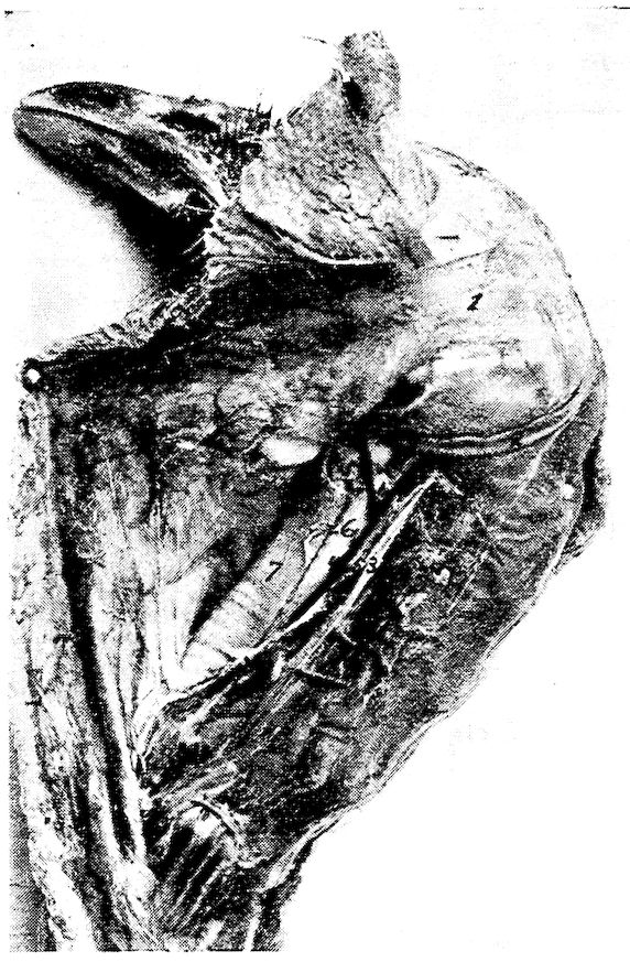
Fig. 18.—Muscles of the upper neck region, 1, Dermo-temporalis. 2, Cornu of the os hyoideum. 3, Carotid artery. 4, Jugular vein. 5, Pneumogastric nerve. 6, Esophagus. 7, Trachea.
Insertion.—From the point of origin it spreads out into a thin fan-like layer and meets its fellow in the median raphe. It is attached to the skin in this region.
Shape.—A very thin fan-shaped muscle.
Relations.—Closely adherent to the skin.
Action.—Assists in supporting the lingual apparatus and superior larynx. Compresses and elevates the part.
75Dermo-cleido Dorsalis. Location.—Anterior shoulder region.
Origin.—From the upper mesial part of the clavicular bone.
Insertion.—Three fasciculi forming a fan-like arrangement attached to the skin in the shoulder-joint and scapular region. The extremities of the fasciculi may meet in the median line of the dorsal region and merge with the dermo-dorsalis.
Shape.—Three fasciculi forming a fan-shaped radiation.
Relations.—Superiorly with the skin. Inferiorly with the shoulder-joint.
Action.—Contracting with the origin as the fixed point, they brace the skin over the forepart of the back. With the integumental attachment as the fixed point, they aid in the act of respiration.
Cleido-trachealis. Location.—Extending from the shoulder to the superior laryngeal region.
Origin.—From a small area on the antero-inner part of the middle of the arm of the furcula.
Insertion.—The fibers pass upward and become flattened and attached to the skin and dermo-temporalis muscle. They touch each other on the anterior border of the superior larynx, the trachea, and the skin over these parts.
Shape.—A slender bundle of muscles.
Relations.—Superiorly the skin, and internally the larynx and the trachea.
Action.—Controls the skin region of this part of the neck.
Dermo-spinalis. Location.—In the region of the shoulder.
Origin.—In an attenuated fascia from the crest of the neural spines of the first, the second, and the third dorsal vertebræ.
Insertion.—To the skin in a broad pale stratum, over the scapular region.
Shape.—Thin, pale, and very delicate.
Relations.—Superiorly with the skin, and inferiorly with the shoulder.
Action.—Controls the skin of the region.
Dermo-iliacus. Location.—Along the back on either side of the spine.
Origin.—From the inner angle of the marginal portion of the antero-dorsal border of the ilium.
Insertion.—Passing forward as a delicate band it spreads out and becomes lost in the skin of the shoulder region.
Shape.—Thin, delicate, and ribbon-shaped.
76Relations.—Superiorly with the skin.
Action.—If the posterior end is fixed, it will cause the feathers to lie close to the skin. Its action would then be opposite to the dermo-dorsalis.
Dermo-ulnaris. Location.—Outer surface of the anterior upper costal and the posterior humeral region.
Origin.—By a thin fascia from the outer part of the third and the fourth true ribs at the base of their epineural appendages. Also from the fascia between them.
Insertion.—The fibers pass forward and upward as a thick muscular bundle and is loosely attached to the skin at a point just back of the humerus. The tendon later becomes spread out and attached to the fascia as far as the elbow-joint. It covers the olecranon of the ulna.
Shape.—At first rather thick, fascicular-like; later becomes tendinous; and at its attachment it becomes spread out over the olecranon.
Relation.—Inferiorly with the ribs and humerus, and externally with the skin.
Action.—A depressor of the humeral region.
Temporal (Fig. 19, No. 4). Location.—Occupies the temporal fossa.
Origin.—From the mesian line of the sphenotic process, and the adjacent wall of the orbit.
Insertion.—The fibers passing downward and forward blend with the fibers of the masseter. Inferiorly it inserts by a tendon to the coranoid process upon the superior ramal margin of the mandible.
Shape.—Fan-shaped with broad portion uppermost.
Relations.—It occupies the temporal fossa. It is related superiorly with the dermo-temporalis and skin. Inferiorly, with the biventer maxillæ and the masseter.
77Action.—It aids in closing the jaw.
Masseter (Fig. 19, No. 1). Location.—Occupies the supero-lateral portion of the surface of the lower jaw.
Origin.—In two portions: the first, by a broad and thin tendon from the entire length of the bony ridge above the external auditory meatus, from the squamosal process, and from the outer portion of the quadrate bone; the second, from the side and under border of the zygoma.
Insertion.—The first portion: The fibers pass downward and forward beneath the zygoma; a few of the fibers blend with those of the temporal; one tendon inserts to a small tubercle on the upper border of the jaw behind the coronoid process, and by fleshy insertion to the outer side of the ramus of the lower jaw, quite as far forward as the horny portion of the beak. The second portion: by a small tendon to the mandible on its upper border immediately in front of the articular portion.
Shape.—Flat, and elongate. Somewhat fleshy.
Relations.—Superiorly with the temporal, and inferiorly with the biventer maxillæ and stylo-hyoideus. Externally with the skin, and internally with the jaw bone.
Action.—Aids in closing the jaw.
Biventer Maxillæ (Fig. 19, No. 3). Location.—Covers the outer posterior portion of the mandible.
Origin.—It arises in two portions: The first, from a ridge bounding the posterior part of the auditory canal; the second, from a depression of the mesial side. These two parts blend and extend downward and forward.
Insertion.—To the posterior part of the articular end of the mandible.
Shape.—A curved fleshy mass.
Relations.—Superiorly with the temporal and stylo-hyoideus. Posteriorly with the digastricus and genio-hyoideus. Internally with the jaw bone, and externally with the skin.
Action.—It aids in opening the jaw.
Entotympanic. Location.—Posterior to the pterygoid.
Origin.—From the side of the basi-sphenoid and from the base of the rostrum immediately beyond it.
Insertion.—The fibers pass downward and forward, and insert by a double tendinous slip. One slip inserts to a spine-like process on 78the upper side of the pterygoid, and the other to the quadrato-pterygoidean articulation.
Shape.—Spindle-shaped.
Relations.—Anteriorly with the pterygoid. Internally and posteriorly with the basi-sphenoid. Externally with the pterygoideus internus.
Action.—Aids in raising the upper mandible by pulling forward the quadratus and pterygoidean against the palatines.
Pterygoideus Internus (Fig. 17, No. B, 5). (Synonym.—Pterygoideus medialis.[4])
4. According to international veterinary nomenclature.
Location.—A muscular mass at the roof of the mouth.
Origin.—From the major part of the surface of the palatine bone and the distal head and shaft of the pterygoid and the sphenoidal projection.
Insertion.—By a tendon to the antero-internal part of the articular part of the mandible.
Shape.—Fusiform with a thick fleshy belly.
Relations.—Superiorly with the palatine, the pterygoid, and the sphenoid bones. Inferiorly with the skin.
Action.—It aids in closing the jaw.
Pterygoideus Externus. (Synonym.—Pterygoideus lateralis.)
Location.—Supero-external to the pterygoideus internus.
Origin.—From the outer part of the extremity of the orbital process of the quadrate.
Insertion.—The fibers pass downward, outward and forward and are inserted to the inner part of the mandibular ramus.
Shape.—A small round bundle.
Relations.—Inferiorly with the pterygoideus internus. Externally with the inferior maxilla.
Action.—Aids in closing the jaw.
Digastricus (Fig. 17, No. B, 16). Location.—Extends from the basi-temporal to the side of the neck.
Origin.—From the external lateral angle of the basi-temporal.
Insertion.—Opposite the angle of the jaw the fibers spread out in fan-like arrangement. The muscle meets its fellow of the opposite side; extends longitudinally over the superior larynx, for some distance down the neck.
Shape.—Thin and ribbon-shaped.
Relations.—Externally to the platysma myoides. The anterior 79fasciculi blend with those of the mylo-hyoideus; internally with the larynx at the upper part.
Action.—To raise the trachea and hyoid apparatus against the pharynx.
Mylo-hyoideus. Location.—The forepart of the inter-ramal space.
Origin.—From the inner side of the lower jaw at a point just above the lower border and the inturned edge of the horny sheath of the beak.
Insertion.—By aponeurosis to the under side of the hyoid, on the median line between the first and the second basi-branchial.
Shape.—Thin, flat, and delicate.
Relations.—Inferiorly with the skin. Thin and rather broad, it meets its fellow of the opposite side. Laterally with the rami of the jaw. Superiorly with the hyoid apparatus.
Action.—Lifts the tongue upward against the roof of the mouth.
Stylo-hyoideus (Fig. 17, No. B, 6). Location.—Supero-posterior to the hyoid apparatus.
Origin.—From the outer portion of the articular enlargement of the lower jaw.
Insertion.—By a tendon to the basi-branchial of the thyro-hyal.
Shape.—A long transversely flattened fasciculus.
Relations.—Supero-posterior part of the hyoid apparatus along side the genio-hyoideus and the cerato-hyoideus.
Action.—Singly, pulls the tongue to one side; acting with its fellow, pulls the tongue upward.
Genio-hyoideus (Fig. 9, No. 7). Location.—Supero-posterior to the hyoid apparatus.
Origin.—From the anterior portion of the inner side of the lower jaw.
Insertion.—To the middle of the outer side of the basi-branchial of the corner of the os hyoides which cornu it completely envelops.
Shape.—A long, rather thick fasciculus.
Relations.—With the stylo-hyoideus and cerato-hyoideus.
80Action.—Protrudes the tongue from the mouth.
Cerato-hyoideus (Fig. 9, No. 8). (Synonym.—Kerato-hyoideus.)
Location.—Inferior to the hyoid apparatus.
Origin.—By a delicate tendinous slip from the under side of the shaft of the basi-hyal element of the hyoid apparatus.
Insertion.—To a small elevation on the under side of the anterior end of the glosso-hyal.
Shape.—A small round muscular fasciculus becoming tendinous in its anterior half.
Relations.—Superiorly with the hyoid apparatus.
Action.—Singly, pulls the tongue to one side; acting with its fellow, depresses the tongue.
Sterno-hyoideus. Location.—Inferior to the hyoid apparatus.
Origin.—From the outer anterior surface of the thyroid bone of the superior larynx.
Insertion.—To the anterior part of the basi-hyal.
Shape.—At first broad and rather fleshy, becomes contracted and somewhat tendinous.
Relations.—Inferior to the hyoid apparatus and superior larynx.
Action.—When the larynx is fixed, acting alone deflects the tongue laterally; acting with its fellow, depresses the tongue. If the base of the tongue is fixed the two pull the larynx forward. Hence muscles of deglutition.
Depressor-glossus. Location.—Superior to the basi-hyal.
Origin.—From the under portion of the basi-hyal.
Insertion.—To under part of the glosso-hyal.
Shape.—Small, short.
Relations.—Superiorly with the basi-hyal and glosso-hyal bones.
Action.—Depresses the tip of the tongue and elevates the base.
Cerato-glossus. Location.—On the upper portion of the cornu of the hyoid.
Origin.—One-half of the surface of the first basi-branchial.
Insertion.—To the upper side of the shaft of the cerato-branchial element of the hyoid apparatus.
Shape.—A small muscular fasciculus.
Relations.—Supero-laterally to the cerato-branchial element.
Action.—Elevates the cornua of the hyoid arches and presses them against the skull.
Hyoideus Transversus (Fig. 9, No. 9 and 10). Location.—Envelops the spur process of the euro-hyal.
81Origin and Insertion.—It extends from the inner side of one cornu to the spur process.
Shape.—Thin, flat.
Relations.—Posterior to the euro-hyal, which it envelops, and between the cornua of the os hyoides.
Action.—Approximates the cornua of the os hyoides.
Complexus (Fig. 19, No. 5). Location.—Supero-lateral portion of the neck.
Origin.—By three tendinous slips from the antero-lateral portion of the transverse process of the fourth, the fifth, and the sixth cervical vertebræ.
Insertion.—The muscular fibers pass around the neck and meet their fellows in the median line in a fascia formation. The thin tendinous sheet inserts into the occiput just above the occipital ridge.
Shape.—From point of origin this muscle expands into a broad sheet.
Relations.—It overlies the muscles of the occipital region.
Action.—It extends the head.
Rectus Capitis Anticus Minor (Fig. 19, No. 8). (Synonym.—Rectus capitis ventralis minor.)
Location.—Infero-lateral portion of the neck.
Origin.—From the apices of the hyapophyses of the second, the third, and the fourth cervical vertebræ.
Insertion.—To the occiput just below the complexus.
Shape.—A long and rather thick fasciculus.
Relations.—Externally with the skin. The tendinous slips of origin pass between the flexor capitis inferior and longus colli anterior.
82Action.—Extends the head.
Flexor Capitis Inferior (Fig. 19, No. 7). Location.—Antero-inferior part of the neck.
Origin.—From the apices of the hyapophyses of the second, the third, and the fourth cervical vertebræ.
Insertion.—To the triangular area on the basi-temporal of the base of the cranium.
Shape.—At first tendinous, becomes somewhat fleshy as it passes forward.
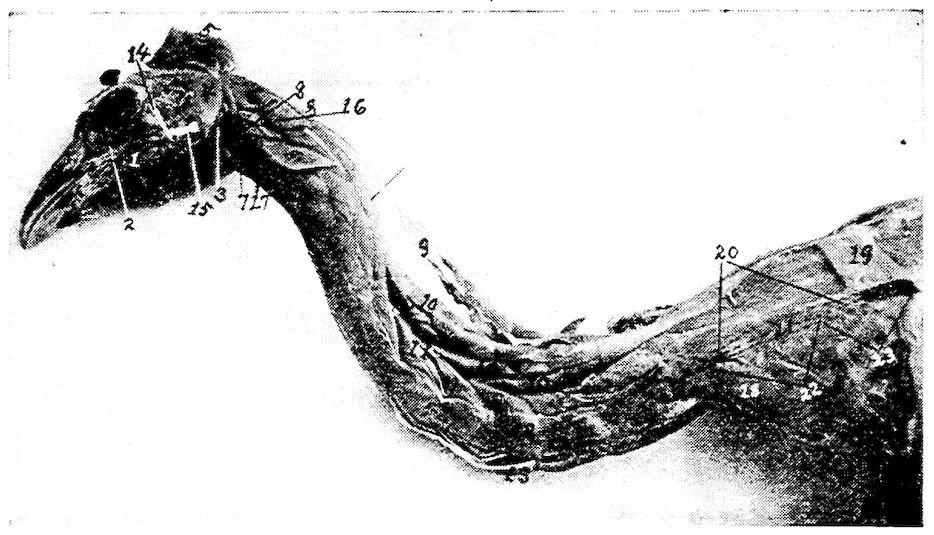
Fig. 19.—Muscles of the head and neck. 1, Masseter muscle. 2, Infraorbital sinus. 3, Biventer maxilla. 4, Temporalis. 5, Complexus. 6, Rectus capitis posticus major. 7, Flexor capitis inferior. 8, Rectus capitis anticus minor. 9, Biventer cervicis. 10, Longus colli posticus. 11, Obliquus colli. 12, Intertransversales. 13, Longus colli anterior. 14, Lateral temporo-maxillary ligament. 15, Lateral ligament of the jaw. 16, Trachelo-mastoideus. 17, Rectus capitis lateralis. 18, Scalenus medius. 19, Rhomboideus. 20, Trapezius. 21, Location of scapula. 22, Supraspinatus. 23, Teres et Infraspinatus.
Relations.—Antero-inferior portion of the neck close to the rectus capitis anticus minor.
Action.—Flexes the head upon the neck.
Rectus Capitis Posticus Major (Fig. 19, No. 6). (Synonym.—Rectus capitis dorsalis major.)
Location.—Supero-lateral area on the anterior portion of the neck.
Origin.—From the median anterior aspect of the second cervical vertebra, and the supero-anterior border of the neural canal.
83Insertion.—To the crescent-shaped area at the back of the skull.
Shape.—Short, thick, fleshy bundle.
Relations.—Superiorly with the complexus; inferiorly with the trachelo-mastoideus and the vertebræ.
Action.—Singly, pulls the head to one side; acting with its fellow, extends the head.
Biventer Cervicis (Fig. 19, No. 9). Location.—Occupies the superior part of the neck.
Origin.—From the side of the neural spine of the first dorsal vertebra, and from the adjacent tendon of the longus colli posticus.
Insertion.—To the occiput.
Shape.—Tendinous in the middle with a more or less spindle-shaped belly at each end.
Relations.—Superior to the longus colli posticus. It lies next to the skin.
Action.—Extends the head on the neck and elevates the neck.
Longus Colli Posticus (Fig. 19, No. 10). (Synonym.—Longus colli dorsalis.)
Location.—Occupies the superior part of the neck.
Origin.—From the marginal edges of the summits of the neural spines of the first two dorsal vertebræ.
Insertion.—To the transverse process of the axis, and superior part of the cervical vertebræ.
Shape.—A long, somewhat narrow, flattened muscle, the inferior portion of which is divided into five or six fasciculi.
Relations.—Superiorly with the biventer cervicis and inferiorly with the vertebræ.
Action.—Raises the neck upward.
Obliquus Colli (Fig. 19, No. 11). Location.—On the lateral side of the neck.
Origin and Insertion.—The first of the seven fasciculi originates from the diapophysis of the eleventh cervical vertebra, winds obliquely over the tenth, and is inserted into the posterior margin of the postzygapophysis of the ninth vertebra. In its passage it receives the slip from the longus colli posticus. The next fasciculus originates from the transverse process of the tenth cervical vertebra, winds obliquely over the ninth vertebra, and is inserted to the postzygapophysis of the eighth vertebra. In its passage it also receives a fasciculus from the longus colli posticus. The next three fasciculi originate in a similar manner being attached to similar postzygapophyses 84and have slips extending forward which slips are inserted to the neural spines of the alternate vertebræ. The sixth fasciculus originates from the transverse process of the sixth vertebra, passing obliquely up the neck is inserted to the extremity of the diapophysis of the fourth vertebra, the outer extremity of the transverse process of the third vertebra. The seventh, or anterior, fasciculus originates from the transverse process of the fifth vertebra, and is inserted to the extremity of the diapophysis of the third vertebra.
Shape.—Short, thick fasciculi.
Relations.—Laterally with the vertebræ. Inferiorly with the intertransversales. Superiorly with the longus colli posticus, and externally with the skin.
Action.—Flexes one vertebra on the other laterally.
Longus Colli Anterior (Fig. 19, No. 13). (Synonym.—Longus colli ventralis.)
Location.—Occupies the anterior portion of the neck.
Origin and Insertion.—This muscle is divided into a vertical portion, and a superior and an inferior oblique portion. The vertical portion originates from the hypophyses of the tenth to the fifteenth vertebra and is inserted by a tendon to the tubercle on the inferior portion of the atlas. Small, slender tendons are given off and insert to the apices of the parapophyses of the fourth to the tenth cervical vertebræ. There is more or less attachment to the bodies of the cervical vertebræ mentioned. The superior oblique portion originates from the diapophyses of the third, the fourth, the fifth cervical vertebræ and becoming tendinous is inserted to the tubercle on the inferior portion of the body of the atlas. The inferior oblique portion originates from the transverse processes of the fourth, the fifth, and the sixth cervical vertebræ. It is inserted by a slender tendon to the apex of the parapophysis of the third cervical vertebra.
Shape.—A long fleshy muscle extending the entire length of the neck.
Relations.—Inferiorly with the skin and superiorly with the cervical vertebræ.
Action.—Pulls the neck downward.
Rectus Capitis Lateralis (Fig. 19, No. 17). Location.—The infero-lateral anterior portion of the neck.
Origin.—Originates tendinous from the diapophysis of the third, the fourth, and the fifth cervical vertebræ.
85Insertion.—Passes obliquely upward in front of the spinal column to the inner tubercle on the basal ridge of the basi-temporal bone, by a subcompressed tendon.
Shape.—A somewhat thick fasciculus.
Relations.—Superiorly with the trachelo-mastoideus, inferiorly with the longus colli anterior, and externally with the skin.
Action.—Singly, pulls the head down and to one side; acting with its fellow, pulls the head downward.
Trachelo-mastoideus (Fig. 19, No. 16). (Synonym.—Longissimus capitis et atlantis.)
Location.—Laterally on the anterior portion of the neck.
Origin.—Semitendinous from the diapophyses of the second, the third, the fourth, and the fifth cervical vertebræ.
Insertion.—By subcompressed tendon to the base of the cranium, at the outer tubercle of the basal ridge of the basi-temporal.
Shape.—Flattened from side to side; broad at the posterior portion, becomes angular at the anterior portion.
Action.—Singly, pulls the head down and to one side; acting with its fellow, pulls the head downward.
Interspinales (Fig. 17, A, No. 2). Location.—Superior to the vertebra.
Origin and Insertion.—A series of muscles connecting the superior neural spines of the cervical vertebræ.
Shape.—Thin, flat.
Relations.—Anteriorly the posterior border of the vertebræ in front and posteriorly the anterior border of the succeeding vertebræ, inferiorly by the vertebræ.
Action.—To approximate the spinous portion of the vertebræ.
Interarticulares (Fig. 17, No. A, 4). Location.—Supero-laterally to the vertebræ. Between the postzygapophysis.
Origin and Insertion.—From the posterior margin of the ring of the atlas to the postzygapophysis of the axis. Then in the succeeding vertebræ from the postzygapophysis of the vertebra to the same of the succeeding vertebra.
Shape.—Muscular bundle.
Relations.—Inferiorly with the vertebræ. Supero-laterally with the obliquo-transversales.
Action.—Aids in approximating the vertebræ in a supero-lateral direction.
86Obliquo-transversales (Fig. 17, A, No. 1). Location.—Supero-lateral to the vertebræ.
Origin and Insertion.—Passes obliquely from the transverse process of one vertebra to the postzygapophysis of the vertebra beyond.
Shape.—A thin fasciculus.
Relations.—Inferiorly with the interarticulares and laterally with the intertransversales.
Action.—Aids in flexing the vertebræ supero-laterally.
Intertransversales (Fig. 19, No. 12). Location.—Laterally to the vertebræ.
Origin and Insertion.—Extend between the transverse processes beginning at the third cervical vertebra.
Shape.—Short, thick.
Relations.—Laterally the vertebræ. Superiorly the obliquo-transversales.
Action.—Aids in flexing the vertebræ laterally.
Scalenus Medius (Fig. 19, No. 18). (Synonym.—Scalenus.)
Location.—Supero-laterally to the entrance of the thorax.
Origin.—From the diapophysis and pleurapophysis of the eleventh cervical vertebra.
Insertion.—To the entire border of the first rib. A few fibers pass over to the anterior free margin of the middle third of the second rib.
Shape.—Rather pyramidal in shape with base uppermost.
Relations.—Posteriorly with the levatores costarum. Internally with the longus colli.
Action.—When the first rib is fixed singly, turns the neck to one side; acting with its fellow, extends the neck. When the neck is fixed, by drawing the first rib forward, it acts as an inspiratory muscle.
The Superior Larynx
The Inferior Larynx
87Constrictor Glottidis. Location.—Supero-anterior portion of the superior larynx.
Origin.—From the superior and longitudinal line of the thyroid plate.
Insertion.—All along the inner margin of the arytenoid bone, and to the apex of the mid-cricoidal segment.
Shape.—Thin, flat sheet extending outward and upward, and then inward.
Relation.—Interiorly with the larynx.
Action.—The two muscles of this kind acting together, close the glottis by drawing the apices of the arytenoids to the median line.
Thyreo-arytenoideus. Location.—Supero-lateral portion of the superior larynx.
Origin.—From the entire outer margin of the thyroid plate and the outer margin of the cricoid bone.
Insertion.—All along the outer margin of the arytenoid bone and the outer border of the central cricoid piece.
Shape.—A thin, flat sheet.
Relations.—Infero-laterally with the superior larynx.
Action.—The two muscles of this kind acting together, open the glottis by pulling the arytenoid bone outward.
Tracheo-lateralis (Fig. 20, No. 2). Location.—Along the lateral side of the trachea.
Origin.—By the union of the bronchio-trachealis posticus and bronchio-trachealis anticus, on the lateral side of the trachea, about 1½ centimeters from the bifurcation of the trachea.
Insertion.—In delicate fan-like structure to the side of the trachea, near the superior larynx.
Shape.—Thin, ribbon-shaped, closely attached to the trachea.
Relations.—Internally with the trachea, and externally with the skin and other integument.
Action.—Acts as a brace to the sides of the trachea, and contracting, approximates the tracheal rings and thus shortens the trachea.
Broncho-trachealis Posticus (Fig. 21, No. 3). Location.—Postero-superior portion of the trachea. The fibers pass downward and backward.
Origin.—From the inferior end of the tracheo-lateralis.
Insertion.—To the end of the third half of the bronchial ring of the same side.
Shape.—A small fasciculus.
88Relations.—Internally with the trachea, and externally with the skin and other integument.
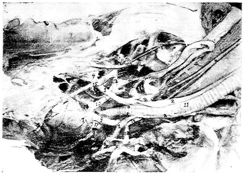
Fig. 20.—The anterior pectoral region. 1, Sterno-trachealis. 2, Tracheo-lateralis. 3, Broncho-trachealis brevis. 4, Costal process of sternum. 5, Right and left carotid converging and occupying same sheath at 6. 7, The heart showing branches of coronary artery. 8, Cervical air-sac. 9, Left brachio-cephalic artery. 10, Right brachio-cephalic artery. 11, Trachea. 12, Inferior larynx. 13. Subclavian artery. 14, Carotid trunk. 15, Internal thoracic artery. 16 Thyroid gland. 17, Anterior vena cava. 18, Auricular appendage.
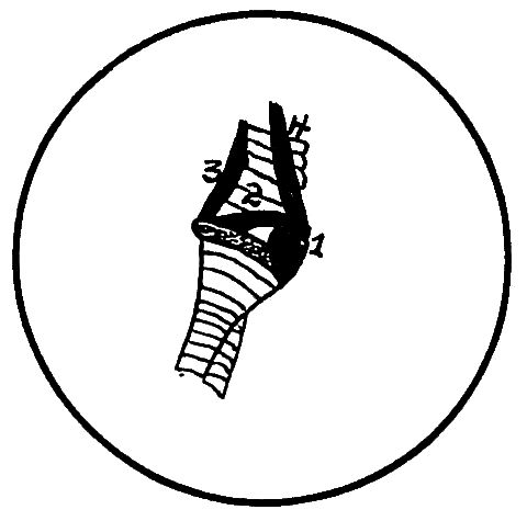
Fig. 21.—The inferior tracheal region. 1, Bronchialis anticus. 2, Bronchialis posticus. 3, Broncho-trachealis posticus. 4, Broncho-trachealis anticus.
Action.—A tensor of the true or inferior larynx.
Broncho-trachealis Anticus (Fig. 21, No. 4). Location.—Anterior portion of the inferior extremity of the trachea.
89Origin.—The anterior branch of the bifurcated tracheo-lateralis.
Insertion.—To the anterior extremity of the third half ring of the bronchus of the same side.
Shape.—A small fasciculus.
Relations.—Internally with the trachea, and externally with the skin and other integument.
Action.—A tensor of the inferior larynx.
Broncho-trachealis Brevis (Fig. 20, No. 3). Location.—Posterior part of the inferior extremity of the trachea.
Origin.—From just beneath the broncho-trachealis anticus muscle.
Insertion.—Extends obliquely across the inferior larynx and is inserted to the posterior end of the second bronchial ring.
Shape.—Rather short, strong, straight, subcylindrical.
Relations.—Internally with the trachea, and laterally with the broncho-trachealis posticus.
Action.—A tensor of the inferior larynx.
Bronchialis Posticus (Fig. 21, No. 2). Location.—Supero-lateral side of the anterior end of the bronchi.
Origin.—From the lateral inferior margin of the last tracheal ring.
Insertion.—Passing obliquely across the larynx it inserts to the posterior extremity of the second half ring of the bronchus.
Shape.—Small, thick, spindle-shaped.
Relations.—Internally with the trachea, inferiorly with the bronchialis anticus, and superiorly with the broncho-trachealis brevis.
Action.—A tensor of the larynx.
Bronchialis Anticus (Fig. 21, No. 1). Location.—Inferior to the former muscle.
Origin.—From the last ring of the trachea.
Insertion.—The fibers pass obliquely forward and become inserted to the rim of the arytenoid cartilage of the inferior larynx, and the anterior extremities of the first and second half rings of the bronchus.
Shape.—Thick, spindle-shaped; about twice the size of the bronchialis posticus.
Relations.—It is crossed at its origin by the broncho-trachealis anticus and is related internally with the bronchus and end of the trachea.
Action.—A tensor of the larynx.
Sterno-trachealis (Fig. 20, No. 1). Location.—The inferior region of the trachea above the above-named muscles.
90Origin.—From the side of the trachea beneath the posterior border of the broncho-trachealis anticus.
Insertion.—Passes downward and backward across the cavity of the chest and is inserted to the inner part of the costal process of the sternum.
Shape.—A delicate cord of fibers.
Relations.—Internally with the broncho-trachealis muscles.
Action.—A relaxor of the larynx, and hence, of the tympanic membrane.
Triangularis Sterni. Location.—In the floor of the thoracic cavity.
Origin.—From the entire superior margin of the summits of the costal processes.
Insertion.—The fibers extend upward and backward, dividing into four digitations which cover the inner surface of the three principal sternal ribs, and are inserted to the first four as high as their articulations with the vertebral ribs.
Shape.—Flat and somewhat triangular.
Relations.—Interiorly with the floor of the thoracic cavity.
Action.—A powerful muscle of expiration. In contracting, lessens the cubic content of the thorax.
The Diaphragm (Fig. 47, No. 10; Fig. 33, No. 9). Description.—The diaphragm in the domestic fowl is rudimentary. It consists of a thin semi-transparent membrane, situated between and separating the thoracic and the abdominal cavities. It readily conforms to the various organs pressing upon it from each side. On each side are three rudimentary muscles which extend from the vertebral heads of the second, the third, and the fourth sternal ribs. The fibers of these muscles spread out in fan-like upon the diaphragm, and are seen just above the digitations of the triangularis sterni.
Obliquus Abdominis Externus (Fig. 22, No. 1). Location.—The outermost muscle of the lateral abdominal wall.
91Attachments.—By a delicate aponeurotic membrane, from the sides of all the true dorsal ribs, from the posterior border of the last vertebral rib and the adjoining margins of the pelvis, and from the entire posterior surface of the inferior border of the post-pubic element of the pelvis; by aponeurosis, blending with the fascia toward the root of the tail and lower part of the abdomen; and by aponeurosis to the sides of the sternum, to the under part of the pectoralis major muscle, and to the zyphoid prolongation. Also by aponeurosis it meets its fellow from the opposite side, at the linea alba.
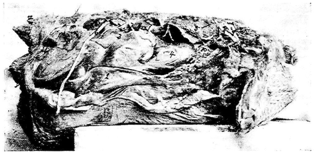
Fig. 22.—The abdominal muscles of a hen. Right side. 1, Obliquus abdominis externus. 2, Obliquus abdominis internus. 3, Peritoneum covering intestines. 4, Peritoneum covering the gizzard. 5, The lungs (note how small they are from a relative standpoint). 6, The great sciatic or ischiadic nerve. 7, Lumbar nerves. 8, Kidney. 9, Brachial plexus. 10, Pericardium of the base of the heart. 11, Anterior vena cava. 12, Oil gland. 13, Showing a sacculation of the abdominal muscles. There is no tunica abdominalis in the fowl.
Obliquus Abdominis Internus (Fig. 22, No. 2). Location.—Just internal with regard to the external oblique.
Attachments.—By aponeurosis, from the posterior third of the post-pubic element of the pelvis; by muscular fibers, from the balance of the bone; and by a few fibers, from the iliac border posterior to the acetabulum. Anteriorly, these fibers are inserted to the entire posterior margin of the last vertebral rib, and into the pleurapophysial head of the last costal rib.
Rectus Abdominis (Fig. 13, No. 10 and Fig. 23, No. 1). Location.—The inferior median abdominal wall.
92Attachments.—By aponeurosis, from the distal extremity of the post-pubic element of the pelvis and from the semitendinous ligament which stretches from one post-pubic tip to the other. Attaches anteriorly to the zyphoid margin of the sternum, and passes, by a broad aponeurotic membrane, over the outer surface of the thoracic wall beneath the external oblique.
Transversalis Abdominis (Fig. 25, No. H, 1). Location.—Infero-lateral portion of the abdominal wall.
Attachments.—By a thin, tendinous attachment from the entire post-pubic and iliac margins of the pelvis and from the interpubic ligament. Its fibers cross the abdomen between the peritoneum, the rectum, and the internal oblique. It is inserted over the entire pleural part of the last two vertebral ribs, the intercostal muscles between them, and the same surface of the hemapophyses connected below. Inserts into the linea alba.
Action.—The abdominal muscles give support to the abdominal organs, aid in flexing the spine, draw the last rib backward, thus aiding in respiration, and compress the abdominal organs to aid in defecation and expiration.
Sacro-lumbalis. Location.—The lateral lumbo-sacral region.
Origin.—Tendinous from the anterior margin of the ilium, from the angles of the last two vertebral ribs, and by tendinous slips, from the outer ends of the transverse processes of the last three dorsal vertebræ.
Insertion.—By a few fleshy fibers into the angle of the first dorsal rib and to the corresponding points upon the free cervical ribs, and by a strong semitendinous insertion into the outer extremity of the diapophysis of the twelfth cervical vertebra.
Shape.—A close fitting, tendo-muscular sheet extending between the anterior margin of the ilium and the root of the neck.
Relations.—Intimately blended with the longissimus dorsi externally.
Action.—Assists the longissimus dorsi.
Longissimus Dorsi (Fig. 24, No. 4). Location.—The superior dorso-lumbar region.
93Origin.—From the inner portion of the anterior margin of the ilium, and from the various surfaces afforded by its walls and the walls of the ilio-neural canal; by a series of short and distinct tendons alternately from the anterior and posterior extremities of the summits of the neural spines of all the dorsal vertebræ. From the diapophyses of the dorsal vertebræ, from the crests of the neural spines of the last three dorsal vertebræ, from the bodies of the dorsal vertebræ, and from the fascia between them and the sacro-lumbalis; and also by a tendinous sheet continuous with the origin of the longus colli posticus.
Insertion.—By four fasciculi into the free posterior margins of the oblique processes of the eleventh, the twelfth, the thirteenth, and the fourteenth cervical vertebræ.
Shape.—A large flat sheet.
Relations.—Laterally, with the superior spinous processes of the vertebræ, and with the superior surface of the ribs.
Action.—Singly, flexes the back laterally; with its fellow, aids in elevating the body upward.
Levator Coccygis (Fig. 23, No. 2). Location.—The superior part of the caudal apparatus.
Origin.—From a limited area of the ilium just beyond and to the side of the anterior free caudal vertebræ.
Insertion.—Into the tuberosity of the anterior margin of the pygostyle.
Shape.—Short, fleshy.
Relations.—Superior portion of the coccygeal vertebræ.
Action.—Elevates the tail.
Levator Caudæ (Fig. 23, No. 4). Location.—Supero-lateral side of the tail.
Origin.—From the posterior surface of the post-acetabular area of the pelvis, and from the superior surface of all the coccygeal vertebræ except the pygostyle.
94Insertion.—To the four inner quill butts of the main tail feathers.
Shape.—Long, spindle-shaped.
Relations.—Internally, with the levator coccygis and externally, with the lateralis caudæ.
Action.—Powerfully elevates the four inner main tail feathers.
Transversus Peronei (Fig. 23, No. 3). Location.—The posterior abdominal region.
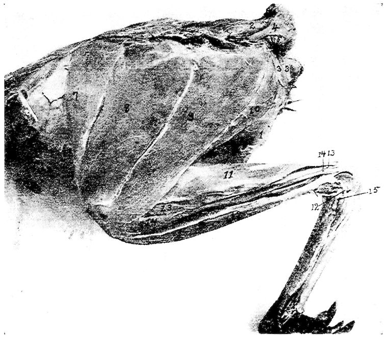
Fig. 23.—Outer layer of muscles of the tail and thigh, 1, Rectus abdominis. 2, Levator coccygis. 3, Transversalis peronie. 4, Levator caudæ. 5, Lateralis caudæ. 6, Depressor caudæ. 7, Sartorius. 8 and 9, Gluteus primus. 10, Semitendinosus. 11, Gastrocnemius. 12, Peroneus longus. 13, Flexor perforatus indicis secundus pedis. 14, Flexor perforatus medius secundus pedis. 15, Lateral ligament of the hock.
Origin.—From the entire posterior margin of the ischium, and from the posterior margin of the post-pubis extending beyond it.
Insertion.—Becoming aponeurotic, it passes toward the coccyx and is attached to the entire posterior margin of the ilium. Passes to the median line, meeting its fellow in front of the anus.
Shape.—Thin, sheet-like.
95Relations.—Infero-anteriorly with the anus. Posteriorly with the edges of the ilium and of the ischium. Internally with the depressor caudæ.
Action.—Gives support to the viscera in the post-anal region, and aids in attaching the anus to the structures above.
Depressor Caudæ (Fig. 23, No. 6). Location.—The infero-lateral side of the tail.
Origin.—From the lower half of the posterior border of the ischium, and from the entire posterior border of the post-pubis beyond.
Insertion.—To the quill butts of the three or four outer main tail feathers.
Shape.—Strong, conical.
Relations.—Externally, with the depressor coccygis.
Action.—Singly, pulls the tail downward and outward; with its fellow, pulls the tail downward.
Depressor Coccygis. Location.—The outermost of the infero-lateral muscles of the tail.
Origin.—From the lower half of the posterior margin of the ischium and the anterior three-fourths of the posterior margin of the post-pubic element of the pelvis beyond it.
Insertion.—To the thickened rim of the inferior and expanded portion of the pygostyle.
Shape.—Flat, triangular.
Relations.—Internally with the depressor caudæ.
Action.—Singly, pulls the tail downward and to one side; with its fellow, pulls the tail downward.
Lateralis Caudæ (Fig. 23, No. 5). Location.—The lateral side of the tail.
Origin.—From the tip of the transverse process of the first free caudal vertebræ.
Insertion.—To the outer three quill butts of the main tail feathers.
Shape.—Four fasciculi forming a fleshy belly.
Relations.—Inferiorly, with the levator caudæ and superiorly with the depressor caudæ.
Action.—Singly, pulls the tail downward and outward; with its fellow, pulls the tail downward. The outermost fasciculus contracting, spreads the tail feathers.
Lateralis Coccygis (Fig. 25, No. E, 2). Location.—The infero-lateral side of the caudal vertebræ.
Origin.—From the surface of the posterior end of the ilium and by 96tendons from the under side of the ends of the first three or four caudal vertebræ.
Insertion.—To the side of the posterior margin of the expanded portion of the pygostyle.
Shape.—Subcompressed mass.
Relations.—With the transverse processes of the caudal vertebra, inferiorly.
Action.—Controls the lateral movements of the tail and its feathers, and the oblique downward movement.
Infracoccygis (Fig. 25, No. E, 1). Location.—The extreme inferior portion of the caudal vertebræ.
Origin.—From the inferior surface of the diapophysis of the last vertebra which anchyloses with the pelvic sacrum, and from all the free caudal vertebræ.
Insertion.—Into the lower side of the pygostyle.
Shape.—Flat, somewhat triangular.
Relations.—Superiorly, with the vertebræ, laterally, with the lateralis coccygis, and mesially, with its fellow of the opposite side.
Action.—Depresses the tail.
Latissimus Dorsi (Fig. 24, No. 7). Location.—Supero-lateral portion of the dorsal region.
Origin.—From two portions, the anterior slip from the outer edge of the superior margins of the neural spines of the second and the third dorsal vertebræ, and the second portion from a similar point on all the succeeding dorsal vertebræ. The second portion is fascia-like in its attachments.
Insertion.—The fibers converge toward the humerus and enter between the deltoid the scapular head of the triceps and the remaining heads of this muscle and is inserted to the anconal part of the humerus, just within in the radial crest.
97Shape.—Thin, triangular.
Relations.—Superiorly, with the skin. The most superficial of the dorsal muscles.
Action.—To elevate the humerus and thus flex the shoulder-joint. An expiratory muscle when the wing is fixed.
Trapezius (Fig. 19, No. 20). Location.—In the shoulder region, just below the longissimus dorsi.
Origin.—From the neural spines of the second, the third, the fourth, and the fifth cervical vertebræ just below the latissimus dorsi.
Insertion.—To the mesian upper border of the scapula.
Shape.—A flat oblong layer of fibers.
Relations.—The posterior part of the trapezius overlies the anterior part of the rhomboideus. The posterior two-thirds is covered by the latissimus dorsi. The anterior third is superficial.
Action.—Draws the scapula forward.
Rhomboideus (Fig. 19, No. 19). Location.—Supero-posterior scapular region.
Origin.—From the neural spines of the first four dorsal vertebræ beneath the latissimus dorsi and trapezius muscles.
Insertion.—The fibers passing outward and backward attach to the posterior third of the mesial or upper border of the scapula.
Shape.—Thin, flat, delicate.
Relations.—Superiorly, by the latissimus dorsi and the trapezius.
Action.—To draw the free end of the scapula upward and forward.
Serratus Magnus Anticus (Fig. 13, No. 16). Location.—The supero-lateral side of the thorax.
Origin.—By three strong digitations: first, from the outer part of the second true rib just above the base of the epineural appendage; the other two, from similar locations on the two succeeding ribs, just above the origin of the dermo-ulnaris.
Insertions.—The first by tendinous attachment to the interpleurapophysial membrane. The rest of the fibers pass upward and slightly forward, and are inserted to the inferior surface of the apex of the scapula.
Shape.—Three flat, strong digitations.
Relations.—Internally, with the ribs.
Action.—If the scapula is fixed, it is a muscle of inspiration.
Serratus Parvus Anticus. Location.—The outer and upper surface of the anterior part of the thorax.
98Origin.—By three digitations from the outer surface of the first three ribs above the origin of the serratus magnus anticus and the thoraco-scapularis.
Insertion.—The thin sheet of fibers passes upward and backward, and is inserted to the inferior margin of the scapula.
Shape.—Thin, flat.
Relations.—Internally, with the ribs.
Action.—If the scapula is fixed, it is a muscle of respiration.
Teres et Infraspinatus (Fig. 13, No. 11). Location.—Scapular region.
Origin.—From the superior surface and outer margin of the posterior two-thirds of the scapula.
Insertion.—The fibers pass forward and outward, and insert to the humerus at the middle of the ulnar margin of the pneumatic fossa, between the forks of the triceps.
Shape. —Flat, triangular-shaped.
Relations.—Anteriorly, with the supraspinatus. Internally, with the ribs.
Action.—Aids in keeping the humerus in its socket. Assists in closing the wing by drawing the humerus to the side of the body.
Intercostales (Fig. 24, No. 5). Location.—Between the ribs of the upper thoracic region.
Origin and Insertion.—From the anterior border of one rib, the fibers, passing obliquely upward and forward, and the lower portion downward and forward, are inserted to the posterior border of the rib just in front.
Shape.—They are best developed in the anterior portion of the thoracic region and extend down as far as the sternal ribs.
Relations.—Internally the pleura, and anteriorly and posteriorly the ribs.
Action.—The first rib being rendered fixed by the scalenus medius, the muscles draw the ribs forward, thus aiding inspiration.
Levatores Costarum (Fig. 24, No. 2). Location.—Supero-lateral portion of the thorax.
Origin and Insertion.—A series of muscles, the first of which extends from the extremity of the transverse process of the twelfth vertebra, the fibers passing downward and backward to the anterior free margin of the upper third of the long posterior rib, and to the external surface close to its margin. The others arise and insert in a similar manner from the ends of the diapophyses of all 99the dorsal vertebræ and attach to each succeeding rib. Those in front are best developed.
Shape.—Thin and triangular.
Relations.—External to the intercostales.
Action.—Aids in respiration.
Appendico-costales (Fig. 24, No. 1). Location.—The lateral side of the thorax.
Origin and Insertion.—From the posterior edge of an uncinate process or epineural appendage, the fibers extending downward and backward to the outer surface of the succeeding rib.
Shape.—A series of thin triangular-shaped muscles.
Relations.—Internally, with the intercostales.
Action.—Aids in respiration.
Tensor Patagii Longus (Fig. 15C, No. 1; Fig. 14, No. 2). Location.—Anterior shoulder region, in the triangular patagium of the wing.
Origin.—By a flat tendon common to it and the tensor patagii brevis from the supero-mesial line of the head of the clavicle.
Insertion.—By a long slender tendon extending in the duplicature of the patagium, with which the fibers blend. Passing to the side of the extensor metacarpi radialis longus, it extends over the end of the radius and is inserted to the os carpi radiale, and to the fascia which binds down the other tendons on the anterior part of the wrist-joint.
Shape.—Cone-shaped belly terminating in a long tendon.
Relations.—Externally, with the skin; by its tendon, with the border of the extensor metacarpi radialis longior.
Action.—Tenses the soft part in the fold of the wing, and aids in flexing the forearm.
Tensor Patagii Brevis (Fig. 14, No. 3). Location.—In the triangular patagium of the wing.
Origin.—From the head of the clavicle in common with the preceding.
Insertion.—The tendon bifurcates, one branch blending with the fascia of the extensor metacarpi radialis longior, the other inserting just below the tubercle on the external condyle of the humerus.
100Shape.—Fleshy; somewhat broader and longer than the preceding.
Relations.—Interiorly, with the preceding.
Action.—Assists the tensor patagii longus in flexing the forearm.
Pectoralis Major (Fig. 13, No. 5). Location.—On the lateral side of the sternum.
Origin.—From the posterior portion of the lateral wing of the sternal body, from the outer marginal third of the keel of the sternum, and from the entire outer side of the limb of the furcula or clavicle.
Insertion.—The fibers converging form toward the proximal third of the humerus a broad tendon which by its insertion covers the entire palmar part of the pectoral crest. A few of the tendinous fibers pass over the shoulder-joint and blend with the long head of the biceps. Near this point it receives the insertion of the dermo-humeralis.
Relations.—Interiorly, with the skin; superiorly, in the sternal region, with the pectoralis secundus and the pectoralis tertius.
Shape.—Fleshy; largest of the pectorales.
Action.—Powerfully depresses the humerus. The chief muscle of flight.
Pectoralis Secundus (Fig. 13, No. 9; Fig. 15, No. A, 6). Location.—The entire lateral side of the sternum.
Origin.—From the anterior sternal extremity of the lower third of the coracoid, from the keel and sternal wing, and by a tendon from a membraneous expansion between the coracoid and the clavicular bones and from the lower third of the coracoid.
Insertion.—The fibers converge into a tendon, which passes upward around the coracoid to its posterior through a canal formed by the scapula, the coracoid, and the clavicle, the foramen triosseum (Fig. 15, No. A, 7). This tendon then passes outward and downward, and becoming flat, inserts to the humerus just anterior to the radial crest and nearer to the humeral head than does the pectoralis major.
Relations.—Internally, with the breast-bone; externally, with the pectoralis major; and superiorly, with the pectoralis tertius.
101Shape.—Long, fusiform, fleshy.
Action.—Raises the wing.
Pectoralis Tertius (Fig. 13, No. 6; Fig. 15, No. A, 5). Location.—On the antero-lateral side of the sternum.
Origin.—From the anterior half of the exterior of the body of the sternum, from the fascia of the subclavius on the outer border of the costal process, and from the outer, lower third of the coracoid process.
Insertion.—The fibers pass upward to the outer side of the coracoid, and becoming tendinous as they reach the humerus, by a strong flattened tendon insert to the ulnar crest of the humerus on the proximal margin at about a middle point of the pneumatic fossa.
Relations.—Externally, with the pectoralis major; interiorly, with the pectoralis secundus; internally, with the fascia of the subclavius and the sternum.
Shape.—Fleshy, fusiform.
Action.—Assists the pectoralis secundus in elevating the humerus.
Coraco-humeralis. Location.—The scapulo-humeral region.
Origin.—From the outer side of the head of the coracoid, supero-laterally to the long head of the biceps.
Insertion.—To the palmar part of the head of the humerus just inside of the insertion of the pectoralis major.
Shape.—A delicate subcylindrical muscle.
Relations.—Superior to the head of the humerus.
Action.—Aids in extending the humerus.
Scapulo-humeralis (Fig. 14, No. 10). Location.—The scapulo-humeral region.
Origin.—From the inner side of the neck of the scapula just within the head of the deltoid.
Insertion.—Passing over the top of the shoulder-joint it is inserted to the palmar part of the humeral head between the insertion of the pectoralis major and the pectoralis secundus.
102Shape.—Narrow, flat ribbon.
Relations.—Along the upper margin of the larger portion of the deltoid.
Action.—Aids in extending the humerus.
Supraspinatus (Fig. 19, No. 22). Location.—Scapular region.
Origin.—From the superior surface and outer third of the scapula.
Insertion.—To the lower border of the pneumatic fossa of the humerus.
Shape.—Thin, flat, triangular.
Relations.—Externally, with the trapezius and posteriorly, with the teres et infraspinatus.
Action.—If the scapula is the fixed point it will pull the humerus upward and backward.
Subclavius. Location.—Anterior sternal region.
Origin.—From the entire outer surface of the sternal process of the sternum and the adjacent outer surfaces of three or four of the hemapophyses.
Insertion.—To the inferior margin of the coracoid bone, the longer fibers passing over to the fossa in the lower third of the posterior part of the coracoid.
Shape.—Rather small, fleshy.
Relations.—Overlapped by the pectoralis tertius.
Action.—Pulls the coracoid outward. Also aids in keeping the coracoid in place.
Coraco-brachialis (Fig. 13, No. 15). Location.—Along the coracoid shaft.
Origin.—By a delicate tendon from a small circular point on the postero-mesial part of the shaft of the coracoid immediately above the attachment of the subclavius and the fossa at that point.
Insertion.—To the top of the ulnar tuberosity of the humerus. This subcircular space is common to the teres minor and to this muscle.
Shape.—Long, fusiform.
Relations.—With the shaft of the coracoid between the teres minor and subscapularis.
Action.—To depress the wing.
Teres Minor (Fig. 13, No. 14). Location.—The coraco-scapular region.
Origin.—From under the side of the anterior tip of the scapula.
Insertion.—Passing outward behind the coracoid head and beneath 103the neck of the scapula, it is attached by a small tendon to the top of the ulnar tuberosity of the humerus in common with the coraco-brachialis, with which tendon it fuses.
Shape.—Small, chunky.
Relations.—Anteriorly, with the tip of the scapula and behind the coracoid head. Posteriorly, with the coraco-brachialis.
Action.—Aids in the downward stroke of the wing.
Levator Scapulæ. Location.—Scapular region.
Origin.—By two strong digitations, one from each of the lateral processes of the first cervical vertebra.
Insertion.—To the middle third of the inferior part of the blade of the scapula.
Shape.—Small and flat.
Relations.—Inferior to the scapula.
Action.—Pulls the scapula forward, and with it the entire shoulder girdle, which articulates like a hinge-joint at the costo-sternal juncture.
Thoraco-scapularis. Location.—Lateral side of the chest.
Origin.—From the outer part of the lower half of the first free rib, the outer side of the next succeeding rib and its epineural appendage, and from a similar surface on the next rib.
Insertion.—By a broad, flat tendon which passes between the two divisions of the subscapularis and inserts at a point at the juncture of the anterior and middle third of the outer margin of the scapula.
Shape.—Broad, flat.
Relations.—Internally, with the ribs and the scapula.
Action.—When the scapula is fixed it is a muscle of inspiration. When the ribs are fixed, it draws the scapula downward.
Subscapularis. Location.—Scapulo-humeral region.
Origin.—From the anterior and outer half of the scapula.
Insertion.—By a strong and subcylindrical tendon to the top of the ulnar tuberosity of the humerus close to the combined tendons of the coraco-brachialis and teres minor.
Shape.—A large and powerful muscle.
Relations.—The flat tendon of the thoraco-scapularis divides the posterior portion of the belly into two portions.
Action.—A powerful rotary muscle of the head of the humerus; aids in keeping the humeral head in the glenoid cavity.
Biceps (Fig. 13, No. 8). (Synonym.—Biceps brachii.)
Location.—The anterior brachial region.
Origin.—By a broad tendon it covers the top of the shoulder-joint, dividing into two heads, giving rise to the long and the short heads. The long is inserted into the outer part of the head of the coracoid just beyond the glenoid cavity; the short head to the distal angle of the ulnar tuberosity of the humerus.
Insertion.—To the ulna just in front of the articular cavity for the trochlear surface of the distal extremity of the humerus.
Shape.—Large, subfusiform.
Relations.—On one side by the triceps and the other the deltoid and anteriorly partially covered by the patagii muscles.
Action.—A powerful flexor of the forearm.
Deltoid (Fig. 13, No. 12; Fig. 15, No. 8). Location.—Occupying the lateral side of the humerus; the brachial region.
Origin.—Divided into two portions, the long narrow head extends from the clavicular process of the scapula and from the adjacent surface of the same bone. These fibers then extend around the back of the shoulder-joint, and are joined by the fibers that arise from the entire outer surface of the large os humero-scapulare. These latter fibers are inserted upon an extensive area upon the anconal part of the bone beyond the humeral articular head, and to almost the entire shaft below it.
Insertion.—By a subcylindrical tendon to the proximal side of the tubercle of the external condyle, and above the insertion of the extensor metacarpi radialis longus.
Shape.—Large, fleshy, with tendinous attachment.
Relations.—On one side by the biceps and the other by the triceps also the patagii.
Action.—To extend the arm.
Triceps (Fig. 15, No. 7). (Synonym.—Triceps brachii.)
Location.—The posterior humeral region.
Origin.—By three portions: by the internal and external heads, and by the long scapular head. The internal and the external heads are blended except at their proximal extremities. Each head arises from the anconal surface of the shaft of the humerus. The 105internal head being located toward the deltoid attachment and the external head into the pneumatic fossa. The long head extends from a circumscribed area just posterior to the glenoid cavity of the scapula. The fibers pass around the shoulder-joint, beneath the deltoid.
Insertion.—The broad tendon passes over the elbow-joint and is inserted to the entire under surface of the olecranon of the ulna.
Shape.—Long, large, fleshy.
Relations.—The supraspinatus passes between the internal and the external heads. It is bounded by the biceps and the deltoid.
Action.—An extensor of the antibrachial region directly antagonizing the biceps.
Brachialis Anticus (Fig. 15, No. 9; Fig. 14, No. 5). Location.—In the flexure of the humerus and the ulna.
Origin.—It arises fleshy from a circumscribed area on the inner side of the anconal part of the distal extremity of the humerus.
Insertion.—The fibers passing directly over the elbow-joint become inserted to the lateral surface of the proximal end of the ulna close to the margin of the sigmoid cavity.
Shape.—Small, fleshy.
Relations.—With the joint.
Action.—Assists in flexing the forearm upon the arm. Protects the structures in the anterior part of the elbow-joint.
Extensor Metacarpi Radialis Longior (Fig. 13, No. 17). Location.—Superior to the radius.
Origin.—By two strong tendinous heads; the outer from the tubercle of the external condyle of the humerus, just above the origin of the tendon of the tensor patagii brevis; the inner and stronger 106portion from the tubercle found above the oblique trochlear facet of the distal end of the radius.
Insertion.—Becoming a flat, broad, and strong tendon at about the middle of the forearm, finally becomes inserted to the apex of the anchylosed first metacarpal of the carpo-metacarpus.
Shape.—A thick, fusiform belly, tendinous at both extremities.
Relations.—By the side of the pronator brevis.
Action.—Raises the hand, and draws it forward toward the radial margin of the forearm and retains it on the same side.
Extensor Digitorum Communis (Fig. 15, No. 2). Location.—The upper metacarpal region.
Origin.—Immediately below the tubercle of the external condyle of the humerus.
Insertion.—By a small tendon to the outer side of the base of the pollex, and at a mid-point upon the anterior rim of the proximal phalanx of the middle finger.
Shape.—Small spindle-shaped, becoming tendinous at about the middle of the forearm.
Relations.—Occupies the middle of the group of muscles on the outer side of the forearm.
Action.—An extensor of the digit.
Supinator Brevis (Fig. 14, No. 7). Location.—The superior part of the radius.
Origin.—From the external condyle of the humerus below the origin of the tendon of the extensor digitorum communis.
Insertion.—To the outer side of the shaft of the radius, for nearly one-third of its length.
Shape.—Thin, slender.
Relations.—Inferiorly, with the bone.
Action.—A supinator of the radial region, and antagonistic to the pronators.
Flexor Metacarpi Radialis (Fig. 15, No. 10). Location.—The lowest of the group of three muscles on the outer part of the forearm.
Origin.—By two tendinous heads, the longer from the external condyle of the humerus, and the other just beyond the base of the olecranon.
Insertion.—Becomes tendinous at about the middle of the shaft of the ulna and inserts to a prominent process on the proximal third of the posterior part of the shaft of the mid-metacarpal.
Shape.—Fusiform.
107Relations.—Inferiorly, the anconeus and superiorly, the extensor digitorum communis.
Action.—A powerful flexor of the hand.
Pronator Brevis (Fig. 13, No. 18). Location.—Supero-lateral side of the forearm.
Origin.—From just above the internal condyle of the humerus.
Insertion.—The tendon passing obliquely across the interosseous space and is inserted to the ulnar side of the shaft of the radius, just beyond the juncture of the proximal and the middle thirds.
Shape.—Fusiform.
Relations.—Superior to the pronator longus.
Action.—Pronates the forearm, and flexes the forearm upon the arm.
Pronator Longus (Fig. 14, No. 6). Location.—Lateral side of the radio-ulnar region.
Origin.—From the middle of the internal condyle of the humerus.
Insertion.—To the shaft of the radius just beneath the pronator brevis.
Shape.—Massive, ellipsoidal.
Relations.—Between the pronator brevis and flexor digitorum profundus.
Action.—A pronator.
Extensor Ossis Metacarpi Pollicis (Fig. 13, No. 23). Location.—Slightly interposed between the ulna and radius.
Origin.—Immediately in front of the greater sigmoid cavity of the ulna.
Insertion.—To the palmar side of the base of the first metacarpal, in common with the extensor metacarpi radialis longior.
Shape.—Delicate, straight.
Relations.—Superiorly, the extensor indicis longus, and inferiorly, the anconeus and the flexor digitorum profundus.
Action.—Extends the hand upon the forearm.
Anconeus (Fig. 15, No. 4). Location.—Between the ulna and the radius.
Origin.—By a short, strong, subcylindrical tendon from the lower posterior of the external condyle of the humerus.
Insertion.—To the latero-radial side of the ulna somewhat beyond its middle.
Shape.—Fusiform.
108Relations.—Superiorly, the extensor indicis longus, and inferiorly, the ulna.
Action.—A flexor of the forearm.
Extensor Indicis Longus (Fig. 13, No. 22; Fig. 15, No. 13). Location.—Infero-lateral to the radius.
Origin.—From about one-half the surface of the proximal portion of the radius.
Insertion.—Possesses a long tendon which passing over a groove at the distal end of the ulna and receiving muscular fibers at the base of the metacarpus, extends down the anterior part of the hand and is inclosed in a sheath in front of the superior part of the first phalanx of the index-finger. This tendon is inserted into the anterior upper rim of the distal phalanx.
Shape.—Small, thin.
Relations.—Supero-laterally with the radius.
Action.—Extensor of the digit.
Flexor Digitorum Sublimis. Location.—The forearm.
Origin.—From the internal condyle of the humerus.
Insertion.—Passing over the wrist, it is inserted to the middle phalanx of the hand.
Shape.—A musculo-tendinous band, rather delicate and rudimentary.
Relations.—Closely adhering to the integument that stretches from the internal condyle of the humerus to the wrist.
Action.—Assists these muscles. A flexor.
Flexor Digitorum Profundus (Fig. 13, No. 21). Location.—Lateral side of the ulna.
Origin.—By two heads from the proximal extremity of the ulna, between which the brachialis anticus passes.
Insertion.—The two bellies uniting are attached to the under side of the shaft of the ulna. It becomes tendinous about the middle of the shaft, and inserts to the ulnar side of the base of the distal joint of the index-finger.
Shape.—A fleshy belly with long tendon.
Relations.—Inferiorly, with the flexor carpi ulnaris, superiorly, with the pronator brevis.
Action.—A flexor of the digit.
Flexor Carpi Ulnaris (Fig. 13, No. 19). Location.—Inferior part of the forearm.
Origin.—By two strong tendons; one from the side and posterior 109of the internal condyle of the humerus, passing through the humero-ulnar pulley at the side of the base of the olecranon process; the other, to the posterior of the internal condyle of the humerus. It does not pass through a pulley.
Insertion.—A subcylindrical tendon extends back of the ulnare ossicle of the carpus giving off a tendinous slip to the flexor digitorum profundus, and becomes inserted to the anterior rim of the proximal phalanx of the index digit. It has an attachment at the carpus.
Shape.—A strong fleshy belly terminating in a long tendon.
Relations.—The outermost inferior muscle of the forearm.
Action.—A powerful flexor of the hand upon the forearm.
Flexor Carpi Ulnaris Brevior (Fig. 13, No. 20). Location.—Inferior part of the antibrachial region.
Origin.—From a broad area on the middle third of the upper side of the shaft of the ulna.
Insertion.—Near the top of the outer edge of the anchylosed os magnum of the carpo-metacarpus.
Shape.—Thin, somewhat flat.
Relations.—To the inside of the flexor digitorum profundus.
Action.—Flexes the hand upon the forearm and rotates the hand toward the body.
Extensor Proprius Pollicis (Fig. 13, No. 29a). Location.—Anterior to the radio-carpal joint.
Origin.—From the ulnar side of the tendon of the extensor metacarpi radialis longior.
Insertion.—To the antero-ulnar side of the pollex.
Shape.—Small, spindle-shaped; tendinous at both attachments.
Relations.—Inferiorly, with the proximal and anterior part of the metacarpus.
Action.—An extensor of the pollex.
Flexor Brevis Pollicis (Fig. 13, No. 28a). Location.—Anterior to the pollex.
110Origin.—From the shaft of the mid-metacarpal bone, just below the pollex.
Insertion.—Distal apex of the pollex.
Shape.—Short, small, fleshy.
Relations.—Superiorly, with the inferior part of the pollex.
Action.—Flexes the pollex.
Flexor Minimi Digiti. Location.—Inferior side of the metacarpus.
Origin.—From the posterior part of the median metacarpal close to the os carpi ulnare. A few fibers extend to the ulna.
Insertion.—Into the base of the median phalanx.
Shape.—Small, slender, short.
Relations.—Superiorly, with the metacarpal bone.
Action.—A flexor of the digit.
Flexor Minimi Digiti Brevis (Fig. 13, No. 30). Location.—Inferior to the third, or small finger.
Origin.—From the lower and posterior end of the median metacarpal and from the adjacent tendon of the preceding.
Insertion.—Inserted to the apex of the small finger.
Shape.—Small and rather rudimentary.
Relations.—Inferior to the small finger.
Action.—Acts as a posterior ligament of this joint.
Abductor Minimi Digiti. Location.—Supero-lateral side of the small finger.
Origin.—From the proximal extremity of the anterior aspect of the third finger.
Insertion.—To the posterior border of the proximal phalanx of the second, or index-finger at a point just above the tip of the third finger.
Shape.—A few muscular fibers with some tendinous material.
Relations.—At the base of the median segment of the index-finger and to the side of the third.
Action.—Antagonizes the powerful flexors on the back of the joint.
Interosseus Dorsalis (Fig. 13, No. 31). Location.—By the side of the metacarpus.
Origin.—From the margin of the shaft of the median metacarpal.
Insertion.—To the anterior part of the base of the distal phalanx of the index digit.
Shape.—Short, delicate, with long tendon.
Relations.—Internally, the metacarpal bone.
111Action.—Extends the last bone of the middle finger.
Interosseus Palmaris (Fig. 13, No. 32). Location.—On the opposite side of the bone from the interosseous dorsalis.
Origin.—From the shaft of the median metacarpal under the preceding.
Insertion.—To the apex of the last joint of the second, or middle finger.
Shape.—Small, delicate, with long slender tendon.
Relations.—Beneath the preceding and along the metacarpal bone.
Action.—Flexes the terminal phalanx.
Flexor Metacarpi Brevis. Location.—To the lateral side of the metacarpal bone.
Origin.—From the outer side of the distal extremity of the ulna.
Insertion.—To the base, in front, and on the ulnar side of the proximal phalanx of the index digit.
Shape.—Delicate, with long slender tendon.
Relations.—On the lateral side of the ulna and the metacarpal bone.
Action.—Flexes the metacarpus.
Sartorius (Fig. 23, No. 7). Location.—Extreme anterior portion of the thigh.
Origin.—From the outer two-thirds of the superior surface of the raised emarginations of the anterior border of the ilium, and by fascia from the neural spines of the fourth dorsal vertebra.
Insertion.—Obliquely by a semitendinous fascia to the inner edge of the ligamentum patella, and to the inner and adjacent border of the anterior half of the summit of the tibia.
Shape.—Large, fleshy.
112Relations.—Posteriorly, with the gluteus primus and the vasti.
Action.—Extends the leg, flexes and adducts the thigh.
Gluteus Primus (Fig. 23, No. 8 and 9). Location.—The outer flat massive muscle of the thigh.
Origin.—By a thin fascia from nearly the entire length of the supero-internal margin of the pre-acetabular portion of the ilium, above the antitrochanter, and from the entire length of the post-acetabular ridge.
Insertion.—Near the patella it joins the tendon of the extensor femoris, and by aponeurosis spreads over the knee and is inserted to the crest of the upper border of the tibia, the patella being incorporated in its aponeurotic ligament.
Shape.—Triangular in shape, it is aponeurotic anteriorly and more fleshy posteriorly.
Relations.—Anteriorly, covers over the posterior portion of the sartorius and the body of the gluteus medius; posteriorly, bordered by the semitendinosus and the biceps.
Action.—Abducts the thigh.
Gluteus Medius (Fig. 24, No. 11). Location.—Supero-anterior part of the ilium under the gluteus primus.
Origin.—From the entire supero-internal margin of the pre-acetabular surface of the ilium and the concave surface on the adjacent bone.
Insertion.—By a strong tendon which passes over a bursa on the anterior rim of the trochanter and is inserted obliquely to this trochanter.
Shape.—A fleshy muscle filling the concavity of the pre-acetabular division of the ilium.
Relations.—Superiorly, with the gluteus primus; inferiorly, with the gluteus minimus.
Action.—Abducts and pulls the femur forward.
Gluteus Minimus (Fig. 24, No. 12). (Synonym.—Gluteus profundus.)
Location.—Beneath the medius and the smaller of the two.
Origin.—To the anterior margin of the outer border of the ilium, and from the supero-external surface of the last rib.
Insertion.—The fibers extending downward and backward and outward become tendinous and are inserted below the trochanter of the outer part of the upper third of the femur.
Shape.—Small, fleshy.
113Relations.—Inferiorly, with the bone and superiorly, with the gluteus medius.
Action.—Abducts and pulls the thigh forward.
Extensor Femoris (Fig. 24, No. 15). Location.—Anterior femoral region. It is divided into two parts: the vastus externus and the crureus.
Origin.—The vastus extemus (Fig. 24, No. 14) arises tendinous from the base of the trochanter of the outer part of the femur and from this point down the bone approximately to the condyle. The crureus (Fig. 24, No. 13) originates by a tendon from the anterior upper prominent rim of the trochanter, from this point extending down the antero-external part of the shaft of the femur.
Insertion.—Merges with the gluteus primus, and, by a fascia-like arrangement, spreads over the front of the knee-joint inserts to the patella and is also inserted into the crest of the tibia.
Shape.—Large, fleshy.
Relations.—Posteriorly, with the femur and anteriorly, with the sartorius.
Action.—Extends strongly the leg upon the thigh.
Vastus Internus (Fig. 16, No. 3). Location.—Internal and lateral side of the femur.
Origin.—From the postero-internal part of the shaft of the femur just below the head, and in a straight line extending down the femur.
Insertion.—By a broad tendon along the thickened inner border of the summit of the tibia.
Shape.—Thick, long.
Relations.—Anteriorly, with the bone; surrounded by the other crural muscles.
Action.—Extends the leg upon the knee; a powerful assistant of the extensor femoris.
Biceps Flexor Cruris (Fig. 17, No. D, 1). Location.—Posterior tibial region.
Origin.—By a tendinous fascia from the post-acetabular ridge, extending between the antitrochanter and the anterior point of the insertion of the semitendinosus.
Insertion.—To the tuberosity on the external part of the shaft of the fibula a short distance below the head.
Shape.—Large, rather cone-shaped with base directed upward.
Relations.—Anteriorly, with the vastus externus; posteriorly, with 114the semitendinosus. The inferior tendinous portion passes through a tendinous sling or pulley called the biceps band.
Action.—Flexes the leg upon the thigh.
Semitendinosus (Fig. 23, No. 10). Location.—Posterior femoral region.
Origin.—By a tough, strong fascia from the surface of the caudal muscles and from the posterior third of the post-acetabular ridge.
Insertion.—The fibers pass downward and forward, and insert to the tendinous raphe along the posterior margin of the semitendinosus accessorius; lower down it merges with the median fascia of the inner head of the gastrocnemius.
Shape.—Fleshy, broad, flat and long.
Relations.—Internally, with the semimembranosus and anteriorly, with the biceps flexor cruris.
Action.—Flexes the leg.
Semitendinosus Accessorius. Location.—Posterior femoral region.
Origin.—From an oblique line just above the condyle, on the posterior of the shaft of the femur.
Insertion.—Its fibers, passing upward and backward, attach themselves to the tendinous raphe common to this muscle and the semitendinosus, and are finally inserted to the inner side of the shaft of the tibia.
Shape.—Flat, oblong.
Relations.—Posteriorly to the shaft of the femur, and externally to the long adductors of the thigh.
Action.—Aids the preceding in flexing the leg.
Semimembranosus (Fig. 24, No. 17). Location.—Postero-internal to the semitendinosus.
Origin.—From the outer surface of the ischium, beginning at the lower margin of its notch on the posterior pelvic border, extends on a curved line on the adjacent surface beyond.
Insertion.—The fibers, passing downward and forward, insert by a broad, thin tendon to the shaft of the tibia a short distance below the head and on a line parallel to the long axis of the tibia.
Shape.—Long, narrow, ribbon-shaped.
Relations.—Lies adjacent to the semitendinosus and in the same plane.
Action.—Directly flexes the leg.
Femoro-caudal (Fig. 24, No. 8). Location.—Infero-lateral to the caudal and post-femoral region.
115Origin.—By a delicate tendon from the base of the pygostyle.
Insertion.—By a thin, flat tendon upon the outer part of the shaft of the femur below the trochanter and at about the juncture of the upper and middle third of the bone.
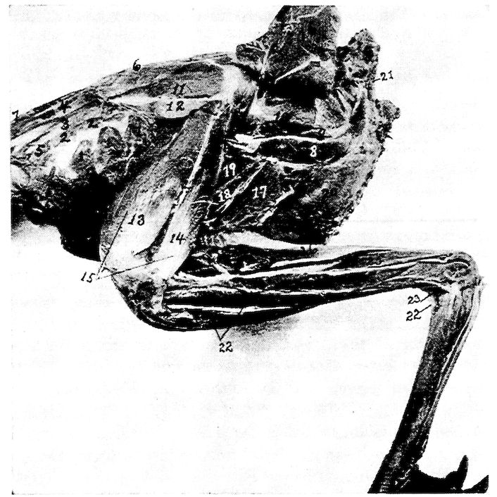
Fig. 24.—Second layer of muscles of the thigh of a cock. Outside view. 1, Appendico-costales. 2, Levatores costarum. 3, Sacro-lumbalis. 4, Longissimus dorsi. 5, Intercostales. 6, Longissimus dorsi. 7, Latissimus dorsi. 8, Femoro-caudal. 9, Edge of obturator externus. 11, Gluteus medius. 12, Gluteus minimus. 13, Crureus. 14, Vastus externus. 15, Extensor femoris. 16, Biceps flexor cruris. 17, Semimembranosus. 18, Adductor magnus. 19, Adductor longus. 20, External abdominal oblique. 21, Depressor coccygis. 22, Tibialis anticus. 23, Tibialis posticus. 24, Flexor perforans digitorum profundus. 25, Flexor perforatus medius secundus pedis. 26, Flexor longus hallucis.
Shape.—Long, narrow, spindle-shaped, flattened from side to side.
Relations.—Interiorly to the obturator externus, and along the upper border of the long adductors of the thigh and the semimembranosus.
Action.—Pulls the tail down and to one side.
116Obturator Externus (Fig. 24, No. 9). Location.—External posterolateral side of the pelvis.
Origin.—From the posterior half of the periphery of the ischiatic foramen and the concavity found on the external surface of the lateral part of the pelvis.
Insertion.—The fibers pass across to the femur and are inserted by a broad, flat tendon to the shaft of the femur just below the trochanter.
Shape.—Thick, fleshy.
Relations.—Lies just above the femoro-caudal muscle, the sciatic nerve and femoral artery pass over and external to its tendon.
Action.—Pulls the head of the femur backward.
Obturator Internus (Fig. 16, No. E, 1). Location.—Occupies the space between the ischium and the pubis.
Origin.—From the ventral surface of the ischium, including the posterior border, from the inner line of the corresponding post-pubis, and from the membrane covering the space between these two bones.
Insertion.—Becoming dense, subcylindrical, and strong, it passes through the obturator foramen and is inserted to the outer part of the trochanter of the femur, in common with the gemellus and opposite to the insertion of the gluteus medius.
Shape.—Subtriangular and flat with a central tendon; bipenniform.
Relations.—Outside the pelvis the tendon lies upon the gemellus and is external to it. Fills the obturator foramen and the space between the ischium and the pubis.
Action.—Acts as a posterior stay to the head of the femur.
Gemellus (Fig. 25, No. 5). Location.—Posterior to the head of the femur.
Origin.—From the fossa between the acetabulum and the obturator foramen, and on the outer side of the pelvis.
Insertion.—Its fibers are attached by fascia to the tendon of the obturator internus, and pass directly to the trochanter of the femur where they insert in common with that of the obturator internus.
Shape.—Strong, thick, chunky.
Relations.—Postero-external to the head of the femur.
Action.—Like the two preceding, when the head of the femur is fixed, it pulls the pelvis forward and steadies it on the head of the femur.
117Adductor Longus (Fig. 24, No. 19). Location.—In the posterior femoral region.
Origin.—From a line on the lateral part of the pelvis.
Insertion.—The fibers, passing downward and forward, are inserted on a longitudinal line along the posterior part of the shaft of the femur.
Shape.—Broad, flat, fleshy.
Relations.—Posteriorly to the femur, and anteriorly to the adductor magnus.
Action.—Adducts the thigh.
Adductor Magnus (Fig. 24, No. 18). Location.—Posterior femoral region.
Origin.—From the fine constituting the lower boundary of the ischiatic fossa on the outer lateral part of the pelvis.
Insertion.—The fibers pass downward and are inserted to the superior curve of the internal femoral condyle. The internal head of the gastrocnemius blends with this muscle just above its attachment.
Shape.—Long, narrow.
Relations.—Posteriorly to the adductor magnus and anteriorly to the semimembranosus.
Action.—Strongly adducts the thigh.
Gastrocnemius (Fig. 23, No. 11). Location.—Posterior tibial region.
118Origin.—There are three heads: the internal, the external, and the tibial. The external head extends by a short, flattened, strong tendon from the postero-external part of the external condyle of the femur; the internal head extends from the outer surface of the inner condyle of the femur; and the tibial head extends from the entire inner rim of the tibial summit and from the free edge of the adjacent crest.
Insertion.—At the lower fourth of the tibial shaft, terminates in a broad, flat tendon which passes over the shallow, longitudinal groove of the tibial cartilage, and, crossing the tibio-tarsal joint, is inserted to the posterior surface of the hypotarsus of the metatarsal bone, and finally, below this point, merges into the podothecal sheath confining the flexor tendons.
Shape.—Large, fleshy, somewhat cone-shaped with the base upward.
Relations.—The posterior fleshy muscle of the post-tibial region located anterior to the soleus.
Action.—Extends the metatarsus on the tibia.
Soleus (Fig. 25, No. G, 1). Location.—The posterior tibial region.
Origin.—From the posterior part of the head of the tibia.
Insertion.—By a long slender tendon to the proximal end and toward the inner angle of the tibial cartilage, some of the fibers passing to the tendon of the gastrocnemius.
Shape.—Small, flattened.
Relations.—Anteriorly, with the flexor perforans digitorum profundus and posteriorly, with the gastrocnemius.
Action.—Similar to that of the gastrocnemius.
Peroneus Longus (Fig. 23, No. 12). Location.—Antero-lateral tibial region.
Origin.—From the raised crest in front of the head of the tibia, and from the fascia that covers the outer side of the knee-joint.
Insertion.—The fibers, passing downward, then downward and outward, terminate in a small tendon at the lower third of the tibia. This tendon bifurcates just above the tibial condyles at the outer part of the limb, the shorter and stronger attaches to the fibrous fascia covering the tibial cartilage, the other merging with the tendon of the flexor perforatus medius primus pedis about 1 centimeter below the hypotarsus of the tarso-metatarsus.
Relations.—Covers over all the muscles of the anterior tibial region.
119Shape.—A broad muscular sheet.
Action.—Assists the flexor perforatus medius primus pedis.
Tibialis Anticus (Fig. 24, No. 22). Location.—In the anterior tibial region.
Origin.—The muscular belly may be easily divided into two parts. The inner head extends from the head of the tibia immediately beneath the peroneus longus. The outer head arises by a strong tendon from a depression on the antero-inferior ridge of the outer condyle of the femur.
Insertion.—The tendon passing through the fibrous ligamentous loop just above the tibial condyles, inserts to a tubercle on the shaft just below the head of the tarso-metatarsus.
Shape.—Large, fusiform.
Relations.—Beneath the peroneus longus.
Action.—Flexes the metatarsus upon the tibia.
Extensor Longus Digitorum (Fig. 25, No. G, 6). Location.—Anterior tibial region.
Origin.—From the inferior portion of the crest and a portion of the shaft of the fibula.
Insertion.—It becomes tendinous at the lower third of the bone, passing under the bony ridge just above the condyle in front, and over the ankle-joint. It is bound down by firm fascia, and at the trochlea of the basal toe joints, divides into three small tendinous slips. These pass over the superior part of the second, the third, and the fourth toes, bifurcating in their course, and are inserted to the distal ones.
Shape.—Long, penniform, with a long, slender tendon.
Relations.—Immediately below the tibialis anticus.
Action.—Extends the digits as their long extensor.
Extensor Hallucis Brevis (Fig. 25, No. G, 4). Location.—The anterior metatarsal region, along the antero-internal edge.
Origin.—From just below the summit of the antero-internal part of the head of the tarso-metatarsus, and from the adjacent shaft below, and from the tendon of the tibialis anticus.
Insertion.—Into the process at the superior part of the bone of the bony claw of the hallux.
Shape.—Slender, thin in diameter, with a long, slender tendon.
Relations.—Superiorly, with the antero-internal edge of the metatarsus and interiorly, its tendon with the top of the hallux.
Action.—Extends the hallux.
120Extensor Brevis Digitorum. Location.—On the anterior surface of the metatarsus.
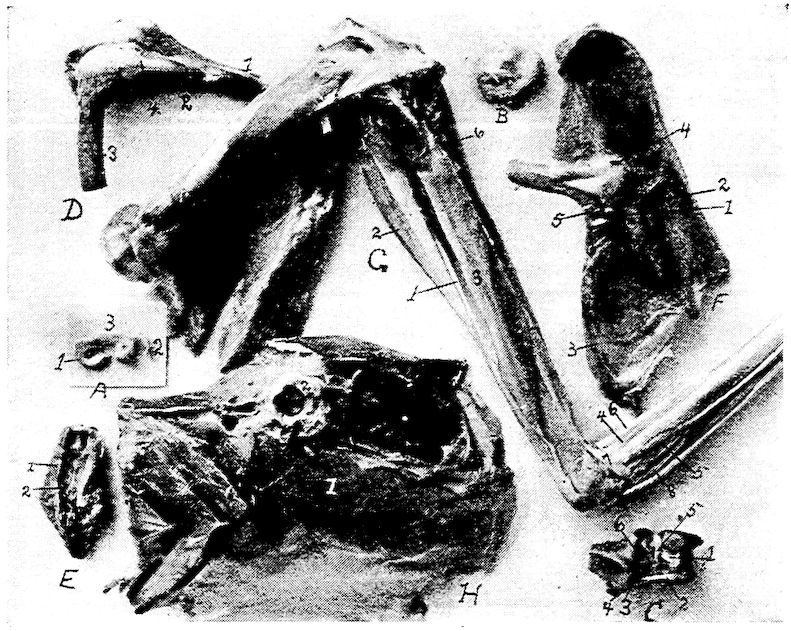
Fig. 25.—Inner layer of muscles of the thigh of a hen. Outside view.
A. Pad of fibrocartilage or meniscus of femoro-tibial articulation. 1, Inner semi-lunar fibrocartilage. 2, External semi-lunar cartilage. 3, Anterior border.
B. Tarsal meniscus or pad of fibrocartilage.
C. Articular surface of the tarsal joint. 1, Posterior ligamentum cruciatum genu. 2, Femoral tendinous insertion of tibialis anticus. 3, Ligamentum tibio-fibulare. 4, Ligamentum interosseum. 5, Ligamentum transversum of the meniscus. 6, Anterior ligamentum cruciatum genu.
D. 1, Tibia. 2, Fibula. 3, Femur. 4, External ligamentum laterale genu.
E. Inferior surface of the tail of a hen. 1, Infracoccygis. 2, Lateralis coccygis.
F. Outside pelvic view. 1, Obturator ligament. 2, Tendon of the obturator internus. 3, Ligamentum oblongum. 4, Ligamentum capsulare. 5, Gemellus.
G. Outside view of leg of a cock. 1, Soleus. 2, Flexor longus hallucis. 3, Flexor perforans digitorum profundus. 4, Extensor brevis hallucis. 5, Extensor brevis annularis. 6, Extensor longus digitorum. 7, Tendon of the tibialis posticus.
H. 1, Transversalis abdominalis. 2, Cotyloid cavity.
Origin.—By a small fleshy belly to the anterior part of the proximal end of the metatarsus.
Insertion.—It becomes tendinous about the middle third of the metatarsus. Extending down the anterior face of the metatarsus it is 121inserted to the inner tubercle of the base of the first phalanx of the outer or fourth toe.
Shape.—Long, slender.
Relations.—Posteriorly with the metatarsus.
Action.—Pulls the fourth toe upward and inward.
Flexor Hallucis Brevis. Location.—Postero-internal side of the metatarsus.
Origin.—Side and lower margin of the inner aspect of the hypotarsus of the tarso-metatarsus and from the shaft of this bone immediately below it.
Insertion.—Winds round inner side of basal joint of the hallux and is inserted on its under side.
Shape.—Small flat muscle gradually tapering into a tendon.
Relations.—Posteriorly with the metatarsus.
Action.—To flex the hallux.
Extensor Annularis Brevis. Location.—On postero-external side of metatarsus.
Origin.—External aspect of the hypotarsus and the shaft below.
Insertion.—Supero-external part of the basal phalanx of the fourth toe.
Shape.—Small flat muscle.
Relations.—Posteriorly with the metatarsus.
Action.—Extensor of the fourth toe.
Tibialis Posticus (Fig. 24, No. 23). Location.—In the posterior tibial region.
Origin.—From the whole shaft of the fibula below the insertion of the biceps flexor cruris, and from the shaft of the tibia a short distance below this insertion. Also from the interosseous membrane, and the adjacent surface of the tibial shaft.
Insertion.—Passing in front of the external malleolus, crossing the ankle-joint, finally inserted into the outer edge of the summit of the tarso-metatarsal bone.
Shape.—Long, subcylindrical.
Relations.—Medially, with the flexor perforans digitorum pedis; superiorly, with the postero-external portion of the tibia.
Action.—Extends the metatarsus upon the tibia.
Flexor Perforatus Indicis Secundus Pedis (Fig. 17, No. 5). Location.—The posterior tibial region.
Origin.—From the external surface of the outer condyle of the femur, just below the head of the gastrocnemius.
122Insertion.—Passing in an oblique manner along posterior to the tibia, extends through the superficial part of the tibial cartilage to the outer side of the leg; then crossing the tibio-tarsal joint, it passes through a special canal of the hypotarsus, down the back of the tarso-metatarsus, under the annular ligament of the sole of the foot and a cartilaginous structure below this ligament, over the trochlea, and is inserted to the middle shaft of the second phalanx of the second toe.
Shape.—Thin, flat, broadly spindle-shaped.
Relations.—Posteriorly, with the flexor perforatus medius secundus pedis, and anteriorly, with the flexor perforatus annularis primus pedis.
Action.—Flexes the toes.
Flexor Longus Hallucis (Fig. 17, No. 7; Fig. 25, No. 2). Location.—The posterior tibial region.
Origin.—By two tendinous heads: one from the posterior part of the femur, just below the two condyles; the other from the outer part of the external condyle of the femur. The deep flexor passes between these two heads.
Insertion.—Becoming tendinous at the lower third of the leg, passes beneath the superficial flexors in a special canal on the outer side of the tibial cartilage, and extends through the large canal of the hypotarsus (Fig. 12, No. F, 5) next to the shaft, then down to the apex of the accessory metacarpal bone. It is inserted to the tubercle on the inferior proximal end of the ungual phalanx.
Shape.—Large, fusiform.
Relations.—With the exterior of the deep flexors.
Action.—Flexes the hallux as its long flexor.
Flexor Perforatus Annularis Primus Pedis (Fig. 17, Nos. D, 8 and C, 6). Location.—The posterior tibial region.
Origin.—From the inner side of the belly of the flexor longus hallucis.
Insertion.—Becoming tendinous at the lower third of the tibia, passes a little to the outer side and through the tibial cartilage, then over the ankle-joint and through the hypotarsal canal (Fig. 12, No. 5) to the under side of the outer toe, where it forms a sheath through which the deep flexors extend. It gives off on either side a tendinous slip which attaches to the basal phalanx.
Shape.—Long, slender, compressed laterally.
123Relations.—Internally, with the flexor perforatus indicis secundus pedis, and externally, with the flexor longus hallucis.
Action.—Flexes the toes as their long flexor. A second flexor of the outer toe.
Flexor Perforatus Medius Secundus Pedis (Fig. 17, No. 9). Location.—The posterior tibial region.
Origin.—From a strong fascia that merges with the knee-joint, and by a tendon from the external condyle of the femur in common with the tendon of the flexor perforatus indicis secundus pedis.
Insertion.—Continued as a flattened tendon extending from the lower third of the leg, passing obliquely through the tibial cartilage and then through the interno-posterior canal of the hypotarsus, (Fig. 12, No. F, 5) inserted to the second joint of the middle toe.
Shape.—Long, fusiform.
Relations.—With the shaft of the fibula, which it covers.
Action.—Flexes the middle toe.
Flexor Perforatus Medius Primus Pedis (Fig. 17, No. 2). Location.—Posterior tibial region.
Origin.—From the inner side of the muscular portion of the flexor perforatus annularis primus pedis.
Insertion.—Terminating in a flattened tendon, passes through the tibial cartilage and hypotarsus, and then extends along the shaft of the basal joint of the third toe, and, dividing into two slips, inserts to either side of its shaft.
Shape.—Long, rather small.
Relations.—With the flexor perforatus annularis primus pedis on the inner side.
Action.—Flexes the third toe.
Flexor Perforatus Indicis Primus Pedis (Fig. 17, No. 4). Location.—The posterior tibial region.
Origin.—From a thin, broad tendon, on the external condyle of the femur, in common with the flexor perforatus medius secundus pedis.
Insertion.—Becoming tendinous, passes through the tibial cartilage and the hypotarsus of the metatarsus (Fig. 16, A) and inserts to the sides of the basal joint of the second toe.
Shape.—Long, slender, fusiform; somewhat flattened laterally.
Relations.—With the flexor perforatus medius secundis pedis, posteriorly.
Action.—A flexor of the toes.
124Flexor Perforans Digitorum Profundus (Fig. 24, No. 24 and Fig. 25, No. G, 3). Location.—The posterior tibial region.
Origin.—From the posterior part of the external condyle of the femur, from the posterior part of the tibia just below the summit, and from a point near the origin of the soleus.
Insertion.—Its heavy flattened tendon passes through the tibial cartilage, this tendon dividing, the branches pass along the under side of the toes, perforating the outer tendons and become inserted to the proximal tubercle at the base of the under side of the ungual phalanxes of the second, third, and fourth toes.
Shape.—Long, fleshy, with a long tendon.
Relations.—Closely with the tibial bone; the deepest of all the flexors.
Action.—Flexes the digits.
Orbicularis Palpebrarum (Fig. 7, No. 30). (Synonym.—Orbicularis oculi.)
Location.—Upon the lacrimal and maxillary bones.
Origin.—From the lacrimal and maxillary bones and the ciliary margin.
Insertion.—To the lower free edge of the tarsal cartilage.
Shape.—Thin, delicate layer of fibers.
Relations.—Externally, with the skin; internally, with the lacrimal and the maxillary bones.
Action.—Aids in closing the eye.
Levator Palpebræ Superioris (Fig. 7, No. 24). Location.—Along the superior roof of the orbit.
Origin.—Along a longitudinal line found near the middle of the roof of the orbit.
125Insertion.—To the ciliary margin of the upper lid, near the outer canthus.
Shape.—Thin, delicate layer.
Relations.—Superiorly, with the bony wall, and inferiorly, with the eyeball.
Action.—Raises the superior eyelid.
Depressor Palpebræ Inferioris (Fig. 7, No. 29). (Synonym.—Malaris.)
Location.—Along the inferior border of the eye.
Origin.—From the inferior border of the interorbital foramen.
Insertion.—To the lower lid.
Shape.—Thin, flat, delicate.
Relations.—Superiorly, with the eyeball; inferiorly, with the inferior orbital wall.
Action.—Pulls the lower lid down.
Quadratus Nictitans (Fig. 26, No. B, 7). Location.—Above the eyeball along the inferior border of the upper wall of the orbit.
Origin.—From the sclerotic from the upper part of the ball.
Insertion.—To the upper part of the sheath of the optic nerve near the optic foramen.
Shape.—Broad, thin, quadrate.
Relations.—Inferiorly, with the eyeball; superiorly, with the orbital wall, with the superior oblique, and with the superior rectus.
Action.—Aids in pulling the nictitating membrane obliquely outward and downward over the forepart of the eyeball.
Pyramidalis Nictitans (Fig. 26, No. B, 8). Location.—Inferoposteriorly to the eyeball.
Origin.—From the lower nasal side of the eyeball.
Insertion.—The fibers converge toward the upper part of the optic nerve, into a tendon which passes through a pulley at the free margin of the quadratus. Inserts to the lower part of the margin of the third eyelid, the nictitans.
Shape.—Pyramidal; of thin layers of fibers.
Relations.—Internally, with the bony orbital wall; externally, with the eyeball.
Action.—Aids the quadratus nictitans.
Obliquus Superior (Fig. 26, No. B, 5). (Synonym.—Obliquus dorsalis.)
Location.—Superior to the eyeball.
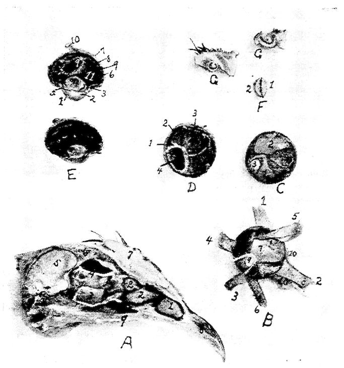
Fig. 26.—Structures of the eye and nasal passage.
A. 1, The anterior turbinated bone. 2, The posterior turbinated bones. 3, Orbital gland. 4, Ophthalmic division of the fifth pair of cranial nerves. 5, Section through the cerebrum. 6, Section through the skull showing the air spaces. 7, Section through the comb. 8, Anterior portion of the upper beak. 9, Edge of the hard palate.
B. The intrinsic muscles of the eyeball. 1, Superior rectus. 2, Internal rectus. 3, Inferior rectus. 4, External rectus. 5, Superior oblique. 6, Inferior oblique. 7, Quadratus. 8, Pyramidalis. 9, Optic nerve. 10, Edge of membrana nictitans.
C. Posterior part of the eye showing at the inferior portion of the ball the pyramidalis muscle at 1, and at 2, the quadratus. 3, Optic nerve.
D. Inner view of the posterior part of the vitreous chamber. 1, Sclerotic coat. 2, Choroid coat. 3, Retina. 4, Pecten.
E. Longitudinal section through the eye of a hen. 1, Cornea. 2, Anterior chamber. 3, Scleral ring. 4, Crystalline lense. 5, Iris. 6, Retina. 7, Sclerotic. 8, Choroid. 9, Pecten. 10, Optic nerve. 11, Vitreous chamber.
F. An edge view of the crystalline lense of a hen showing it to be asymmetrical biconvex. 1, The anterior side. 2, The posterior side.
G. The upper section through the anterior portion of the anterior turbinated bone showing one complete circle and the lower a section through the middle turbinated showing one and one-half turns.
127Origin.—From the orbital septum just back of the inner canthus, the fibers passing backward and forward.
Insertion.—Into the sclera, anterior to the sclerotic plates.
Shape.—Small, thin, fan-shaped.
Relations.—With the eyeball, superiorly; at its extremities with the superior rectus, interiorly.
Action.—Rotates the eyeball.
Obliquus Inferior (Fig. 26, No. B, 6). (Synonym.—Obliquus ventralis.)
Location.—Inferior to the eyeball.
Origin.—From the interorbital septum near the inner canthus of the eye.
Insertion.—By a broad expansion to the inferior portion of the eyeball.
Shape.—Tendinous at its insertions; fan-shaped as it passes downward and outward.
Relations.—Internally, with the eyeball; and externally, with the bony orbital wall, with the inferior rectus muscle, inferiorly.
Action.—Aids in rotating the eye; opposes the obliquus superior.
Rectus Superior (Fig. 26, No. B, 1). (Synonym.—Rectus dorsalis.)
Location.—Superior to the eyeball.
Origin.—Along the supero-posterior border of the optic foramen.
Insertion.—To the sclera just within the margin of the bony circle of the eye.
Shape.—Flat, thin.
Relations.—Internally, with the obliquus superior; superiorly, with the bony wall of the orbit.
Action.—To pull the eyeball upward.
Rectus Inferior (Fig. 26, No. B, 3). (Synonym.—Rectus ventralis.)
Location.—Inferior to the eyeball.
Origin.—From the inferior border of the optic foramen.
Insertion.—To the lower portion of the eyeball within the margin of the bony circle of the eye.
Shape.—Thin, fan-shaped.
Relations.—Superiorly, with the eyeball; inferiorly, with the bony wall of the orbit.
Action.—Pulls the eyeball downward.
Rectus Externus (Fig. 26, No. B, 4). (Synonym.—Rectus lateralis.)
Location.—External to the eyeball.
128Origin.—By two heads infero-laterally to the optic foramen.
Insertion.—To the postero-external side of the eyeball.
Shape.—Short, fan-shaped.
Relations.—Internally, with the eyeball; externally, with the bony wall of the orbit.
Action.—Pulls the eyeball outward.
Rectus Internus (Fig. 26, No. B, 2). (Synonym.—Rectus medialis.)
Location.—Inner side of the eyeball.
Origin.—From the supero-anterior border of the optic foramen.
Insertion.—To the sclera on the anterior portion of the eyeball.
Shape.—Thin, fan-shaped.
Relations.—Internally, with the eyeball and with the pyramidalis; externally, with the bony wall of the orbit.
Action.—Pulls the eyeball inward.
Circumconcha (Fig. 7, No. 32). Location.—Surrounds the external ear.
Origin and Insertion.—The circumconcha, surrounding the periphery of the ear, is adherent to the skull and loosely to the skin, and is attached to the outer terminus of the supra-occipital crest.
Relations.—Internally, with the skull; externally, with the skin.
Action.—Relaxes the tympanum.
Tensor Tympani (Fig. 7, No. 31). Location.—External to the quadrate and to the external auditory meatus.
Origin and Insertion.—From the surface of the quadrate and the inner end of the quadrato-jugal to the inner surface of the tympanum.
Shape.—A few fibers.
Relations.—Internally, with the quadrate and the quadrato-jugal; externally, the integumental duplicature.
Action.—Tenses the tympanum.
5. Muscles are arranged in pairs. Singly means one muscle acting alone, i.e., without its fellow; together means acting both at the same time.
SPLANCHNOLOGY — THE ACCESSORY ORGANS OF DIGESTION — THE RELATIONS OF THE VISCERAL ORGANS OF THE DOMESTIC FOWL — THE RELATIONS OF THE VISCERAL ORGANS OF THE BABY CHICK
The Digestive Apparatus.—This apparatus, apparatus digestorius, consists of the organs directly concerned in the reception of food, in its passage through the body, and in the expulsion of the unabsorbed portion. For convenience, these organs are grouped as follows: the alimentary canal and the accessory organs.
The alimentary canal is a tube which extends from the mouth to the anus. It has a complete lining of mucous membrane, external to which is an almost continuous muscular coat. The abdominal portion of the tube is largely covered with a serous membrane, the visceral peritoneum. The canal consists of the following consecutive segments: mouth, pharynx, first portion of the esophagus, crop, second portion of the esophagus, proventriculus, gizzard, small intestine, large intestine, and cloaca.
The accessory organs are beak, tongue, salivary glands, liver, and pancreas.
The Mouth (Fig. 27).—The distinctive character of the mouth of the fowl consists, in the absence of lips and teeth and instead of jaw bones of other animals, of a beak. The edge of the beak is covered by a horn-like gum. The shape of the beak differs in the various classes of birds. In the chicken the beak is short, strong, thick, and pointed; in palmipeds it is flattened. The upper mandible extends out over the lower mandible. The outer portion of the upper and the lower beaks is covered by a horny sheath. There is no velum, and the mouth cavity is extended rather continuously into the pharynx (Fig. 27, No. 4). The posterior cross bar of the hard palate (Fig. 27, No. 5), which possesses a row of filiform papillæ projecting backward, marks the upper boundary line of the mouth; while the posterior end of the tongue (Fig. 27, No. 6), likewise with a row of filiform papillæ, marks its lower boundary line.
Parts of the Mouth.—The mouth of fowls is divided into an upper and a lower half. The upper half is divided into an anterior and a posterior part.
The anterior part extends from the anterior tip of the beak to the posterior nares. It is further divided into an outer hard part and 136an inner soft part. The outer part is covered by epithelium which may be considered as a continuation from the upper outer portion of the beak. A portion of this is cornified. The inner part is covered by a mucous membrane containing mucous glands, nerves, and blood-vessels.
In the posterior part of the upper half of the mouth, are situated the posterior nares (Fig. 27, No. 10) which appear as a longitudinal slit in the center of the hard palate. There is also found in this part a furrow which contains the opening of the Eustachian tubes, or tubæ auditivæ. There are also two ridges near the same posterior part in which are red and white papillæ, which may appear in rows, and which contain mucous glands.
Fig. 27.—Mouth and pharynx of a cock laid open. 1, Eye. 2, Edge of beak covered with a horn-like gum. 3, Tongue. 4, Pharynx. 5, Posterior cross bar of hard palate. 6, Posterior end of tongue. 7, Anterior end of esophagus. 8, Phrenum. 9, Palatal papillaries. 10, Posterior nares.
The lower half of the mouth lies between the lower jaw bones and the walls of the mouth cavity. The tongue is attached here by 137means of the phrenum (Fig. 27, No. 8). This half of the mouth forms a pocket-like structure which aids in taking up the food.
The mouth cavity is lined with stratified squamous epithelium, continuous with that of the pharynx and the esophagus. Taste buds are located in the mucous membrane of the mouth.
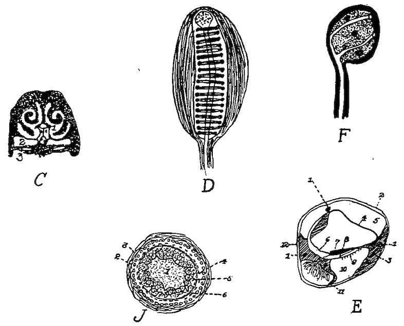
Fig. 28.—Various anatomical parts of the fowl.
C. A section through the nasal region of the fowl. 1, Nasal passages showing the turbinated bones dividing the nasal passage into the superior, middle and inferior meati. 2, The infraorbital sinus. 3, The hard palate.
D. Herbst’s touch corpuscle from the beak of a quail.
E. A vertical section through the ductus cochlearis of a pigeon. 1, A blood-vessel. 2, The periosteum. 3, The bony structure. 4, The vascular integument. 5. The scala vestibuli. 6, The inner hyaline cylindrical cells. 7, The membrana tectoria. 8, The papilla acustica basilaris. 9, The membrana basilaris. 10, The scala tympani. 11, The ganglion of the cochlear nerve in the ramus basilaris. 12, The periosteum (Gadow).
F. A corpuscle of the soft papilla of a duck’s tongue.
J. A transverse section through a feather papilla. 1, The pulp. 2, The malpighian layer. 3, The corium. 4, The stratum corneum of the papilla. 5, The malpighian cell group of the main shaft. 6, The horny sheath.
In many water fowls, as geese and ducks, the gum edge of the mandible has grooves extending crosswise, in which are numerous terminals of the trigeminus nerve arranged as taste organs (Fig. 29, G and Fig. 28, D and F). In many birds of prey and in water birds, e.g., the goose and the duck, there is found at the base of the beak, a very thin, nervous or sensitive skin, waxy in appearance, called the ceroma.
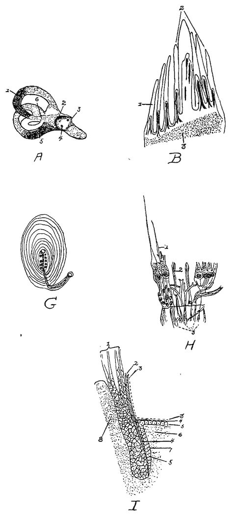
Fig. 29.—Various anatomical parts of the fowl.
A. 1, The superior semicircular canal. 2, The vestibular nerve. 3, The meatus auditorius internus—the entrance of the auditory nerve. 4, The entrance of the cochlear nerve. 5, Posterior semicircular canal. 6, The ampulla.
B. Side papilla of the tongue. 1 and 2, Papillæ showing a fibrous central core supporting blood-vessels and nerves. 3, The basement membrane consisting of connective tissue.
G. Herbst’s corpuscle of the tongue of a duck, showing the capsule, lamella and around the nerve zone, numerous round bodies arranged in two rows.
H. From the cristæ acoustica of the ampulla of the dove. A, A vertical section with isolated fiber cells. 1 and 2, The nerve fibers. 3, The hair cells.
I. A longitudinal section through a feather papilla showing a young feather. 1, The developing feather. 2, The horny sheath. 3, The epithelium. 4, Stratum corneum. 5, Stratum malpighi. 6, Corium. 7, Malpighian cell group of the follicle.
139The Hard Palate.—The hard palate, palatum durum, forming the roof of the mouth has for its boundaries the beak anteriorly, the mandibular arches laterally, and the pharynx posteriorly. It has four or five transverse bars, projecting posteriorly from each of which is a row of filiform papillæ (Figs. 27 and 29).
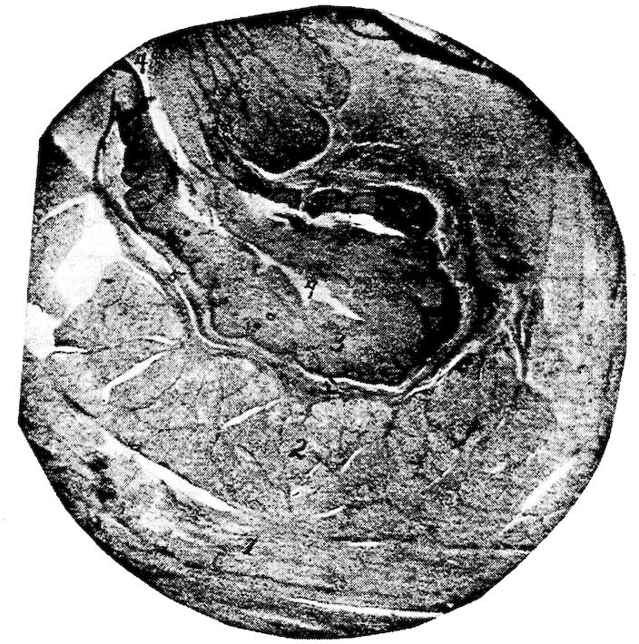
Fig. 30.—A section through the wall of the crop of a hen. 1, The outer muscular layer showing longitudinal fibers. 2, The inner muscular layer showing the sectioned ends of the bundles of muscle fibers. 3, Stratified squamous epithelium. 4, The outer surface or lumen of a fold. 5, The stroma, muscularis mucosa, and submucosa.
The beak (Fig. 4, No. 1) with little if any aid from the tongue, is the prehensile organ.
The Tongue (Fig. 27, No. 3). Location.—The tongue (lingua) of the fowl is situated in the floor of the mouth between the rami of the 140lower mandible, and is slung to the cranium by the cerato-branchial element of the os hyoideum (Fig. 18, No. 2).
Shape.—The tongue, is pointed in front and wide behind, shaped like an Indian arrow-head, and is supported by the bony and cartilaginous hyoid apparatus (Fig. 9, A).
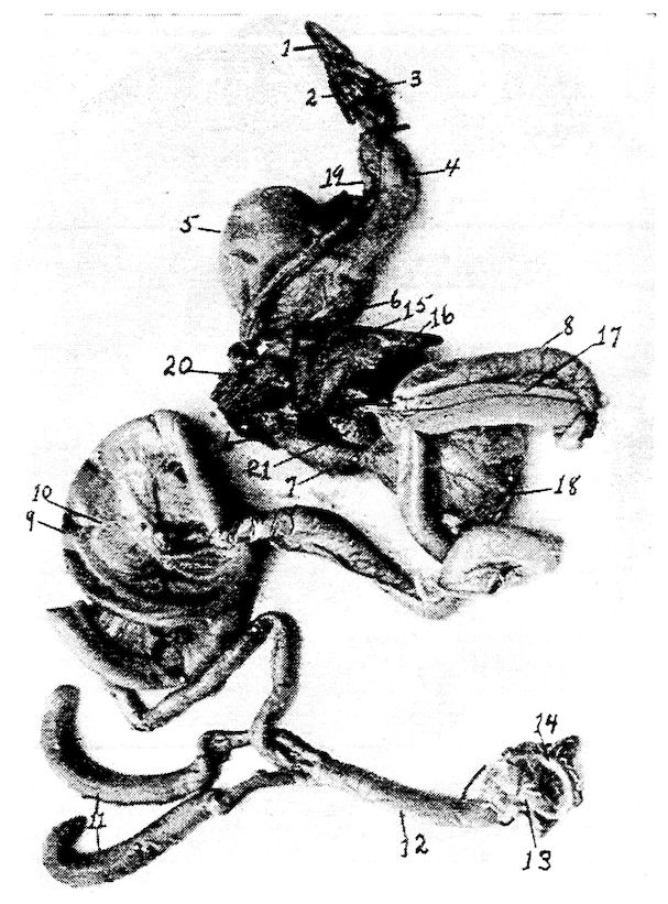
Fig. 31.—The visceral organs of the hen. 1, Tongue. 2, Larynx. 3, Glottis. 4, First portion of the esophagus. 5, Crop. 6, Second portion of the esophagus. 7, Proventriculus. 8, Duodenum. 9, Free or floating portion of the small intestine. 10, Mesentery supporting the free portion of the small intestine. 11, Cæca. 12, Rectum or large intestine. 13, Cloaca. 14, Anus. 15, Liver. 16, Gall-bladder. 17, Pancreas. 18, Gizzard. 19, Trachea. 20, Lungs. 21, Spleen.
Structure.—The free part of the tongue consists of two long tubes which are formed by the rolling in of the ventral sides of its horny coverings. The dorsal plate, which constitutes one-half of the 141horny sheath, does not form a part of the tubes, but ends in the region of the point of the entoglossum.
The top surface is covered by a thick stratum corneum, giving it a rather horny surface. Glands occur in the posterior part of the tongue.
While not so freely movable as in mammals, the tongues of birds are very flexible.
The Pharynx (Fig. 27). Location.—The anterior part of the pharynx joins with the mouth and the posterior part with the upper portion of the esophagus and the superior larynx. The pharynx is bounded anteriorly by the base of the tongue and by the posterior edge of the hard palate. Posteriorly, it is marked by the entrance of the esophagus and by a ridge of filiform papillæ located on the supero-posterior part of the larynx, called the palatal papillaries. A few filiform papillæ stud the roof. The pharynx communicates anteriorly with the posterior nares and the mouth, and, posteriorly, with the esophagus and the larynx.
Shape.—The pharynx is a musculo-membranous sac.
Structure.—The pharynx is lined with a mucous membrane covered by squamous epithelium. The mucous membrane is thrown into irregular folds. The bird has two Eustachian tubes. Pneumatic apertures conduct the air from the Eustachian tubes to the pericranial diploë.
In the posterior pharyngeal roof is situated the infundibular crevice.
Function.—The function of the pharynx is to give passage for the air from the posterior nares to the larynx, and to give passage for the food from the mouth to the esophagus.
Glands Adjacent to the Mouth and to the Pharynx.—The mucous membrane adjacent to the glottis and in the roof of the mouth contains alveolar glands. The angular gland of the mouth, located beneath the zygomatic arch, by some anatomists is considered the rudimentary parotid gland. The sublingual glands, or glandulæ sublinguales, are well developed (Fig. 17, No. B, 4). They form conical masses, with the apex directed anteriorly, and occupy a portion of the intermaxillary space. Several ducts from these glands open into the mouth cavity. In the palatine region there are also located glands called the palatine glands, which open by many stomata upon the surface.
142The Esophagus (Fig. 18, No. 6). Location and Shape.—The esophagus is a musculo-membranous tube, capable of great distension, which extends from the pharynx to the proventriculus (Fig. 35, No. 1, and 3).
The esophagus communicates anteriorly with the pharynx. It extends down the neck, lying supero-laterally with regard to the trachea and toward the right side. It enters the thorax above the trachea, and, passing through between the bronchi, terminates into the proventriculus. At the entrance of the thorax and just to the right of the median line, it expands into the crop which divides it into two portions, designated as the first and the second. The first portion of the esophagus is the longer, the length depending upon the size and kind of fowl.
Structure.—The wall is composed of four coats, the mucous membrane, the submucosa, the muscular coats, and the fibrous sheath or tunic. The mucous membrane is pale and is covered with stratified squamous epithelium.
The mucous membrane is loosely attached to the muscular coat by the submucosa. Except during deglutition it lies in longitudinal folds which obliterate the lumen. Opening from the mucosa are lenticular glands, which may be seen on inflating an esophagus and looking through its transparent walls.
The muscular coat of the esophagus is divided into two layers, an outer longitudinal and an inner circular layer.
The outer fibrous sheath connects the esophagus loosely to the surrounding structures.
Function.—The function of the esophagus is to give passage way for the food from the pharynx to the crop and from the crop to the proventriculus.
The Crop (Fig. 35, No. 2; Figs. 30 and 31). Location and Shape.—The crop, or ingluvies, saccular in shape, is located at the entrance of the thorax and just to the right of the median line. The first portion of the esophagus empties into the crop superiorly and inferiorly the crop opens into the second portion of the esophagus. Like the esophagus the crop is capable of great distension.
Structure.—The wall of the crop is composed of four coats, the mucous membrane, submucosa, muscular and the outer fibrous.
The crop is lined with mucous membrane containing mucous glands which secrete a mucus to keep the surface moist. The surface of the mucous membrane is covered by stratified squamous epithelium.
143The submucosa connects the mucous membrane to the muscular coat.
The wall of the crop is provided with strong muscles. The fibrous coat, or tunic, connects it with the surrounding structures.
Function.—The crop is a storehouse for the food during the hours of feeding, the food when needed by the stomach being gradually discharged from the crop by the contraction of its muscular walls. The subcutaneous cervical muscles which cover this reservoir aid in this discharge.
In the act of deglutition, the food, after being subjected to the fluid supplied in the mouth by the adjacent glands, is poised upon the tongue and swallowed partly by a sudden jerk of the head, and partly by means of the pressure of the tongue against the hard palate, the food then passes down the esophagus and lodges first in the crop, till needed by the stomach, when it is passed through the second portion of the esophagus to the first portion of the stomach, the proventriculus. The time during which food remains in the crop depends upon the nature of the food. Animal food will, in part, be retained about eight hours and vegetable foods may not all be passed on for from sixteen to eighteen hours.
The Stomach (Fig. 31, No. 7; Fig. 32, B).—The stomach, or ventriculus, of fowls is made up of two portions, namely, the pars glandularis, or proventriculus, and the pars muscularis, or gizzard.
The Proventriculus. Location.—The proventriculus lies in the superior part of the groove formed by the two lobes of the liver, is inferior to the aorta, and is directed slightly to the left, communicating anteriorly with the second portion of the esophagus and posteriorly emptying into the pars muscularis, the gizzard.
Shape.—The proventriculus is round transversely and elongated, in fact, nearly fusiform. In the hen of average size it measures about 1.62 inches long and 0.8 inch in diameter.
Structure.—The wall of the proventriculus has four coats, the mucous, submucous, muscular, and serous.
The inner mucous coat which is raised in folds, is lined with columnar epithelial cells. The mucous membrane contains lymphoid tissue. The mucous coat throughout contains simple tubular glands which secrete a highly acid fluid which finds its way to the surface through small cylindrical ducts lying at right angles to the inner surface of the mucous membrane (Fig. 32, No. B, 1).
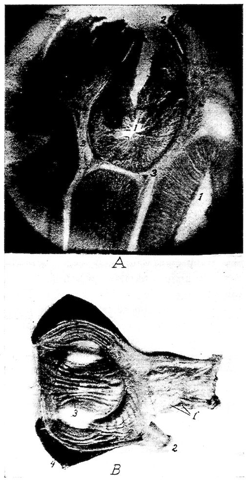
Fig. 32.—Gizzard and stomach of the fowl.
A. 1, Alveolar glands. 2, Mucous membrane of the inner surface of the proventriculus showing the tubular glands. 3, Connective tissue between the alveolar glands. 4, An artery. 5, Muscularis mucosa.
B. Photograph of the proventriculus and gizzard. 1, Proventriculus showing stomata of glands. 2, End of duodenum. 3, Gizzard showing hornified epithelium formed into grooves. 4, The heavy muscular walls.
145The submucosa, connects the mucous and muscular coats and the muscularis layer throws the mucous membrane into folds.
Outside the muscularis mucosa there is another layer (Fig. 32, A), of simple tubular glands, grouped in lobules, and lined by cuboidal cells and separated from each other by clefts. These tubular glands converge toward the center and open into the same cavity.
The middle muscular coat can be divided into three layers: two thin, longitudinal layers, and a thick, circular layer interposed between the other two.
Function.—The function of the proventriculus appears to be to soak the food with a secretion. The secretion from the proventriculus is similar to that produced by the fundus glands of the stomach of mammals. It contains acid and a ferment-pepsin.
The Gizzard (Fig. 31, No. 18). Location.—The gizzard, or muscular stomach, occupies a portion of the central part of the abdominal cavity (Fig. 33, No. 4). It lies slightly to the left and just behind the liver, the proventriculus, and the spleen, and rests upon a mass of intestines. The gizzard communicates at its anterior portion with the proventriculus and with the duodenum. These openings are close together. The gizzards of a large number of hens of average size averaged in weight as follows: full, 0.215 pound; empty, 0.126 pound. The gizzard stands perpendicularly and somewhat obliquely in the abdominal cavity.
Shape.—In shape the gizzard is roundish, flattened laterally.
Structure.—The walls of the gizzard are very thick and are made up of three coats: mucous, muscular, and serous.
The cavity of the gizzard is covered by a thick skin-like structure possessing a heavy stratified squamous epithelial layer which is thrown somewhat into ridges (Fig. 32, B). This membrane becomes thinnest near the edges.
The mucous membrane, being cornified and readily detachable, is by some anatomists considered as a special membrane (Fig. 37, B).
At the pyloric opening there is a valve formed by a fold of the mucous membrane, which prevents grit and large particles of food from passing out of the gizzard.
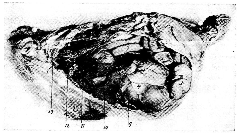
Fig. 33.—View of viscera of the left side of a hen. 1, The base of the heart. 2, Proventriculus. 3, Left lobe of the liver. 4, Gizzard. 5, End of the duodenal loop. 6, Pancreas. 7, Free portion of the small intestine. 8, Lungs. 9, Diaphragm.
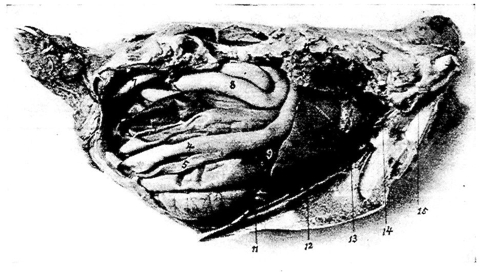
Fig. 34.—View of the viscera of the right side of a hen. 1, Base of the heart. 2, Lungs. 3, Right lobe of the liver. 4, Duodenal loop. 5, Pancreas. 6, Cæcum. 7, Large intestine or rectum. 8, Free portion of the floating small intestine. 9, Gall-bladder. 10, Right kidney. 11, The ribs. Note the lungs pushing up between them.
On each side it has a powerful fleshy muscle, the muscularis lateralis. These musculares laterales are hemispherical, consist of very closely packed fibers extending transversely, and are attached to strong anterior and posterior ligamentous tendons. They are joined at the edge of the organ by a strong aponeurosis. The muscular fibers are red but do not possess cross striations; they are of the smooth type. The muscularis intermedii, thinner and less 147developed than the musculares laterales, occurs on each of the anterior and the posterior parts of the gizzard.
The serous coat covers over the greater part of the external surface of the gizzard and closely adheres to the muscular coat.
Function.—The hard callous pads of the gizzard, operated by the powerful muscles above described, together with grit, act like mill stones and make reduction to fineness very complete.
At the posterior part there is a sacculated portion containing glands of the long tubular type which secrete a fluid ferment similar to that secreted by the glands of the pyloric portion of the stomach of mammals. These glands also exist in a small band near the entrance of the gizzard. Here the food is mixed with strongly acid secretion containing pepsin which makes gastric digestion perfect.
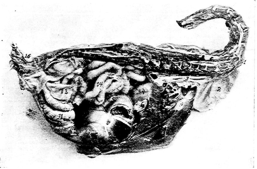
Fig. 35.—A median antero-posterior section through the body of a 1-pound pullet. 1, First portion of the esophagus. 2, Crop. 3, Second portion of the intestine. 4, Proventriculus. 5, Gizzard. 6, Spleen. 7, Liver. 8, Heart. 9, Point where duodenum was severed from gizzard. 10, Point where duodenum was severed. 11, Duodenum. 12, Pancreas. 13, Cæcum. 14, Floating small intestine. 15, Ovary. 16, Oil sac.
The Small Intestine (Fig. 31, No. 8 and 9).—The small intestine, intestinum tenue, is the tube which connects the gizzard with the large intestine. It is divided into two parts, the duodenum and the free portion. Of the three parts as considered in the mammalian 148intestine, only the first, the duodenum, can be distinguished. There is no demarcation between the jejunum and the ileum. The jejunum and the ileum or that part which represents these sections, are in coils suspended from the free border of the mesentery the other border of which is, in turn, attached to the dorsal wall (Fig. 64). The length of the small intestine in a hen of average size is about 61.7 inches.
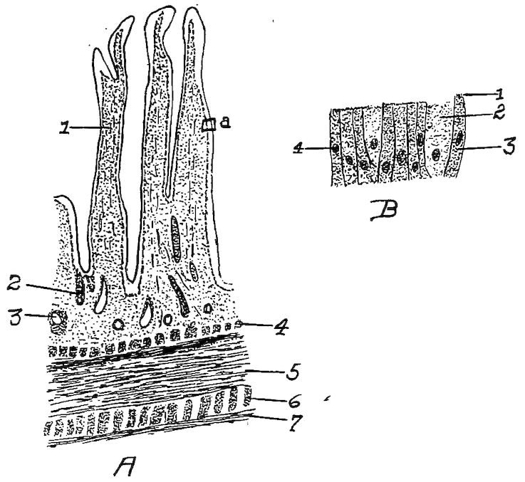
Fig. 36.
A. Section of the duodenum of the fowl. 1, Villus. 2, Gland. 3, Mass of lymphoid tissue. 4, Muscularis mucosa. 5, Longitudinal layer. 6, Circular muscular layer. 7, Serous layer.
B. A section from A at a. 1, The striated free border of the cells. 2, Goblet cell. 3, Columnar cell. 4, Nucleus of cell.
The wall of the small intestine is provided with four coats, as follows: a mucous, submucous, muscular coat made up of two layers—the outer, longitudinal and the inner circular layer, and an external serous.
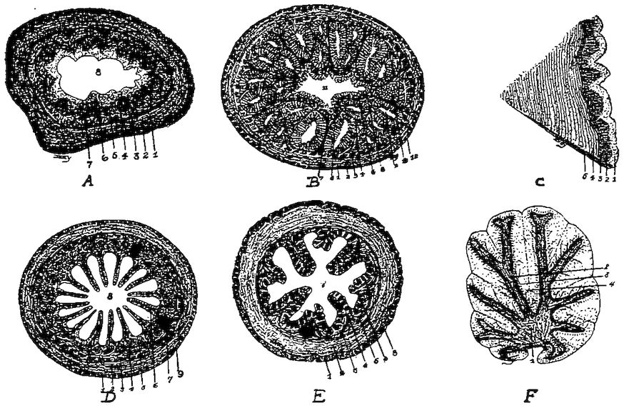
Fig. 37.—Histological studies of various anatomical parts.
A. A transverse section of the first portion of the esophagus of a fowl. 1, The outer longitudinal muscular layer. 2, The circular muscular layer. 3, The submucosa. 4, The muscularis mucosa. 5, Stroma. 6, Epithelial layer. 7, The lenticular glands. 8, The lumen.
B. A transverse section of the proventriculus. 1, The outer longitudinal muscular layer. 2, The middle muscular layer. 3, The inner longitudinal muscular layer. 4, Stroma. 5, The muscularis mucosa. 6, The submucosa. 7, Stroma. 8, Tubular glands. 9, Tubulo-alveolar glands. 10, A tubulo-alveolar gland with the tubular glands cut transversely, 11, The lumen of the proventriculus.
C. A section of the inner wall of the gizzard. 1, The hyaline mucous membrane. 2, Branched tubular glands. 3, Submucosa. 4, Muscle. 5, A connective tissue septum.
D. A transverse section of the small intestines. 1, The outer longitudinal muscular layer. 2, The inner circular muscular layer. 3, The muscularis mucosa. 4, The stroma. 5, Brunner’s glands. 6, A villus. 7, Mass of lymphoid tissue.
E. A transverse section through the cecum. 1, The outer longitudinal muscular layer. 2, The inner circular muscular layer. 3, The submucosa. 4, The muscularis mucosa. 5, The stroma. 6, Tubular glands. 7, The lumen.
F. The cerebellum showing the arbo-vitæ. 1, White, fiber portion. 2, The granular layer. 3, The layer of Purkinje cells. 4, The molecular layer.
The mucous membrane which lines the intestine is thick, soft, and highly vascular. It has a velvety appearance, due to numerous long, thin projecting villi. The villi (Fig. 36, A) are concerned in the absorption of the digested food, absorbing principally the emulsified fats. Each villus is covered with a single layer of high columnar epithelial cells. Some of these, the so-called goblet cells, provide mucin which lubricates the mucous surface. These cells are found in all mucous surfaces and prevent the surface from becoming dry. There are a few goblet cells near the summits of the villi. Openings of simple intestinal tubular glands the duodenal glands, or the glands formerly known as Brunner’s glands, are located between the villi. These glands secrete the succus entericus, or intestinal juice. These openings, or stomata, are lined with granular cells. The 150reaction of the contents of the small intestine is strongly acid, but gradually less so in proportion to the distance down the intestinal tract until the cæca are reached, where the reaction is found to be faintly acid, neutral, or slightly alkaline.
Function.—The function of the small intestine is that of digestion and absorption.
The Duodenum. Location and Shape.—The duodenum, a small tube, originates from the gizzard about ½ inch to the right of the entrance of the proventriculus. Extending from left to right, it passes under and behind the gizzard along the inner side of the right abdominal wall backward to the posterior portion of the wall, and a trifle more than half way toward the left side, where the loop rests. This loop, the duodenal loop, is about 5 inches in length (Fig. 31, No. 8). The two branches of the loop the first and the second portions or the descending and ascending limbs, are loosely held by connective tissue, and have the pancreas lodged between them.
The Free Portion of the Small Intestine. Location.—That section of the small intestine following the duodenum is called the free portion of the small intestine and occupies the space between the abdominal air-sac and the median line of the abdominal cavity. It is disposed in coils and is suspended from the dorsal wall of the abdomen by a thin membrane, the mesentery.
The bile ducts enter the small intestine about 14 inches from its point of origin. The pancreas also pours its contents into the small intestine.
The Large Intestine (Fig. 31, No. 12). Location.—The large intestine extends in a straight line along the inferior border of the vertebral column communicating anteriorly with the small intestine, and the cæca and posteriorly with the cloaca.
Shape.—The diameter of the large intestine is approximately twice that of the small intestine. In fowls of average size its mean length is 4.61 inches. The large intestine has sometimes been spoken of as the rectum, or straight gut.
Structure.—Like the small intestine the large intestine has four coats: an inner mucous, a submucous, a middle muscular and an outer serous. The folds of mucous membrane of the large intestine have tubular glands lined with columnar cells (Fig. 37, A, No. 1).
Function.—The large intestine is similar in function to that of the small intestine, in that digestion and absorption may take place within it.
151The Cæca (Fig. 31, No. 11). Location.—The fowl has two cæca extending forward from their point of origin at the juncture of the small and large intestine.
Shape.—The cæca average 7.61 inches in length. They are large in caliber toward the blind extremity and are constricted near their origin.
Structure.—The parietal coats are continued from the small intestine.
Ebarth has described an elevated body in the cæcum, which is located about 4 millimeters from the opening and is composed entirely of lymphoid tissue.
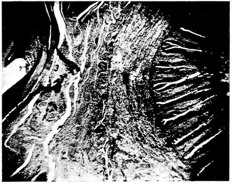
Fig. 37A.—Photomicrograph of a transverse section of large intestine and oviduct just anterior to the cloaca, showing the tubular glands of the large intestines, mucosa of oviduct with intervening structures. 1, The tubular glands of large intestine, or rectum. 2, Muscular wall of intestine. 3, Wall of the oviduct. 4, Mucosa of the oviduct.
The cæca are usually partly filled with a soft pultaceous material of a pasty consistency.
The Cloaca (Fig. 31, No. 13). Location.—The rectum opens by a circular, valvular orifice into the dilated portion just in front of the anus, which dilatation is called the cloaca.
Shape.—The cloaca is saccular in shape.
Structure.—The cloaca is divided into two portions, the coprodeumal and the urodeumal. That portion of the cloaca into which the intestine empties is called the coprodeum; and the ureter and 152oviduct empty into the urodeum. The seminiferous tubules, carrying the semen from the testes in the male empty in teatlike projections on the cloacal mucous membrane into the urodeumal portion. The cloacal walls are similar in structure to the large intestines.
On the dorsal wall of the cloaca between it and the spine, is a small sac, called the bursa of Fabricius, which has a duct communicating with the cloaca. The mucous membrane of this sac is thrown into folds and is studded with glands. The bursa of Fabricius is larger in the young than in the adult bird. It apparently atrophies as the bird becomes older. When the bird is four months old this bursa is best developed, and at this age it may be as large as 2 or 3 centimeters in diameter.
Function.—The function of the cloaca is to give passage way to the feces, the urine, and the egg and to act as an organ of copulation.
Course of the Food.—The food first enters the mouth, after being picked up by aid of the beak. From here it passes through the pharynx and first portion of the esophagus to the crop, without mastication, as the bird is not provided with teeth. The food is passed from the crop by aid of its muscular walls as needed; thence through the second portion of the esophagus to the proventriculus, an expansion in the digestive tube just before it terminates in the gizzard. The glands of the proventriculus produce a secretion in which the food is soaked before passing into the gizzard. The gizzard is provided with strong muscular walls which, by aid of grit, thoroughly reduce the food to fineness. From the gizzard the food passes through the first portion of the small intestine, where it is subjected to the action of the bile from the liver, the pancreatic juice from the pancreas, and of the succus entericus from the glands of the intestinal wall. The food is then passed into the cæca. The indigestible portion of the food passes from the cæca through the large intestine, or rectum, to the cloaca and thence to the external world.
The digestive functions of the bird are very potent and rapid. This compensates for the waste caused by their extensive, frequent, and energetic motions, and is in accordance with the rapidity of their circulation and their high state of irritability.
The accessory organs of digestion are the liver, pancreas, and some anatomists include also the spleen. The first two manufacture 153fluids containing ferments which aid in splitting or digesting the food.
The Liver (Figs. 31, 33 and 34). Location.—The liver, hepar, lies ventrally and posteriorly to the heart. It is related anteriorly with the diaphragm, inferiorly with the sternum, posteriorly with the gizzard and intestine, and superiorly with the ovary, oviduct and proventriculus and laterally with the abdominal wall.
Shape.—The liver is a voluminous deep livid brown gland, soft and friable in texture. It is divided into two principal lobes, a right and a left.
The right lobe is larger than the left. In the hen of average size the liver weighs 35 grams. The parietal surface is convex and smooth. The surface which lies against the viscera is irregularly concave. The visceral surface furnishes exit for the bile duct and passage for the nerves and blood-vessels. This part is called the porta.
The left lobe may be cleft from below so deeply as to form two lobes on that side.
Structure.—Each lobe is covered by a double serous membrane, one closely adherent, the other surrounding the structure loosely. These tunics, which are reflections of the peritoneum, are continued from the base of the liver, over both the anterior, and the posterior surface. The loose layer is formed by the air cells surrounding the lobes. The thin border of the liver is usually free.
The two lobes of the liver are connected by a narrow isthmus of liver tissue. Occasionally there is a bird in which there occurs a lobus Spigelii located at the posterior of the liver between the two principal lobes.
The apex of the heart sacculates the diaphragm backward, so that part of this apex lies between the right and the left lobes (Fig. 43, No. 6 and 7). A ligament, the falciform, extends from the apex of the pericardial membrane, and attaching it rather firmly to the central connective tissue, or interlobar ligament. This ligament also has attachments to the inner surface of the sternum. The broad ligament of the liver is formed posteriorly by a fold of the peritoneum.
The interlobar, or principal, ligament of the liver is formed by a large and strong duplicature of the peritoneum, which makes a longitudinal division in the abdominal cavity similar to the lateral division made by the thoracic mediastinum in mammals. It 154is reflected upon the pericardium from the linea alba and the middle line of the sternum, and passes deeply into the interspace of the lobes of the liver. It is attached to these lobes throughout their whole length and connects them below to one side of the gizzard. The lateral and posterior part of the liver attach to the adjacent air cells, and the whole viscus is thus kept fixed in its position during rapid and violent movements of the bird.
The remains of the umbilical veins are traceable within the duplicature of the membranes forming the septum. These remains thus represent the round ligament of mammals.
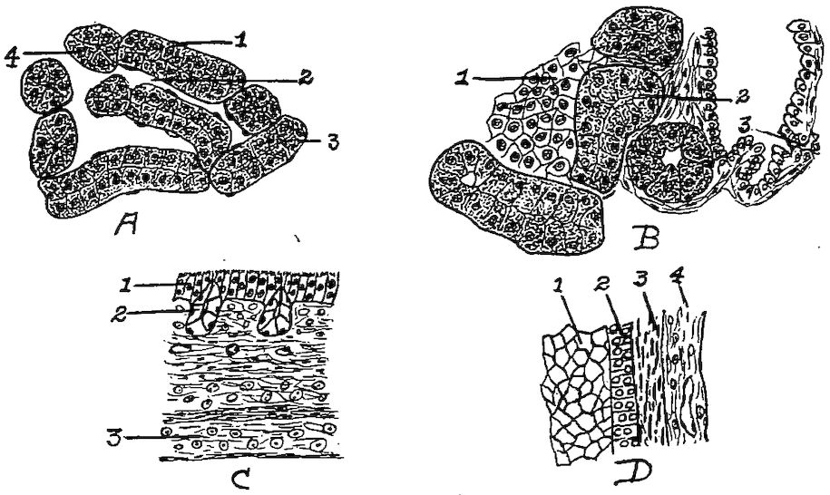
Fig. 38.—Cellular structure of liver, pancreas, and trachea.
A. Liver. 1, Liver cells. 2, Sinusoid. 4, Nucleus of cells.
B. Pancreas. 1, Island of Langerhans. 2, Alveolar cells. 3, Duct.
C. Trachea. 1, Ciliated epithelia. 2, Glands. 3, Hyaline cartilage.
D. Section of wall of ovum at Fig. 57 letter d. 1, Yolk. 2, Granular membrane. 3, Theca. 4, Blood-vessel.
A microscopic study of the liver of the fowl shows a compact mass of liver cells polyhedral in shape, with large nuclei (Fig. 38, A). The liver tissue differs from that of mammals in that there is no clearly outlined lobular arrangement; neither is the outline of the individual cell so well marked. The parenchymatous portion is made up of columns of liver tissue. These columns anastomose and show narrow channels between. They are best seen in the young chick.
Function.—One of the functions of the liver is to secrete bile. A gall-bladder, which receives part of the bile secreted by the right lobe of the liver, is located on the posterior face of this lobe. Extending 155from this gall-bladder there is a duct, the cystic duct, which empties into the small intestine toward the extremity of the second branch of the duodenal loop. Another duct called the hepatic duct, proceeds directly from the two lobes of the liver and empties into the intestine just in front of the cystic duct.
The Pancreas (Fig. 31, No. 17). Location and Shape.—The pancreas, an organ lying in the duodenal loop is a yellowish-white, lobulated gland, elongated in shape. Its average length in fowls of average size is 4.96 inches, and the average weight about 0.008 pound. The pancreas is divided into lobes, which in turn are divided into lobules.
Structure.—The pancreas has a supporting connective tissue. The lobules are made up of small alveolar glands, which are lined with columnar epithelial cells. The ducts leading from the alveoli are small; these unite to form larger ducts in which the epithelium is taller. Between the alveoli throughout the pancreas there are clusters of polyhedral cells which form the islands of Langerhans. These islands are said to produce an internal secretion, or hormone, which is absorbed by the blood or lymph capillaries and thus enters the circulation (Fig. 38, B).
Function.—The function of the pancreas is to secrete a fluid containing digestive ferments.
The Spleen (Fig. 31, No. 21). Location.—The spleen, or lien, lies in a triangle formed by the proventriculus, the liver, and the gizzard.
Shape.—It is a reddish-brown body shaped like a buckeye, is small in size, weighing only about 0.005 pound.
Structure.—The outer surface of the spleen is covered by a reflection of the peritoneum. After this covering is removed, there is observed a firm, white, fibrous layer, the cortical portion. This covering sends into the interior small and large trabeculæ, forming a framework and dividing it into acini, or compartments. The spaces are filled with a dark red parenchymatous material called splenic pulp. The framework, in both deep and surface portions, are found elastic fibers and smooth muscular fibers.
The spleen is essentially a lymphatic organ, its peculiar structure depending largely upon the arrangement of the blood-vessels. Compact lymphatic tissue occurs in the spleen in spherical, oval, or cylindrical collections of closely packed lymphoid cells. These masses are known as the Malpighian bodies, or splenic corpuscles. 156They are distributed throughout the splenic pulp. Each splenic corpuscle contains one or more small arteries. These extend near the periphery of the corpuscle and more rarely in the center.
The splenic artery passes in and the splenic veins out at the hilum which is located on the concave, or attached, side of the spleen. The splenic artery, upon entering the organ, at once branches, the trabeculæ forming a support for the vessels.
After the arteries have entered the hilum, as stated above, they divide into many branches which follow the septa of the connective tissue. At first the arteries are accompanied by branches of the splenic veins. Soon, however, the arteries leave the veins and the septa, and penetrate the splenic pulp. In the splenic pulp the adventitia of the smaller arteries assume the character of reticular tissue, and become infiltrated with lymphoid cells. This infiltration forms masses which are called the splenic corpuscles, or Malpighian bodies. The terminal arteries break up into capillaries, which still retain an adventitia, and empty into border spaces, or sinuses, sometimes spoken of as ampullæ. These sinuses in turn empty into cavernous sinuses of the splenic pulp. From these are finally formed the venules; and the collections of venules form the splenic veins through which the blood gains exit from the spleen.
The Abdominal and Pelvic Cavities.—In birds the abdominal cavity is divided into two smaller cavities by a fibrous septum. The anterior cavity representing the abdominal contains the liver, and the other representing the pelvic contains the gizzard, intestines and oviduct.
The Peritoneum and the Mesentery.—The abdominal and pelvic cavities are lined by the peritoneum. Like all serous membranes this is composed of a parietal and a visceral portion, which together form a complete sac, with the organs it covers situated on the outer side. The peritoneum like other serous membranes consists of a mesothelial and a submesothelial portion, the cells of the former being arranged in a single layer. Since a serous membrane is so arranged as to line a closed cavity, and at the same time to cover its contents, it follows that the entire membrane must be a closed sac, the mesothelial layer being on the inside; such a sac is called a serous sac, or cavity. Synovial membranes are also regarded as a variety of serous membranes. The fold, or layer, of the membrane which lines the cavity is called the parietal, that which covers the greater part of the organs contained therein is called the 157visceral portion; the two surfaces contacting, and gliding upon each other, are lubricated by a fluid secretion contained in the sac; hence one use of these membranes is to prevent friction between the walls of cavities and the organs contained therein. Serous membranes line the abdominal cavity, pericardium, cavities of the heart and is continuous throughout the vascular structures.
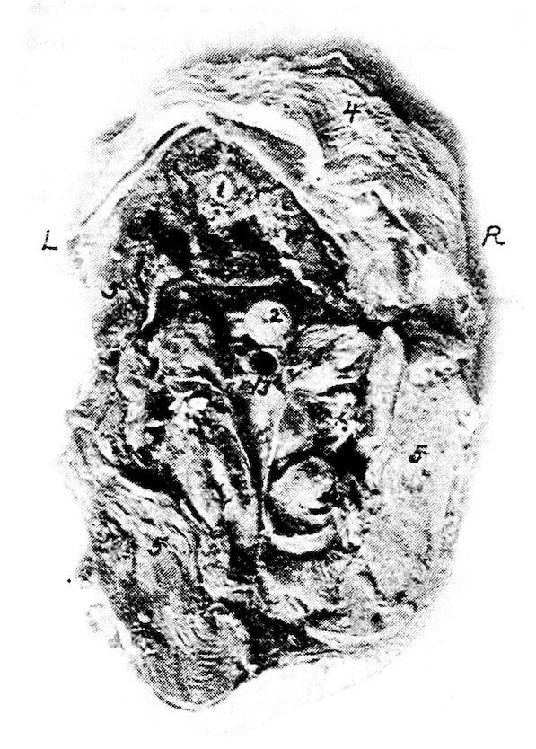
Fig. 39.—A transverse section of body of hen through 15, Fig. 34. R, Right side. L, Left side. 1, Spinal cord. 2, Esophagus. 3, Trachea. 4, Skin. 5, Pectoral muscles.
The serous membrane besides covering the external surface of the viscera, double folds pass from one organ to another, or from an organ to the parietes of the cavity. These double folds of the peritoneum are known as ligaments, or as mesenteries. In ligaments the two folds are strengthened by an interposed layer of fibro-elastic tissue. A mesentery is a broad, double fold of peritoneum, attached to the abdominal parietes above, and containing a portion of the intestine in its free or remote extremity. Between its folds we find blood-vessels, nerves, and lymphatics or lacteals, hence it permits 158vascular and nervous communications with the organ attached to it. The free portion of the small intestine is attached to the free margin of the mesentery.
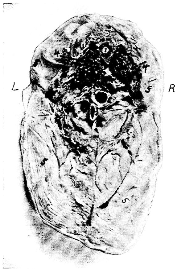
Fig. 40.—Transverse section through the body of a hen through 14, Fig. 34. R, Right side. L, Left side. 1, Spinal cord. 2, Esophagus. 3, Trachea near inferior larynx. 4, Lungs. 5, Pectoral muscles.
Figure 33 shows a fowl with the left abdominal wall and the left thoracic wall removed. No. 1 in this figure shows the base of the heart in front of the left lobe of the liver. Above the liver is the proventriculus; above this, the diaphragm; and above the diaphragm and the base of the heart the left lung occupying the superior part of the thoracic cavity, and that there is no distinct pleural sac, as in mammals but that the lung pushes out between the ribs, thus pressing against the ribs on the inner and the lateral sides. The gizzard is back of the liver, to the left side of the abdominal cavity, 159and beneath and in front of the duodenal loop. The small intestine from this side is above the gizzard. Supero-anterior to the gizzard is the blind end of the cæcum. The pancreas is within the duodenal loop.
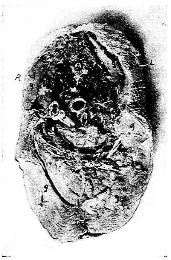
Fig. 41.—Transverse section through the body of a hen at 12, Fig. 33. R, Right side. L, Left side. 1, Spinal cord. 2, Esophagus. 3, Inferior larynx. 4, Base of the heart. 5, Aorta. 6, Vena cava. 7, Lungs. 8, Skin. 9, Pectoral muscles.
Figure 34 shows the viscera from the right side after the removal of the right abdominal and the right thoracic wall. The base of the heart is in front of the right lobe of the liver. Above these the right lung occupies the upper part of the thoracic cavity as in the preceding illustration. Just back of this is the anterior lobe of the kidney. The gall-bladder is observed at No. 9 on the right lobe of the liver. Just inferior to a longitudinal central line is the duodenal loop, between the limbs of which is seen the pancreas. Above this loop the cæca are located. The gizzard is not visible 160from the right side; on this side posterior to the liver is the small intestine. Superior to this at No. 7 the rectum.
The relative position of the visceral organs in the median line is observed in Fig. 35. No. 1 of this figure shows the stump of the first portion of the esophagus, and No. 2, the left wall of the crop. Following this, and located just below the vertebræ is the second portion of the esophagus. The major portion of the crop is located on the right side.
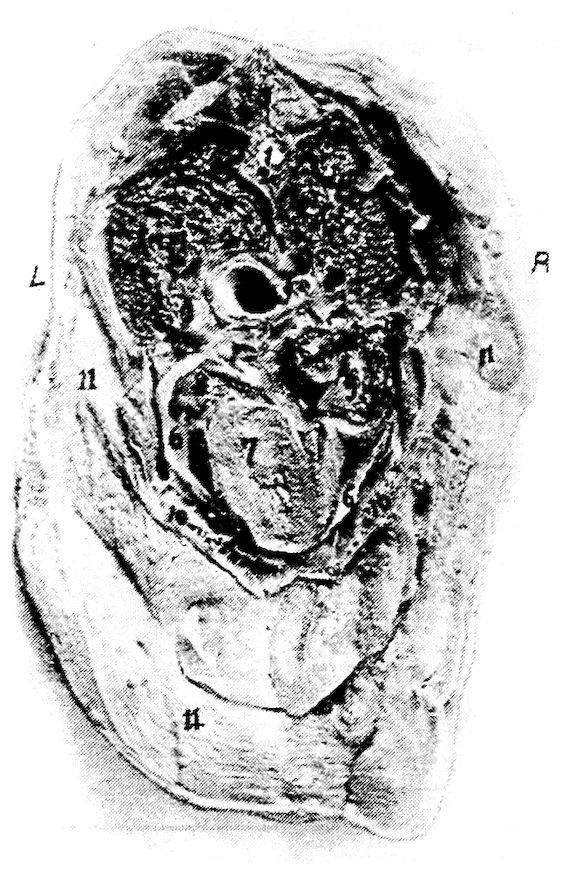
Fig. 42.—Transverse section through the body of a hen at 11, Fig. 33. R, Right side. L, Left side. 1, Spinal cord. 2, Vertebra. 3, Spinous process of vertebra. 4, Lungs. 5, Esophagus. 6, Pericardial sac. 7, Sectioned surface of heart. 8, Auricle of heart. 9, Blood clot in right auricle. 10, Section of sternum. 11, Pectoral muscles.
It will be noted that the second portion of the esophagus passes over the base of the heart and the superior part of the liver, and then terminates in the proventriculus. The proventriculus extends downward and empties into the gizzard. This organ lies antero-laterally to the gizzard, supero-posterior to the liver, and to the left of the spleen. The spleen lies in a triangle formed by the liver, the proventriculus, 161and the gizzard. The heart is noted to lie supero-anterior to the liver, and between the anterior portion of the fissure formed by the right and the left lobe. The ovary is located back of the diaphragm at the anterior end of the kidney and below the inferior surface of the bodies of the vertebræ. The bulk of the floating portion of the small intestine is located above the gizzard.
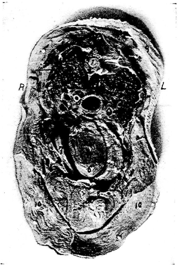
Fig. 43.—Transverse section through the body of a hen at 13, Fig. 34. R, Right side. L, Left side. 1, Spinal cord. 2, Body of vertebra. 3, Superior spinous process of vertebra. 4, Lungs. 5, Esophagus. 6, Heart. 7, Right and left lobes of liver. 8, Sternum. 9, Skin. 10, Pectoral muscles.
A transverse anterior section of a normal laying hen is shown in Fig. 39, the section being made at 15 of Fig. 34. At this point the esophagus lies centrally and above the trachea. Figure 40 shows a transverse anterior section through the thoracic region at 14 of Fig. 34. At this point the esophagus is slightly to the right of and is superior to the trachea. At this level the apex of the lung is sectioned. This is in the region of the cervical air-sac. Figure 41 shows a posterior section at 12 of Fig. 33. The 162esophagus here is above and to the right of the inferior larynx, and directly below and between the lungs. The inferior larynx is above the base of the heart.
Figure 42 shows a transverse anterior section made at 11 of Fig. 33. It shows that at this level the esophagus is centrally located and passes over the base of the heart. The heart occupies the lower portion of the thorax and the lungs the upper. Figure 43 shows a photograph of a transverse anterior section of the body made at 13 of Fig. 34. At this level the apex of the heart lies within the anterior fissure of the liver. The sectioned portion of the heart shows the lower portion of the ventricle.
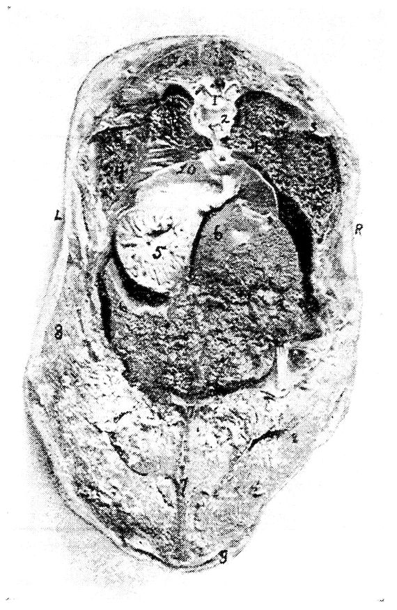
Fig. 44.—Transverse section through the body of a hen at 12, Fig. 34. R, Right side. L, Left side. 1, Spinal cord. 2, Articular surface of vertebral segment. 3, End of rib. 4, Lungs. 5, Proventriculus. 6, Liver showing Glisson’s capsule. 7, Sternum. 8, Pectoral muscles. 9, Skin.
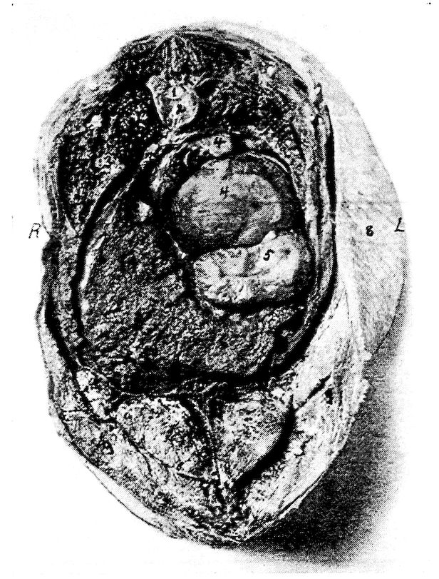
Fig. 45.—Transverse section through the body of a hen at 10, Fig. 33. R, Right side. L, Left side. 1, Spinal cord. 2, Body of vertebra. 3, Lungs. 4, Ova. 5, Proventriculus. 6, Liver. 7, Sternum. 8, Skin. 9, Pectoral muscles.
In this figure the lungs show, on the sectioned surface, the ends of some of the larger bronchi. The esophagus is located above the heart. Showing the relations of the visceral organs back of the heart girdle, photograph number 44, gives an anterior section made at 12, Fig. 33. The lungs are spread out occupying the posterior thoracic region and below at No. 10 is the diaphragm. The diaphragm does not appear to have that rigidity and firmness of position as in mammals. It is rather rudimentary. Below the left lung and above the left lobe of the liver is the proventriculus. Note that the viscus is empty and that the mucous membrane is thrown into folds. At this point the liver occupies much of the abdominal cavity. Figure 45 is a view of an anterior section made at 10, 164Fig. 33. In this section the lungs are decreasing in caliber. The liver occupies much of the space in the lower right abdominal quadrant, and above and to the right is the ovary with many of the ova developing yolks. Below No. 4 which is a developing yolk, is the proventriculus. Figure 46 is a view of an anterior section made at 9, Fig. 33. At this level are shown sectioned surfaces of the kidneys, which lie on either side of the spinal column. Below the spinal column and occupying the left upper quadrant is the ovary containing ova in the process of developing yolks. To the right is the sectioned surface of one of the cæca and below this and on the right side is the sectioned ends of many of the loops of the floating portion of the small intestine. Occupying the left lower quadrant is the sectioned surface of the gizzard and on the abdominal floor and to the right of the gizzard, the posterior end of the right lobe of the liver.
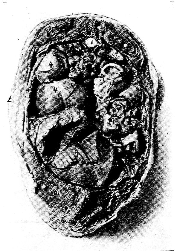
Fig. 46.—A transverse section through the body of a hen at 9, Fig. 33. R, Right side. L, Left side. 1, Spinal cord. 2, Vertebra. 3, Kidneys. 4, Ovary. 5, Cæcum. 6, Small intestine. 7, Gizzard. 8, Right lobe of liver. 9, Skin. 10, Pectoral muscles.
There is approximately 47 per cent. of the yolk retained in the abdominal yolk sac of the baby chick at hatching. Figure 47 shows a photograph of a longitudinal section through a baby chick. This figure shows all the posterior portion of the abdominal cavity occupied with abdominal yolk. The abdominal viscera are pushed forward, and as the yolk is gradually absorbed the visceral organs gradually occupy their normal position.
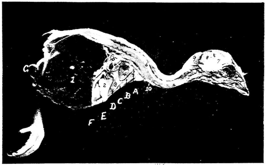
Fig. 47.—An antero-posterior section through the body of a baby chick just hatched. 1, Abdominal yolk sac. 2, Gizzard. 3, Liver. 4, Heart. 5, Intestines. 6, Spinal cord. 7, Cerebrum. 8, Cerebellum. 9, Fat in the post-occipital region. 10, The thymus gland.
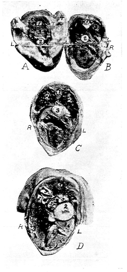
Fig. 48.—Transverse sections of the body of a baby chick at hatching. R, Right side. L, Left side.
A. A transverse section at A, Fig. 50. 1, Spinal cord. 2, Esophagus. 3, Entrance to thorax. 4, Stubs of the wings.
B. A transverse section at B, Fig. 50. 1, Spinal cord. 2, Esophagus. 3, Lungs. 4, Heart.
C. A transverse section through the body at C, Fig. 50. 1, Spinal cord. 2, Lungs. 3, Esophagus. 4, Liver. 5, Heart.
D. A transverse section at D, Fig. 50. 1, Spinal cord. 2, Proventriculus. 3, Liver. 4, Gizzard. 5, Intestine. 6, Kidneys. 7, Gall-bladder.
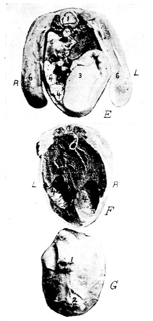
Fig. 49.—Transverse section through the body of a baby chick.
E at E, Fig. 50. 1, Spinal cord. 2, Kidneys. 3, Gizzard. 4, Intestines. 5, Unabsorbed yolk. 6, Stubs of legs.
F. A section at F, Fig. 50. 1, Spinal cord. 2, Kidneys. 3, Intestine. 4, Unabsorbed yolk.
G. A section at G, Fig. 50. 1, Anus. 2, Umbilicus.
A section through this body Fig. 47 at A is shown in Fig. 48, A. At this point the esophagus appears below the vertebral column. Figure 48, B, a section through the body at Fig. 47, B, shows the apex of the lungs. In a median line and below the lungs is the esophagus. Note the mucous membrane thrown into folds. Here the heart is sectioned, showing both auricles and both ventricles. This is a view looking forward. Figure 48, C, is a view of a posterior section made at Fig. 47, C. It shows the same relations of the esophagus and lungs, but shows at this level both the right and the left lobe of the liver. Between these lobes we note the sectioned apex of the heart. Figure 48, D shows a posterior section at Fig. 16847, D. At this level the kidneys are above and infero-laterally to the spinal column. The intestines are below the kidneys; and on the left side, between the kidneys and left lobe of the liver, and near the abdominal wall, is the proventriculus. Occupying the major portion of the inferior abdominal cavity are the right and the left lobe of the liver, and between these the anterior border of the gizzard. Note the gizzard sitting at an angle inclining toward the left side. Figure 49, E shows a section at Fig. 47, E. Here the gizzard occupies the left lower abdominal quadrant. To the right are the intestines; and directly above the gizzard is a small portion of the anterior end of the abdominal yolk sac. Infero-laterally are the kidneys. Figure 49, F, shows the kidneys similarly located as in the preceding; and just below is the rectum suspended by the mesentery. The rest of the cavity is occupied by the abdominal yolk.