New original cover art included with this eBook is granted to the public domain.
Copyright, 1907, by Doubleday, Page & Company
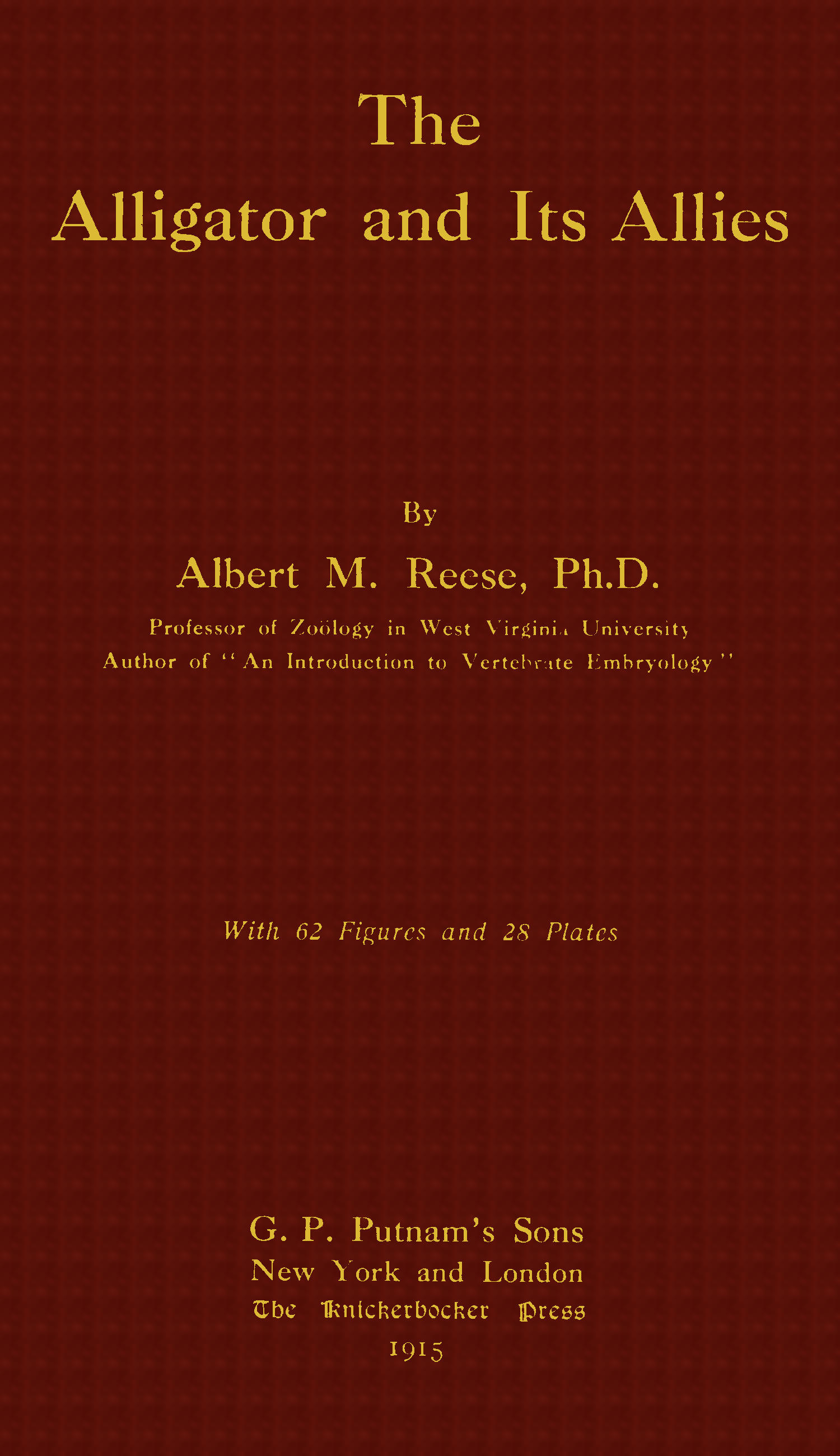
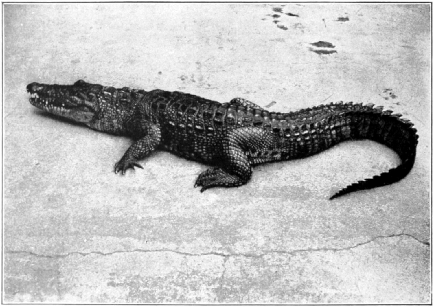
Alligator Mississippiensis. (After Ditmars.)
(Reproduced by Permission of Doubleday, Page & Company.)
The Alligator and Its Allies
By
Albert M. Reese, Ph.D.
Professor of Zoölogy in West Virginia University
Author of “An Introduction to Vertebrate Embryology”
With 62 Figures and 28 Plates
G. P. Putnam’s Sons New York and London The Knickerbocker Press 1915
Copyright, 1915
BY
ALBERT M. REESE
The Knickerbocker Press, New York
PREFACE
The purpose of this volume is to bring
together, in convenient form for the use of
students of zoölogy, some of the more
important details of the biology, anatomy, and
development of the Crocodilia. For obvious
reasons the American Alligator is the species chiefly
used.
In the first chapter the discussion of the alligator
is largely the result of the personal observations
of the author; the facts in regard to the less familiar
forms are taken from Ditmars and others. The
description of the skeleton, with the exception of
short quotations from Reynolds, is the author’s.
The chapter on the muscular system is a translation
from Bronn’s Thierreich, and the author has
not verified the descriptions of that writer.
The description of the nervous system is partly
the author’s and partly taken from Bronn and
others.
The chapters on the digestive, urogenital,
respiratory, and vascular systems are practically
all from descriptions by the author.
The chapter on “The Development of the
Alligator” is a reprint, with slight alterations, of
the paper of that title published for the author by
the Smithsonian Institution.
The bibliography, while not complete, will be
found to contain most of the important works
dealing with this group of reptiles.
The author is grateful to Mr. Raymond L.
Ditmars and to his publishers, Messrs. Doubleday,
Page & Co., and Messrs. Sturgis & Walton, for
the use of a number of plates; to the Macmillan
Company and to the United States Bureau of
Fish and Fisheries for the same privilege; to the
National Museum for photographs of the skull of
the gavial; and to the Smithsonian Institution for
the use of the plates from researches published by
them and included herein.
Proper acknowledgment is made, under each
borrowed figure, to the author from whom it is
taken.
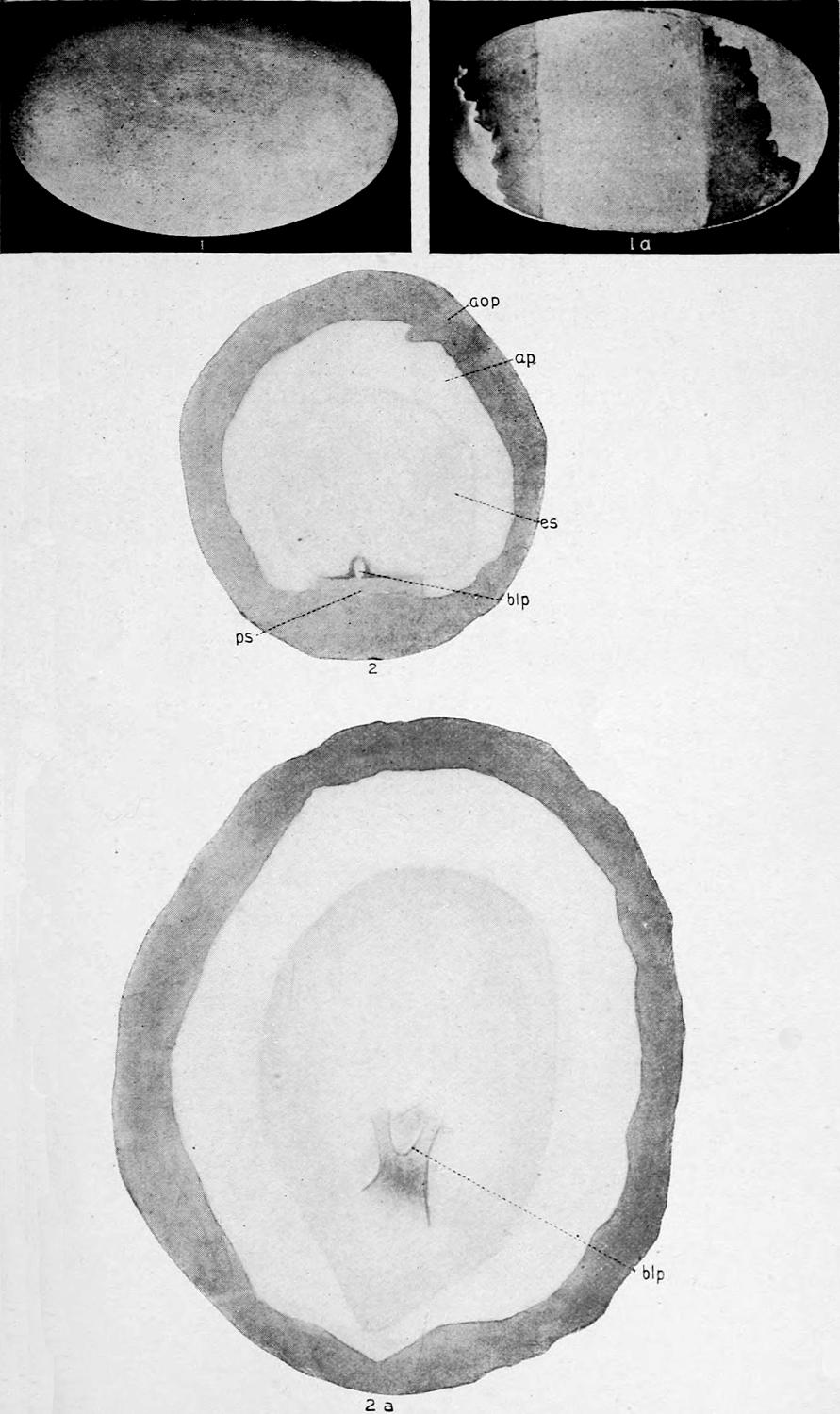
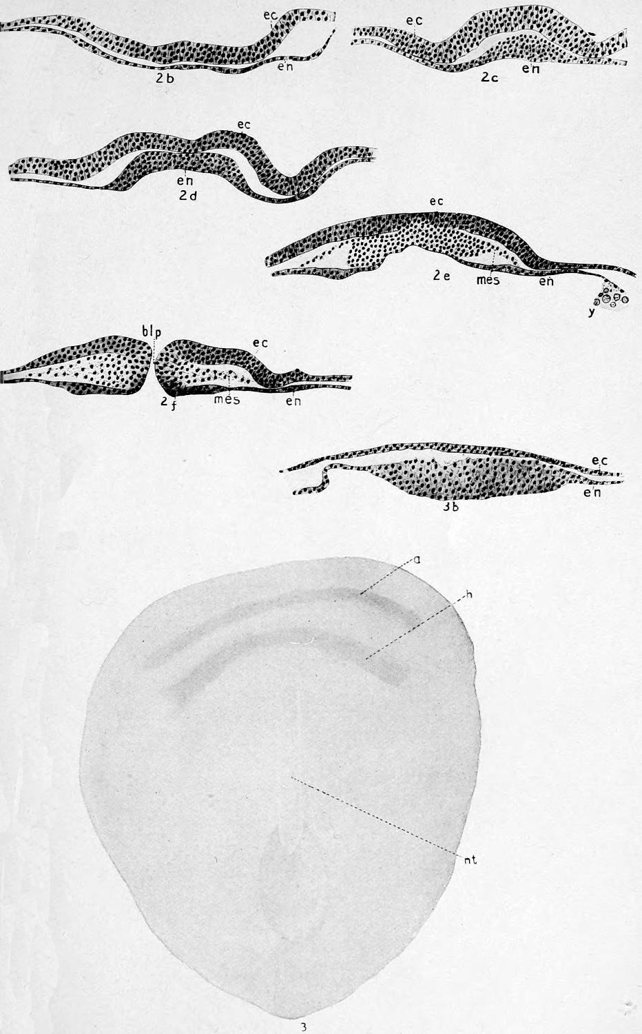
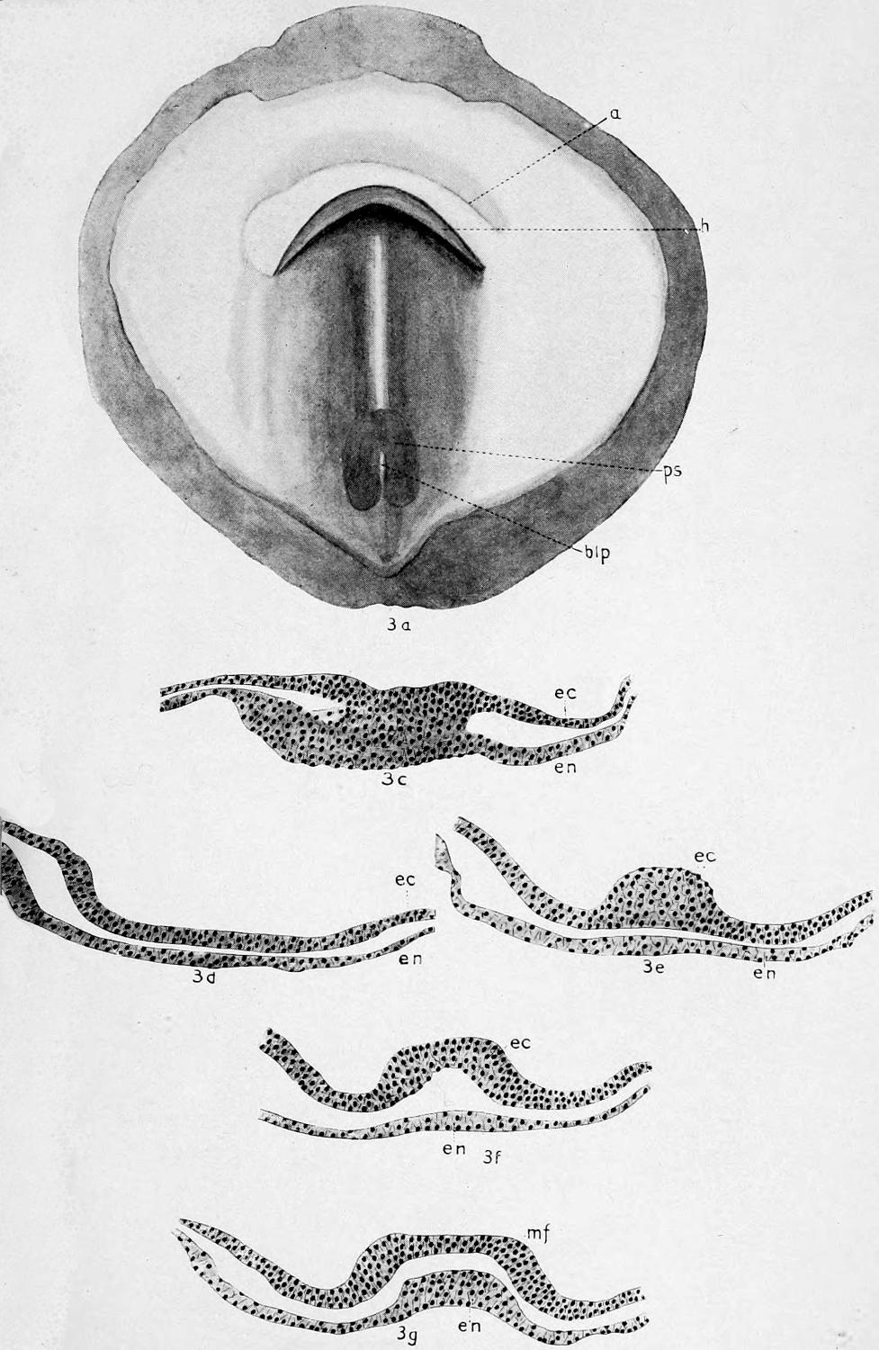
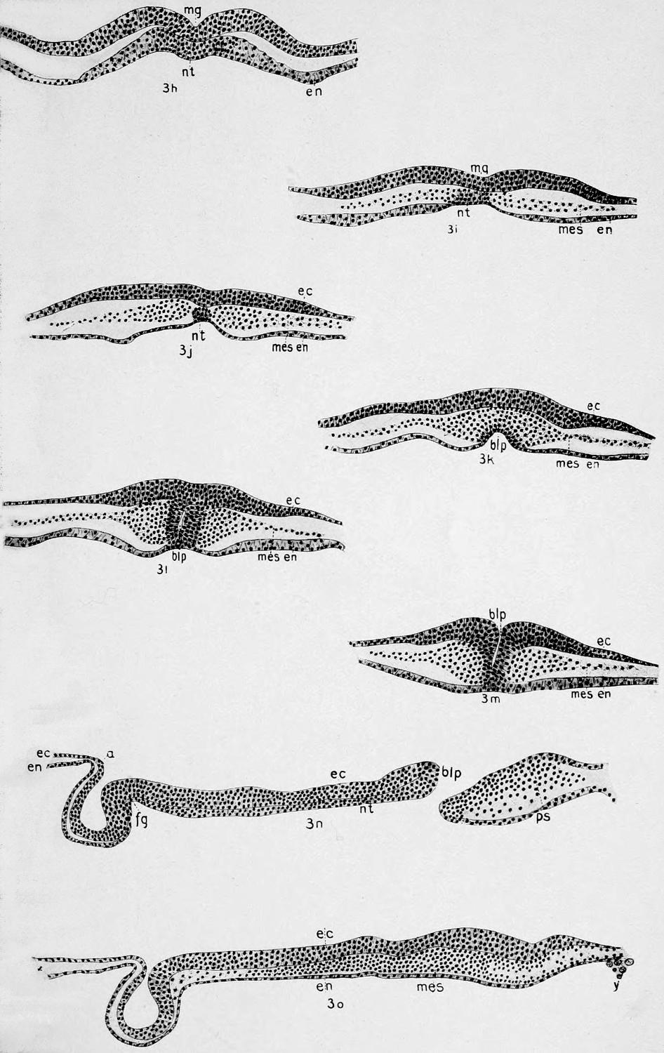
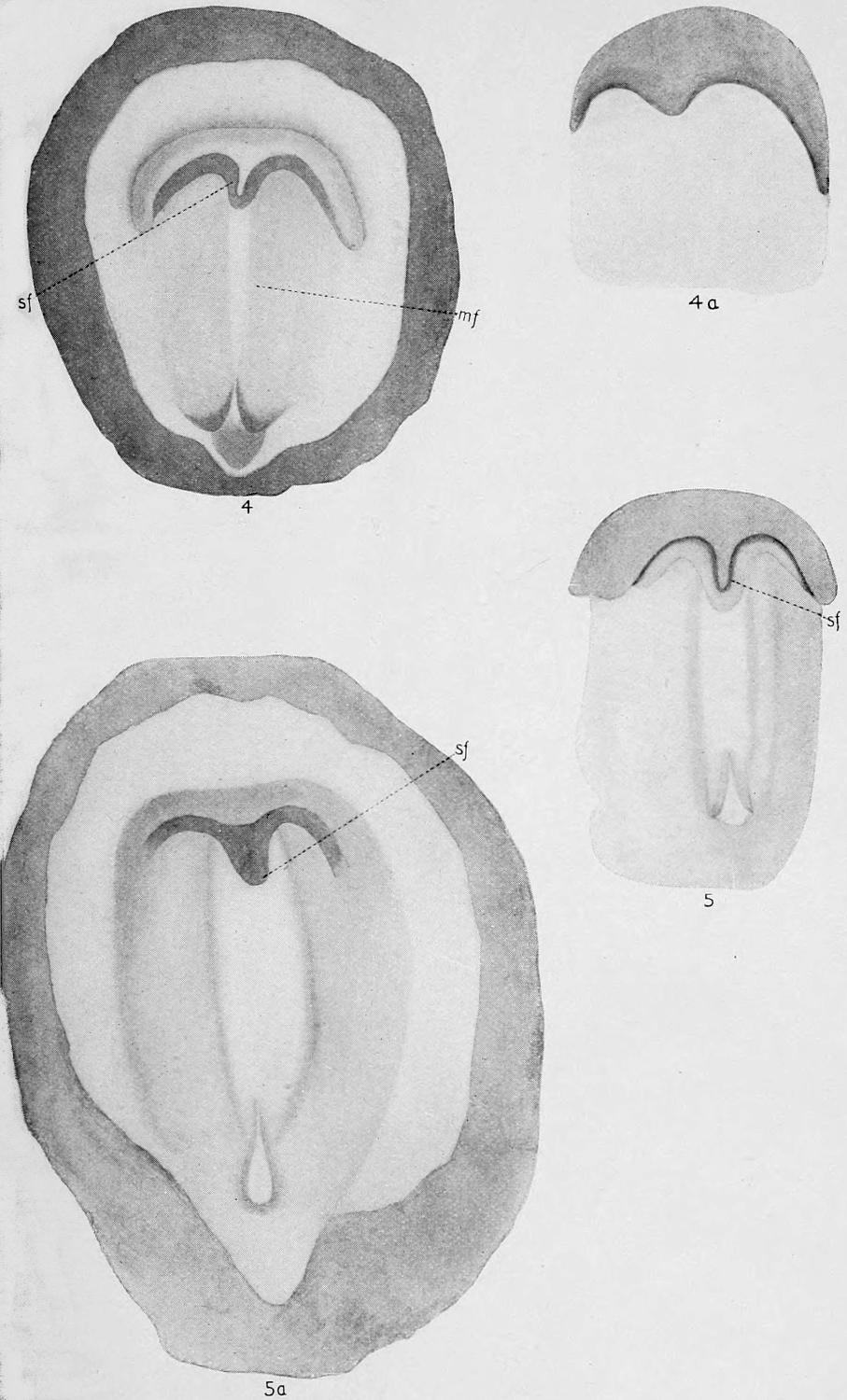
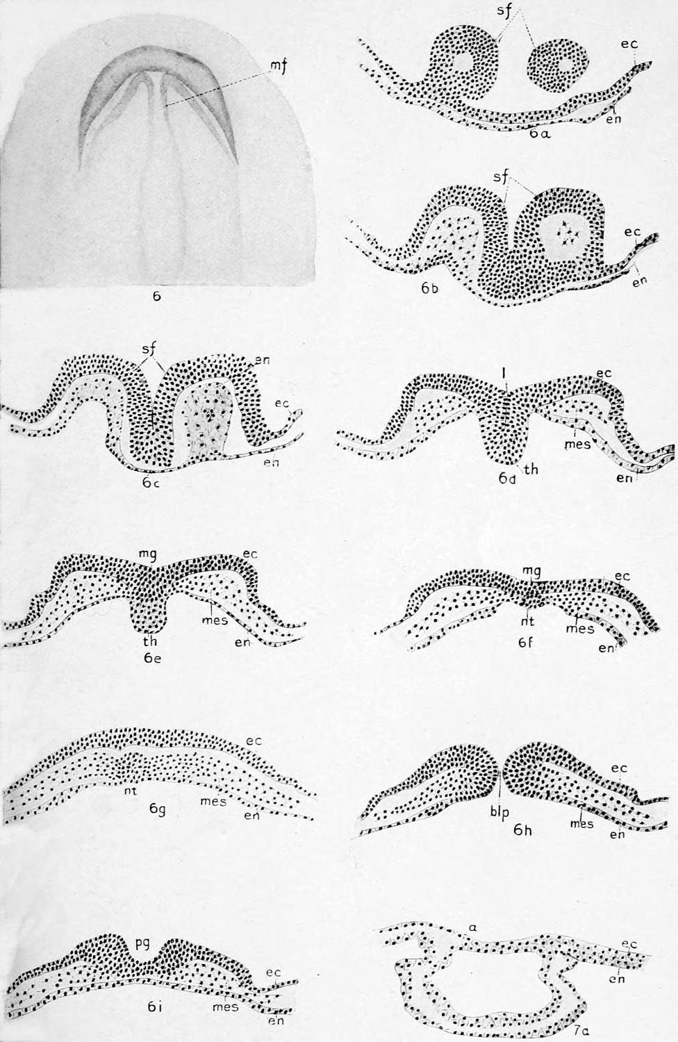
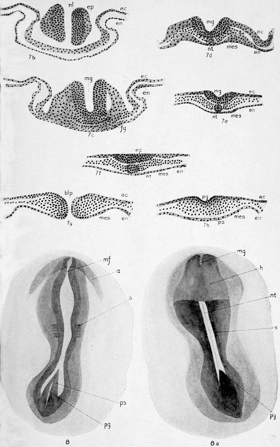
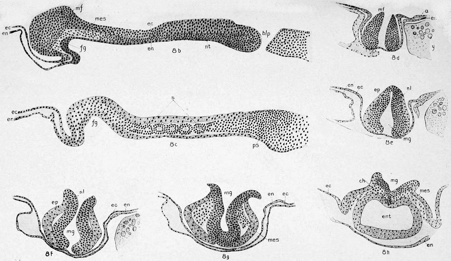
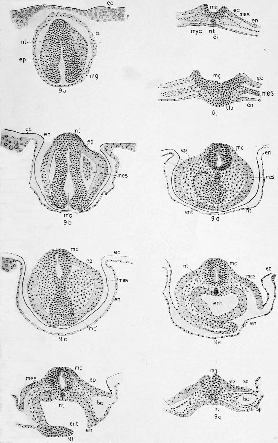
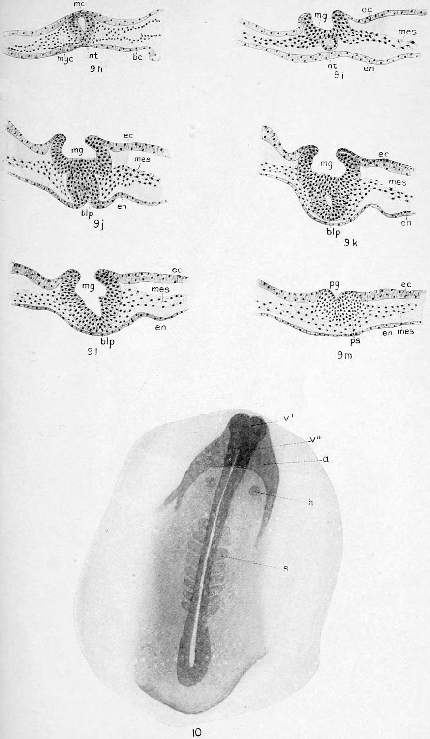
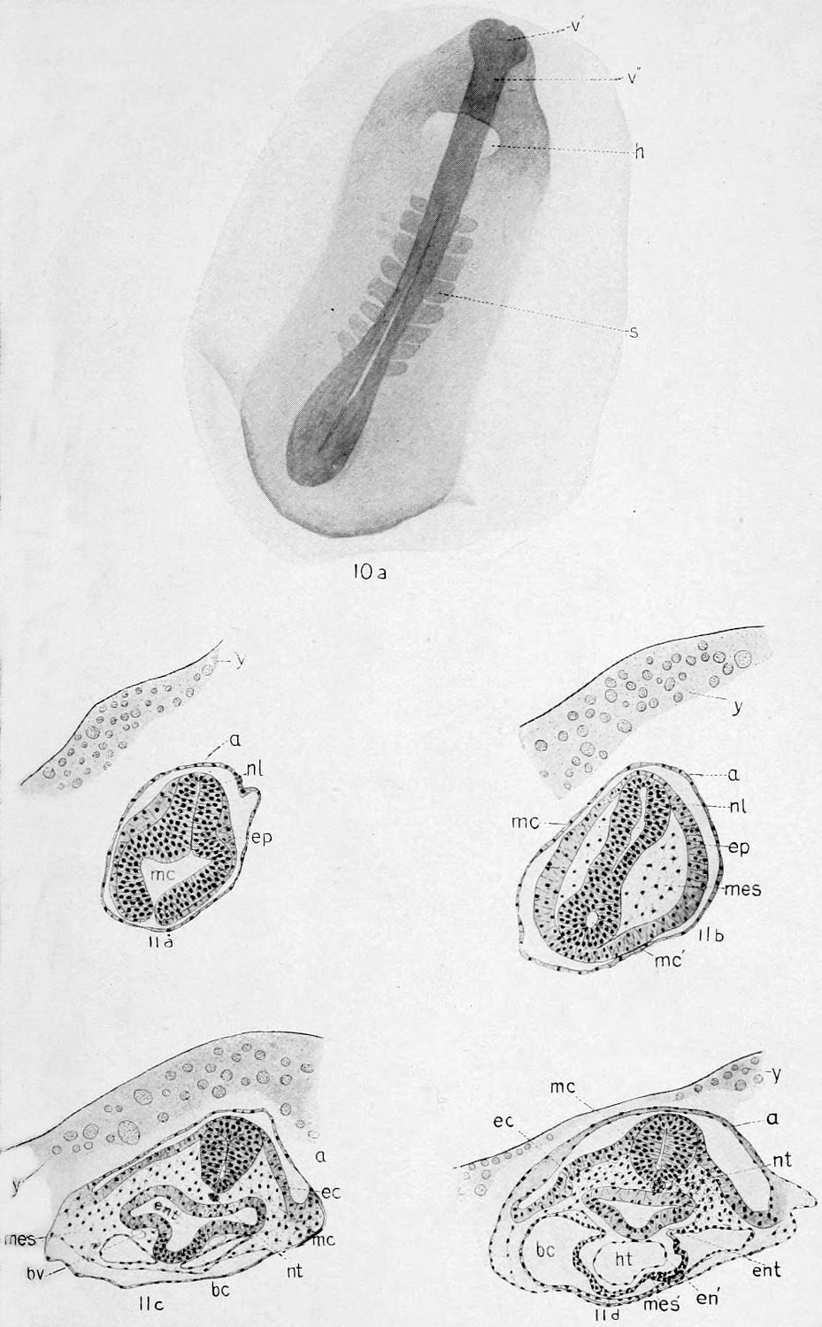
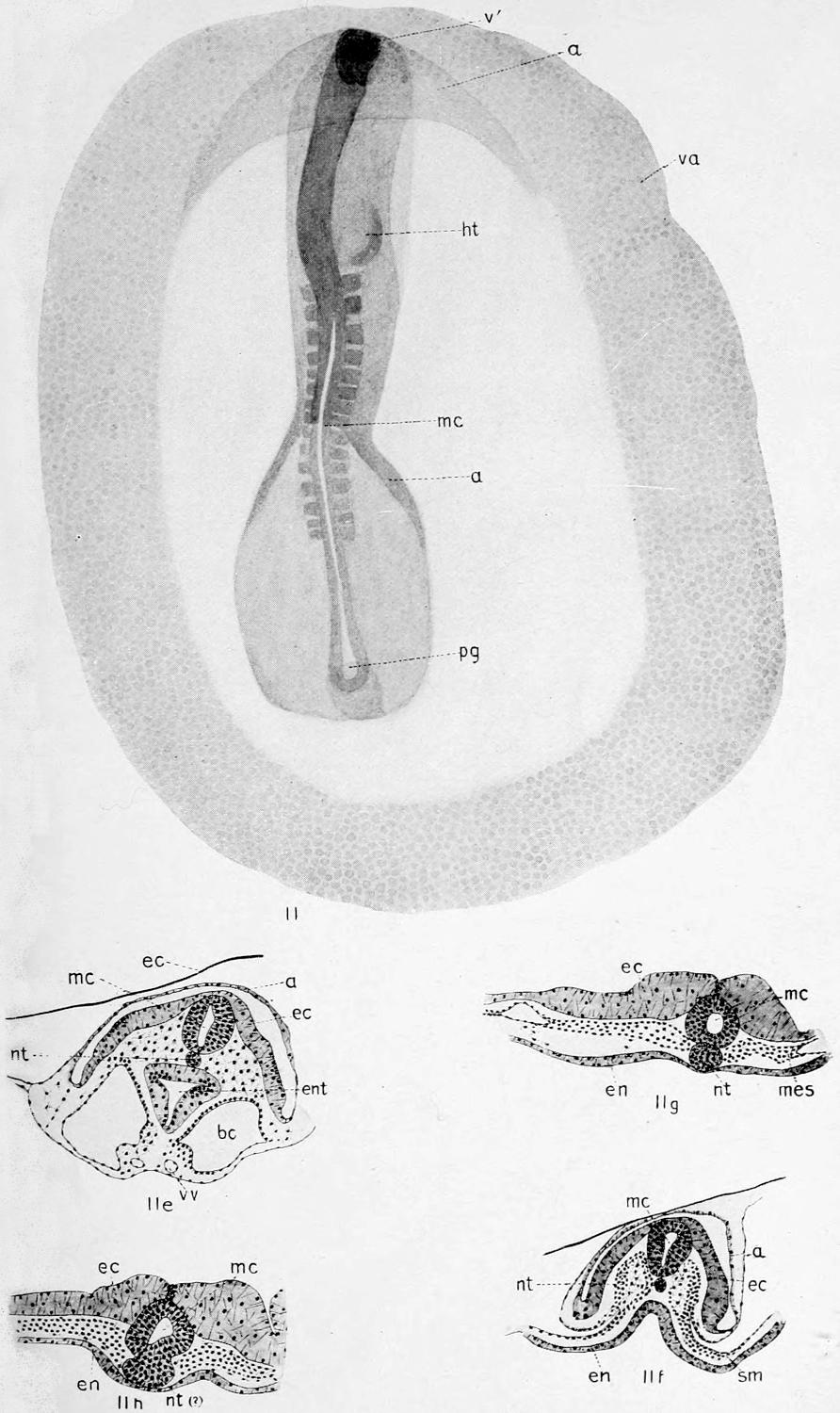
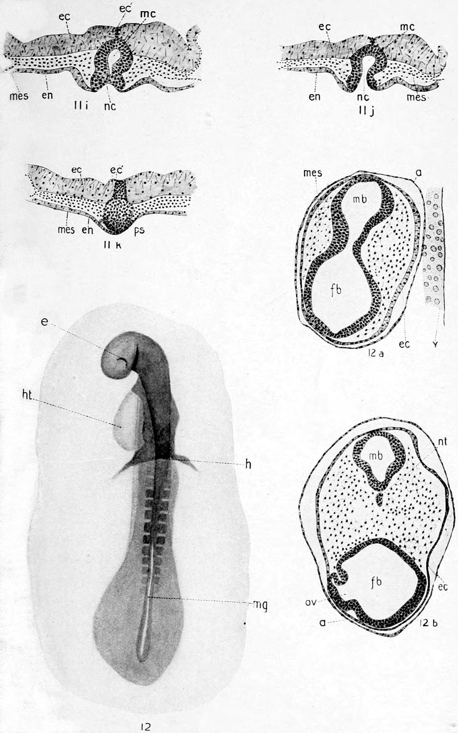
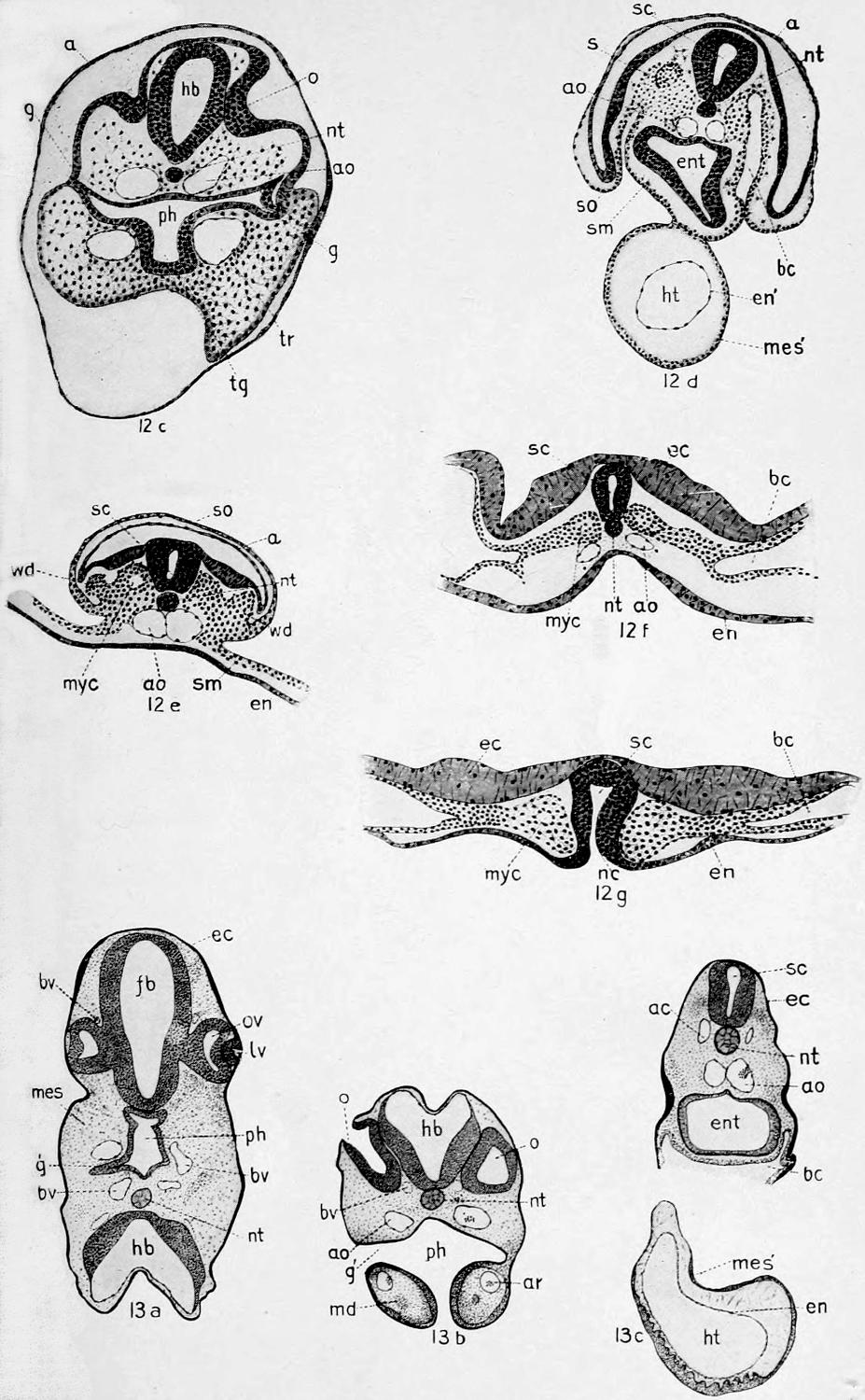
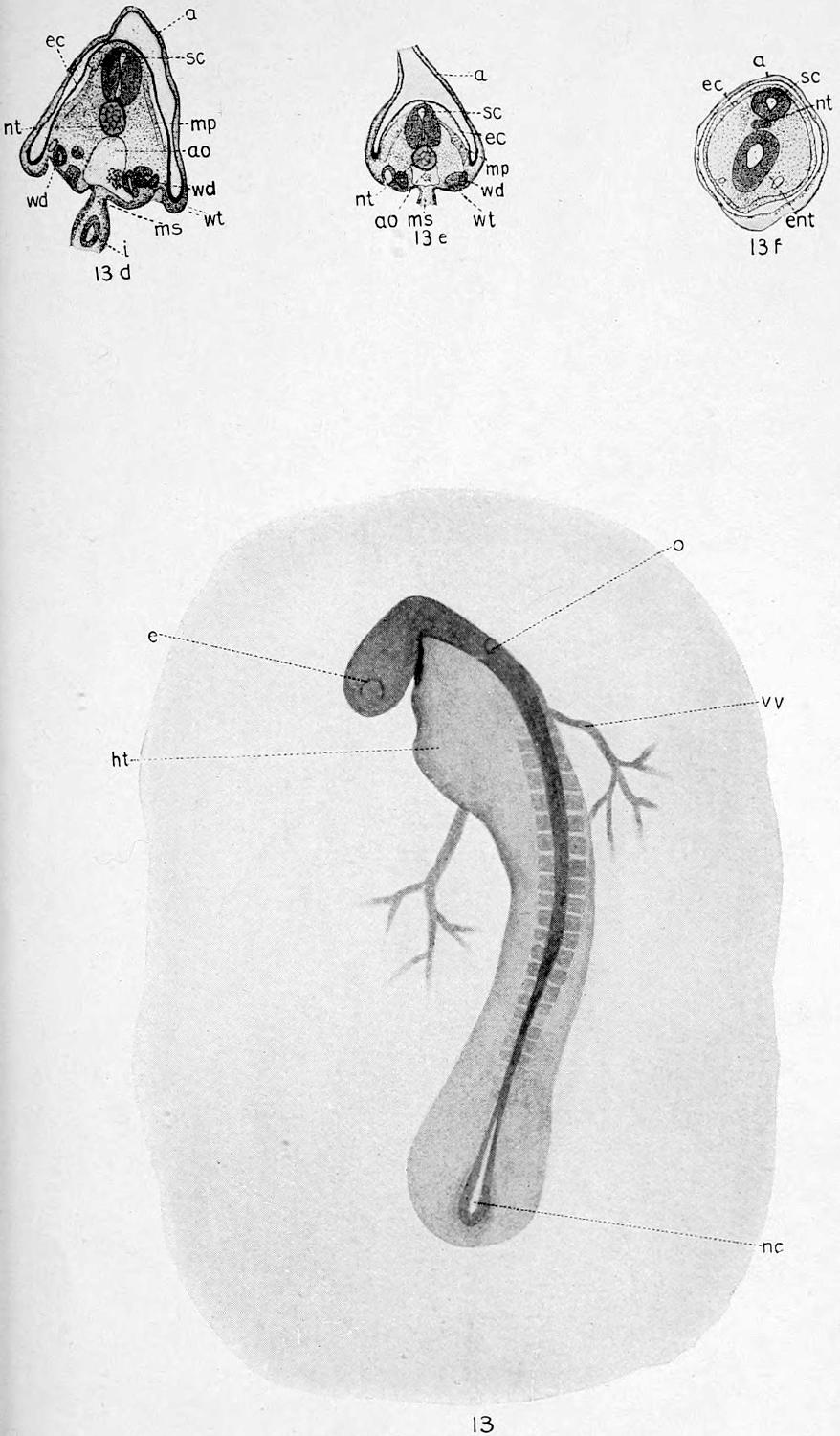
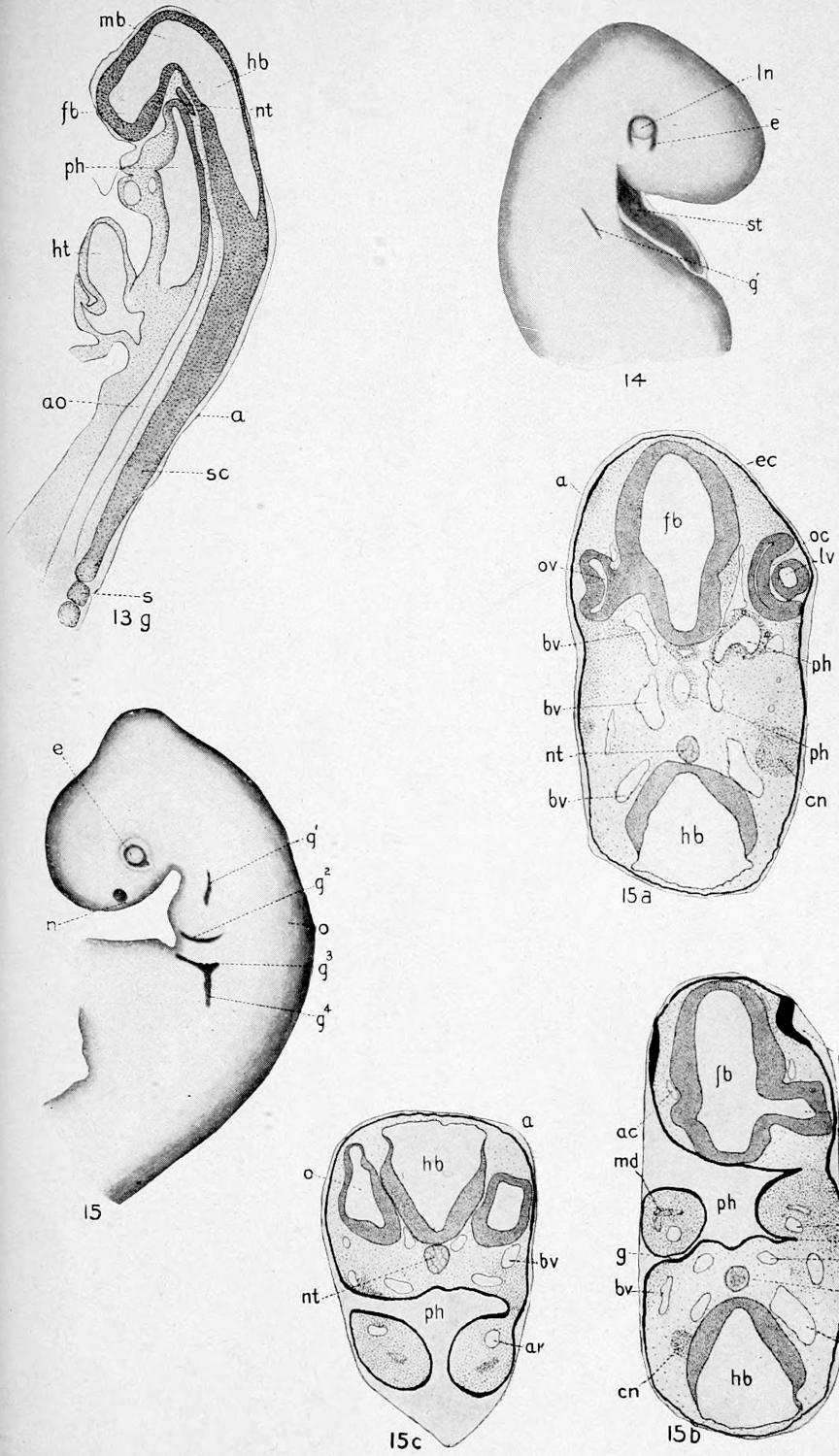
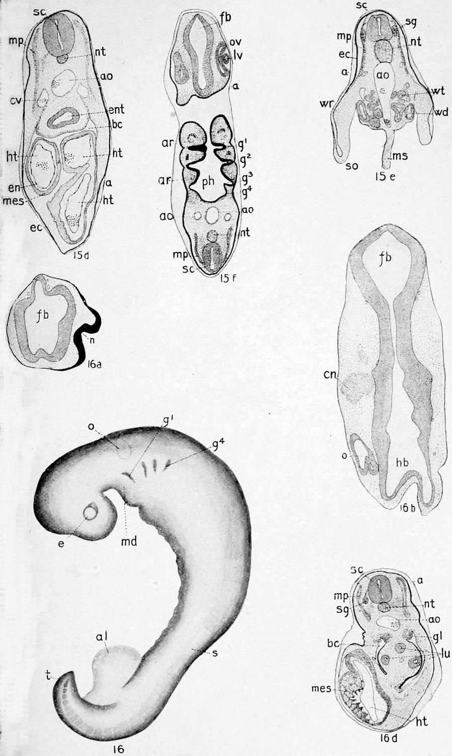
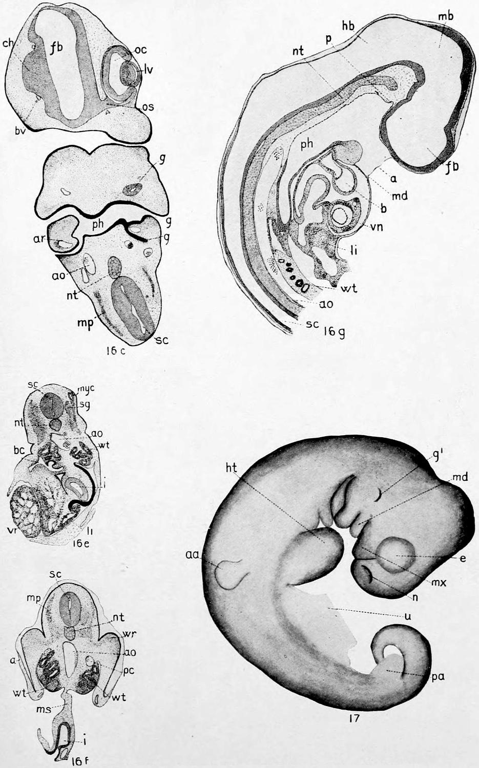
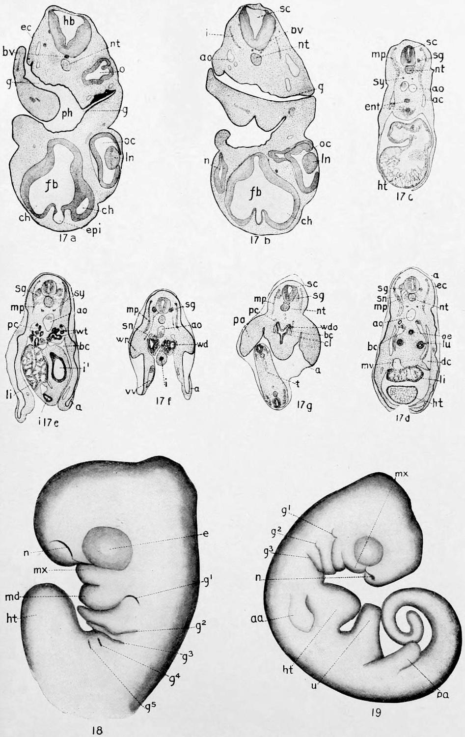
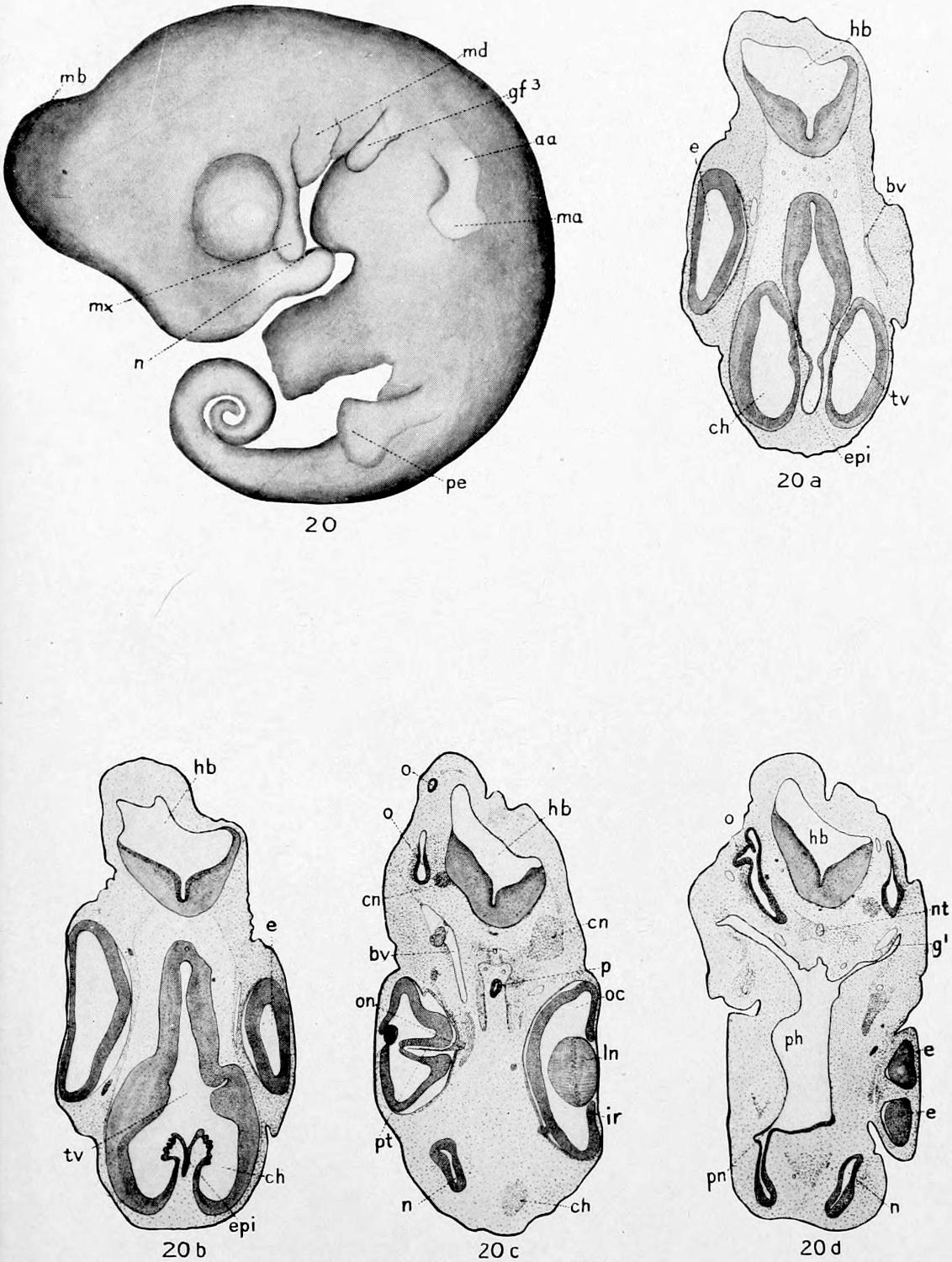
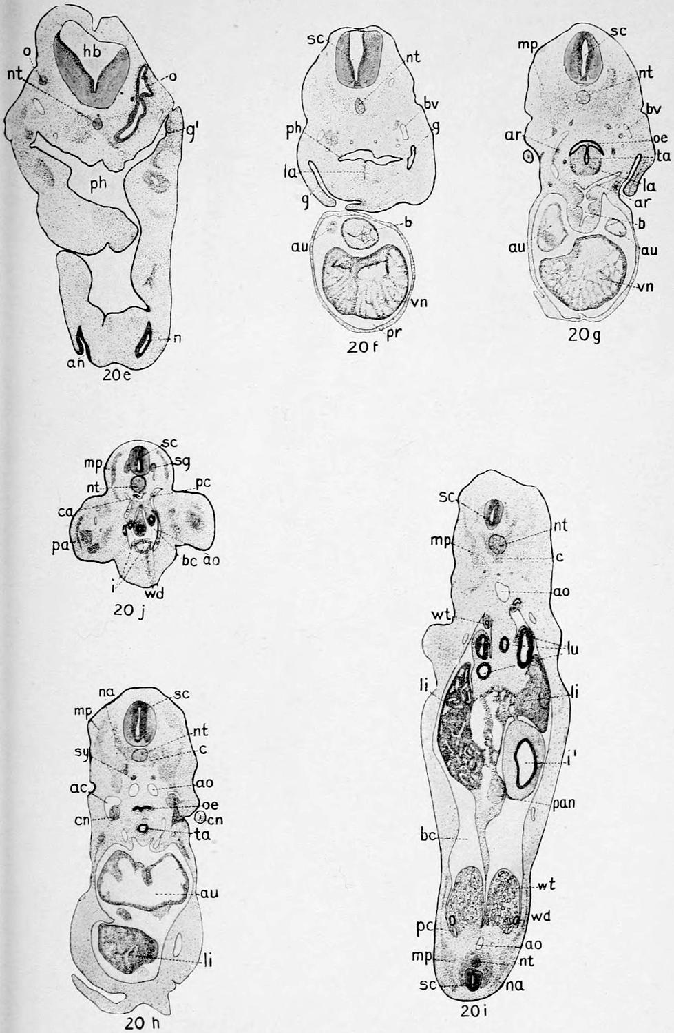
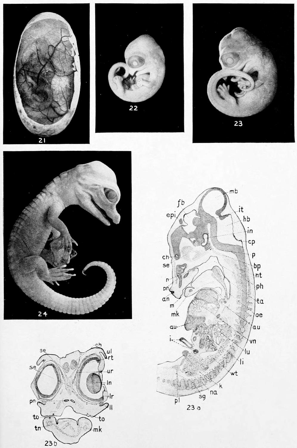
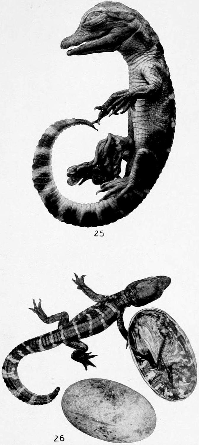
Plates VI to XXVIII. A Series of Figures to Illustrate the Development of the American Alligator
Following 342
CHAPTER I THE BIOLOGY OF THE CROCODILIA — Classification — Ancestry — Geographical Distribution — Alligator Mississippiensis — Habitat. — Food. — Size and Growth. — Voice. — Breeding Habits. — Economic Importance. — The Chinese Alligator — The Caiman — The American Crocodile — The Orinoco Crocodile , C. intermedius — The African or Nile Crocodile , C. niloticus — The Marsh Crocodile or Mugger , C. palustris — The Salt-Water Crocodile , C. porosus — The Indian Gavial , Gavialis gangeticus
THE ALLIGATOR AND
ITS ALLIES
As in most groups of animals, there is considerable
difference of opinion as to the
proper classification of the Crocodilia.
One of the older textbooks (Claus and Sedgwick)
divides the order Crocodilia into three sub-orders:
the Teleosauria, Steneosauria, and Procœlia, the
last only being represented by living forms. The
Procœlia or Crocodilia proper are divided into
three families,—the Crocodilidæ, the Alligatoridæ
(including the caiman as well as the alligator),
and the Gavialidæ.
This division into families seems to be based
mainly on the shape of the head, or, at any rate,
it throws those forms together that have heads
of the same outline.
It is this outline of the head that Ditmars (Reptiles
of the World) uses in classifying the Crocodilia,
which, he says, are all included in the single family—Crocodilidæ.
The following list, taken from his
Reptiles of the World (pp. 68-69), will give a clear
idea of the number, distribution, and maximum
size of the members of the order Crocodilia. More
will be said of some of the members of this list
later.
Habitat
Max. Size
A. Snout extremely long and slender, extending from the head like the handle of a frying pan
Gavialis gangeticus, Indian Gavial
Northern India
30 ft.
Tomistoma schlegeli, Malayan Gavial
Borneo and Sumatra
15 ft.
B. Snout very sharp and slender; of triangular outline
Crocodilus cataphractus, Sharp-nosed Crocodile
W. Africa
12 ft.
Crocodilus johnstoni, Australian Crocodile
Australia
6-8 ft.
Crocodilus intermedius, Orinoco Crocodile
Venezuela
12 ft.
C. Snout moderately sharp; outline distinctly triangular
Crocodilus americanus, American Crocodile
Fla.; Mexico; Central and S. America
14 ft.
Crocodilus siamensis, Siamese Crocodile
Siam; Java
7 ft.
Crocodilus niloticus, Nile Crocodile
Africa generally
16 ft.
Crocodilus porosus, Salt-water Crocodile
India and Malasia
20 ft.
D. Snout more oval; bluntly triangular
Crocodilus robustus, Madagascar Crocodile
Madagascar
30 ft.
Crocodilus rhombiferus, Cuban Crocodile
Cuba only
7 ft.
Crocodilus moreletti, Guatemala Crocodile
Guatemala; Honduras
7 ft.
E. Snout short and broad; conformation barely suggesting a triangular outline
[1] These species are exceptions in their genus. The snout is blunt
like that of the genus Alligator.
[2] Alleged to grow to this size by competent observers.
Gadow in the Cambridge Natural History (p. 450)
agrees with Boulanger in believing that the recent
Crocodilia cannot be separated into different
families, yet he describes seven families of Crocodilia,
two of which, the Gavialidæ and Crocodilidæ,
include the living members of the order; the former
includes the gavials, of course, and the latter the
crocodiles, alligators, and caimans.
Though “doctors disagree” thus in regard to
the scientific classification of this small group of
animals, this fact does not in the least diminish the
intense interest in the individual members of the
order.
Although the huge dragon-like dinosaurs or
“terrible reptiles,” some of which were probably
more than one hundred feet long, became extinct
during the Mesozoic epoch, perhaps millions of
years before man made his appearance upon earth,
we have one group of reptiles still living in certain
parts of the earth of which the Mesozoic lords of
creation need not feel ashamed. While most of
the living Crocodilia are mere pigmies in size,
compared to the Atlantosaurus, there are a few
representatives of the living group, to be discussed
later, that are said to reach a length of thirty feet,
which length makes pigmies, in turn, of most of
the other living reptiles.
Considering the extinct as well as the living
Crocodilia, Gadow says it is very difficult to separate
them from the Dinosauria. In the Mesozoic
Crocodilia the fore limbs were much shorter and
weaker than the hind limbs, as was often the case
with the dinosaurs; they were almost entirely
marine, but gave indications of descent from terrestrial
forms.
Various facts point, thinks Gadow, “to some
Theropodous Dinosaurian stock of which the Crocodilia
may well form an aquatic, further developed
branch” (Cambridge Natural History, p. 432).
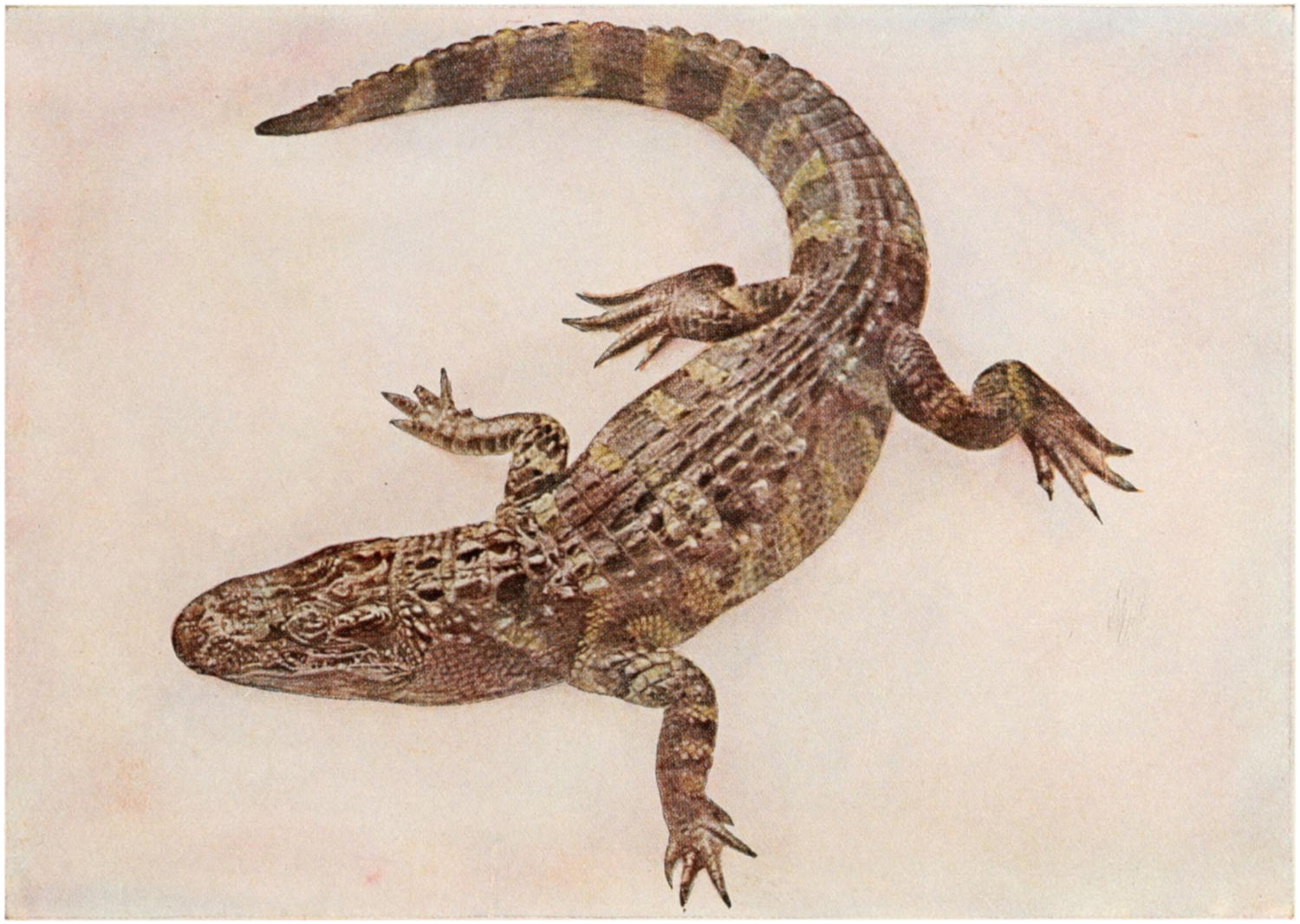
Skull of Belodon. A, from above; B, from below. A, orbit; Bo,
basi-occipital; Ch, internal nares; D, pre-orbital fossa; Exo. exoccipital;
Fr. frontal; Ju. jugal; La. lacrymal; Mx. maxilla; Na. nasal;
Pa. parietal; Pl. palatine; Pmx. pre-maxilla; Por. post-orbital; Prf.
pre-frontal; Pt. pterygoid; Qu. quadrate; S, lateral temporal fossa;
S′, superior temporal fossa; Sq. squamosal; Vo. vomer. (From Zittel.)
Fig. A. A Triassic Ancestor of the Crocodilia.
From Parker & Haswell, Textbook of Zoölogy.
The direct ancestors of the Crocodilia, Gadow
says, are still unknown.
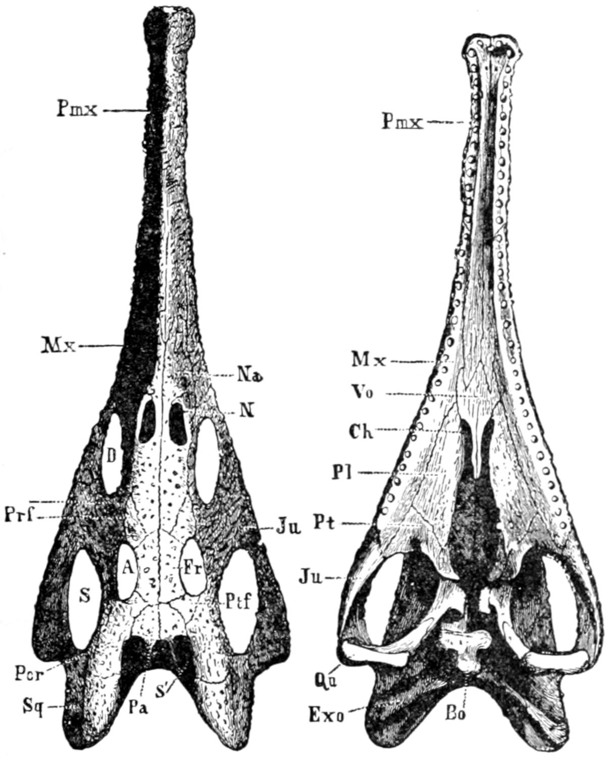
As will be seen by examination of the table
(p. 2) from Ditmars, and of Figure 1, the recent
Crocodilia are found in all of the great continental
areas except Europe; mainly in the tropical or
subtropical regions.
Fig. 1. Map Showing Present Distribution of Crocodilia.
(After Gadow.)
The alligator is found in the southwestern United
States and in China.
The crocodile is the most numerous in species
and is the most widely distributed of the group.
It is especially characteristic of Africa and Madagascar,
but is found also in Florida, Mexico, Central
and South America, the West Indies, South
Asia, the East Indies, and Australia.
The gavial is found in India and some of the islands
of the Orient, especially Borneo and Sumatra.
The caiman occurs in southern Mexico, Central
and South America.
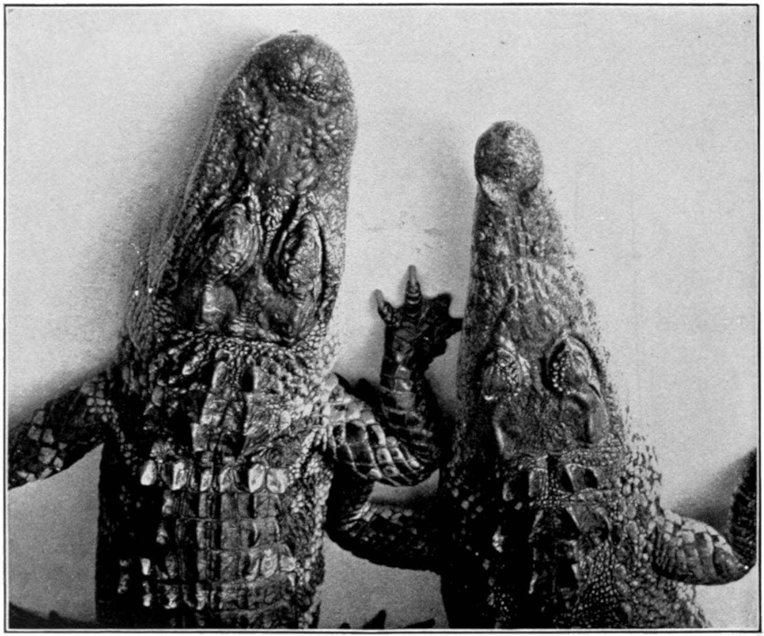
Fig. 2. Heads of American Alligator and American Crocodile;
Alligator on Left. (After Ditmars.)
(Reproduced by Permission of Sturgis & Walton Co.)
The distribution of individual forms will be
mentioned again when they are discussed in detail.
Since this animal, generally known as the
American or the Florida alligator (formerly A.
lucius), is the one upon which most of the facts
of this book are based, it will be discussed first.
At this point it may be well to answer the question
that is sure to be asked by someone early in
any conversation upon the Crocodilia. The writer,
and doubtless every other zoölogist, has been asked
countless times, “What is the difference between
an alligator and a crocodile?” As a matter of
fact there is, perhaps, no absolute distinction between
the two groups, but there are certain features
that make it easy to distinguish, say, between
the American alligator and the American crocodile.
The most striking difference is in the outline
of the head; the alligator has a broad, rounded
snout, while that of the crocodile is narrower and
more pointed (Fig. 2). Again, in the crocodile the
fourth tooth from the front projects slightly outwards
and fits into a notch in the side of the upper
jaw, while in the alligator (also in the caiman) the
corresponding tooth on each side fits into a socket
in the upper jaw and hence is hidden, except in
some old animals with very long teeth, in which
it may pierce the upper jaw and show from above.
According to Ditmars, the crocodile has, as a rule,
larger and more exposed teeth than the alligator.
Finally, as will be brought out later, the crocodile
is usually more quick and active, and also more
vicious, than the alligator.
Very young alligators are nearly black, with
distinct, yellow cross bands; as they grow older
these markings become less distinct until in maturity
the animals are of a uniform gray or dirty
black color.
The American alligator is found in
the rivers and swamps of the Southern States,
from the southern part of North Carolina to the
Rio Grande, though Florida is usually thought of as
being the region in which they particularly abound.
Years ago, before the rifle of the ubiquitous tourist
and so-called sportsman had gotten in its deadly
work, the alligators were probably very abundant
in the Southern States; but they have been so
ruthlessly destroyed by native hunters for their
skins, and by others for mere wanton sport, that
one may travel, perhaps, for days along the rivers
of the South without seeing a single ’gator.
The account quoted by Clarke from Bartram’s
travels of more than one hundred years ago, while
probably exaggerated, gives an idea of the abundance
of the alligators at that time: “The rivers
at this place from shore to shore, and perhaps
near a half mile above and below me, appeared
to be one solid bank of fish of various kinds, pushing
through the narrow pass of San Juans into the
little lake on their return down the river, and the
alligators were in such incredible numbers, and so
close together from shore to shore, that it would
have been easy to have walked across their heads,
had the animals been harmless.” At the present
time it is usually necessary to travel far from the
usual routes of the Northern tourists to find alligators
in any abundance.
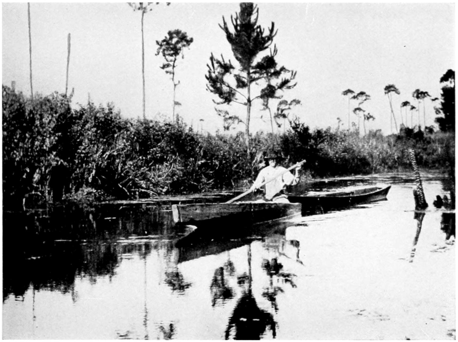
At Palm Beach, Florida, lived, a few years ago,
and probably still lives, a well-known hunter and
guide, “Alligator Joe.” Just what nationality he
may be is difficult to determine, but that he knows
that trackless waste, the Everglades, at least in
the region of Palm Beach, is evident. He has an
“alligator farm” near the great hotels of that
famous winter resort, at which he keeps, or did a
few years ago, a large number of alligators of all
sizes, as well as a number of crocodiles. For a
consideration (by no means a modest one) he
would take out a party of tourists for a day into
the Everglades, guaranteeing that he would find
an alligator for them to shoot. It was rumored
by the natives that an accomplice was always sent
ahead to free the alligator at the psychological
moment, after the hunters had been paddled by a
devious course to the selected spot, but whether
this were true or not the writer was not able to
determine. It is true, however, that he and the
writer paddled in a rather graceful canoe, dug out
of a single cypress log, and waded through the
Everglades for several days, searching for alligator
eggs, and that we found only one nest and saw
only one or two alligators (Fig. 3).
Doubtless in more remote parts of the Everglades
the alligators are much more numerous.
During another summer the writer, with a guide,
penetrated the very center of the State, to the
region southeast of Lake Kissimmee, forty miles
from the nearest railroad; here the alligators, and
in consequence their nests, are fairly abundant,
though the native hunters are, even in this remote
region, rapidly thinning their ranks.
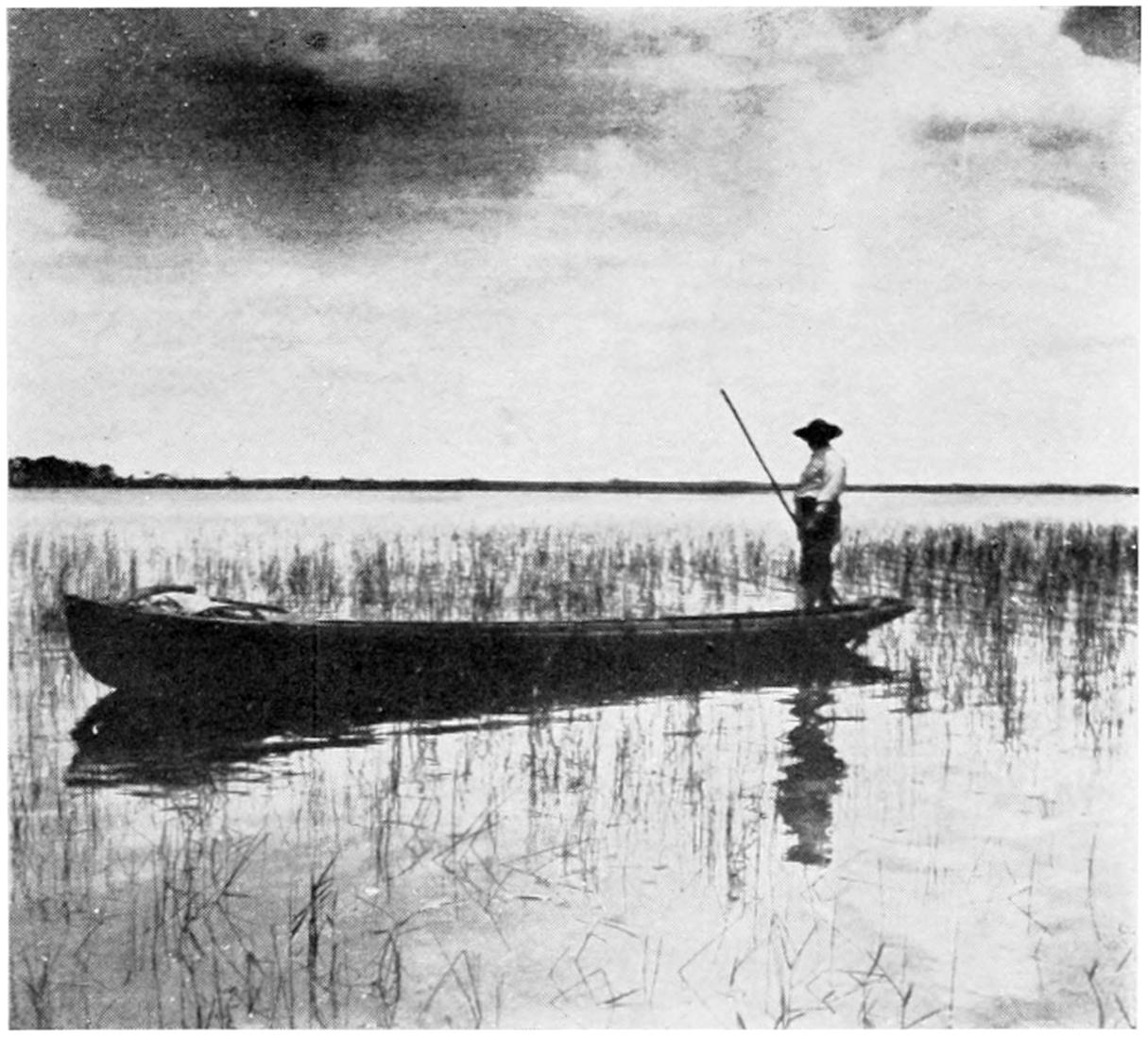
A still greater number of alligators was found, the
following summer, in the Okefinokee Swamp in
southern Georgia. In the center of this great
waste, ten miles or more from dry land, nearly
one hundred alligators, ranging from about four
to eight feet in length, were killed within a week
by a small party of native hunters with whom the
writer was traveling (Fig. 4).
Whether this wholesale destruction by sportsman
and native hunter will eventually exterminate
our giant reptile, as has been the case with the
buffalo and other game animals, it is impossible
to say. Unless the Everglades and the Okefinokee
are largely drained it seems probable that a few
alligators will always remain in the most inaccessible
regions.
Fig. 3. Alligator Joe in the Everglades.
(From a Photograph by the Author.)
Fig. 4. Alligator Hunter in the Okefinokee.
(From a Photograph by the Author.)
The collection of eggs for sale and for hatching
purposes, as well as their destruction for food by
bears and other animals, will also tend towards
the annihilation of the species in the course of
time. The economic importance of the alligator
will be discussed later.
While in the old days, as has been said, the alligator
was common in the larger rivers and lakes,
and may even have ventured short distances into
salt water, he must now frequently be satisfied to
hide his great body in a “’gator hole” that is
scarcely more than a puddle. These “holes”
(Fig. 7) are common in central Florida and are
sometimes scarcely large enough to allow the alligator
to dive into them to seek the underground
cave in which he hides. It is on the edge of such
a hole that the nest is built, as will be described
later.
Often from a small swamp or slough alligator
“trails” lead off in different directions. These
trails are narrow, winding gullies such as might be
made by cattle in a damp pasture. If followed
from the main slough the trail will usually be
found to end in a “hole,” in which an alligator will
probably be found (Fig. 7). In a great swamp like
the Everglades or the Okefinokee such holes would
naturally not be found.
On one side of the hole is usually a smooth place
where the vegetation is worn away; it is here that
the ’gator “pulls out” to sleep in the sun; and
wary must the hunter be to approach within sight
of the animal before being seen or heard by him.
At the first alarm he slides quietly or plunges
quickly into the muddy water, and the hunter
must wait long if he expects to see the ’gator come
to the surface.
The opening of the cave is always below the
surface of the water, but it is possible that there
may be a subterranean chamber that is not completely
filled with water. How the animal is
gotten from his cave will be described later. According
to some writers the alligator retires to his
cave to hibernate during the cooler winter months.
This is possibly true in the more northerly limits
of his range. It is well known that if kept in cool
water the alligator will lie dormant and refuse all
food for months at a time. The writer has had
young alligators in captivity, under these conditions,
that refused food from late in the autumn
until nearly the first of April.
The proprietor of one of the largest alligator
farms in the country says: “Our alligators stop
eating the first week in October and do not begin
to eat until the latter part of April. We have
experimented with our stock to see if we could get
them to eat in the winter, and found that by keeping
the water in the tanks at a certain temperature
they would eat, but we found out that the warm
water would make their bowels move, and that
they would not eat enough to keep themselves up,
as in the summer, and as a result they would become
very poor and thin, so we do not force them
to eat any more.” The effect upon the growth
of an animal of these two methods of feeding will
be noted later when the age and rate of growth are
discussed. The same writer says, in answer to a
question about hibernation: “In their wild state
they go into their dens under water and remain
dormant all winter.” Whether this statement is
the result of actual observation the writer is not
able to say, but, judging by some other statements
from the same source, it is probably from hearsay.
The writer, having visited the alligator haunts
only in late spring and summer, has had no opportunity
of studying the habits of the animal in its
natural habitat during the winter season. During
the heat of summer the animal does not seek the
sun as he is said to do during cooler weather, but
spends more time on the bank at night and during
the cooler parts of the day.
That he sometimes wanders over dry land, perhaps
going from hole to hole, is evident from the
tracks that are sometimes seen crossing a dusty
road or path. These trails are easily recognized
by the clawed footprints with a line, made by
the dragging tail, between them. Although most
awkward on land, he can, if necessary, move very
quickly. It is, however, in the water that he
shows to best advantage; he is an active, powerful
swimmer, his tail being used as a propeller as
in the fishes. When swimming actively the legs
are held close against the body in order that they
may retard the animal’s motion as little as possible.
While swimming in a leisurely way the top of the
head is at the surface of the water, perhaps just
the nostrils and eyes projecting above the surface,
so that the size of the animal can be estimated by
the distance between these projecting points. One
afternoon the writer and a guide, while paddling
along an old canal that was dug years ago into
the Okefinokee Swamp, were preceded for perhaps
half a mile by a large alligator that swam just fast
enough to keep out of our reach until he came to the
place where he wanted to turn off into the swamp.
Although so awkward on land, the alligator is
said to be able to defend himself very effectively
with his tail, which he sweeps from side to side
with sufficient force, in the case of a large specimen,
to knock a man off his feet. Although the writer
has seen captured and helped to capture alive
several alligators up to eight feet in length he has
never seen this vigorous use of the tail as a weapon
of defense.
While the alligator, like most other wild animals,
will doubtless defend itself when cornered, it will
always flee from man if possible, and the writer
has frequently waded and swam in ponds and lakes
where alligators lived without the least fear of
attack. This might not have been possible years
ago when the animals were more numerous and
had not been intimidated by man and his weapons.
The food of the adult alligator consists
of fishes, birds, mammals, and possibly smaller
individuals of its own species. The young eat
small fish, frogs, insects, or worms.
If the animal be too large to swallow whole it
is shaken and torn, the shaking being so vigorous
that, according to Ditmars, the entrails of the
prey may be thrown to a distance of twenty feet
or more. Should two alligators seize the same
prey at the same time they whirl about in opposite
directions so violently that the prey is torn apart.
This action may be illustrated by giving two small
captive alligators a piece of tough meat; they hold
on with bulldog tenacity, and each, folding its legs
close to its body, will use its tail like a propeller
until the animal whirls around with remarkable
speed. The commotion that two ten-foot alligators
would cause when thus struggling can easily
be imagined. That a large alligator, if it tried,
could easily drag under the water and drown a man
or possibly a much larger animal is evident.
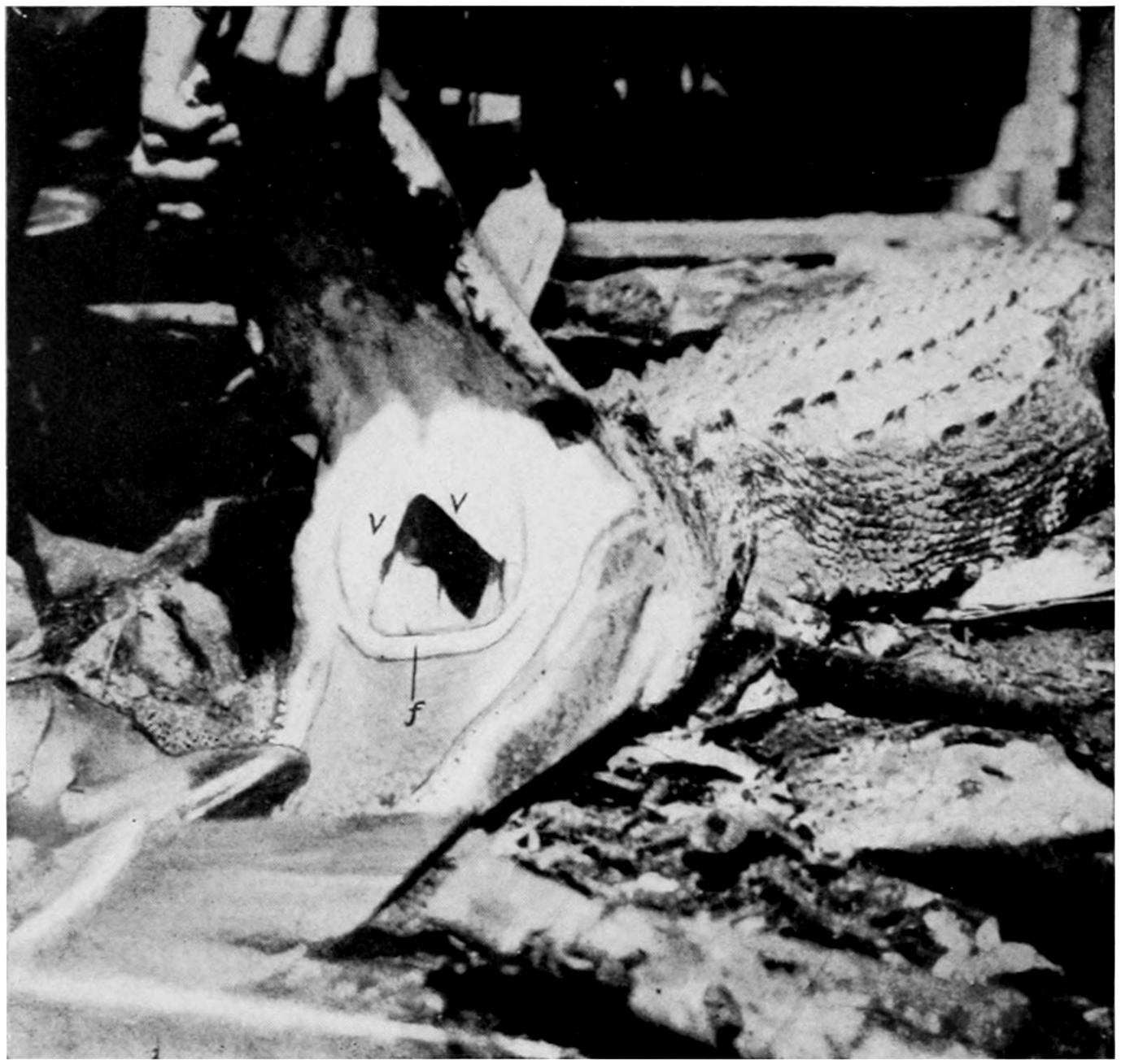
While the alligator has a valve-like fold of skin
in its throat that enables it to open its mouth and
crush its prey under water, it is said that it must
raise its head above water in order to swallow
its food. A young alligator on land will usually
throw back its head when trying to swallow a
large piece of meat, so that it may be simply this
motion that brings the head of the alligator above
the surface of the water.
Ditmars thus describes the fate of a dog that
approached too near a very large alligator: “As
a dog, weighing about fifty pounds, unwarily approached
the edge of this creature’s tank, it was
suddenly grasped and before completing its first
yelp of terror was dragged beneath the surface. A
few minutes later the twelve-foot saurian appeared
at the top, holding the dead canine in its jaws.
The dog was shifted about, amid the sound of
breaking bones, and swallowed head first, and
entire, after a few gulps.”
Although, years ago, alligators
of fifteen feet length may have been common in
favorable localities in the South, it is probable
that few if any such monsters now exist. A
twelve-foot alligator, owing to its great girth, is a
huge animal and but few of this size are to be
found in captivity. The largest specimen the
writer has ever seen is the one in the Bronx
Zoo, which is barely thirteen feet in length. At
hatching the alligator is about eight inches in
length.
Clarke (17) says: “The largest specimen I saw
measured twelve feet in length; and none of the
many hunters and natives of Florida I have met
have seen any longer than thirteen feet. All the
hunters agree that it is only the males that acquire
the great size; no one had ever seen a female that
measured over eight feet, and the majority are not
over seven. The male has a heavier, more powerful
head, and during the breeding season especially
is more brilliantly colored.”
It is a very common belief, even among those
who should be most familiar with their habits,
that the growth of the alligator is remarkably slow,
so that a large specimen may be described by the
exhibitor as more than a century old. The same
dealer in alligators quoted above says upon this
subject: “You can figure about two inches a year
to their growth.” He also says: “We judge that
an alligator about twenty-five to thirty years old
will breed.” Even scientific writers of reputation
have not been free from this error in their writings.
That the alligator may live to an extreme age, as
seems to be true of some of the tortoises, is quite possible,
and it is probable that after reaching a length
of twelve or fifteen feet the growth is very slow.
In captivity, when kept in warm water and
other favorable conditions, the alligator will grow,
according to measurements taken at the New York
Zoölogical Park, at the rate of about one foot a
year, for about the first ten years. Under unfavorable
conditions the growth may be exceedingly
slow. Under favorable conditions in nature the
rate of growth may exceed that given above.
Instead of requiring twenty-five to thirty years
to reach sexual maturity, as quoted above, it is
likely that the female may lay eggs at five to ten
years, though such a fact is difficult to determine
of animals in their native haunts.
The alligator, unlike most other members
of its class, the Ophidia, Chelonia, and Lacertilia,
has a voice, which, in an adult bull, may be heard
for a mile or more. This bellowing is difficult to describe;
it is something between a moan and a roar,
and may be to attract the opposite sex or to serve
as a challenge to other large animals. It is usually
ascribed to the male, but whether confined to him
or not the writer is unable to say.
In younger animals the voice is, of course, less
deep and in very young individuals it is a squeak
or grunt, easily imitated by hunters for the purpose
of luring the animals from their hiding places.
Judging from the statements
of native hunters the laying season of the alligator
might be thought to be at any time from January
to September. As a matter of fact the month of
June is the time when most, if not all, of the eggs
are laid. S. F. Clarke gives June 9th and June
17th as the limits of the laying season in Florida,
but I found at least one nest in which eggs were
laid as late as June 26th: no eggs were found before
the first date given by Clarke. It seemed quite
certain that the laying, during the season in question,
had been delayed by an extreme drought that
had dried up the smaller swamps and reduced the
alligator holes to mere puddles. Nests were found
in considerable numbers as early as June 8th, but no
eggs were laid in any of them until the end of the
dry period which occurred nearly two weeks later.
Almost immediately after the occurrence of the rains
that filled up the swamps eggs were deposited in all
of the nests at about the same time. From the fact
that all of these completed nests had stood for so
long a time without eggs, and from the fact that
all of the eggs from these nests contained embryos
in a well-advanced state of development, it seemed
evident that the egg-laying had been delayed by the
unusually dry weather. Eggs taken directly from
the oviducts of an alligator that was killed at this
time also contained embryos that had already
passed through the earlier stages of development.
Thus it was that the earliest stages of development
were not obtained during this summer.
It is said that during the mating season, which
precedes by some time, of course, the laying season,
the males are noisy and quarrelsome, and that
they exhibit sexual characteristics of color by
which they may be distinguished from the females.
Never having been in the alligator country at this
season, the writer has made no personal observations
along these lines, but from the frequency
with which alligators with mutilated or missing
members are found it is evident that fierce encounters
must sometimes take place, whatever the
cause. During June and July, at least, and probably
during most of the year, the alligators are
very silent, an occasional bellow during the very
early morning hours being the only audible evidence
that one has that the big reptiles are in the
neighborhood. Whatever may be the sexual differences
during the mating season, at ordinary times
the two sexes are so much alike that I have, on
more than one occasion, seen experienced hunters
disagree as to the supposed sex of an alligator that
had just been killed.
Although I have never seen a nest actually during
the process of construction, it is easy to imagine,
after the examination of a large number of freshly
made nests, what the process must be like.
The alligator, probably the female, as the male,
after the mating season, takes no interest whatever
in the propagation of his species, selects a slight
elevation on or near the bank of the “hole”
in which she lives. This elevation is generally,
though not always, a sunny spot, and is frequently
at the foot of a small tree or clump of bushes.
Where the alligator is living in a large swamp she
may have to go a considerable distance to find a
suitable location for her nest; when her hole is
scarcely more than a deep, overgrown puddle, as
is often the case in the less swampy regions, she
may find a good nesting place within a few feet of
her cave. That the female alligator stays in the
neighborhood of her nest after she has filled it
with eggs seems pretty certain, but that she defends
it from the attacks of other animals is extremely
doubtful: certainly man is in very little danger
when he robs the nest of the alligator, and, according
to the statement of reliable hunters, bears are
very persistent searchers for and eaters of alligator
eggs. Having selected (with how much care it
is impossible to say) the location for the nest, the
alligator proceeds to collect, probably biting it
off with her teeth, a great mass of whatever vegetation
happens to be most abundant in that immediate
vicinity. This mass of flags or of marsh grass
is piled into a conical or rounded heap and is
packed down by the builder repeatedly crawling
over it.
Fig. 5. A Nest of C. Porosus: Palawan, P. I.
(From a Photograph by Rowley.)
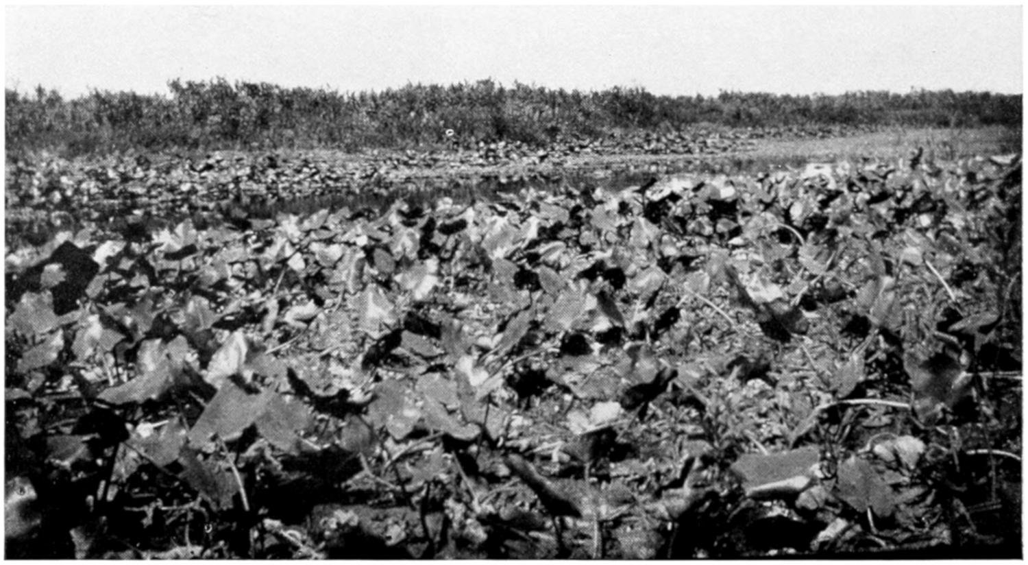
Fig. 6.—Jackson Slough; near Lake Kissimmee, Florida.
In the vicinity of this pond several alligator nests were found, either within a few
yards of the edge, or on the banks of smaller “holes” which were connected with the
larger pond by narrow “trails.” (From a Photograph by the Author.)
There is a great deal of variation in the size and
form of the different nests, some being two meters
or more in diameter and nearly a meter in height,
while others are much smaller in diameter and so
low as to seem scarcely more than an accidental
pile of dead vegetation. It is probable that the
nests are under construction for some time, perhaps
to give time for the fresh vegetation of which
they are composed to ferment and soften, and
also for the material to settle into a more compact
mass. The compactness of the alligator’s nest
was well illustrated one day when the writer used
an apparently deserted nest as a vantage ground
from which to take a photograph: on opening this
nest it was found, after all, to contain eggs, and
though some of the eggs were cracked, none of
them were badly crushed. This nest although it
was so low and flat that it was thought to be one
that had been used during some previous season,
contained forty-eight eggs, a greater number than
was found in any other nest; while in other nests
that were twice as large as this one were found less
than half as many eggs, showing that there is no
relation between the size of the nest and the number
of eggs. The average number of eggs per nest,
in the twelve nests that were noted, was thirty-one.
One observer reported a nest that contained sixty
eggs, but this, if true, was a very unusual case.
Reports of still larger numbers of eggs in one nest
probably refer to crocodiles, which are said to lay
one hundred or more eggs in a nest. Although
crocodiles may be found in certain parts of Florida,
the writer has had no opportunity of observing
their nesting habits.
The eggs are laid in the nest without any apparent
arrangement. After the nest has been prepared,
and has had time to settle properly, the alligator
scrapes off the top, and lays the eggs in a hole
in the damp, decaying vegetation; the top of the
nest is again rounded off, and it is impossible to
tell, without examination, whether the nest contains
eggs or not.
As to whether the same nest is used for more
than one season there is a difference of opinion
among alligator hunters, and the writer has had
no opportunity of making personal observations.
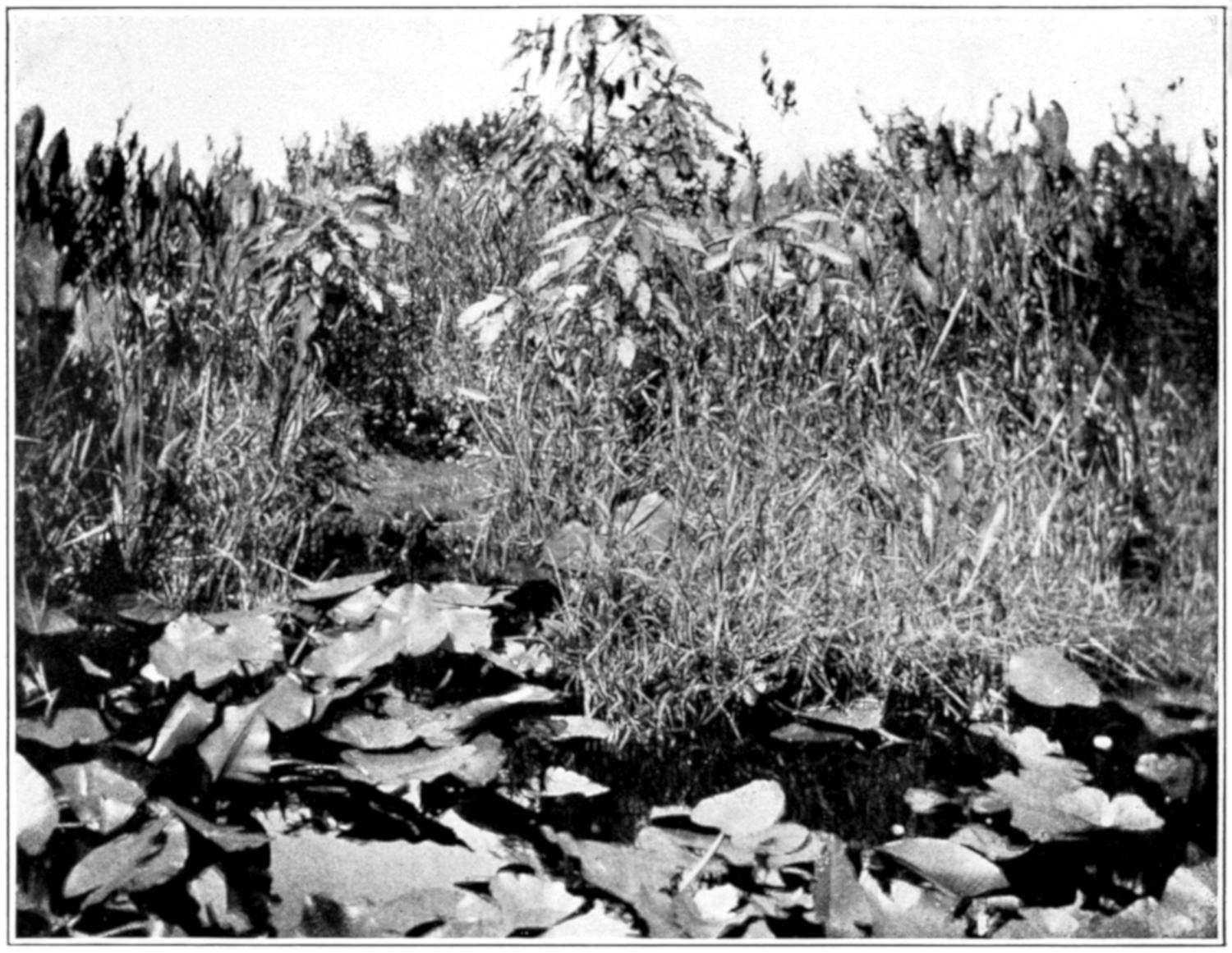
Fig. 7. A Typical ’gator “Hole.”
Only a few yards across, and surrounded by a dense growth of vegetation. On the
far side is seen an opening in the surrounding grass and flags where the ground is
worn smooth by the alligator in crawling out of the hole. Under the bank, probably
near the place where the alligator “pulls out,” is the deep cave into which the inhabitant
of this hole quickly goes on the approach of danger. As this cave may be
fifteen or twenty feet deep it is not an easy matter to get the animal out. When a
female alligator inhabits such a hole, a nest may often be found within three or four
yards of the water, though it is sometimes at a greater distance. Such a hole as this
may be connected by narrow, winding “trails” with larger ponds, as noted under
Fig. 6. (From a Photograph by the Author.)
While it is usually stated that the eggs are incubated
by the heat of the sun, it is held by some
observers that the necessary heat is derived not
from the sun but from the decomposition of the
vegetable matter of which the nest is composed.
Possibly heat may be derived from both of these
sources, but it seems likely that the conditions
that are especially favorable to normal incubation
are moisture and an even, though not necessarily
an elevated, temperature. Moisture is certainly
a necessary condition, as the porous shell allows
such rapid evaporation that the egg is soon killed
if allowed to dry. The inside of the nest is always
damp, no matter how dry the outside may become
under the scorching sun, so that this condition is
fully met. The eggs of the Madagascar crocodile,
according to Voeltzkow,[3] offer a marked contrast
to those of the alligator. Instead of being laid
in damp nests of decaying vegetation, they are
laid in holes that are dug in the dry sand, and are
very sensitive to moisture, the early stages, especially,
being soon killed by the least dampness. A
crocodile’s nest containing eggs is shown in Figure
5. In this species of crocodile, probably C. porosus,
the nest resembles that of the Florida alligator.
The photograph was taken by Mr. Rowley on the
edge of a small lake on the Island of Palawan, P. I.
[3] Voeltzkow, A., “The Biology and Development of the Outer Form
of the Madagascar Crocodile,” Abhandl. Senckberg. Gesell., Bd. 26,
Hft. I.
The daily range of temperature in the Southern
swamps is sometimes remarkably great, so that if
the eggs were not protected in some way they would
often pass through a range of temperature of possibly
fifty degrees or more; while in the center of a
great mass of damp vegetation they are probably
kept at a fairly constant temperature. Unfortunately
no thermometer was taken to the swamps,
so that no records of the temperatures of alligator
nests were obtained, but it was frequently noticed
that when, at night or very early in the morning,
the hand was thrust deep into the center of an
alligator’s nest the vegetation felt decidedly warm,
while in the middle of the day, when the surrounding
air was, perhaps, fifty degrees (Fahrenheit)
warmer than it was just before sunrise, the inside
of the same nest felt quite cool. It is probable,
then, that the conditions of temperature and
moisture in the center of the nest are quite uniform.
One lot of eggs that had been sent from Florida to
Maryland continued to incubate in an apparently
normal way when packed in a box of damp sawdust,
the temperature of which was about 80 degrees
Fahrenheit. Another lot of eggs continued
to incubate, until several young alligators were
hatched, in the ordinary incubator, at a temperature
of about 95 degrees Fahrenheit.[4]
[4] Reese, A. M., “Artificial Incubation of Alligator Eggs,” Amer.
Nat., March, 1901, pp. 193-195.
The fact that eggs taken directly from the oviducts
of the cold-blooded alligator contain embryos
of considerable size seems to indicate that no such
elevation of temperature as is necessary with avian
eggs is necessary with the eggs of the alligator.
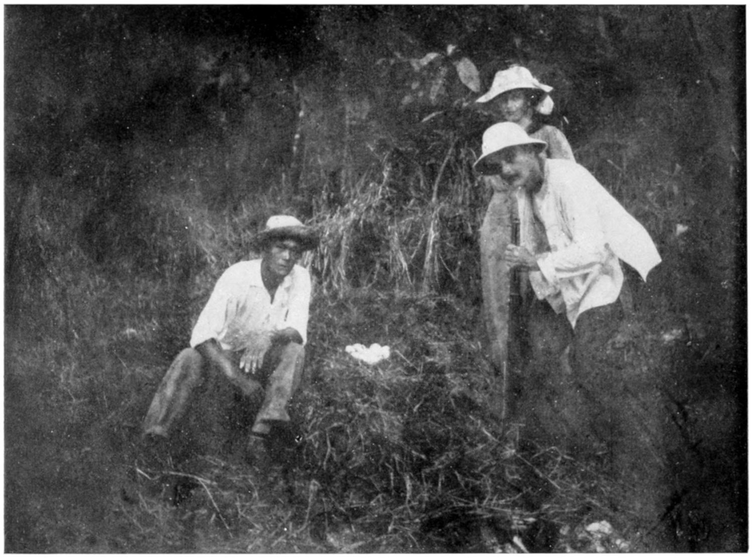
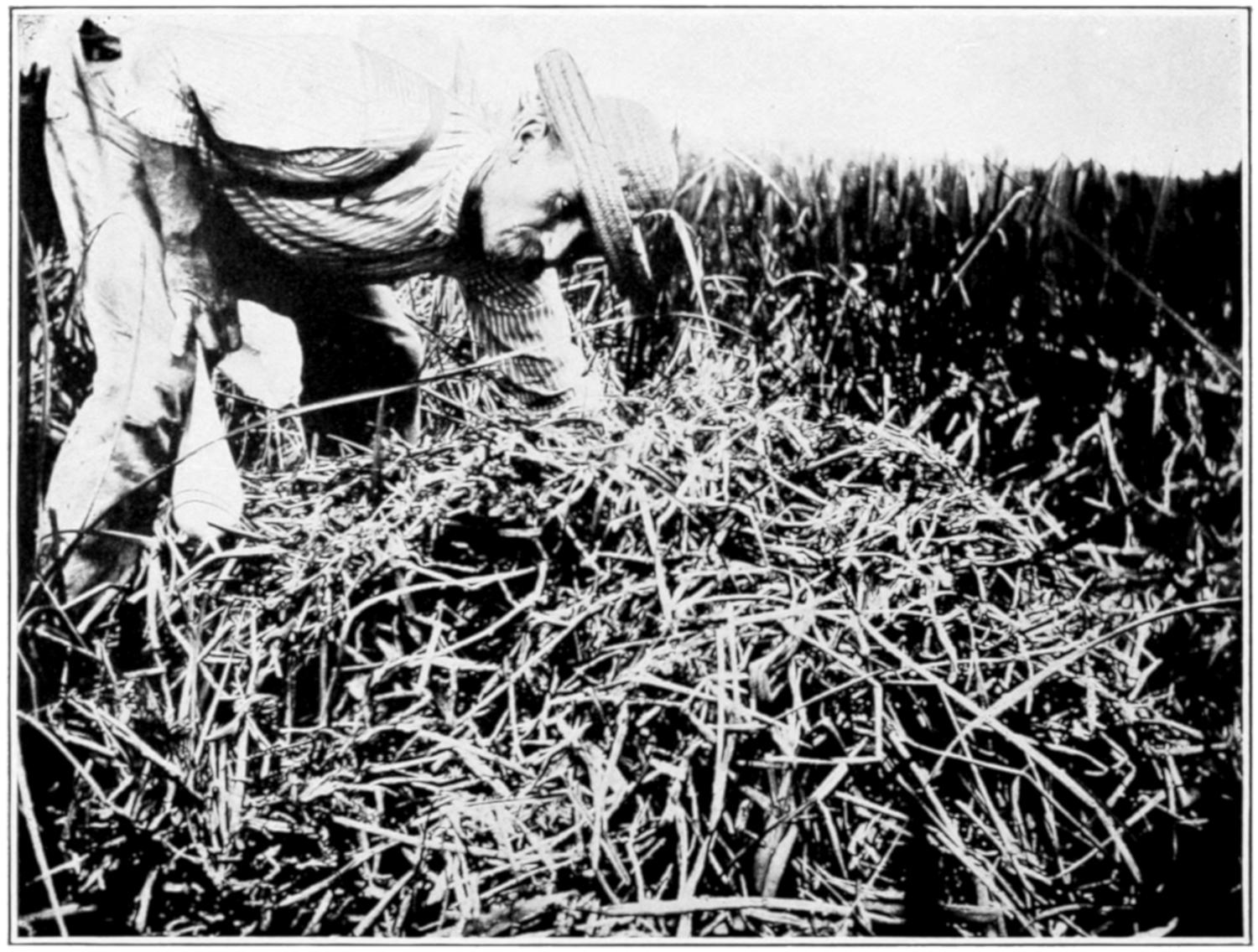
Fig. 8. A Typical Alligator’s Nest, Made Chiefly of Grass.
The guide is feeling for eggs without disturbing the outside of the nest. Being
made of the same material as the background, the nest does not stand out very
sharply, though in nature the contrast is somewhat more marked, owing to the
fact that the surrounding grass is green while the grass of which the nest is built
is dead and brown. (From a Photograph by the Author.)
The complete process of incubation probably
extends through a period of about eight weeks,
but no accurate observations along this line could
be made. For some hours previous to hatching
the young alligators make a curious squeaking
sound inside the shell, that may be heard for a
distance of several yards: this sound may be for
the purpose of attracting the attention of the female
alligator, who will open the top of the nest in time
to allow the just hatched alligators to escape: unless
thus rescued, it would seem impossible for the little
animals to dig their way out from the center of
the closely packed mass of decaying vegetation.
At the time of hatching the alligator is, as already
noted, about eight inches in length, and it seems
impossible that it should have been contained in
so small an egg.
The size of alligator eggs, as might be expected,
is subject to considerable variation. In measuring
the eggs a pair of brass calipers was used, and the
long and short diameters of more than four hundred
eggs were obtained. A number of eggs of average
size, when weighed in mass on the scales of a
country store, gave an average of 2.8 oz. per egg.
There was more variation in the long diameter
of eggs than in the short diameter.
The longest egg of all those measured was 85
mm.; the shortest was 65 mm. The widest egg
(greatest short diameter) was 50 mm.; the narrowest
egg (least short diameter) was 38 mm.
The average long diameter was 73.742 mm.; the
average short diameter was 42.588 mm.
The greatest variation in long diameter in any
one nest of eggs was 15.5 mm.; the greatest variation
in short diameter in the eggs of any one nest
was 11 mm.
The average variation in the long diameter of
the eggs from the same nest was 11.318 mm.; the
average variation in the short diameter of the
eggs from the same nest was 5.136 mm.
It will be seen from the above that the average
variation in the long diameter of eggs from the
same nest is between one sixth and one seventh of
the long diameter of the average egg; while the
average variation in the short diameter of the eggs
from the same nest is less than one eighth of the
short diameter of the average egg.
S. F. Clarke[5] gives the limits of the long diameter
as 50 mm. and 90 mm., and the maximum and
minimum short diameters as 45 mm. and 28 mm.
No such extremes in size were noticed among the
eight hundred or more eggs that were examined.
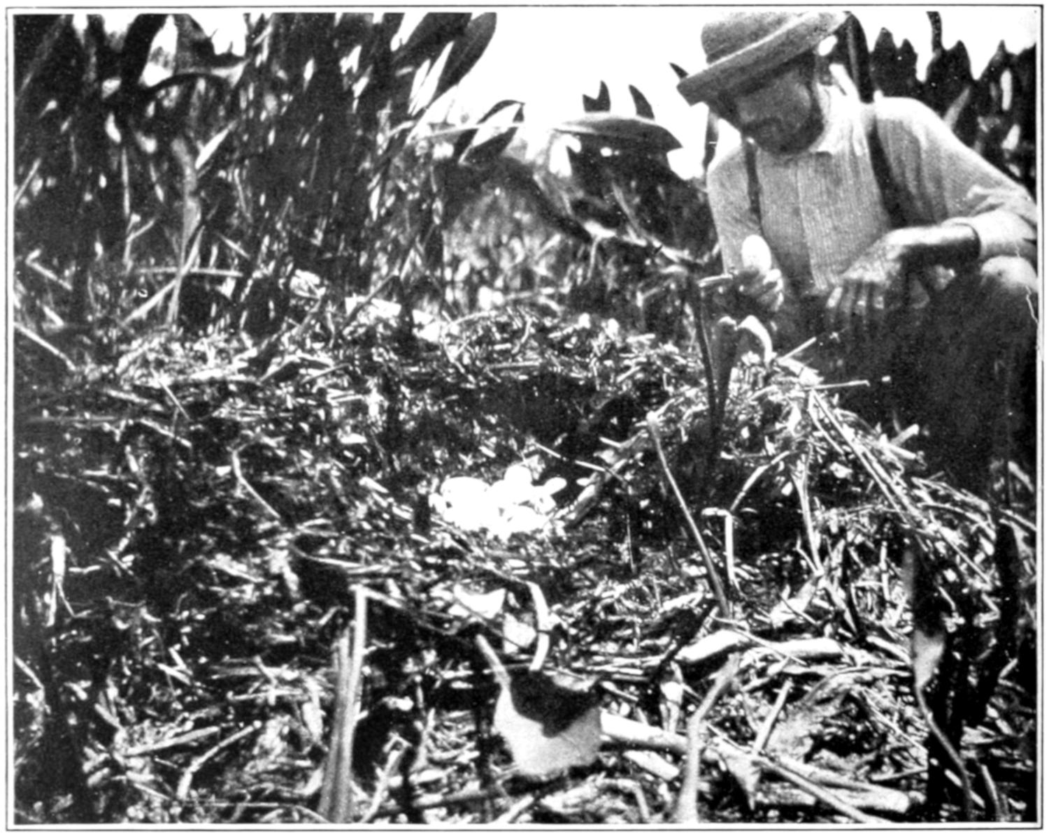
Fig. 9. An Alligator’s Nest, Somewhat Smaller than the One
Represented in Fig. 8, Built Chiefly of Flags.
The nest has been opened to show the irregularly arranged mass of eggs inside.
The size and shape of the egg are shown by the one in the guide’s hand. (From
a Photograph by the Author.)
[6] More than one hundred
years ago attempts were made to utilize the skin
of the alligator, but it was not until about 1855
that these attempts were successful and alligator
leather became somewhat fashionable and some
thousands of hides were converted into leather.
The demand was short-lived, however, and was
not again felt until the demand for shoe-leather
during the war between the States revived the
business. At the close of the war the business
again failed, but about 1869 the demand became
greater than ever and has continued unabated to
the present time. The supply of skins from our
own States proving inadequate, large numbers of
skins were soon imported from Mexico and Central
America. The skins from South America are so
heavy that they are of little value in making leather.
Of the States of the Union, Florida has been the
chief producer, the most important centers for
hides being Cocoa, Melbourne, Fort Pierce, Miami,
and Kissimmee. Ten men at the first-named place
took, in 1899-1900, 2500 skins; one man took 800
skins in one year; another man collected 42 skins
in one night. At Fort Pierce twelve men took
4000 skins in 1889. In 1899, three firms at Kissimmee
handled 33,600 hides. After this time the
total number of hides taken and the average per
man diminished greatly.
[6] The following figures are from an article by C. H. Stevenson in the
Report of the Bureau of Fisheries, 1902, pp. 283-352.
Besides being killed for their hides, the alligators
have been destroyed by the thousands merely
for wanton sport, so that in 1902 it was estimated
that their numbers in Florida and Louisiana were
less than one fifth of what they were twenty years
before that time, and unless steps be taken to
prevent it, the alligator hide, as an article of
commerce, may cease to exist in our Southern
States.
It has been claimed that the destruction of the
alligator has allowed the cane rat and muskrat
to increase to a serious extent, the former doing
great damage to crops, the latter often injuring
the levees to a dangerous extent. Legislation to
forbid the killing of alligators of less than five feet
in length has been suggested and should be passed,
since animals of less size have almost no commercial
value for leather.
In 1902, the annual output from the tanneries
of the United States approximated 280,000 skins,
worth about $420,000. Of these about fifty-six
per cent. came from Mexico and Central America,
twenty-two per cent. from Florida, twenty per
cent. from Louisiana, and the remaining two per
cent. from the other Gulf States. South American
hides are not handled by the United States markets.
In 1908, there were marketed from the South
Atlantic and Gulf States 372,000 pounds of alligator
hides, valued at $61,000.
According to the United States Bureau of
Fisheries the hunter in 1891 averaged about 60
cents for the skin, while in 1902 the price averaged
about 90 cents, varying between 15 cents and $2.00,
depending on the size and condition of the skin.
“Prime hides five feet long, with no cuts, scale
slips, or other defects, are worth about 95 cents
each, in trade, when the hunter sells them at the
country stores, and about $1.10 cash, at the tanneries.
Those measuring seven feet are worth
$1.55, six feet, $1.12; four feet, 52 cents, and three
feet, 25 cents. Little demand exists for those
under three feet in length” (Report Commissioner
of Fish and Fisheries, 1902, p. 345). Hides of
seven feet are in most demand, those over ten feet
are not much used. The income of the hunters is
largely increased by the sale of otter, bear, deer,
and other skins.
The different varieties of skins are described
by Stevenson (74) as follows:
“There are several distinct varieties of alligator
skins on the markets, the most important being
the Floridian, Louisianian, and Mexican; each
differs from the others in certain well-defined
characteristics, and owing to these differences
each variety has its special uses.
“The Florida skins are longer in the body—that
is, from the fore legs to the hind legs—than those
from Louisiana and Mexico, and consequently
they are largely in demand by manufacturers of
large handbags. They also have a number of
so-called ‘buttons’ or ‘corn marks’ on the inside
or under surface of an equal number of the scutes
resulting from imbedded horn-like tissues in the
center of those scales. These increase the difficulty
in tanning the skins and detract somewhat from
the appearance of the finished article, and for this
reason the Florida skins are ordinarily the cheapest
on the market. The farther south the skins are
secured in Florida the greater the number of ‘corn
marks,’ and those from the vicinity of Key West
are almost valueless for this reason.
“The Louisiana skins differ from those of Florida
in the absence of the ‘corn marks’ above noted,
and from both the Florida and Mexican skins in
being more pliable and in having the scales more
artistically curved and shaped. Consequently
they are preferred for such small articles as card-cases
and pocketbooks, and usually sell at the
highest prices. Skins obtained in Mississippi and
Texas are similar to those secured in Louisiana,
while those from Georgia and South Carolina are
similar to those from Florida, except that the
‘corn markings’ are not so numerous. All the
Florida and Louisiana skins show greater uniformity
of coloring, being of a bluish black on the
upper surface and a peculiar bluish white on the
under side.
“In addition to an absence of the characteristics
above noted the Mexican and Central American
skins are distinguished by having from one to four
small dots or markings like pin holes near the
caudal end of each scale. The length of the Mexican
skins varies greatly in proportion to the width,
sometimes equaling that of the Florida skins.
Those from the east coast of Mexico are the best,
being lighter in color and with neat and attractively
shaped scales. The west coast skins are yellowish
in color when in the green state, and the scales are
larger and not so artistically formed. The Florida
and Louisiana skins are almost invariably split
down the back, or rather along each side of the
back, so as to preserve the under side in a solid
piece, but most of the Mexican skins are split down
the middle of the abdomen, keeping the back
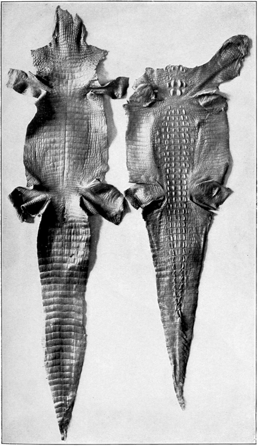
intact, making what is commonly known as ‘horn
alligator.’ (See Fig. 15.)
“The skin should be removed soon after death as,
in warm climates, putrefaction sets in very early
and the value of the skin is depreciated. After
removal, the flesh side of the skin is thoroughly
rubbed with fine salt, and the skin is carefully
rolled up with the salted side inside and is ready
for shipment, but must be kept in a dry, cool place.
Great care must be taken not to cut the hide since
small cuts that are not noticeable in the raw skin
may be so conspicuous in the dressed skin as to
render it of much less value; a large percentage
of the hides received in the markets are thus
damaged.
“Formerly only the ‘belly skin’ was removed,
by two longitudinal incisions just below the horny
portion of the back; but it was later found that
the thick horny skin of the back could be tanned
nearly as well as the thinner belly skin, so that
the entire skin is now usually removed by a longitudinal
incision along the mid-ventral line, with
lateral incisions along each leg to the foot (Fig. 15).
The entire skin is more commonly taken in Mexico
and Central America than in our States.
“Although the raw skins are sold according to
length, the tanned hides are sold by the width of
the leather at the widest part. Standard hides
sell for $1.00 to $1.65 per twelve inches of width.
Some skins tanned and dyed in a superior manner
sell for $2.00 or more for single skins of 2¹⁄₂ feet
in length. As a rule the Louisiana skins fetch the
highest prices, and those from Florida the lowest.
Imitation alligator leather is now prepared in large
quantities, principally from sheepskins or the
buffing from cowhides. These are tanned according
to the usual process, and before the skins are
finished they are embossed with the characteristic
alligator markings by passing them between two
rollers.” (Above-mentioned report, p. 346.)
Very little of the leather is now used in making
shoes, the chief demand being for handbags,
music-rolls, etc.
In hunting alligators for their hides two methods
are usually employed, in our Southern States at
least. The common method is “fire-hunting”
at night; the hunters here go, either singly or in
pairs, usually in boats, sometimes on foot, with
shotgun and torch. The torch may be fastened
to the hunter’s hat, after the manner of the miner’s
lamp. One more progressive hunter that I knew
had, as a torch, an acetylene lamp, attached to his
hat, with the tube for the gas extending down his
back to the generator in his pocket. This lamp
threw a blinding beam of light far across the swamp
into the eyes of the unsuspecting ’gator, which
usually remained fascinated until it could be approached
to within easy range. A shotgun at
close range, of course, blows off nearly the entire
top of the animal’s head and kills it instantly; it
is then seized before it sinks out of reach and is
either taken into the boat or dragged upon the
bank to be collected with others in the early
morning.
In daylight, with no glaring light to hypnotize
it, the alligator is difficult to approach within
range and it usually disappears into its cave before
the hunter can get a shot at it. The daylight
hunter, then, should be supplied not, of course,
with a light, but with a ten- or fifteen-foot pole with
a large iron hook at the end. If the alligator be
vigorously prodded with this mammoth fishhook
he will usually finally seize it with his mouth and
can be pulled out of his hole alive. It is then an
easy matter to kill him with a bullet through the
base of the brain. I have seen an eight-foot alligator
thus killed with a little .22 calibre “cat”
rifle. An eight-foot alligator will often be all that
two men can manage to drag out of his cave in
this way; and, in the torrid heat of the Southern
swamp, this violent exercise is not to the liking of
the usually not very energetic hunter.
While the manufacture of leather gives the chief
value to the alligator there are other ways in
which it has some economic importance. Chief
of these is probably the sale of alligator goods
to tourists. In 1891 there were in Jacksonville,
Florida, twelve dealers in live and stuffed alligators.
In 1890, 8400 alligators were sold to tourists, the
price for the live animals varying from $10.00 to
$35 per hundred. For individual animals of the
smallest size (less than twelve inches long) the
price is usually from 50 cents to $1.00. For a
three-foot alligator the price is generally $3-$5.00;
for sizes over three feet $2.00 per foot may be
charged, though for very large specimens the price
may be from $50 to $300 each.
Besides the live and stuffed animals the teeth
are polished and sold as souvenirs; about 450
pounds of teeth were sold in 1890, at a price varying
from $1.00 to $2.00 per pound. From 75 to
200 teeth will make a pound.
In 1891 about forty people, in addition to the
regular dealers, were engaged, in the United States,
in stuffing alligators, polishing teeth, etc. The
teeth are extracted by burying the head until
decomposition sets in.
The tiny alligators that are most commonly
sold to tourists, to be brought North, perhaps,
and allowed to freeze or starve to death, may
either be caught by a wire noose at the end of a
fishing rod, or they may be hatched from eggs that
are taken from the nests shortly before they are
ready to hatch. Such eggs may readily be hatched
by simply keeping them moist and at a fairly
constant temperature, as has been previously
noted. Besides the above uses Ditmars says:
“The eggs are eaten in many portions of the South,
and the search for eggs at the proper season furnishes
profitable employment for many persons,
as each nest contains a large number of eggs.”
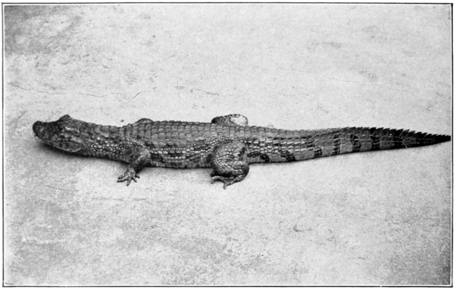
Broad-Nosed Caiman. (Caiman latirostris.)
Distribution: Tropical South America.
Attains a length of about seven feet. (After Ditmars.)
(Reproduced by Permission of Sturgis & Walton Co.)
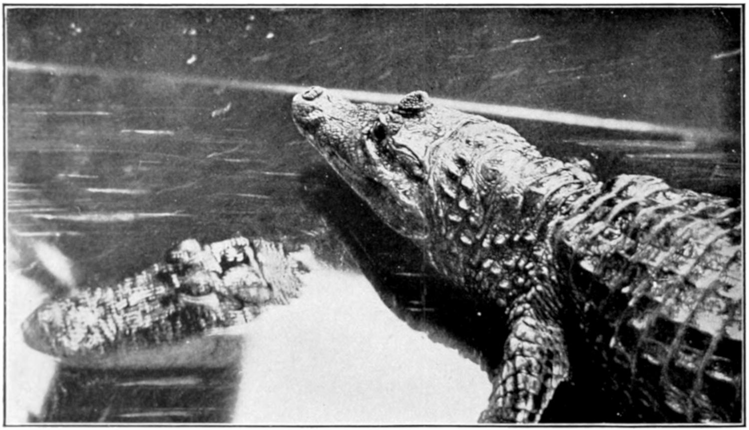
The length of an adult is about eight feet. (After Ditmars.)
(Reproduced by Permission of Sturgis & Walton Co.)
Never having eaten an alligator egg I cannot
speak from personal experience of its flavor; but
it has always seemed strange to me that more use
is not made of the flesh of the alligator. This
flesh is often said to have too strong a flavor to be
palatable; I have eaten it when it had no such
rank taste but was decidedly agreeable, being, as
might perhaps be expected of so amphibious an
animal, somewhat like both fish and flesh, yet not
exactly like either. Perhaps greater care should
be taken in skinning an animal that is to be used
for food in order that the flesh be not tainted with
the musk. It may be a lack of care in preparation
that has given rise to the impression that alligator
meat is too strong to be pleasant. It is perhaps,
also, the “idea” of eating a reptile that makes
the meat unpopular. A half-grown boy, who was
once in the swamps with me, had expressed a great
aversion to alligator meat, so the guide, one day,
offered him a nicely fried piece of alligator meat,
saying it was fish; the meat was eaten with evident
relish and the diner was not told until after a
second piece had disappeared what he had been
eating. It always seemed strange to me that the
poor people of the South should not more often
vary the monotony of fat pork and corn bread
with alligator steaks. Whether the meat could be
smoked or salted so that it would keep in a hot
climate I do not know; I am not aware of any
experiments along this line.
Beside the American form, Alligator mississippiensis,
the only other species of alligator is found
in China, along the Yang-tse-Kiang River; it is
Alligator sinensis. It reaches a length of six feet
and externally resembles its American relative;
it is greenish black above speckled with yellow;
grayish below.
This is the nearest relative of the alligator and
is found in Central America and tropical South
America. As seen by the table on page 2, it is
usually a small animal, though one species, the
black caiman, is said to reach a length of twenty
feet (Fig. 10). The nasal bones do not form a
bony septum as in the alligator and the ventral
armor consists of overlapping bony scutes. The
canine teeth of the lower jaw fit into a pit in the
upper jaw, as in the alligator.
They are said by some writers to be extremely
abundant in the waters of the upper Amazon,
migrating to the flooded forests during the rainy
season and returning to the streams on the approach
of the dry season. According to Ditmars there
are five species of caiman of which the spectacled
caiman, C. sclerops, and the black caiman, C. niger,
are the most striking. The former is so named
because of the spectacled appearance due to the
swollen and wrinkled upper eyelids; it reaches a
length of eight feet and is said to be of a treacherous
disposition. The latter has a blunt snout like
the alligator and is the largest of the New World
crocodilians.
Of about a dozen existing species of crocodile,
but one, the American crocodile, C. americanus,
is found in the United States, and it is
limited to the swamps and coast of southern Florida
below Lake Worth; its greater sensitiveness
to cold is doubtless the cause of its not being
found so far north as the alligator. Its range
extends south through Mexico and Central America
into South America. It was first found
in Florida by Dr. Hornaday in 1875. It sometimes
reaches a length of fourteen feet.
As has already been noted there is, besides certain
structural differences, a marked difference in
the dispositions of the Florida alligator and crocodile.
While an alligator may snap its jaws, hiss,
and swing its tail from side to side, it is not difficult
for a couple of men with ropes and a pole to safely
tie up a large specimen. The struggles of a crocodile
are of a more serious nature. Ditmars thus
describes an encounter with a captive Florida
crocodile: “The writer well remembers his first
acquaintance with a big fellow from Florida.
Driven out of the crate the crocodile looked the
picture of good nature. Standing away from what
he thought to be the reach of his tail, the writer
prodded the apparently sluggish brute with a
stick to start it for the tank. Several things happened
in quick order. With a crescentic twist of
the body utterly beyond the power of an alligator,
the brute dashed its tail at the writer, landing him
such a powerful blow that he was lifted completely
from the ground. As he left terra firma, an almost
involuntary inclination caused him to hurl his
body away from a pair of widely-gaping, tooth-studded
jaws swinging perilously near. Landing
with a thud on one shoulder, though otherwise
unhurt, the writer threw himself over and over,
rolling from the dangerous brute that was actually
pursuing him on the run, body raised high from
the ground. For an instant it seemed as if the
crocodile would win. As the writer sprang to his
feet and glanced backward, he beheld the brute
throw itself flat on its belly, open the jaws widely,
then remain motionless as a statue. Such is the
average crocodile—an active, vicious and, above
all, treacherous brute.” Ditmars says again, in
the same book: “When the keepers of the reptile
house of the New York Zoölogical Park clean out
the big pool of the crocodilians, they actually walk
over the backs of some of the big ’gators, so tame
are these. They never become unduly familiar
with the crocodiles, finding it necessary to pen the
latter behind heavily barred gates—and in the process
the men are often chased from the enclosure.”
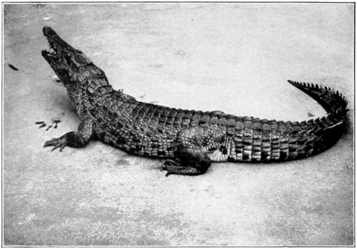
Nile Crocodile. (Crocodilus niloticus.)
Distribution: Africa generally; Madagascar.
Grows to a length of sixteen feet. (After Ditmars.)
(Reproduced by Permission of Sturgis & Walton Co.)
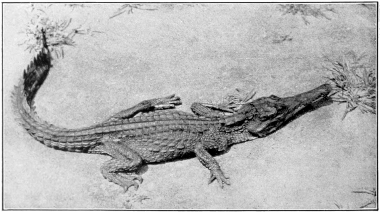
Fig. 11. West African Crocodile. (Crocodilus cataphractus.)
Distribution: West Africa.
Does not attain so large a size as the Nile Crocodile. (After Ditmars.)
(Reproduced by Permission of Sturgis & Walton Co.)
In contrast to this ferocious aggressiveness in
captivity the American crocodile is said to be very
timid and retiring when in its native habitat.
Young animals are greenish with black marking;
as they become older they are of an olive color,
and old specimens are dull gray.
This is a species with a very narrow snout that is
not quite so large as the preceding. It is said to be
abundant in the Orinoco River and its tributaries.
Besides the two above mentioned there is a
small species, C. rhombifera, found only in Cuba
and hence known as the Cuban crocodile.
This well known and much feared species is
found throughout the continent of Africa, and is
very common on the island of Madagascar (Fig.
11). In the lower waters of the Nile it is now nearly
exterminated. It has always been a conspicuous
animal in Egypt and was one of the animals held
sacred by the Egyptians and preserved by them
as mummies. It is discussed by Herodotus, and
the “leviathan” mentioned in the Book of Job
is doubtless this crocodile. In fact the name is
said to be derived from the ancient Greek for
lizard, just as the word alligator is said to be derived
from the Spanish for lizard—el lagarto; the
resemblance in form between these big saurians
and their smaller relatives is evident. The alligator,
being confined to America and a small part
of Asia, was probably not known to the ancients.
An excellent account of the development and
habits of the present species is given by Voeltzkow
(78), who says it is, perhaps, the most common
vertebrate in Madagascar. The largest specimen
measured by this observer was thirteen feet;
Ditmars gives sixteen feet as the maximum size.
This man-eating crocodile, according to Ditmars,
destroys more human lives than any other wild
animal of the dark continent.
The story told by Herodotus of the bird, probably
a species of plover, which enters the gaping
mouth of the crocodile to pick off the leeches found
there may be true, since there is such a bird that
may be seen perching on the backs of crocodiles,
and as the Crocodilia frequently lie with their
mouths wide open it is quite possible that these
birds may pick off the worms that are often found
within. It is also possible that the alertness of
these birds to danger may serve as a warning to
the crocodiles with which they associate.
Distribution: India to North Australia. Occurs at sea.
Grows to a length of twenty feet. (After Ditmars.)
(Reproduced by Permission of Sturgis & Walton Co.)
According to Voeltzkow these crocodiles dig
caves of thirty-nine to forty feet length in the
banks of the streams they inhabit, into which they
retire on the approach of danger. The caves open
under water and slope upward towards the surface
of the ground where a few small air-holes are
found. The natives locate the caves by means of
the air-holes and dig out the hidden animal, first
stopping up the entrance.
In Madagascar the eggs are laid in August and
September and hatch in about twelve weeks;
they are laid at night, usually shortly before daybreak.
From twenty to thirty eggs are laid in
one nest, which is merely a hole dug in the dry
sand. As was said in connection with the Florida
alligator, the habits of the two animals are quite
different in this respect,—the moisture that is so
important in the one case is fatal to the embryo
in the other. When the eggs are laid the nest is
filled in with sand so that there is nothing to indicate
its position except that the female crocodile
is in the habit of lying on the spot where her eggs
lie buried.
Like the alligator the young crocodile makes a
squeaking noise shortly before hatching and the
mother doubtless opens the nest, at this time, to
allow the young to escape. A fence that Voeltzkow
built around a nest was repeatedly broken down
by the mother in attempting to get back to her
eggs.
The character of the crocodile’s egg is discussed,
in comparison with that of the Florida alligator,
on page 23.
Found in India, Ceylon, Burmah, the Malay
Peninsula, and many of the islands in that region.
It has a rather broad snout, and reaches a length
of twelve feet. It is a timid form and is harmless
to man. It is frequently venerated by the Hindoos
and is kept in a semi-domesticated condition
in ponds where it is fed and becomes very tame.
In the dry season when the natural ponds are
empty they sometimes migrate overland in search
of water, but generally they bury themselves in
the mud and lie dormant until the rains begin
again.
This is one of the largest if not the largest of
living reptiles (Fig. 12). It is said by Ditmars to
reach a length of twenty feet and there is a record
of one specimen that was thirty-three feet in
length. It is said to be easily recognized by the
prominent, longitudinal ridge that extends in
front of each eye, over the prefrontal bone, and by
the absence of the suboccipital scutes.
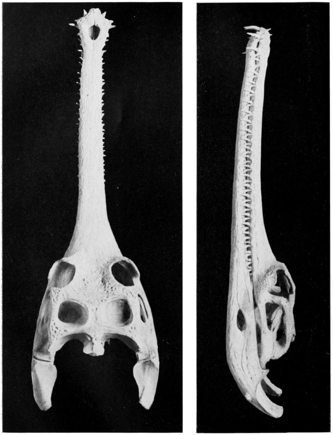
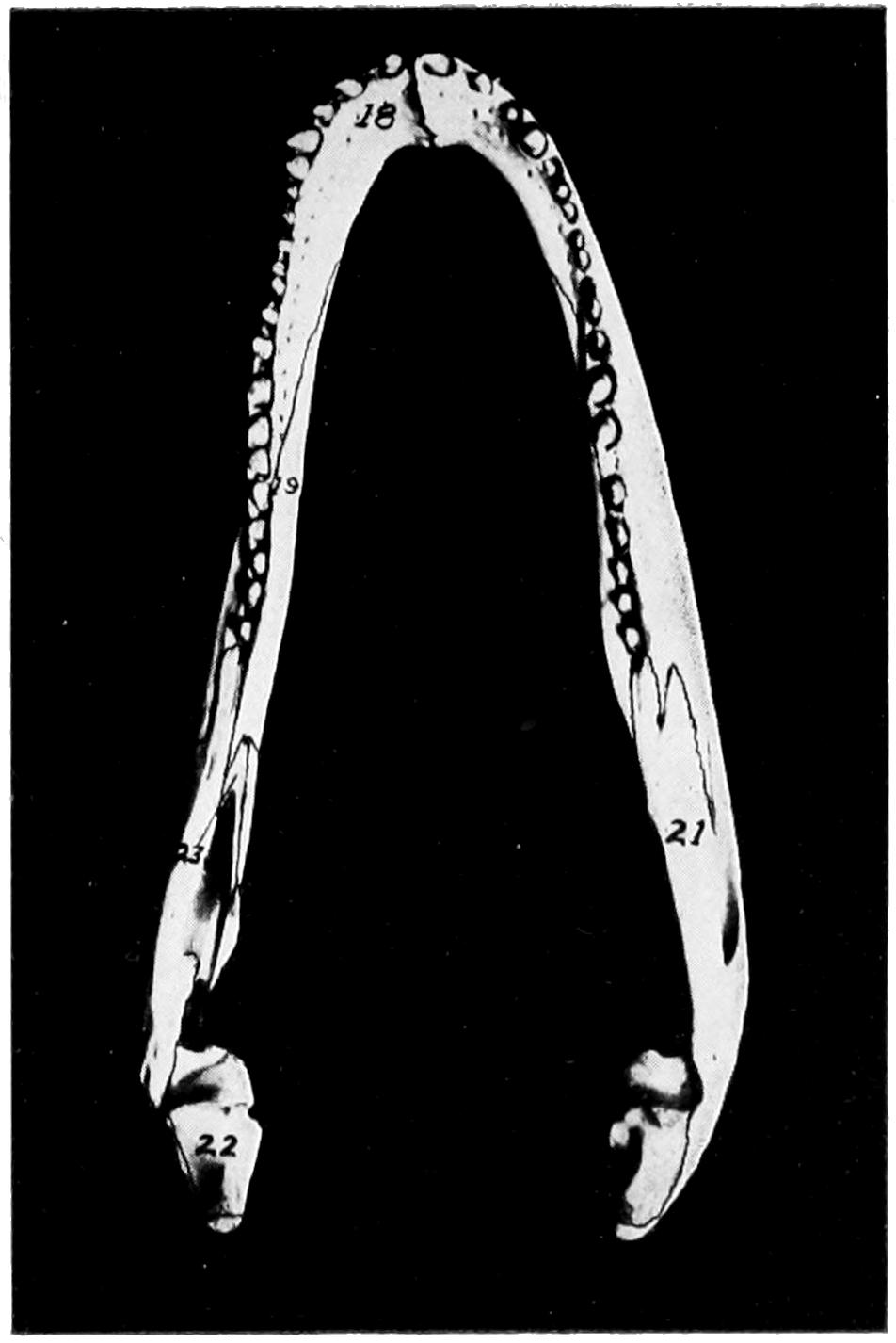
Fig. 13. Skull of Gavial.
(Dorsal View.)
(Photograph from U. S. National
Museum.)
Fig. 14. Skull of
Gavial.
(Lateral View.)
(Photograph from U. S.
National Museum.)
It is typically an inhabitant of tidal waters and
is sometimes found swimming at sea, out of sight
of land; it seldom goes inland to any great distance
from the sea. It is a man-eating species and many
human lives are said to be destroyed by it in India
and surrounding countries. A British “blue book”
states that in British India 244 deaths were caused
by Crocodilia in the year 1910.
In captivity it is savage and untamable. Ditmars,
in speaking of three specimens that he had
in captivity, says they were “positively the most
vicious reptiles” he had ever seen.
This animal, which inhabits the Ganges and
other rivers of northern India, is, with the possible
exception of the preceding species, the largest of
the Crocodilia; it is said to reach a length of thirty
feet, which is twice that of a very large Florida
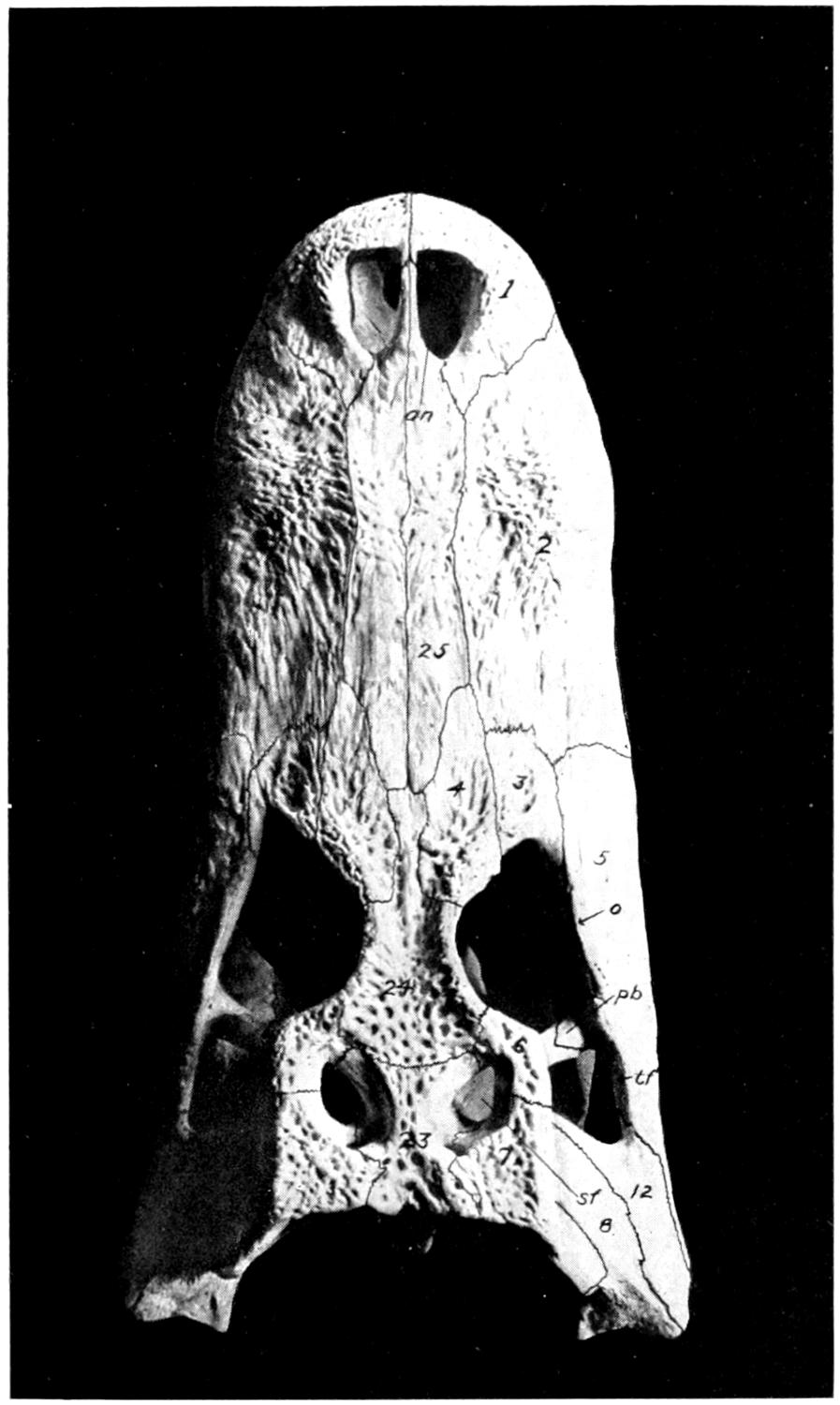
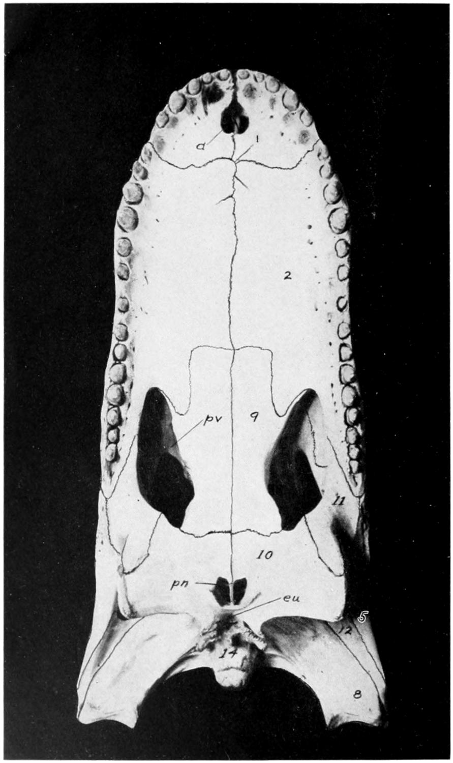
alligator. As previously noted its snout is extremely
long and narrow (Figs. 13 and 14), with a
large, fleshy hump at the tip, that projects above
the muddy water in which the animal lies concealed.
It is a timid animal and, in spite of its huge size,
dashes quickly into the water on the approach of
man, to whom it is seldom or never dangerous.
Its Indian name, gharial, from which its generic
name has been corrupted, means fish-eater, since
its food consists, it is said, largely if not entirely
of fish.
Considering its huge size and the character of
its jaws and teeth as shown in Figures 13 and 14,
it is fortunate that it prefers fish to human flesh.
Anderson (2) describes the eggs and young of the
Indian gavial. He found forty eggs in a nest of
sand; they were in two layers, with a foot of sand
between them. The young were 15.8 inches long
at hatching. He says: “The young run with
amazing rapidity the moment they are out of the
shell.... Some of them actually bit my fingers
before I had time to remove the shell from their
bodies.” The following quotation from Oldenburg
(46), for which I am indebted to Dr. Hussakof,
is perhaps the earliest reference to the egg of the
American alligator. It also mentions the habit
that is practiced by some of the recent Crocodilia
of swallowing stones to aid in digestion, as was
apparently done by some of the large extinct
reptiles.
“The eggs of Crocodiles and Alligators are little
bigger than a Turkey’s. I thought to bring one to
England, but it was lost. I never broke any to
see the Yolk and White; but the Shell is as firme
and like in shape to a Turkey’s, but not spotted.
I inquired into the Stone in the Stomach of a
Cayman or Crocodile, and I found by the inquiry
of a very observing gentleman there, that they
were nothing but several Stones, which that
Creature swallows for digestion. He took out of
one a piece of a Rock as big as his head: out of
others he had taken sixteen or twenty lesser.
None regards them much there, whatever Monardes
relateth.”
CHAPTER II THE SKELETON — A. The Exoskeleton — The Histology of the Integument. — B. The Endoskeleton — I. The Vertebral Column. — II. The Skull. — III. The Ribs and Sternum. — IV. The Appendicular Skeleton.
The exoskeleton is well developed in the
Crocodilia, and forms a very considerable
protection to its bearer. It is both dermal
and epidermal in origin.
The epidermal skeleton of the alligator consists of
oblong horny scales, arranged in transverse rows;
the long axes of the scales are parallel to that of the
body. On the tail, except along the mid-dorsal
line, and on the ventral side of the trunk and head
these scales are very regular in outline and arrangement;
on the sides of the head and trunk and on
the legs they are much smaller and less regularly
arranged, while along the mid-dorsal line of the
tail, especially in its posterior half, they are elevated
into tall keels that give the tail a large
surface for swimming. The first three digits of
both manus and pes are armed with horny claws,
which also belong to the epidermal part of the
exoskeleton.
Fig. 15. Alligator Skins; Under-surface and Horn-back.
The dermal exoskeleton consists of bony scutes
that underlie the epidermal scales of the dorsal surface
of the trunk and anterior part of the tail. The
overlying scales, except in very young animals,
are always rubbed off, so that the bony scales
are exposed. The ventral or inner surface of the
scutes is flat, while the outer surface is strongly
keeled and in old animals is often rough and pitted.
The plates are nearly square in outline and
are closely joined together in most places.
The scutes are grouped in two fairly distinct
areas known as the nuchal and the dorsal shields.
The former lies just back of the head, in the region
of the fore legs, and consists of four larger and a
number of smaller plates (Fig. 15). The latter, or
dorsal shield, extends over the back in fairly regular
longitudinal rows and quite regular transverse
rows. At the widest part of the trunk there are
six or eight of these scutes in one transverse row.
They become smaller towards the tail.
The teeth are exoskeletal structures, partly of
ectodermal, partly of dermal origin. They are
conical in shape, without roots, and are replaced
when lost. They will be described in connection
with the skull.
Musk glands, said by Gadow to be present in all
Crocodilia, are found in both sexes and are derivations
of the skin. One pair, each of which may be
as large as a walnut, is found on the lower side of
the head, one on the inside of each half of the
mandible. The other pair is inside of the lips of
the cloaca.
To understand
the structure of the integument of the Crocodilia
it is well to begin with the embryo. A cross section
of the epidermis of such an embryo will show the
rete Malpighii as a single layer of short, cylindrical
cells; over these are found more or less flattened,
disk-shaped cells formed by transverse division of
the underlying cells of the rete. On the outside lies
the epitrichial layer which consists of a mosaic of
polygonal cells, near the middle of each of which
lies an oval nucleus. Between the epitrichial cells
are small oval holes, not unlike the stomata in the
epidermis of plant tissues. Bronn thinks these are
not artifacts, but he does not suggest any explanation
of their occurrence.
In the epidermis of young and half-grown animals
the rete Malpighii is seen still as above noted.
On these cylindrical cells are found flattened cells
that gradually become very flat and lose their
nuclei as they pass over into the horny layer.
The stratum corneum consists of strongly flattened
cells in which the nuclei can no longer be
clearly seen, though their location can usually be
determined by the groups of pigment granules. On
the cells of the more superficial layers of the stratum
corneum are seen straight, dark lines, perhaps
ridges caused by pressure of the over- or underlying
polygonal cells. The individual cells of the horny
layer are usually easily isolated in the belly and
neck regions where they never become very thick;
but in the back the cells in this layer are very
numerous and fuse with each other to form the
bony plates; here the rete is the only clearly differentiated
layer. Whether prickle cells are present
in the epidermis of the crocodile Bronn is not
certain, though he thinks they probably are.
Rathke pointed out that on the surface of certain
folds of the integument, especially in the
region of the jaws, are found in all Crocodilia
certain small, scattered, wart-like elevations,
around each of which is customarily a narrow,
shallow, circular groove; they usually have a dark
brown but sometimes a gray or even white color.
Microscopic examination shows these warts to
be of epidermal origin, consisting of bright, round
cells that are closely united, without visible intercellular
substance. Treatment with potassium
hydroxid and then with water will show sometimes,
though not always, fine granular nuclei in the
cells.
In probably all members of the genus Crocodilus
at least is found, on the thick swelling on the right
and on the left side of the neck and trunk, a small,
flat pit which has the appearance of the opening of
an integumental gland. The pits are present also
in the scales of the throat, under the side of the
neck, sides of the body, lateral and ventral surfaces
of the anterior half of the tail, and the legs. They
are near the hinder border of the scales. Only
occasionally are two pits
found in one scale. These
pits are found in the gavials
but are absent in
some, probably all, alligators.
A small knob
projects from the center
of some of the pits.
These pits are not openings
of glands but have
about the same structure
as the pits seen in the
head.
The integumental
bones in the Crocodilia
originate in the connective
tissue of the cutis.
Investigations in young
animals show that these
bones usually take their
origin in the under and
middle layers of the
cutis and generally work
towards the periphery.
The vertebral column
consists of about sixty-five
vertebræ, which may be separated into the
usual regions; there are nine cervical, ten dorsal, five
lumbar, two sacral, and about thirty-nine caudal. It
is likely that the number of caudals may be subject
to frequent variation; one complete skeleton
had sixty-five vertebræ in all, another had sixty-eight.
A complete skeleton of the crocodile (species
not known) had sixty vertebræ. A thirteen-foot
skeleton at Western Reserve University had only
sixty-one vertebræ, but some of the caudals were
evidently missing. Two skeletons of C. porosus in
the museum at Singapore had sixty and sixty-three
vertebræ respectively. A skeleton of Tomistoma
schlegeli in the same museum had sixty vertebræ.
The Cervical Vertebræ.
Since all of the cervical
vertebræ bear ribs, we shall assume the distinction
between them and the dorsal vertebræ to be that
the ribs of the latter meet the sternum, while those
of the former do not reach to the sternum. Assuming
this distinction, there are, as was said
above, nine cervical vertebræ.
Fig. 17. First Four Cervical Vertebræ of a Crocodile
(C. vulgaris). (From Reynolds, partly after Von Zittel.)
1. pro-atlas.
2. lateral portion of atlas.
3. odontoid process.
4. ventral portion of atlas.
5. neural spine of axis.
6. postzygapophysis of fourth vertebra.
7. tubercular portion of fourth cervical rib.
8. first cervical rib.
9. second cervical rib.
10. convex posterior surface of centrum of fourth vertebra.
With the exception of the first two, to be discussed
later, these are all essentially alike and the
fourth will be described as a type (Fig. 17). Its
centrum is cylindrical or somewhat hourglass
shaped, concave anteriorly and convex posteriorly;
it is not completely fused with the neural arch but
is united with it by sutures. From the anterior
end of the ventral surface of the centrum projects
downward and forward a small plow-shaped process,
the hypapophysis. On each side of the centrum,
near its anterior end, is a facet with which the lower
branch (capitulum) of the rib articulates. The
neural arch is strongly developed and is extended
dorsally into a prominent neural spine and on each
side as a short, blunt, transverse process with which
the tubercle or upper branch of the rib articulates.
Posteriorly the arch is notched on each side to form
the openings for the exit of the spinal nerves. Projecting
dorsally and anteriorly from the arch are
two short processes which bear the medially and
dorsally facing prezygapophyses (Fig. 17). Just
caudad to these processes are somewhat shorter
processes that bear the laterally and ventrally
facing postzygapophyses (Fig. 17, 6).
The atlas, as in other vertebrates, is highly
specialized. It consists (Fig. 17), even in the adult
animal, six feet or more in length, of four distinct
portions, a ventral (4), a dorsal (1), and two lateral
(2) parts. The ventral portion is relatively more
massive than in most animals; its anterior surface
is concave and forms the main part of the articular
surface for the occipital condyle of the skull. Its
postero-dorsal surface articulates with the odontoid
process of the axis. On its postero-lateral surfaces
are the facets for articulation with the first ribs,
which, unlike the other cervical ribs, have but one
articular surface. Articulating dorsally with this
ventral element of the atlas are the two rather
heavy lateral elements which form the neural
arch. Anteriorly they form the lateral parts of the
articular surface for the condyle and dorsally they
unite for a short distance with each other. Projecting
ventrally from the posterior part of their
dorsal portion are the small postzygapophyses.
Ventrally and laterally they articulate with the
odontoid process (Fig. 17, 3). Projecting dorsad and
cephalad from the dorsal surface of these lateral
elements is the dorsal element of the atlas (Fig.
17, 1), the pro-atlas, which may not be properly
a part of the vertebral column at all, since it is said
to be merely a membrane bone. Gadow says it
is the detached neural spine of the atlas. It is
thin and triangular in shape, resembling in contour
a large, mammalian epiglottis. It forms an arch
over the space between the skull and the front
of the atlas proper. Reynolds calls it the pro-atlas.
The Axis.
The centrum differs from those following
it (described above) mainly in its close
articulation (not fusion) with the large odontoid
process; this process not only projects into the
atlas, as is usually the case, but articulates with its
postero-lateral border on each side, and is distinctly
visible in a lateral view of the neck (Fig. 17,
3). Like the rest of the cervical vertebræ the posterior
surface of the centrum is convex. The neural
arch of the atlas differs from those following
mainly in having a much wider (in an antero-posterior
direction) neural spine. The lateral processes
and those bearing the prezygapophyses are
also less strongly developed than on the following
vertebræ.
Fig. 18. Anterior View of A, a Late Thoracic and B, the
First Sacral Vertebra of a Young Crocodile
(C. palustris). × ¹⁄₃. (After Reynolds.)
1. neural spine.
2. process bearing prezygapophysis.
3. facet for articulation with the capitulum of the rib.
4. sacral rib.
5. surface which is united with the ilium.
6. concave anterior face of centrum.
The Thoracic Vertebræ.
The first thoracic vertebra
differs scarcely at all from the ninth cervical;
and the tenth thoracic differs from the first lumbar
only in bearing a short rib. Only the first three
thoracic centra bear the hypapophyses noted in
connection with the cervical vertebræ. The ribs
of the first two thoracic vertebræ articulate with
them by two processes, as in the typical cervical
vertebræ; the other ribs articulate only with the
transverse process. The fourth thoracic may be
described as a type of this region (Fig. 18, A). Its
centrum is rather longer than in the first two
thoracic and in the cervical vertebræ and has no
process for articulation with the head of the rib,
otherwise it is essentially the same. Like all of
the vertebræ behind it and unlike those in front
it is apparently completely fused with its neural
arch. The neural arch is very broad (in an antero-posterior
direction) and is extended dorsally as a
wide neural spine (1). The neural spines of the
following thoracic and the first two or three lumbar
vertebræ are increasingly broad and truncated.
The transverse processes are very broad, long and
thin, and in the third to eighth vertebræ they have
two articular surfaces, an anterior and more medial
one for articulation with the head of the rib (3) and
a posterior and more distal one for articulation with
the tubercle of the rib. These two surfaces approach
each other as the vertebræ are followed
caudad until, in the last two thoracic vertebræ,
they form practically one surface. The processes
of the pre- and postzygapophyses spring from the
arch at the base of the transverse process; the
former surface is directed dorsally and medially,
the latter ventrally and laterally. The intervertebral
foramina are smaller and more nearly circular
than in the cervical region, and are more closely
surrounded by bone.
The Lumbar Vertebræ.
The five lumbar vertebræ
are essentially like the thoracic except that the
transverse processes, which, of course, bear no ribs,
are both shorter and narrower. The postero-lateral
border of the centrum of the last of these
five vertebræ has a small surface for articulation
with the antero-medial border of the transverse
process of the first sacral vertebra.
The Sacral Vertebræ
(Fig. 18, B). These are
two in number. The centrum of the first is concave
in front and flat behind, instead of being
convex behind, and the second is flat (instead of
concave) in front, and convex behind. The neural
spine and zygapophyses are as in the lumbar region.
Projecting laterally from each sacral vertebra,
forming a close, sutural joint with both centrum
and neural arch, is a heavy bone shaped like a
truncated pyramid (4); the base of the pyramid
is ankylosed with the ilium. These bones
seem to be much thickened transverse processes,
but since they are not completely fused with
their respective vertebræ and are said to ossify
separately they should probably be called sacral
ribs. The two sacral vertebræ do not seem to
be any more closely united than are any other two
vertebræ.
The Caudal Vertebræ
(Fig. 16, C). These are
characterized by the entire absence of ribs, and
by the presence on all but the first and the last
four or five of V-shaped chevron bones. The
first ten or twelve of these chevron bones articulate
chiefly with the postero-ventral ends of the centra,
but they also articulate with the antero-ventral
ends of the vertebra behind themselves; and as they
are followed caudad they seem to lie directly
below the intervertebral regions and to articulate
equally with the vertebræ before and behind.
The chevron bones gradually diminish in size
from before back. The neural processes of the
first four or five caudals are broad, like those of the
more anterior regions, but caudad to this point they
become narrower and more pointed, though they
retain the same height until about the last ten or
twelve vertebræ. Towards the tip of the tail the
dorsal spines diminish in height and finally disappear.
The transverse processes of the first five
or six of the caudals are long and narrow. They
gradually diminish in length until the eighteenth
caudal, back of which they are no longer to be seen.
The zygapophyses are mostly about the same as in
the more anterior vertebræ, but towards the posterior
end of the tail the postzygapophyses come to lie
between rather than above the prezygapophyses.
The neural canal diminishes, of course, in size
towards the tip of the tail until it is no longer
present, the last five or six vertebræ consisting
only of the centra.
The skull of the alligator is very massive and
has several peculiarities. 1. The bones of the
dorsal surface are rough and pitted, especially in
old animals. 2. The jaws are enormously large in
proportion to the brain cavity, and are armed with
many large teeth. 3. The mandibular articulation
is some distance caudad to the occipital
condyle. 4. The interorbital septum is mainly
cartilaginous. 5. There is a complicated system
of Eustachian passages connecting with the back of
the mouth by a single opening. 6. The posterior
nares are placed very far back and the palate is
correspondingly long.
The skull as a whole may be divided into three
regions: the cranium, the lower jaw, and the hyoid;
these will be described in the order given.
The Cranium.
As a matter of convenience the
bones will be described as seen from the different
aspects—dorsal, ventral, lateral, posterior, and in
sagittal section—without particular regard to their
grouping into segments or regions.
The Dorsal Aspect
(Fig. 19). At the extreme
posterior end of the median line lies the parietal
(23), double in the embryo but a single bone in
the adult. It forms a part of the roof of the cranial
cavity and articulates anteriorly with the
frontal, laterally with the postfrontals, squamosals,
and, according to Reynolds, with alisphenoids,
pro-otics and epiotics, and ventrally with the
supraoccipital. It forms the median boundary
of each of the two supratemporal fossæ (sf).
On each side of the parietal and forming the
posterior comers of the rectangular postero-dorsal
region of the skull are the squamosals (7). Each
squamosal articulates medially with the parietal,
anteriorly with the postfrontal, and ventrally with
the quadrate and exoccipital. It forms part of the
posterior and lateral boundaries of the supratemporal
fossa and a part of the roof of the external
auditory meatus.
Articulating with the anterior border of the
squamosals and forming the anterior corners of
the rectangular region mentioned above are the
postfrontals (6). The postfrontal articulates medially
with the parietal and frontal, and ventrally
with the alisphenoid and a small part of
the quadrate. It sends, in a ventro-lateral direction,
a thick process that unites with a similar
process from the jugal to form the postorbital bar
(pb) which lies between the orbit (o) and the temporal
fossa (tf). The postfrontal forms the antero-lateral
boundary of the supratemporal fossa.
Articulating posteriorly and laterally with the
parietal and the postfrontals, and forming the
highest point of the skull, is the single frontal bone
(24), which, like the parietal, is paired in the embryo.
It is a heavy bone whose dorsal surface is
flattened posteriorly, deeply concave in the middle
region, and drawn out into a long projection anteriorly.
It forms part of the roof of the cranial
cavity and articulates ventro-laterally with the
alisphenoid and anteriorly with the prefrontals and
nasals. It forms a part of the median boundary of
the orbit.
The prefrontal (4) is an elongated bone in the
latero-median border of the orbit. Medially and
anteriorly it articulates with the frontal and nasal,
laterally with the maxillary and lachrymal, and
ventrally, by a heavy process, with the pterygoid.
Fig. 19. Dorsal View of the Skull of the
Alligator (A. Mississippiensis).
1. premaxilla.
2. maxilla.
3. lachrymal.
4. prefrontal.
5. jugal.
6. postfrontal.
7. squamosal.
8. quadrate.
12. quadratojugal.
23. parietal.
24. frontal.
25. nasal. an, anterior nares;
o, orbit; pb, postorbital
bar; sf, supratemporal
fossa; tf,
lateral temporal
fossa.
The nasal (25) is a long narrow bone forming the
greater part of the roof of the nasal passage.
Along the median line of the skull it articulates
with its fellow; posteriorly with the frontal;
laterally with the prefrontal and maxillary; and
anteriorly with the premaxilla. In the crocodile,
caiman, and gavial it also articulates with the
lachrymal. In the alligator the anterior ends of the
two nasals form a narrow rod of bone that extends
across the anterior nares, and, meeting a projection
from the premaxillaries, divides the opening into
right and left halves. In the crocodile the nasals
project only a very little way into the nares; in
the caiman (according to Reynolds) they do not
extend into the nares at all, and in the gavial, whose
much elongated snout is mainly due to the great
length of the maxillaries, the nasals do not extend
more than a third of the distance from the prefrontals
to the anterior nares.
The maxilla (2) is a large bone that forms a large
part of the upper jaw and that holds most of the
teeth of that jaw. On the ventral side, as will be
described later, it articulates with its fellow in the
middle line, with the premaxilla, with the palatine,
and with the transpalatine. Dorsally it articulates
with the premaxilla in front; with the nasal and
prefrontal on the medial side; and with the
lachrymal and jugal behind.
The premaxilla (1) forms, with its fellow, the
extreme tip of the upper jaw. Each bone forms
the anterior and lateral borders of its half of the
anterior nares. It articulates medially with its
fellow and posteriorly with the nasal and maxilla.
Ventrally, as will be noted later, it bears five teeth
and articulates with its fellow medially and with
the maxilla posteriorly. Between the premaxillæ
on the ventral side is the large anterior palatine
foramen.
The lachrymal (3) is a fairly large bone that forms
the anterior border of the orbit. It is bounded
laterally by the jugal, anteriorly by the maxilla,
and medially by the prefrontals. Its postero-medial
border is pierced by a large lachrymal
foramen that extends lengthwise through the bone
and opens, at its anterior end, into the nasal
chamber.
The supraorbital, missing in the skull figured, is
a small bone lying in the eyelid close to the junction
of the frontal and prefrontal. Being unattached
it is usually absent from prepared skulls.
The jugal or malar (5) is an elongated bone that
forms a part of the lateral border of the head, on
the one hand, and most of the lateral border of the
orbit on the other. Anteriorly it articulates with
the maxilla; medially with the lachrymal and
prefrontal; posteriorly with the quadratojugal,
and ventrally with the transpalatine. With the
transpalatine it sends, in a dorso-medial direction,
a process that meets the process, described above
in connection with the postfrontal, to form the
postorbital bar.
The quadratojugal (12) is a small bone, wedged in
between the jugal in front and the quadrate behind.
Fig. 20. Ventral View of the Skull of the
Alligator (A. Mississippiensis).
2. maxilla.
5. jugal.
8. quadrate.
9. palatine.
10. pterygoid.
11. transpalatine.
12. quadratojugal.
14. basioccipital. a, anterior
palatine vacuity; eu,
opening of the median
Eustachian canal; pn,
posterior nares; pv,
posterior palatine
vacuity.
The quadrate (8) is more irregular and has more
complicated articulations than almost any bone
in the skull. Its posterior end, which forms the
articular surface for the lower jaw, is elongated
laterally and slightly concave. Anteriorly the
quadrate articulates with the quadratojugal; medially
with the basisphenoid and exoccipital; dorsally
with the exoccipital, squamosal, postfrontal,
and, possibly, with the pro-otic; ventrally with the
pterygoid, alisphenoid, and probably with some of
the otic bones. Its dorsal side forms most of the
floor of the external auditory meatus which will be
described later. While the basioccipital may be
seen from the dorsal side, it is not really one of the
dorsal bones of the skull and will be described later;
the same is true of the pterygoids and palatines
which may be seen through the empty orbits.
The Ventral Aspect
(Fig. 20). The larger part
of this side of the skull is made up of four pairs of
bones: the premaxillæ, the maxillæ, the palatines,
and the pterygoids, lying, from anterior to posterior,
in the order named.
The premaxilla (1), as described in the dorsal
view of the skull, is a triangular bone which, with
its fellow, forms the anterior end of the snout.
Each premaxilla bears five teeth, not only in the
alligator but in the crocodile, the caiman, and in the
gavial. Of these teeth the fourth from the front
is the largest; the first two are small, and the
third and fifth are of intermediate size. This
arrangement as to size is also true, apparently, in
the other groups of Crocodilia. The ventral surface
of the premaxilla, which is more or less flat and
horizontal, is pierced by a number of small foramina,
in a row parallel to the curved outer margin
of the bone. Between these foramina and the
base of the teeth are four rounded depressions to
receive the points of the first four teeth in the lower
jaw; of these depressions the first and fourth are
the deepest. The first pit often becomes so deep
as to perforate the bone; this is true also with
the crocodile and, according to Reynolds, with the
caiman, but is not true of the gavial, whose interlocking
teeth project outside of the jaws. It will
be remembered that one of the chief distinctions,
given early in this work, between the crocodile
and the alligator is that in the former the fourth
tooth in the lower jaw fits into a notch and not into
a pit in the upper jaw.
The maxilla (2), which with its fellow forms most
of the hard palate, has also been mentioned in
connection with the dorsal aspect. Each maxilla is
notched, posteriorly, to form the anterior border
of the posterior palatine vacuity, and together
they are notched to receive the rectangular anterior
ends of the palatines. The postero-lateral
extremity of the maxilla articulates with the transpalatine.
Along the outer border of the bone are
the teeth, of which there are fifteen or sixteen in the
alligator, about the same number (perhaps one or
two less) in the caiman and crocodile, and about
twenty-four in the gavial. The first or anterior
eight or ten teeth have individual sockets, the rest
are placed in a groove. In the crocodile none of
the teeth have individual sockets, and in the gavial
they all have sockets. The premaxillary and more
anterior of the maxillary teeth are slightly recurved
and are sharper than the posterior maxillaries which
besides being blunt have a constriction above the
surface of the socket.
The crocodilian tooth consists of three layers
(Fig. 20 A).
The enamel (e) forms a fairly thick layer over the
crown of the tooth; it exhibits a very clear striated
structure, the striations being apparently due to
stratification.
Some of the tubules of the dentine (d) continue
into the enamel, where they may be distinguished
by their remarkable fineness and their straight
course.
The cement (c) covers the root of the tooth that
projects into the alveolus of the jawbone; it is
much more strongly developed than in the lizards
and contains a very large number of bone corpuscles
which are distinguished from the bone corpuscles
proper by their greater circumference.
The fairly large pulp cavity (p) has, like the
tooth itself, a conical form.
Parallel to the teeth is a row of small foramina,
a continuation of those noted in the premaxilla;
some or all of these foramina open into a longitudinal
sinus along the alveolar border of the maxilla;
this sinus opens posteriorly by one or more large
apertures into the posterior palatine vacuity.
The palatines (9) form a broad bar of bone from
the pterygoids behind to the maxillæ in front.
They are united with each other by a straight
median suture and form a considerable part of the
floor as well as a part of the side walls and roof of
the nasal passage. They form most of the median
boundaries of the posterior palatine vacuities (pv).
Dorsally they articulate with the pterygoids, prefrontals,
and vomers.
The pterygoids (10) are the very irregular bones
that project ventrad and caudad from beneath the
orbits. Their suture is continuous, caudad, with
that between the palatines and at the posterior end
of this suture is the posterior opening of the nasal
chamber, the posterior nares (pn). This opening
is divided by a vertical, longitudinal, bony septum,
and the part of the chamber into which it immediately
opens, which lies in the pterygoids, is divided
by a number of transverse, vertical septa. Posterior
and dorsal to the posterior nares the pterygoids
are fused. Anteriorly the pterygoids articulate
with the palatines; dorsally with the quadrates,
basisphenoid, alisphenoids, and prefrontals, and
dorso-laterally with the transpalatines. The lateral
vertical border of the pterygoid is roughened
and is, according to Reynolds, covered, during life,
with a pad of cartilage against which the medial
side of the mandible plays.
The transpalatine (11) is a T-shaped bone articulating
ventrally with the pterygoid and dorsally
with the maxilla, the jugal, and the postfrontal.
Fig. 20A.
Longitudinal Section of the Jaw and Tooth of a Crocodile.
(After Bronn.)
c, cement; d, dentine; e, enamel; p,
pulp, of functional tooth; c′, cement; d′, dentine; e′,
enamel, of rudimentary tooth; e″, epidermis; k, bone of jaw.
The basioccipital (14) is seen projecting caudad
as the single occipital condyle; it will be described
in connection with the posterior aspect of the skull.
The jugal (5), quadratojugal (12), and quadrate
(8) may all be seen from this view. The first two
have been sufficiently described in connection with
the dorsal aspect; the last will be further described
in connection with the lateral aspect.
Just caudad to the posterior nares is a small
opening, the unpaired Eustachian canal (eu).
The Lateral Aspect
(Fig. 21). As will be seen by
the figure, practically all of the bones visible in this
view have already been described, except those of
the mandible, which will be described separately.
At the base of the skull are, however, two bones,
the basi- and alisphenoid, that have not been
described and that show as well in this as in any
other view. The basisphenoid (just below v and
hidden by the pterygoid) was mentioned in connection
with the quadrate, with whose posterior margin
it articulates. It is an unpaired bone of very
irregular shape. Anteriorly it is flattened out to
form the rostrum, a rectangular process that forms
the posterior part of the interorbital septum; in
fact it is the only part of the septum present in
a prepared skull, since the rest is cartilaginous.
Dorso-laterally the basisphenoid articulates with
the alisphenoid; posteriorly with the basioccipital;
ventrally with the pterygoid; and posteriorly with
the exoccipital and basioccipital. On the dorsal
surface of the basisphenoid is the pituitary fossa,
not seen, of course, in this view of the skull.
The alisphenoids (crossed by the dotted line from
V) are a pair of very irregular bones that form most
of the antero-lateral walls of the brain case. They
articulate dorsally with the parietal, frontal, and
postfrontal; ventrally with the basisphenoid
and pterygoid; and posteriorly with the quadrate
and some of the otic bones not visible in this view.
Between it and the quadrate, plainly visible in this
view, is a large opening, the foramen ovale (V),
through which, according to Reynolds, the trigeminal
nerve passes. In the middle line, directly
under the frontal bone, is an opening between the
anterior wings of the two alisphenoids, for the exit
of the optic nerves. Ventrad and caudad to this
opening, and sometimes continuous with it, is
another large foramen, just dorsad to the rostrum,
for the exit, according to Reynolds, of the oculomotor
and abducens nerves. Projecting caudad
is seen the rounded condylar part of the basi-occipital
(14) to be described later, and dorso-cephalad
to this is a part of the exoccipital (13)
in which four foramina may be seen; of the dorsal
three the one nearest the condyle and foramen
magnum is for the exit (Reynolds) of the hypoglossal
nerve (XII); slightly dorso-cephalad to
this is one for the vagus nerve (X); between these
two is a very small one for a vein; the largest and
ventrally located foramen is for the entrance of the
internal carotid (15). Another large foramen in
the exoccipital bone will be seen and described in
connection with the posterior view of the skull.
Dorsal to the quadrate and largely bounded by it
is the wide external auditory meatus (16), which
leads into the tympanic cavity. This cavity is
complicated by a number of canals that lead from
it in various directions. Overhanging the cavity
and meatus is the squamosal bone, described in
connection with the dorsal aspect of the skull.
Fig. 21. Lateral View of the Skull of an Alligator (Caiman latirostris). ×¹⁄₃. (Brit. Mus.)
(After Reynolds.)
1. premaxilla.
2. maxilla.
3. lachrymal.
4. prefrontal.
5. jugal.
6. postfrontal.
7. squamosal.
8. quadrate.
9. palatine.
10. pterygoid.
11. transpalatine.
12. quadratojugal.
13. exoccipital.
14. basi-occipital.
15. foramen by which carotid artery enters skull.
16. external auditory meatus.
17. frontal.
18. supra-angular.
19. articular.
20. dentary.
21. coronoid.
22. angular.
III, VI, opening for exit of oculomotor and abducens nerves.
V, foramen ovale.
X, pneumogastric foramen.
XII, hypoglossal foramen.
The Posterior Aspect
(Fig. 22). Most of the
bones seen in this view have already been described.
The pterygoids (10) form the two prominent, ventro-lateral
projections, while dorsal to these is the large
process formed by the quadrate (8) and quadratojugal
(12). The dorsal margin is formed by the
edges of the parietal (23) and the squamosals (7).
Immediately below the parietal is the supraoccipital
(26); it is a small, triangular bone, articulating
above with the parietal and squamosals,
below with the exoccipitals, and anteriorly with
the epiotic. It takes no part in the formation of
the foramen magnum.
The exoccipitals (13) form the entire boundary of
the foramen magnum except the narrow ventral
portion formed by the basioccipital. Each exoccipital
is a wing-shaped bone, articulating dorsally with
the squamosal and supraoccipital, ventrally with
the quadrate, basioccipital, and basisphenoid, and
anteriorly with the opisthotic. It is pierced by
five foramina, four of which were described in
connection with the lateral view. Some distance
laterad and somewhat dorsad to the pair already
described is the fifth and largest foramen (VII);
it really lies between the exoccipital and quadrate,
but the former bone forms almost its entire boundary;
through it, according to Reynolds, pass the
seventh nerve and certain blood-vessels.
Fig. 22. Posterior View of the Skull of
A. Mississippiensis.
7. squamosal.
8. quadrate.
10. pterygoid.
11. transpalatine.
12. quadratojugal.
13. exoccipital.
14. basioccipital.
15. foramen for carotid artery.
23. parietal.
26. supraoccipital.
VII. foramen for 7th nerve and certain blood-vessels. bs, position of basisphenoid.
Fig. 23. Longitudinal Section through the Skull of an Alligator (Caiman latirostris).
×¹⁄₃. (Brit. Mus.) (After Reynolds.)
1. premaxilla.
2. nasal.
3. frontal.
4. parietal.
5. supra-occipital.
6. epiotic.
7. prootic.
Immediately in front of the
figure 7 is the prominent foramen
for the trigeminal nerve.
8. opisthotic.
9. basioccipital.
10. quadrate.
11. pterygoid.
12. basisphenoid.
13. alisphenoid.
14. prefrontal.
15. vomer.
16. maxilla.
17. palatine.
18. dentary.
19. splenial.
20. angular.
21. supra-angular.
22. articular.
23. coronoid.
24. exoccipital.
25. squamosal.
26. jugal.
27. external mandibular foramen.
28. internal mandibular foramen.
VIII. internal auditory meatus.
XII. hypoglossal foramen.
Fig. 24. Dorsal View of Lower Jaw
of Alligator (A. Mississippiensis).
18. dentary.
19. splenial.
21. supra-angular.
22. articular.
23. coronoid.
The basioccipital (14) which, as has been said,
forms a small part of the ventral wall of the foramen
magnum, consists of a heavy dorsal portion, the ventrally
curved condyle, and of a broader, irregular
ventral portion, between which and the basisphenoid
is the single opening of the Eustachian canals
(eu). Dorsally and laterally the basioccipital articulates
with the exoccipitals; ventrally, laterally,
and anteriorly with the basisphenoid which was
described in the lateral view.
The Sagittal Section
(Fig. 23). The only bones
shown in this figure (besides those of the mandible,
to be described later) that have not already been
described are the vomers and those of the auditory
capsules.
The vomers (15) are delicate bones articulating
with the maxillæ, the palatines, the pterygoids, and
with each other. They form a part of the septum
and roof of the nasal passage.
The mesethmoid is not ossified.
Reynolds describes the bones of the auditory
capsules as follows:
“Three bones, the epiotic, opisthotic, and pro-otic,
together form the auditory or periotic capsule
of each side. They are wedged in between the lateral
portions of the occipital and parietal segments
and complete the cranial wall in this region. Their
relations to the surrounding structures are very
complicated, and many points can be made out
only in sections of the skull passing right through
the periotic capsule. The relative position of the
three bones is, however, well seen in a median
longitudinal section. The opisthotic early becomes
united with the exoccipital, while the epiotic
similarly becomes united with the supraoccipital,
the pro-otic (Fig. 23, 7)—seen in longitudinal
section to be pierced by the prominent trigeminal
foramen—alone remaining distinct throughout life.
The three bones together surround the essential
organ of hearing which communicates laterally
with the deep tympanic cavity by the fenestra
ovalis.
“The tympanic cavity, leading to the exterior by
the external auditory meatus (Fig. 21, 16), is well
seen in a side view of the skull; it is bounded on its
inner side by the periotic bones, posteriorly in
part by the exoccipital, and elsewhere mainly
by the quadrate. A large number of canals and
passages open into it. On its inner side opening
ventro-anteriorly is the fenestra ovalis, opening
ventro-posteriorly the internal auditory meatus
(Fig. 23, VIII), while dorsally there is a wide opening
which forms a communication through the roof
of the brain case with the tympanic cavity of the
other side. On its posterior wall is the prominent
foramen through which the facial nerve passes on
its way to its final exit from the skull through the
exoccipital; this foramen is bounded by the quadrate,
squamosal, and exoccipital. The opening of
the fenestra ovalis is in the fresh skull occupied by
the expanded end of the auditory ossicle, the
columella, whose outer end articulates by a concave
facet with a trifid extracolumellar cartilage
which reaches the tympanic membrane. The
lower process of this extracolumella passes into a
cartilaginous rod which lies in a canal in the
quadrate and is during life continuous with Meckel’s
cartilage within the articular bone of the mandible.
“The columella and extracolumella are together
homologous with the chain of mammalian auditory
ossicles.”
The Lower Jaw
(Figs. 21, 23, and 24). The
mandible consists of two similar rami, rather closely
united at the anterior-median symphysis with
each other. Each ramus consists of six bones.
The dentary (Figs. 23 and 24, 18; Fig. 21, 20) is
a long bone that unites at the symphysis with its
fellow to form the point of the jaw. It bears, along
its dorsal edge, about twenty teeth; all but the
posterior four or five of these teeth are in individual
sockets; this may vary somewhat with age. The
outer surface of the dentary, especially towards the
symphysis, is covered with numerous, small, deep
pits, while along its inner side, parallel to the row
of teeth, is a row of somewhat larger pits like those
noted in the maxilla and premaxilla. Articulating
with the mesial side of the dentary along the
greater part of its length is a flat bone, the splenial
(Figs. 23 and 24, 19); between these two bones is a
long cavity that makes the ramus hollow almost
to the symphysis. A large foramen, not shown in
any of the figures, leads through the splenial into
this cavity.
Articulating with the caudal end of the splenial
and forming the anterior border, as seen from the
mesial side, of the large external mandibular foramen
(Fig. 23, 27) is a small bone, the coronoid (Figs. 23
and 24, 23, Fig. 21, 21); it articulates with the
splenial anteriorly, with the supra-angular dorso-caudally,
and with the angular ventrally.
The supra-angular (Figs. 23 and 24, 21, Fig. 21,
18) is an elongated bone that forms the dorsal border
of the external mandibular foramen; it also forms
the lateral edge of the articular surface for the
quadrate. It articulates anteriorly with the splenial,
the dentary, and the coronoid; and posteriorly
with the angular and articular.
The articular (Figs. 23 and 24, 22, Fig. 21, 19),
which is scarcely visible in a lateral view, forms
most of the surface for articulation with the quadrate,
and sends back the large process so characteristic
of the crocodilian skull. On the dorsal side
of this process is a concavity that looks like another
articular surface. Laterally the articular articulates
with the supra-angular; ventrally and posteriorly
with the angular.
The angular (Fig. 23, 20, Fig. 21, 22) forms the
ventro-posterior border of the jaw and of the
external mandibular foramen. Its narrow, posterior
end forms a part of the prominent process mentioned
in connection with the articular. Between
it and the posterior edge of the splenial is the
internal mandibular foramen, which is much smaller
than the external (Fig. 23, 28). Anteriorly the
angular articulates with the dentary, coronoid,
and splenial; dorsally with the supra-angular and
the articular.
The Hyoid
(Fig. 25). The hyoid being mainly of
cartilage is usually not seen in prepared skeletons.
It is thus described by Reynolds:
“The hyoid of the Crocodile consists of a wide
flattened plate of cartilage, the basilingual plate
or body of the hyoid, and a pair of cornua.
“The basilingual plate (Fig. 25, 1) is rounded anteriorly
and marked by a deep notch posteriorly. The
cornua (Fig. 25, 3), which are attached at a pair of
notches near the middle of the outer border of the
basilingual plate, are partly ossified, but their expanded
ends are formed of cartilage. They pass
at first backwards and then upwards and inwards.
They are homologous with part of the first branchial
arches of Selachians.”
Fig. 25. Hyoids of an Alligator (Caiman latirostris)
(TO THE LEFT) and of a Green Turtle (Chelone midas) (TO
THE RIGHT). ×⁵⁄₈. (Brit. Mus.) (After Reynolds.)
The cartilaginous portions are dotted.
1. basilingual plate or body of the hyoid.
2. hyoid arch.
3. first branchial arch (anterior cornu).
4. second branchial arch (posterior cornu).
The Cervical Ribs.
As noted above, all of the
cervical vertebræ possess ribs. The first rib,
attached to the atlas, consists of a single, long blade
projecting backward at an acute angle (Fig. 17, 8)
as far as the middle of the fourth vertebra. As
described above it articulates with the atlas at but
one place. All of the other cervical ribs have two
articular surfaces, a tuberculum and a capitulum,
with a well-marked vertebrarterial canal between
them. The ventral surface or capitulum articulates
with a short process on the centrum; the
dorsal surface or tuberculum (7) articulates with
the transverse process. The third to seventh ribs
are somewhat T-shaped, the stem of the T being
the tubercle and head, while the cross arm of the T
extends parallel to the axis of the neck (Fig. 17, 7).
In the eighth rib the posterior arm of the T is
elongated and projects out at a wide angle from the
body; and in the ninth or last cervical rib this
arm extends laterally as far as the vertebral portion
of the thoracic ribs and has a cartilaginous tip.
The Thoracic Ribs
(Figs. 16 and 26). These are
ten in number, the first eight pairs being connected
with the sternum. The fourth may be taken as
typical. It consists of a bony vertebral portion
and partially ossified intermediate and sternal
portions. The vertebral portion articulates with
its corresponding transverse process by two surfaces,
as described in connection with the thoracic
vertebræ. In the first and second ribs only the
tuberculum articulates with the transverse process,
the head having a separate articular surface on the
side of the centrum, as in the typical cervical rib.
In the last thoracic rib the head and tubercle
are not distinguishable from each other. Near the
distal end of all the vertebral portions except the
first and the last two ribs is a caudally projecting,
partially ossified, uncinate process. The intermediate
portion is present in all but the tenth rib,
and wherever present, except in the ninth rib,
it articulates distally with the sternal portion.
The sternal portions extend medio-cephalad in a
direction at right angles to the intermediate portion;
the first two articulate with the sternum, the next
six with the xiphisternal horns, and the ninth and
tenth are missing.
Fig. 26. Sternum and Associated Membrane Bones of a Crocodile
(C. palustris). ×¹⁄₃. (Brit. Mus.) (After Reynolds.)
The last pair of abdominal ribs which are united with the epipubes by
a plate of cartilage have been omitted.
1. interclavicle.
2. sternum.
3. sternal rib.
4. abdominal splint rib.
5. xiphisternal horn.
The Abdominal Ribs
(Fig. 26, 4). While these ribs
are membrane bones and are not homologous with
the other ribs, they may as well be mentioned at
this time. They consist of about seven V-shaped
sets of slender bones, the point of each V being
directed cephalad. Each V is made up of from
two or five slender bones, the number and arrangement
being subject to considerable variation.
The last V of the series (not well shown in the
figure) is considerably larger than the rest and is
made up of four curved bones that extend around
the anterior ends of the pubic bones and are united
to them by a broad tough membrane. The first or
most anterior V is united by a narrow membrane
(not shown in the figure) with the membrane that
extends between the xiphisternal horns. All of
the V’s are more or less connected with each other
by fibrous membranes. Since these ribs lie superficial
to the recti muscles of the ventral body wall
they are sometimes missing in carelessly prepared
skeletons.
The Sternum
(Fig. 26). The sternum consists of
the cartilaginous sternum proper (2), the xiphisternal
horns (5), and the bony episternum or interclavicle
(1). The latter is an elongated, flattened
bone of somewhat spatulate outline, lying in the
midventral line; it projects forwards to about the
sixth cervical vertebra, while the anterior edge of
the sternum is below the eighth cervical. Lying
dorsal and lateral to the episternum is the flat,
almost membranous sternum, to the posterior
border of which the first two thoracic ribs are
attached. The xiphisternum consists of two long,
slender rods of cartilage; the anterior ends of these
rods are in contact with each other and with the
posterior border of the sternum; from this point
they gradually diverge from each other as they
extend caudad. A membrane extends between
the horns as far back as the attachment of the last
thoracic ribs.
The Pectoral Girdle and Anterior Limb.
The
pectoral girdle (Fig. 27) is of a very simple type,
consisting, unless the episternum (interclavicle)
be counted, of but two bones, the scapula (s) and
coracoid (c). The former consists of an upper, flat,
paddle-shaped portion and a thicker lower portion
which articulates anteriorly with the coracoid, and
posteriorly forms about half of the notch-like
glenoid cavity. The dorsal edge of the flattened
portion is continued as a small, cartilaginous suprascapula.
The coracoid is a flattened bone, wide
at either end and narrow in the middle, so that in
a dorsal view it is shaped like an hourglass. It
is decidedly curved, with the convex side down.
Its outer edge articulates
with the
scapula and is
thickened to form
the anterior border
of the glenoid cavity.
Its median
end is attached to
the sternum. Near
its scapular articulation
there is a
well-marked foramen
that passes
entirely through
the bone. The
episternum (e) or
interclavicle was
described in connection
with the
sternum and ribs.
There is no clavicle nor other coracoid elements.
Fig. 27. Pectoral Girdle and
Anterior Limb.
c, coracoid; ce, centrale; cl, claw; e, episternum;
h, humerus; m, metacarpals; p, pisiform; r,
radius; r′, radiale; s, scapula; u, ulna;
u′, ulnare.
The anterior limb consists of the usual parts,—the
upper arm, forearm, and manus. The humerus
(Fig. 27, h) is rather thick in proportion to
its length; it has an elongated articular surface at
its proximal end for articulation with the glenoid
cavity, and a larger, somewhat bilobed surface for
articulation with the radius and ulna. On its
ventral side, near the proximal end, is a very
prominent protuberance, the deltoid ridge. The
ulna (u) is slightly heavier and longer than the
radius and forms the greater part of the elbow joint
and about half of the wrist joint. Its proximal
end is considerably larger than the distal, but has
no olecranon process. Its distal end articulates
with the ulnare and pisiform. The ulna as a whole
is slightly curved, while the radius is quite straight.
The radius (r) consists of a cylindrical shaft with
enlargements of about equal size at the ends.
The proximal end articulates with the side of the
ulna and with the humerus; the distal end with
the radiale.
The carpus consists of a proximal row of three
distinct bones and a distal row of smaller and less
fully ossified elements. Of the proximal row the
radiale (r′) is much the largest bone. It is hourglass
shaped, with the proximal end somewhat
larger than the distal. Proximally it articulates
mainly with the radius but also slightly with the
ulna and ulnare. Distally it articulates with the
centrale. The ulnare (u′), the second bone in size
in the wrist, has about the same shape as the radiale
but is much smaller. Proximally it articulates
with the pisiform, radiale, and, apparently, with
the ulna; distally it is in contact with the fused
carpalia elements. The pisiform (p) is a small,
irregular bone, articulating with the ulna and the
ulnare; it is apparently connected by a long ligament
with the fifth metacarpal but does not actually
articulate with it. The centrale (ce) is a flattened,
partially ossified element between the radiale
and the first and second metacarpals. The distal
carpal bones are represented by two irregular,
partially ossified elements between the ulnare and
the third, fourth, and fifth metacarpals.
The manus proper consists of five digits. The
metacarpals (m) are of about the same shape, but
vary in length and thickness; each consists of a
cylindrical shaft with a slight enlargement at each
end. The first digit or pollex has two phalanges,
the second has three, the third has four, the fourth
has four, and the fifth has three. The terminal
phalanx of each of the first four digits is pointed,
has a pair of lateral grooves, and is encased in a
large, horny claw (cl).
The Pelvic Girdle and Posterior Limb.
The pelvic
girdle is described differently by Wiedersheim and
Reynolds; the bone called by the former the pubis,
the latter calls the epipubis. The bone called by
Wiedersheim the pubis takes no part in the formation
of the acetabulum; the pubis of Reynolds
helps form the acetabulum but is a very small,
unossified structure. Gadow also calls the lower
bone the epipubis. I shall follow Reynolds’s
interpretation.
The ilium (Fig. 28, 1) is a heavy bone with
a dorso-laterally projecting crest; medially it is
firmly united to the sacral ribs (Fig. 18, 5) while its
outer side forms the upper and greater part of the
acetabulum. Its outer and lower border has two
surfaces, the larger and more posterior articulating
with the ischium, the other with the cartilaginous
pubis.
Fig. 28. Pelvis and Sacrum of an Alligator (Caiman
latirostris). ×¹⁄₂. (Brit. Mus.) (After Reynolds.)
1. ilium.
2. ischium.
3. true pubis.
4. epipubis (so-called pubis).
5. acetabular foramen.
6. neural spines of sacral vertebrae.
7. symphysis ischii.
8. process bearing prezygapophysis.
The ischium (2) is a slightly arched bone, its
ventral end a flattened blade articulating with its
fellow, its dorsal end enlarged and thickened to
articulate with the ilium, pubis, and epipubis.
This dorsal end, which forms the ventral side of the
acetabulum, is divided into two distinct articular
surfaces by a deep, rounded notch; the posterior
and larger surface articulates with the ilium, the
anterior surface about equally with the pubis and
epipubis.
The pubis (3), which is much the smallest element
of the pelvis, is a small mass of cartilage
lying between the ilium above and the ischium
below. It forms a small part of the anterior wall
of the acetabulum.
The epipubis (4) is a slightly arched bone, somewhat
enlarged at its proximal end where it unites
with the ischium, and flattened out into a fan-shaped
extremity, where it is united with its fellow
and with the last pair of abdominal ribs by the
broad, thin sheet of cartilage or fibrous tissue noted
in connection with the abdominal ribs. As mentioned
above, it is called by Wiedersheim and
others the pubis. Near the center of the acetabulum
there is a small foramen.
The posterior limb (Fig. 29) consists of the usual
divisions—thigh, shin, and foot. The femur (f)
is a bone of the same general outline as the humerus,
though slightly longer and heavier. The head, for
articulation with the acetabulum, is rather hemi-elliptical
than hemispherical in shape, the long
axis of the ellipse being vertical. The distal
enlargement is of at least as great, if not greater,
bulk than the proximal and shows some indication
of a division into two articular surfaces. The
ventral side of the femur near the proximal end
shows a fairly distinct trochantal ridge.
The shin or crus is made
up of two well-developed
bones, the tibia (t) and fibula
(fb), the former being
somewhat longer and considerably
thicker than the
latter.
Fig. 29. Posterior Limb.
ca, calcaneum or fibulare; cl, claw;
f, femur; fb, fibula; t, tibia;
t³, t⁴⁻⁵, tarsalia; tb, tb′, tibiale-centrale;
IV, V, 4th and 5th
metatarsals.
The tibia consists of a
cylindrical shaft with enlargements
of about equal
size at the ends. The
proximal end forms most
of the knee joint, the
distal end articulates with
a tarsal element said by
Reynolds to represent the
fused astragalus and centrale,
by Wiedersheim
called the astragalus, and
said to represent the
united tibiale, intermedium,
and centrale (tb,
tb′). The fibula articulates by a small enlargement
at its proximal end with the femur, and by an enlargement
of about equal size, at its distal end, with the
fibulare or calcaneum (ca), and with a small facet on
the above-mentioned tibiale-centrale element.
The tarsus is much modified and consists of
four elements, in two rows; those of the proximal
row are much larger than the two distal elements.
Articulating with both tibia and fibula, as mentioned
above, and with the first metatarsal and
one of the distal tarsalia, is the large and irregular
tibiale-centrale element of Reynolds (tb, tb′).
In the tarsus here shown it consists of two elements.
Post-axial in position is the calcaneum or fibulare
(ca), articulating with the preceding tarsal element,
with the fibula, with the rudimentary fifth metatarsal,
and with the distal tarsal element said by
Reynolds to represent the fourth and fifth tarsalia.
The calcaneum is extended caudad into a
prominent knob quite like the heel of the higher
mammals.
The two distal tarsal bones are small; one is
said by Reynolds to represent the first three tarsalia
(t³), the other (t⁴⁻⁵) the fourth and fifth. Wiedersheim
says one of these bones represents the first
three tarsalia, the other the fourth. In the tarsus
here shown these two elements are fused.
The foot has five digits, though the fifth is small
and consists merely of a small, distally pointed
metatarsal bone. According to Wiedersheim this
fifth metatarsal is fused with the fifth tarsalia.
The metatarsals of the first four digits are long and
progressively more slender from the first to the
fourth; each is distinctly enlarged at the ends.
The first digit or hallux has two phalanges, the
second has three, the third has four, and the fourth
has four. According to Reynolds, the fourth toe
has five phalanges; the figure here shown, which
was drawn from nature, has only four on the fourth
toe; the latter is the number given by Bronn
for the crocodiles. The terminal phalanges of the
first three digits are large and pointed, with the same
lateral grooves noted in connection with the fore
foot; each is sheathed in a horny claw. The four
fully developed digits of the pes are nearly twice as
long as the corresponding digits of the manus, but
they are not proportionately thicker.
CHAPTER III THE MUSCLES — The Chewing Muscles — Muscles of the Ventral Surface of the Neck — Dorsal Neck Muscles — The Muscles of the Scapula — Muscles of the Forearm — The Abdominal Muscles — Muscles of the Posterior Appendages — The Tail Muscles
The description of the muscles here given is
taken from Bronn (11), who, in turn,
largely follows Gadow. The animal described
is the crocodile, but while Bronn does not
indicate the species, it is probable that the differences
between the various members of the Crocodilia
would be slight. The figures of the muscular
system are mainly from the Florida alligator.
In his description Bronn gives for each muscle
the various synonyms (often more than half a
dozen) that are employed by different writers; in
this work Bronn’s nomenclature is given first and
the synonyms follow in parentheses.
Temporalo-maxillaris (Temporalis) (Masseter,
Temporal, Aeussere ober Heber or Schlafmuskel).
Arises in the temporal fossa, passes under the
zygoma, and inserts itself on the inner and outer
sides of the lower jaw.
Pterygo-maxillaris (Pterygoideus) (Pterygoidien,
Aeusser Flügelmuskel, Pterygoideus externus,
Pterygoideus internus). A large muscle which
consists of two portions: the outer, weaker portion
springs from the pterygoid process, the inner
stronger part from the pterygoid fossa and pterygoid
process; they run together around the angle
of the lower jaw, where they form a large, bulging
fold. They are the chief muscles of this part of the
body since the masseter is lacking and the temporalis
is weakly developed.
Occipito-maxillaris (Digastricus maxillæ) (Niederzieher
des Unterkiefers, Abaisseur ou l’analogue
du digastrique, Senker des Unterkiefers, Aristotelis
apertor oris, Digastricus, Aperator oris). Arises
from the hinder border of the lateral occipital and
is inserted at the hinder end of the lower jaw.
Its course is from front to back. If the skull be
stationary this muscle drops the lower jaw; if the
jaw be fixed it raises the skull.
Intermaxillaris and Sphincter Colli (Intermaxillaire,
Mylo-hyoideus, Zwischenkiefermuskel, Latissimus
colli). This muscle consists chiefly of
transversely running fibers, and has in its middle
third a small, median, longitudinal raphe or aponeurosis.
In the posterior part of the neck it
is very thin, but increases in thickness more and
more as it passes cephalad. A short anterior and
a long posterior portion may be distinguished.
The former extends from the inner side of the right
to that of the left half of the lower jaw, without
a median aponeurosis. The hinder half of this
muscle is united by a pair of aponeuroses to the
lower jaw, on one hand (the smaller part), and to
a fascia, on the other hand (the far larger part), that
separates several of the neck muscles. The smaller
part begins immediately behind the pterygoid on
the inner side of the halves of the lower jaw but
ends on the outer side of the two halves of the jaw.
Latus Colli (Latissimus colli accessorius). Lies
underneath the preceding. Its muscle bundles lie
between the collo-capitis muscle and the bodies of
the first three cervical vertebræ, and form a broad
band that extends from the hyoid bone to the
backwardly directed cervical ribs of the first and
second pairs.
Coraco-ceratoideus (Omo-hyoideus, Coraco-hyoideus).
A long, narrow, and moderately thick
muscle which takes its origin from the upper border
of the coracoid, where the latter touches the scapula.
It extends forward near the œsophagus and attaches
itself to about the middle of the backwardly
turned border of the horn of the hyoid of that side.
Episterno-ceratoideus (Niederzieher des Zungenbeins,
or Brustbeinzungenbeinmuskel, Sterno-hyoideus).
A flat and fairly broad muscle which
springs from the ventral surface of the episternum;
behind, it is separated by a slight space from the
corresponding muscle of the other side, with which
it nearly covers the cervical part of the trachea.
Towards its anterior end it divides into two heads;
one of these inserts itself on the outer border and
outer surface of the cornu of the hyoid; the other
head, lying laterad to the former, is suddenly reduced
to a short tendon by which it is attached to
the following muscle.
Maxillo-coracoideus (Mylo-hyoideus anterior,
Sterno-maxillare). This muscle arises from the
upper border and inner surface of the caudal third
of the lower jaw. In its further course it becomes
tendinous and projects by a short tendon outwards
from the hyoid cornu to unite with the head of the
preceding muscle, as noted above; it then becomes
fleshy again and is inserted on the medial part of
the upper border of the coracoid.
Maxillo-hyoideus (Genio-ceratoidien, Hyomaxillaris,
Hyoglossus, Hyomandibularis, Mylo-hyoideus
posterior). This muscle arises, very thin, from
the mandibular symphysis, goes thence immediately
backward and inward to insert itself, by its
broad end, on the whole anterior end of the horn
of the hyoid and on the hyoid itself.
Cerato-hyoideus. Arises from the horn of the
hyoid and inserts itself on the body of the hyoid.
Costo-coracoideus. This muscle arises from the
distal ends of the first and second ribs and is
inserted on the ventral surface of the coracoid at
the boundary of the scapula.
Costo-scapularis (Collo-scapularis superficialis,
Levator scapulæ superficialis). See shoulder
muscles.
Costo-vertebralis Medialis (Scaleni). Fairly large,
flat, and long-drawn-out three-cornered muscle.
Attached by its base to the most anterior sternal
rib, by its upper border to the fifth cervical rib,
and by its point to the end of the second cervical
rib.
Costo-vertebralis Lateralis (Longus colli). Originates
thin and sharp on the body of the fifth
thoracic vertebra, increases in thickness slowly
but decidedly cephalad, then again becomes thinner
and inserts itself on the inner side of the ribs of the
most anterior two cervical vertebræ.
Collo-capitis (Rectus capitis anterior). Arises,
as a rule, from the cervical centra, at times from
the second thoracic vertebra (Gavialis). It extends
forward and is inserted on the basi-occipital
and the hinder border of the pterygoid. For a
greater part of their length the two muscles lie
close together, but forward they separate somewhat
from each other.
Occipito-cervicalis Medialis (Complexus cervicis,
Biventer cervicis, Zweibäuchiger Strecker or Zweibäuchiger
Nackenmuskel, Splenius capitis). It
springs, by separate points, from the dorsal processes
of the four anterior body vertebræ and the
six posterior neck vertebræ; it is convex on its
dorsal, weakly concave on its ventral surface; it
leads cephalad as a short, strong tendon by which it
is attached to the angle between the upper hinder
border of the skull, i.e. to the superior and lateral
occipital region.
Squamoso-cervicalis Medialis (Kopfbäuchmuskel
[Splenius] or durchflochtener Muskel [Complexus],
Trachelo-mastoideus, Complexus). This muscle
lies laterad and ventrad to the preceding and is at
times partly covered by it in its posterior half. It
arises from separate heads from the spinal processes
of the two anterior and six posterior cervical
vertebræ; beginning caudad, thin and sharp, it
gradually becomes thicker as it passes cephalad
until it becomes partially tendinous and inserts
itself on the hinder border of the squamosal, laterad
to the occipito-cervicalis medialis muscle.
Epistropheo-vertebralis (Splenius colli). This
muscle springs from the spinous processes of the
most anterior three body vertebræ and the last
cervical vertebra; it receives fibers from the articular
processes and intermediate parts of the six
posterior cervical vertebræ and is inserted on the
second cervical vertebra.
Collo-squamosus (Splenius capitis, Nackenwarzenmuskel,
Trachelo-mastoideus). Springs from
the upper transverse processes of the last three
neck vertebræ, and, becoming tendinous, is inserted
on the hinder border of the squamosal.
Collo-occipitis. Arises from the transverse processes
of the posterior five cervical vertebræ,
extends directly forwards on the ribs of the vertebræ,
and is inserted under the articular surface of
the lateral occipital.
Occipito-epistropheus (short, straight, hinder
head-muscle, or extensor). This muscle springs
from the lateral surface of the body of the second
neck vertebra and inserts itself on the basi-exoccipital,
under the preceding muscle.
Cervicalis Adscendens. Arises in great part
from the angles under the most anterior ribs; a
smaller part appears farther above where it is
covered by the rhomboideus muscle. It is inserted
on the upper side of the five posterior cervical
ribs and on the distal ends of the long second
cervical rib.
Capiti-sternalis (Sterno-mastoideus). This is a
fairly large muscle, on the side of the neck, that
extends from the skull to the breast and from the
middle of the neck is divided into two portions:
(a) an anterior part or atlanti-mastoideus (Plate
I., Figs. 1 and 2, cst¹) (upper end of the “head
nodder,” sterno-mastoideus, anterior part of
sterno-mastoideus, anterior part of atlanti-mastoideus);
(b) a posterior part or sterno-atlanticus
(Plate I., Figs. 1 and 2, cst²) (sterno-mastoideus,
inner belly of the “head-nodder,” posterior part
of the sterno-atlanticus). The former part is a
rather short but not weak muscle that arises from
the squamosum and inserts itself on the rib of the
atlas (alligator) or of the atlas and epistropheus
(crocodile).
The latter part is fairly strong and exceeds the
anterior part in length; it springs from the rib of
the first cervical vertebra, opposite the insertion
of the anterior part, and inserts itself on the anterior
border of the outer surface near the episternum.
At times superficial fibers pass into the pectoral
fascia.
Dorso-scapularis (Cucullaris) (Plate I., Figs. 1
and 2, Cu) (Trapezius). A broad but thin muscle
that begins as an aponeurosis from the dorsal
fascia in the middle line of the hinder part of the
neck and beginning of the back; with converging
fibers it passes within to insert itself partly on the
spine of the scapula and partly by superficial
fibers in the fascia that cover the deltoides scapularis
inferior muscle.
Collo-scapularis Superficialis (Plate I., Fig. 1,
cssp) (Levator scapulæ superficialis, Levator scapulæ,
Heber des Schulterblatts, Acromio-trachélien,
Teil des Serratus magnus, Levator anguli scapulæ).
A considerable muscle on the side of the
neck. It arises from the tips of the ribs of the
first and second cervical vertebræ (where it is
fused with the sterno-atlanticus muscle), and
also from the transverse process of the third and
fourth cervical vertebræ; it goes with diverging
fibers to the entire anterior border of the scapula.
Thoraci-scapularis Superficialis (Serratus superficialis,
Pectoralis minor, Hinterer Theil des inneren
grösseren Rückwärtsziehers, Pars posterior m.
serrati antici majoris, Theil des Grand dentelé,
Serrati posteriores, Latissimus dorsi scapulo-costalis).
A strong muscle of three prongs that
go directly, by superficial fibers, over into the
oblique abdominal muscle and meet the ribs.
The first and smallest prong arises from the
under end of the rib of the ninth vertebra (last
cervical); the second and medium-sized prong
comes from the uncinate process of the tenth rib
(first thoracic) and from beneath the uncinate
process of the second thoracic rib; the third and
strongest prong takes its origin from the uncinate
processes of the second and third thoracic ribs.
All three prongs unite to form a broad, homogeneous
muscle which passes forward and above to
the hinder border of the scapula, upon whose entire
surface, except at the lower end, it is inserted.
Collo-thoraci-suprascapularis Profundus (Plate I.,
Fig. 3, cthspr) (Levator scapulæ et serratus profundus,
Serrati anteriores, Serratus anticus major,
Vorderer Theil des inneren grösseren Rückwärtsziehers
or vorderen grossen gezahnten Muskels,
Pars anterior m. serrati antici majoris, Theil
des Grand dentelé, Theil des Serratus magnus).
This muscle arises in varying extent from the
transverse process of the fifth cervical vertebra
to the first (crocodile) or second (alligator) ribs.
It is inserted on the inner surface of the suprascapula,
except on its forward part, and is made up
of two layers—a superficial and a deep one. The
former layer (Fig. 3, cthspr¹) is weakly developed
and is composed of two or three thin, distinct
bundles, that extend from the ribs of the eighth,
ninth, and eleventh vertebræ (alligator) or from
the transverse process of the seventh vertebra
and the rib of the tenth. The deeper layer is
considerably developed; its bundles come, in the
alligator, from the fifth to tenth vertebræ; in
the crocodile from the fifth to ninth.
Rhomboideus (Plate I., Fig. 3, rh) (Rautenmuskel,
Angulaire de l’omoplate). This is a very small,
independent muscle that springs, by two or three
distinct bundles, from the fascia covering the
longissimus dorsi muscle, in the region of the
eighth and ninth vertebræ; after a short course it
inserts itself on the antero-dorsal angle of the
suprascapula.
Costo-coracoideus (Plate I., Fig. 3, cc) (Subclavius
et Triangularis sterni and Levator secundæ
superioris costæ, Petit dentelé, Pectoralis minor,
Pectoralis). This is a broad muscle of considerable
size on the ventral side of the breast; it consists of
a lateral and of a medial portion, the former
springing from the last cervical rib, the latter from
the anterior border of the first sternocostal ridge.
The two parts unite and are inserted on the whole
posterior border of the coracoid.
Pectoralis minor (Pectoralis, Costo-coracoideus).
A broad, considerable muscle on the under side
of the breast, which is made up of two parts, of
which the lateral springs from the anterior border
of the last (ninth) cervical rib, and the medial from
the anterior border of the first sternocostal ridge.
Both parts unite into a homogeneous layer which is
inserted broadly on the whole hinder border of the
coracoid.
Pectoralis (Plate I., Figs. 1 and 2, p) (Pectoralis
major, Grosser Brustmuskel). A broad
muscle on the under side of the breast, bounded
behind by the rectus abdominis and obliquus
abdominis externus muscles, with which it is
united. It arises from the whole episternum,
from the whole sternum, except from the median
line of its posterior part, from the sternal ends of
the first six thoracic ribs, from all six sternocostal
ridges, and, with a small prong, from the eighth
rib. It is inserted on the distal part of the convex
surface of the processus lateralis humeri.
Supracoracoideus (Plate I., Figs. 1 and 2, spc)
(Supracoracoscapularis, Deltoideus, Schlüsselbeinhälfte,
Theil der Schulterblatthälfte des Hebers
des Armes, Obergrätenmuskel, Hebemuskel des
Oberarmes, Epicoraco-humeralis). A muscle of
considerable size at the anterior region of the
coracoid and the under region of the scapula,
which is divided into two parts: (a) the coracoid
(inferior) division is the stronger and arises from
the whole anterior half of the coracoid, from its
outer and inner surfaces; it is inserted, together
with the second part, on the proximal, little-developed
part of the processus lateralis humeri;
(b) the scapularis (superior) division is the weaker
of the two and is covered by the deltoides scapularis
inferior muscle; it arises from the surface
of the under third of the scapula, behind the spine;
it unites with the preceding part to form a single
muscle and inserts itself, as said above, on the
proximal part of the processus lateralis humeri.
Coraco-brachialis (Brevis) (Plate I., Figs. 4, 5,
and 6, cbb) (Theil des grossen Brustmuskels oder
Hakenarmmuskel, Pectoralis II., Pectoralis minor).
A fairly strong muscle. It arises from the outer
surface of the coracoid, except the median edge
and the anterior section, and runs to the flexor
surface of the upper arm where it is inserted on
the proximal third between the lateral and median
processes.
Coraco-antebrachialis (Plate I., Figs. 2 and 5, b¹)
(Biceps, Coracoideus, Langer Kopf des langen
Beugers, Langer Kopf des Biceps, Biceps humeri,
Biceps brachii, Coraco-radialis). A slender and
rather weak muscle on the flexor side of the upper
arm. It arises by a fairly broad but thin tendon
from the outer surface of the coracoid immediately
before the coraco-brachialis. As a weak bundle
it passes between the lateral and median processes,
lying medially near the brachialis inferior muscle,
with which, at the end of the upper arm, it unites;
after their union the two muscles continue as a
broad tendon that splits into two parts, which are
inserted on the proximal end of the radius and of
the ulna.
Humero-antebrachialis Inferior (Plate I., Figs.
2 and 6, hai) (Brachialis inferior, Caput breve m.
bicipitis, Kurzer Kopf des Biceps, Brachial interne,
Brachialis anticus, Erster vom Oberarm ausgehender
Beuger, Portion of Brachiæus). Springs
from the lateral flexor side of the humerus, from
the distal end of the lateral process to the distal
end of the bone, except the epiphysis; at the end of
the upper arm it unites with the biceps and with it
is inserted, by two tendons, to the radius and ulna.
Dorso-humeralis (Plate I., Fig. 1, dh) (Latissimus
dorsi, Breiter Rückenmuskel, Humero-dorsalis).
It springs as an aponeurosis from the back
at the level of the first four or five dorsal vertebræ,
and passes, with converging fibers, cephalo-ventrad
to unite with the teres major muscle; in
common with the latter it extends along the extensor
surface of the humerus to be inserted between
the lateral and median processes.
Dorsalis Scapulæ (Plate I., Fig. 1, dss) (Deltoides
scapularis superior, Unterer Theil des äusseren
Schulterblattmuskels, Untergrätenmuskel, Suprascapularis,
Infraspinatus, Supraspinatus). Springs
from the anterior half of the outer surface of the
scapula, passes between the deltoides scapularis
inferior and the caput scapulare laterale externum
m. anconæi, as a narrow band, to be inserted on
the lateral side of the humerus.
Deltoides scapularis Inferior (Plate I., Figs. 1 and
2, dsi) (Deltoideus superior, Supra- and Infraspinatus,
Theil der Schulterhälfte des Hebers des
Armes, Theil der oberen [Schulterblatt-] Abtheilung
des Deltoideus, Zweiter Hebemuskel des Oberarmes,
Theil des Deltoides). A strong muscle on
the side of the shoulder. It springs from the
spine of the scapula, passes back with slightly
converging fibers, and ends chiefly on the outer
surface of the processus lateralis humeri, while a
number of superficial fibers end in the humero-radialis
muscle.
Scapulo-humeralis Profundus (Plate I., Fig. 4,
shpr) (Teres minor, Erster Teres major, Scapulo-humeralis).
A small muscle that springs from
the posterior border of the lower third of the scapula,
and passes, with converging fibers, to its
insertion on the humerus just distal to the medial
process.
Teres Major (Grosser runder Muskel oder kleiner
Rückwärtszieher des Oberarmbein, Zweiter teres
major). Springs from the posterior half of the
upper region of the outer surface of the scapula.
It passes down, with converging fibers, to unite
with the latissimus dorsi muscle to form a strong
tendon that is inserted on the extensor surface of
the humerus.
Subscapularis (Unterschulterblattmuskel).
Springs from the inner surface of the scapula,
except from the suprascapula, goes with converging
fibers directly over the capsule of the shoulder
joint to be attached to the medial process of the
humerus.
Anconæus. This strong muscle lies on the extensor
side of the upper arm. It is made up of two
layers: the superficial comes from the pectoral girdle
in two heads: (a) the caput scapulare laterale
externum and (b) caput coraco-scapulare; the
deeper layer originates on the humerus by three
heads, (c) caput humerale laterale, (d) caput
humerale posticum, and (e) caput humerale mediale.
These five heads of the anconæus muscle
with their synonyms will now be described.
(a) Caput Scapulare Laterale Externum (Plate
I., Figs. 1 and 4, asl) (Brevi proximum caput m.
tricipitis, Gewöhnlicher [äusserer] langer Kopf
des dreiköpfigen Streckers, Portion scapulaire
externe du triceps-brachial, Erster langer Kopf
des Triceps, [Zweiter] abducirender vom Schultergerüst
entstehender Kopf des Streckmuskels des
Vorderarmes, Triceps Nr. 1, Triceps longus).
This muscle springs as a tendon from the hinder
border of the scapula directly beneath the articular
cavity, and extends back, between the scapulo-humeralis
profundus and the dorsalis scapulæ
muscles, into the muscle belly.
(b) Caput coraco-scapulare (Plate I., Figs. 2, 4, 5,
6, acs) (Externum caput m. tricipitis, Innerer
langer Kopf des dreiköpfigen Streckers, Portion
scapulaire interne du triceps-brachial, Zweiter
langer Kopf des Triceps, [Erster] abducirender vom
Schultergerüst entstehender Kopf des Streckmuskels
des Vorderarmes, Triceps Nr. 2, Triceps
longus secundus). Arises by two distinct tendinous
tips—the upper, weaker one from the hinder
border of the scapula, the lower, broader one from
the hinder border of the coracoid.
(c) Caput Humeri Laterale (Plate I., Figs. 1 and
4, ahl) (Brevius caput m. brachiei interni, [Aeusserer]
kurzer Kopf des dreiköpfigen Streckers, Portion
huméral externe du triceps brachial, Aeusserer
vom Humerus ausgehender Kopf des Streckmuskels
des Vorderarmes, Theil des Triceps Nr.
3, Triceps externum). Springs from the lateral
part of the extensor surface of the humerus dorsal
to the lateral process and the origins of the humero-radialis
and brachialis superior.
(d) Caput Humerale Posticum (Plate I., Fig. 4,
ahp) (Longissimum caput m. brachiei internum,
Theil des inneren [kurzen] Kopfes des dreiköpfigen
Streckers, Theil des Triceps Nr. 3, Theil des
Triceps internus, Theil der Portion humérale
interne du triceps brachial, [Mittler] vom Humerus
ausgehender Kopf des Streckmuskels des Vorderarmes).
Springs from the middle of the extensor
surface of the humerus between the lateral
and medial heads.
(e) Caput Humerale Mediale (Longius caput m.
brachiei interni, Theil des [inneren] kurzen Kopfes
des dreiköpfigen Streckers, Theil der Portion
humérale interne du triceps brachial, [Innerer] vom
Humerus ausgehender Kopf des Streckmuskels
des Vorderarmes, Theil des Triceps Nr. 3, Theil
des Triceps internus). This head originates on
the medial part of the extensor surface of the
upper arm at the end of the medial process where
it is united with the scapulo-humeralis profundus
muscle.
The muscle mass formed by the union of all the
above heads goes over, as a broad and somewhat
thick tendon, to become inserted on the proximal
part of the ulna.
Humero-radialis (Plate I., Figs. 1 and 4, hr)
(Caput longum m. bicipitis, Eigener kurzer Beuger,
[Zweiter] vom Oberarm ausgehender Beuger,
Brachialis externus, Portion a of Brachiæus).
A fairly large muscle on the outer side of the upper
arm, lying between the brachialis inferior and
caput humerale laterale muscles, with both of
which it is, at the beginning, united. It originates
with its deeper and chief mass from the outer
surface of the humerus, just distal to the lateral
process; while its superficial layer, especially the
upper fibers, come directly from the deltoides
scapularis inferior and therefore have their origin
on the scapula. In the middle of the upper arm
it becomes a slender round tendon that extends,
through a tendinous loop, to the radius, on whose
outer side, at the end of the proximal third, it is
inserted.
Humero-radialis Internus (Radialis internus,
Lange Vorwärtswender, Pronateur, Pronator teres,
Pronator quadratus, Oberflächlich gelegener, langer
runder Einwärtsdreher). This muscle arises
from the condylus internus (C. ulnaris s. medialis)
and attaches itself to the radius throughout almost
its entire length. It is a fairly strong muscle.
Ulno-radialis (Carré pronateur, Pronator teres,
Pronator quadratus, Muskel welcher dem Pronator
quadratus entsprecht). A strongly developed
muscle. It springs from the upper part of the
flexor surface of the ulna and is inserted on the
lower part of the flexor surface of the radius.
Humero-radialis Longus (Plate II., Figs. 1 and
2, 1) (Supinator longus, Long supinateur, Lange
Rückwärtswender, Supinator radii longus). Among
the Crocodilia this and the following muscle are
well developed. This one springs from the condylus
externus humeri and is inserted on the outer
side of the entire length of the radius.
Humero-radialis Brevis (Plate II., Fig. 4, d)
(Supinator brevis, Kurze Rückwärtswender, Extensor
carpi-radialis brevis [?]). Arises near the
preceding from the external condyle of the humerus
and is inserted at the upper end of the radius.
Humero-carpi-radialis (Plate II., Fig. 2, a)
(Aeusserer oder langer Speichenmuskel, Musculus
quem parti superiori extensoris digitorum communis
respondere videbat, Extensor carpi-radialis
longus, Abductor pollicis longus). Towards the
ulna, near the supinator longus muscle. It springs
from the external condyle of the humerus, covers
the supinator brevis muscle, and is inserted on the
proximal end of the carpi-radialis.
Humero-carpi-ulnaris (Plate II., Fig. 2, c)
(Extensor carpi-ulnaris, Ulnaris externus). Originates
on the external condyle of the humerus, is
inserted on the proximal end of the os carpi-ulnare.
Humero-metacarpalis III., IV., V. (Plate II.,
Fig. 2, b) (Extensor digitorum longus, Aeusserer
Speichenmuskel or Speichenstrecker der Hand,
Extenseur commun, Extensor radialis longus,
Extensor digitorum communis). This muscle lies
between the humero-carpi-radialis and the humero-carpi-ulnaris
muscles. It springs from the condylus
externus humeri and divides, on reaching
the carpus, into three thin, flat tendons, which in
part fuse with the carpo-phalangei muscle, and in
part are inserted on the carpal bones of the third,
fourth, and fifth fingers.
Carpo-phalangei (Plate II., Fig. 2, d). (Extensor
digitorum brevis, Extenseurs courts, Gemeinschaftlicher
Strecker der Hand, Extensor digitorum
communis brevis). Springs from the carpal and,
in part, from the metacarpal bones and is inserted
on the terminal phalanges of the five fingers.
Ulno-carpi-radialis (Ein dem Strecker und Abzieher
des Daumens analoger Muskel, Extensor
pollicis longus, Extensor carpi-radialis brevior[?]).
Springs from the under half of the ulna, and is
inserted on the os carpi-radiale.
Carpo-phalangeus I. (Extensor pollicis brevis).
This is a small, thick muscle that originates on the
distal part of the os carpi-radiale and is inserted
on the phalanx of the thumb.
Humero-radialis Lateralis (Plate II., Fig. 1, 6)
(Flexor carpi-ulnaris, Innerer Ellenbogenmuskel,
Ulnaris internus). A fairly strongly developed
muscle. It springs from the internal condyle of
the humerus, extends along the ulna, and is inserted
on the proximal part of the os carpi-ulnare, and
the nearby pisiform bone.
Humero-radialis Medialis (Plate II., Fig. 1, 2)
(Flexor carpi-radialis, Radialis internus). A
strongly developed muscle. It springs from the
internal condyle of the humerus, receives fibers
from almost the entire length of the radius, and
is inserted on the proximal end of the os carpi-radiale
and with a thin tendon to the metacarpal
bone of the thumb. Rüdinger was not able to
find this muscle in Alligator cynocephalus.
Carpo-phalangei (Plate II., Fig. 1, 4) (Flexor
digitorum communis brevis, Oberflächlicher gemeinschaftlicher
Fingerbeuger, Fléchisseur sublime,
Flexores sublimis a profundo perforati, Lange
Flexoren der Finger, Flexor digitorum communis
sublimis s. brevis, Flexor digitorum sublimis).
A small thick muscle. It springs from the ligamentum
carpi-volare proprium and from the ulnar
border of the distal end of the os carpi-radiale and
is divided into eight muscle-bellies which pass
over to the proximal ends of the first phalanges as
thin tendons that are penetrated by those of the
humero-ulno-phalangei muscle.
Humero-ulno-phalangei (Plate II., Figs. 1 and
2, 5) (Flexor digitorum communis profundus,
Fléchisseur profond, Tiefer gemeinschaftlicher
Fingerbeuger, Flexor digitorum profundus, Flexor
profundus). Arises with three heads. The first
head takes its origin from the internal condyle of
the humerus, runs between the humero-radialis
lateralis muscles, and passes as a tendon over
to the carpus where it unites with the other two
heads of this muscle. The second, deep head
comes from almost the entire length of the ulna.
These two heads may be called the long heads.
The third, short head springs from the proximal
ends of the two large carpal bones of the first row,
and becomes united radially with the thick flat
tendon ending the first two heads. The common
terminal tendon splits into four points which pass
among the tendons of the carpo-phalangei muscle
and are inserted on the terminal phalanges. From
the terminal tendons of this muscle spring the
lumbricales muscles.
Carpo-phalangeus (Plate II., Fig. 1, 8) (Abductor
pollicis). Springs from the os carpi-radiale;
is inserted on the first phalanx of the thumb.
Carpo-metacarpalis I. (Plate II., Fig. 1, 9)
(Opponens pollicis). Originates from the os carpi-radiale
and is inserted on the radial side of the
entire first metacarpus.
Metacarpo-phalangeus I. Originates from the
base of the metacarpus of digit III.; is inserted on
the ulnar side of the first phalanx of the thumb.
Pisiformi-phalangeus primus digiti V. (Plate II.,
Fig. 1, 7) (Abductor digiti minimi, Abducteur du
petit doigt, Abductor digiti quinti). Springs from
the pisiform bone, and is inserted on the medial
border of the first phalanx of the fifth finger.
Carpo-metacarpalis V. (Opponens digiti minimi,
Opponens primus). Springs from the carpi-ulnare
bone and is inserted on the metacarpal bone of the
fifth digit.
Carpo-phalangeus primus digiti V. (Plate II.,
Fig. 1, 3) (Flexor digiti minimi brevis, Opponens
secundus). Arises from the ulnar border of the
proximal part of the carpi-radiale bone and is
inserted on the proximal end of the first phalanx
of the fifth finger.
Metacarpo-phalangeus I. digiti V. (Adductor
digiti minimi). Springs from the metacarpal bones
of the second and third fingers and is inserted on the
radial side of the first phalanx of the fifth finger.
Obliquus Abdominis Externus (Grand oblique,
Aeusserer schiefer Bauchmuskel, Obliquus externus,
Obliquus externus + internus + Serrati, Oblique
descendens). Springs, with a flat prong, from the
uncinate processes of the true ribs, thence it
extends as a tendinous aponeurosis, near the
lateral boundary of the ileo-costalis muscle, caudal-ward
to the region of the twenty-third (crocodile)
vertebra. From this fairly straight line of origin
the muscle takes a sharply distoventral course and
is inserted, at least in part, on the outer surface of
the sternal part of the ribs of the tenth to sixteenth
vertebræ, but does not reach the mid-ventral line.
Under this chief part of the outer layer of the
abdominal muscle lies a second, more band-like
muscle mass which is also strong but of considerably
less extent. It takes its origin from the
outer surface of the middle third of the ribs. In
the region of the twentieth vertebra it fuses with
the upper layer, but inwardly reaches nearer the
median line than the upper layer.
Obliquus Abdominis Internus (Petit oblique, Obliquus
internus, Subcostalis). Arises as a flat muscle
layer first with a strong tendinous portion from
the anterior dorsal border of the os pubis and
from the there-located cartilaginous inscriptio
tendinea of the rectus; second, by a dorsal portion,
with a short tendon, from the anteromedial
surface of the pubo-iliac articulation from the
pubis and ilium equally; third, from the dorsal
anterior ends of the last named bones. It is
inserted somewhat mediad to the lateral border of
the rectus ventralis muscle that covers it on the
outside.
Transversus Abdominis (Transverse, Oblique
Bauchmuskel, Innerer Bauchmuskel, Transversus
ventralis). This muscle springs by short, flat,
indistinct forks from the inner surface of the proximal
ends of the dorsal ribs but does not reach the
centra of the vertebræ because of the long, broad
transverse processes. Caudally the origin passes
dorsalward to the lateral border of the quadratus
lumborum muscle between which and the ileo-costalis
muscle it is attached to the end of the
transverse process.
Rectus Abdominis (Gerader Bauchmuskel + pyramidenförmiger
Muskel, Pyramidalis, Rectus abdominis
+ pyramidalis). This muscle consists, in
the Crocodilia, of several very distinct parts:
I. The rectus ventralis, the chief part, arises
as a fleshy tendon from the sternum and from the
ventral part of the last rib that reaches the sternum,
and extends with direct longitudinal fiber-bundles of
equal mass over the ventral third of the body back
to the pelvis. It is inserted as a fleshy tendon on
the anterior border of the pubis and more laterally is
united, together with the obliquus internus muscle,
chiefly to the last abdominal ribs which arise as an
ossification of the last strongly developed inscriptio
tendinea. This muscle-band, which unites with
that of the opposite side to form the linea alba, is
divided metamerically by seven distinct inscriptiones
tendinea. These inscriptiones are the above
described abdominal ribs which consist of bony
connective-tissue without a trace of cartilage cells.
These so-called abdominal ribs, then, are not true
ribs but are ossifications of the tendinous structures.
II. From the anterior border of the os pubis
and the last strong inscription, also, to some extent,
as a process of the preceding part, begins a new
fleshy layer which, extending in diminishing size
backward, is inserted by a strong tendon on the
distoventral end of the ischium somewhat laterad
to the symphysis. It is the muscle that is called
by different authors the pyramidalis.
III. Rectus lateralis. About in the region of
the twentieth vertebra, or at the level of the fifth
inscription, a fleshy band-like muscle separates
itself from the edge of the rectus muscle and the
obliquus internus muscle and passes over to fuse
with the ischio-coccygeus muscle.
IV. Rectus internus. On the inner surface
of the rectus ventralis, from which it is separated
by the intervening aponeurosis of the rectus muscle,
appears a muscle lying on the outside of the diaphragmatic
muscle. It extends as a broad band
from the breast to the anterior border of the os
pubis, with longitudinally directed fibers, to half
the width of the rectus ventralis muscle.
Intercostales (Zwischenrippenmuskeln). The
intercostal muscles in the Crocodilia are, in proportion
to the strength of the ribs, of slight structure;
they extend only from rib to rib and are,
therefore, very short, though fairly thick. They, as
usual, consist of the outer muscles with a direction
like that of the external oblique, and of an inner
muscle extending in the opposite direction, i.e.,
at right angles. The internal muscles are especially
well developed in the breast region and pass
over into the internal oblique muscle.
Quadratus Lumborum (Carré des lombes, Viereckiger
Lendenmuskel, Psoas major). A strong,
thick muscle that springs from the inner surface
of the transverse processes and bodies of the last
six presacral and the first sacral vertebræ. The
muscle diminishes as it passes in a caudoventral
direction and is inserted with a strong tendinous
band to the trochanter femoris.
The Diaphragm (Diaphragmaticus, Zwerchfell,
Bauchfellmuskel). Closely inclosed between the
skin and muscle of the abdomen, in the Crocodilia,
is a pair of muscles; they are, as a whole, thin
muscles that are widely separated and extend in
an anteroposterior direction. Each arises by two
parts which, however, are united at the pelvis.
One of these parts is small at its beginning, is fairly
thick, and is attached by a short tendon, immediately
over the pubis in front of the hip joint, to the
ilium. The other part is not a very thick layer, and
is attached, by a fairly long line, partly on the inner
surface of the hindermost abdominal rib and partly
on the outer border of the pubis. After the union
of these two portions the muscle extends farther
forwards and the fibers of the stronger portion
spread out like a fan, becoming wider and thinner
as they go forward and are at last attached partly
to the pericardium, partly to the lobes of the liver of
that side of the body. To be more exact, the fibers
of the diaphragmaticus that lie nearest the middle
line of the belly-wall extend forward as a fairly
broad band to fuse with the pericardium. Most of
the fibers of this muscle, however, are in close
connection with a fibrous membrane which surrounds
the liver parenchyma; this membrane is
mostly very thin but it gradually becomes thicker
towards the hinder border of the liver. Other
muscle bands do not reach so far as the liver but
are located near the middle line of the back; they
are all, however, attached to an aponeurosis which
passes over the upper, hinder border of the liver
lobes to fuse with the fibrous capsule of the liver.
To the sternum as to the ribs is only a small
part of this muscle attached.
Between the two above described muscles is
found a space which is filled, in great part, with a
fibrous membrane that binds the two muscles
together. This membrane begins very thin and
without a marked boundary behind the kidneys; it
runs forward directly under them and the dorsal
wall of the body, becoming gradually thicker, though
never very thick, and fuses, laterad to the kidneys,
with the above-mentioned aponeurosis of the two
diaphragmaticus muscles. Thence this aponeurosis
goes to the upper, hinder side of the liver where
it becomes fairly thick. One thus finds in front
of the stomach a fibrous membrane, belonging to
the diaphragmaticus, which is pierced by the
œsophagus and by a fairly large space that extends
around the œsophagus and between it and the
liver. This membrane fastens the liver to the
œsophagus.
The muscle of the right side is covered, on almost
its entire inner surface (from its hinder end to the
liver) by the belly-like skin, and is fairly closely
united with it. The left muscle, on the other hand,
is only covered by this skin from the hinder border
of the stomach forwards; farther forward it lies
immediately on the under and left side of the stomach
and is united with it by loose connective-tissue.
Outwardly both muscles are united by a thin layer
of connective-tissue to the true abdominal muscles.
So far as yet known this muscle is not present in
other reptiles.
Ambiens (Plate III., Figs. 1 and 2, amb, Plate
IV., Figs. 2 and 4, amb, Plate V., Figs. 2 and 3, amb)
(Part I., Rectus femoris and Sartorius partim,
Vastus internus, Innere Streckmuskelmasse) (Part
II., Gracilis, Rectus femoris, Sartorius). Arises by
a short tendon from the anterior spine of the ilium,
near its union with the pubis. The muscle swells
quickly to a thick belly which, lying under the skin
on the forward and inner side of the upper thigh, is
again reduced to a small, flat tendon which extends
abruptly over the anteromedial surface of the knee
joint to its outer side; it then passes through the
complex of tendons of the femoro-tibialis muscle,
beneath which it unites with the tendon of origin
for the peroneus posterior muscle.
To this muscle is the following strange muscle to
be ascribed (Part II): it springs, small in extent,
from the inner surface of the os pubis near the
acetabulum, extends thence forward around the
pubis, and runs into a long, thin tendon which
unites with the insertion tendon of the subcutaneous
extensor ilio-tibialis muscle.
Extensor Ilio-tibialis (Plate III., Fig. 2, ex. il. tb.)
(Part I., Rectus femoris, Adductor flexor, Glutæus
maximus; M. du facia lata, Vastus externus,
Tensor faciæ latæ, Tensor femoris vaginæ, Glutæus
minimus, Tensor faciæ femoris). This muscle, in
the Crocodilia, consists of two parts:
I. The chief part is long and broad, and springs
as a tendon from more than the anterior half of the
lateral border of the dorsal crest of the ilium, covering
the origin of the ilio-fibularis muscle. Its
insertion, by a broad, flat tendon, overlying the
femoro-tibialis muscle, together with the tendon of
this muscle, is on the anterior surface of the head
of the tibia.
II. The second, significantly smaller and narrower,
part arises outside of the quadratus lumborum
by a short tendon from the most dorsal
end of the ilium; it goes over, medially, along the
nearer head of the ambiens muscle, then to the
anteromedial side of the upper leg, into the deeper-lying
femoro-tibialis muscle.
Femoro-tibialis (Plate III., Fig. 1, fm. tb., Plate
IV., Fig. 2, fm. tb., Plate V., Figs. 1 and 3, fm. tb.)
(Cruræus et Vasti, Cruralis). Arises by an anterior
inner and a posterior outer head; both heads
arise from the outer-anterior and inner surfaces of
the femur, and unite with each other and with
the extensor ilio-tibialis and ambiens muscles as a
strong tendon, which extends over the knee and
is inserted on the anterior border of the head of
the tibia; this tendon incloses, like a sheath, the
end-tendon of the ambiens muscle.
Ilio-fibularis (Plate III., Fig. 2, il. fib., Plate IV.,
Fig. 2, il. fib., Plate V., Fig. 3, il. fib.) (Biceps
cruris, Semitendinosus + Semimembranosus, Glutæus
maximus, Abductor fibularis, Flexor abductor
cruris). This consists, in the Crocodilia, of two
entirely separate small, band-like muscles. The
first springs by a short tendon from the lateral
surface of the middle ilium, very near the origin of
the caudali-ilio-femoralis and extensor ilio-tibialis
muscles. The chief part of the end-tendon is inserted
at the end of the first sixth of the fibula,
on its outer-forward corner near the origin of the
peroneus anterior muscle; a shorter tendon-branch
goes to the tendon of the peroneus posterior muscle;
and a third, still smaller branch goes to the
caput femorale of the gastrocnemius muscle, by
which it contributes to the structure of the lateral
part of the tendo-communis externus.
The second part springs, by an equally short
tendon, very near the first, from the hinder end of
the dorsal crest of the ilium, goes directly over the
preceding to the knee, where its tendon unites with
that of the extensor ilio-tibialis muscle.
Ilio-femoralis (Plate III., Fig. 2, il. fm., Plate IV.,
Fig. 2, il. fm., Plate V., Fig. 1, il. f.) (Glutæus,
Quadratus femoris [?], Glutæus medius). This
muscle is inwardly fused with the caudali-ilio-femoralis,
whose anterior part it forms.
Caudali-ilio-femoralis (Plate III., Fig. 2, cd. il.
fm., Plate IV., Fig. 1, cd. il. fm.) (Zweiter Auswärtsroller,
Extensor femoris caudalis accessorius,
Glutæus minimus). This forms a thick mass that
springs directly from the lateral surface of the
anterior and middle parts of the ilium, is covered
outside by the ilio-fibularis muscle, and, pushing
between the two heads of the femoro-tibialis
muscle, is inserted on the whole outer surface of
the middle third of the femur.
Caudi-femoralis (Plate III., Figs. 1 and 2, cd.
fm., Plate IV., Fig. 1, cd. fm.) (Pyriformis, Pyriformis
+ Subcaudalis, Femoro-peroneo-coccygeus, Extensor
femoris caudalis). This muscle in the
Crocodilia consists of two parts:
I. The chief part extends from the first postsacral
(the twelfth) vertebra caudad; it springs from
the roots of the caudal ribs (transverse processes)
and the whole lateral surface of the vertebral
arches. Since the first postsacral vertebra has no
ventral process, the muscles of the opposite sides
fuse in the mid-line. Towards the caudal region
it gradually increases in strength. Its fibers converge
in a lateroventral direction to form a short,
thick tendon which attaches itself to the inner
surface of the femur mediad and somewhat below
the trochanter. At right angles from this tendon
extends a round, long tendon which, lying parallel
to the hinder side of the thigh, pushes in between
the chief parts of the ischiadicus and later between
the origin-tendon of the gastrocnemius and peroneus
posterior muscles, and is inserted on the posterior
surface of the head of the fibula.
II. This is a more anterior and inner muscle,
small in size, which has a fleshy origin from the
bodies and ribs (transverse processes) of the second
sacral and first caudal vertebræ at a distance from
the posteromedian border of the ischium. It
extends caudad, lying near the hinder part of the
pubi-ischio-femoralis externus, and is inserted
on the trochanter.
Flexor Tibialis Externus (Plate III., Figs. 1, 2,
3, fl. tb. ext. or f. t. ext., Plate IV., Fig. 4, fl. tb.
ext., Plate V., Figs. 1 and 3, fl. tb. ext. and f. t.
ext.) (Triceps flexor cruris partim, Biceps). A
strong, spindle-shaped muscle that arises, together
with the ilio-fibularis, by a short tendon, from the
side of the portio dorsalis of the ilium, and in
the neighborhood of the bend of the knee is split
into two tendons, of which the short one is inserted
on the fibular side of the neck of the tibia, while
the other, running along near the caput femoris of
the gastrocnemius muscle, unites with the tendon
of the caput tibiæ of the gastrocnemius muscle
just beyond the ankle joint.
Flexor Tibialis Internus (Plate III., Figs. 1, 2, 3,
fl. tb. int., or f. t. int., Plate IV., Fig. 4, fl. tb. int.,
Plate V., Fig. 1, fl. tb. int.) (Demi-nerveux +
Demi-membraneux, Triceps flexor cruris partim,
Gracilis, Adductor flexor tibialis, Semimembranosus,
Semitendinosus + Gracilis partim, Gracilis +
Semimembranosus + Semitendinosus). A three-headed
muscle whose heads arise separately and
first unite in the region of the lower leg as a
short, strong tendon. Their origins are as follows:
(1) As a band from the anterior margin of
the ischium, pushing between the ischio-femoralis
and the pubi-ischio-femoralis externus muscles; (2)
From the posterior margin of the ischium as a
narrow, tendinous band near the insertion of the
ischio-caudalis muscle; (3) from the portio-dorsalis
posterior of the ilium, ventralward, near the origin
of the flexor tibialis externus muscle.
Ischio-femoralis (Plate III., Fig. 1, is. f.) (Adductores,
Adductor longus, Adductor primus). Springs
directly (without tendon) from the entire anterior
border of the ischium. It is band-like and, running
over the tendon of the pubo-femoralis internus
and externus muscle, is inserted as a broad, fleshy
tendon on the middle third of the inner, posterior
surface of the femur.
Pubi-ischio-femoralis Externus (Plate III., Fig. 1,
p. is. f. int., Plate IV., Fig. 1, p. is. f. int.) (as a
whole: Quatuor pectinei [partim]; in parts: I.
Marsupialis externus, Obturator externus; II. Quadratus
femoris). This muscle arises in two parts.
The anterior part is broad and comes from the
entire ventral and inwardly turned outer surface
of the pubis. Its insertion is on the femur in
conjunction with the first part of the pubi-ischio-femoralis
internus. The second part is shorter but
thicker than the first and springs from almost all of
the outer surface of the ischium that is not covered
by the origins of the ischio-femoralis, pubi-ischio-tibialis,
and flexor tibialis internus muscles.
It is inserted, by a strong, short tendon, on the trochanter,
somewhat caudad to the insertion of the
pubi-ischio-femoralis internus.
Pubi-ischio-femoralis Internus (Plate III., Fig. 1,
p. is. f. int., Plate IV., Figs. 1 and 2, p. is. f. int.)
(as a whole: Iliacus internus + Quatuor pectinei
[partim]; in parts: I. and II.: Kamm-Muskeln,
Pectineus inferior [I.] + superior [II.], Marsupialis
internus, Obturator internus; III., Iliacus [Darmbeinmuskel],
Iliacus internus). This muscle
arises by two or three parts: I. The anterior
arises, without tendon, from the greater part of
the inner and anteriorly directed surface of the
pubis; median to this, in the alligator, is a small
bundle, II., which unites with part I. These
unite with the pubi-ischio-femoralis externus to
form a strong tendon that is inserted on the trochanter.
III. This is a larger muscle that springs without
tendon from the inner surface of the body and
transverse processes of the twenty-fifth and twenty-sixth
(in alligator) vertebræ, from the part of the
ilium that lies between these transverse processes
and the ischium, and from a small part of the
ischium. It is inserted on the whole inner surface
of the proximal third of the femur.
Pubi-ischio-femoralis Posterior (Plate III., Fig.
1, p. is. f. post., Plate IV., Figs. 1 and 4, p. is. f.
post. and pb. is. f. m. post.) (Adductus [partim],
Gemellus, Obturator internus). Springs without
tendon from the whole caudally directed border of
the ischium. It is inserted, by a short tendon,
near and laterad to the tendon of the ischio-femoralis
muscle, on the hinder surface of the upper leg.
Extensor Longus Digitorum (Plate V., Figs. 2 and
3, ext. l. [long.] dig.) (Long extenseur commun,
Gemeinschaftlicher Fussheber oder Beuger, Extensor
communis digitorum). Springs, together with
the tibialis anticus, from the external condyle of
the femur, goes with this muscle under the ligamentum
tibio-fibulare and after union with this
divides into four short tendons. Three of these
tendons are inserted on the fibular side of the bases
of the first three metatarsal bones; the fourth goes
over into the muscle of the third toe.
Tibialis Anticus (Plate III., Fig. 1, tib. ant.,
Plate IV., Fig. 4, tib. ant., Plate V., Figs. 2 and 3,
tib. ant.) (Jambier antérieur, Vorderer Schienbeinmuskel).
Springs by a fleshy tendon from the
anterior surface of the head and neck of the tibia
and quickly unites with the following muscle.
Peroneus Anterior (Plate V., Figs. 2 and 3, peron.
ant.) (Peroneus longus). In the alligator. Its
origin: it extends from the insertion of the ilio-fibularis
muscle distalwards by the whole outer
surface of the fibula, under the skin and over the
ligamentum tibio-fibulare, and gives off a broad,
tendinous portion to help strengthen the tendon
Achilles, which portion may be followed to the
rudiment of the fifth toe. On the outer side of
this toe rudiment is attached a tendon from the
tibial border of the muscle.
In the crocodile. This muscle is here divided
into two parts, of which the one that springs from
the anterior surface of the fibula is inserted on the
toe rudiment, while the greater and outward part
extends over the calcaneum bone and has the same
distribution as in the alligator.
Peroneus Posterior (Plate IV., Figs. 3 and 4, peron.
post., Plate V., Figs. 2 and 3, peron. post.) (Plantaris).
Springs chiefly from the tendon of the
ambiens muscle running over the knee and forms
the direct continuation of this muscle. Besides
this come tendinous fibers from the insertion
tendons of the femoro-tibialis and extensor ilio-tibialis
muscles; and finally supporting fibers from
the outer, end-tendon of the ilio-fibularis muscle.
The fibers of this muscle pass partly into the
fibular portion of the caput femoralis of the gastrocnemius,
while the chief mass of the muscle is
inserted on the posterior surface of the calcaneum.
Gastrocnemius (Plate III., Figs. 1 and 3, cap.
int. gastr., Plate IV., Fig. 4, cap. int. gastr. and
cap. ext. gastr., Plate V., Figs. 2 and 3, cap. ext.
gastr. and cap. int. gastr.) (Solenmuskel, Outer
head of gastrocnemius). This is the strongest
superficial flexor muscle on the posterior surface
of the lower leg; it consists of two heads: I.
The caput femorale (externum) originates by a
strong, short tendon from the lateral and posterior
surface of the external condyle of the femur. This
head has a double insertion: (1) from the outer,
fibular border of the muscle separates off a tendon
that spreads out in the first layer of the plantar
tendon-muscle; (2) the chief part of caput I.
becomes a broad, flat, subcutaneous tendon which
is covered by the tendon Achilles and serves as the
origin of the short flexors of the toes.
II. The caput tibiale (internum) springs without
a tendon from the posterior surface of the
head and the proximal third of the tibia. The
broad and somewhat flat muscle has two insertions:
(1) on the plantar and medial border of the first
basis metatarsi; (2) the chief insertion on the outer
border of the rudimentary fifth toe, after forming,
with the tendon of the flexor tibialis externus, the
tendon Achilles.
Flexor Longus Digitorum (Plate IV., Fig. 4)
(Langer durchbohrender gemeinschaftlicher Zehenbeuger).
A many-headed muscle visible on the
posterior side of the lower leg after removal of the
preceding muscle. (a) Caput externum: a flat, fairly
broad muscle which springs from the outer and
posterior surface of the fibula. Arriving at the
astragalo-scaphoid bone, it forms a very strong tendon
which unites with the still stronger tendon of the
other head and both together form the broad initial
tendon of the flexor digitorum communis brevis
muscle. (b) Caput internum: this springs without
a tendon from the whole posterior surface of the
upper half of the tibia, sometimes, as in crocodiles,
uniting with the caput femoralis of the gastrocnemius
muscle. The common tendon splits into
three points for the first, second, and third toes.
Tibialis Posticus (Plate V., Fig. 2, tib. post.)
(Jambier postérieur, Hinterer Schienbeinmuskel).
Originates without tendon from the whole fibular
side of the tibia, on the one hand, and from the
whole inner and forward side of the fibula on the
other hand, occupying the whole space between
these two bones on the hinder side of the lower
leg. At its proximal end it is united with the
caput internum of the preceding muscle which
completely covers it from behind. It narrows
down to a very strong tendon which divides into
two equally strong, round tendons; of these the
one on the tibial side is inserted on the basis ossis
of the first metacarpal, the one towards the fibula
goes immediately to the second metacarpal.
Interosseus Cruris (Kniekehlmuskel). A small
muscle stretching between the distal ends of the
fibula and tibia with almost transverse fibers; it
is covered dorsally by the tibio-fibulare ligament
and appears as a distal division of the tibialis
posticus muscle.
Flexor Digitorum Brevis (Flexor longus accessorius,
Flexor brevis perforatus). Springs from the
bones of the foot and from the strong tendon of the
flexor digitorum longus muscle. It divides into
three bellies for the second, third and fourth toes.
The first two are pierced by the above-mentioned
tendon of the flexor digitorum longus and are inserted
on the next to last phalanx of the second and
third toes; the third, on the contrary, is inserted
on the claw joint of the fourth toe and is not perforated;
there is no tendon to the fourth toe from
the flexor digitorum longus muscle.
Extensor Hallucis Proprius (Plate V., Fig. 3)
(Kurzer gemeinschaftlicher Zehenstrecker, Extensor
hallucis). This muscle springs by a short,
flat, fairly strong tendon from the outer dorsal
border of the distal half of the fibula. It is inserted:
(1) on the proximal half of the first metatarsal
bone, (2) a second much weaker part is
united with the tendon of insertion of the tibialis
anticus extensor longus digitorum to the first
metatarsal bone.
The muscles of the tail have, as shown by
Gadow, the character of the primitive body muscles,
with their primitive metameric division, fairly
plainly preserved. This musculature is arranged
in four rows of trumpet-shaped cones, one projecting
into the other, by which arrangement each
metamere exhibits a transverse zigzag line of four
anteriorly and three posteriorly directed points.
Ilio-ischio-caudalis (Plate III., Figs. 1 and 2, is.
cd., Plate IV., Figs. 1 and 2, is. cd.) (Ischio-coccygeus).
The crocodile is the nearest to the
typical condition in the musculature of the tail.
The lateral and ventral part of the tail musculature
forms a broad mass that extends to the end
of the tail; it lies immediately under the skin and
springs from the caudal ribs (transverse processes—Gadow)
and from the spinous processes of all the
caudal vertebræ. The entire side musculature of
the tail ends cephalad in several portions; the most
ventral and medial of these bound the cloaca as an
at least slightly developed, morphological sphincter;
the lateral portion is attached to the posteroventral
border of the ischium; while the
dorsal portion is inserted by two heads on the first
caudal rib and on the posterior spine of the ilium.
Plate I.
Shoulder Muscles of Crocodilus Acutus. (From Bronn after
Fürbringer.)
Fig. 1. Shoulder Muscles after Removal of the Sphincter
Colli Muscle (sphr).
Fig. 2. Shoulder Muscles after Removal of the Sphincter
Colli Muscle (sphc).
Fig. 3. Deep Layer of the Inner Shoulder Muscles after
Removal of the Humerus and its Musculature as well
as the Collo-Scapularis Superficialis Muscles
(cssp) and Thoraci-Scapularis Superficialis
Muscles (thcsp).
Fig. 4.Shoulder Muscles after Removal of the Pars Scapularis
of the Supra-Coraco-Scapularis (sps) and of
the Biceps Muscle (b).
Fig. 5. Different View of Fig. 4.
Fig. 6. Shoulder Muscles after Removal of the Pars Coracoidea
of the Supra-Coraco-Scapularis (spc) and Deltoides
Scapularis Superior (dss) Muscles.
posterior branch of the thoracicus
superior VII for
the collo-thoraci-suprascapularis
profundus and
rhomboideus muscles.
7a.
proximal.
7b.
distal thoracicus superior
VII.
10a.
thoracicus inferior.
12.
supra-coracoideus.
15.
integumental, (13 and 14),
muscular branch of the
supra-coracoideus.
19.
pectoralis.
21.
brachialis longus inferior.
29b.
teres major.
31.
dorsalis scapulæ (posterior).
32.
cutaneus brachii and antebrachii
superior lateralis.
32a.
humero-radialis.
33.
deltoides inferior, (25 and
42), cutaneus brachii and
antebrachii medialis.
Plate II.
Figs. 1-4. Muscles of the Forearm of the Alligator.
(From Bronn.)
Figs. 5-7. (From Bronn after Rathke.)
Fig. 1.
1, humero-radialis longus (supinator longus); 2, humero-radialis medialis
(flexor carpi radialis); 3, carpo-phalangei I digiti V; 4, carpo-phalangei
(flexor digitorum communis brevis); 5, humero-ulno-phalangei (flexor
digitorum communis profundus); 6, humero-radialis longus; s. flexor carpi
ulnaris; 7, pisiforme-phalangeus primus digiti V; 8, carpo-phalangeus I;
9, carpo-metacarpalis I.
Fig. 2.
1-5 as in Fig. 1; a, humero-carpi-radialis; b, humero-metacarpalis III,
IV, V (extensor digitorum longus); c, humero-carpi-ulnaris; d, carpo-phalangei
(extensor-digitorum brevis).
Fig. 3.
a, b, humero-ulno-phalangei (flexor digitorum communis profundus).
Fig. 5.
Head, Neck, and a Part of the Body of a Crocodilus
Vulgaris. (Ventral View).
a, lower jaw; b, upper jaw; c, arch of palate; d, fold of palate; h,
pterygoideus internus (Rathke); i, pterygoideus externus (Rathke);
k, longus colli (Rathke); m, rectus capitis anticus major (Rathke);
n, sterno-mastoideus (Rathke); o, levator scapulæ (Rathke); p, scalenus
(Rathke).
Fig. 6. Part of a Similar Preparation of C. Rhombifer.
a, the hindermost of the superior maxillary teeth; b, lower jaw; c,
wings of palate; d, pterygoid; e, quadrate; g, intertransversalis (Rathke);
h, trachelomastoideus (Rathke); i, levator scapulæ (Rathke); k, longus
colli (Rathke); m, pterygoideus externus (Rathke); n, rectus capitis
anticus major (Rathke).
Fig. 7. A Part of the Head and Neck of Alligator Lucius.
a, pterygoideus externus (Rathke); b, digastricus (Rathke); c, rectus
capitis anticus major (Rathke); d, sterno-mastoideus, anterior belly
(Rathke); e, sterno-mastoideus, posterior belly (Rathke); f, levator
scapulæ (Rathke); g, cervicalis adscendens (Rathke); h, longus colli
(Rathke); i, intertransversalis (Rathke); k, trachelomastoideus (Rathke);
l, biventer cervicis (Rathke); m, splenius colli (Rathke); n, splenius
capitis (Rathke).
Plate III.
Fig. 1. Muscles of the Posterior Extremity of Alligator
Mississippiensis. Left Side, Ventral (Plantar) Surface.
Fig. 2. The Same, Dorsal and Lateral Surfaces.
Fig. 3. A. Mississippiensis; the Tendons of the Flexor Tibialis
Muscle in their Relation to the Gastrocnemius
Muscle. Right Leg, Inner Surface. (Figs. 1-3
from Bronn, after Gadow.)
amb, ambiens; cap, ext, gastr, external head of gastrocnemius; cap, int, gastr, internal
head of gastrocnemius; cd, fm, caudali-femoralis; cd, il, fem, caudi-ilio-femoralis;
ex. il. tb. extensor ilio-tibialis; ext, l (long), dig, extensor longus
digitorum; fl, tb, ext, flexor tibialis externus; fl, tb, int, flexor tibialis internus;
fm, tb, femoro-tibialis; il, cd, ilio-caudalis; il, cost, ilio-costalis; il, fib, ilio-fibularis;
il, fm (il, f), ilio-femoralis; il, s, cd, ilio-sacro-caudalis; is, cd, ischio-caudalis;
is, f, ischio-femoralis; ob, ext, obliquus externus; pb, cd, pubi-caudalis;
pb, is, tb, pubi-ischio-tibialis; pb, tb, pubi-tibialis; peron, ant, peroneus anterior;
peron, post, peroneus posterior; p, is, f, ext, pubi-ischio-femoralis
externus; p, is, f, int, pubi-ischio-femoralis internus; p, is, f, post, pubi-ischio-femoralis
posterior; qudr, lb, quadratus lumborum; rect, rectus abdominis;
tib, ant, tibialis anticus; tib, post, tibialis posticus; trans, transversus
abdominis; tr, per, transversus perinei; m, post, il, posterior border of ilium;
ob, foramen in pubis for the obturator nerve; o, il, ilium; o, is, ischium;
o, pb, pubis; o, cl, cloacal bone; pr, l, pb, lateral process of pubis; pr, tr, transverse
process; sp, ant, il, anterior spine of ilium; Sy, p, symphysis pubis;
Sy, is, symphysis of ischium; tb, is, tubercle of ischium.
Plate IV. (From Bronn, after Gadow.)
Fig. 1. Alligator Mississippiensis. Inner Surface of the
Pelvic Region, Left Side. The Pubis, Ischium, and
Vertebræ are Cut through the Median Plane,
XXVIII, 28th Vertebra.
Fig. 2. Hatteria Punctata.
Fig. 3. A. Mississippiensis. The Deepest Muscles on the
Plantar Surface of the Left Hind Foot. Roman
Numerals IX-XII, Short Toe Muscles.
Fig. 4. A. Mississippiensis. Left Leg from the Postero-mesial
Aspect. The Plantar Flexor Musculature in Situ,
after Removal of the Gastrocnemius Muscle
and the Associated Muscles. Roman
Numerals, VI-X, Short Toe
Muscles.
amb, ambiens; cap, ext, gastr, external head of gastrocnemius; cap, int, gastr, internal
head of gastrocnemius; cd, fm, caudali-femoralis; cd, il, fem, caudi-ilio-femoralis;
ex, il, tb, extensor ilio-tibialis; ext, l (long), dig, extensor longus
digitorum; fl, tb, ext, flexor tibialis externus; fl, tb, int, flexor tibialis internus;
fm, tb, femoro-tibialis; il, cd, ilio-caudalis; il, cost, ilio-costalis; il, fib, ilio-fibularis;
il, fm (il, f), ilio-femoralis; il, s, cd, ilio-sacro-caudalis; is, cd, ischio-caudalis;
is, f, ischio-femoralis; ob, ext, obliquus externus; pb, cd, pubi-caudalis;
pb, is, tb, pubi-ischio-tibialis; pb, tb, pubi-tibialis; peron, ant, peroneus anterior;
peron, post, peroneus posterior; p, is, f, ext, pubi-ischio-femoralis
externus; p, is, f, int, pubi-ischio-femoralis internus; p, is, f, post, pubi-ischio-femoralis
posterior; qudr, lb, quadratus lumborum; rect, rectus abdominis;
tib, ant, tibialis anticus; tib, post, tibialis posticus; trans, transversus
abdominis; tr, per, transversus perinei; m, post, il, posterior border of ilium;
ob, foramen in pubis for the obturator nerve; o, il, ilium; o, is, ischium;
o, pb, pubis; o, cl, cloacal bone; pr, l, pb, lateral process of pubis; pr, tr, transverse
process; sp, ant, il, anterior spine of ilium; Sy, p, symphysis pubis;
Sy, is, symphysis of ischium; tb, is, tubercle of ischium.
Plate V. (From Bronn, after Gadow.)
Fig. 1. Hatteria Punctata. (Ventral View.)
Fig. 2. Alligator Mississippiensis. Left Posterior Extremity;
Foot in Pronation, hence Seen from the Dorsal Side.lg, t, f, Ligamentum Tibio-Fibulare.
Fig. 3. A. Mississippiensis. Muscles of the Dorsal Surface
of the Lower Leg and Foot.
amb, ambiens; cap, ext, gastr, external head of gastrocnemius; cap, int, gastr, internal
head of gastrocnemius; cd, fm, caudali-femoralis; cd, il, fem, caudi-ilio-femoralis;
ex, il, tb, extensor ilio-tibialis; ext, l (long), dig, extensor longus
digitorum; fl, tb, ext, flexor tibialis externus; fl, tb, int, flexor tibialis internus;
fm, tb, femoro-tibialis; il, cd, ilio-caudalis; il, cost, ilio-costalis; il, fib, ilio-fibularis;
il, fm (il, f), ilio-femoralis; il, s, cd, ilio-sacro-caudalis; is, cd, ischio-caudalis;
is, f, ischio-femoralis; ob, ext, obliquus externus; pb, cd, pubi-caudalis;
pb, is, tb, pubi-ischio-tibialis; pb, tb, pubi-tibialis; peron, ant, peroneus anterior;
peron, post, peroneus posterior; p, is, f, ext, pubi-ischio-femoralis
externus; p, is, f, int, pubi-ischio-femoralis internus; p, is, f, post, pubi-ischio-femoralis
posterior; qudr, lb, quadratus lumborum; rect, rectus abdominis;
tib, ant, tibialis anticus; tib, post, tibialis posticus; trans, transversus
abdominis; tr, per, transversus perinei; m, post, il, posterior border of ilium;
ob, foramen in pubis for the obturator nerve; o, il, ilium; o, is, ischium;
o, pb, pubis; o, cl, cloacal bone; pr, l, pb, lateral process of pubis; pr, tr, transverse
process; sp, ant, il, anterior spine of ilium; Sy, p, symphysis pubis;
Sy, is, symphysis of ischium; tb, is, tubercle of ischium.
CHAPTER IV THE NERVOUS SYSTEM — Spinal Cord — Brain — The Cranial Nerves (Crocodile) — The Spinal Nerves — Special Sense Organs — The Eye. — The Ear.
The spinal cord extends the whole length
of the vertebral canal and ends near the end
of the tail as a thin, round thread. It
varies in thickness and shape in cross section, being
nearly always elliptical, but at places approaching
a circle. Large, spindle-formed thickenings of
about equal diameter are present in the cervical
and lumbar regions.
A cauda equina is absent in the alligator, the
nerves of the large tail leaving the cord like the
intercostals.
On its ventral surface the cord has a deep
perpendicular fissure, the fissura ventralis, that
extends almost to the center; it extends even
along the reduced region in the tail. A vascular
membrane extends into this fissure.
A shallow but distinct furrow extends along
the dorsal side of the cord, parallel to which, on
either side, is a fine, linear furrow.
The first two spinal nerves have no dorsal roots.
The cervical cord passes insensibly into the
medulla, the dorsal furrow becoming wider and
more shallow as it merges into the fourth ventricle.
A dorsal view of the brain is shown in Figure 30,
A. The most prominent structures here seen are
the cerebral hemispheres, VH, whose combined
transverse diameter is greater than their longitudinal.
The tapering, cephalic end of each hemisphere
forms an olfactory tract, I, which extends
cephalad to form the olfactory bulb, B. ol. Lying
between the caudal ends of the hemispheres is a
small conical body, G.p., called by Bronn and
others the pineal body. The writer has found (62),
however, that this body is the paraphysis rather
than the epiphysis. Caudad to the cerebra-hemispheres
and in contact with them are the
optic lobes, MH; they have about the same
shape and position as in the frog, but are much
smaller in proportion to the size of the hemispheres.
Immediately caudad to the optic lobes is the cerebellum,
HH, somewhat elliptical in outline as seen
from above.
Extending caudad from beneath the cerebellum
is the medulla, NH, with its triangular fourth
ventricle. The outlines of the medulla are somewhat
obscured by the numerous roots of the
eighth to eleventh cranial nerves, VIII-XI, which
arise along its dorsal border. The medulla, as was
said above, passes, as is usually the case, without
any line of demarcation into the spinal cord, the
obex filling in the apex of the fourth ventricle at the
anterior end of the median dorsal fissure.
Fig. 30. Brain of Alligator. (A, dorsal; B, ventral; and
C, lateral view.) (From Wiedersheim, slightly altered.)
VH, cerebral hemispheres, each of which gives rise postero-laterally to a
hippocampal lobe partially overlying the corresponding optic tract,
Tr.opt; ZH, thalamencephalon; MH, optic lobes; HH, cerebellum;
NH, medulla oblongata; I-XII, cranial nerves; 1, 2, first and second
spinal nerves; B.ol, olfactory bulb; Tro, olfactory tract; G.p, paraphysis;
Jnf, infundibulum; Hyp, hypophysis; Med, spinal cord.
A lateral view of the brain is shown in Figure
30, C. The hemisphere, VH, is conical in outline,
with a small projection from the posteroventral
region; its continuation forwards as the olfactory
tract, Tro., and bulb, B. ol., is plain. Beneath it
and extending forwards are the prominent optic
nerve, II, and tract. Caudad to the latter and
projecting ventrad and caudad are the infundibulum,
Inf., and hypophysis, Hyp.
Caudad to the cerebrum are seen the end of the
paraphysis, G.p., the optic lobes, MH, and the
cerebellum, HH. From the cerebral peduncles
(ventrad to the optic lobes) arises the oculomotor
nerve, III, and dorsocaudad to this, between
the optic lobe and the cerebellum, arises the
trochlear nerve, IV. From the middle zone (in a
dorsoventral direction) of the medulla, ventrad to
the cerebellum, arises the very large trigeminal
nerve, V; while from its usual place, on the ventral
surface of the medulla, the abducens nerve, VI,
takes its origin by several roots. At some distance
caudad from the trigeminal, from the dorsal
surface of the medulla, as noted above, the very
large acoustic nerve, VIII, arises; and immediately
ventrad to this, on the side of the medulla, the
facial nerve, VII, may be seen. Commencing just
caudad to the acoustic and extending along the
upper border of the medulla and beginning of the
spinal cord, are seen a dozen or more small nerve
roots, which unite to form the glossopharyngeal, IX,
vagus, X, and spinal accessory, XI, nerves. Ventral
to the roots of the last, on the ventral surface
of the medulla, arise the roots of the hypoglossal
nerve, XII. A short distance caudad to this nerve
are seen the first two spinal nerves, 1 and 2, which
have, as noted above, no dorsal roots.
A ventral view of the brain is shown in Figure 30,
B. The cerebral hemispheres, VH, have the same
outline, of course, as in the dorsal view, but the
rounded projection from the caudal end of each
is here seen on each side of the infundibulum, Inf.
The infundibulum is in close contact with the
chiasma anteriorly, and lies close between the
converging optic tracts, Tr. opt. From the chiasma
the optic nerves, II, extend, in an antero-lateral
direction, almost at right angles to each
other. The appearance of the olfactory tracts, I,
is the same as in the dorsal view. Caudad to the
infundibulum, from the cerebral peduncles, ZH,
arise the rather small oculomotor nerves, III.
Caudad to these, from near the ventral fissure,
on the middle region of the medulla, arise the abducens
nerves, VI, and from the ventral side of the
posterior part of the medulla and of the anterior
end of the cord arise the hypoglossal, XII, and
the first two spinal nerves, 1 and 2. The origins
of the other cranial nerves were described in
connection with the lateral view of the brain, where
they show more clearly. On each side of the cerebral
peduncles is seen the ventrolateral edge of
the corresponding optic lobe. The pyramidal
tracts are seen, extending caudad from the region
of the peduncles, as a swelling on each side of the
median ventral fissure.
The origin of each of the cranial nerves was described
in connection with the lateral and ventral
views of the brain. A full description of the
distribution of these nerves would require more
space than the limits of this book will allow, but
a brief account will now be given.
I and II. The olfactory and optic nerves.
These two large nerves go immediately to their
respective sense organs, so that no further discussion
of them need be here given.
III. The oculomotor nerve. The single stem
divides into three branches: a median, going to
the externus rectus muscle; a lateral, going to the
inferior rectus muscle; and an intermedial, going
to the inferior oblique muscle.
IV. The trochlear (pathetic) nerve leads to
the superior oblique muscle.
V. The trigeminal nerve. The distribution
of this nerve is very complicated. It has three
main divisions: (1) the ophthalmic branch, (2)
the superior maxillary branch, and (3) the inferior
maxillary branch. (1) The ophthalmic in turn
divides into two branches: the smaller, frontal,
going to the integument of the upper and lower
eyelids; the larger, nasal, going chiefly to the nasal
cavity but also sending some small branches to the
upper and lower eyelids. (2) The superior maxillary
branch separates into a number of divisions:
(a) a branch that, in the neighborhood of the
auditory capsule, fuses with the facial nerve; (b)
a twig to the integument of the forehead and
to the upper and lower eyelids; (c) a branch to the
Harderian gland and the conjunctiva; (d) a branch
to the neighborhood of the cheek, to the angle of
the mouth, and to the palatine branch of the facial
nerve; (e) a branch to the palate; (f) a branch to
the integument of the upper jaw; (g) a branch to
the teeth of the upper jaw. (3) The inferior
maxillary branch divides into four branches: (a)
this division supplies the skin of the cheek
region; (b) a branch to the chewing muscles; (c)
a branch that divides into two nerves—the
first going to the skin of the lower jaw, the
second dividing again into two nerves, both of
which lead to the integument of the lower jaw;
(d) the fourth division of the inferior maxillary,
known as the inferior alveolar, itself divides into
two twigs—(a′) the first twig divides into two
parts, a larger and a smaller, both of which lead,
by different paths, to the inner skin of the
mouth; (b′) the second twig divides into four
parts—two leading to the mylohyoid muscle
and to the integument at the corner of the
mouth, one to the integumental glands at the
corner of the mouth, and one to the floor of
the mouth cavity.
VI. The abducens nerve leads to the retractor
oculi muscle and to the muscle for the nictitating
membrane.
VII. The facial nerve gives off three main
branches: (1) the first divides again into three
twigs—(a) connecting with a branch of the trigeminal
nerve, (b) and (c) connecting with the trigeminal
and also leading to the palate; (2) the second
branch divides into two twigs that connect with
the glossopharyngeal nerve; (3) the third branch
divides into two parts, a muscular twig, and the
chorda tympani.
VIII. The auditory or acoustic nerve leads, of
course, to the sensory regions of the ear.
IX. The glossopharyngeal nerve divides into
four main branches, as follows: (1) to the larynx,
(2) to the œsophagus, (3) to the hyomaxillary
and sterno-maxillary muscles, and (4) to the tongue.
There are also certain communicating twigs with
the facial and vagus nerves.
X. The vagus or pneumogastric nerve gives
off four branches: (1) and (2) communicate with
each other and supply the pharynx, larynx, œsophagus,
and trachea; (3) goes to the œsophagus; (4)
goes to the heart, lungs, and stomach.
XI. The spinal accessory nerve. There seems
to be some doubt as to the exact identity and
distribution of this nerve, but Bronn says that,
according to Fischer, it gives twigs to the lower
head-muscles and then divides into fine branches
in the atlanti-mastoideus muscle.
XII. The hypoglossal nerve, going to the region
of the tongue, divides into three branches: (1) the
median and smallest goes to the sterno-maxillary
muscle; (2) the inner and larger goes to the same
muscle and also to the coraco-hyoid and sterno-hyoid
muscles; (3) the outer and largest divides into
three twigs of which the first two lead to the
hyomaxillary and sterno-maxillary muscles respectively,
while the third divides into two twigs that
lead to the hyoglossal and genioglossal muscles
respectively.
As was noted above, the dorsal roots of the first
two spinal nerves are lacking.
I, II, and III. The ventral branches of these
three nerves supply the smaller, ventral neck
muscles.
IV. The ventral branch of this nerve innervates
with its chief divisions the ventral muscles, the
sphincter colli, and the integument of the neck,
and sends a small branch to the levator scapulæ
superficialis muscle.
V. The ventral branch of this nerve sends
branches to the ventral muscles of the neck, to the
levator scapulæ superficialis; a large branch goes
to the sterno-mastoid; and the rest of the nerve
distributes itself in the sphincter colli and the
integument and ventral muscles of the neck.
VI. The sixth nerve distributes itself to the
ventral musculature and to the integument of
the neck, and sends a fairly strong branch to the
levator scapulæ superficialis muscle and to the
most anterior part of the collo-thoraci-suprascapularis
profundus muscle.
VII. The seventh nerve is the first to enter,
by a small branch, into the brachial plexus (Figure
31). It also sends a branch to the ventral muscles
and the integument of the neck, and three branches
to various shoulder muscles.
VIII. The ventral branch of the eighth nerve
(Figure 31) is the second largest nerve of the
brachial plexus. It gives some twigs to the ventral
muscles and then gives one or two nerves to
the collo-thoraci-suprascapularis profundus and the
serratus superficialis muscles. The rest of the
nerve divides into an inferior and a superior branch
which unite with the ninth nerve.
IX. The ninth and tenth nerves are the largest
of the brachial plexus. The former, after giving
off some twigs to the ventral musculature and to
the serratus superficialis and the hinder regions
of the collo-thoraci-suprascapularis profundus
muscles, unites with the tenth nerve just after
giving off the small thoracicus inferior nerve
to the costo-coracoideus muscle. After uniting
with the tenth nerve the ninth nerve immediately
divides into two branches that form loops with
branches of the eighth nerve, the whole making a
very complicated plexus.
X. The tenth nerve, as noted above, is one of
the two largest nerves of the brachial plexus.
After giving off a single nerve to the ventral
musculature, this nerve unites with the eleventh
nerve; it then gives a branch to the costo-coracoideus
muscle and forms a loop with the ninth nerve.
After giving off a couple of nerves it again divides
into two equal branches which unite with similar
branches of the eighth nerve.
XI. The eleventh nerve is next to the smallest
of the plexus. Besides branches to the trunk
musculature it gives a fine twig to the integument
of the axilla and unites with the tenth nerve in the
brachial plexus. This is the last nerve that enters
into the brachial plexus.
Fig. 31. Brachial Plexus of C. Acutus.
(From Bronn, after Fürbringer.)
VII-XI.
ventral
branches of
seventh to
eleventh
spinal
nerves.
3a.
thoracicus
anterior
VII.
4.
thoracicus
superior V.
7.
thoracicus
superior
VI.
7a.
proximally-leading
thoracicus
superior.
7b.
distally-leading
thoracicus
superior
VIII.
9.
thoracicus
superior IX.
10a, 10a₁, 10a₂, 10a₃.
thoracicus
inferior.
18.
cutaneus pectoralis.
19.
pectoralis.
21.
brachialis
longus inferior.
22.
coraco-brachialis.
22c.
branch for the
distal belly
of biceps
muscle.
24.
muscular
branch for
the humero-antebrachialis
inferior.
(25 + 42).
cutaneus
brachii and
antebrachii
medialis.
29.
subscapularis.
31.
dorsalis scapulæ
(posterior).
32.
cutaneus brachii
superior
lateralis.
33.
deltoides inferior.
34.
brachialis
longus superior.
36.
anconæus.
36a.
scapulo-humeralis
profundus.
The distribution of the nerves of the brachial
plexus is as follows (Fig. 31): (a) supracoracoideus
to the muscle of that name and to the integument
of the breast; (b) thoraci inferiores nerves (10a)—a
complex of nerves from the eighth, ninth, and
tenth spinal stems—lead to the costo-coracoideus
muscles and to the anterior part of the transversus
abdominis muscle; (c) the pectoralis (19), a large
nerve leading to the muscle of that name; (d)
cutaneus pectoralis (18), fine branches from the
XIth spinal nerve to the integument of the axilla
and the neighboring parts of the breast; (e) coraco-brachialis
(22) to the like named muscle; (f) cutaneus
brachii et antebrachii medialis (25 + 42) to
the medial side of the integument of the upper
and fore arm; (g) brachialis longus inferior (21), a
large nerve that supplies the biceps and humero-antebrachialis
inferior muscles, and then divides
into the medianus and ulnaris inferior nerves; (h)
subscapularis (29) to the like named muscle; (i)
scapulo-humeralis profundus (36a) to the like
named muscle; (j) axillaris, a large stem that
divides into two main twigs that lead to the skin
of the lateral side of the upper arm, to the proximal
part of the forearm, to the humero-radialis muscle,
and to the deltoides coraco-sternalis muscle; (k)
dorsalis scapulæ (posterior) (31) to the deltoideus
scapularis muscle; (1) teres major (29b), one
(alligator) or two (crocodile) middle-sized nerves
to the teres major muscle; (m) latissimi dorsi (29b)
to the like named muscle; (n) brachialis longus
superior (radialis) (not shown in Figure 31) to the
extensor side of forearm and the hand.
Of the spinal nerves between the brachial and
crural plexuses Bronn gives no description for the
Crocodilia. The most posterior nerve of the
former plexus is the eleventh and the most anterior
nerve to take part in the latter is the twenty-third,
so that there are eleven nerves that are doubtless
distributed to the regions not supplied by the two
plexuses.
Fig. 32. Crural Plexus and Ischiadic Plexus of the Left
Side of A. Mississippiensis. The Nerve Branches are Shown as
far as their Entrance into the Muscles. The Crural Plexus
is Made up of the Presacral Stemsa, b, c. The Obturator Nerve
is Built of Two Branches from Stems a & b. (From Bronn, after
Gadow.)
a, b, c.
presacral nerves.
α & β.
postsacral nerves.
s = XXVI.
sacral nerve (26th
spinal nerve).
2.
to extensor ileo-tibialis muscle.
3.
to femoro-tibialis muscle.
4.
to ileo-fibularis muscle.
5.
to ileo-femoralis muscle.
6.
to caudi-ileo-femoralis muscle.
7.
caudi-femoralis muscle.
8.
to flexor tibialis externus
muscle.
9.
to flexor tibialis internus
muscle.
11.
to ischio-femoralis muscle.
13.
pubo-ischio-femoralis internus.
14.
pubo-ischio-femoralis externus
muscle.
15.
to pubo-ischio-femoralis posterior
muscle.
The crural-ischial plexuses (Fig. 32) are made up
of branches from five nerves, three presacral (a, b,
and c), the sacral (s = xxvi), and one postsacral (α);
the second postsacral shown in the figure apparently
does not enter into the plexus.
The first and second presacrals terminate chiefly
in the abdominal and thigh muscles, though the
second sends a large branch to fuse with a branch
from the third to form the large obturator nerve
(N. obt.) that leads to the muscles of the thigh and
knee.
The third presacral sends a branch back to fuse
with the large sacral (s = xxvi), and these two,
together with a branch from the first postsacral,
form a complicated network that sends numerous
branches to the muscles of the pelvic and femoral
regions, to the skin, legs, and tail, as shown in
Figure 32. The large muscles of the tail are
innervated by the regular, metameric nerves of
that region, and since there are usually thirty-nine
caudal vertebræ, there are probably about that
many pairs of caudal nerves, although the last few
vertebræ and the muscles of that region are so
small it may be that some of the nerves are
lacking.
It is not possible in a work of this size to give
much space to the discussion of the anatomy of
the special sense organs. A few of the main features
will be given here, taken mainly from Bronn’s
Thierreich, but for details of structure the reader is
referred to that larger work.
As might be expected, the Crocodilia
have the usual upper and lower eyelids and the
nictitating membrane. Except along their thickened
rims the lids are usually rather faintly pigmented,
and near the thickened border numerous
goblet cells are found.
The structure of the upper and lower lids is
similar except that in the former a bony formation
is present, as a support to that lid, even in very
young animals. The arrangement of the muscles,
which are of both smooth and striped fibers, and
the histological structure cannot be described here.
The nictitating membrane is strongly developed
in the Crocodilia. Its outer surface is marked by
two fairly high folds that are conspicuously pigmented.
The cartilage described in the nictitating
membrane of Lacerta is wanting, according
to Bronn, in the Crocodilia.
The glands of the eye are of three types: the
lachrymal glands proper, the Harderian glands,
and the conjunctival glands. The lachrymal gland
is small in proportion to the size of the eye. It
is an elongated, almost band-like structure situated
in the roof of the eye-socket, near its border;
its long axis lies in an antero-posterior direction.
It is so closely inclosed by and united with connective
tissue that it is difficult to find.
The Harderian gland is much larger than the
lachrymal gland proper and is easily found. It
lies in the forward part of the eye-socket and is
of a somewhat three-cornered shape. From its
outer and forward base it sends a short, delicate
duct to open between the nictitating membrane
and the eyeball.
The lachrymal canal is well developed in the
Crocodilia. Near the forward angle of the eye, on
the inner side of the lower lid, are found from
three to eight tear dots, lying in a row from behind
forward. Each of these dots opens into a small
elongated sac. This sac opens downwards and
forwards into a common canal, which canal, at
first narrow but soon widening, extends for a
time parallel to the free border of the eyelid and
then enters the opening in the hinder side of the
lachrymal bone. Rathke found none of these
tear dots on the upper eyelid so concluded that
the lachrymal fluid could escape only through
the lower lid. This canal, which might correspond
to the lachrymal sac of higher forms, is rather
narrow until it enters the lachrymal bone, then it
becomes considerably wider and forms a sort of reservoir
that Rathke calls the “saccus naso-lachrymalis.”
This reservoir is of irregular form and opens
forwards into the base of the nasal cavity proper.
The third type of gland mentioned above, the
conjunctival, is found on the lower eyelid where
the conjunctiva passes from the lid to the eyeball.
The gland is of a “scattered acinose” type.
The usual muscles of the eyeball are found in
the Crocodilia. The four rectus and two oblique
muscles have about the usual arrangement and are
attached to the eyeball by very short aponeuroses.
The retractor oculi muscle is only weakly developed.
It consists of two separate bundles which, lying
behind the optic nerve, arise from the forward bony
wall of the socket and are inserted on the sclera
very near the optic nerve.
The eyeball consists of the usual layers, including,
as might be expected from the nocturnal habits
of the Crocodilia, a typical tapetum lucidum.
In the sclera, instead of the bony ring common
to the saurians, is found a well-developed cartilage
covered with the fibrous layer of the sclera; the
fibers of this layer are arranged into two more or
less distinct layers.
While not worked out in detail the cornea consists
of the usual five layers.
In the iris the musculature is less developed than
in the birds; Bronn thinks this may be compensated
for by the greater development of the
“vascular structures.”
The pupil is a vertical slit.
The choroid is very closely united on the outside
with the sclera; on the inside it is less closely attached
to the retina except at the ora serrata. It
consists of an outer fibrous coat, an inner, unstratified
pigmented epithelium derived embryologically
from the pigmented layer of the retina, and the
ground substance which is a network of irregular
and very vascular cells.
As in probably all reptiles there is present in
the Crocodilia a vascular pigmented fold of the
choroid, the pecten, which projects into the middle
of the cavity of the eyeball.
In the retina Bronn describes the following ten
layers, which are those commonly given in other
vertebrate retinas: (1) the inner limiting membrane,
(2) optic fiber layer, (3) ganglion cell layer,
(4) inner granular layer, (5) inner nuclear layer, (6)
outer granular layer, (7) outer nuclear layer, (8)
outer limiting membrane, (9) cone layer, (10)
pigmented layer. The Crocodilia differ from probably
all other reptiles in having rods as well as
cones in the retina. The rods are more numerous
except in the neighborhood of the fovea centralis
where the cones predominate; in the fovea itself
only cones are found.
The lens does not show any characteristics
unusual enough to warrant special description.
The ear is of special interest here
because it is in the Crocodilia that are first found
the three distinct regions of the ear that are seen
in the Aves and Mammalia: the external auditory
meatus, the tympanic cavity, and the labyrinth.
It is the presence of the meatus that lifts the Crocodilia
above the other Reptilia.
Two strong folds of integument, one above and
one below, completely cover the outer ear and
allow it to open as a mere slit on the lateral surface
of the head a little back of the corner of the
eye. By lifting the upper valve one may perceive
the lower half of the meatus and the bottom of
the tympanic membrane. The upper valve is the
larger and is sickle shaped; the lower is smaller
and more three cornered. Both spring from the
outer surface of the squamosal bone, from its
posterior obtuse angle to its anterior union with
the postfrontal. The lower fold is raised highest
behind the corner of the eye and is lost in the middle
of the rima auditoria; by this Hasse indicates the
position of the outer opening of the external auditory
meatus. The form of the meatus may be
compared to a wedge whose base is directed dorso-medio-caudad
and whose edge points in a ventro-latero-cephalic
direction; its side walls are either
soft or bony; its outer end is covered by the folds;
at its inner end is the tympanic membrane or drum.
The drum is a round, soft, elastic membrane in
which a radial arrangement of its constituent
fibers may be seen. It is funnel shaped from without
and above, and the fibers radiate from the apex
to which the columella is attached. The membrane
is stretched taut and while it does not, as in the
higher vertebrates, lie in a bony groove, it possesses
around its periphery a strong thickening of circular
fibers, the annulus tympanicus, by means of which
it is closely united with the lining membrane of
the outer ear passage. The drum is attached chiefly
to the quadrate but in part to the squamosal bone.
The middle ear is divided into an outer part,
the tympanic cavity proper, and a part next to the
labyrinth, the recessus cavi tympani. Within the
tympanic cavity, besides blood-vessels and nerves,
is found the columella with its appendage (found
in all Reptilia), the recessus scalæ tympani. The
tympanic cavity is formed mainly by the quadrate,
though the exoccipital and squamosal bones take
some part. In outline it might be compared to a
truncated, four-sided pyramid, with its base below,
its truncated apex above, and with an anterior, a
posterior, a mesial, and a lateral side.
From the floor of each tympanic cavity a
Eustachian tube leads towards the throat. These
tubes unite and connect with the throat by a single
small opening just behind the posterior nares, as
shown in the figures of the skull.
The semicircular canals with their ampullæ lie in
the usual positions as seen in other vertebrates: the
anterior vertical, posterior vertical, and horizontal.
The details in structure of the inner ear cannot be
given here. The nervous epithelium is said to have
the same characteristics as in other vertebrates.
CHAPTER V THE DIGESTIVE SYSTEM — The Oral Cavity — The Œsophagus — The Stomach — The Intestine — The Histology of the Enteron of the Florida Alligator — The Tongue. — The Roof of the Mouth. — The Œsophagus. — The Stomach. — The Small Intestine. — The Large Intestine. — Summary.
Themouth in the Crocodilia is large, as is well
known, and may be opened very wide. It
is bounded anteriorly and laterally by the
teeth of the two jaws; these teeth were described
in connection with the skull. The mucous membrane
of the roof of the mouth and of the dorsum of
the tongue, especially the former, exhibits numerous
small papillæ (see page 160), and among these,
in the posterior region of the mouth, are the ducts
of mucous glands.
Fig. 33. Interior of the Mouth of A. Mississippiensis
f, transverse fold at the base of the tongue; v, v, velum palatinum.
The tongue extends from just back of the
mandibular symphysis to the glottis. It is attached
throughout its entire ventral side except
for a short distance at its tip, so that it may be
elevated and depressed but not protruded. Among
the papillæ on its dorsal surface are sense organs,
said to be tactile and gustatory corpuscles (see
page 165). The posterior margin of the tongue is
elevated as a transverse fold that meets a corresponding
fold, the velum palatinum, from the lower
side of the palate and completely shuts off the
mouth from the openings of the trachea and gullet
(Fig. 33). Into this hinder chamber open the
posterior nares, so that the animal can open its
mouth under water without getting water into
its trachea; or it may, while holding its prey in
its mouth, come to the surface to breathe, without
danger of letting water into its trachea. The nasal
passages, leading from the nostrils to the posterior
nares, are, of course, completely inclosed by bone, as
described in connection with the skull. Ventral
to the larynx and posterior part of the mouth is the
large, shield-shaped hyoid apparatus, Fig. 25, h,
also described in connection with the skull.
The œsophagus, Fig. 34, e, is long and of about
the same diameter throughout except possibly for
a slight enlargement of the anterior region where it
leaves the pharynx. The two “olivary enlargements”
mentioned by Chaffanjon (15) are not
always present, and when seen were found to
contain either food or small stones or both.
The outside of the œsophagus is smooth and
muscular while the lining is thrown into numerous
longitudinal folds that in the empty œsophagus
nearly obliterate the lumen; where distended by food
or pebbles the longitudinal folds may be almost
obliterated. In a
thirty-inch animal
the œsophagus is
about six inches long,
and opens suddenly,
but without any apparent
valve, into the
large chamber of the
stomach. The histology
of the œsophagus
and the other
regions of the digestive
tract will be described
later.
Fig. 34. Digestive System of A.
Mississippiensis.
bd, bile duct; bs, bile sac; c, cloaca; e, œsophagus;
f, larger or fundic region of stomach;
h, hyoid apparatus; l, liver; p, smaller or
pyloric region of the stomach; pa, pancreas;
r, rectum; s, small intestine; t, tongue; tr,
trachea.
The stomach, as is
well known, is made
up of two distinct
parts; that on the
animal’s left, into
which the œsophagus
opens, is many times
larger than the part
from which the small
intestine leads. The
larger or fundic region,
Fig. 34, f, has,
as will be described,
very heavy muscular
walls. When empty the lining of this part of
the stomach is thrown into a few comparatively
large folds, but when greatly distended with food,
as it sometimes is, the internal folds are completely
obliterated and the muscular layers are
stretched until they have scarcely an eighth of
their original thickness. In Figure 34 the stomach
is considerably distended.
The large region of the stomach frequently
contains a number of stones, and for that reason,
probably, is sometimes spoken of as the gizzard.
In one thirty-inch alligator fourteen pebbles of
irregular shape, varying in largest diameter from
four to seventeen mm. and aggregating six grams
in weight, were found. Voeltzkow (78) says that
gastroliths of two to three cm. diameter are
found in the stomach of the adult Madagascar
crocodile.
Neither the transverse fold nor the smooth,
lateral disks (or shields) described by Chaffanjon
could be seen in either the empty or in the distended
stomach.
The smaller part of the stomach, Fig. 34, p, lies
to the right and somewhat ventrad to the anterior
region of the larger part, near the entrance to the
œsophagus. It connects by a fairly large opening
with the larger part of the stomach, and by a
smaller opening with the duodenum. The former
opening apparently has no valve, unless it be a
slight sphincter muscle; the latter is guarded by
a pair of thickened lips, called by Chaffanjon
“semilunar valves.”
The walls of the smaller part of the stomach are,
as might be expected, much thinner than those of
the larger region, but they are proportionately
fairly thick and are internally thrown into numerous
folds.
In the intestine three regions may be distinguished:
a long, considerably coiled small intestine;
a wide, nearly straight rectum; and a short, wide
cloaca.
The small intestine, Fig. 34, s, is of moderate and
rather uniform diameter, though somewhat thicker
near the stomach, and is not coiled so extensively as
figured by Chaffanjon. Near the stomach it receives
the ducts of the liver and pancreas. The
bile duct, Fig. 34, bd, is a continuation of an elongated
bile sac, bs, which lies between the large right
and smaller left lobes of the liver, l. The two
main lobes of the liver, which appear smaller than
in reality because of foreshortening in drawing, are
connected, across the base of the œsophagus, by a
narrow transverse band.
The pancreas, pa, which is of fair size, lies
partly dorsal to and partly in a narrow loop of the
intestine, so that it is not very evident in a ventral
view of the animal.
The small intestine has heavy muscular walls
whose histological structure will be described elsewhere.
It opens abruptly, without any indication
of a cæcum, into the large intestine or rectum.
The rectum, r, is of about twice the diameter of the
small intestine, though this, of course, varies with
the amount of fecal matter it contains; it is nearly
straight and possesses much thinner walls than
the small intestine, though this, again, varies
with the state of collapse or distention.
At the posterior end of the rectum is a heavy
sphincter valve separating that part of the intestine
from the cloaca.
The cloaca, c, is widest anteriorly where it is
about as wide as the rectum; it gradually diminishes
in diameter caudad, and appears flattened laterally.
Its wall has the same general structure as
the rectum, as will be described below. The
mucous membrane posterior to the openings of the
genital ducts is thrown into a more or less complete,
ring-like transverse fold (Fig. 55 g.). In
some species there may be a second, half-ring-like
fold in the dorsal wall caudad to the more complete
ring. The cloaca is divided by this fold into a
larger anterior portion, g, and a shorter posterior
portion, h; in the former the mucous membrane is
thrown into a large number of small folds that in
places form a network; in the latter the mucous
membrane has a hard, thick epithelium, with a
smooth surface and only a few longitudinal folds.
The ureters open, Fig. 55, d, e, at a moderate
distance from each other, into the anterior region of
the cloaca (about where the dorsal and lateral walls
of this region come together). The genital ducts
(oviducts or vasa deferentia), c, f, on the other
hand, open close together through the ventral wall
of the posterior half of the cloaca, just in front
of the copulatory organ.
Into the cloaca, very near the anus, open two
glands of fairly large size that Rathke called musk
glands. These glands lie outside of the pelvis
between the side walls of the cloaca and a large
muscle that surrounds this part of the body.
They have an oval form and open usually from
their anterior end, sometimes just caudad to this,
by a short, fairly wide, slit-like opening which has
an anteroposterior direction. The walls of the
glands are made up of three closely associated
layers of connective tissue, the inner one being
thrown into folds. Since these layers contain no
muscle fibers the secretion of the gland is probably
squeezed out by contraction of the circular muscles
of the cloaca. Usually the cloacal glands are
stretched full by a thick, yellowish mass that smells
strongly of musk.
The part of the cloaca caudad to the pelvic
opening has a differently arranged musculature
from the more anterior region. It consists of
two separate pairs of striped muscles that surround
the musk glands on the outer side. The first
pair form a fairly broad, moderately thick ring
muscle next to the anus that is attached anteriorly
to the pubis and posteriorly to the second hæmal
process. When these muscles draw together they
narrow or completely close the anal slit. The
muscles of the other pair are broader but thinner,
and extend in a general dorso-ventral direction.
Anteriorly, above the cloaca, they are united with
each other, but posteriorly they separate and,
with the above ring muscle, are inserted on the
second hæmal arch. Judging from their attachment
they widen the anal opening laterally.
It has long been known that the sea lamprey,
Petromyzon marinus, during the spawning season,
when the body is distended with eggs, takes no
food, and that the digestive tract during this period
shrivels up until it is reduced to a mere thread.
This condition doubtless obtains in other forms as
well, though it has not been actually observed by
the writer elsewhere.
A number of small alligators that were kept alive
in the laboratory for a year or more caused the
writer to wonder whether any very marked change
had taken place in their digestive tracts during
the months they took no food.
In captivity, especially if the water in their tank
be kept cold, alligators may refuse
food for five or sixth months.
Whether, during the winter months,
in their native haunts, they entirely
cease feeding, the writer has had
no opportunity to observe, though
it is popularly reported that such
is the case.
The first alligator from which
tissues were taken was about a year
and a half old, and
measured eighteen
inches in length. It
was killed in March
after a fast of several
months, probably four
or five, possibly more,
though it was not in
the writer’s possession
for so long a time.
Fig. 35. Outline of
Digestive Tract
Fig. 35. A diagrammatic outline of the
digestive tract of the alligator from the beginning
of the œsophagus to the cloaca, to
show the planes of the sections that were
studied, a.oes., anterior œsophagus; a.r.,
anterior rectum; a.s.i., anterior small intestine;
c.st., cardiac stomach; f.st., fundic stomach;
m.s.i., middle small intestine; p.oes.,
posterior œsophagus; p.r., posterior rectum
or cloaca; p.s.i., posterior small intestine;
p.st., pyloric stomach.
Although carefully
fixed in the usual fluids, the epithelial structures
from this animal were not as clearly defined in
most cases as could be desired; this rather
unsatisfactory fixation may have been due to
some physiological condition characteristic of
the period of hibernation. That this was the
case seems likely from the better fixation obtained
by the same methods in the case of animals killed
during the feeding season.
The other animals from which tissues were taken
were considerably smaller than the one mentioned
above. They were killed early in the fall, after
having been fed regularly for about five months
upon bits of meat, both raw and cooked.
The covering of the tongue was
studied in two regions, near the free end, and
towards the base.
A section of the former region, drawn under high
power, is shown in Figure 36. It consists of a
dense mass of fibrous tissue, a, and small scattered
cells, overlaid by a stratified epithelium of eight
or ten layers. Only a small part of the fibrous
base, just beneath the epithelium, is here shown.
It is a dense areolar tissue with the elastic fibers
apparently predominating.
The epithelium, e, consists, as has just been
said, of about eight or ten layers of cells, those at
the base being generally cuboidal in shape, while
towards the surface the cells become more and
more flattened until at the surface they form
a thick horny layer, h, in which no nuclei can be
seen. The cells of the horny layer are flattened into
mere fibers, which, at places, are seen projecting
from the surface.
The boundary
between the
horny cells and
those beneath is
quite distinct,
though perhaps
not quite so sharp
as shown in the
figure under discussion.
Fig. 36. The covering of the anterior region
of the tongue of the hibernating animal,
under fairly high magnification; the plane of
this section is not shown in Figure 35; a,
areolar tissue; e, epithelium; h, horny layer
of epithelium.
In a previous
paper, the writer
noted that the
dorsum of the
tongue is covered
with small, evenly distributed papillæ, easily seen
by aid of a hand lens. These so-called papillæ
are here seen to be hardly papillæ at all, but small
folds or wrinkles, although the epithelium is somewhat
thickened at intervals. No glands are to be
seen in this region of the tongue.
The only difference between the anterior region
of the tongue during hibernation and during the
feeding season seems to be in the scaly layer of the
epithelium. Instead of the compact, sharply differentiated
layer of scaly cells seen in Figure 36,
the anterior region of the tongue during feeding is
covered with a layer of rather loose, scaly cells,
in most of which the nuclei may be seen. No
difference in the
amount of sloughing
off can be noticed
as is the case
with the epithelium
of the roof of
the mouth.
Figure 37 represents
a section,
under very low
magnification, of
the covering of the
base of the tongue.
The areolar tissue,
a, is about the same
as in the preceding
section, except that it is more compact just under
the epithelium than it is in its deeper regions.
It seems also more vascular than in the preceding
section.
Fig. 37. Covering of the posterior region
of the tongue of the hibernating animal
showing glands, under low magnification;
a, areolar tissue; bv, blood-vessels;
g, glands; e, epithelium.
The epithelium, e, is of the stratified squamous variety,
but consists of many more layers of cells than
in the preceding section and is hence several times
as thick. While its cells are flattened towards
the surface, after the manner of this kind of epithelium,
they do not form the definite horny layer described
above.
Fig. 38. One of the glands from the posterior region of the tongue
of the hibernating animal, under high magnification; a, areolar tissue;
av, alveolus.
The most marked difference between the two
regions of the tongue is the presence, in the posterior
or basal region, of numerous glands, g, probably
mucous- or slime-secreting. They are thickly
scattered through the areolar base, close beneath
the epithelium. Two large glands and one small one
are shown in the figure under discussion. Each
gland opens to the surface by an apparently wide
duct, but since no good section of such a duct
was obtained it is not shown in the figure. Although
the rest of the tissue was well preserved
and showed cell structure clearly, it was with
difficulty that the details of the glands could be
determined.
A high-power drawing of a portion of one of the
glands is shown in Figure 38. The large alveolus,
av, to the left, is from the peripheral region of the
gland and is surrounded, on its free side, by the
areolar tissue described above. The inter-alveolar
spaces, which are somewhat exaggerated in the
figure, are filled with fibers which are arranged more
or less in layers and hence appears different from
the surrounding areolar tissue. The alveoli are
circular or elongated in section, and have fairly
wide lumina. They are lined with a single layer
of columnar or cuboidal cells which are very granular,
so that their walls are difficult to determine.
Each cell contains, near its base, a very large,
usually spherical nucleus. These nuclei stain
darkly and give the dark appearance to the glands
as seen under low magnification, especially in
rather thick sections.
During feeding the epithelium of this region of
the tongue consists of fewer layers of cells than
during hibernation but is otherwise unchanged
from what is described above. The glands consist,
at least in all of the material examined, of
much fewer alveoli than are shown in Figure 37.
One of these glands is shown in Figure 39.
Although no more care was used in fixation
than in the corresponding tissue of the hibernating
animal the glands here show their cell details far more
clearly than in the former tissue; this may have
been partly due to the latter sections being thinner.
The glands are of a compound, tubulo-alveolar
type; although
numerous sections
through
ducts were obtained
(as in
Fig. 39), no details
of these
ducts could be
seen.
Fig. 39. One of the glands from the posterior
region of the tongue of the feeding animal,
under somewhat higher magnification than
used in Figure 37; av, alveolus; d, duct of
gland; e, stratified epithelium.
As noted
above, and as
may be seen by
comparing Figures
37 and 39,
the gland during
hibernation, at
least in the animals
studied, consists of many more alveoli than
during the feeding season; this, of course, might
not prove to be always the case if larger numbers
of animals were studied; the difference in
the ages of the animals might have caused
this difference in the glands. In the material
studied the largest glands from the hibernating
animals consist of more than twice as many alveoli
as the glands in the feeding animals. As seen
under high magnification there is no noticeable
difference in the glands at the two seasons.
Rathke has given the name of “Geschmackwärzchen”
to the conical projections found on
the dorsum of the crocodilian tongue; they are
distinguished by their softness and thinner epithelial
covering from the cones that, in many of
these animals, bear the openings of the mucous
glands.
These taste papillæ generally have the form of a
truncated cone and often are surrounded by a shallow
circular pit, outside of which, in turn, is sometimes
a small low wall. They are distributed over
the entire dorsum of the tongue, usually at considerable
distance from each other in comparison
to the size of the tongue, and are not so numerous
as the taste papillæ of the Mammalia. Rathke
found their absolute number greatest in A.
lucius.
Rathke mentions other larger and harder projections
on the tongue of certain Crocodilia which,
though not perforated by a mucous duct, he
thinks are of questionable relation to the sense
papillæ. They usually have more the form of a
flattened than of a truncated cone, and are very
numerous in some species.
In the paper mentioned
above the author notes that the papillæ on the roof
of the mouth are evenly distributed and are more
distinct than those of the dorsum of the tongue.
One of these papillæ as seen under fairly high
magnification is shown in Figure 40.
Fig. 40. The covering of the roof of the mouth of the hibernating
animal, under fairly high magnification; a, areolar tissue; e,
epithelium; h, horny layer; f, fibers of horny layer.
The areolar tissue, a, forming the base of the
section is of about the same character as seen in
the section of the tongue. Less than one tenth
of the thickness of the entire areolar base is shown
in this section.
The epithelium, e, where not thrown into papillæ,
has also about the same character as that of the
anterior region of the tongue—the same number
of cell layers and the same distinct horny layer.
At intervals the thickness of the cellular part of
the epithelium is greatly increased, and at the
same time the horny layer is also thickened, to form
distinct papillæ like the one shown in the figure.
These, as has been said, are comparatively small
and have the shape of a blunt cone. The center of
the cone is, of course, made up of the cellular
epithelium, while the outside is covered with the
thickened horny layer from which fibers, f, are
often seen projecting. Near the apex of the cone
the nuclei are larger and more widely scattered
than those at the base.
No glands were seen in the roof of the mouth
of the hibernating animal, but since the entire roof
was not sectioned it is probable that they may
exist in some regions; in fact, as noted below, sections
through the posterior region of the roof of
the mouth of the feeding animal do show numerous
glands.
As might be expected there is comparatively
little difference between this region of the enteron
during hibernation and during the feeding season.
The only noticeable difference is in the stratified
epithelium; that of the feeding animal not only has
less sharp papillæ but has also a much thinner scaly
layer of cells. As is seen in the figure of the roof
of the mouth during hibernation the scaly cells
make up, except on the papillæ, nearly or quite
half of the thickness of the epithelium, while in the
feeding animal they make up not more than one
fourth or one third of the entire epithelium. Very
few cells are seen sloughing off as in Figure 40; possibly
the act of feeding keeps the superficial scaly
cells rubbed off smooth.
In the extreme posterior region of the roof of the
mouth the epithelium consists of a greater number
of layers (though the number is very variable)
than in the region shown in Figure 40. In this
posterior region, as noted above, glands are found.
These glands have the same structure as those
described in connection with the posterior region of
the tongue.
Sections of the œsophagus
were made from two regions, an anterior, half-inch
caudad to the pharynx, and a posterior region,
half-inch cephalad to the opening of the œsophagus
into the stomach (Fig. 35).
The general structure of the wall of the œsophagus,
as seen under a low magnification, will first
be described, after which the minute structure of
the epithelium, as seen under high magnification,
will be discussed.
In the anterior region the usual layers of the
vertebrate enteron are present, except, possibly,
the muscularis mucosa.
The epithelium, to be described later, is, together
with the submucosa, thrown into complicated
folds; its closely arranged and darkly stained nuclei
cause it to stand out in strong contrast to the other
tissues of the section (Fig. 41, e).
The submucosa, sm, is of considerable thickness.
It is composed of a fairly dense mass of connective
tissue, mainly elastic fibers, through which are
scattered small blood-vessels, bv, and small dark
areas, mb, that are apparently
longitudinal bundles
of involuntary muscle fibers.
These few and scattered
fibers probably
represent the muscularis
mucosa that is so well
developed in the posterior
region of the œsophagus.
Outside of the mucosa
is a thick circular layer of
involuntary muscle fibers,
cm, the fibers being collected
into irregular bundles,
between which are
narrow spaces filled with
connective tissue that contains
a few small blood-vessels.
Surrounding the circular
layer is a thinner and less clearly defined layer
of longitudinal muscle fibers, lm. The muscle
bundles are more definite than in the circular layer
and are separated from each other by a considerable
amount of connective tissue with a few small
blood-vessels.
Fig. 41. A transsection
through the anterior region of the
œsophagus of the hibernating
animal under low magnification;
bv, blood-vessels; mb, muscle
bundles; other letters as in Figure
42.
The serosa, s, is here quite indistinct. It consists
of a slightly vascular
connective tissue which cannot
be distinctly differentiated
from the connective
tissue of the longitudinal
layer.
Fig. 42. A transsection
through the posterior region
of the œsophagus of the
hibernating animal, under low
magnification; e, epithelium;
cm, circular muscles; lm,
longitudinal muscles; mm,
muscularis mucosa; sm, submucosa;
s, serosa.
In the posterior region of
the œsophagus, as may be
seen by comparison of figures
41 and 42, the wall as a whole
is about one third thicker
than in the anterior region
just described, though how
much of this difference is due
to different degrees of distension
or contraction it is
hard to say.
The epithelium, e, is in the
tissue studied thrown into
less complicated folds than
in the anterior region, and is
not so thick.
The submucosa, sm, if the
entire layer may be so called,
has about the same thickness
and structure as in the
more anterior region; but instead of the small
and widely scattered bundles of longitudinal
muscle fibers there is a distinct layer of muscle
which may be called the muscularis mucosa, mm,
lying about midway between the epithelium and
the circular muscle layer.
The muscularis mucosa is somewhat variable in
thickness and is thrown into folds that correspond
to the larger folds of the epithelium and the submucosa;
one of these folds is shown in Figure 42.
The fibers of the muscularis mucosa are apparently
all longitudinal in position.
Outside of the submucosa is a layer of circular
muscle fibers, cm; it is here somewhat wider and
more dense than in the anterior region.
The longitudinal muscle layer (Fig. 42, lm) is
much wider and more compact than in the anterior
region. The fibers are indistinctly divided into
large irregular masses as shown in the figure.
The serosa (Fig. 42, s) is a varying but fairly
thick layer that is quite distinct from the longitudinal
muscle layer. It consists of the usual
connective tissue groundwork with scattered blood-vessels.
The epithelium, as was said above, is thicker
and somewhat more folded in the anterior than in
the posterior region, and in the former region is
partially ciliated while in the latter cilia are entirely
wanting. With these exceptions the epithelium
is practically the same in the two regions.
Figure 43 represents the epithelium from the anterior
region as seen under high magnification. The
outlines of all the cells could not be determined
but if each nucleus represents a cell there are
twenty-five or thirty layers of cells. The nuclei are
arranged in two dense,
irregular groups, one
along the base of the
epithelium, the other
about two thirds of the
distance from the base
to the free border. The
basal nuclei are perhaps
slightly larger and more
rounded than those of
the distal group. Between
these two groups
are numerous more scattered
nuclei; while
scattered through the
epithelium, except near
the free border, are
smaller, round nuclei
that stain somewhat
darker than the rest;
these, from their size and
appearance, seem possibly
to belong to an invisible network of connective
tissue that has penetrated the epithelium from the
surrounding mucosa.
Fig. 43. The epithelium of the
anterior region of the œsophagus
of the hibernating animal, under
high magnification.
The free border of the epithelium consists of long,
ciliated, columnar cells in which the cell walls may
be easily seen. The cilia are of average length and
even in this anterior region are not everywhere
present; possibly they are arranged in bands, but
the material at hand was not sufficient
to determine this. As was
noted above, cilia are wanting in the
posterior region.
The only differences noted in the
anterior region of the œsophagus
between the feeding and the hibernating
conditions are in the muscularis
mucosa and the epithelium.
As was noted above, the muscularis
mucosa is practically absent in the
hibernating stage, being represented
only by a few small, scattered bundles
of longitudinal muscle fibers; while
in the feeding stage there is a narrow
but fairly distinct layer to represent
the muscularis mucosa.
Fig. 44. The
epithelium of the
anterior region
of the œsophagus
of the feeding
animal, under
high magnification.
The difference in the appearance
of the epithelium is not striking.
The nuclei are somewhat larger in
the feeding stage and, instead of
being crowded into a basal and a
median zone, as noted in the hibernating
conditions, they form a dense basal
zone, but show no indication of medial zone. From
the dense basal zone the nuclei become more scattered
towards the free surface and are rarely found
closer to the surface than is shown in Figure 44.
The smaller nuclei scattered among the larger
ones, noted in connection with the hibernating
stage, are not here seen.
As in the hibernating stage cilia are present on
some but not all cells of this region.
The only noticeable difference between the feeding
and hibernating conditions of the posterior
region of the œsophagus is in the epithelium,
which, as in the feeding condition of the anterior
œsophagus, exhibits but one zone of closely set
nuclei, that at the base of the epithelium.
The stomach was sectioned in
three regions, as shown in Figure 35: (1) in the
cardiac region very near the opening of the œsophagus;
(2) in the middle or fundic region;
and (3) in the region near the opening of the
pylorus. The first two sections are in the first or
large region of the stomach; the third section is in
the second or small region of the stomach (Fig.
35).
The wall as a whole is thickest in the fundus, being
there practically twice as thick as in the pyloric
and half again as thick as in the cardiac region.
This great thickening is due mainly to a thickening
of the middle or oblique layer of muscle, which is
here remarkably developed. The mucosa is of
nearly uniform thickness in the different regions
and will be described later.
Since there is no striking difference beside that of
thickness in the general structure of the wall of the
different regions, the pyloric region, as seen under
low magnification, will now be described (Fig.
45).
The mucosa, m, consists of fairly long glands
underlaid by a well-marked muscularis mucosa, mm,
the latter exhibiting a compact circular layer over
a wider but more scattered layer of longitudinal
fibers. A considerable amount of fibrous connective
tissue lies among the muscle fibers. The
circular layer of the muscularis mucosa sends
towards the surface numerous strands or septa
between the glands; six or eight of these are seen in
the figure. These strands are not nearly so numerous
in the large region of the stomach. As
was said, the outer or longitudinal layer of the
muscularis mucosa is wider but less compact than
the circular and its bundles of fibers are seen in the
figure as a layer of large, scattered dots just beneath
the circular layer.
The submucosa, sm, is of average thickness and
density. In the fundic and cardiac regions it
seems to extend between the circular and oblique
layers; at any rate, there is a considerable layer
of connective tissue between these two muscular
layers.
The circular muscular layer, cm, is of only moderate
thickness and is of rather a loose character.
In the pyloric region it is not very distinct from
the underlying oblique layer, but in the other
regions, as has just been said, it is separated from
the oblique layer by a considerable layer of connective
tissue like that of
the submucosa.
The oblique layer, om,
even in this section of the
pyloric region is the thickest
of the three muscle
layers; while in the cardiac,
and especially in the
fundic, regions it is of
great thickness, as was
noted above, and is made
up of larger bundles with
less intervening connective
tissue.
Fig. 45. A transsection
through the wall of the pyloric region
of the stomach of the feeding
animal, under low magnification;
m, mucosa; om, oblique
muscles; other letters as in Figure
42.
The outer or longitudinal
muscle layer, lm, is
comparatively little developed
and consists of
small rather scattered
bundles of muscles with
a correspondingly large
amount of connective tissue.
This connective
tissue passes insensibly
into that of the surrounding
serosa, s, a loose, vascular layer of varying
thickness and density, shown very thick in Figure
45, but often much thinner.
So far as could be determined, the mucous membrane
has the same structure in both anterior
and middle regions of the stomach. That of the
pyloric or small region, although fixed, stained, et
cetera, just as carefully as the rest, did not show
cell details sufficiently
well to
draw; the ducts of
the glands in this
region are fairly distinct
but the deeper
parts of the glands
have the appearance
of series of
alveoli or large adipose
cells. What
the significance of
this condition may
be the writer is not
able to say, but
since the structure
of this region of the
gastric mucous
membrane is not
clear no attempt
will be made to describe its appearance under
higher magnification than was employed in the figure
above. However, as will be noted below, there
is probably no great difference between the pyloric
mucosa and that of the other regions of the stomach.
Fig. 46. The glands of the middle or
fundic region of the stomach of the hibernating
animal, under high magnification;
A, through duct; B, through body of gland;
C, through fundus of gland.
Figure 46 shows portions of typical glands
from the mucosa of the middle region of the stomach,
the posterior border of the large stomach
cavity; A is a longitudinal section through two
ducts where they open to the surface; B is a
similar section through the body of a gland below
the region of the duct; C is a transsection through
the bottom or fundus of a gland; all are drawn
with a camera under the same magnification.
As is seen in Figure 45, under low magnification,
the duct is about one third of the entire length of
the gland. The lumen of the duct is fairly wide,
that of the body of the gland is reduced to a mere
slit, while that of the fundus is quite wide.
One, two, or possibly more, glands may open to
the surface through one duct, as is shown in Figure
46. There is nothing peculiar about the epithelium
of these glands. Near the opening of the
duct the cells are of a typical columnar character
with finely granular cytoplasm, each with a nucleus
at its basal end.
In the deeper parts of the duct the cells become
shorter until in the body of the gland (Fig. 46, B)
they are cuboidal in outline.
The bodies of the glands are so closely packed
together that it is difficult to pick out an individual
tube that will show details clearly enough to draw
with a camera lucida. So far as could be observed
all of the cells of this region of the gland are alike.
The bottom or fundus of the gland, as seen in
Figure 46, C, is somewhat enlarged and has a wide
lumen. The cells are of the same general character
as in the more distal parts of the gland except
that they are somewhat more columnar or pyramidal
than in the body of the gland. The nuclei
of the body and fundus are usually somewhat larger
and more nearly spherical than in the columnar
cells of the duct.
The feeding animals from which tissues were
taken were considerably smaller than the hibernating
specimen, so that the stomach walls were
proportionately thinner; but, so far as could be
discovered, there was no difference in structure.
The relative thickness of the entire wall in each
of the three regions sectioned was about the same
as described above.
As has been said, the mucosa on the pyloric or
small region of the stomach from the hibernating
animal was so poorly fixed that its structure could
not be made out. In the feeding stage the mucosa
of this region was as well fixed as any of the other
tissues and showed that its structure is essentially
like that shown in Figure 46, except that the
glands are proportionately not quite so long as
in the fundic and cardiac regions, and are
somewhat more open—that is, they have wider
lumina; their lining cells are all of one kind and are
unchanged from what was seen in the hibernating
condition.
Three regions of the small
intestine will be described: (1) an anterior, just
caudad to the stomach; (2) a middle; and (3) a
posterior, one half inch cephalad to the rectum
or large intestine (Fig. 35).
As might be expected, the general structure of
the wall of the intestine is essentially the same in
all three regions, the slight differences noticeable
being due mainly to variations in the thickness of
the various layers.
The middle and posterior regions have about the
same diameter, while the diameter of the anterior
region is considerably greater, due partly to the
greater diameter of the lumen but mainly to the
greater thickness of the constituent layers, especially
the mucosa. The mucosa is also thrown into
more numerous and complicated folds in the anterior
than in the middle and posterior regions; the
complexity of the mucosa seems to diminish as
the intestine is followed caudad. In the anterior
region the mucosa may form at least one half of the
entire thickness of the wall, while in the posterior
region it may form less than one third of the
thickness of the intestinal wall. The minute
structure of the intestinal epithelium will be described
below.
The chief peculiarity of the intestinal wall is
the apparent total absence of a submucosa
(Fig. 47). As will be described later, the mucosal
epithelium is laid upon the usual bed of fibrous
and lymphatic tissue, the tunica propria (Fig.
47, tp).
At the outer border of the tunica propria, and
with no tissue corresponding to a submucosa
between it and the circular muscular layer, is a
thin and indistinct layer that has the appearance
of a longitudinal layer of muscle fibers; this should
correspond to the muscularis mucosa (Figs. 47, 48,
49, and 51, mm).
Fig. 47. A transsection of the wall of the anterior region of the small
intestine of the hibernating animal, under low magnification; ln, lymph
node; tp, tunica propria; other letters as in Figure 42.
The circular, cm, and longitudinal, lm, muscle
layers are compact, and are distinct from the
other layers of the wall; the former is approximately
twice the thickness of the latter. The
relative thickness of all the layers in the three
regions of the intestine may be seen by comparing
Figures 47, 48, and 49.
Fig. 48. An outline of a transsection
of the wall of the middle
region of the small intestine of the
hibernating animal, under low magnification;
lettering as in Figure 42.
The serosa, s, which is of about the same character
in the three regions under discussion, is a distinct
and fairly dense layer
of connective tissue
with numerous blood-vessels.
The general appearance
of the mucous
membrane as a whole
is sufficiently clear in
the low-power drawing
described above,
so that all that need
be shown under a
higher magnification
is the epithelium (Fig.
50). The upper part
of this figure represents
the lower end of one of the intestinal
glands cut longitudinally, below which is the
end of another gland in transverse section. Between
the two sections is the compact tunica
propria of lymphatic tissue.
The section from which this particular figure
was drawn was in the anterior region, but the
corresponding part of a section in either of the
other regions would have practically the same
appearance.
The epithelium is of the stratified columnar type.
The superficial cells are very tall and narrow,
with the nuclei generally at or near the bases,
though an occasional nucleus
may be seen near
the free end of a cell.
Below the tall columnar
cells are four or five rows
of nuclei which represent
smaller, irregular cells,
though the cell walls could
not always be determined
between the closely
packed nuclei. No goblet
cells are to be seen at
any place.
Fig. 49. An outline of a transsection
through the wall of the
posterior region of the small intestine
of the hibernating animal,
under low magnification; lettering
as in Figure 42.
The relative diameters
of the three regions of
the small intestine in
the feeding condition are
about the same as noted
for the hibernating stage;
that is, the anterior region
has the greatest diameter and the other
regions are smaller and have about the same average
diameter.
The most marked difference between the intestine
during hibernation and feeding is in the relative
thickness of the mucosa and muscular layers. As
described for the hibernating stage, so in the feeding
stage, the mucosa is relatively the thickest in
the anterior regions and diminishes in thickness
caudad; but while,
in the hibernating
stage, it forms, in
the anterior region,
as much as half of
the entire thickness
of the wall, in the
feeding condition it
forms, in the same
region, at least two
thirds of the entire
wall and in the middle
and posterior
regions more than
half of the wall.
Fig. 50. Part of the mucous membrane
of the anterior region of the small
intestine of the hibernating animal, under
high magnification. The upper part of
the figure shows a part of a gland cut
longitudinally, the lower part of the figure
shows another gland cut transversely;
e, epithelium; tp, tunica propria.
The feeding animals
being the smaller,
the diameter of
the intestine was
considerably less
than in the hibernating
stage; but
the actual thickness
of the mucosa was practically the same, so that
the difference in diameter was due to the difference
in the thickness of the muscular and fibrous layers.
It is therefore probable that the differences noted
above are due rather to the differences in the size
of the animals from which the tissues were taken
than to the different conditions of hibernation and
feeding. The point to be
noticed is that the increase
in the diameter of the intestine
is due almost if not
entirely to an increase in
thickness of the connective
tissue and muscle layers.
No difference in the complexity
of the folds of the
mucosa of the two stages
can be noticed.
The thickness of the fibro-muscular
part of the wall of
the intestine varies considerably
on different sides of
the same region, but it consists
of the same layers in
about the same relative
amounts.
Fig. 51. An outline of a
transsection of the wall of the
middle region of the small intestine
of the feeding animal,
under low magnification; m,
mucosa; other letters as in
Figure 42.
Figure 51 represents in
outline the wall of the middle
region of the small intestine
during feeding.
The epithelium is of the same thickness in the
two stages, and the only difference in its character
that can be seen under a high magnification is that,
in the middle region at least, the nuclei are not
crowded so close together at the basal ends of
the cells as in the hibernating stage but are scattered
more towards their free ends.
Fig. 52. A transsection of the wall of the anterior region of the
rectum or large intestine of the hibernating animal, under low magnification;
tp, tunica propria; other letters as in Figure 42.
Altogether, the differences in microscopic structure
between the small intestine of an alligator at
the end of the hibernating period and at the end of
a period of regular feeding are very slight.
The planes of the two
sections studied are shown in Figure 35; a low-power
drawing of the posterior region is shown in
Figure 52. The anterior and posterior regions of
the large intestine do not differ from each other
sufficiently to make it worth while to represent
both by drawings. Had an entire section through
either region been drawn it
would be seen that the wall
is of very different thickness
in different places, as was
noted in connection with the
small intestine; the posterior
section was drawn where the
wall was thin.
It might be supposed that
in the feeding season the fecal
matter in the posterior region
of the rectum would
stretch the walls sufficiently
to obliterate largely the
prominent folds seen in Figure
52, but such does not
seem to be the case. The
usual layers of the vertebrate intestine are present.
Fig. 53. The epithelium
of the anterior region of the
rectum of the hibernating animal,
under high magnification;
e, epithelium; tp,
tunica propria.
The epithelium, shown under high magnification
in Figure 53, is of the same character and thickness
throughout, except that as the anal aperture is
approached the columnar epithelium changes into
the stratified variety. It consists of very tall and
narrow columnar cells apparently in one layer,
though it is difficult to be sure of this. With an
occasional exception, near the top, all of the nuclei
are arranged in a fairly wide zone below the middle
of the epithelium. The nuclei are oval in shape and
lie so close together that it is difficult, as has been
said, to be sure that the cell to which each belongs
extends throughout the entire thickness of the
epithelium.
Beneath the epithelium (Fig. 52, e) is a dense
tunica propria, tp, underlaid, in turn, by the
muscularis mucosa, mm, and a submucosa, sm,
of the usual character, which is thrown into marked
folds. The circular, cm, and longitudinal, lm,
layers are of the usual character except that they
vary more in thickness, as noted above, and in
density than is usually the case.
The serosa, s, is comparatively thin and compact
in both regions, and varies somewhat in
thickness at different places.
The large intestine of the feeding animal was
sectioned in the same regions as in the hibernating.
As has been said, the feeding animals used were
much smaller than the hibernating, so that, as
might be expected, the diameter of the large
intestine was much less in the former than in the
latter. Except for this difference in diameter
there was no noticeable difference between the two
stages. In the case of the small intestine, it will
be remembered, the greater diameter of the intestine
of the larger animal was mainly due to the
greater thickness of the muscular and connective-tissue
layers and not to any increase in thickness
of the mucous membrane. In the large intestine
the mucosa varies in thickness in the animals of
different size as do the other layers of the wall.
The glandular character of the lining of the large
intestine seems to indicate that this region of the
intestine must have some digestive or absorptive
function and that it does not act merely as a receptacle
for fecal matter; this makes it all the more
strange that there should not be some change produced
in its structure by five or six months of
feeding or of fasting.
The material used in this investigation
was taken from young animals at the end of
a feeding period of about five months, and towards
the end of the hibernating period after fasting for
four or five months.
The regions of the enteron that were studied were
as follows: the tip and base of the tongue; the anterior
and posterior regions of the roof of the
mouth; the anterior and posterior regions of the
œsophagus; the cardiac, fundic, and pyloric regions
of the stomach; the anterior, middle, and posterior
regions of the small intestine; the anterior and posterior
regions of the large intestine. Since the work
was started at the end of the hibernating period,
the tissues of that period were studied and drawn
first.
The only difference between the structure of the
tip of the tongue during hibernation and during
the feeding season is that the scaly epithelium with
which it is covered is somewhat thicker and more
compact in the former than in the latter condition,
though even this difference may have been due to
differences in the ages of the animals used. The
base of the tongue differs from the tip in having
a thicker epithelium and in having compound
tubulo-alveolar glands. These glands in the hibernating
animal have many more alveoli than in the
feeding animal, though this, again, may have been
due to the difference in age.
The lining of the roof of the mouth is essentially
the same as that of the tongue. The glands are
found only in the posterior region. The slight
differences in the papillæ here found may easily
be due to the difference in age.
The œsophagus shows the usual layers for that
region. Its epithelium is partly ciliated in the
anterior part. The muscularis mucosa is very
scant in the anterior region. The only difference
between the two stages is that in the feeding the
muscularis mucosa in the anterior region is much
more strongly developed than in the hibernating
stage; and in the former the nuclei of the epithelium
are not arranged in two zones as in the
latter.
The stomach has the usual layers, and has essentially
the same structure in the three regions
studied, except that the wall in the fundic region
is much the thickest, due mainly to the great
thickness of the middle muscle layer. Only one
kind of cell is found in the gastric glands. No
difference is to be noted between the hibernating
and feeding conditions.
The chief peculiarity of the small intestine is the
apparent entire absence of the submucosa. Goblet
cells are also wanting. The greater diameter
of the anterior region is due both to the greater
diameter of the lumen and to the greater thickness
of the walls. The middle and posterior regions
have about the same diameter, though the mucosa
becomes thinner and less complicated caudad.
There is practically no difference between the
hibernating and feeding stages.
The anterior and posterior regions of the large
intestine have essentially the same structure.
No difference can be seen between the hibernating
and feeding conditions.
CHAPTER VI THE UROGENITAL ORGANS — The Copulatory Organs.
Figure54 represents the urogenital apparatus
of a thirty-inch female specimen of Alligator
mississippiensis. Figure 55 shows the corresponding
organs of a male A. lucius; reproduced
from Bronn.
The urogenital organs in the young animal are so
similar in the two sexes that one might easily be
mistaken for the other; of course in sexually mature
animals, especially during the breeding season,
this is not the case.
The kidneys, Fig. 54, k, Fig. 55, a, are flattened,
lobulated organs lying against the dorsal body wall.
The large anterior lobe of each kidney is pointed at its
anterior end and lies at some little distance from its
fellow; it is partially divided into secondary lobes and
is traversed on its ventral surface by branching blood-vessels.
Its antero-medial border is sometimes partially
concealed, in a ventral view, by the elongated
gonad of that side. Caudad to the main lobe of
the kidney is a smaller, usually distinct, lobe in contact
mesially with its fellow of the opposite side.
A fairly wide ureter, Fig. 54, u, Fig. 55, d,
extends from the posterior end of each kidney to
open (Fig. 54, u¹, Fig. 55, e)
into the anterior region of the
cloaca, as described in connection
with the digestive
system.
The ovary, Fig. 54, o, as
noted above and as seen in
Figs. 54 and 55, in the young
animal is of practically the
same shape as the testis. The
ova at this stage are of microscopic
size and are hence
not visible to the naked eye.
The ovary, even at this stage,
is more or less distinctly
marked off into lobules by a
series of small grooves.
Fig. 54. Female Urogenital
System.
f, oviduct; f¹, opening of oviduct;
k, kidney; m, mesentery;
o, ovary; u, ureter; u¹, opening
of ureter.
The oviduct, Fig. 54, f, which
at this stage is, of course, of
small diameter, extends across
the ventral surface of its corresponding
kidney and opens,
f¹, into the posterior part of
the cloaca as has already been described. Its
peritoneal opening is some distance cephalad to
the head of the ovary. Its course from this
opening is straight until about the anterior end
of the ovary; it then becomes somewhat convoluted
for a short distance, but gradually
straightens out, to pass to its posterior end as
a nearly straight duct. The anterior straight
portion of the oviduct is connected with the head
of the ovary by a narrow band of mesentery.
Each testis, Fig. 55, b, like the ovary, lies along
the ventro-mesial border of its corresponding kidney
and is connected with the posterior region
of the cloaca by a slender vas deferens, Fig. 55, c, f.
According to Rathke (in C. acutus) a small,
slender epididymis lies along the outer side of the
posterior half of each testis.
The penis, Fig. 56, usually
lies completely hidden in the cloaca; with its
glans projecting backwards it is strongly arched;
along the convex side of the arch, which is directed
towards the upper wall of the cloaca, runs a groove,
which serves as a penial urethra to conduct the
semen.
According to Rathke there may be recognized
in connection with the penis two fibrous strands
(resembling the corpora cavernosa of mammals),
a corpus cavernosus urethrea, and a covering
derived from the mucous membrane of the cloaca.
The two fibrous strands arise from the pubis as two
thick plates that soon completely fuse together by
their adjacent sides to form the shaft, c, of the
penis. These fused strands taper gradually towards
the glans, in which they end in a point.
From their mode of fusion there is left between
them, on the side towards the upper wall of the
cloaca, a fairly deep furrow that extends to the tip.
According to Rathke these shafts are not of cavernous
tissue, but the tube is lined by a layer of this
tissue.
Fig. 55. Male Urogenital
Apparatus of Alligator
Lucius. (After Bronn.)
a, kidney; b, testis; c, vas deferens;
d, ureter; e, opening of the ureter
into the cloaca; f, opening of the
vas deferens into the cloaca; g,
upper region of the cloaca; h,
hinder region of the cloaca;
i, rectum.
Fig. 56. Male Organ of
Alligator Lucius, XI.
(From Bronn, after
Rathke.)
a, the right crus penis; b, the
mucous membrane of
the cloaca that covers the
organ; c, shaft of the penis;
d, base of glans; e, point
of glans; f, part of the ring
muscle of the cloaca.
The glans, e, consists of two parts between
which, where they leave the shaft of the penis, is
a funnel-shaped hole, wider towards the free end
of the penis and divided into similar lateral halves
by a fold of skin. The glans is much shorter than
the shaft of the penis. The covering of the penis
is much thinner than the mucous membrane of
the cloaca and is thinnest along the groove; it
extends from the shaft over the glans without
forming a foreskin.
The base of the penis is attached to the pubis
near its symphysis. With this base the most
anterior part of the strong ring-muscle of the
cloaca is closely attached by a fairly large mass of
fibrous tissue. Rathke fails to find any muscles
that are concerned alone with the copulatory
organs.
In the copulation of the crocodile, according to
Rathke, the penis is erected, though how this is
caused is difficult to say since the corpora cavernosa
consist only of fibrous tissue and the cavernous
tissue lining the groove is very thin. The penis
can, therefore, project only a short distance from
the cloaca. The cavernous tissue is capable of
causing only a slight elongation of the shaft, but
the glans is considerably elongated by the strong
influx of blood into that structure. According to
Voeltzkow (78) the penis in the Madagascar
crocodile is 20 cm. in length.
The clitoris of the sexually mature female
crocodile is very much smaller than the penis of a
male of the same size, but, according to Rathke,
it varies greatly in size in different species. It is
built on exactly the same plan as the penis.
According to Bronn the clitoris as well as the
penis projects from the cloaca, out through the
anus, in the embryo of the crocodile; this was not
observed by the present writer in the embryo of
the Florida alligator, A. mississippiensis.
CHAPTER VII THE RESPIRATORY ORGANS — The Lungs.
The Larynx and Trachea.
In the Crocodilia
the framework of the larynx consists of three
cartilages, of which two represent the arytenoids
of the Mammalia; the third represents the thyroid
and cricoid of mammals. The last is considerably
larger than the first and is a broad closed ring, differing
in form in the different species. In spite of
the fact that some of them have a voice, the vocal
cords, according to Bronn, are wanting in the
Crocodilia. According to Henle the vocal apparatus
is produced by the projection into the laryngeal
cavity of the inner border of the small arytenoid
cartilages and by the infolding, under these cartilages,
of the mucous membrane of the larynx; this
forms the thick but fairly free folds that, when the
glottis is narrowed, are well adapted to produce
the harsh tone of the animal.
The epiglottis is absent in the Crocodilia.
In many Crocodilia (C. vulgaris, for example)
the trachea, Fig. 57, tr, forms a loop which begins
in some species before hatching, in other species not
until long after hatching. In the genus Alligator
the trachea is straight.
More universal than this
looped structure there is found
another peculiar structure in
the crocodilian trachea. It
is a short vertical partition in
the stem just before its division
into the two branches.
This partition is partly membranous
and possesses one or
more stiffening cartilaginous
strands which are outgrowths
of so many cartilaginous rings
of the trachea. The number
of the stiffening fibers varies
in the different species.
The number of the tracheal
rings varies not only in the
different species but also in
different individuals of the
same species. There are between
fifty and sixty in A.
mississippiensis. According
to Rathke the number of rings
in the individual animal almost
certainly does not increase
with age. The number of rings is smallest
in the gavials and greatest in the crocodiles (genus
Crocodilus). The number of rings in the two
divisions of the trachea does not increase with age
except, perhaps, in C. acutus and biporcatus. The
lateral bend that the tracheal stem of so many
Crocodilia exhibits is not due to the greater number
of rings because in some species (gavials) where
the bend is present the number of rings is smaller
than in the Crocodilia where the bend is absent.
According to Rathke and others most of the
tracheal rings are closed, but a varying, though
at most small, number are open on the dorsal side.
These openings become wider as the larynx is
approached. The transverse muscle fibers which
are found in the most anterior and largest of these
breaks in the tracheal rings were found, says
Rathke, in embryos after the middle period of
incubation.
The cartilaginous rings of the bronchi, b, are
also apparently open for a time after their formation,
but soon close. Not infrequently in embryos
and in young animals are found rings that are
split like a fork, with one or both branches fused
with neighboring branches.
The lungs, Fig. 57, l, are more highly
developed among the Crocodilia than among any
other Saurian or Hydrosaurian group. The histological
groundwork of the whole lung tissue is a connective
tissue of fine elastic fibers. In the lungs, on
the canal that appears as the elongation of the
bronchus, cartilage appears, according to Rathke, as
bands lying one behind the other; some of these
bands form complete, others partial rings; some
of the latter are forked. The hindermost appear
to be the broadest and most irregular. Their
number is different in different species and varies
in different individuals of the same species. They
range in number, according to Rathke, from nine,
in A. lucius, to twenty-five, in C. acutus.[7]
[7] Miller (45c)
says: “In the crocodile and alligator the bronchus enters the lung
near its center, and passes somewhat obliquely into the lung until it reaches the
junction of the lower middle third; here it breaks up into eight to fifteen tubular
passages. These tubular passages are studded with a great many air-sacs. In
these animals the lung for the first time gives the structure as it is found in
mammals. There are many air-sacs which communicate with a common cavity,
or atrium, all of which in turn communicate with a single terminal bronchus. A
single lobule of the mammalian lung is simply enlarged to form the lung of the
crocodile; the lung of the former is only a conglomerate of that of the latter.”
The arterial branch, carrying venous blood to
the lungs, develops a capillary network close to
the alveolar walls, which leads away over the low
alveolar septa, while over the tops of the higher
septa and on the inner surface of the tube-like
bronchial processes it forms a wide-meshed network
of capillaries that are apparently chiefly
nutrient.
All the respiratory capillaries are attached by
only one side to the alveolar wall; the free side
that projects into the air space of the alveolus is
covered by a continuous pavement epithelium.
While the respiratory surfaces are covered with
an alveolar epithelium of large polygonal cells,
the free borders of all high septa and ridges, as
well as the inner surfaces of the bronchial processes,
are covered with ciliated cylindrical cells.
CHAPTER VIII THE VASCULAR SYSTEM — The Heart — The Venous System — The Posterior Vena Cava and its Branches. — The Anterior Venæ Cavæ and their Branches. — The Arterial System — The Abdominal Aorta and its Branches. — The Anterior Arteries. — Lettering for Figures 59-62
The account given by Bronn in his Thierreich
is apparently the only published description
of the circulatory organs in the Crocodilia.
This account, even when translated, is not very
satisfactory, especially because it contains no diagrams
of the circulation. It was, therefore, deemed
worth while to work out the circulation in the
Florida alligator in order that we might have not
only a written description, but also a series of more
or less accurate diagrams of the veins and arteries.
A number of departures from the description of
Bronn were found, some of which are noted below.
Most of the work was done upon animals of
about thirty inches length, which were obtained
alive from the Arkansas Alligator Farm at Hot
Springs, Ark.
The arteries were injected with a colored starch
mass by inserting a two-way cannula into the
dorsal aorta. With the blood thus forced into
them from the arteries, the veins could, in most
cases, be traced without difficulty.
In the diagrams the outlines of the more important
organs are accurately shown by dotted lines,
and the relative diameters of the blood-vessels are
shown as accurately as possible by the solid black
lines.
In the Crocodilia, as is well known, the heart is
four-chambered and has about the same general
shape as in the higher vertebrates, Fig. 58.
The venous blood is emptied into a thin-walled
sinus venosus on the dorsal side of the heart by
three large vessels and one small one. The largest
of these, the postcava, empties into the posterior
side of the sinus venosus and brings blood from the
posterior regions of the body; it is quite wide, but
is exposed for a very short distance between the
liver and the heart. Two large hepatic veins
empty into the postcava so near the sinus venosus
that they practically have openings into the sinus,
as is shown in a somewhat exaggerated way in
Fig. 59. Near the postcaval and hepatic openings
is the distinct coronary vein, lying in a slight
depression between the right and left ventricles.
From the anterior regions of the body the blood
is brought back through two fairly wide but very
thin-walled precaval veins which pass across the dorsal
surface of the heart to enter the sinus venosus.
Fig. 58. Heart of A. Lucius. (Dorsal View.)
(From Bronn, after Fritsch.)
Ao, d, Ao, s, right and left aortæ;
At, d, At, s, right and left atria; Au, d,
Au, s, right and left auricles; Ca, pr, primary carotid; P, d, P, s, right
and left pulmonary arteries; Sc, d, Sc, s, right and left subclavians;
Sv, sinus venosus; V, cc, coronary vein; V, c, d, V, c, i, right precava,
and inferior cava; Ven, d, Ven, s, right and left ventricles; V, h,
hepatic vein; V, p, d, V, p, s, right and left pulmonary veins.
The arterial blood is brought from the lungs by
two wide, thin-walled pulmonary veins, Fig. 58,
V.p.s., V.p.d. They leave the lungs somewhat
caudad to their middle region, near the point of
entrance of the bronchii and the pulmonary arteries,
pass mediad in a direction almost at right
angles to the long axis of the body, and enter the
left auricle at the same point.
Blood leaves the heart through five large vessels:
(1) the pulmonary artery, (2) the two aortic arches,
(3) the right subclavian, (4) the primary carotid.
The pulmonary leaves the small right ventricle
as a single stem, which soon branches into two
arteries that pass cephalad and laterad to the lungs,
along with and close to the main bronchi. The
other arteries that carry blood into the systemic
circulation are fused at their base to form a sort
of conus arteriosus which may be distended in
injected specimens until it is larger than the two
ventricles together. When opened this conus is
found to contain two chambers that lead into the
left ventricle; the larger chamber gives origin to
the right systemic arch, the right subclavian, and
the primary carotid; the smaller chamber is the
basal part of the left systemic arch.
The two systemic vessels, Fig. 58, Ao.s, Ao.d,
pass, in the usual manner, as two arches to the
dorsal region, just posterior to the ventricles,
where they form the dorsal aorta in the manner to
be described in connection with the arterial system.
The further course of the primary carotid and
of the right subclavian will also be described in
connection with the arterial system.
The auricles are very large in proportion to the
ventricles, though their relative sizes will, of course,
vary with the amount of contained blood.
Fig. 59. The veins of the posterior region of the Florida
alligator. The postcaval system and its associated veins are
shown in the main figure; the hepatic portal system is shown in
the smaller figure to the left.—For lettering, see pages 224-25.
The
postcava, Fig. 59, pc, as noted above, is a wide,
thin-walled vessel seen extending across the short
space between the anterior face of the right
lobe of the liver and the sinus venosus. As was
also noted above, the hepatic veins, vh,—at any
rate that from the left lobe of the liver,—enter the
postcava so close to the heart that they may be
considered to have one or more distinct openings
into the sinus venosus. Followed caudad, the
postcava may be traced through the large right lobe
of the liver, from which it receives several branches.
Emerging from the posterior border of the liver,
it is seen to extend caudad, in the median line, as a
rather inconspicuous vessel that receives blood
from the reproductive organs and the kidneys that
lie close on either side of it.
The hepatic portal vein, h, has the usual distribution
for that vessel. Entering the liver in the
neighborhood of the bile duct, it receives first (i.e.,
nearest the liver) a small branch from the pancreas,
pv; near the pancreatic are one or two
branches from the stomach, g, and a branch from
the spleen, sp. A short distance caudad to these
vessels are two or three mesenteric veins, m,
leading from the mesentery and small intestine.
Caudad to the mesenterics, the portal system may
be seen as a vein of diminished caliber, i, leading
from the posterior part of the small intestine and
from the large intestine.
The connection mentioned by Bronn between
the rectal branch of the portal vein and the caudal
vein could not be demonstrated. After entering
the liver, the portal, of course, breaks up into capillaries,
and the blood thus distributed is re-collected
by the capillaries of the hepatic veins above
mentioned.
The internal epigastric veins, ep, are, perhaps,
the most conspicuous vessels of the postcaval
system. When the ventral abdominal wall of
the animal is removed, they may be seen extending
forward from the pelvic region, on each side of the
body, to enter the posterior edge of the liver. The
epigastric of the right side enters the large or right
lobe of the liver, where it breaks up into capillaries;
the left epigastric sends its main branch into the
left lobe of the liver, but also sends a branch over
to enter the right lobe.
Following the epigastrics caudad, they are seen
to receive vessels from nearly all parts of the
posterior region of the body. The left epigastric,
which extends across the ventral side of the stomach,
receives from that organ four or five branches,
g¹; while the farther removed right epigastric
receives only one or two branches from the stomach.
Posterior to these gastric veins the epigastrics
receive one or more veins, b, from the body
wall and skin. Posterior, again, to the last-named
veins, each epigastric receives, in the pelvic
region, a large vein, the iliac, il, which receives, in
turn, a vein from the pelvis, pl, and continues down
the thigh and lower leg to the foot as the femoral,
f, the chief vein of the posterior appendage.
After receiving small branches from the muscles
of the thigh, the femoral receives near the knee
a small branch from the posterior surface of the
lower leg, fb, and a larger one, t, that leads from
the anterior surface of the lower leg and foot.
The veins of the pes were so small, in the comparatively
small animals it was necessary to use,
that their distribution could not be determined with
certainty, though they seemed to parallel very closely
their corresponding arteries to be described below.
A short distance caudad to the iliac veins, each
epigastric receives one or two fairly large branches
from the pelvic region, called by Bronn the ischiadic
veins, is. Caudad to the ischiadics and dorsal
to the cloaca, each epigastric is united with a short
but wide renal portal or renal advehente vein, rp,
leading to the posterior border of its respective
kidney and receiving, on the way, a short branch
from the pelvic region, shown just cephalad to
the reference lines rt and rp.
Very close to its junction with the renal portals
each epigastric gives off a small branch which
unites with its fellow of the opposite side to form
a median vein, rt, the rectal leading from the
posterior part of the large intestine. A very short
distance caudal to these last veins, in the region
just dorsal to the anal opening, the epigastrics
are formed by the division of the caudal vein, cv,
which, of course, brings blood from the tail and is,
on account of the large size of that organ, of considerable
caliber.
The entrance of the precaval veins into the
heart was mentioned above; their branches, in
order from the heart cephalad, will now be described.
Since the two precavæ are alike, it
will be necessary to describe the branches of only
one side of the body. After leaving the heart, the
precava may be traced forward, for a short distance,
at the side of the trachea and œsophagus, as
a wide, thin-walled trunk, Fig. 60, vca. The first
tributaries that it receives are the internal mammary
and vertebral veins, which join it at the base
of the neck at almost the same place.
The internal mammary, Fig. 60, im, is a rather
small vein, bringing blood from the ventral wall of
the thorax. It may be followed along the inner
surface of the ribs, near the sternum, in company
with its corresponding artery.
The vertebral vein, Fig. 60, v, is also of small diameter
and extends to the dorsal body wall near the
spinal column, from which region it returns blood
to the anterior vena cava; it is drawn too large in
the figure.
Fig. 60. The veins of the anterior region of the
Florida alligator. The veins of the left foreleg are
shown at A.—For lettering, see pages 224-25.
Just cephalad to the vertebral and internal
mammary, the internal jugular, j, enters the precava.
The internal jugular may be followed
directly forward, close to the side of the trachea
and œsophagus, from which it receives numerous
branches. Near its point of entrance to, or rather
exit from, the skull, it anastomoses, by two or
three short branches, with the external jugular,
ej, to be described later. Its distribution in
the cranial cavity could not be determined in the
available material. At the point of entry of the
internal jugular the precava passes laterad for a
short distance and then divides into two more or
less equal branches, the above-mentioned external
jugular, ej, and the subclavian, s, of which the
latter will first be described.
The subclavian, s, of course, returns blood from
the regions of the shoulder and arm. On reaching
the body wall, where it might be called the axillary,
ax, it receives, on its posterior side, a large thoracic
vein, t, which returns blood from the thorax,
shoulder, and skin. The thoracic receives a
branch from the posterior surface of the arm,
which might be called the postbrachial, pb; this
postbrachial may be traced, as a rather small
vessel, to the hand; at the elbow it is connected,
by one or more small branches, with the brachial.
Just distal to the thoracic the axillary vein
receives two fairly large vessels, the subscapulars,
sc, that return blood from the shoulder and
upper arm. After receiving the subscapulars,
the axillary may be followed into the upper
arm as the brachial, br. As has been said, the
brachial and postbrachial anastomose near the
elbow, and in this region the former receives a
small vessel that extends parallel to it from the
manus.
In the forearm the brachial may be called the
radial, Fig. 60, A, ra; on the back of the manus the
radial receives branches from the various digits
and from a rather complex plexus of vessels in the
carpal region.
The external jugular, Fig. 60, ej, after separating
from the subclavian, may be traced cephalad, close
beneath the skin, to the base of the skull, where
it is connected with the internal jugular by short
branches, as has already been noted. It receives
several small branches from the skin and muscles
of the neck and shoulder regions. At the region
of its anastomosis with the internal jugular
it receives a large branch, the muscular, ms, from
the massive muscle at the angle of the jaw and from
the skin of that region.
A short distance cephalad to the muscular the
external jugular receives, on its mesial side, two
or three branches from the trachea, larynx, and
œsophagus, tr. Anterior to these vessels the
external jugular is formed by the union of two
chief veins, the lingual, l, from the ventro-lateral
surface of the tongue, and the inferior dental, id,
from the mesial surface of the lower jaw. The
connection of the superior dental (extending along
the bases of the maxillary teeth) with the jugular
could not be determined with certainty,
hence that vessel is not shown in the figure. The
same is true of the small veins in the region of the
cranium.
The
right and left aortic arches, Fig. 61, Aod, Aos,
arising from the heart in the manner already
described, form a rather long loop and approach
each other in the middorsal line. Here they are
united by a short, wide connective in such a way
that the left arch seems continued into the cœliac
artery and the right into the dorsal aorta proper.
Each arch, anterior to the connective, gives off two
fairly large branches, oe, to the posterior region of
the œsophagus.
The cœliac artery, Fig. 61, c, is the largest branch
of the abdominal aortic system. After giving off a
couple of small branches, oe, to the posterior region
of the œsophagus, it gives off a large spleno-intestinal
artery, si, to the spleen and small intestine.
The cœliac then breaks up into three arteries
of about the same size: the gastro-hepatico-intestinal,
ghi, carrying blood to the stomach, liver, and
small intestine; the pancreo-intestinal, pi, leading
to the pancreas and small intestine; and the gastric,
ga, to the greater part of the stomach.
From the dorsal aorta proper, da, which, as has
been said, seems to be the direct continuation of
the right aortic arch, several arteries are given
off; these will be described as they occur in an
antero-posterior direction.
At about the point of union of the two aortic
arches arises the most anterior of seven or eight
pairs of lumbar
arteries, lu 1-7;
this first lumbar
artery is continued
cephalad for
some distance as
a longitudinal
trunk that gives
off several lateral
branches to the
walls of the thoracic
region. The
other six or seven
lumbars are distributed
to the
dorsal body wall,
and arise, at
more or less regular
intervals, as
far caudad as the
sacrum, or even
back of that
point.
Fig. 61. The arteries of the posterior
region of the Florida alligator.—For lettering,
see pages 224-25.
The first large
branch of the
aorta is the unpaired
mesenteric
artery, m¹, which is given off in about
the region of the fourth pair of lumbars; it
carries blood through the mesentery to the greater
part of the small intestine and also sends a small
branch to the large intestine.
Posterior to the mesenteric, the aorta gives off
four or five pairs of short arteries, the urogenitals,
u 1-4, that lead to the nearby reproductive organs
and kidneys.
About the middle region of the kidneys, a short
distance anterior to the sacrum, is given off a pair
of rather large arteries, called by Bronn the ischiadicæ,
is¹; each ischiadica, after giving off a couple
of small branches to the back, passes laterad and
divides into three main branches: (1¹) to the
ventral body wall, (3¹) to the anterior border and
deeper region of the thigh, and (2¹) to the pelvis.
In the region of the sacrum is given off a pair of
iliac arteries, il¹. Each iliac is of about the same
diameter as the ischiadica and gives off, soon after
leaving the aorta, an artery, ab, that apparently
leads chiefly to the abdominal muscles. Distal to
the origin of the abdominal, the iliac gives off a
small pelvic artery, pa, which leads, as the name
would indicate, to the pelvis. The iliac then
passes into the thigh, where it gives off several
large branches and may be called the sciatic, sc.
At the knee the sciatic gives off two rather small
branches: one, the fibular artery, f¹, extends down
along the posterior side of the lower leg; the other is
parallel to the first and may be called the tibial
artery, tb, since it extends along the anterior or
tibial side of the shank. These two arteries give
off numerous branches to the muscles of the lower
leg. After giving off the fibular and tibial arteries,
the sciatic passes, as a large vessel, through the
lower leg, to which it gives but few branches, and
may here be called the crural artery, cr. At the tarsus
it divides rather suddenly and, perhaps, variably,
into four chief branches, leading to the toes.
Fig. 62. The arteries of the anterior region of
the Florida alligator. The arteries of the left
foreleg shown at A.—For lettering, see pages
224-25.
A short distance caudad to the origin of the iliacs
the dorsal aorta gives off a pair of small pelvic arteries,
pa¹, going to the muscles of that region. Caudal
to these pelvic arteries is given off the unpaired
first hæmorrhoidal artery, he¹, which divides into a
rectal, rt¹, and a cloacal, cl, branch.
Caudal to the first hæmorrhoidal arises the second
hæmorrhoidal, he²; also unpaired, leading to the
cloaca.
Posterior to the second hæmorrhoidal, the aorta
continues into the tail as the large caudal artery, ca.
The origin of the great arterial
trunks—the pulmonary, aortic arches, primary
carotid, and right subclavian has already been given
and the distribution of the pulmonary arteries and
aortic arches has been described, so that it now remains
to describe the distribution of the right subclavian,
Fig. 62, sc.d., and the primary carotid, capr.
The right subclavian, sc.d., since it has an
independent origin from the heart, instead of arising
as a branch of the primary carotid, will be
described first. After leaving the heart it passes
cephalad and laterad and gives off the following
branches in order, beginning at the heart: an
œsophageal artery, oe, a small, caudally directed
vessel carrying blood to the posterior region of the
œsophagus. Close to the œsophageal arises another
small, caudally directed vessel, the pleural
artery, plu, extending to the pleura and possibly
to the pericardium. From the same region as the
preceding two arteries, but extending cephalad
along the trachea and œsophagus, arises the much
larger branch of the right subclavian, the right
collateralis colli, cc, whose course and distribution
will be described later.
Close to the distal side of the collateralis colli
arises the very small thyroid artery, th, leading to
the oval thyroid gland that lies against the ventral
surface of the trachea a short distance anterior to
the heart.
A short distance distal to the thyroid artery
the subclavian gives off a fairly large artery, the
internal mammary, im¹ (shown too large in the
figure), that passes to the inner surface of the ribs
near the sternum and lies parallel to the vein of
the same name, described above.
A short distance distal to the internal mammary
arises an artery of about the same diameter, the
vertebral, v¹; it passes dorsad and caudad to the
region of the thoracic vertebræ.
After giving off the five vessels just described,
the subclavian artery passes into the shoulder
where it divides into three main branches: (a)
the subscapular, sc¹, going to the skin and muscles
of the shoulder; (b) the thoracic, t¹, carrying blood
to the posterior muscles of the shoulder and to the
posterior region of the upper arm; (c) the brachial,
br¹, which is really the continuation of the subclavian
and is the chief artery of the anterior appendage.
After sending several branches to the upper arm
the brachial divides, in the region of the elbow,
into two main vessels, the radial, ra¹, and ulnar,
ul¹, arteries, Fig. 62, A. The radial artery, in the
carpal region, divides in a complicated way into
five main vessels that extend into the digits.
The ulnar artery gives off several branches to the
forearm, but apparently does not connect directly
with the branches to the digits.
The primary carotid, capr. After leaving the
heart, this very large vessel passes cephalad and
laterad for some distance on the left side of the
body and then gives off, from its anterior side, the
large left subclavian artery, sc.s., to be described
later. After giving off the subclavian artery, it
makes a short loop, still farther to the left, and then
turns sharply mediad to pass to the head in the
median plane directly dorsal to the œsophagus.
Its distribution in the cervical and cephalic region
will be described later. The mate to the œsophageal
branch, oe (near heart), of the right subclavian
which was mentioned above is apparently
sometimes given off from the primary carotid near
its base (as shown in Fig. 62) and sometimes as a
branch of the left pleural artery.
The left subclavian artery, sc.s., although it has a
different origin, has the same branches as described
in connection with the right subclavian. The
exact order in which the first of these (the thyroid,
th; the internal mammary, im¹; the collateralis
colli, cc; the pleural, plu; and the vertebral, v¹)
are given off is, as might be expected, subject to
some variation.
The collateralis colli, cc (following Bronn’s
nomenclature), whose origin was noted above,
will now be discussed; since the two are alike only
one need be described. After leaving the subclavian,
it passes cephalad, at the side of the trachea
and œsophagus, in company with the internal
jugular vein, so that in this part of its course
it would seem to be the internal carotid artery. It
gives numerous small twigs to the trachea and
œsophagus, oe. In the region of the posterior part
of the huge jaw muscle it is connected directly, x,
with the adjacent branch, cm (called by Bronn the
common carotid) of the primary carotid, and
indirectly, x¹, with a complicated group of branches
from the common carotid. Cephalad to the
connective x¹, which extends dorsad and is hence
foreshortened in the figure, the collateralis colli
gives off a small vessel, y (too large in Fig. 62), to the
shoulder and skin; it then sends a fairly large
branch, jm, into the large jaw muscle, close to
which it now lies. Next a small branch, lg, is sent
to the larynx. Continuing cephalad and laterad
(in Fig. 62 it is drawn farther to the side than it
actually lies) for a short distance farther, it divides
into three branches: (1) a short twig, mg, that
goes to the musk gland on the side of the mandible
and to the skin of that region; (2) a large branch,
the mandibular, md, that enters the large foramen
on the mesial side of the mandible and extends in
the cavity of that bone throughout its entire
length; (3) the lingual artery, l¹, which in turn
divides, some distance cephalad, into two branches,
one extending along the lateral region, the other
nearer the mid-ventral surface of the tongue. It is
seen, then, that the collateralis colli arteries supply
directly the lower side of the head—tongue, mandible,
etc.—though they may also send blood through
the above-mentioned connectives to the brain
and dorsal regions of the skull.
The primary carotid, capr, as was noted above,
makes a curve to the left after leaving the heart and
then passes back to the median plane, where it
may be seen lying against the ventral side of the
neck muscles and dorsal to the œsophagus; in this
place it gives off a series of unpaired cervical
arteries, Fig. 62, ce, each of which almost immediately
divides into an anterior and a posterior
branch, that carry blood to the cervical vertebræ.
At the base of the skull, in the region where it is
united by the first connective, x, with the collateralis
colli, as described above, the primary carotid
divides into two similar branches, called by Bronn
the common carotids, cm. The distribution of
these two vessels is symmetrical, so that only
one need be described. While the collateralis colli,
as has been said, carry blood chiefly to the tongue
and lower jaw, the common carotids supply the
cranium and upper jaw.
Soon after its formation by the division of the
primary carotid, the common carotid is joined, as
noted above, with the collateralis colli of that side
by the connective, x; since the common carotid
and its branches all lie dorsal to the collateralis colli
and its branches, the connectives x and x¹ extend
in a more or less dorso-ventral direction. The two
common carotids, almost completely surrounded by
bone, in passing cephalad sweep first laterad, then
mediad, so that they together form almost a complete
ellipse, as seen in Fig. 62; there is, however,
no apparent connection between them at the anterior
region where they lie so close together.
A short distance cephalad to the connective x
the common carotid is connected laterally, z, with
a rather complicated plexus of vessels lying at the
base of the skull; it is through this plexus that the
common carotid is connected with the collateralis
colli by the second connective, x¹.
The short branch z quickly divides into three
parts: (1) a small anteriorly directed vessel which
may be called the internal carotid, ic, since it enters
the skull through the most ventral of the three
foramina in the exoccipital, and probably supplies
the brain, though its further course could not
be followed; (2) a somewhat larger posteriorly
directed artery, oc, going to the muscles at the
occipital region of the skull; (3) a short laterally
directed stem, z¹. The last-named branch, z¹,
in turn, leads in three directions: (a) to the collateralis
colli artery through the connective x¹; (b)
a short anteriorly directed vessel, e, that passes
into the skull, possibly to the ear, through the
large foramen that lies between the exoccipital and
quadrate bones; it gives off a small twig, q, to the
muscles in the region of the jaw articulation
(quadrate); (c) the main stem of the branch z
continues laterad and cephalad as one of the chief
arteries, z², to the anterior region of the skull,
giving off a fairly wide branch, jm¹, to the large
jaw muscle, and then two branches, o¹ and o², to
the lateral surface of the eyeball and socket; it
then anastomoses, just cephalad and laterad to the
eye, with the forward continuation, cm¹, of the
corresponding main stem, cm, of the common
carotid, already mentioned. The vessel cm¹,
after almost meeting its fellow in the middle
line, passes cephalad and laterad across the
ventral surface of the eye to the union, above
mentioned, with the lateral branch, z²; at the
posterior-mesial border of the eye it gives off a
branch that divides into two twigs, one, o³, for
the posterior eye muscles, and one, e¹ to the
region of the ear and the top of the skull.
At the point of union of the branches cm¹ and
z² a sort of simple plexus may be formed from which
two vessels, n, pass to the posterior nasal region,
and two vessels pass forward along the side of the
upper jaw. Of the latter two vessels one, which
may be called the inferior dental of the maxilla, dm,
is very small and extends along the maxilla to its
very tip, at the base of the teeth and ventral to the
palatine bone; the other, which is larger and may be
called the superior dental of the maxilla, dm¹,
extends cephalad along the mesial side of the
maxilla, dorsal to the palatine bone; it sends numerous
twigs into the maxillary bone among the
roots of the teeth. After passing nearly to the
end of the snout, the superior dental, dm¹ suddenly
forms a loop towards the median line and passes
as a straight branch, n¹, directly caudad, near
and parallel to the median plane. The branch n¹
extends along the floor of the nasal cavity and,
after giving off twigs to this chamber, ends in a
network of vessels, o⁴, on the anterior surface of
the eyeball and socket.
A pair of very small arteries, n², may be seen in
the nasal chamber between and parallel to the
branches, n¹; they lie close to each side of the nasal
septum and supply the anterior nasal region.
They apparently arise, as shown by the broken
lines, from the loop of the superior dental
artery, dm¹, though this could not be definitely
determined.
Aos., Aod., left and right aortic arches.
ab, abdominal artery.
ax, axillary vein.
b, veins from body wall.
br, brachial vein.
br¹, brachial artery.
c, cœliac artery.
ca, caudal artery.
capr, primary carotid.
cc, collateralis colli artery.
ce, cervical artery.
cl, cloacal artery.
cm, cm¹, common carotid artery.
cr, crural artery.
cv, caudal vein.
da, dorsal aorta.
dm, inferior dental artery of maxilla.
dm¹, superior dental artery of maxilla.
e, e¹, artery into skull, perhaps to ear.
ej, external jugular vein.
ep, internal epigastric vein.
f, femoral vein.
f¹, fibular artery.
fb, fibular vein.
g, gastric vein of portal.
g¹, gastric vein of epigastric.
ga, gastric artery.
ghi, gastro-hepatico-intestinal artery.
h, hepatic portal vein.
he¹, he², hæmorrhoidal arteries.
i, intestinal vein.
ic, internal carotid artery.
id, inferior dental vein.
il, iliac vein.
il¹, iliac artery.
im, internal mammary vein.
im¹, internal mammary artery.
is, ischiadic vein.
is¹, ischiadic artery.
j, internal jugular vein.
jm, jm¹, artery to jaw muscle.
l, lingual vein.
l¹, lingual artery.
lg, laryngeal artery.
lu, 1-7, lumber arteries (numbers on left side of figure).
m, mesenteric vein.
m¹, mesenteric artery.
md, mandibular artery.
mg, artery to musk gland.
ms, muscular vein.
n, artery to posterior nasal region.
n¹, artery to anterior and mid-nasal region.
n², artery to anterior nasal region.
o¹-o⁴, arteries to eye.
oc, artery to muscles at base of skull.
oe, œsophageal arteries.
pa, pelvic artery.
pa¹, second pelvic artery.
pb, post brachial vein.
pc, post cava.
pd, right pulmonary artery.
pi, pancreo-intestinal artery.
pl, pl¹, pelvic vein.
plu, pleural artery.
ps, left pulmonary artery.
q, artery to muscle at angle of jaw.
ra, radial vein.
ra¹, radial artery.
re, reproductive vein or artery.
rp, renal portal vein.
rt, rectal vein.
rt¹, rectal artery.
rv, renal vein.
s, subclavian vein.
sc, sciatic artery (Fig. 61).
sc, subscapular vein (Fig. 60).
sc¹, subscapular artery.
sc.d., sc.s., right and left subclavian arteries.
si, spleno-intestinal artery.
sp, splenic vein.
s.v., sinus venosus.
t, thoracic vein.
t¹, thoracic artery.
tb, tibial artery.
th, thyroid artery.
tr, tracheal vein.
u, 1-4, urogenital arteries (numbers on right side of figure).
ul¹, ulnar artery.
v, vertebral vein.
v¹, vertebral artery.
vca, anterior vena cava.
vh, hepatic vein.
vpd, vps, right and left pulmonary veins.
x, x¹, connectives between collateralis colli and carotid.
y, artery to shoulder and skin.
z, z¹, z², branches of common carotid.
1¹, 2¹, 3¹, branches of ischiadic artery.
CHAPTER IX THE DEVELOPMENT OF THE ALLIGATOR ( A. mississippiensis ) — Introduction — The Egg — The Development of the Embryo — STAGE I — STAGE II — STAGE III — STAGE IV — STAGE V — STAGE VI — STAGE VII — STAGE VIII — STAGE IX — STAGE X — STAGE XI — STAGE XII — STAGE XIII — STAGE XIV — STAGE XV — STAGE XVI — STAGE XVII — STAGE XVIII — STAGE XIX — STAGE XX — STAGE XXI — STAGE XXII — STAGE XXIII — SUMMARY — LETTERING FOR ALL FIGURES ON PLATES VI.-XXVIII. — EXPLANATION OF FIGURES 1-26 ON PLATES VI.-XXVIII.
With the exception of S. F. Clarke’s well-known
paper, to which frequent reference
will be made, practically no work
has been done upon the development of the
American alligator. This is probably due to the
great difficulties experienced in obtaining the necessary
embryological material. Clarke, some
twenty years ago, made three trips to the
swamps of Florida in quest of the desired material.
The writer has also spent parts of three
summers in the Southern swamps—once in the
Everglades, once among the smaller swamps
and lakes of central Florida, and once in the Okefinokee
Swamp. For the first of these expeditions
he is indebted to the Elizabeth Thompson Science
Fund; but for the more successful trip, when
most of the material for this work was collected, he
is indebted to the Smithsonian Institution, from
which a liberal grant of money to defray the expenses
of the expedition was received.
The writer also desires to express his appreciation
of the numerous courtesies that he has received
from Dr. Samuel F. Clarke, especially for the loan
of several excellent series of sections, from which a
number of the earlier stages were drawn.
In preparing the material several kinds of fixation
were employed, but the ordinary corrosive
sublimate-acetic mixture gave about the most
satisfactory results. Ten per cent. formalin,
Parker’s mixture of formalin and alcohol, etc.,
were also used. In all cases the embryos were
stained in toto with borax carmine, and in most
cases the sections were also stained on the slide with
Lyon’s blue. This double stain gave excellent
results. Transverse, sagittal, and horizontal series
of sections were made, the youngest embryos being
cut into sections five microns thick, the older
stages ten microns or more in thickness.
The egg (Fig. 1) is a perfect ellipse, the relative
lengths of whose axes vary considerably in the
eggs of different nests and slightly in the eggs of
the same nest. Of more than four hundred eggs
measured, the longest was 85 mm.; the shortest
65 mm. Of the same eggs, the greatest short
diameter was 50 mm.; the least short diameter
was 38 mm. The average long diameter of these
four hundred eggs was 73.74 mm.; the average short
diameter was 42.59 mm. The average variation
in the long axis of the eggs of any one nest was
11.32 mm., more than twice the average variation
in the short axis, which was 5.14 mm. No relation
was noticed between the size and the number of
eggs in any one nest. Ten eggs of average size
weighed 812 grams—about 81 grams each.
Voeltzkow (78) states that the form of the egg
of the Madagascar crocodile is very variable. No
two eggs in the same nest are exactly alike, some
being elliptical, some “egg-shaped,” and some
“cylindrical with rounded ends.” The average
size is 68 mm. by 47 mm., shorter and thicker than
the average alligator egg.
When first laid, the eggs are pure white, and are
quite slimy for a few hours, but they generally
become stained after a time by the damp and
decaying vegetation composing the nest in which
they are closely packed.
The shell is thicker and of a coarser texture than
that of the hen’s egg. Being of a calcareous
nature, it is easily dissolved in dilute acids.
The shell membrane is in two not very distinct
layers, the fibers of which, according to S. F. Clarke,
are spirally wound around the egg at right angles to
each other. No air-chamber, such as is found in the
hen’s egg, is found in any stage in the development.
In most—probably all normal—eggs a white
band appears around the lesser circumference a
short time after being laid. This chalky band,
which is shown at about its maximum development
in Fig. 1a, is found, on removal of the shell, to be
caused, not by a change in the shell, but by the
appearance of an area of chalky substance in the
shell membranes. Clarke thinks this change in
the membrane is to aid in the passage of gases to
and from the developing embryo. Generally this
chalky area forms a distinct band entirely around
the shorter circumference of the egg, but sometimes
extends only partly around it. It varies in width
from about 15 mm. to 35 mm., being narrowest at
its first appearance. Sometimes its borders are
quite sharp and even (Fig. 1a); in other cases they
are very irregular. If the embryo dies the chalky
band is likely to become spotted with dark areas.
The shell and shell membrane of the egg of the
Madagascar crocodile are essentially the same as
those just described, except that the shell is sometimes
pierced by small pores that pass entirely
through it. The same chalky band surrounds the
median zone of the egg (78).
The white of the egg is chiefly remarkable for its
unusual density, being so stiff that the entire egg
may be emptied from the shell into the hand and
passed from one hand to the other without danger
of rupturing either the mass of albumen or the
enclosed yolk. The albumen, especially in the
immediate neighborhood of the yolk, seems to
consist of a number of very thin concentric layers.
It varies in color, in different eggs, from a pale
yellowish white, its usual color, to a very decided
green.
As might be expected, no chalazæ are present.
The yolk is a spherical mass, of a pale yellow
color, lying in the center of the white. Its diameter
is so great that it lies very close to the shell
around the lesser circumference of the egg, so that
it is there covered by only a thin layer of white,
and care must be taken in removing the shell from
this region in order not to rupture the yolk. The
yolk substance is quite fluid and is contained in a
rather delicate vitelline membrane.
The albumen and yolk of the crocodile’s egg,
as described by Voeltzkow, differ from those of
the alligator only in the color of the albumen,
which in the crocodile is normally light green (78).
As pointed out by Clarke, the position of the
embryo upon the yolk is subject to some variation.
During the earliest stages it may occur at the pole
of the yolk nearest the side of the egg; later it may
generally be found toward the end of the egg;
and still later it shifts its position to the side of the
egg. It is probable, as Clarke says, that the
position at the end of the egg secures better protection
by the greater amount of white, at that
point, between the yolk and the shell; while the
later removal to the side of the egg, when the vascular
area and the allantois begin to function, secures
a better aëration of the blood of the embryo.
Around the embryo, during the stages that
precede the formation of the vascular area, is seen
an irregular area of a lighter color and a mottled
appearance. This area is bounded by a distinct,
narrow, white line, and varies in size from perhaps
a square centimeter to one third the surface of the
yolk.
During the earliest stages of development the
embryo is very transparent; so that, as there is no
fixed place upon the yolk at which it may be
expected to occur, it is often very difficult to find.
Owing to this transparency, to the extreme delicacy
of the embryo, and to the character of the white,
the removal of an early embryo from the egg of
the alligator is a difficult operation and is accomplished
only after some practice.
As the writer has pointed out elsewhere (59),
the embryo of the alligator is often of considerable
size when the egg is laid. This makes the obtaining
of the earliest stages of development a difficult
matter; so that the writer, as has already been
said, like S. F. Clarke (17), made three trips to the
South in quest of the desired material. Voeltzkow
(78) experienced the same difficulty in his work on
the crocodile, and made several trips to Africa
before he succeeded in obtaining all the desired
stages of development.
To obtain the earliest stages, I watched the
newly made nests until the eggs were laid, and in
this way a number of eggs were obtained within a
very few hours after they had been deposited, and
all of these eggs contained embryos of a more or
less advanced stage of development. Gravid
females were then killed, and the eggs removed
from the oviducts. These eggs, although removed
from a “cold-blooded” animal, generally contained
embryos of some size, and only one lot of eggs thus
obtained contained undeveloped embryos, which
embryos refused to develop further in spite of the
most careful treatment. Voeltzkow (78) found,
in the same way, that the earlier stages of the
crocodile were extremely difficult to handle; so
that, in order to obtain the earlier stages, he was
reduced to the rather cruel expedient of tying
a gravid female and periodically removing the eggs
from the oviducts through a slit cut in the body
wall.
The older embryos are hardy and bear transportation
well, so that it is comparatively easy
to obtain the later stages of development.
For the stages up to the formation of the first
four or five somites, I am indebted, as I have
already said, to Professor Clarke, and, since I
have had opportunity to examine only the sections
and not the surface views of these stages, I shall
quote directly Clarke’s paper in the Journal of
Morphology (17) in description of these surface
views.
The youngest embryo that we have for description
is shown in Figures 2 and 2a. Of Figure 2
Clarke says:
“The limiting line between the opaque and
pellucid areas is clearly marked, and within the
latter is a shield-shaped area connected by the narrower
region of the primitive streak with the area
opaca. The blastopore is already formed near
the posterior end of the shield.
“A ventral view of another embryo of the same
age (Fig. 2a), seen from the ventral side, shows
that the blastopore extends quite through the
blastoderm, in an oblique direction downwards
and forwards, from the dorsal to the ventral side.
The thickened area of the primitive streak is here
very prominent. There is, too, the beginning of a
curved depression at the anterior end of the shield,
the first formation of the head-fold.”
Transverse sections of this stage are shown in
Figures 2b-2f.
Figure 2b, through the anterior region of the
blastoderm, shows a sharply defined ectoderm (ec)
which is composed of three or four layers of cells
in the median region, while it gradually thins out
laterally. Closely underlying this ectoderm is a
thin sheet of irregular cells, the entoderm (en).
Figure 2c is about one fifth of the length of the
blastoderm posterior to the preceding and represents
approximately the same conditions, except
that there is an irregular thickening of the entoderm
in the median region (en). This thickening apparently
marks the anterior limit of the mesoderm, to
be discussed shortly.
Figure 2d represents the condition of the blastoderm
throughout about one third of its length,
posterior to the preceding section. The somewhat
regular folds in the ectoderm (ec) are probably not
medullary folds, but are such artificial folds
as might easily be produced in handling the delicate
blastoderm. The thickening of the entoderm,
noticed in the preceding figure, is here more
sharply defined, and as we pass toward the blastopore
becomes separated somewhat from the entoderm
proper as a middle layer or mesoderm (Fig.
2e, mes). It would thus seem, from a study of
these sections, that most of the mesoderm is
derived from the entoderm. In fact, all of the
mesoderm in front of the blastopore seems to
have this origin, for it is not until the anterior
edge of the blastopore is reached that there is any
connection between the ectoderm and entoderm
(Fig. 2e).
Figure 2e is a section through the region just
mentioned, where, medially, the ectoderm, mesoderm,
and entoderm form a continuous mass of
cells. Laterally the mesoderm (mes) is a distinct
layer of cells of a fairly characteristic mesodermal
type. The notochord is not yet discernible,
though a slight condensation of cells in the middle
line may indicate its position.
Figure 2f is one of the four sections that were cut
through the blastopore (blp), which is a hole of
considerable size that opens, as the figure shows,
entirely through the blastoderm. Along the walls
of the blastopore the ectoderm and entoderm are,
of course, continuous with each other and form
a sharply defined boundary to the opening. As
we pass laterally from the blastopore the cells
become less compact, and are continued on each
side as the mesodermal layer (mes). In this series
the sections posterior to the blastopore were
somewhat torn, and so were not drawn; but they
probably did not differ materially from those
of the corresponding region of the immediately
following stages, which are shown in Figures 3m
and 6i and will be described in their proper order.
The next stage to be described is shown in surface
views in Figures 3 and 3a. Of this stage Clarke
says:
“The head-fold rapidly increases in depth
and prominence, as shown in Figure 3, which is a
ventral view a few hours later [than the preceding
stage]. The time cannot be given exactly, as it is
found that eggs of the same nest are not equally
advanced when laid, and differ in their rate of
development. The lighter curve in front of the
head-fold is the beginning of the anterior fold of the
amnion. The notochord has been rapidly forming,
and now shows very distinctly on the ventral side,
when viewed by transmitted light. A dorsal view
of the same embryo (Fig. 3a) shows that the medullary
or neural groove is appearing, and that it ends
abruptly anteriorly near the large transverse head-fold.
Posteriorly it terminates at the thickened
area in front of the blastopore, which still remains
open.”
Figures 3b-m are drawn from transsections of an
embryo of about this stage of development. For a
short distance in front of the beginning of the head-fold,
there is a mass of cells of considerable thickness
between the ectoderm and entoderm. In
Figure 3b these cells appear as an irregular thickening
of the entoderm, while in Figure 3c they form
a continuous mass, uniting the upper and lower
germ layers. This condition is seen, though in a
much less striking degree, in the following stage
of development. As to its significance the writer is
not prepared to decide.
Figure 3d passes through the head-fold, which
in this embryo was probably not so far developed
as it was in the embryo shown in Figures 3 and 3a.
Not having seen the embryo, however, before it was
sectioned, the writer cannot be certain of this
point. The ectoderm and entoderm are here of
nearly the same thickness.
Figure 3e is a short distance posterior to the
preceding. It shows a marked thickening of the
ectoderm in the medial region (ec), which is continuous
posteriorly with the anterior ends of the
medullary folds that are just beginning to differentiate
(Figs. 3f-h).
Figure 3g passes through the anterior end of the
medullary plate or folds (mf), whichever they may
be called. The ectoderm of the folds is thickened
and is considerably elevated above the rest of the
blastoderm. There is scarcely any sign, in this
region, of a medullary groove. The entoderm (en)
is considerably thickened in the medial region, this
thickening being continuous posteriorly, as in the
preceding stage, with the mesoderm.
In Figure 3h, cut in a plane at some distance
posterior to the preceding, the medullary groove
(mg) is well marked; its bordering folds gradually
thin out laterally to the thickness of the
ordinary ectoderm. The medial thickening of
the entoderm is very marked, but it has not in
this region separated into a distinct mesoblastic
layer.
Immediately under the medullary groove is a
dense mass of cells (nt), apparently the anterior
end of the notochord in process of formation.
Figure 3i, still farther toward the blastopore,
shows the medullary groove wider and shallower
than in the more anterior sections. The mesoderm
(mes) is here a layer laterally distinct from the
entoderm. In the middle line it is still continuous
with the entoderm, and at this place it is the more
dense mass of cells that may be recognized as
the notochord (nt). It is evidently difficult to
decide whether this group of cells (nt), which will
later become a distinct body, the notochord, is
derived directly from the entoderm or from the
mesoderm, which is itself a derivative of the entoderm.
There is here absolutely no line of demarcation
between the cells of the notochord and those
of the mesoderm and entoderm.
In Figure 3j the ectoderm (ec) is nearly flat,
scarcely a sign of the medullary groove appearing.
The mesoderm (mes) is here a distinct layer,
entirely separate from both notochord (nt) and
entoderm (en). The notochord is a clearly defined
mass of cells, distinct, as has been said, from the
mesoderm, but still closely united with the underlying
entoderm, which is much thinner than the
ectoderm. This condition of the notochord,
which is found throughout about one third of the
length of the embryo, would give the impression
that the notochord is of a distinctly entodermal
origin.
In Figure 3k there is no sign of the medullary
groove, though the ectoderm (ec) is still much thickened
in the middle line. The section passes, posterior
to the notochord, through the anterior edge
of the ventral opening of the blastopore (blp).
The mesoderm (mes) is here again continuous with
the entoderm, around the edge of the blastopore,
but is distinct from the ectoderm.
Figure 3l represents the third section posterior
to the preceding. The blastopore, which passes
upward and backward through the blastoderm, is
seen as an enclosed slit (blp). It is surrounded by a
distinct layer of compactly arranged cells continuous
with the thickened ectoderm (ec) above, with
the thin entoderm (en) below, and laterally with
the gradually thinning and scattering mesoderm
(mes).
Figure 3m is the next section posterior to the one
just described. It passes through the dorsal
opening of the blastopore (blp), which appears as a
deep, narrow cleft with thick ectodermal borders.
The three germ layers are still continuous with
each other, though the connection of the entoderm
with the other two is slight. The sections posterior
to this one will be described in the next stage,
where they have essentially the same structure
and are better preserved.
Figures 3n and 3o are sagittal sections of an
embryo of about the stage under discussion. In
both figures the head-fold is seen as a deep loop
of ectoderm and entoderm, while the head-fold of
the amnion is seen at a.
The beginning of the foregut is seen in Figure 3n
(fg), which is the more nearly median of the two
sections, Figure 3o being a short distance to the
side of the middle line.
In Figure 3o the thin entoderm (en) is separated
from the much thicker ectoderm (ec) by a considerable
layer of rather loose mesoderm (mes). In
Figure 3n, which is almost exactly median in position,
there is, of course, no mesoderm to be seen
in front of the blastopore, and the entoderm
shows a considerable increase in thickness, due to
the formation of the notochord (nt). The blastopore
(blp) is the most striking feature of the figure,
and is remarkable for its great width in an antero-posterior
direction. Its anterior and posterior
borders are outlined by sharply defined layers of
ectoderm and entoderm. Posterior to the blastopore
the lower side of the ectoderm is continuous
with a considerable mass of cells, the primitive
streak (ps).
“Figures 4 and 4a are of an embryo removed, on
June 18th, from an egg which had been taken out
of an alligator two days before. Figure 4, a dorsal
view, is of special interest in that it shows a secondary
fold taking place in the head-fold. This grows
posteriorly along the median dorsal line, forming
a V-shaped process with the apex pointing backward
toward the blastopore. There is quite a
deep groove between the arms of the V. The head-fold
on the ventral side, as seen in Figure 4a, made
from the same embryo as Figure 4, grows most
rapidly on the mid-line, and also becomes thicker at
that place. The medullary folds now begin to
form on either side of the medullary groove, ending
posteriorly on either side of the blastopore and
anteriorly on either side of the point of the V-shaped
process in the middle of the head-fold.
This is seen in Figure 5, which is a dorsal view of an
embryo from an egg three days after it was taken
out of an alligator. A ventral view of the same
embryo (Fig. 5a) represents the thickened process
on the mid-line at its greatest development. For
some reason the notochord did not show in this
embryo, possibly owing to particles of the yolk
material adhering about the mid-line.
“In an embryo a day or two older, the V-shaped
fold of the head-fold is seen to have broken through
at the apex, and each of the arms thus separated
from one another unites with the medullary fold
of its respective side. This can be seen in Figure
6, which is a dorsal view of part of an embryo a day
or two older than the one represented by Figures
5 and 5a.
“This is so unexpected a method of formation
for the anterior part of the medullary folds that
I have made use of both Figures 4 and 5. They
were made from very perfect specimens, and the
sections of both of them, and of the specimen
from which Figure 6 was drawn, proves that the
structure is what it is indicated to be in surface
appearance. That is, the transverse sections posterior
to the V, in the embryos shown in Figures 4
and 5, show the medullary groove and the medullary
folds; the several sections passing through the
apex of the V show neither groove nor folds, but
only a median thickening; and in front of the point
or apex of the V the successive sections discover
a gradually widening groove between the arms,
which is also much deeper than the shallow groove
found posterior to the V. While I have not seen,
and from the nature of the conditions one cannot
see, the change actually proceeding from the form
of Figure 5 to that of Figure 6, still the explanation
given appears to be the only one possible” (17).
A somewhat extended series of transverse sections
of an embryo of about this age is represented
in figures 6a-i.
Figure 6a is a section through the head-fold; it
passes through the extreme anterior end of the
secondary folds (sf) that were described, in surface
view, above (Figs. 5 and 6). The section was not
quite at right angles to the long axis of the embryo,
so that the fold on the right was cut farther toward
its anterior end than was the fold on the left.
The pushing under of the head causes a forward
projection of the secondary folds, so that the fold
to the right appears as a rounded mass of cells with
a small cavity near its center. On the left the
plane of the section passes through the posterior
limit of the head-fold, and shows the cells of the
secondary fold continuous with the dorsal side of
the ectoderm (ec). As pointed out above by Clarke,
the secondary folds are here some distance apart,
and gradually approach each other as we proceed
toward the posterior. The entoderm (en) is here
flat and takes no part in the secondary folds.
In Figure 6b, a short distance back of the one
just described, the secondary folds (sf) are much
larger and are closer together. On the right the
section passes through the extreme limit of the
head-fold, so that the secondary fold of that side
is still a closed circle, with a few scattered cells
enclosed. On the left the section is posterior to
the head-fold; on this side the secondary fold is
seen as a high arch of ectoderm, with a thick mass
of entoderm beneath it.
Figure 6c represents a section which passes
back of the head-fold on both sides. The secondary
folds (sf) are seen as a pair of ectodermal arches
continuous with each other in the middle line of the
embryo. The ectoderm of the folds is much
thickened and gradually becomes thinner distally.
On the right the entoderm shows the same thickening
(en) that was shown on the left side of the
preceding figure. This thickened appearance of
the entoderm is due to the fact that the section
passes through the anterior limit of a tall fold
of that layer, which underlies the similar fold
of the ectoderm that has already been described.
This secondary fold of the entoderm is seen on
the left side of the section. It may be traced
through several sections, but soon flattens out
posteriorly.
Figure 6d is a short distance posterior to the
preceding. The secondary folds are here much
less pronouncedly arched and the deep groove
between them is reduced to a line (l). The entoderm
(en) is no longer markedly arched and is
closely adherent, along the median plane, to the
ectoderm, where there is seen the thickening
(th) that has been mentioned by Clarke (see above).
Springing from the entoderm on each side of this
thickening is a small mass of mesoderm (mes).
The section immediately posterior to the one
just described is represented in Figure 6e. The
line (l) which separated the two secondary folds
in the preceding section is no longer present, so
that the ectoderm (ec) is continuous from side to
side, with only a shallow depression (mg), which
may be considered as the extreme anterior end
of the medullary groove. The median thickening
(th) is cut near its posterior limit and still shows a
close fusion of the germ layers. There is no line
of demarcation between the gradually flattening
secondary folds of the anterior end of the embryo
and the just forming medullary folds of the posterior
end, so that it is impossible to say whether
the thickening of ectoblast in this figure should be
called secondary folds or medullary folds. As a
matter of fact, the secondary folds become, of
course, the anterior ends of the medullary folds.
The mesoblast (mes) is here of considerable extent,
and its entodermal origin is beyond doubt, though
not well shown in the figure.
Figure 6f is about one sixth of the length of the
embryo posterior to the preceding. The medullary
thickening of the ectoderm (ec) is still marked
and the shallow medullary groove (mg) is fairly distinct.
The entoderm (en) is medially continuous
with both mesoderm (mes) and notochord (nt),
though these two tissues are otherwise distinct
from each other.
Figure 6g is nearly one third the length of the
embryo posterior to the preceding and passes
through the posterior third of the embryo. The
medullary thickening of the ectoderm (ec) is
marked, but shows no sign of a medullary groove;
in fact, the median line is the most elevated region
of the ectoderm. The notochord (nt) is larger
in cross-section than in the more anterior regions.
It is still continuous with the entoderm (en) and
is fairly closely attached to, though apparently
not continuous with, the mesoderm (mes) on each
side.
Figure 6h passes through the blastopore (blp).
The appearance of the section is almost identical
with that of Figure 2f, already described.
Figure 6i is five sections posterior to the preceding
and has about the same structure as the corresponding
sections in the preceding two stages, where
this region of the embryo was injured, and hence
not drawn. Continuous with the posterior border
of the blastopore (seen in the preceding figure)
is the deep furrow, the primitive groove (pg).
The ectoblast (ec) bordering this groove is much
thickened and may be called the primitive streak.
The lower side of this primitive streak is continuous
with the mesoblast (mes), while the entoderm (en)
is here entirely distinct from the mesoderm. It
is evident that the mesoderm posterior to the
blastopore is proliferated from the lower side of the
ectoblast and not from the upper side of the entoblast,
as is the case anterior to the blastopore.
The primitive groove gradually becomes more and
more shallow, as it is followed toward the posterior,
until it is no longer discernible; back of this
point the primitive streak may be traced for a considerable
distance, becoming thinner and thinner
until it too disappears, and there remains only the
slightly thickened ectoblast underlaid by the thin
and irregular layers of mesoblast and entoblast.
The primitive streak may be traced for a distance
equal to about one third the distance between
the head-fold and the blastopore.
No surface view of this stage was seen by the
writer, and hence is not figured. The figures were
drawn from one of the series of sections obtained
through the courtesy of Prof. S. F. Clarke. This
series was marked “3 Urwirbeln,” so that the
embryo was apparently slightly younger than the
youngest stage obtained by myself and represented
in Figures 8 and 8a.
Figure 7a represents a section that passed
through the head-fold of the amnion (a) just in
front of the head-fold of the embryo; the amniotic
cavity here appears as a large empty space.
Figure 7b is several sections posterior to the
preceding; it passes through the head-fold of the
embryo, but is just back of the head-fold of
the amnion. The deep depression of the ectoderm
(ec) and entoderm (en) caused by the head-fold is
plainly seen. In this depression lie the ends of
the medullary folds, distinct from each other both
dorsally and ventrally. Each medullary fold is
made up of two parts—a medial, more dense nervous
layer (nl), and a distal, less dense epidermal
layer (ep). The section corresponding to this one
will be more fully described in connection with
the following stage.
Figure 7c is some distance posterior to the preceding,
though the exact distance could not be
determined because of a break in the series at this
point. The section passes through the posterior
limit of the head-fold. The medullary groove
(mg) is very deep and comparatively wide; around
its sides the germ layers are so closely associated
that they may scarcely be distinguished. Ventral
to the medullary groove the foregut (fg) is seen
as a crescentic slit.
Figure 7d is about a dozen sections posterior to
the one just described and is about one seventh
the length of the embryo from the anterior end.
The embryo is much more compressed, in a dorso-ventral
direction, and the medullary groove (mg)
is correspondingly more shallow. Where the ectoderm
(ec) curves over to form the medullary folds
it becomes much more compact and somewhat
thicker. The notochord (nt) is large and distinct,
but is still fused with the entoderm (en). The
mesoderm (mes) forms a well-defined layer, entirely
distinct from both the notochord and the
entoderm. From this region, as we pass caudad,
the size of the embryo in cross-section gradually
decreases and the medullary groove becomes more
shallow.
Figure 7e is about one third of the length of the
embryo from the posterior end, and is only a few
sections from the caudal end of the medullary
groove. The ectoderm (ec) is much thinner than
in the preceding figure and the medullary groove
(mg) is much more shallow. The notochord (nt)
is of about the same diameter as before, but is here
quite distinct from the entoderm (en) as well as
from the mesoderm (mes).
Figure 7f is seven sections posterior to Figure
7e. The medullary groove has disappeared and
the medullary folds have flattened to form what
might be called a medullary plate (at the end of the
reference line ec), which continues to the anterior
border of the blastopore. The notochord (nt)
is larger in cross-section than in the more anterior
regions; it is still distinct from the entoderm.
Figure 7g passes through the blastopore and
shows essentially the same structure as was described
in connection with the corresponding
section of Stage I (Fig. 2f).
Figure 7h represents the region of the primitive
groove (pg) and primitive streak (ps). The section
shows the typical structure for this region—a
thick mass of cells is proliferating from the ventral
side of the ectoderm (ec) and is spreading laterally
to form a distinct mesoderm (mes). The entoderm
(en) is entirely distinct from the other layers.
On opening the egg this embryo (Figs. 8 and 8a)
was found to lie on the end of the yolk, near the
center of the irregular, lighter area which was
mentioned in connection with the description of
the egg. The length of the embryo proper is 3 mm.
This was the youngest stage found in 1905, and
approximates quite closely the condition of the
chick embryo after 24 hours’ incubation. The
dorsal aspect of this embryo, viewed by transmitted
light, is shown in Figure 8. The medullary
folds (mf) have bent over until they are in contact,
though apparently not fused for a short distance
near their anterior ends. As will be described
in connection with the sections of this stage, the
medullary folds are actually fused for a short
distance at this time, though in surface views
they appear to be separated from each other. In
the Madagascar crocodile (78) the first point of
fusion of the medullary folds is in the middle
region of the embryo, or perhaps even nearer the
posterior than the anterior end of the medullary
groove. Throughout the greater part of their
length the medullary folds are still widely separated;
posteriorly they are merged with the sides
of the very distinct primitive streak (ps), which
seems, owing to its opacity, to extend as a sharp
point toward the head. Extending for the greater
part of the length of the primitive streak is the
primitive groove (pg), which, when the embryo
is viewed by transmitted light, is a very striking
feature at this stage of development and resembles,
in a marked way, the same structure in the embryo
chick. Clarke (17) figures the blastopore at this
stage as a small opening in front of the primitive
streak, but does not mention any such condition
as above described at any stage of development.
Five pairs of somites (s) have been formed and
may be seen, though but faintly outlined, in both
dorsal and ventral views of the embryo; they lie
about half-way between the extreme ends of the
embryo. The head-fold (h, Fig. 8a) shows plainly
in a ventral view as a darker, more opaque anterior
region, extending for about one fourth the length
of the embryo. The still unfused region of the
medullary folds may be seen also in the ventral
view at mg. The head-fold of the amnion (a)
forms a very thin, transparent hood over the
extreme anterior end of the embryo. The tail
fold of the amnion has not made its appearance,
and in fact is not apparent at any stage in the
development. This is true also of the Madagascar
crocodile. The notochord (nt) may be seen in a
ventral view as a faint, linear opacity extending
along the middle line from the head-fold to the
primitive streak.
Two sagittal sections of this stage are shown in
Figures 8b and 8c. The embryo from which the
sections were made was apparently somewhat
crooked, so that it was not possible to get perfect
longitudinal sections. For example, in Figure 8b
the plane of the section is almost exactly median
in the extreme posterior and middle regions, but is
on one side of the middle line elsewhere. This
explains the enormous thickening of the ectoblast
in the region of the head, where the section passes
through one of the medullary folds (mf) at its
thickest part; and also explains the fact that the
ectoblast is thinner in the middle region (ec),
where the section passes through the medullary
groove, than it is farther toward the blastopore
where the section cuts the edge of the medullary
folds. The outlines of the middle and extreme
posterior regions of the ectoblast are much more
irregular and ragged than is shown in the figure.
The plane of the section passes through the notochord
(nt) in the posterior region, but not in the
anterior end of the embryo, where a layer of
mesoblast (mes) is seen. The great size of the
blastopore (blp) is well shown, as is the beginning
of the foregut (fg). Comparison of this figure with
the more anterior transverse sections and with the
dorsal surface view of this stage will make the
rather unusual conditions comprehensible.
Figure 8c is cut to one side of the median plane,
distal to the medullary folds. Being outside of
the medullary folds, the ectoderm (ec) is thinner
and less dense than in Figure 8b; anteriorly it is
pushed down and back as the head-fold, and posteriorly
it becomes thin where it forms the dorsal
boundary of the primitive streak (ps).
The foregut (fg), as would be expected, is not so
deep as in the median section (8b). The most
striking feature of the section is the presence of
five mesoblastic somites (s). Each somite, especially
the second, third, and fourth, is made up of
a mass of mesoblast whose cells are compactly
arranged peripherally, but are scattered in the
center, where a small myocœl may be seen.
Figure 8d is through the anterior end of the
embryo; the posterior edge of the amnion is cut
only on one side (a). The medullary folds (mf)
are shown as two distinct masses of tissue, separated
by a considerable space from each other,
both dorsally and ventrally; they are underlaid
by the ectoderm of the head-fold, through which
the section passes. A mass of yolk (y) is shown
at one side of the section.
Figure 8e represents a section a short distance
posterior to the one just described, and passes
through the short region where the dorsal edges of
the medullary folds have fused with each other.
The ventral side of the medullary groove (mg) is,
as in the preceding section, still unclosed. An
epidermal layer of ectoblast (ep) is now distinct
from the nervous layer (nl).
Figure 8f is through a region still farther toward
the posterior end. Here the medullary groove
is again open above, and is still open below. A
well-marked space is seen between the epidermal
(ep) and nervous (nl) layers of the ectoderm, but no
mesoblast is yet to be seen.
Figure 8g passes through the middle part of the
head-fold, and shows that the medullary folds in
this region are fused below, but are widely separated
above, where their margins are markedly bent
away from the mid-line. Between the epidermal
and nervous layers of the ectoderm a considerable
mass of mesoderm cells (mes) is seen. The curious
appearance of the preceding four figures, as well
as the first three figures of the next stage, was at
first quite puzzling, until a model of the embryo
was made from a series of sections. It was then
plain, as would have been the case before, except
for the unusual depth dorso-ventrally of the head of
the embryo, why the medullary canal should at the
extreme anterior end be open both dorsally and
ventrally, while a few sections caudad it is open
only ventrally, and still farther toward the tail it is
again open both above and below. These conditions
are produced by the bending under of the
anterior region of the medullary folds, probably by
the formation of the head-fold. It is apparently
a process distinct from the ordinary cranial flexure,
which occurs later. The fusion of the medullary
folds to form a canal begins, as has been already
mentioned, near the anterior end, whence it extends
both forward and backward. Hence, if the anterior
ends of the medullary folds be bent downward
and backward, their unfused dorsal edges will come
to face ventrally instead of dorsally and sections
through the anterior part of this bent-under region
will show the medullary canal open both above
and below, as in Figure 8d, while sections farther
caudad pass through the short region where the
folds are fused, and we have the appearance represented
in Figure 8e. In Figure 8f is shown a section
passing posterior to the short, fused region of the
folds, and we again have the medullary canal open
both above and below. Figure 8g represents a
section through the tip of the bent-under region of
the medullary folds, which are here fused below
and open above.
Figure 8h passes through the posterior part of
the head-fold, between the limits of the fold of the
ectoderm and that of the entoderm. The medullary
groove (mg) is here very wide and comparatively
shallow; its walls are continued laterally as
the gradually thinning ectoderm (ec). The enteron
(ent) is completely enclosed, and forms a large, somewhat
compressed, thick-walled cavity. Between
the dorsal wall of the enteron and the lower side of
the medullary canal lies the notochord (nt), a small,
cylindrical rod of closely packed cells derived, in this
region at least, from the entoderm. In the posterior
region of the embryo it is not possible to determine
with certainty the origin of the notochord, owing to
the close fusion of all three germ layers. Between
the wall of the enteron and the lower side of the ectoderm
is a considerable mass of mesoderm (mes),
which here consists of more scattered and angular
cells than in the preceding section.
Figure 8i shows the appearance of a section
through the mesoblastic somites, in one of which
a small myocœl (myc) is seen. As is seen by the
size of the figure, which is drawn under the same
magnification as were all the sections of the series,
the embryo in this region is much smaller in section
than it is toward either end, especially toward
the anterior end. The medullary groove (mg) is
still more shallow than in the more anterior sections,
and the ectoderm (ec), with which its folds
are continuous laterally, is here nearly horizontal.
The mesoblast (mes) is of a more compact nature
than in the preceding section and shows little or no
sign of cleavage, although a distinct myocœl may
be seen and cleavage is well marked in sections
between this one and the preceding.
The notochord (nt) has about the same appearance
as in the preceding section, but is more
distinctly separated from the surrounding cells.
Figure 8j is through the posterior end of the
embryo; it shows the relation of parts in the region
of the primitive streak. Although not visible in
surface views, and hence not represented in Figure
8, the medullary groove is continued without
any line of demarcation into the primitive groove,
and the medullary folds into the edges of the primitive
streak, so that it is impossible to set any
definite boundaries between these structures unless
the dorsal opening of the blastopore be taken as the
point of division. The medullary groove (mg), if
it be here so called, is proportionately more shallow
than in the preceding figure and is actually much
wider. The section passes behind the posterior
end of the notochord, so that structure is not seen.
Though not so well indicated as might be desired
in the figure, the three germ layers are here indistinguishable
in the middle line, and in the center
of this mass of cells the blastopore (blp) or neurenteric
canal may be seen as a small vertical slit.
As will be more fully described in the following
stage, this canal opens dorsally a few sections
posterior to the one under discussion and ventrally
a few sections farther toward the head.
In all the sections of this stage the ectoderm
and entoderm are fairly thick in the region of the
embryo proper, but become thinner until reduced
to a mere membrane as we pass to more distal
regions. Both layers are composed of loosely
arranged cells, with scattered nuclei. Where the
ectoderm becomes thickened to form the medullary
folds, the cells are much more compactly
arranged; hence this region stands out in strong
contrast to the rest of the ectoderm.
The embryo represented by this series of transverse
sections is intermediate in development
between those represented in surface views by
Figures 8 and 10. The amnion and head-fold are
nearly the same as in Figure 8; the medullary folds
are intermediate in development, the anterior end
not showing so marked an enlargement as shown in
Figure 10, v′. There are six or seven faintly distinguishable
somites.
Figure 9a represents a section through the
anterior part of the head-fold; it shows one unusual
condition: the head lies entirely beneath the surface
of the yolk. This condition is quite confusing when
the section is studied for the first time. The pushing
of the head under the yolk is shown at its commencement
in Figure 11. The process continues
until nearly the entire anterior half of the embryo
is covered; but when the embryo attains a considerable
size it is seen to lie entirely above the yolk, as
in the chick. According to Voeltzkow’s figures
(78) this same condition is found in the crocodile,
and Balfour also mentions it in connection with
the lizard. The fusion of the medullary folds has
made considerable progress, so that the entire anterior
end of the canal is enclosed, except in the
region where the folds are bent down and back, as
in the preceding stage; here the folds are still
distinct from each other, leaving the medullary
canal open on the ventral side, as shown in Figures
9a and 9b. In the section under discussion the
ectoderm (ec) is a very thin membrane on top of
a considerable mass of yolk, while no entoderm can
be distinguished. The amnion (a) completely surrounds
the embryo as an irregular membrane of
some thickness in which no arrangement into
layers can be seen. The epidermal ectoderm (ep)
is composed of the usual loosely arranged cells,
so that it is clearly distinguishable from the compactly
arranged cells of the nervous layer (nl), from
which it is separated by only a line.
In Figure 9b, which shows a section a short distance
posterior to the preceding, the medullary
canal (mc) is somewhat deeper and is still open
ventrally. There is a distinct space between
the nervous (nl) and epidermal (ep) layers of the
ectoderm, in which space a few mesoblast cells
(mes) may be seen. The section is cut just posterior
to the edge of the amnion, so that there is now
neither amnion nor yolk above the embryo.
Figure 9c is about ten sections posterior to Figure
9b. The section passes through the anterior wall
of the bent-under part of the medullary canal
(mc′), so that the actual canal is shown only on the
dorsal side (mc), where it is completely closed and
begins to assume the shape of the typical embryonic
spinal cord. The space between the superficial
(ep) and nervous (nl) layers of the ectoderm is
quite extensive and is largely filled by a fairly
compact mass of mesoderm (mes).
Figure 9d, although only five sections posterior
to the preceding, shows a marked change in structure.
The medullary canal (mc) is here of the
typical outline for embryos of this age. A large,
compact mass of cells (ent) appears at first glance
to be the same that was noted in the preceding
stage at the tip end of the turned-under medullary
canal; it is, however, the extreme anterior wall of
the enteron, which is in close contact with the above-mentioned
tip of the medullary canal. Between
this anterior wall of the enteron, of which wall it is
really a part, and the medullary canal is the notochord
(nt). The space surrounding the notochord
and enteron is filled with a fairly compact mass
of typical, stellate mesoblast cells. The depression
of the ectoderm (ec) and entoderm (en) of the
blastoderm caused by the formation of the head-fold
is here less marked, and the dorsal side of the
embryo in this region is slightly elevated above
the level of the blastoderm.
Figure 9e represents a section passing through
the posterior edge of the head-fold. The epidermal
ectoderm is here continuous with the thin layer
of superficial ectoderm (ec) of the blastoderm, while
the entoderm (en) of the blastoderm is still continuous
beneath the embryo. The thick ectoderm of
the embryo is sharply differentiated from the thin
layer of ectoderm that extends laterally over the
yolk. The pharynx (ent) is a large cavity whose
wall is thick except at the dorsal side, where it is
thin and somewhat depressed, apparently to make
room between it and the medullary canal for the
notochord (nt).
Figure 9f is about twenty sections posterior to
the preceding section, and passes through the point
of separation of the folds of the entoderm (en).
From this point the entoderm gradually flattens
out, leaving the enteron unenclosed. The medullary
canal (mc) and notochord (nt) are about as
in the preceding section, but the ectoderm (ep)
is somewhat thinner and more flattened. The
mesoderm (mes) on the right side exhibits a distinct
cleavage, the resulting body cavity (bc) being a
large, triangular space.
Figure 9g, the twenty-fifth section posterior
to that represented in Figure 9f, shows a marked
change in the form of the embryo. While of
about the same lateral dimensions, the dorso-ventral
diameter of the embryo in this region is
less than one half what it was in the head region.
The epidermal ectoderm (ep) is now nearly horizontal
in position and is not so abruptly separated
laterally from the thin lateral sheets of ectoblast.
The medullary groove (mg) is here a very narrow
vertical slit. At this stage the fusion of the medullary
folds has taken place over the anterior third
of the embryo. For a short distance, represented
in about thirty-five sections, the canal is open
as in the figure under discussion; for the next one
hundred sections (about one third the length of
the embryo) in the region of the mesoblastic somites
the canal is again closed, while throughout the last
one third of its length the canal is widely open dorsally.
The enteron is here entirely open ventrally,
the entoderm being almost flat and horizontal.
The notochord (nt) is distinctly outlined and is
somewhat flattened in a dorso-ventral direction.
The body cavity (bc) is well marked, but is separated
by a considerable mass of uncleft mesoblast
from the notochord and the walls of the medullary
groove.
A space of about one hundred sections, or one
third the length of the embryo, intervenes between
Figures 9g and 9i. This is the region of the mesoblastic
somites, and in this region, as has been
above stated, the medullary canal is completely
enclosed. It is evident then that the entire anterior
two thirds of the medullary canal is enclosed
except for the short region represented in Figure 8g.
Whether or not this short open region between the
two longer enclosed regions is a normal condition
the material at hand does not show.
Figure 9h represents a typical section in the
region of the mesoblastic somites just described.
It shows the enclosed medullary canal (mc), the
body cavity (bc) on the right, and a mesoblastic
somite with its small cavity (myc) on the left.
The entire section is smaller than the sections
anterior or posterior to this region, and seems to
be compressed in a dorso-ventral direction, this
compression being especially marked in the case
of the notochord.
Figure 9i is through a region nearly one hundred
sections posterior to the preceding, and cuts the
embryo, therefore, through the posterior one fourth
of its length. The chief difference between this
and the preceding section is in the medullary canal,
which is here open and is in the form of a wide
groove with an irregular, rounded bottom and
vertical sides. The size of the section is considerably
greater than in the preceding, the increase
being especially noticeable in the notochord (nt),
which is cut near its posterior end. There is little
or no sign of mesoblastic cleavage.
Figure 9j is about twenty sections posterior
to Figure 9i. The medullary groove (mg) is considerably
larger than in the more anterior regions,
and its folds are somewhat inclined toward each
other, though still wide apart. The notochord
and entoderm are fused to form a large, compact
mass of tissue close under the ventral wall of the
medullary groove. On the ventral side of this
mass of cells a groove (blp) marks the anterior and
ventral opening of the blastopore shown in the next
figure. The mesoblast shows no sign of cleavage.
Figure 9k shows the medullary groove (mg)
in about the same position as in the preceding
section. The blastopore (blp) is here seen as a
small cavity in the center of the large mass of cells
that was noted in the last figure. The entoderm
(en) is continuous from side to side, but is not so
sharply differentiated from the other germ layers
as is represented in the figure.
Figure 9l is four sections back of the preceding;
the wide, dorsal opening (blp) of the blastopore or
neurenteric canal into the medullary groove (mg) is
shown. The blastopore or neurenteric canal, then,
is still at this stage a passage that leads entirely
through the embryo, the medullary canal being in
this region unenclosed above. Ventrally it is seen
as a narrow opening through the entoderm; it
then passes upward and backward, behind the end
of the notochord, as a small but very distinct canal,
which may be traced through about ten sections.
The enclosed portion of the canal lies, as has been
stated (Figure 9k, blp), in the center of the mass of
cells that is fused with or is a part of the floor of
the medullary groove.
The above-described neurenteric canal is essentially
like that described by Balfour in the Lacertilia.
He does not say, however, and it is not possible
to tell from his figures, whether there is a long,
gradually diminishing groove posterior to the
dorsal opening of the canal, as in the alligator.
He says that the medullary folds fuse posteriorly
until the medullary canal is enclosed over
the opening of the neurenteric canal; also that
“the neurenteric canal persists but a very short
time after the complete closure of the medullary
canal.”
In Figure 9m, for about thirty sections (one
tenth the entire length of the embryo), behind the
section represented in the last figure, there is a very
gradual change in the embryo, converting the deep
groove, mg in Figure 9l, into the shallow slit, pg,
Figure 9m.
There is no line of demarcation between the
typical medullary groove region of Figure 9l and
the equally typical primitive groove region represented
in Figure 9m. As was noted in the preceding
stage, the medullary folds are quite
continuous with the folds of the primitive
streak, and the medullary groove with the
primitive groove; so that, unless we take the
dorsal opening of the neurenteric canal as
the point of separation, there is no line of
division between these structures. The entoderm
(en) and the lateral regions of the ectoderm
(ec) and mesoderm (mes) in Figure 9m are
about as they were in Figure 9l, but in the middle
line is seen a compact mass of cells forming
the primitive streak (ps), with the shallow
primitive groove (pg) on the dorsal side. The
cells on each side of the primitive groove and
for a short distance below it are compact, as is
shown in the figure, but as we pass ventrally
and laterally they become looser and more angular
to form the lateral sheets of mesoblast
(mes), very much as is the case in the chick and
other forms. For a few sections posterior to the
one shown in Figure 9m the primitive streak may
be seen, then it disappears, and only the ectoderm
and entoderm remain as thin sheets of
tissue above the yolk.
Although of practically the same size as the
preceding, this stage has advanced sufficiently in
development to warrant a description.
The medullary folds are apparently still slightly
open for the greater part of their length, though
they are evidently fused together in the head
region, except at the extreme end. Transverse sections,
however, of Figure 12, in which the medullary
folds, from the dorsal aspect, seemed open
(mg) as in Figure 10, have shown that these folds
are fused throughout their length.
The first cerebral vesicle (v′) is clearly indicated as
an enlargement of the anterior end of the nervous
system, and a slight enlargement (v″) posterior to the
first probably represents the second cerebral vesicle.
There are now eight pairs of somites (s).
The head-fold (h) now shows in both dorsal and
ventral views, appearing in the former, when
viewed by transmitted light, as a lighter, circular
area on either side of the body, just posterior to the
hinder edge of the amnion.
The head-fold of the amnion (a) has extended
about twice as far backward as it did in the preceding
stage.
Owing to the opacity caused by the medullary
folds being in contact along the middle line, the
notochord is no longer visible in surface views.
The head at this stage begins to push down into
the yolk in a strange way that will be described
later.
This stage is about one fourth longer than
the preceding. The medullary canal is enclosed
throughout its entire length, though it appears in
surface view (Fig. 11) to be open in the posterior
half (mc) of the embryo. An enlargement of this
apparently open region at the extreme posterior
end (pg) is probably caused by the remains of the
primitive groove or the neurenteric canal, and a
slight opacity at the same point may be caused
by the primitive streak. The anterior end of the
neural tube is bent in a ventral direction (v), as in
the preceding stage. The somites (s) now number
fifteen pairs; they are somewhat irregular in size
and shape.
The head-fold is not so striking a feature as in the
preceding stage. The head-fold of the amnion (a)
now covers nearly two thirds of the embryo. The
heart (ht) is seen as a dark, rounded object projecting
to the right side of the neural canal, just
anterior to the first somite. The vitelline blood-vessels
are just beginning to form, but are not
shown in the figure.
The depression of the anterior region that was
noted in the preceding stage has advanced so far
that a considerable part of the embryo now projects
forward under the blastoderm. In some
cases it is almost concealed in a dorsal view; in other
cases it may easily be seen through the transparent
membranes, especially after clearing.
In opening eggs of this stage one is at first apt
to underestimate the size of the embryos, since
the anterior part of the embryos cannot be seen
until after they are removed from the yolk and are
viewed from the ventral side.
The embryo from which the series of transverse
sections of this stage was made, while of the same
state of development as that shown in Figure 11,
was more fully covered by the blastoderm than is
shown in the surface view in question.
Figure 11a passes through the tip of the head.
Dorsal to the embryo is the ectoderm and a thick
mass of yolk (y). The amnion (a) is seen as an
irregular membrane which entirely surrounds the
head. The medullary canal (mc) is entirely closed
except at the extreme anterior end, which is bent
downward so that the opening is on the ventral
side. The nervous (nl) and epidermal (ep) layers
of the ectoderm are in contact throughout, but
are clearly distinguishable because of the difference
in the compactness of their cells.
In Figure 11b is represented a section, behind the
preceding, which passes through the posterior tip
of the turned-under anterior end (mc′). Here the
medullary canal is closed both above (mc) and
below (mc′). The amnion (a) has about the same
appearance as in the more anterior section, but
there is here a considerable space, filled with mesoblast
(mes), between the nervous (nl) and epidermal
(ep) layers of ectoderm.
Figure 11c is twenty sections, about one tenth
the length of the embryo, posterior to the one
last described. The large mass of overhanging
yolk (y) is still present, as is also the amnion (a),
though the latter no longer passes entirely around
the embryo; only the true amnion could be made
out. The thickened walls of the medullary canal
have reduced that cavity to a narrow, Y-shaped
slit (mc). The notochord (nt) is very slender
in this region, compared to its diameter farther
toward the posterior end. The enteron (ent) is a
large cavity, whose wall is made up of loosely
arranged cells except around a median, ventral
depression where the cells are more compact.
This depression may be traced through ten or
fifteen sections and may represent the beginning of
the thyroid gland, though this point was not
worked out with certainty. Surrounding the
notochord and enteron is a loose mass of typical,
stellate mesoblast cells (mes), which are cleft on
either side to form the anterior limit of the body
cavity (bc). Between the body cavity below
and the enteron above, on each side, is a small
blood-vessel (bv) which when followed caudad
is found to open ventrally and medially into the
anterior end of the heart.
Figure 11d is about a dozen sections posterior
to the preceding. The appearance of the overhanging
yolk (y), of the amnion (a), and of the
notochord (nt) is about as in the more anterior
section. The medullary canal (mc) is a straight,
vertical slit, and the depression in the floor of the
pharynx (ent) is much more shallow. The body
cavity (bc) is much larger and extends across the
mid-ventral line beneath the heart (ht), which is cut
through its middle region. The heart may be
traced through about twenty sections (one tenth
the length of the embryo); its mesoblastic wall
(mes′) is thin and irregular, and is lined by a distinct
endothelium (en′) whose exact origin has not
yet been worked out.
Figure 11e is just back of the heart, and shows
in its place the two vitelline veins (vv). The depression
in the floor of the enteron (ent) is entirely
distinct from the one that has been mentioned
above, and is simply the posterior limit of the
head-fold of the entoderm; the fifth section posterior
to this shows where this depression opens ventrally
to the yolk sac. The other structures
shown in the figure are not markedly different
from what was seen in Figure 11d.
Figure 11f is about one tenth the length of the
embryo posterior to Figure 11e. The chief differences
here noticed are in the enteric and cœlomic
cavities. The former is no longer enclosed, a dorsal
fold in the entoderm being all that remains of
the cavity that was seen in the more anterior
figures, while the latter is here reduced to a narrow
cleft between the somatic and splanchnic mesoblast.
A thickening of the mesoblast on either
side of the notochord, especially on the left, represents
a mesoblastic somite. The medullary canal
(mc) is more open than in the more anterior sections.
For about one third of the length of the embryo
posterior to Figure 11f there is a gradual flattening,
in a dorso-ventral direction, with loss of the amnion,
until the condition represented in Figure 11g
is reached. The most striking feature of this region
is the great thickness of the ectoderm (ec), which is
still made up of scattered, irregular cells. In the
middle line, directly over the medullary canal (here
a nearly cylindrical tube), is a sort of break in the
ectoderm, as though there had not been a complete
fusion of the epidermal layer when the nervous
layer came together on the closure of the medullary
groove. This break in the ectoderm may be followed
back to the region of the primitive streak,
and will be mentioned again. As has been noted,
the medullary canal (mc) is nearly circular in cross-section,
and is closely underlaid by the notochord
(nt), which is several times the diameter that it
was in more anterior sections. The mesoblast
(mes) is a comparatively thin layer, intermediate
in thickness between the ectoderm and entoderm.
It shows laterally a slight separation to form the
body cavity.
Figure 11h is about ten sections posterior to
Figure 11g, and differs from it chiefly in that the
notochord (nt) is continuous with the lower side
of the medullary canal (mc), though still distinct
from the underlying entoderm (en).
Figure 11i, four sections farther from the head,
shows the same greatly thickened ectoderm (ec)
with the same break (ec′) in the middle line. The
section is posterior to the notochord and passes
through the anterior edge of the blastopore or, as
it may now perhaps better be called, the neurenteric
canal. The cells of the medullary wall are
continuous with those of the entoderm. The
mesoderm (mes) is still distinct from the other
germ layers.
Figure 11j is the next section posterior to the one
just described and differs from it only in showing
the actual opening of the neurenteric canal (nc)
into the medullary canal (mc). The medullary
canal extends, with gradually diminishing caliber,
for about fifteen sections posterior to the point
at which the neurenteric canal empties into it.
The mesoblast (mes) is so closely attached to the
lower wall of the neurenteric canal that it seems
to be actually continuous with it.
For a considerable distance posterior to the
end of the medullary canal we find the structure
similar to that shown in Figure 11k, which is about
the twentieth section posterior to Figure 11j. The
break (ec′) in the ectoderm is here seen as a compact
group of cells which at first glance seem to be
continuous with a rounded mass of cells below (ps).
Examination under greater magnification, however,
shows that the two groups of cells are distinct.
As the sections are followed back of this region, the
upper mass of cells (ec′) gradually disappears, and
after its disappearance the lower mass (ps), which
is already continuous with the mesoderm (mes) on
either side, becomes continuous with the under
side of the ectoderm. The mass of cells (ps) is
apparently the primitive streak, though it is distinct
from the ectoderm for a considerable distance
posterior to the neurenteric canal. Just what
may be the meaning of the thickened ridge of
ectoderm (ec) it is difficult to determine.
The entire length of the embryo proper is 6.5
mm. from the extreme posterior end to the region
of the midbrain (v²), which now, on account of the
cranial flexure, forms the most anterior part of
the body. Besides being slightly longer than the
preceding stage, the embryo has increased in thickness,
especially in the anterior region, where the
enlargement of the cerebral cavity is considerable.
Body torsion has begun (Fig. 12), so that the
anterior third of the embryo now lies on its right
side, while the rest of the body is still dorsal side
up. The direction of body torsion does not seem
to be as definite as it is in the chick, some alligator
embryos turning to the right side, others to the left.
Clarke has illustrated this fact in his alligator
figures. He says (17) that embryos lie “more
frequently on the left, but often on the right side.”
The head is distinctly retort-shaped, and at the
side of the forebrain (v′) a small crescentic thickening
is the optic vesicle (e). The auditory vesicle,
though of considerable size, does not show in this
surface view. The head-fold (h) extends for about
one third the length of the entire embryo, though
its exact limit is difficult to determine in surface
view. There is no sign of a tail-fold.
About seventeen pairs of somites are present.
The amnion extends over the anterior two thirds
of the embryo.
The above-mentioned increase in the diameter of
this embryo over that of the preceding is evident
when the first two transverse sections of this stage
are compared with the corresponding sections of
the earlier stage; in the middle and posterior regions
there is not very much difference in size.
Figure 12a passes through the region of the forebrain.
This end of the embryo lies on its side, as
was noted above and as may be recognized from
the relative positions of the head and the overlying
yolk (y). The great size of this and the following
figure is due partly to the increase in size mentioned
above and partly to the fact that the sections
pass through the region of cranial flexure. The
present figure (12a) represents the brain cavity as
large and dumbbell-shaped, with comparatively
thick walls of compactly arranged cells. The
ventral end of this cavity (fb) is cut anterior to
the region of the optic vesicles, while the dorsal
end (mb) may perhaps be called the midbrain. In
the sections that follow this one the two cavities
are distinct from each other. The medullary
canal, as was stated above, is now completely
enclosed, except for the ventral opening of the
neurenteric canal, to be presently noticed. Surrounding
the brain is a considerable mass of mesoblast
(mes). It is composed of the typical stellate
cells. The ectoderm (ec) is made up of the same
irregularly and loosely arranged cells that have
been seen in earlier stages; it is of unequal thickness
in different regions, the thicker parts being at
the sides. The amnion (a) has the usual appearance,
and in this region of course completely surrounds
the embryo.
Figure 12b is ten sections posterior to the
section just described. The width of the embryo
is greater in this region, but the dorso-ventral
diameter is about the same as in the more anterior
section.
The overlying yolk and blastoderm are not
shown in any figure of the series except the first.
In this figure the forebrain (fb) and midbrain (mb)
are widely separated instead of being connected, as
in the preceding figure, where the section passed
through the actual bend of the cranial flexure.
The anterior and ventral part of the cranial
cavity, the forebrain (fb), is nearly circular in outline.
It exhibits on one side a well-marked optic
vesicle (ov), which is sufficiently advanced in
development to show a rudimentary optic stalk.
The outer wall of the optic vesicle is in close contact
with the superficial ectoderm, which shows
as yet no sign of the formation of a lens vesicle.
The plane of the section being probably not quite
at right angles to the long axis of the embryo,
the optic vesicle of one side only was cut. The
wall of this part of the forebrain is of about the
same thickness and appearance as in the preceding
stage. The other cerebral cavity (mb) of this
section is probably the hinder part of the midbrain,
though it may be the anterior part of the hindbrain;
there is no sharp line of demarcation between
these regions of the brain. This cavity (mb)
is much smaller in section than the forebrain; its
walls are of about the same thickness.
Ventral to the midbrain is the anterior end of
the notochord (nt), surrounded by the mesoblast.
At various places throughout the mesoblast irregular
open spaces may be seen; these are blood-vessels.
The ectoderm (ec) and amnion (a) have
about the same appearance as in the preceding
figure, though the former seems somewhat thinner.
Figure 12c is just back of the bent-under forebrain
represented in the preceding figure and in
front of the main body of the heart. The plane of
the section not being at right angles to the long
axis of the body (as was mentioned above), the
figure is not bilaterally symmetrical. The neural
canal, since the section passes through the auditory
vesicles, may here be called the hindbrain (hb). It
has an almond-shaped cavity, surrounded by a wall
of medium thickness. In close contact with the
wall of the hindbrain, on each side, is the inner
side of the auditory vesicle (o), which is seen as a
deep, wide-mouthed pit in the superficial ectoderm.
On the right side of the section the auditory pit
is cut through its middle region; it is simply a
thickened and condensed area of the ectoderm
which has been invaginated in the usual way.
Directly beneath the hindbrain is the notochord
(nt), on each side of which, in the mesoblast, is
the dorsal aorta (ao), or rather the continuation of
the aorta into the head. Beneath these structures
and extending from one side of the section to the
other is the pharynx (ph); its lining wall is fused
on each side with the ectoderm, but there is no
actual opening to the exterior. These points of
contact (g) between entoderm and ectoderm are of
course the gill clefts; they are not yet visible from
the outside. The roof of the pharynx is flat and
comparatively thin, while the floor is thickened
and depressed to form a deep, wide pit, traceable
through six or eight sections. This pit may be the
thyroid gland already noticed in the preceding
stage. Below the main cavity of the pharynx and
close to each side of the thyroid rudiment just mentioned
is a large blood-vessel (tr). These two vessels
when traced posteriorly are found to be continuous
with the anterior end of the heart, and hence
may be called the truncus. They were noticed in
Figure 11c, bv. The ectoderm surrounding the
lower side of the embryo was so thin and indistinct
that it could scarcely be distinguished from the
mesoderm of that region. The amnion (a) is still
a continuous envelope entirely surrounding the
embryo.
Figure 12d, about twenty sections posterior to
Figure 12c, is in the posterior heart region. The
spinal cord (sc), as might be expected, is smaller
than in the more anterior region, but is otherwise
not markedly different from what was there seen.
The notochord (nt) also has the same appearance
as before. The enteron (ent) shows of course in
this region no gill clefts; it is a small, irregular
cavity with thicker walls than in the figure just
described. The ventro-lateral depression is entirely
distinct from the depression that was called the
thyroid rudiment in the preceding figure. Dorsal
to the enteron are the two dorsal aortæ (ao),
now smaller and more ventral to the notochord
than in the preceding figure. Ventral to the
enteron is the large heart (ht), projecting below
the body cavity, which is no longer enclosed. The
mesodermic wall (mes′) of the heart is still comparatively
thin and is separated by a considerable
space from the membranous endocardium (en′).
The extent and shape of the heart are shown in the
surface view of this stage. On the right side of the
section the body cavity extends to a point nearly
opposite the middle of the spinal cord, considerably
dorsal to the notochord, while on the left side the
dorsal limit of the body cavity is scarcely level
with the lower side of the notochord. Between
the dorsal end of the body cavity and the side of the
spinal cord, on the left, is a dense mass of mesoblast
(s), one of the mesoblastic somites. A few sections
either anterior or posterior to the one under discussion
will show the condition of the two sides
reversed—that is, the body cavity will extend to
the greater distance on the left and will be interrupted
by a mesoblastic somite on the right. It
is evident, then, that the upper angle of the body
cavity is extended dorsally as a series of narrow
pouches between the somites. The mesoblast
that lines the body cavity, the splanchnopleure
(sm) and somatopleure (so), is somewhat denser
than the general mass of mesoblast, so that these
layers are quite distinct, the former (sm) extending
around the enteron (ent) and heart (ht), and
the latter (so) being carried dorsalward as the
mesoblastic part of the amnion (a). The amnion
may be traced through about 130 of the 200 sections
into which this embryo was cut.
Figure 12e is nearly one fourth the length of the
embryo posterior to Figure 12d; it is approximately
in the middle region. The diameter of the embryo
has been gradually decreasing until now it is very
much less than in the head region. The section
being behind the head-fold the entoderm (en) is
nearly flat and the enteron is quite unenclosed.
The canal of the spinal cord (sc) is smaller in
proportion to the thickness of its walls, and the
notochord (nt) is somewhat larger than in the
preceding sections. In proportion to its extent,
the ectoderm is very thick. Under the notochord
the dorsal aortæ (ao) are seen as two large, round
openings in the mesoblast. On the left side the
section passes through the center of a somite and
shows a small, round myocœl (myc). The mesoblastic
layer of the amnion (so) is distinct throughout
from the ectoblastic layer (a).
The most important structures to be here noted
are the first rudiments of the Wolffian ducts (wd).
They are seen in the present section as lateral ridges
of mesoblast projecting outward and upward
toward the ectoblast, which suddenly becomes
thin as it passes over them. These ridges or cords
of mesoblast are as yet quite solid. They arise
suddenly at about the eightieth section of the
series of two hundred and may be traced through
about forty sections, or one fifth of the length of the
embryo. Their exact length is difficult to determine
because, while their anterior ends are blunt
and sharply defined, they taper so gradually
posteriorly that it is hard to tell just where they
end. They apparently originate anteriorly and
gradually extend toward the tail. In a slightly
younger embryo the rudimentary Wolffian duct
could be seen as a still smaller rod of cells extending
posteriorly for a few sections, from the seventy-fifth
section of a series of about two hundred. In
the particular series under discussion the left
rudimentary Wolffian duct was about one fifth
longer than the right one.
Figure 12f is just posterior to the head-fold of
the amnion, passing, in fact, on the left side
through the extreme edge of its lateral fold, which
is shown as a upward bend in the ectoblast and
somatopleure.
The ectoblast (ec) shows the same remarkable
thickening that was noted in the corresponding
region of the preceding stage. The spinal cord
(sc), notochord (nt), aortæ (ao), and entoderm (en)
need no special mention. The mesoderm seems to
be separated by unusually wide spaces from both
ectoderm and entoderm, and is made up of rather
closely packed cells except around the aortæ,
where there seems scarcely enough tissue to hold
these vessels in place. The body cavity (bc) is
large, and a small myocœl (myc) is seen on the left.
Figure 12g is through the neurenteric canal (nc),
a distinct opening through the floor of the spinal
canal. The section is of course just back of the
posterior end of the notochord. The entoderm
(en) along the margin of the neurenteric canal is
naturally continuous with the wall of the spinal
cord (sc). The ectoderm (ec) is thicker than ever,
except in the median plane, where it passes over
the spinal cord. The mesoblast is more abundant
than in the preceding figure, and shows on the
left what appears to be a distinct myocœl (myc),
though in surface view the mesoblastic somites do
not extend this far toward the tail.
This embryo (Fig. 13) is about 5 mm. in length,
and hence is slightly smaller than the preceding
stage, though somewhat more advanced in development.
The medullary canal is still apparently
unclosed for a short distance at the extreme posterior
end; this appearance is probably due to the
neurenteric canal (nc) and to the thinness of the
roof of the medullary canal rather than to any
lack of fusion of the medullary folds. The optic
vesicle is more distinct than in the preceding stage;
a somewhat similar, though smaller, opacity (o)
marks the position of the ear. There are now
about twenty pairs of somites, though it is difficult
to determine their exact number on account of the
torsion of the body. The amnion is at about the
same stage of development as in Stage IX. The
heart (ht) is a large double mass, whose outlines
may be dimly seen when the embryo is viewed by
transmitted light. The vitelline vessels (vv) are
still but faintly outlined in the vascular area; the
veins and arteries cannot yet be distinguished from
each other. The gill clefts, though not visible
externally in the embryo drawn, may be seen in
sections of this stage as evaginations of the wall of
the pharynx.
The transverse sections of this stage are slightly
more advanced in development than was the
embryo that has just been described in surface
view. Only those sections have been figured which
show a decided advance in the development of
some special structures over their condition in the
preceding stage. The sections of the preceding
stages were drawn under a magnification of eighty-seven
diameters; those of this and the following
stage were drawn under a magnification of only
forty-one diameters. All of the figures have
been reduced one half in reproduction.
Figure 13a is the most anterior section of this
series to be described. On account of the cranial
flexure, which causes the long axis of the forebrain
to lie at right angles to that of the spinal cord,
this section cuts the head region longitudinally.
The ectoderm (ec) is of varying thickness, the
thickest areas being on each side of the forebrain;
it is more compact than in the earlier stages, and,
owing to the low magnification under which it is
drawn, it is represented here by a single heavy line.
Under this magnification only the nuclei of the
mesoderm cells (mes) can be seen, so that this tissue
is best represented by dots, more closely set in
some places than in others. The forebrain is an
elongated cavity (fb) with thick, dense walls.
Attached to each side of the forebrain is an optic
vesicle (ov), which is considerably larger than in
the preceding stage. The connection between
the cavity of the forebrain and that of the optic
vesicle is not seen in this section; it is a wide
passage that may be seen in several sections posterior
to the one under discussion. The beginning
of the invagination of the optic vesicle to form the
optic cup may be seen on both sides, but more
plainly on the right. On the right side also is
noticed a marked thickening of the ectoderm,
which is invaginated to form a small pit, the lens
vesicle (lv); on the left side the section is just
behind the lens vesicle. Above the optic stalk on
each side, in the angle between the optic vesicle
and the side of the forebrain, is a small blood-vessel
(bv). Several other blood-vessels may be
seen at various places in the mesoblast, four of
them near the pharynx being especially noticeable.
The hindbrain (hb) is wider than, but not so deep as,
the forebrain; its walls are very thick laterally, but
are thin on the dorsal and ventral sides. The
dorsal wall is reduced to a mere membrane, which,
with the overlying ectoderm, has been pushed into
the brain cavity, as is generally the case with such
embryos. Close to the ventral wall of the hindbrain
the notochord (nt) is seen. The character of
the notochord has already begun to change; the
cells are becoming rounded and vacuolated, with
but few visible nuclei except around the periphery
of the notochord. Near the center of the section,
close to the ventral end of the forebrain, is the
pharynx (ph), cut near its anterior limit; it is here a
small, irregularly rectangular cavity with a comparatively
thin wall. On the left side of the
pharynx the first gill cleft (g) is indicated as a
narrow diverticulum reaching toward the ectoderm.
A few sections posterior to this one the first gill
cleft is widely open to the exterior. As has been
said, in the surface view of this stage above described
none of the gill clefts showed; so that in this
respect at least the sectioned embryo was more
nearly of the state of development of the embryo
represented in Figure 14, to be described later.
Figure 13b, about forty sections posterior to
Figure 13a, passes through the hindbrain in the region
of the ears. Being back of the region affected
by cranial flexure, this section is of course of much
less area than the preceding. The ectoderm shows
no unusual features; it is of uniform thickness
except where it becomes continuous with the
entoderm around the mandibular folds (md);
there it is somewhat thickened. The most striking
feature of the section is the presence of two
large auditory vesicles (o). The section being
not quite at right angles to this part of the embryo,
the vesicles are not cut in exactly the same plane;
the one on the left is cut through its opening to the
exterior, while the one on the right appears as a
completely enclosed cavity. In a section a short
distance posterior to this one the appearance of
the vesicles would be the reverse of what it is here.
As may be seen in the figure, the vesicles are large,
thick-walled cavities lying close to the lateral
walls of the hindbrain. The hindbrain itself
has the usual triangular cross-section, with thick
lateral walls and a thin, wrinkled dorsal wall.
Close to the ventral side of the hindbrain lies the
notochord (nt), on each side of which, in the
angle between the brain and the auditory vesicles,
is a small blood-vessel (bv). Ventral to these
structures and close to the dorsal wall of the
pharynx (ph) are the two large dorsal aortæ (ao).
The ventral side of the section passes through the
open anterior end of the pharynx (ph). On the
left is seen the widely open hyomandibular cleft
(g′), between the main body of the section and the
mandibular arch (md). On the right side the
plane of the section was such that the hyomandibular
cleft was not cut through its external opening.
In each mandibular fold a large aortic arch (ar) is
seen, and also a slight condensation of mesoblast,
the latter probably being the forerunner of
cartilage.
Figure 13c passes through the anterior part of
the heart about seventy-five sections posterior to
Figure 13b. The embryo in this region is narrow
but deep (dorso-ventrally), the depth being largely
due to the size of the heart. The ectoderm (ec)
is considerably thickened on each side of the
pharynx (ph); this thickened area may be traced
for some distance both anteriorly and posteriorly
from this point; its significance could not be determined.
The spinal cord (sc) and notochord (nt)
need no special description; the former is smaller
and the latter larger than in the more anterior
sections. The two large blood-vessels (ac) near
the spinal cord and notochord are probably the
anterior cardinal veins. The aortæ are cut by the
plane of this section just anterior to their point of
fusion into a single vessel. A few blood corpuscles
are seen in the right aorta. The enteron (ent), cut
posterior to the region of the gill clefts, is a large
elliptical cavity, with its long axis in a transverse
position. Its entodermal wall is comparatively
thin and smooth, with the cell nuclei arranged
chiefly on the outer side, i. e., away from the
cavity of the enteron. The body cavity (bc) is
here still unenclosed, and its walls, the somatic
stalk, are cut off close to the body of the embryo.
The heart (ht), the most conspicuous feature of
this section, is nearly as large in cross-section as all
the rest of the embryo. As seen in such a section
it is entirely detached from the body of the embryo,
and in this particular case has about the shape of
the human stomach. The mesoblastic portion
of its wall (mes′) is of very irregular thickness; it
forms a dense layer entirely around the outside,
except for the pointed dorsal region, and is especially
thick along the ventral margin, where it is
thrown into well marked folds, the heavy muscle
columns. Lining the cavity of the heart is the
membranous endothelium (en′), and between this
and the dense outer wall just described is a loose
reticular tissue with but few nuclei.
As the series is followed toward the tail the
sections diminish in size until, at a point about
one third the embryo length from the posterior
end, they are of scarcely one fourth the area of the
sections through the region of the hindbrain.
Figure 13d is about one hundred and twenty-five
sections posterior to Figure 13c. Although not
so small as the sections that follow it, this section
is considerably smaller in area than the one last
described. The amnion (a), which was not represented
in the last three figures, is very evident
here. The spinal cord (sc) is considerably smaller
here than in the preceding figure, while the notochord
(nt) is not only relatively but actually larger
than in the more anterior regions. Beneath the
notochord is the aorta (ao), now a single large
vessel. The mesoblast on each side of the body
is here differentiated into a distinct muscle plate
(mp). These muscle plates have very much the
appearance of the thickened ectoderm seen in the
younger stages of development. At about its
middle region (i. e., at the end of the reference line
ec) each muscle plate is separated from the overlying
ectoderm by an empty space; this space is
still more marked in some other series. Ventral
to the aorta, and supported by a well marked
though still thick mesentery (ms), is the intestine.
It is a small, nearly cylindrical tube with thick
walls; the splanchnic mesoblast which surrounds
it is more dense than the general mass of mesoblast;
it was somewhat torn in the section and is so
represented in the figure. The urinary organs
have made considerable progress since the last
stage. In the figure under discussion they are seen
as a group of tubules on either side of the aorta.
The tubule most distant from the middle line, on
each side, is the Wolffian duct (wd). It extends
through the posterior two thirds of the embryo and
varies in diameter at different points; it is usually
lined with a single layer of cubical cells which contain
large nuclei. The Wolffian bodies (wt) are a
mass of slightly convoluted tubules that may be
traced throughout the greater part of the region
through which the Wolffian duct extends. These
tubules also vary somewhat in diameter, but they
are usually of greater caliber than the duct. No
actual nephrostomes are to be seen, though the
occasional fusion of a tubule with the peritoneal
epithelium, as is seen on the left side of the present
figure, may represent such an opening.
Figure 13e is about one hundred and forty sections
posterior to the section just described. The
embryo is here very slender, so that the contrast
between this and the first figure (13a) of this stage
is remarkable. Except in size, this section does
not differ greatly from the preceding. The spinal
cord, notochord, etc., are smaller than before,
but are of about the same relative size. The
mesentery (ms) in the section drawn was torn
across, so that the intestine is not represented.
Medial to the Wolffian duct is a tubule (wt),
which seems to be the same as those which were
called Wolffian tubules in the preceding stage,
but which may be the beginning of the ureter.
Figure 13f, about two hundred and fifty sections
posterior to the last, passes through the extreme
posterior end of the embryo. The section is
nearly circular in outline and is somewhat larger
than the preceding. The amnion (a) completely
encircles the embryo. The ectoderm (ec) is of
fairly even thickness, and the mesoblast which it
encloses is of the usual character. The spinal
cord (sc) is nearly circular in outline, as is its central
canal. The digestive tract (ent) is larger in section
than it was in more anterior regions; it is nearly
circular in cross-section and its walls are made up of
several layers of cells, so that it resembles to a
considerable degree the spinal cord of the same
region. In the narrow space between the spinal
cord and the hindgut is seen the notochord (nt),
somewhat flattened and relatively and actually
smaller than in the preceding figure. A few scattered
blood-vessels may be seen in the mesoblast at
various places.
A sagittal section of an embryo of this stage,
drawn under the same magnification as were the
transverse sections, is shown in Figure 13g. The
embryo being bent laterally could not be cut by
any one plane throughout its entire length, so
that only the anterior end is represented in the
figure. The amnion (a) may be clearly seen except
at certain places where it is closely adherent to
the superficial ectoderm. Under the low magnification
used the superficial ectoderm cannot be
distinguished from the ectoderm of the nervous
system. The plane of the section being in the
anterior end almost exactly median, this part of
the central nervous system is seen as the usual
retort-shaped cavity, while in the region back
of the brain, where the neural canal is narrow, the
section passes through the wall of the spinal cord
(sc) and does not show the neural canal at all.
The wall of the forebrain (fb) is quite thick, especially
at the extreme anterior end; the wall of the
midbrain (mb), where the marked cranial flexure
takes place, is somewhat thinner, and it gradually
becomes still thinner as it is followed posteriorly
over the hindbrain (hb). Between the floors of
the fore- and hindbrains, in the acute angle caused
by the cranial flexure, is the anterior end of
the notochord (nt), the only part of that structure
that lies in the plane of the section. Ventral
and posterior to the notochord is a large cavity,
the pharynx (ph), whose entoblastic lining can
scarcely be distinguished under this magnification
from the surrounding tissues. The stomodeal
opening being as yet unformed, the pharynx is
closed anteriorly; posteriorly also, owing to the
plane of the section, the pharynx appears to be
closed, since its connection with the yolk stalk is
not shown. In the floor of the pharynx, almost
under the reference line ph, a slight depression
marks the position of the first gill cleft. In the
mesoblast ventral to the pharynx and near the gill
cleft just mentioned, a couple of irregular openings
represent the anterior end of the bulbus arteriosus,
posterior and ventral to which is the heart (ht), a
large, irregular cavity. The dorsal aorta (ao) may
be seen as an elongated opening in the mesoblast,
extending in this section from the middle region of
the pharynx to the posterior end of the figure where
it is somewhat torn. Two of the eighteen or
twenty pairs of mesoblastic somites possessed by
this embryo are shown at the posterior end of the
figure (s), where the plane of the section was far
enough from the median line to cut them.
Only the anterior region of this embryo is shown
in the figure, which is a ventro-lateral view. While
there is some change in the general shape and in
parts of the head, the reason for figuring this
stage is to show the first gill cleft (g′), which lies
at an acute angle to the long axis of the neck behind
the eye (e). The cleft is narrow but sharp and
distinct in outline; it shows neither in this nor in
the following stages the branched, Y-shaped outline
mentioned by Clarke.
In this stage, also, only the anterior region of the
embryo is figured in surface view. The shape of
the head is about the same as in the preceding
stage, but it is drawn in exact profile. Three
gill clefts (g¹⁻³) are now present, and are wide and
distinct. The first cleft, as in the preceding stage,
lies at an acute angle to the long axis of the pharynx
and nearly at right angles to the second cleft. The
third cleft sends a wide branch (g⁴) toward the
posterior, as has been described by Clarke, from
which, or in connection with which according to
Clarke, the fourth cleft will develop. All three
clefts may be distinctly seen to open entirely
through the pharyngeal wall. The outlines of the
visceral folds, especially of the mandibular, begin
to be apparent. The nasal pit (n) now shows as a
round depression in front of the more definitely
outlined eye (e). The auditory vesicle (o) is so
deep beneath the surface that it may be seen only
by transmitted light.
Figures 15a-e represent transverse sections of an
embryo of about this general state of development,
except that the gill clefts are not so definitely
open as in the surface view.
Figure 15a, the most anterior section of the series,
passes through the forebrain (fb) in the region of
the eyes, and through the hindbrain (hb) anterior
to the auditory vesicles. The forebrain is here
a large cavity with a dense wall of a comparatively
even thickness. Owing probably to the section not
being exactly in the transverse plane, the eyes are
cut in different regions, that on the left (ov) being
cut through its stalk, while that on the right (oc)
is cut near its middle region and hence does not
show any connection with the forebrain. The
almost complete obliteration of the cavity of the
optic vesicle to form the optic cup by the invagination
of the outer wall of the vesicle is shown on
the right side of the section (oc). The lens vesicle
(lv) is completely cut off from the superficial ectoderm
(ec), which is comparatively thin. The
hindbrain (hb) has the usual shape for that structure.
Its ventral wall is dense and thick, while its
roof is reduced to the usual thin, wrinkled membrane.
Close to the floor of the hindbrain lies the
notochord (nt), which is large and is distinctly
vacuolated. To the right of the hindbrain a large
mass of darkly stained cells (cn) is one of the
cranial nerves, which is connected with the hindbrain
a few sections anterior to the one under
consideration. The pharynx (ph), which is cut
near its extreme anterior end, is represented by
three irregular cavities near the base of the forebrain.
Scattered throughout the mesoblast, which
makes up the greater part of the section, are numerous
blood-vessels (bv).
Figure 15b is twenty sections posterior to Figure
15a and passes through the tip of the bent-under
forebrain (fb). On the left the section is anterior
to the optic vesicle and barely touches the side
of the optic stalk, which is seen as a small lump on
the ventro-lateral wall of the brain. On the right
the connection of the optic vesicle (ov) with the
forebrain is shown. Dorsal to the optic vesicle
just mentioned is a markedly thickened and slightly
invaginated region of the ectoderm (n); this is the
nasal pit; on the left side of the figure the thickening
is shown, but the section did not pass through the
invagination. The hindbrain (hb) is somewhat
narrower than in the preceding figure, but is otherwise
about the same; the origin of a cranial nerve
is seen on its left side (cn). The notochord (nt)
has the same appearance as in the preceding section.
A number of blood-vessels may be seen, the pair
lying nearest the notochord being the aortæ (ao),
while the two other pairs, on either side of the
fore- and hindbrains, are the anterior cardinals (ac).
The first aortic arches are shown at ar. On the
left the section passes through the exterior opening
of the first gill cleft (g′), so that the mandibular
fold (md) on that side is a distinct circular structure,
made of a dense mass of mesoderm surrounded
by a rather thick ectoderm. The mesoderm of this
fold is especially dense near the center, probably
the beginning of the visceral bar. Near the center
is also seen the aortic arch that has already been
mentioned. On the right the section does not
pass through the external opening of the first gill
cleft (g′) so that the tissue of the mandibular fold is
continuous with the rest of the head. It is of
course the slight obliquity of the section that
causes the pharynx (ph) to be completely enclosed
on the right, while on the left it is open to the
exterior both through the gill cleft and between
the mandibular fold and the tip of the head. The
superficial ectoderm shown here as a heavy black
line varies considerably in thickness, being thickest
in the region of the nasal pit already mentioned
and thinnest over the roof of the hindbrain. The
amnion (a) in this, as in the other sections of the
series, has the appearance of a thin, very irregular
line.
Figure 15c is posterior to the region affected by
cranial flexure and so shows only one region of the
embryo, that of the hindbrain (hb), which is here of
essentially the same structure as above described.
On each side of the hindbrain is a large auditory
vesicle (o); that on the left is cut through its
center and shows the beginning of differentiation,
its lower end being thick-walled and rounded, while
its upper end is more pointed and has a thin, somewhat
wrinkled wall. The notochord (nt) is slightly
larger than in the more anterior sections. Numerous
blood-vessels (bv, ar) are seen in the mesoblast.
The pharynx (ph) is here open ventrally and also
through the gill cleft of the left side; on the right
side the plane of the section did not pass through
the external opening of the cleft. The mesoblast
of the visceral folds is much more dense than
that of the dorsal region of the section.
Figure 15d, as is evident, is a section through
the region of the heart, which appears as three irregular
cavities (ht) with fairly thick mesoblastic
walls (mes′) lined with endothelium (en′). The
body wall, though consisting of but little besides
the ectoderm (ec), completely surrounds the heart,
and the pericardial or body cavity thus formed
extends dorsally as a narrow space on either side of
the foregut, giving the appearance of a rudimentary
mesentery, though no especial development of such
a structure would naturally be expected in this
region of the embryo. The foregut (ent) is a
moderately large cavity lined with a very distinct
entoderm of even thickness. Dorsal to the foregut
are three large blood-vessels, a median, and now
single, dorsal aorta (ao), and a pair of cardinal veins
(cv). The notochord (nt) is small and is flattened
against the ventral side of the spinal cord (sc),
which latter structure needs no special mention.
The muscle plates (mp) are considerably elongated,
so that they now extend ventrally to a point slightly
below the upper angles of the body cavity.
Figure 15e is through the middle region of the
embryo, and, owing to the curvature of the body,
is not an exact dorso-ventral section; this accounts,
in part at least, for the unusual diameter in a
dorso-ventral direction of the aorta (ao), which
is very large in proportion to the other structures.
The posterior cardinal vein is shown on the left,
but not on the right. The relative sizes of the
spinal cord (sc) and notochord (nt) are very
different from what was seen in the preceding
figure. In this section the spinal cord is considerably
smaller than in the preceding, while the
notochord is very much larger; in fact the notochord
here seems abnormally large when compared
to corresponding sections of other series. It
is true, however, that while the spinal cord has
been diminishing in diameter the notochord has
been increasing. The spinal cord, notochord,
and dorsal aorta are all so large that they are
flattened against each other, the pushing in of the
ventral side of the spinal cord being even more
marked than is shown in the figure. On either
side of the spinal cord a large spinal ganglion (sg)
is seen, closely wedged in between the spinal cord
and the adjacent muscle plate (mp). As in the
preceding stage, there is a marked space between
the muscle plate and the adjacent ectoderm (ec).
The somatic mesoblast at the upper angle of the
unenclosed body cavity is thickened on each side
and somewhat bulged out by the Wolffian body to
form what might be termed a Wolffian ridge (wr).
In the mid-ventral line is the considerably developed
mesentery (ms), from which the intestine
has been torn. The Wolffian bodies now consist,
on each side, of a group of five or six tubules (wt) of
various sizes, near which in a more ventro-lateral
position, close to the upper angle of the body
cavity, is the more distinct Wolffian duct (wd).
The allantois is fairly large by this time, and may
be seen in the most posterior sections as an irregular,
thick-walled outgrowth from the hindgut.
A horizontal section through the anterior end
of an embryo of this age is shown in Figure 15f.
While enclosed of course in the same membranous
amnion (a), the pharyngeal region of the section
is separated by a considerable space from the
more anterior region where the section passes
through the forebrain (fb) and eyes. The spinal
cord (sc), notochord (nt), muscle plates (mp), aortæ
(ao), and anterior cardinal veins (ac) need no special
description. The appearance of the pharynx
(ph), with its gill clefts and folds, is quite similar
to that of the corresponding structures in the chick.
None of the four clefts (g¹⁻⁴) show, in the plane at
which the section was cut, any connection with the
exterior; in fact the fourth cleft (g⁴) would scarcely
be recognized as a cleft if seen in this section alone.
One or two of the more anterior clefts are open to
the exterior. Three pairs of aortic arches are seen,
and each visceral fold has a central condensation of
mesoblast.
The embryo (Fig. 16) now lies on one side, body
torsion being complete. The curvature of the
body is so marked that the exact length is difficult
to determine. The eye (e) and ear (o) have
about the same superficial appearance as in the
preceding stage. The nose is not shown in this
figure. About thirty somites are present; the
exact number cannot be determined in surface view.
The amnion is complete, though not shown in the
figure, and the tail (t) is well formed. The umbilical
stalk was torn in the removal of the embryo, so
that it is not shown in the figure. The dim outline
of the now convoluted heart may be seen if
the “cleared” embryo be viewed by transmitted
light; it is not shown in the figure. The allantois
(al) is a rounded sac of considerable size just
anterior to the tail. Four gill clefts (g¹⁻⁴) are
now present; the most posterior one is more faint
than is represented in the figure, and it could not
be definitely determined from a surface view
whether or not it opened to the exterior. The
mandibular fold (md) is now fairly well outlined,
but there is as yet no sign of the maxillary process.
Figure 16a is the most anterior of a series of
transverse sections made of an embryo of the
approximate age of the surface view just described;
it passes through the tip of the forebrain (fb) and
shows the nasal pit (n) of the right side. The
great thickening of ectoderm in the region of the
nasal invagination is represented by a solid line.
Owing to the obliquity of the section, the left nasal
pit was not cut. The mesoblast is quite dense and
contains two or three small blood-vessels near the
roof of the brain. The plane of this section, owing
to the cranial and body flexure, cut the embryo
also in the region of the pharynx; this part of the
section was, as a matter of convenience, omitted
from the drawing.
Figure 16b is in reality more anterior in position,
considering the entire embryo, than the preceding;
but the region of the embryo represented is more
posterior, so that it is described at this point.
The greatly elongated outline of the brain is due
to its being cut through the region of flexure, so
that the forebrain (fb), or, perhaps, midbrain, is
shown at one end, and the hindbrain (hb) at the
other. The walls of these cavities are somewhat
wrinkled and irregular and their constituent cells
are beginning to show slight differentiation, though
this is not shown in the figure. On the left side
are seen a couple of darkly stained masses; one is
the origin of a cranial nerve (cn); and the other
is one of the auditory vesicles (o), which is still
more irregular in outline than it was in the preceding
stage. The only blood-vessels to be seen
are a few very small ones that lie close to the wall
of the brain. The ectoderm is quite thin at all
points.
Figure 16c, the largest section of this series,
passes through the forebrain in the region of the
eyes and through the gill clefts. The forebrain (fb)
exhibits on the left a marked thickening of its wall
(ch), the edge of the cerebral hemisphere of that
side, which is just beginning to develop; on its right
side the lower part of the forebrain is connected
by a well marked optic stalk (os) with the optic
cup (oc), in whose opening lies the lens vesicle (lv),
now reduced to a crescentic slit by the thickening of
its posterior wall. The mesoblast is more dense
in those parts of the section adjacent to the pharynx
than in the more distant regions, and the ectoderm
thickens in a marked way as it approaches the
borders of the pharynx and gill clefts. Only a
few small blood-vessels (bv) are to be seen in the
region of the forebrain.
Parts of three pairs of clefts (g) are shown in the
figure: one pair opens widely on either side, so
that there is a large area of the section that is
distinct from the two still larger portions and contains
a small, thick-walled cavity (g) on the right
side; this cavity is a gill cleft that is cut through
neither its outer nor its pharyngeal opening.
No structures other than this small portion of a
gill cleft and a few blood-vessels are to be seen in
this middle region of the section. In the more
posterior part of the section, in which the notochord
(nt) is located, a pair of curved clefts may be seen,
opening entirely through the wall on the left, but
closed on the right (g). One distinct pair of aortic
arches is shown (ar), and also the dorsal aortæ
(ao), which are of very unequal size. The spinal
cord (sc) and muscle plates need no special description.
Figure 16d is in the region of the heart (ht) and
lungs (lu). The former is an irregular cavity whose
walls, especially on the ventral side (mes′), are becoming
very thick and much folded. Although
thin, the body wall completely surrounds the heart,
as would be expected, since this was true of the preceding
stage. The lung rudiments (lu) and the foregut
from which they have arisen have the same appearance
as in the chick; they consist of three small,
thick-walled tubes so arranged as to form a nearly
equilateral triangle. They are surrounded by a
swollen, rounded mass of mesoblast which almost
completely fills the surrounding portion of the
body cavity (bc). The pleural sides of these
crescentic portions of the body (or pleural) cavity—that
is, the boundary of the mass of mesoblast
just mentioned—are lined with a thickened layer of
cells, shown by the solid black lines in the figure.
The lung rudiments may be traced through about
fifty sections of this series, or about one twelfth of
the entire series. At the dorsal angle of the part of
the body cavity (bc) just described, near the dorsal
aorta (ao), are two dark, granular masses (ge),
which, under a higher magnification than is here
used, are seen to consist of a small group of blood-vessels
filled with corpuscles; although several
sections in front of the anterior limits of the kidneys
these are evidently glomeruli. They may
be traced, though diminishing in size, far toward
the tail, in close connection with the Wolffian
bodies. At intervals they are connected by narrow
channels with the dorsal aorta; no such connection
was present in the section drawn. The
notochord (nt), spinal cord (sc), muscle plates (mp),
and spinal ganglia (sg) need no special mention.
The mesoblast is beginning to condense in the
neighborhood of the notochord, and the ectoderm
is slightly thickened laterally and dorsally.
Figure 16e is in the region of the liver and the
Wolffian bodies; it also shows the tip of the ventricular
end of the heart. The liver (li) is a large
irregular mass, of a blotchy appearance under this
magnification, lying between the heart (vn) and
the intestine (i). Under greater magnification it is
seen to be made up of indefinite strings of cells; and
its still wide opening into the intestine may be
seen in more posterior sections. The intestine
(i), which in this section might be called the
stomach, is a fairly large cavity with the usual
thick entodermic walls; it is supported by a comparatively
narrow mesentery. The body cavity
on the side next this mesentery has the same thick
lining that was noted in the region of the lungs.
The convolutions of the thick peritoneal lining
may easily be mistaken in places for parts of the
enteron. The Wolffian bodies may be seen as two
groups of tubules (wt) in their usual location. The
heart is cut through the ventricle (vn), as has been
said. The section being at right angles to the long
axes of the villi-like growths of the myocardium,
the depressions between these mesoblastic cords are
seen as a number of small irregular areas, each one
lined with its endocardium. The incompleteness
of the body wall below the heart is apparently due
to an artificial break and not to a lack of fusion.
The only point that need be mentioned in connection
with the structures of the dorsal part of the
section is that the distinctness of the myocœl
(myc) on the right side is somewhat exaggerated.
Figure 16f is in the middle region of the embryo
where both splanchnopleure and somatopleure are
unfused. Owing chiefly to the unclosed condition
of the midgut (i) and to the increase in length of
the mesentery (ms), the section is quite deep dorso-ventrally.
The continuation of the amnion (a)
with the somatopleure is of course here evident.
The most striking feature of the section is the
marked projection of the Wolffian ridges, though
no local enlargements of these ridges indicate the
rudiments of the limbs. A large mass of Wolffian
tubules (wt) is seen projecting into the upper part
of the body cavity on each side; close to each of
these masses is the posterior cardinal vein (pc), and
between them is the large aorta (ao). The other
structures are about as in the preceding section.
Figure 16g represents a sagittal section of the
anterior half of the body of an embryo of this or
possibly a slightly younger stage of development.
The three regions of the brain are clearly indicated,
as well as the cavity of the spinal cord (sc).
The roof of the hindbrain has been made too thick
in the figure; it should be represented by a mere
line. A little mesoblast is to be seen at places
between the roof of the brain and the superficial
ectoderm. A slight invagination of the epithelium
(p), between the floor of the brain and the anterior
end of the notochord, probably represents the
beginning of the hypophysis. No indication of the
paraphysis is yet to be seen. Extending from
the region of the hypophysis to the posterior
end of the section is the notochord (nt); it is much
vacuolated and gradually increases in thickness
toward the posterior, though its outline is quite
irregular; except at the extreme anterior end and
at one or two other places, it lies in close contact
with the floor of the neural tube. Directly under
the notochord lies, in the posterior half of the figure,
the large dorsal aorta (ao). The pharynx (ph),
opening between the end of the forebrain and the
thick mandibular fold (across which opening the
amnion, a, of course extends), is a funnel-shaped
space which passes out of the plane of the section
toward the posterior end of the figure. Its thick
endodermal lining extends to the mandibular fold
on the ventral side, while on the dorsal side it
gradually thins out and becomes continuous with
the thin ectoderm that extends over the forebrain.
Just back of the mandibular fold is the bulbus (b),
and back of that is the edge of the ventricle (vn).
Posterior and dorsal to the ventricle the liver (li)
is seen as an irregular mass of cells, and dorsal to
the liver one of the Wolffian bodies (wt) is cut
through its extreme edge.
Body flexure has increased until now the forebrain
and tail are almost in contact (Fig. 17).
The eye has developed somewhat; the ear vesicle,
which is not shown in the figure, is small and seems
to lie nearer the ventral side; the nasal pit is much
larger and is crescentic in shape. The hyomandibular
cleft (g′) still persists as a small crescentic slit,
while the next three clefts are now represented
merely by superficial grooves separated by distinct
ridges, the visceral folds. No indication of a fifth
cleft is seen. The maxillary process (mx) grows
ventralward under the forebrain and is already
longer than the mandibular arch (md).
The chief advance in development over the
preceding stage, besides the formation of the
maxillary process, is in the appearance of the
appendages (aa and pa); they have the characteristic
shape of the rudimentary vertebrate
appendage, though the anterior pair seem to point
in an unusual direction at this stage and to be
slightly more developed than the posterior. The
curious, anteriorly directed heart (ht) is, perhaps,
somewhat abnormal. The umbilical stalk (u) is
comparatively narrow and, like the allantois, was
cut off close to the body.
Transverse sections of an embryo of this stage
are represented in Figures 17a-g, drawn under a
lower magnification than were any of the preceding
figures.
Figure 17a is in the region of the pharynx, and
passes through the forebrain (fb) and posterior
part of the hindbrain (hb). In the thick walls of
both of these structures histological differentiation
has begun, so that even under low power an inner
granular and an outer clear zone may be distinguished.
Under greater magnification the presence
of short fibers may be made out among the
cells. The cerebral hemispheres (ch) are well-marked
structures, their asymmetry being of course
due to the obliquity of the section. Only one
eye is cut by the plane of the section, and this one
shows no connection with the forebrain. The
outer wall of the optic cup (oc) is so thin that
under this magnification it can scarcely be seen
as a dark line surrounding the retinal wall. The
lens (ln) is now a solid mass, of the usual type
for vertebrate embryos, its front or outer wall
being a scarcely discernible line. The hindbrain
(hb) has the usual form for that region and does not
differ particularly from what was noted in earlier
stages except in the histological differentiation
that has already been mentioned. As with the eye,
it is only on the right side that the auditory vesicle
(o) is shown. It shows some differentiation, but
not so much as would be seen were it cut in another
region. In the center of the section the pharynx
(ph) forms an irregular cavity connected with the
exterior on the left by a gill cleft (g) and by another
slit which is simply the anterior margin of the
stomodæum. On the right neither of these openings
is in the plane of the figure, though the gill
cleft (hyomandibular), which lies close to the
auditory vesicle, is almost an open passage. A
few small blood-vessels are scattered through the
section; one of these (bv), lying between the notochord
(nt) and the floor of the brain, is noticeable
from its being very closely packed with corpuscles,
so that at first glance, under low magnification, it
looks more like a nerve than a blood-vessel.
Figure 17b is also through the pharyngeal region,
a short distance behind the preceding section.
The growth of the cerebral hemispheres (ch) is
better shown than in the preceding figure, as is
also the general form of the optic cup (oc). On
the left the nasal cavity (n) is seen as an elongated
slit with thick walls; it is cut near, but not through,
its opening to the exterior. The same gill cleft
(g) that was seen in the preceding figure is seen
here as a narrow, transverse cleft, open at both
ends. Between the notochord (nt) and the spinal
cord (sc) is the same, though now double, blood-filled
vessel (bv) that was seen in the preceding
section. The other blood-vessels are larger here
than in the more anterior region. There is a
faint condensation of mesoblast in the neighborhood
of the notochord, and a more marked condensation
(mp) farther toward each side is the curiously
shaped muscle plate.
Figure 17c is through the heart region, and that
organ is cut through the opening from the lower or
ventricular into the upper or auricular chamber.
The thickening of the wall of the ventricle, which
was noticed in the preceding stage, has increased to
such an extent that there is now a marked difference
in the thickness of the ventricular and auricular
walls. As in the preceding stage, the body
wall is torn, probably in handling, so that it appears
to be incomplete around the ventral side of the
heart. Dorsal to the heart two small circular holes
(ent) with thick walls are the œsophagus and
trachea, cut anterior to the point of bifurcation of
the latter into the bronchial or lung rudiments.
On either side of these structures is an elongated
blood-vessel (ac), the anterior cardinal vein, its
elongation being due to the fact that it is cut at the
place where it turns downward to empty into the
heart. Dorsal to the œsophagus are the aortæ
(ao), which are here cut just at the point where
the two vessels unite to form one; the next section,
posterior to the one under discussion, shows an
unpaired aorta. The notochord (nt) and spinal
cord (sc) need no description, except to note that
the latter shows active histological differentiation,
numerous mitotic figures being seen under higher
magnification, especially in the cells that line the
spinal canal. On the right of the cord the edge
of a spinal ganglion (sg) is seen, in connection with
which in other sections are seen the clearly defined
nerve roots. The condensation of mesoblast
around the notochord is quite evident, and in close
contact with this medial condensation are two very
characteristic, S-shaped muscle plates (mp), which
extend from the level of the dorsal side of the spinal
cord to the upper limits of the cardinal veins. In
some sections the muscle plates even yet show
slight remains of the myocœl at the dorsal end.
Figure 17d is in the region of the posterior end
of the heart (ht), which is cut through the tip of
the ventricle, and the anterior end of the liver
(li), which has the appearance of a mass of darkly
stained cords or strands of cells surrounding a
large blood-vessel (mv). This blood-vessel may
be called the meatus venosus, though it is not separated
by any line of demarcation from the auricle.
A few sections anterior to this region the meatus
venosus opens dorsally into a large vessel on each
side (dc), which at first glance seems a part of the
body cavity, but which is in reality the ductus
Cuvieri, formed by the union of the anterior and
posterior cardinal veins. An irregular, crescentic
cleft (bc), lying medial and parallel to each of the
Cuvierian vessels, is the body cavity. In the upper
angle of this cavity is a granular mass, the glomerulus,
that of the left side being accompanied by
the extreme anterior end of the Wolffian duct. In
the rounded mass of mesoblast, between the cleft-like
regions of the body cavity, the lung rudiments
(lu), and the œsophagus (oe) are seen as three
small, circular openings; that of the œsophagus is
somewhat smaller than the other two. The notochord
(nt), spinal cord (sc), and muscle plates (mp)
have almost the same appearance as in the preceding
section. A spinal ganglion (sg) is seen on
each side of the spinal cord; the one on the left
shows a well-defined spinal nerve (sn), which may
be traced ventrally as far as the end of the muscle
plate, along whose medial side it courses. The
ventral nerve root is plainly seen; the dorsal root,
in this section, less plainly. The amnion (a) and
abdominal wall are, as in the preceding figure,
torn in the region of the ventricle.
Figure 17e is a short distance posterior to the
figure just described. The liver is cut through its
middle region and forms a large, darkly staining,
reticular mass on the left side of the figure. The
digestive tract is seen at two places to the right of
the liver; the smaller and more ventral of these
openings (i) may be called the intestine, while
the larger is evidently the stomach (i′). The
body wall is here unfused and becomes suddenly
thinner as it passes upward into the amnion (a).
The Wolffian tubules (wt) form a very conspicuous
mass on either side of the mesentery, in close connection
with the posterior cardinal veins (pc).
In the mesoblast between the dorsal aorta (ao)
and the notochord are two small, irregular, darkly
stained masses (sy). These are shown in the
preceding two figures, but were not mentioned
in the description. They may be traced through
a great part of the length of the embryo back of
the head region; at intervals corresponding in
length to the distance between the spinal ganglia
they are enlarged, while between these enlargements
they are very small in cross-section. At
certain points a small blood-vessel is given off by
the dorsal aorta to the immediate neighborhood
of each of these small areas. Although they show
no connection with the central nervous system,
these structures appear to be the rudiments of the
sympathetic nerves.
Figure 17f is in the region just in front of the
hind legs. The abdominal walls are here unfused,
and into the unenclosed body cavity projects the
intestine (i), supported by a narrow mesentery
and surrounded by a comparatively thick mass of
mesoblast. The Wolffian body and duct form a
mass of considerable size on each side of the
mesentery. The Wolffian body is cut near its
posterior end and consists of smaller tubules than
in the more anterior regions. The Wolffian ducts
(wd), on the other hand, are very large and are
much more clearly distinguishable from the Wolffian
tubules than in the preceding sections. The
Wolffian ridges (wr) are very marked projections
on the sides of the body, and in a region farther
caudad become especially developed as the posterior
appendages, to be described in connection with
the following section. Both spinal ganglia are
shown in this figure (sg), and in connection with
the left ganglion the spinal nerve (sn), extending
ventrally as far as the level of the Wolffian
duct. The sympathetic nerve rudiments do
not extend so far caudad as the plane of this
section. The dorsal end of each muscle plate
(mp) is seen, in this and other sections, to be
slightly enlarged to form a round knob; this
knob contains a distinct cavity (not shown in
the figure), the myocœl.
In Figure 17g, owing to the curvature of the body,
the plane of the section passes through the body
at three places: through the region of the heart and
lungs (Fig. 17d), through the region of the posterior
appendages, and through the tail. In fact, the
plane of the section represented by each of the
preceding figures cut the embryo in more than one
region, but for the sake of simplicity only one
region was represented in each figure. In the figure
under discussion only the leg and tail regions have
been drawn, though the latter region (t), being cut
through one of its curves, is seen as an elongated
body with a section of the spinal cord, notochord,
etc., at each end. Both regions shown in
the figure are enclosed in the same fold (a) of the
amnion. Of the structures in the dorsal side of the
larger or more anterior part of this figure nothing
need be said. The most striking feature of the
section is the presence of the large posterior leg
rudiments (pa). As was noted in the preceding
figure, they are, as usual, merely local enlargements
or projections of the mesoblast (covered,
of course, with ectoblast) of the Wolffian ridge.
They are, as shown in this section and in the surface
view of this stage (Fig. 17), bluntly pointed projections
from the sides of the body. The anterior
appendage seems to be slightly more developed
than the posterior, as was noted in describing the
surface view of the embryo. The digestive tract
is cut through its extreme posterior end, in the
region that may be termed the cloaca (cl), for into
it at this point the Wolffian ducts open (wdo).
As the narrow cloacal chamber is followed toward
the tail, it becomes still smaller in diameter, and
the ventral depression or cleft seen in this figure
gradually becomes deeper until its walls are continuous
with the ectoderm that covers the ventral projection
of mesoderm between the hind legs; no
actual opening to the exterior is present, however.
There is a space of about twenty-five or thirty
sections (in a series of eight hundred) between the
posterior ends of the Wolffian bodies and the
cloacal openings of the Wolffian ducts. The
body cavity (bc) and the posterior cardinal veins
(pc) are very small in this region, as might be
expected.
Only the head of this embryo is represented, as
the general state of development is about the same
as in the preceding stage.
The chief object in making the figure is to show
the five gill clefts (g¹⁻⁵). The fifth cleft, though
small and probably not open to the exterior, is
quite distinct in this embryo. The writer would
feel more doubt of its being a true, though rudimentary,
gill cleft had not Clarke (17) found a
fifth pair of clefts in his material. The nasal
pit has advanced in development somewhat and
shows the beginning of the groove that connects
it with the mouth. In this figure the crescentic
hyomandibular cleft shows its connection with
the groove between the mandibular and the hyoid
folds.
This embryo is only slightly more developed
than the preceding. Body flexure is so great that
the forebrain and tail nearly touch. Only the
anterior three gill clefts are visible. The maxillary
process (mx) is longer and more narrow; the mandibular
fold has not changed appreciably. The
nasal pit (n) is now connected by a distinct groove
with the stomodæum. The appendages have
increased in size, the posterior (pa) being the
longer. The anterior appendage (aa) is distinctly
broadened to form the manus, while no sign of
the pes is to be seen at the extremity of the
posterior appendage. The heart (ht) is still very
prominent. The stalk of the umbilicus (u), which
is quite narrow, projects from the ventral wall in
the region between the heart and the hind legs.
The tail is of considerable length and is closely
coiled.
The superficial changes noted in this stage
chiefly concern the head, which has increased
considerably in length (Fig. 20). The curvature of
the body is slightly more marked, and the tail is
more tightly coiled at the end. There are still
signs of three gill clefts. The maxillary process
(mx) is long and narrow, while the mandibular
arch (md) is still short and broad. The fronto-nasal
region has greatly increased and has the
aquiline profile noted by Clarke. The nasal
groove has disappeared, and there remains the
small opening (n) at the side of the fronto-nasal
region, near the end of the still separate maxillary
process. The umbilicus is in about the same
condition as in the preceding stage, but the heart
is less prominent. The outline of the manus (ma)
is more definite, and the extremity of the posterior
appendage is distinctly flattened out to form the
rudimentary pes (pe). The position of the elbow-joint
in the anterior appendage is seen at the end
of the reference line aa.
Typical transverse sections of this stage are
shown in Figures 20a-j.
Figure 20a is a section through the middle
region of the head, cutting the hindbrain on one
side and the forebrain on the other. The walls of
the brain show rather more histological differentiation
than was seen in the preceding sections,
though this cannot be shown under the low magnification
used. The hindbrain (hb), which is cut
near its anterior border, exhibits the usual membranous
dorsal and thick ventral walls. The
forebrain is here seen as three distinct cavities—a
median third ventricle (tv), with a thick ventral
wall, and a thin dorsal wall extended to form a large
paraphysis (epi), and two lateral ventricles (ch),
the cavities of the cerebral hemispheres, whose
walls are quite thick except on the side next the
third ventricle. The sections of this series being
slightly oblique, the eye is here cut on the right side
only, where it is seen as a large, semicircular cavity
(e) with thick, dense walls. The mesoblast, in which
several blood-vessels (bv) are seen, exhibits three distinct
areas—a median, lighter zone, with a more dense
area on either side. The significance of this variation
in the density of the mesoblast is not apparent.
Figure 20b is only a few sections posterior to the
section just described. It is drawn chiefly to show
the appearance of the forebrain, the other structures
being about as in the preceding figure, except
that both eyes (e) are here represented. The section
passes through the wide opening between the
third (tv) and the lateral ventricles (ch) and cuts the
anterior edge of the pineal body[8]
(epi). The
paraphysis is very large and is directed backward
instead of forward, as is usually the case among
the lower vertebrates (if the alligator may be so
classed). It is shown in Figure 17a of a preceding
stage and will be again shown in a sagittal section
to be described later. The same areas of more
dense and less dense mesoblast noted in the preceding
figure are seen here.
[8] Subsequent investigation showed that the structure here described
as the pineal body is, in reality, the paraphysis; the pineal body is
absent in A. mississippiensis.
Figure 20c, though still in the head region, shows
several features that were not seen in the preceding
figures. On the left of the hindbrain (hb) the
auditory vesicle (o), which is now considerably
more advanced than in earlier figures, is seen as a
larger, flask-shaped cavity and a smaller, round one.
Between the larger cavity and the hindbrain is the
root of a cranial nerve (cn), apparently the eighth,
since in another section it comes in close contact
with the wall of the larger part of the auditory vesicle
just mentioned. On the right side, ventral to
the hindbrain, another and much larger nerve (cn)
is seen. Nearly in the center of the figure is seen a
small, irregular, thick-walled cavity (p); this is the
pituitary body, and its connection with the roof of
the pharynx may easily be made out in another
section. The mesoblast in this region of the sections
contains numerous large and small blood-vessels
and exhibits certain denser areas which
probably represent the beginnings of the cranial
cartilages. No sign of the forebrain is seen (the
plane of the section passing in front of that region),
except that the tip of the wall of one of the cerebral
hemispheres (ch) is cut. The left nasal chamber
(n) is shown: it will be noted again in the following
section. The eye on the right side shows no
remarkable features; its lens (ln) is large and lies
well back of the lips of the optic cup, which may
now be called the iris (ir). A thin layer of mesoblast
has pushed in between the lens and the
superficial ectoderm to form the cornea, and the
outer wall of the optic cup is now distinctly pigmented.
The inner wall of the optic cup is beginning
to differentiate into the retinal elements.
The eye on the left side is cut farther from its
central region and has a very different appearance
from the eye just described. This unusual appearance
is due to the fact that the section passed
through the choroid fissure, which is very large and
seems to be formed by the pushing in of the walls
of the cup and not by a mere cleft in these walls.
This fissure is hardly noticeable in the stage preceding
the present, and in a stage slightly older it has
disappeared; so that it would seem to be a very transient
structure. It apparently is formed at about
the time that the optic stalk, as such, disappears.
It is in the region of the choroid fissure, if not through
it, that the optic nerve (on) enters the eye. Through
the fissure also enters a vascular tuft of mesoblast
(pt) which may be seen projecting into the optic
cup after the disappearance of the fissure. This
loop of blood-vessels is doubtless the pecten.
Figure 20d represents a section through the
hindbrain (hb), pharynx (ph), and tip of the snout.
On either side of the hindbrain are a convoluted auditory
vesicle (o), and several blood-vessels and
nerves, while ventral to it is seen the anterior end
of the notochord (nt), around which the mesoblast
is somewhat more dense than elsewhere. The
pharynx (ph) sends out toward the surface a
narrow gill cleft (g′) in the neighborhood of each
auditory vesicle. These clefts connect with the
exterior by very narrow slits, not seen in the plane
of this section. The opposite end of the pharynx, as
seen in this figure, opens on the left (pn) into the
nasal chamber. The nasal cavity on the right is
cut in such a plane that it shows neither its external
nor its pharyngeal opening. The nasal passages
are here fairly long and nearly straight chambers;
their lining epithelium is quite thick in the middle
region, but becomes thinner where it merges into
the epithelium of the pharynx at one end, and into
the superficial epithelium at the other end. The unusual
appearance of the eye (e), on the right side of
the figure, is due to the fact that the plane of the
section cut tangentially through the extreme edge
of the eye in the region of the choroid fissure.
Figure 20e is only a short distance posterior to
the preceding. On the left side the pharynx (ph)
is connected with the exterior through the stomodæum,
and on the right the hyomandibular cleft
(g′) is cut almost through its opening to the exterior.
The auditory vesicle (o) on the right is cut near its
middle region, while that on the left is barely
touched by the plane of the section. The notochord
(nt), with its condensed area of mesoblast,
is somewhat larger than in the preceding section.
The nasal canal on the right (n) is cut through
neither anterior nor posterior opening, while on the
left side the canal shows the anterior opening (an).
Figure 20f, which is in the region of the posterior
part of the pharynx and the anterior part of the
heart, shows some rather unusual conditions.
The spinal cord (sc) and notochord (nt), with
the faintly outlined condensations of mesoblast in
their region, need no special description. The
pharynx (ph) is here reduced to an irregular, transversely
elongated cavity, the lateral angles of
which are connected on each side with the exterior
through a tortuous and almost closed gill cleft
(g), which must be followed through many sections
before its inner and outer openings may be determined.
Dorsal to the pharynx numerous blood-vessels
(bv), both large and small, may be seen,
while ventral to it is noticed a faint condensation of
mesoblast (la), in the form of an inverted T, the
anlage of the laryngeal structures. The ventral
portion of the figure is made up of a nearly circular
thin-walled cavity, the pericardium (pr). Most of
the pericardial cavity is occupied in this section by
the thick-walled ventricle (vn), above which is the
bulbus (b) and the tip of the auricle (au). The
bulbus is nearly circular in outline, though its cavity
is very irregular. A few sections anterior to this,
the opening of the bulbus into the ventricle is seen.
In Figure 20g the section represented is only a
short distance posterior to the one represented by
Figure 20f. The mesoblastic structures in the
neighborhood of the spinal cord (sc) and notochord
(nt) will be described in connection with the next
figure, where they are more clearly defined. The
œsophagus (oe)—or posterior end of the pharynx,
whichever it may be called—is here a crescentic
slit, with its convex side upward; ventrally it opens
by a narrow glottis into the trachea (ta). The
trachea is surrounded by the same condensed area
of mesoblast (la) that was mentioned in connection
with the preceding figure, but the condensation is
here more marked. From the bulbus (b) an aortic
arch (ar) extends upward for a short distance on the
right side, while to the left of the œsophagus an aortic
arch (ar) is cut through the upper part of its course.
Ventral to the bulbus the ventricle (vn) and two auricles
(au) are seen surrounded by the pericardial wall.
Figure 20h is in the region of the liver (li), which
has about the same position in relation to the auricles
(au) that was occupied by the ventricle in the
last figure. The auricles are connected with
each other by a wide passage. The trachea (ta)
and the œsophagus (oe) are entirely distinct from
each other; the former is a small, nearly circular
hole, while the lumen of the latter is obliterated and
its walls form a solid, bow-shaped mass of cells.
Since there is a narrow space between this mass of
cells and the surrounding mesoblast, it might be
thought that the lumen of the œsophagus had been
closed by the simple shrinkage of its walls; higher
magnification, however, fails to show any sign of
a collapsed lumen. It is doubtless the problematic
and temporary closure of the œsophagus that is
noticed in other forms. On each side of the
œsophagus, in close relation with the anterior
cardinal vein (ac), is noticed a nerve (cn) cut
through a ganglionic enlargement. When traced
forward these nerves are seen to arise from the region
of the medulla, and when followed caudad they
are found to be distributed chiefly to the tissues
surrounding the newly formed bronchi; they are
doubtless the tenth cranial nerves. On the right
side of the figure the close connection of this nerve
with the near-by gill cleft is seen. Above the
paired aortæ (ao) the sympathetic nerves (sy)
will be noticed. The mesoblast surrounding the
spinal cord (sc) and notochord (nt) is distinctly
condensed (more so than the figure shows) to
form what may be called the centrum (c) and neural
arch (na) of the vertebræ. The arch, owing to
the slight obliquity of the section, shows here only
on one side. The spinal cord is not yet completely
enclosed by the neural arches. The muscle plates
(mp) are in close connection with the rudiments
of the vertebræ just mentioned. The spinal cord
(sc) is here differentiated into three areas—a
dense, deeply stained area immediately around
the neurocœl; a less dense area of cells surrounding
the inner area and extending ventralward as a
rounded projection on each side; and an outer
layer, with few or on nuclei, surrounding the inner
two layers except on the dorsal side.
In Figure 20i the size and complexity of the
figure are due, it will be easily understood, to the
fact that the plane of the section passed through
the curve of the body, thus practically cutting
the embryo in two regions—an anterior, where the
lungs (lu) and liver (li) are seen, and a posterior,
where the Wolffian bodies (wt) are present. The
spinal cord and the surrounding structures have
almost the same characteristics at both ends of the
figure, except that the primitive spinal column is
rather more distinct in the posterior end of the
section. The posterior cardinal veins (pc), Wolffian
ducts (wd), and Wolffian bodies (wt) are also
prominent structures of this end of the figure,
the last being made up of a great number of tubules.
The extreme anterior ends of the Wolffian bodies
are seen in the other half of the section in the upper
angles of the body cavity, dorsal to the lung rudiments
(lu). Filling most of the body cavity (bc)
and making up the greater part of the middle of the
figure are the liver (li), now a very large organ; the
stomach (i′), also quite large; the pancreas (pan), a
small body lying near the stomach; and the lungs
(lu), which here consist of several thick-walled
tubes, surrounded by lobes of mesoblast. The
other features of the figure need no special mention.
Figure 20j is through the base of the posterior
appendages (pa), in which the cartilages are already
being outlined by condensations of mesoblast. The
intestine (i) is cut in two regions—at a more anterior
point, where it is seen as a small, circular
hole surrounded by mesoblast and hung by a
narrow mesentery, and through the cloacal region,
the larger and more ventral cavity, into which the
Wolffian ducts (wd) open a short distance caudad
to this section. The blood-vessels present a rather
curious appearance. A short distance anterior to
this point the aorta has divided into three, or it
might be said that it has given off two, large
branches. These two branches, one on either side
near the posterior cardinal vein, pass toward
the ventral side of the embryo on each side of the
cloaca and end at about the region represented by
the present figure. The small portion of the aorta
that remains after the giving off of the two
branches just described continues, as the caudal
artery (ca), into the tail; it is a small vessel just
under the notochord, and gives off small, paired
branches at regular intervals toward the vertebral
region. The posterior cardinal veins (pc), posterior
to the openings of the Wolffian ducts into the
cloaca, unite to form a large caudal vein lying just
ventral to the caudal artery.
This embryo, as may be seen, for example, by the
form of the appendages, is slightly further developed
than the one represented in Figure 20. The
figure is from a photograph of a living embryo as
it lay in the egg, a portion of the shell and shell
membranes having been removed. The embryo,
which lies on its left side, is rather faintly outlined
because of the overlying allantois. The allantois
has been increasing rapidly in size, and is here so
large that it extends beneath the cut edges of the
shell at all points except in the region in front of
the head of the embryo, where its border may be
seen. Its blood-vessels, especially the one that
crosses the head just back of the eye, are clearly
shown in the figure, and in the living specimen,
when filled with the bright red blood, they form a
most beautiful demonstration. As in the chick,
the allantois lies close beneath the shell membranes
and is easily torn in removing them.
Figure 22 is a photograph of a somewhat older
embryo, removed from the egg and freed of the
fetal membranes. The appendages show the position
of both elbow and knee joints, and in the
paddle-shaped manus and pes the digits may be
faintly seen. The tail is very long and is spirally
coiled, the outer spiral being in contact with the
frontal region of the head. The jaws are completely
formed, the upper projecting far beyond
the lower. The elliptical outline of the eyes is
noticeable, but the lids are still too little developed
to be seen in this figure. The surface of the
embryo is still smooth and white.
In this surface view (Fig. 23) several changes are
seen, though no very great advance in development
has taken place. The outlines of the digits
(five in the manus and four in the pes) are now
well de fined; they even project slightly beyond
the general outline of the paddle-shaped part.
The tail has begun to straighten out, and it now
extends across the front of the face. The lower
jaw has increased in length, but is still shorter
than the upper. The eyelids, especially the upper,
are beginning to be discernible in surface view.
Though still without pigment, the surface of the
body is beginning to show by faint transverse
lines the development of scales; these lines are most
evident in this figure in the middle region of the tail,
just before it crosses the nose.
A sagittal section of the entire embryo (except
the tail) of this age is shown in Figure 23a. In the
head region the section is nearly median, while the
posterior part of the body is cut slightly to one
side of the middle line. At the tip of the now
well-developed snout is seen one of the nostrils (an),
cut through the edge; its connection with the complicated
nasal chamber (n) is not here seen, nor is
the connection of the nasal chamber with the
posterior nares (pn). The pharynx (ph), is anteriorly
connected with the exterior through the
mouth (m) and the nares, while posteriorly it
opens into the œsophagus (oe); the trachea (ta),
though distinct from the œsophagus, does not yet
open into the pharynx. In the lower jaw two
masses of cartilage are seen, one near the symphysis
(mk) and one near the wall of the trachea,
doubtless the rudiment of the hyoid. The deep
groove back of the Meckel’s cartilage (mk) marks
the tip of the developing tongue, which here forms
the thick mass on the floor of the mouth cavity.
Dorsal to the pharynx a mass of cartilage (se)
is developing in the sphenethmoid region. This
being a median section, the ventricles of the fore-
(fb), mid- (mb), and hindbrain (hb) are seen as large
cavities, while the cerebral hemispheres (ch) appear
nearly solid, only a small portion of a lateral
ventricle showing. The paraphysis (epi) is cut a
little to one side of the middle and so does not show
its connection with the brain. At the base of
the brain the infundibulum (in) is seen as an elongated
cavity whose ventral wall is in close contact
with a group of small, darkly staining alveoli (p),
the pituitary body. Extending posteriorly from
the pituitary body is a gradually thickening mass of
cartilage (bp), which surrounds the anterior end
of the notochord (nt) and may be called the basilar
plate. In its anterior region, where the section is
nearly median, the spinal column shows its canal,
with the enclosed spinal cord, while toward the
posterior end of the figure the vertebræ are cut to
one side of the middle line, and hence show the
neural arches (na) with the alternating spinal
ganglia (sg). Near the posterior end of the figure
the pelvic girdle (pl) is seen. The largest organ of
the embryo, as seen in this section, is the heart, of
which the ventricle (vn) seems to be closely surrounded,
both in front and behind, by the auricles
(au). The liver (li) is the large, reticular mass
back of the heart. Dorsal and anterior to the
liver is the lung (lu), now of considerable size and
development. The enteron is cut in several places
(oe, i) and its walls are beginning to show some
differentiation, though this cannot be seen under
the magnification here used. One of the Wolffian
bodies is seen as a huge mass of tubules (wt) extending
from the pelvic region, where the mass is
greatest, to the region of the lungs. The Wolffian
tubules stain darkly and the whole structure
forms a very striking feature of the section. Dorsal
to the posterior end of the Wolffian body is a
small, oval mass of very fine tubules (k), which do
not stain so darkly as do the Wolffian tubules;
this mass is apparently the beginning of the
permanent kidney, the metanephros. Its tubules,
though their origin has not been determined, seem
to be entirely distinct from the tubules of the
Wolffian body.
A single vertical section through the anterior
part of the head of an embryo of this age has been
represented in Figure 23b. On the right side the
plane of the section cut through the lens of the eye
(ln); on the left side the section was anterior to
the lens. The upper (ul) and lower (ll) eyelids
are more evident here than in the surface view.
Owing to the hardness of the lens, its supporting
structures were torn away in sectioning. The
vitreous humor is not represented in the figure.
The superior (ur) and inferior (lr) recti muscles are
well shown on the right side; they are attached to
the median part of a Y-shaped mass of cartilage (se),
which may be termed the sphenethmoidal cartilage.
Between the branches of this Y-shaped cartilage the
anterior ends of the cerebral hemispheres (ch)—better
called, perhaps, the olfactory lobes—are
seen. Between the lower end of the sphenethmoidal
cartilage and a dorsally evaginated part of
the pharynx are two small openings (pn); when
traced forward these tubes are found to open into
the convoluted nasal chamber, while a short distance
posterior to the plane of this figure they unite
with each other and open almost immediately into
the pharynx. The rather complicated structures of
the nasal passages of the alligator have been described
by the writer in another paper (57). In the
lower jaw the cartilage (mk) is seen on either
side and several bands of muscle are developing in
the mesoblast. Two deep grooves give form to
what may be called the rudimentary tongue (tn).
In both jaws one or two tooth rudiments (to) may
be distinguished as small invaginations of ectoderm.
In this stage the curvature of the body and tail is
less marked than was seen in the last surface view.
The body has increased greatly in size, so that the
size of the head is relatively not so great. The
size of the eye in relation to that of the head is
much diminished also. The five anterior and four
posterior digits are well formed, and their claws
are of considerable size, though of course not
present on all the digits. The outlines of scales
may be traced from the tip of the tail to the skull;
they are especially prominent along the dorsal
profile. The skin is just beginning to show traces
of pigment, which is, however, not shown in the
photograph. The umbilical stalk is seen projecting
with a loop of the intestine from the abdominal
wall; this is shown more clearly in the next stage.
The embryo now begins to exhibit some of the
external characteristics of the adult alligator.
This embryo needs no particular description. It
has reached in its external appearance practically
the adult condition, although there is still considerable
yolk (not shown in the figure) to be
absorbed, and the embryo would not have hatched
for many days. Pigmentation, begun in the last
stage, is now complete. The umbilical stalk is
clearly seen projecting from a large opening in the
body wall. The long loop of the intestine that
extends down into the yolk sac is here evident, and
it is hard to understand how it can all be drawn up
into the body cavity when the umbilical stalk is
withdrawn. No sharp shell-tooth at the tip of the
snout, such as is described by Voeltzkow (78) in
the crocodile, is here seen.
This figure shows the relative sizes of the just-hatched
alligator and the egg from which it came.
It also shows the position of the young alligator in
the egg, half of the shell having been removed for
that purpose. The blotchy appearance of the
unopened egg is due chiefly to stains produced by
the decayed vegetation of the nest. At hatching
the young alligator is about 20 cm. long, nearly
three times the length of the egg; but the tail is so
compressed that, though it makes up about half
of the length of the animal, it takes up very little
room in the egg.
Owing to the fact that the embryo may undergo
considerable development before the egg is laid,
and also to the unusual difficulty of removing
the very young embryos, the earlier stages of
development are very difficult to obtain.
The mesoderm seems to be derived chiefly by
proliferation from the entoderm, in which way all
of that anterior to the blastopore arises. Posterior
to the blastopore the mesoderm is proliferated
from the lower side of the ectoderm in the usual
way. No distinction can be made between the
mesoderm derived from the ectoderm and that
derived from the entoderm.
The ectoderm shows during the earlier stages a
very great increase in thickness along the median
longitudinal axis of the embryo.
The notochord is apparently of entodermal origin,
though in the posterior regions, where the
germ layers are continuous with each other, it is
difficult to decide with certainty.
The medullary folds have a curious origin,
difficult to explain without the use of figures.
They are continuous posteriorly with the primitive
streak, so that it is impossible to tell where the
medullary groove ends and the primitive groove
begins, unless the dorsal opening of the blastopore
be taken as the dividing point.
The amnion develops rapidly, and entirely from
the anterior end.
The blastopore or neurenteric canal is a very
distinct feature of all the earlier stages up to about
the time of closure of the medullary canal.
Preceding the ordinary cranial flexure there is a
sort of temporary bending of the head region, due
apparently to the formation of the head-fold.
During the earlier stages of development the
anterior end of the embryo is pushed under the
surface of the blastoderm, and is hence not seen
from above.
Body torsion is not so definite in direction as in
the chick, some embryos lying on the right side,
others on the left.
Of the gill clefts, three clearly open to the exterior
and probably a fourth also. A probable fifth
cleft was seen in sections and in one surface
view.
The first trace of the urinary system is seen as a
dorsally projecting, solid ridge of mesoblast in the
middle region of the embryo, which ridge soon
becomes hollowed out to form the Wolffian duct.
The origin of the hypophysis and paraphysis is
clearly seen; the latter projects backward.
No connection can be seen between the first
rudiments of the sympathetic nerves and the central
nervous system.
The lumen of the œsophagus is for a time obliterated
as in other forms.
The choroid fissure is a very transitory but well-marked
feature of the eye.
a, head-fold of amnion.
aa, anterior appendage.
ac, anterior cardinal vein.
al, allantois.
an, anterior nares.
ao, aorta.
aop, area opaca.
ap, area pellucida.
ar, aortic arch.
au, auricle.
b, bulbus arteriosus.
bc, body cavity.
blp, blastopore.
bp, basilar plate.
bv, blood-vessel.
c, centrum of vertebra.
ca, caudal artery.
ch, cerebral hemisphere.
cl, cloaca.
cn, cranial nerve.
cp, posterior choroid plexus.
cv, cardinal veins.
dc, ductus Cuvieri.
e, eye.
ec, ectoderm.
ec′, thickening of ectoderm,
en, entoderm.
en′, endocardium.
ent, enteron.
ep, epidermal layer of ectoderm.
epi, paraphysis.
es, embryonic shield.
f, fronto-nasal process.
fb, forebrain.
fg, foregut.
g¹⁻⁵, gill clefts.
gf¹⁻⁶, gill folds.
gl, glomerulus.
h, head-fold.
hb, hindbrain.
ht, heart.
i, intestine.
i′, stomach.
in, infundibulum.
ir, iris.
it, iter.
k, kidney (metanephros).
l, remains of groove between secondary folds.
la, larynx (cartilages of).
li, liver.
ll, lower lid of eye.
ln, lens.
lr, inferior rectus muscle of eye.
lu, lungs.
lv, lens vesicle.
m, mouth.
ma, manus.
mb, midbrain.
mc, medullary canal.
me′, tip end of medullary canal.
md, mandibular fold.
mes, mesoderm.
mes′, myocardium.
mf, medullary fold.
mg, medullary groove.
mk, Meckel’s cartilage.
mp, muscle plate.
ms, mesentery.
mv, meatus venosus.
mx, maxillary fold.
myc, myocœl.
n, nasal invagination or cavity.
na, neural arch of vertebra.
nc, neurenteric canal.
nl, nervous layer of ectoderm.
nt, notochord.
o, ear vesicle.
oc, optic cup.
oe, œsophagus.
on, optic nerve.
os, optic stalk.
ov, optic vesicle.
p, pituitary body.
pa, posterior appendage.
pan, pancreas.
pc, posterior cardinal vein.
pe, pes.
pg, primitive groove.
ph, pharynx.
pl, pelvis.
pn, posterior nares.
pr, pericardial cavity.
ps, primitive streak.
pt, pecten.
rt, retina.
s, somites.
sc, spinal cord.
se, sphenethmoid cartilage.
sf, secondary fold.
sg, spinal ganglion.
sm, splanchnic mesoblast.
sn, spinal nerve.
so, somatic mesoblast.
st, stomodæum.
sy, sympathetic nervous system.
t, tail.
ta, trachea.
tg, thyroid gland.
th, thickening and posterior limit of sf.
tn, tongue.
to, tooth anlage.
tr, truncus arteriosus.
tv, third ventricle of brain.
tv′, third ventricle of brain.
u, umbilical stalk.
ul, upper lid of eye.
ur, superior rectus muscle of eye.
v′-″-‴, first, second, and third cerebral vesicles.
va, vascular area.
vm, vitelline membrane.
vn, ventricle of heart.
vv, vitelline blood-vessels.
wd, Wolffian duct.
wdo, opening of Wolffian duct.
wr, Wolffian ridge.
wt, Wolffian tubules.
y, yolk.
All of the figures, with the exception of the photographs and those
copied by permission from S. F. Clarke, were drawn under a camera lucida.
The magnification of each figure, except those from Clarke, is indicated
below.
The photographs were made by the author, and were enlarged for
reproduction by the photographic department of the Smithsonian
Institution. The other surface views were made, under the author’s
direction, by Miss C. M. Reese.
With the exception of Stage III., all of the figures of any one stage are
given the same number, followed where necessary by a distinguishing
letter, so that it is possible to tell at a glance which section and surface
views belong together. The transverse sections are all cut in series from
anterior to posterior.
Figures1a. Egg with part of the shell removed to show the chalky
band in the shell membrane. × ²⁄₃.
Figures 2 and 2a. Dorsal and ventral views respectively of the
blastoderm before the formation of the notochord,
medullary folds, etc. After Clarke.
Figures2b-2f. Transverse sections of an embryo of the age represented
in Figures 2 and 2a. × 43.
Figures3
and 3a. Ventral and dorsal views respectively of an embryo
a few days older than that represented in Figures 2 and
2a. After Clarke.
Figures3b-3m.
Transverse sections of an embryo of the age shown
in Figures 3 and 3a. × 43.
Figures 3n and 3o. Two sagittal sections of an embryo of the same
stage as Figures 3 and 3a. × 43.
Figures4 and 4a. Dorsal and ventral views respectively of a slightly
older embryo than the one shown in Figures 3 and 3a.
Figure 4a shows only the head region. After Clarke.
Figures5 and 5a. Dorsal and ventral views respectively of an embryo
of almost the same age as the preceding, to show the further
development of the medullary folds. After Clarke.
Figures 6. Dorsal view of an embryo only a day or two older than the
preceding. After Clarke.
Figures 6a-6i. A series of transverse sections of this stage. × 43.
Figures 7a-7h. A series of transverse sections of an embryo slightly
older than the one shown in Figures 4-6. × 43. (No
surface view of this stage is figured.)
Figures8 and 8a. Dorsal and ventral views respectively of an embryo
with five pairs of mesoblastic somites. × 20. (Drawn
by transmitted light.)
Figures8b and
8c. Two sagittal sections of an embryo of this stage.
× 43.
Figures 8d-8j. A series of transverse sections of the embryo represented
in Figures 8 and 8a. × 43.
Figures9a-9m. A series of transverse sections of an embryo somewhat
more advanced in development than the one represented
in the last series. × 43.
Figures 10 and 10a. Dorsal and ventral views respectively of an
embryo with eight pairs of mesoblastic somites. × 20.
(Drawn chiefly by transmitted light.)
Figures 11. Dorsal view of an embryo with fourteen pairs of mesoblastic
somites. The area pellucida and the developing
vascular area are shown, the latter having a mottled
appearance. The pushing of the head under the blastoderm
is also shown. × 20. (Drawn chiefly by transmitted
light.)
Figures 11a-11k. A series of transverse sections of an embryo of this
stage. × 43.
Figures 12. Dorsal view of an embryo with about seventeen pairs of
mesoblastic somites. Part of the area pellucida is
represented. (Both transmitted and reflected light
were used in making the drawing.) × 13.
Figures 12a-12g. A series of transverse sections of an embryo of this
stage. × 43.
Figures 13. Surface view of an embryo with about twenty pairs of
mesoblastic somites. × (about) 15. (Drawn with both
reflected and transmitted light.)
Figures 13a-13f. A series of transverse sections of an embryo slightly
more developed than the one shown in Figure 13.
× 20.
Figures 13g. A sagittal section of an embryo of about the age of the
one represented in Figure 13. × 20.
Figures14. Head of an embryo with one pair of gill clefts; ventro-lateral
view. × 13.
Figures15. Profile view of the head of an embryo with three pairs of
gill clefts. × 13.
Figures 15a-15e. A series of transverse sections of an embryo of
about the age of the one represented in Figure 15. × 20.
Figures 15f. A horizontal section through the anterior region of an
embryo of the age of that shown in Figure 15. × 20.
Figures16. Surface view in profile of an embryo with four pairs of gill
clefts. × (about) 12.
Figures
16a-16f. A series of transverse sections of an embryo of the
approximate age of the one represented in Figure 16.
× 20.
Figures 16g. A sagittal section of an embryo of the age (possibly
slightly younger) of the one represented in Figure 16.
× 20.
Figures17. Surface view in profile of an embryo at the time of origin of
the limbs. × (about) 5.
Figures 17a-17g. A series of transverse sections of an embryo of the
age of the one represented in Figure 17. × 7.
Figures 18. Surface view in profile of the head of an embryo slightly
larger than, though of about the same state of development
as, the one represented in Figure 17. Reproduced
here chiefly to show the gill clefts. ×
(about) 3.
Figures19. Surface view of an embryo somewhat more developed
than the one just described. × (about) 3.
Figures 20. Surface view of an embryo older than the one represented
in Figure 19; with well-developed manus and pes.
× (about) 5.
Figures 20a-20j. A series of transverse sections of an embryo of the
age of the one represented in Figure 20. × 7.
Figures 21. A photograph of a living embryo in the egg, showing the
allantois, yolk mass, etc. The embryo is somewhat
more developed than the one shown in Figure 20.
× ²⁄₃.
Figures22. A photograph of a still larger embryo, removed from the
shell and freed from the fetal membranes. × (about) 1.
Figures23. A photograph of a still more advanced embryo, in which
the digits are quite evident and the scales are beginning
to show. × (about) 1.
Figures23a. A sagittal section of an embryo of the age of the one
represented in Figure 23; the tail has not been shown
in this figure. × (about) 3.
Figures23b. A vertical section through the head of an embryo of
about the size (perhaps slightly smaller) of the one
shown in Figure 23. × (about) 3.
Figures24. A photograph of an older embryo in which the pigmentation
of the scales is evident, though not shown in the
figure. × (about) 1.
Figures25. A photograph of an embryo in which the pigmentation and
the development of the body form are practically
complete. The allantois, unabsorbed yolk, etc., have
been removed. × (about) ³⁄₄.
Figures26. A photograph of a just-hatched alligator, of an alligator
egg, and of a young alligator in the egg just before hatching.
× (about) ³⁄₇.
Plate VI.1, 1a, The Egg; 2, 2a, Stage I.
Plate VII.2b-2f, Stage I. 3, 3b, Stage II.
Plate VIII.3a-3g, Stage II.
Plate IX.3h-3o, Stage II.
Plate X.Stage III.
Plate XI.6-6i, Stage III; 7a, Stage IV.
Plate XII.7b-7h, Stage IV; 8, 8a, Stage V.
Plate XIII.Stage V.
Plate XIV.8i-8j, Stage V; 9a-9g, Stage VI.
Plate XV.9h-9m, Stage VI; 10, Stage VII.
Plate XVI.10a, Stage VII; 11a-11d, Stage VIII.
Plate XVII.Stage VIII.
Plate XVIII.11i-11k, Stage VIII; 12-12b,
Stage IX.
Plate XIX.12c-12g, Stage IX; 13a-13c,
Stage X.
Plate XX.Stage X.
Plate XXI.13g, Stage X; 14, Stage XI; 15-15c,
Stage XII.
Plate XXII.15d-15f, Stage XII; 16-16d,
Stage XIII.
Plate XXIII.16c-16g, Stage XIII; 17, Stage XIV.
Plate XXIV.17a-17g, Stage XIV; 18, Stage XV; 19,
Stage XVI.
Plate XXVIII.25, Stage XXII, Alligator
Embryo; 26, Stage XXIII, Alligator
Just Hatched and Relative Size
of Egg.
BIBLIOGRAPHY
1
.
Adams, A. L., The Wanderings of a Naturalist in India. Edinburgh, 1867.
2
.
Anderson, A., “An Account of the Eggs and Young of the Gavial (G. gangeticus),” Proc. Zoöl. Soc., 1875, p. 2.
3
.
Balfour, F. M., Comparative Embryology, vol. 2.
4
.
Battersby, J., “Crocodile’s Egg with Solid Shell,” Nature, vol. 48, no. 1237, p. 248.
5
.
Bischoff, “Über den Bau des Crocodilherzens, besonders von C. lucius,” J. Müller’s Archiv, 1836.
6
.
Boake, Bancroft, “The Nest of the Crocodile,” Zoölogist, vol. 5, 1870, pp. 2002-4.
7
.
Boettger, O., Katalog d. Reptilien-Samml. im Museum d. Senckenbergischen Gess., Frkft., 1893.
8
.
Boutan, Louis, Le Crocodile des Marias (C. palustus), Mission Scientifique Permanente
d’Exploration en Indo-China Décades Zoologique, Hanoi, 1906.
9
.
Brandt, , “Sur le ductus caroticus du Caiman,”
Bull. Acad. St. Petersburg, vol. 17, p. 307, 1872.
10
.
Brehms, Thierleben, vol. 4, pp. 498-572, 1912.
11
.
Bronn, H. G., Klassen und Ordnungen des Thier-Reichs, vol. 63, “Reptilien 2, Eidechsen und Wasserechsen.”
12
.
Brühl, C. B., Das Skelet der Krokodiliner, dargestellt in 20 Tafeln, 1862.
12
a.
Butler, G. W., “On the Subdivision of the Body Cavity in Lizards, Crocodiles, and Birds,”
Proc. Zoöl. Soc., London, pt. 4, 1889.
13
.
Buttmann, H., De musculis Crocodili, Diss. inaugur., Hallæ, 1826.
14
.
Cambridge Natural History (Gadow), vol. on Amphibia and Reptiles, pp. 430-472, 1901.
15
.
Chaffanjon, M. J., “Observations sur l’Alligator Mississippiensis,” Ann. de la
Soc. Linn. de Lyon, vol. 28, pp. 83-96, 1881.
16
.
Clarke, S. F., “The Nest and Eggs of the Alligator, A. lucius, Cuv.,” Zool. Anz.,
II. jahrg., no. 290, pp. 568-70, 1888.
17
.
—— “The Habits and Embryology of the American Alligator,” Jour. Morph., vol. 5, pp. 182-214, 1891.
18
.
Claus and Sedgwick, Elementary Textbook of Zoölogy. 1884.
19
.
Cope, E. D., Checklist of North American Batrachia and Reptiles. Washington, 1875.
19
a.
Davenport, C. B., “Note on the Carotids and Ductus Botalli of the Alligator,” Bull. Mus.
Comp. Zoöl., Harvard, vol. 24, pp. 45-51, 1893.
20
.
Ditmars, R. L., The Reptile Book. New York, 1907.
21
.
—— Reptiles of the World. New York, 1910.
22
.
Dumeril, A., and Bocourt, ... études sur les reptiles et les
batraciens du Mexique. Paris, 1870-82.
23
.
Duvernoy, , “Note sur le structure du cœur des Crocodiliens,”
Jour. de l’Institute, 1838.
24
.
Eisler, P., “Zur Kenntniss der Histologie des Alligatormagens,” Archiv. f. Mik. Anat.,
vol. 34, pp. 1-10, T. I., 1889.
25
.
Fauvel, , An account of the Chinese alligator (title of
paper not known), J. China Asiat. Soc., vol. 13, pp. 1-36, 1879.
26
.
Feilden, H. W., “The Nest of the Alligator,” Zoölogist, 2 sr., vol. 5, pp. 2090-92, 1870.
27
.
Felix, W., Head-Kidney in the Crocodile. 1897.
28
.
Gadow, Hans, “On the Modification of the First and Second Visceral Arches, with Especial References
to the Homologies of the Auditory Ossicles,” Phil. Trans., vol. 179B, pp. 451-85, T. 71-74.
29
.
—— “Untersuchungen über die Bauchmuskeln der Crocodile, Eidechsen u. Schildkröten,” Morph. Jahrb., vol. 7, pp. 57-100, 1881.
29
a.
Geoffroy, E., “Observations anatomiques sur le Crocodile du Nil,” Ann. Mus. Nat., vol. 2, pp. 45-47, 1803.
30
.
Giebel, C. G., “Das Skelet des westafrik. Crocodilus cataphractus,” in dessen Zeitschrift
f. d. ges. Naturw., p. 105, 1877.
31
.
Hair, , A thesis in the Univer. of Edinburgh (title not known),
referred to by Haughton in Ann. and Mag. Nat. Hist., below.
32
.
Hasse, C., “Das Gehörorgan der Crocodile,” in seine Anat. Studien, p. 679, 1871.
33
.
—— “Das Gehörorgan der Crocodile,” in dessen Anat. Studien, p. 679, 1873 (possibly same as no. 32).
34
.
Haughton, S., “On the Muscular Anatomy of the Crocodile,” Proc. of the Royal Irish Acad.,
vol. 9, pt. 3, Dublin, 1866.
35
.
—— “On the Muscular Anatomy of the Alligator,” Annals and Mag. of Nat. Hist., 4th sr., vol. 1, pp. 282-92, 1868.
36
.
Herrick, C. L., “The Brain of the Alligator,” Cincinnati Acad. Nat. Hist., 1890.
36
a.
Hertwig, O., Handbuch der vergleichenden und experimentellen Entwickelungslehre der Wirbelthiere, vol. 2, Jena, 1906.
37
.
Hornaday, W. T., The American Natural History, pp. 317-22, 1904.
38
.
Howes, G. B., “On the Probable Existence of a Jacobson’s Organ among the Crocodilia,” Proc. Zoöl.
Soc. London, pp. 148-59, Feb., 1891.
39
.
Huxley, Thos., “On the Dermal Armor of Jacare and Caiman, with Notes on the Specific and Generic
Characters of Recent Crocodilia,” Jour. of the Proc. of the Linn. Soc., 1859.
40
.
Jacquart, H., “Sur plusieurs points du système veineux abdominal du Caiman à museau de brochet
(Alligator lucius),” Compt. Rendus Acad. Sc. Paris, T. 47, pp. 829ff, 1858.
41
.
Kingsley, J. S., Comparative Anatomy of Vertebrates. Phila., 1912.
42
.
v. Klein, F., “Beiträge zur Osteologie der Crocodilenschädel,” Jahreshefte des Vereins f. vaterländ.
Natur. in Württemberg, 19 Jahrg., p. 10, 1863.
43
.
Kükenthal, W., “Zur Entwickelung des Handskelettes des Krokodiles,” Morph. Jahrb., vol. 19, pp. 42-55, 1892-93.
43
a.
Lydekker, Rich., New Natural History, vol. 5, 1901.
44
.
Mayer, , “Bemerkungen über den Schädel von Gavialis
Schlegelii u. Crocodilus raninus,” Archiv. f. Naturg., p. 312, 1858.
45
.
Meek, Alex., “On the Occurrence of a Jacobson’s Organ, with Notes on the Development of the Nasal
Cavity, the Lachrymal Duct, and Harderian Gland in Crocodilus porosus,” Jour. Anat. and Physiol., London, vol. 27, pp. 151-160, T. 10, 1892.
45
a.
Meek, Alex., “On the Morphogenesis of the Head of the Crocodile,” Jour. Anat. and Physiol.,
vol. 45, pp. 357-77, 1911.
45
b.
Milani, A., “Beiträge zur Kentniss der Reptilienlunge (alligator and crocodile),” Zool. Jahrb.,
vol. 10, pp. 133-56, 1897.
45
c.
Miller, W. S., “The Structure of the Lung,” Jour. Morph., vol. 8, p. 171, 1893.
46
.
Oldenburg, Hy., “Giving some Account of the Present Undertakings, Studies, and Labours,
of the Ingenious in many considerable parts of the World,” Philos. Trans., vol. 1, Anno 1665 and 1666, p. 703.
47
.
Panizza, , “Sulla struttura del cuore e della circolazione
del sangue del Crocodilus lucius,” Biblioteca Italiana, vol. 60, 1887 (?).
48
.
Parker, W. K., On the Structure and Development of the Skull in the Crocodile, London,
1882; rev. Trans. Zoöl. Soc. London, vol. 11 or 12, pp. 263-310; Nature, vol. 26, pp. 252-54, 1882.
49
.
Parker and Haswell, Text-book of Zoölogy. 1897.
50
.
Peters, W., “Über das Gehörknöchelchen und den Meckel’schen Knorpel bei den Crocodilen,”
Monatsb. der königl. preuss. Akademie der Wiss. zu Berlin, p. 529, 1868.
51
.
—— “Über die Höhlen des Unterkiefers bei den Crocodilen,” ibid., p. 15, 1870.
52
.
Poelmann, C., “Note sur le système circulatoire des Crocodiliens,” Bull. de l’Acad.
Belgique, vol. 21, pp. 67-72, 1854; L’Institute, vol. 23, p. 213, 1854.
53
.
Rabl-Rückhard, “Das Centralnervensystem des Alligators,” Zeitsch. f. Wiss. Zool., vol. 30, pp. 336-73, 1878.
54
.
Rathke, C., Untersuchungen über die Entwickelung und den Körperbau der Crocodile. Braunschweig, 1866.
55
.
Reese, A. M., “The Nasal Passage of the Florida Alligator,” Proc. Phila. Acad. Nat. Sci., pp. 457-64, 1901.
56
.
—— “Artificial Incubation of Alligator Eggs,” Amer. Nat., vol. 35, no. 411, pp. 193-95, March, 1901.
57
.
—— “A Double Embryo of the Florida Alligator,” Anat. Anz., vol. 28, nos. 9 and 10, pp. 229-31, 1906.
58
.
—— “The Development of the American Alligator,” Smithsonian Misc. Coll., no. 1791, pp. 1-66, 1908.
59
.
—— “The Breeding Habits of the Florida Alligator,” ibid., no. 1696, pp. 381-86, 1907.
60
.
—— “The Home of the Alligator,” Pop. Sc. Monthly, pp. 365-72, Oct., 1910.
61
.
—— “Development of the Digestive Canal of the American Alligator,” Smith. Misc. Coll., no. 1946, pp. 1-25, 1910.
62
.
—— “The Development of the Brain of the American Alligator: The Paraphysis and Hypophysis,” Smith. Misc. Coll., no. 1922, pp. 1-20, 1910.
63
.
—— “The Histology of the Enteron of the Florida Alligator,” Anat. Record, vol. 7, no. 4, pp. 105-29, April, 1913.
64
.
—— “The Vascular System of the Florida Alligator,” Proc. Phila. Acad. Nat. Sc., pp. 413-25, 1914.
65
.
Röse, C., “Über die Zahnentwickelung der Krokodile,” Verhandl. d. anat. Gesellsch., Jena,
vol. 6, pp. 225-27, 1892; also Morph. Arb., Jena, vol. 3, pp. 195-228, 1893-4.
66
.
—— “Über die Verwachsung von retinierten Zähnen mit den Kieferknochen,” Anat. Anz., pp. 82-89, 8 Jahr, 1893.
67
.
—— “Über das rudimentäre Jacobsonschen Organ der Krokodile und des Menschen,” Anat. Anz., pp. 458-72, 8 Jahr, 1893.
68
.
—— “Über die Nasendrüse und die Gaumendrüsen von Crocodilus poroso,” Anat. Anz., vol. 8, pp. 745-51, 1893.
69
.
—— “Über die Zahnleiste und die Eischweile der Sauropsiden,” Anat. Anz., vol. 7, pp. 748-64, 1892.
70
.
Schwalbe, G., “Über Auricularhöcker bei Reptilien, ein Beitrag zur Phylogenie des Äusseren Ohres,”
Anat. Anz., vol. 6, pp. 43-53, 1891.
71
.
Sclater, W. L., List of the Reptiles and Batrachians of S. Africa. 1898.
72
.
Sluiter, E. P., “Das Jacobsonsche Organ von Crocodilus porosus (Schn.),” Anat. Anz., vol. 7, pp. 540-45, 1892.
73
.
Smith, H. M., “Notes on the Alligator Industry,” Bull. U. S. Fish Com., vol. 11, pp. 343-45, 1891.
74
.
Stevenson, C. W., “Utilization of the Skins of Aquatic Animals; Leather from Alligator Skins,”
Report of Com. Fish and Fisheries for 1902, pp. 342-46, 1904.
75
.
Tennent, Sir J. E., Sketches of the Natural History of Ceylon. London, 1861.
76
.
Van Bemmelen, J. F., “Die Visceraltaschen und Aortenbogen bei Reptilien und Vögeln,” Zool.
Anz., vol. 9, pp. 528-32, 543-46, 1886.
76
a.
Virchow, Hans, “Ueber die Alligatorwirbelsäule,” Archiv. fur Anat., parts 2 and 3, pp. 103-142, 1914.
77
.
Voeltzkow, A., “On the Oviposition and Embryological Development of the Crocodile,” translated
in Ann. Nat. Hist., vol. 9, pp. 66-72, 1891.
78
.
—— “Biologie und Entwickelung der äusseren Körperform von Crocodilus madagascariensis Grand,” Abhandl. Senckenberg
Naturf. Gesell., vol. 26, part 1, pp. 1-149, 1889.
79
.
Voeltzkow, M. (?), “Beiträge zur Entwickelungsgeschichte der Reptilien, I-IV (Crocodilus
madagascariensis und Podocnemis madagascariensis),” ibid., vol. 26, 1889.
80
.
Vrolik, , “Sur le cœur du Caiman à museau de Brochet
(Crocodilus lucius),” Het Instituut, 1841.
81
.
Waytailingam, S., “Notes on the Breeding of Crocodilus Palustris,” Proc. Zoöl. Soc.
London, pp. 186-87, 1880.
82
.
Wiedersheim, R., Comparative Anatomy of Vertebrates. 1899.
83
.
—— “Beiträge zur Entwickelungsgeschichte des Urogenitalapparatus der Krokodile und Schildkröten,” Verhndl. der 10.
internat. Med. Cong. Berlin, vol. 2, pt. 1, pp. 132-34, 1891; Anat. Anz., 5 Jahrg., pp. 337-44, 1890.
84
.
Williston, S. W., Water Reptiles of the Past and Present. Chicago, 1914.
85
.
Ziegler, H. E., Lehrbuch der vergleichenden Entwickelungsgeschichte der niederen Wirbelthiere. Jena, 1902.
86
.
Zuckerhandl, E., “Zur Anatomie und Entwickelungsgeschichte der Arterien des Unterschenkels
und des Fusses,” Anat. Hefte, pt. 1, vol. 5, pp. 207-91, 1895 (?).
87
.
—— “Zur Anatomie des Vorderarmes” (2d part), ibid., pp. 157-205, 1895.
88
.
—— “Description anatomique de trois crocodiles envoyés de Siam par les Pères Jésuites,” Mémoires de l’académie Royale
des sciences, vol. 3, part 2, p. 266.
89
.
—— Description anatomique d’un crocodile, ibid., part 3, p. 173.
Language inconsistencies and errors in spelling, capitalisation and hyphenation and in the translations of terms
have been retained, except as mentioned under Changes below. The textual differences between the list of
illustrations and the illustration captions have not been standardised.
Scale references (× ²⁄₃ etc.) are based on the source document, not on this e-text.
Page 123, paragraph Pubi-ischio-femoralis Externus: (Plate III., Fig. 1, p. is. f. int., Plate IV., Fig. 1, p. is. f. int.)
should possibly read (Plate III., Fig. 1, p. is. f. ext., Plate IV., Fig. 1, p. is. f. ext.).
Page 273-274: ... to the region of the midbrain (v²) ...: v² does not occur in Plates XVIII. or
XIX.; ... at the side of the forebrain (v′) ...: v′ occurs in Plate XVII.
Some of the numbers in round brackets refer to the bibliography at the end of the text; these have been linked to the
bibliography. In some bibliographical references the source document shows blank spaces rather than first names.
Changes made
Footnotes and illustrations have been moved out of text paragraphs; some ditto marks have been replaced with the dittoed text.
The references to plates (for example those after page 130) have been standardised regarding the use of commas and italicised abbreviations.
Some minor typographical and punctuation errors have been corrected silently, as have some minor lay-out and typographical inconsistencies.
Page vii, items 10 and 11: captions of individual photographs have been added.
Page 3: repeated footnote marker [2] removed from last column of Caiman niger.
Page 51: ... skeleton of Tomistoma schlegali ... changed to ... skeleton of Tomistoma schlegeli ....
Page 72, caption with Fig. 24: supraangular changed to supra-angular.
Page 87, caption with Fig. 29: tb¹ changed to tb′ cf. illustration.
Page 107: entsprect changed to entsprecht.
Page 165: Gescmackwärzchen changed to Geschmackwärzchen.
Page 258: ... as shown in Figures 9
and 9b. changed to ... as shown in Figures
9a and 9b.
Page 343, item 12: Das Skelet der Krokodiliner changed to Das Skelet der Krokodilinen.
Page 345, item 36a: experimentallen changed to experimentellen; item 47: struttura del cruore changed to struttura del cuore.
Page 347, item 67: des Menchen changed to des Menschen; item 70: Auricular höcker changed to Auricularhöcker; item 80: Het. Institute changed to Het Instituut.
Index: some spellings corrected to reflect the spelling as used in the text (coranoid to coronoid,
Cyanocephalus to Cynocephalus, Gescmackwärzchen to Geschmackwärzchen).