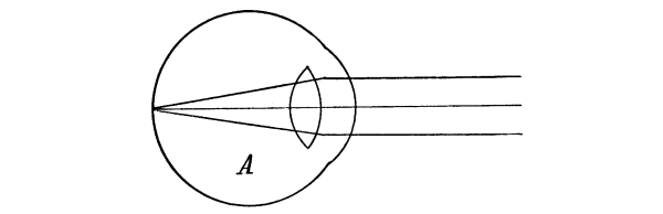
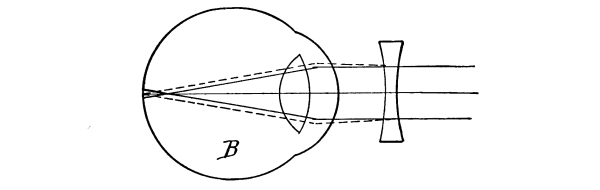
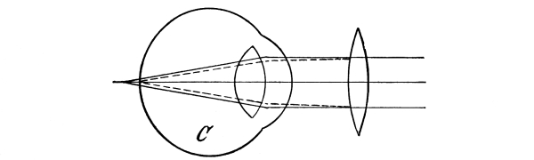
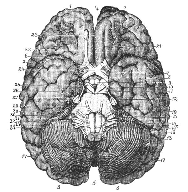
A HAND-BOOK OF ANATOMY AND PHYSIOLOGY FOR
NURSES AND OTHERS DESIRING A PRACTICAL
KNOWLEDGE OF THE SUBJECT
BY ANNETTE FISKE, A. M.
GRADUATE OF THE WALTHAM TRAINING SCHOOL FOR NURSES
ILLUSTRATED
PHILADELPHIA AND LONDON W. B. SAUNDERS COMPANY 1911
Copyright, 1911, by W. B. Saunders Company PRINTED IN AMERICA
PRESS OF W. B. SAUNDERS COMPANY PHILADELPHIA
TO MY FATHER
and
TO DR. ALFRED WORCESTER
as those who have perhaps most helped me in
the formation and realization of my ideals
this book is affectionately dedicated
PREFACE.
Although there are already in existence many books on anatomy and
physiology for nurses, none with which I am acquainted has seemed to me
to provide in concise form just the knowledge needed by the nurse in
her profession. Most of them, moreover, separate the anatomy from the
physiology and all treat the different systems of tissues separately,
first the bones, then the muscles, and so on. These defects, as they
seem to me, I have attempted to correct not only by weaving the
physiology in with the anatomy, but by treating first the general
structures found throughout the body and then describing the structure
and function of each part in detail. Thus, the first chapter is devoted
to a description of the general structure of all the tissues, a
separate chapter being devoted, however, to the skin, its appendages,
and function, including the sense of touch. Then the head with its
bones, muscles, and organs of special sense is described, while the
brain is treated with the rest of the nervous system, thus forming the
connecting link between the head and the body. In the same way the
back, chest, abdomen, pelvis, and extremities are taken up in turn and
the bones, muscles, blood-vessels, nerves, and special organs of each,
together with their functions, described.
Although written more particularly for nurses I am in hopes that
this book may prove useful to any others who may desire to acquire a
practical knowledge of anatomy and physiology.
Besides the usual text-books, I am much indebted for material to notes
taken in lecture courses given by Dr. Fred R. Jouett and Dr. F. J.
Goodridge of Cambridge, Mass., at the Cambridge School of Nursing, and
by Dr. Vivian Daniel of Watertown at the Waltham Training School for Nurses.
I wish particularly to express my gratitude and appreciation for the
kind and helpful criticism given me by Dr. Eugene A. Darling, Assistant
Professor of Physiology, Harvard College.
Chemical Constitution of the Body, 11—The Cell, 12—The Fundamental
Tissues of the Body, 13—Epithelial Tissue, 14—Connective Tissue,
15—Structure of Bone, 17—Bone Formation, 18—Chemical Composition of
Bone, 18—Classification and Function of Bones, 19—Joints, 20—Muscle,
21—Action of the Muscles, 23—Physiology of Muscle, 24—Cilia, 27—The
Blood, 27—Arteries, 27—Veins, 28—Capillaries, 28—Lymphatic System,
29—Lymphatic Vessels, 31—Lymphatic Glands, 32—Lymph, 32—Glands,
35—Ductless Glands, 36—Nervous Tissue, 36.
The Nose, 57—The Sense of Smell, 58—The Mouth, 59—The Hyoid Bone,
60—The Teeth, 60—The Sense of Taste, 61—Salivary Glands, 61—The
Tonsils, 62—The Ear, 63—Eustachian Tubes, 63—Sensation of Hearing,
65—The Eye, 66—Lachrymal Gland, 68—Coats of the Eye, 68—Light Rays and
Sight, 70—Accommodation, 72—Color Perception, 73.
The Heart, 101—The Pericardium, 101—Cavities of the Heart, 103—The
Endocardium, 103—The Valves of the Heart, 103—Circulation,
105—Circulation in the Fetus, 106—Arteries, 107—Veins, 109—Portal
Circulation, 109—Pulmonary Circulation, 110—Nerves of the Heart,
110—Heart Sounds, 111—The Heart Beat, 111—Factors Affecting
Circulation, 112—The Pulse, 113—Blood Pressure, 114—Nerve Supply
of the Blood-vessels, 115—The Blood, 116—Composition of the Blood,
116—Coagulability of the Blood, 117—Blood-corpuscles, 118.
The Larynx, 121—The Trachea, 123—The Thyroid Gland, 124—The Thymus
Gland, 124—The Bronchi, 125—The Lungs, 125—The Pleura, 125—The
Mediastinum, 126—Respiration, 127—Air, 129—Respiratory Sounds,
129—Changes in Air in Lungs, 129—Effect of Respiration on Blood,
130—Nervous Mechanism of Respiration, 130—Variations in Respiration, 131.
CHAPTER X.
The Abdomen and the Organs of Digestion and Excretion
The Shoulder Girdle, 171—The Clavicle, 171—The Scapula, 173—Shoulder
Muscles, 174—The Humerus, 175—Upper Arm Muscles, 176—The Ulna, 177—The
Radius, 178—The Wrist, 180—The Hand, 181—Meta-carpals, 181—Phalanges,
181—Muscles of the Forearm, 182—Muscles of the Hand, 184—Joints of the
Upper Extremity, 185—Blood Supply of the Upper Extremity, 185—Nerves of
the Upper Extremity, 186.
The Femur, 187—Thigh Muscles, 189—The Patella, 192—Joints of the
Lower Extremity, 192—The Tibia, 194—The Fibula, 194—The Ankle, 195—The
Foot, 195—Metatarsals, 195—Phalanges, 195—Muscles of the Leg, 196—The
Blood Supply of the Lower Extremity, 198—Nerves of the Lower Extremity, 199.
CHAPTER I. COMPOSITION AND GENERAL STRUCTURE OF THE BODY.
Anatomy is the study of the physical structure and physiology the study
of the normal functions of the human body.
Chemical Constitution of the Body.—In the body only twenty
elements have been found. These include carbon, oxygen, hydrogen,
nitrogen, sulphur, phosphorus, calcium, magnesium, manganese, chlorin,
potassium, and fluorin. For the most part they appear in very complex
and highly unstable combinations, though oxygen and nitrogen may be
said to exist uncombined in the blood, alimentary canal, and lungs.
Hydrogen also occurs in simple form in the alimentary canal, but as the
result of fermentation, not as an element of the body.
Of the organic compounds some contain nitrogen and some do not. The
most important of the former are the proteins, which are found only in
living bodies and consist of carbon, hydrogen, oxygen, nitrogen, and
sulphur combined in very similar proportions. The important proteins in
the body are the serum albumen and fibrin found in the blood, myosin in
muscle, globulin in the red blood-corpuscles, and casein in the milk.
Similar to the proteins but capable of passing through membranes are the
peptones, the final result of protein digestion, from which the
albuminoids differ in that they contain no sulphur. Ferments containing
nitrogen exist in all the cells of the body, though more particularly
in those of the digestive organs, and the coloring matters, as the
bilirubin of the bile, are nitrogenous.
The organic substances that do not contain nitrogen are the
carbohydrates or starches, the hydrocarbons or fats, and the acids, of
which the most important is carbon dioxide, given off by the lungs.
The inorganic substances are water, which forms a large percentage
of all the tissues and from one-fourth to one-third of the whole body
weight, sodium chloride or common salt, which plays an important part
in keeping substances in solution, potassium and magnesium chloride,
and hydrochloric acid, found in the stomach.
The Cell.—Although the body is a very complex organism,
the cell is its unit or foundation. In fact, the body begins life as a
single protoplasmic cell, the ovum, which is frequently compared
to the amœba, a microscopic animal consisting of a single cell of
protoplasm or living substance—a substance not well understood as
yet—but possessing practically all the functions of the human body.
For, although it has no organs and is homogeneous in structure, the
amœba can move by throwing out a process, and can surround and absorb
food, which it builds up into new tissue, discarding the waste. The
ovum, however, differs from the amœba in that it has a transparent
limiting membrane and contains a darker spot, the nucleus. This in
turn contains another smaller spot, the nucleolus, while through the
protoplasm, which is semi-fluid, extends a fine network that seems to
hold it in place.
The ovum is very small, about ¹/₁₂₅ inch in diameter,
and after fertilization grows by segmentation, the nucleus dividing in two
and the protoplasm grouping itself anew about the two nuclei. This division
continues, each cell dividing and forming two, or sometimes four, new
cells, all of which at first appear alike. By degrees, however,
differentiation takes place and different groups of cells assume
different characteristics. Thus the various tissues are gradually
developed, each with a structure and a function of its own, and are
distributed among the various organs, each organ consisting of several
tissues. During the process of growth and even after full growth of the
body is attained old cells are continually dying and being replaced by
new ones.
The typical cell is circular, but through being squeezed together
in the tissues or for some other reason the cells vary in shape in
different parts, being at times hexagonal, spindle-shaped, or columnar.
Yet, whatever their differences in shape or other characteristics, they
all live the same sort of life. All protoplasm absorbs oxygen when it
comes in contact with it and in the process of combining with it is in
part burned or oxidized, with the consequent setting free of heat and
other forms of energy and the formation of carbon dioxide. So long as
the body is alive, therefore, whether it is in a state of activity or
of rest, it is the seat of constant chemical change throughout all its
cells, and to these chemical changes are due all the forms of energy
manifested by the body. For energy is never destroyed, though it may
appear in a different form, and the elements of the human body are
so combined that their energy may be liberated and manifested in the
different functions the body exhibits.
The fundamental tissues of the body are the epithelial tissues,
the connective tissues, including the cartilaginous and bony tissues,
and the muscular and nervous tissues. Of these the epithelial tissues
serve as a protection to the surface of other tissues; the connective
tissues together form a framework for the support and general
protection of the other tissues; while energy is expended by muscular
and nervous tissue, the latter directing the former in its movements.
All the tissues are inter-dependent and the organs work together.
Besides cells every tissue contains a certain amount of lifeless matter,
the intercellular substance, which was at some time produced by the cells.
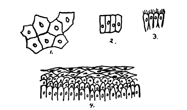
In epithelial tissue there is little intercellular substance,
the cells being close together and arranged generally as a skin or
membrane covering external or internal surfaces. When there are several
layers of cells, the deepest are columnar in shape and the others
become more and more flattened and scale-like as they approach the
surface, where they are gradually rubbed off and replaced by the growth
of new cells from below. This stratified epithelium, as it is called,
is found wherever a surface is exposed to friction, as in the skin and
in the mucous membrane of the mouth, pharynx, and esophagus, and in
that of the vagina and the neck of the uterus. In simple epithelium,
where there is only a single layer of cells, the cells may be pavement
or hexagonal, columnar, glandular, or ciliated, according to their
different functions. The flat pavement cells occur where a very smooth
surface is required, as in the heart, lungs, blood-vessels, serous
cavities, etc. None of these surfaces communicate directly with the
external surface of the body and the name endothelium is substituted
for epithelium. The columnar form of cell in the intestine facilitates
the passage of leucocytes between the cells. In glandular epithelium
the cells vary according to the gland in which they occur, their
protoplasm being filled with the material the gland secretes. Finally,
ciliated epithelium is composed of columnar cells with cilia or little
hair-like processes upon their free surface which serve to send
secreted fluids and other matters along the surfaces where they occur,
as in the air passages, parts of the generative organs, the ventricles
of the brain, and the central canal of the spinal cord.
Connective tissue has a great deal of intercellular substance.
One form, areolar tissue, is composed of a loose network of fine
white fibers with a few yellow elastic fibers interspersed and with
cells lying in the spaces between the fibers. It connects and surrounds
the different organs and parts, holding them together, yet allowing
free motion, and is one of the most extensively distributed of the
tissues. It is continuous throughout.
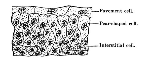
Fig. 2.—Section of bladder epithelium. (Hill.)
Closely allied to the areolar is the fibrous tissue,
in which the white fibers lie close together and run for the most part in one
direction only. This is found in ligaments, joints and tendons, as also
in such fibrous protective membranes as the periosteum, dura mater, the
fasciæ of muscles, etc. Fibrous tissue is silvery white in appearance
and is very strong and tough, yet pliant. It is not extensile.
Elastic tissue, on the other hand, has a large predominance of
yellow elastic fibers and is very extensile and elastic, though not so
strong as the fibrous. It is found in the walls of the blood-vessels,
especially the arteries, in the walls of the air tubes, in the
ligaments of the spine, etc.
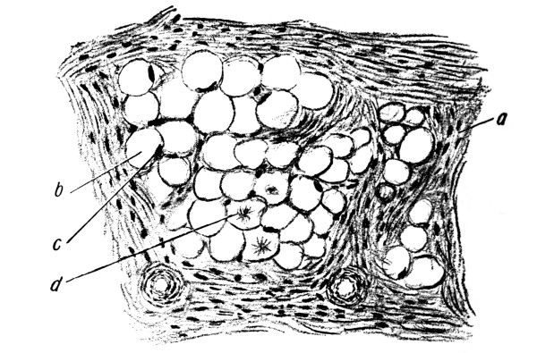
Fatty or adipose tissue is formed by the deposit of
fat in the cells of the areolar tissue and is found in most parts where the areolar
tissue occurs, though it varies largely in amount in different parts.
It is found pretty generally under the skin, fills in inequalities
about various organs and about the joints, and exists in large
quantities in the marrow of the long bones. In moderate amounts it
gives grace to the form and constitutes an important reserve fund.
Fig. 3.—Adipose tissue (Leroy): a,
Fibrous tissue; b, fat cells; c, nucleus of fat cells; d,
fatty acid crystals in fat cells.
Cartilage consists of groups of nucleated cells in intercellular
substance. It is very firm, yet highly elastic, and serves in the
joints to break the force of concussion of the harder and less elastic
bones. Except when it occurs at the end of a bone, it is covered with a
membrane called the perichondrium, which carries its blood supply. In
the nose, ear, larynx and trachea it serves to give shape, to keep the
passages open, and to afford attachment for muscles. Most of the skeleton
of the fetus consists of cartilage, which later develops into bone.
Bone.—In bone the intercellular tissue is rendered
hard by the deposit of mineral salts, the resulting material being of great
strength and rigidity. The texture may be close and dense like ivory or
open and spongy, the difference lying merely in the fact that the one
has fewer spaces between the solid particles than the other. There is
usually a hard, compact layer on the exterior of the bone, as that is
where the greatest cross-strain comes, especially in the long bones,
while within is the cancellous or spongy tissue, which gives lightness
to the bone and is capable of withstanding enormous pressure, though it
can bear little cross-strain.
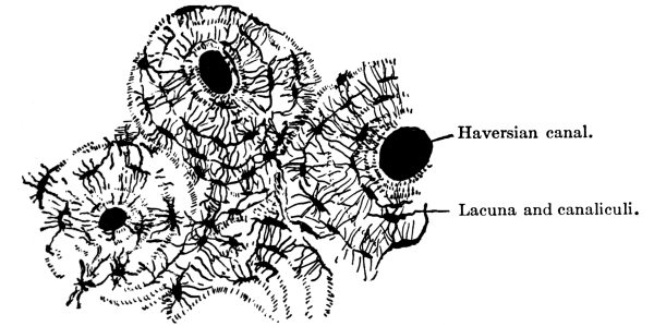
Fig. 4.—Cross-section
of compact bone tissue. (After Sharpey.)
Structure of Bone.—The hard substance in bone is always
arranged in lamellæ or bundles of bony fibers, which in cancellous
tissue meet to form a kind of lattice-work, while in the dense tissue
they are generally arranged in rings about the Haversian canals,
channels through which the blood-vessels pass through the bone
longitudinally. Between the lamellæ are spaces called lacunæ,
in which lie branched cells, the spaces being connected with each other
and with the Haversian canals by numerous tiny canals or canaliculi,
by which nutrient material finds its way from the Haversian canals to all
parts of the bone.
Within the bone is the medulla or marrow, which is of two varieties:
the yellow, which is largely fat and is found in the long bones of
adults, and the red, which is nearly three-fourths water and is found
in most of the other adult bones and in the bones of the fetus and of
the infant.
Lining the medullary and cancellous cavities is a delicate connective
tissue lining, the endosteum, which contains many bone-forming cells,
while on the outside of the bone, except at the articular ends, is the
periosteum with its outer protective layer and its inner vascular
layer containing osteoblasts or bone-forming cells. The periosteum is
essential for the growth of new bone where the old bone has died, and
if the periosteum is removed from healthy bone the part beneath is
liable to die, as it is by the constant growth of the osteoblasts that
the bone grows and is renewed. In the repair of broken bones tissue is
formed between and around the broken ends.
Bone Formation.—Most of the skull and face bones begin as
membranes of connective tissue, that is, are formed in membrane.
Bones are also formed in cartilage, the bone formation in this case
beginning from centers of ossification, where the deposit of lime
salts in the intercellular substance begins, the salts coming to the
centers dissolved in the plasma. Such a center of growth in a bone
is called the epiphysis and is separated from the main part of the
bone or diaphysis by cartilage until full growth is attained, when
ossification becomes complete. So in surgery, in working on the bones
of children, part of the epiphysis should always be left for the sake
of future growth. The outer shell of compact tissue is deposited by the
periosteum.
Chemical Composition of Bone.—Chemically bone is composed
of about one-third organic or animal matter, largely gelatine, and
two-thirds inorganic matter, including various salts of calcium,
magnesium, and sodium. In young children the animal matter predominates
and the bones are soft and often bend instead of breaking, only the
outside shell on one side giving way, as in “green-stick” fracture.
In rickets there is a deficiency of lime salts, but the increased
brittleness of the bones in old age is due, not to increase of mineral
matter, but to the less spongy texture of old bones.
Classification and Function of Bones.—There are in the body
some two hundred bones, which may be classified as long, short, flat,
and irregular. Occasionally an irregular bone develops in a fontanelle,
the membranous opening at the juncture of the sutures of the skull.
This is known as a Wormian bone. It is not, however, included in the
two hundred, as are not the sesamoid bones or bones developed in
tendons, with the exception of the patella or knee-cap.
Long bones are developed in cartilage and consist of a shaft, two
extremities, and various processes. They are more or less curved to
give them strength and grace. They serve as supports and act as levers
for purposes of motion and the exercise of power. Since a hollow
cylinder is just as strong as a solid one of the same size, the weight
coming only on the outer shell, the great bones which are accountable
for weight and which need to be light themselves have hollow shafts,
composed chiefly of compact tissue with a central medullary canal. The
ends, however, are expanded in order to make better connection at the
joints and to afford broad surfaces for muscular attachment, cancellous
tissue being used in them for lightness and strength. The large spongy
ends also give elasticity and lessen jar, and by bringing the tendons
to the bone at a greater angle increase their effectiveness. Blood is
brought to the long bones not only by the vessels of the periosteum but
by the medullary artery, which penetrates the compact tissue by the
nutrient foramen and divides into an ascending and a descending branch.
Short bones are spongy throughout. They are used for strength and where
little motion is required.
Flat bones are composed of two thin layers of compact tissue with a
varying amount of cancellous tissue between, and are for protection and
muscular attachment. The cancellous material between the two layers or
tablets of the skull is called the diploë.
Eminences and depressions occur on bones and when they are not
articular are for the attachment of ligaments and muscles. If they are
articular, they help to form joints.
As a whole the bony framework serves to keep the soft parts in place,
to support and protect them, and to aid in locomotion. The bones of the
head and trunk support and protect organs; those of the arms are for
tact and prehension; those of the lower extremities are for support and
locomotion.
Normally bones have little sensibility, but when inflamed they are
extremely sensitive and painful.
Joints.—The bones are connected with and move upon one another
by means of joints. These joints are of three kinds: 1. Immovable,
where the adjacent margins of the bones are closely applied, with
little fibrous tissue between, as in the sutures of the head; 2. those
with limited motion, which are very strong, the parts being connected
with tough fibro-cartilage; and 3. freely movable. In this last group
the articulating surfaces are covered with cartilage, which again is
lined with a delicate synovial membrane which secretes a small amount
of lubricating fluid, the synovial fluid, to reduce friction. Their
surfaces are also sometimes deepened by the presence of inter-articular
fibro-cartilages. Bursæ or sacs of synovial membrane occur outside the
joints under tendons and ligaments to reduce friction.
The nature and extent of the motion of a joint is defined and the bones
are held together by strong bands of fibrous tissue or ligaments, these
ligaments being more fully developed in joints where there is great
freedom of motion or where there is great weight to be supported. In a
ball-and-socket joint, such as the hip, there is a ligament in the form
of a strong capsule which surrounds the joint on all sides and limits
its motion, while hinge joints, like the elbow, and pivot joints, such
as that formed by the atlas on the axis, have lateral ligaments that
allow of freer motion. In the shoulder-joint, which is the most freely
movable joint in the body, the capsular ligament is very lax.
In general the kinds of motion possible in joints may be said to be
flexion, extension, abduction, adduction, circumduction, and rotation.
When much violence is applied to a joint and no dislocation results, as
in a sprain, there is often much stretching and even laceration of the ligaments.
Muscle.—The flesh, which forms a large proportion of the
weight of the body, consists of muscular tissue. Of this two kinds
are found: 1. The striated or striped muscle of animal life, which is
under the control of the will and so is known as voluntary muscle,
and 2. the unstriped or smooth muscle of organic life over which we have
no control, that is, the involuntary muscle. Each fiber of striped
muscle has an elastic, membranous sheath, the sarcolemma, and consists
of rod-shaped cells with a nucleus along the edge, set end to end and
having crosswise striations. In unstriated muscle the
fibers, which have no sarcolemma, consist of oval or spindle-shaped cells,
with a nucleus much smaller than that of striped muscle and situated in the
middle. In both kinds of muscle the fibers are bound together with
connective tissue and blood-vessels into fasciculi or bundles, and many
bundles go to make up a muscle. The muscle in turn has a connective
tissue envelope or sheath, the fascia. These fasciæ are found
throughout the body, the superficial ones being just beneath the skin,
while the deep ones not only form sheaths for the various muscles but
form partitions between them and serve to strengthen their attachments.
The striped muscles are those of motion, while the unstriped occur in
the hollow organs, surrounding the cavity and in some cases lessening
its capacity by their contraction.
An intermediate form of muscle known as cardiac muscle occurs in the
heart. Here the fibers have striations but the nucleus is generally in
the middle of the cell and the fibers branch and run together.
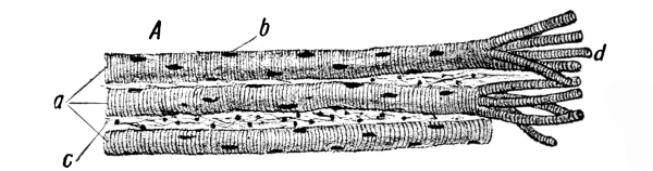
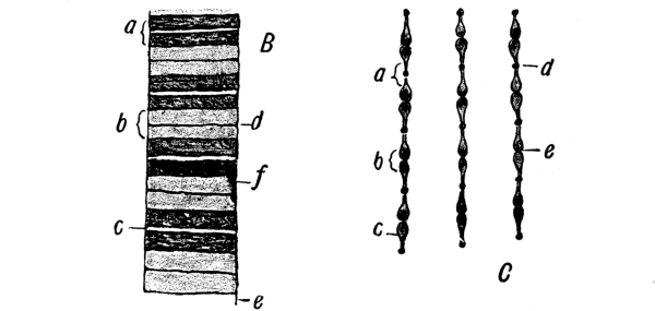
Fig. 5.—Voluntary muscle (Leroy). A,
Three voluntary fibers in long sections: a, three voluntary muscle
fibers; b, nuclei of same; c, fibrous tissue between the fibers
(endomysium); d, fibers separated into sarcostyles. B, Fiber
(diagrammatic): a, dark band; b, light band; c, median line of
Hensen; d, membrane of Krause; e, sarcolemma; f, nucleus. C:
a, Light band; b, dark band; c, contracting elements; d, row
of dots composing the membrane of Krause; e, slight narrowing of
contracting element aiding in production of median line of Hensen.
In life muscle appears more or less translucent and is contractile and
alkaline, but in death it loses its translucency and becomes rigid, at
the same time giving off in decomposition much carbon dioxide, so that
its reaction is acid. This phenomenon of the muscles becoming rigid in
death is called rigor mortis and occurs generally a few hours after
death, though it may come at once or be considerably delayed. It
may last anywhere from a few moments to several days but generally
lasts from twenty-four to thirty-six hours. It is probably due to the
formation in the muscle of myosin, a substance which probably comes
from myosinogen in the living muscle and which is closely akin to
the fibrin of blood. Probably the myosin or what precedes it causes
clotting of the muscle just as fibrin or what precedes it causes
clotting of the blood.
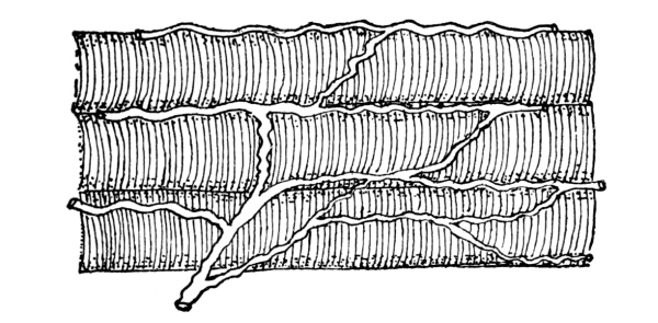
Fig. 6.—Three voluntary muscle
fibers from an injected muscle, showing network of blood capillaries. (Hill.)
The muscles vary in shape in different parts of the body, being long
and slender in the limbs and broad and flat in the trunk. They are
attached chiefly to bones but also to cartilages, ligaments, and skin,
either by means of tendons, which are cords or bands of white inelastic
fibrous tissue, or by means of aponeuroses, membranous expansions of
the same nature. Most voluntary muscles consist of a belly and two ends
or tendons. The origin is the fixed point from which it acts while the
movable point upon which it acts is known as its insertion.
Action of the Muscles.—When attached to bones, muscles are
distributed in three ways: 1. When it is necessary to produce much
motion rapidly, a short muscle is used. 2. When a part needs to be
moved far and much contraction on the part of the muscle is, therefore,
needed, the muscle is very long, as in the case of the sartorius
muscle, which shortens half its length. 3. Finally, where less distance
has to be covered but greater power is required, tendons are used, as
in this case the contraction is powerful but does not carry the part far.
In performing the mechanical work of the body the muscles are aided by
the fact that the bones, to which they are largely attached, are set
together loosely and form a set of levers, on which the muscles act
to perform certain definite acts. All three classes of levers occur:
1. where the fulcrum is between the weight and the power, as in the
case of the head, which is balanced by the muscles of the neck on the
vertebræ; 2. where the weight is between the fulcrum and the power, as
when a person raises himself upon his toes; and 3. where the power is
between the fulcrum and the weight, as when the biceps is used to raise
a weight held in the hand. The erect position of the body is difficult
to maintain because the center of gravity is high up, and it is by the
contraction of many muscles in the legs, thighs, back, abdomen, and
neck that the body is balanced upright upon the feet.
Physiology of Muscle.—Irritability or sensitiveness to
stimulation and contractility or the power to contract are the two
most important functions of muscle. Contraction occurs in response to
nervous energy brought by the nerves, a nerve filament going to each
muscle fiber, into which it plunges, its substance being lost and its
sheath becoming continuous with that of the muscle fiber. Any irritant,
as heat, electricity, etc., when applied to the nerve, causes the
muscle to contract. Moreover, muscle has an irritability of its own
and can contract independently of the nervous system. In contracting
it shortens and thickens, bringing the two ends closer together, and
becomes firm and rigid. The amount of contraction depends upon the
strength of the stimulus and the irritability of the muscle. The
minimal stimulus is the least stimulus that will cause a contraction
and the maximal is one that will cause the greatest contraction. The
work done depends in like manner upon the strength of the stimulus.
During contraction certain sounds are given off called muscle
sounds, which can be heard with the stethoscope but have no special
significance.
The muscles which have the greatest power of rapid contraction are
generally attached to levers. Indeed, striated muscle is characterized
by the rapidity and strength with which it works, though its rhythmic
motion is slight. Smooth muscle, on the other hand, is characterized
by its great force, considerable rhythm, considerable tone, and slight
rapidity, that is, its contraction is slower and lasts longer than that
of striated muscle. Cardiac muscle is characterized by great rhythm and
force, fair rapidity, and slight tonicity, tonicity being the amount of
tone or readiness to work. For even in sleep muscle is always in tone,
that is, ready to do its work. It is this that makes the difference in
appearance between a living and a dead person and enables one to spring
to his feet at night if he hears a noise, a thing he could not do if
his muscles were wholly relaxed. Thus, rapidity is the great function
of striated, tonicity of smooth, and rhythm of cardiac muscle. In
paralysis the muscles droop and lose their tone. Muscles are frequently
the seat of rheumatic disorders.
When set free, potential energy accomplishes work. In muscle there is
a good deal of potential energy, which is set free as heat and as work
accomplished. Even when the muscles are at rest, chemical changes are
going on and heat is being produced, though more heat is produced when
they are functioning. If the body depended upon its gross motions for
all its heat it would grow cold while a person rested. The respiratory
organs, however, and the heart are always working and chemical changes
are constantly taking place.
Ordinarily a muscle has some object in contracting, such as the raising
of a load, and it contracts voluntarily more or less according to the
weight of the load. The amount of work done is calculated in
foot-pounds or gram-meters, that is, the energy required to raise one
pound one foot or one gram one meter. As a rule the muscles with the
longest fibers, as the biceps, do the most work and those with a large
number of fibers do more than those with less. It has been calculated
that whereas an engine gives back one-twelfth of the energy of the coal
consumed, muscle liberates one-fourth of the energy brought to it in
the form of food. During activity the glycogen or sugar in the muscle
is used up and the muscle becomes more acid, owing to the lactic acid
that is formed. The carbon is taken in and carbon dioxide given off.
Nitrogen puts the muscle in condition to do its work but is not so
much used up in the work as is the carbohydrate material. So it is the
non-nitrogenous matter that does the work and any increase in urea, the
end-product of protein metabolism, is mere wear and tear.
Sudden heat or cold causes muscular contraction and moderate heat
favors both muscular and nervous irritability. Moderate cold, however,
lessens the force of contraction and below zero muscle very largely
loses its irritability without necessarily becoming rigid.
While well supplied with blood, muscle will contract without fatigue,
but if the blood supply is shut off, it soon loses its irritability
and becomes rigid. The more a muscle is used in moderation the more it
develops, but after it has done a certain amount of work it becomes
exhausted, losing its irritability or power to respond to stimuli and
later becoming rigid. Such fatigue is due to the production of certain
poisonous waste products which have a paralyzing effect on the nerves
and which are ordinarily gradually carried away in the blood, but which
sometimes, if produced to excess, accumulate too fast for the blood
wholly to remove them. Usually the nerve becomes exhausted first and
the muscle substance later. So long as it is connected with the nervous
system a muscle will respond to stimuli, but when the nerve becomes
tired, degeneration is more rapid. In fact, the degree of exhaustion is
determined by several factors, as by relation to the central nervous
system, variations in temperature, blood supply, and functional activity,
the process being more rapid in warm than in cold blooded animals.
Cilia.—A few motions are accomplished by tissue that is not
muscular, as in the case of the cilia attached to the cells of the
respiratory tract, which lie flat on the free surface and then lash
forward, serving in the air cells to keep the air in motion and in the
tubes to send secretions from below upward and outward and to keep out
foreign bodies. Cilia are also found in the female genital tract, where
they aid the passage of the ovum from the ovary to the womb. They act
together, though apparently not governed by the nervous system. As in
the white corpuscles of the blood, whose motion also is not muscular,
the changes that take place in ciliated epithelium are probably about
the same as those in muscular tissue, that is, contractile.
The Blood.—To most of the tissues just described nourishment
is brought in the blood, which circulates through the body in a system
of hollow tubes, the arteries and veins, whence it is distributed
through the agency of the lymphatic system. There are no blood-vessels,
however, in the epidermis, epithelium, nails, hair, teeth, nor in the
cornea of the eye. The vessels that carry the blood from the heart
are called arteries, those that return it veins. The former begin as
large vessels and gradually decrease in size; the latter begin as small
vessels and form larger and larger trunks as they approach the heart.
The arteries have three coats: 1. a thin, serous coat, the internal
or intima; 2. a middle or muscular coat, and 3. an external coat of
connective tissue. The middle coat is the thickest and is the one that
prevents the walls from collapsing when cut across. Except in the
cranium, each artery is enclosed in a sheath with its vein or veins,
the venæ comites. Usually the arteries occupy protected situations and
are straight in their course. Where a vessel has to accommodate itself
to the movements of a part, however, it may be curved, as in the case
of the facial artery which is curled on itself to allow for movements
of the jaw. They anastomose or communicate freely with one another,
thus promoting equality of distribution and pressure and making good
circulation possible even after the obliteration of a large vessel.
The veins have three coats like the arteries, but they are not
so thick and the muscular coat is not so highly developed, so that the
walls collapse when cut and have no elasticity. There are constrictions
on the surface of many of the veins due to the presence of valves.
These valves are formed of semilunar folds of the lining membrane
and are arranged in pairs. They serve to prevent the blood, whose
circulation in the veins is sluggish, from flowing back.
There are two sets of veins, the superficial and the deep, which
communicate with each other. In fact, all the veins, large and small,
anastomose very freely, especially in the skull and neck, where
obstruction would result in serious trouble, throughout the spinal
cord, and in the abdomen and pelvis. The deep veins accompany the
arteries in their sheath, while the superficial ones have thicker walls
and run between the layers of the superficial fascia under the skin,
terminating in the deep veins. In the skull the venous channels take
the form of sinuses, formed by a separating of the layers of the dura
mater, with an endothelial lining that is continuous with that of the
veins.
The capillaries are intermediate between the arteries
and the veins, the final division of the arteries and the first source of the veins.
They are tiny vessels with but a single coat, continuous with the
innermost coat of both arteries and veins and consisting practically of
one layer of cells with a small amount of connective tissue between.
They spread in a great network throughout the tissues, forming plexuses
and being especially abundant where the blood is needed for other
purposes than local nutrition, as in the secreting glands. Their
diameter is so small that the red corpuscles have to pass in single
file and may even then be squeezed out of shape. As they have no
muscular tissue in their walls, they have no power of contracting.
Their walls, however, like those of the smaller arteries and veins, are
porous and by virtue of this quality they play an important part in the
economy, since in them the exchange takes place between the tissues and
the blood.
The arteries in general carry freshly oxidized blood and the veins
blood from which the oxygen has been largely used up and which contains
waste material. In the pulmonary system, however, the case is reversed,
the pulmonary arteries conveying venous blood, as it is called, from
the heart to the lungs to be oxidized and the veins returning the blood
after it has received its new supply of oxygen.
The pumping of the blood through the arteries is assisted by the
contractions of the muscular coat, while the elastic tissue, of which
it contains a certain amount, gives elasticity to the walls and enables
them to stretch and so to accommodate the larger blood supply forced
into them at each beat by the heart. The walls of the veins have not
the power of contracting and the blood is pushed through more by
gravity and the action of the arteries than by any action of their own.
The walls of all the vessels are nourished by tiny blood-vessels in
the outer coat, known as vasa vasorum, and the nerves that regulate
the action of the arteries are the vasomotor nerves from the vasomotor
center in the medulla. Sufficient impulse goes from this center to the
blood-vessels all the time to keep them somewhat contracted, in a state
of tone, that is, which is increased or diminished as the blood supply
is to be diminished or increased.
Lymphatic System.—The lymphatic system also extends
throughout the body and consists of a system of channels, spaces, and glands
very closely related to the circulatory system and containing a fluid
called lymph. There are three principal parts to the system: 1. the
lymph spaces, which are open spaces, with no definite walls, in the
connective tissue framework of the body, more frequent near arteries
and veins and especially so among the capillaries; 2. the lymph
capillaries or small vessels which connect the lymph spaces; and 3. the
lymphatic vessels, of which there is a deep and a superficial set, the
latter accompanying the superficial veins on the surface of the body,
the former accompanying the deep blood-vessels.
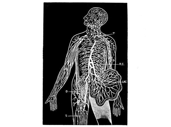
Fig. 7.—Diagram showing the course
of the main trunks of the absorbent system: the lymphatics of lower
extremities (D) meet the lacteals of the
intestines (LAC) at the receptaculum chyli
(R.C.), where the thoracic duct begins. The
superficial vessels are shown in the diagram on the right arm and leg
(S), and the deeper ones on the left arm
(D). The glands are here and there shown in
groups. The small right duct opens into the veins on the right side.
The thoracic duct opens into the union of the great veins of the left
side of the neck (T). (Yeo.)
The lymph spaces are generally small, though
there are some large serous cavities, such as the abdomen, that may be
considered as extended lymph spaces.
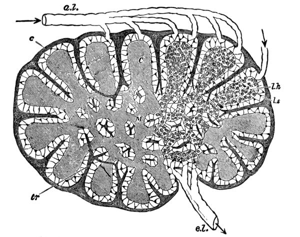
Fig. 8.—Diagram of a lymphatic gland,
showing afferent (a. l.) and efferent (e. l.) lymphatic vessels;
cortical substance (C); medullary substance (M); fibrous coat
(c); sending trabeculæ (tr) into the substance of the gland, where
they branch, and in the medullary part form a reticulum; the trabeculæ
are surrounded by the lymph path or sinus (l. s.), which separates
them from the adenoid tissue (l. h.). (Sharpey.)
The lymphatic vessels have delicate, transparent walls, with three
coats like the arteries, though much thinner, and anastomose even
more freely than the veins. They have a beaded appearance due to the
presence of numerous valves, which form constrictions on their surface.
The right lymphatic duct, which is only about an inch long, drains all
the lymphatics of the right half of the upper part of the trunk, the
head, and the neck approximately, while the thoracic duct drains those
of the rest of the body. The latter, which is the largest vessel of the
system, begins opposite the second lumbar vertebra with a bulb-like
reservoir for the lymph or chyle, the receptaculum chyli, and extends
up along the spinal column for a distance of about eighteen inches to
the seventh cervical vertebra, where, with the right lymphatic duct, it
empties into the left subclavian vein at its junction with the internal
jugular, thus establishing direct communication between the lymph
spaces and the venous system. The orifices of both vessels are guarded
by semilunar valves to prevent regurgitation of the blood.
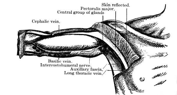
Fig. 9.—Central (superficial)
lymphatic glands of the axilla. (After Leaf.)
The lymphatic glands are small oval glandular bodies
and occur here and there along the course of the lymphatics. Before entering one of
them the vessel breaks up into several afferent vessels which form
a plexus within and then emerge again as several efferent vessels
which soon unite to form one trunk. These glands occur chiefly in the
mesentery, along the great vessels, and in the mediastinum, axilla,
neck, elbow, groin, and popliteal space.
The lymph varies in character with the locality, being a little
thicker and more opalescent in the lacteals, as the lymphatics of the
small intestine are called, especially during digestion, when fat is
present. Here it is called chyle. Otherwise it is generally a clear,
transparent and slightly opalescent fluid, which, owing to the presence
of fibrin, clots when drawn from the body and allowed to stand. In
fact, it resembles blood plasma very closely in composition and, as it
also contains a certain number of corpuscles or leucocytes that just
correspond to the white corpuscles of the blood, it is practically
blood without the red corpuscles. These leucocytes have considerable
power of amœboid movement and are thought by some to play an important
part in the absorption of food.
Owing to intracapillary pressure, the lymph transudes into the
lymph spaces and bathes the tissues, being carried away again by
the lymphatics. The amount of transudation is determined by the
blood pressure—the greater the pressure, the greater the amount of
transudation—and is increased by some organic action of the cells in
the walls of the vessels. In the process of transudation a certain
amount of solid matter goes through the wall of the vessel and it is
probable that certain protein elements can be carried thus from the
blood-vessels to the lymphatics, though they do not pass through the
capillary wall as readily as other substances. Some lymph is also
probably formed by the action of the tissues themselves, though the
process is not understood.
All muscular movements, active or passive, including the respiratory
movements, tend to drive the lymph on its way by pressure, the valves
of the vessels keeping it from flowing back. Moreover, its flow is
from the capillaries to the veins or from a region of high pressure to
one of less pressure. There is probably also some contraction in the
walls of the vessels themselves, and the continual formation of lymph
helps to drive it along. If an obstruction to the circulation occurs,
however, back-pressure results and causes too great transudation. In
that event a limb becomes swollen, pale, and generally cool. It pits on
pressure, the pressure driving the lymph out and there being no
circulation to bring it back. This condition is called œdema and occurs
in liver, kidney, and heart troubles, being generally first observed at
the ankles. In ascites, hydrothorax, hydrocephalus, and pericardial and
pleural effusions the fluid corresponds to lymph in its composition and
the large amount is due to excessive formation of the fluid, which is
normally present in small quantities.
Lymph gives the tissues substances from the blood that they need and
carries off those they do not, whether waste or substances of use to
other tissues. Because they thus absorb certain materials not needed
by the tissues and convey them to the circulation, the lymphatics have
also been called absorbents. Indeed, lymph may be spoken of as the
middleman between the blood and the tissues.
Another function of the lymph is to lubricate. Thus, the synovial fluid
of the joints is lymph and the pleuræ and the pericardium contain lymph
or serum to reduce the friction between the adjoining surfaces as much
as possible. The brain and spinal cord do not quite fill the cavities
of the cranium and the spinal column but float on a cushion of lymph,
the cerebro-spinal fluid. When the brain, which is subject to increase
and diminution in size, increases in size, it drives the lymph out, and
when it diminishes, the lymph returns.
The lymph glands serve as a protection to adjacent parts and when it
leaves the gland the lymph is purer and richer in leucocytes than
when it entered. In fact, they filter harmful matter from the lymph
and apparently also form white corpuscles. Normally they can with
difficulty be felt, but in disease, if the leucocytes are unable to
destroy or carry off the poison, the lymph carries it along to the
glands, which swell and become tender. If the infection is not severe
the swelling goes down and the tenderness passes after a short time,
but if it is severe, there may be suppuration and abscess formation and
the gland even perhaps be destroyed, giving its life for the health of
the part. Thus a wound in the foot, if infected, may cause irritation
and enlargement of the glands at the knee and in the groin.
The lymphatic glands are frequently the seat of tubercular infection,
especially in the neck, and are enlarged in scarlet fever, tonsillitis,
and diphtheria. In syphilis there is general glandular enlargement, and
the glands in the groin become enlarged in all diseases of the genital
organs. In malignant growths, such as cancer, the extension of the
disease is often along the lines of the lymphatics.
Glands.—Of glands in general a word might now be spoken. They
are of two kinds, excreting and secreting, and, when simple, are formed
by the folding in of a free surface, as in the case of the salivary,
gastric, and sebaceous glands, the cells at the gland becoming so
modified as to be able to perform the function of excreting or
secreting. In racemose glands the gland is broken up into many pockets.
Excreting glands take from an organ or from a part substances which
have outlived their usefulness and are to be cast out of the body,
while the secreting glands form from the blood substances that did not
exist in it before, but which are of use to the body, as the ptyalin
of the saliva. A strict line cannot, however, be drawn between the two
kinds of glands, most glands partaking more or less of both functions,
though the sebaceous and sweat glands are probably purely excreting
glands and the salivary glands are almost purely secreting. The glands,
moreover, are more or less interchangeable in their functions, that
is, they have vicarious function, and one gland can take up and do for
another what that other is for some reason unable to do. In jaundice,
where there is stoppage of the bile duct, the kidneys help out the
liver by excreting the bile. If one kidney is removed the
other does work for both, and the glands of the skin may help out the kidneys or
vice versa. Hemorrhage from the lungs sometimes occurs in suppression
of the menses.
In a general way the function of glands is chemical. They filter
out by osmosis, selecting the useful parts for secretion and the useless
for excretion. In the chemical action that goes on considerable energy
is given off, as is shown by the amount of pressure in the glands and
by the fact that their temperature is higher than that of the blood.
They all work in a reflex manner, being under the control of the
central nervous system. Thus, what is eaten affects the nerve terminals
in the mouth, the sensation passes to the nervous system, and an
impulse is carried by the motor nerves to the salivary glands.
Most of the glands have ducts to convey away their secretion to other
parts of the body or to send excretions out of the body, but there are
also ductless glands, which, though they seem to have some important
function in the process of metabolism, are not well understood. Most
of them seem to manufacture some substance that is absorbed by the
tissues and that plays an important part in the bodily metabolism,
though nothing is secreted by them externally. They are said to have
an internal secretion, whereas the glands with ducts have an external
secretion. The liver has both forms of secretion, the bile which is
sent out and the glycogen that is stored. The ductless glands are the
thymus and thyroid glands, the suprarenal capsules, and the pituitary
body in the brain.
Nervous Tissue.—Presiding over all the organs, muscles, and
blood-vessels, as the source of all action and all sensation, are the
nerves. Nervous tissue is of two kinds: 1. the gray or vesicular, which
originates impulses and receives impressions, and 2. the white or
fibrous, which conveys impressions. The gray matter
consists of large granular cells of protoplasm containing nuclei, which give
off many branches or dendrites. From the under surface there usually comes one
main branch, the axis-cylinder process. These processes sometimes
give off branches and sometimes not, but they form the nerve fibers and
carry impulses away from the nerve cells. The cells of the processes
are elongated in shape, have a nucleus, and are placed end to end, with
a definite constriction between them.
Each axis-cylinder process is surrounded by a sheath called the
medullary sheath, while each nerve fiber consists of
a central axis-cylinder process surrounded by the white substance of Schwann
and enclosed in a sheath. A bundle of these fibers invested in a
fibro-areolar membrane called the neurilemma constitutes
a nerve, and of these the white matter is formed. The blood supply is brought by
minute vessels, the vasa nervorum.
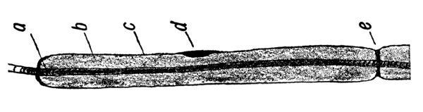
Fig. 10.—Longitudinal nerve fiber
(diagrammatic): a, Axis-cylinder; b, medullary sheath; c,
neurilemma; d, nucleus; e, node of Ranvier. (Leroy.)
The nerves of the cerebro-spinal system preside over animal life and
have to do with voluntary acts, while those from the sympathetic
system regulate organic life and are quite independent of the will.
Both sensory and motor nerves extend all over the body, accompanying
the arteries in a general way. The sensory nerves end on the surface
in plexuses, in end bulbs situated in the papillæ of the skin, or
in tactile corpuscles, these last occurring more especially where
there is no hair. The motor nerves end peripherally in plexuses or
by end plates. The central terminations of the motor nerves and the
terminations of sensory nerves in special organs, except where they end
in a cell, are not well understood.
Like muscles, nerves are probably never at rest, for through them the
muscles get their tone. When a nerve acts, no heat is produced and
there is no change in the nerve afterward, as there is in muscle.
Probably nerve impulse is the transmission of physical rather than
chemical changes along the fiber, the atoms of the nerve being set
in vibration and the vibrations being transmitted along its length.
Stimulation is produced by physical injury, by chemical influence, by
electricity, by heat, and the message is always referred to the nerve
termination. Thus, if the nerve at the elbow, over the “crazy bone,” is
touched, a tingling is felt in the fingers rather than at the point of
pressure. A person who has had an arm or leg amputated will frequently
speak of his fingers or toes on that side being cold, or complain of
pain in them, because the scar below the point of amputation tightens
around the nerves and pinches them.
It is through the nerves that people get in touch with the outer world
and that they judge of size, weight, etc. All careful adjustment of the
muscles is under the control of the nervous system.
CHAPTER II. THE SKIN, ITS APPENDAGES AND ITS FUNCTION.
The whole exterior surface of the body is covered by the skin, an
excreting and absorbing organ, which serves as a protection to the
parts beneath and is also the organ of touch. It has two layers,
a superficial and a deep. The superficial layer, the epidermis
or cuticle, is composed wholly of epithelial cells, of which the deepest
layer is columnar and moulded upon the papillary layer of the derma,
while the intermediate layers are more rounded and the surface ones
flat. The deepest layer also contains the skin pigment, which causes
the variation in shade between the Indian, the negro, and the white
man. Below the epidermis, which is chiefly protective, is the tough,
elastic, and flexible tissue of the derma or true skin, in which are
vested most of the activities of the skin. Its surface is covered
with papillæ, which are more numerous in the more sensitive parts.
Each papilla contains one or more capillary loops and one or more
nerve fibers, while some terminate in an oval body known as a tactile
corpuscle. Beneath the papillæ is the reticular layer, composed of
interlacing bands of fibrous tissue and containing blood-vessels,
lymphatics, and nerves, as well as unstriped muscle fibers where
hair is present.
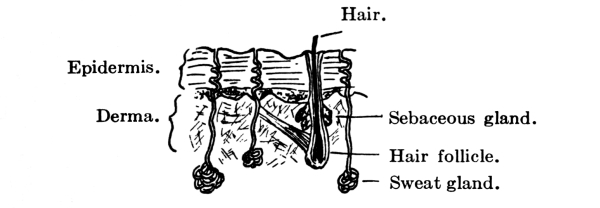
Fig. 11.—Vertical section of skin.
At the apertures of the body the skin stops and is replaced by mucous
membrane, an integument of greater delicacy but which consists
fundamentally of the same two layers, a superficial, bloodless
epithelium and a deep fibrous derma. It is continuous with the skin,
but is much redder and more sensitive and bleeds more easily. The
passages and cavities that it lines, unlike those lined by serous
membranes, communicate with the exterior of the body and are for that
reason protected against contact with foreign substances by mucus,
which is thicker and more sticky than the lymph that moistens the
endothelium found on serous surfaces. Mucous membrane is found in the
alimentary canal, the respiratory tract, and the genito-urinary tract.
In cavities, like the stomach and intestines, which are subject to
variations in capacity, it is thrown into folds or rugæ. The mucus is
secreted by small glands in the membrane.
Appendages of the Skin.—The skin has various appendages. On
the dorsal surface of the last phalanges of the fingers and toes are
flattened and horny modifications of epithelium, the nails. They have
a root embedded in a groove of skin by which they grow in length and a
vascular matrix of derma beneath them which gives growth in thickness.
To their growth in length there seems to be no limit.
The hairs also, which occur all over the body,
except on the palms of the hands and the soles of the feet, are a modification
of the epithelium. Each hair has a bulbous root springing from an involution
in the epidermis and derma called the hair follicle,
into which one or two sebaceous glands empty. It is raised
by involuntary muscle fibers and grows by constant additions to the surface by which
it is attached. This growth seems, however, to be limited, and when its term
is reached the hair falls out and is replaced by another. The horny
epithelial cells that go to form the hair contain the pigment that
gives it its color.
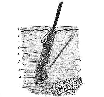
Fig. 12.—Skin and longitudinal section of
hair: a, Epidermis; b, corium; c, sebaceous gland; d, fibrous
root-sheath; e, glassy membrane; f, outer root-sheath; g, inner
root-sheath; h, expanded bulbous end of hair; i, papilla of hair;
j, arrector pili; k, adipose tissue. (Leroy)
Like the hairs, the sebaceous glands are situated
in all parts of the body except the palms of the hands and the soles of the feet.
They lie in the papillary layer and empty into the hair follicles, except
occasionally, when they empty directly upon the surface of the skin.
They secrete an oily substance, sebum, the débris resulting from the
degeneration of the epithelial cells of the gland itself, which serves
to keep the hair glossy and the skin soft and flexible.
The sweat glands, on the other hand, are more frequent
on the palms and soles and though sometimes found in the derma are usually situated
lower down in the subcutaneous cellular tissue. They are least numerous
on the back and neck. Coiled up in the lower layers of the skin, they
discharge the sweat through a spiral excretory duct upon its free surface.
The sweat is a clear, colorless, watery fluid with a salty taste,
an alkaline reaction, and a characteristic odor that varies with
the individual. If very scanty, it may be acid in reaction. Besides
water it contains a small percentage of solids, as inorganic salts,
especially sodium chloride, fatty acids, neutral fats, and at times,
especially in some diseases of the kidneys, urea, that is, the
end-products of the metabolism of starches and fats chiefly. There
is usually also some carbon dioxide, whence the expression cutaneous
respiration.
The sweat serves to keep the skin moist and in good condition, to
remove outworn and poisonous or irritating matters, and to regulate
the temperature. As a rule it evaporates upon reaching the surface, in
which case it is known as invisible or insensible perspiration, but if
conditions of the atmosphere are not favorable to prompt evaporation,
as when the air is damp, the skin becomes damp and there is visible
perspiration.
Though an abundant supply of blood increases the action of the sweat
glands, they are regulated by definite secretory nerves rather than
by the vasomotor nerves. In a cold sweat the action is probably due
to some disturbance of the nerve supply without increase of the blood
supply. Ordinarily perspiring is a reflex act due to the stimulation
of the afferent cutaneous nerves, as by the application of heat, but
sometimes, as in cases of strong emotions, involuntary impulses are
sent from the brain to the spinal centers and so arouse the action of
the glands. Atropin has the power of preventing the secretion of sweat
by paralyzing the terminations of the secretory nerves, while
pilocarpin produces an opposite effect in a similar way.
On account of these sweat glands the skin becomes next in importance
after the kidneys in the excretion of waste products. The quantity of
sweat excreted varies greatly and is hard to measure. It is influenced
by the temperature and humidity of the surrounding air, by the nature
and quantity of food and drink consumed, by the amount of exercise,
the relative activity of other organs, especially the kidneys,
and by certain mental conditions. The hotter it is, the greater the amount of
perspiration. In damp weather there may be less perspiration, but it
does not evaporate and is therefore more in evidence.
Ordinarily man has a temperature of 98.6°. The source of this body heat
or temperature is the general body metabolism, muscular activity, and
activity of the glands, especially of the liver, which is constantly
active, the blood in the hepatic vein being warmer than that in any
other part of the body. The tissue of the brain also is said to be
warmer than the surrounding blood, and the heart and respiratory
muscles, which are in constant activity, are responsible for much of
the body heat. The amount of heat generated in the body, therefore,
varies at different times, according as a person is awake or asleep,
quiet or active.
Temperature Regulation.—The temperature is regulated by
variations in the production and loss of heat, less being known of its
production than of its loss. It has been calculated that four-fifths of
the energy of the body is converted into heat, one-fifth into work. As
the minimum amount of heat produced in twenty-four hours is sufficient
to raise 10 gallons of water from 0° to boiling-point, it is evident
that if there were not some way for the escape of much of this heat the
body would become hotter and hotter and finally destroy itself. The
temperature, however, except on the surface, is uniform, heat being
lost as fast as it is produced. For, although oxidation at any point
raises the heat of the blood at the point, this heat is carried by the
blood to other parts, to which the surplus is given up, while blood
cooled in the skin goes to the hotter inward parts to cool them and be
warmed itself. In fact, heat is expended by conduction and radiation,
through respiration, perspiration, and heat given to the urine and
fæces. It is, therefore, largely, 75 to 80 per cent., carried off
through the skin and the lungs; 60 to 70 per cent. is lost by radiation
to the air and other bodies with which the body comes in contact;
20 to 30 per cent. is lost by the evaporation of sweat, 4 to 8 per
cent. by the warming of expired air, urine and feces, and 1 to 2 per
cent. by cold food that is taken in. Radiation acts more favorably
where the surroundings are cool and the air in motion, as on a breezy
day. Conduction is carried on best where the surrounding air is cool,
especially if it is moist, for moist air is a better conductor of heat
than dry air. Evaporation is very important in hot weather or where men
work in hot air.
Even in health the temperature may range from 98.6° to 99.5°, and a
degree or two below or above is not dangerous. When a person first
gets up in the morning his temperature is apt to be subnormal, but
after food and exercise have been taken it becomes normal and stays
so till the end of the day, when, if the person is tired, it may go
up a little. If a person is tired out, the temperature is apt to be
subnormal. There is also in the body what is called the vital tide,
which is highest afternoon and evening and lowest in the morning.
The rate of production of heat varies greatly in different people. One
person uses a certain amount of tissue more quickly than another, that
is, he lives faster. Moreover, size makes a difference in that a small
body has more surface to its weight than a large one and so has to
produce the same amount of heat at a faster rate in order to maintain
the right temperature. Taking food increases heat, probably because of
the muscular effort needed to eat it. Muscular work is another factor.
And finally the whole matter of heat production seems to be under
the control of the nervous system. Not much is known on this point
except that there is a heat center in the medulla which plays an
important part in heat production and whose influence is seen where
the temperature shoots way up in disease just before death. It is now
thought that fever is due to a disturbance of this nervous mechanism,
though just what the disturbance is is not known.
Fever is a condition of increased bodily temperature, due to
increased production or to decreased loss of heat. As a rule, in all
fevers the metabolic changes in the body are increased. Hence the
patient becomes emaciated in a long fever. The frequent increase in the
amount of urea during fever shows an increase in protein metabolism.
The temperature in fevers rises as high as 106° and in sunstroke
sometimes to 110°. Except in sunstroke a higher temperature than 106°
generally means death. Subnormal temperature is due to a decrease in
the bodily metabolism and so to lessened heat production. As a rule,
if the functions are all active, especially that of the sweat glands,
a person can be exposed to severe heat without the temperature being
affected, though sometimes on a hot summer day it may be up half to one
degree. The cause of heat-stroke with its high fever is unknown, but
probably it is due to some effect on the heat center in the brain. Heat
prostration is also due to prolonged exposure to heat, but is generally
accompanied by a subnormal temperature. The effect of cold, as in
freezing, is to diminish all the metabolic activities of the body. The
temperature can be artificially regulated more or less by variations of
food, varying amounts of exercise, by drugs, etc.
Sense of Touch.—Before passing on to a discussion of the
individual parts, a few words might well be said of the sense of touch,
since that is general and resides largely in the skin, whose other
functions have just been described. It may be regarded as the form from
which all the other special senses have developed, certain portions
of the body having become more sensitive than others to certain
vibrations, as the eye to those of light. The internal organs probably
have little sense of touch.
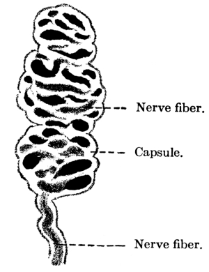
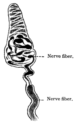
Figs. 13, 14.—Meissner’s corpuscle from man; ×750.
(Böhm, Davidoff, and Huber.)
Touch is useful only within arm’s reach but there gives one a sense of
space that sight does not give. It is practically determined by the
touch corpuscles, which are found in the skin over
almost the entire body, though they are more numerous in some places than in others,
the distribution of the corpuscles determining the sensitiveness of the
skin. These touch corpuscles are protoplasmic bodies containing nuclei,
about which are entwined filaments from the cutaneous nerves. Where the
corpuscles are absent the filaments of the cutaneous nerves themselves
play an important part. The finger tips have a very delicate sense of
touch and the tip of the tongue is the most sensitive part of the body.
Hence spaces in the mouth seem larger than elsewhere. By the
transmission of sensations of touch to the brain the sensation is
localized and the tactile sensation becomes a tactile perception.
There are three main divisions of the sense of touch: 1. sensations of
touch proper or tactile sensation; 2. sensations of temperature, and
3. sensations of pain. The temperature sense is the transmission by
the skin of sensations not so much of a certain degree of heat or cold
as of the difference between the temperature of an object and that of
the skin. The longer an object is in contact with the skin, the less
conscious the person is of it, not only because it becomes of the
same temperature, but also because he becomes accustomed to it. There
also seem to be in the skin, besides the touch corpuscles, two other
terminal organs with separate nerve fibers, the one for detecting heat,
the other cold; for there are places on the body where heat can be
detected and cold cannot, and vice versa.
Sensations of pain may be merely an exaggeration of tactile sensation,
as in too hard pressure or too great heat, but there seems to be also
a sensation of pain in the skin. All organs are said to have common
sensibility to pain and any exaggeration of this sensibility causes a
sensation of pain. All the special senses require a certain amount of
judgment in the interpretation of the sensations they convey.
CHAPTER III. THE CRANIUM AND FACE.
The intelligence and all the special senses, except the sense of touch
already spoken of, are gathered together compactly in the head, where
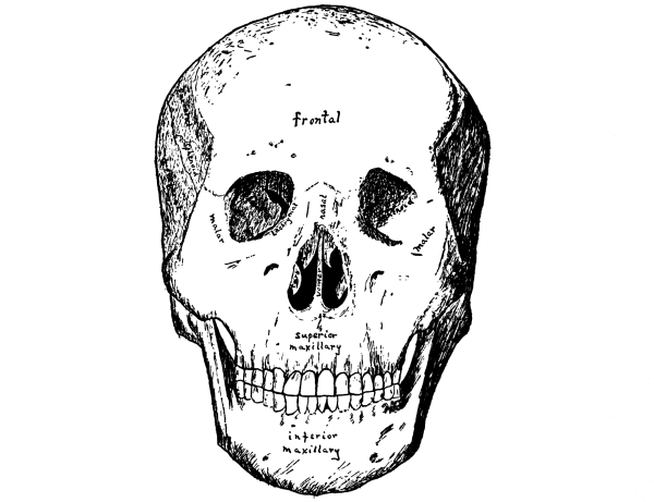
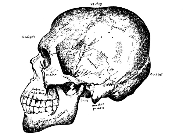
they are carefully protected with bony tissue. Covering the brain is
the skull or cranium, which is made up of eight bones, the frontal, the
occipital, two parietal, two temporal, the sphenoid, and the ethmoid,
while the bones of the face are fourteen in number, two nasal, two
superior maxillary, two lachrymal, two malar, two palate, two inferior
turbinated, the vomer, and the inferior maxillary. For the most part
the bones are arranged in pairs, one on either side.
The Cranial Bones.—The cranium or skull is especially adapted
for the protection of the brain and the bones are flat and closely
fitted to its surface. They have two layers of bone, the outer and the
inner tables, of which the outer is the thicker, and between these is
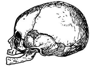
a tissue filled with blood-vessels, the diploë. In the infant, whose
brain has not yet attained its full size, opportunity must be left
for growth and the skull therefore consists of a number of bones with
interlocking notched edges, where growth takes place, but in the adult
it forms one solid covering of bone.
The line where the edges of two cranial bones come together is called
a suture. The suture between the frontal bone and the forward edges
of the two parietal bones is called the coronal suture,
that between the two parietal bones at the vertex of the skull is known as the
longitudinal or sagittal suture, and that between
the occipital bone and the back edges of the parietal bones as the lambdoidal suture.
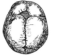
Where the coronal and sagittal sutures meet is a membranous interval
known as the anterior fontanelle, while the posterior fontanelle
is at the juncture of the sagittal with the lambdoidal suture. These
fontanelles—so called from the pulsations of the brain
that can be seen in them—close after birth either by the extension of the
surrounding bones or by the development in them of small bones known
as Wormian bones, the posterior one closing within a few months,
the anterior by the end of the second year. In rickets, however, the anterior
fontanelle remains open a long time, sometimes into the fourth year.
Fig. 15.—Cranium at birth,
showing sutures and fontanelles.
The frontal bone, as its name implies, forms the
fore part of the head or forehead. It joins the parietal bones above and the
temporal bones on either side. At the lower edge are the supra-orbital
arches, each with a supra-orbital notch or foramen on its inner margin
for the passage of the supra-orbital vessels and nerve, the nerve most
affected in neuralgia. Just above the arches on either side are the
superciliary ridges, behind which, between the two tables of the skull,
lie the frontal sinuses. On the inner surface the frontal sulcus for
the longitudinal sinus runs along the median line.
The parietal bones are the side bones of the skull. They meet
each other in the sagittal suture at the median line above and join
the frontal and occipital bones at either end, while below they touch
upon the temporal bones, the temporal muscles being attached in part
along their lower surface. These muscles are inserted into the coronoid
process of the lower jaw, which they thus help to raise and to retract.
Fig. 16.—Front view
of the skull. (After Sobotta.)
The occipital bone is at the base of the skull
and at birth consists of four pieces. In the lower, anterior part is the foramen
magnum, an oval opening through which the spinal cord passes from
the skull down into the spinal canal. Half way between the foramen
and the top of the bone is the external occipital protuberance for
the attachment of the ligamentum nuchæ which holds the head erect.
The inner side of the bone is deeply concave and is divided by a
cross-shaped grooved ridge into four fossæ, the internal occipital
protuberance being situated where the arms of the cross meet. The
occipital lobes of the cerebrum lie in the two upper fossæ and the
hemispheres of the cerebellum in the two lower ones. In the grooves
upon the ridge are the sinuses which collect the blood from the brain.
The occipital and frontal muscles, united by a thin aponeurosis, cover
the whole upper cranium and are known as the occipito-frontalis
muscle. At the back this is attached to the occipital bone, while
in front it interlaces with various face muscles. It is a powerful
muscle and raises the brows, wrinkles the forehead, and draws the scalp
forward. Long hair grows on the skin over it as a further protection
against blows upon the skull and sudden variations in temperature.
The temporal bones—said to be so named because the
hair over them is the first to turn with age—are situated at the sides and base
of the skull and are in three portions: the squamous or scale-like, the
mastoid or nipple-like, and the petrous or stony portion. The squamous
is the upper portion and has projecting from its lower part the long
arched zygomatic process, which articulates with the malar bone
of the face and from which arises the masseter muscle, one of the chief
muscles of mastication, which has its insertion in the ramus and angle
of the lower jaw. Just above the zygomatic process the temporal
muscle has its origin in part, while below is the glenoid fossa
for articulation with the condyle of the lower jaw, the posterior portion
of the fossa being occupied by part of the parotid gland.
The rough mastoid portion of the temporal bone is toward the back and
affords attachment to various muscles, of which the most important are
the occipito-frontalis and the sterno-cleido-mastoid. Within it are the
mastoid cells, which communicate with the inner ear and are lined with
mucous membrane continuous with that of the tympanum. They probably
have something to do with the hearing. In children they often become
the seat of inflammation (mastoid abscess) in infectious diseases and
the mastoid bone has to be cut to let out pus that has collected.
As the lateral sinus is directly behind the mastoid bone, there is
very great danger of going through into the sinus and causing a fatal
hemorrhage.
Fig. 17.—Side view of the skull.
(After Sobotta.)
The petrous portion, which contains the organ of hearing, is between
and somewhat behind the other two portions, at the lower edge of the
temporal bone, wedged between the sphenoid and the occipital bones. On
its outer surface is the external auditory meatus, and from below
projects a long sharp spine called the styloid process, to which
several minor muscles are attached. In the same angle between the
petrous and squamous portions lies the bony Eustachian tube.
The sphenoid or wedge bone, so called because in the
process of development it serves as a wedge, lies at the base of the
cranium, forming as it were the anterior part of the floor of the
cavity containing the brain. It is a large, bat-shaped bone and
articulates with all the cranial and many of the facial bones, binding
them all together. It has a body, two large wings, and two lesser wings
and, appears on the outside of the skull between the frontal and the
temporal bones behind the zygomatic process. In the adult the body of
the sphenoid is hollowed out into the sphenoid sinuses, in which pus
sometimes forms.
The Ethmoid Bone.—In front of and below the sphenoid and
extending forward to the frontal bone is the ethmoid, the last of the
cranial bones. It consists of a horizontal cribriform or sieve-like
plate, from either side of which depend lateral masses of ethmoid
cells. To the inner side of these masses are attached the thin curved
turbinated bones, superior and middle, while between them is a
vertical plate that forms the bony septum of the nose. Rising from
the upper surface of the cribriform plate is another vertical plate,
the crista galli, with the olfactory grooves on either side for
the reception of the olfactory bulbs, filaments of the olfactory nerve
passing down through the perforations of the cribriform plate to the
nose. For the brain, which fills almost the entire cavity of the
cranium, is supported by the sphenoid and ethmoid bones internally, as
it is protected externally by the other cranial bones.
Ossification of Sutures.—If premature ossification of all the
sutures occurs, idiocy results, while in cephalocele there
is a gap in the ossifying of the bones so that the membranes or brain protrude.
In rickets the forehead is high and square and the face bones
poorly developed, so that the head looks larger than it really is. In
Paget’s disease the bones enlarge and soften. This affects the head
but not the face and often the first thing noticed is that the hat is
too small. Craniotabes is thinning of the bone in places, the bone
becoming like parchment and being easily bent. It is generally caused
by pressure of the pillow or the nurse’s arm.
Bones of the Face.—The facial bones serve to form the various
features of the face, which after all are merely organs of special
sense. Many delicate muscles control the facial expression which,
consciously or unconsciously, reflects the character of their owner.
Surgically the most important of the facial bones are the two superior
maxillary bones, because of the number of diseases to which they
are liable. They meet in front, together forming the upper jaw, and
with the malar bone help form the lower part of the orbit of the eye.
They are cuboid in shape and are hollowed out into a pyramidal cavity
called the antrum of Highmore, which opens by a small orifice into
the middle nasal meatus and which sometimes becomes infected and has
to be tapped. The nasal process for articulation with the frontal and
nasal bones has, at its lower edge, a crest for the inferior turbinated
bone, and close beside this on the inside, extending down from the
upper edge, is a deep groove which, with the lachrymal and inferior
turbinated bones, helps to form the lachrymal canal for the nasal tear
duct. The bones give attachment to many small muscles, connected for
the most part with the nose and mouth, of which the masseter is the
only important one.
The two malar or cheek bones are small quadrangular
bones, which form the prominences of the cheeks and help form the orbits of the
eyes. Projecting backward from each is a zygomatic process for
articulation with the zygomatic process of the temporal bone, while a
maxillary process extends downward for articulation with the superior
maxillary. Here again the most important muscle attached is the
masseter. If the malar bone is crushed great deformity results.
The lachrymal bones are two small bones, about the size and shape
of a finger-nail, situated at the front of the inner wall of the orbit. At
the external edge is a groove which lodges the lachrymal sac above and
forms part of the lachrymal canal below.
The two palate bones are at the back of the nasal fossæ and help to
form the floor of the nose, the roof of the mouth, and the orbit. Each
has a vertical and a horizontal plate, and it is these last that by
their juncture form the hard palate. Oftentimes in cases of hare-lip
cleft palate also occurs, the result of incomplete development. To
remedy the consequent opening in the roof of the mouth, which makes
articulation difficult, operation is generally resorted to, though
sometimes a plate is fitted over the opening by a dentist.
The nasal bones are two small oblong bones which
articulate with the frontal and superior maxillary bones and with each other.
They form the bridge of the nose, the rest of the nose being wholly of cartilage,
except for the vomer, a bone shaped like a plough-share, which forms
part of the nasal septum, articulating along its anterior edge with the
ethmoid and the triangular cartilage.
The two inferior turbinated bones lie along the outer walls of the
nasal fossæ. They are thin scroll-like bones covered with mucous
membrane and serve to heat the air as it passes in. Sometimes when one
has a cold, the membrane and the bone too swell up and close the nares.
Loss of the sense of smell in a bad cold may be due to such swelling
and the consequent impeding of the entrance of odoriferous particles—a
condition that would likewise interfere with the sense of taste. Part
of the bone is sometimes removed, to enlarge the passage, enough being
left to warm the air.
Lastly, there is the inferior maxillary bone or
lower jaw. This has a horseshoe-shaped body and two rami, one at either
end. Each ramus has a pointed process in front called the coronoid process,
into which is inserted the temporal muscle. At the back, and separated
from the coronoid process by the sigmoid notch, is the condyle, which
articulates with the glenoid fossa on the temporal bone. The rami also
give attachment to the masseter muscle at its point of insertion. In
adult age the ramus is almost vertical but in old age the portion of
the jaw hollowed out into alveoli for the teeth becomes absorbed and
the angle of the jaw becomes very obtuse. On the inner side of the jaw
near the middle on either side is the fossa for the sublingual gland,
while the submaxillary gland lies in a fossa farther back on either
side.
Sometimes the lower jaw is dislocated and when once this has occurred
it is liable to occur again, the ligaments becoming stretched.
CHAPTER IV. THE ORGANS OF SPECIAL SENSE.
The Nose.—The nose, the organ of the sense of smell, is
composed of a framework of bones and cartilages, the bridge being
formed by the two nasal bones, and the septum by the vomer and the
triangular cartilage. It consists of two parts, the external nose
and the internal or nasal fossæ, which open to the face by the
anterior nares or nostrils and into the pharynx by the posterior nares.
Externally it is covered with skin, internally with ciliated mucous
membrane. The fossæ have the inferior turbinated bones along their
outer walls and are divided into three parts known as the superior,
the middle, and the inferior meatus, the middle one connecting with
the antrum of Highmore, while into the inferior meatus the lachrymal
canal empties. There are many small muscles of which little use is
made, although in forced respiration, as in pneumonia, where every aid
to breathing is called into play, even the alæ nasi or nostrils are made
to exert what muscular power they possess in order to supply more air.
Fig. 18.—The nasal cavity.
(After Sobotta.)
Not only is most of the air breathed in through the nose and warmed in
its passage through, but the nose is the organ of smell and by means
of the peculiar property of its nerves protects the lungs against
deleterious gases and helps the taste discriminate. The olfactory
or first cranial nerves, after emerging from the brain, lie on the
under surface of the frontal lobe and rest on the ethmoid bone in what
is known as the olfactory tract. Each nerve ends in a bulb-like
termination called an olfactory bulb, which rests on the cribriform
plate and sends little terminal fibers down through to be distributed
to the nasal cavities, especially to the upper half of the septum of
the nose, the roof of the nose, and the anterior and middle turbinated
bones. For in the mucous membrane of the upper nasal cavity are
specially modified epithelial cells called olfactory cells,
which play an important part in the conduction of smell. Hence when one
wishes to smell anything especially well he sniffs it up.
Probably the sensation of smell is caused by odoriferous particles
in the atmosphere being breathed into the nose, where they affect the
olfactory cells, which transmit the impulses to the olfactory nerve
and so to the brain. Whereas a certain amount of moisture in the nasal
cavity seems to be essential for accuracy of smell, the presence of
too much or too little interferes with it. The mucous membrane has a
certain power also of distinguishing different smells at the same time,
though this power varies greatly in different people, one smell often
wholly overpowering all others.
The cartilage below the bridge of the nose is sometimes attacked
in syphilis and cancer, and lupus often begins on the nose.
Deviation of the septum may occlude all air from one side of the
nose, an effect also produced by polypi, generally of the turbinated
bone. Either condition is easily remedied. Nosebleed, though
generally unimportant, may be serious in adults.
The Mouth.—The mouth is of great importance as an entrance
for fresh air to the lungs when the nasal passages are for any reason
impeded and as the resonant chamber from which proceeds the voice,
man’s chief means of communication with his fellows. Its chief value
may be said, however, to reside in the fact that it is the vestibule
of the alimentary canal. It is an ovoid cavity lined with mucous
membrane and is bounded in front by the lips, at the sides by the
cheeks, below by the floor and tongue, and above by the hard palate
anteriorly and by the soft palate posteriorly, the uvula depending
from the latter like a curtain between the mouth and the pharynx. Shape
is given to the mouth by the bones of the upper and lower jaw and its
size is altered by the lowering and raising of the latter, which is
quite freely movable.
Fig. 19.—The hyoid bone. (Toldt.)
At the back of the mouth, at the entrance to the pharynx, are the
anterior and posterior pillars of the fauces, which contain
muscular tissue, and between which on either side are thick masses
of lymphoid tissue, the tonsils. The floor of the mouth is formed
largely by the tongue, which completely fills the space within the
lower teeth. Its base or root is directed backward and downward and is
attached by muscles to the hyoid bone and the lower jaw, the hyoid
bone being a horseshoe-shaped bone lying just below and as it were
within the inferior maxillary. The base of the tongue is attached also
to the epiglottis and at the sides to the soft palate by the anterior
pillars. Except at its base and the posterior part of its under surface
the tongue is free, but a fold of mucous membrane, the frenum, holds
it somewhat in front. Thus it possesses great versatility of motion and
serves as an auxiliary in articulation, mastication, and deglutition.
The Teeth.—Securely embedded in either jaw are the teeth,
nature’s instrument for the first preparation of the food for digestion
through tearing and grinding. The incisors, which are in front, have
wide sharp edges for cutting the food. Next come the canine teeth
with a sharp point for tearing it, while at the back are the molars
with a broad flat top for grinding.
There are two sets of teeth: 1. the temporary or milk teeth,
twenty in number—four incisors, two canines, and four molars in
each jaw—which appear at from six months to two years, and 2. the
permanent teeth, thirty-two in number—four incisors, two canines,
known as eye teeth in the upper jaw and as stomach teeth in
the lower jaw, four bicuspids, so called because they have two cusps where
the molars have four or five, and six molars in each jaw—which come
from the sixth to the twenty-first years. The first to appear are the
two lower middle incisors, which come at the age of six months. The
last to appear are the wisdom teeth, the farthest back of the molars,
which come at the age of twenty-one years or thereabouts.
Each tooth consists of a crown or body above the gum, a neck, and a
fang or root within the gum. The body is of dentine or ivory with a
thin crust of enamel and contains the pulp, a vascular connective
tissue containing many nerves. Beginning at the neck and covering the
fang is a layer of cement or true bone.
The Sense of Taste.—The sense of taste lies chiefly in the
taste buds as they are called which are filled with gustatory
cells and are found in the papillæ of the tongue, principally in
the circumvallate papillæ at the back of the tongue, which are few
in number and arranged in a V-shape. There is also a certain power
of taste in the tip and sides of the tongue but little in the upper
surface or dorsum. Only five special tastes can be distinguished:
bitter, sweet, acid, sour, and salt, but sometimes more than one
can be distinguished at a time, as bitter and sweet. Every one can
distinguish between different tastes but the power varies in different
people and with different conditions. Certain tastes seem to be better
distinguished in certain places, as sweet at the tip and bitter at the
back of the tongue. Moreover, the sense of taste is very dependent
upon the sense of smell, especially in the case of aromatic and savory
substances, which one really does not taste but smell. If one held his
nose and closed his eyes he would not know from the taste whether he
was eating onion or apple. This leads to the habit of pinching the nose
when taking nauseous medicines.
To be tasted a substance must be in solution. Friction against the
tongue, lips or cheek increase the sense of taste. A temperature of
100° Fahrenheit favors taste, while both great heat and great cold
impair it.
There are probably at least two nerves of taste, the lingual branch
of the trifacial or fifth cranial and the gustatory branch of the
glosso-pharyngeal.
Along with the sense of taste there are other senses in the mouth which
play an important part, such as pressure and the sense of heat and
cold, and it is often hard to distinguish them from the pure sensation
of taste, which indeed is always accompanied by them.
Salivary Glands.—On either side of the mouth are three
racemose glands for the secretion of the saliva, which serves to
soften and lubricate the food and partially to digest starches by means of its
ferment, ptyalin. The parotid
gland is the largest and is below and in front of the ear, opening
by Stensen’s duct. The submaxillary gland is
below the jaw toward the back on either side and its duct is Wharton’s
duct. The sublingual gland lies beneath the
mucous membrane of the floor of the mouth and opens by eight to twenty
tiny ducts beside the frenum, the ducts of Rivinus. The activity of
the glands depends upon the blood supply; the more blood the greater
their activity.
Fig. 20.—Dissection of the side of the
face, showing the salivary glands: a, Sublingual gland; b, submaxillary
gland, with its duct opening on the floor of the mouth beneath the tongue at
d; c, parotid gland and its duct, which opens on the inner side
of the cheek. (After Yeo.)
The Tonsils.—The tonsils vary in size and in tonsillitis
swell and may even meet in the median line. They are frequently
removed. When they are enlarged one often gets a third tonsil or
adenoids, a lymphoid growth at the back of the pharynx which causes
mouth-breathing by day and snoring by night. A child with adenoids is
starved for air and what air is breathed in is not warmed. The growth
should be removed.
A short frenum produces tongue-tie, which may be remedied by
snipping. Cancer of the tongue is fairly common and necessitates a
radical operation. In mumps the parotid glands are inflamed and enlarged.
The Ear.—The special organ of hearing is the ear, to which
there are three parts, the external, the middle, and the internal ear.
The external ear consists of the pinna or expanded cartilaginous
portion, for the concentration and direction of sound waves, and the
external auditory canal, partly cartilage, partly bone, which is
directed forward, inward, and downward and conveys sound to the middle ear.
Fig. 21.—The small bones of
the ear; external view (enlarged). (After Gray.)
The middle ear or tympanum is an irregular cavity in the petrous
portion of the temporal bone. Its outer wall is formed by the membrana
tympani or drum, an oval translucent membrane placed obliquely at
the bottom of the external auditory canal. The middle ear communicates
with the inner ear through the fenestra ovalis or oval window and
contains the ossicles, the malleus or hammer,
the incus or anvil, and the
stapes or stirrup, which are arranged in a movable chain
from the drum to the oval window. The malleus, which is connected with
the membrana tympani, articulates by its head with the body of the
incus, while the stapes articulates with the incus by its head and is
connected by its base with the margin of the oval window. Connection
is made between the middle ear and the pharynx and the pressure of the
air upon the drum made equal on either side by means of the Eustachian
tubes. These tubes are about an inch and a half long, have cilia, and
convey wax and other matter from the ear to the pharynx. Occasionally
in a cold or for some other reason they become stopped up and trouble
results in the middle ear. Some of the mastoid cells also connect
with the middle ear and may become infected, causing mastoid disease.
Fig. 22.—Interior view of left bony
labyrinth after removal of the superior and external walls: 1, 2, 3, the
superior, posterior, and external or horizontal semicircular canals; 4,
fovea hemi-elliptica; 5, fovea hemispherica; 6, common opening of the
superior and posterior semicircular canals; 7, opening of the aqueduct
of the vestibule; 8, opening of the aqueduct of the cochlea; 9, the
scala vestibuli; 10, scala tympani; the lamina spiralis separating 9
and 10. (From Quain, after Sömmerring.)
The internal ear consists of various chambers hollowed out in
the petrous portion of the temporal bone. There is an osseous
labyrinth, consisting of a central cavity known as the vestibule,
three semicircular canals, and the cochlea
and within the osseous labyrinth, surrounded by perilymph, is the
membranous labyrinth, of like form, filled with the endolymph.
Communication exists externally with the middle ear by the round and
oval windows and internally with the internal
auditory canal, through which passes the eighth cranial or
auditory nerve, the special nerve of hearing, which is distributed to
the inner ear only. When the auditory nerve enters the ear through this
internal auditory meatus it divides into two branches,
of which one goes to the vestibule and the other to the organ of
Corti, a group of specially modified epithelial cells in the cochlea
of the membranous labyrinth, which is very important in transmitting
the impulses to the brain. The nerve also breaks up into very small
branches and is distributed practically throughout the wall of the labyrinth.
The sensation of hearing is the result of impulses transmitted to
the auditory nerve and so conveyed to the auditory center in the brain.
It is caused by sound waves which travel through the air from their
point of origin and enter the external ear. This collects and selects
the waves of sound and helps one to a certain extent to determine the
direction from which the sound comes. As they pass through the external
meatus the sound waves are collected into a comparatively small area
for transmission to the middle ear, where, by means of the drum, they
set in vibration the chain of ossicles. Through these the vibrations
are in turn transmitted to the oval window, being intensified in the
process. Here again they are taken up by the perilymph, from which they
pass through the wall of the membranous labyrinth to the endolymph,
affecting the epithelial lining of the labyrinth in such a way that the
impulses are transmitted to the auditory nerve, more particularly in
the vestibule, from which the vibrations enter the cochlea. They also
affect the cells of the organ of Corti in like manner as they pass from
the perilymph to the endolymph. The membrane that covers the fenestra
rotunda or round window relaxes and expands as the vibrations strike
it, thus serving to eliminate the shock of impact.
Musical sounds are caused by rhythmical or regularly repeated
vibrations, while irregular vibrations give rise to noises. In musical
sounds loudness is determined by the height or amplitude of the
vibrations, pitch by the length of the wave, and quality by the number
of so called partial tones. A sensation of sound cannot be produced by
less than 30 vibrations a second and the ordinary person cannot
hear more than 16,000 vibrations a second. Different sounds can
be distinguished when they follow each other as closely as by one
one-hundredth of a second.
All sound does not come through the canal of the ear. The bones of the
head vibrate and carry sound. So there are instruments for the deaf
which are put in the ear and others which are placed between the teeth.
The semicircular canals are not essential to
hearing but have something to do with a person’s power of maintaining
his equilibrium. Injury to them may cause dizziness and loss of equilibrium.
The Eye.—One more feature, perhaps the most expressive,
remains to be described, the eye. The senses are all modifications
of the original cutaneous sensibility and the nerve of sight is no
more sensitive to light than any other nerve. It therefore needs an
end organ that is sensitive to the motions of the ether in order to
give impressions of light. This organ is provided in the eye, which
is not only itself capable of being moved in every direction, but is
placed in the most movable part of the body, the head, which can be
turned in almost a complete circle. The eyeball is spherical and
lies in the cavity of the orbit upon a cushion of fat, where it has a
large range of sight but is securely protected from injury by its bony
surroundings. The sunken eyes following protracted illness are due to
the using by the system of the fat on which the eyeball ordinarily rests.
Each orbital cavity is formed by the juncture of some seven bones and
communicates with the cavity of the brain through the optic foramen
and through the sphenoidal fissure. Above the orbits are arched
eminences of skin, the eye-brows, from which several rows of short
hairs grow longitudinally and which serve to protect the eyes and to
limit the amount of light to a certain extent, as in frowning.
Still further protection is afforded by the eyelids, longitudinal
folds of skin, the one above, the other below, which close like
curtains over the eye. Beneath the external layer of skin in the lids
is fatty tissue and then the orbicularis palpebrarum muscle by means
of which they are closed. They are kept in shape by the tarsal plates
or cartilages, in whose ocular surface are embedded the Meibomian
glands, whose secretion prevents the free edges of the lids from
sticking together. Along these edges grows a double or triple row
of stiff hairs, the eye-lashes, which curve outward so as not to
interfere with each other and also to prevent the entrance into the eye
of foreign bodies. Lining the inner surface of the lids and reflected
thence over the anterior surface of the sclerotic coat of the eye is
a mucous membrane, the conjunctiva, which is thick, opaque, and
vascular on the lids but thin and transparent on the eyeball. The angles
between the lids are known as the internal and the external canthus.
Fig. 23.—The external ocular muscles. (Pyle.)
Muscles and Nerves.—The eyeball is held in position by the
ocular muscles, the conjunctiva, and the lids, while surrounding it,
yet allowing free movement, is a thin membranous sac, the tunica
vaginalis oculi. The superior and inferior recti
muscles at the upper and lower edges of the ball turn the eye up
and down; the internal and external recti at the inner and outer edges
turn the eye inward and outward; and the superior and
inferior oblique rotate the eye. The nerves supplying these muscles
are the third or motor oculi, the fourth and the sixth.
The lachrymal gland, which is about the size and
shape of an almond, is situated at the upper and outer part of the orbit. It secretes
a fluid which keeps the anterior surface of the eye bathed in moisture
and is ordinarily drained away through the lachrymal sac in the inner
canthus, whence it passes by the lachrymal ducts into the nose. When
the amount secreted is excessive, it overflows the lower lid as tears.
Fig. 24.—Diagram of
the lacrimal apparatus. (Pyle.)
Coats of Eye.—The membranes or coats of the eye are three in number:
an outer or sclerotic, a middle or vascular, and an inner or sensitive.
The sclerotic coat is a rather thick, fibrous, protective membrane.
Where it passes in front of the iris, however, it is thinner and
transparent and is known as the cornea. The cornea
projects somewhat and, as it were, resembles a segment of a smaller sphere set
into the rest of the sclerotic.
The middle or vascular coat, known as the choroid,
carries blood-vessels for the retina or sensitive coat in its
inner layer and has an outer layer of pigment cells that excludes light and darkens
the inner chamber of the eye. The folds of the choroid at its anterior
margin contain the ciliary muscles and are known as the ciliary
processes, while the name iris is given to the little
round pigmented, perforated, curtain-like muscle just in front of the
crystalline lens. The posterior surface of the iris is covered with a
thick layer of pigment cells to prevent the entrance of light except
through the central opening or pupil, and its anterior surface also
has pigment cells that give it its color, though the difference in the
color of people’s eyes is due rather to the amount of pigment present
than to its color, a small amount of pigment being present in blue eyes
and a large amount in brown and black eyes. Variations in the size
of the pupil are brought about by contractions of the circular and
radiating fibers of the iris, contraction of the circular fibers making
it smaller and those of the radiating larger. The pupil is constricted
for near objects and during sleep, and is dilated for distant objects.
In a dull light also it dilates to let in more light, and in a bright
light it contracts. The appearance of the pupil is often important as a
means of diagnosis and in etherization.
Fig. 25.—Vertical section
through the eyeball and eyelids. (Pyle.)
Lastly there is the innermost sensitive coat or retina, which
has eight layers, the outer one containing some pigment cells and the next
the rods and cones, in which the power of perception is supposed to
lie, branches of the optic nerve being distributed over it in all
directions. In fact, the retina is formed by a membranous expansion of
the optic or second cranial nerve, the special nerve of sight, which
passes into the orbit through the optic foramen at the back and enters
the eyeball close to the macula lutea or yellow spot. The exact
spot where the optic nerve enters the retina is not sensitive
and is known as the blind spot. In the center of the macula lutea, however,
which is in the middle of the retina posteriorly, is a tiny pit, the
fovea centralis, in which all the layers of the retina except the
rods and cones are absent, and at this point vision is most perfect.
It is, therefore, always turned toward the object looked at, and when
one wishes to see an object distinctly, he must keep moving his eyes
over it that the rays from each part may fall in turn upon the fovea
centralis.
Directly behind the pupil is the crystalline lens,
a rather firm gelatinous body enclosed in a capsule, which is transparent in life
but opaque in death. The lens is doubly convex and is held in place
by the suspensory ligaments, which arise from the ciliary processes.
In front of it is the anterior chamber of the eye, filled with a thin
watery fluid called the aqueous humor, while
the larger space back of it, occupying about four-fifths of the entire
globe, is filled with a jelly-like substance known as the vitreous humor.
The chief artery of the eye is the ophthalmic.
Light Rays.—The eye is practically a camera and its principal
function is to reflect images. Although there are several refracting
surfaces and media, for practical purposes the cornea alone need be
considered. Except for those rays which enter the eye perpendicularly
to the cornea, whose line of entrance is called the optic axis, all
rays are refracted when they enter the eye and the point at which they
meet and cross each other behind the cornea is called the principal
focus of the eye. To focus properly, all the rays from any one point
on an object must meet again in a common point upon the retina, their
conjugate focus. In the normal eye all the rays from an object are
focused on the retina and form upon it an image of the object which, as
in the camera, is inverted, because of the crossing of the rays behind
the cornea. Once focused on the retina the light traverses the various
layers to the layer of rods and cones, where chemical action takes
place and affects the little filaments of the optic nerve, by which the
message is carried to the brain.
Fig. 26.—Diagram showing the difference
between (A) emmetropic, (B) myopic and (C) hypermetropic eyes.
(American Text-book of Physiology.)
When the eye is at rest the pupil and lens are in their normal
condition and at such times the eye sees only distant objects. The
ability of the eye to focus upon objects at different distances is
called accommodation and to accomplish it three things are necessary:
1. change in the shape of the lens; 2. convergence of the axes of the
eyes, and 3. narrowing of the pupils.
When the eye is directed toward distant objects, the muscle fibers
in the ciliary processes relax, causing tightening of the suspensory
ligaments and consequent flattening of the surface of the lens.
Otherwise an image would be formed in front of the retina; for the
greater the convexity of the lens, the greater the angle of refraction.
Such accommodation is passive and so not fatiguing. To look at nearby
objects, on the contrary, the ciliary muscles contract, drawing the
choroid forward and allowing the suspensory ligaments to relax, so that
the lens bulges in front. This is an exertion.
In order to accommodate properly, moreover, both eyes must work
together and the axes of both eyes must be directed toward the object.
Therefore, in looking at nearby objects the axes of the eyes converge,
drawn by the internal recti muscles. In strabismus or cross eye,
where the axes of both eyes cannot be directed toward the object at the
same time, the rays fall upon one part of one eye and upon a different
part of the other eye and two separate images are seen.
Finally there is concentric narrowing of the pupil by contraction of
the circular fibers of the iris, by which means various side rays that
would come to a focus outside the retina are excluded.
All the muscles of accommodation, the ciliary muscles, the internal
recti, and the sphincter pupillæ, are under the control of the third nerve.
Connected with this power of accommodation and dependent on it are the
two conditions of near-sightedness or myopia
and far-sightedness or hypermetropia.
The normal eye is emmetropic and is almost perfectly spherical, but in
the near-sighted or myopic eye the ball, instead of being round, is
flattened from above down and so bulges in front. Consequently, owing
to the greater distance from the lens to the retina, images are formed
in front of the retina. Only nearby objects can be seen clearly,
because the farther the object from the eye the farther in front of
the retina the image is formed. Concave glasses are worn to enable
near-sighted people to see at a distance. Hypermetropic or far-sighted
eyes are flattened from before backward and can see only objects at a
distance clearly, as those nearby form images behind the retina. For
such eyes convex glasses are worn.
As the ordinary person approaches middle life, he becomes able to see
better at a distance than near to. This presbyopia, as it is called,
which is practically far-sightedness, is due to a partial loss of the
power of accommodation in the lens, the result of a general loss of
elasticity in the parts.
Another very common defect is astigmatism, a failure of the rays
to focus upon a point, owing generally to a flattening in the surface of
the cornea.
Color perception is also an important function of the eye. The
waves of hyperluminous ether when of a certain rate of vibration give the
sensation of heat and when their vibrations are more rapid they give
the sensation of light. Each of the primary colors of the spectrum
gives off a pretty definite number of light rays which travel through
the air and enter the eye, the number of rays determining the color
thrown upon the retina and the velocity determining the intensity of
the color. Occasionally when light is passing through into the eye it
is broken up as in a prism and the person gets a sensation as of all
sorts of colors, chromatic aberration. Total or partial absence of
sensitiveness to color is called color blindness. It is commonest
in the form of inability to distinguish between red and green and is
probably due to a defect in the retina.
Sometimes a hair follicle on the lid becomes infected and a sty
is formed. Pink eye is conjunctivitis or inflammation of the
conjunctiva. A Meibomian duct may become stopped and cause bulging, or
there may be a sagging down or ptosis of the upper lid in certain
diseases, as meningitis, apoplexy, and more especially syphilis.
Rodent ulcer often begins by the eye or on the cheek.
CHAPTER V. THE NERVOUS SYSTEM.
The nervous system, which regulates all the vital processes of the
body, physical and chemical, and which is situated partly in the head
and partly in the trunk, may well form the connecting link between the
description of the head and that of the trunk. It has two divisions,
the cerebro-spinal system and the sympathetic system. The former
consists of the cerebrum or brain proper, the cerebellum or little
brain, the pons Varolii, the medulla oblongata, the spinal cord, and
the cranial and spinal nerves; the latter of a series of ganglia or
aggregations of nerve centers. The brain, which includes the cerebrum,
cerebellum, pons, and medulla, occupies the cranium and the spinal cord
is contained within the bony framework of the spinal column. In the
male the brain weighs about 49 ounces and in the female 44, while in an
idiot it seldom weighs more than 23 ounces.
The cerebrum or brain proper has two parts or
hemispheres, roughly oval in shape, each of which has five lobes separated by
fissures, the frontal, parietal, occipital, and temporo-sphenoidal
lobes, and the central lobe or island of Reil at the base of the
brain. The chief fissures are the longitudinal fissure,
the fissure of Sylvius at the base of the brain, and the fissure of Rolando
between the frontal and parietal lobes. There are also five serous
cavities called ventricles, the two lateral and the third, fourth,
and fifth ventricles, of which the first two, one in either hemisphere,
are the most important. Around these cavities is the brain substance,
which is made up of two tissues, the white and the gray, the latter
forming the outer part of the brain to the depth of perhaps half an
inch, and the white matter forming the rest. The outer or gray part is
called the cortex and is largely made up of nerve cells. It might be
called the active part of the brain. The white part consists largely of
nerve fibers which are given off from the nerve cells and are carried
down into the spinal cord.
The surface of the brain is convoluted, the ridges being separated by
deep furrows or sulci, by which means a great extent of gray matter
is secured. The furrows contain fluid from the subarachnoid spaces
and vary in number and depth according to intelligence. While the
convolutions are not uniform in all brains, the principal ones are
constant.
Both the brain and the spinal cord are covered by three membranes, the
dura mater, the arachnoid, and the pia mater. The dura mater is dense
and fibrous and lines the interior of the skull, being firmly adherent
to it at many points. In fact, it constitutes the internal periosteum
of the cranial bones. The arachnoid is a delicate serous membrane,
with two layers, lubricated to prevent friction, which divides the
space between the dura mater and the pia mater, bridging over the
convolutions and enclosing the subdural and subarachnoid spaces which
are connected with lymphatics and contain a serous secretion, the
cerebro-spinal fluid. This fluid forms an elastic water cushion, on
which the brain rests, and prevents concussion. The pia mater is
vascular, containing blood-vessels, lymphatics, and nerves, and is closely
attached to the surface of the brain, dipping down into all the sulci.
At the base or under surface of the brain are some very important
structures. The olfactory bulbs lie beneath the frontal
lobe and projecting back is the olfactory tract, through which the olfactory
nerves come from the brain. Back of the olfactory tract is the optic
commissure where the optic nerves coming from the brain cross each
other. And back of the commissure again is the optic tract, where the
optic nerves emerge from the brain. At the base of the brain are also
the exits of the twelve cranial nerves.
Upon entering the brain the arteries run a tortuous course, the
tortuosity breaking the force of the blood stream in the small vessels
where congestion would be with difficulty relieved. The basilar artery,
which is formed by the juncture of the two vertebrals, divides into the
two posterior cerebrals, each of which joins one of the anterior
cerebrals by a posterior communicating artery. The two anterior
cerebrals also are joined by an anterior communicating artery, thus
completing the circle. The circle thus formed at the base of the brain
is called the circle of Willis and provides for a good supply of
blood in event of an accident to any vessel. The blood is returned to
the general circulation through the cerebral veins and sinuses formed
by the separation of the dura mater into two layers.
The cerebellum is about one-seventh the size of
the cerebrum and weighs about 5 ounces. It lies in the lower occipital fossæ of
the skull and is oblong in shape and divided into two lateral hemispheres
by a transverse fissure. It is made up of both white and gray matter,
of which the former predominates, the gray being external as in the
cerebrum. The cells are about the same as in the cortex and its surface
is traversed by queer furrows. Of its function little is known but it
probably plays a most important part in the coördination of the nervous
and muscular acts by which the movements of the body are carried on.
At the back of the cerebrum and below the cerebellum is the pons
Varolii, which forms a connecting link with the medulla oblongata
or bulging part of the cord. It is made up essentially of white matter
or nerve fibers, though there is a small amount of gray matter in which
are found the nuclei of some of the cranial nerves.
In the medulla oblongata, which is about 1 inch
long and extends from the pons Varolii to the upper border of the atlas or
first cervical vertebra, the gray matter is not necessarily external
to the white but is found in patches in the white. The gray matter
here corresponds more or less to that of the spinal cord and the white
matter is continuous with that of the cord. From the medulla arise the
fifth to twelfth cranial nerves and the vasomotor nerves. The cardiac
nerve has its center here and here too are the centers of respiration,
phonation, deglutition, mastication, and expression. In the medulla the
nerves that arise in the cerebrum cross over from one side of the body
to the other on the crossed pyramidal tracts. The importance of this
crossing of the nerve fibers is seen in apoplexy, when a blood-vessel
is ruptured in the brain and hemorrhage causes pressure, generally on
the motor tract. Paralysis of the nerves and of the muscles to which
they go results. The paralysis is generally of one side of the body,
the opposite side from that on which the injury occurred. The seat
of injury in the brain or cord can frequently be determined by the
situation and extent of the paralysis.
Spinal Cord.—Extending down from the medulla through
the spinal column is the cord. Its length from the foramen magnum, where
it begins, down through the vertebræ to the lower border of the first
lumbar vertebra, where it ends in a very fine thread-like process with
no special function, called the filum terminale, is 17 to 18 inches.
Just before it ends a number of nerves are given off in a tail-like
expansion known as the cauda equina or horse’s tail. It is not
uniform throughout its length but presents two enlargements, a cervical
enlargement in the lower cervical region, and a lumbar enlargement in
the lower dorsal region, where the nerves are given off to the arms and
legs respectively. The membranes are the same as those of the brain
and are continuous with them, but here the dura mater is not attached
to the bony walls enclosing it. For the cord does not fit closely
into the canal but is as it were suspended in it. The subarachnoid
space communicates with the ventricles of the brain by the foramen of
Majendie and is filled with cerebro-spinal fluid for the protection
of the cord. In cerebro-spinal meningitis or spotted fever this fluid
is infected and for diagnosis lumbar puncture is performed.
Fig. 28.—Different views of a portion
of the spinal cord from the cervical region, with the roots of the nerves. In
A the anterior surface of the specimen is shown, the anterior nerve
root of its right side being divided; in B a view of the right side
is given; in C the upper surface is shown; in D the nerve roots
and ganglion are shown from below: 1, the anterior median fissure; 2,
posterior median fissure; 3, anterior lateral depression, over which
the anterior nerve roots are seen to spread; 4, posterior lateral
groove, into which the posterior roots are seen to sink; 5, anterior
roots passing the ganglion; 5´, in A, the anterior root divided; 6,
the posterior roots, the fibers of which pass into the ganglion, 6; 7,
the united or compound nerve; 7´, the posterior primary branch seen in
A and D to be derived in part from the anterior and in part from
the posterior root. (Allen Thomson.)
If a cross-section of the cord is made, it is found to have a pretty
definite structure. It is roughly circular and is divided by certain
fissures, of which the most important are the anterior and posterior
median, the latter being rather a dividing line or septum. By them
it is divided into halves connected by a small band in the middle
called the commissure. The white matter is exterior to the gray
and is divided by it into four columns, which again are divided into
tracts according to certain groups of nerves that travel through
them. The most important tract is the direct pyramidal tract in the
anterior column. The gray matter is arranged in the form of a letter H
practically, consisting of two lateral halves, more or less crescentic
in outline, connected by a narrow band, the gray commissure. Each
half is divided into two horns, the anterior, toward the front of the
cord, and the posterior, toward the back, the former being generally
much thicker and heavier than the latter. The structure of the gray and
of the white matter is essentially the same as in the brain, but
the proportion varies in different parts of the cord, the white
predominating in the cervical region and the gray being much better
developed in the lumbar region, where the nerve cells for control of
the lower extremities occur. The gray is least well developed in the
dorsal region. Through the center of the cord runs a small hole or
canal filled with cerebro-spinal fluid, the central canal of the cord.
Fig. 29.—Functional areas
of the cerebral cortex, left hemisphere. (A. A. Stevens.)
The brain is the seat of intelligence and will, the center of all
voluntary action. Molecular change in some part of the cerebral
substance is the indispensable accompaniment of every phenomenon of
consciousness. Indeed, the brain is never in a state of complete
repose, there being dreams even during sleep. The brain is not
sensitive to injury in the sense of pain. It can be lacerated without
much pain.
Various centers exist in the brain, of which the most important perhaps
is the motor center. The visual center is in the occipital lobe,
the auditory center in the temporal lobe, the speech center
in the third left frontal convolution. Thus the impulses of the senses have
been located, though the function of many parts, the so called silent
areas, are still in obscurity.
The motor center, that is, the center for motion of
the skeletal muscles, is situated about the fissure of Rolando and is divided into
three parts, one for the legs, one for the face, and one for the arms,
the one for the legs being uppermost and the others below in the order
mentioned. Fibers from these cells extend down through the brain and
cord to the muscles, the fibers being collected into well-recognized
bundles and the whole known as the motor tract. There may be one long
fiber from a cell in the brain down through most of the cord or there
may be a succession of shorter fibers that are not actually connected
but are in close contact with each other. In the upper pons the fibers
for the face cross to the opposite side, while the rest keep on down
through the medulla, and as they emerge from the medulla they too cross
to the other side and keep on down in the crossed pyramidal tract.
A few fibers do not cross but come down the direct pyramidal tract,
which, however, disappears part way down. The crossed pyramidal tract
is the true motor tract and in it the fibers are continually sending
branches to the cells in the gray matter, where they connect with the
anterior horn.
The anatomy of the sensory tract is not so well understood. By
it impulses are sent to the brain by the peripheral organs, practically
the surface of the body. The sensory fibers connect with the sensory
cells in the posterior horn, from which fibers are sent to the brain,
practically the reverse of motor action. There are three chief sensory
tracts, which are supposed to transmit different sensations, one pain,
one muscular sensations, and the third sensations of touch. All these
tracts, of which the chief is the direct cerebellar tract, in passing
up the cord pass to the opposite side at different levels and then go
on to the cortex of the brain.
The action of the nerves is similar to reflex action, only that an
effort of will is needed to send an impulse from the brain. It is by
the help of the brain along this line that an infinity of artificial
reflexes or habits is acquired, for which volition is needed in the
beginning but which are later done unconsciously. Herein lie the
possibilities of all education.
The brain and spinal cord work together, the cord acting as a medium
between the brain, in which all the higher psychical processes, such as
will, thought, etc., originate, and the muscular apparatus. The cord,
however, has some action entirely independent of the brain, as is seen
in reflex action. This action is entirely involuntary, so that the
cord is sometimes spoken of as the seat of involuntary action, commonly
called reflex action. All unconscious acts are reflex acts, as when the
hand is drawn away from a hot iron. If an impulse is sent along one
of the sensory fibers, it enters the cord through the posterior horn,
where its nerve cell is found. Then, through some connection between
the nerve cell of the sensory fiber and that of the motor fiber the
impulse is transmitted to the motor cell and another impulse is sent
out of the cord along the motor fiber of the nerve to the muscle.
One of the commonest reflexes is the knee-jerk. Reflex action is
important because the reflexes are interfered with, delayed, destroyed,
or increased in different diseases. The time normally required for a
reflex act is very brief, that for the knee-jerk being about three
one-hundredths of a second.
The nerves of the head, known as the cranial nerves, arise from
the brain, while the rest of the body is supplied by the spinal nerves,
which come off at intervals from the spinal cord. The cranial nerves
consist of twelve pairs: (1) The olfactory or nerve of smell, (2) the
optic or nerve of sight, (3) the motor oculi, (4) the patheticus, which
controls the eye, (5) the trigeminus or trifacial, a nerve of general
sensation, motion, and taste, (6) the abducens, a motor nerve, (7) the
facial nerve of the face, ear, palate, and tongue, (8) the auditory or
nerve of hearing, (9) the glosso-pharyngeal, nerve of sensation and
taste, (10) the pneumogastric or vagus, which is both motor and sensory
and governs respiration, the heart, and the stomach, (11) the spinal
accessory, to the muscles of the soft palate, and (12) the hypoglossal,
the motor nerve to the tongue.
The spinal nerves also are arranged in pairs: Eight cervical pairs,
twelve dorsal or thoracic, five lumbar, five sacral, and one coccygeal,
these titles denoting their point of origin near the vertebra of the
same name. Each of these nerves arises by two roots, an anterior motor
root from the anterior horn of gray matter and a posterior sensory root
from the posterior horn, the latter having a ganglion upon it. After
emerging from the cord the two roots unite to form the nerve, that the
nerve may contain both motor and sensory fibers. The motor fibers are
called efferent because they carry impulses from the cord, while the
sensory are called afferent because they carry impulses back to the
cord. After leaving the cord the nerves unite to form plexuses, which
again divide into various nerve trunks and are distributed to the muscles.
The first cervical nerves pass out of the spinal column above
the first cervical vertebra and the other cervical nerves below that
and the succeeding vertebræ, while the other spinal nerves emerge
each below the corresponding vertebra, as the first dorsal below
the first dorsal vertebra, etc. After emerging they break up into a
large anterior division and a small posterior division, the posterior
branches supplying the spine and the dorsal muscles and skin, the
anterior the rest of the trunk and the limbs. The cervical plexus is
formed by the anterior divisions of the first four cervical nerves, the
brachial plexus by the last four cervical and the first dorsal or
thoracic nerves, the lumbar plexus by the four upper lumbar, and the
sacral plexus by the last lumbar and the four upper sacral nerves.
The only important branch of any of the four upper cervical nerves,
which in general supply the neck and shoulders, is the phrenic, which
is distributed to the pericardium, the pleuræ, and the under surface of
the diaphragm.
The brachial plexus, as its name implies, supplies the arms and
has a number of important branches, as the circumflex to the shoulder, the
musculo-cutaneous to the upper arm, the elbow-joint, and the outer
surface of the forearm, the internal cutaneous to the inner side of
the arm, the median to the pronators and flexors and the fingers on
the radial side, and the ulnar to the elbow and wrist-joint. The
musculo-spiral runs down the spiral groove to the external condyle of
the humerus or upper arm bone, where it divides into the radial and the
posterior interosseous, the former going to the thumb and two adjacent
fingers and the latter to the wrist-joint and the muscles on the back
of the forearm. Sometimes, in fracture of the humerus the callus thrown
out pinches the musculo-spiral and causes pain.
The dorsal or thoracic nerves supply the back with their posterior
divisions and their anterior divisions are the intercostal nerves.
The lumbar nerves supply the abdomen, pelvis, and thigh, the chief
branches being the ilio-hypogastric to the abdomen and gluteal region,
the ilio-inguinal to the inguinal region and scrotum, the external
cutaneous and genito-crural to the thigh, and the obturator to the
thigh and the hip and knee-joints. The anterior crural descends beneath
Poupart’s ligament and divides into an anterior and a posterior division
which supply the thigh muscles, its branches going to the pelvis.
Fig. 30.—Diagrammatic view of the
sympathetic cord of the right side, showing its connections with the principal
cerebro-spinal nerves and the main preaortic plexuses. (Reduced from
Quain’s anatomy.)
The sacral plexus supplies the organs of the pelvis, the thigh, and
the leg. Its chief branches are the great sciatic, the largest nerve in
the body, and the small sciatic, which go to the buttocks and thigh.
The great sciatic runs down the back of the thigh and divides at the
lower third of the thigh into the internal and external popliteal
nerves, the former of which passes along the back of the thigh to the
knee, where it becomes the posterior tibial, which in turn divides at
the ankle into the internal and external plantar. The external popliteal
descends along the outer side of the popliteal space and divides
an inch below the head of the fibula into the anterior tibial,
which supplies the flexors and skin of the ankle-joint, and the
musculo-cutaneous, which sends branches to the skin of the lower leg
and the dorsum of the foot.
The Sympathetic System.—Joined to the cerebro-spinal system
by intervening cords is the sympathetic system. This is made up of two
series of ganglia, one on either side of the spinal column, connected
by longitudinal bands and extending from the base of the skull to the
coccyx. They do not form an independent nervous system, each ganglion,
which seems to resemble the motor cells of the spinal cord, being
connected by motor and sensory fibers with the cerebral system.
The sympathetic nerves are mostly gray, non-medullated
fibers and are distributed to viscera, secreting glands, and blood-vessels, whose
movements are involuntary and feelings obtuse. They form networks upon
the heart and other viscera and send branches to the cranium to the
organs of special sense. There are three main plexuses: The solar
plexus behind the stomach, which supplies the abdominal viscera; the
hypogastric plexus in front of the prominence of the
sacrum, whose nerves go to the pelvic organs; and the cardiac plexus behind
the aortic arch for the thoracic viscera.
Over these nerves one has no control. A blow in the region between the
costal cartilages and below the sternum is a solar plexus blow and is
very upsetting.
The sympathetic system serves to maintain vitality in all the important
portions of the system and one of its important functions is to keep up
communication between one part and another, so that when any organ is
affected the others will act accordingly and help out to the best of
their ability.
CHAPTER VI. THE BACK.
Fig. 31.— The spinal
column. (Church and Peterson.)
The Spine.—The trunk may be roughly divided into the back, the
chest or thorax, the abdomen, and the pelvis. By the back is denoted
the spinal column with its muscles, blood-vessels, etc., and the spinal
cord already described. The spine or vertebral column, which serves the
double purpose of holding the body erect and of protecting the cord, is
usually about two feet, two inches in length. In its course there occur
several curves, which serve to give springiness and strength and, with
the intervertebral cartilages, to mitigate the force of concussion from
blows and falls. The curve is convex forward in the cervical region,
convex backward in the dorsal, forward in the lumbar, and backward
again in the sacral region. There is most freedom of motion in the
cervical region.
As is the case with the other bones, the vertebræ are specially
adapted in shape and size to the needs they are called upon to fill.
Strength and flexibility, with a minimum bulk, a channel for the cord,
and passages for the numerous nerves and blood-vessels are some of the
requirements which, in combination, they meet to an astonishing degree.
They are thirty-three in all, and are divided into groups according
to the region in which they occur: seven cervical in the neck, twelve
dorsal or thoracic, five lumbar, five sacral, and four coccygeal.
Although the vertebræ of the different groups differ more or less
in size and shape in accordance with the various demands of their
positions, they all have certain general characteristics. Each has
a body, two laminæ, two pedicles, two transverse processes, and one
spinous process. The pedicles extend back from the body on either
side and support two broad plates of bone, the laminæ, whose juncture
at the back completes the spinal foramen for the passage of the cord.
At their juncture is the spinous process, which can be felt beneath
the skin, while the transverse processes project from the juncture of
the laminæ with the pedicles. All the processes are for the attachment
of muscles that move the spine. The body is formed of cancellous bone
with a compact layer outside. Transversely it is slightly oval, while
its upper and lower surfaces are flat, except in the cervical region,
where the upper surface is concave laterally and the under convex
laterally and concave from before back. Between the bodies are disks of
fibro-cartilage, which increases motion and springiness. The spinous
process or spine is short in the cervical region, long and directed
downward in the dorsal region, thick and projecting almost straight out
in the lumbar region. The pedicles are notched above and below so that
when articulated the notches of two vertebræ join to form the
intervertebral foramen for the outward passage of nerves and the
inward passage of blood-vessels.
The distinguishing mark of the cervical vertebræ is the foramen in
each transverse process, through which the vertebral arteries run to
the skull. They are also smaller than the dorsal and lumbar vertebræ.
The dorsal vertebræ are distinguished by having on the transverse
processes and on the body smooth articular surfaces called facets and
demi-facets for articulation with the ribs. The lumbar vertebræ are
the largest and heaviest and have the thickest spine. By the time the
sacral region is reached, however, the vertebræ have only a rudimentary
spinous process. Moreover, in adult age the sacral bones grow together
and form one triangular bone, the sacrum, which has a
broad base called the promontory of the sacrum and a blunt apex. It is concave
in front and convex behind and has an articulating surface for joining
the pelvic bones. In the case of the coccyx also the four original
bones, all rudimentary in character and supposed to be the survival of
a tail, grow together to form one bone. Together the sacrum and coccyx
form the posterior wall of the true pelvis.
Fig. 33.—The sacrum, from before.
(Drawn by D. Gunn.)
Some of the dorsal vertebræ are peculiar in the arrangement of their
facets and demi-facets, while among the cervical vertebræ are several
whose peculiarities should be more carefully noted. Thus, the first
cervical vertebra or atlas supports the head and has practically no
body, the place of the body being taken by a narrow anterior arch of
bone and an opening, continuous with the spinal foramen, into which
the odontoid process of the axis fits, being held in place by
ligaments. At either side on top is a facet for articulation with the
occipital bone. There is almost no spine. The second vertebra or axis
has surmounting the body the odontoid process, with a facet in front
for articulation with the atlas and one behind for the transverse
ligament to move over. The seventh cervical vertebra or vertebra
prominens has a very long spinous process—hence name—to which is attached
the ligamentum nuchæ. It can be felt very distinctly on the living.
Running from the skull down through the spinal column into the sacral
vertebræ and formed by the joining of the spinal foramina
of the individual vertebræ is an opening called the spinal canal, which
holds the cord. The cord, however, stops practically at the first
lumbar vertebra, where it splits up into the cauda equina,
only the filum terminate extending farther down.
Occasionally the laminæ do not form completely and the membranes of the
cord may bulge out and form a tumor, or the cord itself may come out
also. This generally occurs in the lumbar region, where it is known as
spina bifida. If in case of fracture of a vertebra there is paralysis
of the parts below due simply to the pressure of a fragment of bone
upon the cord, it may be completely cured by removal of the fragment.
If, however, the cord suffers injury, the paralysis will remain.
Humpback or Pott’s disease is caused by the tubercle bacillus,
which eats away the bodies of the vertebræ so that the column caves in
and the spinous processes are thrown out in a hump or kyphos.
Fig. 34.—Muscles of the right side
of the head and neck: 1, Frontalis; 2, superior auricular; 3, posterior auricular;
4, orbicularis palpebrarum; 5, pyramidalis nasi; 6, compressor naris;
7, levator labii superioris alæque nasi; 8, levator labii superioris;
9, zygomaticus major; 10, orbicularis oris; 11, depressor labii
inferioris; 12, depressor anguli oris; 13, anterior belly of digastric;
14, mylohyoid; 15, hyoglossus; 16, stylohyoid; 17, posterior belly of
digastric; 18, the masseter; 19, sternohyoid; 20, anterior belly of
omohyoid; 21, thyrohyoid; 22, 23, lower and middle constrictors of
pharynx; 24, sternomastoid; 25, 26, splenius; 27, levator scapulæ; 28,
anterior scalenus; 29, posterior belly of omohyoid; 30, middle and
posterior scalenus; 31, trapezius. (Dorland’s Dictionary.)
Muscles of the Neck.—Before speaking of the muscles of the back
a few of those of the neck had best be taken up. They are numerous
but mostly of minor importance. Largest and most important is the
sterno-cleido-mastoid muscle, which has its origin on the upper part
of the sternum and the inner third of the clavicle and is inserted
into the mastoid process of the temporal bone. It passes obliquely
across the side of the neck and serves to flex the head to the side
and to draw the face in the opposite direction. When both muscles
contract the head is flexed on the neck and the neck on the chest. In
wry neck or torticollis this muscle is constantly contracted. The
platysma myoides arises from the fascia over the pectoral, deltoid,
and trapezius muscles and is inserted into the lower jaw, the angle of
the mouth, and the loose tissue in the lower part of the face. It
wrinkles the skin of the neck and depresses the lower jaw. In the
cow and horse it is so highly developed that by it the skin can be
contracted all over the body to drive off flies. The rectus capitis
anticus major arises from the third to the sixth cervical vertebræ
and is inserted into the occipital bone, serving to flex the head. The
scalenus muscles have their origin on the lower cervical
vertebræ and are inserted into the first and second ribs, thus aiding in the
elevation of the ribs as well as in lateral flexion of the neck. The
head is held upright by the ligamentum nuchæ, which rises from the
external occipital protuberance and is inserted into the spinous
processes of all the cervical vertebræ except the first.
Muscles of the Back.—The chief back muscles are the trapezius
and the latissimus dorsi, which together cover in the back pretty
thoroughly. The trapezius arises from the occipital
bone, the ligamentum nuchæ, and the spinous processes of the seventh cervical
and all the dorsal vertebræ and is inserted into the outer third of
the clavicle or collar bone and the acromion process and spine of the
scapula or shoulder blade. It is thus triangular in shape and covers in
the neck and shoulders, serving to draw the head back and to the side.
It overlaps the latissimus dorsi.
The latissimus dorsi has its origin by aponeurosis
from the spinous processes of the six lower dorsal and all the lumbar and sacral
vertebræ, from the crest of the ilium or hip bone, and from the three
or four lower ribs, swings across the side, dwindling in size, and is
inserted by a small tendon into the bicipital groove of the humerus or
upper arm bone, thus covering in the part of the back not covered by
the trapezius. It draws the arm down and back, raises the lower ribs,
and draws the trunk forward, as in climbing. The flat muscles of the
back and abdomen have a tendency to flatten out into aponeuroses, such
as occurs in the origin of the latissimus dorsi.
The levator scapulæ, from the transverse processes
of the upper
cervical vertebræ to the posterior border of the scapula, serves
to raise the angle of the scapula, and the rhomboideus major
and minor, from the ligamentum nuchæ, the seventh cervical, and the upper
dorsal vertebræ to the root of the spine of the scapula, draw the
inferior angle back and up.
Fig. 35.—Muscles of the trunk from
behind (left side, superficial; right side, deep): 1, Sternomastoid; 2,
splenius; 3, trapezius; 4, latissimus dorsi; 5, infraspinatus; 6, teres
minor; 7, teres major; 8, deltoid; 9, external oblique of abdomen;
10, gluteus medius; 11, gluteus maximus; 12, levator anguliscapulæ;
13, rhomboideus minor; 14, rhomboideus major; 15, part of longissimus
dorsi; 16, tendons of insertion of iliocostalis; 17, supraspinatus; 18,
infraspinatus; 19, teres minor; 20, teres major; 21, serratus magnus;
22, upper, and 22´, lower part of serratus posticus inferior; 23,
internal oblique; 24, gluteus medius; 25, pyriformis and superior and
inferior gemelli; 26, 26´, portions of obturator internus; 27, tendon
of obturator internus; 28, quadratus femoris. (Dorland’s Dictionary.)
The blood supply in the cervical region and about the shoulders comes
from branches of the subclavian artery, such as the suprascapular and
the transversalis colli. Lower down the supply comes from the posterior
branches of the intercostals, dorsal branches of the lumbar, and
branches of the internal iliac.
The muscles of the back are supplied by the spinal nerves, the spinal
accessory also going to the trapezius muscle.
CHAPTER VII. THE CHEST.
The chest or thorax occupies the upper part of the trunk in front and
is a dome-shaped cavity containing and protecting the heart and lungs.
Its walls are formed by the dorsal vertebræ at the back, the ribs at
either side, and the sternum and costal cartilages in front, all well
covered with muscles. The floor is formed by the diaphragm.
Through the upper opening of the chest pass the trachea, the esophagus, and many
important vessels and nerves.
Fig. 36.—Thorax
(anterior view.) (Ingals.)
The shape of the chest may vary in disease. Thus, in rickets there is
the prominent “pigeon” breast and the rosary, that is, a bead at the
juncture of each rib with the costal cartilage, while in emphysema the
chest is enlarged in all directions and barrel-shaped. In severe cases
of lateral curvature it is distorted but may be improved by exercises.
The Sternum.—The sternum or breast-bone is a long narrow bone
and has three parts, the manubrium or handle above,
the gladiolus or sword, and the
ensiform cartilage at the lower end. On either
side are notches for the costal cartilages; for the first
seven ribs as well as the clavicle articulate with it. Except for some muscles
along the edges it lies directly under the skin and the ridge between
the manubrium and the gladiolus can be felt in the living, a fact which
assists in determining the position of the different ribs in cases of
fracture, as the second rib articulates at this point.
Fig. 37.—A and B, typical ribs;
C, first rib; D, twelfth rib. 1, head; 2, neck; 3, tuberosity;
4, grooved edge; 5, shaft; 6, oval depression for costa cartilage.
The Ribs.—The ribs are twenty-four in number, twelve
on each side, of which the upper seven, which articulate with the sternum by
individual cartilages, are called true ribs, the other five false
ribs. Of the false ribs the upper three articulate indirectly with
the sternum through the seventh cartilage, with which their cartilages
unite, while the other two have their anterior extremities free and are
called floating ribs. All the ribs slope down toward the front
and are by nature more freely movable in women than in men. Most of
the ribs have a head divided by a little ridge into two
facets for articulation with the dorsal vertebræ, a flattened neck, a tuberosity
at the base of the neck with a facet for articulation with the
transverse process of the vertebra below, an angle, and a shaft,
which is externally convex and is grooved on its lower edge for the
intercostal vessels and nerve. The first and second, eleventh and
twelfth ribs, however, are somewhat peculiar, the first two being
shorter, flatter and rather broader than the rest and the first having
only one facet on the head, while the last two have only one facet on
the head and no neck or tuberosity.
The costal cartilages serve to prolong the ribs and
greatly increase the elasticity of the chest wall. They grow longer down to the seventh
and then decrease again in length.
The ribs, except the first and second, which are protected by the
clavicle, are frequently broken. Such a break causes pain in
breathing and sometimes the end of a rib pierces the lung tissue
and swelling all over the body results, due to the presence of air.
Caries or death of the rib is also frequent. Fracture of the
sternum occurs occasionally, generally from direct force, as from a
blow with the knee in foot-ball, and there may be dislocation between
the manubrium and gladiolus.
Muscles of the Chest.—The spaces between the ribs, from the
tubercle of the rib behind to the cartilage in front, are filled by
the external intercostal muscles, which pass downward and forward
from the lower border of one rib to the upper border of the one below.
There are, therefore, eleven pairs of these muscles. There are also
eleven pairs of the internal intercostals, which commence at the
sternum and extend back to the angle of the rib. These extend downward
and backward. The external intercostals raise and evert the ribs in
inspiration, the internal depress and invert them in expiration.
The chief respiratory muscle, however, is the diaphragm,
a somewhat fan-shaped muscle that forms the floor of the chest cavity. It takes
its origin from the ensiform cartilage, the six or seven lower ribs and
their cartilages, and from the upper three or four lumbar vertebræ,
that is, from the whole of the internal circumference of the thorax,
and is inserted into the central cordiform tendon. It has several large
and several small openings for the aorta, the esophagus, the venæ cavæ,
the thoracic duct, and various nerves, and its surfaces are covered by
serous membranes, by the two pleuræ and the pericardium above and by
the peritoneum below. It partially supports the heart and lungs. Convex
toward the chest, it becomes flattened in contraction and so increases
the capacity of the chest. It aids in all expulsive acts, as sneezing,
coughing, laughing, urinating, defecating, vomiting, and childbirth.
Hiccough is spasm of the diaphragm.
Fig. 38.—Interior view of the
diaphragm. (Leidy.) 1-3, The three lobes of the central tendon, surrounded by
the fleshy fasciculi derived from the inferior margin of the thorax;
4, 5, the crura; 6, 7, the arcuate ligaments; 8, aortic orifice;
9, esophageal orifice; 10, quadrate foramen; 11, psoas muscle; 12,
quadrate lumbar muscle.
The arteries of the chest are the intercostal branches of the
subclavian and the thoracic aorta, the phrenic, mediastinal, and
intercostal branches of the internal mammary, and the thoracic branches
of the axillary.
The nerves are the intercostals and phrenics.
Mammary Glands.—On the outside of the chest walls, lodged in
the fascia of the pectoral muscles, are the mammary glands, accessory
organs of the generative system. They exist in both sexes but are
only rudimentary in the male. In the female they are small before
puberty but enlarge as the generative organs become more completely
developed, forming two hemispherical eminences, one on either side,
between the third and seventh ribs. During pregnancy they increase once
more in size preparatory to the secretion of the milk, and in old age
they atrophy. From the middle projects a small pinkish-brown conical
eminence, the nipple, surrounded by a paler area, the areola.
After the second month of pregnancy both nipple and areola become darker
in color, a point of great diagnostic value in early pregnancy.
The mammary glands themselves consist of lobules of gland tissue with
a central lactiferous tubule, the lobules being gathered into lobes
with fatty tissue between. From the juncture of these tubules result
fifteen or twenty excretory ducts, the tubuli lactiferi, which
converge toward the areola. Beneath the nipple they dilate, forming the
ampullæ, and then contract again to pass out through the nipple as
straight tubes.
Breast abscess occurs most commonly in nursing mothers, as where
a part is most active there is most danger of abscess. Many benign
tumors of the breast, as the fibrous tumors, occur and are especially
common in young women. If a fibrous tumor is allowed to develop it may
become cancerous. Cancer, however, generally occurs after the age of
forty and is usually due to some irritation, as to a blow from a ball.
The arteries of the breasts are the thoracic branches of the
axillary, the intercostal, and the internal mammary.
The nerves are from the thoracic cutaneous.
CHAPTER VIII. THE HEART AND CIRCULATION.
The Heart.—Shielded within the chest are, as has been said, the
heart and lungs. The heart lies on the left side behind the sternum
and the cartilages of the fourth to seventh ribs in a closed, conical,
membranous sac, the pericardium, which is attached by its base to the
central tendon of the diaphragm, and whose point extends up between
the pleuræ of the lungs. This sac has an external fibrous layer and an
internal serous layer that is reflected back over the heart itself,
forming a closed sac, within which a thin fluid is secreted that serves
to reduce friction during the movements of the heart, the two inner
surfaces sliding over each other with every beat.
Fig. 39.—The heart.
(Stoney.)
Fig. 40.—Left auricle and ventricle,
opened and part of their walls removed to show their cavities: 1, Right
pulmonary vein cut short; 1´, cavity of left auricle; 3, 3´, thick wall
of left ventricle; 4, portion of same with papillary muscle attached;
5, the other papillary muscles; 6, 6´, the segments of the mitral
valve; 7, in aorta is placed over the semilunar valves; 8, pulmonary
artery; 10, aorta and its branches. (Allen Thomson.)
The heart itself is a hollow conical organ composed of cardiac muscle,
a combination of smooth and striated fibers found nowhere else in the
body. It lies obliquely, base up, between the lungs, suspended by the
great blood-vessels and with the apex directed downward, forward, and to
the left, the apex beat being normally felt in the fifth intercostal
space, one inch inside and two inches below the left nipple. In size it
varies in different people and is generally smaller in women than in
men. On the average it is five inches long, three and a half inches
broad, and two inches thick. A man’s heart usually weighs about eleven
ounces and that of a woman nine ounces. It never leaks except from
disease and such leakage is fatal.
The Cavities.—The heart contains four cavities, two auricles
above and two ventricles below, with a longitudinal septum between the
auricle and ventricle on the right and those on the left. The posterior
surface is largely made up of the left ventricle and the anterior of
the right ventricle. The right auricle, which receives the blood from
the general circulation, has a capacity of about two fluid ounces and
is larger than the left, which receives the blood returning from the
lungs, though its walls are thinner. Of the ventricles the left is the
larger and its walls are about three times as thick as those of the
right, for it has to send the blood all over the body. All the cavities
are lined with smooth, transparent, serous membrane, the endocardium,
which is continuous with the intima of the great vessels.
Fig. 41.—Orifices of the heart,
seen from above, both the auricles and the great vessels being removed: PA,
Pulmonary artery and its semilunar valves; Ao, aorta and its valves;
RAV, tricuspid, and LAV, bicuspid valves; mv, segments of mitral
valve; lv, segment of tricuspid valve. (Huxley.)
The Valves.—The opening from the auricle into the ventricle on
either side is guarded on the ventral side by a valve formed of folds
of endocardium. The valve on the right side has three flaps or cusps
and is called the tricuspid valve, while that on the
left has two flaps, larger and thicker than those of the tricuspid, and is known as
the bicuspid or mitral valve. The flaps of either
valve are kept from being forced into the auricle in closing by fine tendinous cords,
the chordæ tendineæ, which are attached to the columnæ carneæ,
muscular bands or columns projecting from the walls of the ventricle,
which contract and hold the chordæ tendineæ taut. The opening into the
pulmonary artery is from the posterior part of the right ventricle
and is guarded by the semilunar or pulmonary valve,
while the aortic opening from the left ventricle is guarded by a similar valve,
the aortic valve, the most important valve in the body. All these
valves are planned primarily to prevent regurgitation of the blood
during contraction of the heart muscle. Pressure in the ventricle must
exceed that in the arteries before the semilunar valves will open and
the blood can be driven out, just as the auriculo-ventricular valves
remain closed until the pressure in the auricles exceeds that in the
ventricles.
The heart beat is caused by the twisting of the heart upon its
axis during contraction of the muscle. Normally it beats rhythmically and
regularly, whatever a person does, at a rate of about seventy-two
contractions to the minute in the adult. To the regular cardiac
cycle, as it is called, there are two periods, the systole and the
diastole, the former representing the period of contraction of the
ventricles, when the blood is sent to the lungs and over the body,
and the latter representing the period of rest following the emptying
of the ventricles, during which they are refilled. Contraction of
the heart occupies one-fifth of the time of one beat, dilatation
two-fifths, and the pause two-fifths. There are really two systoles,
one of the auricles and one of the ventricles, but they come so close
together that they are practically simultaneous so far as sound is
concerned, though they can be distinguished by sight. During systole
the tricuspid and mitral valves close sharply to prevent regurgitation
into the auricles, while the semilunar valves open to let the blood
out. The cardiac cycle is, therefore, as follows:
Fig. 42.—Diagram of the circulation.
(After Kirke.)
Circulation.—The blood, after it has given off its oxygen
and collected carbon dioxide, returns to the heart through two main
channels, the superior and inferior venæ cavæ, the former bringing
the blood from the upper part of the body, including the head, neck,
and arms, and the latter from the lower part below the diaphragm. The
two vessels empty along with the coronary sinus, which is guarded by
the coronary valve, into the right auricle. At the same time that
they empty into this auricle the four pulmonary veins,
the only veins that carry arterial or oxygenated blood, are emptying the fresh blood
from the lungs into the left auricle. When both auricles are full,
they contract and send the blood into the ventricles, the auricular
systole. As the blood comes through into the ventricles it probably
comes around by the walls and closes the auriculo-ventricular valves,
though just how the valves close is not certain. When the two
ventricles are full they in turn contract, the ventricular systole,
and the blood is forced out, that in the right ventricle passing to the
lungs for its new supply of oxygen through the pulmonary artery,
the only artery to carry venous blood, and that from the left ventricle
entering the aorta for general distribution through the body. Following
the systole is a pause, the diastole, while the heart fills again.
Fig. 43.—The fetal circulation.
Circulation in Fetus.—In the fetus there is direct communication
between the two auricles through the foramen ovale, which normally
closes at birth, though occasionally it remains open. There is also
communication between the pulmonary artery and the arch of the aorta
through the ductus arteriosus. The freshly oxidized
blood comes to the fetus through the placenta, from which it is brought along
the umbilical cord in the umbilical vein to the liver and thence to
the inferior vena cava, where it mixes with the blood from the lower
extremities. By the inferior vena cava it is carried to the right
auricle, where the Eustachian valve—a valve between the inferior
vena cava and the auriculo-ventricular opening, larger in the fetus
than in later life where it serves no special purpose—guides it across
the auricle and through the foramen ovale to the left auricle. From
this auricle, together with a small amount of blood from the lungs,
it goes to the left ventricle and is distributed by the aorta almost
entirely to the head and upper extremities. Hence their large size and
perfect development at birth. Returned from the upper extremities by
the superior vena cava, the blood enters the right auricle again and,
passing over the Eustachian valve this time, descends to the right
ventricle, from which the greater part passes by the pulmonary artery
and the ductus arteriosus to the descending aorta, though a small
amount keeps on through the pulmonary artery to the lungs. In the aorta
it mixes with the blood from the left ventricle and part goes to supply
the lower extremities, though the greater part is carried back to the
placenta through the two umbilical arteries. The fact that the greater
part of the blood traverses the liver accounts for its large size at
birth, while the lower extremities, which receive for the most part
blood that has already circulated through the upper extremities, are of
small size and imperfectly developed.
Arteries.—After birth the arterial blood for the general
circulation leaves the heart by the aorta, the main
distributing artery of the body. Through this and its branches it is carried
throughout the body in what, with the return of the venous blood by the venæ cavæ
and other smaller veins, is known as the systemic circulation. The
aorta ascends from the left ventricle and arches backward to the left
over the root of the left lung to descend along the spinal column at
the left to the fourth lumbar vertebra, about opposite the umbilicus,
where, considerably diminished in size by the branches it has given
off, it divides into the two common iliacs. For convenience its
different parts are named, according to their position, the ascending
aorta, the arch of the aorta, and the descending aorta, the last
being subdivided into the thoracic and the abdominal aorta.
Fig. 44.—The aortæ and
their branches. (Leidy.)
From the ascending aorta come off the coronary arteries which supply
the heart muscle itself, as the coronary sinuses carry off the venous
blood from the heart. From the arch are given off the left common
carotid and left subclavian and the innominate, which divides into
the right common carotid and right subclavian.
The common carotids pass up the neck behind the sterno-cleido-mastoid
muscles in a line from the sterno-clavicular joint to a point midway
between the mastoid process and the angle of the lower jaw and divide
opposite the upper border of the thyroid cartilage into the internal
and external carotids, of which the former with its branches supplies
the anterior part of the brain, the eye and forehead, and the latter
the neck and face.
The subclavian is the artery of the upper extremity but its vertebral
branch goes to the brain, where with its fellow it forms the basilar
artery, whose branches together with the branches of the internal
carotid form the circle of Willis at the base of the brain. Other
branches of the subclavian are the thyroid axis, with branches to
the neck and shoulders; the internal mammary, with branches to the
chest walls, mediastinum, and diaphragm, such as the musculo-phrenic and
superior epigastric; and the superior intercostal. At the lower
border of the first rib, over which it passes, the name axillary is
substituted for subclavian, while at the lower border of the axilla,
where it starts down the arm, it is called the brachial artery. At
the elbow the brachial divides into the radial and ulnar arteries.
The axillary artery sends branches to the chest and shoulder and is
more frequently injured than any other artery except the popliteal. Aneurism
may occur in it and is very likely to occur in the thoracic aorta.
From the thoracic aorta branches go to various of the chest contents,
while the abdominal aorta supplies the abdominal viscera. Among the
branches of the abdominal aorta are: the celiac axis, which has a
gastric, an hepatic, and a splenic branch; the superior and inferior
mesenteric to the intestines; the renal; the suprarenal; the
spermatic or ovarian; the inferior phrenic; and the lumbar.
The common iliacs divide at the upper edge of the sacrum into the
external and internal iliacs, of which the latter with its branches
supplies the walls and viscera of the pelvis and the inner part of the
thigh. The external iliac and its branches go to the thigh, leg, and foot.
Veins.—Of the veins few need be mentioned by name. The deep
veins have the same names as the arteries they accompany, though there
are two innominate veins where there is only one innominate artery,
the subclavian and internal jugular veins on either side joining
to form an innominate vein and the two innominates in turn forming
the superior vena cava. Of the superficial veins the external
and internal jugular correspond to the common carotid arteries and
return the blood from the head and face. The external jugular vein is
important because it is the largest superficial vein in the neck and
is often cut in suicide. The median vein is found at the bend of
the elbow and is used in letting blood and in giving salt solution,
while the basilic is on the inner side and the median cephalic
on the outer side of the upper arm. Varicosity often occurs in the
internal or long saphenous and the external or short
saphenous in the leg. The inferior vena cava is formed by the
juncture of the two common iliac veins.
Portal Circulation.—The portal system of veins includes four
large trunks which collect the blood from the viscera of digestion, the
superior and inferior mesenteric veins from the intestines, the
splenic vein from the spleen, and the gastric from the stomach.
These join together to form the portal vein, the only vein that
breaks up into capillaries. This divides and ramifies through the liver,
whence it emerges as the hepatic veins. The whole is known as the
portal circulation.
Pulmonary Circulation.—Of the pulmonary circulation and its
vessels a few words might also be said. The pulmonary artery,
which carries the blood from the right ventricle to the lungs, is only
about two inches long and divides into a right and a left pulmonary
artery, which pierce the pericardium and go to their respective lungs.
The right one is the larger and longer, for it has farther to go and
gives off a branch to supply the third lobe of the right lung. The
vessels finally divide and subdivide, terminating in the pulmonary
capillaries. The venous capillaries then gather together to form a
main vein in each lobule, these veins uniting into two trunks for each
lung, the pulmonary veins, which empty into the left auricle.
Nerves of Heart.—The muscular fibers of the heart have the
power of rhythmical contraction. Independent nerve centers or ganglia
are also found in the muscular walls and influence the mechanism of
the heart, especially the acceleratory mechanism. Thus, in some of the
lower animals the heart can be removed from the body, and if placed in
normal salt solution will go on beating for some time. The heart is
controlled, however, by two nerves, the vagus or pneumogastric
and the sympathetic. Of these the vagus is the inhibitory mechanism.
It acts as a check and makes the heart’s action regular and rhythmic. If
it is cut, the action of the heart becomes very rapid and irregular.
The sympathetic is the acceleratory mechanism. When the vagus alone is
stimulated, it first slows, then stops the heart, for it weakens the
systole and prolongs diastole. Acceleration follows stimulation of the
sympathetic, both the rapidity and the force of the beat being
increased. When a person faints from a blow in the abdomen, it is
because the pneumogastric is affected and inhibits the action of
the heart. The work of the heart is very dependent upon its nervous
condition and functional diseases of the heart are practically wholly
due to nervous derangement.
Heart Sounds.—Through the stethoscope two heart sounds may
be heard. They are known as the first and second sounds. The first
is a soft, rushing sound, stronger and louder than the other, and is
caused in part by the contraction of the muscle itself when the blood
is forced out and in part by the closure of the auriculo-ventricular
valves. The second sound is shorter and sharper, a snap, and is
caused by the closure of the semilunar valves when the contraction of
the ventricles ceases and they begin to refill. In certain diseased
conditions, where the edges of the valves are roughened, they do not
snap properly and the sound varies from the normal.
The Heart Beat.—The rate of the heart beat is proportionate
to the size of the person and increases in rapidity as the size
diminishes. If the ear is placed over the abdomen of a pregnant woman,
the heart of the fetus can be heard beating very rapidly. In prolonged
labor it may become more rapid or very faint and warn the doctor that
something should be done. The usual rate of the pulse in the fetus
is 140 to 150 times a minute, though it varies with size and sex. At
birth it drops to 140 to 130; for the first year it is 130 to 115; for
the second year 115 to 105; for the third year 105 to 95; from the
seventh to the fourteenth years 80 to 90; from the fourteenth to the
twenty-first years 75 to 80; from twenty-one to sixty 60 to 75. In
old age it rises a little and is 75 to 80. The rate is higher in the
average woman than in the average man and increases with exercise, with
increase of temperature, and in high altitudes, where the atmospheric
pressure is less.
At each beat of the heart from four to six ounces of blood are expelled
into the pulmonary artery and the aorta, and in 22 or 23 beats all the
blood in the body passes through the heart. The power exerted by the
heart every minute in thus driving the blood upon its course has been
estimated as sufficient to raise its own weight, three-quarters of a
pound, the height of the Washington monument or 150 meters; for the
ventricles have to force the blood into vessels already full.
Factors Affecting Circulation.—There are three main factors
in the circulation: 1. the systole, which gives the blood its first
impulse; 2. the peripheral resistance in the capillaries, which serves
to hold it in check, slowing the circulation and doing away with its
rhythmic character, and 3. the elasticity of the walls of the arteries.
If a ligature is tied about an artery, there is a swelling on the side
toward the heart, while in the case of a vein, the swelling is on the
side away from the heart, that is, the swelling is in either case on
the side from which the blood comes. When an artery is cut, however,
the blood comes out rhythmically in spurts, though from a cut vein it
oozes slowly and regularly. For the blood is pumped out by the heart
rhythmically and its rhythmic beating against the walls of the artery
is felt in the pulse, which follows slightly after the beat of the
heart itself. The pulse is due to the fact that the vessels into which
the blood is forced are already full. This causes a local dilation at
the beginning of the artery which passes with diminishing force along
its entire length, the distention being due to the fact that more force
is needed to drive the blood through the small arteries and capillaries
than to stretch the elastic walls of the aorta and the large arteries.
It is this elastic character of the arteries that makes the blood flow
constant, for otherwise the blood would come intermittently in jets, as
it is pumped from the heart. The elastic walls of the vessels, however,
offer a certain resistance to the pumping of the fluid through them and
at the same time, by relaxing between whiles, allow a certain amount of
fluid to be retained in them, so that they continue full and the flow
is more or less constant. The insufficient outlet also helps to make
the flow constant.
By the time the blood reaches the veins its rhythmic character has
been done away with, but though there are no elastic walls in the
veins, it still has force enough after the slowing in the capillaries
to return to the heart. In this it is aided to a certain extent by
the valves and by the action of the skeletal muscles as they contract
and expand, especially in the arms and legs, where the blood runs
perpendicularly and there is a high column to be supported. There are
also more veins than arteries, each large artery having two large
veins, the venæ comites, to help get the blood back to the heart, and
the veins anastomose freely. Thus, if the blood cannot get back by one
channel it does by another. In parts like the brain, where it is very
important that there should be no compression, since any disturbance
of circulation would lead to serious results, the vessels are enclosed
in thick walls, and in the liver, through which all the blood passes
and where compression is sure to cause trouble, the veins are simply
caverns carved out in the organ and have no walls. They lie open when
the organ is opened. Varicose veins are the result of valves giving
way through inherited weakness or disease so that others have an unduly
large weight to support.
The Pulse.—The pulse wave is characterized by a quick
rise and a slow fall, though this cannot ordinarily be distinguished by
the finger. In some slow fevers, however, the fall is very long and
distinct ripples can be felt. This is known as the dicrotic pulse.
With age the arterial walls grow stiffer and more rigid and less
adapted to their work. In certain cases of heart disease the heart does
not transmit all the beats to the pulse and to get the true rate the
heart must be listened to.
The rate at which the pulse wave travels varies with the size of the
artery and the force of the heart beat but is about 15 to 20 feet a
second. The flow is most rapid in the arteries because they are nearest
the heart, where the pressure is greatest, and slowest in the
capillaries, where the area is greatest, the sectional area of the
capillaries, known as the peripheral area because it is farthest from
the heart, being larger than that of the large arteries. Thus rapidity
of flow varies with pressure and with area.
Blood Pressure.—Liquids, moreover, are incompressible and
exert pressure on the walls of the tubes through which they pass. The
amount of pressure depends upon the inflow and outflow, increasing
directly with the inflow and inversely with the outflow, that is, the
smaller the outlet the greater the pressure, and vice versa. The
pressure is also greatest nearest to the inflow and gradually decreases
with distance until at the point of outflow there is practically no
pressure. So, in the arteries the blood pressure is greatest in the
large vessels nearer the heart and gradually decreases as they branch
into smaller and smaller vessels. In passing through the capillaries,
owing to their small size and resultant increased friction, the
blood meets with more resistance, the peripheral resistance, and
this resistance usually regulates the pressure in the arteries. The
greater the peripheral resistance, as a rule, the greater the arterial
pressure. The pressure in the capillaries is very slight and in the
veins there is practically no pressure. In fact, in the large veins
near the heart the pressure is negative and the blood is almost sucked
into the heart.
Pressure, then, is greatest in the arteries and least in the veins,
while the rate of flow is fastest in the arteries—300 to 500
millimeters a second—and slowest in the capillaries—75 millimeters
a second—being a little faster again in the veins—200 millimeters a
second.
Blood pressure is gauged by opening a vessel and inserting a
manometer, the pressure being determined by the height to which the
mercury is raised. In man the pressure in the arteries is 120 to 160
millimeters. It is considerably heightened during inspiration by the
increased pressure of the lungs on the heart and great vessels. In
pericarditis the opposite is true.
When the blood pressure is high, the pulse is small and travels
fast, because the wall of the artery is already highly stretched. Such a
pulse is hard and incompressible. A large pulse occurs where the heart
is strong and the pressure is low, owing to peripheral dilatation. A
low-pressure pulse is soft and compressible if the heart beat is weak.
A slow pulse is generally stronger than a rapid one.
The nerve supply of the blood-vessels comes from the spinal cord
through the vasomotor nerves, which are connected with the sympathetic
system and are distributed to the smooth muscle fibers of the vessels.
They are of two classes, the vasoconstrictors, which diminish the
lumen of the vessels, and the vasodilators, which increase the size
of the vessels. By these nerves the general tone of the arteries is
kept up. They are distributed chiefly to vessels in the skin and
in the abdominal organs and the constrictors are probably the more
important. When the constrictors are stimulated, three phenomena occur:
1. diminished flow through the vessel, due to its diminished size; 2.
increased general arterial pressure, and 3. increased flow through
the other arteries. When the dilators are stimulated the opposite
effect is produced: 1. the flow through the vessel is increased; 2.
there is decreased arterial pressure, and 3. there is decreased flow
through the other arteries. The palor of fright is due to the action
of the vasoconstrictor nerves of the face and blushing to the action
of the vasodilators. Heat stimulates the vasodilators so that more
blood goes to the skin, perspiration begins, and the body is cooled by
evaporation. Cold stimulates the vasoconstrictors and the blood is kept
within the body, where it cannot cool. If a part has too much blood, an
impulse passes by the vasoconstrictors to lessen the supply, while if
more blood is needed a message goes to the central nervous system and
an impulse passes by the vasodilators to flush the organ. The more
active a part is in functioning the greater the number of capillaries,
except in the brain, which has only large vessels. The vessels of the
intestines contain much blood and are capable of containing all the
blood in the body.
The Blood.—The blood itself, which thus circulates through
the body, carrying nutrition to the tissues and removing waste, is a
complex fluid of a bright red color. Its amount has been calculated
to be about one-thirteenth of the body weight. One-fourth of it is
generally in the heart, lungs, and large arteries and veins, one-fourth
in the liver, one-fourth in the skeletal muscles, and one-fourth
variously distributed through the other organs. If there is too little
blood, the vital processes cannot go on as they should, while too great
a supply causes weakness rather than strength. So the tendency is to
keep the amount constant and any blood added is disposed of and any
blood lost is replaced. In starvation it is the last tissue to be used
up, for on it the life of the other tissues depends.
Composition.—In composition the blood is practically the same
in all arteries and fundamentally the same everywhere, but in passing
through certain organs certain substances are added to or taken from
it, so that its character changes more or less. Thus it varies somewhat
in composition in different parts of the body, as in the liver and
kidneys. It has five main functions: 1. the conveying of fuel from the
digestive tract to the tissues, or force production; 2. the carrying of
oxygen to the tissues; 3. the carrying of tissue-building materials,
or tissue building; 4. the distribution of heat; and 5. the removal of
waste products.
The blood is slightly alkaline in reaction, of a saltish taste, and has
a specific gravity of 1055. Its temperature is about 100° Fahrenheit
or 37.8° Centigrade. It is made up of two parts, the plasma or fluid
portion and the corpuscles or solid portion. The plasma,
again, which is transparent and almost colorless, consists of two materials, the
blood serum and fibrin. Fibrin does not exist as such in the body
nor in freshly shed blood, but there is a substance named fibrinogen
which is worked on by another substance, the fibrin ferment, to form
fibrin. Both fibrin ferment and fibrinogen can be isolated from the blood.
Coagulability.—In the body the blood is perfectly fluid and under
normal conditions does not coagulate. But, though fluid when first
shed, upon standing it gradually becomes viscid, that is, in two or
three minutes, then jelly-like, in five to ten minutes, and grows
firmer and firmer until there finally appears around this jelly-like
mass or clot a yellowish fluid, the serum. The clot is made
up of the corpuscles and fibrin. If some blood is drawn and set on ice until
the corpuscles settle, the plasma can then be drawn off, and after it
has stood a while in a warm place coagulation will take place, a mass
of fibrin forming in the middle. It takes from one to two hours for
clotting to be complete. In very slow clotting at a low temperature the
white corpuscles appear in a layer on top of the clot, the buffy coat.
Of fibrin little is known, but its formation is the most important
step in clotting, as its presence is absolutely essential. If it
is removed by whipping, the blood will not clot. It is a delicate,
stringy material, elastic and contractile, and contains certain salts
of lime and magnesium, upon whose presence its power of coagulation
depends. The coagulability of blood differs in different people and is
occasionally so little as to make operation dangerous.
The most favorable temperature for clotting is that of the body,
extreme heat preventing it and cold delaying it. That the blood does
not clot in the body must be due to some relation between the blood
and the walls of the arteries and veins that prevents it, just as the
walls of the stomach are not digested by the juices secreted. Though
coagulation does not normally take place in the body, it does take
place when a blood-vessel is injured or when the blood comes in contact
with the air, a wise provision of nature, as otherwise the tendency
would be for bleeding to go on indefinitely after injury. The greater
the surface with which the blood comes in contact the more quickly it
clots. Injury to the vessel wall itself is necessary; the endothelium
must be cracked. Under extreme injury the muscular coat of the vessel
undergoes spasmodic contraction and partially closes it. Hence a wound
caused by tearing is less likely to bleed than one due to cutting.
The valves of the heart, which are covered with endothelium, are
frequently the seat of fibrin coagulation, bits of the fibrin thus
formed giving rise to conditions in various kinds of heart trouble. Or
the bits of fibrin float in the blood and perhaps lodge in the small
vessels of the brain and cause apoplexy. Pus in various parts of the
body will set up coagulation in nearby arteries. In fact, the presence
of any foreign substance in the blood causes clotting.
Fig. 45.—Cells of blood: a,
Colored blood-corpuscles seen on the flat; b, on edge; c,
in rouleau; d, blood platelets. (Leroy.)
Blood-corpuscles.—The solid parts of the blood are the red
corpuscles, the white corpuscles, and the blood plaques or plates.
It is to the red corpuscles, or erythrocytes which
number about 5,000,000 to the cubic millimeter of blood, that the color of the blood
is due. Under the microscope they appear as small, spherical, biconcave
discs with a slightly greenish-yellow color, which have a tendency to
form in rouleaux. They are homogeneous, with no limiting membrane,
and are made up of a fine network of tissue, the stroma, in which
is embedded the hemoglobin or coloring matter. This hemoglobin is a
crystalline body and the most complex substance known to chemists. The
corpuscles are very flexible and can squeeze through small apertures,
as in the tiny capillaries, and regain their shape. They are probably
formed chiefly in the red bone marrow at the ends of the bones, which
under the microscope shows red corpuscles in various stages of growth,
and also in the spleen, for which no other use is known. Their function
is to carry oxygen, which forms a chemical combination, though an
extremely loose one, with the hemoglobin. As the tissues are more
greedy of oxygen than is the hemoglobin, they rob the corpuscles of it.
Fig. 46.—Various forms of
leucocytes: a, Small lymphocyte; b, large lymphocyte;
c, polymorphonuclear neutrophile; d, eosinophile. (Leroy.)
The white corpuscles or leucocytes are much
fewer in number, about one to from 300 to 700 of the red, the average number being
5,000 to 10,000 to the cubic millimeter. They are larger than the red
corpuscles, colorless, and spherical when at rest. Their structure is
more definite, there being a definite cell substance or protoplasm
and one or more nuclei, which vary more or less in shape and size.
The corpuscles are classed in accordance with these variations in the
nuclei. They are most numerous during digestion and are probably formed
in the lymphatic system, constantly passing from the lymphatics to the
arteries and veins. For they have the function of amœboid movement by
which they not only wander from place to place in the blood, keeping
close to the sides of the vessels, but pass through the walls of the
capillaries, probably between the cells which form their lining, into
the lymph spaces. This is known as migration of the white corpuscles.
In inflammation they collect in the inflamed area to assist in allaying
the inflammation by absorbing and carrying off its products. For they
carry waste products and destroy poisons, acting as scavengers and
protectors of the body. When they are unsuccessful and the inflammation
gets the better of them, they become pus corpuscles.
Besides the corpuscles there are seen floating in the blood small
disk-like substances with no special characteristics, the blood
plaques or plates, whose function is unknown.
In anemia the red corpuscles are diminished and the white corpuscles
and blood plaques increased in number. After excessive bleeding normal
salt solution is injected, subcutaneously or by rectum, as being nearly
equivalent to blood serum in composition, and the renewal of the
solid elements is left to time. The length of time needed for their
restoration is about a week, except in the case of the hemoglobin,
which takes longer.
CHAPTER IX. THE LUNGS AND RESPIRATION.
Besides the heart and the great vessels the chest contains the lungs,
the chief organ of respiration, which, with the rest of the respiratory
system, will now be treated. The nose and mouth, through which the air
first enters the body, have already been spoken of. From them the air
passes through the larynx to the trachea, thence to the bronchi, and so
to the lungs, where the supply of oxygen for the tissues is taken from
the air by the hemoglobin of the blood.
The Larynx.—The larynx lies in front of the pharynx
at the upper and fore part of the neck, where it causes a considerable
projection, known as Adam’s apple. It is a triangular box, base
up, flattened at the back, in front, and at the sides, but becoming
cylindrical below. Above it opens into the bottom of the pharynx and
below into the trachea. It is lined with mucous membrane. Its opening
at the base of the tongue is closed during swallowing by a little
door-like valve of fibro-cartilage, the epiglottis, to prevent the
entrance of food.
Nine cartilages go to make up the larynx, of which the most important
are the thyroid and cricoid cartilages and the epiglottis already
mentioned. The thyroid is the largest and is open behind,
its two alæ or wings meeting in an acute angle in front and forming the Adam’s
apple, always more prominent in the male than in the female. It is
attached above to the hyoid bone and has cornua or horns on either
side, top and bottom. The cricoid or ring-like cartilage resembles
a seal ring with the stone placed posteriorly. It is stronger than the
thyroid and forms the lower part of the cavity of the larynx.
Inside and resting on the upper border of the cricoid are the two
smaller arytenoid or pitcher-like cartilages, pyramidal in shape,
and surmounting these again the two cornicula laryngis. The two
cuneiform cartilages are in the free borders of the folds of mucous
membrane which extend from the apex of the arytenoids to the sides of
the epiglottis. Numerous small muscles serve to bind these various
cartilages together.
At the angle of the thyroid cartilage in front are attached the
epiglottis at the top and just below that the superior
or false vocal cords, two folds of mucous membrane enclosing the superior
thyro-arytenoid ligaments. Lower still are found the inferior or
true vocal cords, which are formed by the inferior thyro-arytenoid
ligaments covered with a thin, tightly fitting mucous membrane. Both
sets of vocal cords as well as the epiglottis may be seen by means
of a head and a throat mirror. Between the true vocal cords is a
narrow triangular interval called the glottis. It is by means of the
vibrations of these cords that sound is produced. The false vocal cords
cannot produce sound, though they can modify it indirectly. Quality of
voice, as treble, base, etc., depends upon the size of the larynx and
the length and elasticity of the vocal cords. Modulation is produced by
changing the form of the cavity of the mouth and nose. In whispering
the lips take the place of the vocal cords and produce sound by the
vibration of their muscular walls.
Instead of tracheotomy laryngotomy is sometimes done in the
depression between the thyroid and the cricoid, which may be felt on
the living. Foreign bodies sometimes get into the larynx and have
to be removed, or the mucous membrane may become inflamed, causing
laryngitis. Syphilis attacks the larynx, and tuberculosis
and cancer of the larynx occur, these last two being generally fatal.
Edema of the glottis may also occur.
Fig. 47.—The larynx,
trachea and bronchi (After Sobotta.)
The Trachea.—The trachea is a membranous tube
extending down from the larynx for about four and a half inches to the fourth
or fifth dorsal vertebra, where it divides into the right and left bronchi. It
is formed of sixteen to twenty imperfect cartilaginous rings, open
behind, enclosed in a double elastic fibrous membrane, and is lined
with ciliated mucous membrane. The rings are for strength and in
the interval at the back where they are wanting there is one layer
of longitudinal and another of transverse unstriped muscle fibers.
The passage is kept clear by the action of the cilia, which sweep up
and out any particles of dust that become entangled in the mucus.
Tracheotomy is generally done about one inch below the cricoid,
just above the sternal notch, incision being made through the cartilage.
Fig. 48.—The upper thorax
of a child eight years old, showing the thyroid and thymus glands. (Sobotta.)
Extending up on either side of the upper trachea in the
neck are the two lobes of the thyroid gland, the isthmus,
which connects the lobes, covering the trachea below anteriorly. The function of the gland
is obscure, but it has an internal secretion of great importance in
the metabolic processes. Its removal or disease is followed by general
disturbances of mind and body. The injection of thyroid extract has
proved effective as treatment. In goiter the gland becomes enlarged.
The thymus gland lies below the thyroid gland at birth, in front of
and at the sides of the trachea, and runs down behind the sternum. It
is largest at the end of the second year, after which it atrophies,
being almost absent at puberty. It, too, is ductless and its function
is not well understood.
The Bronchi.—The two bronchi, of which the right
is the larger and shorter, resemble the trachea in structure. As they enter the root
of the lung they divide, the right into three and the left into two
branches, one for each lobe, after which they divide and subdivide,
the bronchioles becoming smaller and smaller and finally ending in the
infundibula, pouch-like places lined with air cells, in which cilia
keep the air in motion. As they grow smaller the bronchioles gradually
become wholly membranous.
Fig. 49.—Relation of lungs
to other thoracic organs. (Ingals.)
The Lungs.—The lungs themselves, two in number, lie
each in a serous sac or pleura, similar in structure to the pericardium and
serving a like purpose. The outer layer of the pleura is reflected back
over the thoracic wall and diaphragm. There is no pleural cavity in
health between the two layers of the pleura, the two surfaces being in
close contact, though moistened with lymph to prevent friction during
respiration. In inflammation of the pleuræ or pleurisy, they become
thickened and roughened and friction results, as is shown by the sounds
heard through the stethoscope. Friction causes effusion and fluid
collects. This generally absorbs again, but occasionally the serous
fluid becomes pustular and empyema results.
In front, between the two pleuræ, which are wholly separate, is the
mediastinal space or mediastinum, which extends from the sternum
to the spinal column and contains all the thoracic viscera except the
lungs and heart, that is, the trachea, esophagus, thoracic duct, and
many large vessels and nerves.
Fig. 50.—Diagrammatic
representation of the termination of a bronchial tube in a group
of infundibula: B, Bronchial tube; LB, bronchiole;
A, atrium; I, infundibulum; C, alveoli.
(de Nancrede.)
Roughly speaking, the lungs begin at the sterno-clavicular articulation
above, the apex coming up above the level of the first rib, and extend
downward together to the fourth cartilage, where the lower margins
gradually separate, the lowest lung limit being the eleventh rib in
the vertebral region. Each lung is conical. The apices extend upward
and the bases, which are broad and concave, rest upon the diaphragm.
The right lung is divided by a fissure into three lobes, the left into
two. The root consists of a bronchus and pulmonary arteries, veins,
lymphatics, and nerves. The tissue itself is composed of an aggregation
of lobules, each consisting of a terminal bronchiole with its alveoli
or air cells, blood-vessels, and nerves, a lung in miniature. The blind
pouches which the air cells surround are called infundibula and are
separated by delicate membranous septa in which lie the capillaries of
the pulmonary artery, thus exposing the blood to the air on two sides.
The lung itself is supplied by the bronchial arteries from the thoracic
aorta and by branches of the sympathetic and pneumogastric nerves.
At birth the lungs are pinkish-white in color but in later life they
are marked with slate-colored patches, due to the deposit in the lung
tissue of particles of dirt breathed in. They are light, spongy, and
highly elastic, and will float in water, crepitating upon pressure
owing to the air in the tissue.
At birth, also, the lungs are solid, so that the first air has to
overcome adhesions between the collapsed walls of the bronchioles and
air sacs, but after they are thus gradually unfolded, in that they are
of extensible material and open to the air above, atmospheric pressure
from within keeps them distended to the full extent of the chest, which
is air tight. They never collapse afterwards unless puncture of the
chest wall, as in stabbing, causes collapse, in which case the lung
shrivels into a small ball.
Respiration.—That the organic materials used by the body as
food may give up their energy they must be broken up, and for this
oxygen is needed. The supply of oxygen for the purpose is brought
to the tissues by the blood, which acquires it in the lungs, and
the waste product of combustion, carbon dioxide, is carried off in
the same manner. The lungs are, therefore, adapted to take in large
quantities of air and to keep up a rapid exchange of oxygen and carbon
dioxide in the blood. This process of supplying oxygen to the tissues
and of removing carbon dioxide and other waste is ordinarily an
involuntary act, though it can be regulated temporarily, and is known
as respiration or breathing.
There are two periods to respiration: 1. inspiration or the drawing
in of air, and 2. expiration or the expulsion of air from the lungs,
the former process being a little shorter than the latter. A pause
follows each expiration before there is another inspiration. At birth
the normal rate of respiration is 42, but it grows slower as the child
grows older, being 26 at the age of five or six, while in the adult it
averages 17 to 20 times a minute. It is slower during sleep and more
rapid during physical activity. The average amount of air taken in with
every inspiration is 30 cubic inches and the minimum air space per
individual should be 3000 cubic feet per hour.
Breathing is of two kinds, diaphragmatic or abdominal and chest
or rib breathing, the former usually being more pronounced in men
than in women, probably because of centuries of tight dressing on the
part of the latter. As a rule, however, both diaphragm and ribs come
into play; for in inspiration, which is an active movement, the thorax
becomes enlarged from before backward, laterally, and vertically.
The ribs are raised by the external intercostals chiefly, though the
internal intercostals aid somewhat, and swinging out upon the vertebræ,
widen the chest as well as deepen it. The diaphragm, which is dome-like
when relaxed, becomes flattened in contraction and so increases the
size of the chest from above downward. As the chest enlarges, the lungs
expand, the air in them becomes rarefied, and more air rushes in.
When the lungs are full they relax and the muscles relax after their
contraction, so that expiration is a passive movement, due largely to
the elastic relaxation of lungs and muscles, the air being driven out
by the lessened capacity of the lungs.
Difficult Breathing.—In heart and lung troubles, where too
little oxygen is carried to the tissues, dyspnœa or difficult breathing
results and may even advance to asphyxia, a condition in which no
air is obtained. In difficult or labored respiration the pectoral muscles
are used in inspiration and the scaleni, which pass from the vertebræ
of the neck to the sternum, develop and become powerful. The levatores
of the ribs may also assist, and even the muscles of the neck and arms
may help out, while in forced expiration the abdominal muscles are
called into play. The glottis opens and closes rhythmically as the air
enters and leaves the lungs, and the nostrils add their mite in the
struggle for oxygen. Finally there may be scarcely a muscle in the body
that is not striving to aid the respiration, and general convulsions
may result, followed by exhaustion and death.
Air.—In ordinary breathing the lungs are not used to their full
capacity and the air ordinarily used is known as tidal air. In forced
inspiration the lungs are filled to their fullest extent and the air
then taken in in excess of the tidal air is known as complemental
air. In like manner, the difference between the air ordinarily
breathed out and that breathed out in forced expiration is known as
supplemental air. The sum of these three is the vital capacity
of the lungs, while beyond this there is probably some air that is never
expelled, the stationary or residual air.
Respiratory Sounds.—The entrance and exit of the air is accompanied
by respiratory sounds or murmurs, which vary according to their
position in the trachea, the bronchi, or the bronchioles and are
modified in diseases of the lungs and bronchi, when they are often
called râles.
Changes in Air in Lungs.—In passing through the nose and the
rest of the respiratory tract the air is warmed to body temperature and
saturated with moisture. After its entrance into the lungs various
changes take place in it through the mingling of the tidal with the
residual air. Thus, it gives up about 4 or 5 per cent. of its oxygen
and acquires some 4 per cent. additional carbon dioxide, while the
amount of nitrogen remains about the same. By its giving up more oxygen
than it receives carbon dioxide, its volume is slightly diminished.
Exhaled air also contains traces of ammonia and certain organic
matters, generally the results of decomposition, which give a bad odor
to the breath and are more dangerous in a close room than the mere
lack of oxygen or the presence of carbon dioxide. Indeed, the amount
of oxygen may be very much diminished, being reduced even to 5 or 6
per cent. instead of the normal 21 per cent., without being noticed
or giving rise to any immediate bad results. Yet the importance of
ventilation is very evident.
Effect on Blood.—Respiration causes changes also in the blood,
the venous blood being purple and the arterial bright red. This
difference in color is due to the absence or presence of oxygen, which
is not absorbed or dissolved by the blood but forms a rather unstable
compound, oxyhemoglobin, with the hemoglobin of the blood. As the
oxygen is removed in the passage of the blood through the body, there
results in venous blood reduced hemoglobin, which is of a purplish
color. Upon exposure to the air, however, it absorbs oxygen once more
and resumes its scarlet color. If carbon monoxide gets into the blood,
as in cases of gas poisoning, it drives off the oxygen and forms a more
stable compound with the hemoglobin, whence the difficulty in restoring
a person so poisoned.
Nervous Mechanism.—Nervously, respiration is controlled in
three ways: 1. by the phrenic nerve to the diaphragm; 2. by some fibers
of the vagus or pneumogastric, and 3. by the respiratory center in
the bulbous portion of the spinal cord. Injury to the respiratory
center means the ceasing of respiration and death. Stimulation of the
respiratory center seems to depend upon the character of the blood. If
it is well oxygenized, the breathing is slow and quiet; if there is a
lack of oxygen, dyspnœa results. Probably certain chemical substances
in the blood, which are ordinarily rapidly burned up by the oxygen but
which accumulate in its absence, serve to stimulate the respiratory
center, thus adjusting the effort to get oxygen to the need of it.
Respiration may be stopped by stimulating the mucous membrane of the
nose, as with strong ammonia.
Variations.—Certain variations from the ordinary respiration might
be mentioned here. A deep inspiration followed by a long expiration is
known as a sigh and a very deep inspiration through the mouth only
as a yawn. Hiccough results from a sudden inspiratory contraction
of the diaphragm during which the glottis is suddenly closed. In
sobbing the inspirations are short and rapid with a prompt closing of
the glottis between. Both coughing and sneezing consist of a
deep inspiration followed by complete closure of the glottis and then its
sudden opening and the forcible expulsion of air. Coughing, however,
is generally caused by an irritation or obstruction of the larynx or
trachea and the air is expelled through the mouth, while sneezing is
caused by irritation of the nasal passages and the air is driven out
through the nose. Laughing and crying also resemble one another
in that each is an inspiration followed by a series of short, spasmodic
expirations, during which the glottis is open and the vocal cords in
characteristic vibration. They differ, however, in rhythm and in the
facial expression that accompanies them.
CHAPTER X. THE ABDOMEN AND THE ORGANS OF DIGESTION AND EXCRETION.
The Abdominal Cavity.—Below the diaphragm and separated from
the lowest cavity of the trunk, the pelvis, only by an invisible plane
drawn through the brim of the true pelvis, is the abdominal cavity,
which may be said in a general way to contain the organs of digestion
and the kidneys. It is protected behind by the vertebræ and anteriorly
by the lower ribs above and below by muscular walls, which make
possible the complete bending of the body. These muscles are for the
most part large and very strong and the greater number are inserted,
in part at least, into a median tendinous line, the linea alba,
which passes from the ensiform cartilage of the sternum above to the
symphysis pubis below.
Muscles.—The external oblique muscles form
the outermost layer of the abdominal wall. They rise from the external surface of
the eight lower ribs on either side and are inserted in the anterior
half of the iliac crest as well as by aponeurosis in the linea alba,
where each joins its fellow from the opposite side, the fibers running
downward and inward like the fingers in the trouser’s pocket. Along the
lower border of the aponeurosis is a broad fold, Poupart’s ligament.
The internal oblique rises on either side from the
outer half of Poupart’s ligament and the anterior part of the crest of the ilium
and is inserted into the crest of the os pubis, the cartilages of the
lower ribs, and the linea alba. Its fibers run at right angles to those
of the external oblique. These oblique muscles serve to compress the
viscera, to flex the body, and also assist in expiration.
The deepest of the abdominal muscles is the transversalis, which
rises from the outer third of Poupart’s ligament and the adjoining part
of the crest of the ilium, from the six lower costal cartilages, and
by a broad aponeurosis, the lumbar fascia, from the lumbar vertebræ.
It is inserted into the pubic crest and by aponeurosis into the linea
alba. There is one of these muscles on either side.
Fig. 51.—Muscles of the trunk
from before (left side, superficial; and right side, deep): 1, Pectoralis
major; 2, deltoid; 3, portion of latissimus dorsi; 4, serratus magnus; 5,
subclavius; 6, the pectoralis, sternocostal portion; 7, serratus
magnus; 12, rectus abdominis; 13, internal oblique; 14, external
oblique; 15, abdominal aponeurosis and tendinous intersections of
rectus abdominis; 16, over symphysis pubis; 17, linea semilunaris; 18,
gluteus medius; 19, tensor vaginæ femoris; 20, rectus femoris; 21,
sartorius; 22, femoral part of iliopsoas; 23, pectineus; 24, adductor
longus; 25, gracilis. (Dorland’s Dictionary.)
The rectus abdominis is also really two muscles and extends
from the symphysis pubis to the cartilages of the fifth, sixth, and seventh
ribs. At first it passes back of the oblique and transversalis
muscles, but about a fourth of the way up it passes in front of the
transversalis and between two layers of the internal oblique, which
thereafter forms its sheath. Its chief duty is to flex the chest on the
pelvis, though it also compresses the abdominal viscera.
One other muscle, a small one, is found in front, the pyramidalis,
which rises from the pubic crest and is inserted into the linea alba
midway to the umbilicus.
At the back the open space over the kidneys, between the lower ribs
and the os innominatum, is closed in on either side by the quadratus
lumborum, which extends from the three or four lower lumbar vertebræ
and the adjacent iliac crest to the last rib and the upper four lumbar
vertebræ. It flexes the trunk laterally or forward according as one muscle
or both are used, and may aid in either expiration or inspiration.
The nerves of the abdominal muscles are chiefly the internal
intercostals.
The Peritoneum.—Lining the abdominal cavity is a serous
membrane, the peritoneum, which is reflected back over the viscera
within in such a way as to cover each one wholly or in part. Folds of
peritoneum, the omenta, connect the stomach with the other viscera,
the most important being the great omentum, which has one layer
descending from the anterior and another from the posterior wall of the
stomach. The mesenteries are double layers of peritoneum which hold
the intestines to the vertebræ and posterior wall. Between their folds
run the blood-vessels.
Fig. 52.—Diagram showing the
nine regions of the abdominal cavity: 1, Right hypochondriac;
2, epigastric; 3, left hypochondriac; 4, right lumbar; 5, umbilical;
6, left lumbar; 7, right iliac; 8, hypogastric; 9, left iliac. (Ashton.)
Abdominal Regions.—For convenience of description the abdominal
cavity has been divided into nine regions by means of two transverse
parallel lines, the one through the ninth costal cartilages and the
other just over the iliac crests, and two perpendicular parallel lines
through the cartilage of the eighth rib and the middle of Poupart’s
ligament on either side. These nine regions have been named as follows:
The right and left hypochondriac regions up under the ribs with the
epigastrium between, the right and left lumbar regions next below
with the umbilical between, and the right and left
inguinal with the hypogastric
between. Others divide it into quadrants by one line drawn
across and another down through the umbilicus. The contents of the
abdomen in full are the stomach, intestines, liver, gall-bladder,
spleen, pancreas, kidneys, suprarenal capsules, and the great vessels,
that is, the organs of digestion and excretion. When distended the
bladder extends up into the abdominal cavity, as does the uterus also
when enlarged.
Salivary Digestion.—Although most of the digestive organs are
situated in the abdomen, the food enters the body through the mouth,
where its prehension is a voluntary act. Here digestion also begins and
from the first the process is a double one, mechanical and chemical,
mechanical digestion consisting largely of muscular movements by which
the food is ground up and carried through the digestive tract. Thorough
mastication or grinding of the food by the teeth is necessary, while
the tongue assists by moving the food about and by mixing it thoroughly
with the saliva, a viscid fluid composed of water and salts and
having a slightly alkaline reaction. The saliva is secreted by the
parotid, sublingual, and submaxillary glands, and serves to soften and
dissolve the food and by virtue of its unorganized ferment, ptyalin,
to convert starch into sugar. Upon proteins and fats it has practically
no digestive action. Moderate warmth and an alkaline medium favor its
action, while extremes of heat or cold or an acid medium hinder it.
There is little absorption in the mouth, though starch, nicotine, and
alcohol may be absorbed in small quantities.
The Pharynx.—When the food is ready for deglutition or
swallowing, it is thrust back into the pharynx, a somewhat conical,
musculo-membranous sac, situated, base upward, behind the nose and
mouth and behind, but somewhat above, the larynx. The pharynx is about
four and a half inches long and ends on a level with the cricoid
cartilage in the esophagus or gullet. It is attached to the vertebræ
at the back and opens in front into the mouth. The posterior nares,
the Eustachian tubes, and the larynx also open into it, the last being
protected by the epiglottis, which closes during deglutition to
prevent food from entering the air passages, just as the soft palate is
drawn back to prevent regurgitation of food into the nose. There are
three coats to the pharynx: 1. a mucous coat continuous with that of
the mouth and ciliated down to the floor of the nares; 2. a fibrous
coat, and 3. a muscular coat containing among others the constrictor
muscles which serve to carry the food down to the esophagus. Its
arteries are branches of the external carotid and its nerves come from
the spinal accessory and the sympathetic. Occasionally a foreign
body gets lodged in the pharynx just out of reach of the finger and
threatens strangulation. Retropharyngeal abscess on the posterior
wall occurs rarely.
Fig. 53.—Position of the
thoracic and abdominal organs, front view. (Morrow.)
The Esophagus.—From the pharynx the food passes to the cardiac
orifice of the stomach, opposite the tenth dorsal vertebra, through
the esophagus, a muscular tube about nine inches long, which collapses
when empty, its lumen then appearing as a transverse slit. It, too,
has three coats: 1. an inner mucous coat; 2. an areolar coat, and 3.
a muscular coat, the muscles being arranged in two sets, an outer
longitudinal layer and an inner circular layer. By a series of rhythmic
contractions, especially of the circular fibers, the food is pushed
along, though sometimes with liquid food there is no peristaltic action
of the esophagus, the pharyngeal muscles alone sending it to the
stomach. At the lower end of the esophagus an especially strong band
of circular muscle fibers form a sort of sphincter, which prevents the
regurgitation of food. The whole act of swallowing is a reflex, not a
voluntary act and is due to irritation set up by the stimulus of the
foreign body, the food. Stricture of the esophagus is common and
may be of three kinds: 1. spasmodic, occurring in nervous women; 2.
fibrous, due to scar tissue, or 3. malignant, due to cancer.
The Stomach.—The stomach is a pear-shaped dilatation of
the alimentary canal, lying under the liver and diaphragm in the
epigastrium and left hypochondrium and connecting the esophagus with
the small intestine. It lies largely behind the ribs, but the greater
curvature is only two fingers’ breadth above the umbilicus and can
be manipulated through the skin. The cardiac end, into which the
esophagus enters, is the larger and points upward to the left. The
lesser and lower end, known as the pylorus, is at the right and its
opening into the small intestine is guarded by the pyloric sphincter.
The lesser curvature is concave and on the upper surface; the greater,
convex and on the under surface. The great omentum is attached to the latter.
In size the stomach varies more or less, that of a man generally
being larger than that of a woman, but it is usually about ten inches
long and four or five inches across. It has a capacity of about five
pints and serves as a storehouse for food.
The stomach has four coats: 1. a serous coat derived from the
peritoneum; 2. a muscular coat of three layers with longitudinal fibers
continuous with those of the esophagus, circular fibers, and oblique
fibers; 3. an areolar coat, and 4. a mucous coat, which, when the
stomach is empty, is thrown into longitudinal folds or rugæ, and
whose surface is covered with glands, the gastric glands, for the
secretion of the digestive fluids.
The arteries come from the celiac axis and the nerves
from the pneumogastric and the solar plexus.
Ulcer and cancer of the stomach are both rather common. In
the former there is apt to be hyper-acidity and in the latter
hypo-acidity, but the rule does not always hold. In cases of ulcer
there may be hemorrhage and even perforation. Such hemorrhage can be
distinguished from hemorrhage from the lungs by its slightly acid odor
and by the frothy character of hemorrhage from the lungs. There is much
irritation at the pylorus and where there is irritation there is liable
to be cancer.
Gastric Digestion.—In the stomach the food is churned and
thoroughly mixed with the gastric juices, and it is also subjected
to a propulsive movement that drives it on to the intestine. When it
comes to the stomach it is semi-solid and when it has become fluid or
semi-fluid, in which state it is known as chyme, it is ready to pass
on. Before it can do so, however, it must overcome the strong pyloric
sphincter, and this it does by the muscles about the sphincter pushing
it constantly on until the sphincter gives way. Probably most of the
propulsive movements take place within a few inches of the pylorus.
The gastric juice is secreted by glands in the wall of the stomach
and poured out through little tubules which project from the surface.
It is a thin, almost colorless fluid with a sour taste and odor due
to the presence of free hydrochloric acid, an important element in
digestion. Probably when the stomach is empty and for some twenty
minutes after the appearance of food there is no hydrochloric acid
present and, the food being alkaline, salivary digestion continues.
Then, called forth by the presence of the food, the hydrochloric acid
appears and salivary digestion ceases in the acid medium. Little
digestion of starches or fats takes place, the chief action being
on proteins, which are converted into soluble peptones. For besides
hydrochloric acid the gastric juice contains two ferments: 1. pepsin,
which is particularly active in aiding the digestion of proteins, and
2. rennin, which especially affects milk. Neither hydrochloric acid
nor pepsin seems capable of digesting food alone, but each is essential
to the other. They are secreted by different types of cells, secretion
depending upon the nerve supply and upon the presence of food. Gastric
digestion is favored by minute subdivision of the food and by the
right proportion of hydrochloric acid, which should be 0.2 per cent.
Body temperature is also advantageous. Except that proteins are put in
solution and partly digested, little digestion goes on in the stomach,
and though the rugæ afford a large absorbing surface, little absorption
takes place, although more takes place than in the mouth and in time
most foods, except fats, can be absorbed. The time of digestion varies
with different foods and in different people, but probably three to
five hours are necessary. The food leaves the stomach as chyme, a
fluid of about the consistency of pea soup.
Vomiting is more or less the reverse of swallowing and is generally
preceded by a feeling of nausea, which starts up retching, a more
or less involuntary effort of the stomach to throw off its contents.
To relieve the retching a long breath is taken, followed by a deep
expiration that opens the cardiac end of the stomach and allows the
abdominal muscles to force the food out. After much vomiting and
prolonged retching the pyloric end of the stomach may be affected and
bile will then appear in the vomitus. Artificial vomiting may be
produced by irritation of the gastric nerve center in the brain or by
irritation of the stomach itself.
Intestinal Canal.—From the stomach the food passes into the
intestinal canal, a convoluted tube which extends from the
stomach to the anus and in which, more particularly in the upper portion, the
greater part of the digestion and absorption of food takes place. This
tube, which is about six times the height of its possessor, consists of
two parts, the small and the large intestines, the first four-fifths,
or about 25 feet, being small intestine. It occupies the central and
lower parts of the abdominal cavity and a small portion of the pelvic
cavity, and is attached to the spine by the mesentery, which, however,
allows great freedom of motion, so that there is little fixation to the
loops of the small intestines.
The Small Intestine.—The small intestine opens out of the
stomach and has three divisions: 1. the duodenum, which is only about
ten to twelve inches long; 2. the jejunum, so called because it is
generally empty after death, which is about two-fifths of the remainder
and lies chiefly in the umbilical region and the left iliac fossa, and
3. the ileum or curved intestine, the remaining
three-fifths, which gets its name from its numerous coils and which lies in the middle
and the right side of the abdomen. There is no direct division between the
jejunum and the ileum, but the first part of the former and the last
part of the latter are quite different in character. At its entrance
into the large intestine the ileum is guarded by the ileo-cecal valve.
The same coats continue in the small intestine as were found in the
stomach, but they are here much thinner and the inner coat is shaggy,
like velvet, with innumerable minute processes called villi, which
greatly increase the absorbing surface. In fact, the great length of
the intestine as well as the presence of the villi is aimed to provide
a large surface to absorb the food as it passes, an even greater
increase of surface being provided by the fact that the intestinal wall
is thrown into folds, the valvulæ conniventes. Each villus is covered
with a layer of columnar epithelial cells and has within connective
tissue, in which are found a fine capillary network and open lymph
spaces from which leads a single lacteal vessel.
Closely connected with the lymphatic vessels are the solitary glands,
small round bodies the size of a small pin’s head. Peyer’s glands
or patches are patches of solitary glands opposite the mesenteric
attachment and are largest and most numerous in the ileum. In typhoid
fever they are involved and may become the seat of ulcers. There are
also the glands of Lieberkühn which secrete the succus entericus.
The arteries of the small intestine, which include the superior
mesenteric, are from the celiac axis and the nerves are from the
superior mesenteric plexus of the sympathetic. The veins empty
chiefly into the portal system.
The movements of the intestine, like those of the esophagus, are
peristaltic, but the action is complicated by the fact that the tube is
not straight but in coils.
Intestinal Digestion.—The food, which enters the duodenum
as chyme, there comes in contact with the bile and the pancreatic juice,
which together but unmixed enter the duodenum from their respective
ducts by a common orifice. As in the stomach, the digestive juices
are called forth by the presence of food. The bile is secreted in
the liver, from which it flows away through the hepatic duct, which
joins the cystic duct from the gall-bladder to form the common bile
duct. Through this it flows into the intestine during digestion, but
between whiles it passes up into the gall-bladder, where it is stored
for future use and whence it is expelled when needed. When pure it is a
thick, viscid liquid, varying from a bright red to a greenish-yellow in
color according to the pigments present, and of an alkaline reaction.
It consists chiefly of the bile pigments, biliverdin, which gives
the green color, and bilirubin, which gives the red color, and of
bile salts in solution, cholesterin, which probably forms the basis
of many gall stones, is also present. Bile is a disinfectant to the
bowel and a lubricant for the feces. How much digestive action it has
is a question, but it affords the necessary alkaline medium for the
pancreatic juice to act in.
The pancreatic juice is secreted by the pancreas,
from which it enters the intestine through the pancreatic duct, and is probably
the most important fluid in the digestive process. It is clear, practically
colorless, slightly viscid or gelatinous, and quite strongly alkaline
in reaction, owing to the presence of sodium carbonate. It contains
three ferments, amylopsin for the digestion of starch, trypsin
for the digestion of proteins, and steapsin for the digestion of fats.
By it, as by the saliva, starch is turned into sugar or maltose, in
which form it is absorbed, while proteins are converted into peptones,
as they are in the stomach. Since, however, fats are acted on nowhere
else, the chief function of the pancreatic juice may be considered
to be the digestion of fats. Having broken through their albuminous
envelope, it divides them into glycerine and fatty acids and then
emulsifies them with the assistance of the bile.
The food also comes in contact with the succus entericus, a juice
secreted by the glands of Lieberkühn in the small intestine, whose
chief action is the conversion of sugar into glucose.
Absorption.—As the food is absorbed from the intestine it is
liquid and entirely digested and is known as chyle. Practically
all absorption takes place from the small intestine, though there is a
little in the large intestine. It takes place in two ways: 1. through
the portal vessels and 2. through the lacteals, which are the
lymphatic vessels of the small intestine. Fats are absorbed practically
entirely by the lacteals. They enter the cells covering the villi,
travel thence to the lymph spaces, and so into the lacteal or main
lymph channel, whence they are carried to the thoracic duct and the
general circulation. From the blood they are absorbed as fat and stored
up as adipose or fatty tissue, which is found throughout the body in
connective tissue about the organs. Organic salts and water are for
the most part absorbed by the portal system, which they reach through
the capillaries of the villi and through which they go to
the liver. Starches, in the form of sugar, pass between the
cells of the villi into the lymph spaces, from which they are taken up by the capillaries.
On the way to the liver maltose becomes dextrose. Proteins, in the form
of peptones, pass through the layer of epithelial cells to the lymph
spaces and then to the capillaries, an active part being taken by the
cells. By the time they reach the liver the peptones have been changed
back into proteins. In fact, peptones seem to have some poisonous
effect upon the blood if they get into it as such.
The Large Intestine.—The large intestine differs from the small
in size and in fixity of position, lying curved in horseshoe shape
above and around the small intestine. It is five or six feet long,
large in caliber, and is thrown into crosswise folds. It has the same
four coats as the small intestine, but the mucous coat is pale and
smooth, without villi. Its glands are the crypts of Lieberkühn and
the solitary glands. The arteries are branches of the superior and
inferior mesenteric and the nerves come from sympathetic plexuses.
The blind sac lying in the right iliac fossa, with which the large
intestine begins, is called the cecum, and into this
the ileum opens, the ileo-cecal valve preventing regurgitation. Just below the
ileo-cecal opening is the vermiform appendix, a narrow, worm-like
tube with a blind end, varying in length from one to nine inches, but
generally about four and one-half inches long, which, so far as is
known, is functionless as well as dangerous. People have been born
without an appendix and it has in rare instances grown again after
operation. Its base is located in the living by McBurney’s point, a
point two inches from the anterior superior spine of the ilium on a
line drawn from the spine to the umbilicus.
From the cecum the intestine ascends in what is known as the ascending
colon along the abdominal wall at the right to the under surface of
the liver, where it turns in the hepatic flexure abruptly across the
body to the left, passing below the liver, stomach, and spleen in the
transverse colon. In the splenic flexure it turns down the left
abdominal wall, the descending colon passing to the crest of the
ilium, where there is another curve, the sigmoid flexure, leading
to the rectum, which passes for six or eight inches down along the
vertebræ, a little to the left, to the anus, the external opening.
This opening is guarded by two sphincter muscles, about an inch
apart, the internal and external sphincters. The coils of the small
intestine lie below the transverse colon, covered mostly by the
omentum. The splenic flexure is behind the stomach and below the spleen
and is slightly higher than the hepatic flexure. The sigmoid flexure
can be felt in the left inguinal region in thin people.
The fact that the rectum is somewhat to the left is of importance
in childbirth because if the rectum is packed, it may turn the child’s
head in the wrong direction.
No digestion goes on in the large intestine, the function being to
dry by absorbing water. The movements are practically the same
as those of the small intestine except that they are much less
active. Fermentation makes the contents acid. By the time food
reaches the rectum it has been thoroughly digested and has given up
its nourishment. It is then expelled as waste matter or feces.
Defecation combines the involuntary movements of peristalsis and
relaxation of the sphincters with the voluntary aid of the abdominal
muscles. The ano-spinal reflex, by which movements of the bowel are
regulated, is in the lumbar enlargement of the cord.
The hemorrhoidal veins in the lower rectum are connected with both
the systemic and the portal veins and have no valves so that, as they
are subjected to much strain, they often become varicose and dilated.
This condition is called hemorrhoids or piles. Obstruction
of the intestine may be caused by the growth of a constricting band, by
intussusception or telescoping of the intestine on itself, especially
at the ileo-cecal valve, or by volvulus or twisting. Foreign bodies
are sometimes found in the appendix but they are not usually the cause
of appendicitis. Cancer of the intestine is common and its mass
is apt to cause obstruction with all its attendant symptoms. It may
necessitate an artificial anus. Hernia or rupture may also
occur and the hernia may become strangulated.
Food and Metabolism.—Anything serves as food that replaces or
hinders the loss to which the component parts of the body are liable.
Proteins, carbohydrates, fats, some mineral matters, as salt and
perhaps iron, and water are needed. The energy once expended by plants
or animals in the formation of the materials which serve as food is set
free in the body by the breaking up of these complex substances into
their original elements, which are then recombined into the complex
materials needed for the body’s life and growth. This process of
building up complex materials from simple ones is known as anabolism
and that of breaking them down as katabolism, while the two combined
form the complete cycle of metabolism. Those foods have the best
value that give up their energy most readily. For their combustion,
heat, oxygen, and water are needed. Hunger indicates that the supply of
material for katabolism has been used up and that more is needed, just
as thirst indicates the need of the system for more fluids.
The proteins or nitrogenous foods include all animal foods except fats,
fish, crustaceans, eggs, milk and its products, certain vegetables,
especially the lentils, that is, peas and beans, and gelatine. The fats
include various fats and oils commonly eaten. The carbohydrates are
the starchy foods, as cereals, sugars, fruits, and most vegetables,
in fact, practically all except the lentils. Various beverages and
condiments have no great nutritive value but serve to stimulate the
appetite and to excite the secretion of the digestive juices. Coffee,
tea, and alcohol are stimulants.
The different classes of foods have different functions in the
nourishment of the body. The proteins are primarily tissue-builders
and also help somewhat in force production. The fats are essentially
heat-producers, though they too help in force production. The
carbohydrates are chiefly important as force-producers, though they
also produce heat and to a certain extent save protein oxidation. Fat
is formed by all three but only in small amount by proteins. So no one
food can form the whole diet but there must be variety. Carbohydrates
and fats are not sufficient for life, some protein is necessary.
Carbohydrates are more digestible than fats but have less potential
energy. Gelatine saves waste of nitrogen, though it does not increase
the supply. Water and salts are not nutritive but they aid the body
processes, the water helping to dilute and dissolve substances for
digestion.
The end-product of the consumption of protein is urea, which is
eliminated by the kidneys. Just where it is formed is unknown,
but many think in the liver. A trifling amount of urea is also eliminated in
the sweat and in the breath as well as in the feces. Proteins increase
nitrogenous metabolism and also the metabolism of other foods, but the
amount of nitrogen eliminated is just equal to that taken in. Probably
some comes from the tissues themselves and not from the food. The
oxidation of carbohydrates and fats is measured by the amount of carbon
excreted. At first as much is given off as is taken in, but after a
while the carbohydrate is stored up as glycogen in the liver and the
fats are stored as fat.
The amount of food needed varies with the person’s size and occupation,
less being needed for a child than for an adult and more for a
hard-working man than for one who is doing less work. In general, 100
to 130 grams of protein, 40 to 80 grams of fat, 450 to 550 grams of
carbohydrates, 30 grams of salts, and 28,000 grams of water is a fair
amount.
Foods are cooked to make them more digestible and to develop their
flavor, so that they will taste better. Cooking also kills germs and
parasites that might be harmful. Meats should be cooked rapidly on
the outside to coagulate the surface albumen and keep in the juices.
The heat, besides coagulating the albumen, turns the tough parts to
gelatine. In cereals the tough envelope of cellulose is broken up and
in vegetables the tough fibrous parts are softened and made more digestible.
The Liver.—Below the diaphragm on the right and extending
across above the stomach, resting in a way upon the transverse colon
and the small intestine, is the liver, the largest gland
in the body. It is dark reddish-brown in color and is larger in proportion in the
child than in the adult. The upper surface is convex and lies in
contact with the diaphragm, while the lower surface is concave to fit
over the organs beneath. With a full breath it comes downward and
forward, with the edge against the abdominal wall, and can be easily
felt. Numerous strong ligaments, including the suspensory ligament
from the diaphragm, hold it in place, and it is more firmly fixed than
any other of the abdominal organs, probably on account of its large
size. It is divided by fissures into five lobes, of which the most
important are the right and left, the right one being the largest and
containing the gall-bladder in one of its fissures.
Fig. 56.—The liver,
seen from below. 1, Inferior vena cava; 2, gall-bladder. (Morrow.)
The liver tissue contains a large number of cells collected into
lobules, in the center of each of which is a blood-vessel, the
intralobular vein, from which a network of capillaries extends to
the edge of the lobule, there being a capillary on either side of each
row of cells. Between the cells also are the intercellular biliary
passages, roots of the bile ducts which exist in the connective tissue
between the lobules and which join to form two main ducts, one from
the right and the other from the left lobe. By the union of these two
ducts the hepatic duct is formed, which, after a course of one or two
inches, joins the cystic duct from the gall-bladder to form the ductus
communis or common bile duct.
The liver has a double blood supply, the hepatic artery from
the celiac axis bringing nourishment to the connective tissue and the walls
of the blood-vessels, while the capillaries between the cells come from
the portal vein, which, being formed by the junction of the superior
and inferior mesenteric, the splenic and the gastric veins, contains
the proteins and carbohydrates absorbed during digestion. After its
passage through the liver this blood from the portal vein is collected
once more into the hepatic veins, which convey it to the inferior vena
cava. During its passage, however, various changes take place, for the
liver plays an important part in the metabolic processes of the body.
The liver has two principal functions, the secreting of bile and
the storing up of glycogen. The secretion of bile, which is a very
important aid to digestion, is probably a reflex act, the presence of
peptones in the portal blood after meals acting as a stimulant to the
liver cells. For food at once increases the secretion of bile, which
is poured from the cells into the small bile ducts and finally passes
into the hepatic duct and so to the gall-bladder, where it is stored
until needed. Although the flow from the liver is constant, the amount
secreted reaches its maximum when the food gets down into the small
intestine, that is, four or five hours after eating, there being a lull
before that. Apart from the process of secretion, the manufacture of
the bile pigments, bilirubin and biliverdin, which are made
from the hemoglobin of the blood, seems to require some special action on
the part of the liver cells.
The glycogen, which is manufactured and stored in the liver cells,
is a clear hyaline substance, akin to starch and capable of being
converted into sugar by the starch ferment. Probably there is some
such ferment in the blood which converts the glycogen into sugar as
soon as it passes from the liver into the blood, though what it is,
is not known. Neither is it known just how glycogen is formed, but
it is manufactured chiefly after a mixed meal in which carbohydrates
predominate, proteins having little and fats no effect upon its
formation. It is undoubtedly formed from the sugar in the portal blood
and the process requires some work on the part of the liver cell
itself. Probably there is always some sugar in the circulating blood
which, as it is used up, must be made good. If there it not enough in
the diet, the liver supplies the deficiency from its store of glycogen.
Glycogen is found also in the muscles, in the placenta as food for the
fetus, in leucocytes, and to a slight extent in cartilage. In fact, it
is the form in which carbohydrate material is supplied to the tissues
as needed. Normally, much of the sugar is used up by the blood and
its cells in metabolism, giving rise to heat and energy. In muscles
glycogen is probably digested as lactic acid, as before action muscle
is neutral or slightly alkaline and after action acid.
When the liver is deranged and allows the glycogen to pass out into the
blood too freely, or when the glycogen is not held as such but turned
to sugar and passed out in large quantities, sugar in the urine or
diabetes mellitus results.
Besides its secreting function the liver has an eliminative function
and plays an important part in purifying the blood, removing from it
many poisonous and narcotic substances. It is thought by some, though
it has not been proved, that urea, the end-product of protein
metabolism, which is brought by the blood to the kidneys and there
excreted, is formed in the liver. At any rate, urea is formed not only
from the nitrogenous food eaten but from the metabolism of protein
substances in the tissues, being purely a waste product, from which
the nutritious substances have been absorbed. The amount thrown off is
an accurate gauge of the amount of protein metabolism going on. The
process of its manufacture is doubtless very complex.
Ptosis or dropping of the liver sometimes occurs and is due
to the stretching of the ligaments. Rupture is common, generally as the
result of a fall from a height, on account of its size and friability.
The liver is also subject to many diseases. Cirrhosis occurs in
people who drink a good deal and in its later stages is accompanied by
ascites, an accumulation of fluid in the abdominal cavity. When there
is a general accumulation of fluid throughout the body it is known as
anasarca. Syphilis causes enlargement of the liver. Abscesses
occur, perhaps oftener in the tropics than farther north, and may break
into the lungs, stomach, or intestine.
The Gall-bladder.—The gall-bladder, which is simply
a reservoir for the bile, is a pear-shaped organ three inches long and one inch
broad. It lies in a fossa on the under side of the liver,
with the large end or fundus touching the abdominal wall just below the ninth
costal cartilage. Here it can be felt as a small mass in empyema of
the gall-bladder. Normally it holds a little over one ounce, but with
occlusion it may become stretched. Its duct is the cystic duct, which
joins the hepatic duct in the common bile duct, but bile only passes
up into the gall-bladder when the opening into the duodenum is closed,
that is, between meals.
If one of the bile ducts is stopped up by a stone or cancer or for any
other cause, the bile backs up in the liver, the pigments are absorbed
into the circulation, and jaundice results. In this condition
operation is dangerous, as the time of coagulation of the blood,
normally five minutes or less, is much delayed. Gall stones, formed
largely of bile pigments and cholesterin, sometimes collect in the
gall-bladder, where they cause irritation and may give rise to
empyema of the gall-bladder. The stones vary in size from a pea
to a hen’s egg and when small may be very numerous.
The Pancreas.—Another accessory organ of digestion is the
pancreas, the abdominal salivary gland, as it is sometimes called
on account of its close resemblance to the parotid gland. This is a
grayish-white racemose gland, six and a half inches long by one and
a half inches wide and one inch thick, lying behind the stomach on
a level with the first and second lumbar vertebræ and shaped like a
pistol with its handle toward the right. In an emaciated person it can
be felt. The pancreatic duct runs the whole length of the gland
from left to right and conveys the pancreatic juice from various
little glands in the substance of the organ to the duodenum, into
which it empties along with the common bile duct by a common orifice.
The arteries are from the celiac axis and superior mesenteric, the
veins belong to the portal system, and the nerves come from
the solar plexus.
Surgically the pancreas is of no special importance, though acute
pancreatitis does occasionally occur and is a very serious condition
and one hard to diagnose.
The Spleen.—The largest and most important of the ductless
glands is the spleen, an oblong, flattened organ lying deep
in the left hypochondriac region between the stomach and diaphragm above the
descending colon, and corresponding to the ninth, tenth, and eleventh
ribs. It is soft, brittle, and very vascular. Its artery is a branch
of the celiac axis and the vein belongs to the portal system. Its
nerves are the pneumogastric and branches from the solar plexus. The
function is not well understood but probably it is connected with or
related to the vascular system in some way. Perhaps it manufactures
blood corpuscles.
The spleen varies more in size than any other organ. Normally it cannot
be felt, but in typhoid it usually can. It is generally atrophied in
old age and hypertrophied in almost all acute infectious diseases,
especially in typhoid fever and malaria. In leukemia it is often
greatly enlarged. Sometimes in violent falls it is ruptured
and there is considerable hemorrhage.
The Suprarenal Capsules.—The other ductless glands, the
suprarenal capsules, yellowish triangular bodies, are situated just
above and in front of the kidneys. Their function is important but not
well understood. Death, accompanied by great muscular weakness, follows
the removal of both, and when they are diseased, similar weakness is
observed and the skin becomes bronzed. Injection of the extract of the
suprarenals stimulates the muscular system. So probably they secrete
into the blood minute quantities of a substance or substances
beneficial to the body, especially to the muscular system.
Fig. 58.—Diagram of the relation of kidney to
viscera, spine, and surface points. (American Text-Book of Surgery.)
The Kidneys.—The two kidneys lie on either side of the vertebræ
at the back of the abdominal cavity and behind the peritoneum, between
the last dorsal and the third lumbar vertebræ, their inner edge being
about one inch from the spinous processes. They are bean-shaped, four
inches long, two inches wide, and one inch thick, and are embedded in
a mass of fat and loose areolar tissue. They can be felt only when
misplaced or when enlarged, as by tuberculosis or malignant disease.
The whole kidney is enveloped in a fibrous capsule which
normally may be peeled off but which in some diseases becomes adherent. On the
internal border is a fissure or hilum, through which pass the
blood-vessels and the ureter. Upon entering, the ureter dilates
into a sac, the pelvis of the kidney, into which project the Malpighian
pyramids of the medullary substance, a substance made up of the
straight uriniferous tubules and blood-vessels. Outside the medullary
substance and just under the capsule is the cortex, containing the
Malpighian bodies, blood-vessels, and the convoluted
tubules or loops of Henle. Each Malpighian body contains within a capsule a
plexus of capillaries, the glomerulus, with an afferent arteriole and
an efferent vein. The renal artery is a branch of the aorta and the
nerves are from the solar plexus.
Fig. 59.—A longitudinal
section of the kidney. (Leroy.) a, Renal artery; c,
cortex; m, medulla; u, ureter.
Fig. 60.—A Malpighian body
or corpuscle. (Leidy.) a, Afferent artery; e, efferent
vessel; c, capillaries; k, commencement of uriniferous
tubule; h, uriniferous tubule.
The Urine.—As the blood passes through the glomeruli, the urine
is filtered off as it were, probably by a process of transudation rather
than simple filtration. The cells lining the tubules also play an
important part in its formation, not by secreting new substances but
by taking up those brought by the blood and discharging them into the
convoluted tubules, from which the urine passes through the straight
tubules of the medulla to the pelvis, to be carried thence by the
ureter. The process of the formation of the urine, therefore, is not
purely a process of secretion but requires some action on the part of
the kidney, though no new substances are secreted in the kidney.
The passage of the urine down through the ureters is assisted
by a kind of peristaltic action in the walls of the ureters and it is expelled
from the body by the act of micturition, which is mostly voluntary,
though a certain amount of nervous mechanism controls it. The seat of
this nervous mechanism is in the lumbar enlargement of the spinal cord.
In some nervous conditions, especially where there is injury to the
spinal cord, there is involuntary micturition.
The urine is a watery solution containing many waste products,
especially urea. It is generally amber in color, varying in shade with
circumstances, with an aromatic, characteristic odor when fresh. It
is acid in reaction and has a specific gravity of about 1020, though
this too varies with circumstances. Besides water, which is its chief
constituent, it contains urea, uric acid, organic acids, urates,
inorganic salts, including sodium chloride and phosphates of calcium
and magnesium, a certain amount of ammonia, and certain pigments. Its
acidity is due to acid sodium phosphate in solution but varies with the
food, and in disease the urine may become alkaline when passed. After
standing a few hours in a warm place it decomposes and becomes alkaline.
The quantity, which is normally three pints or fifteen hundred cubic
centimeters in twenty-four hours, varies with the amount of fluid
drunk, the amount of perspiration, etc. The amount secreted depends
chiefly, however, upon the flow of the blood through the kidneys; the
greater the flow of blood, the larger the amount of urine formed; and
the blood flow is determined by blood pressure and by vasomotor action.
Secretion also seems to be increased by the presence of urea, which
apparently serves as a stimulant to the kidney cells.
The excretion of waste materials takes place by three main channels,
the lungs, skin, and kidneys, and the materials are of four kinds,
urea, carbon dioxide, salts, and water. The lungs
carry off carbon dioxide and water chiefly, the skin these and inorganic salts,
while the kidneys eliminate practically all the urea as well as
inorganic salts and water. When the kidneys are not working the skin
carries off much urea. In fact, a close relationship exists between the
kidneys and the skin in the matter of excretion. Thus, with increased
perspiration in warm weather comes decreased urine, while in cold
weather the blood is sent in and the urine increased in amount.
To incite action of the kidneys drugs known as diuretics may be used.
These act in two ways, by stimulating the kidney cells directly and by
acting on the general circulation or nervous system. Any emotional or
nervous excitement increases the flow of urine.
There are certain abnormal constituents of urine, of which the two most
important are albumen and sugar. The former is found only when
there is some disturbance of the kidneys, ureters, or bladder, and its
presence usually denotes some change in the cells lining the urinary
tract. It may occur in congestion of the kidney as well as in disease.
Sugar is found only in diabetes, the amount varying with the severity
of the disease. In jaundice certain bile pigments are present in the
urine, giving it a dark brown color and to the foam a greenish-yellow
color. Even normal urine has some sediment upon standing, consisting
of cells from the urinary tract and mucus. In very acid urine after
standing a heavy sediment, whitish or pinkish, i.e., brick dust,
in color, is thrown down. It does not necessarily denote disease, but
shows the urine is acid and concentrated. In alkaline urine there
is a sediment due to phosphates.
Rupture of the kidney occurs but is not so serious as rupture of the
liver or spleen because the kidney is situated outside of the
peritoneum. It necessitates the removal of the kidney, however, and
when for any reason one kidney is removed the other increases in size
and does double work to compensate for the loss. Removal of both
kidneys means death. Sometimes the kidney becomes loose and moves
about, a condition known as floating kidney. Perinephritic abscess
is abscess in the loose fatty tissue about the kidney.
Fig. 61.—The urinary
organs viewed from behind.
The Ureters, one for each kidney, are tubes the size of a goose
quill and about fourteen inches long, extending from the hilum of the
kidney to the base of the bladder. They have three coats, an internal
mucous, a muscular, and an external fibrous coat, this last being
continuous with the cortex of the kidney and the fibrous tissue of the
bladder. In the female the ureters may be felt through the wall of the
vagina as they come into the bladder. In tubercular disease of one
kidney the ureter becomes inflamed and enlarged and through the vagina
feels almost like a lead pencil, a sure diagnostic sign.
The Bladder and Urethra.—In their course to the bladder
the ureters pass from the abdominal into the pelvic cavity, but before
describing the pelvis itself it will be well to complete the account
of the urinary organs by considering the bladder and urethra. The
bladder is the reservoir for the urine and has muscular walls lined
with mucous membrane. A peritoneal coat covers the upper surface and is
reflected to the walls of the abdomen and pelvis. It is situated back
of the os pubis, the front bone of the pelvis, with its base or fundus
directed downward and backward. Normally it is in the pelvis, but when
much distended it mounds up into the abdominal cavity, where it can be
felt in front as a tumor. It rests on the rectum in the male and on
the cervix in the female and is held in place by numerous ligaments.
When empty it may be Y-shaped, but it becomes oval when distended. Its
capacity is about one pint.
The lower abdominal wall and the anterior wall of the bladder may be
wanting congenitally. In paralysis of the sphincter at the neck of the
bladder distention results. Stones may be found in the bladder.
From the neck of the bladder the urine passes out of the body through
the urethra. This in the male passes down through the
penis and is about ten inches long. Except when urine is passing it is a transverse
slit with the upper and under surfaces in contact, while at the end of
the penis the slit of the meatus urinarius is vertical. When the penis
is flaccid, the urethra describes a sharp curve before its entrance
into the bladder, but it becomes approximately straight when the penis
is raised at right angles to the body—an important point to remember
in catheterization.
In the female the urethra is straight and much shorter, being only
about one and a half inches long. The meatus urinarius is in the
anterior vaginal wall about one inch behind the clitoris.
Sometimes the urethra is ruptured in a fall. Stricture
of the urethra occurs sometimes after gonorrhoea, owing to the formation
of scar tissue following ulcer.
CHAPTER XI. THE PELVIS AND THE GENITAL ORGANS.
The Pelvis.—Before taking up the pelvic organs, the pelvis
itself should be described. The name pelvis has been given to the bony
ring which is interposed between the spine and the femurs on account of
its resemblance to a basin. At the back of this basin or pelvis are the
sacrum and coccyx, already described in connection
with the back, and at the sides and meeting in the median line in front are the two
ossa innominata or nameless bones, so called on account
of their peculiar and indescribable shape. At birth each os innominatum is made
up of three bones, the ilium, ischium, and pubes, but about
the age of puberty the three become welded into one. At their point of
junction is the cavity of the acetabulum for articulation with the head
of the femur or thigh bone.
Fig. 62.—Front view
of the pelvis, with its ligaments. (Dorland.) a, Anterior
sacro-iliac ligament; b, iliofemoral ligament; c,
obturator membrane; d, symphysis pubis; e,
sacro-sciatic ligament.
Fig. 63.—The right
innominate bone. (After Toldt.)
The upper, expanded portion of the os innominatum is the ilium,
whose upper border is known as the crest and which has two spinous
processes front and back, a superior and an inferior, the superior
spine being in each case the larger. These spines, especially the
anterior superior spines, and the crest give attachment to many
muscles, and to the outer surface of the bone the gluteal muscles are
attached. The anterior superior spine is also important in making
measurements to ascertain whether both legs are of equal length.
Below the ilium posteriorly is the body of the ischium,
which has on its lower edge a tuberosity, the prominent bone on which one sits. Near
the upper edge is the spine of the ischium, between which and
the posterior inferior spine of the ilium is the greater sacro-sciatic
notch for the passage of vessels and nerves, including the sciatic
nerves. From the tuberosity the ramus extends forward below the
obturator foramen, a large opening between the ischium and the pubes,
also for the passage of vessels and nerves, to meet the pubes, the last
and smallest of the three bones which go to make up the os innominatum.
The anterior surface of each pubes presents a crest, ending
externally in a spine, and the two pubic bones join in front in the
symphysis pubis. The bone gets its name from the growth of pubic
hairs over this region at puberty.
Fig. 64.—Diameters
of the pelvis: d, antero-posterior; o b, oblique;
t r, transverse. (de Nancrede.)
Anteriorly the ossa innominata support the external organs of
generation, while within are the internal organs of generation. On the
inner surface of the ilium, slightly above the level of the acetabulum,
is the ileo-pectineal line, above which lie the iliac fossæ.
A plane drawn through the prominence of the sacrum, the ileo-pectineal
lines, and the upper margin of the symphysis pubis serves to divide
the upper or false pelvis from the lower or true pelvis. The false
pelvis, which is the larger, serves to support the intestines and
to take part of the weight from the abdominal walls, while the true
pelvis, being more surrounded by bone and so capable of affording
more protection, guards the internal organs of generation. The lower
circumference of the pelvis is known as the outlet. In the female
the bones are lighter, the sacrum less curved, and the diameters greater
than in the male.
On the whole, the pelvic bones are well covered in with muscles. The
anterior superior spine, however, is easily felt in front and the whole
crest can be felt back to the posterior superior spine. The tuberosity
of the ischium also can be felt, especially when the thigh is flexed,
for it is largely uncovered of muscles. The spine of the os pubis can
always be felt, on a level with the great trochanter, and the relation
of its position to that of a hernia shows whether the rupture is above
or below Poupart’s ligament, that is, whether it is inguinal or femoral.
Occasionally there is lack of development of the pubic bones for two
or three inches and the bladder is exposed. Fracture of the pelvis
may occur, perhaps with injury to the viscera. The acetabulum may
be fractured or the sacrum broken, with injury to the sacral plexus
of nerves, causing paralysis of the lower extremities and of the
sphincters, with resultant involuntary passage of urine and feces, and
in childbirth the coccyx is often broken. In rickets there may be
great deformity of the pelvis, causing trouble in childbirth later in
life. Osteomalacia is a disease of adults, in which the bones are
soft and the weight pushes the promontory of the sacrum forward and
approximates the sides of the pelvis.
The Male Generative Organs.—The male generative organs consist
of the prostate gland, testes, and penis.
The prostate gland is shaped like a small horse-chestnut and is
composed of numerous glands from which come a dozen or more excretory
ducts. It surrounds the neck of the bladder and the beginning of the
urethra and is next to the rectum, through which an examination may be
made to determine its size. For it often enlarges in elderly men, the
frequent passage of urine in small amounts being a symptom of enlarged
prostate.
The procreating glands, which secrete the spermatozoa or semen, are
two in number, the testes or testicles, and are homologous to the
ovaries in the female. They are ovoid in form and are suspended by the
spermatic cords in a sac, the scrotum, back of the penis. During
early fetal life they are in the back of the abdomen near the kidneys,
but before birth they descend along the inguinal canals into the
scrotum. The excretory duct of the testis is called the vas deferens.
It passes up by the spermatic cord through the inguinal canal into
the pelvis to the base of the bladder and at the base of the prostate
joins the duct of the vesicula seminalis to form the ejaculatory
duct. The two vesiculæ seminales are small receptacles for the semen
lying in contact with the base of the bladder and secrete a fluid with
which they dilute the semen. The ejaculatory duct terminates near
the prostate in the urethra by a slit-like orifice on each side, the
spermatozoa being finally excreted through the urethra.
The penis is the external organ of generation in the male and is
attached to the pubes and the anterior part of the ischium. It is
composed of erectile tissue and encloses the urethra, the meatus
urinarius appearing at its end as a vertical slit. Toward the end the
skin of the penis is loose and is prolonged forward in what is known
as the prepuce or foreskin. It is this that is clipped away in
circumcision.
The Female Generative Organs.—The female generative organs
include the ovaries, Fallopian tubes, uterus, vagina, and the external
genitalia or vulva.
The ovaries, which are homologous to the testes in
the male, are two flattened oval bodies, grayish pink in color, suspended from the
lateral or broad ligaments which fasten the uterus to the walls of the
pelvis. They are one and a quarter inches long, three-quarters of an
inch wide, and half an inch thick and are attached at the upper end to
one of the fimbriæ of the Fallopian tubes. They consist of
numerous Graafian follicles embedded in a fibrous stroma, each follicle
containing an ovum about ¹/₁₂₅ inch in diameter and just visible to
the naked eye. When a follicle ruptures and discharges an ovum, an
irregular yellow spot, the corpus luteum, appears at the point of
rupture. After ordinary menstruation it is known as the false corpus
luteum and after conception as the true one, this one being larger
and lasting longer.
Fig. 65.—View of the pelvis
and its organs. (Savage.) B, Bladder; U, uterus (drawn
down by loop e); F, Fallopian tubes; O, ovaries;
L, round ligaments; g, ureter; a, ovarian vessels,
often prominent under their peritoneal covering; R, rectum;
V, vertebra.
The Fallopian tubes are the oviducts and convey the
ova from the ovaries to the uterus. They are four inches long and lie between the
layers of the broad ligaments, opening into the uterus by an orifice the
size of a bristle, while the end next to the ovary spreads out
trumpet-like and is edged with fimbriæ as with a fringe, the
fimbriated extremity. There are three coats: a serous coat which is
continuous with the peritoneum, a muscular coat, and, within, a mucous
coat covered with cilia, continuous with the mucous membrane of the
uterus. One fimbria is attached to the ovary and as the ovum is given
off it finds its way into the tube and thence to the uterus.
Fig. 66.—Sagittal
section of the female pelvis. (Dickinson.)
The uterus is a pear-shaped organ, about three inches
long, two inches broad above, and one inch thick, situated in the pelvic cavity
between the rectum and the bladder. The wide part or fundus is above
and the narrow neck or cervix below, lying partly within the vagina.
The whole is held in place by ligaments. These include the broad
ligaments, which extend from the sides of the uterus to the lateral
walls of the pelvis, and the round ligaments, two muscular cords,
about four inches long, which pass out through the abdominal ring
into the inguinal canal and so to the mons veneris and labia, thus
corresponding to the spermatic cords in the male. The cavity of the
body of the uterus is small and flattened and opens into the cervix by
the internal os uteri, the external os being at the opening of the
cervix into the vagina. There are three coats: a serous coat derived
from the peritoneum, a muscular coat of unstriped fibers which forms
the bulk of the whole organ, and a mucous coat covered with ciliated
epithelium.
The uterus is always enlarged during menstruation and is enormously
enlarged in pregnancy. It receives the fecundated ovum, retains and
supports it during the development of the fetus, and is the chief agent
of expulsion. In tubal or extra-uterine pregnancy the ovum
becomes attached in the tube instead of in the uterus, and develops there,
rupturing the tube and causing serious hemorrhage.
The passage from the cervix out of the body is the vagina,
a membranous canal, curved upward and backward to conform to the axis of
the pelvis, and attached above to the cervix. Ordinarily the sides are
in contact.
The arteries of the internal organs of generation are the uterine
from the internal iliac and the ovarian from the aorta in the female,
the pudic branches of the internal iliac and the spermatic from the
aorta in the male. The nerves are largely from the sympathetic system.
Abscess formation occurs frequently in the tubes and gonorrheal
infection may spread up the vagina and through the uterus to the tubes,
and even to the abdominal cavity itself. The tubes may also be tubercular.
Salpingectomy or removal of the tubes is the commonest operation
after that for appendicitis. Cancer of the uterus may necessitate
panhysterectomy or removal of the uterus and all its appendages.
Fig. 67.—Virginal vulva.
(Modified from Tarnier.) 1, labia majora; 2, fourchette;
3, labia minora; 4, glans clitoridis; 5, meatus urinarius;
6, vestibule; 7, entrance to the vagina; 8, hymen; 9, orifice
of Bartholin’s gland; 10, anterior commissure of labia majora;
11, anus; 12, blind recess; 13, fossa navicularis; 14, body of clitoris.
The external genitalia in the female, as in the male, are situated
over the pubic arch. They are known as the vulva
and include the mons Veneris, the labia majora and minora, the vaginal orifice,
the clitoris, and the meatus urinarius.
The mons Veneris is a rounded eminence composed of fatty tissue,
which surmounts the pubic bones and is covered with hair at puberty.
From it two prominent longitudinal folds of skin, covered with hair on
the outside, the labia majora, extend backward, forming the
lateral boundaries of the vulva. Within these labia again are two
thin cutaneous folds, the labia minora or nymphæ,
which run back from the clitoris for about one and a half inches and enclose the
vaginal orifice. The clitoris corresponds to the
penis and is just above the upper part of the labia minora. Between it and the
vagina is the meatus urinarius. The orifice of the vagina is partly
closed in the virgin by the hymen, a thin fold of mucous membrane,
which occasionally closes it completely, imperforate hymen. The
fourchette is a small transverse fold of skin at the junction
of the labia minora posteriorly. Between the vagina and the rectum is the
perineal body, a somewhat triangular structure made up of many small
muscles. Its surface is known as the perineum. It is frequently torn
wholly or in part during childbirth and has to be sewed up.
CHAPTER XII. THE UPPER EXTREMITIES.
The upper extremities include the shoulders, arms, forearms, wrists,
and hands and contain each thirty-two bones. The bones of the two
shoulders taken together are called the shoulder girdle and consist
of the two clavicles or collar bones and the two scapulæ or shoulder
blades, which together make an almost complete girdle of the shoulders.
The clavicle is a long slender bone extending
almost horizontally from the sternum to the scapula and can be felt for its
whole length in the living. For the inner two-thirds it is convex
anteriorly, for the outer third concave. In woman it is generally less
curved, smoother, and more slender than in man, and as bone is rough
when the muscles attached are powerful, the right clavicle, being used
more, is generally rougher and thicker than the left. Among the muscles
attached are the large neck muscle, the sterno-cleido-mastoid, whose
tendons form the presternal notch, the trapezius,
the pectoralis major, and the
deltoid.
Being slender and superficial the clavicle is most frequently broken
of any bone in the body, generally by indirect violence, as by falling
with the hand out, though old people in such a case are apt to get
Colles’ fracture at the wrist. The bone generally gives way at the
juncture of the outer and middle thirds, with displacement of the
parts inward, so that the fracture is seldom compound. Since, however,
the main vessels of the upper arm, with their nerves, lie beneath the
clavicle, there is danger of their being punctured. Such serious injury
is guarded against by the presence of the subclavius muscle. The
clavicle is occasionally removed for sarcoma.
The scapula or shoulder blade, so called from
its shape, is a large, flat, triangular bone with a prominent ridge, the spine,
crossing its dorsum or posterior surface near its upper edge. It
extends from the second to the seventh rib, with its posterior margin
parallel to and about one inch from the dorsal vertebræ. The head,
in which is situated the glenoid cavity for articulation with the
humerus or upper arm bone, is surrounded by a slight constriction,
the neck. Above it projects the coracoid process,
so called from its fancied resemblance to a crow’s beak. This can usually be felt
about one inch from the juncture of the outer and middle thirds of
the clavicle and from it arise the short head of the biceps and the
coraco-brachialis muscle. The acromion process at the
end of the spine extends out beyond the glenoid cavity posteriorly and affords
attachment to the deltoid and trapezius muscles. It forms the summit
of the shoulder. Numerous other muscles are attached to the surface
of the scapula, the only parts which are truly subcutaneous being the
whole length of the spine and the acromion process, though the lower
angle and the coracoid process can generally be felt. The muscles bulge
so much that the spine in the living appears as a slight depression
extending back almost to the vertebræ. The large number of the muscles
on the shoulder and arm is due to the great flexibility and strength
required for the various uses to which the arms are put.
Shoulder Muscles.—The most important shoulder muscle is the
deltoid, a large triangular muscle, which surrounds
and protects the shoulder-joint and gives the shoulder its rounded form. It rises
from the outer third of the clavicle, from the acromion process,
and from the whole length of the spine of the scapula, and is inserted
by a tendon into a rough prominence on the middle of the outer side
of the humerus. It serves to raise the arm and to draw it somewhat
forward or back, according as the anterior or posterior fibers are
used. The pectoralis major rises from the inner
half of the clavicle, the front of the sternum, and the cartilages of the true
ribs and its fibers converge to form a fan-shaped muscle, which is inserted by a
flat tendon into the edge of the bicipital groove on the humerus. It
draws the arm forward and inward and helps considerably in forced
inspiration. The serratus magnus rises from the
outer surface and upper border of the eight upper ribs and from an aponeurosis
covering the upper intercostal spaces, and is inserted along the whole length of
the posterior border of the scapula. It carries the scapula forward and
is used in pushing.
The scapula is seldom broken because it is quite movable and is
covered with large muscles and because it lies on the chest, which
serves as an elastic cushion. The acromion process is the part most
frequently broken and occasionally the neck is fractured. Tumors
occur and may necessitate the amputation of the whole upper extremity.
The Humerus.—The bone of the upper arm, the humerus, is the
largest bone in the upper extremity and articulates with the scapula
above and with the ulna and radius below. At its upper end are the
head and the anatomical neck, with the greater tuberosity
external to and the lesser tuberosity in front of them. The
constriction of the surgical neck is below the tuberosities, and
extending from between them downward and inward along the upper third
of the bone is the bicipital groove for the long head of the biceps.
Though round above, below the shaft becomes flattened from before
backward and curves slightly forward, terminating in the internal and
external condyles, from the former of which the flexors and the
round pronator arise and from the latter the extensors and supinators.
From the external condyle also there projects in front the radial
head or capitellum for articulation with the radius. Internally
to the capitellum in front and in a corresponding position on the
back of the bone are the trochlear surfaces for articulation with the
ulna, there being a depression in front called the coronoid fossa
for the reception of the coronoid process of the ulna in flexion of
the forearm, and another depression behind, the olecranon fossa,
to receive the tip of the olecranon process during extension. On the
lower half of the humerus at the back is the spiral groove for the
musculo-spiral nerve and the superior profunda artery, while
the ulnar nerve runs in a groove back of the internal condyle.
The humerus is almost completely covered with muscles, the only part
that is subcutaneous being a small portion of the external and internal
condyles. The head can be felt under the muscles and the greater
tuberosity forms the point of the shoulder. When the arm is at the
side, the biceps appears at the front and inner side and the brachialis
anticus on either side below, while on the back of the arm, with its
largest swelling above, is the triceps.
Upper Arm Muscles.—The biceps is the most
important arm muscle. It rises by a short head from the coracoid process of the
scapula and by a long head from a tubercle on the upper margin of the
glenoid cavity, the tendon arching over the head of the humerus and
descending in the bicipital groove. It is inserted into the back of the
tuberosity of the radius and by a broad aponeurosis into the fascia of
the forearm. It flexes and supinates the forearm and renders the fascia
tense. Its inner border forms a guide in tying the brachial artery,
as this artery runs along its inner side.
The brachialis anticus rises from the lower half of
the outer and inner surfaces of the humerus and is inserted into the coronoid process
of the ulna, thus covering and projecting the elbow-joint anteriorly.
It is a flexor of the forearm.
Another smaller muscle on the anterior arm, which also aids in flexion,
is the coraco-brachialis, which extends from the coracoid process of
the scapula to the middle of the inner surface of the humerus.
Extending the entire length of the posterior surface of the humerus
is the triceps, similar to the quadriceps extensor in
the thigh and direct antagonist to the biceps and brachialis anticus muscles. It
rises by a long head from below the glenoid fossa, by the external head
from the upper third of the posterior surface of the humerus, and by
the internal head from the middle and lower thirds of the posterior
surface. It is inserted in the olecranon process of the ulna and serves
to extend the forearm and arm.
The humerus is more often fractured by muscular action than any
other bone. Usually the fracture occurs in the lower half of the
bone and sometimes the musculo-spiral nerve is involved. There is a
great tendency to non-union, probably due to interposition of soft
parts. Sometimes the break is across and down between the condyles,
T-fracture. Involvement of the elbow-joint is more serious than
fracture of the humerus alone. Sarcoma of the humerus does occur and
may require the removal of the clavicle and scapula as well as of the
arm bone itself. In amputation of the humerus in children a long skin
flap is left to allow for growth of the bone, as it is liable to grow again.
The Ulna.—In the forearm there are two bones, the ulna and the
radius, of which the former is the longer. The ulna is on
the inner side of the forearm and its upper end forms the greater part of the
articulation with the humerus, as most of the articulation at the wrist
is formed by the radius and the inter-articular fibro-cartilage. The
head of the ulna is at the lower extremity of the bone and articulates
on the outer side with the radius and below with the triangular
fibro-cartilage. From its inner side projects the styloid process.
The olecranon process forms the upper extremity and presents anteriorly
an articular surface, the greater sigmoid cavity, for articulation
with the trochlea of the humerus, where it fits into the olecranon
fossa during extension. The same articulating surface also covers
the coronoid process, a smaller projection below and in front
of the olecranon, which fits into the coronoid fossa during flexion.
Continuous with the greater sigmoid cavity on the outer side is the
lesser sigmoid cavity for articulation with the head of the radius.
Under the triceps tendon, which is inserted into the olecranon, is a
bursa or sac of synovial membrane, such as occurs in parts where much
force is brought to bear.
The Radius.—The radius, or spoke of the wheel,
is on the outer side of the forearm and gets its name from the way it turns
upon the ulna in pronation. The shaft is larger below than above
and is slightly curved longitudinally for greater strength. The upper
extremity or head is small and has a slightly concave upper surface
for articulation with the radial head of the humerus. It articulates by
its sides with the lesser sigmoid cavity and is bound to the ulna by
the orbicular ligament, which runs over a smooth articular surface.
Below the head is the constriction of the neck with the tuberosity
for the biceps tendon to the inner side below. The lower extremity is
large and forms the chief part of the wrist-joint, articulating with
the semilunar and scaphoid bones of the wrist. From the lower extremity
the strong conical styloid process projects externally.
Fig. 72.—Bones of the right forearm
in a position of supination. (Toldt.)
In the living the olecranon process of the ulna is always felt
at the elbow and the posterior border of the ulna forms the prominent
ridge down the forearm, leading to the styloid process. The head of the
radius is felt just below the external condyle and often makes a dimple
in the muscles of the forearm. The rest of its upper half is concealed
with muscles, but the lower half is easily felt as there are only
tendons over it. The styloid process is felt externally. Normally that
of the radius is a little lower than that of the ulna, so that in cases
of fracture their relative position is of considerable importance as
showing the amount of deformity.
The two forearm bones are more frequently broken together than
separately and generally by direct violence, the lower fragment being
usually drawn up by the action of the flexor and extensor muscles and
producing a swelling on the palmar surface of the forearm. Indirect
violence usually causes fracture of the radius only. In both cases, but
especially in fracture of both bones, there is a tendency for membrane
to get between the fragments, so the arm is put up in splints with the
hand midway between pronation and supination in order to separate the
bones as far as possible. Care must be taken not to have the bandage
too tight or gangrene of the fingers may result. In most fractures of
the arm it is put up bent, but in fracture of the olecranon it is put
up fully extended, as the fragment is sure otherwise to be displaced by
the pull of the triceps. In fact, the olecranon is sometimes fractured
by the muscular force of the triceps, though usually its fracture,
which is frequent, is due to direct violence. The ulna is also often
fractured in the middle by direct violence or the styloid process may
be broken. Fracture of the neck or shaft of the radius is very common,
the most important arm fracture being that of the lower end of the
radius or Colles’ fracture. This and the corresponding fracture in
the leg, Pott’s fracture, are two of the commonest fractures. In
dislocation of the wrist the normal relation of the two styloid
processes remains unchanged, but in Colles’ fracture the lower fragment
often projects on the back of the hand, making a typical deformity
called the silver fork deformity.
The bones of the wrist and hand had best be described before the
forearm muscles are taken up, as the muscles of the forearm are
distributed largely to the fingers.
The Wrist.—The wrist or carpus is made up of
eight bones arranged in two rows of four each. In the first row are the scaphoid
and semilunar bones, on the outer side, articulating with the
radius, the cuneiform articulating with the fibro-cartilage of the
wrist-joint, and the pisiform. In the second row, in corresponding
positions, are the trapezium, trapezoid, os magnum, and
unciform. The eminence felt on the radial side of the wrist is the
protuberance of the scaphoid, while the pisiform is generally felt on
the ulnar side.
The Hand.—The hand contains nineteen bones, five metacarpal
bones, one for each finger and the thumb, whose bases articulate with
the lower row of wrist bones, and fourteen phalanges, three for each
finger and two for the thumb, of which the first row articulate with
the metacarpal bones. They are all long bones and are slightly concave
anteriorly. When the hand is flexed it is the heads of the metacarpal
bones, not the bases of the phalanges, that are so prominent, the head
of the third metacarpal being most prominent.
The metacarpals are seldom fractured, though bad fractures
occasionally occur. In comminuted fracture nothing can be done but
remove the bone. If the periosteum is left the bone will grow again.
Two diseases sometimes affect the metacarpals and the phalanges,
tuberculosis and syphilis. Both cause swelling of the bones.
Muscles of the Forearm.—The chief groups
of muscles on the forearm are the flexors and pronators on the
anterior surface and the extensors and supinators on the posterior
surface. In general the flexors and pronators take their origin from
on or around the internal condyle, while the extensors and supinators
arise on or around the external condyle. Where not otherwise stated it
will be understood that such is their origin. In a general way they may
by grouped as follows:
Anterior Surface.
Flexors of wrist
flexor carpi radialis
flexor carpi ulnaris
palmaris longus
Flexors of fingers
flexor sublimis digitorum
flexor profundus digitorum
Flexor of thumb
flexor longus pollicis
Pronators of hand
pronator radii teres
pronator quadratus
Posterior Surface.
Extensor of forearm
anconeus
Extensors of wrist
extensor carpi radialis longior
extensor carpi radialis brevior
extensor carpi ulnaris
Extensors of thumb
extensor ossis metacarpi pollicis
extensor primi internodii pollicis
extensor secundi internodii pollicis
Extensor of fingers
extensor communis digitorum
Extensor of index finger
extensor indicis
Extensor of little finger
extensor minimi digiti
Supinators of hand
supinator longus
supinator brevis
Of the flexors of the wrist the flexor carpi radialis is inserted
into the base of the index and usually of the third metacarpal bone,
the flexor carpi ulnaris into the fifth metacarpal, the pisiform and
the unciform bones, while the palmaris longus goes to the anterior
annular ligament of the wrist and the palmar fascia of the hand. The
flexor sublimis digitorum is inserted by four tendons into the second
phalanges of the fingers, while the flexor profundus digitorum arises
from the upper part of the ulna and is inserted into the last phalanges
of the fingers. The flexor of the thumb arises from the middle of the
radius and is inserted into the last phalanx of the thumb. Which joint
is flexed by a muscle depends upon the origin and insertion of the
muscle, all those included between being affected. Thus, the flexor
sublimis digitorum, which has its origin in part at least from the
condyle and is inserted in the second phalanges of the fingers, flexes
the forearm, wrist, and all the finger-joints but the last, while the
flexor profundus digitorum, arising from the ulna, though it flexes
the wrist and fingers, has no power of flexing the forearm.
The pronator radii teres, besides arising from the supra-condylar
ridge, rises from the coronoid process of the ulna. It is inserted into
the middle of the outer surface of the radius and serves to pronate
the forearm. The other pronator, the pronator quadratus, is a small
quadrilateral muscle extending transversely across the radius and ulna
just above their carpal extremities. It rises from the anterior surface
of the ulna and is inserted into the anterior external border of the radius.
On the back of the forearm the anconeus serves to extend the forearm
only, being inserted into the upper part of the posterior surface of
the ulna. The extensors of the wrist are inserted into the bases of
the various metacarpal bones and have some power to extend the forearm
as well as the wrist. The extensors of the thumb, as their names
imply, go one to the metacarpal bone and one to each of the phalanges,
the longest one extending the whole thumb, the others only a part. They
rise from the ulna and radius, not the condyle. The extensor communis
digitorum goes to all the phalanges of all the fingers, the extensor
minimi digiti to those of the little finger only, and the extensor
indicis to those of the index finger, the last two arising short of
the condyle.
Of the supinators the longer one is inserted into the styloid process
of the radius, while the shorter one, the supinator brevis, is
inserted into the upper part of the same bone, both thus serving to
turn the radius on the ulna.
Where the tendons of the various muscles pass over the wrist, both
front and back, they are covered with a synovial sheath and are held
down by a broad ligament, which some of them perforate, the annular
ligament. The strong fibrous band of the anterior annular ligament
arches over the carpal bones in front. Beneath it pass the median nerve
and the tendons of the flexors of the fingers and thumb. The posterior
annular ligament is of less importance.
The deep palmar fascia forms a sheath for the muscles of the hand.
In carpenters there sometimes occurs Dupuytren’s contraction of the
palmar fascia, which draws the fingers up. As operation is not always
successful, it is quite a serious matter.
The muscles of the hand itself include various abductor, adductor,
and short flexor muscles of the thumb and little finger. There also
extend between the metacarpal bones the lumbricales, four small
muscles that aid the deep flexor muscles; likewise seven interossei,
of which four are dorsal and three palmar. The dorsal interossei
arise by two heads from the adjacent sides of the metacarpal bones and
are inserted into the bases of the first phalanges, thus abducting the
fingers; while the palmar interossei, arising from the palmar surface
of the second, fourth, and fifth metacarpals, are inserted into the
three corresponding first phalanges and adduct the fingers toward an
imaginary line drawn through the middle finger.
Joints of the Upper Extremity.—The joints of the upper
extremity, with the exception of the wrist-joint, are the most freely
movable of any in the body, probably because the hand has the finest
work to do and a greater number of motions are required. Even the wrist
has much greater freedom of motion than the corresponding joint in the
lower extremity.
The shoulder-joint is rather a deep joint, to allow of the varied
motion required, and has a capsular ligament from the margin of the
glenoid fossa above to the neck of the humerus below. The elbow,
which is a hinge joint, has an anterior and a posterior ligament and
two lateral ligaments, as is practically the case in all such joints.
The wrist has several ligaments which, taken together, are capsular
in nature.
Blood Supply of the Upper Extremity.—The blood supply of the
upper extremity comes through the subclavian artery, which, on
the right, springs from the innominate artery and on the left from
the aortic arch. It remains one trunk as far as the elbow, though
different names have been given to different parts. Thus, as it passes
over the lower border of the first rib, it becomes the axillary,
and at the lower border of the axilla, where it starts down the arm,
the brachial. At the elbow it divides into the ulnar and
radial arteries.
In its upper part the brachial artery lies internal to the humerus
but below it is in front of the bone. The radial runs in a line from
the middle of the elbow anteriorly to the inner side of the styloid
process of the radius and is much exposed to injury in the lower third
of its course, as when the hand is thrust through glass. On it at the
wrist the pulse is counted. It is much smaller than the ulnar and winds
around the outer side of the thumb to the palm, where, with the deep
branch from the ulnar, it forms the deep palmar arch. The
ulnar artery passes obliquely inward to the middle of the forearm and
thence along its ulnar border to the palm of the hand, where it divides
into the deep branch and the superficial palmar arch which supplies
the four digital arteries.
From the axillary artery branches go to the chest wall and shoulder,
the most important being the two circumflex arteries to the deltoid.
The brachial has only two branches of any importance, the superior
and inferior profunda, both on the upper arm, of course.
In case of hemorrhage compression can frequently be applied with
the fingers where the subclavian crosses the rib or in the axilla, where
the artery can be pressed up against the humerus.
Nerves.—The nerve supply of the shoulder comes chiefly from
the anterior and posterior thoracic, the suprascapular, and the
circumflex, these last going to the deltoid. The biceps is supplied
by the musculo-cutaneous, the triceps by the musculo-spiral,
and the brachialis anticus by both. Most of the flexor and pronator muscles
are supplied by the median, while the posterior interosseous
and the musculo-spiral nerves go to the extensors and supinators. The
ulnar nerve supplies the hand largely.
CHAPTER XIII. THE LOWER EXTREMITIES.
The lower extremities resemble the upper very closely in the
arrangement of the bones, muscles, arteries, and nerves, though
modifications occur, due to the difference in function of the lower
limbs. There is one long bone in the upper part or thigh, the femur,
and two in the lower part or leg, the tibia and fibula, while over the
knee-joint is the patella or knee-cap. The ankle has seven bones and
the foot nineteen like the hand.
The Femur.—The femur is the longest bone in the body,
being about one-fourth the height of the person. It inclines toward its
fellow at the knee in order to bring the knee-joints near the center of
gravity in walking, the amount of inclination varying with the width of
the hips and the height of the person. On account of the greater width
of hip the tendency to knock-knee is greater in women than in men.
The shaft of the femur is enlarged at the extremities and is
slightly curved forward, the concavity being strengthened at the back
by a longitudinal ridge, the linea aspera, along part of which the
gluteus maximus muscle is attached. The head, which is covered with
cartilage, except for an oval depression for the attachment of the
ligamentum teres, one of the ligaments of the hip-joint, and which
articulates with the hollow of the acetabulum in the os innominatum,
projects considerably upward, inward, and forward from the shaft,
the neck varying much in length and angle. It is generally more
horizontal in women than in men and in rickets the great weight on the
softened bone tends to press the head down, causing the deformity known
as “coxa vera”, in which the neck is almost horizontal. Extending
upward, outward, and backward from the shaft at the base of the neck,
about three-quarters of an inch lower than the head and about on a
level with the acetabulum and the spine of the os pubis, is the
greater trochanter. This large, irregular prominence and the smaller
one of the lesser trochanter, which is at the lower part of the
base of the neck posteriorly, are for the attachment of muscles and
to assist in rotating the bone. The lower extremity of the femur is
larger than the upper and is flat from before backward. Between its
two large eminences, the external and internal condyles, is a smooth
depression in front, the trochlear surface, for articulation with
the patella. The external condyle is more prominent in front, the internal
inferiorly, the latter being the longer of the two by about half an
inch. The epiphysis at the lower end of the femur is the only one
in which ossification has begun at birth. Therefore, if ossification is
found there, the child is known to have arrived at full term.
Fig. 74.—Bones of
the lower extremity. (Toldt.)
So many large muscles are attached to the femur that the shaft cannot
be detected in the living unless the person is very thin and poorly
developed. The outer surface of the greater trochanter, however, and
the condyles can be felt.
A string stretched from the anterior superior spine of the ilium to the
tuberosity of the ischium passes in the middle just over the upper edge
of the greater trochanter. The line thus drawn is known as Nélaton’s
line and is of considerable importance in many conditions of the
hip. Thus, if the hip is dislocated, the trochanter will be thrown
above Nélaton’s line, and in osteomalacia the pelvis sinks and the
trochanter is again above the line.
Thigh Muscles.—Of the thigh muscles only a few need be
mentioned. One large muscle is the psoas magnus, which has its origin
on the front of the last dorsal and all the lumbar vertebræ, passes
down across the brim of the pelvis and under Poupart’s ligament,
gradually diminishing in size, and terminates in a tendon that is
inserted into the lesser trochanter. It serves to flex the thigh on the
pelvis and to rotate it outward. The psoas parvus rises from the last
dorsal and the first lumbar vertebræ and does not go out of the pelvis.
The sartorius or tailor muscle is flat and ribbon-like
and is the longest muscle in the body. It rises from the anterior superior spine
of the ilium and is inserted into the upper inner surface of the shaft
of the tibia. By it the legs are crossed. It also forms the outer side
of an important landmark, Scarpa’s triangle, whose base is formed by
Poupart’s ligament and the inner side by the adductor magnus muscle,
which passes from the ramus of the os pubis and the tuberosity of the
ischium to the linea aspera. The femoral artery bisects the triangle
and runs into its apex.
The bulk of the anterior portion of the thigh is formed by the
quadriceps extensor, which is really made up of four muscles, the
rectus femoris, whose origin is on the anterior inferior iliac spine
and above the acetabulum; the vastus externus, which comes from
the greater trochanter and the upper linea aspera; and the vastus
internus and crureus, which rise from the neck of the femur
and the linea aspera. It is inserted into the tubercle of the tibia by
the ligamentum patellæ, in which the patella lies. Its action is
to extend the leg.
At the back and forming the buttocks are the three glutei muscles,
the gluteus maximus, medius, and minimus. All these rise
from the outer side of the ilium and have their insertion on or about the great
trochanter. They serve to hold the trunk erect and to extend, abduct,
and rotate the thigh.
Fig. 76.—Muscles of the inner side of thigh and
interior of pelvis: 1, Iliacus; 2, psoas magnus; 3, obturator internus; 4,
pyriformis; 5, erector spinæ; 6, gluteus maximus; 7, sartorius; 8,
adductor longus; 9, gracilis; 10, adductor magnus; 11, semimembranosus;
12, semitendinosus; 13, rectus femoris; 14, vastus internus. (Dorland’s Dictionary.)
Fig. 77.—Superficial muscles of front of thigh: 1,
Insertion of external oblique into iliac crest; 2, aponeurosis of external
oblique; 3, external abdominal ring; 4, gluteus medius; 5, tensor
vaginæ formoris; 6, sartorius; 7, iliopsoas; 8, pectineus; 9, adductor
longus; 10, gracilis; 11, adductor magnus; 12, vastus externus;
13, rectus femoris; 14, vastus internus; 15, biceps flexor cruris.
(Dorland’s Dictionary.)
Lower down and forming the back of the thigh are the biceps and the
semitendinosus and semimembranosus muscles. The biceps rises by two
heads from the tuberosity of the ischium and the linea aspera and is
inserted into the head of the fibula. It is on the outer side of the
thigh and its tendon, which embraces the external lateral ligament of
the knee-joint, forms the outer hamstring. On the inner side are the
semitendinosus and the semimembranosus muscles. These rise from
the tuberosity of the ischium and are inserted, the one into the upper
inner surface of the shaft of the tibia and the other into the internal
tuberosity of the tibia. Their tendons form the inner hamstring. Like
the biceps they serve to extend the thigh and flex the leg on the
thigh, but where the biceps rotates the leg out they, being attached to
the inner side of the leg bones, rotate it in.
The patella, or small pan, is a flat, somewhat
triangular bone developed in the quadriceps extensor tendon. Four muscles are
attached to it as well as the ligamentum patellæ, which holds it to the
tibia and gives increased leverage by making the quadriceps extensor
work at a greater angle. It articulates with the condyles and serves
to protect the joint. One bursa, the prepatella bursa, separates it
from the skin and another, surrounded by adipose tissue, from the head
of the tibia. The external surface can be seen and felt on the front of
the knee and the bone can be moved from side to side when the leg is straight.
Joints of the Lower Extremity.—The hip-joint is a
ball-and-socket joint but is not so freely movable as the shoulder-joint,
the head of the femur being held in the acetabulum by many strong ligaments,
of which the most important is the capsular ligament.
The knee-joint is largely a hinge joint, but in some
positions it has some rotation. It is formed by the condyles of the femur, the head of
the tibia, and the patella, and has fourteen ligaments, including the
ligamentum patellæ and the crucial ligaments. Its synovial sac
is the largest found in any joint. Two semilunar cartilages, placed on
the head of the tibia, serve to deepen the socket for the condyles,
changing somewhat in shape and thickness as the joint moves. The
interval between the thigh and the leg bones can be felt at the knee.
When the leg is extended the juncture of the bones is slightly above
the patella, while in flexion a knife passed below the apex of the
patella will pass into the joint.
Congenital dislocation of the hip occurs. Separation of the
epiphysis of the femur may occur and sometimes the neck, rarely
the lower part of the shaft, is fractured. Either condyle may be
fractured off or there may be a T-fracture, in which case the popliteal
artery may be injured. In dislocation the head may be behind or
in front of the acetabulum. Impacted hip, where the neck of the
femur has, in a fall, been driven into the head, is common in old
people. Sometimes, especially in young children, the bone is infected,
osteomyelitis. Sarcoma occurs. Most tubercular disease of the
hip originates at the upper extremity of the femur, tuberculosis
generally starting in the head and then attacking the capsule and
the soft parts of the joint. If neglected, shortening of the leg may
result, in which case the bone has to be broken and set at an angle in
order to enable the child to walk.
Occasionally a bit of cartilage gets broken off in the knee-joint and
wedged between the bones, so that the joint cannot be straightened.
This is dislocation of the semilunar cartilage and necessitates an
operation for removal of the piece. The cartilage will eventually be
replaced by fibrous tissue and in a few months the leg will be all
right. Dislocation of the knee is rare, though it may occur in any
direction. Often the bursæ of the joint are irritated, as by kneeling
to scrub floors, and bursitis or housemaid’s knee results.
Fracture of the patella may be caused by muscular traction or by
direct violence, and is generally repaired by making an incision
and sewing the parts of the bone together. Tumor albus or white
swelling is tuberculosis of the knee and is fairly common in
children. Specific knee means syphilis of the knee and generally
occurs in both knees.
The Tibia.—The tibia or shin bone is next longest to
the femur and is on the inner side of the leg, corresponding to the
ulna in the arm. The shaft is prismoid and is more slender for
the lower quarter, where fracture is consequently most frequent.
The anterior border forms the crest or shin and can be felt for
its upper two-thirds. The lower extremity, which is smaller than
the upper, articulates with the astragalus bone of the ankle and
with the fibula. Its head or upper extremity is expanded into two
lateral tuberositis for articulation with the femur and for muscular
attachment, both of which can easily be felt just below the bend of
the knee. Their upper surfaces are smooth and concave, with a vertical
bifid spine in the middle and a prominent tubercle for the attachment
of the semilunar cartilages on either side. On the anterior surface of
the head, below, is a rough eminence or tubercle, which also can be
felt. The lower part of this is for the attachment of the ligamentum
patellæ, while the upper part, which is smoother, is for the bursa that
is placed under the tendon to prevent friction. On the back of the
outer tuberosity is a facet for the head of the fibula. At the lower
end there projects downward on the inner side, overhanging the arch of
the foot, the internal malleolus, the prominent part of the ankle.
It is on a higher level and somewhat farther forward than the external
malleolus.
The Fibula.—The fibula is the most slender of all the bones in
proportion to its length and is on the outer side of the leg. Its
head is small and placed toward the back of the tibia below the
knee-joint, from which it is excluded. The head articulates with the
external tuberosity and has extending upward from it the styloid
process. To it is attached the biceps tendon or outer hamstring.
At the lower extremity of the shaft is the external malleolus, which
articulates with the astragalus and forms the outer ankle. The only
parts of the fibula that can be felt, besides the malleolus, which is
very prominent, are the head and the lower external surface of the shaft.
In fracture of the leg both bones are usually broken, though either
may be broken separately. Pott’s fracture is fracture of the lower
fibula, and may be caused by stamping hard when stepping on to the
sidewalk. In rickets the tibia becomes bowed outward and forward,
causing bow leg, a condition which in very young children may be
rectified by manipulation. Later on braces are needed and after five
years the bones have to be broken and set straight.
The Ankle.—The ankle or tarsus has but seven bones where the
wrist has eight. They are the os calcis or heel bone, which is
the largest and strongest and forms the tuberosity of the heel; the
astragalus, which is next largest and helps to form the ankle-joint;
the cuboid; the navicular
(boat-like) or scaphoid; and the internal, middle, and external
cuneiform bones. The astragalus
is above and partially in front of the os calcis, to which is attached
the tendo Achillis. The cuboid is on the outer
side of the foot, in front of the os calcis and behind the metatarsals. It is noticeable
in congenital club-foot, in which condition the tarsal bones may be
distorted in shape and misplaced. The navicular or scaphoid
is on the inner side of the foot, between the astragalus and the three
cuneiform bones.
The Foot.—There are five metatarsal bones in
the foot, corresponding to the five metacarpals in the hand, and the toes have
the same number of phalanges as the fingers, though
they are shorter and stronger. The big toe corresponds to the thumb.
Fig. 80.—Bones of the
right foot, dorsal surface: 1, Astragalus; 2, talus; 3, os calcis,
4, navicular; 5, internal cuneiform; 6, middle cuneiform;
7, external cuneiform; 8, cuboid; 9, metatarsus; 10-14,
phalanges. (Leidy.)
Fracture of the os calcis and the astragalus are most commonly
caused by a fall from a height, while the metatarsals and phalanges are
generally broken by something heavy falling upon them. Because of their
delicate structure, their distance from the heart, and the differences
of temperature to which they are subjected, the tarsal bones are
especially liable to become tubercular, amputation of the feet even
becoming necessary at times. In diabetes there may be a perforating
ulcer on the sole of the foot and the bone may become diseased.
Fig. 81.
Fig. 82.
Fig. 83.
Fig. 81.—Superficial muscles of
the leg from inner side: 1, Vastus internus; 2, sartorius; 3, gracilis;
4, semitendinosus; 5, semimembranosus; 6, inner head of gastrocnemius;
7, soleus; 8, tendon of plantaris; 9, tendon of tibialis posticus;
10, flexor longus digitorum; 11, flexor longus hallucis; 12, tibialis
anticus; 13, abductor hallucis. (Dorland’s Dictionary.)
Fig. 82.—Muscles of leg and foot
(from before): 1, Tendon of rectus femoris; 2, vastus internus; 3, vastus
externus; 4, sartorius; 5, iliotibial band; 6, inner head of gastrocnemius;
7, inner part of soleus; 8, tibialis anticus; 9, extensor proprius hallucis;
10, extensor longus digitorum; 11, peroneus longus; 12, peroneus brevis;
13, peroneus tertius; 14, origin of extensor brevis digitorum.
(Dorland’s Dictionary.)
Fig. 83.—Superficial muscles of leg
(from behind): 1, Vastus externus; 2, biceps flexor cruris; 3, semitendinosus;
4, semimembranosus; 5, gracilis; 6, sartorius; 7, outer, and 8, inner, head of
gastrocnemius; 9, plantaris; 10, soleus; 11, peroneus longus; 12, peroneus brevis;
13, flexor longus digitorum; 14, tibialis posticus; 15, lower fibers of flexor
longus hallucis. (Dorland’s Dictionary.)
Muscles of the Leg.—The greater part of the calf of the leg
is formed by the gastrocnemius, a large bulging muscle, which rises
from the condyles of the femur and is inserted along with the soleus,
whose origin is on the back of the upper fibula, and the plantaris,
which comes from the linea aspera, into the os calcis by a common
tendon, the tendo Achillis, the largest and strongest tendon in
the body. Its action is to extend the foot and to rotate it slightly
inward. Other extensors of the foot, which also evert it, are the
peroneus longus and the peroneus brevis
at the upper and outer part of the leg, the former rising from the outer tuberosity of the tibia and
the upper fibula and being inserted into the first metatarsal and the
internal cuneiform, the latter arising from the lower fibula and being
inserted into the fifth metatarsal. The foot is flexed, adducted, and
rotated inward by means of the tibialis anticus, which
rises from the outer tuberosity and the upper two-thirds of the outer surface of the
tibia and is inserted into the internal cuneiform bone.
In the foot, and corresponding to the palmar fascia in the hand, is
the plantar fascia, the densest of all fibrous membranes. There are
also various annular ligaments, and the foot muscles are arranged
similarly to those in the hand.
The Blood Supply of the Lower Extremity.—The blood supply of
the lower extremity comes from the external iliac artery, a branch of
the common iliac, which passes obliquely downward and outward along
the border of the psoas muscle to Poupart’s ligament, where it enters
the thigh and becomes the femoral artery. Its only important branches
are the deep epigastric, which goes up along the internal abdominal
ring, and the deep circumflex iliac. As the femoral artery it
passes down the inner side of the thigh to the internal condyle of the
femur, being very superficial at Scarpa’s triangle, where it can be
compressed with the thumb to stop hemorrhage below. If a tourniquet is
applied, it should be applied a little lower down. The first and most
important branch of the femoral is the profunda femoris.
About two-thirds of the way to the knee the artery takes the name
popliteal. It lies superficially in the popliteal space back of
the knee, but above and below it is covered with muscles. Its branches
supply the knee-joint and nearby muscles and are unimportant. At the
lower border of the popliteus muscle, a small muscle at the knee, it
divides into the anterior and posterior tibial arteries. The course
of the former of these may be marked by a line from the inner side of
the head of the fibula to midway between the malleoli at the front of
the ankle, where it terminates in the dorsalis pedis artery for the
back of the foot. By this last the pulse is sometimes taken and its
pulsation is a guide in determining how high up to amputate in gangrene
of the foot. The posterior tibial extends obliquely down the back of
the leg to the heel, where it divides into the internal and external
plantar arteries which go to the sole of the foot. Its most important
branch is the peroneal.
Besides the deep veins accompanying the arteries there are the
superficial veins, the internal or long saphenous on the
inner side of the leg and thigh and the external or short saphenous
on the middle of the leg posteriorly and emptying into the popliteal vein.
Varicosity often occurs in these veins.
Nerves.—The nerves of the muscles about the hip are branches of
the lumbar nerve. The anterior crural supplies the anterior part
of the thigh, the gluteal the muscles of the same name, and the great
sciatic the large muscles of the back of the thigh. Below the knee
the anterior tibial goes to the tibialis anticus and the internal
popliteal to the muscles of the calf, while the peroneus muscles are
supplied by the musculo-cutaneous.
Aikens’ Training School Methods and the Head Nurse
3
Beck’s Reference Handbook for Nurses
4
Boyd’s State Registration for Nurses
4
Davis’ Obstetric and Gynecologic Nursing
5
DeLee’s Obstetrics for Nurses
5
Dorland’s Medical Dictionaries
7, 8
Fiske’s Anatomy and Physiology for Nurses
4
Fowler’s Operating Room and the Patient
4
Friedenwald and Ruhrah on Diet
6
Galbraith’s Four Epoch’s of Woman’s Life
6
Galbraith’s Hygiene and Physical Training for Women
6
Grafstrom’s Mechano-therapy (Massage)
8
Griffith’s Care of the Baby
8
Hoxie’s Medicine for Nurses
8
Lewis’ Anatomy and Physiology for Nurses
7
Macfarlane’s Gynecology for Nurses
5
Manhattan Hospital Eye, Ear, Nose and Throat Nursing
6
McCombs’ Diseases of Children for Nurses
7
McKenzie’s Exercise in Education and Medicine
5
Morris’ Essentials of Materia Medica
8
Morrow’s Immediate Care of Injured
8
Nancrede’s Essentials of Anatomy
8
Paul’s Materia Medica for Nurses
8
Paul’s Nursing in the Acute Infectious Fevers
8
Pyle’s Personal Hygiene
8
Register’s Fever Nursing
8
Stoney’s Bacteriology and Surgical Technic
2
Stoney’s Nursing
2
Wilson’s Reference Handbook of Obstetric Nursing
7
W. B. SAUNDERS COMPANY
925 Walnut Street Philadelphia
London: 9, Henrietta Street, Covent Garden
Stoney’s Nursing
NEW (4th) EDITION
In this excellent volume the author explains the
entire range of private nursing as distinguished from hospital
nursing; and the nurse is given definite directions how best to meet
the various emergencies. The American Journal of Nursing says it
“is the fullest and most complete” and “may well be recommended as being
of great general usefulness. The best chapter is the one on observation
of symptoms which is very thorough.” There are directions how to
improvise everything ordinarily needed in the sick room.
Practical Points in Nursing. By
Emily M. A. Stoney, Superintendent of the Training
School for Nurses in the Carney Hospital, South Boston, Mass. 12mo, 495
pages, illustrated. Cloth, $1.75 net.
Stoney’s Materia Medica
NEW (3d) EDITION
Stoney’s Materia Medica was written by a head
nurse who knows just what the nurse needs. American Medicine says
it contains “all the information in regards to drugs that a nurse should
possess. * * * The treatment of poisoning is stated in a manner that
will permit of its being carried out thoroughly and intelligently.”
Materia Medica for Nurses. By
Emily M. A. Stoney, Superintendent of the
Training School for Nurses in the Carney Hospital, South Boston, Mass.
12mo volume of 300 pages. Cloth, $1.50 net.
Stoney’s Surgical Technic
NEW (3d) EDITION
The first part of the book is devoted to
Bacteriology and Antiseptics; the second part to Surgical Technic,
Signs of Death, Autopsies, Bandaging and Dressings, Obstetric Nursing,
Care of Infants, etc., Hygiene and Personal Conduct of the Nurse, etc.
The New York Medical Record says it “is a very practical book which
presents the subjects stated in its title in a concise manner.”
Bacteriology and Surgical Technic for
Nurses. By Emily M. A. Stoney. Revised
by Frederic R. Griffith, M. D., New York
12mo volume of 300 pages, fully illustrated. Cloth, $1.50 net.
Aikens’ Hospital Management
JUST READY
This is just the work for hospital
superintendents, training-school principals, physicians, and all who
are actively interested in hospital administration. Each chapter has
been written by one specially fitted to write upon that particular
phase of the subject; and Miss Aikens has brought the various chapters
into a harmonious whole.
Hospital Management. Arranged
and edited by Charlotte A. Aikens, formerly
Director of Sibley Memorial Hospital, Washington, D. C. 12mo of 488
pages, illustrated. Cloth, $3.00 net
Aikens’ Primary Studies for Nurses
Trained Nurse and Hospital Review says: “It is
safe to say that any pupil who has mastered even the major portion of
this work would be one of the best prepared first year pupils who ever
stood for examination.”
Primary Studies for Nurses. By
Charlotte A. Aikens, formerly Director
of Sibley Memorial Hospital, Washington, D. C. 12mo of 435 pages,
illustrated. Cloth, $1.75 net.
Aikens’ Training-School Methods and the Head Nurse
This work not only tells how to teach, but also
what should be taught the nurse and how much. The Medical Record
says: “This book is original, breezy and healthy.”
Hospital Training-School Methods and
the Head Nurse. By Charlotte A. Aikens,
formerly Director of Sibley Memorial Hospital, Washington, D. C. 12mo
of 267 pages. Cloth, $1.50 net.
Aikens’ Clinical Studies for Nurses
ILLUSTRATED
This new work is written on the same lines as the
author’s successful work for primary students, taking up the studies
the nurse must pursue during the second and third years.
Clinical Studies for Nurses.
By Charlotte A. Aikens, formerly Director
of Sibley Memorial Hospital, Washington, D. C. 12mo of 512 pages,
illustrated. Cloth, $2.00 net.
Fowler’s Operating Room
NEW (2d) EDITION
Dr. Fowler’s work contains all information of a
surgical nature that a nurse must know in order to attain the highest
efficiency. Canadian Journal of Medicine and Surgery says: “We find
compactly and clearly stated just those thousand and one things which
when required are so hard to locate.”
The Operating Room and the
Patient. By Russell S. Fowler, M. D.,
Professor of Surgery, Brooklyn Postgraduate Medical School. Octavo of
284 pages, with original illustrations. Cloth, $2.00 net.
Fiske’s Anatomy and Physiology
JUST READY
Miss Fiske weaves the physiology in with the
anatomy, and in such a way that both anatomy and function are readily
understood and retained by the reader.
Anatomy and Physiology for
Nurses. By Annette Fiske. A. M.,
Graduate of the Waltham Training School for Nurses, Massachusetts. 12mo
of 250 pages, illustrated.
Beck’s Reference Handbook
NEW (2d) EDITION
This book contains all the information that a
nurse requires to carry out any directions given by the physician. The
Montreal Medical Journal says it is “cleverly systematized and shows
close observation of the sickroom and hospital regime.”
A Reference Handbook for Nurses.
By Amanda K. Beck, Graduate of the Illinois
Training School for Nurses, Chicago, Ill. 32mo volume of 200 pages.
Bound in flexible leather, $1.25 net.
Boyd’s State Registration for Nurses
This book tells the nurse just what she must know
in order to obtain a certificate in any State. It presents comparative
summaries of the laws, requirements, fees, exceptions and restrictions,
violations and their penalties. The work will also form a serviceable
basis for the drafting of laws.
State Registration for Nurses. By
Louie Croft Boyd, R. N., Graduate Colorado
Training School for Nurses. Price, 50 cents net.
DeLee’s Obstetrics for Nurses
THIRD EDITION
Dr. DeLee’s book really considers two
subjects—obstetrics for nurses and actual obstetric nursing. Trained
Nurse and Hospital Review says the “book abounds with practical
suggestions, and they are given with such clearness that they cannot
fail to leave their impress.”
Obstetrics for Nurses. By Joseph B. DeLee, M. D., Professor of Obstetrics at
the Northwestern University Medical School, Chicago. 12mo volume of 512
pages, fully illustrated. Cloth, $2.50 net.
Davis’ Obstetric & Gynecologic Nursing
THE NEW (3d) EDITION
The Trained Nurse and Hospital Review says: “This is one of the most
practical and useful books ever presented to the nursing profession.”
The text is illustrated.
Obstetric and Gynecologic Nursing. By
Edward P. Davis, M. D., Professor of
Obstetrics in the Jefferson Medical College,
Philadelphia. 12mo volume of 436 pages, illustrated.
Buckram, $1.75 net.
Macfarlane’s Gynecology for Nurses
ILLUSTRATED
Dr. A. M. Seabrook, Woman’s Hospital of
Philadelphia, says: “It is a most admirable little book, covering in
a concise but attractive way the subject from the nurse’s standpoint.
You certainly keep up to date in all these matters, and are to be
complimented upon your progress and enterprise.”
A Reference Handbook of Gynecology
for Nurses. By Catharine MacFarlane, M.
D., Gynecologist to the Woman’s Hospital of Philadelphia. 32mo of 150
pages, with 70 illustrations. Flexible leather, $1.25 net.
McKenzie’s Exercise in Education and Medicine
Exercise in Education and
Medicine. By R. Tait McKenzie, B.
A., M. D., Professor of Physical Education, and Director of the
Department, University of Pennsylvania. Octavo of 406 pages, with 346
illustrations. Cloth, $3.50 net.
Manhattan Hospital Eye, Ear, Nose, and Throat Nursing
ILLUSTRATED
This is a practical book, prepared by surgeons
who, from their experience in the operating amphitheatre and at the
bedside, have realized the shortcomings of present nursing books in
regard to eye, ear, nose, and throat nursing.
Nursing in Diseases of the Eye, Ear,
Nose and Throat. By the Committee on Nurses of the Manhattan Eye,
Ear, and Throat Hospital: J. Edward Giles,
M. D., Surgeon in Eye Department; Arthur B.
Duel, M. D., (chairman), Surgeon in Ear Department; Harmon Smith, M. D., Surgeon in Throat Department.
Assisted by John R. Shannon, M. D.,
Assistant Surgeon in Eye Department; and John R.
Page, M. D., Assistant Surgeon in Ear Department. With chapters
by Herbert B. Wilcox, M. D., Attending
Physician to the Hospital; and Miss Eugenia D.
Ayers, Superintendent of Nurses. 12mo of 260 pages, illustrated.
Cloth, $1.50 net.
Friedenwald and Ruhrah’s Dietetics for Nurses
NEW (2d) EDITION
This work has been prepared to meet the needs of
the nurse, both in training school and after graduation. American
Journal of Nursing says it “is exactly the book for which nurses and
others have long and vainly sought.”
Dietetics for Nurses. By Julius Friedenwald, M. D., Professor of Diseases
of the Stomach, and John Ruhrah, M. D.,
Professor of Diseases of Children, College of Physicians and Surgeons,
Baltimore. 12mo volume of 395 pages. Cloth, $1.50 net
Friedenwald & Ruhrah on Diet
THIRD EDITION
Diet in Health and Disease.
By Julius Friedenwald, M. D., and
John Ruhrah, M. D. Octavo volume of
764 pages. Cloth, $4.00 net.
Galbraith’s Personal Hygiene and Physical Training for Women
JUST ISSUED
Personal Hygiene and Physical
Training for Women. By Anna M.
Galbraith, M. D., Fellow New York Academy of Medicine. 12mo
of 371 pages, illustrated. Cloth, $2.00 net.
Galbraith’s Four Epochs of Woman’s Life
THE NEW (2d) EDITION
The Four Epochs of Woman’s Life.
By Anna M. Galbraith, M. D. With an
Introductory Note by John H. Musser, M. D.,
University of Pennsylvania. 12mo of 247 pages. Cloth, $1.50 net.
McCombs’ Diseases of Children for Nurses
JUST ISSUED—NEW (2d) EDITION
Dr. McCombs’ experience in lecturing to nurses
has enabled him to emphasize just those points that nurses most need
to know. National Hospital Record says: “We have needed a good book
on children’s diseases and this volume admirably fills the want.” The
nurse’s side has been written by head nurses, very valuable being the
work of Miss Jennie Manly.
Diseases of Children for Nurses.
By Robert S. McCombs, M. D., Instructor of
Nurses at the Children’s Hospital of Philadelphia. 12mo of 470 pages,
illustrated. Cloth, $2.00 net
Wilson’s Obstetric Nursing
In Dr. Wilson’s work the entire subject is covered
from the beginning of pregnancy, its course, signs, labor, its actual
accomplishment, the puerperium and care of the infant. American
Journal of Obstetrics says: “Every page emphasizes the nurse’s
relation to the case.”
A Reference Handbook of Obstetric
Nursing. By W. Reynolds Wilson, M. D.,
Visiting Physician to the Philadelphia Lying-in Charity. 32mo of 355
pages, illustrated. Flexible leather, $1.25 net.
American Pocket Dictionary
NEW (6th) EDITION
The Trained Nurse and Hospital Review says:
“We have had many occasions to refer to this dictionary, and in every
instance we have found the desired information.”
American Pocket Medical
Dictionary. Edited by W. A. Newman
Dorland, A. M., M. D., Loyola University, Chicago. Flexible
leather, gold edges, $1.00 net; with patent thumb index, $1.25 net.
Lewis’ Anatomy and Physiology
SECOND EDITION
Nurses Journal of Pacific Coast says “it is not
in any sense rudimentary, but comprehensive in its treatment of the
subjects.” The low price makes this book particularly attractive.
Anatomy and Physiology for
Nurses. By LeRoy Lewis, M. D., Lecturer
on Anatomy and Physiology for Nurses, Lewis Hospital, Bay City, Mich.
12mo of 375 pages, 150 illustrations. Cloth, $1.75 net.
Dorland’s Illustrated Dictionary
NEW (5th) EDITION
The American Illustrated Medical
Dictionary. Edited by W. A. N. Dorland,
M. D. Large octavo of 876 pages, 293 illustrations, 119 in colors.
Flexible leather, $4.50 net; thumb indexed, $5.00 net.
Paul’s Materia Medica
A Text-Book of Materia Medica for
Nurses. By George P. Paul, M. D.,
Samaritan Hospital, Troy, N. Y. 12mo of 240 pages. Cloth, $1.50 net.
Paul’s Fever Nursing
Nursing in the Acute Infectious
Fevers. By George P. Paul, M. D. Cloth,
$1.00 net.
Hoxie’s Medicine for Nurses
Practice of Medicine for
Nurses. By George Howard Hoxie, M. D.,
University of Kansas. With a chapter on Technic of Nursing by Pearl L. Laptad. 12mo of 284 pages, illustrated.
Cloth, $1.50 net.
Grafstrom’s Mechano-therapy
SECOND EDITION
Mechano-therapy (Massage and Medical
Gymnastics). By Axel V. Grafstrom,
B. Sc., M. D., 12mo, 200 pages. Cloth, $1.25 net.
Nancrede’s Anatomy
NEW (7th) EDITION
Essentials of Anatomy.Charles B. G. DeNancrede, M. D., University of
Michigan. 12mo, 400 pages, 180 illustrations. Cloth, $1.00 net.
Morrow’s Immediate Care of Injured
Immediate Care of the Injured.
By Albert S. Morrow, M. D., New York City
Home for Aged and Infirm. Octavo of 340 pages, with 238 illustrations.
Cloth, $2.50 net.
Register’s Fever Nursing
A Text-Book on Practical Fever
Nursing. By Edward C. Register, M. D.,
North Carolina Medical College. Octavo of 350 pages, illustrated.
Cloth, $2.50 net.
Pyle’s Personal Hygiene
NEW (4th) EDITION
A Manual of Personal Hygiene.
Edited by Walter L. Pyle, M. D. Wills Eye
Hospital, Philadelphia. 12mo, 472 pages, illus. $1.50 net.
Morris’ Materia Medica
NEW (7th) EDITION
Essentials of Materia
Medica, Therapeutics, and Prescription Writing. By
Henry Morris, M. D. Revised by
W. A. Bastedo, M. D., Columbia University, N. Y.
12mo of 300 pages, illustrated. Cloth, $1.00 net.
Griffith’s Care of the Baby
JUST ISSUED NEW (5th) EDITION
The Care of the Baby. By J. P. Crozer Griffith, M. D., University of
Pennsylvania. 12mo of 455 pages, illustrated. Cloth, $1.50 net.
Transcriber’s Notes:
The cover image was created by the transcriber, and is in the public domain.
The illustrations have been moved so that they do not break up
paragraphs and so that they are next to the text they illustrate.
Typographical and punctuation errors have been silently corrected.