The Autopsy was begun in the house of Eng, Sunday,
February 1st, 1874, and finished in the Mütter
Museum of the College of Physicians, at Philadelphia.
Age of subjects, 63 years. Examination made about
fifteen days and eight hours after death. The weather
had been cold. No preservative had been employed
prior to the date of the autopsy.
6
The following is their description in Chang.
Body moderately emaciated. Rigor mortis none.
The fingers of the right hand were semi-flexed, a condition
due doubtless to the long-standing paralysis of
the right upper extremity. Passive congestion was
marked over entire dorsal aspect of the neck, trunk,
and upper extremities. It was less marked over the
corresponding surfaces of the forearm and legs. The
feet and hands were almost entirely free. The superficial
veins in the last-named localities, especially in
the left foot, were distended. The passive congestion
extended over the right thoracic region as far as the
median line, and on the front of both thighs, especially
the right. Upon the head the congestion was seen
behind and beneath the ears, and was sparsely distributed
over both malar prominences. The lips were
discolored. The integument of the genitals was infiltrated,
the scrotum particularly being much swollen.
There was extensive greenish discoloration on the anterior
abdominal wall. The left external abdominal
ring was enlarged. Both testicles were within the
scrotum. The hair of the head was gray. That on
the right side of the pubis was black, that on the left
was of an iron-gray color.
The following is their description in Eng.
Body moderately well nourished, spare. Rigor mortis
slight. Passive congestion less marked than in
Chang. It was most conspicuous on the buttocks and
infraspinous spaces. There was none in front of the
body. The testicle of left side absent from scrotum.
There was moderate greenish discoloration of anterior
7wall of abdomen. The hair on the pubis was black on
the right side, pure gray on left side of the median line.
Measurements.—Chang was 5 ft. 2-1/2 in. in height;
Eng, 5 ft. 3-1/2 in. When the bodies were laid upon a
table Chang’s left side and Eng’s right side were drawn
somewhat toward one another. This was most marked
in Chang, and gave a greater inclination of his trunk
toward his brother’s. (See Figs. 1 and 19.)
When the bodies were suspended and placed face to
face, it was evident that the congenital position had
been secured. All observations were made, as far as
was possible, with the bodies in this position. With it
the details of structure, it was thought, could be easily
understood; without it the subject would be difficult
and confused.
The “band” was a massive commissure placed between
the bodies at the junction of the abdominal and
thoracic regions of each. It was broader above than
below, and had a circumference of nine inches. It
presented four surfaces for examination, an upper,
lower, and two lateral surfaces.
The upper surface was somewhat flattened in both
Chang and Eng. The ensiform cartilage of each
body could be felt deflected from the sternum and
prolonged into the band. The base of the cartilage
in Eng presented a rounded circular eminence, measuring
one inch in diameter. There was no corresponding
eminence in Chang. The upper aspect of
each process could be well defined beneath the skin,
the subcutaneous connective tissue being more
8abundant in Chang than in Eng. The upper surface measured
2-1/2 in. in width at its base towards Eng, and 2-1/3
in. at its base towards Chang. It was 2 in. wide at its
middle.
- Fig. 2.
- The twins in the acquired position (E. R., C. L.), showing
band and the primary incisions, a-b, c-d. From a photograph taken after
death at Philadelphia.
The lower surface was much narrower than the upper.
It was marked in the centre, but nearer the anterior than
the posterior border, by a linear scar one inch in length,
which it was thought answered to the position of the
single umbilicus. The skin was adherent at this point,
but elsewhere was easily raised in folds. Behind the
scar, i. e., toward the posterior part of the band, the
9skin was somewhat corrugated. This portion answered,
in position, to Chang’s umbilical pouch.
The lateral surfaces. The terms upper surface and
lower surface have fixed values, no matter how they
may be approached by the observer. This is not the
case, however, with the lateral surfaces, as will appear
from the following considerations. Viewing the band
as a separate form—as it was spoken of during the life
of the twins—we will see that the terms front (“anterior”)
and back (“posterior”), as given to the lateral
surfaces, were derived from studying the acquired
position. Thus we were bound not to cut the “front”
of the band, but allowed to make an incision on the
“back.” Now this position of selection was destroyed,
and its terms deprived of what meaning they may have
had, by the reproduction of the congenital relations of
the bodies.
There is no doubt that in infancy and early childhood
there was no acquired position, and, therefore,
neither “front” nor “back” to the band. And later,
when, as we have reason to believe, the position of selection
was gradually adopted, the terms “front” and
“back” were reversible—the “front” meaning that
which corresponded to the surface of least thoracic
approximation. Thus when the adult condition was
fixed, and the “front” answered to the widely separated
right side of Eng’s chest and left side of Chang’s
chest, the “back” was in relation with the closely approximated
left side of Eng’s chest and right side of
Chang’s.
To avoid awkward repetition of phrases expressing
the facts of the last sentence, the following characters
10will be employed in describing the “lateral” surfaces of
the band.
E. R., C. L. (Eng’s right, Chang’s left) will designate
the “anterior” surface of the acquired position.
C. R., E. L. (Chang’s right, Eng’s left) will designate
the “posterior” surface. Since the right side of
Chang’s half of the band merged into the left on Eng’s
half, while the right side of Eng’s half, after it passed
the middle line, became the left half of Chang’s, we
propose using the characters E. L., E. R., and C. R.,
C. L., which will be understood as signifying left side
Eng, right side Eng, etc.
Using the above signs we found that the surface
E. R., C. L. was inclined decidedly downward and
backward when seen in the congenital position, and was
3 in. high and 2-1/2 in. wide. At E. R., the border answering
to the ensiform cartilage was marked by a large
rounded tubercle; it was much more pronounced than
on the corresponding border of C. L. When seen in
the acquired position, E. R., C. L. became “anterior,”
when, at its upper margin, C. L. was longer than E. R.
by one-half inch. The tubercle on E. R., already
noticed, was much more prominent than C. L. The
contour of the inferior margin was also different, being
more uneven. C. L. was not only longer, but was
more obliquely placed downward and outward to the
centre of the band than E. R. (See cast in the Mütter
Museum.)
In front (E. R., C. L.) the superficies could not be
well examined owing to the restrictions imposed by the
families. A view of it from within can be obtained in
11Figs. 8, 9, q. v. Permission having been granted to
make an incision “behind,” at C. R., E. L., a modified
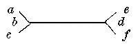
letter-H incision was employed, thus—
Turning the skin flaps here indicated upwards and
downwards, and the lateral triangles outwards, there
was found beneath, a layer of superficial fatty connective
tissue, with a well-defined layer of fat on either
side, but with an intermediate portion which was free
from fat, and of greater thickness.
The skin could with some little trouble be raised
over the dorsal or upper surface, showing here entire
absence of fat. A very delicate artery was found
running across the middle, from Eng to Chang.
The lower portion of the surface C. R., E. L. was
inseparably connected with the umbilicus. It was
also united to the superficial fascia on C. L., about 1-1/2
inches from the umbilicus; this did not have any connection
with the deeper parts. The process of fibrous
tissue which had been felt through the skin was conspicuous
on this surface of the band, and was covered
by a delicate non-fatty layer of connective tissue.
Towards the lower part of the surface were seen
several diverging lines of fibrous tissue, which were
lost within the integument about the umbilical scar, at
the lower surface of the band. They were exceedingly
thin, and at one point lay directly over the posterior
and inferior wall of the umbilical pouch of Chang.
12
- Fig. 3.
- The surface, C. R., E. L.,
exposed by removal of skin and superficial fascia to display the tendons of the external
oblique muscles and adjacent parts.
- A.
- The superficial fascia—lost over the position of Chang’s umbilical pouch.
- B, C.
- Supplemental layers of fibrous tissue of Eng not seen in Chang; B is a continuation toward Eng of aponeurotic fibres having a source
from the linea alba of Chang; C is independent of the former,
and is continuous with the deep pectoral fascia.
- D.
- The interlacing of fibres on tendon of external oblique muscle of Chang.
- E.
- The linea alba of Chang, beginning at C. R.
- F.
- Its continuation to E. L., and insertion upon the ensiform
cartilage.
Turning down the superficial layer, the aponeurosis
of the external oblique muscle was exposed (Fig. 3).
A marked contrast was exhibited in the two sides of
the band. In Chang the parts were normal so far as
they were exposed—the characteristic apertures for
the escape of small vessels being abundant and conspicuous.
In turning down the superficial fascia in
Eng (Fig. 3, A), it was found to be continuous at its
lower portion with an aponeurotic layer (Fig. 3, B),
which extended toward the median line, where it was
continuous with the linea alba of Chang. In addition
to this, a second layer (Fig. 3, C), analogous in position
to a deep layer of the superficial fascia, which was
13entirely independent of Chang, extended over nearly
the whole of Eng’s division, and was particularly well
defined over the fibres of origin of the external oblique
muscle. This was continuous with the deep layer of
the superficial fascia which passed over the entire side
of Eng’s thorax.
Toward the middle of the band this layer gradually
lost its distinctive features, and was firmly incorporated
with the tendon of the external oblique muscle. A
number of fibres corresponding to it extended in
inseparable intimacy with this tendon. These were
gradually lost as they approached the linea alba of
Chang, and the parts being in position these fibres
were at their lower portion covered in by the aponeurotic
extension of the linea alba already mentioned.
On Chang’s side, as we have seen, the parts comparable
to these accessory layers were absent. There
was no line of demarcation between the tendon of
the oblique and the aponeurotic attachment of the
pectoralis. The tendon of the external oblique presented
a different appearance from the normal one in a
more extensive interlacing of fibres of the linea alba
with the tendon.
The part termed above the linea alba of Chang
(Fig. 3, E), has already been indicated through skin
and superficial fascia. As can be seen, this band of
fibres, having its origin from the middle line of the
abdomen of Chang, was found to be a direct continuation
of the linea alba. It was remarkable in not being
inserted into the ensiform process of Chang, but into
that of Eng, and in giving off the aponeurotic outshoot
B, already noticed, as well as in having a diffused point
of insertion into Eng’s tissue as in the ensiform
14cartilage (F). In a word, the linea alba approaches the
surface C. R., E. L. from C. R. below, and is inserted
into E. L. above.
Limited incisions being alone permitted, the large
vessels of the abdomen were sought for in the process
of embalmment, believing, as we did, that the procedures
of securing them would enable us, by extending
the cuts from below upward, to fairly open the
abdomen and examine thereby the interior of the band.
In each body, therefore, an incision six inches long
(Fig. 2, ab, cd) was extended from the centre of
the right iliac region to the centre of the right hypochondriac
region. This was subsequently joined
by an oblique incision passing from the upper end of
the first mentioned to the lateral border of the ensiform
cartilage at its base. This incision measured
7-1/2 in. The lower end of the vertical incision was met
by a horizontal one passing to the centre of the hypogastric
region, and measuring 3-1/4 in.
Through these incisions were studied (1) the umbilical
ligaments and (2) the abdominal viscera.
1. The umbilical ligaments.[2]—By turning forward
the anterior flap in Eng as far as possible, the peritoneal
lining was exhibited, and there was brought into
view a structure beginning at the summit of the
bladder, and which, ascending the abdominal wall and
passing obliquely to the right side, could be traced
15clearly to the scar-like tissue marking the remains of
the umbilical structures situated upon the anterior
abdominal wall within about 1-1/2 in. of the band. This
structure was the umbilical ligament (Fig. 4, A). It
was loaded with fat, and, as it terminated at the scar,
distinct lobules of fat (several of which were pedunculated)
were abundantly deposited.
The bladder was distended and raised 5 in. above
the pubis.
- Fig. 4.
- The umbilical ligament in Eng.
- A.
- The umbilical ligament.
- B.
- The lobule of fat at position of the normal umbilicus.
In Chang (Fig. 5), the same appearances were seen
as those above given, save that no fat was deposited
in the umbilical ligament. On the contrary, it resembled
the omentum of an emaciated subject. When
stretched, the fold was fully an inch wide, quite transparent,
and marked by two longitudinal bands, which
16recalled the shapes of the obliterated vessels. But two
rather small sessile fatty appendages were seen at the
scar.
The bladder was empty, contracted, and lay within
the true pelvis.
- Fig. 5.
- The umbilical ligament in Chang.
- A.
- The umbilical ligament.
- B.
- The lobule of fat at position of the normal umbilicus.
In both Chang and Eng an isolated mass of subperitoneal
fat, presenting a sub-circular form, and
measuring 1 in. in diameter, was found in the position
of the normal umbilicus (Figs. 4 and 5).
2. The viscera.—In Eng the omentum was gathered
up toward the transverse colon. It was abundantly
furnished with fat.[3] The transverse colon extended
17across the abdomen, beginning on the right side on a
level with the eleventh rib. It was contracted and
contained a little flatus. The rest of the exposed
region was occupied by coils of small intestine, yielding
a mesentery very rich in fat. The stomach was
not visible. By removing the small intestine, and
bringing down the transverse colon and large intestine,
the pyloric extremity of the stomach was seen. The
fundus of stomach, spleen, and left kidney were not
seen. (Fig. 6.)
- Fig. 6.
- The abdominal organs in Eng—the small intestines removed.
- A.
- Left lobe of liver.
- B.
- Right lobe of liver.
- C.
- Gall-bladder.
- D.
- Suspensory ligament.
- E.
- Lobules of fat in the position of the termination of the umbilical ligament.
18The liver.—The right lobe was alone visible. This
extended entirely across the right hypochondriac and
epigastric regions. Its external free border was not in
contact with the ribs. Between it and the external
abdominal wall there was an interval of nearly an inch
at its greatest part, which was crossed by the external
lateral ligament. The inferior border of the lobe rested
upon and nearly concealed the pylorus of the stomach
as well as the upper half of right kidney. Corresponding
in position to the upper portion of the right
kidney was a well-defined layer of peritoneum, presenting
a sharply defined internal border. Upon dissecting
away the peritoneum from this border it was
found to answer to the inferior vena cava. The lesser
omentum occupied its usual position. The fundus of
the gall-bladder was two-thirds of an inch beyond the
anterior border of the lobe, immediately to the outer side
of the caudal lobe. The position of the longitudinal
fissure was well off to the left side of the abdomen,
presenting, between the right and left lobes, a conspicuous
cleft which was partially occupied by the base of
the caudal lobe. The round ligament, with its associated
suspensory ligament, had doubtless passed
nearly vertically, before the relations had been disturbed
by the incision in the abdominal wall, upwards
and forwards to the anterior abdominal wall at a point
lying one inch to the outer side of the centre of the
umbilicus.
In the subject, as it lay on the table with the flap
a, b (Fig. 2), turned to the left, the suspensory ligament
had the appearance of being much more obliquely
inclined to the left, and could be made nearly
horizontal by a little traction. Lying beneath this
19ligament, but belonging to the anterior abdominal
wall, was a large mass of subperitoneal fat about the
size of a pigeon’s egg. Extending to the extreme left,
and continuous with the anterior border of the left
lobe of the liver, was a delicate prolongation of liver
substance which was lost within the connecting band.
- Fig. 7.
- The abdominal organs of Chang in position—the small intestines removed.
- A.
- Left lobe of liver.
- B.
- Right lobe of liver.
- C.
- Gall-bladder.
- D.
- Suspensory ligament.
- E.
- Lobules of fat in the position of the termination of the umbilical ligament.
The upper surface of this prolongation was supported
by a fold of peritoneum, extending directly upward,
apparently attached to the base of the ensiform
20cartilage. Visible upon the anterior aspect of this fold
was a tortuous artery, afterwards found to be the left
internal mammary. This fold may be called the accessory
suspensory ligament; nothing similar to it was
seen in Chang. The left lobe of the liver, save a portion
of its anterior edge, was not visible.
In Chang (Fig. 7), by exposing the parts as in Eng,
throwing the abdominal flap, c, d (Fig. 2), to the right,
there was at once brought into view the transverse
colon, the greater omentum, and greater curvature of
the stomach. The latter organ was large, empty, and
without any undue traction could be so displayed as to
yield its fundus and greater curvature in position. The
fundus was not visible. Lying conspicuously within
the left hypochondriac region was the spleen. Its inferior
free border, with its peritoneal attachment, was
distinctly seen; its upper portions, however, were invisible.
The left lobe of the liver held a position answering
to that of the right lobe in Eng—the external
lateral ligament being stretched across the left hypochondriac
region, pursuing a similar course to the external
lateral ligament of Eng (q. v.). The left lobe at
its outer portion rested upon the spleen, its inner portion
upon the stomach. The outer portion of the left
lobe presented a thin compressed border, the inner portion
was divided by a deep sulcus into two lobes.
The right lobe lay deep within the right hypochondriac
region, the portion about the longitudinal fissure
anteriorly, alone appearing in the dissection. The suspensory
ligament held a position similar to that in Eng.
The gall-bladder held its normal position to the right
lobe, and was moderately distended with bile.
21Both Chang and Eng had the organs occupying the
hypochondriac and epigastric regions retaining, on the
whole, such relations as are usually observed.
This statement appears pertinent, at this stage of
the autopsy, in order to explain—
We here describe (1) the hepatic pouches; (2) the
umbilical pouches; (3) the vascular structures of the
band; (4) the diaphragms; (5) the ensiform cartilages.
1. The hepatic pouches.—The photograph (Fig. 2)
indicates the position of the right lobe of Eng’s liver in
the right hypochondriac region. The right lobe of
Chang is of course not seen in the figure, since it lies
on the side of the body which is not in the field of vision.
It must follow from the rights and lefts of the two individuals
being opposites that, in drawing a line between
the livers (which, as already seen, occupy normal
positions as to right and left) across the band, such
a line will be diagonal to the axes of the ensiform cartilages;
Chang’s half of the band having the line enter
the band from his “right,” Eng from his “left.” It will
also follow that any pouches of peritoneum which might
accompany this line will pursue a similar direction—be
on the same plane—be right or left with respect to
the axis of that plane. Now it was actually demonstrated
that such a line did extend between the livers,
and was accompanied by such peritoneal pouches.
These pouches were termed the hepatic pouches, and
may be described as follows:—
Chang.—The subject lying on the table with rights
and lefts determined as in the acquired position, the
finger could be inserted behind the suspensory
22ligament (Fig. 7) in a pouch lying directly beneath the
ensiform cartilages, into which passed an extension of
liver-like tissue.
Eng.—This fact could not be well demonstrated in
Eng in this position, but is well seen in Fig. 8.
It follows that the two hepatic pouches are on nearly
the same plane, and that each approaches the central
point of the band diagonally from the right side of the
subject with whose abdominal cavity it is continuous.
2. The umbilical pouches.—Beneath the hepatic
pouches, and between them and the inferior border of
the band, were two pouches which, from their association
with the round ligament, have been termed the umbilical
pouches.
When the finger was passed toward the band from
the abdomen of Chang, and following the peritoneum
of the anterior wall of the abdomen, it passed into a
pouch of the band directly over the skin covering,
across the band, over the umbilicus, and was received
within the folds of the suspensory ligament of the
liver of Eng. This pouch was so superficial that while
the finger was in the pouch any motion of the finger
was readily followed by the observer.
In the same way as above, if the finger was introduced
behind the suspensory ligament of Eng, it slipped
into a pouch which passed across the median line of
the band, and was received within the folds of the suspensory
ligament of the liver of Chang.
There were then two pouches communicating with
the two abdominal cavities, arranged one above another
in the band, Chang’s being the lower of the two. No
remains of an umbilical vein were detected, nor was
23there any communication between the pouches and the
umbilicus. It has already been noticed that the round
ligament of each liver passed from the longitudinal
fissure to a scar on the anterior wall of the abdomen
near the band. It was not, therefore, within the round
ligaments, but the folds of the suspensory ligaments,
that the pouches were found.
Eng’s pouch measured 2-1/2 in. From edge of Chang’s
suspensory ligament to end of hepatic pouch measured
3 in.
Extending across the band, about midway between
the properties of the two individuals, was a septum.
It was attached above and below to the respective
boundaries of the band, and along its entire length
was incorporated with its two peritoneal cavities, so
that when in the course of the dissection of the “posterior”
surface of the band the peritoneal covering of
the band was displayed, several large lobules of fat
were seen lying to Eng’s side of the septum.[4]
It will be seen that Fig. 8 represents the band opened
to display the pouches with the septum. The lower
end of the septum is fixed near the scar of the umbilicus,
and holds an immobile position over the umbilical
pouches. At this point it is free from fat. But as
it extends over the hepatic pouches it is more pliant.
This portion of the septum has been carried a little to
Chang’s side of the band to display the entire length
of the hepatic pouch of Eng.
24Figs. 9 and 10 are designed to exhibit the appearances
presented in securing views of the septum from
its sides. Fig. 9 is the side toward Chang, and Fig.
10 is the side toward Eng.
- Fig. 8.
- The surface, C. R., E. L.,
showing the interior of band by free division of the aponeuroses seen in Fig. 7,
and their underlying peritoneal attachments.
- A.
- The orifice of umbilical pouch of Eng.
- B.
- The orifice of umbilical pouch of Chang, showing connection with suspensory ligament of
Eng.
- C.
- The fenestrated umbilical pouch of Eng passing between the folds of the suspensory
ligament of Chang.
- D.
- Suspensory ligament of liver of Eng.
- E.
- Hepatic tract.
- F.
- Hepatic pouch of Eng.
- G.
- The septum.
The pouches and septum were now removed and the
position of the hepatic tract determined. It rested
25upon the incurved borders of the ensiform cartilages
(see Fig. 15), and as the subject lay on the table with
the “posterior” surfaces of the band exposed (Fig. 11)
the hepatic tract was slightly arched. It measured
three inches in length, was compressed, and measured
six lines wide and three lines thick. The tract arose
from the livers at the same point—namely, directly
above the longitudinal fissure, having more fulness on
the side of the right than of the left lobe. The round
ligament, as it passed out of the longitudinal fissure of
each liver, was placed beneath and a little to the left
of the tract.
- Fig. 9.
- The septum viewed from Chang’s side.
- A.
- The orifice of umbilical pouch of Chang.
- B.
- The orifice of the hepatic pouch of Chang.
- C.
- Suspensory ligament of Chang containing umbilical pouch of Eng.
26
- Fig. 10.
- The septum viewed from Eng’s side.
- A.
- The orifice of the umbilical pouch of Eng.
- B.
- The orifice of the hepatic pouch of same.
- C.
- Suspensory ligament of Eng containing the umbilical pouch of Chang.
3. The vascular structures of the band were as follows:—
The livers being united, it was found that a colored
injection thrown into the portal vein of Chang passed
into the liver of Eng. A careful dissection of the
blood vesselblood vessel (Fig. 11, C) proved it to be a terminal twig
of the portal system of Chang. It was of the thickness
of a No. seven catheter, French scale, gradually
diminished in size, and was lost toward the centre of
the band. It did not pass as such across the band,
27but appeared to break up into minute branches before
reaching the liver of Eng. At the same time there
was undoubted distension of the portal capillaries
with the colored fluid under the capsule of the dorsal
surface of the right lobe of Eng’s liver, one and
one-half inches from the band. Examination of the
branches of the mesenteric veins of Eng revealed the
curious fact that some of them had received the injection.
This had not been transmitted through the
liver, for the portal vein at the transverse fissure was
empty, but through a distinct extra-hepatic portal
track, which was found lying under the peritoneum
28beneath the position of the hepatic pouches, and in
association with the umbilical pouches. This vessel
began by relatively large radicals towards Chang’s side,
became larger as these encroached on Eng’s side, and
was finally received within the portal system of Eng’s
body, as a tributary to its mesenteric vein.
- Fig. 11.
- The surface, C. R., E. L., with
pouches removed to display the hepatic tract.
- A.
- Liver of Chang.
- B.
- Liver of Eng.
- C.
- Portal vessel of Chang.
- D, D.
- Minute branches of hepatic artery.
- E.
- Subcutaneous fat of surface, E. R., C.
L.
No other vessels were met with in the band excepting
a few insignificant branches of the hepatic artery,
and the terminal twigs of the right internal mammary
of Eng. The former vessels are marked D, D, Fig. 11.
The latter vessel terminated by piercing the diaphragm,
and giving ultimate filaments to the integument of the
“front” of the band as shown in Fig. 12.
- Fig. 12.
- The surface, C. R., E. L., with
pouches, hepatic tract, and peritoneal attachments removed to display the diaphragms.
- A.
- Subcutaneous fat of surface, E. R., C.
L.
- B, C.
- Symmetrical muscular fasciculi.
- D.
- Fasciculi of Eng crossing the median line of the band.
4. The diaphragms.—The subject being in the same
position as in Fig. 2, the livers were removed, the
29peritoneal coverings dissected from the band, and the diaphragms
exposed (Fig. 12). The point (A), marked by
the terminal twigs of the right internal mammary of
Eng, indicated the “anterior” of the band. A broad
slip of fibres of Chang (B) was seen to pass across the
median line, and to be inserted into the left border of
the ensiform cartilage of Eng (Fig. 17). This arrangement
would appear to correspond to the smaller collection
of fibres (C) belonging entirely to Eng. A second
arrangement of fibres was seen above those just indicated,
immediately under the cartilages (D). This
appeared to arise from the border of the cordiform tendon
of Eng by two distinct narrow slips, which crossed
the median line to be inserted dispersedly on the diaphragm
of Chang.
- Fig. 13.
- The peritoneal linings of the anterior walls of both abdominal cavities.
- A, A.
- The summits of the bladders.
- B, B.
- The umbilical ligaments.
- C, C.
- The nodules of fat at the parietal scar.
- D, D.
- The isolated lobules of fat.
305. The ensiform cartilages.—After removing the
diaphragms the cartilages were exposed. They may
be described as follows:—
Chang.—The cartilage measured 2-1/2 in. wide, and
8 in. in length along its axis. The right border was
very prominent, and projected 1/2 in. beyond the limit of
the corresponding border in Eng. It was almost in
close contact with the cartilage of the eighth rib; it was
very robust, with upper surface convex, under surface
nearly plane. The left lateral border was 2-1/2 in. in
length, right lateral border 11 lines in length. The
former was marked by three tubercles of about equal
size. One situated about 1 in. from the sternal origin;
the other about 2 in. from the same point; the third
at its extreme anterior border. None of these were
robust, or presented any of the thickening noticed on
the right side. The middle of these tubercles was on
a line with that of the posterior tubercle. The junction
of the ensiform process with the sternum was not
marked by the eminence characterizing the similar
point in Eng.
Eng.—The cartilage differed from that of Chang in
being 2-1/3 in. wide, 11 lines in length of axis. The
left lateral border was abruptly deflected downward,
and did not present the transverse smooth projection
noticed in Chang. This deflection was almost at right
angles to the dorsal surface, acuminate inferiorly, and
presenting a straight surface toward Chang, and an
oblique one toward the ribs. The length of left lateral
border was 1 in. The right lateral border, 1-1/3 in. in
length, presented a smooth sub-rounded edge without
tubercles, and terminated in a free rounded border on
a plane a little above that of Chang. On the whole
31this border was more robust than that of Chang. On
the dorsal aspect of the process near its base was seen
the rounded eminence described in the account of the
external appearances (see page 7).
A comparison between the two ensiform cartilages
shows that in Chang the anterior border was longer
than in the right in Eng. In other proportions Eng’s
was equal if not larger than Chang’s, and was more
robust.
- Fig. 14.
- A section of both ensiform cartilages, C. R., E. L.
- A.
- Chang’s cartilage.
- B.
- Eng’s cartilage.
- C.
- The synchondrosis.
- D.
- The bursa-like sac covering the same.
- E.
- An opening in the sac.
- F.
- Transversalis muscle of Eng.
- G.
- Transversalis muscle of Chang.
The union between the cartilages was of the character
of a symphysis. The union was very intimate
along the border E. L., C. R., “posterior” (Fig. 14); the
exposure of the junction by a delicate transverse cut
showed a close union between the cartilages, thus constituting
this part of the band a synchondrosis. That
32this, however, did not characterize the entire line of
apposition was at once seen by turning to the border
E. R., C. L. (“anterior”), where an interval, two lines
in width, was seen between the cartilages, an interval
which had been evidently susceptible of variation
during life. This interval extended across one-fourth
the width of the band. The portion of the band
between the parts as above indicated, was occupied by
a bursa-like sac (Fig. 14, D), which was opened by a
minute orifice (E) to display its true nature. This sac
was crossed above by a stout band of fibrous tissue
(Fig. 15, A) an inch in width. Beneath, the sac was
protected by a less well-defined band of the same
width as the upper ligament, and which crossed between
the two processes, to be lost in the perichondrium.
- Fig. 15.
- Upper surface of ensiform cartilages.
- A.
- The upper ligament uniting the cartilages.
Viewing the cartilages as the constituent parts of
the band, we found the border C. L., E. R., the “anterior,”
to be longer than C. R., E. L., the “posterior.”
33C. L., E. R. was a convex, nearly even border, C. L.
being larger than E. R., with a gaping interval placed
nearer Eng than Chang. C. R., E. L. was an irregular,
uneven border, without interval, C. R. being smaller
than E. L., and placed to the outer side.
1. The spleens.—This organ in Eng was 5 in. long,
3-1/3 in. wide. The dorsum was marked by a large
sulcus, extending nearly across the organ, continuous
with the sulcus on the superior border. The hilus was
relatively shorter than that of Chang, beginning
above, fully an inch below its upper border, and terminating
within a half inch of its inferior border.
In Chang it measured 5 in. long, 2-1/3 in. wide. It
was sub-elliptical in form, upper lip somewhat abruptly
compressed. The lower border was obtuse and
rounded. The dorsum was smooth, and presented at
its posterior edge a single sulcus placed midway between
the tip and the inferior border. The hilus extended
nearly the entire length of the under surface.
2. The livers (Fig. 16).—In Eng the liver was 9 in.
broad. The right lobe was 7-1/2 in. wide, antero-posteriorly.
The fundus of the gall-bladder was seen on the
anterior edge of the organ. The only noticeable feature
on the under surface of the liver, was the lobus
Spigelii. This was large, measuring 2 in. in transverse
diameter, and 2-1/3 in. in antero-posterior diameter. It
presented a somewhat increased breadth of neck, which
was overlapped by an anterior prolongation of the lobe,
and terminated by a rounded compressed extremity at
the transverse fissure. The quadrilateral lobe was
34well developed, 2 in. long in greatest diameter, 10 lines
wide.
- Fig. 16.
- The livers.
- A.
- Right lobe of Eng.
- B.
- Left lobe of same.
- C.
- Right lobe of Chang.
- D.
- Left lobe of same.
- E.
- Hepatic tract.
- F.
- Round ligament of Eng.
- G.
- Round ligament of Chang.
- H.
- Accessory suspensory ligament of Eng, with termination of the right mammary artery.
- I.
- Fundus of gall-bladder of Chang.
- J.
- Fundus of gall-bladder of Eng.
35In Chang the liver was 8-1/3 in. broad. The right
lobe was 5 in. wide, antero-posteriorly. The appearance
of the gall-bladder corresponded to that seen in
Eng. The under surface was normal. The lobus
Spigelii presented a narrower neck than in Eng, the
anterior prolongation being greater. The quadrilateral
lobe was less developed than in Eng. Indeed it was
not raised above the under surface of the right lobe,
and its limits were so imperfectly marked that it could
not well be measured.
- Fig. 17.
- Kidneys of Eng.
- A.
- Left kidney.
- B.
- Right kidney.
- C.
- Left renal vein.
- D.
- Left supra-renal vein.
- E.
- Left spermatic vein.
- F.
- Descending vena cava not distended with clot.
- G.
- Right renal vein.
- H.
- Aorta distended with plaster.
- I.
- Primitive iliac arteries.
363. The kidneys.—In Eng, the body lying on the
table E. R., C. L., the left kidney (Fig. 17, A) was
4 in. long, 1-1/2 wide at its hilus, and of the usual kidney
shape. It lacked 1/2 in. of reaching the crest of the ilium.
The renal vein (Fig. 17, C) of the same side measured
3 in. in length, and was decidedly oblique in
position, its termination in the cava being below the
level of the lower end of the kidney.
The right kidney (Fig. 17, B) corresponded in position
to the left kidney of Chang, that is to say, it was
in the shallower portion of the abdomen, and in contact
with the abdominal wall. It measured 4 in. in
length, and 2-1/4 in. in width. Its inferior border lacked
2 in. of reaching the superior crest of the ilium. The
renal vein ascended a little upward to enter the cava
a little below the level of the upper end of the kidney.
In Chang, the body lying in such a way that the
great trochanter of the right side rested on the table,
the left trochanter being raised three inches from the
same plane, an obliquity was given to the trunk, and
rendered the position of the abdominal organs somewhat
anomalous.
The left kidney (Fig. 18, A) lay with its lower
half clearly within the iliac fossa, its inferior border
answering to a point an inch and a half below the
termination of the aorta. The organ lay, at its inner
and inferior portion, upon the left primitive iliac vein;
it measured 3-3/4 in. in length, and 2-7/12 in. in width at its
widest part. It was larger below, where it retained the
usual appearance, but was somewhat abruptlyabruptly pointed
37above, and was marked by the characteristic notch on
its inner side. The renal vein (Fig. 18, C) was very
obliquely situated, indeed was almost parallel with the
cava, and was 3-1/2 in. long. The termination of the renal
vein answered to a line running across the abdomen
lying fully 1 in. above the upper end of the left kidney.
- Fig. 18.
- Kidneys of Chang.
- A.
- Left kidney.
- B.
- Right kidney.
- C.
- Left renal vein.
- D.
- Right renal vein.
- E.
- Left spermatic vein.
- F.
- Aorta filled with plaster.
- G.
- Primitive iliac veins.
- H.
- Descending cava distended with clot.
- I.
- Left supra-renal vein.
The right kidney (Fig. 18, B) was normally situated.
It measured 4 in. in length, and 1-1/2 in. in width at its
centre, and presented the usual reniform appearance.
38Its inferior edge just reached an eminence answering
to the superior crest of the ilium.
4. The testicles.—The right testicle of Eng was
normal. The left testicle was not within the scrotum.
Dissection from within the abdomen showed that the
organ had been retracted. It lay well concealed within
the inguinal canal, slight traction making it appear
within the abdomen.
The testicles of Chang were normal.
5. The hearts.—The heart in Eng was situated
nearer the median line than normal. The abdominal
incision was very unfavorable for studying its exact
position in the mediastinum. It was removed through
an opening made in the diaphragm. The right side of
the heart was occupied by a soft grumous clot much
smaller than was found in the same locality in Chang,
and which did not distend the cavities. The left side
was normal. It was without clot so far as could be
determined in the injected condition of the ventricle.
The heart of Chang presented a right auricle and
ventricle distended with a dense venous clot; this
extended from the right ventricle along the pulmonary
arteries. The left side of the heart was empty.
The ductus arteriosus and foramen ovale were firmly
closed in both hearts.
6. The vessels.—The arteries of both subjects were,
so far as examined, in an extremely atheromatous condition.
Large plates of calcareous matter were deposited
in the abdominal aortas. The injecting matter
flowed insufficiently in the left lower extremity of
Chang, from a clot plugging the femoral artery.
39The venous system of Chang was engorged, giving
the appearance of these vessels having been injected
after death; that of Eng was comparatively empty.
7. The lungs.—The lungs were so altered by post-mortem
changes prior to embalming, their contraction
by the chloride of zinc, and their increase of
weight from the plaster, that no extended examination
was made of them. But little difference was
seen between the conditions of the lungs in the two
men. No hepatization was present in Chang.
8. The vertebral column and ribs.—There was
marked lateral curvature of the vertebral column in
both bodies. This was more conspicuous in Chang.
The convexity of the curve was about half-way down
the vertebral column, and inclined in Chang to the
right side. The distance from the centre of the vertebral
column to the left abdominal wall, 2 in.; to the
right abdominal wall, 5 in. The left side of the abdominal
cavity, measuring from about the level of the
band to the last rib of the right side, 7-1/2 inches.
The ribs in both Chang and Eng were 22 in number,
7 true and 4 false. On the right side of Eng the first,
second, and third ribs were normal. The fourth, fifth,
sixth, and seventh presented diminished intercostal
spaces, owing probably to the extreme traction made
on them by the deflection of the ensiform cartilages.
The intercostal space between the third and fourth
ribs was slightly contracted; that between the fourth
and fifth ribs was very much contracted, the muscle
being bulged inward. Between the fifth and sixth, and
40sixth and seventh ribs the space was less contracted.
The remaining intercostal spaces were about normal.
The fifth rib near its articulation with the vertebral
column formed a well-defined ridge within the thorax,
carrying with it the sixth and seventh ribs, thus forming
a rounded elevation, distinguishing the positions of
these ribs from the thoracic wall above and below
this point, where the parietal surface presented the
usual concave appearance.
On the left side of Chang a similar arrangement of
ribs and intercostal spaces was seen to the above.
The remaining organs were not examined.