Mr. Peters, 35-year-old father of four and a resident
of Chicago’s northwest side, went to a Chicago hospital
one winter day after persistent headaches had made his
life miserable. Routine examinations showed nothing amiss
and his doctor ordered a “brain scan” in the hospital’s
department of nuclear medicine.
Thirty minutes before “scan time”, Mr. Peters was
given, by intravenous injection, a minute amount of radioactive
technetium. This radiochemical had been structured
so that, if there were a tumor in his cranium, the
radioisotopes would be attracted to it. Then he was positioned
so an instrument called a scanner could pass
close to his head.
As the motor-driven scanner passed back and forth, it
picked up the gamma rays being emitted by the radioactive
technetium, much as a Geiger counter detects other
radiation. These rays were recorded as black blocks on
sensitized film inside the scanner. The result was a piece
of exposed film that, when developed, bore an architectural
likeness or image of Mr. Peters’ cranium.
12
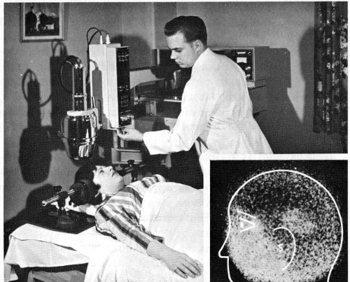
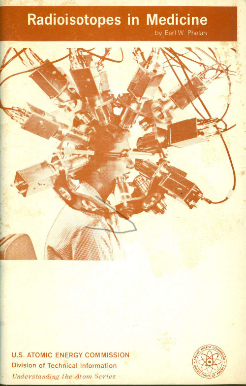
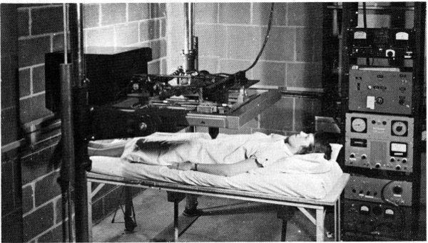
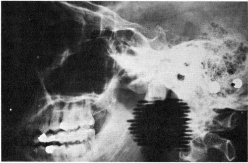
The inset picture shows a brain scan made with a positron scintillation
camera. A tumor is indicated by light area above ear. (Light
area in facial region is caused by uptake in bone and extracellular
space.) The photograph shows a patient, completely comfortable,
receiving a brain scan on one of the three rectilinear scanning devices
in the nuclear medicine laboratory of a hospital.
Mr. Peters, who admitted to no pain or other adverse
reaction from the scanning, was photographed by the
scanner from the front and both sides. The procedure took
less than an hour. The developed film showed that the
technetium had concentrated in one spot, indicating definitely
that a tumor was present. Comparison of front and
side views made it possible to pinpoint the location exactly.
Surgery followed to remove the tumor. Today, thanks
to sound and early diagnosis, Mr. Peters is well and back
on the job. His case is an example of how radioisotopes
are used in hospitals and medical centers for diagnosis.
13
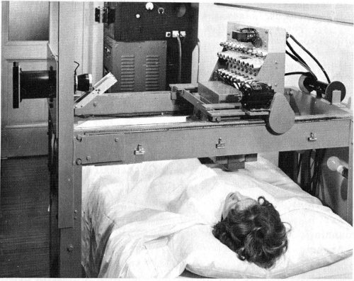
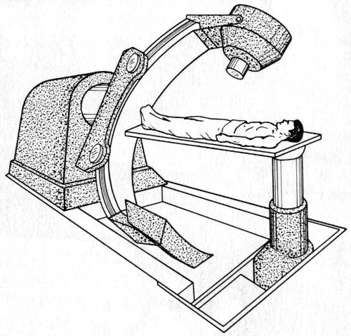
The first whole body scanner, which was developed at the Donner
Laboratory in 1952 and is still being used. The lead collimator
contains 10 scintillation counters and moves across the subject.
The bed is moved and serial scans are made and then joined together
to form a head-to-toe picture of the subject.
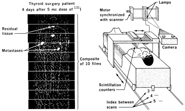
The diagram
shows a scan and the parts of a scanner. (Also see page 21.)
14
In one representative hospital, 17 different kinds of radioisotope
measurements are available to aid physicians in
making their diagnoses. All the methods use tracer quantities
of materials. Other hospitals may use only a few of
them, some may use even
more. In any case they are
merely tools to augment the
doctors’ skill. Examples of
measurements that can be
made include blood volume,
blood circulation rate, red
blood cell turnover, glandular
activity, location of cancerous
tissue, and rates of formation
of bone tissue or blood
cells.
Of the more than 100 different
radioisotopes that have
been used by doctors during
the past 30 years, five have
received by far the greatest
attention. These are iodine-131,
phosphorus-32, gold-198,
chromium-51, and iron-59.
Some others have important
uses, too, but have been less
widely employed than these five. The use of individual radioisotopes
in making important diagnostic tests makes a
fascinating story. Typical instances will be described in
the following pages.
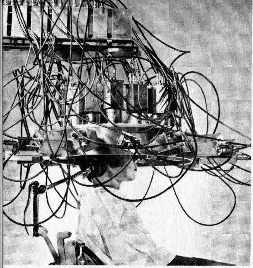
A differential multi-detector developed
at Brookhaven National
Laboratory locates brain tumors
with positron-emitting isotopes.
By using many pairs of detection
crystals, the device shortens the
scanning time and increases accuracy.
(See cover for another type of positron scanner.)
Brain tumors tend to concentrate certain ions (charged
atoms or molecules). When these ions are gamma-ray
emitters, it is possible to take advantage of the penetrating
power of their gamma rays to locate the tumor with a
scanning device located outside the skull.
Arsenic-74 and copper-64 are isotopes emitting positrons,[8]
which have one peculiar property. Immediately
after a positron is emitted from a nucleus it decays, producing
two gamma rays that travel in exactly opposite
directions. The scanning device has two detectors called
15
scintillation counters, one mounted on each side of the
patient’s head.
The electrical circuitry in the scanner is such that only
those gamma rays are counted that impinge simultaneously
on both counters. This procedure eliminates most of the
“noise”, or scattered and background radiation.
Because chromium, in the molecule sodium chromate,
attaches itself to red blood cells, it is useful in several
kinds of tests. The procedures are slightly complicated,
but yield useful information. In one, a sample of the
patient’s blood is withdrawn, stabilized with heparin (to
prevent clotting) and incubated with a tracer of radioactive
sodium chromate. Excess chromate that is not taken up by
the cells is reduced and washed away. Then the radioactivity
of the cells is measured, just before injection into
the patient. After a suitable time to permit thorough mixing
of the added material throughout the blood stream, a new
blood sample is taken and its radioactivity is measured.
The total volume of red blood cells then can be calculated
by dividing the total radioactivity of the injected sample
by the activity per milliliter of the second sample.
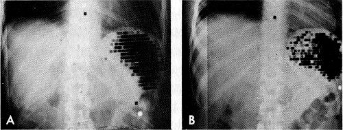
Spleen scans made with red blood cells, which had been altered by
heat treatment and tagged with chromium-51. Such damaged cells
are selectively removed by the spleen. A is a normal spleen. B
shows an abscess in the spleen. Note dark ring of radioactivity
surrounding the lighter area of decreased activity at the central
portion of spleen.
In certain types of anemia the patient’s red blood cells
die before completing the usual red-cell lifetime of about
120 days. To diagnose this, red cells are tagged with
chromium-51 (⁵¹Cr) in the manner just described. Then
16
some of them are injected back into the patient and an identical
sample is injected into a compatible normal individual.
If the tracer shows that the cells’ survival time is too
short in both recipients to the same degree, the conclusion
is that the red cells themselves must be abnormal. On the
other hand, if the cell-survival time is normal in the normal
individual and too short in the patient, the diagnosis is
that the patient’s blood contains some substance that destroys
the red cells.
When chromium trichloride, CrCl₃, is used as the tagging
agent, the chromium is bound almost exclusively to plasma
proteins, rather than the red cells. Chromium-51 may
thus be used for estimating the volume of plasma circulating
in the heart and blood vessels. The same type of
computation is carried on for red cells (after correction
for a small amount of chromium taken up by the red blood
cells). This procedure is easy to carry out because the
radioactive chromium chloride is injected directly into a
vein.
An ingenious automatic device has been devised for
computing a patient’s total blood volume using the ⁵¹Cr
measurement of the red blood cell volume as its basis.
This determination of total blood volume is of course
necessary in deciding whether blood or plasma transfusions
are needed in cases involving bleeding, burns,
or surgical shock. This ⁵¹Cr procedure was used during
the Korean War to determine how much blood had been
lost by wounded patients, and helped to save many, many
lives.
For several years, iodine-131 has been used as a tracer
in determining cardiac output, which is the rate of blood
flow from the heart. It has appeared recently that red
blood cells tagged with ⁵¹Cr are more satisfactory for this
measurement than iodine-labeled albumin in the blood
serum. It is obvious that the blood-flow rate is an extremely
important physiological quantity, and a doctor must
know it to treat either heart ailments or circulatory disturbances.
In contrast to the iodine-131 procedure, which requires
that an artery be punctured and blood samples be removed
regularly for measurement, chromium labeling merely
17
requires that a radiation counter be mounted on the outside
of the chest over the aorta (main artery leaving the heart).
A sample of labeled red blood cells is introduced into a
vein, and the recording device counts the radioactivity
appearing in the aorta as a function of time. Eventually, of
course, the counting rate (the number of radioactive disintegrations
per second) levels off when the indicator sample
has become mixed uniformly in the blood stream. From
the shape of the curve on which the data are recorded
during the measurements taken before that time, the operator
calculates the heart output per second.
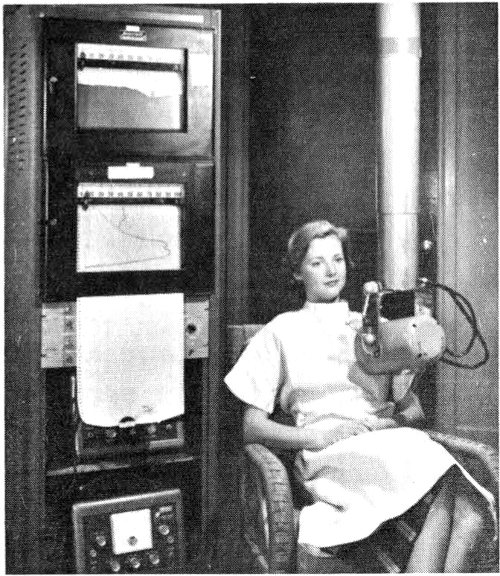
In this cardiac output study
a probe is positioned over
the heart and the passage
of iodine-131 labeled
human serum albumin
through this area is recorded.
Obstetricians caring for expectant mothers use red cells
tagged with ⁵¹Cr to find the exact location of the placenta.
For example, in the condition known as placenta previa, the
placenta—the organ within the uterus by which nourishment
is transferred from the mother’s blood to that of the
unborn child—may be placed in such a position that fatal
bleeding can occur. A radiation-counting instrument placed
over the lower abdomen gives information about the exact
location of the placenta. If an abnormal situation exists,
the attending physician is then alert and ready to cope with
it. The advantages of chromium over iodine-131, which has
also been used, are that smaller doses are required, and
that there is no transfer of radioactivity to the fetal
circulation.
18
Still another common measurement using ⁵¹Cr-labeled red
blood cells is the determination of the amount and location
of bleeding from the gastrointestinal tract (the stomach
and bowels). The amount is found by simple measurement
of chromium in the blood that appears in the stools. To
find the location is slightly more complicated. The intestinal
contents are sampled at different levels through
an inserted tube, and the radiation of the samples determined
separately.
Finally, gastrointestinal loss of protein can be measured
with the aid of ⁵¹Cr-labeled blood serum. The serum is
treated with CrCl₃ and then injected into a vein. In several
very serious ailments there is serious loss of blood protein
through the intestines. In these conditions the ⁵¹Cr
level in the intestinal excretions is high, and this alerts the
doctor to apply remedial measures.
Vitamin B₁₂ is a cobalt compound. Normally the few
milligrams of B₁₂ in the body are stored in the liver and
released to the blood stream as needed. In pernicious
anemia, a potentially fatal but curable disease, the B₁₂
content of the blood falls from the usual level of 300-900
micromicrograms per milliliter (ml) to 0 to 100 micromicrograms
per ml. The administration of massive doses
of B₁₂ is the only known remedy for this condition.
If the B₁₂ is labeled with radioactive cobalt, its passage
into the blood stream may be observed by several different
methods. The simplest is to give the B₁₂ by mouth, and
after about 8 hours study the level of cobalt radioactivity
in the blood. Cobalt-60 has been used for several years,
but recently cobalt-58 has been found more satisfactory.
It has a half-life of 72 days while ⁶⁰Co has a 5.3-year half-life.
This reduces greatly the amount of radiation to the
patient’s liver by the retained radioactivity.
Like chromium-51, iodine is a versatile tracer element.
It is used to determine blood volume, cardiac output,
plasma volume, liver activity, fat metabolism, thyroid
19
cancer metastases, brain tumors, and the size, shape, and
activity of the thyroid gland.
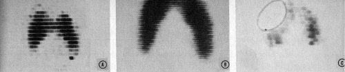
A linear photoscanner produced these pictures of (A) a normal
thyroid, (B) an enlarged thyroid, and (C) a cancerous thyroid.
Because of its unique connection with the thyroid gland,
iodine-131 is most valuable in measurements connected
with that organ. Thyroxin, an iodine compound, is manufactured
in the thyroid gland, and transferred by the blood
stream to the body tissues. The thyroxin helps to govern
the oxygen consumption of the body and therefore helps
control its metabolism. Proper production of thyroxin is
essential to the proper utilization of nutrients. Lowered
metabolism means increased body weight. Lowered thyroid
activity may mean expansion of the gland, causing one
form of goiter.
Iodine-131 behaves in the body just as the natural non-radioactive
isotope, iodine-127, does, but the radioactivity
permits observation from outside the body with some form
of radiation counter. Iodine can exist in the body in many
different chemical compounds, and the counter can tell
where it is but not in what form. Hence chemical manipulation
is necessary in applying this technique to different
diagnostic procedures.
The thyroid gland, which is located at the base of the
neck, is very efficient in trapping inorganic iodide from
the blood stream, concentrating and storing the iodine-containing
material and gradually releasing it to the blood
stream in the form of protein-bound iodine (PBI).
One of the common diagnostic procedures for determining
thyroid function, therefore, is to measure the percentage
of an administered dose of ¹³¹I that is taken up by the
gland. Usually the patient is given a very small dose of
radioactive sodium iodide solution to drink, and two hours
later the amount of iodine in the gland is determined by
measuring the radiation coming from the neck area. In
20
hyperthyroidism, or high thyroid gland activity, the gland
removes iodide ions from the blood stream more rapidly
than normal.
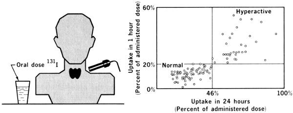
Screening test for Hyperthyroidism
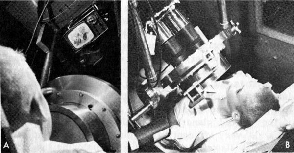
It is especially important in isotope studies on infants and small
children that the radiation exposure be low. By carrying out studies
in the whole body counter room, the administered dose can be
greatly reduced. The photographs illustrate a technique of measuring
radioiodine uptake in the thyroid gland with extremely small
amounts of a mixture of iodine-131 and iodine-125. A shows a
small television set that is mounted above the crystal in such a
way that good viewing requires that the head be kept in the desired
position. This helps solve the problem of keeping small children
still during a 15-minute counting period. B shows a child in position
for a thyroid uptake study.
This simple procedure has been used widely. One difficulty
in using it is that its success is dependent upon the
time interval between injection and measurement. An
overactive gland both concentrates iodine rapidly and also
21
discharges it back to the blood stream as PBI more
rapidly than normal. Modifications of the test have been
made to compare the amount of iodine-131 that was administered
with the amount circulating in the blood as PBI.
The system acquires chemical separation of the two forms
of iodine from a sample of blood removed from a vein,
followed by separate counting. This computation of the
“conversion ratio” of radioactive plasma PBI to plasma-total
¹³¹I gives results that are less subject to misinterpretation.
To determine local activity in small portions of the
thyroid, an automatic scanner is used. A collimator[9]
shields the detector (a Geiger-Müller tube or scintillating
crystal) so that only those impulses originating within a
very small area are accepted by the instrument. The detector
is then moved back and forth slowly over the entire
area and the radiation is automatically recorded at definite
intervals, creating a “map” of the active area. In cases
where lumps, or nodules, have been discovered in the thyroid,
the map is quite helpful in distinguishing between
cancerous and benign nodules. The former
are almost always less radioactive than
surrounding tissues.
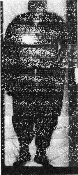
Seven serial scans made with the whole body
scanner were put together to provide a whole
body scan of this patient with thyroid cancer that
had spread to the lung. One millicurie of
iodine-131 was administered and the scan made
72 hours later. Note the uptake in the lung. This
patient was successfully treated with large
doses of iodine-131.
Fragments of cancerous thyroid tissue
may migrate to other parts of the body and
grow there. These new cancers are known
as metastatic cancers and are a signal
of an advanced state of disease. In such a
situation even complete surgical removal
of the original cancer may not save the
patient. If these metastases are capable of concentrating
iodine (less than 10% of them are), they can be located by
scanning the whole body in the manner that was just described.
When a thyroid cancer is discovered, therefore,
a doctor may look for metastases before deciding to
operate.
22
Human blood serum albumin labeled with ¹³¹I is used for
measurement of the volume of circulating plasma. The
procedure is quite similar to that used with radioactive
chromium. Iodinated human serum albumin labeled with
¹³¹I is injected into a vein. Then, after allowing time for
complete mixing of the sample with the blood, a second
sample is counted using a scintillation counter.

Time-lapse motion pictures of the liver of a 3-year-old girl
were made with the scintillation camera 1 hour after injection
of 50 microcuries of iodine-131-labeled rose bengal dye. This
child was born without a bile-duct system and an artificial bile
duct had been created surgically. She developed symptoms that
caused concern that the duct had closed. These scans show the
mass of material containing the radioactive material (small
light area) moving downward and to the right, indicating that
the duct was still open.
For many years, a dye known as rose bengal has been
used in testing liver function. About 10 years ago this
procedure was improved by labeling the dye with ¹³¹I. When
this dye is injected into a vein it goes to the liver, which
removes it from the blood stream and transfers it to the
intestines to be excreted. The rate of disappearance of the
dye from the blood stream is therefore a measure of the
liver activity. Immediately after administration of the
radioactive dye, counts are recorded, preferably continuously
from several sites with shielded, collimated detectors.
One counter is placed over the side of the head or
the thigh to record the clearance of the dye from the blood
stream. A second is placed over the liver, and a third over
the abdomen to record the passage of the dye into the small
intestine.
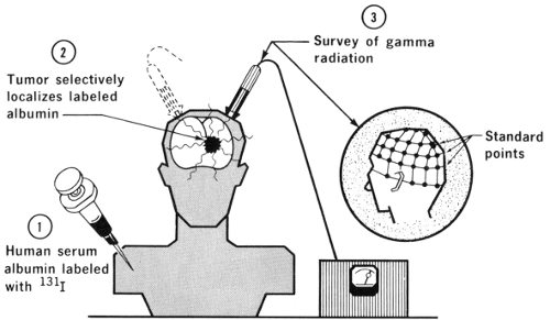
Human serum albumin labeled with ¹³¹I is sometimes
used for location of brain tumors. It appears that tumors
23
alter a normal “barrier” between the brain and blood in
such a manner that the labeled albumin can penetrate
tumorous tissues although it would be excluded from healthy
brain tissue.
The brain behaves almost uniquely among body tissues in
that a “blood-brain barrier” exists, so that substances
injected into the blood stream will not pass into brain
cells although they will pass readily into muscular tissue.
This blood-brain barrier does not exist in brain tumors. A
systematic scanning of the skull then permits location of
these cancerous “hot spots”.
Iron is a necessary constituent of red blood cells, so its
radioactive form, ⁵⁹Fe, has been used frequently in measurement
of the rate of formation of red cells, the lifetime
of red cells, and red cell volumes. The labeling is
more difficult than labeling with chromium for the same
purposes, so this procedure no longer has the importance
it once had.
On the other hand, direct measurement of absorption of
iron by the digestive tract can be accomplished only by
using ⁵⁹Fe. In achlorhydria the gastric juice in the stomach
is deficient in hydrochloric acid, and this condition has
been shown to lower the iron absorption. A normal diet
contains much more iron than the body needs, but in special
24
cases, sometimes called “tired blood” in advertising for
medicines, iron compounds are prescribed for the patient.
If ⁵⁹Fe is included, its appearance in the blood stream can
be monitored and the effectiveness of the medication noted.
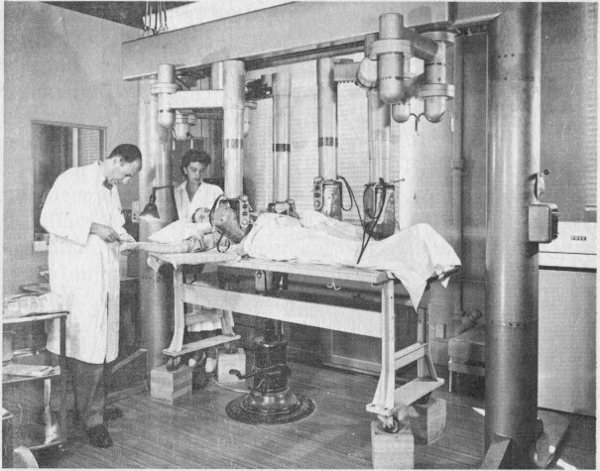
This multiple-port scintillation counter is used for iron-kinetic
studies. The tracer dose of iron-59 is administered into the arm
vein and then the activities in the bone marrow, liver, and spleen
are recorded simultaneously with counters positioned over these
areas, and show distribution of iron-59 as a function of time. When
the data are analyzed in conjunction with iron-59 content in blood,
information can be obtained about sites of red blood cell production
and destruction.
The phosphate ion is a normal constituent of the blood.
In many kinds of tumors, phosphates seem to be present
in the cancerous tissue in a concentration several times
that of the surrounding healthy tissue. This offers a way
of using phosphorus-32 to distinguish between cancer cells
and their neighbors. Due to the fact that ³²P gives off beta
rays but no gammas, the counter must be placed very close
to the suspected tissue, since beta particles have very
25
little penetrating power. This fact limits the use of the
test to skin cancers or to cancers exposed by surgery.
Some kinds of brain tumors, for instance, are difficult
to distinguish visually from the healthy brain tissue. In such
cases, the patient may be given ³²P labeled phosphate
intravenously some hours before surgery. A tiny beta-sensitive
probe counter then can be moved about within
the operative site to indicate to the surgeon the limits of
the cancerous area.
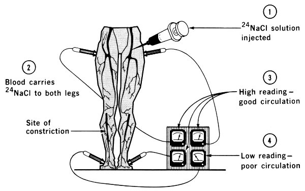
Normal blood is about 1% sodium chloride or ordinary
salt. This fact makes possible the use of ²⁴Na in some
measurements of the blood and other fluids. The figure
illustrates this technique. A sample of ²⁴NaCl solution is
injected into a vein in an arm or leg. The time the radioisotope
arrives at another part of the body is detected with
a shielded radiation counter. The elapsed time is a good
indication of the presence or absence of constrictions or
obstructions in the circulatory system.
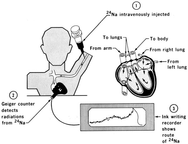
The passage of blood through the heart may also be
measured with the aid of sodium-24. Since this isotope
emits gamma rays, measurement is done using counters
on the outside of the body, placed at appropriate locations
above the different sections of the heart.
26
Because of its short half-life of six hours, technetium-99m[10]
is coming into use for diagnosis using scanning devices,
particularly for brain tumors. It lasts such a short
time it obviously cannot be kept in stock, so it is prepared
by the beta decay of molybdenum-99.[11] A stock of molybdenum
is kept in a shielded container in which it undergoes
radioactive decay yielding technetium. Every morning, as
the technetium is needed, it is extracted from its parent
by a brine solution. This general procedure of extracting
a short-lived isotope from its parent is also used in other
cases. We shall see later that radon gas is obtained by an
analogous method from its parent, radium.
27
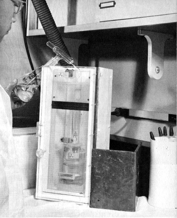
Using a “nuclear cow” to get
technetium from its parent
isotope. The “cow” is being
fed saltwater through a tube.
The saltwater drains through
a high-radiation (hot) isotope.
The resultant drip-off is a
daughter such as technetium-99m.
This new, mild isotope
can be mixed with other elements
and these become the
day’s supply of radioisotopes
for other scans. Technetium-99m
decays in 6 hours. Thus
greater amounts, with less
possibility of injury, can be
administered and a better
picture results.
For years it has been recognized that there would be
many uses for a truly portable device for taking X-ray
pictures—one that could be carried by the doctor to the
bedside or to the scene of an accident. Conventional X-ray
equipment has been in use by doctors for many years, and
highly efficient apparatus has become indispensable, especially
in treating bone conditions. There is, however, a
need for a means of examining patients who cannot be
moved to a hospital X-ray room, and are located where
electric current sources are not available.
A few years ago, a unit was devised that weighed only a
few pounds, and could take “X-ray pictures” (actually
gamma radiographs) using the gamma rays from the radioisotope
thulium-170. The thulium source is kept inside a
lead shield, but a photographic shutter-release cable can
be pressed to move it momentarily over an open port in
the shielding. The picture is taken with an exposure of a
few seconds. A somewhat similar device uses strontium-90
as the source of beta radiation that in turn stimulates
the emission of gamma rays from a target within the
instrument.
28
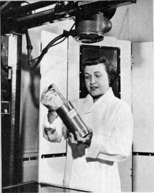
A technician holds an inexpensive portable
X-ray unit that was developed by the
Argonne National Laboratory. Compare
its size with the standard X-ray machine
shown at left and above.
Still more recently, ¹²⁵I has been used very successfully
in a portable device as a low-energy gamma source for
radiography. The gamma rays from this source are sufficiently
penetrating for photographing the arms and legs,
and the necessary shielding is easily supplied to protect
the operator. By contrast with larger devices, the gamma-ray
source can be as small as one-tenth millimeter in
diameter, virtually a point source; this makes possible
maximum sharpness of image. The latest device, using up
to one curie[12] of ¹²⁵I, weighs 2 pounds, yet has adequate
shielding for the operator. It is truly portable.
If this X-ray source is combined with a rapid developing
photographic film, a physician can be completely freed
from dependence upon the hospital laboratory for emergency
X rays. A finished print can be ready for inspection in
10 seconds. The doctor thus can decide quickly whether
it is safe to move an accident victim, for instance. In
military operations, similarly, it becomes a simple matter
to examine wounded soldiers in the field where conventional
equipment is not available.
More than 30 years ago, when deuterium (heavy hydrogen)
was first discovered, heavy water (D₂O) was used for the
29
determination of total body water. A small sample of
heavy water was given either intravenously or orally, and
time was allowed for it to mix uniformly with all the water
in the body (about 4 to 6 hours). A sample was then obtained
of the mixed water and analyzed for its heavy water
content. This procedure was useful but it was hard to make
an accurate analysis of low concentrations of heavy water.
More recently, however, tritium (³H) (radioactive hydrogen)
has been produced in abundance. Its oxide, tritiated
water (³H₂O), is chemically almost the same as ordinary
water, but physically it may be distinguished by the beta
rays given off by the tritium. This very soft (low-energy)
beta ray requires the use of special counting equipment,
either a windowless flow-gas counter or a liquid scintillator,
but with the proper techniques accurate measurement
is possible. The total body water can then be computed by
the general isotope dilution formula used for measuring
blood plasma volume.
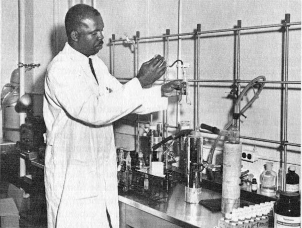
The total body water is determined by the dilution method using
tritiated water. This technician is purifying a urine sample so that
the tritium content can be determined and the total body water
calculated.
30
Another booklet in this series,
Neutron Activation Analysis,
discusses a new process by which microscopic quantities
of many different materials may be analyzed accurately.
Neutron irradiation of these samples changes some
of their atoms to radioactive isotopes. A multichannel
analyzer instrument gives a record of the concentration of
any of about 50 of the known elements.
One use of this technique involved the analysis of a hair
from Napoleon’s head. More than 100 years after his death
it was shown that the French Emperor had been given
arsenic in large quantities and that this possibly caused
his death.
The ways in which activation analysis can be applied to
medical diagnosis are at present largely limited to toxicology,
the study of poisons, but the future may bring new
possibilities.
Knowledge is still being sought, for example, about the
physiological role played by minute quantities of some of
the elements found in the body. The ability to determine
accurately a few parts per million of “trace elements” in
the various tissues and body fluids is expected to provide
much useful information as to the functions of these
materials.
A large number of different radioisotopes have been
used for measurement of disease conditions in the human
body. They may measure liquid volumes, rates of flow or
rates of transfer through organs or membranes; they may
show the behavior of internal organs; they may differentiate
between normal and malignant tissues. Hundreds of hospitals
are now making thousands of these tests annually.
This does not mean that all the diagnostic problems have
been solved. Much of the work is on an experimental rather
than a routine basis. Improvements in techniques are still
being made. As quantities of radioisotopes available for
these purposes grow, and as the cost continues to drop, it
is expected there will be still more applications. Finally,
this does not mean we no longer need the doctor’s diagnostic
31
skill. All radioisotope procedures are merely tools
to aid the skilled physician. As the practice of medicine
has changed from an art to a science, radioisotopes have
played a useful part.